EP3131566B1 - Bi-terminal pegylated integrin-binding peptides and methods of use thereof - Google Patents
Bi-terminal pegylated integrin-binding peptides and methods of use thereof Download PDFInfo
- Publication number
- EP3131566B1 EP3131566B1 EP15779171.6A EP15779171A EP3131566B1 EP 3131566 B1 EP3131566 B1 EP 3131566B1 EP 15779171 A EP15779171 A EP 15779171A EP 3131566 B1 EP3131566 B1 EP 3131566B1
- Authority
- EP
- European Patent Office
- Prior art keywords
- peg
- peptide
- conjugate
- integrin
- imaging
- Prior art date
- Legal status (The legal status is an assumption and is not a legal conclusion. Google has not performed a legal analysis and makes no representation as to the accuracy of the status listed.)
- Active
Links
Images


Classifications
-
- C—CHEMISTRY; METALLURGY
- C07—ORGANIC CHEMISTRY
- C07K—PEPTIDES
- C07K7/00—Peptides having 5 to 20 amino acids in a fully defined sequence; Derivatives thereof
- C07K7/04—Linear peptides containing only normal peptide links
- C07K7/06—Linear peptides containing only normal peptide links having 5 to 11 amino acids
-
- A—HUMAN NECESSITIES
- A61—MEDICAL OR VETERINARY SCIENCE; HYGIENE
- A61K—PREPARATIONS FOR MEDICAL, DENTAL OR TOILETRY PURPOSES
- A61K31/00—Medicinal preparations containing organic active ingredients
- A61K31/33—Heterocyclic compounds
- A61K31/335—Heterocyclic compounds having oxygen as the only ring hetero atom, e.g. fungichromin
- A61K31/337—Heterocyclic compounds having oxygen as the only ring hetero atom, e.g. fungichromin having four-membered rings, e.g. taxol
-
- A—HUMAN NECESSITIES
- A61—MEDICAL OR VETERINARY SCIENCE; HYGIENE
- A61K—PREPARATIONS FOR MEDICAL, DENTAL OR TOILETRY PURPOSES
- A61K38/00—Medicinal preparations containing peptides
-
- A—HUMAN NECESSITIES
- A61—MEDICAL OR VETERINARY SCIENCE; HYGIENE
- A61K—PREPARATIONS FOR MEDICAL, DENTAL OR TOILETRY PURPOSES
- A61K47/00—Medicinal preparations characterised by the non-active ingredients used, e.g. carriers or inert additives; Targeting or modifying agents chemically bound to the active ingredient
- A61K47/50—Medicinal preparations characterised by the non-active ingredients used, e.g. carriers or inert additives; Targeting or modifying agents chemically bound to the active ingredient the non-active ingredient being chemically bound to the active ingredient, e.g. polymer-drug conjugates
- A61K47/51—Medicinal preparations characterised by the non-active ingredients used, e.g. carriers or inert additives; Targeting or modifying agents chemically bound to the active ingredient the non-active ingredient being chemically bound to the active ingredient, e.g. polymer-drug conjugates the non-active ingredient being a modifying agent
- A61K47/56—Medicinal preparations characterised by the non-active ingredients used, e.g. carriers or inert additives; Targeting or modifying agents chemically bound to the active ingredient the non-active ingredient being chemically bound to the active ingredient, e.g. polymer-drug conjugates the non-active ingredient being a modifying agent the modifying agent being an organic macromolecular compound, e.g. an oligomeric, polymeric or dendrimeric molecule
- A61K47/59—Medicinal preparations characterised by the non-active ingredients used, e.g. carriers or inert additives; Targeting or modifying agents chemically bound to the active ingredient the non-active ingredient being chemically bound to the active ingredient, e.g. polymer-drug conjugates the non-active ingredient being a modifying agent the modifying agent being an organic macromolecular compound, e.g. an oligomeric, polymeric or dendrimeric molecule obtained otherwise than by reactions only involving carbon-to-carbon unsaturated bonds, e.g. polyureas or polyurethanes
- A61K47/60—Medicinal preparations characterised by the non-active ingredients used, e.g. carriers or inert additives; Targeting or modifying agents chemically bound to the active ingredient the non-active ingredient being chemically bound to the active ingredient, e.g. polymer-drug conjugates the non-active ingredient being a modifying agent the modifying agent being an organic macromolecular compound, e.g. an oligomeric, polymeric or dendrimeric molecule obtained otherwise than by reactions only involving carbon-to-carbon unsaturated bonds, e.g. polyureas or polyurethanes the organic macromolecular compound being a polyoxyalkylene oligomer, polymer or dendrimer, e.g. PEG, PPG, PEO or polyglycerol
-
- A—HUMAN NECESSITIES
- A61—MEDICAL OR VETERINARY SCIENCE; HYGIENE
- A61K—PREPARATIONS FOR MEDICAL, DENTAL OR TOILETRY PURPOSES
- A61K47/00—Medicinal preparations characterised by the non-active ingredients used, e.g. carriers or inert additives; Targeting or modifying agents chemically bound to the active ingredient
- A61K47/50—Medicinal preparations characterised by the non-active ingredients used, e.g. carriers or inert additives; Targeting or modifying agents chemically bound to the active ingredient the non-active ingredient being chemically bound to the active ingredient, e.g. polymer-drug conjugates
- A61K47/51—Medicinal preparations characterised by the non-active ingredients used, e.g. carriers or inert additives; Targeting or modifying agents chemically bound to the active ingredient the non-active ingredient being chemically bound to the active ingredient, e.g. polymer-drug conjugates the non-active ingredient being a modifying agent
- A61K47/62—Medicinal preparations characterised by the non-active ingredients used, e.g. carriers or inert additives; Targeting or modifying agents chemically bound to the active ingredient the non-active ingredient being chemically bound to the active ingredient, e.g. polymer-drug conjugates the non-active ingredient being a modifying agent the modifying agent being a protein, peptide or polyamino acid
- A61K47/64—Drug-peptide, drug-protein or drug-polyamino acid conjugates, i.e. the modifying agent being a peptide, protein or polyamino acid which is covalently bonded or complexed to a therapeutically active agent
-
- A—HUMAN NECESSITIES
- A61—MEDICAL OR VETERINARY SCIENCE; HYGIENE
- A61K—PREPARATIONS FOR MEDICAL, DENTAL OR TOILETRY PURPOSES
- A61K51/00—Preparations containing radioactive substances for use in therapy or testing in vivo
- A61K51/02—Preparations containing radioactive substances for use in therapy or testing in vivo characterised by the carrier, i.e. characterised by the agent or material covalently linked or complexing the radioactive nucleus
- A61K51/04—Organic compounds
- A61K51/08—Peptides, e.g. proteins, carriers being peptides, polyamino acids, proteins
- A61K51/082—Peptides, e.g. proteins, carriers being peptides, polyamino acids, proteins the peptide being a RGD-containing peptide
-
- C—CHEMISTRY; METALLURGY
- C07—ORGANIC CHEMISTRY
- C07K—PEPTIDES
- C07K14/00—Peptides having more than 20 amino acids; Gastrins; Somatostatins; Melanotropins; Derivatives thereof
- C07K14/435—Peptides having more than 20 amino acids; Gastrins; Somatostatins; Melanotropins; Derivatives thereof from animals; from humans
- C07K14/705—Receptors; Cell surface antigens; Cell surface determinants
- C07K14/70546—Integrin superfamily
-
- C—CHEMISTRY; METALLURGY
- C12—BIOCHEMISTRY; BEER; SPIRITS; WINE; VINEGAR; MICROBIOLOGY; ENZYMOLOGY; MUTATION OR GENETIC ENGINEERING
- C12N—MICROORGANISMS OR ENZYMES; COMPOSITIONS THEREOF; PROPAGATING, PRESERVING, OR MAINTAINING MICROORGANISMS; MUTATION OR GENETIC ENGINEERING; CULTURE MEDIA
- C12N2770/00—MICROORGANISMS OR ENZYMES; COMPOSITIONS THEREOF; PROPAGATING, PRESERVING, OR MAINTAINING MICROORGANISMS; MUTATION OR GENETIC ENGINEERING; CULTURE MEDIA ssRNA viruses positive-sense
- C12N2770/00011—Details
- C12N2770/32011—Picornaviridae
- C12N2770/32111—Aphthovirus, e.g. footandmouth disease virus
- C12N2770/32122—New viral proteins or individual genes, new structural or functional aspects of known viral proteins or genes
Definitions
- Integrins are a large family of cell-surface receptors responsible for mediating cell-cell and cell-extracellular matrix (ECM) adhesion. There are at least 24 different integrins, each a heterodimer composed of an ⁇ and ⁇ subunit, whose expression is determined by several factors including tissue type, stage of development, and various tissue pathologies such as inflammation and cancer. Although they do not possess any intrinsic enzymatic activity, subsequent to ligand binding, integrins translate extracellular cues into intracellular signals by bringing into juxtaposition a complex of cytoplasmic structural and signaling molecules that then interact and determine the cellular response. As integrins are involved in most elements of cell behavior including motility, proliferation, invasion, and survival, their roles in disease have been widely reported.
- integrins are thought to play an active role in promoting certain diseases including cancer.
- ⁇ v ⁇ 3 integrin has been implicated in promoting the invasive phenotype of melanoma and glioblastoma, owing to its multiple abilities including upregulating pro-invasive metalloproteinases as well as providing pro-migratory and survival signals.
- ⁇ v ⁇ 3 is also upregulated on endothelial cells of angiogenic blood vessels and may provide similar signals for the development of neo-vessels in cancer, such data have led many pharmaceutical and academic centers to develop antagonists of ⁇ v ⁇ 3 for therapeutic purposes, many of which have been peptides or peptidomimetics.
- understanding the structural basis of integrin-ligand interactions would aid in the design of improved integrin antagonists.
- the ⁇ v ⁇ 6 integrin receptor is expressed only on epithelial cells. This integrin is involved in both normal and pathological tissue processes. For example, ⁇ v ⁇ 6 is upregulated by epithelial cells during wound healing and inflammation. It is likely that the ability of ⁇ v ⁇ 6 to locally activate TGF- ⁇ by binding to its protective pro-peptide, the latency associated peptide (LAP), explains the function of this integrin in these transient pathologies. Thus, TGF- ⁇ can suppress inflammatory responses and epithelial proliferation, indicating that ⁇ v ⁇ 6 serves as a negative control to dampen-down these processes.
- LAP latency associated peptide
- Chronic inflammation can lead to an excess of ⁇ v ⁇ 6 -dependent activation of TGF- ⁇ , resulting in fibrosis in the lung of experimental animals.
- some pathologies that result in fibrosis in humans may also involve ⁇ v ⁇ 6 -dependent TGF- ⁇ activation.
- Constitutive ⁇ v ⁇ 6 overexpression in the skin of mice results in chronic wounds appearing on a significant number of transgenic animals.
- chronic wounds associated with human diseases e.g., certain forms of epidermolysis bullosa
- ⁇ v ⁇ 6 integrin is a major new target in cancer.
- ⁇ v ⁇ 6 is epithelial-specific, it is weak or undetectable in most resting epithelial tissues but is strongly upregulated in many types of cancer, often at the invasive front.
- ⁇ v ⁇ 6 is highly upregulated in oral squamous cell carcinoma (OSCC), pancreatic cancer, ovarian cancer, and colon cancer.
- OSCC oral squamous cell carcinoma
- pancreatic cancer pancreatic cancer
- ovarian cancer ovarian cancer
- ⁇ v ⁇ 6 integrin has been identified as a receptor for foot-and-mouth disease virus (FMDV) in vitro by binding through an RGD motif in the viral capsid protein, VP1. Structural studies have revealed that one of the modes by which FMDV binds to cells is via a small 31-amino acid containing loop on its protein-shell. This FMDV loop binds to ⁇ v ⁇ 6 with high selectivity and specificity.
- FMDV foot-and-mouth disease virus
- WO 07/039728 describes a radiolabeled ⁇ v ⁇ 6 -targeting peptide, A20FMDV2, consisting of 20 core amino acids of the FMDV loop, which bound to immobilized human ⁇ v ⁇ 6 with high specificity and selectivity in competitive ELISA binding assays.
- A20FMDV2 consisting of 20 core amino acids of the FMDV loop, which bound to immobilized human ⁇ v ⁇ 6 with high specificity and selectivity in competitive ELISA binding assays.
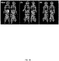
- the ability of radiolabeled A20FMDV2 to image ⁇ v ⁇ 6 -expressing human tumors was also assessed using PET in an athymic nu/nu mouse model. However, these in vivo studies showed rapid metabolism of the radiolabeled ⁇ v ⁇ 6 -targeting peptide. In fact, by one hour, radioactivity in the urine was distributed about equally between three metabolites and no unmetabolized peptide was detected.
- tumor targeting agents which not only provide high tumor selectivity and specificity for ⁇ v ⁇ 6 -expressing tumors, but are also capable of having increased metabolic stability and retention in ⁇ v ⁇ 6 -expressing tumors.
- the present invention satisfies this need and provides related advantages as well.
- the present invention provides bi-terminal PEGylated peptide conjugates that target ⁇ v ⁇ 6 integrin.
- the peptide conjugates of the present invention further comprise a biological agent such as an imaging agent or a therapeutic agent, e.g., covalently attached to one of the PEG moieties.
- the peptide conjugates of the present invention are particularly useful for imaging a ⁇ v ⁇ 6 integrin-containing tumor, organ, or tissue and for treating ⁇ v ⁇ 6 integrin-mediated diseases and disorders such as cancer, inflammatory diseases, autoimmune diseases, chronic fibrosis, chronic obstructive pulmonary disease (COPD), lung emphysema, and chronic wounding skin disease.
- Compositions and kits containing the peptide conjugates of the present invention find utility in a wide range of applications including, e.g., in vivo imaging and immunotherapy.
- the present invention provides a conjugate comprising:
- the integrin is ⁇ v ⁇ 6 integrin.
- the ⁇ v ⁇ 6 integrin-binding peptide comprises the amino acid sequence RGDLX 1 X 2 X 3 , wherein X 1 and X 2 are independently selected amino acids and X 3 is L or I.
- X 1 is Q
- X 2 is V
- X 3 is L.
- the peptide comprises the amino acid sequence RGDLX 1 X 2 X 3 AQX 6 , wherein X 6 is K or R. In certain instances, X 6 is R.
- the ⁇ v ⁇ 6 integrin-binding peptide comprises the amino acid sequence RSDLTPLFX 7 , wherein X 7 is absent or is any amino acid.
- X 7 is absent (i.e., the peptide comprises the amino acid sequence RSDLTPLF).
- X 7 is K (i.e., the peptide comprises the amino acid sequence RSDLTPLFK).
- the peptide comprises or consists of an amino acid sequence selected from the group consisting of NAVPNLRGDLQVLAQKVART (A20FMDV2) and NAVPNLRGDLQVLAQRVART (A20FMDV2 K16R).
- the peptide is a peptidomimetic that binds to the target integrin, e.g ., ⁇ v ⁇ 6 integrin.
- the peptide binds to the integrin and a receptor that is co-expressed with the integrin.
- the receptor that is co-expressed with the integrin is C-X-C chemokine receptor type 4 (CXCR4).
- the peptide is between about 8 and about 45 amino acids in length. In certain instances, the peptide is 20 amino acids in length.
- the first PEG moiety and the second PEG moiety each have a molecular weight of less than about 3 000 daltons (Da).
- the first PEG moiety and the second PEG moiety are monodisperse PEG moieties having a defined chain length.
- PEG moieties having a defined chain length include small, monodisperse PEG molecules having greater than about 95% oligomer purity.
- the first PEG moiety and the second PEG moiety are independently selected from the group consisting of PEG 11 , PEG 12 (PEG 800), PEG 28 (PEG 1500), and (PEG 28 ) 2 (PEG 1500x2).
- the first PEG moiety and the second PEG moiety are the same.
- the first PEG moiety and the second PEG moiety are both PEG 28 (PEG 1500).
- the conjugate further comprises an imaging agent or a therapeutic agent covalently attached to the peptide, the first PEG moiety, and/or the second PEG moiety.
- the imaging agent or therapeutic agent is covalently attached to the first PEG moiety.
- the imaging agent or therapeutic agent is covalently attached as the most N-terminal moiety in the conjugate.
- the imaging agent is selected from the group consisting of a radionuclide, biotin, a fluorophore, a fluorescent protein, an antibody, horseradish peroxidase, alkaline phosphatase, and combinations thereof.
- the radionuclide is selected from the group consisting of 11 C, 13 N, 15 O, 18 F, 19 F, 61 Cu, 62 Cu, 64 Cu, 67 Cu, 68 Ga, 111 In, 124 I, 125 I, and 131 I.
- the radionuclide is attached via a prosthetic group to the peptide, the first PEG moiety, or the second PEG moiety. In some instances, the radionuclide is attached via a prosthetic group as the most N-terminal moiety in the conjugate.
- the therapeutic agent is selected from the group consisting of a radionuclide, a pro-apoptotic peptide, a nanoparticle, a chemotherapeutic agent, a nanodroplet, a liposomal drug, a cytokine, and combinations thereof.
- the therapeutic agent is a radionuclide selected from the group consisting of 90 Y and 177 Lu.
- the radionuclide is attached via a chelating agent to the peptide, the first PEG moiety, or the second PEG moiety.
- the radionuclide is attached via a chelating agent as the most N-terminal moiety in the conjugate.
- the therapeutic agent is a pro-apoptotic peptide comprising the amino acid sequence D (KLAKLAK) 2 .
- the pro-apoptotic peptide is attached via a glycine linker to the peptide, the first PEG moiety, or the second PEG moiety.
- the pro-apoptotic peptide is attached, e.g., via a glycine linker, to the first PEG moiety.
- the therapeutic agent is a nanoparticle comprising a telodendrimer scaffold or other micelle-based nanacarrier system.
- the telodendrimer scaffold is PEG 5K CA 8 .
- the nanoparticle is loaded with a chemotherapeutic agent.
- chemotherapeutic agents include paclitaxel (PTX) and other cytotoxic chemotherapeutic agents described herein.
- the conjugate further comprises an albumin binding motif covalently attached to the peptide, the first PEG moiety, or the second PEG moiety.
- the albumin binding motif is 4-(4-iodophenyl)butyric acid (IPA) or a homolog thereof with a shorter alkyl chain such as, e.g., 4-(4-iodophenyl)propionic acid or 4-(4-iodophenyl)acetic acid.
- the albumin binding motif is covalently attached to the first and/or second PEG moiety via a linker such as a glutamic acid (E) linker or other suitable linker (e.g., amino acid or peptide linker) known to one of skill in the art.
- the albumin binding motif is ⁇ -(4-(4-iodophenyl)butyl amide)lysine-glutamic acid ("K(IPA)E”), which corresponds to IPA that is covalently attached to the side-chain of the lysine residue of a lysine-glutamic acid peptide linker.
- the K(IPA)E albumin binding motif is covalently attached to the first PEG moiety.
- the imaging agent or therapeutic agent is covalently attached (e.g., via a prosthetic group, a chelating agent, or a linker) to an albumin binding motif that is covalently attached to the first PEG moiety.
- the present invention provides a composition comprising a bi-terminal PEGylated peptide conjugate described herein or a plurality thereof.
- the plurality of conjugates contains monodisperse PEG moieties having a defined chain length (e.g., greater than about 95% oligomer purity).
- the first PEG moiety and the second PEG moiety in each of the pluarity of conjugates are independently selected from the group consisting of PEG 11 , PEG 12 (PEG 800), PEG 28 (PEG 1500), and (PEG 28 ) 2 (PEG 1500x2).
- the first PEG moiety and the second PEG moiety in each of the pluarity of conjugates are the same.
- the first PEG moiety and the second PEG moiety in each of the pluarity of conjugates are both PEG 28 (PEG 1500).
- the present invention provides multimeric peptide conjugates wherein a plurality of the conjugates are linked to each other.
- the multimeric conjugate is a dimer or a tetramer of the plurality of conjugates.
- the multimeric peptide conjugates are formed via linkage between the second PEG moiety of each conjugate.
- the conjugates are linked to each other at the second PEG moiety via at least one lysine residue.
- the composition further comprises a pharmaceutical carrier or excipient.
- the present invention provides a kit for imaging or therapy, the kit comprising:
- the present invention provides a conjugate or composition of the invention for use in a method for the in vivo imaging of a ⁇ v ⁇ 6 integrin-containing target tissue, the method comprising:
- the target tissue is a cancerous tissue or an organ.
- cancerous tissues include cancerous tissues or tumors associated with pancreatic cancer, breast cancer, colorectal cancer, prostate cancer, cervical cancer, and oral squamous cell carcinoma.
- the peptide conjugate is administered for imaging a tumor such as a pancreatic tumor.
- the peptide conjugate is detected by Magnetic Resonance Imaging (MRI), Magnetic Resonance Spectroscopy (MRS), Single Photon Emission Computerized Tomography (SPECT), Positron Emission Tomography (PET), or optical imaging.
- the conjugate is detected for the diagnosis or prognosis of a disease or disorder mediated by the integrin.
- the disease or disorder is associated with the expression, overexpression, and/or activation of the integrin.
- the disease or disorder is an ⁇ v ⁇ 6 integrin-mediated disease or disorder.
- the present invention provides a conjugate or composition for use in a method for treating an ⁇ v ⁇ 6 integrin-mediated disease or disorder in a subject in need thereof, the method comprising: administering to the subject a therapeutically effective amount of a bi-terminal PEGylated peptide conjugate of the invention or a composition thereof (e.g., a plurality or multimer of conjugates), wherein a therapeutic agent is covalently attached to the peptide, the first PEG moiety, or the second PEG moiety.
- the disease or disorder is associated with the expression, overexpression, and/or activation of the ⁇ v ⁇ 6 integrin.
- ⁇ v ⁇ 6 integrin-mediated diseases or disorders include cancer, inflammatory diseases, autoimmune diseases, chronic fibrosis, chronic obstructive pulmonary disease (COPD), lung emphysema, and chronic wounding skin disease.
- COPD chronic obstructive pulmonary disease
- lung emphysema chronic wounding skin disease.
- the ⁇ v ⁇ 6 integrin-mediated disease or disorder is a cancer selected from the group consisting of pancreatic cancer, breast cancer, colorectal cancer, prostate cancer, cervical cancer, and oral squamous cell carcinoma.
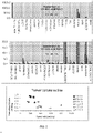
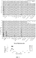
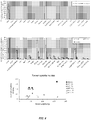
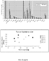
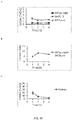
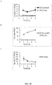
- the present invention is based in part upon the surprising discovery that both the size and location of the PEG moiety on the integrin-binding peptide significantly affect the targeting and pharmacokinetic characteristics of the resulting peptide conjugate.
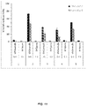
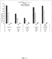
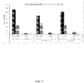
- Examples 1 and 2 illustrate that bi-terminal PEGylation (i.e., attaching PEG units at both the N- and C-termini of the peptide) was able to confer superior targeting characteristics and in vivo pharmacokinetics on the exemplary ⁇ v ⁇ 6 integrin-binding A20FMDV2 peptide and variants thereof (e.g., K16R variant).
- the bi-terminal PEGylated A20FMDV2 and A20FMDV2 (K16R) peptides showed greatly improved pharmacokinetic profiles beyond what was predicted from individual N- or C-terminal PEGylation.
- the two PEG units acted synergistically to achieve greatly improved stability alongside high ⁇ v ⁇ 6 (+)-tumor uptake and retention.
- PEG chains with an exactly defined number of ethylene glycol repeating units 'n', at the N- and at the C-terminus e.g., MW ⁇ ⁇ 3000
- the present invention further provides novel molecular imaging and therapeutic agents with improved affinities and pharmacokinetics based on modifying the bi-terminal PEGylated peptide conjugates described herein with pro-apoptotic peptides, therapeutic radionuclides, micelle-based nanocarriers, multimerization, and/or the addition of blood albumin binding motifs to further improve the affinity, in vivo stability, targeting capabilities, and/or clearance behavior of the peptide conjugates.
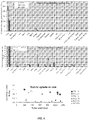
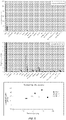
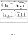
- Example 4 demonstrate that bi-terminal PEGylation of an integrin-binding peptide as short as 8 amino acids with even shorter PEG units (e.g., PEG 11 ) imparts advantageous properties to the peptide such as high selectively, improved serum stability, radiolabeling yields, and lipophilicity when compared to the parent peptide sequence, a cyclic version of the peptide, and individual N- or C-terminal PEGylated versions of the peptide.
- PEG 11 PEG units
- bi-terminal PEGylated peptide conjugates of the present invention having short peptide sequences (e.g., about 8 amino acids in length) and short PEG units (e.g., PEG 11 ) have desirable targeting and pharmacokinetic characteristics that make them suitable for in vivo imaging and therapy.
- the bi-terminal PEGylated peptide conjugates of the present invention can be prepared using standard methods. Only relatively short PEG polymers are needed, allowing synthesis on solid phase. This ensures straightforward preparation and purification.
- the peptide conjugate is obtained as a single compound of precise composition and molecular mass, compared to other PEGylated compounds which may display positional isomerism and contain mixtures of PEG chains with an average length.
- the imaging and targeting of integrin ( ⁇ v ⁇ 6 ) expression in tumors with the peptide conjugates of the present invention result in the detection and treatment of otherwise overlooked tumors, and also serve as a prognostic indicator of cancer in a non-invasive way.
- conjugate is intended to include a chemical compound that has been formed by the joining or attachment of two or more compounds.
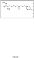
- a conjugate of the present invention includes a "bi-terminal PEGylated peptide conjugate” comprising an integrin-binding peptide covalently attached to a first polyethylene glycol (PEG) moiety at the amino-terminus of the peptide and a second PEG moiety at the carboxyl-terminus of the peptide.
- PEG polyethylene glycol
- the conjugate of the present invention can further comprise an imaging agent or a therapeutic agent covalently attached to the peptide, the first PEG moiety, or the second PEG moiety.
- integrin-binding peptide and "peptide that binds to an integrin” refer to the binding/interaction of a peptide motif in the conjugate which shows the capacity of specific interaction with a specific integrin or a specific group of integrins.
- the terms refer to the ability of a peptide or a portion thereof to interact with and/or bind to a target integrin and without cross-reacting with molecules of similar sequences or structures.
- a peptide specifically binds to a target integrin when it binds to the target integrin with a substantially lower dissociation constant (i.e., tighter binding) than a molecule of similar sequence or structure.
- a specific binding occurs when the peptide binds to the target integrin with an about 2, 3, 4, 5, 6, 8, 10, 15, 20, 25, 30, 40, 50, 100, or 1000-fold or greater affinity than a related molecule.
- the binding of the peptide to a site on the target integrin may occur via intermolecular forces such as ionic bonds, hydrogen bonds, hydrophobic interactions, dipole-dipole bonds, and/or Van der Waals forces.
- Cross-reactivity may be tested, for example, by assessing binding of the peptide under conventional conditions to the target integrin as well as to a number of more or less (e.g., structurally and/or functionally) closely related molecules.
- These methods may include, without limitation, binding studies, blocking and competition studies with closely related molecules, FACS analysis, surface plasmon resonance (e.g., with BIAcore), analytical ultracentrifugation, isothermal titration calorimetry, fluorescence anisotropy, fluorescence spectroscopy, radiolabeled ligand binding assays, and combinations thereof.
- PEGylation refers to the process of covalently coupling a polyethylene glycol (PEG) molecule to another molecule, e.g., a peptide, polypeptide, protein, antibody, and the like, which is then referred to as "PEGylated.”
- PEG polyethylene glycol
- an integrin-binding peptide may be PEGylated at both the amino-terminus and the carboxyl terminus with monodisperse PEG molecules having a defined chain length to generate the bi-terminal PEGylated peptide conjugates of the invention.
- Monodisperse PEG molecules typically comprise discrete molecular weights with an exactly defined number of repeating ethylene glycol units.
- PEG moieties suitable for use in the present invention are commercially available from Polypure AS (Oslo, Norway), which supplies monodisperse PEG molecules and PEG derivatives thereof consisting of substantially one oligomer only (e.g., greater than about 90%, 91%, 92%, 93%, 94%, 95%, 96%, 97%, 98%, or 99% oligomer purity).
- the integrin-binding peptide is PEGylated at both ends with a single type or mixtures of different types of monodisperse PEG moieties having a molecular weight of less than about 3000 daltons (Da), such as, e.g., PEG 11 , PEG 12 (PEG 800), PEG 28 (PEG 1500), and/or (PEG 28 ) 2 (PEG 1500x2).
- Da daltons
- a “peptidomimetic” refers to a chemical compound having a structure that is different from the general structure of an existing peptide, but that functions in a manner similar to the existing peptide, e.g., by mimicking the biological activity of that peptide.
- Peptidomimetics typically comprise naturally-occurring amino acids and/or unnatural amino acids, but can also comprise modifications to the peptide backbone. Peptidomimetics can exhibit increased affinity, specificity, and/or stability compared to an existing peptide.
- amino acid includes naturally-occurring ⁇ -amino acids and their stereoisomers, as well as unnatural amino acids and their stereoisomers.
- “Stereoisomers” of amino acids refers to mirror image isomers of the amino acids, such as L-amino acids or D-amino acids.
- a stereoisomer of a naturally-occurring amino acid refers to the mirror image isomer of the naturally-occurring amino acid, i.e., the D-amino acid.
- Naturally-occurring amino acids are those encoded by the genetic code, as well as those amino acids that are later modified, e.g., ⁇ -carboxyglutamate and O-phosphoserine.
- Naturally-occurring ⁇ -amino acids include, without limitation, alanine (Ala), cysteine (Cys), aspartic acid (Asp), glutamic acid (Glu), phenylalanine (Phe), glycine (Gly), histidine (His), isoleucine (Ile), arginine (Arg), lysine (Lys), leucine (Leu), methionine (Met), asparagine (Asn), proline (Pro), glutamine (Gln), serine (Ser), threonine (Thr), valine (Val), tryptophan (Trp), tyrosine (Tyr), and combinations thereof.
- Stereoisomers of a naturally-occurring ⁇ -amino acids include, without limitation, D-alanine (D-Ala), D-cysteine (D-Cys), D-aspartic acid (D-Asp), D-glutamic acid (D-Glu), D-phenylalanine (D-Phe), D-histidine (D-His), D-isoleucine (D-Ile), D-arginine (D-Arg), D-lysine (D-Lys), D-leucine (D-Leu), D-methionine (D-Met), D-asparagine (D-Asn), D-proline (D-Pro), D-glutamine (D-Gln), D-serine (D-Ser), D-threonine (D-Thr), D-valine (D-Val), D-tryptophan (D-Trp), D-tyrosine (D-Tyr), and combinations thereof.
- Unnatural amino acids include, without limitation, amino acid analogs, amino acid mimetics, synthetic amino acids, N -substituted glycines, and N -methyl amino acids in either the L- or D-configuration that function in a manner similar to the naturally-occurring amino acids.
- amino acid analogs are unnatural amino acids that have the same basic chemical structure as naturally-occurring amino acids, i.e., an ⁇ carbon that is bound to a hydrogen, a carboxyl group, an amino group, but have modified R ( i.e ., side-chain) groups.
- Non-limiting examples of unnatural amino acids include 1-aminocyclopentane-1-carboxylic acid (Acp), 1-aminocyclobutane-1-carboxylic acid (Acb), 1-aminocyclopropane-1-carboxylic acid (Acpc), citrulline (Cit), homocitrulline (HoCit), ⁇ -aminohexanedioic acid (Aad), 3-(4-pyridyl)alanine (4-Pal), 3-(3-pyridyl)alanine (3-Pal), propargylglycine (Pra), ⁇ -aminoisobutyric acid (Aib), ⁇ -aminobutyric acid (Abu), norvaline (Nva), ⁇ , ⁇ -diaminopropionic acid (Dpr), ⁇ , ⁇ -diaminobutyric acid (Dbu), ⁇ - tert -butylglycine (Bug), 3,5-din
- amino acid mimetics are chemical compounds that have a structure that is different from the general chemical structure of an amino acid, but that function in a manner similar to a naturally-occurring amino acid.
- Suitable amino acid mimetics include, without limitation, ⁇ -amino acids and ⁇ -amino acids.
- ⁇ -amino acids the amino group is bonded to the ⁇ -carbon atom of the carboxyl group such that there are two carbon atoms between the amino and carboxyl groups.
- ⁇ -amino acids the amino group is bonded to the ⁇ -carbon atom of the carboxyl group such that there are three carbon atoms between the amino and carboxyl groups.
- Suitable R groups for ⁇ - or ⁇ -amino acids include, but are not limited to, side-chains present in naturally-occurring amino acids and unnatural amino acids.
- N -substituted glycines are unnatural amino acids based on glycine, where an amino acid side-chain is attached to the glycine nitrogen atom.
- Suitable amino acid side-chains include, but are not limited to, side chains present in naturally-occurring amino acids and side-chains present in unnatural amino acids such as amino acid analogs.
- Non-limiting examples of N -substituted glycines include N -(2-aminoethyl)glycine, N -(3-aminopropyl)glycine, N-(2- methoxyethyl)glycine, N -benzyl glycine, (S)- N -(1-phenylethyl)glycine, N- cyclohexylmethylglycine, N -(2-phenylethyl)glycine, N -(3-phenylpropyl)glycine, N -(6-aminogalactosyl)glycine, N -(2-(3'-indolylethyl)glycine, N -(2-( p -methoxyphenylethyl))glycine, N -(2-( p -chlorophenylethyl)glycine, and N -[2-( p -hydroxyphenyleth
- N -substituted glycine oligomers referred to herein as "peptoids,” have been shown to be protease resistant (see, e.g., Miller et al., Drug Dev. Res., 35:20-32 (1995 )).
- Amino acids may be referred to herein by either their commonly known three letter symbols or by the one-letter symbols recommended by the IUPAC-IUB Biochemical Nomenclature Commission.
- an L-amino acid may be represented herein by its commonly known three letter symbol (e.g ., Arg for L-arginine) or by an upper-case one-letter amino acid symbol ( e.g ., R for L-arginine).
- a D-amino acid may be represented herein by its commonly known three letter symbol (e.g., D-Arg for D-arginine) or by a lower-case one-letter amino acid symbol (e.g., r for D-arginine).
- amino acid sequences With respect to amino acid sequences, one of skill in the art will recognize that individual substitutions, additions, or deletions to a peptide, polypeptide, or protein sequence which alters, adds, or deletes a single amino acid or a small percentage of amino acids in the encoded sequence is a "conservatively modified variant" where the alteration results in the substitution of an amino acid with a chemically similar amino acid.
- the chemically similar amino acid includes, without limitation, a naturally-occurring amino acid such as an L-amino acid, a stereoisomer of a naturally occurring amino acid such as a D-amino acid, and an unnatural amino acid such as an amino acid analog, amino acid mimetic, synthetic amino acid, N -substituted glycine, and N -methyl amino acid.
- a naturally-occurring amino acid such as an L-amino acid
- a stereoisomer of a naturally occurring amino acid such as a D-amino acid
- an unnatural amino acid such as an amino acid analog, amino acid mimetic, synthetic amino acid, N -substituted glycine, and N -methyl amino acid.
- amino acids e.g., G, A, I, L, or V
- an aliphatic polar-uncharged group such as C, S, T, M, N, or Q
- basic residues e.g., K, R, or H
- an amino acid with an acidic side chain e.g., E or D
- may be substituted with its uncharged counterpart e.g., Q or N, respectively; or vice versa.
- Each of the following eight groups contains other exemplary amino acids that are conservative substitutions for one another:
- peptide refers to a compound made up of a single chain of D- or L- amino acids or a mixture of D- and L-amino acids joined by peptide bonds. Generally, peptides are about 2 to about 50 amino acids in length. As non-limiting examples, the integrin-binding peptides present in the conjugates described herein are between about 5 to about 45 amino acids in length, between about 8 to about 45 amino acids in length, between about 8 to about 25 amino acids in length, between about 8 to about 20 amino acids in length, between about 12 to about 45 amino acids in length, between about 12 to about 30 amino acids in length, about 8 amino acids in length, or about 20 amino acids in length.
- a "cyclic peptide” refers to a peptide in which the amino-terminus of the peptide or a side-chain on the peptide having a free amino group (e.g., lysine) is joined by a peptide bond to the carboxyl-terminus of the peptide or a side-chain on the peptide having a free carboxyl group (e.g., aspartic acid, glutamic acid).
- a free amino group e.g., lysine
- helix-promoting residue includes amino acids with a conformational preference greater than 1.0 for being found in the middle of an ⁇ -helix (see, e.g., Creighton, Proteins, 1993; and Pace et al., Biophysical J., 75:422-427 (1998 )).
- non-orthodox helix-promoting combinations of amino acids are also within the scope of the invention if they enhance the specificity and/or affinity of binding to a target integrin, e.g., ⁇ v ⁇ 6 integrin.
- a therapeutically effective amount refers to the amount of a conjugate or composition of the present invention that is capable of achieving a therapeutic effect in a subject in need thereof.
- a therapeutically effective amount of a conjugate or composition of the present invention can be the amount that is capable of preventing or relieving one or more symptoms associated with a disease or disorder.
- conjugates and compositions of the present invention can be co-administered with other therapeutic agents such as anticancer, anti-inflammatory, immunosuppressive, antiviral, antibiotic, and/or antifungal agents.
- administering includes oral administration, topical contact, administration as a suppository, intravenous, intraperitoneal, intramuscular, intralesional, intrathecal, intranasal, or subcutaneous administration, or the implantation of a slow-release device, e.g., a mini-osmotic pump, to a subject.
- Administration is by any route, including parenteral and transmucosal (e.g ., buccal, sublingual, palatal, gingival, nasal, vaginal, rectal, or transdermal).
- Parenteral administration includes, e.g ., intravenous, intramuscular, intra-arteriole, intradermal, subcutaneous, intraperitoneal, intraventricular, and intracranial.
- Other modes of delivery include, but are not limited to, the use of liposomal formulations, intravenous infusion, transdermal patches, etc.
- One skilled in the art will know of additional methods for administering a therapeutically effective amount of a conjugate or composition of the present invention for preventing or relieving one or more symptoms associated with a disease or disorder such as cancer or an inflammatory or autoimmune disease.
- conjugate or composition of the present invention is administered at the same time, just prior to, or just after the administration of a second drug (e.g ., anticancer agent, anti-inflammatory agent, immunosuppressive agent, antiviral agent, antibiotic, antifungal agent, etc .).
- a second drug e.g ., anticancer agent, anti-inflammatory agent, immunosuppressive agent, antiviral agent, antibiotic, antifungal agent, etc .
- radioactivity refers to the radiation, including alpha particles, beta particles, nucleons, electrons, positrons, neutrinos, and gamma rays, emitted by a radioactive substance.
- radionuclides suitable for use in the present invention include, but are not limited to, fluorine 18 ( 18 F), fluorine 19 ( 19 F), phosphorus 32 ( 32 P), scandium 47 ( 47 Sc), cobalt 55 ( 55 Co), copper 60 ( 60 Cu), copper 61 ( 61 Cu), copper 62 ( 62 Cu), copper 64 ( 64 Cu), gallium 66 ( 66 Ga), copper 67 ( 67 Cu), gallium 67 ( 67 Ga), gallium 68 ( 68 Ga), rubidium 82 ( 82 Rb), yttrium 86 ( 86 Y), yttrium 87 ( 87 Y), strontium 89 ( 89 Sr), yttrium 90 ( 90 Y), rhodium 105 ( 105 Rh), silver 111 ( 111 Ag), indium 111 ( 111 In), iodine 124 ( 124 I), iodine 125 ( 125 I), iodine 131 ( 131 I), tin 117m (
- the "m” in 117m Sn and 99m Tc stands for the meta state.
- radioactive elements such as uranium, radium, and thorium, which typically represent mixtures of radioisotopes, are suitable examples of radionuclides.
- 67 Cu, 131 I, 177 Lu, and 186 Re are beta- and gamma-emitting radionuclides.
- 212 Bi is an alpha- and beta-emitting radionuclide.
- 211 At is an alpha-emitting radionuclide.
- 32 P, 47 Sc, 89 Sr, 90 Y, 105 Rh, 111 Ag, 117m Sn, 149 Pm, 153 Sm, 166 Ho, and 188 Re are examples of beta-emitting radionuclides.
- 67 Ga, 111 In, 99m Tc, and 201 Tl are examples of gamma-emitting radionuclides.
- 55 Co, 60 Cu, 61 Cu, 62 Cu, 66 Ga, 68 Ga, 82 Rb, and 86 Y are examples of positron-emitting radionuclides.
- 64 Cu is a beta- and positron-emitting radionuclide.
- subject typically refers to humans, but can also include other animals such as, e.g., other primates, rodents, canines, felines, equines, ovines, porcines, and the like.
- the present invention provides bi-terminal PEGylated peptide conjugates that target ⁇ v ⁇ 6 integrin.
- the peptide conjugates of the present invention further comprise a biological agent such as an imaging agent or a therapeutic agent, e.g., covalently attached to one of the PEG moieties.
- the peptide conjugates of the present invention are particularly useful for imaging a ⁇ v ⁇ 6 integrin-containing tumor, organ, or tissue and for treating ⁇ v ⁇ 6 integrin-mediated diseases and disorders such as cancer, inflammatory diseases, autoimmune diseases, chronic fibrosis, chronic obstructive pulmonary disease (COPD), lung emphysema, and chronic wounding skin disease.
- Compositions and kits containing the peptide conjugates of the present invention find utility in a wide range of applications including, e.g., in vivo imaging and immunotherapy.
- the present description provides a conjugate comprising:
- the integrin is ⁇ v ⁇ 6 integrin.
- the ⁇ v ⁇ 6 integrin-binding peptide comprises the amino acid sequence RGDLX 1 X 2 X 3 , wherein X 1 and X 2 are independently selected amino acids and X 3 is L or I. In some instances, X 1 and X 2 are independently selected from the group consisting of Glu, Ala, Leu, Met, Gln, Lys, Arg, Val, Ile, His, Thr, Trp, Phe, and Asp. In certain embodiments, X 1 is Q, X 2 is V, and X 3 is L. In particular embodiments, the peptide comprises the amino acid sequence RGDLX 1 X 2 X 3 AQX 6 , wherein X 6 is K or R.
- X 6 is R.
- the ⁇ v ⁇ 6 integrin-binding peptide comprises or consists of an amino acid sequence selected from the group consisting of NAVPNLRGDLQVLAQKVART (A20FMDV2) and NAVPNLRGDLQVLAQRVART (A20FMDV2 K16R).
- the ⁇ v ⁇ 6 integrin-binding peptide comprises the amino acid sequence RSDLTPLFX 7 , wherein X 7 is absent or is any amino acid.
- X 7 is absent (i.e., the peptide comprises or consists of the amino acid sequence RSDLTPLF).
- X 7 is K (i.e., the peptide comprises or consists of the amino acid sequence RSDLTPLFK).
- the peptide binds to the integrin and a receptor that is co-expressed with the integrin.
- the receptor that is co-expressed with the integrin is C-X-C chemokine receptor type 4 (CXCR4).
- the peptide binds to both ⁇ v ⁇ 6 integrin and CXCR4.
- the receptor that is co-expressed with the integrin is another integrin, e.g., ⁇ v ⁇ 3 integrin co-expressed with ⁇ v ⁇ 5 integrin.
- the peptide binds to both ⁇ v ⁇ 3 integrin and ⁇ v ⁇ 5 integrin.
- the peptide is between about 8 and about 45 amino acids in length. In certain instances, the peptide is 20 amino acids in length.
- the first PEG moiety and the second PEG moiety each have a molecular weight of less than about 3000 daltons (Da).
- the first PEG moiety and the second PEG moiety are monodisperse PEG moieties having a defined chain length.
- PEG moieties having a defined chain length generally include PEG molecules of discrete molecular weights with an exactly defined number of repeating ethylene glycol units.
- Non-limiting examples of PEG moieties having a defined chain length include small, monodisperse PEG molecules having greater than about 90%, 91%, 92%, 93%, 94%, or 95% oligomer purity.
- PEG compound mixtures having an average molecular weight are not used in the conjugates of the present invention.
- the first PEG moiety and the second PEG moiety are independently selected from the group consisting of PEG 11 , PEG 12 (PEG 800), PEG 28 (PEG 1500), and (PEG 28 ) 2 (PEG 1500x2).
- the first PEG moiety and the second PEG moiety are the same.
- the first PEG moiety and the second PEG moiety are both PEG 28 (PEG 1500).
- PEG units suitable for use as the first and/or second PEG moiety in the conjugates of the present invention include PEG 200, PEG 300, PEG 400, PEG 500, PEG 600, PEG 700, PEG 900, PEG 1000, PEG 1100, PEG 1200, PEG 1300, PEG 1400, PEG 1600, PEG 1700, PEG 1800, PEG 1900, PEG 2000, PEG 2100, PEG 2200, PEG 2300, PEG 2400, PEG 2500, PEG 2600, PEG 2700, PEG 2800, PEG 2900, PEG 3000, as well as derivatives thereof such as branched PEG derivatives.
- these PEG molecules contain an exactly defined number of repeating units "n" and are monodisperse ( e.g., having greater than about 95% oligomer purity).
- PEG moieties suitable for use in the present invention are commercially available from EMD Chemicals, Inc. (San Diego, CA) and Polypure AS (Oslo, Norway).
- the conjugate further comprises an imaging agent or a therapeutic agent covalently attached to the peptide, the first PEG moiety, and/or the second PEG moiety.
- the imaging agent or therapeutic agent is covalently attached to the first PEG moiety.
- the imaging agent or therapeutic agent is covalently attached as the most N-terminal moiety in the conjugate.
- the imaging agent is selected from the group consisting of a radionuclide, biotin, a fluorophore, a fluorescent protein, an antibody, horseradish peroxidase, alkaline phosphatase, and combinations thereof.
- the radionuclide is selected from the group consisting of 11 C, 13 N, 15 O, 18 F, 19 F, 61 Cu, 62 Cu, 64 Cu, 67 Cu, 68 Ga, 111 In, 124 I, 125 I, and 131 I.
- the radionuclide is attached via a prosthetic group to the peptide, the first PEG moiety, or the second PEG moiety.
- the radionuclide is attached via a prosthetic group to the first PEG moiety.
- the radionuclide is attached via a prosthetic group as the most N-terminal moiety in the conjugate.
- prosthetic groups include benzoyl groups (e.g., fluorobenzoic acid (FBA)), fluoropropionic acid (FPA), pyridine (Py), dipyridyl-tetrazine (Tz), trans-cyclooctene (TCO), derivatives thereof, and combinations thereof.
- the radionuclide is 18 F or 19 F covalently attached to the first PEG moiety via a benzoyl group such as FBA.
- a benzoyl group such as FBA.
- 4-[ 18 F]-fluorobenzoic acid ([ 18 F]FBA) or 4-[ 19 F]-fluorobenzoic acid ([ 19 F]FBA) can be used to radiolabel the peptide conjugates of the present invention.
- the therapeutic agent is selected from the group consisting of a radionuclide, a pro-apoptotic peptide, a nanoparticle, a chemotherapeutic agent, a nanodroplet, a liposomal drug, a cytokine, and combinations thereof.
- the therapeutic agent is a radionuclide selected from the group consisting of 90 Y and 177 Lu.
- the radionuclide is attached via a chelating agent to the peptide, the first PEG moiety, or the second PEG moiety.
- the radionuclide is attached via a chelating agent to the first PEG moiety.
- the radionuclide is attached via a chelating agent as the most N-terminal moiety in the conjugate.
- chelating agents include macrocyclic metal chelators such as DOTA (1,4,7,10-tetraazacyclododecane-N,N′,N ⁇ ,N′′′-tetraacetic acid), NOTA (1,4,7-triazacyclononane-N,N′,N ⁇ -triacetic acid), DTPA (diethyl enetriaminepentaacetic anhydride), TETA (1,4,8,11-tetraazacyclotetradecane-N,N′,N ⁇ ,N′′′-tetraacetic acid), and DTTA (N-(p-isothiocyanatobenzyl)-diethylenetriamine-N,N′,N ⁇ ,N′′′-tetraacetic acid).
- DOTA 1,4,7,10-tetraazacyclododecane-N,N′,N ⁇ ,N′′′
- the therapeutic agent is a pro-apoptotic peptide comprising the amino acid sequence D (KLAKLAK) 2 .
- the pro-apoptotic peptide is attached via a glycine linker to the peptide, the first PEG moiety, or the second PEG moiety.
- the pro-apoptotic peptide is attached via a glycine linker to the first PEG moiety.
- glycine linkers include a single glycine residue or at least about 2, 3, 4, 5, 6, 7, 8, 9, 10, 11, 12, 13, 14, 15, 16, 17, 18, 19, or 20 consecutive glycine residues or glycine residues separated by other amino acid residues.
- the glycine linker is a glycinylglycine linker.
- linkers suitable for attaching the pro-apoptotic peptide to the peptide conjugates of the present invention, e.g., without significantly interfering with the targeting properties and function of each individual component.
- the therapeutic agent is a nanoparticle comprising a telodendrimer scaffold or other micelle-based nanacarrier system.
- the telodendrimer scaffold is PEG 5K CA 8 .
- Telodendrimers suitable for use in the present invention are described in US Patent Publication No. 20130164369 .
- the nanoparticle is loaded with a chemotherapeutic agent.
- chemotherapeutic agents include paclitaxel (PTX) and other cytotoxic chemotherapeutic agents described herein.
- the conjugate further comprises an albumin binding motif covalently attached to the peptide, the first PEG moiety, or the second PEG moiety.
- the albumin binding motif is 4-(4-iodophenyl)butyric acid (IPA) or a homolog thereof with a shorter alkyl chain such as, e.g ., 4-(4-iodophenyl)propionic acid or 4-(4-iodophenyl)acetic acid.
- the albumin binding motif is 4-(4-methylphenyl)butyric acid or 4-(4-bromophenyl)butyric acid or a homolog thereof with a shorter alkyl chain such as, e.g., a propionic acid or acetic acid homolog thereof.
- the albumin binding motif is covalently attached to the first and/or second PEG moiety.
- the albumin binding motif is covalently attached to the first and/or second PEG moiety via a linker such as a glutamic acid (E) linker or other suitable linker (e.g ., amino acid or peptide linker) known to one of skill in the art.
- the albumin binding motif is ⁇ -(4-(4-iodophenyl)butyl amide)lysine-glutamic acid (“K(IPA)E”), which corresponds to IPA that is covalently attached to the side-chain of the lysine residue of a lysine-glutamic acid peptide linker.
- K(IPA)E albumin binding motif is covalently attached to the first PEG moiety.
- the imaging agent or therapeutic agent is covalently attached ( e.g ., via a prosthetic group, a chelating agent, or a linker) to an albumin binding motif that is covalently attached to the first PEG moiety, such that the imaging agent or therapeutic agent is the most N-terminal moiety in the conjugate.
- the present invention provides a composition comprising a bi-terminal PEGylated peptide conjugate described herein or a plurality thereof (e.g ., at least about 2, 3, 4, 5, 6, 7, 8, 9, 10, 11, 12, 13, 14, 15, 16, 17, 18, 19, 20, 25, 30, or more peptide conjugates of the invention that differ, e.g., in their integrin-binding peptide sequences, first and/or second PEG moieties, imaging and/or therapeutic agents, or combinations thereof).
- the plurality of conjugates i.e ., the first and second PEG moieties in each of the pluarity of conjugates
- the first PEG moiety and the second PEG moiety in each of the pluarity of conjugates are independently selected from the group consisting of PEG 11 , PEG 12 (PEG 800), PEG 28 (PEG 1500), and (PEG 28 ) 2 (PEG 1500x2).
- the first PEG moiety and the second PEG moiety in each of the pluarity of conjugates are the same.
- the first PEG moiety and the second PEG moiety in each of the pluarity of conjugates are both PEG 28 (PEG 1500).
- the present invention provides multimeric peptide conjugates wherein a plurality of the conjugates are linked to each other.
- the multimeric conjugate is a dimer or a tetramer of the plurality of conjugates.
- the multimeric peptide conjugates are formed via linkage between the second PEG moiety of each conjugate.
- the conjugates are linked to each other at the second PEG moiety via at least one lysine residue (e.g., at least 1, 2, 3, 4, 5, or more lysine (K) residues).
- one or more of the lysine residues comprises an imaging or therapeutic agent such as a radionuclide (e.g ., for use as a radiolabel) attached thereto.
- the composition further comprises a pharmaceutical carrier or excipient.
- the present invention provides a kit for imaging or therapy, the kit comprising:
- the present disclosure provides a method for the in vivo imaging of a target tissue, the method comprising:
- the target tissue is a cancerous tissue or an organ.
- cancerous tissues include cancerous tissues or tumors associated with pancreatic cancer, breast cancer, colorectal cancer, prostate cancer, cervical cancer, and oral squamous cell carcinoma.
- the peptide conjugate is administered for imaging a tumor such as a pancreatic tumor.
- pancreatic tumors suitable for imaging in accordance with the present invention include, but are not limited to, adenocarcinomas, serous cystadenomas, acinar cell cancers, pancreatic neuroendocrine tumors ( e.g ., insulinomas), and the like.
- the imaging agent comprises a radionuclide (e.g. , bound to a prosthetic group such as a benzoyl group or a chelating agent), biotin, a fluorophore, a fluorescent protein, horseradish peroxidase, or alkaline phosphatase.
- a radionuclide comprises the imaging agent
- detection occurs when radiation from the radionuclide is used to determine where the peptide conjugate is concentrated in the subject.
- a fluorophore or fluorescent protein comprises the imaging agent, detection occurs when fluorescence from the fluorophore or fluorescent protein is used to determine where the peptide conjugate is concentrated in the subject.
- the peptide conjugate is detected by Magnetic Resonance Imaging (MRI), Magnetic Resonance Spectroscopy (MRS), Single Photon Emission Computerized Tomography (SPECT), Positron Emission Tomography (PET), or optical imaging.
- the conjugate is detected for the diagnosis or prognosis of a disease or disorder mediated by the integrin.
- the disease or disorder is associated with the expression, overexpression, and/or activation of the integrin.
- the disease or disorder is an ⁇ v ⁇ 6 integrin-mediated disease or disorder, e.g., the peptide conjugate is detected for the diagnosis or prognosis of an ⁇ v ⁇ 6 -mediated disease or disorder.
- a method for treating an integrin-mediated disease or disorder in a subject in need thereof comprising: administering to the subject a therapeutically effective amount of a bi-terminal PEGylated peptide conjugate described herein or a composition thereof ( e.g ., a plurality or multimer of conjugates), wherein a therapeutic agent is covalently attached to the peptide, the first PEG moiety, or the second PEG moiety.
- the disease or disorder is associated with the expression, overexpression, and/or activation of the integrin.
- integrin-mediated diseases or disorders include cancer, inflammatory diseases, autoimmune diseases, chronic fibrosis, chronic obstructive pulmonary disease (COPD), lung emphysema, and chronic wounding skin disease.
- the disease or disorder is an ⁇ v ⁇ 6 integrin-mediated disease or disorder.
- the ⁇ v ⁇ 6 integrin-mediated disease or disorder is pancreatic cancer, breast cancer, colorectal cancer, prostate cancer, cervical cancer, or oral squamous cell carcinoma.
- a therapeutically effective amount of the conjugate or the composition is an amount sufficient for achieving a therapeutic benefit in the subject. In yet other embodiments, a therapeutically effective amount of the conjugate or the composition is an amount sufficient to target delivery of the therapeutic agent to a cell expressing the integrin.
- a method for imaging epithelial cells expressing or overexpressing an integrin of interest e.g., ⁇ v ⁇ 6 integrin
- the method comprising administering to the subject a therapeutically effective amount of a peptide conjugate or composition as described herein.
- the method is particularly useful for the imaging of chronic fibrosis, chronic obstructive pulmonary disease (COPD), lung emphysema, chronic wounding skin disease (e.g., epidermolysis bullosa), or epithelial tumor cells.
- COPD chronic obstructive pulmonary disease
- lung emphysema chronic wounding skin disease
- epidermolysis bullosa chronic wounding skin disease
- the method of imaging ⁇ v ⁇ 6 -overexpressing epithelial cells may include linking the peptide or one of the PEG components of the conjugate to a fluorescent probe, and incorporating the resulting peptide conjugate into a suitable dosage form such that upon administration the ⁇ v ⁇ 6 integrin-binding conjugate may be visualized by its fluorescent tag.
- a method for delivering a therapeutic agent to a cell expressing or overexpressing an integrin of interest e.g., ⁇ v ⁇ 6 integrin
- a tumor, organ, or tissue containing cells expressing or overexpressing an integrin of interest e.g., ⁇ v ⁇ 6 integrin
- the method comprising administering a peptide conjugate or composition comprising the therapeutic agent as described herein to the subject.
- the present invention provides bi-terminal PEGylated integrin-binding peptide conjugates.
- the integrins are a superfamily of cell adhesion receptors that bind to extracellular matrix ligands, cell-surface ligands, and soluble ligands. Integrins are transmembrane ⁇ heterodimers and at least 18 ⁇ and eight ⁇ subunits are known in humans, generating 24 heterodimers. The ⁇ and ⁇ subunits have distinct domain structures, with extracellular domains from each subunit contributing to the ligand-binding site of the heterodimer.
- Non-limiting examples of integrins include ⁇ 1 ⁇ 1 , ⁇ 2 ⁇ 1 , ⁇ 3 ⁇ 1 , ⁇ 4 ⁇ 1 , ⁇ 5 ⁇ 1 , ⁇ 6 ⁇ 1 , ⁇ 7 ⁇ 1 , ⁇ 8 ⁇ 1 , ⁇ 9 ⁇ 1 , ⁇ 10 ⁇ 1 , ⁇ 11 ⁇ 1 , ⁇ v ⁇ 1 , ⁇ v ⁇ 3 , ⁇ v ⁇ 5 , ⁇ v ⁇ 6 , ⁇ v ⁇ 8 , ⁇ IIb ⁇ 3 , ⁇ 4 ⁇ 7 , ⁇ E ⁇ 7 , ⁇ 6 ⁇ 4 , ⁇ L ⁇ 2 , ⁇ M ⁇ 2 , ⁇ X ⁇ 2 , ⁇ D ⁇ 2 , and combinations thereof.
- the peptide binds to (e.g., targets) ⁇ v ⁇ 6 integrin.
- the ⁇ v ⁇ 6 integrin-binding peptide comprises the amino acid sequence RGDLX 1 X 2 X 3 , wherein X 1 and X 2 are independently selected amino acids and X 3 is Leu (L) or Ile (I).
- X 1 is Q
- X 2 is V
- X 3 is L.
- amino acid positions herein are numbered from the amino-terminus to the carboxyl-terminus of the peptide.
- the residues LX 1 X 2 X 3 are present within an ⁇ -helix.
- An ⁇ -helix is understood to be a sequential group of amino acids in a peptide that interact with a particular hydrogen bonding pattern and thus define a helical structure.
- the hydrogen bonding pattern in a standard ⁇ -helix is between the carbonyl oxygen of residue n and the amide hydrogen of residue n+4.
- this hydrogen bonding pattern is between residues n and n+3.
- this hydrogen bonding pattern is between residues n and n+5.
- the number of residues per turn in each ⁇ -helix is 3.6, 3.0, and 4.4 for the standard ⁇ -helix, 3 10 -helix, and pi-helix, respectively.
- the ⁇ -helix of the peptide enables the hydrophobic side-chains of the residues LX 1 X 2 L/I to protrude from one side of the helix.
- the ⁇ -helix has at least one turn.
- An ⁇ -helix useful in the present description may be an ⁇ -helix mimetic as described in, e.g., PCT Publication No. WO 95/00534 .
- ⁇ -helix mimetics are ⁇ -helical structures which are able to stabilize the structure of a naturally-occurring or synthetic peptide.
- the ⁇ v ⁇ 6 integrin-binding peptides used in the conjugates of the present invention may comprise standard helices, 3 10 -helices, pi-helices, or any combination thereof.
- the helices may comprise amino acids that form a "cap" structure, such as an amino-terminal cap and/or a carboxyl-terminal cap which flank the helix.
- the ⁇ v ⁇ 6 integrin-binding peptide comprises the sequence RGDLX 1 X 2 LX 4 X 5 X 6 , wherein X 1 , X 2 , X 4 , X 5 , and X 6 are independently selected amino acids.
- X 1 , X 2 , X 4 , X 5 , and X 6 are helix-promoting residues.
- the helix-promoting residues can be independently selected from the group consisting of Glu, Ala, Leu, Met, Gln, Lys, Arg, Val, Ile, His, Thr, Trp, Phe, and Asp.
- the helix-promoting residues can comprise naturally-occurring amino acids or unnatural amino acids such as artificial or modified amino acids.
- the peptide comprises the sequence RGDLX 1 X 2 LX 4 X 5 X 6 Z n , wherein Z is a helix-promoting residue and n is any number between 1 and 20. Preferably, n is between 5 and 15 or between 8 and 12. Extension of the helix to include helical residues in the Z position can further increase the helix dipole and provide enhanced binding to ⁇ v ⁇ 6 integrin.
- the ⁇ v ⁇ 6 integrin-binding peptide may be represented by the formula: B m RGDLX 1 X 2 LX 4 X 5 X 6 Z n , wherein B is m amino acids which enhances the hydrophobic interactions with the helix defined from LX 1 X 2 L and also enhances the RGD domain for binding, Z is a helix-promoting residue, n is a number between 1 and 35, and m is a number between 1 and 35.
- m is selected so that B is sufficiently long to facilitate a hydrophobic/non-covalent interacting core. The exact nature of these residues depends on the general design of the region.
- hydrophobic interactions from residues such as Val, Ile, Leu
- electrostatic interactions using Asp, Glu, Lys, and/or Arg together with their counterpart ion-pair at X 1 and/or X 2 ).
- the ⁇ v ⁇ 6 integrin-binding peptide comprises the amino acid sequence RGDLX 1 X 2 X 3 AQX 6 , wherein X 6 is Lys (K) or Arg (R). In preferred embodiments, X 6 is R.
- the ⁇ v ⁇ 6 integrin-binding peptide comprises or consists of an amino acid sequence selected from NAVPNLRGDLQVLAQKVART (A20FMDV2), NAVPNLRGDLQVLAQRVART (A20FMDV2 K16R), GFTTGRRGDLATIHGMNRPF (A20LAP), YTASARGDLAHLTTTHARHL (A20FMDV1), and combinations thereof.
- the ⁇ v ⁇ 6 integrin-binding peptide comprises the amino acid sequence RSDLTPLFX 7 , wherein X 7 is absent or is any amino acid.
- X 7 is absent (i.e., the peptide comprises or consists of the amino acid sequence RSDLTPLF).
- X 7 is K (i.e., the peptide comprises or consists of the amino acid sequence RSDLTPLFK).
- ⁇ v ⁇ 6 integrin which is a receptor for fibronectin, tenascin, vitronectin, the latency associated peptide (LAP) of TGF- ⁇ , and viral capsid protein (VP1) of foot-and-mouth disease virus (FMDV), is expressed at very low or undetectable levels in only a subset of epithelial cells in normal adult tissues ( Breuss et al., J. Cell Sci., 108:2241-2251 (1995 )). However, ⁇ v ⁇ 6 integrin expression is increased dramatically during development, following injury or inflammation, or in a variety of epithelial neoplasms.
- keratinocytes show de novo expression of ⁇ v ⁇ 6 integrin in both oral and skin wounds (Breuss et al., supra; Clark et al., Am. J. Path., 148:1407-1421 (1996 )).
- ⁇ v ⁇ 6 integrin plays an active role in tumor invasion because its expression is often higher at the invasive margins of oral squamous cell carcinomas.
- ⁇ v ⁇ 6 integrin is an excellent target for both imaging and therapy of diseases or disorders such as pancreatic cancer, oral cancer, ovarian cancer, breast cancer, and colon cancer.
- bi-terminal PEGylation of ⁇ v ⁇ 6 integrin-binding peptides with small, monodisperse PEG molecules having a defined chain length can be used to generate conjugates of the present invention that display significantly better localizing and/or targeting potential by providing high tumor selectivity and specificity for ⁇ v ⁇ 6 -expressing tumors and having increased metabolic stability and retention at the tumor site when compared to peptides having individual N- or C-terminal PEGylation.
- the peptide is a bivalent peptide that binds to the integrin and a receptor that is co-expressed with the integrin.
- co-expressed receptors include CXCR4.
- the bivalent peptide binds to both ⁇ v ⁇ 6 integrin and CXCR4.
- the receptor that is co-expressed with the integrin is another integrin, e.g., ⁇ v ⁇ 3 integrin co-expressed with ⁇ v ⁇ 5 integrin.
- the bivalent peptide binds to both ⁇ v ⁇ 3 integrin and ⁇ v ⁇ 5 integrin.
- the peptide comprises a first peptide fragment that binds to an integrin linked to a second peptide fragment that binds to a co-expressed receptor. In other instances, the peptide comprises a first peptide fragment that binds to a co-expressed receptor linked to a second peptide fragment that binds to an integrin.
- the first and second peptide fragments can be linked directly to each other or can be linked via a glycine linker or other suitable linker known in the art.
- the first peptide fragment is PEGylated at the N-terminus and the second peptide fragment is PEGylated at the C-terminus, thereby forming a bi-terminal PEGylated bivalent peptide conjugate.
- the peptide of the invention is between about 5 to about 45 amino acids in length, between about 8 to about 45 amino acids in length, between about 8 to about 25 amino acids in length, between about 12 to about 45 amino acids in length, between about 5 to about 40 amino acids in length, between about 10 to about 40 amino acids in length, or about 35, 30, 25, 20, 15, or 10 amino acids in length.
- the peptide may be about 5, 6, 7, 8, 9, 10, 11, 12, 13, 14, 15, 16, 17, 18, 19, 20, 21, 22, 23, 24, 25, 26, 27, 28, 29, 30, 31, 32, 33, 34, 35, 36, 37, 38, 39, 40, 41, 42, 43, 44, 45, or more amino acids in length.
- the peptide should not exceed a length which would allow the formation of a tertiary structure, such as, for example, greater than 45 amino acids if present as an isolated molecule.
- the peptide may exceed 45 amino acids if fused to a larger molecule such as an antibody or another protein or macromolecule which could prevent the formation of a tertiary structure within the peptide.
- the peptide may also exceed 45 amino acids if it is a bivalent peptide having first and second peptide fragments that bind to different receptors.
- the peptide is about 20 amino acids in length.
- the peptides used in the conjugates of the invention can also be functional variants of the peptides as defined above, including peptides that possess at least about 50%, 55%, 60%, 65%, 70%, 75%, 80%, 85%, 90%, 95%, or more sequence identity with the peptides described above.
- the peptides can comprise naturally-occurring amino acids and/or unnatural amino acids.
- unnatural amino acids include, but are not limited to, D-amino acids, ornithine, diaminobutyric acid ornithine, norleucine ornithine, pyriylalanine, thienylalanine, naphthylalanine, phenylglycine, alpha and alpha-disubstituted amino acids, N-alkyl amino acids, lactic acid, halide derivatives of naturally-occurring amino acids (e.g., trifluorotyrosine, p-Cl- phenylalanine, p-Br-phenylalanine, p-I-phenylalanine, etc.), L-allylglycine, b-alanine, L- ⁇ -amino butyric acid, L-g-amino butyric acid, L- ⁇ -amino isobutyric acid, L-e-amino caproic acid, 7-amino heptanoic acid,
- the peptides may be further modified.
- one or more amide bonds may be replaced by ester or alkyl backbone bonds.
- the peptides used in the conjugates of the invention may include both modified peptides and synthetic peptide analogues.
- Peptides may be modified to improve formulation and storage properties, or to protect labile peptide bonds by incorporating non-peptidic structures.
- Peptides of the present invention may be prepared using methods known in the art. For example, peptides may be produced by chemical synthesis, e.g ., using solid phase techniques and/or automated peptide synthesizers, or by recombinant means. In certain instances, peptides may be synthesized using solid phase strategies on an automated multiple peptide synthesizer (Abimed AMS 422) using 9-fluorenylmethyloxycarbonyl (Fmoc) chemistry.
- the peptides can then be purified by reversed phase-HPLC and lyophilized.
- the peptides may alternatively be prepared by cleavage of a longer peptide or full-length protein sequence.
- a fragment containing the ⁇ v ⁇ 6 integrin-binding domain of fibronectin, tenascin, vitronectin, the latency associated peptide (LAP) of TGF- ⁇ , or viral capsid protein (VP1) of foot-and-mouth disease virus (FMDV) can be isolated by cleavage of the full-length protein.
- the peptide component of the conjugates of the invention may be cyclized.
- Methods are well known in the art for introducing cyclic structures into peptides to select and provide conformational constraints to the structure that result in enhanced stability.
- a C- or N-terminal cysteine can be added to the peptide, so that when oxidized the peptide will contain a disulfide bond, generating a cyclic peptide.
- Other peptide cyclization methods include the formation of thioethers and carboxyl- and amino- terminal amides and esters.
- the bi-terminal PEGylated integrin-binding peptide conjugates of the present invention have particular utility in human and veterinary imaging, therapeutic, prognostic, and diagnostic applications.
- the conjugates can be used for imaging tumors such as malignant tumors of the pancreas (e.g ., adenocarcinomas, serous cystadenomas, acinar cell cancers, pancreatic neuroendocrine tumors such as insulinomas, etc.) or any other tissue or organ.
- the conjugates are also useful for treating diseases and disorders such as cancer ( e.g ., pancreatic cancer, breast cancer, colon cancer, cervical cancer, lung cancer, etc. ) , inflammatory disease, autoimmune disease, chronic fibrosis, chronic obstructive pulmonary disease (COPD), lung emphysema, and chronic wounding skin disease.
- cancer e.g ., pancreatic cancer, breast cancer, colon cancer, cervical cancer, lung cancer, etc.
- COPD chronic ob
- Administration of the peptide conjugates of the present invention with a suitable pharmaceutical excipient as necessary can be carried out via any of the accepted modes of administration.
- administration can be, for example, intravenous, topical, subcutaneous, transcutaneous, transdermal, intramuscular, oral, intra-joint, parenteral, intra-arteriole, intradermal, intraventricular, intracranial, intraperitoneal, intralesional, intranasal, rectal, vaginal, or by inhalation.
- administration may be directly to the tumor and/or into tissues surrounding the tumor.
- compositions containing a conjugate or a combination of conjugates of the present invention may be administered repeatedly, e . g ., at least 2, 3, 4, 5, 6, 7, 8, or more times, or the composition may be administered by continuous infusion.
- Suitable sites of administration include, but are not limited to, dermal, mucosal, bronchial, gastrointestinal, anal, vaginal, eye, and ear.
- the formulations may take the form of solid, semi-solid, lyophilized powder, or liquid dosage forms, such as, for example, tablets, pills, lozenges, capsules, powders, solutions, suspensions, emulsions, suppositories, retention enemas, creams, ointments, lotions, gels, aerosols, or the like, preferably in unit dosage forms suitable for simple administration of precise dosages.
- unit dosage form refers to physically discrete units suitable as unitary dosages for human subjects and other mammals (e.g ., dogs), each unit containing a predetermined quantity of active material calculated to produce the desired onset, tolerability, and/or therapeutic effects, in association with a suitable pharmaceutical excipient (e.g. , an ampoule).
- a suitable pharmaceutical excipient e.g. , an ampoule
- more concentrated compositions may be prepared, from which the more dilute unit dosage compositions may then be produced.
- the more concentrated compositions thus will contain substantially more than, e.g., at least 1, 2, 3, 4, 5, 6, 7, 8, 9, 10, or more times the amount of a conjugate or a combination of conjugates.
- compositions to be administered contains a quantity of the conjugate or combination of conjugates in a pharmaceutically effective amount for imaging a tumor, organ, or tissue or for relief of a condition being treated, when administered in accordance with the teachings of this invention.
- pharmaceutically acceptable salts of the conjugates of the present invention e.g ., acid addition salts
- compositions typically include a conventional pharmaceutical carrier or excipient and may additionally include other medicinal agents, carriers, adjuvants, diluents, tissue permeation enhancers, solubilizers, and the like.
- the composition will contain about 0.01% to about 90%, about 0.1% to about 75%, about 0.1% to 50%, or about 0.1% to 10% by weight of a conjugate of the present invention or a combination thereof, with the remainder consisting of suitable pharmaceutical carrier and/or excipients.
- Appropriate excipients can be tailored to the particular composition and route of administration by methods well known in the art. See, e.g., REMINGTON'S PHARMACEUTICAL SCIENCES, supra.
- excipients include, but are not limited to, lactose, dextrose, sucrose, sorbitol, mannitol, starches, gum acacia, calcium phosphate, alginates, tragacanth, gelatin, calcium silicate, microcrystalline cellulose, polyvinylpyrrolidone, cellulose, water, saline, syrup, methylcellulose, ethylcellulose, hydroxypropylmethylcellulose, and polyacrylic acids such as Carbopols, e.g., Carbopol 941, Carbopol 980, Carbopol 981, etc.
- Carbopols e.g., Carbopol 941, Carbopol 980, Carbopol 981, etc.
- compositions can additionally include lubricating agents such as talc, magnesium stearate, and mineral oil; wetting agents; emulsifying agents; suspending agents; preserving agents such as methyl-, ethyl-, and propyl-hydroxy-benzoates ( i.e., the parabens); pH adjusting agents such as inorganic and organic acids and bases; sweetening agents; coloring agents; and flavoring agents.
- lubricating agents such as talc, magnesium stearate, and mineral oil
- wetting agents such as talc, magnesium stearate, and mineral oil
- emulsifying agents such as methyl-, ethyl-, and propyl-hydroxy-benzoates ( i.e., the parabens)
- pH adjusting agents such as inorganic and organic acids and bases
- sweetening agents coloring agents
- flavoring agents such as inorganic and organic acids and bases.
- the compositions may also comprise biodegradable polymer beads, dextran,
- compositions can be in the form of tablets, lozenges, capsules, emulsions, suspensions, solutions, syrups, sprays, powders, and sustained-release formulations.
- Suitable excipients for oral administration include pharmaceutical grades of mannitol, lactose, starch, magnesium stearate, sodium saccharine, talcum, cellulose, glucose, gelatin, sucrose, magnesium carbonate, and the like.
- the pharmaceutical compositions take the form of a pill, tablet, or capsule, and thus, the composition can contain, along with the conjugate or combination of conjugates, any of the following: a diluent such as lactose, sucrose, dicalcium phosphate, and the like; a disintegrant such as starch or derivatives thereof; a lubricant such as magnesium stearate and the like; and a binder such a starch, gum acacia, polyvinylpyrrolidone, gelatin, cellulose and derivatives thereof.
- the conjugates can also be formulated into a suppository disposed, for example, in a polyethylene glycol (PEG) carrier.
- PEG polyethylene glycol
- Liquid compositions can be prepared by dissolving or dispersing a conjugate or a combination of conjugates and optionally one or more pharmaceutically acceptable adjuvants in a carrier such as, for example, aqueous saline (e.g ., 0.9% w/v sodium chloride), aqueous dextrose, glycerol, ethanol, and the like, to form a solution or suspension, e . g ., for oral, topical, or intravenous administration.
- aqueous saline e.g ., 0.9% w/v sodium chloride
- aqueous dextrose e.g ., glycerol
- ethanol e.g ethanol
- the conjugates of the present invention can also be formulated into a retention enema.
- compositions of the present invention can be in the form of emulsions, lotions, gels, creams, jellies, solutions, suspensions, ointments, and transdermal patches.
- the composition can be delivered as a dry powder or in liquid form via a nebulizer.
- the compositions can be in the form of sterile injectable solutions and sterile packaged powders.
- injectable solutions are formulated at a pH of about 4.5 to about 7.5.
- compositions of the present invention can also be provided in a lyophilized form.
- Such compositions may include a buffer, e.g. , bicarbonate, for reconstitution prior to administration, or the buffer may be included in the lyophilized composition for reconstitution with, e.g ., water.
- the lyophilized composition may further comprise a suitable vasoconstrictor, e.g ., epinephrine.
- the lyophilized composition can be provided in a syringe, optionally packaged in combination with the buffer for reconstitution, such that the reconstituted composition can be immediately administered to a patient.
- administered dosages will be effective to deliver picomolar to micromolar concentrations of the conjugate to the appropriate site or sites.
- dose administered will vary depending on a number of factors, including, but not limited to, the particular conjugate or set of conjugates to be administered, the mode of administration, the type of application (e.g., imaging, diagnostic, prognostic, therapeutic, etc.), the age of the patient, and the physical condition of the patient.
- the smallest dose and concentration required to produce the desired result should be used.
- Dosage should be appropriately adjusted for children, the elderly, debilitated patients, and patients with cardiac and/or liver disease. Further guidance can be obtained from studies known in the art using experimental animal models for evaluating dosage.
- the increased metabolic stability, tumor retention, and tumor to blood ratios associated with the conjugates of the present invention permits a wider margin of safety for dosage concentrations and for repeated dosing.
- the bi-terminal PEGylated integrin-binding peptide conjugates of the present invention are used for the treatment of an integrin-mediated disease or disorder in a subject in need thereof.
- diseases or disorders suitable for treatment with the peptide conjugates described herein include, but are not limited to, allergy, anxiety disorder, autoimmune disease, behavioral disorder, birth defect, blood disorder, bone disease, cancer, chronic fibrosis, chronic obstructive pulmonary disease (COPD), chronic wounding skin disease, circulatory disease, tooth disease, depressive disorder, dissociative disorder, ear condition, eating disorder, eye condition, food allergy, food-borne illness, gastrointestinal disease, genetic disorder, heart disease, hormonal disorder, immune deficiency, infectious disease, inflammatory disease, insect-transmitted disease, nutritional disorder, kidney disease, leukodystrophy, liver disease, lung emphysema, mental health disorder, metabolic disease, mood disorder, musculodegenerative disorder, neurological disorder, neurodegenerative disorder, neuromuscular disorder, personality disorder, phobia,
- the ⁇ v ⁇ 6 -mediated disease or disorder is cancer, an inflammatory disease, an autoimmune disease, chronic fibrosis, chronic obstructive pulmonary disease (COPD), lung emphysema, and chronic wounding skin disease (e.g ., epidermolysis bullosa).
- cancer an inflammatory disease
- an autoimmune disease chronic fibrosis
- chronic obstructive pulmonary disease (COPD) chronic obstructive pulmonary disease
- COPD chronic obstructive pulmonary disease
- lung emphysema chronic wounding skin disease
- chronic wounding skin disease e.g ., epidermolysis bullosa
- Cancer generally includes any of various malignant neoplasms characterized by the proliferation of anaplastic cells that tend to invade surrounding tissue and metastasize to new body sites.
- Non-limiting examples of different types of cancer suitable for treatment using the conjugates or compositions of the present invention include ovarian cancer, breast cancer, lung cancer, bladder cancer, thyroid cancer, liver cancer, pleural cancer, pancreatic cancer, cervical cancer, prostate cancer, testicular cancer, colon cancer, anal cancer, bile duct cancer, gastrointestinal carcinoid tumors, esophageal cancer, gall bladder cancer, rectal cancer, appendix cancer, small intestine cancer, stomach (gastric) cancer, renal cancer ( i.e ., renal cell carcinoma), cancer of the central nervous system, skin cancer, choriocarcinomas, head and neck cancers, bone cancer, osteogenic sarcomas, fibrosarcoma, neuroblastoma, glioma, melanoma, leukemia (e.g., acute lymphocytic le
- conjugates of the present invention can be co-administered with other therapeutic agents for the treatment of cancer.
- Suitable anti-cancer agents for combination therapy include, without limitation, cytotoxins and agents such as antimetabolites, alkylating agents, anthracyclines, antibiotics, antimitotic agents, procarbazine, hydroxyurea, asparaginase, corticosteroids, interferons, radiopharmaceuticals, peptides with antitumor activity such as TNF- ⁇ , pharmaceutically acceptable salts thereof; derivatives thereof, prodrugs thereof, and combinations thereof.
- a pharmaceutical composition comprising one or more conjugates of the present invention may be administered to a patient before, during, or after administration of an anti-cancer agent or combination of anti-cancer agents either before, during, or after chemotherapy.
- Treatment with the conjugate after chemotherapy may be particularly useful for reducing and/or preventing recurrence of the tumor or metastasis.
- the anti-cancer agent can be covalently linked directly or indirectly (e.g. , via liposomes or nanoparticles) to a bi-terminal PEGylated integrin-binding peptide as described herein.
- Inflammatory diseases typically include diseases or disorders characterized or caused by inflammation. Inflammation can result from a local response to cellular injury that is marked by capillary dilatation, leukocytic infiltration, redness, heat, and pain that serves as a mechanism initiating the elimination of noxious agents and damaged tissue.
- the site of inflammation can include, for example, the lungs, the pleura, a tendon, a lymph node or gland, the uvula, the vagina, the brain, the spinal cord, nasal and pharyngeal mucous membranes, a muscle, the skin, bone or bony tissue, a joint, the urinary bladder, the retina, the cervix of the uterus, the canthus, the intestinal tract, the vertebrae, the rectum, the anus, a bursa, a follicle, and the like.
- inflammatory diseases suitable for treatment using the conjugates of the present invention include, but are not limited to, inflammatory bowel disease (e.g.
- rheumatoid diseases such as rheumatoid arthritis, fibrositis, pelvic inflammatory disease, acne, psoriasis, actinomycosis, dysentery, biliary cirrhosis, Lyme disease, heat rash, Stevens-Johnson syndrome, mumps, pemphigus vulgaris, and blastomycosis.
- Autoimmune diseases generally include diseases or disorders resulting from an immune response against a self-tissue or tissue component such as, e.g., a self-antibody response or cell-mediated response.
- autoimmune diseases suitable for treatment using the conjugates of the present invention include, without limitation, organ-specific autoimmune diseases, in which an autoimmune response is directed against a single tissue, such as Type I diabetes mellitus, myasthenia gravis, vitiligo, Graves' disease, Hashimoto's disease, Addison's disease, autoimmune gastritis, and autoimmune hepatitis; and non-organ specific autoimmune diseases, in which an autoimmune response is directed against a component present in several or many organs throughout the body, such as systemic lupus erythematosus, progressive systemic sclerosis and variants, polymyositis, and dermatomyositis.
- Additional autoimmune diseases include, for example, pernicious anemia, primary biliary cirrhosis,
- conjugates of the present invention can be co-administered with other therapeutic agents for the treatment of inflammatory or autoimmune diseases.
- Suitable anti-inflammatory agents for combination therapy include, without limitation, corticosteroids, non-steroidal anti-inflammatory agents, antibodies such as infliximab, 5-aminosalicylates, antibiotics, pharmaceutically acceptable salts thereof; derivatives thereof, prodrugs thereof, and combinations thereof.
- Suitable immunosuppressive agents for combination therapy include, without limitation, azathioprine and metabolites thereof, antimetabolites such as methotrexate, immunosuppressive antibodies, mizoribine monophosphate, cyclosporine, scoparone, FK-506 (tacrolimus), FK-778, rapamycin (sirolimus), glatiramer acetate, mycopehnolate, pharmaceutically acceptable salts thereof, derivatives thereof, prodrugs thereof, and combinations thereof.
- the conjugates of the present invention are useful for treating an infection or infectious disease caused by, e.g., a virus, bacterium, fungus, parasite, or any other infectious agent.
- infectious diseases suitable for treatment include, but are not limited to, acquired immunodeficiency syndrome (AIDS/HIV) or HIV-related disorders, Alpers syndrome, anthrax, bovine spongiform encephalopathy (mad cow disease), chicken pox, cholera, conjunctivitis, Creutzfeldt-Jakob disease (CJD), dengue fever, Ebola, elephantiasis, encephalitis, fatal familial insomnia, Fifth's disease, Gerstmann-Straussler-Scheinker syndrome, hantavirus, helicobacter pylori, hepatitis (hepatitis A, hepatitis B, hepatitis C), herpes, influenza (e.g ., avian influenza A (bird flu)), Kuru, lepro
- AIDS/HIV
- the conjugates of the present invention are useful for treating a neurological or musculoskeletal disorder.
- disorders include, but are not limited to, Alzheimer's disease, Aicardi syndrome, amnesia, amyotrophic lateral sclerosis (Lou Gehrig's Disease), anencephaly, aphasia, arachnoiditis, Arnold Chiari malformation, ataxia telangiectasia, Batten disease, Bell's palsy, brachial plexus injury, brain injury, brain tumor, Charcol-Marie-Tooth disease, encephalitis, epilepsy, essential tremor, Guillain-Barre Syndrome, hydrocephalus, hyperhidrosis, Krabbes disease, meningitis, Moebius syndrome, muscular dystrophy, multiple sclerosis, Parkinson's disease, peripheral neuropathy, postural or orthostatic tachycardia syndrome, progressive supranuclear palsy, Reye's syndrome, shingles, Shy-Drager Syndrome, spa
- the conjugates of the present invention typically have a therapeutic agent covalently or noncovalently attached to one or more of the peptide or the first or second PEG moiety.
- the therapeutic agent is cytotoxic.
- Suitable therapeutic agents provide beneficial, prophylactic, and/or therapeutic properties to a subject and include, but are not limited to, radionuclides, chemotherapeutic agents, nanoparticles, nanodroplets, liposomal drugs, and cytokines.
- radionuclides include, but are not limited to, radionuclides, chemotherapeutic agents, nanoparticles, nanodroplets, liposomal drugs, and cytokines.
- the therapeutic agent can be directly attached to the peptide or PEG portion of the conjugate via covalent attachment of the therapeutic agent to a primary amine group present in the peptide or PEG moiety.
- a therapeutic agent can also be bound to the peptide or PEG portion of the conjugate via noncovalent interactions (e.g ., ionic bonds, hydrophobic interactions, hydrogen bonds, Van der Waals forces, dipole-dipole bonds, etc. ) .
- the therapeutic agent is a cytotoxic chemotherapeutic agent.
- Cytotoxic chemotherapeutic agents are well known in the art and include anti-cancer agents such as alkylating agents (e.g., nitrogen mustards such as mechlorethamine (HN2), cyclophosphamide, ifosfamide, melphalan (L-sarcolysin), and chlorambucil), ethylenimines and methylmelamines ( e.g ., hexamethylmelamine, thiotepa, alkyl sulphonates such as busulfan, nitrosoureas such as carmustine (BCNU), lomustine (CCNLJ), semustine (methyl-CCN-U), and streptozoein (streptozotocin), and triazenes such as decarbazine (DTIC; dimethyltriazenoimidazolecarboxamide)), antimetabolites (e.g ., folicarbazine
- one or more of the peptide or the first or second PEG moiety of the conjugate is linked to a particle that contains the therapeutic agent.
- Particles in this instance include, but are not limited to, nanoparticles and lipid-based vesicles such as liposomes or other similar structures composed of lipids.
- Liposomes are typically spherical vesicles comprising a phospholipid bilayer that may be used as agents to deliver materials such as drugs or other compounds. Liposomes can be composed of naturally-derived phospholipids with mixed lipid chains (egg phosphatidylethanolamine) or of pure components like dioleolylphosphatidylethanolamine (DOPE).
- Liposomes are generally created by sonication of phospholipids in a suitable medium such as water. Low shear rates create multilamellar liposomes having multilayered structures. Continued high-shear sonication tends to form smaller unilamellar liposomes. Research has also been able to enable liposomes to avoid detection by the immune system, for example, by coating the lipsomes with polyethylene glycol (PEG). It is also possible to incorporate species in liposomes, such as the peptide conjugates of the present invention, to help to target them to a delivery site, e.g., to cells, tumors, organs, tissues, and the like.
- PEG polyethylene glycol
- nanoparticles as delivery agents for materials associated with or bound to the nanoparticles.
- Some types of nanoparticles comprise a core, often of metal and/or semiconductor atoms, to which one or more of the peptide or the first or second PEG moiety of the conjugate may be linked (see, e.g., PCT Publication Nos. WO 02/32404 , WO 05/10816 , and WO 05/116226 ).
- Other types of nanoparticles may be formed from materials such as liposomes.
- the nanoparticles may be quantum dots, e.g ., nanocrystals of semiconducting materials which have chemical and physical properties that differ markedly from those of the bulk solid ( see, e.g., Gleiter, Adv. Mater., 4:474-481 (1992 )). Now that their quantum size effects are understood, fundamental and applied research on these systems has become increasingly popular. An interesting application is the use of nanocrystals as luminescent labels for biological systems (see, e.g., Brucher et al., Science, 281:2013-2016 (1998 ); Chan et al., Science, 281:2016-2018 (1998 ); Mattousi et al., J. Am. Chem.
- Quantum dots have several advantages over conventional fluorescent dyes. For example, quantum dots emit light at a variety of precise wavelengths depending on their size and have long luminescent lifetimes.
- the therapeutic agent is a cytotoxic peptide or polypeptide capable of promoting cell death.
- Cytotoxic peptides and polypeptides are well known in the art and include, for example, ricin, abrin, Pseudomonas exotoxin, tissue factor, and the like. The use of ricin as a cytotoxic agent is described in Burrows et al., P.N.A.S. USA, 90:8996-9000 (1993 ).
- tissue factor which leads to localized blood clotting and infarction of a tumor
- Ran et al. Cancer Res., 58:4646-4653 (1998 ) and Huang et al., Science, 275:547-550 (1997 ).
- Tsai et al., Dis. Colon Rectum, 38:1067-1074 (1995 ) describes the abrin A chain conjugated to a monoclonal antibody.
- Other ribosome-inactivating proteins are described as cytotoxic agents in PCT Publication No. WO 96/06641 .
- Pseudomonas exotoxin may also be used as the cytotoxic polypeptide (see, e.g., Aiello et al., P.N.A.S. USA, 92:10457-10461 (1995 )).
- Certain cytokines, such as TNF- ⁇ and IL-2, may also be useful as cytotoxic and/or therapeutic agents.
- radioactive atoms may also be cytotoxic if delivered in sufficient doses.
- the therapeutic agent may comprise a radioactive atom which, in use, delivers a sufficient quantity of radioactivity to the target site so as to be cytotoxic.
- Suitable radioactive atoms for use in the peptide conjugates of the present invention include any of the radionuclides described herein, or any other isotope which emits enough energy to destroy a target cell, tumor, organ, or tissue.
- the isotopes and density of radioactive atoms in the conjugate are such that a dose of at least about 4000, 6000, 8000, or 10000 cGy is delivered to the target site and, preferably, to the cells at the target site and their organelles, particularly the nucleus.
- the radioactive atom may be attached to one or more of the peptide or the first or second PEG moiety of the conjugate in known ways.
- EDTA or another chelating agent may be attached to the peptide or PEG moiety and used to attach 111 In or 90 Y.
- tyrosine residues present in the peptide may be labeled with 125 I or 131 I.
- a benzoyl group is attached to the peptide or PEG moiety and used to attach 18 F or 19 F.
- 4-[ 18 F]-fluorobenzoic acid or 4-[ 19 F]-fluorobenzoic acid can be used to radiolabel the peptide conjugates of the present invention.
- the bi-terminal PEGylated integrin-binding peptide conjugates of the present invention are used as in vivo optical imaging agents (e.g., radiotracers or imaging probes) of tissues and organs in various biomedical applications including, but not limited to, imaging of tumors, tomographic imaging of organs, monitoring of organ functions, coronary angiography, fluorescence endoscopy, laser guided surgery, photoacoustic and sonofluorescence methods, and the like.
- the conjugates of the invention are useful for the detection of the presence of tumors and other abnormalities by monitoring where a particular conjugate is concentrated in a subject.
- the conjugates are useful for laser-assisted guided surgery for the detection of micro-metastases of tumors upon laparoscopy. In yet another embodiment, the conjugates are useful in the diagnosis of atherosclerotic plaques and blood clots.
- the conjugates of the present invention are useful in the imaging of: (1) ocular diseases in ophthalmology, e.g ., to enhance the visualization of chorioretinal diseases such as vascular disorders, retinopathies, neovascularization, and tumors via direct microscopic imaging; (2) skin diseases such as skin tumors via direct microscopic imaging; (3) gastrointestinal, oral, bronchial, cervical, and urinary diseases and tumors via endoscopy; (4) atherosclerotic plaques and other vascular abnormalities via flexible endocsopic catheters; and (5) pancreatic tumors, breast tumors, brain tumors, perfusion, and stroke via 2D- or 3D-image reconstruction.
- ocular diseases in ophthalmology e.g ., to enhance the visualization of chorioretinal diseases such as vascular disorders, retinopathies, neovascularization, and tumors via direct microscopic imaging
- skin diseases such as skin tumors via direct microscopic imaging
- the conjugates of the present invention can be administered either systemically or locally to the tumor, organ, or tissue to be imaged, prior to the imaging procedure.
- the conjugates are administered in doses effective to achieve the desired optical image of a tumor, tissue, or organ.
- doses may vary widely, depending upon the particular conjugate employed, the tumor, tissue, or organ subjected to the imaging procedure, the imaging equipment being used, and the like.
- the conjugates described herein are used to directly stain or label a sample so that the sample can be identified or quantitated.
- a specific conjugate can be added as part of an assay for a biological target analyte (e.g ., antigen), as a detectable tracer element in a biological or non-biological fluid, or for other in vitro purposes known to one of skill in the art.
- the sample is obtained directly from a liquid source or as a wash from a solid material (organic or inorganic) or a growth medium in which cells have been introduced for culturing, or a buffer solution in which cells have been placed for evaluation.
- the cells are optionally single cells, including microorganisms, or multiple cells associated with other cells in two or three dimensional layers, including multicellular organisms, embryos, tissues, biopsies, filaments, biofilms, and the like.
- a detectable response generally refers to a change in, or occurrence of, an optical signal that is detectable either by observation or instrumentally.
- the detectable response is radioactivity (i.e., radiation), including alpha particles, beta particles, nucleons, electrons, positrons, neutrinos, and gamma rays emitted by a radioactive substance such as a radionuclide.
- the detectable response is fluorescence or a change in fluorescence, e . g ., a change in fluorescence intensity, fluorescence excitation or emission wavelength distribution, fluorescence lifetime, and/or fluorescence polarization.
- a standard or control e.g. , healthy tissue or organ.
- the conjugates of the present invention typically have an imaging agent covalently or noncovalently attached to one or more of the peptide or the first or second PEG moiety.
- imaging agents include, but are not limited to, radionuclides, detectable tags, fluorophores, fluorescent proteins, enzymatic proteins, and the like.
- the imaging agent can be directly attached to the peptide or PEG portion of the conjugate via covalent attachment of the imaging agent to a primary amine group present in the peptide or PEG moiety.
- an imaging agent can also be bound to the peptide or PEG portion of the conjugate via noncovalent interactions (e.g ., ionic bonds, hydrophobic interactions, hydrogen bonds, Van der Waals forces, dipole-dipole bonds, etc.).
- noncovalent interactions e.g ., ionic bonds, hydrophobic interactions, hydrogen bonds, Van der Waals forces, dipole-dipole bonds, etc.
- the conjugate is radiolabeled with a radionuclide by directly attaching the radionuclide to one or more of the peptide or the first or second PEG moiety of the conjugate.
- a benzoyl group labeled with the radionuclide is directly attached to the peptide or PEG portion of the conjugate.
- 4-[ 18 F]-fluorobenzoic acid (“[ 18 F]FBA”) or 4-[ 19 F]-fluorobenzoic acid (“[ 19 F]FBA”) can be used to radiolabel the conjugates of the present invention.
- the radionuclide is bound to a chelating agent or chelating agent-linker attached to the conjugate.
- Suitable radionuclides for direct conjugation include, without limitation, 18 F, 19 F, 124 I, 125 I, 131 I, and mixtures thereof.
- Suitable radionuclides for use with a chelating agent include, without limitation, 47 Sc, 64 Cu, 67 Cu, 89 Sr, 86 Y, 87 Y, 90 Y, 105 Rh, 111 Ag, 111 In, 117m Sn, 149 Pm, 153 Sm, 166 Ho, 177 Lu, 186 Re, 188 Re, 211 At, 212 Bi, and mixtures thereof.
- Suitable chelating agents include, but are not limited to, DOTA, NOTA, NOTA-TCO, BAD, TETA, DTPA, EDTA, NTA, HDTA, their phosphonate analogs, and mixtures thereof.
- Non-limiting examples of fluorophores or fluorescent dyes suitable for use as imaging agents include Alexa Fluor ® dyes (Invitrogen Corp.; Carlsbad, CA), fluorescein, fluorescein isothiocyanate (FITC), Oregon Green TM ; rhodamine, Texas red, tetrarhodamine isothiocynate (TRITC), CyDye TM fluors (e.g., Cy2, Cy3, Cy5), and the like.
- fluorescent proteins suitable for use as imaging agents include, but are not limited to, green fluorescent protein, red fluorescent protein (e.g., DsRed), yellow fluorescent protein, cyan fluorescent protein, blue fluorescent protein, and variants thereof (see, e.g., U.S. Patent Nos. 6,403,374 , 6,800,733 , and 7,157,566 ).
- GFP variants include, but are not limited to, enhanced GFP (EGFP), destabilized EGFP, the GFP variants described in Doan et al., Mol. Microbiol., 55:1767-1781 (2005 ), the GFP variant described in Crameri et al., Nat.
- DsRed variants are described in, e.g., Wang et al., Proc. Natl. Acad. Sci. U.S.A., 101:16745-16749 (2004 ) and include mRaspberry and mPlum. Further examples of DsRed variants include mRFPmars described in Fischer et al., FEBS Lett., 577:227-232 (2004 ) and mRFPruby described in Fischer et al., FEBS Lett., 580:2495-2502 (2006 ).
- the imaging agent that is bound to a conjugate of the present invention comprises a detectable tag such as, for example, biotin, avidin, streptavidin, or neutravidin.
- the imaging agent comprises an enzymatic protein including, but not limited to, luciferase, chloramphenicol acetyltransferase, ⁇ -galactosidase, ⁇ -glucuronidase, horseradish peroxidase, xylanase, alkaline phosphatase, and the like.
- Any device or method known in the art for detecting the radioactive emissions of radionuclides in a subject is suitable for use in the present invention.
- methods such as Single Photon Emission Computerized Tomography (SPECT), which detects the radiation from a single photon gamma-emitting radionuclide using a rotating gamma camera, and radionuclide scintigraphy, which obtains an image or series of sequential images of the distribution of a radionuclide in tissues, organs, or body systems using a scintillation gamma camera, may be used for detecting the radiation emitted from a radiolabeled conjugate of the present invention.
- Positron emission tomography PET is another suitable technique for detecting radiation in a subject.
- U.S. Patent No. 5,429,133 describes a laparoscopic probe for detecting radiation concentrated in solid tissue tumors.
- Miniature and flexible radiation detectors intended for medical use are produced by Intra-Medical LLC (Santa Monica, CA).
- Magnetic Resonance Imaging (MRI) or any other imaging technique known to one of skill in the art is also suitable for detecting the radioactive emissions of radionuclides. Regardless of the method or device used, such detection is aimed at determining where the conjugate is concentrated in a subject, with such concentration being an indicator of the location of a tumor or tumor cells.
- MRI Magnetic Resonance Imaging
- Non-invasive fluorescence imaging of animals and humans can also provide in vivo diagnostic or prognostic information and be used in a wide variety of clinical specialties. For instance, techniques have been developed over the years for simple ocular observations following UV excitation to sophisticated spectroscopic imaging using advanced equipment (see, e.g., Andersson-Engels et al., Phys. Med. Biol., 42:815-824 (1997 )). Specific devices or methods known in the art for the in vivo detection of fluorescence, e.g., from fluorophores or fluorescent proteins, include, but are not limited to, in vivo near-infrared fluorescence ( see, e.g., Frangioni, Curr. Opin. Chem.
- Other methods or devices for detecting an optical response include, without limitation, visual inspection, CCD cameras, video cameras, photographic film, laser-scanning devices, fluorometers, photodiodes, quantum counters, epifluorescence microscopes, scanning microscopes, flow cytometers, fluorescence microplate readers, and signal amplification using photomultiplier tubes.
- kits to facilitate and/or standardize the use of the conjugates and compositions described herein, as well as to facilitate the methods described herein.
- Materials and reagents to carry out these various methods can be provided in kits to facilitate execution of the methods.
- the term "kit” includes a combination of articles that facilitates a process, assay, analysis, or manipulation.
- kits comprising the conjugates or compositions of the present invention find utility in a wide range of applications including, for example, immunotherapy and in vivo imaging a cell, tumor, organ, tissue, bioaggregate, biofilm, or the like.
- Kits can contain chemical reagents as well as other components.
- the kits of the present invention can include, without limitation, instructions to the kit user (e.g ., directions for use of the conjugate or composition in immunotherapy, directions for use of the conjugate or composition in imaging a cell, tumor, organ, or tissue, etc .), apparatus and reagents for sample collection and/or purification, apparatus and reagents for product collection and/or purification, reagents for bacterial cell transformation, reagents for eukaryotic cell transfection, previously transformed or transfected host cells, sample tubes, holders, trays, racks, dishes, plates, solutions, buffers or other chemical reagents, suitable samples to be used for standardization, normalization, and/or control samples. Kits of the present invention can also be packaged for convenient storage and safe shipping, for example, in a box having a lid.
- Example 1 Bi-terminal PEGylation for improved pharmacokinetics and in vivo stability of targeting peptides.
- FBA-PEG 28 -A20FMDV2-PEG 28 a peptide-based molecular imaging probe (conjugate) of the present invention, selectively targets the ⁇ v ⁇ 6 integrin and possesses an advantageous pharmacokinetic profile.
- the probe is based on a 20-amino acid core termed A20FMDV2, originating from a peptide fragment of the coat protein of the foot-and-mouth-disease virus ( Jackson et al., Virus Res., 2003;91:33-46 ; Logan et al., Nature, 1993;362:566-8 ), with the amino-acid sequence NAVPNLRGDLQVLAQKVART (using one-letter amino-acid identifiers, written left-to-right from N- to C-terminus).
- the peptide has been modified with monodisperse PEG chains ( Roberts et al., Adv. Drug Deliver.
- the imaging probe also carries a fluorine-18-bearing fluorobenzoic acid ( 18 F FBA) prosthetic group for detection by gamma-probes or inside a positron emission tomography (PET) scanner.
- 18 F FBA fluorine-18-bearing fluorobenzoic acid
- Integrins are a group heterodimeric bi-directional transmembrane cell surface receptors ( Hynes, Cell, 2002;110:673-87 ). While most integrins are essential for proper biological function, some of them are highly localized and significantly up-regulated only in diseased tissue.
- the integrin ⁇ v ⁇ 6 alpha(v)beta(6)
- has been recognized as an important marker during tumorgenesis Bandyopadhyay et al., Current drug targets, 2009;10:645-52
- has even been identified as a prognostic indicator correlating with the severity of disease for challenging malignancies such as lung cancer ( Elayadi et al., Cancer Res., 2007;67:5889-95 ), colorectal cancer ( Bates, Cell Cycle, 2005;4:1350-2 ), cervical cancer ( Hazelbag et al., J Pathol., 2007;212:316-24 ), and gastric cancer ( Zhang et al., Clin Oncol-UK, 2008;20:61-6 ).
- ⁇ v ⁇ 6 expression levels could make significant contributions to proper detection, characterization, and (personalized) management of disease. It would also provide information on the homogeneity (or heterogeneity) of the malignancy, and aid in the detection of distant disease (metastases) susceptible to ⁇ v ⁇ 6 -targeted therapy. To achieve this, a molecular probe that selectively targets the integrin ⁇ v ⁇ 6 on the cellular level is required.
- A20FMDV2 peptide (radio)labeled with positron-emitting radioisotope prosthetic groups for detection by gamma-probes or inside a positron emission tomography (PET) scanner, can successfully detect the integrin ⁇ v ⁇ 6 in vitro and in vivo ( Hausner et al., Cancer Res., 2007;67:7833-40 ).
- the bi-terminally PEGylated probes of the present invention bears one PEG at the N-terminus and one PEG at the C-terminus, resulting in a greatly improved pharmacokinetic profile.
- Key features include high tumor uptake and retention, as well as high metabolic stability.
- Figure 1 shows the structures of the A20FMDV2 PEGylation variations that were evaluated in the studies described herein.
- Figures 2-10 provide a detailed comparison of key parameters for biodistribution and imaging probe stability of these A20FMDV2 PEGylation variations.
- Table 1 provides a summary of the select key pharmacokinetic and stability data for each PEG-modified A20FMDV2 variant.
- Table 1. Summary of the biodistribution and stability experimental data for A20FMDV2 PEGylated compounds.
- PEG1500 refers to the compound by its approx. molecular weight
- PEG28 refers to the number of ethylene glycol units in the reagent.
- Biodistribution data were determined by dissection of animals (generally n ⁇ 3/time point or condition) at given time point after conscious uptake, followed by measuring of accumulated activity in a gamma counter, and weighing of the organ. Data are expressed as % of decay-corrected injected dose per gram of tissue (%ID/g). Stability in PBS and serum was determined ex vivo by incubation of aliquots in given medium, typically at r.t. (PBS) or 37°C (serum), followed by analysis on radio HPLC. Data are expressed as % probe remaining intact in sample.
- Tissues/organs were collected from animals at given time points after i.v. injection of the probe, following conscious uptake. Samples were homogenized and extracted as needed and analyzed by radio HPLC. Data are expressed as % probe remaining intact in tissue/organ sample.
- Tumor models - DX3puro ⁇ 6 / DX3puro Human melanoma cell line. DX3puro ⁇ 6 has been stably transfected to express integrin ⁇ v ⁇ 6 . DX3puro is negative control cell line. Both express equal levels of other integrins (including ⁇ v ⁇ 3 ).
- BxPC-3 Pancreatic cell line with endogenous ( ⁇ v ⁇ 6 -expression.
- MIA PaCa-2 Pancreatic cell line, no ⁇ v ⁇ 6 -expression.
- FIG. 2 shows biodistribution data for compound 1 (18F-FBA-A20FMDV2) in DX3puroB6 and Dx3puro tumors.
- Key observations include fast blood clearance: (1) Renal clearance (Urine very high at 1,2 h (>900% ID/g)), Kidneys comparatively low, and clearing well over time ( ⁇ 6% ID/g @ 1h, ⁇ 0.5% ID/g @ 4 h); (2) Minor GI clearance (Gall bladder initially high, then clearing; Stomach low - clearing ( ⁇ 4% ID/g @ 1h); (3) Lung 1.6% ID/g @ 1 h - dropping to 0.4% ID/g @ 2, 4 h. Tumors were moderate to large size.
- Tumor size appeared to have minimal, if any effect on % ID/g (largest may be necrotic): POS: 0.88 -> 0.23% ID/g @ 1, 4 h -> Moderate, and washing out; NEG: 0.49 -> 0.19%ID/g @ 1, 4 h -> Low, and washing out. Moderate POS/NEG ratio, going to near-parity @ 4 h.
- FIG. 3 shows biodistribution data for compound 1 (18F-FBA-A20FMDV2) in BxPC-3 (and MIA PaCa-2) tumors.
- Key observations include fast blood clearance: (1) Renal clearance (Urine high at 1,2 h (>300% ID/g)), Kidneys comparatively low, and clearing well over time ( ⁇ 4% ID/g @ 1h, ⁇ 0.2% ID/g @ 4 h); (2) Minor GI clearance (Gall bladder initially high, then clearing; Stomach low - clearing (0.5% ID/g @ 1h); (3) Lung 0.8% ID/g @ 1 h - dropping to ⁇ 0.1% ID/g @ 2, 4 h.
- BxPC-3 tumors were moderate size (MIA PaCa-2 tumors were moderate to large.
- Tumor size appeared to have minimal, if any effect on % ID/g: POS: 0.69 -> 0.12% ID/g @ 1, 4 h -> Moderate, washing out, comparable to Dx3puroB6; NEG: 0.16 -> 0.01%ID/g @ 1, 4 h -> Low, washing out quickly. Good POS/NEG ratio, increasing over time, better than Dx3puroB6/Dx3puro pair.
- FIG. 4 shows biodistribution data for compound 2 (18F-FBA-PEG28-A20FMDV2) in DX3puroB6 and Dx3puro tumors.
- Key observations include fast blood clearance: (1) Renal clearance (Urine high at 1 h ( ⁇ 200% ID/g) - clearing); Kidneys comparatively low, and clearing well over time (25% ID/g -> 2.7% ID/g @ 1 h, 4 h); (2) Essentially no GI clearance (Gall bladder ⁇ 3% ID/g; Liver ⁇ 0.1% ID/g Stomach low - clearing (7% ID/g -> 1.8% ID/g @ 1 h, 4 h) ; (3) Lung 2.0% ID/g @ 1 h - dropping to 1.4% ID/g @ 4 h.
- Tumors were moderate to large size. Tumor size appeared to have minimal, if any effect on % ID/g (largest may be necrotic): POS: 0.49 -> 0.49% ID/g @ 1, 4 h -> Steady, no wash out; NEG: 0.11 -> 0.07%ID/g @ 1, 4 h -> Low, and washing out. Good POS/NEG ratio, increasing for later time points, highest at 2 h.
- FIG. 5 shows biodistribution data for compound 2 (18F-FBA-PEG28-A20FMDV2) in BxPC-3 tumors.
- Key observations include fast blood clearance: (1) Renal clearance (Urine high at 1 h ( ⁇ 400% ID/g) - clearing); Kidneys comparatively low, and clearing well over time (19% ID/g -> 3.3% ID/g @ 1 h, 4 h); (2) Essentially no GI clearance (Gall bladder ⁇ 4% ID/g; Liver ⁇ 0.2% ID/g Stomach low - clearing (2.6% ID/g -> 1.9% ID/g @ 1 h, 4 h); (3) Lung 2.1% ID/g @ 1 h - dropping to 1.7% ID/g @ 4 h. Tumors were moderate size. Tumor size appeared to have minimal, if any effect on % ID/g. Uptake: 1.9 -> 1.5% ID/g @ 1, 4 h -> Drop after 1 h, then steady; Higher uptake than Dx3puroB6.
- FIG. 6 shows biodistribution data for compound 3 (18F-FBA-PEG28-PEG28-A20FMDV2) in DX3puroB6 and Dx3puro tumors.
- Key observations include fast blood clearance: (1) Renal clearance (Urine moderate at 1 h ( ⁇ 60% ID/g) - clearing); Kidneys high - NOT clearing (41% ID/g -> 46% ID/g @ 1 h, 4 h); (2) Minor GI clearance (Gall bladder ⁇ 3% ID/g; Liver ⁇ 0.1% ID/g Stomach moderate - steady (5.5% ID/g -> 5.5% ID/g @ 1 h, 4 h); (3) Lung 1.1 -> 1.3% ID/g @ 1 h, 4 h. Tumors were moderate to large size.
- Tumor size appeared to have minimal, if any effect on % ID/g (largest may be necrotic): POS: 0.52 -> 0.54% ID/g @ 1, 4 h -> Steady, no wash out; NEG: 0.11 -> 0.07%ID/g @ 1, 4 h -> Low, and washing out. Good POS/NEG ratio, increasing over time.
- FIG. 7 shows biodistribution data for compound 3 (18F-FBA-PEG28-PEG28-A20FMDV2) in BxPC-3 (and MIA PaCa-2) tumors.
- Key observations include fast blood clearance: (1) Renal clearance (Urine moderate at 1 h ( ⁇ 1500% ID/g) - clearing); Kidneys high - NOT clearing (43% ID/g -> 42% ID/g @ 1 h, 4 h); (2) Minor GI clearance (Gall bladder ⁇ 1.5% ID/g; Liver ⁇ 0.2% ID/g Stomach moderate - going somewhat up (2.3% ID/g -> 6.6% ID/g @ 1 h, 4 h); (3) Lung 1.7 -> 1.3% ID/g @ 1 h, 4 h. Tumors were moderate size.
- Tumor size appeared to have no effect on % ID/g: POS: 1.6 -> 2.1% ID/g @ 1, 4 h -> Steady, increasing, Higher uptake & retention than Dx3puroB6; NEG: 0.22 -> 0.12%ID/g @ 1, 4 h -> Low, and washing out. Good POS/NEG ratio, significantly increasing over time.
- FIG. 8 shows biodistribution data for compound 4 (18F-FBA-A20FMDV2-PEG28) in DX3puroB6 and Dx3puro tumors.
- Key observations include fast blood clearance: (1) Renal clearance (Urine very high at 1,2 h (>1000% ID/g)), Kidneys comparatively low, and clearing well over time to ⁇ 2% ID/g); (2) Minor GI clearance (Gall bladder ⁇ 10% ID/g - clearing; Stomach ⁇ 12% ID/g -> 2% ID/g - clearing; LgInt ⁇ 9 -> 2%ID/g-clearing; Liver low (! ⁇ 0.5%ID/g & dropping); (3) Lung ⁇ 3 -> 2%ID/g - small drop. Usually small tumors.
- Tumor size appeared to have no/minimal (POS 1 h) effect on % ID/g: POS: 1.3, 1.0, 0.3% ID/g at 1,2,4 h -> Moderate, and washing out; NEG: 0.5, 0.3, 0.1% ID/g at 1,2,4h -> Low, and washing out. Moderate, relatively steady POS/NEG over time.
- Figure 9 shows biodistribution data for compound 5 (18F-FBA-PEG28-A20FMDV2-PEG28) in DX3puroB6 and Dx3puro tumors.
- Key observations include fast blood clearance: (1) Chiefly renal clearance (urine: high, clearing, 396 -> 135% ID/g @ 1 h, 4 h); kidneys: high, clearing, 58 -> 20% ID/g @ 1 h, 4 h); (2) Some GI clearance (Gall bladder 7 -> 2.7% ID/g @ 1 h, 4 h; Stomach 11% -> 8% ID/g @ 1 h, 4 h ID/g - slow clearing; LgInt ⁇ 5 %ID/g steady; Liver low ⁇ 0.5% ID/g @ 1 h, dropping); (3) Lung ⁇ 2% ID/g steady.
- Chiefly renal clearance urine: high, clearing, 396 -> 135% ID/g @ 1 h, 4 h
- kidneys high, clearing, 58 -> 20% ID/g
- Tumor size appeared to have no effect on % ID/g: POS: 2.3 -> 1.4% ID/g @ 1 h, 4 h -> Drop after 1 h, then steady; NEG: 0.39 -> 0.14% ID/g @ 1 h, 4 h -> Low, and washing out. Good POS/NEG ratio, increasing over time.
- Figure 10 shows biodistribution data for compound 5 (18F-FBA-PEG28-A20FMDV2-PEG28) in BxPC-3 tumors. Key observations include very rapid blood clearance: Major excretion route is renal, some hepatobiliary too. Tumor uptake: Very good (4.7% ID/g @ 1h), dropping slightly to 3.4% ID/g@4h.
- Tumor uptake 4.7 -> 3.4% ID/g @ 1 h, 4 h -> High, then slight drop. Higher uptake than Dx3puroB6. Smallest tumors appear to have somewhat lower uptake (within each cohort). Tumor-to-organ ratios: good to excellent, except for Tumor/Kidney.
- Compound 1 (18F-FBA-A20FMDV2) was metabolized into 3 metabolites with shorter Rt. Radiotracer eluted at 15.9 min. Urine metabolites of compound 1 eluted at 9.0 min (44%), 10.4 min (30%), and 10.8 min (26%). Ratio varies between animals/time points.
- Compound 2 (18F-FBA-PEG28-A20FMDV2) was metabolized into a major metabolite with slightly shorter Rt. Radiotracer eluted at 16.2 min. Urine at 1 h: metabolites of compound 2 eluted at 15.4 min (est 90%); 16.4 (est 10%; unlikely to be intact).
- Urine at 4 h same as 1 h.
- Compound 3 (18F-FBA-PEG28-PEG28-A20FMDV2) was metabolized with slightly longer Rt. Radiotracer eluted at 17.4 min.
- Urine at 1h metabolites of compound 3 eluted at 17.8 min, with small 18.1 min shoulder.
- Compound 4 (18F-FBA-A20FMDV2-PEG28) was metabolized into 3 metabolites with shorter Rt. Radiotracer eluted at 17.0 min.
- Urine metabolites of compound 4 eluted at 10.3 min, 11.7 min, and 12.1 min.
- compound 5 achieved 88.5% stability as a sharp single peak at 1h post-injection (Rt at 17.4 min), with minor metabolites at 14.5 min (0.4%), 14.9 min (1.3%), 15.2 min (2.3%), and 18.6 min (7.5%).
- compound 5 achieved greater or equal to about 80% stability at 2 h.
- the amount of compound 5 that remained intact was as follows: 81.7% at 30 min, 80.4% at 60 min, and 79.6% at 120 min (Rt at 17.4 min).
- > ⁇ 92% of compound 5 remained intact in PBS at ⁇ 6h.
- the probe can be synthesized using solid-phase peptide synthesis and solid-phase radiolabeling techniques. Accordingly, the peptide was synthesized, PEGylated, and radiolabeled with a fluorine-18-bearing fluorobenzoic acid ( 18 F FBA) prosthetic group on solid support (solid-phase peptide synthesis and solid-phase radiolabeling).
- the preparation is fully compatible with standard peptide chemistries, thus allowing for site-specific introduction of the PEG chains in the peptide (e.g., at the N-terminus, the C-terminus, or both). Furthermore, the preparation is also fully compatible with solid-phase radiolabeling chemistries, resulting in precisely defined, high-purity radioimaging probes.
- the solid support was comprised of peptide-synthesis resin beads bearing Rink-amide linker surface modifications.
- Amino acids were Fmoc-protected natural (L-) amino acids with trifluoroacetic acid labile side-chain protection.
- Peptide synthesis and introduction of the PEG units was accomplished using standard solid phase Fmoc/piperidine chemistries under HATU/DIPEA activation in N,N -dimethylformamide.
- coupling times were extended to several hours.
- the reaction progress was monitored by the picrylsulfonic acid test.
- 4-fluorobenzoic acid was attached in the same fashion as the amino acids.
- the radiolabeled peptide, [ 18 F] FBA-PEG 28 -A20FMDV2-PEG 28 was obtained as solution in phosphate buffered saline (PBS) in 5% decay corrected radiochemical yield in 4 h synthesis time since end of bombardment (EoB) from [ 18 F]F-fluoride. Radiochemical purities were >95%. Identity was confirmed by co-injection with nonradioacive standard and observing co-elution of UV and radio HPLC trace.
- the cells were subsequently treated with acidic wash buffer (pH 2.5, 4°C) to release surface-bound activity (Reilly et al., J. Nuclear Medicine, 2000;41:429-38 ), followed by a wash with PBS.
- the internalized fraction was determined with a gamma-counter (cell pellet vs. radioactivity released into supernatant).
- the mouse model for in vivo imaging consisted of athymic nu/nu mice subcutaneously injected with human cancer cell lines in the flanks near the shoulders.
- a pair of human melanoma cell lines DX3 was used, one of which had previously been transfected to express integrin ⁇ v ⁇ 6 (DX3PuroB6) while the other served as negative control (Dx3Puro).
- DX3PuroB6 integrin ⁇ v ⁇ 6
- Dx3Puro integrin ⁇ v ⁇ 6
- pancreatic cancer studies the BxPC-3 (naturally expressing ⁇ v ⁇ 6 ) cell line was chosen. In all cases, levels of integrin expression of the injected cell lines was confirmed by flow cytometry. Animals were used for imaging and biodistribution studies when the tumors had a maximum span of about 2 to 8 mm.
- Imaging was carried out on a dedicated high resolution small animal PET/CT system (Siemens Inveon). Data analysis was performed using the accompanying software packet. Experiments were carried out according to standard small animal imaging protocols. Following intravenous (iv) injection of the imaging probe (100-250 ⁇ Ci) into the tail vein of the anesthetized animal (isoflurane), a dynamic 4x15 min scan was performed on the anesthetized animal (isoflurane) beginning 15 min after injection, followed by 15 min scans at later time points (2, 4 h). After each scan, transmission scans and CT scans were done for attenuation correction and anatomical co-registration, respectively.
- iv intravenous injection of the imaging probe (100-250 ⁇ Ci) into the tail vein of the anesthetized animal (isoflurane)
- a dynamic 4x15 min scan was performed on the anesthetized animal (isoflurane) beginning 15 min after injection, followed by 15 min scans at later time points (2, 4
- Imaging probe recovery from tumor The imaging probe (1 mCi) was injected and the ⁇ v ⁇ 6 -positive tumor collected at the 1 h time point. The tumor was homogenized in PBS and proteins precipitated by addition of absolute ethanol. Following centrifugation, the supernatant was collected and remaining proteins precipitated by addition of acetonitrile. A diluted aliquot of the supernatant was evaluated by HPLC.
- Imaging probe recovery from kidney A kidney was collected along with the tumor during the stability study. The kidney was homogenized in PBS and proteins precipitated by addition of absolute ethanol. Following centrifugation, the supernatant was collected and remaining proteins precipitated by addition of acetonitrile. A diluted aliquot of the supernatant was evaluated by HPLC.
- Autoradiography and immunohistochemical straining of tumor tissue The imaging probe (1 mCi) was injected and tumors collected at 1 h. The tumors were embedded in freezing medium and sectioned immediately (20 ⁇ m for autoradiography, 5 ⁇ m for histology). Autoradiography samples were exposed to a storage phosphor-screen overnight. The screen was read at a 50 ⁇ m resolution using a phosphorimager (GE Healthcare Storm 860). For histological detection of the ⁇ v ⁇ 6 integrin, frozen sections were stored until the radioactivity had decayed, fixed for 5 min with a Periodate-Lysine-Paraformaldehyde solution and washed in Tris buffered saline.
- Endogenous peroxidase was blocked with 3% hydrogen peroxide in PBS, followed by incubation with 2.5% normal horse serum.
- the sections were incubated with anti-integrin beta(6) antibody (PBS) for 1h, washed in TBS and incubated with an peroxidase-labeled secondary antibody (anti goat-Ig).
- PBS anti-integrin beta(6) antibody
- TBS washed in TBS
- an peroxidase-labeled secondary antibody anti goat-Ig
- the staining was developed with 3,3'-diaminobenzidine (DAB), counterstained with Mayer's Hematoxylin, dehydrated and mounted with DPX mounting media. All incubations were performed at room temperature.
- DAB 3,3'-diaminobenzidine
- bi-terminal PEGylation was able to confer superior targeting characteristics and in vivo pharmacokinetics on the A20FMDV2 peptide and variants thereof (e.g., K16R variant).
- the bi-terminal PEGylated A20FMDV2 and A20FMDV2 (K16R) peptides showed greatly improved pharmacokinetic profile beyond what was predicted from individual N- or C-terminal PEGylation.
- the two PEG units acted synergistically to achieve greatly improved stability alongside high ⁇ v ⁇ 6 (+)-tumor uptake and retention.
- this example demonstrates that PEGylation is bio-compatible, and, compared to other strategies (e.g., cyclization), does not impose conformational restrictions, is synthetically simple and straightforward, and can readily be combined with a range of other peptide modifications.
- the radiotracers were prepared and radiolabeled on solid phase using standard peptide chemistries ( Hausner et al., Cancer Res., 2007;67:7833-40 ; Sutcliffe-Goulden et al., Eur JNucl Med Mol Imaging, 2002;29:754-9 ).
- the radiotracers were evaluated in vitro (cell binding and internalization) and in vivo in mouse models bearing either paired DX3puro ⁇ 6/DX3puro or BxPC-3 xenografts.
- PEGylation Size and location of the PEG units significantly affected ⁇ v ⁇ 6 -targeting and pharmacokinetics. In cell binding studies, PEGylation generally greatly improved affinity and selectivity for the ⁇ v ⁇ 6 -expressing cell line, and resulted in a high degree of internalization into the ⁇ v ⁇ 6 -expressing cells, as seen for radiotracers 2-5 (compared to non-PEGylated 1; Figure 11 ).
- PEGylation also proved beneficial in vivo (as measured by ⁇ v ⁇ 6 (+)-tumor uptake and retention, as well as clearing from non-target tissues), but it demonstrated sensitivity to size and placement of the PEG group. Increasing the size beyond a certain point did lead to decreased pharmacokinetic performance as seen for the greatly increased uptake and trapping in kidneys for 3 (bearing two PEG 28 units at the N -terminus), compared to 2 (bearing one PEG 28 unit at the N-terminus; Figures 13 and 14 ). Moving the (single) PEG 28 unit to the C-terminus (4) had minimal effect on kidney retention, but, unfortunately, decreased retention in the ⁇ v ⁇ 6 -positive xenograft ( Figures 13 and 15 ).
- the control cell line DX3puro showed 3.5 ⁇ 0.2% binding and 0.9 ⁇ 0.1% internalization.
- 5 maintained high, ⁇ v ⁇ 6 -directed binding in the paired DX3puro ⁇ 6/DX3puro model (ratio: 1 h: 5.8/1; 4 h: 9.8/1).
- High uptake was also observed in the pancreatic BxPC-3 model (uptake: 1 h: 4.71 ⁇ 0.88% ID/g; 4 h: 3.39 ⁇ 1.34% ID/g) even though very small tumors were present (43 ⁇ 19 mg; Figure 16 ).
- 5 is beneficial for the detection of small ⁇ v ⁇ 6 -positive primary tumors and metastases.
- 5 also indicate that, in contrast to other A20FMDV2-tracers, 5 stayed mostly intact (>80% at 1 h in tumor).
- the lysine at position 16 was targeted for replacement for several reasons, notably (1) the lysine side-chain amine group precludes the possibility of site-specific amide-bond-based radiolabeling (such as, for example, with [ 18 F]FBA) of the peptide in solution phase because this amine group would compete with the N-terminal amine for binding to the prosthetic group (this is not a concern during solid-phase radiolabeling because the amino-acid side chains, including the amine group of the lysine, remain protected until after radiolabeling), and (2) replacement by similarly basic amino acids (but lacking the amine) may increase stability while maintaining target affinity and selectivity.
- site-specific amide-bond-based radiolabeling such as, for example, with [ 18 F]FBA
- 5R demonstrated a pharmacokinetic profile better than that seen for its parent 5 ( Figure 16 ): the DX3puro ⁇ 6/DX3puro xenograft uptake ratio was higher for all time points evaluated (reaching 12.9/1 at 4 h), while the radioactivity concentration in the kidney was lower throughout.
- the overall benefit of the K16R mutation is also illustrated by the biodistribution data listed in Table 2: the large majority of tissues showed lower % ID/g uptake for 5R than for 5 (exceptions: gall bladder (higher, but acceptable absolute uptake), lung (approx. equal), and DX3puro ⁇ 6 tumor (higher - desirable), and, accordingly, key DX3puro ⁇ 6/organ ratios increased notably for 5R over 5.
- the K16R substitution also had positive effects on radiotracer stability, as shown in Figure 19 : no partial decomposition (indicative of radiolysis/oxidation) was observed for the formulated 5R, whereas the parent 5 commonly displayed around 5% decomposition in the radiotracer formulation.
- This bi-terminal PEGylation strategy may also be beneficial for other homing peptides, similar to cyclization ( White et al., Nat Chem., 2011;3:509-24 ; Okarvi, Med Res Rev., 2004;24:357-97 ; Roxin et al., Future Med Chem., 2012;4:1601-18 ).
- PEGylation is bio-compatible, and, compared to other strategies (e.g., cyclization), does not impose conformational restrictions, is synthetically simple and straightforward, and can readily be combined with a range of other peptide modifications.
- Example 3 Development of bi-terminal PEGylated peptides with improved affinities and pharmacokinetics for ⁇ v ⁇ 6 -targeted molecular imaging and therapy.
- This example illustrates the development of novel molecular imaging agents with improved affinities and pharmacokinetics and the development and testing of the efficacy of several novel ⁇ v ⁇ 6 -targeted therapeutic strategies.
- this example provides ⁇ v ⁇ 6 -targeted therapy based on PEG 28 -A20FMDV2(K16R)-PEG 28 as a delivery vehicle for three therapeutic strategies: (i) the pro-apoptotic peptide D (KLAKLAK) 2 ; (ii) the therapeutic radionuclide 90 Y; and (iii) a micelle-based paclitaxel (PTX) nanocarrier.
- KLAKLAK pro-apoptotic peptide D
- PTX micelle-based paclitaxel
- This example also provides modifications including multimerization and/or the addition of a blood albumin binding motif to further improve the affinity, in vivo stability, targeting capabilities, and/or clearance behavior of PEG 28 -A20FMDV2(K16R)-PEG 28 .
- ⁇ ⁇ ⁇ 6 integrin as a molecular target for cancer imaging and therapy: There is a significant and rapidly growing body of literature that suggests the ⁇ ⁇ ⁇ 6 integrin to be an extremely attractive molecule for imaging and therapy.
- ⁇ v ⁇ 6 is an epithelial-specific cell surface receptor that is undetectable in healthy adult epithelium but is significantly upregulated in a wide range of epithelial derived cancers (1-12). This receptor is often localized to the invasive front and infiltrating edges of tumors and plays a key role in invasion and metastasis and its expression is often associated with poor prognosis (13-19).
- ⁇ v ⁇ 6 being a predictor of decreased progression free survival (PFS), response rate (RR) and overall survival (OS) (20, 21), we and others believe that the silver lining of this negative correlation is the much needed opportunity to utilize ⁇ v ⁇ 6 for both diagnostic and therapeutic measures.
- PFS progression free survival
- RR response rate
- OS overall survival
- Colorectal cancer is the second most common cause of cancer related death, highly attributable to liver metastasis (11, 18, 26). Numerous reports reveal a definite role of ⁇ v ⁇ 6 in cell migration, proliferation and invasion within CRC models as well as reported cross-talk between ⁇ v ⁇ 6 and CXCR4 further defining ⁇ v ⁇ 6 's role in liver metastasis (25). In a recent phase II trial, the efficacy of Abituzumab combined with cetuximab/irinotecan standard of care (SoC) in the treatment of patients with KRAS w/t metastatic colorectal cancer was explored (27).
- SoC cetuximab/irinotecan standard of care
- Ductal carcinoma in situ accounts for 25-30% of breast cancers, of which only a small subset will progress to invasive cancer; however, the lack of a prognostic indicator for women with DCIS has led to over-diagnosis and over-treatment.
- DCIS patients Upon diagnosis, DCIS patients are immediately confronted with serious decisions regarding treatment; the absence of relevant biomarkers to help guide these decisions is a health care failure that must be corrected.
- the big question still remaining for patients is, "Will my DCIS develop into invasive cancer during my lifetime?"
- DCIS patients are treated as if they will progress as no robust markers are available to distinguish DCIS for invasive carcinoma.
- trastuzumab is used as an adjuvant treatment for HER2+ patients, but unfortunately over 70% of patients develop resistance and new therapies are sorely needed. Recent evidence has shown ⁇ v ⁇ 6 +/HEP2+ disease to be one of the most devastating classes of breast cancer with OS barely above 50% (OS for HER2+ patients, independent of ⁇ v ⁇ 6 status, is 65.1%). While the pathology of trastuzumab resistance is heterogeneous, members of the PI3K signaling axis are commonly involved and there is substantial data demonstrating direct linkage and cross-talk between ⁇ v ⁇ 6 and this pathway.
- pancreatic cancer While the list of diseases in which ⁇ v ⁇ 6 has been identified continues to expand greatly, ⁇ v ⁇ 6 was initially identified in pancreatic cancer. Sipos et al. demonstrated high levels of ⁇ v ⁇ 6 -expression in pancreatic ductal adenocarcinomas (PDAC), with 94% (32/34) of samples receiving the maximum score (29). Pancreatic cancer is the most lethal malignancy in the United States, with greater than 98% of those people diagnosed with the disease ultimately dying from it (30). The only curative treatment for pancreatic cancer exists when localized disease is identified; however, only 20% of patients will be diagnosed when the cancer is limited to the pancreas or regional lymph nodes for which surgical resection offers a chance of cure.
- PDAC pancreatic ductal adenocarcinomas
- ⁇ v ⁇ 6 -directed imaging and therapy will provide critical stage and grade (indolence/aggressiveness) information, guide therapy decisions that are difficult to make on the current diagnostic approaches, and have a significant outcome for both the under- and over-treated cancer patients.
- the clinical impact of the approaches described in this example is immediate for a broad spectrum of diseases and holds potential to break through barriers in the treatment of some of the most lethal malignancies facing clinicians today.
- ⁇ ⁇ ⁇ 6 -specific molecular imaging agents To date, no molecular imaging agents for ⁇ v ⁇ 6 have made it to the clinic and few are in development. Through unbiased phage biopanning on lung adenocarcinoma cell lines, Brown et al. identified the 20 amino acid peptide H2009.1 and subsequently identified the binding target as ⁇ v ⁇ 6 (33). They demonstrated that ⁇ v ⁇ 6 is upregulated in many non-small cell lung cancer (NSCLC) patients, is an independent negative prognostic indicator and report that ⁇ v ⁇ 6 is "turned on" in the disease progression of NSCLC (2).
- NSCLC non-small cell lung cancer
- Brown described the use of this peptide as a tetramer to improve the targeting of a liposomal formulation of doxorubicin (34).
- Another approach taken by the Gambhir group was an engineered cystine knot ⁇ v ⁇ 6 peptide and they reported two peptides radiolabeled with 18 F-fluorine. Although the peptides were >80% intact in human serum (ex vivo) after one hour, in vivo evaluation of these knottin peptides showed kidney uptake >27% ID/g (35, 36).
- the ⁇ v ⁇ 6 integrin is a receptor for fibronectin, tenascin, vitronectin, the latency associated peptide (LAP) of TGF ⁇ and the VP1 coat protein of FMDV.
- Our first generation ⁇ v ⁇ 6 -targeting peptide was A20FMDV2, a 20 amino acid peptide, NAVPNLRGDLQVLAQKVART, based on the sequence of the GH-loop of the VP1 protein of FMDV.
- A20FMDV2 demonstrated high affinity, 3 nM for immobilized ⁇ v ⁇ 6 with at least a 1000-fold selectivity over the other RGD binding integrins (39, 40).
- A20FMDV2 as an in vivo imaging agent for ⁇ v ⁇ 6 with [ 18 F]FBA-A20FMDV2.
- ⁇ v ⁇ 6 -specific targeting in vivo was observed, rapid tumor wash out (0.66% ID/g at 1 h, 0.06% ID/g at 4 h) and poor in vivo stability precluded its further application and led to a series of improvements (mainly through the size and locations of PEG and the radiolabeling prosthetic group used) to arrive at the conjugate [ 18 F]FBA-PEG 28 -A20FMDV2(K16R)-PEG 28 (39-44).
- [ 18 F]FBA-PEG 28 -A20FMDV2(K16R)-PEG 28 a bi-PEGylated peptide featuring a lysine-to-arginine substitution, has ⁇ v ⁇ 6 -positive tumor retention of 2.64% ID/g at 4 h, shows preferential renal excretion with kidneys containing ⁇ 8% ID/g at 4 h, is 90% intact in serum at 2 h and has a ⁇ v ⁇ 6 -positive to negative tumor ratio of 12.5:1 at 4 h ( Figure 20 ).
- PEG 28 -A20FMDV2(K16R)-PEG 28 is used as a carrier for three therapeutics with different mechanisms: delivery of i) D (KLAKLAK) 2 (a pro-apoptotic peptide that causes cytotoxicity via disruption of mitochondrial membranes); ii) the therapeutic radionuclide 90 Yttrium ( ⁇ -emitter, energy suitable to treat large tumors); and iii) a micelle-based nanocarrier formulation of paclitaxel (PTX, which causes mitotic arrest via aberrant stabilization of microtubules that in turn leads to cell death).
- D KLAKLAK 2
- a pro-apoptotic peptide that causes cytotoxicity via disruption of mitochondrial membranes
- the therapeutic radionuclide 90 Yttrium ⁇ -emitter, energy suitable to treat large tumors
- PTX micelle-based nanocarrier formulation of paclitaxel
- Efficacy of all three strategies is assessed both in vitro in cell assays (e.g., WST-1, TUNEL and Caspase 3/7) and in vivo in mouse models (e.g., paired positive and negative subcutaneous xenografts and an antigen positive orthotopic xenograft).
- Positron emission tomography (PET), bioluminescence imaging (BLI) and Cerenkov luminescence imaging (CLI) are used to both track in vivo biodistribution and measure therapeutic efficacy of these targeted therapies.
- DX3/mTFL/ITGB6 and DX3/mTFL serve as positive and negative controls for all in vitro assays unless otherwise stated.
- DX3/mTFL/ITGB6 stably express ⁇ 6 integrin (not expressed in the parental line or DX3/mTFL) under control of a CMV promoter.
- ⁇ v integrin the only integrin capable of heterodimerizing with ⁇ 6
- exogenous expression of ⁇ 6 results in the presence of ⁇ v ⁇ 6 on the cell surface.
- mice The paired DX3/mTFL/ITGB6 and DX3/mTFL lines also allow for in vivo examination of ⁇ v ⁇ 6 -based selectivity of action of the three therapeutics.
- a paired DX3/mTFL/ITGB6 and DX3/mTFL xenograft model is used to determine Maximum Tolerated Dose (MTD) and therapeutic effect of all three PEG 28 -A20FMDV2(K16R)-PEG 28 targeted therapeutics.
- MTD Maximum Tolerated Dose
- non-tumor bearing mice are used to avoid influence on the distribution of the drug, overall health and body weight and rapid growth of tumors confounding interpretation.
- mice receive bilateral, subcutaneous (SC) injections of DX3/mTFL/ITGB6 and DX3/mTFL cells (3 ⁇ 10 6 in 100 ⁇ L serum free medium) to the subscapular region.
- SC subcutaneous
- paired tumors have reached 100-200 mg as determined by caliper measurement and BLI, mice are divided into cohorts as described below.
- a single lead therapeutic is selected for further analysis in an orthotopic xenograft model using BxPC-3/mTFL (a human pancreatic adenocarcinoma cell line with naturally occurring, elevated ITGB6 expression).
- BxPC-3/mTFL cells (1 ⁇ 10 6 in 100 ⁇ L saline) is injected into the distal pancreas accessed through a subcostal incision. Pressure is held for 1 m to facilitate hemostasis of the puncture site as well as prevent spillage of cell suspension. The surgical incision is then closed with 5-0 nylon sutures or hemoclips, which are removed after one week. Given the rapid elimination of anesthesia, recovery occurs within the first 10 m and mice are continuously observed for 1h for respiratory activity, skin turgor and movement. Buprenorphine is administered postoperatively and continued every 12 h for a total of 48 h following the procedure.
- mice are housed in a vivarium and monitored daily until palpable tumors have developed at which time mice are divided into cohorts as described below. Following the completion of study procedures, all animals are euthanized (cervical dislocation under anesthesia). All cell lines and resulting xenografts express mTFL, a thermostable variant of firefly luciferase, which allows for bioluminescence imaging of resulting tumors for longitudinal studies.
- the 14 amino acid amphipathic peptide D (KLAKLAK) 2 was originally described as an antibacterial peptide that disrupts bacterial cell membranes (56). Given the evolutionary similarity of mitochondrial and bacteria membrane structure, when internalized into eukaryotic cells D (KLAKLAK) 2 disrupts the mitochondrial membrane which, in turn, triggers apoptosis (57). When conjugated to "homing" peptides D (KLAKLAK) 2 has proven success in specifically accumulating in cells and causing cell death (58-60). The efficacy and specificity of a D (KLAKLAK) 2 and PEG 28 -A20FMDV2(K16R)-PEG 28 conjugate is assessed.
- the D (KLAKLAK) 2 -GG-PEG 28 -A20FMDV2(K16R)-PEG 28 conjugate is synthesized using solid phase peptide synthesis (SPPS).
- the D (KLAKLAK) 2 -GG-PEG 28 -A20FMDV2(K16R)-PEG 28 conjugate can be synthesized using a convergent azide-alkyne Huisgen 1,3-dipolar cycloaddition ("click" reaction) approach between the two peptides.
- a nanoformulation can be used to achieve the desired therapeutic effect, e.g., if D (KLAKLAK) 2 alone proves to be toxic.
- All peptides used are synthesized manually on Rink-resins using standard Fmoc chemistry.
- D (KLAKLAK) 2 and PEG 28 -A20FMDV2(K16R)-PEG 28 conjugate a glycine-glycine (GG) linker is incorporated between the two peptides to minimize steric hindrance that might prevent binding and internalization.
- All peptides are purified using standard RP-HPLC methods and characterized using MALDI-MS.
- PEG 28 -A20FMDV2-GG- D (KLAKLAK) 2 -K and D (KLAKLAK) 2 -GG-PEG 28 -A20FMDV2-K were prepared, characterized, and radiolabeled using [ 18 F]FBA and cell binding studies were performed ( Figure 21 ).
- the results indicate that conjugation of D (KLAKLAK) 2 to PEG 28 -A20FMDV2-PEG 28 does not affect its affinity, specificity, and internalization-potential towards the integrin ⁇ v ⁇ 6 . Serum stability of the peptide conjugate is assessed over a 24 h period.
- the D (KLAKLAK) 2 -GG-PEG 28 -A20FMDV2(K16R)-PEG 28 conjugate is incubated with serum at 37°C, aliquots taken at 1, 2, 4, 12 and 24 h, plasma proteins precipitated with ethanol and supernatant analyzed by HPLC to determine the percentage of intact conjugate.
- WST-1 Water soluble tetrazolium
- D KLAKLAK 2 -GG-PEG 28 -A20FMDV2(K16R)-PEG 28 .
- D (KLAKLAK) 2 -GG-PEG 28 -A20FMDV2(K16R)-PEG 28 ranging from 0-500 ⁇ M or matching concentrations of one of the following controls: i) D (KLAKLAK) 2 and PEG 28 -A20FMDV2(K16R)-PEG 28 unconjugated; ii) D (KLAKLAK) 2 alone; iii) PEG 28 -A20FMDV2(K16R)-PEG 28 alone; or iv) vehicle (saline) only.
- MTD is dependent upon both the delivered therapy and the model in which the study is completed and must be determined empirically for each therapeutic proposed.
- Non-tumor bearing mice are divided into six cohorts that receive one of a range of concentrations of the D (KLAKLAK) 2 -GG-PEG 28 -A20FMDV2(K16R)-PEG 28 conjugate (0, 5, 10, 15, 20 or 25 ⁇ g/g) in a single dose via tail vein injection. Animals are then monitored for a period of 30 days for signs of distress, unacceptable side effects or weight loss. At the conclusion of the study, MTD is calculated and this data used to determine the therapeutic dose to be used in the efficacy study described below.
- mice bearing paired DX3/mTFL/ITGB6 and DX3/mTFL-derived xenografts are divided into six cohorts and receive a single injection of either the conjugate (Experimental Group #1), D (KLAKLAK) 2 and PEG 28 -A20FMDV2(K16R)-PEG 28 unconjugated (Control Group #1), D (KLAKLAK) 2 alone (Control Group #2) PEG 28 -A20FMDV2(K16R)-PEG 28 alone (Control Group #3) or saline (Control Group #4).
- mice from each cohort are euthanized and tissue, tumors and organs are reserved for immunohistochemical (IHC) analysis of morphology (H&E) and other indicators of distress/death (caspase-3, TUNEL, etc.).
- IHC immunohistochemical
- H&E morphology
- caspase-3 other indicators of distress/death
- tumor sizes are determined twice weekly by BLI.
- anesthetized (isoflurane) mice are injected intraperitoneally with D-luciferin (150 mg/kg) 15 minutes prior to image acquisition. Following this uptake period, mice (up to five at a time) are placed on a pre-warmed imaging surface with constant isoflurane. Photographic and luminescence images are acquired with total scan time ⁇ 1 min. At each imaging time point, animal weights are recorded to ensure that no animals show unacceptable weight loss (greater than 10% animal's weight) following treatment. This schedule is followed for all animals until tumors have reached maximum sizes as defined by the IACUC Policy on Human
- PRTT peptide receptor targeted therapy
- SSTR somatostatin receptor
- NET neuroendocrine tumors
- ⁇ v ⁇ 6 -targeted therapies may comprise radiolabeling PEG 28 -A20FMDV2(K16R)-PEG 28 with a therapeutic radionuclide (69).
- 90 Y is an exemplary useful radionuclide due to the high energy of the ⁇ -particle (2.3 MeV) with a maximum range in tissue of 12 mm (well suited for treating larger tumors) as well as the ability to image the ⁇ -emissions using CLI to monitor accumulation at the tumor site.
- PRTT For PRTT, the simultaneous infusion of basic amino acids such as lysine that have been used in patients as kidney-protective agents and successfully reduced nephrotoxcity (70, 71). Multimerization and/or the addition of albumin binding motifs to PEG 28 -A20FMDV2(K16R)-PEG 28 that have the potential to improve the pharmacokinetic properties of the peptide may also reduce kidney retention.
- the combination of PRTT with both the multiple radionuclide approach as well as a multi-receptor/albumin carrier approach is used.
- taking advantage of the different physical properties of 90 Y and 177 Lu allows for the broad targeting in a range of tumor sizes as already demonstrated in NET patients with 90 Y/ 177 Lu-DOTA-TATE (72).
- PEG 28 -A20FMDV2(K16R)-PEG 28 is labeled with 90 Y using standard methods and DOTA as the chelator.
- Aqueous ammonium acetate (1M, pH 8.0-8.5) and a solution of the DOTA-PEG 28 -A20FMDV2(K16R)-PEG 28 (20 nmol) are added to a solution of [ 90 Y]Cl 3 (1-5 mCi) in 0.05M aqueous hydrochloric acid.
- the pH is adjusted to 7 to 8 with aqueous ammonium acetate (1M) and the reaction mixture incubated at r.t. for 1h.
- DTPA is added to scavenge any remaining free 90 Y 3+ .
- the 90 Y-DOTA-PEG 28 -A20FMDV2(K16R)-PEG 28 is purified by C-18 Sep-Pak, formulated in PBS/saline, and analyzed by RP-HPLC.
- the cells are subsequently treated with acidic wash buffer to release surface-bound activity followed by a wash with PBS.
- the internalized fraction is determined with a ⁇ -counter (cell pellet vs. radioactivity released into supernatant).
- 90 Y-DOTA-PEG 28 -A20FMDV2(K16R)-PEG 28 (27 ⁇ Ci) is incubated with serum at 37°C (or with PBS control), aliquots taken at 0, 4, 24, 48 and 144 h and plasma proteins precipitated with ethanol. The fraction of protein-bound radioactivity is determined with a ⁇ -counter (pellet vs. supernatant) and analytical HPLC of the supernatant performed to determine the percentage of intact 90 Y-DOTA-PEG 28 -A20FMDV2(K16R)-PEG 28 .
- MTD is determined as described in 1.1c) above.
- Mice bearing paired SC xenografts receive either 0, 5, 8.75, 12.5, 16.25 or 20 ⁇ Ci/g of 90 Y-DOTA-PEG 28 -A20FMDV2(K16R)-PEG 28 and monitored as described above.
- images from 0-72 h post-injection (PI) are acquired to non-invasively monitor accumulation and retention of 90 Y-DOTA-PEG 28 -A20FMDV2(K16R)-PEG 28 at the tumor site.
- CLI image acquisition procedure is identical to that of BLI (outlined in 1.1.d) above) with a total scan time ⁇ 15 min.
- Efficacy of 90 Y-DOTA-PEG 28 -A20FMDV2(K16R)-PEG 28 treatment is determined via the method described in 1.1.d) above. Mice bearing paired SC xenografts are divided into two cohorts that receive either 90 Y-DOTA-PEG 28 -A20FMDV2(K16R)-PEG 28 (MTD) or saline control, each of which is delivered via a single tail vein injection. Mice are imaged and monitored post-treatment as described in 1.1.d) above.
- Nanoparticles show significant promise as delivery vehicles for therapeutics and molecular imaging agents (61). By enhancing the tolerability and/or efficacy of new drugs, nanoparticles are making a significant contribution to clinical outcome.
- Abraxane ® albumin stabilized paclitaxel, nab-PTX
- Gemcitabine was recently FDA approved in combination with Gemcitabine as the first-line treatment for patients with metastatic pancreatic cancer (62). It is likely that many more combination regimes will be approved with improved anti-cancer effects as we strive to treat the myriad of aggressive diseases.
- an ⁇ v ⁇ 6 -targeted-NP is used to deliver the chemotherapeutic, PTX.
- PEG 28 -A20FMDV2(K16R)-PEG 28 is attached to the NP (i.e., the therapy-carrier) as opposed to direct attachment of the peptide to the therapy as described above.
- the telodendrimer scaffold PEG 5K CA 8 a micelle-based nanocarrier, can be used as a vehicle (NP) and conjugated to the peptide to achieve ⁇ v ⁇ 6 -targeted delivery of PTX (64, 65).
- This nanocarrier system is an amphiphilic linear-dendritic block copolymer that uses three biocompatible building blocks: polyethylene glycol (PEG), L-lysine and cholic acid (CA).
- PEG 5K CA 8 is soluble in water and self assembles into micelles (21 ⁇ 4 nm) with a high PTX loading capacity (12 mg/mL).
- PTX-loaded PEG 5K CA 8 was demonstrated in ovarian cancer mouse models with superior toxicity compared to Taxol ® and Abraxane ® .
- N 3 -PEG 5K CA 8 and alkynyl-PEG 28 -A20FMDV2(K16R)-PEG 28 are synthesized for ligation via the azide-alkyne Huisgen 1,3-dipolar cycloaddition reaction.
- N 3 -PEG 5K CA 8 is synthesized in solution, dialyzed and lyophilized ( Figure 22 ).
- Alkynyl-PEG 28 -A20FMDV2(K16R)-PEG 28 is synthesized using SPPS.
- the micelles are characterized using TEM. The size and size distribution are measured by dynamic light scattering (DLS).
- PTX and PEG 28 -A20FMDV2(K16R)PEG 28 -PEG 5K CA 8 are dissolved in chloroform, dried and subsequently sonicated in PBS buffer solution, unloaded drug precipitants removed by 0.22 ⁇ m filtration and loading calculated from RP-HPLC).
- Serum stability of PEG 28 -A20FNMV2(K16R)PEG 28 -PEG 5K CA 8 -PTX is studied as described in 1.1.a) above and PTX release is assessed using dialysis and RP-HPLC.
- MTD is determined as described in SA 1.1.c) above.
- Non-tumor bearing mice are divided into six cohorts that receive either 0, 12, 24, 36, 48 or 60 ⁇ g/g PEG 28 -A20FMDV2(K16R)-PEG 28 -PEG 5K CA 8 -PTX and monitored as described previously.
- Efficacy of PEG 28 -A20FMDV2(K16R)PEG 28 -PEG 5K CA 8 -PTX treatment is determined via the method described in 1.1.d) above. Mice bearing paired SC xenografts are divided into five cohorts that receive either PEG 28 -A20FMDV2(K16R)PEG 28 -PEG 5K CA 8 -PTX (Experimental Group #1), PEG 28 -A20FNMV2(K16R)PEG 28 -PEG 5K CA 8 (Control Group #1), PEG 5K CA 8 -PTX (Control Group #2), PEG 5K CA 8 (Control Group #3), or saline (Control Group #4). Mice are imaged and monitored post-treatment as described in 1.1.d) above.
- a single lead compound is selected based on highest fraction of tumor regression/non-progression (based statistical analysis) for further analysis within the BxPC-3/mTFL orthotopic model.
- This model lesions develop within their normal microenvironment. Additionally, delivery of the therapeutic to the tumor site in this model can more accurately confirm desired pharmacokinetics.
- Mice bearing orthotopic BxPC-3/mTFL tumors are divided into four cohorts that receive one of three doses of targeted therapeutic (e.g., 0.25X, 0.5X or IX MTD) or saline control. Analysis of therapeutic effect and all post- treatment monitoring of mice are completed as described in 1.1.d) above.
- Therapeutic effect can be determined primarily by reduction of tumor size/growth rate and survival of animals.
- the first analytic goal is to estimate the Maximum Tolerated Dose
- MTD dose-limiting toxicity
- the second analytic goal is to assess the efficacy of the targeted peptide radiolabeled with 90 Y or conjugated with D (KLAKLAK) 2 or PEG 5K CA 8 compared with appropriate controls in a within-mouse paired tumor model (target-positive and target-negative).
- the primary outcome can be tumor growth rate, measured twice weekly by BLI.
- the primary comparison can be between the conjugated/radiolabeled treatment of target-positive tumors and untreated target-positive tumors; secondary comparisons can assess the effect of appropriate controls and the difference in effects on target-positive vs. target-negative tumors.
- PEG 28 -A20FMDV2(K16R)-PEG 28 for the ⁇ v ⁇ 6 receptor has been demonstrated both in vitro (ELISA and cell binding) and in vivo (small animal PET imaging) as described herein.
- This peptide conjugate possesses particularly favorable targeting and pharmacokinetic properties.
- further improvements can be made to this peptide with respect to its affinity as well as in vivo pharmacokinetics and circulation time.
- the use of multimerization and/or addition of a non-covalent blood albumin binding motif can further improve the affinity, in vivo performance, and targeting capabilities.
- Various covalent prosthetic groups, chelators, and "click" chemistry can be interrogated to design the best approach to radiolabel the peptide.
- multimeric derivatives Compared to monomeric peptides, multimeric derivatives have exhibited greatly enhanced target affinity for cell surface receptors. Particularly, non-linear improvement in affinity is often observed when transitioning from monomeric peptide to dimeric and tetrameric peptides, resulting in more than doubling/quadrupling of affinities (34).
- the additional benefit is believed to be avidity-based, derived from synergistic (multivalent) interaction of the multimer-subunits with multiple cell surface receptors. This would likely induce structural reorganization of the cell surface receptors, resulting in increased binding through formation of focal adhesion hotspots.
- PEG 28 -A20FMDV2(K16R)-PEG 28 -derived peptides The evaluation of affinity-enhancement for the ⁇ v ⁇ 6 receptor in vitro and in vivo through multimerization of PEG 28 -A20FMDV2(K16R)-PEG 28 -derived peptides is therefore a significant step for both molecular imaging and therapy as outlined in above.
- different length PEG-linkers e.g., PEG 5 , PEG 11
- head-to-tail multimerization e.g., using solution-phase click chemistry
- Monomeric, dimeric, and tetrameric peptides and their radiolabeled analogs can be prepared ( Figure 23 ). Steric crowding is minimized by the C-terminal PEG 28 -spacers connecting to the branching lysine. All peptides are synthesized on Rink-resins using standard Fmoc chemistry. Following a ⁇ -alanine spacer, a Lys(Alloc) residue is introduced for site-specific radiolabeling at the end of the peptide synthesis after deprotection with Pd(PPh 3 ) 4 ("Pd").
- Branching is achieved through use of Fmoc-Lys(Fmoc)-OH, either once (for dimers) or twice (for tetramers; see Figure 23 ); for the monomer a Fmoc-Lys(ivDde)-OH is used, that, after selective deprotection with hydrazine, is capped with acetate (using acetic anhydride) to provide a control with a C-terminus equivalent to that of the multimers.
- the remaining peptide assembly uses Fmoc-amino acids and Fmoc-PEG 28 -COOH and acetic anhydride (for terminal capping), with reagent equivalents adjusted for the multimers.
- the prosthetic group used for radiolabeling the peptide can affect the overall pharmacokinetics.
- the labeling approach covalent vs. chelator-based
- a modular strategy is presented that facilitates radiolabeling with radioisotopes including 18 F, 64 Cu, and 90 Y.
- Established methods are utilized, including a solid-phase radiolabeling approach using 4-[ 18 F]fluorobenzoic acid ([ 18 F]FBA) (48).
- radioisotopes and strategies can be used.
- DOTA(OtBu) 3 -COOH or NOTA(OtBu) 2 -COOH are coupled, followed by TFA cleavage, HPLC purification, and metal chelation in solution.
- Bound BtFn (or BtVn) is detected by addition of ExtrAvidin-Horseradish peroxidase, followed by tetramethylbenzidine (TMB) solution. The reaction is terminated with IN sulfuric acid and absorbance measured at 450 nm. IC 50 - values determined by plotting %-Bt ligand bound vs. peptide concentration. All compounds with IC 50 ⁇ 5nM and selectivity of >1000-fold for ⁇ v ⁇ 6 over ⁇ v ⁇ 3 can move forward to cell binding studies.
- A20FMDV2 demonstrated high affinity (e.g., 3 nM) for immobilized ⁇ v ⁇ 6 , with at least a 1000-fold selectivity over the other RGD binding integrins (40).
- DX3/mTFL/ITGB6 and DX3/mTFL (both derived from the parental DX3 melanoma cell line and equivalent to DX3puro ⁇ 6 and DX3puro, respectively, with additional expression of mTFL, a thermostable variant of firefly luciferase, which allows for bioluminescence imaging [BLI] of tumors in animal studies) serve as positive and negative controls, respectively, for all in vitro assays unless otherwise stated.
- DX3/mTFL/ITGB6 stably express ⁇ 6 integrin under control of a CMV promoter.
- ⁇ v integrin As ⁇ v integrin (the only integrin capable of heterodimerizing with ⁇ 6 ) is normally expressed in the DX3 cell line, exogenous expression of ⁇ 6 results in the presence of ⁇ v ⁇ 6 on the cell surface.
- the integrin ⁇ v ⁇ 6 is not expressed in the DX3/mTFL (or the parental line), and both cell lines express ⁇ v ⁇ 3 , ⁇ v ⁇ 5 and ⁇ 5 ⁇ 1 , and other closely related members of the integrin family.
- mice The paired DX3/mTFL/ITGB6 and DX3/mTFL lines allow for in vivo examination of ⁇ v ⁇ 6 -based selectivity of both the radiolabeled peptides and the potential therapeutics described above.
- anesthetized (isoflurane) mice receive bilateral, subcutaneous (SC) injections of DX3/mTFL/ITGB6 and DX3/mTFL cells (3 ⁇ 10 6 in 100 ⁇ L serum free medium) subscapular.
- SC subcutaneous
- a cell suspension e.g., 3.75 ⁇ 10 6 cells
- Serum stability for each radiolabeled peptide can be assessed over a time period of 2 h ( 18 F) or 24 h ( 64 Cu) by incubation with serum at 37°C (control: in PBS). Aliquots can be taken at 0.5, 1, 2 h ( 64 Cu: also 4, 12 and 24 h), plasma proteins precipitated with ethanol and the supernatant analyzed by HPLC to determine the percentage of intact radiolabeled peptides. All radiolabeled peptides that are >95% intact after 2 hours and meet all the above cell-binding criteria can move forward to in vivo studies.
- the paired DX3/mTFL/ITGB6 and DX3/mTFL xenograft model is described above.
- the animals are used for imaging (PET/CT) and biodistribution studies following IACUC approved protocols.
- PET/CT imaging studies
- the radiolabeled peptides (approx. 200 ⁇ Ci) are delivered via tail vein injection in anesthetized mice (isoflurane) and mice are imaged (e.g., 18 F: 0-1, 2, 4 h; 64 Cu: 1, 4, 24 h).
- Each emission scan can be accompanied by a transmission scan (for attenuation correction) and a CT scan (for anatomical reference).
- Image data can be binned, reconstructed, co-registered, and analyzed by drawing regions of interest (Rols) around tumors and organs of interest (kidney, bladder, liver, heart, muscle) based on the CT reference data, and plotted as time-activity-curves.
- Rols regions of interest
- organs of interest Kidney, bladder, liver, heart, muscle
- time-activity-curves For biodistribution studies, the radiolabeled peptides (approx. 20 ⁇ Ci) are injected as described above. Tissues, organs, and tumors are collected rapidly (e.g., time points for 18 F: 1, 2, 4 h; 64 Cu: 1, 4, 24 h), rinsed with PBS, and radioactivity measured in a ⁇ -counter.
- Calibrated, decay-corrected radioactivity concentrations can be expressed as percent of injected dose per gram of sample (%ID/g). Additionally, for blocking experiments, the demonstrated ⁇ v ⁇ 6 -targeting peptide [ 19 F]FBA-PEG 28 -A20FMDV2 can be injected i.v. (30 mg/kg, 10 mg/ml in saline) ten minutes before the radiotracer; dissection and analysis can be done at the 1h time point as described above. For evaluation of tracer stability in vivo, tumor, kidney, and urine samples are analyzed. To assess stability in tumor and kidney the radiolabeled peptides can be injected as described above.
- the ⁇ v ⁇ 6 -positive tumor and kidneys can be harvested (1 h) and homogenized in PBS. Proteins are precipitated with ethanol, radioactivity measured and the supernatant analyzed by HPLC to determine the percentage of intact radiolabeled peptides. When available, excess urine is collected during biodistribution studies for HPLC analysis. Additionally, two sets of tumors can be embedded in freezing medium and sectioned immediately (20 ⁇ m for autoradiography, 5 ⁇ m for IHC, alternating). Autoradiography samples are exposed to a storage phosphor-screen overnight and read using a phosphorimager. IHC can be done once the radioactivity has decayed; sections are fixed (formaldehyde), blocked (3% aq.
- H2O2 incubated with the anti-integrin ⁇ 6 antibody and peroxidase-labeled secondary antibody, followed by development with 3-amino-9-ethylcarbazole (AEC) and counterstaining with Mayer's Hematoxylin.
- AEC 3-amino-9-ethylcarbazole
- the chelator-based approach to radiolabeling offers the greatest flexibility with respect to choice of radioisotope as well as ease of final purification. Additionally, the formal charge of the prosthetic-group radiolabel, believed to play a role in renal trapping, can be modified by choosing chelators with different numbers of negative charges (e.g., DOTA, NOTA, CB-TE2A). In addition to increasing the circulation time of the peptide, this approach also has the potential to reduce the problematic kidney retention observed with radiolabeled-chelate-analogs. Al[ 18 F]-NOTA-PEG 28 -A20FMDV2 showed kidney uptake at 4h of 229% ID/g vs. [ 18 F]FBA-PEG 28 -A20FMDV2(K16R)-PEG 28 which at 4h was only 7.87% ID/g ( Figure 25 ).
- the iodine in 4-(4-iodophenyl)butyric acid is replaced with groups such as methyl or bromine, and/or the alkyl chain is shortened, as the affinity of the albumin binder to albumin has been shown to be fine-tunable.
- a mono-cysteine modification of the peptide is introduced as single cysteine residues have been shown to form a temporary disulfide bond with Cys34 of albumin, aiding in tumor targeting of peptide tracers (55).
- the albumin binder can be assembled using Fmoc chemistry (Scheme 1 in Figure 26 ) from 4-(4-iodophenyl)butyric acid (IPA) and Fmoc-Lys-OAll. Following Fmoc removal, the allyl protected albumin binder ( Figure 24 ) can be coupled to the side-chain of the first amino acid (Fmoc-Glu(OH)-) on solid phase, followed by standard peptide assembly. (The allyl-protection can be removed from the albumin binder prior to the trifluoroacetic acid (TFA) cleavage).
- Fmoc chemistry Scheme 1 in Figure 26
- IPA 4-(4-iodophenyl)butyric acid
- Fmoc-Lys-OAll Fmoc-Lys-OAll
- chelators For radiometal labeling, chelators (DOTA, NOTA) are introduced as mono-unprotected acids, followed by acids, followed by cleavage, HPLC purification and solution-phase radiolabeling (Scheme 1 in Figure 26 ).
- the prosthetic group For solid phase-radiolabeling (e.g., with [ 18 F]FBA), the prosthetic group can be coupled to the free N-terminus (instead of the chelator) prior to cleavage, followed by radio-HPLC purification.
- K(IPA)E i.e., ⁇ -(4-(4-iodophenyl)butyl amide)lysine-glutamic acid
- K(IPA)E i.e., ⁇ -(4-(4-iodophenyl)butyl amide)lysine-glutamic acid
- Target specificity and selectivity were also shown in cell-based binding and internalization assays using integrin ⁇ v ⁇ 6 -expressing DX3puro ⁇ 6 cells and the ⁇ v ⁇ 6 -negative DX3puro control ( Figure 27 ). Both the K(IPA)E-bearing radiotracer and its K(IPA)E-free control bound at 52% to DX3puro ⁇ 6 cells; 56% of bound radioactivity was internalized for the K(IPA)E bearing radiotracer (vs 44% of the control). By comparison, binding and internalization for both radiotracers was low in the ⁇ v ⁇ 6 -negative DX3puro control.
- An ultrafiltration assay is used to determine the binding of the radiolabeled-albumin binder-peptide using previously described methods. Centrifree ultrafiltration devices can be used to separate free radiolabeled peptide from the plasma albumin-bound fraction at the end of the incubation period. Filtrates can be measured in a ⁇ -counter and percent binding determined. Size-exclusion HPLC can be done on blood plasma samples incubated with the peptide (37°C; 5, 15, 30, 60 m) to assess binding kinetics. All other in vitro and in vivo screening may proceed as described in Sections 1.2.b), 1.2.c) and 2.1 above.
- the impact of multimerization and the addition of the albumin binding motif can be clearly quantified in vitro and in vivo and compounds that have high affinity (e.g., ⁇ 1 nM), good selectivity for ⁇ v ⁇ 6 (e.g., over 1000 times higher affinity over other integrins) along with good in vivo behavior and significantly improved in vivo stability can be identified.
- Other criteria such as total tumor uptake, rapid clearance from organs such as kidneys, speed of synthesis, simplicity, site-specific labeling, radiochemical yields and specific activities can be taken into consideration for further selection of compounds to be advanced for further evaluation in non-human primates (NHP) such as rhesus monkeys.
- NEP non-human primates
- NHP studies can be performed. NHP provide the necessary and predictive translational and preclinical models because of reproductive, developmental, physiologic, genetic, and immunologic similarities when compared to humans. For these studies, 2 lead peptides identified based on ⁇ v ⁇ 6 -affinity and selectivity can be selected, and in vivo ⁇ v ⁇ 6 (+)-tumor-targeting and retention and clearance from non-tumor tissues can be assessed in 4 rhesus monkeys (2 males and 2 females) using established techniques.
- mice Four rhesus monkeys (Macaca mulatta ) are included as two pairs (one male, one female each) for each of the peptides tested after extensive studies in mice. This approach allows the study of male/female pairs simultaneously, eliminating confounding variables while conserving animal numbers. These studies also allow animals to be imaged simultaneously.
- Figure 28 shows coronal PET/CT images of scans of [ 18 F]FBA-PEG 28 -A20FMDV2(K16R)-PEG 28 in healthy rhesus monkeys.
- This example demonstrates the design, synthesis, and evaluation of novel ⁇ v ⁇ 6 integrin-targeting molecular imaging agents and therapeutic strategies.
- This example illustrates that bi-terminal PEGylation using a shorter PEG moiety such as PEG 11 (i.e., polyethylene glycol of 11 repeating units) was able to confer superior in vitro characteristics to an 8 amino acid peptide that binds to ⁇ v ⁇ 6 integrin (i.e., RSDLTPLF) by improving its stability in serum, radiolabeling yields, and lipophilicity.
- RSDLTPLFNH 2 has a high affinity of 7 nM and is 100 times more selective for ⁇ v ⁇ 6 over ⁇ v ⁇ 3
- serum stability studies of this peptide revealed poor in vitro stability.
- this example demonstrates that a bi-terminal PEGylated version of this peptide advantageously provides high selectively for ⁇ v ⁇ 6 integrin and high serum stability for in vivo imaging and therapy.
- [ 18 F]fluoride for radiolabeling was produced by the 18 O(p,n) 18 F reaction using an 11 MeV Siemens RDS 111 negative ion cyclotron in-house at the Center for Molecular and Genomic Imaging (Davis, CA), or purchased from PETNET Solutions (Sacramento, CA). Radioactivity was measured using a Capintec dose calibrator (Capintec Inc, Ramsey, NJ). HPLC gold 168 (Beckman-Coulter) in series with a Gabistar Radiation Detector (Raytest, Straubenhardt, Germany) was used to measure the purity and radiochemical purity of the samples respectively. The detectors were connected in series, resulting in a slight difference in retention times observed for radioactive compounds and their corresponding cold standards.
- Integrin alpha V antibody P2W7 was purchased from Novus Biologicals (Littleton, CO).
- Bovine serum albumin (BSA) was purchased from VWR (Randor, PA) and TMB one solution was purchased from Promega (Madison, WI).
- ⁇ v ⁇ 6 integrin was purchased from R&D systems (Minneapolis, MN).
- Protein biotinylation kit was purchased from Amersham plc (Amersham, UK) while competing natural ligand fibronectin was purchased from Invitrogen (Carlsbad, CA). Affinity was measured in absorbance units by the Thermo Multiscan Plate reader with associated Ascent Software (Thermofisher, Waltham, MA).
- PSA picrylsulfonic acid
- a sample of the resin beads was placed onto a ceramic test plate, a drop of PSA test solution consisting of 100 ⁇ L DMF, 10 ⁇ L DIPEA, and one drop of PSA was added to the sample.
- the chloranil test was performed after washing with DMF (3x); a drop of chloranil solution consisting of 53 mg of p-tetrachlorobenzoquinone and 50 ⁇ L of acetaldehyde in 2.5 mL of DMF (Chan et al., 2003).
- Fmoc-Lys(ivDde)-OH was initially coupled to the resin by adding Fmoc-Lys(ivDde)-OH (3 equiv.), HATU (2.99 equiv.), and DIPEA (6 equiv.) in DMF and coupled with the conditions described for solid-phase peptide synthesis. Coupling of the remaining sequence used amino acids with standard protecting groups, except for the addition of arginine, Boc-Arg(Pbf)-OH was used in place of Fmoc-Arg(Pbf)-OH to yield a free N -terminal amine after cleavage from the resin.
- Fmoc-Lys(Boc)-OH was initially coupled to the resin by adding Fmoc-Lys(Boc)-OH (3 equiv.), HATU (2.99 equiv.), and DIPEA (6 equiv.) in DMF and coupled with the conditions described for solid-phase peptide synthesis.
- the remaining couplings used amino acids with standard protecting groups.
- Fmoc-PEG 11 -OH was initially coupled to the resin by adding Fmoc-PEG 11 -OH (1.1 equiv.), HATU (1.1 equiv.), and DIPEA (2 equiv.) in DMF and placed on an automatic rotator for 16 hours, followed by a second coupling for 6 hours. Coupling reactions were monitored by PSA test. After complete coupling reactions the beads were rinsed with DMF (3x) and drained. Fmoc removal was performed. The remaining sequence was completed by coupling the amino acids with standard protecting groups.
- N -terminal PEGylation for H-PEG 11 -RSDLTPLF-NH 2 and H-PEG 11 -RSDLTPLF-PEG 11 -NH 2 were performed after the successful coupling and Fmoc deprotection of the N -terminal arginine.
- Fmoc-PEG 11 -OH was coupled to the peptide on resin (peptidyl resin) by adding Fmoc-PEG 11 -OH (1.1 equiv.), HATU (1.1 equiv.), and DIPEA (2 equiv.) in DMF and placed on an automatic rotator for 16 hours, followed by a second coupling for 6 hours. Coupling reactions were monitored by picrylsulfonic acid (PSA) test.
- PSA picrylsulfonic acid
- Synthesis and cyclization of c(RSDLTPLFE)K was achieved with solid-phase peptide synthesis.
- Synthesis of c(RSDLTPLFE)K-NH 2 was achieved by first coupling Fmoc-Lys(ivDde)-OH to the resin by adding Fmoc-Lys(ivDde)-OH (3 equiv.), HATU (2.99 equiv.), and DIPEA (6 equiv.) in DMF and coupled with the conditions described for solid-phase peptide synthesis.
- Fmoc-Glu(OAll)-OH was coupled by the addition of Fmoc-Glu(OAll)-OH (3 equiv.), HATU (2.99 equiv.), and DIPEA (6 equiv.) in DMF.
- the remaining couplings used amino acids with standard protecting groups.
- the resin was dried via lyophilizer overnight.
- the peptidyl resin was rinsed and swelled in dichloromethane (DCM).
- DCM dichloromethane
- the Allyl side-chain of glutamic acid was deprotected by adding 20 equivalents of phenylsilane and 0.5 equivalents of tetrakis(triphenylphospine)palladium(0) in roughly 2-3mL of DCM.
- the phenylsilane/tetrakis(triphenyphosphine)palladium(0) solution was briefly bubbled with nitrogen, pipetted into the reaction vessel to add to the peptidyl resin, and placed on the rotator for 30 minutes (2x). Afterwards the peptidyl resin was washed with 1%DIPEA in DMF (v/v) (3x), 100% DMF (3x), 5% diethyldithiocarbamic acid (wt/v) sodium salt in DMF (3x), and 100% DMF (3x) (Chan et al., 2003). The peptidyl resin was then washed with MeOH (3x) and DMF (3x). The Fmoc protecting group was removed.
- HATU (0.99 equiv.) and DIPEA (2.0 equiv.) was mixed with 2 mL of DMF, added to the peptidyl resin and placed onto the rotator for 16 hours. Complete coupling was measured by PSA test. Following cyclization, the side-chain of (ivDde) was removed by mixing the peptidyl resin with 2% hydrazine solution in DMF for 15 minutes (2x). The peptidyl resin was rinsed with DMF (3x), MeOH (3x), and DMF (3x) and tested with PSA solution to determine the presence of a free amine.
- a 96-well plate was coated with 50 ⁇ L/well of 5 ⁇ g/mL P2W7 in phosphate buffered saline (PBS) at 37°C for 1 h.
- PBS phosphate buffered saline
- the plate was washed three times with PBS followed by treatment with PBS containing 5% bovine serum albumin (BSA; fraction V; w/v) and 1% Tween 20 (v/v) at 4°C for 16 h to block nonspecific binding.
- BSA bovine serum albumin
- Tween 20 v/v
- WaB consisted of a solution of 2 mmol/L Tris buffer (pH 7.6), 150 mmol/L sodium chloride, 1 mmol/L manganese chloride, and 0.1% Tween 20 (v/v) in deionized water.
- Triplicate wells were coated with 50 ⁇ L/well of a mixture of equal volumes of biotinylated fibronectin (BtFn) for ⁇ v ⁇ 6 , or biotinylated vitronectin (BtVn) for ⁇ v ⁇ 3 , in conjugate buffer, consisting of 1% BSA (fraction V; w/v) in WaB, and a serial dilution of peptide stock (2 mmol/L; 10% DMSO (v/v) in water) from 1 ⁇ M to 1pM.
- Wells containing no peptide, no peptide and no antibody, or no peptide and no ligand served as controls.
- the plate was incubated for 1 h at room temperature and washed three times with WaB before ExtrAvidin-horseradish peroxidase conjugate was added (50 ⁇ L/well, 1:1,000 dilution in conjugate buffer) at room temperature for 1 h. After washing with WaB, bound BtFn or BtVn was detected by addition of TMB (50 ⁇ L/well) and incubation for 10 to 15 min. The reaction was terminated by adding 50 ⁇ L/well IN sulfuric acid and absorbance was measured at 450 nm (yellow color of acidified oxidized TMB, indicating fibronectin binding). Calculations of IC 50 values are based on data analysis with Prism software (GraphPad Software).
- [ 18 F]FBA precursor solution ethyl 4-(trimethylammoniumtriflate) benzoate, 5 mg, in 0.5 mL anhydrous DMSO
- aOH 0.5N sodium hydroxide
- the solution was then aspirated into a syringe containing 2 mL of IN hydrochloric acid (HCL) and 6 mL of H 2 O.
- [ 18 F]FBA 500 mCi was coupled to each peptidyl resin using solid-phase coupling techniques.
- the peptidyl resin (3-5mg) was placed into a 1 mL fritted syringe and swollen for 1 h in DMF prior to radiolabeling. DMF was expelled from the each peptide syringe and 0.5 - 1.0 Ci of [ 18 F]FBA in DMF were drawn up into each of the peptides syringes to couple to the resin-bound peptides.
- HATU (15-30 equiv. in 30 ⁇ L DMF) and DIPEA (30-60 equiv.
- the distribution coefficient measures the lipophilicity of compound in an ionized solution.
- the formulated [ 18 F]FBA peptide was diluted into PBS to a concentration of 4 ⁇ Ci/mL to form the radiolabeled peptide stock solution. Aliquots of 50 ⁇ L of the radiolabeled peptide stock solution were transferred into an Eppendorf tube (1.5 mL). 450 ⁇ L of PBS (hydrophilic layer) and 500 ⁇ L of n-octanol (lipophilic layer) were added. The Eppendorf was vortexed for 3 minutes (experiment carried out in quadruplicate).
- the affinity for both ⁇ v ⁇ 6 and ⁇ v ⁇ 3 for [ 19 F]FBA peptide sequences were measured with ELISA.
- the IC 50 of the peptides for ⁇ v ⁇ 6 ranged from 1.97 nM (H-RSDLTPLFK([ 19 F]FBA)-NH 2 ) to >300 nM (c(RSDLTPLFE)K([ 19 F]FBA)-NH 2 (Table 4).
- Table 4 Table 4.
- the IC 50 of all peptides for ⁇ v ⁇ 3 were >100 ⁇ M.
- Selectivity of the [ 19 F]FBA peptides for ⁇ v ⁇ 6 over ⁇ v ⁇ 3 ranged from 300 to 10,000 fold in favor of ⁇ v ⁇ 6 .
- the ELISA of the [ 19 F]FBA peptides showed high affinity for ⁇ v ⁇ 6 with an IC 50 in the low, single to double digit, nanomolar range, with the exception of c(RSDLTPLFE)K([ 19 F]FBA)-NH 2 .
- [ 18 F]FBA was synthesized with a final purity >95% purity.
- [ 18 F]FBA were synthesized in 80.5 ⁇ 12% yield and confirmed to be >95% purity by radio RP-HPLC.
- [ 18 F]FBA peptides were synthesized with radiolabeling yields ranging from 6.8 ⁇ 1.0% to 65.9 ⁇ 5.5% after decay correction from start of [ 18 F]FBA coupling to end of TFA evaporation (Table 5). Table 5.
- [ 18 F]FBA peptides and their radiolabeling yields decay corrected to the start of [ 18 F]FBA coupling.
- the first modification, NH 2 -RSDLTPLFK([ 18 F]FBA)-NH 2 showed worse results as the peptide degraded to 18.6% after 1 h in mouse serum.
- the exposed N-terminus is susceptible to enzymes such as amino and endo-peptidases, resulting in rapid peptide degradation.
- enzymes such as amino and endo-peptidases
- Adding lysine on the C-terminus of H-RSDLTPLF-NH 2 appears to play a role in reducing protease recognition of the peptide sequence, potentially allowing the peptide to remain largely intact.
- PEGylation on either the N or C-terminus, or bi-PEGylation saw significant improvements in serum stability with serum stabilities of 94.6%, 99.1%, and 99.7%, respectively.
- Adding PEG 11 on the N or C-terminus played a role in preventing protease interaction; thus bi-PEGylated [ 18 F]FBA-PEG 11 -RSDLTPLF-PEG 11 -NH 2 further perpetuated the trend by having the largest percentage of intact peptide after incubating in mouse serum for 1 hour.
- the lipophilicity of each of the radiolabeled peptides was measured for each modification. The logD ranged from -1.86 ⁇ 0.26 to 0.45 ⁇ 0.03 (Table 7).
- the base peptide [ 18 F]FBA-RSDLTPLF-NH 2 had an initial logD value of 0.45, revealing a slight lipophilicity.
- Peptides that are lipophilic are often associated with hepatobiliary clearance, resulting in in vivo images with higher signal in the abdomen ( Hosseiniyak et al., (2012) Liver uptake of radiolabeled targeting proteins and peptides: considerations for targeting peptide conjugated design, Drug Discovery Today 17(21-22): 1224-32 ).
- this example demonstrates that a bi-terminal PEGylated peptide of the present invention having the structure PEG 11 -RSDLTPLF-PEG 11 advantageously provides high selectively for ⁇ v ⁇ 6 integrin and improved serum stability, radiolabeling yields, and lipophilicity when compared to the parent peptide sequence, a cyclic version of the peptide, and individual Nor C-terminal PEGylated versions of the peptide.
Landscapes
- Health & Medical Sciences (AREA)
- Life Sciences & Earth Sciences (AREA)
- Chemical & Material Sciences (AREA)
- Medicinal Chemistry (AREA)
- General Health & Medical Sciences (AREA)
- Proteomics, Peptides & Aminoacids (AREA)
- Pharmacology & Pharmacy (AREA)
- Epidemiology (AREA)
- Public Health (AREA)
- Veterinary Medicine (AREA)
- Animal Behavior & Ethology (AREA)
- Organic Chemistry (AREA)
- Engineering & Computer Science (AREA)
- Bioinformatics & Cheminformatics (AREA)
- Molecular Biology (AREA)
- Genetics & Genomics (AREA)
- Biophysics (AREA)
- Biochemistry (AREA)
- Immunology (AREA)
- Cell Biology (AREA)
- Gastroenterology & Hepatology (AREA)
- Zoology (AREA)
- Toxicology (AREA)
- Physics & Mathematics (AREA)
- Optics & Photonics (AREA)
- Medicinal Preparation (AREA)
- Medicines That Contain Protein Lipid Enzymes And Other Medicines (AREA)
- Peptides Or Proteins (AREA)
- Medicines Containing Antibodies Or Antigens For Use As Internal Diagnostic Agents (AREA)
Applications Claiming Priority (2)
| Application Number | Priority Date | Filing Date | Title |
|---|---|---|---|
| US201461979997P | 2014-04-15 | 2014-04-15 | |
| PCT/US2015/025700 WO2015160770A1 (en) | 2014-04-15 | 2015-04-14 | Bi-terminal pegylated integrin-binding peptides and methods of use thereof |
Publications (3)
| Publication Number | Publication Date |
|---|---|
| EP3131566A1 EP3131566A1 (en) | 2017-02-22 |
| EP3131566A4 EP3131566A4 (en) | 2017-11-08 |
| EP3131566B1 true EP3131566B1 (en) | 2022-10-05 |
Family
ID=54324485
Family Applications (1)
| Application Number | Title | Priority Date | Filing Date |
|---|---|---|---|
| EP15779171.6A Active EP3131566B1 (en) | 2014-04-15 | 2015-04-14 | Bi-terminal pegylated integrin-binding peptides and methods of use thereof |
Country Status (8)
Families Citing this family (22)
| Publication number | Priority date | Publication date | Assignee | Title |
|---|---|---|---|---|
| CN106573031B (zh) | 2014-04-15 | 2021-05-28 | 加利福尼亚大学董事会 | 双末端聚乙二醇化整合素-结合肽及其使用方法 |
| WO2016134164A1 (en) * | 2015-02-18 | 2016-08-25 | Memorial Sloan Kettering Cancer Center | Methods of enhancing cerenkov luminescence using nanoparticles, and compositions related thereto |
| JP7174627B2 (ja) * | 2015-10-23 | 2022-11-17 | ウニフェルシタイト・トゥヴェンテ | インテグリン結合ペプチド及びその使用 |
| AU2017283537B2 (en) * | 2016-06-13 | 2021-11-11 | The Regents Of The University Of California | Alpha(V)beta(6) integrin-binding peptides and methods of use thereof |
| EP3471697B1 (en) * | 2016-06-16 | 2024-09-04 | The University of Liverpool | Chemical composition |
| GB201613166D0 (en) * | 2016-07-29 | 2016-09-14 | Vectura Ltd | Polymer |
| EP3524631A4 (en) * | 2016-10-07 | 2020-04-15 | Tokyo Institute of Technology | Branched hetero type monodispersed polyethylene glycol, method for producing same and conjugate of same |
| IL305158A (en) * | 2016-11-01 | 2023-10-01 | Arrowhead Pharmaceuticals Inc | Ligands of alpha-V beta-6 integrin and their uses |
| GB201706472D0 (en) * | 2017-04-24 | 2017-06-07 | Cancer Res Tech Ltd | Tumour-targeting peptide variants |
| KR102762143B1 (ko) | 2017-11-01 | 2025-02-07 | 애로우헤드 파마슈티컬스 인코포레이티드 | 인테그린 리간드 및 그의 용도 |
| CN111683719A (zh) * | 2018-02-06 | 2020-09-18 | 慕尼黑工业大学伊萨尔河右岸医院 | 术中分子生物成像用化合物、其制备方法、其在术中分子生物成像的用途和包括术中分子生物成像的手术方法 |
| WO2020051549A1 (en) | 2018-09-07 | 2020-03-12 | The Regents Of The University Of California | Alpha(v)beta(6) integrin-binding peptides and methods of use thereof |
| WO2020093055A2 (en) | 2018-11-02 | 2020-05-07 | Indi Molecular, Inc. | Peptide libraries with non-canonical amino acids |
| EP3996686B1 (en) * | 2019-07-10 | 2025-03-26 | The University of Hong Kong | Pegylated synthetic kl4 peptide, compositions and methods thereof |
| CN110522923A (zh) * | 2019-09-19 | 2019-12-03 | 四川大学 | 果糖和rgd肽共修饰的双重靶向三阴性乳腺癌的脂质材料 |
| WO2021067825A1 (en) * | 2019-10-02 | 2021-04-08 | Sirnaomics, Inc. | Oligonucleotides with nucleoside analogs |
| US20220348609A1 (en) * | 2019-10-25 | 2022-11-03 | University Of Washington | Computational design of alpha(v) beta (6) integrin binding proteins |
| EP4146268A4 (en) * | 2020-05-08 | 2024-12-11 | The Regents of the University of California | METHODS FOR DETECTING AND TREATMENT OF LUNG DAMAGE IN VIRAL RESPIRATORY INFECTIONS |
| WO2022026867A1 (en) * | 2020-07-31 | 2022-02-03 | Cedars-Sinai Medical Center | Glutamine as an anticancer therapy in solid tumors |
| WO2022087156A1 (en) * | 2020-10-20 | 2022-04-28 | The Board Of Regents Of The University Of Texas System | Proinflammatory prodrugs |
| WO2022098745A1 (en) * | 2020-11-03 | 2022-05-12 | Indi Molecular, Inc. | Compositions, delivery systems, and methods useful in tumor therapy |
| WO2024263637A1 (en) | 2023-06-20 | 2024-12-26 | The Regents Of The University Of California | Combination therapy with peptide receptor radionuclide and dna repair inhibitor |
Family Cites Families (21)
| Publication number | Priority date | Publication date | Assignee | Title |
|---|---|---|---|---|
| GB9108386D0 (en) * | 1991-04-19 | 1991-06-05 | Agricultural Genetics Co | Modified plant viruses as vectors |
| US5429133A (en) | 1992-12-18 | 1995-07-04 | Neoprobe Corporation | Radiation responsive laparoscopic instrument |
| US5446128A (en) | 1993-06-18 | 1995-08-29 | The Board Of Trustees Of The University Of Illinois | Alpha-helix mimetics and methods relating thereto |
| WO1996006641A1 (en) | 1994-08-29 | 1996-03-07 | Prizm Pharmaceuticals, Inc. | Conjugates of vascular endothelial growth factor with targeted agents |
| US5625048A (en) | 1994-11-10 | 1997-04-29 | The Regents Of The University Of California | Modified green fluorescent proteins |
| US6124128A (en) | 1996-08-16 | 2000-09-26 | The Regents Of The University Of California | Long wavelength engineered fluorescent proteins |
| GB0025414D0 (en) | 2000-10-16 | 2000-11-29 | Consejo Superior Investigacion | Nanoparticles |
| CA2439400A1 (en) | 2001-02-26 | 2002-09-06 | The Regents Of The University Of California | Non-oligomerizing tandem fluorescent proteins |
| WO2002087497A2 (en) * | 2001-04-26 | 2002-11-07 | Board Of Regents, The University Of Texas System | Therapeutic agent/ligand conjugate compositions, their methods of synthesis and use |
| US7469107B2 (en) | 2003-07-23 | 2008-12-23 | Lexmark International, Inc. | Method for providing imaging substance for use in an imaging device via a virtual replenishment |
| JP5398982B2 (ja) | 2004-05-24 | 2014-01-29 | ミダテック リミテッド | Rnaリガンドを含むナノ粒子 |
| GB0520068D0 (en) * | 2005-10-03 | 2005-11-09 | Cancer Res Technology | av peptide ligand |
| EP1935428A1 (en) * | 2006-12-22 | 2008-06-25 | Antisense Pharma GmbH | Oligonucleotide-polymer conjugates |
| CA2694824A1 (en) * | 2007-08-09 | 2009-02-12 | Syntonix Pharmaceuticals, Inc. | Immunomodulatory peptides |
| KR20130028727A (ko) * | 2010-03-29 | 2013-03-19 | 아브락시스 바이오사이언스, 엘엘씨 | 치료제의 약물 전달 및 유효성 향상 방법 |
| DK2455104T3 (da) * | 2010-11-19 | 2013-10-14 | Universitaetsklinikum Freiburg | Biofunktionaliserede stimulusafhængige opløselige PEG-hydrogeler |
| CU23950B1 (es) * | 2011-03-21 | 2013-10-29 | Ct De Ingeniería Genética Y Biotecnología | Péptidos cíclicos con actividad antineoplásica y antiangiogénica |
| US8895055B2 (en) | 2011-12-21 | 2014-11-25 | The Regents Of The University Of California | Telodendrimer nanodiscs without apolipoprotein |
| CN106573031B (zh) * | 2014-04-15 | 2021-05-28 | 加利福尼亚大学董事会 | 双末端聚乙二醇化整合素-结合肽及其使用方法 |
| AU2017283537B2 (en) * | 2016-06-13 | 2021-11-11 | The Regents Of The University Of California | Alpha(V)beta(6) integrin-binding peptides and methods of use thereof |
| WO2020051549A1 (en) * | 2018-09-07 | 2020-03-12 | The Regents Of The University Of California | Alpha(v)beta(6) integrin-binding peptides and methods of use thereof |
-
2015
- 2015-04-14 CN CN201580032239.XA patent/CN106573031B/zh active Active
- 2015-04-14 ES ES15779171T patent/ES2930029T3/es active Active
- 2015-04-14 AU AU2015247833A patent/AU2015247833B2/en active Active
- 2015-04-14 WO PCT/US2015/025700 patent/WO2015160770A1/en active Application Filing
- 2015-04-14 JP JP2016563034A patent/JP6789823B2/ja active Active
- 2015-04-14 CA CA2945945A patent/CA2945945C/en active Active
- 2015-04-14 EP EP15779171.6A patent/EP3131566B1/en active Active
-
2016
- 2016-10-13 US US15/292,564 patent/US10919932B2/en active Active
-
2019
- 2019-09-18 AU AU2019232847A patent/AU2019232847B2/en active Active
-
2020
- 2020-12-22 US US17/131,324 patent/US11485758B2/en active Active
-
2022
- 2022-09-15 US US17/945,457 patent/US20240101601A1/en active Pending
Non-Patent Citations (2)
| Title |
|---|
| KRAFT E A: "Definition of an unexpected ligand recognition motif for alpha3ß6 integrin", JOURNAL OF BIOLOGICAL CHEMISTRY, AMERICAN SOCIETY FOR BIOCHEMISTRY AND MOLECULAR BIOLOGY, US, vol. 274, no. 4, 22 January 1999 (1999-01-22), pages 1979 - 1985, XP002136307, ISSN: 0021-9258, DOI: 10.1074/JBC.274.4.1979 * |
| S. H. HAUSNER ET AL: "The Effect of Bi-Terminal PEGylation of an Integrin v 6-Targeted 18F Peptide on Pharmacokinetics and Tumor Uptake", THE JOURNAL OF NUCLEAR MEDICINE, vol. 56, no. 5, 1 May 2015 (2015-05-01), US, pages 784 - 790, XP055612706, ISSN: 0161-5505, DOI: 10.2967/jnumed.114.150680 * |
Also Published As
| Publication number | Publication date |
|---|---|
| EP3131566A1 (en) | 2017-02-22 |
| AU2015247833A1 (en) | 2016-11-10 |
| WO2015160770A1 (en) | 2015-10-22 |
| US20170174723A1 (en) | 2017-06-22 |
| CN106573031B (zh) | 2021-05-28 |
| CA2945945C (en) | 2023-10-03 |
| CA2945945A1 (en) | 2015-10-22 |
| US20240101601A1 (en) | 2024-03-28 |
| US11485758B2 (en) | 2022-11-01 |
| JP2017513859A (ja) | 2017-06-01 |
| AU2019232847B2 (en) | 2020-08-27 |
| JP6789823B2 (ja) | 2020-11-25 |
| AU2019232847A1 (en) | 2019-10-10 |
| US10919932B2 (en) | 2021-02-16 |
| AU2015247833B2 (en) | 2019-10-10 |
| EP3131566A4 (en) | 2017-11-08 |
| US20210206804A1 (en) | 2021-07-08 |
| CN106573031A (zh) | 2017-04-19 |
| ES2930029T3 (es) | 2022-12-05 |
Similar Documents
| Publication | Publication Date | Title |
|---|---|---|
| US11485758B2 (en) | Bi-terminal pegylated integrin-binding peptides and methods of use thereof | |
| US12023391B2 (en) | Alpha(v)beta(6) integrin-binding peptides and methods of use thereof | |
| US9493537B2 (en) | Compounds with reduced ring size for use in diagnosing and treating melanoma, including metastatic melanoma and methods related to same | |
| US11591369B2 (en) | Alpha(v)beta(6) integrin-binding peptides and methods of use thereof | |
| US20230183317A1 (en) | Methods of detecting and treating lung damage in respiratory-related viral infections | |
| HK1234666B (en) | Bi-terminal pegylated integrin-binding peptides and methods of use thereof | |
| HK1234666A1 (en) | Bi-terminal pegylated integrin-binding peptides and methods of use thereof | |
| US20210284689A1 (en) | Peptide compositions for immuno-oncology molecular imaging and targeted drug delivery | |
| AU2019335513B2 (en) | Alpha(v)beta(6) integrin-binding peptides and methods of use thereof | |
| WO2024263637A1 (en) | Combination therapy with peptide receptor radionuclide and dna repair inhibitor |
Legal Events
| Date | Code | Title | Description |
|---|---|---|---|
| STAA | Information on the status of an ep patent application or granted ep patent |
Free format text: STATUS: THE INTERNATIONAL PUBLICATION HAS BEEN MADE |
|
| PUAI | Public reference made under article 153(3) epc to a published international application that has entered the european phase |
Free format text: ORIGINAL CODE: 0009012 |
|
| STAA | Information on the status of an ep patent application or granted ep patent |
Free format text: STATUS: REQUEST FOR EXAMINATION WAS MADE |
|
| 17P | Request for examination filed |
Effective date: 20161107 |
|
| AK | Designated contracting states |
Kind code of ref document: A1 Designated state(s): AL AT BE BG CH CY CZ DE DK EE ES FI FR GB GR HR HU IE IS IT LI LT LU LV MC MK MT NL NO PL PT RO RS SE SI SK SM TR |
|
| AX | Request for extension of the european patent |
Extension state: BA ME |
|
| DAV | Request for validation of the european patent (deleted) | ||
| DAX | Request for extension of the european patent (deleted) | ||
| A4 | Supplementary search report drawn up and despatched |
Effective date: 20171006 |
|
| RIC1 | Information provided on ipc code assigned before grant |
Ipc: C07K 7/06 20060101ALI20170929BHEP Ipc: C07K 14/705 20060101ALI20170929BHEP Ipc: C07K 14/085 20060101ALI20170929BHEP Ipc: A61K 38/00 20060101AFI20170929BHEP Ipc: A61K 39/385 20060101ALI20170929BHEP |
|
| REG | Reference to a national code |
Ref country code: HK Ref legal event code: DE Ref document number: 1234666 Country of ref document: HK |
|
| STAA | Information on the status of an ep patent application or granted ep patent |
Free format text: STATUS: EXAMINATION IS IN PROGRESS |
|
| 17Q | First examination report despatched |
Effective date: 20190820 |
|
| GRAP | Despatch of communication of intention to grant a patent |
Free format text: ORIGINAL CODE: EPIDOSNIGR1 |
|
| STAA | Information on the status of an ep patent application or granted ep patent |
Free format text: STATUS: GRANT OF PATENT IS INTENDED |
|
| INTG | Intention to grant announced |
Effective date: 20220426 |
|
| GRAS | Grant fee paid |
Free format text: ORIGINAL CODE: EPIDOSNIGR3 |
|
| GRAA | (expected) grant |
Free format text: ORIGINAL CODE: 0009210 |
|
| STAA | Information on the status of an ep patent application or granted ep patent |
Free format text: STATUS: THE PATENT HAS BEEN GRANTED |
|
| AK | Designated contracting states |
Kind code of ref document: B1 Designated state(s): AL AT BE BG CH CY CZ DE DK EE ES FI FR GB GR HR HU IE IS IT LI LT LU LV MC MK MT NL NO PL PT RO RS SE SI SK SM TR |
|
| REG | Reference to a national code |
Ref country code: GB Ref legal event code: FG4D |
|
| REG | Reference to a national code |
Ref country code: CH Ref legal event code: EP |
|
| REG | Reference to a national code |
Ref country code: AT Ref legal event code: REF Ref document number: 1522324 Country of ref document: AT Kind code of ref document: T Effective date: 20221015 |
|
| REG | Reference to a national code |
Ref country code: IE Ref legal event code: FG4D |
|
| REG | Reference to a national code |
Ref country code: DE Ref legal event code: R096 Ref document number: 602015081068 Country of ref document: DE |
|
| REG | Reference to a national code |
Ref country code: NL Ref legal event code: FP |
|
| REG | Reference to a national code |
Ref country code: ES Ref legal event code: FG2A Ref document number: 2930029 Country of ref document: ES Kind code of ref document: T3 Effective date: 20221205 |
|
| REG | Reference to a national code |
Ref country code: LT Ref legal event code: MG9D |
|
| REG | Reference to a national code |
Ref country code: AT Ref legal event code: MK05 Ref document number: 1522324 Country of ref document: AT Kind code of ref document: T Effective date: 20221005 |
|
| PG25 | Lapsed in a contracting state [announced via postgrant information from national office to epo] |
Ref country code: SE Free format text: LAPSE BECAUSE OF FAILURE TO SUBMIT A TRANSLATION OF THE DESCRIPTION OR TO PAY THE FEE WITHIN THE PRESCRIBED TIME-LIMIT Effective date: 20221005 Ref country code: PT Free format text: LAPSE BECAUSE OF FAILURE TO SUBMIT A TRANSLATION OF THE DESCRIPTION OR TO PAY THE FEE WITHIN THE PRESCRIBED TIME-LIMIT Effective date: 20230206 Ref country code: NO Free format text: LAPSE BECAUSE OF FAILURE TO SUBMIT A TRANSLATION OF THE DESCRIPTION OR TO PAY THE FEE WITHIN THE PRESCRIBED TIME-LIMIT Effective date: 20230105 Ref country code: LT Free format text: LAPSE BECAUSE OF FAILURE TO SUBMIT A TRANSLATION OF THE DESCRIPTION OR TO PAY THE FEE WITHIN THE PRESCRIBED TIME-LIMIT Effective date: 20221005 Ref country code: FI Free format text: LAPSE BECAUSE OF FAILURE TO SUBMIT A TRANSLATION OF THE DESCRIPTION OR TO PAY THE FEE WITHIN THE PRESCRIBED TIME-LIMIT Effective date: 20221005 Ref country code: AT Free format text: LAPSE BECAUSE OF FAILURE TO SUBMIT A TRANSLATION OF THE DESCRIPTION OR TO PAY THE FEE WITHIN THE PRESCRIBED TIME-LIMIT Effective date: 20221005 |
|
| PG25 | Lapsed in a contracting state [announced via postgrant information from national office to epo] |
Ref country code: RS Free format text: LAPSE BECAUSE OF FAILURE TO SUBMIT A TRANSLATION OF THE DESCRIPTION OR TO PAY THE FEE WITHIN THE PRESCRIBED TIME-LIMIT Effective date: 20221005 Ref country code: PL Free format text: LAPSE BECAUSE OF FAILURE TO SUBMIT A TRANSLATION OF THE DESCRIPTION OR TO PAY THE FEE WITHIN THE PRESCRIBED TIME-LIMIT Effective date: 20221005 Ref country code: LV Free format text: LAPSE BECAUSE OF FAILURE TO SUBMIT A TRANSLATION OF THE DESCRIPTION OR TO PAY THE FEE WITHIN THE PRESCRIBED TIME-LIMIT Effective date: 20221005 Ref country code: IS Free format text: LAPSE BECAUSE OF FAILURE TO SUBMIT A TRANSLATION OF THE DESCRIPTION OR TO PAY THE FEE WITHIN THE PRESCRIBED TIME-LIMIT Effective date: 20230205 Ref country code: HR Free format text: LAPSE BECAUSE OF FAILURE TO SUBMIT A TRANSLATION OF THE DESCRIPTION OR TO PAY THE FEE WITHIN THE PRESCRIBED TIME-LIMIT Effective date: 20221005 Ref country code: GR Free format text: LAPSE BECAUSE OF FAILURE TO SUBMIT A TRANSLATION OF THE DESCRIPTION OR TO PAY THE FEE WITHIN THE PRESCRIBED TIME-LIMIT Effective date: 20230106 |
|
| P01 | Opt-out of the competence of the unified patent court (upc) registered |
Effective date: 20230505 |
|
| REG | Reference to a national code |
Ref country code: DE Ref legal event code: R097 Ref document number: 602015081068 Country of ref document: DE |
|
| PG25 | Lapsed in a contracting state [announced via postgrant information from national office to epo] |
Ref country code: SM Free format text: LAPSE BECAUSE OF FAILURE TO SUBMIT A TRANSLATION OF THE DESCRIPTION OR TO PAY THE FEE WITHIN THE PRESCRIBED TIME-LIMIT Effective date: 20221005 Ref country code: RO Free format text: LAPSE BECAUSE OF FAILURE TO SUBMIT A TRANSLATION OF THE DESCRIPTION OR TO PAY THE FEE WITHIN THE PRESCRIBED TIME-LIMIT Effective date: 20221005 Ref country code: EE Free format text: LAPSE BECAUSE OF FAILURE TO SUBMIT A TRANSLATION OF THE DESCRIPTION OR TO PAY THE FEE WITHIN THE PRESCRIBED TIME-LIMIT Effective date: 20221005 Ref country code: DK Free format text: LAPSE BECAUSE OF FAILURE TO SUBMIT A TRANSLATION OF THE DESCRIPTION OR TO PAY THE FEE WITHIN THE PRESCRIBED TIME-LIMIT Effective date: 20221005 Ref country code: CZ Free format text: LAPSE BECAUSE OF FAILURE TO SUBMIT A TRANSLATION OF THE DESCRIPTION OR TO PAY THE FEE WITHIN THE PRESCRIBED TIME-LIMIT Effective date: 20221005 |
|
| PLBE | No opposition filed within time limit |
Free format text: ORIGINAL CODE: 0009261 |
|
| STAA | Information on the status of an ep patent application or granted ep patent |
Free format text: STATUS: NO OPPOSITION FILED WITHIN TIME LIMIT |
|
| PG25 | Lapsed in a contracting state [announced via postgrant information from national office to epo] |
Ref country code: SK Free format text: LAPSE BECAUSE OF FAILURE TO SUBMIT A TRANSLATION OF THE DESCRIPTION OR TO PAY THE FEE WITHIN THE PRESCRIBED TIME-LIMIT Effective date: 20221005 Ref country code: AL Free format text: LAPSE BECAUSE OF FAILURE TO SUBMIT A TRANSLATION OF THE DESCRIPTION OR TO PAY THE FEE WITHIN THE PRESCRIBED TIME-LIMIT Effective date: 20221005 |
|
| 26N | No opposition filed |
Effective date: 20230706 |
|
| PG25 | Lapsed in a contracting state [announced via postgrant information from national office to epo] |
Ref country code: SI Free format text: LAPSE BECAUSE OF FAILURE TO SUBMIT A TRANSLATION OF THE DESCRIPTION OR TO PAY THE FEE WITHIN THE PRESCRIBED TIME-LIMIT Effective date: 20221005 |
|
| PG25 | Lapsed in a contracting state [announced via postgrant information from national office to epo] |
Ref country code: LU Free format text: LAPSE BECAUSE OF NON-PAYMENT OF DUE FEES Effective date: 20230414 |
|
| PG25 | Lapsed in a contracting state [announced via postgrant information from national office to epo] |
Ref country code: MC Free format text: LAPSE BECAUSE OF FAILURE TO SUBMIT A TRANSLATION OF THE DESCRIPTION OR TO PAY THE FEE WITHIN THE PRESCRIBED TIME-LIMIT Effective date: 20221005 |
|
| PG25 | Lapsed in a contracting state [announced via postgrant information from national office to epo] |
Ref country code: MC Free format text: LAPSE BECAUSE OF FAILURE TO SUBMIT A TRANSLATION OF THE DESCRIPTION OR TO PAY THE FEE WITHIN THE PRESCRIBED TIME-LIMIT Effective date: 20221005 |
|
| REG | Reference to a national code |
Ref country code: IE Ref legal event code: MM4A |
|
| PG25 | Lapsed in a contracting state [announced via postgrant information from national office to epo] |
Ref country code: IE Free format text: LAPSE BECAUSE OF NON-PAYMENT OF DUE FEES Effective date: 20230414 |
|
| PG25 | Lapsed in a contracting state [announced via postgrant information from national office to epo] |
Ref country code: IE Free format text: LAPSE BECAUSE OF NON-PAYMENT OF DUE FEES Effective date: 20230414 |
|
| PG25 | Lapsed in a contracting state [announced via postgrant information from national office to epo] |
Ref country code: BG Free format text: LAPSE BECAUSE OF FAILURE TO SUBMIT A TRANSLATION OF THE DESCRIPTION OR TO PAY THE FEE WITHIN THE PRESCRIBED TIME-LIMIT Effective date: 20221005 |
|
| PG25 | Lapsed in a contracting state [announced via postgrant information from national office to epo] |
Ref country code: BG Free format text: LAPSE BECAUSE OF FAILURE TO SUBMIT A TRANSLATION OF THE DESCRIPTION OR TO PAY THE FEE WITHIN THE PRESCRIBED TIME-LIMIT Effective date: 20221005 |
|
| PGFP | Annual fee paid to national office [announced via postgrant information from national office to epo] |
Ref country code: NL Payment date: 20250427 Year of fee payment: 11 |
|
| PGFP | Annual fee paid to national office [announced via postgrant information from national office to epo] |
Ref country code: DE Payment date: 20250429 Year of fee payment: 11 |
|
| PGFP | Annual fee paid to national office [announced via postgrant information from national office to epo] |
Ref country code: GB Payment date: 20250428 Year of fee payment: 11 Ref country code: ES Payment date: 20250505 Year of fee payment: 11 |
|
| PGFP | Annual fee paid to national office [announced via postgrant information from national office to epo] |
Ref country code: IT Payment date: 20250422 Year of fee payment: 11 Ref country code: BE Payment date: 20250428 Year of fee payment: 11 |
|
| PGFP | Annual fee paid to national office [announced via postgrant information from national office to epo] |
Ref country code: FR Payment date: 20250425 Year of fee payment: 11 |
|
| PGFP | Annual fee paid to national office [announced via postgrant information from national office to epo] |
Ref country code: CH Payment date: 20250501 Year of fee payment: 11 |
|
| PG25 | Lapsed in a contracting state [announced via postgrant information from national office to epo] |
Ref country code: CY Free format text: LAPSE BECAUSE OF FAILURE TO SUBMIT A TRANSLATION OF THE DESCRIPTION OR TO PAY THE FEE WITHIN THE PRESCRIBED TIME-LIMIT; INVALID AB INITIO Effective date: 20150414 |
|
| PG25 | Lapsed in a contracting state [announced via postgrant information from national office to epo] |
Ref country code: HU Free format text: LAPSE BECAUSE OF FAILURE TO SUBMIT A TRANSLATION OF THE DESCRIPTION OR TO PAY THE FEE WITHIN THE PRESCRIBED TIME-LIMIT; INVALID AB INITIO Effective date: 20150414 |