EP3129779B1 - Methods and compositions for detecting misfolded proteins - Google Patents
Methods and compositions for detecting misfolded proteins Download PDFInfo
- Publication number
- EP3129779B1 EP3129779B1 EP15776737.7A EP15776737A EP3129779B1 EP 3129779 B1 EP3129779 B1 EP 3129779B1 EP 15776737 A EP15776737 A EP 15776737A EP 3129779 B1 EP3129779 B1 EP 3129779B1
- Authority
- EP
- European Patent Office
- Prior art keywords
- dye
- urine
- misfolded proteins
- spot
- solution
- Prior art date
- Legal status (The legal status is an assumption and is not a legal conclusion. Google has not performed a legal analysis and makes no representation as to the accuracy of the status listed.)
- Active
Links
Images


Classifications
-
- G—PHYSICS
- G01—MEASURING; TESTING
- G01N—INVESTIGATING OR ANALYSING MATERIALS BY DETERMINING THEIR CHEMICAL OR PHYSICAL PROPERTIES
- G01N33/00—Investigating or analysing materials by specific methods not covered by groups G01N1/00 - G01N31/00
- G01N33/48—Biological material, e.g. blood, urine; Haemocytometers
- G01N33/50—Chemical analysis of biological material, e.g. blood, urine; Testing involving biospecific ligand binding methods; Immunological testing
- G01N33/68—Chemical analysis of biological material, e.g. blood, urine; Testing involving biospecific ligand binding methods; Immunological testing involving proteins, peptides or amino acids
- G01N33/689—Chemical analysis of biological material, e.g. blood, urine; Testing involving biospecific ligand binding methods; Immunological testing involving proteins, peptides or amino acids related to pregnancy or the gonads
-
- G—PHYSICS
- G01—MEASURING; TESTING
- G01N—INVESTIGATING OR ANALYSING MATERIALS BY DETERMINING THEIR CHEMICAL OR PHYSICAL PROPERTIES
- G01N33/00—Investigating or analysing materials by specific methods not covered by groups G01N1/00 - G01N31/00
- G01N33/48—Biological material, e.g. blood, urine; Haemocytometers
- G01N33/50—Chemical analysis of biological material, e.g. blood, urine; Testing involving biospecific ligand binding methods; Immunological testing
- G01N33/52—Use of compounds or compositions for colorimetric, spectrophotometric or fluorometric investigation, e.g. use of reagent paper and including single- and multilayer analytical elements
-
- G—PHYSICS
- G01—MEASURING; TESTING
- G01N—INVESTIGATING OR ANALYSING MATERIALS BY DETERMINING THEIR CHEMICAL OR PHYSICAL PROPERTIES
- G01N33/00—Investigating or analysing materials by specific methods not covered by groups G01N1/00 - G01N31/00
- G01N33/48—Biological material, e.g. blood, urine; Haemocytometers
- G01N33/50—Chemical analysis of biological material, e.g. blood, urine; Testing involving biospecific ligand binding methods; Immunological testing
- G01N33/68—Chemical analysis of biological material, e.g. blood, urine; Testing involving biospecific ligand binding methods; Immunological testing involving proteins, peptides or amino acids
- G01N33/6803—General methods of protein analysis not limited to specific proteins or families of proteins
- G01N33/6827—Total protein determination, e.g. albumin in urine
- G01N33/6839—Total protein determination, e.g. albumin in urine involving dyes, e.g. Coomassie blue, bromcresol green
-
- C—CHEMISTRY; METALLURGY
- C09—DYES; PAINTS; POLISHES; NATURAL RESINS; ADHESIVES; COMPOSITIONS NOT OTHERWISE PROVIDED FOR; APPLICATIONS OF MATERIALS NOT OTHERWISE PROVIDED FOR
- C09B—ORGANIC DYES OR CLOSELY-RELATED COMPOUNDS FOR PRODUCING DYES, e.g. PIGMENTS; MORDANTS; LAKES
- C09B69/00—Dyes not provided for by a single group of this subclass
- C09B69/10—Polymeric dyes; Reaction products of dyes with monomers or with macromolecular compounds
- C09B69/106—Polymeric dyes; Reaction products of dyes with monomers or with macromolecular compounds containing an azo dye
-
- G—PHYSICS
- G01—MEASURING; TESTING
- G01N—INVESTIGATING OR ANALYSING MATERIALS BY DETERMINING THEIR CHEMICAL OR PHYSICAL PROPERTIES
- G01N21/00—Investigating or analysing materials by the use of optical means, i.e. using sub-millimetre waves, infrared, visible or ultraviolet light
- G01N21/75—Systems in which material is subjected to a chemical reaction, the progress or the result of the reaction being investigated
- G01N21/77—Systems in which material is subjected to a chemical reaction, the progress or the result of the reaction being investigated by observing the effect on a chemical indicator
- G01N21/78—Systems in which material is subjected to a chemical reaction, the progress or the result of the reaction being investigated by observing the effect on a chemical indicator producing a change of colour
-
- G—PHYSICS
- G01—MEASURING; TESTING
- G01N—INVESTIGATING OR ANALYSING MATERIALS BY DETERMINING THEIR CHEMICAL OR PHYSICAL PROPERTIES
- G01N2800/00—Detection or diagnosis of diseases
- G01N2800/36—Gynecology or obstetrics
- G01N2800/368—Pregnancy complicated by disease or abnormalities of pregnancy, e.g. preeclampsia, preterm labour
Definitions
- aspects of the present disclosure relate to the fields of diagnostics and prognostics.
- PE Preeclampsia
- WHO World Health Organization
- preeclampsia affects 5-8% of all pregnancies or 270,000 women and is responsible for 18% of maternal deaths each year and the occurrence is rising. Infants are also at risk; 10,500 babies die each year due to preeclampsia in the US.
- Preeclampsia is diagnosed based on observed symptoms. There is no accurate diagnostic product for preeclampsia.
- preeclampsia The risk of maternal death is much higher in resource-limited settings. The most often recognized factor responsible for major maternal and fetal morbidity is failure to recognize preeclampsia in a timely manner. This is a major obstacle because it does not allow for transfer of the woman with preeclampsia to a higher level health care facility where she can be managed with magnesium, steroid therapy and/or emergent delivery prior to eclampsia, maternal hypertensive stroke or fetal death due to abruption. Preeclampsia is an evolving condition. Therefore, improving detection of preeclampsia in women who are clinically asymptomatic or questionably symptomatic for preeclampsia will be of significant clinical importance.
- This strategy is known as secondary preeclampsia prevention, and has the potential of reducing pregnancy-related mortality and morbidity by preventing eclampsia and damage to end-organ systems due to convulsions, stroke, pulmonary edema, liver and/or kidney failure and maternal death.
- the invention provides methods of determining that a urine sample from a pregnant woman contains or does not contain misfolded proteins as defined in the claims. In another aspect, the invention provides methods of determining whether a pregnant woman has, or is at risk of, preeclampsia.
- Described herein are scientific and technological approaches to address the limitations of the typical urine dipstick for evaluation of proteinuria.
- a paper-based method referred to, in some instances, as the Congo Red Dot Simple Kit
- a smart phone-based method referred to, in some instances, as the Congo Red Dot Quant Kit
- image analysis which permits a quantitative and unbiased assessment of misfolded proteins in urine in a short period of time (e.g ., 7 minutes) with potential, for example, to address the inability to complete a 24 hour proteinuria assessment in health care facilities, such as resource-limited health care facilities.
- Congo Red is one non-limiting example of a dye that can be used in accordance with the present disclosure.
- the tests and kits described herein are referred to with specific reference to Congo Red (e.g ., Congo Red Dot Simple Kit); however, in some embodiments, a dye, other than Congo Red, with an affinity for misfolded proteins and, as explained below, cellulose can be used.
- a dye, other than Congo Red with an affinity for misfolded proteins and, as explained below, cellulose can be used.
- urine congophilia refers to urine with misfolded proteins that bind Congo Red. It should be understood that this term is not intended to be limiting. Proteins that are congophilic may also have an affinity for other dyes that can be used as a substitute for Congo Red.
- the methods described herein may further comprise comparing the extent to which the solution (e.g ., of urine and dye) diffuses ( e.g ., diffuses radially) from the spot on the surface to the extent to which a negative control diffuses, and determining that the sample (e.g ., urine sample) does not contain misfolded proteins if the extent to which the solution ( e.g ., of urine and dye) diffuses ( e.g ., diffuses radially) from the spot on the surface is comparable to the extent to which diffusion occurs in a negative control.
- the additional sample e.g ., urine sample
- the additional sample is diluted to the extent needed for the conditions under which the method is carried out, such as 5-fold to 15-fold (e.g ., 5-fold, 6-fold, 7-fold, 8-fold, 9-fold, 10-fold, 11-fold, 12-fold, 13-fold, 14-fold or 15-fold).
- the subject is a pregnant woman. In some cases, the subject is a pregnant woman suspected of having preeclampsia. In some cases, the subject is a pregnant woman at risk of developing ( e.g ., is genetically predisposed to, or has previously had) preeclampsia. In some cases, the methods further comprise diagnosing the pregnant woman as having preeclampsia, or as likely to develop preeclampsia, if the sample (e.g ., urine sample) contains misfolded proteins or if misfolded proteins are detected.
- the sample e.g ., urine sample
- the dye is Congo Red.
- the concentration of the dye in the solution is 0.05% to 0.2%.
- the concentration of the dye in the solution may be 0.1%.
- the material that contains free hydroxyl groups is cellulose.
- the surface is paper or contains paper.
- the surface further comprises an adhesive backing.
- the methods comprise (a) combining a sample (e.g ., a urine sample) obtained from a subject ( e.g ., a pregnant woman) with a first dye that binds misfolded proteins and a material that contains free hydroxyl groups ( e.g ., cellulose) and a second dye that is a different color from the first dye and does not bind misfolded proteins and a material that contains free hydroxyl groups ( e.g ., cellulose), thereby producing a solution ( e.g ., of urine and two dyes); (b) applying the solution to a surface that comprises a material that contains free hydroxyl groups ( e.g ., cellulose), under conditions under which the first dye binds misfolded proteins
- the methods further comprise comparing the extent to which the solution (e.g ., of urine and two dyes) diffuses (e.g ., diffuses radially) from the spot on the surface to the extent to which diffusion occurs in a positive control, and determining that the sample (e.g ., urine sample) contains misfolded proteins if the extent to which the solution ( e.g ., of urine and two dyes) diffuses radially from the spot on the surface is comparable to the extent to which diffusion occurs in the positive control.
- the sample e.g ., urine sample
- the methods further comprise comparing the extent to which the solution (e.g ., of urine and two dyes) diffuses (e.g ., diffuses radially) from the spot on the surface to the extent to which diffusion occurs in a negative control, and determining that the sample (e.g ., urine sample) contains misfolded proteins if the extent to which the solution ( e.g ., of urine and two dyes) diffuses radially from the spot on the surface is comparable to the extent to which diffusion occurs in the negative control.
- the sample e.g ., urine sample
- the methods further comprise acquiring an image of the spot on the surface.
- An image may be acquired, for example, using a mobile telephone.
- the methods further comprise quantifying the difference between the extent to which the first dye diffuses radially from the spot on the surface and the extent to which the second dye diffuses radially from the spot on the surface.
- Two different dyes may be used, for example, in the calibration procedure, as provided herein.
- the methods further comprise calculating a correlation coefficient between the extent to which the first dye diffuses radially from the spot on the surface and the extent to which the second dye diffuses radially from the spot on the surface.
- a correlation coefficient may be calculated using an algorithm (e.g ., algorithm that assesses the correlation coefficient ( ⁇ ) between the signal in different color channels of pictures taken with a smartphone).
- the first dye is Congo Red.
- the kit further comprises an applicator (e.g ., a pipette, such as a disposable pipette).
- an applicator e.g ., a pipette, such as a disposable pipette.
- the applicator is preloaded with the dye.
- the first dye is Congo Red.
- the second dye is erioglaucine.
- the concentration of the two dyes in the aqueous solution is 0.2% to 1.0%.
- the concentration of the two dyes in the aqueous solution may be 0.5%.
- the volume of the aqueous solution is 1 ⁇ l to 10 ⁇ l.
- the volume of the aqueous solution may be 7.5 ⁇ l.
- the applicator is a pipette (e.g ., a disposable pipette).
- kits further comprise a positive control, a negative control, or both a positive control and a negative control.
- the described methods may further comprise treating the subject for preeclampsia (e.g., administering medication to lower blood pressure, administering a corticosteroid, administering an anticonvulsant medication).
- preeclampsia e.g., administering medication to lower blood pressure, administering a corticosteroid, administering an anticonvulsant medication.
- Some aspects described herein provide methods of detecting misfolded proteins in a urine sample obtained from a pregnant woman, comprising (a) applying, to a surface onto which Congo Red is adsorbed, a urine sample obtained from a pregnant woman, thereby producing a surface that contains urine and Congo Red, and (b) subjecting the surface that contains urine and Congo Red to isoelectric focusing, and (c) determining the extent of, or the rate of, migration of Congo Red.
- the methods further comprise comparing the extent of, or the rate of, migration of Congo Red to an appropriate positive or negative control, wherein a difference in the extent of, or the rate of, migration of Congo Red relative to the appropriate control is indicative of misfolded proteins in the urine sample.
- Isoelectric focusing refers to a technique for separating different molecules by differences in their isoelectric point (pI). Isoelectric focusing is a type of zone electrophoresis, typically performed on proteins in a gel (or other porous substrate), that takes advantage of the fact that overall charge on the molecule of interest is a function of the pH of its surroundings.
- proteinuria may be assessed quantitatively. Physiological changes with hydration, posture and pregnancy itself change the amount of proteins present in urine. For these reasons, the "gold" standard for proteinuria assessment in pregnancy is from a timed (24 hour) urine collection. In the U.S. and Canada, most women with suspected preeclampsia are hospitalized during collection, although some may be managed as outpatients and instructed to collect the 24-hour urine sample at home. The burden of collecting a complete sample, and the requirement for refrigeration between each void, leads to noncompliance and test inaccuracy.
- methods and compositions of the present disclosure are in agreement with the ASSURED criteria for ideal diagnostics ( A ffordable for those at risk for infection, S ensitive and S pecific, U ser-Friendly, R apid and robust, E quipment-free, and D elivered to those in need).
- ASSURED criteria for ideal diagnostics A ffordable for those at risk for infection, S ensitive and S pecific, U ser-Friendly, R apid and robust, E quipment-free, and D elivered to those in need.
- women having preeclampsia excrete in urine high levels of misfolded proteins. This increases in severity and precedes up to 10 weeks before the onset of clinical manifestations. In principle, this phenomenon categorizes preeclampsia as a protein conformational disorder similar to Alzheimer's and prion disease.
- Some aspects of the present disclosure relate to methods of determining that a urine sample obtained from a subject (e.g ., a pregnant woman) contains or does not contain misfolded proteins.
- Other aspects of the present disclosure relate to methods of detecting misfolded proteins in a sample (e.g ., urine sample) obtained from a subject ( e.g ., a pregnant woman).
- Protein folding is the process by which a protein assumes its minimal-energy configuration. Each protein exists as an unfolded polypeptide or random coil when translated from a sequence of mRNA to a linear chain of amino acids. During protein folding, a polypeptide folds into its characteristic and functional three-dimensional structure from random coil.
- Misfolded proteins may result when a protein follows the wrong folding pathway or energy-minimizing funnel, and misfolding can happen spontaneously. Misfolded proteins may also result from aberrant degradation, clearance mechanisms or protein crowding. Misfolded proteins are typically, but not always, insoluble, and tend to form long linear or fibrillar aggregates referred to as amyloid deposits ( Dobson C. M. Nature 426, 884-890, 2003 ).
- misfolded proteins that may be detected individually, or as heterologous co-aggregates, include, without limitation, Serpina-1, albumin, IgG ⁇ -free light chain ( ⁇ FLC), ceruloplasmin, interferon-inducible protein 6-16 (IFI6 also known as G1P3), amyloid precursor protein (APP).
- misfolded proteins are congophilic proteins or congophilic protein aggregates.
- Misfolded proteins in some cases, may be amyloid proteins or beta-sheet aggregates.
- misfolded proteins are aggregates of non-random cleavage fragments of Serpina-1 and albumin.
- misfolded proteins are amyloidogenic proteins and may display epitopes, including, for example, prefibrillar oligomers and annular protofibrils.
- a "urine sample” for use in a method of the present disclosure is 10 ⁇ l to 1 ml, or more.
- a urine sample is 50 ⁇ l to 500 ⁇ l.
- a urine sample is 10 ⁇ l, 20 ⁇ l, 30 ⁇ l, 40 ⁇ l, 50 ⁇ l, 60 ⁇ l, 70 ⁇ l, 80 ⁇ l, 90 ⁇ l, 100 ⁇ l, 110 ⁇ l, 120 ⁇ l, 130 ⁇ l, 140 ⁇ l, 150 ⁇ l, 160 ⁇ l, 170 ⁇ l, 180 ⁇ l, 190 ⁇ l or 200 ⁇ l.
- a urine sample is 100 ⁇ l, 200 ⁇ l, 300 ⁇ l, 400 ⁇ l or 500 ⁇ l.
- a urine sample is obtained from a pregnant woman who is at 15 to 20, 15 to 25, 15 to 30, 15 to 35, 15 to 40, 20 to 30, 20 to 35, 20 to 40, 25 to 25, or 25 to 40 weeks of gestation. In some cases, a urine sample may be from a pregnant woman who is at less than 1 week of gestation ( e.g ., 1, 2, 3, 4, 5 or 6 days of gestation). In some cases, a urine sample is obtained from a woman who is pregnant in the first trimester, second trimester, or third trimester. In some cases, a urine sample is obtained from a woman who is not pregnant and who is considering becoming pregnant ( e.g ., to assess a baseline level of congophilia). In some cases, a urine sample is obtained from a woman who is in postpartum.
- Various neurodegenerative and non-neurological diseases are caused by, or characterized by, the presence of misfolded proteins.
- the most prevalent neurodegenerative disease caused by the accumulation of misfolded proteins is Alzheimer's disease. Parkinson's disease and Huntington's disease have similar amyloid origins. Yet another example is amyotrophic lateral sclerosis.
- a subject is not a pregnant woman.
- a subject is a one who is suspected of having and/or is at risk of developing a neurological (e.g ., neurodegenerative) disorder.
- a subject is a one who is suspected of having and/or is at risk of developing a non-neurological disorder.
- Some aspects of the present disclosure include combining a urine sample from a subject (e.g ., a pregnant woman) with a dye that binds misfolded proteins (e.g ., binds preferentially) and also binds cellulose or any material that comprises free hydroxyl groups, , thereby producing a solution of urine and dye.
- a urine sample and a dye are considered to be "combined” if they are in the same container (e.g ., if the urine sample is added to a container containing the dye, or if the dye is added to a container containing the urine sample or if the urine sample and the dye are combined simultaneously.
- a "solution of urine and dye” refers to urine that contains dye.
- the solution may or may not contain additional components, such as, for example, water or buffer.
- additional components such as, for example, water or buffer.
- a urine sample from a subject is diluted (e.g ., with water or buffer) before combining it with dye.
- a urine sample remains undiluted.
- a dye that binds misfolded proteins is one that has an affinity for ( e.g. , intercalates with) misfolded proteins.
- a non-limiting example of a dye that binds misfolded proteins is Congo Red.
- Congo Red is the sodium salt of 3,3'-([1,1'-biphenyl]-4,4'-diyl)bis(4-aminonaphthalene-1-sulfonic acid. It is a secondary diazo dye that is water soluble, yielding a red colloidal solution.
- dyes that bind misfolded proteins can be used in accordance with the present disclosure including, without limitation, other substantive dyes (also known as direct dyes because they do not typically require a mordant and act through hydrogen bonding) such as, for example, those originally extracted from plants or developed to stain cotton and later found to stain amyloids.
- substantive dyes for use as provided herein include, without limitation, sirius red F3B (CI 35780), benzo scarlet 4BNS (CI 29200), sirius scarlet GG (CI 40270), orange G and crystal violet.
- Additional examples of dyes for use as provided herein include, without limitation, fluorescent dyes such as Thioflavin T and Thioflavin S.
- a dye for use as provided herein is Evans Blue, Trypan blue, Amino-8-napthalene sulfonate (ANS) or bis-azo ANS.
- a dye that binds cellulose is one that has an affinity for ( e.g ., intercalates with) cellulose fibers.
- Examples of dyes that bind cellulose fibers that can be used in accordance with the present disclosure include, without limitation, Congo Red, sirius red F3B (CI 35780), benzo scarlet 4BNS (CI 29200), sirius scarlet GG (CI 40270), orange G, crystal violet, Thioflavin S, Thioflavin T, Evans Blue, Trypan blue, ANS and bis-azo ANS.
- Some aspects of the present disclosure include applying a solution of urine and dye to a surface that comprises cellulose, thereby producing a spot on the surface.
- Cellulose is an organic compound with the formula (C 6 H 10 O 5 ) n , a polysaccharide including a linear chain of several hundred to over ten thousand ⁇ (1 ⁇ 4) linked D-glucose units.
- the term "cellulose" excludes nitrocellulose.
- a surface comprising cellulose does not comprise nitrocellulose. That is, a surface comprising cellulose is without (is free of) nitrocellulose. In some cases, however, a surface comprising cellulose may further comprise nitrocellulose, depending on the type of surface and dye being used.
- Dyes such as, for example, Congo red
- Congo red appear to have a non-covalent affinity for cellulose and, thus, bind to cellulose.
- cellulose it is thought that Congo red is adsorbed by hydrogen bonding between hydroxyl groups of the polysaccharide chains and the amino groups of the dye.
- surfaces that comprise cellulose include paper surfaces (e.g ., plain paper, paper labels with adhesive backing), though surfaces that comprise cellulose for use herein are not so limited.
- Other surfaces that comprise cellulose include cotton, linen, or surfaces containing elastin.
- Surfaces that comprise cellulose to which, for example, Congo Red binds may be referred to as dye-compatible surfaces or Congo Red-compatible surfaces.
- g., paper is based on several factors, including, for example, contact angle of the surface, porosity of the surface, fluorescence of the surface (which is a reflection of additive), and ambience (e.g ., temperature and humidity of the environment in which a method of the present disclosure is being performed).
- ambience e.g ., temperature and humidity of the environment in which a method of the present disclosure is being performed.
- conditions under which a dye binds misfolded proteins and cellulose and subsequently diffuses along a surface comprising cellulose (or the dye does not diffuse) depends on any one or more of the foregoing factors.
- a cellulosic surface is considered suitable for use as provided herein if, when applied to the surface, (1) a solution of urine comprising misfolded proteins and dye diffuses ( e.g .
- a correlation coefficient, ⁇ is used to distinguish between suitable paper and unsuitable paper.
- the exact cut-off of the correlation coefficient, ⁇ , between suitable paper and unsuitable paper is defined by determining the relationship of ⁇ with the contact angle, ⁇ , on different types of "plain" paper.
- a correlation coefficient, ⁇ of 0.5 or less ( e.g ., 0.1 to 0.5) is indicative of suitable paper.
- a ⁇ of 0.1, 0.2, 0.3, 0.4 or 0.5 is indicative of suitable paper.
- a spot on a surface refers to the detectable (e.g ., observable, visible) spot formed on a surface following application of a solution, for example the solution of urine and dye (with or without misfolded proteins in the urine).
- a solution for example the solution of urine and dye (with or without misfolded proteins in the urine).
- cellulose fibers in the surface and misfolded proteins in the urine "compete” for binding with dye.
- dye e.g ., Congo Red
- the amount of unbound dye e.g ., dye that is not bound to misfolded proteins
- a suitable cellulosic surface e.g.
- the solution with urine comprising misfolded proteins and dye diffuses ( e.g ., diffuses radially) from the visible application spot (referred to as the spot on the surface), and diffusion of dye is detectable.
- the spot on the surface the visible application spot
- dye diffusion of dye is detectable.
- 100 ⁇ l of a solution of sample (e.g ., urine) comprising misfolded proteins and dye is applied to a suitable cellulosic surface
- the solution of sample comprising misfolded proteins and dye diffuses, for example, radially to a distance of 5-7 mm from the application spot, and diffusion of dye is detectable.
- This extent of diffusion of dye is one example of "diffusing to an appreciable extent” or “diffusing radially to an appreciable extent.”
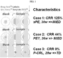
- the characteristic larger, homogeneous circle as shown, e.g ., in FIG. 1 , Case 1, "Sample" formed by diffusion of the solution with urine comprising misfolded proteins and dye is indicative of presence of misfolded proteins in the urine sample. That is, the urine sample contains misfolded proteins.
- a sample e.g. a urine sample
- dye e.g ., Congo Red
- the solution containing, for example, urine and dye, and not misfolded proteins is applied to a suitable cellulosic surface (e.g ., plain paper)
- the dye binds to the cellulose fibers of the surface in or near the application spot and while urine in the solution diffuses ( e.g . radially) from the application spot, dye does not diffuse from the application spot, or dye diffuses to a lesser extent relative to dye in solution with urine comprising misfolded proteins.
- sample may diffuse ( e.g. radially) to a distance of 5-7 mm from the application spot, while dye may diffuse ( e.g. radially) to a distance of only 1-3 mm from the application spot.
- This extent of diffusion of dye is one example of "not diffusing to an appreciable extent” or “not diffusing radially to an appreciable extent.”
- the characteristic smaller, heterogeneous circle as shown, e.g ., in FIG. 1 , Case 1, "Blank" formed by lack of appreciable diffusion of dye is indicative of absence of misfolded proteins in the sample. That is, the sample (e.g ., urine sample) does not contain misfolded proteins.
- a dye is considered not to diffuse to an appreciable extent if one can determine that dye has not spread (e.g ., radially) from the central point (albeit, it may diffuse to a limited extent from the central point, such as of 1-3 mm). This limited spreading results because the dye is not bound to misfolded proteins and can bind to the surface and does so rapidly when it is applied to the surface.
- a sample e.g . urine
- a sample may be "strongly positive” for misfolded protein (e.g., contains enough misfolded protein to bind all or most of the dye in solution).
- a characteristic homogenous circle may result from the methods of the present disclosure, as shown, for example, in Figure 1 , Case 1, "Sample.”
- a sample may be "weakly positive” for misfolded protein (e.g ., contains misfolded protein, but not enough to bind all or most of the dye in solution).
- a characteristic central spot with a homogeneous "halo" may result from the methods of the present disclosure, as shown, for example, in Figure 1 , Case 2, "Sample.”
- the halo typically results from diffusion of the portion of the dye that is bound to misfolded proteins and therefore does not bind to the cellulose in the central spot area.
- Some aspects of the present disclosure include maintaining a spot on a surface that comprises, for example, cellulose under conditions under which diffusion of a solution of urine (or other sample) comprising misfolded proteins and dye occurs.
- a solution, or a spot is maintained on a surface for 1 to 5 minutes, or more.
- a spot may be maintained for 1 to 3 minutes, or 1 to 2 minutes.
- a spot is maintained for 2 minutes.
- a spot is maintained on a surface for less than 1 minute.
- maintenance also refers, in some cases, to maintaining ambience (e.g ., temperature and humidity) for the period of time of assessment of a particular sample (e.g ., 1 to 5 minutes) that support diffusion of a solution. It should also be understood that the period of time to complete a diagnostic test of the present disclosure may vary, depending on ambience and type of surface (e.g ., cellulosic surface).
- Some aspects of the present disclosure include determining that the urine sample contains misfolded proteins if solution that passes through the surface is colored, or determining that the urine sample does not contain misfolded proteins if solution that passes through the surface is colorless. For example, if the dye is Congo Red, solution passing through the surface will appear red, pink or purple in color (because the solution contains Congo Red) if misfolded proteins are present in the urine sample. Without being bound by theory, this is because Congo Red, which has bound to the misfolded proteins and which cannot bind to the surface ( e.g ., as a result of saturation by binding of the misfolded proteins), will pass through the surface.
- solution passing through the surface will appear colorless (because the solution does not contain Congo Red) if misfolded proteins are not present in the urine sample. Without being bound by theory, this is because free Congo Red has bound to the surface, which acts as a filter, letting through only solution void of misfolded proteins and therefore void of Congo Red.
- Weight assigned to a paper is the weight of a ream (e.g ., 500 sheets, wherein 1 sheet equals 17" x 22") of paper.
- plain paper has a weight of 20 lbs, 24 lbs or 32 lbs.
- Paper in some instances, may be characterized by density. In some cases, plain paper has a density of 800 kg/m 3 (50 lb/ft 3 ).
- surface comprising cellulose further comprises an adhesive backing.
- surface comprising cellulose with an adhesive backing is a label (e.g ., a commercially-available self-adhesive label). Results provided herein show that, in some cases, an adhesive backing prevents wrinkling of a paper surface and, thus, provides more accurate results. Nonetheless, surfaces comprising cellulose do not require an adhesive backing.
- a concentration of dye (or a concentration of a combination of two or more dyes) in a solution is 0.01% to 1.0%, or more.
- a concentration of dye (or a concentration of a combination of two or more dyes) in a solution may be 0.01% to 0.1%, 0.01% to 0.2%, 0.01% to 0.3%, 0.01% to 0.4%, 0.05% to 0.1%, 0.05% to 0.2%, 0.05% to 0.3%, 0.05% to 0.4%, 0.05% to 0.5%, 0.05% to 0.6%, 0.05% to 0.7%, 0.05% to 0.8%, or 0.05% to 0.9%.
- a concentration of dye (or a concentration of a combination of two or more dyes) in a solution is 0.01%, 0.05%, 0.1%, 0.15%, 0.2%, 0.25%, 0.3%, 0.35%, 0.4%, 0.45%, 0.5%, 0.55%, 0.6%, 0.65%, 0.7%, 0.75%, 0.8%, 0.85%, 0.9%, 0.95% or 1.0%. In some cases, a concentration of dye (or a concentration of a combination of two or more dyes) in a solution is 0.1%.
- a concentration of dye (or a concentration of a combination of two or more dyes) in an aqueous solution is 0.2% to 1.0%, or more.
- a concentration of dye (or a concentration of a combination of two or more dyes) in a solution may be 0.2%, 0.3%, 0.4%, 0.5%, 0.6%, 0.7%, 0.8%, 0.9% or 1.0%.
- a volume of an aqueous solution is 1 ⁇ l to 10 ⁇ l, or more.
- a volume of an aqueous solution may be 1 ⁇ l, 1.5 ⁇ l, 2 ⁇ l, 2.5 ⁇ l, 3 ⁇ l, 3.5 ⁇ l, 4 ⁇ l, 4.5 ⁇ l, 5 ⁇ l, 5.5 ⁇ l, 6 ⁇ l, 6.5 ⁇ l, 7 ⁇ l, 7.5 ⁇ l, 8 ⁇ l, 8.5 ⁇ l, 9 ⁇ l, 9.5 ⁇ l or 10 ⁇ l.
- Positive controls of the present disclosure are produced from samples (e.g . urine samples) known to contain misfolded proteins.
- negative controls of the present disclosure are obtained from samples (e.g . urine samples) known to not contain misfolded proteins or from water, saline, phosphate buffer saline (PBS) or other salt solution not containing misfolded proteins.
- the conditions under which a positive and/or negative control is produced are similar to the conditions under which a method of the present disclosure is performed for the particular urine sample being compared to the positive or negative control.
- a test urine sample from a pregnant woman is compared to a urine sample with misfolded proteins from a pregnant woman (positive control) and/or to a urine sample without misfolded proteins from a pregnant woman (negative control). If results obtained with the test sample are comparable to results obtained with a positive control, then the test sample is determined to contain misfolded proteins. If results obtained with the test sample are comparable to results obtained with a negative control, then the test sample is determined to not contain misfolded proteins.
- the type of surface comprising cellulose e.g ., plain paper used in a method of producing a positive and/or negative control is the same as the type of surface comprising cellulose used in a method of assessing a test urine sample.
- volume of solution as well as the type (e.g ., Congo Red) and concentration of dye (or dyes) used in a method of producing a positive and/or negative control is the same as the volume of solution as well as the type and concentration of dye (or dyes) used in a method of assessing a test urine sample.
- positive and negative results for comparison to the test sample results are provided as images in the test kit.
- Some methods of the present disclosure include additional steps for confirming whether or not a sample (e.g. urine sample) from a subject (e.g ., pregnant woman) contains misfolded proteins.
- a sample e.g. urine sample
- a subject e.g ., pregnant woman
- an additional, diluted urine sample may be similarly assessed. This can be done in at least two ways. In one instance, a first urine sample is obtained ( e.g ., using clean catch) obtained from a subject, and a first portion of that first urine sample is assessed for the presence of misfolded proteins. Then, a second portion of that first sample is similarly assessed for the presence of misfolded proteins. The second portion may be diluted then similarly assessed for the presence of misfolded proteins.
- a determination that both the first and second portions (diluted or undiluted) of urine from the first urine sample have misfolded proteins can serve as confirmation that the subject has urine with misfolded proteins.
- a first urine sample is obtained ( e.g ., using clean catch), and a portion of that first urine sample is assessed for the presence of misfolded proteins.
- a second urine sample is collected from the same subject, and a portion of that second urine sample is similarly assessed for the presence of misfolded proteins.
- the second portion may be diluted then similarly assessed for the presence of misfolded proteins
- a determination that both the first and second samples of urine (diluted or undiluted) from the same subject have misfolded proteins can serve as confirmation that the subject has urine with misfolded proteins.
- Some aspects of the present disclosure include combining a sample (e.g. a urine sample) from a pregnant woman with a first dye that binds misfolded proteins and e.g ., cellulose and a second dye that is a different color from the first dye and does not bind misfolded proteins and cellulose, thereby producing a solution of urine and two dyes.
- a sample e.g. a urine sample
- a first dye that binds misfolded proteins and e.g ., cellulose and a second dye that is a different color from the first dye and does not bind misfolded proteins and cellulose
- the color of one dye is visually distinct from the color of the other dye.
- the first dye may be red and the second dye may be blue.
- the first dye is Congo Red.
- the second dye is a water soluble dye such as erioglaucine (also referred to as FD & C Blue No. 1, a food coloring).
- water soluble dyes e.g ., other FD & C colors
- FD & C Red No. 40 FD & C Yellow No. 5
- FD & C Yellow No. 6 FD & C Blue No. 2, FD & C Red No. 3, or FD & C Green No. 3.
- a correlation coefficient, ⁇ is used to distinguish between non-preeclampsia samples (e.g. urine samples) and preeclampsia samples (e.g. urine samples).
- the exact cut-off of the correlation coefficient, ⁇ that best discriminates non-preeclampsia urine samples from preeclampsia urine samples is defined by testing a large set of urine samples from pregnant women without preeclampsia, pregnant women with preeclampsia, and pregnant women with other pregnancy conditions.
- a correlation coefficient, ⁇ of 0.5 or greater (e.g ., 0.5 to 1.0) is indicative of preeclampsia.
- a ⁇ of 0.5, 0.6, 0.7, 0.8, 0.9 or 1.0 is indicative of suitable paper.
- correlation coefficient is calculated using an algorithm.
- the algorithm in some cases, is programmed as a mobile telephone or tablet application.
- the algorithm in some cases, compensates for illumination, subtracts the white background, and then calculates the correlation coefficient between the different color channels (e.g ., red and blue channels) of the color image of the diffused spot.
- the correlation coefficient between channels is significantly different between non-preeclamptic sample (e.g . urine sample) and preeclamptic sample (e.g . urine sample).
- This information can be used, for example, in calibration and screening applications (e.g ., software applications) as follows.
- Calibration The goal of calibration application is to help the user choose a surface, such as a cellulosic surface (e.g ., paper), and choose the concentrations of dyes (e.g ., Congo Red and blue food dye) for robust discrimination between normal and preeclamptic sample (e.g. urine sample).
- a surface such as a cellulosic surface (e.g ., paper)
- concentrations of dyes e.g ., Congo Red and blue food dye
- Two criteria may be used: (1) sufficient signal in both color channels so that camera noise does not influence the correlation calculation; and (2) fiber structure of the surface ( e.g ., paper), which should permit the second dye (e.g ., the blue food dye) to spread uniformly, while allowing the first dye when not bound to misfolded proteins (e.g ., the Congo Red dye) to bind.
- the appropriate surface (e.g ., paper) and dye concentration choice should give a balanced signal in both color channels (e.g ., red and blue channels) as well as a large difference in correlation coefficients between non-preeclamptic and preeclamptic samples (e.g . urine samples).
- the calibration procedure works, in some cases, by carefully introducing different concentrations of the first dye (e.g ., Congo Red) and the second dye (e.g ., blue food dye) in non-preeclamptic and preeclamptic samples (e.g ., urine samples), applying drops of the mixtures on different surfaces ( e.g ., papers), allowing the samples to diffuse, and taking photographs of the diffused samples (see FIGS. 27A and 27B ).
- Calibration software measures the percentage difference of the signals of the two channels (e.g ., red and blue channel) and the correlation coefficient between the two channels. A plot of the correlation coefficient and the percentage signal difference reveals which concentrations and surfaces (e.g ., papers) are suitable for use.
- the kit and optionally including the software application can be used for screening for misfolded proteins.
- the urine is mixed with the first and second dye, for example, and a drop of the mixture is diffused on a cellulosic surface
- a photo of the diffused sample e.g ., drop
- the software application uses the white background to estimate and compensate for illumination color.
- the background is subtracted out from the image so that only the pixels corresponding to the sample survive.
- the correlation coefficient between the two color channels (e.g ., red and blue channels) is then calculated and compared to a threshold to indicate the presence of misfolded proteins.
- a correlation coefficient threshold of 0.5 to 1.0 (e.g ., 0.85) distinguishes between normal and preeclamptic urine, as exemplified in Figures 27A and 27B .
- compositions and kits for detecting misfolded proteins in samples e.g . urine samples. Such compositions and kits, in some cases, may be used to diagnose preeclampsia.
- samples e.g . urine samples.
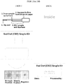
- kits of the present disclosure are shown in Figures 25A and 25B and in Figures 26A and 26B .
- Figures 25A and 25A depict the interior of an example of a tri-fold configured Congo Red Dot Simple Kit and a Congo Red Dot Quant Kit, respectively, with a top panel, middle panel and bottom panel made from, for example, cardstock.
- a pouch is formed with the middle panel covered by the top and bottom panels.
- a curved slit in the bottom panel (represented by a curved dotted line) is positioned and sized to receive the folded flap closing the pouch.
- the top panel of Figure 25A shows a disposable pipette that may be provided in the kit.
- the pipette may be preloaded with dye (e.g ., 5 ⁇ l of a 0.5% solution of Congo Red, or 5 ⁇ l of a 5% solution of Congo Red + 2.5 ⁇ l of a 0.1% solution of erioglaucine) and sealed.
- the pipette may then be secured in the middle panel by pre-formed slits (represented by two straight dotted lines).
- the slits are positioned such that the tip of the pipetted with the preloaded dye (or two dyes) remains inside the pouch and protected from light.
- a device for clipping the sealed end of the pipette e.g ., a nail clipper or small pair of scissors.
- a surface comprising cellulose is provided for sample testing.
- the surface by be affixed (e.g ., a self-adhesive paper label) to the kit trifold.
- the surface is provided with the kit.
- the surface is provided separately.
- the surface may require separate validation for suitability, accounting for, for example, differences in environmental conditions, such as humidity and temperature.
- the bottom panel contains a series of images of controls for a strongly positive sample, a weakly positive sample and a negative sample.
- the test sample may be compared to any one or more of the controls to determine sample result.
- the kit depicted in Figure 26A further includes color calibration scales (represented by graded bars flanking at left and right of test surface designed to orient the picture and correct for any light gradient. Excess green (represented by bars flanking at top and bottom of test surface) compensates for any tendency of a camera to adjust color.
- a Congo Red Dot Quant Array Kit is an extension of the Congo Red Dot Quant Kit in that the kit is basically the same with the exception that it provides for the assessment of multiple test samples on a single surface (e.g ., cellulosic surface).
- the present disclosure describes diagnostic methods and kits that can be used for efficient and accurate detection of misfolded proteins in urine (or other sample), which, in some cases, is indicative of certain disorders (e.g., preeclampsia in cases where the test sample is urine obtained from a pregnant woman).
- the experiments presented in this example were directed to the development of simple diagnostic methods and compositions (e.g ., kits) for rapid and accurate identification of misfolded proteins in e.g ., urine.
- simple diagnostic methods and compositions e.g ., kits
- the presence of misfolded proteins in urine can be used as an indication of preeclampsia.
- the existing Congo Red Dot test is an effective test that can be used as a diagnostic and prognostic tool for preeclampsia. Nonetheless, the Congo Red Dot test, which typically utilizes relatively expensive reagents such as nitrocellulose and alcohol washes, takes approximately 5 hours to complete.
- One of the objectives of this study was to produce simplified, low-cost and efficient diagnostic methods and compositions that can be implemented, for example, at the point of care, for example at a pregnancy checkup with a healthcare provider and is also suitable for low-resource setting.
- Paper suitability is based, at least in part, on contact angle of the paper, fluorescence of the paper, which is a reflection of additives, and porosity of paper, ambience (e.g ., temperature and humidity of environment/region).
- ambience e.g ., temperature and humidity of environment/region.
- a software routine was designed to determine paper "suitability" for Congo Red Dot testing based on a correlation coefficient (Rho, ⁇ ) between suitable paper and unsuitable paper.
- ⁇ The exact cut-off of the correlation coefficient, ⁇ , between suitable paper and unsuitable paper is defined by determining the relationship of ⁇ with the contact angle, ⁇ , on different types of "plain" paper. Electrolyte and protein concentrations are varied in vitro. Contact angle, ⁇ , is likely a significant determinant of ⁇ and is less affected by electrolyte and protein concentration. In some embodiments, a ⁇ of 0.5 or less ( e.g ., 0.1 to 0.5) is indicative of suitable paper. For example, a ⁇ of 0.1, 0.2, 0.3, 0.4 or 0.5 is indicative of suitable paper.
- Urine samples from a pregnant woman with severe preeclampsia ( FIG. 1 , "Sample,” Case 1), from a pregnant woman with preeclampsia ( FIG. 1 , “Sample,” Case 2), and from a pregnant woman without preeclampsia ( FIG. 1 , “Sample,” Case 3, control) were first mixed with Congo Red and then applied to plain paper. Water mixed with Congo Red was used as a control ( FIG. 1 , "Blank,” Cases 1-3). In the sample of water ( FIG.
- the size of the formed circle depends on many factors, including, for example, paper wettability (e.g ., measured by the contact angle ⁇ ) and Congo Red concentration.
- paper wettability e.g ., measured by the contact angle ⁇
- Congo Red concentration e.g ., sodium tartrate
- the first kit referred to as the Congo Red Dot Simple Kit (for rapid ( e.g ., 1 to 3 minute)), may be used for subjective assessment of urine congophilia.
- the second kit referred to as the Congo Red Dot Quant Kit, may be used for objective quantification of results and is enabled by a software algorithm that measures "spot homogeneity.” Development of the Congo Red Dot Quant Method and Kit is described below.
- the Congo Red Dot Quant Kit was designed with a dual dye mix using red and blue dyes.
- the blue color was selected as complementary to the color of Congo Red in the RGB (Red Green Blue) color space.
- the RGB color model is related to the physiology of human eye and brain and not to colorimetrically defined colors. It is also the typical output of mobile telephone (e.g., smartphone) cameras.
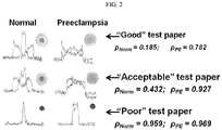
- erioglaucine (FD&C blue 1, non-toxic dye, McCormick & Co.) was selected for quantification of Congo Red spreading/retardation by image analysis, and, thus, discrimination of preeclampsia cases from non-preeclampsia cases.
- the blue dye migrates with the water phase, marking the maximum spread area (see, e.g ., FIG. 2 , "Normal," top row).
- the optimal composition of the dual dye was determined to be a 2:1 mix of 5% Congo Red:10% erioglaucine, with 3 ⁇ l of the dual dye mix added per 100 ⁇ l volume of urine.
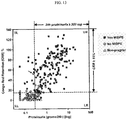
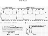
- an image analysis algorithm was created (e.g ., in MATLAB), which extracts the correlation coefficient (Rho, ⁇ ) between the signal in the red and blue channels of a mobile telephone-acquired image or a tablet-acquired image ( FIG. 2 ).
- the correlation coefficient is calculated after preprocessing to compensate for illuminant and paper color.
- the exact cut-off of the correlation coefficient, ⁇ that best discriminates non-preeclampsia urine samples from preeclampsia urine samples is defined by testing a large set of urine samples from pregnant women without preeclampsia, pregnant women with preeclampsia, and pregnant women with other pregnancy conditions.
- stored urine samples retrieved from women with well-characterized diagnoses and pregnancy outcomes are used (e.g ., 824 urine samples from 662 women are available, stored at -80 °C).
- a comparative accuracy analysis is conducted relative to P/C ratio (results for all samples were available) and the dipstick test, which is read both visually and objectively using the Siemens Clinitek Status reader.
- algorithms such as machine-learning algorithms (e.g ., support vector machines), variational analysis, and/or a non-parametric Bayesian technique, are used to determine if other image analysis parameters (e.g., aside from the correlation coefficient of red and blue pixels in the space of spread) can further improve patient classification.
- the image processing may be carried out on a remote server instead of the mobile telephone or tablet and the results reported back to the user.
- a stringent study design was employed, which involved a feasibility and a validation phase ( FIG. 5 ).
- Urine that had been mixed with Congo Red (CR) was spotted on an unsupported nitrocellulose membrane, and then washed with increasing concentrations of methanol ( FIG. 6A ).
- the rationale of this experiment was based on the self-assembling property of CR and its ability to initiate formation of large insoluble oligomers following binding to amyloid proteins that have an extensive ⁇ -sheet structure.
- spots of sPE but not P-CRL women remained red after the methanol wash, indicating that women with PE display urinary congophilia.
- a standardized protocol was further designed to allow an objective quantification of each urine sample propensity to retain CR, while minimizing variations due to differences in proteinuria and hydration status (the Congo Red Dot [CRD] test).
- CRR CR Retention
- CRI CR Incorporation
- CR-bound amyloids exhibit bright red fluorescence when illuminated with UV light, along with a shift in absorbance from about 490 to 540 nm.
- the characteristic red fluorescence of amyloid-bound CR was observed ( FIG. 6D ).
- 30% (12/40) of sPE urine samples (but no P-CRLs) changed visibly in color from orange-red to magenta ( FIG. 6E ), consistent with the bathochromic spectral shift described for ⁇ -amyloid oligomerization (aggregation), in vitro.
- Urine congophilia differs among hypertensive pregnancy disorders and increases with PE severity
- the Congo Red Dot (CRD) test is a simple modality to diagnose sPE and predict MIDPE
- the UPS was the strongest predictor of urine congophilia among women who required MIDPE, independent of GA or total proteinuria or albuminuria ( P ⁇ 0.001). This finding concurred with the prior observation that several peptide fragment biomarkers of the UPS score have a high misfolding potential.
- Congophilic material isolated from PE urine contains round and fibrillar nano-scale structures with amyloid-like microscopic features
- FIGS. 9A and 9B A protocol for CR-assisted precipitation of urine samples was developed to further characterize the congophilia of PE.
- FIG. 9A Green birefringent round or elongated particles (see insert) were observed.
- FIGS. 9B, 9C At transmission electron microscopy (TEM, FIGS. 9B, 9C ) the round structures varied in size from 30-300 nm while the fibrillar conformations were longer, arborescent, and tangled together in larger electrodense novel structures. These structures were absent in P-CRL specimens processed and imaged in parallel ( FIG. 9D ). Negative stain TEM ( FIGS.
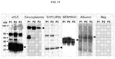
- FIG. 9G right panel is a representative Western blot for albumin which illustrates a sPE urine sample before (U1) and after (P1) CR-assisted precipitation.
- G1P3 Unlike ⁇ FLC and ceruloplasmin, which have been previously reported to undergo pathologic aggregation with relevance for several human conformational disorders, G1P3 has not been previously investigated for participation in amyloid structures. We performed an in silico analysis of the amyloidogenic potential for the three G1P3 isoforms resulting from alternative splicing. Within the G1P3 sequence, we noted an unusually high number of aggregation-prone segments ("hot spots"). The AGGRESCAN algorithm predicted that G1P3 should have an aggregation propensity at least if not in excess of A ⁇ 42 with the shortest isoform (G1P3a) being the most amyloidogenic (Na4vSS: G1P3a: 13.6 vs. A ⁇ 42: 6.4, FIG. 15 and Table 4).
- PE urine contains aggregated APP proteoforms
- Fibrillar amyloid proteins are resistant to proteolysis by trypsin. This led us to consider the possibility that a proteomics approach, which relies on fingerprinting of tryptic cleavage peptides, might have missed important protein identities in the urine congophilic material.
- a proteomics approach which relies on fingerprinting of tryptic cleavage peptides, might have missed important protein identities in the urine congophilic material.
- One candidate was APP.
- Cellular processing of APP is a well-recognized pathophysiologic phenomenon linked to Alzheimer's disease. Table 4.
- the 22C11 antibody detected immunoreactive urine proteins in both P-CRL (U1-U2) and sPE (U3-U8) at the expected molecular weight for mature sAPP ( ⁇ 130 kDa) ( FIG. 10E ). Yet, sPE urine contained an additional band ( ⁇ 110 kDa) likely representing immature sAPP (lanes U4-U6). Notably, some sPE women with intense ALZ90 and DE2B4 immunoreactivity (lanes U3, U7, U8) had less prominent 22C11 sAPP bands.
- sAPP isoforms containing the KPI domain e.g ., APP770 and APP 751 function as anti-proteases and are generically referred to as protease nexin-II (APP-KPI/PN2). Similar to SERPINA1, APP-KPI/PN2 potently inhibits serine proteases including trypsin and coagulation factors acting as suicide substrates. Fragments of APP-KPI/PN2 that include the KPI domain are known to be highly amyloidogenic ( 50 , 51 ).
- Figure 10F demonstrates that urine specimens of sPE women contain an APP fragment ( ⁇ 47 kDa) specifically detected by the MAB5354 (KPI domain) antibody.
- FIGS. 16C, 16D mature sAPP proteoforms are normal urine constituents in healthy pregnancies.
- the relatively low serum sAPP, high fractional excretion of sAPP, and especially the high levels of fragmented APP proteoforms in sPE urine point to a possible derangement in the APP proteolytic processing pathway in sPE.
- the placenta is central to the pathophysiology of PE.
- ⁇ -secretases ADAM10, ADAM17
- BACE1, BACE2 ⁇ -secretases
- PSEN1, PSEN2 ⁇ -secretases
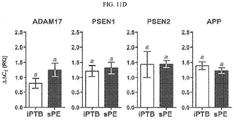
- the amplification signal for the ⁇ -secretase BACE1 was only weakly detected in both preterm and term placenta. This was in contrast to BACE2 which was expressed at significantly higher levels especially in preterm placenta ( P ⁇ 0.001 vs. term).
- ⁇ -secretases had overall lower mRNA levels.
- the villous trophoblast mRNA levels of ADAM10 ( P ⁇ 0.001) and BACE2 ( P ⁇ 0.001) were significantly higher in sPE compared to GA-matched placental tissues of women with idiopathic preterm birth (iPTB) ( FIG. 11C ).
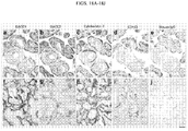
- Severe preeclampsia associates with deposition of extracellular ALZ90 positive aggregates in the placenta resembling amyloid plaques
- Intense 22C11 (N-terminus) APP immunoreactivity was identified in the basal plate and chorionic villi of iPTB placentas ( FIG. 12A ).
- Decidual cells stained more intensely than extravillous trophoblasts, while in the placental villi, endothelial cells and cytothrophoblasts stained more conspicuously compared to the surrounding stroma ( FIG. 12A , insert).
- 22C11 staining patterns between sPE and iPTB placentas There were notable differences in 22C11 staining patterns between sPE and iPTB placentas. In sPE, decidual cells stained positive at almost the same intensity compared to iPTB, but the distribution of the 22C11 decidual cells was more scattered and had a distorted morphology ( FIG. 12B ). Intense staining of acellular filamentous aggregates was observed in the maternal inter-villous spaces, with a significantly increased number on
- FIG. 12E insert) or flaky acellular material aggregated into plaques ( FIG. 12F ).
- ALZ90 positive plaques were more frequently observed in areas affected by fibrinoid-like degeneration. Still, not all fibrinoid-like material was ALZ90-positive.
- the ALZ90-positive material had a characteristic purple proteinaceous appearance different from the surrounding eosinophilic fibrinoid ( FIG. 12G ).
- Incubation with mouse IgG confirmed staining specificity ( FIG. 12H ).
- the ALZ90-positive endovascular material was immunoreactive for G1P3, and frequently noted in areas traditionally described morphologically as placental calcifications, which we confirmed by Alizarin S staining ( FIGS. 17A-17D ).
- Pregnant women were enrolled from March 2004 to December 2010 at Yale-New Haven Hospital (New Haven, CT).
- Non-pregnant proteinuric women were enrolled at Fletcher Allen Health Care Hospital (Burlington, VT).
- the research protocols were approved by both Yale University and University of Vermont Human Research Protection Program
- PE is a syndrome characterized by heterogeneous clinical manifestations with different pathogenesis and various risk factors.
- the definition of PE and hypertensive disorders during pregnancy is often arbitrary.
- diagnosing PE is not as easy as one might believe.
- an intervention-based near-miss event was chosen, that is MIDPE, as a better reflection of true illness. This approach was successfully applied during our prior urine proteomics discovery study.
- Binding of CR to amyloids results in a change of the dye's absorbance spectrum in visible light (from orange-red to rose-magenta), and this bathochromic shift has been previously used to assess the level of ⁇ -amyloid oligomerization (aggregation) in-vitro (22, 100).
- All sPE and P-CRL urine samples used in the feasibility phase were processed as for the CRD test.
- the urine-CR mixture was diluted 10-fold and examined spectrophotometrically for CR-induced spectral shift (see below).
- a theoretical concentration of amyloid-like proteins in each urine sample was derived from absorbance values at 403, 504 and 541-nm using Klunk's formula. Normalization for intrinsic absorbance and light scattering was achieved by subtraction of values obtained in samples without CR.
- the spectral shift determined concentration of amyloid-like proteins was correlated with the subject's CRR test result.
- ThT fluorescence was measured after a protocol originally described for serum. Briefly, spun urine samples (30 ⁇ l) were mixed with 80 ⁇ M ThT in 100 mM phosphate buffered saline (PBS) pH 7.4. Fluorescence measurements were obtained in a spectrofluorometer (Clariostar, BMG Labtech, Cary NC) with excitation/emission wavelengths set at 444/485 nm, respectively. Normalization for intrinsic urine fluorescence was achieved by subtracting values in the absence of ThT.
- PBS phosphate buffered saline
- Soluble amyloid oligomers are cytotoxic and potent inducers of cell death.
- PFOs prefibrillar oligomers
- FOs fibrillar oligomers
- APF annular protofibrils
- the suspension was placed onto 200 mesh formvar/carbon-coated nickel grids after positive or negative staining with 2% uranyl acetate.
- Real-time PCR was performed on placental and amniochorion mRNA using the TaqMan chemistry (Applied Biosystems) and validated primer/probes for APP and the following APP-cleaving enzymes: ⁇ -secretases (ADAM 10, ADAM17), ⁇ -secretases (BACE1, BACE2) and ⁇ -secretases [Presenilin-1 (PSEN1), Presenilin-2 (PSEN2)]. Staining intensity in placental villi was assessed semiquantitatively on a scale from 0 (absent) to +5 (intense). Antibodies against Cytokeratin-7 and CD163 were used as cellular markers for trophoblasts and macrophages, respectively. Technical details are provided below.
- PE surrogate markers used or proposed
- P:C ratio protein-to-creatinine
- sFlt-1 soluble fms-like tyrosine kinase-1
- uFP placental growth factor
- UPSr urine proteomics scores
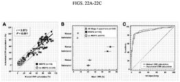
- Test accuracy (cases correctly classified/total number of cases), sensitivity, specificity, positive and negative predictive values and likelihood ratios (LR) were measured on receiver operator characteristic ROC plots using MedCalc (Broekstraat, Belgium) statistical software. Confidence intervals (CI) were calculated using the bootstrapping method. Comparison of ROC curves was performed using the De Long method which uses a non-parametric method to determine statistical significance of the difference between the areas under dependent ROC curves (derived from the same cases).
- the primary antibodies employed for immunodetection of soluble oligomers have been developed and characterized in prior publications by members or our team.
- the following primary polyclonal antibodies were used: A11, OC, and anti-APF that recognize specific quaternary structure epitopes formed by amyloidogenic proteins.
- CR is known to induce amyloid oligomer growth and nucleation in aqueous solutions. As the CR-bound aggregates grow, they precipitate out of solution and can be isolated by centrifugation. Based on these principles, we developed a protocol for CR-assisted precipitation of urine samples to further characterize the congophilia and the misfoldome of PE. 10 mL volumes of urine were first spun at 15,000 g for 15 min and 4 °C. The supernatant was mixed with 200 ⁇ l CR stock solution, vortexed for 1-hour as for the CRD test and spun again thereafter to pellet congophilic aggregated protein.
- the pellet was resuspended in water and recollected 3 times through centrifugation to remove all unbound CR.
- the final CR precipitate was resuspended in 50 ⁇ l water and either imaged immediately or stored frozen at -80 °C for subsequent analyses. If no pellet was visible, the specimen was deemed CR precipitate-negative but the microfuge was processed identically as for the CR precipitate-positive samples.
- CR-precipitates were resuspended in water, dried on a microscope slide, and imaged under polarized light using an Olympus U-STP microscope equipped with an Olympus OLY-200 digital camera (Olympus, Melville, NY). Alternatively, the suspension was placed onto 200 mesh formvar/carbon-coated nickel grids after positive or negative staining with 2% uranyl acetate. Specimens from tubes attempting to prepare CR precipitates P-CRL women were also examined.
- AGGRESCAN a web-based algorithm for calculation and visualization of aggregation propensity. This algorithm has been shown to anticipate aggregation of protein segments involved in several conformational disorders. It has also been shown to differentiate various classes of proteins by their aggregation and solubility properties. Natively unfolded proteins were predicted to have the lowest aggregation propensity. In the AGGRESCAN output the sequence stretches with the highest predicted aggregation propensity appear as peaks in the profile plots. A polypeptide sequence is considered a "hot spot" if there are 5 or more sequentially continuous residues with an average aggregation propensity value (a4v) per amino acid larger than the hot spot threshold (HST).
- HST hot spot threshold
- sAPP was measured in urine and paired serum samples by ELISA (sAPP: IBL International, Hamburg, Germany). Appropriately diluted samples (urine 2-fold, serum 50-fold) were placed in 96-well plates precoated with a capture antibody directed against an epitope common to both sAPPa (cleavage product of ⁇ -secretase) and sAPP ⁇ (cleavage product of ⁇ -secretase).
- sAPPa cleavage product of ⁇ -secretase
- sAPP ⁇ cleavage product of ⁇ -secretase
- RT-PCR was performed using TaqMan (Applied Biosystems, Carlsbad, CA) chemistry in 20 ⁇ L reactions composed of 10 ⁇ L mastermix (TaqMan® Fast Universal PCR 2x Master Mix,), 8 ⁇ L water, 1 ⁇ L cDNA template normalized at and 1 ⁇ L PCR probe set (TaqMan® Gene Expression Assays on Demand).
- ddCt dCt of individual sample - dCt of same target in reference sample
- paraffin sections of placental villous tissue were deparaffinized in xylene and rehydrated with graded ethanol to potassium-phosphate-buffered saline solution, pH 7.2. Following antigen retrieval with citrate buffer, the sections were pretreated with 1% hydrogen peroxide for 15-min.
- the stage 1 dataset originated from previously acquired images (before and after wash, Nikon Coolpix 4500) that have been manually quantified as part of the initial study.
- This data set consisted of 18 arrays from a total of 218 subjects. Each array held duplicate spots from 12-15 subjects ( FIG. 20A ).
- red channel pixel value divided by green channel ratio R/G, with R and G ranging from 0-255
- a second calculation used the red channel information subtracted by green channel (difference R-G) with the same reasoning.
- the green channel was selected over the blue channel due to the Bayer pattern which makes digital camera sensors more sensitive to green than to blue in order to match the heightened sensitivity of the human visual system towards the color green.
- a luminance conversion equation was used to retain intensity of the colors while eliminating the color-information itself ( FIG. 20B ).
- the luminance (L) algorithm resembled the one employed by the manual processing routine. Simulated calculations performed with a command-line interface of our application (command-line version of the processing library run on a ThinkPad T500 with Linux Ubuntu operating system) determined that the R/G ( FIG. 20C ) and L ( FIG.
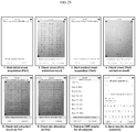
- the true to size template was included for printout, as shown in Figure 24 .
- the template marks the sample positions for up to 41 subjects (each specimen is spotted in duplicate in adjacent cells of two columns). The black center points in each cell are visible through transparency and serve as a guide for sample placement to ensure the predictable dot positioning on the sheet.
- the process workflow is schematically represented in Figure 21 .
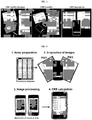
- Image acquisition The images are acquired using the build-in camera of the smartphone.
- the resolution of the iPhone 4 camera is sufficient to capture a sample dot with a maximum number of about 40 pixels per diameter. This results in approximately 1,200 pixels per sample dot.
- each image is converted into luminance grayscale for all the following steps.
- the Otsu threshold of the sheet is calculated to separate the foreground (sheet) from background.
- all holes in the foreground part are filled applying mathematical morphology.
- a gradient filter is applied to expose the sheet borders on the image.
- the corners are detected using the Hough line transform (a method of finding lines in an image).
- a rough Hough line transform is first calculated to loosely find the four most prominent edges (borders of the sheet).
- the 4 intersections of these lines are extracted. Because these positions do not match the corners perfectly, a second finer Hough line transform is performed separately on regions of interest around the previously found corners. In this second run, only the two main lines near the corners are extracted and intersected, which improves localization of the corner points while keeping memory usage to a minimum.
- Sheet extraction From the four corner points, the positions of the longer and shorter edges are estimated and the sheet is perspectively transformed into a rectangular geometry using bilinear interpolation. The geometric correction also reduces the number of pixels per dot to about 700 pixels (downscaling).
- the three position markers in the corners of the sheet are detected by calculating the average intensity in each corner and selecting the corner with the highest intensity (which corresponds to the corner without the punch-out marker) as the reference point.
- the sheet is transposed or rotated accordingly, such that all markers are repositioned on the upper and left corners.
- the normalized image now contains the sheet with the sheet spanning between the four image corners. This process is done individually on Pix1 and Pix2.
- Dot detection All possible sample dots are present in Pix1 but some might disappear during washing (negative testing samples). Thus, the complete dot detection can only be performed on Pix1.
- a gradient filter is applied to each extracted cell on Pix1 to detect the dot edges. The radius of each dot is estimated.
- a Hough circle transform is then performed and the two most prominent circular shapes in each cell are selected. Because the relative position of the dot does not change during washing, the same position information from Pix1 is used for processing of Pix2.
- Dot extraction With the positions of the dots known, the intensity of the corresponding pixels are extracted and summed up. To account for white-balance and mild illumination changes, a background subtraction is performed by subtracting the average luminance of the cell outside the dot from the average luminance within the dot Table 5. Table 5.
- CRR CRR calculation. Analogues with the manual formula, the test result (CRR) is calculated as the ratio of average intensity of the dots on Pix1 to the average intensity of the dot in Pix2. The value of the blank sample (dots in the left upper cells position) is subtracted from all other calculated CRR values on the sheet.
- stage 2 images were acquired and processed with an iPhone 4 and an application running the above sequence.
- the iPhone camera was used for acquisition, but the images then transferred to a computer where the processing was performed in an iPhone simulator.
- Eight CRD arrays containing 328 different urine specimens were analyzed as part of stage 2 . These arrays were prepared specifically for this study from aliquots maintained frozen at -80°C. The specimens originated from 273 different women (55 specimens were subsequent collections at a time later in pregnancy). All specimens were consecutive with respect to specimen collection and storage. There was no overlap in specimens with those analyzed part of stage 1 .
- the prevalence of the outcome of interest (MIDPE) in the stage 2 dataset was 40% among specimens (118/328) and among subjects (108/273).
- FIG. 23 A screen by screen workflow of the iPhone app illustrating the processing time is included in FIG. 23 (in the shown example pictures Pix1 and Pix2 have been previously acquired and stored in the smartphone's picture library). Utilizing our image processing tool, the time from the conclusion of the "wet part" of the CRD test array to result was reduced to approximately 2 minutes of processing time on the smartphone.
- L x y 0.3 ⁇ R x y + 0.59 ⁇ G x y + 0.11 ⁇ B x y with L(x,y) being the luminance, and R(x,y), G(x,y), and B(x,y) determine the red, green, and blue values of the pixel at position x,y, respectively.
- r d i ⁇ x , y ⁇ d i L x y N d i ⁇ ⁇ x y ⁇ c i , x y ⁇ d i L x y N c i ⁇ N d i with r ( d i ) being the normalized luminance (regarded as "redness") of dot d i , c i the cell containing dot d i , and N the number of pixels in c i or d i .
- a reference to "A and/or B", when used in conjunction with open-ended language such as “comprising” can refer, in one embodiment, to A only (optionally including elements other than B); in another embodiment, to B only (optionally including elements other than A); and in yet another embodiment, to both A and B (optionally including other elements).
- a reference to "A, B and/or C", when used in conjunction with open-ended language such as “comprising” can refer, in some embodiments, to A only (optionally including elements other than B and C); in another embodiment, to B only (optionally including elements other than A and C); in yet another embodiment, to C only (optionally including elements other than C and C); in yet another embodiment, to A and B (optionally including other elements), to A and C (optionally including other elements), to B and C (optionally including other elements), or to A, B and C (optionally including other elements).
- the phrase "at least one,” in reference to a list of one or more elements, should be understood to mean at least one element selected from any one or more of the elements in the list of elements, but not necessarily including at least one of each and every element specifically listed within the list of elements and not excluding any combinations of elements in the list of elements.
- This definition also allows that elements may optionally be present other than the elements specifically identified within the list of elements to which the phrase "at least one" refers, whether related or unrelated to those elements specifically identified.
- At least one of A and B can refer, in some embodiments, to at least one, optionally including more than one, A, with no B present (and optionally including elements other than B); in another embodiment, to at least one, optionally including more than one, B, with no A present (and optionally including elements other than A); in yet another embodiment, to at least one, optionally including more than one, A, and at least one, optionally including more than one, B (and optionally including other elements).
Landscapes
- Health & Medical Sciences (AREA)
- Life Sciences & Earth Sciences (AREA)
- Engineering & Computer Science (AREA)
- Immunology (AREA)
- Hematology (AREA)
- Molecular Biology (AREA)
- Urology & Nephrology (AREA)
- Biomedical Technology (AREA)
- Chemical & Material Sciences (AREA)
- Physics & Mathematics (AREA)
- Pathology (AREA)
- Biotechnology (AREA)
- Microbiology (AREA)
- General Physics & Mathematics (AREA)
- General Health & Medical Sciences (AREA)
- Biochemistry (AREA)
- Food Science & Technology (AREA)
- Medicinal Chemistry (AREA)
- Cell Biology (AREA)
- Analytical Chemistry (AREA)
- Proteomics, Peptides & Aminoacids (AREA)
- Gynecology & Obstetrics (AREA)
- Pregnancy & Childbirth (AREA)
- Reproductive Health (AREA)
- Bioinformatics & Cheminformatics (AREA)
- Bioinformatics & Computational Biology (AREA)
- Biophysics (AREA)
- Investigating Or Analysing Biological Materials (AREA)
Priority Applications (1)
| Application Number | Priority Date | Filing Date | Title |
|---|---|---|---|
| EP20168843.9A EP3745127B1 (en) | 2014-04-10 | 2015-04-10 | Methods and compositions for detecting misfolded proteins |
Applications Claiming Priority (2)
| Application Number | Priority Date | Filing Date | Title |
|---|---|---|---|
| US201461978158P | 2014-04-10 | 2014-04-10 | |
| PCT/US2015/025432 WO2015157704A2 (en) | 2014-04-10 | 2015-04-10 | Methods and compositions for detecting misfolded proteins |
Related Child Applications (3)
| Application Number | Title | Priority Date | Filing Date |
|---|---|---|---|
| EP20168843.9A Division EP3745127B1 (en) | 2014-04-10 | 2015-04-10 | Methods and compositions for detecting misfolded proteins |
| EP20168843.9A Division-Into EP3745127B1 (en) | 2014-04-10 | 2015-04-10 | Methods and compositions for detecting misfolded proteins |
| EP20167626.9 Division-Into | 2020-04-01 |
Publications (2)
| Publication Number | Publication Date |
|---|---|
| EP3129779A2 EP3129779A2 (en) | 2017-02-15 |
| EP3129779B1 true EP3129779B1 (en) | 2020-06-03 |
Family
ID=54264908
Family Applications (2)
| Application Number | Title | Priority Date | Filing Date |
|---|---|---|---|
| EP15776737.7A Active EP3129779B1 (en) | 2014-04-10 | 2015-04-10 | Methods and compositions for detecting misfolded proteins |
| EP20168843.9A Active EP3745127B1 (en) | 2014-04-10 | 2015-04-10 | Methods and compositions for detecting misfolded proteins |
Family Applications After (1)
| Application Number | Title | Priority Date | Filing Date |
|---|---|---|---|
| EP20168843.9A Active EP3745127B1 (en) | 2014-04-10 | 2015-04-10 | Methods and compositions for detecting misfolded proteins |
Country Status (11)
| Country | Link |
|---|---|
| US (3) | US10324094B2 (es) |
| EP (2) | EP3129779B1 (es) |
| JP (1) | JP6697394B2 (es) |
| CN (2) | CN116027043A (es) |
| AU (3) | AU2015243171B2 (es) |
| CA (2) | CA3157573C (es) |
| DK (1) | DK3129779T3 (es) |
| ES (1) | ES2817417T3 (es) |
| MX (2) | MX383381B (es) |
| WO (1) | WO2015157704A2 (es) |
| ZA (1) | ZA201606987B (es) |
Families Citing this family (18)
| Publication number | Priority date | Publication date | Assignee | Title |
|---|---|---|---|---|
| CN110187119B (zh) | 2008-10-31 | 2022-07-29 | 耶鲁大学 | 子痫前期检测和治疗的方法和组合物 |
| JP6697394B2 (ja) | 2014-04-10 | 2020-05-20 | イェール ユニバーシティーYale University | 異常折り畳みタンパク質を検出するための方法および組成物 |
| DK3322985T3 (da) * | 2015-07-15 | 2022-01-17 | Gestvision Inc | Anordning til påvisning af fejlfoldede proteiner og fremgangsmåder til anvendelse deraf |
| WO2017180984A1 (en) * | 2016-04-14 | 2017-10-19 | The Texas A&M University System | First trimester epigenetic and microrna biomarkers for preeclampsia |
| US10345244B2 (en) * | 2016-06-03 | 2019-07-09 | International Business Machines Corporation | Diagnostic test device with patterned material spots |
| US10335045B2 (en) | 2016-06-24 | 2019-07-02 | Universita Degli Studi Di Trento | Self-adaptive matrix completion for heart rate estimation from face videos under realistic conditions |
| US10269107B2 (en) * | 2017-02-23 | 2019-04-23 | Global Neighbor Inc | Selective plant detection and treatment using green luminance photometric machine vision scan with real time chromaticity operations and image parameter floors for low processing load |
| WO2019071240A1 (en) | 2017-10-06 | 2019-04-11 | The Research Foundation For The State University For The State Of New York | AQUEOUS AND NONAQUEOUS SELECTIVE OPTICAL DETECTION OF FREE SULPHITES |
| BR112020013608A2 (pt) * | 2017-12-28 | 2020-12-01 | Shuwen Biotech Co., Ltd. | aparelho, kit de reagentes e método para detectar proteína moldada |
| US11255844B2 (en) * | 2018-03-06 | 2022-02-22 | Fresenius Medical Care Holdings, Inc. | Peritoneal dialysis systems and related methods |
| CN109659011A (zh) * | 2018-12-29 | 2019-04-19 | 杭州脉流科技有限公司 | 利用移动设备采集处理十二导联ecg图像的方法以及移动设备和计算机可读存储介质 |
| WO2022099319A1 (en) * | 2020-11-06 | 2022-05-12 | The Board Of Trustees Of The Leland Stanford Junior Universityan | Systems and methods for evaluating gestational progress and applications thereof |
| IL303096A (en) * | 2020-11-21 | 2023-07-01 | Shuwen Biotech Co Ltd | Apparatus and method for detecting misfolded protein in biological sample |
| CA3217628A1 (en) * | 2021-05-03 | 2022-11-10 | Luke GENESLAW | Detection of labels in biomedical images |
| TW202409564A (zh) * | 2022-05-20 | 2024-03-01 | 大陸商數問生物技術(宣城)有限公司 | 用於檢測生物樣本中的錯誤折疊蛋白質的裝置和方法 |
| WO2024249849A2 (en) * | 2023-05-31 | 2024-12-05 | Northwestern University | Scalable platform for single cell proteomics via digitized detection of proteins and their proteoforms |
| CN117236201B (zh) * | 2023-11-16 | 2024-02-23 | 南京信息工程大学 | 一种基于Diffusion和ViT的降尺度方法 |
| CN120761295A (zh) * | 2025-08-21 | 2025-10-10 | 赛克赛斯生物科技股份有限公司 | 三苯甲烷类化合物在检测阿尔茨海默症中的应用 |
Family Cites Families (18)
| Publication number | Priority date | Publication date | Assignee | Title |
|---|---|---|---|---|
| US234610A (en) * | 1880-11-16 | Robert o | ||
| US4444879A (en) | 1981-01-29 | 1984-04-24 | Science Research Center, Inc. | Immunoassay with article having support film and immunological counterpart of analyte |
| AU1679992A (en) * | 1991-03-28 | 1992-11-02 | Micro Research Inc. | Controlled-release solid phase assay device including encapsulated reagents for detecting chemical substances |
| US5998216A (en) | 1996-10-01 | 1999-12-07 | Beth Israel Deaconess Medical Center | Stabilizing formulation for preserving the integrity of proteins present in a body fluid sampled ex-vivo for evaluation as a specimen |
| WO1998028006A1 (en) | 1996-12-23 | 1998-07-02 | Cambridge University Technical Services Limited | Diagnosis and treatment of pathological pregnancies |
| US6444435B1 (en) * | 2000-11-30 | 2002-09-03 | Serim Research Corporation | Test strip for determining dialysate composition |
| US7355044B2 (en) | 2001-10-25 | 2008-04-08 | Agfa-Gevaert N.V. | Self-assembling dyes |
| PT2308507E (pt) | 2002-07-19 | 2015-05-11 | Beth Israel Hospital | Métodos de tratamento de pré-eclâmpsia |
| EP1578361B1 (en) | 2002-09-12 | 2011-04-20 | The Regents of The University of California | Immunogens and corresponding antibodies specific for high molecular weight aggregation intermediates common to amyloids formed from proteins of differing sequence |
| EP1839057B1 (en) | 2004-12-15 | 2010-08-25 | Beth Israel Deaconess Medical Center | Nucleic acids and polypeptides useful for diagnosing and treating complications of pregnancy |
| EP1839056B1 (en) | 2004-12-21 | 2011-08-03 | Yale University | Diagnosis of preeclampsia |
| EP1733762A1 (en) | 2005-05-25 | 2006-12-20 | 3M Espe AG | Dental composition for detection of carious tissue, detection method |
| EP1946121A2 (en) | 2005-10-27 | 2008-07-23 | Yale University, Inc. | Urinary proteomic biomarker patterns in preeclampsia |
| BRPI0818779A2 (pt) * | 2007-10-18 | 2015-04-14 | Momtec Life Ltd | Dispositivo de diagnóstico para identificar ruptura da membrana durante a gravidez |
| US20110065139A1 (en) * | 2007-11-27 | 2011-03-17 | Jacob Mullerad | diagnostic device for identifying rupture of membrane during pregnancy |
| CN110187119B (zh) * | 2008-10-31 | 2022-07-29 | 耶鲁大学 | 子痫前期检测和治疗的方法和组合物 |
| CN103012599B (zh) | 2012-11-28 | 2015-04-29 | 龙岩九健生物芯片技术研究所 | 一种调节硝酸纤维素硝化取代度的方法 |
| JP6697394B2 (ja) | 2014-04-10 | 2020-05-20 | イェール ユニバーシティーYale University | 異常折り畳みタンパク質を検出するための方法および組成物 |
-
2015
- 2015-04-10 JP JP2016561743A patent/JP6697394B2/ja active Active
- 2015-04-10 MX MX2016013255A patent/MX383381B/es unknown
- 2015-04-10 CA CA3157573A patent/CA3157573C/en active Active
- 2015-04-10 EP EP15776737.7A patent/EP3129779B1/en active Active
- 2015-04-10 AU AU2015243171A patent/AU2015243171B2/en active Active
- 2015-04-10 ES ES15776737T patent/ES2817417T3/es active Active
- 2015-04-10 WO PCT/US2015/025432 patent/WO2015157704A2/en not_active Ceased
- 2015-04-10 CA CA2945058A patent/CA2945058C/en active Active
- 2015-04-10 EP EP20168843.9A patent/EP3745127B1/en active Active
- 2015-04-10 US US14/683,908 patent/US10324094B2/en active Active
- 2015-04-10 DK DK15776737.7T patent/DK3129779T3/da active
- 2015-04-10 CN CN202210738173.8A patent/CN116027043A/zh active Pending
- 2015-04-10 CN CN201580031385.0A patent/CN106461644B/zh active Active
-
2016
- 2016-10-07 MX MX2021006791A patent/MX2021006791A/es unknown
- 2016-10-11 ZA ZA2016/06987A patent/ZA201606987B/en unknown
-
2019
- 2019-05-08 US US16/406,942 patent/US11079392B2/en active Active
-
2020
- 2020-10-02 AU AU2020244572A patent/AU2020244572B2/en active Active
-
2021
- 2021-06-29 US US17/361,967 patent/US11953505B2/en active Active
-
2022
- 2022-12-23 AU AU2022291625A patent/AU2022291625B2/en active Active
Non-Patent Citations (1)
| Title |
|---|
| None * |
Also Published As
| Publication number | Publication date |
|---|---|
| DK3129779T3 (da) | 2020-09-07 |
| US11079392B2 (en) | 2021-08-03 |
| AU2022291625A1 (en) | 2023-02-16 |
| CA2945058A1 (en) | 2015-10-15 |
| US11953505B2 (en) | 2024-04-09 |
| CA3157573C (en) | 2025-11-18 |
| MX2016013255A (es) | 2017-05-03 |
| US10324094B2 (en) | 2019-06-18 |
| EP3745127A1 (en) | 2020-12-02 |
| ES2817417T3 (es) | 2021-04-07 |
| CA3157573A1 (en) | 2015-10-15 |
| AU2020244572A1 (en) | 2020-11-05 |
| AU2015243171B2 (en) | 2020-10-15 |
| US20150293115A1 (en) | 2015-10-15 |
| CA2945058C (en) | 2022-07-12 |
| ZA201606987B (en) | 2020-01-29 |
| MX2021006791A (es) | 2021-07-15 |
| CN106461644A (zh) | 2017-02-22 |
| AU2020244572B2 (en) | 2022-12-15 |
| US20190353662A1 (en) | 2019-11-21 |
| EP3745127B1 (en) | 2024-08-28 |
| AU2015243171A1 (en) | 2016-10-27 |
| AU2022291625B2 (en) | 2024-09-12 |
| WO2015157704A3 (en) | 2016-04-28 |
| CN106461644B (zh) | 2022-05-13 |
| CN116027043A (zh) | 2023-04-28 |
| JP2017518483A (ja) | 2017-07-06 |
| MX383381B (es) | 2025-03-13 |
| US20220043001A1 (en) | 2022-02-10 |
| JP6697394B2 (ja) | 2020-05-20 |
| WO2015157704A2 (en) | 2015-10-15 |
| EP3129779A2 (en) | 2017-02-15 |
Similar Documents
| Publication | Publication Date | Title |
|---|---|---|
| AU2022291625B2 (en) | Methods and compositions for detecting misfolded proteins | |
| Buhimschi et al. | Protein misfolding, congophilia, oligomerization, and defective amyloid processing in preeclampsia | |
| Senapati et al. | Predicting first trimester pregnancy outcome: derivation of a multiple marker test | |
| Lacquaniti et al. | Delayed graft function and chronic allograft nephropathy: diagnostic and prognostic role of neutrophil gelatinase-associated lipocalin | |
| Dahabiyeh et al. | A pilot study of alterations in oxidized angiotensinogen and antioxidants in pre-eclamptic pregnancy | |
| Cade et al. | Should the spot albumin-to-creatinine ratio replace the spot protein-to-creatinine ratio as the primary screening tool for proteinuria in pregnancy? | |
| CA2794021C (en) | Hbf and a1m as early stage markers for preeclampsia | |
| Munnangi et al. | Pregnancy associated plasma protein-A2: a novel biomarker for down syndrome | |
| Lopez-Hernandez et al. | Nested case–control study reveals increased levels of urinary proteins from human kidney toxicity panels in women predicted to develop preeclampsia | |
| Suliman et al. | Cancer antigen 125 and C-reactive protein inflammatory mediators and uric acid in association with preeclampsia in North Kordofan State, Western Sudan | |
| HK40042433A (en) | Methods and compositions for detecting misfolded proteins | |
| HK1234152B (en) | Methods and compositions for detecting misfolded proteins | |
| HK1234152A1 (en) | Methods and compositions for detecting misfolded proteins | |
| Kawamura et al. | Plasma and serum prorenin concentrations in diabetes, hypertension, and renal disease | |
| Khalil et al. | How accurate are placental growth factor, urate, lactate dehydrogenase and proteinuria in diagnosing preeclampsia and its severity? | |
| CN108614118A (zh) | 妊娠期高血压疾病相关标志物组合物及其应用 | |
| Osmanağaoğlu et al. | Predictive value of plasma total carnitine, arginine, asymmetric dimethylarginine and ischemia-modified albumin levels and their combined use in the early detection of preeclampsia | |
| Gruson et al. | First trimester placental growth factor and soluble fms-like tyrosine kinase 1 are significantly related to PAPP-A levels | |
| Glew et al. | Lysosomal enzymes in preeclamptic women in northern Nigeria | |
| Reznik | Pregnancy associated plasma protein-A2: A novel biomarker for down syndrome |
Legal Events
| Date | Code | Title | Description |
|---|---|---|---|
| STAA | Information on the status of an ep patent application or granted ep patent |
Free format text: STATUS: THE INTERNATIONAL PUBLICATION HAS BEEN MADE |
|
| PUAI | Public reference made under article 153(3) epc to a published international application that has entered the european phase |
Free format text: ORIGINAL CODE: 0009012 |
|
| STAA | Information on the status of an ep patent application or granted ep patent |
Free format text: STATUS: REQUEST FOR EXAMINATION WAS MADE |
|
| 17P | Request for examination filed |
Effective date: 20161110 |
|
| AK | Designated contracting states |
Kind code of ref document: A2 Designated state(s): AL AT BE BG CH CY CZ DE DK EE ES FI FR GB GR HR HU IE IS IT LI LT LU LV MC MK MT NL NO PL PT RO RS SE SI SK SM TR |
|
| AX | Request for extension of the european patent |
Extension state: BA ME |
|
| DAV | Request for validation of the european patent (deleted) | ||
| DAX | Request for extension of the european patent (deleted) | ||
| STAA | Information on the status of an ep patent application or granted ep patent |
Free format text: STATUS: EXAMINATION IS IN PROGRESS |
|
| 17Q | First examination report despatched |
Effective date: 20171116 |
|
| GRAP | Despatch of communication of intention to grant a patent |
Free format text: ORIGINAL CODE: EPIDOSNIGR1 |
|
| STAA | Information on the status of an ep patent application or granted ep patent |
Free format text: STATUS: GRANT OF PATENT IS INTENDED |
|
| INTG | Intention to grant announced |
Effective date: 20191122 |
|
| GRAS | Grant fee paid |
Free format text: ORIGINAL CODE: EPIDOSNIGR3 |
|
| GRAA | (expected) grant |
Free format text: ORIGINAL CODE: 0009210 |
|
| STAA | Information on the status of an ep patent application or granted ep patent |
Free format text: STATUS: THE PATENT HAS BEEN GRANTED |
|
| AK | Designated contracting states |
Kind code of ref document: B1 Designated state(s): AL AT BE BG CH CY CZ DE DK EE ES FI FR GB GR HR HU IE IS IT LI LT LU LV MC MK MT NL NO PL PT RO RS SE SI SK SM TR |
|
| REG | Reference to a national code |
Ref country code: GB Ref legal event code: FG4D |
|
| REG | Reference to a national code |
Ref country code: CH Ref legal event code: EP Ref country code: AT Ref legal event code: REF Ref document number: 1277533 Country of ref document: AT Kind code of ref document: T Effective date: 20200615 |
|
| REG | Reference to a national code |
Ref country code: DE Ref legal event code: R096 Ref document number: 602015053802 Country of ref document: DE |
|
| REG | Reference to a national code |
Ref country code: RO Ref legal event code: EPE |
|
| REG | Reference to a national code |
Ref country code: DK Ref legal event code: T3 Effective date: 20200903 |
|
| REG | Reference to a national code |
Ref country code: NL Ref legal event code: FP |
|
| REG | Reference to a national code |
Ref country code: LT Ref legal event code: MG4D |
|
| PG25 | Lapsed in a contracting state [announced via postgrant information from national office to epo] |
Ref country code: SE Free format text: LAPSE BECAUSE OF FAILURE TO SUBMIT A TRANSLATION OF THE DESCRIPTION OR TO PAY THE FEE WITHIN THE PRESCRIBED TIME-LIMIT Effective date: 20200603 Ref country code: LT Free format text: LAPSE BECAUSE OF FAILURE TO SUBMIT A TRANSLATION OF THE DESCRIPTION OR TO PAY THE FEE WITHIN THE PRESCRIBED TIME-LIMIT Effective date: 20200603 Ref country code: GR Free format text: LAPSE BECAUSE OF FAILURE TO SUBMIT A TRANSLATION OF THE DESCRIPTION OR TO PAY THE FEE WITHIN THE PRESCRIBED TIME-LIMIT Effective date: 20200904 Ref country code: FI Free format text: LAPSE BECAUSE OF FAILURE TO SUBMIT A TRANSLATION OF THE DESCRIPTION OR TO PAY THE FEE WITHIN THE PRESCRIBED TIME-LIMIT Effective date: 20200603 |
|
| REG | Reference to a national code |
Ref country code: NO Ref legal event code: T2 Effective date: 20200603 |
|
| PG25 | Lapsed in a contracting state [announced via postgrant information from national office to epo] |
Ref country code: LV Free format text: LAPSE BECAUSE OF FAILURE TO SUBMIT A TRANSLATION OF THE DESCRIPTION OR TO PAY THE FEE WITHIN THE PRESCRIBED TIME-LIMIT Effective date: 20200603 Ref country code: HR Free format text: LAPSE BECAUSE OF FAILURE TO SUBMIT A TRANSLATION OF THE DESCRIPTION OR TO PAY THE FEE WITHIN THE PRESCRIBED TIME-LIMIT Effective date: 20200603 Ref country code: BG Free format text: LAPSE BECAUSE OF FAILURE TO SUBMIT A TRANSLATION OF THE DESCRIPTION OR TO PAY THE FEE WITHIN THE PRESCRIBED TIME-LIMIT Effective date: 20200903 Ref country code: RS Free format text: LAPSE BECAUSE OF FAILURE TO SUBMIT A TRANSLATION OF THE DESCRIPTION OR TO PAY THE FEE WITHIN THE PRESCRIBED TIME-LIMIT Effective date: 20200603 |
|
| PG25 | Lapsed in a contracting state [announced via postgrant information from national office to epo] |
Ref country code: AL Free format text: LAPSE BECAUSE OF FAILURE TO SUBMIT A TRANSLATION OF THE DESCRIPTION OR TO PAY THE FEE WITHIN THE PRESCRIBED TIME-LIMIT Effective date: 20200603 |
|
| PG25 | Lapsed in a contracting state [announced via postgrant information from national office to epo] |
Ref country code: PT Free format text: LAPSE BECAUSE OF FAILURE TO SUBMIT A TRANSLATION OF THE DESCRIPTION OR TO PAY THE FEE WITHIN THE PRESCRIBED TIME-LIMIT Effective date: 20201006 Ref country code: SM Free format text: LAPSE BECAUSE OF FAILURE TO SUBMIT A TRANSLATION OF THE DESCRIPTION OR TO PAY THE FEE WITHIN THE PRESCRIBED TIME-LIMIT Effective date: 20200603 Ref country code: EE Free format text: LAPSE BECAUSE OF FAILURE TO SUBMIT A TRANSLATION OF THE DESCRIPTION OR TO PAY THE FEE WITHIN THE PRESCRIBED TIME-LIMIT Effective date: 20200603 |
|
| PG25 | Lapsed in a contracting state [announced via postgrant information from national office to epo] |
Ref country code: SK Free format text: LAPSE BECAUSE OF FAILURE TO SUBMIT A TRANSLATION OF THE DESCRIPTION OR TO PAY THE FEE WITHIN THE PRESCRIBED TIME-LIMIT Effective date: 20200603 Ref country code: IS Free format text: LAPSE BECAUSE OF FAILURE TO SUBMIT A TRANSLATION OF THE DESCRIPTION OR TO PAY THE FEE WITHIN THE PRESCRIBED TIME-LIMIT Effective date: 20201003 |
|
| REG | Reference to a national code |
Ref country code: DE Ref legal event code: R097 Ref document number: 602015053802 Country of ref document: DE |
|
| REG | Reference to a national code |
Ref country code: AT Ref legal event code: UEP Ref document number: 1277533 Country of ref document: AT Kind code of ref document: T Effective date: 20200603 |
|
| REG | Reference to a national code |
Ref country code: ES Ref legal event code: FG2A Ref document number: 2817417 Country of ref document: ES Kind code of ref document: T3 Effective date: 20210407 |
|
| PLBE | No opposition filed within time limit |
Free format text: ORIGINAL CODE: 0009261 |
|
| STAA | Information on the status of an ep patent application or granted ep patent |
Free format text: STATUS: NO OPPOSITION FILED WITHIN TIME LIMIT |
|
| 26N | No opposition filed |
Effective date: 20210304 |
|
| PG25 | Lapsed in a contracting state [announced via postgrant information from national office to epo] |
Ref country code: SI Free format text: LAPSE BECAUSE OF FAILURE TO SUBMIT A TRANSLATION OF THE DESCRIPTION OR TO PAY THE FEE WITHIN THE PRESCRIBED TIME-LIMIT Effective date: 20200603 |
|
| PG25 | Lapsed in a contracting state [announced via postgrant information from national office to epo] |
Ref country code: MC Free format text: LAPSE BECAUSE OF FAILURE TO SUBMIT A TRANSLATION OF THE DESCRIPTION OR TO PAY THE FEE WITHIN THE PRESCRIBED TIME-LIMIT Effective date: 20200603 |
|
| PG25 | Lapsed in a contracting state [announced via postgrant information from national office to epo] |
Ref country code: LU Free format text: LAPSE BECAUSE OF NON-PAYMENT OF DUE FEES Effective date: 20210410 |
|
| REG | Reference to a national code |
Ref country code: BE Ref legal event code: MM Effective date: 20210430 |
|
| PG25 | Lapsed in a contracting state [announced via postgrant information from national office to epo] |
Ref country code: IS Free format text: LAPSE BECAUSE OF FAILURE TO SUBMIT A TRANSLATION OF THE DESCRIPTION OR TO PAY THE FEE WITHIN THE PRESCRIBED TIME-LIMIT Effective date: 20201003 |
|
| PG25 | Lapsed in a contracting state [announced via postgrant information from national office to epo] |
Ref country code: BE Free format text: LAPSE BECAUSE OF NON-PAYMENT OF DUE FEES Effective date: 20210430 |
|
| PG25 | Lapsed in a contracting state [announced via postgrant information from national office to epo] |
Ref country code: HU Free format text: LAPSE BECAUSE OF FAILURE TO SUBMIT A TRANSLATION OF THE DESCRIPTION OR TO PAY THE FEE WITHIN THE PRESCRIBED TIME-LIMIT; INVALID AB INITIO Effective date: 20150410 |
|
| P01 | Opt-out of the competence of the unified patent court (upc) registered |
Effective date: 20230519 |
|
| PG25 | Lapsed in a contracting state [announced via postgrant information from national office to epo] |
Ref country code: CY Free format text: LAPSE BECAUSE OF FAILURE TO SUBMIT A TRANSLATION OF THE DESCRIPTION OR TO PAY THE FEE WITHIN THE PRESCRIBED TIME-LIMIT Effective date: 20200603 |
|
| PG25 | Lapsed in a contracting state [announced via postgrant information from national office to epo] |
Ref country code: MK Free format text: LAPSE BECAUSE OF FAILURE TO SUBMIT A TRANSLATION OF THE DESCRIPTION OR TO PAY THE FEE WITHIN THE PRESCRIBED TIME-LIMIT Effective date: 20200603 |
|
| PG25 | Lapsed in a contracting state [announced via postgrant information from national office to epo] |
Ref country code: MT Free format text: LAPSE BECAUSE OF FAILURE TO SUBMIT A TRANSLATION OF THE DESCRIPTION OR TO PAY THE FEE WITHIN THE PRESCRIBED TIME-LIMIT Effective date: 20200603 |
|
| PGFP | Annual fee paid to national office [announced via postgrant information from national office to epo] |
Ref country code: RO Payment date: 20250324 Year of fee payment: 11 |
|
| PGFP | Annual fee paid to national office [announced via postgrant information from national office to epo] |
Ref country code: CZ Payment date: 20250324 Year of fee payment: 11 Ref country code: PL Payment date: 20250321 Year of fee payment: 11 |
|
| PGFP | Annual fee paid to national office [announced via postgrant information from national office to epo] |
Ref country code: TR Payment date: 20250327 Year of fee payment: 11 |
|
| PGFP | Annual fee paid to national office [announced via postgrant information from national office to epo] |
Ref country code: NL Payment date: 20250427 Year of fee payment: 11 |
|
| PGFP | Annual fee paid to national office [announced via postgrant information from national office to epo] |
Ref country code: DE Payment date: 20250429 Year of fee payment: 11 |
|
| PGFP | Annual fee paid to national office [announced via postgrant information from national office to epo] |
Ref country code: GB Payment date: 20250428 Year of fee payment: 11 Ref country code: ES Payment date: 20250505 Year of fee payment: 11 Ref country code: DK Payment date: 20250425 Year of fee payment: 11 |
|
| PGFP | Annual fee paid to national office [announced via postgrant information from national office to epo] |
Ref country code: NO Payment date: 20250429 Year of fee payment: 11 |
|
| PGFP | Annual fee paid to national office [announced via postgrant information from national office to epo] |
Ref country code: IT Payment date: 20250422 Year of fee payment: 11 |
|
| PGFP | Annual fee paid to national office [announced via postgrant information from national office to epo] |
Ref country code: FR Payment date: 20250425 Year of fee payment: 11 |
|
| PGFP | Annual fee paid to national office [announced via postgrant information from national office to epo] |
Ref country code: CH Payment date: 20250501 Year of fee payment: 11 |
|
| PGFP | Annual fee paid to national office [announced via postgrant information from national office to epo] |
Ref country code: AT Payment date: 20250319 Year of fee payment: 11 |
|
| PGFP | Annual fee paid to national office [announced via postgrant information from national office to epo] |
Ref country code: IE Payment date: 20250428 Year of fee payment: 11 |