EP3116999B1 - Methods and compositions for secretion of heterologous polypeptides - Google Patents
Methods and compositions for secretion of heterologous polypeptides Download PDFInfo
- Publication number
- EP3116999B1 EP3116999B1 EP15716209.0A EP15716209A EP3116999B1 EP 3116999 B1 EP3116999 B1 EP 3116999B1 EP 15716209 A EP15716209 A EP 15716209A EP 3116999 B1 EP3116999 B1 EP 3116999B1
- Authority
- EP
- European Patent Office
- Prior art keywords
- antibody
- signal peptide
- seq
- variant
- polypeptide
- Prior art date
- Legal status (The legal status is an assumption and is not a legal conclusion. Google has not performed a legal analysis and makes no representation as to the accuracy of the status listed.)
- Active
Links
- 108090000765 processed proteins & peptides Proteins 0.000 title claims description 303
- 102000004196 processed proteins & peptides Human genes 0.000 title claims description 284
- 229920001184 polypeptide Polymers 0.000 title claims description 282
- 238000000034 method Methods 0.000 title claims description 105
- 230000028327 secretion Effects 0.000 title claims description 34
- 239000000203 mixture Substances 0.000 title description 8
- 108010076504 Protein Sorting Signals Proteins 0.000 claims description 384
- 102000040430 polynucleotide Human genes 0.000 claims description 177
- 108091033319 polynucleotide Proteins 0.000 claims description 177
- 239000002157 polynucleotide Substances 0.000 claims description 177
- 210000004027 cell Anatomy 0.000 claims description 116
- 108090000623 proteins and genes Proteins 0.000 claims description 78
- 102000004169 proteins and genes Human genes 0.000 claims description 65
- -1 tac Proteins 0.000 claims description 45
- 230000001965 increasing effect Effects 0.000 claims description 40
- 150000007523 nucleic acids Chemical class 0.000 claims description 40
- 102000039446 nucleic acids Human genes 0.000 claims description 39
- 108020004707 nucleic acids Proteins 0.000 claims description 39
- 241000588724 Escherichia coli Species 0.000 claims description 36
- 108010021625 Immunoglobulin Fragments Proteins 0.000 claims description 33
- 102000008394 Immunoglobulin Fragments Human genes 0.000 claims description 33
- 230000014509 gene expression Effects 0.000 claims description 33
- 238000004519 manufacturing process Methods 0.000 claims description 33
- 230000035772 mutation Effects 0.000 claims description 33
- 239000013598 vector Substances 0.000 claims description 28
- 101000867232 Escherichia coli Heat-stable enterotoxin II Proteins 0.000 claims description 27
- 230000000694 effects Effects 0.000 claims description 21
- 239000013604 expression vector Substances 0.000 claims description 13
- 102220054627 rs45495192 Human genes 0.000 claims description 12
- 238000012258 culturing Methods 0.000 claims description 11
- 102220558029 Glutathione hydrolase 5 proenzyme_L11I_mutation Human genes 0.000 claims description 10
- 101150009573 phoA gene Proteins 0.000 claims description 10
- 239000004365 Protease Substances 0.000 claims description 9
- 101100084022 Pseudomonas aeruginosa (strain ATCC 15692 / DSM 22644 / CIP 104116 / JCM 14847 / LMG 12228 / 1C / PRS 101 / PAO1) lapA gene Proteins 0.000 claims description 9
- 102200158393 rs5030732 Human genes 0.000 claims description 9
- 108091005804 Peptidases Proteins 0.000 claims description 8
- 102220479740 Cell division cycle protein 123 homolog_S11I_mutation Human genes 0.000 claims description 7
- 108010006519 Molecular Chaperones Proteins 0.000 claims description 7
- 102000005431 Molecular Chaperones Human genes 0.000 claims description 7
- 238000004113 cell culture Methods 0.000 claims description 7
- 230000002950 deficient Effects 0.000 claims description 6
- 230000003248 secreting effect Effects 0.000 claims description 6
- 101100178822 Mycobacterium tuberculosis (strain ATCC 25618 / H37Rv) htrA1 gene Proteins 0.000 claims description 5
- 101100277437 Rhizobium meliloti (strain 1021) degP1 gene Proteins 0.000 claims description 5
- 101150081864 Spr gene Proteins 0.000 claims description 5
- 101150018266 degP gene Proteins 0.000 claims description 5
- 239000008194 pharmaceutical composition Substances 0.000 claims description 5
- 102200048773 rs2224391 Human genes 0.000 claims description 5
- 239000006143 cell culture medium Substances 0.000 claims description 4
- 239000003937 drug carrier Substances 0.000 claims description 4
- 239000000546 pharmaceutical excipient Substances 0.000 claims description 4
- 210000001236 prokaryotic cell Anatomy 0.000 claims description 4
- 102100037486 Reverse transcriptase/ribonuclease H Human genes 0.000 claims 4
- 125000003275 alpha amino acid group Chemical group 0.000 description 75
- 206010028980 Neoplasm Diseases 0.000 description 67
- 239000000427 antigen Substances 0.000 description 64
- 230000027455 binding Effects 0.000 description 64
- 108091007433 antigens Proteins 0.000 description 60
- 102000036639 antigens Human genes 0.000 description 60
- 230000014616 translation Effects 0.000 description 55
- 238000013519 translation Methods 0.000 description 54
- 235000018102 proteins Nutrition 0.000 description 53
- 108060008682 Tumor Necrosis Factor Proteins 0.000 description 43
- 125000000539 amino acid group Chemical group 0.000 description 43
- 102000000852 Tumor Necrosis Factor-alpha Human genes 0.000 description 41
- 201000011510 cancer Diseases 0.000 description 39
- 235000001014 amino acid Nutrition 0.000 description 38
- 208000037265 diseases, disorders, signs and symptoms Diseases 0.000 description 35
- 101001002634 Homo sapiens Interleukin-1 alpha Proteins 0.000 description 28
- 102100020881 Interleukin-1 alpha Human genes 0.000 description 28
- 150000001413 amino acids Chemical class 0.000 description 28
- 229940024606 amino acid Drugs 0.000 description 27
- 230000008676 import Effects 0.000 description 27
- 238000006467 substitution reaction Methods 0.000 description 25
- 239000012634 fragment Substances 0.000 description 23
- 102000005962 receptors Human genes 0.000 description 23
- 108020003175 receptors Proteins 0.000 description 23
- 108060003951 Immunoglobulin Proteins 0.000 description 22
- 102000018358 immunoglobulin Human genes 0.000 description 22
- 208000035475 disorder Diseases 0.000 description 21
- 108090000176 Interleukin-13 Proteins 0.000 description 20
- 102000003816 Interleukin-13 Human genes 0.000 description 20
- 238000001262 western blot Methods 0.000 description 20
- 239000003814 drug Substances 0.000 description 19
- 108010073929 Vascular Endothelial Growth Factor A Proteins 0.000 description 18
- 102100039037 Vascular endothelial growth factor A Human genes 0.000 description 18
- 230000006870 function Effects 0.000 description 18
- 230000002829 reductive effect Effects 0.000 description 17
- 108091007491 NSP3 Papain-like protease domains Proteins 0.000 description 16
- 125000002496 methyl group Chemical group [H]C([H])([H])* 0.000 description 16
- AYFVYJQAPQTCCC-UHFFFAOYSA-N Threonine Natural products CC(O)C(N)C(O)=O AYFVYJQAPQTCCC-UHFFFAOYSA-N 0.000 description 15
- 239000004473 Threonine Substances 0.000 description 15
- 230000004048 modification Effects 0.000 description 15
- 238000012986 modification Methods 0.000 description 15
- 125000003729 nucleotide group Chemical group 0.000 description 15
- 229940002612 prodrug Drugs 0.000 description 15
- 239000000651 prodrug Substances 0.000 description 15
- 101001012157 Homo sapiens Receptor tyrosine-protein kinase erbB-2 Proteins 0.000 description 14
- 102100030086 Receptor tyrosine-protein kinase erbB-2 Human genes 0.000 description 14
- 238000003556 assay Methods 0.000 description 14
- 201000010099 disease Diseases 0.000 description 14
- 108010019530 Vascular Endothelial Growth Factors Proteins 0.000 description 13
- 229940127089 cytotoxic agent Drugs 0.000 description 13
- 241000894007 species Species 0.000 description 13
- 238000013459 approach Methods 0.000 description 12
- 238000005516 engineering process Methods 0.000 description 12
- 239000002773 nucleotide Substances 0.000 description 12
- 102100022005 B-lymphocyte antigen CD20 Human genes 0.000 description 11
- 101000897405 Homo sapiens B-lymphocyte antigen CD20 Proteins 0.000 description 11
- 108010000499 Thromboplastin Proteins 0.000 description 11
- 102100030859 Tissue factor Human genes 0.000 description 11
- 210000004899 c-terminal region Anatomy 0.000 description 11
- 210000001322 periplasm Anatomy 0.000 description 11
- 238000002415 sodium dodecyl sulfate polyacrylamide gel electrophoresis Methods 0.000 description 11
- AYFVYJQAPQTCCC-GBXIJSLDSA-N L-threonine Chemical compound C[C@@H](O)[C@H](N)C(O)=O AYFVYJQAPQTCCC-GBXIJSLDSA-N 0.000 description 10
- OUYCCCASQSFEME-QMMMGPOBSA-N L-tyrosine Chemical compound OC(=O)[C@@H](N)CC1=CC=C(O)C=C1 OUYCCCASQSFEME-QMMMGPOBSA-N 0.000 description 10
- 235000004279 alanine Nutrition 0.000 description 10
- 239000002246 antineoplastic agent Substances 0.000 description 10
- 230000001976 improved effect Effects 0.000 description 10
- 238000011282 treatment Methods 0.000 description 10
- OUYCCCASQSFEME-UHFFFAOYSA-N tyrosine Natural products OC(=O)C(N)CC1=CC=C(O)C=C1 OUYCCCASQSFEME-UHFFFAOYSA-N 0.000 description 10
- 241000894006 Bacteria Species 0.000 description 9
- 108020004414 DNA Proteins 0.000 description 9
- 108090000723 Insulin-Like Growth Factor I Proteins 0.000 description 9
- 108010025020 Nerve Growth Factor Proteins 0.000 description 9
- 102000007562 Serum Albumin Human genes 0.000 description 9
- 108010071390 Serum Albumin Proteins 0.000 description 9
- 230000001580 bacterial effect Effects 0.000 description 9
- 235000018417 cysteine Nutrition 0.000 description 9
- 238000003752 polymerase chain reaction Methods 0.000 description 9
- 230000004044 response Effects 0.000 description 9
- 230000014621 translational initiation Effects 0.000 description 9
- MTCFGRXMJLQNBG-REOHCLBHSA-N (2S)-2-Amino-3-hydroxypropansäure Chemical compound OC[C@H](N)C(O)=O MTCFGRXMJLQNBG-REOHCLBHSA-N 0.000 description 8
- 102100036302 C-C chemokine receptor type 6 Human genes 0.000 description 8
- 108050005493 CD3 protein, epsilon/gamma/delta subunit Proteins 0.000 description 8
- 102000017420 CD3 protein, epsilon/gamma/delta subunit Human genes 0.000 description 8
- 101000716068 Homo sapiens C-C chemokine receptor type 6 Proteins 0.000 description 8
- 241001465754 Metazoa Species 0.000 description 8
- 108090000099 Neurotrophin-4 Proteins 0.000 description 8
- MTCFGRXMJLQNBG-UHFFFAOYSA-N Serine Natural products OCC(N)C(O)=O MTCFGRXMJLQNBG-UHFFFAOYSA-N 0.000 description 8
- 108700039208 TraU Proteins 0.000 description 8
- QIVBCDIJIAJPQS-UHFFFAOYSA-N Tryptophan Natural products C1=CC=C2C(CC(N)C(O)=O)=CNC2=C1 QIVBCDIJIAJPQS-UHFFFAOYSA-N 0.000 description 8
- KZSNJWFQEVHDMF-UHFFFAOYSA-N Valine Natural products CC(C)C(N)C(O)=O KZSNJWFQEVHDMF-UHFFFAOYSA-N 0.000 description 8
- 230000015572 biosynthetic process Effects 0.000 description 8
- 229940079593 drug Drugs 0.000 description 8
- 102000052116 epidermal growth factor receptor activity proteins Human genes 0.000 description 8
- 108700015053 epidermal growth factor receptor activity proteins Proteins 0.000 description 8
- 230000004927 fusion Effects 0.000 description 8
- 239000000499 gel Substances 0.000 description 8
- 210000004408 hybridoma Anatomy 0.000 description 8
- YOHYSYJDKVYCJI-UHFFFAOYSA-N n-[3-[[6-[3-(trifluoromethyl)anilino]pyrimidin-4-yl]amino]phenyl]cyclopropanecarboxamide Chemical compound FC(F)(F)C1=CC=CC(NC=2N=CN=C(NC=3C=C(NC(=O)C4CC4)C=CC=3)C=2)=C1 YOHYSYJDKVYCJI-UHFFFAOYSA-N 0.000 description 8
- 235000000346 sugar Nutrition 0.000 description 8
- 230000001225 therapeutic effect Effects 0.000 description 8
- 108700012359 toxins Proteins 0.000 description 8
- 102100023995 Beta-nerve growth factor Human genes 0.000 description 7
- 102100032937 CD40 ligand Human genes 0.000 description 7
- 102100039498 Cytotoxic T-lymphocyte protein 4 Human genes 0.000 description 7
- 102100039619 Granulocyte colony-stimulating factor Human genes 0.000 description 7
- 108090000978 Interleukin-4 Proteins 0.000 description 7
- 102000004388 Interleukin-4 Human genes 0.000 description 7
- 102000015696 Interleukins Human genes 0.000 description 7
- 108010063738 Interleukins Proteins 0.000 description 7
- QNAYBMKLOCPYGJ-REOHCLBHSA-N L-alanine Chemical compound C[C@H](N)C(O)=O QNAYBMKLOCPYGJ-REOHCLBHSA-N 0.000 description 7
- QIVBCDIJIAJPQS-VIFPVBQESA-N L-tryptophane Chemical compound C1=CC=C2C(C[C@H](N)C(O)=O)=CNC2=C1 QIVBCDIJIAJPQS-VIFPVBQESA-N 0.000 description 7
- 102000046283 TNF-Related Apoptosis-Inducing Ligand Human genes 0.000 description 7
- 108700012411 TNFSF10 Proteins 0.000 description 7
- 102100040245 Tumor necrosis factor receptor superfamily member 5 Human genes 0.000 description 7
- 230000004071 biological effect Effects 0.000 description 7
- 239000003795 chemical substances by application Substances 0.000 description 7
- XUJNEKJLAYXESH-UHFFFAOYSA-N cysteine Natural products SCC(N)C(O)=O XUJNEKJLAYXESH-UHFFFAOYSA-N 0.000 description 7
- 125000000151 cysteine group Chemical group N[C@@H](CS)C(=O)* 0.000 description 7
- 238000010494 dissociation reaction Methods 0.000 description 7
- 230000005593 dissociations Effects 0.000 description 7
- 239000012636 effector Substances 0.000 description 7
- 230000012010 growth Effects 0.000 description 7
- 239000003102 growth factor Substances 0.000 description 7
- 239000003112 inhibitor Substances 0.000 description 7
- 238000003780 insertion Methods 0.000 description 7
- 230000037431 insertion Effects 0.000 description 7
- 239000003446 ligand Substances 0.000 description 7
- 150000002482 oligosaccharides Chemical class 0.000 description 7
- 229920000642 polymer Polymers 0.000 description 7
- 239000003053 toxin Substances 0.000 description 7
- 231100000765 toxin Toxicity 0.000 description 7
- 239000004474 valine Substances 0.000 description 7
- 102100024222 B-lymphocyte antigen CD19 Human genes 0.000 description 6
- 102100036846 C-C motif chemokine 21 Human genes 0.000 description 6
- 102100036849 C-C motif chemokine 24 Human genes 0.000 description 6
- 102100021936 C-C motif chemokine 27 Human genes 0.000 description 6
- 102100028990 C-X-C chemokine receptor type 3 Human genes 0.000 description 6
- 102100025618 C-X-C chemokine receptor type 6 Human genes 0.000 description 6
- 101150013553 CD40 gene Proteins 0.000 description 6
- 108010021064 CTLA-4 Antigen Proteins 0.000 description 6
- 229940045513 CTLA4 antagonist Drugs 0.000 description 6
- SHZGCJCMOBCMKK-UHFFFAOYSA-N D-mannomethylose Natural products CC1OC(O)C(O)C(O)C1O SHZGCJCMOBCMKK-UHFFFAOYSA-N 0.000 description 6
- AOJJSUZBOXZQNB-TZSSRYMLSA-N Doxorubicin Chemical compound O([C@H]1C[C@@](O)(CC=2C(O)=C3C(=O)C=4C=CC=C(C=4C(=O)C3=C(O)C=21)OC)C(=O)CO)[C@H]1C[C@H](N)[C@H](O)[C@H](C)O1 AOJJSUZBOXZQNB-TZSSRYMLSA-N 0.000 description 6
- 108090000386 Fibroblast Growth Factor 1 Proteins 0.000 description 6
- 108090000379 Fibroblast growth factor 2 Proteins 0.000 description 6
- PNNNRSAQSRJVSB-SLPGGIOYSA-N Fucose Natural products C[C@H](O)[C@@H](O)[C@H](O)[C@H](O)C=O PNNNRSAQSRJVSB-SLPGGIOYSA-N 0.000 description 6
- 102220574842 Gap junction alpha-3 protein_L11S_mutation Human genes 0.000 description 6
- 102100039620 Granulocyte-macrophage colony-stimulating factor Human genes 0.000 description 6
- 108090000100 Hepatocyte Growth Factor Proteins 0.000 description 6
- 102100021866 Hepatocyte growth factor Human genes 0.000 description 6
- 101000980825 Homo sapiens B-lymphocyte antigen CD19 Proteins 0.000 description 6
- 101000713085 Homo sapiens C-C motif chemokine 21 Proteins 0.000 description 6
- 101000916050 Homo sapiens C-X-C chemokine receptor type 3 Proteins 0.000 description 6
- 108091006905 Human Serum Albumin Proteins 0.000 description 6
- 102000008100 Human Serum Albumin Human genes 0.000 description 6
- 102100037852 Insulin-like growth factor I Human genes 0.000 description 6
- 102100033461 Interleukin-17A Human genes 0.000 description 6
- 108010002616 Interleukin-5 Proteins 0.000 description 6
- SHZGCJCMOBCMKK-DHVFOXMCSA-N L-fucopyranose Chemical compound C[C@@H]1OC(O)[C@@H](O)[C@H](O)[C@@H]1O SHZGCJCMOBCMKK-DHVFOXMCSA-N 0.000 description 6
- KZSNJWFQEVHDMF-BYPYZUCNSA-N L-valine Chemical compound CC(C)[C@H](N)C(O)=O KZSNJWFQEVHDMF-BYPYZUCNSA-N 0.000 description 6
- ROHFNLRQFUQHCH-UHFFFAOYSA-N Leucine Natural products CC(C)CC(N)C(O)=O ROHFNLRQFUQHCH-UHFFFAOYSA-N 0.000 description 6
- 102000034570 NR1 subfamily Human genes 0.000 description 6
- 108020001305 NR1 subfamily Proteins 0.000 description 6
- 102000010780 Platelet-Derived Growth Factor Human genes 0.000 description 6
- 108010038512 Platelet-Derived Growth Factor Proteins 0.000 description 6
- 102000004887 Transforming Growth Factor beta Human genes 0.000 description 6
- 108090001012 Transforming Growth Factor beta Proteins 0.000 description 6
- 108010009583 Transforming Growth Factors Proteins 0.000 description 6
- 102000009618 Transforming Growth Factors Human genes 0.000 description 6
- 230000010056 antibody-dependent cellular cytotoxicity Effects 0.000 description 6
- 230000001363 autoimmune Effects 0.000 description 6
- 238000001727 in vivo Methods 0.000 description 6
- 238000002703 mutagenesis Methods 0.000 description 6
- 231100000350 mutagenesis Toxicity 0.000 description 6
- 229940053128 nerve growth factor Drugs 0.000 description 6
- 229920001542 oligosaccharide Polymers 0.000 description 6
- 238000002823 phage display Methods 0.000 description 6
- 239000002953 phosphate buffered saline Substances 0.000 description 6
- 239000013612 plasmid Substances 0.000 description 6
- 238000002360 preparation method Methods 0.000 description 6
- 239000000047 product Substances 0.000 description 6
- 230000002062 proliferating effect Effects 0.000 description 6
- 238000011321 prophylaxis Methods 0.000 description 6
- 238000002198 surface plasmon resonance spectroscopy Methods 0.000 description 6
- ZRKFYGHZFMAOKI-QMGMOQQFSA-N tgfbeta Chemical compound C([C@H](NC(=O)[C@H](C(C)C)NC(=O)CNC(=O)[C@H](CCC(O)=O)NC(=O)[C@H](CCCNC(N)=N)NC(=O)[C@H](CC(N)=O)NC(=O)[C@H](CC(C)C)NC(=O)[C@H]([C@@H](C)O)NC(=O)[C@H](CCC(O)=O)NC(=O)[C@H]([C@@H](C)O)NC(=O)[C@H](CC(C)C)NC(=O)CNC(=O)[C@H](C)NC(=O)[C@H](CO)NC(=O)[C@H](CCC(N)=O)NC(=O)[C@@H](NC(=O)[C@H](C)NC(=O)[C@H](C)NC(=O)[C@@H](NC(=O)[C@H](CC(C)C)NC(=O)[C@@H](N)CCSC)C(C)C)[C@@H](C)CC)C(=O)N[C@@H]([C@@H](C)O)C(=O)N[C@@H](C(C)C)C(=O)N[C@@H](CC=1C=CC=CC=1)C(=O)N[C@@H](C)C(=O)N1[C@@H](CCC1)C(=O)N[C@@H]([C@@H](C)O)C(=O)N[C@@H](CC(N)=O)C(=O)N[C@@H](CCC(O)=O)C(=O)N[C@@H](C)C(=O)N[C@@H](CC=1C=CC=CC=1)C(=O)N[C@@H](CCCNC(N)=N)C(=O)N[C@@H](C)C(=O)N[C@@H](CC(C)C)C(=O)N1[C@@H](CCC1)C(=O)N1[C@@H](CCC1)C(=O)N[C@@H](CCCNC(N)=N)C(=O)N[C@@H](CCC(O)=O)C(=O)N[C@@H](CCCNC(N)=N)C(=O)N[C@@H](CO)C(=O)N[C@@H](CCCNC(N)=N)C(=O)N[C@@H](CC(C)C)C(=O)N[C@@H](CC(C)C)C(O)=O)C1=CC=C(O)C=C1 ZRKFYGHZFMAOKI-QMGMOQQFSA-N 0.000 description 6
- 125000000341 threoninyl group Chemical group [H]OC([H])(C([H])([H])[H])C([H])(N([H])[H])C(*)=O 0.000 description 6
- 238000011144 upstream manufacturing Methods 0.000 description 6
- 102000004219 Brain-derived neurotrophic factor Human genes 0.000 description 5
- 108090000715 Brain-derived neurotrophic factor Proteins 0.000 description 5
- 108010029697 CD40 Ligand Proteins 0.000 description 5
- 108020004705 Codon Proteins 0.000 description 5
- 102000001301 EGF receptor Human genes 0.000 description 5
- LYCAIKOWRPUZTN-UHFFFAOYSA-N Ethylene glycol Chemical compound OCCO LYCAIKOWRPUZTN-UHFFFAOYSA-N 0.000 description 5
- DHMQDGOQFOQNFH-UHFFFAOYSA-N Glycine Chemical compound NCC(O)=O DHMQDGOQFOQNFH-UHFFFAOYSA-N 0.000 description 5
- 108010017213 Granulocyte-Macrophage Colony-Stimulating Factor Proteins 0.000 description 5
- 101000716102 Homo sapiens T-cell surface glycoprotein CD4 Proteins 0.000 description 5
- 102000004877 Insulin Human genes 0.000 description 5
- 108090001061 Insulin Proteins 0.000 description 5
- 102000004218 Insulin-Like Growth Factor I Human genes 0.000 description 5
- 102000048143 Insulin-Like Growth Factor II Human genes 0.000 description 5
- 108090001117 Insulin-Like Growth Factor II Proteins 0.000 description 5
- 102000003814 Interleukin-10 Human genes 0.000 description 5
- 108090000174 Interleukin-10 Proteins 0.000 description 5
- 108010065805 Interleukin-12 Proteins 0.000 description 5
- 102000013462 Interleukin-12 Human genes 0.000 description 5
- 102100033101 Interleukin-17B Human genes 0.000 description 5
- 102000003810 Interleukin-18 Human genes 0.000 description 5
- 108090000171 Interleukin-18 Proteins 0.000 description 5
- 108090001007 Interleukin-8 Proteins 0.000 description 5
- 102000004890 Interleukin-8 Human genes 0.000 description 5
- ROHFNLRQFUQHCH-YFKPBYRVSA-N L-leucine Chemical compound CC(C)C[C@H](N)C(O)=O ROHFNLRQFUQHCH-YFKPBYRVSA-N 0.000 description 5
- COLNVLDHVKWLRT-QMMMGPOBSA-N L-phenylalanine Chemical compound OC(=O)[C@@H](N)CC1=CC=CC=C1 COLNVLDHVKWLRT-QMMMGPOBSA-N 0.000 description 5
- 102000016267 Leptin Human genes 0.000 description 5
- 108010092277 Leptin Proteins 0.000 description 5
- 101710100969 Receptor tyrosine-protein kinase erbB-3 Proteins 0.000 description 5
- 102100029986 Receptor tyrosine-protein kinase erbB-3 Human genes 0.000 description 5
- 102100036011 T-cell surface glycoprotein CD4 Human genes 0.000 description 5
- NKANXQFJJICGDU-QPLCGJKRSA-N Tamoxifen Chemical compound C=1C=CC=CC=1C(/CC)=C(C=1C=CC(OCCN(C)C)=CC=1)/C1=CC=CC=C1 NKANXQFJJICGDU-QPLCGJKRSA-N 0.000 description 5
- 102100033571 Tissue-type plasminogen activator Human genes 0.000 description 5
- 102100031988 Tumor necrosis factor ligand superfamily member 6 Human genes 0.000 description 5
- 102100038232 Vascular endothelial growth factor C Human genes 0.000 description 5
- 229940077737 brain-derived neurotrophic factor Drugs 0.000 description 5
- 150000001720 carbohydrates Chemical group 0.000 description 5
- 230000004540 complement-dependent cytotoxicity Effects 0.000 description 5
- 231100000433 cytotoxic Toxicity 0.000 description 5
- 239000002254 cytotoxic agent Substances 0.000 description 5
- 231100000599 cytotoxic agent Toxicity 0.000 description 5
- 230000001472 cytotoxic effect Effects 0.000 description 5
- 230000007423 decrease Effects 0.000 description 5
- 210000003000 inclusion body Anatomy 0.000 description 5
- 229940047122 interleukins Drugs 0.000 description 5
- 229940039781 leptin Drugs 0.000 description 5
- NRYBAZVQPHGZNS-ZSOCWYAHSA-N leptin Chemical compound O=C([C@H](CO)NC(=O)[C@H](CC(C)C)NC(=O)[C@H](CC(O)=O)NC(=O)[C@H](CC(C)C)NC(=O)[C@H](CCC(N)=O)NC(=O)[C@H](CC=1C2=CC=CC=C2NC=1)NC(=O)[C@H](CC(C)C)NC(=O)[C@@H](NC(=O)[C@H](CC(O)=O)NC(=O)[C@H](CCC(N)=O)NC(=O)[C@H](CC(C)C)NC(=O)[C@H](CO)NC(=O)CNC(=O)[C@H](CCC(N)=O)NC(=O)[C@@H](N)CC(C)C)CCSC)N1CCC[C@H]1C(=O)NCC(=O)N[C@@H](CS)C(O)=O NRYBAZVQPHGZNS-ZSOCWYAHSA-N 0.000 description 5
- 238000005259 measurement Methods 0.000 description 5
- 239000008188 pellet Substances 0.000 description 5
- OXCMYAYHXIHQOA-UHFFFAOYSA-N potassium;[2-butyl-5-chloro-3-[[4-[2-(1,2,4-triaza-3-azanidacyclopenta-1,4-dien-5-yl)phenyl]phenyl]methyl]imidazol-4-yl]methanol Chemical compound [K+].CCCCC1=NC(Cl)=C(CO)N1CC1=CC=C(C=2C(=CC=CC=2)C2=N[N-]N=N2)C=C1 OXCMYAYHXIHQOA-UHFFFAOYSA-N 0.000 description 5
- 239000002243 precursor Substances 0.000 description 5
- 230000001105 regulatory effect Effects 0.000 description 5
- 238000013207 serial dilution Methods 0.000 description 5
- 229940124597 therapeutic agent Drugs 0.000 description 5
- 210000001519 tissue Anatomy 0.000 description 5
- 238000013518 transcription Methods 0.000 description 5
- 230000035897 transcription Effects 0.000 description 5
- 229960005486 vaccine Drugs 0.000 description 5
- HZAXFHJVJLSVMW-UHFFFAOYSA-N 2-Aminoethan-1-ol Chemical compound NCCO HZAXFHJVJLSVMW-UHFFFAOYSA-N 0.000 description 4
- FPQQSJJWHUJYPU-UHFFFAOYSA-N 3-(dimethylamino)propyliminomethylidene-ethylazanium;chloride Chemical compound Cl.CCN=C=NCCCN(C)C FPQQSJJWHUJYPU-UHFFFAOYSA-N 0.000 description 4
- 102100031585 ADP-ribosyl cyclase/cyclic ADP-ribose hydrolase 1 Human genes 0.000 description 4
- 239000004475 Arginine Substances 0.000 description 4
- 102100022718 Atypical chemokine receptor 2 Human genes 0.000 description 4
- 208000023275 Autoimmune disease Diseases 0.000 description 4
- 108010007726 Bone Morphogenetic Proteins Proteins 0.000 description 4
- 102000007350 Bone Morphogenetic Proteins Human genes 0.000 description 4
- 102100024167 C-C chemokine receptor type 3 Human genes 0.000 description 4
- 102100037853 C-C chemokine receptor type 4 Human genes 0.000 description 4
- 102100035875 C-C chemokine receptor type 5 Human genes 0.000 description 4
- 102100036305 C-C chemokine receptor type 8 Human genes 0.000 description 4
- 102100023702 C-C motif chemokine 13 Human genes 0.000 description 4
- 102100023700 C-C motif chemokine 16 Human genes 0.000 description 4
- 102100023701 C-C motif chemokine 18 Human genes 0.000 description 4
- 102100021943 C-C motif chemokine 2 Human genes 0.000 description 4
- 102100021933 C-C motif chemokine 25 Human genes 0.000 description 4
- 102100021935 C-C motif chemokine 26 Human genes 0.000 description 4
- 102100036166 C-X-C chemokine receptor type 1 Human genes 0.000 description 4
- 102100028989 C-X-C chemokine receptor type 2 Human genes 0.000 description 4
- 102100036150 C-X-C motif chemokine 5 Human genes 0.000 description 4
- 102100036153 C-X-C motif chemokine 6 Human genes 0.000 description 4
- 102100036170 C-X-C motif chemokine 9 Human genes 0.000 description 4
- 102100025221 CD70 antigen Human genes 0.000 description 4
- 102100039196 CX3C chemokine receptor 1 Human genes 0.000 description 4
- 102000007644 Colony-Stimulating Factors Human genes 0.000 description 4
- 108010071942 Colony-Stimulating Factors Proteins 0.000 description 4
- 201000004624 Dermatitis Diseases 0.000 description 4
- 108060006698 EGF receptor Proteins 0.000 description 4
- 241000196324 Embryophyta Species 0.000 description 4
- 108010054218 Factor VIII Proteins 0.000 description 4
- 102000001690 Factor VIII Human genes 0.000 description 4
- 102000018233 Fibroblast Growth Factor Human genes 0.000 description 4
- 108050007372 Fibroblast Growth Factor Proteins 0.000 description 4
- 102100031706 Fibroblast growth factor 1 Human genes 0.000 description 4
- 102100028417 Fibroblast growth factor 12 Human genes 0.000 description 4
- 102100024785 Fibroblast growth factor 2 Human genes 0.000 description 4
- 102100028072 Fibroblast growth factor 4 Human genes 0.000 description 4
- 102100028075 Fibroblast growth factor 6 Human genes 0.000 description 4
- 102100028071 Fibroblast growth factor 7 Human genes 0.000 description 4
- 108010051696 Growth Hormone Proteins 0.000 description 4
- 101000777636 Homo sapiens ADP-ribosyl cyclase/cyclic ADP-ribose hydrolase 1 Proteins 0.000 description 4
- 101000678892 Homo sapiens Atypical chemokine receptor 2 Proteins 0.000 description 4
- 101000716063 Homo sapiens C-C chemokine receptor type 8 Proteins 0.000 description 4
- 101000716070 Homo sapiens C-C chemokine receptor type 9 Proteins 0.000 description 4
- 101000978375 Homo sapiens C-C motif chemokine 16 Proteins 0.000 description 4
- 101000978371 Homo sapiens C-C motif chemokine 18 Proteins 0.000 description 4
- 101000897480 Homo sapiens C-C motif chemokine 2 Proteins 0.000 description 4
- 101000713078 Homo sapiens C-C motif chemokine 24 Proteins 0.000 description 4
- 101000897486 Homo sapiens C-C motif chemokine 25 Proteins 0.000 description 4
- 101000897494 Homo sapiens C-C motif chemokine 27 Proteins 0.000 description 4
- 101000947174 Homo sapiens C-X-C chemokine receptor type 1 Proteins 0.000 description 4
- 101000916059 Homo sapiens C-X-C chemokine receptor type 2 Proteins 0.000 description 4
- 101000856683 Homo sapiens C-X-C chemokine receptor type 6 Proteins 0.000 description 4
- 101000947186 Homo sapiens C-X-C motif chemokine 5 Proteins 0.000 description 4
- 101000917234 Homo sapiens Fibroblast growth factor 12 Proteins 0.000 description 4
- 101000746367 Homo sapiens Granulocyte colony-stimulating factor Proteins 0.000 description 4
- 101000843810 Homo sapiens Hydroxycarboxylic acid receptor 1 Proteins 0.000 description 4
- 101000599951 Homo sapiens Insulin-like growth factor I Proteins 0.000 description 4
- 101000998146 Homo sapiens Interleukin-17A Proteins 0.000 description 4
- 101001017968 Homo sapiens Leukotriene B4 receptor 1 Proteins 0.000 description 4
- 101000946843 Homo sapiens T-cell surface glycoprotein CD8 alpha chain Proteins 0.000 description 4
- 101000830596 Homo sapiens Tumor necrosis factor ligand superfamily member 15 Proteins 0.000 description 4
- 102100030642 Hydroxycarboxylic acid receptor 1 Human genes 0.000 description 4
- 102100026120 IgG receptor FcRn large subunit p51 Human genes 0.000 description 4
- 108010054477 Immunoglobulin Fab Fragments Proteins 0.000 description 4
- 102000001706 Immunoglobulin Fab Fragments Human genes 0.000 description 4
- 102000006496 Immunoglobulin Heavy Chains Human genes 0.000 description 4
- 108010019476 Immunoglobulin Heavy Chains Proteins 0.000 description 4
- 108010008212 Integrin alpha4beta1 Proteins 0.000 description 4
- 108010047761 Interferon-alpha Proteins 0.000 description 4
- 102000006992 Interferon-alpha Human genes 0.000 description 4
- 102000014150 Interferons Human genes 0.000 description 4
- 108010050904 Interferons Proteins 0.000 description 4
- 102000000589 Interleukin-1 Human genes 0.000 description 4
- 108010002352 Interleukin-1 Proteins 0.000 description 4
- 102000003777 Interleukin-1 beta Human genes 0.000 description 4
- 108090000193 Interleukin-1 beta Proteins 0.000 description 4
- 108010002350 Interleukin-2 Proteins 0.000 description 4
- 102100026879 Interleukin-2 receptor subunit beta Human genes 0.000 description 4
- 102100036678 Interleukin-27 subunit alpha Human genes 0.000 description 4
- 108090001005 Interleukin-6 Proteins 0.000 description 4
- 108010002335 Interleukin-9 Proteins 0.000 description 4
- DCXYFEDJOCDNAF-REOHCLBHSA-N L-asparagine Chemical compound OC(=O)[C@@H](N)CC(N)=O DCXYFEDJOCDNAF-REOHCLBHSA-N 0.000 description 4
- FBOZXECLQNJBKD-ZDUSSCGKSA-N L-methotrexate Chemical compound C=1N=C2N=C(N)N=C(N)C2=NC=1CN(C)C1=CC=C(C(=O)N[C@@H](CCC(O)=O)C(O)=O)C=C1 FBOZXECLQNJBKD-ZDUSSCGKSA-N 0.000 description 4
- 102100033374 Leukotriene B4 receptor 1 Human genes 0.000 description 4
- 102000004083 Lymphotoxin-alpha Human genes 0.000 description 4
- 108090000542 Lymphotoxin-alpha Proteins 0.000 description 4
- 108010046938 Macrophage Colony-Stimulating Factor Proteins 0.000 description 4
- 102100028123 Macrophage colony-stimulating factor 1 Human genes 0.000 description 4
- 241000124008 Mammalia Species 0.000 description 4
- 206010027476 Metastases Diseases 0.000 description 4
- 102100030173 Muellerian-inhibiting factor Human genes 0.000 description 4
- 241000699666 Mus <mouse, genus> Species 0.000 description 4
- 101100335081 Mus musculus Flt3 gene Proteins 0.000 description 4
- OVRNDRQMDRJTHS-UHFFFAOYSA-N N-acelyl-D-glucosamine Natural products CC(=O)NC1C(O)OC(CO)C(O)C1O OVRNDRQMDRJTHS-UHFFFAOYSA-N 0.000 description 4
- MBLBDJOUHNCFQT-LXGUWJNJSA-N N-acetylglucosamine Natural products CC(=O)N[C@@H](C=O)[C@@H](O)[C@H](O)[C@H](O)CO MBLBDJOUHNCFQT-LXGUWJNJSA-N 0.000 description 4
- 101710141525 Neurotoxin 3 Proteins 0.000 description 4
- 102000003683 Neurotrophin-4 Human genes 0.000 description 4
- 102100033857 Neurotrophin-4 Human genes 0.000 description 4
- 208000008589 Obesity Diseases 0.000 description 4
- 108091034117 Oligonucleotide Proteins 0.000 description 4
- 102000035195 Peptidases Human genes 0.000 description 4
- 239000002202 Polyethylene glycol Substances 0.000 description 4
- 101800004937 Protein C Proteins 0.000 description 4
- 102000014128 RANK Ligand Human genes 0.000 description 4
- 108010025832 RANK Ligand Proteins 0.000 description 4
- 102100029981 Receptor tyrosine-protein kinase erbB-4 Human genes 0.000 description 4
- 101710100963 Receptor tyrosine-protein kinase erbB-4 Proteins 0.000 description 4
- 102400000827 Saposin-D Human genes 0.000 description 4
- 101800001700 Saposin-D Proteins 0.000 description 4
- 102100038803 Somatotropin Human genes 0.000 description 4
- 102100034922 T-cell surface glycoprotein CD8 alpha chain Human genes 0.000 description 4
- 102100024584 Tumor necrosis factor ligand superfamily member 12 Human genes 0.000 description 4
- 102100036922 Tumor necrosis factor ligand superfamily member 13B Human genes 0.000 description 4
- 102100024587 Tumor necrosis factor ligand superfamily member 15 Human genes 0.000 description 4
- 102100026890 Tumor necrosis factor ligand superfamily member 4 Human genes 0.000 description 4
- 102100032100 Tumor necrosis factor ligand superfamily member 8 Human genes 0.000 description 4
- 102100038234 Vascular endothelial growth factor D Human genes 0.000 description 4
- 239000000488 activin Substances 0.000 description 4
- 239000000556 agonist Substances 0.000 description 4
- 125000003295 alanine group Chemical group N[C@@H](C)C(=O)* 0.000 description 4
- 239000000611 antibody drug conjugate Substances 0.000 description 4
- 229940049595 antibody-drug conjugate Drugs 0.000 description 4
- ODKSFYDXXFIFQN-UHFFFAOYSA-N arginine Natural products OC(=O)C(N)CCCNC(N)=N ODKSFYDXXFIFQN-UHFFFAOYSA-N 0.000 description 4
- 239000003114 blood coagulation factor Substances 0.000 description 4
- 229940112869 bone morphogenetic protein Drugs 0.000 description 4
- HXCHCVDVKSCDHU-LULTVBGHSA-N calicheamicin Chemical compound C1[C@H](OC)[C@@H](NCC)CO[C@H]1O[C@H]1[C@H](O[C@@H]2C\3=C(NC(=O)OC)C(=O)C[C@](C/3=C/CSSSC)(O)C#C\C=C/C#C2)O[C@H](C)[C@@H](NO[C@@H]2O[C@H](C)[C@@H](SC(=O)C=3C(=C(OC)C(O[C@H]4[C@@H]([C@H](OC)[C@@H](O)[C@H](C)O4)O)=C(I)C=3C)OC)[C@@H](O)C2)[C@@H]1O HXCHCVDVKSCDHU-LULTVBGHSA-N 0.000 description 4
- 229930195731 calicheamicin Natural products 0.000 description 4
- 230000004663 cell proliferation Effects 0.000 description 4
- 229940047120 colony stimulating factors Drugs 0.000 description 4
- 238000012217 deletion Methods 0.000 description 4
- 230000037430 deletion Effects 0.000 description 4
- 230000001419 dependent effect Effects 0.000 description 4
- 239000000122 growth hormone Substances 0.000 description 4
- 108010037896 heparin-binding hemagglutinin Proteins 0.000 description 4
- 229940088597 hormone Drugs 0.000 description 4
- 239000005556 hormone Substances 0.000 description 4
- 230000001900 immune effect Effects 0.000 description 4
- 229940127121 immunoconjugate Drugs 0.000 description 4
- 238000000338 in vitro Methods 0.000 description 4
- 239000000893 inhibin Substances 0.000 description 4
- 230000002401 inhibitory effect Effects 0.000 description 4
- 238000002347 injection Methods 0.000 description 4
- 239000007924 injection Substances 0.000 description 4
- 102000006495 integrins Human genes 0.000 description 4
- 108010044426 integrins Proteins 0.000 description 4
- UWKQSNNFCGGAFS-XIFFEERXSA-N irinotecan Chemical compound C1=C2C(CC)=C3CN(C(C4=C([C@@](C(=O)OC4)(O)CC)C=4)=O)C=4C3=NC2=CC=C1OC(=O)N(CC1)CCC1N1CCCCC1 UWKQSNNFCGGAFS-XIFFEERXSA-N 0.000 description 4
- 230000003211 malignant effect Effects 0.000 description 4
- 230000009401 metastasis Effects 0.000 description 4
- 229960000485 methotrexate Drugs 0.000 description 4
- 210000004897 n-terminal region Anatomy 0.000 description 4
- 235000020824 obesity Nutrition 0.000 description 4
- BASFCYQUMIYNBI-UHFFFAOYSA-N platinum Chemical compound [Pt] BASFCYQUMIYNBI-UHFFFAOYSA-N 0.000 description 4
- 229920001223 polyethylene glycol Polymers 0.000 description 4
- 229960000856 protein c Drugs 0.000 description 4
- 230000002285 radioactive effect Effects 0.000 description 4
- 238000012552 review Methods 0.000 description 4
- 210000002966 serum Anatomy 0.000 description 4
- 239000000126 substance Substances 0.000 description 4
- WYWHKKSPHMUBEB-UHFFFAOYSA-N tioguanine Chemical compound N1C(N)=NC(=S)C2=C1N=CN2 WYWHKKSPHMUBEB-UHFFFAOYSA-N 0.000 description 4
- 230000002103 transcriptional effect Effects 0.000 description 4
- 210000004881 tumor cell Anatomy 0.000 description 4
- 239000012130 whole-cell lysate Substances 0.000 description 4
- 108091032973 (ribonucleotides)n+m Proteins 0.000 description 3
- VVIAGPKUTFNRDU-UHFFFAOYSA-N 6S-folinic acid Natural products C1NC=2NC(N)=NC(=O)C=2N(C=O)C1CNC1=CC=C(C(=O)NC(CCC(O)=O)C(O)=O)C=C1 VVIAGPKUTFNRDU-UHFFFAOYSA-N 0.000 description 3
- STQGQHZAVUOBTE-UHFFFAOYSA-N 7-Cyan-hept-2t-en-4,6-diinsaeure Natural products C1=2C(O)=C3C(=O)C=4C(OC)=CC=CC=4C(=O)C3=C(O)C=2CC(O)(C(C)=O)CC1OC1CC(N)C(O)C(C)O1 STQGQHZAVUOBTE-UHFFFAOYSA-N 0.000 description 3
- 108010059616 Activins Proteins 0.000 description 3
- 206010001052 Acute respiratory distress syndrome Diseases 0.000 description 3
- 108010043324 Amyloid Precursor Protein Secretases Proteins 0.000 description 3
- 102000002659 Amyloid Precursor Protein Secretases Human genes 0.000 description 3
- 108091003079 Bovine Serum Albumin Proteins 0.000 description 3
- 102100031172 C-C chemokine receptor type 1 Human genes 0.000 description 3
- 102100023698 C-C motif chemokine 17 Human genes 0.000 description 3
- 102100036842 C-C motif chemokine 19 Human genes 0.000 description 3
- 102100036848 C-C motif chemokine 20 Human genes 0.000 description 3
- 102100032366 C-C motif chemokine 7 Human genes 0.000 description 3
- 102100025248 C-X-C motif chemokine 10 Human genes 0.000 description 3
- 201000009030 Carcinoma Diseases 0.000 description 3
- 102000014914 Carrier Proteins Human genes 0.000 description 3
- 102000004127 Cytokines Human genes 0.000 description 3
- 108090000695 Cytokines Proteins 0.000 description 3
- 108010053770 Deoxyribonucleases Proteins 0.000 description 3
- 102000016911 Deoxyribonucleases Human genes 0.000 description 3
- 108090000204 Dipeptidase 1 Proteins 0.000 description 3
- 238000002965 ELISA Methods 0.000 description 3
- 102100031939 Erythropoietin Human genes 0.000 description 3
- 108010039471 Fas Ligand Protein Proteins 0.000 description 3
- 102100035308 Fibroblast growth factor 17 Human genes 0.000 description 3
- GHASVSINZRGABV-UHFFFAOYSA-N Fluorouracil Chemical compound FC1=CNC(=O)NC1=O GHASVSINZRGABV-UHFFFAOYSA-N 0.000 description 3
- PEDCQBHIVMGVHV-UHFFFAOYSA-N Glycerine Chemical compound OCC(O)CO PEDCQBHIVMGVHV-UHFFFAOYSA-N 0.000 description 3
- 108010017080 Granulocyte Colony-Stimulating Factor Proteins 0.000 description 3
- 102100026122 High affinity immunoglobulin gamma Fc receptor I Human genes 0.000 description 3
- 101000978362 Homo sapiens C-C motif chemokine 17 Proteins 0.000 description 3
- 101000713106 Homo sapiens C-C motif chemokine 19 Proteins 0.000 description 3
- 101000713099 Homo sapiens C-C motif chemokine 20 Proteins 0.000 description 3
- 101000913074 Homo sapiens High affinity immunoglobulin gamma Fc receptor I Proteins 0.000 description 3
- 101001076292 Homo sapiens Insulin-like growth factor II Proteins 0.000 description 3
- 101000917858 Homo sapiens Low affinity immunoglobulin gamma Fc region receptor III-A Proteins 0.000 description 3
- 101000764535 Homo sapiens Lymphotoxin-alpha Proteins 0.000 description 3
- 101001128431 Homo sapiens Myeloid-derived growth factor Proteins 0.000 description 3
- 101000874179 Homo sapiens Syndecan-1 Proteins 0.000 description 3
- 101000610605 Homo sapiens Tumor necrosis factor receptor superfamily member 10A Proteins 0.000 description 3
- 102000026633 IL6 Human genes 0.000 description 3
- 101710177940 IgG receptor FcRn large subunit p51 Proteins 0.000 description 3
- 102100026818 Inhibin beta E chain Human genes 0.000 description 3
- 108010004250 Inhibins Proteins 0.000 description 3
- 102100025947 Insulin-like growth factor II Human genes 0.000 description 3
- 102100025390 Integrin beta-2 Human genes 0.000 description 3
- 108010064593 Intercellular Adhesion Molecule-1 Proteins 0.000 description 3
- 102100037877 Intercellular adhesion molecule 1 Human genes 0.000 description 3
- 108010074328 Interferon-gamma Proteins 0.000 description 3
- 102000008070 Interferon-gamma Human genes 0.000 description 3
- 108090000172 Interleukin-15 Proteins 0.000 description 3
- 102000003812 Interleukin-15 Human genes 0.000 description 3
- 102100039879 Interleukin-19 Human genes 0.000 description 3
- 102100030703 Interleukin-22 Human genes 0.000 description 3
- 108010065637 Interleukin-23 Proteins 0.000 description 3
- 102000013264 Interleukin-23 Human genes 0.000 description 3
- 102100037792 Interleukin-6 receptor subunit alpha Human genes 0.000 description 3
- 102100037795 Interleukin-6 receptor subunit beta Human genes 0.000 description 3
- CKLJMWTZIZZHCS-REOHCLBHSA-N L-aspartic acid Chemical compound OC(=O)[C@@H](N)CC(O)=O CKLJMWTZIZZHCS-REOHCLBHSA-N 0.000 description 3
- 102100029193 Low affinity immunoglobulin gamma Fc region receptor III-A Human genes 0.000 description 3
- 102100026238 Lymphotoxin-alpha Human genes 0.000 description 3
- KDXKERNSBIXSRK-UHFFFAOYSA-N Lysine Natural products NCCCCC(N)C(O)=O KDXKERNSBIXSRK-UHFFFAOYSA-N 0.000 description 3
- 101710122877 Muellerian-inhibiting factor Proteins 0.000 description 3
- OVRNDRQMDRJTHS-RTRLPJTCSA-N N-acetyl-D-glucosamine Chemical compound CC(=O)N[C@H]1C(O)O[C@H](CO)[C@@H](O)[C@@H]1O OVRNDRQMDRJTHS-RTRLPJTCSA-N 0.000 description 3
- 102000007072 Nerve Growth Factors Human genes 0.000 description 3
- 108091028043 Nucleic acid sequence Proteins 0.000 description 3
- 229910019142 PO4 Inorganic materials 0.000 description 3
- 229930012538 Paclitaxel Natural products 0.000 description 3
- 229920001213 Polysorbate 20 Polymers 0.000 description 3
- DNIAPMSPPWPWGF-UHFFFAOYSA-N Propylene glycol Chemical compound CC(O)CO DNIAPMSPPWPWGF-UHFFFAOYSA-N 0.000 description 3
- 206010039491 Sarcoma Diseases 0.000 description 3
- 108010003723 Single-Domain Antibodies Proteins 0.000 description 3
- 102100035721 Syndecan-1 Human genes 0.000 description 3
- 210000001744 T-lymphocyte Anatomy 0.000 description 3
- 208000031981 Thrombocytopenic Idiopathic Purpura Diseases 0.000 description 3
- 102000036693 Thrombopoietin Human genes 0.000 description 3
- 108010041111 Thrombopoietin Proteins 0.000 description 3
- 108050006955 Tissue-type plasminogen activator Proteins 0.000 description 3
- 102000056172 Transforming growth factor beta-3 Human genes 0.000 description 3
- 108090000097 Transforming growth factor beta-3 Proteins 0.000 description 3
- 102000012883 Tumor Necrosis Factor Ligand Superfamily Member 14 Human genes 0.000 description 3
- 108010065158 Tumor Necrosis Factor Ligand Superfamily Member 14 Proteins 0.000 description 3
- 102100040113 Tumor necrosis factor receptor superfamily member 10A Human genes 0.000 description 3
- 101710187743 Tumor necrosis factor receptor superfamily member 1A Proteins 0.000 description 3
- 102100033732 Tumor necrosis factor receptor superfamily member 1A Human genes 0.000 description 3
- 102100022205 Tumor necrosis factor receptor superfamily member 21 Human genes 0.000 description 3
- 206010067584 Type 1 diabetes mellitus Diseases 0.000 description 3
- 108090000435 Urokinase-type plasminogen activator Proteins 0.000 description 3
- 108091008605 VEGF receptors Proteins 0.000 description 3
- 108010073923 Vascular Endothelial Growth Factor C Proteins 0.000 description 3
- 102100033177 Vascular endothelial growth factor receptor 2 Human genes 0.000 description 3
- 239000002253 acid Substances 0.000 description 3
- 239000005557 antagonist Substances 0.000 description 3
- PYMYPHUHKUWMLA-UHFFFAOYSA-N arabinose Natural products OCC(O)C(O)C(O)C=O PYMYPHUHKUWMLA-UHFFFAOYSA-N 0.000 description 3
- 230000008901 benefit Effects 0.000 description 3
- SRBFZHDQGSBBOR-UHFFFAOYSA-N beta-D-Pyranose-Lyxose Natural products OC1COC(O)C(O)C1O SRBFZHDQGSBBOR-UHFFFAOYSA-N 0.000 description 3
- 229940098773 bovine serum albumin Drugs 0.000 description 3
- 235000014633 carbohydrates Nutrition 0.000 description 3
- 230000004186 co-expression Effects 0.000 description 3
- 150000001875 compounds Chemical class 0.000 description 3
- 229920001577 copolymer Polymers 0.000 description 3
- 230000006378 damage Effects 0.000 description 3
- STQGQHZAVUOBTE-VGBVRHCVSA-N daunorubicin Chemical compound O([C@H]1C[C@@](O)(CC=2C(O)=C3C(=O)C=4C=CC=C(C=4C(=O)C3=C(O)C=21)OC)C(C)=O)[C@H]1C[C@H](N)[C@H](O)[C@H](C)O1 STQGQHZAVUOBTE-VGBVRHCVSA-N 0.000 description 3
- 230000003247 decreasing effect Effects 0.000 description 3
- 125000001495 ethyl group Chemical group [H]C([H])([H])C([H])([H])* 0.000 description 3
- 239000000284 extract Substances 0.000 description 3
- 229960002949 fluorouracil Drugs 0.000 description 3
- 235000008191 folinic acid Nutrition 0.000 description 3
- 239000011672 folinic acid Substances 0.000 description 3
- VVIAGPKUTFNRDU-ABLWVSNPSA-N folinic acid Chemical compound C1NC=2NC(N)=NC(=O)C=2N(C=O)C1CNC1=CC=C(C(=O)N[C@@H](CCC(O)=O)C(O)=O)C=C1 VVIAGPKUTFNRDU-ABLWVSNPSA-N 0.000 description 3
- 230000033581 fucosylation Effects 0.000 description 3
- 238000002825 functional assay Methods 0.000 description 3
- 230000002538 fungal effect Effects 0.000 description 3
- 230000002068 genetic effect Effects 0.000 description 3
- 230000002209 hydrophobic effect Effects 0.000 description 3
- 230000005847 immunogenicity Effects 0.000 description 3
- 229940072221 immunoglobulins Drugs 0.000 description 3
- ZPNFWUPYTFPOJU-LPYSRVMUSA-N iniprol Chemical compound C([C@H]1C(=O)NCC(=O)NCC(=O)N[C@H]2CSSC[C@H]3C(=O)N[C@@H](CCCCN)C(=O)N[C@@H](C)C(=O)N[C@@H](CCCNC(N)=N)C(=O)N[C@H](C(N[C@H](C(=O)N[C@@H](CCCNC(N)=N)C(=O)N[C@@H](CC=4C=CC(O)=CC=4)C(=O)N[C@@H](CC=4C=CC=CC=4)C(=O)N[C@@H](CC=4C=CC(O)=CC=4)C(=O)N[C@@H](CC(N)=O)C(=O)N[C@@H](C)C(=O)N[C@@H](CCCCN)C(=O)N[C@@H](C)C(=O)NCC(=O)N[C@@H](CC(C)C)C(=O)N[C@@H](CSSC[C@H](NC(=O)[C@H](CC(O)=O)NC(=O)[C@H](CCC(O)=O)NC(=O)[C@H](C)NC(=O)[C@H](CO)NC(=O)[C@H](CCCCN)NC(=O)[C@H](CC=4C=CC=CC=4)NC(=O)[C@H](CC(N)=O)NC(=O)[C@H](CC(N)=O)NC(=O)[C@H](CCCNC(N)=N)NC(=O)[C@H](CCCCN)NC(=O)[C@H](C)NC(=O)[C@H](CCCNC(N)=N)NC2=O)C(=O)N[C@@H](CCSC)C(=O)N[C@@H](CCCNC(N)=N)C(=O)N[C@@H]([C@@H](C)O)C(=O)N[C@@H](CSSC[C@H](NC(=O)[C@H](CC=2C=CC=CC=2)NC(=O)[C@H](CC(O)=O)NC(=O)[C@H]2N(CCC2)C(=O)[C@@H](N)CCCNC(N)=N)C(=O)N[C@@H](CC(C)C)C(=O)N[C@@H](CCC(O)=O)C(=O)N2[C@@H](CCC2)C(=O)N2[C@@H](CCC2)C(=O)N[C@@H](CC=2C=CC(O)=CC=2)C(=O)N[C@@H]([C@@H](C)O)C(=O)NCC(=O)N2[C@@H](CCC2)C(=O)N3)C(=O)NCC(=O)NCC(=O)N[C@@H](C)C(O)=O)C(=O)N[C@@H](CCC(N)=O)C(=O)N[C@H](C(=O)N[C@@H](CC=2C=CC=CC=2)C(=O)N[C@H](C(=O)N1)C(C)C)[C@@H](C)O)[C@@H](C)CC)=O)[C@@H](C)CC)C1=CC=C(O)C=C1 ZPNFWUPYTFPOJU-LPYSRVMUSA-N 0.000 description 3
- 230000000977 initiatory effect Effects 0.000 description 3
- 230000003993 interaction Effects 0.000 description 3
- 229960001691 leucovorin Drugs 0.000 description 3
- 125000005647 linker group Chemical group 0.000 description 3
- 210000004962 mammalian cell Anatomy 0.000 description 3
- 210000004379 membrane Anatomy 0.000 description 3
- 239000012528 membrane Substances 0.000 description 3
- GLVAUDGFNGKCSF-UHFFFAOYSA-N mercaptopurine Chemical compound S=C1NC=NC2=C1NC=N2 GLVAUDGFNGKCSF-UHFFFAOYSA-N 0.000 description 3
- 108020004999 messenger RNA Proteins 0.000 description 3
- 229910052751 metal Inorganic materials 0.000 description 3
- 239000002184 metal Substances 0.000 description 3
- 150000002739 metals Chemical class 0.000 description 3
- 206010061289 metastatic neoplasm Diseases 0.000 description 3
- KKZJGLLVHKMTCM-UHFFFAOYSA-N mitoxantrone Chemical compound O=C1C2=C(O)C=CC(O)=C2C(=O)C2=C1C(NCCNCCO)=CC=C2NCCNCCO KKZJGLLVHKMTCM-UHFFFAOYSA-N 0.000 description 3
- 239000003900 neurotrophic factor Substances 0.000 description 3
- 230000000683 nonmetastatic effect Effects 0.000 description 3
- 210000000056 organ Anatomy 0.000 description 3
- 229960001592 paclitaxel Drugs 0.000 description 3
- COLNVLDHVKWLRT-UHFFFAOYSA-N phenylalanine Natural products OC(=O)C(N)CC1=CC=CC=C1 COLNVLDHVKWLRT-UHFFFAOYSA-N 0.000 description 3
- NBIIXXVUZAFLBC-UHFFFAOYSA-K phosphate Chemical compound [O-]P([O-])([O-])=O NBIIXXVUZAFLBC-UHFFFAOYSA-K 0.000 description 3
- 239000010452 phosphate Substances 0.000 description 3
- 229940127126 plasminogen activator Drugs 0.000 description 3
- 239000000256 polyoxyethylene sorbitan monolaurate Substances 0.000 description 3
- 235000010486 polyoxyethylene sorbitan monolaurate Nutrition 0.000 description 3
- 125000002924 primary amino group Chemical group [H]N([H])* 0.000 description 3
- 235000019419 proteases Nutrition 0.000 description 3
- 238000003127 radioimmunoassay Methods 0.000 description 3
- 150000003839 salts Chemical group 0.000 description 3
- 238000012216 screening Methods 0.000 description 3
- 238000002741 site-directed mutagenesis Methods 0.000 description 3
- 206010041823 squamous cell carcinoma Diseases 0.000 description 3
- 150000008163 sugars Chemical class 0.000 description 3
- 208000011580 syndromic disease Diseases 0.000 description 3
- RCINICONZNJXQF-MZXODVADSA-N taxol Chemical compound O([C@@H]1[C@@]2(C[C@@H](C(C)=C(C2(C)C)[C@H](C([C@]2(C)[C@@H](O)C[C@H]3OC[C@]3([C@H]21)OC(C)=O)=O)OC(=O)C)OC(=O)[C@H](O)[C@@H](NC(=O)C=1C=CC=CC=1)C=1C=CC=CC=1)O)C(=O)C1=CC=CC=C1 RCINICONZNJXQF-MZXODVADSA-N 0.000 description 3
- 125000001493 tyrosinyl group Chemical group [H]OC1=C([H])C([H])=C(C([H])=C1[H])C([H])([H])C([H])(N([H])[H])C(*)=O 0.000 description 3
- VBEQCZHXXJYVRD-GACYYNSASA-N uroanthelone Chemical compound C([C@@H](C(=O)N[C@H](C(=O)N[C@@H](CS)C(=O)N[C@@H](CC(N)=O)C(=O)N[C@@H](CS)C(=O)N[C@H](C(=O)N[C@@H]([C@@H](C)CC)C(=O)NCC(=O)N[C@@H](CC=1C=CC(O)=CC=1)C(=O)N[C@@H](CO)C(=O)NCC(=O)N[C@@H](CC(O)=O)C(=O)N[C@@H](CCCNC(N)=N)C(=O)N[C@@H](CS)C(=O)N[C@@H](CCC(N)=O)C(=O)N[C@@H]([C@@H](C)O)C(=O)N[C@@H](CCCNC(N)=N)C(=O)N[C@@H](CC(O)=O)C(=O)N[C@@H](CC(C)C)C(=O)N[C@@H](CCCNC(N)=N)C(=O)N[C@@H](CC=1C2=CC=CC=C2NC=1)C(=O)N[C@@H](CC=1C2=CC=CC=C2NC=1)C(=O)N[C@@H](CCC(O)=O)C(=O)N[C@@H](CC(C)C)C(=O)N[C@@H](CCCNC(N)=N)C(O)=O)C(C)C)[C@@H](C)O)NC(=O)[C@H](CO)NC(=O)[C@H](CC(O)=O)NC(=O)[C@H](CC(C)C)NC(=O)[C@H](CO)NC(=O)[C@H](CCC(O)=O)NC(=O)[C@@H](NC(=O)[C@H](CC=1NC=NC=1)NC(=O)[C@H](CCSC)NC(=O)[C@H](CS)NC(=O)[C@@H](NC(=O)CNC(=O)CNC(=O)[C@H](CC(N)=O)NC(=O)[C@H](CC(C)C)NC(=O)[C@H](CS)NC(=O)[C@H](CC=1C=CC(O)=CC=1)NC(=O)CNC(=O)[C@H](CC(O)=O)NC(=O)[C@H](CC=1C=CC(O)=CC=1)NC(=O)[C@H](CO)NC(=O)[C@H](CO)NC(=O)[C@H]1N(CCC1)C(=O)[C@H](CS)NC(=O)CNC(=O)[C@H]1N(CCC1)C(=O)[C@H](CC=1C=CC(O)=CC=1)NC(=O)[C@H](CO)NC(=O)[C@@H](N)CC(N)=O)C(C)C)[C@@H](C)CC)C1=CC=C(O)C=C1 VBEQCZHXXJYVRD-GACYYNSASA-N 0.000 description 3
- 230000003612 virological effect Effects 0.000 description 3
- WNXJIVFYUVYPPR-UHFFFAOYSA-N 1,3-dioxolane Chemical compound C1COCO1 WNXJIVFYUVYPPR-UHFFFAOYSA-N 0.000 description 2
- 102100036933 12-(S)-hydroxy-5,8,10,14-eicosatetraenoic acid receptor Human genes 0.000 description 2
- LSBDFXRDZJMBSC-UHFFFAOYSA-N 2-phenylacetamide Chemical class NC(=O)CC1=CC=CC=C1 LSBDFXRDZJMBSC-UHFFFAOYSA-N 0.000 description 2
- NDMPLJNOPCLANR-UHFFFAOYSA-N 3,4-dihydroxy-15-(4-hydroxy-18-methoxycarbonyl-5,18-seco-ibogamin-18-yl)-16-methoxy-1-methyl-6,7-didehydro-aspidospermidine-3-carboxylic acid methyl ester Natural products C1C(CC)(O)CC(CC2(C(=O)OC)C=3C(=CC4=C(C56C(C(C(O)C7(CC)C=CCN(C67)CC5)(O)C(=O)OC)N4C)C=3)OC)CN1CCC1=C2NC2=CC=CC=C12 NDMPLJNOPCLANR-UHFFFAOYSA-N 0.000 description 2
- QXZBMSIDSOZZHK-DOPDSADYSA-N 31362-50-2 Chemical compound C([C@@H](C(=O)N[C@@H](CC(C)C)C(=O)N[C@@H](CCSC)C(N)=O)NC(=O)CNC(=O)[C@@H](NC(=O)[C@H](C)NC(=O)[C@H](CC=1C2=CC=CC=C2NC=1)NC(=O)[C@H](CCC(N)=O)NC(=O)[C@H](CC(N)=O)NC(=O)CNC(=O)[C@H](CC(C)C)NC(=O)[C@H](CCCNC(N)=N)NC(=O)[C@H](CCC(N)=O)NC(=O)[C@H]1NC(=O)CC1)C(C)C)C1=CNC=N1 QXZBMSIDSOZZHK-DOPDSADYSA-N 0.000 description 2
- SXXLKZCNJHJYFL-UHFFFAOYSA-N 4,5,6,7-tetrahydro-[1,2]oxazolo[4,5-c]pyridin-5-ium-3-olate Chemical compound C1CNCC2=C1ONC2=O SXXLKZCNJHJYFL-UHFFFAOYSA-N 0.000 description 2
- 208000030507 AIDS Diseases 0.000 description 2
- 101710137189 Amyloid-beta A4 protein Proteins 0.000 description 2
- 102100022704 Amyloid-beta precursor protein Human genes 0.000 description 2
- 101710151993 Amyloid-beta precursor protein Proteins 0.000 description 2
- BFYIZQONLCFLEV-DAELLWKTSA-N Aromasine Chemical compound O=C1C=C[C@]2(C)[C@H]3CC[C@](C)(C(CC4)=O)[C@@H]4[C@@H]3CC(=C)C2=C1 BFYIZQONLCFLEV-DAELLWKTSA-N 0.000 description 2
- DCXYFEDJOCDNAF-UHFFFAOYSA-N Asparagine Natural products OC(=O)C(N)CC(N)=O DCXYFEDJOCDNAF-UHFFFAOYSA-N 0.000 description 2
- IJGRMHOSHXDMSA-UHFFFAOYSA-N Atomic nitrogen Chemical compound N#N IJGRMHOSHXDMSA-UHFFFAOYSA-N 0.000 description 2
- 102100022717 Atypical chemokine receptor 1 Human genes 0.000 description 2
- 108010028006 B-Cell Activating Factor Proteins 0.000 description 2
- 102100038080 B-cell receptor CD22 Human genes 0.000 description 2
- 102000015081 Blood Coagulation Factors Human genes 0.000 description 2
- 108010039209 Blood Coagulation Factors Proteins 0.000 description 2
- 108010051479 Bombesin Proteins 0.000 description 2
- 102000013585 Bombesin Human genes 0.000 description 2
- 102100024506 Bone morphogenetic protein 2 Human genes 0.000 description 2
- 102100024505 Bone morphogenetic protein 4 Human genes 0.000 description 2
- 102100022525 Bone morphogenetic protein 6 Human genes 0.000 description 2
- 102100022545 Bone morphogenetic protein 8B Human genes 0.000 description 2
- 102100031174 C-C chemokine receptor type 10 Human genes 0.000 description 2
- 102100031151 C-C chemokine receptor type 2 Human genes 0.000 description 2
- 101710149815 C-C chemokine receptor type 2 Proteins 0.000 description 2
- 101710149862 C-C chemokine receptor type 3 Proteins 0.000 description 2
- 101710149863 C-C chemokine receptor type 4 Proteins 0.000 description 2
- 101710149870 C-C chemokine receptor type 5 Proteins 0.000 description 2
- 102100036301 C-C chemokine receptor type 7 Human genes 0.000 description 2
- 102100036303 C-C chemokine receptor type 9 Human genes 0.000 description 2
- 102100025074 C-C chemokine receptor-like 2 Human genes 0.000 description 2
- 101710112613 C-C motif chemokine 13 Proteins 0.000 description 2
- 102100023703 C-C motif chemokine 15 Human genes 0.000 description 2
- 102100036850 C-C motif chemokine 23 Human genes 0.000 description 2
- 101710112538 C-C motif chemokine 27 Proteins 0.000 description 2
- 102100021942 C-C motif chemokine 28 Human genes 0.000 description 2
- 102100032367 C-C motif chemokine 5 Human genes 0.000 description 2
- 102100034871 C-C motif chemokine 8 Human genes 0.000 description 2
- 102100031650 C-X-C chemokine receptor type 4 Human genes 0.000 description 2
- 102100031658 C-X-C chemokine receptor type 5 Human genes 0.000 description 2
- 102100025279 C-X-C motif chemokine 11 Human genes 0.000 description 2
- 102100025277 C-X-C motif chemokine 13 Human genes 0.000 description 2
- 102100025250 C-X-C motif chemokine 14 Human genes 0.000 description 2
- 102100039396 C-X-C motif chemokine 16 Human genes 0.000 description 2
- 102100039398 C-X-C motif chemokine 2 Human genes 0.000 description 2
- 102100036189 C-X-C motif chemokine 3 Human genes 0.000 description 2
- 101710085504 C-X-C motif chemokine 6 Proteins 0.000 description 2
- 101710085500 C-X-C motif chemokine 9 Proteins 0.000 description 2
- 102100032957 C5a anaphylatoxin chemotactic receptor 1 Human genes 0.000 description 2
- 108010046080 CD27 Ligand Proteins 0.000 description 2
- 108010017987 CD30 Ligand Proteins 0.000 description 2
- 102100032912 CD44 antigen Human genes 0.000 description 2
- 108010009575 CD55 Antigens Proteins 0.000 description 2
- 102100040531 CKLF-like MARVEL transmembrane domain-containing protein 2 Human genes 0.000 description 2
- 102100040527 CKLF-like MARVEL transmembrane domain-containing protein 3 Human genes 0.000 description 2
- 102100040529 CKLF-like MARVEL transmembrane domain-containing protein 4 Human genes 0.000 description 2
- 102100040525 CKLF-like MARVEL transmembrane domain-containing protein 5 Human genes 0.000 description 2
- 102100040528 CKLF-like MARVEL transmembrane domain-containing protein 6 Human genes 0.000 description 2
- 102100040855 CKLF-like MARVEL transmembrane domain-containing protein 7 Human genes 0.000 description 2
- 102100039553 CKLF-like MARVEL transmembrane domain-containing protein 8 Human genes 0.000 description 2
- 108090000835 CX3C Chemokine Receptor 1 Proteins 0.000 description 2
- 108010061304 CXCR6 Receptors Proteins 0.000 description 2
- 102100022480 Cadherin-20 Human genes 0.000 description 2
- 101100504320 Caenorhabditis elegans mcp-1 gene Proteins 0.000 description 2
- 102400000113 Calcitonin Human genes 0.000 description 2
- 108060001064 Calcitonin Proteins 0.000 description 2
- 101001110283 Canis lupus familiaris Ras-related C3 botulinum toxin substrate 1 Proteins 0.000 description 2
- GAGWJHPBXLXJQN-UORFTKCHSA-N Capecitabine Chemical compound C1=C(F)C(NC(=O)OCCCCC)=NC(=O)N1[C@H]1[C@H](O)[C@H](O)[C@@H](C)O1 GAGWJHPBXLXJQN-UORFTKCHSA-N 0.000 description 2
- 102100028892 Cardiotrophin-1 Human genes 0.000 description 2
- 108010078791 Carrier Proteins Proteins 0.000 description 2
- 108090000994 Catalytic RNA Proteins 0.000 description 2
- 102000053642 Catalytic RNA Human genes 0.000 description 2
- 102000016289 Cell Adhesion Molecules Human genes 0.000 description 2
- 108010067225 Cell Adhesion Molecules Proteins 0.000 description 2
- 108010083647 Chemokine CCL24 Proteins 0.000 description 2
- 108010083698 Chemokine CCL26 Proteins 0.000 description 2
- 102100035294 Chemokine XC receptor 1 Human genes 0.000 description 2
- 102100026098 Claudin-7 Human genes 0.000 description 2
- 102100032887 Clusterin Human genes 0.000 description 2
- 108090000197 Clusterin Proteins 0.000 description 2
- 102100022641 Coagulation factor IX Human genes 0.000 description 2
- 108091026890 Coding region Proteins 0.000 description 2
- 206010009944 Colon cancer Diseases 0.000 description 2
- 102100036329 Cyclin-dependent kinase 3 Human genes 0.000 description 2
- 108010025880 Cyclomaltodextrin glucanotransferase Proteins 0.000 description 2
- CMSMOCZEIVJLDB-UHFFFAOYSA-N Cyclophosphamide Chemical compound ClCCN(CCCl)P1(=O)NCCCO1 CMSMOCZEIVJLDB-UHFFFAOYSA-N 0.000 description 2
- UHDGCWIWMRVCDJ-CCXZUQQUSA-N Cytarabine Chemical compound O=C1N=C(N)C=CN1[C@H]1[C@@H](O)[C@H](O)[C@@H](CO)O1 UHDGCWIWMRVCDJ-CCXZUQQUSA-N 0.000 description 2
- 102100035298 Cytokine SCM-1 beta Human genes 0.000 description 2
- 102100026234 Cytokine receptor common subunit gamma Human genes 0.000 description 2
- WQZGKKKJIJFFOK-QTVWNMPRSA-N D-mannopyranose Chemical compound OC[C@H]1OC(O)[C@@H](O)[C@@H](O)[C@@H]1O WQZGKKKJIJFFOK-QTVWNMPRSA-N 0.000 description 2
- 108090000626 DNA-directed RNA polymerases Proteins 0.000 description 2
- 102000004163 DNA-directed RNA polymerases Human genes 0.000 description 2
- 108010092160 Dactinomycin Proteins 0.000 description 2
- WEAHRLBPCANXCN-UHFFFAOYSA-N Daunomycin Natural products CCC1(O)CC(OC2CC(N)C(O)C(C)O2)c3cc4C(=O)c5c(OC)cccc5C(=O)c4c(O)c3C1 WEAHRLBPCANXCN-UHFFFAOYSA-N 0.000 description 2
- 108010049207 Death Domain Receptors Proteins 0.000 description 2
- 102000009058 Death Domain Receptors Human genes 0.000 description 2
- 229920002307 Dextran Polymers 0.000 description 2
- RTZKZFJDLAIYFH-UHFFFAOYSA-N Diethyl ether Chemical compound CCOCC RTZKZFJDLAIYFH-UHFFFAOYSA-N 0.000 description 2
- 102100020743 Dipeptidase 1 Human genes 0.000 description 2
- 206010058314 Dysplasia Diseases 0.000 description 2
- 102000004190 Enzymes Human genes 0.000 description 2
- 108090000790 Enzymes Proteins 0.000 description 2
- 102100023688 Eotaxin Human genes 0.000 description 2
- 101710139422 Eotaxin Proteins 0.000 description 2
- 102000003951 Erythropoietin Human genes 0.000 description 2
- 108090000394 Erythropoietin Proteins 0.000 description 2
- 229930189413 Esperamicin Natural products 0.000 description 2
- 108010076282 Factor IX Proteins 0.000 description 2
- 102000003971 Fibroblast Growth Factor 1 Human genes 0.000 description 2
- 102100028412 Fibroblast growth factor 10 Human genes 0.000 description 2
- 102100035290 Fibroblast growth factor 13 Human genes 0.000 description 2
- 102100035292 Fibroblast growth factor 14 Human genes 0.000 description 2
- 102100035307 Fibroblast growth factor 16 Human genes 0.000 description 2
- 108050002072 Fibroblast growth factor 16 Proteins 0.000 description 2
- 102100031734 Fibroblast growth factor 19 Human genes 0.000 description 2
- 102100031361 Fibroblast growth factor 20 Human genes 0.000 description 2
- 102000003973 Fibroblast growth factor 21 Human genes 0.000 description 2
- 108090000376 Fibroblast growth factor 21 Proteins 0.000 description 2
- 102100024802 Fibroblast growth factor 23 Human genes 0.000 description 2
- 102100028043 Fibroblast growth factor 3 Human genes 0.000 description 2
- 108090000381 Fibroblast growth factor 4 Proteins 0.000 description 2
- 102100028073 Fibroblast growth factor 5 Human genes 0.000 description 2
- 108090000382 Fibroblast growth factor 6 Proteins 0.000 description 2
- 108090000385 Fibroblast growth factor 7 Proteins 0.000 description 2
- 108090000368 Fibroblast growth factor 8 Proteins 0.000 description 2
- 102100037665 Fibroblast growth factor 9 Human genes 0.000 description 2
- 102000012673 Follicle Stimulating Hormone Human genes 0.000 description 2
- 108010079345 Follicle Stimulating Hormone Proteins 0.000 description 2
- 102100020997 Fractalkine Human genes 0.000 description 2
- 101150019176 GDF10 gene Proteins 0.000 description 2
- 101710115997 Gamma-tubulin complex component 2 Proteins 0.000 description 2
- 102000004878 Gelsolin Human genes 0.000 description 2
- 108090001064 Gelsolin Proteins 0.000 description 2
- 102400000321 Glucagon Human genes 0.000 description 2
- 108060003199 Glucagon Proteins 0.000 description 2
- WHUUTDBJXJRKMK-UHFFFAOYSA-N Glutamic acid Natural products OC(=O)C(N)CCC(O)=O WHUUTDBJXJRKMK-UHFFFAOYSA-N 0.000 description 2
- 239000004471 Glycine Substances 0.000 description 2
- 239000000095 Growth Hormone-Releasing Hormone Substances 0.000 description 2
- 102100040895 Growth/differentiation factor 10 Human genes 0.000 description 2
- 102100035379 Growth/differentiation factor 5 Human genes 0.000 description 2
- 229940122440 HIV protease inhibitor Drugs 0.000 description 2
- 102100031573 Hematopoietic progenitor cell antigen CD34 Human genes 0.000 description 2
- NTYJJOPFIAHURM-UHFFFAOYSA-N Histamine Chemical compound NCCC1=CN=CN1 NTYJJOPFIAHURM-UHFFFAOYSA-N 0.000 description 2
- 101001071349 Homo sapiens 12-(S)-hydroxy-5,8,10,14-eicosatetraenoic acid receptor Proteins 0.000 description 2
- 101000678879 Homo sapiens Atypical chemokine receptor 1 Proteins 0.000 description 2
- 101000798902 Homo sapiens Atypical chemokine receptor 4 Proteins 0.000 description 2
- 101000884305 Homo sapiens B-cell receptor CD22 Proteins 0.000 description 2
- 101000762366 Homo sapiens Bone morphogenetic protein 2 Proteins 0.000 description 2
- 101000762379 Homo sapiens Bone morphogenetic protein 4 Proteins 0.000 description 2
- 101000899390 Homo sapiens Bone morphogenetic protein 6 Proteins 0.000 description 2
- 101000899368 Homo sapiens Bone morphogenetic protein 8B Proteins 0.000 description 2
- 101000777558 Homo sapiens C-C chemokine receptor type 10 Proteins 0.000 description 2
- 101000980744 Homo sapiens C-C chemokine receptor type 3 Proteins 0.000 description 2
- 101000738584 Homo sapiens C-C chemokine receptor type 4 Proteins 0.000 description 2
- 101000946926 Homo sapiens C-C chemokine receptor type 5 Proteins 0.000 description 2
- 101000716065 Homo sapiens C-C chemokine receptor type 7 Proteins 0.000 description 2
- 101000934394 Homo sapiens C-C chemokine receptor-like 2 Proteins 0.000 description 2
- 101000978379 Homo sapiens C-C motif chemokine 13 Proteins 0.000 description 2
- 101000978376 Homo sapiens C-C motif chemokine 15 Proteins 0.000 description 2
- 101000713081 Homo sapiens C-C motif chemokine 23 Proteins 0.000 description 2
- 101000897493 Homo sapiens C-C motif chemokine 26 Proteins 0.000 description 2
- 101000897477 Homo sapiens C-C motif chemokine 28 Proteins 0.000 description 2
- 101000797762 Homo sapiens C-C motif chemokine 5 Proteins 0.000 description 2
- 101000797758 Homo sapiens C-C motif chemokine 7 Proteins 0.000 description 2
- 101000946794 Homo sapiens C-C motif chemokine 8 Proteins 0.000 description 2
- 101000922348 Homo sapiens C-X-C chemokine receptor type 4 Proteins 0.000 description 2
- 101000922405 Homo sapiens C-X-C chemokine receptor type 5 Proteins 0.000 description 2
- 101000858088 Homo sapiens C-X-C motif chemokine 10 Proteins 0.000 description 2
- 101000858064 Homo sapiens C-X-C motif chemokine 13 Proteins 0.000 description 2
- 101000858068 Homo sapiens C-X-C motif chemokine 14 Proteins 0.000 description 2
- 101000889133 Homo sapiens C-X-C motif chemokine 16 Proteins 0.000 description 2
- 101000889128 Homo sapiens C-X-C motif chemokine 2 Proteins 0.000 description 2
- 101000947193 Homo sapiens C-X-C motif chemokine 3 Proteins 0.000 description 2
- 101000947177 Homo sapiens C-X-C motif chemokine 6 Proteins 0.000 description 2
- 101000947172 Homo sapiens C-X-C motif chemokine 9 Proteins 0.000 description 2
- 101000867983 Homo sapiens C5a anaphylatoxin chemotactic receptor 1 Proteins 0.000 description 2
- 101000868215 Homo sapiens CD40 ligand Proteins 0.000 description 2
- 101000934356 Homo sapiens CD70 antigen Proteins 0.000 description 2
- 101000749427 Homo sapiens CKLF-like MARVEL transmembrane domain-containing protein 2 Proteins 0.000 description 2
- 101000749433 Homo sapiens CKLF-like MARVEL transmembrane domain-containing protein 3 Proteins 0.000 description 2
- 101000749431 Homo sapiens CKLF-like MARVEL transmembrane domain-containing protein 4 Proteins 0.000 description 2
- 101000749437 Homo sapiens CKLF-like MARVEL transmembrane domain-containing protein 5 Proteins 0.000 description 2
- 101000749435 Homo sapiens CKLF-like MARVEL transmembrane domain-containing protein 6 Proteins 0.000 description 2
- 101000749308 Homo sapiens CKLF-like MARVEL transmembrane domain-containing protein 7 Proteins 0.000 description 2
- 101000888512 Homo sapiens CKLF-like MARVEL transmembrane domain-containing protein 8 Proteins 0.000 description 2
- 101000899410 Homo sapiens Cadherin-19 Proteins 0.000 description 2
- 101000899459 Homo sapiens Cadherin-20 Proteins 0.000 description 2
- 101000804783 Homo sapiens Chemokine XC receptor 1 Proteins 0.000 description 2
- 101000804771 Homo sapiens Cytokine SCM-1 beta Proteins 0.000 description 2
- 101001055227 Homo sapiens Cytokine receptor common subunit gamma Proteins 0.000 description 2
- 101000917237 Homo sapiens Fibroblast growth factor 10 Proteins 0.000 description 2
- 101000878181 Homo sapiens Fibroblast growth factor 14 Proteins 0.000 description 2
- 101000878124 Homo sapiens Fibroblast growth factor 17 Proteins 0.000 description 2
- 101000846394 Homo sapiens Fibroblast growth factor 19 Proteins 0.000 description 2
- 101000846532 Homo sapiens Fibroblast growth factor 20 Proteins 0.000 description 2
- 101001051973 Homo sapiens Fibroblast growth factor 23 Proteins 0.000 description 2
- 101001060280 Homo sapiens Fibroblast growth factor 3 Proteins 0.000 description 2
- 101001060274 Homo sapiens Fibroblast growth factor 4 Proteins 0.000 description 2
- 101001060267 Homo sapiens Fibroblast growth factor 5 Proteins 0.000 description 2
- 101001060265 Homo sapiens Fibroblast growth factor 6 Proteins 0.000 description 2
- 101001060261 Homo sapiens Fibroblast growth factor 7 Proteins 0.000 description 2
- 101001027380 Homo sapiens Fibroblast growth factor 9 Proteins 0.000 description 2
- 101000854520 Homo sapiens Fractalkine Proteins 0.000 description 2
- 101000746373 Homo sapiens Granulocyte-macrophage colony-stimulating factor Proteins 0.000 description 2
- 101001023988 Homo sapiens Growth/differentiation factor 5 Proteins 0.000 description 2
- 101000777663 Homo sapiens Hematopoietic progenitor cell antigen CD34 Proteins 0.000 description 2
- 101001035752 Homo sapiens Hydroxycarboxylic acid receptor 3 Proteins 0.000 description 2
- 101000959794 Homo sapiens Interferon alpha-2 Proteins 0.000 description 2
- 101000959708 Homo sapiens Interferon alpha-4 Proteins 0.000 description 2
- 101000959704 Homo sapiens Interferon alpha-5 Proteins 0.000 description 2
- 101000959714 Homo sapiens Interferon alpha-6 Proteins 0.000 description 2
- 101000961126 Homo sapiens Interferon alpha-7 Proteins 0.000 description 2
- 101001002466 Homo sapiens Interferon lambda-3 Proteins 0.000 description 2
- 101001003142 Homo sapiens Interleukin-12 receptor subunit beta-1 Proteins 0.000 description 2
- 101001003138 Homo sapiens Interleukin-12 receptor subunit beta-2 Proteins 0.000 description 2
- 101001010600 Homo sapiens Interleukin-12 subunit alpha Proteins 0.000 description 2
- 101001003135 Homo sapiens Interleukin-13 receptor subunit alpha-1 Proteins 0.000 description 2
- 101001003132 Homo sapiens Interleukin-13 receptor subunit alpha-2 Proteins 0.000 description 2
- 101001003140 Homo sapiens Interleukin-15 receptor subunit alpha Proteins 0.000 description 2
- 101001019598 Homo sapiens Interleukin-17 receptor A Proteins 0.000 description 2
- 101000998181 Homo sapiens Interleukin-17B Proteins 0.000 description 2
- 101000961065 Homo sapiens Interleukin-18 receptor 1 Proteins 0.000 description 2
- 101001019615 Homo sapiens Interleukin-18 receptor accessory protein Proteins 0.000 description 2
- 101001019591 Homo sapiens Interleukin-18-binding protein Proteins 0.000 description 2
- 101000960946 Homo sapiens Interleukin-19 Proteins 0.000 description 2
- 101001055144 Homo sapiens Interleukin-2 receptor subunit alpha Proteins 0.000 description 2
- 101001055145 Homo sapiens Interleukin-2 receptor subunit beta Proteins 0.000 description 2
- 101001010591 Homo sapiens Interleukin-20 Proteins 0.000 description 2
- 101001044893 Homo sapiens Interleukin-20 receptor subunit alpha Proteins 0.000 description 2
- 101001010626 Homo sapiens Interleukin-22 Proteins 0.000 description 2
- 101001044883 Homo sapiens Interleukin-22 receptor subunit alpha-1 Proteins 0.000 description 2
- 101001044887 Homo sapiens Interleukin-22 receptor subunit alpha-2 Proteins 0.000 description 2
- 101000853009 Homo sapiens Interleukin-24 Proteins 0.000 description 2
- 101000853002 Homo sapiens Interleukin-25 Proteins 0.000 description 2
- 101000853000 Homo sapiens Interleukin-26 Proteins 0.000 description 2
- 101000852998 Homo sapiens Interleukin-27 subunit alpha Proteins 0.000 description 2
- 101001033279 Homo sapiens Interleukin-3 Proteins 0.000 description 2
- 101000998120 Homo sapiens Interleukin-3 receptor subunit alpha Proteins 0.000 description 2
- 101001033312 Homo sapiens Interleukin-4 receptor subunit alpha Proteins 0.000 description 2
- 101000960936 Homo sapiens Interleukin-5 receptor subunit alpha Proteins 0.000 description 2
- 101000599048 Homo sapiens Interleukin-6 receptor subunit alpha Proteins 0.000 description 2
- 101000599056 Homo sapiens Interleukin-6 receptor subunit beta Proteins 0.000 description 2
- 101000917839 Homo sapiens Low affinity immunoglobulin gamma Fc region receptor III-B Proteins 0.000 description 2
- 101000947178 Homo sapiens Platelet basic protein Proteins 0.000 description 2
- 101000582950 Homo sapiens Platelet factor 4 Proteins 0.000 description 2
- 101000655540 Homo sapiens Protransforming growth factor alpha Proteins 0.000 description 2
- 101000668165 Homo sapiens RNA-binding motif, single-stranded-interacting protein 1 Proteins 0.000 description 2
- 101000651890 Homo sapiens Slit homolog 2 protein Proteins 0.000 description 2
- 101000651893 Homo sapiens Slit homolog 3 protein Proteins 0.000 description 2
- 101000617130 Homo sapiens Stromal cell-derived factor 1 Proteins 0.000 description 2
- 101000684994 Homo sapiens Stromal cell-derived factor 2 Proteins 0.000 description 2
- 101000914514 Homo sapiens T-cell-specific surface glycoprotein CD28 Proteins 0.000 description 2
- 101000914484 Homo sapiens T-lymphocyte activation antigen CD80 Proteins 0.000 description 2
- 101000799461 Homo sapiens Thrombopoietin Proteins 0.000 description 2
- 101000831567 Homo sapiens Toll-like receptor 2 Proteins 0.000 description 2
- 101000669447 Homo sapiens Toll-like receptor 4 Proteins 0.000 description 2
- 101000635958 Homo sapiens Transforming growth factor beta-2 proprotein Proteins 0.000 description 2
- 101000894525 Homo sapiens Transforming growth factor-beta-induced protein ig-h3 Proteins 0.000 description 2
- 101000795117 Homo sapiens Triggering receptor expressed on myeloid cells 2 Proteins 0.000 description 2
- 101000830598 Homo sapiens Tumor necrosis factor ligand superfamily member 12 Proteins 0.000 description 2
- 101000830600 Homo sapiens Tumor necrosis factor ligand superfamily member 13 Proteins 0.000 description 2
- 101000597779 Homo sapiens Tumor necrosis factor ligand superfamily member 18 Proteins 0.000 description 2
- 101000764263 Homo sapiens Tumor necrosis factor ligand superfamily member 4 Proteins 0.000 description 2
- 101000638161 Homo sapiens Tumor necrosis factor ligand superfamily member 6 Proteins 0.000 description 2
- 101000638255 Homo sapiens Tumor necrosis factor ligand superfamily member 8 Proteins 0.000 description 2
- 101000638251 Homo sapiens Tumor necrosis factor ligand superfamily member 9 Proteins 0.000 description 2
- 101000610604 Homo sapiens Tumor necrosis factor receptor superfamily member 10B Proteins 0.000 description 2
- 101000742579 Homo sapiens Vascular endothelial growth factor B Proteins 0.000 description 2
- 101000742596 Homo sapiens Vascular endothelial growth factor C Proteins 0.000 description 2
- 101000742599 Homo sapiens Vascular endothelial growth factor D Proteins 0.000 description 2
- 101001026578 Hordeum vulgare Ent-kaurenoic acid oxidase 1 Proteins 0.000 description 2
- 108010001336 Horseradish Peroxidase Proteins 0.000 description 2
- 108010000521 Human Growth Hormone Proteins 0.000 description 2
- 102000002265 Human Growth Hormone Human genes 0.000 description 2
- 239000000854 Human Growth Hormone Substances 0.000 description 2
- 108090000144 Human Proteins Proteins 0.000 description 2
- 102000003839 Human Proteins Human genes 0.000 description 2
- 102100039356 Hydroxycarboxylic acid receptor 3 Human genes 0.000 description 2
- 108091058560 IL8 Proteins 0.000 description 2
- 208000028622 Immune thrombocytopenia Diseases 0.000 description 2
- 102000013463 Immunoglobulin Light Chains Human genes 0.000 description 2
- 108010065825 Immunoglobulin Light Chains Proteins 0.000 description 2
- 108010041012 Integrin alpha4 Proteins 0.000 description 2
- 102000008607 Integrin beta3 Human genes 0.000 description 2
- 108010020950 Integrin beta3 Proteins 0.000 description 2
- 102100040018 Interferon alpha-2 Human genes 0.000 description 2
- 102100039949 Interferon alpha-4 Human genes 0.000 description 2
- 102100039948 Interferon alpha-5 Human genes 0.000 description 2
- 102100040007 Interferon alpha-6 Human genes 0.000 description 2
- 102100039350 Interferon alpha-7 Human genes 0.000 description 2
- 102100020992 Interferon lambda-3 Human genes 0.000 description 2
- 108090000467 Interferon-beta Proteins 0.000 description 2
- 102000003996 Interferon-beta Human genes 0.000 description 2
- 102100020790 Interleukin-12 receptor subunit beta-1 Human genes 0.000 description 2
- 102100020792 Interleukin-12 receptor subunit beta-2 Human genes 0.000 description 2
- 102100030698 Interleukin-12 subunit alpha Human genes 0.000 description 2
- 102100020791 Interleukin-13 receptor subunit alpha-1 Human genes 0.000 description 2
- 102100020793 Interleukin-13 receptor subunit alpha-2 Human genes 0.000 description 2
- 102100020789 Interleukin-15 receptor subunit alpha Human genes 0.000 description 2
- 101800003050 Interleukin-16 Proteins 0.000 description 2
- 102000049772 Interleukin-16 Human genes 0.000 description 2
- 108050003558 Interleukin-17 Proteins 0.000 description 2
- 102100035018 Interleukin-17 receptor A Human genes 0.000 description 2
- 102100039340 Interleukin-18 receptor 1 Human genes 0.000 description 2
- 102100035010 Interleukin-18 receptor accessory protein Human genes 0.000 description 2
- 102100035017 Interleukin-18-binding protein Human genes 0.000 description 2
- 102000000588 Interleukin-2 Human genes 0.000 description 2
- 102100026878 Interleukin-2 receptor subunit alpha Human genes 0.000 description 2
- 102100022706 Interleukin-20 receptor subunit alpha Human genes 0.000 description 2
- 102100030704 Interleukin-21 Human genes 0.000 description 2
- 102100022723 Interleukin-22 receptor subunit alpha-1 Human genes 0.000 description 2
- 102100022703 Interleukin-22 receptor subunit alpha-2 Human genes 0.000 description 2
- 102100036671 Interleukin-24 Human genes 0.000 description 2
- 102100036680 Interleukin-25 Human genes 0.000 description 2
- 102100036679 Interleukin-26 Human genes 0.000 description 2
- 108010066979 Interleukin-27 Proteins 0.000 description 2
- 108010002386 Interleukin-3 Proteins 0.000 description 2
- 102000000646 Interleukin-3 Human genes 0.000 description 2
- 102100033493 Interleukin-3 receptor subunit alpha Human genes 0.000 description 2
- 102100039078 Interleukin-4 receptor subunit alpha Human genes 0.000 description 2
- 102100039881 Interleukin-5 receptor subunit alpha Human genes 0.000 description 2
- 102000000585 Interleukin-9 Human genes 0.000 description 2
- 102100026244 Interleukin-9 receptor Human genes 0.000 description 2
- 102100033420 Keratin, type I cytoskeletal 19 Human genes 0.000 description 2
- 208000008839 Kidney Neoplasms Diseases 0.000 description 2
- AGPKZVBTJJNPAG-WHFBIAKZSA-N L-isoleucine Chemical compound CC[C@H](C)[C@H](N)C(O)=O AGPKZVBTJJNPAG-WHFBIAKZSA-N 0.000 description 2
- 108090001030 Lipoproteins Proteins 0.000 description 2
- 102000004895 Lipoproteins Human genes 0.000 description 2
- 101001089108 Lotus tetragonolobus Anti-H(O) lectin Proteins 0.000 description 2
- 102000009151 Luteinizing Hormone Human genes 0.000 description 2
- 108010073521 Luteinizing Hormone Proteins 0.000 description 2
- 108010064548 Lymphocyte Function-Associated Antigen-1 Proteins 0.000 description 2
- 102100035304 Lymphotactin Human genes 0.000 description 2
- 239000004472 Lysine Substances 0.000 description 2
- 102100037267 Mammaglobin-B Human genes 0.000 description 2
- 108010052285 Membrane Proteins Proteins 0.000 description 2
- 102100030335 Midkine Human genes 0.000 description 2
- 101100005657 Mus musculus Ccr7 gene Proteins 0.000 description 2
- 101100446506 Mus musculus Fgf3 gene Proteins 0.000 description 2
- 101100027996 Mus musculus Omg gene Proteins 0.000 description 2
- NWIBSHFKIJFRCO-WUDYKRTCSA-N Mytomycin Chemical compound C1N2C(C(C(C)=C(N)C3=O)=O)=C3[C@@H](COC(N)=O)[C@@]2(OC)[C@@H]2[C@H]1N2 NWIBSHFKIJFRCO-WUDYKRTCSA-N 0.000 description 2
- NQTADLQHYWFPDB-UHFFFAOYSA-N N-Hydroxysuccinimide Chemical compound ON1C(=O)CCC1=O NQTADLQHYWFPDB-UHFFFAOYSA-N 0.000 description 2
- ZDZOTLJHXYCWBA-VCVYQWHSSA-N N-debenzoyl-N-(tert-butoxycarbonyl)-10-deacetyltaxol Chemical compound O([C@H]1[C@H]2[C@@](C([C@H](O)C3=C(C)[C@@H](OC(=O)[C@H](O)[C@@H](NC(=O)OC(C)(C)C)C=4C=CC=CC=4)C[C@]1(O)C3(C)C)=O)(C)[C@@H](O)C[C@H]1OC[C@]12OC(=O)C)C(=O)C1=CC=CC=C1 ZDZOTLJHXYCWBA-VCVYQWHSSA-N 0.000 description 2
- SEQKRHFRPICQDD-UHFFFAOYSA-N N-tris(hydroxymethyl)methylglycine Chemical compound OCC(CO)(CO)[NH2+]CC([O-])=O SEQKRHFRPICQDD-UHFFFAOYSA-N 0.000 description 2
- 238000005481 NMR spectroscopy Methods 0.000 description 2
- 108090000028 Neprilysin Proteins 0.000 description 2
- 102000003729 Neprilysin Human genes 0.000 description 2
- 108090000742 Neurotrophin 3 Proteins 0.000 description 2
- 102000004230 Neurotrophin 3 Human genes 0.000 description 2
- 108090000095 Neurotrophin-6 Proteins 0.000 description 2
- 108010077641 Nogo Proteins Proteins 0.000 description 2
- 102000010410 Nogo Proteins Human genes 0.000 description 2
- 102100037589 OX-2 membrane glycoprotein Human genes 0.000 description 2
- 108010042215 OX40 Ligand Proteins 0.000 description 2
- 102000003982 Parathyroid hormone Human genes 0.000 description 2
- 108090000445 Parathyroid hormone Proteins 0.000 description 2
- 108010090127 Periplasmic Proteins Proteins 0.000 description 2
- 206010035226 Plasma cell myeloma Diseases 0.000 description 2
- 102000001938 Plasminogen Activators Human genes 0.000 description 2
- 108010001014 Plasminogen Activators Proteins 0.000 description 2
- 102100036154 Platelet basic protein Human genes 0.000 description 2
- 102100030304 Platelet factor 4 Human genes 0.000 description 2
- 102100037596 Platelet-derived growth factor subunit A Human genes 0.000 description 2
- 102100040990 Platelet-derived growth factor subunit B Human genes 0.000 description 2
- 239000004372 Polyvinyl alcohol Substances 0.000 description 2
- 108010076181 Proinsulin Proteins 0.000 description 2
- NBBJYMSMWIIQGU-UHFFFAOYSA-N Propionic aldehyde Chemical compound CCC=O NBBJYMSMWIIQGU-UHFFFAOYSA-N 0.000 description 2
- 102100038280 Prostaglandin G/H synthase 2 Human genes 0.000 description 2
- 102100024924 Protein kinase C alpha type Human genes 0.000 description 2
- 101710109947 Protein kinase C alpha type Proteins 0.000 description 2
- 108010019674 Proto-Oncogene Proteins c-sis Proteins 0.000 description 2
- 102100032350 Protransforming growth factor alpha Human genes 0.000 description 2
- 101710148333 Regulator of G-protein signaling 13 Proteins 0.000 description 2
- 102100021035 Regulator of G-protein signaling 18 Human genes 0.000 description 2
- 102100037415 Regulator of G-protein signaling 3 Human genes 0.000 description 2
- 101710140411 Regulator of G-protein signaling 3 Proteins 0.000 description 2
- 102400000834 Relaxin A chain Human genes 0.000 description 2
- 101800000074 Relaxin A chain Proteins 0.000 description 2
- 102400000610 Relaxin B chain Human genes 0.000 description 2
- 101710109558 Relaxin B chain Proteins 0.000 description 2
- 206010038389 Renal cancer Diseases 0.000 description 2
- 108090000783 Renin Proteins 0.000 description 2
- 102100028255 Renin Human genes 0.000 description 2
- 108700008625 Reporter Genes Proteins 0.000 description 2
- 208000013616 Respiratory Distress Syndrome Diseases 0.000 description 2
- 101500000017 Ricinus communis Allergen Ric c 1 large chain Proteins 0.000 description 2
- 102100027339 Slit homolog 3 protein Human genes 0.000 description 2
- 206010041067 Small cell lung cancer Diseases 0.000 description 2
- CDBYLPFSWZWCQE-UHFFFAOYSA-L Sodium Carbonate Chemical compound [Na+].[Na+].[O-]C([O-])=O CDBYLPFSWZWCQE-UHFFFAOYSA-L 0.000 description 2
- VMHLLURERBWHNL-UHFFFAOYSA-M Sodium acetate Chemical compound [Na+].CC([O-])=O VMHLLURERBWHNL-UHFFFAOYSA-M 0.000 description 2
- 102100022831 Somatoliberin Human genes 0.000 description 2
- 101710142969 Somatoliberin Proteins 0.000 description 2
- 102000013275 Somatomedins Human genes 0.000 description 2
- 108091081024 Start codon Proteins 0.000 description 2
- 208000005718 Stomach Neoplasms Diseases 0.000 description 2
- 102100021669 Stromal cell-derived factor 1 Human genes 0.000 description 2
- 102100023184 Stromal cell-derived factor 2 Human genes 0.000 description 2
- 102000019197 Superoxide Dismutase Human genes 0.000 description 2
- 108010012715 Superoxide dismutase Proteins 0.000 description 2
- 201000009594 Systemic Scleroderma Diseases 0.000 description 2
- 206010042953 Systemic sclerosis Diseases 0.000 description 2
- 108091008874 T cell receptors Proteins 0.000 description 2
- 102000016266 T-Cell Antigen Receptors Human genes 0.000 description 2
- 102100027213 T-cell-specific surface glycoprotein CD28 Human genes 0.000 description 2
- 102100027222 T-lymphocyte activation antigen CD80 Human genes 0.000 description 2
- FOCVUCIESVLUNU-UHFFFAOYSA-N Thiotepa Chemical compound C1CN1P(N1CC1)(=S)N1CC1 FOCVUCIESVLUNU-UHFFFAOYSA-N 0.000 description 2
- 108090000190 Thrombin Proteins 0.000 description 2
- 102100034195 Thrombopoietin Human genes 0.000 description 2
- 102000011923 Thyrotropin Human genes 0.000 description 2
- 108010061174 Thyrotropin Proteins 0.000 description 2
- 108090000373 Tissue Plasminogen Activator Proteins 0.000 description 2
- 102100024333 Toll-like receptor 2 Human genes 0.000 description 2
- 102100039360 Toll-like receptor 4 Human genes 0.000 description 2
- 101710183280 Topoisomerase Proteins 0.000 description 2
- 102400001320 Transforming growth factor alpha Human genes 0.000 description 2
- 101800004564 Transforming growth factor alpha Proteins 0.000 description 2
- 102100030737 Transforming growth factor beta-2 proprotein Human genes 0.000 description 2
- 102100021398 Transforming growth factor-beta-induced protein ig-h3 Human genes 0.000 description 2
- 239000007997 Tricine buffer Substances 0.000 description 2
- 102100029678 Triggering receptor expressed on myeloid cells 2 Human genes 0.000 description 2
- 102000000160 Tumor Necrosis Factor Receptor-Associated Peptides and Proteins Human genes 0.000 description 2
- 108010080432 Tumor Necrosis Factor Receptor-Associated Peptides and Proteins Proteins 0.000 description 2
- 102100040247 Tumor necrosis factor Human genes 0.000 description 2
- 101710097155 Tumor necrosis factor ligand superfamily member 12 Proteins 0.000 description 2
- 102100024585 Tumor necrosis factor ligand superfamily member 13 Human genes 0.000 description 2
- 102100035283 Tumor necrosis factor ligand superfamily member 18 Human genes 0.000 description 2
- 102100032101 Tumor necrosis factor ligand superfamily member 9 Human genes 0.000 description 2
- 102100040112 Tumor necrosis factor receptor superfamily member 10B Human genes 0.000 description 2
- 101710187751 Tumor necrosis factor receptor superfamily member 21 Proteins 0.000 description 2
- 102000003990 Urokinase-type plasminogen activator Human genes 0.000 description 2
- 108010073919 Vascular Endothelial Growth Factor D Proteins 0.000 description 2
- 102100038217 Vascular endothelial growth factor B Human genes 0.000 description 2
- 230000002159 abnormal effect Effects 0.000 description 2
- 150000007513 acids Chemical class 0.000 description 2
- DZBUGLKDJFMEHC-UHFFFAOYSA-N acridine Chemical compound C1=CC=CC2=CC3=CC=CC=C3N=C21 DZBUGLKDJFMEHC-UHFFFAOYSA-N 0.000 description 2
- RJURFGZVJUQBHK-UHFFFAOYSA-N actinomycin D Natural products CC1OC(=O)C(C(C)C)N(C)C(=O)CN(C)C(=O)C2CCCN2C(=O)C(C(C)C)NC(=O)C1NC(=O)C1=C(N)C(=O)C(C)=C2OC(C(C)=CC=C3C(=O)NC4C(=O)NC(C(N5CCCC5C(=O)N(C)CC(=O)N(C)C(C(C)C)C(=O)OC4C)=O)C(C)C)=C3N=C21 RJURFGZVJUQBHK-UHFFFAOYSA-N 0.000 description 2
- 230000004913 activation Effects 0.000 description 2
- 230000001154 acute effect Effects 0.000 description 2
- 201000000028 adult respiratory distress syndrome Diseases 0.000 description 2
- 239000002168 alkylating agent Substances 0.000 description 2
- WQZGKKKJIJFFOK-PHYPRBDBSA-N alpha-D-galactose Chemical compound OC[C@H]1O[C@H](O)[C@H](O)[C@@H](O)[C@H]1O WQZGKKKJIJFFOK-PHYPRBDBSA-N 0.000 description 2
- 230000004075 alteration Effects 0.000 description 2
- 229960003437 aminoglutethimide Drugs 0.000 description 2
- ROBVIMPUHSLWNV-UHFFFAOYSA-N aminoglutethimide Chemical compound C=1C=C(N)C=CC=1C1(CC)CCC(=O)NC1=O ROBVIMPUHSLWNV-UHFFFAOYSA-N 0.000 description 2
- DZHSAHHDTRWUTF-SIQRNXPUSA-N amyloid-beta polypeptide 42 Chemical compound C([C@@H](C(=O)N[C@@H](C)C(=O)N[C@@H](CCC(O)=O)C(=O)N[C@@H](CC(O)=O)C(=O)N[C@H](C(=O)NCC(=O)N[C@@H](CO)C(=O)N[C@@H](CC(N)=O)C(=O)N[C@@H](CCCCN)C(=O)NCC(=O)N[C@@H](C)C(=O)N[C@H](C(=O)N[C@@H]([C@@H](C)CC)C(=O)NCC(=O)N[C@@H](CC(C)C)C(=O)N[C@@H](CCSC)C(=O)N[C@@H](C(C)C)C(=O)NCC(=O)NCC(=O)N[C@@H](C(C)C)C(=O)N[C@@H](C(C)C)C(=O)N[C@@H]([C@@H](C)CC)C(=O)N[C@@H](C)C(O)=O)[C@@H](C)CC)C(C)C)NC(=O)[C@H](CC=1C=CC=CC=1)NC(=O)[C@@H](NC(=O)[C@H](CC(C)C)NC(=O)[C@H](CCCCN)NC(=O)[C@H](CCC(N)=O)NC(=O)[C@H](CC=1N=CNC=1)NC(=O)[C@H](CC=1N=CNC=1)NC(=O)[C@@H](NC(=O)[C@H](CCC(O)=O)NC(=O)[C@H](CC=1C=CC(O)=CC=1)NC(=O)CNC(=O)[C@H](CO)NC(=O)[C@H](CC(O)=O)NC(=O)[C@H](CC=1N=CNC=1)NC(=O)[C@H](CCCNC(N)=N)NC(=O)[C@H](CC=1C=CC=CC=1)NC(=O)[C@H](CCC(O)=O)NC(=O)[C@H](C)NC(=O)[C@@H](N)CC(O)=O)C(C)C)C(C)C)C1=CC=CC=C1 DZHSAHHDTRWUTF-SIQRNXPUSA-N 0.000 description 2
- YBBLVLTVTVSKRW-UHFFFAOYSA-N anastrozole Chemical compound N#CC(C)(C)C1=CC(C(C)(C#N)C)=CC(CN2N=CN=C2)=C1 YBBLVLTVTVSKRW-UHFFFAOYSA-N 0.000 description 2
- 230000003042 antagnostic effect Effects 0.000 description 2
- 239000003242 anti bacterial agent Substances 0.000 description 2
- 230000001455 anti-clotting effect Effects 0.000 description 2
- 229940088710 antibiotic agent Drugs 0.000 description 2
- 230000001640 apoptogenic effect Effects 0.000 description 2
- 235000009582 asparagine Nutrition 0.000 description 2
- 229960001230 asparagine Drugs 0.000 description 2
- 235000003704 aspartic acid Nutrition 0.000 description 2
- 208000010668 atopic eczema Diseases 0.000 description 2
- KMGARVOVYXNAOF-UHFFFAOYSA-N benzpiperylone Chemical compound C1CN(C)CCC1N1C(=O)C(CC=2C=CC=CC=2)=C(C=2C=CC=CC=2)N1 KMGARVOVYXNAOF-UHFFFAOYSA-N 0.000 description 2
- OQFSQFPPLPISGP-UHFFFAOYSA-N beta-carboxyaspartic acid Natural products OC(=O)C(N)C(C(O)=O)C(O)=O OQFSQFPPLPISGP-UHFFFAOYSA-N 0.000 description 2
- 201000000053 blastoma Diseases 0.000 description 2
- 230000000903 blocking effect Effects 0.000 description 2
- 210000004369 blood Anatomy 0.000 description 2
- 239000008280 blood Substances 0.000 description 2
- 210000004204 blood vessel Anatomy 0.000 description 2
- GXJABQQUPOEUTA-RDJZCZTQSA-N bortezomib Chemical compound C([C@@H](C(=O)N[C@@H](CC(C)C)B(O)O)NC(=O)C=1N=CC=NC=1)C1=CC=CC=C1 GXJABQQUPOEUTA-RDJZCZTQSA-N 0.000 description 2
- 108010006025 bovine growth hormone Proteins 0.000 description 2
- 210000004556 brain Anatomy 0.000 description 2
- 239000000872 buffer Substances 0.000 description 2
- BBBFJLBPOGFECG-VJVYQDLKSA-N calcitonin Chemical compound N([C@H](C(=O)N[C@@H](CC(C)C)C(=O)NCC(=O)N[C@@H](CCCCN)C(=O)N[C@@H](CC(C)C)C(=O)N[C@@H](CO)C(=O)N[C@@H](CCC(N)=O)C(=O)N[C@@H](CCC(O)=O)C(=O)N[C@@H](CC(C)C)C(=O)N[C@@H](CC=1NC=NC=1)C(=O)N[C@@H](CCCCN)C(=O)N[C@@H](CC(C)C)C(=O)N[C@@H](CCC(N)=O)C(=O)N[C@@H]([C@@H](C)O)C(=O)N[C@@H](CC=1C=CC(O)=CC=1)C(=O)N1[C@@H](CCC1)C(=O)N[C@@H](CCCNC(N)=N)C(=O)N[C@@H]([C@@H](C)O)C(=O)N[C@@H](CC(N)=O)C(=O)N[C@@H]([C@@H](C)O)C(=O)NCC(=O)N[C@@H](CO)C(=O)NCC(=O)N[C@@H]([C@@H](C)O)C(=O)N1[C@@H](CCC1)C(N)=O)C(C)C)C(=O)[C@@H]1CSSC[C@H](N)C(=O)N[C@@H](CO)C(=O)N[C@@H](CC(N)=O)C(=O)N[C@@H](CC(C)C)C(=O)N[C@@H](CO)C(=O)N[C@@H]([C@@H](C)O)C(=O)N1 BBBFJLBPOGFECG-VJVYQDLKSA-N 0.000 description 2
- 229960004015 calcitonin Drugs 0.000 description 2
- 108010041776 cardiotrophin 1 Proteins 0.000 description 2
- 238000012219 cassette mutagenesis Methods 0.000 description 2
- 230000024245 cell differentiation Effects 0.000 description 2
- 230000001413 cellular effect Effects 0.000 description 2
- 230000030570 cellular localization Effects 0.000 description 2
- 210000003169 central nervous system Anatomy 0.000 description 2
- 238000012512 characterization method Methods 0.000 description 2
- 238000012412 chemical coupling Methods 0.000 description 2
- 238000006243 chemical reaction Methods 0.000 description 2
- 210000004978 chinese hamster ovary cell Anatomy 0.000 description 2
- 230000001684 chronic effect Effects 0.000 description 2
- 230000001447 compensatory effect Effects 0.000 description 2
- 239000000562 conjugate Substances 0.000 description 2
- 239000000356 contaminant Substances 0.000 description 2
- 238000004132 cross linking Methods 0.000 description 2
- 210000000805 cytoplasm Anatomy 0.000 description 2
- OPTASPLRGRRNAP-UHFFFAOYSA-N cytosine Chemical compound NC=1C=CNC(=O)N=1 OPTASPLRGRRNAP-UHFFFAOYSA-N 0.000 description 2
- 238000001212 derivatisation Methods 0.000 description 2
- FOCAHLGSDWHSAH-UHFFFAOYSA-N difluoromethanethione Chemical compound FC(F)=S FOCAHLGSDWHSAH-UHFFFAOYSA-N 0.000 description 2
- 230000029087 digestion Effects 0.000 description 2
- 230000003292 diminished effect Effects 0.000 description 2
- AMRJKAQTDDKMCE-UHFFFAOYSA-N dolastatin Chemical compound CC(C)C(N(C)C)C(=O)NC(C(C)C)C(=O)N(C)C(C(C)C)C(OC)CC(=O)N1CCCC1C(OC)C(C)C(=O)NC(C=1SC=CN=1)CC1=CC=CC=C1 AMRJKAQTDDKMCE-UHFFFAOYSA-N 0.000 description 2
- 229930188854 dolastatin Natural products 0.000 description 2
- 229960004679 doxorubicin Drugs 0.000 description 2
- 238000012377 drug delivery Methods 0.000 description 2
- 238000001962 electrophoresis Methods 0.000 description 2
- 201000008184 embryoma Diseases 0.000 description 2
- 230000002255 enzymatic effect Effects 0.000 description 2
- 229940088598 enzyme Drugs 0.000 description 2
- 229940105423 erythropoietin Drugs 0.000 description 2
- 230000005284 excitation Effects 0.000 description 2
- 229960004222 factor ix Drugs 0.000 description 2
- 229960000301 factor viii Drugs 0.000 description 2
- 229940126864 fibroblast growth factor Drugs 0.000 description 2
- OVBPIULPVIDEAO-LBPRGKRZSA-N folic acid Chemical compound C=1N=C2NC(N)=NC(=O)C2=NC=1CNC1=CC=C(C(=O)N[C@@H](CCC(O)=O)C(O)=O)C=C1 OVBPIULPVIDEAO-LBPRGKRZSA-N 0.000 description 2
- 229940028334 follicle stimulating hormone Drugs 0.000 description 2
- CHPZKNULDCNCBW-UHFFFAOYSA-N gallium nitrate Chemical compound [Ga+3].[O-][N+]([O-])=O.[O-][N+]([O-])=O.[O-][N+]([O-])=O CHPZKNULDCNCBW-UHFFFAOYSA-N 0.000 description 2
- 206010017758 gastric cancer Diseases 0.000 description 2
- 102000034356 gene-regulatory proteins Human genes 0.000 description 2
- 108091006104 gene-regulatory proteins Proteins 0.000 description 2
- MASNOZXLGMXCHN-ZLPAWPGGSA-N glucagon Chemical compound C([C@@H](C(=O)N[C@H](C(=O)N[C@@H](CCC(N)=O)C(=O)N[C@@H](CC=1C2=CC=CC=C2NC=1)C(=O)N[C@@H](CC(C)C)C(=O)N[C@@H](CCSC)C(=O)N[C@@H](CC(N)=O)C(=O)N[C@@H]([C@@H](C)O)C(O)=O)C(C)C)NC(=O)[C@H](CC(O)=O)NC(=O)[C@H](CCC(N)=O)NC(=O)[C@H](C)NC(=O)[C@H](CCCNC(N)=N)NC(=O)[C@H](CCCNC(N)=N)NC(=O)[C@H](CO)NC(=O)[C@H](CC(O)=O)NC(=O)[C@H](CC(C)C)NC(=O)[C@H](CC=1C=CC(O)=CC=1)NC(=O)[C@H](CCCCN)NC(=O)[C@H](CO)NC(=O)[C@H](CC=1C=CC(O)=CC=1)NC(=O)[C@H](CC(O)=O)NC(=O)[C@H](CO)NC(=O)[C@@H](NC(=O)[C@H](CC=1C=CC=CC=1)NC(=O)[C@@H](NC(=O)CNC(=O)[C@H](CCC(N)=O)NC(=O)[C@H](CO)NC(=O)[C@@H](N)CC=1NC=NC=1)[C@@H](C)O)[C@@H](C)O)C1=CC=CC=C1 MASNOZXLGMXCHN-ZLPAWPGGSA-N 0.000 description 2
- 229960004666 glucagon Drugs 0.000 description 2
- 230000013595 glycosylation Effects 0.000 description 2
- 238000006206 glycosylation reaction Methods 0.000 description 2
- 230000036541 health Effects 0.000 description 2
- 230000002607 hemopoietic effect Effects 0.000 description 2
- 206010073071 hepatocellular carcinoma Diseases 0.000 description 2
- 239000004030 hiv protease inhibitor Substances 0.000 description 2
- 229920001519 homopolymer Polymers 0.000 description 2
- 125000002887 hydroxy group Chemical group [H]O* 0.000 description 2
- 229960001101 ifosfamide Drugs 0.000 description 2
- HOMGKSMUEGBAAB-UHFFFAOYSA-N ifosfamide Chemical compound ClCCNP1(=O)OCCCN1CCCl HOMGKSMUEGBAAB-UHFFFAOYSA-N 0.000 description 2
- 230000002163 immunogen Effects 0.000 description 2
- 230000016784 immunoglobulin production Effects 0.000 description 2
- 230000002637 immunotoxin Effects 0.000 description 2
- 229940051026 immunotoxin Drugs 0.000 description 2
- 239000002596 immunotoxin Substances 0.000 description 2
- 231100000608 immunotoxin Toxicity 0.000 description 2
- 238000011534 incubation Methods 0.000 description 2
- 230000001939 inductive effect Effects 0.000 description 2
- 230000008595 infiltration Effects 0.000 description 2
- 238000001764 infiltration Methods 0.000 description 2
- NOESYZHRGYRDHS-UHFFFAOYSA-N insulin Chemical compound N1C(=O)C(NC(=O)C(CCC(N)=O)NC(=O)C(CCC(O)=O)NC(=O)C(C(C)C)NC(=O)C(NC(=O)CN)C(C)CC)CSSCC(C(NC(CO)C(=O)NC(CC(C)C)C(=O)NC(CC=2C=CC(O)=CC=2)C(=O)NC(CCC(N)=O)C(=O)NC(CC(C)C)C(=O)NC(CCC(O)=O)C(=O)NC(CC(N)=O)C(=O)NC(CC=2C=CC(O)=CC=2)C(=O)NC(CSSCC(NC(=O)C(C(C)C)NC(=O)C(CC(C)C)NC(=O)C(CC=2C=CC(O)=CC=2)NC(=O)C(CC(C)C)NC(=O)C(C)NC(=O)C(CCC(O)=O)NC(=O)C(C(C)C)NC(=O)C(CC(C)C)NC(=O)C(CC=2NC=NC=2)NC(=O)C(CO)NC(=O)CNC2=O)C(=O)NCC(=O)NC(CCC(O)=O)C(=O)NC(CCCNC(N)=N)C(=O)NCC(=O)NC(CC=3C=CC=CC=3)C(=O)NC(CC=3C=CC=CC=3)C(=O)NC(CC=3C=CC(O)=CC=3)C(=O)NC(C(C)O)C(=O)N3C(CCC3)C(=O)NC(CCCCN)C(=O)NC(C)C(O)=O)C(=O)NC(CC(N)=O)C(O)=O)=O)NC(=O)C(C(C)CC)NC(=O)C(CO)NC(=O)C(C(C)O)NC(=O)C1CSSCC2NC(=O)C(CC(C)C)NC(=O)C(NC(=O)C(CCC(N)=O)NC(=O)C(CC(N)=O)NC(=O)C(NC(=O)C(N)CC=1C=CC=CC=1)C(C)C)CC1=CN=CN1 NOESYZHRGYRDHS-UHFFFAOYSA-N 0.000 description 2
- 102000028416 insulin-like growth factor binding Human genes 0.000 description 2
- 108091022911 insulin-like growth factor binding Proteins 0.000 description 2
- 108010021315 integrin beta7 Proteins 0.000 description 2
- 229940079322 interferon Drugs 0.000 description 2
- 229940047124 interferons Drugs 0.000 description 2
- 108010074108 interleukin-21 Proteins 0.000 description 2
- 229960004768 irinotecan Drugs 0.000 description 2
- 201000010982 kidney cancer Diseases 0.000 description 2
- GOTYRUGSSMKFNF-UHFFFAOYSA-N lenalidomide Chemical compound C1C=2C(N)=CC=CC=2C(=O)N1C1CCC(=O)NC1=O GOTYRUGSSMKFNF-UHFFFAOYSA-N 0.000 description 2
- HPJKCIUCZWXJDR-UHFFFAOYSA-N letrozole Chemical compound C1=CC(C#N)=CC=C1C(N1N=CN=C1)C1=CC=C(C#N)C=C1 HPJKCIUCZWXJDR-UHFFFAOYSA-N 0.000 description 2
- 125000001909 leucine group Chemical group [H]N(*)C(C(*)=O)C([H])([H])C(C([H])([H])[H])C([H])([H])[H] 0.000 description 2
- 208000014018 liver neoplasm Diseases 0.000 description 2
- 229940066294 lung surfactant Drugs 0.000 description 2
- 239000003580 lung surfactant Substances 0.000 description 2
- 229940040129 luteinizing hormone Drugs 0.000 description 2
- 230000001926 lymphatic effect Effects 0.000 description 2
- 210000001365 lymphatic vessel Anatomy 0.000 description 2
- 108010019677 lymphotactin Proteins 0.000 description 2
- 239000003550 marker Substances 0.000 description 2
- AEUKDPKXTPNBNY-XEYRWQBLSA-N mcp 2 Chemical compound C([C@@H](C(=O)N[C@@H](CS)C(=O)N[C@@H](CCCNC(N)=N)C(=O)N[C@@H]([C@@H](C)CC)C(=O)N[C@@H](CCCNC(N)=N)C(=O)NCC(=O)N[C@@H](CCCNC(N)=N)C(=O)N[C@@H]([C@@H](C)CC)C(=O)N[C@@H](CC=1NC=NC=1)C(=O)N1[C@@H](CCC1)C(=O)N[C@@H](CC(C)C)C(=O)N[C@@H](CS)C(=O)N[C@@H](CS)C(=O)N[C@@H](CCCNC(N)=N)C(=O)N[C@@H](CCCNC(N)=N)C(O)=O)NC(=O)CNC(=O)[C@H](C)NC(=O)[C@H](CCCNC(N)=N)NC(=O)[C@H](CCCNC(N)=N)NC(=O)[C@H](CCC(O)=O)NC(=O)[C@H](CC(C)C)NC(=O)[C@H]1N(CCC1)C(=O)[C@H](CC(C)C)NC(=O)[C@H](CS)NC(=O)[C@H](CC(C)C)NC(=O)[C@H](C)NC(=O)[C@H](CCCNC(N)=N)NC(=O)[C@H](CCCNC(N)=N)NC(=O)[C@H](CS)NC(=O)[C@H](C)NC(=O)[C@H](CS)NC(=O)[C@@H](NC(=O)[C@@H](N)C(C)C)C(C)C)C1=CC=CC=C1 AEUKDPKXTPNBNY-XEYRWQBLSA-N 0.000 description 2
- 230000001404 mediated effect Effects 0.000 description 2
- 229960001428 mercaptopurine Drugs 0.000 description 2
- 230000001394 metastastic effect Effects 0.000 description 2
- 125000001360 methionine group Chemical group N[C@@H](CCSC)C(=O)* 0.000 description 2
- 125000000325 methylidene group Chemical group [H]C([H])=* 0.000 description 2
- 230000000813 microbial effect Effects 0.000 description 2
- 229960001156 mitoxantrone Drugs 0.000 description 2
- QZGIWPZCWHMVQL-UIYAJPBUSA-N neocarzinostatin chromophore Chemical compound O1[C@H](C)[C@H](O)[C@H](O)[C@@H](NC)[C@H]1O[C@@H]1C/2=C/C#C[C@H]3O[C@@]3([C@@H]3OC(=O)OC3)C#CC\2=C[C@H]1OC(=O)C1=C(O)C=CC2=C(C)C=C(OC)C=C12 QZGIWPZCWHMVQL-UIYAJPBUSA-N 0.000 description 2
- 229940032018 neurotrophin 3 Drugs 0.000 description 2
- 208000002154 non-small cell lung carcinoma Diseases 0.000 description 2
- 239000002777 nucleoside Substances 0.000 description 2
- 238000005457 optimization Methods 0.000 description 2
- 230000002138 osteoinductive effect Effects 0.000 description 2
- 229960001756 oxaliplatin Drugs 0.000 description 2
- DWAFYCQODLXJNR-BNTLRKBRSA-L oxaliplatin Chemical compound O1C(=O)C(=O)O[Pt]11N[C@@H]2CCCC[C@H]2N1 DWAFYCQODLXJNR-BNTLRKBRSA-L 0.000 description 2
- 239000000199 parathyroid hormone Substances 0.000 description 2
- 229960001319 parathyroid hormone Drugs 0.000 description 2
- 230000001575 pathological effect Effects 0.000 description 2
- 238000010647 peptide synthesis reaction Methods 0.000 description 2
- 108010017843 platelet-derived growth factor A Proteins 0.000 description 2
- 229920002451 polyvinyl alcohol Polymers 0.000 description 2
- 230000008569 process Effects 0.000 description 2
- 108010087851 prorelaxin Proteins 0.000 description 2
- 125000006239 protecting group Chemical group 0.000 description 2
- 231100000654 protein toxin Toxicity 0.000 description 2
- ZCCUUQDIBDJBTK-UHFFFAOYSA-N psoralen Chemical compound C1=C2OC(=O)C=CC2=CC2=C1OC=C2 ZCCUUQDIBDJBTK-UHFFFAOYSA-N 0.000 description 2
- 230000005180 public health Effects 0.000 description 2
- 238000000746 purification Methods 0.000 description 2
- RXWNCPJZOCPEPQ-NVWDDTSBSA-N puromycin Chemical compound C1=CC(OC)=CC=C1C[C@H](N)C(=O)N[C@H]1[C@@H](O)[C@H](N2C3=NC=NC(=C3N=C2)N(C)C)O[C@@H]1CO RXWNCPJZOCPEPQ-NVWDDTSBSA-N 0.000 description 2
- 238000010791 quenching Methods 0.000 description 2
- 230000000171 quenching effect Effects 0.000 description 2
- 238000001959 radiotherapy Methods 0.000 description 2
- 229960004622 raloxifene Drugs 0.000 description 2
- GZUITABIAKMVPG-UHFFFAOYSA-N raloxifene Chemical compound C1=CC(O)=CC=C1C1=C(C(=O)C=2C=CC(OCCN3CCCCC3)=CC=2)C2=CC=C(O)C=C2S1 GZUITABIAKMVPG-UHFFFAOYSA-N 0.000 description 2
- 238000003259 recombinant expression Methods 0.000 description 2
- 238000010188 recombinant method Methods 0.000 description 2
- 230000022983 regulation of cell cycle Effects 0.000 description 2
- 230000010076 replication Effects 0.000 description 2
- 230000000717 retained effect Effects 0.000 description 2
- 108091092562 ribozyme Proteins 0.000 description 2
- 229940095743 selective estrogen receptor modulator Drugs 0.000 description 2
- 239000000333 selective estrogen receptor modulator Substances 0.000 description 2
- 238000000926 separation method Methods 0.000 description 2
- 208000000587 small cell lung carcinoma Diseases 0.000 description 2
- 239000001632 sodium acetate Substances 0.000 description 2
- 235000017281 sodium acetate Nutrition 0.000 description 2
- 239000007787 solid Substances 0.000 description 2
- 239000000243 solution Substances 0.000 description 2
- 208000017572 squamous cell neoplasm Diseases 0.000 description 2
- 238000003756 stirring Methods 0.000 description 2
- 201000011549 stomach cancer Diseases 0.000 description 2
- PVYJZLYGTZKPJE-UHFFFAOYSA-N streptonigrin Chemical compound C=1C=C2C(=O)C(OC)=C(N)C(=O)C2=NC=1C(C=1N)=NC(C(O)=O)=C(C)C=1C1=CC=C(OC)C(OC)=C1O PVYJZLYGTZKPJE-UHFFFAOYSA-N 0.000 description 2
- 201000000596 systemic lupus erythematosus Diseases 0.000 description 2
- 229960001603 tamoxifen Drugs 0.000 description 2
- 238000002560 therapeutic procedure Methods 0.000 description 2
- 125000003396 thiol group Chemical group [H]S* 0.000 description 2
- 229960001196 thiotepa Drugs 0.000 description 2
- 229960004072 thrombin Drugs 0.000 description 2
- 229960003087 tioguanine Drugs 0.000 description 2
- 230000009261 transgenic effect Effects 0.000 description 2
- 238000011269 treatment regimen Methods 0.000 description 2
- 208000029729 tumor suppressor gene on chromosome 11 Diseases 0.000 description 2
- 108010087967 type I signal peptidase Proteins 0.000 description 2
- 210000002700 urine Anatomy 0.000 description 2
- 229960005356 urokinase Drugs 0.000 description 2
- 229960004355 vindesine Drugs 0.000 description 2
- UGGWPQSBPIFKDZ-KOTLKJBCSA-N vindesine Chemical compound C([C@@H](C[C@]1(C(=O)OC)C=2C(=CC3=C([C@]45[C@H]([C@@]([C@H](O)[C@]6(CC)C=CCN([C@H]56)CC4)(O)C(N)=O)N3C)C=2)OC)C[C@@](C2)(O)CC)N2CCC2=C1N=C1[C]2C=CC=C1 UGGWPQSBPIFKDZ-KOTLKJBCSA-N 0.000 description 2
- GBABOYUKABKIAF-IELIFDKJSA-N vinorelbine Chemical compound C1N(CC=2C3=CC=CC=C3NC=22)CC(CC)=C[C@H]1C[C@]2(C(=O)OC)C1=CC([C@]23[C@H]([C@@]([C@H](OC(C)=O)[C@]4(CC)C=CCN([C@H]34)CC2)(O)C(=O)OC)N2C)=C2C=C1OC GBABOYUKABKIAF-IELIFDKJSA-N 0.000 description 2
- 229960002066 vinorelbine Drugs 0.000 description 2
- XLYOFNOQVPJJNP-UHFFFAOYSA-N water Substances O XLYOFNOQVPJJNP-UHFFFAOYSA-N 0.000 description 2
- 229920003169 water-soluble polymer Polymers 0.000 description 2
- 238000002424 x-ray crystallography Methods 0.000 description 2
- NNJPGOLRFBJNIW-HNNXBMFYSA-N (-)-demecolcine Chemical compound C1=C(OC)C(=O)C=C2[C@@H](NC)CCC3=CC(OC)=C(OC)C(OC)=C3C2=C1 NNJPGOLRFBJNIW-HNNXBMFYSA-N 0.000 description 1
- WDQLRUYAYXDIFW-RWKIJVEZSA-N (2r,3r,4s,5r,6r)-4-[(2s,3r,4s,5r,6r)-3,5-dihydroxy-4-[(2r,3r,4s,5s,6r)-3,4,5-trihydroxy-6-(hydroxymethyl)oxan-2-yl]oxy-6-[[(2r,3r,4s,5s,6r)-3,4,5-trihydroxy-6-(hydroxymethyl)oxan-2-yl]oxymethyl]oxan-2-yl]oxy-6-(hydroxymethyl)oxane-2,3,5-triol Chemical compound O[C@@H]1[C@@H](CO)O[C@@H](O)[C@H](O)[C@H]1O[C@H]1[C@H](O)[C@@H](O[C@H]2[C@@H]([C@@H](O)[C@H](O)[C@@H](CO)O2)O)[C@H](O)[C@@H](CO[C@H]2[C@@H]([C@@H](O)[C@H](O)[C@@H](CO)O2)O)O1 WDQLRUYAYXDIFW-RWKIJVEZSA-N 0.000 description 1
- MFRNYXJJRJQHNW-DEMKXPNLSA-N (2s)-2-[[(2r,3r)-3-methoxy-3-[(2s)-1-[(3r,4s,5s)-3-methoxy-5-methyl-4-[methyl-[(2s)-3-methyl-2-[[(2s)-3-methyl-2-(methylamino)butanoyl]amino]butanoyl]amino]heptanoyl]pyrrolidin-2-yl]-2-methylpropanoyl]amino]-3-phenylpropanoic acid Chemical compound CN[C@@H](C(C)C)C(=O)N[C@@H](C(C)C)C(=O)N(C)[C@@H]([C@@H](C)CC)[C@H](OC)CC(=O)N1CCC[C@H]1[C@H](OC)[C@@H](C)C(=O)N[C@H](C(O)=O)CC1=CC=CC=C1 MFRNYXJJRJQHNW-DEMKXPNLSA-N 0.000 description 1
- YXTKHLHCVFUPPT-YYFJYKOTSA-N (2s)-2-[[4-[(2-amino-5-formyl-4-oxo-1,6,7,8-tetrahydropteridin-6-yl)methylamino]benzoyl]amino]pentanedioic acid;(1r,2r)-1,2-dimethanidylcyclohexane;5-fluoro-1h-pyrimidine-2,4-dione;oxalic acid;platinum(2+) Chemical compound [Pt+2].OC(=O)C(O)=O.[CH2-][C@@H]1CCCC[C@H]1[CH2-].FC1=CNC(=O)NC1=O.C1NC=2NC(N)=NC(=O)C=2N(C=O)C1CNC1=CC=C(C(=O)N[C@@H](CCC(O)=O)C(O)=O)C=C1 YXTKHLHCVFUPPT-YYFJYKOTSA-N 0.000 description 1
- FLWWDYNPWOSLEO-HQVZTVAUSA-N (2s)-2-[[4-[1-(2-amino-4-oxo-1h-pteridin-6-yl)ethyl-methylamino]benzoyl]amino]pentanedioic acid Chemical compound C=1N=C2NC(N)=NC(=O)C2=NC=1C(C)N(C)C1=CC=C(C(=O)N[C@@H](CCC(O)=O)C(O)=O)C=C1 FLWWDYNPWOSLEO-HQVZTVAUSA-N 0.000 description 1
- CGMTUJFWROPELF-YPAAEMCBSA-N (3E,5S)-5-[(2S)-butan-2-yl]-3-(1-hydroxyethylidene)pyrrolidine-2,4-dione Chemical compound CC[C@H](C)[C@@H]1NC(=O)\C(=C(/C)O)C1=O CGMTUJFWROPELF-YPAAEMCBSA-N 0.000 description 1
- TVIRNGFXQVMMGB-OFWIHYRESA-N (3s,6r,10r,13e,16s)-16-[(2r,3r,4s)-4-chloro-3-hydroxy-4-phenylbutan-2-yl]-10-[(3-chloro-4-methoxyphenyl)methyl]-6-methyl-3-(2-methylpropyl)-1,4-dioxa-8,11-diazacyclohexadec-13-ene-2,5,9,12-tetrone Chemical compound C1=C(Cl)C(OC)=CC=C1C[C@@H]1C(=O)NC[C@@H](C)C(=O)O[C@@H](CC(C)C)C(=O)O[C@H]([C@H](C)[C@@H](O)[C@@H](Cl)C=2C=CC=CC=2)C/C=C/C(=O)N1 TVIRNGFXQVMMGB-OFWIHYRESA-N 0.000 description 1
- XRBSKUSTLXISAB-XVVDYKMHSA-N (5r,6r,7r,8r)-8-hydroxy-7-(hydroxymethyl)-5-(3,4,5-trimethoxyphenyl)-5,6,7,8-tetrahydrobenzo[f][1,3]benzodioxole-6-carboxylic acid Chemical compound COC1=C(OC)C(OC)=CC([C@@H]2C3=CC=4OCOC=4C=C3[C@H](O)[C@@H](CO)[C@@H]2C(O)=O)=C1 XRBSKUSTLXISAB-XVVDYKMHSA-N 0.000 description 1
- XRBSKUSTLXISAB-UHFFFAOYSA-N (7R,7'R,8R,8'R)-form-Podophyllic acid Natural products COC1=C(OC)C(OC)=CC(C2C3=CC=4OCOC=4C=C3C(O)C(CO)C2C(O)=O)=C1 XRBSKUSTLXISAB-UHFFFAOYSA-N 0.000 description 1
- AESVUZLWRXEGEX-DKCAWCKPSA-N (7S,9R)-7-[(2S,4R,5R,6R)-4-amino-5-hydroxy-6-methyloxan-2-yl]oxy-6,9,11-trihydroxy-9-(2-hydroxyacetyl)-4-methoxy-8,10-dihydro-7H-tetracene-5,12-dione iron(3+) Chemical compound [Fe+3].COc1cccc2C(=O)c3c(O)c4C[C@@](O)(C[C@H](O[C@@H]5C[C@@H](N)[C@@H](O)[C@@H](C)O5)c4c(O)c3C(=O)c12)C(=O)CO AESVUZLWRXEGEX-DKCAWCKPSA-N 0.000 description 1
- JXVAMODRWBNUSF-KZQKBALLSA-N (7s,9r,10r)-7-[(2r,4s,5s,6s)-5-[[(2s,4as,5as,7s,9s,9ar,10ar)-2,9-dimethyl-3-oxo-4,4a,5a,6,7,9,9a,10a-octahydrodipyrano[4,2-a:4',3'-e][1,4]dioxin-7-yl]oxy]-4-(dimethylamino)-6-methyloxan-2-yl]oxy-10-[(2s,4s,5s,6s)-4-(dimethylamino)-5-hydroxy-6-methyloxan-2 Chemical compound O([C@@H]1C2=C(O)C=3C(=O)C4=CC=CC(O)=C4C(=O)C=3C(O)=C2[C@@H](O[C@@H]2O[C@@H](C)[C@@H](O[C@@H]3O[C@@H](C)[C@H]4O[C@@H]5O[C@@H](C)C(=O)C[C@@H]5O[C@H]4C3)[C@H](C2)N(C)C)C[C@]1(O)CC)[C@H]1C[C@H](N(C)C)[C@H](O)[C@H](C)O1 JXVAMODRWBNUSF-KZQKBALLSA-N 0.000 description 1
- INAUWOVKEZHHDM-PEDBPRJASA-N (7s,9s)-6,9,11-trihydroxy-9-(2-hydroxyacetyl)-7-[(2r,4s,5s,6s)-5-hydroxy-6-methyl-4-morpholin-4-yloxan-2-yl]oxy-4-methoxy-8,10-dihydro-7h-tetracene-5,12-dione;hydrochloride Chemical compound Cl.N1([C@H]2C[C@@H](O[C@@H](C)[C@H]2O)O[C@H]2C[C@@](O)(CC=3C(O)=C4C(=O)C=5C=CC=C(C=5C(=O)C4=C(O)C=32)OC)C(=O)CO)CCOCC1 INAUWOVKEZHHDM-PEDBPRJASA-N 0.000 description 1
- RCFNNLSZHVHCEK-IMHLAKCZSA-N (7s,9s)-7-(4-amino-6-methyloxan-2-yl)oxy-6,9,11-trihydroxy-9-(2-hydroxyacetyl)-4-methoxy-8,10-dihydro-7h-tetracene-5,12-dione;hydrochloride Chemical compound [Cl-].O([C@H]1C[C@@](O)(CC=2C(O)=C3C(=O)C=4C=CC=C(C=4C(=O)C3=C(O)C=21)OC)C(=O)CO)C1CC([NH3+])CC(C)O1 RCFNNLSZHVHCEK-IMHLAKCZSA-N 0.000 description 1
- NOPNWHSMQOXAEI-PUCKCBAPSA-N (7s,9s)-7-[(2r,4s,5s,6s)-4-(2,3-dihydropyrrol-1-yl)-5-hydroxy-6-methyloxan-2-yl]oxy-6,9,11-trihydroxy-9-(2-hydroxyacetyl)-4-methoxy-8,10-dihydro-7h-tetracene-5,12-dione Chemical compound N1([C@H]2C[C@@H](O[C@@H](C)[C@H]2O)O[C@H]2C[C@@](O)(CC=3C(O)=C4C(=O)C=5C=CC=C(C=5C(=O)C4=C(O)C=32)OC)C(=O)CO)CCC=C1 NOPNWHSMQOXAEI-PUCKCBAPSA-N 0.000 description 1
- FPVKHBSQESCIEP-UHFFFAOYSA-N (8S)-3-(2-deoxy-beta-D-erythro-pentofuranosyl)-3,6,7,8-tetrahydroimidazo[4,5-d][1,3]diazepin-8-ol Natural products C1C(O)C(CO)OC1N1C(NC=NCC2O)=C2N=C1 FPVKHBSQESCIEP-UHFFFAOYSA-N 0.000 description 1
- IEXUMDBQLIVNHZ-YOUGDJEHSA-N (8s,11r,13r,14s,17s)-11-[4-(dimethylamino)phenyl]-17-hydroxy-17-(3-hydroxypropyl)-13-methyl-1,2,6,7,8,11,12,14,15,16-decahydrocyclopenta[a]phenanthren-3-one Chemical compound C1=CC(N(C)C)=CC=C1[C@@H]1C2=C3CCC(=O)C=C3CC[C@H]2[C@H](CC[C@]2(O)CCCO)[C@@]2(C)C1 IEXUMDBQLIVNHZ-YOUGDJEHSA-N 0.000 description 1
- FDKXTQMXEQVLRF-ZHACJKMWSA-N (E)-dacarbazine Chemical compound CN(C)\N=N\c1[nH]cnc1C(N)=O FDKXTQMXEQVLRF-ZHACJKMWSA-N 0.000 description 1
- LKJPYSCBVHEWIU-KRWDZBQOSA-N (R)-bicalutamide Chemical compound C([C@@](O)(C)C(=O)NC=1C=C(C(C#N)=CC=1)C(F)(F)F)S(=O)(=O)C1=CC=C(F)C=C1 LKJPYSCBVHEWIU-KRWDZBQOSA-N 0.000 description 1
- AGNGYMCLFWQVGX-AGFFZDDWSA-N (e)-1-[(2s)-2-amino-2-carboxyethoxy]-2-diazonioethenolate Chemical compound OC(=O)[C@@H](N)CO\C([O-])=C\[N+]#N AGNGYMCLFWQVGX-AGFFZDDWSA-N 0.000 description 1
- UKAUYVFTDYCKQA-UHFFFAOYSA-N -2-Amino-4-hydroxybutanoic acid Natural products OC(=O)C(N)CCO UKAUYVFTDYCKQA-UHFFFAOYSA-N 0.000 description 1
- FONKWHRXTPJODV-DNQXCXABSA-N 1,3-bis[2-[(8s)-8-(chloromethyl)-4-hydroxy-1-methyl-7,8-dihydro-3h-pyrrolo[3,2-e]indole-6-carbonyl]-1h-indol-5-yl]urea Chemical compound C1([C@H](CCl)CN2C(=O)C=3NC4=CC=C(C=C4C=3)NC(=O)NC=3C=C4C=C(NC4=CC=3)C(=O)N3C4=CC(O)=C5NC=C(C5=C4[C@H](CCl)C3)C)=C2C=C(O)C2=C1C(C)=CN2 FONKWHRXTPJODV-DNQXCXABSA-N 0.000 description 1
- LJJFNFYPZOHRHM-UHFFFAOYSA-N 1-isocyano-2-methoxy-2-methylpropane Chemical compound COC(C)(C)C[N+]#[C-] LJJFNFYPZOHRHM-UHFFFAOYSA-N 0.000 description 1
- NFGXHKASABOEEW-UHFFFAOYSA-N 1-methylethyl 11-methoxy-3,7,11-trimethyl-2,4-dodecadienoate Chemical compound COC(C)(C)CCCC(C)CC=CC(C)=CC(=O)OC(C)C NFGXHKASABOEEW-UHFFFAOYSA-N 0.000 description 1
- BTOTXLJHDSNXMW-POYBYMJQSA-N 2,3-dideoxyuridine Chemical compound O1[C@H](CO)CC[C@@H]1N1C(=O)NC(=O)C=C1 BTOTXLJHDSNXMW-POYBYMJQSA-N 0.000 description 1
- BOMZMNZEXMAQQW-UHFFFAOYSA-N 2,5,11-trimethyl-6h-pyrido[4,3-b]carbazol-2-ium-9-ol;acetate Chemical compound CC([O-])=O.C[N+]1=CC=C2C(C)=C(NC=3C4=CC(O)=CC=3)C4=C(C)C2=C1 BOMZMNZEXMAQQW-UHFFFAOYSA-N 0.000 description 1
- BGFTWECWAICPDG-UHFFFAOYSA-N 2-[bis(4-chlorophenyl)methyl]-4-n-[3-[bis(4-chlorophenyl)methyl]-4-(dimethylamino)phenyl]-1-n,1-n-dimethylbenzene-1,4-diamine Chemical compound C1=C(C(C=2C=CC(Cl)=CC=2)C=2C=CC(Cl)=CC=2)C(N(C)C)=CC=C1NC(C=1)=CC=C(N(C)C)C=1C(C=1C=CC(Cl)=CC=1)C1=CC=C(Cl)C=C1 BGFTWECWAICPDG-UHFFFAOYSA-N 0.000 description 1
- QCXJFISCRQIYID-IAEPZHFASA-N 2-amino-1-n-[(3s,6s,7r,10s,16s)-3-[(2s)-butan-2-yl]-7,11,14-trimethyl-2,5,9,12,15-pentaoxo-10-propan-2-yl-8-oxa-1,4,11,14-tetrazabicyclo[14.3.0]nonadecan-6-yl]-4,6-dimethyl-3-oxo-9-n-[(3s,6s,7r,10s,16s)-7,11,14-trimethyl-2,5,9,12,15-pentaoxo-3,10-di(propa Chemical compound C[C@H]1OC(=O)[C@H](C(C)C)N(C)C(=O)CN(C)C(=O)[C@@H]2CCCN2C(=O)[C@H](C(C)C)NC(=O)[C@H]1NC(=O)C1=C(N=C2C(C(=O)N[C@@H]3C(=O)N[C@H](C(N4CCC[C@H]4C(=O)N(C)CC(=O)N(C)[C@@H](C(C)C)C(=O)O[C@@H]3C)=O)[C@@H](C)CC)=C(N)C(=O)C(C)=C2O2)C2=C(C)C=C1 QCXJFISCRQIYID-IAEPZHFASA-N 0.000 description 1
- VNBAOSVONFJBKP-UHFFFAOYSA-N 2-chloro-n,n-bis(2-chloroethyl)propan-1-amine;hydrochloride Chemical compound Cl.CC(Cl)CN(CCCl)CCCl VNBAOSVONFJBKP-UHFFFAOYSA-N 0.000 description 1
- AOPRXJXHLWYPQR-UHFFFAOYSA-N 2-phenoxyacetamide Chemical class NC(=O)COC1=CC=CC=C1 AOPRXJXHLWYPQR-UHFFFAOYSA-N 0.000 description 1
- YIMDLWDNDGKDTJ-QLKYHASDSA-N 3'-deamino-3'-(3-cyanomorpholin-4-yl)doxorubicin Chemical compound N1([C@H]2C[C@@H](O[C@@H](C)[C@H]2O)O[C@H]2C[C@@](O)(CC=3C(O)=C4C(=O)C=5C=CC=C(C=5C(=O)C4=C(O)C=32)OC)C(=O)CO)CCOCC1C#N YIMDLWDNDGKDTJ-QLKYHASDSA-N 0.000 description 1
- XMTQQYYKAHVGBJ-UHFFFAOYSA-N 3-(3,4-DICHLOROPHENYL)-1,1-DIMETHYLUREA Chemical compound CN(C)C(=O)NC1=CC=C(Cl)C(Cl)=C1 XMTQQYYKAHVGBJ-UHFFFAOYSA-N 0.000 description 1
- PWMYMKOUNYTVQN-UHFFFAOYSA-N 3-(8,8-diethyl-2-aza-8-germaspiro[4.5]decan-2-yl)-n,n-dimethylpropan-1-amine Chemical compound C1C[Ge](CC)(CC)CCC11CN(CCCN(C)C)CC1 PWMYMKOUNYTVQN-UHFFFAOYSA-N 0.000 description 1
- HVCOBJNICQPDBP-UHFFFAOYSA-N 3-[3-[3,5-dihydroxy-6-methyl-4-(3,4,5-trihydroxy-6-methyloxan-2-yl)oxyoxan-2-yl]oxydecanoyloxy]decanoic acid;hydrate Chemical compound O.OC1C(OC(CC(=O)OC(CCCCCCC)CC(O)=O)CCCCCCC)OC(C)C(O)C1OC1C(O)C(O)C(O)C(C)O1 HVCOBJNICQPDBP-UHFFFAOYSA-N 0.000 description 1
- VXGRJERITKFWPL-UHFFFAOYSA-N 4',5'-Dihydropsoralen Natural products C1=C2OC(=O)C=CC2=CC2=C1OCC2 VXGRJERITKFWPL-UHFFFAOYSA-N 0.000 description 1
- AOJJSUZBOXZQNB-VTZDEGQISA-N 4'-epidoxorubicin Chemical compound O([C@H]1C[C@@](O)(CC=2C(O)=C3C(=O)C=4C=CC=C(C=4C(=O)C3=C(O)C=21)OC)C(=O)CO)[C@H]1C[C@H](N)[C@@H](O)[C@H](C)O1 AOJJSUZBOXZQNB-VTZDEGQISA-N 0.000 description 1
- CLPFFLWZZBQMAO-UHFFFAOYSA-N 4-(5,6,7,8-tetrahydroimidazo[1,5-a]pyridin-5-yl)benzonitrile Chemical compound C1=CC(C#N)=CC=C1C1N2C=NC=C2CCC1 CLPFFLWZZBQMAO-UHFFFAOYSA-N 0.000 description 1
- QFVHZQCOUORWEI-UHFFFAOYSA-N 4-[(4-anilino-5-sulfonaphthalen-1-yl)diazenyl]-5-hydroxynaphthalene-2,7-disulfonic acid Chemical compound C=12C(O)=CC(S(O)(=O)=O)=CC2=CC(S(O)(=O)=O)=CC=1N=NC(C1=CC=CC(=C11)S(O)(=O)=O)=CC=C1NC1=CC=CC=C1 QFVHZQCOUORWEI-UHFFFAOYSA-N 0.000 description 1
- DODQJNMQWMSYGS-QPLCGJKRSA-N 4-[(z)-1-[4-[2-(dimethylamino)ethoxy]phenyl]-1-phenylbut-1-en-2-yl]phenol Chemical compound C=1C=C(O)C=CC=1C(/CC)=C(C=1C=CC(OCCN(C)C)=CC=1)/C1=CC=CC=C1 DODQJNMQWMSYGS-QPLCGJKRSA-N 0.000 description 1
- TVZGACDUOSZQKY-LBPRGKRZSA-N 4-aminofolic acid Chemical compound C1=NC2=NC(N)=NC(N)=C2N=C1CNC1=CC=C(C(=O)N[C@@H](CCC(O)=O)C(O)=O)C=C1 TVZGACDUOSZQKY-LBPRGKRZSA-N 0.000 description 1
- 108010068327 4-hydroxyphenylpyruvate dioxygenase Proteins 0.000 description 1
- 102100022464 5'-nucleotidase Human genes 0.000 description 1
- WBSMIPAMAXNXFS-UHFFFAOYSA-N 5-Nitro-2-(3-phenylpropylamino)benzoic acid Chemical compound OC(=O)C1=CC([N+]([O-])=O)=CC=C1NCCCC1=CC=CC=C1 WBSMIPAMAXNXFS-UHFFFAOYSA-N 0.000 description 1
- LGZKGOGODCLQHG-CYBMUJFWSA-N 5-[(2r)-2-hydroxy-2-(3,4,5-trimethoxyphenyl)ethyl]-2-methoxyphenol Chemical compound C1=C(O)C(OC)=CC=C1C[C@@H](O)C1=CC(OC)=C(OC)C(OC)=C1 LGZKGOGODCLQHG-CYBMUJFWSA-N 0.000 description 1
- IDPUKCWIGUEADI-UHFFFAOYSA-N 5-[bis(2-chloroethyl)amino]uracil Chemical compound ClCCN(CCCl)C1=CNC(=O)NC1=O IDPUKCWIGUEADI-UHFFFAOYSA-N 0.000 description 1
- NMUSYJAQQFHJEW-KVTDHHQDSA-N 5-azacytidine Chemical compound O=C1N=C(N)N=CN1[C@H]1[C@H](O)[C@H](O)[C@@H](CO)O1 NMUSYJAQQFHJEW-KVTDHHQDSA-N 0.000 description 1
- FHIDNBAQOFJWCA-UAKXSSHOSA-N 5-fluorouridine Chemical compound O[C@@H]1[C@H](O)[C@@H](CO)O[C@H]1N1C(=O)NC(=O)C(F)=C1 FHIDNBAQOFJWCA-UAKXSSHOSA-N 0.000 description 1
- 101710142585 50S ribosomal protein 6, chloroplastic Proteins 0.000 description 1
- WYXSYVWAUAUWLD-SHUUEZRQSA-N 6-azauridine Chemical compound O[C@@H]1[C@H](O)[C@@H](CO)O[C@H]1N1C(=O)NC(=O)C=N1 WYXSYVWAUAUWLD-SHUUEZRQSA-N 0.000 description 1
- YCWQAMGASJSUIP-YFKPBYRVSA-N 6-diazo-5-oxo-L-norleucine Chemical compound OC(=O)[C@@H](N)CCC(=O)C=[N+]=[N-] YCWQAMGASJSUIP-YFKPBYRVSA-N 0.000 description 1
- 229960005538 6-diazo-5-oxo-L-norleucine Drugs 0.000 description 1
- 101710154451 60S ribosomal protein L27-A Proteins 0.000 description 1
- 101710187898 60S ribosomal protein L28 Proteins 0.000 description 1
- 102100021671 60S ribosomal protein L29 Human genes 0.000 description 1
- 102100026802 72 kDa type IV collagenase Human genes 0.000 description 1
- ZGXJTSGNIOSYLO-UHFFFAOYSA-N 88755TAZ87 Chemical compound NCC(=O)CCC(O)=O ZGXJTSGNIOSYLO-UHFFFAOYSA-N 0.000 description 1
- HDZZVAMISRMYHH-UHFFFAOYSA-N 9beta-Ribofuranosyl-7-deazaadenin Natural products C1=CC=2C(N)=NC=NC=2N1C1OC(CO)C(O)C1O HDZZVAMISRMYHH-UHFFFAOYSA-N 0.000 description 1
- 102100031901 A-kinase anchor protein 2 Human genes 0.000 description 1
- 108010029988 AICDA (activation-induced cytidine deaminase) Proteins 0.000 description 1
- 101150054149 ANGPTL4 gene Proteins 0.000 description 1
- 102100039864 ATPase family AAA domain-containing protein 2 Human genes 0.000 description 1
- 102100037768 Acetyl-CoA acetyltransferase, mitochondrial Human genes 0.000 description 1
- 102100028249 Acetyl-coenzyme A transporter 1 Human genes 0.000 description 1
- 102100021886 Activin receptor type-2A Human genes 0.000 description 1
- 102100027647 Activin receptor type-2B Human genes 0.000 description 1
- 208000024893 Acute lymphoblastic leukemia Diseases 0.000 description 1
- 102100022089 Acyl-[acyl-carrier-protein] hydrolase Human genes 0.000 description 1
- 208000026872 Addison Disease Diseases 0.000 description 1
- 102100036601 Aggrecan core protein Human genes 0.000 description 1
- 108010067219 Aggrecans Proteins 0.000 description 1
- 102000002260 Alkaline Phosphatase Human genes 0.000 description 1
- 108020004774 Alkaline Phosphatase Proteins 0.000 description 1
- 206010027654 Allergic conditions Diseases 0.000 description 1
- 102100021266 Alpha-(1,6)-fucosyltransferase Human genes 0.000 description 1
- 102100022524 Alpha-1-antichymotrypsin Human genes 0.000 description 1
- 102100022712 Alpha-1-antitrypsin Human genes 0.000 description 1
- 102100026882 Alpha-synuclein Human genes 0.000 description 1
- CEIZFXOZIQNICU-UHFFFAOYSA-N Alternaria alternata Crofton-weed toxin Natural products CCC(C)C1NC(=O)C(C(C)=O)=C1O CEIZFXOZIQNICU-UHFFFAOYSA-N 0.000 description 1
- 102100022749 Aminopeptidase N Human genes 0.000 description 1
- 108010090849 Amyloid beta-Peptides Proteins 0.000 description 1
- 102000013455 Amyloid beta-Peptides Human genes 0.000 description 1
- 206010061424 Anal cancer Diseases 0.000 description 1
- 102100034608 Angiopoietin-2 Human genes 0.000 description 1
- 108700042530 Angiopoietin-Like Protein 4 Proteins 0.000 description 1
- 102100025668 Angiopoietin-related protein 3 Human genes 0.000 description 1
- 102100025674 Angiopoietin-related protein 4 Human genes 0.000 description 1
- 108090000644 Angiozyme Proteins 0.000 description 1
- 102100031936 Anterior gradient protein 2 homolog Human genes 0.000 description 1
- 102100025511 Anti-Muellerian hormone type-2 receptor Human genes 0.000 description 1
- 208000003343 Antiphospholipid Syndrome Diseases 0.000 description 1
- 108020000948 Antisense Oligonucleotides Proteins 0.000 description 1
- 102100021569 Apoptosis regulator Bcl-2 Human genes 0.000 description 1
- 101001118434 Arabidopsis thaliana 60S ribosomal protein L11-2 Proteins 0.000 description 1
- 101100351711 Arabidopsis thaliana PEX14 gene Proteins 0.000 description 1
- 101100067974 Arabidopsis thaliana POP2 gene Proteins 0.000 description 1
- 108010078554 Aromatase Proteins 0.000 description 1
- 102000014654 Aromatase Human genes 0.000 description 1
- 201000001320 Atherosclerosis Diseases 0.000 description 1
- 101800001288 Atrial natriuretic factor Proteins 0.000 description 1
- 102400001282 Atrial natriuretic peptide Human genes 0.000 description 1
- 101800001890 Atrial natriuretic peptide Proteins 0.000 description 1
- 208000032116 Autoimmune Experimental Encephalomyelitis Diseases 0.000 description 1
- 208000031212 Autoimmune polyendocrinopathy Diseases 0.000 description 1
- 206010050245 Autoimmune thrombocytopenia Diseases 0.000 description 1
- NOWKCMXCCJGMRR-UHFFFAOYSA-N Aziridine Chemical class C1CN1 NOWKCMXCCJGMRR-UHFFFAOYSA-N 0.000 description 1
- 108091008875 B cell receptors Proteins 0.000 description 1
- 108010008014 B-Cell Maturation Antigen Proteins 0.000 description 1
- 102000006942 B-Cell Maturation Antigen Human genes 0.000 description 1
- 102100027205 B-cell antigen receptor complex-associated protein alpha chain Human genes 0.000 description 1
- 102100027203 B-cell antigen receptor complex-associated protein beta chain Human genes 0.000 description 1
- 208000010839 B-cell chronic lymphocytic leukemia Diseases 0.000 description 1
- 102100025218 B-cell differentiation antigen CD72 Human genes 0.000 description 1
- 102100035634 B-cell linker protein Human genes 0.000 description 1
- 208000003950 B-cell lymphoma Diseases 0.000 description 1
- 102100021631 B-cell lymphoma 6 protein Human genes 0.000 description 1
- 108091012583 BCL2 Proteins 0.000 description 1
- 235000014469 Bacillus subtilis Nutrition 0.000 description 1
- 208000023328 Basedow disease Diseases 0.000 description 1
- 208000027496 Behcet disease Diseases 0.000 description 1
- 208000009137 Behcet syndrome Diseases 0.000 description 1
- VGGGPCQERPFHOB-MCIONIFRSA-N Bestatin Chemical compound CC(C)C[C@H](C(O)=O)NC(=O)[C@@H](O)[C@H](N)CC1=CC=CC=C1 VGGGPCQERPFHOB-MCIONIFRSA-N 0.000 description 1
- 102100029945 Beta-galactoside alpha-2,6-sialyltransferase 1 Human genes 0.000 description 1
- 102100021257 Beta-secretase 1 Human genes 0.000 description 1
- 102100021277 Beta-secretase 2 Human genes 0.000 description 1
- 229940122361 Bisphosphonate Drugs 0.000 description 1
- 206010005003 Bladder cancer Diseases 0.000 description 1
- 108010006654 Bleomycin Proteins 0.000 description 1
- 101100396232 Bombyx mori EN03 gene Proteins 0.000 description 1
- 102100025422 Bone morphogenetic protein receptor type-2 Human genes 0.000 description 1
- ZOXJGFHDIHLPTG-UHFFFAOYSA-N Boron Chemical compound [B] ZOXJGFHDIHLPTG-UHFFFAOYSA-N 0.000 description 1
- 241000283690 Bos taurus Species 0.000 description 1
- 206010006187 Breast cancer Diseases 0.000 description 1
- 206010055113 Breast cancer metastatic Diseases 0.000 description 1
- 208000026310 Breast neoplasm Diseases 0.000 description 1
- MBABCNBNDNGODA-LTGLSHGVSA-N Bullatacin Natural products O=C1C(C[C@H](O)CCCCCCCCCC[C@@H](O)[C@@H]2O[C@@H]([C@@H]3O[C@H]([C@@H](O)CCCCCCCCCC)CC3)CC2)=C[C@H](C)O1 MBABCNBNDNGODA-LTGLSHGVSA-N 0.000 description 1
- KGGVWMAPBXIMEM-JQFCFGFHSA-N Bullatacinone Natural products O=C(C[C@H]1C(=O)O[C@H](CCCCCCCCCC[C@H](O)[C@@H]2O[C@@H]([C@@H]3O[C@@H]([C@@H](O)CCCCCCCCCC)CC3)CC2)C1)C KGGVWMAPBXIMEM-JQFCFGFHSA-N 0.000 description 1
- KGGVWMAPBXIMEM-ZRTAFWODSA-N Bullatacinone Chemical compound O1[C@@H]([C@@H](O)CCCCCCCCCC)CC[C@@H]1[C@@H]1O[C@@H]([C@H](O)CCCCCCCCCC[C@H]2OC(=O)[C@H](CC(C)=O)C2)CC1 KGGVWMAPBXIMEM-ZRTAFWODSA-N 0.000 description 1
- COVZYZSDYWQREU-UHFFFAOYSA-N Busulfan Chemical compound CS(=O)(=O)OCCCCOS(C)(=O)=O COVZYZSDYWQREU-UHFFFAOYSA-N 0.000 description 1
- 101710149814 C-C chemokine receptor type 1 Proteins 0.000 description 1
- 102100031092 C-C motif chemokine 3 Human genes 0.000 description 1
- 101710155856 C-C motif chemokine 3 Proteins 0.000 description 1
- 101710155834 C-C motif chemokine 7 Proteins 0.000 description 1
- 101710098275 C-X-C motif chemokine 10 Proteins 0.000 description 1
- 101710098272 C-X-C motif chemokine 11 Proteins 0.000 description 1
- 102100024217 CAMPATH-1 antigen Human genes 0.000 description 1
- 102100034798 CCAAT/enhancer-binding protein beta Human genes 0.000 description 1
- 108010049990 CD13 Antigens Proteins 0.000 description 1
- 102100027207 CD27 antigen Human genes 0.000 description 1
- 108010065524 CD52 Antigen Proteins 0.000 description 1
- 102100027221 CD81 antigen Human genes 0.000 description 1
- 102100035793 CD83 antigen Human genes 0.000 description 1
- QCMYYKRYFNMIEC-UHFFFAOYSA-N COP(O)=O Chemical class COP(O)=O QCMYYKRYFNMIEC-UHFFFAOYSA-N 0.000 description 1
- 102100028228 COUP transcription factor 1 Human genes 0.000 description 1
- 102100028226 COUP transcription factor 2 Human genes 0.000 description 1
- 102000000905 Cadherin Human genes 0.000 description 1
- 108050007957 Cadherin Proteins 0.000 description 1
- 102100024158 Cadherin-10 Human genes 0.000 description 1
- 102100024156 Cadherin-12 Human genes 0.000 description 1
- 102100024154 Cadherin-13 Human genes 0.000 description 1
- 102100022527 Cadherin-18 Human genes 0.000 description 1
- 102100022529 Cadherin-19 Human genes 0.000 description 1
- 102100029761 Cadherin-5 Human genes 0.000 description 1
- 102100025331 Cadherin-8 Human genes 0.000 description 1
- 102100025332 Cadherin-9 Human genes 0.000 description 1
- 101100289995 Caenorhabditis elegans mac-1 gene Proteins 0.000 description 1
- 101100476210 Caenorhabditis elegans rnt-1 gene Proteins 0.000 description 1
- 101100314454 Caenorhabditis elegans tra-1 gene Proteins 0.000 description 1
- KLWPJMFMVPTNCC-UHFFFAOYSA-N Camptothecin Natural products CCC1(O)C(=O)OCC2=C1C=C3C4Nc5ccccc5C=C4CN3C2=O KLWPJMFMVPTNCC-UHFFFAOYSA-N 0.000 description 1
- 241000282465 Canis Species 0.000 description 1
- 101100293794 Canis lupus familiaris NME1 gene Proteins 0.000 description 1
- GAGWJHPBXLXJQN-UHFFFAOYSA-N Capecitabine Natural products C1=C(F)C(NC(=O)OCCCCC)=NC(=O)N1C1C(O)C(O)C(C)O1 GAGWJHPBXLXJQN-UHFFFAOYSA-N 0.000 description 1
- 102100032145 Carbohydrate sulfotransferase 10 Human genes 0.000 description 1
- 102100033377 Carbohydrate sulfotransferase 15 Human genes 0.000 description 1
- OKTJSMMVPCPJKN-UHFFFAOYSA-N Carbon Chemical compound [C] OKTJSMMVPCPJKN-UHFFFAOYSA-N 0.000 description 1
- SHHKQEUPHAENFK-UHFFFAOYSA-N Carboquone Chemical compound O=C1C(C)=C(N2CC2)C(=O)C(C(COC(N)=O)OC)=C1N1CC1 SHHKQEUPHAENFK-UHFFFAOYSA-N 0.000 description 1
- 229920002134 Carboxymethyl cellulose Polymers 0.000 description 1
- 206010007572 Cardiac hypertrophy Diseases 0.000 description 1
- 208000006029 Cardiomegaly Diseases 0.000 description 1
- AOCCBINRVIKJHY-UHFFFAOYSA-N Carmofur Chemical compound CCCCCCNC(=O)N1C=C(F)C(=O)NC1=O AOCCBINRVIKJHY-UHFFFAOYSA-N 0.000 description 1
- DLGOEMSEDOSKAD-UHFFFAOYSA-N Carmustine Chemical compound ClCCNC(=O)N(N=O)CCCl DLGOEMSEDOSKAD-UHFFFAOYSA-N 0.000 description 1
- 102000004018 Caspase 6 Human genes 0.000 description 1
- 108090000425 Caspase 6 Proteins 0.000 description 1
- 102100035904 Caspase-1 Human genes 0.000 description 1
- 102100025597 Caspase-4 Human genes 0.000 description 1
- 108090000712 Cathepsin B Proteins 0.000 description 1
- 102000004225 Cathepsin B Human genes 0.000 description 1
- 102100021633 Cathepsin B Human genes 0.000 description 1
- 102000000844 Cell Surface Receptors Human genes 0.000 description 1
- 108010001857 Cell Surface Receptors Proteins 0.000 description 1
- 102100025064 Cellular tumor antigen p53 Human genes 0.000 description 1
- 241000282693 Cercopithecidae Species 0.000 description 1
- 206010008342 Cervix carcinoma Diseases 0.000 description 1
- 101100004180 Chironomus tentans BR3 gene Proteins 0.000 description 1
- 102000012286 Chitinases Human genes 0.000 description 1
- 108010022172 Chitinases Proteins 0.000 description 1
- JWBOIMRXGHLCPP-UHFFFAOYSA-N Chloditan Chemical compound C=1C=CC=C(Cl)C=1C(C(Cl)Cl)C1=CC=C(Cl)C=C1 JWBOIMRXGHLCPP-UHFFFAOYSA-N 0.000 description 1
- XCDXSSFOJZZGQC-UHFFFAOYSA-N Chlornaphazine Chemical compound C1=CC=CC2=CC(N(CCCl)CCCl)=CC=C21 XCDXSSFOJZZGQC-UHFFFAOYSA-N 0.000 description 1
- MKQWTWSXVILIKJ-LXGUWJNJSA-N Chlorozotocin Chemical compound OC[C@@H](O)[C@@H](O)[C@H](O)[C@H](C=O)NC(=O)N(N=O)CCCl MKQWTWSXVILIKJ-LXGUWJNJSA-N 0.000 description 1
- 108010038447 Chromogranin A Proteins 0.000 description 1
- 102100031186 Chromogranin-A Human genes 0.000 description 1
- 108010044213 Class 5 Receptor-Like Protein Tyrosine Phosphatases Proteins 0.000 description 1
- 102100038423 Claudin-3 Human genes 0.000 description 1
- 108050007296 Claudin-7 Proteins 0.000 description 1
- 206010009900 Colitis ulcerative Diseases 0.000 description 1
- 102100031519 Collagen alpha-1(VI) chain Human genes 0.000 description 1
- 102100031162 Collagen alpha-1(XVIII) chain Human genes 0.000 description 1
- 102100033780 Collagen alpha-3(IV) chain Human genes 0.000 description 1
- 208000001333 Colorectal Neoplasms Diseases 0.000 description 1
- 108010047041 Complementarity Determining Regions Proteins 0.000 description 1
- 108091035707 Consensus sequence Proteins 0.000 description 1
- 241000699800 Cricetinae Species 0.000 description 1
- 208000011231 Crohn disease Diseases 0.000 description 1
- 229930188224 Cryptophycin Natural products 0.000 description 1
- 108050006400 Cyclin Proteins 0.000 description 1
- 102100025191 Cyclin-A2 Human genes 0.000 description 1
- 108010024986 Cyclin-Dependent Kinase 2 Proteins 0.000 description 1
- 108010025464 Cyclin-Dependent Kinase 4 Proteins 0.000 description 1
- 108010025454 Cyclin-Dependent Kinase 5 Proteins 0.000 description 1
- 108010025468 Cyclin-Dependent Kinase 6 Proteins 0.000 description 1
- 108010009356 Cyclin-Dependent Kinase Inhibitor p15 Proteins 0.000 description 1
- 102000009512 Cyclin-Dependent Kinase Inhibitor p15 Human genes 0.000 description 1
- 108010009392 Cyclin-Dependent Kinase Inhibitor p16 Proteins 0.000 description 1
- 108010009367 Cyclin-Dependent Kinase Inhibitor p18 Proteins 0.000 description 1
- 102000009503 Cyclin-Dependent Kinase Inhibitor p18 Human genes 0.000 description 1
- 102100036239 Cyclin-dependent kinase 2 Human genes 0.000 description 1
- 102100036252 Cyclin-dependent kinase 4 Human genes 0.000 description 1
- 102100026804 Cyclin-dependent kinase 6 Human genes 0.000 description 1
- 102100026810 Cyclin-dependent kinase 7 Human genes 0.000 description 1
- 102100024457 Cyclin-dependent kinase 9 Human genes 0.000 description 1
- 102100024458 Cyclin-dependent kinase inhibitor 2A Human genes 0.000 description 1
- 102100026805 Cyclin-dependent-like kinase 5 Human genes 0.000 description 1
- 108010037462 Cyclooxygenase 2 Proteins 0.000 description 1
- 102100031655 Cytochrome b5 Human genes 0.000 description 1
- 102100036952 Cytoplasmic protein NCK2 Human genes 0.000 description 1
- HMFHBZSHGGEWLO-SOOFDHNKSA-N D-ribofuranose Chemical compound OC[C@H]1OC(O)[C@H](O)[C@@H]1O HMFHBZSHGGEWLO-SOOFDHNKSA-N 0.000 description 1
- 102000053602 DNA Human genes 0.000 description 1
- 102100033587 DNA topoisomerase 2-alpha Human genes 0.000 description 1
- 102000016928 DNA-directed DNA polymerase Human genes 0.000 description 1
- 108010014303 DNA-directed DNA polymerase Proteins 0.000 description 1
- 101100481404 Danio rerio tie1 gene Proteins 0.000 description 1
- 208000006313 Delayed Hypersensitivity Diseases 0.000 description 1
- NNJPGOLRFBJNIW-UHFFFAOYSA-N Demecolcine Natural products C1=C(OC)C(=O)C=C2C(NC)CCC3=CC(OC)=C(OC)C(OC)=C3C2=C1 NNJPGOLRFBJNIW-UHFFFAOYSA-N 0.000 description 1
- 108010002156 Depsipeptides Proteins 0.000 description 1
- 206010012438 Dermatitis atopic Diseases 0.000 description 1
- AUGQEEXBDZWUJY-ZLJUKNTDSA-N Diacetoxyscirpenol Chemical compound C([C@]12[C@]3(C)[C@H](OC(C)=O)[C@@H](O)[C@H]1O[C@@H]1C=C(C)CC[C@@]13COC(=O)C)O2 AUGQEEXBDZWUJY-ZLJUKNTDSA-N 0.000 description 1
- AUGQEEXBDZWUJY-UHFFFAOYSA-N Diacetoxyscirpenol Natural products CC(=O)OCC12CCC(C)=CC1OC1C(O)C(OC(C)=O)C2(C)C11CO1 AUGQEEXBDZWUJY-UHFFFAOYSA-N 0.000 description 1
- 206010051392 Diapedesis Diseases 0.000 description 1
- BWGNESOTFCXPMA-UHFFFAOYSA-N Dihydrogen disulfide Chemical compound SS BWGNESOTFCXPMA-UHFFFAOYSA-N 0.000 description 1
- 102100025012 Dipeptidyl peptidase 4 Human genes 0.000 description 1
- 102100028571 Disabled homolog 2-interacting protein Human genes 0.000 description 1
- 206010061818 Disease progression Diseases 0.000 description 1
- 102100024364 Disintegrin and metalloproteinase domain-containing protein 8 Human genes 0.000 description 1
- ZQZFYGIXNQKOAV-OCEACIFDSA-N Droloxifene Chemical compound C=1C=CC=CC=1C(/CC)=C(C=1C=C(O)C=CC=1)\C1=CC=C(OCCN(C)C)C=C1 ZQZFYGIXNQKOAV-OCEACIFDSA-N 0.000 description 1
- 101100257260 Drosophila melanogaster sob gene Proteins 0.000 description 1
- 101100278839 Drosophila melanogaster sw gene Proteins 0.000 description 1
- 206010059866 Drug resistance Diseases 0.000 description 1
- 102100023332 Dual specificity mitogen-activated protein kinase kinase 7 Human genes 0.000 description 1
- 229930193152 Dynemicin Natural products 0.000 description 1
- 102100036254 E3 SUMO-protein ligase PIAS2 Human genes 0.000 description 1
- 102100025137 Early activation antigen CD69 Human genes 0.000 description 1
- 102100033267 Early placenta insulin-like peptide Human genes 0.000 description 1
- 206010014733 Endometrial cancer Diseases 0.000 description 1
- 206010014759 Endometrial neoplasm Diseases 0.000 description 1
- AFMYMMXSQGUCBK-UHFFFAOYSA-N Endynamicin A Natural products C1#CC=CC#CC2NC(C=3C(=O)C4=C(O)C=CC(O)=C4C(=O)C=3C(O)=C3)=C3C34OC32C(C)C(C(O)=O)=C(OC)C41 AFMYMMXSQGUCBK-UHFFFAOYSA-N 0.000 description 1
- SAMRUMKYXPVKPA-VFKOLLTISA-N Enocitabine Chemical compound O=C1N=C(NC(=O)CCCCCCCCCCCCCCCCCCCCC)C=CN1[C@H]1[C@@H](O)[C@H](O)[C@@H](CO)O1 SAMRUMKYXPVKPA-VFKOLLTISA-N 0.000 description 1
- 108010055323 EphB4 Receptor Proteins 0.000 description 1
- 102100031983 Ephrin type-B receptor 4 Human genes 0.000 description 1
- 102100033940 Ephrin-A3 Human genes 0.000 description 1
- 102100023721 Ephrin-B2 Human genes 0.000 description 1
- 101800003838 Epidermal growth factor Proteins 0.000 description 1
- HTIJFSOGRVMCQR-UHFFFAOYSA-N Epirubicin Natural products COc1cccc2C(=O)c3c(O)c4CC(O)(CC(OC5CC(N)C(=O)C(C)O5)c4c(O)c3C(=O)c12)C(=O)CO HTIJFSOGRVMCQR-UHFFFAOYSA-N 0.000 description 1
- 102000018651 Epithelial Cell Adhesion Molecule Human genes 0.000 description 1
- 108010066687 Epithelial Cell Adhesion Molecule Proteins 0.000 description 1
- OBMLHUPNRURLOK-XGRAFVIBSA-N Epitiostanol Chemical compound C1[C@@H]2S[C@@H]2C[C@]2(C)[C@H]3CC[C@](C)([C@H](CC4)O)[C@@H]4[C@@H]3CC[C@H]21 OBMLHUPNRURLOK-XGRAFVIBSA-N 0.000 description 1
- 241000283073 Equus caballus Species 0.000 description 1
- 101000617479 Escherichia coli (strain K12) PTS system fructose-like EIIA component Proteins 0.000 description 1
- 208000000461 Esophageal Neoplasms Diseases 0.000 description 1
- 102100029951 Estrogen receptor beta Human genes 0.000 description 1
- JOYRKODLDBILNP-UHFFFAOYSA-N Ethyl urethane Chemical compound CCOC(N)=O JOYRKODLDBILNP-UHFFFAOYSA-N 0.000 description 1
- 241000206602 Eukaryota Species 0.000 description 1
- 206010073306 Exposure to radiation Diseases 0.000 description 1
- 102100026693 FAS-associated death domain protein Human genes 0.000 description 1
- 108010087819 Fc receptors Proteins 0.000 description 1
- 102000009109 Fc receptors Human genes 0.000 description 1
- 241000282324 Felis Species 0.000 description 1
- 108050002074 Fibroblast growth factor 17 Proteins 0.000 description 1
- 102100035323 Fibroblast growth factor 18 Human genes 0.000 description 1
- 102100024804 Fibroblast growth factor 22 Human genes 0.000 description 1
- 102100037680 Fibroblast growth factor 8 Human genes 0.000 description 1
- 102100027842 Fibroblast growth factor receptor 3 Human genes 0.000 description 1
- 101710182396 Fibroblast growth factor receptor 3 Proteins 0.000 description 1
- 102100037362 Fibronectin Human genes 0.000 description 1
- 108010067306 Fibronectins Proteins 0.000 description 1
- 241000724791 Filamentous phage Species 0.000 description 1
- 102100037854 G1/S-specific cyclin-E2 Human genes 0.000 description 1
- 102000017700 GABRP Human genes 0.000 description 1
- 102100036519 Gastrin-releasing peptide Human genes 0.000 description 1
- 206010017993 Gastrointestinal neoplasms Diseases 0.000 description 1
- 101150023475 Gfi1 gene Proteins 0.000 description 1
- 208000007465 Giant cell arteritis Diseases 0.000 description 1
- 206010018364 Glomerulonephritis Diseases 0.000 description 1
- 102100033417 Glucocorticoid receptor Human genes 0.000 description 1
- 102000058058 Glucose Transporter Type 2 Human genes 0.000 description 1
- 102100033366 Glutathione hydrolase 1 proenzyme Human genes 0.000 description 1
- 229930186217 Glycolipid Natural products 0.000 description 1
- 102000003886 Glycoproteins Human genes 0.000 description 1
- 108090000288 Glycoproteins Proteins 0.000 description 1
- 108010086677 Gonadotropins Proteins 0.000 description 1
- 102000006771 Gonadotropins Human genes 0.000 description 1
- 208000024869 Goodpasture syndrome Diseases 0.000 description 1
- BLCLNMBMMGCOAS-URPVMXJPSA-N Goserelin Chemical compound C([C@@H](C(=O)N[C@H](COC(C)(C)C)C(=O)N[C@@H](CC(C)C)C(=O)N[C@@H](CCCN=C(N)N)C(=O)N1[C@@H](CCC1)C(=O)NNC(N)=O)NC(=O)[C@H](CO)NC(=O)[C@H](CC=1C2=CC=CC=C2NC=1)NC(=O)[C@H](CC=1NC=NC=1)NC(=O)[C@H]1NC(=O)CC1)C1=CC=C(O)C=C1 BLCLNMBMMGCOAS-URPVMXJPSA-N 0.000 description 1
- 108010069236 Goserelin Proteins 0.000 description 1
- 102000004457 Granulocyte-Macrophage Colony-Stimulating Factor Human genes 0.000 description 1
- 208000015023 Graves' disease Diseases 0.000 description 1
- 102100020948 Growth hormone receptor Human genes 0.000 description 1
- 102100034221 Growth-regulated alpha protein Human genes 0.000 description 1
- 102100028972 HLA class I histocompatibility antigen, A alpha chain Human genes 0.000 description 1
- 102100030595 HLA class II histocompatibility antigen gamma chain Human genes 0.000 description 1
- 102100040505 HLA class II histocompatibility antigen, DR alpha chain Human genes 0.000 description 1
- 108010075704 HLA-A Antigens Proteins 0.000 description 1
- 108010067802 HLA-DR alpha-Chains Proteins 0.000 description 1
- 208000030836 Hashimoto thyroiditis Diseases 0.000 description 1
- 108010007707 Hepatitis A Virus Cellular Receptor 2 Proteins 0.000 description 1
- 102100034458 Hepatitis A virus cellular receptor 2 Human genes 0.000 description 1
- 102100034676 Hepatocyte cell adhesion molecule Human genes 0.000 description 1
- 208000009889 Herpes Simplex Diseases 0.000 description 1
- 241000238631 Hexapoda Species 0.000 description 1
- 102100038006 High affinity immunoglobulin epsilon receptor subunit alpha Human genes 0.000 description 1
- 102000000543 Histamine Receptors Human genes 0.000 description 1
- 108010002059 Histamine Receptors Proteins 0.000 description 1
- 102100021454 Histone deacetylase 4 Human genes 0.000 description 1
- 102100021453 Histone deacetylase 5 Human genes 0.000 description 1
- 102100038719 Histone deacetylase 7 Human genes 0.000 description 1
- 102100038720 Histone deacetylase 9 Human genes 0.000 description 1
- 241000282412 Homo Species 0.000 description 1
- 101000678236 Homo sapiens 5'-nucleotidase Proteins 0.000 description 1
- 101000627872 Homo sapiens 72 kDa type IV collagenase Proteins 0.000 description 1
- 101000774738 Homo sapiens A-kinase anchor protein 2 Proteins 0.000 description 1
- 101000598552 Homo sapiens Acetyl-CoA acetyltransferase, mitochondrial Proteins 0.000 description 1
- 101000970954 Homo sapiens Activin receptor type-2A Proteins 0.000 description 1
- 101000937269 Homo sapiens Activin receptor type-2B Proteins 0.000 description 1
- 101000824278 Homo sapiens Acyl-[acyl-carrier-protein] hydrolase Proteins 0.000 description 1
- 101000819490 Homo sapiens Alpha-(1,6)-fucosyltransferase Proteins 0.000 description 1
- 101000678026 Homo sapiens Alpha-1-antichymotrypsin Proteins 0.000 description 1
- 101000924533 Homo sapiens Angiopoietin-2 Proteins 0.000 description 1
- 101000693085 Homo sapiens Angiopoietin-related protein 3 Proteins 0.000 description 1
- 101000775021 Homo sapiens Anterior gradient protein 2 homolog Proteins 0.000 description 1
- 101000693801 Homo sapiens Anti-Muellerian hormone type-2 receptor Proteins 0.000 description 1
- 101000914489 Homo sapiens B-cell antigen receptor complex-associated protein alpha chain Proteins 0.000 description 1
- 101000914491 Homo sapiens B-cell antigen receptor complex-associated protein beta chain Proteins 0.000 description 1
- 101000934359 Homo sapiens B-cell differentiation antigen CD72 Proteins 0.000 description 1
- 101000803266 Homo sapiens B-cell linker protein Proteins 0.000 description 1
- 101000971234 Homo sapiens B-cell lymphoma 6 protein Proteins 0.000 description 1
- 101000863864 Homo sapiens Beta-galactoside alpha-2,6-sialyltransferase 1 Proteins 0.000 description 1
- 101001111439 Homo sapiens Beta-nerve growth factor Proteins 0.000 description 1
- 101000894895 Homo sapiens Beta-secretase 1 Proteins 0.000 description 1
- 101000894883 Homo sapiens Beta-secretase 2 Proteins 0.000 description 1
- 101000934635 Homo sapiens Bone morphogenetic protein receptor type-2 Proteins 0.000 description 1
- 101000945963 Homo sapiens CCAAT/enhancer-binding protein beta Proteins 0.000 description 1
- 101000914511 Homo sapiens CD27 antigen Proteins 0.000 description 1
- 101000868273 Homo sapiens CD44 antigen Proteins 0.000 description 1
- 101000914479 Homo sapiens CD81 antigen Proteins 0.000 description 1
- 101000946856 Homo sapiens CD83 antigen Proteins 0.000 description 1
- 101000860854 Homo sapiens COUP transcription factor 1 Proteins 0.000 description 1
- 101000860860 Homo sapiens COUP transcription factor 2 Proteins 0.000 description 1
- 101000762229 Homo sapiens Cadherin-10 Proteins 0.000 description 1
- 101000762238 Homo sapiens Cadherin-12 Proteins 0.000 description 1
- 101000762243 Homo sapiens Cadherin-13 Proteins 0.000 description 1
- 101000899405 Homo sapiens Cadherin-18 Proteins 0.000 description 1
- 101000794587 Homo sapiens Cadherin-5 Proteins 0.000 description 1
- 101000935111 Homo sapiens Cadherin-7 Proteins 0.000 description 1
- 101000935095 Homo sapiens Cadherin-8 Proteins 0.000 description 1
- 101000935098 Homo sapiens Cadherin-9 Proteins 0.000 description 1
- 101000775595 Homo sapiens Carbohydrate sulfotransferase 10 Proteins 0.000 description 1
- 101000715398 Homo sapiens Caspase-1 Proteins 0.000 description 1
- 101000933112 Homo sapiens Caspase-4 Proteins 0.000 description 1
- 101000898449 Homo sapiens Cathepsin B Proteins 0.000 description 1
- 101000882908 Homo sapiens Claudin-3 Proteins 0.000 description 1
- 101000912652 Homo sapiens Claudin-7 Proteins 0.000 description 1
- 101000941581 Homo sapiens Collagen alpha-1(VI) chain Proteins 0.000 description 1
- 101000940068 Homo sapiens Collagen alpha-1(XVIII) chain Proteins 0.000 description 1
- 101000710873 Homo sapiens Collagen alpha-3(IV) chain Proteins 0.000 description 1
- 101000934320 Homo sapiens Cyclin-A2 Proteins 0.000 description 1
- 101000715946 Homo sapiens Cyclin-dependent kinase 3 Proteins 0.000 description 1
- 101000911952 Homo sapiens Cyclin-dependent kinase 7 Proteins 0.000 description 1
- 101000980930 Homo sapiens Cyclin-dependent kinase 9 Proteins 0.000 description 1
- 101000945639 Homo sapiens Cyclin-dependent kinase inhibitor 3 Proteins 0.000 description 1
- 101000922386 Homo sapiens Cytochrome b5 Proteins 0.000 description 1
- 101001024712 Homo sapiens Cytoplasmic protein NCK2 Proteins 0.000 description 1
- 101000889276 Homo sapiens Cytotoxic T-lymphocyte protein 4 Proteins 0.000 description 1
- 101000908391 Homo sapiens Dipeptidyl peptidase 4 Proteins 0.000 description 1
- 101000915396 Homo sapiens Disabled homolog 2-interacting protein Proteins 0.000 description 1
- 101000832767 Homo sapiens Disintegrin and metalloproteinase domain-containing protein 8 Proteins 0.000 description 1
- 101000624594 Homo sapiens Dual specificity mitogen-activated protein kinase kinase 7 Proteins 0.000 description 1
- 101001074629 Homo sapiens E3 SUMO-protein ligase PIAS2 Proteins 0.000 description 1
- 101000619542 Homo sapiens E3 ubiquitin-protein ligase parkin Proteins 0.000 description 1
- 101100118549 Homo sapiens EGFR gene Proteins 0.000 description 1
- 101000934374 Homo sapiens Early activation antigen CD69 Proteins 0.000 description 1
- 101000998777 Homo sapiens Early placenta insulin-like peptide Proteins 0.000 description 1
- 101000925241 Homo sapiens Ephrin-A3 Proteins 0.000 description 1
- 101001049392 Homo sapiens Ephrin-B2 Proteins 0.000 description 1
- 101001010910 Homo sapiens Estrogen receptor beta Proteins 0.000 description 1
- 101000911074 Homo sapiens FAS-associated death domain protein Proteins 0.000 description 1
- 101000878128 Homo sapiens Fibroblast growth factor 18 Proteins 0.000 description 1
- 101001051971 Homo sapiens Fibroblast growth factor 22 Proteins 0.000 description 1
- 101001027382 Homo sapiens Fibroblast growth factor 8 Proteins 0.000 description 1
- 101000935587 Homo sapiens Flavin reductase (NADPH) Proteins 0.000 description 1
- 101000738575 Homo sapiens G1/S-specific cyclin-E2 Proteins 0.000 description 1
- 101000822394 Homo sapiens Gamma-aminobutyric acid receptor subunit pi Proteins 0.000 description 1
- 101000926939 Homo sapiens Glucocorticoid receptor Proteins 0.000 description 1
- 101000997558 Homo sapiens Glutathione hydrolase 1 proenzyme Proteins 0.000 description 1
- 101001069921 Homo sapiens Growth-regulated alpha protein Proteins 0.000 description 1
- 101001082627 Homo sapiens HLA class II histocompatibility antigen gamma chain Proteins 0.000 description 1
- 101000872875 Homo sapiens Hepatocyte cell adhesion molecule Proteins 0.000 description 1
- 101000878611 Homo sapiens High affinity immunoglobulin epsilon receptor subunit alpha Proteins 0.000 description 1
- 101000899259 Homo sapiens Histone deacetylase 4 Proteins 0.000 description 1
- 101000899255 Homo sapiens Histone deacetylase 5 Proteins 0.000 description 1
- 101001032113 Homo sapiens Histone deacetylase 7 Proteins 0.000 description 1
- 101001032092 Homo sapiens Histone deacetylase 9 Proteins 0.000 description 1
- 101001019455 Homo sapiens ICOS ligand Proteins 0.000 description 1
- 101001076604 Homo sapiens Inhibin alpha chain Proteins 0.000 description 1
- 101000998783 Homo sapiens Insulin-like 3 Proteins 0.000 description 1
- 101001034652 Homo sapiens Insulin-like growth factor 1 receptor Proteins 0.000 description 1
- 101001044940 Homo sapiens Insulin-like growth factor-binding protein 2 Proteins 0.000 description 1
- 101001044927 Homo sapiens Insulin-like growth factor-binding protein 3 Proteins 0.000 description 1
- 101000840582 Homo sapiens Insulin-like growth factor-binding protein 6 Proteins 0.000 description 1
- 101001078133 Homo sapiens Integrin alpha-2 Proteins 0.000 description 1
- 101000994378 Homo sapiens Integrin alpha-3 Proteins 0.000 description 1
- 101000994365 Homo sapiens Integrin alpha-6 Proteins 0.000 description 1
- 101001046686 Homo sapiens Integrin alpha-M Proteins 0.000 description 1
- 101001046677 Homo sapiens Integrin alpha-V Proteins 0.000 description 1
- 101000935040 Homo sapiens Integrin beta-2 Proteins 0.000 description 1
- 101001015004 Homo sapiens Integrin beta-3 Proteins 0.000 description 1
- 101001015006 Homo sapiens Integrin beta-4 Proteins 0.000 description 1
- 101001054334 Homo sapiens Interferon beta Proteins 0.000 description 1
- 101000599940 Homo sapiens Interferon gamma Proteins 0.000 description 1
- 101001002470 Homo sapiens Interferon lambda-1 Proteins 0.000 description 1
- 101001002469 Homo sapiens Interferon lambda-2 Proteins 0.000 description 1
- 101001003149 Homo sapiens Interleukin-10 receptor subunit beta Proteins 0.000 description 1
- 101000852992 Homo sapiens Interleukin-12 subunit beta Proteins 0.000 description 1
- 101000998178 Homo sapiens Interleukin-17C Proteins 0.000 description 1
- 101000998151 Homo sapiens Interleukin-17F Proteins 0.000 description 1
- 101001043809 Homo sapiens Interleukin-7 receptor subunit alpha Proteins 0.000 description 1
- 101001055219 Homo sapiens Interleukin-9 receptor Proteins 0.000 description 1
- 101000605522 Homo sapiens Kallikrein-1 Proteins 0.000 description 1
- 101000605516 Homo sapiens Kallikrein-12 Proteins 0.000 description 1
- 101000605514 Homo sapiens Kallikrein-13 Proteins 0.000 description 1
- 101000605520 Homo sapiens Kallikrein-14 Proteins 0.000 description 1
- 101000605518 Homo sapiens Kallikrein-15 Proteins 0.000 description 1
- 101001091379 Homo sapiens Kallikrein-5 Proteins 0.000 description 1
- 101001091385 Homo sapiens Kallikrein-6 Proteins 0.000 description 1
- 101001091356 Homo sapiens Kallikrein-9 Proteins 0.000 description 1
- 101000998011 Homo sapiens Keratin, type I cytoskeletal 19 Proteins 0.000 description 1
- 101001046960 Homo sapiens Keratin, type II cytoskeletal 1 Proteins 0.000 description 1
- 101001046936 Homo sapiens Keratin, type II cytoskeletal 2 epidermal Proteins 0.000 description 1
- 101001046952 Homo sapiens Keratin, type II cytoskeletal 2 oral Proteins 0.000 description 1
- 101000934753 Homo sapiens Keratin, type II cytoskeletal 75 Proteins 0.000 description 1
- 101000716729 Homo sapiens Kit ligand Proteins 0.000 description 1
- 101001139130 Homo sapiens Krueppel-like factor 5 Proteins 0.000 description 1
- 101001139126 Homo sapiens Krueppel-like factor 6 Proteins 0.000 description 1
- 101000777628 Homo sapiens Leukocyte antigen CD37 Proteins 0.000 description 1
- 101001017969 Homo sapiens Leukotriene B4 receptor 2 Proteins 0.000 description 1
- 101000927946 Homo sapiens LisH domain-containing protein ARMC9 Proteins 0.000 description 1
- 101000878605 Homo sapiens Low affinity immunoglobulin epsilon Fc receptor Proteins 0.000 description 1
- 101000739159 Homo sapiens Mammaglobin-A Proteins 0.000 description 1
- 101000739168 Homo sapiens Mammaglobin-B Proteins 0.000 description 1
- 101000990902 Homo sapiens Matrix metalloproteinase-9 Proteins 0.000 description 1
- 101000961414 Homo sapiens Membrane cofactor protein Proteins 0.000 description 1
- 101000628547 Homo sapiens Metalloreductase STEAP1 Proteins 0.000 description 1
- 101000628535 Homo sapiens Metalloreductase STEAP2 Proteins 0.000 description 1
- 101000615613 Homo sapiens Mineralocorticoid receptor Proteins 0.000 description 1
- 101000976899 Homo sapiens Mitogen-activated protein kinase 15 Proteins 0.000 description 1
- 101001013159 Homo sapiens Myeloid leukemia factor 2 Proteins 0.000 description 1
- 101000961071 Homo sapiens NF-kappa-B inhibitor alpha Proteins 0.000 description 1
- 101000928278 Homo sapiens Natriuretic peptides B Proteins 0.000 description 1
- 101000995164 Homo sapiens Netrin-4 Proteins 0.000 description 1
- 101000979338 Homo sapiens Nuclear factor NF-kappa-B p100 subunit Proteins 0.000 description 1
- 101000978937 Homo sapiens Nuclear receptor subfamily 0 group B member 2 Proteins 0.000 description 1
- 101000633503 Homo sapiens Nuclear receptor subfamily 2 group E member 1 Proteins 0.000 description 1
- 101000633516 Homo sapiens Nuclear receptor subfamily 2 group F member 6 Proteins 0.000 description 1
- 101001109700 Homo sapiens Nuclear receptor subfamily 4 group A member 1 Proteins 0.000 description 1
- 101001109698 Homo sapiens Nuclear receptor subfamily 4 group A member 2 Proteins 0.000 description 1
- 101001109689 Homo sapiens Nuclear receptor subfamily 4 group A member 3 Proteins 0.000 description 1
- 101001109685 Homo sapiens Nuclear receptor subfamily 5 group A member 2 Proteins 0.000 description 1
- 101001109682 Homo sapiens Nuclear receptor subfamily 6 group A member 1 Proteins 0.000 description 1
- 101001098175 Homo sapiens P2X purinoceptor 7 Proteins 0.000 description 1
- 101000613565 Homo sapiens PRKC apoptosis WT1 regulator protein Proteins 0.000 description 1
- 101001095231 Homo sapiens Peptidyl-prolyl cis-trans isomerase D Proteins 0.000 description 1
- 101000595751 Homo sapiens Phosphatidylinositol 4,5-bisphosphate 3-kinase catalytic subunit gamma isoform Proteins 0.000 description 1
- 101000633511 Homo sapiens Photoreceptor-specific nuclear receptor Proteins 0.000 description 1
- 101001091365 Homo sapiens Plasma kallikrein Proteins 0.000 description 1
- 101000613343 Homo sapiens Polycomb group RING finger protein 2 Proteins 0.000 description 1
- 101001056707 Homo sapiens Proepiregulin Proteins 0.000 description 1
- 101000610543 Homo sapiens Prokineticin-2 Proteins 0.000 description 1
- 101000945496 Homo sapiens Proliferation marker protein Ki-67 Proteins 0.000 description 1
- 101001117314 Homo sapiens Prostaglandin D2 receptor 2 Proteins 0.000 description 1
- 101000605534 Homo sapiens Prostate-specific antigen Proteins 0.000 description 1
- 101000602015 Homo sapiens Protocadherin gamma-B4 Proteins 0.000 description 1
- 101001110313 Homo sapiens Ras-related C3 botulinum toxin substrate 2 Proteins 0.000 description 1
- 101001132698 Homo sapiens Retinoic acid receptor beta Proteins 0.000 description 1
- 101000716809 Homo sapiens Secretogranin-1 Proteins 0.000 description 1
- 101000884271 Homo sapiens Signal transducer CD24 Proteins 0.000 description 1
- 101000689224 Homo sapiens Src-like-adapter 2 Proteins 0.000 description 1
- 101000616112 Homo sapiens Stress-associated endoplasmic reticulum protein 1 Proteins 0.000 description 1
- 101000713602 Homo sapiens T-box transcription factor TBX21 Proteins 0.000 description 1
- 101000946860 Homo sapiens T-cell surface glycoprotein CD3 epsilon chain Proteins 0.000 description 1
- 101000738413 Homo sapiens T-cell surface glycoprotein CD3 gamma chain Proteins 0.000 description 1
- 101000738335 Homo sapiens T-cell surface glycoprotein CD3 zeta chain Proteins 0.000 description 1
- 101100425948 Homo sapiens TNFRSF13C gene Proteins 0.000 description 1
- 101100369992 Homo sapiens TNFSF10 gene Proteins 0.000 description 1
- 101000633605 Homo sapiens Thrombospondin-2 Proteins 0.000 description 1
- 101000633617 Homo sapiens Thrombospondin-4 Proteins 0.000 description 1
- 101000845170 Homo sapiens Thymic stromal lymphopoietin Proteins 0.000 description 1
- 101000830560 Homo sapiens Toll-interacting protein Proteins 0.000 description 1
- 101000831496 Homo sapiens Toll-like receptor 3 Proteins 0.000 description 1
- 101000669460 Homo sapiens Toll-like receptor 5 Proteins 0.000 description 1
- 101000669406 Homo sapiens Toll-like receptor 6 Proteins 0.000 description 1
- 101000669402 Homo sapiens Toll-like receptor 7 Proteins 0.000 description 1
- 101000800483 Homo sapiens Toll-like receptor 8 Proteins 0.000 description 1
- 101000904152 Homo sapiens Transcription factor E2F1 Proteins 0.000 description 1
- 101001050288 Homo sapiens Transcription factor Jun Proteins 0.000 description 1
- 101000635938 Homo sapiens Transforming growth factor beta-1 proprotein Proteins 0.000 description 1
- 101000851892 Homo sapiens Tropomyosin beta chain Proteins 0.000 description 1
- 101000679921 Homo sapiens Tumor necrosis factor receptor superfamily member 21 Proteins 0.000 description 1
- 101000679903 Homo sapiens Tumor necrosis factor receptor superfamily member 25 Proteins 0.000 description 1
- 101000679857 Homo sapiens Tumor necrosis factor receptor superfamily member 3 Proteins 0.000 description 1
- 101000611185 Homo sapiens Tumor necrosis factor receptor superfamily member 5 Proteins 0.000 description 1
- 101000611023 Homo sapiens Tumor necrosis factor receptor superfamily member 6 Proteins 0.000 description 1
- 101000597785 Homo sapiens Tumor necrosis factor receptor superfamily member 6B Proteins 0.000 description 1
- 101000851376 Homo sapiens Tumor necrosis factor receptor superfamily member 8 Proteins 0.000 description 1
- 101000851370 Homo sapiens Tumor necrosis factor receptor superfamily member 9 Proteins 0.000 description 1
- 101000850748 Homo sapiens Tumor necrosis factor receptor type 1-associated DEATH domain protein Proteins 0.000 description 1
- 101000934996 Homo sapiens Tyrosine-protein kinase JAK3 Proteins 0.000 description 1
- 101000931371 Homo sapiens Zinc finger protein ZFPM2 Proteins 0.000 description 1
- 101000669028 Homo sapiens Zinc phosphodiesterase ELAC protein 2 Proteins 0.000 description 1
- 101000685848 Homo sapiens Zinc transporter ZIP6 Proteins 0.000 description 1
- VSNHCAURESNICA-UHFFFAOYSA-N Hydroxyurea Chemical compound NC(=O)NO VSNHCAURESNICA-UHFFFAOYSA-N 0.000 description 1
- 102100034980 ICOS ligand Human genes 0.000 description 1
- XQFRJNBWHJMXHO-RRKCRQDMSA-N IDUR Chemical compound C1[C@H](O)[C@@H](CO)O[C@H]1N1C(=O)NC(=O)C(I)=C1 XQFRJNBWHJMXHO-RRKCRQDMSA-N 0.000 description 1
- MPBVHIBUJCELCL-UHFFFAOYSA-N Ibandronate Chemical compound CCCCCN(C)CCC(O)(P(O)(O)=O)P(O)(O)=O MPBVHIBUJCELCL-UHFFFAOYSA-N 0.000 description 1
- XDXDZDZNSLXDNA-TZNDIEGXSA-N Idarubicin Chemical compound C1[C@H](N)[C@H](O)[C@H](C)O[C@H]1O[C@@H]1C2=C(O)C(C(=O)C3=CC=CC=C3C3=O)=C3C(O)=C2C[C@@](O)(C(C)=O)C1 XDXDZDZNSLXDNA-TZNDIEGXSA-N 0.000 description 1
- XDXDZDZNSLXDNA-UHFFFAOYSA-N Idarubicin Natural products C1C(N)C(O)C(C)OC1OC1C2=C(O)C(C(=O)C3=CC=CC=C3C3=O)=C3C(O)=C2CC(O)(C(C)=O)C1 XDXDZDZNSLXDNA-UHFFFAOYSA-N 0.000 description 1
- 208000010159 IgA glomerulonephritis Diseases 0.000 description 1
- 206010021263 IgA nephropathy Diseases 0.000 description 1
- 208000022559 Inflammatory bowel disease Diseases 0.000 description 1
- 102100025885 Inhibin alpha chain Human genes 0.000 description 1
- 102100027004 Inhibin beta A chain Human genes 0.000 description 1
- 102100033262 Insulin-like 3 Human genes 0.000 description 1
- 102100039688 Insulin-like growth factor 1 receptor Human genes 0.000 description 1
- 102100022710 Insulin-like growth factor-binding protein 2 Human genes 0.000 description 1
- 102100022708 Insulin-like growth factor-binding protein 3 Human genes 0.000 description 1
- 102100029180 Insulin-like growth factor-binding protein 6 Human genes 0.000 description 1
- 102100025305 Integrin alpha-2 Human genes 0.000 description 1
- 102100032819 Integrin alpha-3 Human genes 0.000 description 1
- 102100032816 Integrin alpha-6 Human genes 0.000 description 1
- 102100022338 Integrin alpha-M Human genes 0.000 description 1
- 102100022337 Integrin alpha-V Human genes 0.000 description 1
- 102100022297 Integrin alpha-X Human genes 0.000 description 1
- 108010040765 Integrin alphaV Proteins 0.000 description 1
- 102100032999 Integrin beta-3 Human genes 0.000 description 1
- 102100033000 Integrin beta-4 Human genes 0.000 description 1
- 102100026720 Interferon beta Human genes 0.000 description 1
- 102100037850 Interferon gamma Human genes 0.000 description 1
- 102100020990 Interferon lambda-1 Human genes 0.000 description 1
- 102100020989 Interferon lambda-2 Human genes 0.000 description 1
- 102100020788 Interleukin-10 receptor subunit beta Human genes 0.000 description 1
- 108010011429 Interleukin-12 Subunit p40 Proteins 0.000 description 1
- 102000014158 Interleukin-12 Subunit p40 Human genes 0.000 description 1
- 102100036701 Interleukin-12 subunit beta Human genes 0.000 description 1
- 108010085418 Interleukin-13 Receptor alpha2 Subunit Proteins 0.000 description 1
- 102000007482 Interleukin-13 Receptor alpha2 Subunit Human genes 0.000 description 1
- 102100033105 Interleukin-17C Human genes 0.000 description 1
- 102100033454 Interleukin-17F Human genes 0.000 description 1
- 108050009288 Interleukin-19 Proteins 0.000 description 1
- 102000000743 Interleukin-5 Human genes 0.000 description 1
- 108010002586 Interleukin-7 Proteins 0.000 description 1
- 102100021593 Interleukin-7 receptor subunit alpha Human genes 0.000 description 1
- 102100038318 Kallikrein-12 Human genes 0.000 description 1
- 102100038315 Kallikrein-13 Human genes 0.000 description 1
- 102100038298 Kallikrein-14 Human genes 0.000 description 1
- 102100038301 Kallikrein-15 Human genes 0.000 description 1
- 102100034872 Kallikrein-4 Human genes 0.000 description 1
- 102100034868 Kallikrein-5 Human genes 0.000 description 1
- 102100034866 Kallikrein-6 Human genes 0.000 description 1
- 102100034876 Kallikrein-9 Human genes 0.000 description 1
- 102100022905 Keratin, type II cytoskeletal 1 Human genes 0.000 description 1
- 102100022854 Keratin, type II cytoskeletal 2 epidermal Human genes 0.000 description 1
- 102100022926 Keratin, type II cytoskeletal 2 oral Human genes 0.000 description 1
- 102100025367 Keratin, type II cytoskeletal 75 Human genes 0.000 description 1
- 108010066302 Keratin-19 Proteins 0.000 description 1
- 108010076876 Keratins Proteins 0.000 description 1
- 102000011782 Keratins Human genes 0.000 description 1
- 102100020880 Kit ligand Human genes 0.000 description 1
- 102100020680 Krueppel-like factor 5 Human genes 0.000 description 1
- 102100020679 Krueppel-like factor 6 Human genes 0.000 description 1
- SNDPXSYFESPGGJ-BYPYZUCNSA-N L-2-aminopentanoic acid Chemical compound CCC[C@H](N)C(O)=O SNDPXSYFESPGGJ-BYPYZUCNSA-N 0.000 description 1
- AHLPHDHHMVZTML-BYPYZUCNSA-N L-Ornithine Chemical compound NCCC[C@H](N)C(O)=O AHLPHDHHMVZTML-BYPYZUCNSA-N 0.000 description 1
- ODKSFYDXXFIFQN-BYPYZUCNSA-P L-argininium(2+) Chemical compound NC(=[NH2+])NCCC[C@H]([NH3+])C(O)=O ODKSFYDXXFIFQN-BYPYZUCNSA-P 0.000 description 1
- UKAUYVFTDYCKQA-VKHMYHEASA-N L-homoserine Chemical compound OC(=O)[C@@H](N)CCO UKAUYVFTDYCKQA-VKHMYHEASA-N 0.000 description 1
- FFEARJCKVFRZRR-BYPYZUCNSA-N L-methionine Chemical compound CSCC[C@H](N)C(O)=O FFEARJCKVFRZRR-BYPYZUCNSA-N 0.000 description 1
- SNDPXSYFESPGGJ-UHFFFAOYSA-N L-norVal-OH Natural products CCCC(N)C(O)=O SNDPXSYFESPGGJ-UHFFFAOYSA-N 0.000 description 1
- LRQKBLKVPFOOQJ-YFKPBYRVSA-N L-norleucine Chemical compound CCCC[C@H]([NH3+])C([O-])=O LRQKBLKVPFOOQJ-YFKPBYRVSA-N 0.000 description 1
- 125000002842 L-seryl group Chemical group O=C([*])[C@](N([H])[H])([H])C([H])([H])O[H] 0.000 description 1
- 125000000510 L-tryptophano group Chemical group [H]C1=C([H])C([H])=C2N([H])C([H])=C(C([H])([H])[C@@]([H])(C(O[H])=O)N([H])[*])C2=C1[H] 0.000 description 1
- 239000005551 L01XE03 - Erlotinib Substances 0.000 description 1
- JLERVPBPJHKRBJ-UHFFFAOYSA-N LY 117018 Chemical compound C1=CC(O)=CC=C1C1=C(C(=O)C=2C=CC(OCCN3CCCC3)=CC=2)C2=CC=C(O)C=C2S1 JLERVPBPJHKRBJ-UHFFFAOYSA-N 0.000 description 1
- 201000010743 Lambert-Eaton myasthenic syndrome Diseases 0.000 description 1
- 102100038269 Large neutral amino acids transporter small subunit 3 Human genes 0.000 description 1
- 229920001491 Lentinan Polymers 0.000 description 1
- 201000001779 Leukocyte adhesion deficiency Diseases 0.000 description 1
- 102100031586 Leukocyte antigen CD37 Human genes 0.000 description 1
- 102100033375 Leukotriene B4 receptor 2 Human genes 0.000 description 1
- 108010000817 Leuprolide Proteins 0.000 description 1
- 102100036882 LisH domain-containing protein ARMC9 Human genes 0.000 description 1
- GQYIWUVLTXOXAJ-UHFFFAOYSA-N Lomustine Chemical compound ClCCN(N=O)C(=O)NC1CCCCC1 GQYIWUVLTXOXAJ-UHFFFAOYSA-N 0.000 description 1
- 102100038007 Low affinity immunoglobulin epsilon Fc receptor Human genes 0.000 description 1
- 101710172064 Low-density lipoprotein receptor-related protein Proteins 0.000 description 1
- 102100040705 Low-density lipoprotein receptor-related protein 8 Human genes 0.000 description 1
- 206010058467 Lung neoplasm malignant Diseases 0.000 description 1
- 206010025323 Lymphomas Diseases 0.000 description 1
- 108060004872 MIF Proteins 0.000 description 1
- 101150053046 MYD88 gene Proteins 0.000 description 1
- 108010009474 Macrophage Inflammatory Proteins Proteins 0.000 description 1
- 102000009571 Macrophage Inflammatory Proteins Human genes 0.000 description 1
- 108010031029 Mammaglobin B Proteins 0.000 description 1
- 102100037273 Mammaglobin-A Human genes 0.000 description 1
- VJRAUFKOOPNFIQ-UHFFFAOYSA-N Marcellomycin Natural products C12=C(O)C=3C(=O)C4=C(O)C=CC(O)=C4C(=O)C=3C=C2C(C(=O)OC)C(CC)(O)CC1OC(OC1C)CC(N(C)C)C1OC(OC1C)CC(O)C1OC1CC(O)C(O)C(C)O1 VJRAUFKOOPNFIQ-UHFFFAOYSA-N 0.000 description 1
- 102100030412 Matrix metalloproteinase-9 Human genes 0.000 description 1
- 229930126263 Maytansine Natural products 0.000 description 1
- 208000000172 Medulloblastoma Diseases 0.000 description 1
- 102100039373 Membrane cofactor protein Human genes 0.000 description 1
- 201000009906 Meningitis Diseases 0.000 description 1
- IVDYZAAPOLNZKG-KWHRADDSSA-N Mepitiostane Chemical compound O([C@@H]1[C@]2(CC[C@@H]3[C@@]4(C)C[C@H]5S[C@H]5C[C@@H]4CC[C@H]3[C@@H]2CC1)C)C1(OC)CCCC1 IVDYZAAPOLNZKG-KWHRADDSSA-N 0.000 description 1
- 206010027406 Mesothelioma Diseases 0.000 description 1
- 102100026712 Metalloreductase STEAP1 Human genes 0.000 description 1
- 102100026711 Metalloreductase STEAP2 Human genes 0.000 description 1
- 108010092801 Midkine Proteins 0.000 description 1
- 102100021316 Mineralocorticoid receptor Human genes 0.000 description 1
- VFKZTMPDYBFSTM-KVTDHHQDSA-N Mitobronitol Chemical compound BrC[C@@H](O)[C@@H](O)[C@H](O)[C@H](O)CBr VFKZTMPDYBFSTM-KVTDHHQDSA-N 0.000 description 1
- 101710151803 Mitochondrial intermediate peptidase 2 Proteins 0.000 description 1
- 102100023483 Mitogen-activated protein kinase 15 Human genes 0.000 description 1
- 229930192392 Mitomycin Natural products 0.000 description 1
- 208000034578 Multiple myelomas Diseases 0.000 description 1
- 241000699660 Mus musculus Species 0.000 description 1
- 101000934396 Mus musculus C-C chemokine receptor-like 2 Proteins 0.000 description 1
- 101100481406 Mus musculus Tie1 gene Proteins 0.000 description 1
- 241000428199 Mustelinae Species 0.000 description 1
- 206010028424 Myasthenic syndrome Diseases 0.000 description 1
- 102100026784 Myelin proteolipid protein Human genes 0.000 description 1
- 101710094913 Myelin proteolipid protein Proteins 0.000 description 1
- 102100024134 Myeloid differentiation primary response protein MyD88 Human genes 0.000 description 1
- 102100029687 Myeloid leukemia factor 2 Human genes 0.000 description 1
- 102100031789 Myeloid-derived growth factor Human genes 0.000 description 1
- PHSRRHGYXQCRPU-AWEZNQCLSA-N N-(3-oxododecanoyl)-L-homoserine lactone Chemical compound CCCCCCCCCC(=O)CC(=O)N[C@H]1CCOC1=O PHSRRHGYXQCRPU-AWEZNQCLSA-N 0.000 description 1
- OVBPIULPVIDEAO-UHFFFAOYSA-N N-Pteroyl-L-glutaminsaeure Natural products C=1N=C2NC(N)=NC(=O)C2=NC=1CNC1=CC=C(C(=O)NC(CCC(O)=O)C(O)=O)C=C1 OVBPIULPVIDEAO-UHFFFAOYSA-N 0.000 description 1
- OVRNDRQMDRJTHS-FMDGEEDCSA-N N-acetyl-beta-D-glucosamine Chemical compound CC(=O)N[C@H]1[C@H](O)O[C@H](CO)[C@@H](O)[C@@H]1O OVRNDRQMDRJTHS-FMDGEEDCSA-N 0.000 description 1
- 102100039337 NF-kappa-B inhibitor alpha Human genes 0.000 description 1
- 102100036836 Natriuretic peptides B Human genes 0.000 description 1
- 108010032605 Nerve Growth Factor Receptors Proteins 0.000 description 1
- 102400000058 Neuregulin-1 Human genes 0.000 description 1
- 108090000556 Neuregulin-1 Proteins 0.000 description 1
- 206010029240 Neuritis Diseases 0.000 description 1
- 108010043296 Neurocan Proteins 0.000 description 1
- 102100030466 Neurocan core protein Human genes 0.000 description 1
- 108090000770 Neuropilin-2 Proteins 0.000 description 1
- SYNHCENRCUAUNM-UHFFFAOYSA-N Nitrogen mustard N-oxide hydrochloride Chemical compound Cl.ClCC[N+]([O-])(C)CCCl SYNHCENRCUAUNM-UHFFFAOYSA-N 0.000 description 1
- KGTDRFCXGRULNK-UHFFFAOYSA-N Nogalamycin Natural products COC1C(OC)(C)C(OC)C(C)OC1OC1C2=C(O)C(C(=O)C3=C(O)C=C4C5(C)OC(C(C(C5O)N(C)C)O)OC4=C3C3=O)=C3C=C2C(C(=O)OC)C(C)(O)C1 KGTDRFCXGRULNK-UHFFFAOYSA-N 0.000 description 1
- 108091060545 Nonsense suppressor Proteins 0.000 description 1
- 102100023059 Nuclear factor NF-kappa-B p100 subunit Human genes 0.000 description 1
- 102100023172 Nuclear receptor subfamily 0 group B member 2 Human genes 0.000 description 1
- 102100028470 Nuclear receptor subfamily 2 group C member 1 Human genes 0.000 description 1
- 102100028448 Nuclear receptor subfamily 2 group C member 2 Human genes 0.000 description 1
- 102100029534 Nuclear receptor subfamily 2 group E member 1 Human genes 0.000 description 1
- 102100029528 Nuclear receptor subfamily 2 group F member 6 Human genes 0.000 description 1
- 102100022679 Nuclear receptor subfamily 4 group A member 1 Human genes 0.000 description 1
- 102100022676 Nuclear receptor subfamily 4 group A member 2 Human genes 0.000 description 1
- 102100022673 Nuclear receptor subfamily 4 group A member 3 Human genes 0.000 description 1
- 102100022669 Nuclear receptor subfamily 5 group A member 2 Human genes 0.000 description 1
- 102100022670 Nuclear receptor subfamily 6 group A member 1 Human genes 0.000 description 1
- 101710163270 Nuclease Proteins 0.000 description 1
- 241000238413 Octopus Species 0.000 description 1
- 206010030155 Oesophageal carcinoma Diseases 0.000 description 1
- 108010038807 Oligopeptides Proteins 0.000 description 1
- 102000015636 Oligopeptides Human genes 0.000 description 1
- 229930187135 Olivomycin Natural products 0.000 description 1
- 108700026244 Open Reading Frames Proteins 0.000 description 1
- AHLPHDHHMVZTML-UHFFFAOYSA-N Orn-delta-NH2 Natural products NCCCC(N)C(O)=O AHLPHDHHMVZTML-UHFFFAOYSA-N 0.000 description 1
- UTJLXEIPEHZYQJ-UHFFFAOYSA-N Ornithine Natural products OC(=O)C(C)CCCN UTJLXEIPEHZYQJ-UHFFFAOYSA-N 0.000 description 1
- 241000283973 Oryctolagus cuniculus Species 0.000 description 1
- 206010033128 Ovarian cancer Diseases 0.000 description 1
- 206010061535 Ovarian neoplasm Diseases 0.000 description 1
- 102100037602 P2X purinoceptor 7 Human genes 0.000 description 1
- 108091033411 PCA3 Proteins 0.000 description 1
- 102100040853 PRKC apoptosis WT1 regulator protein Human genes 0.000 description 1
- 101150084398 PTAFR gene Proteins 0.000 description 1
- 108010011536 PTEN Phosphohydrolase Proteins 0.000 description 1
- 241000609499 Palicourea Species 0.000 description 1
- VREZDOWOLGNDPW-ALTGWBOUSA-N Pancratistatin Chemical compound C1=C2[C@H]3[C@@H](O)[C@H](O)[C@@H](O)[C@@H](O)[C@@H]3NC(=O)C2=C(O)C2=C1OCO2 VREZDOWOLGNDPW-ALTGWBOUSA-N 0.000 description 1
- VREZDOWOLGNDPW-MYVCAWNPSA-N Pancratistatin Natural products O=C1N[C@H]2[C@H](O)[C@H](O)[C@H](O)[C@H](O)[C@@H]2c2c1c(O)c1OCOc1c2 VREZDOWOLGNDPW-MYVCAWNPSA-N 0.000 description 1
- 206010061902 Pancreatic neoplasm Diseases 0.000 description 1
- 108090000526 Papain Proteins 0.000 description 1
- 206010034277 Pemphigoid Diseases 0.000 description 1
- 241000721454 Pemphigus Species 0.000 description 1
- 108010057150 Peplomycin Proteins 0.000 description 1
- 108090000284 Pepsin A Proteins 0.000 description 1
- 102000057297 Pepsin A Human genes 0.000 description 1
- 102100037827 Peptidyl-prolyl cis-trans isomerase D Human genes 0.000 description 1
- 208000031845 Pernicious anaemia Diseases 0.000 description 1
- 206010057249 Phagocytosis Diseases 0.000 description 1
- 102100032543 Phosphatidylinositol 3,4,5-trisphosphate 3-phosphatase and dual-specificity protein phosphatase PTEN Human genes 0.000 description 1
- 102100036052 Phosphatidylinositol 4,5-bisphosphate 3-kinase catalytic subunit gamma isoform Human genes 0.000 description 1
- ABLZXFCXXLZCGV-UHFFFAOYSA-N Phosphorous acid Chemical group OP(O)=O ABLZXFCXXLZCGV-UHFFFAOYSA-N 0.000 description 1
- 102100029533 Photoreceptor-specific nuclear receptor Human genes 0.000 description 1
- KMSKQZKKOZQFFG-HSUXVGOQSA-N Pirarubicin Chemical compound O([C@H]1[C@@H](N)C[C@@H](O[C@H]1C)O[C@H]1C[C@@](O)(CC=2C(O)=C3C(=O)C=4C=CC=C(C=4C(=O)C3=C(O)C=21)OC)C(=O)CO)[C@H]1CCCCO1 KMSKQZKKOZQFFG-HSUXVGOQSA-N 0.000 description 1
- 102100034869 Plasma kallikrein Human genes 0.000 description 1
- 102100038124 Plasminogen Human genes 0.000 description 1
- 108010051742 Platelet-Derived Growth Factor beta Receptor Proteins 0.000 description 1
- 108700023400 Platelet-activating factor receptors Proteins 0.000 description 1
- 102100026547 Platelet-derived growth factor receptor beta Human genes 0.000 description 1
- 102100030477 Plectin Human genes 0.000 description 1
- 108010054050 Plectin Proteins 0.000 description 1
- 102100040919 Polycomb group RING finger protein 2 Human genes 0.000 description 1
- 206010036105 Polyneuropathy Diseases 0.000 description 1
- HFVNWDWLWUCIHC-GUPDPFMOSA-N Prednimustine Chemical compound O=C([C@@]1(O)CC[C@H]2[C@H]3[C@@H]([C@]4(C=CC(=O)C=C4CC3)C)[C@@H](O)C[C@@]21C)COC(=O)CCCC1=CC=C(N(CCCl)CCCl)C=C1 HFVNWDWLWUCIHC-GUPDPFMOSA-N 0.000 description 1
- 102100033237 Pro-epidermal growth factor Human genes 0.000 description 1
- 102100025498 Proepiregulin Human genes 0.000 description 1
- 102100040125 Prokineticin-2 Human genes 0.000 description 1
- 102000003946 Prolactin Human genes 0.000 description 1
- 108010057464 Prolactin Proteins 0.000 description 1
- 108010002519 Prolactin Receptors Proteins 0.000 description 1
- 102100029000 Prolactin receptor Human genes 0.000 description 1
- 102100036691 Proliferating cell nuclear antigen Human genes 0.000 description 1
- 102100034836 Proliferation marker protein Ki-67 Human genes 0.000 description 1
- ONIBWKKTOPOVIA-UHFFFAOYSA-N Proline Natural products OC(=O)C1CCCN1 ONIBWKKTOPOVIA-UHFFFAOYSA-N 0.000 description 1
- 102100024218 Prostaglandin D2 receptor 2 Human genes 0.000 description 1
- 108050003267 Prostaglandin G/H synthase 2 Proteins 0.000 description 1
- 206010060862 Prostate cancer Diseases 0.000 description 1
- 208000000236 Prostatic Neoplasms Diseases 0.000 description 1
- 108010015499 Protein Kinase C-theta Proteins 0.000 description 1
- 102220586173 Protein downstream neighbor of Son_Y22S_mutation Human genes 0.000 description 1
- 102100021566 Protein kinase C theta type Human genes 0.000 description 1
- 102100034433 Protein kinase C-binding protein NELL2 Human genes 0.000 description 1
- 201000004681 Psoriasis Diseases 0.000 description 1
- 108010007100 Pulmonary Surfactant-Associated Protein A Proteins 0.000 description 1
- 102100027773 Pulmonary surfactant-associated protein A2 Human genes 0.000 description 1
- 102100022129 Ras-related C3 botulinum toxin substrate 2 Human genes 0.000 description 1
- 241000700159 Rattus Species 0.000 description 1
- 102100028508 Receptor-type tyrosine-protein phosphatase zeta Human genes 0.000 description 1
- 108020004511 Recombinant DNA Proteins 0.000 description 1
- 108010008281 Recombinant Fusion Proteins Proteins 0.000 description 1
- 102000007056 Recombinant Fusion Proteins Human genes 0.000 description 1
- 208000015634 Rectal Neoplasms Diseases 0.000 description 1
- 208000033464 Reiter syndrome Diseases 0.000 description 1
- 201000000582 Retinoblastoma Diseases 0.000 description 1
- 102100033909 Retinoic acid receptor beta Human genes 0.000 description 1
- OWPCHSCAPHNHAV-UHFFFAOYSA-N Rhizoxin Natural products C1C(O)C2(C)OC2C=CC(C)C(OC(=O)C2)CC2CC2OC2C(=O)OC1C(C)C(OC)C(C)=CC=CC(C)=CC1=COC(C)=N1 OWPCHSCAPHNHAV-UHFFFAOYSA-N 0.000 description 1
- 108091028664 Ribonucleotide Proteins 0.000 description 1
- PYMYPHUHKUWMLA-LMVFSUKVSA-N Ribose Natural products OC[C@@H](O)[C@@H](O)[C@@H](O)C=O PYMYPHUHKUWMLA-LMVFSUKVSA-N 0.000 description 1
- NSFWWJIQIKBZMJ-YKNYLIOZSA-N Roridin A Chemical compound C([C@]12[C@]3(C)[C@H]4C[C@H]1O[C@@H]1C=C(C)CC[C@@]13COC(=O)[C@@H](O)[C@H](C)CCO[C@H](\C=C\C=C/C(=O)O4)[C@H](O)C)O2 NSFWWJIQIKBZMJ-YKNYLIOZSA-N 0.000 description 1
- 108010005173 SERPIN-B5 Proteins 0.000 description 1
- 108091006299 SLC2A2 Proteins 0.000 description 1
- 108091006570 SLC33A1 Proteins 0.000 description 1
- 108091006993 SLC43A1 Proteins 0.000 description 1
- 108010011005 STAT6 Transcription Factor Proteins 0.000 description 1
- 240000004808 Saccharomyces cerevisiae Species 0.000 description 1
- 101000725027 Saccharomyces cerevisiae (strain ATCC 204508 / S288c) 60S ribosomal protein L11-A Proteins 0.000 description 1
- 101000725541 Saccharomyces cerevisiae (strain ATCC 204508 / S288c) 60S ribosomal protein L11-B Proteins 0.000 description 1
- 101100123851 Saccharomyces cerevisiae (strain ATCC 204508 / S288c) HER1 gene Proteins 0.000 description 1
- 206010061934 Salivary gland cancer Diseases 0.000 description 1
- 102100020867 Secretogranin-1 Human genes 0.000 description 1
- 102100030333 Serpin B5 Human genes 0.000 description 1
- 108010089417 Sex Hormone-Binding Globulin Proteins 0.000 description 1
- 102100030758 Sex hormone-binding globulin Human genes 0.000 description 1
- 108010051611 Signal Recognition Particle Proteins 0.000 description 1
- 102000013598 Signal recognition particle Human genes 0.000 description 1
- 102100038081 Signal transducer CD24 Human genes 0.000 description 1
- 102100023980 Signal transducer and activator of transcription 6 Human genes 0.000 description 1
- BQCADISMDOOEFD-UHFFFAOYSA-N Silver Chemical compound [Ag] BQCADISMDOOEFD-UHFFFAOYSA-N 0.000 description 1
- 102100022433 Single-stranded DNA cytosine deaminase Human genes 0.000 description 1
- 229920000519 Sizofiran Polymers 0.000 description 1
- 108010068542 Somatotropin Receptors Proteins 0.000 description 1
- 102100024510 Src-like-adapter 2 Human genes 0.000 description 1
- 108010048349 Steroidogenic Factor 1 Proteins 0.000 description 1
- 102100029856 Steroidogenic factor 1 Human genes 0.000 description 1
- 206010072148 Stiff-Person syndrome Diseases 0.000 description 1
- 102100021813 Stress-associated endoplasmic reticulum protein 1 Human genes 0.000 description 1
- 108090000787 Subtilisin Proteins 0.000 description 1
- QAOWNCQODCNURD-UHFFFAOYSA-L Sulfate Chemical compound [O-]S([O-])(=O)=O QAOWNCQODCNURD-UHFFFAOYSA-L 0.000 description 1
- UZMAPBJVXOGOFT-UHFFFAOYSA-N Syringetin Natural products COC1=C(O)C(OC)=CC(C2=C(C(=O)C3=C(O)C=C(O)C=C3O2)O)=C1 UZMAPBJVXOGOFT-UHFFFAOYSA-N 0.000 description 1
- BXFOFFBJRFZBQZ-QYWOHJEZSA-N T-2 toxin Chemical compound C([C@@]12[C@]3(C)[C@H](OC(C)=O)[C@@H](O)[C@H]1O[C@H]1[C@]3(COC(C)=O)C[C@@H](C(=C1)C)OC(=O)CC(C)C)O2 BXFOFFBJRFZBQZ-QYWOHJEZSA-N 0.000 description 1
- 102100036840 T-box transcription factor TBX21 Human genes 0.000 description 1
- 102100035794 T-cell surface glycoprotein CD3 epsilon chain Human genes 0.000 description 1
- 102100037911 T-cell surface glycoprotein CD3 gamma chain Human genes 0.000 description 1
- 102100037906 T-cell surface glycoprotein CD3 zeta chain Human genes 0.000 description 1
- 102100033455 TGF-beta receptor type-2 Human genes 0.000 description 1
- 108700012920 TNF Proteins 0.000 description 1
- 102000004393 TNF receptor-associated factor 2 Human genes 0.000 description 1
- 108090000925 TNF receptor-associated factor 2 Proteins 0.000 description 1
- 102000004399 TNF receptor-associated factor 3 Human genes 0.000 description 1
- 108090000922 TNF receptor-associated factor 3 Proteins 0.000 description 1
- 102000003715 TNF receptor-associated factor 4 Human genes 0.000 description 1
- 108090000008 TNF receptor-associated factor 4 Proteins 0.000 description 1
- 102000003718 TNF receptor-associated factor 5 Human genes 0.000 description 1
- 108090000001 TNF receptor-associated factor 5 Proteins 0.000 description 1
- 102000003714 TNF receptor-associated factor 6 Human genes 0.000 description 1
- 108090000009 TNF receptor-associated factor 6 Proteins 0.000 description 1
- 108010000449 TNF-Related Apoptosis-Inducing Ligand Receptors Proteins 0.000 description 1
- 102000002259 TNF-Related Apoptosis-Inducing Ligand Receptors Human genes 0.000 description 1
- 102000003623 TRPC6 Human genes 0.000 description 1
- 229940123237 Taxane Drugs 0.000 description 1
- 108010039185 Tenecteplase Proteins 0.000 description 1
- CGMTUJFWROPELF-UHFFFAOYSA-N Tenuazonic acid Natural products CCC(C)C1NC(=O)C(=C(C)/O)C1=O CGMTUJFWROPELF-UHFFFAOYSA-N 0.000 description 1
- 208000024313 Testicular Neoplasms Diseases 0.000 description 1
- 206010057644 Testis cancer Diseases 0.000 description 1
- RYYWUUFWQRZTIU-UHFFFAOYSA-N Thiophosphoric acid Chemical class OP(O)(S)=O RYYWUUFWQRZTIU-UHFFFAOYSA-N 0.000 description 1
- 108010046722 Thrombospondin 1 Proteins 0.000 description 1
- 102100036034 Thrombospondin-1 Human genes 0.000 description 1
- 102100029529 Thrombospondin-2 Human genes 0.000 description 1
- 102100029219 Thrombospondin-4 Human genes 0.000 description 1
- 102100031294 Thymic stromal lymphopoietin Human genes 0.000 description 1
- 208000024770 Thyroid neoplasm Diseases 0.000 description 1
- 108010060818 Toll-Like Receptor 9 Proteins 0.000 description 1
- 102100024652 Toll-interacting protein Human genes 0.000 description 1
- 102000002689 Toll-like receptor Human genes 0.000 description 1
- 108020000411 Toll-like receptor Proteins 0.000 description 1
- 102100024324 Toll-like receptor 3 Human genes 0.000 description 1
- 102100039357 Toll-like receptor 5 Human genes 0.000 description 1
- 102100039387 Toll-like receptor 6 Human genes 0.000 description 1
- 102100039390 Toll-like receptor 7 Human genes 0.000 description 1
- 102100033110 Toll-like receptor 8 Human genes 0.000 description 1
- 102100033117 Toll-like receptor 9 Human genes 0.000 description 1
- IWEQQRMGNVVKQW-OQKDUQJOSA-N Toremifene citrate Chemical compound OC(=O)CC(O)(C(O)=O)CC(O)=O.C1=CC(OCCN(C)C)=CC=C1C(\C=1C=CC=CC=1)=C(\CCCl)C1=CC=CC=C1 IWEQQRMGNVVKQW-OQKDUQJOSA-N 0.000 description 1
- 102100024026 Transcription factor E2F1 Human genes 0.000 description 1
- 102100023132 Transcription factor Jun Human genes 0.000 description 1
- 108700029229 Transcriptional Regulatory Elements Proteins 0.000 description 1
- 108010033576 Transferrin Receptors Proteins 0.000 description 1
- 102100026144 Transferrin receptor protein 1 Human genes 0.000 description 1
- 102000046299 Transforming Growth Factor beta1 Human genes 0.000 description 1
- 102000011117 Transforming Growth Factor beta2 Human genes 0.000 description 1
- 108010082684 Transforming Growth Factor-beta Type II Receptor Proteins 0.000 description 1
- 102100033663 Transforming growth factor beta receptor type 3 Human genes 0.000 description 1
- 101800002279 Transforming growth factor beta-1 Proteins 0.000 description 1
- 102100030742 Transforming growth factor beta-1 proprotein Human genes 0.000 description 1
- 101800000304 Transforming growth factor beta-2 Proteins 0.000 description 1
- 108050001421 Transient receptor potential channel, canonical 6 Proteins 0.000 description 1
- UMILHIMHKXVDGH-UHFFFAOYSA-N Triethylene glycol diglycidyl ether Chemical compound C1OC1COCCOCCOCCOCC1CO1 UMILHIMHKXVDGH-UHFFFAOYSA-N 0.000 description 1
- 102100036471 Tropomyosin beta chain Human genes 0.000 description 1
- 108060008683 Tumor Necrosis Factor Receptor Proteins 0.000 description 1
- 108010047933 Tumor Necrosis Factor alpha-Induced Protein 3 Proteins 0.000 description 1
- 108010078814 Tumor Suppressor Protein p53 Proteins 0.000 description 1
- 102100024596 Tumor necrosis factor alpha-induced protein 3 Human genes 0.000 description 1
- 102100029690 Tumor necrosis factor receptor superfamily member 13C Human genes 0.000 description 1
- 102220477731 Tumor necrosis factor receptor superfamily member 13C_L28A_mutation Human genes 0.000 description 1
- 102100033725 Tumor necrosis factor receptor superfamily member 16 Human genes 0.000 description 1
- 102100022203 Tumor necrosis factor receptor superfamily member 25 Human genes 0.000 description 1
- 102100022156 Tumor necrosis factor receptor superfamily member 3 Human genes 0.000 description 1
- 102100040403 Tumor necrosis factor receptor superfamily member 6 Human genes 0.000 description 1
- 102100035284 Tumor necrosis factor receptor superfamily member 6B Human genes 0.000 description 1
- 102100036857 Tumor necrosis factor receptor superfamily member 8 Human genes 0.000 description 1
- 102100036856 Tumor necrosis factor receptor superfamily member 9 Human genes 0.000 description 1
- 102100033081 Tumor necrosis factor receptor type 1-associated DEATH domain protein Human genes 0.000 description 1
- 108010046308 Type II DNA Topoisomerases Proteins 0.000 description 1
- 102100025387 Tyrosine-protein kinase JAK3 Human genes 0.000 description 1
- 201000006704 Ulcerative Colitis Diseases 0.000 description 1
- 208000007097 Urinary Bladder Neoplasms Diseases 0.000 description 1
- 208000006105 Uterine Cervical Neoplasms Diseases 0.000 description 1
- 206010046851 Uveitis Diseases 0.000 description 1
- 101150117115 V gene Proteins 0.000 description 1
- 102000009484 Vascular Endothelial Growth Factor Receptors Human genes 0.000 description 1
- 206010047115 Vasculitis Diseases 0.000 description 1
- 208000014070 Vestibular schwannoma Diseases 0.000 description 1
- JXLYSJRDGCGARV-WWYNWVTFSA-N Vinblastine Natural products O=C(O[C@H]1[C@](O)(C(=O)OC)[C@@H]2N(C)c3c(cc(c(OC)c3)[C@]3(C(=O)OC)c4[nH]c5c(c4CCN4C[C@](O)(CC)C[C@H](C3)C4)cccc5)[C@@]32[C@H]2[C@@]1(CC)C=CCN2CC3)C JXLYSJRDGCGARV-WWYNWVTFSA-N 0.000 description 1
- 206010047741 Vulval cancer Diseases 0.000 description 1
- 102000013814 Wnt Human genes 0.000 description 1
- 108050003627 Wnt Proteins 0.000 description 1
- 102100020996 Zinc finger protein ZFPM2 Human genes 0.000 description 1
- 102100039877 Zinc phosphodiesterase ELAC protein 2 Human genes 0.000 description 1
- 102100023144 Zinc transporter ZIP6 Human genes 0.000 description 1
- SPJCRMJCFSJKDE-ZWBUGVOYSA-N [(3s,8s,9s,10r,13r,14s,17r)-10,13-dimethyl-17-[(2r)-6-methylheptan-2-yl]-2,3,4,7,8,9,11,12,14,15,16,17-dodecahydro-1h-cyclopenta[a]phenanthren-3-yl] 2-[4-[bis(2-chloroethyl)amino]phenyl]acetate Chemical compound O([C@@H]1CC2=CC[C@H]3[C@@H]4CC[C@@H]([C@]4(CC[C@@H]3[C@@]2(C)CC1)C)[C@H](C)CCCC(C)C)C(=O)CC1=CC=C(N(CCCl)CCCl)C=C1 SPJCRMJCFSJKDE-ZWBUGVOYSA-N 0.000 description 1
- IFJUINDAXYAPTO-UUBSBJJBSA-N [(8r,9s,13s,14s,17s)-17-[2-[4-[4-[bis(2-chloroethyl)amino]phenyl]butanoyloxy]acetyl]oxy-13-methyl-6,7,8,9,11,12,14,15,16,17-decahydrocyclopenta[a]phenanthren-3-yl] benzoate Chemical compound C([C@@H]1[C@@H](C2=CC=3)CC[C@]4([C@H]1CC[C@@H]4OC(=O)COC(=O)CCCC=1C=CC(=CC=1)N(CCCl)CCCl)C)CC2=CC=3OC(=O)C1=CC=CC=C1 IFJUINDAXYAPTO-UUBSBJJBSA-N 0.000 description 1
- IHGLINDYFMDHJG-UHFFFAOYSA-N [2-(4-methoxyphenyl)-3,4-dihydronaphthalen-1-yl]-[4-(2-pyrrolidin-1-ylethoxy)phenyl]methanone Chemical compound C1=CC(OC)=CC=C1C(CCC1=CC=CC=C11)=C1C(=O)C(C=C1)=CC=C1OCCN1CCCC1 IHGLINDYFMDHJG-UHFFFAOYSA-N 0.000 description 1
- XZSRRNFBEIOBDA-CFNBKWCHSA-N [2-[(2s,4s)-4-[(2r,4s,5s,6s)-4-amino-5-hydroxy-6-methyloxan-2-yl]oxy-2,5,12-trihydroxy-7-methoxy-6,11-dioxo-3,4-dihydro-1h-tetracen-2-yl]-2-oxoethyl] 2,2-diethoxyacetate Chemical compound O([C@H]1C[C@](CC2=C(O)C=3C(=O)C4=CC=CC(OC)=C4C(=O)C=3C(O)=C21)(O)C(=O)COC(=O)C(OCC)OCC)[C@H]1C[C@H](N)[C@H](O)[C@H](C)O1 XZSRRNFBEIOBDA-CFNBKWCHSA-N 0.000 description 1
- JLCPHMBAVCMARE-UHFFFAOYSA-N [3-[[3-[[3-[[3-[[3-[[3-[[3-[[3-[[3-[[3-[[3-[[5-(2-amino-6-oxo-1H-purin-9-yl)-3-[[3-[[3-[[3-[[3-[[3-[[5-(2-amino-6-oxo-1H-purin-9-yl)-3-[[5-(2-amino-6-oxo-1H-purin-9-yl)-3-hydroxyoxolan-2-yl]methoxy-hydroxyphosphoryl]oxyoxolan-2-yl]methoxy-hydroxyphosphoryl]oxy-5-(5-methyl-2,4-dioxopyrimidin-1-yl)oxolan-2-yl]methoxy-hydroxyphosphoryl]oxy-5-(6-aminopurin-9-yl)oxolan-2-yl]methoxy-hydroxyphosphoryl]oxy-5-(6-aminopurin-9-yl)oxolan-2-yl]methoxy-hydroxyphosphoryl]oxy-5-(6-aminopurin-9-yl)oxolan-2-yl]methoxy-hydroxyphosphoryl]oxy-5-(6-aminopurin-9-yl)oxolan-2-yl]methoxy-hydroxyphosphoryl]oxyoxolan-2-yl]methoxy-hydroxyphosphoryl]oxy-5-(5-methyl-2,4-dioxopyrimidin-1-yl)oxolan-2-yl]methoxy-hydroxyphosphoryl]oxy-5-(4-amino-2-oxopyrimidin-1-yl)oxolan-2-yl]methoxy-hydroxyphosphoryl]oxy-5-(5-methyl-2,4-dioxopyrimidin-1-yl)oxolan-2-yl]methoxy-hydroxyphosphoryl]oxy-5-(5-methyl-2,4-dioxopyrimidin-1-yl)oxolan-2-yl]methoxy-hydroxyphosphoryl]oxy-5-(6-aminopurin-9-yl)oxolan-2-yl]methoxy-hydroxyphosphoryl]oxy-5-(6-aminopurin-9-yl)oxolan-2-yl]methoxy-hydroxyphosphoryl]oxy-5-(4-amino-2-oxopyrimidin-1-yl)oxolan-2-yl]methoxy-hydroxyphosphoryl]oxy-5-(4-amino-2-oxopyrimidin-1-yl)oxolan-2-yl]methoxy-hydroxyphosphoryl]oxy-5-(4-amino-2-oxopyrimidin-1-yl)oxolan-2-yl]methoxy-hydroxyphosphoryl]oxy-5-(6-aminopurin-9-yl)oxolan-2-yl]methoxy-hydroxyphosphoryl]oxy-5-(4-amino-2-oxopyrimidin-1-yl)oxolan-2-yl]methyl [5-(6-aminopurin-9-yl)-2-(hydroxymethyl)oxolan-3-yl] hydrogen phosphate Polymers Cc1cn(C2CC(OP(O)(=O)OCC3OC(CC3OP(O)(=O)OCC3OC(CC3O)n3cnc4c3nc(N)[nH]c4=O)n3cnc4c3nc(N)[nH]c4=O)C(COP(O)(=O)OC3CC(OC3COP(O)(=O)OC3CC(OC3COP(O)(=O)OC3CC(OC3COP(O)(=O)OC3CC(OC3COP(O)(=O)OC3CC(OC3COP(O)(=O)OC3CC(OC3COP(O)(=O)OC3CC(OC3COP(O)(=O)OC3CC(OC3COP(O)(=O)OC3CC(OC3COP(O)(=O)OC3CC(OC3COP(O)(=O)OC3CC(OC3COP(O)(=O)OC3CC(OC3COP(O)(=O)OC3CC(OC3COP(O)(=O)OC3CC(OC3COP(O)(=O)OC3CC(OC3COP(O)(=O)OC3CC(OC3COP(O)(=O)OC3CC(OC3CO)n3cnc4c(N)ncnc34)n3ccc(N)nc3=O)n3cnc4c(N)ncnc34)n3ccc(N)nc3=O)n3ccc(N)nc3=O)n3ccc(N)nc3=O)n3cnc4c(N)ncnc34)n3cnc4c(N)ncnc34)n3cc(C)c(=O)[nH]c3=O)n3cc(C)c(=O)[nH]c3=O)n3ccc(N)nc3=O)n3cc(C)c(=O)[nH]c3=O)n3cnc4c3nc(N)[nH]c4=O)n3cnc4c(N)ncnc34)n3cnc4c(N)ncnc34)n3cnc4c(N)ncnc34)n3cnc4c(N)ncnc34)O2)c(=O)[nH]c1=O JLCPHMBAVCMARE-UHFFFAOYSA-N 0.000 description 1
- IEDXPSOJFSVCKU-HOKPPMCLSA-N [4-[[(2S)-5-(carbamoylamino)-2-[[(2S)-2-[6-(2,5-dioxopyrrolidin-1-yl)hexanoylamino]-3-methylbutanoyl]amino]pentanoyl]amino]phenyl]methyl N-[(2S)-1-[[(2S)-1-[[(3R,4S,5S)-1-[(2S)-2-[(1R,2R)-3-[[(1S,2R)-1-hydroxy-1-phenylpropan-2-yl]amino]-1-methoxy-2-methyl-3-oxopropyl]pyrrolidin-1-yl]-3-methoxy-5-methyl-1-oxoheptan-4-yl]-methylamino]-3-methyl-1-oxobutan-2-yl]amino]-3-methyl-1-oxobutan-2-yl]-N-methylcarbamate Chemical compound CC[C@H](C)[C@@H]([C@@H](CC(=O)N1CCC[C@H]1[C@H](OC)[C@@H](C)C(=O)N[C@H](C)[C@@H](O)c1ccccc1)OC)N(C)C(=O)[C@@H](NC(=O)[C@H](C(C)C)N(C)C(=O)OCc1ccc(NC(=O)[C@H](CCCNC(N)=O)NC(=O)[C@@H](NC(=O)CCCCCN2C(=O)CCC2=O)C(C)C)cc1)C(C)C IEDXPSOJFSVCKU-HOKPPMCLSA-N 0.000 description 1
- 108010023617 abarelix Proteins 0.000 description 1
- AIWRTTMUVOZGPW-HSPKUQOVSA-N abarelix Chemical compound C([C@@H](C(=O)N[C@H](CC(N)=O)C(=O)N[C@@H](CC(C)C)C(=O)N[C@@H](CCCCNC(C)C)C(=O)N1[C@@H](CCC1)C(=O)N[C@H](C)C(N)=O)N(C)C(=O)[C@H](CO)NC(=O)[C@@H](CC=1C=NC=CC=1)NC(=O)[C@@H](CC=1C=CC(Cl)=CC=1)NC(=O)[C@@H](CC=1C=C2C=CC=CC2=CC=1)NC(C)=O)C1=CC=C(O)C=C1 AIWRTTMUVOZGPW-HSPKUQOVSA-N 0.000 description 1
- 229960002184 abarelix Drugs 0.000 description 1
- 238000009825 accumulation Methods 0.000 description 1
- ZOZKYEHVNDEUCO-XUTVFYLZSA-N aceglatone Chemical compound O1C(=O)[C@H](OC(C)=O)[C@@H]2OC(=O)[C@@H](OC(=O)C)[C@@H]21 ZOZKYEHVNDEUCO-XUTVFYLZSA-N 0.000 description 1
- 229950002684 aceglatone Drugs 0.000 description 1
- 208000004064 acoustic neuroma Diseases 0.000 description 1
- 229930183665 actinomycin Natural products 0.000 description 1
- RJURFGZVJUQBHK-IIXSONLDSA-N actinomycin D Chemical compound C[C@H]1OC(=O)[C@H](C(C)C)N(C)C(=O)CN(C)C(=O)[C@@H]2CCCN2C(=O)[C@@H](C(C)C)NC(=O)[C@H]1NC(=O)C1=C(N)C(=O)C(C)=C2OC(C(C)=CC=C3C(=O)N[C@@H]4C(=O)N[C@@H](C(N5CCC[C@H]5C(=O)N(C)CC(=O)N(C)[C@@H](C(C)C)C(=O)O[C@@H]4C)=O)C(C)C)=C3N=C21 RJURFGZVJUQBHK-IIXSONLDSA-N 0.000 description 1
- 230000009471 action Effects 0.000 description 1
- 230000003213 activating effect Effects 0.000 description 1
- 239000013543 active substance Substances 0.000 description 1
- 125000002015 acyclic group Chemical group 0.000 description 1
- 208000009956 adenocarcinoma Diseases 0.000 description 1
- 229950004955 adozelesin Drugs 0.000 description 1
- BYRVKDUQDLJUBX-JJCDCTGGSA-N adozelesin Chemical compound C1=CC=C2OC(C(=O)NC=3C=C4C=C(NC4=CC=3)C(=O)N3C[C@H]4C[C@]44C5=C(C(C=C43)=O)NC=C5C)=CC2=C1 BYRVKDUQDLJUBX-JJCDCTGGSA-N 0.000 description 1
- 210000004100 adrenal gland Anatomy 0.000 description 1
- 229940009456 adriamycin Drugs 0.000 description 1
- 208000011341 adult acute respiratory distress syndrome Diseases 0.000 description 1
- 238000001042 affinity chromatography Methods 0.000 description 1
- 230000009824 affinity maturation Effects 0.000 description 1
- 230000001270 agonistic effect Effects 0.000 description 1
- 108700025316 aldesleukin Proteins 0.000 description 1
- 125000003342 alkenyl group Chemical group 0.000 description 1
- 125000000217 alkyl group Chemical group 0.000 description 1
- 229940045714 alkyl sulfonate alkylating agent Drugs 0.000 description 1
- 150000008052 alkyl sulfonates Chemical class 0.000 description 1
- 229940100198 alkylating agent Drugs 0.000 description 1
- SHGAZHPCJJPHSC-YCNIQYBTSA-N all-trans-retinoic acid Chemical compound OC(=O)\C=C(/C)\C=C\C=C(/C)\C=C\C1=C(C)CCCC1(C)C SHGAZHPCJJPHSC-YCNIQYBTSA-N 0.000 description 1
- 230000000172 allergic effect Effects 0.000 description 1
- 108010050122 alpha 1-Antitrypsin Proteins 0.000 description 1
- 229940024142 alpha 1-antitrypsin Drugs 0.000 description 1
- HMFHBZSHGGEWLO-UHFFFAOYSA-N alpha-D-Furanose-Ribose Natural products OCC1OC(O)C(O)C1O HMFHBZSHGGEWLO-UHFFFAOYSA-N 0.000 description 1
- 108090000185 alpha-Synuclein Proteins 0.000 description 1
- 229960000473 altretamine Drugs 0.000 description 1
- 229940059260 amidate Drugs 0.000 description 1
- 150000001412 amines Chemical group 0.000 description 1
- 229960002749 aminolevulinic acid Drugs 0.000 description 1
- 229960003896 aminopterin Drugs 0.000 description 1
- 229960001220 amsacrine Drugs 0.000 description 1
- XCPGHVQEEXUHNC-UHFFFAOYSA-N amsacrine Chemical compound COC1=CC(NS(C)(=O)=O)=CC=C1NC1=C(C=CC=C2)C2=NC2=CC=CC=C12 XCPGHVQEEXUHNC-UHFFFAOYSA-N 0.000 description 1
- 201000007538 anal carcinoma Diseases 0.000 description 1
- 238000004458 analytical method Methods 0.000 description 1
- 229960002932 anastrozole Drugs 0.000 description 1
- BBDAGFIXKZCXAH-CCXZUQQUSA-N ancitabine Chemical compound N=C1C=CN2[C@@H]3O[C@H](CO)[C@@H](O)[C@@H]3OC2=N1 BBDAGFIXKZCXAH-CCXZUQQUSA-N 0.000 description 1
- 229950000242 ancitabine Drugs 0.000 description 1
- 239000003098 androgen Substances 0.000 description 1
- 229940030486 androgens Drugs 0.000 description 1
- 208000007502 anemia Diseases 0.000 description 1
- 230000033115 angiogenesis Effects 0.000 description 1
- 239000004037 angiogenesis inhibitor Substances 0.000 description 1
- 210000004102 animal cell Anatomy 0.000 description 1
- 238000000137 annealing Methods 0.000 description 1
- 229940045799 anthracyclines and related substance Drugs 0.000 description 1
- 230000002280 anti-androgenic effect Effects 0.000 description 1
- 229940046836 anti-estrogen Drugs 0.000 description 1
- 230000001833 anti-estrogenic effect Effects 0.000 description 1
- 230000000340 anti-metabolite Effects 0.000 description 1
- 239000000051 antiandrogen Substances 0.000 description 1
- 229940030495 antiandrogen sex hormone and modulator of the genital system Drugs 0.000 description 1
- 238000011319 anticancer therapy Methods 0.000 description 1
- 238000010913 antigen-directed enzyme pro-drug therapy Methods 0.000 description 1
- 230000000890 antigenic effect Effects 0.000 description 1
- 239000013059 antihormonal agent Substances 0.000 description 1
- 229940100197 antimetabolite Drugs 0.000 description 1
- 239000002256 antimetabolite Substances 0.000 description 1
- 229940045687 antimetabolites folic acid analogs Drugs 0.000 description 1
- 239000000074 antisense oligonucleotide Substances 0.000 description 1
- 238000012230 antisense oligonucleotides Methods 0.000 description 1
- PYMYPHUHKUWMLA-WDCZJNDASA-N arabinose Chemical compound OC[C@@H](O)[C@@H](O)[C@H](O)C=O PYMYPHUHKUWMLA-WDCZJNDASA-N 0.000 description 1
- 150000008209 arabinosides Chemical class 0.000 description 1
- 125000000637 arginyl group Chemical group N[C@@H](CCCNC(N)=N)C(=O)* 0.000 description 1
- 229940078010 arimidex Drugs 0.000 description 1
- 229940087620 aromasin Drugs 0.000 description 1
- 239000003886 aromatase inhibitor Substances 0.000 description 1
- 229940046844 aromatase inhibitors Drugs 0.000 description 1
- 125000003118 aryl group Chemical group 0.000 description 1
- 125000000613 asparagine group Chemical group N[C@@H](CC(N)=O)C(=O)* 0.000 description 1
- 208000006673 asthma Diseases 0.000 description 1
- 201000008937 atopic dermatitis Diseases 0.000 description 1
- 230000001746 atrial effect Effects 0.000 description 1
- 108010044540 auristatin Proteins 0.000 description 1
- 229960002756 azacitidine Drugs 0.000 description 1
- VSRXQHXAPYXROS-UHFFFAOYSA-N azanide;cyclobutane-1,1-dicarboxylic acid;platinum(2+) Chemical compound [NH2-].[NH2-].[Pt+2].OC(=O)C1(C(O)=O)CCC1 VSRXQHXAPYXROS-UHFFFAOYSA-N 0.000 description 1
- 229950011321 azaserine Drugs 0.000 description 1
- 150000001541 aziridines Chemical class 0.000 description 1
- 210000003719 b-lymphocyte Anatomy 0.000 description 1
- SQVRNKJHWKZAKO-UHFFFAOYSA-N beta-N-Acetyl-D-neuraminic acid Natural products CC(=O)NC1C(O)CC(O)(C(O)=O)OC1C(O)C(O)CO SQVRNKJHWKZAKO-UHFFFAOYSA-N 0.000 description 1
- 108010079292 betaglycan Proteins 0.000 description 1
- 229960000997 bicalutamide Drugs 0.000 description 1
- 210000003445 biliary tract Anatomy 0.000 description 1
- 108091008324 binding proteins Proteins 0.000 description 1
- 230000000975 bioactive effect Effects 0.000 description 1
- 238000004166 bioassay Methods 0.000 description 1
- 230000005540 biological transmission Effects 0.000 description 1
- 229950008548 bisantrene Drugs 0.000 description 1
- 150000004663 bisphosphonates Chemical class 0.000 description 1
- 229950006844 bizelesin Drugs 0.000 description 1
- OYVAGSVQBOHSSS-UAPAGMARSA-O bleomycin A2 Chemical class N([C@H](C(=O)N[C@H](C)[C@@H](O)[C@H](C)C(=O)N[C@@H]([C@H](O)C)C(=O)NCCC=1SC=C(N=1)C=1SC=C(N=1)C(=O)NCCC[S+](C)C)[C@@H](O[C@H]1[C@H]([C@@H](O)[C@H](O)[C@H](CO)O1)O[C@@H]1[C@H]([C@@H](OC(N)=O)[C@H](O)[C@@H](CO)O1)O)C=1N=CNC=1)C(=O)C1=NC([C@H](CC(N)=O)NC[C@H](N)C(N)=O)=NC(N)=C1C OYVAGSVQBOHSSS-UAPAGMARSA-O 0.000 description 1
- 230000036770 blood supply Effects 0.000 description 1
- 210000000988 bone and bone Anatomy 0.000 description 1
- 229910052796 boron Inorganic materials 0.000 description 1
- 229960001467 bortezomib Drugs 0.000 description 1
- 229960005520 bryostatin Drugs 0.000 description 1
- MJQUEDHRCUIRLF-TVIXENOKSA-N bryostatin 1 Chemical compound C([C@@H]1CC(/[C@@H]([C@@](C(C)(C)/C=C/2)(O)O1)OC(=O)/C=C/C=C/CCC)=C\C(=O)OC)[C@H]([C@@H](C)O)OC(=O)C[C@H](O)C[C@@H](O1)C[C@H](OC(C)=O)C(C)(C)[C@]1(O)C[C@@H]1C\C(=C\C(=O)OC)C[C@H]\2O1 MJQUEDHRCUIRLF-TVIXENOKSA-N 0.000 description 1
- MUIWQCKLQMOUAT-AKUNNTHJSA-N bryostatin 20 Natural products COC(=O)C=C1C[C@@]2(C)C[C@]3(O)O[C@](C)(C[C@@H](O)CC(=O)O[C@](C)(C[C@@]4(C)O[C@](O)(CC5=CC(=O)O[C@]45C)C(C)(C)C=C[C@@](C)(C1)O2)[C@@H](C)O)C[C@H](OC(=O)C(C)(C)C)C3(C)C MUIWQCKLQMOUAT-AKUNNTHJSA-N 0.000 description 1
- MBABCNBNDNGODA-LUVUIASKSA-N bullatacin Chemical compound O1[C@@H]([C@@H](O)CCCCCCCCCC)CC[C@@H]1[C@@H]1O[C@@H]([C@H](O)CCCCCCCCCC[C@@H](O)CC=2C(O[C@@H](C)C=2)=O)CC1 MBABCNBNDNGODA-LUVUIASKSA-N 0.000 description 1
- 229960002092 busulfan Drugs 0.000 description 1
- ZTQSAGDEMFDKMZ-UHFFFAOYSA-N butyric aldehyde Natural products CCCC=O ZTQSAGDEMFDKMZ-UHFFFAOYSA-N 0.000 description 1
- 108700002839 cactinomycin Proteins 0.000 description 1
- 229950009908 cactinomycin Drugs 0.000 description 1
- 229950009823 calusterone Drugs 0.000 description 1
- IVFYLRMMHVYGJH-PVPPCFLZSA-N calusterone Chemical compound C1C[C@]2(C)[C@](O)(C)CC[C@H]2[C@@H]2[C@@H](C)CC3=CC(=O)CC[C@]3(C)[C@H]21 IVFYLRMMHVYGJH-PVPPCFLZSA-N 0.000 description 1
- 229940088954 camptosar Drugs 0.000 description 1
- 229940127093 camptothecin Drugs 0.000 description 1
- VSJKWCGYPAHWDS-FQEVSTJZSA-N camptothecin Chemical compound C1=CC=C2C=C(CN3C4=CC5=C(C3=O)COC(=O)[C@]5(O)CC)C4=NC2=C1 VSJKWCGYPAHWDS-FQEVSTJZSA-N 0.000 description 1
- 229960004117 capecitabine Drugs 0.000 description 1
- 150000004657 carbamic acid derivatives Chemical class 0.000 description 1
- 125000002837 carbocyclic group Chemical group 0.000 description 1
- 125000004432 carbon atom Chemical group C* 0.000 description 1
- 239000002041 carbon nanotube Substances 0.000 description 1
- 229910021393 carbon nanotube Inorganic materials 0.000 description 1
- 229960004562 carboplatin Drugs 0.000 description 1
- 229960002115 carboquone Drugs 0.000 description 1
- 239000001768 carboxy methyl cellulose Substances 0.000 description 1
- 235000010948 carboxy methyl cellulose Nutrition 0.000 description 1
- 239000008112 carboxymethyl-cellulose Substances 0.000 description 1
- 208000002458 carcinoid tumor Diseases 0.000 description 1
- XREUEWVEMYWFFA-CSKJXFQVSA-N carminomycin Chemical compound C1[C@H](N)[C@H](O)[C@H](C)O[C@H]1O[C@@H]1C2=C(O)C(C(=O)C3=C(O)C=CC=C3C3=O)=C3C(O)=C2C[C@@](O)(C(C)=O)C1 XREUEWVEMYWFFA-CSKJXFQVSA-N 0.000 description 1
- 229930188550 carminomycin Natural products 0.000 description 1
- XREUEWVEMYWFFA-UHFFFAOYSA-N carminomycin I Natural products C1C(N)C(O)C(C)OC1OC1C2=C(O)C(C(=O)C3=C(O)C=CC=C3C3=O)=C3C(O)=C2CC(O)(C(C)=O)C1 XREUEWVEMYWFFA-UHFFFAOYSA-N 0.000 description 1
- 229960003261 carmofur Drugs 0.000 description 1
- 229960005243 carmustine Drugs 0.000 description 1
- NSQLIUXCMFBZME-MPVJKSABSA-N carperitide Chemical compound C([C@H]1C(=O)NCC(=O)NCC(=O)N[C@@H](CCCNC(N)=N)C(=O)N[C@@H](CCSC)C(=O)N[C@@H](CC(O)=O)C(=O)N[C@@H](CCCNC(N)=N)C(=O)N[C@H](C(NCC(=O)N[C@@H](C)C(=O)N[C@@H](CCC(N)=O)C(=O)N[C@@H](CO)C(=O)NCC(=O)N[C@@H](CC(C)C)C(=O)NCC(=O)N[C@@H](CSSC[C@@H](C(=O)N1)NC(=O)[C@H](CO)NC(=O)[C@H](CO)NC(=O)[C@H](CCCNC(N)=N)NC(=O)[C@H](CCCNC(N)=N)NC(=O)[C@H](CC(C)C)NC(=O)[C@@H](N)CO)C(=O)N[C@@H](CC(N)=O)C(=O)N[C@@H](CO)C(=O)N[C@@H](CC=1C=CC=CC=1)C(=O)N[C@@H](CCCNC(N)=N)C(=O)N[C@@H](CC=1C=CC(O)=CC=1)C(O)=O)=O)[C@@H](C)CC)C1=CC=CC=C1 NSQLIUXCMFBZME-MPVJKSABSA-N 0.000 description 1
- 229950001725 carubicin Drugs 0.000 description 1
- BBZDXMBRAFTCAA-AREMUKBSSA-N carzelesin Chemical compound C1=2NC=C(C)C=2C([C@H](CCl)CN2C(=O)C=3NC4=CC=C(C=C4C=3)NC(=O)C3=CC4=CC=C(C=C4O3)N(CC)CC)=C2C=C1OC(=O)NC1=CC=CC=C1 BBZDXMBRAFTCAA-AREMUKBSSA-N 0.000 description 1
- 229950007509 carzelesin Drugs 0.000 description 1
- 108010047060 carzinophilin Proteins 0.000 description 1
- 101150112018 ced-4 gene Proteins 0.000 description 1
- 229960000590 celecoxib Drugs 0.000 description 1
- RZEKVGVHFLEQIL-UHFFFAOYSA-N celecoxib Chemical compound C1=CC(C)=CC=C1C1=CC(C(F)(F)F)=NN1C1=CC=C(S(N)(=O)=O)C=C1 RZEKVGVHFLEQIL-UHFFFAOYSA-N 0.000 description 1
- 230000010261 cell growth Effects 0.000 description 1
- 210000000170 cell membrane Anatomy 0.000 description 1
- 201000010881 cervical cancer Diseases 0.000 description 1
- 239000002738 chelating agent Substances 0.000 description 1
- HJWLJNBZVZDLAQ-HAQNSBGRSA-N chembl2103874 Chemical compound C1C[C@@H](CS(=O)(=O)NC)CC[C@@H]1N(C)C1=NC=NC2=C1C=CN2 HJWLJNBZVZDLAQ-HAQNSBGRSA-N 0.000 description 1
- 239000013043 chemical agent Substances 0.000 description 1
- 239000007795 chemical reaction product Substances 0.000 description 1
- 238000002512 chemotherapy Methods 0.000 description 1
- 229940044683 chemotherapy drug Drugs 0.000 description 1
- 229960004630 chlorambucil Drugs 0.000 description 1
- JCKYGMPEJWAADB-UHFFFAOYSA-N chlorambucil Chemical compound OC(=O)CCCC1=CC=C(N(CCCl)CCCl)C=C1 JCKYGMPEJWAADB-UHFFFAOYSA-N 0.000 description 1
- 229950008249 chlornaphazine Drugs 0.000 description 1
- 229960001480 chlorozotocin Drugs 0.000 description 1
- 210000000349 chromosome Anatomy 0.000 description 1
- 230000012085 chronic inflammatory response Effects 0.000 description 1
- 208000025302 chronic primary adrenal insufficiency Diseases 0.000 description 1
- 229960004316 cisplatin Drugs 0.000 description 1
- DQLATGHUWYMOKM-UHFFFAOYSA-L cisplatin Chemical compound N[Pt](N)(Cl)Cl DQLATGHUWYMOKM-UHFFFAOYSA-L 0.000 description 1
- ACSIXWWBWUQEHA-UHFFFAOYSA-N clodronic acid Chemical compound OP(O)(=O)C(Cl)(Cl)P(O)(O)=O ACSIXWWBWUQEHA-UHFFFAOYSA-N 0.000 description 1
- 229960002286 clodronic acid Drugs 0.000 description 1
- 238000010367 cloning Methods 0.000 description 1
- 206010009887 colitis Diseases 0.000 description 1
- 208000029742 colonic neoplasm Diseases 0.000 description 1
- LGZKGOGODCLQHG-UHFFFAOYSA-N combretastatin Natural products C1=C(O)C(OC)=CC=C1CC(O)C1=CC(OC)=C(OC)C(OC)=C1 LGZKGOGODCLQHG-UHFFFAOYSA-N 0.000 description 1
- 238000012875 competitive assay Methods 0.000 description 1
- 230000000295 complement effect Effects 0.000 description 1
- 230000021615 conjugation Effects 0.000 description 1
- 238000010276 construction Methods 0.000 description 1
- 229940111134 coxibs Drugs 0.000 description 1
- 108010089438 cryptophycin 1 Proteins 0.000 description 1
- PSNOPSMXOBPNNV-VVCTWANISA-N cryptophycin 1 Chemical compound C1=C(Cl)C(OC)=CC=C1C[C@@H]1C(=O)NC[C@@H](C)C(=O)O[C@@H](CC(C)C)C(=O)O[C@H]([C@H](C)[C@@H]2[C@H](O2)C=2C=CC=CC=2)C/C=C/C(=O)N1 PSNOPSMXOBPNNV-VVCTWANISA-N 0.000 description 1
- 108010090203 cryptophycin 8 Proteins 0.000 description 1
- PSNOPSMXOBPNNV-UHFFFAOYSA-N cryptophycin-327 Natural products C1=C(Cl)C(OC)=CC=C1CC1C(=O)NCC(C)C(=O)OC(CC(C)C)C(=O)OC(C(C)C2C(O2)C=2C=CC=CC=2)CC=CC(=O)N1 PSNOPSMXOBPNNV-UHFFFAOYSA-N 0.000 description 1
- 125000000392 cycloalkenyl group Chemical group 0.000 description 1
- 125000000753 cycloalkyl group Chemical group 0.000 description 1
- 239000003255 cyclooxygenase 2 inhibitor Substances 0.000 description 1
- 229960004397 cyclophosphamide Drugs 0.000 description 1
- 229960000684 cytarabine Drugs 0.000 description 1
- 229940104302 cytosine Drugs 0.000 description 1
- 239000000824 cytostatic agent Substances 0.000 description 1
- 230000001085 cytostatic effect Effects 0.000 description 1
- 210000001151 cytotoxic T lymphocyte Anatomy 0.000 description 1
- 229960003901 dacarbazine Drugs 0.000 description 1
- 229960000640 dactinomycin Drugs 0.000 description 1
- 229960000975 daunorubicin Drugs 0.000 description 1
- 230000002939 deleterious effect Effects 0.000 description 1
- 229960005052 demecolcine Drugs 0.000 description 1
- 239000005547 deoxyribonucleotide Substances 0.000 description 1
- 125000002637 deoxyribonucleotide group Chemical group 0.000 description 1
- 108700001680 des-(1-3)- insulin-like growth factor 1 Proteins 0.000 description 1
- 238000001514 detection method Methods 0.000 description 1
- 229950003913 detorubicin Drugs 0.000 description 1
- 238000011161 development Methods 0.000 description 1
- 230000018109 developmental process Effects 0.000 description 1
- 206010012601 diabetes mellitus Diseases 0.000 description 1
- 239000000032 diagnostic agent Substances 0.000 description 1
- 229940039227 diagnostic agent Drugs 0.000 description 1
- WVYXNIXAMZOZFK-UHFFFAOYSA-N diaziquone Chemical compound O=C1C(NC(=O)OCC)=C(N2CC2)C(=O)C(NC(=O)OCC)=C1N1CC1 WVYXNIXAMZOZFK-UHFFFAOYSA-N 0.000 description 1
- 229950002389 diaziquone Drugs 0.000 description 1
- KCFYHBSOLOXZIF-UHFFFAOYSA-N dihydrochrysin Natural products COC1=C(O)C(OC)=CC(C2OC3=CC(O)=CC(O)=C3C(=O)C2)=C1 KCFYHBSOLOXZIF-UHFFFAOYSA-N 0.000 description 1
- 239000000539 dimer Substances 0.000 description 1
- 229940090124 dipeptidyl peptidase 4 (dpp-4) inhibitors for blood glucose lowering Drugs 0.000 description 1
- 230000005750 disease progression Effects 0.000 description 1
- NAGJZTKCGNOGPW-UHFFFAOYSA-N dithiophosphoric acid Chemical class OP(O)(S)=S NAGJZTKCGNOGPW-UHFFFAOYSA-N 0.000 description 1
- VSJKWCGYPAHWDS-UHFFFAOYSA-N dl-camptothecin Natural products C1=CC=C2C=C(CN3C4=CC5=C(C3=O)COC(=O)C5(O)CC)C4=NC2=C1 VSJKWCGYPAHWDS-UHFFFAOYSA-N 0.000 description 1
- 239000003534 dna topoisomerase inhibitor Substances 0.000 description 1
- 229960003668 docetaxel Drugs 0.000 description 1
- 230000003828 downregulation Effects 0.000 description 1
- ZWAOHEXOSAUJHY-ZIYNGMLESA-N doxifluridine Chemical compound O[C@@H]1[C@H](O)[C@@H](C)O[C@H]1N1C(=O)NC(=O)C(F)=C1 ZWAOHEXOSAUJHY-ZIYNGMLESA-N 0.000 description 1
- 229950005454 doxifluridine Drugs 0.000 description 1
- 229950004203 droloxifene Drugs 0.000 description 1
- NOTIQUSPUUHHEH-UXOVVSIBSA-N dromostanolone propionate Chemical compound C([C@@H]1CC2)C(=O)[C@H](C)C[C@]1(C)[C@@H]1[C@@H]2[C@@H]2CC[C@H](OC(=O)CC)[C@@]2(C)CC1 NOTIQUSPUUHHEH-UXOVVSIBSA-N 0.000 description 1
- 229950004683 drostanolone propionate Drugs 0.000 description 1
- 230000009977 dual effect Effects 0.000 description 1
- 229960005501 duocarmycin Drugs 0.000 description 1
- VQNATVDKACXKTF-XELLLNAOSA-N duocarmycin Chemical compound COC1=C(OC)C(OC)=C2NC(C(=O)N3C4=CC(=O)C5=C([C@@]64C[C@@H]6C3)C=C(N5)C(=O)OC)=CC2=C1 VQNATVDKACXKTF-XELLLNAOSA-N 0.000 description 1
- 229930184221 duocarmycin Natural products 0.000 description 1
- AFMYMMXSQGUCBK-AKMKHHNQSA-N dynemicin a Chemical compound C1#C\C=C/C#C[C@@H]2NC(C=3C(=O)C4=C(O)C=CC(O)=C4C(=O)C=3C(O)=C3)=C3[C@@]34O[C@]32[C@@H](C)C(C(O)=O)=C(OC)[C@H]41 AFMYMMXSQGUCBK-AKMKHHNQSA-N 0.000 description 1
- FSIRXIHZBIXHKT-MHTVFEQDSA-N edatrexate Chemical compound C=1N=C2N=C(N)N=C(N)C2=NC=1CC(CC)C1=CC=C(C(=O)N[C@@H](CCC(O)=O)C(O)=O)C=C1 FSIRXIHZBIXHKT-MHTVFEQDSA-N 0.000 description 1
- 229950006700 edatrexate Drugs 0.000 description 1
- VLCYCQAOQCDTCN-UHFFFAOYSA-N eflornithine Chemical compound NCCCC(N)(C(F)F)C(O)=O VLCYCQAOQCDTCN-UHFFFAOYSA-N 0.000 description 1
- 229960002759 eflornithine Drugs 0.000 description 1
- 238000001493 electron microscopy Methods 0.000 description 1
- 238000001983 electron spin resonance imaging Methods 0.000 description 1
- XOPYFXBZMVTEJF-PDACKIITSA-N eleutherobin Chemical compound C(/[C@H]1[C@H](C(=CC[C@@H]1C(C)C)C)C[C@@H]([C@@]1(C)O[C@@]2(C=C1)OC)OC(=O)\C=C\C=1N=CN(C)C=1)=C2\CO[C@@H]1OC[C@@H](O)[C@@H](O)[C@@H]1OC(C)=O XOPYFXBZMVTEJF-PDACKIITSA-N 0.000 description 1
- XOPYFXBZMVTEJF-UHFFFAOYSA-N eleutherobin Natural products C1=CC2(OC)OC1(C)C(OC(=O)C=CC=1N=CN(C)C=1)CC(C(=CCC1C(C)C)C)C1C=C2COC1OCC(O)C(O)C1OC(C)=O XOPYFXBZMVTEJF-UHFFFAOYSA-N 0.000 description 1
- 229950000549 elliptinium acetate Drugs 0.000 description 1
- 206010014599 encephalitis Diseases 0.000 description 1
- 201000003914 endometrial carcinoma Diseases 0.000 description 1
- 230000002357 endometrial effect Effects 0.000 description 1
- 230000003511 endothelial effect Effects 0.000 description 1
- 239000003623 enhancer Substances 0.000 description 1
- JOZGNYDSEBIJDH-UHFFFAOYSA-N eniluracil Chemical compound O=C1NC=C(C#C)C(=O)N1 JOZGNYDSEBIJDH-UHFFFAOYSA-N 0.000 description 1
- 229950010213 eniluracil Drugs 0.000 description 1
- 229950011487 enocitabine Drugs 0.000 description 1
- 230000007613 environmental effect Effects 0.000 description 1
- 229940116977 epidermal growth factor Drugs 0.000 description 1
- 229960001904 epirubicin Drugs 0.000 description 1
- 229950002973 epitiostanol Drugs 0.000 description 1
- 229930013356 epothilone Natural products 0.000 description 1
- 150000003883 epothilone derivatives Chemical class 0.000 description 1
- AAKJLRGGTJKAMG-UHFFFAOYSA-N erlotinib Chemical compound C=12C=C(OCCOC)C(OCCOC)=CC2=NC=NC=1NC1=CC=CC(C#C)=C1 AAKJLRGGTJKAMG-UHFFFAOYSA-N 0.000 description 1
- 229960001433 erlotinib Drugs 0.000 description 1
- 201000004101 esophageal cancer Diseases 0.000 description 1
- ITSGNOIFAJAQHJ-BMFNZSJVSA-N esorubicin Chemical compound O([C@H]1C[C@@](O)(CC=2C(O)=C3C(=O)C=4C=CC=C(C=4C(=O)C3=C(O)C=21)OC)C(=O)CO)[C@H]1C[C@H](N)C[C@H](C)O1 ITSGNOIFAJAQHJ-BMFNZSJVSA-N 0.000 description 1
- 229950002017 esorubicin Drugs 0.000 description 1
- LJQQFQHBKUKHIS-WJHRIEJJSA-N esperamicin Chemical compound O1CC(NC(C)C)C(OC)CC1OC1C(O)C(NOC2OC(C)C(SC)C(O)C2)C(C)OC1OC1C(\C2=C/CSSSC)=C(NC(=O)OC)C(=O)C(OC3OC(C)C(O)C(OC(=O)C=4C(=CC(OC)=C(OC)C=4)NC(=O)C(=C)OC)C3)C2(O)C#C\C=C/C#C1 LJQQFQHBKUKHIS-WJHRIEJJSA-N 0.000 description 1
- 229960001842 estramustine Drugs 0.000 description 1
- FRPJXPJMRWBBIH-RBRWEJTLSA-N estramustine Chemical compound ClCCN(CCCl)C(=O)OC1=CC=C2[C@H]3CC[C@](C)([C@H](CC4)O)[C@@H]4[C@@H]3CCC2=C1 FRPJXPJMRWBBIH-RBRWEJTLSA-N 0.000 description 1
- 229940011871 estrogen Drugs 0.000 description 1
- 239000000262 estrogen Substances 0.000 description 1
- 239000000328 estrogen antagonist Substances 0.000 description 1
- QSRLNKCNOLVZIR-KRWDZBQOSA-N ethyl (2s)-2-[[2-[4-[bis(2-chloroethyl)amino]phenyl]acetyl]amino]-4-methylsulfanylbutanoate Chemical compound CCOC(=O)[C@H](CCSC)NC(=O)CC1=CC=C(N(CCCl)CCCl)C=C1 QSRLNKCNOLVZIR-KRWDZBQOSA-N 0.000 description 1
- 229960005237 etoglucid Drugs 0.000 description 1
- NPUKDXXFDDZOKR-LLVKDONJSA-N etomidate Chemical compound CCOC(=O)C1=CN=CN1[C@H](C)C1=CC=CC=C1 NPUKDXXFDDZOKR-LLVKDONJSA-N 0.000 description 1
- VJJPUSNTGOMMGY-MRVIYFEKSA-N etoposide Chemical compound COC1=C(O)C(OC)=CC([C@@H]2C3=CC=4OCOC=4C=C3[C@@H](O[C@H]3[C@@H]([C@@H](O)[C@@H]4O[C@H](C)OC[C@H]4O3)O)[C@@H]3[C@@H]2C(OC3)=O)=C1 VJJPUSNTGOMMGY-MRVIYFEKSA-N 0.000 description 1
- 229960000255 exemestane Drugs 0.000 description 1
- 239000013613 expression plasmid Substances 0.000 description 1
- 229950011548 fadrozole Drugs 0.000 description 1
- 229940043168 fareston Drugs 0.000 description 1
- 230000002349 favourable effect Effects 0.000 description 1
- 229940087476 femara Drugs 0.000 description 1
- 238000000855 fermentation Methods 0.000 description 1
- 230000004151 fermentation Effects 0.000 description 1
- 210000003754 fetus Anatomy 0.000 description 1
- 108090000047 fibroblast growth factor 13 Proteins 0.000 description 1
- 102000003684 fibroblast growth factor 13 Human genes 0.000 description 1
- 229960000961 floxuridine Drugs 0.000 description 1
- ODKNJVUHOIMIIZ-RRKCRQDMSA-N floxuridine Chemical compound C1[C@H](O)[C@@H](CO)O[C@H]1N1C(=O)NC(=O)C(F)=C1 ODKNJVUHOIMIIZ-RRKCRQDMSA-N 0.000 description 1
- XRECTZIEBJDKEO-UHFFFAOYSA-N flucytosine Chemical compound NC1=NC(=O)NC=C1F XRECTZIEBJDKEO-UHFFFAOYSA-N 0.000 description 1
- 229960004413 flucytosine Drugs 0.000 description 1
- 229960000390 fludarabine Drugs 0.000 description 1
- GIUYCYHIANZCFB-FJFJXFQQSA-N fludarabine phosphate Chemical compound C1=NC=2C(N)=NC(F)=NC=2N1[C@@H]1O[C@H](COP(O)(O)=O)[C@@H](O)[C@@H]1O GIUYCYHIANZCFB-FJFJXFQQSA-N 0.000 description 1
- 229910052731 fluorine Inorganic materials 0.000 description 1
- MKXKFYHWDHIYRV-UHFFFAOYSA-N flutamide Chemical compound CC(C)C(=O)NC1=CC=C([N+]([O-])=O)C(C(F)(F)F)=C1 MKXKFYHWDHIYRV-UHFFFAOYSA-N 0.000 description 1
- 229960002074 flutamide Drugs 0.000 description 1
- 235000019152 folic acid Nutrition 0.000 description 1
- 239000011724 folic acid Substances 0.000 description 1
- 229960000304 folic acid Drugs 0.000 description 1
- 150000002224 folic acids Chemical class 0.000 description 1
- 238000009472 formulation Methods 0.000 description 1
- 229960004783 fotemustine Drugs 0.000 description 1
- YAKWPXVTIGTRJH-UHFFFAOYSA-N fotemustine Chemical compound CCOP(=O)(OCC)C(C)NC(=O)N(CCCl)N=O YAKWPXVTIGTRJH-UHFFFAOYSA-N 0.000 description 1
- 230000005714 functional activity Effects 0.000 description 1
- 108020001507 fusion proteins Proteins 0.000 description 1
- 102000037865 fusion proteins Human genes 0.000 description 1
- 229930182830 galactose Natural products 0.000 description 1
- 229940044658 gallium nitrate Drugs 0.000 description 1
- 230000002496 gastric effect Effects 0.000 description 1
- 208000015419 gastrin-producing neuroendocrine tumor Diseases 0.000 description 1
- 201000000052 gastrinoma Diseases 0.000 description 1
- 238000001879 gelation Methods 0.000 description 1
- 229960005277 gemcitabine Drugs 0.000 description 1
- SDUQYLNIPVEERB-QPPQHZFASA-N gemcitabine Chemical compound O=C1N=C(N)C=CN1[C@H]1C(F)(F)[C@H](O)[C@@H](CO)O1 SDUQYLNIPVEERB-QPPQHZFASA-N 0.000 description 1
- 229940020967 gemzar Drugs 0.000 description 1
- 238000001415 gene therapy Methods 0.000 description 1
- 238000010353 genetic engineering Methods 0.000 description 1
- 210000004602 germ cell Anatomy 0.000 description 1
- 208000005017 glioblastoma Diseases 0.000 description 1
- 235000013922 glutamic acid Nutrition 0.000 description 1
- 239000004220 glutamic acid Substances 0.000 description 1
- ZDXPYRJPNDTMRX-UHFFFAOYSA-N glutamine Natural products OC(=O)C(N)CCC(N)=O ZDXPYRJPNDTMRX-UHFFFAOYSA-N 0.000 description 1
- 150000004676 glycans Chemical class 0.000 description 1
- 229930182470 glycoside Natural products 0.000 description 1
- PCHJSUWPFVWCPO-UHFFFAOYSA-N gold Chemical compound [Au] PCHJSUWPFVWCPO-UHFFFAOYSA-N 0.000 description 1
- 229910052737 gold Inorganic materials 0.000 description 1
- 239000010931 gold Substances 0.000 description 1
- 239000002622 gonadotropin Substances 0.000 description 1
- 229960002913 goserelin Drugs 0.000 description 1
- 239000001963 growth medium Substances 0.000 description 1
- 201000009277 hairy cell leukemia Diseases 0.000 description 1
- 201000010536 head and neck cancer Diseases 0.000 description 1
- 208000014829 head and neck neoplasm Diseases 0.000 description 1
- 208000007475 hemolytic anemia Diseases 0.000 description 1
- 230000002440 hepatic effect Effects 0.000 description 1
- 208000006454 hepatitis Diseases 0.000 description 1
- 231100000283 hepatitis Toxicity 0.000 description 1
- 239000000833 heterodimer Substances 0.000 description 1
- UUVWYPNAQBNQJQ-UHFFFAOYSA-N hexamethylmelamine Chemical compound CN(C)C1=NC(N(C)C)=NC(N(C)C)=N1 UUVWYPNAQBNQJQ-UHFFFAOYSA-N 0.000 description 1
- 229960001340 histamine Drugs 0.000 description 1
- HNDVDQJCIGZPNO-UHFFFAOYSA-N histidine Natural products OC(=O)C(N)CC1=CN=CN1 HNDVDQJCIGZPNO-UHFFFAOYSA-N 0.000 description 1
- 210000005260 human cell Anatomy 0.000 description 1
- WGCNASOHLSPBMP-UHFFFAOYSA-N hydroxyacetaldehyde Natural products OCC=O WGCNASOHLSPBMP-UHFFFAOYSA-N 0.000 description 1
- 229960001330 hydroxycarbamide Drugs 0.000 description 1
- 229940015872 ibandronate Drugs 0.000 description 1
- 229960000908 idarubicin Drugs 0.000 description 1
- 229960003685 imatinib mesylate Drugs 0.000 description 1
- YLMAHDNUQAMNNX-UHFFFAOYSA-N imatinib methanesulfonate Chemical compound CS(O)(=O)=O.C1CN(C)CCN1CC1=CC=C(C(=O)NC=2C=C(NC=3N=C(C=CN=3)C=3C=NC=CC=3)C(C)=CC=2)C=C1 YLMAHDNUQAMNNX-UHFFFAOYSA-N 0.000 description 1
- 230000028993 immune response Effects 0.000 description 1
- 230000003053 immunization Effects 0.000 description 1
- 238000002649 immunization Methods 0.000 description 1
- DBIGHPPNXATHOF-UHFFFAOYSA-N improsulfan Chemical compound CS(=O)(=O)OCCCNCCCOS(C)(=O)=O DBIGHPPNXATHOF-UHFFFAOYSA-N 0.000 description 1
- 229950008097 improsulfan Drugs 0.000 description 1
- 230000006872 improvement Effects 0.000 description 1
- 238000011065 in-situ storage Methods 0.000 description 1
- 230000006698 induction Effects 0.000 description 1
- 208000027866 inflammatory disease Diseases 0.000 description 1
- 230000028709 inflammatory response Effects 0.000 description 1
- 108010019691 inhibin beta A subunit Proteins 0.000 description 1
- 229940125396 insulin Drugs 0.000 description 1
- 229960003130 interferon gamma Drugs 0.000 description 1
- 108090000681 interleukin 20 Proteins 0.000 description 1
- 108040002014 interleukin-18 receptor activity proteins Proteins 0.000 description 1
- 102000008625 interleukin-18 receptor activity proteins Human genes 0.000 description 1
- 108010074109 interleukin-22 Proteins 0.000 description 1
- 108040006852 interleukin-4 receptor activity proteins Proteins 0.000 description 1
- 108040006859 interleukin-5 receptor activity proteins Proteins 0.000 description 1
- 108040006858 interleukin-6 receptor activity proteins Proteins 0.000 description 1
- 108040006862 interleukin-9 receptor activity proteins Proteins 0.000 description 1
- AGPKZVBTJJNPAG-UHFFFAOYSA-N isoleucine Natural products CCC(C)C(N)C(O)=O AGPKZVBTJJNPAG-UHFFFAOYSA-N 0.000 description 1
- 229960000310 isoleucine Drugs 0.000 description 1
- 108010024383 kallikrein 4 Proteins 0.000 description 1
- 210000003734 kidney Anatomy 0.000 description 1
- 229950005692 larotaxel Drugs 0.000 description 1
- SEFGUGYLLVNFIJ-QDRLFVHASA-N larotaxel dihydrate Chemical compound O.O.O([C@@H]1[C@@]2(C[C@@H](C(C)=C(C2(C)C)[C@H](C([C@@]23[C@H]1[C@@]1(CO[C@@H]1C[C@@H]2C3)OC(C)=O)=O)OC(=O)C)OC(=O)[C@H](O)[C@@H](NC(=O)OC(C)(C)C)C=1C=CC=CC=1)O)C(=O)C1=CC=CC=C1 SEFGUGYLLVNFIJ-QDRLFVHASA-N 0.000 description 1
- 229960004942 lenalidomide Drugs 0.000 description 1
- 229940115286 lentinan Drugs 0.000 description 1
- 230000003902 lesion Effects 0.000 description 1
- 229960003881 letrozole Drugs 0.000 description 1
- 208000032839 leukemia Diseases 0.000 description 1
- 210000000265 leukocyte Anatomy 0.000 description 1
- GFIJNRVAKGFPGQ-LIJARHBVSA-N leuprolide Chemical compound CCNC(=O)[C@@H]1CCCN1C(=O)[C@H](CCCNC(N)=N)NC(=O)[C@H](CC(C)C)NC(=O)[C@@H](CC(C)C)NC(=O)[C@@H](NC(=O)[C@H](CO)NC(=O)[C@H](CC=1C2=CC=CC=C2NC=1)NC(=O)[C@H](CC=1N=CNC=1)NC(=O)[C@H]1NC(=O)CC1)CC1=CC=C(O)C=C1 GFIJNRVAKGFPGQ-LIJARHBVSA-N 0.000 description 1
- 229960004338 leuprorelin Drugs 0.000 description 1
- 230000000670 limiting effect Effects 0.000 description 1
- 206010024627 liposarcoma Diseases 0.000 description 1
- 201000007270 liver cancer Diseases 0.000 description 1
- 235000015250 liver sausages Nutrition 0.000 description 1
- 230000004807 localization Effects 0.000 description 1
- 229960002247 lomustine Drugs 0.000 description 1
- YROQEQPFUCPDCP-UHFFFAOYSA-N losoxantrone Chemical compound OCCNCCN1N=C2C3=CC=CC(O)=C3C(=O)C3=C2C1=CC=C3NCCNCCO YROQEQPFUCPDCP-UHFFFAOYSA-N 0.000 description 1
- 229950008745 losoxantrone Drugs 0.000 description 1
- 108010031117 low density lipoprotein receptor-related protein 8 Proteins 0.000 description 1
- 210000004072 lung Anatomy 0.000 description 1
- 201000005249 lung adenocarcinoma Diseases 0.000 description 1
- 201000005202 lung cancer Diseases 0.000 description 1
- 208000020816 lung neoplasm Diseases 0.000 description 1
- RVFGKBWWUQOIOU-NDEPHWFRSA-N lurtotecan Chemical compound O=C([C@]1(O)CC)OCC(C(N2CC3=4)=O)=C1C=C2C3=NC1=CC=2OCCOC=2C=C1C=4CN1CCN(C)CC1 RVFGKBWWUQOIOU-NDEPHWFRSA-N 0.000 description 1
- 229950002654 lurtotecan Drugs 0.000 description 1
- 125000003588 lysine group Chemical group [H]N([H])C([H])([H])C([H])([H])C([H])([H])C([H])([H])C([H])(N([H])[H])C(*)=O 0.000 description 1
- 150000002671 lyxoses Chemical class 0.000 description 1
- 238000012423 maintenance Methods 0.000 description 1
- 230000036210 malignancy Effects 0.000 description 1
- 208000015486 malignant pancreatic neoplasm Diseases 0.000 description 1
- MQXVYODZCMMZEM-ZYUZMQFOSA-N mannomustine Chemical compound ClCCNC[C@@H](O)[C@@H](O)[C@H](O)[C@H](O)CNCCCl MQXVYODZCMMZEM-ZYUZMQFOSA-N 0.000 description 1
- 229950008612 mannomustine Drugs 0.000 description 1
- 239000000463 material Substances 0.000 description 1
- 230000008774 maternal effect Effects 0.000 description 1
- 238000001840 matrix-assisted laser desorption--ionisation time-of-flight mass spectrometry Methods 0.000 description 1
- WKPWGQKGSOKKOO-RSFHAFMBSA-N maytansine Chemical compound CO[C@@H]([C@@]1(O)C[C@](OC(=O)N1)([C@H]([C@@H]1O[C@@]1(C)[C@@H](OC(=O)[C@H](C)N(C)C(C)=O)CC(=O)N1C)C)[H])\C=C\C=C(C)\CC2=CC(OC)=C(Cl)C1=C2 WKPWGQKGSOKKOO-RSFHAFMBSA-N 0.000 description 1
- 230000007246 mechanism Effects 0.000 description 1
- 229960004961 mechlorethamine Drugs 0.000 description 1
- HAWPXGHAZFHHAD-UHFFFAOYSA-N mechlorethamine Chemical compound ClCCN(C)CCCl HAWPXGHAZFHHAD-UHFFFAOYSA-N 0.000 description 1
- 229960004296 megestrol acetate Drugs 0.000 description 1
- RQZAXGRLVPAYTJ-GQFGMJRRSA-N megestrol acetate Chemical compound C1=C(C)C2=CC(=O)CC[C@]2(C)[C@@H]2[C@@H]1[C@@H]1CC[C@@](C(C)=O)(OC(=O)C)[C@@]1(C)CC2 RQZAXGRLVPAYTJ-GQFGMJRRSA-N 0.000 description 1
- 201000001441 melanoma Diseases 0.000 description 1
- 229960001924 melphalan Drugs 0.000 description 1
- SGDBTWWWUNNDEQ-LBPRGKRZSA-N melphalan Chemical compound OC(=O)[C@@H](N)CC1=CC=C(N(CCCl)CCCl)C=C1 SGDBTWWWUNNDEQ-LBPRGKRZSA-N 0.000 description 1
- 206010027191 meningioma Diseases 0.000 description 1
- 229950009246 mepitiostane Drugs 0.000 description 1
- 208000037819 metastatic cancer Diseases 0.000 description 1
- 208000011575 metastatic malignant neoplasm Diseases 0.000 description 1
- MYWUZJCMWCOHBA-VIFPVBQESA-N methamphetamine Chemical compound CN[C@@H](C)CC1=CC=CC=C1 MYWUZJCMWCOHBA-VIFPVBQESA-N 0.000 description 1
- 229930182817 methionine Natural products 0.000 description 1
- VJRAUFKOOPNFIQ-TVEKBUMESA-N methyl (1r,2r,4s)-4-[(2r,4s,5s,6s)-5-[(2s,4s,5s,6s)-5-[(2s,4s,5s,6s)-4,5-dihydroxy-6-methyloxan-2-yl]oxy-4-hydroxy-6-methyloxan-2-yl]oxy-4-(dimethylamino)-6-methyloxan-2-yl]oxy-2-ethyl-2,5,7,10-tetrahydroxy-6,11-dioxo-3,4-dihydro-1h-tetracene-1-carboxylat Chemical compound O([C@H]1[C@@H](O)C[C@@H](O[C@H]1C)O[C@H]1[C@H](C[C@@H](O[C@H]1C)O[C@H]1C[C@]([C@@H](C2=CC=3C(=O)C4=C(O)C=CC(O)=C4C(=O)C=3C(O)=C21)C(=O)OC)(O)CC)N(C)C)[C@H]1C[C@H](O)[C@H](O)[C@H](C)O1 VJRAUFKOOPNFIQ-TVEKBUMESA-N 0.000 description 1
- HPNSFSBZBAHARI-UHFFFAOYSA-N micophenolic acid Natural products OC1=C(CC=C(C)CCC(O)=O)C(OC)=C(C)C2=C1C(=O)OC2 HPNSFSBZBAHARI-UHFFFAOYSA-N 0.000 description 1
- 229960005485 mitobronitol Drugs 0.000 description 1
- 229960003539 mitoguazone Drugs 0.000 description 1
- MXWHMTNPTTVWDM-NXOFHUPFSA-N mitoguazone Chemical compound NC(N)=N\N=C(/C)\C=N\N=C(N)N MXWHMTNPTTVWDM-NXOFHUPFSA-N 0.000 description 1
- VFKZTMPDYBFSTM-GUCUJZIJSA-N mitolactol Chemical compound BrC[C@H](O)[C@@H](O)[C@@H](O)[C@H](O)CBr VFKZTMPDYBFSTM-GUCUJZIJSA-N 0.000 description 1
- 229950010913 mitolactol Drugs 0.000 description 1
- 229960004857 mitomycin Drugs 0.000 description 1
- 229960000350 mitotane Drugs 0.000 description 1
- 238000002156 mixing Methods 0.000 description 1
- 238000010369 molecular cloning Methods 0.000 description 1
- 230000003990 molecular pathway Effects 0.000 description 1
- 208000037890 multiple organ injury Diseases 0.000 description 1
- 201000006417 multiple sclerosis Diseases 0.000 description 1
- 206010028417 myasthenia gravis Diseases 0.000 description 1
- 229960000951 mycophenolic acid Drugs 0.000 description 1
- HPNSFSBZBAHARI-RUDMXATFSA-N mycophenolic acid Chemical compound OC1=C(C\C=C(/C)CCC(O)=O)C(OC)=C(C)C2=C1C(=O)OC2 HPNSFSBZBAHARI-RUDMXATFSA-N 0.000 description 1
- 208000025113 myeloid leukemia Diseases 0.000 description 1
- 201000000050 myeloid neoplasm Diseases 0.000 description 1
- NJSMWLQOCQIOPE-OCHFTUDZSA-N n-[(e)-[10-[(e)-(4,5-dihydro-1h-imidazol-2-ylhydrazinylidene)methyl]anthracen-9-yl]methylideneamino]-4,5-dihydro-1h-imidazol-2-amine Chemical compound N1CCN=C1N\N=C\C(C1=CC=CC=C11)=C(C=CC=C2)C2=C1\C=N\NC1=NCCN1 NJSMWLQOCQIOPE-OCHFTUDZSA-N 0.000 description 1
- 229950006780 n-acetylglucosamine Drugs 0.000 description 1
- 239000002105 nanoparticle Substances 0.000 description 1
- 229930014626 natural product Natural products 0.000 description 1
- 229940086322 navelbine Drugs 0.000 description 1
- 108010068617 neonatal Fc receptor Proteins 0.000 description 1
- 201000008383 nephritis Diseases 0.000 description 1
- 238000007857 nested PCR Methods 0.000 description 1
- 208000007538 neurilemmoma Diseases 0.000 description 1
- 201000011519 neuroendocrine tumor Diseases 0.000 description 1
- 230000003472 neutralizing effect Effects 0.000 description 1
- PXHVJJICTQNCMI-UHFFFAOYSA-N nickel Substances [Ni] PXHVJJICTQNCMI-UHFFFAOYSA-N 0.000 description 1
- XWXYUMMDTVBTOU-UHFFFAOYSA-N nilutamide Chemical compound O=C1C(C)(C)NC(=O)N1C1=CC=C([N+]([O-])=O)C(C(F)(F)F)=C1 XWXYUMMDTVBTOU-UHFFFAOYSA-N 0.000 description 1
- 229960002653 nilutamide Drugs 0.000 description 1
- 229960001420 nimustine Drugs 0.000 description 1
- VFEDRRNHLBGPNN-UHFFFAOYSA-N nimustine Chemical compound CC1=NC=C(CNC(=O)N(CCCl)N=O)C(N)=N1 VFEDRRNHLBGPNN-UHFFFAOYSA-N 0.000 description 1
- 229910052757 nitrogen Inorganic materials 0.000 description 1
- KGTDRFCXGRULNK-JYOBTZKQSA-N nogalamycin Chemical compound CO[C@@H]1[C@@](OC)(C)[C@@H](OC)[C@H](C)O[C@H]1O[C@@H]1C2=C(O)C(C(=O)C3=C(O)C=C4[C@@]5(C)O[C@H]([C@H]([C@@H]([C@H]5O)N(C)C)O)OC4=C3C3=O)=C3C=C2[C@@H](C(=O)OC)[C@@](C)(O)C1 KGTDRFCXGRULNK-JYOBTZKQSA-N 0.000 description 1
- 229950009266 nogalamycin Drugs 0.000 description 1
- 229940085033 nolvadex Drugs 0.000 description 1
- 150000003833 nucleoside derivatives Chemical class 0.000 description 1
- CZDBNBLGZNWKMC-MWQNXGTOSA-N olivomycin Chemical class O([C@@H]1C[C@@H](O[C@H](C)[C@@H]1O)OC=1C=C2C=C3C[C@H]([C@@H](C(=O)C3=C(O)C2=C(O)C=1)O[C@H]1O[C@@H](C)[C@H](O)[C@@H](OC2O[C@@H](C)[C@H](O)[C@@H](O)C2)C1)[C@H](OC)C(=O)[C@@H](O)[C@@H](C)O)[C@H]1C[C@H](O)[C@H](OC)[C@H](C)O1 CZDBNBLGZNWKMC-MWQNXGTOSA-N 0.000 description 1
- 229950011093 onapristone Drugs 0.000 description 1
- 229950000846 onartuzumab Drugs 0.000 description 1
- 238000011275 oncology therapy Methods 0.000 description 1
- 229960003104 ornithine Drugs 0.000 description 1
- BWKDAMBGCPRVPI-ZQRPHVBESA-N ortataxel Chemical compound O([C@@H]1[C@]23OC(=O)O[C@H]2[C@@H](C(=C([C@@H](OC(C)=O)C(=O)[C@]2(C)[C@@H](O)C[C@H]4OC[C@]4([C@H]21)OC(C)=O)C3(C)C)C)OC(=O)[C@H](O)[C@@H](NC(=O)OC(C)(C)C)CC(C)C)C(=O)C1=CC=CC=C1 BWKDAMBGCPRVPI-ZQRPHVBESA-N 0.000 description 1
- 230000003204 osmotic effect Effects 0.000 description 1
- 230000003647 oxidation Effects 0.000 description 1
- 238000007254 oxidation reaction Methods 0.000 description 1
- 230000001590 oxidative effect Effects 0.000 description 1
- VREZDOWOLGNDPW-UHFFFAOYSA-N pancratistatine Natural products C1=C2C3C(O)C(O)C(O)C(O)C3NC(=O)C2=C(O)C2=C1OCO2 VREZDOWOLGNDPW-UHFFFAOYSA-N 0.000 description 1
- 201000002528 pancreatic cancer Diseases 0.000 description 1
- 208000008443 pancreatic carcinoma Diseases 0.000 description 1
- 201000002530 pancreatic endocrine carcinoma Diseases 0.000 description 1
- 229940055729 papain Drugs 0.000 description 1
- 235000019834 papain Nutrition 0.000 description 1
- 102000045222 parkin Human genes 0.000 description 1
- 230000036961 partial effect Effects 0.000 description 1
- 239000002245 particle Substances 0.000 description 1
- 230000037361 pathway Effects 0.000 description 1
- 208000030940 penile carcinoma Diseases 0.000 description 1
- 201000008174 penis carcinoma Diseases 0.000 description 1
- 229960002340 pentostatin Drugs 0.000 description 1
- FPVKHBSQESCIEP-JQCXWYLXSA-N pentostatin Chemical compound C1[C@H](O)[C@@H](CO)O[C@H]1N1C(N=CNC[C@H]2O)=C2N=C1 FPVKHBSQESCIEP-JQCXWYLXSA-N 0.000 description 1
- QIMGFXOHTOXMQP-GFAGFCTOSA-N peplomycin Chemical compound N([C@H](C(=O)N[C@H](C)[C@@H](O)[C@H](C)C(=O)N[C@@H]([C@H](O)C)C(=O)NCCC=1SC=C(N=1)C=1SC=C(N=1)C(=O)NCCCN[C@@H](C)C=1C=CC=CC=1)[C@@H](O[C@H]1[C@H]([C@@H](O)[C@H](O)[C@H](CO)O1)O[C@@H]1[C@H]([C@@H](OC(N)=O)[C@H](O)[C@@H](CO)O1)O)C=1NC=NC=1)C(=O)C1=NC([C@H](CC(N)=O)NC[C@H](N)C(N)=O)=NC(N)=C1C QIMGFXOHTOXMQP-GFAGFCTOSA-N 0.000 description 1
- 229950003180 peplomycin Drugs 0.000 description 1
- 229940111202 pepsin Drugs 0.000 description 1
- 230000002093 peripheral effect Effects 0.000 description 1
- 201000002628 peritoneum cancer Diseases 0.000 description 1
- 230000003094 perturbing effect Effects 0.000 description 1
- 230000008782 phagocytosis Effects 0.000 description 1
- 125000002467 phosphate group Chemical group [H]OP(=O)(O[H])O[*] 0.000 description 1
- 150000004713 phosphodiesters Chemical class 0.000 description 1
- 230000004962 physiological condition Effects 0.000 description 1
- 229960000952 pipobroman Drugs 0.000 description 1
- NJBFOOCLYDNZJN-UHFFFAOYSA-N pipobroman Chemical compound BrCCC(=O)N1CCN(C(=O)CCBr)CC1 NJBFOOCLYDNZJN-UHFFFAOYSA-N 0.000 description 1
- NUKCGLDCWQXYOQ-UHFFFAOYSA-N piposulfan Chemical compound CS(=O)(=O)OCCC(=O)N1CCN(C(=O)CCOS(C)(=O)=O)CC1 NUKCGLDCWQXYOQ-UHFFFAOYSA-N 0.000 description 1
- 229950001100 piposulfan Drugs 0.000 description 1
- 229960001221 pirarubicin Drugs 0.000 description 1
- 239000013600 plasmid vector Substances 0.000 description 1
- 102000030769 platelet activating factor receptor Human genes 0.000 description 1
- 229910052697 platinum Inorganic materials 0.000 description 1
- 150000003057 platinum Chemical class 0.000 description 1
- 229920000191 poly(N-vinyl pyrrolidone) Polymers 0.000 description 1
- 229920001308 poly(aminoacid) Polymers 0.000 description 1
- 229920001583 poly(oxyethylated polyols) Polymers 0.000 description 1
- 238000002264 polyacrylamide gel electrophoresis Methods 0.000 description 1
- 230000008488 polyadenylation Effects 0.000 description 1
- 208000005987 polymyositis Diseases 0.000 description 1
- 230000007824 polyneuropathy Effects 0.000 description 1
- 229920001282 polysaccharide Polymers 0.000 description 1
- 239000005017 polysaccharide Substances 0.000 description 1
- 229920000036 polyvinylpyrrolidone Polymers 0.000 description 1
- 239000001267 polyvinylpyrrolidone Substances 0.000 description 1
- 235000013855 polyvinylpyrrolidone Nutrition 0.000 description 1
- 229960004694 prednimustine Drugs 0.000 description 1
- CPTBDICYNRMXFX-UHFFFAOYSA-N procarbazine Chemical compound CNNCC1=CC=C(C(=O)NC(C)C)C=C1 CPTBDICYNRMXFX-UHFFFAOYSA-N 0.000 description 1
- 229960000624 procarbazine Drugs 0.000 description 1
- 229940097325 prolactin Drugs 0.000 description 1
- 229940087463 proleukin Drugs 0.000 description 1
- 230000000069 prophylactic effect Effects 0.000 description 1
- 238000000159 protein binding assay Methods 0.000 description 1
- 230000012846 protein folding Effects 0.000 description 1
- 238000000734 protein sequencing Methods 0.000 description 1
- 238000009163 protein therapy Methods 0.000 description 1
- 230000004850 protein–protein interaction Effects 0.000 description 1
- 230000002797 proteolythic effect Effects 0.000 description 1
- WOLQREOUPKZMEX-UHFFFAOYSA-N pteroyltriglutamic acid Chemical compound C=1N=C2NC(N)=NC(=O)C2=NC=1CNC1=CC=C(C(=O)NC(CCC(=O)NC(CCC(=O)NC(CCC(O)=O)C(O)=O)C(O)=O)C(O)=O)C=C1 WOLQREOUPKZMEX-UHFFFAOYSA-N 0.000 description 1
- 150000003212 purines Chemical class 0.000 description 1
- 229950010131 puromycin Drugs 0.000 description 1
- 150000003230 pyrimidines Chemical class 0.000 description 1
- 230000005855 radiation Effects 0.000 description 1
- 229920005604 random copolymer Polymers 0.000 description 1
- 239000002994 raw material Substances 0.000 description 1
- BMKDZUISNHGIBY-UHFFFAOYSA-N razoxane Chemical compound C1C(=O)NC(=O)CN1C(C)CN1CC(=O)NC(=O)C1 BMKDZUISNHGIBY-UHFFFAOYSA-N 0.000 description 1
- 229960000460 razoxane Drugs 0.000 description 1
- 208000002574 reactive arthritis Diseases 0.000 description 1
- 230000008707 rearrangement Effects 0.000 description 1
- 102000027426 receptor tyrosine kinases Human genes 0.000 description 1
- 108091008598 receptor tyrosine kinases Proteins 0.000 description 1
- 206010038038 rectal cancer Diseases 0.000 description 1
- 201000001275 rectum cancer Diseases 0.000 description 1
- 238000011160 research Methods 0.000 description 1
- 108091008146 restriction endonucleases Proteins 0.000 description 1
- 229930002330 retinoic acid Natural products 0.000 description 1
- 229940120975 revlimid Drugs 0.000 description 1
- 206010039073 rheumatoid arthritis Diseases 0.000 description 1
- OWPCHSCAPHNHAV-LMONGJCWSA-N rhizoxin Chemical compound C/C([C@H](OC)[C@@H](C)[C@@H]1C[C@H](O)[C@]2(C)O[C@@H]2/C=C/[C@@H](C)[C@]2([H])OC(=O)C[C@@](C2)(C[C@@H]2O[C@H]2C(=O)O1)[H])=C\C=C\C(\C)=C\C1=COC(C)=N1 OWPCHSCAPHNHAV-LMONGJCWSA-N 0.000 description 1
- 239000002336 ribonucleotide Substances 0.000 description 1
- 125000002652 ribonucleotide group Chemical group 0.000 description 1
- 229950004892 rodorubicin Drugs 0.000 description 1
- MBABCNBNDNGODA-WPZDJQSSSA-N rolliniastatin 1 Natural products O1[C@@H]([C@@H](O)CCCCCCCCCC)CC[C@H]1[C@H]1O[C@@H]([C@H](O)CCCCCCCCCC[C@@H](O)CC=2C(O[C@@H](C)C=2)=O)CC1 MBABCNBNDNGODA-WPZDJQSSSA-N 0.000 description 1
- IMUQLZLGWJSVMV-UOBFQKKOSA-N roridin A Natural products CC(O)C1OCCC(C)C(O)C(=O)OCC2CC(=CC3OC4CC(OC(=O)C=C/C=C/1)C(C)(C23)C45CO5)C IMUQLZLGWJSVMV-UOBFQKKOSA-N 0.000 description 1
- VHXNKPBCCMUMSW-FQEVSTJZSA-N rubitecan Chemical compound C1=CC([N+]([O-])=O)=C2C=C(CN3C4=CC5=C(C3=O)COC(=O)[C@]5(O)CC)C4=NC2=C1 VHXNKPBCCMUMSW-FQEVSTJZSA-N 0.000 description 1
- XYSQXZCMOLNHOI-UHFFFAOYSA-N s-[2-[[4-(acetylsulfamoyl)phenyl]carbamoyl]phenyl] 5-pyridin-1-ium-1-ylpentanethioate;bromide Chemical compound [Br-].C1=CC(S(=O)(=O)NC(=O)C)=CC=C1NC(=O)C1=CC=CC=C1SC(=O)CCCC[N+]1=CC=CC=C1 XYSQXZCMOLNHOI-UHFFFAOYSA-N 0.000 description 1
- 201000003804 salivary gland carcinoma Diseases 0.000 description 1
- 239000012723 sample buffer Substances 0.000 description 1
- 229930182947 sarcodictyin Natural products 0.000 description 1
- 201000000306 sarcoidosis Diseases 0.000 description 1
- 206010039667 schwannoma Diseases 0.000 description 1
- 150000003341 sedoheptuloses Chemical class 0.000 description 1
- 238000012163 sequencing technique Methods 0.000 description 1
- 125000003607 serino group Chemical group [H]N([H])[C@]([H])(C(=O)[*])C(O[H])([H])[H] 0.000 description 1
- 230000035939 shock Effects 0.000 description 1
- SQVRNKJHWKZAKO-OQPLDHBCSA-N sialic acid Chemical compound CC(=O)N[C@@H]1[C@@H](O)C[C@@](O)(C(O)=O)OC1[C@H](O)[C@H](O)CO SQVRNKJHWKZAKO-OQPLDHBCSA-N 0.000 description 1
- 230000019491 signal transduction Effects 0.000 description 1
- 230000011664 signaling Effects 0.000 description 1
- 229910052709 silver Inorganic materials 0.000 description 1
- 239000004332 silver Substances 0.000 description 1
- 229950001403 sizofiran Drugs 0.000 description 1
- 229910000029 sodium carbonate Inorganic materials 0.000 description 1
- 239000002904 solvent Substances 0.000 description 1
- 238000009987 spinning Methods 0.000 description 1
- 229950006315 spirogermanium Drugs 0.000 description 1
- ICXJVZHDZFXYQC-UHFFFAOYSA-N spongistatin 1 Natural products OC1C(O2)(O)CC(O)C(C)C2CCCC=CC(O2)CC(O)CC2(O2)CC(OC)CC2CC(=O)C(C)C(OC(C)=O)C(C)C(=C)CC(O2)CC(C)(O)CC2(O2)CC(OC(C)=O)CC2CC(=O)OC2C(O)C(CC(=C)CC(O)C=CC(Cl)=C)OC1C2C ICXJVZHDZFXYQC-UHFFFAOYSA-N 0.000 description 1
- 210000000130 stem cell Anatomy 0.000 description 1
- 230000004936 stimulating effect Effects 0.000 description 1
- 229960001052 streptozocin Drugs 0.000 description 1
- ZSJLQEPLLKMAKR-GKHCUFPYSA-N streptozocin Chemical compound O=NN(C)C(=O)N[C@H]1[C@@H](O)O[C@H](CO)[C@@H](O)[C@@H]1O ZSJLQEPLLKMAKR-GKHCUFPYSA-N 0.000 description 1
- 239000000758 substrate Substances 0.000 description 1
- 239000006228 supernatant Substances 0.000 description 1
- 230000004083 survival effect Effects 0.000 description 1
- 208000024891 symptom Diseases 0.000 description 1
- 210000002437 synoviocyte Anatomy 0.000 description 1
- 238000003786 synthesis reaction Methods 0.000 description 1
- DKPFODGZWDEEBT-QFIAKTPHSA-N taxane Chemical class C([C@]1(C)CCC[C@@H](C)[C@H]1C1)C[C@H]2[C@H](C)CC[C@@H]1C2(C)C DKPFODGZWDEEBT-QFIAKTPHSA-N 0.000 description 1
- RCINICONZNJXQF-XAZOAEDWSA-N taxol® Chemical compound O([C@@H]1[C@@]2(CC(C(C)=C(C2(C)C)[C@H](C([C@]2(C)[C@@H](O)C[C@H]3OC[C@]3(C21)OC(C)=O)=O)OC(=O)C)OC(=O)[C@H](O)[C@@H](NC(=O)C=1C=CC=CC=1)C=1C=CC=CC=1)O)C(=O)C1=CC=CC=C1 RCINICONZNJXQF-XAZOAEDWSA-N 0.000 description 1
- 229940063683 taxotere Drugs 0.000 description 1
- 206010043207 temporal arteritis Diseases 0.000 description 1
- NRUKOCRGYNPUPR-QBPJDGROSA-N teniposide Chemical compound COC1=C(O)C(OC)=CC([C@@H]2C3=CC=4OCOC=4C=C3[C@@H](O[C@H]3[C@@H]([C@@H](O)[C@@H]4O[C@@H](OC[C@H]4O3)C=3SC=CC=3)O)[C@@H]3[C@@H]2C(OC3)=O)=C1 NRUKOCRGYNPUPR-QBPJDGROSA-N 0.000 description 1
- 229960001278 teniposide Drugs 0.000 description 1
- 229950009016 tesetaxel Drugs 0.000 description 1
- MODVSQKJJIBWPZ-VLLPJHQWSA-N tesetaxel Chemical compound O([C@H]1[C@@H]2[C@]3(OC(C)=O)CO[C@@H]3CC[C@@]2(C)[C@H]2[C@@H](C3=C(C)[C@@H](OC(=O)[C@H](O)[C@@H](NC(=O)OC(C)(C)C)C=4C(=CC=CN=4)F)C[C@]1(O)C3(C)C)O[C@H](O2)CN(C)C)C(=O)C1=CC=CC=C1 MODVSQKJJIBWPZ-VLLPJHQWSA-N 0.000 description 1
- 201000003120 testicular cancer Diseases 0.000 description 1
- 108091008744 testicular receptors 2 Proteins 0.000 description 1
- 108091008743 testicular receptors 4 Proteins 0.000 description 1
- 229960005353 testolactone Drugs 0.000 description 1
- BPEWUONYVDABNZ-DZBHQSCQSA-N testolactone Chemical compound O=C1C=C[C@]2(C)[C@H]3CC[C@](C)(OC(=O)CC4)[C@@H]4[C@@H]3CCC2=C1 BPEWUONYVDABNZ-DZBHQSCQSA-N 0.000 description 1
- RYYWUUFWQRZTIU-UHFFFAOYSA-K thiophosphate Chemical compound [O-]P([O-])([O-])=S RYYWUUFWQRZTIU-UHFFFAOYSA-K 0.000 description 1
- 201000002510 thyroid cancer Diseases 0.000 description 1
- YFTWHEBLORWGNI-UHFFFAOYSA-N tiamiprine Chemical compound CN1C=NC([N+]([O-])=O)=C1SC1=NC(N)=NC2=C1NC=N2 YFTWHEBLORWGNI-UHFFFAOYSA-N 0.000 description 1
- 229950011457 tiamiprine Drugs 0.000 description 1
- 101150065732 tir gene Proteins 0.000 description 1
- 238000004448 titration Methods 0.000 description 1
- 229940113038 tnkase Drugs 0.000 description 1
- 229940044693 topoisomerase inhibitor Drugs 0.000 description 1
- 229960000303 topotecan Drugs 0.000 description 1
- UCFGDBYHRUNTLO-QHCPKHFHSA-N topotecan Chemical compound C1=C(O)C(CN(C)C)=C2C=C(CN3C4=CC5=C(C3=O)COC(=O)[C@]5(O)CC)C4=NC2=C1 UCFGDBYHRUNTLO-QHCPKHFHSA-N 0.000 description 1
- 229960005026 toremifene Drugs 0.000 description 1
- XFCLJVABOIYOMF-QPLCGJKRSA-N toremifene Chemical compound C1=CC(OCCN(C)C)=CC=C1C(\C=1C=CC=CC=1)=C(\CCCl)C1=CC=CC=C1 XFCLJVABOIYOMF-QPLCGJKRSA-N 0.000 description 1
- 230000005026 transcription initiation Effects 0.000 description 1
- 230000026683 transduction Effects 0.000 description 1
- 238000010361 transduction Methods 0.000 description 1
- 238000012546 transfer Methods 0.000 description 1
- 230000009466 transformation Effects 0.000 description 1
- 108010042974 transforming growth factor beta4 Proteins 0.000 description 1
- 238000011830 transgenic mouse model Methods 0.000 description 1
- 229950001353 tretamine Drugs 0.000 description 1
- IUCJMVBFZDHPDX-UHFFFAOYSA-N tretamine Chemical compound C1CN1C1=NC(N2CC2)=NC(N2CC2)=N1 IUCJMVBFZDHPDX-UHFFFAOYSA-N 0.000 description 1
- 229960001727 tretinoin Drugs 0.000 description 1
- PXSOHRWMIRDKMP-UHFFFAOYSA-N triaziquone Chemical compound O=C1C(N2CC2)=C(N2CC2)C(=O)C=C1N1CC1 PXSOHRWMIRDKMP-UHFFFAOYSA-N 0.000 description 1
- 229960004560 triaziquone Drugs 0.000 description 1
- 229930013292 trichothecene Natural products 0.000 description 1
- 150000003327 trichothecene derivatives Chemical class 0.000 description 1
- 229960001670 trilostane Drugs 0.000 description 1
- KVJXBPDAXMEYOA-CXANFOAXSA-N trilostane Chemical compound OC1=C(C#N)C[C@]2(C)[C@H]3CC[C@](C)([C@H](CC4)O)[C@@H]4[C@@H]3CC[C@@]32O[C@@H]31 KVJXBPDAXMEYOA-CXANFOAXSA-N 0.000 description 1
- NOYPYLRCIDNJJB-UHFFFAOYSA-N trimetrexate Chemical compound COC1=C(OC)C(OC)=CC(NCC=2C(=C3C(N)=NC(N)=NC3=CC=2)C)=C1 NOYPYLRCIDNJJB-UHFFFAOYSA-N 0.000 description 1
- 229960001099 trimetrexate Drugs 0.000 description 1
- 229950000212 trioxifene Drugs 0.000 description 1
- 229960000875 trofosfamide Drugs 0.000 description 1
- UMKFEPPTGMDVMI-UHFFFAOYSA-N trofosfamide Chemical compound ClCCN(CCCl)P1(=O)OCCCN1CCCl UMKFEPPTGMDVMI-UHFFFAOYSA-N 0.000 description 1
- 229950010147 troxacitabine Drugs 0.000 description 1
- RXRGZNYSEHTMHC-BQBZGAKWSA-N troxacitabine Chemical compound O=C1N=C(N)C=CN1[C@H]1O[C@@H](CO)OC1 RXRGZNYSEHTMHC-BQBZGAKWSA-N 0.000 description 1
- 239000002753 trypsin inhibitor Substances 0.000 description 1
- 125000000430 tryptophan group Chemical group [H]N([H])C(C(=O)O*)C([H])([H])C1=C([H])N([H])C2=C([H])C([H])=C([H])C([H])=C12 0.000 description 1
- HDZZVAMISRMYHH-LITAXDCLSA-N tubercidin Chemical compound C1=CC=2C(N)=NC=NC=2N1[C@@H]1O[C@@H](CO)[C@H](O)[C@H]1O HDZZVAMISRMYHH-LITAXDCLSA-N 0.000 description 1
- 201000008827 tuberculosis Diseases 0.000 description 1
- 230000004614 tumor growth Effects 0.000 description 1
- 102000003298 tumor necrosis factor receptor Human genes 0.000 description 1
- 229910052721 tungsten Inorganic materials 0.000 description 1
- 229950009811 ubenimex Drugs 0.000 description 1
- 229960001055 uracil mustard Drugs 0.000 description 1
- 201000005112 urinary bladder cancer Diseases 0.000 description 1
- 206010046766 uterine cancer Diseases 0.000 description 1
- 208000012991 uterine carcinoma Diseases 0.000 description 1
- 125000002987 valine group Chemical group [H]N([H])C([H])(C(*)=O)C([H])(C([H])([H])[H])C([H])([H])[H] 0.000 description 1
- 229940099039 velcade Drugs 0.000 description 1
- 229960003048 vinblastine Drugs 0.000 description 1
- JXLYSJRDGCGARV-XQKSVPLYSA-N vincaleukoblastine Chemical compound C([C@@H](C[C@]1(C(=O)OC)C=2C(=CC3=C([C@]45[C@H]([C@@]([C@H](OC(C)=O)[C@]6(CC)C=CCN([C@H]56)CC4)(O)C(=O)OC)N3C)C=2)OC)C[C@@](C2)(O)CC)N2CCC2=C1NC1=CC=CC=C21 JXLYSJRDGCGARV-XQKSVPLYSA-N 0.000 description 1
- 229960004528 vincristine Drugs 0.000 description 1
- OGWKCGZFUXNPDA-XQKSVPLYSA-N vincristine Chemical compound C([N@]1C[C@@H](C[C@]2(C(=O)OC)C=3C(=CC4=C([C@]56[C@H]([C@@]([C@H](OC(C)=O)[C@]7(CC)C=CCN([C@H]67)CC5)(O)C(=O)OC)N4C=O)C=3)OC)C[C@@](C1)(O)CC)CC1=C2NC2=CC=CC=C12 OGWKCGZFUXNPDA-XQKSVPLYSA-N 0.000 description 1
- OGWKCGZFUXNPDA-UHFFFAOYSA-N vincristine Natural products C1C(CC)(O)CC(CC2(C(=O)OC)C=3C(=CC4=C(C56C(C(C(OC(C)=O)C7(CC)C=CCN(C67)CC5)(O)C(=O)OC)N4C=O)C=3)OC)CN1CCC1=C2NC2=CC=CC=C12 OGWKCGZFUXNPDA-UHFFFAOYSA-N 0.000 description 1
- CILBMBUYJCWATM-PYGJLNRPSA-N vinorelbine ditartrate Chemical compound OC(=O)[C@H](O)[C@@H](O)C(O)=O.OC(=O)[C@H](O)[C@@H](O)C(O)=O.C1N(CC=2C3=CC=CC=C3NC=22)CC(CC)=C[C@H]1C[C@]2(C(=O)OC)C1=CC([C@]23[C@H]([C@@]([C@H](OC(C)=O)[C@]4(CC)C=CCN([C@H]34)CC2)(O)C(=O)OC)N2C)=C2C=C1OC CILBMBUYJCWATM-PYGJLNRPSA-N 0.000 description 1
- 239000013603 viral vector Substances 0.000 description 1
- 238000012800 visualization Methods 0.000 description 1
- 229960001771 vorozole Drugs 0.000 description 1
- XLMPPFTZALNBFS-INIZCTEOSA-N vorozole Chemical compound C1([C@@H](C2=CC=C3N=NN(C3=C2)C)N2N=CN=C2)=CC=C(Cl)C=C1 XLMPPFTZALNBFS-INIZCTEOSA-N 0.000 description 1
- 201000005102 vulva cancer Diseases 0.000 description 1
- 230000029663 wound healing Effects 0.000 description 1
- 229940053867 xeloda Drugs 0.000 description 1
- 150000003742 xyloses Chemical class 0.000 description 1
- 229910052727 yttrium Inorganic materials 0.000 description 1
- 229950009268 zinostatin Drugs 0.000 description 1
- 229960000641 zorubicin Drugs 0.000 description 1
- FBTUMDXHSRTGRV-ALTNURHMSA-N zorubicin Chemical compound O([C@H]1C[C@@](O)(CC=2C(O)=C3C(=O)C=4C=CC=C(C=4C(=O)C3=C(O)C=21)OC)C(\C)=N\NC(=O)C=1C=CC=CC=1)[C@H]1C[C@H](N)[C@H](O)[C@H](C)O1 FBTUMDXHSRTGRV-ALTNURHMSA-N 0.000 description 1
- 150000003952 β-lactams Chemical class 0.000 description 1
Images
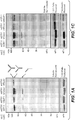
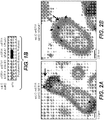
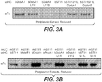

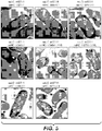


Classifications
-
- C—CHEMISTRY; METALLURGY
- C07—ORGANIC CHEMISTRY
- C07K—PEPTIDES
- C07K16/00—Immunoglobulins [IGs], e.g. monoclonal or polyclonal antibodies
-
- C—CHEMISTRY; METALLURGY
- C12—BIOCHEMISTRY; BEER; SPIRITS; WINE; VINEGAR; MICROBIOLOGY; ENZYMOLOGY; MUTATION OR GENETIC ENGINEERING
- C12N—MICROORGANISMS OR ENZYMES; COMPOSITIONS THEREOF; PROPAGATING, PRESERVING, OR MAINTAINING MICROORGANISMS; MUTATION OR GENETIC ENGINEERING; CULTURE MEDIA
- C12N15/00—Mutation or genetic engineering; DNA or RNA concerning genetic engineering, vectors, e.g. plasmids, or their isolation, preparation or purification; Use of hosts therefor
- C12N15/09—Recombinant DNA-technology
- C12N15/11—DNA or RNA fragments; Modified forms thereof; Non-coding nucleic acids having a biological activity
- C12N15/62—DNA sequences coding for fusion proteins
- C12N15/625—DNA sequences coding for fusion proteins containing a sequence coding for a signal sequence
-
- C—CHEMISTRY; METALLURGY
- C12—BIOCHEMISTRY; BEER; SPIRITS; WINE; VINEGAR; MICROBIOLOGY; ENZYMOLOGY; MUTATION OR GENETIC ENGINEERING
- C12N—MICROORGANISMS OR ENZYMES; COMPOSITIONS THEREOF; PROPAGATING, PRESERVING, OR MAINTAINING MICROORGANISMS; MUTATION OR GENETIC ENGINEERING; CULTURE MEDIA
- C12N15/00—Mutation or genetic engineering; DNA or RNA concerning genetic engineering, vectors, e.g. plasmids, or their isolation, preparation or purification; Use of hosts therefor
- C12N15/09—Recombinant DNA-technology
- C12N15/63—Introduction of foreign genetic material using vectors; Vectors; Use of hosts therefor; Regulation of expression
- C12N15/70—Vectors or expression systems specially adapted for E. coli
-
- C—CHEMISTRY; METALLURGY
- C12—BIOCHEMISTRY; BEER; SPIRITS; WINE; VINEGAR; MICROBIOLOGY; ENZYMOLOGY; MUTATION OR GENETIC ENGINEERING
- C12P—FERMENTATION OR ENZYME-USING PROCESSES TO SYNTHESISE A DESIRED CHEMICAL COMPOUND OR COMPOSITION OR TO SEPARATE OPTICAL ISOMERS FROM A RACEMIC MIXTURE
- C12P21/00—Preparation of peptides or proteins
- C12P21/02—Preparation of peptides or proteins having a known sequence of two or more amino acids, e.g. glutathione
-
- C—CHEMISTRY; METALLURGY
- C07—ORGANIC CHEMISTRY
- C07K—PEPTIDES
- C07K2317/00—Immunoglobulins specific features
- C07K2317/10—Immunoglobulins specific features characterized by their source of isolation or production
-
- C—CHEMISTRY; METALLURGY
- C07—ORGANIC CHEMISTRY
- C07K—PEPTIDES
- C07K2317/00—Immunoglobulins specific features
- C07K2317/10—Immunoglobulins specific features characterized by their source of isolation or production
- C07K2317/14—Specific host cells or culture conditions, e.g. components, pH or temperature
-
- C—CHEMISTRY; METALLURGY
- C07—ORGANIC CHEMISTRY
- C07K—PEPTIDES
- C07K2317/00—Immunoglobulins specific features
- C07K2317/30—Immunoglobulins specific features characterized by aspects of specificity or valency
- C07K2317/31—Immunoglobulins specific features characterized by aspects of specificity or valency multispecific
-
- C—CHEMISTRY; METALLURGY
- C07—ORGANIC CHEMISTRY
- C07K—PEPTIDES
- C07K2319/00—Fusion polypeptide
- C07K2319/01—Fusion polypeptide containing a localisation/targetting motif
- C07K2319/02—Fusion polypeptide containing a localisation/targetting motif containing a signal sequence
-
- C—CHEMISTRY; METALLURGY
- C12—BIOCHEMISTRY; BEER; SPIRITS; WINE; VINEGAR; MICROBIOLOGY; ENZYMOLOGY; MUTATION OR GENETIC ENGINEERING
- C12N—MICROORGANISMS OR ENZYMES; COMPOSITIONS THEREOF; PROPAGATING, PRESERVING, OR MAINTAINING MICROORGANISMS; MUTATION OR GENETIC ENGINEERING; CULTURE MEDIA
- C12N2800/00—Nucleic acids vectors
Definitions
- the present invention relates generally to the fields of molecular biology and protein technology. More specifically, the invention concerns signal peptides for the secretion of heterologous polypeptides from bacteria.
- heterologous polypeptide for example, antibodies
- diagnostic and therapeutic agents for various disorders and diseases.
- Many research and clinical applications require large quantities of functional polypeptide, thus calling for scaled-up, yet economic systems for polypeptide production.
- Particularly useful is the recombinant production of antibodies using a variety of expression hosts, ranging from prokaryotes such as E. coli or B. subtilis, to yeast, plants, insect cells and mammalian cells. Kipriyanov and Little (1999) Mol. Biotech. 12:173-201 .
- bacteria particularly E. coli
- the raw materials used i.e. bacterial cells
- Prokaryotic hosts grow much faster than, e.g., mammalian cells, allowing quicker analysis of genetic manipulations. Shorter generation time and ease of scaling up also make bacterial fermentation a more attractive means for large quantity protein production.
- the genomic structure and biological activity of many bacterial species including E. coli have been well-studied and a wide range of suitable vectors are available, making expression of a desirable antibody more convenient.
- Recombinant proteins can be obtained from bacteria either through refolding of inclusion bodies expressed in the cytoplasm, or through expression followed by secretion to the bacterial periplasm.
- the choice between secretion and refolding is generally guided by several considerations. Secretion is usually the faster and more commonly used strategy for producing antibodies. Kipriyanov and Little (1999), supra.
- E. coli secretion and refolding capacity is often limited to a lower level compared to other expression hosts.
- Antibody expression in prokaryotic systems can be carried out in different scales.
- Pluckthun and Pack see Pluckthun and Pack (1997) Immunotech 3:83-105 ; Pluckthun et al. (1996) in ANTIBODY ENGINEERING: A PRACTICAL APPROACH, pp 203-252 (Oxford Press ); Pluckthun (1994) in HANDBOOK OF EXP PHARMCOL VOL 3: THE PHARMCOL OF MONOCLONAL ANTIBODIES, pp269-315 (ed. M. Rosenberg and G.P. Moore; Springer-Verlag, Berl in).
- WO 2007/00665 relates to a filamentous phage display method wherein the polypeptides of interest displayed on phage particles are cotranslationally translocated across the cytoplasmic membrane of Gram-negative bacteria, in particular based on the signal recognition particle pathway.
- WO 2010/057097 relates to a reporter system for detection of protein-protein interactions in the periplasm of a prokaryotic host cell.
- WO 2011/057120 relates to signal sequences for the secretion of heterologous polypeptides from bacteria.
- WO 2002/006190 relates to crystalline and salt forms of an HIV protease inhibitor, pharmaceutical compositions comprising the HIV protease inhibitor, and methods of using the same.
- WO 2005/038031 relates to the use of a recombinant expression vector expressing an antibody fragment fused with an E. coli signal sequence in the form of a heterozygote protein to mass-produce the antibody fragment.
- WO 2009/021548 relates to a method for the production of full-length immunoglobulins in prokaryotic cells, wherein the immunoglobulins are secreted in the culture medium.
- EP 1 908 769 relates to methods and compositions for expression and production of recombinant antibodies in a host cell system, such as prokaryotic and eukaryotic expression systems.
- WO 2005/063816 relates to stabilized monovalent antibody fragments and their use as therapeutic agents.
- the invention provides a novel means for increasing production of antibodies.
- the use of periplasmic secretion as a means for high-level production of heterologous proteins (e.g., antibodies) can be limited by several frequently encountered problems.
- antibodies are complex multimeric proteins made from two different polypeptides, the heavy and light chains, which must be exported into the periplasm, folded properly and form the appropriate disulfide bonds. The complexity of this protein folding and secretion adds to the challenges of antibody manufacturing in E coli.
- coli secretion and refolding capacity is often limited to a lower level compared to other expression hosts. While TIR optimization has been shown to be useful for generating more efficient protein secretion, other approaches not been shown to routinely improve the secretion of heterologous proteins in E coli. For example, optimization of a signal protein was shown to decrease secretion of recombinant cyclodextrin glucanotransferase (CGTase) into the periplasmic space. Jonet et al., J Mol Microbiol Biotechnol (2012); 22:48-58 .
- CCTase cyclodextrin glucanotransferase
- the inventors demonstrate that increasing the average hydrophobicity of the signal peptide increased secretion of soluble antibody to the E coli periplasm.
- Variant signal peptides with increased average hydrophobicity were developed and the inventors demonstrated increased periplasmic secretion of soluble antibody when signal peptide variants with increased average hydrophobicity were used, and decreased periplasmic section of soluble antibody when signal peptide variants with decreased average hydrophobicity were used.
- an antibody heavy chain and/or an antibody light chain from an E. coli host cell comprising culturing an E. coli host cell comprising polynucleotide comprising (1) a first polynucleotide encoding a first signal peptide as defined in the claims operably linked to a polynucleotide encoding an antibody heavy chain, wherein the average hydrophobicity of the signal peptide is greater than about 0.5; and (2) a second polynucleotide encoding a second signal peptide as defined in the claims operably linked to a polynucleotide encoding an antibody light chain, wherein the average hydrophobicity of the second signal peptide is greater than about 0.5, whereby upon expression of the antibody in a host cell, the heavy and light chains are folded and assembled to form a biologically active antibody.
- an antibody heavy chain and/or light chain from an E. coli host cell comprising culturing an E. coli host cell comprising polynucleotide comprising (1) a first polynucleotide encoding a first signal peptide operably linked to a polynucleotide encoding an antibody heavy chain, wherein the average hydrophobicity of the signal peptide is greater than about 0.5; and (2) a second polynucleotide encoding a second signal peptide operably linked to a polynucleotide encoding an antibody light chain, wherein the average hydrophobicity of the second signal peptide is greater than about 0.5, whereby upon expression of the antibody in a host cell, the heavy and light chains are folded and assembled to form a biologically active antibody.
- the method further comprises recovering the heterologous polypeptide from the host cell culture. In some embodiments, the heterologous polypeptide is recovered from the host cell culture medium. In some embodiments, the method further comprises combining the recovered heterologous polypeptide with a pharmaceutically acceptable carrier, excipient, or carrier to prepare a pharmaceutical formulation comprising the heterologous polypeptide.
- an antibody heavy chain and/or light chain from an E. coli host cell comprising culturing an E. coli host cell comprising polynucleotide comprising (1) a first polynucleotide encoding a first signal peptide operably linked to a polynucleotide encoding an antibody heavy chain, wherein the average hydrophobicity of the signal peptide is greater than about 0.5; and (2) a second polynucleotide encoding a second signal peptide operably linked to a polynucleotide encoding an antibody light chain, wherein the average hydrophobicity of the second signal peptide is greater than about 0.5, whereby upon expression of the antibody in a host cell, the heavy and light chains are folded and assembled to form a biologically active antibody.
- an antibody heavy chain and/or light chain from an E. coli host cell comprising culturing an E. coli host cell comprising polynucleotide comprising (1) a first polynucleotide encoding a first signal peptide operably linked to a polynucleotide encoding an antibody heavy chain, wherein the average hydrophobicity of the signal peptide is greater than about 0.5; and (2) a second polynucleotide encoding a second signal peptide operably linked to a polynucleotide encoding an antibody light chain, wherein the average hydrophobicity of the second signal peptide is greater than about 0.5, whereby upon expression of the antibody in a host cell, the heavy and light chains are folded and assembled to form a biologically active antibody.
- the average hydrophobicity of the first signal peptide is greater than about 0.6. In some embodiments, the average hydrophobicity of the first signal peptide is greater than about 0.7. In some embodiments, the average hydrophobicity of the second signal peptide is greater than about 0.6. In some embodiments, the average hydrophobicity of the second signal peptide is greater than about 0.7. In some embodiments, the average hydrophobicity of the first and second signal peptides is similar (e.g., about equivalent). In some embodiments, the average hydrophobicity of the first and second signal peptides is different.
- the first and/or second signal peptide is a variant co-translational signal peptide.
- the first and/or second signal peptide is a variant DsbA signal peptide as defined in the claims.
- the variant DsbA signal peptide comprises a mutation at residue L11, wherein the variant DsbA signal peptide has a greater average hydrophobicity than a wildtype DsbA signal peptide of SEQ ID NO:3.
- the mutation is L11I or S18Y.
- the variant DsbA signal peptide comprises sequence of SEQ ID NO:13 or 15.
- the signal peptide is a Sfmc signal peptide.
- the Sfmc signal peptide has TIR strength that is different than TIR strength of wild-type Sfmc signal peptide.
- the relative translation strength (also termed TIR strength) of the Sfmc signal peptide is about 2, about 3, about 4, about 5, about 6, about 7, or more, such as about 8, about 9, or more.
- the relative translation strength of the Sfmc signal peptide is between 1 and 3, between 2 and 4, between 3 and 5, between 4 and 6, between 5 and 7, between 6 and 8.
- the relative translation strength of the Sfmc signal peptide is between 2 and 5, between 3 and 7, or between 4 and 8.
- the signal peptide is FlgI, NikA, AsmA, TolB, YraI, FecB, CemH, TreA, FocC, TraU, SfmL, or TorT.
- the FlgI, NikA, AsmA, TolB, YraI, FecB, CemH, TreA, FocC, TraU, SfmL, or TorT signal peptide has relative translation strength that is different than relative translation strength of wild-type FlgI, NikA, AsmA, TolB, YraI, FecB, CemH, TreA, FocC, TraU, SfmL, or TorT signal peptide.
- the TIR strength of the FlgI, NikA, AsmA, TolB, YraI, FecB, CemH, TreA, FocC, TraU, SfmL, or TorT signal peptide is about 2, about 3, about 4, about 5, about 6, about 7, or more, such as about 8, about 9, or more.
- the relative translation strength of the FlgI, NikA, AsmA, TolB, YraI, FecB, CemH, TreA, FocC, TraU, SfmL, or TorT signal peptide is between 1 and 3, between 2 and 4, between 3 and 5, between 4 and 6, between 5 and 7, between 6 and 8.
- the relative translation strength of the FlgI, NikA, AsmA, TolB, YraI, FecB, CemH, TreA, FocC, TraU, SfmL, or TorT signal peptide is between 2 and 5, between 3 and 7, or between 4 and 8.
- the signal peptide is not Sfmc. In some embodiments, the signal peptide is not TorT. In some embodiments, the signal peptide is not any one or more of FlgI, NikA, AsmA, TolB, YraI, FecB, CemH, TreA, FocC, TraU, SfmL, or TorT.
- the relative translation strength of the first and/or second signal peptide is about 1, about 2, about 3, about 4, about 5, about 6, about 7, or about 8. In some embodiments, the relative translation strength of the first signal peptide is about 5 and the relative translation strength of the second signal sequence is about 8. In some embodiments, the relative translation strength of the first signal peptide is about 8 and the relative translation strength of the second signal sequence is about 5. In some embodiments, the relative translation strength of the Sfmc signal peptide is between 1 and 3, between 2 and 4, between 3 and 5, between 4 and 6, between 5 and 7, between 6 and 8. In some embodiments, the relative translation strength of the Sfmc signal peptide is between 2 and 5, between 3 and 7, or between 4 and 8.
- the polynucleotide in the host cell further comprises a promoter.
- the promoter is a prokaryotic promoter selected from the group consisting of phoA, tac, lpp, lac-lpp, lac, ara, and T7 promoter.
- the E. coli host cell is of a strain deficient in endogenous protease activities.
- the genotype of the E. coli lacks degP and prc genes and harbors a mutant spr gene.
- the host cell further comprises a polynucleotide encoding at least one prokaryotic polypeptide selected from the group consisting of DsbA, DsbC, DsbG and FkpA. In some embodiments, the polynucleotide encodes both DsbA and DsbC.
- the methods further comprise recovering the antibody from the host cell culture. In some embodiments, the antibody is recovered from the host cell culture medium. In some embodiments, the methods further comprise combining the recovered antibody with a pharmaceutically acceptable carrier, excipient, or carrier to prepare a pharmaceutical formulation comprising the antibody. In some embodiments, at least 50% of the immunoglobulin polypeptide complexes that are formed are the antibody. In some embodiments, least 70% of the immunoglobulin polypeptide complexes that are formed are the antibody. In some embodiments, least 80% of the immunoglobulin polypeptide complexes that are formed are the antibody. In some embodiments, least 90% of the immunoglobulin polypeptide complexes that are formed are the antibody.
- the antibody is a monoclonal antibody. In some embodiments, the antibody is a chimeric antibody, an affinity matured antibody, a bispecific antibody, humanized antibody, or a human antibody. In some embodiments, the antibody is a bispecific antibody.
- variant DsbA signal peptides wherein the variant comprises an H region with an average hydrophobicity that is greater than 0.5.
- variant DsbA signal peptides comprising a mutation at residue L11, wherein the variant has a greater average hydrophobicity than a DsbA signal peptide of SEQ ID NO:3.
- the mutation is L11I and/or S18Y.
- variant STII signal peptides comprising a mutation at residue S11, wherein the variant STII signal peptide has a greater average hydrophobicity than a STII signal peptide of SEQ ID NO:1.
- the mutation is S11A, S11I or S11L.
- variant signal peptides consisting of, consisting essentially of or comprising a sequence of SEQ ID NO:8, 11, 13, 15, 31, or 33.
- the heterologous polypeptide is an antibody heavy chain. In some embodiments, the heterologous polypeptide is an antibody light chain. In some embodiments, the heterologous polypeptide is an antibody light and heavy chain. In some embodiments, the heterologous polypeptide is a multimeric polypeptide. In some embodiments, the heterologous polypeptide is an immunoadhesin.
- polynucleotide sequences that encode any of the variant signal peptides as defined in the claims.
- polynucleotide sequences that encode any of the variant signal peptides as defined in the claims, operably linked to a polynucleotide encoding a heterologous polypeptide, whereby upon expression of the heterologous polypeptide in a host cell the heterologous polypeptide is folded and assembled to form a biologically active heterologous polypeptide.
- the host cell is a prokaryotic host cell. In some embodiments, the host cell is E coli.
- polynucleotides encoding an antibody
- said polynucleotides comprising (1) a polynucleotide encoding a first signal peptide as defined in the claims operably linked to a polynucleotide encoding an antibody heavy chain and (2) a polynucleotide encoding a second signal peptide as defined in the claims operably linked to a polynucleotide encoding an antibody light chain, whereby upon expression of the antibody in a host cell the heavy and light chains are folded and assembled to form a biologically active antibody, wherein the first signal peptide is a variant signal peptide of any one of variant signal peptide as defined in the claims.
- the first signal peptide consists of, consists essentially of, or comprises a sequence of SEQ ID NO:8, 11, 13, 15, 31, 33, or 42.
- the first signal peptide is a variant DsbA signal peptide, wherein the variant comprises an H region with an average hydrophobicity that is greater than 0.5.
- the first signal peptide is a variant DsbA signal peptide comprising a mutation at residue L11, wherein the variant has a greater average hydrophobicity than a DsbA signal peptide of SEQ ID NO:3.
- the mutation is L11I and/or S18Y.
- the first signal peptide is a variant STII signal peptide comprising a mutation at residue S11, wherein the variant STII signal peptide has a greater average hydrophobicity than a STII signal peptide of SEQ ID NO:1.
- the mutation is S11A, S11I or S11L.
- the second signal peptide is a signal peptide. In some embodiments, the second signal peptide consists of, consists essentially of, or comprises a sequence of SEQ ID NO:8, 11, 13, 15, 31, 33, or 42. In some embodiments, the second signal peptide is a variant DsbA signal peptide, wherein the variant comprises an H region with an average hydrophobicity that is greater than 0.5. In some embodiments, the second signal peptide is a variant DsbA signal peptide comprising a mutation at residue L11, wherein the variant has a greater average hydrophobicity than a DsbA signal peptide of SEQ ID NO:3. In some embodiments, the mutation is L11I and/or S18Y.
- the second signal peptide is a variant STII signal peptide comprising a mutation at residue S11, wherein the variant STII signal peptide has a greater average hydrophobicity than a STII signal peptide of SEQ ID NO:1.
- the mutation is S11A, S11I or S11L.
- the polynucleotide encoding an antibody further comprises (3) a polynucleotide encoding a third signal peptide as defined in the claims operably linked to a polynucleotide encoding an Fc polypeptide.
- the third signal peptide may be, for example, any of the variant signal peptides as defined in the claims.
- the third signal peptide consists of, consists essentially of, or comprises a sequence of SEQ ID NO:8, 11, 13, 15, 31, or 33.
- the polynucleotide further comprises a promoter operably linked to the heterologous polypeptide.
- the promoter is a prokaryotic promoter selected from the group consisting of phoA, tac, lpp, lac-lpp, lac, ara, trp, and T7 promoter.
- the promoter is a phoA promoter.
- the polynucleotide comprises (a) a first promoter, wherein the first promoter is operably linked to a light chain and (b) a second promoter, wherein the second promoter is operably linked to a heavy chain.
- the first and second promoters are both phoA promoters.
- the polynucleotide further comprises (c) a third promoter, wherein the third promoter is operably linked to an Fc polypeptide.
- the promoter is a phoA promoter.
- heterologous polypeptide is a protease, an immunoadhesin, an extracellular domain of a receptor, a heteromultimeric protein, or an antibody.
- the antibody is a monoclonal antibody. In some embodiments, the antibody is a chimeric antibody, a bispecific antibody, humanized antibody, an antibody fragment or a human antibody. In some embodiments, the antibody is a bispecific antibody.
- a vector comprising a polynucleotide of any of the polynucleotides as defined in the claims.
- the vector is an expression vector.
- composition comprising any of the polynucleotides as defined in the claims.
- the host cell is a prokaryotic cell.
- the prokaryotic cell is E. coli.
- the E. coli is of a strain deficient in endogenous protease activities.
- the genotype of the E. coli lacks degP and prc genes and harbors a mutant spr gene.
- the host cell further comprises a polynucleotide encoding a prokaryotic chaperone protein.
- the prokaryotic chaperone protein is DsbA and/or DsbC.
- host cell overexpresses a prokaryotic chaperone protein.
- a heterologous polypeptide comprising culturing any of the host cells described herein so that the nucleic acid is expressed, whereby upon expression of said polynucleotide in a host cell as defined in the claims, the heterologous polypeptide is folded to form a biologically active heterologous polypeptide.
- the method further comprises recovering the heterologous polypeptide from the host cell culture.
- the heterologous polypeptide is recovered from the host cell culture medium.
- the method further comprises combining the recovered heterologous polypeptide with a pharmaceutically acceptable carrier, excipient, or carrier to prepare a pharmaceutical formulation comprising the heterologous polypeptide.
- provided are methods of secreting a heterologous polypeptide from a cell comprising culturing any of the host cells provided herein so that the nucleic acid is expressed and the heterologous polypeptide is secreted.
- a heterologous polypeptide from a cell comprising culturing any of the host cells provided herein so that the nucleic acid is expressed and the heterologous polypeptide is translocated.
- a heterologous polypeptide obtained by any of the methods is disclosed herein.
- the polypeptide is an antibody.
- use of the variant signal peptide results in, e.g., increased production of heterologous polypeptide (e.g., antibody, e.g., heavy chain and/or light chain), increased secretion of heterologous polypeptide (e.g., antibody), increased production of mature heterologous polypeptide (e.g., antibody), increased secretion of mature heterologous polypeptide (e.g., antibody), increased production of soluble heterologous polypeptide (e.g., antibody), increased secretion of soluble heterologous polypeptide (e.g., antibody), increased localization of inclusion bodies on the periplasmic side, and/or increased production of a heterologous polypeptide whereby the heterologous polypeptide is secreted, folded and assembled into a biologically active polypeptide (e.g., antibody), e.g., as compared to use of wildtype (non-variant) signal peptide.
- heterologous polypeptide e.g., antibody, e.g.
- the relative translation strength (also termed TIR strength) of the variant signal peptide and wildtype (non-variant) signal peptide is about 1. In some embodiments, the relative translation strength of the variant signal peptide and wildtype (non-variant) signal peptide is about 2, about 3, about 4, about 5, about 6, about 7 or more such about 8 or more. In some embodiments the relative translation strength of the variant signal peptide and wildtype (non-variant) signal peptide is about 4. In some embodiments the relative translation strength of the variant signal peptide and wildtype (non-variant) signal peptide is about 5. In some embodiments the relative translation strength of the variant signal peptide and wildtype (non-variant) signal peptide is about 6.
- the relative translation strength of the variant signal peptide and wildtype (non-variant) signal peptide is about 8. In some embodiments, the relative translation strength of the first and second signal peptides is about equal. In some embodiments, the relative translation strengths of the first and second signal peptides are different.
- a polynucleotide sequence that encodes a variant signal peptide of the invention.
- the variants are of a DsbA or STII signal peptide.
- the polynucleotide sequence encodes an amino acid of SEQ ID NO:8, 11, 13, 14, 15, 31, or 33.
- the polynucleotide sequence encodes an amino acid sequence of SEQ ID NO: 8, 11, or 13.
- the polynucleotide sequence encodes an amino acid sequence of SEQ ID NO: 14.
- the polynucleotide sequence encodes an amino acid sequence of SEQ ID NO: 12 or 15.
- the polynucleotide encodes an amino acid sequence of SEQ ID NO: 31 or 33. In some aspects of the disclosure, the polynucleotide encodes an amino acid sequence of SEQ ID NO: 32, 34, or 35. In some aspects of the disclosure, the polynucleotide consists of, consists essentially of, or comprises a polynucleotide sequence of SEQ ID NO:23, 24, 25, 26, 27, 28, 29 30, 36, 37, 38, 39 or 40. In some aspects of the disclosure, the polynucleotide consists of, consists essentially of, or comprises a polynucleotide sequence of SEQ ID NO:23, 24, 25, 26, or 28.
- the polynucleotide consists of, consists essentially of, or comprises a polynucleotide sequence of SEQ ID NO:29. In some aspects of the disclosure, the polynucleotide consists of, consists essentially of, or comprises a polynucleotide sequence of one of SEQ ID NO:27 or 30. In some aspects of the disclosure, the polynucleotide consists of, consists essentially of, or comprises a polynucleotide sequence of SEQ ID NO: 36 or 38. In some aspects of the disclosure, the polynucleotide consists of, consists essentially of, or comprises a polynucleotide sequence of SEQ ID NO: 37, 39 or 40.
- the invention provides a polynucleotide comprising a polynucleotide encoding a variant signal peptide as defined in the claims of the invention operably linked to a polynucleotide encoding a heterologous polypeptide, whereby upon expression of the heterologous polypeptide in a host cell (e.g., a prokaryotic host cell, e.g., an E. coli host cell), the heterologous polypeptide is folded and assembled to form a biologically active heterologous polypeptide. Examples of heterologous polypeptides are further disclosed herein.
- the heterologous polypeptide is an antibody heavy chain.
- the heterologous polypeptide is an antibody light chain.
- the heterologous polypeptide is an Fc polypeptide. In some embodiments, the heterologous polypeptide is a multimeric polypeptide. In some embodiments, the heterologous polypeptide is a heteromultimer. In some aspects of the disclosure, the signal peptide consists of, consists essentially of, or comprises an amino acid sequence of SEQ ID NO:8, 11, 12, 13, 14, 15, 31, 32, 33, 34, or 35. In some aspects of the disclosure, the signal peptide consists of, consists essentially of, or comprises an amino acid sequence of SEQ ID NO: 8, 11, or 13. In some aspects of the disclosure, the signal peptide consists of, consists essentially of, or comprises an amino acid sequence of SEQ ID NO: 14.
- the signal peptide consists of, consists essentially of, or comprises an amino acid sequence of SEQ ID NO: 12 or 15. In some aspects of the disclosure, the signal peptide consists of, consists essentially or, or comprises an amino acid sequence of SEQ ID NO: 31 or 33. In some embodiments, the signal peptide consists of, consists essentially or, or comprises amino acid sequence of SEQ ID NO: 32, 34, or 35. In some aspects of the disclosure, the polynucleotide encoding a variant signal peptide consists of, consists essentially of, or comprises sequence of SEQ ID NO:23, 24, 25, 26, 27, 28, 29 30, 31, 32, 33, 34, 35, 36, 37, 38, 39, 40, 44, 45, or 46.
- the polynucleotide encoding a variant signal peptide consists of, consists essentially of, or comprises sequence of SEQ ID NO:23, 26, 28, 30, 36, 37, 38, 45 or 46. In some aspects of the disclosure, the polynucleotide encoding a variant signal peptide consists of, consists essentially of, or comprises sequence of SEQ ID NO:29. In some aspects of the disclosure, the polynucleotide encoding a variant signal peptide consists of, consists essentially of, or comprises sequence of SEQ ID NO:27 or 30.
- the polynucleotide encoding a variant signal peptide consists of, consists essentially of, or comprises a polynucleotide sequence of SEQ ID NO: 36 or 38. In some aspects of the disclosure, the polynucleotide encoding a variant signal peptide consists of, consists essentially of, or comprises a polynucleotide sequence of SEQ ID NO: 37, 39 or 40. In some aspects of the disclosure, the polynucleotide encoding a variant signal peptide consists of, consists essentially of, or comprises a polynucleotide sequence of SEQ ID NO: 45 or 46.
- the disclosure provides a polynucleotide comprising (1) a polynucleotide encoding a first signal peptide operably linked to a polynucleotide encoding a first heterologous polypeptide and (2) a polynucleotide encoding a second signal peptide operably linked to a polynucleotide encoding a second heterologous polypeptide, whereby upon expression of the antibody in a host cell, the first and second heterologous polypeptides are folded and assembled to form a biologically active polypeptide complex.
- the invention provides a polynucleotide encoding an antibody, said polynucleotide comprising (1) a polynucleotide encoding a first signal peptide as defined in the claims operably linked to a polynucleotide encoding an antibody heavy chain and (2) a polynucleotide encoding a second signal peptide as defined in the claims operably linked to a polynucleotide encoding an antibody light chain, whereby upon expression of the antibody in a host cell (e.g., a prokaryotic host cell, e.g., an E. coli host cell), the heavy and light chains are folded and assembled to form a biologically active antibody.
- a host cell e.g., a prokaryotic host cell, e.g., an E. coli host cell
- the first signal peptide is a signal peptide (e.g., any signal peptide known in the art). In some embodiments, the signal peptide is a co-translational signal peptide. In some embodiments, the first signal peptide is a DsbA signal peptide. In some embodiments, the first signal peptide is a STII signal peptide. In some embodiments, the first signal peptide is any one of the variant signal peptides disclosed here. In some embodiments, the first signal peptide consists of, consists essentially of, or comprises an amino acid sequence of SEQ ID NO:8, 11, 12, 13, 14, 15, 31, 32, 33, 34, or 35.
- the first signal peptide consists of, consists essentially of, or comprises an amino acid sequence of SEQ ID NO: 8, 11, or 13. In some embodiments, the first signal peptide consists of, consists essentially of, or comprises an amino acid sequence of SEQ ID NO: 14. In some embodiments, the first signal peptide consists of, consists essentially of, or comprises an amino acid sequence of SEQ ID NO: 12 or 15. In some embodiments, the first signal peptide consists of, consists essentially of, or comprises an amino acid sequence of SEQ ID NO: 31 or 33. In some embodiments, the first signal peptide consists of, consists essentially of, or comprises an amino acid sequence of SEQ ID NO: 32, 34, or 35.
- the polynucleotide encoding a first signal peptide consists of, consists essentially of, or comprises sequence of SEQ ID NO:23, 24, 25, 26, 27, 28, 29 30, 31, 32, 33, 34, or 35. In some embodiments, the polynucleotide encoding a first signal peptide consists of, consists essentially of, or comprises sequence of SEQ ID NO:23, 24, 25, 26, 28, 36, 37, 38, 39 or 40. In some embodiments, the polynucleotide encoding a first signal peptide consists of, consists essentially of, or comprises sequence of SEQ ID NO:29.
- the polynucleotide encoding a first signal peptide consists of, consists essentially of, or comprises sequence of SEQ ID NO:27 or 30. In some embodiments, the polynucleotide encoding a first signal peptide consists of, consists essentially of, or comprises sequence of SEQ ID NO: 36 or 38. In some embodiments, the polynucleotide encoding a first signal peptide consists of, consists essentially of, or comprises sequence of SEQ ID NO: 37, 39 or 40.
- the second signal peptide is a signal peptide (e.g., any signal peptide known in the art). In some embodiments, the second signal peptide is a DsbA signal peptide. In some embodiments, the second signal peptide is a STII signal peptide. In some embodiments, the second signal peptide is any one of the variant signal peptides disclosed here. In some embodiments, the second signal peptide consists of, consists essentially of, or comprises an amino acid sequence of SEQ ID NO:8, 11, 12, 13, 14, 15, 31, 32, 33, 34, or 35. In some embodiments, the second signal peptide consists of, consists essentially of, or comprises an amino acid sequence of SEQ ID NO: 8, 11, or 13.
- the second signal peptide consists of, consists essentially of, or comprises an amino acid sequence of SEQ ID NO: 14. In some embodiments, the second signal peptide consists of, consists essentially of, or comprises an amino acid sequence of SEQ ID NO: 12 or 15. In some embodiments, the second signal peptide consists of, consists essentially of, or comprises an amino acid sequence of SEQ ID NO: 31 or 33. In some embodiments, the second signal peptide consists of, consists essentially of, or comprises an amino acid sequence of SEQ ID NO: 32, 35, or 35.
- the polynucleotide encoding a second signal peptide consists of, consists essentially of, or comprises sequence of SEQ ID NO:23, 24, 25, 26, 27, 28, 29 30, 36, 37, 38, 39 or 40. In some embodiments, the polynucleotide encoding a second signal peptide consists of, consists essentially of, or comprises sequence of SEQ ID NO:23, 24, 25, 26, or 28. In some embodiments, the polynucleotide encoding a second signal peptide consists of, consists essentially of, or comprises sequence of SEQ ID NO:29.
- the polynucleotide encoding a second signal peptide consists of, consists essentially of, or comprises sequence of SEQ ID NO:27 or 30. In some embodiments, the polynucleotide encoding a second signal peptide consists of, consists essentially of, or comprises sequence of SEQ ID NO:36 or 38. In some embodiments, the polynucleotide encoding a second signal peptide consists of, consists essentially of, or comprises sequence of SEQ ID NO:37, 39 or 40.
- the first signal peptide consists of, consists essentially of or comprises a signal peptide (e.g., any signal peptide known in the art), and the second signal peptide consists of, consists essentially of, or comprises a signal peptide as defined in the claims, e.g., a signal peptide of any one of SEQ ID NO: 8, 11, 13, 15, 31, and 33 (in some embodiments, any one of SEQ ID NO: 8, 11, and 13, and in some aspects of the disclosure, any one of SEQ ID NO: 36 and 38).
- the second signal peptide consists of, consists essentially of, or comprises an amino acid sequence of SEQ ID NO:8, 11, 13, 15, 31 or 33.
- the second signal peptide consists of, consists essentially of, or comprises an amino acid sequence of SEQ ID NO: 8, 11, or 13. In some aspects of the disclosure, the second signal peptide consists of, consists essentially of, or comprises an amino acid sequence of SEQ ID NO: 14. In some aspects of the disclosure, the second signal peptide consists of, consists essentially of, or comprises an amino acid sequence of SEQ ID NO: 12 or 15. In some aspects of the disclosure, the second signal peptide consists of, consists essentially of, or comprises an amino acid sequence of SEQ ID NO:36 or 38. In some aspects of the disclosure, the second signal peptide consists of, consists essentially of, or comprises an amino acid sequence of SEQ ID NO:37, 39 or 40.
- the polynucleotide encoding a second signal peptide consists of, consists essentially of, or comprises sequence of SEQ ID NO:23, 24, 25, 26, 27, 28, 29 30, 36, 37, 38, 39 or 40,. In some aspects of the disclosure, the polynucleotide encoding a second signal peptide consists of, consists essentially of, or comprises sequence of SEQ ID NO:23, 24, 25, 26, or 28. In some aspects of the disclosure, the polynucleotide encoding a second signal peptide consists of, consists essentially of, or comprises sequence of SEQ ID NO:29.
- the polynucleotide encoding a second signal peptide consists of, consists essentially of, or comprises sequence of SEQ ID NO:27 or 30. In some aspects of the disclosure, the polynucleotide encoding a second signal peptide consists of, consists essentially of, or comprises sequence of SEQ ID NO: 36 or 38. In some aspects of the disclosure, the polynucleotide encoding a second signal peptide consists of, consists essentially of, or comprises sequence of SEQ ID NO:37, 39 or 40.
- the second signal peptide consists of, consists essentially of or comprises a signal peptide (e.g., any signal peptide known in the art)
- the first signal peptide consists of, consists essentially of, or comprises a signal peptide as defined in the claims, e.g., a signal peptide comprising, consisting of, or consisting essentially of an amino acid sequence of any one of SEQ ID NO: 8, 11, 13, 15, 31, and 33 (in some embodiments, any one of SEQ ID NO: 8, 11, and 13, and in some embodiments, any one of SEQ ID NO: 31 and 33).
- the first signal peptide consists of, consists essentially of, or comprises an amino acid sequence of SEQ ID NO:8, 11, 13, 15, 31, or 33. In some embodiments, the first signal peptide consists of, consists essentially of, or comprises an amino acid sequence of SEQ ID NO: 8, 11, or 13. In some aspects of the disclosure, the first signal peptide consists of, consists essentially of, or comprises an amino acid sequence of SEQ ID NO: 14. In some aspects of the disclosure, the first signal peptide consists of, consists essentially of, or comprises an amino acid sequence of SEQ ID NO: 12 or 15. In some aspects of the disclosure, the first signal peptide consists of, consists essentially of, or comprises an amino acid sequence of SEQ ID NO: 31 or 33.
- the first signal peptide consists of, consists essentially of, or comprises an amino acid sequence of SEQ ID NO: 33, 34 or 35.
- the polynucleotide encoding a first signal peptide consists of, consists essentially of, or comprises sequence of SEQ ID NO:23, 24, 25, 26, 27, 28, 29.30, 36, 37, 38 39 or 40.
- the polynucleotide encoding a first signal peptide consists of, consists essentially of, or comprises sequence of SEQ ID NO:23, 24, 25, 26, 28, 36, 37, 38, 39 or 40.
- the polynucleotide encoding a first signal peptide consists of, consists essentially of, or comprises sequence of SEQ ID NO:29. In some aspects of the disclosure, the polynucleotide encoding a first signal peptide consists of, consists essentially of, or comprises sequence of SEQ ID NO:27 or 30. In some aspects of the disclosure, the polynucleotide encoding a first signal peptide consists of, consists essentially of, or comprises sequence of SEQ ID NO: 36 or 38. In some aspects of the disclosure, the polynucleotide encoding a first signal peptide consists of, consists essentially of, or comprises sequence of SEQ ID NO:37, 39 or 40.
- the polynucleotide encoding an antibody further comprises (3) a polynucleotide encoding a third signal peptide as defined in the claims operably linked to a polynucleotide encoding a Fc polypeptide.
- the third signal peptide consists of, consists essentially of, or comprises a signal peptide disclosed herein, e.g., a signal peptide comprising, consisting of, or consisting essentially of an amino acid sequence of any one of 8, 11, 13, 15, 31, and 33, (in some embodiments, any one of SEQ ID NO: 8, 11, and 13, and in some embodiments, any one of SEQ ID NO: 31 and 33).
- the first signal peptide consists of, consists essentially of, or comprises an amino acid sequence of SEQ ID NO: 8, 11, or 13. In some aspects of the disclosure, the first signal peptide consists of, consists essentially of, or comprises an amino acid sequence of SEQ ID NO: 14. In some aspects of the disclosure, the third signal peptide consists of, consists essentially of, or comprises an amino acid sequence of SEQ ID NO: 12 or 15.. In some embodiments, the third signal peptide consists of, consists essentially of, or comprises an amino acid sequence of SEQ ID NO: 31 or 33.
- the polynucleotide encoding a third signal peptide consists of, consists essentially of, or comprises sequence of SEQ ID NO:23, 24, 25, 26, 27, 28, 29, 30, 36, 37, 38 39 or 40. In some aspects of the disclosure, the polynucleotide encoding a third signal peptide consists of, consists essentially of, or comprises sequence of SEQ ID NO:23, 24, 25, 26, 28, 36, 37, 38, 39 or 40. In some aspects of the disclosure, the polynucleotide encoding a third signal peptide consists of, consists essentially of, or comprises sequence of SEQ ID NO:29.
- the polynucleotide encoding a third signal peptide consists of, consists essentially of, or comprises sequence of SEQ ID NO:27 and 30. In some aspects of the disclosure, the polynucleotide encoding a third signal peptide consists of, consists essentially of, or comprises sequence of SEQ ID NO:36 or 38. In some aspects of the disclosure, the polynucleotide encoding a third signal peptide consists of, consists essentially of, or comprises sequence of SEQ ID NO:37, 39 or 40. In some aspects of the disclosure, the third signal peptide is a signal peptide (e.g., any signal peptide known in the art).
- the first signal peptide comprises a signal peptide (e.g., any signal peptide known in the art) and the second signal peptide comprises a signal peptide (e.g., any signal peptide known in the art).
- the first signal peptide consists of, consists essentially of, or comprises a signal peptide and the second signal peptide consists of, consists essentially of, or comprises a signal peptide disclosed herein, e.g., a signal peptide of any one of SEQ ID NO: 8, 11, 12, 13, 14, 15, 36, 37, 38, 39 and 40 (in some aspects of the disclosurek, any one of SEQ ID NO: 8, 11, and 13, and in some aspects of the disclosure, any one of SEQ ID NO: 31 and 33).
- the second signal peptide consists of, consists essentially of, or comprises a signal peptide and the first signal peptide consists of, consists essentially of, or comprises a signal peptide disclosed herein, e.g., a signal peptide of any one of SEQ ID NO: 8, 11, 12, 13, 14, 15, 36, 37, 38, 39 and 40 (in some aspects of the disclosure, any one of SEQ ID NO: 8, 11, and 13, and in some aspects of the disclosure, any one of SEQ ID NO: 31 and 33).
- the first signal peptide consists of, consists essentially of, or comprises a signal peptide disclosed herein, e.g., a signal peptide of any one of SEQ ID NO: 8, 11, 12, 13, 14, 15, 36, 3, 38, 39 and 40 (in some aspects of the disclosure, any one of SEQ ID NO: 8, 11, and 13, and in some aspects of the disclosure, any one of SEQ ID NO: 31 and 33)
- the second signal peptide consists of, consists essentially of, or comprises a signal peptide disclosed herein, e.g., a signal peptide of any one of 8, 11, 12, 13, 14, 15, 36, 37, 38, 39 and 40 (in some aspects of the disclosure, any one of SEQ ID NO: 8, 11, and 13, and in some aspects of the disclosure, any one of SEQ ID NO: 31 and 33).
- use of the variant signal peptide results in, e.g., increased production of heterologous polypeptide (e.g., antibody), increased secretion of heterologous polypeptide (e.g., antibody), increased production of mature heterologous polypeptide (e.g., antibody), increased secretion of mature heterologous polypeptide (e.g., antibody), increased production of soluble heterologous polypeptide (e.g., antibody), increased secretion of soluble heterologous polypeptide (e.g., antibody), increased production of a heterologous polypeptide whereby the heterologous polypeptide is secreted, folded and assembled into a biologically active polypeptide (e.g., antibody), e.g., as compared to use of wildtype (non-variant) signal peptide.
- heterologous polypeptide e.g., antibody
- increased secretion of heterologous polypeptide e.g., antibody
- increased production of mature heterologous polypeptide e.g.
- use of the variant signal peptide results in, e.g., increased production of heterologous polypeptide (e.g., antibody), increased secretion of heterologous polypeptide (e.g., antibody), increased production of mature heterologous polypeptide (e.g., antibody), increased secretion of mature heterologous polypeptide (e.g., antibody), increased production of soluble heterologous polypeptide (e.g., antibody), increased secretion of soluble heterologous polypeptide (e.g., antibody), increase production of a heterologous polypeptide whereby the heterologous polypeptide is secreted, folded and assembled into a biologically active polypeptide (e.g., antibody), e.g., as compared to use of wildtype (non-variant) signal peptide, wherein the relative translation strength of the variant signal peptide and wildtype (non-variant) signal peptide are approximately equivalent.
- heterologous polypeptide e.g., antibody
- the relative translation strength of the variant signal peptide and wildtype (non-variant) signal peptide is about 1. In some embodiments, the relative translation strength of the variant signal peptide and wildtype (non-variant) signal peptide is about 2, about 3, about 4, about 5, about 6, about 7 or more such about 8 or more. In some embodiments the relative translation strength of the variant signal peptide and wildtype (non-variant) signal peptide is about 4. In some embodiments the relative translation strength of the variant signal peptide and wildtype (non-variant) signal peptide is about 5. In some embodiments the relative translation strength of the variant signal peptide and wildtype (non-variant) signal peptide is about 6.
- the relative translation strength of the variant signal peptide and wildtype (non-variant) signal peptide is about 8. In some embodiments, the relative translation strength of the first and second signal peptides are approximately equal. In some embodiments, the relative translation strengths of the first and second signal peptides are different.
- the disclosure provides uses of a heterologous polypeptide generated using the methods of the invention, in the preparation of a medicament for the therapeutic and/or prophylactic treatment of a disease, such as a cancer, a tumor, a cell proliferative disorder, and/or an immune (such as autoimmune) disorder.
- a disease such as a cancer, a tumor, a cell proliferative disorder, and/or an immune (such as autoimmune) disorder.
- the heterologous polypeptide can be of any form described herein, including antibody, antibody fragment, polypeptide (e.g., an oligopeptide), or combination thereof.
- the disclosure provides use of a signal peptide disclosed herein or polynucleotide encoding a signal peptide disclosed herein in the preparation of a medicament for the therapeutic and/or prophylactic treatment of a disease, such as a cancer, a tumor, a cell proliferative disorder and/or an immune (such as autoimmune) disorder.
- a disease such as a cancer, a tumor, a cell proliferative disorder and/or an immune (such as autoimmune) disorder.
- the disclosure provides use of an expression vector disclosed herein in the preparation of a medicament for the therapeutic and/or prophylactic treatment of a disease, such as a cancer, a tumor, a cell proliferative disorder and/or an immune (such as autoimmune) disorder.
- a disease such as a cancer, a tumor, a cell proliferative disorder and/or an immune (such as autoimmune) disorder.
- the disclosure provides use of a host cell disclosed herein in the preparation of a medicament for the therapeutic and/or prophylactic treatment of a disease, such as a cancer, a tumor, a cell proliferative disorder and/or an immune (such as autoimmune) disorder.
- a disease such as a cancer, a tumor, a cell proliferative disorder and/or an immune (such as autoimmune) disorder.
- the disclosure provides use of an article of manufacture disclosed herein in the preparation of a medicament for the therapeutic and/or prophylactic treatment of a disease, such as a cancer, a tumor, a cell proliferative disorder, an immune (such as autoimmune) disorder and/or an angiogenesis-related disorder (wound healing).
- a disease such as a cancer, a tumor, a cell proliferative disorder, an immune (such as autoimmune) disorder and/or an angiogenesis-related disorder (wound healing).
- the disclosure provides use of a kit disclosed herein in the preparation of a medicament for the therapeutic and/or prophylactic treatment of a disease, such as a cancer, a tumor, a cell proliferative disorder and/or an immune (such as autoimmune) disorder).
- a disease such as a cancer, a tumor, a cell proliferative disorder and/or an immune (such as autoimmune) disorder.
- vector is intended to refer to a nucleic acid molecule capable of transporting another nucleic acid to which it has been linked.
- plasmid refers to a circular double stranded DNA loop into which additional DNA segments may be ligated.
- phage vector Another type of vector is a viral vector, wherein additional DNA segments may be ligated into the viral genome.
- viral vector is capable of autonomous replication in a host cell into which they are introduced (e.g., bacterial vectors having a bacterial origin of replication and episomal mammalian vectors).
- vectors e.g., non-episomal mammalian vectors
- vectors can be integrated into the genome of a host cell upon introduction into the host cell, and thereby are replicated along with the host genome.
- certain vectors are capable of directing the expression of genes to which they are operatively linked.
- Such vectors are referred to herein as "recombinant expression vectors” (or simply, “recombinant vectors”).
- expression vectors of utility in recombinant DNA techniques are often in the form of plasmids.
- plasmid and “vector” may be used interchangeably as the plasmid is the most commonly used form of vector.
- cistron is intended to refer to a genetic element broadly equivalent to a translational unit comprising the nucleotide sequence coding for a polypeptide chain and adjacent control regions. "Adjacent control regions” include, for example, a translational initiation region (TIR; as defined herein below) and a termination region.
- TIR translational initiation region
- polycistronic expression vector refers to a single vector that contains and expresses multiple cistrons under the regulatory control of one single promoter.
- a common example of polycistronic vector is a "dicistronic" vector that contains and expresses two different polypeptides under the control of one promoter.
- a "separate cistron" expression vector refers to a single vector comprising at least two separate promoter-cistron pairs, wherein each cistron is under the control of its own promoter. Upon expression of a separate cistron expression vector, both transcription and translation processes of different genes are separate and independent.
- TIR translational initiation region
- translational initiation sequence refers to a nucleic acid region providing the efficiency of translational initiation of a gene of interest.
- a TIR within a particular cistron encompasses the ribosome binding site (RBS) and sequences 5' and 3' to RBS.
- RBS is defined to contain, minimally, the Shine-Dalgarno region and the start codon (AUG). Accordingly, a TIR also includes at least a portion of the nucleic acid sequence to be translated.
- a TIR includes a secretion signal sequence encoding a signal peptide that precedes the sequence encoding for the light or heavy chain within a cistron.
- a TIR variant contains sequence variants (particularly substitutions) within the TIR region that alter the property of the TIR, such as its translational strength as defined herein below.
- a TIR variant of the invention contains sequence substitutions within the first 2 to about 14, preferably about 4 to 12, more preferably about 6 codons of the secretion signal sequence that precedes the sequence encoding for the light or heavy chain within a cistron.
- translational strength refers to a measurement of a secreted polypeptide in a control system wherein one or more variants of a TIR is used to direct secretion of a polypeptide and the results compared to the wild-type TIR or some other control under the same culture and assay conditions.
- Signal peptide refers to a short peptide that can be used to direct a newly synthesized protein of interest through a cellular membrane, usually the inner membrane or both inner and outer membranes of prokaryotes.
- the signal peptide encoded by the secretion signal sequence may be endogenous to the host cells, or they may be exogenous, including signal peptides native to the polypeptide to be expressed.
- the signal peptide is typically present at the amino terminus of a polypeptide to be expressed, and are typically removed enzymatically between biosynthesis and secretion of the polypeptide from the cytoplasm. Thus, the signal peptide is usually not present in a mature protein product.
- Signal peptides e.g.
- prokaryotic e.g., E coli signal peptides
- E coli signal peptides are commonly composed of three distinct regions: an N-terminal region which typically contains at least 1 or 2 positively charged amino acid residues, a hydrophobic core region termed the H-region (also termed H domain), and a C-terminal region recognized by the signal peptidase.
- H-region also termed H domain
- C-terminal region recognized by the signal peptidase.
- One of skill in the art understands how to define the N-terminal region, H-region and C-terminal regions of a given signal peptide.
- Total or “sum” hydrophobicity is calculated by (a) assigning each amino acid in the peptide (or portion of a peptide) a normalized consensus hydrophobicity value according to Eisenberg, D. et al, J Mol Biol (1984) 179:125-142 . Table I (page 126), then adding up the normalized consensus hydrophobicity values for the amino acids in the peptide (or portion of the peptide).
- the average hydrophobicity is calculated for the H-domain of the signal peptide.
- operably linked refers to a juxtaposition of two or more components, wherein the components so described are in a relationship permitting them to function in their intended manner.
- a promoter is operably linked to a coding sequence if it acts in cis to control or modulate the transcription of the linked sequence.
- the DNA sequences that are "operably linked” are contiguous and, where necessary to join two protein coding regions or in the case of a secretory leader, contiguous and in reading frame.
- an operably linked promoter is generally located upstream of the coding sequence, it is not necessarily contiguous with it.
- Operably linked enhancers can be located upstream, within or downstream of coding sequences and at considerable distances from the promoter.
- Linking is accomplished by recombinant methods known in the art, e.g., using PCR methodology, by annealing, or by ligation at convenient restriction sites. If convenient restriction sites do not exist, then synthetic oligonucleotide adaptors or linkers are used in accord with conventional practice.
- transcriptional regulatory elements refer to nucleotide sequences present in cis, necessary for transcription and translation of a polynucleotide encoding a heterologous polypeptide into polypeptides.
- the transcriptional regulatory elements normally comprise a promoter 5' of the gene sequence to be expressed, transcriptional initiation and termination sites, and polyadenylation signal sequence.
- transcriptional initiation site refers to the nucleic acid in the construct corresponding to the first nucleic acid incorporated into the primary transcript, i.e., the mRNA precursor; the transcriptional initiation site may overlap with the promoter sequences.
- a “promoter” refers to a polynucleotide sequence that controls transcription of a gene or sequence to which it is operably linked.
- a promoter includes signals for RNA polymerase binding and transcription initiation. The promoters used will be functional in the cell type of the host cell in which expression of the selected sequence is contemplated.
- a large number of promoters including constitutive, inducible and repressible promoters from a variety of different sources, are well known in the art (and identified in databases such as GenBank) and are available as or within cloned polynucleotides (from, e.g., depositories such as ATCC as well as other commercial or individual sources). With inducible promoters, the activity of the promoter increases or decreases in response to a signal.
- host cell (or "recombinant host cell”), as used herein, is intended to refer to a cell that has been genetically altered, or is capable of being genetically altered by introduction of an exogenous polynucleotide, such as a recombinant plasmid or vector. It should be understood that such terms are intended to refer not only to the particular subject cell but to the progeny of such a cell. Because certain modifications may occur in succeeding generations due to either mutation or environmental influences, such progeny may not, in fact, be identical to the parent cell, but are still included within the scope of the term "host cell” as used herein.
- an "isolated" polypeptide e.g., an antibody
- an “isolated” polypeptide is one which has been identified and separated and/or recovered from a component of its natural environment. Contaminant components of its natural environment are materials which would interfere with diagnostic or therapeutic uses for the antibody, and may include enzymes, hormones, and other proteinaceous or nonproteinaceous solutes.
- the polypeptide will be purified (1) to greater than 95% by weight of polypeptide as determined by the Lowry method, and most preferably more than 99% by weight, (2) to a degree sufficient to obtain at least 15 residues of N-terminal or internal amino acid sequence by use of a spinning cup sequenator, or (3) to homogeneity by SDS-PAGE (sodium dodecyl sulfate polyacrylamide gel electrophoresis) under reducing or nonreducing conditions using Coomassie blue or, preferably, silver stain.
- Isolated polypeptide includes the polypeptide in situ within recombinant cells since at least one component of the polypeptide's natural environment will not be present. Ordinarily, however, isolated polypeptide will be prepared by at least one purification step.
- nucleic acid molecule is a nucleic acid molecule that is identified and separated from at least one contaminant nucleic acid molecule with which it is ordinarily associated in the natural source of the nucleic acid.
- isolated nucleic acid molecule is other than in the form or setting in which it is found in nature.
- Polynucleotide or “nucleic acid,” as used interchangeably herein, refer to polymers of nucleotides of any length, and include DNA and RNA.
- the nucleotides can be deoxyribonucleotides, ribonucleotides, modified nucleotides or bases, and/or their analogs, or any substrate that can be incorporated into a polymer by DNA or RNA polymerase, or by a synthetic reaction.
- a polynucleotide may comprise modified nucleotides, such as methylated nucleotides and their analogs. If present, modification to the nucleotide structure may be imparted before or after assembly of the polymer.
- sequence of nucleotides may be interrupted by non-nucleotide components.
- a polynucleotide may be further modified after synthesis, such as by conjugation with a label.
- Other types of modifications include, for example, "caps,” substitution of one or more of the naturally occurring nucleotides with an analog, internucleotide modifications such as, for example, those with uncharged linkages (e.g.
- methyl phosphonates, phosphotriesters, phosphoamidates, carbamates, etc.) and with charged linkages e.g ., phosphorothioates, phosphorodithioates, etc.
- pendant moieties such as, for example, proteins (e.g., nucleases, toxins, antibodies, signal peptides, ply-L-lysine, etc.), those with intercalators (e.g., acridine, psoralen, etc.), those containing chelators (e.g., metals, radioactive metals, boron, oxidative metals, etc.), those containing alkylators, those with modified linkages (e.g., alpha anomeric nucleic acids, etc.), as well as unmodified forms of the polynucleotide(s).
- proteins e.g., nucleases, toxins, antibodies, signal peptides, ply-L-lysine, etc.
- any of the hydroxyl groups ordinarily present in the sugars may be replaced, for example, by phosphonate groups, phosphate groups, protected by standard protecting groups, or activated to prepare additional linkages to additional nucleotides, or may be conjugated to solid or semi-solid supports.
- the 5' and 3' terminal OH can be phosphorylated or substituted with amines or organic capping group moieties of from 1 to 20 carbon atoms.
- Other hydroxyls may also be derivatized to standard protecting groups.
- Polynucleotides can also contain analogous forms of ribose or deoxyribose sugars that are generally known in the art, including, for example, 2'-O-methyl-, 2'-O-allyl, 2'-fluoro- or 2'-azido-ribose, carbocyclic sugar analogs, alpha-anomeric sugars, epimeric sugars such as arabinose, xyloses or lyxoses, pyranose sugars, furanose sugars, sedoheptuloses, acyclic analogs and a basic nucleoside analogs such as methyl riboside.
- One or more phosphodiester linkages may be replaced by alternative linking groups.
- linking groups include, but are not limited to, wherein phosphate is replaced by P(O)S ("thioate”), P(S)S ("dithioate”), (O)NR 2 ("amidate"), P(O)R, P(O)OR', CO or CH 2 ("formacetal"), in which each R or R' is independently H or substituted or unsubstituted alkyl (1-20 C) optionally containing an ether (-O-) linkage, aryl, alkenyl, cycloalkyl, cycloalkenyl or araldyl. Not all linkages in a polynucleotide need be identical. The preceding description applies to all polynucleotides referred to herein, including RNA and DNA.
- Oligonucleotide generally refers to short, generally single stranded, generally synthetic polynucleotides that are generally, but not necessarily, less than about 200 nucleotides in length.
- oligonucleotide and “polynucleotide” are not mutually exclusive. The description above for polynucleotides is equally and fully applicable to oligonucleotides.
- polypeptide refers generally to peptides and proteins from any cell source having more than about ten amino acids.
- heterologous polypeptides are those polypeptides foreign to the host cell being utilized, such as a human protein produced by E. coli. While the heterologous polypeptide may be prokaryotic or eukaryotic, preferably it is eukaryotic, more preferably mammalian, and most preferably human. Preferably, it is a recombinantly produced, or recombinant polypeptide.
- heterologous polypeptides are those polypeptides foreign to the host cell being utilized, such as a human protein produced by E. coli.
- heterologous polypeptide may be prokaryotic or eukaryotic, preferably it is eukaryotic, more preferably mammalian, and most preferably human. Preferably, it is a recombinantly produced, or recombinant polypeptide.
- mammalian polypeptides include molecules such as, e.g., renin, a growth hormone, including human growth hormone; bovine growth hormone; growth hormone releasing factor; parathyroid hormone; thyroid stimulating hormone; lipoproteins; 1-antitrypsin; insulin A-chain; insulin B-chain; proinsulin; thrombopoietin; follicle stimulating hormone; calcitonin; luteinizing hormone; glucagon; clotting factors such as factor VIIIC, factor IX, tissue factor, and von Willebrands factor; anti-clotting factors such as Protein C; atrial naturietic factor; lung surfactant; a plasminogen activator, such as urokinase or human urine or tissue-type plasminogen activator (t-PA) and variants thereof such as RETEVASETM and TNKASETM; bombesin; thrombin; hemopoietic growth factor; tumor necrosis factor-alpha and -beta; antibodies to ErbB
- Polypeptides herein may include human serum albumin (HSA), 2C4, tissue factor, anti-tissue factor, anti-CD20, anti-HER-2, heregulin, anti-IgE, anti-CD11a, anti-CD18, VEGF and receptors and antibodies thereto such as rhuFab V2 and AVASTINTM, growth hormone and its variants, such as hGH, growth hormone receptors, growth hormone releasing protein (GHRP), LIV-1 ( EP 1,263,780 ), TRAIL, tumor necrosis factor (TNF) and antibodies thereto, TNF receptor and related antibodies, TNF-receptor-IgG, TNF receptor associated factors (TRAFs) and inhibitors thereof, Factor VIII, Factor VIII B domain, interferons such as interferon-gamma, transforming growth factors (TGFs) such as TGF-beta, anti-TGF such as anti-TGF-beta, activin, inhibin, anti-activin, anti-inhibin, tissue-plasmin
- polypeptides are recombinant polypeptides, in some aspects of the disclosure, antibodies, which include monoclonal antibodies and humanized antibodies. Such antibodies may be full-length antibodies or antibody fragments. In some aspects of the disclosure, these antibodies are human or humanized antibodies. In some aspects of the disclosure, the antibody is an anti-c-met, anti-IgE, anti-CD18, anti-VEGF, anti-tissue factor, 2C4, anti-Her-2, anti-CD20, anti-CD40, or anti-CD11a antibody.
- Antibody fragments encompassed within the definition of polypeptide in some embodiments, comprise a light chain, in some embodiments, a kappa light chain. Such exemplary fragments include, for example, a Fab, Fab', F(ab') 2 , or F(ab') 2 -leucine zipper (LZ) fusion, and a one-armed antibody.
- Protein expression refers to conversion of the information encoded in a gene into messenger RNA (mRNA) and then to the protein.
- mRNA messenger RNA
- an “antibody-drug conjugate” means an antibody conjugated to one or more cytotoxic agents, such as a chemotherapeutic agent, a drug, a growth inhibitory agent, a toxin (e.g., a protein toxin, an enzymatically active toxin of bacterial, fungal, plant, or animal origin, or fragments thereof), or a radioactive isotope (i.e., a radioconjugate).
- cytotoxic agents such as a chemotherapeutic agent, a drug, a growth inhibitory agent, a toxin (e.g., a protein toxin, an enzymatically active toxin of bacterial, fungal, plant, or animal origin, or fragments thereof), or a radioactive isotope (i.e., a radioconjugate).
- blocking antibody or an antibody “antagonist” is one which inhibits or reduces biological activity of the antigen it binds. In some aspects of the disclosure, blocking antibodies or antagonist antibodies completely inhibit the biological activity of the antigen.
- an "agonist antibody”, as used herein, is an antibody which mimics at least one of the functional activities of a polypeptide of interest (e.g., HGF).
- Binding affinity generally refers to the strength of the sum total of noncovalent interactions between a single binding site of a molecule (e.g., an antibody) and its binding partner (e.g., an antigen). Unless indicated otherwise, as used herein, “binding affinity” refers to intrinsic binding affinity which reflects a 1:1 interaction between members of a binding pair (e.g., antibody and antigen).
- the affinity of a molecule X for its partner Y can generally be represented by the dissociation constant (Kd).
- the Kd is 1 x 10 -7 , 1 x 10 -8 , 5 x 10 -8 , 1 x 10 -9 , 3 x 10 -9 , 5 x 10 -9 , or even 1 x 10 -10 or stronger.
- Affinity can be measured by common methods known in the art, including those described herein. Low-affinity antibodies generally bind antigen slowly and tend to dissociate readily, whereas high-affinity antibodies generally bind antigen faster and tend to remain bound longer. A variety of methods of measuring binding affinity are known in the art, any of which can be used for purposes of the present invention. Specific illustrative aspects of the disclosure are described in the following.
- the "Kd" or "Kd value” according to this invention is measured by a radiolabeled antigen binding assay (RIA) performed with the Fab version of an antibody of interest and its antigen as described by the following assay that measures solution binding affinity of Fabs for antigen by equilibrating Fab with a minimal concentration of ( 125 I)-labeled antigen in the presence of a titration series of unlabeled antigen, then capturing bound antigen with an anti-Fab antibody-coated plate ( Chen, et al., (1999) J. Mol. Biol. 293:865-881 ).
- RIA radiolabeled antigen binding assay
- microtiter plates (Dynex) are coated overnight with 5 ⁇ g/ml of a capturing anti-Fab antibody (Cappel Labs) in 50 mM sodium carbonate (pH 9.6), and subsequently blocked with 2% (w/v) bovine serum albumin in PBS for two to five hours at room temperature (approximately 23°C).
- a non-adsorbant plate (Nunc #269620) 100 pM or 26 pM [ 125 I]-antigen are mixed with serial dilutions of a Fab of interest (e.g., consistent with assessment of an anti-VEGF antibody, Fab-12, in Presta et al., (1997) Cancer Res.
- the Fab of interest is then incubated overnight; however, the incubation may continue for a longer period (e.g., 65 hours) to insure that equilibrium is reached. Thereafter, the mixtures are transferred to the capture plate for incubation at room temperature ( e.g., for one hour). The solution is then removed and the plate washed eight times with 0.1% Tween-20 in PBS. When the plates have dried, 150 ⁇ l/well of scintillant (MicroScint-20; Packard) is added, and the plates are counted on a Topcount gamma counter (Packard) for ten minutes.
- a Topcount gamma counter Packard
- the Kd or Kd value is measured by using surface plasmon resonance assays using a BIAcoreTM-2000 or a BIAcoreTM-3000 (BIAcore, Inc., Piscataway, NJ) at 25°C with immobilized antigen CM5 chips at -10 response units (RU).
- carboxymethylated dextran biosensor chips (CM5, BIAcore Inc.) are activated with N-ethyl-N'- (3-dimethylaminopropyl)-carbodiimide hydrochloride (EDC) and N-hydroxysuccinimide (NHS) according to the supplier's instructions.
- EDC N-ethyl-N'- (3-dimethylaminopropyl)-carbodiimide hydrochloride
- NHS N-hydroxysuccinimide
- Antigen is diluted with 10mM sodium acetate, pH 4.8, into 5 ⁇ g/ml ( ⁇ 0.2 ⁇ M) before injection at a flow rate of 5 ⁇ l/minute to achieve approximately 10 response units (RU) of coupled protein.
- 1M ethanolamine is injected to block unreacted groups.
- two-fold serial dilutions of Fab (0.78 nM to 500 nM) are injected in PBS with 0.05% Tween 20 (PBST) at 25°C at a flow rate of approximately 25 ⁇ l/min.
- PBST Tween 20
- the following modifications are used for the surface Plasmon resonance assay method: antibody is immobilized to CM5 biosensor chips to achieve approximately 400 RU, and for kinetic measurements, two-fold serial dilutions of target protein are injected in PBST buffer at 25°C with a flow rate of about 30 ul/minute.
- association rates (k on ) and dissociation rates (k off ) are calculated using a simple one-to-one Langmuir binding model (BIAcore Evaluation Software version 3.2) by simultaneous fitting the association and dissociation sensorgram.
- the equilibrium dissociation constant (Kd) is calculated as the ratio k off /k on . See, e.g., Chen, Y., et al., (1999) J. Mol. Biol. 293:865-881 .
- CM5 chips CM5 chips
- RU -10 response units
- carboxymethylated dextran biosensor chips CM5, BIAcore Inc.
- EDC N-ethyl-N'- (3-dimethylaminopropyl)-carbodiimide hydrochloride
- NHS N-hydroxysuccinimide
- Antigen is diluted with 10mM sodium acetate, pH 4.8, into 5 ⁇ g/ml ( ⁇ 0.2uM) before injection at a flow rate of 5 ⁇ l/minute to achieve approximately 10 response units (RU) of coupled protein. Following the injection of antigen, 1M ethanolamine is injected to block unreacted groups. For kinetics measurements, two-fold serial dilutions of Fab (0.78 nM to 500 nM) are injected in PBS with 0.05% Tween 20 (PBST) at 25°C at a flow rate of approximately 25 ⁇ l/min.
- PBST Tween 20
- the following modifications are used for the surface Plasmon resonance assay method: antibody is immobilized to CM5 biosensor chips to achieve approximately 400 RU, and for kinetic measurements, two-fold serial dilutions of target protein are injected in PBST buffer at 25°C with a flow rate of about 30 ul/minute. Association rates (k on ) and dissociation rates (k off ) are calculated using a simple one-to-one Langmuir binding model (BIAcore Evaluation Software version 3.2) by simultaneous fitting the association and dissociation sensorgram. The equilibrium dissociation constant (Kd) was calculated as the ratio k off /k on . See, e.g., Chen, Y., et al., (1999) J.
- naked antibody is an antibody that is not conjugated to a heterologous molecule, such as a cytotoxic moiety or radiolabel.
- An antibody having a "biological characteristic" of a designated antibody is one which possesses one or more of the biological characteristics of that antibody which distinguish it from other antibodies that bind to the same antigen.
- a salvage receptor binding epitope to the antibody (especially an antibody fragment), as described, e.g., in US Patent 5,739,277 .
- a nucleic acid molecule encoding the salvage receptor binding epitope can be linked in frame to a nucleic acid encoding a polypeptide sequence of this disclosure so that the fusion protein expressed by the engineered nucleic acid molecule comprises the salvage receptor binding epitope and a polypeptide sequence of this disclosure.
- the term "salvage receptor binding epitope” refers to an epitope of the Fc region of an IgG molecule (e.g., IgG 1 , IgG 2 , IgG 3 , or IgG 4 ) that is responsible for increasing the in vivo serum half-life of the IgG molecule (e.g., Ghetie et al., Ann. Rev. Immunol. 18:739-766 (2000 ), Table 1). Antibodies with substitutions in an Fc region thereof and increased serum half-lives are also described in WO00/42072 , WO 02/060919 ; Shields et al., J. Biol. Chem.
- the serum half-life can also be increased, for example, by attaching other polypeptide sequences.
- antibodies or other polypeptides useful in the methods of the invention can be attached to serum albumin or a portion of serum albumin that binds to the FcRn receptor or a serum albumin binding peptide so that serum albumin binds to the antibody or polypeptide, e.g., such polypeptide sequences are disclosed in WO01/45746 .
- the serum albumin peptide to be attached comprises an amino acid sequence of DICLPRWGCLW (SEQ ID NO: 48).
- the half-life of a Fab is increased by these methods. See also, Dennis et al. J. Biol. Chem. 277:35035-35043 (2002 ) for serum albumin binding peptide sequences.
- fragment is meant a portion of a polypeptide or nucleic acid molecule that contains, preferably, at least 10%, 20%, 30%, 40%, 50%, 60%, 70%, 80%, 90%, 95%, or more of the entire length of the reference nucleic acid molecule or polypeptide.
- a fragment may contain 10, 20, 30, 40, 50, 60, 70, 80, 90, or 100, 200, 300, 400, 500, 600, or more nucleotides or 10, 20, 30, 40, 50, 60, 70, 80, 90, 100, 120, 140, 160, 180, 190, 200 amino acids or more.
- antibody herein is used in the broadest sense and encompasses various antibody structures, including but not limited to monoclonal antibodies, polyclonal antibodies, multispecific antibodies (e.g., bispecific antibodies), and antibody fragments so long as they exhibit the desired antigen-binding activity.
- antibody fragment refers to a molecule other than an intact antibody that comprises a portion of an intact antibody that binds the antigen to which the intact antibody binds.
- antibody fragments include but are not limited to Fv, Fab, Fab', Fab'-SH, F(ab') 2 ; diabodies; linear antibodies; single-chain antibody molecules (e.g. scFv); and multispecific antibodies formed from antibody fragments.
- an "antibody that binds to the same epitope” as a reference antibody refers to an antibody that blocks binding of the reference antibody to its antigen in a competition assay by 50% or more, and conversely, the reference antibody blocks binding of the antibody to its antigen in a competition assay by 50% or more.
- An exemplary competition assay is provided herein.
- chimeric antibody refers to an antibody in which a portion of the heavy and/or light chain is derived from a particular source or species, while the remainder of the heavy and/or light chain is derived from a different source or species.
- the "class" of an antibody refers to the type of constant domain or constant region possessed by its heavy chain.
- the heavy chain constant domains that correspond to the different classes of immunoglobulins are called ⁇ , ⁇ , ⁇ , ⁇ , and ⁇ , respectively.
- Fc region herein is used to define a C-terminal region of an immunoglobulin heavy chain that contains at least a portion of the constant region.
- the term includes native sequence Fc regions and variant Fc regions.
- a human IgG heavy chain Fc region extends from Cys226, or from Pro230, to the carboxyl-terminus of the heavy chain.
- the C-terminal lysine (Lys447) of the Fc region may or may not be present.
- numbering of amino acid residues in the Fc region or constant region is according to the EU numbering system, also called the EU index, as described in Kabat et al., Sequences of Proteins of Immunological Interest, 5th Ed. Public Health Service, National Institutes of Health, Bethesda, MD, 1991 .
- FR Framework or "FR” refers to variable domain residues other than hypervariable region (HVR) residues.
- the FR of a variable domain generally consists of four FR domains: FR1, FR2, FR3, and FR4. Accordingly, the HVR and FR sequences generally appear in the following sequence in VH (or VL): FR1-H1(L1)-FR2-H2(L2)-FR3-H3(L3)-FR4.
- full length antibody “intact antibody,” and “whole antibody” are used herein interchangeably to refer to an antibody having a structure substantially similar to a native antibody structure or having heavy chains that contain an Fc region as defined herein.
- a "human antibody” is one which possesses an amino acid sequence which corresponds to that of an antibody produced by a human or a human cell or derived from a non-human source that utilizes human antibody repertoires or other human antibody-encoding sequences. This definition of a human antibody specifically excludes a humanized antibody comprising non-human antigen-binding residues.
- a "human consensus framework” is a framework which represents the most commonly occurring amino acid residues in a selection of human immunoglobulin VL or VH framework sequences.
- the selection of human immunoglobulin VL or VH sequences is from a subgroup of variable domain sequences.
- the subgroup of sequences is a subgroup as in Kabat et al., Sequences of Proteins of Immunological Interest, Fifth Edition, NIH Publication 91-3242, Bethesda MD (1991), vols. 1-3 .
- the subgroup is subgroup kappa I as in Kabat et al., supra.
- the subgroup is subgroup III as in Kabat et al., supra.
- a “humanized” antibody refers to a chimeric antibody comprising amino acid residues from non-human HVRs and amino acid residues from human FRs.
- a humanized antibody will comprise substantially all of at least one, and typically two, variable domains, in which all or substantially all of the HVRs (e.g., CDRs) correspond to those of a non-human antibody, and all or substantially all of the FRs correspond to those of a human antibody.
- a humanized antibody optionally may comprise at least a portion of an antibody constant region derived from a human antibody.
- a "humanized form" of an antibody, e.g., a non-human antibody refers to an antibody that has undergone humanization.
- hypervariable region refers to each of the regions of an antibody variable domain which are hypervariable in sequence ("complementarity determining regions” or “CDRs") and/or form structurally defined loops ("hypervariable loops") and/or contain the antigen-contacting residues ("antigen contacts").
- CDRs complementarity determining regions
- hypervariable loops form structurally defined loops
- antigen contacts Generally, antibodies comprise six HVRs: three in the VH (H1, H2, H3), and three in the VL (L1, L2, L3).
- Exemplary HVRs herein include:
- multispecific antibody is used in the broadest sense and specifically covers an antibody that has polyepitopic specificity.
- Such multispecific antibodies include, but are not limited to, an antibody comprising a heavy chain variable domain (V H ) and a light chain variable domain (V L ), where the V H V L unit has polyepitopic specificity, antibodies having two or more V L and V H domains with each V H V L unit binding to a different epitope, antibodies having two or more single variable domains with each single variable domain binding to a different epitope, full length antibodies, antibody fragments such as Fab, Fv, dsFv, scFv, diabodies, bispecific diabodies and triabodies, antibody fragments that have been linked covalently or non-covalently.
- Polyepitopic specificity refers to the ability to specifically bind to two or more different epitopes on the same or different target(s).
- “Monospecific” refers to the ability to bind only one epitope.
- the multispecific antibody is an IgG antibody that binds to each epitope with an affinity of 5 ⁇ M to 0.001 pM, 3 ⁇ M to 0.001 pM, 1 ⁇ M to 0.001 pM, 0.5 ⁇ M to 0.001 pM, or 0.1 ⁇ M to 0.001 pM.
- one-armed antibody or “one-armed antibodies” refers to an antibody that comprises (1) a variable domain joined by a peptide bond to a polypeptide comprising a CH2 domain, a CH3 domain or a CH2-CH3 domain and (2) a second CH2, CH3 or CH2-CH3 domain, wherein a variable domain is not joined by a peptide bond to a polypeptide comprising the second CH2, CH3 or CH2-CH3 domain.
- the one-armed antibody comprises 3 polypeptides (1) a first polypeptide comprising a variable domain (e.g., VH), CH1, CH2 and CH3, (2) a second polypeptide comprising a variable domain ( e.g., VL) and a CL domain, and (3) a third polypeptide comprising a CH2 and CH3 domain.
- the third polypeptide does not comprise a variable domain.
- the one-armed antibody has a partial hinge region containing the two cysteine residues which form disulfide bonds linking the constant heavy chains.
- the variable domains of the one armed antibody form an antigen binding region.
- a variable domain of the one armed antibody is a single variable domain, wherein each single variable domain is an antigen binding region.
- KnH knock-into-hole
- a protuberance for example, KnHs have been introduced in the Fc:Fc binding interfaces, CL:CH1 interfaces or VH/VL interfaces of antibodies (e.g., US2007/0178552 , WO 96/027011 , WO 98/050431 and Zhu et al. (1997) Protein Science 6:781-788 ).
- multispecific antibodies having KnH in their Fc regions can further comprise single variable domains linked to each Fc region, or further comprise different heavy chain variable domains that pair with similar or different light chain variable domains.
- KnH technology can also be used to pair two different receptor extracellular domains together or any other polypeptide sequences that comprises different target recognition sequences (e.g., including affibodies, peptibodies and other Fc fusions).
- Papain digestion of antibodies produces two identical antigen-binding fragments, called “Fab” fragments, and a residual "Fc” fragment, a designation reflecting the ability to crystallize readily.
- the Fab fragment consists of an entire L chain along with the variable region domain of the H chain (V H ), and the first constant domain of one heavy chain (C H 1).
- Pepsin treatment of an antibody yields a single large F(ab') 2 fragment which roughly corresponds to two disulfide linked Fab fragments having divalent antigen-binding activity and is still capable of cross-linking antigen.
- Fab' fragments differ from Fab fragments by having additional few residues at the carboxy terminus of the C H 1 domain including one or more cysteines from the antibody hinge region.
- Fab'-SH is the designation herein for Fab' in which the cysteine residue(s) of the constant domains bear a free thiol group.
- F(ab') 2 antibody fragments originally were produced as pairs of Fab' fragments which have hinge cysteines between them. Other chemical couplings of antibody fragments are also known.
- the term “immunoadhesin” designates molecules which combine the binding specificity of a heterologous protein (an “adhesin”) with the effector functions of immunoglobulin constant domains.
- the immunoadhesins comprise a fusion of an amino acid sequence with a desired binding specificity, which amino acid sequence is other than the antigen recognition and binding site of an antibody (i.e., is “heterologous” compared to a constant region of an antibody), and an immunoglobulin constant domain sequence (e.g., CH2 and/or CH3 sequence of an IgG).
- adhesin sequences include contiguous amino acid sequences that comprise a portion of a receptor or a ligand that binds to a protein of interest.
- Adhesin sequences can also be sequences that bind a protein of interest, but are not receptor or ligand sequences ( e.g., adhesin sequences in peptibodies).
- Such polypeptide sequences can be selected or identified by various methods, include phage display techniques and high throughput sorting methods.
- the immunoglobulin constant domain sequence in the immunoadhesin can be obtained from any immunoglobulin, such as IgG-1, IgG-2, IgG-3, or IgG-4 subtypes, IgA (including IgA-1 and IgA-2), IgE, IgD, or IgM.
- immunoglobulin such as IgG-1, IgG-2, IgG-3, or IgG-4 subtypes, IgA (including IgA-1 and IgA-2), IgE, IgD, or IgM.
- Hinge region in the context of an antibody or half-antibody is generally defined as stretching from Glu216 to Pro230 of human IgG1 ( Burton, Molec. Immunol.22:161-206 (1985 )). Hinge regions of other IgG isotypes may be aligned with the IgG1 sequence by placing the first and last cysteine residues forming inter-heavy chain S-S bonds in the same positions.
- the "lower hinge region" of an Fc region is normally defined as the stretch of residues immediately C-terminal to the hinge region, i.e. residues 233 to 239 of the Fc region.
- FcgammaR binding was generally attributed to amino acid residues in the lower hinge region of an IgG Fc region.
- the "CH2 domain" of a human IgG Fc region usually extends from about residues 231 to about 340 of the IgG.
- the CH2 domain is unique in that it is not closely paired with another domain. Rather, two N-linked branched carbohydrate chains are interposed between the two CH2 domains of an intact native IgG molecule. It has been speculated that the carbohydrate may provide a substitute for the domain-domain pairing and help stabilize the CH2 domain.
- the "CH3 domain” comprises the stretch of residues C-terminal to a CH2 domain in an Fc region (i.e. from about amino acid residue 341 to about amino acid residue 447 of an IgG).
- a “functional Fc region” possesses an “effector function” of a native sequence Fc region.
- effector functions include C1q binding; CDC; Fc receptor binding; ADCC; phagocytosis; down regulation of cell surface receptors ( e.g. B cell receptor; BCR), etc.
- Such effector functions generally require the Fc region to be combined with a binding domain (e.g., an antibody variable domain) and can be assessed using various assays as disclosed, for example, in definitions herein.
- a “native sequence Fc region” comprises an amino acid sequence identical to the amino acid sequence of an Fc region found in nature.
- Native sequence human Fc regions include a native sequence human IgG1 Fc region (non-A and A allotypes); native sequence human IgG2 Fc region; native sequence human IgG3 Fc region; and native sequence human IgG4 Fc region as well as naturally occurring variants thereof.
- a “variant Fc region” comprises an amino acid sequence which differs from that of a native sequence Fc region by virtue of at least one amino acid modification, preferably one or more amino acid substitution(s).
- the variant Fc region has at least one amino acid substitution compared to a native sequence Fc region or to the Fc region of a parent polypeptide, e.g. from about one to about ten amino acid substitutions, and preferably from about one to about five amino acid substitutions in a native sequence Fc region or in the Fc region of the parent polypeptide.
- the variant Fc region herein will preferably possess at least about 80% homology with a native sequence Fc region and/or with an Fc region of a parent polypeptide, and most preferably at least about 90% homology therewith, more preferably at least about 95% homology therewith.
- Fc complex refers to two CH2 domains of an Fc region interacting together and/or two CH3 domains of an Fc region interacting together, wherein the CH2 domains and/or the CH3 domains interact through bonds and/or forces (e.g., van der Waals, hydrophobic, hydrophilic forces) that are not peptide bonds.
- Fc component refers to a hinge region, a CH2 domain or a CH3 domain of an Fc region.
- Fc CH component or “FcCH” as used here in refers to a polypeptide comprising a CH2 domain, a CH3 domain, or CH2 and CH3 domains of an Fc region.
- antibody mutant refers to an amino acid sequence variant of an antibody wherein one or more of the amino acid residues of the species-dependent antibody have been modified. Such mutants necessarily have less than 100% sequence identity or similarity with the species-dependent antibody.
- the antibody mutant will have an amino acid sequence having at least 75% amino acid sequence identity or similarity with the amino acid sequence of either the heavy or light chain variable domain of the species-dependent antibody, more preferably at least 80%, more preferably at least 85%, more preferably at least 90%, and most preferably at least 95%. Identity or similarity with respect to this sequence is defined herein as the percentage of amino acid residues in the candidate sequence that are identical (i.e. same residue) or similar (i.e.
- a “disorder” or “disease” is any condition that would benefit from treatment with a substance/molecule or method of the invention. This includes chronic and acute disorders or diseases including those pathological conditions which predispose the mammal to the disorder in question.
- disorders to be treated herein include malignant and benign tumors; carcinoma, blastoma, and sarcoma.
- Treatment refers to both therapeutic treatment and prophylactic or preventative measures. Those in need of treatment include those already having a benign, pre-cancerous, or non-metastatic tumor as well as those in which the occurrence or recurrence of cancer is to be prevented.
- the term "therapeutically effective amount” refers to an amount of a therapeutic agent to treat or prevent a disease or disorder in a mammal.
- the therapeutically effective amount of the therapeutic agent may reduce the number of cancer cells; reduce the primary tumor size; inhibit (i.e., slow to some extent and preferably stop) cancer cell infiltration into peripheral organs; inhibit (i.e., slow to some extent and preferably stop) tumor metastasis; inhibit, to some extent, tumor growth; and/or relieve to some extent one or more of the symptoms associated with the disorder.
- the drug may prevent growth and/or kill existing cancer cells, it may be cytostatic and/or cytotoxic.
- efficacy in vivo can, for example, be measured by assessing the duration of survival, time to disease progression (TTP), the response rates (RR), duration of response, and/or quality of life.
- autoimmune disease herein is a non-malignant disease or disorder arising from and directed against an individual's own tissues.
- the autoimmune diseases herein specifically exclude malignant or cancerous diseases or conditions, especially excluding B cell lymphoma, acute lymphoblastic leukemia (ALL), chronic lymphocytic leukemia (CLL), Hairy cell leukemia and chronic myeloblastic leukemia.
- Examples of autoimmune diseases or disorders include, but are not limited to, inflammatory responses such as inflammatory skin diseases including psoriasis and dermatitis (e.g.
- atopic dermatitis atopic dermatitis
- systemic scleroderma and sclerosis responses associated with inflammatory bowel disease (such as Crohn's disease and ulcerative colitis); respiratory distress syndrome (including adult respiratory distress syndrome; ARDS); dermatitis; meningitis; encephalitis; uveitis; colitis; glomerulonephritis; allergic conditions such as eczema and asthma and other conditions involving infiltration of T cells and chronic inflammatory responses; atherosclerosis; leukocyte adhesion deficiency; rheumatoid arthritis; systemic lupus erythematosus (SLE); diabetes mellitus (e.g.
- Type I diabetes mellitus or insulin dependent diabetes mellitis multiple sclerosis; Reynaud's syndrome; autoimmune thyroiditis; allergic encephalomyelitis; Sjorgen's syndrome; juvenile onset diabetes; and immune responses associated with acute and delayed hypersensitivity mediated by cytokines and T-lymphocytes typically found in tuberculosis, sarcoidosis, polymyositis, granulomatosis and vasculitis; pernicious anemia (Addison's disease); diseases involving leukocyte diapedesis; central nervous system (CNS) inflammatory disorder; multiple organ injury syndrome; hemolytic anemia (including, but not limited to cryoglobinemia or Coombs positive anemia) ; myasthenia gravis; antigen-antibody complex mediated diseases; anti-glomerular basement membrane disease; antiphospholipid syndrome; allergic neuritis; Graves' disease; Lambert-Eaton myasthenic syndrome; pemphigoid bullous; pemphigus; autoimmune
- cancer and “cancerous” refer to or describe the physiological condition in mammals that is typically characterized by unregulated cell growth. Included in this definition are benign and malignant cancers.
- head stage cancer or “early stage tumor” is meant a cancer that is not invasive or metastatic or is classified as a Stage 0, I, or II cancer.
- cancers include, but are not limited to, carcinoma, lymphoma, blastoma (including medulloblastoma and retinoblastoma), sarcoma (including liposarcoma and synovial cell sarcoma), neuroendocrine tumors (including carcinoid tumors, gastrinoma, and islet cell cancer), mesothelioma, schwannoma (including acoustic neuroma), meningioma, adenocarcinoma, melanoma, and leukemia or lymphoid malignancies. More particular examples of such cancers include squamous cell cancer (e.g.
- lung cancer including small-cell lung cancer (SCLC), non-small cell lung cancer (NSCLC), adenocarcinoma of the lung and squamous carcinoma of the lung, cancer of the peritoneum, hepatocellular cancer, gastric or stomach cancer including gastrointestinal cancer, pancreatic cancer, glioblastoma, cervical cancer, ovarian cancer, liver cancer, bladder cancer, hepatoma, breast cancer (including metastatic breast cancer), colon cancer, rectal cancer, colorectal cancer, endometrial or uterine carcinoma, salivary gland carcinoma, kidney or renal cancer, prostate cancer, vulval cancer, thyroid cancer, hepatic carcinoma, anal carcinoma, penile carcinoma, testicular cancer, esophageal cancer, tumors of the biliary tract, as well as head and neck cancer and multiple myeloma.
- SCLC small-cell lung cancer
- NSCLC non-small cell lung cancer
- adenocarcinoma of the lung and squamous carcinoma of the lung cancer of
- pre-cancerous refers to a condition or a growth that typically precedes or develops into a cancer.
- a "pre-cancerous" growth will have cells that are characterized by abnormal cell cycle regulation, proliferation, or differentiation, which can be determined by markers of cell cycle regulation, cellular proliferation, or differentiation.
- dysplasia is meant any abnormal growth or development of tissue, organ, or cells.
- the dysplasia is high grade or precancerous.
- Metastasis is meant the spread of http://en.wikipedia.org/wiki/Cancer cancer from its primary site to other places in the body. Cancer cells can break away from a primary tumor, penetrate into lymphatic and blood vessels, circulate through the bloodstream, and grow in a distant focus (metastasize) in normal tissues elsewhere in the body. Metastasis can be local or distant. Metastasis is a sequential process, contingent on tumor cells breaking off from the primary tumor, traveling through the bloodstream, and stopping at a distant site. At the new site, the cells establish a blood supply and can grow to form a life-threatening mass.
- non-metastatic is meant a cancer that is benign or that remains at the primary site and has not penetrated into the lymphatic or blood vessel system or to tissues other than the primary site.
- a non-metastatic cancer is any cancer that is a Stage 0, I, or II cancer, and occasionally a Stage III cancer.
- primary tumor or “primary cancer” is meant the original cancer and not a metastatic lesion located in another tissue, organ, or location in the subject's body.
- cancer benign tumor
- a tumor that remains localized at the site of origin and does not have the capacity to infiltrate, invade, or metastasize to a distant site.
- tumor burden is meant the number of cancer cells, the size of a tumor, or the amount of cancer in the body. Tumor burden is also referred to as tumor load.
- tumor number is meant the number of tumors.
- subject is meant a mammal, including, but not limited to, a human or non-human mammal, such as a bovine, equine, canine, ovine, or feline.
- a human or non-human mammal such as a bovine, equine, canine, ovine, or feline.
- the subject is a human.
- anti-cancer therapy refers to a therapy useful in treating cancer.
- anti-cancer therapeutic agents include, but are limited to, e.g., chemotherapeutic agents, growth inhibitory agents, cytotoxic agents, agents used in radiation therapy, anti-angiogenesis agents, apoptotic agents, anti-tubulin agents, and other agents to treat cancer , anti-CD20 antibodies, platelet derived growth factor inhibitors (e.g., GleevecTM (Imatinib Mesylate)), a COX-2 inhibitor (e.g., celecoxib), interferons, cytokines, antagonists (e.g., neutralizing antibodies) that bind to one or more of the following targets ErbB2, ErbB3, ErbB4, PDGFR-beta, BlyS, APRIL, BCMA or VEGF receptor(s), TRAIL/Apo2, and other bioactive and organic chemical agents, etc. Combinations thereof are also included in the invention.
- cytotoxic agent refers to a substance that inhibits or prevents the function of cells and/or causes destruction of cells.
- the term is intended to include radioactive isotopes (e.g., I 131 , I 125 , Y 90 and Re 186 ), chemotherapeutic agents, and toxins such as enzymatically active toxins of bacterial, fungal, plant or animal origin, or fragments thereof.
- chemotherapeutic agent is a chemical compound useful in the treatment of cancer.
- chemotherapeutic agents include is a chemical compound useful in the treatment of cancer.
- chemotherapeutic agents include alkylating agents such as thiotepa and CYTOXAN® cyclosphosphamide; alkyl sulfonates such as busulfan, improsulfan and piposulfan; aziridines such as benzodopa, carboquone, meturedopa, and uredopa; ethylenimines and methylamelamines including altretamine, triethylenemelamine, trietylenephosphoramide, triethiylenethiophosphoramide and trimethylolomelamine; acetogenins (especially bullatacin and bullatacinone); a camptothecin (including the synthetic analogue topotecan); bryostatin; callystatin; CC-1065 (including its adozelesin
- calicheamicin especially calicheamicin gammall and calicheamicin omegaI1 (see, e.g., Agnew, Chem Intl. Ed. Engl., 33: 183-186 (1994 )); dynemicin, including dynemicin A; bisphosphonates, such as clodronate; an esperamicin; as well as neocarzinostatin chromophore and related chromoprotein enediyne antiobiotic chromophores), aclacinomysins, actinomycin, authramycin, azaserine, bleomycins, cactinomycin, carabicin, carminomycin, carzinophilin, chromomycinis, dactinomycin, daunorubicin, detorubicin, 6-diazo-5-oxo-L-norleucine, ADRIAMYCIN® doxorubicin (including
- anti-hormonal agents that act to regulate or inhibit hormone action on tumors
- SERMs selective estrogen receptor modulators
- tamoxifen including NOLVADEX® tamoxifen
- raloxifene including NOLVADEX® tamoxifen
- droloxifene 4-hydroxytamoxifen
- trioxifene keoxifene
- LY117018 onapristone
- aromatase inhibitors that inhibit the enzyme aromatase, which regulates estrogen production in the adrenal glands, such as, for example, 4(5)-imidazoles, aminoglutethimide, MEGASE® megestrol acetate, AROMASIN® exemestane, formestanie, fadrozole, RIVISOR® vorozole, FEMARA® letrozole, and ARIMIDEX® anastrozole
- anti-androgens such as flutamide, nil
- prodrug refers to a precursor or derivative form of a pharmaceutically active substance that is less cytotoxic to tumor cells compared to the parent drug and is capable of being enzymatically activated or converted into the more active parent form. See, e.g., Wilman, "Prodrugs in Cancer Chemotherapy” Biochemical Society Transactions, 14, pp. 375-382, 615th Meeting Harbor (1986 ) and Stella et al., “Prodrugs: A Chemical Approach to Targeted Drug Delivery,” Directed Drug Delivery, Borchardt et al., (ed.), pp. 247-267, Humana Press (1985 ).
- the prodrugs of this invention include, but are not limited to, phosphate-containing prodrugs, thiophosphate-containing prodrugs, sulfate-containing prodrugs, peptide-containing prodrugs, D-amino acid-modified prodrugs, glycosylated prodrugs, ⁇ -lactam-containing prodrugs, optionally substituted phenoxyacetamide-containing prodrugs or optionally substituted phenylacetamide-containing prodrugs, 5-fluorocytosine and other 5-fluorouridine prodrugs which can be converted into the more active cytotoxic free drug.
- cytotoxic drugs that can be derivatized into a prodrug form for use in this invention include, but are not limited to, those chemotherapeutic agents described above.
- radiation therapy is meant the use of directed gamma rays or beta rays to induce sufficient damage to a cell so as to limit its ability to function normally or to destroy the cell altogether. It will be appreciated that there will be many ways known in the art to determine the dosage and duration of treatment. Typical treatments are given as a one time administration and typical dosages range from 10 to 200 units (Grays) per day.
- a “biologically active” or “functional” polypeptide is one capable of exerting one or more of its natural activities in structural, regulatory, biochemical or biophysical events.
- a “biologically active” or “functional” antibody is one capable of exerting one or more of its natural activities in structural, regulatory, biochemical or biophysical events.
- a biologically active antibody may have the ability to specifically bind an antigen and the binding may in turn elicit or alter a cellular or molecular event such as signaling transduction or enzymatic activity.
- a biologically active antibody may also block ligand activation of a receptor or act as an agonist antibody. The capability of an antibody to exert one or more of its natural activities depends on several factors, including proper folding and assembly of the polypeptide chains.
- compositions of the invention and methods using same
- signal peptides and variant signal peptides, suitable, e.g., for methods of producing heterologous polypeptides (e.g., antibodies, e.g., full-length antibodies).
- Methods for characterizing signal peptides are known in the art.
- signal peptides are commonly composed of three distinct regions: an N-terminal region which contains 1 or 2 positively charged amino acid residues, a hydrophobic core region often referred as the H-region (also termed H domain), and a C-terminal region recognized by the signal peptidase.
- the H-region is about 10-16 residues long. Hydrophobicity of the signal peptide may be calculated using the Eisenberg scale.
- each amino acid is assigned a normalized consensus hydrophobicity value (see Eisenberg, et al. supra at Table I (page 126).
- the sum hydrophobicity (also termed total hydrophobicity) is calculated by adding up the consensus hydrophobicity value for each amino acid of the signal peptide (or, e.g., to calculate the total hydrophobicity value for the H-region, the consensus hydrophobicity value for each amino acid in the H-region is added).
- Mutation of signal peptide sequence may be done using methods known in the art. Techniques for genetically modifying a DNA in this manner have been reviewed in Mutagenesis: a Practical Approach, M.J. McPherson, Ed., (IRL Press, Oxford, UK. (1991 ), and include site-directed mutagenesis, cassette mutagenesis and polymerase chain reaction (PCR) mutagenesis, for example. Other methods for mutagenesis include QuickChange site-directed mutagenesis and overlap extension PCR.
- signal peptide with greater average hydrophobicity e.g., average hydrophobicity of the H region, or average hydrophobicity of the entire signal peptide
- DsbA signal peptide e.g., for use in any of the methods of the invention, e.g., making a heterologous polypeptide (e.g., an antibody), secreting a heterologous polypeptide from a cell, making a soluble heterologous polypeptide, secreting to the periplasm a soluble heterologous polypeptide, making a mature heterologous polypeptide, secreting to the periplasm a mature heterologous polypeptide, translocating a heterologous polypeptide, optimizing secretion of a heterologous polypeptide.
- a heterologous polypeptide e.g., an antibody
- Exemplary signal peptides that have greater average hydrophobicity than DsbA include: FlgI, NikA, AsmA, TolB, YraI, FecB, CemH, TreA, FocC, TraU, SfmL, TorT, SfmC.
- the average hydrophobicity of the first signal peptide is greater than about 0.5. In some embodiments, the average hydrophobicity of the first signal peptide is greater than about 0.6. In some embodiments, the average hydrophobicity of the first signal peptide is greater than about 0.7. In some embodiments, the average hydrophobicity of the first signal peptide is greater than about 0.52, 0.53, 0.54, 0.55, 0.56, 0.57, 0.58, 0.59, 0.6, 0.61, 0.62, 0.63, 0.64, 0.65, 0.66, 0.67, 0.68, 0.69, 0.7 or higher. In some embodiments, the average hydrophobicity of the second signal peptide is greater than about 0.6.
- the average hydrophobicity of the second signal peptide is greater than about 0.7. In some embodiments, the average hydrophobicity of the second signal peptide is greater than about 0.52, 0.53, 0.54, 0.55, 0.56, 0.57, 0.58, 0.59, 0.6, 0.61, 0.62, 0.63, 0.64, 0.65, 0.66, 0.67, 0.68, 0.69, 0.7 or higher. In some embodiments, the average hydrophobicity of the first and second signal peptides is similar (e.g., about equivalent). In some embodiments, the average hydrophobicity of the first and second signal peptides is different.
- DsbA and STII signal peptides are well known in the art.
- the sequence of the DsbA and STII signal peptides is shown in Tables 7 and 8.
- the sequences of the DsbA and STII N-terminal region, H-region, and the C-terminal region are shown in Tables 7 and 8.
- heterologous polypeptide Methods for characterizing production and secretion of heterologous polypeptide are known in the art and some methods are described and exemplified herein.
- a host strain harboring an expression vector(s) encoding variant signal peptides operably linked to a heterologous protein (e.g., antibody) are cultured, and polypeptide is extracted. Soluble fraction is separated by non-reducing SDS Page electrophoresis followed by Western blot analysis to determine level of full length heterologous protein that is produced.
- Presence of absence or level of mature verses precursor polypeptide may be determined, e.g., by Edman sequencing of protein isolated from bands on the western gel, and characterization of isolated polypeptide as possessing or lacking a signal peptide, as is well known in the art. Production of full antibody (or e.g., other heteromultimeric proteins) may be determined by running the western blot under denaturing conditions.
- Activity of heterologous polypeptide e.g., antibody
- Function of the protein may be determined using a suitable functional assay. For example, binding activity may be tested using ELISA, Biacore and other methods well known in the art. Other functions may be tested using assays well-known in the art as appropriate for the specific heterologous polypeptide.
- the translational initiation region is a major determinant of the overall translation level of a protein.
- the TIR includes the polynucleotide that encodes the signal sequence, and extends from immediately upstream of the Shine-Delgarno sequence to approximately twenty nucleotides downstream of the initiation codon. Modifications of this polynucleotide sequence (in some embodiments, modification within the first about 2 to about 14, about 4 to 12, about 6 codons of the sequence encoding the signal peptide) can alter the efficiency of translational initiation, thereby adjusting the level of translation of the downstream protein. TIRs have different translational strengths.
- the first and second translational initiation regions (operably linked, e.g., to a first and second heterologous polypeptide) (and in some embodiment, the third translational initiation region, operably linked, e.g., to a third heterologous polypeptide) provide approximately equal translational strengths.
- the relative translation strength is about one or two. In some embodiments the relative translation strength is about one. In some embodiments, the relative translation strength is about two. In some embodiments, the relative translation strength is one and/or two. In some embodiments, the relative translation strength is about three or about four. In some embodiments, the relative translation strength is selected from one or more of one, two, three, four, five, or more (such as six or seven or more).
- the relative translation strength of the first and second TIR is about one. In some embodiments, the relative translation strength (also termed TIR strength) of the first and second TIR is about two. In some embodiments, the relative translation strength of the first, second and third TIR is about one. In some embodiments, the relative translation strength of the first, second and third TIR is about two. In some embodiments, the relative translation strength is about 2, about 3, approximately 4, about 5, about 6, about 7, or more, such as about 8, about 9, or more. In some embodiments, the relative translation strength is between 1 and 3, between 2 and 4, between 3 and 5, between 4 and 6, between 5 and 7, or between 6 and 8. In some embodiments, the relative translation strength is between 2 and 5, between 3 and 7, or between 4 and 8.
- the relative translation strength of the first and/or second signal peptide is about 1, about 2, about 3, about 4, about 5, approximately 6, about 7, or about 8. In some embodiments, the relative translation strength of the first signal peptide is about 5 and the relative translation strength of the second signal sequence is about 8. In some embodiments, the relative translation strength of a first and second TIR is approximately equivalent. In some embodiments, the relative translation strength of a first and second TIR is different.
- a polynucleotide encoding a signal peptide (such as a variant signal peptide) will be provided in a vector with appropriate elements for expression of a gene of interest.
- the vector comprises a promoter 5' to the signal sequence, a restriction enzyme recognition site 3' to the signal sequence for insertion of a gene of interest or a reporter gene, and a selectable marker, such as a drug resistance marker, for selection and/or maintenance of bacteria transformed with the resulting plasmids.
- Plasmid vectors are further discussed and exemplified herein. Promoters suitable for use with prokaryotic hosts are known in the art and some are exemplified and described herein.
- Any reporter gene may be used which can be quantified in some manner.
- alkaline phosphatase production can be quantitated as a measure of the secreted level of the phoA gene product.
- Other examples include, for example, the ⁇ -lactamase gene.
- the secreted level of polypeptides can be determined, for example, by a functional assay for the polypeptide of interest, if available, radioimmunoassays (RIA), enzyme-linked immunoassays (ELISA), or by PAGE and visualization of the correct molecular weight of the polypeptide of interest.
- RIA radioimmunoassays
- ELISA enzyme-linked immunoassays
- PAGE and visualization of the correct molecular weight of the polypeptide of interest e.g., PAGE and visualization of the correct molecular weight of the polypeptide of interest.
- heterologous polypeptides include a transmembrane molecule (e.g. receptor, such as a receptor tyrosine kinase) or a ligand such as a growth factor.
- exemplary heterologous polypeptides include molecules such as renin; a growth hormone, including human growth hormone and bovine growth hormone; growth hormone releasing factor; parathyroid hormone; thyroid stimulating hormone; lipoproteins; alpha-1-antitrypsin; insulin A-chain; insulin B-chain; proinsulin; follicle stimulating hormone; calcitonin; luteinizing hormone; glucagon; clotting factors such as factor VIIIC, factor IX, tissue factor (TF), and von Willebrands factor; anti-clotting factors such as Protein C; atrial natriuretic factor; lung surfactant; a plasminogen activator, such as urokinase or human urine or tissue-type plasminogen activator (t-PA); bombesin; thrombin; hemo
- Immunoadhesins are expressly contemplated as heterologous polypeptides according to the invention.
- Exemplary targets for antibodies or heteromultimeric polypeptides or polypeptides or immunoadhesins include, but are not limited to, the following list: BMPI, BMP2, BMP3B (GDFIO), BMP4, BMP6, BMP8, CSFI (M-CSF), CSF2 (GM-CSF), CSF3 (G-CSF), EPO, FGFI (aFGF), FGF2 (bFGF), FGF3 (int-2), FGF4 (HST), FGF5, FGF6 (HST-2), FGF7 (KGF), FGF9, FGF10, FGF1 1 , FGF12, FGF12B, FGF14, FGF16, FGF17, FGF19, FGF20, FGF21 , FGF23, IGF1 , IGF2, IFNAI, IFNA2, IFNA4, IFNA5, IFNA6, IFNA7, IFNBI, IFNG, IFNWI, FELI, FELI (EPSELON), FELI (ZETA
- targets include CD proteins such as CD3, CD4, CD8, CD16, CD19, CD20, CD34; CD64, CD200 members of the ErbB receptor family such as the EGF receptor, HER2, HER3 or HER4 receptor; cell adhesion molecules such as LFA-1 , Mad, p150.95, VLA-4, ICAM-1 , VCAM, alpha4/beta7 integrin, and alphav/beta3 integrin including either alpha or beta subunits thereof (e.g., anti-CD 1 1 a, anti-CD18 or anti-CD1 1 b antibodies); growth factors such as VEGF-A, VEGF-C; tissue factor (TF); alpha interferon (alphalFN); TNFalpha, an interleukin, such as IL-1 beta, IL-3, IL-4, IL-5, I L-8, I L-9, IL-13, IL17A/F, IL-18, IL-13Ralphal , IL13Ralpha
- CD proteins such as
- the antibody may bind at least two target molecules, for example, at least two target molecules selected from the group consisting of: I L-1 alpha and IL-1 beta, IL-12 and IL-18; IL-13 and IL-9; IL-13 and IL-4; IL-13 and IL-5; IL-5 and IL-4; IL-13 and IL-1 beta; IL-13 and IL- 25; IL-13 and TARC; IL-13 and MDC; IL-13 and MEF; IL-13 and TGF- ⁇ ; IL-13 and LHR agonist; IL-12 and TWEAK, IL-13 and CL25; IL-13 and SPRR2a; IL-13 and SPRR2b; IL-13 and ADAM8, IL-13 and PED2, IL17A and IL17F, CD3 and CD19, CD138 and CD20; CD138 and CD40; CD19 and CD20; CD20 and CD3; CD38 and CD138; CD38 and CD138; CD38 and
- TNFalpha and Te38 TNFalpha and BAFF; TNFalpha and CD22; TNFalpha and CTLA-4; TNFalpha and GP130; TNFa and IL-12p40; VEGF and HER2, VEGF-A and HER2, VEGF-A and PDGF, HER1 and HER2, VEGF-A and VEGF-C, VEGF-C and VEGF-D, HER2 and DR5,VEGF and IL-8, VEGF and MET, VEGFR and MET receptor, VEGFR and EGFR, HER2 and CD64, HER2 and CD3, HER2 and CD16, HER2 and HER3; EGFR(HERI) and HER2, EGFR and HER3, EGFR and HER4, IL-13 and CD40L, IL4 and CD40L, TNFR1 and IL-1 R, TNFR1 and IL-6R and TNFR1 and IL-18R, EpCAM and CD3, MAPG and CD28
- the target is anti-VEGF, anti-c-met, anti-IgE, anti-CD11, anti-CD 18, anti-CD40, anti-tissue factor (TF), anti-HER2, and anti-TrkC antibodies.
- Antibodies directed against non-polypeptide antigens are also contemplated.
- Additional exemplary targets for antibodies encompassed by the present invention include CD proteins such as CD3, CD4, CD8, CD19, CD20, CD34, and CD46; members of the ErbB receptor family such as the EGF receptor, HER2, HER3 or HER4 receptor; cell adhesion molecules such as LFA-1, Mac1, p150.95, VLA-4, ICAM-1, VCAM, ⁇ 4/ ⁇ 7 integrin, and ⁇ v/ ⁇ 3 integrin including either ⁇ or ⁇ subunits thereof (e.g.
- anti-CDlla, anti-CD18 or anti-CD11b antibodies growth factors such as VEGF; tissue factor (TF); TGF- ⁇ , alpha interferon ( ⁇ -IFN); an interleukin, such as IL-8; IgE; blood group antigens Apo2, death receptor; flk2/flt3 receptor; obesity (OB) receptor; mpl receptor; CTLA-4; protein C etc.
- growth factors such as VEGF; tissue factor (TF); TGF- ⁇ , alpha interferon ( ⁇ -IFN); an interleukin, such as IL-8; IgE; blood group antigens Apo2, death receptor; flk2/flt3 receptor; obesity (OB) receptor; mpl receptor; CTLA-4; protein C etc.
- VEGF vascular endothelial growth factor
- TF tissue factor
- TGF- ⁇ alpha interferon
- IL-8 interleukin
- IgE blood group antigens Apo2, death receptor
- the antibody(ies), e.g. the antibody(ies) used in the methods herein may incorporate any of the features, singly or in combination, as described in Sections 1-6 below:
- an antibody provided herein is an antibody fragment.
- Antibody fragments include, but are not limited to, Fab, Fab', Fab'-SH, F(ab')2, Fv, and scFv fragments, a one-armed antibody, and other fragments described below.
- Fab fragment antigen
- Fab' fragment antigen binding domain
- Diabodies are antibody fragments with two antigen-binding sites that may be bivalent or bispecific. See, for example, EP 404,097 ; WO 1993/01161 ; Hudson et al., Nat. Med. 9:129-134 (2003 ); and Hollinger et al., Proc. Natl. Acad. Sci. USA 90: 6444-6448 (1993 ). Triabodies and tetrabodies are also described in Hudson et al., Nat. Med. 9:129-134 (2003 ).
- Single-domain antibodies are antibody fragments comprising all or a portion of the heavy chain variable domain or all or a portion of the light chain variable domain of an antibody.
- a single-domain antibody is a human single-domain antibody (Domantis, Inc., Waltham, MA; see, e.g., U.S. Patent No. 6,248,516 B1 ).
- One-armed antibodies i.e., the heavy chain variable domain and the light chain variable domain form a single antigen binding arm
- the monovalent trait of a one-armed antibody results in and/or ensures an antagonistic function upon binding of the antibody to a target molecule.
- the one-armed antibody comprising a Fc region is characterized by superior pharmacokinetic attributes (such as an enhanced half life and/or reduced clearance rate in vivo) compared to Fab forms having similar/substantially identical antigen binding characteristics, thus overcoming a major drawback in the use of conventional monovalent Fab antibodies.
- Techniques for making one-armed antibodies include, but are not limited to, "knob-in-hole” engineering (see, e.g., U.S. Patent No. 5,731,168 ).
- Onartuzumab is an example of a one-armed antibody.
- Antibody fragments can be made by various techniques, including but not limited to proteolytic digestion of an intact antibody as well as production by recombinant host cells (e.g. E. coli or phage), as described herein.
- recombinant host cells e.g. E. coli or phage
- an antibody provided herein is a chimeric antibody.
- Certain chimeric antibodies are described, e.g., in U.S. Patent No. 4,816,567 ; and Morrison et al., Proc. Natl. Acad. Sci. USA, 81:6851-6855 (1984 )).
- a chimeric antibody comprises a non-human variable region (e.g., a variable region derived from a mouse, rat, hamster, rabbit, or non-human primate, such as a monkey) and a human constant region.
- a chimeric antibody is a "class switched" antibody in which the class or subclass has been changed from that of the parent antibody. Chimeric antibodies include antigen-binding fragments thereof.
- a chimeric antibody is a humanized antibody.
- a non-human antibody is humanized to reduce immunogenicity to humans, while retaining the specificity and affinity of the parental non-human antibody.
- a humanized antibody comprises one or more variable domains in which HVRs, e.g., CDRs, (or portions thereof) are derived from a non-human antibody, and FRs (or portions thereof) are derived from human antibody sequences.
- HVRs e.g., CDRs, (or portions thereof) are derived from a non-human antibody
- FRs or portions thereof
- a humanized antibody optionally will also comprise at least a portion of a human constant region.
- some FR residues in a humanized antibody are substituted with corresponding residues from a non-human antibody (e.g., the antibody from which the HVR residues are derived), e.g., to restore or improve antibody specificity or affinity.
- a non-human antibody e.g., the antibody from which the HVR residues are derived
- Human framework regions that may be used for humanization include but are not limited to: framework regions selected using the "best-fit" method (see, e.g., Sims et al. J. Immunol. 151:2296 (1993 )); framework regions derived from the consensus sequence of human antibodies of a particular subgroup of light or heavy chain variable regions (see, e.g., Carter et al. Proc. Natl. Acad. Sci. USA, 89:4285 (1992 ); and Presta et al. J. Immunol., 151:2623 (1993 )); human mature (somatically mutated) framework regions or human germline framework regions (see, e.g., Almagro and Fransson, Front. Biosci.
- an antibody provided herein is a human antibody.
- Human antibodies can be produced using various techniques known in the art. Human antibodies are described generally in van Dijk and van de Winkel, Curr. Opin. Pharmacol. 5: 368-74 (2001 ) and Lonberg, Curr. Opin. Immunol. 20:450-459 (2008 ).
- Human antibodies may be prepared by administering an immunogen to a transgenic animal that has been modified to produce intact human antibodies or intact antibodies with human variable regions in response to antigenic challenge.
- Such animals typically contain all or a portion of the human immunoglobulin loci, which replace the endogenous immunoglobulin loci, or which are present extrachromosomally or integrated randomly into the animal's chromosomes. In such transgenic mice, the endogenous immunoglobulin loci have generally been inactivated.
- Human antibodies can also be made by hybridoma-based methods. Human myeloma and mouse-human heteromyeloma cell lines for the production of human monoclonal antibodies have been described. (See, e.g., Kozbor J. Immunol., 133: 3001 (1984 ); Brodeur et al., Monoclonal Antibody Production Techniques and Applications, pp. 51-63 (Marcel Dekker, Inc., New York, 1987 ); and Boerner et al., J. Immunol., 147: 86 (1991 ).) Human antibodies generated via human B-cell hybridoma technology are also described in Li et al., Proc. Natl. Acad. Sci.
- Human antibodies may also be generated by isolating Fv clone variable domain sequences selected from human-derived phage display libraries. Such variable domain sequences may then be combined with a desired human constant domain. Techniques for selecting human antibodies from antibody libraries are described below.
- Antibodies of the invention may be isolated by screening combinatorial libraries for antibodies with the desired activity or activities. For example, a variety of methods are known in the art for generating phage display libraries and screening such libraries for antibodies possessing the desired binding characteristics. Such methods are reviewed, e.g., in Hoogenboom et al. in Methods in Molecular Biology 178:1-37 (O'Brien et al., ed., Human Press, Totowa, NJ, 2001 ) and further described, e.g., in the McCafferty et al., Nature 348:552-554 ; Clackson et al., Nature 352: 624-628 (1991 ); Marks et al., J. Mol. Biol.
- repertoires of VH and VL genes are separately cloned by polymerase chain reaction (PCR) and recombined randomly in phage libraries, which can then be screened for antigen-binding phage as described in Winter et al., Ann. Rev. Immunol., 12: 433-455 (1994 ).
- Phage typically display antibody fragments, either as single-chain Fv (scFv) fragments or as Fab fragments.
- scFv single-chain Fv
- Libraries from immunized sources provide high-affinity antibodies to the immunogen without the requirement of constructing hybridomas.
- naive repertoire can be cloned (e.g., from human) to provide a single source of antibodies to a wide range of non-self and also self antigens without any immunization as described by Griffiths et al., EMBO J, 12: 725-734 (1993 ).
- naive libraries can also be made synthetically by cloning unrearranged V-gene segments from stem cells, and using PCR primers containing random sequence to encode the highly variable CDR3 regions and to accomplish rearrangement in vitro, as described by Hoogenboom and Winter, J. Mol. Biol., 227: 381-388 (1992 ).
- Patent publications describing human antibody phage libraries include, for example: US Patent No.
- Antibodies or antibody fragments isolated from human antibody libraries are considered human antibodies or human antibody fragments herein.
- an antibody provided herein is a multispecific antibody, e.g. a bispecific antibody.
- Multispecific antibodies are monoclonal antibodies that have binding specificities for at least two different sites.
- one of the binding specificities is for c-met and the other is for any other antigen.
- bispecific antibodies may bind to two different epitopes of c-met.
- Bispecific antibodies may also be used to localize cytotoxic agents to cells which express c-met.
- Bispecific antibodies can be prepared as full length antibodies or antibody fragments.
- Multispecific antibodies include, but are not limited to, recombinant co-expression of two immunoglobulin heavy chain-light chain pairs having different specificities (see Milstein and Cuello, Nature 305: 537 (1983 ), WO 93/08829 , and Traunecker et al., EMBO J. 10: 3655 (1991 )), and "knob-in-hole” engineering (see, e.g., U.S. Patent No. 5,731,168 ). Multi-specific antibodies may also be made by engineering electrostatic steering effects for making antibody Fc-heterodimeric molecules ( WO 2009/089004A1 ); cross-linking two or more antibodies or fragments (see, e.g., US Patent No.
- the antibody or fragment herein also includes a "Dual Acting FAb” or “DAF” comprising an antigen binding site that binds to c-met as well as another, different antigen, such as EGFR (see, US 2008/0069820 , for example).
- bispecific antibodies are known in the art. Traditionally, the recombinant production of bispecific antibodies is based on the co-expression of two immunoglobulin heavy chain-light chain pairs, where the two heavy chains have different specificities ( Milstein and Cuello, Nature, 305: 537 (1983 )). Because of the random assortment of immunoglobulin heavy and light chains, these hybridomas (quadromas) produce a potential mixture of 10 different antibody molecules, of which only one has the correct bispecific structure. The purification of the correct molecule, which is usually done by affinity chromatography steps, is rather cumbersome, and the product yields are low. Similar procedures are disclosed in WO 93/08829 published May 13, 1993 , and in Traunecker et al., EMBO J., 10: 3655 (1991 ).
- antibody variable domains with the desired binding specificities are fused to immunoglobulin constant domain sequences.
- the fusion preferably is with an immunoglobulin heavy chain constant domain, comprising at least part of the hinge, CH2, and CH3 regions. It is preferred to have the first heavy-chain constant region (CH1), containing the site necessary for light chain binding, present in at least one of the fusions.
- DNAs encoding the immunoglobulin heavy chain fusions and, if desired, the immunoglobulin light chain are inserted into separate expression vectors, and are cotransfected into a suitable host organism.
- the bispecific antibodies are composed of a hybrid immunoglobulin heavy chain with a first binding specificity in one arm, and a hybrid immunoglobulin heavy chain-light chain pair (providing a second binding specificity) in the other arm. It was found that this asymmetric structure facilitates the separation of the desired bispecific compound from unwanted immunoglobulin chain combinations, as the presence of an immunoglobulin light chain in only one half of the bispecific molecule provides for a facile way of separation. This approach is disclosed in WO 94/04690 . For further details of generating bispecific antibodies see, for example, Suresh et al., Methods in Enzymology, 121:210 (1986 ).
- the interface between a pair of antibody molecules can be engineered to maximize the percentage of heterodimers which are recovered from recombinant cell culture.
- the preferred interface comprises at least a part of the C H 3 domain of an antibody constant domain.
- one or more small amino acid side chains from the interface of the first antibody molecule are replaced with larger side chains (e.g., tyrosine or tryptophan) (knobs or protuberances).
- Compensatory "cavities" (holes) of identical or similar size to the large side chain(s) are created on the interface of the second antibody molecule by replacing large amino acid side chains with smaller ones (e.g., alanine or threonine). This provides a mechanism for increasing the yield of the heterodimer over other unwanted end-products such as homodimers. Knobs and holes are further described herein.
- amino acid sequence variants of the antibodies provided herein are contemplated. For example, it may be desirable to improve the binding affinity and/or other biological properties of the antibody.
- Amino acid sequence variants of an antibody may be prepared by introducing appropriate modifications into the nucleotide sequence encoding the antibody, or by peptide synthesis. Such modifications include, for example, deletions from, and/or insertions into and/or substitutions of residues within the amino acid sequences of the antibody. Any combination of deletion, insertion, and substitution can be made to arrive at the final construct, provided that the final construct possesses the desired characteristics, e.g., antigen-binding.
- antibody variants having one or more amino acid substitutions are provided.
- Sites of interest for substitutional mutagenesis include the HVRs and FRs.
- Amino acid substitutions may be introduced into an antibody of interest and the products screened for a desired activity, e.g., retained/improved antigen binding, decreased immunogenicity, or improved ADCC or CDC.
- substitutional variant involves substituting one or more hypervariable region residues of a parent antibody (e.g. a humanized or human antibody).
- a parent antibody e.g. a humanized or human antibody
- the resulting variant(s) selected for further study will have modifications (e.g., improvements) in certain biological properties (e.g., increased affinity, reduced immunogenicity) relative to the parent antibody and/or will have substantially retained certain biological properties of the parent antibody.
- An exemplary substitutional variant is an affinity matured antibody, which may be conveniently generated, e.g., using phage display-based affinity maturation techniques such as those described herein. Briefly, one or more HVR residues are mutated and the variant antibodies displayed on phage and screened for a particular biological activity (e.g. binding affinity).
- Amino acid sequence insertions include amino- and/or carboxyl-terminal fusions ranging in length from one residue to polypeptides containing a hundred or more residues, as well as intrasequence insertions of single or multiple amino acid residues.
- terminal insertions include an antibody with an N-terminal methionyl residue.
- Other insertional variants of the antibody molecule include the fusion to the N- or C-terminus of the antibody to an enzyme (e.g. for ADEPT) or a polypeptide which increases the serum half-life of the antibody.
- an antibody provided herein is altered to increase or decrease the extent to which the antibody is glycosylated.
- Addition or deletion of glycosylation sites to an antibody may be conveniently accomplished by altering the amino acid sequence such that one or more glycosylation sites is created or removed.
- the carbohydrate attached thereto may be altered.
- Native antibodies produced by mammalian cells typically comprise a branched, biantennary oligosaccharide that is generally attached by an N-linkage to Asn297 of the CH2 domain of the Fc region. See, e.g., Wright et al. TIBTECH 15:26-32 (1997 ).
- the oligosaccharide may include various carbohydrates, e.g., mannose, N-acetyl glucosamine (GlcNAc), galactose, and sialic acid, as well as a fucose attached to a GlcNAc in the "stem" of the biantennary oligosaccharide structure.
- modifications of the oligosaccharide in an antibody of the invention may be made in order to create antibody variants with certain improved properties.
- antibody variants having a carbohydrate structure that lacks fucose attached (directly or indirectly) to an Fc region.
- the amount of fucose in such antibody may be from 1% to 80%, from 1% to 65%, from 5% to 65% or from 20% to 40%.
- the amount of fucose is determined by calculating the average amount of fucose within the sugar chain at Asn297, relative to the sum of all glycostructures attached to Asn 297 (e. g. complex, hybrid and high mannose structures) as measured by MALDI-TOF mass spectrometry, as described in WO 2008/077546 , for example.
- Asn297 refers to the asparagine residue located at about position 297 in the Fc region (Eu numbering of Fc region residues); however, Asn297 may also be located about ⁇ 3 amino acids upstream or downstream of position 297, i.e., between positions 294 and 300, due to minor sequence variations in antibodies. Such fucosylation variants may have improved ADCC function. See, e.g., US Patent Publication Nos. US 2003/0157108 (Presta, L. ); US 2004/0093621 (Kyowa Hakko Kogyo Co., Ltd).
- Examples of publications related to "defucosylated” or “fucose-deficient” antibody variants include: US 2003/0157108 ; WO 2000/61739 ; WO 2001/29246 ; US 2003/0115614 ; US 2002/0164328 ; US 2004/0093621 ; US 2004/0132140 ; US 2004/0110704 ; US 2004/0110282 ; US 2004/0109865 ; WO 2003/085119 ; WO 2003/084570 ; WO 2005/035586 ; WO 2005/035778 ; WO2005/053742 ; WO2002/031140 ; Okazaki et al. J. Mol. Biol.
- Examples of cell lines capable of producing defucosylated antibodies include Lec13 CHO cells deficient in protein fucosylation ( Ripka et al. Arch. Biochem. Biophys.
- Antibodies variants are further provided with bisected oligosaccharides, e.g., in which a biantennary oligosaccharide attached to the Fc region of the antibody is bisected by GlcNAc. Such antibody variants may have reduced fucosylation and/or improved ADCC function. Examples of such antibody variants are described, e.g., in WO 2003/011878 (Jean-Mairet et al. ); US Patent No. 6,602,684 (Umana et al. ); and US 2005/0123546 (Umana et al. ). Antibody variants with at least one galactose residue in the oligosaccharide attached to the Fc region are also provided.
- Such antibody variants may have improved CDC function.
- Such antibody variants are described, e.g., in WO 1997/30087 (Patel et al. ); WO 1998/58964 (Raju, S. ); and WO 1999/22764 (Raju, S. ).
- one or more amino acid modifications may be introduced into the Fc region of an antibody provided herein, thereby generating an Fc region variant.
- the Fc region variant may comprise a human Fc region sequence (e.g., a human IgG1, IgG2, IgG3 or IgG4 Fc region) comprising an amino acid modification (e.g. a substitution) at one or more amino acid positions.
- the invention contemplates an antibody variant that possesses some but not all effector functions, which make it a desirable candidate for applications in which the half life of the antibody in vivo is important yet certain effector functions (such as complement and ADCC) are unnecessary or deleterious.
- Antibodies with reduced effector function include those with substitution of one or more of Fc region residues 238, 265, 269, 270, 297, 327 and 329 ( U.S. Patent No. 6,737,056 ).
- Fc mutants include Fc mutants with substitutions at two or more of amino acid positions 265, 269, 270, 297 and 327, including the so-called "DANA" Fc mutant with substitution of residues 265 and 297 to alanine ( US Patent No. 7,332,581 ).
- an antibody variant comprises an Fc region with one or more amino acid substitutions which improve ADCC, e.g., substitutions at positions 298, 333, and/or 334 of the Fc region (EU numbering of residues).
- alterations are made in the Fc region that result in altered (i.e., either improved or diminished) C1q binding and/or Complement Dependent Cytotoxicity (CDC), e.g., as described in US Patent No. 6,194,551 , WO 99/51642 , and Idusogie et al. J. Immunol. 164: 4178-4184 (2000 ).
- CDC Complement Dependent Cytotoxicity
- Antibodies with increased half lives and improved binding to the neonatal Fc receptor (FcRn), which is responsible for the transfer of maternal IgGs to the fetus are described in US2005/0014934A1 (Hinton et al. ). Those antibodies comprise an Fc region with one or more substitutions therein which improve binding of the Fc region to FcRn.
- Such Fc variants include those with substitutions at one or more of Fc region residues: 238, 256, 265, 272, 286, 303, 305, 307, 311, 312, 317, 340, 356, 360, 362, 376, 378, 380, 382, 413, 424 or 434, e.g., substitution of Fc region residue 434 ( US Patent No. 7,371,826 ).
- cysteine engineered antibodies e.g., "thioMAbs”
- one or more residues of an antibody are substituted with cysteine residues.
- the substituted residues occur at accessible sites of the antibody.
- reactive thiol groups are thereby positioned at accessible sites of the antibody and may be used to conjugate the antibody to other moieties, such as drug moieties or linkerdrug moieties, to create an immunoconjugate, as described further herein.
- any one or more of the following residues may be substituted with cysteine: V205 (Kabat numbering) of the light chain; A118 (EU numbering) of the heavy chain; and S400 (EU numbering) of the heavy chain Fc region.
- Cysteine engineered antibodies may be generated as described, e.g., in U.S. Patent No. 7,521,541 .
- knobs into holes as a method of producing multispecific antibodies and/or one-armed antibodies and/or immunoadhesins is well known in the art. See US Pat. No. 5,731,168 granted 24 March 1998 assigned to Genentech, PCT Pub. No. WO2009089004 published 16 July 2009 and assigned to Amgen, and US Pat. Pub. No. 20090182127 published 16 July 2009 and assigned to Novo Nordisk A/S. See also Marvin and Zhu, Acta Pharmacologica Sincia (2005) 26(6):649-658 and Kontermann (2005) Acta Pharacol. Sin., 26:1-9 . A brief discussion is provided here.
- a “protuberance” refers to at least one amino acid side chain which projects from the interface of a first polypeptide and is therefore positionable in a compensatory cavity in the adjacent interface ( i.e. the interface of a second polypeptide) so as to stabilize the heteromultimer, and thereby favor heteromultimer formation over homomultimer formation, for example.
- the protuberance may exist in the original interface or may be introduced synthetically (e.g. by altering nucleic acid encoding the interface). Normally, nucleic acid encoding the interface of the first polypeptide is altered to encode the protuberance.
- nucleic acid encoding at least one "original” amino acid residue in the interface of the first polypeptide is replaced with nucleic acid encoding at least one "import" amino acid residue which has a larger side chain volume than the original amino acid residue. It will be appreciated that there can be more than one original and corresponding import residue.
- the upper limit for the number of original residues which are replaced is the total number of residues in the interface of the first polypeptide.
- the side chain volumes of the various amino residues are shown in the following table.
- the preferred import residues for the formation of a protuberance are generally naturally occurring amino acid residues and are preferably selected from arginine (R), phenylalanine (F), tyrosine (Y) and tryptophan (W). Most preferred are tryptophan and tyrosine.
- the original residue for the formation of the protuberance has a small side chain volume, such as alanine, asparagine, aspartic acid, glycine, serine, threonine or valine.
- Exemplary amino acid substitutions in the CH3 domain for forming the protuberance include without limitation the T366W substitution.
- a “cavity” refers to at least one amino acid side chain which is recessed from the interface of a second polypeptide and therefore accommodates a corresponding protuberance on the adjacent interface of a first polypeptide.
- the cavity may exist in the original interface or may be introduced synthetically (e.g. by altering nucleic acid encoding the interface). Normally, nucleic acid encoding the interface of the second polypeptide is altered to encode the cavity. To achieve this, the nucleic acid encoding at least one "original” amino acid residue in the interface of the second polypeptide is replaced with DNA encoding at least one "import” amino acid residue which has a smaller side chain volume than the original amino acid residue. It will be appreciated that there can be more than one original and corresponding import residue.
- the upper limit for the number of original residues which are replaced is the total number of residues in the interface of the second polypeptide.
- the side chain volumes of the various amino residues are shown in Table B above.
- the preferred import residues for the formation of a cavity are usually naturally occurring amino acid residues and are preferably selected from alanine (A), serine (S), threonine (T) and valine (V). Most preferred are serine, alanine or threonine.
- the original residue for the formation of the cavity has a large side chain volume, such as tyrosine, arginine, phenylalanine or tryptophan.
- Exemplary amino acid substitutions in the CH3 domain for generating the cavity include without limitation the T366S, L368A and Y407A substitutions.
- an “original” amino acid residue is one which is replaced by an "import” residue which can have a smaller or larger side chain volume than the original residue.
- the import amino acid residue can be a naturally occurring or non-naturally occurring amino acid residue, but preferably is the former.
- “Naturally occurring” amino acid residues are those residues encoded by the genetic code and listed in Table B above.
- non-naturally occurring amino acid residue is meant a residue which is not encoded by the genetic code, but which is able to covalently bind adjacent amino acid residue(s) in the polypeptide chain.
- non-naturally occurring amino acid residues are norleucine, ornithine, norvaline, homoserine and other amino acid residue analogues such as those described in Ellman et al., Meth. Enzym. 202:301-336 (1991 ), for example.
- the procedures of Noren et al. Science 244: 182 (1989 ) and Ellman et al., supra can be used. Briefly, this involves chemically activating a suppressor tRNA with a non-naturally occurring amino acid residue followed by in vitro transcription and translation of the RNA.
- the method of the instant invention involves replacing at least one original amino acid residue, but more than one original residue can be replaced.
- no more than the total residues in the interface of the first or second polypeptide will comprise original amino acid residues which are replaced.
- original residues for replacement are "buried".
- buried is meant that the residue is essentially inaccessible to solvent.
- the import residue is not cysteine to prevent possible oxidation or mispairing of disulfide bonds.
- the protuberance is "positionable" in the cavity which means that the spatial location of the protuberance and cavity on the interface of a first polypeptide and second polypeptide respectively and the sizes of the protuberance and cavity are such that the protuberance can be located in the cavity without significantly perturbing the normal association of the first and second polypeptides at the interface.
- protuberances such as Tyr, Phe and Trp do not typically extend perpendicularly from the axis of the interface and have preferred conformations
- the alignment of a protuberance with a corresponding cavity relies on modeling the protuberance/cavity pair based upon a three-dimensional structure such as that obtained by X-ray crystallography or nuclear magnetic resonance (NMR). This can be achieved using widely accepted techniques in the art.
- original or template nucleic acid is meant the nucleic acid encoding a polypeptide of interest which can be “altered” (i.e. genetically engineered or mutated) to encode a protuberance or cavity.
- the original or starting nucleic acid may be a naturally occurring nucleic acid or may comprise a nucleic acid which has been subjected to prior alteration (e.g. a humanized antibody fragment).
- altering the nucleic acid is meant that the original nucleic acid is mutated by inserting, deleting or replacing at least one codon encoding an amino acid residue of interest. Normally, a codon encoding an original residue is replaced by a codon encoding an import residue.
- the protuberance or cavity can be "introduced" into the interface of a first or second polypeptide by synthetic means, e.g. by recombinant techniques, in vitro peptide synthesis, those techniques for introducing non-naturally occurring amino acid residues previously described, by enzymatic or chemical coupling of peptides or some combination of these techniques. Accordingly, the protuberance or cavity which is "introduced” is "non-naturally occurring” or “non-native", which means that it does not exist in nature or in the original polypeptide (e.g. a humanized monoclonal antibody).
- the import amino acid residue for forming the protuberance has a relatively small number of “rotamers” (e.g. about 3-6).
- a “rotamer” is an energetically favorable conformation of an amino acid side chain. The number of rotamers of the various amino acid residues are reviewed in Ponders and Richards, J. Mol. Biol. 193: 775-791 (1987 ).
- a first Fc polypeptide and a second Fc polypeptide meet/interact at an interface.
- the interface of the second Fc polypeptide comprises a protuberance (also termed a "knob") which is positionable in a cavity (also termed a "hole") in the interface of the first Fc polypeptide (sequence).
- the first Fc polypeptide has been altered from a template/original polypeptide to encode the cavity or the second Fc polypeptide has been altered from a template/original polypeptide to encode the protuberance, or both.
- the first Fc polypeptide has been altered from a template/original polypeptide to encode the cavity and the second Fc polypeptide has been altered from a template/original polypeptide to encode the protuberance.
- the interface of the second Fc polypeptide comprises a protuberance which is positionable in a cavity in the interface of the first Fc polypeptide, wherein the cavity or protuberance, or both, have been introduced into the interface of the first and second Fc polypeptides, respectively.
- the interface of the first Fc polypeptide comprises a protuberance which is positionable in a cavity in the interface of the second Fc polypeptide (sequence).
- the second Fc polypeptide has been altered from a template/original polypeptide to encode the cavity or the first Fc polypeptide has been altered from a template/original polypeptide to encode the protuberance, or both.
- the second Fc polypeptide has been altered from a template/original polypeptide to encode the cavity and the first Fc polypeptide has been altered from a template/original polypeptide to encode the protuberance.
- the interface of the first Fc polypeptide comprises a protuberance which is positionable in a cavity in the interface of the second Fc polypeptide, wherein the protuberance or cavity, or both, have been introduced into the interface of the first and second Fc polypeptides, respectively.
- the protuberance and cavity each comprise a naturally occurring amino acid residue.
- the Fc polypeptide comprising the protuberance is generated by replacing an original residue from the interface of a template/original polypeptide with an import residue having a larger side chain volume than the original residue.
- the Fc polypeptide comprising the protuberance is generated by a method comprising a step wherein polynucleotide encoding an original residue from the interface of said polypeptide is replaced with polynucleotide encoding an import residue having a larger side chain volume than the original.
- the original residue is threonine.
- the original residue is T366.
- the import residue is arginine (R). In one aspect of the disclosure, the import residue is phenylalanine (F). In one aspect of the disclosure, the import residue is tyrosine (Y). In one aspect of the disclosure, the import residue is tryptophan (W). In one aspect of the disclosure, the import residue is R, F, Y or W.
- a protuberance is generated by replacing two or more residues in a template/original polypeptide.
- the Fc polypeptide comprising a protuberance comprises replacement of threonine at position 366 with tryptophan, amino acid numbering according to the EU numbering scheme of Kabat et al. (pp. 688-696 in Sequences of proteins of immunological interest, 5th ed., Vol. 1 (1991; NIH, Bethesda, MD )).
- the Fc polypeptide comprising a cavity is generated by replacing an original residue in the interface of a template/original polypeptide with an import residue having a smaller side chain volume than the original residue.
- the Fc polypeptide comprising the cavity may be generated by a method comprising a step wherein polynucleotide encoding an original residue from the interface of said polypeptide is replaced with polynucleotide encoding an import residue having a smaller side chain volume than the original.
- the original residue is threonine.
- the original residue is leucine.
- the original residue is tyrosine.
- the import residue is not cysteine (C).
- the import residue is alanine (A).
- the import residue is serine (S).
- the import residue is threonine (T).
- the import residue is valine (V).
- a cavity can be generated by replacing one or more original residues of a template/original polypeptide.
- the Fc polypeptide comprising a cavity comprises replacement of two or more original amino acids selected from the group consisting of threonine, leucine and tyrosine.
- the Fc polypeptide comprising a cavity comprises two or more import residues selected from the group consisting of alanine, serine, threonine and valine.
- the Fc polypeptide comprising a cavity comprises replacement of two or more original amino acids selected from the group consisting of threonine, leucine and tyrosine, and wherein said original amino acids are replaced with import residues selected from the group consisting of alanine, serine, threonine and valine.
- an original amino acid that is replaced is T366, L368 and/or Y407.
- the Fc polypeptide comprising a cavity comprises replacement of threonine at position 366 with serine, amino acid numbering according to the EU numbering scheme of Kabat et al. supra. In one aspect of the disclosure, the Fc polypeptide comprising a cavity comprises replacement of leucine at position 368 with alanine, amino acid numbering according to the EU numbering scheme of Kabat et al. supra. In one aspect of the disclosure, the Fc polypeptide comprising a cavity comprises replacement of tyrosine at position 407 with valine, amino acid numbering according to the EU numbering scheme of Kabat et al. supra.
- the Fc polypeptide comprising a cavity comprises two or more amino acid replacements selected from the group consisting of T366S, L368A and Y407V, amino acid numbering according to the EU numbering scheme of Kabat et al. supra.
- the Fc polypeptide comprising the protuberance comprises replacement of threonine at position 366 with tryptophan, amino acid numbering according to the EU numbering scheme of Kabat et al. supra.
- the antibody comprises Fc mutations constituting "knobs” and "holes” as described in WO2005/063816 .
- a hole mutation can be one or more of T366A, L368A and/or Y407V in an Fc polypeptide, and a knob mutation can be T366W.
- an antibody provided herein may be further modified to contain additional nonproteinaceous moieties that are known in the art and readily available.
- the moieties suitable for derivatization of the antibody include but are not limited to water soluble polymers.
- water soluble polymers include, but are not limited to, polyethylene glycol (PEG), copolymers of ethylene glycol/propylene glycol, carboxymethylcellulose, dextran, polyvinyl alcohol, polyvinyl pyrrolidone, poly-1, 3-dioxolane, poly-1,3,6-trioxane, ethylene/maleic anhydride copolymer, polyaminoacids (either homopolymers or random copolymers), and dextran or poly(n-vinyl pyrrolidone)polyethylene glycol, propropylene glycol homopolymers, prolypropylene oxide/ethylene oxide co-polymers, polyoxyethylated polyols (e.g., PEG), copolymers of
- Polyethylene glycol propionaldehyde may have advantages in manufacturing due to its stability in water.
- the polymer may be of any molecular weight, and may be branched or unbranched.
- the number of polymers attached to the antibody may vary, and if more than one polymer are attached, they can be the same or different molecules. In general, the number and/or type of polymers used for derivatization can be determined based on considerations including, but not limited to, the particular properties or functions of the antibody to be improved, whether the antibody derivative will be used in a therapy under defined conditions, etc.
- conjugates of an antibody and nonproteinaceous moiety that may be selectively heated by exposure to radiation are provided.
- the nonproteinaceous moiety is a carbon nanotube ( Kam et al., Proc. Natl. Acad. Sci. USA 102: 11600-11605 (2005 )).
- the radiation may be of any wavelength, and includes, but is not limited to, wavelengths that do not harm ordinary cells, but which heat the nonproteinaceous moiety to a temperature at which cells proximal to the antibody-nonproteinaceous moiety are killed.
- the medicament is an immunoconjugate comprising an antibody (such as a c-met antibody) conjugated to one or more cytotoxic agents, such as chemotherapeutic agents or drugs, growth inhibitory agents, toxins (e.g., protein toxins, enzymatically active toxins of bacterial, fungal, plant, or animal origin, or fragments thereof), or radioactive isotopes.
- cytotoxic agents such as chemotherapeutic agents or drugs, growth inhibitory agents, toxins (e.g., protein toxins, enzymatically active toxins of bacterial, fungal, plant, or animal origin, or fragments thereof), or radioactive isotopes.
- an immunoconjugate is an antibody-drug conjugate (ADC) in which an antibody is conjugated to one or more drugs, including but not limited to a maytansinoid (see U.S. Patent Nos. 5,208,020 , 5,416,064 and European Patent EP 0 425 235 B1 ); an auristatin such as monomethylauristatin drug moieties DE and DF (MMAE and MMAF) (see U.S. Patent Nos. 5,635,483 and 5,780,588 , and 7,498,298 ); a dolastatin; a calicheamicin or derivative thereof (see U.S. Patent Nos.
- ADC antibody-drug conjugate
- an immunoconjugate comprises an antibody as described herein conjugated to an enzymatically active toxin or fragment thereof, including but not limited to diphtheria A chain, nonbinding active fragments of diphtheria toxin, exotoxin A chain (from Pseudomonas aeruginosa), ricin A chain, abrin A chain, modeccin A chain, alpha-sarcin, Aleurites fordii proteins, dianthin proteins, Phytolaca americana proteins (PAPI, PAPII, and PAP-S), momordica charantia inhibitor, curcin, crotin, sapaonaria officinalis inhibitor, gelonin, mitogellin, restrictocin, phenomycin, enomycin, and the tricothecenes.
- an enzymatically active toxin or fragment thereof including but not limited to diphtheria A chain, nonbinding active fragments of diphtheria toxin, exotoxin A chain (
- an immunoconjugate comprises an antibody as described herein conjugated to a radioactive atom to form a radioconjugate.
- a radioactive atom to form a radioconjugate.
- radioactive isotopes are available for the production of radioconjugates. Examples include At211, 1131, 1125, Y90, Re186, Re188, Sm153, Bi212, P32, Pb212 and radioactive isotopes of Lu.
- the radioconjugate When used for detection, it may comprise a radioactive atom for scintigraphic studies, for example tc99m or 1123, or a spin label for nuclear magnetic resonance (NMR) imaging (also known as magnetic resonance imaging, mri), such as iodine-123 again, iodine-131, indium-111, fluorine-19, carbon-13, nitrogen-15, oxygen-17, gadolinium, manganese or iron.
- NMR nuclear magnetic resonance
- Conjugates of an antibody and cytotoxic agent may be made using a variety of bifunctional protein coupling agents such as N-succinimidyl-3-(2-pyridyldithio) propionate (SPDP), succinimidyl-4-(N-maleimidomethyl) cyclohexane-1-carboxylate (SMCC), iminothiolane (IT), bifunctional derivatives of imidoesters (such as dimethyl adipimidate HCl), active esters (such as disuccinimidyl suberate), aldehydes (such as glutaraldehyde), bis-azido compounds (such as bis (p-azidobenzoyl) hexanediamine), bisdiazonium derivatives (such as bis-(p-diazoniumbenzoyl)-ethylenediamine), diisocyanates (such as toluene 2,6-diisocyanate), and bis-active fluorine compounds (such as 1,5
- a ricin immunotoxin can be prepared as described in Vitetta et al., Science 238:1098 (1987 ).
- Carbon-14-labeled1-isothiocyanatobenzyl-3-methyldiethylene triaminepentaacetic acid (MX-DTPA) is an exemplary chelating agent for conjugation of radionucleotide to the antibody. See WO94/11026 .
- the linker may be a "cleavable linker" facilitating release of a cytotoxic drug in the cell.
- an acid-labile linker, peptidase-sensitive linker, photolabile linker, dimethyl linker or disulfide-containing linker Chari et al., Cancer Res. 52:127-131 (1992 ); U.S. Patent No. 5,208,020 ) may be used.
- the immunuoconjugates or ADCs herein expressly contemplate, but are not limited to such conjugates prepared with cross-linker reagents including, but not limited to, BMPS, EMCS, GMBS, HBVS, LC-SMCC, MBS, MPBH, SBAP, SIA, SIAB, SMCC, SMPB, SMPH, sulfo-EMCS, sulfo-GMBS, sulfo-KMUS, sulfo-MBS, sulfo-SIAB, sulfo-SMCC, and sulfo-SMPB, and SVSB (succinimidyl-(4-vinylsulfone)benzoate) which are commercially available (e.g., from Pierce Biotechnology, Inc., Rockford, IL., U.S.A).
- cross-linker reagents including, but not limited to, BMPS, EMCS, GMBS, HBVS, LC
- a heterologous polypeptide e.g, an antibody
- the nucleic acid encoding it is isolated and inserted into a replicable vector for further cloning (amplification of the DNA) or for expression.
- DNA encoding the polypeptide (eg, antibody) is readily isolated and sequenced using conventional procedures (e.g., by using oligonucleotide probes that are capable of binding specifically to genes encoding the heavy and light chains of the antibody).
- Many vectors are available. The choice of vector depends in part on the host cell to be used. Generally, preferred host cells are of either prokaryotic origin. It will be appreciated that constant regions of any isotype can be used for this purpose, including IgG, IgM, IgA, IgD, and IgE constant regions, and that such constant regions can be obtained from any human or animal species.
- Polynucleotide sequences encoding polypeptide components of the polypeptide (e.g., antibody) of the disclosure can be obtained using standard recombinant techniques. Desired polynucleotide sequences may be isolated and sequenced from antibody producing cells such as hybridoma cells. Alternatively, polynucleotides can be synthesized using nucleotide synthesizer or PCR techniques. Once obtained, sequences encoding the polypeptides are inserted into a recombinant vector capable of replicating and expressing heterologous polynucleotides in prokaryotic hosts. Many vectors that are available and known in the art can be used for the purpose of the present invention.
- Selection of an appropriate vector will depend mainly on the size of the nucleic acids to be inserted into the vector and the particular host cell to be transformed with the vector.
- Each vector contains various components, depending on its function (amplification or expression of heterologous polynucleotide, or both) and its compatibility with the particular host cell in which it resides.
- the vector components generally include, but are not limited to: an origin of replication, a selection marker gene, a promoter, a ribosome binding site (RBS), a signal sequence, the heterologous nucleic acid insert and a transcription termination sequence.
- plasmid vectors containing replicon and control sequences which are derived from species compatible with the host cell are used in connection with these hosts.
- the vector ordinarily carries a replication site, as well as marking sequences which are capable of providing phenotypic selection in transformed cells.
- E. coli is typically transformed using pBR322, a plasmid derived from an E. coli species.
- pBR322 contains genes encoding ampicillin (Amp) and tetracycline (Tet) resistance and thus provides easy means for identifying transformed cells.
- pBR322 its derivatives, or other microbial plasmids or bacteriophage may also contain, or be modified to contain, promoters which can be used by the microbial organism for expression of endogenous proteins.
- promoters which can be used by the microbial organism for expression of endogenous proteins. Examples of pBR322 derivatives used for expression of particular antibodies are described in detail in Carter et al., U.S. Patent No. 5,648,237 .
- phage vectors containing replicon and control sequences that are compatible with the host microorganism can be used as transforming vectors in connection with these hosts.
- bacteriophage such as ⁇ GEM.TM.-11 may be utilized in making a recombinant vector which can be used to transform susceptible host cells such as E. coli LE392.
- the expression vector of the invention may comprise two or more promotercistron pairs, encoding each of the polypeptide components.
- a promoter is an untranslated regulatory sequence located upstream (5') to a cistron that modulates its expression.
- Prokaryotic promoters typically fall into two classes, inducible and constitutive. Inducible promoter is a promoter that initiates increased levels of transcription of the cistron under its control in response to changes in the culture condition, e.g., the presence or absence of a nutrient or a change in temperature.
- the selected promoter can be operably linked to cistron DNA encoding the light or heavy chain by removing the promoter from the source DNA via restriction enzyme digestion and inserting the isolated promoter sequence into the vector of the invention.
- Both the native promoter sequence and many heterologous promoters may be used to direct amplification and/or expression of the target genes.
- heterologous promoters are utilized, as they generally permit greater transcription and higher yields of expressed target gene as compared to the native target polypeptide promoter.
- Promoters suitable for use with prokaryotic hosts include the PhoA promoter, the ⁇ -lactamase and lactose promoter systems, a tryptophan (trp) promoter system and hybrid promoters such as the tac or the trc promoter.
- trp tryptophan
- other promoters that are functional in bacteria such as other known bacterial or phage promoters
- Their nucleotide sequences have been published, thereby enabling a skilled worker operably to ligate them to cistrons encoding the target light and heavy chains ( Siebenlist et al., (1980) Cell 20: 269 ) using linkers or adaptors to supply any required restriction sites.
- the translational initiation region is a major determinant of the overall translation level of a protein.
- the TIR includes the polynucleotide that encodes the signal sequence, and extends from immediately upstream of the Shine-Delgarno sequence to approximately twenty nucleotides downstream of the initiation codon.
- the vector will comprise a TIR, TIRs and variant TIRs are known in the art and methods for generating TIRs are known in in the art
- a series of nucleic acid sequence variants can be created with a range of translational strengths, thereby providing a convenient means by which to adjust this factor for the optimal secretion of many different polypeptides.
- variant or mutant TIRs can be provided in the background of a plasmid vector thereby providing a set of plasmids into which a gene of interest may be inserted and its expression measured, so as to establish an optimum range of translational strengths for maximal expression of mature polypeptide.
- variant TIRs are disclosed in USP 8,241,901 .
- each cistron within the recombinant vector comprises a secretion signal sequence component that directs translocation of the expressed polypeptides across a membrane.
- the signal sequence may be a component of the vector, or it may be a part of the target polypeptide DNA that is inserted into the vector.
- the signal sequence selected for the purpose of this invention should be one that is recognized and processed (i.e., cleaved by a signal peptidase) by the host cell.
- the signal sequence is substituted by a prokaryotic signal sequence selected, for example, from the signal polypeptides of the present invention.
- the vector may comprise a signal sequence selected from the group consisting of alkaline phosphatase, penicillinase, Lpp, or heat-stable enterotoxin II (STII) leaders, LamB, PhoE, PelB, OmpA, and MBP.
- STII heat-stable enterotoxin II
- one or more polynucleotides collectively encode an antibody.
- a single polynucleotide encodes the light chain of the antibody and a separate polynucleotide encodes the heavy chain of the antibody.
- a single polynucleotide encodes the light chain and heavy chain of the antibody.
- one or more polynucleotides collectively encode a one-armed antibody.
- a single polynucleotide encodes (a) the light and heavy chain of the one armed antibody, and (b) the Fc polypeptide.
- a single polynucleotide encodes the light and heavy chain of the one armed antibody, and a separate polynucleotide encodes the Fc polypeptide.
- separate polynucleotides encode the light chain component of the one-armed antibody, the heavy chain component of the one-armed antibody and the Fc polypeptide, respectively. Production of a one-armed antibody is described in, for example, in WO2005063816 .
- Prokaryotic host cells suitable for expressing antibodies of the invention include Archaebacteria and Eubacteria, such as Gram-negative or Gram-positive organisms.
- useful bacteria include Escherichia (e.g., E. coli ) , Bacilli (e.g., B. subtilis ) , Enterobacteria, Pseudomonas species (e.g., P. aeruginosa ) , Salmonella typhimurium, Serratia marcescans, Klebsiella, Proteus, Shigella, Rhizobia, Vitreoscilla, or Paracoccus.
- gram-negative cells are used.
- E. coli cells are used as hosts for the invention.
- E. coli strains include strain W3110 ( Bachmann, Cellular and Molecular Biology, vol. 2 (Washington, D.C.: American Society for Microbiology, 1987), pp. 1190-1219 ; ATCC Deposit No. 27,325) and derivatives thereof, including strain 33D3 having genotype W3110 ⁇ fhuA ( ⁇ tonA) ptr3 lac Iq lacL8 ⁇ ompT ⁇ (nmpc-fepE) degP41 kanR ( U.S. Pat. No. 5,639,635 ) and strains 63C1, 66F8 and 64B4.
- Other strains and derivatives thereof such as E. coli 294 (ATCC 31,446), E. coli B, E.
- E. coli ⁇ 1776 (ATCC 31,537) and E. coli RV308(ATCC 31,608) are also suitable. These examples are illustrative rather than limiting. Methods for constructing derivatives of any of the above-mentioned bacteria having defined genotypes are known in the art and described in, for example, Bass et al., Proteins, 8:309-314 (1990 ). It is generally necessary to select the appropriate bacteria taking into consideration replicability of the replicon in the cells of a bacterium. For example, E.
- coli, Serratia, or Salmonella species can be suitably used as the host when well known plasmids such as pBR322, pBR325, pACYC177, or pKN410 are used to supply the replicon.
- plasmids such as pBR322, pBR325, pACYC177, or pKN410 are used to supply the replicon.
- the host cell should secrete minimal amounts of proteolytic enzymes, and additional protease inhibitors may desirably be incorporated in the cell culture.
- Host cells are transformed with the above-described expression vectors and cultured in conventional nutrient media modified as appropriate for inducing promoters, selecting transformants, or amplifying the genes encoding the desired sequences.
- Transformation means introducing DNA into the prokaryotic host so that the DNA is replicable, either as an extrachromosomal element or by chromosomal integrant.
- transformation is done using standard techniques appropriate to such cells.
- the calcium treatment employing calcium chloride is generally used for bacterial cells that contain substantial cell-wall barriers.
- Another method for transformation employs polyethylene glycol/DMSO.
- Yet another technique used is electroporation.
- Prokaryotic cells used to produce the polypeptides of the disclosure are grown in media known in the art and suitable for culture of the selected host cells.
- suitable media include Luria broth (LB) plus necessary nutrient supplements.
- the media also contains a selection agent, chosen based on the construction of the expression vector, to selectively permit growth of prokaryotic cells containing the expression vector. For example, ampicillin is added to media for growth of cells expressing ampicillin resistant gene.
- any necessary supplements besides carbon, nitrogen, and inorganic phosphate sources may also be included at appropriate concentrations introduced alone or as a mixture with another supplement or medium such as a complex nitrogen source.
- the culture medium may contain one or more reducing agents selected from the group consisting of glutathione, cysteine, cystamine, thioglycollate, dithioerythritol and dithiothreitol.
- the prokaryotic host cells are cultured at suitable temperatures.
- the preferred temperature ranges from about 20°C to about 39°C, more preferably from about 25°C to about 37°C, even more preferably at about 30°C.
- the pH of the medium may be any pH ranging from about 5 to about 9, depending mainly on the host organism.
- the pH is preferably from about 6.8 to about 7.4, and more preferably about 7.0.
- an inducible promoter is used in the expression vector of the invention, protein expression is induced under conditions suitable for the activation of the promoter.
- PhoA promoters are used for controlling transcription of the polypeptides.
- the transformed host cells are cultured in a phosphate-limiting medium for induction.
- the phosphate-limiting medium is the C.R.A.P medium (see, e.g., Simmons et al., J. Immunol. Methods (2002), 263:133-147 ) or media described in WO2002/061090 .
- a variety of other inducers may be used, according to the vector construct employed, as is known in the art.
- the expressed polypeptides of the present dislcosure are secreted into and recovered from the periplasm of the host cells.
- Protein recovery typically involves disrupting the microorganism, generally by such means as osmotic shock, sonication or lysis. Once cells are disrupted, cell debris or whole cells may be removed by centrifugation or filtration. The proteins may be further purified, for example, by affinity resin chromatography. Alternatively, proteins can be transported into the culture media and isolated therein. Cells may be removed from the culture and the culture supernatant being filtered and concentrated for further purification of the proteins produced.
- the expressed polypeptides can be further isolated and identified using commonly known methods such as polyacrylamide gel electrophoresis (PAGE) and Western blot assay.
- PAGE polyacrylamide gel electrophoresis
- antibody production is conducted in large quantity by a fermentation process.
- Various large-scale fed-batch fermentation procedures are available for production of recombinant proteins.
- Large-scale fermentations have at least 1000 liters of capacity, preferably about 1,000 to 100,000 liters of capacity. These fermentors use agitator impellers to distribute oxygen and nutrients, especially glucose (the preferred carbon/energy source).
- Small scale fermentation refers generally to fermentation in a fermentor that is no more than approximately 100 liters in volumetric capacity, and can range from about 1 liter to about 100 liters.
- induction of protein expression is typically initiated after the cells have been grown under suitable conditions to a desired density, e.g., an OD550 of about 180-220, at which stage the cells are in the early stationary phase.
- a desired density e.g., an OD550 of about 180-220
- inducers may be used, according to the vector construct employed, as is known in the art and described above. Cells may be grown for shorter periods prior to induction. Cells are usually induced for about 12-50 hours, although longer or shorter induction time may be used.
- various fermentation conditions can be modified.
- additional vectors overexpressing chaperone proteins such as Dsb proteins (DsbA, DsbB, DsbC, DsbD, and/or DsbG) or FkpA (a peptidylprolyl cis,trans-isomerase with chaperone activity) can be used to co-transform the host prokaryotic cells.
- the chaperone proteins have been demonstrated to facilitate the proper folding and solubility of heterologous proteins produced in bacterial host cells. Chen et al., (1999) J. Biol. Chem.
- DsbA and C are expressed (e.g., over-expressed) in the bacterial host cell.
- DsbA, DsbC and FkpA are expressed (e.g., overexpressed) in the bacterial host cell.
- certain host strains deficient for proteolytic enzymes can be used for the present invention.
- host cell strains may be modified to effect genetic mutation(s) in the genes encoding known bacterial proteases such as Protease III, OmpT, DegP, Tsp, Protease I, Protease Mi, Protease V, Protease VI, and combinations thereof.
- E. coli protease-deficient strains are available and described in, for example, Joly et al., (1998), supra; Georgiou et al., U.S. Patent No. 5,264,365 ; Georgiou et al., U.S. Patent No. 5,508,192 ; Hara et al., Microbial Drug Resistance, 2:63-72 (1996 ).
- E. coli strains deficient for proteolytic enzymes and transformed with plasmids overexpressing one or more chaperone proteins and/or FkpA are used as host cells in the expression system of the invention.
- Standard protein purification methods known in the art can be employed.
- the following procedures are exemplary of suitable purification procedures: fractionation on immunoaffinity or ion-exchange columns, ethanol precipitation, reverse phase HPLC, chromatography on silica or on a cation-exchange resin such as DEAE, chromatofocusing, SDS-PAGE, ammonium sulfate precipitation, and gel filtration using, for example, Sephadex G-75.
- Protein A immobilized on a solid phase is used for immunoaffinity purification of the antibody products of the invention.
- Protein A is a 41kD cell wall protein from Staphylococcus aureas which binds with a high affinity to the Fc region of antibodies. Lindmark et al., (1983) J. Immunol. Meth. 62:1-13 .
- the solid phase to which Protein A is immobilized is preferably a column comprising a glass or silica surface, more preferably a controlled pore glass column or a silicic acid column. In some applications, the column has been coated with a reagent, such as glycerol, in an attempt to prevent nonspecific adherence of contaminants.
- the preparation derived from the cell culture as described above is applied onto the Protein A immobilized solid phase to allow specific binding of the antibody of interest to Protein A.
- the solid phase is then washed to remove contaminants non-specifically bound to the solid phase.
- the antibody of interest is recovered from the solid phase by elution.
- the invention also provides immunoconjugates (interchangeably termed “antibody-drug conjugates” or "ADC”), comprising any of the antibodies described herein conjugated to, e.g., a cytotoxic agent such as a chemotherapeutic agent, a drug, a growth inhibitory agent, a toxin (e.g., an enzymatically active toxin of bacterial, fungal, plant, or animal origin, or fragments thereof), or a radioactive isotope (i.e., a radioconjugate).
- a cytotoxic agent such as a chemotherapeutic agent
- a drug e.g., a growth inhibitory agent
- a toxin e.g., an enzymatically active toxin of bacterial, fungal, plant, or animal origin, or fragments thereof
- a radioactive isotope i.e., a radioconjugate
- a heterologous polypeptide may be used, for example, to purify, detect, and target a specific polypeptide it recognizes, including both in vitro and in vivo diagnostic and therapeutic methods.
- an antibody of the invention can be used in immunoassays for qualitatively and quantitatively measuring specific antigens in biological samples.
- Conventional methods for detecting antigen-antibody binding includes, for example, an enzyme linked immunosorbent assay (ELISA), an radioimmunoassay (RIA) or tissue immunohistochemistry.
- ELISA enzyme linked immunosorbent assay
- RIA radioimmunoassay
- tissue immunohistochemistry a label bound to the antibody for detection purposes.
- the label used with the antibody is any detectable functionality that does not interfere with its binding to antibody.
- radioisotopes 32 P, 32 S, 14 C, 125 I 3 H, and 131 I include the radioisotopes 32 P, 32 S, 14 C, 125 I 3 H, and 131 I, fluorophores such as rare earth chelates or fluorescein and its derivatives, rhodamine and its derivatives, dansyl, umbelliferone, luceriferases, e.g., firefly luciferase and bacterial luciferase ( U.S. Pat. No.
- luciferin 2,3-dihydrophthalazinediones, horseradish peroxidase (HRP), alkaline phosphatase, .beta.-galactosidase, glucoamylase, lysozyme, saccharide oxidases, e.g., glucose oxidase, galactose oxidase, and glucose-6-phosphate dehydrogenase, heterocyclic oxidases such as uricase and xanthine oxidase, lactoperoxidase, biotin/avidin, spin labels, bacteriophage labels, stable free radicals, imaging radionuclides (such as Technecium) and the like.
- HRP horseradish peroxidase
- alkaline phosphatase e.beta.-galactosidase
- glucoamylase lysozyme
- saccharide oxidases e.g.,
- Preferred labels herein are enzymes such as horseradish peroxidase and alkaline phosphatase.
- the conjugation of such label, including the enzymes, to the antibody polypeptide is a standard manipulative procedure for one of ordinary skill in immunoassay techniques. See, for example, O'Sullivan et al., "Methods for the Preparation of Enzyme-antibody Conjugates for Use in Enzyme Immunoassay," in Methods in Enzymology, ed. J. J. Langone and H. Van Vunakis, Vol. 73 (Academic Press, New York, N.Y., 1981), pp. 147-166 .
- Such bonding methods are suitable for use with the heterologous polypeptides of this invention.
- antigen can be assayed in biological fluids by a competition immunoassay utilizing a competing antigen standard labeled with a detectable substance and an unlabeled heterologous polypeptide.
- a competition immunoassay utilizing a competing antigen standard labeled with a detectable substance and an unlabeled heterologous polypeptide.
- the biological sample, the labeled antigen standards and the heterologous polypeptide are combined and the amount of labeled antigen standard bound to the unlabeled heterologous polypeptide is determined.
- the amount of tested antigen in the biological sample is inversely proportional to the amount of labeled antigen standard bound to the heterologous polypeptide.
- a heterologous polypeptide (such as an antibody) is particularly useful to detect and profile expressions of specific surface antigens in vitro or in vivo.
- an aglycosylated antibody does not exert effector functions (i.e., ADCC or CDC activity). Therefore, when the antibody binds to the cell surface antigen, it will not initiate undesirable cytotoxic events.
- the surface antigen can be specific to a particular cell or tissue type, therefore serving as a marker of the cell or tissue type.
- the surface antigen marker is differentially expressed at various differentiation stages of particular cell or tissue types. The antibody directed against such surface antigen can thus be used for the screening of cell or tissue populations expressing the marker.
- the antibody can be used for the screening and isolation of stem cells such as embryonic stem cells, hematopoietic stem cells and mesenchymal stem cells.
- the antibody can also be used to detect tumor cells expressing tumor-associated surface antigens such c-met, HER2, HER3 or HER4 receptors.
- an antibody or other heterologous polypeptide may be used as an affinity purification agent.
- the polypeptide is immobilized on a solid phase such a Sephadex resin or filter paper, using methods well known in the art.
- the immobilized polypeptide is contacted with a sample containing the antigen to be purified, and thereafter the support is washed with a suitable solvent that will remove substantially all the material in the sample except the antigen to be purified, which is bound to the immobilized polypeptide. Finally, the support is washed with another suitable solvent, such as glycine buffer, pH 5.0, that will release the antigen from the polypeptide.
- the disclosure provides uses of a heterologous polypeptide generated using the methods of the invention, in the preparation of a medicament for the therapeutic and/or prophylactic treatment of a disease, such as a cancer, a tumor, a cell proliferative disorder, and/or an immune (such as autoimmune) disorder.
- a disease such as a cancer, a tumor, a cell proliferative disorder, and/or an immune (such as autoimmune) disorder.
- the heterologous polypeptide can be of any form described herein, including antibody, antibody fragment, polypeptide (e.g., an oligopeptide), or combination thereof.
- the antigen is a human protein molecule and the subject is a human subject.
- the polypeptides can be used to diagnose, treat, inhibit or prevent diseases, disorders or conditions associated with abnormal expression and or activity of one or more antigen molecules, including but not limited to malignant and benign tumors; nonleukemias and lymphoid malignancies; neuronal, glial, astrocytal, hypothalamic and other glandular, macrophagal, epithelial, stromal and blastocoelic disorders; and inflammatory, angiogenic and immunologic disorders.
- diseases, disorders or conditions associated with abnormal expression and or activity of one or more antigen molecules including but not limited to malignant and benign tumors; nonleukemias and lymphoid malignancies; neuronal, glial, astrocytal, hypothalamic and other glandular, macrophagal, epithelial, stromal and blastocoelic disorders; and inflammatory, angiogenic and immunologic disorders.
- a protein e.g., an antibody
- an immunoconjugate comprising the antibody is administered to a subject.
- the immunoconjugate and/or antigen to which it is bound is/are internalized by the cell.
- Heterologous polypeptides can be used either alone or in combination with other compositions in a therapy.
- the heterologous polypeptide may be coadministered with an antibody, chemotherapeutic agent(s) (including cocktails of chemotherapeutic agents), other cytotoxic agent(s), anti-angiogenic agent(s), cytokines, and/or growth inhibitory agent(s).
- chemotherapeutic agent(s) including cocktails of chemotherapeutic agents
- other cytotoxic agent(s) include anti-angiogenic agent(s), cytokines, and/or growth inhibitory agent(s).
- the heterologous polypeptide inhibits tumor growth, it may be particularly desirable to combine the heterologous polypeptide with one or more other therapeutic agent(s) which also inhibits tumor growth.
- the patient may receive combined radiation therapy (e.g. external beam irradiation or therapy with a radioactive labeled agent, such as an antibody).
- combined therapies noted above include combined administration (where the two or more agents are
- heterologous polypeptide (and optionally, an adjunct therapeutic agent) is/are administered by any suitable means, including parenteral, subcutaneous, intraperitoneal, intrapulmonary, and intranasal, and, if desired for local treatment, intralesional administration.
- Parenteral infusions include intramuscular, intravenous, intraarterial, intraperitoneal, or subcutaneous administration.
- the antibody is suitably administered by pulse infusion, particularly with declining doses of the antibody.
- the dosing is given by injections, most preferably intravenous or subcutaneous injections, depending in part on whether the administration is brief or chronic.
- the heterologous polypeptide will be formulated, dosed, and administered in a fashion consistent with good medical practice.
- Factors for consideration in this context include the particular disorder being treated, the particular mammal being treated, the clinical condition of the individual patient, the cause of the disorder, the site of delivery of the agent, the method of administration, the scheduling of administration, and other factors known to medical practitioners.
- the antibody need not be, but is optionally formulated with one or more agents currently used to prevent or treat the disorder in question. The effective amount of such other agents depends on the amount of antibody present in the formulation, the type of disorder or treatment, and other factors discussed above. These are generally used in the same dosages and with administration routes as used hereinbefore or about from 1 to 99% of the heretofore employed dosages.
- the appropriate dosage of the antibody (when used alone or in combination with other agents such as chemotherapeutic agents) will depend on the type of disease to be treated, the type of antibody, the severity and course of the disease, whether the antibody is administered for preventive or therapeutic purposes, previous therapy, the patient's clinical history and response to the antibody, and the discretion of the attending physician.
- the antibody is suitably administered to the patient at one time or over a series of treatments.
- about 1 ⁇ g/kg to 15 mg/kg (e.g. 0.1mg/kg-10mg/kg) of antibody is an initial candidate dosage for administration to the patient, whether, for example, by one or more separate administrations, or by continuous infusion.
- the treatment is sustained until a desired suppression of disease symptoms occurs.
- the preferred dosage of the antibody will be in the range from about 0.05mg/kg to about 10mg/kg. An initial higher loading dose, followed by one or more lower doses may be administered. However, other dosage regimens may be useful. The progress of this therapy is easily monitored by conventional techniques and assays.
- an article of manufacture containing materials useful for the treatment of the disorders described above comprises a container and a label or package insert on or associated with the container.
- Suitable containers include, for example, bottles, vials, syringes, etc.
- the containers may be formed from a variety of materials such as glass or plastic.
- the container holds a composition which is effective for treating the condition and may have a sterile access port (for example the container may be an intravenous solution bag or a vial having a stopper pierceable by a hypodermic injection needle).
- At least one active agent in the composition is a antibody of the invention.
- the label or package insert indicates that the composition is used for treating the condition of choice, such as cancer.
- the article of manufacture may comprise (a) a first container with a composition contained therein, wherein the composition comprises a antibody; and (b) a second container with a composition contained therein, wherein the composition comprises a further cytotoxic agent.
- the article of manufacture in this aspect of the disclosure may further comprise a package insert indicating that the first and second antibody compositions can be used to treat cancer.
- the article of manufacture may further comprise a second (or third) container comprising a pharmaceutically-acceptable buffer, such as bacteriostatic water for injection (BWFI), phosphate-buffered saline, Ringer's solution and dextrose solution. It may further include other materials desirable from a commercial and user standpoint, including other buffers, diluents, filters, needles, and syringes.
- BWFI bacteriostatic water for injection
- phosphate-buffered saline such as bacteriostatic water for injection (BWFI), phosphate-buffered saline, Ringer
- PhoA reporter plasmids for translational strength measurement, the signal peptide variant of interest was fused to the phoA gene via regular PCR or splicing overlap extension (SOE) PCR. The resulting fragment was cloned into the SpeI and NotI (New England Biolabs) sites of pPhoA51 [1]. Site-specific mutations in signal peptide sequences were introduced by the QuickChange Site-Directed Mutagenesis Kit (Stratagene). Table 1.
- Cb r This study pBR-mSTII1-PhoA E. coli MluI-ssSTII TIR1 fused to ⁇ (1-22)PhoA. Cb r This studv pBR-bDsbA1-PhoA E. coli BssHII-ssDsbA TIR1 fused to ⁇ (1-22)PhoA. Cb r This study pBR-bMalE1-PhoA E. coli BssHII-ssMalE TIR1 fused to ⁇ (1-22)PhoA. Cb r This study pBR-bPhoA1-PhoA E.
- Cb r This study pBR-bSTII1 S11L codon1-PhoA E. coli BssHII-ssSTII S11L codon1 TIR1 fused to ⁇ (1-22)PhoA. Cb r This study pBR-bSTII1 S11L (CTC)-PhoA E. coli BssHII-ssSTII S11L codon2 TIR1 fused to ⁇ (1-22)PhoA. Cb r This study pBR-bSTII1 S11I-PhoA E. coli BssHII-ssSTII S111 TIR1 fused to ⁇ (1-22)PhoA.
- Cb r This study pBR-bSTII1 S11A-PhoA E. coli BssHII-ssSTII S11A TIR1 fused to ⁇ (1-22)PhoA. Cb r This study pBR-bSTII1 S11C-PhoA E. coli BssHII-ssSTII S11C TIR1 fused to ⁇ (1-22)PhoA. Cb r This study pBR-bSTII1 S11Y-PhoA E. coli BssHII-ssSTII S11Y TIR1 fused to ⁇ (1-22)PhoA. Cb r This study pBR-bSTII1 S11Y-PhoA E.
- Bacterial Growth Bacteria were grown in Lauria-Bertani (LB) or complete C.R.A.P. media ( Simmons et al., Journal of immunological methods, 2002 ) in baffled shake flasks at 37°C or at 30°C as indicated. Antibiotics were added at the following concentrations: carbenicillin 50 ⁇ g/ml, tetracycline 20 ⁇ g/ml.
- the host strain 64B4 harboring the full-length antibody expression vector or the heavy chain-only expression vector was inoculated in 5 mL of LB supplemented with 20 ⁇ L/mL tetracycline and 5 mM sodium phosphate, pH 7 and incubated with shaking at 30°C overnight.
- the translational strengths of the signal peptide variants were determined by the alkaline phosphatase assay adapted from previously publication [1, 3].
- Strain 27C7 harboring the PhoA reporter vector was inoculated into 5 mL of selective LB and grown at 30°C with shaking overnight. The overnight culture was diluted by 100 fold into 5 mL of selective LB and incubated at 30°C with shaking for another 4 hours. Bacteria were normalized to 1 OD at wavelength 600 nm and pelleted down. The pellets were immediately suspended in 1 mL strict AP media ( Simmons et al., Nature Biotechnology, 1996 ) and stored overnight at -20°C.
- bacteria were thawed and incubated with 20 ⁇ L of toluene with shaking at 37°C for at least 1 hour. 40 ⁇ L of each sample was then added into 1 mL of 1 M Tris-HCL, pH 8 containing 1 mM -Nitrophenyl phosphate disodium salt hexahydrate (PNPP, Sigma-Aldrich) and incubated at room temperature for 1 hour. 100 ⁇ L of 1 M sodium phosphate, pH 6.5 was then added to stop the reaction. 200 ⁇ L of each sample was immediately transferred into wells of a 96 well plate and OD 410 was measured by a plate reader (Molecular Technologies).
- Relative translational strengths were calculated by subtracting A 410 of 27C7/pBR322 (the empty vector) from A 410 of each sample and then dividing the number by A 410 of 27C7/pPhoA86.
- the relative TIR strength of pPhoA86 was defined as 1.
- lysis buffer (10 mM Tris, pH 6.8, 5 mM EDTA, 0.2 mg/mL Lysozyme, and 5 mM iodoacetic acid) to a final OD 550 of 3 and incubated on ice for 10 min. Samples were then sonicated by 10 x 1 sec-pulses twice and centrifuged for 15 min at 16,000 xg and 4°C. Supernatant was carefully collected. 100 ⁇ L of supernatant was mixed with 100 ⁇ L of tricine buffer or tricine buffer and boiled at 95°C for 5 min.
- Periplasmic proteins were extracted as described [4, 5]. Briefly, 10 OD 550 of cells were harvested by centrifugation at 3,000 xg and 4°C for 20 min. Pellets were resuspended gently in 1 mL of cold TBS buffer (200 mM Tris, pH8.0, 0.5 mM sucrose, 1 mM EDTA) with 1 tablet of protease inhibitor cocktail (Roche). Samples were incubated on ice for 30 min and centrifuged at 16,000 xg and 4°C for 30 min. Supernatant was carefully collected. 100 ⁇ L of supernatant was mixed with 100 ⁇ L of tricine buffer with 0.2 M DTT and heated at 95°C for 5 min.
- Protein samples in tricine buffer with or without DTT were loaded onto 10% Bis-Tris SDS-PAGE gels (Life Technologies) and separated by electrophoresis. To ensure that equal amounts of total protein was loaded, gels with 1 OD lysates were stained with Coomassie blue. Proteins in non-stained gels were transferred to cellulose membranes (Biorad) by iBlot semi-dry transfer (Life Technologies) or by wet transfer (Biorad) with CAPS buffer (10 mM N-cyclohexyl-3-aminopropanesulfonic acid, 3% methanol, pH 11). Heavy chain-containing species were probed with goat anti-human Fc secondary antibody, HRP conjugated (Pierce).
- the heavy chain processing efficiency was calculated as the percentage of secreted heavy chain (initial yield of mature heavy chain / (initial yield of precursor+ initial yield of mature heavy chain)).
- TEM Transmission electron microscopy
- End point samples from shake flask cultures were first fixed in modified Karnovsky's fixative (2% paraformaldehyde and 2.5% glutaraldehyde in 0.1M sodium cacodylate buffer, ph7.2) and then post-fixed in 1% aqueous osmium tetroxide (EM Sciences, Hatfield, PA) for 1h followed by overnight incubation in 0.5% Uranyl acetate at 40C.
- the samples were then dehydrated through a series of ethanol concentrations (50%, 70%, 90%, 100%), followed by propylene oxide (each step was 15 min) and embedded in Eponate 12 (Ted Pella, Redding, CA).
- Ultrathin sections (80 nm) were cut with an Ultracut microtome (Leica), stained with 0.2% lead citrate and examined in a JEOL JEM-1400 transmission electron microscope (TEM) at 120kV. Digital images were captured with a GATAN Ultrascan 1000 CCD camera.
- Immunogold electron microscopy In immunogold EM experiments, samples were prepared for cryosectioning. For cryosectioning, the cells were fixed in 4% paraformaldehyde with 0.1% glutaraldehyde in phosphate buffer (0.1 M; pH 7.2), washed several times in PBS, embedded in 12% gelatin and infiltrated in 2.3 M sucrose overnight at 4°C. Samples were then mounted on pins for cryo-ultramicrotomy frozen in cryosectioning chamber (supplied with liquid nitrogen). Ultrathin cryosections (100 nm) were prepared with a diamond knife (Diatome) at -80°C using an ultramicrotome (Ultracut; Leica) equipped with a cryosectioning chamber.
- Diatome diamond knife
- Ultracut Ultramicrotome
- Thawed cryosections were transferred to Formvar- and carbon-coated EM grids (Nickel) with a drop of 2.3 M sucrose and were immunolabeled (see below) and counterstained for EM with 0.5% uranylacetate in 2% methylcellulose for 1 min at RT.
- immunogold labeling The thawed cryosections on grids were blocked in blocking agent (Aurion Inc) for 30 min and incubated with an HRP-conjugated goat anti-human Fc antibody (Pierce) for 45 min at room temperature, followed by incubation with goat anti-HRP gold-conjugated antibody (Jackson ImmunoResearch) for 30 min. Sections were then counterstained as described above. Immunogold-labeled sections were visualized and examined in a JEOL JEM-1400 transmission electron microscope (TEM) at 120kV. Digital images were captured with a GATAN Ultrascan 1000 CCD camera.
- TEM transmission electron microscope
- Production of antibodies (e.g. full-length antibodies) in E. coli can be achieved by secreting antibody heavy and light chain from the cytoplasm to the periplasm [2].
- the secretion is mediated by an E. coli signal peptide, which is fused to the N-terminus of the heavy chain and light chain.
- the oxidizing environment and enzymes in the periplasm facilitate the assembly of the heavy chain and light chain into an antibody.
- the use of periplasmic secretion as a means for high-level production of heterologous proteins (e.g., antibodies) can be limited by several frequently encountered problems. First, secretion efficiency of the protein of interest (e.g., antibody) may be low. Second, the precursor in many cases is incompletely processed to mature protein.
- heterologous proteins often fold improperly, aggregate into insoluble inclusion bodies, or are proteolyzed by E. coli proteases.
- antibodies are complicated multimeric proteins made from two different polypeptides, the heavy (HC) and light chains (LC), which must be exported into the periplasm, folded properly and form the appropriate disulfide bonds. The complexity of this protein folding plus secretion adds to the challenges of antibody manufacturing in E coli.
- TIR optimization has been shown to be useful for generating more efficient protein secretion
- other approaches not been shown to routinely improve the secretion of heterologous proteins in E coli.
- optimization of a signal protein was shown to decrease secretion of recombinant cyclodextrin glucanotransferase (CGTase) into the periplasmic space.
- CCTase cyclodextrin glucanotransferase
- the translational initiation region is a major determinant of the overall translation level of a protein.
- the TIR includes the polynucleotide that encodes the signal sequence, and extends from immediately upstream of the Shine-Delgarno sequence to approximately twenty nucleotides downstream of the initiation codon. Modifications of this polynucleotide sequence can alter the efficiency of translational initiation, thereby adjusting the level of translation of the downstream protein.
- Signal peptides affected periplasmic accumulation and processing of antibody heavy chain.
- the relative TIR strength was ⁇ 1 so as to control for possible effects of signal peptide hydrophobicity change on transcriptional strength.
- Prior studies generally have not controlled for the translational strength of modified signal peptides. If not carefully controlled as in the present study, modulation in signal peptide hydrophobicity could possibly change the translational strength of the TIR (e.g., via changes in TIR polynucleotide sequence). Changes in the translational strength of the TIR might affect secretion efficiency and protein production levels.
- antibody 5D5 heavy chain was fused to STII signal peptide (bSTII1), DsbA signal peptide (bDsbA1), MalE signal peptide (bMalE1), or PhoA signal peptide (bPhoA1), each with a similar translational level (Table 3).
- STII signal peptide bSTII1
- bDsbA1 DsbA signal peptide
- MalE signal peptide bMalE1
- PhoA signal peptide bPhoA1
- Reduced heavy chain from the whole cell samples migrated as a single band on SDS-PAGE.
- the proteins in the SDS-PAGE are transferred to a PVDF membrane.
- the heavy chain bands from the PVDF membrane were cut and subjected to Edman sequencing.
- sequences matching both the precursor or mature heavy chain were detected (Table 4), suggesting some heavy chain was retained in the cytoplasm or in the inner membrane.
- DsbA signal peptide the sequence matching the precursor was very minor or non-detectable.
- immunogold electron microscopy immunogold electron microscopy
- Bacteria collected from shake flask cultures were subjected to cryosectioning, probed with ⁇ Fc-HRP antibody followed by ⁇ HRP-18nm gold particle secondary antibody, and analyzed under transmission electron microscope (TEM).
- TEM transmission electron microscope
- the hydrophobicity of the signal peptide was important for antibody heavy chain secretion to and accumulation in the periplasm.
- DsbA signal peptide was more effective in heavy chain secretion.
- signal peptides are commonly composed of three distinct regions: an N-terminal region which contains 1 or 2 positively charged amino acid residues, a hydrophobic core region often referred as the H-region, and a C-terminal region recognized by the signal peptidase [8].
- the hydrophobicity of DsbA and STII signal peptides was modulated by site-directed mutagenesis. Substituting Leucine11 (L11) to Serine (S) in DsbA signal peptide reduced the overall and average hydrophobicity of DsbA signal peptide (Table 7 and Table 2). The Leucine11 to Isoleucine(I) mutation in the DsbA signal peptide was generated as a control. In the STII signal peptide sequence, Serine 11 was mutated to Leucine or Isoleucine to increase the hydrophobicity (Table 7 and Table 2). The mutated signal peptides were each fused to a mature PhoA protein for TIR strength measurements.
- hydrophobicity signal peptide variants with similar TIR strengths to STII signal peptide TIR1 or DsbA signal peptide TIR1 (Table 3) were fused to the antibody 5D5 heavy chain. To exclude possible effects due to the light chain potentially interacting with the heavy chain and/or competing for the same secretion machinery, we designed plasmids that express only heavy chain but not light chain.
- the secretion efficiency of 5D5 heavy chain to the periplasm in the absence of light chain was determined by N-terminal sequencing as described above (Table 5).
- Use of the DsbA signal peptide resulted in the majority of heavy chain being mature heavy chain and a very minor presence of precursor heavy chain, indicating that secretion of 5D5 heavy chain mediated by DsbA signal peptide is efficient. Similar result was observed for the control, the DsbA L11I variant. Decreasing the hydrophobicity of DsbA signal peptide by L11A, L11S, or L11Q decreased the secretion efficiency as more precursor than mature 5D5 heavy chain was detected.
- the S11L single residue mutation was sufficient for efficient 5D5 heavy chain secretion, as a highly hydrophobic STII signal peptide signal variant with similar translational strength (STII A6L, A10L, S11L) did not further increase secretion efficiency.
- STII A6L, A10L, S11L highly hydrophobic STII signal peptide signal variant with similar translational strength
- modulating signal peptide hydrophobicity while controlling the TIR strength greatly affected the secretion efficiency of 5D5 heavy chain to the periplasm.
- the STII signal peptide gave less mature heavy chain signal than precursor signal, with a mature/precursor ratio of 1:3. Improving STII signal peptide hydrophobicity by an S11L or an S11I mutation resulted in more mature heavy chain than the precursor heavy chain (3:1 and 4:1). Semi-quantification using initial yields from Edman sequencing confirmed the effects of signal peptide hydrophobicity on the secretion of 5D5 heavy chain. Use of DsbA signal peptide resulted in ⁇ 86% secreted heavy chain; the L11S mutation decreased the percentage of secreted heavy chain to 39%. STII mediated 39% secreted heavy chain; S11L and S11I mutation increased heavy chain secretion to more than 60%. Together, the hydrophobicity of the signal peptide was important for antibody heavy chain processing and secretion to the periplasm.
- SfmC is a predicted pilin protein localized in the periplasm.
- the secretion of SfmC is dependent on the signal recognition particle (SRP) in the co-translational secretion pathway ( Huber et al., J Bacteriol. 2005. 2983-2991 ; Zhou et al., 2014. PLOS One ).
- SRP signal recognition particle
- the signal peptide of SfmC is more hydrophobic than the signal peptides of either DsbA or STII (Table 3).
- TIR1 and TIR2 variants of the SfmC signal peptide fused them each to 5D5 heavy chain. Both variants mediated very efficient secretion of 5D5 heavy chain (100% secreted heavy chain).
- both S11L and L11I mutations in the STII signal peptide increased the full-length 5D5 antibody level compared to use of the STII signal peptide ( Figure 4B ). Therefore, increasing the hydrophobicity of signal peptide promoted the production of full-length 5D5 antibody.
- Mab 1 and mab 2 are each full-length IgG1 antibodies.
- the light chain was fused to mSTII1
- the heavy chain was fused to STII, DsbA, STII S11L, or DsbA L11S with similar TIR strengths.
- Full-length antibody levels, periplasmic soluble heavy chain levels, and total heavy chain levels were determined as described previously.
- STII or DsbA signal peptides fused to heavy chain resulted in similar levels of full-length antibody and periplasmic soluble heavy chain; however, use of the DsbA L11S signal peptide variant with decreased hydrophobicity significantly decreased levels of full-length mAb1 and periplasmic soluble heavy chain, suggesting signal peptide hydrophobicity is still an important factor for mAb1 production and secretion into the periplasm.
- use of STII signal peptide resulted in less full-length antibody and less periplasmic soluble heavy chain than did use of DsbA signal peptide.
- Modulation of the signal peptide hydrophobicity changed cellular localization of inclusion bodies.
- Overexpressed recombinant proteins in E. coli are often contained in large insoluble aggregates known as inclusion bodies in the cytoplasm or in the periplasm. Formation of inclusion bodies containing proteins of interest is often associated with low levels of soluble target proteins.
- inclusion body formation in the cytoplasm indicates inefficient protein secretion to the periplasm.
- TEM transmission electron microscopy
- Figure 6 shows that alterations in the side chain size in the -2 amino acid position in the cleavage site does not affect antibody 5D5 production.
- the Tyr22 to Ser mutation in the STII signal peptide did not change the 5D5 level.
- the Ser18 to Tyr mutation in the DsbA signal peptide had no effect on 5D5 level. Therefore, the side-chain bulkiness in the -2 amino acid position does not affect 5D5 levels in the periplasm.
- the secreted HC% is determined by the semi-quantification of the initial yields of mature heavy chains and the precursors, as described in the Materials and Methods section. Table 5. Secretion of 5D5 heavy chain in the absence of 5D5 light chain Signal peptide for heavy chain Mature HC Precursor HC Secreted HC %* bSTII1 Detected Detected 34 bDsbA1 Major Minor or not detected 84 bMalE1 Minor Major 12 mMalE1 Minor Major 11 mPhoA1 Minor Major 4 bSTII1 S11Q Detected Detected 68 bSTII1 S11A Major Minor 66 bSTII1 S11Y Minor Major 22 bSTII1 S11L Detected Very minor or not detected 70 bSTII1 A6L A10L S11L Major Very minor 65 bDsbA1 L11I Major Minor 82 bDsbA1 L11A Minor Major 60 bDsbA1 L11S Minor Major 38 bD
Description
- The present invention relates generally to the fields of molecular biology and protein technology. More specifically, the invention concerns signal peptides for the secretion of heterologous polypeptides from bacteria.
- Recent years have seen increasing promises of using heterologous polypeptide, for example, antibodies, as diagnostic and therapeutic agents for various disorders and diseases. Many research and clinical applications require large quantities of functional polypeptide, thus calling for scaled-up, yet economic systems for polypeptide production. Particularly useful is the recombinant production of antibodies using a variety of expression hosts, ranging from prokaryotes such as E. coli or B. subtilis, to yeast, plants, insect cells and mammalian cells. Kipriyanov and Little (1999) Mol. Biotech. 12:173-201.
- Compared to other polypeptide production systems, bacteria, particularly E. coli, provides many unique advantages. The raw materials used (i.e. bacterial cells) are inexpensive and easy to grow, therefore reducing the cost of products. Prokaryotic hosts grow much faster than, e.g., mammalian cells, allowing quicker analysis of genetic manipulations. Shorter generation time and ease of scaling up also make bacterial fermentation a more attractive means for large quantity protein production. The genomic structure and biological activity of many bacterial species including E. coli have been well-studied and a wide range of suitable vectors are available, making expression of a desirable antibody more convenient. Compared with eukaryotes, fewer steps are involved in the production process, including the manipulation of recombinant genes, stable transformation of multiple copies into the host, expression induction and characterization of the products. Pluckthun and Pack (1997) Immunotech 3:83-105.
- Various approaches have been used to make recombinant polypeptides in bacteria. Recombinant proteins can be obtained from bacteria either through refolding of inclusion bodies expressed in the cytoplasm, or through expression followed by secretion to the bacterial periplasm. The choice between secretion and refolding is generally guided by several considerations. Secretion is usually the faster and more commonly used strategy for producing antibodies. Kipriyanov and Little (1999), supra. However, E. coli secretion and refolding capacity is often limited to a lower level compared to other expression hosts.
- Antibody expression in prokaryotic systems can be carried out in different scales. For general reviews of antibody production in E. coli, see Pluckthun and Pack (1997) Immunotech 3:83-105; Pluckthun et al. (1996) in ANTIBODY ENGINEERING: A PRACTICAL APPROACH, pp 203-252 (Oxford Press); Pluckthun (1994) in HANDBOOK OF EXP PHARMCOL VOL 3: THE PHARMCOL OF MONOCLONAL ANTIBODIES, pp269-315 (ed. M. Rosenberg and G.P. Moore; Springer-Verlag, Berlin).
-
WO 2007/00665 WO 2010/057097 relates to a reporter system for detection of protein-protein interactions in the periplasm of a prokaryotic host cell.WO 2011/057120 relates to signal sequences for the secretion of heterologous polypeptides from bacteria.WO 2002/006190 relates to crystalline and salt forms of an HIV protease inhibitor, pharmaceutical compositions comprising the HIV protease inhibitor, and methods of using the same.WO 2005/038031 relates to the use of a recombinant expression vector expressing an antibody fragment fused with an E. coli signal sequence in the form of a heterozygote protein to mass-produce the antibody fragment.WO 2009/021548 relates to a method for the production of full-length immunoglobulins in prokaryotic cells, wherein the immunoglobulins are secreted in the culture medium.EP 1 908 769WO 2005/063816 relates to stabilized monovalent antibody fragments and their use as therapeutic agents. - Many biological assays (such as X-ray crystallography) and clinical applications (such as protein therapy) require large amounts of protein. Accordingly, a need exists for high yield yet simple systems for producing properly assembled, soluble and functional heterologous polypeptides, such as antibodies.
- The invention provides a novel means for increasing production of antibodies. The use of periplasmic secretion as a means for high-level production of heterologous proteins (e.g., antibodies) can be limited by several frequently encountered problems. First, secretion efficiency of the protein of interest may be low. Second, the precursor in many cases is incompletely processed to mature protein. Third, over-expressed heterologous proteins often fold improperly, aggregate into insoluble inclusion bodies, or are proteolyzed by E. coli proteases. Fourth, antibodies are complex multimeric proteins made from two different polypeptides, the heavy and light chains, which must be exported into the periplasm, folded properly and form the appropriate disulfide bonds. The complexity of this protein folding and secretion adds to the challenges of antibody manufacturing in E coli. E. coli secretion and refolding capacity is often limited to a lower level compared to other expression hosts. While TIR optimization has been shown to be useful for generating more efficient protein secretion, other approaches not been shown to routinely improve the secretion of heterologous proteins in E coli. For example, optimization of a signal protein was shown to decrease secretion of recombinant cyclodextrin glucanotransferase (CGTase) into the periplasmic space. Jonet et al., J Mol Microbiol Biotechnol (2012); 22:48-58.
- The subject matter of the invention is defined by the appended claims.
- In the present work, the inventors demonstrate that increasing the average hydrophobicity of the signal peptide increased secretion of soluble antibody to the E coli periplasm. Variant signal peptides with increased average hydrophobicity were developed and the inventors demonstrated increased periplasmic secretion of soluble antibody when signal peptide variants with increased average hydrophobicity were used, and decreased periplasmic section of soluble antibody when signal peptide variants with decreased average hydrophobicity were used.
- In one aspect, provided are methods of increasing secretion of an antibody heavy chain and/or an antibody light chain from an E. coli host cell, comprising culturing an E. coli host cell comprising polynucleotide comprising (1) a first polynucleotide encoding a first signal peptide as defined in the claims operably linked to a polynucleotide encoding an antibody heavy chain, wherein the average hydrophobicity of the signal peptide is greater than about 0.5; and (2) a second polynucleotide encoding a second signal peptide as defined in the claims operably linked to a polynucleotide encoding an antibody light chain, wherein the average hydrophobicity of the second signal peptide is greater than about 0.5, whereby upon expression of the antibody in a host cell, the heavy and light chains are folded and assembled to form a biologically active antibody.
- In one aspect of the disclosure, provided are methods of making an antibody heavy chain and/or light chain from an E. coli host cell, comprising culturing an E. coli host cell comprising polynucleotide comprising (1) a first polynucleotide encoding a first signal peptide operably linked to a polynucleotide encoding an antibody heavy chain, wherein the average hydrophobicity of the signal peptide is greater than about 0.5; and (2) a second polynucleotide encoding a second signal peptide operably linked to a polynucleotide encoding an antibody light chain, wherein the average hydrophobicity of the second signal peptide is greater than about 0.5, whereby upon expression of the antibody in a host cell, the heavy and light chains are folded and assembled to form a biologically active antibody. In some embodiments, the method further comprises recovering the heterologous polypeptide from the host cell culture. In some embodiments, the heterologous polypeptide is recovered from the host cell culture medium. In some embodiments, the method further comprises combining the recovered heterologous polypeptide with a pharmaceutically acceptable carrier, excipient, or carrier to prepare a pharmaceutical formulation comprising the heterologous polypeptide.
- In one aspect of the disclosure, provided are methods of making an antibody heavy chain and/or light chain from an E. coli host cell, comprising culturing an E. coli host cell comprising polynucleotide comprising (1) a first polynucleotide encoding a first signal peptide operably linked to a polynucleotide encoding an antibody heavy chain, wherein the average hydrophobicity of the signal peptide is greater than about 0.5; and (2) a second polynucleotide encoding a second signal peptide operably linked to a polynucleotide encoding an antibody light chain, wherein the average hydrophobicity of the second signal peptide is greater than about 0.5, whereby upon expression of the antibody in a host cell, the heavy and light chains are folded and assembled to form a biologically active antibody.
- In one aspect of the disclosure, provided are methods of translocating an antibody heavy chain and/or light chain from an E. coli host cell, comprising culturing an E. coli host cell comprising polynucleotide comprising (1) a first polynucleotide encoding a first signal peptide operably linked to a polynucleotide encoding an antibody heavy chain, wherein the average hydrophobicity of the signal peptide is greater than about 0.5; and (2) a second polynucleotide encoding a second signal peptide operably linked to a polynucleotide encoding an antibody light chain, wherein the average hydrophobicity of the second signal peptide is greater than about 0.5, whereby upon expression of the antibody in a host cell, the heavy and light chains are folded and assembled to form a biologically active antibody.
- In some embodiments, the average hydrophobicity of the first signal peptide is greater than about 0.6. In some embodiments, the average hydrophobicity of the first signal peptide is greater than about 0.7. In some embodiments, the average hydrophobicity of the second signal peptide is greater than about 0.6. In some embodiments, the average hydrophobicity of the second signal peptide is greater than about 0.7. In some embodiments, the average hydrophobicity of the first and second signal peptides is similar (e.g., about equivalent). In some embodiments, the average hydrophobicity of the first and second signal peptides is different.
- In some aspects of the disclosure, the first and/or second signal peptide is a variant co-translational signal peptide. According to the invention, the first and/or second signal peptide is a variant DsbA signal peptide as defined in the claims. In some embodiments, the variant DsbA signal peptide comprises a mutation at residue L11, wherein the variant DsbA signal peptide has a greater average hydrophobicity than a wildtype DsbA signal peptide of SEQ ID NO:3. In some embodiments, wherein the mutation is L11I or S18Y. In some embodiments, the variant DsbA signal peptide comprises sequence of SEQ ID NO:13 or 15.
- In some embodiments, the signal peptide is a Sfmc signal peptide. In some embodiments, the Sfmc signal peptide has TIR strength that is different than TIR strength of wild-type Sfmc signal peptide. In some embodiments, the relative translation strength (also termed TIR strength) of the Sfmc signal peptide is about 2, about 3, about 4, about 5, about 6, about 7, or more, such as about 8, about 9, or more. In some embodiments, the relative translation strength of the Sfmc signal peptide is between 1 and 3, between 2 and 4, between 3 and 5, between 4 and 6, between 5 and 7, between 6 and 8. In some embodiments, the relative translation strength of the Sfmc signal peptide is between 2 and 5, between 3 and 7, or between 4 and 8. In some embodiments, the signal peptide is FlgI, NikA, AsmA, TolB, YraI, FecB, CemH, TreA, FocC, TraU, SfmL, or TorT. In some embodiments, the FlgI, NikA, AsmA, TolB, YraI, FecB, CemH, TreA, FocC, TraU, SfmL, or TorT signal peptide has relative translation strength that is different than relative translation strength of wild-type FlgI, NikA, AsmA, TolB, YraI, FecB, CemH, TreA, FocC, TraU, SfmL, or TorT signal peptide. In some embodiments, the TIR strength of the FlgI, NikA, AsmA, TolB, YraI, FecB, CemH, TreA, FocC, TraU, SfmL, or TorT signal peptide is about 2, about 3, about 4, about 5, about 6, about 7, or more, such as about 8, about 9, or more. In some embodiments, the relative translation strength of the FlgI, NikA, AsmA, TolB, YraI, FecB, CemH, TreA, FocC, TraU, SfmL, or TorT signal peptide is between 1 and 3, between 2 and 4, between 3 and 5, between 4 and 6, between 5 and 7, between 6 and 8. In some embodiments, the relative translation strength of the FlgI, NikA, AsmA, TolB, YraI, FecB, CemH, TreA, FocC, TraU, SfmL, or TorT signal peptide is between 2 and 5, between 3 and 7, or between 4 and 8.
- In some embodiments, the signal peptide is not Sfmc. In some embodiments, the signal peptide is not TorT. In some embodiments, the signal peptide is not any one or more of FlgI, NikA, AsmA, TolB, YraI, FecB, CemH, TreA, FocC, TraU, SfmL, or TorT.
- In some embodiments, the relative translation strength of the first and/or second signal peptide is about 1, about 2, about 3, about 4, about 5, about 6, about 7, or about 8. In some embodiments, the relative translation strength of the first signal peptide is about 5 and the relative translation strength of the second signal sequence is about 8. In some embodiments, the relative translation strength of the first signal peptide is about 8 and the relative translation strength of the second signal sequence is about 5. In some embodiments, the relative translation strength of the Sfmc signal peptide is between 1 and 3, between 2 and 4, between 3 and 5, between 4 and 6, between 5 and 7, between 6 and 8. In some embodiments, the relative translation strength of the Sfmc signal peptide is between 2 and 5, between 3 and 7, or between 4 and 8.
- In some embodiments, wherein the polynucleotide in the host cell further comprises a promoter. In some embodiments, the promoter is a prokaryotic promoter selected from the group consisting of phoA, tac, lpp, lac-lpp, lac, ara, and T7 promoter.
- In some embodiment, the E. coli host cell is of a strain deficient in endogenous protease activities. In some embodiments, the genotype of the E. coli lacks degP and prc genes and harbors a mutant spr gene. In some embodiments, the host cell further comprises a polynucleotide encoding at least one prokaryotic polypeptide selected from the group consisting of DsbA, DsbC, DsbG and FkpA. In some embodiments, the polynucleotide encodes both DsbA and DsbC.
- In some embodiments, the methods further comprise recovering the antibody from the host cell culture. In some embodiments, the antibody is recovered from the host cell culture medium. In some embodiments, the methods further comprise combining the recovered antibody with a pharmaceutically acceptable carrier, excipient, or carrier to prepare a pharmaceutical formulation comprising the antibody. In some embodiments, at least 50% of the immunoglobulin polypeptide complexes that are formed are the antibody. In some embodiments, least 70% of the immunoglobulin polypeptide complexes that are formed are the antibody. In some embodiments, least 80% of the immunoglobulin polypeptide complexes that are formed are the antibody. In some embodiments, least 90% of the immunoglobulin polypeptide complexes that are formed are the antibody.
- In some embodiments, the antibody is a monoclonal antibody. In some embodiments, the antibody is a chimeric antibody, an affinity matured antibody, a bispecific antibody, humanized antibody, or a human antibody. In some embodiments, the antibody is a bispecific antibody.
- In another aspect of the disclosure, provided are variant DsbA signal peptides, wherein the variant comprises an H region with an average hydrophobicity that is greater than 0.5.
- In another aspect, provided are variant DsbA signal peptides comprising a mutation at residue L11, wherein the variant has a greater average hydrophobicity than a DsbA signal peptide of SEQ ID NO:3. In some embodiments, the mutation is L11I and/or S18Y.
- In another aspect, provided are variant STII signal peptides comprising a mutation at residue S11, wherein the variant STII signal peptide has a greater average hydrophobicity than a STII signal peptide of SEQ ID NO:1. In some embodiments, the mutation is S11A, S11I or S11L.
- In another aspect, provided are variant signal peptides consisting of, consisting essentially of or comprising a sequence of SEQ ID NO:8, 11, 13, 15, 31, or 33.
- In another aspect, provided is any of the variant signal peptides as defined in the claims fused to a heterologous protein. In some embodiments, the heterologous polypeptide is an antibody heavy chain. In some embodiments, the heterologous polypeptide is an antibody light chain. In some embodiments, the heterologous polypeptide is an antibody light and heavy chain. In some embodiments, the heterologous polypeptide is a multimeric polypeptide. In some embodiments, the heterologous polypeptide is an immunoadhesin.
- In another aspect, provided are polynucleotide sequences that encode any of the variant signal peptides as defined in the claims.
- In another aspect, provided are polynucleotide sequences that encode any of the variant signal peptides as defined in the claims, operably linked to a polynucleotide encoding a heterologous polypeptide, whereby upon expression of the heterologous polypeptide in a host cell the heterologous polypeptide is folded and assembled to form a biologically active heterologous polypeptide.
- In some embodiments, the host cell is a prokaryotic host cell. In some embodiments, the host cell is E coli.
- In another aspect, provided are polynucleotides encoding an antibody, said polynucleotides comprising (1) a polynucleotide encoding a first signal peptide as defined in the claims operably linked to a polynucleotide encoding an antibody heavy chain and (2) a polynucleotide encoding a second signal peptide as defined in the claims operably linked to a polynucleotide encoding an antibody light chain, whereby upon expression of the antibody in a host cell the heavy and light chains are folded and assembled to form a biologically active antibody, wherein the first signal peptide is a variant signal peptide of any one of variant signal peptide as defined in the claims. In some embodiments, the first signal peptide consists of, consists essentially of, or comprises a sequence of SEQ ID NO:8, 11, 13, 15, 31, 33, or 42. In some embodiments, the first signal peptide is a variant DsbA signal peptide, wherein the variant comprises an H region with an average hydrophobicity that is greater than 0.5. In some embodiments, the first signal peptide is a variant DsbA signal peptide comprising a mutation at residue L11, wherein the variant has a greater average hydrophobicity than a DsbA signal peptide of SEQ ID NO:3. In some embodiments, the mutation is L11I and/or S18Y. in some embodiments, the first signal peptide is a variant STII signal peptide comprising a mutation at residue S11, wherein the variant STII signal peptide has a greater average hydrophobicity than a STII signal peptide of SEQ ID NO:1. In some embodiments, the mutation is S11A, S11I or S11L.
- In some embodiments, the second signal peptide is a signal peptide. In some embodiments, the second signal peptide consists of, consists essentially of, or comprises a sequence of SEQ ID NO:8, 11, 13, 15, 31, 33, or 42. In some embodiments, the second signal peptide is a variant DsbA signal peptide, wherein the variant comprises an H region with an average hydrophobicity that is greater than 0.5. In some embodiments, the second signal peptide is a variant DsbA signal peptide comprising a mutation at residue L11, wherein the variant has a greater average hydrophobicity than a DsbA signal peptide of SEQ ID NO:3. In some embodiments, the mutation is L11I and/or S18Y. in some embodiments, the second signal peptide is a variant STII signal peptide comprising a mutation at residue S11, wherein the variant STII signal peptide has a greater average hydrophobicity than a STII signal peptide of SEQ ID NO:1. In some embodiments, the mutation is S11A, S11I or S11L.
- In some embodiments, the polynucleotide encoding an antibody further comprises (3) a polynucleotide encoding a third signal peptide as defined in the claims operably linked to a polynucleotide encoding an Fc polypeptide. The third signal peptide may be, for example, any of the variant signal peptides as defined in the claims. In some embodiments, the third signal peptide consists of, consists essentially of, or comprises a sequence of SEQ ID NO:8, 11, 13, 15, 31, or 33.
- In some embodiments, the polynucleotide further comprises a promoter operably linked to the heterologous polypeptide. In some embodiments, the promoter is a prokaryotic promoter selected from the group consisting of phoA, tac, lpp, lac-lpp, lac, ara, trp, and T7 promoter. In some embodiments, the promoter is a phoA promoter.
- In some embodiment, the polynucleotide comprises (a) a first promoter, wherein the first promoter is operably linked to a light chain and (b) a second promoter, wherein the second promoter is operably linked to a heavy chain. In some embodiments, the first and second promoters are both phoA promoters.
- In some embodiments, the polynucleotide further comprises (c) a third promoter, wherein the third promoter is operably linked to an Fc polypeptide. In some embodiments, the promoter is a phoA promoter.
- In some embodiments, wherein the heterologous polypeptide is a protease, an immunoadhesin, an extracellular domain of a receptor, a heteromultimeric protein, or an antibody.
- In some embodiments, the antibody is a monoclonal antibody. In some embodiments, the antibody is a chimeric antibody, a bispecific antibody, humanized antibody, an antibody fragment or a human antibody. In some embodiments, the antibody is a bispecific antibody.
- A vector comprising a polynucleotide of any of the polynucleotides as defined in the claims. In some embodiments, the vector is an expression vector.
- A composition comprising any of the polynucleotides as defined in the claims.
- A host cell comprising any of the polynucleotides as defined in the claims. In some embodiments, the host cell is a prokaryotic cell. In some embodiments, the prokaryotic cell is E. coli. In some embodiments, the E. coli is of a strain deficient in endogenous protease activities. In some embodiments, the genotype of the E. coli lacks degP and prc genes and harbors a mutant spr gene. In some embodiments, the host cell further comprises a polynucleotide encoding a prokaryotic chaperone protein. In some embodiments, the prokaryotic chaperone protein is DsbA and/or DsbC. In some embodiments, host cell overexpresses a prokaryotic chaperone protein.
- In another aspect, provided are methods of making a heterologous polypeptide, said method comprising culturing any of the host cells described herein so that the nucleic acid is expressed, whereby upon expression of said polynucleotide in a host cell as defined in the claims, the heterologous polypeptide is folded to form a biologically active heterologous polypeptide. In some embodiments, the method further comprises recovering the heterologous polypeptide from the host cell culture. In some embodiments, the heterologous polypeptide is recovered from the host cell culture medium. In some embodiments, the method further comprises combining the recovered heterologous polypeptide with a pharmaceutically acceptable carrier, excipient, or carrier to prepare a pharmaceutical formulation comprising the heterologous polypeptide.
- In another aspect, provided are methods of secreting a heterologous polypeptide from a cell, said method comprising culturing any of the host cells provided herein so that the nucleic acid is expressed and the heterologous polypeptide is secreted.
- In another aspect, provided are methods of translocating a heterologous polypeptide from a cell, said method comprising culturing any of the host cells provided herein so that the nucleic acid is expressed and the heterologous polypeptide is translocated.
- A heterologous polypeptide obtained by any of the methods is disclosed herein. In some aspects of the disclosure, the polypeptide is an antibody.
- In some embodiments, use of the variant signal peptide results in, e.g., increased production of heterologous polypeptide (e.g., antibody, e.g., heavy chain and/or light chain), increased secretion of heterologous polypeptide (e.g., antibody), increased production of mature heterologous polypeptide (e.g., antibody), increased secretion of mature heterologous polypeptide (e.g., antibody), increased production of soluble heterologous polypeptide (e.g., antibody), increased secretion of soluble heterologous polypeptide (e.g., antibody), increased localization of inclusion bodies on the periplasmic side, and/or increased production of a heterologous polypeptide whereby the heterologous polypeptide is secreted, folded and assembled into a biologically active polypeptide (e.g., antibody), e.g., as compared to use of wildtype (non-variant) signal peptide. In some embodiments, the relative translation strength (also termed TIR strength) of the variant signal peptide and wildtype (non-variant) signal peptide is about 1. In some embodiments, the relative translation strength of the variant signal peptide and wildtype (non-variant) signal peptide is about 2, about 3, about 4, about 5, about 6, about 7 or more such about 8 or more. In some embodiments the relative translation strength of the variant signal peptide and wildtype (non-variant) signal peptide is about 4. In some embodiments the relative translation strength of the variant signal peptide and wildtype (non-variant) signal peptide is about 5. In some embodiments the relative translation strength of the variant signal peptide and wildtype (non-variant) signal peptide is about 6. In some embodiments the relative translation strength of the variant signal peptide and wildtype (non-variant) signal peptide is about 8. In some embodiments, the relative translation strength of the first and second signal peptides is about equal. In some embodiments, the relative translation strengths of the first and second signal peptides are different.
- In one aspect, provided is a polynucleotide sequence that encodes a variant signal peptide of the invention. In some embodiments, the variants are of a DsbA or STII signal peptide. In some embodiments, the polynucleotide sequence encodes an amino acid of SEQ ID NO:8, 11, 13, 14, 15, 31, or 33. In some embodiments, the polynucleotide sequence encodes an amino acid sequence of SEQ ID NO: 8, 11, or 13. In some aspects of the disclosure, the polynucleotide sequence encodes an amino acid sequence of SEQ ID NO: 14. In some aspects of the disclosure, the polynucleotide sequence encodes an amino acid sequence of SEQ ID NO: 12 or 15. In some embodiments, the polynucleotide encodes an amino acid sequence of SEQ ID NO: 31 or 33. In some aspects of the disclosure, the polynucleotide encodes an amino acid sequence of SEQ ID NO: 32, 34, or 35. In some aspects of the disclosure, the polynucleotide consists of, consists essentially of, or comprises a polynucleotide sequence of SEQ ID NO:23, 24, 25, 26, 27, 28, 29 30, 36, 37, 38, 39 or 40. In some aspects of the disclosure, the polynucleotide consists of, consists essentially of, or comprises a polynucleotide sequence of SEQ ID NO:23, 24, 25, 26, or 28. In some aspects of the disclosure, the polynucleotide consists of, consists essentially of, or comprises a polynucleotide sequence of SEQ ID NO:29. In some aspects of the disclosure, the polynucleotide consists of, consists essentially of, or comprises a polynucleotide sequence of one of SEQ ID NO:27 or 30. In some aspects of the disclosure, the polynucleotide consists of, consists essentially of, or comprises a polynucleotide sequence of SEQ ID NO: 36 or 38. In some aspects of the disclosure, the polynucleotide consists of, consists essentially of, or comprises a polynucleotide sequence of SEQ ID NO: 37, 39 or 40.
- In another aspect, the invention provides a polynucleotide comprising a polynucleotide encoding a variant signal peptide as defined in the claims of the invention operably linked to a polynucleotide encoding a heterologous polypeptide, whereby upon expression of the heterologous polypeptide in a host cell (e.g., a prokaryotic host cell, e.g., an E. coli host cell), the heterologous polypeptide is folded and assembled to form a biologically active heterologous polypeptide. Examples of heterologous polypeptides are further disclosed herein. In some embodiments, the heterologous polypeptide is an antibody heavy chain. In some embodiments, the heterologous polypeptide is an antibody light chain. In some embodiments, the heterologous polypeptide is an Fc polypeptide. In some embodiments, the heterologous polypeptide is a multimeric polypeptide. In some embodiments, the heterologous polypeptide is a heteromultimer. In some aspects of the disclosure, the signal peptide consists of, consists essentially of, or comprises an amino acid sequence of SEQ ID NO:8, 11, 12, 13, 14, 15, 31, 32, 33, 34, or 35. In some aspects of the disclosure, the signal peptide consists of, consists essentially of, or comprises an amino acid sequence of SEQ ID NO: 8, 11, or 13. In some aspects of the disclosure, the signal peptide consists of, consists essentially of, or comprises an amino acid sequence of SEQ ID NO: 14. In some aspects of the disclosure, the signal peptide consists of, consists essentially of, or comprises an amino acid sequence of SEQ ID NO: 12 or 15. In some aspects of the disclosure, the signal peptide consists of, consists essentially or, or comprises an amino acid sequence of SEQ ID NO: 31 or 33. In some embodiments, the signal peptide consists of, consists essentially or, or comprises amino acid sequence of SEQ ID NO: 32, 34, or 35. In some aspects of the disclosure, the polynucleotide encoding a variant signal peptide consists of, consists essentially of, or comprises sequence of SEQ ID NO:23, 24, 25, 26, 27, 28, 29 30, 31, 32, 33, 34, 35, 36, 37, 38, 39, 40, 44, 45, or 46. In some aspects of the disclosure, the polynucleotide encoding a variant signal peptide consists of, consists essentially of, or comprises sequence of SEQ ID NO:23, 26, 28, 30, 36, 37, 38, 45 or 46. In some aspects of the disclosure, the polynucleotide encoding a variant signal peptide consists of, consists essentially of, or comprises sequence of SEQ ID NO:29. In some aspects of the disclosure, the polynucleotide encoding a variant signal peptide consists of, consists essentially of, or comprises sequence of SEQ ID NO:27 or 30. In some aspects of the disclosure, the polynucleotide encoding a variant signal peptide consists of, consists essentially of, or comprises a polynucleotide sequence of SEQ ID NO: 36 or 38. In some aspects of the disclosure, the polynucleotide encoding a variant signal peptide consists of, consists essentially of, or comprises a polynucleotide sequence of SEQ ID NO: 37, 39 or 40. In some aspects of the disclosure, the polynucleotide encoding a variant signal peptide consists of, consists essentially of, or comprises a polynucleotide sequence of SEQ ID NO: 45 or 46.
- In another aspect, the disclosure provides a polynucleotide comprising (1) a polynucleotide encoding a first signal peptide operably linked to a polynucleotide encoding a first heterologous polypeptide and (2) a polynucleotide encoding a second signal peptide operably linked to a polynucleotide encoding a second heterologous polypeptide, whereby upon expression of the antibody in a host cell, the first and second heterologous polypeptides are folded and assembled to form a biologically active polypeptide complex.
- In another aspect, the invention provides a polynucleotide encoding an antibody, said polynucleotide comprising (1) a polynucleotide encoding a first signal peptide as defined in the claims operably linked to a polynucleotide encoding an antibody heavy chain and (2) a polynucleotide encoding a second signal peptide as defined in the claims operably linked to a polynucleotide encoding an antibody light chain, whereby upon expression of the antibody in a host cell (e.g., a prokaryotic host cell, e.g., an E. coli host cell), the heavy and light chains are folded and assembled to form a biologically active antibody.
- In some embodiments, the first signal peptide is a signal peptide (e.g., any signal peptide known in the art). In some embodiments, the signal peptide is a co-translational signal peptide. In some embodiments, the first signal peptide is a DsbA signal peptide. In some embodiments, the first signal peptide is a STII signal peptide. In some embodiments, the first signal peptide is any one of the variant signal peptides disclosed here. In some embodiments, the first signal peptide consists of, consists essentially of, or comprises an amino acid sequence of SEQ ID NO:8, 11, 12, 13, 14, 15, 31, 32, 33, 34, or 35. In some embodiments, the first signal peptide consists of, consists essentially of, or comprises an amino acid sequence of SEQ ID NO: 8, 11, or 13. In some embodiments, the first signal peptide consists of, consists essentially of, or comprises an amino acid sequence of SEQ ID NO: 14. In some embodiments, the first signal peptide consists of, consists essentially of, or comprises an amino acid sequence of SEQ ID NO: 12 or 15. In some embodiments, the first signal peptide consists of, consists essentially of, or comprises an amino acid sequence of SEQ ID NO: 31 or 33. In some embodiments, the first signal peptide consists of, consists essentially of, or comprises an amino acid sequence of SEQ ID NO: 32, 34, or 35. In some embodiments, the polynucleotide encoding a first signal peptide consists of, consists essentially of, or comprises sequence of SEQ ID NO:23, 24, 25, 26, 27, 28, 29 30, 31, 32, 33, 34, or 35. In some embodiments, the polynucleotide encoding a first signal peptide consists of, consists essentially of, or comprises sequence of SEQ ID NO:23, 24, 25, 26, 28, 36, 37, 38, 39 or 40. In some embodiments, the polynucleotide encoding a first signal peptide consists of, consists essentially of, or comprises sequence of SEQ ID NO:29. In some embodiments, the polynucleotide encoding a first signal peptide consists of, consists essentially of, or comprises sequence of SEQ ID NO:27 or 30. In some embodiments, the polynucleotide encoding a first signal peptide consists of, consists essentially of, or comprises sequence of SEQ ID NO: 36 or 38. In some embodiments, the polynucleotide encoding a first signal peptide consists of, consists essentially of, or comprises sequence of SEQ ID NO: 37, 39 or 40.
- In some embodiments, the second signal peptide is a signal peptide (e.g., any signal peptide known in the art). In some embodiments, the second signal peptide is a DsbA signal peptide. In some embodiments, the second signal peptide is a STII signal peptide. In some embodiments, the second signal peptide is any one of the variant signal peptides disclosed here. In some embodiments, the second signal peptide consists of, consists essentially of, or comprises an amino acid sequence of SEQ ID NO:8, 11, 12, 13, 14, 15, 31, 32, 33, 34, or 35. In some embodiments, the second signal peptide consists of, consists essentially of, or comprises an amino acid sequence of SEQ ID NO: 8, 11, or 13. In some embodiments, the second signal peptide consists of, consists essentially of, or comprises an amino acid sequence of SEQ ID NO: 14. In some embodiments, the second signal peptide consists of, consists essentially of, or comprises an amino acid sequence of SEQ ID NO: 12 or 15. In some embodiments, the second signal peptide consists of, consists essentially of, or comprises an amino acid sequence of SEQ ID NO: 31 or 33. In some embodiments, the second signal peptide consists of, consists essentially of, or comprises an amino acid sequence of SEQ ID NO: 32, 35, or 35. In some embodiments, the polynucleotide encoding a second signal peptide consists of, consists essentially of, or comprises sequence of SEQ ID NO:23, 24, 25, 26, 27, 28, 29 30, 36, 37, 38, 39 or 40. In some embodiments, the polynucleotide encoding a second signal peptide consists of, consists essentially of, or comprises sequence of SEQ ID NO:23, 24, 25, 26, or 28. In some embodiments, the polynucleotide encoding a second signal peptide consists of, consists essentially of, or comprises sequence of SEQ ID NO:29. In some embodiments, the polynucleotide encoding a second signal peptide consists of, consists essentially of, or comprises sequence of SEQ ID NO:27 or 30. In some embodiments, the polynucleotide encoding a second signal peptide consists of, consists essentially of, or comprises sequence of SEQ ID NO:36 or 38. In some embodiments, the polynucleotide encoding a second signal peptide consists of, consists essentially of, or comprises sequence of SEQ ID NO:37, 39 or 40.
- In some embodiments, the first signal peptide consists of, consists essentially of or comprises a signal peptide (e.g., any signal peptide known in the art), and the second signal peptide consists of, consists essentially of, or comprises a signal peptide as defined in the claims, e.g., a signal peptide of any one of SEQ ID NO: 8, 11, 13, 15, 31, and 33 (in some embodiments, any one of SEQ ID NO: 8, 11, and 13, and in some aspects of the disclosure, any one of SEQ ID NO: 36 and 38). In some embodiments, the second signal peptide consists of, consists essentially of, or comprises an amino acid sequence of SEQ ID NO:8, 11, 13, 15, 31 or 33. In some embodiments, the second signal peptide consists of, consists essentially of, or comprises an amino acid sequence of SEQ ID NO: 8, 11, or 13. In some aspects of the disclosure, the second signal peptide consists of, consists essentially of, or comprises an amino acid sequence of SEQ ID NO: 14. In some aspects of the disclosure, the second signal peptide consists of, consists essentially of, or comprises an amino acid sequence of SEQ ID NO: 12 or 15. In some aspects of the disclosure, the second signal peptide consists of, consists essentially of, or comprises an amino acid sequence of SEQ ID NO:36 or 38. In some aspects of the disclosure, the second signal peptide consists of, consists essentially of, or comprises an amino acid sequence of SEQ ID NO:37, 39 or 40. In some aspects of the disclosure, the polynucleotide encoding a second signal peptide consists of, consists essentially of, or comprises sequence of SEQ ID NO:23, 24, 25, 26, 27, 28, 29 30, 36, 37, 38, 39 or 40,. In some aspects of the disclosure, the polynucleotide encoding a second signal peptide consists of, consists essentially of, or comprises sequence of SEQ ID NO:23, 24, 25, 26, or 28. In some aspects of the disclosure, the polynucleotide encoding a second signal peptide consists of, consists essentially of, or comprises sequence of SEQ ID NO:29. In some embodiments, the polynucleotide encoding a second signal peptide consists of, consists essentially of, or comprises sequence of SEQ ID NO:27 or 30. In some aspects of the disclosure, the polynucleotide encoding a second signal peptide consists of, consists essentially of, or comprises sequence of SEQ ID NO: 36 or 38. In some aspects of the disclosure, the polynucleotide encoding a second signal peptide consists of, consists essentially of, or comprises sequence of SEQ ID NO:37, 39 or 40.
- In some embodiments, the second signal peptide consists of, consists essentially of or comprises a signal peptide (e.g., any signal peptide known in the art), and the first signal peptide consists of, consists essentially of, or comprises a signal peptide as defined in the claims, e.g., a signal peptide comprising, consisting of, or consisting essentially of an amino acid sequence of any one of SEQ ID NO: 8, 11, 13, 15, 31, and 33 (in some embodiments, any one of SEQ ID NO: 8, 11, and 13, and in some embodiments, any one of SEQ ID NO: 31 and 33). In some embodiments, the first signal peptide consists of, consists essentially of, or comprises an amino acid sequence of SEQ ID NO:8, 11, 13, 15, 31, or 33. In some embodiments, the first signal peptide consists of, consists essentially of, or comprises an amino acid sequence of SEQ ID NO: 8, 11, or 13. In some aspects of the disclosure, the first signal peptide consists of, consists essentially of, or comprises an amino acid sequence of SEQ ID NO: 14. In some aspects of the disclosure, the first signal peptide consists of, consists essentially of, or comprises an amino acid sequence of SEQ ID NO: 12 or 15. In some aspects of the disclosure, the first signal peptide consists of, consists essentially of, or comprises an amino acid sequence of SEQ ID NO: 31 or 33. In some aspects of the disclosure, the first signal peptide consists of, consists essentially of, or comprises an amino acid sequence of SEQ ID NO: 33, 34 or 35. In some aspects of the disclosure, the polynucleotide encoding a first signal peptide consists of, consists essentially of, or comprises sequence of SEQ ID NO:23, 24, 25, 26, 27, 28, 29.30, 36, 37, 38 39 or 40. In some aspects of the disclosure, the polynucleotide encoding a first signal peptide consists of, consists essentially of, or comprises sequence of SEQ ID NO:23, 24, 25, 26, 28, 36, 37, 38, 39 or 40. In some aspects of the disclosure, the polynucleotide encoding a first signal peptide consists of, consists essentially of, or comprises sequence of SEQ ID NO:29. In some aspects of the disclosure, the polynucleotide encoding a first signal peptide consists of, consists essentially of, or comprises sequence of SEQ ID NO:27 or 30. In some aspects of the disclosure, the polynucleotide encoding a first signal peptide consists of, consists essentially of, or comprises sequence of SEQ ID NO: 36 or 38. In some aspects of the disclosure, the polynucleotide encoding a first signal peptide consists of, consists essentially of, or comprises sequence of SEQ ID NO:37, 39 or 40.
- In some embodiments, the polynucleotide encoding an antibody further comprises (3) a polynucleotide encoding a third signal peptide as defined in the claims operably linked to a polynucleotide encoding a Fc polypeptide. In some embodiments, the third signal peptide consists of, consists essentially of, or comprises a signal peptide disclosed herein, e.g., a signal peptide comprising, consisting of, or consisting essentially of an amino acid sequence of any one of 8, 11, 13, 15, 31, and 33, (in some embodiments, any one of SEQ ID NO: 8, 11, and 13, and in some embodiments, any one of SEQ ID NO: 31 and 33). In some embodiments, the first signal peptide consists of, consists essentially of, or comprises an amino acid sequence of SEQ ID NO: 8, 11, or 13. In some aspects of the disclosure, the first signal peptide consists of, consists essentially of, or comprises an amino acid sequence of SEQ ID NO: 14. In some aspects of the disclosure, the third signal peptide consists of, consists essentially of, or comprises an amino acid sequence of SEQ ID NO: 12 or 15.. In some embodiments, the third signal peptide consists of, consists essentially of, or comprises an amino acid sequence of SEQ ID NO: 31 or 33. In some aspects of the disclosure, the polynucleotide encoding a third signal peptide consists of, consists essentially of, or comprises sequence of SEQ ID NO:23, 24, 25, 26, 27, 28, 29, 30, 36, 37, 38 39 or 40. In some aspects of the disclosure, the polynucleotide encoding a third signal peptide consists of, consists essentially of, or comprises sequence of SEQ ID NO:23, 24, 25, 26, 28, 36, 37, 38, 39 or 40. In some aspects of the disclosure, the polynucleotide encoding a third signal peptide consists of, consists essentially of, or comprises sequence of SEQ ID NO:29. In some aspects of the disclosure, the polynucleotide encoding a third signal peptide consists of, consists essentially of, or comprises sequence of SEQ ID NO:27 and 30. In some aspects of the disclosure, the polynucleotide encoding a third signal peptide consists of, consists essentially of, or comprises sequence of SEQ ID NO:36 or 38. In some aspects of the disclosure, the polynucleotide encoding a third signal peptide consists of, consists essentially of, or comprises sequence of SEQ ID NO:37, 39 or 40. In some aspects of the disclosure, the third signal peptide is a signal peptide (e.g., any signal peptide known in the art).
- In some aspects of the disclosure, e.g., of the polynucleotide encoding an antibody that further comprises (3) a polynucleotide encoding a third signal peptide, the first signal peptide comprises a signal peptide (e.g., any signal peptide known in the art) and the second signal peptide comprises a signal peptide (e.g., any signal peptide known in the art). In some aspects of the disclosure, the first signal peptide consists of, consists essentially of, or comprises a signal peptide and the second signal peptide consists of, consists essentially of, or comprises a signal peptide disclosed herein, e.g., a signal peptide of any one of SEQ ID NO: 8, 11, 12, 13, 14, 15, 36, 37, 38, 39 and 40 (in some aspects of the disclosurek, any one of SEQ ID NO: 8, 11, and 13, and in some aspects of the disclosure, any one of SEQ ID NO: 31 and 33). In some aspects of the disclosure, the second signal peptide consists of, consists essentially of, or comprises a signal peptide and the first signal peptide consists of, consists essentially of, or comprises a signal peptide disclosed herein, e.g., a signal peptide of any one of SEQ ID NO: 8, 11, 12, 13, 14, 15, 36, 37, 38, 39 and 40 (in some aspects of the disclosure, any one of SEQ ID NO: 8, 11, and 13, and in some aspects of the disclosure, any one of SEQ ID NO: 31 and 33). In some aspects of the disclosure, e.g., of the polynucleotide encoding an antibody that further comprises (3) a polynucleotide encoding a third signal peptide, the first signal peptide consists of, consists essentially of, or comprises a signal peptide disclosed herein, e.g., a signal peptide of any one of SEQ ID NO: 8, 11, 12, 13, 14, 15, 36, 3, 38, 39 and 40 (in some aspects of the disclosure, any one of SEQ ID NO: 8, 11, and 13, and in some aspects of the disclosure, any one of SEQ ID NO: 31 and 33) and the second signal peptide consists of, consists essentially of, or comprises a signal peptide disclosed herein, e.g., a signal peptide of any one of 8, 11, 12, 13, 14, 15, 36, 37, 38, 39 and 40 (in some aspects of the disclosure, any one of SEQ ID NO: 8, 11, and 13, and in some aspects of the disclosure, any one of SEQ ID NO: 31 and 33).
- In some embodiments, use of the variant signal peptide results in, e.g., increased production of heterologous polypeptide (e.g., antibody), increased secretion of heterologous polypeptide (e.g., antibody), increased production of mature heterologous polypeptide (e.g., antibody), increased secretion of mature heterologous polypeptide (e.g., antibody), increased production of soluble heterologous polypeptide (e.g., antibody), increased secretion of soluble heterologous polypeptide (e.g., antibody), increased production of a heterologous polypeptide whereby the heterologous polypeptide is secreted, folded and assembled into a biologically active polypeptide (e.g., antibody), e.g., as compared to use of wildtype (non-variant) signal peptide. In some embodiments, use of the variant signal peptide results in, e.g., increased production of heterologous polypeptide (e.g., antibody), increased secretion of heterologous polypeptide (e.g., antibody), increased production of mature heterologous polypeptide (e.g., antibody), increased secretion of mature heterologous polypeptide (e.g., antibody), increased production of soluble heterologous polypeptide (e.g., antibody), increased secretion of soluble heterologous polypeptide (e.g., antibody), increase production of a heterologous polypeptide whereby the heterologous polypeptide is secreted, folded and assembled into a biologically active polypeptide (e.g., antibody), e.g., as compared to use of wildtype (non-variant) signal peptide, wherein the relative translation strength of the variant signal peptide and wildtype (non-variant) signal peptide are approximately equivalent. In some embodiments, the relative translation strength of the variant signal peptide and wildtype (non-variant) signal peptide is about 1. In some embodiments, the relative translation strength of the variant signal peptide and wildtype (non-variant) signal peptide is about 2, about 3, about 4, about 5, about 6, about 7 or more such about 8 or more. In some embodiments the relative translation strength of the variant signal peptide and wildtype (non-variant) signal peptide is about 4. In some embodiments the relative translation strength of the variant signal peptide and wildtype (non-variant) signal peptide is about 5. In some embodiments the relative translation strength of the variant signal peptide and wildtype (non-variant) signal peptide is about 6. In some embodiments the relative translation strength of the variant signal peptide and wildtype (non-variant) signal peptide is about 8. In some embodiments, the relative translation strength of the first and second signal peptides are approximately equal. In some embodiments, the relative translation strengths of the first and second signal peptides are different.
- In one aspect, the disclosure provides uses of a heterologous polypeptide generated using the methods of the invention, in the preparation of a medicament for the therapeutic and/or prophylactic treatment of a disease, such as a cancer, a tumor, a cell proliferative disorder, and/or an immune (such as autoimmune) disorder. The heterologous polypeptide can be of any form described herein, including antibody, antibody fragment, polypeptide (e.g., an oligopeptide), or combination thereof.
- In one aspect, the disclosure provides use of a signal peptide disclosed herein or polynucleotide encoding a signal peptide disclosed herein in the preparation of a medicament for the therapeutic and/or prophylactic treatment of a disease, such as a cancer, a tumor, a cell proliferative disorder and/or an immune (such as autoimmune) disorder.
- In one aspect, the disclosure provides use of an expression vector disclosed herein in the preparation of a medicament for the therapeutic and/or prophylactic treatment of a disease, such as a cancer, a tumor, a cell proliferative disorder and/or an immune (such as autoimmune) disorder.
- In one aspect, the disclosure provides use of a host cell disclosed herein in the preparation of a medicament for the therapeutic and/or prophylactic treatment of a disease, such as a cancer, a tumor, a cell proliferative disorder and/or an immune (such as autoimmune) disorder.
- In one aspect, the disclosure provides use of an article of manufacture disclosed herein in the preparation of a medicament for the therapeutic and/or prophylactic treatment of a disease, such as a cancer, a tumor, a cell proliferative disorder, an immune (such as autoimmune) disorder and/or an angiogenesis-related disorder (wound healing).
- In one aspect, the disclosure provides use of a kit disclosed herein in the preparation of a medicament for the therapeutic and/or prophylactic treatment of a disease, such as a cancer, a tumor, a cell proliferative disorder and/or an immune (such as autoimmune) disorder).
-
-
Figures 1A ,1B , and1C : depict effects of signal peptide variants on 5D5 full-length antibody and heavy chain levels. E. coli host strain 64B4 harboring the expression vector was grown in complete C.R.A.P. phosphate limiting media in a shake flask for 24 hrs and end point samples were normalized by OD550. (1A) Top panel: Western blot of soluble heavy chain-containing species. 64B4 cells carrying pBR-mSTII1-bSTII1-5D5 (mSTII1, bSTII1), pBR-mSTII1-bMalE1-5D5 (mSTII1, bMalEl), pBR-mSTH1-bDsbA1-5D5 (mSTII1, bDsbAl), or pBR-mSTII1-bPhoA1-5D5 (mSTII1, bPhoA1) were lysed and the aqueous soluble fraction was separated by non-reducing SDS-PAGE electrophoresis followed by Western blot probing with HRP conjugated aFc antibody. The resulting blot shows heavy chain-containing species corresponding to full-length 5D5 antibody, heavy chain-heavy chain-light chain, and heavy chain-light chain from top to bottom. Bottom panel: end point samples were normalized to 1 OD550 and pelleted. Total proteins were denatured and reduced by mixing with tricine sample buffer containing 0.2M DTT. Heavy chain migrated as a single band on SDS-PAGE at a molecular weight of around 49 kDa and were probed by aFc antibody. Heavy chain bands that migrated slower on the gel contained precursors, as confirmed by the Edman protein sequencing. (1B) Levels of soluble heavy chain in the periplasm. End point samples of 64B4 cells harboring pBR-mSTII1-bSTII1-5D5 (mSTII1, bSTII1), pBR-mSTII1-bMalE1-5D5 (mSTII1, bMalEl), pBR-mSTII1-bDsbA1-5D5 (mSTII1, bDsbAl), or pBR-mSTII1-bPhoA1-5D5 (mSTII1, bPhoA1) were treated by osmotic shock. The supernatant was collected, denatured and reduced by tricine buffer containing 0.2 M DTT. Reduced heavy chain migrated as a single band at around 49 kDa and was probed with αFc. (1C) Top panel: Non-reducing western blot of 64B4 cells carrying pBR-mSTII1-bSTII1-5D5 (mSTII1, bSTII1), pBR-mSTII1-bDsbA1-5D5 (mSTII1, bDsbAl), pBR-mSTII1-mPhoA1-5D5 (mSTII1, mPhoA1), and pBR-mSTII1-mMalE1-5D5 (mSTII1, mMalE1). Middle panel: Total heavy chain in the 1 OD550 pellet of the same samples were reduced by DTT and analyzed by Western blot. Bottom panel: Periplasmic extracts of the same samples were reduced and analyzed by Western blot with aFc antibody. All the expression plasmids are named as such that the first signal peptide TIR variant is for light chain and the second signal peptide TIR variant is for heavy chain. For instance, mSTII1-bSTII1-5D5 means STII signal sequence with a TIR of 1 for light chain and a mluI restriction site upstream; bSTII1 means an STII signal sequence with a TIR of 1 for HC with a bst signal sequence upstream; 5D5 is the antibody name. -
Figures 2A and 2B : depict the effect of use of different signal peptides on the cellular localization of heavy chain by immunogold electron microscopy. A64B4 cells carrying constructs expressing various signal peptides were cultured in shake flasks for 24 hrs. End point samples were fixed, embedded, and cryosectioned. The cryosections were probed with HRP-conjugated αFc antibody and gold conjugated αHRP secondary antibody. Immunostained samples were visualized by transmission electron microscope (TEM). (2A) shows A64B4 cells carrying pBR-mSTII1-bSTII1-5D5 (mSTII1, bSTII1). (2B) shows A64B4 cells carrying pBR-mSTII1-bDsbA1-5D5 (mSTII1, bDsbAl). The periplasmic space is pointed out by black arrows. Immunogold signals are pointed out by black arrowheads. -
Figures 3A and 3B : depict the effect of signal peptide hydrophobicity on heavy chain accumulation in the periplasm. (3A) shows the levels of periplasmic soluble heavy chain in the absence of light chain. Periplasmic extracts from 64B4 cells carrying pBR-bDsbA1-5D5HC (bDsbAl), pBR-bDsbA1 L11I-5D5HC (bDsbAl L11I), pBR-bDsbA1 L11S-5D5HC (bDsbAl L11S), pBR-bSTII1-5D5HC (bSTII1), pBR-bSTII1 S11L codon1-5D5HC (bSTII1 codon1), or pBR-bSTII1 S11L codon2-5D5HC (bSTII1 codon2) were analyzed by Western blot. Reduced heavy chain migrated at around 49 kDa and was probed by αFc antibody. (3B) shows the levels of periplasmic soluble heavy chain with co-expression of light chain. Periplasmic extracts from 64B4 carrying pBR-mSTII1-bSTII1-5D5 (mSTII1, bSTII1), pBR-mSTII1-bMalE1-5D5 (mSTII1, bMalEl), pBR-mSTII1-bDsbA1-5D5 (mSTII1, bDsbAl), pBR-mSTII1-bPhoA1-5D5 (mSTII1, bPhoA1), pBR-mSTII1-bDsbA1 L11I-5D5 (mSTII1, bDsbAl L11I), pBR-mSTII1-bDsbA1L11S-5D5 (mSTII1, bDsbAl L11S), pBR-mSTII1-bSTII1 S11L-5D5 (mSTII1, bSTII1 S11L), or pBR-mSTII1-bSTII1 S11I-5D5 (mSTII1, bSTII1 S11I) were analyzed by Western blot. Reduced heavy chain was detected by aFc antibody. -
Figures 4A and 4B : depict the effect of signal peptide hydrophobicity on full-length 5D5 level. Top panels: whole cell lysates from 64B4 harboring (A) pBR-mSTII1-bSTII1-5D5 (mSTII1, bSTII1), pBR-mSTII1-bDsbA1L11I-5D5 (mSTII1, bDsbAl L11I), pBR-mSTII1-bDsbA1 L11S-5D5 (mSTII1, bDsbAl L11S), pBR-bDsbA1-5D5HC (bDsbAl) (B) pBR-mSTII1-bSTII1-5D5 (mSTII1, bSTII1), pBR-bDsbA1-5D5HC (bDsbAl), pBR-mSTII1-bSTII1 S11L-5D5 (mSTII1, bSTII1 S11L), or pBR-mSTII1-bSTII1 S11I-5D5 (mSTII1, bSTII1 S11I) were analyzed by non-reducing SDS-PAGE gel followed by Western blot probing with aFc antibody. Heavy chain-containing species are pointed out. Bottom panels: total heavy chain protein in the 1 OD550 pellet from the same samples used in the top panels were analyzed by reducing SDS-PAGE gel and Western blot. Heavy chain migrating at 49 kDa was probed with aFc antibody. -
Figure 5 : depicts the effect of the signal peptide and its hydrophobicity on the cellular localization of inclusion bodies. End point samples from cultures of 64B4 carrying pBR-mSTII1-bSTII1-5D5 (mSTII1, bSTII1), pBR-mSTII1-bMalE1-5D5 (mSTII1, bMalEl), pBR-mSTII1-bPhoA1-5D5 (mSTII1, bPhoA1), pBR-mSTII1-bDsbA1-5D5 (mSTII1, bDsbAl), pBR-mSTII1-bDsbA1 L11S-5D5 (mSTII1, bDsbAl L11S), pBR-mSTII1-bSTII1 S11L-5D5 (mSTII1, bSTII1 S11L), pBR-mSTII1-mMalE1-5D5 (mSTII1, mMalE1), or pBR-mSTII1-mPhoA1-5D5 (mSTII1, mPhoA1) were fixed, embedded, sectioned into ultrathin slices, and visualized under TEM. The periplasmic space is pointed out by arrows and the inclusion bodies are pointed out by arrowheads. -
Figure 6 : depicts western blot analysis of Ser/Tyr mutations in the C-terminal region. Top panel: whole cell lysates from 64B4 cells harboring pBR-mSTII1-bSTII1-5D5 (mSTII1, bSTII1), pBR-mSTII1-bSTII1 Y22S-5D5 (mSTII1, bSTII1 Y22S), pBR-bDsbA1-5D5HC (bDsbAl), or pBR-bDsbA1 S18Y-5D5HC (bDsbAl S18Y) were analyzed by non-reducing Western blot probing with αFc. Heavy chain-containing species including full-length 5D5, heavy-heavy-light, and heavy-light are indicated. Bottom panel: total proteins in the 1 OD550 pellet were reduced by DTT and analyzed by Western blot. The reduced heavy chain migrated at -49 kDa. -
Figures 7A and 7B : depict the effects of signal peptide hydrophobicity on full-length antibody levels and periplasmic soluble heavy chain levels for mAb1 and mAb2. (7A) Top panel shows whole cell lysates from 64B4 harboring pBR-mSTII1-bSTII1-mAb1 (mSTII1, bSTII1), pBR-mSTII1-bSTII1 S11L-mAb1 (mSTII1, bSTII1 S11L), pBR-mSTII1-bDsbA1-mAb1 (mSTII1, bDsbAl), and pBR-mSTII1-bDsbA1 L11S-mAb1 (mSTII1, bDsbAl L11S), which were analyzed by non-reducing SDS-PAGE gel followed by Western blot probing with aFc antibody. Heavy chain-containing species are indicated with arrows. Middle panel: shows periplasmic proteins from the same samples, which were extracted, reduced by DTT, and analyzed by SDS-PAGE gel followed by Western blot probing with aFc antibody. Bottom panel shows total heavy chain protein in the 1 OD550 pellets of the same samples, which were reduced by DTT and analyzed by Western blot. (7B) Top panel shows whole cell lysates from 64B4 harboring pBR-mSTII1-bSTII1-mAb2 (mSTII1, bSTII1), pBR-mSTII1-bSTII1 S11L-mAb2 (mSTII1, bSTII1 S11L), pBR-mSTII1-bDsbA1-mAb2 (mSTII1, bDsbAl), and pBR-mSTII1-bDsbA1 L11S-mAb2 (mSTII1, bDsbAl L11S), which were analyzed by non-reducing SDS-PAGE gel followed by Western blot probing with aFc antibody. Heavy chain-containing species are indicated with arrows. Middle panel shows periplasmic proteins from the same samples, which were extracted, reduced by DTT, and analyzed by SDS-PAGE gel followed by Western blot probing with aFc antibody. Bottom panel shows total heavy chain protein in the 1 OD550 pellets of the same samples, which were reduced by DTT and analyzed by Western blot. - The techniques and procedures described or referenced herein are generally well understood and commonly employed using conventional methodology by those skilled in the art, such as, for example, the widely utilized methodologies described in Sambrook et al., Molecular Cloning: A Laboratory Manual 3rd. edition (2001) Cold Spring Harbor Laboratory Press, Cold Spring Harbor, N.Y. CURRENT PROTOCOLS IN MOLECULAR BIOLOGY (F. M. Ausubel, et al. eds., (2003)); the series METHODS IN ENZYMOLOGY (Academic Press, Inc.): PCR 2: A PRACTICAL APPROACH (M. J. MacPherson, B. D. Hames and G. R. Taylor eds. (1995)), Harlow and Lane, eds. (1988) ANTIBODIES, A LABORATORY MANUAL, and ANIMAL CELL CULTURE (R. I. Freshney, ed. (1987)).
- The term "vector," as used herein, is intended to refer to a nucleic acid molecule capable of transporting another nucleic acid to which it has been linked. One type of vector is a "plasmid", which refers to a circular double stranded DNA loop into which additional DNA segments may be ligated. Another type of vector is a phage vector. Another type of vector is a viral vector, wherein additional DNA segments may be ligated into the viral genome. Certain vectors are capable of autonomous replication in a host cell into which they are introduced (e.g., bacterial vectors having a bacterial origin of replication and episomal mammalian vectors). Other vectors (e.g., non-episomal mammalian vectors) can be integrated into the genome of a host cell upon introduction into the host cell, and thereby are replicated along with the host genome. Moreover, certain vectors are capable of directing the expression of genes to which they are operatively linked. Such vectors are referred to herein as "recombinant expression vectors" (or simply, "recombinant vectors"). In general, expression vectors of utility in recombinant DNA techniques are often in the form of plasmids. In the present specification, "plasmid" and "vector" may be used interchangeably as the plasmid is the most commonly used form of vector.
- The term "cistron," as used herein, is intended to refer to a genetic element broadly equivalent to a translational unit comprising the nucleotide sequence coding for a polypeptide chain and adjacent control regions. "Adjacent control regions" include, for example, a translational initiation region (TIR; as defined herein below) and a termination region.
- A "polycistronic" expression vector refers to a single vector that contains and expresses multiple cistrons under the regulatory control of one single promoter. A common example of polycistronic vector is a "dicistronic" vector that contains and expresses two different polypeptides under the control of one promoter. Upon expression of a dicistronic or polycistronic vector, multiple genes are first transcribed as a single transcriptional unit, and then translated separately.
- A "separate cistron" expression vector according to the present invention refers to a single vector comprising at least two separate promoter-cistron pairs, wherein each cistron is under the control of its own promoter. Upon expression of a separate cistron expression vector, both transcription and translation processes of different genes are separate and independent.
- The "translation initiation region" or TIR or translational initiation region or translational initiation sequence, as used herein refers to a nucleic acid region providing the efficiency of translational initiation of a gene of interest. In general, a TIR within a particular cistron encompasses the ribosome binding site (RBS) and sequences 5' and 3' to RBS. The RBS is defined to contain, minimally, the Shine-Dalgarno region and the start codon (AUG). Accordingly, a TIR also includes at least a portion of the nucleic acid sequence to be translated. Preferably, a TIR includes a secretion signal sequence encoding a signal peptide that precedes the sequence encoding for the light or heavy chain within a cistron. A TIR variant contains sequence variants (particularly substitutions) within the TIR region that alter the property of the TIR, such as its translational strength as defined herein below. Preferably, a TIR variant of the invention contains sequence substitutions within the first 2 to about 14, preferably about 4 to 12, more preferably about 6 codons of the secretion signal sequence that precedes the sequence encoding for the light or heavy chain within a cistron.
- The term "translational strength" as used herein refers to a measurement of a secreted polypeptide in a control system wherein one or more variants of a TIR is used to direct secretion of a polypeptide and the results compared to the wild-type TIR or some other control under the same culture and assay conditions.
- "Signal peptide" (also termed "signal sequence") refers to a short peptide that can be used to direct a newly synthesized protein of interest through a cellular membrane, usually the inner membrane or both inner and outer membranes of prokaryotes. The signal peptide encoded by the secretion signal sequence may be endogenous to the host cells, or they may be exogenous, including signal peptides native to the polypeptide to be expressed. The signal peptide is typically present at the amino terminus of a polypeptide to be expressed, and are typically removed enzymatically between biosynthesis and secretion of the polypeptide from the cytoplasm. Thus, the signal peptide is usually not present in a mature protein product. Signal peptides (e.g. prokaryotic, e.g., E coli signal peptides) are commonly composed of three distinct regions: an N-terminal region which typically contains at least 1 or 2 positively charged amino acid residues, a hydrophobic core region termed the H-region (also termed H domain), and a C-terminal region recognized by the signal peptidase. One of skill in the art understands how to define the N-terminal region, H-region and C-terminal regions of a given signal peptide.
- By "average hydrophobicity" of a peptide (or portion of a peptide) is meant the average hydrophobicity as calculated using the formula: average hydrophobicity of a peptide (or portion of a peptide) = total (sum) hydrophobicity of the peptide (or portion of a peptide) /number of amino acids in the peptide (or portion of a peptide). "Total" or "sum" hydrophobicity is calculated by (a) assigning each amino acid in the peptide (or portion of a peptide) a normalized consensus hydrophobicity value according to Eisenberg, D. et al, J Mol Biol (1984) 179:125-142. Table I (page 126), then adding up the normalized consensus hydrophobicity values for the amino acids in the peptide (or portion of the peptide). In some embodiments, the average hydrophobicity is calculated for the H-domain of the signal peptide.
- "Operably linked" refers to a juxtaposition of two or more components, wherein the components so described are in a relationship permitting them to function in their intended manner. For example, a promoter is operably linked to a coding sequence if it acts in cis to control or modulate the transcription of the linked sequence. Generally, but not necessarily, the DNA sequences that are "operably linked" are contiguous and, where necessary to join two protein coding regions or in the case of a secretory leader, contiguous and in reading frame. However, although an operably linked promoter is generally located upstream of the coding sequence, it is not necessarily contiguous with it. Operably linked enhancers can be located upstream, within or downstream of coding sequences and at considerable distances from the promoter. Linking is accomplished by recombinant methods known in the art, e.g., using PCR methodology, by annealing, or by ligation at convenient restriction sites. If convenient restriction sites do not exist, then synthetic oligonucleotide adaptors or linkers are used in accord with conventional practice.
- "Regulatory elements" as used herein, refer to nucleotide sequences present in cis, necessary for transcription and translation of a polynucleotide encoding a heterologous polypeptide into polypeptides. The transcriptional regulatory elements normally comprise a promoter 5' of the gene sequence to be expressed, transcriptional initiation and termination sites, and polyadenylation signal sequence. The term "transcriptional initiation site" refers to the nucleic acid in the construct corresponding to the first nucleic acid incorporated into the primary transcript, i.e., the mRNA precursor; the transcriptional initiation site may overlap with the promoter sequences.
- A "promoter" refers to a polynucleotide sequence that controls transcription of a gene or sequence to which it is operably linked. A promoter includes signals for RNA polymerase binding and transcription initiation. The promoters used will be functional in the cell type of the host cell in which expression of the selected sequence is contemplated. A large number of promoters including constitutive, inducible and repressible promoters from a variety of different sources, are well known in the art (and identified in databases such as GenBank) and are available as or within cloned polynucleotides (from, e.g., depositories such as ATCC as well as other commercial or individual sources). With inducible promoters, the activity of the promoter increases or decreases in response to a signal.
- The term "host cell" (or "recombinant host cell"), as used herein, is intended to refer to a cell that has been genetically altered, or is capable of being genetically altered by introduction of an exogenous polynucleotide, such as a recombinant plasmid or vector. It should be understood that such terms are intended to refer not only to the particular subject cell but to the progeny of such a cell. Because certain modifications may occur in succeeding generations due to either mutation or environmental influences, such progeny may not, in fact, be identical to the parent cell, but are still included within the scope of the term "host cell" as used herein.
- An "isolated" polypeptide (e.g., an antibody) is one which has been identified and separated and/or recovered from a component of its natural environment. Contaminant components of its natural environment are materials which would interfere with diagnostic or therapeutic uses for the antibody, and may include enzymes, hormones, and other proteinaceous or nonproteinaceous solutes. In preferred aspects of the disclosure, the polypeptide will be purified (1) to greater than 95% by weight of polypeptide as determined by the Lowry method, and most preferably more than 99% by weight, (2) to a degree sufficient to obtain at least 15 residues of N-terminal or internal amino acid sequence by use of a spinning cup sequenator, or (3) to homogeneity by SDS-PAGE (sodium dodecyl sulfate polyacrylamide gel electrophoresis) under reducing or nonreducing conditions using Coomassie blue or, preferably, silver stain. Isolated polypeptide includes the polypeptide in situ within recombinant cells since at least one component of the polypeptide's natural environment will not be present. Ordinarily, however, isolated polypeptide will be prepared by at least one purification step.
- An "isolated" nucleic acid molecule is a nucleic acid molecule that is identified and separated from at least one contaminant nucleic acid molecule with which it is ordinarily associated in the natural source of the nucleic acid. An isolated nucleic acid molecule is other than in the form or setting in which it is found in nature.
- "Polynucleotide," or "nucleic acid," as used interchangeably herein, refer to polymers of nucleotides of any length, and include DNA and RNA. The nucleotides can be deoxyribonucleotides, ribonucleotides, modified nucleotides or bases, and/or their analogs, or any substrate that can be incorporated into a polymer by DNA or RNA polymerase, or by a synthetic reaction. A polynucleotide may comprise modified nucleotides, such as methylated nucleotides and their analogs. If present, modification to the nucleotide structure may be imparted before or after assembly of the polymer. The sequence of nucleotides may be interrupted by non-nucleotide components. A polynucleotide may be further modified after synthesis, such as by conjugation with a label. Other types of modifications include, for example, "caps," substitution of one or more of the naturally occurring nucleotides with an analog, internucleotide modifications such as, for example, those with uncharged linkages (e.g., methyl phosphonates, phosphotriesters, phosphoamidates, carbamates, etc.) and with charged linkages (e.g., phosphorothioates, phosphorodithioates, etc.), those containing pendant moieties, such as, for example, proteins (e.g., nucleases, toxins, antibodies, signal peptides, ply-L-lysine, etc.), those with intercalators (e.g., acridine, psoralen, etc.), those containing chelators (e.g., metals, radioactive metals, boron, oxidative metals, etc.), those containing alkylators, those with modified linkages (e.g., alpha anomeric nucleic acids, etc.), as well as unmodified forms of the polynucleotide(s). Further, any of the hydroxyl groups ordinarily present in the sugars may be replaced, for example, by phosphonate groups, phosphate groups, protected by standard protecting groups, or activated to prepare additional linkages to additional nucleotides, or may be conjugated to solid or semi-solid supports. The 5' and 3' terminal OH can be phosphorylated or substituted with amines or organic capping group moieties of from 1 to 20 carbon atoms. Other hydroxyls may also be derivatized to standard protecting groups. Polynucleotides can also contain analogous forms of ribose or deoxyribose sugars that are generally known in the art, including, for example, 2'-O-methyl-, 2'-O-allyl, 2'-fluoro- or 2'-azido-ribose, carbocyclic sugar analogs, alpha-anomeric sugars, epimeric sugars such as arabinose, xyloses or lyxoses, pyranose sugars, furanose sugars, sedoheptuloses, acyclic analogs and a basic nucleoside analogs such as methyl riboside. One or more phosphodiester linkages may be replaced by alternative linking groups. These alternative linking groups include, but are not limited to, wherein phosphate is replaced by P(O)S ("thioate"), P(S)S ("dithioate"), (O)NR2 ("amidate"), P(O)R, P(O)OR', CO or CH2 ("formacetal"), in which each R or R' is independently H or substituted or unsubstituted alkyl (1-20 C) optionally containing an ether (-O-) linkage, aryl, alkenyl, cycloalkyl, cycloalkenyl or araldyl. Not all linkages in a polynucleotide need be identical. The preceding description applies to all polynucleotides referred to herein, including RNA and DNA.
- "Oligonucleotide," as used herein, generally refers to short, generally single stranded, generally synthetic polynucleotides that are generally, but not necessarily, less than about 200 nucleotides in length. The terms "oligonucleotide" and "polynucleotide" are not mutually exclusive. The description above for polynucleotides is equally and fully applicable to oligonucleotides.
- As used herein, "polypeptide" refers generally to peptides and proteins from any cell source having more than about ten amino acids. "Heterologous" polypeptides are those polypeptides foreign to the host cell being utilized, such as a human protein produced by E. coli. While the heterologous polypeptide may be prokaryotic or eukaryotic, preferably it is eukaryotic, more preferably mammalian, and most preferably human. Preferably, it is a recombinantly produced, or recombinant polypeptide. "Heterologous" polypeptides are those polypeptides foreign to the host cell being utilized, such as a human protein produced by E. coli. While the heterologous polypeptide may be prokaryotic or eukaryotic, preferably it is eukaryotic, more preferably mammalian, and most preferably human. Preferably, it is a recombinantly produced, or recombinant polypeptide.
- Examples of mammalian polypeptides include molecules such as, e.g., renin, a growth hormone, including human growth hormone; bovine growth hormone; growth hormone releasing factor; parathyroid hormone; thyroid stimulating hormone; lipoproteins; 1-antitrypsin; insulin A-chain; insulin B-chain; proinsulin; thrombopoietin; follicle stimulating hormone; calcitonin; luteinizing hormone; glucagon; clotting factors such as factor VIIIC, factor IX, tissue factor, and von Willebrands factor; anti-clotting factors such as Protein C; atrial naturietic factor; lung surfactant; a plasminogen activator, such as urokinase or human urine or tissue-type plasminogen activator (t-PA) and variants thereof such as RETEVASE™ and TNKASE™; bombesin; thrombin; hemopoietic growth factor; tumor necrosis factor-alpha and -beta; antibodies to ErbB2 domain(s) such as 2C4 (
WO 01/00245 - Polypeptides herein may include human serum albumin (HSA), 2C4, tissue factor, anti-tissue factor, anti-CD20, anti-HER-2, heregulin, anti-IgE, anti-CD11a, anti-CD18, VEGF and receptors and antibodies thereto such as rhuFab V2 and AVASTIN™, growth hormone and its variants, such as hGH, growth hormone receptors, growth hormone releasing protein (GHRP), LIV-1 (
EP 1,263,780 ), TRAIL, tumor necrosis factor (TNF) and antibodies thereto, TNF receptor and related antibodies, TNF-receptor-IgG, TNF receptor associated factors (TRAFs) and inhibitors thereof, Factor VIII, Factor VIII B domain, interferons such as interferon-gamma, transforming growth factors (TGFs) such as TGF-beta, anti-TGF such as anti-TGF-beta, activin, inhibin, anti-activin, anti-inhibin, tissue-plasminogen activators and their variants such as t-PA, RETEPLASE™, and TNKase, anti-Fas antibodies, Apo-2 ligand; Apo-2 ligand inhibitor; Apo-2 receptor, Apo-3, apoptotic factors, Ced-4, DcR3, death receptor and agonist antibodies (DR4, DR5), lymphotoxin (LT), prolactin, prolactin receptor, SOB proteins, WISP (wnt-induced secreted proteins), neurotoxin-3 (NT-3), nerve growth factor (NGF) and anti-NGF, DNase, hepatitis antigen, herpes simplex antigen, leptin, insulin-like growth factors (IGFs) such as IGF-1 and IGF-2 and their binding proteins and receptors such as IGFBP-1-IGFBP-6, insulin, fibroblast growth factors (FGFs) such as FGF-17, Toll protein, TIE ligands, CD40 and anti-CD40, immunoadhesins, subtilisin, hepatocyte growth factor (HGF), thrombopoietin (TPO), interleukins such as IL-2, IL-12, IL-17, IL-22, IL-8, IL-9, and antibodies thereto, and prostrate-specific cancer antigen (PSCA). - Particularly preferred polypeptides are recombinant polypeptides, in some aspects of the disclosure, antibodies, which include monoclonal antibodies and humanized antibodies. Such antibodies may be full-length antibodies or antibody fragments. In some aspects of the disclosure, these antibodies are human or humanized antibodies. In some aspects of the disclosure, the antibody is an anti-c-met, anti-IgE, anti-CD18, anti-VEGF, anti-tissue factor, 2C4, anti-Her-2, anti-CD20, anti-CD40, or anti-CD11a antibody. Antibody fragments encompassed within the definition of polypeptide, in some embodiments, comprise a light chain, in some embodiments, a kappa light chain. Such exemplary fragments include, for example, a Fab, Fab', F(ab')2, or F(ab')2-leucine zipper (LZ) fusion, and a one-armed antibody.
- Protein "expression" refers to conversion of the information encoded in a gene into messenger RNA (mRNA) and then to the protein.
- An " immunoconjugate" (interchangeably referred to as "antibody-drug conjugate," or "ADC") means an antibody conjugated to one or more cytotoxic agents, such as a chemotherapeutic agent, a drug, a growth inhibitory agent, a toxin (e.g., a protein toxin, an enzymatically active toxin of bacterial, fungal, plant, or animal origin, or fragments thereof), or a radioactive isotope (i.e., a radioconjugate).
- A "blocking" antibody or an antibody "antagonist" is one which inhibits or reduces biological activity of the antigen it binds. In some aspects of the disclosure, blocking antibodies or antagonist antibodies completely inhibit the biological activity of the antigen.
- An "agonist antibody", as used herein, is an antibody which mimics at least one of the functional activities of a polypeptide of interest (e.g., HGF).
- "Binding affinity" generally refers to the strength of the sum total of noncovalent interactions between a single binding site of a molecule (e.g., an antibody) and its binding partner (e.g., an antigen). Unless indicated otherwise, as used herein, "binding affinity" refers to intrinsic binding affinity which reflects a 1:1 interaction between members of a binding pair (e.g., antibody and antigen). The affinity of a molecule X for its partner Y can generally be represented by the dissociation constant (Kd). Desirably the Kd is 1 x 10⁻⁷, 1 x 10⁻⁸, 5 x 10⁻⁸, 1 x 10⁻⁹, 3 x 10⁻⁹, 5 x 10⁻⁹, or even 1 x 10⁻¹⁰ or stronger. Affinity can be measured by common methods known in the art, including those described herein. Low-affinity antibodies generally bind antigen slowly and tend to dissociate readily, whereas high-affinity antibodies generally bind antigen faster and tend to remain bound longer. A variety of methods of measuring binding affinity are known in the art, any of which can be used for purposes of the present invention. Specific illustrative aspects of the disclosure are described in the following. In one aspect of the disclosure, the "Kd" or "Kd value" according to this invention is measured by a radiolabeled antigen binding assay (RIA) performed with the Fab version of an antibody of interest and its antigen as described by the following assay that measures solution binding affinity of Fabs for antigen by equilibrating Fab with a minimal concentration of (125I)-labeled antigen in the presence of a titration series of unlabeled antigen, then capturing bound antigen with an anti-Fab antibody-coated plate (Chen, et al., (1999) J. Mol. Biol. 293:865-881). To establish conditions for the assay, microtiter plates (Dynex) are coated overnight with 5 µg/ml of a capturing anti-Fab antibody (Cappel Labs) in 50 mM sodium carbonate (pH 9.6), and subsequently blocked with 2% (w/v) bovine serum albumin in PBS for two to five hours at room temperature (approximately 23°C). In a non-adsorbant plate (Nunc #269620), 100 pM or 26 pM [125I]-antigen are mixed with serial dilutions of a Fab of interest (e.g., consistent with assessment of an anti-VEGF antibody, Fab-12, in Presta et al., (1997) Cancer Res. 57:4593-4599). The Fab of interest is then incubated overnight; however, the incubation may continue for a longer period (e.g., 65 hours) to insure that equilibrium is reached. Thereafter, the mixtures are transferred to the capture plate for incubation at room temperature (e.g., for one hour). The solution is then removed and the plate washed eight times with 0.1% Tween-20 in PBS. When the plates have dried, 150 µl/well of scintillant (MicroScint-20; Packard) is added, and the plates are counted on a Topcount gamma counter (Packard) for ten minutes. Concentrations of each Fab that give less than or equal to 20% of maximal binding are chosen for use in competitive binding assays. According to another aspect of the disclosure the Kd or Kd value is measured by using surface plasmon resonance assays using a BIAcore™-2000 or a BIAcore™-3000 (BIAcore, Inc., Piscataway, NJ) at 25°C with immobilized antigen CM5 chips at -10 response units (RU). Briefly, carboxymethylated dextran biosensor chips (CM5, BIAcore Inc.) are activated with N-ethyl-N'- (3-dimethylaminopropyl)-carbodiimide hydrochloride (EDC) and N-hydroxysuccinimide (NHS) according to the supplier's instructions. Antigen is diluted with 10mM sodium acetate, pH 4.8, into 5µg/ml (∼0.2µM) before injection at a flow rate of 5µl/minute to achieve approximately 10 response units (RU) of coupled protein. Following the injection of antigen, 1M ethanolamine is injected to block unreacted groups. For kinetics measurements, two-fold serial dilutions of Fab (0.78 nM to 500 nM) are injected in PBS with 0.05% Tween 20 (PBST) at 25°C at a flow rate of approximately 25µl/min. In some aspects of the disclosure, the following modifications are used for the surface Plasmon resonance assay method: antibody is immobilized to CM5 biosensor chips to achieve approximately 400 RU, and for kinetic measurements, two-fold serial dilutions of target protein are injected in PBST buffer at 25°C with a flow rate of about 30 ul/minute. Association rates (kon) and dissociation rates (koff) are calculated using a simple one-to-one Langmuir binding model (BIAcore Evaluation Software version 3.2) by simultaneous fitting the association and dissociation sensorgram. The equilibrium dissociation constant (Kd) is calculated as the ratio koff/kon. See, e.g., Chen, Y., et al., (1999) J. Mol. Biol. 293:865-881. If the on-rate exceeds 106 M-1 S-1 by the surface plasmon resonance assay above, then the on-rate can be determined by using a fluorescent quenching technique that measures the increase or decrease in fluorescence emission intensity (excitation = 295 nm; emission = 340 nm, 16 nm band-pass) at 25°C of a 20nM anti-antigen antibody (Fab form) in PBS, pH 7.2, in the presence of increasing concentrations of antigen as measured in a spectrometer, such as a stop-flow equipped spectrophometer (Aviv Instruments) or a 8000-series SLM-Aminco spectrophotometer (ThermoSpectronic) with a stir red cuvette.
- An "on-rate" or "rate of association" or "association rate" or "kon" according to this invention can also be determined with the same surface plasmon resonance technique described above using a BIAcore™-2000 or a BIAcore™-3000 (BIAcore, Inc., Piscataway, NJ) at 25°C with immobilized antigen CM5 chips at -10 response units (RU). Briefly, carboxymethylated dextran biosensor chips (CM5, BIAcore Inc.) are activated with N-ethyl-N'- (3-dimethylaminopropyl)-carbodiimide hydrochloride (EDC) and N-hydroxysuccinimide (NHS) according to the supplier's instructions. Antigen is diluted with 10mM sodium acetate, pH 4.8, into 5µg/ml (∼0.2uM) before injection at a flow rate of 5µl/minute to achieve approximately 10 response units (RU) of coupled protein. Following the injection of antigen, 1M ethanolamine is injected to block unreacted groups. For kinetics measurements, two-fold serial dilutions of Fab (0.78 nM to 500 nM) are injected in PBS with 0.05% Tween 20 (PBST) at 25°C at a flow rate of approximately 25µl/min. In some aspects of the disclosure, the following modifications are used for the surface Plasmon resonance assay method: antibody is immobilized to CM5 biosensor chips to achieve approximately 400 RU, and for kinetic measurements, two-fold serial dilutions of target protein are injected in PBST buffer at 25°C with a flow rate of about 30 ul/minute. Association rates (kon) and dissociation rates (koff) are calculated using a simple one-to-one Langmuir binding model (BIAcore Evaluation Software version 3.2) by simultaneous fitting the association and dissociation sensorgram. The equilibrium dissociation constant (Kd) was calculated as the ratio koff/kon. See, e.g., Chen, Y., et al., (1999) J. Mol. Biol. 293:865-881. However, if the on-rate exceeds 106 M-1 S-1 by the surface plasmon resonance assay above, then the on-rate is preferably determined by using a fluorescent quenching technique that measures the increase or decrease in fluorescence emission intensity (excitation = 295 nm; emission = 340 nm, 16 nm band-pass) at 25°C of a 20nM anti-antigen antibody (Fab form) in PBS, pH 7.2, in the presence of increasing concentrations of antigen as measured in a spectrometer, such as a stop-flow equipped spectrophometer (Aviv Instruments) or a 8000-series SLM-Aminco spectrophotometer (ThermoSpectronic) with a stir red cuvette.
- A "naked antibody" is an antibody that is not conjugated to a heterologous molecule, such as a cytotoxic moiety or radiolabel.
- An antibody having a "biological characteristic" of a designated antibody is one which possesses one or more of the biological characteristics of that antibody which distinguish it from other antibodies that bind to the same antigen.
- In order to screen for antibodies which bind to an epitope on an antigen bound by an antibody of interest, a routine cross-blocking assay such as that described in Antibodies, A Laboratory Manual, Cold Spring Harbor Laboratory, Ed Harlow and David Lane (1988), can be performed.
- To increase the half-life of the antibodies or polypeptide containing the amino acid sequences of this disclosure, one can attach a salvage receptor binding epitope to the antibody (especially an antibody fragment), as described, e.g., in
US Patent 5,739,277 . For example, a nucleic acid molecule encoding the salvage receptor binding epitope can be linked in frame to a nucleic acid encoding a polypeptide sequence of this disclosure so that the fusion protein expressed by the engineered nucleic acid molecule comprises the salvage receptor binding epitope and a polypeptide sequence of this disclosure. As used herein, the term "salvage receptor binding epitope" refers to an epitope of the Fc region of an IgG molecule (e.g., IgG1, IgG2, IgG3, or IgG4) that is responsible for increasing the in vivo serum half-life of the IgG molecule (e.g., Ghetie et al., Ann. Rev. Immunol. 18:739-766 (2000), Table 1). Antibodies with substitutions in an Fc region thereof and increased serum half-lives are also described inWO00/42072 WO 02/060919 WO01/45746 - By "fragment" is meant a portion of a polypeptide or nucleic acid molecule that contains, preferably, at least 10%, 20%, 30%, 40%, 50%, 60%, 70%, 80%, 90%, 95%, or more of the entire length of the reference nucleic acid molecule or polypeptide. A fragment may contain 10, 20, 30, 40, 50, 60, 70, 80, 90, or 100, 200, 300, 400, 500, 600, or more nucleotides or 10, 20, 30, 40, 50, 60, 70, 80, 90, 100, 120, 140, 160, 180, 190, 200 amino acids or more.
- The term "antibody" herein is used in the broadest sense and encompasses various antibody structures, including but not limited to monoclonal antibodies, polyclonal antibodies, multispecific antibodies (e.g., bispecific antibodies), and antibody fragments so long as they exhibit the desired antigen-binding activity.
- An "antibody fragment" refers to a molecule other than an intact antibody that comprises a portion of an intact antibody that binds the antigen to which the intact antibody binds. Examples of antibody fragments include but are not limited to Fv, Fab, Fab', Fab'-SH, F(ab')2; diabodies; linear antibodies; single-chain antibody molecules (e.g. scFv); and multispecific antibodies formed from antibody fragments.
- An "antibody that binds to the same epitope" as a reference antibody refers to an antibody that blocks binding of the reference antibody to its antigen in a competition assay by 50% or more, and conversely, the reference antibody blocks binding of the antibody to its antigen in a competition assay by 50% or more. An exemplary competition assay is provided herein.
- The term "chimeric" antibody refers to an antibody in which a portion of the heavy and/or light chain is derived from a particular source or species, while the remainder of the heavy and/or light chain is derived from a different source or species.
- The "class" of an antibody refers to the type of constant domain or constant region possessed by its heavy chain. There are five major classes of antibodies: IgA, IgD, IgE, IgG, and IgM, and several of these may be further divided into subclasses (isotypes), e.g., IgG1, IgG2, IgG3, IgG4, IgA1, and IgA2. The heavy chain constant domains that correspond to the different classes of immunoglobulins are called α, δ, ε, γ, and µ, respectively.
- The term "Fc region" herein is used to define a C-terminal region of an immunoglobulin heavy chain that contains at least a portion of the constant region. The term includes native sequence Fc regions and variant Fc regions. In one aspect of the disclosure, a human IgG heavy chain Fc region extends from Cys226, or from Pro230, to the carboxyl-terminus of the heavy chain. However, the C-terminal lysine (Lys447) of the Fc region may or may not be present. Unless otherwise specified herein, numbering of amino acid residues in the Fc region or constant region is according to the EU numbering system, also called the EU index, as described in Kabat et al., Sequences of Proteins of Immunological Interest, 5th Ed. Public Health Service, National Institutes of Health, Bethesda, MD, 1991.
- "Framework" or "FR" refers to variable domain residues other than hypervariable region (HVR) residues. The FR of a variable domain generally consists of four FR domains: FR1, FR2, FR3, and FR4. Accordingly, the HVR and FR sequences generally appear in the following sequence in VH (or VL): FR1-H1(L1)-FR2-H2(L2)-FR3-H3(L3)-FR4.
- The terms "full length antibody," "intact antibody," and "whole antibody" are used herein interchangeably to refer to an antibody having a structure substantially similar to a native antibody structure or having heavy chains that contain an Fc region as defined herein.
- A "human antibody" is one which possesses an amino acid sequence which corresponds to that of an antibody produced by a human or a human cell or derived from a non-human source that utilizes human antibody repertoires or other human antibody-encoding sequences. This definition of a human antibody specifically excludes a humanized antibody comprising non-human antigen-binding residues.
- A "human consensus framework" is a framework which represents the most commonly occurring amino acid residues in a selection of human immunoglobulin VL or VH framework sequences. Generally, the selection of human immunoglobulin VL or VH sequences is from a subgroup of variable domain sequences. Generally, the subgroup of sequences is a subgroup as in Kabat et al., Sequences of Proteins of Immunological Interest, Fifth Edition, NIH Publication 91-3242, Bethesda MD (1991), vols. 1-3. In one aspect of the disclosure, for the VL, the subgroup is subgroup kappa I as in Kabat et al., supra. In one aspect of the disclosure, for the VH, the subgroup is subgroup III as in Kabat et al., supra.
- A "humanized" antibody refers to a chimeric antibody comprising amino acid residues from non-human HVRs and amino acid residues from human FRs. In certain aspects of the disclosure, a humanized antibody will comprise substantially all of at least one, and typically two, variable domains, in which all or substantially all of the HVRs (e.g., CDRs) correspond to those of a non-human antibody, and all or substantially all of the FRs correspond to those of a human antibody. A humanized antibody optionally may comprise at least a portion of an antibody constant region derived from a human antibody. A "humanized form" of an antibody, e.g., a non-human antibody, refers to an antibody that has undergone humanization.
- The term "hypervariable region" or "HVR" as used herein refers to each of the regions of an antibody variable domain which are hypervariable in sequence ("complementarity determining regions" or "CDRs") and/or form structurally defined loops ("hypervariable loops") and/or contain the antigen-contacting residues ("antigen contacts"). Generally, antibodies comprise six HVRs: three in the VH (H1, H2, H3), and three in the VL (L1, L2, L3). Exemplary HVRs herein include:
- (a) hypervariable loops occurring at amino acid residues 26-32 (L1), 50-52 (L2), 91-96 (L3), 26-32 (H1), 53-55 (H2), and 96-101 (H3) (Chothia and Lesk, J. Mol. Biol. 196:901-917 (1987));
- (b) CDRs occurring at amino acid residues 24-34 (L1), 50-56 (L2), 89-97 (L3), 31-35b (H1), 50-65 (H2), and 95-102 (H3) (Kabat et al., Sequences of Proteins of Immunological Interest, 5th Ed. Public Health Service, National Institutes of Health, Bethesda, MD (1991));
- (c) antigen contacts occurring at amino acid residues 27c-36 (L1), 46-55 (L2), 89-96 (L3), 30-35b (H1), 47-58 (H2), and 93-101 (H3) (MacCallum et al. J. Mol. Biol. 262: 732-745 (1996)); and
- (d) combinations of (a), (b), and/or (c), including HVR amino acid residues 46-56 (L2), 47-56 (L2), 48-56 (L2), 49-56 (L2), 26-35 (H1), 26-35b (H1), 49-65 (H2), 93-102 (H3), and 94-102 (H3). Unless otherwise indicated, HVR residues and other residues in the variable domain (e.g., FR residues) are numbered herein according to Kabat et al., supra.
- The term "multispecific antibody" is used in the broadest sense and specifically covers an antibody that has polyepitopic specificity. Such multispecific antibodies include, but are not limited to, an antibody comprising a heavy chain variable domain (VH) and a light chain variable domain (VL), where the VHVL unit has polyepitopic specificity, antibodies having two or more VL and VH domains with each VHVL unit binding to a different epitope, antibodies having two or more single variable domains with each single variable domain binding to a different epitope, full length antibodies, antibody fragments such as Fab, Fv, dsFv, scFv, diabodies, bispecific diabodies and triabodies, antibody fragments that have been linked covalently or non-covalently. "Polyepitopic specificity" refers to the ability to specifically bind to two or more different epitopes on the same or different target(s). "Monospecific" refers to the ability to bind only one epitope. According to one aspect of the disclosure the multispecific antibody is an IgG antibody that binds to each epitope with an affinity of 5 µM to 0.001 pM, 3 µM to 0.001 pM, 1 µM to 0.001 pM, 0.5 µM to 0.001 pM, or 0.1 µM to 0.001 pM.
- The term "one-armed antibody" or "one-armed antibodies" refers to an antibody that comprises (1) a variable domain joined by a peptide bond to a polypeptide comprising a CH2 domain, a CH3 domain or a CH2-CH3 domain and (2) a second CH2, CH3 or CH2-CH3 domain, wherein a variable domain is not joined by a peptide bond to a polypeptide comprising the second CH2, CH3 or CH2-CH3 domain. In one aspect of the disclosure, the one-armed antibody comprises 3 polypeptides (1) a first polypeptide comprising a variable domain (e.g., VH), CH1, CH2 and CH3, (2) a second polypeptide comprising a variable domain (e.g., VL) and a CL domain, and (3) a third polypeptide comprising a CH2 and CH3 domain. In an aspect of the disclosure, the third polypeptide does not comprise a variable domain. In another aspect of the disclosure, the one-armed antibody has a partial hinge region containing the two cysteine residues which form disulfide bonds linking the constant heavy chains. In one aspect of the disclosure, the variable domains of the one armed antibody form an antigen binding region. In another aspect of the disclosure, a variable domain of the one armed antibody is a single variable domain, wherein each single variable domain is an antigen binding region.
- The term "knob-into-hole" or "KnH" technology as mentioned herein refers to the technology directing the pairing of two polypeptides together in vitro or in vivo by introducing a protuberance (knob) into one polypeptide and a cavity (hole) into the other polypeptide at an interface in which they interact. For example, KnHs have been introduced in the Fc:Fc binding interfaces, CL:CH1 interfaces or VH/VL interfaces of antibodies (e.g.,
US2007/0178552 ,WO 96/027011 WO 98/050431 - Papain digestion of antibodies produces two identical antigen-binding fragments, called "Fab" fragments, and a residual "Fc" fragment, a designation reflecting the ability to crystallize readily. The Fab fragment consists of an entire L chain along with the variable region domain of the H chain (VH), and the first constant domain of one heavy chain (CH1). Pepsin treatment of an antibody yields a single large F(ab')2 fragment which roughly corresponds to two disulfide linked Fab fragments having divalent antigen-binding activity and is still capable of cross-linking antigen. Fab' fragments differ from Fab fragments by having additional few residues at the carboxy terminus of the
C H1 domain including one or more cysteines from the antibody hinge region. Fab'-SH is the designation herein for Fab' in which the cysteine residue(s) of the constant domains bear a free thiol group. F(ab')2 antibody fragments originally were produced as pairs of Fab' fragments which have hinge cysteines between them. Other chemical couplings of antibody fragments are also known. - As used herein, the term "immunoadhesin" designates molecules which combine the binding specificity of a heterologous protein (an "adhesin") with the effector functions of immunoglobulin constant domains. Structurally, the immunoadhesins comprise a fusion of an amino acid sequence with a desired binding specificity, which amino acid sequence is other than the antigen recognition and binding site of an antibody (i.e., is "heterologous" compared to a constant region of an antibody), and an immunoglobulin constant domain sequence (e.g., CH2 and/or CH3 sequence of an IgG). Exemplary adhesin sequences include contiguous amino acid sequences that comprise a portion of a receptor or a ligand that binds to a protein of interest. Adhesin sequences can also be sequences that bind a protein of interest, but are not receptor or ligand sequences (e.g., adhesin sequences in peptibodies). Such polypeptide sequences can be selected or identified by various methods, include phage display techniques and high throughput sorting methods. The immunoglobulin constant domain sequence in the immunoadhesin can be obtained from any immunoglobulin, such as IgG-1, IgG-2, IgG-3, or IgG-4 subtypes, IgA (including IgA-1 and IgA-2), IgE, IgD, or IgM.
- "Hinge region" in the context of an antibody or half-antibody is generally defined as stretching from Glu216 to Pro230 of human IgG1 (Burton, Molec. Immunol.22:161-206 (1985)). Hinge regions of other IgG isotypes may be aligned with the IgG1 sequence by placing the first and last cysteine residues forming inter-heavy chain S-S bonds in the same positions.
- The "lower hinge region" of an Fc region is normally defined as the stretch of residues immediately C-terminal to the hinge region, i.e. residues 233 to 239 of the Fc region. Prior to the present invention, FcgammaR binding was generally attributed to amino acid residues in the lower hinge region of an IgG Fc region.
- The "CH2 domain" of a human IgG Fc region usually extends from about residues 231 to about 340 of the IgG. The CH2 domain is unique in that it is not closely paired with another domain. Rather, two N-linked branched carbohydrate chains are interposed between the two CH2 domains of an intact native IgG molecule. It has been speculated that the carbohydrate may provide a substitute for the domain-domain pairing and help stabilize the CH2 domain. Burton, Molec. Immunol.22:161-206 (1985).
- The "CH3 domain" comprises the stretch of residues C-terminal to a CH2 domain in an Fc region (i.e. from about amino acid residue 341 to about amino acid residue 447 of an IgG).
- A "functional Fc region" possesses an "effector function" of a native sequence Fc region. Exemplary "effector functions" include C1q binding; CDC; Fc receptor binding; ADCC; phagocytosis; down regulation of cell surface receptors (e.g. B cell receptor; BCR), etc. Such effector functions generally require the Fc region to be combined with a binding domain (e.g., an antibody variable domain) and can be assessed using various assays as disclosed, for example, in definitions herein.
- A "native sequence Fc region" comprises an amino acid sequence identical to the amino acid sequence of an Fc region found in nature. Native sequence human Fc regions include a native sequence human IgG1 Fc region (non-A and A allotypes); native sequence human IgG2 Fc region; native sequence human IgG3 Fc region; and native sequence human IgG4 Fc region as well as naturally occurring variants thereof.
- A "variant Fc region" comprises an amino acid sequence which differs from that of a native sequence Fc region by virtue of at least one amino acid modification, preferably one or more amino acid substitution(s). Preferably, the variant Fc region has at least one amino acid substitution compared to a native sequence Fc region or to the Fc region of a parent polypeptide, e.g. from about one to about ten amino acid substitutions, and preferably from about one to about five amino acid substitutions in a native sequence Fc region or in the Fc region of the parent polypeptide. The variant Fc region herein will preferably possess at least about 80% homology with a native sequence Fc region and/or with an Fc region of a parent polypeptide, and most preferably at least about 90% homology therewith, more preferably at least about 95% homology therewith.
- "Fc complex" as used herein refers to two CH2 domains of an Fc region interacting together and/or two CH3 domains of an Fc region interacting together, wherein the CH2 domains and/or the CH3 domains interact through bonds and/or forces (e.g., van der Waals, hydrophobic, hydrophilic forces) that are not peptide bonds.
- "Fc component" as used herein refers to a hinge region, a CH2 domain or a CH3 domain of an Fc region.
- "Fc CH component" or "FcCH" as used here in refers to a polypeptide comprising a CH2 domain, a CH3 domain, or CH2 and CH3 domains of an Fc region.
- As used herein, "antibody mutant" or "antibody variant" refers to an amino acid sequence variant of an antibody wherein one or more of the amino acid residues of the species-dependent antibody have been modified. Such mutants necessarily have less than 100% sequence identity or similarity with the species-dependent antibody. In one aspect of the disclosure, the antibody mutant will have an amino acid sequence having at least 75% amino acid sequence identity or similarity with the amino acid sequence of either the heavy or light chain variable domain of the species-dependent antibody, more preferably at least 80%, more preferably at least 85%, more preferably at least 90%, and most preferably at least 95%. Identity or similarity with respect to this sequence is defined herein as the percentage of amino acid residues in the candidate sequence that are identical (i.e. same residue) or similar (i.e. amino acid residue from the same group based on common side-chain properties, see below) with the species-dependent antibody residues, after aligning the sequences and introducing gaps, if necessary, to achieve the maximum percent sequence identity. In som aspect of the disclosure, none of N-terminal, C-terminal, or internal extensions, deletions, or insertions into the antibody sequence outside of the variable domain shall be construed as affecting sequence identity or similarity.
- A "disorder" or "disease" is any condition that would benefit from treatment with a substance/molecule or method of the invention. This includes chronic and acute disorders or diseases including those pathological conditions which predispose the mammal to the disorder in question. Non-limiting examples of disorders to be treated herein include malignant and benign tumors; carcinoma, blastoma, and sarcoma.
- "Treatment" refers to both therapeutic treatment and prophylactic or preventative measures. Those in need of treatment include those already having a benign, pre-cancerous, or non-metastatic tumor as well as those in which the occurrence or recurrence of cancer is to be prevented.
- The term "therapeutically effective amount" refers to an amount of a therapeutic agent to treat or prevent a disease or disorder in a mammal. In the case of cancers, the therapeutically effective amount of the therapeutic agent may reduce the number of cancer cells; reduce the primary tumor size; inhibit (i.e., slow to some extent and preferably stop) cancer cell infiltration into peripheral organs; inhibit (i.e., slow to some extent and preferably stop) tumor metastasis; inhibit, to some extent, tumor growth; and/or relieve to some extent one or more of the symptoms associated with the disorder. To the extent the drug may prevent growth and/or kill existing cancer cells, it may be cytostatic and/or cytotoxic. For cancer therapy, efficacy in vivo can, for example, be measured by assessing the duration of survival, time to disease progression (TTP), the response rates (RR), duration of response, and/or quality of life.
- An "autoimmune disease" herein is a non-malignant disease or disorder arising from and directed against an individual's own tissues. The autoimmune diseases herein specifically exclude malignant or cancerous diseases or conditions, especially excluding B cell lymphoma, acute lymphoblastic leukemia (ALL), chronic lymphocytic leukemia (CLL), Hairy cell leukemia and chronic myeloblastic leukemia. Examples of autoimmune diseases or disorders include, but are not limited to, inflammatory responses such as inflammatory skin diseases including psoriasis and dermatitis (e.g. atopic dermatitis); systemic scleroderma and sclerosis; responses associated with inflammatory bowel disease (such as Crohn's disease and ulcerative colitis); respiratory distress syndrome (including adult respiratory distress syndrome; ARDS); dermatitis; meningitis; encephalitis; uveitis; colitis; glomerulonephritis; allergic conditions such as eczema and asthma and other conditions involving infiltration of T cells and chronic inflammatory responses; atherosclerosis; leukocyte adhesion deficiency; rheumatoid arthritis; systemic lupus erythematosus (SLE); diabetes mellitus (e.g. Type I diabetes mellitus or insulin dependent diabetes mellitis); multiple sclerosis; Reynaud's syndrome; autoimmune thyroiditis; allergic encephalomyelitis; Sjorgen's syndrome; juvenile onset diabetes; and immune responses associated with acute and delayed hypersensitivity mediated by cytokines and T-lymphocytes typically found in tuberculosis, sarcoidosis, polymyositis, granulomatosis and vasculitis; pernicious anemia (Addison's disease); diseases involving leukocyte diapedesis; central nervous system (CNS) inflammatory disorder; multiple organ injury syndrome; hemolytic anemia (including, but not limited to cryoglobinemia or Coombs positive anemia) ; myasthenia gravis; antigen-antibody complex mediated diseases; anti-glomerular basement membrane disease; antiphospholipid syndrome; allergic neuritis; Graves' disease; Lambert-Eaton myasthenic syndrome; pemphigoid bullous; pemphigus; autoimmune polyendocrinopathies; Reiter's disease; stiff-man syndrome; Behcet disease; giant cell arteritis; immune complex nephritis; IgA nephropathy; IgM polyneuropathies; immune thrombocytopenic purpura (ITP) or autoimmune thrombocytopenia etc.
- The terms "cancer" and "cancerous" refer to or describe the physiological condition in mammals that is typically characterized by unregulated cell growth. Included in this definition are benign and malignant cancers. By "early stage cancer" or "early stage tumor" is meant a cancer that is not invasive or metastatic or is classified as a Stage 0, I, or II cancer. Examples of cancer include, but are not limited to, carcinoma, lymphoma, blastoma (including medulloblastoma and retinoblastoma), sarcoma (including liposarcoma and synovial cell sarcoma), neuroendocrine tumors (including carcinoid tumors, gastrinoma, and islet cell cancer), mesothelioma, schwannoma (including acoustic neuroma), meningioma, adenocarcinoma, melanoma, and leukemia or lymphoid malignancies. More particular examples of such cancers include squamous cell cancer (e.g. epithelial squamous cell cancer), lung cancer including small-cell lung cancer (SCLC), non-small cell lung cancer (NSCLC), adenocarcinoma of the lung and squamous carcinoma of the lung, cancer of the peritoneum, hepatocellular cancer, gastric or stomach cancer including gastrointestinal cancer, pancreatic cancer, glioblastoma, cervical cancer, ovarian cancer, liver cancer, bladder cancer, hepatoma, breast cancer (including metastatic breast cancer), colon cancer, rectal cancer, colorectal cancer, endometrial or uterine carcinoma, salivary gland carcinoma, kidney or renal cancer, prostate cancer, vulval cancer, thyroid cancer, hepatic carcinoma, anal carcinoma, penile carcinoma, testicular cancer, esophageal cancer, tumors of the biliary tract, as well as head and neck cancer and multiple myeloma.
- The term "pre-cancerous" refers to a condition or a growth that typically precedes or develops into a cancer. A "pre-cancerous" growth will have cells that are characterized by abnormal cell cycle regulation, proliferation, or differentiation, which can be determined by markers of cell cycle regulation, cellular proliferation, or differentiation.
- By "dysplasia" is meant any abnormal growth or development of tissue, organ, or cells. Preferably, the dysplasia is high grade or precancerous.
- By "metastasis" is meant the spread of http://en.wikipedia.org/wiki/Cancer cancer from its primary site to other places in the body. Cancer cells can break away from a primary tumor, penetrate into lymphatic and blood vessels, circulate through the bloodstream, and grow in a distant focus (metastasize) in normal tissues elsewhere in the body. Metastasis can be local or distant. Metastasis is a sequential process, contingent on tumor cells breaking off from the primary tumor, traveling through the bloodstream, and stopping at a distant site. At the new site, the cells establish a blood supply and can grow to form a life-threatening mass.
- Both stimulatory and inhibitory molecular pathways within the tumor cell regulate this behavior, and interactions between the tumor cell and host cells in the distant site are also significant.
- By "non-metastatic" is meant a cancer that is benign or that remains at the primary site and has not penetrated into the lymphatic or blood vessel system or to tissues other than the primary site. Generally, a non-metastatic cancer is any cancer that is a Stage 0, I, or II cancer, and occasionally a Stage III cancer.
- By "primary tumor" or "primary cancer" is meant the original cancer and not a metastatic lesion located in another tissue, organ, or location in the subject's body.
- By "benign tumor" or "benign cancer" is meant a tumor that remains localized at the site of origin and does not have the capacity to infiltrate, invade, or metastasize to a distant site.
- By "tumor burden" is meant the number of cancer cells, the size of a tumor, or the amount of cancer in the body. Tumor burden is also referred to as tumor load.
- By "tumor number" is meant the number of tumors.
- By "subject" is meant a mammal, including, but not limited to, a human or non-human mammal, such as a bovine, equine, canine, ovine, or feline. Preferably, the subject is a human.
- The term "anti-cancer therapy" refers to a therapy useful in treating cancer. Examples of anti-cancer therapeutic agents include, but are limited to, e.g., chemotherapeutic agents, growth inhibitory agents, cytotoxic agents, agents used in radiation therapy, anti-angiogenesis agents, apoptotic agents, anti-tubulin agents, and other agents to treat cancer , anti-CD20 antibodies, platelet derived growth factor inhibitors (e.g., Gleevec™ (Imatinib Mesylate)), a COX-2 inhibitor (e.g., celecoxib), interferons, cytokines, antagonists (e.g., neutralizing antibodies) that bind to one or more of the following targets ErbB2, ErbB3, ErbB4, PDGFR-beta, BlyS, APRIL, BCMA or VEGF receptor(s), TRAIL/Apo2, and other bioactive and organic chemical agents, etc. Combinations thereof are also included in the invention.
- The term "cytotoxic agent" as used herein refers to a substance that inhibits or prevents the function of cells and/or causes destruction of cells. The term is intended to include radioactive isotopes (e.g., I131, I125, Y90 and Re186), chemotherapeutic agents, and toxins such as enzymatically active toxins of bacterial, fungal, plant or animal origin, or fragments thereof.
- A "chemotherapeutic agent" is a chemical compound useful in the treatment of cancer. Examples of chemotherapeutic agents include is a chemical compound useful in the treatment of cancer. Examples of chemotherapeutic agents include alkylating agents such as thiotepa and CYTOXAN® cyclosphosphamide; alkyl sulfonates such as busulfan, improsulfan and piposulfan; aziridines such as benzodopa, carboquone, meturedopa, and uredopa; ethylenimines and methylamelamines including altretamine, triethylenemelamine, trietylenephosphoramide, triethiylenethiophosphoramide and trimethylolomelamine; acetogenins (especially bullatacin and bullatacinone); a camptothecin (including the synthetic analogue topotecan); bryostatin; callystatin; CC-1065 (including its adozelesin, carzelesin and bizelesin synthetic analogues); cryptophycins (particularly cryptophycin 1 and cryptophycin 8); dolastatin; duocarmycin (including the synthetic analogues, KW-2189 and CB1-TM1); eleutherobin; pancratistatin; a sarcodictyin; spongistatin; nitrogen mustards such as chlorambucil, chlornaphazine, cholophosphamide, estramustine, ifosfamide, mechlorethamine, mechlorethamine oxide hydrochloride, melphalan, novembichin, phenesterine, prednimustine, trofosfamide, uracil mustard; nitrosureas such as carmustine, chlorozotocin, fotemustine, lomustine, nimustine, and ranimnustine; antibiotics such as the enediyne antibiotics (e. g., calicheamicin, especially calicheamicin gammall and calicheamicin omegaI1 (see, e.g., Agnew, Chem Intl. Ed. Engl., 33: 183-186 (1994)); dynemicin, including dynemicin A; bisphosphonates, such as clodronate; an esperamicin; as well as neocarzinostatin chromophore and related chromoprotein enediyne antiobiotic chromophores), aclacinomysins, actinomycin, authramycin, azaserine, bleomycins, cactinomycin, carabicin, carminomycin, carzinophilin, chromomycinis, dactinomycin, daunorubicin, detorubicin, 6-diazo-5-oxo-L-norleucine, ADRIAMYCIN® doxorubicin (including morpholino-doxorubicin, cyanomorpholino-doxorubicin, 2-pyrrolino-doxorubicin and deoxydoxorubicin), epirubicin, esorubicin, idarubicin, marcellomycin, mitomycins such as mitomycin C, mycophenolic acid, nogalamycin, olivomycins, peplomycin, potfiromycin, puromycin, quelamycin, rodorubicin, streptonigrin, streptozocin, tubercidin, ubenimex, zinostatin, zorubicin; anti-metabolites such as methotrexate and 5-fluorouracil (5-FU); folic acid analogues such as denopterin, methotrexate, pteropterin, trimetrexate; purine analogs such as fludarabine, 6-mercaptopurine, thiamiprine, thioguanine; pyrimidine analogs such as ancitabine, azacitidine, 6-azauridine, carmofur, cytarabine, dideoxyuridine, doxifluridine, enocitabine, floxuridine; androgens such as calusterone, dromostanolone propionate, epitiostanol, mepitiostane, testolactone; anti-adrenals such as aminoglutethimide, mitotane, trilostane; folic acid replenisher such as frolinic acid; aceglatone; aldophosphamide glycoside; aminolevulinic acid; eniluracil; amsacrine; bestrabucil; bisantrene; edatraxate; defofamine; demecolcine; diaziquone; elfornithine; elliptinium acetate; an epothilone; etoglucid; gallium nitrate; hydroxyurea; lentinan; lonidainine; maytansinoids such as maytansine and ansamitocins; mitoguazone; mitoxantrone; mopidanmol; nitraerine; pentostatin; phenamet; pirarubicin; losoxantrone; podophyllinic acid; 2- ethylhydrazide; procarbazine; PSK® polysaccharide complex (JHS Natural Products, Eugene, OR); razoxane; rhizoxin; sizofiran; spirogermanium; tenuazonic acid; triaziquone; 2,2',2"-trichlorotriethylamine; trichothecenes (especially T-2 toxin, verracurin A, roridin A and anguidine); urethan; vindesine; dacarbazine; mannomustine; mitobronitol; mitolactol; pipobroman; gacytosine; arabinoside ("Ara-C"); cyclophosphamide; thiotepa; taxoids, e.g., TAXOL® paclitaxel (Bristol- Myers Squibb Oncology, Princeton, N.J.), ABRAXANE™ Cremophor-free, albumin-engineered nanoparticle formulation of paclitaxel (American Pharmaceutical Partners, Schaumberg, Illinois), and TAXOTERE® doxetaxel (Rhône- Poulenc Rorer, Antony, France); chloranbucil; GEMZAR® gemcitabine; 6-thioguanine; mercaptopurine; methotrexate; platinum analogs such as cisplatin and carboplatin; vinblastine; platinum; etoposide (VP-16); ifosfamide; mitoxantrone; vincristine; NAVELBINE® vinorelbine; novantrone; teniposide; edatrexate; daunomycin; aminopterin; xeloda; ibandronate; irinotecan (Camptosar, CPT-11) (including the treatment regimen of irinotecan with 5-FU and leucovorin); topoisomerase inhibitor RFS 2000; difluorometlhylornithine (DMFO); retinoids such as retinoic acid; capecitabine; combretastatin; VELCADE bortezomib; REVLIMID lenalidomide; leucovorin (LV); oxaliplatin, including the oxaliplatin treatment regimen (FOLFOX); inhibitors of PKC-alpha, Raf, H-Ras, EGFR (e.g., erlotinib (Tarceva™)) and VEGF-A that reduce cell proliferation and pharmaceutically acceptable salts, acids or derivatives of any of the above.
- Also included in this definition are anti-hormonal agents that act to regulate or inhibit hormone action on tumors such as anti-estrogens and selective estrogen receptor modulators (SERMs), including, for example, tamoxifen (including NOLVADEX® tamoxifen), raloxifene, droloxifene, 4-hydroxytamoxifen, trioxifene, keoxifene, LY117018, onapristone, and FARESTON· toremifene; aromatase inhibitors that inhibit the enzyme aromatase, which regulates estrogen production in the adrenal glands, such as, for example, 4(5)-imidazoles, aminoglutethimide, MEGASE® megestrol acetate, AROMASIN® exemestane, formestanie, fadrozole, RIVISOR® vorozole, FEMARA® letrozole, and ARIMIDEX® anastrozole; and anti-androgens such as flutamide, nilutamide, bicalutamide, leuprolide, and goserelin; as well as troxacitabine (a 1,3-dioxolane nucleoside cytosine analog); antisense oligonucleotides, particularly those which inhibit expression of genes in signaling pathways implicated in abherant cell proliferation, such as, for example, PKC-alpha, Raf and H-Ras; ribozymes such as a VEGF expression inhibitor (e.g., ANGIOZYME® ribozyme) and a HER2 expression inhibitor; vaccines such as gene therapy vaccines, for example, ALLOVECTIN® vaccine, LEUVECTIN® vaccine, and VAXID® vaccine; PROLEUKIN® rIL-2; LURTOTECAN® topoisomerase 1 inhibitor; ABARELIX® rmRH; Vinorelbine and Esperamicins (see
U.S. Pat. No. 4,675,187 ), and pharmaceutically acceptable salts, acids or derivatives of any of the above. - The term "prodrug" as used in this application refers to a precursor or derivative form of a pharmaceutically active substance that is less cytotoxic to tumor cells compared to the parent drug and is capable of being enzymatically activated or converted into the more active parent form. See, e.g., Wilman, "Prodrugs in Cancer Chemotherapy" Biochemical Society Transactions, 14, pp. 375-382, 615th Meeting Belfast (1986) and Stella et al., "Prodrugs: A Chemical Approach to Targeted Drug Delivery," Directed Drug Delivery, Borchardt et al., (ed.), pp. 247-267, Humana Press (1985). The prodrugs of this invention include, but are not limited to, phosphate-containing prodrugs, thiophosphate-containing prodrugs, sulfate-containing prodrugs, peptide-containing prodrugs, D-amino acid-modified prodrugs, glycosylated prodrugs, β-lactam-containing prodrugs, optionally substituted phenoxyacetamide-containing prodrugs or optionally substituted phenylacetamide-containing prodrugs, 5-fluorocytosine and other 5-fluorouridine prodrugs which can be converted into the more active cytotoxic free drug. Examples of cytotoxic drugs that can be derivatized into a prodrug form for use in this invention include, but are not limited to, those chemotherapeutic agents described above.
- By "radiation therapy" is meant the use of directed gamma rays or beta rays to induce sufficient damage to a cell so as to limit its ability to function normally or to destroy the cell altogether. It will be appreciated that there will be many ways known in the art to determine the dosage and duration of treatment. Typical treatments are given as a one time administration and typical dosages range from 10 to 200 units (Grays) per day.
- A "biologically active" or "functional" polypeptide (such as a heterologous polypeptide) is one capable of exerting one or more of its natural activities in structural, regulatory, biochemical or biophysical events.
- A "biologically active" or "functional" antibody is one capable of exerting one or more of its natural activities in structural, regulatory, biochemical or biophysical events. For example, a biologically active antibody may have the ability to specifically bind an antigen and the binding may in turn elicit or alter a cellular or molecular event such as signaling transduction or enzymatic activity. A biologically active antibody may also block ligand activation of a receptor or act as an agonist antibody. The capability of an antibody to exert one or more of its natural activities depends on several factors, including proper folding and assembly of the polypeptide chains.
- Provided herein are methods using signal peptides, and variant signal peptides, suitable, e.g., for methods of producing heterologous polypeptides (e.g., antibodies, e.g., full-length antibodies). Methods for characterizing signal peptides are known in the art. In one scheme, signal peptides are commonly composed of three distinct regions: an N-terminal region which contains 1 or 2 positively charged amino acid residues, a hydrophobic core region often referred as the H-region (also termed H domain), and a C-terminal region recognized by the signal peptidase. In some aspects of the disclosure, the H-region is about 10-16 residues long. Hydrophobicity of the signal peptide may be calculated using the Eisenberg scale. See Eisenberg, D. et al, J Mol Biol (1984) 179:125-142. Briefly, each amino acid is assigned a normalized consensus hydrophobicity value (see Eisenberg, et al. supra at Table I (page 126). The sum hydrophobicity (also termed total hydrophobicity) is calculated by adding up the consensus hydrophobicity value for each amino acid of the signal peptide (or, e.g., to calculate the total hydrophobicity value for the H-region, the consensus hydrophobicity value for each amino acid in the H-region is added). Average hydrophobicity is calculated using the following formula: Average hydrophobicity = total (sum) hydrophobicity /number of amino acids. In some embodiments, the average hydrophobicity of the entire signal peptide is calculated. In some embodiments, average hydrophobicity of the H-region (also termed H-domain) is calculated.
- Mutation of signal peptide sequence may be done using methods known in the art. Techniques for genetically modifying a DNA in this manner have been reviewed in Mutagenesis: a Practical Approach, M.J. McPherson, Ed., (IRL Press, Oxford, UK. (1991), and include site-directed mutagenesis, cassette mutagenesis and polymerase chain reaction (PCR) mutagenesis, for example. Other methods for mutagenesis include QuickChange site-directed mutagenesis and overlap extension PCR.
- In another aspect, provided is use of signal peptide with greater average hydrophobicity (e.g., average hydrophobicity of the H region, or average hydrophobicity of the entire signal peptide) than DsbA signal peptide, e.g., for use in any of the methods of the invention, e.g., making a heterologous polypeptide (e.g., an antibody), secreting a heterologous polypeptide from a cell, making a soluble heterologous polypeptide, secreting to the periplasm a soluble heterologous polypeptide, making a mature heterologous polypeptide, secreting to the periplasm a mature heterologous polypeptide, translocating a heterologous polypeptide, optimizing secretion of a heterologous polypeptide. Exemplary signal peptides that have greater average hydrophobicity than DsbA include: FlgI, NikA, AsmA, TolB, YraI, FecB, CemH, TreA, FocC, TraU, SfmL, TorT, SfmC.
- In some embodiments, the average hydrophobicity of the first signal peptide is greater than about 0.5. In some embodiments, the average hydrophobicity of the first signal peptide is greater than about 0.6. In some embodiments, the average hydrophobicity of the first signal peptide is greater than about 0.7. In some embodiments, the average hydrophobicity of the first signal peptide is greater than about 0.52, 0.53, 0.54, 0.55, 0.56, 0.57, 0.58, 0.59, 0.6, 0.61, 0.62, 0.63, 0.64, 0.65, 0.66, 0.67, 0.68, 0.69, 0.7 or higher. In some embodiments, the average hydrophobicity of the second signal peptide is greater than about 0.6. In some embodiments, the average hydrophobicity of the second signal peptide is greater than about 0.7. In some embodiments, the average hydrophobicity of the second signal peptide is greater than about 0.52, 0.53, 0.54, 0.55, 0.56, 0.57, 0.58, 0.59, 0.6, 0.61, 0.62, 0.63, 0.64, 0.65, 0.66, 0.67, 0.68, 0.69, 0.7 or higher. In some embodiments, the average hydrophobicity of the first and second signal peptides is similar (e.g., about equivalent). In some embodiments, the average hydrophobicity of the first and second signal peptides is different.
- DsbA and STII signal peptides are well known in the art. The sequence of the DsbA and STII signal peptides is shown in Tables 7 and 8. The sequences of the DsbA and STII N-terminal region, H-region, and the C-terminal region are shown in Tables 7 and 8.
- Methods for characterizing production and secretion of heterologous polypeptide are known in the art and some methods are described and exemplified herein. For example, a host strain harboring an expression vector(s) encoding variant signal peptides operably linked to a heterologous protein (e.g., antibody) are cultured, and polypeptide is extracted. Soluble fraction is separated by non-reducing SDS Page electrophoresis followed by Western blot analysis to determine level of full length heterologous protein that is produced. Presence of absence or level of mature verses precursor polypeptide may be determined, e.g., by Edman sequencing of protein isolated from bands on the western gel, and characterization of isolated polypeptide as possessing or lacking a signal peptide, as is well known in the art. Production of full antibody (or e.g., other heteromultimeric proteins) may be determined by running the western blot under denaturing conditions. Activity of heterologous polypeptide (e.g., antibody) may be determined using routine functional assays, as are well-known in the art. Function of the protein may be determined using a suitable functional assay. For example, binding activity may be tested using ELISA, Biacore and other methods well known in the art. Other functions may be tested using assays well-known in the art as appropriate for the specific heterologous polypeptide.
- The translational initiation region (TIR) is a major determinant of the overall translation level of a protein. The TIR includes the polynucleotide that encodes the signal sequence, and extends from immediately upstream of the Shine-Delgarno sequence to approximately twenty nucleotides downstream of the initiation codon. Modifications of this polynucleotide sequence (in some embodiments, modification within the first about 2 to about 14, about 4 to 12, about 6 codons of the sequence encoding the signal peptide) can alter the efficiency of translational initiation, thereby adjusting the level of translation of the downstream protein. TIRs have different translational strengths. In some embodiments, the first and second translational initiation regions (operably linked, e.g., to a first and second heterologous polypeptide) (and in some embodiment, the third translational initiation region, operably linked, e.g., to a third heterologous polypeptide) provide approximately equal translational strengths. In some embodiments, the relative translation strength is about one or two. In some embodiments the relative translation strength is about one. In some embodiments, the relative translation strength is about two. In some embodiments, the relative translation strength is one and/or two. In some embodiments, the relative translation strength is about three or about four. In some embodiments, the relative translation strength is selected from one or more of one, two, three, four, five, or more (such as six or seven or more). In some embodiments, the relative translation strength of the first and second TIR is about one. In some embodiments, the relative translation strength (also termed TIR strength) of the first and second TIR is about two. In some embodiments, the relative translation strength of the first, second and third TIR is about one. In some embodiments, the relative translation strength of the first, second and third TIR is about two. In some embodiments, the relative translation strength is about 2, about 3, approximately 4, about 5, about 6, about 7, or more, such as about 8, about 9, or more. In some embodiments, the relative translation strength is between 1 and 3, between 2 and 4, between 3 and 5, between 4 and 6, between 5 and 7, or between 6 and 8. In some embodiments, the relative translation strength is between 2 and 5, between 3 and 7, or between 4 and 8. In some embodiments, the relative translation strength of the first and/or second signal peptide is about 1, about 2, about 3, about 4, about 5, approximately 6, about 7, or about 8. In some embodiments, the relative translation strength of the first signal peptide is about 5 and the relative translation strength of the second signal sequence is about 8. In some embodiments, the relative translation strength of a first and second TIR is approximately equivalent. In some embodiments, the relative translation strength of a first and second TIR is different.
- In some embodiments, a polynucleotide encoding a signal peptide (such as a variant signal peptide) will be provided in a vector with appropriate elements for expression of a gene of interest. In some embodiments, the vector comprises a promoter 5' to the signal sequence, a restriction enzyme recognition site 3' to the signal sequence for insertion of a gene of interest or a reporter gene, and a selectable marker, such as a drug resistance marker, for selection and/or maintenance of bacteria transformed with the resulting plasmids. Plasmid vectors are further discussed and exemplified herein. Promoters suitable for use with prokaryotic hosts are known in the art and some are exemplified and described herein.
- Any reporter gene may be used which can be quantified in some manner. Thus, for example, alkaline phosphatase production can be quantitated as a measure of the secreted level of the phoA gene product. Other examples include, for example, the β-lactamase gene.
- The secreted level of polypeptides can be determined, for example, by a functional assay for the polypeptide of interest, if available, radioimmunoassays (RIA), enzyme-linked immunoassays (ELISA), or by PAGE and visualization of the correct molecular weight of the polypeptide of interest. Methods for determining level of secreted polypeptide are well known in the art and some are exemplified herein.
- Exemplary heterologous polypeptides include a transmembrane molecule (e.g. receptor, such as a receptor tyrosine kinase) or a ligand such as a growth factor. Exemplary heterologous polypeptides include molecules such as renin; a growth hormone, including human growth hormone and bovine growth hormone; growth hormone releasing factor; parathyroid hormone; thyroid stimulating hormone; lipoproteins; alpha-1-antitrypsin; insulin A-chain; insulin B-chain; proinsulin; follicle stimulating hormone; calcitonin; luteinizing hormone; glucagon; clotting factors such as factor VIIIC, factor IX, tissue factor (TF), and von Willebrands factor; anti-clotting factors such as Protein C; atrial natriuretic factor; lung surfactant; a plasminogen activator, such as urokinase or human urine or tissue-type plasminogen activator (t-PA); bombesin; thrombin; hemopoietic growth factor; tumor necrosis factor-alpha and -beta; enkephalinase; RANTES (regulated on activation normally T-cell expressed and secreted); human macrophage inflammatory protein (MIP-1-alpha); a serum albumin such as human serum albumin; Muellerianinhibiting substance; relaxin A-chain; relaxin B-chain; prorelaxin; mouse gonadotropinassociated peptide; a microbial protein, such as beta-lactamase; DNase; IgE; a cytotoxic T-lymphocyte associated antigen (CTLA), such as CTLA-4; inhibin; activin; vascular endothelial growth factor (VEGF); receptors for hormones or growth factors; protein A or D; rheumatoid factors; a neurotrophic factor such as bone-derived neurotrophic factor (BDNF), neurotrophin-3, -4, -5, or -6 (NT-3, NT-4, NT-5, or NT-6), or a nerve growth factor such as NGF-β; platelet-derived growth factor (PDGF); fibroblast growth factor such as aFGF and bFGF; epidermal growth factor (EGF); transforming growth factor (TGF) such as TGF-alpha and TGF-beta, including TGF-β1, TGF-β2, TGF-β3, TGF-β4, or TGF-β5; insulin-like growth factor-I and -II (IGF-I and IGF-II); des(1-3)-IGF-I (brain IGF-I), insulin-like growth factor binding proteins; CD proteins such as CD3, CD4, CD8, CD19, CD20 and CD40; erythropoietin; osteoinductive factors; immunotoxins; a bone morphogenetic protein (BMP); an interferon such as interferon-alpha, -beta, and -gamma; colony stimulating factors (CSFs), e.g., M-CSF, GM-CSF, and G-CSF; interleukins (ILs), e.g., IL-1 to IL-10; superoxide dismutase; T-cell receptors; surface membrane proteins; decay accelerating factor; viral antigen such as, for example, a portion of the AIDS envelope; transport proteins; homing receptors; addressins; regulatory proteins; integrins such as CD11a, CD11b, CD11c, CD18, an ICAM, VLA-4 and VCAM; a tumor associated antigen such as HER2, HER3 or HER4 receptor; and fragments of any of the above-listed polypeptides.
- Immunoadhesins are expressly contemplated as heterologous polypeptides according to the invention.
- Exemplary targets for antibodies or heteromultimeric polypeptides or polypeptides or immunoadhesins include, but are not limited to, the following list: BMPI, BMP2, BMP3B (GDFIO), BMP4, BMP6, BMP8, CSFI (M-CSF), CSF2 (GM-CSF), CSF3 (G-CSF), EPO, FGFI (aFGF), FGF2 (bFGF), FGF3 (int-2), FGF4 (HST), FGF5, FGF6 (HST-2), FGF7 (KGF), FGF9, FGF10, FGF1 1 , FGF12, FGF12B, FGF14, FGF16, FGF17, FGF19, FGF20, FGF21 , FGF23, IGF1 , IGF2, IFNAI, IFNA2, IFNA4, IFNA5, IFNA6, IFNA7, IFNBI, IFNG, IFNWI, FELI, FELI (EPSELON), FELI (ZETA), ILIA, IL1 B, IL2, IL3, IL4, IL5, IL6, IL7, IL8, IL9, IL10, IL1 1 , IL12A, I L12B, IL13, IL14, IL15, I L16, IL17, IL17B, IL18, IL19, IL20, IL22, IL23, IL24, IL25, IL26, IL27, IL28A, IL28B, I L29, IL30, PDGFA, PDGFB, TGFA, TGFB1 , TGFB2, TGFB3, LTA (TNF-b), LTB, TNF (TNF-a), TNFSF4 (OX40 ligand), TNFSF5 (CD40 ligand), TNFSF6 (FasL), TNFSF7 (CD27 ligand), TNFSF8 (CD30 ligand), TNFSF9 (4-1 BB ligand), TNFSFIO (TRAIL), TNFSF1 I (TRANCE), TNFSF12 (AP03L), TNFSF13 (April), TNFSF13B, TNFSF14 (HVEM- L), TNFSF15 (VEGI), TNFSF18, HGF (VEGFD), VEGF, VEGFB, VEGFC, ILIR1 , IL1 R2, IL1 RL1 , IL1 RL2, IL2RA, IL2RB, IL2RG, IL3RA, IL4R, IL5RA, IL6R, IL7R, IL8RA, IL8RB, IL9R, ILIORA, ILIORB, IL1 1 RA, IL12RB1, IL12RB2, IL13RA1 , IL13RA2, IL15RA, IL17R, IL18R1 , IL20RA, IL21 R, IL22R, IL1 HY1 , IL1 RAP, IL1 RAPL1 , IL1 RAPL2, IL1 RN, IL6ST, IL18BP, IL18RAP, IL22RA2, AIFI, HGF, LEP (leptin), PTN, THPO, CCLI(I- 309), CCL2 (MCP - 1 / MCAF), CCL3 (MIP-la), CCL4 (MIP-lb), CCL5 (RANTES), CCL7 (MCP- 3), CCL8 (mcp- 2), CCLH (eotaxin), CCL13 (MCP-4), CCL15 (MIP-ld), CCL16 (HCC-4), CCL17 (TARC), CCL18 (PARC), CCL19 (MDP-3b), CCL20 (MIP- 3a), CCL21 (SLC / exodus-2), CCL22 (MDC / STC-I), CCL23 (MPIF-I), CCL24 (MPIF-2 / eotaxin-2), CCL25 (TECK), CCL26 (eotaxin-3), CCL27 (CTACK / ILC), CCL28, CXCLI(GROI), CXCL2 (GR02), CXCL3 (GR03), CXCL5 (ENA-78), CXCL6 (GCP-2), CXCL9 (MIG), CXCL10 (IP 10), CXCL1 1 (I-TAC), CXCL12 (SDFI), CXCL13, CXCL14, CXCL16, PF4 (CXCL4), PPBP (CXCL7), CX3CL1 (SCYDI), SCYEI, XCLI (lymphotactin), XCL2 (SCM-lb), BLRI (MDR15), CCBP2 (D6 / JAB61), CCR1 (CKRI / HM145), CCR2 (mcp-IRB / RA), CCR3 (CKR3 / CMKBR3), CCR4, CCR5 (CMKBR5 / ChemR13), CCR6 (CMKBR6 / CKR-L3 / STRL22 / DRY6), CCR7 (CKR7 / EBII), CCR8 (CMKBR8 / TERI / CKR- LI), CCR9 (GPR-9-6), CCRLI (VSHKI), CCRL2 (L- CCR), XCRI (GPR5 / CCXCRI), CMKLRI, CMKORI (RDCI), CX3CR1 (V28), CXCR4, GPR2 (CCRIO), GPR31 , GPR81 (FKSG80), CXCR3 (GPR9/CKR-L2), CXCR6 (TYMSTR /STRL33 / Bonzo), HM74, IL8RA (IL8Ra), IL8RB (IL8Rb), LTB4R (GPR16), TCPIO, CKLFSF2, CKLFSF3, CKLFSF4, CKLFSF5, CKLFSF6, CKLFSF7, CKLFSF8, BDNF, C5R1 , CSF3, GRCCIO (CIO), EPO, FY (DARC), GDF5, HDFIA, DL8, PRL, RGS3, RGS13, SDF2, SLIT2, TLR2, TLR4, TREMI, TREM2, VHL, ABCFI; ACVRI; ACVRIB; ACVR2; ACVR2B; ACVRLI; AD0RA2A; Aggrecan; AGR2; AICDA; AIFI; AIGI; AKAPI; AKAP2; AMH; AMHR2; ANGPTI; ANGPT2; ANGPTL3; ANGPTL4; ANPEP; APC; APOCI; AR; AZGPI (zinc-a-glycoprotein); B7.1 ; B7.2; BAD; BAFF (BLys); BAGI; BAH; BCL2; BCL6; BDNF; BLNK; BLRI (MDR15); BMPI; BMP2; BMP3B (GDFIO); BMP4; BMP6; BMP8; BMPRIA; BMPRIB; BMPR2; BPAGI (plectin); BRCAI; C19orflO (IL27w); C3; C4A; C5; C5R1 ; CANTI; CASP1 ; CASP4; CAVI; CCBP2 (D6 / JAB61); CCLI (1 -309); CCLII (eotaxin); CCL13 (MCP-4); CCL15 (M1P-1d); CCL16 (HCC-4); CCL17 (TARC); CCL18 (PARC); CCL19 (MIP-3b); CCL2 (MCP -1); MCAF; CCL20 (MIP-3a); CCL21 (MTP-2); SLC; exodus-2; CCL22 (MDC / STC-I); CCL23 (MPIF- 1); CCL24 (MPIF-2 / eotaxin-2); CCL25 (TECK); CCL26 (eotaxin-3); CCL27 (CTACK / ILC); CCL28; CCL3 (MTP-la); CCL4 (MDP-lb); CCL5 (RANTES); CCL7 (MCP-3); CCL8 (mcp-2); CCNAI; CCNA2; CCNDI; CCNEI; CCNE2; CCRI(CKRI / HM145); CCR2 (mcp-IRB / RA);CCR3 (CKR3 / CMKBR3); CCR4; CCR5 (CMKBR5 / ChemR13); CCR6 (CMKBR6 / CKR-L3 / STRL22 / DRY6); CCR7 (CKR7 / EBII); CCR8 (CMKBR8 / TERI / CKR-LI); CCR9 (GPR-9-6); CCRLI (VSHKI); CCRL2 (L-CCR); CD164; CD19; CDIC; CD20; CD200; CD22; CD24; CD28; CD3; CD37; CD38; CD3E; CD3G; CD3Z; CD4; CD40; CD40L; CD44; CD45RB; CD52; CD69; CD72; CD74; CD79A; CD79B; CD8; CD80; CD81 ; CD83; CD86; CDHI (E-cadherin); CDH10; CDH12; CDH13; CDH18; CDH19; CDH20; CDH5; CDH7; CDH8; CDH9; CDK2; CDK3; CDK4; CDK5; CDK6; CDK7; CDK9; CDKNIA (p21Wapl/Cipl); CDKNIB (p27Kipl); CDKNIC; CDKN2A (P16INK4a); CDKN2B; CDKN2C; CDKN3; CEBPB; CERI; CHGA; CHGB; Chitinase; CHST10; CKLFSF2; CKLFSF3; CKLFSF4; CKLFSF5; CKLFSF6; CKLFSF7; CKLFSF8; CLDN3; CLDN7 (claudin- 7); CLN3; CLU (clusterin); CMKLRI; CMKORI (RDCI); CNRI; COL18A1 ; COLIAI; COL4A3; COL6A1 ; CR2; CRP; CSFI (M-CSF); CSF2 (GM-CSF); CSF3 (GCSF); CTLA4; CTNNBI (b-catenin); CTSB (cathepsin B); CX3CL1 (SCYDI); CX3CR1 (V28); CXCLI(GROI); CXCL10 (IP-10); CXCLII(1-TAC / IP-9); CXCL12 (SDFI); CXCL13; CXCL14; CXCL16; CXCL2, (GR02); CXCL3 (GR03); CXCL5 (ENA-78 / LIX); CXCL6 (GCP-2); CXCL9 (MIG); CXCR3 (GPR9/CKR-L2); CXCR4; CXCR6 (TYMSTR /STRL33 / Bonzo); CYB5; CYCI; CYSLTRI; DAB2IP; DES; DKFZp451 J01 18; DNCLI; DPP4; E2F1 ; ECGFI; EDGI; EFNAI; EFNA3; EFNB2; EGF; EGFR; ELAC2; ENG; EN01 ; EN02; EN03; EPHB4; EPO; ERBB2 (Her-2); EREG; ERK8; ESRI; ESR2; F3 (TF); FADD; FasL; FASN; FCERIA; FCER2; FCGR3A; FGF; FGFI(aFGF); FGF10; FGF1 1 ; FGF12; FGF12B; FGF13; FGF14; FGF16; FGF17; FGF18; FGF19; FGF2 (bFGF); FGF20; FGF21 ; FGF22; FGF23; FGF3 (int-2); FGF4 (HST); FGF5; FGF6 (HST-2); FGF7 (KGF); FGF8; FGF9; FGFR3; FIGF (VEGFD); FELI (EPSILON); FILI (ZETA); FLJ12584; FLJ25530; FLRTI (fibronectin); FLTI; FOS; FOSLI (FRA-I); FY (DARC); GABRP (GABAa); GAGEBI; GAGECI; GALNAC4S-6ST; GAT A3; GDF5; GFI 1 ; GGT1 ; GM- CSF; GNASI; GNRHI; GPR2 (CCRIO); GPR31 ; GPR44; GPR81 (FKSG80); GRCCIO (CIO); GRP; GSN (Gelsolin); GSTPI; HAVCR2; HDAC4; HDAC5; HDAC7A; HDAC9; HGF; HIFIA; HDPI; histamine and histamine receptors; HLA-A; HLA-DRA; HM74; HMOXI ; HUMCYT2A; ICEBERG; ICOSL; ID2; IFN-a; IFNAI; IFNA2; IFNA4; IFNA5; IFNA6; IFNA7; IFNB1 ; IFNgamma; DFNWI; IGBPI; IGFI; IGFIR; IGF2; IGFBP2; IGFBP3; IGFBP6; IL-I; IL10; MORA; IL10RB; IL1 1 ; IL1 1 RA; IL-12; IL12A; IL12B; IL12RB1 ; IL12RB2; IL13; IL13RA1 ; IL13RA2; IL14; IL15; IL15RA; IL16; IL17; IL17B; IL17C; IL17R; IL18; IL18BP; IL18R1; IL18RAP; IL19; IL1A; IL1 B; ILIF10; IL1 F5; IL1 F6; IL1 F7; IL1 F8; IL1 F9; IL1 HYI; IL1 R1; IL1 R2; IL1 RAP; IL1 RAPL1 ; IL1 RAPL2; IL1 RL1 ; IL1 RL2, ILIRN; IL2; IL20; IL20RA; IL21 R; IL22; IL22R; IL22RA2; IL23; IL24; IL25; IL26; IL27; I L28A; IL28B; IL29; IL2RA; IL2RB; IL2RG; IL3; IL30; IL3RA; IL4; IL4R; IL5; IL5RA; IL6; IL6R; IL6ST (glycoprotein 130); EL7; EL7R; EL8; IL8RA; DL8RB; IL8RB; DL9; DL9R; DLK; INHA; INHBA;INSL3; INSL4; IRAKI; ERAK2; ITGAI; ITGA2; ITGA3; ITGA6 (a6 integrin); ITGAV; ITGB3; ITGB4 (b 4 integrin); JAGI; JAKI; JAK3; JUN; K6HF; KAN; KDR; KITLG; KLF5 (GC Box BP); KLF6; KLKIO; KLK12; KLK13; KLK14; KLK15; KLK3; KLK4; KLK5; KLK6; KLK9; KRT1 ; KRT19 (Keratin 19); KRT2A; KHTHB6 (hair-specific type H keratin); LAMAS; LEP (leptin); Lingo-p75; Lingo- Troy; LPS; LTA (TNF-b); LTB; LTB4R (GPR16); LTB4R2; LTBR; MACMARCKS; MAG or Omgp ; MAP2K7 (c-Jun); MDK; MIBI; midkine; MEF; MIP-2; MKI67; (Ki-67); MMP2; MMP9; MS4A1 ; MSMB; MT3 (metallothionectin-111); MTSSI; MUCI (mucin); MYC; MYD88; NCK2; neurocan; NFKBI; NFKB2; NGFB (NGF); NGFR; NgR-Lingo; NgR- Nogo66 (Nogo); NgR- p75; NgR-Troy; NMEI (NM23A); N0X5; NPPB; NROBI; NR0B2; NRIDI; NR1 D2; NR1 H2; NR1 H3; NR1 H4; NR1 12; NR1 13; NR2C1 ; NR2C2; NR2E1 ; NR2E3; NR2F1 ; NR2F2; NR2F6; NR3C1 ; NR3C2; NR4A1 ; NR4A2; NR4A3; NR5A1 ; NR5A2; NR6A1 ; NRPI; NRP2; NT5E; NTN4; ODZI; OPRDI; P2RX7; PAP; PARTI; PATE; PAWR; PCA3; PCNA; PDGFA; PDGFB; PECAMI; PF4 (CXCL4); PGF; PGR; phosphacan; PIAS2; PIK3CG; PLAU (uPA); PLG; PLXDCI; PPBP (CXCL7); PPID; PRI; PRKCQ; PRKDI; PRL; PROC; PROK2; PSAP; PSCA; PTAFR; PTEN; PTGS2 (COX-2); PTN; RAC2 (p21 Rac2); RARB; RGSI; RGS13; RGS3; RNFIIO (ZNF144); ROB02; S100A2; SCGB1 D2 (lipophilin B); SCGB2A1 (mammaglobin2); SCGB2A2 (mamnnaglobin 1); SCYEI (endothelial Monocyte-activating cytokine); SDF2; SERPINAI; SERPINA3; SERP1 NB5 (maspin); SERPINEI (PAI-I); SERPDMF1 ; SHBG; SLA2; SLC2A2; SLC33A1 ; SLC43A1 ; SLIT2; SPPI; SPRRIB (Sprl); ST6GAL1 ; STABI; STAT6; STEAP; STEAP2; TB4R2; TBX21 ; TCPIO; TDGFI; TEK; TGFA; TGFBI; TGFBIII; TGFB2; TGFB3; TGFBI; TGFBRI; TGFBR2; TGFBR3; THIL; THBSI (thrombospondin-1); THBS2; THBS4; THPO; TIE (Tie-1); TMP3; tissue factor; TLRIO; TLR2; TLR3; TLR4; TLR5; TLR6; TLR7; TLR8; TLR9; TNF; TNF-a; TNFAEP2 (B94); TNFAIP3; TNFRSFIIA; TNFRSFIA; TNFRSFIB; TNFRSF21 ; TNFRSF5; TNFRSF6 (Fas); TNFRSF7; TNFRSF8; TNFRSF9; TNFSFIO (TRAIL); TNFSFI 1 (TRANCE); TNFSF12 (AP03L); TNFSF13 (April); TNFSF13B; TNFSF14 (HVEM-L); TNFSF15 (VEGI); TNFSF18; TNFSF4 (OX40 ligand); TNFSF5 (CD40 ligand); TNFSF6 (FasL); TNFSF7 (CD27 ligand); TNFSF8 (CD30 ligand); TNFSF9 (4-1 BB ligand); TOLLIP; Toll-like receptors; TOP2A (topoisomerase Ea); TP53; TPMI; TPM2; TRADD; TRAFI; TRAF2; TRAF3; TRAF4; TRAF5; TRAF6; TREMI; TREM2; TRPC6; TSLP; TWEAK; VEGF; VEGFB; VEGFC; versican; VHL C5; VLA-4; XCLI (lymphotactin); XCL2 (SCM-lb); XCRI(GPR5 / CCXCRI); YYI; and ZFPM2.
- In some aspects of the disclosure, targets include CD proteins such as CD3, CD4, CD8, CD16, CD19, CD20, CD34; CD64, CD200 members of the ErbB receptor family such as the EGF receptor, HER2, HER3 or HER4 receptor; cell adhesion molecules such as LFA-1 , Mad, p150.95, VLA-4, ICAM-1 , VCAM, alpha4/beta7 integrin, and alphav/beta3 integrin including either alpha or beta subunits thereof (e.g., anti-CD 1 1 a, anti-CD18 or anti-CD1 1 b antibodies); growth factors such as VEGF-A, VEGF-C; tissue factor (TF); alpha interferon (alphalFN); TNFalpha, an interleukin, such as IL-1 beta, IL-3, IL-4, IL-5, I L-8, I L-9, IL-13, IL17A/F, IL-18, IL-13Ralphal , IL13Ralpha2, IL-4R, IL-5R, IL-9R, IgE; blood group antigens; flk2/flt3 receptor; obesity (OB) receptor; mpl receptor; CTLA-4; RANKL, RANK, RSV F protein, protein C etc., low-density lipoprotein receptorrelated protein (LRP)-1 or LRP-8 or transferrin receptor, and at least one target selected from the group consisting of 1) beta-secretase (BACE1 or BACE2), 2) alpha- secretase, 3) gamma-secretase, 4) tau-secretase, 5) amyloid precursor protein (APP), 6) death receptor 6 (DR6), 7) amyloid beta peptide, 8) alpha-synuclein, 9) Parkin, 10) Huntingtin, 1 1) p75 NTR, and 12) caspase-6.
- It is understood that the antibody (e.g., multispecific antibody, e.g., bispecific antibody) may bind at least two target molecules, for example, at least two target molecules selected from the group consisting of: I L-1 alpha and IL-1 beta, IL-12 and IL-18; IL-13 and IL-9; IL-13 and IL-4; IL-13 and IL-5; IL-5 and IL-4; IL-13 and IL-1 beta; IL-13 and IL- 25; IL-13 and TARC; IL-13 and MDC; IL-13 and MEF; IL-13 and TGF-β; IL-13 and LHR agonist; IL-12 and TWEAK, IL-13 and CL25; IL-13 and SPRR2a; IL-13 and SPRR2b; IL-13 and ADAM8, IL-13 and PED2, IL17A and IL17F, CD3 and CD19, CD138 and CD20; CD138 and CD40; CD19 and CD20; CD20 and CD3; CD38 and CD138; CD38 and CD20; CD38 and CD40; CD40 and CD20; CD-8 and IL-6; CD20 and BR3, TNFalpha and TGF-beta, TNFalpha and IL-1 beta; TNFalpha and IL-2, TNF alpha and IL-3, TNFalpha and IL-4, TNFalpha and IL-5, TNFalpha and IL6, TNFalpha and IL8, TNFalpha and IL-9, TNFalpha and IL-10, TNFalpha and IL-1 1 , TNFalpha and IL-12, TNFalpha and IL-13, TNFalpha and IL-14, TNFalpha and IL-15, TNFalpha and IL-16, TNFalpha and IL-17, TNFalpha and IL-18, TNFalpha and IL-19, TNFalpha and IL-20, TNFalpha and IL-23, TNFalpha and IFNalpha, TNFalpha and CD4, TNFalpha and VEGF, TNFalpha and MIF, TNFalpha and ICAM-1 , TNFalpha and PGE4, TNFalpha and PEG2, TNFalpha and RANK ligand,. TNFalpha and Te38; TNFalpha and BAFF; TNFalpha and CD22; TNFalpha and CTLA-4; TNFalpha and GP130; TNFa and IL-12p40; VEGF and HER2, VEGF-A and HER2, VEGF-A and PDGF, HER1 and HER2, VEGF-A and VEGF-C, VEGF-C and VEGF-D, HER2 and DR5,VEGF and IL-8, VEGF and MET, VEGFR and MET receptor, VEGFR and EGFR, HER2 and CD64, HER2 and CD3, HER2 and CD16, HER2 and HER3; EGFR(HERI) and HER2, EGFR and HER3, EGFR and HER4, IL-13 and CD40L, IL4 and CD40L, TNFR1 and IL-1 R, TNFR1 and IL-6R and TNFR1 and IL-18R, EpCAM and CD3, MAPG and CD28, EGFR and CD64, CSPGs and RGM A; CTLA-4 and BTN02; IGF1 and IGF2; IGF1/2 and Erb2B; MAG and RGM A; NgR and RGM A; NogoA and RGM A; OMGp and RGM A; PDL-I and CTLA-4; and RGM A and RGM B.
- In some aspects of the disclosure, the target is anti-VEGF, anti-c-met, anti-IgE, anti-CD11, anti-CD 18, anti-CD40, anti-tissue factor (TF), anti-HER2, and anti-TrkC antibodies. Antibodies directed against non-polypeptide antigens (such as tumor-associated glycolipid antigens) are also contemplated. Additional exemplary targets for antibodies encompassed by the present invention include CD proteins such as CD3, CD4, CD8, CD19, CD20, CD34, and CD46; members of the ErbB receptor family such as the EGF receptor, HER2, HER3 or HER4 receptor; cell adhesion molecules such as LFA-1, Mac1, p150.95, VLA-4, ICAM-1, VCAM, α4/β7 integrin, and αv/β3 integrin including either α or β subunits thereof (e.g. anti-CDlla, anti-CD18 or anti-CD11b antibodies); growth factors such as VEGF; tissue factor (TF); TGF-β, alpha interferon (α-IFN); an interleukin, such as IL-8; IgE; blood group antigens Apo2, death receptor; flk2/flt3 receptor; obesity (OB) receptor; mpl receptor; CTLA-4; protein C etc. Bi-specific antibodies are expressly contemplated by the invention.
- In one aspects of the invention, the antibody(ies), e.g. the antibody(ies) used in the methods herein may incorporate any of the features, singly or in combination, as described in Sections 1-6 below:
- In certain aspects of the disclosure, an antibody provided herein is an antibody fragment. Antibody fragments include, but are not limited to, Fab, Fab', Fab'-SH, F(ab')2, Fv, and scFv fragments, a one-armed antibody, and other fragments described below. For a review of certain antibody fragments, see Hudson et al. Nat. Med. 9:129-134 (2003). For a review of scFv fragments, see, e.g., Pluckthün, in The Pharmacology of Monoclonal Antibodies, vol. 113, Rosenburg and Moore eds., (Springer-Verlag, New York), pp. 269-315 (1994); see also
WO 93/16185 U.S. Patent Nos. 5,571,894 and5,587,458 . For discussion of Fab and F(ab')2 fragments comprising salvage receptor binding epitope residues and having increased in vivo half-life, seeU.S. Patent No. 5,869,046 . - Diabodies are antibody fragments with two antigen-binding sites that may be bivalent or bispecific. See, for example,
EP 404,097 WO 1993/01161 - Single-domain antibodies are antibody fragments comprising all or a portion of the heavy chain variable domain or all or a portion of the light chain variable domain of an antibody. In certain aspects of the disclosure, a single-domain antibody is a human single-domain antibody (Domantis, Inc., Waltham, MA; see, e.g.,
U.S. Patent No. 6,248,516 B1 ). - One-armed antibodies (i.e., the heavy chain variable domain and the light chain variable domain form a single antigen binding arm) are disclosed in, for example,
WO2005/063816 ; Martens et al, Clin Cancer Res (2006), 12: 6144. For treatment of pathological conditions requiring an antagonistic function, and where bivalency of an antibody results in an undesirable agonistic effect, the monovalent trait of a one-armed antibody (i.e., an antibody comprising a single antigen binding arm) results in and/or ensures an antagonistic function upon binding of the antibody to a target molecule. Furthermore, the one-armed antibody comprising a Fc region is characterized by superior pharmacokinetic attributes (such as an enhanced half life and/or reduced clearance rate in vivo) compared to Fab forms having similar/substantially identical antigen binding characteristics, thus overcoming a major drawback in the use of conventional monovalent Fab antibodies. Techniques for making one-armed antibodies include, but are not limited to, "knob-in-hole" engineering (see, e.g.,U.S. Patent No. 5,731,168 ). Onartuzumab is an example of a one-armed antibody. - Antibody fragments can be made by various techniques, including but not limited to proteolytic digestion of an intact antibody as well as production by recombinant host cells (e.g. E. coli or phage), as described herein.
- In certain aspect of the disclosure, an antibody provided herein is a chimeric antibody. Certain chimeric antibodies are described, e.g., in
U.S. Patent No. 4,816,567 ; and Morrison et al., Proc. Natl. Acad. Sci. USA, 81:6851-6855 (1984)). In one example, a chimeric antibody comprises a non-human variable region (e.g., a variable region derived from a mouse, rat, hamster, rabbit, or non-human primate, such as a monkey) and a human constant region. In a further example, a chimeric antibody is a "class switched" antibody in which the class or subclass has been changed from that of the parent antibody. Chimeric antibodies include antigen-binding fragments thereof. - In certain aspect of the disclosure, a chimeric antibody is a humanized antibody. Typically, a non-human antibody is humanized to reduce immunogenicity to humans, while retaining the specificity and affinity of the parental non-human antibody. Generally, a humanized antibody comprises one or more variable domains in which HVRs, e.g., CDRs, (or portions thereof) are derived from a non-human antibody, and FRs (or portions thereof) are derived from human antibody sequences. A humanized antibody optionally will also comprise at least a portion of a human constant region. In some aspects of the disclosure, some FR residues in a humanized antibody are substituted with corresponding residues from a non-human antibody (e.g., the antibody from which the HVR residues are derived), e.g., to restore or improve antibody specificity or affinity.
- Humanized antibodies and methods of making them are reviewed, e.g., in Almagro and Fransson, Front. Biosci. 13:1619-1633 (2008), and are further described, e.g., in Riechmann et al., Nature 332:323-329 (1988); Queen et al., Proc. Nat'l Acad. Sci. USA 86:10029-10033 (1989);
US Patent Nos. 5, 821,337 ,7,527,791 ,6,982,321 , and7,087,409 ; Kashmiri et al., Methods 36:25-34 (2005) (describing SDR (a-CDR) grafting); Padlan, Mol. Immunol. 28:489-498 (1991) (describing "resurfacing"); Dall'Acqua et al., Methods 36:43-60 (2005) (describing "FR shuffling"); and Osbourn et al., Methods 36:61-68 (2005) and Klimka et al., Br. J. Cancer, 83:252-260 (2000) (describing the "guided selection" approach to FR shuffling). - Human framework regions that may be used for humanization include but are not limited to: framework regions selected using the "best-fit" method (see, e.g., Sims et al. J. Immunol. 151:2296 (1993)); framework regions derived from the consensus sequence of human antibodies of a particular subgroup of light or heavy chain variable regions (see, e.g., Carter et al. Proc. Natl. Acad. Sci. USA, 89:4285 (1992); and Presta et al. J. Immunol., 151:2623 (1993)); human mature (somatically mutated) framework regions or human germline framework regions (see, e.g., Almagro and Fransson, Front. Biosci. 13:1619-1633 (2008)); and framework regions derived from screening FR libraries (see, e.g., Baca et al., J. Biol. Chem. 272:10678-10684 (1997) and Rosok et al., J. Biol. Chem. 271:22611-22618 (1996)).
- In certain aspects of the disclosure, an antibody provided herein is a human antibody. Human antibodies can be produced using various techniques known in the art. Human antibodies are described generally in van Dijk and van de Winkel, Curr. Opin. Pharmacol. 5: 368-74 (2001) and Lonberg, Curr. Opin. Immunol. 20:450-459 (2008).
- Human antibodies may be prepared by administering an immunogen to a transgenic animal that has been modified to produce intact human antibodies or intact antibodies with human variable regions in response to antigenic challenge. Such animals typically contain all or a portion of the human immunoglobulin loci, which replace the endogenous immunoglobulin loci, or which are present extrachromosomally or integrated randomly into the animal's chromosomes. In such transgenic mice, the endogenous immunoglobulin loci have generally been inactivated. For review of methods for obtaining human antibodies from transgenic animals, see Lonberg, Nat. Biotech. 23:1117-1125 (2005). See also, e.g.,
U.S. Patent Nos. 6,075,181 and6,150,584 describing XENOMOUSETM technology;U.S. Patent No. 5,770,429 describing HuMab® technology;U.S. Patent No. 7,041,870 describing K-M MOUSE® technology, and U.S. Patent Application Publication No.US 2007/0061900 , describing VelociMouse® technology). Human variable regions from intact antibodies generated by such animals may be further modified, e.g., by combining with a different human constant region. - Human antibodies can also be made by hybridoma-based methods. Human myeloma and mouse-human heteromyeloma cell lines for the production of human monoclonal antibodies have been described. (See, e.g., Kozbor J. Immunol., 133: 3001 (1984); Brodeur et al., Monoclonal Antibody Production Techniques and Applications, pp. 51-63 (Marcel Dekker, Inc., New York, 1987); and Boerner et al., J. Immunol., 147: 86 (1991).) Human antibodies generated via human B-cell hybridoma technology are also described in Li et al., Proc. Natl. Acad. Sci. USA, 103:3557-3562 (2006). Additional methods include those described, for example, in
U.S. Patent No. 7,189,826 (describing production of monoclonal human IgM antibodies from hybridoma cell lines) and Ni, Xiandai Mianyixue, 26(4):265-268 (2006) (describing human-human hybridomas). Human hybridoma technology (Trioma technology) is also described in Vollmers and Brandlein, Histology and Histopathology, 20(3):927-937 (2005) and Vollmers and Brandlein, Methods and Findings in Experimental and Clinical Pharmacology, 27(3): 185-91 (2005). - Human antibodies may also be generated by isolating Fv clone variable domain sequences selected from human-derived phage display libraries. Such variable domain sequences may then be combined with a desired human constant domain. Techniques for selecting human antibodies from antibody libraries are described below.
- Antibodies of the invention may be isolated by screening combinatorial libraries for antibodies with the desired activity or activities. For example, a variety of methods are known in the art for generating phage display libraries and screening such libraries for antibodies possessing the desired binding characteristics. Such methods are reviewed, e.g., in Hoogenboom et al. in Methods in Molecular Biology 178:1-37 (O'Brien et al., ed., Human Press, Totowa, NJ, 2001) and further described, e.g., in the McCafferty et al., Nature 348:552-554; Clackson et al., Nature 352: 624-628 (1991); Marks et al., J. Mol. Biol. 222: 581-597 (1992); Marks and Bradbury, in Methods in Molecular Biology 248:161-175 (Lo, ed., Human Press, Totowa, NJ, 2003); Sidhu et al., J. Mol. Biol. 338(2): 299-310 (2004); Lee et al., J. Mol. Biol. 340(5): 1073-1093 (2004); Fellouse, Proc. Natl. Acad. Sci. USA 101(34): 12467-12472 (2004); and Lee et al., J. Immunol. Methods 284(1-2): 119-132(2004).
- In certain phage display methods, repertoires of VH and VL genes are separately cloned by polymerase chain reaction (PCR) and recombined randomly in phage libraries, which can then be screened for antigen-binding phage as described in Winter et al., Ann. Rev. Immunol., 12: 433-455 (1994). Phage typically display antibody fragments, either as single-chain Fv (scFv) fragments or as Fab fragments. Libraries from immunized sources provide high-affinity antibodies to the immunogen without the requirement of constructing hybridomas. Alternatively, the naive repertoire can be cloned (e.g., from human) to provide a single source of antibodies to a wide range of non-self and also self antigens without any immunization as described by Griffiths et al., EMBO J, 12: 725-734 (1993). Finally, naive libraries can also be made synthetically by cloning unrearranged V-gene segments from stem cells, and using PCR primers containing random sequence to encode the highly variable CDR3 regions and to accomplish rearrangement in vitro, as described by Hoogenboom and Winter, J. Mol. Biol., 227: 381-388 (1992). Patent publications describing human antibody phage libraries include, for example:
US Patent No. 5,750,373 , andUS Patent Publication Nos. 2005/0079574 ,2005/0119455 ,2005/0266000 ,2007/0117126 ,2007/0160598 ,2007/0237764 ,2007/0292936 , and2009/0002360 . - Antibodies or antibody fragments isolated from human antibody libraries are considered human antibodies or human antibody fragments herein.
- In certain aspect of the disclosure, an antibody provided herein is a multispecific antibody, e.g. a bispecific antibody. Multispecific antibodies are monoclonal antibodies that have binding specificities for at least two different sites. In certain aspects of the disclosure, one of the binding specificities is for c-met and the other is for any other antigen. In certain aspects of the disclosure, bispecific antibodies may bind to two different epitopes of c-met. Bispecific antibodies may also be used to localize cytotoxic agents to cells which express c-met. Bispecific antibodies can be prepared as full length antibodies or antibody fragments.
- Techniques for making multispecific antibodies include, but are not limited to, recombinant co-expression of two immunoglobulin heavy chain-light chain pairs having different specificities (see Milstein and Cuello, Nature 305: 537 (1983),
WO 93/08829 U.S. Patent No. 5,731,168 ). Multi-specific antibodies may also be made by engineering electrostatic steering effects for making antibody Fc-heterodimeric molecules (WO 2009/089004A1 ); cross-linking two or more antibodies or fragments (see, e.g.,US Patent No. 4,676,980 , and Brennan et al., Science, 229: 81 (1985)); using leucine zippers to produce bi-specific antibodies (see, e.g., Kostelny et al., J. Immunol., 148(5):1547-1553 (1992)); using "diabody" technology for making bispecific antibody fragments (see, e.g., Hollinger et al., Proc. Natl. Acad. Sci. USA, 90:6444-6448 (1993)); and using single-chain Fv (sFv) dimers (see,e.g. Gruber et al., J. Immunol., 152:5368 (1994)); and preparing trispecific antibodies as described, e.g., in Tutt et al. J. Immunol. 147: 60 (1991). - Engineered antibodies with three or more functional antigen binding sites, including "Octopus antibodies," are also included herein (see, e.g.
US 2006/0025576A1 ). - The antibody or fragment herein also includes a "Dual Acting FAb" or "DAF" comprising an antigen binding site that binds to c-met as well as another, different antigen, such as EGFR (see,
US 2008/0069820 , for example). - Methods for making bispecific antibodies are known in the art. Traditionally, the recombinant production of bispecific antibodies is based on the co-expression of two immunoglobulin heavy chain-light chain pairs, where the two heavy chains have different specificities (Milstein and Cuello, Nature, 305: 537 (1983)). Because of the random assortment of immunoglobulin heavy and light chains, these hybridomas (quadromas) produce a potential mixture of 10 different antibody molecules, of which only one has the correct bispecific structure. The purification of the correct molecule, which is usually done by affinity chromatography steps, is rather cumbersome, and the product yields are low. Similar procedures are disclosed in
WO 93/08829 published May 13, 1993 - According to a different and more preferred approach, antibody variable domains with the desired binding specificities (antibody-antigen combining sites) are fused to immunoglobulin constant domain sequences. The fusion preferably is with an immunoglobulin heavy chain constant domain, comprising at least part of the hinge, CH2, and CH3 regions. It is preferred to have the first heavy-chain constant region (CH1), containing the site necessary for light chain binding, present in at least one of the fusions. DNAs encoding the immunoglobulin heavy chain fusions and, if desired, the immunoglobulin light chain, are inserted into separate expression vectors, and are cotransfected into a suitable host organism. This provides for great flexibility in adjusting the mutual proportions of the three polypeptide fragments in aspects of the disclosure when unequal ratios of the three polypeptide chains used in the construction provide the optimum yields. It is, however, possible to insert the coding sequences for two or all three polypeptide chains in one expression vector when the expression of at least two polypeptide chains in equal ratios results in high yields or when the ratios are of no particular significance.
- In a preferred aspect of the disclosure of this approach, the bispecific antibodies are composed of a hybrid immunoglobulin heavy chain with a first binding specificity in one arm, and a hybrid immunoglobulin heavy chain-light chain pair (providing a second binding specificity) in the other arm. It was found that this asymmetric structure facilitates the separation of the desired bispecific compound from unwanted immunoglobulin chain combinations, as the presence of an immunoglobulin light chain in only one half of the bispecific molecule provides for a facile way of separation. This approach is disclosed in
WO 94/04690 - According to another approach, the interface between a pair of antibody molecules can be engineered to maximize the percentage of heterodimers which are recovered from recombinant cell culture. The preferred interface comprises at least a part of the CH3 domain of an antibody constant domain. In this method, one or more small amino acid side chains from the interface of the first antibody molecule are replaced with larger side chains (e.g., tyrosine or tryptophan) (knobs or protuberances). Compensatory "cavities" (holes) of identical or similar size to the large side chain(s) are created on the interface of the second antibody molecule by replacing large amino acid side chains with smaller ones (e.g., alanine or threonine). This provides a mechanism for increasing the yield of the heterodimer over other unwanted end-products such as homodimers. Knobs and holes are further described herein.
- In certain aspects of the disclosure, amino acid sequence variants of the antibodies provided herein are contemplated. For example, it may be desirable to improve the binding affinity and/or other biological properties of the antibody. Amino acid sequence variants of an antibody may be prepared by introducing appropriate modifications into the nucleotide sequence encoding the antibody, or by peptide synthesis. Such modifications include, for example, deletions from, and/or insertions into and/or substitutions of residues within the amino acid sequences of the antibody. Any combination of deletion, insertion, and substitution can be made to arrive at the final construct, provided that the final construct possesses the desired characteristics, e.g., antigen-binding.
- In certain aspects of the disclosure, antibody variants having one or more amino acid substitutions are provided. Sites of interest for substitutional mutagenesis include the HVRs and FRs. Amino acid substitutions may be introduced into an antibody of interest and the products screened for a desired activity, e.g., retained/improved antigen binding, decreased immunogenicity, or improved ADCC or CDC.
- One type of substitutional variant involves substituting one or more hypervariable region residues of a parent antibody (e.g. a humanized or human antibody). Generally, the resulting variant(s) selected for further study will have modifications (e.g., improvements) in certain biological properties (e.g., increased affinity, reduced immunogenicity) relative to the parent antibody and/or will have substantially retained certain biological properties of the parent antibody. An exemplary substitutional variant is an affinity matured antibody, which may be conveniently generated, e.g., using phage display-based affinity maturation techniques such as those described herein. Briefly, one or more HVR residues are mutated and the variant antibodies displayed on phage and screened for a particular biological activity (e.g. binding affinity).
- Amino acid sequence insertions include amino- and/or carboxyl-terminal fusions ranging in length from one residue to polypeptides containing a hundred or more residues, as well as intrasequence insertions of single or multiple amino acid residues. Examples of terminal insertions include an antibody with an N-terminal methionyl residue. Other insertional variants of the antibody molecule include the fusion to the N- or C-terminus of the antibody to an enzyme (e.g. for ADEPT) or a polypeptide which increases the serum half-life of the antibody.
- In certain aspects of the disclosure, an antibody provided herein is altered to increase or decrease the extent to which the antibody is glycosylated. Addition or deletion of glycosylation sites to an antibody may be conveniently accomplished by altering the amino acid sequence such that one or more glycosylation sites is created or removed.
- Where the antibody comprises an Fc region, the carbohydrate attached thereto may be altered. Native antibodies produced by mammalian cells typically comprise a branched, biantennary oligosaccharide that is generally attached by an N-linkage to Asn297 of the CH2 domain of the Fc region. See, e.g., Wright et al. TIBTECH 15:26-32 (1997). The oligosaccharide may include various carbohydrates, e.g., mannose, N-acetyl glucosamine (GlcNAc), galactose, and sialic acid, as well as a fucose attached to a GlcNAc in the "stem" of the biantennary oligosaccharide structure. In some aspects of the disclosure, modifications of the oligosaccharide in an antibody of the invention may be made in order to create antibody variants with certain improved properties.
- In one aspect of the disclosure, antibody variants are provided having a carbohydrate structure that lacks fucose attached (directly or indirectly) to an Fc region. For example, the amount of fucose in such antibody may be from 1% to 80%, from 1% to 65%, from 5% to 65% or from 20% to 40%. The amount of fucose is determined by calculating the average amount of fucose within the sugar chain at Asn297, relative to the sum of all glycostructures attached to Asn 297 (e. g. complex, hybrid and high mannose structures) as measured by MALDI-TOF mass spectrometry, as described in
WO 2008/077546 , for example. Asn297 refers to the asparagine residue located at about position 297 in the Fc region (Eu numbering of Fc region residues); however, Asn297 may also be located about ± 3 amino acids upstream or downstream of position 297, i.e., between positions 294 and 300, due to minor sequence variations in antibodies. Such fucosylation variants may have improved ADCC function. See, e.g., US Patent Publication Nos.US 2003/0157108 (Presta, L. );US 2004/0093621 (Kyowa Hakko Kogyo Co., Ltd). Examples of publications related to "defucosylated" or "fucose-deficient" antibody variants include:US 2003/0157108 ;WO 2000/61739 WO 2001/29246 US 2003/0115614 ;US 2002/0164328 ;US 2004/0093621 ;US 2004/0132140 ;US 2004/0110704 ;US 2004/0110282 ;US 2004/0109865 ;WO 2003/085119 ;WO 2003/084570 ;WO 2005/035586 ;WO 2005/035778 ;WO2005/053742 ;WO2002/031140 ; Okazaki et al. J. Mol. Biol. 336:1239-1249 (2004); Yamane-Ohnuki et al. Biotech. Bioeng. 87: 614 (2004). Examples of cell lines capable of producing defucosylated antibodies include Lec13 CHO cells deficient in protein fucosylation (Ripka et al. Arch. Biochem. Biophys. 249:533-545 (1986); US Pat Appl NoUS 2003/0157108 A1, Presta, L ; andWO 2004/056312 A1 , Adams et al., especially at Example 11), and knockout cell lines, such as alpha-1,6-fucosyltransferase gene, FUT8, knockout CHO cells (see, e.g., Yamane-Ohnuki et al. Biotech. Bioeng. 87: 614 (2004); Kanda, Y. et al., Biotechnol. Bioeng., 94(4):680-688 (2006); andWO2003/085107 ). - Antibodies variants are further provided with bisected oligosaccharides, e.g., in which a biantennary oligosaccharide attached to the Fc region of the antibody is bisected by GlcNAc. Such antibody variants may have reduced fucosylation and/or improved ADCC function. Examples of such antibody variants are described, e.g., in
WO 2003/011878 (Jean-Mairet et al. );US Patent No. 6,602,684 (Umana et al. ); andUS 2005/0123546 (Umana et al. ). Antibody variants with at least one galactose residue in the oligosaccharide attached to the Fc region are also provided. Such antibody variants may have improved CDC function. Such antibody variants are described, e.g., inWO 1997/30087 (Patel et al. WO 1998/58964 (Raju, S. WO 1999/22764 (Raju, S. - In certain aspects of the disclosure, one or more amino acid modifications may be introduced into the Fc region of an antibody provided herein, thereby generating an Fc region variant. The Fc region variant may comprise a human Fc region sequence (e.g., a human IgG1, IgG2, IgG3 or IgG4 Fc region) comprising an amino acid modification (e.g. a substitution) at one or more amino acid positions.
- In certain aspects of the disclosure, the invention contemplates an antibody variant that possesses some but not all effector functions, which make it a desirable candidate for applications in which the half life of the antibody in vivo is important yet certain effector functions (such as complement and ADCC) are unnecessary or deleterious.
- Antibodies with reduced effector function include those with substitution of one or more of Fc region residues 238, 265, 269, 270, 297, 327 and 329 (
U.S. Patent No. 6,737,056 ). Such Fc mutants include Fc mutants with substitutions at two or more of amino acid positions 265, 269, 270, 297 and 327, including the so-called "DANA" Fc mutant with substitution of residues 265 and 297 to alanine (US Patent No. 7,332,581 ). - Certain antibody variants with improved or diminished binding to FcRs are described. (See, e.g.,
U.S. Patent No. 6,737,056 ;WO 2004/056312 , and Shields et al., J. Biol. Chem. 9(2): 6591-6604 (2001).) - In certain aspect of the disclosure, an antibody variant comprises an Fc region with one or more amino acid substitutions which improve ADCC, e.g., substitutions at positions 298, 333, and/or 334 of the Fc region (EU numbering of residues).
- In some aspects of the disclosure, alterations are made in the Fc region that result in altered (i.e., either improved or diminished) C1q binding and/or Complement Dependent Cytotoxicity (CDC), e.g., as described in
US Patent No. 6,194,551 ,WO 99/51642 - Antibodies with increased half lives and improved binding to the neonatal Fc receptor (FcRn), which is responsible for the transfer of maternal IgGs to the fetus (Guyer et al., J. Immunol. 117:587 (1976) and Kim et al., J. Immunol. 24:249 (1994)), are described in
US2005/0014934A1 (Hinton et al. ). Those antibodies comprise an Fc region with one or more substitutions therein which improve binding of the Fc region to FcRn. Such Fc variants include those with substitutions at one or more of Fc region residues: 238, 256, 265, 272, 286, 303, 305, 307, 311, 312, 317, 340, 356, 360, 362, 376, 378, 380, 382, 413, 424 or 434, e.g., substitution of Fc region residue 434 (US Patent No. 7,371,826 ). - See also Duncan & Winter, Nature 322:738-40 (1988);
U.S. Patent No. 5,648,260 ;U.S. Patent No. 5,624,821 ; andWO 94/29351 - In certain aspects of the disclosure, it may be desirable to create cysteine engineered antibodies, e.g., "thioMAbs," in which one or more residues of an antibody are substituted with cysteine residues. In particular aspects of the disclosure, the substituted residues occur at accessible sites of the antibody. By substituting those residues with cysteine, reactive thiol groups are thereby positioned at accessible sites of the antibody and may be used to conjugate the antibody to other moieties, such as drug moieties or linkerdrug moieties, to create an immunoconjugate, as described further herein. In certain aspects of the disclosure, any one or more of the following residues may be substituted with cysteine: V205 (Kabat numbering) of the light chain; A118 (EU numbering) of the heavy chain; and S400 (EU numbering) of the heavy chain Fc region. Cysteine engineered antibodies may be generated as described, e.g., in
U.S. Patent No. 7,521,541 . - The use of knobs into holes as a method of producing multispecific antibodies and/or one-armed antibodies and/or immunoadhesins is well known in the art. See
US Pat. No. 5,731,168 granted 24 March 1998 assigned to Genentech,PCT Pub. No. WO2009089004 published 16 July 2009 and assigned to Amgen, andUS Pat. Pub. No. 20090182127 published 16 July 2009 and assigned to Novo Nordisk A/S. See also Marvin and Zhu, Acta Pharmacologica Sincia (2005) 26(6):649-658 and Kontermann (2005) Acta Pharacol. Sin., 26:1-9. A brief discussion is provided here. - A "protuberance" refers to at least one amino acid side chain which projects from the interface of a first polypeptide and is therefore positionable in a compensatory cavity in the adjacent interface (i.e. the interface of a second polypeptide) so as to stabilize the heteromultimer, and thereby favor heteromultimer formation over homomultimer formation, for example. The protuberance may exist in the original interface or may be introduced synthetically (e.g. by altering nucleic acid encoding the interface). Normally, nucleic acid encoding the interface of the first polypeptide is altered to encode the protuberance. To achieve this, the nucleic acid encoding at least one "original" amino acid residue in the interface of the first polypeptide is replaced with nucleic acid encoding at least one "import" amino acid residue which has a larger side chain volume than the original amino acid residue. It will be appreciated that there can be more than one original and corresponding import residue. The upper limit for the number of original residues which are replaced is the total number of residues in the interface of the first polypeptide. The side chain volumes of the various amino residues are shown in the following table.
TABLE B Properties of Amino Acid Residues Amino Acid One- Letter Abbreviation MASSa (daltons) VOLUMEb (Angstrom3) Accessible Surface Areac (Angstrom2) Alanine (Ala) A 71.08 88.6 115 Arginine (Arg) R 156.20 173.4 225 Asparagine (Asn) N 114.11 117.7 160 Aspartic acid (Asp) D 115.09 111.1 150 Cysteine (Cys) C 103.14 108.5 135 Glutamine (Gln) Q 128.14 143.9 180 Glutamic acid (Glu) E 129.12 138.4 190 Glycine (Gly) G 57.06 60.1 75 Histidine (His) H 137.15 153.2 195 Isoleucine Ile) I 113.17 166.7 175 Leucine (Leu) L 113.17 166.7 170 Lysine (Lys) K 128.18 168.6 200 Methionine (Met) M 131.21 162.9 185 Phenylalinine (Phe) F 147.18 189.9 210 Proline (Pro) P 97.12 122.7 145 Serine (Ser) S 87.08 89.0 115 Threonine (Thr) T 101.11 116.1 140 Tryptophan (Trp) W 186.21 227.8 255 Tyrosine (Tyr) Y 163.18 193.6 230 Valine (Val) V 99.14 140.0 155 a Molecular weight amino acid minus that of water. Values from Handbook of Chemistry and Physics, 43rd ed. Cleveland, Chemical Rubber Publishing Co., 1961.
b Values from A.A. Zamyatnin, Prog. Biophys. Mol. Biol. 24:107-123, 1972.
c Values from C. Chothia, J. Mol. Biol. 105:1-14, 1975. The accessible surface area is defined inFigures 6-20 of this reference. - The preferred import residues for the formation of a protuberance are generally naturally occurring amino acid residues and are preferably selected from arginine (R), phenylalanine (F), tyrosine (Y) and tryptophan (W). Most preferred are tryptophan and tyrosine. In one aspect of the disclosure, the original residue for the formation of the protuberance has a small side chain volume, such as alanine, asparagine, aspartic acid, glycine, serine, threonine or valine. Exemplary amino acid substitutions in the CH3 domain for forming the protuberance include without limitation the T366W substitution.
- A "cavity" refers to at least one amino acid side chain which is recessed from the interface of a second polypeptide and therefore accommodates a corresponding protuberance on the adjacent interface of a first polypeptide. The cavity may exist in the original interface or may be introduced synthetically (e.g. by altering nucleic acid encoding the interface). Normally, nucleic acid encoding the interface of the second polypeptide is altered to encode the cavity. To achieve this, the nucleic acid encoding at least one "original" amino acid residue in the interface of the second polypeptide is replaced with DNA encoding at least one "import" amino acid residue which has a smaller side chain volume than the original amino acid residue. It will be appreciated that there can be more than one original and corresponding import residue. The upper limit for the number of original residues which are replaced is the total number of residues in the interface of the second polypeptide. The side chain volumes of the various amino residues are shown in Table B above. The preferred import residues for the formation of a cavity are usually naturally occurring amino acid residues and are preferably selected from alanine (A), serine (S), threonine (T) and valine (V). Most preferred are serine, alanine or threonine. In one aspect of the disclosure, the original residue for the formation of the cavity has a large side chain volume, such as tyrosine, arginine, phenylalanine or tryptophan. Exemplary amino acid substitutions in the CH3 domain for generating the cavity include without limitation the T366S, L368A and Y407A substitutions.
- An "original" amino acid residue is one which is replaced by an "import" residue which can have a smaller or larger side chain volume than the original residue. The import amino acid residue can be a naturally occurring or non-naturally occurring amino acid residue, but preferably is the former. "Naturally occurring" amino acid residues are those residues encoded by the genetic code and listed in Table B above. By "non-naturally occurring" amino acid residue is meant a residue which is not encoded by the genetic code, but which is able to covalently bind adjacent amino acid residue(s) in the polypeptide chain. Examples of non-naturally occurring amino acid residues are norleucine, ornithine, norvaline, homoserine and other amino acid residue analogues such as those described in Ellman et al., Meth. Enzym. 202:301-336 (1991), for example. To generate such non-naturally occurring amino acid residues, the procedures of Noren et al. Science 244: 182 (1989) and Ellman et al., supra can be used. Briefly, this involves chemically activating a suppressor tRNA with a non-naturally occurring amino acid residue followed by in vitro transcription and translation of the RNA. The method of the instant invention involves replacing at least one original amino acid residue, but more than one original residue can be replaced. Normally, no more than the total residues in the interface of the first or second polypeptide will comprise original amino acid residues which are replaced. Typically, original residues for replacement are "buried". By "buried" is meant that the residue is essentially inaccessible to solvent. Generally, the import residue is not cysteine to prevent possible oxidation or mispairing of disulfide bonds.
- The protuberance is "positionable" in the cavity which means that the spatial location of the protuberance and cavity on the interface of a first polypeptide and second polypeptide respectively and the sizes of the protuberance and cavity are such that the protuberance can be located in the cavity without significantly perturbing the normal association of the first and second polypeptides at the interface. Since protuberances such as Tyr, Phe and Trp do not typically extend perpendicularly from the axis of the interface and have preferred conformations, the alignment of a protuberance with a corresponding cavity relies on modeling the protuberance/cavity pair based upon a three-dimensional structure such as that obtained by X-ray crystallography or nuclear magnetic resonance (NMR). This can be achieved using widely accepted techniques in the art.
- By "original or template nucleic acid" is meant the nucleic acid encoding a polypeptide of interest which can be "altered" (i.e. genetically engineered or mutated) to encode a protuberance or cavity. The original or starting nucleic acid may be a naturally occurring nucleic acid or may comprise a nucleic acid which has been subjected to prior alteration (e.g. a humanized antibody fragment). By "altering" the nucleic acid is meant that the original nucleic acid is mutated by inserting, deleting or replacing at least one codon encoding an amino acid residue of interest. Normally, a codon encoding an original residue is replaced by a codon encoding an import residue. Techniques for genetically modifying a DNA in this manner have been reviewed in Mutagenesis: a Practical Approach, M.J. McPherson, Ed., (IRL Press, Oxford, UK. (1991), and include site-directed mutagenesis, cassette mutagenesis and polymerase chain reaction (PCR) mutagenesis, for example. By mutating an original/template nucleic acid, an original/template polypeptide encoded by the original/template nucleic acid is thus correspondingly altered.
- The protuberance or cavity can be "introduced" into the interface of a first or second polypeptide by synthetic means, e.g. by recombinant techniques, in vitro peptide synthesis, those techniques for introducing non-naturally occurring amino acid residues previously described, by enzymatic or chemical coupling of peptides or some combination of these techniques. Accordingly, the protuberance or cavity which is "introduced" is "non-naturally occurring" or "non-native", which means that it does not exist in nature or in the original polypeptide (e.g. a humanized monoclonal antibody).
- Generally, the import amino acid residue for forming the protuberance has a relatively small number of "rotamers" (e.g. about 3-6). A "rotamer" is an energetically favorable conformation of an amino acid side chain. The number of rotamers of the various amino acid residues are reviewed in Ponders and Richards, J. Mol. Biol. 193: 775-791 (1987).
- In one aspect of the disclosure, a first Fc polypeptide and a second Fc polypeptide meet/interact at an interface. In some aspects of the disclosure wherein the first and second Fc polypeptides meet at an interface, the interface of the second Fc polypeptide (sequence) comprises a protuberance (also termed a "knob") which is positionable in a cavity (also termed a "hole") in the interface of the first Fc polypeptide (sequence). In one aspect of the disclosure, the first Fc polypeptide has been altered from a template/original polypeptide to encode the cavity or the second Fc polypeptide has been altered from a template/original polypeptide to encode the protuberance, or both. In one aspect of the disclosure, the first Fc polypeptide has been altered from a template/original polypeptide to encode the cavity and the second Fc polypeptide has been altered from a template/original polypeptide to encode the protuberance. In one aspect of the disclosure, the interface of the second Fc polypeptide comprises a protuberance which is positionable in a cavity in the interface of the first Fc polypeptide, wherein the cavity or protuberance, or both, have been introduced into the interface of the first and second Fc polypeptides, respectively. In some aspects of the disclosurewherein the first and second Fc polypeptides meet at an interface, the interface of the first Fc polypeptide (sequence) comprises a protuberance which is positionable in a cavity in the interface of the second Fc polypeptide (sequence). In one aspect of the disclosure, the second Fc polypeptide has been altered from a template/original polypeptide to encode the cavity or the first Fc polypeptide has been altered from a template/original polypeptide to encode the protuberance, or both. In one aspect of the disclosure, the second Fc polypeptide has been altered from a template/original polypeptide to encode the cavity and the first Fc polypeptide has been altered from a template/original polypeptide to encode the protuberance. In one aspect of the disclosure, the interface of the first Fc polypeptide comprises a protuberance which is positionable in a cavity in the interface of the second Fc polypeptide, wherein the protuberance or cavity, or both, have been introduced into the interface of the first and second Fc polypeptides, respectively.
- In one aspect of the disclosure, the protuberance and cavity each comprise a naturally occurring amino acid residue. In one aspect of the disclosure, the Fc polypeptide comprising the protuberance is generated by replacing an original residue from the interface of a template/original polypeptide with an import residue having a larger side chain volume than the original residue. In one aspect of the disclosure, the Fc polypeptide comprising the protuberance is generated by a method comprising a step wherein polynucleotide encoding an original residue from the interface of said polypeptide is replaced with polynucleotide encoding an import residue having a larger side chain volume than the original. In one aspect of the disclosure, the original residue is threonine. In one aspect of the disclosure, the original residue is T366. In one aspect of the disclosure, the import residue is arginine (R). In one aspect of the disclosure, the import residue is phenylalanine (F). In one aspect of the disclosure, the import residue is tyrosine (Y). In one aspect of the disclosure, the import residue is tryptophan (W). In one aspect of the disclosure, the import residue is R, F, Y or W. In one aspect of the disclosure, a protuberance is generated by replacing two or more residues in a template/original polypeptide. In one aspect of the disclosure, the Fc polypeptide comprising a protuberance comprises replacement of threonine at position 366 with tryptophan, amino acid numbering according to the EU numbering scheme of Kabat et al. (pp. 688-696 in Sequences of proteins of immunological interest, 5th ed., Vol. 1 (1991; NIH, Bethesda, MD)).
- In some aspects of the disclosure, the Fc polypeptide comprising a cavity is generated by replacing an original residue in the interface of a template/original polypeptide with an import residue having a smaller side chain volume than the original residue. For example, the Fc polypeptide comprising the cavity may be generated by a method comprising a step wherein polynucleotide encoding an original residue from the interface of said polypeptide is replaced with polynucleotide encoding an import residue having a smaller side chain volume than the original. In one aspect of the disclosure, the original residue is threonine. In one aspect of the disclosure, the original residue is leucine. In one aspect of the disclosure, the original residue is tyrosine. In one aspect of the disclosure, the import residue is not cysteine (C). In one aspect of the disclosure, the import residue is alanine (A). In one aspect of the disclosure, the import residue is serine (S). In one aspect of the disclosure, the import residue is threonine (T). In one aspect of the disclosure, the import residue is valine (V). A cavity can be generated by replacing one or more original residues of a template/original polypeptide. For example, in one aspect of the disclosure, the Fc polypeptide comprising a cavity comprises replacement of two or more original amino acids selected from the group consisting of threonine, leucine and tyrosine. In one aspect of the disclosure, the Fc polypeptide comprising a cavity comprises two or more import residues selected from the group consisting of alanine, serine, threonine and valine. In some aspects of the disclosure, the Fc polypeptide comprising a cavity comprises replacement of two or more original amino acids selected from the group consisting of threonine, leucine and tyrosine, and wherein said original amino acids are replaced with import residues selected from the group consisting of alanine, serine, threonine and valine. In some aspects of the disclosure, an original amino acid that is replaced is T366, L368 and/or Y407. In one aspect of the disclosure, the Fc polypeptide comprising a cavity comprises replacement of threonine at position 366 with serine, amino acid numbering according to the EU numbering scheme of Kabat et al. supra. In one aspect of the disclosure, the Fc polypeptide comprising a cavity comprises replacement of leucine at position 368 with alanine, amino acid numbering according to the EU numbering scheme of Kabat et al. supra. In one aspect of the disclosure, the Fc polypeptide comprising a cavity comprises replacement of tyrosine at position 407 with valine, amino acid numbering according to the EU numbering scheme of Kabat et al. supra. In one aspect of the disclosure, the Fc polypeptide comprising a cavity comprises two or more amino acid replacements selected from the group consisting of T366S, L368A and Y407V, amino acid numbering according to the EU numbering scheme of Kabat et al. supra. In some aspects of the disclosureof these antibody fragments, the Fc polypeptide comprising the protuberance comprises replacement of threonine at position 366 with tryptophan, amino acid numbering according to the EU numbering scheme of Kabat et al. supra.
- In one aspect of the disclosure, the antibody comprises Fc mutations constituting "knobs" and "holes" as described in
WO2005/063816 . For example, a hole mutation can be one or more of T366A, L368A and/or Y407V in an Fc polypeptide, and a knob mutation can be T366W. - In certain aspects of the disclosure, an antibody provided herein may be further modified to contain additional nonproteinaceous moieties that are known in the art and readily available. The moieties suitable for derivatization of the antibody include but are not limited to water soluble polymers. Non-limiting examples of water soluble polymers include, but are not limited to, polyethylene glycol (PEG), copolymers of ethylene glycol/propylene glycol, carboxymethylcellulose, dextran, polyvinyl alcohol, polyvinyl pyrrolidone, poly-1, 3-dioxolane, poly-1,3,6-trioxane, ethylene/maleic anhydride copolymer, polyaminoacids (either homopolymers or random copolymers), and dextran or poly(n-vinyl pyrrolidone)polyethylene glycol, propropylene glycol homopolymers, prolypropylene oxide/ethylene oxide co-polymers, polyoxyethylated polyols (e.g., glycerol), polyvinyl alcohol, and mixtures thereof. Polyethylene glycol propionaldehyde may have advantages in manufacturing due to its stability in water. The polymer may be of any molecular weight, and may be branched or unbranched. The number of polymers attached to the antibody may vary, and if more than one polymer are attached, they can be the same or different molecules. In general, the number and/or type of polymers used for derivatization can be determined based on considerations including, but not limited to, the particular properties or functions of the antibody to be improved, whether the antibody derivative will be used in a therapy under defined conditions, etc.
- In another aspect of the disclosure, conjugates of an antibody and nonproteinaceous moiety that may be selectively heated by exposure to radiation are provided. In one aspect of the disclosure, the nonproteinaceous moiety is a carbon nanotube (Kam et al., Proc. Natl. Acad. Sci. USA 102: 11600-11605 (2005)). The radiation may be of any wavelength, and includes, but is not limited to, wavelengths that do not harm ordinary cells, but which heat the nonproteinaceous moiety to a temperature at which cells proximal to the antibody-nonproteinaceous moiety are killed.
- In one aspect of the disclosure, the medicament is an immunoconjugate comprising an antibody (such as a c-met antibody) conjugated to one or more cytotoxic agents, such as chemotherapeutic agents or drugs, growth inhibitory agents, toxins (e.g., protein toxins, enzymatically active toxins of bacterial, fungal, plant, or animal origin, or fragments thereof), or radioactive isotopes.
- In one aspect of the disclosure, an immunoconjugate is an antibody-drug conjugate (ADC) in which an antibody is conjugated to one or more drugs, including but not limited to a maytansinoid (see
U.S. Patent Nos. 5,208,020 ,5,416,064 andEuropean Patent EP 0 425 235 B1 ); an auristatin such as monomethylauristatin drug moieties DE and DF (MMAE and MMAF) (seeU.S. Patent Nos. 5,635,483 and5,780,588 , and7,498,298 ); a dolastatin; a calicheamicin or derivative thereof (seeU.S. Patent Nos. 5,712,374 ,5,714,586 ,5,739,116 ,5,767,285 ,5,770,701 ,5,770,710 ,5,773,001 , and5,877,296 ; Hinman et al., Cancer Res. 53:3336-3342 (1993); and Lode et al., Cancer Res. 58:2925-2928 (1998)); an anthracycline such as daunomycin or doxorubicin (see Kratz et al., Current Med. Chem. 13:477-523 (2006); Jeffrey et al., Bioorganic & Med. Chem. Letters 16:358-362 (2006); Torgov et al., Bioconj. Chem. 16:717-721 (2005); Nagy et al., Proc. Natl. Acad. Sci. USA 97:829-834 (2000); Dubowchik et al., Bioorg. & Med. Chem. Letters 12:1529-1532 (2002); King et al., J. Med. Chem. 45:4336-4343 (2002); andU.S. Patent No. 6,630,579 ); methotrexate; vindesine; a taxane such as docetaxel, paclitaxel, larotaxel, tesetaxel, and ortataxel; a trichothecene; and CC1065. - In another aspect of the disclosure, an immunoconjugate comprises an antibody as described herein conjugated to an enzymatically active toxin or fragment thereof, including but not limited to diphtheria A chain, nonbinding active fragments of diphtheria toxin, exotoxin A chain (from Pseudomonas aeruginosa), ricin A chain, abrin A chain, modeccin A chain, alpha-sarcin, Aleurites fordii proteins, dianthin proteins, Phytolaca americana proteins (PAPI, PAPII, and PAP-S), momordica charantia inhibitor, curcin, crotin, sapaonaria officinalis inhibitor, gelonin, mitogellin, restrictocin, phenomycin, enomycin, and the tricothecenes.
- In another aspect of the disclosure, an immunoconjugate comprises an antibody as described herein conjugated to a radioactive atom to form a radioconjugate. A variety of radioactive isotopes are available for the production of radioconjugates. Examples include At211, 1131, 1125, Y90, Re186, Re188, Sm153, Bi212, P32, Pb212 and radioactive isotopes of Lu. When the radioconjugate is used for detection, it may comprise a radioactive atom for scintigraphic studies, for example tc99m or 1123, or a spin label for nuclear magnetic resonance (NMR) imaging (also known as magnetic resonance imaging, mri), such as iodine-123 again, iodine-131, indium-111, fluorine-19, carbon-13, nitrogen-15, oxygen-17, gadolinium, manganese or iron.
- Conjugates of an antibody and cytotoxic agent may be made using a variety of bifunctional protein coupling agents such as N-succinimidyl-3-(2-pyridyldithio) propionate (SPDP), succinimidyl-4-(N-maleimidomethyl) cyclohexane-1-carboxylate (SMCC), iminothiolane (IT), bifunctional derivatives of imidoesters (such as dimethyl adipimidate HCl), active esters (such as disuccinimidyl suberate), aldehydes (such as glutaraldehyde), bis-azido compounds (such as bis (p-azidobenzoyl) hexanediamine), bisdiazonium derivatives (such as bis-(p-diazoniumbenzoyl)-ethylenediamine), diisocyanates (such as toluene 2,6-diisocyanate), and bis-active fluorine compounds (such as 1,5-difluoro-2,4-dinitrobenzene). For example, a ricin immunotoxin can be prepared as described in Vitetta et al., Science 238:1098 (1987). Carbon-14-labeled1-isothiocyanatobenzyl-3-methyldiethylene triaminepentaacetic acid (MX-DTPA) is an exemplary chelating agent for conjugation of radionucleotide to the antibody. See
WO94/11026 U.S. Patent No. 5,208,020 ) may be used. - The immunuoconjugates or ADCs herein expressly contemplate, but are not limited to such conjugates prepared with cross-linker reagents including, but not limited to, BMPS, EMCS, GMBS, HBVS, LC-SMCC, MBS, MPBH, SBAP, SIA, SIAB, SMCC, SMPB, SMPH, sulfo-EMCS, sulfo-GMBS, sulfo-KMUS, sulfo-MBS, sulfo-SIAB, sulfo-SMCC, and sulfo-SMPB, and SVSB (succinimidyl-(4-vinylsulfone)benzoate) which are commercially available (e.g., from Pierce Biotechnology, Inc., Rockford, IL., U.S.A).
- For recombinant production of a heterologous polypeptide (e.g, an antibody), the nucleic acid encoding it is isolated and inserted into a replicable vector for further cloning (amplification of the DNA) or for expression. DNA encoding the polypeptide (eg, antibody) is readily isolated and sequenced using conventional procedures (e.g., by using oligonucleotide probes that are capable of binding specifically to genes encoding the heavy and light chains of the antibody). Many vectors are available. The choice of vector depends in part on the host cell to be used. Generally, preferred host cells are of either prokaryotic origin. It will be appreciated that constant regions of any isotype can be used for this purpose, including IgG, IgM, IgA, IgD, and IgE constant regions, and that such constant regions can be obtained from any human or animal species.
- Polynucleotide sequences encoding polypeptide components of the polypeptide (e.g., antibody) of the disclosure can be obtained using standard recombinant techniques. Desired polynucleotide sequences may be isolated and sequenced from antibody producing cells such as hybridoma cells. Alternatively, polynucleotides can be synthesized using nucleotide synthesizer or PCR techniques. Once obtained, sequences encoding the polypeptides are inserted into a recombinant vector capable of replicating and expressing heterologous polynucleotides in prokaryotic hosts. Many vectors that are available and known in the art can be used for the purpose of the present invention. Selection of an appropriate vector will depend mainly on the size of the nucleic acids to be inserted into the vector and the particular host cell to be transformed with the vector. Each vector contains various components, depending on its function (amplification or expression of heterologous polynucleotide, or both) and its compatibility with the particular host cell in which it resides. The vector components generally include, but are not limited to: an origin of replication, a selection marker gene, a promoter, a ribosome binding site (RBS), a signal sequence, the heterologous nucleic acid insert and a transcription termination sequence.
- In general, plasmid vectors containing replicon and control sequences which are derived from species compatible with the host cell are used in connection with these hosts. The vector ordinarily carries a replication site, as well as marking sequences which are capable of providing phenotypic selection in transformed cells. For example, E. coli is typically transformed using pBR322, a plasmid derived from an E. coli species. pBR322 contains genes encoding ampicillin (Amp) and tetracycline (Tet) resistance and thus provides easy means for identifying transformed cells. pBR322, its derivatives, or other microbial plasmids or bacteriophage may also contain, or be modified to contain, promoters which can be used by the microbial organism for expression of endogenous proteins. Examples of pBR322 derivatives used for expression of particular antibodies are described in detail in
Carter et al., U.S. Patent No. 5,648,237 . - In addition, phage vectors containing replicon and control sequences that are compatible with the host microorganism can be used as transforming vectors in connection with these hosts. For example, bacteriophage such as λGEM.TM.-11 may be utilized in making a recombinant vector which can be used to transform susceptible host cells such as E. coli LE392.
- The expression vector of the invention may comprise two or more promotercistron pairs, encoding each of the polypeptide components. A promoter is an untranslated regulatory sequence located upstream (5') to a cistron that modulates its expression. Prokaryotic promoters typically fall into two classes, inducible and constitutive. Inducible promoter is a promoter that initiates increased levels of transcription of the cistron under its control in response to changes in the culture condition, e.g., the presence or absence of a nutrient or a change in temperature.
- A large number of promoters recognized by a variety of potential host cells are well known. The selected promoter can be operably linked to cistron DNA encoding the light or heavy chain by removing the promoter from the source DNA via restriction enzyme digestion and inserting the isolated promoter sequence into the vector of the invention. Both the native promoter sequence and many heterologous promoters may be used to direct amplification and/or expression of the target genes. In some embodiments, heterologous promoters are utilized, as they generally permit greater transcription and higher yields of expressed target gene as compared to the native target polypeptide promoter.
- Promoters suitable for use with prokaryotic hosts include the PhoA promoter, the β-lactamase and lactose promoter systems, a tryptophan (trp) promoter system and hybrid promoters such as the tac or the trc promoter. However, other promoters that are functional in bacteria (such as other known bacterial or phage promoters) are suitable as well. Their nucleotide sequences have been published, thereby enabling a skilled worker operably to ligate them to cistrons encoding the target light and heavy chains (Siebenlist et al., (1980) Cell 20: 269) using linkers or adaptors to supply any required restriction sites.
- The translational initiation region (TIR) is a major determinant of the overall translation level of a protein. The TIR includes the polynucleotide that encodes the signal sequence, and extends from immediately upstream of the Shine-Delgarno sequence to approximately twenty nucleotides downstream of the initiation codon. Generally, the vector will comprise a TIR, TIRs and variant TIRs are known in the art and methods for generating TIRs are known in in the art A series of nucleic acid sequence variants can be created with a range of translational strengths, thereby providing a convenient means by which to adjust this factor for the optimal secretion of many different polypeptides. The use of a reporter gene fused to these variants, such as PhoA, provides a method to quantitate the relative translational strengths of different translation initiation regions. The variant or mutant TIRs can be provided in the background of a plasmid vector thereby providing a set of plasmids into which a gene of interest may be inserted and its expression measured, so as to establish an optimum range of translational strengths for maximal expression of mature polypeptide. Variant TIRs are disclosed in USP
8,241,901 . - In one aspect of the invention, each cistron within the recombinant vector comprises a secretion signal sequence component that directs translocation of the expressed polypeptides across a membrane. In general, the signal sequence may be a component of the vector, or it may be a part of the target polypeptide DNA that is inserted into the vector. The signal sequence selected for the purpose of this invention should be one that is recognized and processed (i.e., cleaved by a signal peptidase) by the host cell. For prokaryotic host cells that do not recognize and process the signal sequences native to the heterologous polypeptides, the signal sequence is substituted by a prokaryotic signal sequence selected, for example, from the signal polypeptides of the present invention. In addition, the vector may comprise a signal sequence selected from the group consisting of alkaline phosphatase, penicillinase, Lpp, or heat-stable enterotoxin II (STII) leaders, LamB, PhoE, PelB, OmpA, and MBP.
- In one aspect, one or more polynucleotides (e.g., expression vectors) collectively encode an antibody. In one embodiment, a single polynucleotide encodes the light chain of the antibody and a separate polynucleotide encodes the heavy chain of the antibody. In one embodiment, a single polynucleotide encodes the light chain and heavy chain of the antibody. In some embodiments, one or more polynucleotides (e.g., expression vectors) collectively encode a one-armed antibody. In one embodiment, a single polynucleotide encodes (a) the light and heavy chain of the one armed antibody, and (b) the Fc polypeptide. In one embodiment, a single polynucleotide encodes the light and heavy chain of the one armed antibody, and a separate polynucleotide encodes the Fc polypeptide. In one embodiment, separate polynucleotides encode the light chain component of the one-armed antibody, the heavy chain component of the one-armed antibody and the Fc polypeptide, respectively. Production of a one-armed antibody is described in, for example, in
WO2005063816 . - Prokaryotic host cells suitable for expressing antibodies of the invention include Archaebacteria and Eubacteria, such as Gram-negative or Gram-positive organisms. Examples of useful bacteria include Escherichia (e.g., E. coli), Bacilli (e.g., B. subtilis), Enterobacteria, Pseudomonas species (e.g., P. aeruginosa), Salmonella typhimurium, Serratia marcescans, Klebsiella, Proteus, Shigella, Rhizobia, Vitreoscilla, or Paracoccus. In one embodiment, gram-negative cells are used. In one embodiment, E. coli cells are used as hosts for the invention. Examples of E. coli strains include strain W3110 (Bachmann, Cellular and Molecular Biology, vol. 2 (Washington, D.C.: American Society for Microbiology, 1987), pp. 1190-1219; ATCC Deposit No. 27,325) and derivatives thereof, including strain 33D3 having genotype W3110 ΔfhuA (ΔtonA) ptr3 lac Iq lacL8 ΔompTΔ(nmpc-fepE) degP41 kanR (
U.S. Pat. No. 5,639,635 ) and strains 63C1, 66F8 and 64B4. Other strains and derivatives thereof, such as E. coli 294 (ATCC 31,446), E. coli B, E. coliλ 1776 (ATCC 31,537) and E. coli RV308(ATCC 31,608) are also suitable. These examples are illustrative rather than limiting. Methods for constructing derivatives of any of the above-mentioned bacteria having defined genotypes are known in the art and described in, for example, Bass et al., Proteins, 8:309-314 (1990). It is generally necessary to select the appropriate bacteria taking into consideration replicability of the replicon in the cells of a bacterium. For example, E. coli, Serratia, or Salmonella species can be suitably used as the host when well known plasmids such as pBR322, pBR325, pACYC177, or pKN410 are used to supply the replicon. Typically the host cell should secrete minimal amounts of proteolytic enzymes, and additional protease inhibitors may desirably be incorporated in the cell culture. - Host cells are transformed with the above-described expression vectors and cultured in conventional nutrient media modified as appropriate for inducing promoters, selecting transformants, or amplifying the genes encoding the desired sequences.
- Transformation means introducing DNA into the prokaryotic host so that the DNA is replicable, either as an extrachromosomal element or by chromosomal integrant. Depending on the host cell used, transformation is done using standard techniques appropriate to such cells. The calcium treatment employing calcium chloride is generally used for bacterial cells that contain substantial cell-wall barriers. Another method for transformation employs polyethylene glycol/DMSO. Yet another technique used is electroporation.
- Prokaryotic cells used to produce the polypeptides of the disclosure are grown in media known in the art and suitable for culture of the selected host cells. Examples of suitable media include Luria broth (LB) plus necessary nutrient supplements. In some embodiments, the media also contains a selection agent, chosen based on the construction of the expression vector, to selectively permit growth of prokaryotic cells containing the expression vector. For example, ampicillin is added to media for growth of cells expressing ampicillin resistant gene.
- Any necessary supplements besides carbon, nitrogen, and inorganic phosphate sources may also be included at appropriate concentrations introduced alone or as a mixture with another supplement or medium such as a complex nitrogen source. Optionally the culture medium may contain one or more reducing agents selected from the group consisting of glutathione, cysteine, cystamine, thioglycollate, dithioerythritol and dithiothreitol.
- The prokaryotic host cells are cultured at suitable temperatures. For E. coli growth, for example, the preferred temperature ranges from about 20°C to about 39°C, more preferably from about 25°C to about 37°C, even more preferably at about 30°C. The pH of the medium may be any pH ranging from about 5 to about 9, depending mainly on the host organism. For E. coli, the pH is preferably from about 6.8 to about 7.4, and more preferably about 7.0.
- If an inducible promoter is used in the expression vector of the invention, protein expression is induced under conditions suitable for the activation of the promoter. In one aspect of the invention, PhoA promoters are used for controlling transcription of the polypeptides. Accordingly, the transformed host cells are cultured in a phosphate-limiting medium for induction. Preferably, the phosphate-limiting medium is the C.R.A.P medium (see, e.g., Simmons et al., J. Immunol. Methods (2002), 263:133-147) or media described in
WO2002/061090 . A variety of other inducers may be used, according to the vector construct employed, as is known in the art. - In one embodiment, the expressed polypeptides of the present dislcosure are secreted into and recovered from the periplasm of the host cells. Protein recovery typically involves disrupting the microorganism, generally by such means as osmotic shock, sonication or lysis. Once cells are disrupted, cell debris or whole cells may be removed by centrifugation or filtration. The proteins may be further purified, for example, by affinity resin chromatography. Alternatively, proteins can be transported into the culture media and isolated therein. Cells may be removed from the culture and the culture supernatant being filtered and concentrated for further purification of the proteins produced. The expressed polypeptides can be further isolated and identified using commonly known methods such as polyacrylamide gel electrophoresis (PAGE) and Western blot assay.
- In one aspect of the invention, antibody production is conducted in large quantity by a fermentation process. Various large-scale fed-batch fermentation procedures are available for production of recombinant proteins. Large-scale fermentations have at least 1000 liters of capacity, preferably about 1,000 to 100,000 liters of capacity. These fermentors use agitator impellers to distribute oxygen and nutrients, especially glucose (the preferred carbon/energy source). Small scale fermentation refers generally to fermentation in a fermentor that is no more than approximately 100 liters in volumetric capacity, and can range from about 1 liter to about 100 liters.
- In a fermentation process, induction of protein expression is typically initiated after the cells have been grown under suitable conditions to a desired density, e.g., an OD550 of about 180-220, at which stage the cells are in the early stationary phase. A variety of inducers may be used, according to the vector construct employed, as is known in the art and described above. Cells may be grown for shorter periods prior to induction. Cells are usually induced for about 12-50 hours, although longer or shorter induction time may be used.
- To improve the production yield and quality of the polypeptides of the invention, various fermentation conditions can be modified. For example, to improve the proper assembly and folding of the secreted antibody polypeptides, additional vectors overexpressing chaperone proteins, such as Dsb proteins (DsbA, DsbB, DsbC, DsbD, and/or DsbG) or FkpA (a peptidylprolyl cis,trans-isomerase with chaperone activity) can be used to co-transform the host prokaryotic cells. The chaperone proteins have been demonstrated to facilitate the proper folding and solubility of heterologous proteins produced in bacterial host cells. Chen et al., (1999) J. Biol. Chem. 274:19601-19605;
Georgiou et al., U.S. Patent No. 6,083,715 ;Georgiou et al., U.S. Patent No. 6,027,888 ; Bothmann and Pluckthun (2000) J. Biol. Chem. 275:17100-17105; Ramm and Pluckthun, (2000) J. Biol. Chem. 275:17106-17113; Arie et al., (2001) Mol. Microbiol. 39:199-210. In some embodiments, DsbA and C are expressed (e.g., over-expressed) in the bacterial host cell. In some embodiments, DsbA, DsbC and FkpA are expressed (e.g., overexpressed) in the bacterial host cell. - To minimize proteolysis of expressed heterologous proteins (especially those that are proteolytically sensitive), certain host strains deficient for proteolytic enzymes can be used for the present invention. For example, host cell strains may be modified to effect genetic mutation(s) in the genes encoding known bacterial proteases such as Protease III, OmpT, DegP, Tsp, Protease I, Protease Mi, Protease V, Protease VI, and combinations thereof. Some E. coli protease-deficient strains are available and described in, for example, Joly et al., (1998), supra;
Georgiou et al., U.S. Patent No. 5,264,365 ;Georgiou et al., U.S. Patent No. 5,508,192 ; Hara et al., Microbial Drug Resistance, 2:63-72 (1996). - In one embodiment, E. coli strains deficient for proteolytic enzymes and transformed with plasmids overexpressing one or more chaperone proteins and/or FkpA are used as host cells in the expression system of the invention.
- Standard protein purification methods known in the art can be employed. The following procedures are exemplary of suitable purification procedures: fractionation on immunoaffinity or ion-exchange columns, ethanol precipitation, reverse phase HPLC, chromatography on silica or on a cation-exchange resin such as DEAE, chromatofocusing, SDS-PAGE, ammonium sulfate precipitation, and gel filtration using, for example, Sephadex G-75.
- In one aspect, Protein A immobilized on a solid phase is used for immunoaffinity purification of the antibody products of the invention. Protein A is a 41kD cell wall protein from Staphylococcus aureas which binds with a high affinity to the Fc region of antibodies. Lindmark et al., (1983) J. Immunol. Meth. 62:1-13. The solid phase to which Protein A is immobilized is preferably a column comprising a glass or silica surface, more preferably a controlled pore glass column or a silicic acid column. In some applications, the column has been coated with a reagent, such as glycerol, in an attempt to prevent nonspecific adherence of contaminants.
- As the first step of purification, the preparation derived from the cell culture as described above is applied onto the Protein A immobilized solid phase to allow specific binding of the antibody of interest to Protein A. The solid phase is then washed to remove contaminants non-specifically bound to the solid phase. Finally the antibody of interest is recovered from the solid phase by elution.
- The invention also provides immunoconjugates (interchangeably termed "antibody-drug conjugates" or "ADC"), comprising any of the antibodies described herein conjugated to, e.g., a cytotoxic agent such as a chemotherapeutic agent, a drug, a growth inhibitory agent, a toxin (e.g., an enzymatically active toxin of bacterial, fungal, plant, or animal origin, or fragments thereof), or a radioactive isotope (i.e., a radioconjugate).
- A heterologous polypeptide may be used, for example, to purify, detect, and target a specific polypeptide it recognizes, including both in vitro and in vivo diagnostic and therapeutic methods.
- In one aspect, an antibody of the invention can be used in immunoassays for qualitatively and quantitatively measuring specific antigens in biological samples. Conventional methods for detecting antigen-antibody binding includes, for example, an enzyme linked immunosorbent assay (ELISA), an radioimmunoassay (RIA) or tissue immunohistochemistry. Many methods may use a label bound to the antibody for detection purposes. The label used with the antibody is any detectable functionality that does not interfere with its binding to antibody. Numerous labels are known, including the radioisotopes 32P, 32S, 14C, 125I 3H, and 131I, fluorophores such as rare earth chelates or fluorescein and its derivatives, rhodamine and its derivatives, dansyl, umbelliferone, luceriferases, e.g., firefly luciferase and bacterial luciferase (
U.S. Pat. No. 4,737,456 ), luciferin, 2,3-dihydrophthalazinediones, horseradish peroxidase (HRP), alkaline phosphatase, .beta.-galactosidase, glucoamylase, lysozyme, saccharide oxidases, e.g., glucose oxidase, galactose oxidase, and glucose-6-phosphate dehydrogenase, heterocyclic oxidases such as uricase and xanthine oxidase, lactoperoxidase, biotin/avidin, spin labels, bacteriophage labels, stable free radicals, imaging radionuclides (such as Technecium) and the like. - Conventional methods are available to bind these labels covalently to the heterologous polypeptides. For instance, coupling agents such as dialdehydes, carbodiimides, dimaleimides, bis-imidates, bis-diazotized benzidine, and the like may be used to tag the antibodies with the above-described fluorescent, chemiluminescent, and enzyme labels. See, for example,
U.S. Pat. No. 3,940,475 (fluorimetry) andU.S. Pat. No. 3,645,090 (enzymes); Hunter et al. Nature 144: 945 (1962); David et al. Biochemistry 13:1014-1021 (1974); Pain et al. J. Immunol. Methods 40:219-230 (1981); and Nygren Histochem. and Cytochem 30:407-412 (1982). Preferred labels herein are enzymes such as horseradish peroxidase and alkaline phosphatase. The conjugation of such label, including the enzymes, to the antibody polypeptide is a standard manipulative procedure for one of ordinary skill in immunoassay techniques. See, for example, O'Sullivan et al., "Methods for the Preparation of Enzyme-antibody Conjugates for Use in Enzyme Immunoassay," in Methods in Enzymology, ed. J. J. Langone and H. Van Vunakis, Vol. 73 (Academic Press, New York, N.Y., 1981), pp. 147-166. Such bonding methods are suitable for use with the heterologous polypeptides of this invention. - Alternative to labeling the heterologous polypeptide, antigen can be assayed in biological fluids by a competition immunoassay utilizing a competing antigen standard labeled with a detectable substance and an unlabeled heterologous polypeptide. In this assay, the biological sample, the labeled antigen standards and the heterologous polypeptide are combined and the amount of labeled antigen standard bound to the unlabeled heterologous polypeptide is determined. The amount of tested antigen in the biological sample is inversely proportional to the amount of labeled antigen standard bound to the heterologous polypeptide.
- In one aspect, a heterologous polypeptide (such as an antibody) is particularly useful to detect and profile expressions of specific surface antigens in vitro or in vivo. As discussed before, generally, an aglycosylated antibody does not exert effector functions (i.e., ADCC or CDC activity). Therefore, when the antibody binds to the cell surface antigen, it will not initiate undesirable cytotoxic events. The surface antigen can be specific to a particular cell or tissue type, therefore serving as a marker of the cell or tissue type. Preferably, the surface antigen marker is differentially expressed at various differentiation stages of particular cell or tissue types. The antibody directed against such surface antigen can thus be used for the screening of cell or tissue populations expressing the marker. For example, the antibody can be used for the screening and isolation of stem cells such as embryonic stem cells, hematopoietic stem cells and mesenchymal stem cells. The antibody can also be used to detect tumor cells expressing tumor-associated surface antigens such c-met, HER2, HER3 or HER4 receptors.
- An antibody or other heterologous polypeptide may be used as an affinity purification agent. In this process, the polypeptide is immobilized on a solid phase such a Sephadex resin or filter paper, using methods well known in the art. The immobilized polypeptide is contacted with a sample containing the antigen to be purified, and thereafter the support is washed with a suitable solvent that will remove substantially all the material in the sample except the antigen to be purified, which is bound to the immobilized polypeptide. Finally, the support is washed with another suitable solvent, such as glycine buffer, pH 5.0, that will release the antigen from the polypeptide.
- In one aspect, the disclosure provides uses of a heterologous polypeptide generated using the methods of the invention, in the preparation of a medicament for the therapeutic and/or prophylactic treatment of a disease, such as a cancer, a tumor, a cell proliferative disorder, and/or an immune (such as autoimmune) disorder. The heterologous polypeptide can be of any form described herein, including antibody, antibody fragment, polypeptide (e.g., an oligopeptide), or combination thereof. In some aspects of the disclosure, the antigen is a human protein molecule and the subject is a human subject.
- The polypeptides can be used to diagnose, treat, inhibit or prevent diseases, disorders or conditions associated with abnormal expression and or activity of one or more antigen molecules, including but not limited to malignant and benign tumors; nonleukemias and lymphoid malignancies; neuronal, glial, astrocytal, hypothalamic and other glandular, macrophagal, epithelial, stromal and blastocoelic disorders; and inflammatory, angiogenic and immunologic disorders.
- In certain aspects of the disclosure, a protein (e.g., an antibody) is administered to a subject. In certain aspects of the disclosure, an immunoconjugate comprising the antibody is administered to a subject. Preferably, the immunoconjugate and/or antigen to which it is bound is/are internalized by the cell.
- Heterologous polypeptides can be used either alone or in combination with other compositions in a therapy. For instance, the heterologous polypeptide may be coadministered with an antibody, chemotherapeutic agent(s) (including cocktails of chemotherapeutic agents), other cytotoxic agent(s), anti-angiogenic agent(s), cytokines, and/or growth inhibitory agent(s). Where the heterologous polypeptide inhibits tumor growth, it may be particularly desirable to combine the heterologous polypeptide with one or more other therapeutic agent(s) which also inhibits tumor growth. Alternatively, or additionally, the patient may receive combined radiation therapy (e.g. external beam irradiation or therapy with a radioactive labeled agent, such as an antibody). Such combined therapies noted above include combined administration (where the two or more agents are included in the same or separate formulations), and separate administration, in which case, administration of the antibody can occur prior to, and/or following, administration of the adjunct therapy or therapies.
- The heterologous polypeptide (and optionally, an adjunct therapeutic agent) is/are administered by any suitable means, including parenteral, subcutaneous, intraperitoneal, intrapulmonary, and intranasal, and, if desired for local treatment, intralesional administration. Parenteral infusions include intramuscular, intravenous, intraarterial, intraperitoneal, or subcutaneous administration. In addition, the antibody is suitably administered by pulse infusion, particularly with declining doses of the antibody. Preferably the dosing is given by injections, most preferably intravenous or subcutaneous injections, depending in part on whether the administration is brief or chronic.
- The heterologous polypeptide will be formulated, dosed, and administered in a fashion consistent with good medical practice. Factors for consideration in this context include the particular disorder being treated, the particular mammal being treated, the clinical condition of the individual patient, the cause of the disorder, the site of delivery of the agent, the method of administration, the scheduling of administration, and other factors known to medical practitioners. The antibody need not be, but is optionally formulated with one or more agents currently used to prevent or treat the disorder in question. The effective amount of such other agents depends on the amount of antibody present in the formulation, the type of disorder or treatment, and other factors discussed above. These are generally used in the same dosages and with administration routes as used hereinbefore or about from 1 to 99% of the heretofore employed dosages.
- For the prevention or treatment of disease, the appropriate dosage of the antibody (when used alone or in combination with other agents such as chemotherapeutic agents) will depend on the type of disease to be treated, the type of antibody, the severity and course of the disease, whether the antibody is administered for preventive or therapeutic purposes, previous therapy, the patient's clinical history and response to the antibody, and the discretion of the attending physician. The antibody is suitably administered to the patient at one time or over a series of treatments. Depending on the type and severity of the disease, about 1 µg/kg to 15 mg/kg (e.g. 0.1mg/kg-10mg/kg) of antibody is an initial candidate dosage for administration to the patient, whether, for example, by one or more separate administrations, or by continuous infusion. For repeated administrations over several days or longer, depending on the condition, the treatment is sustained until a desired suppression of disease symptoms occurs. The preferred dosage of the antibody will be in the range from about 0.05mg/kg to about 10mg/kg. An initial higher loading dose, followed by one or more lower doses may be administered. However, other dosage regimens may be useful. The progress of this therapy is easily monitored by conventional techniques and assays.
- In another aspect of the disclosure, an article of manufacture containing materials useful for the treatment of the disorders described above is provided. The article of manufacture comprises a container and a label or package insert on or associated with the container. Suitable containers include, for example, bottles, vials, syringes, etc. The containers may be formed from a variety of materials such as glass or plastic. The container holds a composition which is effective for treating the condition and may have a sterile access port (for example the container may be an intravenous solution bag or a vial having a stopper pierceable by a hypodermic injection needle). At least one active agent in the composition is a antibody of the invention. The label or package insert indicates that the composition is used for treating the condition of choice, such as cancer. Moreover, the article of manufacture may comprise (a) a first container with a composition contained therein, wherein the composition comprises a antibody; and (b) a second container with a composition contained therein, wherein the composition comprises a further cytotoxic agent. The article of manufacture in this aspect of the disclosure may further comprise a package insert indicating that the first and second antibody compositions can be used to treat cancer. Alternatively, or additionally, the article of manufacture may further comprise a second (or third) container comprising a pharmaceutically-acceptable buffer, such as bacteriostatic water for injection (BWFI), phosphate-buffered saline, Ringer's solution and dextrose solution. It may further include other materials desirable from a commercial and user standpoint, including other buffers, diluents, filters, needles, and syringes.
- Strains and Plasmids. Strains and plasmids used in this study are listed in Table 1. To construct heavy chain-only expression vectors, full-length antibody expression vectors were digested with SpeI and NsiI (New England Biolabs). The resulting fragment containing the signal peptide sequence and heavy chain sequence was cloned into SpeI and NsiI sites of a pBR322-derived cloning vector comprising a truncated light chain fragment (122-237), a phoA promoter fused to a heavy chain sequence, and a lamda to transcriptional terminator. Western blot analysis probing with ακLc antibody confirmed that light chain was not expressed with the heavy chain-only vector. To construct PhoA reporter plasmids for translational strength measurement, the signal peptide variant of interest was fused to the phoA gene via regular PCR or splicing overlap extension (SOE) PCR. The resulting fragment was cloned into the SpeI and NotI (New England Biolabs) sites of pPhoA51 [1]. Site-specific mutations in signal peptide sequences were introduced by the QuickChange Site-Directed Mutagenesis Kit (Stratagene).
Table 1. Strains and plasmids used in this study Strains Relevant genotypes References 27C7 W3110 ΔfhuA (ΔtonA) phoAΔE15 Δ(argF-lac)169 ptr3 degP41 kanR ompTΔ(nmpc-fepE) [1] 64B4 W3110 ΔfhuA ΔphoA ilvG+ Δprc spr43H1 ΔdegP ΔmanA lacIq ΔompT [2] 5-alpha F'Iq F' proA+B+ lacIq Δ(lacZ)M15 zzf::Tn10 (TetR) / fhuA2Δ(argF-lacZ)U169 phoA glnV44 Φ80Δ(lacZ)M15 gyrA96 recA1 relA1 endA1 thi-1 hsdR33 New England Biolab Plasmid Relevant characteristics References pPhoA51 E. coli Xbal-ssSTII TIR1 fused to Δ(1-22)PhoA. Cbr [1] pPhoA86 E. coli Xbal-ssSTII TIR1+ fused to Δ(1-22)PhoA. Cbr [1] pPhoA41 E. coli Xbal-ssSTII TIR3 fused to Δ(1-22)PhoA. Cbr [1] pBR322 Cbr, Tcr Laboratory stock pBR-bSTII1-PhoA E. coli BssHII-ssSTII TIR1 fused to Δ(1-22)PhoA. Cbr This study pBR-mSTII1-PhoA E. coli MluI-ssSTII TIR1 fused to Δ(1-22)PhoA. Cbr This studv pBR-bDsbA1-PhoA E. coli BssHII-ssDsbA TIR1 fused to Δ(1-22)PhoA. Cbr This study pBR-bMalE1-PhoA E. coli BssHII-ssMalE TIR1 fused to Δ(1-22)PhoA. Cbr This study pBR-bPhoA1-PhoA E. coli BssHII-ssPhoA TIR1 fused to Δ(1-22)PhoA. Cbr This study pBR-mMalE1-PhoA E. coli MluI-ssMalE TIR1 fused to Δ(1-22)PhoA. Cbr This studv pBR-mPhoA1-PhoA E. coli MluI-ssPhoA TIR1 fused to Δ(1-22)PhoA. Cbr This studv pBR-bDsbA1 L11I-PhoA E. coli BssHII-ssDsbA L11I TIR1 fused to Δ(1-22)PhoA. Cbr This study pBR-bDsbA1 L11S-PhoA E. coli BssHII-ssDsbA L11S TIR1 fused to Δ(1-22)PhoA. Cbr This study pBR-bDsbA1 L11A-PhoA E. coli BssHII-ssDsbA L11A TIR1 fused to Δ(1-22)PhoA. Cbr This study pBR-bDsbA1 L11C-PhoA E. coli BssHII-ssDsbA L11C TIR1 fused to Δ(1-22)PhoA. Cbr This study pBR-bDsbA1 L11Y-PhoA E. coli BssHII-ssDsbA L11Y TIR1 fused to Δ(1-22)PhoA. Cbr This study pBR-bDsbA1 L11Q-PhoA E. coli BssHII-ssDsbA L11Q TIR1 fused to Δ(1-22)PhoA. Cbr This study pBR-bDsbA1 L11G E. coli BssHII-ssDsbA L11G codon1 TIR1 fused to This studv codon1-PhoA Δ(1-22)PhoA. Cbr pBR-bDsbA1 L11G codon2-PhoA E. coli BssHII-ssDsbA L11G codon2 TIR1 fused to Δ(1-22)PhoA. Cbr This study pBR-bDsbA1 S18Y-PhoA E. coli BssHII-ssDsbA S18Y TIR1 fused to Δ(1-22)PhoA. Cbr This study pBR-bSTII1 S11L codon1-PhoA E. coli BssHII-ssSTII S11L codon1 TIR1 fused to Δ(1-22)PhoA. Cbr This study pBR-bSTII1 S11L (CTC)-PhoA E. coli BssHII-ssSTII S11L codon2 TIR1 fused to Δ(1-22)PhoA. Cbr This study pBR-bSTII1 S11I-PhoA E. coli BssHII-ssSTII S111 TIR1 fused to Δ(1-22)PhoA. Cbr This study pBR-bSTII1 S11A-PhoA E. coli BssHII-ssSTII S11A TIR1 fused to Δ(1-22)PhoA. Cbr This study pBR-bSTII1 S11C-PhoA E. coli BssHII-ssSTII S11C TIR1 fused to Δ(1-22)PhoA. Cbr This study pBR-bSTII1 S11Y-PhoA E. coli BssHII-ssSTII S11Y TIR1 fused to Δ(1-22)PhoA. Cbr This study pBR-bSTII1 S11Y-PhoA E. coli BssHII-ssSTII S11Y TIR1 fused to Δ(1-22)PhoA. Cbr This study pBR-bSTII1 S11Q-PhoA E. coli BssHII-ssSTII S11Q TIR1 fused to Δ(1-22)PhoA. Cbr This study pBR-bSTII1 S11G-PhoA E. coli BssHII-ssSTII S11G TIR1 fused to Δ(1-22)PhoA. Cbr This study pBR-bSTII1 Y22S-PhoA E. coli BssHII-ssSTII Y22S TIR1 fused to Δ(1-22)PhoA. Cbr This study pBR-mSTII1-bSTII1-5D5 MluI-STII TIR1 fused to 5D5Lc, BssHII-STII TIR1 fused to 5D5Hc [3] pBR-mSTII1-bDsbA1-5D5 MluI-STII TIR1 fused to 5D5Lc, BssHII-DsbA TIR1 fused to 5D5Hc [3] pBR-mSTII1-bMalE1-5D5 MluI-STII TIR1 fused to 5D5Lc, BssHII-MalE TIR1 fused to 5D5Hc [3] pBR-mSTII1-bPhoA1-5D5 MluI-STII TIR1 fused to 5D5Lc, BssHII-PhoA TIR1 fused to 5D5Hc [3] pBR-mSTII1-mPhoA1-5D5 MluI-STII TIR1 fused to 5D5Lc, MluI-PhoA TIR1 fused to 5D5Hc This study pBR-mSTII1-mMalE1-5D5 MluI-STII TIR1 fused to 5D5Lc, MluI-MalE TIR1 fused to 5D5Hc This study pBR-mSTII1-bDsbA1 L11I-5D5 MluI-STII TIR1 fused to 5D5Lc, BssHII-DsbA L11I TIR1 fused to 5D5Hc This study pBR-mSTII1-bDsbA1 L11S-5D5 MluI-STII TIR1 fused to 5D5Lc, BssHII-DsbA L11S TIR1 fused to 5D5Hc This study pBR-mSTII1-bDsA1 S18Y-5D5 MluI-STII TIR1 fused to 5D5Lc, BssHII-STII S18Y TIR1 fused to 5D5Hc This study pBR-mSTII1-bSTII1 S11L codon1-5D5 MluI-STII TIR1 fused to 5D5Lc, BssHII-STII S11L codon1 TIR1 fused to 5D5Hc This study pBR-mSTII1-bSTII1 S11L codon2-5D5 MluI-STII TIR1 fused to 5D5Lc, BssHII-STII S11L codon2 TIR1 fused to 5D5Hc This study pBR-mSTII1-bSTII1 MluI-STII TIR1 fused to 5D5Lc, BssHII-STII S11I This study S11I-5D5 TIR1 fused to 5D5Hc pBR-mSTII1-bSTII1 Y22S-5D5 MluI-STII TIR1 fused to 5D5Lc, BssHII-STII Y22S TIR1 fused to 5D5Hc This study pBR-xSTII1+-xSTII1+-5D5 XbaI-STII TIR1+ fused to 5D5Lc, XbaI STII TIR1+ fused to 5D5Hc This study pBR-xSTII1+-bDsbA1-5D5 XbaI-STII TIR1+ fused to 5D5Lc, BssHII-DsbA TIR1 fused to 5D5Hc This study pBR-xSTII1-5D5HC XbaI-STII TIR1 fused to 5D5Hc This study pBR-xSTII1+-5D5HC XbaI-STII TIR1+ fused to 5D5Hc This study pBR-bSTII1-5D5HC BssHII-STII TIR1 fused to 5D5Hc This study pBR-bDsbA1-5D5HC BssHII-DsbA TIR1 fused to 5D5Hc This study pBR-bMalE1-5D5HC BssHII-MalEI TIR1 fused to 5D5Hc This study pBR-bPhoA1-5D5HC BssHII-PhoA TIR1 fused to 5D5Hc This study pBR-mMalE1-5D5HC MluI-MalEI TIR1 fused to 5D5Hc This study pBR-mPhoA1-5D5HC MluI-PhoA TIR1 fused to 5D5Hc This study pBR-bSTII1 S11L codon1-5D5HC BssHII-STII S11L codon1 TIR1 fused to 5D5Hc This study pBR-bSTII1 S11L codon2-5D5HC BssHII-STII S11L codon2 TIR1 fused to 5D5Hc This study pBR-bSTII1 S11I-5D5HC BssHII-STII S11I TIR1 fused to 5D5Hc This study pBR-bSTII1 S11A-5D5HC BssHII-STII S11A TIR1 fused to 5D5Hc This study pBR-bSTII1 S11C-5D5HC BssHII-STII S11C TIR1 fused to 5D5Hc This study pBR-bSTII1 S11Y codon1-5D5HC BssHII-STII S11Y codon1 TIR1 fused to 5D5Hc This study pBR-bSTII1 S11Y codon2-5D5HC BssHII-STII S11Y codon2 TIR1 fused to 5D5Hc This study pBR-bSTII1 S11Q-5D5HC BssHII-STII S11Q TIR1 fused to 5D5Hc This study pBR-bSTII1 S11G-5D5HC BssHII-STII S11G TIR1 fused to 5D5Hc This study pBR-bSTII1 Y22S-5D5HC BssHII-STII Y22S TIR1 fused to 5D5Hc This study pBR-bDsbA1 L11I-5D5HC BssHII-DsbA L11I TIR1 fused to 5D5Hc This study pBR-bDsbA1 L11S-5D5HC BssHII-DsbA L11IS TIR1 fused to 5D5Hc This study pBR-bDsbA1 L11A-5D5HC BssHII-DsbA L11A TIR1 fused to 5D5Hc This study pBR-bDsbA1 L11C-5D5HC BssHII-DsbA L11C TIR1 fused to 5D5Hc This study pBR-bDsbA1 L11Y-5D5HC BssHII-DsbA L11Y TIR1 fused to 5D5Hc This study pBR-bDsbA1 L11Q-5D5HC BssHII-DsbA L11Q TIR1 fused to 5D5Hc This study pBR-bDsbA1 L11G BssHII-DsbA L11G TIR1 codon1 fused to 5D5Hc This study codon1-5D5HC pBR-bDsbA1 L11G codon2-5D5HC BssHII-DsbA L11G codon2 TIR1 fused to 5D5Hc This study pBR-bDsbA1 S18Y-5D5HC BssHII-DsbA S18Y TIR1 fused to 5D5Hc This study pBR-bSTII1 LLL-1-5D5HC BssHII-STII A10L/S11L/M12L TIR1 fused to 5D5Hc This study pBR-bSTII1 LLL-2-5D5HC BssHII-STII A6L/A10L/S11L codon1 TIR1 fused to 5D5Hc This study pBR-bSTII1 LLL-3-5D5HC BssHII-STII A6L/A10L/S11L codon2 TIR1 fused to 5D5Hc This study pBR-xSfmC1-5D5HC XbaI-SfmC TIR1 fused to 5D5Hc This study pBR-xSfmC2-5D5HC XbaI-SfmC TIR2 fused to 5D5Hc This study pBR-mSTII1-bSTII1-mAb1 MluI-STII TIR1 fused to mAb1 Lc, BssHII-STII TIR1 fused to mAb1 Hc This study pBR-mSTII1-bDsbA1-mAb1 MluI-STII TIR1 fused to mAb1 Lc, BssHII-DsbA TIR1 fused to mAb1 Hc This study pBR-mSTII1-bSTII1 S11L-mAb1 MluI-STII TIR1 fused to mAb1 Lc, BssHII-STII S11L codon2 TIR1 fused to mAb1 Hc This study pBR-mSTII1-bDsbA1 L11S-mAb1 MluI-STII TIR1 fused to mAb1 Lc, BssHII-DsbA L11S TIR1 fused to mAb1 Hc This study pBR-mSTII1-bDsbA1 L11I-mAb1 MluI-STII TIR1 fused to mAb1 Lc, BssHII-DsbA L11I TIR1 fused to mAb1 Hc This study pBR-mSTII1-bSTII1-mAb2 MluI-STII TIR1 fused to mAb2 Lc, BssHII-STII TIR1 fused to mAb2 Hc This study pBR-mSTII1-bDsbA1-mAb2 MluI-STII TIR1 fused to mAb2 Lc, BssHII-DsbA TIR1 fused to mAb2 Hc This study pBR-mSTII1-bSTII1 S11L-mAb2 MluI-STII TIR1 fused to mAb2 Lc, BssHII-STII S11L codon2 TIR1 fused to mAb2 Hc This study pBR-mSTII1-bDsbA1 L11S-mAb2 MluI-STII TIR1 fused to mAb2 Lc, BssHII-DsbA L11S TIR1 fused to mAb2 Hc This study - Bacterial Growth. Bacteria were grown in Lauria-Bertani (LB) or complete C.R.A.P. media (Simmons et al., Journal of immunological methods, 2002) in baffled shake flasks at 37°C or at 30°C as indicated. Antibiotics were added at the following concentrations: carbenicillin 50 µg/ml, tetracycline 20 µg/ml. To induce protein expression, the host strain 64B4 harboring the full-length antibody expression vector or the heavy chain-only expression vector was inoculated in 5 mL of LB supplemented with 20 µL/mL tetracycline and 5 mM sodium phosphate, pH 7 and incubated with shaking at 30°C overnight. 0.5 mL of the overnight culture was inoculated into 25 mL of complete C.R.A.P. phosphate-limiting media [2] and bacteria were grown at 30°C with shaking for 24 hr. Optical density of the end point culture was measured at 550 nm. Bacterial samples were collected for Western blot analysis or N-terminal sequencing analysis.
- Translational Strength Measurement. The translational strengths of the signal peptide variants were determined by the alkaline phosphatase assay adapted from previously publication [1, 3]. Strain 27C7 harboring the PhoA reporter vector was inoculated into 5 mL of selective LB and grown at 30°C with shaking overnight. The overnight culture was diluted by 100 fold into 5 mL of selective LB and incubated at 30°C with shaking for another 4 hours. Bacteria were normalized to 1 OD at wavelength 600 nm and pelleted down. The pellets were immediately suspended in 1 mL strict AP media (Simmons et al., Nature Biotechnology, 1996) and stored overnight at -20°C. The next day, bacteria were thawed and incubated with 20 µL of toluene with shaking at 37°C for at least 1 hour. 40 µL of each sample was then added into 1 mL of 1 M Tris-HCL, pH 8 containing 1 mM -Nitrophenyl phosphate disodium salt hexahydrate (PNPP, Sigma-Aldrich) and incubated at room temperature for 1 hour. 100 µL of 1 M sodium phosphate, pH 6.5 was then added to stop the reaction. 200 µL of each sample was immediately transferred into wells of a 96 well plate and OD410 was measured by a plate reader (Molecular Technologies). Relative translational strengths were calculated by subtracting A410 of 27C7/pBR322 (the empty vector) from A410 of each sample and then dividing the number by A410 of 27C7/pPhoA86. The relative TIR strength of pPhoA86 was defined as 1.
- Antibody extraction and Western Blotting analysis. End point samples from shake flask cultures were collected. To measure total heavy chain level, bacteria were normalized to 1 OD at 550 nm and were harvested by centrifugation at 16,000 xg and 4°C for 3 min. Pellets were resuspended in 200 µL tricine buffer with 0.2 M dithiothreitol (DTT, Sigma) and heated at 95°C for 5 min to disrupt the disulfide bonds and denature proteins.
- To extract soluble proteins from bacteria, whole cell broth was diluted into chilled lysis buffer (10 mM Tris, pH 6.8, 5 mM EDTA, 0.2 mg/mL Lysozyme, and 5 mM iodoacetic acid) to a final OD550 of 3 and incubated on ice for 10 min. Samples were then sonicated by 10 x 1 sec-pulses twice and centrifuged for 15 min at 16,000 xg and 4°C. Supernatant was carefully collected. 100 µL of supernatant was mixed with 100 µL of tricine buffer or tricine buffer and boiled at 95°C for 5 min.
- Periplasmic proteins were extracted as described [4, 5]. Briefly, 10 OD550 of cells were harvested by centrifugation at 3,000 xg and 4°C for 20 min. Pellets were resuspended gently in 1 mL of cold TBS buffer (200 mM Tris, pH8.0, 0.5 mM sucrose, 1 mM EDTA) with 1 tablet of protease inhibitor cocktail (Roche). Samples were incubated on ice for 30 min and centrifuged at 16,000 xg and 4°C for 30 min. Supernatant was carefully collected. 100 µL of supernatant was mixed with 100 µL of tricine buffer with 0.2 M DTT and heated at 95°C for 5 min.
- Protein samples in tricine buffer with or without DTT were loaded onto 10% Bis-Tris SDS-PAGE gels (Life Technologies) and separated by electrophoresis. To ensure that equal amounts of total protein was loaded, gels with 1 OD lysates were stained with Coomassie blue. Proteins in non-stained gels were transferred to cellulose membranes (Biorad) by iBlot semi-dry transfer (Life Technologies) or by wet transfer (Biorad) with CAPS buffer (10 mM N-cyclohexyl-3-aminopropanesulfonic acid, 3% methanol, pH 11). Heavy chain-containing species were probed with goat anti-human Fc secondary antibody, HRP conjugated (Pierce). Light chain-containing species were probed with goat-anti-human kLc antibody, HRP conjugated (Bethyl Laboratories). Target proteins on immunoblots were detected by enhanced chemiluminescent (GE Healthcare).
Edman sequencing of the N-terminus of heavy chain. End point samples from shake flask cultures were normalized to 1 OD550 and harvested by centrifugation at 16,000 xg for 3 min. Pellets were resuspended in 200 µL of tricine buffer with 0.2 M DTT and heated at 95°C for 5 min. Proteins were separated by electrophoresis on 8% or 10% Bis-Tris SDS-PAGE gels (Life Technologies). 1 OD550 or 4 OD550 of cell lysate from 64B4 harboring an empty pBR322 vector was also loaded as a control. After electrophoresis, proteins were transferred to PVDF membranes by wet transfer (Biorad) in CAPS buffer. The heavy chain band at ∼ 49kDa on the membrane was cut out and analyzed by Edman sequencing using the Applied Biosystems Procise Sequencer Model 494HT. The ratio of mature heavy chain to precursor was estimated based on peak intensity. For semi-quantification, picomole values of each amino acid were calculated by the sequence analysis program SEQX against the uncorrected phenylthiohydantion amino acid standards (Henzel et al., Journal of Chromatography, 1987). An average of 10 cycles was used to produce the repetitive yield plot to calculate the linear regression and the initial yields of major and minor sequences were defined as the y-intercepts of the plotted lines. The heavy chain processing efficiency was calculated as the percentage of secreted heavy chain (initial yield of mature heavy chain / (initial yield of precursor+ initial yield of mature heavy chain)). - Transmission electron microscopy (TEM). End point samples from shake flask cultures were first fixed in modified Karnovsky's fixative (2% paraformaldehyde and 2.5% glutaraldehyde in 0.1M sodium cacodylate buffer, ph7.2) and then post-fixed in 1% aqueous osmium tetroxide (EM Sciences, Hatfield, PA) for 1h followed by overnight incubation in 0.5% Uranyl acetate at 40C. The samples were then dehydrated through a series of ethanol concentrations (50%, 70%, 90%, 100%), followed by propylene oxide (each step was 15 min) and embedded in Eponate 12 (Ted Pella, Redding, CA). Ultrathin sections (80 nm) were cut with an Ultracut microtome (Leica), stained with 0.2% lead citrate and examined in a JEOL JEM-1400 transmission electron microscope (TEM) at 120kV. Digital images were captured with a GATAN Ultrascan 1000 CCD camera.
- Immunogold electron microscopy (immunoEM). In immunogold EM experiments, samples were prepared for cryosectioning. For cryosectioning, the cells were fixed in 4% paraformaldehyde with 0.1% glutaraldehyde in phosphate buffer (0.1 M; pH 7.2), washed several times in PBS, embedded in 12% gelatin and infiltrated in 2.3 M sucrose overnight at 4°C. Samples were then mounted on pins for cryo-ultramicrotomy frozen in cryosectioning chamber (supplied with liquid nitrogen). Ultrathin cryosections (100 nm) were prepared with a diamond knife (Diatome) at -80°C using an ultramicrotome (Ultracut; Leica) equipped with a cryosectioning chamber. Thawed cryosections were transferred to Formvar- and carbon-coated EM grids (Nickel) with a drop of 2.3 M sucrose and were immunolabeled (see below) and counterstained for EM with 0.5% uranylacetate in 2% methylcellulose for 1 min at RT. For immunogold labeling: The thawed cryosections on grids were blocked in blocking agent (Aurion Inc) for 30 min and incubated with an HRP-conjugated goat anti-human Fc antibody (Pierce) for 45 min at room temperature, followed by incubation with goat anti-HRP gold-conjugated antibody (Jackson ImmunoResearch) for 30 min. Sections were then counterstained as described above. Immunogold-labeled sections were visualized and examined in a JEOL JEM-1400 transmission electron microscope (TEM) at 120kV. Digital images were captured with a GATAN Ultrascan 1000 CCD camera.
- Production of antibodies (e.g. full-length antibodies) in E. coli can be achieved by secreting antibody heavy and light chain from the cytoplasm to the periplasm [2]. The secretion is mediated by an E. coli signal peptide, which is fused to the N-terminus of the heavy chain and light chain. The oxidizing environment and enzymes in the periplasm facilitate the assembly of the heavy chain and light chain into an antibody. The use of periplasmic secretion as a means for high-level production of heterologous proteins (e.g., antibodies) can be limited by several frequently encountered problems. First, secretion efficiency of the protein of interest (e.g., antibody) may be low. Second, the precursor in many cases is incompletely processed to mature protein. Third, over-expressed heterologous proteins often fold improperly, aggregate into insoluble inclusion bodies, or are proteolyzed by E. coli proteases. Fourth, antibodies are complicated multimeric proteins made from two different polypeptides, the heavy (HC) and light chains (LC), which must be exported into the periplasm, folded properly and form the appropriate disulfide bonds. The complexity of this protein folding plus secretion adds to the challenges of antibody manufacturing in E coli.
- While TIR optimization has been shown to be useful for generating more efficient protein secretion, other approaches not been shown to routinely improve the secretion of heterologous proteins in E coli. For example, optimization of a signal protein was shown to decrease secretion of recombinant cyclodextrin glucanotransferase (CGTase) into the periplasmic space. Jonet et al J Mol Microbiol Biotechnol (2012); 22:48-58.
- Two aspects of the signal peptide affect protein accumulation in the periplasm: the translational strength and the translocation efficiency. The translational initiation region (TIR) is a major determinant of the overall translation level of a protein. The TIR includes the polynucleotide that encodes the signal sequence, and extends from immediately upstream of the Shine-Delgarno sequence to approximately twenty nucleotides downstream of the initiation codon. Modifications of this polynucleotide sequence can alter the efficiency of translational initiation, thereby adjusting the level of translation of the downstream protein. Prior studies examining the effects of mutating signal peptide sequence generally did not control for potential impact on TIR strength due to changes in nucleic acid sequence; however, in our previous study, the use of different signal peptides to drive full antibody production was examined while controlling for TIR strength. At a relative TIR strength of about 1, fusion of the DsbA signal peptide to the heavy chain generally resulted in higher level of full-length antibody production than fusion of the signal peptides of STII, MalE, or PhoA (light chain signal peptide was STII in each study) (
Figure 1A ). In the present study, we hypothesized that the DsbA signal peptide is more hydrophobic than the other signal peptides that were tested, and we demonstrated that the hydrophobicity of the signal peptide is important for heavy chain translocation to the periplasm and full-length antibody production. - Signal peptides affected periplasmic accumulation and processing of antibody heavy chain. We examined the effect of signal peptide variants on heavy chain accumulation in the periplasm. In all variants, the relative TIR strength was ∼1 so as to control for possible effects of signal peptide hydrophobicity change on transcriptional strength. Prior studies generally have not controlled for the translational strength of modified signal peptides. If not carefully controlled as in the present study, modulation in signal peptide hydrophobicity could possibly change the translational strength of the TIR (e.g., via changes in TIR polynucleotide sequence). Changes in the translational strength of the TIR might affect secretion efficiency and protein production levels.
- The N-terminus of antibody 5D5 heavy chain was fused to STII signal peptide (bSTII1), DsbA signal peptide (bDsbA1), MalE signal peptide (bMalE1), or PhoA signal peptide (bPhoA1), each with a similar translational level (Table 3). In each case, antibody 5D5 light chain was fused to the same STII signal peptide (mSTII1). The expression of both heavy chain and light chain was induced upon phosphate limitation in shake flask cultures. Periplasmic protein extracts were isolated by osmotic shock and centrifugation. Western blot analysis of the supernatant showed that use of DsbA signal peptide resulted in significantly higher levels of soluble periplasmic heavy chain than did use of the STII, MalE, or PhoA signal peptides (
Fig 1B ). Because the TIR strengths of bMalE1 and bPhoA1 are higher than those of bSTII1 and bDsbA1, to exclude the possibility that higher TIR may cause inefficient secretion, we engineered two additional signal peptides, mMalE1 and mPhoA1, with TIR strengths slightly lower than those of bSTII1 and bDsbA1 (Table 3). Both mMalE1 and mPhoA1 fused to heavy chain resulted in significantly lower full-length 5D5 and soluble periplasmic heavy chain than bDsbA1 (Fig 1C ). - To determine the effect of signal peptides on heavy chain secretion from the cytoplasm to the periplasm, we monitored the presence of mature heavy chain and precursor heavy chain. The un-secreted periplasmic protein in the cytoplasm or in the inner membrane will maintain the signal peptide sequence. During the translocation to the periplasm, the signal peptide is cleaved by the peptidase [6, 7]. Thus the mature protein that is released to the periplasm will not contain the signal peptide. The difference in molecular weight between the precursor and mature heavy chain is only 1-2 kDa, which is difficult to resolve on SDS-PAGE. Accordingly, N-terminal protein sequencing was performed to distinguish between the precursor and the mature heavy chain. Reduced heavy chain from the whole cell samples migrated as a single band on SDS-PAGE. The proteins in the SDS-PAGE are transferred to a PVDF membrane. , The heavy chain bands from the PVDF membrane were cut and subjected to Edman sequencing. When heavy chain was fused to the STII, MalE, or PhoA signal peptide, sequences matching both the precursor or mature heavy chain were detected (Table 4), suggesting some heavy chain was retained in the cytoplasm or in the inner membrane. When heavy chain was fused to DsbA signal peptide, the sequence matching the precursor was very minor or non-detectable. We estimated the ratio of mature to precursor heavy chain using the peak intensity and semi-quantified the percentage of secreted heavy chain using the initial yields. Both methods showed that when fused to DsbA signal peptide, the majority of sequenced peptide corresponded to the mature heavy chain (Table 4), suggesting heavy chain was efficiently secreted to the periplasmic space (Table 4). Therefore, DsbA signal peptide mediated more efficient processing of heavy chain (from heavy chain precursor to mature heavy chain) than did the STII, MalE, or PhoA signal peptide.
- We also performed immunogold electron microscopy (immunoEM) to directly visualize the cellular localization of heavy chain when it was fused to different signal peptides. Bacteria collected from shake flask cultures were subjected to cryosectioning, probed with αFc-HRP antibody followed by αHRP-18nm gold particle secondary antibody, and analyzed under transmission electron microscope (TEM). For bacteria expressing STII-light chain and STII-heavy chain, very few immunogold signals were found in the periplasm. The majority of the gold labeling was observed on the cytoplasmic side, indicating heavy chain was predominantly trapped in the cytoplasm (
Figure 2A ). In contrast, for bacteria expressing STII-light chain and DsbA-heavy chain, gold labeling was predominantly detected on the periplasmic side (Figure 2B ). Taken together, these results demonstrate that at similar translational level, use of the DsbA signal peptide resulted in more efficient heavy chain translocation and more heavy chain accumulation in the periplasm than did use of the other three signal peptides. - The hydrophobicity of the signal peptide was important for antibody heavy chain secretion to and accumulation in the periplasm. To explore the mechanism of why DsbA signal peptide was more effective in heavy chain secretion, we compared the amino acid sequences of DsbA signal peptide to STII, MalE, and PhoA signal peptides. Despite the diversity in primary sequences, signal peptides are commonly composed of three distinct regions: an N-terminal region which contains 1 or 2 positively charged amino acid residues, a hydrophobic core region often referred as the H-region, and a C-terminal region recognized by the signal peptidase [8]. Both the H-region and the full-length sequence of the DsbA signal peptide are more hydrophobic than those of the STII, MalE, and PhoA signal peptides (Table 2). We asked if the hydrophobicity of the signal peptide played an important role in antibody heavy chain translocation to the periplasm.
- The hydrophobicity of DsbA and STII signal peptides was modulated by site-directed mutagenesis. Substituting Leucine11 (L11) to Serine (S) in DsbA signal peptide reduced the overall and average hydrophobicity of DsbA signal peptide (Table 7 and Table 2). The Leucine11 to Isoleucine(I) mutation in the DsbA signal peptide was generated as a control. In the STII signal peptide sequence, Serine 11 was mutated to Leucine or Isoleucine to increase the hydrophobicity (Table 7 and Table 2). The mutated signal peptides were each fused to a mature PhoA protein for TIR strength measurements. The hydrophobicity signal peptide variants with similar TIR strengths to STII signal peptide TIR1 or DsbA signal peptide TIR1 (Table 3) were fused to the antibody 5D5 heavy chain. To exclude possible effects due to the light chain potentially interacting with the heavy chain and/or competing for the same secretion machinery, we designed plasmids that express only heavy chain but not light chain.
- The effect of signal peptide hydrophobicity variants on heavy chain accumulation in the periplasm was monitored by Western blot analysis of the periplasm extracts (
Figure 3B ). In the absence of light chain expression, use of the DsbA signal peptide resulted in more soluble heavy chain in the periplasm than did use of the STII signal peptide. Decreasing the hydrophobicity of the DsbA signal peptide by the L11S mutation strongly decreased the periplasmic level of soluble heavy chain, while the use of the DsbA signal peptide L11I control did not show much change in heavy chain level (Figure 3A ). On the other hand, increasing the hydrophobicity of the STII signal peptide increased the level of soluble heavy chain in the periplasm (Figure 3A ). - To further confirm the effect of signal peptide hydrophobicity on 5D5 heavy chain secretion, we created additional signal peptide variants (Table 2; Table 8) with a range of different hydrophobicity and fused them to 5D5 heavy chain. In STII signal sequence, Serine 11 was mutated to Alanine (A) which increases the overall and average hydrophobicity, Tyrosine (Y) which only slightly increases the hydrophobicity, or to Glutamine (Q) which decreases the hydrophobicity of STII. We also created a highly hydrophobic STII signal peptide variant by making the triple mutation of Ala6 (A6) to leucine, Alanine 10 (A10) to leucine, and Serine 11 (S11) to leucine (STII A6L, A10L, S11L). In DsbA sequence, Leucine 11 was mutated to Alanine (A) or Glutamine (Q) to decrease the overall and average hydrophobicity. All of the signal peptide variants had similar TIR strengths to STII signal peptide TIR1 or DsbA signal peptide TIR1 (Table 3).
- The secretion efficiency of 5D5 heavy chain to the periplasm in the absence of light chain was determined by N-terminal sequencing as described above (Table 5). Use of the DsbA signal peptide resulted in the majority of heavy chain being mature heavy chain and a very minor presence of precursor heavy chain, indicating that secretion of 5D5 heavy chain mediated by DsbA signal peptide is efficient. Similar result was observed for the control, the DsbA L11I variant. Decreasing the hydrophobicity of DsbA signal peptide by L11A, L11S, or L11Q decreased the secretion efficiency as more precursor than mature 5D5 heavy chain was detected. On the other hand, neither STII nor STII S11Q mediated efficient secretion of 5D5 heavy chain using non-quantitative Edman sequencing, while STII S11L improved the secretion. Semi-quantitative Edman sequencing showed that STII and STII S11Y mediated inefficient secretion of 5D5 heavy chain; however, STII S11Q mutation increased the percentage of secreted 5D5 heavy chain. Increasing the STII signal peptide hydrophobicity by an S11A or an S11L mutation increased the secretion efficiency. Moreover, the S11L single residue mutation was sufficient for efficient 5D5 heavy chain secretion, as a highly hydrophobic STII signal peptide signal variant with similar translational strength (STII A6L, A10L, S11L) did not further increase secretion efficiency. Taken together, modulating signal peptide hydrophobicity while controlling the TIR strength greatly affected the secretion efficiency of 5D5 heavy chain to the periplasm.
- Similar results were observed when light chain was co-expressed with heavy chain. When antibody 5D5 light chain was fused to a STII signal peptide (mSTII1), the use of the DsbA L11S signal peptide resulted in significantly less aqueous-soluble heavy chain in the periplasm than the use of the DsbA or DsbA L11I signal peptides. Similarly, use of the STII S11L or STII S11I signal peptide mediated more soluble heavy chain accumulation in the periplasm than did the STII signal peptide (
Fig 3B ). Collectively, modulation of the signal peptide hydrophobicity significantly altered 5D5 heavy chain accumulation in the periplasm. Increasing the hydrophobicity by a single amino acid change significantly improved the periplasmic level of soluble heavy chain, while decreasing the hydrophobicity had the opposite effect. - We further analyzed the effects of signal peptide hydrophobicity on heavy chain processing by N-terminal sequencing (Table 4). In each case, 5D5 light chain was fused to STII signal peptide (mSTII1). Major mature heavy chain signal and very minor precursor signal were detected by N-terminal sequencing when heavy chain was fused to DsbA and DsbA L11I signal peptides. Using an estimation based on peak intensity, the ratio of mature heavy chain to precursor heavy chain for the construct using DsbA signal peptide was more than 10:1. The L11S mutation in the DsbA signal peptide decreased the mature/precursor ratio to 1:3 as more precursor than mature heavy chain signal was detected. The STII signal peptide gave less mature heavy chain signal than precursor signal, with a mature/precursor ratio of 1:3. Improving STII signal peptide hydrophobicity by an S11L or an S11I mutation resulted in more mature heavy chain than the precursor heavy chain (3:1 and 4:1). Semi-quantification using initial yields from Edman sequencing confirmed the effects of signal peptide hydrophobicity on the secretion of 5D5 heavy chain. Use of DsbA signal peptide resulted in ∼86% secreted heavy chain; the L11S mutation decreased the percentage of secreted heavy chain to 39%. STII mediated 39% secreted heavy chain; S11L and S11I mutation increased heavy chain secretion to more than 60%. Together, the hydrophobicity of the signal peptide was important for antibody heavy chain processing and secretion to the periplasm.
- The effects of signal peptide hydrophobicity on 5D5 secretion was also supported by the study of a highly hydrophobic signal peptide from SfmC. SfmC is a predicted pilin protein localized in the periplasm. The secretion of SfmC is dependent on the signal recognition particle (SRP) in the co-translational secretion pathway (Huber et al., J Bacteriol. 2005. 2983-2991; Zhou et al., 2014. PLOS One). The signal peptide of SfmC is more hydrophobic than the signal peptides of either DsbA or STII (Table 3). We generated TIR1 and TIR2 variants of the SfmC signal peptide and fused them each to 5D5 heavy chain. Both variants mediated very efficient secretion of 5D5 heavy chain (100% secreted heavy chain).
- The effects of signal peptide hydrophobicity on full-length antibody levels. We next analyzed the effect of signal peptide hydrophobicity on full-length antibody production. Lysates from the host cells expressing 5D5 heavy chain and light chain were collected and levels of fully assembled antibody 5D5 were analyzed by non-reducing SDS-PAGE electrophoresis followed by Western blot. In each case, 5D5 light chain was fused to STII signal peptide (using the mSTII1 nucleic acid sequence). DsbA L11S signal peptide fused to heavy chain resulted in a significant decrease of full-length 5D5 level compared to use of DsbA or DsbA L11I signal peptides fused to the heavy chain (
Figure 4A ). On the other hand, both S11L and L11I mutations in the STII signal peptide increased the full-length 5D5 antibody level compared to use of the STII signal peptide (Figure 4B ). Therefore, increasing the hydrophobicity of signal peptide promoted the production of full-length 5D5 antibody. - We wanted to know whether the effect of signal peptide hydrophobicity applies to the secretion of other antibody heavy chains. For this purpose, we fused STII, STII S11L, DsbA, or DsbA L11S TIR1 signal peptides to the heavy chain of mAb1 or mAb2 and measured the secretion efficiency of heavy chain in the absence of light chain using Edman sequencing (Table 6). For both antibody heavy chains, DsbA mediated more secreted heavy chain than STII. Decreasing the hydrophobicity of DsbA by L11S mutation decreased the percentage of secreted heavy chain, and increasing the hydrophobicity of STII by S11L mutation showed the opposite effect.
- We further tested the effect of signal peptide hydrophobicity on full-length antibody production of two other monoclonal antibodies,
mab 1 and mab 2 (Figure 7 ). Mab 1 and mab 2 are each full-length IgG1 antibodies. In both cases, the light chain was fused to mSTII1, and the heavy chain was fused to STII, DsbA, STII S11L, or DsbA L11S with similar TIR strengths. Full-length antibody levels, periplasmic soluble heavy chain levels, and total heavy chain levels were determined as described previously. For mAb1, STII or DsbA signal peptides fused to heavy chain resulted in similar levels of full-length antibody and periplasmic soluble heavy chain; however, use of the DsbA L11S signal peptide variant with decreased hydrophobicity significantly decreased levels of full-length mAb1 and periplasmic soluble heavy chain, suggesting signal peptide hydrophobicity is still an important factor for mAb1 production and secretion into the periplasm. For mAb2, use of STII signal peptide resulted in less full-length antibody and less periplasmic soluble heavy chain than did use of DsbA signal peptide. Increasing STII signal peptide hydrophobicity by S11L mutation caused an increase of full-length mAb2 as well as periplasmic soluble heavy chain accumulation, while decreasing the hydrophobicity of DsbA signal peptide by L11S mutation had the opposite effect. Therefore, signal peptide hydrophobicity is important for mAb2 production and secretion into the E. coli periplasm. - Modulation of the signal peptide hydrophobicity changed cellular localization of inclusion bodies. Overexpressed recombinant proteins in E. coli are often contained in large insoluble aggregates known as inclusion bodies in the cytoplasm or in the periplasm. Formation of inclusion bodies containing proteins of interest is often associated with low levels of soluble target proteins. Moreover, inclusion body formation in the cytoplasm indicates inefficient protein secretion to the periplasm. Using transmission electron microscopy (TEM), we observed inclusion bodies prominently in the cytoplasm of host cells expressing light chain fused to mSTII1 and heavy chain fused to the STII, MalE, or PhoA signal peptide (
Figure 5 ), suggesting inefficient protein secretion in these cells. In contrast, for host cells expressing STII-LC and DsbA-HC, inclusion bodies were less commonly observed and were mostly localized in the periplasm (Figure 5 ), indicating efficient protein secretion. Interestingly, use of STII S11L signal peptide (with increased hydrophobicity) showed a phenotype similar to that of the DsbA signal peptide: inclusion bodies were mostly localized on the periplasmic side (Figure 5 ). Use of the DsbA L11S signal peptide (with reduced hydrophobicity) resulted in inclusion bodies observed predominantly in the cytoplasm (Figure 5 ). In sum, modulation of signal peptide hydrophobicity altered the cellular localization of inclusion bodies. - Mutations in the C-terminal domain of the signal peptide did not alter full-length antibody production. The C-terminal region of the signal peptide is critical for the cleavage of signal peptide by peptidase. Prior studies indicate that this region typically prefers amino acid residues with small side-chain at -1 and -3 position in the cleavage site [6]. Both STII and DsbA signal peptides have the small residue (Ala) at the -1 and -3 position (
Figure 3A ). However, at the -2 position in DsbA sequence there is a small residue Ser, whereas at the same position in STII sequence there is a bulky residue (Tyr (Y)).Figure 6 shows that alterations in the side chain size in the -2 amino acid position in the cleavage site does not affect antibody 5D5 production. The Tyr22 to Ser mutation in the STII signal peptide did not change the 5D5 level. Similarly, the Ser18 to Tyr mutation in the DsbA signal peptide had no effect on 5D5 level. Therefore, the side-chain bulkiness in the -2 amino acid position does not affect 5D5 levels in the periplasm.Table 2: Amino acid sequences of signal peptide variants Parent gene Clone number Amino acid sequence SEQ ID NO: Sum hydro Ave hydro Sum hydro (H) Ave hydr o (H) STII bSTII1 
1 9.20 0.40 10.27 0.73 STII mSTII1 
2 9.20 0.40 10.27 0.73 DsbA bDsbA1 
3 9.56 0.50 9.23 0.92 PhoA bPhoA1 
4 7.51 0.36 9.00 0.75 PhoA mPhoA1 
5 7.51 0.36 9.00 0.75 MalE bMalE1 
6 8.09 0.31 6.51 0.41 MalE bPhoA1 
7 8.09 0.31 6.51 0.41 STII bSTII1 S11L 
8 10.44 0.45 11.51 0.82 STII bSTII1 S11L codon1 
9 10.44 0.45 11.51 0.82 STII bSTII1 S11L codon2 
10 10.44 0.45 11.83 0.85 STII bSTII1 
11 10.76 0.47 11.83 0.85 STII bSTII1 Y22S 
12 8.76 0.38 - - DsbA bDsbA1 L11I 
13 9.88 0.52 9.55 0.96 DsbA bDsbA1 L11S 
14 8.32 0.44 7.99 0.80 DsbA bDsbA1 S18Y 
15 10.00 0.53 - - STII bSTII1 S11A 
31 10.00 0.43 11.07 0.79 STII bSTII1 S11Q 
32 8.53 0.37 9.60 0.69 STII bSTII1 A6L A10L S11L 
33 11.32 0.49 13.01 0.87 DsbA bDsbA L11A 
34 9.12 0.48 8.79 0.88 DsbA bDsbA L11Q 
35 7.65 0.40 7.32 0.73 STII bSTII1 L11Y 
41 9.64 0.42 10.71 0.77 SfmC xSfmC1 
42 12.56 0.55 11.85 1.08 SfmC xSfmC2 
43 12.56 0.55 11.85 1.08 Underline = mutations in amino acid sequences
Sum hydrophobicity (sum hydro) is calculated based on the normalized consensus scale developed by Eisenberg et al [20].
Ave hydro = sum hydrophobicity of the signal sequence divided by the number of amino acid residues
Sum hydro (H) = sum hydrophobicity of the H-region
Ave hydro (H)= average hydrophobicity of the H-regionTable 3. DNA sequences of signal peptide variants and relative TIR strengths Parent gene Clone number Relative TIR strengths SEQ ID NO: DNA sequences STII bSTII1 0.65±0.1 1 16 
STII mSTII1 0.31±0.0 3 17 
DsbA bDsbA1 0.85±0.1 2 18 
PhoA bPhoA1 1.63±0.0 7 19 
PhoA mPhoA1 0.67±0.0 4 20 
MalE bMalE1 1.80±0.1 2 21 
MalE mMalE1 0.44±0.0 6 22 

STII bSTII1 S11L 0.29±0.0 2 23 
STII bSTII1 S11L codon 10.77±0.0 3 24 
STII bSTII1 S11L codon2 0.78±0.0 4 25 
STII bSTII1 S11I 0.79±0.0 6 26 
STII bSTII1 Y22S 0.82±0.0 4 27 
DsbA bDsbA1 L11I 0.79±0.0 6 28 
DsbA bDsbA1 L11S 0.99±0.0 5 29 
DsbA bDsbA1 S18Y 0.86±0.0 5 30 
STII bSTII1 S11A 0.48±0.0 8 36 
STII bSTII1 S11Q 0.55±0.0 5 37 
STII bSTII1 A6L A10L S11L 0.83±0.0 4 38 
DsbA bDsbA L11A 1.25±0.0 6 39 
DsbA bDsbA L11Q 0.94±0.0 5 40 
STII bSTII1 S11Y 0.61±0.0 6 44 
SfmC xSfmC1 1.22±0.0 8 45 
SfmC xSfmC2 2.22±0.1 6 46 

Bold=BssHII, MluI, or XbaI restriction site
Underline = mutations made to change the amino acid sequencesTable 4. Secretion of 5D5 heavy chain with co-expression of 5D5 light chain Signal peptide for heavy chain Mature HC Precursor HC Estimated mature/precursor ratio* Secreted HC% ** bSTII1 Minor Major 1:3 45 bDsbA1 Major Minor or not detected >10:1 86 bMalE1 Detected Detected N/D N/D mMalE1 Minor Major N/D 35 bPhoA1 Minor Major N/D ND mPhoA1 Minor Major N/D 6 bDsbA1 L11I Major Not detected N/D 81 bDsbA1 L11S Minor Major 1:3 39 bSTII1 S11L Major Minor 3:1 N/D bSTII1 S11L codon2 Major Minor N/D 67 bSTII1 S11I Major Minor 4:1 66 * Estimated mature/precursor ratio is based on a rough estimation of peak intensity.
** The secreted HC% is determined by the semi-quantification of the initial yields of mature heavy chains and the precursors, as described in the Materials and Methods section.Table 5. Secretion of 5D5 heavy chain in the absence of 5D5 light chain Signal peptide for heavy chain Mature HC Precursor HC Secreted HC %* bSTII1 Detected Detected 34 bDsbA1 Major Minor or not detected 84 bMalE1 Minor Major 12 mMalE1 Minor Major 11 mPhoA1 Minor Major 4 bSTII1 S11Q Detected Detected 68 bSTII1 S11A Major Minor 66 bSTII1 S11Y Minor Major 22 bSTII1 S11L Detected Very minor or not detected 70 bSTII1 A6L A10L S11L Major Very minor 65 bDsbA1 L11I Major Minor 82 bDsbA1 L11A Minor Major 60 bDsbA1 L11S Minor Major 38 bDsbA1 L11Q Minor Major 20 xSfmC1 Detected Extremely low coverage 100 xSfmC2 Detected Extremely low coverage 100 * The secreted HC% is determined by the semi-quantification of the initial yields of mature heavy chains and the precursors, as described in the Materials and Methods section. Table 6. Secretion of mAb1 heavy chain and mAb2 heavy chain in the absence of light chain. Signal peptide for heavy chain Cargo heavy chain Secreted HC%* bSTII1 mAb1 HC 35 bSTII1 S11L codon2 mAb1 HC 78 bDsbA1 mAb1 HC 86 bDsbA1 L11S mAb1 HC 12 bSTII1 mAb2 HC 17 bSTII1 S11L codon2 mAb2 HC 77 bDsbA1 mAb2 HC 89 bDsbA1 L11S mAb2 HC 71 * The secreted HC% is determined by the semi-quantification of the initial yields of mature heavy chains and the precursors, as described in the Materials and Methods section. Table 7. Amino acid sequences of DsbA signal sequence, STII signal sequence, and variant signal sequences with altered hydrophobicity. The N-terminal region is marked in bold, the H-region is italicized, and the C-terminal region is in regular font. Mutated residues are underlined. Signal peptide Amino acid sequence SEQ ID NO: DsbA MKKIWLALAGLVLAFSASA 47 DsbA L11I MKK IWLALAGIVLAFSASA 49 DsbA L11S MKK IWLALAGSVLAFSASA 50 STII MKKNIAFLLASMFVFSIATNAYA 51 STII S11L MKK NIAFLLALMFVFSIATNAYA 52 STII S11I MKK NIAFLLAIMFVFSIATNAYA 53 Table 8. Amino acid sequence of DsbA signal sequence, STII signal sequence, variant signal sequences with altered hydrophobicity, and the SfmC signal sequence. The N-terminal region is marked in bold, the H-region is italicized, and the C-terminal region is in regular font. Mutated residues are underlined. Signal peptide Amino acid sequence SEQ ID NO: DsbA MKK IWLALAGLVLAFSASA 54 DsbA L11A MKK IWLALAGAVLAFSASA 55 Dsba L11Q MKK IWLALAGQVLAFSASA 56 STII MKK NIAFLLASMFVFSIATNAYA 57 STII S11A MKK NIAFLLAAMFVFSIATNAYA 58 STII S11Q MKK NIAFLLAQMFVFSIATNAYA 59 STII S11Y MKK NIAFLLAYMFVFSIATNAYA 60 STII A6L, A10L, S11L MKK NILFLLLLMFVFSIATNAYA 61 SfmC MMTKIK LLMLIIFYLIISASAHA 62 -
- 1. Simmons, L.C. and D.G. Yansura, Nat Biotechnol, 1996. 14(5): p. 629-34.
- 2. Simmons, L.C., et al., J Immunol Methods, 2002. 263(1-2): p. 133-47.
- 3.
US 8361744 - 4. Quan, S., et al., Methods Mol Biol, 2013. 966: p. 359-66.
- 5. Oliver, D.B. and J. Beckwith, Cell, 1982. 30(1): p. 311-9.
- 6. Auclair, S.M., M.K. Bhanu, and D.A. Kendall, Protein Sci, 2012. 21(1): p. 13-25.
- 7. Josefsson, L.G. and L.L. Randall, Cell, 1981. 25(1): p. 151-7.
- 8. Izard JW, K.D., Mol Microbiol, 1994. 13(5): p. 765-73.
-
- <110> GENENTECH, INC. ET AL.
- <120> METHODS AND COMPOSITIONS FOR SECRETION OF HETEROLOGOUS POLYPEPTIDES
- <130> P5804R1-WO
- <140>
<141> - <150>
62/108,476
<151> 2015-01-27 - <150>
62/107,981
<151> 2015-01-26 - <150>
61/953,629
<151> 2014-03-14 - <160> 62
- <170> PatentIn version 3.5
- <210> 1
<211> 23
<212> PRT
<213> Artificial Sequence - <220>
<221> source
<223> /note="Description of Artificial Sequence: Synthetic peptide" - <400> 1

- <210> 2
<211> 23
<212> PRT
<213> Artificial Sequence - <220>
<221> source
<223> /note="Description of Artificial Sequence: Synthetic peptide" - <400> 2

- <210> 3
<211> 19
<212> PRT
<213> Artificial Sequence - <220>
<221> source
<223> /note="Description of Artificial Sequence: Synthetic peptide" - <400> 3

- <210> 4
<211> 21
<212> PRT
<213> Artificial Sequence - <220>
<221> source
<223> /note="Description of Artificial Sequence: Synthetic peptide" - <400> 4

- <210> 5
<211> 21
<212> PRT
<213> Artificial Sequence - <220>
<221> source
<223> /note="Description of Artificial Sequence: Synthetic peptide" - <400> 5

- <210> 6
<211> 26
<212> PRT
<213> Artificial Sequence - <220>
<221> source
<223> /note="Description of Artificial Sequence: Synthetic peptide" - <400> 6

- <210> 7
<211> 26
<212> PRT
<213> Artificial Sequence - <220>
<221> source
<223> /note="Description of Artificial Sequence: Synthetic peptide" - <400> 7

- <210> 8
<211> 23
<212> PRT
<213> Artificial Sequence - <220>
<221> source
<223> /note="Description of Artificial Sequence: Synthetic peptide" - <400> 8

- <210> 9
<211> 23
<212> PRT
<213> Artificial Sequence - <220>
<221> source
<223> /note="Description of Artificial Sequence: Synthetic peptide" - <400> 9


- <210> 10
<211> 23
<212> PRT
<213> Artificial Sequence - <220>
<221> source
<223> /note="Description of Artificial Sequence: Synthetic peptide" - <400> 10

- <210> 11
<211> 23
<212> PRT
<213> Artificial Sequence - <220>
<221> source
<223> /note="Description of Artificial Sequence: Synthetic peptide" - <400> 11

- <210> 12
<211> 23
<212> PRT
<213> Artificial Sequence - <220>
<221> source
<223> /note="Description of Artificial Sequence: Synthetic peptide" - <400> 12

- <210> 13
<211> 19
<212> PRT
<213> Artificial Sequence - <220>
<221> source
<223> /note="Description of Artificial Sequence: Synthetic peptide" - <400> 13

- <210> 14
<211> 19
<212> PRT
<213> Artificial Sequence - <220>
<221> source
<223> /note="Description of Artificial Sequence: Synthetic peptide" - <400> 14

- <210> 15
<211> 19
<212> PRT
<213> Artificial Sequence - <220>
<221> source
<223> /note="Description of Artificial Sequence: Synthetic peptide" - <400> 15

- <210> 16
<211> 78
<212> DNA
<213> Artificial Sequence - <220>
<221> source
<223> /note="Description of Artificial Sequence: Synthetic oligonucleotide" - <400> 16

- <210> 17
<211> 75
<212> DNA
<213> Artificial Sequence - <220>
<221> source
<223> /note="Description of Artificial Sequence: Synthetic oligonucleotide" - <400> 17

- <210> 18
<211> 66
<212> DNA
<213> Artificial Sequence - <220>
<221> source
<223> /note="Description of Artificial Sequence: Synthetic oligonucleotide" - <400> 18

- <210> 19
<211> 72
<212> DNA
<213> Artificial Sequence - <220>
<221> source
<223> /note="Description of Artificial Sequence: Synthetic oligonucleotide" - <400> 19

- <210> 20
<211> 72
<212> DNA
<213> Artificial Sequence - <220>
<221> source
<223> /note="Description of Artificial Sequence: Synthetic oligonucleotide" - <400> 20

- <210> 21
<211> 87
<212> DNA
<213> Artificial Sequence - <220>
<221> source
<223> /note="Description of Artificial Sequence: Synthetic oligonucleotide" - <400> 21

- <210> 22
<211> 87
<212> DNA
<213> Artificial Sequence - <220>
<221> source
<223> /note="Description of Artificial Sequence: Synthetic oligonucleotide" - <400> 22

- <210> 23
<211> 78
<212> DNA
<213> Artificial Sequence - <220>
<221> source
<223> /note="Description of Artificial Sequence: Synthetic oligonucleotide" - <400> 23

- <210> 24
<211> 78
<212> DNA
<213> Artificial Sequence - <220>
<221> source
<223> /note="Description of Artificial Sequence: Synthetic oligonucleotide" - <400> 24

- <210> 25
<211> 78
<212> DNA
<213> Artificial Sequence - <220>
<221> source
<223> /note="Description of Artificial Sequence: Synthetic oligonucleotide" - <400> 25

- <210> 26
<211> 78
<212> DNA
<213> Artificial Sequence - <220>
<221> source
<223> /note="Description of Artificial Sequence: Synthetic oligonucleotide" - <400> 26

- <210> 27
<211> 78
<212> DNA
<213> Artificial Sequence - <220>
<221> source
<223> /note="Description of Artificial Sequence: Synthetic oligonucleotide" - <400> 27

- <210> 28
<211> 66
<212> DNA
<213> Artificial Sequence - <220>
<221> source
<223> /note="Description of Artificial Sequence: Synthetic oligonucleotide" - <400> 28

- <210> 29
<211> 66
<212> DNA
<213> Artificial Sequence - <220>
<221> source
<223> /note="Description of Artificial Sequence: Synthetic oligonucleotide" - <400> 29

- <210> 30
<211> 66
<212> DNA
<213> Artificial Sequence - <220>
<221> source
<223> /note="Description of Artificial Sequence: Synthetic oligonucleotide" - <400> 30

- <210> 31
<211> 23
<212> PRT
<213> Artificial Sequence - <220>
<221> source
<223> /note="Description of Artificial Sequence: Synthetic peptide" - <400> 31

- <210> 32
<211> 23
<212> PRT
<213> Artificial Sequence - <220>
<221> source
<223> /note="Description of Artificial Sequence: Synthetic peptide" - <400> 32

- <210> 33
<211> 23
<212> PRT
<213> Artificial Sequence - <220>
<221> source
<223> /note="Description of Artificial Sequence: Synthetic peptide" - <400> 33

- <210> 34
<211> 19
<212> PRT
<213> Artificial Sequence - <220>
<221> source
<223> /note="Description of Artificial Sequence: Synthetic peptide" - <400> 34

- <210> 35
<211> 19
<212> PRT
<213> Artificial Sequence - <220>
<221> source
<223> /note="Description of Artificial Sequence: Synthetic peptide" - <400> 35

- <210> 36
<211> 78
<212> DNA
<213> Artificial Sequence - <220>
<221> source
<223> /note="Description of Artificial Sequence: Synthetic oligonucleotide" - <400> 36

- <210> 37
<211> 78
<212> DNA
<213> Artificial Sequence - <220>
<221> source
<223> /note="Description of Artificial Sequence: Synthetic oligonucleotide" - <400> 37

- <210> 38
<211> 78
<212> DNA
<213> Artificial Sequence - <220>
<221> source
<223> /note="Description of Artificial Sequence: Synthetic oligonucleotide" - <400> 38

- <210> 39
<211> 66
<212> DNA
<213> Artificial Sequence - <220>
<221> source
<223> /note="Description of Artificial Sequence: Synthetic oligonucleotide" - <400> 39

- <210> 40
<211> 66
<212> DNA
<213> Artificial Sequence - <220>
<221> source
<223> /note="Description of Artificial Sequence: Synthetic oligonucleotide" - <400> 40

- <210> 41
<211> 23
<212> PRT
<213> Artificial Sequence - <220>
<221> source
<223> /note="Description of Artificial Sequence: Synthetic peptide" - <400> 41

- <210> 42
<211> 23
<212> PRT
<213> Artificial Sequence - <220>
<221> source
<223> /note="Description of Artificial Sequence: Synthetic peptide" - <400> 42


- <210> 43
<211> 23
<212> PRT
<213> Artificial Sequence - <220>
<221> source
<223> /note="Description of Artificial Sequence: Synthetic peptide" - <400> 43

- <210> 44
<211> 78
<212> DNA
<213> Artificial Sequence - <220>
<221> source
<223> /note="Description of Artificial Sequence: Synthetic oligonucleotide" - <400> 44

- <210> 45
<211> 78
<212> DNA
<213> Artificial Sequence - <220>
<221> source
<223> /note="Description of Artificial Sequence: Synthetic oligonucleotide" - <400> 45

- <210> 46
<211> 78
<212> DNA
<213> Artificial Sequence - <220>
<221> source
<223> /note="Description of Artificial Sequence: Synthetic oligonucleotide" - <400> 46

- <210> 47
<211> 19
<212> PRT
<213> Artificial Sequence - <220>
<221> source
<223> /note="Description of Artificial Sequence: Synthetic peptide" - <400> 47

- <210> 48
<211> 11
<212> PRT
<213> Artificial Sequence - <220>
<221> source
<223> /note="Description of Artificial Sequence: Synthetic peptide" - <400> 48

- <210> 49
<211> 19
<212> PRT
<213> Artificial Sequence - <220>
<221> source
<223> /note="Description of Artificial Sequence: Synthetic peptide" - <400> 49

- <210> 50
<211> 19
<212> PRT
<213> Artificial Sequence - <220>
<221> source
<223> /note="Description of Artificial Sequence: Synthetic peptide" - <400> 50

- <210> 51
<211> 23
<212> PRT
<213> Artificial Sequence - <220>
<221> source
<223> /note="Description of Artificial Sequence: Synthetic peptide" - <400> 51

- <210> 52
<211> 23
<212> PRT
<213> Artificial Sequence - <220>
<221> source
<223> /note="Description of Artificial Sequence: Synthetic peptide" - <400> 52

- <210> 53
<211> 23
<212> PRT
<213> Artificial Sequence - <220>
<221> source
<223> /note="Description of Artificial Sequence: Synthetic peptide" - <400> 53

- <210> 54
<211> 19
<212> PRT
<213> Artificial Sequence - <220>
<221> source
<223> /note="Description of Artificial Sequence: Synthetic peptide" - <400> 54

- <210> 55
<211> 19
<212> PRT
<213> Artificial Sequence - <220>
<221> source
<223> /note="Description of Artificial Sequence: Synthetic peptide" - <400> 55

- <210> 56
<211> 19
<212> PRT
<213> Artificial Sequence - <220>
<221> source
<223> /note="Description of Artificial Sequence: Synthetic peptide" - <400> 56

- <210> 57
<211> 23
<212> PRT
<213> Artificial Sequence - <220>
<221> source
<223> /note="Description of Artificial Sequence: Synthetic peptide" - <400> 57

- <210> 58
<211> 23
<212> PRT
<213> Artificial Sequence - <220>
<221> source
<223> /note="Description of Artificial Sequence: Synthetic peptide" - <400> 58

- <210> 59
<211> 23
<212> PRT
<213> Artificial Sequence - <220>
<221> source
<223> /note="Description of Artificial Sequence: Synthetic peptide" - <400> 59

- <210> 60
<211> 23
<212> PRT
<213> Artificial Sequence - <220>
<221> source
<223> /note="Description of Artificial Sequence: Synthetic peptide" - <400> 60

- <210> 61
<211> 23
<212> PRT
<213> Artificial Sequence - <220>
<221> source
<223> /note="Description of Artificial Sequence: Synthetic peptide" - <400> 61

- <210> 62
<211> 23
<212> PRT
<213> Artificial Sequence - <220>
<221> source
<223> /note="Description of Artificial Sequence: Synthetic peptide" - <400> 62

Claims (23)
- A method of increasing secretion of an antibody heavy chain and/or an antibody light chain from an E. coli host cell, comprising culturing an E. coli host cell comprising polynucleotide comprising(1) a first polynucleotide encoding a first signal peptide operably linked to a polynucleotide encoding an antibody heavy chain, wherein the average hydrophobicity of the signal peptide is greater than about 0.5; and(2) a second polynucleotide encoding a second signal peptide operably linked to a polynucleotide encoding an antibody light chain, wherein the average hydrophobicity of the second signal peptide is greater than about 0.5, whereby upon expression of the antibody in a host cell, the heavy and light chains are folded and assembled to form a biologically active antibody,wherein the first and second signal peptide are a variant DsbA signal peptide,wherein the variant DsbA signal peptide comprisesa) a mutation at residue L11 and/or S18Y of SEQ ID NO:3, wherein the variant DsbA signal peptide has a greater average hydrophobicity than a wildtype DsbA signal peptide of SEQ ID NO:3, which has an average hydrophobicity of 0.50; orb) a mutation at residue L11 and/or S18Y, wherein the mutation at residue L11 is L11I and wherein the variant DsbA signal peptide has a greater average hydrophobicity than a wildtype DsbA signal peptide of SEQ ID NO:3, which has an average hydrophobicity of 0.50; orc) sequence of SEQ ID NO:13 or 15.
- The method of claim 1, whereina) the polynucleotide in the host cell further comprises a promoter; orb) the polynucleotide in the host cell further comprises a promoter, wherein the promoter is a prokaryotic promoter selected from the group consisting of phoA, tac, lpp, lac-lpp, lac, ara, and T7 promoter.
- The method of any one of claims 1-2, wherein the E. coli host cell is of a strain deficienta) in endogenous protease activities, orb) in endogenous protease activities, wherein the genotype of the E. coli lacks degP and prc genes and harbors a mutant spr gene.
- The method of any one of claims 1-3, wherein the host cell further comprises a polynucleotide encoding at least one prokaryotic polypeptide selected from the group consisting ofa) DsbA, DsbC, DsbG and FkpA, orb) DsbA, DsbC, DsbG and FkpA wherein the polynucleotide encodes both DsbA and DsbC.
- The method of any one of claims 1-4, wherein the antibody isa) a monoclonal antibody; or ,b) a monoclonal antibody, wherein the monoclonal antibody is a chimeric antibody, an affinity matured antibody, a bispecific antibody, humanized antibody, or a human antibody.
- A variant DsbA signal peptide comprising a mutation at residue L11 and/or S18Y of SEQ ID NO:3, wherein the variant hasa) a greater average hydrophobicity than a DsbA signal peptide of SEQ ID NO:3, which has an average hydrophobicity of 0.50; orb) a greater average hydrophobicity than a DsbA signal peptide of SEQ ID NO:3, which has an average hydrophobicity of 0.50, and wherein the mutation at residue L11 is L11I.
- A variant STII signal peptide comprising a mutation at residue S11 of SEQ ID NO:1,
wherein the variant STII signal peptide hasa) a greater average hydrophobicity than a STII signal peptide of SEQ ID NO:1, which has an average hydrophobicity of 0.40;b) a greater average hydrophobicity than a STII signal peptide of SEQ ID NO:1, which has an average hydrophobicity of 0.40, and wherein the mutation is S11A, S11I or S11L. - A variant signal peptide consisting of a sequence of SEQ ID NO:8, 11, 13, 15, 31, or 33.
- The variant signal peptide of any one of claims 6-7, fused toa) a heterologous protein, orb) a heterologous protein, wherein the heterologous polypeptide isi) an antibody heavy chain; orii) an antibody light chain; oriii) an antibody light and heavy chain; oriv) a multimeric polypeptide; orv) an immunoadhesin.
- A polynucleotide sequence that encodes a variant signal peptide of any one of claims 6-7.
- The polynucleotide sequence of claim 10 operably linked to a polynucleotide encoding a heterologous polypeptide, whereby upon expression of the heterologous polypeptide in a host cell the heterologous polypeptide is folded and assembled to forma) a biologically active heterologous polypeptide; orb) a biologically active heterologous polypeptide, wherein the host cell isi) a prokaryotic host cell; orii) E. coli.
- A polynucleotide encoding an antibody, said polynucleotide comprising (1) a polynucleotide encoding a first signal peptide operably linked to a polynucleotide encoding an antibody heavy chain and (2) a polynucleotide encoding a second signal peptide operably linked to a polynucleotide encoding an antibody light chain, whereby upon expression of the antibody in a host cell the heavy and light chains are folded and assembled to form a biologically active antibody, whereina) the first and/or second signal peptide is a variant signal peptide of claim 6 or 7 or wherein the first and/or second signal peptide consists of, or comprises a sequence of SEQ ID NO:8, 11, 13, 15, 31, or 33; orb) the first and/or second signal peptide is a variant signal peptide of claim 6 or 7 or wherein the first and/or second signal peptide consists of, or comprises a sequence of SEQ ID NO:8, 11, 13, 15, 31, or 33, wherein the polynucleotide encoding an antibody further comprises (3) a polynucleotide encoding a third signal peptide operably linked to a polynucleotide encoding a Fc polypeptide, wherein the third signal peptidei) is a variant DsbA signal peptide of SEQ ID NO:3, wherein the variant comprises an H region with an average hydrophobicity that is greater than 0.5; orii) is a variant DsbA signal peptide comprising a mutation at residue L11 and/or S18 of SEQ ID NO:3, wherein the variant has a greater average hydrophobicity than a DsbA signal peptide of SEQ ID NO:3, which has an average hydrophobicity of 0.50; oriii) is a variant DsbA signal peptide comprising a mutation at residue L11 and/or S18 of SEQ ID NO:3, wherein the variant has a greater average hydrophobicity than a DsbA signal peptide of SEQ ID NO:3, which has an average hydrophobicity of 0.50, wherein the mutation is L11I and/or S18Y; oriv) is a variant STII signal peptide comprising a mutation at residue S11 of SEQ ID NO:1, wherein the variant STII signal peptide has a greater average hydrophobicity than a STII signal peptide of SEQ ID NO:1, which has an average hydrophobicity of 0.40; orv) is a variant STII signal peptide comprising a mutation at residue S11 of SEQ ID NO:1, wherein the variant STII signal peptide has a greater average hydrophobicity than a STII signal peptide of SEQ ID NO:1, which has an average hydrophobicity of 0.40, wherein the mutation is S11A, S11I or S11L; orvi) consists of, or comprises a sequence of SEQ ID NO:8, 11, 13, 15, 31, or 33.
- The polynucleotide of any one of claims 11-12, further comprising a promoter operably linked toa) the heterologous polypeptide; orb) the heterologous polypeptide, wherein the promoter is a prokaryotic promoter selected from the group consisting of phoA, tac, lpp, lac-lpp, lac, ara, trp, and T7 promoter.
- The polynucleotide of any one of claims 11-13, further comprising (a) a first promoter, wherein the first promoter is operably linked to a light chain and (b) a second promoter, wherein the second promoter is operably linked toa) a heavy chain; orb) a heavy chain, wherein the first and second promoters are both phoA promoters.
- The polynucleotide of claim 14, further comprising (c) a third promoter, wherein the third promoter is operably linked toa) a Fc polypeptide, orb) a Fc polypeptide, wherein the third promoter is a phoA promoter.
- The polynucleotide of any one of claims 12-15, wherein the antibody isa) a monoclonal antibody; orb) a monoclonal antibody wherein the antibody is a chimeric antibody, a bispecific antibody, humanized antibody, an antibody fragment or a human antibody; orc) an antibody fragment, wherein the antibody fragment is a one-armed antibody.
- A vector comprisinga) a polynucleotide of any one of claims 11-16; orb) a polynucleotide of any one of claims 11-16, wherein the vector is an expression vector.
- A host cell comprisinga) a polynucleotide of claims 10-16; orb) a polynucleotide of claims 10-16, wherein the host cell isi) a prokaryotic cell; orii) E. coli, optionally wherein the genotype of the E. coli lacks degP and prc genes and harbors a mutant spr gene; oriii) E. coli of a strain deficient in i) endogenous protease activities; or ii) endogenous protease activities , wherein the genotype of the E. coli lacks degP and prc genes and harbors a mutant spr gene.
- The host cell of claim 18, wherein the host cella) further comprises a polynucleotide encoding i) a prokaryotic chaperone protein; or ii) a prokaryotic chaperone protein , wherein the prokaryotic chaperone protein is DsbA and/or DsbC; and/orb) wherein the host cell overexpresses a prokaryotic chaperone protein.
- A method of making a heterologous polypeptide (such as an antibody heavy chain and/or light chain), said method comprising culturing a host cell of any one of claims 18-19 so that the nucleic acid is expressed, whereby upon expression of said polynucleotide in a host cell, the heterologous polypeptide is folded to form a biologically active heterologous polypeptide.
- The method of claim 20, wherein the method further comprisesa) recovering the heterologous polypeptide from i) the host cell culture; or ii) the host cell culture, wherein the heterologous polypeptide is recovered from the host cell culture medium; and/orb) combining the recovered heterologous polypeptide with a pharmaceutically acceptable carrier, excipient, or carrier to prepare a pharmaceutical formulation comprising the heterologous polypeptide.
- A method of secreting a heterologous polypeptide (such as an antibody heavy chain and/or light chain) from a cell, said method comprising culturing a host cell of any one of claims 18-19 so that the nucleic acid is expressed and the heterologous polypeptide is secreted.
- A method of translocating a heterologous polypeptide (such as antibody heavy chain and/or light chain) from a cell, said method comprising culturing a host cell of any one of claims 18-19 so that the nucleic acid is expressed and the heterologous polypeptide is translocated.
Priority Applications (3)
| Application Number | Priority Date | Filing Date | Title |
|---|---|---|---|
| HRP20211748TT HRP20211748T1 (en) | 2014-03-14 | 2015-03-16 | Methods and compositions for secretion of heterologous polypeptides |
| SI201531745T SI3116999T1 (en) | 2014-03-14 | 2015-03-16 | Methods and compositions for secretion of heterologous polypeptides |
| PL15716209T PL3116999T3 (en) | 2014-03-14 | 2015-03-16 | Methods and compositions for secretion of heterologous polypeptides |
Applications Claiming Priority (4)
| Application Number | Priority Date | Filing Date | Title |
|---|---|---|---|
| US201461953629P | 2014-03-14 | 2014-03-14 | |
| US201562107981P | 2015-01-26 | 2015-01-26 | |
| US201562108476P | 2015-01-27 | 2015-01-27 | |
| PCT/US2015/020783 WO2015139046A1 (en) | 2014-03-14 | 2015-03-16 | Methods and compositions for secretion of heterologous polypeptides |
Publications (2)
| Publication Number | Publication Date |
|---|---|
| EP3116999A1 EP3116999A1 (en) | 2017-01-18 |
| EP3116999B1 true EP3116999B1 (en) | 2021-09-15 |
Family
ID=52829322
Family Applications (1)
| Application Number | Title | Priority Date | Filing Date |
|---|---|---|---|
| EP15716209.0A Active EP3116999B1 (en) | 2014-03-14 | 2015-03-16 | Methods and compositions for secretion of heterologous polypeptides |
Country Status (18)
| Country | Link |
|---|---|
| US (2) | US10435694B2 (en) |
| EP (1) | EP3116999B1 (en) |
| JP (2) | JP6644717B2 (en) |
| KR (1) | KR102561695B1 (en) |
| CN (1) | CN106103730B (en) |
| AU (1) | AU2015229035B2 (en) |
| BR (1) | BR112016020822A2 (en) |
| CA (1) | CA2941687A1 (en) |
| ES (1) | ES2897765T3 (en) |
| HR (1) | HRP20211748T1 (en) |
| IL (1) | IL247632B (en) |
| MA (1) | MA39746A (en) |
| MX (2) | MX2016011637A (en) |
| PL (1) | PL3116999T3 (en) |
| RU (1) | RU2748026C2 (en) |
| SG (1) | SG11201607519VA (en) |
| SI (1) | SI3116999T1 (en) |
| WO (1) | WO2015139046A1 (en) |
Families Citing this family (11)
| Publication number | Priority date | Publication date | Assignee | Title |
|---|---|---|---|---|
| JP6644717B2 (en) * | 2014-03-14 | 2020-02-12 | ジェネンテック, インコーポレイテッド | Methods and compositions for secreting heterologous polypeptides |
| US20190271022A1 (en) * | 2016-10-27 | 2019-09-05 | Medimmune, Llc | Signal Polypeptide For Improved Secretion Of Protein |
| CN106676122B (en) * | 2017-01-24 | 2019-10-29 | 成都分子脉象生物科技有限公司 | It is a kind of based on the dual-function dna element of AraF signal peptide and its application |
| GB201708277D0 (en) * | 2017-05-24 | 2017-07-05 | Ge Healthcare | A Recombinant protein |
| KR101946789B1 (en) * | 2018-01-26 | 2019-02-12 | (주)휴온스 | Recombination vector comprising Disulfide Bond Isomerase signal peptide and use thereof |
| MX2021007241A (en) * | 2018-12-19 | 2021-09-23 | Versameb Ag | Rna encoding a protein. |
| WO2021023367A1 (en) * | 2019-08-05 | 2021-02-11 | Wacker Chemie Ag | Bacterial strain for releasing a recombinant protein in a fermentation method |
| SE543945C2 (en) * | 2020-02-07 | 2021-10-05 | Cloneopt Ab | Synthetically evolved DNA constructs for regulating signal peptide performance as well as vectors and host cells thereof |
| WO2021158163A1 (en) * | 2020-02-07 | 2021-08-12 | Xbrane Biopharma Ab | Synthetically evolved dna constructs for regulating signal peptide performance as well as vectors, host cells and recombinant proteins thereof |
| SE544059C2 (en) * | 2020-02-07 | 2021-11-30 | Cloneopt Ab | Synthetically evolved dna constructs for regulating signal peptide performance as well as vectors and host cells thereof |
| CA3232722A1 (en) * | 2021-09-24 | 2023-03-30 | Kiavash MIRZADEH | Dna constructs and host cells for expressing recombinant protein |
Family Cites Families (127)
| Publication number | Priority date | Publication date | Assignee | Title |
|---|---|---|---|---|
| DE2031216A1 (en) | 1969-06-19 | 1971-01-14 | Citizen Watch Co Ltd , Tokio | Day and date setting device for clocks with calendar |
| US3940475A (en) | 1970-06-11 | 1976-02-24 | Biological Developments, Inc. | Radioimmune method of assaying quantitatively for a hapten |
| US4816567A (en) | 1983-04-08 | 1989-03-28 | Genentech, Inc. | Recombinant immunoglobin preparations |
| US4675187A (en) | 1983-05-16 | 1987-06-23 | Bristol-Myers Company | BBM-1675, a new antibiotic complex |
| US4737456A (en) | 1985-05-09 | 1988-04-12 | Syntex (U.S.A.) Inc. | Reducing interference in ligand-receptor binding assays |
| US4676980A (en) | 1985-09-23 | 1987-06-30 | The United States Of America As Represented By The Secretary Of The Department Of Health And Human Services | Target specific cross-linked heteroantibodies |
| US6548640B1 (en) | 1986-03-27 | 2003-04-15 | Btg International Limited | Altered antibodies |
| DE3883899T3 (en) | 1987-03-18 | 1999-04-22 | Sb2 Inc | CHANGED ANTIBODIES. |
| US5770701A (en) | 1987-10-30 | 1998-06-23 | American Cyanamid Company | Process for preparing targeted forms of methyltrithio antitumor agents |
| US5606040A (en) | 1987-10-30 | 1997-02-25 | American Cyanamid Company | Antitumor and antibacterial substituted disulfide derivatives prepared from compounds possessing a methyl-trithio group |
| JP2919890B2 (en) | 1988-11-11 | 1999-07-19 | メディカル リサーチ カウンスル | Single domain ligand, receptor consisting of the ligand, method for producing the same, and use of the ligand and the receptor |
| DE3920358A1 (en) | 1989-06-22 | 1991-01-17 | Behringwerke Ag | BISPECIFIC AND OLIGO-SPECIFIC, MONO- AND OLIGOVALENT ANTI-BODY CONSTRUCTS, THEIR PRODUCTION AND USE |
| CA2026147C (en) | 1989-10-25 | 2006-02-07 | Ravi J. Chari | Cytotoxic agents comprising maytansinoids and their therapeutic use |
| US5208020A (en) | 1989-10-25 | 1993-05-04 | Immunogen Inc. | Cytotoxic agents comprising maytansinoids and their therapeutic use |
| US6075181A (en) | 1990-01-12 | 2000-06-13 | Abgenix, Inc. | Human antibodies derived from immunized xenomice |
| US6150584A (en) | 1990-01-12 | 2000-11-21 | Abgenix, Inc. | Human antibodies derived from immunized xenomice |
| US5770429A (en) | 1990-08-29 | 1998-06-23 | Genpharm International, Inc. | Transgenic non-human animals capable of producing heterologous antibodies |
| US5508192A (en) | 1990-11-09 | 1996-04-16 | Board Of Regents, The University Of Texas System | Bacterial host strains for producing proteolytically sensitive polypeptides |
| US5264365A (en) | 1990-11-09 | 1993-11-23 | Board Of Regents, The University Of Texas System | Protease-deficient bacterial strains for production of proteolytically sensitive polypeptides |
| ATE164395T1 (en) | 1990-12-03 | 1998-04-15 | Genentech Inc | METHOD FOR ENRICHMENT OF PROTEIN VARIANTS WITH MODIFIED BINDING PROPERTIES |
| US5571894A (en) | 1991-02-05 | 1996-11-05 | Ciba-Geigy Corporation | Recombinant antibodies specific for a growth factor receptor |
| DK0590058T3 (en) | 1991-06-14 | 2004-03-29 | Genentech Inc | Humanized heregulin antibody |
| GB9114948D0 (en) | 1991-07-11 | 1991-08-28 | Pfizer Ltd | Process for preparing sertraline intermediates |
| WO1993006217A1 (en) | 1991-09-19 | 1993-04-01 | Genentech, Inc. | EXPRESSION IN E. COLI OF ANTIBODY FRAGMENTS HAVING AT LEAST A CYSTEINE PRESENT AS A FREE THIOL, USE FOR THE PRODUCTION OF BIFUNCTIONAL F(ab')2 ANTIBODIES |
| US5587458A (en) | 1991-10-07 | 1996-12-24 | Aronex Pharmaceuticals, Inc. | Anti-erbB-2 antibodies, combinations thereof, and therapeutic and diagnostic uses thereof |
| WO1993008829A1 (en) | 1991-11-04 | 1993-05-13 | The Regents Of The University Of California | Compositions that mediate killing of hiv-infected cells |
| EP1997894B1 (en) | 1992-02-06 | 2011-03-30 | Novartis Vaccines and Diagnostics, Inc. | Biosynthetic binding protein for cancer marker |
| AU668423B2 (en) | 1992-08-17 | 1996-05-02 | Genentech Inc. | Bispecific immunoadhesins |
| WO1994011026A2 (en) | 1992-11-13 | 1994-05-26 | Idec Pharmaceuticals Corporation | Therapeutic application of chimeric and radiolabeled antibodies to human b lymphocyte restricted differentiation antigen for treatment of b cell lymphoma |
| US5635483A (en) | 1992-12-03 | 1997-06-03 | Arizona Board Of Regents Acting On Behalf Of Arizona State University | Tumor inhibiting tetrapeptide bearing modified phenethyl amides |
| US5780588A (en) | 1993-01-26 | 1998-07-14 | Arizona Board Of Regents | Elucidation and synthesis of selected pentapeptides |
| CA2163345A1 (en) | 1993-06-16 | 1994-12-22 | Susan Adrienne Morgan | Antibodies |
| DE4417598A1 (en) | 1994-05-19 | 1995-12-14 | Max Planck Gesellschaft | Use of the tetracycline promoter for the stringently regulated production of recombinant proteins in prokaryotic cells |
| US5773001A (en) | 1994-06-03 | 1998-06-30 | American Cyanamid Company | Conjugates of methyltrithio antitumor agents and intermediates for their synthesis |
| US5639635A (en) | 1994-11-03 | 1997-06-17 | Genentech, Inc. | Process for bacterial production of polypeptides |
| US6242177B1 (en) | 1995-03-01 | 2001-06-05 | Genentech, Inc. | Methods and compositions for secretion of heterologous polypeptides |
| US5840523A (en) | 1995-03-01 | 1998-11-24 | Genetech, Inc. | Methods and compositions for secretion of heterologous polypeptides |
| US5747662A (en) | 1995-03-01 | 1998-05-05 | Genentech, Inc. | Methods and compositions for secretion of heterologous polypeptides |
| US5731168A (en) | 1995-03-01 | 1998-03-24 | Genentech, Inc. | Method for making heteromultimeric polypeptides |
| US5641870A (en) | 1995-04-20 | 1997-06-24 | Genentech, Inc. | Low pH hydrophobic interaction chromatography for antibody purification |
| US5869046A (en) | 1995-04-14 | 1999-02-09 | Genentech, Inc. | Altered polypeptides with increased half-life |
| US5739277A (en) | 1995-04-14 | 1998-04-14 | Genentech Inc. | Altered polypeptides with increased half-life |
| US5712374A (en) | 1995-06-07 | 1998-01-27 | American Cyanamid Company | Method for the preparation of substantiallly monomeric calicheamicin derivative/carrier conjugates |
| US5714586A (en) | 1995-06-07 | 1998-02-03 | American Cyanamid Company | Methods for the preparation of monomeric calicheamicin derivative/carrier conjugates |
| GB9603256D0 (en) | 1996-02-16 | 1996-04-17 | Wellcome Found | Antibodies |
| WO1997038123A1 (en) | 1996-04-05 | 1997-10-16 | Board Of Regents, The University Of Texas System | Methods for producing soluble, biologically-active disulfide bond-containing eukaryotic proteins in bacterial cells |
| ES2246069T3 (en) | 1997-05-02 | 2006-02-01 | Genentech, Inc. | PROCEDURE FOR THE PREPARATION OF MULTI-SPECIFIC ANTIBODIES THAT HAVE COMMON AND MULTIMERIC COMPONENTS. |
| US6083715A (en) | 1997-06-09 | 2000-07-04 | Board Of Regents, The University Of Texas System | Methods for producing heterologous disulfide bond-containing polypeptides in bacterial cells |
| WO1998058964A1 (en) | 1997-06-24 | 1998-12-30 | Genentech, Inc. | Methods and compositions for galactosylated glycoproteins |
| WO1999022764A1 (en) | 1997-10-31 | 1999-05-14 | Genentech, Inc. | Methods and compositions comprising glycoprotein glycoforms |
| US6610833B1 (en) | 1997-11-24 | 2003-08-26 | The Institute For Human Genetics And Biochemistry | Monoclonal human natural antibodies |
| ES2375931T3 (en) | 1997-12-05 | 2012-03-07 | The Scripps Research Institute | HUMANIZATION OF ANTIBODY MURINO. |
| JP2002510481A (en) | 1998-04-02 | 2002-04-09 | ジェネンテック・インコーポレーテッド | Antibody variants and fragments thereof |
| US6194551B1 (en) | 1998-04-02 | 2001-02-27 | Genentech, Inc. | Polypeptide variants |
| EP2261229A3 (en) | 1998-04-20 | 2011-03-23 | GlycArt Biotechnology AG | Glycosylation engineering of antibodies for improving antibody-dependent cellular cytotoxicity |
| US6737056B1 (en) | 1999-01-15 | 2004-05-18 | Genentech, Inc. | Polypeptide variants with altered effector function |
| JP2003512019A (en) | 1999-01-15 | 2003-04-02 | ジェネンテック・インコーポレーテッド | Polypeptide variants with altered effector functions |
| ES2420835T3 (en) | 1999-04-09 | 2013-08-27 | Kyowa Hakko Kirin Co., Ltd. | Procedure to control the activity of immunofunctional molecules |
| CA2727172A1 (en) | 1999-05-26 | 2001-01-04 | Genentech, Inc. | Methods of treatment using anti-erbb antibody-maytansinoid conjugates |
| JP4668498B2 (en) | 1999-10-19 | 2011-04-13 | 協和発酵キリン株式会社 | Method for producing polypeptide |
| WO2001044463A1 (en) | 1999-12-15 | 2001-06-21 | Genentech, Inc. | Shotgun scanning, a combinatorial method for mapping functional protein epitopes |
| ATE422369T1 (en) | 1999-12-24 | 2009-02-15 | Genentech Inc | METHODS AND COMPOSITIONS FOR EXTENSING THE DISPOSAL HALF-LIFE OF BIODACTIC COMPOUNDS |
| AU767394C (en) | 1999-12-29 | 2005-04-21 | Immunogen, Inc. | Cytotoxic agents comprising modified doxorubicins and daunorubicins and their therapeutic use |
| IL150592A0 (en) | 2000-01-25 | 2003-02-12 | Genentech Inc | Liv-1 related protein, polynucleotides encoding the same and use thereof for treatment of cancer |
| JP2003531588A (en) | 2000-04-11 | 2003-10-28 | ジェネンテック・インコーポレーテッド | Multivalent antibodies and their uses |
| US20020022659A1 (en) * | 2000-07-19 | 2002-02-21 | Harris Gregory D. | Crystalline and salt forms of an HIV protease inhibitor |
| CA2953239A1 (en) | 2000-10-06 | 2002-04-18 | Kyowa Hakko Kirin Co., Ltd. | Antibody composition-producing cell |
| US6946292B2 (en) | 2000-10-06 | 2005-09-20 | Kyowa Hakko Kogyo Co., Ltd. | Cells producing antibody compositions with increased antibody dependent cytotoxic activity |
| US7064191B2 (en) | 2000-10-06 | 2006-06-20 | Kyowa Hakko Kogyo Co., Ltd. | Process for purifying antibody |
| US6596541B2 (en) | 2000-10-31 | 2003-07-22 | Regeneron Pharmaceuticals, Inc. | Methods of modifying eukaryotic cells |
| CN101940189A (en) | 2000-11-30 | 2011-01-12 | 米德列斯公司 | Transgenic trasnchromosomal rodents for making human antibodies |
| JP4336498B2 (en) | 2000-12-12 | 2009-09-30 | メディミューン,エルエルシー | Molecules with extended half-life and compositions and uses thereof |
| US6979556B2 (en) | 2000-12-14 | 2005-12-27 | Genentech, Inc. | Separate-cistron contructs for secretion of aglycosylated antibodies from prokaryotes |
| IL156111A0 (en) | 2000-12-14 | 2003-12-23 | Genentech Inc | Bacterial host strains |
| DE60135498D1 (en) | 2000-12-14 | 2008-10-02 | Genentech Inc | Production of Whole Antibodies in Prokaryotic Cells |
| EP1423510A4 (en) | 2001-08-03 | 2005-06-01 | Glycart Biotechnology Ag | Antibody glycosylation variants having increased antibody-dependent cellular cytotoxicity |
| EP1908769A1 (en) | 2001-08-27 | 2008-04-09 | Genentech, Inc. | A system for antibody expression and assembly |
| ATE382053T1 (en) * | 2001-08-27 | 2008-01-15 | Genentech Inc | SYSTEM FOR ANTIBODIES EXPRESSION AND SYNTHESIS |
| ES2326964T3 (en) | 2001-10-25 | 2009-10-22 | Genentech, Inc. | GLICOPROTEIN COMPOSITIONS. |
| US20040093621A1 (en) | 2001-12-25 | 2004-05-13 | Kyowa Hakko Kogyo Co., Ltd | Antibody composition which specifically binds to CD20 |
| AU2003236019A1 (en) | 2002-04-09 | 2003-10-20 | Kyowa Hakko Kirin Co., Ltd. | Drug containing antibody composition appropriate for patient suffering from Fc Gamma RIIIa polymorphism |
| CA2481837A1 (en) | 2002-04-09 | 2003-10-16 | Kyowa Hakko Kogyo Co., Ltd. | Production process for antibody composition |
| CA2481920A1 (en) | 2002-04-09 | 2003-10-16 | Kyowa Hakko Kogyo Co., Ltd. | Antibody composition-containing medicament |
| ATE503829T1 (en) | 2002-04-09 | 2011-04-15 | Kyowa Hakko Kirin Co Ltd | CELL WITH REDUCED OR DELETED ACTIVITY OF A PROTEIN INVOLVED IN GDP-FUCOSE TRANSPORT |
| BR0309145A (en) | 2002-04-09 | 2005-02-01 | Kyowa Hakko Kogyo Kk | Cells from which the genome is modified |
| WO2003085119A1 (en) | 2002-04-09 | 2003-10-16 | Kyowa Hakko Kogyo Co., Ltd. | METHOD OF ENHANCING ACTIVITY OF ANTIBODY COMPOSITION OF BINDING TO FcϜ RECEPTOR IIIa |
| AU2003239966B9 (en) | 2002-06-03 | 2010-08-26 | Genentech, Inc. | Synthetic antibody phage libraries |
| US7361740B2 (en) | 2002-10-15 | 2008-04-22 | Pdl Biopharma, Inc. | Alteration of FcRn binding affinities or serum half-lives of antibodies by mutagenesis |
| NZ538996A (en) | 2002-10-31 | 2008-04-30 | Genentech Inc | Methods and compositions for increasing antibody production |
| PL212899B1 (en) | 2002-12-16 | 2012-12-31 | Genentech Inc | Immunoglobulin variants and uses thereof |
| EP1585767A2 (en) | 2003-01-16 | 2005-10-19 | Genentech, Inc. | Synthetic antibody phage libraries |
| CA2542046A1 (en) | 2003-10-08 | 2005-04-21 | Kyowa Hakko Kogyo Co., Ltd. | Fused protein composition |
| CA2542125A1 (en) | 2003-10-09 | 2005-04-21 | Kyowa Hakko Kogyo Co., Ltd. | Process for producing antibody composition by using rna inhibiting the function of .alpha.1,6-fucosyltransferase |
| KR100570422B1 (en) | 2003-10-16 | 2006-04-11 | 한미약품 주식회사 | Expression vector for secreting an antibody fragment using e. coli signal peptide and method for the mass production of antibody fragment using same |
| HUE031632T2 (en) | 2003-11-05 | 2017-07-28 | Roche Glycart Ag | Antigen binding molecules with increased Fc receptor binding affinity and effector function |
| BR122018071808B8 (en) | 2003-11-06 | 2020-06-30 | Seattle Genetics Inc | conjugate |
| JPWO2005053742A1 (en) | 2003-12-04 | 2007-06-28 | 協和醗酵工業株式会社 | Medicament containing antibody composition |
| PL1718677T3 (en) | 2003-12-19 | 2012-09-28 | Genentech Inc | Monovalent antibody fragments useful as therapeutics |
| JP5128935B2 (en) | 2004-03-31 | 2013-01-23 | ジェネンテック, インコーポレイテッド | Humanized anti-TGF-β antibody |
| US7785903B2 (en) | 2004-04-09 | 2010-08-31 | Genentech, Inc. | Variable domain library and uses |
| NZ580115A (en) | 2004-09-23 | 2010-10-29 | Genentech Inc | Cysteine engineered antibody light chains and conjugates |
| EA200700751A1 (en) | 2004-10-05 | 2008-06-30 | Элан Фарма Интернэшнл Лимитед | METHODS AND COMPOSITIONS FOR IMPROVING THE PRODUCTION OF RECOMBINANT PROTEIN |
| ES2539790T3 (en) | 2005-03-25 | 2015-07-06 | Genentech, Inc. | Methods and compositions to modulate hyperstabilized c-met |
| EP1902131B1 (en) | 2005-07-08 | 2009-11-11 | University of Zurich | Phage display using cotranslational translocation of fusion polypeptides |
| CN100475965C (en) | 2005-07-22 | 2009-04-08 | 上海高科联合生物技术研发有限公司 | Method for E, coli to express lysostaphin in high efficiency via external secretion |
| EP1957531B1 (en) | 2005-11-07 | 2016-04-13 | Genentech, Inc. | Binding polypeptides with diversified and consensus vh/vl hypervariable sequences |
| EP1973951A2 (en) | 2005-12-02 | 2008-10-01 | Genentech, Inc. | Binding polypeptides with restricted diversity sequences |
| TW200812616A (en) | 2006-05-09 | 2008-03-16 | Genentech Inc | Binding polypeptides with optimized scaffolds |
| WO2007147901A1 (en) | 2006-06-22 | 2007-12-27 | Novo Nordisk A/S | Production of bispecific antibodies |
| PL2059533T3 (en) | 2006-08-30 | 2013-04-30 | Genentech Inc | Multispecific antibodies |
| US20080226635A1 (en) | 2006-12-22 | 2008-09-18 | Hans Koll | Antibodies against insulin-like growth factor I receptor and uses thereof |
| US8722584B2 (en) | 2007-01-12 | 2014-05-13 | Cornell University | Genetic selection for protein folding and solubility in the bacterial periplasm |
| AU2008210538B2 (en) * | 2007-01-31 | 2013-06-06 | Pelican Technology Holdings, Inc. | Bacterial leader sequences for increased expression |
| CN100592373C (en) | 2007-05-25 | 2010-02-24 | 群康科技(深圳)有限公司 | Liquid crystal panel drive device and its drive method |
| US20100168392A1 (en) * | 2007-08-10 | 2010-07-01 | Wacker Chemie Ag | Expression of full length igg and secretion into the culture medium of prokaryotic cells |
| US8592562B2 (en) | 2008-01-07 | 2013-11-26 | Amgen Inc. | Method for making antibody Fc-heterodimeric molecules using electrostatic steering effects |
| KR20160095186A (en) | 2008-03-06 | 2016-08-10 | 제넨테크, 인크. | Combination therapy with c-met and egfr antagonists |
| WO2010054010A1 (en) * | 2008-11-07 | 2010-05-14 | Fabrus Llc | Anti-dll4 antibodies and uses thereof |
| US9388406B2 (en) | 2008-11-17 | 2016-07-12 | Cornell University | System useful for reporting protein-protein interactions in the bacterial periplasm |
| GB0917647D0 (en) | 2009-10-08 | 2009-11-25 | Glaxosmithkline Biolog Sa | Expression system |
| RU2585488C2 (en) | 2009-11-05 | 2016-05-27 | Дженентек, Инк. | Methods and composition for secretion of heterologous polypeptides |
| WO2012031027A1 (en) | 2010-08-31 | 2012-03-08 | Genentech, Inc. | Biomarkers and methods of treatment |
| US20130004481A1 (en) | 2011-01-12 | 2013-01-03 | Boehringer Ingelheim International Gmbh | Anticancer therapy |
| WO2013003680A1 (en) | 2011-06-30 | 2013-01-03 | Genentech, Inc. | Anti-c-met antibody formulations |
| SG11201400724SA (en) | 2011-09-19 | 2014-04-28 | Genentech Inc | Combination treatments comprising c-met antagonists and b-raf antagonists |
| SG11201402485UA (en) | 2011-11-21 | 2014-06-27 | Genentech Inc | Purification of anti-c-met antibodies |
| JP6644717B2 (en) | 2014-03-14 | 2020-02-12 | ジェネンテック, インコーポレイテッド | Methods and compositions for secreting heterologous polypeptides |
-
2015
- 2015-03-16 JP JP2016575644A patent/JP6644717B2/en active Active
- 2015-03-16 BR BR112016020822A patent/BR112016020822A2/en not_active Application Discontinuation
- 2015-03-16 KR KR1020167028574A patent/KR102561695B1/en active IP Right Grant
- 2015-03-16 CN CN201580013658.9A patent/CN106103730B/en active Active
- 2015-03-16 WO PCT/US2015/020783 patent/WO2015139046A1/en active Application Filing
- 2015-03-16 CA CA2941687A patent/CA2941687A1/en active Pending
- 2015-03-16 AU AU2015229035A patent/AU2015229035B2/en active Active
- 2015-03-16 PL PL15716209T patent/PL3116999T3/en unknown
- 2015-03-16 ES ES15716209T patent/ES2897765T3/en active Active
- 2015-03-16 SI SI201531745T patent/SI3116999T1/en unknown
- 2015-03-16 RU RU2016140244A patent/RU2748026C2/en active
- 2015-03-16 US US15/125,548 patent/US10435694B2/en active Active
- 2015-03-16 MA MA039746A patent/MA39746A/en unknown
- 2015-03-16 HR HRP20211748TT patent/HRP20211748T1/en unknown
- 2015-03-16 SG SG11201607519VA patent/SG11201607519VA/en unknown
- 2015-03-16 EP EP15716209.0A patent/EP3116999B1/en active Active
- 2015-03-16 MX MX2016011637A patent/MX2016011637A/en unknown
-
2016
- 2016-09-05 IL IL247632A patent/IL247632B/en active IP Right Grant
- 2016-09-08 MX MX2021001460A patent/MX2021001460A/en unknown
-
2019
- 2019-08-22 US US16/548,599 patent/US11390872B2/en active Active
-
2020
- 2020-01-08 JP JP2020001218A patent/JP2020078305A/en active Pending
Also Published As
| Publication number | Publication date |
|---|---|
| IL247632B (en) | 2021-02-28 |
| KR20160134758A (en) | 2016-11-23 |
| CN106103730B (en) | 2021-06-08 |
| SG11201607519VA (en) | 2016-10-28 |
| MX2016011637A (en) | 2017-04-13 |
| AU2015229035A1 (en) | 2016-09-22 |
| MA39746A (en) | 2021-04-28 |
| BR112016020822A2 (en) | 2017-10-03 |
| JP2020078305A (en) | 2020-05-28 |
| KR102561695B1 (en) | 2023-07-28 |
| JP2017510294A (en) | 2017-04-13 |
| US10435694B2 (en) | 2019-10-08 |
| RU2748026C2 (en) | 2021-05-19 |
| US20200181625A1 (en) | 2020-06-11 |
| RU2016140244A (en) | 2018-04-17 |
| ES2897765T3 (en) | 2022-03-02 |
| CN106103730A (en) | 2016-11-09 |
| US11390872B2 (en) | 2022-07-19 |
| AU2015229035B2 (en) | 2021-08-05 |
| RU2016140244A3 (en) | 2019-05-22 |
| HRP20211748T1 (en) | 2022-02-18 |
| SI3116999T1 (en) | 2021-12-31 |
| EP3116999A1 (en) | 2017-01-18 |
| MX2021001460A (en) | 2021-04-28 |
| US20170029825A1 (en) | 2017-02-02 |
| PL3116999T3 (en) | 2021-12-27 |
| WO2015139046A1 (en) | 2015-09-17 |
| CA2941687A1 (en) | 2015-09-17 |
| IL247632A0 (en) | 2016-11-30 |
| JP6644717B2 (en) | 2020-02-12 |
Similar Documents
| Publication | Publication Date | Title |
|---|---|---|
| US11390872B2 (en) | Methods and compositions for secretion of heterologous polypeptides | |
| KR101860963B1 (en) | Production of heteromultimeric proteins | |
| JP6161540B2 (en) | Fc variants and methods for producing them | |
| JP6091894B2 (en) | Coiled coil and / or tether-containing protein complex and use thereof | |
| EP2766397B1 (en) | Improved assembly of bispecific antibodies | |
| KR20230113662A (en) | Purification of multispecific antibodies | |
| BR112014008684B1 (en) | METHODS OF PRODUCTION OF HETEROMULTIMERIC PROTEINS AND METHOD OF PRODUCTION OF BISPECIFIC ANTIBODY |
Legal Events
| Date | Code | Title | Description |
|---|---|---|---|
| REG | Reference to a national code |
Ref country code: HR Ref legal event code: TUEP Ref document number: P20211748 Country of ref document: HR |
|
| PUAI | Public reference made under article 153(3) epc to a published international application that has entered the european phase |
Free format text: ORIGINAL CODE: 0009012 |
|
| STAA | Information on the status of an ep patent application or granted ep patent |
Free format text: STATUS: REQUEST FOR EXAMINATION WAS MADE |
|
| 17P | Request for examination filed |
Effective date: 20161014 |
|
| AK | Designated contracting states |
Kind code of ref document: A1 Designated state(s): AL AT BE BG CH CY CZ DE DK EE ES FI FR GB GR HR HU IE IS IT LI LT LU LV MC MK MT NL NO PL PT RO RS SE SI SK SM TR |
|
| AX | Request for extension of the european patent |
Extension state: BA ME |
|
| DAX | Request for extension of the european patent (deleted) | ||
| STAA | Information on the status of an ep patent application or granted ep patent |
Free format text: STATUS: EXAMINATION IS IN PROGRESS |
|
| 17Q | First examination report despatched |
Effective date: 20170731 |
|
| GRAP | Despatch of communication of intention to grant a patent |
Free format text: ORIGINAL CODE: EPIDOSNIGR1 |
|
| STAA | Information on the status of an ep patent application or granted ep patent |
Free format text: STATUS: GRANT OF PATENT IS INTENDED |
|
| INTG | Intention to grant announced |
Effective date: 20201023 |
|
| GRAS | Grant fee paid |
Free format text: ORIGINAL CODE: EPIDOSNIGR3 |
|
| GRAJ | Information related to disapproval of communication of intention to grant by the applicant or resumption of examination proceedings by the epo deleted |
Free format text: ORIGINAL CODE: EPIDOSDIGR1 |
|
| GRAL | Information related to payment of fee for publishing/printing deleted |
Free format text: ORIGINAL CODE: EPIDOSDIGR3 |
|
| STAA | Information on the status of an ep patent application or granted ep patent |
Free format text: STATUS: EXAMINATION IS IN PROGRESS |
|
| GRAP | Despatch of communication of intention to grant a patent |
Free format text: ORIGINAL CODE: EPIDOSNIGR1 |
|
| STAA | Information on the status of an ep patent application or granted ep patent |
Free format text: STATUS: GRANT OF PATENT IS INTENDED |
|
| INTC | Intention to grant announced (deleted) | ||
| INTG | Intention to grant announced |
Effective date: 20210406 |
|
| GRAA | (expected) grant |
Free format text: ORIGINAL CODE: 0009210 |
|
| STAA | Information on the status of an ep patent application or granted ep patent |
Free format text: STATUS: THE PATENT HAS BEEN GRANTED |
|
| AK | Designated contracting states |
Kind code of ref document: B1 Designated state(s): AL AT BE BG CH CY CZ DE DK EE ES FI FR GB GR HR HU IE IS IT LI LT LU LV MC MK MT NL NO PL PT RO RS SE SI SK SM TR |
|
| REG | Reference to a national code |
Ref country code: CH Ref legal event code: EP Ref country code: GB Ref legal event code: FG4D |
|
| REG | Reference to a national code |
Ref country code: DE Ref legal event code: R096 Ref document number: 602015073286 Country of ref document: DE |
|
| REG | Reference to a national code |
Ref country code: IE Ref legal event code: FG4D |
|
| REG | Reference to a national code |
Ref country code: AT Ref legal event code: REF Ref document number: 1430561 Country of ref document: AT Kind code of ref document: T Effective date: 20211015 |
|
| REG | Reference to a national code |
Ref country code: SE Ref legal event code: TRGR |
|
| REG | Reference to a national code |
Ref country code: NL Ref legal event code: FP |
|
| REG | Reference to a national code |
Ref country code: LT Ref legal event code: MG9D |
|
| PG25 | Lapsed in a contracting state [announced via postgrant information from national office to epo] |
Ref country code: RS Free format text: LAPSE BECAUSE OF FAILURE TO SUBMIT A TRANSLATION OF THE DESCRIPTION OR TO PAY THE FEE WITHIN THE PRESCRIBED TIME-LIMIT Effective date: 20210915 Ref country code: LT Free format text: LAPSE BECAUSE OF FAILURE TO SUBMIT A TRANSLATION OF THE DESCRIPTION OR TO PAY THE FEE WITHIN THE PRESCRIBED TIME-LIMIT Effective date: 20210915 Ref country code: BG Free format text: LAPSE BECAUSE OF FAILURE TO SUBMIT A TRANSLATION OF THE DESCRIPTION OR TO PAY THE FEE WITHIN THE PRESCRIBED TIME-LIMIT Effective date: 20211215 Ref country code: FI Free format text: LAPSE BECAUSE OF FAILURE TO SUBMIT A TRANSLATION OF THE DESCRIPTION OR TO PAY THE FEE WITHIN THE PRESCRIBED TIME-LIMIT Effective date: 20210915 Ref country code: NO Free format text: LAPSE BECAUSE OF FAILURE TO SUBMIT A TRANSLATION OF THE DESCRIPTION OR TO PAY THE FEE WITHIN THE PRESCRIBED TIME-LIMIT Effective date: 20211215 |
|
| REG | Reference to a national code |
Ref country code: GR Ref legal event code: EP Ref document number: 20210402982 Country of ref document: GR Effective date: 20220113 |
|
| REG | Reference to a national code |
Ref country code: HR Ref legal event code: T1PR Ref document number: P20211748 Country of ref document: HR |
|
| PG25 | Lapsed in a contracting state [announced via postgrant information from national office to epo] |
Ref country code: LV Free format text: LAPSE BECAUSE OF FAILURE TO SUBMIT A TRANSLATION OF THE DESCRIPTION OR TO PAY THE FEE WITHIN THE PRESCRIBED TIME-LIMIT Effective date: 20210915 |
|
| REG | Reference to a national code |
Ref country code: ES Ref legal event code: FG2A Ref document number: 2897765 Country of ref document: ES Kind code of ref document: T3 Effective date: 20220302 |
|
| REG | Reference to a national code |
Ref country code: HR Ref legal event code: ODRP Ref document number: P20211748 Country of ref document: HR Payment date: 20220308 Year of fee payment: 8 |
|
| PG25 | Lapsed in a contracting state [announced via postgrant information from national office to epo] |
Ref country code: IS Free format text: LAPSE BECAUSE OF FAILURE TO SUBMIT A TRANSLATION OF THE DESCRIPTION OR TO PAY THE FEE WITHIN THE PRESCRIBED TIME-LIMIT Effective date: 20220115 Ref country code: SM Free format text: LAPSE BECAUSE OF FAILURE TO SUBMIT A TRANSLATION OF THE DESCRIPTION OR TO PAY THE FEE WITHIN THE PRESCRIBED TIME-LIMIT Effective date: 20210915 Ref country code: SK Free format text: LAPSE BECAUSE OF FAILURE TO SUBMIT A TRANSLATION OF THE DESCRIPTION OR TO PAY THE FEE WITHIN THE PRESCRIBED TIME-LIMIT Effective date: 20210915 Ref country code: PT Free format text: LAPSE BECAUSE OF FAILURE TO SUBMIT A TRANSLATION OF THE DESCRIPTION OR TO PAY THE FEE WITHIN THE PRESCRIBED TIME-LIMIT Effective date: 20220117 Ref country code: EE Free format text: LAPSE BECAUSE OF FAILURE TO SUBMIT A TRANSLATION OF THE DESCRIPTION OR TO PAY THE FEE WITHIN THE PRESCRIBED TIME-LIMIT Effective date: 20210915 Ref country code: CZ Free format text: LAPSE BECAUSE OF FAILURE TO SUBMIT A TRANSLATION OF THE DESCRIPTION OR TO PAY THE FEE WITHIN THE PRESCRIBED TIME-LIMIT Effective date: 20210915 Ref country code: AL Free format text: LAPSE BECAUSE OF FAILURE TO SUBMIT A TRANSLATION OF THE DESCRIPTION OR TO PAY THE FEE WITHIN THE PRESCRIBED TIME-LIMIT Effective date: 20210915 |
|
| REG | Reference to a national code |
Ref country code: DE Ref legal event code: R097 Ref document number: 602015073286 Country of ref document: DE |
|
| PLBE | No opposition filed within time limit |
Free format text: ORIGINAL CODE: 0009261 |
|
| STAA | Information on the status of an ep patent application or granted ep patent |
Free format text: STATUS: NO OPPOSITION FILED WITHIN TIME LIMIT |
|
| PG25 | Lapsed in a contracting state [announced via postgrant information from national office to epo] |
Ref country code: DK Free format text: LAPSE BECAUSE OF FAILURE TO SUBMIT A TRANSLATION OF THE DESCRIPTION OR TO PAY THE FEE WITHIN THE PRESCRIBED TIME-LIMIT Effective date: 20210915 |
|
| 26N | No opposition filed |
Effective date: 20220616 |
|
| PG25 | Lapsed in a contracting state [announced via postgrant information from national office to epo] |
Ref country code: MC Free format text: LAPSE BECAUSE OF FAILURE TO SUBMIT A TRANSLATION OF THE DESCRIPTION OR TO PAY THE FEE WITHIN THE PRESCRIBED TIME-LIMIT Effective date: 20210915 |
|
| REG | Reference to a national code |
Ref country code: AT Ref legal event code: UEP Ref document number: 1430561 Country of ref document: AT Kind code of ref document: T Effective date: 20210915 |
|
| PG25 | Lapsed in a contracting state [announced via postgrant information from national office to epo] |
Ref country code: LU Free format text: LAPSE BECAUSE OF NON-PAYMENT OF DUE FEES Effective date: 20220316 |
|
| PGFP | Annual fee paid to national office [announced via postgrant information from national office to epo] |
Ref country code: PL Payment date: 20221220 Year of fee payment: 9 Ref country code: HR Payment date: 20221230 Year of fee payment: 9 |
|
| REG | Reference to a national code |
Ref country code: HR Ref legal event code: ODRP Ref document number: P20211748 Country of ref document: HR Payment date: 20221230 Year of fee payment: 9 |
|
| PGFP | Annual fee paid to national office [announced via postgrant information from national office to epo] |
Ref country code: NL Payment date: 20230215 Year of fee payment: 9 |
|
| PGFP | Annual fee paid to national office [announced via postgrant information from national office to epo] |
Ref country code: RO Payment date: 20230228 Year of fee payment: 9 Ref country code: IE Payment date: 20230223 Year of fee payment: 9 Ref country code: FR Payment date: 20230209 Year of fee payment: 9 Ref country code: AT Payment date: 20230223 Year of fee payment: 9 |
|
| PGFP | Annual fee paid to national office [announced via postgrant information from national office to epo] |
Ref country code: TR Payment date: 20230315 Year of fee payment: 9 Ref country code: SI Payment date: 20230214 Year of fee payment: 9 Ref country code: SE Payment date: 20230307 Year of fee payment: 9 Ref country code: IT Payment date: 20230310 Year of fee payment: 9 Ref country code: GR Payment date: 20230223 Year of fee payment: 9 Ref country code: GB Payment date: 20230208 Year of fee payment: 9 Ref country code: DE Payment date: 20230210 Year of fee payment: 9 Ref country code: BE Payment date: 20230213 Year of fee payment: 9 |
|
| PGFP | Annual fee paid to national office [announced via postgrant information from national office to epo] |
Ref country code: ES Payment date: 20230403 Year of fee payment: 9 Ref country code: CH Payment date: 20230401 Year of fee payment: 9 |
|
| REG | Reference to a national code |
Ref country code: DE Ref legal event code: R082 Ref document number: 602015073286 Country of ref document: DE Representative=s name: KRAUS & LEDERER PARTGMBB, DE |
|
| PG25 | Lapsed in a contracting state [announced via postgrant information from national office to epo] |
Ref country code: HU Free format text: LAPSE BECAUSE OF FAILURE TO SUBMIT A TRANSLATION OF THE DESCRIPTION OR TO PAY THE FEE WITHIN THE PRESCRIBED TIME-LIMIT; INVALID AB INITIO Effective date: 20150316 |