EP3100740A1 - Zusammensetzung und verfahren zur ex-vivo-immunmodulation und/oder konservierung von organen, verfahren und verwendung - Google Patents
Zusammensetzung und verfahren zur ex-vivo-immunmodulation und/oder konservierung von organen, verfahren und verwendung Download PDFInfo
- Publication number
- EP3100740A1 EP3100740A1 EP14880740.7A EP14880740A EP3100740A1 EP 3100740 A1 EP3100740 A1 EP 3100740A1 EP 14880740 A EP14880740 A EP 14880740A EP 3100740 A1 EP3100740 A1 EP 3100740A1
- Authority
- EP
- European Patent Office
- Prior art keywords
- cells
- hsp70
- mhc
- organ
- march
- Prior art date
- Legal status (The legal status is an assumption and is not a legal conclusion. Google has not performed a legal analysis and makes no representation as to the accuracy of the status listed.)
- Withdrawn
Links
- 210000000056 organ Anatomy 0.000 title claims abstract description 110
- 238000000034 method Methods 0.000 title claims abstract description 49
- 239000000203 mixture Substances 0.000 title claims abstract description 47
- 238000004321 preservation Methods 0.000 title claims abstract description 33
- 230000002519 immonomodulatory effect Effects 0.000 title claims abstract description 26
- 101100507655 Canis lupus familiaris HSPA1 gene Proteins 0.000 claims abstract description 253
- 210000004027 cell Anatomy 0.000 claims abstract description 155
- 102100038616 E3 ubiquitin-protein ligase MARCHF1 Human genes 0.000 claims abstract description 82
- 101710168829 E3 ubiquitin-protein ligase MARCHF1 Proteins 0.000 claims abstract description 82
- 108090000765 processed proteins & peptides Proteins 0.000 claims abstract description 70
- 238000002054 transplantation Methods 0.000 claims abstract description 63
- 102000004196 processed proteins & peptides Human genes 0.000 claims abstract description 31
- 230000008569 process Effects 0.000 claims abstract description 21
- 239000000162 organ preservation solution Substances 0.000 claims abstract description 13
- 210000004443 dendritic cell Anatomy 0.000 claims description 125
- 239000000243 solution Substances 0.000 claims description 47
- 108090000623 proteins and genes Proteins 0.000 claims description 40
- 102000004169 proteins and genes Human genes 0.000 claims description 34
- 108020004999 messenger RNA Proteins 0.000 claims description 17
- 239000012634 fragment Substances 0.000 claims description 13
- 239000000126 substance Substances 0.000 claims description 12
- 230000005779 cell damage Effects 0.000 claims description 10
- 208000037887 cell injury Diseases 0.000 claims description 10
- 238000004519 manufacturing process Methods 0.000 claims description 10
- 238000003860 storage Methods 0.000 claims description 9
- 201000008827 tuberculosis Diseases 0.000 claims description 9
- 238000012216 screening Methods 0.000 claims description 8
- 230000003833 cell viability Effects 0.000 claims description 7
- 238000000338 in vitro Methods 0.000 claims description 7
- 108700027941 Celsior Proteins 0.000 claims description 5
- 108700042768 University of Wisconsin-lactobionate solution Proteins 0.000 claims description 5
- 239000012841 histidine-tryptophan-ketoglutarate (HTK) solution Substances 0.000 claims description 5
- 101710154868 60 kDa heat shock protein, mitochondrial Proteins 0.000 claims description 4
- 102100038222 60 kDa heat shock protein, mitochondrial Human genes 0.000 claims description 4
- 108010042283 HSP40 Heat-Shock Proteins Proteins 0.000 claims description 4
- 101150051208 HSPH1 gene Proteins 0.000 claims description 4
- 102100034051 Heat shock protein HSP 90-alpha Human genes 0.000 claims description 4
- 101001016865 Homo sapiens Heat shock protein HSP 90-alpha Proteins 0.000 claims description 4
- 101100451677 Mus musculus Hspa4 gene Proteins 0.000 claims description 4
- 102000007056 Recombinant Fusion Proteins Human genes 0.000 claims description 2
- 108010008281 Recombinant Fusion Proteins Proteins 0.000 claims description 2
- 210000002216 heart Anatomy 0.000 claims description 2
- 210000003734 kidney Anatomy 0.000 claims description 2
- 238000002156 mixing Methods 0.000 claims description 2
- 102100029721 DnaJ homolog subfamily B member 1 Human genes 0.000 claims 1
- 230000014509 gene expression Effects 0.000 abstract description 98
- 230000004083 survival effect Effects 0.000 abstract description 28
- 210000003289 regulatory T cell Anatomy 0.000 abstract description 27
- 230000001154 acute effect Effects 0.000 abstract description 23
- 230000000694 effects Effects 0.000 abstract description 21
- 210000001165 lymph node Anatomy 0.000 abstract description 20
- 239000003814 drug Substances 0.000 abstract description 16
- 229940079593 drug Drugs 0.000 abstract description 15
- 230000001506 immunosuppresive effect Effects 0.000 abstract description 10
- 230000008901 benefit Effects 0.000 abstract description 6
- 238000001727 in vivo Methods 0.000 abstract description 6
- 230000001939 inductive effect Effects 0.000 abstract description 6
- 238000002483 medication Methods 0.000 abstract description 3
- 230000000977 initiatory effect Effects 0.000 abstract 1
- 241001465754 Metazoa Species 0.000 description 77
- 238000011282 treatment Methods 0.000 description 69
- 101000609767 Dromaius novaehollandiae Ovalbumin Proteins 0.000 description 64
- 102000003814 Interleukin-10 Human genes 0.000 description 50
- 108090000174 Interleukin-10 Proteins 0.000 description 50
- 101000831567 Homo sapiens Toll-like receptor 2 Proteins 0.000 description 36
- 102100024333 Toll-like receptor 2 Human genes 0.000 description 36
- 241000699670 Mus sp. Species 0.000 description 33
- 210000001744 T-lymphocyte Anatomy 0.000 description 24
- 238000002474 experimental method Methods 0.000 description 24
- 210000003491 skin Anatomy 0.000 description 23
- 108010007457 Extracellular Signal-Regulated MAP Kinases Proteins 0.000 description 22
- 102100024193 Mitogen-activated protein kinase 1 Human genes 0.000 description 22
- 230000031261 interleukin-10 production Effects 0.000 description 21
- 230000009467 reduction Effects 0.000 description 21
- NHBKXEKEPDILRR-UHFFFAOYSA-N 2,3-bis(butanoylsulfanyl)propyl butanoate Chemical compound CCCC(=O)OCC(SC(=O)CCC)CSC(=O)CCC NHBKXEKEPDILRR-UHFFFAOYSA-N 0.000 description 20
- 102000004495 STAT3 Transcription Factor Human genes 0.000 description 19
- 108010017324 STAT3 Transcription Factor Proteins 0.000 description 19
- 239000000427 antigen Substances 0.000 description 19
- 102000036639 antigens Human genes 0.000 description 19
- 108091007433 antigens Proteins 0.000 description 19
- 238000000684 flow cytometry Methods 0.000 description 19
- 239000002158 endotoxin Substances 0.000 description 18
- 229920006008 lipopolysaccharide Polymers 0.000 description 18
- IAZDPXIOMUYVGZ-UHFFFAOYSA-N Dimethylsulphoxide Chemical compound CS(C)=O IAZDPXIOMUYVGZ-UHFFFAOYSA-N 0.000 description 15
- 108010043121 Green Fluorescent Proteins Proteins 0.000 description 12
- 102000004144 Green Fluorescent Proteins Human genes 0.000 description 12
- 238000004458 analytical method Methods 0.000 description 12
- 239000005090 green fluorescent protein Substances 0.000 description 12
- 230000005764 inhibitory process Effects 0.000 description 12
- 239000006228 supernatant Substances 0.000 description 12
- 210000001519 tissue Anatomy 0.000 description 12
- 230000002401 inhibitory effect Effects 0.000 description 11
- 238000010172 mouse model Methods 0.000 description 11
- 238000003753 real-time PCR Methods 0.000 description 11
- 230000004044 response Effects 0.000 description 11
- 230000001861 immunosuppressant effect Effects 0.000 description 10
- 102100036301 C-C chemokine receptor type 7 Human genes 0.000 description 9
- 102000002812 Heat-Shock Proteins Human genes 0.000 description 9
- 108010004889 Heat-Shock Proteins Proteins 0.000 description 9
- 101000716065 Homo sapiens C-C chemokine receptor type 7 Proteins 0.000 description 9
- 230000000961 alloantigen Effects 0.000 description 9
- 238000011156 evaluation Methods 0.000 description 9
- 230000005012 migration Effects 0.000 description 9
- 238000013508 migration Methods 0.000 description 9
- 238000012360 testing method Methods 0.000 description 9
- 102100022297 Integrin alpha-X Human genes 0.000 description 8
- 230000004913 activation Effects 0.000 description 8
- 230000006870 function Effects 0.000 description 8
- 238000007654 immersion Methods 0.000 description 8
- 239000003018 immunosuppressive agent Substances 0.000 description 8
- 230000006698 induction Effects 0.000 description 8
- 230000035755 proliferation Effects 0.000 description 8
- 108091032973 (ribonucleotides)n+m Proteins 0.000 description 7
- 102100028681 C-type lectin domain family 4 member K Human genes 0.000 description 7
- 230000001419 dependent effect Effects 0.000 description 7
- 229960003444 immunosuppressant agent Drugs 0.000 description 7
- 239000012528 membrane Substances 0.000 description 7
- 239000003761 preservation solution Substances 0.000 description 7
- 102100022900 Actin, cytoplasmic 1 Human genes 0.000 description 6
- 108010085238 Actins Proteins 0.000 description 6
- 101710183165 C-type lectin domain family 4 member K Proteins 0.000 description 6
- 229930105110 Cyclosporin A Natural products 0.000 description 6
- PMATZTZNYRCHOR-CGLBZJNRSA-N Cyclosporin A Chemical compound CC[C@@H]1NC(=O)[C@H]([C@H](O)[C@H](C)C\C=C\C)N(C)C(=O)[C@H](C(C)C)N(C)C(=O)[C@H](CC(C)C)N(C)C(=O)[C@H](CC(C)C)N(C)C(=O)[C@@H](C)NC(=O)[C@H](C)NC(=O)[C@H](CC(C)C)N(C)C(=O)[C@H](C(C)C)NC(=O)[C@H](CC(C)C)N(C)C(=O)CN(C)C1=O PMATZTZNYRCHOR-CGLBZJNRSA-N 0.000 description 6
- 108010036949 Cyclosporine Proteins 0.000 description 6
- 206010061218 Inflammation Diseases 0.000 description 6
- 241000699666 Mus <mouse, genus> Species 0.000 description 6
- 230000003110 anti-inflammatory effect Effects 0.000 description 6
- 238000013459 approach Methods 0.000 description 6
- 210000001185 bone marrow Anatomy 0.000 description 6
- 230000004054 inflammatory process Effects 0.000 description 6
- 239000003112 inhibitor Substances 0.000 description 6
- 230000007246 mechanism Effects 0.000 description 6
- 230000001617 migratory effect Effects 0.000 description 6
- 239000002096 quantum dot Substances 0.000 description 6
- ZAHRKKWIAAJSAO-UHFFFAOYSA-N rapamycin Natural products COCC(O)C(=C/C(C)C(=O)CC(OC(=O)C1CCCCN1C(=O)C(=O)C2(O)OC(CC(OC)C(=CC=CC=CC(C)CC(C)C(=O)C)C)CCC2C)C(C)CC3CCC(O)C(C3)OC)C ZAHRKKWIAAJSAO-UHFFFAOYSA-N 0.000 description 6
- 229960002930 sirolimus Drugs 0.000 description 6
- QFJCIRLUMZQUOT-HPLJOQBZSA-N sirolimus Chemical compound C1C[C@@H](O)[C@H](OC)C[C@@H]1C[C@@H](C)[C@H]1OC(=O)[C@@H]2CCCCN2C(=O)C(=O)[C@](O)(O2)[C@H](C)CC[C@H]2C[C@H](OC)/C(C)=C/C=C/C=C/[C@@H](C)C[C@@H](C)C(=O)[C@H](OC)[C@H](O)/C(C)=C/[C@@H](C)C(=O)C1 QFJCIRLUMZQUOT-HPLJOQBZSA-N 0.000 description 6
- 210000004988 splenocyte Anatomy 0.000 description 6
- 230000035882 stress Effects 0.000 description 6
- 210000001266 CD8-positive T-lymphocyte Anatomy 0.000 description 5
- 101000914484 Homo sapiens T-lymphocyte activation antigen CD80 Proteins 0.000 description 5
- 241001529936 Murinae Species 0.000 description 5
- 102100027222 T-lymphocyte activation antigen CD80 Human genes 0.000 description 5
- 150000001413 amino acids Chemical class 0.000 description 5
- 230000006378 damage Effects 0.000 description 5
- 238000009826 distribution Methods 0.000 description 5
- 230000001965 increasing effect Effects 0.000 description 5
- 239000013642 negative control Substances 0.000 description 5
- 230000003068 static effect Effects 0.000 description 5
- 210000003501 vero cell Anatomy 0.000 description 5
- 238000011725 BALB/c mouse Methods 0.000 description 4
- 102000029816 Collagenase Human genes 0.000 description 4
- 108060005980 Collagenase Proteins 0.000 description 4
- 102000004127 Cytokines Human genes 0.000 description 4
- 108090000695 Cytokines Proteins 0.000 description 4
- 101000997832 Homo sapiens Tyrosine-protein kinase JAK2 Proteins 0.000 description 4
- 102100033444 Tyrosine-protein kinase JAK2 Human genes 0.000 description 4
- 229960002424 collagenase Drugs 0.000 description 4
- 239000012636 effector Substances 0.000 description 4
- 210000000987 immune system Anatomy 0.000 description 4
- 230000003834 intracellular effect Effects 0.000 description 4
- 210000004985 myeloid-derived suppressor cell Anatomy 0.000 description 4
- PHEDXBVPIONUQT-RGYGYFBISA-N phorbol 13-acetate 12-myristate Chemical compound C([C@]1(O)C(=O)C(C)=C[C@H]1[C@@]1(O)[C@H](C)[C@H]2OC(=O)CCCCCCCCCCCCC)C(CO)=C[C@H]1[C@H]1[C@]2(OC(C)=O)C1(C)C PHEDXBVPIONUQT-RGYGYFBISA-N 0.000 description 4
- 230000002633 protecting effect Effects 0.000 description 4
- 230000002829 reductive effect Effects 0.000 description 4
- 238000011160 research Methods 0.000 description 4
- 238000011749 CBA mouse Methods 0.000 description 3
- 101100243399 Caenorhabditis elegans pept-2 gene Proteins 0.000 description 3
- 102000004447 HSP40 Heat-Shock Proteins Human genes 0.000 description 3
- 241000282414 Homo sapiens Species 0.000 description 3
- 101001057504 Homo sapiens Interferon-stimulated gene 20 kDa protein Proteins 0.000 description 3
- 101001055144 Homo sapiens Interleukin-2 receptor subunit alpha Proteins 0.000 description 3
- 206010062016 Immunosuppression Diseases 0.000 description 3
- 102100027268 Interferon-stimulated gene 20 kDa protein Human genes 0.000 description 3
- 206010028980 Neoplasm Diseases 0.000 description 3
- 230000006052 T cell proliferation Effects 0.000 description 3
- 239000002671 adjuvant Substances 0.000 description 3
- 210000000612 antigen-presenting cell Anatomy 0.000 description 3
- 206010003246 arthritis Diseases 0.000 description 3
- 230000004071 biological effect Effects 0.000 description 3
- 230000015556 catabolic process Effects 0.000 description 3
- 230000030833 cell death Effects 0.000 description 3
- 239000006285 cell suspension Substances 0.000 description 3
- 239000000356 contaminant Substances 0.000 description 3
- 238000006731 degradation reaction Methods 0.000 description 3
- 230000003467 diminishing effect Effects 0.000 description 3
- 210000001508 eye Anatomy 0.000 description 3
- 210000002865 immune cell Anatomy 0.000 description 3
- 230000000670 limiting effect Effects 0.000 description 3
- 238000013507 mapping Methods 0.000 description 3
- 230000001404 mediated effect Effects 0.000 description 3
- 239000002609 medium Substances 0.000 description 3
- 210000001616 monocyte Anatomy 0.000 description 3
- 239000013641 positive control Substances 0.000 description 3
- 230000001105 regulatory effect Effects 0.000 description 3
- 210000002966 serum Anatomy 0.000 description 3
- 230000035939 shock Effects 0.000 description 3
- 230000011664 signaling Effects 0.000 description 3
- 210000000952 spleen Anatomy 0.000 description 3
- 238000003756 stirring Methods 0.000 description 3
- 230000035899 viability Effects 0.000 description 3
- -1 (1,1,3,3-Tetramethylbutyl)phenol-polyethylene Chemical group 0.000 description 2
- VZQHRKZCAZCACO-PYJNHQTQSA-N (2s)-2-[[(2s)-2-[2-[[(2s)-2-[[(2s)-2-amino-5-(diaminomethylideneamino)pentanoyl]amino]propanoyl]amino]prop-2-enoylamino]-3-methylbutanoyl]amino]propanoic acid Chemical compound OC(=O)[C@H](C)NC(=O)[C@H](C(C)C)NC(=O)C(=C)NC(=O)[C@H](C)NC(=O)[C@@H](N)CCCNC(N)=N VZQHRKZCAZCACO-PYJNHQTQSA-N 0.000 description 2
- IDOQDZANRZQBTP-UHFFFAOYSA-N 2-[2-(2,4,4-trimethylpentan-2-yl)phenoxy]ethanol Chemical compound CC(C)(C)CC(C)(C)C1=CC=CC=C1OCCO IDOQDZANRZQBTP-UHFFFAOYSA-N 0.000 description 2
- OBMZMSLWNNWEJA-XNCRXQDQSA-N C1=CC=2C(C[C@@H]3NC(=O)[C@@H](NC(=O)[C@H](NC(=O)N(CC#CCN(CCCC[C@H](NC(=O)[C@@H](CC4=CC=CC=C4)NC3=O)C(=O)N)CC=C)NC(=O)[C@@H](N)C)CC3=CNC4=C3C=CC=C4)C)=CNC=2C=C1 Chemical compound C1=CC=2C(C[C@@H]3NC(=O)[C@@H](NC(=O)[C@H](NC(=O)N(CC#CCN(CCCC[C@H](NC(=O)[C@@H](CC4=CC=CC=C4)NC3=O)C(=O)N)CC=C)NC(=O)[C@@H](N)C)CC3=CNC4=C3C=CC=C4)C)=CNC=2C=C1 OBMZMSLWNNWEJA-XNCRXQDQSA-N 0.000 description 2
- 108020004414 DNA Proteins 0.000 description 2
- 241000588724 Escherichia coli Species 0.000 description 2
- 206010015548 Euthanasia Diseases 0.000 description 2
- 241000124008 Mammalia Species 0.000 description 2
- 108010006519 Molecular Chaperones Proteins 0.000 description 2
- 108700009457 Mycobacterium tuberculosis HSP70 Proteins 0.000 description 2
- 239000004677 Nylon Substances 0.000 description 2
- 101710176384 Peptide 1 Proteins 0.000 description 2
- 239000004698 Polyethylene Substances 0.000 description 2
- 239000002202 Polyethylene glycol Substances 0.000 description 2
- 108091006593 SLC15A2 Proteins 0.000 description 2
- 102000008063 Small Heat-Shock Proteins Human genes 0.000 description 2
- 102100021488 Solute carrier family 15 member 2 Human genes 0.000 description 2
- 102000040945 Transcription factor Human genes 0.000 description 2
- 108091023040 Transcription factor Proteins 0.000 description 2
- 229920004929 Triton X-114 Polymers 0.000 description 2
- 102000044159 Ubiquitin Human genes 0.000 description 2
- 108090000848 Ubiquitin Proteins 0.000 description 2
- 230000002159 abnormal effect Effects 0.000 description 2
- 210000005006 adaptive immune system Anatomy 0.000 description 2
- 230000001464 adherent effect Effects 0.000 description 2
- 238000003556 assay Methods 0.000 description 2
- 230000001580 bacterial effect Effects 0.000 description 2
- 230000020411 cell activation Effects 0.000 description 2
- 230000001413 cellular effect Effects 0.000 description 2
- 239000003795 chemical substances by application Substances 0.000 description 2
- 230000002500 effect on skin Effects 0.000 description 2
- 210000002615 epidermis Anatomy 0.000 description 2
- 238000011066 ex-situ storage Methods 0.000 description 2
- 230000002631 hypothermal effect Effects 0.000 description 2
- 230000028993 immune response Effects 0.000 description 2
- 238000011534 incubation Methods 0.000 description 2
- 208000015181 infectious disease Diseases 0.000 description 2
- 230000028709 inflammatory response Effects 0.000 description 2
- 210000005007 innate immune system Anatomy 0.000 description 2
- 230000003993 interaction Effects 0.000 description 2
- 238000011835 investigation Methods 0.000 description 2
- 208000028867 ischemia Diseases 0.000 description 2
- 238000012423 maintenance Methods 0.000 description 2
- 238000001565 modulated differential scanning calorimetry Methods 0.000 description 2
- 238000012544 monitoring process Methods 0.000 description 2
- 229920001778 nylon Polymers 0.000 description 2
- 230000026731 phosphorylation Effects 0.000 description 2
- 238000006366 phosphorylation reaction Methods 0.000 description 2
- 239000013612 plasmid Substances 0.000 description 2
- 229920001223 polyethylene glycol Polymers 0.000 description 2
- 230000001012 protector Effects 0.000 description 2
- 238000002731 protein assay Methods 0.000 description 2
- 210000005084 renal tissue Anatomy 0.000 description 2
- 230000010410 reperfusion Effects 0.000 description 2
- 108091052270 small heat shock protein (HSP20) family Proteins 0.000 description 2
- 238000007619 statistical method Methods 0.000 description 2
- 230000004936 stimulating effect Effects 0.000 description 2
- 230000000638 stimulation Effects 0.000 description 2
- GPRLSGONYQIRFK-MNYXATJNSA-N triton Chemical compound [3H+] GPRLSGONYQIRFK-MNYXATJNSA-N 0.000 description 2
- 210000004881 tumor cell Anatomy 0.000 description 2
- 238000010798 ubiquitination Methods 0.000 description 2
- 230000034512 ubiquitination Effects 0.000 description 2
- XLYOFNOQVPJJNP-UHFFFAOYSA-N water Substances O XLYOFNOQVPJJNP-UHFFFAOYSA-N 0.000 description 2
- 229920000936 Agarose Polymers 0.000 description 1
- 101000878581 Aplysia californica Feeding circuit activating peptides Proteins 0.000 description 1
- 208000023275 Autoimmune disease Diseases 0.000 description 1
- 208000035143 Bacterial infection Diseases 0.000 description 1
- 241000283690 Bos taurus Species 0.000 description 1
- 102000006303 Chaperonin 60 Human genes 0.000 description 1
- 108010058432 Chaperonin 60 Proteins 0.000 description 1
- 206010008531 Chills Diseases 0.000 description 1
- 206010011703 Cyanosis Diseases 0.000 description 1
- 102000053602 DNA Human genes 0.000 description 1
- 101710088194 Dehydrogenase Proteins 0.000 description 1
- 239000006144 Dulbecco’s modified Eagle's medium Substances 0.000 description 1
- 102000004190 Enzymes Human genes 0.000 description 1
- 108090000790 Enzymes Proteins 0.000 description 1
- 206010015150 Erythema Diseases 0.000 description 1
- 206010017533 Fungal infection Diseases 0.000 description 1
- 206010048748 Graft loss Diseases 0.000 description 1
- 108010017213 Granulocyte-Macrophage Colony-Stimulating Factor Proteins 0.000 description 1
- 102100039620 Granulocyte-macrophage colony-stimulating factor Human genes 0.000 description 1
- 108010027992 HSP70 Heat-Shock Proteins Proteins 0.000 description 1
- 102000018932 HSP70 Heat-Shock Proteins Human genes 0.000 description 1
- 108010033152 HSP90 Heat-Shock Proteins Proteins 0.000 description 1
- 102000007011 HSP90 Heat-Shock Proteins Human genes 0.000 description 1
- 206010019233 Headaches Diseases 0.000 description 1
- 102000018713 Histocompatibility Antigens Class II Human genes 0.000 description 1
- 108010027412 Histocompatibility Antigens Class II Proteins 0.000 description 1
- 241000282412 Homo Species 0.000 description 1
- 206010020772 Hypertension Diseases 0.000 description 1
- 102100034343 Integrase Human genes 0.000 description 1
- 108090000978 Interleukin-4 Proteins 0.000 description 1
- YQEZLKZALYSWHR-UHFFFAOYSA-N Ketamine Chemical compound C=1C=CC=C(Cl)C=1C1(NC)CCCCC1=O YQEZLKZALYSWHR-UHFFFAOYSA-N 0.000 description 1
- 238000001276 Kolmogorov–Smirnov test Methods 0.000 description 1
- 102000005431 Molecular Chaperones Human genes 0.000 description 1
- 102000007474 Multiprotein Complexes Human genes 0.000 description 1
- 108010085220 Multiprotein Complexes Proteins 0.000 description 1
- 241000699660 Mus musculus Species 0.000 description 1
- 101100120552 Mus musculus Foxp3 gene Proteins 0.000 description 1
- 241000186359 Mycobacterium Species 0.000 description 1
- 241000187479 Mycobacterium tuberculosis Species 0.000 description 1
- 208000031888 Mycoses Diseases 0.000 description 1
- 206010028813 Nausea Diseases 0.000 description 1
- 206010029155 Nephropathy toxic Diseases 0.000 description 1
- 108010058846 Ovalbumin Proteins 0.000 description 1
- 229910019142 PO4 Inorganic materials 0.000 description 1
- OAICVXFJPJFONN-UHFFFAOYSA-N Phosphorus Chemical compound [P] OAICVXFJPJFONN-UHFFFAOYSA-N 0.000 description 1
- 208000000474 Poliomyelitis Diseases 0.000 description 1
- 206010037660 Pyrexia Diseases 0.000 description 1
- 108010092799 RNA-directed DNA polymerase Proteins 0.000 description 1
- 238000011529 RT qPCR Methods 0.000 description 1
- 229940124639 Selective inhibitor Drugs 0.000 description 1
- 206010052779 Transplant rejections Diseases 0.000 description 1
- 102000006275 Ubiquitin-Protein Ligases Human genes 0.000 description 1
- 108010083111 Ubiquitin-Protein Ligases Proteins 0.000 description 1
- 239000012840 University of Wisconsin (UW) solution Substances 0.000 description 1
- 208000036142 Viral infection Diseases 0.000 description 1
- 206010047513 Vision blurred Diseases 0.000 description 1
- 230000009471 action Effects 0.000 description 1
- 239000003463 adsorbent Substances 0.000 description 1
- 230000002411 adverse Effects 0.000 description 1
- 230000002776 aggregation Effects 0.000 description 1
- 238000004220 aggregation Methods 0.000 description 1
- 230000000735 allogeneic effect Effects 0.000 description 1
- 238000010171 animal model Methods 0.000 description 1
- 239000003242 anti bacterial agent Substances 0.000 description 1
- 230000003556 anti-epileptic effect Effects 0.000 description 1
- 230000000843 anti-fungal effect Effects 0.000 description 1
- 229940088710 antibiotic agent Drugs 0.000 description 1
- 239000001961 anticonvulsive agent Substances 0.000 description 1
- 229940121375 antifungal agent Drugs 0.000 description 1
- 230000030741 antigen processing and presentation Effects 0.000 description 1
- 230000006907 apoptotic process Effects 0.000 description 1
- 230000036528 appetite Effects 0.000 description 1
- 235000019789 appetite Nutrition 0.000 description 1
- 238000003149 assay kit Methods 0.000 description 1
- 208000022362 bacterial infectious disease Diseases 0.000 description 1
- 239000007640 basal medium Substances 0.000 description 1
- 239000011324 bead Substances 0.000 description 1
- 239000011230 binding agent Substances 0.000 description 1
- 238000004166 bioassay Methods 0.000 description 1
- 230000008827 biological function Effects 0.000 description 1
- 230000033228 biological regulation Effects 0.000 description 1
- 210000004369 blood Anatomy 0.000 description 1
- 239000008280 blood Substances 0.000 description 1
- 210000000988 bone and bone Anatomy 0.000 description 1
- 238000010322 bone marrow transplantation Methods 0.000 description 1
- 230000006931 brain damage Effects 0.000 description 1
- 231100000874 brain damage Toxicity 0.000 description 1
- 208000029028 brain injury Diseases 0.000 description 1
- 239000008366 buffered solution Substances 0.000 description 1
- 210000000845 cartilage Anatomy 0.000 description 1
- 230000006037 cell lysis Effects 0.000 description 1
- 238000002659 cell therapy Methods 0.000 description 1
- 230000005754 cellular signaling Effects 0.000 description 1
- 210000003169 central nervous system Anatomy 0.000 description 1
- 230000008859 change Effects 0.000 description 1
- GLNADSQYFUSGOU-UHFFFAOYSA-J chembl1640 Chemical compound [Na+].[Na+].[Na+].[Na+].C1=C(S([O-])(=O)=O)C=C2C=C(S([O-])(=O)=O)C(N=NC3=CC=C(C=C3C)C=3C=C(C(=CC=3)N=NC=3C(=CC4=CC(=CC(N)=C4C=3O)S([O-])(=O)=O)S([O-])(=O)=O)C)=C(O)C2=C1N GLNADSQYFUSGOU-UHFFFAOYSA-J 0.000 description 1
- 229940032122 claris Drugs 0.000 description 1
- 238000004140 cleaning Methods 0.000 description 1
- 206010009887 colitis Diseases 0.000 description 1
- 230000000052 comparative effect Effects 0.000 description 1
- 238000012733 comparative method Methods 0.000 description 1
- 239000002299 complementary DNA Substances 0.000 description 1
- 150000001875 compounds Chemical class 0.000 description 1
- 238000004624 confocal microscopy Methods 0.000 description 1
- 239000000470 constituent Substances 0.000 description 1
- 230000001276 controlling effect Effects 0.000 description 1
- 238000001816 cooling Methods 0.000 description 1
- 238000011461 current therapy Methods 0.000 description 1
- 210000000172 cytosol Anatomy 0.000 description 1
- 231100000433 cytotoxic Toxicity 0.000 description 1
- 230000001472 cytotoxic effect Effects 0.000 description 1
- 230000034994 death Effects 0.000 description 1
- 230000007423 decrease Effects 0.000 description 1
- 230000000593 degrading effect Effects 0.000 description 1
- 206010012601 diabetes mellitus Diseases 0.000 description 1
- 230000004069 differentiation Effects 0.000 description 1
- 238000007865 diluting Methods 0.000 description 1
- 201000010099 disease Diseases 0.000 description 1
- 208000037265 diseases, disorders, signs and symptoms Diseases 0.000 description 1
- 238000004090 dissolution Methods 0.000 description 1
- 210000003162 effector t lymphocyte Anatomy 0.000 description 1
- 230000002121 endocytic effect Effects 0.000 description 1
- 210000002472 endoplasmic reticulum Anatomy 0.000 description 1
- 238000005516 engineering process Methods 0.000 description 1
- 230000006353 environmental stress Effects 0.000 description 1
- 229940088598 enzyme Drugs 0.000 description 1
- 230000003628 erosive effect Effects 0.000 description 1
- 231100000321 erythema Toxicity 0.000 description 1
- 239000003797 essential amino acid Substances 0.000 description 1
- 235000020776 essential amino acid Nutrition 0.000 description 1
- 210000003527 eukaryotic cell Anatomy 0.000 description 1
- 238000000605 extraction Methods 0.000 description 1
- 230000001605 fetal effect Effects 0.000 description 1
- 210000004209 hair Anatomy 0.000 description 1
- 231100000869 headache Toxicity 0.000 description 1
- 230000003284 homeostatic effect Effects 0.000 description 1
- 230000009097 homeostatic mechanism Effects 0.000 description 1
- 230000013632 homeostatic process Effects 0.000 description 1
- 230000003053 immunization Effects 0.000 description 1
- 238000002649 immunization Methods 0.000 description 1
- 229940124589 immunosuppressive drug Drugs 0.000 description 1
- 238000002650 immunosuppressive therapy Methods 0.000 description 1
- 230000002757 inflammatory effect Effects 0.000 description 1
- 210000000936 intestine Anatomy 0.000 description 1
- 238000002955 isolation Methods 0.000 description 1
- 229960003299 ketamine Drugs 0.000 description 1
- 210000001821 langerhans cell Anatomy 0.000 description 1
- 210000004185 liver Anatomy 0.000 description 1
- 210000004072 lung Anatomy 0.000 description 1
- 210000001365 lymphatic vessel Anatomy 0.000 description 1
- 239000006166 lysate Substances 0.000 description 1
- 210000002540 macrophage Anatomy 0.000 description 1
- 238000002826 magnetic-activated cell sorting Methods 0.000 description 1
- 230000037323 metabolic rate Effects 0.000 description 1
- 238000000386 microscopy Methods 0.000 description 1
- 230000003278 mimic effect Effects 0.000 description 1
- 210000003470 mitochondria Anatomy 0.000 description 1
- 230000008693 nausea Effects 0.000 description 1
- 230000017074 necrotic cell death Effects 0.000 description 1
- 230000007694 nephrotoxicity Effects 0.000 description 1
- 231100000417 nephrotoxicity Toxicity 0.000 description 1
- 239000002547 new drug Substances 0.000 description 1
- 235000015097 nutrients Nutrition 0.000 description 1
- 238000001543 one-way ANOVA Methods 0.000 description 1
- 208000035824 paresthesia Diseases 0.000 description 1
- 230000001991 pathophysiological effect Effects 0.000 description 1
- 239000008194 pharmaceutical composition Substances 0.000 description 1
- NBIIXXVUZAFLBC-UHFFFAOYSA-K phosphate Chemical compound [O-]P([O-])([O-])=O NBIIXXVUZAFLBC-UHFFFAOYSA-K 0.000 description 1
- 239000010452 phosphate Substances 0.000 description 1
- 230000004962 physiological condition Effects 0.000 description 1
- 230000010287 polarization Effects 0.000 description 1
- 238000002360 preparation method Methods 0.000 description 1
- 239000000047 product Substances 0.000 description 1
- 230000002062 proliferating effect Effects 0.000 description 1
- 230000002035 prolonged effect Effects 0.000 description 1
- 230000001737 promoting effect Effects 0.000 description 1
- 238000001742 protein purification Methods 0.000 description 1
- 230000012743 protein tagging Effects 0.000 description 1
- 208000005069 pulmonary fibrosis Diseases 0.000 description 1
- 238000011084 recovery Methods 0.000 description 1
- 230000008844 regulatory mechanism Effects 0.000 description 1
- 201000005404 rubella Diseases 0.000 description 1
- 210000005212 secondary lymphoid organ Anatomy 0.000 description 1
- 230000035945 sensitivity Effects 0.000 description 1
- 238000000926 separation method Methods 0.000 description 1
- 210000004927 skin cell Anatomy 0.000 description 1
- 210000004989 spleen cell Anatomy 0.000 description 1
- 230000003637 steroidlike Effects 0.000 description 1
- 238000007920 subcutaneous administration Methods 0.000 description 1
- 238000010254 subcutaneous injection Methods 0.000 description 1
- 239000007929 subcutaneous injection Substances 0.000 description 1
- IHCDKJZZFOUARO-UHFFFAOYSA-M sulfacetamide sodium Chemical compound O.[Na+].CC(=O)[N-]S(=O)(=O)C1=CC=C(N)C=C1 IHCDKJZZFOUARO-UHFFFAOYSA-M 0.000 description 1
- 230000009885 systemic effect Effects 0.000 description 1
- 238000012353 t test Methods 0.000 description 1
- 230000001225 therapeutic effect Effects 0.000 description 1
- 238000002560 therapeutic procedure Methods 0.000 description 1
- 230000002992 thymic effect Effects 0.000 description 1
- 231100000419 toxicity Toxicity 0.000 description 1
- 230000001988 toxicity Effects 0.000 description 1
- 230000026683 transduction Effects 0.000 description 1
- 238000010361 transduction Methods 0.000 description 1
- 108091005703 transmembrane proteins Proteins 0.000 description 1
- 102000035160 transmembrane proteins Human genes 0.000 description 1
- 238000011269 treatment regimen Methods 0.000 description 1
- 230000001960 triggered effect Effects 0.000 description 1
- 238000012795 verification Methods 0.000 description 1
- 230000003612 virological effect Effects 0.000 description 1
- 210000001835 viscera Anatomy 0.000 description 1
- 239000011782 vitamin Substances 0.000 description 1
- 235000013343 vitamin Nutrition 0.000 description 1
- 229940088594 vitamin Drugs 0.000 description 1
- 229930003231 vitamin Natural products 0.000 description 1
- BPICBUSOMSTKRF-UHFFFAOYSA-N xylazine Chemical compound CC1=CC=CC(C)=C1NC1=NCCCS1 BPICBUSOMSTKRF-UHFFFAOYSA-N 0.000 description 1
- 229960001600 xylazine Drugs 0.000 description 1
- DGVVWUTYPXICAM-UHFFFAOYSA-N β‐Mercaptoethanol Chemical compound OCCS DGVVWUTYPXICAM-UHFFFAOYSA-N 0.000 description 1
Images








Classifications
-
- A—HUMAN NECESSITIES
- A01—AGRICULTURE; FORESTRY; ANIMAL HUSBANDRY; HUNTING; TRAPPING; FISHING
- A01N—PRESERVATION OF BODIES OF HUMANS OR ANIMALS OR PLANTS OR PARTS THEREOF; BIOCIDES, e.g. AS DISINFECTANTS, AS PESTICIDES OR AS HERBICIDES; PEST REPELLANTS OR ATTRACTANTS; PLANT GROWTH REGULATORS
- A01N1/00—Preservation of bodies of humans or animals, or parts thereof
- A01N1/10—Preservation of living parts
- A01N1/12—Chemical aspects of preservation
- A01N1/122—Preservation or perfusion media
- A01N1/126—Physiologically active agents, e.g. antioxidants or nutrients
-
- A—HUMAN NECESSITIES
- A01—AGRICULTURE; FORESTRY; ANIMAL HUSBANDRY; HUNTING; TRAPPING; FISHING
- A01N—PRESERVATION OF BODIES OF HUMANS OR ANIMALS OR PLANTS OR PARTS THEREOF; BIOCIDES, e.g. AS DISINFECTANTS, AS PESTICIDES OR AS HERBICIDES; PEST REPELLANTS OR ATTRACTANTS; PLANT GROWTH REGULATORS
- A01N1/00—Preservation of bodies of humans or animals, or parts thereof
- A01N1/10—Preservation of living parts
- A01N1/12—Chemical aspects of preservation
- A01N1/122—Preservation or perfusion media
-
- A—HUMAN NECESSITIES
- A61—MEDICAL OR VETERINARY SCIENCE; HYGIENE
- A61K—PREPARATIONS FOR MEDICAL, DENTAL OR TOILETRY PURPOSES
- A61K38/00—Medicinal preparations containing peptides
- A61K38/04—Peptides having up to 20 amino acids in a fully defined sequence; Derivatives thereof
- A61K38/06—Tripeptides
- A61K38/063—Glutathione
-
- A—HUMAN NECESSITIES
- A61—MEDICAL OR VETERINARY SCIENCE; HYGIENE
- A61K—PREPARATIONS FOR MEDICAL, DENTAL OR TOILETRY PURPOSES
- A61K38/00—Medicinal preparations containing peptides
- A61K38/16—Peptides having more than 20 amino acids; Gastrins; Somatostatins; Melanotropins; Derivatives thereof
- A61K38/17—Peptides having more than 20 amino acids; Gastrins; Somatostatins; Melanotropins; Derivatives thereof from animals; from humans
- A61K38/177—Receptors; Cell surface antigens; Cell surface determinants
- A61K38/1774—Immunoglobulin superfamily (e.g. CD2, CD4, CD8, ICAM molecules, B7 molecules, Fc-receptors, MHC-molecules)
-
- A—HUMAN NECESSITIES
- A61—MEDICAL OR VETERINARY SCIENCE; HYGIENE
- A61P—SPECIFIC THERAPEUTIC ACTIVITY OF CHEMICAL COMPOUNDS OR MEDICINAL PREPARATIONS
- A61P37/00—Drugs for immunological or allergic disorders
- A61P37/02—Immunomodulators
-
- G—PHYSICS
- G01—MEASURING; TESTING
- G01N—INVESTIGATING OR ANALYSING MATERIALS BY DETERMINING THEIR CHEMICAL OR PHYSICAL PROPERTIES
- G01N33/00—Investigating or analysing materials by specific methods not covered by groups G01N1/00 - G01N31/00
- G01N33/48—Biological material, e.g. blood, urine; Haemocytometers
- G01N33/50—Chemical analysis of biological material, e.g. blood, urine; Testing involving biospecific ligand binding methods; Immunological testing
- G01N33/5005—Chemical analysis of biological material, e.g. blood, urine; Testing involving biospecific ligand binding methods; Immunological testing involving human or animal cells
- G01N33/5008—Chemical analysis of biological material, e.g. blood, urine; Testing involving biospecific ligand binding methods; Immunological testing involving human or animal cells for testing or evaluating the effect of chemical or biological compounds, e.g. drugs, cosmetics
- G01N33/5044—Chemical analysis of biological material, e.g. blood, urine; Testing involving biospecific ligand binding methods; Immunological testing involving human or animal cells for testing or evaluating the effect of chemical or biological compounds, e.g. drugs, cosmetics involving specific cell types
- G01N33/5047—Cells of the immune system
- G01N33/505—Cells of the immune system involving T-cells
-
- G—PHYSICS
- G01—MEASURING; TESTING
- G01N—INVESTIGATING OR ANALYSING MATERIALS BY DETERMINING THEIR CHEMICAL OR PHYSICAL PROPERTIES
- G01N33/00—Investigating or analysing materials by specific methods not covered by groups G01N1/00 - G01N31/00
- G01N33/48—Biological material, e.g. blood, urine; Haemocytometers
- G01N33/50—Chemical analysis of biological material, e.g. blood, urine; Testing involving biospecific ligand binding methods; Immunological testing
- G01N33/5005—Chemical analysis of biological material, e.g. blood, urine; Testing involving biospecific ligand binding methods; Immunological testing involving human or animal cells
- G01N33/5008—Chemical analysis of biological material, e.g. blood, urine; Testing involving biospecific ligand binding methods; Immunological testing involving human or animal cells for testing or evaluating the effect of chemical or biological compounds, e.g. drugs, cosmetics
- G01N33/5082—Supracellular entities, e.g. tissue, organisms
- G01N33/5088—Supracellular entities, e.g. tissue, organisms of vertebrates
Definitions
- the invention relates to the fields of Immunology, Pharmacy, Chemistry and Medicine. More specifically, the present invention provides a composition for ex-vivo modulating MARCH-I, MHC II and/or CD86 in organ cells, i.e., removed from the donor to subsequent transplantation to a recipient.
- the said composition comprises one or more organs preserving solutions together with an Hsp.
- the composition of the invention comprises the Hsp70 and/or one or more synthetic peptides derived from Hsp70.
- composition of the invention provides the preservation and at the same time rejection reduction of the organ, then providing numerous advantages and solving several relevant technical problems: not only preserves the useful life of the organ as well as starting a process in the organ that inhibits the acute rejection; lengths the in vivo useful life time of the organ; has local immunomodulating effects in the recipient; generates indirectly an impact in the life quality of the recipient when requiring less immunosuppressive medications; induces the regulatory T cells activity (which are admittedly immunosuppressive), reduces the MHC molecules expression in the graft, reduces the alloreactive response in lymph nodes of the recipient, lengths the graft survival in the recipient.
- MARCH-I, MHC II and/or CD86 modulators for using in organs' preservation compositions organs and a process for preparing such compositions.
- organ transplantation is successful depending on the immunosuppressive therapies which manage the organ's survival and also techniques preserving the donor organ appropriately. Regardless the source of the organ, all organs which are transplanted need to be preserved during the time of removal and implementation.
- the most used solution is the University of Wisconsin (UW) solution, which has been used since 1987. But there are other solutions used, such as the Histidine-Tryptophan-Ketoglutarate (HTK) solution, the Celsior solution, the Euro-Collins solution, among others.
- HTK Histidine-Tryptophan-Ketoglutarate
- Celsior solution the Celsior solution
- Euro-Collins solution among others.
- the most used preservation solutions currently preserve the visceral organs for up to 48 hours on average, depending on the organ condition. But they don't have the immunomodulating activities.
- the grafts rejection is a result of a complex and coordinated series of interactions involving the innate and adaptive immune system.
- the molecular basis of the rejection is in the T cells ability of recognizing polymorphic versions of proteins range, in such case, alloantigen resulting from a genetically different organism of the recipient.
- the specific T cells recognize this alloantigen, they will proliferate, differentiate in effector cells and there will be a migration to the graft local where they are going to cause the tissue destruction (rejection).
- the recipient monocytes start to enter into the transplanted tissue and dendritic cells (DCs) of the skin migrate, through lymphatic vessels, and infiltrate the draining lymph node of the recipient.
- the DCs of the skin have antigens from the donor through two mechanisms: (a) direct way, in which the T cells recognize intact antigens of the MHC in the donor DCs; (b) indirect way, which involves the acknowledgment by the peptides T cells of the donor being presented in MHC molecules on the recipient's DCs surfaces.
- MARCH-I is a protein associated to the membrane and binds ubiquitin molecules to MHC II and CD86 molecules. This ubiquitination of the MHC II and CD86 molecules leads to its degradation and subsequent reduction of quantity/title in the membrane of antigens presenting cells, such as the dendritic cells (DCs).
- DCs dendritic cells
- the DCs dendritic cells
- the acute rejection starts when the active DCs found in the transplanted organ migrate to the draining lymph node of the recipient, interact with the T cells and activate them. This activation requires the presence of MHC II e CD86 molecules expressed by the DCs.
- T cells Once active, these T cells come back to the graft, recognize it as something "strange” and start degrading it by inducting the cell death, causing the rejection of the organ.
- organs rejection is treated in the clinic with drugs inhibiting the activation and the proliferation of the T cells.
- a serious technical problem is that, by the lack of alternatives so far, the treatment has been made in the patient and never in the donor organ, which causes a systemic response and unwanted side effects, among other disadvantages.
- the present invention provides an innovative solution for these problems.
- the immunosuppressive drugs currently used for the therapeutic treatment and handling of the organs rejection are focused on the inhibition of the alloreactive cell activation.
- they have several problems related to the induction of severe side effects.
- hypertension which may lead to the situation where the patient needs additional medication for its control
- nephrotoxicity which occurs with up to 40% of the patients and frequently forces the doctor to deliver suboptimal doses of the drug for limiting the toxicity
- effects over the central nervous system such as: shivering, headache, depression, paresthesia, blurry vision, increased risk of viral, bacterial or fungal infections, increased risk of tumors occurrence, lack of appetite, nausea; some patients are resistant to the drugs and the combination of several drugs is necessary; high cost of the drugs; some drugs demonstrate adverse interactions with other drugs, such as antibiotics, non-steroidal anti-inflammatory, antiepileptic, antifungal and also immunization, such as German measles and polio.
- Hsp proteins Thermal shock or stress proteins were first described in 1962 and they form a group of proteins induced by stress signals, including environmental stresses (such as heat, for example) and pathophysiological states, such as fever, inflammation and infection, being distributed at an ubiquitous way between prokaryotic and eukaryotic organisms.
- the Hsps may be classified in five main families, according to their molecular weight: Hsp100, Hsp90, Hsp70, Hsp60 and sHsp ( small heat shock proteins ) and may be found in cytosol, membrane, core, endoplasmic reticulum and the mitochondria of the cells.
- Hsps are considered intracellular proteins, they can be moved to the plasmatic membrane or released into the extracellular environment, interacting with cells from the immune system and changing its functions.
- the analysis of responses immune to Hsps in experimental models and patients has indicated the Hsp's abilities of promoting immunomodulation.
- Hps70 family members showed having a protector and anti-inflammatory role in animal models of arthritis, colitis, pulmonary fibrosis and brain damages. But how would Hsp70 be modulating the immune response?
- the first line of thought focuses on cells modulation of the innate immune system in an immunosuppressant profile. These studies showed this profile in dendritic cells, myeloid-derived suppressor cells - MDSCs, monocytes and macrophages.
- the other line of thought focuses on modulation of the adaptive immune system by Hsp70.
- the anti-inflammatory mechanism proposed is the induction of regulating T cells (Tregs) specific to peptides of Hsp70.
- the Tregs have a fundamental role in suppressing the effector immune responses that are excessive and that can cause damage to the tissue. These cells have an ability of inhibiting the proliferation of effector T cells (CD4+ and CD8+) in draining lymph nodes and they are also responsible for controlling its homeostasis.
- the Tregs can be used to control responses against non-proper antigens that are introduced in the host, as in the case of transplantations, and promote the indefinite survival of allografts in many experimental models.
- Document WO 2001/017554 describes methods and compositions for the treatment and rejection using proteins of thermal shock (Heat shock proteins - Hsp) to be administered to the patient.
- the said document describes compositions that can be used before, during and after the organ transplantation, including diminishing its rejection, being the said compositions preferably in the form of pharmaceutical compositions.
- the present invention distinguishes from the said document, among other technical reasons, for referring to peptides and method for ex-vivo immunomodulation and/or preservation of organs that comprises the application of at least one fragment of Hsp70 protein and/or an Hsp in combination with one or more organ preservation solutions.
- Document US 5.348.945 refers to a method for fighting tissue mortality under stress and which stress induces to a response of endogenous Hsp70.
- the present invention differs from the said document, for it is not conjugated no antigen to HSP when treating immune cells presenting antigen for then treating recipient with the treated cells, modulating the rejection.
- immune cells presenting antigen are not treated aiming a subsequent cell therapy or concurrently to the transplantation.
- the immune cells are treated ex-vivo, but inside the organ to be transplanted itself.
- Document WO 2008/47243 A2 discloses a strategy of merging a Hsp70 with the transduction domain of another protein, in a way to transmit a death inhibitor signal to a cell.
- the present invention differs from such document, among other technical reasons, for not providing protein fusions. In addition, it does not refer to the cellular apoptosis problem itself but to the acute rejection ex-vivo problem to organ transplantations.
- Document US 2002/0111314 A1 refers to a method for increasing the heat shock protein level in a cell.
- the present invention differs from the said document, among other technical reasons, for not referring to the intracellular increase of Hsp70, but to the extracellular treatment with Hsp70, as well as all other cells that compose the organ to be transplanted.
- Document EP 2040539 B1 refers to compositions with nutrients and cofactors that lengthen the cell organ's lives, as well as corticoids for immunosuppression of the acute rejection.
- the present invention differs from the said document, among other technical reasons, for not being a typical organ preserving solution found in the state of the art.
- the composition of the present invention does not comprise corticoids.
- the study refers to an investigation of the Hsp70 ability of inhibiting the allograft system rejection in experimental models (a tumor model and another of non-vascularized tissue), in which treatment with has Hsp70 significantly lengthened briefly the graft survival or the tumor cells.
- evidences are presented of an immunosuppressant role of the Hsp70 protein in the rejection of only one non-vascularized tissue and tumor cells.
- none of the pointed documents describe a method for ex-vivo immunomodulation method and/or preservation of organs comprising the application of an effective dose of at least one of the molecular entities disclosed in the present invention, nor a composition for ex-vivo immunomodulation and preservation of transplantation organs comprising one or more organ preservation solutions (as, for example, a solution of Winscosin (Viaspan ® ) or Euro-Collins (RENOGRAF ® ), among others) in combination with one or more modelling molecule(s) of MARCH-I, MHC II and/or CD86 in organ cells already removed from donor, i.e., ex-vivo.
- the present invention provides immune system cells in organs that need ex-vivo conservation, in a way that they remain even induced, diminishing the all specific effector response.
- the present invention solves the problems found in the state of the art related to the difficulty of maintaining the transplantation organs viability and the difficulty of limiting its acute rejection after the transplantation when reducing or excluding the use of immunosuppressant in the transplant recipient patient.
- the common inventive concept to different objects of the invention consists of modulating the strategy MARCH-I, MHC II and/or CD86 organ cells already removed from the donor, i.e., ex-vivo, instead of modulating the cells of the organ recipient person.
- It is one of the invention objects the use of a substance modulating MARCH-I, MHC II and/or CD86 for preparing a composition for ex-vivo preservation and immunomodulation of organ cells to be transplanted.
- It is another object of the invention a preservation composition of organs comprising:
- said organ preservation solutions are chosen from the group consisting of: solution of Winscosin (Viaspan®); Euro-Collins (RENOGRAF ® ); Histidine-Tryptophan-Ketoglutarate (HTK) solution - Custodiol ® ; Celsior ® Cold Storage Solution; or combination thereof.
- the substance or molecular entity modulating MARCH-I, MHC II and/or CD86 is an Hsp ( heat shock protein ) selected from the group consisting of: Hsp70; Hsp60; Hsp40; Hsp90; Hsp110; fragment thereof or combinations of any of those molecular entities.
- Hsp heat shock protein
- the Hsp70 (DnaK) is the recombinant protein of M . tuberculosis.
- the fragment of the protein Hsp70 is at least one peptide selected from the group consisting of peptides identified as: SEQ ID No:01, SEQ ID No:02, SEQ ID No:03, SEQ ID No:04, SEQ ID No:05, SEQ ID No:06 and SEQ ID No:07.
- the present invention also provides a process for preparing an ex vivo immunomodulating composition for transplantation organ cells, said process comprising a step of mixing one or more organ preservation solutions with at least one modulating molecular entity of the amount of MARCH-I, MHC II and/or CD86.
- said process is characterized by the fact that the said cells having a protein selected from the group consisting of MARCH-I, MHC II, CD86, or combination thereof, are dendritic cells or cells of the organ to be transplanted itself.
- the said process is characterized by the fact that the step of evaluating the MARCH-I, MHC II and/or CD86 amount in the cells is done by measuring through PCR in real time the mRNA transcribed from genes codifying MARCH-I, MHC II and/or CD86.
- said process further comprises the step of in vivo evaluation of the cell viability and/or the presence of cell damage in the ex-vivo organ cells after contacting with the said modulating molecular entities of the MARCH-I, MHC II and/or CD86 amount.
- the present invention provides several advantages and solves several relevant technical problems: not only preserves the organ useful life as also starts a process in the organ inhibiting the acute rejection; lengths the useful life in vivo of the organ; has local immunomodulation effects in the recipient; generates indirectly a impact on the quality of life of the transplant recipient when requiring less immunosuppressive medications; induces the regulatory T cells activity (which are recognized as immunosuppressive), lowers the MHC molecules expression in the graft, lowers the alloreactive expression in the lymph nodes of the recipient, lengths the graft survival in the recipient.
- the main function of the organs preservation solutions is protect the organ from cell damage and cell death caused by the long static storage on ice, which may achieve 30h in Brazil.
- the organs preservation solutions known up to now do not solve satisfactorily the technical problems related to the viability maintenance difficulty of transplantation organs and the difficulty of limit its acute rejection after the transplantation.
- the present invention solves these problems from the MARCH-I, MHC II and/or CD86 modulation in the organ cells already removed from the donor, i.e., ex-vivo, instead of modulating the organ recipient person cells, as have been considered the conventional approaches.
- the invention also provides more alternatives to the doctor and to the patient receiving the organ, once it is the organ to be transplanted that is subject to the treatment invention, not the patient receiving it. This approach has substantial benefits and was not available up to now.
- the term must be understood as any solution preserving any organ and/or part of the tissues. Also in the patent application context, the term may also be understood as any solution preserving any organ and/or part of animals tissues to be transplanted, including the transplantations either allogeneic as the self-transplantations.
- the animals comprise mammals, including human beings.
- organs preserving conventional solutions are: solution of Winscosin (Viaspan ® ); Euro-Collins (RENOGRAF ® ); Histidine-Tryptophan-Ketoglutarate (HTK) solution - Custodiol ® ; Celsior ® Cold Storage Solution, CoStorSol ® , MaPerSol ® , among others. The description made above of some of them must not be understood as limiting the term to such examples.
- organs comprises: skin, kidney, liver, heart, lungs, cells used for bone marrow transplantation, spleen, bones and cartilages, eyes or eyes parts (for example, corneal), among other organs.
- the term must be understood as the application of an efficient dose to achieve the biological effect of MARCH-I, MHC II and/or CD86 modulation, not matter if the desired biological effect is achieved only partially (for example, an effect of 10%, 20%, 30%, 50%, 60%, 70%, 80% to 100%).
- the term refers to the efficient dose application of one or more organ preservation solutions in combination with any molecular entity modulating MARCH-I, MHC II and/or CD86, as for example, the Hsp, or fragments thereof, as mentioned bellow.
- Hsp comprise the proteins Hsp70, Hsp60, Hsp40, Hsp90, Hsp110 and combinations thereof.
- the term must be understood as any peptide sequence (or peptide) derived from a protein Hsp, as Hsp70 (DnaK), heat shock protein achieved from the DnaK sequence of Mycobacterium tuberculosis.
- Hsp Hsp70
- heat shock protein achieved from the DnaK sequence of Mycobacterium tuberculosis.
- the term must be understood as any peptide present in the complete sequence of amino acids of a Hsp, regardless the amino acids chain size, and having the same biological function and/or the same biological effect of MARCH-I, MHC II and/or CD86 modulation. It includes the proteins Hsp70, Hsp60, Hsp40, Hsp90, Hsp110 and also the Hsp70 fragments with peptide sequences represented by SEQ ID No:01, SEQ ID No:02, SEQ ID No:03, SEQ ID No:04, SEQ ID No:05, SEQ ID No:06 or SEQ ID No:07, or also, the complete Hsp70 of Mycobacterium.
- the invention composition provides the physical environment for achieving the technical effects of the invention.
- the presence of a MARCH-I, MHC II and/or CD86 modulator in organs preservation compositions provides an alternative approach for preserving the organ and, at the same time, reduce the organ rejection after transplantation.
- the invention composition comprises the Hsp70 as MHC II and CD86 modulator in the donor's cells.
- this embodiment of the invention composition modulates MARCH-I in the graft DCs, providing the rejection response reduction.
- Example 1 Composition for ex-vivo preservation and immunomodulation of organ cells to be transplanted. Use of a substance modulating MARCH- I, MHC II and/or CD86: Hsp70 in preservation solution
- the substance modulating MARCH-I, MHC II and/or CD86 is the Mycobacterium tuberculosis Hsp70 (sequence number NC_000962.2 of the GeneBank).
- This protein was achieved from the coding gene subcloning in the plasmid pET23a, introduced into the strain BL21 of Escherichia coli. After bacterial cultivation and production of the protein, this was purified according to Mehlert & Young, using an ATP-agarose (Sigma) column to purify the protein.
- the flurimetric test Quant-iTTM Protein Assay Kit (Invitrogen) was used and the samples were read in the Qubit® fluorometer (Invitrogen).
- the graft was soaked into a solution of 500 ⁇ L of sterile PBS 1x having 30 ⁇ g of TBHsp70, i.e., a solution of 60 ⁇ g/mL or 0.06 g/L.
- results are shown that present the expression of molecules reduction of the MHC II is caused by the Hsp70, being this the mechanism that inhibits the acute rejection to allografts.
- Such phenomenon depends on the expression of TLR2 and IL-10 in the graft cells and results in a lower proliferation of CD4+ and CD8+ T cells in the draining lymph node (DLN) of the recipient.
- the graft treatment with Hsp70 reduces the MHC II expression exclusively in the dendritic cells (DCs) CD103+, which are able to migrate efficiently to the recipient DLN.
- the Hsp70 induced an increase in the production of anti-inflammatory cytokine IL-10 by the DCs. This production was mediated by the molecules ERK and STAT3 activation, after engaging the recipient TLR2.
- the MHC II expression reduction induced by the Hsp70 coincides with an increase of the MARCH-I ubiquitin molecule expression and both phenomena depended on the molecules TLR2, ERK, STAT3 and IL-10.
- results not only clarify the mechanism for immunosuppressant effects of the Hsp70, when the said Hsp70 is used in a way to treat grafts and not the grafts recipient people, as also support and enable the process for screening the new molecular entities useful for preparing the composition for ex-vivo preservation and/or immunomodulation of organs for transplantation.
- mice Female mice ( Mus musculus ) BALB/c and C57BI/6; knockout animals C57BI/6 for TLR2 ( TLR2 - / - ); wild animals 129Sv/Ev (WT) and knockouts for IL-10 ( il10 - / - ); and C57BL/6-GFP animals. All animals used in the experiments ranged between 6 and 10 weeks old and were kept in individual cages (Techniplast). Autoclaved water and feed were provided freely. All shaving was also autoclaved. The cases maintenance (individual cages) for water change, feed and cleaning were performed twice a week.
- DnaK gene of M. tuberculosis was inserted into a vector pET-23a(+).
- the plasmid was assembled by the company Genscript- USA, the protein was produced in the strain BL21 of Escherichia coli and purified according to MEHLERT & YOUNG (1989) teachings.
- a method for treating grafts, including allografts, was developed by the inventors of the present invention.
- the method treats the grafts (that may be full organs) instead of recipient that will receive the graft, using as tool a classic murine model of skin transplantation and well established.
- Such method to the light of the inventors, may be considered as new and inventive.
- the said model consists of a complete incompatibility of MHC molecules and was described by BILLINGHAM & MEDAWAR (1951).
- the donor mouse C57BI/6 (B6) who expresses the MHC III-A b molecule, is sacrificed and a piece of 1 cm 2 is removed from the animal tail skin and immersed into a phosphate buffered solution (PBS) having 60 ⁇ g/mL of Hsp70 or egg albumin (OVA), for 60 minutes, on ice.
- PBS phosphate buffered solution
- OVA egg albumin
- BALB/c recipients which express the MHC II I-A d molecule, are anaesthetized and the dorsal stem hairs are removed.
- 1 cm 2 of the skin was removed in each recipient mouse and the donor tail skin was sutured in the exposed area.
- the animals are maintained in individual cages and daily monitored.
- the graft rejection is confirmed by observing the cyanosis, erythema, erosion and cutaneous graft loss.
- DCs Dendritic cells
- GM-CSF and IL-4 both achieved from BD Biosciences
- the cells were cultivated in 24-well plate in serum free mean, the AIM-V® (Gibco).
- the non-adherent cells (DCs) were separated from adherent cells.
- the BMDCs were incubated with 60 ⁇ g/ml of Hsp70, 60 ⁇ g/ml of OVA, 10 ⁇ g/ml of Peptide-glycan - PGN (Sigma-Aldrich) or Hsp70 + 20 ng IL-10 recombinant murine (R&D System) for 24h and, subsequently, analyzed by flow cytometry or had the RNA total extracted. The supernatants were collected and used for analyzing cytokine.
- CD11 c+ cells were purified from lymph nodes that drain mice skin antigens C57BI/6 WT or TLR2 KO, and 129Sv/Ev WT or IL-10 KO. The animals were sacrificed and the axial, brachial and inguinal lymph nodes were removed. These organs were macerated in a nylon screen with a mean that has D Collagenase (Roche). The cell suspension achieved was marked with the magnetic beads conjugated with an anti-CD11c antibody (N418) (Miltenyi Biotech) for 15 min. After being washed, the CD11 c+ cells were purified by positive selecting using the MACS MS (Miltenyi Biotech) separation columns, according to the manufacturer instructions. The cells purity purified was controlled by flow cytometry, indicating a purity of 90% CD11 c+ cells in the positive fraction. Therefore, the cells were grown in 96-well plates in serum free mean AIM-V® (Gibco).
- DCs were incubated with 30 ⁇ g/mL of Hsp70 or OVA for 24 h and after analyzed by flow cytometry or had the RNA totally removed. The supernatants were collected and used for analyzing cytokine. The cells were further treated with 20 ng/ml of Rapamycin (Cell Signaling), 1 ⁇ g/ml of Cyclosporine A (Roche) or 20 ng/ml of IL-10 recombinant murine (R&D System) for 24h and the total RNA was removed.
- Rapamycin Cell Signaling
- Cyclosporine A Roche
- R&D System ng/ml of IL-10 recombinant murine
- dendritic cells derived from bone marrow (BMDCs) of WT or TLR2 KO mice were stimulated with 30 ⁇ g/ml of Hsp70 for 0, 15, 30 or 45 minutes.
- the cells were stimulated with 50 ⁇ M of phorbol myristate acetate (PMA) for 30 minutes.
- PMA phorbol myristate acetate
- the cells were fixed with Cytofix Buffer (BD Biosciences) for 10 minutes at 37°C and permeated with Phosflow Perm Buffer III (BD Biosciences) for 30 minutes on ice. After such procedures, the cells were marked for p-ERK1/2 and p-STAT3.
- BMDCs WT were treated with 30 ⁇ M of the selective inhibitor of ERK, PD98059 (Cayman Chemical), for 40 minutes or 50 ⁇ M of the inhibitor via JAK2/STAT3, AG490 (Sigma-Aldrich), for 60 minutes or were maintained without treatment, previously to the treatment with the Hsp70.
- the cells were marked for I-A b (AF6-120.1), CD11c (HL3), CD11 b (M1/70), CD45R/B220 (RA3-6B2), CD103 (M290), CCR7 (CD197 - 4B12), CD80 (16-10A1), CD86 (GL1) antibodies achieved from BD Biosciences; and Langerin (CD207 - eBioRMUL.2) from eBioscience.
- the cells were marked for CD4 (GK1.5), CD8a (53-6.7), Foxp3 (MF23), Ki67 (B56) achieved from BD Biosciences.
- the specific transcription factor marking of Tregs Foxp3 was performed using the kit Mouse Foxp3 Buffer Set (BD Biosciences), according to the manufacturer instructions.
- the levels of phosphorylated ERK and STAT3 were determined using antibodies for p-ERK1/2 and p-STAT3 (BD Biosciences) in BMDCs previously fixed and permeablized.
- the cells were subsequently analyzed in the flow cytometer FACSCanto II (BD Biosciences) with the BD FACSDiva (BD Biosciences) program.
- the data achieved were analyzed with the Flowjo program (version 7.6.5, Tree Star).
- the DCs cultures supernatants treated with Hsp70 or OVA for 24h were analyzed regarding the presence of IL-10 with the aid of CBA Mouse Inflammation kit (BD Biosciences), according to the manufacturer instructions.
- the samples were subsequently analyzed in the flow cytometer FACSCanto II (BD Biosciences) with the BD FACSDiva program (BD Biosciences).
- the data achieved were analyzed with FCAP Array program (version 3.0, BD Biosciences) and expressed in pg/ml.
- RNA was isolated from the DCs treated using the kit RNAeasy (Qiagen), according to the manufacturer instructions.
- the total purified RNA concentration was determined with the kit Qubit® RNA Assay Kit (Invitrogen), read in the Qubit® Fluorometer (Invitrogen). 50 ng of the RNA had reverse transcriptase with 100U of Sensiscript (Qiagen). DNA concentrations were determined with the kit Qubit® dsDNA HS Assay (Invitrogen), read in the Qubit® Fluorometer (Invitrogen).
- the quantitative real time PCR was performed in the instrument StepOneTM Real-Time PCR System (Applied Biosystems).
- the relative levels of mRNA were calculated according to the comparative method of Ct, described by SCHMITTGEN & LIVAK (2008).
- the ⁇ -actin constituent gene was used as setter.
- Non-treated DCs served as reference for OVA or Hsp70-treated DCs.
- the epitopes mapping of T cells was performed for proving the peptides binding and in vivo T cells stimulation. For this purpose, it was used a protocol described in the MacLeod and collaborators (2008) paper. Briefly described, a solution that had 100 ⁇ g of TBHsp70 + 7 ⁇ g of LPS (served as adjuvant) was injected i.p in BALB/c mice. After nine days, the animals experienced euthanasia, spleens were removed and treated with Collagenase D. Splenocytes were plated in a concentration of 10 6 cells/mL.
- Cells were stimulated with DMSO (negative control, since it was used for diluting peptides), 60 ⁇ g/ml of Hsp70 or 5 ⁇ g/ml of each one of the selected peptides, separately for 24h. After this period, the cells were marked for CD11 c-PECy7, CD4-PE-Cy5.5, CD25-PE, Foxp3-APC, B220-APC, Gr1-PE, B220-PerCP and the supernatant analyzed regarding the presence of IL-10, kit CBA Mouse Inflammation (BD Biosciences), according to the manufacturer recommendations. All samples were read in the flow cytometer FACSCanto II (BD Biosciences).
- a new and inventive method was developed for graft treatment, instead of the recipient, using as tool a classic and well established skin transplantation murine model.
- This model consists of a complete incompatibility of MHC molecules and was described by BILLINGHAM & MEDAWAR (1951).
- the donor mouse B6 which expresses the MHC II I-A b molecule, is sacrificed.
- a piece of 1 cm 2 was removed from the animal tail skin and immersed into a PBS solution having 60 ⁇ g/mL of Hsp70 or OVA, for 60 minutes, on ice.
- the grafts were sutured in the BALB/c recipients' back, who express the MHC II I-A d molecule, and the rejection clinical progression is daily monitored.
- Dendritic cells of the allograft treated with Hsp70 migrate efficiently for draining lymph node of the recipient
- the acute rejection depends on the graft DCs migration for the recipient DLN. In skin transplantations, the inhibition of this migration reduces the recipients' sensitivity. With that, it was investigated if the Hsp70 was preventing the donor cells migration to the DLN.
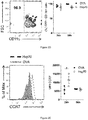
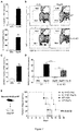
- the graft cells resulted from B6 animals were screened in the BALB/c animals lymph nodes, by two different approaches. Firstly, it was analyzed the presence of cells I-A b + in the recipient DLN, one and four days after the transplantation. These two moments were chosen since represent both migration moments of the skin cells to the DLN. It was not noted any difference between the mice that received grafts treated with OVA or Hsp70 ( Figure 2A ). In addition, it was verified that most of migratory cells were DCs ( Figure 2C ).
- the CCR7 expression is needed for DCs migration to the secondary lymphoid organs. Consequently, the CCR7 expression was evaluated in the donor cells, which were present in the recipient DLN, resulted from grafts treated with Hsp70 or OVA. Surprisingly, after 24 hours from the transplantation, it was noted that the Migratory DCs of grafts treated with Hsp70 expressed upper levels of CCR7 when compared with DCs of the grafts treated with OVA ( Figure 2E and 2F ).
- the CCR7 levels did not differ between Hsp70 and OVA groups.
- the data demonstrates that the Hsp70 does not influence the donor DCs migration to the recipient DLN, as showed in figures 2A , 2B , 2C , 2D , 2E and 2F .
- C, D Representative dot plots and CD11 c+ cells percentages of the donors screened in the recipient DLN.
- E CCR7 expression in the I-A b + or
- the Hsp70 reduced the MHC II expression and CD86 in activated dendritic cells
- BMDCs Mice bone marrow
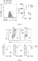
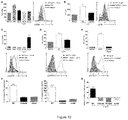
- DCs isolated from skin draining lymph nodes were treated with OVA or Hsp70 for 24h. After this period, the expression of I-A b in the DCs was analyzed by flow cytometry.
- B Percentages of CD11 c+ I-Ab hi , CD11c+I-Ab int and CD11 c+ I-Ab low cells.
- C Mean Fluorescence Intensity - MFI of I-A b in the CD11 c+ cells. Graphic expressed on average ⁇ SEM. *, p ⁇ 0.05. Experiments performed in triplicate. Representative of two independent experiments, being the results indicated in Figure 4 .
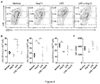
- CD86 expression in which the treatment with LPS led to a higher percentage of cells expressing high amounts of CD86 (CD11 c+ CD86 hi ), being the treatment with Hsp70 able to revert partially this expression ( Figure 5 ).
- DCs were different from the bone marrow of B6 WT mice and in the sixth day of culture were treated or not with LPS for 24h. After this period, these cells were treated or not with Hsp70 for additional 24h. Subsequently, the CD86 expression was analyzed by flow cytometry.
- A Cells strategy analysis according to CD86 expression ( high, intermediate or low.
- B Percentages of CD11 c+ CD86 hi , CD11c+CD86 int and CD11c+CD86 low cells.
- C Mean Fluorescence Intensity (MFI) of CD86 in the CD11 c+ cells. Graphic expressed on average ⁇ SEM. *, p ⁇ 0.05. Experiments performed in triplicate. Representative of two independent experiments, being the results indicated in Figure 5 .
- the recipient T cells recognize the donor MHC molecules presenting allopeptides, whereas in the indirect way, the T cells recognize the alloantigens presented in proper MHC molecules. Therefore, the simpler explanation for inhibiting the acute rejection achieved in our system may be because grafts DCs treated with Hsp70 are expressing low levels of MHC II and CD86, inhibiting the direct way of alloantigen recognition. For investigating this issue, levels of I-A b and of co-stimulating molecules in the donors' cells migrating to the recipient DLN were analyzed. In fact, the treatment with Hsp70 led to a MHC II expression reduction in the donors DCs.
- the rejection to grafts depends on the CD4+ and CD8+ alloreactive T cells proliferation, in the recipient DLN and probably a lower proliferation of these cells may be led to the lower number of the total of T cells present in the DLNs of animals that received grafts treated with Hsp70.
- the proliferation of these cells was analyzed by marking with Ki67 in the transplanted animals DLN. Ki67 is a molecule having a role in the cellular cycle and is expressed by any organism cell that is proliferating. It was noted that the graft treatment with Hsp70 leads to a reduction marked in the CD4+ and CD8+ T cells proliferation in the recipient mouse DLN ( Figure 7 , items B and C).
- Tregs An increase in the number of regulating T cells (Tregs) may reduce the alloreactive T cells proliferation in the recipient DLN, delaying the rejection to grafts. Due to the fact that the inventors' research group has noted previously that the acute rejection inhibition induced by the Hsp70 depends on Tregs, it was investigated if the graft treatment with Hsp70 leads to an increase of the Tregs in the Recipients DLN. Interestingly, it was not detected a significant increase of the Tregs in the recipient DLN after receiving a graft treated with Hsp70 ( Figure 7 , item D). Our results suggest that the MHC II modulation by the Hsp70 leads to the alloreactive proliferation reduction and not through Tregs. These findings are in accordance with the observation that immature DCs administration in human leads to an inhibition of the CD8+ T cells effector functions.
- mice B6 and sutured in BALB/c recipients were performed. In this case, grafts were not treated. Instead, B6 mice BMDCs, previously treated with Hsp70 or OVA for 24h, were injected by the subcutaneous way below the sutured graft ( Figure 8 , item A). The animals were monitored daily for grafts rejection verification. With that, it was attempted to mimic what was happening in the experiments previously showed.
- DCs CD103+ from donors have the main migratory subtype measuring the tolerance to grafts treated with Hsp70
- DCs subtypes may be found in the skin: Langerhans Cells (or LCs - Langerin+ CD103- CD11 b+) found in the epidermis; two additional subtypes comprising DCs CD103+ (Langerin+ CD103+ CD11 b-); and "classic" dermal DCs (Langerin- CD103- CD11 b+).
- These DCs subtypes in the skin have different stimulating abilities, causing the differential polarization of specific T cells for the same antigens.
- the LCs may induce Tregs in a homoeostatic state and inflammatory T cells in inflammation conditions, the DCs CD103+ cause the tolerance in the intestine.
- DCs CD141 hi from humans are equivalent to DCs CD103+ from mice.
- these cells may inhibit the graft disease against the host in a xenograft model, through IL-10 production.
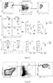
- DCs subtype was mediating the tolerance in the system used and, for such effect, the DCs subtypes that were migrating from the graft to the recipient DLN after the graft treatment with Hsp70 were analyzed. It was noted that DCs CD103+ was the only Migratory DCs subtype present in the DLN, either 24h or 96h after the transplantation ( Figure 9 , item A). In the analyzed periods, the LCs or "classic" dermal DCs migration was not detected. This observation is according to previous studies suggesting that LCs are not necessary for cutaneous grafts rejection with total incapability of MHC; instead, these cells would remain secured in the graft epidermis after the transplantation.
- the DCs CD103+ represent the main cells subtype that arrive in the recipient DLN and may be modulated by the Hsp70.
- A Analysis strategy for DCs subtypes present in the mice recipients DNL.
- B Representative graphs of the DCs CD103+ from donors (analyzed as in A) and percentage of DCs CD103+ expressing high and reduced levels of I-A b in the DLNs of the mice that received grafts treated with OVA or Hsp70.
- the cells phenotype migrating from the graft to the recipient DLN CD11 c+ Langerin+ CD11b- CD103+ ( Figure 10 ).
- the Migratory DCs from donors are CD11 c+ Langerin+ CD11 b-CD103+.
- the cytokine IL-10 is necessary for reducing the MHC II expression in the DCs and for acute rejection inhibition induced by Hsp70
- the IL-10 is the main anti-inflammatory cytokine and it is known that it may lead to the MHC II expression reduction. It was demonstrated that Hs70 may induce the IL-10 expression in different DCs from the bone marrow (BMDCs), as well as in arthritis models. Thus, this chaperone exerts its anti-inflammatory effects dependent on the IL-10. It was hypothesized that the IL-10 induction would be the mechanism addressing the MHC II expression reduction in the Stimulating DCs with Hsp70. In fact, the Hsp70 may induce the IL-10 production, either at mRNA level as protein level, in DCs isolated from mice lymph nodes mice ( Figure 11 , item A).
- MARCH-I transmembrane protein induction MARCH-I.
- MARCH-I is a member of the family E3 of ubiquitin ligase and proved to be able of regulating the expression of some molecules as the MHC II, MHC I, CD86, CD4 in the antigen-presenting cells (APCs).
- APCs antigen-presenting cells
- DCs were treated isolated from LNs of mice with Hsp70 or OVA and mRNA levels of the MARCH-I were analyzed through a Real-time PCR. It was detected in the DCs an increase of 20 times in the MARCH-I mRNA after the treatment with Hsp70 ( Figure 11 , item C). For testing if this increase was being triggered by the Hsp70-induced IL-10 production, the DCs isolated from knockout animals for IL-10 were treated with Hsp70 and subsequently the MARCH-I levels of mRNA were analyzed.
- the DCs IL-10 KO treated with Hsp70 did not present an increase in the MARCH-I levels, when compared with DCs WT ( Figure 11 , item D).
- the addition of recombinant murine IL-10 in the culture reestablished partially the MARCH-I expression by the DCs KO ( Figure 11 , item D).
- Hsp70-induced IL-10 leads to the MARCH-I expression increase and this is fundamental for reducing the MHC II expression.
- An interpretation of these findings was that the IL-10 would be required for mediating the rejection inhibition induced by the Hsp70.
- we performed cutaneous transplantations (as described above) using grafts resulted from donors IL-10 KO, treated with Hsp70 or OVA.
- the absence of IL-10 in the graft cancelled the protection mediated by the Hsp70 ( Figure 11 , item E).
- Figure 11 shows that the Hsp70-induced IL-10 production leads to the MARCH-I expression increase and this is fundamental for reducing the MHC II expression.
- A DCs isolated from LNs draining skin antigens were treated with OVA or Hsp70 for 24h. After that, IL-10 mRNA levels were analyzed by Real-time PCR. The ⁇ -actin was used as a setter. The IL-10 concentration was measured in the cultures supernatant by flow cytometry.
- B DCs isolated from LNs draining skin antigens ofWT animals or IL-10 KO were treated with OVA or Hsp70 for 24h and the expression of I-A b was analyzed by flow cytometry.
- the Hsp70 induces the MHC II reduction using the TLR2-ERK-STAT3-IL-10 way
- IL-10 is fundamental for protecting effect of the Hsp70 no graft
- the IL-10 expression regulation was recently associated to the TLR2 membrane recipient, besides inducing the tolerance in diabetes and arthritis.
- a hypothesis was created that the Hsp70-induced IL-10 expression is mediated by the TLR2.
- DCs isolated from lymph nodes of WT mice or knockouts for TLR2 (TLR2 KO) are thus treated with Hsp70 or OVA and the IL-10 expression was analyzed, either in mRNA levels as protein.
- DCs TLR2 KO treated with Hsp70 showed a significant reduction of the mRNA expression ( Figure 12 , item A) and of the IL-10 protein ( Figure 12 , item B), when compared with the DCs WT.
- Hsp70-induced MHC II expression inhibition was also dependent on TLR2 in DCs isolated from LNS ( Figure 12 , item C).
- FIG. 12 The TLR2 is necessary for the IL-10 production, MHC II expression reduction and acute rejection inhibition, all induced by Hsp70.
- A DCs isolated from LNs draining skin antigens of WT animals or TLR2 KO were treated with OVA or Hsp70 for 24h and IL-10 mRNA levels were analyzed by Real-time PCR. The ⁇ -actin was used as setter.
- B WT or TLR2 KO BMDCs were treated with OVA, Hsp70 or PGN for 24h and the IL-10 concentration was measured by flow cytometry from the supernatant.
- C Representative graphs and percentages of the CD11 c+ I-Ab hi , CD11c+I-Ab int and CD11c+I-Ab low cells of DCs isolated from LNs draining skin antigens of WT animals or TLR2 KO.
- D Cutaneous grafts of WT animals or TLR2 KO were immersed into a solution having 60 ⁇ g/ml of OVA or Hsp70 for 60 minutes, on ice. After this period, the treated grafts were sutured in the BALB/c animals' back and the acceptance was daily monitored.
- Hsp70 may activate the STAT3 in myeloid-derived suppressor cells - MDSCs, leading to an immunosuppression.
- the Hsp70 induced the phosphorylation STAT3 in the BMDCs, 45 minutes after the stimulus ( Figure 13 , item C). This phosphorylation was dependent on the TLR2 expression in the treated BMDCs ( Figure 13 , item D).
- FIG. 13 The Hsp70 activates the ERK-STAT3 via TLR2, leading to IL-10 production reduction and the MARCH-I expression increase.
- BMDCs were treated with PMA for 30 minutes; Hsp70 for 15, 30 or 45 minutes; or were left without stimulus. The p-ERK expression was measured by flow cytometry.
- B The MFI and representative histogram of the p-ERK expression in the BMDCs of mice WT or TLR2 KO that were treated with Hsp70 for 15 minutes or that were left without stimulus.
- C BMDCs were treated with PMA for 30 minutes; Hsp70 for 15, 30 or 45 minutes; or were left without stimulus. The p-STAT3 expression was measured by flow cytometry.
- FIG. 14 ERK and STAT3 are necessary for reducing the Hsp70-induced MHC II expression.
- DCs isolated from LNs draining skin antigens were treated with the ERK (PD98059) inhibitor or JAK2/STAT3 (AG490) for 60 minutes and then were stimulated with Hsp70 for 24h. After this period, these cells were analyzed for the expression of I-A b by flow cytometry.
- the current therapies for transplant recipient patients are characterized by a very strong immunosuppression, leading to severe problems following the transplantation as infections and tumors.
- an immunomodulation method based on the modulation/MHC II expression reduction in the DCs of the graft, which will migrate to the recipient DLN, duplicating the graft survival period without adding any other medication or immunosuppressing drug.
- Figure 15 shows that the Hsp70 provides this effect, inducing in the DCs the MARCH-I expression increase, and the subsequent reduction in the MHC II expression, addressed by the IL-10 production via TLR2-ERK-STAT3.
- the modulator as Hsp70, may bind directly to TLR2 of the DCs, or in an endocytic recipient as described in the state of the art, which may cooperate with TLR2. This binding triggers the ERK activation and the subsequent activation of STAT3, leading to the IL-10 production increase. This IL-10 will induce the MARCH-I expression increase and, thus, reducing the MHC II expression in the DCs membrane.
- Example 2 Process for screening new molecular entities useful for preparing the composition for ex-vivo preservation and/or immunomodulation of organs for transplantation.
- preservation compositions were initially considered having synthetic analogues of the Hsp70 as, for example, some peptides, and evaluated the binding potential thereof to the MHC II in the animals studied, such that to provide the same immunosuppressant effects.
- the experiments conceived by the inventors provide a process for screening the new molecular entities useful for preparing the composition for ex-vivo preservation and/or immunomodulation of organs for transplantation, the said process comprising:
- the said process is wherein the said cells having a protein selected from the group consisting of MARCH-I, MHC II, CD86, or combination thereof, are dendritic cells or cells of the organ to be transplanted itself.
- the said process is wherein the step of evaluating the MARCH-I, MHC II and/or CD86 amount in the cells is done by measuring by Real-time PCR of the mRNA transcribed from genes codifying MARCH-I, MHC II and/or CD86.
- the said process further comprises the step of evaluating in vitro the cell viability and/or the presence of cell damage in organs cells after contacting with a composition for organs preservation and/or immunomodulation, as better detailed in the examples 4 and 5.
- Hsp70 fragments For performing an investigation on the possibility of using Hsp70 fragments as modulating MARCH-I, MHC II and/or CD86, bioinformatics programs were used for drawing synthetic peptides from the Hsp70 protein sequence being subsequently synthetized by the company JPT Innovative Peptide Solutions (Berlin, Germany).
- the amino acids sequence of Mycobacterium tuberculosis Hsp70 (GI: 61222666) was entered into the IEDB Analysis Resource (http://tools.immuneepitope.org/analyze/html/mhc_II_binding.html), server that previews peptide sequences probability bound to MHC molecules II, providing scores for peptides according to the bonding probability.
- Scores smaller than 50 are considered of high affinity, the ones higher than 50 and smaller than 500 are considered of intermediate affinity and the ones higher than 500 and smaller than 5000 are considered of low affinity.
- Four peptides were then selected of high affinity, two with intermediate affinity and one with low affinity. Subsequently, a sequences line was performed between M. tuberculosis Hsp70 and murine Hsp70 (respectively, GI:61222666 and GI:3986773), determining the homology between the peptides.
- the Table 3 bellow indicates the peptides selected.
- the acute rejection inhibition by the Hsp70 is dependent on Tregs cells CD4+CD25+.
- the subcutaneous injection of Hsp70 increased the percentage of the Tregs in the DLN.
- the MARCH-I expression in the DCs is fundamental for developing thymic Tregs (tTregs).
- tuberculosis Hsp70 and mice Hsp70 determining the homology between peptides Table 4. Table 4.
- Peptide name Amino acids sequence Homology Peptide 1 DKMAMQRLREAAEKAK Semi-preserved Peptide 2 VGGSTRMPAVTDLVK Semi-preserved Peptide 3 KGGVMTRLIERNTTI Preserved Peptide 4 PRGIPQIEVTFDIDA Preserved Peptide 5 KYTAPEISARILMKL Not preserved Peptide 6 ITDAVITTPAYFNDA Semi-preserved Peptide 7 GSDISAIKSAMEKLG Not preserved
- the peptide 5 also provided an increase in the Tregs production ( Figure 16 , item A).
- Hsp70 also did not induce Tregs generation ( Figure 16 , item A), but in this case because LPS was added in the preparation as adjuvant. If there was no LPS in the system, the Hsp70 would induce these cells in these conditions.
- FIG 16 Epitopes mapping of T cells from fragments (peptides) of Hsp70.
- BALB/c mice were injected i.p. with a solution containing 100 ⁇ g of Hsp70 + 7 ⁇ g of LPS.
- the splenocytes were plated and re-stimulated in vitro with DMSO (used as negative control, since it was used to dilute peptides), 5 ⁇ g/ml of peptide E- ⁇ , 60 ⁇ g/ml of Hsp70 or 5 ⁇ g/ml of each one of the selected peptides, separately for 24h.
- (A) the cells were analyzed by flow cytometry for CD4 and CD25 expression or
- the concentration of IL-10 was determined from the supernatant.
- cutaneous transplantations were performed which B6 WT donors' grafts were treated by immersion with Hsp70, the peptide 2, the peptide 5 or an OVA peptide. After this, these grafts were sutured in BALB/c recipients WT and their rejection were daily monitored.
- Figure 17 shows the graft treatment with fragment (peptide) of Hsp70 lengthening the cutaneous allograft acceptance in a murine model.
- Example 3 Composition for preserving transplantation organs and for rejection inhibition thereof - increase of graft survival.
- the immersion treatment, followed by a topic treatment with Hsp70 increases the cutaneous graft survival
- each animal receives a topic application of 2 drops of Hsp70 or OVA solution until day 7, according to the graft treatment. That is, animals that received grafts treated with Hsp70 receive the local application of a Hsp70 solution and the same for OVA.
- FIG 18 A summary of the strategy used can be found in Figure 18 , item A. It can be noted that the graft survival was much bigger in animals treated with Hsp70, when compared to the animals treated with OVA ( Figure 18 , B and C and Table 6). In addition, this adopted strategy was a bit better than the unique treatment of graft with Hsp70 ( Figure 18 , item D).
- Table 6 Effect of the graft immersion-treatment with Hsp70 or OVA, followed by 7 days of a topic application of a solution with Hsp70 or OVA in cutaneous allograft murine model. Results achieved when combining the two independent experiments.
- Figure 18 shows the graft treatment with a dose of Hsp70 lengthening the cutaneous allograft acceptance in a murine model.
- Cutaneous grafts of wild (WT) animals B6 (I-A b ) were immersed into a solution having 60 ⁇ g/ml of Hsp70 or OVA for 60 minutes at 4°C. After this, the grafts were sutured in BALB/c (I-A d ) animals' back.
- the animals received a topic application of 2 drops of a solution having 60 ⁇ g/ml of Hsp70 or OVA for 7 days in a row. The animals were daily monitored.
- A Schematic representation of the treatment strategy used in the experiment.
- the treatment with Hsp70 is the one that best induces MARCH-I on dendritic cells. Therefore, the induction of MARCH-I on donor cells has demonstrated a good strategy to length the grafts acceptance.
- the MARCH-I induction was compared to the one by Hsp70 with two immunosuppressant well used in the clinic for rejection handling and that has been reported to act in dendritic cells modulating the antigen presentation: Cyclosporine A (CsA) and Rapamycin (RAPA). To do so, dendritic cells of lymph nodes were isolated, which were then treated with CsA, RAPA, Hsp70 or IL-10 (positive control) for 24 hours.
- CsA Cyclosporine A
- RAPA Rapamycin
- VERO cells were cultivated for 30h on ice, in contact with: 1) an EuroCollins solution; 2) an EuroCollins solution + 30 ⁇ g/ml of M. tuberculosis Hsp70; 3) an EuroCollins solution + 30 ⁇ g/ml of OVA; or 4) PBS 1x (negative control).
- PBS 1x negative control
- the presence of LDH in the supernatant was analyzed ( Figure 25 A) .
- the LDH released in comparison to the total LDH present in the cell was evaluated according to what is described in the following ( Figure 25 B) . In both cases, cells treated with PBS presented a great release of the LDH enzyme, indicating high cell damage.
- Cells of the renal tissue cell line VERO were cultivated in DMEM (Cultilab) medium supplemented with 10% of bovine fetal serum (FCS) (Cultilab), 1x of essential amino acids (Gibco), 1x vitamins (Gibco) and 55 ⁇ M of ⁇ -mercaptoethanol at 37°C with 5% of CO 2 .
- FCS bovine fetal serum
- Gibco essential amino acids
- Gibco 1x vitamins
- 55 ⁇ M of ⁇ -mercaptoethanol 55 ⁇ M of ⁇ -mercaptoethanol at 37°C with 5% of CO 2 .
- 5 x 10 4 cells per well in a 96 well-plate were plated, being incubated for 24h for cells to adhere. Subsequently, the cultivation medium was removed and the cells submitted to three different treatments: 1) an EuroCollins preservation solution; 2) EuroCollins + 30 ⁇ g/ml of M.
- tuberculosis Hsp70 3) EuroCollins + 30 ⁇ g/ml of OVA; or 4) PBS 1x (negative control).
- Cells were then cultivated for 30h, outside the greenhouse 37°C and in contact with ice, mimicking a cold storage.
- LDH Dehydrogenase Lactic
- Example 5 ex vivo organ preservation solutions: evaluation of cell viability
- the cell viability of treated VERO cells and cultivated for 30h on ice was monitored using LIVE/DEAD® Cell Viability (Life Technologies) kit, according to the manufacturer recommendations.
- the cells were evaluated in a Confocal Mycroscope (Zeiss).
- the cells were marked with the Live/Dead kit and analysis by microscopy. In this test, living cells are green and the dead ones red. Cells treated with PBS died much more when compared to other treatments, after 30h of static storage on ice. There was no significant difference in the measured viability proportions, between the EuroCollins group and the EuroCollins+Hsp70 group.
Landscapes
- Health & Medical Sciences (AREA)
- Life Sciences & Earth Sciences (AREA)
- Engineering & Computer Science (AREA)
- Immunology (AREA)
- General Health & Medical Sciences (AREA)
- Chemical & Material Sciences (AREA)
- Biomedical Technology (AREA)
- Cell Biology (AREA)
- Bioinformatics & Cheminformatics (AREA)
- Medicinal Chemistry (AREA)
- Hematology (AREA)
- Urology & Nephrology (AREA)
- Molecular Biology (AREA)
- Zoology (AREA)
- Pharmacology & Pharmacy (AREA)
- Animal Behavior & Ethology (AREA)
- Public Health (AREA)
- Veterinary Medicine (AREA)
- Wood Science & Technology (AREA)
- Proteomics, Peptides & Aminoacids (AREA)
- Epidemiology (AREA)
- Dentistry (AREA)
- Gastroenterology & Hepatology (AREA)
- Environmental Sciences (AREA)
- Biotechnology (AREA)
- Food Science & Technology (AREA)
- Physics & Mathematics (AREA)
- Analytical Chemistry (AREA)
- Biochemistry (AREA)
- General Physics & Mathematics (AREA)
- Pathology (AREA)
- Microbiology (AREA)
- Tropical Medicine & Parasitology (AREA)
- Toxicology (AREA)
- Organic Chemistry (AREA)
- Nuclear Medicine, Radiotherapy & Molecular Imaging (AREA)
- General Chemical & Material Sciences (AREA)
- Chemical Kinetics & Catalysis (AREA)
- Agricultural Chemicals And Associated Chemicals (AREA)
- Medicines That Contain Protein Lipid Enzymes And Other Medicines (AREA)
Applications Claiming Priority (2)
| Application Number | Priority Date | Filing Date | Title |
|---|---|---|---|
| BR102014002362A BR102014002362A8 (pt) | 2014-01-30 | 2014-01-30 | método de imunomodulação e/ou preservação de órgãos ex-vivo, composições, processo e usos |
| PCT/BR2014/000258 WO2015113126A1 (pt) | 2014-01-30 | 2014-07-30 | Composição e método para a imunomodulação e/ou preservação de órgãos ex-vivo, processos e uso |
Publications (2)
| Publication Number | Publication Date |
|---|---|
| EP3100740A1 true EP3100740A1 (de) | 2016-12-07 |
| EP3100740A4 EP3100740A4 (de) | 2017-11-15 |
Family
ID=53756051
Family Applications (1)
| Application Number | Title | Priority Date | Filing Date |
|---|---|---|---|
| EP14880740.7A Withdrawn EP3100740A4 (de) | 2014-01-30 | 2014-07-30 | Zusammensetzung und verfahren zur ex-vivo-immunmodulation und/oder konservierung von organen, verfahren und verwendung |
Country Status (5)
| Country | Link |
|---|---|
| US (1) | US20160295854A1 (de) |
| EP (1) | EP3100740A4 (de) |
| CN (1) | CN105939725A (de) |
| BR (1) | BR102014002362A8 (de) |
| WO (1) | WO2015113126A1 (de) |
Families Citing this family (1)
| Publication number | Priority date | Publication date | Assignee | Title |
|---|---|---|---|---|
| CN108559772B (zh) * | 2018-03-30 | 2020-07-17 | 青岛泱深生物医药有限公司 | 作为类风湿性关节炎和骨关节炎诊治标志物的march1基因 |
Family Cites Families (11)
| Publication number | Priority date | Publication date | Assignee | Title |
|---|---|---|---|---|
| US5348945A (en) | 1990-04-06 | 1994-09-20 | Wake Forest University | Method of treatment with hsp70 |
| US5599659A (en) * | 1993-03-11 | 1997-02-04 | Breonics, Inc. | Preservation solution for ex vivo, warm preservation of tissues, explants,organs and vascular endothelial cells comprising retinal-derived fibroblast growth factor, cyclodextrin and chondroitin sulfate |
| JPH07215801A (ja) * | 1994-01-28 | 1995-08-15 | Senju Pharmaceut Co Ltd | 肺保存剤および肺保存方法 |
| US20020111314A1 (en) | 1995-10-06 | 2002-08-15 | The Trustees Of Columbia University | Novel stimulation of gene expression and protein synthesis of heat shock protein 72/73 (Hsp 70) |
| CA2383213A1 (en) | 1999-09-10 | 2001-03-15 | Fordham University | Methods and compositions for the treatment and prevention of graft rejection using heat shock proteins |
| WO2005040398A2 (en) * | 2003-10-16 | 2005-05-06 | The Regents Of The University Of California | Methods for preserving nucleated mammalian cells |
| JP2007530420A (ja) | 2003-07-07 | 2007-11-01 | ザ・ジェネラル・ホスピタル・コーポレイション | fugetacticタンパク質、組成物および使用方法 |
| WO2005034980A1 (en) * | 2003-09-12 | 2005-04-21 | University Of Colorado | Glutamine for use in treating injury |
| US20090118173A1 (en) * | 2006-01-24 | 2009-05-07 | Universiteit Utrecht Uu Holding B.V. | Treatment and prevention of inflammatory bowel diseases |
| GB0612877D0 (en) | 2006-06-29 | 2006-08-09 | Univ Edinburgh | Organ preservation solution |
| CN101511872A (zh) | 2006-08-29 | 2009-08-19 | 为人技术株式会社 | 用于抑制凋亡的药物组合物及其递送方法 |
-
2014
- 2014-01-30 BR BR102014002362A patent/BR102014002362A8/pt not_active Application Discontinuation
- 2014-07-30 WO PCT/BR2014/000258 patent/WO2015113126A1/pt not_active Ceased
- 2014-07-30 US US15/030,982 patent/US20160295854A1/en not_active Abandoned
- 2014-07-30 EP EP14880740.7A patent/EP3100740A4/de not_active Withdrawn
- 2014-07-30 CN CN201480074696.0A patent/CN105939725A/zh active Pending
Also Published As
| Publication number | Publication date |
|---|---|
| BR102014002362A8 (pt) | 2018-04-17 |
| CN105939725A (zh) | 2016-09-14 |
| WO2015113126A1 (pt) | 2015-08-06 |
| BR102014002362A2 (pt) | 2016-01-26 |
| US20160295854A1 (en) | 2016-10-13 |
| EP3100740A4 (de) | 2017-11-15 |
Similar Documents
| Publication | Publication Date | Title |
|---|---|---|
| RU2709711C2 (ru) | Новые иммуногенные пептиды | |
| JP7320947B2 (ja) | 新規な免疫原性CD1d結合ペプチド | |
| Thornley et al. | Fragile TIM-4–expressing tissue resident macrophages are migratory and immunoregulatory | |
| Guilliams et al. | From skin dendritic cells to a simplified classification of human and mouse dendritic cell subsets | |
| Chen et al. | Direct expansion of human allospecific FoxP3+ CD4+ regulatory T cells with allogeneic B cells for therapeutic application | |
| de Vries et al. | Mast cells condition dendritic cells to mediate allograft tolerance | |
| KR20130119952A (ko) | 감염성 질환, 자가면역 질환, 동종인자에 대한 면역 반응, 알레르기 질환, 종양, 이식편 거부 및 유전자 요법 또는 유전자 백신접종을 위해 사용되는 바이러스 벡터에 대한 면역 반응의 예방 및/또는 치료에 사용하기 위한 면역원성 펩티드 | |
| US12152247B2 (en) | NF-KB signaling pathway-manipulated dendritic cells | |
| Carvalho et al. | Differential Recruitment of Dendritic Cells Subsets to Lymph Nodes Correlates with a Protective or Permissive T‐Cell Response during Leishmania (Viannia) Braziliensis or Leishmania (Leishmania) Amazonensis Infection | |
| James et al. | HY peptides modulate transplantation responses to skin allografts | |
| Camacho et al. | IRF4 expression by lung dendritic cells drives acute but not Trm cell–dependent memory Th2 responses | |
| Gebremeskel et al. | Reconstitution models to evaluate natural killer T cell function in tumor control | |
| JP2017516752A (ja) | 単離されたドナーmhc由来ペプチド及びその使用 | |
| US20170106022A1 (en) | Assays for determining the effect of an immune cell on a cell from an infectious or neoplastic disease | |
| EP3100740A1 (de) | Zusammensetzung und verfahren zur ex-vivo-immunmodulation und/oder konservierung von organen, verfahren und verwendung | |
| Tung et al. | Orchestration of CD4 T cell epitope preferences after multipeptide immunization | |
| Danilevskii et al. | Cell therapy for type 1 diabetes mellitus: a review of clinical trials | |
| de Lastic et al. | Effect of dendritic cell state and antigen-presentation conditions on resulting T-cell phenotypes and Th cytokine profiles | |
| US20240252644A1 (en) | Person-tailored t cell composition targeting merkel cell carcinoma | |
| Peters et al. | Immunotherapy with regulatory T cells in transplantation | |
| RU2597976C2 (ru) | Способ получения регуляторных дендритных клеток | |
| Marrero et al. | Autoreactivity to self H‐2Kb peptides in TAP1–/–mice. Intravenous administration of H‐2Kb class I‐derived peptides induces long‐term survival of grafts from C57BL/6 donors | |
| Wang et al. | Prolongation of rat renal allograft survival by CD4+ CD25‑T cells induced by recipient dendritic cells transfected with IKK2dn | |
| Baum | The Role of Interleukin-21 in the Chronic Rejection of Transplant Allografts and the Development of Type 1 Diabetes | |
| Buckland et al. | Prospects for the Induction of Transplant Tolerance Using Dendritic Cells |
Legal Events
| Date | Code | Title | Description |
|---|---|---|---|
| PUAI | Public reference made under article 153(3) epc to a published international application that has entered the european phase |
Free format text: ORIGINAL CODE: 0009012 |
|
| 17P | Request for examination filed |
Effective date: 20160829 |
|
| AK | Designated contracting states |
Kind code of ref document: A1 Designated state(s): AL AT BE BG CH CY CZ DE DK EE ES FI FR GB GR HR HU IE IS IT LI LT LU LV MC MK MT NL NO PL PT RO RS SE SI SK SM TR |
|
| AX | Request for extension of the european patent |
Extension state: BA ME |
|
| DAX | Request for extension of the european patent (deleted) | ||
| RIC1 | Information provided on ipc code assigned before grant |
Ipc: A61K 38/16 20060101AFI20170627BHEP Ipc: A61K 38/10 20060101ALI20170627BHEP Ipc: A61K 38/06 20060101ALI20170627BHEP Ipc: A01N 1/02 20060101ALI20170627BHEP |
|
| A4 | Supplementary search report drawn up and despatched |
Effective date: 20171013 |
|
| RIC1 | Information provided on ipc code assigned before grant |
Ipc: A61K 38/06 20060101ALI20171009BHEP Ipc: A61K 38/10 20060101ALI20171009BHEP Ipc: A61K 38/16 20060101AFI20171009BHEP Ipc: A01N 1/02 20060101ALI20171009BHEP |
|
| STAA | Information on the status of an ep patent application or granted ep patent |
Free format text: STATUS: THE APPLICATION IS DEEMED TO BE WITHDRAWN |
|
| 18D | Application deemed to be withdrawn |
Effective date: 20180515 |