EP3094239B1 - Method and system for generating pre-scaled images for a series of mammography images - Google Patents
Method and system for generating pre-scaled images for a series of mammography images Download PDFInfo
- Publication number
- EP3094239B1 EP3094239B1 EP15737601.3A EP15737601A EP3094239B1 EP 3094239 B1 EP3094239 B1 EP 3094239B1 EP 15737601 A EP15737601 A EP 15737601A EP 3094239 B1 EP3094239 B1 EP 3094239B1
- Authority
- EP
- European Patent Office
- Prior art keywords
- image
- overview
- mammography
- series
- images
- Prior art date
- Legal status (The legal status is an assumption and is not a legal conclusion. Google has not performed a legal analysis and makes no representation as to the accuracy of the status listed.)
- Active
Links
- 238000009607 mammography Methods 0.000 title claims description 104
- 238000000034 method Methods 0.000 title claims description 45
- 210000000481 breast Anatomy 0.000 claims description 163
- 238000009877 rendering Methods 0.000 claims description 17
- 238000010586 diagram Methods 0.000 description 12
- 238000012545 processing Methods 0.000 description 6
- 238000012552 review Methods 0.000 description 5
- 238000004590 computer program Methods 0.000 description 4
- 238000004091 panning Methods 0.000 description 4
- 230000005540 biological transmission Effects 0.000 description 3
- 238000004422 calculation algorithm Methods 0.000 description 3
- 238000004891 communication Methods 0.000 description 2
- 230000006870 function Effects 0.000 description 2
- 238000012216 screening Methods 0.000 description 2
- 230000005856 abnormality Effects 0.000 description 1
- 238000013459 approach Methods 0.000 description 1
- 238000004364 calculation method Methods 0.000 description 1
- 230000001413 cellular effect Effects 0.000 description 1
- 238000013170 computed tomography imaging Methods 0.000 description 1
- 230000001186 cumulative effect Effects 0.000 description 1
- 238000013500 data storage Methods 0.000 description 1
- 238000002059 diagnostic imaging Methods 0.000 description 1
- 229940079593 drug Drugs 0.000 description 1
- 239000003814 drug Substances 0.000 description 1
- 230000000694 effects Effects 0.000 description 1
- 238000010191 image analysis Methods 0.000 description 1
- 238000003384 imaging method Methods 0.000 description 1
- 230000002085 persistent effect Effects 0.000 description 1
- 238000003325 tomography Methods 0.000 description 1
- 230000000007 visual effect Effects 0.000 description 1
Images


Classifications
-
- A—HUMAN NECESSITIES
- A61—MEDICAL OR VETERINARY SCIENCE; HYGIENE
- A61B—DIAGNOSIS; SURGERY; IDENTIFICATION
- A61B6/00—Apparatus for radiation diagnosis, e.g. combined with radiation therapy equipment
- A61B6/50—Clinical applications
- A61B6/502—Clinical applications involving diagnosis of breast, i.e. mammography
-
- A—HUMAN NECESSITIES
- A61—MEDICAL OR VETERINARY SCIENCE; HYGIENE
- A61B—DIAGNOSIS; SURGERY; IDENTIFICATION
- A61B6/00—Apparatus for radiation diagnosis, e.g. combined with radiation therapy equipment
- A61B6/46—Apparatus for radiation diagnosis, e.g. combined with radiation therapy equipment with special arrangements for interfacing with the operator or the patient
- A61B6/461—Displaying means of special interest
- A61B6/463—Displaying means of special interest characterised by displaying multiple images or images and diagnostic data on one display
-
- A—HUMAN NECESSITIES
- A61—MEDICAL OR VETERINARY SCIENCE; HYGIENE
- A61B—DIAGNOSIS; SURGERY; IDENTIFICATION
- A61B6/00—Apparatus for radiation diagnosis, e.g. combined with radiation therapy equipment
- A61B6/46—Apparatus for radiation diagnosis, e.g. combined with radiation therapy equipment with special arrangements for interfacing with the operator or the patient
- A61B6/461—Displaying means of special interest
- A61B6/466—Displaying means of special interest adapted to display 3D data
-
- A—HUMAN NECESSITIES
- A61—MEDICAL OR VETERINARY SCIENCE; HYGIENE
- A61B—DIAGNOSIS; SURGERY; IDENTIFICATION
- A61B6/00—Apparatus for radiation diagnosis, e.g. combined with radiation therapy equipment
- A61B6/52—Devices using data or image processing specially adapted for radiation diagnosis
- A61B6/5211—Devices using data or image processing specially adapted for radiation diagnosis involving processing of medical diagnostic data
- A61B6/5229—Devices using data or image processing specially adapted for radiation diagnosis involving processing of medical diagnostic data combining image data of a patient, e.g. combining a functional image with an anatomical image
- A61B6/5235—Devices using data or image processing specially adapted for radiation diagnosis involving processing of medical diagnostic data combining image data of a patient, e.g. combining a functional image with an anatomical image combining images from the same or different ionising radiation imaging techniques, e.g. PET and CT
-
- G—PHYSICS
- G06—COMPUTING; CALCULATING OR COUNTING
- G06T—IMAGE DATA PROCESSING OR GENERATION, IN GENERAL
- G06T3/00—Geometric image transformation in the plane of the image
- G06T3/40—Scaling the whole image or part thereof
-
- A—HUMAN NECESSITIES
- A61—MEDICAL OR VETERINARY SCIENCE; HYGIENE
- A61B—DIAGNOSIS; SURGERY; IDENTIFICATION
- A61B6/00—Apparatus for radiation diagnosis, e.g. combined with radiation therapy equipment
- A61B6/02—Devices for diagnosis sequentially in different planes; Stereoscopic radiation diagnosis
- A61B6/025—Tomosynthesis
-
- A—HUMAN NECESSITIES
- A61—MEDICAL OR VETERINARY SCIENCE; HYGIENE
- A61B—DIAGNOSIS; SURGERY; IDENTIFICATION
- A61B6/00—Apparatus for radiation diagnosis, e.g. combined with radiation therapy equipment
- A61B6/02—Devices for diagnosis sequentially in different planes; Stereoscopic radiation diagnosis
- A61B6/03—Computerised tomographs
- A61B6/032—Transmission computed tomography [CT]
-
- A—HUMAN NECESSITIES
- A61—MEDICAL OR VETERINARY SCIENCE; HYGIENE
- A61B—DIAGNOSIS; SURGERY; IDENTIFICATION
- A61B6/00—Apparatus for radiation diagnosis, e.g. combined with radiation therapy equipment
- A61B6/48—Diagnostic techniques
- A61B6/486—Diagnostic techniques involving generating temporal series of image data
-
- G—PHYSICS
- G06—COMPUTING; CALCULATING OR COUNTING
- G06T—IMAGE DATA PROCESSING OR GENERATION, IN GENERAL
- G06T2207/00—Indexing scheme for image analysis or image enhancement
- G06T2207/10—Image acquisition modality
- G06T2207/10016—Video; Image sequence
-
- G—PHYSICS
- G06—COMPUTING; CALCULATING OR COUNTING
- G06T—IMAGE DATA PROCESSING OR GENERATION, IN GENERAL
- G06T2207/00—Indexing scheme for image analysis or image enhancement
- G06T2207/10—Image acquisition modality
- G06T2207/10116—X-ray image
-
- G—PHYSICS
- G06—COMPUTING; CALCULATING OR COUNTING
- G06T—IMAGE DATA PROCESSING OR GENERATION, IN GENERAL
- G06T2207/00—Indexing scheme for image analysis or image enhancement
- G06T2207/30—Subject of image; Context of image processing
- G06T2207/30004—Biomedical image processing
- G06T2207/30068—Mammography; Breast
Definitions
- the embodiments described herein relate to a system and method for image analysis and more particularly a system and method for generating a pre-scaled image for at least one mammography image of an image series.
- Medical personnel e.g. radiologists
- medical personnel examine mammography images to diagnose various abnormalities in a breast.
- medical personnel desire to review the mammography image displaying a breast (i.e. the breast image), with little regard to any of the background.
- medical personnel are primarily interested in the breast area, it is highly desirable to extract this breast area and display it at an optimal and consistent scale across all displayed images. That is, medical personnel seek mammography systems that facilitate quickly generated diagnostic reviews of mammography images, and allow for convenient navigation and review between views of a medical imaging data set while maintaining the same presentation state, including for example the scale factor, across all of the images in the views.
- Medical personnel have typically used the zooming and panning functionality of a mammography system. Specifically, once the user has positioned the mouse on a part of the mammography image that the user wants to focus on, using the zooming and panning functionality of the mammography system. Doing so for each successive image is time-consuming and inconvenient.
- a breast window for a breast image is considered to be the smallest region that encloses all of the tissue points of a breast shown on mammography image.
- a breast window may be characterized by the size of the region as well as the location of the region within the mammography image.
- the mammography system may also define a breast window that contains an optimal part of a breast image and displays it optimally scaled (herein referred to as a "pre-scaled image”) such that the defined breast window fills the entirety of the viewing area of the mammography system.
- pre-scaled image optimally scaled
- a plurality of mammography images from a single modality may be considered to be an image series (also referred to as a series of mammography images).
- Newer modalities such as x-ray tomosynthesis or computed tomography imaging modalities, produce "volume-based" mammography images, which for example, may comprise a plurality of two-dimensional images stacked in series.
- Volume-based images are generated by first, processing traditional two-dimensional images to render additional images which may show the breast at different angles. These images are then used to generate the volume-based images in an image series.
- Medical personnel may view all images of an image series together by generating an overview image, or by obtaining an overview image from a modality. Wherein the overview image shows all or a subset of, tissue points from each image in the image series on a single two-dimensional image.
- EP2372649 which represents the closest prior art, discloses a computer-implemented method for defining a breast window within an image for a breast.
- a method for generating a pre-scaled image for at least one mammography image of a series the series containing a plurality of mammography images, each mammography image displaying a breast having tissue points.
- the method comprising: obtaining an overview breast window, wherein the overview breast window encloses all tissue points; adjusting, if required, the overview breast window, for example in size, position, orientation or alignment, and storing it as an image breast window, applying the image breast window to apportion each image in the series, creating a breast window image; and rendering the pre-scaled image based on the breast window image for each image in the image series.
- the system comprising: a database memory for storing the overview breast window; and, a processor coupled to the database memory and configured to obtain an overview breast window, wherein the overview breast window encloses all tissue points, adjusting the overview breast window, for example in size, position, orientation or alignment, and storing the adjusted overview breast window as an image breast window, applying the image breast window to apportion each image in the series to create a breast window image, and rendering the pre-scaled image based on the breast window image.
- the embodiments of the systems and methods described herein may be implemented in hardware or software, or a combination of both. However, preferably, these embodiments are implemented in computer programs executing on programmable computers each comprising at least one module component which comprises at least one processor (e.g. a microprocessor), a data storage system (including volatile and nonvolatile memory and/or storage elements), at least one input device, and at least one output device.
- the programmable computers (referred to below as computing devices) may be a personal computer, laptop, personal data assistant, and cellular telephone, smart-phone device, tablet computer, and/or wireless device.
- Program code is applied to input data to perform the functions described herein and generate output information.
- the output information is applied to one or more output devices, in known fashion.
- Each program is preferably implemented in a high level procedural or object oriented programming and/or scripting language to communicate with a computer system.
- the programs can be implemented in assembly or machine language, if desired. In any case, the language may be a compiled or interpreted language.
- Each such computer program is preferably stored on a storage media or a device (e.g. ROM or magnetic diskette) readable by a general or special purpose programmable computer, for configuring and operating the computer when the storage media or device is read by the computer to perform the procedures described herein.
- the subject system may also be considered to be implemented as a computer-readable storage medium, configured with a computer program, where the storage medium so configured causes a computer to operate in a specific and predefined manner to perform the functions described herein.
- system, processes and methods of the described embodiments are capable of being distributed in a computer program product comprising a computer readable medium that bears computer usable instructions for one or more processors.
- the medium may be provided in various forms, including one or more diskettes, compact disks, tapes, chips, wireline transmissions, satellite transmissions, internet transmission or downloadings, magnetic and electronic storage media, digital and analog signals, and the like.
- the computer useable instructions may also be in various forms, including compiled and non-compiled code.
- an embodiment means “one or more (but not all) embodiments of the present invention(s),” unless expressly specified otherwise.
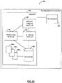
- Mammography system 100 may contain a processor 102 that is operatively coupled to memory 104.
- Memory 104 may store a breast window module 120 for defining an overview breast window 380 or image breast window 385 n applied on a plurality of images 300 1 to 300 n (where 300 1 represents a first image and 300 n represents an n th image and 385 n represents the image breast window corresponding to the n th image), a rendering module 110 for rendering new volume based image series from an existing image series or a new overview image 350 from an existing image series (e.g.
- the storage module 130 may also store other data such as for example, an overview image 350.
- Images 300 1 to 300 n stored on image database 140 may be mammography images.
- images 300 1 to 300 n may be images obtained directly from a modality, overview images, or rendered images (e.g. multi-planar reformatted images), including volume-based images.
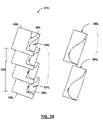
- FIG. 1B therein illustrated is a schematic diagram of the volume-based pre-scaled images 395 1 to 395 n for an image series 310 which contains a plurality of images 300 1 to 300 n (where 300 1 represents a first image and 300 n represents an n th image, and 395 1 represents a pre-scaled image for the first mammography image 300 1 , and 395 n represents a pre-scaled image for the n th mammography image 300 n ).
- a breast window image 390 1...n is rendered for each corresponding image 300 1...n of the image series 310.
- the volume-based image has a depth 330, which is the distance between the first image 300 1 and the last image 300 n when the images are in spatial order.
- Images 300 1 to 300 n may be processed to generate another plurality of images 300 1 to 300 m showing, for example, the breast at different angles with different slice thicknesses, etc.
- FIG. 1C therein illustrated is the general flow of data used for generating a pre-scaled image 395 1...n for at least one mammography image, 300 1...n , of an image series 310.
- Images series 310 may be obtained directly from a modality or generated from an existing series.
- Overview image 350 is generated from or obtained for the image series 310, and represents all or a subset of, images contained in the series.
- An overview breast window 380 is defined for the overview image 350 as a portion of the overview image, for example a rectangular shape, that contains all, or a subset of, the projected tissue points inside the overview image 350.
- the overview breast window 380 is then used to create the image breast windows 385 n .
- This process may include adjustments, for example, for differences in size, position, alignment or orientation, for each image represented in the overview image.
- Each breast window image 390 n is the image resulting from cropping each image represented in the overview image by the boundary defined by the image breast window 385 n .
- the breast window images 390 1...n are then used to generate pre-scaled images 395 1...n for each image, 300 1...n , in image series 310.
- the pre-scaled image is automatically adjusted, for example, by scaling, panning, flipping, or rotating, to consistently render each image in an image series.
- breast window module 120 may be configured to receive notifications that one of a plurality of images, 300 1...n , is ready for breast window definition. For each image 300 1...n used to create the overview image 350, the system generates a corresponding image breast windows 385 1...n , breast window images 390 1...n , and pre-scaled images 395 1...n .
- the breast window module 120 may define an overview breast window 380 around an overview image 350.
- An overview breast window 380 is considered to be the smallest region (for example, a rectangle in one example embodiment) that encloses all, or a subset of, the tissue points of a breast displayed on a mammography image.
- the breast window module 120 may send notifications after it has defined an overview breast window 380 or image breast window 385 n .
- an overview breast window may be defined by the size of the region enclosing all or some of the tissue points or by the location coordinates of the region within the image.
- Rendering module 110 may be configured to receive notifications to render a pre-scaled image 395 n .
- the rendering module 110 may render a pre-scaled image 395 n from the breast window image 390 n for each image in the image series 310.
- the pre-scaled image 395 n is rendered then mapped to the entirety of the viewing area in a consistent manner across all the viewing areas.
- the system may elect to render a subset of pre-scaled images.
- Rendering module 110 may also receive notification to render an overview image 350 ( FIG. 3B ).
- the overview image 350 may be rendered based on the images of an image series 310.
- An overview image 350 contains overview tissue points 370 and an overview breast boundary 360 that runs along the border of the overview tissue points 370.
- Each overview tissue point 370 within the overview image 350 has corresponding tissue point (e.g. 320 1 and 320 2 in FIGS. 3C and 3D ) in the mammography images within a given image series.
- the rendering module 110 may send notifications after it has rendered a pre-scaled image 395 n or an overview image 350.
- Storage module 130 may be configured to receive notifications to store objects. When storage module 130 receives notification that the overview breast window 380 is defined, storage module 130 may store the overview breast window 380, or the image breast window 385 n , in the database 140.
- Storage module 130 may also store the overview breast window 380 in the database 140 as the image breast window for each image in the series.
- Database 140 may store a plurality of images 300 1 to 300 n and breast windows, for example, an overview breast window 380 or image breast windows 385 n , and may be implemented using any database software or persistent storage method known in the art.
- database 140 may be implemented using Oracle ® , Microsoft SQL Server ® or IBM DB2 ® with suitably defined schemas to identify and navigate images.
- database 140 may be part of a Picture Archiving and Communication Systems (PACS) deployment, such as those found in a hospital.
- PACS Picture Archiving and Communication Systems
- database 140 is illustrated as residing in the same memory as mammography system 100, it will be understood that database 140 may be stored and accessed remotely through a network connection, for example, using a Digital Imaging and Communications in Medicine (DICOM) protocol. In such case, it will be further understood that the operations of the rendering module 110, breast window module 120, and storage module 130 may be performed locally on the mammography system, remotely on the system where the database 140 resides, or on a third-party system configured to access database 140 and mammography system 100.
- DICOM Digital Imaging and Communications in Medicine
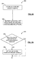
- FIGS. 1A , 1B , 1C , 2A and FIGS. 3A to FIG. 3E illustrate steps of a method 200 for generating a pre-scaled image 395 n for at least one mammography image 300 n of an image series 310.
- the pre-scaled image may be generated based on images obtained from two dimensional images, or volume-based images.
- the image series 310 is identified and an overview breast window 380 associated with the image series 310 is obtained.
- the rendering module 110 at step (220) may render the pre-scaled image 390 n using the overview breast window 380.
- FIGS. 1A , 1B , 1C , 2B and FIGS. 3A to FIG. 3D illustrate steps of a method 210 for obtaining an overview breast window 380.
- the overview breast window 380 may be located and retrieved from the database memory 140. If not, then at step (240) the breast window module 120 may define an overview breast window 380 and the storage module 130 may store the overview breast window 380 in the database memory. The overview breast window 380 may have been defined in the process of viewing another image of the same series.
- FIGS. 1A , 1B , 1C , 2C and FIGS. 3A to FIG. 3D illustrate steps of a method 240 for defining and storing an overview breast window 380, and defining an image breast window 385 n and breast window image 390 n .
- an overview image 350 associated with the image series 310 may initially be obtained.
- the breast window module 120 may define an overview breast window 380 at step (260) for the overview image 350.
- the overview breast window 380 may be stored in the database memory 140.
- the overview breast window 380 may be stored in the database memory 140 as the breast window associated with each mammography image in the image series 310.
- an image breast window 385 1...n may be defined for each image in the image series.
- a breast window image 390 1...n may be defined for each image in the series.
- FIGS. 1A , 1B , 1C , 2D and FIGS. 3A to FIG. 3D illustrate steps of a method 250 for obtaining an overview image 350.
- the overview image 350 may be located and retrieved from the database memory 140.
- rendering module 110 at step (290) may render an overview image 350.
- the overview image 350 is rendered based on the images series 310.
- Rendering may be performed using any known method for rendering an overview image for a given plurality of mammography images (e.g. a slabbing technique).
- the rendering module 110 may send notification to the breast window module 120 that an overview image 350 is ready for breast window definition at step (260).
- the storage module 130 may store the overview image 350 in the image database 140.
- FIG. 1A , 1B , 1C , 2E and FIGS. 3A to FIG. 3D which illustrates the steps of method 271 for defining and storing an image breast window 390 1...n for each image, 300 1...n , in an image series 310.
- the breast window module 120 checks if overview breast window requires adjustment, for example, to scale or position, and it is adjusted at step (263) if adjustment is required. Once adjusted, the image breast window 385 n is derived based on the adjusted values of the overview breast window 380.
- the definition of the overview breast window 380 for the overview image 350 may be performed using any known method for defining a breast window for a breast image. If no adjustment is required, the values of the overview breast window are inherited to derive the image breast window 385 n at step (262).
- the image breast window 385 is stored for each image in an image series 310.
- the breast window module 120 may send the overview breast window 380 to the storage module 130.
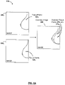
- FIG. 3A illustrates a schematic diagram of an exemplary series of mammography images 310, where the image series 310 comprise mammography images 300 1 and 300 2 stored on image database 140.
- Each of the mammography images contains tissue points 320 1 and 320 2 .
- Rendering module 110 (at step (290) of FIG. 2D ) generates an overview image 350 based on mammography images 300 1 and 300 2 wherein each mammography image tissue point 320 1 and 320 2 of mammography images 300 1 and 300 2 is represented by an overview tissue points 370 1 and 370 2 in the overview image 350.
- the overview image 350 may be stored on image database 140.
- FIG. 3B which illustrates a schematic diagram of an overview image 350 stored on image database 140.
- the overview image 350 comprises an overview tissue point 370 and an overview breast boundary 360 formed along the border of the overview tissue point.
- the breast window module 120 (at step (260) of FIG. 2C ) defines an overview breast window 380 by identifying the smallest region (e.g. a rectangle in one embodiment) that encloses all (as it is defined by an algorithm used) of the overview tissue point 370.
- the smallest region e.g. a rectangle in one embodiment
- FIG. 3E which illustrates a schematic diagram of a pre-scaled image 395 n , for an image 300 n from image series 310.
- the image 300 n is stored on image database 140 along with the corresponding image breast window 385 n .
- the breast window module 120 (at step (260) of FIG. 2C ) defines the image breast window 385 n by adjusting, if necessary, the overview breast window 380.
- FIG. 3D which illustrates a schematic diagram of the image breast window 385 1 for the mammography images 300 1 and 300 2 of FIG. 3A .
- the storage module 130 (at step (270) of FIG. 2C ) stores the overview breast window 380 for the images series 310.
- the overview breast window 380 is an approximation of the smallest region that encloses all of the tissue points 320 1 and 320 2 on the images 300 1 and 300 2 .
- the breast window definition had been performed using the individual mammography image 300 1 instead of the overview image 350, the smallest region (e.g. rectangle in one embodiment) that encloses all of the tissue points 320 1 in mammography image 300 1 would be identified by the region 381 1 of FIG. 3C .
- the breast window definition had been performed on the individual mammography image 300 2 instead of the overview image 350, the smallest region that encloses all the tissue points 320 2 in mammography image 300 2 would be identified by the region 381 2 of FIG. 3D .
- the system would perform image processing on every image 300 1 to 300 n in order to determine the breast window 381 1 and subsequently allow pre-scaled view image 300 2 with the breast window 381 2 .
- the method disclosed in the present invention of generating a pre-scaled image is not limited to two-dimensional images and may be used to generate a pre-scaled image as well.
Applications Claiming Priority (2)
| Application Number | Priority Date | Filing Date | Title |
|---|---|---|---|
| US14/155,851 US9474497B2 (en) | 2014-01-15 | 2014-01-15 | Method and system for generating pre-scaled images for a series of mammography images |
| PCT/CA2015/000021 WO2015106339A1 (en) | 2014-01-15 | 2015-01-14 | Method and system for generating pre-scaled images for a series of mammography images |
Publications (3)
| Publication Number | Publication Date |
|---|---|
| EP3094239A1 EP3094239A1 (en) | 2016-11-23 |
| EP3094239A4 EP3094239A4 (en) | 2017-08-16 |
| EP3094239B1 true EP3094239B1 (en) | 2022-06-29 |
Family
ID=53521808
Family Applications (1)
| Application Number | Title | Priority Date | Filing Date |
|---|---|---|---|
| EP15737601.3A Active EP3094239B1 (en) | 2014-01-15 | 2015-01-14 | Method and system for generating pre-scaled images for a series of mammography images |
Country Status (6)
| Country | Link |
|---|---|
| US (1) | US9474497B2 (zh) |
| EP (1) | EP3094239B1 (zh) |
| CN (1) | CN105916435B (zh) |
| BR (1) | BR112016016529B1 (zh) |
| ES (1) | ES2922182T3 (zh) |
| WO (1) | WO2015106339A1 (zh) |
Families Citing this family (5)
| Publication number | Priority date | Publication date | Assignee | Title |
|---|---|---|---|---|
| JP6126058B2 (ja) | 2014-09-30 | 2017-05-10 | 富士フイルム株式会社 | 画像表示装置、画像処理装置、放射線画像撮影システム、断層画像表示方法、及び断層画像表示プログラム。 |
| CN106846248A (zh) * | 2017-01-19 | 2017-06-13 | 商丘市第人民医院 | 一种针对乳房x线图像序列生成预缩放图像的方法和系统 |
| US10372876B2 (en) * | 2017-01-20 | 2019-08-06 | Agfa Healthcare Inc. | System and method for providing breast image data |
| EP3675770A1 (en) | 2017-08-31 | 2020-07-08 | 3Shape A/S | Volume rendering using surface guided cropping |
| JP7203474B2 (ja) * | 2019-02-20 | 2023-01-13 | 富士フイルム株式会社 | マンモグラフィ装置及びプログラム |
Family Cites Families (15)
| Publication number | Priority date | Publication date | Assignee | Title |
|---|---|---|---|---|
| BR9812021A (pt) * | 1997-08-28 | 2000-09-26 | Qualia Computing Inc | Processos para detecção automatizada de microcalcificações, para segmentar uma área de uma imagem de mamograma digital, para detecção automatizada de microcalcifição agrupada, para incorporar as detecções de saìda de um sistema de detecção auxiliado por computador para detectar microcalcificações agrupadas em um mamograma, e, para coleta automatizada de imagem, aparelhos para detecção automatizada de microcalcificações, e, para detectar microcalcificações agrupadas em uma imagem de mamograma digital |
| US7129961B1 (en) * | 2000-09-18 | 2006-10-31 | Sony Corporation | System and method for dynamic autocropping of images |
| US7577282B2 (en) | 2002-11-27 | 2009-08-18 | Hologic, Inc. | Image handling and display in X-ray mammography and tomosynthesis |
| US7142633B2 (en) | 2004-03-31 | 2006-11-28 | General Electric Company | Enhanced X-ray imaging system and method |
| US7599542B2 (en) | 2005-04-08 | 2009-10-06 | John Philip Brockway | System and method for detection and display of diseases and abnormalities using confidence imaging |
| US7885443B2 (en) | 2005-11-14 | 2011-02-08 | Hologic, Inc. | Facilitating temporal comparison of medical images |
| US8044972B2 (en) | 2006-12-21 | 2011-10-25 | Sectra Mamea Ab | Synchronized viewing of tomosynthesis and/or mammograms |
| US7992100B2 (en) | 2006-12-21 | 2011-08-02 | Sectra Ab | Dynamic slabbing to render views of medical image data |
| US8051386B2 (en) | 2006-12-21 | 2011-11-01 | Sectra Ab | CAD-based navigation of views of medical image data stacks or volumes |
| EP2143067B1 (en) * | 2007-05-04 | 2019-08-21 | Koninklijke Philips N.V. | Automatic display of symmetric anatomical structure |
| US8634622B2 (en) * | 2008-10-16 | 2014-01-21 | Icad, Inc. | Computer-aided detection of regions of interest in tomographic breast imagery |
| WO2011044295A2 (en) | 2009-10-07 | 2011-04-14 | Hologic, Inc. | Processing and displaying computer-aided detection information associated with breast x-ray images |
| US8649578B2 (en) * | 2010-03-08 | 2014-02-11 | Agfa Healthcare Inc. | Method and system for defining a breast window |
| JP6077993B2 (ja) | 2010-04-30 | 2017-02-08 | アイキャド インクiCAD, INC. | 画像の異形を識別するための画像データの処理方法、システムおよびプログラム |
| EP2629263B1 (en) * | 2012-02-17 | 2015-06-03 | Agfa HealthCare | Method for defining a region of interest in a radiation image of a breast |
-
2014
- 2014-01-15 US US14/155,851 patent/US9474497B2/en active Active
-
2015
- 2015-01-14 WO PCT/CA2015/000021 patent/WO2015106339A1/en active Application Filing
- 2015-01-14 CN CN201580004734.XA patent/CN105916435B/zh not_active Expired - Fee Related
- 2015-01-14 BR BR112016016529-2A patent/BR112016016529B1/pt active IP Right Grant
- 2015-01-14 ES ES15737601T patent/ES2922182T3/es active Active
- 2015-01-14 EP EP15737601.3A patent/EP3094239B1/en active Active
Also Published As
| Publication number | Publication date |
|---|---|
| CN105916435A (zh) | 2016-08-31 |
| EP3094239A4 (en) | 2017-08-16 |
| WO2015106339A1 (en) | 2015-07-23 |
| BR112016016529A2 (zh) | 2017-08-08 |
| US9474497B2 (en) | 2016-10-25 |
| ES2922182T3 (es) | 2022-09-09 |
| CN105916435B (zh) | 2019-09-13 |
| US20150199790A1 (en) | 2015-07-16 |
| EP3094239A1 (en) | 2016-11-23 |
| BR112016016529B1 (pt) | 2022-12-06 |
Similar Documents
| Publication | Publication Date | Title |
|---|---|---|
| JP7317077B2 (ja) | トモシンセシス画像スラブを生成し表示するためのシステムおよび方法 | |
| US11361479B2 (en) | Enhancements for displaying and viewing tomosynthesis images | |
| US10275130B2 (en) | Facilitating transitioning between viewing native 2D and reconstructed 3D medical images | |
| US9020235B2 (en) | Systems and methods for viewing and analyzing anatomical structures | |
| WO2020212762A2 (en) | Methods and systems for syncing medical images across one or more networks and devices | |
| EP3094239B1 (en) | Method and system for generating pre-scaled images for a series of mammography images | |
| US9390548B2 (en) | Three-dimensional volume rendering using an in-memory database | |
| US7620229B2 (en) | Method and apparatus for aiding image interpretation and computer-readable recording medium storing program therefor | |
| EP4170673A1 (en) | Auto-focus tool for multimodality image review | |
| US9514575B2 (en) | Image and annotation display | |
| JP6360495B2 (ja) | トモシンセシスにおけるデータ伝送ボリュームを低減するための方法 | |
| US20160078615A1 (en) | Visualization of Anatomical Labels | |
| US10032296B2 (en) | Volumertric image data visualization | |
| US8803911B2 (en) | User interface and viewing workflow for mammography workstation | |
| JP6114266B2 (ja) | 画像をズームするシステム及び方法 | |
| EP3314582A1 (en) | Interactive mesh editing | |
| EP1835428A2 (en) | An application server for processing medical image data | |
| KR20130135800A (ko) | 의료기기 및 이를 이용한 영상표시방법 | |
| KR101855734B1 (ko) | 의료기기 및 이를 이용한 영상표시방법 | |
| JP2020534536A (ja) | 高解像度画像生成及び処理のためのリアルタイム再構成ネイティブ画像要素リサンプリング | |
| Bolan | 3D tools advance enterprise wide |
Legal Events
| Date | Code | Title | Description |
|---|---|---|---|
| PUAI | Public reference made under article 153(3) epc to a published international application that has entered the european phase |
Free format text: ORIGINAL CODE: 0009012 |
|
| 17P | Request for examination filed |
Effective date: 20160816 |
|
| AK | Designated contracting states |
Kind code of ref document: A1 Designated state(s): AL AT BE BG CH CY CZ DE DK EE ES FI FR GB GR HR HU IE IS IT LI LT LU LV MC MK MT NL NO PL PT RO RS SE SI SK SM TR |
|
| AX | Request for extension of the european patent |
Extension state: BA ME |
|
| RAP1 | Party data changed (applicant data changed or rights of an application transferred) |
Owner name: AGFA HEALTHCARE INC. |
|
| DAX | Request for extension of the european patent (deleted) | ||
| A4 | Supplementary search report drawn up and despatched |
Effective date: 20170714 |
|
| RIC1 | Information provided on ipc code assigned before grant |
Ipc: A61B 6/02 20060101ALN20170710BHEP Ipc: A61B 6/00 20060101ALI20170710BHEP Ipc: G06T 3/40 20060101AFI20170710BHEP Ipc: A61B 6/03 20060101ALN20170710BHEP |
|
| STAA | Information on the status of an ep patent application or granted ep patent |
Free format text: STATUS: EXAMINATION IS IN PROGRESS |
|
| STAA | Information on the status of an ep patent application or granted ep patent |
Free format text: STATUS: EXAMINATION IS IN PROGRESS |
|
| 17Q | First examination report despatched |
Effective date: 20210113 |
|
| RAP1 | Party data changed (applicant data changed or rights of an application transferred) |
Owner name: AGFA HEALTHCARE INC. |
|
| STAA | Information on the status of an ep patent application or granted ep patent |
Free format text: STATUS: EXAMINATION IS IN PROGRESS |
|
| REG | Reference to a national code |
Ref country code: DE Ref legal event code: R079 Ref document number: 602015079652 Country of ref document: DE Free format text: PREVIOUS MAIN CLASS: A61B0005000000 Ipc: G06T0003400000 |
|
| GRAP | Despatch of communication of intention to grant a patent |
Free format text: ORIGINAL CODE: EPIDOSNIGR1 |
|
| STAA | Information on the status of an ep patent application or granted ep patent |
Free format text: STATUS: GRANT OF PATENT IS INTENDED |
|
| RIC1 | Information provided on ipc code assigned before grant |
Ipc: A61B 6/03 20060101ALN20220324BHEP Ipc: A61B 6/02 20060101ALN20220324BHEP Ipc: A61B 6/00 20060101ALI20220324BHEP Ipc: G06T 3/40 20060101AFI20220324BHEP |
|
| INTG | Intention to grant announced |
Effective date: 20220412 |
|
| GRAS | Grant fee paid |
Free format text: ORIGINAL CODE: EPIDOSNIGR3 |
|
| GRAA | (expected) grant |
Free format text: ORIGINAL CODE: 0009210 |
|
| STAA | Information on the status of an ep patent application or granted ep patent |
Free format text: STATUS: THE PATENT HAS BEEN GRANTED |
|
| AK | Designated contracting states |
Kind code of ref document: B1 Designated state(s): AL AT BE BG CH CY CZ DE DK EE ES FI FR GB GR HR HU IE IS IT LI LT LU LV MC MK MT NL NO PL PT RO RS SE SI SK SM TR |
|
| REG | Reference to a national code |
Ref country code: GB Ref legal event code: FG4D |
|
| REG | Reference to a national code |
Ref country code: CH Ref legal event code: EP |
|
| REG | Reference to a national code |
Ref country code: AT Ref legal event code: REF Ref document number: 1501829 Country of ref document: AT Kind code of ref document: T Effective date: 20220715 |
|
| REG | Reference to a national code |
Ref country code: IE Ref legal event code: FG4D |
|
| REG | Reference to a national code |
Ref country code: DE Ref legal event code: R096 Ref document number: 602015079652 Country of ref document: DE |
|
| REG | Reference to a national code |
Ref country code: ES Ref legal event code: FG2A Ref document number: 2922182 Country of ref document: ES Kind code of ref document: T3 Effective date: 20220909 |
|
| REG | Reference to a national code |
Ref country code: NL Ref legal event code: FP |
|
| REG | Reference to a national code |
Ref country code: LT Ref legal event code: MG9D |
|
| REG | Reference to a national code |
Ref country code: SE Ref legal event code: TRGR |
|
| PG25 | Lapsed in a contracting state [announced via postgrant information from national office to epo] |
Ref country code: NO Free format text: LAPSE BECAUSE OF FAILURE TO SUBMIT A TRANSLATION OF THE DESCRIPTION OR TO PAY THE FEE WITHIN THE PRESCRIBED TIME-LIMIT Effective date: 20220929 Ref country code: LT Free format text: LAPSE BECAUSE OF FAILURE TO SUBMIT A TRANSLATION OF THE DESCRIPTION OR TO PAY THE FEE WITHIN THE PRESCRIBED TIME-LIMIT Effective date: 20220629 Ref country code: HR Free format text: LAPSE BECAUSE OF FAILURE TO SUBMIT A TRANSLATION OF THE DESCRIPTION OR TO PAY THE FEE WITHIN THE PRESCRIBED TIME-LIMIT Effective date: 20220629 Ref country code: GR Free format text: LAPSE BECAUSE OF FAILURE TO SUBMIT A TRANSLATION OF THE DESCRIPTION OR TO PAY THE FEE WITHIN THE PRESCRIBED TIME-LIMIT Effective date: 20220930 Ref country code: FI Free format text: LAPSE BECAUSE OF FAILURE TO SUBMIT A TRANSLATION OF THE DESCRIPTION OR TO PAY THE FEE WITHIN THE PRESCRIBED TIME-LIMIT Effective date: 20220629 Ref country code: BG Free format text: LAPSE BECAUSE OF FAILURE TO SUBMIT A TRANSLATION OF THE DESCRIPTION OR TO PAY THE FEE WITHIN THE PRESCRIBED TIME-LIMIT Effective date: 20220929 |
|
| REG | Reference to a national code |
Ref country code: AT Ref legal event code: MK05 Ref document number: 1501829 Country of ref document: AT Kind code of ref document: T Effective date: 20220629 |
|
| PG25 | Lapsed in a contracting state [announced via postgrant information from national office to epo] |
Ref country code: RS Free format text: LAPSE BECAUSE OF FAILURE TO SUBMIT A TRANSLATION OF THE DESCRIPTION OR TO PAY THE FEE WITHIN THE PRESCRIBED TIME-LIMIT Effective date: 20220629 Ref country code: LV Free format text: LAPSE BECAUSE OF FAILURE TO SUBMIT A TRANSLATION OF THE DESCRIPTION OR TO PAY THE FEE WITHIN THE PRESCRIBED TIME-LIMIT Effective date: 20220629 |
|
| PG25 | Lapsed in a contracting state [announced via postgrant information from national office to epo] |
Ref country code: SM Free format text: LAPSE BECAUSE OF FAILURE TO SUBMIT A TRANSLATION OF THE DESCRIPTION OR TO PAY THE FEE WITHIN THE PRESCRIBED TIME-LIMIT Effective date: 20220629 Ref country code: SK Free format text: LAPSE BECAUSE OF FAILURE TO SUBMIT A TRANSLATION OF THE DESCRIPTION OR TO PAY THE FEE WITHIN THE PRESCRIBED TIME-LIMIT Effective date: 20220629 Ref country code: RO Free format text: LAPSE BECAUSE OF FAILURE TO SUBMIT A TRANSLATION OF THE DESCRIPTION OR TO PAY THE FEE WITHIN THE PRESCRIBED TIME-LIMIT Effective date: 20220629 Ref country code: PT Free format text: LAPSE BECAUSE OF FAILURE TO SUBMIT A TRANSLATION OF THE DESCRIPTION OR TO PAY THE FEE WITHIN THE PRESCRIBED TIME-LIMIT Effective date: 20221031 Ref country code: EE Free format text: LAPSE BECAUSE OF FAILURE TO SUBMIT A TRANSLATION OF THE DESCRIPTION OR TO PAY THE FEE WITHIN THE PRESCRIBED TIME-LIMIT Effective date: 20220629 Ref country code: AT Free format text: LAPSE BECAUSE OF FAILURE TO SUBMIT A TRANSLATION OF THE DESCRIPTION OR TO PAY THE FEE WITHIN THE PRESCRIBED TIME-LIMIT Effective date: 20220629 |
|
| PG25 | Lapsed in a contracting state [announced via postgrant information from national office to epo] |
Ref country code: PL Free format text: LAPSE BECAUSE OF FAILURE TO SUBMIT A TRANSLATION OF THE DESCRIPTION OR TO PAY THE FEE WITHIN THE PRESCRIBED TIME-LIMIT Effective date: 20220629 Ref country code: IS Free format text: LAPSE BECAUSE OF FAILURE TO SUBMIT A TRANSLATION OF THE DESCRIPTION OR TO PAY THE FEE WITHIN THE PRESCRIBED TIME-LIMIT Effective date: 20221029 |
|
| REG | Reference to a national code |
Ref country code: DE Ref legal event code: R097 Ref document number: 602015079652 Country of ref document: DE |
|
| PG25 | Lapsed in a contracting state [announced via postgrant information from national office to epo] |
Ref country code: AL Free format text: LAPSE BECAUSE OF FAILURE TO SUBMIT A TRANSLATION OF THE DESCRIPTION OR TO PAY THE FEE WITHIN THE PRESCRIBED TIME-LIMIT Effective date: 20220629 |
|
| PG25 | Lapsed in a contracting state [announced via postgrant information from national office to epo] |
Ref country code: DK Free format text: LAPSE BECAUSE OF FAILURE TO SUBMIT A TRANSLATION OF THE DESCRIPTION OR TO PAY THE FEE WITHIN THE PRESCRIBED TIME-LIMIT Effective date: 20220629 Ref country code: CZ Free format text: LAPSE BECAUSE OF FAILURE TO SUBMIT A TRANSLATION OF THE DESCRIPTION OR TO PAY THE FEE WITHIN THE PRESCRIBED TIME-LIMIT Effective date: 20220629 |
|
| PGFP | Annual fee paid to national office [announced via postgrant information from national office to epo] |
Ref country code: FR Payment date: 20230117 Year of fee payment: 9 Ref country code: ES Payment date: 20230210 Year of fee payment: 9 |
|
| PLBE | No opposition filed within time limit |
Free format text: ORIGINAL CODE: 0009261 |
|
| STAA | Information on the status of an ep patent application or granted ep patent |
Free format text: STATUS: NO OPPOSITION FILED WITHIN TIME LIMIT |
|
| PGFP | Annual fee paid to national office [announced via postgrant information from national office to epo] |
Ref country code: SE Payment date: 20230116 Year of fee payment: 9 Ref country code: IT Payment date: 20230131 Year of fee payment: 9 Ref country code: GB Payment date: 20230117 Year of fee payment: 9 Ref country code: BE Payment date: 20230117 Year of fee payment: 9 |
|
| 26N | No opposition filed |
Effective date: 20230330 |
|
| REG | Reference to a national code |
Ref country code: DE Ref legal event code: R119 Ref document number: 602015079652 Country of ref document: DE |
|
| PG25 | Lapsed in a contracting state [announced via postgrant information from national office to epo] |
Ref country code: SI Free format text: LAPSE BECAUSE OF FAILURE TO SUBMIT A TRANSLATION OF THE DESCRIPTION OR TO PAY THE FEE WITHIN THE PRESCRIBED TIME-LIMIT Effective date: 20220629 |
|
| REG | Reference to a national code |
Ref country code: CH Ref legal event code: PL |
|
| PG25 | Lapsed in a contracting state [announced via postgrant information from national office to epo] |
Ref country code: LU Free format text: LAPSE BECAUSE OF NON-PAYMENT OF DUE FEES Effective date: 20230114 |
|
| PG25 | Lapsed in a contracting state [announced via postgrant information from national office to epo] |
Ref country code: LI Free format text: LAPSE BECAUSE OF NON-PAYMENT OF DUE FEES Effective date: 20230131 Ref country code: DE Free format text: LAPSE BECAUSE OF NON-PAYMENT OF DUE FEES Effective date: 20230801 Ref country code: CH Free format text: LAPSE BECAUSE OF NON-PAYMENT OF DUE FEES Effective date: 20230131 |
|
| PG25 | Lapsed in a contracting state [announced via postgrant information from national office to epo] |
Ref country code: IE Free format text: LAPSE BECAUSE OF NON-PAYMENT OF DUE FEES Effective date: 20230114 |
|
| PGFP | Annual fee paid to national office [announced via postgrant information from national office to epo] |
Ref country code: NL Payment date: 20240110 Year of fee payment: 10 |
|
| PGFP | Annual fee paid to national office [announced via postgrant information from national office to epo] |
Ref country code: ES Payment date: 20240209 Year of fee payment: 10 |
|
| PGFP | Annual fee paid to national office [announced via postgrant information from national office to epo] |
Ref country code: GB Payment date: 20240103 Year of fee payment: 10 |