EP3021859B1 - Oncolytic adenoviruses armed with heterologous genes - Google Patents
Oncolytic adenoviruses armed with heterologous genes Download PDFInfo
- Publication number
- EP3021859B1 EP3021859B1 EP14799117.8A EP14799117A EP3021859B1 EP 3021859 B1 EP3021859 B1 EP 3021859B1 EP 14799117 A EP14799117 A EP 14799117A EP 3021859 B1 EP3021859 B1 EP 3021859B1
- Authority
- EP
- European Patent Office
- Prior art keywords
- seq
- antibody
- virus
- sequence
- cells
- Prior art date
- Legal status (The legal status is an assumption and is not a legal conclusion. Google has not performed a legal analysis and makes no representation as to the accuracy of the status listed.)
- Active
Links
- 108090000623 proteins and genes Proteins 0.000 title claims description 242
- 241000701161 unidentified adenovirus Species 0.000 title claims description 133
- 230000000174 oncolytic effect Effects 0.000 title claims description 45
- 241000700605 Viruses Species 0.000 claims description 429
- 108700019146 Transgenes Proteins 0.000 claims description 339
- 102000004169 proteins and genes Human genes 0.000 claims description 139
- 108090000765 processed proteins & peptides Proteins 0.000 claims description 119
- 230000027455 binding Effects 0.000 claims description 83
- 108091028043 Nucleic acid sequence Proteins 0.000 claims description 67
- 239000000203 mixture Substances 0.000 claims description 59
- 239000012634 fragment Substances 0.000 claims description 58
- 238000011282 treatment Methods 0.000 claims description 50
- 108020004999 messenger RNA Proteins 0.000 claims description 45
- 230000001225 therapeutic effect Effects 0.000 claims description 45
- 230000010076 replication Effects 0.000 claims description 43
- -1 ICOS Proteins 0.000 claims description 30
- 102100024216 Programmed cell death 1 ligand 1 Human genes 0.000 claims description 29
- 108010074708 B7-H1 Antigen Proteins 0.000 claims description 28
- 229940045513 CTLA4 antagonist Drugs 0.000 claims description 23
- 102000004127 Cytokines Human genes 0.000 claims description 20
- 108090000695 Cytokines Proteins 0.000 claims description 20
- 102000004190 Enzymes Human genes 0.000 claims description 20
- 108090000790 Enzymes Proteins 0.000 claims description 20
- 108060001084 Luciferase Proteins 0.000 claims description 20
- 239000005089 Luciferase Substances 0.000 claims description 19
- 102000005962 receptors Human genes 0.000 claims description 17
- 108020003175 receptors Proteins 0.000 claims description 17
- 230000008488 polyadenylation Effects 0.000 claims description 15
- 108700008625 Reporter Genes Proteins 0.000 claims description 14
- 108010021064 CTLA-4 Antigen Proteins 0.000 claims description 13
- 102100039498 Cytotoxic T-lymphocyte protein 4 Human genes 0.000 claims description 13
- 101710089372 Programmed cell death protein 1 Proteins 0.000 claims description 12
- 108060008682 Tumor Necrosis Factor Proteins 0.000 claims description 12
- 102000000852 Tumor Necrosis Factor-alpha Human genes 0.000 claims description 12
- 230000009368 gene silencing by RNA Effects 0.000 claims description 12
- 108010012236 Chemokines Proteins 0.000 claims description 9
- 102000019034 Chemokines Human genes 0.000 claims description 9
- 108010002350 Interleukin-2 Proteins 0.000 claims description 9
- 102000000588 Interleukin-2 Human genes 0.000 claims description 9
- 239000000463 material Substances 0.000 claims description 9
- FWMNVWWHGCHHJJ-SKKKGAJSSA-N 4-amino-1-[(2r)-6-amino-2-[[(2r)-2-[[(2r)-2-[[(2r)-2-amino-3-phenylpropanoyl]amino]-3-phenylpropanoyl]amino]-4-methylpentanoyl]amino]hexanoyl]piperidine-4-carboxylic acid Chemical compound C([C@H](C(=O)N[C@H](CC(C)C)C(=O)N[C@H](CCCCN)C(=O)N1CCC(N)(CC1)C(O)=O)NC(=O)[C@H](N)CC=1C=CC=CC=1)C1=CC=CC=C1 FWMNVWWHGCHHJJ-SKKKGAJSSA-N 0.000 claims description 8
- 102100030703 Interleukin-22 Human genes 0.000 claims description 8
- 102100024213 Programmed cell death 1 ligand 2 Human genes 0.000 claims description 8
- 108010029697 CD40 Ligand Proteins 0.000 claims description 7
- 101150013553 CD40 gene Proteins 0.000 claims description 7
- 102100032937 CD40 ligand Human genes 0.000 claims description 7
- 102100040245 Tumor necrosis factor receptor superfamily member 5 Human genes 0.000 claims description 7
- 102100031650 C-X-C chemokine receptor type 4 Human genes 0.000 claims description 6
- 101000922348 Homo sapiens C-X-C chemokine receptor type 4 Proteins 0.000 claims description 6
- 108010065805 Interleukin-12 Proteins 0.000 claims description 6
- 102000013462 Interleukin-12 Human genes 0.000 claims description 6
- 101100407308 Mus musculus Pdcd1lg2 gene Proteins 0.000 claims description 6
- 108700030875 Programmed Cell Death 1 Ligand 2 Proteins 0.000 claims description 6
- 102100020886 Sodium/iodide cotransporter Human genes 0.000 claims description 6
- 238000003776 cleavage reaction Methods 0.000 claims description 6
- 108010013351 sodium-iodide symporter Proteins 0.000 claims description 6
- 102100026238 Lymphotoxin-alpha Human genes 0.000 claims description 5
- 108090001012 Transforming Growth Factor beta Proteins 0.000 claims description 5
- 102000004887 Transforming Growth Factor beta Human genes 0.000 claims description 5
- 239000002955 immunomodulating agent Substances 0.000 claims description 5
- 229940121354 immunomodulator Drugs 0.000 claims description 5
- 102100031151 C-C chemokine receptor type 2 Human genes 0.000 claims description 4
- 101710149815 C-C chemokine receptor type 2 Proteins 0.000 claims description 4
- 101710149863 C-C chemokine receptor type 4 Proteins 0.000 claims description 4
- 102100036842 C-C motif chemokine 19 Human genes 0.000 claims description 4
- 102100021943 C-C motif chemokine 2 Human genes 0.000 claims description 4
- 102100036846 C-C motif chemokine 21 Human genes 0.000 claims description 4
- 102100032367 C-C motif chemokine 5 Human genes 0.000 claims description 4
- 102100028989 C-X-C chemokine receptor type 2 Human genes 0.000 claims description 4
- 102100036170 C-X-C motif chemokine 9 Human genes 0.000 claims description 4
- 101000713106 Homo sapiens C-C motif chemokine 19 Proteins 0.000 claims description 4
- 101000897480 Homo sapiens C-C motif chemokine 2 Proteins 0.000 claims description 4
- 101000713085 Homo sapiens C-C motif chemokine 21 Proteins 0.000 claims description 4
- 101000797762 Homo sapiens C-C motif chemokine 5 Proteins 0.000 claims description 4
- 101000947172 Homo sapiens C-X-C motif chemokine 9 Proteins 0.000 claims description 4
- 101000853002 Homo sapiens Interleukin-25 Proteins 0.000 claims description 4
- 101000984189 Homo sapiens Leukocyte immunoglobulin-like receptor subfamily B member 2 Proteins 0.000 claims description 4
- 101000984186 Homo sapiens Leukocyte immunoglobulin-like receptor subfamily B member 4 Proteins 0.000 claims description 4
- 101001128431 Homo sapiens Myeloid-derived growth factor Proteins 0.000 claims description 4
- 101000617130 Homo sapiens Stromal cell-derived factor 1 Proteins 0.000 claims description 4
- 101000851370 Homo sapiens Tumor necrosis factor receptor superfamily member 9 Proteins 0.000 claims description 4
- 108090000172 Interleukin-15 Proteins 0.000 claims description 4
- 102000003812 Interleukin-15 Human genes 0.000 claims description 4
- 102000003810 Interleukin-18 Human genes 0.000 claims description 4
- 108090000171 Interleukin-18 Proteins 0.000 claims description 4
- 108010065637 Interleukin-23 Proteins 0.000 claims description 4
- 108010002586 Interleukin-7 Proteins 0.000 claims description 4
- 102100021592 Interleukin-7 Human genes 0.000 claims description 4
- 108010018951 Interleukin-8B Receptors Proteins 0.000 claims description 4
- 102100025584 Leukocyte immunoglobulin-like receptor subfamily B member 1 Human genes 0.000 claims description 4
- 102100025583 Leukocyte immunoglobulin-like receptor subfamily B member 2 Human genes 0.000 claims description 4
- 102100025578 Leukocyte immunoglobulin-like receptor subfamily B member 4 Human genes 0.000 claims description 4
- 108090000542 Lymphotoxin-alpha Proteins 0.000 claims description 4
- 102100021669 Stromal cell-derived factor 1 Human genes 0.000 claims description 4
- 102100036856 Tumor necrosis factor receptor superfamily member 9 Human genes 0.000 claims description 4
- 230000002584 immunomodulator Effects 0.000 claims description 4
- 108010074108 interleukin-21 Proteins 0.000 claims description 4
- 108010074109 interleukin-22 Proteins 0.000 claims description 4
- 230000003834 intracellular effect Effects 0.000 claims description 4
- 102100036301 C-C chemokine receptor type 7 Human genes 0.000 claims description 3
- 108010017213 Granulocyte-Macrophage Colony-Stimulating Factor Proteins 0.000 claims description 3
- 102100039620 Granulocyte-macrophage colony-stimulating factor Human genes 0.000 claims description 3
- 101000716065 Homo sapiens C-C chemokine receptor type 7 Proteins 0.000 claims description 3
- 108090000978 Interleukin-4 Proteins 0.000 claims description 3
- 102000004388 Interleukin-4 Human genes 0.000 claims description 3
- 102100029822 B- and T-lymphocyte attenuator Human genes 0.000 claims description 2
- 102100035875 C-C chemokine receptor type 5 Human genes 0.000 claims description 2
- 101710149870 C-C chemokine receptor type 5 Proteins 0.000 claims description 2
- 102100036305 C-C chemokine receptor type 8 Human genes 0.000 claims description 2
- 102100025074 C-C chemokine receptor-like 2 Human genes 0.000 claims description 2
- 102100036848 C-C motif chemokine 20 Human genes 0.000 claims description 2
- 102100028990 C-X-C chemokine receptor type 3 Human genes 0.000 claims description 2
- 102100031658 C-X-C chemokine receptor type 5 Human genes 0.000 claims description 2
- 102100025248 C-X-C motif chemokine 10 Human genes 0.000 claims description 2
- 102100025279 C-X-C motif chemokine 11 Human genes 0.000 claims description 2
- 102100025277 C-X-C motif chemokine 13 Human genes 0.000 claims description 2
- 102100024263 CD160 antigen Human genes 0.000 claims description 2
- 102100027207 CD27 antigen Human genes 0.000 claims description 2
- 102100038078 CD276 antigen Human genes 0.000 claims description 2
- 101710185679 CD276 antigen Proteins 0.000 claims description 2
- 102100025221 CD70 antigen Human genes 0.000 claims description 2
- 102100034458 Hepatitis A virus cellular receptor 2 Human genes 0.000 claims description 2
- 101710083479 Hepatitis A virus cellular receptor 2 homolog Proteins 0.000 claims description 2
- 101000864344 Homo sapiens B- and T-lymphocyte attenuator Proteins 0.000 claims description 2
- 101000716068 Homo sapiens C-C chemokine receptor type 6 Proteins 0.000 claims description 2
- 101000716063 Homo sapiens C-C chemokine receptor type 8 Proteins 0.000 claims description 2
- 101000713099 Homo sapiens C-C motif chemokine 20 Proteins 0.000 claims description 2
- 101000916050 Homo sapiens C-X-C chemokine receptor type 3 Proteins 0.000 claims description 2
- 101000922405 Homo sapiens C-X-C chemokine receptor type 5 Proteins 0.000 claims description 2
- 101000858088 Homo sapiens C-X-C motif chemokine 10 Proteins 0.000 claims description 2
- 101000858060 Homo sapiens C-X-C motif chemokine 11 Proteins 0.000 claims description 2
- 101000858064 Homo sapiens C-X-C motif chemokine 13 Proteins 0.000 claims description 2
- 101000761938 Homo sapiens CD160 antigen Proteins 0.000 claims description 2
- 101000914511 Homo sapiens CD27 antigen Proteins 0.000 claims description 2
- 101000934356 Homo sapiens CD70 antigen Proteins 0.000 claims description 2
- 101000853000 Homo sapiens Interleukin-26 Proteins 0.000 claims description 2
- 101000984190 Homo sapiens Leukocyte immunoglobulin-like receptor subfamily B member 1 Proteins 0.000 claims description 2
- 101000914514 Homo sapiens T-cell-specific surface glycoprotein CD28 Proteins 0.000 claims description 2
- 101000801234 Homo sapiens Tumor necrosis factor receptor superfamily member 18 Proteins 0.000 claims description 2
- 101000851376 Homo sapiens Tumor necrosis factor receptor superfamily member 8 Proteins 0.000 claims description 2
- 101000666896 Homo sapiens V-type immunoglobulin domain-containing suppressor of T-cell activation Proteins 0.000 claims description 2
- 102100034980 ICOS ligand Human genes 0.000 claims description 2
- 101710093458 ICOS ligand Proteins 0.000 claims description 2
- 102000003814 Interleukin-10 Human genes 0.000 claims description 2
- 108090000174 Interleukin-10 Proteins 0.000 claims description 2
- 108090000176 Interleukin-13 Proteins 0.000 claims description 2
- 108050003558 Interleukin-17 Proteins 0.000 claims description 2
- 102000013691 Interleukin-17 Human genes 0.000 claims description 2
- 108010066979 Interleukin-27 Proteins 0.000 claims description 2
- 108010067003 Interleukin-33 Proteins 0.000 claims description 2
- 108010002616 Interleukin-5 Proteins 0.000 claims description 2
- 108090001005 Interleukin-6 Proteins 0.000 claims description 2
- 108090001007 Interleukin-8 Proteins 0.000 claims description 2
- 108010002335 Interleukin-9 Proteins 0.000 claims description 2
- 102000017578 LAG3 Human genes 0.000 claims description 2
- 101150030213 Lag3 gene Proteins 0.000 claims description 2
- 108010061593 Member 14 Tumor Necrosis Factor Receptors Proteins 0.000 claims description 2
- 102000010750 Metalloproteins Human genes 0.000 claims description 2
- 108010063312 Metalloproteins Proteins 0.000 claims description 2
- 108010042215 OX40 Ligand Proteins 0.000 claims description 2
- 108050000258 Prostaglandin D receptors Proteins 0.000 claims description 2
- 102100024218 Prostaglandin D2 receptor 2 Human genes 0.000 claims description 2
- 229940126547 T-cell immunoglobulin mucin-3 Drugs 0.000 claims description 2
- 102100027213 T-cell-specific surface glycoprotein CD28 Human genes 0.000 claims description 2
- 102100026890 Tumor necrosis factor ligand superfamily member 4 Human genes 0.000 claims description 2
- 102100028785 Tumor necrosis factor receptor superfamily member 14 Human genes 0.000 claims description 2
- 102100033728 Tumor necrosis factor receptor superfamily member 18 Human genes 0.000 claims description 2
- 102100022153 Tumor necrosis factor receptor superfamily member 4 Human genes 0.000 claims description 2
- 101710165473 Tumor necrosis factor receptor superfamily member 4 Proteins 0.000 claims description 2
- 102100036857 Tumor necrosis factor receptor superfamily member 8 Human genes 0.000 claims description 2
- 108010079206 V-Set Domain-Containing T-Cell Activation Inhibitor 1 Proteins 0.000 claims description 2
- 102100038929 V-set domain-containing T-cell activation inhibitor 1 Human genes 0.000 claims description 2
- 102100038282 V-type immunoglobulin domain-containing suppressor of T-cell activation Human genes 0.000 claims description 2
- 239000000262 estrogen Substances 0.000 claims description 2
- IJJVMEJXYNJXOJ-UHFFFAOYSA-N fluquinconazole Chemical compound C=1C=C(Cl)C=C(Cl)C=1N1C(=O)C2=CC(F)=CC=C2N=C1N1C=NC=N1 IJJVMEJXYNJXOJ-UHFFFAOYSA-N 0.000 claims description 2
- 108090000237 interleukin-24 Proteins 0.000 claims description 2
- 102100023990 60S ribosomal protein L17 Human genes 0.000 claims 2
- 102100037853 C-C chemokine receptor type 4 Human genes 0.000 claims 1
- 108091030071 RNAI Proteins 0.000 claims 1
- 210000004027 cell Anatomy 0.000 description 367
- 229950002830 enadenotucirev Drugs 0.000 description 245
- 206010028980 Neoplasm Diseases 0.000 description 175
- 230000014509 gene expression Effects 0.000 description 148
- 238000000034 method Methods 0.000 description 91
- 239000006228 supernatant Substances 0.000 description 83
- 239000013612 plasmid Substances 0.000 description 80
- 108020004414 DNA Proteins 0.000 description 72
- 208000015181 infectious disease Diseases 0.000 description 72
- 230000029812 viral genome replication Effects 0.000 description 68
- 238000002965 ELISA Methods 0.000 description 66
- 238000011529 RT qPCR Methods 0.000 description 65
- 102000004196 processed proteins & peptides Human genes 0.000 description 65
- 238000004519 manufacturing process Methods 0.000 description 58
- 239000002245 particle Substances 0.000 description 58
- 206010009944 Colon cancer Diseases 0.000 description 54
- 150000001413 amino acids Chemical class 0.000 description 52
- 229920001184 polypeptide Polymers 0.000 description 46
- 241000699670 Mus sp. Species 0.000 description 44
- 108091032973 (ribonucleotides)n+m Proteins 0.000 description 41
- 229920001213 Polysorbate 20 Polymers 0.000 description 41
- 238000009472 formulation Methods 0.000 description 41
- 230000006870 function Effects 0.000 description 41
- 239000000256 polyoxyethylene sorbitan monolaurate Substances 0.000 description 41
- 235000010486 polyoxyethylene sorbitan monolaurate Nutrition 0.000 description 41
- 201000011510 cancer Diseases 0.000 description 39
- 102000005789 Vascular Endothelial Growth Factors Human genes 0.000 description 36
- 108010019530 Vascular Endothelial Growth Factors Proteins 0.000 description 36
- 239000000523 sample Substances 0.000 description 35
- 238000012512 characterization method Methods 0.000 description 33
- 239000000427 antigen Substances 0.000 description 32
- 108091007433 antigens Proteins 0.000 description 31
- 102000036639 antigens Human genes 0.000 description 31
- 229960000397 bevacizumab Drugs 0.000 description 31
- 239000003446 ligand Substances 0.000 description 31
- 108091026890 Coding region Proteins 0.000 description 30
- 108091034057 RNA (poly(A)) Proteins 0.000 description 30
- 230000000120 cytopathologic effect Effects 0.000 description 28
- 239000006166 lysate Substances 0.000 description 26
- 239000000835 fiber Substances 0.000 description 25
- PEDCQBHIVMGVHV-UHFFFAOYSA-N Glycerine Chemical compound OCC(O)CO PEDCQBHIVMGVHV-UHFFFAOYSA-N 0.000 description 24
- NTIZESTWPVYFNL-UHFFFAOYSA-N Methyl isobutyl ketone Chemical compound CC(C)CC(C)=O NTIZESTWPVYFNL-UHFFFAOYSA-N 0.000 description 24
- 238000003780 insertion Methods 0.000 description 24
- 230000037431 insertion Effects 0.000 description 24
- 108010021625 Immunoglobulin Fragments Proteins 0.000 description 22
- 102000008394 Immunoglobulin Fragments Human genes 0.000 description 22
- 238000001514 detection method Methods 0.000 description 22
- LFQSCWFLJHTTHZ-UHFFFAOYSA-N Ethanol Chemical compound CCO LFQSCWFLJHTTHZ-UHFFFAOYSA-N 0.000 description 21
- 230000000694 effects Effects 0.000 description 21
- 210000004072 lung Anatomy 0.000 description 20
- 238000011002 quantification Methods 0.000 description 20
- 238000007920 subcutaneous administration Methods 0.000 description 20
- 238000001262 western blot Methods 0.000 description 20
- 238000003556 assay Methods 0.000 description 19
- 229940088598 enzyme Drugs 0.000 description 19
- 238000004458 analytical method Methods 0.000 description 18
- 239000000243 solution Substances 0.000 description 18
- 210000004881 tumor cell Anatomy 0.000 description 18
- 230000003612 virological effect Effects 0.000 description 18
- 238000013357 binding ELISA Methods 0.000 description 17
- 239000000872 buffer Substances 0.000 description 17
- 239000012228 culture supernatant Substances 0.000 description 17
- 244000309459 oncolytic virus Species 0.000 description 17
- 238000001890 transfection Methods 0.000 description 17
- 101000856237 Homo sapiens Cancer/testis antigen 1 Proteins 0.000 description 16
- 238000010276 construction Methods 0.000 description 16
- 101150063421 l5 gene Proteins 0.000 description 16
- 102100025570 Cancer/testis antigen 1 Human genes 0.000 description 15
- FAPWRFPIFSIZLT-UHFFFAOYSA-M Sodium chloride Chemical compound [Na+].[Cl-] FAPWRFPIFSIZLT-UHFFFAOYSA-M 0.000 description 15
- 239000002299 complementary DNA Substances 0.000 description 15
- 239000005090 green fluorescent protein Substances 0.000 description 15
- 230000001965 increasing effect Effects 0.000 description 15
- 239000007924 injection Substances 0.000 description 15
- 238000002347 injection Methods 0.000 description 15
- 239000012139 lysis buffer Substances 0.000 description 15
- 210000001519 tissue Anatomy 0.000 description 15
- 101150005585 E3 gene Proteins 0.000 description 14
- 238000003570 cell viability assay Methods 0.000 description 14
- 239000002773 nucleotide Substances 0.000 description 14
- 125000003729 nucleotide group Chemical group 0.000 description 14
- 210000001744 T-lymphocyte Anatomy 0.000 description 13
- 230000000875 corresponding effect Effects 0.000 description 13
- 239000000047 product Substances 0.000 description 13
- 206010027476 Metastases Diseases 0.000 description 12
- 101800001494 Protease 2A Proteins 0.000 description 12
- 101800001066 Protein 2A Proteins 0.000 description 12
- 108091027967 Small hairpin RNA Proteins 0.000 description 12
- 238000002835 absorbance Methods 0.000 description 12
- 239000000758 substrate Substances 0.000 description 12
- 238000001712 DNA sequencing Methods 0.000 description 11
- 101000808011 Homo sapiens Vascular endothelial growth factor A Proteins 0.000 description 11
- VEXZGXHMUGYJMC-UHFFFAOYSA-N Hydrochloric acid Chemical compound Cl VEXZGXHMUGYJMC-UHFFFAOYSA-N 0.000 description 11
- 206010058467 Lung neoplasm malignant Diseases 0.000 description 11
- 241000699666 Mus <mouse, genus> Species 0.000 description 11
- 102100040678 Programmed cell death protein 1 Human genes 0.000 description 11
- 238000012228 RNA interference-mediated gene silencing Methods 0.000 description 11
- 239000013592 cell lysate Substances 0.000 description 11
- 230000001413 cellular effect Effects 0.000 description 11
- 238000006243 chemical reaction Methods 0.000 description 11
- 238000005516 engineering process Methods 0.000 description 11
- 102000058223 human VEGFA Human genes 0.000 description 11
- 230000028993 immune response Effects 0.000 description 11
- 239000012528 membrane Substances 0.000 description 11
- 230000003321 amplification Effects 0.000 description 10
- AIYUHDOJVYHVIT-UHFFFAOYSA-M caesium chloride Chemical compound [Cl-].[Cs+] AIYUHDOJVYHVIT-UHFFFAOYSA-M 0.000 description 10
- 238000010790 dilution Methods 0.000 description 10
- 239000012895 dilution Substances 0.000 description 10
- 238000001727 in vivo Methods 0.000 description 10
- 238000007799 mixed lymphocyte reaction assay Methods 0.000 description 10
- 238000003199 nucleic acid amplification method Methods 0.000 description 10
- 230000001105 regulatory effect Effects 0.000 description 10
- 239000004055 small Interfering RNA Substances 0.000 description 10
- 238000007400 DNA extraction Methods 0.000 description 9
- 101150075174 E1B gene Proteins 0.000 description 9
- 108091092724 Noncoding DNA Proteins 0.000 description 9
- 229940120638 avastin Drugs 0.000 description 9
- 210000004369 blood Anatomy 0.000 description 9
- 239000008280 blood Substances 0.000 description 9
- 239000013553 cell monolayer Substances 0.000 description 9
- 210000004443 dendritic cell Anatomy 0.000 description 9
- 201000005296 lung carcinoma Diseases 0.000 description 9
- 238000002560 therapeutic procedure Methods 0.000 description 9
- 238000013518 transcription Methods 0.000 description 9
- 230000035897 transcription Effects 0.000 description 9
- 208000001333 Colorectal Neoplasms Diseases 0.000 description 8
- 210000000234 capsid Anatomy 0.000 description 8
- 239000007789 gas Substances 0.000 description 8
- 238000003306 harvesting Methods 0.000 description 8
- 238000000338 in vitro Methods 0.000 description 8
- 230000009401 metastasis Effects 0.000 description 8
- 230000037361 pathway Effects 0.000 description 8
- 239000003380 propellant Substances 0.000 description 8
- 239000011780 sodium chloride Substances 0.000 description 8
- 238000013519 translation Methods 0.000 description 8
- XLYOFNOQVPJJNP-UHFFFAOYSA-N water Substances O XLYOFNOQVPJJNP-UHFFFAOYSA-N 0.000 description 8
- 206010033128 Ovarian cancer Diseases 0.000 description 7
- 230000000692 anti-sense effect Effects 0.000 description 7
- 210000004899 c-terminal region Anatomy 0.000 description 7
- 201000010989 colorectal carcinoma Diseases 0.000 description 7
- 230000016784 immunoglobulin production Effects 0.000 description 7
- WQZGKKKJIJFFOK-GASJEMHNSA-N Glucose Natural products OC[C@H]1OC(O)[C@H](O)[C@@H](O)[C@@H]1O WQZGKKKJIJFFOK-GASJEMHNSA-N 0.000 description 6
- 229940076838 Immune checkpoint inhibitor Drugs 0.000 description 6
- 241001465754 Metazoa Species 0.000 description 6
- 241000283973 Oryctolagus cuniculus Species 0.000 description 6
- 108010076504 Protein Sorting Signals Proteins 0.000 description 6
- WQZGKKKJIJFFOK-VFUOTHLCSA-N beta-D-glucose Chemical compound OC[C@H]1O[C@@H](O)[C@H](O)[C@@H](O)[C@@H]1O WQZGKKKJIJFFOK-VFUOTHLCSA-N 0.000 description 6
- 238000010367 cloning Methods 0.000 description 6
- 210000001072 colon Anatomy 0.000 description 6
- 238000009295 crossflow filtration Methods 0.000 description 6
- 230000003247 decreasing effect Effects 0.000 description 6
- 230000001419 dependent effect Effects 0.000 description 6
- 210000002950 fibroblast Anatomy 0.000 description 6
- 239000012274 immune-checkpoint protein inhibitor Substances 0.000 description 6
- 238000011534 incubation Methods 0.000 description 6
- 230000002458 infectious effect Effects 0.000 description 6
- 238000001802 infusion Methods 0.000 description 6
- 239000007928 intraperitoneal injection Substances 0.000 description 6
- 210000001616 monocyte Anatomy 0.000 description 6
- 239000000843 powder Substances 0.000 description 6
- 229940002612 prodrug Drugs 0.000 description 6
- 239000000651 prodrug Substances 0.000 description 6
- 239000002904 solvent Substances 0.000 description 6
- 229960005486 vaccine Drugs 0.000 description 6
- JKMHFZQWWAIEOD-UHFFFAOYSA-N 2-[4-(2-hydroxyethyl)piperazin-1-yl]ethanesulfonic acid Chemical compound OCC[NH+]1CCN(CCS([O-])(=O)=O)CC1 JKMHFZQWWAIEOD-UHFFFAOYSA-N 0.000 description 5
- 201000009030 Carcinoma Diseases 0.000 description 5
- 108010047041 Complementarity Determining Regions Proteins 0.000 description 5
- 108091008026 Inhibitory immune checkpoint proteins Proteins 0.000 description 5
- 102000037984 Inhibitory immune checkpoint proteins Human genes 0.000 description 5
- ZDXPYRJPNDTMRX-VKHMYHEASA-N L-glutamine Chemical compound OC(=O)[C@@H](N)CCC(N)=O ZDXPYRJPNDTMRX-VKHMYHEASA-N 0.000 description 5
- 241001529936 Murinae Species 0.000 description 5
- 239000002033 PVDF binder Substances 0.000 description 5
- 239000000443 aerosol Substances 0.000 description 5
- 230000005875 antibody response Effects 0.000 description 5
- 210000000612 antigen-presenting cell Anatomy 0.000 description 5
- 239000002246 antineoplastic agent Substances 0.000 description 5
- 230000000903 blocking effect Effects 0.000 description 5
- 239000003814 drug Substances 0.000 description 5
- 238000011124 ex vivo culture Methods 0.000 description 5
- 108020001507 fusion proteins Proteins 0.000 description 5
- 102000037865 fusion proteins Human genes 0.000 description 5
- 239000008103 glucose Substances 0.000 description 5
- 125000000487 histidyl group Chemical group [H]N([H])C(C(=O)O*)C([H])([H])C1=C([H])N([H])C([H])=N1 0.000 description 5
- 238000003384 imaging method Methods 0.000 description 5
- 230000005764 inhibitory process Effects 0.000 description 5
- 239000007788 liquid Substances 0.000 description 5
- 239000008188 pellet Substances 0.000 description 5
- 229920000136 polysorbate Polymers 0.000 description 5
- 229920002981 polyvinylidene fluoride Polymers 0.000 description 5
- 239000013641 positive control Substances 0.000 description 5
- 230000008569 process Effects 0.000 description 5
- 238000000746 purification Methods 0.000 description 5
- 230000002829 reductive effect Effects 0.000 description 5
- 239000012925 reference material Substances 0.000 description 5
- 108091008146 restriction endonucleases Proteins 0.000 description 5
- 238000013207 serial dilution Methods 0.000 description 5
- 210000002966 serum Anatomy 0.000 description 5
- 238000002415 sodium dodecyl sulfate polyacrylamide gel electrophoresis Methods 0.000 description 5
- 238000011144 upstream manufacturing Methods 0.000 description 5
- QKNYBSVHEMOAJP-UHFFFAOYSA-N 2-amino-2-(hydroxymethyl)propane-1,3-diol;hydron;chloride Chemical compound Cl.OCC(N)(CO)CO QKNYBSVHEMOAJP-UHFFFAOYSA-N 0.000 description 4
- CXURGFRDGROIKG-UHFFFAOYSA-N 3,3-bis(chloromethyl)oxetane Chemical compound ClCC1(CCl)COC1 CXURGFRDGROIKG-UHFFFAOYSA-N 0.000 description 4
- IJGRMHOSHXDMSA-UHFFFAOYSA-N Atomic nitrogen Chemical compound N#N IJGRMHOSHXDMSA-UHFFFAOYSA-N 0.000 description 4
- 108090000565 Capsid Proteins Proteins 0.000 description 4
- 102100023321 Ceruloplasmin Human genes 0.000 description 4
- HEDRZPFGACZZDS-UHFFFAOYSA-N Chloroform Chemical compound ClC(Cl)Cl HEDRZPFGACZZDS-UHFFFAOYSA-N 0.000 description 4
- 239000006144 Dulbecco’s modified Eagle's medium Substances 0.000 description 4
- KCXVZYZYPLLWCC-UHFFFAOYSA-N EDTA Chemical compound OC(=O)CN(CC(O)=O)CCN(CC(O)=O)CC(O)=O KCXVZYZYPLLWCC-UHFFFAOYSA-N 0.000 description 4
- 241000701076 Macacine alphaherpesvirus 1 Species 0.000 description 4
- TWRXJAOTZQYOKJ-UHFFFAOYSA-L Magnesium chloride Chemical compound [Mg+2].[Cl-].[Cl-] TWRXJAOTZQYOKJ-UHFFFAOYSA-L 0.000 description 4
- 108700011259 MicroRNAs Proteins 0.000 description 4
- ISWSIDIOOBJBQZ-UHFFFAOYSA-N Phenol Chemical compound OC1=CC=CC=C1 ISWSIDIOOBJBQZ-UHFFFAOYSA-N 0.000 description 4
- ATUOYWHBWRKTHZ-UHFFFAOYSA-N Propane Chemical compound CCC ATUOYWHBWRKTHZ-UHFFFAOYSA-N 0.000 description 4
- 229940124158 Protease/peptidase inhibitor Drugs 0.000 description 4
- 241000700159 Rattus Species 0.000 description 4
- 238000011579 SCID mouse model Methods 0.000 description 4
- 230000005867 T cell response Effects 0.000 description 4
- 108020000411 Toll-like receptor Proteins 0.000 description 4
- 102000002689 Toll-like receptor Human genes 0.000 description 4
- 108020005202 Viral DNA Proteins 0.000 description 4
- 230000002411 adverse Effects 0.000 description 4
- 239000012131 assay buffer Substances 0.000 description 4
- 230000001580 bacterial effect Effects 0.000 description 4
- 230000009286 beneficial effect Effects 0.000 description 4
- 239000006143 cell culture medium Substances 0.000 description 4
- 238000005119 centrifugation Methods 0.000 description 4
- 230000002596 correlated effect Effects 0.000 description 4
- 238000001378 electrochemiluminescence detection Methods 0.000 description 4
- 230000005714 functional activity Effects 0.000 description 4
- 239000000499 gel Substances 0.000 description 4
- 239000012216 imaging agent Substances 0.000 description 4
- 210000002865 immune cell Anatomy 0.000 description 4
- 239000003112 inhibitor Substances 0.000 description 4
- 238000005259 measurement Methods 0.000 description 4
- 230000007246 mechanism Effects 0.000 description 4
- 239000002679 microRNA Substances 0.000 description 4
- 238000010899 nucleation Methods 0.000 description 4
- 230000002611 ovarian Effects 0.000 description 4
- 239000000137 peptide hydrolase inhibitor Substances 0.000 description 4
- 210000003819 peripheral blood mononuclear cell Anatomy 0.000 description 4
- 239000000546 pharmaceutical excipient Substances 0.000 description 4
- 238000010188 recombinant method Methods 0.000 description 4
- 238000002271 resection Methods 0.000 description 4
- DAEPDZWVDSPTHF-UHFFFAOYSA-M sodium pyruvate Chemical compound [Na+].CC(=O)C([O-])=O DAEPDZWVDSPTHF-UHFFFAOYSA-M 0.000 description 4
- 210000001324 spliceosome Anatomy 0.000 description 4
- 230000004936 stimulating effect Effects 0.000 description 4
- 239000004094 surface-active agent Substances 0.000 description 4
- 239000000725 suspension Substances 0.000 description 4
- CSCPPACGZOOCGX-UHFFFAOYSA-N Acetone Chemical compound CC(C)=O CSCPPACGZOOCGX-UHFFFAOYSA-N 0.000 description 3
- GUBGYTABKSRVRQ-XLOQQCSPSA-N Alpha-Lactose Chemical compound O[C@@H]1[C@@H](O)[C@@H](O)[C@@H](CO)O[C@H]1O[C@@H]1[C@@H](CO)O[C@H](O)[C@H](O)[C@H]1O GUBGYTABKSRVRQ-XLOQQCSPSA-N 0.000 description 3
- 238000011725 BALB/c mouse Methods 0.000 description 3
- 102100021663 Baculoviral IAP repeat-containing protein 5 Human genes 0.000 description 3
- 102100032976 CCR4-NOT transcription complex subunit 6 Human genes 0.000 description 3
- 102100035793 CD83 antigen Human genes 0.000 description 3
- FBPFZTCFMRRESA-FSIIMWSLSA-N D-Glucitol Natural products OC[C@H](O)[C@H](O)[C@@H](O)[C@H](O)CO FBPFZTCFMRRESA-FSIIMWSLSA-N 0.000 description 3
- FBPFZTCFMRRESA-KVTDHHQDSA-N D-Mannitol Chemical compound OC[C@@H](O)[C@@H](O)[C@H](O)[C@H](O)CO FBPFZTCFMRRESA-KVTDHHQDSA-N 0.000 description 3
- FBPFZTCFMRRESA-JGWLITMVSA-N D-glucitol Chemical compound OC[C@H](O)[C@@H](O)[C@H](O)[C@H](O)CO FBPFZTCFMRRESA-JGWLITMVSA-N 0.000 description 3
- 102100031181 Glyceraldehyde-3-phosphate dehydrogenase Human genes 0.000 description 3
- 108010043121 Green Fluorescent Proteins Proteins 0.000 description 3
- 102000004144 Green Fluorescent Proteins Human genes 0.000 description 3
- 239000007995 HEPES buffer Substances 0.000 description 3
- 101710094396 Hexon protein Proteins 0.000 description 3
- 101000946856 Homo sapiens CD83 antigen Proteins 0.000 description 3
- 101000599940 Homo sapiens Interferon gamma Proteins 0.000 description 3
- 101001133056 Homo sapiens Mucin-1 Proteins 0.000 description 3
- 101000666382 Homo sapiens Transcription factor E2-alpha Proteins 0.000 description 3
- GUBGYTABKSRVRQ-QKKXKWKRSA-N Lactose Natural products OC[C@H]1O[C@@H](O[C@H]2[C@H](O)[C@@H](O)C(O)O[C@@H]2CO)[C@H](O)[C@@H](O)[C@H]1O GUBGYTABKSRVRQ-QKKXKWKRSA-N 0.000 description 3
- 102000004083 Lymphotoxin-alpha Human genes 0.000 description 3
- 229930195725 Mannitol Natural products 0.000 description 3
- OKKJLVBELUTLKV-UHFFFAOYSA-N Methanol Chemical compound OC OKKJLVBELUTLKV-UHFFFAOYSA-N 0.000 description 3
- 206010061535 Ovarian neoplasm Diseases 0.000 description 3
- 239000004698 Polyethylene Substances 0.000 description 3
- DNIAPMSPPWPWGF-UHFFFAOYSA-N Propylene glycol Chemical compound CC(O)CO DNIAPMSPPWPWGF-UHFFFAOYSA-N 0.000 description 3
- 108010088540 Prostaglandin E receptors Proteins 0.000 description 3
- 108010034546 Serratia marcescens nuclease Proteins 0.000 description 3
- 101710172711 Structural protein Proteins 0.000 description 3
- 108010002687 Survivin Proteins 0.000 description 3
- 102100038313 Transcription factor E2-alpha Human genes 0.000 description 3
- 239000013504 Triton X-100 Substances 0.000 description 3
- 229920004890 Triton X-100 Polymers 0.000 description 3
- 239000013543 active substance Substances 0.000 description 3
- 230000030741 antigen processing and presentation Effects 0.000 description 3
- OWMVSZAMULFTJU-UHFFFAOYSA-N bis-tris Chemical compound OCCN(CCO)C(CO)(CO)CO OWMVSZAMULFTJU-UHFFFAOYSA-N 0.000 description 3
- 239000011545 carbonate/bicarbonate buffer Substances 0.000 description 3
- 230000005754 cellular signaling Effects 0.000 description 3
- 239000003153 chemical reaction reagent Substances 0.000 description 3
- 239000003638 chemical reducing agent Substances 0.000 description 3
- 238000002512 chemotherapy Methods 0.000 description 3
- 230000000295 complement effect Effects 0.000 description 3
- 230000001461 cytolytic effect Effects 0.000 description 3
- 229940127089 cytotoxic agent Drugs 0.000 description 3
- 201000010099 disease Diseases 0.000 description 3
- 208000037265 diseases, disorders, signs and symptoms Diseases 0.000 description 3
- 229940079593 drug Drugs 0.000 description 3
- 239000012636 effector Substances 0.000 description 3
- 238000000605 extraction Methods 0.000 description 3
- ZDXPYRJPNDTMRX-UHFFFAOYSA-N glutamine Natural products OC(=O)C(N)CCC(N)=O ZDXPYRJPNDTMRX-UHFFFAOYSA-N 0.000 description 3
- 108020004445 glyceraldehyde-3-phosphate dehydrogenase Proteins 0.000 description 3
- 239000001963 growth medium Substances 0.000 description 3
- HNDVDQJCIGZPNO-UHFFFAOYSA-N histidine Natural products OC(=O)C(N)CC1=CN=CN1 HNDVDQJCIGZPNO-UHFFFAOYSA-N 0.000 description 3
- 230000006801 homologous recombination Effects 0.000 description 3
- 238000002744 homologous recombination Methods 0.000 description 3
- 210000000987 immune system Anatomy 0.000 description 3
- 238000003119 immunoblot Methods 0.000 description 3
- 230000001506 immunosuppresive effect Effects 0.000 description 3
- 238000002513 implantation Methods 0.000 description 3
- 230000000977 initiatory effect Effects 0.000 description 3
- 230000003993 interaction Effects 0.000 description 3
- 238000001990 intravenous administration Methods 0.000 description 3
- 229930027917 kanamycin Natural products 0.000 description 3
- SBUJHOSQTJFQJX-NOAMYHISSA-N kanamycin Chemical compound O[C@@H]1[C@@H](O)[C@H](O)[C@@H](CN)O[C@@H]1O[C@H]1[C@H](O)[C@@H](O[C@@H]2[C@@H]([C@@H](N)[C@H](O)[C@@H](CO)O2)O)[C@H](N)C[C@@H]1N SBUJHOSQTJFQJX-NOAMYHISSA-N 0.000 description 3
- 229960000318 kanamycin Drugs 0.000 description 3
- 229930182823 kanamycin A Natural products 0.000 description 3
- 239000008101 lactose Substances 0.000 description 3
- 239000002502 liposome Substances 0.000 description 3
- 230000036210 malignancy Effects 0.000 description 3
- 239000000594 mannitol Substances 0.000 description 3
- 235000010355 mannitol Nutrition 0.000 description 3
- 238000001000 micrograph Methods 0.000 description 3
- 235000013336 milk Nutrition 0.000 description 3
- 239000008267 milk Substances 0.000 description 3
- 210000004080 milk Anatomy 0.000 description 3
- 239000006199 nebulizer Substances 0.000 description 3
- 230000003282 necrolytic effect Effects 0.000 description 3
- 150000007523 nucleic acids Chemical group 0.000 description 3
- 238000011275 oncology therapy Methods 0.000 description 3
- 238000004806 packaging method and process Methods 0.000 description 3
- 239000008194 pharmaceutical composition Substances 0.000 description 3
- 230000007505 plaque formation Effects 0.000 description 3
- 238000000159 protein binding assay Methods 0.000 description 3
- 239000011541 reaction mixture Substances 0.000 description 3
- 230000004044 response Effects 0.000 description 3
- 102220101652 rs878854761 Human genes 0.000 description 3
- 239000012723 sample buffer Substances 0.000 description 3
- 239000000600 sorbitol Substances 0.000 description 3
- 235000010356 sorbitol Nutrition 0.000 description 3
- 238000010186 staining Methods 0.000 description 3
- 238000001356 surgical procedure Methods 0.000 description 3
- 230000004083 survival effect Effects 0.000 description 3
- 238000011287 therapeutic dose Methods 0.000 description 3
- 230000006433 tumor necrosis factor production Effects 0.000 description 3
- 239000013598 vector Substances 0.000 description 3
- 230000035899 viability Effects 0.000 description 3
- 238000005406 washing Methods 0.000 description 3
- YBJHBAHKTGYVGT-ZKWXMUAHSA-N (+)-Biotin Chemical compound N1C(=O)N[C@@H]2[C@H](CCCCC(=O)O)SC[C@@H]21 YBJHBAHKTGYVGT-ZKWXMUAHSA-N 0.000 description 2
- LVGUZGTVOIAKKC-UHFFFAOYSA-N 1,1,1,2-tetrafluoroethane Chemical compound FCC(F)(F)F LVGUZGTVOIAKKC-UHFFFAOYSA-N 0.000 description 2
- RTQWWZBSTRGEAV-PKHIMPSTSA-N 2-[[(2s)-2-[bis(carboxymethyl)amino]-3-[4-(methylcarbamoylamino)phenyl]propyl]-[2-[bis(carboxymethyl)amino]propyl]amino]acetic acid Chemical compound CNC(=O)NC1=CC=C(C[C@@H](CN(CC(C)N(CC(O)=O)CC(O)=O)CC(O)=O)N(CC(O)=O)CC(O)=O)C=C1 RTQWWZBSTRGEAV-PKHIMPSTSA-N 0.000 description 2
- FSPQCTGGIANIJZ-UHFFFAOYSA-N 2-[[(3,4-dimethoxyphenyl)-oxomethyl]amino]-4,5,6,7-tetrahydro-1-benzothiophene-3-carboxamide Chemical compound C1=C(OC)C(OC)=CC=C1C(=O)NC1=C(C(N)=O)C(CCCC2)=C2S1 FSPQCTGGIANIJZ-UHFFFAOYSA-N 0.000 description 2
- KDCGOANMDULRCW-UHFFFAOYSA-N 7H-purine Chemical compound N1=CNC2=NC=NC2=C1 KDCGOANMDULRCW-UHFFFAOYSA-N 0.000 description 2
- 208000010370 Adenoviridae Infections Diseases 0.000 description 2
- CIWBSHSKHKDKBQ-JLAZNSOCSA-N Ascorbic acid Chemical compound OC[C@H](O)[C@H]1OC(=O)C(O)=C1O CIWBSHSKHKDKBQ-JLAZNSOCSA-N 0.000 description 2
- 102100028668 C-type lectin domain family 4 member C Human genes 0.000 description 2
- VTYYLEPIZMXCLO-UHFFFAOYSA-L Calcium carbonate Chemical compound [Ca+2].[O-]C([O-])=O VTYYLEPIZMXCLO-UHFFFAOYSA-L 0.000 description 2
- 241000283707 Capra Species 0.000 description 2
- 108010072135 Cell Adhesion Molecule-1 Proteins 0.000 description 2
- 102100024649 Cell adhesion molecule 1 Human genes 0.000 description 2
- 102100035294 Chemokine XC receptor 1 Human genes 0.000 description 2
- 102100038497 Cytokine receptor-like factor 2 Human genes 0.000 description 2
- 101710194733 Cytokine receptor-like factor 2 Proteins 0.000 description 2
- 241000701022 Cytomegalovirus Species 0.000 description 2
- IGXWBGJHJZYPQS-SSDOTTSWSA-N D-Luciferin Chemical compound OC(=O)[C@H]1CSC(C=2SC3=CC=C(O)C=C3N=2)=N1 IGXWBGJHJZYPQS-SSDOTTSWSA-N 0.000 description 2
- CYCGRDQQIOGCKX-UHFFFAOYSA-N Dehydro-luciferin Natural products OC(=O)C1=CSC(C=2SC3=CC(O)=CC=C3N=2)=N1 CYCGRDQQIOGCKX-UHFFFAOYSA-N 0.000 description 2
- AOJJSUZBOXZQNB-TZSSRYMLSA-N Doxorubicin Chemical compound O([C@H]1C[C@@](O)(CC=2C(O)=C3C(=O)C=4C=CC=C(C=4C(=O)C3=C(O)C=21)OC)C(=O)CO)[C@H]1C[C@H](N)[C@H](O)[C@H](C)O1 AOJJSUZBOXZQNB-TZSSRYMLSA-N 0.000 description 2
- 238000012286 ELISA Assay Methods 0.000 description 2
- 108010042634 F2A4-K-NS peptide Proteins 0.000 description 2
- BJGNCJDXODQBOB-UHFFFAOYSA-N Fivefly Luciferin Natural products OC(=O)C1CSC(C=2SC3=CC(O)=CC=C3N=2)=N1 BJGNCJDXODQBOB-UHFFFAOYSA-N 0.000 description 2
- GHASVSINZRGABV-UHFFFAOYSA-N Fluorouracil Chemical compound FC1=CNC(=O)NC1=O GHASVSINZRGABV-UHFFFAOYSA-N 0.000 description 2
- 108700007698 Genetic Terminator Regions Proteins 0.000 description 2
- 241000282412 Homo Species 0.000 description 2
- 101000766907 Homo sapiens C-type lectin domain family 4 member C Proteins 0.000 description 2
- 101000804783 Homo sapiens Chemokine XC receptor 1 Proteins 0.000 description 2
- 101001046686 Homo sapiens Integrin alpha-M Proteins 0.000 description 2
- 101000984197 Homo sapiens Leukocyte immunoglobulin-like receptor subfamily A member 2 Proteins 0.000 description 2
- 101000984199 Homo sapiens Leukocyte immunoglobulin-like receptor subfamily A member 4 Proteins 0.000 description 2
- 101000984192 Homo sapiens Leukocyte immunoglobulin-like receptor subfamily B member 3 Proteins 0.000 description 2
- 101000946889 Homo sapiens Monocyte differentiation antigen CD14 Proteins 0.000 description 2
- 101000611936 Homo sapiens Programmed cell death protein 1 Proteins 0.000 description 2
- 101001012157 Homo sapiens Receptor tyrosine-protein kinase erbB-2 Proteins 0.000 description 2
- 101000914484 Homo sapiens T-lymphocyte activation antigen CD80 Proteins 0.000 description 2
- 101000763314 Homo sapiens Thrombomodulin Proteins 0.000 description 2
- 101000863873 Homo sapiens Tyrosine-protein phosphatase non-receptor type substrate 1 Proteins 0.000 description 2
- 241000598171 Human adenovirus sp. Species 0.000 description 2
- 206010062016 Immunosuppression Diseases 0.000 description 2
- 102100040061 Indoleamine 2,3-dioxygenase 1 Human genes 0.000 description 2
- 102100022338 Integrin alpha-M Human genes 0.000 description 2
- 102100022297 Integrin alpha-X Human genes 0.000 description 2
- 102000006992 Interferon-alpha Human genes 0.000 description 2
- 108010047761 Interferon-alpha Proteins 0.000 description 2
- 102000013264 Interleukin-23 Human genes 0.000 description 2
- 229930182816 L-glutamine Natural products 0.000 description 2
- 108010017736 Leukocyte Immunoglobulin-like Receptor B1 Proteins 0.000 description 2
- 102100025586 Leukocyte immunoglobulin-like receptor subfamily A member 2 Human genes 0.000 description 2
- 102100025555 Leukocyte immunoglobulin-like receptor subfamily A member 4 Human genes 0.000 description 2
- 102100025582 Leukocyte immunoglobulin-like receptor subfamily B member 3 Human genes 0.000 description 2
- DDWFXDSYGUXRAY-UHFFFAOYSA-N Luciferin Natural products CCc1c(C)c(CC2NC(=O)C(=C2C=C)C)[nH]c1Cc3[nH]c4C(=C5/NC(CC(=O)O)C(C)C5CC(=O)O)CC(=O)c4c3C DDWFXDSYGUXRAY-UHFFFAOYSA-N 0.000 description 2
- 102000006830 Luminescent Proteins Human genes 0.000 description 2
- 108010047357 Luminescent Proteins Proteins 0.000 description 2
- 102100034256 Mucin-1 Human genes 0.000 description 2
- 101000597780 Mus musculus Tumor necrosis factor ligand superfamily member 18 Proteins 0.000 description 2
- 239000012124 Opti-MEM Substances 0.000 description 2
- 229910019142 PO4 Inorganic materials 0.000 description 2
- 101150034459 Parpbp gene Proteins 0.000 description 2
- 108010067902 Peptide Library Proteins 0.000 description 2
- WCUXLLCKKVVCTQ-UHFFFAOYSA-M Potassium chloride Chemical compound [Cl-].[K+] WCUXLLCKKVVCTQ-UHFFFAOYSA-M 0.000 description 2
- 102000008866 Prostaglandin E receptors Human genes 0.000 description 2
- 108020005067 RNA Splice Sites Proteins 0.000 description 2
- 230000010799 Receptor Interactions Effects 0.000 description 2
- 102100030086 Receptor tyrosine-protein kinase erbB-2 Human genes 0.000 description 2
- 101710151245 Receptor-type tyrosine-protein kinase FLT3 Proteins 0.000 description 2
- 102100020718 Receptor-type tyrosine-protein kinase FLT3 Human genes 0.000 description 2
- 208000035415 Reinfection Diseases 0.000 description 2
- VMHLLURERBWHNL-UHFFFAOYSA-M Sodium acetate Chemical compound [Na+].CC([O-])=O VMHLLURERBWHNL-UHFFFAOYSA-M 0.000 description 2
- 108091081024 Start codon Proteins 0.000 description 2
- CZMRCDWAGMRECN-UGDNZRGBSA-N Sucrose Chemical compound O[C@H]1[C@H](O)[C@@H](CO)O[C@@]1(CO)O[C@@H]1[C@H](O)[C@@H](O)[C@H](O)[C@@H](CO)O1 CZMRCDWAGMRECN-UGDNZRGBSA-N 0.000 description 2
- 229930006000 Sucrose Natural products 0.000 description 2
- 241000282887 Suidae Species 0.000 description 2
- 230000006044 T cell activation Effects 0.000 description 2
- 102100027222 T-lymphocyte activation antigen CD80 Human genes 0.000 description 2
- 102100026966 Thrombomodulin Human genes 0.000 description 2
- 102100040247 Tumor necrosis factor Human genes 0.000 description 2
- 102000009270 Tumour necrosis factor alpha Human genes 0.000 description 2
- 108050000101 Tumour necrosis factor alpha Proteins 0.000 description 2
- 102100029948 Tyrosine-protein phosphatase non-receptor type substrate 1 Human genes 0.000 description 2
- XSQUKJJJFZCRTK-UHFFFAOYSA-N Urea Chemical compound NC(N)=O XSQUKJJJFZCRTK-UHFFFAOYSA-N 0.000 description 2
- 102000040856 WT1 Human genes 0.000 description 2
- 108700020467 WT1 Proteins 0.000 description 2
- 101150084041 WT1 gene Proteins 0.000 description 2
- 230000004913 activation Effects 0.000 description 2
- 229960002964 adalimumab Drugs 0.000 description 2
- 230000033289 adaptive immune response Effects 0.000 description 2
- 230000001270 agonistic effect Effects 0.000 description 2
- 230000003042 antagnostic effect Effects 0.000 description 2
- 239000003242 anti bacterial agent Substances 0.000 description 2
- 230000003092 anti-cytokine Effects 0.000 description 2
- 230000001064 anti-interferon Effects 0.000 description 2
- 230000008901 benefit Effects 0.000 description 2
- 230000003115 biocidal effect Effects 0.000 description 2
- 230000033228 biological regulation Effects 0.000 description 2
- 230000015572 biosynthetic process Effects 0.000 description 2
- 229960000455 brentuximab vedotin Drugs 0.000 description 2
- 239000008366 buffered solution Substances 0.000 description 2
- 238000010804 cDNA synthesis Methods 0.000 description 2
- 230000006037 cell lysis Effects 0.000 description 2
- 230000003833 cell viability Effects 0.000 description 2
- 230000036755 cellular response Effects 0.000 description 2
- 229960005395 cetuximab Drugs 0.000 description 2
- KRKNYBCHXYNGOX-UHFFFAOYSA-N citric acid Chemical compound OC(=O)CC(O)(C(O)=O)CC(O)=O KRKNYBCHXYNGOX-UHFFFAOYSA-N 0.000 description 2
- 239000011248 coating agent Substances 0.000 description 2
- 238000000576 coating method Methods 0.000 description 2
- 208000029742 colonic neoplasm Diseases 0.000 description 2
- 239000002131 composite material Substances 0.000 description 2
- 150000001875 compounds Chemical class 0.000 description 2
- 102000003675 cytokine receptors Human genes 0.000 description 2
- 108010057085 cytokine receptors Proteins 0.000 description 2
- 230000009089 cytolysis Effects 0.000 description 2
- 230000034994 death Effects 0.000 description 2
- 230000002950 deficient Effects 0.000 description 2
- 238000012217 deletion Methods 0.000 description 2
- 230000037430 deletion Effects 0.000 description 2
- 229960001251 denosumab Drugs 0.000 description 2
- 150000002016 disaccharides Chemical class 0.000 description 2
- 239000003797 essential amino acid Substances 0.000 description 2
- 235000020776 essential amino acid Nutrition 0.000 description 2
- 238000002474 experimental method Methods 0.000 description 2
- 239000000284 extract Substances 0.000 description 2
- 108700014844 flt3 ligand Proteins 0.000 description 2
- 238000012632 fluorescent imaging Methods 0.000 description 2
- 229960002949 fluorouracil Drugs 0.000 description 2
- 229960000578 gemtuzumab Drugs 0.000 description 2
- UYTPUPDQBNUYGX-UHFFFAOYSA-N guanine Chemical compound O=C1NC(N)=NC2=C1N=CN2 UYTPUPDQBNUYGX-UHFFFAOYSA-N 0.000 description 2
- 102000043557 human IFNG Human genes 0.000 description 2
- 210000004408 hybridoma Anatomy 0.000 description 2
- 229960001001 ibritumomab tiuxetan Drugs 0.000 description 2
- 230000001900 immune effect Effects 0.000 description 2
- 230000002163 immunogen Effects 0.000 description 2
- 238000003364 immunohistochemistry Methods 0.000 description 2
- 238000010348 incorporation Methods 0.000 description 2
- 239000004615 ingredient Substances 0.000 description 2
- 230000002401 inhibitory effect Effects 0.000 description 2
- 229960003130 interferon gamma Drugs 0.000 description 2
- 230000002601 intratumoral effect Effects 0.000 description 2
- 229960005386 ipilimumab Drugs 0.000 description 2
- NNPPMTNAJDCUHE-UHFFFAOYSA-N isobutane Chemical compound CC(C)C NNPPMTNAJDCUHE-UHFFFAOYSA-N 0.000 description 2
- 238000002955 isolation Methods 0.000 description 2
- 210000003292 kidney cell Anatomy 0.000 description 2
- 210000004185 liver Anatomy 0.000 description 2
- 201000005202 lung cancer Diseases 0.000 description 2
- 208000020816 lung neoplasm Diseases 0.000 description 2
- 208000037841 lung tumor Diseases 0.000 description 2
- 210000004324 lymphatic system Anatomy 0.000 description 2
- 210000004698 lymphocyte Anatomy 0.000 description 2
- 229910001629 magnesium chloride Inorganic materials 0.000 description 2
- 230000003211 malignant effect Effects 0.000 description 2
- 150000002772 monosaccharides Chemical class 0.000 description 2
- IJDNQMDRQITEOD-UHFFFAOYSA-N n-butane Chemical compound CCCC IJDNQMDRQITEOD-UHFFFAOYSA-N 0.000 description 2
- 229910052757 nitrogen Inorganic materials 0.000 description 2
- 239000002736 nonionic surfactant Substances 0.000 description 2
- 229960002450 ofatumumab Drugs 0.000 description 2
- 229960001972 panitumumab Drugs 0.000 description 2
- 230000036961 partial effect Effects 0.000 description 2
- 230000003836 peripheral circulation Effects 0.000 description 2
- 239000012466 permeate Substances 0.000 description 2
- 239000008363 phosphate buffer Substances 0.000 description 2
- 235000010482 polyoxyethylene sorbitan monooleate Nutrition 0.000 description 2
- 230000003389 potentiating effect Effects 0.000 description 2
- 238000001959 radiotherapy Methods 0.000 description 2
- 230000003362 replicative effect Effects 0.000 description 2
- 230000000717 retained effect Effects 0.000 description 2
- 230000002441 reversible effect Effects 0.000 description 2
- 229960004641 rituximab Drugs 0.000 description 2
- 150000003839 salts Chemical class 0.000 description 2
- 230000028327 secretion Effects 0.000 description 2
- 230000011664 signaling Effects 0.000 description 2
- 239000011734 sodium Substances 0.000 description 2
- 239000001632 sodium acetate Substances 0.000 description 2
- 235000017281 sodium acetate Nutrition 0.000 description 2
- 229940054269 sodium pyruvate Drugs 0.000 description 2
- 210000004988 splenocyte Anatomy 0.000 description 2
- 229960004793 sucrose Drugs 0.000 description 2
- 235000000346 sugar Nutrition 0.000 description 2
- 150000005846 sugar alcohols Polymers 0.000 description 2
- 238000003786 synthesis reaction Methods 0.000 description 2
- 238000012360 testing method Methods 0.000 description 2
- 238000011200 topical administration Methods 0.000 description 2
- 229960005267 tositumomab Drugs 0.000 description 2
- 231100000419 toxicity Toxicity 0.000 description 2
- 230000001988 toxicity Effects 0.000 description 2
- 239000012096 transfection reagent Substances 0.000 description 2
- 229960000575 trastuzumab Drugs 0.000 description 2
- 230000004614 tumor growth Effects 0.000 description 2
- 239000013603 viral vector Substances 0.000 description 2
- DGVVWUTYPXICAM-UHFFFAOYSA-N β‐Mercaptoethanol Chemical compound OCCS DGVVWUTYPXICAM-UHFFFAOYSA-N 0.000 description 2
- BZSALXKCVOJCJJ-IPEMHBBOSA-N (4s)-4-[[(2s)-2-acetamido-3-methylbutanoyl]amino]-5-[[(2s)-1-[[(2s)-1-[[(2s,3r)-1-[[(2s)-1-[[(2s)-1-[[2-[[(2s)-1-amino-1-oxo-3-phenylpropan-2-yl]amino]-2-oxoethyl]amino]-5-(diaminomethylideneamino)-1-oxopentan-2-yl]amino]-1-oxopropan-2-yl]amino]-3-hydroxy Chemical compound CC(=O)N[C@@H](C(C)C)C(=O)N[C@@H](CCC(O)=O)C(=O)N[C@@H](CCCC)C(=O)N[C@@H](CCCC)C(=O)N[C@@H]([C@@H](C)O)C(=O)N[C@@H](C)C(=O)N[C@@H](CCCN=C(N)N)C(=O)NCC(=O)N[C@H](C(N)=O)CC1=CC=CC=C1 BZSALXKCVOJCJJ-IPEMHBBOSA-N 0.000 description 1
- YFMFNYKEUDLDTL-UHFFFAOYSA-N 1,1,1,2,3,3,3-heptafluoropropane Chemical compound FC(F)(F)C(F)C(F)(F)F YFMFNYKEUDLDTL-UHFFFAOYSA-N 0.000 description 1
- IIZPXYDJLKNOIY-JXPKJXOSSA-N 1-palmitoyl-2-arachidonoyl-sn-glycero-3-phosphocholine Chemical compound CCCCCCCCCCCCCCCC(=O)OC[C@H](COP([O-])(=O)OCC[N+](C)(C)C)OC(=O)CCC\C=C/C\C=C/C\C=C/C\C=C/CCCCC IIZPXYDJLKNOIY-JXPKJXOSSA-N 0.000 description 1
- OWEGMIWEEQEYGQ-UHFFFAOYSA-N 100676-05-9 Natural products OC1C(O)C(O)C(CO)OC1OCC1C(O)C(O)C(O)C(OC2C(OC(O)C(O)C2O)CO)O1 OWEGMIWEEQEYGQ-UHFFFAOYSA-N 0.000 description 1
- WEVYNIUIFUYDGI-UHFFFAOYSA-N 3-[6-[4-(trifluoromethoxy)anilino]-4-pyrimidinyl]benzamide Chemical compound NC(=O)C1=CC=CC(C=2N=CN=C(NC=3C=CC(OC(F)(F)F)=CC=3)C=2)=C1 WEVYNIUIFUYDGI-UHFFFAOYSA-N 0.000 description 1
- APRZHQXAAWPYHS-UHFFFAOYSA-N 4-[5-[3-(carboxymethoxy)phenyl]-3-(4,5-dimethyl-1,3-thiazol-2-yl)tetrazol-3-ium-2-yl]benzenesulfonate Chemical compound S1C(C)=C(C)N=C1[N+]1=NC(C=2C=C(OCC(O)=O)C=CC=2)=NN1C1=CC=C(S([O-])(=O)=O)C=C1 APRZHQXAAWPYHS-UHFFFAOYSA-N 0.000 description 1
- QRXMUCSWCMTJGU-UHFFFAOYSA-N 5-bromo-4-chloro-3-indolyl phosphate Chemical compound C1=C(Br)C(Cl)=C2C(OP(O)(=O)O)=CNC2=C1 QRXMUCSWCMTJGU-UHFFFAOYSA-N 0.000 description 1
- 102100026802 72 kDa type IV collagenase Human genes 0.000 description 1
- 229930024421 Adenine Natural products 0.000 description 1
- GFFGJBXGBJISGV-UHFFFAOYSA-N Adenine Chemical compound NC1=NC=NC2=C1N=CN2 GFFGJBXGBJISGV-UHFFFAOYSA-N 0.000 description 1
- 241000701242 Adenoviridae Species 0.000 description 1
- 108010024878 Adenovirus E1A Proteins Proteins 0.000 description 1
- 108010087905 Adenovirus E1B Proteins Proteins 0.000 description 1
- 108010027410 Adenovirus E3 Proteins Proteins 0.000 description 1
- 108010056962 Adenovirus E4 Proteins Proteins 0.000 description 1
- 206010060931 Adenovirus infection Diseases 0.000 description 1
- 239000012103 Alexa Fluor 488 Substances 0.000 description 1
- 102000002260 Alkaline Phosphatase Human genes 0.000 description 1
- 108020004774 Alkaline Phosphatase Proteins 0.000 description 1
- 102100038778 Amphiregulin Human genes 0.000 description 1
- 108010033760 Amphiregulin Proteins 0.000 description 1
- 235000002198 Annona diversifolia Nutrition 0.000 description 1
- 206010005003 Bladder cancer Diseases 0.000 description 1
- 241000283690 Bos taurus Species 0.000 description 1
- 206010006187 Breast cancer Diseases 0.000 description 1
- 208000026310 Breast neoplasm Diseases 0.000 description 1
- 102100032912 CD44 antigen Human genes 0.000 description 1
- 108010071134 CRM197 (non-toxic variant of diphtheria toxin) Proteins 0.000 description 1
- 241000282832 Camelidae Species 0.000 description 1
- 241000282836 Camelus dromedarius Species 0.000 description 1
- 101710120600 Cancer/testis antigen 1 Proteins 0.000 description 1
- GAGWJHPBXLXJQN-UORFTKCHSA-N Capecitabine Chemical compound C1=C(F)C(NC(=O)OCCCCC)=NC(=O)N1[C@H]1[C@H](O)[C@H](O)[C@@H](C)O1 GAGWJHPBXLXJQN-UORFTKCHSA-N 0.000 description 1
- GAGWJHPBXLXJQN-UHFFFAOYSA-N Capecitabine Natural products C1=C(F)C(NC(=O)OCCCCC)=NC(=O)N1C1C(O)C(O)C(C)O1 GAGWJHPBXLXJQN-UHFFFAOYSA-N 0.000 description 1
- 101710103210 Capsid protein F Proteins 0.000 description 1
- 102000014914 Carrier Proteins Human genes 0.000 description 1
- 108010078791 Carrier Proteins Proteins 0.000 description 1
- 102000009410 Chemokine receptor Human genes 0.000 description 1
- 108050000299 Chemokine receptor Proteins 0.000 description 1
- 108010035563 Chloramphenicol O-acetyltransferase Proteins 0.000 description 1
- 108020004705 Codon Proteins 0.000 description 1
- 108700010070 Codon Usage Proteins 0.000 description 1
- 102000029816 Collagenase Human genes 0.000 description 1
- 108060005980 Collagenase Proteins 0.000 description 1
- 108020004635 Complementary DNA Proteins 0.000 description 1
- PMPVIKIVABFJJI-UHFFFAOYSA-N Cyclobutane Chemical class C1CCC1 PMPVIKIVABFJJI-UHFFFAOYSA-N 0.000 description 1
- LVZWSLJZHVFIQJ-UHFFFAOYSA-N Cyclopropane Chemical class C1CC1 LVZWSLJZHVFIQJ-UHFFFAOYSA-N 0.000 description 1
- WQZGKKKJIJFFOK-QTVWNMPRSA-N D-mannopyranose Chemical compound OC[C@H]1OC(O)[C@@H](O)[C@@H](O)[C@@H]1O WQZGKKKJIJFFOK-QTVWNMPRSA-N 0.000 description 1
- 238000013382 DNA quantification Methods 0.000 description 1
- 229920002307 Dextran Polymers 0.000 description 1
- 101150066038 E4 gene Proteins 0.000 description 1
- 238000008157 ELISA kit Methods 0.000 description 1
- 101150029707 ERBB2 gene Proteins 0.000 description 1
- 238000011510 Elispot assay Methods 0.000 description 1
- 241000196324 Embryophyta Species 0.000 description 1
- 102000009024 Epidermal Growth Factor Human genes 0.000 description 1
- 102100030323 Epigen Human genes 0.000 description 1
- 108010016906 Epigen Proteins 0.000 description 1
- 241000289669 Erinaceus europaeus Species 0.000 description 1
- OTMSDBZUPAUEDD-UHFFFAOYSA-N Ethane Chemical class CC OTMSDBZUPAUEDD-UHFFFAOYSA-N 0.000 description 1
- 108700024394 Exon Proteins 0.000 description 1
- 102000010834 Extracellular Matrix Proteins Human genes 0.000 description 1
- 108010037362 Extracellular Matrix Proteins Proteins 0.000 description 1
- 102100023600 Fibroblast growth factor receptor 2 Human genes 0.000 description 1
- 238000012413 Fluorescence activated cell sorting analysis Methods 0.000 description 1
- 101150066002 GFP gene Proteins 0.000 description 1
- 108010001517 Galectin 3 Proteins 0.000 description 1
- 102100039558 Galectin-3 Human genes 0.000 description 1
- 101000834253 Gallus gallus Actin, cytoplasmic 1 Proteins 0.000 description 1
- 102100041003 Glutamate carboxypeptidase 2 Human genes 0.000 description 1
- 102100036263 Glutamyl-tRNA(Gln) amidotransferase subunit C, mitochondrial Human genes 0.000 description 1
- 102220502341 Golgin subfamily A member 1_F2A_mutation Human genes 0.000 description 1
- 102000018710 Heparin-binding EGF-like Growth Factor Human genes 0.000 description 1
- 101800001649 Heparin-binding EGF-like growth factor Proteins 0.000 description 1
- 101000627872 Homo sapiens 72 kDa type IV collagenase Proteins 0.000 description 1
- 101100005713 Homo sapiens CD4 gene Proteins 0.000 description 1
- 101000868273 Homo sapiens CD44 antigen Proteins 0.000 description 1
- 101000827688 Homo sapiens Fibroblast growth factor receptor 2 Proteins 0.000 description 1
- 101000892862 Homo sapiens Glutamate carboxypeptidase 2 Proteins 0.000 description 1
- 101001001786 Homo sapiens Glutamyl-tRNA(Gln) amidotransferase subunit C, mitochondrial Proteins 0.000 description 1
- 101001057504 Homo sapiens Interferon-stimulated gene 20 kDa protein Proteins 0.000 description 1
- 101001002657 Homo sapiens Interleukin-2 Proteins 0.000 description 1
- 101001055144 Homo sapiens Interleukin-2 receptor subunit alpha Proteins 0.000 description 1
- 101001043809 Homo sapiens Interleukin-7 receptor subunit alpha Proteins 0.000 description 1
- 101000619640 Homo sapiens Leucine-rich repeats and immunoglobulin-like domains protein 1 Proteins 0.000 description 1
- 101001017855 Homo sapiens Leucine-rich repeats and immunoglobulin-like domains protein 3 Proteins 0.000 description 1
- 101000608935 Homo sapiens Leukosialin Proteins 0.000 description 1
- 101000917858 Homo sapiens Low affinity immunoglobulin gamma Fc region receptor III-A Proteins 0.000 description 1
- 101000917839 Homo sapiens Low affinity immunoglobulin gamma Fc region receptor III-B Proteins 0.000 description 1
- 101000578784 Homo sapiens Melanoma antigen recognized by T-cells 1 Proteins 0.000 description 1
- 101000934338 Homo sapiens Myeloid cell surface antigen CD33 Proteins 0.000 description 1
- 101000979342 Homo sapiens Nuclear factor NF-kappa-B p105 subunit Proteins 0.000 description 1
- 101000873418 Homo sapiens P-selectin glycoprotein ligand 1 Proteins 0.000 description 1
- 101001109765 Homo sapiens Pro-neuregulin-3, membrane-bound isoform Proteins 0.000 description 1
- 101001117317 Homo sapiens Programmed cell death 1 ligand 1 Proteins 0.000 description 1
- 101001117519 Homo sapiens Prostaglandin E2 receptor EP2 subtype Proteins 0.000 description 1
- 101001136981 Homo sapiens Proteasome subunit beta type-9 Proteins 0.000 description 1
- 101001094545 Homo sapiens Retrotransposon-like protein 1 Proteins 0.000 description 1
- 102000003839 Human Proteins Human genes 0.000 description 1
- 108090000144 Human Proteins Proteins 0.000 description 1
- 101150106931 IFNG gene Proteins 0.000 description 1
- 101150032643 IVa2 gene Proteins 0.000 description 1
- DGAQECJNVWCQMB-PUAWFVPOSA-M Ilexoside XXIX Chemical compound C[C@@H]1CC[C@@]2(CC[C@@]3(C(=CC[C@H]4[C@]3(CC[C@@H]5[C@@]4(CC[C@@H](C5(C)C)OS(=O)(=O)[O-])C)C)[C@@H]2[C@]1(C)O)C)C(=O)O[C@H]6[C@@H]([C@H]([C@@H]([C@H](O6)CO)O)O)O.[Na+] DGAQECJNVWCQMB-PUAWFVPOSA-M 0.000 description 1
- 108060003951 Immunoglobulin Proteins 0.000 description 1
- 108010067060 Immunoglobulin Variable Region Proteins 0.000 description 1
- 102000017727 Immunoglobulin Variable Region Human genes 0.000 description 1
- 102100037852 Insulin-like growth factor I Human genes 0.000 description 1
- 102100027268 Interferon-stimulated gene 20 kDa protein Human genes 0.000 description 1
- 102000000743 Interleukin-5 Human genes 0.000 description 1
- 102100021593 Interleukin-7 receptor subunit alpha Human genes 0.000 description 1
- 108091092195 Intron Proteins 0.000 description 1
- 108010025815 Kanamycin Kinase Proteins 0.000 description 1
- 208000008839 Kidney Neoplasms Diseases 0.000 description 1
- 241000282842 Lama glama Species 0.000 description 1
- 102100022170 Leucine-rich repeats and immunoglobulin-like domains protein 1 Human genes 0.000 description 1
- 102100033284 Leucine-rich repeats and immunoglobulin-like domains protein 3 Human genes 0.000 description 1
- 102100039564 Leukosialin Human genes 0.000 description 1
- 102100029185 Low affinity immunoglobulin gamma Fc region receptor III-B Human genes 0.000 description 1
- 108010010995 MART-1 Antigen Proteins 0.000 description 1
- 102000016200 MART-1 Antigen Human genes 0.000 description 1
- 238000000719 MTS assay Methods 0.000 description 1
- 231100000070 MTS assay Toxicity 0.000 description 1
- GUBGYTABKSRVRQ-PICCSMPSSA-N Maltose Natural products O[C@@H]1[C@@H](O)[C@H](O)[C@@H](CO)O[C@@H]1O[C@@H]1[C@@H](CO)OC(O)[C@H](O)[C@H]1O GUBGYTABKSRVRQ-PICCSMPSSA-N 0.000 description 1
- 102000002274 Matrix Metalloproteinases Human genes 0.000 description 1
- 108010000684 Matrix Metalloproteinases Proteins 0.000 description 1
- 108091027974 Mature messenger RNA Proteins 0.000 description 1
- 102100028389 Melanoma antigen recognized by T-cells 1 Human genes 0.000 description 1
- 102100035877 Monocyte differentiation antigen CD14 Human genes 0.000 description 1
- 102000010168 Myeloid Differentiation Factor 88 Human genes 0.000 description 1
- 108010077432 Myeloid Differentiation Factor 88 Proteins 0.000 description 1
- 102100025243 Myeloid cell surface antigen CD33 Human genes 0.000 description 1
- 102100031789 Myeloid-derived growth factor Human genes 0.000 description 1
- 108700010674 N-acetylVal-Nle(7,8)- allatotropin (5-13) Proteins 0.000 description 1
- 206010028851 Necrosis Diseases 0.000 description 1
- 102000004459 Nitroreductase Human genes 0.000 description 1
- 102000005650 Notch Receptors Human genes 0.000 description 1
- 102100023050 Nuclear factor NF-kappa-B p105 subunit Human genes 0.000 description 1
- 101710163270 Nuclease Proteins 0.000 description 1
- 108700026244 Open Reading Frames Proteins 0.000 description 1
- 102100034925 P-selectin glycoprotein ligand 1 Human genes 0.000 description 1
- 229930012538 Paclitaxel Natural products 0.000 description 1
- 206010061902 Pancreatic neoplasm Diseases 0.000 description 1
- 241001494479 Pecora Species 0.000 description 1
- 101710173835 Penton protein Proteins 0.000 description 1
- 102100028251 Phosphoglycerate kinase 1 Human genes 0.000 description 1
- 101710139464 Phosphoglycerate kinase 1 Proteins 0.000 description 1
- 102100021768 Phosphoserine aminotransferase Human genes 0.000 description 1
- 102100024616 Platelet endothelial cell adhesion molecule Human genes 0.000 description 1
- 241000276498 Pollachius virens Species 0.000 description 1
- 239000002202 Polyethylene glycol Substances 0.000 description 1
- 229920001219 Polysorbate 40 Polymers 0.000 description 1
- 101710098940 Pro-epidermal growth factor Proteins 0.000 description 1
- 102100022659 Pro-neuregulin-3, membrane-bound isoform Human genes 0.000 description 1
- ONIBWKKTOPOVIA-UHFFFAOYSA-N Proline Natural products OC(=O)C1CCCN1 ONIBWKKTOPOVIA-UHFFFAOYSA-N 0.000 description 1
- 206010060862 Prostate cancer Diseases 0.000 description 1
- 108010072866 Prostate-Specific Antigen Proteins 0.000 description 1
- 208000000236 Prostatic Neoplasms Diseases 0.000 description 1
- 102100035764 Proteasome subunit beta type-9 Human genes 0.000 description 1
- 102000014450 RNA Polymerase III Human genes 0.000 description 1
- 108010078067 RNA Polymerase III Proteins 0.000 description 1
- 238000002123 RNA extraction Methods 0.000 description 1
- 238000013381 RNA quantification Methods 0.000 description 1
- 239000012980 RPMI-1640 medium Substances 0.000 description 1
- 206010038389 Renal cancer Diseases 0.000 description 1
- 241001068263 Replication competent viruses Species 0.000 description 1
- 108010044012 STAT1 Transcription Factor Proteins 0.000 description 1
- 108010017324 STAT3 Transcription Factor Proteins 0.000 description 1
- 102000005886 STAT4 Transcription Factor Human genes 0.000 description 1
- 108010019992 STAT4 Transcription Factor Proteins 0.000 description 1
- 108010011005 STAT6 Transcription Factor Proteins 0.000 description 1
- 108091081021 Sense strand Proteins 0.000 description 1
- MTCFGRXMJLQNBG-UHFFFAOYSA-N Serine Natural products OCC(N)C(O)=O MTCFGRXMJLQNBG-UHFFFAOYSA-N 0.000 description 1
- 102000007562 Serum Albumin Human genes 0.000 description 1
- 108010071390 Serum Albumin Proteins 0.000 description 1
- 102100029904 Signal transducer and activator of transcription 1-alpha/beta Human genes 0.000 description 1
- 102100024040 Signal transducer and activator of transcription 3 Human genes 0.000 description 1
- 102100023980 Signal transducer and activator of transcription 6 Human genes 0.000 description 1
- 108010003723 Single-Domain Antibodies Proteins 0.000 description 1
- 102000004598 Small Nuclear Ribonucleoproteins Human genes 0.000 description 1
- 108010003165 Small Nuclear Ribonucleoproteins Proteins 0.000 description 1
- 241000272534 Struthio camelus Species 0.000 description 1
- 108090000088 Symporters Proteins 0.000 description 1
- 102000003673 Symporters Human genes 0.000 description 1
- 108700005078 Synthetic Genes Proteins 0.000 description 1
- 208000024770 Thyroid neoplasm Diseases 0.000 description 1
- 108700009124 Transcription Initiation Site Proteins 0.000 description 1
- 102000040945 Transcription factor Human genes 0.000 description 1
- 108091023040 Transcription factor Proteins 0.000 description 1
- 102000006747 Transforming Growth Factor alpha Human genes 0.000 description 1
- 101800004564 Transforming growth factor alpha Proteins 0.000 description 1
- 239000007983 Tris buffer Substances 0.000 description 1
- 102000004142 Trypsin Human genes 0.000 description 1
- 108090000631 Trypsin Proteins 0.000 description 1
- 102100039094 Tyrosinase Human genes 0.000 description 1
- 108060008724 Tyrosinase Proteins 0.000 description 1
- 208000007097 Urinary Bladder Neoplasms Diseases 0.000 description 1
- 108020000999 Viral RNA Proteins 0.000 description 1
- 108010087302 Viral Structural Proteins Proteins 0.000 description 1
- 102000013814 Wnt Human genes 0.000 description 1
- 208000027418 Wounds and injury Diseases 0.000 description 1
- TVXBFESIOXBWNM-UHFFFAOYSA-N Xylitol Natural products OCCC(O)C(O)C(O)CCO TVXBFESIOXBWNM-UHFFFAOYSA-N 0.000 description 1
- 229960000446 abciximab Drugs 0.000 description 1
- 230000002159 abnormal effect Effects 0.000 description 1
- 239000008351 acetate buffer Substances 0.000 description 1
- 239000004480 active ingredient Substances 0.000 description 1
- 210000005006 adaptive immune system Anatomy 0.000 description 1
- 229960000643 adenine Drugs 0.000 description 1
- 208000011589 adenoviridae infectious disease Diseases 0.000 description 1
- 108700010877 adenoviridae proteins Proteins 0.000 description 1
- 239000002671 adjuvant Substances 0.000 description 1
- 238000009098 adjuvant therapy Methods 0.000 description 1
- 238000013019 agitation Methods 0.000 description 1
- 239000000556 agonist Substances 0.000 description 1
- 150000001298 alcohols Chemical class 0.000 description 1
- 150000001335 aliphatic alkanes Chemical class 0.000 description 1
- 229930013930 alkaloid Natural products 0.000 description 1
- 239000002168 alkylating agent Substances 0.000 description 1
- 229940100198 alkylating agent Drugs 0.000 description 1
- 229960004543 anhydrous citric acid Drugs 0.000 description 1
- 238000005571 anion exchange chromatography Methods 0.000 description 1
- 229940045799 anthracyclines and related substance Drugs 0.000 description 1
- 230000003263 anti-adenoviral effect Effects 0.000 description 1
- 230000001093 anti-cancer Effects 0.000 description 1
- 230000000340 anti-metabolite Effects 0.000 description 1
- 230000000118 anti-neoplastic effect Effects 0.000 description 1
- 230000000259 anti-tumor effect Effects 0.000 description 1
- 238000011091 antibody purification Methods 0.000 description 1
- 238000011319 anticancer therapy Methods 0.000 description 1
- 229940100197 antimetabolite Drugs 0.000 description 1
- 239000002256 antimetabolite Substances 0.000 description 1
- 229940041181 antineoplastic drug Drugs 0.000 description 1
- 239000003963 antioxidant agent Substances 0.000 description 1
- 235000006708 antioxidants Nutrition 0.000 description 1
- 230000001640 apoptogenic effect Effects 0.000 description 1
- 238000003782 apoptosis assay Methods 0.000 description 1
- 230000006907 apoptotic process Effects 0.000 description 1
- PYMYPHUHKUWMLA-WDCZJNDASA-N arabinose Chemical compound OC[C@@H](O)[C@@H](O)[C@H](O)C=O PYMYPHUHKUWMLA-WDCZJNDASA-N 0.000 description 1
- PYMYPHUHKUWMLA-UHFFFAOYSA-N arabinose Natural products OCC(O)C(O)C(O)C=O PYMYPHUHKUWMLA-UHFFFAOYSA-N 0.000 description 1
- 229960005070 ascorbic acid Drugs 0.000 description 1
- 235000010323 ascorbic acid Nutrition 0.000 description 1
- 239000011668 ascorbic acid Substances 0.000 description 1
- QVGXLLKOCUKJST-UHFFFAOYSA-N atomic oxygen Chemical compound [O] QVGXLLKOCUKJST-UHFFFAOYSA-N 0.000 description 1
- 210000003719 b-lymphocyte Anatomy 0.000 description 1
- 229960004669 basiliximab Drugs 0.000 description 1
- 229960003270 belimumab Drugs 0.000 description 1
- SRBFZHDQGSBBOR-UHFFFAOYSA-N beta-D-Pyranose-Lyxose Natural products OC1COC(O)C(O)C1O SRBFZHDQGSBBOR-UHFFFAOYSA-N 0.000 description 1
- GUBGYTABKSRVRQ-QUYVBRFLSA-N beta-maltose Chemical compound OC[C@H]1O[C@H](O[C@H]2[C@H](O)[C@@H](O)[C@H](O)O[C@@H]2CO)[C@H](O)[C@@H](O)[C@@H]1O GUBGYTABKSRVRQ-QUYVBRFLSA-N 0.000 description 1
- 239000003124 biologic agent Substances 0.000 description 1
- 229960002685 biotin Drugs 0.000 description 1
- 235000020958 biotin Nutrition 0.000 description 1
- 239000011616 biotin Substances 0.000 description 1
- 210000000481 breast Anatomy 0.000 description 1
- 239000007853 buffer solution Substances 0.000 description 1
- 239000001273 butane Chemical class 0.000 description 1
- 102220354910 c.4C>G Human genes 0.000 description 1
- 229910000019 calcium carbonate Inorganic materials 0.000 description 1
- 238000004364 calculation method Methods 0.000 description 1
- 229960001838 canakinumab Drugs 0.000 description 1
- 208000035269 cancer or benign tumor Diseases 0.000 description 1
- 229960004117 capecitabine Drugs 0.000 description 1
- 239000004202 carbamide Substances 0.000 description 1
- 230000015556 catabolic process Effects 0.000 description 1
- 239000003054 catalyst Substances 0.000 description 1
- 230000020411 cell activation Effects 0.000 description 1
- 238000004113 cell culture Methods 0.000 description 1
- 230000030833 cell death Effects 0.000 description 1
- 230000032823 cell division Effects 0.000 description 1
- 230000003915 cell function Effects 0.000 description 1
- 239000013043 chemical agent Substances 0.000 description 1
- 239000003795 chemical substances by application Substances 0.000 description 1
- 238000004587 chromatography analysis Methods 0.000 description 1
- 239000012539 chromatography resin Substances 0.000 description 1
- 210000000349 chromosome Anatomy 0.000 description 1
- 230000004087 circulation Effects 0.000 description 1
- 229960004316 cisplatin Drugs 0.000 description 1
- DQLATGHUWYMOKM-UHFFFAOYSA-L cisplatin Chemical compound N[Pt](N)(Cl)Cl DQLATGHUWYMOKM-UHFFFAOYSA-L 0.000 description 1
- 239000007979 citrate buffer Substances 0.000 description 1
- 229960002424 collagenase Drugs 0.000 description 1
- 230000000052 comparative effect Effects 0.000 description 1
- 238000003271 compound fluorescence assay Methods 0.000 description 1
- 230000001010 compromised effect Effects 0.000 description 1
- 230000001276 controlling effect Effects 0.000 description 1
- 239000006184 cosolvent Substances 0.000 description 1
- SPTYHKZRPFATHJ-HYZXJONISA-N dT6 Chemical group O=C1NC(=O)C(C)=CN1[C@@H]1O[C@H](COP(O)(=O)O[C@@H]2[C@H](O[C@H](C2)N2C(NC(=O)C(C)=C2)=O)COP(O)(=O)O[C@@H]2[C@H](O[C@H](C2)N2C(NC(=O)C(C)=C2)=O)COP(O)(=O)O[C@@H]2[C@H](O[C@H](C2)N2C(NC(=O)C(C)=C2)=O)COP(O)(=O)O[C@@H]2[C@H](O[C@H](C2)N2C(NC(=O)C(C)=C2)=O)COP(O)(=O)O[C@@H]2[C@H](O[C@H](C2)N2C(NC(=O)C(C)=C2)=O)CO)[C@@H](O)C1 SPTYHKZRPFATHJ-HYZXJONISA-N 0.000 description 1
- 229960002806 daclizumab Drugs 0.000 description 1
- 230000006378 damage Effects 0.000 description 1
- 238000007405 data analysis Methods 0.000 description 1
- 230000007423 decrease Effects 0.000 description 1
- 238000006731 degradation reaction Methods 0.000 description 1
- 230000000593 degrading effect Effects 0.000 description 1
- 230000003111 delayed effect Effects 0.000 description 1
- 230000008021 deposition Effects 0.000 description 1
- 238000013461 design Methods 0.000 description 1
- 230000001066 destructive effect Effects 0.000 description 1
- 239000003599 detergent Substances 0.000 description 1
- 239000008121 dextrose Substances 0.000 description 1
- 239000012538 diafiltration buffer Substances 0.000 description 1
- 235000013681 dietary sucrose Nutrition 0.000 description 1
- 238000007865 diluting Methods 0.000 description 1
- 239000003085 diluting agent Substances 0.000 description 1
- 230000003292 diminished effect Effects 0.000 description 1
- 229960003983 diphtheria toxoid Drugs 0.000 description 1
- 230000006806 disease prevention Effects 0.000 description 1
- 239000002270 dispersing agent Substances 0.000 description 1
- 239000006185 dispersion Substances 0.000 description 1
- 239000002612 dispersion medium Substances 0.000 description 1
- 239000003534 dna topoisomerase inhibitor Substances 0.000 description 1
- 231100000673 dose–response relationship Toxicity 0.000 description 1
- 229960004679 doxorubicin Drugs 0.000 description 1
- 239000003937 drug carrier Substances 0.000 description 1
- 238000009510 drug design Methods 0.000 description 1
- 238000004520 electroporation Methods 0.000 description 1
- 238000010828 elution Methods 0.000 description 1
- 239000003623 enhancer Substances 0.000 description 1
- 230000002708 enhancing effect Effects 0.000 description 1
- 238000003114 enzyme-linked immunosorbent spot assay Methods 0.000 description 1
- 108010087914 epidermal growth factor receptor VIII Proteins 0.000 description 1
- 102000052116 epidermal growth factor receptor activity proteins Human genes 0.000 description 1
- 108700015053 epidermal growth factor receptor activity proteins Proteins 0.000 description 1
- 210000003527 eukaryotic cell Anatomy 0.000 description 1
- 238000011156 evaluation Methods 0.000 description 1
- 210000002744 extracellular matrix Anatomy 0.000 description 1
- 239000012997 ficoll-paque Substances 0.000 description 1
- 238000001914 filtration Methods 0.000 description 1
- 238000000684 flow cytometry Methods 0.000 description 1
- 239000012530 fluid Substances 0.000 description 1
- 238000001917 fluorescence detection Methods 0.000 description 1
- 239000011888 foil Substances 0.000 description 1
- 239000012737 fresh medium Substances 0.000 description 1
- 238000010230 functional analysis Methods 0.000 description 1
- IRSCQMHQWWYFCW-UHFFFAOYSA-N ganciclovir Chemical group O=C1NC(N)=NC2=C1N=CN2COC(CO)CO IRSCQMHQWWYFCW-UHFFFAOYSA-N 0.000 description 1
- 229960002963 ganciclovir Drugs 0.000 description 1
- 210000001035 gastrointestinal tract Anatomy 0.000 description 1
- 230000002068 genetic effect Effects 0.000 description 1
- 239000011521 glass Substances 0.000 description 1
- 150000004676 glycans Chemical class 0.000 description 1
- 235000019410 glycyrrhizin Nutrition 0.000 description 1
- 229960001743 golimumab Drugs 0.000 description 1
- 210000004524 haematopoietic cell Anatomy 0.000 description 1
- 150000008282 halocarbons Chemical class 0.000 description 1
- 150000005826 halohydrocarbons Chemical class 0.000 description 1
- 201000010536 head and neck cancer Diseases 0.000 description 1
- 208000014829 head and neck neoplasm Diseases 0.000 description 1
- 208000002672 hepatitis B Diseases 0.000 description 1
- 206010073071 hepatocellular carcinoma Diseases 0.000 description 1
- 238000004128 high performance liquid chromatography Methods 0.000 description 1
- 230000005745 host immune response Effects 0.000 description 1
- 102000046699 human CD14 Human genes 0.000 description 1
- 102000048362 human PDCD1 Human genes 0.000 description 1
- 102000052775 human PTGER2 Human genes 0.000 description 1
- 210000005260 human cell Anatomy 0.000 description 1
- 150000004677 hydrates Chemical class 0.000 description 1
- 229930195733 hydrocarbon Natural products 0.000 description 1
- 150000002430 hydrocarbons Chemical class 0.000 description 1
- XMBWDFGMSWQBCA-UHFFFAOYSA-N hydrogen iodide Chemical compound I XMBWDFGMSWQBCA-UHFFFAOYSA-N 0.000 description 1
- 230000036039 immunity Effects 0.000 description 1
- 238000002649 immunization Methods 0.000 description 1
- 230000037449 immunogenic cell death Effects 0.000 description 1
- 102000018358 immunoglobulin Human genes 0.000 description 1
- 230000002434 immunopotentiative effect Effects 0.000 description 1
- 238000012744 immunostaining Methods 0.000 description 1
- 230000006698 induction Effects 0.000 description 1
- 230000001939 inductive effect Effects 0.000 description 1
- 230000002757 inflammatory effect Effects 0.000 description 1
- 229960000598 infliximab Drugs 0.000 description 1
- 206010022000 influenza Diseases 0.000 description 1
- 208000014674 injury Diseases 0.000 description 1
- 230000015788 innate immune response Effects 0.000 description 1
- 210000005007 innate immune system Anatomy 0.000 description 1
- 230000004073 interleukin-2 production Effects 0.000 description 1
- 230000004068 intracellular signaling Effects 0.000 description 1
- 230000010189 intracellular transport Effects 0.000 description 1
- 239000007927 intramuscular injection Substances 0.000 description 1
- 238000010255 intramuscular injection Methods 0.000 description 1
- 229960004768 irinotecan Drugs 0.000 description 1
- UWKQSNNFCGGAFS-XIFFEERXSA-N irinotecan Chemical compound C1=C2C(CC)=C3CN(C(C4=C([C@@](C(=O)OC4)(O)CC)C=4)=O)C=4C3=NC2=CC=C1OC(=O)N(CC1)CCC1N1CCCCC1 UWKQSNNFCGGAFS-XIFFEERXSA-N 0.000 description 1
- 239000001282 iso-butane Substances 0.000 description 1
- 239000007951 isotonicity adjuster Substances 0.000 description 1
- 201000010982 kidney cancer Diseases 0.000 description 1
- 230000002147 killing effect Effects 0.000 description 1
- 238000012933 kinetic analysis Methods 0.000 description 1
- 239000000787 lecithin Substances 0.000 description 1
- 235000010445 lecithin Nutrition 0.000 description 1
- 229940067606 lecithin Drugs 0.000 description 1
- 150000002632 lipids Chemical class 0.000 description 1
- 239000006193 liquid solution Substances 0.000 description 1
- 239000006194 liquid suspension Substances 0.000 description 1
- 210000005228 liver tissue Anatomy 0.000 description 1
- 239000000314 lubricant Substances 0.000 description 1
- 230000002101 lytic effect Effects 0.000 description 1
- 210000002540 macrophage Anatomy 0.000 description 1
- 238000012423 maintenance Methods 0.000 description 1
- 238000009115 maintenance therapy Methods 0.000 description 1
- 208000015486 malignant pancreatic neoplasm Diseases 0.000 description 1
- 239000003550 marker Substances 0.000 description 1
- 230000010534 mechanism of action Effects 0.000 description 1
- 239000002609 medium Substances 0.000 description 1
- HEBKCHPVOIAQTA-UHFFFAOYSA-N meso ribitol Natural products OCC(O)C(O)C(O)CO HEBKCHPVOIAQTA-UHFFFAOYSA-N 0.000 description 1
- 230000004060 metabolic process Effects 0.000 description 1
- 230000006510 metastatic growth Effects 0.000 description 1
- VNWKTOKETHGBQD-UHFFFAOYSA-N methane Chemical class C VNWKTOKETHGBQD-UHFFFAOYSA-N 0.000 description 1
- 239000004530 micro-emulsion Substances 0.000 description 1
- 238000012737 microarray-based gene expression Methods 0.000 description 1
- 239000011325 microbead Substances 0.000 description 1
- 238000000386 microscopy Methods 0.000 description 1
- 239000004005 microsphere Substances 0.000 description 1
- 238000012243 multiplex automated genomic engineering Methods 0.000 description 1
- 229960003816 muromonab-cd3 Drugs 0.000 description 1
- 230000035772 mutation Effects 0.000 description 1
- 210000000066 myeloid cell Anatomy 0.000 description 1
- 210000004985 myeloid-derived suppressor cell Anatomy 0.000 description 1
- YOHYSYJDKVYCJI-UHFFFAOYSA-N n-[3-[[6-[3-(trifluoromethyl)anilino]pyrimidin-4-yl]amino]phenyl]cyclopropanecarboxamide Chemical compound FC(F)(F)C1=CC=CC(NC=2N=CN=C(NC=3C=C(NC(=O)C4CC4)C=CC=3)C=2)=C1 YOHYSYJDKVYCJI-UHFFFAOYSA-N 0.000 description 1
- OFBQJSOFQDEBGM-UHFFFAOYSA-N n-pentane Chemical class CCCCC OFBQJSOFQDEBGM-UHFFFAOYSA-N 0.000 description 1
- 230000017074 necrotic cell death Effects 0.000 description 1
- 238000009099 neoadjuvant therapy Methods 0.000 description 1
- 238000006386 neutralization reaction Methods 0.000 description 1
- 238000007481 next generation sequencing Methods 0.000 description 1
- 108020001162 nitroreductase Proteins 0.000 description 1
- 108091027963 non-coding RNA Proteins 0.000 description 1
- 102000042567 non-coding RNA Human genes 0.000 description 1
- 230000004987 nonapoptotic effect Effects 0.000 description 1
- 231100000252 nontoxic Toxicity 0.000 description 1
- 230000003000 nontoxic effect Effects 0.000 description 1
- 102000039446 nucleic acids Human genes 0.000 description 1
- 108020004707 nucleic acids Proteins 0.000 description 1
- 150000002482 oligosaccharides Polymers 0.000 description 1
- 102000027450 oncoproteins Human genes 0.000 description 1
- 108091008819 oncoproteins Proteins 0.000 description 1
- 210000000056 organ Anatomy 0.000 description 1
- 229960001756 oxaliplatin Drugs 0.000 description 1
- DWAFYCQODLXJNR-BNTLRKBRSA-L oxaliplatin Chemical compound O1C(=O)C(=O)O[Pt]11N[C@@H]2CCCC[C@H]2N1 DWAFYCQODLXJNR-BNTLRKBRSA-L 0.000 description 1
- 239000001301 oxygen Substances 0.000 description 1
- 229910052760 oxygen Inorganic materials 0.000 description 1
- 229960001592 paclitaxel Drugs 0.000 description 1
- 229960000402 palivizumab Drugs 0.000 description 1
- 201000002528 pancreatic cancer Diseases 0.000 description 1
- 208000008443 pancreatic carcinoma Diseases 0.000 description 1
- 230000007170 pathology Effects 0.000 description 1
- 238000002823 phage display Methods 0.000 description 1
- 239000000825 pharmaceutical preparation Substances 0.000 description 1
- 229940127557 pharmaceutical product Drugs 0.000 description 1
- 239000002831 pharmacologic agent Substances 0.000 description 1
- 235000021317 phosphate Nutrition 0.000 description 1
- 150000003904 phospholipids Chemical class 0.000 description 1
- 150000003013 phosphoric acid derivatives Chemical class 0.000 description 1
- 239000004033 plastic Substances 0.000 description 1
- 229920003023 plastic Polymers 0.000 description 1
- 229920001481 poly(stearyl methacrylate) Polymers 0.000 description 1
- 229920001223 polyethylene glycol Polymers 0.000 description 1
- 102000040430 polynucleotide Human genes 0.000 description 1
- 108091033319 polynucleotide Proteins 0.000 description 1
- 239000002157 polynucleotide Substances 0.000 description 1
- 229920005862 polyol Polymers 0.000 description 1
- 150000003077 polyols Chemical class 0.000 description 1
- 239000000244 polyoxyethylene sorbitan monooleate Substances 0.000 description 1
- 239000000249 polyoxyethylene sorbitan monopalmitate Substances 0.000 description 1
- 235000010483 polyoxyethylene sorbitan monopalmitate Nutrition 0.000 description 1
- 229920001282 polysaccharide Polymers 0.000 description 1
- 239000005017 polysaccharide Substances 0.000 description 1
- 229950008882 polysorbate Drugs 0.000 description 1
- 229940101027 polysorbate 40 Drugs 0.000 description 1
- 229920000053 polysorbate 80 Polymers 0.000 description 1
- 229940068968 polysorbate 80 Drugs 0.000 description 1
- 230000001124 posttranscriptional effect Effects 0.000 description 1
- 239000001103 potassium chloride Substances 0.000 description 1
- 235000011164 potassium chloride Nutrition 0.000 description 1
- 239000002243 precursor Substances 0.000 description 1
- 238000002360 preparation method Methods 0.000 description 1
- 239000003755 preservative agent Substances 0.000 description 1
- 230000002335 preservative effect Effects 0.000 description 1
- 238000002203 pretreatment Methods 0.000 description 1
- 230000002265 prevention Effects 0.000 description 1
- 210000004990 primary immune cell Anatomy 0.000 description 1
- 230000005522 programmed cell death Effects 0.000 description 1
- 230000000750 progressive effect Effects 0.000 description 1
- 239000001294 propane Chemical class 0.000 description 1
- 210000005267 prostate cell Anatomy 0.000 description 1
- 238000000751 protein extraction Methods 0.000 description 1
- 229960003876 ranibizumab Drugs 0.000 description 1
- 102000016914 ras Proteins Human genes 0.000 description 1
- 108010054624 red fluorescent protein Proteins 0.000 description 1
- 239000012926 reference standard material Substances 0.000 description 1
- 208000016691 refractory malignant neoplasm Diseases 0.000 description 1
- 210000003289 regulatory T cell Anatomy 0.000 description 1
- 230000000241 respiratory effect Effects 0.000 description 1
- 238000012552 review Methods 0.000 description 1
- 210000003705 ribosome Anatomy 0.000 description 1
- 239000013017 sartobind Substances 0.000 description 1
- 238000012216 screening Methods 0.000 description 1
- 230000003248 secreting effect Effects 0.000 description 1
- 230000035945 sensitivity Effects 0.000 description 1
- 238000012163 sequencing technique Methods 0.000 description 1
- 125000003607 serino group Chemical group [H]N([H])[C@]([H])(C(=O)[*])C(O[H])([H])[H] 0.000 description 1
- 239000013605 shuttle vector Substances 0.000 description 1
- 230000019491 signal transduction Effects 0.000 description 1
- 229910052708 sodium Inorganic materials 0.000 description 1
- 239000001509 sodium citrate Substances 0.000 description 1
- NLJMYIDDQXHKNR-UHFFFAOYSA-K sodium citrate Chemical compound O.O.[Na+].[Na+].[Na+].[O-]C(=O)CC(O)(CC([O-])=O)C([O-])=O NLJMYIDDQXHKNR-UHFFFAOYSA-K 0.000 description 1
- 230000009870 specific binding Effects 0.000 description 1
- 238000012421 spiking Methods 0.000 description 1
- 210000000952 spleen Anatomy 0.000 description 1
- 239000003381 stabilizer Substances 0.000 description 1
- 230000001954 sterilising effect Effects 0.000 description 1
- 238000004659 sterilization and disinfection Methods 0.000 description 1
- 238000003860 storage Methods 0.000 description 1
- 239000012536 storage buffer Substances 0.000 description 1
- 230000035882 stress Effects 0.000 description 1
- 239000000126 substance Substances 0.000 description 1
- 239000005720 sucrose Substances 0.000 description 1
- 150000008163 sugars Chemical class 0.000 description 1
- 230000002459 sustained effect Effects 0.000 description 1
- 208000024891 symptom Diseases 0.000 description 1
- 230000002195 synergetic effect Effects 0.000 description 1
- 238000002626 targeted therapy Methods 0.000 description 1
- RCINICONZNJXQF-MZXODVADSA-N taxol Chemical compound O([C@@H]1[C@@]2(C[C@@H](C(C)=C(C2(C)C)[C@H](C([C@]2(C)[C@@H](O)C[C@H]3OC[C@]3([C@H]21)OC(C)=O)=O)OC(=O)C)OC(=O)[C@H](O)[C@@H](NC(=O)C=1C=CC=CC=1)C=1C=CC=CC=1)O)C(=O)C1=CC=CC=C1 RCINICONZNJXQF-MZXODVADSA-N 0.000 description 1
- 229960000814 tetanus toxoid Drugs 0.000 description 1
- 229940124597 therapeutic agent Drugs 0.000 description 1
- 231100001274 therapeutic index Toxicity 0.000 description 1
- 201000002510 thyroid cancer Diseases 0.000 description 1
- 229960003989 tocilizumab Drugs 0.000 description 1
- 229940044693 topoisomerase inhibitor Drugs 0.000 description 1
- 230000002103 transcriptional effect Effects 0.000 description 1
- 238000012546 transfer Methods 0.000 description 1
- 230000014621 translational initiation Effects 0.000 description 1
- 102000035160 transmembrane proteins Human genes 0.000 description 1
- 108091005703 transmembrane proteins Proteins 0.000 description 1
- 238000011269 treatment regimen Methods 0.000 description 1
- LENZDBCJOHFCAS-UHFFFAOYSA-N tris Chemical compound OCC(N)(CO)CO LENZDBCJOHFCAS-UHFFFAOYSA-N 0.000 description 1
- 239000012588 trypsin Substances 0.000 description 1
- 230000003827 upregulation Effects 0.000 description 1
- 201000005112 urinary bladder cancer Diseases 0.000 description 1
- 238000002255 vaccination Methods 0.000 description 1
- 210000000605 viral structure Anatomy 0.000 description 1
- 238000012800 visualization Methods 0.000 description 1
- 239000008215 water for injection Substances 0.000 description 1
- 230000003442 weekly effect Effects 0.000 description 1
- 239000000811 xylitol Substances 0.000 description 1
- HEBKCHPVOIAQTA-SCDXWVJYSA-N xylitol Chemical compound OC[C@H](O)[C@@H](O)[C@H](O)CO HEBKCHPVOIAQTA-SCDXWVJYSA-N 0.000 description 1
- 235000010447 xylitol Nutrition 0.000 description 1
- 229960002675 xylitol Drugs 0.000 description 1
Images


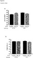

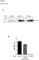
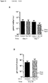


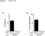
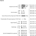


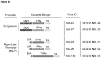


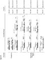
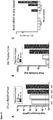
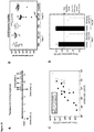
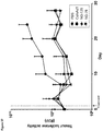
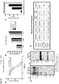
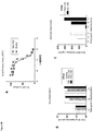
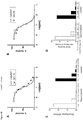
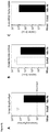
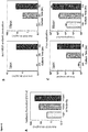
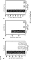

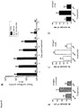
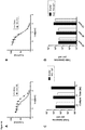
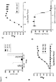
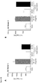
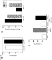
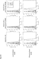
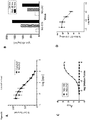
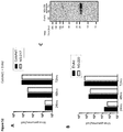
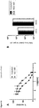
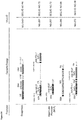
Classifications
-
- A—HUMAN NECESSITIES
- A61—MEDICAL OR VETERINARY SCIENCE; HYGIENE
- A61K—PREPARATIONS FOR MEDICAL, DENTAL OR TOILETRY PURPOSES
- A61K35/00—Medicinal preparations containing materials or reaction products thereof with undetermined constitution
- A61K35/66—Microorganisms or materials therefrom
- A61K35/76—Viruses; Subviral particles; Bacteriophages
- A61K35/761—Adenovirus
-
- A—HUMAN NECESSITIES
- A61—MEDICAL OR VETERINARY SCIENCE; HYGIENE
- A61K—PREPARATIONS FOR MEDICAL, DENTAL OR TOILETRY PURPOSES
- A61K39/00—Medicinal preparations containing antigens or antibodies
- A61K39/0005—Vertebrate antigens
- A61K39/0011—Cancer antigens
-
- A—HUMAN NECESSITIES
- A61—MEDICAL OR VETERINARY SCIENCE; HYGIENE
- A61P—SPECIFIC THERAPEUTIC ACTIVITY OF CHEMICAL COMPOUNDS OR MEDICINAL PREPARATIONS
- A61P35/00—Antineoplastic agents
-
- C—CHEMISTRY; METALLURGY
- C12—BIOCHEMISTRY; BEER; SPIRITS; WINE; VINEGAR; MICROBIOLOGY; ENZYMOLOGY; MUTATION OR GENETIC ENGINEERING
- C12N—MICROORGANISMS OR ENZYMES; COMPOSITIONS THEREOF; PROPAGATING, PRESERVING, OR MAINTAINING MICROORGANISMS; MUTATION OR GENETIC ENGINEERING; CULTURE MEDIA
- C12N7/00—Viruses; Bacteriophages; Compositions thereof; Preparation or purification thereof
-
- A—HUMAN NECESSITIES
- A61—MEDICAL OR VETERINARY SCIENCE; HYGIENE
- A61K—PREPARATIONS FOR MEDICAL, DENTAL OR TOILETRY PURPOSES
- A61K39/00—Medicinal preparations containing antigens or antibodies
- A61K2039/51—Medicinal preparations containing antigens or antibodies comprising whole cells, viruses or DNA/RNA
- A61K2039/525—Virus
- A61K2039/5256—Virus expressing foreign proteins
-
- C—CHEMISTRY; METALLURGY
- C12—BIOCHEMISTRY; BEER; SPIRITS; WINE; VINEGAR; MICROBIOLOGY; ENZYMOLOGY; MUTATION OR GENETIC ENGINEERING
- C12N—MICROORGANISMS OR ENZYMES; COMPOSITIONS THEREOF; PROPAGATING, PRESERVING, OR MAINTAINING MICROORGANISMS; MUTATION OR GENETIC ENGINEERING; CULTURE MEDIA
- C12N2710/00—MICROORGANISMS OR ENZYMES; COMPOSITIONS THEREOF; PROPAGATING, PRESERVING, OR MAINTAINING MICROORGANISMS; MUTATION OR GENETIC ENGINEERING; CULTURE MEDIA dsDNA viruses
- C12N2710/00011—Details
- C12N2710/10011—Adenoviridae
- C12N2710/10311—Mastadenovirus, e.g. human or simian adenoviruses
- C12N2710/10332—Use of virus as therapeutic agent, other than vaccine, e.g. as cytolytic agent
-
- C—CHEMISTRY; METALLURGY
- C12—BIOCHEMISTRY; BEER; SPIRITS; WINE; VINEGAR; MICROBIOLOGY; ENZYMOLOGY; MUTATION OR GENETIC ENGINEERING
- C12N—MICROORGANISMS OR ENZYMES; COMPOSITIONS THEREOF; PROPAGATING, PRESERVING, OR MAINTAINING MICROORGANISMS; MUTATION OR GENETIC ENGINEERING; CULTURE MEDIA
- C12N2710/00—MICROORGANISMS OR ENZYMES; COMPOSITIONS THEREOF; PROPAGATING, PRESERVING, OR MAINTAINING MICROORGANISMS; MUTATION OR GENETIC ENGINEERING; CULTURE MEDIA dsDNA viruses
- C12N2710/00011—Details
- C12N2710/10011—Adenoviridae
- C12N2710/10311—Mastadenovirus, e.g. human or simian adenoviruses
- C12N2710/10341—Use of virus, viral particle or viral elements as a vector
- C12N2710/10343—Use of virus, viral particle or viral elements as a vector viral genome or elements thereof as genetic vector
Definitions
- the present disclosure relates to a modified adenovirus, for example armed with at least one transgene including a therapeutic and/or reporter transgene, in particular a group B virus, such as a serotype 11 adenovirus or a chimeric virus with the fibre, penton and hexon of Ad11, a composition, such as a pharmaceutical formulation comprising the virus, use of the virus and virus formulations, particularly in treatment, especially in the treatment of cancer.
- a group B virus such as a serotype 11 adenovirus or a chimeric virus with the fibre, penton and hexon of Ad11
- a composition such as a pharmaceutical formulation comprising the virus
- use of the virus and virus formulations particularly in treatment, especially in the treatment of cancer.
- the disclosure also extends to processes for preparing the virus.
- Replication deficient adenoviruses vectors have been investigated for a number of years for the delivery of transgenes. Usually the genes have been inserted in the E1 region and/or the E3 region because these regions of the viral genome are non-essential for vectors.
- E1 is an essential region to viral replication and whilst the E3 region in theory can be used as a location to insert a transgene, it would be useful to be able to insert a transgene in more than this location.
- care has to be taken not to disrupt the virus life cycle and/or advantageous viral properties, such as the therapeutic properties of the virus.
- Enadenotucirev is a chimeric oncolytic adenovirus, formerly known as EnAd ( WO2005/118825 ), with fibre, penton and hexon from Ad11p, hence it is a subgroup B virus. It has a chimeric E2B region, which comprises DNA from Ad11p and Ad3. Almost all of the E3 region and part of the E4 region is deleted in EnAd. Therefore, it has significant space in the genome to accommodate additional genetic material whilst remaining viable. Furthermore, because EnAd is a subgroup B adenovirus, pre-existing immunity in humans is less common than, for example, Ad5. Other examples of chimeric oncolytic viruses with Ad11 fibre, penton and hexon include OvAd1 and OvAd2 (see WO2006/060314 ).
- EnAd seems to preferentially infect tumour cells, replicates rapidly in these cells and causes cell lysis. This, in turn, can generate inflammatory immune responses thereby stimulating the body to also fight the cancer. Part of the success of EnAd is hypothesised to be related to the fast replication of the virus in vivo.
- EnAd selectively lyses tumour cells
- transgenes such as a transgene which encodes a cell signalling protein or an antibody, or a transgene which encodes an entity which stimulates a cell signalling protein(s).
- Advantageously arming a virus, with DNA encoding certain proteins that can be expressed inside the cancer cell may enable the body's own defences to be employed to combat tumour cells more effectively, for example by making the cells more visible to the immune system or by delivering a therapeutic gene/protein preferentially to target tumour cells.
- transgenes that are reporters into the genome can aid clinical or pre-clinical studies.
- transgenes It is important that expression of the transgenes does not adversely affect the replication or other advantageous properties of the virus. Thus, the gene or genes must be inserted in a location that does not compromise the replication competence and other advantageous properties of the virus.
- the genome of adenoviruses is tightly packed and therefore it can be difficult to find a suitable location to insert transgenes. This also limits the size of transgenes that can be accommodated.
- the present inventors have developed a method of arming an adenovirus suitable for accommodating a wide variety of transgenes under the control of an endogenous or exogenous promoter that results in a viable, stable, recoverable virus which expresses the transgene in tumour cells.
- the method is robust and repeatable and can be strictly controllable.
- the transgene is located in the proximity of (adjacent to) the gene encoding the fibre protein, either at the 5' end and/or the 3' end of the gene, which does not adversely affect the stability of the virus.
- the present inventors have established that complicated proteins in the form of antibodies or antibody fragments and cell signalling proteins can be inserted in this location in the genome of adenoviruses, for example group B viruses, such as Ad11 and Ad11-derived viruses, and successfully expressed, for example under the control of the endogenous E4 or major late promoter, such that the protein is expressed and the replication of the virus is not compromised.
- group B viruses such as Ad11 and Ad11-derived viruses
- a plasmid developed by the present inventors provides novel restriction sites in the adenovirus genome which can be utilised for insertion of transgene cassettes near to the L5 (fibre) gene to provide viruses of the present disclosure.
- plasmids containing transgenes or transgene cassettes at these insertion site positions can be directly fully synthesized without a cloning step and thus without a need to use the restriction sites.
- the present disclosure provides a replication competent group B oncolytic adenovirus comprising a genome comprising the sequence of formula (I): 5'ITR-B 1 -B A -B 2 -B X -B B -B Y -B 3 -3'ITR (I) wherein:
- B X comprises a restriction site, for example 1, 2, 3 or 4 restriction sites, such as 1 or 2.
- B X comprises at least one transgene, for example 1 or 2 transgenes.
- B X comprises at least one transgene, for example 1 or 2 transgenes and one or more restriction sites, for example 2 or 3 restriction sites, in particular where the restrict sites sandwich a gene or the DNA sequence comprising the genes to allow it/them to be specifically excised from the genome and/or replaced.
- the restriction sites may sandwich each gene, for example when there are two transgenes three different restriction sites are required to ensure that the genes can be selectively excised and/or replaced.
- transgenes are in the form a transgene cassette.
- B X comprises SEQ ID NO: 10.
- SEQ ID NO: 10 is interrupted, for example by a transgene.
- SEQ ID NO: 10 is uninterrupted.
- B X does not comprise a restriction site.
- B X is a bond.
- B X comprises or consists of one or more transgenes.
- B Y comprises a restriction site, for example 1, 2, 3 or 4 restriction sites, such as 1 or 2.
- B Y comprises at least one transgene, for example 1 or 2 transgenes.
- B Y comprises at least one transgene, for example 1 or 2 transgenes and one or more restriction sites, for example 2 or 3 restriction sites, in particular where the restrict sites sandwich a gene or the DNA sequence comprising the genes to allow it/them to be specifically excised from the genome and/or replaced.
- the restriction sites may sandwich each gene, for example when there are two transgenes three different restriction sites are required to ensure that the genes can be selectively excised and/or replaced.
- transgenes are in the form a transgene cassette.
- B Y comprises SEQ ID NO: 11.
- SEQ ID NO: 11 is interrupted, for example by a transgene.
- SEQ ID NO: 11 is uninterrupted.
- B Y does not comprise a restriction site.
- B Y comprises or consists of one or more transgenes.
- B X and B Y each comprises a restriction site, for example 1, 2, 3 or 4 restriction sites, such as 1 or 2.
- B X and B Y each comprises at least one transgene, for example 1 or 2 transgenes.
- B X and B Y each comprises at least one transgene, for example 1 or 2 transgenes and one or more restriction sites, for example 2 or 3 restriction sites, in particular where the restriction sites sandwich a gene or the DNA sequence comprising the genes to allow it to be specifically excised from the genome and/or replaced.
- the restriction sites may sandwich each gene, for example when there are two transgenes three different restriction sites are required to ensure that the genes can be selectively excised and/or replaced.
- transgenes are in the form a transgene cassette.
- B X and B Y comprises SEQ ID NO: 10 and SEQ ID NO: 11 respectively.
- B X and B Y do not comprise a restriction site.
- B X is a bond and B Y is not a bond.
- the transgene is located in B X. In one embodiment the transgene or transgene cassette is located in B Y . In one embodiment a transgene or transgene cassette is located in B X and B Y , for example the transgenes may be the same or different, in each location.
- the transgene in the present virus constructs is/are inserted in a location that is removed from the early genes because this reduces the likelihood of affecting virus gene expression or speed of replication.
- Advantageously using an endogenous promoter maximises the amount of space available for inserting transgenes.
- the virus when under the control of these promoters the virus remains replication competent and is also able to express the antibody as a full length antibody or a suitable binding fragment or other protein.
- the antibody or other protein of choice will be expressed by the cancer cell.
- Employing an endogenous promoter may be advantageous because it reduces the size of the transgene cassette that needs to be incorporated to express the antibody, fragment or other protein, i.e. the cassette can be smaller because no exogenous promoter needs to be included.
- an endogenous promoter in the virus may also be advantageous in a therapeutic context because the transgene is only expressed when the virus is replicating as opposed to a constitutive exogenous promoter which will continually transcribe the transgene and may lead to an inappropriate concentration of the antibody or fragment.
- expression of the antibody or fragment is under the control of the major late promoter.
- the expression of the antibody or fragment is under the control of the E4 promoter.
- an exogenous promoter may be advantageous because it can strongly and constitutively express the antibody or fragment, which may be particularly useful in some situations, for example where the patient has very pervasive cancer.
- expression of the antibody or fragment is under the control of a CMV promoter.
- the exogenous promoter is associated with this DNA sequence, for example is part of the expression cassette encoding the antibody or fragment.
- the DNA sequence encoding the antibody or fragment is located after the L5 gene in the virus sequence.
- the present inventors have established that a variety of transgenes can be inserted into B X and/or B Y under the control of an exogenous or endogenous promoter, without adversely affecting the life cycle of the virus or the stability of the vector.
- the transgene is part of a transgene cassette comprising at least one coding sequence (i.e. at least one transgene) and optionally one or more elements independently selected from:
- the transgene cassette comprises i) or ii) or iii) or iv).
- the transgene cassette comprises i) and ii), or i) and iii), or i) and iv), or ii) and iii), or ii) and iv), or iii) and iv).
- the transgene cassette comprises i) and ii) and iii), or i) and ii) and iv), or i) and iii) and iv), or ii) and iii) and iv).
- the transgene cassette comprises i) and ii) and iii) and iv).
- the transgene or transgene cassette comprises a Kozak squence, which assists in the translation of mRNA, for example at the start of a protein coding sequence.
- composition comprising a virus according to the present disclosure, in particular a pharmaceutical composition, for example comprising an adenovirus according to the disclosure and a pharmaceutically acceptable excipient.
- the present disclosure further relates to an adenovirus or composition according to the disclosure for use in treatment, for example for use in the treatment of cancer.
- the disclosure also relates to a method of treatment comprising administering a therapeutically effective amount of a virus as described herein or a composition comprising the same to a patient in need thereof, in particular a human patient.
- Transgene as employed herein refers to a gene that has been inserted into the genome sequence, which is a gene that is unnatural to the virus (exogenous) or not normally found in that particular location in the virus. Examples of transgenes are given below. Transgene as employed herein also includes a functional fragment of the gene that is a portion of the gene which when inserted is suitable to perform the function or most of the function of the full-length gene.
- Transgene and coding sequence are used interchangeably herein in the context of inserts into the viral genome, unless the context indicates otherwise.
- Coding sequence as employed herein means, for example a DNA sequence encoding a functional RNA, peptide, polypeptide or protein.
- the coding sequence is cDNA for the transgene that encodes the functional RNA, peptide, polypeptide or protein of interest.
- Functional RNA, peptides, polypeptide and proteins of interest are described below.
- virus genome contains coding sequences of DNA. Endogenous (naturally occurring genes) in the genomic sequence of the virus are not considered a transgene, within the context of the present specification unless then have been modified by recombinant techniques such as that they are in a non-natural location or in a non-natural environment.
- transgene as employed herein refers to a segment of DNA containing a gene or cDNA sequence that has been isolated from one organism and is introduced into a different organism i.e. the virus of the present disclosure. In one embodiment this non-native segment of DNA may retain the ability to produce functional RNA, peptide, polypeptide or protein.
- the transgene inserted encodes a human or humanised protein, polypeptide or peptide.
- the transgene inserted encodes a non-human protein, polypeptide or peptide (such as a non-human mammalian protein, polypeptide or peptide) or RNA molecule, for example from a mouse, rat, rabbit, camel, llama or similar.
- the viruses of the present disclosure allow the transgenes to be transported inside the cancerous cell.
- responses generated by the human patient to a non-human sequence can be minimised by this intracellular deliver.
- a DNA sequence may comprise more than one transgene, for example, 1, 2, 3 or 4 transgenes, such as 1 or 2.
- a transgene cassette may comprise more than one transgene, for example, 1, 2, 3 or 4 transgenes, such as 1 or 2.
- the cassette is arranged as shown in the one or more of the Figures or the examples.
- Transgene cassette as employed herein refers to a DNA sequence encoding one or more transgenes in the form of one or more coding sequences and one or more regulatory elements.
- a transgene cassette may encode one or more monocistronic and/or polycistronic mRNA sequences.
- the transgene or transgene cassette encodes a monocistronic or polycistronic mRNA, and for example the cassette is suitable for insertion into the adenovirus genome at a location under the control of an endogenous promoter or exogenous promoter or a combination thereof.
- Monocistronic mRNA as employed herein refers to an mRNA molecule encoding a single functional RNA, peptide, polypeptide or protein.
- the transgene cassette encodes monocistronic mRNA.
- the transgene cassette in the context of a cassette encoding monocistronic mRNA means a segment of DNA optionally containing an exogenous promoter (which is a regulatory sequence that will determine where and when the transgene is active) or a splice site (which is a regulatory sequence determining when a mRNA molecule will be cleaved by the spliceosome) a coding sequence (i.e. the transgene), usually derived from the cDNA for the protein of interest, optionally containing a polyA signal sequence and a terminator sequence.
- an exogenous promoter which is a regulatory sequence that will determine where and when the transgene is active
- a splice site which is a regulatory sequence determining when a mRNA molecule will be cleaved by the spliceosome
- a coding sequence i.e. the transgene
- the transgene cassette may encode one or more polycistronic mRNA sequences.
- Polycistronic mRNA as employed herein refers to an mRNA molecule encoding two or more functional RNA, peptides or proteins or a combination thereof.
- the transgene cassette encodes a polycistronic mRNA.
- transgene cassette in the context of a cassette encoding polycistronic mRNA includes a segment of DNA optionally containing an exogenous promoter (which is a regulatory sequence that will determine where and when the transgene is active) or a splice site (which is a regulatory sequence determining when a mRNA molecule will be cleaved by the spliceosome) two or more coding sequences (i.e. the transgenes), usually derived from the cDNA for the protein or peptide of interest, for example wherein each coding sequence is separated by either an IRES or a 2A peptide.
- the cassette may optionally contain a polyA sequence and a terminator sequence.
- the transgene cassette encodes a monocistronic mRNA followed by a polycistronic mRNA. In another embodiment the transgene cassette encodes a polycistronic mRNA followed by a monocistronic mRNA.
- the present disclosure relates to a group B adenovirus.
- Group B adenovirus is a human adenovirus.
- Adenovirus according to the present disclosure further extends to any, as yet, unidentified or unclassified adenoviral serotypes.
- adenoviral serotypes which are classified into subgroups A-F. See, for example, Strauss, "Adenovirus infections in humans,” in The Adenoviruses, Ginsberg, ea., Plenum Press, New York, NY, pp. 451-596 (1984 ) and Shenk, "Adenoviridae: The Viruses and Their Replication," in Fields Virology, Vol.2, Fourth Edition, Knipe, 35ea., Lippincott Williams & Wilkins, pp. 2265-2267 (2001 ), as shown in Table 1.
- the adenovirus of the present disclosure is a subgroup B, for example independently selected from the group comprising or consisting of: Ad3, Ad7, Ad11, Ad14, Ad16, Ad21, Ad34 and Ad51, such as Ad11, in particular Ad11p (the Slobitski strain).
- Ad11 in particular Ad11p (the Slobitski strain).
- the adenovirus of the invention has the capsid, such as the hexon and/or fibre of a subgroup B adenovirus, such as Ad11, in particular Ad11p.
- the adenovirus is Ad11 or has the fibre and/or hexon and/or penton of Ad11, such as Ad11p.
- it is not a group A virus.
- the adenovirus is not a group C virus. In one embodiment the adenovirus is not Ad5.
- Ad5 as employed herein refers to known adenoviruses designated as serotype 5, it does not extend to genetically engineered viruses that comprises sequences from Ad5. In one embodiment viruses of the present disclosure does not have an Ad5 capsid.
- the adenovirus of the present disclosure is chimeric.
- an adenovirus is chimeric then the characteristics of the outer capsid will be employed to determine the serotype.
- Chimeric as employed herein refers to a virus that comprises DNA from at least two different virus serotypes, including different serotypes within the same group.
- the oncolytic virus has a fibre, hexon and penton proteins from the same serotype, for example Ad11, in particular Ad11p, for example found at positions 30812-31789, 18254-21100 and 13682-15367 of the genomic sequence of the latter wherein the nucleotide positions are relative to genbank ID 217307399 (accession number: GC689208).
- Ad11 in particular Ad11p
- Ad11p for example found at positions 30812-31789, 18254-21100 and 13682-15367 of the genomic sequence of the latter wherein the nucleotide positions are relative to genbank ID 217307399 (accession number: GC689208).
- the adenovirus is enadenotucirev (also known as EnAd and formerly as EnAd).
- Enadenotucirev refers the chimeric adenovirus of SEQ ID NO: 12. It is a replication competent oncolytic chimeric adenovirus which has enhanced therapeutic properties compared to wild type adenoviruses (see WO2005/118825 ).
- EnAd has a chimeric E2B region, which features DNA from Ad11p and Ad3, and deletions in E3/E4. The structural changes in enadenotucirev result in a genome that is approximately 3.5kb smaller than Ad11p thereby providing additional "space" for the insertion of transgenes.
- OvAd1 and OvAd2 are also chimeric adenoviruses similar to enadenotucirev, which also have additional "space” in the genome (see WO2008/080003 ).
- the adenovirus is OvAd1 or OvAd2.
- the adenovirus is oncolytic.
- Oncolytic adenovirus as employed herein means an adenovirus that preferentially kills cancer cells as compared with non-cancer cells.
- the oncolytic virus is apoptotic. That is, it hastens programmed cell death.
- the oncolytic virus is cytolytic.
- the cytolytic activity of oncolytic adenoviruses of the disclosure can be determined in representative tumour cell lines and the data converted to a measurement of potency, for example with an adenovirus belonging to subgroup C, such as Ad5, being used as a standard (i.e. given a potency of 1).
- a suitable method for determining cytolytic activity is an MTS assay (see Example 4, Figure 2 of WO2005/118825 ).
- the oncolytic virus is necrolytic. That is, it causes or hastens cell necrosis or immunogenic cell death. In one embodiment necrolytic cell death is advantageous because it triggers, induces the patients (host) immune responses.
- Replication competent in the context of the present specification refers to a virus that possesses all the necessary machinery to replicate in cells in vitro and in vivo, i.e. without the assistance of a packaging cell line.
- a viral vector, for example deleted in the E1 region, capable of replicating in a complementary packaging cell line is not a replication competent virus in the present context.
- Viral vectors are replication deficient and require a packaging cell to provide a complementary gene to allow replication.
- Adenovirus genome as employed herein means the DNA sequence encoding the structural proteins and elements relevant to the function/life cycle of an adenovirus.
- the viral genome contains five early transcription units (E1A, E1B, E2, E3, and E4), three delayed early units (IX, IVa2 and E2 late) and one late unit (major late) that is processed to generate five families of late mRNAs (L1-L5).
- E1A, E1B, E2, E3, and E4 the early transcription units
- IX, IVa2 and E2 late the delayed early units
- L1-L5 late unit
- Proteins encoded by the early genes are primarily involved in replication and modulation of the host cell response to infection, whereas the late genes encode viral structural proteins.
- Early genes are prefixed by the letter E and the late genes are prefixed by the letter L.
- the genome of adenoviruses is tightly packed, that is, there is little non-coding sequence, and therefore it can be difficult to find a suitable location to insert transgenes.
- the present inventors have identified two DNA regions where transgenes are tolerated, in particular the sites identified are suitable for accommodating complicated transgenes, such as those encoding antibodies. That is, the transgene is expressed without adversely affecting the virus' viability, native properties such as oncolytic properties or replication.
- the oncolytic or partial oncolytic virus according to the disclosure may be as a result of deletion in the E4 and/or E3 region, for example deleted in part of the E4 region or fully deleted in the E3 region, or alternatively deleted in part of the E4 region (such as E4orf4) and fully deleted in the E3 region, for example as exemplified in the sequences disclosed herein.
- the oncolytic virus of the disclosure is chimeric.
- Chimeric as employed herein refers to virus that comprises DNA from two or more different serotypes and has oncolytic virus properties.
- the oncolytic virus is EnAd or an active derivate thereof which retains the essential beneficial properties of the virus.
- EnAd is disclosed in WO2005/118825 and the full sequence for the virus is provided herein SEQ ID NO: 12.
- the chimeric E2B region is disclosed herein as SEQ ID NO: 47.
- Alternative oncolytic viruses include OvAd1 and OvAd2, which are respectively disclosed as SEQ ID NO: 2 and 3 in WO2008/080003 .
- the adenoviruses of the present disclosure exhibit similar virus activity, for example replication and/or infectivity, profiles to EnAd following infection of a variety of different colon cancer cell lines in vitro.
- the present disclosure also relates to the novel sequences of viruses or viral components/ constructs, such as plasmids, disclosed herein.
- a compound of formula (I) wherein B X and B Y are not a bond and comprises a transgene a restriction site or both, such as B X and B Y are both a transgene.
- a bond refers to a co-valent bond connecting the one DNA sequence to another DNA sequence, for example connecting one section of the virus genome to another.
- a variable in formula (I) herein represents a bond the feature or element represented by the bond is absent i.e. deleted.
- adenoviruses As the structure of adenoviruses is, in general, similar the elements below are discussed in terms of the structural elements and the commonly used nomenclature referring thereto, which are known to the skilled person.
- an element When an element is referred to herein then we refer to the DNA sequence encoding the element or a DNA sequence encoding the same structural protein of the element in an adenovirus. The latter is relevant because of the redundancy of the DNA code. The viruses' preference for codon usage may need to be considered for optimised results.
- Any structural element from an adenovirus employed in the viruses of the present disclosure may comprise or consist of the natural sequence or may have similarity over the given length of at least 95%, such as 96%, 97%, 98%, 99% or 100%.
- the original sequence may be modified to omit 10%, 9%, 8%, 7%, 6%, 5%, 4%, 3%, 2% or 1% of the genetic material.
- the skilled person is aware that when making changes the reading frames of the virus must be not disrupted such that the expression of structural proteins is disrupted.
- the given element is a full-length sequence i.e. the full-length gene.
- the given element is less than a full-length and retains the same or corresponding function as the full-length sequence.
- the DNA sequence may be less than a full-length and have no functionality.
- the structural genes encoding structural or functional proteins of the adenovirus are generally linked by non-coding regions of DNA.
- the genomic sequence of the structural element of interest especially non-coding regions thereof
- the element will be considered a structural element of reference to the extent that it is fit for purpose and does not encode extraneous material.
- the gene will be associated with suitable non-coding regions, for example as found in the natural structure of the virus.
- an insert such as DNA encoding a restriction site and/or transgene, is inserted into a non-coding region of genomic virus DNA, such as an intron or intergenic sequence.
- genomic virus DNA such as an intron or intergenic sequence.
- Having said this some non-coding regions of adenovirus may have a function, for example in alternative splicing, transcription regulation or translation regulation, and this may need to be taken into consideration.
- the sites identified herein, that are associated with the L5 region, are suitable for accommodating a variety of DNA sequences encoding complex entities such as RNAi, cytokines, single chain or multimeric proteins, such as antibodies.
- Gene as employed herein refers to coding and any non-coding sequences associated therewith, for example introns and associated exons.
- a gene comprises or consists of only essential structural components, for example coding region.
- ITR Inverted Terminal Repeat
- the 5'ITR as employed herein refers to part or all of an ITR from the 5' end of an adenovirus, which retains the function of the ITR when incorporated into an adenovirus in an appropriate location.
- the 5'ITR comprises or consists of the sequence from about 1bp to 138bp of SEQ ID NO: 12 or a sequence 90, 95, 96, 97, 98 or 99% identical thereto along the whole length, in particular the sequence consisting of from about 1bp to 138bp of SEQ ID NO: 12.
- the 3'ITR as employed herein refers to part or all of an ITR from 3' end of an adenovirus which retains the function of the ITR when incorporated into an adenovirus in an appropriate location.
- the 3'ITR comprises or consists of the sequence from about 32189bp to 32326bp of SEQ ID NO: 12 or a sequence 90, 95, 96, 97, 98 or 99% identical thereto along the whole length, in particular the sequence consisting of from about 32189bp to 32326bp of SEQ ID NO: 12.
- B 1 as employed herein refers to the DNA sequence encoding: part or all of an E1A from an adenovirus, part or all of the E1B region of an adenovirus, and independently part or all of E1A and E1B region of an adenovirus.
- E1A refers to the DNA sequence encoding part or all of an adenovirus E1A region. The latter here is referring to the polypeptide/protein E1A. It may be mutated such that the protein encoded by the E1A gene has conservative or non-conservative amino acid changes, such that it has: the same function as wild-type (i.e. the corresponding non-mutated protein); increased function in comparison to wild-type protein; decreased function, such as no function in comparison to wild-type protein; or has a new function in comparison to wild-type protein or a combination of the same as appropriate.
- E1B refers to the DNA sequence encoding part or all of an adenovirus E1B region (i.e. polypeptide or protein), it may be mutated such that the protein encoded by the E1B gene/region has conservative or non-conservative amino acid changes, such that it has: the same function as wild-type (i.e. the corresponding non-mutated protein); increased function in comparison to wild-type protein; decreased function, such as no function in comparison to wild-type protein; or has a new function in comparison to wild-type protein or a combination of the same as appropriate.
- B 1 can be modified or unmodified relative to a wild-type E1 region, such as a wild-type E1A and/or E1B.
- a wild-type E1 region such as a wild-type E1A and/or E1B.
- the skilled person can easily identify whether E1A and/or E1B are present or (part) deleted or mutated.
- Wild-type as employed herein refers to a known adenovirus.
- a known adenovirus is one that has been identified and named, regardless of whether the sequence is available.
- B 1 has the sequence from 139bp to 3932bp of SEQ ID NO: 12.
- B A refers to the DNA sequence encoding the E2B-L1-L2-L3-E2A-L4 regions including any non-coding sequences, as appropriate. Generally this sequence will not comprise a transgene. In one embodiment the sequence is substantially similar or identical to a contiguous sequence from a known adenovirus, for example a serotype shown in Table 1, in particular a group B virus, for example Ad3, Ad7, Ad11, Ad14, Ad16, Ad21, Ad34, Ad35, Ad51 or a combination thereof, such as Ad3, Ad11 or a combination thereof.
- E2B-L1-L2-L3-E2A-L4 refers to comprising these elements and other structural elements associated with the region, for example B A will generally include the sequence encoding the protein IV2a, for example as follows: IV2A IV2a-E2B-L1-L2-L3-E2A-L4
- the E2B region is chimeric. That is, comprises DNA sequences from two or more different adenoviral serotypes, for example from Ad3 and Ad11, such as Ad11p.
- Ad3 adenoviral serotypes
- Ad11p Ad11p
- the E2B region has the sequence from 5068bp to 10355bp of SEQ ID NO: 12 or a sequence 95%, 96%, 97%, 98% or 99% identical thereto over the whole length.
- the E2B in component B A comprises the sequences shown in SEQ ID NO: 47 (which corresponds to SEQ ID NO: 3 disclosed in WO2005/118825 ).
- B A has the sequence from 3933bp to 27184bp of SEQ ID NO: 12.
- E3 refers to the DNA sequence encoding part or all of an adenovirus E3 region (i.e. protein/polypeptide), it may be mutated such that the protein encoded by the E3 gene has conservative or non-conservative amino acid changes, such that it has the same function as wild-type (the corresponding unmutated protein); increased function in comparison to wild-type protein; decreased function, such as no function in comparison to wild-type protein or has a new function in comparison to wild-type protein or a combination of the same, as appropriate.
- adenovirus E3 region i.e. protein/polypeptide
- the E3 region is form an adenovirus serotype given in Table 1 or a combination thereof, in particular a group B serotype, for example Ad3, Ad7, Ad11 (in particular Ad11p), Ad14, Ad16, Ad21, Ad34, Ad35, Ad51 or a combination thereof, such as Ad3, Ad11 (in particular Ad11p) or a combination thereof.
- Ad3, Ad7, Ad11 (in particular Ad11p), Ad14, Ad16, Ad21, Ad34, Ad35, Ad51 or a combination thereof such as Ad3, Ad11 (in particular Ad11p) or a combination thereof.
- the E3 region is partially deleted, for example is 95%, 90%, 85%, 80%, 75%, 70%, 65%, 60%, 55%, 50%, 45%, 40%, 35%, 30%, 25%, 20%, 15%, 10%, 5% deleted.
- B 2 is a bond, wherein the DNA encoding the E3 region is absent.
- the DNA encoding the E3 region can be replaced or interrupted by a transgene.
- E3 region replaced by a transgene as employed herein includes part or all of the E3 region is replaced with a transgene.
- the B 2 region comprises the sequence from 27185bp to 28165bp of SEQ ID NO: 12.
- B 2 consists of the sequence from 27185bp to 28165bp of SEQ ID NO: 12.
- B X refers to the DNA sequence in the vicinity of the 5' end of the L5 gene in B B .
- the vicinity of or proximal to the 5' end of the L5 gene as employed herein refers to: adjacent (contiguous) to the 5' end of the L5 gene or a non-coding region inherently associated herewith i.e. abutting or contiguous to the 5' prime end of the L5 gene or a non-coding region inherently associated therewith.
- adjacent (contiguous) to the 5' end of the L5 gene or a non-coding region inherently associated herewith i.e. abutting or contiguous to the 5' prime end of the L5 gene or a non-coding region inherently associated therewith.
- in the vicinity of or proximal to may refer to being close the L5 gene, such that there are no coding sequences between the B X region and the 5' end of L5 gene.
- B X is joined directly to a base of L5 which represents, for example the start of a coding sequence of the L5 gene.
- B X is joined directly to a base of L5 which represents, for example the start of a non-coding sequence, or joined directly to a non-coding region naturally associated with L5.
- a non-coding region naturally associated L5 as employed herein refers to part of all of a non-coding regions which is part of the L5 gene or contiguous therewith but not part of another gene.
- B X comprises the sequence of SEQ ID NO: 10.
- This sequence is an artificial non-coding sequence wherein a DNA sequence, for example comprising a transgene (or transgene cassette), a restriction site or a combination thereof may be inserted therein.
- This sequence is advantageous because it acts as a buffer in that allows some flexibility on the exact location of the transgene whilst minimising the disruptive effects on virus stability and viability.
- the insert(s) can occur anywhere within SEQ ID NO: 10 from the 5' end, the 3' end or at any point between bp 1 to 201, for example between base pairs 1/2, 2/3, 3/4, 4/5, 5/6, 6/7, 7/8, 8/9, 9/10, 10/11, 11/12, 12/13, 13/14, 14/15, 15/16, 16/17, 17/18, 18/19, 19/20, 20/21, 21/22, 22/23, 23/24, 24/25, 25/26, 26/27, 27/28, 28/29, 29/30, 30/31, 31/32, 32/33, 33/34, 34/35, 35/36, 36/37, 37/38, 38/39, 39/40, 40/41, 41/42, 42/43, 43/44, 44/45, 45/46, 46/47, 47/48, 48/49, 49/50, 50/51, 51/52, 52/53, 53/54, 54/55, 55/56, 56/57, 57/58
- B X comprises SEQ ID NO: 10 with a DNA sequence inserted between bp 27 and bp 28 or a place corresponding to between positions 28192bp and 28193bp of SEQ ID NO: 12.
- the insert is a restriction site insert. In one embodiment the restriction site insert comprises one or two restriction sites. In one embodiment the restriction site is a 19bp restriction site insert comprising 2 restriction sites. In one embodiment the restriction site insert is a 9bp restriction site insert comprising 1 restriction site. In one embodiment the restriction site insert comprises one or two restriction sites and at least one transgene, for example one or two transgenes. In one embodiment the restriction site is a 19bp restriction site insert comprising 2 restriction sites and at least one transgene, for example one or two transgenes. In one embodiment the restriction site insert is a 9bp restriction site insert comprising 1 restriction site and at least one transgene, for example one, two or three transgenes, such as one or two.
- two restriction sites sandwich one or more, such as two transgenes (for example in a transgene cassette).
- B X comprises two restrictions sites the said restriction sites are different from each other.
- said one or more restrictions sites in B X are non-naturally occurring in the particular adenovirus genome into which they have been inserted.
- said one or more restrictions sites in B X are different to other restrictions sites located elsewhere in the adenovirus genome, for example different to naturally occurring restrictions sites and/or restriction sites introduced into other parts of the genome, such as a restriction site introduced into B Y .
- the restriction site or sites allow the DNA in the section to be cut specifically.
- Cut specifically as employed herein refers to where use of an enzyme specific to the restriction sites cuts the virus only in the desired location, usually one location, although occasionally it may be a pair of locations.
- a pair of locations as employed herein refers to two restrictions sites in proximity of each other that are designed to be cut by the same enzyme (i.e. cannot be differentiated from each other).
- restriction site insert is SEQ ID NO: 55.
- B X has the sequence from 28166bp to 28366bp of SEQ ID NO: 12.
- B X is a bond
- the L5 region refers to the DNA sequence containing the gene encoding the fibre polypeptide/protein, as appropriate in the context.
- the fibre gene/region encodes the fibre protein which is a major capsid component of adenoviruses. The fibre functions in receptor recognition and contributes to the adenovirus' ability to selectively bind and infect cells.
- the fibre is from a group B virus, in particular Ad11, such as Ad11p.
- B B has the sequence from 28367bp to 29344bp of SEQ ID NO: 12.
- DNA sequence in relation to B Y as employed herein refers to the DNA sequence in the vicinity of the 3' end of the L5 gene of B B .
- In the vicinity of or proximal to the 3' end of the L5 gene as employed herein refers to: adjacent (contiguous) to the 3' end of the L5 gene or a non-coding region inherently associated therewith i.e. abutting or contiguous to the 3' prime end of the L5 gene or a non-coding region inherently associated therewith (i.e. all or part of an non-coding sequence endogenous to L5).
- in the vicinity of or proximal to may refer to being close the L5 gene, such that there are no coding sequences between the B Y region and the 3' end of the L5 gene.
- B Y is joined directly to a base of L5 which represents the "end" of a coding sequence.
- B Y is joined directly to a base of L5 which represents the "end" of a non-coding sequence, or joined directly to a non-coding region naturally associated with L5.
- B Y comprises the sequence of SEQ ID NO: 11.
- This sequence is a non-coding sequence wherein a DNA sequence, for example comprising a transgene (or transgene cassette), a restriction site or a combination thereof may be inserted.
- This sequence is advantageous because it acts a buffer in that allows some flexibility on the exact location of the transgene whilst minimising the disruptive effects on virus stability and viability.
- the insert(s) can occur anywhere within SEQ ID NO: 11 from the 5' end, the 3' end or at any point between bp 1 to 35, for example between base pairs 1/2, 2/3, 3/4, 4/5, 5/6, 6/7, 7/8, 8/9, 9/10, 10/11, 11/12, 12/13, 13/14, 14/15, 15/16, 16/17, 17/18, 18/19, 19/20, 20/21, 21/22, 22/23, 23/24, 24/25, 25/26, 26/27, 27/28, 28/29, 29/30, 30/31, 31/32, 32/33, 33/34, or 34/35.
- B Y comprises SEQ ID NO: 11 with a DNA sequence inserted between positions bp 12 and 13 or a place corresponding to 29356bp and 29357bp in SEQ ID NO: 12.
- the insert is a restriction site insert.
- the restriction site insert comprises one or two restriction sites.
- the restriction site is a 19bp restriction site insert comprising 2 restriction sites.
- the restriction site insert is a 9bp restriction site insert comprising 1 restriction site.
- the restriction site insert comprises one or two restriction sites and at least one transgene, for example one or two or three transgenes, such as one or two transgenes.
- the restriction site is a 19bp restriction site insert comprising 2 restriction sites and at least one transgene, for example one or two transgenes.
- the restriction site insert is a 9bp restriction site insert comprising 1 restriction site and at least one transgene, for example one or two transgenes.
- two restriction sites sandwich one or more, such as two transgenes (for example in a transgene cassette).
- B Y comprises two restrictions sites the said restriction sites are different from each other.
- said one or more restrictions sites in B Y are non-naturally occurring (such as unique) in the particular adenovirus genome into which they have been inserted.
- said one or more restrictions sites in B Y are different to other restrictions sites located elsewhere in the adenovirus genome, for example different to naturally occurring restrictions sites or restriction sites introduced into other parts of the genome, such as B X .
- the restriction site or sites allow the DNA in the section to be cut specifically.
- restriction site insert is SEQ ID NO: 54.
- B Y has the sequence from 29345bp to 29379bp of SEQ ID NO: 12.
- B Y is a bond
- the insert is after bp 12 in SEQ ID NO: 11.
- the insert is at about position 29356bp of SEQ ID NO: 12.
- the insert is a transgene cassette comprising one or more transgenes, for example 1, 2 or 3, such as 1 or 2.
- E4 refers to the DNA sequence encoding part or all of an adenovirus E4 region (i.e. polypeptide/protein region), which may be mutated such that the protein encoded by the E4 gene has conservative or non-conservative amino acid changes, and has the same function as wild-type (the corresponding non-mutated protein); increased function in comparison to wild-type protein; decreased function, such as no function in comparison to wild-type protein or has a new function in comparison to wild-type protein or a combination of the same as appropriate.
- an adenovirus E4 region i.e. polypeptide/protein region
- the E4 region is partially deleted, for example is 95%, 90%, 85%, 80%, 75%, 70%, 65%, 60%, 55%, 50%, 45%, 40%, 35%, 30%, 25%, 20%, 15%, 10% or 5% deleted.
- the E4 region has the sequence from 32188bp to 29380bp of SEQ ID NO: 12.
- B 3 is a bond, i.e. wherein E4 is absent.
- B 3 has the sequence consisting of from 32188bp to 29380bp of SEQ ID NO: 12.
- formulas herein are contiguous and may embody non-coding DNA sequences as well as the genes and coding DNA sequences (structural features) mentioned herein.
- the formulas of the present disclosure are attempting to describe a naturally occurring sequence in the adenovirus genome.
- the formula is referring to the major elements characterising the relevant section of genome and is not intended to be an exhaustive description of the genomic stretch of DNA.
- E1A, E1B, E3 and E4 as employed herein each independently refer to the wild-type and equivalents thereof, mutated or partially deleted forms of each region as described herein, in particular a wild-type sequence from a known adenovirus.
- Insert refers to a DNA sequence that is incorporated either at the 5' end, the 3' end or within a given DNA sequence reference segment such that it interrupts the reference sequence. The latter is a reference sequence employed as a reference point relative to which the insert is located. In the context of the present disclosure inserts generally occur within either SEQ ID NO: 10 or SEQ ID NO: 11. An insert can be either a restriction site insert, a transgene cassette or both. When the sequence is interrupted the virus will still comprise the original sequence, but generally it will be as two fragments sandwiching the insert.
- the transgene or transgene cassette does not comprise a non-biased inserting transposon, such as a TN7 transposon or part thereof.
- Tn7 transposon as employed herein refers to a non-biased insertion transposon as described in WO2008/080003 .
- Restriction sites in the locations disclosed here are useful in viruses and constructs of the present disclosure, such as plasmids, because they allow the transgene to be changed rapidly and, for example selectively when the restriction sites around the transgene(s) are unique.
- the transgene or transgene cassette comprises a restriction site at each terminus, thereby allowing the cassette to be replaced.
- restriction site is a location in a DNA sequence that can be cut by a restriction enzyme, usually an enzyme specific to the sequence.
- the restriction site comprises 3 to 22 base pairs, for example 4 to 22, such as 5, 6, 7, 8, 9, 10, 11, 12, 13, 14, 15, 16, 17, 18, 19, 20, 21 or 22 base pairs.
- restriction sites cut by restriction enzymes include but are not limited to:
- restriction enzymes that cut the same recognition sites may also be suitable.
- one or more restrictions sites in B X and B Y are independently selected from a restriction site specific to an enzyme described herein, for example Not I , Fse I , AsiS I , Sgf I and Sbf I, in particular the restriction sites inserted are all different, such as sites specific for Not I and sites specific for Fse I located in B X and Sgf I and Sbf I located in B Y .
- the region B X and/or B Y do not comprise a restriction site.
- the viruses and constructs of the present disclosure can be prepared without restriction sites, for example using synthetic techniques. These techniques allow a great flexibility in the creation of the viruses and constructs. Furthermore, the present inventors have established that the properties of the viruses and constructs are not diminished when they are prepared by synthetic techniques.
- Promoter as employed herein means a region of DNA that initiates transcription of a particular gene or genes. Promoters are generally located proximal to the genes they transcribe, on the same strand and upstream (i.e. 5') on the DNA. Proximal as employed in this context means sufficiently close to function as a promoter. In one embodiment the promoter is within 100 bp of the transcription start site. Thus endogenous promoter as employed herein refers to a promoter that naturally occurs in (i.e. is native to) the adenovirus (or construct) into which the transgene, is being inserted.
- the endogenous promoter employed is the naturally occurring promoter in the virus in its original location in the virus genome, in particular this is the primary or only promoter employed in the expression of the transgene or transgenes.
- the endogenous promoter used to promote the translation and optionally the transcription of the transgene is one integrated in the genome of the adenovirus and not previously introduced by recombinant techniques.
- transgene/transgene cassette is inserted in the appropriate orientation to be under the control of said endogenous promoter. That is, where the promoter is generally on the antisense strand, the cassette is inserted, for example in the antisense orientation.
- genes can be expressed in one of two orientations. However, generally one orientation provides increased levels of expression over the other orientation, for a given (particular) transgene.
- the cassette is in the sense orientation. That is, is transcribed in a 5' to 3' direction. In one embodiment the cassette is in the antisense orientation. That is, transcribed in the 3' to 5' orientation.
- the endogenous promoters in the virus can, for example, be utilised by employing a gene encoding a transgene and a splice acceptor sequence.
- the cassette will comprise a splice acceptor sequence when under the control of an endogenous promoter.
- the coding sequence for example the sequence encoding the antibody or antibody binding fragment further comprises a splice acceptor sequence.
- transgene, transgenes, or transgene cassette are under the control of an E4 promoter or a major late promoter, such as the major late promoter (ML promoter).
- E4 promoter or a major late promoter, such as the major late promoter (ML promoter).
- ML promoter major late promoter
- transgene is activated, i.e. transcribed, when a particular promoter dictates.
- the Major Late Promoter refers to the adenovirus promoter that controls expression of the "late expressed" genes, such as the L5 gene.
- the MLP is a "sense strand" promoter. That is, the promoter influences genes that are downstream of the promoter in the 5'-3' direction.
- the major late promoter refers the original major late promoter located in the virus genome.
- E4 promoter refers to the adenovirus promoter of the E4 region.
- the E4 region is an antisense region; therefore the promoter is an antisense promoter. That is, the promoter is upstream of the E4 region in the 3'-5' direction. Therefore any transgene cassette under control of the E4 promoter may need to be oriented appropriately.
- the cassette under the control of the E4 promoter is in the antisense orientation.
- the cassette is under the control of the E4 promoter in the sense orientation.
- the E4 promoter as employed herein refers to the original E4 promoter located in the virus genome.
- a replication competent oncolytic adenovirus serotype 11 (such as Ad11p) or virus-derivative thereof wherein the fibre, hexon and capsid are serotype 11 (such as Ad11p)
- the virus genome comprises a DNA sequence encoding a therapeutic antibody or antibody-binding fragment, wherein said DNA sequence under the control of a promoter endogenous to the adenovirus selected from consisting of E4 and the major late promoter (i.e. the E4 promoter or the major late promoter), such that the transgene does not interfere with virus replication, for example is associated with the L5 region (i.e. before or after said region), such as located after L5 in the virus genome, for example as shown in SEQ ID NO: 1 to 9, 46, 48 to 53, 56 to 63, 66-69 and 72-73.
- an endogenous promoter is introduced into the viral genome at a desired location by recombinant techniques, for example is introduced in the transgene cassette.
- this arrangement will generally be referred to as an exogenous promoter.
- the transgene cassette comprises an exogenous promoter.
- Exogenous promoter refers to a promoter that is not naturally occurring in the adenovirus into which the transgene is being inserted. Typically exogenous promoters are from other viruses or are mammalian promoters.
- Exogenous promoter as employed herein means a DNA element, usually located upstream of the gene of interest, that regulates the transcription of the gene.
- the regulator of gene expression is an exogenous promoter, for example CMV (cytomegalovirus promoter), CBA (chicken beta actin promoter) or PGK (phosphoglycerate kinase 1 promoter), such as CMV promoter.
- CMV cytomegalovirus promoter
- CBA chicken beta actin promoter
- PGK phosphoglycerate kinase 1 promoter
- the CMV exogenous promoter employed has the nucleotide sequence of SEQ ID NO: 13.
- the PGK exogenous promoter employed has the nucleotide sequence of SEQ ID NO: 14.
- the CBA exogenous promoter employed has the nucleotide sequence of SEQ ID NO: 15.
- a replication competent oncolytic adenovirus serotype 11 (such as Ad11p) or virus-derivative thereof wherein the fibre, hexon and capsid are serotype 11 (such as Ad11p)
- the virus genome comprises a DNA sequence encoding a therapeutic antibody or antibody-binding fragment located in a part of the virus genome which is expressed late in the virus replication cycle and such that the transgene does not interfere with virus replication, wherein said DNA sequence under the control of a promoter exogenous to the adenovirus (for example the CMV promoter).
- the DNA sequence encoding an antibody or fragment is associated with the L5 region as described elsewhere herein.
- the exogenous promoter is an antigen-presenting cell promoter.
- Antigen-presenting cell promoter as employed herein refers to a promoter for a gene that is selectively expressed by antigen-presenting cells, such as dendritic cells or macrophages.
- genes include but are not limited to: FLT-3, FLT-3 ligand, TLRs, CD1a, CD1c, CD11b, CD11c, CD80, CD83, CD86, CD123, CD172a, CD205, CD207, CD209, CD273, CD281, CD283, CD286, CD289, CD287, CXCR4, GITR Ligand, IFN- ⁇ 2, IL-12, IL-23, ILT1, ILT2, ILT3, ILT4, ILT5, ILT7, TSLP Receptor, CD141, CD303, CADM1, CLEC9a, XCR1 or CD304; antigen processing and presentation mediators such as CTIIA or GILT.
- the exogenous promoter is suitable for selective expression of transgenes in said antigen-presenting cells.
- Regulator of gene expression refers to a genetic feature, such as a promoter, enhancer or a splice acceptor sequence that plays a role in gene expression, typically by initiating or enhancing transcription or translation.
- Splice acceptor sequence refers to a regulatory sequence determining when an mRNA molecule will be recognised by small nuclear ribonucleoproteins of the spliceosome complex. Once assembled the spliceosome catalyses splicing between the splice acceptor site of the mRNA molecule to an upstream splice donor site producing a mature mRNA molecule that can be translated to produce a single polypeptide or protein.
- splice acceptor sequences may be employed in the present invention and these can be described as short splice acceptor (small), splice acceptor (medium) and branched splice acceptor (large).
- SSA as employed herein means a short splice acceptor, typically comprising just the splice site, for example 4 bp.
- SA as employed herein means a splice acceptor, typically comprising the short splice acceptor and the polypyrimidine tract, for example 16 bp.
- bSA as employed herein means a branched splice acceptor, typically comprising the short splice acceptor, polypyrimidine tract and the branch point, for example 26 bp.
- the splice acceptor employed in the constructs of the disclosure are shown in SEQ ID NO: 16 to 18.
- the SSA has the nucleotide sequence of SEQ ID NO: 16.
- the SA has the nucleotide sequence of SEQ ID NO: 17.
- the bSA has the nucleotide sequence of SEQ ID NO: 18.
- the splice acceptor sequence is independently selected from the group comprising: TGCTAATCTT CCTTTCTCTC TTCAGG (SEQ ID NO: 18), CCTTTCTCTCTT CAGG (SEQ ID NO: 17), and CAGG (SEQ ID NO: 16).
- the splice site is immediately proceeded (i.e. followed in a 5' to 3' direction) by a consensus Kozak sequence comprising CCACC.
- the splice site and the Kozak sequence are interspersed by up to 100 or less bp.
- the Kozak sequence has the nucleotide sequence of SEQ ID NO: 45.
- the coding sequence when under the control of an endogenous or exogenous promoter (such as an endogenous promoter), the coding sequence will be immediately preceded by a Kozak sequence.
- the start of the coding region is indicated by the initiation codon (AUG), for example is in the context of the sequence (gcc)gccRcc AUG g [SEQ ID NO: 77] the start of the start of the coding sequences is indicated by the bases in bold.
- a lower case letter denotes common bases at this position (which can nevertheless vary) and upper case letters indicate highly-conserved bases, i.e.
- the 'AUGG' sequence is constant or rarely, if ever, changes; 'R' indicates that a purine (adenine or guanine) is usually observed at this position and the sequence in brackets (gcc) is of uncertain significance.
- the initiation codon AUG is incorporated into a Kozak sequence.
- Internal Ribosome Entry DNA Sequence refers to a DNA sequence encoding an Internal Ribosome Entry Sequence (IRES).
- IRES as employed herein means a nucleotide sequence that allows for initiation of translation a messenger RNA (mRNA) sequence, including initiation starting within an mRNA sequence. This is particularly useful when the cassette encodes polycistronic mRNA. Using an IRES results in a polycistronic mRNA that is translated into multiple individual proteins or peptides.
- the Internal Ribosome Entry DNA sequence has the nucleotide sequence of SEQ ID NO: 19.
- a particular IRES is only used once in the genome. This may have benefits with respect to stability of the genome.
- “High self-cleavage efficiency 2A peptide” or “2A peptide” as employed herein refers to a peptide which is efficiently cleaved following translation. Suitable 2A peptides include P2A, F2A, E2A and T2A. The present inventors have noted that once a specific DNA sequence encoding a given 2A peptide is used once, the same specific DNA sequence may not be used a second time. However, redundancy in the DNA code may be utilised to generate a DNA sequence that is translated into the same 2A peptide. Using 2A peptides is particularly useful when the cassette encodes polycistronic mRNA. Using 2A peptides results in a single polypeptide chain being translated which is modified post-translation to generate multiple individual proteins or peptides.
- the encoded P2A peptide employed has the amino acid sequence of SEQ ID NO: 25. In one embodiment the encoded F2A peptide employed has the amino acid sequence of SEQ ID NO: 26. In one embodiment the encoded E2A peptide employed has the amino acid sequence of SEQ ID NO: 27. In one embodiment the encoded T2A peptide employed has the amino acid sequence of SEQ ID NO: 28.
- an mRNA or each mRNA encoded by transgene is/are comprise a polyadenylation signal sequence, such as typically at the end of an mRNA sequence, for example as shown in SEQ ID NO: 20.
- the transgene or the transgene cassette comprises at least one sequence encoding a polyadenylation signal sequence.
- PolyA Polyadenylation signal
- polyadenylation sequence means a DNA sequence, usually containing an AATAAA site, that once transcribed can be recognised by a multiprotein complex that cleaves and polyadenylates the nascent mRNA molecule.
- polyadenylation sequence has the nucleotide sequence of SEQ ID NO: 20.
- the construct does not include a polyadenylation sequence.
- the regulator of gene expression is a splice acceptor sequence.
- sequence encoding a protein/polypeptide/peptide such as an antibody or antibody fragment further comprises a polyadenylation signal.
- the transgene or transgenes independently encode a protein, peptide, RNA molecule, such as an RNA molecule.
- the transgene can be delivered intra-cellularly and can subsequently be transcribed and if appropriate translated.
- Examples of genetic material encoded by a transgene include, for example antibodies or binding fragments thereof, chemokines, cytokines, immunmodulators, enzymes (for example capable of converting pro-drug in the active agent) and an RNAi molecule.
- Peptide as employed herein refers to an amino acid sequence of 2 to 50 residues, for example 5 to 20 residues.
- Polypeptide as employed herein refers to an amino acid sequence of more than 50 residues without tertiary structure, in particular without secondary and tertiary structure.
- Protein refers to an amino acid sequence of more than 50 residues, with secondary and/or tertiary structure, in particular with second and tertiary structure.
- the coding sequence encodes a therapeutic RNA, therapeutic peptide, therapeutic polypeptide or therapeutic protein (i.e. is a therapeutic gene).
- Therapeutic gene as employed herein means a gene that encodes an entity that may be useful in the treatment, amelioration or prevention of disease, for example the gene expresses a therapeutic protein, polypeptide, peptide or RNA, which at least slows down, halts or reverses the progression of a disease, such as cancer.
- the entity encoded by the transgene when transcribed or translated in a cell increases production of danger signals by the cell.
- Dannger signals refers to a variety of molecules produced by cells undergoing injury, stress or non-apoptotic death that act as alarm signals, for example by stimulating cells of the innate immune system to respond directly as well as serving to enhance activation of cells of the adaptive immune system.
- tumours often changes such that natural human immune responses are down regulated.
- the ability to re-start the immune responses from within the tumour is potentially very interesting in the treatment of cancer.
- the encoded therapeutic peptide or protein is designed to be secreted into the extracellular environment.
- the functional RNA, peptide, polypeptide or protein, such as the antibody is released into the external microenvironment of the cell, for example into the culture supernatant, or in vivo : tissue, stroma, circulation, blood and/or lymphatic system.
- the peptide, polypeptide or protein, encoded by the transgene comprises a signal sequence.
- Signal peptide as employed herein refers to a short 13-36 residue peptide sequence located at the N-terminal of proteins which assist the entry of the protein into the secretory pathway for secretion or membrane expression.
- the leader sequence (signal peptide) has the amino acid sequence of SEQ ID NO: 21 or 22.
- the encoded therapeutic peptide or protein, such as an antibody is designed to be expressed as a membrane-anchored form in the surface membrane of the cell, for example by including encoding a transmembrane domain in the protein or a site for attachment of a lipid membrane anchor.
- the functional RNA, peptide, polypeptide or protein, such as an antibody is released from the cell infected by the adenovirus, for example by active secretion or as a result of cell lysis.
- the adenovirus lyses the cell, thereby releasing the functional RNA, peptide, polypeptide or protein, such as the antibody.
- the encoded therapeutic peptide or protein, such as an antibody is designed to be retained within the intact cell.
- RNA, peptide, polypeptide or protein such as antibodies expressed by adenoviruses of the present disclosure can be detected in tissue in vivo as both mRNA (see figure 16 , figure 41C ) and antibody protein (see figure 17A , figure 35B ).
- the expressed functional RNA, peptide or protein, such as the antibody can bind its ligand in ELISA (see figure 17B ).
- the functional RNA, peptide, polypeptide or protein, such as the antibody is detectable early (within 3 days of infection see figure 18B ) and the expression is sustained over several weeks (see figures 17 and 18B ).
- adenoviruses of the present disclosure express functional RNA, peptide, polypeptide or protein, such as antibodies within about 3 days or more of infection, such as within about 36, 48, 60 or 72 hours, or such as 2, 3, 4, 5 or 6 days.
- adenoviruses of the present disclosure express functional RNA, peptide, polypeptide or protein, such as antibodies for several weeks, such as about 1, 2, 3, 4, 5 or 6 weeks. Such as 7, 8, 9, 10, 11, 12, 13, 14, 15, 16, 17, 18, 19, 20, 21, 22, 23, 24, 25, 26, 27, 28, 29, 30, 31, 32, 33, 34, 35, 36, 37, 38, 39, 40, 41 or 42 days.
- RNA, peptide or protein expression is sufficiently high to be able to detect the functional RNA, peptide, polypeptide or protein, such as the antibody in the blood (see figure 19 , figure 35B ).
- RNA, peptide or protein such as antibodies expressed by the adenovirus of the present disclosure enter the blood stream and/or lymphatic system.
- the adenovirus of the present disclosure is an oncolytic virus which has an enhanced therapeutic index for cancer cells.
- the coding sequence encodes functional RNA, for example therapeutic RNA.
- RNA refers to RNA which has a function other than to encode a protein or peptide and includes for examples include RNA constructs suitable for inhibiting or reducing gene activity, including RNAi, such as shRNA and miRNA.
- shRNA refers to short hairpin RNA which is a sequence of RNA that makes a tight hairpin turn that can be used to silence target gene expression via RNA interference (RNAi).
- miRNA miRNA as employed herein refers to a small non-coding RNA molecule (containing about 22 nucleotides) which functions, via base-pairing with complementary sequences within mRNA molecules, to regulate gene expression at the transcriptional or post-transcriptional level.
- miRNA strands bound by miRNA are silenced because they can no longer be translated into proteins by ribosomes, and such complexes are often actively disassembled by the cell.
- the transgene encodes a protein.
- Protein as employed herein includes a protein ligand, a protein receptor, or an antibody molecule.
- Protein ligand as employed herein refers to cell surface membrane or secreted proteins binding fragments thereof, that bind to or otherwise engage with the cellular receptors to influence the function of the cell, for example by stimulating intracellular signalling and modulating gene transcription within the cell.
- the protein expressed is engineered to be expressed on the surface of the cell and/or secreted from the cell.
- the protein encoded is an enzyme, for example that assists in degrading the extra cellular matrix of the tumour, for example a DNAse, a collagenase, a matrix metalloproteinase (such as MMP2 or 14) or similar.
- an enzyme for example that assists in degrading the extra cellular matrix of the tumour, for example a DNAse, a collagenase, a matrix metalloproteinase (such as MMP2 or 14) or similar.
- Suitable antibodies and antibody fragments may be agonistic or antagonistic and include those with anticancer activity and those which modify host cell responses to the cancer, for example: an agonist or antagonistic antibody or antibody fragment may decrease vascularization or normalise vascularization of the tumour.
- agonistic antibodies or other encoded proteins may render the host cell more visible to the host's innate and adaptive immune responses, for example by expressing antigens, danger signals, cytokines or chemokines to attract and activate the same, or by binding to co-stimulatory or checkpoint pathway molecules to enhance adaptive immune responses.
- Therapeutic antibody or antibody-binding fragment as employed herein refers to antibody or antibody-binding fragment which, when inserted in to the oncolytic virus, has a beneficial impact on a pathology in the patient, for example on the cancer being treated.
- Beneficial impact as employed herein refers to a desirable and/or advantageous effect of the antibody being expressed in vivo .
- Classes of therapeutic antibodies and antibody-binding fragments include: anti-EGF antibodies, anti-VEGF antibodies, anti-PDGF antibodies, anti-CTLA antibodies, anti-PD1 antibodies, anti-PDL1 antibodies and anti-FGF antibodies.
- Registered therapeutic antibodies suitable for incorporation into viruses of the present disclosure include: abciximab, adalimumab, alemtzumab, basiliximab, belimumab, bevacizumab, brentuximab vedotin, canakinumab, cetuximab, certolzumab, daclizumab, denosumab, eculzumab, efalixumab, gemtuzumab, golimumab, ibritumomab tiuxetan, infliximab, ipilimumab, muromonab-CD3, ofatumumab, palivizumab, panitumumab, ranibizumab, rituximab, tocilizumab, tositumomab and trastuzumab.
- the antibody variable region sequences of an antibody or antibody fragment employed are between 95 and 100% similar or identical to the variable regions of bevacizumab (also known as Avastin®), such as 96, 97, 98 or 99% similar or identical.
- Also suitable for incorporation into viruses of the present disclosure are the coding sequences for those antibodies and binding fragments thereof which are approved for a cancer indications, for example trastuzumab, tositumomab, rituximab, panitumumab, ofatumumab, ipilimumab, ibritumomab tiuxetan, gemtuzumab, denosumab, cetuximab, brentuximab vedotin, avastin and adalimumab.
- the antibody variable region sequences of an antibody or antibody fragment employed are between 95 and 100% similar or identical to the variable regions of a known antibody or an antibody disclosed herein.
- antibody molecule includes antibodies and binding fragments thereof.
- Antibody as employed herein generally refers to a full length antibody and bispecific or multi-specific formats comprising the same.
- Antibody-binding fragments includes an antibody fragment able to target the antigen with the same, similar or better specificity to the original "antibody” from which it was derived.
- Antibody fragments include: Fab, modified Fab, Fab', modified Fab', F(ab') 2 , Fv, single domain antibodies (e.g. VH or VL or VHH), scFv, bi, tri or tetra-valent antibodies, Bis-scFv, diabodies, triabodies, tetrabodies and epitope-binding fragments of any of the above (see for example Holliger and Hudson, 2005, Nature Biotech.
- Specific as employed herein is intended to refer to an antibody or fragment that only recognises the antigen to which it is specific or to an antibody or fragment that has significantly higher binding affinity to the antigen to which is specific in comparison to its binding affinity to antigens to which it is not specific, for example 5, 6, 7, 8, 9, 10 times higher binding affinity.
- Fab, Fab' or scFv fragment can be employed to generate alternative antibody formats with the same CDRs or the same variable regions, for example, a full-length antibody can readily be converted into a Fab, Fab' or scFv fragment.
- antibody molecules from non-human animals including human antibody molecules, humanised antibody molecules and chimeric antibody molecules.
- the antibody or binding fragment is monoclonal.
- Monoclonal antibodies may be prepared by any method known in the art such as the hybridoma technique ( Kohler & Milstein, 1975, Nature, 256:495-497 ), the trioma technique, the human B-cell hybridoma technique ( Kozbor et al., 1983, Immunology Today, 4:72 ) and the EBV-hybridoma technique ( Cole et al., Monoclonal Antibodies and Cancer Therapy, pp77-96, Alan R Liss, Inc., 1985 ).
- the antibody or binding fragment is non-human, i.e. completely from non-human origin. This is possible because the antibodies and fragments can be delivered inside the cancer cell by the virus.
- the antibody is chimeric, for example has human constant region(s) and non-human variable regions.
- the antibody or binding fragment is human, i.e. from completely human origin.
- the antibody or binding fragment is humanised.
- Humanised antibodies (which include CDR-grafted antibodies) are antibody molecules having one or more complementarity determining regions (CDRs) from a non-human species and a framework region from a human immunoglobulin molecule (see, for example US 5,585,089 ; WO91/09967 ). It will be appreciated that it may only be necessary to transfer the specificity determining residues of the CDRs rather than the entire CDR (see for example, Kashmiri et al., 2005, Methods, 36, 25-34 ). Humanised antibodies may optionally further comprise one or more framework residues derived from the non-human species, for example from which the CDRs were derived.
- the coding sequence encodes an antibody heavy chain an antibody light chain or an antibody fragment.
- Heavy chain (HC) as employed herein refers to the large polypeptide subunit of an antibody.
- Light chain (LC) as employed herein refers to the small polypeptide subunit of an antibody.
- the antibody light chain comprises a CL domain, either kappa or lambda.
- Antibodies for use in the present disclosure may be obtained using any suitable a method known in the art.
- the antigen polypeptide/protein including fusion proteins, including cells (recombinantly or naturally) expressing the polypeptide (such as activated T cells) can be used to produce antibodies which specifically recognise the antigen.
- the polypeptide may be the 'mature' polypeptide or a biologically active fragment or derivative thereof.
- Polypeptides for use to immunise a host animal, may be prepared by processes well known in the art from genetically engineered host cells comprising expression systems or they may be recovered from natural biological sources.
- polypeptides includes peptides, polypeptides and proteins. These are used interchangeably unless otherwise specified.
- the antigen polypeptide may in some instances be part of a larger protein such as a fusion protein for example fused to an affinity tag.
- Antibodies generated against the antigen polypeptide may be obtained, where immunisation of an animal is necessary, by administering the polypeptides to an animal, preferably a non-human animal, using well-known and routine protocols, see for example Handbook of Experimental Immunology, D. M. Weir (ed.), Vol 4, Blackwell Scientific Publishers, Oxford, England, 1986 ). Many warm-blooded animals, such as rabbits, mice, rats, sheep, cows, camels or pigs may be immunised. However, mice, rabbits, pigs and rats are generally most suitable.
- Antibodies for use in the invention may also be generated using single lymphocyte antibody methods by cloning and expressing immunoglobulin variable region cDNAs generated from single lymphocytes selected for the production of specific antibodies by, for example, the methods described by Babcook, J. et al., 1996, Proc. Natl. Acad. Sci. USA 93(15):7843-7848 I; WO92/02551 ; WO2004/051268 and International Patent Application number WO2004/106377 .
- Screening for antibodies can be performed using assays to measure binding to antigen and/or assays to measure the ability to antagonise the receptor.
- An example of a binding assay is an ELISA, in particular, using a fusion protein (optionally comprising a reporter), which is immobilized on plates, and employing a conjugated secondary antibody to detect anti-antigen antibody bound to the fusion protein.
- the constant region domains of the antibody molecule of the present invention may be selected having regard to the proposed function of the antibody molecule, and in particular the effector functions which may be required.
- the constant region domains may be human IgA, IgD, IgE, IgG or IgM domains.
- human IgG constant region domains may be used, especially of the IgG1 and IgG3 isotypes when the antibody molecule is intended for therapeutic uses and antibody effector functions are required.
- IgG2 and IgG4 isotypes may be used when the antibody molecule is intended for therapeutic purposes and antibody effector functions are not required, e.g. for simply agonising activity or for target neutralization.
- IgG4 molecules in which the serine at position 241 has been changed to proline as described in Angal et al., Molecular Immunology, 1993, 30 (1), 105-108 may be used.
- the antibody for example for delivering activation signals to cells bearing the antibody's target molecule, such as cells of the immune system, it may be advantageous to use membrane-anchored versions of the antibody such that the antibody will be expressed on the surface of the expressing cell.
- Such cell surface expressed binding molecules enable efficient multimeric interactions between the target signalling molecule on the surface of another cell which enhances delivery of activation signals from the target molecule into the recipient cell.
- the adenoviruses of the present disclosure can express full length and scFv forms of antibodies.
- the sequence encoding the antibody or antibody fragment comprise or further comprises an internal ribosome entry sequence.
- Internal ribosome entry sequence as employed herein means a nucleotide sequence that allows for translation initiation in the middle of a messenger RNA (mRNA) sequence.
- the encoded therapeutic proteins or peptides are target specific proteins, polypeptides or peptides.
- Target specific proteins or peptides refers to either the target proteins themselves, or different proteins or peptides that directly bind (for example are specific to the target) to or otherwise modify the levels of the target proteins or peptides.
- An example of the former would be a cytokine, whilst an example of the latter would be an antibody against that cytokine.
- Targets of interest generally relate to particular cells, cellular products, antigens or signalling pathways associated with disease, particularly cancer.
- Target depending on the context, also relates to mRNA or similar transcribed from the gene encoding the protein or polypeptide, which for example can be inhibited by RNAi type technology.
- RNAi RNAi type technology
- the target is the mRNA which is encoded by the gene of the target.
- targets of interest include, but are not limited to, stimulatory T-cell co-receptors and ligands thereto, checkpoint inhibitory T-cell co-receptor molecules and ligands thereto, receptors and ligands thereto expressed by regulatory T-cells, myeloid derived suppressor cells and immunosuppressive immune cells, dendritic cell and antigen-presenting cell receptors and ligands thereto, antigen processing and presentation mediators, cytokines and cytokine receptors, chemokines and chemokine receptors, transcription factors and regulators of transcription, intracellular trafficking molecules and regulators of cell function, tumour cell and tumour microenvironmental receptors and products, intracellular tumour cell enzymes such as IDO, antigens for recognition by immune cells.
- stimulatory T-cell co-receptors and ligands thereto checkpoint inhibitory T-cell co-receptor molecules and ligands thereto, receptors and ligands thereto expressed by regulatory T-cells, myeloid derived suppressor
- target as employed herein refers to a protein or polypeptide which can, for example be inhibited, neutralised or activated by, for example an antibody or binding fragment there, as appropriate.
- Target in the context of cytokines refers to a cytokine per se or an antibody or binding fragment thereof specific to the cytokine.
- the virus may encode and express the cytokine itself as release of thereof may stimulate "host" immune responses.
- mutated forms of the ligand can be encoded by the virus which compete with the natural ligand to bind the receptor.
- the mutated ligand may have increased binding affinity for the receptor, for example such that it has a slow off-rate thereby occupying the receptor and increasing or decreasing signalling therefrom.
- the activity of the mutated ligand may be reduced in comparison to the wild-type ligand, thereby reducing the binding and overall activity through the receptor from the natural ligand.
- virus or construct according to the present disclosure encodes a pro-drug, an immunomodulator and/or an enzyme.
- Pro-drug as employed herein means a molecule that is administered as an inactive (or less than fully active) derivative that is subsequently converted to an active pharmacological agent in the body, often through normal metabolic processes.
- a pro-drug serves as a type of precursor to the intended drug.
- a pro-drug converting enzyme serves as the enzyme that converts a pro-drug to its pharmacologically active form.
- Immunomodulator as employed herein means a modulator of immune response. Immunomodulators function in adjustment of the immune response to a desired level, as in immunopotentiation, immunosuppression, or induction of immunologic tolerance.
- Enzyme as employed herein means a substance that acts as a catalyst in living organisms, regulating the rate at which chemical reactions proceed without itself being altered in the process.
- the target is one or more independently selected from the group comprising: CTLA-4, PD-1, PD-L1, PD-L2, VISTA, B7-H3, B7-H4, HVEM, ILT-2, ILT-3, ILT-4, TIM-3, LAG-3, BTLA, LIGHT or CD160, for example CTLA-4, PD-1, PD-L1 and PD-L2.
- CTLA-4, PD-1, PD-L1 and PD-L2 there is provided an antibody or binding fragment thereof which is specific to one of the same.
- a transgene or transgene cassette encodes an antibody or antibody fragment specific to CTLA-4, PD-1, PD-L1 or PD-L2.
- the adenovirus expresses an antibody or antibody fragment specific to CTLA-4, PD-1, PD-L1 or PD-L2.
- the antibody is a checkpoint inhibitor antibody, for example anti-PD-L1.
- the adenovirus expresses full length anti-human PD-L1 antibody.
- the expression of full length anti-human PD-L1 antibody is under the control of an endogenous promoter, such as the major late promoter (MLP), in particular in position B Y .
- MLP major late promoter
- the adenovirus expresses the scFv form of anti-human PD-L1 antibody.
- the expression of a scFv form of anti-human PD-L1 antibody is under the control of an endogenous promoter, such as the Major late promoter, in particular in position B Y .
- amino acid sequence of the anti-PD-L1 antibody VH chain encoded by a virus or construction of the present disclosure is SEQ ID NO: 30.
- amino acid sequence of the anti-PD-L1 antibody constant heavy chain is SEQ ID NO: 33 or 34.
- amino acid sequence of the anti-PD-L1 antibody VL chain is SEQ ID NO: 32.
- amino acid sequence of the anti-PD-L1 antibody constant light chain is SEQ ID NO: 35.
- amino acid sequence of the anti-PD-L1 scFv antibody fragment is SEQ ID NO: 37.
- virus or construct encoding an antibody or binding fragment thereof, for a full-length antibody or scFv specific to CTLA-4, for example as exemplified herein.
- the target is one or more independently selected from the group comprising CD16, CD25, CD33, CD332, CD127, CD31, CD43, CD44, CD162, CD301a, CD301b and Galectin-3.
- an antibody or binding fragment thereof specific thereto for example a full-length antibody or a scFv.
- the target for example which may be targeted by an antibody or binding fragment, is one or more independently selected from the group comprising: FLT-3, FLT-3 ligand, TLRs, TLR ligands, CCR7, CD1a, CD1c, CD11b, CD11c, CD80, CD83, CD86, CD123, CD172a, CD205, CD207, CD209, CD273, CD281, CD283, CD286, CD289, CD287, CXCR4, GITR Ligand, IFN- ⁇ 2, IL-12, IL-23, ILT1, ILT2, ILT3, ILT4, ILT5, ILT7, TSLP Receptor, CD141, CD303, CADM1, CLEC9a, XCR1 and CD304.
- TLR ligands have the ability to stimulate immune responses and, for example are employed as adjuvants.
- the virus encodes and secretes a TRL ligand.
- the target is selected from an antigen processor and antigen presentation mediator, for example CTIIA or GILT.
- the target for example which may be targeted by an antibody or binding fragment, is a cancer target.
- the target is one or more independently selected from the group comprising: OX40, OX40 ligand, CD27, CD28, CD30, CD40, CD40 ligand, CD70, CD137, GITR, 4-1BB, ICOS or ICOS ligand, for example CD40 and CD40 ligand.
- the transgene cassette encodes a ligand comprising CD40 or CD40 ligand, or an antibody, antibody fragment or shRNA targeted to CD40 or CD40 ligand.
- the adenovirus expresses a ligand comprising CD40 or CD40 ligand, or an antibody, antibody fragment or shRNA targeted to (specific to) CD40 or CD40 ligand.
- the target is one or more independently selected from the group comprising: IL-1 ⁇ , IL-1 ⁇ , IL-6, IL-9, IL-12, IL-13, IL-17, IL-18, IL-22, IL-23, IL-24, IL-25, IL-26, IL-27, IL-33, IL-35.
- the transgene cassette encodes an antibody or antibody fragment specific to IL-12, IL-18, IL-22, IL-7, IL-15, IL-21, IFN ⁇ , IFN ⁇ , TNF ⁇ , TGF ⁇ or lymphotoxin ⁇ (LTA).
- the adenovirus expresses IL-12, IL-18, IL-22, IL-7, IL-15, IL-21, IFN ⁇ , IFN ⁇ , TNF ⁇ , TGF ⁇ or lymphotoxin ⁇ (LTA).
- amino acid sequence of IFN ⁇ is SEQ ID NO: 41. In one embodiment the amino acid sequence of IFN ⁇ is SEQ ID NO: 42. In one embodiment the amino acid sequence of TNF ⁇ is SEQ ID NO: 40.
- the target is a chemokine, for example one or more independently selected from the group comprising: IL-8, CCL5, CCL17, CCL20, CCL22, CXCL9, CXCL10, CXCL11, CXCL13, CXCL12, CCL2, CCL19, CCL21, CXCR2, CCR2, CCR4, CCR5, CCR6, CCR7, CCR8, CXCR3, CXCR4, CXCR5 and CRTH2.
- IL-8 CCL5, CCL17, CCL20, CCL22, CXCL9, CXCL10, CXCL11, CXCL13, CXCL12, CCL2, CCL19, CCL21, CXCR2, CCR2, CCR4, CCR5, CCR6, CCR7, CCR8, CXCR3, CXCR4, CXCR5 and CRTH2.
- the transgene cassette encodes an antibody or antibody fragment specific to CCL5, CXCL9, CXCL12, CCL2, CCL19, CCL21, CXCR2, CCR2, CCR4 or CXCR4.
- the chemokines target includes where the viruses encodes and expresses the chemokine, for example to induce or augment host immune responses to the cancer.
- the adenovirus expresses an antibody or antibody fragment specific to CCL5, CXCL9, CXCL12, CCL2, CCL19, CCL21, CXCR2, CCR2, CCR4 or CXCR4.
- the target is one or more independently selected from the group comprising: STAT3, STAT1, STAT4, STAT6, CTIIA, MyD88 and NF ⁇ B family members, for example the protein is targeted with an inhibitor, for example an antibody or bind fragment thereof, or mRNA transcribed from the relevant gene is inhibited by a mechanism, such as RNAi.
- an inhibitor for example an antibody or bind fragment thereof, or mRNA transcribed from the relevant gene is inhibited by a mechanism, such as RNAi.
- the target is HSp70 or a regulator of cell survival and death such as survivin
- the protein is targeted with an inhibitor, for example an antibody or bind fragment thereof, or mRNA transcribed from the relevant gene is inhibited by a mechanism, such as RNAi.
- the target is one or more independently selected from the group comprising: amphiregulin, BTC, NRG1a, NRG1b, NRG3, TGF ⁇ , LRIG1, LRIG3, EGF, EGF-L6, Epigen, HB-EGF, EGFR, Her2, Her3 and Her4, for example the protein is targeted with an inhibitor, for example an antibody or bind fragment thereof, or mRNA transcribed from the relevant gene is inhibited by a mechanism, such as RNAi.
- an inhibitor for example an antibody or bind fragment thereof, or mRNA transcribed from the relevant gene is inhibited by a mechanism, such as RNAi.
- the target is a ligand or receptor for one or more independently selected from the group comprising: hedgehog, FGF, IGF, Wnt, VEGF, TNF, TGF ⁇ , PDGF and Notch.
- the adenovirus expresses an antibody or antibody fragment specific to VEGF.
- the antibody is an anti-VEGF antibody.
- the adenovirus expresses full length anti-human VEGF antibody.
- the expression of full length anti-human VEGF antibody is under the control of an endogenous promoter, such as the Major late promoter (MLP), in particular in position B Y .
- MLP Major late promoter
- the adenovirus expresses the scFv form of anti-human VEGF antibody.
- the expression of the scFv form of anti-human VEGF antibody is under the control of an endogenous promoter, such as the Major late promoter, in particular in position B Y .
- the amino acid sequence of the anti-VEGF antibody VH chain is SEQ ID NO: 29.
- the amino acid sequence of the anti-VEGF antibody constant heavy chain is SEQ ID NO: 33 or 34.
- the amino acid sequence of the anti-VEGF antibody VL chain is SEQ ID NO: 31.
- the amino acid sequence of the anti-VEGF antibody constant light chain is SEQ ID NO: 35.
- amino acid sequence of the anti-VEGF scFv antibody fragment is SEQ ID NO: 36.
- the target is IDO.
- the target is an antigen for recognition by immune cells for one or more proteins or peptides independently selected from the group comprising: immunogenic proteins from infectious organisms, such as cytomegalovirus antigens, influenza antigens, hepatitis B surface and core antigens, diphtheria toxoid, Crm197, tetanus toxoid; peptides derived from such antigens which are known T-cell or antibody epitopes, or genetically engineered composites or multimers of such antigens; tumour-derived proteins as antigens; peptides derived from such antigens which are known T-cell or antibody epitopes; and genetically engineered composites or multimers of such antigens for example WT1, MUC1, LMP2, idiotype, HPV E6&E7, EGFRvIII, HER-2/neu, MAGE A3, p53 nonmutant, p53 mutant, NY-ESO-1, GD2, PSMA, PCSA, PSA, g
- nucleic acid sequences that encode a given amino acid sequence due to codon redundancy, that silent nucleic acid base pair mutations are tolerated and all nucleic acid sequences that encode a given amino acid sequence as defined in any of the SEQ ID NO's are envisioned by the present disclosure.
- the peptide, polypeptide or protein encoded by a transgene is a mimotope.
- a mimotope is a molecule, often a peptide, which mimics the structure of an epitope. The latter property causes an antibody response similar to the one elicited by the epitope. An antibody for a given epitope antigen will recognize a mimotope which mimics that epitope. Mimotopes are commonly obtained from phage display libraries through biopanning. Vaccines utilizing mimotopes are being developed.
- antibodies of known specificity may be used to screen libraries (e.g peptide libraries in phage display-for example Ab sequence libraries or non-antibody peptide libraries, particularly those optimized for producing peptides with more stable 3D conformations) - Generation of mimotopes is well described in the art (see Tribbick G, Rodda S. Combinatorial methods for discovery of peptide ligands which bind to antibody-like molecules. J Mol Recognit. 2002 15(5):306-10 ; Masuko T, Ohno Y, Masuko K, Yagi H, Uejima S, Takechi M, Hashimoto Y. Towards therapeutic antibodies to membrane oncoproteins by a robust strategy using rats immunized with transfectants expressing target molecules fused to green fluorescent protein. Cancer Sci. 2011 102(1):25-35 ).
- a mimotope or other designed vaccine antigens are encoded by a transgene and expressed in order to induce an antibody response in the recipient patient, wherein the antibodies induced have the desired therapeutic effect.
- GFP-peptide fusion proteins with peptide sequences from desired human ligand, are used to induce anti-self target antibody responses, for example a peptide region of PD-L1 that is known to be important for binding to target molecule PD-1 may be genetically linked with GFP or other highly immunogenic foreign carrier proteins such that an immune antibody response to the peptide includes antibodies that cross-react with the native PDL1 molecule and thus serve to block PD-L1:PD-1 interactions in the same way as directly encoding an anti-PDL1 antibody would.
- the transgene employed encodes a sequence shown in any one of SEQ ID NO: 29 to 44, 67 & 70-71.
- adenoviruses of the present disclosure express and release antibody forms and other proteins such as cytokines encoded by a transgene therein into the culture supernatant in vitro or into tumour tissue stroma in vivo (see figures 4-8 , 11-12 , 16-19 , 28-29 , 33 , 35 , 38-40 , 42-43 , 47 , 49 , 51 ,).
- Leader sequences may assist the encoded proteins/polypeptide or peptide exiting the cancer cell. Therefore, in one embodiment the encoded "protein” comprises a leader sequence.
- Leader sequence as employed herein refers to a polynucleotide sequence located between the promoter sequence and the coding region which can regulate gene expression at the level of transcription or translation.
- the coding sequence encodes a peptide.
- Peptide as employed herein refers to an amino acid chain which is not a complete functional protein. Typically a fragment which retains some or all of the function of the protein that it is a fragment of, or can be recognized by the immune system, for example peptides of 8 or more amino acids that can be recognized by T-cells.
- the transgene is a reporter gene encoding, for example an imaging agent including bioluminescent, fluorescent imaging agents (including activatable fluorescent imaging agents), such as luciferase, GFP or eGFP or red fluorescent protein.
- an imaging agent including bioluminescent, fluorescent imaging agents (including activatable fluorescent imaging agents), such as luciferase, GFP or eGFP or red fluorescent protein.
- Reporter gene or reporter sequence as employed herein means a gene or DNA sequence that produces a product easily detected in eukaryotic cells and may be used as a marker to determine the activity of another gene with which its DNA has been closely linked or combined. Reporter genes confer characteristics on cells or organisms expressing them that are easily identified and measured, or are selectable markers. Reporter genes are often used as an indication of whether a certain gene has been taken up by or expressed in the cell or organism population.
- reporter genes include, but are not limited to, LacZ, luciferase, GFP, eGFP, neomycin phosphotransferase, chloramphenicol acetyltransferase, sodium iodide symporter (NIS), nitroreductase (e.g. NfsA, NfsB) intracellular metalloproteins, HSV1-tk or oestrogen receptor.
- the genetic material in particular the transgene does not encode or express a reporter gene such as an imaging agent, luciferase, GFP or eGFP.
- amino acid sequence of NIS is SEQ ID NO: 67.
- Viruses according to the present disclosure can be investigated for their preference for a specific tumour type by examination of its lytic potential in a panel of tumor cells, for example colon tumor cell lines include HT-29, DLD-1, LS174T, LS1034, SW403, HCT116, SW48, and Colo320DM. Any available colon tumour cell lines would be equally useful for such an evaluation.
- Prostate cell lines include DU145 and PC-3 cells.
- Pancreatic cell lines include Panc-1 cells.
- Breast tumour cell lines include MDA231 cell line and ovarian cell lines include the OVCAR-3 cell line.
- Hemopoietic cell lines include, but are not limited to, the Raji and Daudi B-lymphoid cells, K562 erythroblastoid cells, U937 myeloid cells, and HSB2 T-lymphoid cells. Other available tumour cell lines are equally useful.
- the present disclosure also extends to novel sequences disclosed herein.
- the virus is shown in any one of sequences discloses herein, for example SEQ ID NOs: 1 to 9, SEQ ID NOs: 48- 53 SEQ ID NO: 56-63, SEQ ID NO: 66, SEQ ID NO: 68-69 and SEQ ID NO: 72-73.
- the present disclosure relates also extends to a pharmaceutical formulation of a virus as described herein.
- liquid parenteral formulation for example for infusion or injection, of a replication capable oncolytic according to the present disclosure wherein the formulation provides a dose in the range of 1x10 10 to 1x10 14 viral particles per volume of dose.
- Parenteral formulation means a formulation designed not to be delivered through the GI tract. Typical parenteral delivery routes include injection, implantation or infusion. In one embodiment the formulation is provided in a form for bolus delivery.
- parenteral formulation is in the form of an injection.
- Injection includes intravenous, subcutaneous, intra-tumoral or intramuscular injection.
- Injection as employed herein means the insertion of liquid into the body via a syringe.
- the method of the present disclosure does not involve intra-tumoral injection.
- parenteral formulation is in the form of an infusion.
- Infusion as employed herein means the administration of fluids at a slower rate by drip, infusion pump, syringe driver or equivalent device.
- the infusion is administered over a period in the range of 1.5 minutes to 120 minutes, such as about 3, 4, 5, 6, 7, 8, 9, 10, 11, 12, 13, 14 15, 16, 17, 18, 19 20, 25, 30, 35, 40, 45, 50, 55, 60, 65, 70, 65, 80, 85, 90, 95, 100, 105, 110 or 115 minutes.
- one dose of the formulation less than 100mls, for example 30mls, such as administered by a syringe driver.
- the injection is administered as a slow injection, for example over a period of 1.5 to 30 minutes.
- the formulation is for intravenous (i.v.) administration.
- This route is particularly effective for delivery of oncolytic virus because it allows rapid access to the majority of the organs and tissue and is particular useful for the treatment of metastases, for example established metastases especially those located in highly vascularised regions such as the liver and lungs.
- Therapeutic formulations typically will be sterile and stable under the conditions of manufacture and storage.
- the composition can be formulated as a solution, microemulsion, liposome, or other parenteral formulation suitable for administration to a human and may be formulated as a pre-filled device such as a syringe or vial, particular as a single dose.
- the formulation will generally comprise a pharmaceutically acceptable diluent or carrier, for example a non-toxic, isotonic carrier that is compatible with the virus, and in which the virus is stable for the requisite period of time.
- a pharmaceutically acceptable diluent or carrier for example a non-toxic, isotonic carrier that is compatible with the virus, and in which the virus is stable for the requisite period of time.
- the carrier can be a solvent or dispersion medium containing, for example, water, ethanol, polyol (for example, glycerol, propylene glycol, and liquid polyethylene glycol, and the like), and suitable mixtures thereof.
- the proper fluidity can be maintained, for example, by the use of a dispersant or surfactant such as lecithin or a non-ionic surfactant such as polysorbate 80 or 40.
- a dispersant or surfactant such as lecithin or a non-ionic surfactant such as polysorbate 80 or 40.
- isotonic agents include sugars, polyalcohols such as mannitol, sorbitol, or sodium chloride in the composition.
- parenteral formulations employed may comprise one or more of the following a buffer, for example 4-(2-hydroxyethyl)-1-piperazineethanesulfonic acid, a phosphate buffer and/or a Tris buffer, a sugar for example dextrose, mannose, sucrose or similar, a salt such as sodium chloride, magnesium chloride or potassium chloride, a detergent such as a non-ionic surfactant such as briji, PS-80, PS-40 or similar.
- the formulation may also comprise a preservative such as EDTA or ethanol or a combination of EDTA and ethanol, which are thought to prevent one or more pathways of possible degradation.
- the formulation will comprise purified oncolytic virus according to the present disclosure, for example 1x10 10 to 1x10 14 viral particles per dose, such as 1x10 10 to 1x10 12 viral particles per dose.
- concentration of virus in the formulation is in the range 2 x 10 8 to 2 x 10 14 vp/mL, such as 2 x 10 12 vp/ml.
- the parenteral formulation comprises glycerol.
- the formulation comprises oncolytic adenovirus as described herein, HEPES (N-2-hydroxyethylpiperazine-N'-2-ethanesulfonic acid), glycerol and buffer.
- the parenteral formulation consists of virus of the disclosure, HEPES for example 5mM, glycerol for example 5-20% (v/v), hydrochloric acid, for example to adjust the pH into the range 7-8 and water for injection.
- HEPES for example 5mM
- glycerol for example 5-20% (v/v)
- hydrochloric acid for example to adjust the pH into the range 7-8 and water for injection.
- 0.7 mL of virus of the disclosue at a concentration of 2 x 10 12 vp/mL is formulated in 5 mM HEPES, 20% glycerol with a final pH of 7.8.
- the formulation is provided as a formulation for topical administrations including inhalation.
- Suitable inhalable preparations include inhalable powders, metering aerosols containing propellant gases or inhalable solutions free from propellant gases.
- Inhalable powders according to the disclosure will generally contain a virus as described herein with a physiologically acceptable excipient.
- These inhalable powders may include monosaccharides (e.g. glucose or arabinose), disaccharides (e.g. lactose, saccharose, maltose), oligo- and polysaccharides (e.g. dextranes), polyalcohols (e.g. sorbitol, mannitol, xylitol), salts (e.g. sodium chloride, calcium carbonate) or mixtures of these with one another.
- monosaccharides e.g. glucose or arabinose
- disaccharides e.g. lactose, saccharose, maltose
- oligo- and polysaccharides e.g. dextranes
- polyalcohols e.g. sorbitol, mannitol, xylitol
- salts e.g. sodium chloride, calcium carbonate
- Particles for deposition in the lung require a particle size less than 10 microns, such as 1-9 microns for example from 0.1 to 5 ⁇ m, in particular from 1 to 5 ⁇ m.
- the particle size of the carrying the virus is of primary importance and thus in one embodiment the virus according to the present disclosure may be adsorbed or absorbed onto a particle, such as a lactose particle of the given size.
- propellant gases which can be used to prepare the inhalable aerosols are known in the art.
- Suitable propellant gases are selected from among hydrocarbons such as n-propane, n-butane or isobutane and halohydrocarbons such as chlorinated and/or fluorinated derivatives of methane, ethane, propane, butane, cyclopropane or cyclobutane.
- hydrocarbons such as n-propane, n-butane or isobutane
- halohydrocarbons such as chlorinated and/or fluorinated derivatives of methane, ethane, propane, butane, cyclopropane or cyclobutane.
- the above-mentioned propellant gases may be used on their own or in mixtures thereof.
- Particularly suitable propellant gases are halogenated alkane derivatives selected from among TG 11, TG 12, TG 134a and TG227.
- halogenated alkane derivatives selected from among TG 11, TG 12, TG 134a and TG227.
- TG134a 1,1,1,2-tetrafluoroethane
- TG227 1,1,2,3,3,3-heptafluoropropane
- mixtures thereof are particularly suitable.
- the propellant gas-containing inhalable aerosols may also contain other ingredients, such as cosolvents, stabilisers, surface-active agents (surfactants), antioxidants, lubricants and means for adjusting the pH. All these ingredients are known in the art.
- the propellant gas-containing inhalable aerosols according to the invention may contain up to 5 % by weight of active substance. Aerosols according to the invention contain, for example, 0.002 to 5 % by weight, 0.01 to 3 % by weight, 0.015 to 2 % by weight, 0.1 to 2 % by weight, 0.5 to 2 % by weight or 0.5 to 1 % by weight of active ingredient.
- topical administrations to the lung may also be by administration of a liquid solution or suspension formulation, for example employing a device such as a nebulizer, for example, a nebulizer connected to a compressor (e.g., the Pari LC-Jet Plus(R) nebulizer connected to a Pari Master(R) compressor manufactured by Pari Respiratory Equipment, Inc., Richmond, Va.).
- a nebulizer for example, a nebulizer connected to a compressor (e.g., the Pari LC-Jet Plus(R) nebulizer connected to a Pari Master(R) compressor manufactured by Pari Respiratory Equipment, Inc., Richmond, Va.).
- the virus of the invention can be delivered dispersed in a solvent, e.g. in the form of a solution or a suspension, for example as already described above for parenteral formulations. It can be suspended in an appropriate physiological solution, e.g., saline or other pharmacologically acceptable solvent or a buffered solution.
- a physiological solution e.g., saline or other pharmacologically acceptable solvent or a buffered solution.
- Buffered solutions known in the art may contain 0.05 mg to 0.15 mg disodium edetate, 8.0 mg to 9.0 mg NaCl, 0.15 mg to 0.25 mg polysorbate, 0.25 mg to 0.30 mg anhydrous citric acid, and 0.45 mg to 0.55 mg sodium citrate per 1 ml of water so as to achieve a pH of about 4.0 to 5.0.
- the therapeutic suspensions or solution formulations can also contain one or more excipients.
- Excipients are well known in the art and include buffers (e.g., citrate buffer, phosphate buffer, acetate buffer and bicarbonate buffer), amino acids, urea, alcohols, ascorbic acid, phospholipids, proteins (e.g., serum albumin), EDTA, sodium chloride, liposomes, mannitol, sorbitol, and glycerol. Solutions or suspensions can be encapsulated in liposomes or biodegradable microspheres.
- the formulation will generally be provided in a substantially sterile form employing sterile manufacture processes.
- This may include production and sterilization by filtration of the buffered solvent/solution used for the formulation, aseptic suspension of the antibody in the sterile buffered solvent solution and dispensing of the formulation into sterile receptacles by methods familiar to those of ordinary skill in the art.
- Nebulisable formulation according to the present disclosure may be provided, for example, as single dose units (e.g., sealed plastic containers or vials) packed in foil envelopes. Each vial contains a unit dose in a volume, e.g., 2 mL, of solvent/solution buffer.
- the present disclosure extends to a virus or a formulation thereof as described herein for use in treatment, in particular for the treatment of cancer.
- the method of treatment is for use in the treatment of a tumour.
- Tumour as employed herein is intended to refer to an abnormal mass of tissue that results from excessive cell division that is uncontrolled and progressive, also called a neoplasm. Tumours may be either benign (not cancerous) or malignant. Tumour encompasses all forms of cancer and metastases.
- the tumour is a solid tumour.
- the solid tumour may be localised or metastasised.
- tumour is of epithelial origin.
- the tumour is a malignancy, such as colorectal cancer, hepatoma, prostate cancer, pancreatic cancer, breast cancer, ovarian cancer, thyroid cancer, renal cancer, bladder cancer, head and neck cancer or lung cancer.
- a malignancy such as colorectal cancer, hepatoma, prostate cancer, pancreatic cancer, breast cancer, ovarian cancer, thyroid cancer, renal cancer, bladder cancer, head and neck cancer or lung cancer.
- the tumour is a colorectal malignancy.
- Malignancy as employed herein means cancerous cells.
- the oncolytic adenovirus is employed in the treatment or prevention of metastasis.
- the method or formulation herein is employed in the treatment of drug resistant cancers.
- the virus is administered in combination with the administration of a further cancer treatment or therapy.
- virus or formulation according to the present disclosure for use in the manufacture of a medicament for the treatment of cancer, for example a cancer described above.
- a method of treating cancer comprising administering a therapeutically effective amount of a virus or formulation according to the present disclosure to a patient in need thereof, for example a human patient.
- the oncolytic virus or formulation herein is administered in combination with another therapy.
- Cancer therapy includes surgery, radiation therapy, targeted therapy and/or chemotherapy.
- Cancer treatment as employed herein refers to treatment with a therapeutic compound or biological agent, for example an antibody intended to treat the cancer and/or maintenance therapy thereof.
- the cancer treatment is selected from any other anti-cancer therapy including a chemotherapeutic agent, a targeted anticancer agent, radiotherapy, radio-isotope therapy or any combination thereof.
- the virus of the present disclosure such as an oncolytic adenovirus may be used as a pre-treatment to the therapy, such as a surgery (neoadjuvant therapy), to shrink the tumour, to treat metastasis and/or prevent metastasis or further metastasis.
- the oncolytic adenovirus may be used after the therapy, such as a surgery (adjuvant therapy), to treat metastasis and/or prevent metastasis or further metastasis.
- the administration of the additional cancer treatment at the same time or approximately the same time as the oncolytic adenovirus formulation.
- the treatment may be contained within the same formulation or administered as a separate formulation.
- the virus is administered in combination with the administration of a chemotherapeutic agent.
- Chemotherapeutic agent as employed herein is intended to refer to specific antineoplastic chemical agents or drugs that are selectively destructive to malignant cells and tissues.
- alkylating agents for example, alkylating agents, antimetabolites, anthracyclines, plant alkaloids, topoisomerase inhibitors, and other antitumour agents.
- Other examples of chemotherapy include doxorubicin, 5-fluorouracil (5-FU), paclitaxel, capecitabine, irinotecan, and platins such as cisplatin and oxaliplatin.
- the preferred dose may be chosen by the practitioner based on the nature of the cancer being treated.
- the therapeutic agent is ganciclovir, which may assist in controlling immune responses and/or tumour vascularisation.
- one or more therapies employed in the method herein are metronomic, that is a continuous or frequent treatment with low doses of anticancer drugs, often given concomitant with other methods of therapy.
- Subgroup B oncolytic adenoviruses in particular Ad11 and those derived therefrom such as EnAd may be particularly synergistic with chemotherapeutics because they seem to have a mechanism of action that is largely independent of apoptosis, killing cancer cells by a predominantly necrolytic mechanism. Moreover, the immunosuppression that occurs during chemotherapy may allow the oncolytic virus to function with greater efficiency.
- Therapeutic dose as employed herein refers to the amount of virus, such as oncolytic adenovirus that is suitable for achieving the intended therapeutic effect when employed in a suitable treatment regimen, for example ameliorates symptoms or conditions of a disease.
- a dose may be considered a therapeutic dose in the treatment of cancer or metastases when the number of viral particles may be sufficient to result in the following: tumour or metastatic growth is slowed or stopped, or the tumour or metastasis is found to shrink in size, and/or the life span of the patient is extended.
- Suitable therapeutic doses are generally a balance between therapeutic effect and tolerable toxicity, for example where the side-effect and toxicity are tolerable given the benefit achieved by the therapy.
- a virus or therapeutic construct according to the present disclosure is administered weekly, for example one week 1 the dose is administered on day 1, 3, 5, followed by one dose each subsequent week.
- a virus or therapeutic construct according to the present disclosure is administered bi-weekly or tri-weekly, for example is administered in week 1 one on days 1, 3 and 5, and on week 2 or 3 is also administered on days 1, 3 and 5 thereof.
- This dosing regimen may be repeated as many times as appropriate.
- a virus or therapeutic construct according to the present disclosure is administered monthly.
- viruses and constructs of the present disclosure are prepared by recombinant techniques.
- the armed adenovirus genome can be manufactured by other technical means, including entirely synthesising the genome or a plasmid comprising part of all of the genome.
- the region of insertion may not comprise the restriction site nucleotides as the latter are artefacts following insertion of genes using cloning methods.
- the armed adenovirus genome is entirely synthetically manufactured, for example as per SEQ ID NO: 63.
- the disclosure herein further extends to an adenovirus of formula (I) or a subformula thereof, obtained or obtainable from inserting a transgene or transgene cassette.
- Embodiments of the invention comprising certain features/elements are also intended to extend to alternative embodiments "consisting" or “consisting essentially” of the relevant elements/features.
- p employed as a prefix in naming constructs indicates that the construct is a plasmid.
- Viruses were prepared with sequences shown in SEQ ID NO: 2, 5, 6, 7 & 8, employing the methods described below.
- AD293 cells (Agilent #240085) were cultured in DMEM high glucose with glutamine (Gibco: 10109163), 5mM L-glutamine, 2mM Sodium pyruvate, 1mM non-essential amino acids (PAA:M11-003) and pen/strep. This media is referred to as 'AD293 media'.
- DMEM high glucose with glutamine Gibco: 10109163
- 5mM L-glutamine 5mM L-glutamine
- 2mM Sodium pyruvate 1mM non-essential amino acids
- PAA 1mM non-essential amino acids
- pen/strep pen/strep.
- This media is referred to as 'AD293 media'.
- For routine cell culture media is supplemented with 10% FBS (Gibco: 41965062) and for transfections and infections with 2% FBS.
- 1.2 x 10 6 AD293 cells/flask were
- Digested DNA was diluted with 50 ⁇ l nuclease-free water and then purified by phenol/chloroform extraction. The extracted DNA was then precipitated for 16hrs, -20°C in 300 ⁇ l >95% molecular biology grade ethanol and 10 ⁇ l 3M Sodium Acetate.
- the precipitated DNA was pelleted by centrifuging at 14000rpm, 5 mins and was washed in 500 ⁇ l 70% ethanol, before centrifuging again, 14000rpm, 5mins.
- the clean DNA pellet was air dried, resuspended in 500 ⁇ l OptiMEM containing 15 ⁇ l lipofectamine transfection reagent and incubated for 30 mins, RT. The transfection mixture was then added drop wise to the T-25 flask containing AD293 cells.
- CPE cytopathic effect
- Cells in media were pipetted from the bottom of the flask and transferred to a 15 ml falcon tube. Cells were pelleted by centrifuging for 5 min, 1500rpm and the supernatant was collected and stored ( ⁇ 4ml). The cell pellet was resuspended in 1ml of AD293 media and virus harvested using three freeze-thaw cycles. For this the cell pellets were frozen in liquid nitrogen then thawed in a 37°C water bath before centrifugation at 1200rpm, 10mins and collection of the supernatant containing the virus. The harvested viruses were used to re-infect AD293 cells in order to amplify the virus stocks.
- Viable virus production during amplification was confirmed by observation of significant CPE in the cell monolayer ( Figure 1F-G , Figure 2K-R , Figure 3H ). Once CPE was observed the virus was harvested from 293 cells by three freeze-thaw cycles. Amplification in AD293 cells was repeated up to 5 times until virus stocks that produced significant CPE in cell monolayers within 48 hrs of infection were generated ( Figure 1H-I , Figure 2S-Z, Figure 2 @ Figure 3I-M , Table 2).
- AD293 cells were seeded at a concentration of 3.25e5 cells/ml and allowed to grow for 20 hrs.
- the cells were infected with either EnAd containing the SSA-Bev-PA transgene cassette or a control virus EnAd containing a SSA-GFP-PA (NG-107) transgene cassette (Control). 44 hrs post-infection supernatants were collected from the infected cells and clarified by centrifuging.
- ELISA plates (A Nunc Immuno MaxiSorp 96 well microplate) were prepared by coating overnight at 4°C with human VEGF-165 (0.5 ⁇ g/ml, R and D Systems, 293-VE-050) in carbonate/bicarbonate buffer. Plates were washed between all subsequent binding steps with PBS 0.05% Tween 20. The plates were blocked for 1 hour at room temperature with 3% BSA in PBS 0.05% Tween 20. Clarified infection supernatants were diluted into PBS/3% BSA/0.05% Tween 20 (1:2, 1:8, 1:32, 1:128, 1:512, 1:2048).
- a serial dilution of purified Bevacizumab (1000ng/ml - 0.0128ng/ml) was prepared and diluted Bevacizumab samples of 40ng/ml and 0.2ng/ml were also spiked into the control infection supernatants. All samples were added to the VEGF-165 coated plates and incubated for 1 hr at room temperature. The detection antibody, HRP conjugated anti-human-Fc (Abcam, ab97225) was then applied for 1 hr at room temperature before HRP detection was performed with HRP substrate solution 3.3.5.5'-teramethylethylenediamine (TMB, Thermo-Fisher).
- TMB 3.3.5.5'-teramethylethylenediamine
- Example 8 Production of EnAd viruses encoding anti-VEGF antibodies or anti-VEGF ScFvs
- the plasmid pEnAd2.4 (also referred to herein as pColoAd2.4 SEQ ID NO: 64), was used to generate the plasmids pNG-135, pNG-73, pNG-74, pNG-76, pNG-78 and pNG-167 by direct insertion of transgene cassettes into the pEnAd2.4 unique restriction sites located between the L5 and E4 genes (region BY).
- the methods for generating the plasmid are provided in Example 31.
- pNG-135 contains a transgene cassette encoding an anti-VEGF antibody encoded by inclusion of an anti-VEGF VH chain (SEQ ID NO: 29), an antibody constant heavy chain (SEQ ID NO: 33), an anti-VEGF VL chain (SEQ ID NO: 31) and an antibody constant light chain (SEQ ID NO: 35)in the transgene cassette.
- pNG-73 and pNG-74 contain transgene cassettes encoding anti-VEGF ScFvs (SEQ ID NO: 36) under the control of either an exogenous promoter, CMV (SEQ ID NO: 13), or the EnAd endogenous major late promoter (MLP).
- pNG-76, pNG-78 and pNG-167 contain transgene cassettes encoding anti-VEGF ScFvs (SEQ ID NO: 36) with C-terminal Histidine peptide tags (SEQ ID NO.23) under the control of either an exogenous promoter, CMV (SEQ ID NO: 13), or the EnAd endogenous MLP.
- CMV CMV
- Plasmids pNG-135, pNG-73, pNG-74, pNG-76 and pNG-78 were linearised by restriction digest with the enzyme AscI to produce the virus genomes NG-135 (SEQ ID NO: 2), NG-73 (SEQ ID NO: 8), NG-74 (SEQ ID NO: 5), NG-76 (SEQ ID NO: 7) and NG-78 (SEQ ID NO: 6).
- restriction digest reactions were set up according to Table 3 and carried out for 2 hrs, 37°C: Table 3 Reagent Volume ( ⁇ l) Supplier plasmid DNA ( ⁇ 7 ⁇ g) ⁇ 15 AscI 2.5 NEB R0558S Buffer 4 5 NEB B7004S Nuclease free water 27.5 Fisher Scientific (BPE 2484-100)
- Digested DNA was diluted with 50 ⁇ l nuclease-free water and then purified by phenol/chloroform extraction. The extracted DNA was then precipitated for 16hrs, -20°C in 300 ⁇ l >95% molecular biology grade ethanol and 10 ⁇ l 3M Sodium Acetate. The precipitated DNA was pelleted by centrifuging at 14000rpm, 5 mins and was washed in 500 ⁇ l 70% ethanol, before centrifuging again, 14000rpm, 5mins. The clean DNA pellet was air dried, resuspended in 500 ⁇ l OptiMEM containing 15 ⁇ l lipofectamine transfection reagent and incubated for 30 mins, RT.
- the transfection mixture was then added drop wise to a T-25 flask containing HEK293 cells grown to 70% confluency. After incubation of the cells with the transfection mix for 2hrs at 37°C, 5% CO 2 4mls of cell media (DMEM high glucose with glutamine supplemented with 2% FBS) was added to the cells and the flasks was incubated 37°C, 5% CO 2 .
- the transfected HEK293 cells were monitored every 24hrs and were supplemented with additional media every 48-72hrs. The production of virus was monitored by observation of a significant cytopathic effect (CPE) in the cell monolayer ( Figure 1A-E , Figure 2A-J and Figure 3A-G ).
- CPE cytopathic effect
- the virus was harvested from 293 cells by three freeze-thaw cycles. The harvested viruses were used to re-infect 293 cells in order to amplify the virus stocks. Viable virus production during amplification was confirmed by observation of significant CPE in the cell monolayer ( Figure 1F-G , Figure 2K-RH , Figure 3H ). Once CPE was observed the virus was harvested from 293 cells by three freeze-thaw cycles. Amplification in 293 cells was repeated up to 5 times until virus stocks that produced significant CPE in cell monolayers within 48 hrs of infection were generated ( Figure 1H-I , Figure 2S-Z , Figure 2 @, Figure 3I-M ). Once potent virus stocks were amplified the viruses were purified by double caesium chloride banding to produce NG-135, NG-73, NG-74, NG-76 and NG-78 virus stocks.
- NG-135 or EnAd virus replication assessed by qPCR
- gene expression assessed by RTqPCR
- anti-VEGF antibody expression assessed by VEGF binding ELISA was compared in colon carcinoma cell lines.
- NG-135 (SEQ ID NO: 2) is a virus derived from EnAd that contains an anti-VEGF antibody transgene cassette after the EnAd late gene, L5 (Fibre). A schematic of the inserted cassette is shown in Figure 10A and Figure 24 . Production of NG-135 virus is detailed in Example 8.
- HCT-116, DLD or HT-29 colon carcinoma cell lines were seeded in 6 well plates at cell densities of 7.5e5 cells/well for HCT-116 and DLD cells or 2.e6 cells/well for HT-29 cells. Plates were incubated for 18 hrs, 37°C, 5% CO 2 , before cells were either infected with, 100 or 10 EnAd or NG-135 virus particles per cell (ppc) or were left uninfected. Assays were carried out 24, 48 or 72 hrs post infection.
- HT-29 and DLD cells lines either infected for 72 hrs with 10ppc EnAd or NG-135 or left uninfected were used for quantification of viral DNA by qPCR.
- Cell supernatants were collected and clarified by centrifuging for 5 mins, 1200rpm. The cells were washed once with PBS and lysed by freeze-thaw at - 20°C in 400 ⁇ l/well 1 X reporter lysis buffer (Promega: E3971). DNA was extracted from 3 ⁇ l of cell lysate or 10 ⁇ l of supernatant using the Sigma Genelute DNA extraction Kit, according to the manufacturer's protocol. A standard curve using EnAd virus particles (2.5e10-2.5e5vp) was also prepared and extracted using the Sigma Genelute Kit.
- HT-29 and DLD cells lines either infected for 72 hrs with 10ppc EnAd or NG-135 or left uninfected were used for analysis of hexon or anti-VEGF antibody gene expression by RTqPCR.
- Supernatant was removed from each well and the cells were washed with PBS and then lysed in 600 ⁇ l/well RLT buffer (QIAgen) containing ⁇ -mercaptoethanol (1:100). Cell lysates were clarified by centrifuging for 3 mins, 13000rpm and 200 ⁇ l of the lysate was then used for extraction of RNA using the Allprep DNA/RNA/protein extraction kit (QIAgen) according to the manufacturer's protocol.
- QIAgen Allprep DNA/RNA/protein extraction kit
- RNA extracted from each sample was measured and 800ng was used for cDNA synthesis using SuperScript III First Strand Synthesis SuperMix for qRT-PCR (Life Technologies; 11752-050) according to the manufacturer's protocol.
- 1 ⁇ l of each synthesised DNA sample was used for analysis by qPCR using either a EnAd hexon-specific primer-probe set or anti-VEGF antibody specific primer-probe set in the reaction mix detailed below, Table 6 Table 6 Reagent Volume/well ( ⁇ l) Taqman fast advance master mix (Lifetech) 5 Forward primer 0.08 Reverse primer 0.08 Probe 0.02 NFW 3.82 Sample 1 Well Volume 10 qPCR was carried out according to the programme in Table 5.
- HT-29, DLD and HCT-116 cells lines either infected for 24, 48 or 72 hrs with 100ppc EnAd or NG-135 or left uninfected were used for analysis of antibody expression by VEGF binding ELISA.
- Culture supernatants were removed from each well and centrifuged for 5 mins, 1200rpm to remove cell debris.
- ELISA plates (Nunc Immuno MaxiSorp 96 well microplate) were coated with human VEGF-165 (0.5 ⁇ g/ml, R and D Systems, 293-VE-050) and blocked according to the methods detailed in Example 7.
- Infection supernatants were diluted into PBS/3% BSA/0.05% Tween 20 (1:2 or 1:4) and a serial dilution of purified anti-VEGF antibody (1000ng/ml - 0.0128ng/ml) was prepared. All samples were added to the VEGF-165 coated plates and assayed according to the methods detailed in Example 7. The concentrations of secreted anti-VEGF antibody bound to VEGF were determined by interpolating from the standard curves. Anti-VEGF antibody expression increased over time in HT-29, DLD and HCT cells up to 72 hrs, at which point comparable antibody expression was detected in the supernatant of all cell lines assayed ( Figure 13 ).
- HT-29 colon carcinoma and A549 lung carcinoma cell lines were plated in 12 well plates at densities of 1e6 cells/well for HT-29 and 5e5 cells/well for A549 cells. Plates were incubated for 24 hrs, 37°C, 5% CO 2 , before cells were infected with, 100 EnAd or NG-135 virus particles per cell (ppc) or were left uninfected. Assays were carried out 24, 48 or 72 hrs post infection.
- Clarified infection supernatants were diluted into 3% BSA/PBS 0.05% Tween 20 (1:2, 1:8, 1:32).
- a serial dilution of purified Bevacizumab 200ng/ml - 0.1ng/ml was also prepared in PBS/3% BSA/0.05% Tween 20.
- Samples and standards were added to the coated plates and incubated for 1 hr at room temperature.
- the detection antibody, HRP conjugated anti-human-IgG-Fab 0.5 ⁇ g/ml Abcam ab87422
- HRP substrate solution 3.3.5.5'-teramethylethylenediamine (TMB, Thermo-Fisher).
- Fig. 28A 1M HCl was used for stopping the reaction and the developed colour was measured at 450nm on a plate reader.
- concentrations of secreted anti-VEGF antibody in HT-29 cells ( Fig. 28A ) and A549 cells ( Fig. 28B ) were determined by interpolating from the standard curves.
- the total protein that would be predicted to be expressed by 1e6 HT-29 or A549 cells over 24, 48 and 72hrs is summarised in Fig. 28C .
- NG-76 (SEQ ID NO: 7), NG-78 (SEQ ID NO: 6) and EnAd anti-VEGF ScFv expression was compared in HT29 colon carcinoma cells by western blot.
- NG-76 and NG-78 are viruses derived from EnAd that contain anti-VEGF ScFv transgene cassettes after the EnAd late gene, L5 (Fibre). Schematics of the inserted cassettes are shown in Figure 10B and C and production of the viruses is described in E.g. 8.
- HT-29 cells were seeded in 6 well culture plates at a density of 4e6 cells/well and were incubated for 5 hrs at 37°C, 5% CO 2 .
- the cells were then infected for 22, 46 or 70 hrs with 50 NG-76, NG-78 or EnAd virus particles per cell. Media was removed from the wells and the cells were washed once with PBS before lysis in 250 ⁇ l lysis buffer (150mM NaCl, 1% Triton X-100, 0.5% SDS, 50mM Tris-HCl (pH7.5)) containing anti-protease inhibitor cocktail III (Calbiochem: 539134). The lysates were treated with benzonase to degrade genomic DNA and were further diluted 1:4 in lysis buffer containing NuPAGE LDS sample buffer and NuPAGE reducing agent (Life Technologies).
- NG-76 SEQ ID NO: 7
- EnAd anti-VEGF ScFv expression was compared in human embryonic kidney cell lines by VEGF binding ELISA or Western blot.
- AD293 cells were seeded in 6 well culture plates at a density of 5e5 cells per well. 24 hrs post-seeding AD293 cells were infected with NG-76 or EnAd at 100 virus particles per cell. Cells were cultured for 72 hrs before the supernatants were collected from the wells and centrifuged for 5 mins, 1200rpm to remove cell debris. The clarified supernatants were then used for either VEGF binding ELISA or Western blot analysis.
- ELISA supernatants were diluted 1:2 in 3% BSA/PBS 0.05% Tween and then had either 8 ng/ml anti-VEGF antibody spiked into them or were left without antibody.
- ELISA plates were coated with VEGF and blocked according to the method detailed in Example 7. Samples were added to the plates at 100 ⁇ l/well and assay. The detection antibody, HRP conjugated polyclonal anti-His (Abcam ab1187) was then applied for 1 hr at room temperature before HRP detection was performed with HRP substrate solution 3.3.5.5'-teramethylethylenediamine (TMB, Thermo-Fisher). 1M HCl was used for stopping the reaction and the developed colour was measured at 450nm on a plate reader.
- TMB trimethylethylenediamine
- DLD or HCT-116 colon carcinoma cells were implanted as a subcutaneous xenograft in CD1 nu/nu mice. Once tumours reached ⁇ 100mm 3 mice were grouped and treated with 5e9 EnAd or NG-135 virus particles delivered by single intra-tumoural injection. In each study a group of uninfected control mice was also included. DLD tumours were resected day 3, 7 or 28 post-treatment and HCT-116 tumours were resected day 3, 7 or 14 post-treatment.
- tumours were weighed and homogenised in 1X reporter lysis buffer (Promega E3971) containing 1:200 anti-protease inhibitor cocktail III (Calbiochem) at a concentration of 100 ⁇ l buffer per 25 mg of tumour.
- the untreated tumour homogenates were used to prepare an EnAd virus standard curve (2.5e10 - 2.5e5 vp/tumour lysate sample).
- DNA was extracted from 2 ⁇ l of each treated tumour sample or from 100 ⁇ l of each standard using the Sigma Genelute DNA extraction kit, according to the manufacturer's protocol. Extracted samples and standards were analysed by qPCR using an EnAd E3 gene specific primer-probe set according to the qPCR methods detailed in Example 8.
- Quantification of the number of virus genomes per tumour is shown for DLD tumours Day 3 or Day 7 post-treatment ( Figure 15A ) or Day 28 post-treatment ( Figure 15B ). Quantification of the number of virus genomes per tumour is shown for HCT tumours Day 3, Day 7 or Day 14 post-treatment with EnAd or NG-135 ( Figure 18A ). Collectively the data show no significant difference between EnAd and NG-135 virus replication in HCT or DLD tumours.
- RNA anti-VEGF antibody gene expression
- Anti-VEGF antibody expression detected by anti-human IgG1 or VEGF binding ELISA Anti-VEGF antibody expression detected by anti-human IgG1 or VEGF binding ELISA
- Resected tumour lysates prepared for qPCR were also used for analysis of anti-VEGF antibody expression by anti-human IgG1 ELISA (Abcam Kit) or VEGF binding ELISA. Serum from blood samples taken at the point of tumour resection were also assayed for human IgG1 by ELISA. Prior to assaying tumour lysates from treated and control mice were diluted 1:2 in 150 ⁇ l 1X reporter or lysis buffer (Promega) containing 2% Triton X-100, briefly vortexed and sonicated for 5 mins in a sonicating water bath. Blood samples were centrifuged for 5 mins, 5000rpm and the serum collected.
- a serial dilution of purified anti-VEGF antibody, bevacizumab (1000ng/ml - 0.0128ng/ml) was prepared and spiked into either pooled control mice lysates or serum samples from untreated mice to produce the assay standard curves.
- Sonicated lysates from NG-135 or EnAd treated tumours were further diluted 1:2 into assay buffer and as a positive control an EnAd-treated mouse tumour lysate sample was spiked with 8ng/ml purified bevacizumab.
- Serum samples were diluted 1:2 or 1:5 into assay buffer. All samples and standards were then assayed for anti-human IgG1 using the Abcam ELISA Kit according to the manufacturer's protocol. The concentrations of antibody in the tumours were determined by interpolating from the standard curves.
- Human IgG1 antibody expression could be detected in DLD tumours treated with NG-135 and assayed 28 days post treatment ( Figure 17A ) and in a serum sample from a NG-135 treated mouse assayed 28 days post treatment ( Figure 19 ). Antibody was also detected in all HCT-116 tumours treated with NG-135 and assayed 3, 7 or 14 days post-treatment ( Figure 18B ). Antibody expression could not be detected in any tumours or blood samples from mice treated with EnAd.
- a panel of GFP or luciferase expressing reporter viruses were produced in which transgene expression was under the control of an exogenous viral promoter, CMV (NG-47, NG-61, an exogenous mammalian promoter, PGK (NG-159), the endogenous virus major late promoter (NG-62, NG-63, NG-93, NG-98, NG-105, NG-106, NG-107, NG-108) or the endogenous virus early promoter, E4 (NG-109, NG-110). All viruses were derived from EnAd using the cloning plasmid pEnAd2.4 (described in application number GB1322851.5 ) and have transgene cassettes inserted after the EnAd late gene, L5 (Fibre).
- the plasmid pEnAd2.4 was used to generate the plasmids pNG-47, pNG-62, pNG-93, pNG-105, pNG-106, pNG-107, pNG-108, pNG-109, pNG-110 and pNG-159 by direct insertion of transgene cassettes encoding green fluorescent protein (GFP, SEQ ID NO: 38) into the pEnAd2.4 unique restriction sites located between the L5 and E4 genes.
- GFP green fluorescent protein
- Plasmid pEnAd2.4 (SEQ ID NO: 64) was also used to generate plasmids pNG-61 and pNG-63 by direct insertion of transgene cassettes encoding the luminescent protein, luciferase (SEQ ID NO: 39), into the pEnAd2.4 unique restriction sites.
- FIG. 22 Schematics of the inserted transgene cassettes in plasmids pNG-61 and pNG-63 are shown in Figure 22 . All plasmids were confirmed by DNA sequencing. The plasmids encoding reporter genes were linearised and transfected into HEK293 cells to produce virus particles according to the 'virus production' methods detailed in Example 8. The amplified virus particles were purified by double caesium chloride banding to produce virus stocks; NG-47, NG-62, NG-93, NG-105, NG-106, NG-107, NG-108, NG-109, NG-110, NG-61 and NG-63.
- HT-29 colon carcinoma cell lines were seeded in 12 well plates at cell densities of 1e6 cells/well. Plates were incubated for 24 hrs, 37°C, 5% CO 2 , before cells were either infected with 1 virus particles per cell (ppc) of each of the viruses detailed above or were left uninfected. Assays were carried out 24, 48, 72 or 96 hrs post infection.
- Virus replication (assessed by qPCR), oncolytic activity (assessed by cell viability assay) and reporter gene expression (assessed by fluorescence) for reporter viruses NG-47 and NG-61 was compared to EnAd. Production and design of viruses NG-47 and NG-61 is detailed in Example 14.
- HT-29 colon carcinoma cells WI38 fibroblast cell line or MRC5 fibroblast cell line were seeded in 6 well plates at a cell density 2.e6 cells/well. Plates were incubated for 18 hrs, 37°C, 5% CO 2 , before cells were either infected with 1 EnAd, NG-47 or NG-61 virus particles per cell (ppc). Assays were carried out 24, 48, 72 or 96 hrs post infection. Cell supernatants were collected and clarified by centrifuging for 5 mins, 1200rpm. The cells were washed once with PBS and lysed by freeze-thaw at -20°C in 400 ⁇ l/well 1 X reporter lysis buffer (Promega: E3971).
- DNA was extracted from cell lysate or supernatant using the Sigma Genelute DNA extraction Kit, according to the manufacturer's protocol.
- a standard curve using EnAd virus particles (2.5e10-2.5e5vp) was also prepared and extracted using the Sigma Genelute Kit.
- Each extracted sample or standard was analysed by qPCR using a EnAd E3 gene specific primer-probe. Quantification of the number of detected virus genomes per cell demonstrated that NG-47 and NG-61 replication was comparable to EnAd (labelled as EnAd in Figs 25 & 26 ) in HT29 cells ( Figure 25A and 25B ) and in HT29, WI38 and MRC5 cell lines ( Figure 26C ).
- NG-47 and EnAd cell lysates prepared above were also used to assess transgene expression.
- Relative GFP fluorescence was measured on a plate reader (BioTek) on either neat lysate or lysate diluted 1:2 in 1X reporter lysis buffer ( Figure 25C ).
- HT-29 colon carcinoma cells were seeded in 96 well plates at a cell density of 2.5e4 cells/well. Plates were incubated for 4 hrs, 37°C, 5% CO 2 , before cells were either infected with EnAd, NG-47 or NG-61 virus particles at an infection density range of 100-0.39 particles per cell (ppc).
- HT-29 cell viability was assessed using Cell Titre 96 MTS Reagent (Promega: G3581) 72 hrs post infection. Quantification of the % cell survival at each infection density demonstrated that NG-47 and NG-61 oncolytic potency was comparable to EnAd in HT29 cells ( Figure 26A and 26B ).
- Example 16 Production of EnAd viruses encoding antibodies to immune-checkpoint inhibitor pathway proteins
- the plasmid pEnAd2.4 (SEQ ID NO: 64) was used to generate the plasmid pNG-177, by direct insertion of a transgene cassette encoding an anti-human PD-L1 antibody (YW243.55.S70) between the pEnAd2.4 unique restriction sites located between the L5 and E4 genes.
- the pNG-177 plasmid encodes an anti-PD-L1 VH chain (SEQ ID NO: 30), an antibody constant heavy chain (SEQ ID NO: 34), an anti-VEGF VL chain (SEQ ID NO: 32) and an antibody constant light chain (SEQ ID NO: 35).
- SEQ ID NO: 46 A Schematic of the inserted anti-PD-L1 antibody cassette present in the NG-177 virus genome (SEQ ID NO: 46) is shown in Figure 24 .
- the plasmid pEnAd2.4 was used to generate the plasmids pNG-92, pNG-95, pNG-96, pNG-97, pNG-139 and pNG-136 by direct insertion of transgene cassettes encoding human Interferon- ⁇ (IFN ⁇ (SEQ ID NO: 41; pNG-92 and pNG-95),human Interferon- ⁇ (IFN ⁇ SEQ ID NO: 42; pNG-96 and pNG97) or human Tumour necrosis factor alpha (hTNF ⁇ SEQ ID NO: 40; pNG-139) into the pEnAd2.4 unique restriction sites located between the L5 and E4 genes (region B Y ).
- IFN ⁇ human Interferon- ⁇
- IFN ⁇ SEQ ID NO: 42 human Interferon- ⁇
- hTNF ⁇ SEQ ID NO: 40 pNG-139
- the plasmids pNG-92, pNG-95 and pNG-139 were linearised to produce the NG-92 (SEQ ID NO: 51) NG-95 (SEQ ID NO: 49) and NG-139 (SEQ ID NO: 53) genomes. Genomes were transfected into HEK293 cells to produce virus particles according to the 'virus production' methods detailed in Example 8. The amplified virus particles were purified by double caesium chloride banding to produce NG-92, NG-95 and NG-139 virus stocks. The production of viable NG-139 virus particles during amplification was confirmed by immunostaining for the EnAd capsid protein, Hexon.
- HT-29 cells were infected with virus lysate for 48hrs, the media was then removed from the cells, the cells were fixed in 1:1 MeOH:Acetone and stained with anti-adenovirus antibody (Abcam: ab7428) for 1hr at RT. The cells were then washed and secondary antibody detection carried out using HRP conjugated anti-mouse IgG (Abcam: ab6728). Hexon protein was visualised by addition of DAB substrate for 25mins. Hexon staining could be detected throughout the cell monolayers ( Figure 29A and Figure29B ).
- HT-29 colon carcinoma cell lines were plated in 6 well plates at a density of 5e5 cells/well. Cells were infected with 100 NG-139 virus particles per cell (ppc) or were left uninfected. Assays were carried out 24 or 36 hrs post infection.
- DLD colon carcinoma cells were implanted as a subcutaneous xenograft in CD1 nu/nu mice. Once tumours reached ⁇ 100mm 3 mice were grouped and treated with 5e9 EnAd or NG-139 virus particles delivered by single intra-tumoural injection on days 0, 3 and 6. In each study a group of uninfected control mice was also included. DLD tumours were resected day 15 post-treatment and assay for TNF ⁇ production by ELISA according to the manufacturer's protocol. The concentrations of TNF ⁇ detected in the tumour were determined by interpolating from the standard curve and are shown in Figure 29D .
- Example 18 Virus replication and anti-VEGF antibody expression in colon, ovarian and lung carcinoma cell lines
- NG-135 (SEQ ID NO: 2) and EnAd, virus replication and anti-VEGF antibody expression was compared in colon (HT-29, HCT116, DLD), lung (A549) or ovarian (SKOV3) carcinoma cell lines by hlgG1 ELISA.
- Cells were seeded in 12 well culture plates at a density of 5e5-1e6 cells per well. 24 hrs post-seeding cell lines were infected with NG-135 or EnAd at 100 virus particles per cell. Cells were cultured for 24, 48, 72, 96 or 120 hrs before the supernatants were collected from the wells and centrifuged for 5 mins, 1200rpm to remove cell debris.
- DNA was extracted from 1-5 ⁇ l of cell lysate or 10 ⁇ l of supernatant using the Sigma Genelute DNA extraction Kit, according to the manufacturer's protocol.
- a standard curve using EnAd virus particles (2.5e10-2.5e5vp) was also prepared and extracted using the Sigma Genelute Kit.
- Each extracted sample or standard was analysed by qPCR using an EnAd E3 gene specific primer-probe set according to the methods detailed in example 9. The maximum replication across all time points in each cell line is plotted for EnAd and NG-135 in Figure 33A .
- ELISA plates (Nunc Immuno MaxiSorp 96 well microplate) were prepared by coating overnight at 4°C with mouse monoclonal anti-human IgG1 Fc antibody (ab1927 Abcam) in carbonate/bicarbonate buffer. Plates were washed between all subsequent binding steps with PBS 0.05% Tween 20. The plates were blocked for 1 hour at room temperature with 3% BSA in PBS 0.05% Tween 20. Clarified infection supernatants were diluted in 3% BSA in PBS 0.05% Tween 20 (1:2, 1:4, 1:16). A serial dilution of purified Bevacizumab (1000ng/ml - 0.0128ng/ml) was prepared and diluted.
- Bevacizumab samples of 8ng/ml were also spiked into the control infection supernatants. All samples were added to the coated plates and incubated for 1 hr at room temperature. The detection antibody, HRP conjugated anti-human-Fab was then applied for 1 hr at room temperature before HRP detection was performed with HRP substrate solution 3.3.5.5'-teramethylethylenediamine (TMB, Thermo-Fisher). 1M HCl was used for stopping the reaction and the developed colour was measured at 450nm on a plate reader. The concentrations of secreted anti-VEGF antibody were determined by interpolating from the Bevacizumab standard curve ( Figure 33B ).
- HCT-116 colon carcinoma cells were implanted as a subcutaneous xenograft in CD1nu/nu mice. Once tumours reached ⁇ 100mm 3 mice were grouped and treated with 5e9 EnAd or NG-135 virus particles delivered by single intra-tumoural injection. 10 days post treatment the tumours from some animals were resected, weighed and cut into ⁇ 100mg sections. Each section was placed into a filter cup (Nunc) in a 12 well plate and then ex vivo cultured for 7 days in DMEM media supplemented with 10% FBS. The tumour sections and the ex vivo culture media was assayed for viral genome replication or antibody expression at days 0 or 7 post-resection. Sera were taken from other animals for measurements of circulating anti-VEGF antibody
- A549 lung carcinoma cells were injected intravenously into CB17 SCID mice and tumours allowed to develop in the lungs. 8 weeks post injection mice were grouped and treated with either, 5e9 EnAd or NG-135 virus particles delivered by intravenous administration, or were left untreated via injection of only PBS. Lungs and livers were harvested from the mice day 3, 11, 18 or 25 post-treatment ( Figure 36A ). At each time point any visible tumour nodules in the lung were resected and both the pooled nodules from each lung and the remaining lung tissues were rapidly frozen in liquid nitrogen. The lung tissue and lung tumour nodules were assessed for A549 tumour burden and virus genomes by qPCR.
- Resected lung tissue, tumour nodules or liver tissues were homogenised in 1 x reporter lysis buffer. DNA was extracted from 10-100 ⁇ l of the homogenised samples using the Sigma Genelute DNA extraction kit according the manufacturer's protocol. To assess virus genome replication, samples and standard curves were prepared and analysed according to methods detailed in example 13. To assess A549 cell burden a standard curve was prepared by spiking A549 cells (2.25e6 - 3.6e3 cells) into untreated homongenised lung tissue and then extracting total DNA using the Sigma Genelute DNA extraction kit.
- tumour burden in individual mouse lungs correlated with virus replication ( Figure 36C ) and this selectivity of virus replication was further demonstrated by a ⁇ 2 log increase in detectable virus particles in the tumour nodules compared to the surrounding lung tissues that had no macroscopically visible tumour nodules ( Figure 36D ).
- Example 21 Comparison of NG-135 and EnAd activity in a murine orthotopic xenograft model of ovarian cancer
- SKOV-3 ovarian carcinoma cells stably expressing luciferase were implanted into CB17-SCID mice via intraperitoneal injection of 5e6 cells/mouse. 22 days post implantation mice were treated with either PBS (control) or 5e7 EnAd, NG-135 or NG-78 virus particles delivered by intraperitoneal injection. The mice were imaged twice per week using an IVIS imaging camera following intraperitoneal injection of 32mg of luciferin. The relative light units (RLU), as a measure of tumour burden, were determined for a fixed region of interest at different time points. The data show that EnAd, NG-135 and NG-78 viruses significantly reduce tumour burden in this model compared to PBS controls ( Figure 37 ).
- HEK293 cells were thawed and expanded in shake flasks prior to expansion to a 3 L working volume in a 5 L stirred-tank (glass vessel) bioreactor.
- the bioreactor controller was set to parameters of 37°C, a pH setpoint of 7.4, dissolved oxygen (DO) of 50, an airflow rate of 100mL/min, and the agitation at 100 rpm.
- DO dissolved oxygen
- an initial volume of 1.5L EX-CELL culture medium is seeded with HEK293 cells at a viable cell density of 5 x 10 5 cells/mL and then expanded to a working volume of 3 L and once the cells had expanded to the appropriate density the culture was infected with NG-135 at an MOI of 50ppc.
- the 3L culture was harvested and virus was purified from it by processes previously established for GMP manufacture of EnAd virus (outlined below) such that the NG-135 virus produced could be compared to previously manufactured EnAd virus.
- anti-VEGF antibody produced by the infected cells was also purified from the cell culture media to allow structural and functional analyses to be made for comparison with the bevacizumab clinical product, Avastin.
- NG-135 virus was purified from the bioreactor harvest.
- the harvested material was treated with Benzonase® to digest host cell DNA and then concentrated and buffer exchanged by tangential flow filtration (TFF) using a 500kD hollow fibre membrane.
- TFF tangential flow filtration
- the concentrated TFF retained material, containing the NG-135 virus, was purified by selective capture and elution of NG-135 virus using a Sartobind anion exchange chromatography resin.
- the purified virus was then buffer exchanged into 50mM Tris-HCl, 2mM MgCl 2 , 5% glycerol buffer, titred by HPLC, and stored at -80 degrees.
- NG-135-BR1 purified NG-135 virus batch
- oncolytic activity assessed by cell viability assay
- virus replication assessed by qPCR
- antibody expression assessed by ELISA
- Example 15 For assessment of oncolytic potency compared to EnAd a cell viability assay was carried out according to methods detailed in Example 15. The purified NG-135-BR1 showed similar potency to manufactured EnAd reference material ( Figure 38A ).
- HT-29 cells were seeded in 12 well culture plates at a density of 1e6 cells/well, allowed to adhere and then infected with 100ppc of EnAd, NG-135 or NG-135-BR1.
- the concentrated antibody was purified by Protein A chromatography with a 1ml Protein A column on an AKTA purifier system.
- the eluted antibody fraction was concentrated using an Amicon Ultra 50kD concentrator and buffer exchanged into a storage buffer (50mM Tris-HCl, 5% glycerol, pH7.0) using a PD10 column.
- the concentration of purified antibody was determined by OD- as 0.15mg/ml and purity was confirmed by SDS-PAGE.
- the structure of the purified anti-VEGF antibody was compared to clinical Avastin by western blot following non-reduced or reduced SDS-PAGE and the affinity of the antibody to VEGF was assessed by Biacore.
- For western blot 7.5 ⁇ g/ml of Avastin or 6 ⁇ g/ml of purified antibody product was prepared in NuPAGE LDS sample buffer.
- NuPAGE reducing agent was also added to each sample before all samples were heated for 10mins, 70°C.
- SDS-PAGE was carried out using 4-12% Bis-Tris NuPAGE gels according to the manufacturer's protocol. Proteins were transferred onto PVDF membranes by western blot using the Xcell II Blot Module.
- Blocking and immunoblotting was carried out in PBS 0.1% Tween-20 supplemented with 5% milk powder.
- Anti-VEGF antibodies were detected using HRP conjugated polyclonal anti-human IgG (Promega, W4031). Proteins were visualised by enhanced chemiluminescence (ECL). Purified anti-VEGF antibody produced from the NG-135 virus production process showed comparable detectable protein bands on the non-reduced and reduced blots as Avastin ( Figure 38D ).
- VEGF binding affinity of the purified antibody material compared to Avastin was assayed using a validated VEGF-binding Biacore assay (carried out by BioOutsource, UK).
- Kinetic analysis (Biacore T200 Evaluation Software) following the defined assay protocol demonstrated that the purified anti-VEGF antibody sample was able to bind VEGF165 with similar kinetics and affinity to the Avastin reference standard material ( Figure 38E ).
- the plasmid pEnAd2.4 (SEQ ID NO: 64) was used to generate the plasmid pNG-165 (SEQ ID NO: 59) by direct insertion of a transgene cassette encoding an anti-VEGF antibody into the unique restriction sites located between the L5 and E4 genes.
- the pNG-165 transgene cassette encodes an anti-VEGF antibody by inclusion of an anti-VEGF VH chain sequence (SEQ ID NO: 29), an antibody constant heavy chain sequence (SEQ ID NO: 33), a high self-cleavage efficiency P2A peptide sequence (SEQ ID NO: 25), an anti-VEGF VL chain sequence (SEQ ID NO: 31) and an antibody constant light chain sequence (SEQ ID NO: 35).
- the antibody coding sequence is flanked by a 5' short splice acceptor sequence (SEQ ID NO: 16) and a 3' polyadenylation sequence (SEQ ID NO: 20).
- SEQ ID NO: 16 5' short splice acceptor sequence
- SEQ ID NO: 20 3' polyadenylation sequence
- the virus NG-165 was amplified and purified according to methods used to purify the NG-135 virus detailed in Example 8.
- NG-165 oncolytic activity assessed by cell viability assay
- virus replication assessed by qPCR
- anti-VEGF antibody expression assessed by ELISAs
- HT-29 cells were seeded in 12 well culture plates at a density of 1e6 cells/well, allowed to adhere and then infected with 100ppc of EnAd, NG-135 or NG-165.
- DNA was harvested at 24, 48 or 72hrs post infection from both cellular lysates and supernatants according to methods detailed in example 18.
- the extracted DNA samples were analysed by qPCR using an EnAd E3 gene specific primer-probe set according to the methods detailed in Example 9. Total virus genomes detected for NG-165 throughout the infection time course was similar to EnAd reference virus ( Figure 39B )
- clarified infection supernatants 24, 48 or 72 hrs post-infection were diluted in to PBS/3% BSA/0.05% Tween 20 then assayed by IgG1 ELISA using a bevacizumab standard curve according to methods detailed in Example 18.
- concentration of IgG1 antibody was determined by interpolating from the standard curve and indicated that NG-165 expresses similar levels of IgG1 to the NG-135 reference virus ( Figure 39C ).
- the NG-76 and NG-78 viruses previously described in examples 8 and 11, were further characterised for their oncolytic activity in colon carcinoma cells (assessed by cell viability assay) and expression of functional anti-VEGF ScFv protein (assessed by VEGF binding ELISA).
- Oncolytic potency compared to EnAd cell viability assays were carried out according to methods detailed in Example 15. Both NG-76 and NG-78 showed similar oncolytic potency to manufactured EnAd reference material ( Figure 40A and 40B ).
- NG-76 the binding activity of the anti-VEGF scFv expressed under an exogenous (CMV) promoter is described in example 12.
- CMV exogenous
- NG-78 the binding activity of the anti-VEGF scFv expressed from the endogenous virus major late promoter was assessed by either direct VEGF binding ELISA or in an ELISA where bevacizumab clinical product is included to compete for VEGF binding.
- 293F cells were infected with 50ppc NG-78 virus and cultured for 70 hrs. The cells and media were harvested from the flask and the supernatant and cells were separated by centrifuging for 10 mins, 1000rpm.
- the supernatant was collected and the remaining cell pellet resuspended in 1 ml of cell media before carrying out 3 freeze-thaw cycles to lyse the cells.
- Post-lysis the cell debris and media was separated by a second centrifugation step and the supernatant from the lysate was collected.
- the supernatants or lysates were diluted 1 in 2 in 3% BSA/PBS 0.05% tween.
- the samples were further serially diluted to a lowest dilution of 1 in 1024.
- the samples had bevacizumab added to them at concentrations of 0, 0.05, 0.5 or 5 ⁇ g/ml.
- ELISA plates were coated with VEGF, blocked and washed according to methods detailed in example 7. Samples were added to the plates at 100 ⁇ l/well and incubated for 1hr VEGF bound ScFv was detected using HRP-conjugated polyclonal anti-His (Abcam ab1187) followed by TMB detection. Absorbance was read at 450nm on a plate reader and background subtracted absorbance is plotted for the direct binding of the samples to VEGF ( Figure 40C ) and direct binding in the presence of increasing concentrations of bevacizumab ( Figure 40D ). As previously demonstrated for NG-76, functional anti-VEGF ScFv that specifically binds VEGF165 can be expressed and secreted from NG-78 infected cells.
- DLD colon carcinoma cells were implanted as a subcutaneous xenograft in CD1 nu/nu mice. Once tumours reached ⁇ 100mm 3 mice were grouped and treated with 5e9 EnAd or NG-76 virus particles delivered by single intra-tumoural injection. In each study a group of uninfected control mice was also included. DLD tumours were resected day 7 post treatment and assessed for virus replication (by qPCR) and virus or anti-VEGF ScFv gene expression (by RTqPCR).
- cDNA was prepared from RNA of resected tumours according to the methods detailed in example 13. Quantification of the number of cDNA copies detected by qPCR demonstrated comparable expression of the virus late gene, hexon, in NG-76 or EnAd treated DLD tumours Day 7 post-treatment ( Figure 41B ). In contrast, anti-VEGF ScFv gene expression was only detected in DLD cells treated with the NG-76 virus ( Figure 41C ).
- Example 26 Selectivity of expression in cells or tumours of virus encoded transgenes by utilising endogenous or exogenous promoters (NG-135, NG-47, NG-61, NG-63 and NG-107)
- NG-135 antibody expression is dependent on virus replication
- the anti-VEGF antibody cassette in the NG-135 virus is encoded under the control of the EnAd endogenous Major Late Promoter (MLP). It has been previously characterised that during adenovirus infection the majority of gene expression from the major late promoter is dependent on virus replication. To demonstrate that antibody expression when controlled by the EnAd MLP is therefore also dependent on virus replication the kinetics of NG-135 virus replication (assessed by qPCR) and antibody expression (assessed by ELISA) were compared at different MOIs.
- MLP Major Late Promoter
- HT-29 colon carcinoma cells were seeded in 6 well plates at a density of 2e6 cells/well. 18hrs post-seeding the cells were infected with 1, 10 or 100ppc of NG-135 virus.
- clarified infection supernatants 24, 48 or 72 hrs post-infection were diluted in to PBS/3% BSA/0.05% Tween 20 then assayed by anti-VEGF binding ELISA according to the methods detailed in example 9.
- concentration of antibody was determined by interpolating from the standard curve.
- the kinetics of antibody expression show antibody expression increases over the course of infection but detectable antibody expression is associated with a significant level of virus replication above input ( Figures 42B and 42C ).
- NG-135 virus replication assessed by qPCR
- antibody expression assessed by ELISA
- ability to produce infectious virus particles assessed by re-infection assay
- cell supernatants and lysates were harvested from the plates according to methods detailed in example 18.
- DNA was extracted and samples were analysed using an EnAd E3 gene specific primer-probe set according to the methods detailed in Example 9.
- clarified infection supernatants post-infection were diluted in PBS/3% BSA/0.05% Tween 20 then assayed by ELISA according to methods detailed in Example 18.
- harvested supernatants were 10-fold serially diluted from neat and used to re-infect fresh cultures of HT-29 cells seeded at a density of 3e4 cells/well in 96 well plates. Media were removed from the plates 72hrs post-reinfection and the cells fixed with Me:Ac for 10mins at RT. Wells were then washed with PBS and the cells stained for EnAd capsid protein expression by incubation with rabbit anti-hexon primary antibody (diluted 1 in 800) then secondary HRP-coupled anti-rabbit detection antibody. The hexon protein was visualised by addition of DAB substrate and imaging using light microscopy. Infectious titre (TCID50/ml) was determined by scoring all wells containing positive capsid protein staining.
- EnAd viruses which express the reporter gene, eGFP, under the control of an exogenous (CMV) promoter or the endogenous MLP, respectively.
- NG-47 and NG-107 virus characterisation is detailed in example 14.
- Monocytes were isolated from whole blood and cultured to differentiate into dendritic cells according to the methods detailed in example 28. At day 5 of culture differentiated monocyte derived dendritic cells were seeded into 96 well plates and exposed to 200ppc of EnAd, NG-47 or NG-107 or left untreated. After 48hrs cells were collected from the wells, washed and labelled with PE/Cy5 conjugated anti-CD83 antibody (CD83-PE/Cy5 (BioLegend)). CD83 and eGFP expression on the DCs was then assessed by flow cytometry (Applied Biosystems) and data was analysed using FlowJo software. GFP expression could only be detected in cells exposed to NG-47 where eGFP expression is under the exogenous CMV promoter which is not dependent on viral replication for gene expression ( Figure 44 ).
- transgene expression was assessed when transgene expression was under the control of either an exogenous (CMV) promoter or the endogenous MLP.
- CMV exogenous
- Reporter viruses NG-61 and NG-63 which express the luminescent protein, luciferase, were previously described and characterised in Example 14.
- BALB/c mice were implanted with 1e 6 murine colon carcinoma cells (CT26) subcutaneously on their flank. Once an average size of approximately 100mm 3 was reached, tumours were injected with 2.5e 9 particles of either NG-61 or NG-63. Mice were imaged regularly for 14 days post-treatment using an IVIS imaging camera following intraperitoneal injection of 32mg of luciferin. Regions of interest of a fixed size were drawn around the tumours to allow measurement of relative light units (RLU) for each tumour. Untreated tumour bearing mice were also imaged to determine imaging background.
- RLU relative light units
- the plasmid pEnAd2.4 (SEQ ID NO: 64) was used to generate the plasmids pNG-177 (SEQ ID NO: 46 described in example 16), pNG-190 (SEQ ID NO: 60),) and pNG-221 (SEQ ID NO: 61) by direct insertion of transgene cassettes encoding either anti-PD-L1 antibody (YW243) or anti-PD-L1 ScFv of the YW243 antibody, into the unique restriction sites located between the L5 and E4 genes.
- the pNG-177 transgene cassette encodes an anti-PD-L1 antibody by inclusion of an anti-PD-L1 VH chain sequence (SEQ ID NO: 30), an antibody constant heavy chain sequence (SEQ ID NO: 34), an internal ribosome entry sequence (SEQ ID NO. 19), an anti-PD-L1 VL chain sequence (SEQ ID NO: 32) and an antibody constant light chain sequence (SEQ ID NO: 35).
- the pNG-190 transgene cassette encodes an anti-PD-L1 antibody by inclusion of an anti-PD-L1 VH chain sequence (SEQ ID NO: 30), an antibody constant heavy chain sequence (SEQ ID NO: 34), a high self-cleavage efficiency P2A peptide sequence (SEQ ID NO: 25), an anti-PD-L1 VL chain sequence (SEQ ID NO: 32) and an antibody constant light chain sequence (SEQ ID NO: 35).
- the pNG-221 transgene cassette encodes an anti-PD-L1 ScFv (SEQ ID NO: 37).
- the antibody or ScFv coding sequences are flanked by a 5' short splice acceptor sequence (SEQ ID NO: 16) and a 3' polyadenylation sequence (SEQ ID NO: 20). Schematics of the inserted transgene cassettes are shown in Figure 34 . Construction of the plasmids was confirmed by DNA sequencing.
- viruses NG-190 and NG-221 were amplified and purified according to methods used to purify the NG-135 virus detailed in Example 8.
- NG-190 and NG-221 oncolytic activity assessed by cell viability assay
- virus replication assessed by qPCR
- anti-PD-L1 antibody or ScFv expression in colon carcinoma cells assessed by ELISA
- EnAd reference virus a cell viability assay was carried out according to methods detailed in Example 15.
- the NG-190 and NG-221 viruses showed similar oncolytic activity to EnAd ( Figure 46A , B ).
- HT-29 cells were seeded in 12 well culture plates at a density of 1e6 cells/well and after adhering infected with 100ppc of EnAd, NG-190, NG-221 or NG-165.
- DNA was harvested at 24, 48 or 72hrs post infection from both cellular lysates and supernatants according to methods detailed in example 18.
- the extracted DNA samples were analysed by qPCR using an EnAd E3 gene specific primer-probe set according to the methods detailed in Example 9.
- Total virus genomes detected for NG-190 ( Figure 46C ) or NG-221 ( Figure 46D ) was similar to EnAd reference virus throughout the infection time course.
- the concentration of antibody in the samples was determined by interpolating from the assay standard curve and demonstrated that detectable antibody is secreted from NG-190 infected cells at comparable levels to the comparator virus NG-165 ( Figure 47A ) and from NG-177 infected cells at comparable levels to the comparator virus NG-135 ( Figure 49A ).
- the anti-PD-L1 binding activity of antibody or ScFv expressed from NG-190 and NG-221 infected cells was assessed by direct PD-L1 binding ELISA.
- A549 lung carcinoma cells were infected for 72 hours with 100ppc of NG-190, NG-221 or the control virus NG-165. Culture supernatants were harvested and concentrated at 300g for 5 minutes to remove cell debris. Culture supernatants were then concentrated 10 fold by centrifugation in a 9K MWCO protein concentrator spin column (Pierce, 87748) for 20 minutes at 4000g. ELISA plates (NunC Immuno MaxiSorp 96 well microplate) were coated with recombinant PDL1-Fc (0.5 ⁇ g/ml, R&D Systems, 156-B7-100) overnight at 4°C.
- the blocking activity of anti-PD-L1 antibody expressed in the supernatant of NG-190 or NG-177 infected cells was assessed in a PD-L1 ligand:PD-1 receptor interaction assay.
- 293 cells were infected for 72 hours with 100ppc of NG-190 or NG-177.
- Culture supernatants were harvested and concentrated according to the method detailed above.
- ELISA plates were coated with PDL1-Fc (2 ⁇ g/ml, R&D Systems, 156-B7-100) overnight at 4°C. Plates were washed three times with PBS then blocked with PBS/3%BSA /0.05% Tween 20 for one hour at room temperature. Serial doubling dilutions of concentrated supernatants were prepared in PBS then 45ul of each dilution was added to the ELISA plate.
- the plate was developed by the addition of 50ul 1-Step Ultra TMB-ELISA Substrate Solution (thermo, 34028). After 20 minutes the reaction was stopped by the addition of 50 ⁇ l 1M HCl and absorbance at 450nm was measured. To determine the percent of PD-1 binding, measured absorbance values were expressed as a percentage of the control samples which did not contain anti-PD-L1 antibody. Anti-PD-L1 antibody secreted from NG-190 infected cells was able to inhibit PD-1 receptor binding in a dose-dependent manner ( Figure 47D ). Similarly, neat supernatant from NG-177, but not NG-135 infected cells, was able to inhibit PD-1 receptor binding to PD-L1 by >50%
- MLR Mixed Lymphocyte reaction
- PBMCs Peripheral Blood Mononuclear Cells
- CD14+ monocytes were isolated using human CD14 Microbeads (Miltenyi, 130-050-201) according to the manufacturers' protocol.
- Isolated monocytes were cultured in RPMI 1640 (life technologies, 11875-093) supplemented with2mM L-glutamine (GE Healthcare: M11-003), 1mM Sodium pyruvate (GE Healthcare: S11-003), 1mM non-essential amino acids (GE Healthcare: M11-004), 1mM pen/strep (GE Healthcare: P11-010) and 10% FBS (Thermo fisher, SV30160.03) 500 U/ml IL-4 (R&D Systems, 204-IL-050) and 800U/ml GM-CSF (R&D Systems, 215-GM-050). Cultures were fed every 2 days by replacing half the culture volume with fresh medium.
- Monocyte derived dendritic cells were matured on day 5 of culture by the addition of 1 ⁇ g/ml LPS (Sigma-Aldrich, L2654) for 24 hours. The cells were used for the MLR assay on day 6.
- CD4 T cells were isolated from PBMCs (isolated as described above) using a human CD4+ T Cell Isolation Kit (Miltenyi, 130-096-533) according to the manufacturers' protocol. Isolated CD4+ T cells were used in MLR on day of isolation.
- MLR For the MLR, 1e5 isolated CD4+ T cells per well were mixed with 2e4 LPS-matured monocyte-derived dendritic cells and then either positive control anti-PD-L1 antibody (5 ⁇ g/ml, Biolegend 329716) or 20 ⁇ l of concentrated supernatants (prepared above) were added to the test wells. The MLR was incubated for 4 days at 37°C. Supernatants were removed from the plate, clarified and then assayed for the cytokine IL-2 by ELISA. Briefly, ELISA plates were coated with human IL-2 mAb (R&D Systems, MAB602) overnight at 4°C.
- human IL-2 mAb R&D Systems, MAB602
- IL-2 standard curve was prepared from recombinant IL-2 protein (R&D Systems, 202-IL-050) over a range of 2000pg/ml to 31.3pg/ml. MLR samples were prepared by diluting clarified supernatants prepared above 1 in 4 in PBS/3%BSA/0.05% Tween 20. Samples and standards were added at 50 ⁇ l/well to the ELISA plates for 1ht RT then they were was washed three times with PBS/0.05% Tween 20 before addition of biotinylated anti-human IL-2 detection antibody (R&D Systems, BAF202).
- the anti-PD-L1 binding activity of antibody expressed from NG-177 infected cells was assessed for its ability to directly binding non-recombinant PD-L1 ligand expressed in a membrane environment on the surface of lung carcinoma cells (A549).
- A549 cells were either stimulated with 50ng/ml human IFN ⁇ to promote upregulation of PD-L1 expression on the cell surface or left unstimulated. After 24hrs the cells were trypsinised and incubated for 1hr at 4°C with media only, or 50 ⁇ l of concentrated NG-177 or NG-135 infected cell supernatant (prepared above). The cells were washed twice with PBS/1%BSA before incubation for 30min at 4°C with 50 ⁇ l Alexa-fluor 488 labelled goat anti-human IgG (H+L) (LifeTechnologies, A11013) diluted 1 in 250.
- PD-L1 binding activity which was similar to the binding of a PE-labeled purified anti-PD-L1 control antibody (29E.2A3 from Biolegend), could be detected in NG-177 supernatants, but was not detected in NG-135 control virus supernatants ( Figure 50 ).
- the plasmid pEnAd2.4 was used to generate the plasmid pNG-242 (SEQ ID NO: 58) by direct insertion of transgene cassettes encoding an anti-CTLA-4 antibody (11.2.1) into the unique restriction sites located between the L5 and E4 genes.
- the pNG-242 transgene cassette encodes an anti-CTLA-4 antibody by inclusion of an anti-CTLA-4 VH chain sequence (SEQ ID NO: 70), an antibody constant heavy chain sequence (SEQ ID NO: 33), an internal ribosome entry sequence (SEQ ID NO: 19), an anti-CTLA-4 VL chain sequence (SEQ ID NO. 71) and an antibody constant light chain sequence (SEQ ID NO: 35).
- a Schematic of the inserted transgene cassettes is shown in Figure 34 . Construction of the plasmid was confirmed by DNA sequencing.
- the virus NG-242 was amplified and purified according to methods used to purify the NG-135 virus detailed in Example 8.
- NG-242 oncolytic activity assessed by cell viability assay
- anti-CTLA-4 antibody expression assessed by ELISA
- NG-242 oncolytic activity compared to EnAd was carried out according to methods detailed in Example 15.
- the NG-242 virus showed comparable potency to manufactured EnAd reference material ( Figure 51A )
- HT-29 cells were seeded in 12 well culture plates at a density of 1e6 cells/well and after adhering infected with 100ppc of EnAd, NG-242 or NG-135.
- Infection supernatants harvested at 24, 48 or 72hrs post infection were diluted in to PBS/3% BSA/0.05% Tween 20 then assayed by anti-human IgG1 ELISA according to methods detailed in Example 18.
- the concentration of antibody in the samples was determined by interpolating from the assay standard curve and demonstrated that detectable antibody is secreted from NG-242 infected cells at similar levels to the comparator virus NG-135 ( Figure 51B ).
- the anti-CTLA-4 binding activity of antibody expressed from NG-242 infected cells was assessed by direct CTLA-4 binding ELISA.
- A549 cells were infected for 72 hours with 100ppc of NG-242 or NG-165 control virus, which expresses an IgG1 anti-VEGF antibody. Culture supernatants were harvested and concentrated at 300g for 5 minutes to remove cell debris. ELISA plates were coated with recombinant CTLA4-Fc (0.5 ⁇ g/ml, R&D Systems, 325-CT-200) overnight at 4°C. Plates were washed three times with PBS/0.05% Tween 20 then blocked with PBS/3%BSA/0.05% Tween 20.
- the blocking activity of anti-CTLA-4 antibody expressed in the supernatant of NG-242 infected cells was assessed in a CTLA-4 ligand:B7-1 receptor interaction assay.
- Results were analysed by dividing sample absorbance by that of the control (with no test inhibitor) and multiplying by 100 to determine the percent of maximum B7-1 bound.
- Anti-CTLA-4 antibody secreted from NG-242 infected cells was able to inhibit B7-1 receptor binding ( Figure 51D ).
- the plasmid pEnAd2.4 (SEQ ID NO: 64) was used to generate the plasmids pNG-217 (SEQ ID NO: 57), pNG-220 (SEQ ID NO: 56) by direct insertion of transgene cassettes encoding the tumour associated antigen, NY-ESO-1 into the unique restriction sites located between the L5 and E4 genes.
- the pNG-217 transgene cassette encodes the NY-ESO-1 gene (SEQ ID NO: 43) flanked by a CMV promoter sequence (SEQ ID NO: 13) and a 3' polyadenylation sequence (SEQ ID NO: 20).
- the pNG-220 transgene cassette encodes the NY-ESO-1 gene flanked by a PGK promoter sequence (SEQ ID NO: 14) and a 3' polyadenylation sequence (SEQ ID NO: 20). Schematics of the inserted transgene cassettes are shown in Figure 34 . Construction of the plasmids was confirmed by DNA sequencing.
- viruses NG-217 and NG-220 were amplified and purified according to methods used to purify the NG-135 virus detailed in Example 8.
- NG-220 and NG-217 virus replication were compared to EnAd.
- HT-29 cells were seeded in 12 well culture plates at a density of 1e6 cells/well and after adhering infected with 100ppc of EnAd, NG-220 or NG-217.
- DNA was harvested at 24 or 48 post infection from both cellular lysates and supernatants according to methods detailed in example 18. The extracted DNA samples were analysed by qPCR using an EnAd E3 gene specific primer-probe set according to the methods detailed in Example 9. Total virus genomes detected for NG-220 ( Figure 52A ) or NG-217 ( Figure 52B ) was similar to EnAd reference virus.
- HT-29 cells were seeded in 6 well culture plates at a density of 4e6 cells/well and were incubated for 5 hrs at 37°C, 5% CO 2 . The cells were then infected for 48 or 72 hrs with 100 NG-220 or EnAd virus particles per cell. Media was removed from the wells and the cells were washed once with PBS before lysis in 250 ⁇ l lysis buffer (150mM NaCl, 1% Triton X-100, 0.5% SDS, 50mM Tris-HCl (pH7.5)) containing anti-protease inhibitor cocktail III (Calbiochem: 539134).
- the lysates were treated with benzonase to degrade genomic DNA and were further diluted 1:4 in lysis buffer containing NuPAGE LDS sample buffer and NuPAGE reducing agent (Life Technologies). The samples were heated for 10mins, 70°C before carrying out SDS-PAGE using 4-12% Bis-Tris NuPAGE gels (Life Technologies) according to the manufacturer's protocol. Proteins were transferred onto PVDF membranes by western blot using the Xcell II Blot Module (Life Technologies). Blocking and immunoblotting was carried out in PBS 0.1% Tween-20 supplemented with 5% milk powder and all wash steps were carried out in PBS 0.1% Tween-20.
- NY-ESO-1 was detected using mouse monoclonal anti-NY-ESO-1 antibody (3 ⁇ g/ml) and secondary antibody detection was carried out using Rabbit anti-mouse IgG-HRP. Proteins were visualised by enhanced chemiluminescence. NY-ESO-1 expression was detectable at both 48 and 72hrs post infection with NG-220 but not EnAd control ( Figure 52B ).
- Example 31 Construction of an EnAd cloning plasmid, pEnAd2.4, for the insertion of transgene cassettes downstream of the L5, Fibre, gene.
- the plasmid pEnAd2.4 (SEQ ID NO: 64) was obtained by homologous recombination between a shuttle vector, pEnAd2.4 Shuttle, and the EnAd genome.
- the pEn2.4 plasmid contains a bacterial p15A origin of replication, a kanamycin resistance gene and the EnAd genome with unique restriction sites inserted in the B Y region.
- the construction of the pColoAd2.4 plasmid was as follows. A ⁇ 12kb shuttle plasmid, pColoAd1 Shuttle, was initially constructed in order that unique restriction sites could be introduced in the late gene, L5, region of the EnAd genome (region B Y ).
- the 5' (nt 1-4632) and 3' (nt 27837-32326) ends of EnAd were amplified from the EnAd genome by PCR using the primer 5' - TTGGCGGCGCGCCTATCTATATAATATACC-3' [SEQ ID NO: 80] and primers 5'-AATGCAAATCTGTGAGGGG-3' [SEQ ID NO: 82] or 5' - CTTAGTGGTGTTGTGGTATTGG-3' [SEQ ID NO: 83] respectively.
- the 5' arm PCR product contained a 5' introduced AscI site and 3' PspOMI site that corresponds to the PspOMI site at nt 4626 in the EnAd genome.
- the 3' arm PCR product contained a 5' PspOMI site that corresponds to the PspOMI site at nt 27837 in the EnAd genome and a introduced 3' AscI site.
- the PCR products were restriction digested with AscI/PspOMI and ligated in a one-step three-way ligation into an AscI linearised plasmid that contained a p15A origin of replication and a kanamycin resistance cassette. This generated the pEnAd Shuttle plasmid.
- This additional region included restriction sites for two enzymes that are not present in the EnAd genome (GCGATCGC and CCTGCAGG), and can be cut by SgfI and SbfI.
- the synthesised DNA fragment was restriction digested with the enzymes PspOMI and AclI and cloned into the corresponding region in the PspOMI/AclI digested pColoAd1 shuttle plasmid to create the plasmid, pColoAd2.4 shuttle.
- the pColoAd2.4 shuttle plasmid was linearised by restriction digest with the enzyme PspOMI and treated with alkaline phosphatase to remove 5' phosphates.
- the linearised plasmid and the EnAd genome were co-transformed into BJ5183 cells by electroporation according to the manufacturer's protocol and the generation of the pColoAd2.4 plasmid by homologous recombination was determined by restriction digest. Correct construction of all plasmids was confirmed by DNA sequencing.
- Example 32 Synthesis of an EnAd cloning plasmid, pColoAd2.6, for the insertion of transgene cassettes upstream or downstream of the L5, Fibre, gene.
- the plasmid pColoAd2.6 (pNG-185, SEQ ID NO: 65) was generated by synthetic gene segment assembly methods by SGI-DNA (La Jolla, CA, USA). Correct construction of the plasmid was confirmed using next generation sequencing (SGI-DNA).
- the pNG-185 plasmid contains a bacterial p15A origin of replication, a kanamycin resistance gene and the EnAd genome with unique restriction sites inserted in the B X and B Y regions.
- the virus NG-185 was amplified and purified according to methods used to purify the NG-135 virus detailed in Example 8.
- NG-185 oncolytic activity assessed by cell viability assay
- virus replication assessed by qPCR
- HT-29 cells were seeded in 12 well culture plates at a density of 1e6 cells/well and after adhering infected with 100ppc of EnAd, or NG-185.
- DNA was harvested at 48 or 72hrs post infection from both cellular lysates and supernatants according to methods detailed in example 18.
- the extracted DNA samples were analysed by qPCR using an EnAd E3 gene specific primer-probe set according to the methods detailed in Example 9.
- Total virus genomes detected for NG-185 ( Figure 53B ) was similar to EnAd reference virus throughout the infection time course.
- Example 33 Production of EnAd viruses from the plasmid pColoAd2.6 (pNG-185)
- the plasmid pEnAd2.6 (SEQ ID NO: 65), was used to generate the plasmids pNG-257 and pNG-281 by direct insertion of transgene cassettes into the pEnAd2.6 unique restriction sites located in the regions B X and B Y .
- pNG-257 contains a transgene cassette encoding an anti-VEGF ScFv (SEQ ID NO: 36) with a C-terminal His peptide tag (SEQ ID NO: 23), flanked by a 5' bSA (SEQ ID NO: 18) and 3' poly(A) sequence (SEQ ID NO:. 20) inserted in region B X .
- pNG-281 contains transgene cassettes encoding an anti-VEGF ScFv (SEQ ID NO: 36) with a C-terminal Histidine peptide tags (SEQ ID NO: 23), flanked by a 5' bSA (SEQ ID NO: 18) and 3' poly(A) sequence (SEQ ID NO: 20) inserted in region B X and a second transgene cassette encoding an anti-PD-L1 ScFv (SEQ ID NO: 37) with a V5 tag (SEQ ID NO: 24) flanked by a 5' SSA (SEQ ID NO: 16) and 3' poly(A) sequence (SEQ ID NO: 20) inserted in region B Y .
- FIG. 54 Schematics of the inserted transgene cassettes in plasmids pNG-257 and pNG-281 are shown in Figure 54 . Construction of plasmids was confirmed by DNA sequencing. These plasmids contain the EnAd virus genomes NG-257 (SEQ ID NO: 72) and NG-281 (SEQ ID NO: 73).
- the plasmid pEnAd2.4 (SEQ ID NO: 64) was used to generate the plasmid pNG-272 by direct insertion of a cassette encoding an anti-VEGF ScFv and an anti-PD-L1 ScFv into the unique restriction sites located between the L5 and E4 genes (region B Y ).
- the pNG-272 transgene cassette encodes an anti-PD-L1 ScFv and anti-VEGF ScFv by inclusion of an anti-PD-L1 ScFv sequence (SEQ ID NO: 37), a high self-cleavage efficiency P2A peptide sequence (SEQ ID NO: 25), an anti-VEGF ScFv sequence (SEQ ID NO. 36) and a 3' polyadenylation sequence (SEQ ID NO: 20).
- Schematics of the inserted transgene cassettes are shown in Figure 54 . Construction of the plasmids was confirmed by DNA sequencing.
- the virus NG-272 (SEQ ID NO: 69) is amplified and purified according to methods used to purify the NG-135 virus detailed in Example 8.
- Example 35 Production of EnAd viruses encoding the transmembrane protein, sodium/iodide symporter (NIS)
- the plasmid pEnAd2.4 (SEQ ID NO: 64) is used to generate the plasmid pNG-280 by direct insertion of a transgene cassette encoding the sodium iodide symporter (NIS) into the B Y region.
- the pNG-280 cassette contains a 5' SSA (SEQ ID NO: 16), NIS cDNA sequence (SEQ ID NO: 67) and a 3' poly(A) sequence (SEQ ID NO: 20) and encodes the NG-280 virus genome (SEQ ID NO: 68).
- Schematics of the inserted transgene cassettes are shown in Figure 54 . Construction of the plasmids is confirmed by sequencing.
- the plasmid pEnAd2.4 (SEQ ID NO: 64) is used to generate the plasmids pNG-sh01 and pNG-sh02 by direct insertion of cassettes encoding respectively either a shRNA to the protein GAPDH, or a control shRNA that does not share a sequence with any human gene.
- the pNG-sh01 cassette contains a U6 human RNA polymerase III promoter, and a shRNA sequence consisting of: a 29nt antisense sequence, a loop sequence, a 29nt sense sequence and a 3' TTTTTT sequence. Schematics of the inserted transgene cassettes are shown in Figure 54 . Construction of plasmids is confirmed by DNA sequencing.
- viruses NG-sh01 (SEQ ID NO: 66) and NG-sh02 are amplified and purified according to methods used to purify the NG-135 virus detailed in Example 8.
- GAPDH expression in human cell lines is decreased in cells treated with NG-sh01 but not cells treated with NG-sh02.
Description
- The present disclosure relates to a modified adenovirus, for example armed with at least one transgene including a therapeutic and/or reporter transgene, in particular a group B virus, such as a
serotype 11 adenovirus or a chimeric virus with the fibre, penton and hexon of Ad11, a composition, such as a pharmaceutical formulation comprising the virus, use of the virus and virus formulations, particularly in treatment, especially in the treatment of cancer. The disclosure also extends to processes for preparing the virus. - Replication deficient adenoviruses vectors have been investigated for a number of years for the delivery of transgenes. Mostly the genes have been inserted in the E1 region and/or the E3 region because these regions of the viral genome are non-essential for vectors.
- Surprisingly relatively little work has been done on alternative locations for inserting transgenes in the adenovirus genome. In addition most of the work has been performed in Ad5.
- A new generation of replication competent oncolytic adenoviruses is currently in the clinic. These viruses do not require complementing cell lines to replicate. E1 is an essential region to viral replication and whilst the E3 region in theory can be used as a location to insert a transgene, it would be useful to be able to insert a transgene in more than this location. However, care has to be taken not to disrupt the virus life cycle and/or advantageous viral properties, such as the therapeutic properties of the virus.
- Enadenotucirev (EnAd) is a chimeric oncolytic adenovirus, formerly known as EnAd (
WO2005/118825 ), with fibre, penton and hexon from Ad11p, hence it is a subgroup B virus. It has a chimeric E2B region, which comprises DNA from Ad11p and Ad3. Almost all of the E3 region and part of the E4 region is deleted in EnAd. Therefore, it has significant space in the genome to accommodate additional genetic material whilst remaining viable. Furthermore, because EnAd is a subgroup B adenovirus, pre-existing immunity in humans is less common than, for example, Ad5. Other examples of chimeric oncolytic viruses with Ad11 fibre, penton and hexon include OvAd1 and OvAd2 (seeWO2006/060314 ). - EnAd seems to preferentially infect tumour cells, replicates rapidly in these cells and causes cell lysis. This, in turn, can generate inflammatory immune responses thereby stimulating the body to also fight the cancer. Part of the success of EnAd is hypothesised to be related to the fast replication of the virus in vivo.
- Whilst EnAd selectively lyses tumour cells, it may be possible to introduce further beneficial properties, for example increasing the therapeutic activity of the virus or reducing side-effects of the virus by arming it with transgenes, such as a transgene which encodes a cell signalling protein or an antibody, or a transgene which encodes an entity which stimulates a cell signalling protein(s).
- Advantageously arming a virus, with DNA encoding certain proteins that can be expressed inside the cancer cell, may enable the body's own defences to be employed to combat tumour cells more effectively, for example by making the cells more visible to the immune system or by delivering a therapeutic gene/protein preferentially to target tumour cells.
- Furthermore, the ability to insert transgenes that are reporters into the genome can aid clinical or pre-clinical studies.
- It is important that expression of the transgenes does not adversely affect the replication or other advantageous properties of the virus. Thus, the gene or genes must be inserted in a location that does not compromise the replication competence and other advantageous properties of the virus. In addition, the genome of adenoviruses is tightly packed and therefore it can be difficult to find a suitable location to insert transgenes. This also limits the size of transgenes that can be accommodated.
- In therapeutic products it is important to control precisely the characteristics of the active agent, so it is well characterised and can reproducibly be prepared. Prior art systems using randomly inserting transposons are not well suited for use in pharmaceutical products because the transgene inserts randomly into the virus genome and the site of insertion may be influenced by the transgene itself. It can also be difficult to replace genes inserted by the transposon with alternative genes.
- Thus it is desirable to develop a robust and repeatable means of generating armed adenoviruses, which is tolerant to a wide variety of transgenes.
- The present inventors have developed a method of arming an adenovirus suitable for accommodating a wide variety of transgenes under the control of an endogenous or exogenous promoter that results in a viable, stable, recoverable virus which expresses the transgene in tumour cells. The method is robust and repeatable and can be strictly controllable.
- The transgene is located in the proximity of (adjacent to) the gene encoding the fibre protein, either at the 5' end and/or the 3' end of the gene, which does not adversely affect the stability of the virus.
- The present inventors have established that complicated proteins in the form of antibodies or antibody fragments and cell signalling proteins can be inserted in this location in the genome of adenoviruses, for example group B viruses, such as Ad11 and Ad11-derived viruses, and successfully expressed, for example under the control of the endogenous E4 or major late promoter, such that the protein is expressed and the replication of the virus is not compromised.
- A plasmid developed by the present inventors provides novel restriction sites in the adenovirus genome which can be utilised for insertion of transgene cassettes near to the L5 (fibre) gene to provide viruses of the present disclosure. Alternatively, plasmids containing transgenes or transgene cassettes at these insertion site positions can be directly fully synthesized without a cloning step and thus without a need to use the restriction sites.
- The present disclosure provides a replication competent group B oncolytic adenovirus comprising a genome comprising the sequence of formula (I):
5'ITR-B1-BA-B2-BX-BB-BY-B3-3'ITR (I)
wherein: - B1
- is a bond or comprises: E1A, E1B or E1A-E1B;
- BA
- is E2B-L1-L2-L3-E2A-L4;
- B2
- is a bond or comprises E3;
- BX
- is a bond or a DNA sequence comprising: a restriction site, one or more transgenes or both;
- BB
- comprises L5;
- BY
- comprises a transgene cassette comprising a transgene and a splice acceptor sequence; and
- B3
- is a bond or comprises E4;
- In one embodiment BX comprises a restriction site, for example 1, 2, 3 or 4 restriction sites, such as 1 or 2. In one embodiment BX comprises at least one transgene, for example 1 or 2 transgenes. In one embodiment BX comprises at least one transgene, for example 1 or 2 transgenes and one or more restriction sites, for example 2 or 3 restriction sites, in particular where the restrict sites sandwich a gene or the DNA sequence comprising the genes to allow it/them to be specifically excised from the genome and/or replaced. Alternatively, the restriction sites may sandwich each gene, for example when there are two transgenes three different restriction sites are required to ensure that the genes can be selectively excised and/or replaced. In one embodiment one or more, for example all the transgenes are in the form a transgene cassette. In one embodiment BX comprises SEQ ID NO: 10. In one embodiment SEQ ID NO: 10 is interrupted, for example by a transgene. In embodiment SEQ ID NO: 10 is uninterrupted. In one embodiment BX does not comprise a restriction site. In one embodiment BX is a bond. In one embodiment BX comprises or consists of one or more transgenes.
- In one embodiment BY comprises a restriction site, for example 1, 2, 3 or 4 restriction sites, such as 1 or 2. In one embodiment BY comprises at least one transgene, for example 1 or 2 transgenes. In one embodiment BY comprises at least one transgene, for example 1 or 2 transgenes and one or more restriction sites, for example 2 or 3 restriction sites, in particular where the restrict sites sandwich a gene or the DNA sequence comprising the genes to allow it/them to be specifically excised from the genome and/or replaced. Alternatively the restriction sites may sandwich each gene, for example when there are two transgenes three different restriction sites are required to ensure that the genes can be selectively excised and/or replaced. In one embodiment one or more, for example all the transgenes are in the form a transgene cassette. In one embodiment BY comprises SEQ ID NO: 11. In one embodiment SEQ ID NO: 11 is interrupted, for example by a transgene. In embodiment SEQ ID NO: 11 is uninterrupted. In one embodiment BY does not comprise a restriction site. In one embodiment BY comprises or consists of one or more transgenes.
- In one embodiment BX and BY each comprises a restriction site, for example 1, 2, 3 or 4 restriction sites, such as 1 or 2. In one embodiment BX and BY each comprises at least one transgene, for example 1 or 2 transgenes. In one embodiment BX and BY each comprises at least one transgene, for example 1 or 2 transgenes and one or more restriction sites, for example 2 or 3 restriction sites, in particular where the restriction sites sandwich a gene or the DNA sequence comprising the genes to allow it to be specifically excised from the genome and/or replaced. Alternatively the restriction sites may sandwich each gene, for example when there are two transgenes three different restriction sites are required to ensure that the genes can be selectively excised and/or replaced. In one embodiment one or more, for example all the transgenes are in the form a transgene cassette. In one embodiment BX and BY comprises SEQ ID NO: 10 and SEQ ID NO: 11 respectively. In one embodiment BX and BY do not comprise a restriction site. In one embodiment BX is a bond and BY is not a bond.
- In one embodiment the transgene is located in BX. In one embodiment the transgene or transgene cassette is located in BY. In one embodiment a transgene or transgene cassette is located in BX and BY, for example the transgenes may be the same or different, in each location.
- Advantageously, the transgene in the present virus constructs is/are inserted in a location that is removed from the early genes because this reduces the likelihood of affecting virus gene expression or speed of replication.
- In one embodiment there is provided a replication competent oncolytic adenovirus of
serotype 11 or virus-derivative thereof wherein the fibre, hexon and capsid areserotype 11, wherein the virus genome comprises a DNA sequence encoding a therapeutic antibody or antibody-binding fragment, said DNA sequence under the control of a promoter endogenous to the adenovirus selected from consisting of E4 and the major late promoter, such that the transgene does not interfere with virus replication, for example wherein the DNA sequence encoding the therapeutic antibody or antibody-binding fragment is under the control of the E4 promoter or alternatively under the control of the major late promoter, in particular wherein the DNA sequence encoding an antibody or antibody-binding fragment in located after L5 in the virus genome sequence (i.e. towards the 3' end of the virus sequence). Advantageously using an endogenous promoter maximises the amount of space available for inserting transgenes. - Advantageously, when under the control of these promoters the virus remains replication competent and is also able to express the antibody as a full length antibody or a suitable binding fragment or other protein. Thus the antibody or other protein of choice will be expressed by the cancer cell. Employing an endogenous promoter may be advantageous because it reduces the size of the transgene cassette that needs to be incorporated to express the antibody, fragment or other protein, i.e. the cassette can be smaller because no exogenous promoter needs to be included.
- Employing an endogenous promoter in the virus may also be advantageous in a therapeutic context because the transgene is only expressed when the virus is replicating as opposed to a constitutive exogenous promoter which will continually transcribe the transgene and may lead to an inappropriate concentration of the antibody or fragment.
- In one embodiment expression of the antibody or fragment is under the control of the major late promoter.
- In one embodiment the expression of the antibody or fragment is under the control of the E4 promoter.
- In one aspect there is provided a replication competent oncolytic adenovirus of
serotype 11 or virus-derivative thereof wherein the fibre, hexon and capsid areserotype 11, wherein the virus genome comprises a DNA sequence encoding a therapeutic antibody or antibody-binding fragment located in a part of the virus genome which is expressed late in the virus replication cycle and such that the transgene does not interfere with virus replication, wherein said DNA sequence under the control of a promoter exogenous to the adenovirus, for example wherein the DNA sequence encoding the therapeutic antibody or antibody-binding fragment is under the control of the CMV promoter, in particular the DNA sequence encoding an antibody or antibody-binding fragment is located after L5 in the virus genome sequence (i.e. towards the end of the 3' end of the virus sequence). Employing an exogenous promoter may be advantageous because it can strongly and constitutively express the antibody or fragment, which may be particularly useful in some situations, for example where the patient has very pervasive cancer. In one embodiment expression of the antibody or fragment is under the control of a CMV promoter. In one embodiment the exogenous promoter is associated with this DNA sequence, for example is part of the expression cassette encoding the antibody or fragment. - In one embodiment the DNA sequence encoding the antibody or fragment is located after the L5 gene in the virus sequence. Advantageously, the present inventors have established that a variety of transgenes can be inserted into BX and/or BY under the control of an exogenous or endogenous promoter, without adversely affecting the life cycle of the virus or the stability of the vector.
- In one embodiment the transgene is part of a transgene cassette comprising at least one coding sequence (i.e. at least one transgene) and optionally one or more elements independently selected from:
- i. a regulator of gene expression, such as an exogenous promoter or splice acceptor;
- ii. an internal ribosome entry (IRES) DNA sequence;
- iii. a DNA sequence encoding a high self-cleavage efficiency 2A peptide;
- iv. a DNA sequence encoding a polyadenylation sequence, and
- v. combinations of the same.
- Thus in one embodiment the transgene cassette comprises i) or ii) or iii) or iv).
- In one embodiment the transgene cassette comprises i) and ii), or i) and iii), or i) and iv), or ii) and iii), or ii) and iv), or iii) and iv).
- In one embodiment the transgene cassette comprises i) and ii) and iii), or i) and ii) and iv), or i) and iii) and iv), or ii) and iii) and iv).
- In one embodiment the transgene cassette comprises i) and ii) and iii) and iv).
- In one embodiment the transgene or transgene cassette comprises a Kozak squence, which assists in the translation of mRNA, for example at the start of a protein coding sequence.
- Also provided is a composition comprising a virus according to the present disclosure, in particular a pharmaceutical composition, for example comprising an adenovirus according to the disclosure and a pharmaceutically acceptable excipient.
- The present disclosure further relates to an adenovirus or composition according to the disclosure for use in treatment, for example for use in the treatment of cancer.
- The disclosure also relates to a method of treatment comprising administering a therapeutically effective amount of a virus as described herein or a composition comprising the same to a patient in need thereof, in particular a human patient.
- Transgene as employed herein refers to a gene that has been inserted into the genome sequence, which is a gene that is unnatural to the virus (exogenous) or not normally found in that particular location in the virus. Examples of transgenes are given below. Transgene as employed herein also includes a functional fragment of the gene that is a portion of the gene which when inserted is suitable to perform the function or most of the function of the full-length gene.
- Transgene and coding sequence are used interchangeably herein in the context of inserts into the viral genome, unless the context indicates otherwise. Coding sequence as employed herein means, for example a DNA sequence encoding a functional RNA, peptide, polypeptide or protein. Typically the coding sequence is cDNA for the transgene that encodes the functional RNA, peptide, polypeptide or protein of interest. Functional RNA, peptides, polypeptide and proteins of interest are described below.
- Clearly the virus genome contains coding sequences of DNA. Endogenous (naturally occurring genes) in the genomic sequence of the virus are not considered a transgene, within the context of the present specification unless then have been modified by recombinant techniques such as that they are in a non-natural location or in a non-natural environment.
- In one embodiment transgene as employed herein refers to a segment of DNA containing a gene or cDNA sequence that has been isolated from one organism and is introduced into a different organism i.e. the virus of the present disclosure. In one embodiment this non-native segment of DNA may retain the ability to produce functional RNA, peptide, polypeptide or protein.
- Thus in one embodiment the transgene inserted encodes a human or humanised protein, polypeptide or peptide.
- In one embodiment the transgene inserted encodes a non-human protein, polypeptide or peptide (such as a non-human mammalian protein, polypeptide or peptide) or RNA molecule, for example from a mouse, rat, rabbit, camel, llama or similar. Advantageously, the viruses of the present disclosure allow the transgenes to be transported inside the cancerous cell. Thus, responses generated by the human patient to a non-human sequence (such as a protein) can be minimised by this intracellular deliver.
- A DNA sequence may comprise more than one transgene, for example, 1, 2, 3 or 4 transgenes, such as 1 or 2.
- A transgene cassette may comprise more than one transgene, for example, 1, 2, 3 or 4 transgenes, such as 1 or 2.
- In one or more embodiments the cassette is arranged as shown in the one or more of the Figures or the examples.
- Transgene cassette as employed herein refers to a DNA sequence encoding one or more transgenes in the form of one or more coding sequences and one or more regulatory elements.
- A transgene cassette may encode one or more monocistronic and/or polycistronic mRNA sequences.
- In one embodiment the transgene or transgene cassette encodes a monocistronic or polycistronic mRNA, and for example the cassette is suitable for insertion into the adenovirus genome at a location under the control of an endogenous promoter or exogenous promoter or a combination thereof.
- Monocistronic mRNA as employed herein refers to an mRNA molecule encoding a single functional RNA, peptide, polypeptide or protein.
- In one embodiment the transgene cassette encodes monocistronic mRNA.
- In one embodiment the transgene cassette in the context of a cassette encoding monocistronic mRNA means a segment of DNA optionally containing an exogenous promoter (which is a regulatory sequence that will determine where and when the transgene is active) or a splice site (which is a regulatory sequence determining when a mRNA molecule will be cleaved by the spliceosome) a coding sequence (i.e. the transgene), usually derived from the cDNA for the protein of interest, optionally containing a polyA signal sequence and a terminator sequence.
- In one embodiment the transgene cassette may encode one or more polycistronic mRNA sequences.
- Polycistronic mRNA as employed herein refers to an mRNA molecule encoding two or more functional RNA, peptides or proteins or a combination thereof. In one embodiment the transgene cassette encodes a polycistronic mRNA.
- In one embodiment transgene cassette in the context of a cassette encoding polycistronic mRNA includes a segment of DNA optionally containing an exogenous promoter (which is a regulatory sequence that will determine where and when the transgene is active) or a splice site (which is a regulatory sequence determining when a mRNA molecule will be cleaved by the spliceosome) two or more coding sequences (i.e. the transgenes), usually derived from the cDNA for the protein or peptide of interest, for example wherein each coding sequence is separated by either an IRES or a 2A peptide. Following the last coding sequence to be transcribed, the cassette may optionally contain a polyA sequence and a terminator sequence.
- In one embodiment the transgene cassette encodes a monocistronic mRNA followed by a polycistronic mRNA. In another embodiment the transgene cassette encodes a polycistronic mRNA followed by a monocistronic mRNA.
- The present disclosure relates to a group B adenovirus. Group B adenovirus is a human adenovirus. Adenovirus according to the present disclosure further extends to any, as yet, unidentified or unclassified adenoviral serotypes.
- There are over 50 currently known adenoviral serotypes, which are classified into subgroups A-F. See, for example, Strauss, "Adenovirus infections in humans," in The Adenoviruses, Ginsberg, ea., Plenum Press, New York, NY, pp. 451-596 (1984) and Shenk, "Adenoviridae: The Viruses and Their Replication," in Fields Virology, Vol.2, Fourth Edition, Knipe, 35ea., Lippincott Williams & Wilkins, pp. 2265-2267 (2001), as shown in Table 1.
SubGroup Adenoviral Serotype A 12,18,31 B 3,7,11,14,16,21,34,35,51 C 1,2,5,6 D 8-10,13,15,17,19,20,22-30,32,33,36-39,42-49,50 E 4 F 40,41 - The adenovirus of the present disclosure is a subgroup B, for example independently selected from the group comprising or consisting of: Ad3, Ad7, Ad11, Ad14, Ad16, Ad21, Ad34 and Ad51, such as Ad11, in particular Ad11p (the Slobitski strain). In one embodiment the adenovirus of the invention has the capsid, such as the hexon and/or fibre of a subgroup B adenovirus, such as Ad11, in particular Ad11p. In one embodiment the adenovirus is Ad11 or has the fibre and/or hexon and/or penton of Ad11, such as Ad11p.
- In one embodiment it is not a group A virus.
- In one embodiment the adenovirus is not a group C virus. In one embodiment the adenovirus is not Ad5. Ad5 as employed herein refers to known adenoviruses designated as
serotype 5, it does not extend to genetically engineered viruses that comprises sequences from Ad5. In one embodiment viruses of the present disclosure does not have an Ad5 capsid. - In one embodiment the adenovirus of the present disclosure is chimeric. When an adenovirus is chimeric then the characteristics of the outer capsid will be employed to determine the serotype. Chimeric as employed herein refers to a virus that comprises DNA from at least two different virus serotypes, including different serotypes within the same group.
- In one embodiment the oncolytic virus has a fibre, hexon and penton proteins from the same serotype, for example Ad11, in particular Ad11p, for example found at positions 30812-31789, 18254-21100 and 13682-15367 of the genomic sequence of the latter wherein the nucleotide positions are relative to genbank ID 217307399 (accession number: GC689208).
- In one embodiment the adenovirus is enadenotucirev (also known as EnAd and formerly as EnAd). Enadenotucirev as employed herein refers the chimeric adenovirus of SEQ ID NO: 12. It is a replication competent oncolytic chimeric adenovirus which has enhanced therapeutic properties compared to wild type adenoviruses (see
WO2005/118825 ). EnAd has a chimeric E2B region, which features DNA from Ad11p and Ad3, and deletions in E3/E4. The structural changes in enadenotucirev result in a genome that is approximately 3.5kb smaller than Ad11p thereby providing additional "space" for the insertion of transgenes. - OvAd1 and OvAd2 are also chimeric adenoviruses similar to enadenotucirev, which also have additional "space" in the genome (see
WO2008/080003 ). Thus in one embodiment the adenovirus is OvAd1 or OvAd2. - In one embodiment the adenovirus is oncolytic. Oncolytic adenovirus as employed herein means an adenovirus that preferentially kills cancer cells as compared with non-cancer cells.
- In one embodiment the oncolytic virus is apoptotic. That is, it hastens programmed cell death.
- In one embodiment the oncolytic virus is cytolytic. The cytolytic activity of oncolytic adenoviruses of the disclosure can be determined in representative tumour cell lines and the data converted to a measurement of potency, for example with an adenovirus belonging to subgroup C, such as Ad5, being used as a standard (i.e. given a potency of 1). A suitable method for determining cytolytic activity is an MTS assay (see Example 4,
Figure 2 ofWO2005/118825 ). - In one embodiment the oncolytic virus is necrolytic. That is, it causes or hastens cell necrosis or immunogenic cell death. In one embodiment necrolytic cell death is advantageous because it triggers, induces the patients (host) immune responses.
- Replication competent in the context of the present specification refers to a virus that possesses all the necessary machinery to replicate in cells in vitro and in vivo, i.e. without the assistance of a packaging cell line. A viral vector, for example deleted in the E1 region, capable of replicating in a complementary packaging cell line is not a replication competent virus in the present context.
- Viral vectors are replication deficient and require a packaging cell to provide a complementary gene to allow replication.
- Adenovirus genome as employed herein means the DNA sequence encoding the structural proteins and elements relevant to the function/life cycle of an adenovirus.
- All human adenovirus genomes examined to date have the same general organisation i.e., the genes encoding specific functions are located at the same position in the viral genome (referred to herein as structural elements). Each end of the viral genome has a short sequence known as the inverted terminal repeat (or ITR), which is required for viral replication. The viral genome contains five early transcription units (E1A, E1B, E2, E3, and E4), three delayed early units (IX, IVa2 and E2 late) and one late unit (major late) that is processed to generate five families of late mRNAs (L1-L5). Proteins encoded by the early genes are primarily involved in replication and modulation of the host cell response to infection, whereas the late genes encode viral structural proteins. Early genes are prefixed by the letter E and the late genes are prefixed by the letter L.
- The genome of adenoviruses is tightly packed, that is, there is little non-coding sequence, and therefore it can be difficult to find a suitable location to insert transgenes. The present inventors have identified two DNA regions where transgenes are tolerated, in particular the sites identified are suitable for accommodating complicated transgenes, such as those encoding antibodies. That is, the transgene is expressed without adversely affecting the virus' viability, native properties such as oncolytic properties or replication.
- In one embodiment the oncolytic or partial oncolytic virus according to the disclosure may be as a result of deletion in the E4 and/or E3 region, for example deleted in part of the E4 region or fully deleted in the E3 region, or alternatively deleted in part of the E4 region (such as E4orf4) and fully deleted in the E3 region, for example as exemplified in the sequences disclosed herein.
- In one embodiment the oncolytic virus of the disclosure is chimeric. Chimeric as employed herein refers to virus that comprises DNA from two or more different serotypes and has oncolytic virus properties.
- In one embodiment the oncolytic virus is EnAd or an active derivate thereof which retains the essential beneficial properties of the virus. EnAd is disclosed in
WO2005/118825 and the full sequence for the virus is provided herein SEQ ID NO: 12. The chimeric E2B region is disclosed herein as SEQ ID NO: 47. - Alternative oncolytic viruses include OvAd1 and OvAd2, which are respectively disclosed as SEQ ID NO: 2 and 3 in
WO2008/080003 . - Advantageously, the adenoviruses of the present disclosure exhibit similar virus activity, for example replication and/or infectivity, profiles to EnAd following infection of a variety of different colon cancer cell lines in vitro.
- The present disclosure also relates to the novel sequences of viruses or viral components/ constructs, such as plasmids, disclosed herein.
- In one embodiment there is provided a compound of formula (I)wherein BX and BY are not a bond and comprises a transgene a restriction site or both, such as BX and BY are both a transgene.
- A bond refers to a co-valent bond connecting the one DNA sequence to another DNA sequence, for example connecting one section of the virus genome to another. Thus when a variable in formula (I) herein represents a bond the feature or element represented by the bond is absent i.e. deleted.
- As the structure of adenoviruses is, in general, similar the elements below are discussed in terms of the structural elements and the commonly used nomenclature referring thereto, which are known to the skilled person. When an element is referred to herein then we refer to the DNA sequence encoding the element or a DNA sequence encoding the same structural protein of the element in an adenovirus. The latter is relevant because of the redundancy of the DNA code. The viruses' preference for codon usage may need to be considered for optimised results.
- Any structural element from an adenovirus employed in the viruses of the present disclosure may comprise or consist of the natural sequence or may have similarity over the given length of at least 95%, such as 96%, 97%, 98%, 99% or 100%. The original sequence may be modified to omit 10%, 9%, 8%, 7%, 6%, 5%, 4%, 3%, 2% or 1% of the genetic material. The skilled person is aware that when making changes the reading frames of the virus must be not disrupted such that the expression of structural proteins is disrupted.
- In one embodiment the given element is a full-length sequence i.e. the full-length gene.
- In one embodiment the given element is less than a full-length and retains the same or corresponding function as the full-length sequence.
- In one embodiment for a given element which is optional in the constructs of the present disclosure, the DNA sequence may be less than a full-length and have no functionality.
- The structural genes encoding structural or functional proteins of the adenovirus are generally linked by non-coding regions of DNA. Thus there is some flexibility about where to "cut" the genomic sequence of the structural element of interest (especially non-coding regions thereof) for the purpose of inserting a transgene into the viruses of the present disclosure. Thus for the purposes of the present specification, the element will be considered a structural element of reference to the extent that it is fit for purpose and does not encode extraneous material. Thus, if appropriate the gene will be associated with suitable non-coding regions, for example as found in the natural structure of the virus.
- Thus in one embodiment an insert, such as DNA encoding a restriction site and/or transgene, is inserted into a non-coding region of genomic virus DNA, such as an intron or intergenic sequence. Having said this some non-coding regions of adenovirus may have a function, for example in alternative splicing, transcription regulation or translation regulation, and this may need to be taken into consideration.
- The sites identified herein, that are associated with the L5 region, are suitable for accommodating a variety of DNA sequences encoding complex entities such as RNAi, cytokines, single chain or multimeric proteins, such as antibodies.
- Gene as employed herein refers to coding and any non-coding sequences associated therewith, for example introns and associated exons. In one embodiment a gene comprises or consists of only essential structural components, for example coding region.
- Below follows a discussion relating to specific structural elements of adenoviruses.
- The Inverted Terminal Repeat (ITR) sequences are common to all known adenoviruses and were so named because of their symmetry, and are the viral chromosome origins of replication. Another property of these sequences is their ability to form a hairpin.
- The 5'ITR as employed herein refers to part or all of an ITR from the 5' end of an adenovirus, which retains the function of the ITR when incorporated into an adenovirus in an appropriate location. In one embodiment the 5'ITR comprises or consists of the sequence from about 1bp to 138bp of SEQ ID NO: 12 or a
sequence - The 3'ITR as employed herein refers to part or all of an ITR from 3' end of an adenovirus which retains the function of the ITR when incorporated into an adenovirus in an appropriate location. In one embodiment the 3'ITR comprises or consists of the sequence from about 32189bp to 32326bp of SEQ ID NO: 12 or a
sequence - B1 as employed herein refers to the DNA sequence encoding: part or all of an E1A from an adenovirus, part or all of the E1B region of an adenovirus, and independently part or all of E1A and E1B region of an adenovirus.
- E1A as employed herein refers to the DNA sequence encoding part or all of an adenovirus E1A region. The latter here is referring to the polypeptide/protein E1A. It may be mutated such that the protein encoded by the E1A gene has conservative or non-conservative amino acid changes, such that it has: the same function as wild-type (i.e. the corresponding non-mutated protein); increased function in comparison to wild-type protein; decreased function, such as no function in comparison to wild-type protein; or has a new function in comparison to wild-type protein or a combination of the same as appropriate.
- E1B as employed herein refers to the DNA sequence encoding part or all of an adenovirus E1B region (i.e. polypeptide or protein), it may be mutated such that the protein encoded by the E1B gene/region has conservative or non-conservative amino acid changes, such that it has: the same function as wild-type (i.e. the corresponding non-mutated protein); increased function in comparison to wild-type protein; decreased function, such as no function in comparison to wild-type protein; or has a new function in comparison to wild-type protein or a combination of the same as appropriate.
- Thus B1 can be modified or unmodified relative to a wild-type E1 region, such as a wild-type E1A and/or E1B. The skilled person can easily identify whether E1A and/or E1B are present or (part) deleted or mutated.
- Wild-type as employed herein refers to a known adenovirus. A known adenovirus is one that has been identified and named, regardless of whether the sequence is available.
- In one embodiment B1 has the sequence from 139bp to 3932bp of SEQ ID NO: 12.
- BA as employed herein refers to the DNA sequence encoding the E2B-L1-L2-L3-E2A-L4 regions including any non-coding sequences, as appropriate. Generally this sequence will not comprise a transgene. In one embodiment the sequence is substantially similar or identical to a contiguous sequence from a known adenovirus, for example a serotype shown in Table 1, in particular a group B virus, for example Ad3, Ad7, Ad11, Ad14, Ad16, Ad21, Ad34, Ad35, Ad51 or a combination thereof, such as Ad3, Ad11 or a combination thereof. In one embodiment is E2B-L1-L2-L3-E2A-L4 refers to comprising these elements and other structural elements associated with the region, for example BA will generally include the sequence encoding the protein IV2a, for example as follows: IV2A IV2a-E2B-L1-L2-L3-E2A-L4
- In one embodiment the E2B region is chimeric. That is, comprises DNA sequences from two or more different adenoviral serotypes, for example from Ad3 and Ad11, such as Ad11p. In one embodiment the E2B region has the sequence from 5068bp to 10355bp of SEQ ID NO: 12 or a
sequence 95%, 96%, 97%, 98% or 99% identical thereto over the whole length. - In one embodiment the E2B in component BA comprises the sequences shown in SEQ ID NO: 47 (which corresponds to SEQ ID NO: 3 disclosed in
WO2005/118825 ). - In one embodiment BA has the sequence from 3933bp to 27184bp of SEQ ID NO: 12.
- E3 as employed herein refers to the DNA sequence encoding part or all of an adenovirus E3 region (i.e. protein/polypeptide), it may be mutated such that the protein encoded by the E3 gene has conservative or non-conservative amino acid changes, such that it has the same function as wild-type (the corresponding unmutated protein); increased function in comparison to wild-type protein; decreased function, such as no function in comparison to wild-type protein or has a new function in comparison to wild-type protein or a combination of the same, as appropriate.
- In one embodiment the E3 region is form an adenovirus serotype given in Table 1 or a combination thereof, in particular a group B serotype, for example Ad3, Ad7, Ad11 (in particular Ad11p), Ad14, Ad16, Ad21, Ad34, Ad35, Ad51 or a combination thereof, such as Ad3, Ad11 (in particular Ad11p) or a combination thereof.
- In one embodiment the E3 region is partially deleted, for example is 95%, 90%, 85%, 80%, 75%, 70%, 65%, 60%, 55%, 50%, 45%, 40%, 35%, 30%, 25%, 20%, 15%, 10%, 5% deleted. In one embodiment B2 is a bond, wherein the DNA encoding the E3 region is absent.
- In one embodiment the DNA encoding the E3 region can be replaced or interrupted by a transgene. As employed herein "E3 region replaced by a transgene as employed herein includes part or all of the E3 region is replaced with a transgene.
- In one embodiment the B2 region comprises the sequence from 27185bp to 28165bp of SEQ ID NO: 12.
- In one embodiment B2 consists of the sequence from 27185bp to 28165bp of SEQ ID NO: 12.
- BX as employed herein refers to the DNA sequence in the vicinity of the 5' end of the L5 gene in BB. In the vicinity of or proximal to the 5' end of the L5 gene as employed herein refers to: adjacent (contiguous) to the 5' end of the L5 gene or a non-coding region inherently associated herewith i.e. abutting or contiguous to the 5' prime end of the L5 gene or a non-coding region inherently associated therewith. Alternatively, in the vicinity of or proximal to may refer to being close the L5 gene, such that there are no coding sequences between the BX region and the 5' end of L5 gene.
- Thus in one embodiment BX is joined directly to a base of L5 which represents, for example the start of a coding sequence of the L5 gene.
- Thus in one embodiment BX is joined directly to a base of L5 which represents, for example the start of a non-coding sequence, or joined directly to a non-coding region naturally associated with L5. A non-coding region naturally associated L5 as employed herein refers to part of all of a non-coding regions which is part of the L5 gene or contiguous therewith but not part of another gene.
- In one embodiment BX comprises the sequence of SEQ ID NO: 10. This sequence is an artificial non-coding sequence wherein a DNA sequence, for example comprising a transgene (or transgene cassette), a restriction site or a combination thereof may be inserted therein. This sequence is advantageous because it acts as a buffer in that allows some flexibility on the exact location of the transgene whilst minimising the disruptive effects on virus stability and viability.
- The insert(s) can occur anywhere within SEQ ID NO: 10 from the 5' end, the 3' end or at any point between bp 1 to 201, for example between base pairs 1/2, 2/3, 3/4, 4/5, 5/6, 6/7, 7/8, 8/9, 9/10, 10/11, 11/12, 12/13, 13/14, 14/15, 15/16, 16/17, 17/18, 18/19, 19/20, 20/21, 21/22, 22/23, 23/24, 24/25, 25/26, 26/27, 27/28, 28/29, 29/30, 30/31, 31/32, 32/33, 33/34, 34/35, 35/36, 36/37, 37/38, 38/39, 39/40, 40/41, 41/42, 42/43, 43/44, 44/45, 45/46, 46/47, 47/48, 48/49, 49/50, 50/51, 51/52, 52/53, 53/54, 54/55, 55/56, 56/57, 57/58, 58/59, 59/60, 60/61, 61/62, 62/63, 63/64, 64/65, 65/66, 66/67, 67/68, 68/69, 69/70, 70/71, 71/72, 72/73, 73/74, 74/75, 75/76, 76/77, 77/78, 78/79, 79/80, 80/81, 81/82, 82/83, 83/84, 84/85, 85/86, 86/87, 87/88, 88/89, 89/90, 90/91, 91/92, 92/93, 93/94, 94/95, 95/96, 96/97, 97/98, 98/99, 99/100, 100/101, 101/102, 102/103, 103/104, 104/105, 105/106, 106/107, 107/108, 108/109, 109/110, 110/111, 111/112, 112/113, 113/114, 114/115, 115/116, 116/117, 117/118, 118/119, 119/120, 120/121, 121/122, 122/123, 123/124, 124/125, 125/126, 126/127, 127/128, 128/129, 129/130, 130/131, 131/132, 132/133, 133/134, 134/135, 135/136, 136/137, 137/138, 138/139, 139/140, 140/141, 141/142, 142/143, 143/144, 144/145, 145/146, 146/147, 147/148, 148/149, 150/151, 151/152, 152/153, 153/154, 154/155, 155/156, 156/157, 157/158, 158/159, 159/160, 160/161, 161/162, 162/163, 163/164, 164/165, 165/166, 166/167, 167/168, 168/169, 169/170, 170/171, 171/172, 172/173, 173/174, 174/175, 175/176, 176/177, 177/178, 178/179, 179/180, 180/181, 181/182, 182/183, 183/184, 184/185, 185/186, 186/187, 187/188, 189/190, 190/191, 191/192, 192/193, 193/194, 194/195, 195/196, 196/197, 197/198, 198/199, 199/200 or 200/201.
- In one embodiment BX comprises SEQ ID NO: 10 with a DNA sequence inserted between
bp 27 andbp 28 or a place corresponding to between positions 28192bp and 28193bp of SEQ ID NO: 12. - In one embodiment the insert is a restriction site insert. In one embodiment the restriction site insert comprises one or two restriction sites. In one embodiment the restriction site is a 19bp restriction site insert comprising 2 restriction sites. In one embodiment the restriction site insert is a 9bp restriction site insert comprising 1 restriction site. In one embodiment the restriction site insert comprises one or two restriction sites and at least one transgene, for example one or two transgenes. In one embodiment the restriction site is a 19bp restriction site insert comprising 2 restriction sites and at least one transgene, for example one or two transgenes. In one embodiment the restriction site insert is a 9bp restriction site insert comprising 1 restriction site and at least one transgene, for example one, two or three transgenes, such as one or two. In one embodiment two restriction sites sandwich one or more, such as two transgenes (for example in a transgene cassette). In one embodiment when BX comprises two restrictions sites the said restriction sites are different from each other. In one embodiment said one or more restrictions sites in BX are non-naturally occurring in the particular adenovirus genome into which they have been inserted. In one embodiment said one or more restrictions sites in BX are different to other restrictions sites located elsewhere in the adenovirus genome, for example different to naturally occurring restrictions sites and/or restriction sites introduced into other parts of the genome, such as a restriction site introduced into BY. Thus in one embodiment the restriction site or sites allow the DNA in the section to be cut specifically.
- Advantageously, use of "unique" restriction sites provides selectivity and control over the where the virus genome is cut, simply by using the appropriate restriction enzyme.
- Cut specifically as employed herein refers to where use of an enzyme specific to the restriction sites cuts the virus only in the desired location, usually one location, although occasionally it may be a pair of locations. A pair of locations as employed herein refers to two restrictions sites in proximity of each other that are designed to be cut by the same enzyme (i.e. cannot be differentiated from each other).
- In one embodiment the restriction site insert is SEQ ID NO: 55.
- In one embodiment BX has the sequence from 28166bp to 28366bp of SEQ ID NO: 12.
- In one embodiment BX is a bond.
- BB as employed herein refers to the DNA sequence encoding the L5 region. As employed herein the L5 region refers to the DNA sequence containing the gene encoding the fibre polypeptide/protein, as appropriate in the context. The fibre gene/region encodes the fibre protein which is a major capsid component of adenoviruses. The fibre functions in receptor recognition and contributes to the adenovirus' ability to selectively bind and infect cells.
- In viruses of the present disclosure the fibre is from a group B virus, in particular Ad11, such as Ad11p.
- In one embodiment BB has the sequence from 28367bp to 29344bp of SEQ ID NO: 12.
- DNA sequence in relation to BY as employed herein refers to the DNA sequence in the vicinity of the 3' end of the L5 gene of BB. In the vicinity of or proximal to the 3' end of the L5 gene as employed herein refers to: adjacent (contiguous) to the 3' end of the L5 gene or a non-coding region inherently associated therewith i.e. abutting or contiguous to the 3' prime end of the L5 gene or a non-coding region inherently associated therewith (i.e. all or part of an non-coding sequence endogenous to L5). Alternatively, in the vicinity of or proximal to may refer to being close the L5 gene, such that there are no coding sequences between the BY region and the 3' end of the L5 gene.
- Thus in one embodiment BY is joined directly to a base of L5 which represents the "end" of a coding sequence.
- Thus in one embodiment BY is joined directly to a base of L5 which represents the "end" of a non-coding sequence, or joined directly to a non-coding region naturally associated with L5.
- Inherently and naturally are used interchangeably herein. In one embodiment BY comprises the sequence of SEQ ID NO: 11. This sequence is a non-coding sequence wherein a DNA sequence, for example comprising a transgene (or transgene cassette), a restriction site or a combination thereof may be inserted. This sequence is advantageous because it acts a buffer in that allows some flexibility on the exact location of the transgene whilst minimising the disruptive effects on virus stability and viability.
- The insert(s) can occur anywhere within SEQ ID NO: 11 from the 5' end, the 3' end or at any point between
bp 1 to 35, for example betweenbase pairs 1/2, 2/3, 3/4, 4/5, 5/6, 6/7, 7/8, 8/9, 9/10, 10/11, 11/12, 12/13, 13/14, 14/15, 15/16, 16/17, 17/18, 18/19, 19/20, 20/21, 21/22, 22/23, 23/24, 24/25, 25/26, 26/27, 27/28, 28/29, 29/30, 30/31, 31/32, 32/33, 33/34, or 34/35. - In one embodiment BY comprises SEQ ID NO: 11 with a DNA sequence inserted between
positions bp - In one embodiment the restriction site insert is SEQ ID NO: 54.
- In one embodiment BY has the sequence from 29345bp to 29379bp of SEQ ID NO: 12.
- In one embodiment BY is a bond.
- In one embodiment the insert is after
bp 12 in SEQ ID NO: 11. - In one embodiment the insert is at about position 29356bp of SEQ ID NO: 12.
- In one embodiment the insert is a transgene cassette comprising one or more transgenes, for example 1, 2 or 3, such as 1 or 2.
- E4 as employed herein refers to the DNA sequence encoding part or all of an adenovirus E4 region (i.e. polypeptide/protein region), which may be mutated such that the protein encoded by the E4 gene has conservative or non-conservative amino acid changes, and has the same function as wild-type (the corresponding non-mutated protein); increased function in comparison to wild-type protein; decreased function, such as no function in comparison to wild-type protein or has a new function in comparison to wild-type protein or a combination of the same as appropriate.
- In one embodiment the E4 region is partially deleted, for example is 95%, 90%, 85%, 80%, 75%, 70%, 65%, 60%, 55%, 50%, 45%, 40%, 35%, 30%, 25%, 20%, 15%, 10% or 5% deleted. In one embodiment the E4 region has the sequence from 32188bp to 29380bp of SEQ ID NO: 12.
- In one embodiment B3 is a bond, i.e. wherein E4 is absent.
- In one embodiment B3 has the sequence consisting of from 32188bp to 29380bp of SEQ ID NO: 12.
- As employed herein number ranges are inclusive of the end points.
- The skilled person will appreciate that the elements in the formulas herein, such as formula (I), are contiguous and may embody non-coding DNA sequences as well as the genes and coding DNA sequences (structural features) mentioned herein. In one or more embodiments the formulas of the present disclosure are attempting to describe a naturally occurring sequence in the adenovirus genome. In this context it will be clear to the skilled person that the formula is referring to the major elements characterising the relevant section of genome and is not intended to be an exhaustive description of the genomic stretch of DNA.
- E1A, E1B, E3 and E4 as employed herein each independently refer to the wild-type and equivalents thereof, mutated or partially deleted forms of each region as described herein, in particular a wild-type sequence from a known adenovirus.
- "Insert" as employed herein refers to a DNA sequence that is incorporated either at the 5' end, the 3' end or within a given DNA sequence reference segment such that it interrupts the reference sequence. The latter is a reference sequence employed as a reference point relative to which the insert is located. In the context of the present disclosure inserts generally occur within either SEQ ID NO: 10 or SEQ ID NO: 11. An insert can be either a restriction site insert, a transgene cassette or both. When the sequence is interrupted the virus will still comprise the original sequence, but generally it will be as two fragments sandwiching the insert.
- In one embodiment the transgene or transgene cassette does not comprise a non-biased inserting transposon, such as a TN7 transposon or part thereof. Tn7 transposon as employed herein refers to a non-biased insertion transposon as described in
WO2008/080003 . - Restriction sites in the locations disclosed here (for example in BX and/or BY) are useful in viruses and constructs of the present disclosure, such as plasmids, because they allow the transgene to be changed rapidly and, for example selectively when the restriction sites around the transgene(s) are unique.
- Unique as employed herein refers to only one occurrence in the whole of the virus or construct.
- In one embodiment the transgene or transgene cassette comprises a restriction site at each terminus, thereby allowing the cassette to be replaced.
- A restriction site is a location in a DNA sequence that can be cut by a restriction enzyme, usually an enzyme specific to the sequence. In one embodiment the restriction site comprises 3 to 22 base pairs, for example 4 to 22, such as 5, 6, 7, 8, 9, 10, 11, 12, 13, 14, 15, 16, 17, 18, 19, 20, 21 or 22 base pairs. Examples of restriction sites cut by restriction enzymes include but are not limited to:
- sequence GCGGCCGC cut by NotI and CciNI leaving 5' -GGCC overhangs
- sequence GGCCGGCC cut by Fsel and RigI leaving 3' -CCGG overhangs
- sequence GCGATCGC cut by AsiSI, RgaI, SgfI and SfaAI leaving 3' -AT overhangs
- sequence CCTGCAGG cut by SbfI, SdaI and Sse83871 leaving 3' - TGCA overhangs
- sequence TGATCA cut by BclI, FbaI, Ksp221 and BsiQ1 leaving 5' - GATC overhangs
- sequence CAAAACGTCGTGAGACAGTTTG [SEQ ID NO: 74] cut by I-Cre1 leaving 3' - GTGA overhangs
- sequence TAACTATAACGGTCCTAAGGTAGCGAA [SEQ ID NO: 75] cut by I-Ceul leaving 3' CTAA overhangs
- sequence TAGGGATAACAGGGTAAT [SEQ ID NO: 76] cut by I-Sce1 leaving 3' ATAA overhangs
- sequence GCCCGGGC cut by SrfI leaving blunt ends
- sequence GTTTAAAC cut by MssI, Pmel leaving blunt ends
- sequence ATTTAAAT cut by Swal, Smil leaving blunt ends
- sequence GGCGCGCC cut by AscI, PalA1 and SgsI leaving 5' CGCG overhangs
- Other restriction enzymes that cut the same recognition sites may also be suitable.
- In one embodiment one or more restrictions sites in BX and BY are independently selected from a restriction site specific to an enzyme described herein, for example NotI, FseI, AsiSI, SgfI and SbfI, in particular the restriction sites inserted are all different, such as sites specific for NotI and sites specific for FseI located in BX and SgfI and SbfI located in BY.
- As discussed above in one embodiment the region BX and/or BY do not comprise a restriction site. Advantageously, the viruses and constructs of the present disclosure can be prepared without restriction sites, for example using synthetic techniques. These techniques allow a great flexibility in the creation of the viruses and constructs. Furthermore, the present inventors have established that the properties of the viruses and constructs are not diminished when they are prepared by synthetic techniques.
- Promoter as employed herein means a region of DNA that initiates transcription of a particular gene or genes. Promoters are generally located proximal to the genes they transcribe, on the same strand and upstream (i.e. 5') on the DNA. Proximal as employed in this context means sufficiently close to function as a promoter. In one embodiment the promoter is within 100 bp of the transcription start site. Thus endogenous promoter as employed herein refers to a promoter that naturally occurs in (i.e. is native to) the adenovirus (or construct) into which the transgene, is being inserted. In one or more embodiments the endogenous promoter employed is the naturally occurring promoter in the virus in its original location in the virus genome, in particular this is the primary or only promoter employed in the expression of the transgene or transgenes. In one embodiment the endogenous promoter used to promote the translation and optionally the transcription of the transgene is one integrated in the genome of the adenovirus and not previously introduced by recombinant techniques.
- Under the control of an endogenous promoter as employed herein refers to where the transgene/transgene cassette is inserted in the appropriate orientation to be under the control of said endogenous promoter. That is, where the promoter is generally on the antisense strand, the cassette is inserted, for example in the antisense orientation.
- Having said this, genes can be expressed in one of two orientations. However, generally one orientation provides increased levels of expression over the other orientation, for a given (particular) transgene.
- In one embodiment the cassette is in the sense orientation. That is, is transcribed in a 5' to 3' direction. In one embodiment the cassette is in the antisense orientation. That is, transcribed in the 3' to 5' orientation.
- The endogenous promoters in the virus can, for example, be utilised by employing a gene encoding a transgene and a splice acceptor sequence. Thus in one embodiment the cassette will comprise a splice acceptor sequence when under the control of an endogenous promoter. Thus in one embodiment the coding sequence, for example the sequence encoding the antibody or antibody binding fragment further comprises a splice acceptor sequence.
- In one embodiment the transgene, transgenes, or transgene cassette are under the control of an E4 promoter or a major late promoter, such as the major late promoter (ML promoter).
- Under the control of as employed herein means that the transgene is activated, i.e. transcribed, when a particular promoter dictates.
- The Major Late Promoter (ML promoter or MLP) as employed herein refers to the adenovirus promoter that controls expression of the "late expressed" genes, such as the L5 gene. The MLP is a "sense strand" promoter. That is, the promoter influences genes that are downstream of the promoter in the 5'-3' direction. The major late promoter as employed herein refers the original major late promoter located in the virus genome.
- E4 promoter as employed herein refers to the adenovirus promoter of the E4 region. The E4 region is an antisense region; therefore the promoter is an antisense promoter. That is, the promoter is upstream of the E4 region in the 3'-5' direction. Therefore any transgene cassette under control of the E4 promoter may need to be oriented appropriately. In one embodiment the cassette under the control of the E4 promoter is in the antisense orientation. In one embodiment the cassette is under the control of the E4 promoter in the sense orientation. The E4 promoter as employed herein refers to the original E4 promoter located in the virus genome.
- Thus in one embodiment there is provided a replication competent oncolytic adenovirus serotype 11 (such as Ad11p) or virus-derivative thereof wherein the fibre, hexon and capsid are serotype 11 (such as Ad11p), wherein the virus genome comprises a DNA sequence encoding a therapeutic antibody or antibody-binding fragment, wherein said DNA sequence under the control of a promoter endogenous to the adenovirus selected from consisting of E4 and the major late promoter (i.e. the E4 promoter or the major late promoter), such that the transgene does not interfere with virus replication, for example is associated with the L5 region (i.e. before or after said region), such as located after L5 in the virus genome, for example as shown in SEQ ID NO: 1 to 9, 46, 48 to 53, 56 to 63, 66-69 and 72-73.
- In one embodiment an endogenous promoter is introduced into the viral genome at a desired location by recombinant techniques, for example is introduced in the transgene cassette. However, in the context of the present specification this arrangement will generally be referred to as an exogenous promoter.
- In one embodiment the transgene cassette comprises an exogenous promoter. Exogenous promoter as employed herein refers to a promoter that is not naturally occurring in the adenovirus into which the transgene is being inserted. Typically exogenous promoters are from other viruses or are mammalian promoters. Exogenous promoter as employed herein means a DNA element, usually located upstream of the gene of interest, that regulates the transcription of the gene.
- In one embodiment the regulator of gene expression is an exogenous promoter, for example CMV (cytomegalovirus promoter), CBA (chicken beta actin promoter) or PGK (
phosphoglycerate kinase 1 promoter), such as CMV promoter. - In one embodiment the CMV exogenous promoter employed has the nucleotide sequence of SEQ ID NO: 13. In one embodiment the PGK exogenous promoter employed has the nucleotide sequence of SEQ ID NO: 14. In one embodiment the CBA exogenous promoter employed has the nucleotide sequence of SEQ ID NO: 15.
- In one embodiment there is provided a replication competent oncolytic adenovirus serotype 11 (such as Ad11p) or virus-derivative thereof wherein the fibre, hexon and capsid are serotype 11 (such as Ad11p), wherein the virus genome comprises a DNA sequence encoding a therapeutic antibody or antibody-binding fragment located in a part of the virus genome which is expressed late in the virus replication cycle and such that the transgene does not interfere with virus replication, wherein said DNA sequence under the control of a promoter exogenous to the adenovirus (for example the CMV promoter). In one embodiment the DNA sequence encoding an antibody or fragment is associated with the L5 region as described elsewhere herein.
- In one embodiment the exogenous promoter is an antigen-presenting cell promoter. Antigen-presenting cell promoter as employed herein refers to a promoter for a gene that is selectively expressed by antigen-presenting cells, such as dendritic cells or macrophages. Such genes include but are not limited to: FLT-3, FLT-3 ligand, TLRs, CD1a, CD1c, CD11b, CD11c, CD80, CD83, CD86, CD123, CD172a, CD205, CD207, CD209, CD273, CD281, CD283, CD286, CD289, CD287, CXCR4, GITR Ligand, IFN-α2, IL-12, IL-23, ILT1, ILT2, ILT3, ILT4, ILT5, ILT7, TSLP Receptor, CD141, CD303, CADM1, CLEC9a, XCR1 or CD304; antigen processing and presentation mediators such as CTIIA or GILT. Thus in one embodiment the exogenous promoter is suitable for selective expression of transgenes in said antigen-presenting cells.
- "Regulator of gene expression" (or regulator/regulatory element) as employed herein refers to a genetic feature, such as a promoter, enhancer or a splice acceptor sequence that plays a role in gene expression, typically by initiating or enhancing transcription or translation.
- "Splice acceptor sequence", "splice acceptor" or "splice site" as employed herein refers to a regulatory sequence determining when an mRNA molecule will be recognised by small nuclear ribonucleoproteins of the spliceosome complex. Once assembled the spliceosome catalyses splicing between the splice acceptor site of the mRNA molecule to an upstream splice donor site producing a mature mRNA molecule that can be translated to produce a single polypeptide or protein.
- Different sized splice acceptor sequences may be employed in the present invention and these can be described as short splice acceptor (small), splice acceptor (medium) and branched splice acceptor (large).
- SSA as employed herein means a short splice acceptor, typically comprising just the splice site, for example 4 bp. SA as employed herein means a splice acceptor, typically comprising the short splice acceptor and the polypyrimidine tract, for example 16 bp. bSA as employed herein means a branched splice acceptor, typically comprising the short splice acceptor, polypyrimidine tract and the branch point, for example 26 bp.
- In one embodiment the splice acceptor employed in the constructs of the disclosure are shown in SEQ ID NO: 16 to 18. In one embodiment the SSA has the nucleotide sequence of SEQ ID NO: 16. In one embodiment the SA has the nucleotide sequence of SEQ ID NO: 17. In one embodiment the bSA has the nucleotide sequence of SEQ ID NO: 18. In one embodiment the splice acceptor sequence is independently selected from the group comprising: TGCTAATCTT CCTTTCTCTC TTCAGG (SEQ ID NO: 18), CCTTTCTCTCTT CAGG (SEQ ID NO: 17), and CAGG (SEQ ID NO: 16).
- In one embodiment the splice site is immediately proceeded (i.e. followed in a 5' to 3' direction) by a consensus Kozak sequence comprising CCACC. In one embodiment the splice site and the Kozak sequence are interspersed by up to 100 or less bp. In one embodiment the Kozak sequence has the nucleotide sequence of SEQ ID NO: 45.
- Typically, when under the control of an endogenous or exogenous promoter (such as an endogenous promoter), the coding sequence will be immediately preceded by a Kozak sequence. The start of the coding region is indicated by the initiation codon (AUG), for example is in the context of the sequence (gcc)gccRccAUGg [SEQ ID NO: 77] the start of the start of the coding sequences is indicated by the bases in bold. A lower case letter denotes common bases at this position (which can nevertheless vary) and upper case letters indicate highly-conserved bases, i.e. the 'AUGG' sequence is constant or rarely, if ever, changes; 'R' indicates that a purine (adenine or guanine) is usually observed at this position and the sequence in brackets (gcc) is of uncertain significance. Thus in one embodiment the initiation codon AUG is incorporated into a Kozak sequence.
- Internal Ribosome Entry DNA Sequence as employed herein refers to a DNA sequence encoding an Internal Ribosome Entry Sequence (IRES). IRES as employed herein means a nucleotide sequence that allows for initiation of translation a messenger RNA (mRNA) sequence, including initiation starting within an mRNA sequence. This is particularly useful when the cassette encodes polycistronic mRNA. Using an IRES results in a polycistronic mRNA that is translated into multiple individual proteins or peptides. In one embodiment the Internal Ribosome Entry DNA sequence has the nucleotide sequence of SEQ ID NO: 19. In one embodiment a particular IRES is only used once in the genome. This may have benefits with respect to stability of the genome.
- "High self-cleavage efficiency 2A peptide" or "2A peptide" as employed herein refers to a peptide which is efficiently cleaved following translation. Suitable 2A peptides include P2A, F2A, E2A and T2A. The present inventors have noted that once a specific DNA sequence encoding a given 2A peptide is used once, the same specific DNA sequence may not be used a second time. However, redundancy in the DNA code may be utilised to generate a DNA sequence that is translated into the same 2A peptide. Using 2A peptides is particularly useful when the cassette encodes polycistronic mRNA. Using 2A peptides results in a single polypeptide chain being translated which is modified post-translation to generate multiple individual proteins or peptides.
- In one embodiment the encoded P2A peptide employed has the amino acid sequence of SEQ ID NO: 25. In one embodiment the encoded F2A peptide employed has the amino acid sequence of SEQ ID NO: 26. In one embodiment the encoded E2A peptide employed has the amino acid sequence of SEQ ID NO: 27. In one embodiment the encoded T2A peptide employed has the amino acid sequence of SEQ ID NO: 28.
- In one embodiment an mRNA or each mRNA encoded by transgene is/are comprise a polyadenylation signal sequence, such as typically at the end of an mRNA sequence, for example as shown in SEQ ID NO: 20. Thus one embodiment the transgene or the transgene cassette comprises at least one sequence encoding a polyadenylation signal sequence.
- "PolyA", "Polyadenylation signal" or "polyadenylation sequence" as employed herein means a DNA sequence, usually containing an AATAAA site, that once transcribed can be recognised by a multiprotein complex that cleaves and polyadenylates the nascent mRNA molecule.
- In one embodiment the polyadenylation sequence has the nucleotide sequence of SEQ ID NO: 20.
- In one embodiment the construct does not include a polyadenylation sequence. In one embodiment the regulator of gene expression is a splice acceptor sequence.
- In one embodiment the sequence encoding a protein/polypeptide/peptide, such as an antibody or antibody fragment further comprises a polyadenylation signal.
- In one embodiment the transgene or transgenes independently encode a protein, peptide, RNA molecule, such as an RNA molecule. Advantageously the transgene can be delivered intra-cellularly and can subsequently be transcribed and if appropriate translated. Examples of genetic material encoded by a transgene include, for example antibodies or binding fragments thereof, chemokines, cytokines, immunmodulators, enzymes (for example capable of converting pro-drug in the active agent) and an RNAi molecule.
- Peptide as employed herein refers to an amino acid sequence of 2 to 50 residues, for example 5 to 20 residues. Polypeptide as employed herein refers to an amino acid sequence of more than 50 residues without tertiary structure, in particular without secondary and tertiary structure. Protein refers to an amino acid sequence of more than 50 residues, with secondary and/or tertiary structure, in particular with second and tertiary structure.
- In one embodiment the coding sequence encodes a therapeutic RNA, therapeutic peptide, therapeutic polypeptide or therapeutic protein (i.e. is a therapeutic gene).
- Therapeutic gene as employed herein means a gene that encodes an entity that may be useful in the treatment, amelioration or prevention of disease, for example the gene expresses a therapeutic protein, polypeptide, peptide or RNA, which at least slows down, halts or reverses the progression of a disease, such as cancer.
- In one embodiment the entity encoded by the transgene when transcribed or translated in a cell, such as a cancer cell, increases production of danger signals by the cell. "Danger signals" as employed herein refers to a variety of molecules produced by cells undergoing injury, stress or non-apoptotic death that act as alarm signals, for example by stimulating cells of the innate immune system to respond directly as well as serving to enhance activation of cells of the adaptive immune system.
- It is known that the microenvironment of tumours often changes such that natural human immune responses are down regulated. Thus the ability to re-start the immune responses from within the tumour is potentially very interesting in the treatment of cancer.
- In one embodiment the encoded therapeutic peptide or protein is designed to be secreted into the extracellular environment. In one embodiment the functional RNA, peptide, polypeptide or protein, such as the antibody is released into the external microenvironment of the cell, for example into the culture supernatant, or in vivo: tissue, stroma, circulation, blood and/or lymphatic system.
- In one embodiment the peptide, polypeptide or protein, encoded by the transgene, comprises a signal sequence. Signal peptide as employed herein refers to a short 13-36 residue peptide sequence located at the N-terminal of proteins which assist the entry of the protein into the secretory pathway for secretion or membrane expression. In one embodiment the leader sequence (signal peptide) has the amino acid sequence of SEQ ID NO: 21 or 22.
- In another embodiment the encoded therapeutic peptide or protein, such as an antibody is designed to be expressed as a membrane-anchored form in the surface membrane of the cell, for example by including encoding a transmembrane domain in the protein or a site for attachment of a lipid membrane anchor.
- In one embodiment the functional RNA, peptide, polypeptide or protein, such as an antibody is released from the cell infected by the adenovirus, for example by active secretion or as a result of cell lysis. Thus in one embodiment the adenovirus lyses the cell, thereby releasing the functional RNA, peptide, polypeptide or protein, such as the antibody.
- In another embodiment the encoded therapeutic peptide or protein, such as an antibody is designed to be retained within the intact cell.
- Advantageously, functional RNA, peptide, polypeptide or protein, such as antibodies expressed by adenoviruses of the present disclosure can be detected in tissue in vivo as both mRNA (see
figure 16 ,figure 41C ) and antibody protein (seefigure 17A ,figure 35B ). Furthermore, the expressed functional RNA, peptide or protein, such as the antibody can bind its ligand in ELISA (seefigure 17B ). Yet further, the functional RNA, peptide, polypeptide or protein, such as the antibody is detectable early (within 3 days of infection seefigure 18B ) and the expression is sustained over several weeks (seefigures 17 and18B ). - In one embodiment adenoviruses of the present disclosure express functional RNA, peptide, polypeptide or protein, such as antibodies within about 3 days or more of infection, such as within about 36, 48, 60 or 72 hours, or such as 2, 3, 4, 5 or 6 days.
- In one embodiment adenoviruses of the present disclosure express functional RNA, peptide, polypeptide or protein, such as antibodies for several weeks, such as about 1, 2, 3, 4, 5 or 6 weeks. Such as 7, 8, 9, 10, 11, 12, 13, 14, 15, 16, 17, 18, 19, 20, 21, 22, 23, 24, 25, 26, 27, 28, 29, 30, 31, 32, 33, 34, 35, 36, 37, 38, 39, 40, 41 or 42 days.
- Advantageously, functional RNA, peptide or protein expression, such as antibody expression is sufficiently high to be able to detect the functional RNA, peptide, polypeptide or protein, such as the antibody in the blood (see
figure 19 ,figure 35B ). - In one embodiment, functional RNA, peptide or protein, such as antibodies expressed by the adenovirus of the present disclosure enter the blood stream and/or lymphatic system.
- In one embodiment the adenovirus of the present disclosure is an oncolytic virus which has an enhanced therapeutic index for cancer cells.
- In one embodiment the coding sequence encodes functional RNA, for example therapeutic RNA.
- Functional RNA as employed herein refers to RNA which has a function other than to encode a protein or peptide and includes for examples include RNA constructs suitable for inhibiting or reducing gene activity, including RNAi, such as shRNA and miRNA. shRNA as employed herein refers to short hairpin RNA which is a sequence of RNA that makes a tight hairpin turn that can be used to silence target gene expression via RNA interference (RNAi). miRNA (microRNA) as employed herein refers to a small non-coding RNA molecule (containing about 22 nucleotides) which functions, via base-pairing with complementary sequences within mRNA molecules, to regulate gene expression at the transcriptional or post-transcriptional level. mRNA strands bound by miRNA are silenced because they can no longer be translated into proteins by ribosomes, and such complexes are often actively disassembled by the cell.
- In one embodiment the transgene encodes a protein. Protein as employed herein includes a protein ligand, a protein receptor, or an antibody molecule.
- Protein ligand as employed herein refers to cell surface membrane or secreted proteins binding fragments thereof, that bind to or otherwise engage with the cellular receptors to influence the function of the cell, for example by stimulating intracellular signalling and modulating gene transcription within the cell. In one embodiment the protein expressed is engineered to be expressed on the surface of the cell and/or secreted from the cell.
- In one embodiment the protein encoded is an enzyme, for example that assists in degrading the extra cellular matrix of the tumour, for example a DNAse, a collagenase, a matrix metalloproteinase (such as MMP2 or 14) or similar.
- Suitable antibodies and antibody fragments may be agonistic or antagonistic and include those with anticancer activity and those which modify host cell responses to the cancer, for example: an agonist or antagonistic antibody or antibody fragment may decrease vascularization or normalise vascularization of the tumour. In one embodiment agonistic antibodies or other encoded proteins may render the host cell more visible to the host's innate and adaptive immune responses, for example by expressing antigens, danger signals, cytokines or chemokines to attract and activate the same, or by binding to co-stimulatory or checkpoint pathway molecules to enhance adaptive immune responses.
- Therapeutic antibody or antibody-binding fragment as employed herein refers to antibody or antibody-binding fragment which, when inserted in to the oncolytic virus, has a beneficial impact on a pathology in the patient, for example on the cancer being treated.
- Beneficial impact as employed herein refers to a desirable and/or advantageous effect of the antibody being expressed in vivo.
- Classes of therapeutic antibodies and antibody-binding fragments include: anti-EGF antibodies, anti-VEGF antibodies, anti-PDGF antibodies, anti-CTLA antibodies, anti-PD1 antibodies, anti-PDL1 antibodies and anti-FGF antibodies.
- Registered therapeutic antibodies suitable for incorporation into viruses of the present disclosure include: abciximab, adalimumab, alemtzumab, basiliximab, belimumab, bevacizumab, brentuximab vedotin, canakinumab, cetuximab, certolzumab, daclizumab, denosumab, eculzumab, efalixumab, gemtuzumab, golimumab, ibritumomab tiuxetan, infliximab, ipilimumab, muromonab-CD3, ofatumumab, palivizumab, panitumumab, ranibizumab, rituximab, tocilizumab, tositumomab and trastuzumab.
- In one embodiment the antibody variable region sequences of an antibody or antibody fragment employed are between 95 and 100% similar or identical to the variable regions of bevacizumab (also known as Avastin®), such as 96, 97, 98 or 99% similar or identical.
- Also suitable for incorporation into viruses of the present disclosure are the coding sequences for those antibodies and binding fragments thereof which are approved for a cancer indications, for example trastuzumab, tositumomab, rituximab, panitumumab, ofatumumab, ipilimumab, ibritumomab tiuxetan, gemtuzumab, denosumab, cetuximab, brentuximab vedotin, avastin and adalimumab.
- In one embodiment the antibody variable region sequences of an antibody or antibody fragment employed are between 95 and 100% similar or identical to the variable regions of a known antibody or an antibody disclosed herein.
- As used herein "antibody molecule" includes antibodies and binding fragments thereof.
- Antibody as employed herein generally refers to a full length antibody and bispecific or multi-specific formats comprising the same.
- Antibody-binding fragments includes an antibody fragment able to target the antigen with the same, similar or better specificity to the original "antibody" from which it was derived. Antibody fragments include: Fab, modified Fab, Fab', modified Fab', F(ab')2, Fv, single domain antibodies (e.g. VH or VL or VHH), scFv, bi, tri or tetra-valent antibodies, Bis-scFv, diabodies, triabodies, tetrabodies and epitope-binding fragments of any of the above (see for example Holliger and Hudson, 2005, Nature Biotech. 23(9):1126-1136; Adair and Lawson, 2005, Drug Design Reviews - Online 2(3), 209-217). The methods for creating and manufacturing these antibody fragments are well known in the art (see for example Verma et al., 1998, Journal of Immunological Methods, 216, 165-181). Other antibody fragments for use in the present invention include the Fab and Fab' fragments described in international patent applications
WO2005/003169 ,WO2005/003170 andWO2005/003171 . Multivalent antibodies may comprise multiple specificities e.g. bispecific or may be monospecific (see for exampleWO 92/22853 WO05/113605 WO2009/040562 andWO2010/035012 ). - Specific as employed herein is intended to refer to an antibody or fragment that only recognises the antigen to which it is specific or to an antibody or fragment that has significantly higher binding affinity to the antigen to which is specific in comparison to its binding affinity to antigens to which it is not specific, for example 5, 6, 7, 8, 9, 10 times higher binding affinity.
- Known antibodies or antibody-binding fragments can be employed to generate alternative antibody formats with the same CDRs or the same variable regions, for example, a full-length antibody can readily be converted into a Fab, Fab' or scFv fragment.
- A wide range of different forms of antibody may be employed in constructs of the present disclosure including antibody molecules from non-human animals, human antibody molecules, humanised antibody molecules and chimeric antibody molecules.
- In one embodiment the antibody or binding fragment is monoclonal. Monoclonal antibodies may be prepared by any method known in the art such as the hybridoma technique (Kohler & Milstein, 1975, Nature, 256:495-497), the trioma technique, the human B-cell hybridoma technique (Kozbor et al., 1983, Immunology Today, 4:72) and the EBV-hybridoma technique (Cole et al., Monoclonal Antibodies and Cancer Therapy, pp77-96, Alan R Liss, Inc., 1985).
- In one embodiment the antibody or binding fragment is non-human, i.e. completely from non-human origin. This is possible because the antibodies and fragments can be delivered inside the cancer cell by the virus.
- In one embodiment the antibody is chimeric, for example has human constant region(s) and non-human variable regions.
- In one embodiment the antibody or binding fragment is human, i.e. from completely human origin.
- In one embodiment the antibody or binding fragment is humanised. Humanised antibodies (which include CDR-grafted antibodies) are antibody molecules having one or more complementarity determining regions (CDRs) from a non-human species and a framework region from a human immunoglobulin molecule (see, for example
US 5,585,089 ;WO91/09967 - In one embodiment the coding sequence encodes an antibody heavy chain an antibody light chain or an antibody fragment. Heavy chain (HC) as employed herein refers to the large polypeptide subunit of an antibody. Light chain (LC) as employed herein refers to the small polypeptide subunit of an antibody. In one embodiment the antibody light chain comprises a CL domain, either kappa or lambda.
- Antibodies for use in the present disclosure may be obtained using any suitable a method known in the art. The antigen polypeptide/protein including fusion proteins, including cells (recombinantly or naturally) expressing the polypeptide (such as activated T cells) can be used to produce antibodies which specifically recognise the antigen. The polypeptide may be the 'mature' polypeptide or a biologically active fragment or derivative thereof.
- Polypeptides, for use to immunise a host animal, may be prepared by processes well known in the art from genetically engineered host cells comprising expression systems or they may be recovered from natural biological sources. In the present application, the term "polypeptides" includes peptides, polypeptides and proteins. These are used interchangeably unless otherwise specified. The antigen polypeptide may in some instances be part of a larger protein such as a fusion protein for example fused to an affinity tag.
- Antibodies generated against the antigen polypeptide may be obtained, where immunisation of an animal is necessary, by administering the polypeptides to an animal, preferably a non-human animal, using well-known and routine protocols, see for example Handbook of Experimental Immunology, D. M. Weir (ed.), ). Many warm-blooded animals, such as rabbits, mice, rats, sheep, cows, camels or pigs may be immunised. However, mice, rabbits, pigs and rats are generally most suitable.
- Antibodies for use in the invention may also be generated using single lymphocyte antibody methods by cloning and expressing immunoglobulin variable region cDNAs generated from single lymphocytes selected for the production of specific antibodies by, for example, the methods described by Babcook, J. et al., 1996, Proc. Natl. Acad. Sci. USA 93(15):7843-7848I;
WO92/02551 WO2004/051268 and International Patent Application numberWO2004/106377 . - Screening for antibodies can be performed using assays to measure binding to antigen and/or assays to measure the ability to antagonise the receptor. An example of a binding assay is an ELISA, in particular, using a fusion protein (optionally comprising a reporter), which is immobilized on plates, and employing a conjugated secondary antibody to detect anti-antigen antibody bound to the fusion protein.
- The constant region domains of the antibody molecule of the present invention, if present, may be selected having regard to the proposed function of the antibody molecule, and in particular the effector functions which may be required. For example, the constant region domains may be human IgA, IgD, IgE, IgG or IgM domains. In particular, human IgG constant region domains may be used, especially of the IgG1 and IgG3 isotypes when the antibody molecule is intended for therapeutic uses and antibody effector functions are required. Alternatively, IgG2 and IgG4 isotypes may be used when the antibody molecule is intended for therapeutic purposes and antibody effector functions are not required, e.g. for simply agonising activity or for target neutralization. It will be appreciated that sequence variants of these constant region domains may also be used. For example IgG4 molecules in which the serine at position 241 has been changed to proline as described in Angal et al., Molecular Immunology, 1993, 30 (1), 105-108 may be used.
- For certain antibody functions, for example for delivering activation signals to cells bearing the antibody's target molecule, such as cells of the immune system, it may be advantageous to use membrane-anchored versions of the antibody such that the antibody will be expressed on the surface of the expressing cell. Such cell surface expressed binding molecules enable efficient multimeric interactions between the target signalling molecule on the surface of another cell which enhances delivery of activation signals from the target molecule into the recipient cell.
- Advantageously, the adenoviruses of the present disclosure can express full length and scFv forms of antibodies.
- In one embodiment the sequence encoding the antibody or antibody fragment comprise or further comprises an internal ribosome entry sequence. Internal ribosome entry sequence (IRES) as employed herein means a nucleotide sequence that allows for translation initiation in the middle of a messenger RNA (mRNA) sequence.
- In one embodiment the encoded therapeutic proteins or peptides are target specific proteins, polypeptides or peptides.
- Target specific proteins or peptides as employed herein refers to either the target proteins themselves, or different proteins or peptides that directly bind (for example are specific to the target) to or otherwise modify the levels of the target proteins or peptides. An example of the former would be a cytokine, whilst an example of the latter would be an antibody against that cytokine.
- Targets of interest generally relate to particular cells, cellular products, antigens or signalling pathways associated with disease, particularly cancer. Target, depending on the context, also relates to mRNA or similar transcribed from the gene encoding the protein or polypeptide, which for example can be inhibited by RNAi type technology. Thus in the context of RNA, such as RNAi technology the target is the mRNA which is encoded by the gene of the target.
- Examples of targets of interest include, but are not limited to, stimulatory T-cell co-receptors and ligands thereto, checkpoint inhibitory T-cell co-receptor molecules and ligands thereto, receptors and ligands thereto expressed by regulatory T-cells, myeloid derived suppressor cells and immunosuppressive immune cells, dendritic cell and antigen-presenting cell receptors and ligands thereto, antigen processing and presentation mediators, cytokines and cytokine receptors, chemokines and chemokine receptors, transcription factors and regulators of transcription, intracellular trafficking molecules and regulators of cell function, tumour cell and tumour microenvironmental receptors and products, intracellular tumour cell enzymes such as IDO, antigens for recognition by immune cells.
- Thus in one embodiment target as employed herein refers to a protein or polypeptide which can, for example be inhibited, neutralised or activated by, for example an antibody or binding fragment there, as appropriate. Target in the context of cytokines refers to a cytokine per se or an antibody or binding fragment thereof specific to the cytokine. Thus, the virus may encode and express the cytokine itself as release of thereof may stimulate "host" immune responses. In the context of ligands, mutated forms of the ligand can be encoded by the virus which compete with the natural ligand to bind the receptor. The mutated ligand may have increased binding affinity for the receptor, for example such that it has a slow off-rate thereby occupying the receptor and increasing or decreasing signalling therefrom. Alternatively, the activity of the mutated ligand may be reduced in comparison to the wild-type ligand, thereby reducing the binding and overall activity through the receptor from the natural ligand.
- In one embodiment the virus or construct according to the present disclosure encodes a pro-drug, an immunomodulator and/or an enzyme.
- Pro-drug as employed herein means a molecule that is administered as an inactive (or less than fully active) derivative that is subsequently converted to an active pharmacological agent in the body, often through normal metabolic processes. A pro-drug serves as a type of precursor to the intended drug. A pro-drug converting enzyme serves as the enzyme that converts a pro-drug to its pharmacologically active form.
- Immunomodulator as employed herein means a modulator of immune response. Immunomodulators function in adjustment of the immune response to a desired level, as in immunopotentiation, immunosuppression, or induction of immunologic tolerance.
- Enzyme as employed herein means a substance that acts as a catalyst in living organisms, regulating the rate at which chemical reactions proceed without itself being altered in the process.
- The following is a non-exhaustive discussion of exemplary target peptides/polypeptides and proteins.
- In one embodiment the target is one or more independently selected from the group comprising: CTLA-4, PD-1, PD-L1, PD-L2, VISTA, B7-H3, B7-H4, HVEM, ILT-2, ILT-3, ILT-4, TIM-3, LAG-3, BTLA, LIGHT or CD160, for example CTLA-4, PD-1, PD-L1 and PD-L2. In one embodiment there is provided an antibody or binding fragment thereof which is specific to one of the same. Thus in one embodiment a transgene or transgene cassette encodes an antibody or antibody fragment specific to CTLA-4, PD-1, PD-L1 or PD-L2. In one embodiment the adenovirus expresses an antibody or antibody fragment specific to CTLA-4, PD-1, PD-L1 or PD-L2.
- In one embodiment the antibody is a checkpoint inhibitor antibody, for example anti-PD-L1. In one embodiment the adenovirus expresses full length anti-human PD-L1 antibody. In one embodiment the expression of full length anti-human PD-L1 antibody is under the control of an endogenous promoter, such as the major late promoter (MLP), in particular in position BY. In one embodiment the adenovirus expresses the scFv form of anti-human PD-L1 antibody. In one embodiment the expression of a scFv form of anti-human PD-L1 antibody is under the control of an endogenous promoter, such as the Major late promoter, in particular in position BY.
- In one embodiment the amino acid sequence of the anti-PD-L1 antibody VH chain encoded by a virus or construction of the present disclosure is SEQ ID NO: 30. In one embodiment the amino acid sequence of the anti-PD-L1 antibody constant heavy chain is SEQ ID NO: 33 or 34. In one embodiment the amino acid sequence of the anti-PD-L1 antibody VL chain is SEQ ID NO: 32. In one embodiment the amino acid sequence of the anti-PD-L1 antibody constant light chain is SEQ ID NO: 35. In one embodiment amino acid sequence of the anti-PD-L1 scFv antibody fragment is SEQ ID NO: 37.
- In one embodiment there is provided a virus or construct according to the present disclosure encoding an antibody or binding fragment thereof, for a full-length antibody or scFv specific to CTLA-4, for example as exemplified herein.
- In one embodiment the target, is one or more independently selected from the group comprising CD16, CD25, CD33, CD332, CD127, CD31, CD43, CD44, CD162, CD301a, CD301b and Galectin-3. In one embodiment there is provided an antibody or binding fragment thereof specific thereto, for example a full-length antibody or a scFv.
- In one embodiment the target, for example which may be targeted by an antibody or binding fragment, is one or more independently selected from the group comprising: FLT-3, FLT-3 ligand, TLRs, TLR ligands, CCR7, CD1a, CD1c, CD11b, CD11c, CD80, CD83, CD86, CD123, CD172a, CD205, CD207, CD209, CD273, CD281, CD283, CD286, CD289, CD287, CXCR4, GITR Ligand, IFN-α2, IL-12, IL-23, ILT1, ILT2, ILT3, ILT4, ILT5, ILT7, TSLP Receptor, CD141, CD303, CADM1, CLEC9a, XCR1 and CD304.
- Certain TLR ligands have the ability to stimulate immune responses and, for example are employed as adjuvants. In one embodiment the virus encodes and secretes a TRL ligand.
- In one embodiment the target is selected from an antigen processor and antigen presentation mediator, for example CTIIA or GILT.
- In one embodiment the target, for example which may be targeted by an antibody or binding fragment, is a cancer target.
- In one embodiment the target is one or more independently selected from the group comprising: OX40, OX40 ligand, CD27, CD28, CD30, CD40, CD40 ligand, CD70, CD137, GITR, 4-1BB, ICOS or ICOS ligand, for example CD40 and CD40 ligand.
- In one embodiment the transgene cassette encodes a ligand comprising CD40 or CD40 ligand, or an antibody, antibody fragment or shRNA targeted to CD40 or CD40 ligand. In one embodiment the adenovirus expresses a ligand comprising CD40 or CD40 ligand, or an antibody, antibody fragment or shRNA targeted to (specific to) CD40 or CD40 ligand.
- In one embodiment the target is one or more independently selected from the group comprising: IL-1α, IL-1β, IL-6, IL-9, IL-12, IL-13, IL-17, IL-18, IL-22, IL-23, IL-24, IL-25, IL-26, IL-27, IL-33, IL-35. Interleukin-2 (IL-2), IL-4, IL-5, IL-7, IL-10, IL-15, IL-21, IL-25, IL-1RA, IFNα, IFNβ, IFNγ, TNFα, TGFβ, lymphotoxin α (LTA) and GM-CSF.
- In one embodiment the transgene cassette encodes an antibody or antibody fragment specific to IL-12, IL-18, IL-22, IL-7, IL-15, IL-21, IFNα, IFNγ, TNFα, TGFβ or lymphotoxin α (LTA). In one embodiment the adenovirus expresses IL-12, IL-18, IL-22, IL-7, IL-15, IL-21, IFNα, IFNγ, TNFα, TGFβ or lymphotoxin α (LTA).
- In one embodiment the amino acid sequence of IFNγ is SEQ ID NO: 41. In one embodiment the amino acid sequence of IFNα is SEQ ID NO: 42. In one embodiment the amino acid sequence of TNFα is SEQ ID NO: 40.
- In one embodiment the target is a chemokine, for example one or more independently selected from the group comprising: IL-8, CCL5, CCL17, CCL20, CCL22, CXCL9, CXCL10, CXCL11, CXCL13, CXCL12, CCL2, CCL19, CCL21, CXCR2, CCR2, CCR4, CCR5, CCR6, CCR7, CCR8, CXCR3, CXCR4, CXCR5 and CRTH2.
- In one embodiment the transgene cassette encodes an antibody or antibody fragment specific to CCL5, CXCL9, CXCL12, CCL2, CCL19, CCL21, CXCR2, CCR2, CCR4 or CXCR4. In the context of the chemokines target includes where the viruses encodes and expresses the chemokine, for example to induce or augment host immune responses to the cancer.
- In one embodiment the adenovirus expresses an antibody or antibody fragment specific to CCL5, CXCL9, CXCL12, CCL2, CCL19, CCL21, CXCR2, CCR2, CCR4 or CXCR4.
- In one embodiment the target is one or more independently selected from the group comprising: STAT3, STAT1, STAT4, STAT6, CTIIA, MyD88 and NFκB family members, for example the protein is targeted with an inhibitor, for example an antibody or bind fragment thereof, or mRNA transcribed from the relevant gene is inhibited by a mechanism, such as RNAi.
- In one embodiment the target is HSp70 or a regulator of cell survival and death such as survivin, for example the protein is targeted with an inhibitor, for example an antibody or bind fragment thereof, or mRNA transcribed from the relevant gene is inhibited by a mechanism, such as RNAi.
- In one embodiment the target is one or more independently selected from the group comprising: amphiregulin, BTC, NRG1a, NRG1b, NRG3, TGFα, LRIG1, LRIG3, EGF, EGF-L6, Epigen, HB-EGF, EGFR, Her2, Her3 and Her4, for example the protein is targeted with an inhibitor, for example an antibody or bind fragment thereof, or mRNA transcribed from the relevant gene is inhibited by a mechanism, such as RNAi.
- In one embodiment the target is a ligand or receptor for one or more independently selected from the group comprising: hedgehog, FGF, IGF, Wnt, VEGF, TNF, TGFβ, PDGF and Notch.
- In one embodiment the adenovirus expresses an antibody or antibody fragment specific to VEGF. In one embodiment the antibody is an anti-VEGF antibody. For example, such as an antibody having the amino acid sequence of the antibody Bevacizumab or equivalent thereto. In one embodiment the adenovirus expresses full length anti-human VEGF antibody. In one embodiment the expression of full length anti-human VEGF antibody is under the control of an endogenous promoter, such as the Major late promoter (MLP), in particular in position BY. In one embodiment the adenovirus expresses the scFv form of anti-human VEGF antibody. In one embodiment the expression of the scFv form of anti-human VEGF antibody is under the control of an endogenous promoter, such as the Major late promoter, in particular in position BY. In one embodiment the amino acid sequence of the anti-VEGF antibody VH chain is SEQ ID NO: 29. In one embodiment the amino acid sequence of the anti-VEGF antibody constant heavy chain is SEQ ID NO: 33 or 34. In one embodiment the amino acid sequence of the anti-VEGF antibody VL chain is SEQ ID NO: 31. In one embodiment the amino acid sequence of the anti-VEGF antibody constant light chain is SEQ ID NO: 35. In one embodiment amino acid sequence of the anti-VEGF scFv antibody fragment is SEQ ID NO: 36.
- In one embodiment the target is IDO.
- In one embodiment the target is an antigen for recognition by immune cells for one or more proteins or peptides independently selected from the group comprising: immunogenic proteins from infectious organisms, such as cytomegalovirus antigens, influenza antigens, hepatitis B surface and core antigens, diphtheria toxoid, Crm197, tetanus toxoid; peptides derived from such antigens which are known T-cell or antibody epitopes, or genetically engineered composites or multimers of such antigens; tumour-derived proteins as antigens; peptides derived from such antigens which are known T-cell or antibody epitopes; and genetically engineered composites or multimers of such antigens for example WT1, MUC1, LMP2, idiotype, HPV E6&E7, EGFRvIII, HER-2/neu, MAGE A3, p53 nonmutant, p53 mutant, NY-ESO-1, GD2, PSMA, PCSA, PSA, gp100, CEA, MelanA/MART1, Ras mutant, proteinase3 (PR1), bcr-abl, tyrosinase, survivin, PSA, hTERT, particularly WT1, MUC1, HER-2/neu, NY-ESO-1, survivin or hTERT.
- The skilled person will appreciate that many possibilities exist for nucleic acid sequences that encode a given amino acid sequence due to codon redundancy, that silent nucleic acid base pair mutations are tolerated and all nucleic acid sequences that encode a given amino acid sequence as defined in any of the SEQ ID NO's are envisioned by the present disclosure.
- In one embodiment the peptide, polypeptide or protein encoded by a transgene is a mimotope. As employed herein a mimotope is a molecule, often a peptide, which mimics the structure of an epitope. The latter property causes an antibody response similar to the one elicited by the epitope. An antibody for a given epitope antigen will recognize a mimotope which mimics that epitope. Mimotopes are commonly obtained from phage display libraries through biopanning. Vaccines utilizing mimotopes are being developed. Thus antibodies of known specificity may be used to screen libraries (e.g peptide libraries in phage display-for example Ab sequence libraries or non-antibody peptide libraries, particularly those optimized for producing peptides with more stable 3D conformations) - Generation of mimotopes is well described in the art (see Tribbick G, Rodda S. Combinatorial methods for discovery of peptide ligands which bind to antibody-like molecules. J Mol Recognit. 2002 15(5):306-10; Masuko T, Ohno Y, Masuko K, Yagi H, Uejima S, Takechi M, Hashimoto Y. Towards therapeutic antibodies to membrane oncoproteins by a robust strategy using rats immunized with transfectants expressing target molecules fused to green fluorescent protein. Cancer Sci. 2011 102(1):25-35).
- In one embodiment a mimotope or other designed vaccine antigens are encoded by a transgene and expressed in order to induce an antibody response in the recipient patient, wherein the antibodies induced have the desired therapeutic effect. In one embodiment GFP-peptide fusion proteins, with peptide sequences from desired human ligand, are used to induce anti-self target antibody responses, for example a peptide region of PD-L1 that is known to be important for binding to target molecule PD-1 may be genetically linked with GFP or other highly immunogenic foreign carrier proteins such that an immune antibody response to the peptide includes antibodies that cross-react with the native PDL1 molecule and thus serve to block PD-L1:PD-1 interactions in the same way as directly encoding an anti-PDL1 antibody would. Concepts for vaccines inducing ant-self therapeutic antibody responses are well described in the art (see Spohn G, Bachmann MF. Therapeutic vaccination to block receptor-ligand interactions. Expert Opin Biol Ther. 2003 3(3):469-76; Link A, Bachmann MF. Immunodrugs: breaking B- but not T-cell tolerance with therapeutic anticytokine vaccines. Immunotherapy 2010 2(4):561-74; Delavallée L, Assier E, Semerano L, Bessis N, Boissier MC. Emerging applications of anticytokine vaccines. Expert Rev Vaccines. 2008 7(10):1507-17).
- In one or more embodiments the transgene employed encodes a sequence shown in any one of SEQ ID NO: 29 to 44, 67 & 70-71.
- Advantageously adenoviruses of the present disclosure express and release antibody forms and other proteins such as cytokines encoded by a transgene therein into the culture supernatant in vitro or into tumour tissue stroma in vivo (see
figures 4-8 ,11-12 ,16-19 ,28-29 ,33 ,35 ,38-40 ,42-43 ,47 ,49 ,51 ,). - Leader sequences may assist the encoded proteins/polypeptide or peptide exiting the cancer cell. Therefore, in one embodiment the encoded "protein" comprises a leader sequence. Leader sequence as employed herein refers to a polynucleotide sequence located between the promoter sequence and the coding region which can regulate gene expression at the level of transcription or translation.
- In one embodiment the coding sequence encodes a peptide. Peptide as employed herein refers to an amino acid chain which is not a complete functional protein. Typically a fragment which retains some or all of the function of the protein that it is a fragment of, or can be recognized by the immune system, for example peptides of 8 or more amino acids that can be recognized by T-cells.
- In one embodiment the transgene is a reporter gene encoding, for example an imaging agent including bioluminescent, fluorescent imaging agents (including activatable fluorescent imaging agents), such as luciferase, GFP or eGFP or red fluorescent protein.
- Reporter gene or reporter sequence as employed herein means a gene or DNA sequence that produces a product easily detected in eukaryotic cells and may be used as a marker to determine the activity of another gene with which its DNA has been closely linked or combined. Reporter genes confer characteristics on cells or organisms expressing them that are easily identified and measured, or are selectable markers. Reporter genes are often used as an indication of whether a certain gene has been taken up by or expressed in the cell or organism population. Examples of common reporter genes include, but are not limited to, LacZ, luciferase, GFP, eGFP, neomycin phosphotransferase, chloramphenicol acetyltransferase, sodium iodide symporter (NIS), nitroreductase (e.g. NfsA, NfsB) intracellular metalloproteins, HSV1-tk or oestrogen receptor.
- In one embodiment the genetic material (in particular the transgene) does not encode or express a reporter gene such as an imaging agent, luciferase, GFP or eGFP.
- In one embodiment the amino acid sequence of NIS is SEQ ID NO: 67.
- Viruses according to the present disclosure can be investigated for their preference for a specific tumour type by examination of its lytic potential in a panel of tumor cells, for example colon tumor cell lines include HT-29, DLD-1, LS174T, LS1034, SW403, HCT116, SW48, and Colo320DM. Any available colon tumour cell lines would be equally useful for such an evaluation.
- Prostate cell lines include DU145 and PC-3 cells. Pancreatic cell lines include Panc-1 cells. Breast tumour cell lines include MDA231 cell line and ovarian cell lines include the OVCAR-3 cell line. Hemopoietic cell lines include, but are not limited to, the Raji and Daudi B-lymphoid cells, K562 erythroblastoid cells, U937 myeloid cells, and HSB2 T-lymphoid cells. Other available tumour cell lines are equally useful.
- The present disclosure also extends to novel sequences disclosed herein. In one embodiment the virus is shown in any one of sequences discloses herein, for example SEQ ID NOs: 1 to 9, SEQ ID NOs: 48- 53 SEQ ID NO: 56-63, SEQ ID NO: 66, SEQ ID NO: 68-69 and SEQ ID NO: 72-73.
- The present disclosure relates also extends to a pharmaceutical formulation of a virus as described herein.
- In one embodiment there is provided a liquid parenteral formulation, for example for infusion or injection, of a replication capable oncolytic according to the present disclosure wherein the formulation provides a dose in the range of 1x10¹⁰ to 1x10¹⁴ viral particles per volume of dose.
- Parenteral formulation means a formulation designed not to be delivered through the GI tract. Typical parenteral delivery routes include injection, implantation or infusion. In one embodiment the formulation is provided in a form for bolus delivery.
- In one embodiment the parenteral formulation is in the form of an injection. Injection includes intravenous, subcutaneous, intra-tumoral or intramuscular injection. Injection as employed herein means the insertion of liquid into the body via a syringe. In one embodiment the method of the present disclosure does not involve intra-tumoral injection.
- In one embodiment the parenteral formulation is in the form of an infusion.
- Infusion as employed herein means the administration of fluids at a slower rate by drip, infusion pump, syringe driver or equivalent device. In one embodiment the infusion is administered over a period in the range of 1.5 minutes to 120 minutes, such as about 3, 4, 5, 6, 7, 8, 9, 10, 11, 12, 13, 14 15, 16, 17, 18, 19 20, 25, 30, 35, 40, 45, 50, 55, 60, 65, 70, 65, 80, 85, 90, 95, 100, 105, 110 or 115 minutes.
- In one embodiment one dose of the formulation less than 100mls, for example 30mls, such as administered by a syringe driver.
- In one embodiment the injection is administered as a slow injection, for example over a period of 1.5 to 30 minutes.
- In one embodiment the formulation is for intravenous (i.v.) administration. This route is particularly effective for delivery of oncolytic virus because it allows rapid access to the majority of the organs and tissue and is particular useful for the treatment of metastases, for example established metastases especially those located in highly vascularised regions such as the liver and lungs.
- Therapeutic formulations typically will be sterile and stable under the conditions of manufacture and storage. The composition can be formulated as a solution, microemulsion, liposome, or other parenteral formulation suitable for administration to a human and may be formulated as a pre-filled device such as a syringe or vial, particular as a single dose.
- The formulation will generally comprise a pharmaceutically acceptable diluent or carrier, for example a non-toxic, isotonic carrier that is compatible with the virus, and in which the virus is stable for the requisite period of time.
- The carrier can be a solvent or dispersion medium containing, for example, water, ethanol, polyol (for example, glycerol, propylene glycol, and liquid polyethylene glycol, and the like), and suitable mixtures thereof. The proper fluidity can be maintained, for example, by the use of a dispersant or surfactant such as lecithin or a non-ionic surfactant such as
polysorbate - In one embodiment parenteral formulations employed may comprise one or more of the following a buffer, for example 4-(2-hydroxyethyl)-1-piperazineethanesulfonic acid, a phosphate buffer and/or a Tris buffer, a sugar for example dextrose, mannose, sucrose or similar, a salt such as sodium chloride, magnesium chloride or potassium chloride, a detergent such as a non-ionic surfactant such as briji, PS-80, PS-40 or similar. The formulation may also comprise a preservative such as EDTA or ethanol or a combination of EDTA and ethanol, which are thought to prevent one or more pathways of possible degradation.
- In one embodiment the formulation will comprise purified oncolytic virus according to the present disclosure, for example 1x10¹⁰ to 1x10¹⁴ viral particles per dose, such as 1x10¹⁰ to 1x10¹² viral particles per dose. In one embodiment the concentration of virus in the formulation is in the range 2 x 10⁸ to 2 x 10¹⁴ vp/mL, such as 2 x 10¹² vp/ml.
- In one embodiment the parenteral formulation comprises glycerol.
- In one embodiment the formulation comprises oncolytic adenovirus as described herein, HEPES (N-2-hydroxyethylpiperazine-N'-2-ethanesulfonic acid), glycerol and buffer.
- In one embodiment the parenteral formulation consists of virus of the disclosure, HEPES for example 5mM, glycerol for example 5-20% (v/v), hydrochloric acid, for example to adjust the pH into the range 7-8 and water for injection.
- In one embodiment 0.7 mL of virus of the disclosue at a concentration of 2 x 10¹² vp/mL is formulated in 5 mM HEPES, 20% glycerol with a final pH of 7.8.
- A thorough discussion of pharmaceutically acceptable carriers is available in Remington's Pharmaceutical Sciences (Mack Publishing Company, N.J. 1991).
- In one embodiment the formulation is provided as a formulation for topical administrations including inhalation.
- Suitable inhalable preparations include inhalable powders, metering aerosols containing propellant gases or inhalable solutions free from propellant gases. Inhalable powders according to the disclosure will generally contain a virus as described herein with a physiologically acceptable excipient.
- These inhalable powders may include monosaccharides (e.g. glucose or arabinose), disaccharides (e.g. lactose, saccharose, maltose), oligo- and polysaccharides (e.g. dextranes), polyalcohols (e.g. sorbitol, mannitol, xylitol), salts (e.g. sodium chloride, calcium carbonate) or mixtures of these with one another. Mono- or disaccharides are suitably used, the use of lactose or glucose, particularly but not exclusively in the form of their hydrates.
- Particles for deposition in the lung require a particle size less than 10 microns, such as 1-9 microns for example from 0.1 to 5 µm, in particular from 1 to 5 µm. The particle size of the carrying the virus is of primary importance and thus in one embodiment the virus according to the present disclosure may be adsorbed or absorbed onto a particle, such as a lactose particle of the given size.
- The propellant gases which can be used to prepare the inhalable aerosols are known in the art. Suitable propellant gases are selected from among hydrocarbons such as n-propane, n-butane or isobutane and halohydrocarbons such as chlorinated and/or fluorinated derivatives of methane, ethane, propane, butane, cyclopropane or cyclobutane. The above-mentioned propellant gases may be used on their own or in mixtures thereof.
- Particularly suitable propellant gases are halogenated alkane derivatives selected from among
TG 11,TG 12, TG 134a and TG227. Of the abovementioned halogenated hydrocarbons, TG134a (1,1,1,2-tetrafluoroethane) and TG227 (1,1,1,2,3,3,3-heptafluoropropane) and mixtures thereof are particularly suitable. - The propellant gas-containing inhalable aerosols may also contain other ingredients, such as cosolvents, stabilisers, surface-active agents (surfactants), antioxidants, lubricants and means for adjusting the pH. All these ingredients are known in the art.
- The propellant gas-containing inhalable aerosols according to the invention may contain up to 5 % by weight of active substance. Aerosols according to the invention contain, for example, 0.002 to 5 % by weight, 0.01 to 3 % by weight, 0.015 to 2 % by weight, 0.1 to 2 % by weight, 0.5 to 2 % by weight or 0.5 to 1 % by weight of active ingredient.
- Alternatively topical administrations to the lung may also be by administration of a liquid solution or suspension formulation, for example employing a device such as a nebulizer, for example, a nebulizer connected to a compressor (e.g., the Pari LC-Jet Plus(R) nebulizer connected to a Pari Master(R) compressor manufactured by Pari Respiratory Equipment, Inc., Richmond, Va.).
- The virus of the invention can be delivered dispersed in a solvent, e.g. in the form of a solution or a suspension, for example as already described above for parenteral formulations. It can be suspended in an appropriate physiological solution, e.g., saline or other pharmacologically acceptable solvent or a buffered solution. Buffered solutions known in the art may contain 0.05 mg to 0.15 mg disodium edetate, 8.0 mg to 9.0 mg NaCl, 0.15 mg to 0.25 mg polysorbate, 0.25 mg to 0.30 mg anhydrous citric acid, and 0.45 mg to 0.55 mg sodium citrate per 1 ml of water so as to achieve a pH of about 4.0 to 5.0.
- The therapeutic suspensions or solution formulations can also contain one or more excipients. Excipients are well known in the art and include buffers (e.g., citrate buffer, phosphate buffer, acetate buffer and bicarbonate buffer), amino acids, urea, alcohols, ascorbic acid, phospholipids, proteins (e.g., serum albumin), EDTA, sodium chloride, liposomes, mannitol, sorbitol, and glycerol. Solutions or suspensions can be encapsulated in liposomes or biodegradable microspheres. The formulation will generally be provided in a substantially sterile form employing sterile manufacture processes.
- This may include production and sterilization by filtration of the buffered solvent/solution used for the formulation, aseptic suspension of the antibody in the sterile buffered solvent solution and dispensing of the formulation into sterile receptacles by methods familiar to those of ordinary skill in the art.
- Nebulisable formulation according to the present disclosure may be provided, for example, as single dose units (e.g., sealed plastic containers or vials) packed in foil envelopes. Each vial contains a unit dose in a volume, e.g., 2 mL, of solvent/solution buffer.
- In a further aspect the present disclosure extends to a virus or a formulation thereof as described herein for use in treatment, in particular for the treatment of cancer.
- In one embodiment the method of treatment is for use in the treatment of a tumour.
- Tumour as employed herein is intended to refer to an abnormal mass of tissue that results from excessive cell division that is uncontrolled and progressive, also called a neoplasm. Tumours may be either benign (not cancerous) or malignant. Tumour encompasses all forms of cancer and metastases.
- In one embodiment the tumour is a solid tumour. The solid tumour may be localised or metastasised.
- In one embodiment the tumour is of epithelial origin.
- In one embodiment the tumour is a malignancy, such as colorectal cancer, hepatoma, prostate cancer, pancreatic cancer, breast cancer, ovarian cancer, thyroid cancer, renal cancer, bladder cancer, head and neck cancer or lung cancer.
- In one embodiment the tumour is a colorectal malignancy.
- Malignancy as employed herein means cancerous cells.
- In one embodiment the oncolytic adenovirus is employed in the treatment or prevention of metastasis.
- In one embodiment the method or formulation herein is employed in the treatment of drug resistant cancers.
- In one embodiment the virus is administered in combination with the administration of a further cancer treatment or therapy.
- In one embodiment there is provided a virus or formulation according to the present disclosure for use in the manufacture of a medicament for the treatment of cancer, for example a cancer described above.
- In a further aspect there is provide a method of treating cancer comprising administering a therapeutically effective amount of a virus or formulation according to the present disclosure to a patient in need thereof, for example a human patient.
- In one embodiment the oncolytic virus or formulation herein is administered in combination with another therapy.
- "In combination" as employed herein is intended to encompass where the oncolytic virus is administered before, concurrently and/or post cancer treatment or therapy.
- Cancer therapy includes surgery, radiation therapy, targeted therapy and/or chemotherapy. Cancer treatment as employed herein refers to treatment with a therapeutic compound or biological agent, for example an antibody intended to treat the cancer and/or maintenance therapy thereof.
- In one embodiment the cancer treatment is selected from any other anti-cancer therapy including a chemotherapeutic agent, a targeted anticancer agent, radiotherapy, radio-isotope therapy or any combination thereof.
- In one embodiment the virus of the present disclosure such as an oncolytic adenovirus may be used as a pre-treatment to the therapy, such as a surgery (neoadjuvant therapy), to shrink the tumour, to treat metastasis and/or prevent metastasis or further metastasis. The oncolytic adenovirus may be used after the therapy, such as a surgery (adjuvant therapy), to treat metastasis and/or prevent metastasis or further metastasis.
- Concurrently as employed herein is the administration of the additional cancer treatment at the same time or approximately the same time as the oncolytic adenovirus formulation. The treatment may be contained within the same formulation or administered as a separate formulation.
- In one embodiment the virus is administered in combination with the administration of a chemotherapeutic agent.
- Chemotherapeutic agent as employed herein is intended to refer to specific antineoplastic chemical agents or drugs that are selectively destructive to malignant cells and tissues. For example, alkylating agents, antimetabolites, anthracyclines, plant alkaloids, topoisomerase inhibitors, and other antitumour agents. Other examples of chemotherapy include doxorubicin, 5-fluorouracil (5-FU), paclitaxel, capecitabine, irinotecan, and platins such as cisplatin and oxaliplatin. The preferred dose may be chosen by the practitioner based on the nature of the cancer being treated.
- In one embodiment the therapeutic agent is ganciclovir, which may assist in controlling immune responses and/or tumour vascularisation.
- In one embodiment one or more therapies employed in the method herein are metronomic, that is a continuous or frequent treatment with low doses of anticancer drugs, often given concomitant with other methods of therapy.
- Subgroup B oncolytic adenoviruses, in particular Ad11 and those derived therefrom such as EnAd may be particularly synergistic with chemotherapeutics because they seem to have a mechanism of action that is largely independent of apoptosis, killing cancer cells by a predominantly necrolytic mechanism. Moreover, the immunosuppression that occurs during chemotherapy may allow the oncolytic virus to function with greater efficiency.
- Therapeutic dose as employed herein refers to the amount of virus, such as oncolytic adenovirus that is suitable for achieving the intended therapeutic effect when employed in a suitable treatment regimen, for example ameliorates symptoms or conditions of a disease. A dose may be considered a therapeutic dose in the treatment of cancer or metastases when the number of viral particles may be sufficient to result in the following: tumour or metastatic growth is slowed or stopped, or the tumour or metastasis is found to shrink in size, and/or the life span of the patient is extended. Suitable therapeutic doses are generally a balance between therapeutic effect and tolerable toxicity, for example where the side-effect and toxicity are tolerable given the benefit achieved by the therapy.
- In one embodiment a virus or therapeutic construct according to the present disclosure (including a formulation comprising same) is administered weekly, for example one
week 1 the dose is administered onday - In one embodiment a virus or therapeutic construct according to the present disclosure (including a formulation comprising same) is administered bi-weekly or tri-weekly, for example is administered in
week 1 one ondays week days - In one embodiment a virus or therapeutic construct according to the present disclosure (including a formulation comprising same) is administered monthly.
- In one embodiment the viruses and constructs of the present disclosure are prepared by recombinant techniques. The skilled person will appreciate that the armed adenovirus genome can be manufactured by other technical means, including entirely synthesising the genome or a plasmid comprising part of all of the genome. The skilled person will appreciate that in the event of synthesising the genome the region of insertion may not comprise the restriction site nucleotides as the latter are artefacts following insertion of genes using cloning methods.
- In one embodiment the armed adenovirus genome is entirely synthetically manufactured, for example as per SEQ ID NO: 63.
- The disclosure herein further extends to an adenovirus of formula (I) or a subformula thereof, obtained or obtainable from inserting a transgene or transgene cassette.
- "Is" as employed herein means comprising.
- In the context of this specification "comprising" is to be interpreted as "including".
- Embodiments of the invention comprising certain features/elements are also intended to extend to alternative embodiments "consisting" or "consisting essentially" of the relevant elements/features.
- Where technically appropriate, embodiments of the invention may be combined.
- Any embodiments specifically and explicitly recited herein may form the basis of a disclaimer either alone or in combination with one or more further embodiments.
- The present invention is further described by way of illustration only in the following examples, which refer to the accompanying Figures, in which:
-
- Figure 1
- shows the transfection and amplification of NG-135 virus particles in HEK293 cells post-transfection with the NG-135 virus genome.
HEK293 cells were transfected with purified NG-135 genomic DNA and monitored for virus production by observation of cytopathic effect (CPE). Microscopy images (A-E) show CPE, characterised by plaque formation in the cell monolayer, could be observed from 144hrs post-transfection, virus was harvested 216hrs post infection (E).
Harvested virus was amplified in HEK293 cells, harvesting when CPE was observed after 72hrs (F-G) and then amplified a second time, harvesting when CPE was observed after 48hrs (H-I). - Figure 2
- shows the transfection and amplification of NG-74 virus particles in HEK293 cells post-transfection with the NG-74 virus genome.
HEK293 cells were transfected with purified NG-74 genomic DNA and monitored for virus production by observation of cytopathic effect (CPE). Microscopy images (A-J) show CPE, characterised by plaque formation in the cell monolayer, could be observed from 336hrs post-transfection, virus was harvested 384hrs post infection (J). Harvested virus was amplified in HEK293 cells (K-R), harvesting when CPE was observed after 240hrs (R) and then amplified second (S-V), third (W-X), fourth (Y-Z) and fifth (@) time harvesting when significant CPE was observed. - Figure 3
- shows the transfection and amplification of NG-73 virus particles in HEK293 cells post-transfection with the NG-73 virus genome.
HEK293 cells were transfected with purified NG-73 genomic DNA and monitored for virus production by observation of cytopathic effect (CPE). Microscopy images (A-G) show CPE, characterised by plaque formation in the cell monolayer, could be observed from 144hrs post-transfection, virus was harvested 192hrs post infection. Harvested virus was amplified in HEK293 cells (H) and then amplified a second (I-L) and third time (M), harvesting when significant CPE was observed. - Figure 4
- shows ELISA detection of Anti-VEGF antibody secreted from NG-135 infected HEK293 cells.
HEK293 cells were infected in vitro for 72hrs with either a control virus, NG-47, or anti-VEGF antibody expressing virus, NG-135. Human IgG1 anti-VEGF antibody levels in culture supernatants were measured by ELISA using human VEGF coated plates and an anti-human IgG-Fc detection antibody (A). Antibody levels were quantified using a standard curve of the purified human anti-VEGF antibody, bevacizumab (B). - Figure 5
- shows western blot detection of anti-VEGF antibody secreted from NG-135 infected HEK293 cells.
HEK293 cells were infected in vitro for 24hrs with the anti-VEGF antibody expressing virus, NG-135. Human IgG1 anti-VEGF antibody in culture supernatants was assessed by western blotting with an anti-human IgG-detection antibody. - Figure 6
- shows purified human anti-VEGF antibody, bevacizumab, standard curve. Purified human anti-VEGF antibody, bevacizumab, was serially diluted and quantified by ELISA using human VEGF coated plates. This determined the concentrations required for production of a bevacizumab standard curve for use in ELISA.
- Figure 7
- shows western blot detection of anti-VEGF ScFv secreted from NG-76 infected HEK293 cells.
HEK293 cells were infected with NG-76 virus, cultured for 24 or 44 hrs and then assessed for the expression of anti-VEGF ScFv by Western blotting using an anti-His tag antibody to detect the encoded ScFv product. - Figure 8
- shows western blot detection of anti-VEGF ScFv secreted from NG-76 or NG-78 infected colon carcinoma cells.
HT-29 colon carcinoma cells were infected with EnAd, NG-76 or NG-78 viruses, cultured for 22, 46 or 70 hrs and then assessed for expression of anti-VEGF ScFv by Western blotting using an anti-His tag antibody to detect the encoded ScFv product (A). - Figure 9
- shows NG-76 virus replication 48hrs post-infection in HT-29 cells. HT-29 colon carcinoma cells were infected with 10 or 1 NG-76 virus particles per cell (ppc), cultured for 48 hrs and then assessed for virus genome expression by qPCR.
- Figure 10
- shows schematics outlining the construction of anti-VEGF antibody transgene cassettes. Sequences for full (heavy and light chains) or ScFv versions of anti-VEGF antibodies were inserted into the EnAd2.4 genome between the Sbf and Sgf restriction sites located downstream of the virus L5 (Fibre) gene.
- Figure 11
- shows NG-135 virus replication and gene expression is comparable to EnAd in colon carcinoma cell lines. HT-29 and DLD human colorectal carcinoma cell lines were infected in vitro with either EnAd or NG-135 and cultured for 3 days. Virus replication (measured by viral DNA quantification using qPCR) and virus gene (hexon) expression (measured by viral RNA quantification using RTqPCR) were assessed. Comparable data were obtained with both viruses (A & B).
- Figure 12
- shows detectable anti-VEGF antibody gene expression in NG-135 infected colon carcinoma cell lines. HT-29 and DLD human colorectal carcinoma cell lines were infected in vitro with either EnAd or NG-135, cultured for 3 days and then evaluated for expression of anti-VEGF antibody by RTqPCR of RNA from cells.
- Figure 13
- shows anti-VEGF antibody is present in the supernatants of NG-135 infected colon carcinoma cell lines and can bind hVEGF-165. HT-29, DLD and HCT-116 human colorectal carcinoma cell lines were infected in vitro with either EnAd or NG-135, cultured for 3 days. Human IgG1 anti-VEGF antibody levels in culture supernatants were measured by ELISA using human VEGF coated plates and an anti-human IgG-Fc detection antibody. Antibody levels were quantified using a standard curve of the purified human anti-VEGF antibody, bevacizumab.
- Figure 14
- shows anti-VEGF ScFv is present in the supernatants of NG-76 infected colon carcinoma cell lines and can bind hVEGF-165. HT-29 colon carcinoma cells were infected with EnAd, NG-76 or NG-78 viruses, cultured for 22, 46 or 70 hrs and then assessed for expression of anti-VEGF ScFv by Western blotting using an anti-His tag to detect the encoded ScFv product (A).
ScFv expression in Human embryonic kidney cells was assessed by VEGF binding ELISA. Specificity of the expressed ScFv for VEGF was confirmed by the inhibition of VEGF binding by including a low concentration of the purified human anti-VEGF antibody, bevacizumab, in the ELISA (B). - Figure 15
- NG-135 virus replication is comparable to EnAd in a subcutaneous xenograft model implanted with DLD cells. DLD human colon carcinoma cells were implanted as a subcutaneous xenograft in CD1 nu/nu mice and the established tumours were injected with 5x10⁹ EnAd or NG-135 virus particles. Virus replication in the tumours was assessed on
days 3, 7 (A, n=4 per time point) orday 28 post infection (B, n=10) by qPCR. - Figure 16
- shows anti-VEGF antibody gene expression is detected in a NG-135 treated subcutaneous DLD tumour model. DLD human colon carcinoma cells were implanted as a subcutaneous xenograft in CD1 nu/nu mice and the established tumours were injected with 5x10⁹ EnAd or NG-135 virus particles. Virus hexon gene (A) and encoded anti-VEGF antibody heavy chain gene expression (B) (mRNA) was assessed by RTqPCR of tumour tissue RNA on
days - Figure 17
- shows anti-VEGF antibody is expressed in NG-135 treated subcutaneous DLD tumour model. DLD human colon carcinoma cells were implanted as a subcutaneous xenograft in CD1 nu/nu mice and the established tumours were injected with 5x10⁹ EnAd (n=4) or NG-135 (n=8) virus particles. Tumours were excised on
day 28 post infection, homogenized and soluble extracts assessed for total human IgG1 content (A) and anti-VEGF antibody (B) by ELISA. Extracts from one of the EnAd tumours were spiked with 8ng/ml of human anti-VEGF antibody (bevacizumab) as a positive control in each ELISA. - Figure 18
- NG-135 infection of a subcutaneous xenograft model shows anti-VEGF antibody expression and virus replication in HCT-116 tumours.
HCT-116 human colon carcinoma cells were implanted as a subcutaneous xenograft in CD1 nu/nu mice and the established tumours were injected with 5x10⁹ EnAd or NG-135 virus particles. Tumours were excised ondays - Figure 19
- anti-VEGF antibody can be detected in the
peripheral circulation 28 days after NG-135 treatment in a xenograft tumour model. DLD human colon carcinoma cells were implanted as a subcutaneous xenograft in CD1 nu/nu mice and the established tumour injected with 5x10⁹ EnAd or NG-135 virus particles. Levels of anti-VEGF antibody in the peripheral circulation of the tumour-bearing mice was assessed by human IgG1 ELISA assays. Background subtracted absorbance at 450nm is shown in (A) and the concentration of anti-VEGF antibody was quantified using a standard curve of the purified human anti-VEGF antibody, bevacizumab (B). - Figure 20
- shows schematic of example elements that may be present in transgene cassette.
- Figure 21
- shows schematic of example elements encoded in transgene cassettes.
- Figure 22
- shows schematics of transgene cassettes encoding reporter genes.
- Figure 23
- shows schematics of transgene cassettes encoding cytokines.
- Figure 24
- shows schematics of transgene cassettes encoding antibodies or antibody domains.
- Figure 25
- shows virus replication and functional reporter protein expression in colon carcinoma cell lines infected with NG-47 and NG-61 viruses. HT-29 human colorectal carcinoma cell lines were infected for 24, 48, 72 or 96 hrs with EnAd or viruses NG-47 or NG-61, which express the reporter proteins GFP or luciferase, respectively. Virus replication was assessed by qPCR at each time point and was comparable to EnAd (A-B). GFP expression was assessed by level of detectable fluorescence in cell lysates (C).
- Figure 26
- shows NG-47 and NG-61 virus oncolytic potency is comparable to EnAd. HT-29 human colorectal carcinoma cell lines were infected with EnAd, NG-47 or NG-61 virus particles. 72 hrs post infetion cell viability was quantified and plotted at % cell survival (A-B). Both NG-47 and NG-61 virus potency was equivalent to EnAd in HT29 cell lines (A-B) and in HT29, WI38 and MRC5 cell lines (C).
- Figure 27
- shows virus replication and transgene expression in a panel of EnAd viruses expressing reporter genes. HT-29 human colorectal carcinoma cell lines were infected with EnAd or a panel of viruses which express GFP under an exogenous promoter, CMV, the endogenous major late promoter (MLP) or endogenous E4 promoter. After 24, 48, 72 or 96hrs virus replication was assessed by qPCR (A) and GPF expression was quantified by fluorescence detection on a plate reader (B).
- Figure 28
- shows anti-VEGF antibody production in NG-135 infected colon and lung carcinoma cell lines. HT-29 human colorectal carcinoma or A549 lung carcinoma cell lines were infected for 24, 48 or 72 hrs with NG-135 virus particles. At each timepoint antibody production in the cellular supernatant was assessed over 5 mins, 1hr or 3hrs by IgG1 ELISA (HT-29 data are shown in A, A549 data are shown in B). Calculation of the amount of IgG1 produced per 1e6 cells in 24, 48 or 72hrs is shown in the table (C).
- Figure 29
- shows NG-139 virus production and TNFα expression in colon carcinoma cell lines infected with virus NG-139. HT-29 cells were infected with NG-139 virus for 36 hrs before virus production was assessed by visualisation of CPE and staining for production of the virus capsid protein Hexon by immunohistochemistry (IHC) (A and B). TNFα production in NG-139 infected cell supernatant was assessed by ELISA and is quantified in the table shown in (C). DLD human colon carcinoma cells were implanted as a subcutaneous xenograft in CD1 nu/nu mice and the established tumour treated on 3 occasions with 5x10⁹ EnAd or NG-135 virus particles. Levels of TNF in the
tumours 15 days post treatment was assessed by ELISA (D). - Figure 30
- shows a map of the genome architecture of adenoviruses.
- Figure 31
- shows NG-61
virus replication Day 3 andDay 7 post-treatment in a subcutaneous xenograft model implanted with DLD cells. - Figure 32
- shows luciferase transgene expression in
tumours Day 7 post-treatment in a subcutaneous xenograft model implanted with DLD cells. - Figure 33
- shows NG-135 virus replication and anti-VEGF antibody expression in colon, lung and ovarian carcinoma cell lines. HT-29, HCT-116, DLD (colon), SKOV (ovarian) or A549 (lung) carcinoma cell lines were infected with NG-135 or EnAd virus particles for 24-120 hrs. Virus replication was assessed every 24 hrs by qPCR and the maximum replication across the time course plotted in (A). Anti-VEGF antibody production was also measured every 24 hrs by IgG1 ELISA and the total antibody secreted into the cell supernatant at each time point is shown in (B).
- Figure 34
- shows schematics of transgene cassettes encoding a tumour associated antigen, antibodies or antibody domains.
- Figure 35
- shows NG-135 virus replication and anti-VEGF antibody expression in ex vivo cultured explants of HCT-116 subcutaneous xenograft tumours infected in vivo by IT dosing. HCT-116 human colon carcinoma cells were implanted as a subcutaneous xenograft in CD1 nu/nu mice and the established tumour injected with 5x10⁹ NG-135 virus particles. 10 days later tumours were excised and tested for levels of virus genome (A) and anti-VEGF antibody (B) before or after 7 days of ex vivo culture. Anti-VEGF antibody levels in sera.
- Figure 36
- shows NG-135 virus replication and effect on tumour tissues in an in vivo A549 cell lung tumour model in mice. Human A549 cells were injected intravenously into SCID mice to form nodular tumours in the lungs. At 8 weeks, when tumours were established, EnAd or NG-135 viruses (5e9 particles) were injected intravenously and effects on the tumours were monitored at different time points. The dosing regimen is shown in (A). The effect of NG-135 on tumour burden in the lungs (as measured by qPCR for the human gene PTGER2) is shown for different mice at
days - Figure 37
- shows NG-135 and EnAd activity in a murine orthotopic xenograft model of ovarian cancer using human SKOV-3 ovarian carcinoma cells stably expressing luciferase implanted into CB17-SCID mice via intraperitoneal injection (5e6 cells/mouse). 22 days post implantation mice were treated with either PBS (control) or 5e7 EnAd, NG-135 or NG-78 virus particles delivered by intraperitoneal injection and tumour growth monitored as luciferase activity over time.
- Figure 38
- shows characterisation of the NG-135 virus and expressed anti-VEGF antibody following scaled-up production and purification of virus material from HEK293 cells cultured in a bioreactor. NG-135 was comparable in potency (A) and replication (B) to an EnAd virus standard, with increasing antibody levels detected in culture supernatants over time (C). Western blot (D) and Biacore (E) characterization of purified antibody show comparability to commercially manufactured anti-VEGF antibody (Avastin).
- Figure 39
- shows production and characterisation of EnAd viruses encoding anti-VEGF antibody H & L chains linked by a self-cleavable P2A peptide (NG-165), with comparative data for virus potency (A), replication (B) and anti-VEGF antibody production to EnAd and NG-135 (C).
- Figure 40
- shows characterisation of EnAd viruses NG-76 and NG-78 encoding anti-VEGF antibody ScFvs under control of endogenous or exogenous promoters. Both viruses showed similar oncolytic potency to standard EnAd virus (A & B). For NG-78, direct binding activity of ScFv to VEGF in both supernatant and cell lysate fractions is shown in (C), and competition for VEGF binding by commercial anti-VEGF antibody (bevacizumab) is shown in (D).
- Figure 41
- shows a comparison of NG-76 virus activity to EnAd in tumour-bearing mice, with virus replication (A), hexon mRNA (B) and ScFv mRNA (C) being measured.
- Figure 42
- shows the time course of both replication and expression of antibody by NG-135 in HT-29 cells. Secreted antibody was detectable at 72 hours for all MOIs tested, but the level of antibody expression is dependent on input MOI (A). Antibody production and replication at 24, 48 and 72 hours following infection with either 1 or 10 virus ppc are shown in (B-D).
- Figure 43
- shows NG-135 replication (A), anti-VEGF antibody (B) and production of infectious virus particles (C) in HT-29 carcinoma and WI38 and MRC5, stromal fibroblast cells.
- Figure 44
- shows selective expression of an eGFP reporter transgene expressed under the control of an exogenous (CMV) promoter (NG-47) shown in (B) or the endogenous MLP (NG-107) shown in (D) in primary human dendritic cells. eGFP reporter transgene expression for dendritic cells exposed to EnAd is shown in (A) and for uninfected control cells is shown in (C).
- Figure 45
- shows luciferase transgene expression in tumours and the functional immune response to the transgene, virus or tumour in BALB/c mice when transgene expression was under the control of either an exogenous (CMV) promoter (NG-61) or the endogenous MLP promoter (NG-63). CT-26 tumours grown on the flank of immunologically intact BALB/c mice were injected intra-tumourally with either of the viruses and luciferase expression monitored by luminescent imaging over time (A) and T-cell responses to luciferase (B), EnAd (C) or tumour (D) antigens were measured by ELISPOT assay.
- Figure 46
- shows virus oncolytic potency (A,B) and replication (C,D) of viruses encoding antibodies (NG-190) or ScFv antibody variant (NG-221) to the immune-checkpoint inhibitor pathway protein PD-L1, with comparison to EnAd virus as standard comparator.
- Figure 47
- shows characterization of anti-PD-L1 antibody or ScFv production (A) and PD-L1 ligand binding activities (B, C, D) in supernatants of NG190 and NG-221 infected HT-29 cells, with comparison to IgG1 production and binding for NG-165 producing anti-VEGF IgG1 antibody as specificity control.
- Figure 48
- shows the functional activity of anti-PD-L1 antibody expressed in the supernatant of NG-190 infected A549 cells was assessed by the extent of T cell activation in a mixed lymphocyte reaction, measured as IL-2 in culture supernatants. Dendritic cells differentiated from PBMCs of two different donors were used as stimulator cells with CD4 T-cells purified from a third donor. Enhancement of the T-cell response by NG-190 supernatants is compared to that for a purified anti-PDL1 monoclonal antibody and supernatants from NG-165 cultures.
- Figure 49
- shows the functional activity of anti-PD-L1 antibody expressed in the supernatant of NG-177 infected 293 cells in comparison to NG-135 as antibody specificity control. Both viruses produced similar IgG1 levels in 293 cells (A). NG-177 supernatants selectively inhibited binding of PD-L1 to its ligand PD1 compared to NG-135 (B), and these same NG-177 supernatants were able to enhance IL-2 production in an MLR assay, with purified monoclonal anti-PD-L1 as positive control (C).
- Figure 50
- shows FACS analysis of cellular PD-L1 binding activity of NG-177 supernatants on IFNg stimulated A549 cells, comparing to binding of purified monoclonal anti-PD-L1 antibody and NG-135 supernatants.
- Figure 51
- shows characterisation of EnAd virus encoding antibody to the immune-checkpoint inhibitor pathway protein CTLA-4 (NG-242). Virus replication was comparable to EnAd control (A). IgG1 production by NG-242 was comparable to NG-135 (B). Functional activity of the anti-CTLA4 antibody in NG242 supernatants was shown by direct ligand binding (C), comparing to NG-165 control supernatants, as well as by inhibition of recombinant CTLA4-Fc binding to its ligand B7-1 (D) in ELISA experiments.
- Figure 52
- shows characterisation of viruses encoding the tumour associated antigen TAA, NY-ESO-1 (NG-220). Virus replication for NG-220 (A) or NG-217 (B) was comparable to EnAd control. NY-ESO-1 could be detected by western blot in the lysate of NG-220 infected cells but not in EnAd control cells (C).
- Figure 53
- shows charactersiation of EnAd virus with inserted unique restriction sites in regions Bx and By of the genome (NG-185). Virus oncolytic activity compared by cell viability assay (A) and virus replication (B) was comparable to EnAd control.
- Figure 54
- shows schematics of transgene cassettes encoding a multiple ScFvs, shRNAs or the sodium iodide symporter protein.
- "p" employed as a prefix in naming constructs indicates that the construct is a plasmid.
- Viruses were prepared with sequences shown in SEQ ID NO: 2, 5, 6, 7 & 8, employing the methods described below.
- AD293 cells (Agilent #240085) were cultured in DMEM high glucose with glutamine (Gibco: 10109163), 5mM L-glutamine, 2mM Sodium pyruvate, 1mM non-essential amino acids (PAA:M11-003) and pen/strep. This media is referred to as 'AD293 media'. For routine cell culture media is supplemented with 10% FBS (Gibco: 41965062) and for transfections and infections with 2% FBS. 1.2 x 10⁶ AD293 cells/flask were seeded into T-25
flasks 24 hours before transfection such that the density at transfection was ∼ 75 % confluent. - The concentration of plasmid DNA for plasmids, pNG-135, pNG-73, pNG-74, pNG-75 and pNG-76, was measured (Table 2) and 7.0µg or each was then linearised with the restriction enzyme AscI for 2hr, 37 degrees. Digested DNA was diluted with 50µl nuclease-free water and then purified by phenol/chloroform extraction. The extracted DNA was then precipitated for 16hrs, -20°C in 300µl >95% molecular biology grade ethanol and 10µl 3M Sodium Acetate. The precipitated DNA was pelleted by centrifuging at 14000rpm, 5 mins and was washed in 500µl 70% ethanol, before centrifuging again, 14000rpm, 5mins. The clean DNA pellet was air dried, resuspended in 500µl OptiMEM containing 15µl lipofectamine transfection reagent and incubated for 30 mins, RT. The transfection mixture was then added drop wise to the T-25 flask containing AD293 cells. After incubation of the cells with the transfection mix for 2hrs at 37°C, 5% CO2 4mls of cell media (DMEM high glucose with glutamine supplemented with 2% FBS) was added to the cells and the flasks was incubated 37°C, 5% CO2. Cells were monitored daily for the presence of cytopathic effect (CPE) (
Figure 1A-E ,Figure 2A-J ,Figure 3A-G ). Once substantial CPE was observed the virus was harvested and the harvest time point was recorded in Table 2. - Cells in media were pipetted from the bottom of the flask and transferred to a 15 ml falcon tube. Cells were pelleted by centrifuging for 5 min, 1500rpm and the supernatant was collected and stored (∼4ml). The cell pellet was resuspended in 1ml of AD293 media and virus harvested using three freeze-thaw cycles. For this the cell pellets were frozen in liquid nitrogen then thawed in a 37°C water bath before centrifugation at 1200rpm, 10mins and collection of the supernatant containing the virus. The harvested viruses were used to re-infect AD293 cells in order to amplify the virus stocks. Viable virus production during amplification was confirmed by observation of significant CPE in the cell monolayer (
Figure 1F-G ,Figure 2K-R ,Figure 3H ). Once CPE was observed the virus was harvested from 293 cells by three freeze-thaw cycles. Amplification in AD293 cells was repeated up to 5 times until virus stocks that produced significant CPE in cell monolayers within 48 hrs of infection were generated (Figure 1H-I ,Figure 2S-Z, Figure 2 @Figure 3I-M , Table 2). - Once potent virus stocks were amplified the viruses were purified by double caesium chloride banding to produce NG-135, NG-73, NG-74, NG-76 and NG-78 virus stocks. These stocks were titred by measurement of
Abs 260/280 nm (titres are recorded in Table 2).Table 2 Virus ID SEQ ID NO: [plasmid DNA] ng/ml Significant CPE detected Amplification Cycles CsCl Banded virus titre (vp/ml) NG-135 SEQ ID NO: 2 241 216 hrs 2 1.51e12 NG-73 SEQ ID NO: 8 260 192 hrs 3 9.90e10 NG-74 SEQ ID NO: 5 253 384 hrs 5 1.09e11 NG-76 SEQ ID NO: 7 330 184 hrs 2 9.00e10 NG-78 SEQ ID NO: 6 260 312 hrs 3 4.50e11 - The VEGF-binding activity of full length antibody with the amino acid sequence of Bevacizumab secreted from cells infected by EnAd containing a gene SSA-Bev-PA (NG-135) and assessed by enzyme-linked immunosorbent assay (ELISA).
- AD293 cells were seeded at a concentration of 3.25e5 cells/ml and allowed to grow for 20 hrs. The cells were infected with either EnAd containing the SSA-Bev-PA transgene cassette or a control virus EnAd containing a SSA-GFP-PA (NG-107) transgene cassette (Control). 44 hrs post-infection supernatants were collected from the infected cells and clarified by centrifuging.
- ELISA plates (A
Nunc Immuno MaxiSorp 96 well microplate) were prepared by coating overnight at 4°C with human VEGF-165 (0.5 µg/ml, R and D Systems, 293-VE-050) in carbonate/bicarbonate buffer. Plates were washed between all subsequent binding steps with PBS 0.05% Tween 20. The plates were blocked for 1 hour at room temperature with 3% BSA in PBS 0.05% Tween 20. Clarified infection supernatants were diluted into PBS/3% BSA/0.05% Tween 20 (1:2, 1:8, 1:32, 1:128, 1:512, 1:2048). A serial dilution of purified Bevacizumab (1000ng/ml - 0.0128ng/ml) was prepared and diluted Bevacizumab samples of 40ng/ml and 0.2ng/ml were also spiked into the control infection supernatants. All samples were added to the VEGF-165 coated plates and incubated for 1 hr at room temperature. The detection antibody, HRP conjugated anti-human-Fc (Abcam, ab97225) was then applied for 1 hr at room temperature before HRP detection was performed with HRP substrate solution 3.3.5.5'-teramethylethylenediamine (TMB, Thermo-Fisher). 1M HCl was used for stopping the reaction and the developed colour was measured at 450nm on a plate reader. Absorbance at 450nm was plotted for the EnAd and Control infection supernatants (Figure 4A ) demonstrating specific binding of secreted anti-VEGF antibody in the supernatant of NG-135 infected cells. The Bevacizumab standard curve was plotted (Figure 6 ) and the concentrations of secreted anti-VEGF antibody or spiked Bevacizumab samples bound to VEGF were determined by interpolating from the standard curve (Figure 4B ). - The plasmid pEnAd2.4 (also referred to herein as pColoAd2.4 SEQ ID NO: 64), was used to generate the plasmids pNG-135, pNG-73, pNG-74, pNG-76, pNG-78 and pNG-167 by direct insertion of transgene cassettes into the pEnAd2.4 unique restriction sites located between the L5 and E4 genes (region BY). The methods for generating the plasmid are provided in Example 31.
- pNG-135, contains a transgene cassette encoding an anti-VEGF antibody encoded by inclusion of an anti-VEGF VH chain (SEQ ID NO: 29), an antibody constant heavy chain (SEQ ID NO: 33), an anti-VEGF VL chain (SEQ ID NO: 31) and an antibody constant light chain (SEQ ID NO: 35)in the transgene cassette. pNG-73 and pNG-74 contain transgene cassettes encoding anti-VEGF ScFvs (SEQ ID NO: 36) under the control of either an exogenous promoter, CMV (SEQ ID NO: 13), or the EnAd endogenous major late promoter (MLP). pNG-76, pNG-78 and pNG-167 contain transgene cassettes encoding anti-VEGF ScFvs (SEQ ID NO: 36) with C-terminal Histidine peptide tags (SEQ ID NO.23) under the control of either an exogenous promoter, CMV (SEQ ID NO: 13), or the EnAd endogenous MLP. Schematics of the inserted transgene cassettes in plasmids pNG-135, pNG-73, pNG-74, pNG-76, pNG-78 and pNG-167 are shown in
Figure 24 . Construction of plasmids was confirmed by DNA sequencing. - Plasmids pNG-135, pNG-73, pNG-74, pNG-76 and pNG-78 were linearised by restriction digest with the enzyme AscI to produce the virus genomes NG-135 (SEQ ID NO: 2), NG-73 (SEQ ID NO: 8), NG-74 (SEQ ID NO: 5), NG-76 (SEQ ID NO: 7) and NG-78 (SEQ ID NO: 6). The restriction digest reactions were set up according to Table 3 and carried out for 2 hrs, 37°C:
Table 3 Reagent Volume (µl) Supplier plasmid DNA (∼7µg) ∼15 AscI 2.5 NEB R0558S Buffer 4 5 NEB B7004S Nuclease free water 27.5 Fisher Scientific (BPE 2484-100) - Digested DNA was diluted with 50µl nuclease-free water and then purified by phenol/chloroform extraction. The extracted DNA was then precipitated for 16hrs, -20°C in 300µl >95% molecular biology grade ethanol and 10µl 3M Sodium Acetate. The precipitated DNA was pelleted by centrifuging at 14000rpm, 5 mins and was washed in 500µl 70% ethanol, before centrifuging again, 14000rpm, 5mins. The clean DNA pellet was air dried, resuspended in 500µl OptiMEM containing 15µl lipofectamine transfection reagent and incubated for 30 mins, RT. The transfection mixture was then added drop wise to a T-25 flask containing HEK293 cells grown to 70% confluency. After incubation of the cells with the transfection mix for 2hrs at 37°C, 5% CO2 4mls of cell media (DMEM high glucose with glutamine supplemented with 2% FBS) was added to the cells and the flasks was incubated 37°C, 5% CO2. The transfected HEK293 cells were monitored every 24hrs and were supplemented with additional media every 48-72hrs. The production of virus was monitored by observation of a significant cytopathic effect (CPE) in the cell monolayer (
Figure 1A-E ,Figure 2A-J andFigure 3A-G ). Once extensive CPE was observed the virus was harvested from 293 cells by three freeze-thaw cycles. The harvested viruses were used to re-infect 293 cells in order to amplify the virus stocks. Viable virus production during amplification was confirmed by observation of significant CPE in the cell monolayer (Figure 1F-G ,Figure 2K-RH ,Figure 3H ). Once CPE was observed the virus was harvested from 293 cells by three freeze-thaw cycles. Amplification in 293 cells was repeated up to 5 times until virus stocks that produced significant CPE in cell monolayers within 48 hrs of infection were generated (Figure 1H-I ,Figure 2S-Z ,Figure 2 @, Figure 3I-M ). Once potent virus stocks were amplified the viruses were purified by double caesium chloride banding to produce NG-135, NG-73, NG-74, NG-76 and NG-78 virus stocks. - NG-135 or EnAd virus replication (assessed by qPCR), gene expression (assessed by RTqPCR) and anti-VEGF antibody expression (assessed by VEGF binding ELISA was compared in colon carcinoma cell lines. NG-135 (SEQ ID NO: 2) is a virus derived from EnAd that contains an anti-VEGF antibody transgene cassette after the EnAd late gene, L5 (Fibre). A schematic of the inserted cassette is shown in
Figure 10A andFigure 24 . Production of NG-135 virus is detailed in Example 8. HCT-116, DLD or HT-29 colon carcinoma cell lines were seeded in 6 well plates at cell densities of 7.5e5 cells/well for HCT-116 and DLD cells or 2.e6 cells/well for HT-29 cells. Plates were incubated for 18 hrs, 37°C, 5% CO2, before cells were either infected with, 100 or 10 EnAd or NG-135 virus particles per cell (ppc) or were left uninfected. Assays were carried out 24, 48 or 72 hrs post infection. - HT-29 and DLD cells lines either infected for 72 hrs with 10ppc EnAd or NG-135 or left uninfected were used for quantification of viral DNA by qPCR. Cell supernatants were collected and clarified by centrifuging for 5 mins, 1200rpm. The cells were washed once with PBS and lysed by freeze-thaw at - 20°C in 400µl/well 1 X reporter lysis buffer (Promega: E3971). DNA was extracted from 3µl of cell lysate or 10µl of supernatant using the Sigma Genelute DNA extraction Kit, according to the manufacturer's protocol. A standard curve using EnAd virus particles (2.5e10-2.5e5vp) was also prepared and extracted using the Sigma Genelute Kit. Each extracted sample or standard was analysed by qPCR using an EnAd E3 gene specific primer-probe set in the reaction mix detailed in Table 4
Table 4 Reagent Volume/well (µl) Taqman fast advance master mix (Lifetech) 5 EnAd Forward primer 0.08 EnAd Reverse primer 0.08 EnAd Probe 0.02 NFW 2.82 Sample 2 Well Volume 10 Table 5 No. Cycles Temperature (°C) Duration (secs) 1 50 120 1 95 20 40 95 1 60 20 - Quantification of the number of detected virus genomes per cell demonstrated that NG-135 or EnAd virus replication was comparable in both HT-29 and DLD cell lines (
Figure 11A ). No virus genomes could be detected in uninfected cells (data not shown). - HT-29 and DLD cells lines either infected for 72 hrs with 10ppc EnAd or NG-135 or left uninfected were used for analysis of hexon or anti-VEGF antibody gene expression by RTqPCR. Supernatant was removed from each well and the cells were washed with PBS and then lysed in 600µl/well RLT buffer (QIAgen) containing β-mercaptoethanol (1:100). Cell lysates were clarified by centrifuging for 3 mins, 13000rpm and 200µl of the lysate was then used for extraction of RNA using the Allprep DNA/RNA/protein extraction kit (QIAgen) according to the manufacturer's protocol. The concentration of RNA extracted from each sample was measured and 800ng was used for cDNA synthesis using SuperScript III First Strand Synthesis SuperMix for qRT-PCR (Life Technologies; 11752-050) according to the manufacturer's protocol. 1µl of each synthesised DNA sample was used for analysis by qPCR using either a EnAd hexon-specific primer-probe set or anti-VEGF antibody specific primer-probe set in the reaction mix detailed below, Table 6
Table 6 Reagent Volume/well (µl) Taqman fast advance master mix (Lifetech) 5 Forward primer 0.08 Reverse primer 0.08 Probe 0.02 NFW 3.82 Sample 1 Well Volume 10 Figure 11B ). However, anti-VEGF antibody gene expression was only detected in HT-29 or DLD cells infected with the NG-135 virus that contains the anti-VEGF antibody transgene cassette (Figure 12 ). - HT-29, DLD and HCT-116 cells lines either infected for 24, 48 or 72 hrs with 100ppc EnAd or NG-135 or left uninfected were used for analysis of antibody expression by VEGF binding ELISA. Culture supernatants were removed from each well and centrifuged for 5 mins, 1200rpm to remove cell debris. ELISA plates (
Nunc Immuno MaxiSorp 96 well microplate) were coated with human VEGF-165 (0.5 µg/ml, R and D Systems, 293-VE-050) and blocked according to the methods detailed in Example 7. Infection supernatants were diluted into PBS/3% BSA/0.05% Tween 20 (1:2 or 1:4) and a serial dilution of purified anti-VEGF antibody (1000ng/ml - 0.0128ng/ml) was prepared. All samples were added to the VEGF-165 coated plates and assayed according to the methods detailed in Example 7. The concentrations of secreted anti-VEGF antibody bound to VEGF were determined by interpolating from the standard curves. Anti-VEGF antibody expression increased over time in HT-29, DLD and HCT cells up to 72 hrs, at which point comparable antibody expression was detected in the supernatant of all cell lines assayed (Figure 13 ). - HT-29 colon carcinoma and A549 lung carcinoma cell lines were plated in 12 well plates at densities of 1e6 cells/well for HT-29 and 5e5 cells/well for A549 cells. Plates were incubated for 24 hrs, 37°C, 5% CO2, before cells were infected with, 100 EnAd or NG-135 virus particles per cell (ppc) or were left uninfected. Assays were carried out 24, 48 or 72 hrs post infection.
- At each time point culture supernatants were removed from each well and replaced with 400µl of cell culture media. Plates were then incubated for 5mins, 1hr or 3hrs before the media was collected from each well and centrifuged for 5 mins, 1200rpm to remove cell debris. ELISA plates (
Nunc Immuno MaxiSorp 96 well microplate) were coated for 16hrs, 4°C, with mouse monoclonal anti-human IgG1 Fc antibody (2µg/ml, ab1927, Abcam) diluted in carbonate/bicarbonate buffer. The plates were blocked for 1 hour at room temperature with 3% BSA in PBS 0.05% Tween 20 before being washed with PBS 0.05% Tween 20. Plates were washed 3 times with PBS 0.05% Tween 20 between all subsequent binding steps. - Clarified infection supernatants were diluted into 3% BSA/PBS 0.05% Tween 20 (1:2, 1:8, 1:32). A serial dilution of purified Bevacizumab (200ng/ml - 0.1ng/ml) was also prepared in PBS/3% BSA/0.05
% Tween 20. Samples and standards were added to the coated plates and incubated for 1 hr at room temperature. The detection antibody, HRP conjugated anti-human-IgG-Fab (0.5µg/ml Abcam ab87422) was then applied for 1 hr at room temperature before HRP detection was performed with HRP substrate solution 3.3.5.5'-teramethylethylenediamine (TMB, Thermo-Fisher). 1M HCl was used for stopping the reaction and the developed colour was measured at 450nm on a plate reader. The concentrations of secreted anti-VEGF antibody in HT-29 cells (Fig. 28A ) and A549 cells (Fig. 28B ) were determined by interpolating from the standard curves. The total protein that would be predicted to be expressed by 1e6 HT-29 or A549 cells over 24, 48 and 72hrs is summarised inFig. 28C . - NG-76 (SEQ ID NO: 7), NG-78 (SEQ ID NO: 6) and EnAd anti-VEGF ScFv expression was compared in HT29 colon carcinoma cells by western blot. NG-76 and NG-78 are viruses derived from EnAd that contain anti-VEGF ScFv transgene cassettes after the EnAd late gene, L5 (Fibre). Schematics of the inserted cassettes are shown in
Figure 10B and C and production of the viruses is described in E.g. 8. HT-29 cells were seeded in 6 well culture plates at a density of 4e6 cells/well and were incubated for 5 hrs at 37°C, 5% CO2. The cells were then infected for 22, 46 or 70 hrs with 50 NG-76, NG-78 or EnAd virus particles per cell. Media was removed from the wells and the cells were washed once with PBS before lysis in 250µl lysis buffer (150mM NaCl, 1% Triton X-100, 0.5% SDS, 50mM Tris-HCl (pH7.5)) containing anti-protease inhibitor cocktail III (Calbiochem: 539134). The lysates were treated with benzonase to degrade genomic DNA and were further diluted 1:4 in lysis buffer containing NuPAGE LDS sample buffer and NuPAGE reducing agent (Life Technologies). The samples were heated for 10mins, 70°C before carrying out SDS-PAGE using 4-12% Bis-Tris NuPAGE gels (Life Technologies) according to the manufacturer's protocol. Proteins were transferred onto PVDF membranes by western blot using the Xcell II Blot Module (Life Technologies). Blocking and immunoblotting was carried out in PBS 0.1% Tween-20 supplemented with 5% milk powder and all wash steps were carried out in PBS 0.1% Tween-20. Anti-VEGF ScFvs were detected using mouse monoclonal anti-Ct-Hisx6 antibody to the His-tag at the C-terminus of the ScFv and secondary antibody detection was carried out using Rabbit anti-mouse - IgG-HRP. Proteins were visualised by enhanced chemiluminescence. ScFv expression could be detected in HT-29 cell lysates infected with NG-76 or NG-78 but not in cells infected with EnAd (labelled as 76, 78 and Ad1, respectively in
Figure 14A ). ScFv expression was detectable earlier, by 22 hrs, in cells infected with the NG-76 virus in which the ScFv expression is under the control of an exogenous promoter compared to the NG-78 virus in which the ScFv expression is under the control of the endogenous major late promoter. - NG-76 (SEQ ID NO: 7) and EnAd, anti-VEGF ScFv expression was compared in human embryonic kidney cell lines by VEGF binding ELISA or Western blot. AD293 cells were seeded in 6 well culture plates at a density of 5e5 cells per well. 24 hrs post-seeding AD293 cells were infected with NG-76 or EnAd at 100 virus particles per cell. Cells were cultured for 72 hrs before the supernatants were collected from the wells and centrifuged for 5 mins, 1200rpm to remove cell debris. The clarified supernatants were then used for either VEGF binding ELISA or Western blot analysis. For ELISA supernatants were diluted 1:2 in 3% BSA/PBS 0.05% Tween and then had either 8 ng/ml anti-VEGF antibody spiked into them or were left without antibody. ELISA plates were coated with VEGF and blocked according to the method detailed in Example 7. Samples were added to the plates at 100µl/well and assay. The detection antibody, HRP conjugated polyclonal anti-His (Abcam ab1187) was then applied for 1 hr at room temperature before HRP detection was performed with HRP substrate solution 3.3.5.5'-teramethylethylenediamine (TMB, Thermo-Fisher). 1M HCl was used for stopping the reaction and the developed colour was measured at 450nm on a plate reader. Absorbance at 450nm was plotted for the EnAd, NG-76 and NG-76 + 8ng/ml anti-VEGF antibody infection supernatants (
Figure 14B ). Specificity of the expressed ScFv for VEGF in the supernatants of NG-76 infected cells was confirmed by the partial inhibition of VEGF binding by addition of 8ng/ml of purified human anti-VEGF antibody, bevacizumab. For Western blot supernatants were prepared and assayed according to methods detailed in Example 11. ScFv expression could be detected atlow levels 24 hrs post infection and expression had significantly increased by 44 hrs (Figure 7 ). - DLD or HCT-116 colon carcinoma cells were implanted as a subcutaneous xenograft in CD1 nu/nu mice. Once tumours reached ∼100mm3 mice were grouped and treated with 5e9 EnAd or NG-135 virus particles delivered by single intra-tumoural injection. In each study a group of uninfected control mice was also included. DLD tumours were resected
day day - Resected tumours were weighed and homogenised in 1X reporter lysis buffer (Promega E3971) containing 1:200 anti-protease inhibitor cocktail III (Calbiochem) at a concentration of 100µl buffer per 25 mg of tumour. The untreated tumour homogenates were used to prepare an EnAd virus standard curve (2.5e10 - 2.5e5 vp/tumour lysate sample). DNA was extracted from 2µl of each treated tumour sample or from 100µl of each standard using the Sigma Genelute DNA extraction kit, according to the manufacturer's protocol. Extracted samples and standards were analysed by qPCR using an EnAd E3 gene specific primer-probe set according to the qPCR methods detailed in Example 8. Quantification of the number of virus genomes per tumour is shown for
DLD tumours Day 3 orDay 7 post-treatment (Figure 15A ) orDay 28 post-treatment (Figure 15B ). Quantification of the number of virus genomes per tumour is shown forHCT tumours Day 3,Day 7 orDay 14 post-treatment with EnAd or NG-135 (Figure 18A ). Collectively the data show no significant difference between EnAd and NG-135 virus replication in HCT or DLD tumours. - Resected tumours were weighed and homogenised in RLT lysis buffer (QIAgen) containing β-mercaptoethanol (Sigma) at a concentration of 350µl of buffer per 20mg of tumour. RNA was extracted from the tumour samples using the AllPrep DNA/RNA/Protein Mini kit (QIAgen) and treated with the RNAse free DNAse set (QIAgen) according to the manufacturer's protocols. The concentration of RNA extracted from each sample was measured and 800ng was used for cDNA synthesis and qPCR according to the RTqPCR methods detailed in Example 8. Quantification of the number of RNA copies detected by qPCR demonstrated comparable expression of the virus late gene, hexon, in NG-135 or EnAd treated
DLD tumours Day 3 orDay 7 post-treatment (Figure 16A ). In contrast, anti-VEGF antibody gene expression (RNA) was only detected in DLD cells treated with the NG-135 virus (Figure 16B ). - Resected tumour lysates prepared for qPCR (above) were also used for analysis of anti-VEGF antibody expression by anti-human IgG1 ELISA (Abcam Kit) or VEGF binding ELISA. Serum from blood samples taken at the point of tumour resection were also assayed for human IgG1 by ELISA. Prior to assaying tumour lysates from treated and control mice were diluted 1:2 in 150µl 1X reporter or lysis buffer (Promega) containing 2% Triton X-100, briefly vortexed and sonicated for 5 mins in a sonicating water bath. Blood samples were centrifuged for 5 mins, 5000rpm and the serum collected. A serial dilution of purified anti-VEGF antibody, bevacizumab (1000ng/ml - 0.0128ng/ml) was prepared and spiked into either pooled control mice lysates or serum samples from untreated mice to produce the assay standard curves.
- Sonicated lysates from NG-135 or EnAd treated tumours were further diluted 1:2 into assay buffer and as a positive control an EnAd-treated mouse tumour lysate sample was spiked with 8ng/ml purified bevacizumab. Serum samples were diluted 1:2 or 1:5 into assay buffer. All samples and standards were then assayed for anti-human IgG1 using the Abcam ELISA Kit according to the manufacturer's protocol. The concentrations of antibody in the tumours were determined by interpolating from the standard curves. Human IgG1 antibody expression could be detected in DLD tumours treated with NG-135 and assayed 28 days post treatment (
Figure 17A ) and in a serum sample from a NG-135 treated mouse assayed 28 days post treatment (Figure 19 ). Antibody was also detected in all HCT-116 tumours treated with NG-135 and assayed 3, 7 or 14 days post-treatment (Figure 18B ). Antibody expression could not be detected in any tumours or blood samples from mice treated with EnAd. - Sonicated lysates from NG-135 or EnAd treated tumours were further diluted 1:2 into assay buffer and as a positive control a EnAd treated mouse tumour lysate sample was spiked with 1.6ng/ml purified bevacizumab. ELISA plates (
Nunc Immuno MaxiSorp 96 well microplate) were coated with human VEGF-165 (0.5 µg/ml) according to the methods detailed in Example 7. Samples and standards supernatants were added to the VEGF-165 coated plates and assayed according to the methods detailed in Example 7. The concentrations of anti-VEGF binding antibody in the tumours were determined by interpolating from the standard curve. Anti-VEGF antibody able to bind hVEGF-165 was detectable in NG-135 treated DLD tumour samples but not EnAd treated samples (Figure 17B ). - A panel of GFP or luciferase expressing reporter viruses were produced in which transgene expression was under the control of an exogenous viral promoter, CMV (NG-47, NG-61, an exogenous mammalian promoter, PGK (NG-159), the endogenous virus major late promoter (NG-62, NG-63, NG-93, NG-98, NG-105, NG-106, NG-107, NG-108) or the endogenous virus early promoter, E4 (NG-109, NG-110). All viruses were derived from EnAd using the cloning plasmid pEnAd2.4 (described in application number
GB1322851.5 - The plasmid pEnAd2.4 was used to generate the plasmids pNG-47, pNG-62, pNG-93, pNG-105, pNG-106, pNG-107, pNG-108, pNG-109, pNG-110 and pNG-159 by direct insertion of transgene cassettes encoding green fluorescent protein (GFP, SEQ ID NO: 38) into the pEnAd2.4 unique restriction sites located between the L5 and E4 genes. Schematics of the inserted transgene cassettes in plasmids pNG-47, pNG-62, pNG-93, pNG-105, pNG-106, pNG-107, pNG-108, pNG-109, pNG-110 and pNG-159 are shown in
Figure 22 . Plasmid pEnAd2.4 (SEQ ID NO: 64) was also used to generate plasmids pNG-61 and pNG-63 by direct insertion of transgene cassettes encoding the luminescent protein, luciferase (SEQ ID NO: 39), into the pEnAd2.4 unique restriction sites. Schematics of the inserted transgene cassettes in plasmids pNG-61 and pNG-63 are shown inFigure 22 . All plasmids were confirmed by DNA sequencing. The plasmids encoding reporter genes were linearised and transfected into HEK293 cells to produce virus particles according to the 'virus production' methods detailed in Example 8. The amplified virus particles were purified by double caesium chloride banding to produce virus stocks; NG-47, NG-62, NG-93, NG-105, NG-106, NG-107, NG-108, NG-109, NG-110, NG-61 and NG-63. - NG-47, NG-62, NG-93, NG-105, NG-106, NG-107, NG-108, NG-109, NG-110 or EnAd virus replication (assessed by qPCR) and GFP gene expression (assessed fluorescence assay) was compared in colon carcinoma cell line, HT-29. HT-29 colon carcinoma cell lines were seeded in 12 well plates at cell densities of 1e6 cells/well. Plates were incubated for 24 hrs, 37°C, 5% CO2, before cells were either infected with 1 virus particles per cell (ppc) of each of the viruses detailed above or were left uninfected. Assays were carried out 24, 48, 72 or 96 hrs post infection.
- Cell supernatants were collected and clarified by centrifuging for 5 mins, 1200rpm. The cells were washed once with PBS and lysed by freeze-thaw at -20°C in 400µl/well 1 X reporter lysis buffer (Promega: E3971). DNA was extracted from 1µl of cell lysate or 10µl of supernatant using the Sigma Genelute DNA extraction Kit, according to the manufacturer's protocol. A standard curve using EnAd virus particles (2.5e10-2.5e5vp) was also prepared and extracted using the Sigma Genelute Kit. Each extracted sample or standard was analysed by qPCR using a EnAd E3 gene specific primer-probe set according to the qPCR methods detailed in example 9. Quantification of the number of virus genomes per cell is shown 24, 48, 72 or 96 hrs post infection (
Figure 27A ). - Cell lysates prepared above were assayed using either 25µl of thawed neat lysated or lysate diluted 1:2 1 x reporter lysis buffer (Promega: E971). The level of GFP fluorescence in each well was measured on a plate reader (BioTek Synergy HT). The measured, background subtracted, fluorescence for samples infected for 24, 48, 72 or 96hrs is plotted in
Figure 27B . - Virus replication (assessed by qPCR), oncolytic activity (assessed by cell viability assay) and reporter gene expression (assessed by fluorescence) for reporter viruses NG-47 and NG-61 was compared to EnAd. Production and design of viruses NG-47 and NG-61 is detailed in Example 14.
- HT-29 colon carcinoma cells, WI38 fibroblast cell line or MRC5 fibroblast cell line were seeded in 6 well plates at a cell density 2.e6 cells/well. Plates were incubated for 18 hrs, 37°C, 5% CO2, before cells were either infected with 1 EnAd, NG-47 or NG-61 virus particles per cell (ppc). Assays were carried out 24, 48, 72 or 96 hrs post infection. Cell supernatants were collected and clarified by centrifuging for 5 mins, 1200rpm. The cells were washed once with PBS and lysed by freeze-thaw at -20°C in 400µl/well 1 X reporter lysis buffer (Promega: E3971). DNA was extracted from cell lysate or supernatant using the Sigma Genelute DNA extraction Kit, according to the manufacturer's protocol. A standard curve using EnAd virus particles (2.5e10-2.5e5vp) was also prepared and extracted using the Sigma Genelute Kit. Each extracted sample or standard was analysed by qPCR using a EnAd E3 gene specific primer-probe. Quantification of the number of detected virus genomes per cell demonstrated that NG-47 and NG-61 replication was comparable to EnAd (labelled as EnAd in
Figs 25 &26 ) in HT29 cells (Figure 25A and 25B ) and in HT29, WI38 and MRC5 cell lines (Figure 26C ). NG-47 and EnAd cell lysates prepared above were also used to assess transgene expression. Relative GFP fluorescence was measured on a plate reader (BioTek) on either neat lysate or lysate diluted 1:2 in 1X reporter lysis buffer (Figure 25C ). - HT-29 colon carcinoma cells were seeded in 96 well plates at a cell density of 2.5e4 cells/well. Plates were incubated for 4 hrs, 37°C, 5% CO2, before cells were either infected with EnAd, NG-47 or NG-61 virus particles at an infection density range of 100-0.39 particles per cell (ppc). HT-29 cell viability was assessed using
Cell Titre 96 MTS Reagent (Promega: G3581) 72 hrs post infection. Quantification of the % cell survival at each infection density demonstrated that NG-47 and NG-61 oncolytic potency was comparable to EnAd in HT29 cells (Figure 26A and 26B ). - The plasmid pEnAd2.4 (SEQ ID NO: 64) was used to generate the plasmid pNG-177, by direct insertion of a transgene cassette encoding an anti-human PD-L1 antibody (YW243.55.S70) between the pEnAd2.4 unique restriction sites located between the L5 and E4 genes. The pNG-177 plasmid encodes an anti-PD-L1 VH chain (SEQ ID NO: 30), an antibody constant heavy chain (SEQ ID NO: 34), an anti-VEGF VL chain (SEQ ID NO: 32) and an antibody constant light chain (SEQ ID NO: 35). A Schematic of the inserted anti-PD-L1 antibody cassette present in the NG-177 virus genome (SEQ ID NO: 46) is shown in
Figure 24 . - The plasmid pEnAd2.4 was used to generate the plasmids pNG-92, pNG-95, pNG-96, pNG-97, pNG-139 and pNG-136 by direct insertion of transgene cassettes encoding human Interferon-γ (IFNγ (SEQ ID NO: 41; pNG-92 and pNG-95),human Interferon-α (IFNα SEQ ID NO: 42; pNG-96 and pNG97) or human Tumour necrosis factor alpha (hTNFα SEQ ID NO: 40; pNG-139) into the pEnAd2.4 unique restriction sites located between the L5 and E4 genes (region BY). Schematics of the inserted transgene cassettes in plasmids pNG-92, pNG-95, pNG-96, pNG-97 and pNG-139 are shown in
Figure 23 . Construction of plasmids was confirmed by DNA sequencing. - The plasmids pNG-92, pNG-95 and pNG-139 were linearised to produce the NG-92 (SEQ ID NO: 51) NG-95 (SEQ ID NO: 49) and NG-139 (SEQ ID NO: 53) genomes. Genomes were transfected into HEK293 cells to produce virus particles according to the 'virus production' methods detailed in Example 8. The amplified virus particles were purified by double caesium chloride banding to produce NG-92, NG-95 and NG-139 virus stocks. The production of viable NG-139 virus particles during amplification was confirmed by immunostaining for the EnAd capsid protein, Hexon. HT-29 cells were infected with virus lysate for 48hrs, the media was then removed from the cells, the cells were fixed in 1:1 MeOH:Acetone and stained with anti-adenovirus antibody (Abcam: ab7428) for 1hr at RT. The cells were then washed and secondary antibody detection carried out using HRP conjugated anti-mouse IgG (Abcam: ab6728). Hexon protein was visualised by addition of DAB substrate for 25mins. Hexon staining could be detected throughout the cell monolayers (
Figure 29A and Figure29B ). - HT-29 colon carcinoma cell lines were plated in 6 well plates at a density of 5e5 cells/well. Cells were infected with 100 NG-139 virus particles per cell (ppc) or were left uninfected. Assays were carried out 24 or 36 hrs post infection.
- At each time point culture supernatants were removed from each well and centrifuged for 5 mins, 1200rpm to remove cell debris. Clarified supernatants were diluted into assay buffer and used in TNFα ELISA according to the manufacturer's protocol. The concentrations of secreted TNFα were determined by interpolating from the standard curves and are shown in
Figure 29C . - DLD colon carcinoma cells were implanted as a subcutaneous xenograft in CD1 nu/nu mice. Once tumours reached ∼100mm3 mice were grouped and treated with 5e9 EnAd or NG-139 virus particles delivered by single intra-tumoural injection on
days day 15 post-treatment and assay for TNFα production by ELISA according to the manufacturer's protocol. The concentrations of TNFα detected in the tumour were determined by interpolating from the standard curve and are shown inFigure 29D . - NG-135 (SEQ ID NO: 2) and EnAd, virus replication and anti-VEGF antibody expression was compared in colon (HT-29, HCT116, DLD), lung (A549) or ovarian (SKOV3) carcinoma cell lines by hlgG1 ELISA. Cells were seeded in 12 well culture plates at a density of 5e5-1e6 cells per well. 24 hrs post-seeding cell lines were infected with NG-135 or EnAd at 100 virus particles per cell. Cells were cultured for 24, 48, 72, 96 or 120 hrs before the supernatants were collected from the wells and centrifuged for 5 mins, 1200rpm to remove cell debris. Half the supernatant was used to assess antibody production and the other half was used to assess virus genomes. The cells in each well were then washed with 1xPBS and lysed in 1 X reporter lysis buffer (Promega). The lysates were freeze thawed and then assessed for virus replication.
- DNA was extracted from 1-5µl of cell lysate or 10µl of supernatant using the Sigma Genelute DNA extraction Kit, according to the manufacturer's protocol. A standard curve using EnAd virus particles (2.5e10-2.5e5vp) was also prepared and extracted using the Sigma Genelute Kit. Each extracted sample or standard was analysed by qPCR using an EnAd E3 gene specific primer-probe set according to the methods detailed in example 9. The maximum replication across all time points in each cell line is plotted for EnAd and NG-135 in
Figure 33A . - ELISA plates (
Nunc Immuno MaxiSorp 96 well microplate) were prepared by coating overnight at 4°C with mouse monoclonal anti-human IgG1 Fc antibody (ab1927 Abcam) in carbonate/bicarbonate buffer. Plates were washed between all subsequent binding steps with PBS 0.05% Tween 20. The plates were blocked for 1 hour at room temperature with 3% BSA in PBS 0.05% Tween 20. Clarified infection supernatants were diluted in 3% BSA in PBS 0.05% Tween 20 (1:2, 1:4, 1:16). A serial dilution of purified Bevacizumab (1000ng/ml - 0.0128ng/ml) was prepared and diluted. Bevacizumab samples of 8ng/ml were also spiked into the control infection supernatants. All samples were added to the coated plates and incubated for 1 hr at room temperature. The detection antibody, HRP conjugated anti-human-Fab was then applied for 1 hr at room temperature before HRP detection was performed with HRP substrate solution 3.3.5.5'-teramethylethylenediamine (TMB, Thermo-Fisher). 1M HCl was used for stopping the reaction and the developed colour was measured at 450nm on a plate reader. The concentrations of secreted anti-VEGF antibody were determined by interpolating from the Bevacizumab standard curve (Figure 33B ). - HCT-116 colon carcinoma cells were implanted as a subcutaneous xenograft in CD1nu/nu mice. Once tumours reached ∼100mm3 mice were grouped and treated with 5e9 EnAd or NG-135 virus particles delivered by single intra-tumoural injection. 10 days post treatment the tumours from some animals were resected, weighed and cut into ∼100mg sections. Each section was placed into a filter cup (Nunc) in a 12 well plate and then ex vivo cultured for 7 days in DMEM media supplemented with 10% FBS. The tumour sections and the ex vivo culture media was assayed for viral genome replication or antibody expression at
days - Culture media was removed from the filter cups and the surrounding well. For qPCR, media samples were diluted 1:200 in Sigma Genelute DNA extraction kit resuspension buffer and resected tumours or cultured tumour sections were homogenised in 1 x reporter lysis buffer (Promega E3971) containing 1:200 anti-protease inhibitor cocktail at a concentration of 100µl buffer per 25mg of tumour. EnAd standards were prepared and DNA extraction and qPCR was carried out according to methods detailed in example 13. Quantification of the number of virus genomes per tumour at
Day 0 orDay 7 post-resection demonstrated an increase in total viral genomes atday 7 for both EnAd and NG-135 suggesting continued viral genome production during ex vivo culture. Data for NG-135 is shown inFigure 35A . - Homogenised tumour samples or neat media samples prepared for qPCR, were used for analysis of antibody expression by anti-human IgG1 ELISA. Samples were diluted 1:2 into 3% BSA/PBS 0.05% Tween and then IgG1 ELISA was carried out according to the methods detailed in Example 18. Antibody could be detected in HCT-116 tumours at the point of resection (day 0) but the amount of detectable antibody produced by the tumours had significantly increased following 7 days of ex vivo culture (
Figure 35B ). Sera from mice taken atday Figure 35C ). No antibody could be detected in sera or ex vivo culture samples from mice that had been treated with EnAd. - A549 lung carcinoma cells were injected intravenously into CB17 SCID mice and tumours allowed to develop in the lungs. 8 weeks post injection mice were grouped and treated with either, 5e9 EnAd or NG-135 virus particles delivered by intravenous administration, or were left untreated via injection of only PBS. Lungs and livers were harvested from the
mice day Figure 36A ). At each time point any visible tumour nodules in the lung were resected and both the pooled nodules from each lung and the remaining lung tissues were rapidly frozen in liquid nitrogen. The lung tissue and lung tumour nodules were assessed for A549 tumour burden and virus genomes by qPCR. - Resected lung tissue, tumour nodules or liver tissues were homogenised in 1 x reporter lysis buffer. DNA was extracted from 10-100µl of the homogenised samples using the Sigma Genelute DNA extraction kit according the manufacturer's protocol. To assess virus genome replication, samples and standard curves were prepared and analysed according to methods detailed in example 13. To assess A549 cell burden a standard curve was prepared by spiking A549 cells (2.25e6 - 3.6e3 cells) into untreated homongenised lung tissue and then extracting total DNA using the Sigma Genelute DNA extraction kit. The extracted standards and samples were analysed for A549 cell burden by qPCR using a human prostaglandin E receptor (PTGER2) gene specific primer-probe set and the reaction mix and program used for EnAd qPCR as detailed in Example 9. Quantification of A549 tumour burden at
day 3 post-treatment showed a similar A549 tumour burden in NG-135 treated and PBS control mice. But atday 25, tumour burden in NG-135 treated mice was significantly lower than the PBS control group (Figure 36B ). The extent of tumour burden in individual mouse lungs correlated with virus replication (Figure 36C ) and this selectivity of virus replication was further demonstrated by a ∼2 log increase in detectable virus particles in the tumour nodules compared to the surrounding lung tissues that had no macroscopically visible tumour nodules (Figure 36D ). - SKOV-3 ovarian carcinoma cells stably expressing luciferase were implanted into CB17-SCID mice via intraperitoneal injection of 5e6 cells/mouse. 22 days post implantation mice were treated with either PBS (control) or 5e7 EnAd, NG-135 or NG-78 virus particles delivered by intraperitoneal injection. The mice were imaged twice per week using an IVIS imaging camera following intraperitoneal injection of 32mg of luciferin. The relative light units (RLU), as a measure of tumour burden, were determined for a fixed region of interest at different time points. The data show that EnAd, NG-135 and NG-78 viruses significantly reduce tumour burden in this model compared to PBS controls (
Figure 37 ). - HEK293 cells were thawed and expanded in shake flasks prior to expansion to a 3 L working volume in a 5 L stirred-tank (glass vessel) bioreactor. The bioreactor controller was set to parameters of 37°C, a pH setpoint of 7.4, dissolved oxygen (DO) of 50, an airflow rate of 100mL/min, and the agitation at 100 rpm. After the bioreactor system was equilibrated, an initial volume of 1.5L EX-CELL culture medium is seeded with HEK293 cells at a viable cell density of 5 x 10⁵ cells/mL and then expanded to a working volume of 3 L and once the cells had expanded to the appropriate density the culture was infected with NG-135 at an MOI of 50ppc. At 48 hrs post infection the 3L culture was harvested and virus was purified from it by processes previously established for GMP manufacture of EnAd virus (outlined below) such that the NG-135 virus produced could be compared to previously manufactured EnAd virus. In addition to purifying the virus, anti-VEGF antibody produced by the infected cells was also purified from the cell culture media to allow structural and functional analyses to be made for comparison with the bevacizumab clinical product, Avastin.
- NG-135 virus was purified from the bioreactor harvest. The harvested material was treated with Benzonase® to digest host cell DNA and then concentrated and buffer exchanged by tangential flow filtration (TFF) using a 500kD hollow fibre membrane. At this step the TFF permeate, which would normally be discarded, was collected and used for purification of the anti-VEGF antibody (see below). The concentrated TFF retained material, containing the NG-135 virus, was purified by selective capture and elution of NG-135 virus using a Sartobind anion exchange chromatography resin. The purified virus was then buffer exchanged into 50mM Tris-HCl, 2mM MgCl2, 5% glycerol buffer, titred by HPLC, and stored at -80 degrees.
- The purified NG-135 virus batch (named NG-135-BR1) oncolytic activity (assessed by cell viability assay), virus replication (assessed by qPCR) and antibody expression (assessed by ELISA) was compared to EnAd or previously characterised NG-135 reference material.
- For assessment of oncolytic potency compared to EnAd a cell viability assay was carried out according to methods detailed in Example 15. The purified NG-135-BR1 showed similar potency to manufactured EnAd reference material (
Figure 38A ). - For assessment of virus replication or antibody expression, HT-29 cells were seeded in 12 well culture plates at a density of 1e6 cells/well, allowed to adhere and then infected with 100ppc of EnAd, NG-135 or NG-135-BR1.
- For qPCR, DNA was harvested at 24, 48 or 72hrs post infection from both cellular lysates and supernatants according to methods detailed in example 18. The extracted DNA samples were analysed against EnAd standards by qPCR using a EnAd E3 gene specific primer-probe set according to the methods detailed in Example 9. Total virus genomes detected throughout the infection time course was the same for all the viruses tested (
Figure 38B ). - For assessment of anti-VEGF antibody expression, clarified infection supernatants were diluted in to PBS/3% BSA/0.05
% Tween 20 then assayed by ELISA against a bevacizumab standard curve according to methods detailed in Example 18. The concentration of antibody was determined by interpolating from the standard curve (Figure 38C ). - The collected TFF permeate, containing the anti-VEGF monoclonal antibody, was concentrated and buffer exchanged into Protein A diafiltration buffer (200mM Na2PO4, pH 7.0) using a second TFF step with a 30kD hollow fibre membrane. The concentrated antibody was purified by Protein A chromatography with a 1ml Protein A column on an AKTA purifier system. The eluted antibody fraction was concentrated using an Amicon Ultra 50kD concentrator and buffer exchanged into a storage buffer (50mM Tris-HCl, 5% glycerol, pH7.0) using a PD10 column. The concentration of purified antibody was determined by OD- as 0.15mg/ml and purity was confirmed by SDS-PAGE.
- The structure of the purified anti-VEGF antibody was compared to clinical Avastin by western blot following non-reduced or reduced SDS-PAGE and the affinity of the antibody to VEGF was assessed by Biacore. For western blot, 7.5µg/ml of Avastin or 6µg/ml of purified antibody product was prepared in NuPAGE LDS sample buffer. For reducing gels NuPAGE reducing agent was also added to each sample before all samples were heated for 10mins, 70°C. SDS-PAGE was carried out using 4-12% Bis-Tris NuPAGE gels according to the manufacturer's protocol. Proteins were transferred onto PVDF membranes by western blot using the Xcell II Blot Module. Blocking and immunoblotting was carried out in PBS 0.1% Tween-20 supplemented with 5% milk powder. Anti-VEGF antibodies were detected using HRP conjugated polyclonal anti-human IgG (Promega, W4031). Proteins were visualised by enhanced chemiluminescence (ECL). Purified anti-VEGF antibody produced from the NG-135 virus production process showed comparable detectable protein bands on the non-reduced and reduced blots as Avastin (
Figure 38D ). - For analysis of the VEGF binding affinity of the purified antibody material compared to Avastin, the material was assayed using a validated VEGF-binding Biacore assay (carried out by BioOutsource, UK). Kinetic analysis (Biacore T200 Evaluation Software) following the defined assay protocol demonstrated that the purified anti-VEGF antibody sample was able to bind VEGF165 with similar kinetics and affinity to the Avastin reference standard material (
Figure 38E ). - The plasmid pEnAd2.4 (SEQ ID NO: 64) was used to generate the plasmid pNG-165 (SEQ ID NO: 59) by direct insertion of a transgene cassette encoding an anti-VEGF antibody into the unique restriction sites located between the L5 and E4 genes. The pNG-165 transgene cassette encodes an anti-VEGF antibody by inclusion of an anti-VEGF VH chain sequence (SEQ ID NO: 29), an antibody constant heavy chain sequence (SEQ ID NO: 33), a high self-cleavage efficiency P2A peptide sequence (SEQ ID NO: 25), an anti-VEGF VL chain sequence (SEQ ID NO: 31) and an antibody constant light chain sequence (SEQ ID NO: 35). The antibody coding sequence is flanked by a 5' short splice acceptor sequence (SEQ ID NO: 16) and a 3' polyadenylation sequence (SEQ ID NO: 20). A schematic of the inserted transgene cassette is shown in
Figure 34 . Construction of the plasmid was confirmed by DNA sequencing. - The virus NG-165 was amplified and purified according to methods used to purify the NG-135 virus detailed in Example 8.
- NG-165 oncolytic activity (assessed by cell viability assay), virus replication (assessed by qPCR) and anti-VEGF antibody expression (assessed by ELISAs) was compared to EnAd or NG-135 in colon carcinoma cells. For assessment of oncolytic potency compared to EnAd a cell viability assay was carried out according to methods detailed in Example 15. The NG-165 virus showed similar potency to manufactured EnAd reference material (
Figure 39A ). - For assessment of virus replication or antibody expression, HT-29 cells were seeded in 12 well culture plates at a density of 1e6 cells/well, allowed to adhere and then infected with 100ppc of EnAd, NG-135 or NG-165. For qPCR, DNA was harvested at 24, 48 or 72hrs post infection from both cellular lysates and supernatants according to methods detailed in example 18. The extracted DNA samples were analysed by qPCR using an EnAd E3 gene specific primer-probe set according to the methods detailed in Example 9. Total virus genomes detected for NG-165 throughout the infection time course was similar to EnAd reference virus (
Figure 39B ) - For assessment of anti-VEGF antibody expression, clarified
infection supernatants % Tween 20 then assayed by IgG1 ELISA using a bevacizumab standard curve according to methods detailed in Example 18. The concentration of IgG1 antibody was determined by interpolating from the standard curve and indicated that NG-165 expresses similar levels of IgG1 to the NG-135 reference virus (Figure 39C ). - The NG-76 and NG-78 viruses, previously described in examples 8 and 11, were further characterised for their oncolytic activity in colon carcinoma cells (assessed by cell viability assay) and expression of functional anti-VEGF ScFv protein (assessed by VEGF binding ELISA). For assessment of oncolytic potency compared to EnAd cell viability assays were carried out according to methods detailed in Example 15. Both NG-76 and NG-78 showed similar oncolytic potency to manufactured EnAd reference material (
Figure 40A and 40B ). - For NG-76, the binding activity of the anti-VEGF scFv expressed under an exogenous (CMV) promoter is described in example 12. For NG-78 the binding activity of the anti-VEGF scFv expressed from the endogenous virus major late promoter was assessed by either direct VEGF binding ELISA or in an ELISA where bevacizumab clinical product is included to compete for VEGF binding. For both ELISAs 293F cells were infected with 50ppc NG-78 virus and cultured for 70 hrs. The cells and media were harvested from the flask and the supernatant and cells were separated by centrifuging for 10 mins, 1000rpm. The supernatant was collected and the remaining cell pellet resuspended in 1 ml of cell media before carrying out 3 freeze-thaw cycles to lyse the cells. Post-lysis the cell debris and media was separated by a second centrifugation step and the supernatant from the lysate was collected. The supernatants or lysates were diluted 1 in 2 in 3% BSA/PBS 0.05% tween. For the direct binding ELISA the samples were further serially diluted to a lowest dilution of 1 in 1024. For the competition ELISA the samples had bevacizumab added to them at concentrations of 0, 0.05, 0.5 or 5µg/ml. ELISA plates were coated with VEGF, blocked and washed according to methods detailed in example 7. Samples were added to the plates at 100µl/well and incubated for 1hr VEGF bound ScFv was detected using HRP-conjugated polyclonal anti-His (Abcam ab1187) followed by TMB detection. Absorbance was read at 450nm on a plate reader and background subtracted absorbance is plotted for the direct binding of the samples to VEGF (
Figure 40C ) and direct binding in the presence of increasing concentrations of bevacizumab (Figure 40D ). As previously demonstrated for NG-76, functional anti-VEGF ScFv that specifically binds VEGF165 can be expressed and secreted from NG-78 infected cells. - DLD colon carcinoma cells were implanted as a subcutaneous xenograft in CD1 nu/nu mice. Once tumours reached ∼100mm3 mice were grouped and treated with 5e9 EnAd or NG-76 virus particles delivered by single intra-tumoural injection. In each study a group of uninfected control mice was also included. DLD tumours were resected
day 7 post treatment and assessed for virus replication (by qPCR) and virus or anti-VEGF ScFv gene expression (by RTqPCR). - Resected tumours were weighed, homogenised and DNA extracted according to the methods detailed in Example 13. Extracted samples and standards were analysed by qPCR using an EnAd E3 gene specific primer-probe set according to the qPCR methods detailed in Example 9. Quantification of the number of virus genomes per tumour is shown for
DLD tumours Day 7 post-treatment and demonstrates that NG-76 and EnAd have significant virus replication above input (Figure 41A ). - cDNA was prepared from RNA of resected tumours according to the methods detailed in example 13. Quantification of the number of cDNA copies detected by qPCR demonstrated comparable expression of the virus late gene, hexon, in NG-76 or EnAd treated
DLD tumours Day 7 post-treatment (Figure 41B ). In contrast, anti-VEGF ScFv gene expression was only detected in DLD cells treated with the NG-76 virus (Figure 41C ). - The anti-VEGF antibody cassette in the NG-135 virus is encoded under the control of the EnAd endogenous Major Late Promoter (MLP). It has been previously characterised that during adenovirus infection the majority of gene expression from the major late promoter is dependent on virus replication. To demonstrate that antibody expression when controlled by the EnAd MLP is therefore also dependent on virus replication the kinetics of NG-135 virus replication (assessed by qPCR) and antibody expression (assessed by ELISA) were compared at different MOIs.
- HT-29 colon carcinoma cells were seeded in 6 well plates at a density of 2e6 cells/well. 18hrs post-seeding the cells were infected with 1, 10 or 100ppc of NG-135 virus.
- For assessment of anti-VEGF antibody expression, clarified
infection supernatants % Tween 20 then assayed by anti-VEGF binding ELISA according to the methods detailed in example 9. The concentration of antibody was determined by interpolating from the standard curve. - For analysis of virus replication by qPCR, DNA was harvested at 24, 48 or 72hrs post infection from both cellular lysates and supernatants according to methods detailed in example 9. The extracted DNA samples were analysed by qPCR using a EnAd E3 gene specific primer-probe set according to the methods detailed in Example 9. Analysis of antibody expression 72hrs post infection shows detectable secreted antibody for all MOIs tested but the level of antibody expression is dependent on input MOI (
Figure 42A ). The kinetics of antibody expression show antibody expression increases over the course of infection but detectable antibody expression is associated with a significant level of virus replication above input (Figures 42B and 42C ). - To confirm that antibody can be selectively expressed in cells permissive to NG-135 infection and virus replication, NG-135 virus replication (assessed by qPCR), antibody expression (assessed by ELISA) and ability to produce infectious virus particles (assessed by re-infection assay) was determined in cancer cells (HT-29) known to be permissive to EnAd infection and fibroblasts cells (WI-38 and MRC-5) previously characterised to be non-permissive. Briefly, cells were seeded in 12 well plates and infected 18hrs post-seeding with 100ppc NG-135 virus for 4 hrs before the infection media was removed from the cells and replaced with culture media. At 1hr or 72hrs post the 4hr infection period, cell supernatants and lysates were harvested from the plates according to methods detailed in example 18. For qPCR, DNA was extracted and samples were analysed using an EnAd E3 gene specific primer-probe set according to the methods detailed in Example 9. For assessment of anti-VEGF antibody expression, clarified infection supernatants post-infection were diluted in PBS/3% BSA/0.05
% Tween 20 then assayed by ELISA according to methods detailed in Example 18. - For assessment of infectious virus particle production, harvested supernatants were 10-fold serially diluted from neat and used to re-infect fresh cultures of HT-29 cells seeded at a density of 3e4 cells/well in 96 well plates. Media were removed from the plates 72hrs post-reinfection and the cells fixed with Me:Ac for 10mins at RT. Wells were then washed with PBS and the cells stained for EnAd capsid protein expression by incubation with rabbit anti-hexon primary antibody (diluted 1 in 800) then secondary HRP-coupled anti-rabbit detection antibody. The hexon protein was visualised by addition of DAB substrate and imaging using light microscopy. Infectious titre (TCID50/ml) was determined by scoring all wells containing positive capsid protein staining.
- Data analysis revealed that only HT-29 cells showed NG-135 virus replication above infection input levels (
Figure 43A ) or detectable antibody expression (Figure 43B ). Using the IgG1 ELISA assay sensitivity information, the lack of detectable antibody expression by non-tumour cells indicates that these cells produced less than 0.33fg/cell/24h compared with levels of over 100fg/cell/24h for HT-29 tumour cells. These data correlated with extensive production of infectious virus particles in HT-29 tumour cells but no detectable virus production in the fibroblast cell lines (Figure 43C ). - Selective expression of transgenes in primary innate immune cells was characterised for EnAd viruses, NG-47 and NG-107, which express the reporter gene, eGFP, under the control of an exogenous (CMV) promoter or the endogenous MLP, respectively. NG-47 and NG-107 virus characterisation is detailed in example 14.
- Monocytes were isolated from whole blood and cultured to differentiate into dendritic cells according to the methods detailed in example 28. At
day 5 of culture differentiated monocyte derived dendritic cells were seeded into 96 well plates and exposed to 200ppc of EnAd, NG-47 or NG-107 or left untreated. After 48hrs cells were collected from the wells, washed and labelled with PE/Cy5 conjugated anti-CD83 antibody (CD83-PE/Cy5 (BioLegend)). CD83 and eGFP expression on the DCs was then assessed by flow cytometry (Applied Biosystems) and data was analysed using FlowJo software. GFP expression could only be detected in cells exposed to NG-47 where eGFP expression is under the exogenous CMV promoter which is not dependent on viral replication for gene expression (Figure 44 ). - To investigate the selectivity of transgene expression in vivo, reporter viruses were used to determine transgene expression in murine carcinoma cell tumours known to be non-permissive to EnAd virus replication. Transgene expression and the functional immune response to the transgene, virus or tumour where assessed when transgene expression was under the control of either an exogenous (CMV) promoter or the endogenous MLP.
- Reporter viruses NG-61 and NG-63, which express the luminescent protein, luciferase, were previously described and characterised in Example 14. BALB/c mice were implanted with 1e6 murine colon carcinoma cells (CT26) subcutaneously on their flank. Once an average size of approximately 100mm3 was reached, tumours were injected with 2.5e9 particles of either NG-61 or NG-63. Mice were imaged regularly for 14 days post-treatment using an IVIS imaging camera following intraperitoneal injection of 32mg of luciferin. Regions of interest of a fixed size were drawn around the tumours to allow measurement of relative light units (RLU) for each tumour. Untreated tumour bearing mice were also imaged to determine imaging background. Quantification of transgene expression across the treatment groups demonstrated that luciferase was only detectable in tumours treated with NG-61 virus, in which luciferase is under the control of the exogenous CMV promoter (
Figure 45A ). Atday 14 post-treatment spleens were resected from the mice and dissociated. An anti-interferon gamma antibody was immobilised on PVDF plates. Splenocytes and stimuli, either EnAd virus, CT26 cell lysates or trypsin digested recombinant luciferase protein, were added to the PVDF plates and incubated overnight. Plates were then washed and incubated with a biotin labelled anti-interferon gamma antibody before being washed again and incubated with a streptavidin-ALP conjugate. Plates were then washed, BCIP/NBT substrate was added and then the plates were left to develop until distinct spots could be seen. The plates were washed again and then dried before analysis was carried out at CTL Europe, Germany. Quantification revealed that splenocytes from NG-61 but not NG-63 treated mice showed specific responses to the luciferase transgene (Figure 45B ). This result correlated with increased responses in NG-61 treated mice to both the EnAd virus and CT26 tumour cells (Figure 45C and 45D ). - The plasmid pEnAd2.4 (SEQ ID NO: 64) was used to generate the plasmids pNG-177 (SEQ ID NO: 46 described in example 16), pNG-190 (SEQ ID NO: 60),) and pNG-221 (SEQ ID NO: 61) by direct insertion of transgene cassettes encoding either anti-PD-L1 antibody (YW243) or anti-PD-L1 ScFv of the YW243 antibody, into the unique restriction sites located between the L5 and E4 genes. The pNG-177 transgene cassette encodes an anti-PD-L1 antibody by inclusion of an anti-PD-L1 VH chain sequence (SEQ ID NO: 30), an antibody constant heavy chain sequence (SEQ ID NO: 34), an internal ribosome entry sequence (SEQ ID NO. 19), an anti-PD-L1 VL chain sequence (SEQ ID NO: 32) and an antibody constant light chain sequence (SEQ ID NO: 35). The pNG-190 transgene cassette encodes an anti-PD-L1 antibody by inclusion of an anti-PD-L1 VH chain sequence (SEQ ID NO: 30), an antibody constant heavy chain sequence (SEQ ID NO: 34), a high self-cleavage efficiency P2A peptide sequence (SEQ ID NO: 25), an anti-PD-L1 VL chain sequence (SEQ ID NO: 32) and an antibody constant light chain sequence (SEQ ID NO: 35). The pNG-221 transgene cassette encodes an anti-PD-L1 ScFv (SEQ ID NO: 37). The antibody or ScFv coding sequences are flanked by a 5' short splice acceptor sequence (SEQ ID NO: 16) and a 3' polyadenylation sequence (SEQ ID NO: 20). Schematics of the inserted transgene cassettes are shown in
Figure 34 . Construction of the plasmids was confirmed by DNA sequencing. - The viruses NG-190 and NG-221 were amplified and purified according to methods used to purify the NG-135 virus detailed in Example 8.
- NG-190 and NG-221 oncolytic activity (assessed by cell viability assay), virus replication (assessed by qPCR) and anti-PD-L1 antibody or ScFv expression in colon carcinoma cells (assessed by ELISA) was compared to either EnAd reference virus or NG-165, NG-135 viruses which have been previously characterised and express anti-VEGF antibody. For assessment of oncolytic potency compared to EnAd a cell viability assay was carried out according to methods detailed in Example 15. The NG-190 and NG-221 viruses showed similar oncolytic activity to EnAd (
Figure 46A ,B ). - For assessment of virus replication or antibody expression, HT-29 cells were seeded in 12 well culture plates at a density of 1e6 cells/well and after adhering infected with 100ppc of EnAd, NG-190, NG-221 or NG-165. For qPCR, DNA was harvested at 24, 48 or 72hrs post infection from both cellular lysates and supernatants according to methods detailed in example 18. The extracted DNA samples were analysed by qPCR using an EnAd E3 gene specific primer-probe set according to the methods detailed in Example 9. Total virus genomes detected for NG-190 (
Figure 46C ) or NG-221 (Figure 46D ) was similar to EnAd reference virus throughout the infection time course. - For assessment of secreted antibody expression from NG-190 or NG-165 infected cells, clarified infection supernatants harvested 24, 48 or 72 hrs post-infection were diluted in to PBS/3% BSA/0.05
% Tween 20 then assayed by anti-human IgG1 ELISA according to methods detailed in Example 18. Similarly, secreted antibody expression from NG-177 or NG-135 infected cells was assessed in clarified supernatants 72hrs post-infection. The concentration of antibody in the samples was determined by interpolating from the assay standard curve and demonstrated that detectable antibody is secreted from NG-190 infected cells at comparable levels to the comparator virus NG-165 (Figure 47A ) and from NG-177 infected cells at comparable levels to the comparator virus NG-135 (Figure 49A ). - The anti-PD-L1 binding activity of antibody or ScFv expressed from NG-190 and NG-221 infected cells was assessed by direct PD-L1 binding ELISA.
- A549 lung carcinoma cells were infected for 72 hours with 100ppc of NG-190, NG-221 or the control virus NG-165. Culture supernatants were harvested and concentrated at 300g for 5 minutes to remove cell debris. Culture supernatants were then concentrated 10 fold by centrifugation in a 9K MWCO protein concentrator spin column (Pierce, 87748) for 20 minutes at 4000g. ELISA plates (
NunC Immuno MaxiSorp 96 well microplate) were coated with recombinant PDL1-Fc (0.5µg/ml, R&D Systems, 156-B7-100) overnight at 4°C. Plates were washed three times with PBS-0.05% Tween-20 then blocked with PBS/3%BSA/0.05% Tween 20. Serial doubling dilutions of concentrated supernatants were prepared in PBS/3%BSA/0.05% Tween 20 over a range of 1 in 2 to 1 in 2048 then added to the ELISA plate and incubated for 1 hour at room temperature. - For NG-190 and NG-165 samples, plates were washed three times with PBS-0.05% Tween-20 then 50µl 1/8000 Anti-Kappa light chain antibody (Abcam, ab124727) was added to all wells. After incubation for 1 hour and washing, secondary detection was carried out using Goat Anti-Rabbit IgG H&L (HRP) (Abcam, ab6721). The plate was then developed by the addition of 50ul/well 1-Step Ultra TMB-ELISA Substrate Solution (thermo, 34028). After 20 minutes the reaction was stopped by the addition of 50µl 1M HCl and absorbance at 450nm was measured and plotted. Anti-PD-L1 binding activity could be specifically detected in the supernatants of NG-190 infected A549 cells but not NG-165 infected A549 cells (
Figure 47B ). - For NG-221 samples plates were washed three times with PBS-0.05% Tween-20 then 50µl 1:5000 Anti-6X His tag® antibody (HRP) (Abcam, ab1187) was added to all wells for 1 hour at room temperature then washed. The plate was developed by the addition of 50ul 1-Step Ultra TMB-ELISA Substrate Solution (thermo, 34028). After 20 minutes the reaction was stopped by the addition of 50µl 1M HCl and absorbance at 450nm was measured. ScFv Anti-PD-L1 binding activity could be specifically detected in the NG-221 supernatants (
Figure 47C ). - The blocking activity of anti-PD-L1 antibody expressed in the supernatant of NG-190 or NG-177 infected cells was assessed in a PD-L1 ligand:PD-1 receptor interaction assay.
- 293 cells were infected for 72 hours with 100ppc of NG-190 or NG-177. Culture supernatants were harvested and concentrated according to the method detailed above. ELISA plates were coated with PDL1-Fc (2µg/ml, R&D Systems, 156-B7-100) overnight at 4°C. Plates were washed three times with PBS then blocked with PBS/3%BSA /0.05
% Tween 20 for one hour at room temperature. Serial doubling dilutions of concentrated supernatants were prepared in PBS then 45ul of each dilution was added to the ELISA plate. 10ng recombinant PD1-Fc (R&D Systems, 1086-PD-050) was added to each well and the plate incubated for 1 hour. All wells were then washed three times with PBS/0.05% Tween 20 and blocked for 10 minutes with PBS/3%BSA/0.05% Tween 20. Biotinylated affinity purified antibody to human PD-1 (R&D Systems, BAF1086) was then added to the wells at 0.4µg/ml for 1 hour. The wells were washed three times with PBS/0.05% Tween 20 before addition of a 1:200 dilution of streptavidin-HRP (R&D Systems, DY998) for 1hr. The plate was developed by the addition of 50ul 1-Step Ultra TMB-ELISA Substrate Solution (thermo, 34028). After 20 minutes the reaction was stopped by the addition of 50µl 1M HCl and absorbance at 450nm was measured. To determine the percent of PD-1 binding, measured absorbance values were expressed as a percentage of the control samples which did not contain anti-PD-L1 antibody. Anti-PD-L1 antibody secreted from NG-190 infected cells was able to inhibit PD-1 receptor binding in a dose-dependent manner (Figure 47D ). Similarly, neat supernatant from NG-177, but not NG-135 infected cells, was able to inhibit PD-1 receptor binding to PD-L1 by >50% - The functional activity of anti-PD-L1 antibody expressed in the supernatant of NG-190 or NG-177 infected cells was assessed by the extent of T cell activation in a mixed lymphocyte reaction. Peripheral Blood Mononuclear Cells (PBMCs) were isolated from fresh human blood (Clinical Trials Laboratory Services) by centrifugation of 1:2 diluted blood over 13ml of Ficoll-Paque Plus (GE healthcare life sciences, 17-1440-02) at 1300rpm for 30 minutes. CD14+ monocytes were isolated using human CD14 Microbeads (Miltenyi, 130-050-201) according to the manufacturers' protocol. Isolated monocytes were cultured in RPMI 1640 (life technologies, 11875-093) supplemented with2mM L-glutamine (GE Healthcare: M11-003), 1mM Sodium pyruvate (GE Healthcare: S11-003), 1mM non-essential amino acids (GE Healthcare: M11-004), 1mM pen/strep (GE Healthcare: P11-010) and 10% FBS (Thermo fisher, SV30160.03) 500 U/ml IL-4 (R&D Systems, 204-IL-050) and 800U/ml GM-CSF (R&D Systems, 215-GM-050). Cultures were fed every 2 days by replacing half the culture volume with fresh medium.
- Monocyte derived dendritic cells were matured on
day 5 of culture by the addition of 1µg/ml LPS (Sigma-Aldrich, L2654) for 24 hours. The cells were used for the MLR assay onday 6. CD4 T cells were isolated from PBMCs (isolated as described above) using a human CD4+ T Cell Isolation Kit (Miltenyi, 130-096-533) according to the manufacturers' protocol. Isolated CD4+ T cells were used in MLR on day of isolation. - For the MLR, 1e5 isolated CD4+ T cells per well were mixed with 2e4 LPS-matured monocyte-derived dendritic cells and then either positive control anti-PD-L1 antibody (5µg/ml, Biolegend 329716) or 20µl of concentrated supernatants (prepared above) were added to the test wells. The MLR was incubated for 4 days at 37°C. Supernatants were removed from the plate, clarified and then assayed for the cytokine IL-2 by ELISA. Briefly, ELISA plates were coated with human IL-2 mAb (R&D Systems, MAB602) overnight at 4°C. Plates were washed three times with PBS then blocked with PBS/3%BSA/0.05% Tween for one hour at room temperature. An IL-2 standard curve was prepared from recombinant IL-2 protein (R&D Systems, 202-IL-050) over a range of 2000pg/ml to 31.3pg/ml. MLR samples were prepared by diluting clarified supernatants prepared above 1 in 4 in PBS/3%BSA/0.05
% Tween 20. Samples and standards were added at 50µl/well to the ELISA plates for 1ht RT then they were was washed three times with PBS/0.05% Tween 20 before addition of biotinylated anti-human IL-2 detection antibody (R&D Systems, BAF202). After 1hr incubation the plate was washed a further three times with PBS/0.05% Tween 20 and 1:200 dilution of streptavidin-HRP (R&D Systems, DY998) was added for 1hr. The plate was developed by the addition of 50µl 1-Step Ultra TMB-ELISA Substrate Solution (thermo, 34028). After 20 minutes the reaction was stopped by the addition of 50µl 1M HCl and absorbance at 450nm was measured. For two different DC:T cell donor sets from independent experiments, enhanced CD4 T cell responses, in terms of increased IL-2 expression, could be detected for NG-190 infected culture supernatants but not for NG-165 (Figure 48A and 48B ). Similarly, CD4 T cell responses were also enhanced for NG-177 but not NG-135 culture supernatants (Figure 49C ). Taken together these data demonstrate that functional anti-PD-L1 antibody is produced in the context of tumour cell infection for both NG-190 and NG-177 armed viruses. - The anti-PD-L1 binding activity of antibody expressed from NG-177 infected cells was assessed for its ability to directly binding non-recombinant PD-L1 ligand expressed in a membrane environment on the surface of lung carcinoma cells (A549).
- A549 cells were either stimulated with 50ng/ml human IFNγ to promote upregulation of PD-L1 expression on the cell surface or left unstimulated. After 24hrs the cells were trypsinised and incubated for 1hr at 4°C with media only, or 50µl of concentrated NG-177 or NG-135 infected cell supernatant (prepared above). The cells were washed twice with PBS/1%BSA before incubation for 30min at 4°C with 50µl Alexa-fluor 488 labelled goat anti-human IgG (H+L) (LifeTechnologies, A11013) diluted 1 in 250. The cells were washed again, resuspended in PBS/1%BSA and analysed with an Attune acoustic focusing cytometer (Life Technologies). PD-L1 binding activity, which was similar to the binding of a PE-labeled purified anti-PD-L1 control antibody (29E.2A3 from Biolegend), could be detected in NG-177 supernatants, but was not detected in NG-135 control virus supernatants (
Figure 50 ). - The plasmid pEnAd2.4 was used to generate the plasmid pNG-242 (SEQ ID NO: 58) by direct insertion of transgene cassettes encoding an anti-CTLA-4 antibody (11.2.1) into the unique restriction sites located between the L5 and E4 genes. The pNG-242 transgene cassette encodes an anti-CTLA-4 antibody by inclusion of an anti-CTLA-4 VH chain sequence (SEQ ID NO: 70), an antibody constant heavy chain sequence (SEQ ID NO: 33), an internal ribosome entry sequence (SEQ ID NO: 19), an anti-CTLA-4 VL chain sequence (SEQ ID NO. 71) and an antibody constant light chain sequence (SEQ ID NO: 35). A Schematic of the inserted transgene cassettes is shown in
Figure 34 . Construction of the plasmid was confirmed by DNA sequencing. - The virus NG-242 was amplified and purified according to methods used to purify the NG-135 virus detailed in Example 8.
- NG-242 oncolytic activity (assessed by cell viability assay) and anti-CTLA-4 antibody expression (assessed by ELISA) was compared in colon carcinoma cells to either EnAd reference virus or NG-135 reference virus which express anti-VEGF antibody. For assessment of oncolytic potency compared to EnAd a cell viability assay was carried out according to methods detailed in Example 15. The NG-242 virus showed comparable potency to manufactured EnAd reference material (
Figure 51A ) - For assessment of antibody expression, HT-29 cells were seeded in 12 well culture plates at a density of 1e6 cells/well and after adhering infected with 100ppc of EnAd, NG-242 or NG-135. Infection supernatants harvested at 24, 48 or 72hrs post infection were diluted in to PBS/3% BSA/0.05
% Tween 20 then assayed by anti-human IgG1 ELISA according to methods detailed in Example 18. The concentration of antibody in the samples was determined by interpolating from the assay standard curve and demonstrated that detectable antibody is secreted from NG-242 infected cells at similar levels to the comparator virus NG-135 (Figure 51B ). - The anti-CTLA-4 binding activity of antibody expressed from NG-242 infected cells was assessed by direct CTLA-4 binding ELISA.
- A549 cells were infected for 72 hours with 100ppc of NG-242 or NG-165 control virus, which expresses an IgG1 anti-VEGF antibody. Culture supernatants were harvested and concentrated at 300g for 5 minutes to remove cell debris. ELISA plates were coated with recombinant CTLA4-Fc (0.5µg/ml, R&D Systems, 325-CT-200) overnight at 4°C. Plates were washed three times with PBS/0.05
% Tween 20 then blocked with PBS/3%BSA/0.05% Tween 20. Serial doubling dilutions of concentrated supernatants were prepared in PBS/3%BSA/0.05% Tween 20 from 1 in 2 to 1 in 2048 then added to the ELISA plate and incubated for 1 hour at room temperature. This ELISA was then processed according to the methods for detecting PD-L1 binding detailed in example 28. Anti-CTLA-4 binding activity could be specifically detected in the supernatants of NG-242 infected A549 cells but not NG-165 infected A549 cells (Figure 51C ). - The blocking activity of anti-CTLA-4 antibody expressed in the supernatant of NG-242 infected cells was assessed in a CTLA-4 ligand:B7-1 receptor interaction assay.
- Culture supernatants from NG-242 infected cells described above were harvested and concentrated according to methods detailed in example 28. ELISA plates were coated with CTLA4-Fc (2µg/ml, R&D Systems, 325-CT-200) overnight at 4°C. Plates were washed three times with PBS then blocked with PBS/3%BSA/0.05
% Tween 20 for 1 hour at room temperature. Serial doubling dilutions of concentrated supernatants were prepared in PBS then 45µl of each dilution was added to the ELISA plate. 10ng recombinant B7-1-Fc (R&D Systems, 140-B1-100) was added to each well and the plate incubated for 1 hour. All wells were then washed three times with PBS/0.05% Tween 20 then blocked for 10 minutes with PBS/3%BSA/0.05% Tween 20. 2µg/ml of biotinylated anti-human B7-1 antibody (R&D Systems, BAM-402) was added and the plate incubated for 1 hour. Three washes were carried out then a 1:200 dilution of streptavidin-HRP (R&D Systems, DY998) was added. The plate was developed by the addition of 50µl 1-Step Ultra TMB-ELISA Substrate Solution (thermo, 34028). After 20 minutes the reaction was stopped by the addition of 50µl 1M HCl and absorbance at 450nm was measured. Results were analysed by dividing sample absorbance by that of the control (with no test inhibitor) and multiplying by 100 to determine the percent of maximum B7-1 bound. Anti-CTLA-4 antibody secreted from NG-242 infected cells was able to inhibit B7-1 receptor binding (Figure 51D ). - The plasmid pEnAd2.4 (SEQ ID NO: 64) was used to generate the plasmids pNG-217 (SEQ ID NO: 57), pNG-220 (SEQ ID NO: 56) by direct insertion of transgene cassettes encoding the tumour associated antigen, NY-ESO-1 into the unique restriction sites located between the L5 and E4 genes. The pNG-217 transgene cassette encodes the NY-ESO-1 gene (SEQ ID NO: 43) flanked by a CMV promoter sequence (SEQ ID NO: 13) and a 3' polyadenylation sequence (SEQ ID NO: 20). The pNG-220 transgene cassette encodes the NY-ESO-1 gene flanked by a PGK promoter sequence (SEQ ID NO: 14) and a 3' polyadenylation sequence (SEQ ID NO: 20). Schematics of the inserted transgene cassettes are shown in
Figure 34 . Construction of the plasmids was confirmed by DNA sequencing. - The viruses NG-217 and NG-220 were amplified and purified according to methods used to purify the NG-135 virus detailed in Example 8.
- NG-220 and NG-217 virus replication (assessed by qPCR) and NG-220 NY-ESO-1 transgene expression in colon carcinoma cells (assessed by western blot) was compared to EnAd. For assessment of virus replication HT-29 cells were seeded in 12 well culture plates at a density of 1e6 cells/well and after adhering infected with 100ppc of EnAd, NG-220 or NG-217. For qPCR, DNA was harvested at 24 or 48 post infection from both cellular lysates and supernatants according to methods detailed in example 18. The extracted DNA samples were analysed by qPCR using an EnAd E3 gene specific primer-probe set according to the methods detailed in Example 9. Total virus genomes detected for NG-220 (
Figure 52A ) or NG-217 (Figure 52B ) was similar to EnAd reference virus. - For assessment of NY-ESO-1 expression western blot, HT-29 cells were seeded in 6 well culture plates at a density of 4e6 cells/well and were incubated for 5 hrs at 37°C, 5% CO2. The cells were then infected for 48 or 72 hrs with 100 NG-220 or EnAd virus particles per cell. Media was removed from the wells and the cells were washed once with PBS before lysis in 250µl lysis buffer (150mM NaCl, 1% Triton X-100, 0.5% SDS, 50mM Tris-HCl (pH7.5)) containing anti-protease inhibitor cocktail III (Calbiochem: 539134). The lysates were treated with benzonase to degrade genomic DNA and were further diluted 1:4 in lysis buffer containing NuPAGE LDS sample buffer and NuPAGE reducing agent (Life Technologies). The samples were heated for 10mins, 70°C before carrying out SDS-PAGE using 4-12% Bis-Tris NuPAGE gels (Life Technologies) according to the manufacturer's protocol. Proteins were transferred onto PVDF membranes by western blot using the Xcell II Blot Module (Life Technologies). Blocking and immunoblotting was carried out in PBS 0.1% Tween-20 supplemented with 5% milk powder and all wash steps were carried out in PBS 0.1% Tween-20. NY-ESO-1 was detected using mouse monoclonal anti-NY-ESO-1 antibody (3µg/ml) and secondary antibody detection was carried out using Rabbit anti-mouse IgG-HRP. Proteins were visualised by enhanced chemiluminescence. NY-ESO-1 expression was detectable at both 48 and 72hrs post infection with NG-220 but not EnAd control (
Figure 52B ). - The plasmid pEnAd2.4 (SEQ ID NO: 64) was obtained by homologous recombination between a shuttle vector, pEnAd2.4 Shuttle, and the EnAd genome. The pEn2.4 plasmid contains a bacterial p15A origin of replication, a kanamycin resistance gene and the EnAd genome with unique restriction sites inserted in the BY region.
- The construction of the pColoAd2.4 plasmid was as follows. A ∼12kb shuttle plasmid, pColoAd1 Shuttle, was initially constructed in order that unique restriction sites could be introduced in the late gene, L5, region of the EnAd genome (region BY). The 5' (nt 1-4632) and 3' (nt 27837-32326) ends of EnAd were amplified from the EnAd genome by PCR using the primer 5' - TTGGCGGCGCGCCTATCTATATAATATACC-3' [SEQ ID NO: 80] and primers 5'-AATGCAAATCTGTGAGGGG-3' [SEQ ID NO: 82] or 5' - CTTAGTGGTGTTGTGGTATTGG-3' [SEQ ID NO: 83] respectively. The 5' arm PCR product contained a 5' introduced AscI site and 3' PspOMI site that corresponds to the PspOMI site at nt 4626 in the EnAd genome. The 3' arm PCR product contained a 5' PspOMI site that corresponds to the PspOMI site at nt 27837 in the EnAd genome and a introduced 3' AscI site. The PCR products were restriction digested with AscI/PspOMI and ligated in a one-step three-way ligation into an AscI linearised plasmid that contained a p15A origin of replication and a kanamycin resistance cassette. This generated the pEnAd Shuttle plasmid. A DNA fragment corresponding to the region of the EnAd genome that is flanked by PspOMI and AclI restriction sites and contains the late gene, L5, (nt 27837-30060) was synthesised with an added region of 19bp 5'- GCGATCGCTACCCTGCAGG-3' [SEQ ID NO: 90] inserted at position corresponding to EnAd nt 29356 in the region By. This additional region included restriction sites for two enzymes that are not present in the EnAd genome (GCGATCGC and CCTGCAGG), and can be cut by SgfI and SbfI. The synthesised DNA fragment was restriction digested with the enzymes PspOMI and AclI and cloned into the corresponding region in the PspOMI/AclI digested pColoAd1 shuttle plasmid to create the plasmid, pColoAd2.4 shuttle. To obtain the pColoAd2.4 plasmid by homologous recombination, the pColoAd2.4 shuttle plasmid was linearised by restriction digest with the enzyme PspOMI and treated with alkaline phosphatase to remove 5' phosphates. The linearised plasmid and the EnAd genome were co-transformed into BJ5183 cells by electroporation according to the manufacturer's protocol and the generation of the pColoAd2.4 plasmid by homologous recombination was determined by restriction digest. Correct construction of all plasmids was confirmed by DNA sequencing.
- The plasmid pColoAd2.6 (pNG-185, SEQ ID NO: 65) was generated by synthetic gene segment assembly methods by SGI-DNA (La Jolla, CA, USA). Correct construction of the plasmid was confirmed using next generation sequencing (SGI-DNA). The pNG-185 plasmid contains a bacterial p15A origin of replication, a kanamycin resistance gene and the EnAd genome with unique restriction sites inserted in the BX and BY regions.
- The virus NG-185 was amplified and purified according to methods used to purify the NG-135 virus detailed in Example 8.
- NG-185 oncolytic activity (assessed by cell viability assay) and virus replication (assessed by qPCR) was compared to EnAd reference virus. For assessment of oncolytic potency compared to EnAd a cell viability assay was carried out according to methods detailed in Example 15. The NG-185 virus showed similar oncolytic activity to EnAd (
Figure 53A ). - For assessment of virus replication, HT-29 cells were seeded in 12 well culture plates at a density of 1e6 cells/well and after adhering infected with 100ppc of EnAd, or NG-185. For qPCR, DNA was harvested at 48 or 72hrs post infection from both cellular lysates and supernatants according to methods detailed in example 18. The extracted DNA samples were analysed by qPCR using an EnAd E3 gene specific primer-probe set according to the methods detailed in Example 9. Total virus genomes detected for NG-185 (
Figure 53B ) was similar to EnAd reference virus throughout the infection time course. - The plasmid pEnAd2.6 (SEQ ID NO: 65), was used to generate the plasmids pNG-257 and pNG-281 by direct insertion of transgene cassettes into the pEnAd2.6 unique restriction sites located in the regions BX and BY. pNG-257 contains a transgene cassette encoding an anti-VEGF ScFv (SEQ ID NO: 36) with a C-terminal His peptide tag (SEQ ID NO: 23), flanked by a 5' bSA (SEQ ID NO: 18) and 3' poly(A) sequence (SEQ ID NO:. 20) inserted in region BX. pNG-281 contains transgene cassettes encoding an anti-VEGF ScFv (SEQ ID NO: 36) with a C-terminal Histidine peptide tags (SEQ ID NO: 23), flanked by a 5' bSA (SEQ ID NO: 18) and 3' poly(A) sequence (SEQ ID NO: 20) inserted in region BX and a second transgene cassette encoding an anti-PD-L1 ScFv (SEQ ID NO: 37) with a V5 tag (SEQ ID NO: 24) flanked by a 5' SSA (SEQ ID NO: 16) and 3' poly(A) sequence (SEQ ID NO: 20) inserted in region BY. Schematics of the inserted transgene cassettes in plasmids pNG-257 and pNG-281 are shown in
Figure 54 . Construction of plasmids was confirmed by DNA sequencing. These plasmids contain the EnAd virus genomes NG-257 (SEQ ID NO: 72) and NG-281 (SEQ ID NO: 73). - The plasmid pEnAd2.4 (SEQ ID NO: 64) was used to generate the plasmid pNG-272 by direct insertion of a cassette encoding an anti-VEGF ScFv and an anti-PD-L1 ScFv into the unique restriction sites located between the L5 and E4 genes (region BY). The pNG-272 transgene cassette encodes an anti-PD-L1 ScFv and anti-VEGF ScFv by inclusion of an anti-PD-L1 ScFv sequence (SEQ ID NO: 37), a high self-cleavage efficiency P2A peptide sequence (SEQ ID NO: 25), an anti-VEGF ScFv sequence (SEQ ID NO. 36) and a 3' polyadenylation sequence (SEQ ID NO: 20). Schematics of the inserted transgene cassettes are shown in
Figure 54 . Construction of the plasmids was confirmed by DNA sequencing. - The virus NG-272 (SEQ ID NO: 69) is amplified and purified according to methods used to purify the NG-135 virus detailed in Example 8.
- The plasmid pEnAd2.4 (SEQ ID NO: 64) is used to generate the plasmid pNG-280 by direct insertion of a transgene cassette encoding the sodium iodide symporter (NIS) into the BY region. The pNG-280 cassette contains a 5' SSA (SEQ ID NO: 16), NIS cDNA sequence (SEQ ID NO: 67) and a 3' poly(A) sequence (SEQ ID NO: 20) and encodes the NG-280 virus genome (SEQ ID NO: 68). Schematics of the inserted transgene cassettes are shown in
Figure 54 . Construction of the plasmids is confirmed by sequencing. - The plasmid pEnAd2.4 (SEQ ID NO: 64) is used to generate the plasmids pNG-sh01 and pNG-sh02 by direct insertion of cassettes encoding respectively either a shRNA to the protein GAPDH, or a control shRNA that does not share a sequence with any human gene. The pNG-sh01 cassette contains a U6 human RNA polymerase III promoter, and a shRNA sequence consisting of: a 29nt antisense sequence, a loop sequence, a 29nt sense sequence and a 3' TTTTTT sequence. Schematics of the inserted transgene cassettes are shown in
Figure 54 . Construction of plasmids is confirmed by DNA sequencing. - The viruses NG-sh01 (SEQ ID NO: 66) and NG-sh02 are amplified and purified according to methods used to purify the NG-135 virus detailed in Example 8. GAPDH expression in human cell lines is decreased in cells treated with NG-sh01 but not cells treated with NG-sh02.
| SEQ ID NO: 1 | NG-77 virus genome sequence comprising the EnAd genome with a transgene cassette that encodes an anti-VEGF full length antibody inserted in the region BY. The transgene cassette contains a 5' branched splice acceptor sequence (bSA), ab heavy chain sequence with 5' leader, an IRES, an ab light chain sequence with 5' leader and a 3' poly(A) sequence. |
| SEQ ID NO: 2 | NG-135 virus genome sequence comprising the EnAd genome with a transgene cassette that encodes an anti-VEGF full length antibody inserted in the region BY. The transgene cassette contains a 5' short splice acceptor sequence (SSA), ab heavy chain sequence with 5' leader, an IRES, ab light chain sequence with 5' leader and 3' poly(A) sequence. |
| SEQ ID NO: 3 | A virus genome sequence comprising a transgene cassette that encodes an anti-VEGF full length antibody inserted in the region BY. The transgene cassette contains a SSA, ab heavy chain sequence with 5' leader, a SSA, and ab light chain sequence with 5' leader. |
| SEQ ID NO: 4 | A virus genome sequence comprising a transgene cassette that encodes an anti-VEGF full length antibody inserted in the region BY. The transgene cassette contains a SSA, ab heavy chain sequence with 5' leader, a SSA, ab light chain sequence with 5' leader and 3' poly(A) sequence. |
| SEQ ID NO: 5 | NG-74 virus genome sequence comprising the EnAd genome with a transgene cassette that encodes an anti-VEGF ScFv inserted in the region BY. The transgene cassette contains a bSA, anti-VEGF ScFv sequence with 5' leader and 3' poly(A) sequence. |
| SEQ ID NO:6 | NG-78 virus genome sequence comprising the EnAd genome with a transgene cassette that encodes an anti-VEGF ScFv with a C-terminal His6 tag, inserted in the region BY. The transgene cassette contains a bSA, anti-VEGF ScFv sequence with 5' leader and 3' 6 x histidine sequence and a poly(A) sequence. |
| SEQ ID NO: 7 | NG-76 virus genome sequence comprising the EnAd genome with a transgene cassette that encodes an anti-VEGF ScFv with a C-terminal His6 tag, inserted in the region BY. The transgene cassette contains a CMV promoter, anti-VEGF ScFv sequence with 5' leader and 3' 6 x histidine sequence and a poly(A) sequence. |
| SEQ ID NO: 8 | NG-73 virus genome sequence comprising the EnAd genome with a transgene cassette that encodes an anti-VEGF ScFv inserted in the region BY. The transgene cassette contains a CMV promoter, anti-VEGF ScFv sequence with 5' leader and 3' poly(A) sequence. |
| SEQ ID NO: 9 | NG-134 virus genome sequence comprising the EnAd genome with a transgene cassette encoding an anti-VEGF full length antibody inserted into the region BY. The transgene cassette contains a CMV promoter, ab heavy chain sequence with 5' leader, an IRES, ab light chain sequence with 5' leader and a 3' poly(A) sequence. |
| SEQ ID NO: 10 | BX DNA sequence corresponding to and including bp 28166-28366 of the EnAd genome. |
| SEQ ID NO: 11 | BY DNA sequence corresponding to and including bp 29345-29379 of the EnAd genome. |
| SEQ ID NO: 12 | EnAd genome. |
| SEQ ID NO: 13 | CMV exogenous promoter. |
| SEQ ID NO: 14 | PGK exogenous promoter. |
| SEQ ID NO: 15 | CBA exogenous promoter. |
| SEQ ID NO: 16 | Short splice acceptor (SSA). Null sequence |
| SEQ ID NO: 17 | splice acceptor (SA). |
| SEQ ID NO: 18 | branched splice acceptor (bSA). |
| SEQ ID NO: 19 | Internal Ribosome Entry sequence (IRES). |
| SEQ ID NO: 20 | polyadenylation sequence. |
| SEQ ID NO: 21 | Leader sequence (HuVH). |
| SEQ ID NO: 22 | Leader sequence (HG3). |
| SEQ ID NO: 23 | Histidine tag. |
| SEQ ID NO: 24 | V5 tag. |
| SEQ ID NO: 25 | P2A peptide. |
| SEQ ID NO: 26 | F2A peptide. |
| SEQ ID NO: 27 | E2A peptide. |
| SEQ ID NO: 28 | T2A peptide. |
| SEQ ID NO: 29 | anti-VEGF ab VH chain amino acid sequence. |
| SEQ ID NO: 30 | anti-PD-L1 antibody VH chain amino acid sequence. |
| SEQ ID NO: 31 | anti-VEGF ab VL chain amino acid sequence. |
| SEQ ID NO: 32 | anti-PD-L1 antibody VL chain amino acid sequence. |
| SEQ ID NO: 33 | human IgG1 constant heavy chain amino acid sequence. |
| SEQ ID NO: 34 | human IgG1 modified constant heavy chain amino acid sequence. |
| SEQ ID NO: 35 | human kappa constant light chain amino acid sequence. |
| SEQ ID NO: 36 | anti-VEGF ScFv amino acid sequence. |
| SEQ ID NO: 37 | anti-PD-L1 ScFv amino acid sequence. |
| SEQ ID NO: 38 | Green fluorescent protein amino acid sequence. |
| SEQ ID NO: 39 | Luciferase amino acid sequence. |
| SEQ ID NO: 40 | Human Tumour necrosis factor alpha (TNFα) amino acid sequence. |
| SEQ ID NO: 41 | Human Interferon gamma (IFNγ) amino acid sequence. |
| SEQ ID NO: 42 | Human Interferon alpha (IFNα) amino acid sequence. |
| SEQ ID NO: 43 | human cancer/testis antigen 1 (NY-ESO-1) amino acid sequence. |
| SEQ ID NO: 44 | human MUC-1 amino acid sequence. |
| SEQ ID NO: 45 | A Kozak sequence. gccaccatg (Null sequence) |
| SEQ ID NO: 46 | NG-177 virus genome sequence comprising the EnAd genome with a transgene cassette. encoding an anti-PD-L1 full length antibody inserted into the region BY. The transgene cassette contains a CMV promoter, ab heavy chain sequence with 5' leader, an IRES, ab light chain sequence with 5' leader and a 3' poly(A) sequence. |
| SEQ ID NO: 47 | DNA sequence corresponding to E2B region of the EnAd genome (bp 10355-5068). |
| SEQ ID NO: 48 | NG-167 virus genome sequence comprising the EnAd genome with a transgene cassette that encodes an anti-VEGF ScFv with a C-terminal His6 tag, inserted in the region BY. The transgene cassette contains a 5' SSA, anti-VEGF ScFv sequence with 5' leader and a 3' poly(A) sequence. |
| SEQ ID NO: 49 | NG-95 virus genome sequence comprising a transgene cassette that encodes the cytokine, IFNγ, inserted in the region BY. The transgene cassette contains a 5' CMV promoter, IFNγ cDNA sequence and 3' poly(A) sequence. |
| SEQ ID NO: 50 | NG-97 virus genome sequence comprising a transgene cassette that encodes the cytokine, IFNα, inserted in the region BY. The transgene cassette contains a 5' CMV promoter, IFNα cDNA sequence and 3' poly(A) sequence. |
| SEQ ID NO: 51 | NG-92 virus genome sequence comprising the EnAd genome with a transgene cassette that encodes the cytokine, IFNγ, inserted in the region BY. The transgene cassette contains a 5' bSA, IFNγ cDNA sequence and 3' poly(A) sequence. |
| SEQ ID NO: 52 | NG-96 virus genome sequence comprising the EnAd genome with a transgene cassette that encodes the cytokine, IFNα, inserted in the region BY. The transgene cassette contains a 5' bSA, IFNα cDNA sequence and 3' poly(A) sequence. |
| SEQ ID NO: 53 | NG-139 virus genome sequence comprising the EnAd genome with a transgene cassette that encodes the cytokine, TNFα, inserted in the region BY. The transgene cassette contains a 5' SSA, TNFα cDNA sequence and 3' poly(A) sequence. |
| SEQ ID NO: 54 | Restriction site insert (BY). |
| SEQ ID NO: 55 | Restriction site insert (BX). |
| SEQ ID NO: 56 | NG-220 virus genome sequence comprising the EnAd genome with a transgene cassette that encodes the tumour associated antigen, NY-ESO-1, inserted in the region BY. The transgene cassette contains a 5' PGK promoter, NY-ESO-1 cDNA sequence and 3' poly(A) sequence. |
| SEQ ID NO: 57 | NG-217 virus genome sequence comprising the EnAd genome with a transgene cassette that encodes the tumour associated antigen, NY-ESO-1, inserted in the region BY. The transgene cassette contains a 5' CMV promoter, NY-ESO-1 cDNA sequence and 3' poly(A) sequence. |
| SEQ ID NO: 58 | NG-242 virus genome sequence comprising the EnAd genome with a transgene cassette encoding an anti-CTLA-4 full length antibody inserted into the region BY. The transgene cassette contains a SSA, ab heavy chain sequence with 5' leader, an IRES, ab light chain sequence with 5' leader and a 3' poly(A) sequence. |
| SEQ ID NO: 59 | NG-165 virus genome sequence comprising the EnAd genome with a transgene cassette encoding an anti-VEGF full length antibody inserted into the region BY. The transgene cassette contains a SSA, ab heavy chain sequence with 5' leader, a P2A peptide sequence, ab light chain sequence with 5' leader and a 3' poly(A) sequence. |
| SEQ ID NO: 60 | NG-190 virus genome sequence comprising the EnAd genome with a transgene cassette encoding an anti-PD-L1 full length antibody inserted into the region BY. The transgene cassette contains a SSA, ab heavy chain sequence with 5' leader, a P2A peptide sequence, ab light chain sequence with 5' leader and a 3' poly(A) sequence. |
| SEQ ID NO: 61 | NG-221 virus genome sequence comprising the EnAd genome with a transgene cassette that encodes an anti-PD-L1 ScFv with a C-terminal His6 tag, inserted in the region BY. The transgene cassette contains a 5' SSA, anti-PD-L1 ScFv sequence with 5' leader and 3' 6 x histidine sequence then poly(A) sequence. |
| SEQ ID NO: 62 | NG-258 virus genome sequence comprising the EnAd genome with a transgene cassette encoding an anti-VEGF full length antibody inserted into the region BY. The transgene cassette contains a CMV promoter, ab heavy chain sequence with 5' leader, a P2A peptide sequence, ab light chain sequence with 5' leader and a 3' poly(A) sequence. |
| SEQ ID NO: 63 | NG-185 virus genome sequence comprising the EnAd genome with unique restriction sites inserted into the BX and BY regions. |
| SEQ ID NO:64 | pNG-33 (pColoAd2.4) DNA plasmid, comprising a bacterial origin of replication (p15A), an antibiotic resistance gene (KanR) and the EnAd genome sequence with inserted unique restriction sites in the BY region. |
| SEQ ID NO: 65 | pNG-185 (pColoAd2.6) DNA plasmid, comprising a bacterial origin of replication (p15A), an antibiotic resistance gene (KanR) and the EnAd genome sequence with inserted unique restriction sites in the BX and BY regions. |
| SEQ ID NO: 66 | NG-sh01 virus genome sequence comprising a transgene cassette encoding an shRNA to GAPDH inserted into the region BY. The transgene cassette contains a U6 RNA polIII promoter and DNA encoding a shRNA. |
| SEQ ID NO: 67 | Sodium Iodide symporter (NIS) amino acid sequence. |
| SEQ ID NO: 68 | NG-280 virus genome sequence comprising a transgene cassette encoding the sodium iodide symporter (NIS) inserted into the region BY. The transgene cassette contains a 5' SSA, NIS cDNA sequence and 3' poly(A) sequence. |
| SEQ ID NO: 69 | NG-272 virus genome sequence comprising the EnAd genome with a transgene cassette encoding an anti-VEGF ScFv and an anti-PD-L1 ScFv inserted into the region BY. The transgene cassette contains a SSA, anti-PD-L1 ScFv sequence with 5' leader and 3' 6xHis tag, a P2A peptide sequence, anti-VEGF ScFv sequence with 5' leader and 3' V5-tag and a 3' poly(A) sequence. |
| SEQ ID NO: 70 | anti-CTLA-4 VH chain amino acid sequence. |
| SEQ ID NO: 71 | anti-CTLA-4 VL chain amino acid sequence. |
| SEQ ID NO: 72 | NG-257 virus genome sequence comprising the EnAd genome with a transgene cassette encoding an anti-VEGF ScFv inserted into the region BX. The transgene cassette contains a bSA, anti-VEGF ScFv sequence with 5' leader and 3' 6xHis tag then a 3' poly(A) sequence. |
| SEQ ID NO: 73 | NG-281 virus genome sequence comprising the EnAd genome with a transgene cassette encoding an anti-VEGF ScFv inserted into the region BX and a second transgene cassette encoding an anti-PD-L1 ScFv inserted into the region BY. The transgene cassette contains a bSA, anti-VEGF ScFv sequence with 5' leader and 3' 6xHis tag then a 3' poly(A) sequence. |
| SEQ ID NO: 74 | Restriction site recognised and cut by the enzyme I-Cre1. |
| SEQ ID NO: 75 | Restriction site recognised and cut by the enzyme I-Ceu1. |
| SEQ ID NO: 76 | Restriction site recognised and cut by the enzyme I-Sce1. |
| SEQ ID NO: 78-90 | show primers. |
Claims (18)
- A replication competent group B oncolytic adenovirus comprising a sequence of formula (I):
5'ITR-B1-BA-B2-BX-BB-BY-B3-3'ITR
wherein:B1 comprises: E1A, E1B or E1A-E1B;BA comprises-E2B-L1-L2-L3-E2A-L4;B2 is a bond or comprises: E3;BX is a bond or a DNA sequence comprising: a restriction site, one or more transgenes or both; BB comprises L5;BY comprises a transgene cassette comprising a transgene and a splice acceptor sequence; and B3 is a bond or comprises: E4,wherein the transgene cassette is under the control of an endogenous promoter selected from the group consisting of E4 and major late promoter and wherein the transgene cassette comprises a therapeutic gene encoding material selected from the group consisting of an RNAi sequence, a protein, an antibody or binding fragment thereof, a chemokine, a cytokine, an immunomodulator and an enzyme. - A replication competent adenovirus according to claim 1, wherein the promoter is the major late promoter.
- A replication competent adenovirus according to claim 1 or 2, wherein BY also comprises the sequence shown in SEQ ID NO: 11 or a DNA sequence that hybridizes thereto under stringent conditions.
- A replication competent adenovirus according to any one of claims 1 to 3, wherein the splice acceptor is selected from CAGG, SEQ ID NO: 17 and SEQ ID NO: 18.
- A replication competent adenovirus according to any one of claims 1 to 4, wherein the transgene cassette further comprises an internal ribosome entry sequence or a high self-cleavage efficiency 2A peptide.
- A replication competent adenovirus according to any one of claims 1 to 5, wherein the transgene further comprises a Kozak sequence.
- An adenovirus according to any one of claims 1 to 6, wherein the transgene cassette further comprises a polyadenylation sequence.
- An adenovirus according to any one of claims 1 to 7, wherein the transgene cassette further comprises a restriction site at the 3'end of the DNA sequence and/or at the 5'end of the DNA sequence.
- An adenovirus according to any of claims 1 to 8, wherein at least one transgene cassette encodes monocistronic mRNA, or a polycistronic mRA.
- An adenovirus according to claim 1 to 9, wherein the antibody or binding fragment thereof is specific to OX40, OX40 ligand, CD27, CD28, CD30, CD40, CD40 ligand, CD70, CD137, GITR, 4-1BB, ICOS, ICOS ligand, CTLA-4, PD-1, PD-L1, PD-L2, VISTA, B7-H3, B7-H4, HVEM, ILT-2, ILT-3, ILT-4, TIM-3, LAG-3, BTLA, LIGHT, CD160, CTLA-4, PD-1, PD-L1, PD-L2.
- An adenovirus according to any one of claims 1 to 10, wherein the encoded protein is a cytokine independently selected from the group comprising IL-1α, IL-1β, IL-6, IL-9, IL-12, IL-13, IL-17, IL-18, IL-22, IL-23, IL-24, IL-25, IL-26, IL-27, IL-33, IL-35, IL-2, IL-4, IL-5, IL-7, IL-10, IL-15, IL-21, IL-25, IL-1RA, IFNα, IFNβ, IFNγ, TNFα, TGFβ, lymphotoxin α (LTA) and GM-CSF.
- An adenovirus according to any one of claims 1 to 11, wherein the encoded protein is a chemokine independently selected from the group comprising IL-8, CCL5, CCL17, CCL20, CCL22, CXCL9, CXCL10, CXCL11, CXCL13, CXCL12, CCL2, CCL19, CCL21, CXCR2, CCR2, CCR4, CCR5, CCR6, CCR7, CCR8, CXCR3, CXCR4, CXCR5 and CRTH2.
- An adenovirus according to any one of claims 1 to 12, wherein the transgene is a reporter gene, for example sodium iodide symporter, intracellular metalloproteins, HSV1-tk, GFPs, luciferase or oestrogen receptor.
- An adenovirus according to any one of claims 1 to 13, wherein the adenovirus is Ad11.
- An adenovirus according to any one of claims 1 to 13, wherein the adenovirus is the virus known as EnAd shown in sequence ID No: 12.
- An adenovirus according to any one of claims 1 to 15, wherein the virus comprises the sequence of SEQ ID NO: 1, SEQ ID NO: 2, SEQ ID NO: 3, SEQ ID NO: 4, SEQ IS NO: 5, SEQ ID NO: 6, SEQ ID NO: 7, SEQ ID NO: 8, SEQ ID NO: 9, SEQ ID NO: 46, SEQ ID NO: 48, SEQ ID NO: 49, SEQ ID NO: 50, SEQ ID NO: 51, SEQ ID NO: 52, SEQ ID NO: 53, SEQ ID NO: 58, SEQ ID NO: 59, SEQ ID NO: 60, SEQ ID NO: 61, SEQ ID NO: 63, SEQ ID NO: 67, SEQ ID NO: 68, SEQ ID NO: 69, or SEQ ID NO: 73.
- A composition comprising an adenovirus according to any one of claims 1 to 16.
- An adenovirus of any one of claims 1 to 16 or a composition of claim 17, for use in treatment.
Priority Applications (6)
| Application Number | Priority Date | Filing Date | Title |
|---|---|---|---|
| RS20180246A RS57043B1 (en) | 2013-10-25 | 2014-10-24 | Oncolytic adenoviruses armed with heterologous genes |
| PL14799117T PL3021859T3 (en) | 2013-10-25 | 2014-10-24 | Oncolytic adenoviruses armed with heterologous genes |
| SI201430609T SI3021859T1 (en) | 2013-10-25 | 2014-10-24 | Oncolytic adenoviruses armed with heterologous genes |
| EP20210597.9A EP3831398A1 (en) | 2013-10-25 | 2014-10-24 | Oncolytic adenoviruses armed with heterologous genes |
| EP17194455.6A EP3372236A1 (en) | 2013-10-25 | 2014-10-24 | Oncolytic adenoviruses armed with heterologous genes |
| HRP20180317TT HRP20180317T1 (en) | 2013-10-25 | 2018-02-21 | Oncolytic adenoviruses armed with heterologous genes |
Applications Claiming Priority (6)
| Application Number | Priority Date | Filing Date | Title |
|---|---|---|---|
| GB201318885A GB201318885D0 (en) | 2013-10-25 | 2013-10-25 | Adenovirus |
| GBGB1318880.0A GB201318880D0 (en) | 2013-10-25 | 2013-10-25 | Adenovirus |
| GBGB1322851.5A GB201322851D0 (en) | 2013-12-23 | 2013-12-23 | Method |
| GB201401159A GB201401159D0 (en) | 2014-01-23 | 2014-01-23 | Adenovirus |
| GB201406470A GB201406470D0 (en) | 2014-04-10 | 2014-04-10 | Virus |
| PCT/EP2014/072919 WO2015059303A1 (en) | 2013-10-25 | 2014-10-24 | Oncolytic adenoviruses armed with heterologous genes |
Related Child Applications (3)
| Application Number | Title | Priority Date | Filing Date |
|---|---|---|---|
| EP20210597.9A Division EP3831398A1 (en) | 2013-10-25 | 2014-10-24 | Oncolytic adenoviruses armed with heterologous genes |
| EP17194455.6A Division EP3372236A1 (en) | 2013-10-25 | 2014-10-24 | Oncolytic adenoviruses armed with heterologous genes |
| EP17194455.6A Division-Into EP3372236A1 (en) | 2013-10-25 | 2014-10-24 | Oncolytic adenoviruses armed with heterologous genes |
Publications (2)
| Publication Number | Publication Date |
|---|---|
| EP3021859A1 EP3021859A1 (en) | 2016-05-25 |
| EP3021859B1 true EP3021859B1 (en) | 2017-11-29 |
Family
ID=51900851
Family Applications (3)
| Application Number | Title | Priority Date | Filing Date |
|---|---|---|---|
| EP17194455.6A Pending EP3372236A1 (en) | 2013-10-25 | 2014-10-24 | Oncolytic adenoviruses armed with heterologous genes |
| EP14799117.8A Active EP3021859B1 (en) | 2013-10-25 | 2014-10-24 | Oncolytic adenoviruses armed with heterologous genes |
| EP20210597.9A Pending EP3831398A1 (en) | 2013-10-25 | 2014-10-24 | Oncolytic adenoviruses armed with heterologous genes |
Family Applications Before (1)
| Application Number | Title | Priority Date | Filing Date |
|---|---|---|---|
| EP17194455.6A Pending EP3372236A1 (en) | 2013-10-25 | 2014-10-24 | Oncolytic adenoviruses armed with heterologous genes |
Family Applications After (1)
| Application Number | Title | Priority Date | Filing Date |
|---|---|---|---|
| EP20210597.9A Pending EP3831398A1 (en) | 2013-10-25 | 2014-10-24 | Oncolytic adenoviruses armed with heterologous genes |
Country Status (30)
| Country | Link |
|---|---|
| US (3) | US9987314B2 (en) |
| EP (3) | EP3372236A1 (en) |
| KR (1) | KR102272932B1 (en) |
| CN (2) | CN114457044A (en) |
| AU (1) | AU2014338864C1 (en) |
| CA (2) | CA3183645A1 (en) |
| CL (1) | CL2016000959A1 (en) |
| CY (1) | CY1119955T1 (en) |
| DK (1) | DK3021859T3 (en) |
| EA (1) | EA036414B1 (en) |
| ES (1) | ES2661132T3 (en) |
| HR (1) | HRP20180317T1 (en) |
| HU (1) | HUE035875T2 (en) |
| IL (2) | IL244944B (en) |
| LT (1) | LT3021859T (en) |
| MX (1) | MX2016005170A (en) |
| MY (1) | MY175614A (en) |
| NO (1) | NO3021859T3 (en) |
| NZ (1) | NZ718931A (en) |
| PE (1) | PE20160951A1 (en) |
| PH (1) | PH12016500759A1 (en) |
| PL (1) | PL3021859T3 (en) |
| PT (1) | PT3021859T (en) |
| RS (1) | RS57043B1 (en) |
| SA (1) | SA516371011B1 (en) |
| SG (2) | SG10201804030WA (en) |
| SI (1) | SI3021859T1 (en) |
| TR (1) | TR201802728T4 (en) |
| WO (1) | WO2015059303A1 (en) |
| ZA (1) | ZA201603646B (en) |
Families Citing this family (38)
| Publication number | Priority date | Publication date | Assignee | Title |
|---|---|---|---|---|
| CA3176971A1 (en) | 2013-06-14 | 2014-12-18 | Psioxus Therapeutics Limited | A dosing regime and formulations for type b adenoviruses |
| CA3183645A1 (en) | 2013-10-25 | 2015-04-30 | Psioxus Therapeutics Limited | Oncolytic adenoviruses armed with heterologous genes |
| GB201406608D0 (en) * | 2014-04-12 | 2014-05-28 | Psioxus Therapeutics Ltd | Virus |
| GB201510197D0 (en) | 2014-06-12 | 2015-07-29 | Psioxus Therapeutics Ltd | Method of treating ovarian cancer |
| JP2017529070A (en) | 2014-08-27 | 2017-10-05 | サイオクサス セラピューティクス リミテッド | Method for producing adenovirus |
| EP3258967A4 (en) | 2015-02-20 | 2018-10-03 | Ohio State Innovation Foundation | Bivalent antibody directed against nkg2d and tumor associated antigens |
| KR20180012749A (en) | 2015-04-06 | 2018-02-06 | 지안후아 유 | EGFR-directed CAR therapy for glioblastoma |
| WO2016174200A1 (en) | 2015-04-30 | 2016-11-03 | Psioxus Therapeutics Limited | Oncolytic adenovirus encoding a b7 protein |
| WO2017103291A1 (en) * | 2015-12-17 | 2017-06-22 | Psioxus Therapeutics Limited | Virus encoding an anti-tcr-complex antibody or fragment |
| JP7038664B2 (en) | 2016-01-08 | 2022-03-18 | レプリミュン リミテッド | Manipulated oncolytic virus |
| US11542332B2 (en) | 2016-03-26 | 2023-01-03 | Bioatla, Inc. | Anti-CTLA4 antibodies, antibody fragments, their immunoconjugates and uses thereof |
| GB201713765D0 (en) | 2017-08-28 | 2017-10-11 | Psioxus Therapeutics Ltd | Modified adenovirus |
| BR112019003866A2 (en) * | 2016-08-29 | 2019-07-16 | Psioxus Therapeutics Ltd | bispecific mab fragment-stimulated adenovirus with t-cell redirection (bite) |
| WO2018083258A1 (en) * | 2016-11-03 | 2018-05-11 | Psioxus Therapeutics Limited | Oncolytic adenovirus encoding at least three transgenes |
| WO2018083257A1 (en) * | 2016-11-03 | 2018-05-11 | Psioxus Therapeutics Limited | Oncolytic adenovirus encoding transgenes |
| WO2018083259A1 (en) * | 2016-11-03 | 2018-05-11 | Psioxus Therapeutics Limited | Oncolytic adenovirus encoding transgenes |
| GB201700350D0 (en) * | 2017-01-09 | 2017-02-22 | Replimune Ltd | Altered virus |
| WO2018170763A1 (en) * | 2017-03-22 | 2018-09-27 | 深圳市博奥康生物科技有限公司 | Rnai expression vector of ila gene, construction method therefor, and application thereof |
| TWI796329B (en) | 2017-04-07 | 2023-03-21 | 美商默沙東有限責任公司 | Anti-ilt4 antibodies and antigen-binding fragments |
| SI3630143T1 (en) * | 2017-06-01 | 2023-11-30 | Akamis Bio Limited | Oncolytic virus and method |
| WO2019077113A1 (en) | 2017-10-20 | 2019-04-25 | F. Hoffmann-La Roche Ag | Copy protection for antibodies |
| US20210301024A1 (en) * | 2018-07-04 | 2021-09-30 | Cytoimmune Therapeutics, Inc. | Compositions and methods for immunotherapy targeting flt3, pd-1, and/or pd-l1 |
| CA3106143A1 (en) | 2018-07-11 | 2020-01-16 | Actym Therapeutics, Inc. | Engineered immunostimulatory bacterial strains and uses thereof |
| US20220133823A1 (en) * | 2018-09-10 | 2022-05-05 | Genesail Biotech (Shanghai) Co. Ltd. | A modified oncolytic virus, composition and use thereof |
| WO2020172509A1 (en) * | 2019-02-21 | 2020-08-27 | Unleash Immuno Oncolytics, Inc. | Oncolytic adenoviral vector and methods of use |
| CN113748124A (en) | 2019-02-27 | 2021-12-03 | 阿克蒂姆治疗有限公司 | Immunostimulatory bacteria engineered to colonize tumors, tumor resident immune cells, and tumor microenvironment |
| GB201909081D0 (en) | 2019-06-25 | 2019-08-07 | Psioxus Therapeutics Ltd | Method |
| IL292836A (en) | 2019-11-12 | 2022-07-01 | Actym Therapeutics Inc | Immunostimulatory bacteria delivery platforms and their use for delivery of therapeutic products |
| WO2021194179A1 (en) * | 2020-03-23 | 2021-09-30 | ㈜큐리진 | Oncolytic virus comprising nucleic acid sequence that bispecifically targets stat3 and mtor |
| KR20210118760A (en) * | 2020-03-23 | 2021-10-01 | (주)큐리진 | Oncolytic virus comprising bispecific nucleotide molecule targeting ar and mtor |
| EP4148126A1 (en) * | 2020-03-25 | 2023-03-15 | Curigin Co.,Ltd. | Immunoevasive anti-tumor adenovirus |
| EP4196139A2 (en) | 2020-08-12 | 2023-06-21 | Actym Therapeutics, Inc. | Immunostimulatory bacteria-based vaccines, therapeutics, and rna delivery platforms |
| CN114717203B (en) * | 2020-12-22 | 2023-06-27 | 广东东阳光药业有限公司 | hIL7/hCCL19 double-gene recombinant oncolytic virus and preparation method and application thereof |
| AR124489A1 (en) * | 2020-12-22 | 2023-04-05 | Ensoma Inc | ADENOVIRAL VECTORS FOR GENE THERAPY |
| GB202102049D0 (en) | 2021-02-13 | 2021-03-31 | Psioxus Therapeutics Ltd | Viruses |
| CA3235418A1 (en) | 2021-11-09 | 2023-05-19 | Actym Therapeutics, Inc. | Immunostimulatory bacteria for converting macrophages into a phenotype amenable to treatment, and companion diagnostic for identifying subjects for treatment |
| WO2023168436A1 (en) * | 2022-03-03 | 2023-09-07 | Board Of Regents, The University Of Texas System | Anti-cd19 and anti-cd79b chimeric antigen receptors and methods of use thereof |
| CN116836283A (en) * | 2022-05-17 | 2023-10-03 | 苏州万灏生物科技有限公司 | Enhanced oncolytic virus with multiple immune regulatory factors, and preparation method and application thereof |
Family Cites Families (105)
| Publication number | Priority date | Publication date | Assignee | Title |
|---|---|---|---|---|
| EP0170269A3 (en) | 1984-08-02 | 1987-09-23 | Kao Corporation | Medicated cosmetic compositions |
| US5530101A (en) | 1988-12-28 | 1996-06-25 | Protein Design Labs, Inc. | Humanized immunoglobulins |
| GB8928874D0 (en) | 1989-12-21 | 1990-02-28 | Celltech Ltd | Humanised antibodies |
| WO1992002551A1 (en) | 1990-08-02 | 1992-02-20 | B.R. Centre Limited | Methods for the production of proteins with a desired function |
| GB9113120D0 (en) | 1991-06-18 | 1991-08-07 | Kodak Ltd | Photographic processing apparatus |
| US5358866A (en) | 1991-07-03 | 1994-10-25 | The United States Of America As Represented By The Department Of Health And Human Services | Cytosine deaminase negative selection system for gene transfer techniques and therapies |
| US5801029A (en) | 1993-02-16 | 1998-09-01 | Onyx Pharmaceuticals, Inc. | Cytopathic viruses for therapy and prophylaxis of neoplasia |
| FR2705361B1 (en) | 1993-05-18 | 1995-08-04 | Centre Nat Rech Scient | Viral vectors and use in gene therapy. |
| ATE399873T1 (en) | 1993-07-13 | 2008-07-15 | Centelion | DEFECTIVE ADENOVIRUS VECTORS AND THEIR USE IN GENE THERAPY |
| US5631236A (en) | 1993-08-26 | 1997-05-20 | Baylor College Of Medicine | Gene therapy for solid tumors, using a DNA sequence encoding HSV-Tk or VZV-Tk |
| US5595756A (en) | 1993-12-22 | 1997-01-21 | Inex Pharmaceuticals Corporation | Liposomal compositions for enhanced retention of bioactive agents |
| US5830686A (en) | 1994-01-13 | 1998-11-03 | Calydon | Tissue-specific enhancer active in prostate |
| US5677170A (en) | 1994-03-02 | 1997-10-14 | The Johns Hopkins University | In vitro transposition of artificial transposons |
| FR2730504B1 (en) * | 1995-02-13 | 1997-03-28 | Rhone Poulenc Rorer Sa | PROCESS FOR THE PREPARATION OF RECOMBINANT ADENOVIRUS GENOMES |
| DE19648650C2 (en) | 1996-01-29 | 1998-07-02 | Schering Ag | Buffer systems and their use to stabilize pharmaceutical preparations |
| US5877011A (en) | 1996-11-20 | 1999-03-02 | Genzyme Corporation | Chimeric adenoviral vectors |
| US7732129B1 (en) | 1998-12-01 | 2010-06-08 | Crucell Holland B.V. | Method for the production and purification of adenoviral vectors |
| CN1242051A (en) | 1996-12-31 | 2000-01-19 | 昂尼克斯药物公司 | Cytopathic viruses for therapy and prophylaxis of neoplasia |
| DE69838510T2 (en) | 1997-02-20 | 2008-07-03 | Johns Hopkins University School Of Medicine | MUTATIONS IN ATP-DEPENDENT TRANSPOSITION PROTEINS THAT REDUCE THE DESTINATION SPECIFICITY |
| US20030044384A1 (en) | 1997-10-09 | 2003-03-06 | Pro-Virus, Inc. | Treatment of neoplasms with viruses |
| EP1905304A3 (en) | 1997-10-09 | 2008-08-13 | Wellstat Biologics Corporation | Treatment of neoplasms with viruses |
| US6291214B1 (en) | 1998-05-11 | 2001-09-18 | Glaxo Wellcome Inc. | System for generating recombinant viruses |
| US20020019051A1 (en) | 1998-05-27 | 2002-02-14 | Monika Lusky | Chimeric adenoviral vectors |
| US20030017138A1 (en) | 1998-07-08 | 2003-01-23 | Menzo Havenga | Chimeric adenoviruses |
| CN1110553C (en) | 1998-07-15 | 2003-06-04 | 杭州赛狮生物技术开发有限公司 | Gene engineering adenovirus and its application |
| AU773202B2 (en) * | 1998-08-27 | 2004-05-20 | Aventis Pharma S.A. | Targeted adenovirus vectors for delivery of heterologous genes |
| JP2002525065A (en) | 1998-09-11 | 2002-08-13 | ジェンベク、インコーポレイティッド | Adenoviruses selectively targeted |
| US6689600B1 (en) | 1998-11-16 | 2004-02-10 | Introgen Therapeutics, Inc. | Formulation of adenovirus for gene therapy |
| ATE361988T1 (en) | 1998-12-09 | 2007-06-15 | Us Gov Health & Human Serv | RECOMBINANT VECTOR EXPRESSING AND USING MULTIPLE CO-STIMULATORY MOLECULES |
| AU774437B2 (en) | 1999-02-22 | 2004-06-24 | Transgene S.A. | Method for obtaining a purified viral preparation |
| PL351553A1 (en) | 1999-04-09 | 2003-05-05 | Aventis Pharma Sa | Composition intended to preserve contagious adenoviruses of recombination type |
| CA2372655C (en) | 1999-05-17 | 2011-11-15 | Crucell Holland B.V. | Recombinant adenovirus having low pre-existing neutralizing activity |
| ATE291633T1 (en) | 1999-06-01 | 2005-04-15 | Univ Washington | RECOMBINANT ADENOVIRAL VECTORS EXPRESSING CHIMERIC FIBER PROTEINS FOR CELL-SPECIFIC INFECTION AND GENOMIC INTEGRATION |
| EP1204739B1 (en) | 1999-08-09 | 2008-08-06 | Targeted Genetics Corporation | Enhancement of expression of a single-stranded, heterologous nucleotide sequence from recombinant viral vectors by designing the sequence such that it forms intrastrand base pairs |
| US7396679B2 (en) | 1999-11-15 | 2008-07-08 | Onyx Pharmaceuticals, Inc. | Oncolytic adenovirus |
| BRPI0107736B8 (en) | 2000-01-21 | 2021-05-25 | Biovex Ltd | hsv1 js1 strain, pharmaceutical composition, and use of an hsv1 js1 strain |
| US7456009B2 (en) | 2000-03-07 | 2008-11-25 | Merck & Co., Inc. | Adenovirus formulations |
| WO2001092549A2 (en) | 2000-05-31 | 2001-12-06 | Genvec, Inc. | Method and composition for targeting an adenoviral vector |
| US20030031675A1 (en) | 2000-06-06 | 2003-02-13 | Mikesell Glen E. | B7-related nucleic acids and polypeptides useful for immunomodulation |
| ATE385504T1 (en) | 2000-06-06 | 2008-02-15 | Bristol Myers Squibb Co | NUCLEIC ACIDS AND POLYPEPTIDES RELATED TO B7 AND THEIR USES FOR IMMUNE MODULATION |
| DE60234496D1 (en) | 2001-01-04 | 2010-01-07 | Goeran Wadell | VIRUS VECTOR FOR GENE THERAPY |
| US20050175589A1 (en) | 2001-07-13 | 2005-08-11 | Btg International Limited | Anti-neoplastic viral agents |
| AR039067A1 (en) | 2001-11-09 | 2005-02-09 | Pfizer Prod Inc | ANTIBODIES FOR CD40 |
| EP1327688A1 (en) | 2002-01-14 | 2003-07-16 | Vereniging Voor Christelijk Wetenschappelijk Onderwijs | Adenoviruses with enhanced lytic potency |
| US20030219410A1 (en) | 2002-02-01 | 2003-11-27 | Transgene S.A. | Adenoviral vectors for modulating the cellular activities associated to PODs |
| US20060147420A1 (en) * | 2004-03-10 | 2006-07-06 | Juan Fueyo | Oncolytic adenovirus armed with therapeutic genes |
| WO2003092594A2 (en) | 2002-04-30 | 2003-11-13 | Duke University | Adeno-associated viral vectors and methods for their production from hybrid adenovirus and for their use |
| ES2374068T3 (en) | 2002-12-03 | 2012-02-13 | Ucb Pharma, S.A. | TEST TO IDENTIFY ANTIBODY PRODUCTION CELLS. |
| US20040167088A1 (en) | 2003-02-25 | 2004-08-26 | Genvec, Inc. | Method of using adenoviral vectors with increased persistence in vivo |
| US20040213764A1 (en) | 2003-03-28 | 2004-10-28 | William Wold | Adenovirus replication-competent vectors expressing trail |
| GB0312481D0 (en) | 2003-05-30 | 2003-07-09 | Celltech R&D Ltd | Antibodies |
| ES2551439T5 (en) | 2003-07-01 | 2018-11-08 | Ucb Biopharma Sprl | Fab fragments of modified antibodies |
| GB0315450D0 (en) | 2003-07-01 | 2003-08-06 | Celltech R&D Ltd | Biological products |
| GB0315457D0 (en) | 2003-07-01 | 2003-08-06 | Celltech R&D Ltd | Biological products |
| CN1934253A (en) | 2003-07-18 | 2007-03-21 | 昂尼克斯药物公司 | Subgroup B adenoviral vectors for treating disease |
| US20050186178A1 (en) | 2003-08-28 | 2005-08-25 | Ennist David L. | Oncolytic adenoviral vectors encoding GM-CSF |
| EP1673398B1 (en) | 2003-10-16 | 2010-12-29 | Micromet AG | Multispecific deimmunized cd3-binders |
| US20080292592A1 (en) | 2004-04-30 | 2008-11-27 | Sunil Chada | Oncolytic Adenovirus Armed with Therapeutic Genes |
| GB0411186D0 (en) | 2004-05-19 | 2004-06-23 | Celltech R&D Ltd | Biological products |
| ES2358204T3 (en) * | 2004-05-26 | 2011-05-06 | Bayer Schering Pharma Aktiengesellschaft | CHEMICAL ADENOVIRUS FOR USE IN THE TREATMENT OF CANCER. |
| CN100361710C (en) | 2004-06-07 | 2008-01-16 | 成都康弘生物科技有限公司 | Construction and application of oncolytic adenovirus recombinant of tumor cell specifically expressing immunoregulation factor GM-CSF |
| US20060292682A1 (en) | 2004-07-22 | 2006-12-28 | Hawkins Lynda K | Addition of transgenes into adenoviral vectors |
| WO2006039045A2 (en) | 2004-09-01 | 2006-04-13 | The Government Of The United States Of America, As Represented By The Secretary, Department Of Health And Human Services | Adenoviral vectors able to transduce apcs, potential use in immune response generation |
| WO2006060314A2 (en) * | 2004-12-01 | 2006-06-08 | Bayer Schering Pharma Aktiengesellschaft | Generation of replication competent viruses for therapeutic use |
| US20060205080A1 (en) | 2005-03-01 | 2006-09-14 | David Frey | Formulations for therapeutic viruses having enhanced storage stability |
| BRPI0615400A2 (en) | 2005-08-31 | 2011-05-17 | Genvec Inc | adenoviral vector-based malaria vaccines |
| CN1961961B (en) | 2005-11-11 | 2010-05-26 | 深圳市源兴生物医药科技有限公司 | A pharmaceutical formulation and preparation process thereof |
| DE102005055128B4 (en) | 2005-11-15 | 2015-04-02 | Universität Rostock | Viral vector, its use for the treatment of hepatocellular carcinoma and pharmaceutical agents comprising the vector |
| ES2304281B1 (en) | 2006-02-01 | 2009-08-12 | Dnatrix Inc. | ADENOVIRUS ONCOLITICOS FOR THE TREATMENT OF CANCER. |
| CA2673520A1 (en) | 2006-12-22 | 2008-07-03 | Bayer Schering Pharma Aktiengesellschaft | Generation of oncolytic adenoviruses and uses thereof |
| ES2667729T3 (en) | 2007-09-26 | 2018-05-14 | Ucb Biopharma Sprl | Fusions of antibodies with double specificity |
| US20110034560A1 (en) | 2008-01-29 | 2011-02-10 | Sven Jacobson | Liquid formulations of compounds active at sulfonylurea receptors |
| JP2011520993A (en) | 2008-05-27 | 2011-07-21 | オンコリティクス バイオテク,インコーポレーテッド | Regulation of interstitial pressure and delivery and distribution of oncolytic viruses |
| EP2334705B1 (en) | 2008-09-26 | 2016-12-14 | UCB Biopharma SPRL | Biological products |
| MX338038B (en) | 2008-10-01 | 2016-03-30 | Amgen Res Munich Gmbh | Cross-species-specific pscaxcd3, cd19xcd3, c-metxcd3, endosialinxcd3, epcamxc d3, igf-1rxcd3 or fapalpha xcd3 bispecific single chain antibody. |
| ES2385251B1 (en) | 2009-05-06 | 2013-05-06 | Fundació Privada Institut D'investigació Biomèdica De Bellvitge | ONCOLYTIC ADENOVIRUSES FOR THE TREATMENT OF CANCER. |
| US20100297072A1 (en) | 2009-05-19 | 2010-11-25 | Depinho Ronald A | Combinations of Immunostimulatory Agents, Oncolytic Virus, and Additional Anticancer Therapy |
| EP2486137B1 (en) | 2009-10-05 | 2018-05-30 | Ya-Fang Mei | Replication-competent ad11p based viral vectors |
| CN103237889B (en) | 2010-08-16 | 2017-04-05 | 萨克生物研究学院 | Adenoviruss assemble method |
| CA2812093A1 (en) | 2010-09-24 | 2012-03-29 | Oncos Therapeutics Oy | Oncolytic adenoviral vectors coding for monoclonal anti - ctla - 4 antibodies |
| PE20141521A1 (en) | 2011-08-23 | 2014-10-25 | Roche Glycart Ag | T-CELL ACTIVATING ANTIGEN-BINDING BI-SPECIFIC MOLECULES |
| EP2780020A4 (en) | 2011-11-14 | 2016-03-02 | Regenerative Sciences Llc | Suspended particle delivery systems and methods |
| CN102586327B (en) | 2012-01-18 | 2013-09-11 | 陕西师范大学 | D24 fiber protein modified conditionally replicating adenovirus carrier with exogenous gene by one-step method and application of carrier |
| MX359257B (en) | 2012-05-04 | 2018-09-19 | Pfizer | Prostate-associated antigens and vaccine-based immunotherapy regimens. |
| US9314519B2 (en) | 2012-08-21 | 2016-04-19 | Intervet Inc. | Liquid stable virus vaccines |
| CN105407902A (en) | 2013-03-05 | 2016-03-16 | 贝勒医学院 | Oncolytic virus |
| WO2015040234A1 (en) | 2013-09-23 | 2015-03-26 | Crucell Holland B.V. | Adenovirus formulations |
| GB201322851D0 (en) | 2013-12-23 | 2014-02-12 | Psioxus Therapeutics Ltd | Method |
| GB201318793D0 (en) | 2013-10-24 | 2013-12-11 | Plaquetec Ltd | Vascular Biomarkers |
| CA3183645A1 (en) | 2013-10-25 | 2015-04-30 | Psioxus Therapeutics Limited | Oncolytic adenoviruses armed with heterologous genes |
| DK3071697T3 (en) | 2013-11-22 | 2020-01-27 | Dnatrix Inc | ADENOVIRUS EXPRESSING IMMUNCELLE STIMULATORY (S) RECEPTOR AGONIST (S) |
| IL292311A (en) | 2014-04-03 | 2022-06-01 | Igm Biosciences Inc | Modified j-chain |
| GB201406608D0 (en) | 2014-04-12 | 2014-05-28 | Psioxus Therapeutics Ltd | Virus |
| JP2017529070A (en) | 2014-08-27 | 2017-10-05 | サイオクサス セラピューティクス リミテッド | Method for producing adenovirus |
| GB201503500D0 (en) | 2015-03-02 | 2015-04-15 | Ucl Business Plc | Cell |
| CA2979835A1 (en) | 2015-03-17 | 2016-09-22 | Tilt Biotherapeutics Oy | Oncolytic adenoviruses coding for bi-specific antibodies and methods and uses related thereto |
| WO2016174200A1 (en) | 2015-04-30 | 2016-11-03 | Psioxus Therapeutics Limited | Oncolytic adenovirus encoding a b7 protein |
| WO2017103291A1 (en) | 2015-12-17 | 2017-06-22 | Psioxus Therapeutics Limited | Virus encoding an anti-tcr-complex antibody or fragment |
| BR112019003866A2 (en) | 2016-08-29 | 2019-07-16 | Psioxus Therapeutics Ltd | bispecific mab fragment-stimulated adenovirus with t-cell redirection (bite) |
| GB201713765D0 (en) | 2017-08-28 | 2017-10-11 | Psioxus Therapeutics Ltd | Modified adenovirus |
| EP3872180A1 (en) | 2016-10-20 | 2021-09-01 | Alpine Immune Sciences, Inc. | Secretable variant immunomodulatory proteins and engineered cell therapy |
| WO2018083258A1 (en) | 2016-11-03 | 2018-05-11 | Psioxus Therapeutics Limited | Oncolytic adenovirus encoding at least three transgenes |
| WO2018083257A1 (en) | 2016-11-03 | 2018-05-11 | Psioxus Therapeutics Limited | Oncolytic adenovirus encoding transgenes |
| WO2018083259A1 (en) | 2016-11-03 | 2018-05-11 | Psioxus Therapeutics Limited | Oncolytic adenovirus encoding transgenes |
| GB201801614D0 (en) | 2018-01-31 | 2018-03-14 | Psioxus Therapeutics Ltd | Formulation |
-
2014
- 2014-10-24 CA CA3183645A patent/CA3183645A1/en active Pending
- 2014-10-24 RS RS20180246A patent/RS57043B1/en unknown
- 2014-10-24 CN CN202111375653.4A patent/CN114457044A/en active Pending
- 2014-10-24 WO PCT/EP2014/072919 patent/WO2015059303A1/en active Application Filing
- 2014-10-24 SI SI201430609T patent/SI3021859T1/en unknown
- 2014-10-24 MY MYPI2016000623A patent/MY175614A/en unknown
- 2014-10-24 PE PE2016000536A patent/PE20160951A1/en unknown
- 2014-10-24 EP EP17194455.6A patent/EP3372236A1/en active Pending
- 2014-10-24 HU HUE14799117A patent/HUE035875T2/en unknown
- 2014-10-24 PL PL14799117T patent/PL3021859T3/en unknown
- 2014-10-24 MX MX2016005170A patent/MX2016005170A/en active IP Right Grant
- 2014-10-24 US US15/031,716 patent/US9987314B2/en active Active
- 2014-10-24 TR TR2018/02728T patent/TR201802728T4/en unknown
- 2014-10-24 ES ES14799117T patent/ES2661132T3/en active Active
- 2014-10-24 NZ NZ718931A patent/NZ718931A/en unknown
- 2014-10-24 SG SG10201804030WA patent/SG10201804030WA/en unknown
- 2014-10-24 CN CN201480062880.3A patent/CN108064159B/en active Active
- 2014-10-24 AU AU2014338864A patent/AU2014338864C1/en active Active
- 2014-10-24 PT PT147991178T patent/PT3021859T/en unknown
- 2014-10-24 LT LTEP14799117.8T patent/LT3021859T/en unknown
- 2014-10-24 KR KR1020167013117A patent/KR102272932B1/en active IP Right Grant
- 2014-10-24 NO NO14799117A patent/NO3021859T3/no unknown
- 2014-10-24 EA EA201690545A patent/EA036414B1/en unknown
- 2014-10-24 SG SG11201602887QA patent/SG11201602887QA/en unknown
- 2014-10-24 EP EP14799117.8A patent/EP3021859B1/en active Active
- 2014-10-24 CA CA2927968A patent/CA2927968C/en active Active
- 2014-10-24 DK DK14799117.8T patent/DK3021859T3/en active
- 2014-10-24 EP EP20210597.9A patent/EP3831398A1/en active Pending
-
2016
- 2016-04-06 IL IL244944A patent/IL244944B/en active IP Right Grant
- 2016-04-21 CL CL2016000959A patent/CL2016000959A1/en unknown
- 2016-04-24 SA SA516371011A patent/SA516371011B1/en unknown
- 2016-04-25 PH PH12016500759A patent/PH12016500759A1/en unknown
- 2016-05-25 ZA ZA2016/03646A patent/ZA201603646B/en unknown
-
2018
- 2018-02-21 HR HRP20180317TT patent/HRP20180317T1/en unknown
- 2018-02-26 CY CY20181100227T patent/CY1119955T1/en unknown
- 2018-04-30 US US15/967,093 patent/US11439678B2/en active Active
-
2019
- 2019-09-12 IL IL269304A patent/IL269304B/en active IP Right Grant
-
2020
- 2020-12-23 US US17/247,787 patent/US11938159B2/en active Active
Also Published As
Similar Documents
| Publication | Publication Date | Title |
|---|---|---|
| US11938159B2 (en) | Oncolytic adenoviruses armed with heterologous genes | |
| EP3503918B1 (en) | Adenovirus armed with bispecific t cell engager (bite) | |
| US10034905B2 (en) | Group B adenovirus modified in the E4orf4 region | |
| WO2018083259A1 (en) | Oncolytic adenovirus encoding transgenes | |
| WO2018083258A1 (en) | Oncolytic adenovirus encoding at least three transgenes | |
| WO2018083257A1 (en) | Oncolytic adenovirus encoding transgenes | |
| BR112016008973B1 (en) | ONCOLYTIC ADENOVIRUS ARRANGEMENT OF HETEROLOGOUS GENES | |
| NZ791667A (en) | Adenovirus armed with bispecific T-cell activator |
Legal Events
| Date | Code | Title | Description |
|---|---|---|---|
| PUAI | Public reference made under article 153(3) epc to a published international application that has entered the european phase |
Free format text: ORIGINAL CODE: 0009012 |
|
| 17P | Request for examination filed |
Effective date: 20160215 |
|
| AK | Designated contracting states |
Kind code of ref document: A1 Designated state(s): AL AT BE BG CH CY CZ DE DK EE ES FI FR GB GR HR HU IE IS IT LI LT LU LV MC MK MT NL NO PL PT RO RS SE SI SK SM TR |
|
| AX | Request for extension of the european patent |
Extension state: BA ME |
|
| RAX | Requested extension states of the european patent have changed |
Extension state: BA Payment date: 20160512 Extension state: ME Payment date: 20160512 |
|
| 17Q | First examination report despatched |
Effective date: 20160728 |
|
| GRAP | Despatch of communication of intention to grant a patent |
Free format text: ORIGINAL CODE: EPIDOSNIGR1 |
|
| INTG | Intention to grant announced |
Effective date: 20170512 |
|
| RAP1 | Party data changed (applicant data changed or rights of an application transferred) |
Owner name: PSIOXUS THERAPEUTICS LIMITED |
|
| REG | Reference to a national code |
Ref country code: HK Ref legal event code: DE Ref document number: 1224946 Country of ref document: HK |
|
| GRAJ | Information related to disapproval of communication of intention to grant by the applicant or resumption of examination proceedings by the epo deleted |
Free format text: ORIGINAL CODE: EPIDOSDIGR1 |
|
| GRAR | Information related to intention to grant a patent recorded |
Free format text: ORIGINAL CODE: EPIDOSNIGR71 |
|
| GRAS | Grant fee paid |
Free format text: ORIGINAL CODE: EPIDOSNIGR3 |
|
| GRAA | (expected) grant |
Free format text: ORIGINAL CODE: 0009210 |
|
| INTC | Intention to grant announced (deleted) | ||
| INTG | Intention to grant announced |
Effective date: 20171019 |
|
| AK | Designated contracting states |
Kind code of ref document: B1 Designated state(s): AL AT BE BG CH CY CZ DE DK EE ES FI FR GB GR HR HU IE IS IT LI LT LU LV MC MK MT NL NO PL PT RO RS SE SI SK SM TR |
|
| AX | Request for extension of the european patent |
Extension state: BA ME |
|
| REG | Reference to a national code |
Ref country code: CH Ref legal event code: EP |
|
| REG | Reference to a national code |
Ref country code: AT Ref legal event code: REF Ref document number: 949776 Country of ref document: AT Kind code of ref document: T Effective date: 20171215 |
|
| REG | Reference to a national code |
Ref country code: IE Ref legal event code: FG4D |
|
| REG | Reference to a national code |
Ref country code: DE Ref legal event code: R096 Ref document number: 602014018009 Country of ref document: DE |
|
| REG | Reference to a national code |
Ref country code: HR Ref legal event code: TUEP Ref document number: P20180317 Country of ref document: HR |
|
| REG | Reference to a national code |
Ref country code: RO Ref legal event code: EPE |
|
| REG | Reference to a national code |
Ref country code: CH Ref legal event code: NV Representative=s name: RENTSCH PARTNER AG, CH |
|
| REG | Reference to a national code |
Ref country code: DK Ref legal event code: T3 Effective date: 20180302 |
|
| REG | Reference to a national code |
Ref country code: PT Ref legal event code: SC4A Ref document number: 3021859 Country of ref document: PT Date of ref document: 20180309 Kind code of ref document: T Free format text: AVAILABILITY OF NATIONAL TRANSLATION Effective date: 20180228 |
|
| REG | Reference to a national code |
Ref country code: NL Ref legal event code: FP |
|
| REG | Reference to a national code |
Ref country code: ES Ref legal event code: FG2A Ref document number: 2661132 Country of ref document: ES Kind code of ref document: T3 Effective date: 20180327 Ref country code: SE Ref legal event code: TRGR |
|
| REG | Reference to a national code |
Ref country code: EE Ref legal event code: FG4A Ref document number: E015183 Country of ref document: EE Effective date: 20180328 |
|
| REG | Reference to a national code |
Ref country code: NO Ref legal event code: T2 Effective date: 20171129 |
|
| REG | Reference to a national code |
Ref country code: HU Ref legal event code: AG4A Ref document number: E035875 Country of ref document: HU |
|
| REG | Reference to a national code |
Ref country code: GR Ref legal event code: EP Ref document number: 20180400582 Country of ref document: GR Effective date: 20180627 |
|
| REG | Reference to a national code |
Ref country code: HR Ref legal event code: T1PR Ref document number: P20180317 Country of ref document: HR |
|
| REG | Reference to a national code |
Ref country code: HK Ref legal event code: GR Ref document number: 1224946 Country of ref document: HK |
|
| REG | Reference to a national code |
Ref country code: DE Ref legal event code: R097 Ref document number: 602014018009 Country of ref document: DE |
|
| REG | Reference to a national code |
Ref country code: FR Ref legal event code: PLFP Year of fee payment: 5 |
|
| PLBE | No opposition filed within time limit |
Free format text: ORIGINAL CODE: 0009261 |
|
| STAA | Information on the status of an ep patent application or granted ep patent |
Free format text: STATUS: NO OPPOSITION FILED WITHIN TIME LIMIT |
|
| 26N | No opposition filed |
Effective date: 20180830 |
|
| REG | Reference to a national code |
Ref country code: SK Ref legal event code: T3 Ref document number: E 28755 Country of ref document: SK |
|
| REG | Reference to a national code |
Ref country code: AT Ref legal event code: UEP Ref document number: 949776 Country of ref document: AT Kind code of ref document: T Effective date: 20171129 |
|
| REG | Reference to a national code |
Ref country code: HR Ref legal event code: ODRP Ref document number: P20180317 Country of ref document: HR Payment date: 20190827 Year of fee payment: 6 |
|
| REG | Reference to a national code |
Ref country code: HR Ref legal event code: ODRP Ref document number: P20180317 Country of ref document: HR Payment date: 20200922 Year of fee payment: 7 |
|
| REG | Reference to a national code |
Ref country code: HR Ref legal event code: ODRP Ref document number: P20180317 Country of ref document: HR Payment date: 20211014 Year of fee payment: 8 |
|
| REG | Reference to a national code |
Ref country code: HR Ref legal event code: ODRP Ref document number: P20180317 Country of ref document: HR Payment date: 20221019 Year of fee payment: 9 |
|
| REG | Reference to a national code |
Ref country code: NO Ref legal event code: CHAD Owner name: AKAMIS BIO LIMITED, GB |
|
| REG | Reference to a national code |
Ref country code: HU Ref legal event code: HC9C Owner name: AKAMIS BIO LIMITED, GB Free format text: FORMER OWNER(S): PSIOXUS THERAPEUTICS LIMITED, GB |
|
| REG | Reference to a national code |
Ref country code: DE Ref legal event code: R081 Ref document number: 602014018009 Country of ref document: DE Owner name: AKAMIS BIO LIMITED, ABINGDON, GB Free format text: FORMER OWNER: PSIOXUS THERAPEUTICS LTD., ABINGDON, OXFORDSHIRE, GB |
|
| PGFP | Annual fee paid to national office [announced via postgrant information from national office to epo] |
Ref country code: MK Payment date: 20220825 Year of fee payment: 9 |
|
| REG | Reference to a national code |
Ref country code: EE Ref legal event code: HC1A Ref document number: E015183 Country of ref document: EE |
|
| REG | Reference to a national code |
Ref country code: SK Ref legal event code: TC4A Ref document number: E 28755 Country of ref document: SK Owner name: AKAMIS BIO LIMITED, ABINGDON, OXFORDSHIRE, GB Effective date: 20230503 |
|
| REG | Reference to a national code |
Ref country code: HR Ref legal event code: PNAN Ref document number: P20180317 Country of ref document: HR Owner name: AKAMIS BIO LIMITED, GB |
|
| P01 | Opt-out of the competence of the unified patent court (upc) registered |
Effective date: 20230517 |
|
| REG | Reference to a national code |
Ref country code: ES Ref legal event code: PC2A Owner name: AKAMIS BIO LIMITED Effective date: 20230622 |
|
| REG | Reference to a national code |
Ref country code: BE Ref legal event code: HC Owner name: AKAMIS BIO LIMITED; GB Free format text: DETAILS ASSIGNMENT: CHANGE OF OWNER(S), CHANGE OF OWNER(S) NAME; FORMER OWNER NAME: PSIOXUS THERAPEUTICS LIMITED Effective date: 20230504 |
|
| REG | Reference to a national code |
Ref country code: SI Ref legal event code: SP73 Owner name: AKAMIS BIO LIMITED; GB Effective date: 20230628 |
|
| PGFP | Annual fee paid to national office [announced via postgrant information from national office to epo] |
Ref country code: AL Payment date: 20221101 Year of fee payment: 9 |
|
| REG | Reference to a national code |
Ref country code: HR Ref legal event code: ODRP Ref document number: P20180317 Country of ref document: HR Payment date: 20230904 Year of fee payment: 10 |
|
| PGFP | Annual fee paid to national office [announced via postgrant information from national office to epo] |
Ref country code: RO Payment date: 20230925 Year of fee payment: 10 Ref country code: NL Payment date: 20230915 Year of fee payment: 10 Ref country code: MC Payment date: 20230928 Year of fee payment: 10 Ref country code: IT Payment date: 20230913 Year of fee payment: 10 Ref country code: IE Payment date: 20230809 Year of fee payment: 10 Ref country code: GB Payment date: 20230831 Year of fee payment: 10 Ref country code: EE Payment date: 20230905 Year of fee payment: 10 Ref country code: BG Payment date: 20230809 Year of fee payment: 10 |
|
| PGFP | Annual fee paid to national office [announced via postgrant information from national office to epo] |
Ref country code: SK Payment date: 20230912 Year of fee payment: 10 Ref country code: SE Payment date: 20230912 Year of fee payment: 10 Ref country code: RS Payment date: 20230914 Year of fee payment: 10 Ref country code: PL Payment date: 20230807 Year of fee payment: 10 Ref country code: HR Payment date: 20230904 Year of fee payment: 10 Ref country code: GR Payment date: 20230913 Year of fee payment: 10 Ref country code: FR Payment date: 20230911 Year of fee payment: 10 Ref country code: BE Payment date: 20230918 Year of fee payment: 10 |
|
| PGFP | Annual fee paid to national office [announced via postgrant information from national office to epo] |
Ref country code: LV Payment date: 20230821 Year of fee payment: 10 Ref country code: LU Payment date: 20231011 Year of fee payment: 10 |
|
| PGFP | Annual fee paid to national office [announced via postgrant information from national office to epo] |
Ref country code: ES Payment date: 20231103 Year of fee payment: 10 |
|
| PGFP | Annual fee paid to national office [announced via postgrant information from national office to epo] |
Ref country code: IS Payment date: 20231013 Year of fee payment: 10 |
|
| PGFP | Annual fee paid to national office [announced via postgrant information from national office to epo] |
Ref country code: TR Payment date: 20231019 Year of fee payment: 10 Ref country code: SM Payment date: 20231031 Year of fee payment: 10 Ref country code: SI Payment date: 20230825 Year of fee payment: 10 Ref country code: PT Payment date: 20231013 Year of fee payment: 10 Ref country code: NO Payment date: 20231010 Year of fee payment: 10 Ref country code: MT Payment date: 20231017 Year of fee payment: 10 Ref country code: LT Payment date: 20231002 Year of fee payment: 10 Ref country code: HU Payment date: 20230926 Year of fee payment: 10 Ref country code: FI Payment date: 20231011 Year of fee payment: 10 Ref country code: DK Payment date: 20231016 Year of fee payment: 10 Ref country code: DE Payment date: 20230830 Year of fee payment: 10 Ref country code: CZ Payment date: 20231004 Year of fee payment: 10 Ref country code: CY Payment date: 20230906 Year of fee payment: 10 Ref country code: CH Payment date: 20231102 Year of fee payment: 10 Ref country code: AT Payment date: 20230925 Year of fee payment: 10 |
|
| PGFP | Annual fee paid to national office [announced via postgrant information from national office to epo] |
Ref country code: MK Payment date: 20230905 Year of fee payment: 10 |