EP2970440B1 - Selektive uv-vernetzung von peptiden und funktionellen einheiten mit immunglobulinen - Google Patents
Selektive uv-vernetzung von peptiden und funktionellen einheiten mit immunglobulinen Download PDFInfo
- Publication number
- EP2970440B1 EP2970440B1 EP14768752.9A EP14768752A EP2970440B1 EP 2970440 B1 EP2970440 B1 EP 2970440B1 EP 14768752 A EP14768752 A EP 14768752A EP 2970440 B1 EP2970440 B1 EP 2970440B1
- Authority
- EP
- European Patent Office
- Prior art keywords
- immunoglobulin
- iba
- biotin
- nbs
- functional group
- Prior art date
- Legal status (The legal status is an assumption and is not a legal conclusion. Google has not performed a legal analysis and makes no representation as to the accuracy of the status listed.)
- Active
Links
- 0 CC(C)CCC*(C(N)IC1)C1=C Chemical compound CC(C)CCC*(C(N)IC1)C1=C 0.000 description 1
Images


















Classifications
-
- C—CHEMISTRY; METALLURGY
- C07—ORGANIC CHEMISTRY
- C07K—PEPTIDES
- C07K16/00—Immunoglobulins [IG], e.g. monoclonal or polyclonal antibodies
-
- A—HUMAN NECESSITIES
- A61—MEDICAL OR VETERINARY SCIENCE; HYGIENE
- A61K—PREPARATIONS FOR MEDICAL, DENTAL OR TOILETRY PURPOSES
- A61K31/00—Medicinal preparations containing organic active ingredients
- A61K31/33—Heterocyclic compounds
- A61K31/335—Heterocyclic compounds having oxygen as the only ring hetero atom, e.g. fungichromin
- A61K31/337—Heterocyclic compounds having oxygen as the only ring hetero atom, e.g. fungichromin having four-membered rings, e.g. taxol
-
- A—HUMAN NECESSITIES
- A61—MEDICAL OR VETERINARY SCIENCE; HYGIENE
- A61K—PREPARATIONS FOR MEDICAL, DENTAL OR TOILETRY PURPOSES
- A61K47/00—Medicinal preparations characterised by the non-active ingredients used, e.g. carriers or inert additives; Targeting or modifying agents chemically bound to the active ingredient
- A61K47/50—Medicinal preparations characterised by the non-active ingredients used, e.g. carriers or inert additives; Targeting or modifying agents chemically bound to the active ingredient the non-active ingredient being chemically bound to the active ingredient, e.g. polymer-drug conjugates
- A61K47/51—Medicinal preparations characterised by the non-active ingredients used, e.g. carriers or inert additives; Targeting or modifying agents chemically bound to the active ingredient the non-active ingredient being chemically bound to the active ingredient, e.g. polymer-drug conjugates the non-active ingredient being a modifying agent
- A61K47/68—Medicinal preparations characterised by the non-active ingredients used, e.g. carriers or inert additives; Targeting or modifying agents chemically bound to the active ingredient the non-active ingredient being chemically bound to the active ingredient, e.g. polymer-drug conjugates the non-active ingredient being a modifying agent the modifying agent being an antibody, an immunoglobulin or a fragment thereof, e.g. an Fc-fragment
- A61K47/6801—Drug-antibody or immunoglobulin conjugates defined by the pharmacologically or therapeutically active agent
- A61K47/6803—Drugs conjugated to an antibody or immunoglobulin, e.g. cisplatin-antibody conjugates
-
- A—HUMAN NECESSITIES
- A61—MEDICAL OR VETERINARY SCIENCE; HYGIENE
- A61K—PREPARATIONS FOR MEDICAL, DENTAL OR TOILETRY PURPOSES
- A61K47/00—Medicinal preparations characterised by the non-active ingredients used, e.g. carriers or inert additives; Targeting or modifying agents chemically bound to the active ingredient
- A61K47/50—Medicinal preparations characterised by the non-active ingredients used, e.g. carriers or inert additives; Targeting or modifying agents chemically bound to the active ingredient the non-active ingredient being chemically bound to the active ingredient, e.g. polymer-drug conjugates
- A61K47/51—Medicinal preparations characterised by the non-active ingredients used, e.g. carriers or inert additives; Targeting or modifying agents chemically bound to the active ingredient the non-active ingredient being chemically bound to the active ingredient, e.g. polymer-drug conjugates the non-active ingredient being a modifying agent
- A61K47/68—Medicinal preparations characterised by the non-active ingredients used, e.g. carriers or inert additives; Targeting or modifying agents chemically bound to the active ingredient the non-active ingredient being chemically bound to the active ingredient, e.g. polymer-drug conjugates the non-active ingredient being a modifying agent the modifying agent being an antibody, an immunoglobulin or a fragment thereof, e.g. an Fc-fragment
- A61K47/6889—Conjugates wherein the antibody being the modifying agent and wherein the linker, binder or spacer confers particular properties to the conjugates, e.g. peptidic enzyme-labile linkers or acid-labile linkers, providing for an acid-labile immuno conjugate wherein the drug may be released from its antibody conjugated part in an acidic, e.g. tumoural or environment
-
- C—CHEMISTRY; METALLURGY
- C07—ORGANIC CHEMISTRY
- C07K—PEPTIDES
- C07K16/00—Immunoglobulins [IG], e.g. monoclonal or polyclonal antibodies
- C07K16/18—Immunoglobulins [IG], e.g. monoclonal or polyclonal antibodies against material from animals or humans
- C07K16/28—Immunoglobulins [IG], e.g. monoclonal or polyclonal antibodies against material from animals or humans against receptors, cell surface antigens or cell surface determinants
- C07K16/2887—Immunoglobulins [IG], e.g. monoclonal or polyclonal antibodies against material from animals or humans against receptors, cell surface antigens or cell surface determinants against CD20
-
- G—PHYSICS
- G01—MEASURING; TESTING
- G01N—INVESTIGATING OR ANALYSING MATERIALS BY DETERMINING THEIR CHEMICAL OR PHYSICAL PROPERTIES
- G01N33/00—Investigating or analysing materials by specific methods not covered by groups G01N1/00 - G01N31/00
- G01N33/48—Biological material, e.g. blood, urine; Haemocytometers
- G01N33/50—Chemical analysis of biological material, e.g. blood, urine; Testing involving biospecific ligand binding methods; Immunological testing
- G01N33/53—Immunoassay; Biospecific binding assay; Materials therefor
- G01N33/531—Production of immunochemical test materials
-
- G—PHYSICS
- G01—MEASURING; TESTING
- G01N—INVESTIGATING OR ANALYSING MATERIALS BY DETERMINING THEIR CHEMICAL OR PHYSICAL PROPERTIES
- G01N33/00—Investigating or analysing materials by specific methods not covered by groups G01N1/00 - G01N31/00
- G01N33/48—Biological material, e.g. blood, urine; Haemocytometers
- G01N33/50—Chemical analysis of biological material, e.g. blood, urine; Testing involving biospecific ligand binding methods; Immunological testing
- G01N33/53—Immunoassay; Biospecific binding assay; Materials therefor
- G01N33/531—Production of immunochemical test materials
- G01N33/532—Production of labelled immunochemicals
-
- G—PHYSICS
- G01—MEASURING; TESTING
- G01N—INVESTIGATING OR ANALYSING MATERIALS BY DETERMINING THEIR CHEMICAL OR PHYSICAL PROPERTIES
- G01N33/00—Investigating or analysing materials by specific methods not covered by groups G01N1/00 - G01N31/00
- G01N33/48—Biological material, e.g. blood, urine; Haemocytometers
- G01N33/50—Chemical analysis of biological material, e.g. blood, urine; Testing involving biospecific ligand binding methods; Immunological testing
- G01N33/53—Immunoassay; Biospecific binding assay; Materials therefor
- G01N33/531—Production of immunochemical test materials
- G01N33/532—Production of labelled immunochemicals
- G01N33/533—Production of labelled immunochemicals with fluorescent label
-
- G—PHYSICS
- G01—MEASURING; TESTING
- G01N—INVESTIGATING OR ANALYSING MATERIALS BY DETERMINING THEIR CHEMICAL OR PHYSICAL PROPERTIES
- G01N33/00—Investigating or analysing materials by specific methods not covered by groups G01N1/00 - G01N31/00
- G01N33/48—Biological material, e.g. blood, urine; Haemocytometers
- G01N33/50—Chemical analysis of biological material, e.g. blood, urine; Testing involving biospecific ligand binding methods; Immunological testing
- G01N33/53—Immunoassay; Biospecific binding assay; Materials therefor
- G01N33/543—Immunoassay; Biospecific binding assay; Materials therefor with an insoluble carrier for immobilising immunochemicals
- G01N33/54353—Immunoassay; Biospecific binding assay; Materials therefor with an insoluble carrier for immobilising immunochemicals with ligand attached to the carrier via a chemical coupling agent
Definitions
- Immunoglobulins are secreted by plasma cells and are used by the immune system to identify and neutralize objects foreign to the host.
- the immunoglobulin recognizes a unique part of the foreign object known as an antigen. Due to their exceptional specificity and nearly limitless diversity, immunoglobulins are extensively used in an array of diagnostic and therapeutic applications. When utilizing immunoglobulins, it is often necessary to conjugate them with various functional ligands to make them amenable for the specific application. For example, when immunoglobulins are implemented in diagnostic assays, they are commonly conjugated to reporters such as affinity tags, fluorescent probes, and enzymes to enhance antigen detection efficiency and sensitivity.
- immunoglobulins are conjugated with peptides or cytotoxic drugs to achieve enhanced tumor targeting, tissue penetration, and improved therapeutic index.
- functionalizing the immunoglobulins with high conjugation efficiency, while preserving its activity, is critical.
- This method due to its lack of specificity, can reduce immunoglobulin activity as a result of conjugations at the complementarity determining region (CDR, i.e., the region located at the end of the immunoglobulin Fab domain implicated in selective binding to the antigen). Furthermore, conjugations at the Fc domain can prevent binding of secondary immunoglobulins used for quantification in diagnostic assays and can also inhibit immunoglobulin-dependent cellular cytotoxicity (ADCC) when the immunoglobulin is used as a pharmaceutical agent. Therefore, non-site-specific conjugation methods often have a negative impact on the outcome of immunoglobulin-based detection assays by decreasing sensitivity and reproducibility.
- CDR complementarity determining region
- ADCC immunoglobulin-dependent cellular cytotoxicity
- immunoglobulin-based assays would benefit greatly from a site-specific immunoglobulin functionalization method that maintains immunoglobulin activity without impacting antigen binding or Fc recognition.
- site-specific covalent conjugation methods have been developed in an attempt to preserve immunoglobulin activity, including partial reduction of disulfides for sulfur chemistry and targeting immunoglobulin glycosylation sites for carbohydrate chemistry.
- these methods often require complicated chemical procedures with variable outcomes, and also risk denaturing the immunoglobulin and reducing its activity due to exposure to chemically harsh reaction conditions.
- the complexity of these methods results in a high overall cost for preparation of immunoglobulin conjugates. Taken together, these highlight the need for the development of a practical and reproducible method for site-specific conjugation of functional ligands to immunoglobulins.
- the Applicants describe a method for site-specific conjugation of functional moieties to immunoglobulins at the nucleotide binding site (NBS) while preserving antigen binding activity without impeding Fc mediated interactions
- Also disclosed is a method of site specific photo crosslinking an immunoglobulin comprising the steps of:
- the at least one heterocyclic functional group is an indole compound.
- the heterocyclic functional group is indole-3-butyric acid.
- the at least one non-photo reactive functional group is coupled to a surface in an orientation specific manner whereby the antigen binding sites are oriented away from the surface and available for antigen binding such that the immunoglobulin retains about 90%-100% antigen binding activity.
- the at least one non photo-reactive functional group is coupled to an effector molecule.
- the immunoglobulin is coupled to a surface where the surface is a drug delivery system selected from the group comprising a liposome, a micelle, a nanoparticle, a quantum dot or dendrimer.
- the effector molecule is a labeling molecule, an affinity tag, a chemotherapeutic, a cytotoxic agent, an active peptide, a contrast reagent, a radiolabel, DNA, or a small molecule inhibitor.
- the effector molecule is biotin.
- the biotin is bound to streptavidin, where the streptavidin at least partially coats a surface.
- the surface is a nanoparticle, bead, microfluidic device, ELISA plate or microarray device.
- the immunoglobulin is a full length immunoglobulin molecule or a fragment thereof containing the nucleotide binding site of an immunoglobulin.
- the active peptide is selected from the group consisting of cell internalization sequences, receptor targeting sequences and mimitopes.
- the labeling molecule has fluorescent, absorbent, contrast, or radiolabel function.
- the chemotherapeutic is paclitaxel
- the labeling molecule is FITC
- the active peptide is a cyclic iRGD.
- the heterocyclic photo reactive functional group is an indole compound. In another embodiment, the heterocyclic photo reactive functional group is indole-3-butyric acid. In another embodiment, the thiol functional group is a cysteine residue. In yet another embodiment, the functionalized ligand is coupled to a thiol reactive surface.
- the thiol functional group is coupled to a surface in an orientation specific manner whereby the antigen binding sites are oriented away from the surface and available for antigen binding such that the immunoglobulin retains about 90% to about 100% antigen binding activity.
- the at least one non-photo reactive functional group is coupled to an effector molecule.
- the functionalized surface is a drug delivery system chosen from the group consisting of a liposome, a micelle, a nanoparticle, a quantum dot or dendrimer.
- the functionalized ligand is selected from the group consisting of a labeling molecule, an affinity tag, a chemotherapeutic, a cytotoxic agent, an active peptide, a contrast agent, a radiolabel, DNA, or a small molecule inhibitor.
- the affinity tag is biotin.
- the biotin is coupled to streptavidin, where the streptavidin at least partially coats a surface.
- the coated surface is nanoparticles, beads, microfluidic devices, ELISA plates or a microarray devices.
- immunoglobulin is a full length immunoglobulin molecule or a fragment thereof containing the nucleotide binding site of an immunoglobulin.
- the active peptide is selected from the group consisting of cell internalization sequences, receptor targeting sequences and mimitopes.
- the chemotherapeutic is paclitaxel.
- the labeling molecule has fluorescent, absorbent, contrast, or radiolabel function.
- the labeling molecule is FITC.
- an isolated immunoglobulin-ligand conjugate comprising an immunoglobulin having a conserved nucleotide binding site located away from the antigen binding site of the F V domain of the immunoglobulin, the ligand being a hetero-bifunctional crosslinker, the ligand having at least one functional group that is a heterocyclic photo reactive functional group, the ligand also having at least one non-photo reactive functional group, where the at least one heterocyclic photo reactive functional group being coupled to the nucleotide binding site and the at least one non-photo reactive functional group being coupled to an effector molecule.
- the immunoglobulin-ligand conjugate has a non-photo reactive functional group is an orthogonal reactive functional group.
- the immunoglobulin-ligand conjugate has an orthogonal reactive functional group is a thiol group.
- the immunoglobulin-ligand conjugate has at least one photo reactive heterocyclic functional group that is an indole.
- the immunoglobulin-ligand conjugate is coating as least a portion of a surface.
- the immunoglobulin-ligand conjugate has an effector molecule that is a labeling molecule, an affinity tag, a chemotherapeutic, a cytotoxic agent, active peptide, a radiolabel, DNA, or a small molecule inhibitor.
- Immunoglobulins are extensively used in diagnostic arrays and therapeutic applications due to their exceptional specificity and nearly limitless diversity. This often required the conjugation of the immunoglobulins with a functional ligand. Current methods of conjugating functional ligands to immunoglobulins often suffer from reduced immunoglobulin binding efficiency, reduced access to the Fc domain, complicated and harsh chemical reactions and high overall cost for producing immunoglobulin conjugates. Therefore, a need exists for a cost effective, gentle site selective method to conjugate functional ligands to immunoglobulins without compromising immunoglobulin activity.
- the instant application discloses a method of selectively photocrosslinking a functional ligand to an immunoglobulin at the conserved nucleotide binding site (NBS) within the variable region of the Fab arm of the immunoglobulin.
- NBS nucleotide binding site
- This allows the cross-linking of any selected functional ligand to full-length immunoglobulins or an NBS containing immunoglobulin fragment or NBS containing protein.
- the site of cross-linking is located away from the antigen binding site in the Fv domain avoiding compromising antigen recognition.
- the present disclosure provides for a method of site specific affinity crosslinking an immunoglobulin comprising the steps of: a) providing an immunoglobulin, the immunoglobulin having a conserved nucleotide binding site located away from the antigen binding site of the F V domain of the immunoglobulin; and b) providing a hetero-bifunctional photo-reactive crosslinker, the hetero-bifunctional photo-reactive crosslinker having at least one photo reactive functional group that interacts with the conserved nucleotide binding site of the immunoglobulin, and at least one non-photo reactive functional group; and c) mixing the immunoglobulin with the hetero-bifunctional photo-reactive crosslinker; and d) exposing the mixture to ultra-violet light so that the at least one photo reactive functional group of the heterocyclic photo-reactive crosslinker is covalently coupled within the nucleotide binding site of the immunoglobulin.
- a method for selectively crosslinking a functionalized ligand to the NBS of an immunoglobulin using a photocrosslinker and having a thiol functional group comprising the steps of: a) providing an immunoglobulin, the immunoglobulin having a conserved nucleotide binding site located away from the antigen binding site of the F V domain of the immunoglobulin; and b) providing a hetero-bifunctional crosslinker having at least a first functional group and at least a second functional group where the first functional group is a heterocyclic photo reactive functional group and the second functional group is a thiol functional group; and c) mixing the immunoglobulin with the hetero-bifunctional crosslinker; and d) exposing the mixture to ultra-violet light so that the first functional group is covalently coupled within the nucleotide binding site of the immunoglobulin; and e) reacting the thiol functional group with a functionalized ligand.
- NBS nucleotide binding site
- the rich abundance of aromatic amino acids in the NBS provides an excellent site for photo-crosslinking. Exposing the immunoglobulin to UV light (254 nm wavelength) results in the activation of reactive radicals allowing the formation of covalent bonds with compounds bound to the NBS.
- the NBS thus provides a useful site for selective conjugation of small compounds containing aromatic rings to immunoglobulins. Any immunoglobulin or fragment thereof having an NBS can be used in the methods according to the instant disclosure. These methods can also be used on non-immunoglobulin proteins having an NBS or NBS-like structure.
- immunoglobulin as used herein, collectively means proteins, whether natural or wholly or partially synthetically produced, that participate in the body's protective immunity by selectively acting against antigens.
- Immunoglobulins are composed of two identical light chains and two identical heavy chains. The light and heavy chains comprise variable and constant regions.
- heavy chains There are five distinct types of heavy chains based on differences in the amino acid sequences of their constant regions: gamma (y), mu ( ⁇ ), alpha (a), delta ( ⁇ ) and epsilon ( ⁇ ) types, and the heavy chains include the following subclasses: gamma 1 ( ⁇ 1), gamma 2 ( ⁇ 2), gamma 3 ( ⁇ 3), gamma 4 ( ⁇ 4), alpha 1 ( ⁇ 1) and alpha 2 ( ⁇ 2). Also, there are two types of light chains based on differences in the amino acid sequences of their constant regions: kappa ( ⁇ ) and lambda ( ⁇ ) types ( Coleman et al., Fundamental Immunology, 2nd Ed., 1989, 55-73 ). According to the features of the constant regions of the heavy chains, immunoglobulins are classified into five isotypes: IgG, IgA, IgD, IgE and IgM.
- Immunoglobulins are known to generate several structurally different fragments, which include Fab, F(ab'), F(ab')2, Fv, scFv, Fd and Fc.
- Fab contains the variable regions of the light chain and the heavy chain, the constant region of the light chain and the first constant region (C H 1) of the heavy chain, and has a single antigen-binding site.
- the Fab' fragments differ from the Fab fragments in terms of having the hinge region containing one or more cysteine residues at the C-terminus (carboxyl terminus) of the heavy chain C H 1 domain.
- the F(ab')2 fragments are produced as a pair of the Fab' fragments by disulfide bonding formed between cysteine residues of the hinge regions of the Fab' fragments.
- Fv is the minimum immunoglobulin fragment that contains only the heavy-chain variable region and the light-chain variable region.
- the scFv (single-chain Fv) fragments comprise the heavy-chain variable region and the light-chain variable region that are linked to each other by a peptide linker and thus are present in a single polypeptide chain.
- the Fd fragments comprise only the variable region and C H 1 domain of the heavy chain.
- Fc fragment is produced when an immunoglobulin molecule is digested with papain, and is a region of an immunoglobulin molecule except for the variable region (V L ) and the constant regions (C L ) of the light chain and the variable region (V H ) and the constant region 1 (C H 1) of the heavy chain.
- An Fc fragment is suitable for use as a drug carrier because it is biodegraded in vivo.
- an Fc fragment is beneficial in terms of preparation, purification and yield of a complex with the Fc fragment because it has a small molecular weight relative to whole immunoglobulin molecules.
- the Fc fragment may further include the hinge region at the heavy-chain constant region.
- the Fc fragment may be substantially identical to a native form, or may be an extended Fc fragment that contains a portion or the whole of the heavy-chain constant region 1 (C H 1) and/or the light-chain constant region 1 (C L 1) as long as it has an improved effect.
- the Fc fragment may be a fragment having a deletion in a relatively long portion of the amino acid sequence of C H 2 and/or C H 3.
- a preferred Fc fragment is an IgG or IgM-derived Fc fragment.
- the Fc fragment may be a combination or hybrid, in detail, a combination or hybrid of Fc fragments derived from IgG, IgA, IgD, IgE and IgM.
- the term "combination” means a dimeric or multimeric polypeptide in which single-chain Fc fragments of the same origin are linked to a single-chain Fc fragment of a different origin to form a dimer or multimer.
- the term “hybrid” means a polypeptide in which two or more domains of different origin is present in a single-chain Fc fragment.
- a hybrid may be composed of one to four domains selected from among C H 1, C H 2, C H 3 and C H 4 domains contained in IgG1 Fc, IgG2 Fc, IgG3 Fc and IgG4 Fc.
- the Fc fragment may be derived from humans or other animals including cows, goats, swine, mice, rabbits, hamsters, rats and guinea pigs, and preferably humans.
- the human-derived Fc fragment is sometimes preferable to a non-human derived Fc fragment, which may act as an antigen in the human body and cause undesirable immune responses such as the production of a new immunoglobulin against the antigen.
- the methods described herein require the use of a photocrosslinker that binds to the NBS, forming a covalent bond with the NBS with exposure to UV light.
- the most useful photocrosslinkers are small, hydrophobic compounds that can associate within the hydrophobic NBS pocket.
- the small photocrosslinker compound has high affinity for the NBS.
- the binding affinity (Kd) of the small photocrosslinking compounds with the NBS is about less than 50 ⁇ M, more preferably about less than 10 ⁇ M, and still more preferably about less than 8 ⁇ M.
- the preferred photocrosslinker is an indole containing compound or indole-like compound.
- indole refers to an aromatic heterocyclic organic compound having a bicyclic structure that includes a six-membered benzene ring fused to a five-membered nitrogen-containing pyrrole ring, which rings can be substituted.
- indole containing or indole-like compounds include, but are not limited to indole-3-butryric acid, indole-3-acetic acid, 7-methyltryptamine, tryptophan, tryptamine and 5-methylindole-3-carboxaldehyde.
- the photocrosslinker can be a bicyclic or tricyclic compound.
- the photocrosslinking compound can have chemical adducts that increase photo reactivity, including examples such as azide moieties, diazirine, aryl azide, fluorinated aryl azide and benzophenone.
- the compounds such as immunoglobulin-ligand conjugate, linkers, and functional ligands, are prepared by conventional methods of organic and bio-organic chemistry. See, for example, Larock, Comprehensive Organic Transformations, Wiley- VCH, New York, N.Y., U.S.A . Suitable protective groups and their methods of addition and removal, where appropriate, are described in Greene et al., Protective Groups in Organic Synthesis, 2nd ed., 1991, John Wiley and Sons, New York, NY, US .
- the photo crosslinker-effector molecule coupling is performed prior to photo crosslinking.
- the photo crosslinker is an indole or indole-like compound.
- the indole compound is indole-3-butyric acid (IBA).
- IBA is desirable because it is small and has a high affinity for the nucleotide binding site (K d between 1-8 ⁇ M).
- K d between 1-8 ⁇ M
- IBA has a free carboxylic acid group that can be reacted with a myriad of other molecules including effector molecules and functionalized ligands.
- the effector molecules and functionalized ligands may be directly attached to the photo affinity compound, or attachment may be accomplished using peptide or non-peptide coupling agent.
- Peptide linkers are often composed of flexible amino acid residues such as, but not limited to glycine (gly) and serine (ser), often times in repeats.
- a short peptide linker may be a gly-ser, gly-ser-gly-ser, gly-ser-gly-ser-gly-ser, gly-gly-ser-gly or any combination and multiple thereof as determined by the skilled artisan.
- the peptide linker may also contain other amino acid residues as required by the skilled artisan.
- a proline-rich peptide linker can be used if coupling of the ligand to immunoglobulin requires more rigidity. Moreover, short peptide linker sequences can be introduced into a functional ligand as a result of the chemical synthesis process.
- Suitable linkers also include, for example, cleavable and non-cleavable linkers.
- a cleavable linker is typically susceptible to cleavage under intracellular conditions.
- Suitable cleavable linkers include, for example, a peptide linker cleavable by an intracellular protease, such as lysosomal protease or an endosomal protease.
- the linker can be a dipeptide linker, such as a valine-citrulline (val-cit), a phenylalanine-lysine (phe-lys) linker, or maleimidocapronic-valine-citruline-p-aminobenzyloxycarbonyl (mc-Val-Cit-PABA) linker.
- val-cit valine-citrulline
- phe-lys phenylalanine-lysine
- mc-Val-Cit-PABA maleimidocapronic-valine-citruline-p-aminobenzyloxycarbonyl
- Other suitable linkers include linkers hydrolyzable at a specific pH or a pH range, such as a hydrazone linker. Additional suitable cleavable linkers include disulfide linkers.
- non peptide linkers examples include by way of example ethylene glycol, polyethylene glycol, EG 11 -amine, SPDP, IT, dimethyl adipimidate HCl, active esters such as disuccinimidyl suberate, aldehydes such as glutaraldehyde, bis-azido compounds, bis-diazonium derivatives such as bis-(p-diazonium benzoyl)-ethylenediamine, diisocynates such as tolylene 2,6-diisocyanate, and bis-active fluorine compounds such as 1,5-difluoro-2,4-ditrobenzene.
- active esters such as disuccinimidyl suberate
- aldehydes such as glutaraldehyde
- bis-azido compounds bis-diazonium derivatives such as bis-(p-diazonium benzoyl)-ethylenediamine
- diisocynates such as tolylene 2,
- an IBA derivative is IBA-EG 11 -amine (although any suitable and effective length of EG may be used).
- the IBA-EG 11 -amine derivative can be easily reacted with an amine reactive maleic anhydride molecule.
- maleic anhydride molecules can be used to coat various surfaces of interest, such as, for example, microtiter plates used in ELISA assays.
- the IBA-EG 11 -amine can then be directly coupled to the maleic anhydride coated surface.
- IBA-EG 11 -amine can be reacted with an effector molecule having an amine reactive maleic anhydride.
- an effector molecule can, in some instances, be modified to incorporate a maleic anhydride to react with IBA-EG 11 -amine.
- IBA-EG 11 -amine can be directly coupled to biotin, which can be used in a biotin-streptavidin binding system.
- biotin is a small protein having a strong affinity for biotin, a co-factor that plays a role in multiple eukaryotic biological processes.
- Avidin and other biotin-binding proteins including streptavidin and NeutrAvidin Protein, have the ability to bind up to four biotin molecules.
- biotin and avidin are very rapid, and once formed, is unaffected by extremes of pH, temperature, organic solvents and other denaturing agents.
- Streptavidin is useful for, but not limited to coating of surfaces, protein purification, immunoglobulin detection and protein labeling.
- IBA can be coupled to various known effector molecules using methods known to a person of ordinary skill in the art prior to photo activation and coupling to the NBS site.
- IBA-biotin conjugates include, but are not limited to IBA-FITC, IBA-iRGD, IBA-paclitaxel and IBA-CD20 mimotope.
- orthogonal reactive groups are molecules that have different reactive groups on each end of the crosslinker. Examples of orthogonal chemistries include, but are not limited to maleimide/thiol chemistry, click chemistry and other orthogonal chemistries to primary amine chemistry, for example, ketones, aldehydes, azides, and/or alkynes.
- an IBA derivative has an orthogonal thiol group.
- the thiol group can be used as an orthogonally reactive site to conjugate various types of functional ligands that possess thiol reactive groups through disulfide bond formation or reaction with a maleimide functionalized ligand.
- This conjugation strategy has near limitless uses across various diagnostic and therapeutic applications for the preparation of site specific conjugation of affinity tags, fluorescent molecules, peptides and chemotherapeutics to immunoglobulins.
- the effector molecule or functionalized ligand is a labeling molecule, an affinity tag, a chemotherapeutic, a cytotoxic agent, or an active peptide.
- chemotherapeutic agent as used herein is a chemical compound useful in the treatment of cancer, regardless of mechanism of action.
- Classes of chemotherapeutic agents include, but are not limited to: alkylating agents, antimetabolites, spindle poison plant alkaloids, cytotoxic/antitumor antibiotics, topoisomerase inhibitors, immunoglobulins, photosensitizers, and kinase inhibitors.
- Chemotherapeutic agents include compounds used in "targeted therapy” and conventional chemotherapy.
- chemotherapeutic agents include: erlotinib (TARCEVA®, Genentech/OSI Pharm.), docetaxel (TAXOTERE®, Sanofi-Aventis), 5-FU (fluorouracil, 5-fluorouracil, CAS No. 51-21-8 ), gemcitabine (GEMZAR®, Lilly), PD- 0325901 ( CAS No. 391210-10-9 , Pfizer), cisplatin (cis-diamine, dichloroplatinum(II), CAS No. 15663-27-1 ), carboplatin ( CAS No.
- paclitaxel TAXOL®, Bristol-Myers Squibb Oncology, Princeton, N.J.
- trastuzumab HERCEPTIN®, Genentech
- temozolomide 4-methyl-5-oxo- 2,3,4,6,8-pentazabicyclo [4.3.0] nona-2,7,9-triene- 9-carboxamide, CAS No.
- tamoxifen (Z)-2-[4-(1,2-diphenylbut-1-enyl)phenoxy]- N,N- dimethyl-ethanamine, NOLVADEX®, ISTUB AL®, VALODEX®), and doxorubicin (ADRIAMYCIN®), Akti-1/2, HPPD, and rapamycin.
- chemotherapeutic agents include: oxaliplatin (ELOXATIN®, Sanofi), bortezomib (VELCADE®, Millennium Pharm.), sutent (SUNITINIB®, SUl 1248, Pfizer), letrozole (FEMARA®, Novartis), imatinib mesylate (GLEEVEC®, Novartis), XL-518 (MEK inhibitor, Exelixis, WO 2007/044515 ), ARRY-886 (Mek inhibitor, AZD6244, Array BioPharma, Astra Zeneca), SF-1126 (PI3K inhibitor, Semafore Pharmaceuticals), BEZ-235 (PI3K inhibitor, Novartis), XL-147 (PI3K inhibitor, Exelixis), PTK787/ZK 222584 (Novartis), fulvestrant (FASLODEX®, AstraZeneca), leucovorin (folinic acid), rapamycin (si
- dynemicin dynemicin A
- bisphosphonates such as clodronate
- an esperamicin as well as neocarzinostatin chromophore and related chromoprotein enediyne antibiotic chromophores
- aclacinomysins actinomycin, authramycin, azaserine, bleomycins, cactinomycin, carabicin, carminomycin, carzinophilin, chromomycinis, dactinomycin, daunorubicin, detorubicin, 6-diazo-5-oxo-L-norleucine, morpholino-doxorubicin, cyanomorpholino-doxorubicin, 2-pyrrolino-doxorubicin and deoxy doxorubicin), epirubicin, esorubicin, idarubicin
- chemotherapeutic agent include: (i) anti- hormonal agents that act to regulate or inhibit hormone action on tumors such as anti- estrogens and selective estrogen receptor modulators (SERMs), including, for example, tamoxifen (NOLVADEX®; tamoxifen citrate), raloxifene, droloxifene, 4-hydroxytamoxifen, trioxifene, keoxifene, LYl 17018, onapristone, and FARESTON® (toremifme citrate); (ii) aromatase inhibitors that inhibit the enzyme aromatase, which regulates estrogen production in the adrenal glands, such as, for example, 4(5)-imidazoles, aminoglutethimide, MEGASE® (megestrol acetate), AROMASIN® (exemestane; Pfizer), formestanie, fadrozole, RIVISOR® (vorozole), FEMARA® (letrozole; Novartis
- SERMs selective
- the effector molecules may also include cytotoxins or cytotoxic agents including any agent that is detrimental to (e.g. kills or inhibits the growth or division of) cells.
- cytotoxins or cytotoxic agents including any agent that is detrimental to (e.g. kills or inhibits the growth or division of) cells.
- examples include combrestatins, dolastatins, epothilones, staurosporin, maytansinoids, spongistatins, rhizoxin, halichondrins, roridins, hemiasterlins, taxol, cytochalasin B, gramicidin D, ethidium bromide, emetine, mitomycin, etoposide, tenoposide, vincristine, vinblastine, colchicin, doxorubicin, daunorubicin, dihydroxy anthracin dione, mitoxantrone, mithramycin, actinomycin D, 1-dehydro
- physiologically active polypeptides include various physiologically active peptides used for treating or preventing human diseases, which are exemplified by hormones, cytokines, enzymes, immunoglobulins, growth factors, transcription regulatory factors, coagulation factors, vaccines, structural proteins, ligand proteins or receptors, cell surface antigens and receptor antagonists, and derivatives and analogues thereof.
- Other peptides include homophillic peptide sequences, cell internalization sequences, receptor targeting sequences and mimitopes.
- non-limiting examples of the drugs include human growth hormone, growth hormone releasing hormone, growth hormone releasing peptide, interferons and interferon receptors (e.g., interferon-a, - ⁇ and - ⁇ , watersoluble type I interferon receptor, etc.), granulocyte colony stimulating factor (G-CSF), granulocyte-macrophage colony stimulating factor (GM-CSF), glucagon-like peptides (e.g., GLP-1, etc.), G-protein-coupled receptor, interleukins(e.g., interleukin-1, -2, -3, -4, -5, -6, -7, -8, -9, -10, -11, -12, -13, -14, - 15, -16, -17, - 18, -19, -20, -21, -22, -23, -24, -25, -26, -27, -28, -29, -30, etc.) and interleukin receptors (e.
- Still other effector molecules may include detectable substances useful for example in diagnosis.
- detectable substances include various enzymes, prosthetic groups, fluorescent materials, luminescent materials, contrasting agents, absorbent agents, bioluminescent materials, DNA molecules, RNA molecules, radioactive nuclides such as 111 In, 125 I, 131 I, 90 Y, Lu 177 , Bismuth 213 , Californium 252 , Iridium 192 and Tungsten 188 /Rhenium 18 , positron emitting metals (for use in positron emission tomography), and nonradioactive paramagnetic metal ions. See generally U.S. Patent No. 4,741,900 (Alvarez et al.
- Suitable enzymes include horseradish peroxidase, alkaline phosphatase, beta-galactosidase, or acetylcholinesterase; suitable prosthetic groups include streptavidin, avidin and biotin; suitable fluorescent materials include umbelliferone, fluorescein, fluorescein isothiocyanate, rhodamine, dichlorotriazinylamine fluorescein, dansyl chloride and phycoerythrin; suitable luminescent materials include luminol; suitable bioluminescent materials include luciferase, luciferin, and aequorin; and suitable radioactive nuclides include 125 I, 131 I, 111 In and 99 Tc.
- the efficiency of photo crosslinking can also be controlled by varying the experimental conditions under which the reaction is performed. For example, the UV energy, pH, IBA-ligand concentration, IBA-ligand to immunoglobulin ratio, salt concentration and the presence of surfactants can all influence the efficiency of the crosslinking (see Figure 19 ).
- the intensity of the UV energy should be an amount that maximizes photo-incorporation obtained in a minimum amount of time without appreciable change in temperature or damage to the immunoglobulin or functional ligand.
- Photocrosslinking can be performed using any wavelength in the UV spectrum. Preferably the photocrosslinking is achieved at 254 nm with a UV light source.
- the UV light source can be from any UV source capable of providing the necessary amount of energy that is known to a person of ordinary skill in the art.
- the samples are places about 2-8 inches from the UV source. More preferably, the samples are about 3-4 inches from the UV light source.
- the UV intensity is preferably about 100-1500 ⁇ W/cm 2 , and more preferably about 500 ⁇ W/cm 2 .
- UV light is essential for the activation of the photocrosslinker, but only a low energy UV light is necessary.
- the energy of the UV light can range from 0.1-10 J/cm 2 , or preferably 0.1-5 J/cm 2 , or even more preferably 0.1-3 J/cm 2 .
- the preferred photo activation time ranges from approximately 5-240 seconds.
- Each immunoglobulin contains two NBS's.
- the amount of UV energy applied to the crosslinking reactions is such that two ligands are conjugated to the immunoglobulin (one at each NBS). Tables 1-4 below show the UV energy levels to reach a conjugation average of two ligands per immunoglobulin. Table 1.
- the pH of the reaction varies according to the functionalized ligand.
- the pH range is about 6-8, or more preferably about 6.8-7.4.
- keeping the pH about 6.8 will aid in preventing the spontaneous formation of disulfide bridges.
- the concentration of IBA-ligand has an effect on the efficiency of photo crosslinking.
- the concentration of IBA-ligand is greater than 100 ⁇ M and more preferably about 100 ⁇ M-400 ⁇ M.
- the ratio of immunoglobulin to IBA-ligand is preferably about 1:10-1:20.
- surfactants may influence the efficiency of photo crosslinking to the immunoglobulin molecule.
- Surfactants are compounds that lower the surface tension between two liquids or between a liquid and a solid.
- Surfactants contain both hydrophobic and hydrophilic groups.
- Surfactants can be useful in reducing undesired non-specific hydrophobic interactions between proteins. This is especially useful when the IBA-ligand is a peptide or chemotherapeutics compound.
- ionic and non-ionic surfactants examples include n -Octyl ⁇ -D-Glucopyranoside, Polyethylene Glycol Mono-4-octylphenyl Ether, Polyethylene Glycol Monocetyl Ether, Polyethylene Glycol Monododecyl Ether, Tween 20, Tween 40, Tween 60, Tween 80, Tween 85, Sodium Deoxycholate, Lithium Dodecyl Sulfate,Sodium Dodecyl Sulfate, Sodium Cholate, Sodium N -Lauroylsarcosinate Hydrate, Lauryl Sulfobetaine, Caprylyl Sulfobetaine, n -Octyl Sulfobetaine, Palmityl Sulfobetaine and Myristyl Sulfobetaine
- Drug delivery is the method or process of administering a pharmaceutical or biologically active compounds to achieve a therapeutic effect in humans or animals.
- the most common routes of administration include the oral, topical, transmucosal and inhalation routes.
- many medications such as peptides, immunoglobulins, vaccines and other gene based drugs generally may not be delivered using these routes because they might be susceptible to enzymatic degradation or cannot be absorbed into the systemic circulation efficiently due to molecular size and charge issues to be therapeutically effective.
- the methods of the instant application can be used to coat the surfaces of various drug delivery systems including, but not limited to liposomes, preliposomes, micelle, dendrimers, microspheres, gold nanoparticles, polymer nanoparticles, metallic nanoparticles, ceramic nanoparticles, quantum dots, magnetic, metallic, nanoshells, ceramic, carbon nanotubes, viral-based nanoparticle and silica beads.
- ELISA electrospray assay
- ELISA assays are widely used methods for the detection of specific antigens in a biological sample. It involves the immobilization of an immunoglobulin (primary immunoglobulin) to a solid support surface such as plastic microplates, and detecting a specific antigen via binding to the immobilized immunoglobulin, followed by addition of secondary immunoglobulin or immunoglobulins, the latter usually being conjugated to enzymes such as alkaline phosphatase or horseradish peroxidase in order to facilitate detection. Addition of a chemical substrate of the enzyme results in the development of a colored reaction product, which indicates the presence of the antigen of interest in the sample.
- an immunoglobulin primary immunoglobulin
- secondary immunoglobulin or immunoglobulins the latter usually being conjugated to enzymes such as alkaline phosphatase or horseradish peroxidase in order to facilitate detection.
- enzymes such as alkaline phosphatase or horseradish peroxidase
- the immune affinity procedure may be an ELISA immunoassay selected from the group consisting of direct enzyme-linked immunosorbent assays, indirect enzyme-linked immunosorbent assays, direct sandwich enzyme-linked immunosorbent assays, indirect sandwich enzyme-linked immunosorbent assays, and competitive enzyme-linked immunosorbent assays.
- detection is effected through capture ELISA.
- Capture ELISA also known as "sandwich” ELISA
- This type of ELISA is particularly sought after when the substance to be analyzed may be too dilute to bind to the microtiter plate (such as a protein in a cell culture supernatant) or does not bind well to plastics (such as a small organic molecule).
- Optimal dilutions for the capture immunoglobulins, samples, controls, and detecting immunoglobulins as well as incubation times are determined empirically and may require extensive titration.
- the detection immunoglobulin is unlabeled, the secondary immunoglobulin should not cross-react with either the coating immunoglobulin or the sample. Optimally, the appropriate negative and positive controls should also be included.
- the capture or coating immunoglobulin to be used should be diluted in carbonate-bicarbonate buffer or PBS. Capture immunoglobulins are typically plated at 0.2 to 10 ⁇ g/mL. It is preferable to use affinity purified immunoglobulins or at a minimum use an IgG fraction. Generally samples are diluted in PBS (the more sensitive the assay, the less sample is required).
- the immunoglobulins may be labeled directly or indirectly by a detectable moiety.
- the term "detectable moiety” refers to any atom, molecule or a portion thereof, the presence, absence or level of which may be monitored directly or indirectly.
- One example includes radioactive isotopes.
- Other examples include (i) enzymes which can catalyze color or light emitting (luminescence) reactions and (ii) fluorophores, (iii) surface plasmon resonance (SPR), (iv) waveguides, and (v) impedance to quantify bound antigen.
- the detection of the detectable moiety can be direct provided that the detectable moiety is itself detectable (i.e. can be directly visualized or measured), such as, for example, in the case of fluorophores.
- the detection of the detectable moiety can be indirect.
- a second moiety that reacts with the detectable moiety, itself being directly detectable is preferably employed.
- the detectable moiety may be inherent to the immunoglobulin.
- the constant region of an immunoglobulin can serve as an indirect detectable moiety to which a secondary immunoglobulin having a direct detectable moiety can specifically bind.
- secondary immunoglobulins are particular suitable means for the detection of the primary immunoglobulin in the method.
- This secondary immunoglobulin may be itself conjugated to a detectable moiety.
- One of the ways in which an immunoglobulin can be detectably labeled is by linking the same to an enzyme.
- the enzyme when exposed to an appropriate substrate, will react with the substrate in such a manner as to allow its detection, for example by producing a chemical moiety which can be detected, for example, by spectrophotometric, fluorometric or by visual means.
- Enzymes which can be used to label the immunoglobulin include, but are not limited to, horseradish peroxidase, alkaline phosphatase, malate dehydrogenase, staphylococcal nuclease, delta-5-steroid isomerase, yeast alcohol dehydrogenase, alpha-glycerophosphate dehydrogenase, triose phosphate isomerase, asparaginase, glucose oxidase, beta-galactosidase, ribonuclease, urease, catalase, glucose-6-phosphate dehydrogenase, glucoamylase and acetylcholinesterase, or any other enzyme which can be conjugated to an immunoglobulin and its reaction with a substrate, measured (or detected).
- the detection can be accomplished by colorimetric methods, which employ a chromogenic substrate for the enzyme. Detection may also be accomplished by visual comparison of the extent of enzymatic reaction of a substrate in comparison with similarly prepared standards.
- the solid support surface to which the first immunoglobulin is bound may be any water-insoluble, solid support.
- suitable solid support include, but are not limited to, are large beads, e.g., of polystyrene, filter paper, slides, chips, test tubes, and microtiter plates.
- the first immunoglobulin may be bound to the solid support surface as described above.
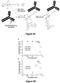
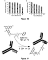
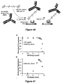
- the immunoglobulin may be bound to the surface through a biotin-streptavidin interaction, or through the interaction with and amine reactive maleic anhydride (see Figures 38 and 40 ).
- the solid support surface mentioned above can include polymers, such as polystyrene, agarose, Sepharose (a crosslinked, beaded-form of agarose), cellulose, glass beads and magnetizable particles of cellulose or other polymers.
- the solid-support can be in the form of large or small beads or particles, tubes, plates, slides, chips or other forms.
- a solid support surface use is preferably made of a test tube, or a microtiter plate the inner walls of which are coated with a first immunoglobulin.
- Dipstick assays can be used to detect an antigen of interest. Dipstick assays use the well-established lateral flow format, wherein capture immunoglobulins are striped or banded onto nitrocellulose membrane and a wicking pad draws the sample up through the dipstick, whereby the antigen of interest interact with the appropriate immunoglobulin. Other immunoglobulins specific to other antigens of interest can be included. Subsequent analysis of enzyme activity and protein quantity can be done using standard methods known to a person skilled in the art, or as discussed above regarding ELISAs.
- Microfluidic devices which may also be referred to as "lab-on-a-chip” systems, biomedical micro-electro-mechanical systems (bioMEMs), or multicomponent integrated systems, can be used for detecting an antigen of interest.
- bioMEMs biomedical micro-electro-mechanical systems
- Such systems miniaturize and compartmentalize processes that allow for detection of antigens of interest, and other processes such as SPR, waveguide, and impedance quantification.
- Array-based assays and bead-based assays can be used with microfluidic devices.
- an immunoglobulin can be coupled to beads and the binding reaction between the coated beads and antigen of interest can be performed in a microfluidic device. Multiplexing, or detecting more than one antigen of interest at once, can also be performed using a microfluidic device.
- Different compartments can comprise different immunoglobulin populations for different antigens of interest, where each population has a different target antigen.
- microarrays are used to detect antigens of interest.
- Microarrays are typically small, high throughput chips generally made of a solid support structure, typically glass slides, nitrocellulose, or microtiter plates.
- immunoglobulins to antigen of interest are bound to the solid support surface. Detection of the captured antigen can be accomplished as discussed above for ELISA detection, or through any method known to a person of ordinary skill in the art.
- the present disclosure is also directed to a kit or system useful for practicing the methods described herein.
- the kit may be a packaged combination of one or more containers, devices, or the like holding the necessary reagents, and usually including written instructions for the performance of assays.
- the kit may include containers to hold the materials during storage, use or both.
- the kit may include any configurations and compositions for performing the various assays described herein, including, but not limited to a means of detecting an antigen of interest and a means to detect the recognition of the detection.
- a kit may only include a detection device having a means for detecting an antigen of interest, and a means for recognition of the detection.
- the kit may only include a detection device having a means for detecting an antigen of interest.
- a means of detection may be an immunoglobulin specific to an antigen of interest.
- At least one reagent is provided for the detection of the recognition of the means of detecting the antigen of interest, which is accomplished by suitable means.
- suitable means may be an immune affinity procedure, an enzymatic assay, or means for detecting a structural feature, amongst others.
- the detection of the recognition of at least one means of detecting an antigen of interest is achieved through an immune affinity procedure is any one of enzyme-linked immunosorbent assay (ELISA), Western Blot, immuno-precipitation, FACS, Biochip array, Lateral Flow, Time Resolved Fluorometry, immuno-fluorochemistry, ECL procedures, or any other procedure based on immune recognition.
- ELISA enzyme-linked immunosorbent assay
- Western Blot Western Blot
- immuno-precipitation FACS
- Biochip array Biochip array
- Lateral Flow Lateral Flow
- Time Resolved Fluorometry immuno-fluorochemistry
- ECL procedures or any other procedure based on immune recognition.
- the kit may comprise a detection device having at least one compartment.
- One compartment may have an array of at least one means of detection wherein each means of detection is located in a defined position in the array.
- array refers to an "addressed" spatial arrangement of the recognition means.
- Each "address" of the array is a predetermined specific spatial region containing a recognition agent.
- an array may be a plurality of vessels (test tubes), plates, micro-wells in a micro-plate each containing a different immunoglobulin.
- An array may also be any solid support holding in distinct regions (dots, lines, columns) different and known recognition agents, for example immunoglobulins.
- the array preferably includes built-in appropriate controls, for example, regions without the sample, regions without the immunoglobulin, regions without either, namely with solvent and reagents alone and regions containing synthetic or isolated proteins or peptides, corresponding to the antigen of interest (positive control).
- Solid support surfaces used for the array will be described in more detail herein after, in connection with the kits provided.
- a solid support surface suitable for use in the kits is typically substantially insoluble in liquid phases.
- Solid support surfaces are not limited to a specific type of support surface. Rather, a large number of supports are available and are known to one of ordinary skill in the art.
- useful solid supports include solid and semi-solid matrixes, such as aerogels and hydrogels, resins, beads, biochip arrays (including thin film coated biochips), microfluidic devices, a silicon chip, multi-well plates (also referred to as micro-titer plates or microplates), lateral flow devices, membranes, filters, dip stick tests, conducting and nonconducting metals, glass (including microscope slides) and magnetic support surfaces.
- useful solid support surfaces include silica gels, polymeric membranes, particles, derivatized plastic films, glass beads, cotton, plastic beads, alumina gels, and polysaccharides such as Sepharose, nylon, latex bead, magnetic bead, paramagnetic bead, super-paramagnetic bead, starch and the like. It should be further noted that any of the reagents included in any of the methods and kits may be provided as reagents embedded, linked, connected, attached placed or fused to any of the solid support surfaces described above.
- An exemplary kit disclosed herein may contain, for example, any combination of:
- the supplied immunoglobulin is specific to the prostate specific antigen (PSA), also known as Kaillikrein-3.
- PSA prostate specific antigen
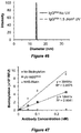
- Kaillikrein-3 is a 30-34 kDa glycoprotein enzyme whose serum levels have been implicated in the early detection of prostate cancer. Detection of PSA using the methods described herein have shown an increase of dynamic detection range, lower limit of detection, higher antigen sensitivity and higher signal intensity when compared to detection methods known to the skilled artisan (see Figures 43- 44 ).
- the kit may contain an IBA conjugated compound.
- the kit may contain the IBA conjugates: IBA-linker-biotin, IBA-linker-MAL/Cys, IBA-linker-FITC, IBA-linker-hexa histadine tag, IBA-linker-peptide, IBA-PEG200-lipid, IBA-Lys-FITC, IBA-EG 11 -amine, IBA-EG 2 -Lys-Lys-Cys, IBA-EG 2 -His 6 -Lys-FITC and IBA-thiol-FITC.
- the kit may also include the above listed IBA conjugates pre-coupled to an immunoglobulin of interest.
- the kit may also contain IBA-chemotheraputics, IBA-cytotoxic agents, IBA-contrasting agents, IBA-active peptides, IBA-thiol-chemotherapeutics, IBA-thiol-cytotoxic agents, IBA-thiol-contrasting agents and IBA-thiol-active peptides.
- the kit can further contain ligands that are thiol reactive such as maleimide-FITC.
- references in the specification to "one embodiment”, “an embodiment”, etc., indicate that the embodiment described may include a particular aspect, feature, structure, moiety, or characteristic, but not every embodiment necessarily includes that aspect, feature, structure, moiety, or characteristic. Moreover, such phrases may, but do not necessarily, refer to the same embodiment referred to in other portions of the specification. Further, when a particular aspect, feature, structure, moiety, or characteristic is described in connection with an embodiment, it is within the knowledge of one skilled in the art to affect or connect such aspect, feature, structure, moiety, or characteristic with other embodiments, whether or not explicitly described.
- the term “about” can refer to a variation of ⁇ 5%, ⁇ 10%, ⁇ 20%, or ⁇ 25% of the value specified. For example, “about 50" percent can in some embodiments carry a variation from 45 to 55 percent.
- the term “about” can include one or two integers greater than and/or less than a recited integer at each end of the range. Unless indicated otherwise herein, the term “about” is intended to include values, e.g., weight percentages, proximate to the recited range that are equivalent in terms of the functionality of the individual ingredient, the composition, or the embodiment.
- the term about can also modify the end-points of a recited range as discuss above in this paragraph.
- ranges recited herein also encompass any and all possible sub-ranges and combinations of sub-ranges thereof, as well as the individual values making up the range, particularly integer values.
- a recited range e.g., weight percentages or carbon groups
- Any listed range can be easily recognized as sufficiently describing and enabling the same range being broken down into at least equal halves, thirds, quarters, fifths, or tenths. As a non-limiting example, each range discussed herein can be readily broken down into a lower third, middle third and upper third, etc.
- an “active peptide” refers to a short amino acid sequences, produced either naturally or synthetically that have hormone or drug like activity that can modulate physiological function through interaction with a target molecule.
- photo reactive refers to crosslinking molecules that are capable of forming a covalent bond with another molecule after exposure to ultra-violet light.
- an “immunoglobulin-ligand conjugate” refers to an immunoglobulin that is conjugated to one or more effector molecules.
- antigen binding site refers to the part of an immunoglobulin molecule that binds antigen specifically.
- contrast agent or “contrasting agent” refers to a substance used to enhance the contrast of structures, cells or fluids within the body in diagnostic and medical imaging.
- fragment refers to a peptide or polypeptide comprising an amino acid sequence of at least 2 contiguous amino acid residues, at least 5 contiguous amino acid residues, at least 10 contiguous amino acid residues, at least 15 contiguous amino acid residues, at least 20 contiguous amino acid residues, at least 25 contiguous amino acid residues, at least 40 contiguous amino acid residues, at least 50 contiguous amino acid residues, at least 60 contiguous amino residues, at least 70 contiguous amino acid residues, at least contiguous 80 amino acid residues, at least contiguous 90 amino acid residues, at least contiguous 100 amino acid residues, at least contiguous 125 amino acid residues, at least 150 contiguous amino acid residues, at least contiguous 175 amino acid residues, at least contiguous 200 amino acid residues, or at least contiguous 250 amino acid residues of the amino acid sequence of a primary or secondary effector molecule.
- isolated in the context of a peptide, polypeptide, fusion protein, antibody or antigen-binding antibody fragment refers to a peptide, polypeptide, fusion protein, antibody or antigen-binding antibody fragment which is substantially free of cellular material or contaminating proteins from the cell or tissue source from which it is derived or obtained, or substantially free of chemical precursors or other chemicals when chemically synthesized.
- substantially free of cellular material or contaminating protein includes preparations of a peptide, polypeptide, fusion protein, antibody or antigen-binding antibody fragment in which the peptide, polypeptide, fusion protein, antibody or antigen-binding antibody fragment is separated from cellular components of the cells from which it is isolated or recombinantly produced.
- a peptide, polypeptide, fusion protein, antibody or antigen-binding antibody fragment that is substantially free of cellular material or contaminating protein includes preparations of a peptide, polypeptide, fusion protein, antibody or antigen-binding antibody fragment having less than about 30%, about 20%, about 10%, or about 5% (by dry weight) of other protein.
- the peptide, polypeptide, fusion protein, antibody or antigen-binding antibody fragment is recombinantly produced, it is also preferably substantially free of culture medium, i.e., culture medium represents less than about 20%, about 10%, or about 5% of the volume of the protein preparation.
- culture medium represents less than about 20%, about 10%, or about 5% of the volume of the protein preparation.
- the peptide, polypeptide, fusion protein, antibody or antigen-binding antibody fragment is produced by chemical synthesis, it is preferably substantially free of chemical precursors or other chemicals, i.e., it is separated from chemical precursors or other chemicals which are involved in the synthesis of the peptide, polypeptide, fusion protein, antibody or antigen-binding antibody fragment.
- such preparations of a peptide, polypeptide, fusion protein, antibody or antigen-binding antibody fragment have less than about 30%, about 20%, about 10%, about 5% (by dry weight) of chemical precursors or compounds other than the peptide, polypeptide, fusion protein, antibody
- NBS nucleotide binding site
- IBA indole-3-butyric acid
- IBA N,N -Diisopropylethylamine
- DIEA 1-fluoro-2,4-dinitrobenzene
- DCC N,N'- Dicyclohexyl carbodiimide
- DMAP 4-(Dimethylamino)pyridine
- paclitaxel purchased from Sigma-Aldrich (St. Louis, MO).
- Streptavidin-HRP, HRP-conjugated IgG Fc ⁇ specific goat anti-mouse, goat anti-rat and goat anti-human were purchased from Jackson ImmunoResearch (West Grove, PA).
- BSA bovine serum albumin
- mouse anti-FITC IgG FITC , clone: DE3
- Amicon Ultra centrifugal filters 0.5 mL, 10K
- Coomassie R-250 and C18 Ziptips were purchased from EMD Millipore (Billerica, MA).
- Amplex Red Assay Kit rat anti-DNP (IgG DNP , clone: LO-DNP-2), tissue culture grade L-glutamine, and ⁇ -mercaptoethanol were purchased from Invitrogen (Grand Island, NY).
- RMPI-1640 media was purchased from Cell-Gro (Manassas, VA).
- Hyclone Fetal Bovine Serum (FBS) and maleic anhydride amine reactive 96-well plates were purchased from Thermo Scientific (Rockford, IL).
- NovaPEG Rink Amide resin, Biotin NovaTag Resin, Fmoc-Cys(Trt)-Wang Resin, and all other amino acids were purchased from Novabiochem (Billerica, MA).
- Fmoc-N-amido-dPEG 2 -acid was purchased from Quanta Biodesign (Powell, OH).
- Tris-gly running buffer, transfer buffer, and tris buffered saline (TBS) were purchased from Boston Bioproducts (Ashland, MA).
- IM9, U266, H929, and MM.1S cell lines were obtained from American Type Culture Collection (Rockville, MD). L-glutamine, penicillin, and streptomycin were purchased from Gibco (Carlsbad, CA).
- Rituximab chimeric human anti-CD20 was a gift from Dr. Rudolph Navari (Indiana University School of Medicine, South Bend, IN).
- IBA-Conjugated Ligands IBA-Ligand
- the purity of both the immunoglobulin and the ligand being conjugated to the immunoglobulin were critical to determine the optimal conditions for UV conjugation.
- All immunoglobulins undergoing photocrosslinking were purchased as purified immunoglobulins with no protein stabilizers. Sodium azide, a very UV reactive preservative, and other small molecule additives were removed prior to UV exposure via membrane filtration.
- Immunoglobulins were incubated with the IBA-ligands for 1 h prior to UV exposure at room temperature (RT). Strict control over the UV energies delivered to the samples was achieved using a Spectroline UV Select Series Crosslinker from Spectronics at a wavelength of 254 nm at a fixed distance from the light source.
- BSA-DNP was synthesized in methanol and water by combining 20 molar equivalents of 1-fluoro-2,4-dinitrobenzene to 1 molar equivalent of BSA.
- BSA-FITC was synthesized in PBS by the addition of 20 molar equivalents of FITC. The reactions were carried out overnight at RT with continuous agitation. The products were purified with Amicon spin concentrators (10 kDa MW cutoff, Millipore) to remove unconjugated ligands and to exchange buffer to PBS pH 7.4.
- BSA-DNP product was analyzed by observing absorbance signals at 280 nm for BSA and at 350 nm for DNP.
- BSA-FITC product was analyzed by observing the absorbance signals at 280 nm for BSA and at 494 nm for FITC. By comparing the absorbance values at the two wavelengths and using the extinction coefficients, we quantified the average number of BSA conjugations for both DNP and FITC.
- Cyclic CD20 Mimotope The cyclic CD20 mimotope for Rituximab binding was synthesized using standard solid phase synthesis protocols on a NovaPEG Rink Amide resin and Fmoc chemistry. The following residues were HBTU activated and coupled following Fmoc deprotection in the following order: Cys(Trt), Met, Ser(tBu), Pro, Asn(Trt), Ala, Ala, Trp(Boc), Cys(Trt), Ala, and N -amido-dPEG 2 -acid. Kaiser tests were performed between coupling steps to monitor synthesis progress.
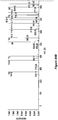
- the peptide was cleaved from the resin in 92.5% TFA, 2.5% TIS, 2.5% EDT, and 2.5% D.I. water, purified via RP-HPLC on a Zorbax C18 column, and characterized using MALDI-TOF MS ( Figure 10 ). The yield was 50%, and product purity was confirmed using RP-HPLC on an analytical Zorbax C18 column to be >95%.
- the peptide was cyclized overnight in DMF with DIEA and verified via MALDI-TOF MS.
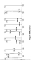
- IBA-Biotin was synthesized using standard solid phase synthesis protocols on a Biotin NovaTag Resin and Fmoc chemistry as described above. The following residues were coupled in order: Lys(Boc), Gly, Gly, IBA. IBA-biotin was cleaved from the resin in 95% TFA, 2.5% TIS, and 2.5% D.I. water, purified via RP-HPLC on a Zorbax C18 column, and characterized using MALDI-TOF MS ( Figure 11 ). The yield was 60%, and product purity was confirmed using RP-HPLC on an analytical Zorbax C18 column to be >95%.
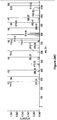
- IBA-FITC was synthesized by HBTU activation of IBA in DMF mixed in equimolar amounts of FITC and L-Lysine. The reaction was carried out overnight while shaking, purified via RP-HPLC on a Zorbax C18 column, and characterized using MALDI-TOF MS ( Figure 12 ). The yield was 75%, and product purity was confirmed using RP-HPLC on an analytical Zorbax C18 column to be >95%.
- IBA-iRGD The IBA-iRGD molecule was synthesized using standard solid phase synthesis protocols on a Fmoc-Cys(Trt)-Wang Resin and Fmoc chemistry as described above. The following residues were coupled in order: Asp(OtBu), Pro, Gly, Lys(Boc), Asp(OtBu), Gly, Arg(Pbf), Cys(Trt), N-amido-dPEG 2 -acid, Lys(ivDde), N-amido-dPEG 2 -acid, Lys(Boc), IBA.
- the ivDde protecting group was removed by 2% hydrazine in DMF.
- FITC was allowed to react for 3 h in DMF.
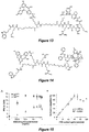
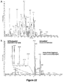
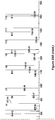
- the molecule was then cleaved from the resin in 92.5% TFA, 2.5% TIS, 2.5% EDT and 2.5% D.I. water, purified via RP-HPLC on a Zorbax C18 column, and characterized using MALDI-TOF MS ( Figure 13 ).
- the peptide was cyclized overnight in DMF with DIEA and cyclization was verified via MALDI-TOF MS.
- the yield was 40%, and product purity was confirmed using RP-HPLC on an analytical Zorbax C18 column to be >95%.
- IBA-paclitaxel The IBA-paclitaxel molecule was synthesized using standard solid phase synthesis protocols on a NovaPEG Rink Amide resin and Fmoc chemistry as described above. The following residues were coupled in order: Glu(OtBu), N-amido-dPEG 2 -acid, Arg(Pbf), Arg(Pbf), Lys(ivDde), N-amido-dPEG 2 -acid, IBA. The ivDde protecting group was removed by 2% hydrazine in DMF and FITC was allowed to react for 3 h in DMF.
- the molecule was then cleaved from the resin in 95% TFA, 2.5% TIS, and 2.5% D.I. water, purified via RP-HPLC on a Zorbax C18 column, and characterized using MALDI-TOF MS ( Figure 14 ).
- the resulting acid containing molecule was then activated with 2 equivalents of DCC, 1 equivalent of DMAP in DMF, and was added to 1 equivalent of Paclitaxel. After the reaction was carried out overnight at RT, the DMF was rotate evaporated.
- the molecule was purified via RP-HPLC on a Zorbax C3 column, and characterized using MALDI-TOF MS. The yield was 50%, and product purity was confirmed using RP-HPLC on an analytical Zorbax C18 column to be >95%.
- Antigen coated ELISA plates for (Rituximab, IgG DNP and IgG FITC ) were generated by adsorbing BSA-DNP or BSA-FITC (0.1 mg/mL) to high binding 96-well ELISA plates in 0.05 M carbonate-bicarbonate coating buffer at pH 9.6 for 2 hour at RT.
- Antigen coated ELISA plates for Rituximab were generated by covalently reacting the cyclic CD20 mimotope (60 pmoles) to a maleic anhydride amine reactive plate surface in PBS pH 8.0 for 2 hour at RT and any remaining reactive sites were then quenched using 50 mM Tris buffer with 100 mM NaCl at pH 8.0 for 1 h ( Figure 15 ). All plate surfaces were then blocked with BSA blocking buffer (100 ⁇ L of 5% BSA in PBS pH 7.4 with 0.1% Tween 20) for 1 hour.
- BSA blocking buffer 100 ⁇ L of 5% BSA in PBS pH 7.4 with 0.1% Tween 20
- Each immunoglobulin (Rituximab, IgG DNP , IgG FITC ) was exposed to UV in the presence or absence of IBA-biotin and were then incubated in the respective antigen coated plates. The plates were washed to remove any unbound components using an automated plate washer (three cycles of 200 ⁇ L PBS with 0.05% Tween 20 at pH 7.4). In an alternate assay, immunoglobulin exposed to UV energy in the presence or absence of IBA-biotin was directly adsorbed to a high binding 96-well ELISA plate in 0.05 M carbonate-bicarbonate coating buffer at pH 9.6 for 2 h at RT.
- SEC Size Exclusion Chromatography
- Immunoglobulin (Rituximab, IgG DNP and IgG FITC ) at 20 ⁇ M was incubated with excess IBA-biotin (300 ⁇ M) in PBS buffer at pH 7.4 and exposed to the indicated amount of UV energy.
- the samples were run on a 10% SDS-PAGE gel with a tris-glycine running buffer under reducing conditions at 110 V for 1 h and were transferred to a nitrocellulose membrane at 110 V for 90 min in a 10% MeOH transfer buffer.
- the membrane was blocked with 10% dry milk in TBS for 1 h and was then blotted with 1:10,000 dilution of streptavidin-HRP for 1 h at RT.
- a chemiluminescent HRP substrate was used to detect the location where IBA-biotin was covalently conjugated to the immunoglobulin.
- both the SDS-PAGE gel (post transfer) and nitrocellulose membrane were coomassie blue stained in a solution of 10% acetic acid, 20% methanol, 0.15% Coomassie R-250 for 30 min and destained in a solution of 20% acetic acid, 20% methanol, 60% D.I. water for 1.5 h. Control experiments performed in the absence of UV exposure, or in the absence of IBA-biotin did not yield any detectable bands. Similarly, control experiments performed with only biotin did not yield any detectable bands.
- the modeling software used to perform the minimization was MOE (version 2011.10, Chemical Computing Group, Montreal, Canada). Crystallographic water molecules were removed from the X-ray crystal structure of Rituximab (PDB: 2OSL), then protonated using the Protonate3D module in MOE and AMBER99 to assign partial charges to receptor atoms.
- IBA was constructed using the Builder module in MOE. Protons were assigned, and AM1-BCC partial charges were computed for the ligand atoms.
- the MOE Dock module was used to generate a proposed binding mode of IBA to the NBS.
- the top-ranking ligand binding mode from the docking was minimized with the MOE Energy Minimize method, employing Steepest Descents, Conjugate Gradient, and Truncated Newton until the system converged (gradient ⁇ 0.05). All receptor atoms were held in place with a fixed potential, and only the residues on the loop defined by Phe35-Pro45 were unfixed and allowed to move, to model any induced-fit of the receptor due to the binding of the IBA ligand.
- IM9, U266, H929, and MM.1S cell lines were cultured in RPMI 1640 media containing 10% FBS, 2 mM L-glutamine, 100 U/mL penicillin, and 100 ⁇ g/mL streptomycin.
- IM9 served as a CD20 positive cell line, and all others were used as negative controls (as determined by Rituximab staining followed by a FITC-labeled secondary immunoglobulin, results not shown).
- IBA-FITC was photocrosslinked to Rituximab at the indicated UV energies.
- UV energy exposure Several parameters such as UV energy exposure, IBA-ligand concentration, immunoglobulin concentration, and buffer conditions are important factors effecting the optimization of the UV-NBS method for efficient photocrosslinking at the NBS.
- the amount of UV energy has to be sufficiently high to promote maximum crosslinking of the IBA without causing damage to the immunoglobulin such that the antigen binding and Fc related functions are preserved.
- the IBA-ligand and immunoglobulin concentrations are critical since the site-specific photocrosslinking is dependent on the non-covalent association between the IBA and NBS prior to UV exposure.
- the IBA-ligand concentration must be sufficiently high that all NBS are occupied by an IBA-ligand but not so high as to promote non-specific coupling to the immunoglobulin as a result of weak non-specific interactions.
- the buffer conditions also play a crucial role in the crosslinking efficiency by affecting the binding interactions between the IBA-ligand and NBS, as well as by enhancing or reducing photo-reactivity of the IBA since UV coupling can be highly dependent on pH.
- IBA-biotin was photocrosslinked to the immunoglobulin by first incubating Rituximab, IgG DNP or IgG FITC immunoglobulins (20 ⁇ M) with saturating concentrations of IBA-biotin (300 ⁇ M) in PBS pH 7.4 to allow for the non-covalent association between IBA and the NBS.
- the saturating concentration of IBA-biotin for the NBS was estimated to be >100 ⁇ M based on the previously reported K d for IBA/NBS interactions (1-8 ⁇ M).
- the samples were then exposed to increasing amounts of UV energy, from 0 to 10 J/cm 2 , to enable IBA-biotin photocrosslinking at the NBS.
- the photocrosslinking efficiency was determined via an ELISA assay, where the IBA-biotin photocrosslinked immunoglobulins were incubated on plate surfaces coated with their respective antigens, and the degree of immunoglobulin biotinylation was determined using HRP conjugated streptavidin (see description below).
- the results we obtained represent the photocrosslinking efficiency combined with the antigen binding activity of the immunoglobulin.
- the IBA-ligand concentration plays a critical role in the efficiency of photocrosslinking since it determines the extent to which all NBS are bound to IBA prior to UV exposure.
- an ELISA study was conducted in which the immunoglobulin concentration (20 ⁇ M) and the amount of UV energy exposure (1.0 J/cm 2 ) were held constant while the ligand, IBA-biotin, concentration was varied from 0-200 ⁇ M.
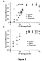
- a UV energy of 1.0 J/cm 2 was selected since this UV energy demonstrated efficient photocrosslinking without any reduction in antigen recognition or Fc damage to the immunoglobulins ( Figure 2 ).
- the IBA-biotin photocrosslinked immunoglobulins were adsorbed to high binding ELISA plates and HRP-streptavidin was used as the reporter to assess the degree of immunoglobulin biotinylation.
- HRP-streptavidin was used as the reporter to assess the degree of immunoglobulin biotinylation.
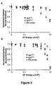
- Increasing the ligand concentration resulted in an increase in the UV crosslinking efficiency reaching a plateau at ⁇ 100 ⁇ M, as can be observed in Figure 3 .
- the curves were then fit to a sigmoid and an EC 50 (half maximum effective concentration) value was determined ( Figure 3 ).
- the EC 50 values for Rituximab, IgG DNP and IgG FITC were 1.7, 1.3 and 6.4 ⁇ M, respectively.
- the effect of UV energy on the average number of IBA conjugations per immunoglobulin was evaluated first by incubating IBA-FITC at 300 ⁇ M, a concentration that promoted complete association with the immunoglobulin, and was then exposed to the indicated UV energy ( Figure 4A ).
- the IBA-FITC conjugated immunoglobulin was then injected on an SEC column where non-conjugated IBA-FITC ligand eluted separately from the conjugated and non-conjugated immunoglobulin.
- the number of non-specific conjugations remains low, i.e., ⁇ 1 at 3 J/cm 2 , due to the short life span of the UV activated IBA radical.
- the specific photocrosslinking reaction between the NBS and IBA-ligand occurs much more rapidly than the non-specific photocrosslinking due to the proximity of the IBA to reactive moieties when bound to the NBS and the enhanced photo reactivity of the aromatic rich NBS. No crosslinking was observed in the absence of IBA or in the absence of UV energy, as expected.
- the 254 nm UV exposures to initiate photocrosslinking between the IBA and the immunoglobulin had minimal impact on the adsorption or fluorescence profile of the conjugated FITC at the UV energy levels used throughout these experiments ( Figure 17 and 18 ).
- UV exposure is an essential step in photocrosslinking of functional groups to immunoglobulins via the UV-NBS method.
- too much UV exposure has potentially damaging effects to the immunoglobulin's structure, which may lead to: i) loss in the immunoglobulin's ability to recognize its antigen, and/or ii) loss in Fc domain recognition by a secondary immunoglobulin.
- the UV energies necessary to initiate covalent bond formation did not render the immunoglobulin inactive, we evaluated the effect of UV energy on immunoglobulin activity and Fc recognition.
- IgG DNP and IgG FITC were exposed to increasing UV energies in the presence of 300 ⁇ M IBA-biotin and evaluated for their antigen binding activity as well as their recognition by Fc-specific secondary immunoglobulins.
- the respective antigens for each immunoglobulin were first immobilized onto an ELISA plate surface through either physical adsorption (BSA-FITC, BSA-DNP) or covalent lysine side chain immobilization (cyclic CD20 mimotope). Next, the UV-exposed, IBA-biotin photocrosslinked immunoglobulins were incubated on the antigen-coated plates and allowed to bind to their respective antigens.
- IgG DNP demonstrated reduced Fc recognition with ⁇ 75% activity at 2.0 J/cm 2 , significantly lower when compared to Rituximab and IgG FITC .
- the most likely cause for the increased sensitivity of IgG DNP to UV exposure is a result of damage to the Fc domain, causing decreased recognition by the secondary immunoglobulin.
- the results of the biotinylation assays depicted in Figure 2A , there was no reduction in biotinylation intensity for IgG DNP up to UV energies of 5 J/cm 2 , demonstrating that the antigen binding activity of IgG DNP is preserved even at high UV energies.
- Tween 20 is often added to immunoglobulin formulations as a stabilizer to reduce undesired non-specific hydrophobic interactions between proteins. Including Tween 20 in the buffer, up to 1 % V/V, had negligible impact on the resulting number of IBA-FITC molecules photocrosslinked to the immunoglobulin ( Figure 19C ). This result revealed that Tween 20 did not interfere with IBA binding to the NBS. Although including Tween 20 in the conjugation buffer was deemed unnecessary for efficient IBA-FITC conjugation, certain larger ligands that contain hydrophobic regions, such as targeting peptides and cytotoxic drugs, may benefit greatly from the presence of a surfactant. Incorporating a surfactant in the photocrosslinking buffer can potentially reduce non-specific interactions between the ligand and the immunoglobulin, thereby inhibiting any non-site-specific conjugations.
- biotinylated immunoglobulins were run under reducing conditions on SDS-PAGE ( Figure 6A ), transferred to a nitrocellulose membrane and biotinylated immunoglobulin fragments were probed by using HRP conjugated streptavidin ( Figure 6B ).
- the developed film established that the biotinylation of the immunoglobulin occurred selectively at the light chain, and the yield of conjugation was dependent on the amount of UV energy exposure. While the NBS is comprised of residues that are both on the V L and V H , the orientation of the non-covalent interactions between the IBA and the residues of the NBS yields crosslinking exclusively to the immunoglobulin light chain. This result was consistent across all three immunoglobulins tested (results not shown). At UV exposures up to 4.0 J/cm 2 , biotinylation was still highly selective for the immunoglobulin light chain ( Figure 21 and 39 ).
- the IBA-biotin photocrosslinked Rituximab was first enzymatically digested into smaller peptide fragments in the presence of trypsin or pepsin using literature protocols. The digested immunoglobulin fragments were then separated on a C18 column and analyzed by an LTQ-Velos Orbitrap mass spectrometer. The biotinylated peptide fragment was then identified based on the presence of diagnostic precursor ions unique to internal fragmentation of the IBA-biotin molecule obtained under MS/MS conditions: 472.23, 679.50, and 714.27 m/z (indicative of intact IBA-biotin).
- UV exposure of IBA can cause the indole to become excited and result in the formation of an N-formylkynurenine as indicated by the m/z of 2140.26 corresponding to a covalent bond between the N-formylkynurenine and unmodified phenylalanine.
- N -formylkynurenine most commonly undergoes a photo decomposition reaction resulting in kynurenine formation ( Figure 24 and 25 ). It is also well established that when phenylalanine is exposed to UV light, the dominant product is hydroxylation of the phenyl ring resulting in the formation of a tyrosine like amino acid derivative.
- the final identified MS/MS fragment indicates a covalent bond between the kynurenine and hydroxylated phenylalanine as indicated by an m/z of 1471.94 ( Figure 1C ).
- the AMBER99 force field with a Generalized-Born implicit solvation model was used to model both the internal energy of the protein, the non-bonded interactions, and the solute-solvent interactions.
- the energy-minimized binding mode is shown in Figure 7B .
- the docking analysis suggests a binding conformation where IBA stacks against the indole ring of the tryptophan at position 118 on the V H , fixing the IBA within the pocket.
- the only accessible site for conjugation is the phenylalanine at position 42 on the light chain. This finding was in line with our MS analysis of the conjugation site and helped to determine the most likely site of covalent bond formation between IBA and phenylalanine ( Figure 26 ).
- FITC labeled immunoglobulins are commonly used in flow cytometry assays for determination of expression levels of cell surface receptors as well as for cell sorting.
- IBA-FITC immunoglobulin labeled immunoglobulins
- Rituximab 20 ⁇ M was incubated with saturating levels of IBA-FITC and exposed to increasing amounts of UV energy to perform the site-specific photocrosslinking reaction.
- the IBA-FITC photocrosslinked Rituximab (200 nM) was then incubated with the CD20 positive IM9 cell line, and the CD20 expression levels were detected by flow cytometry.
- the fluorescent signal increased with increasing UV energy with an optimal UV exposure of 1 J/cm 2 , consistent with previous results.
- the Rituximab-IBA-FITC conjugate was incubated with three CD20 negative myeloma cell lines, U266, H929, and MM.1S.
- UV-NBS UV-NBS Method in Photocrosslinking of Functional Peptides (iRGD) and Chemotherapeutic Agents (Paclitaxel) to Immunoglobulins.
- iRGD Functional Peptides
- Paclitaxel Chemotherapeutic Agents
- the iRGD cyclic peptide construct was covalently conjugated to IBA to enable photocrosslinking of the IBA-iRGD molecule to the immunoglobulins using the UV-NBS method.
- an ethylene glycol linker of two units was introduced between IBA and iRGD to provide flexibility.
- selectively protected lysine was also added which facilitated conjugation of FITC to the IBA-iRGD molecule ( Figure 13 ).
- the addition of FITC allowed for the quantification of the number of conjugations via absorbance at 494 nm by SEC peak integrations, as described previously.
- UV-NBS photocrosslinking method can be applied broadly, there still remain some limitations.
- the UV-NBS method currently cannot be implemented for photocrosslinking of functional ligands that are UV sensitive.
- Some examples of UV sensitive functional ligands are hexa-histadine to his-tag an immunoglobulin facilitating capture via immobilized metal affinity chromatography (IMAC), DNA to facilitate surface immobilization on protein microarrays, and exceptionally photo-sensitive fluorophores that are prone to photo-bleaching.
- IMAC immobilized metal affinity chromatography
- We are currently developing methods to site-specifically photocrosslink an orthogonal reactive group to the NBS to facilitate conjugation of UV sensitive moieties to the NBS through the intermediary UV-NBS conjugated reactive group.
- UV-NBS UV-NBS functionalized immunoglobulins containing the iRGD targeting peptide results in the formation of a bispecific multivalent immunoglobulin conjugate with potential for enhanced tumor targeting and penetration, and paclitaxel conjugated immunoglobulins provide for targeted delivery of cytotoxic drugs.
- UV-NBS method provides a universal, site-specific, and efficient method to functionalize immunoglobulins, with significant implications in various diagnostic and therapeutic applications.
- UV-NBS photocrosslinking technique requires exposure to UV energy that some functional ligands may not be stable to.
- IBA-Thiol an IBA conjugated version of cysteine
- This thiol group can then be used as an orthogonally reactive site to conjugate UV sensitive functional ligands that possess either a thiol reactive group resulting in disulfide bond formation or subsequent reaction with a maleimide functionalized ligand ( Figure 28 ).
- the results detailed here provide a universal technique for the site-specific conjugation of UV sensitive functional ligands to immunoglobulins at the NBS, while preserving immunoglobulin activity.
- IBA Indole-3-butyric acid
- DIEA N,N- Diisopropylethylamine
- DTT dithiothreitol
- fluorescein-5-maleimide purchased from Sigma-Aldrich (St. Louis, MO).
- Streptavidin-HRP, HRP-conjugated IgG Fc ⁇ specific goat anti-mouse, and HRP-conjugated IgG goat anti-fluorescein were purchased from Jackson ImmunoResearch (West Grove, PA).
- BSA bovine serum albumin
- Amicon Ultra centrifugal filters 0.5 mL, 10K
- Amplex Red Assay Kit was purchased from Invitrogen (Grand Island, NY).
- High binding 96-well ELISA plates were purchased from Thermo Scientific (Rockford, IL).
- NovaPEG Rink Amide resin, Biotin NovaTag resin, Fmoc-Cys(Trt)-Wang resin, and all other amino acids were purchased from Novabiochem (Billerica, MA).
- Fmoc-N-amido-dPEG 2 -acid was purchased from Quanta Biodesign (Powell, OH).
- Mouse anti-PSA IgG PSA , clone: B731M
- purified free prostate specific antigen PSA
- IBA-Thiol was synthesized using standard solid phase peptide synthesis protocols on a Fmoc-Cys(Trt) Wang resin and Fmoc chemistry.
- Fmoc-Lys(Boc)-OH was coupled to the resin following HBTU activation in DMF and DIEA at RT for 3.5 h while agitating.
- Fmoc was deprotected using 20% piperidine in DMF and the following residues were added in order: Fmoc-Lys(Boc)-OH, Fmoc-N-amido-dPEG 2 -acid, and IBA. Kaiser tests were performed between coupling steps to monitor the synthesis progress.
- the peptide was cleaved from the resin in a solution of 4% triisopropylsilane, 4% D.I. water, 4% DTT and 88% TFA for 45 min at RT ( Figure 32 ).
- IBA-Thiol was purified via RP-HPLC on a Zorbax C18 column, and characterized using MALDI-TOF MS. The purity was confirmed using RP-HPLC on an analytical Zorbax C18 column (>95%), and the yield was 70%.
- Antigen coated ELISA plates were generated by adsorbing PSA (10 nM) to high binding 96-well ELISA plates in 0.05 M carbonate-bicarbonate coating buffer at pH 9.6 for 2 hours at RT. The plate surfaces were then blocked with BSA blocking buffer (200 ⁇ L of 5% BSA in PBS pH 7.4 with 0.1% Tween 20) for 1 h. The IgG PSA was then exposed to UV light (254 nm) in the presence or absence of IBA-Biotin or IBA-Thiol (300 ⁇ M) in PBS pH 6.8.
- Antigen coated ELISA plates were generated by adsorbing PSA (10 nM) to high binding 96-well ELISA plates in 0.05 M carbonate-bicarbonate coating buffer at pH 9.6 for 2 hour at RT. The plate surfaces were then blocked with BSA blocking buffer for 1 h. The IgG PSA was then exposed to UV light (254 nm) in the presence or absence of IBA-Biotin or IBA-Thiol (300 ⁇ M) in PBS pH 6.8.
- UV conjugated IBA-Thiol was then reacted with 5 equivalents of fluorescein-5-maleimide for 1 hour at RT.
- the UV coupled IgG PSA was then incubated on the antigen coated plate surfaces.
- the plates were washed to remove any unbound components using an automated plate washer (three cycles of 200 ⁇ L PBS with 0.05% Tween 20 at pH 7.4).
- the degree of UV immunoglobulin thiolation, via fluorescein-5-maleimide, for each sample was determined by incubating with a 1:5,000 dilution of HRP conjugated goat anti-fluorescein immunoglobulin (1.0 mg/mL stock) in BSA blocking buffer for 1 h.
- IgG PSA was exposed to increasing UV energies (0-5 J/cm 2 ) in the presence of a saturating concentration of IBA-Thiol (300 ⁇ M) in PBS pH 6.8 to allow for covalent photocrosslinking between the IBA and NBS.
- PBS pH 6.8 was selected due to the reduced rate of disulfide bond formation providing for 97% free thiols remaining after 6 h at room temperature and 85% remaining after 24 hours ( Figure 36 ). This pH also provides for efficient IBA/NBS binding and maintains a high level of site-specific photocrosslinking efficiency.
- the excess IBA-Thiol was removed via membrane filtration and the UV-exposed immunoglobulins were then incubated with a 5-fold excess of maleimide-fluorescein to react to all conjugated reactive thiols.
- the conjugated immunoglobulins were then allowed to bind to surface immobilized prostate specific antigen (PSA).
- PSA prostate specific antigen
- Total immunoglobulin activity was determined by an Fc-specific HRP conjugated secondary immunoglobulin ( Figure 29A ) and IBA-Thiol photocrosslinking efficiency was determined by an anti-fluorescein HRP conjugated secondary immunoglobulin ( Figure 29B ).
- the IBA-Thiol ligand provides some UV shielding that protects the immunoglobulin from damage at high UV energies similarly to IBA-Biotin with an immunoglobulin activity level of 95% at 1.5 J/cm 2 UV ( Figure 29A and Figure 11 ).
- the photocrosslinking efficiency and thiol reactivity post UV exposure follows a similar trend as IBA-Biotin with an increased UV sensitivity above 1.5 J/cm 2 .
- IBA-FITC at 300 ⁇ M ( Figure 12 ) was incubated with IgG PSA , providing for complete non-covalent association of IBA-FITC to all NBS, and was then exposed to the indicated UV energies.
- the IBA-FITC conjugated immunoglobulin was then injected on a size exclusion chromatography (SEC) column where non-conjugated IBA-FITC ligand eluted separately from the immunoglobulin conjugate.
- SEC size exclusion chromatography
- UV-NBS Biotin method site-specifically conjugating a biotin to the NBS of IgG PSA prior to immobilization (UV-NBS Biotin method) the low immunoglobulin immobilization efficiency associated with the previously described UV-NBS method was overcome by ensuring an average of ⁇ 1 biotin conjugation per immunoglobulin while still maintaining the highest level of immunoglobulin activity.
- Surfaces functionalized by the UV-NBS Biotin method displayed significantly enhanced immunoglobulin immobilization efficiency, heightened antigen detection sensitivity, reduced LOD, and improved dynamic antigen detection range resulting in an overall increase in assay sensitivity compared to other commonly used immunoglobulin immobilization methods.
- the UV-NBS Biotin method provides for a site-specific covalent conjugation method that does not impact antigen or Fc binding interactions.
- site-specifically conjugating a biotin molecule to immunoglobulins oriented immobilization can be carried out on any surface that bears streptavidin, regardless of antigen specificity.
- the UV-NBS Biotin method provides a universal, site-specific immobilization method that is amenable to any available assay detection modality with potential significant implications in the development of miniaturized medical diagnostics and lab on a chip technologies.
Landscapes
- Health & Medical Sciences (AREA)
- Immunology (AREA)
- Life Sciences & Earth Sciences (AREA)
- Chemical & Material Sciences (AREA)
- Engineering & Computer Science (AREA)
- Medicinal Chemistry (AREA)
- General Health & Medical Sciences (AREA)
- Molecular Biology (AREA)
- Hematology (AREA)
- Urology & Nephrology (AREA)
- Biomedical Technology (AREA)
- Bioinformatics & Cheminformatics (AREA)
- Biochemistry (AREA)
- Organic Chemistry (AREA)
- Epidemiology (AREA)
- Pharmacology & Pharmacy (AREA)
- Veterinary Medicine (AREA)
- Public Health (AREA)
- Animal Behavior & Ethology (AREA)
- Pathology (AREA)
- Analytical Chemistry (AREA)
- Microbiology (AREA)
- General Physics & Mathematics (AREA)
- Food Science & Technology (AREA)
- Physics & Mathematics (AREA)
- Cell Biology (AREA)
- Biotechnology (AREA)
- Biophysics (AREA)
- Genetics & Genomics (AREA)
- Proteomics, Peptides & Aminoacids (AREA)
- Peptides Or Proteins (AREA)
Claims (16)
- Verfahren zur ortsspezifischen Photovernetzung eines Immunglobulins, wobei das Verfahren umfasst:a) Bereitstellen eines Immunglobulins, aufweisend eine konservierte Nukleotidbindungsstelle, die fern von der Antigenbindungsstelle der Fv-Domäne des Immunglobulins liegt;b) Mischen eines heterobifunktionellen photoreaktiven Vernetzers mit dem Immunglobulin, um ein Gemisch aus dem genannten Immunglobulin und dem Vernetzer bereitzustellen, wobei der heterobifunktionelle photoreaktive Vernetzer mindestens eine photoreaktive heterocyclische funktionelle Gruppe aufweist, die mit der konservierten Nukleotidbindungsstelle des Immunglobulins interagiert, wobei die mindestens eine heterocyclische funktionelle Gruppe ein Indol ist, und mindestens eine nicht-photoreaktive funktionelle Gruppe aufweist;c) Aussetzen des Gemischs an ultraviolettes Licht, so dass die mindestens eine photoreaktive funktionelle Gruppe des heterobifunktionellen photoreaktiven Vernetzers kovalent an die Nukleotidbindungsstelle des Immunglobulins gekoppelt wird;wobei die Konzentration des heterobifunktionellen photoreaktiven Vernetzers in dem Gemisch 100-400 µM beträgt und das Verhältnis des Immunglobulins zu dem heterobifunktionellen photoreaktiven Vernetzer in dem Gemisch 1:10 bis 1:20 beträgt.
- Verfahren nach Anspruch 1, wobei die photoreaktive funktionelle Gruppe Indol-3-buttersäure ist.
- Verfahren nach Anspruch 1, wobei die mindestens eine nicht-photoreaktive funktionelle Gruppe an ein Effektormolekül gekoppelt ist;
wobei das Effektormolekül gegebenenfalls ein Markierungsmolekül, ein Affinitäts-Tag, ein Chemotherapeutikum, ein cytotoxisches Mittel, ein aktives Peptid, ein Kontrastmittel, eine radioaktive Markierung, DNA oder ein niedermolekularer Inhibitor ist. - Verfahren nach Anspruch 1, wobei die mindestens eine nicht-photoreaktive funktionelle Gruppe an eine Oberfläche gekoppelt ist.
- Verfahren nach Anspruch 4, wobei die Oberfläche ein Arzneimittelabgabesystem oder ein diagnostisches Kontrastmittel ist; wobei das Arzneimittelabgabesystem gegebenenfalls ein Liposom, eine Mizelle, ein Nanopartikel, einen Quantenpunkt oder ein Dendrimer umfasst.
- Verfahren nach Anspruch 3, wobei das Effektormolekül ein Markierungsmolekül ist und das Markierungsmolekül Biotin ist, wobei das Biotin zugänglich ist, um an Streptavidin zu binden, wobei das Streptavidin zumindest teilweise eine Oberfläche bedeckt; oder
wobei das Effektormolekül ein Markierungsmolekül ist und das Markierungsmolekül eine Fluoreszenz-, Absorptions-, Kontrast- oder Radiomarkierungsfunktion aufweist. - Verfahren nach Anspruch 4, wobei die Oberfläche ein Nanopartikel, ein Kügelchen, eine Mikrofluidikvorrichtung, eine ELISA-Platte oder eine Mikroarray-vorrichtung ist.
- Verfahren nach Anspruch 1, wobei:der heterobifunktionelle Vernetzer eine erste funktionelle Gruppe und eine zweite funktionelle Gruppe aufweist, wobei die erste funktionelle Gruppe die heterocyclische photoreaktive funktionelle Gruppe ist und die zweite funktionelle Gruppe eine funktionelle Thiolgruppe ist;die erste funktionelle Gruppe im Anschluss an das Aussetzen an ultraviolettes Licht kovalent an die Nukleotidbindungsstelle des Immunglobulins gekoppelt wird; unddie funktionelle Thiolgruppe mit einem entsprechenden Thiol-reaktiven funktionalisierten Liganden umgesetzt wird; wodurch ein funktionalisiertes Immunglobulin bereitgestellt wird.
- Verfahren nach Anspruch 8, wobei die heterocyclische photoreaktive funktionelle Gruppe Indol-3-buttersäure ist; oder
wobei die funktionelle Thiolgruppe ein Cysteinrest ist. - Verfahren nach Anspruch 8, wobei der funktionalisierte Ligand an eine Thiol-reaktive Oberfläche gekoppelt ist;
wobei die Oberfläche gegebenenfalls ein Arzneimittelabgabesystem ist;
wobei das Arzneimittelabgabesystem ferner gegebenenfalls ein Liposom, eine Mizelle, ein Nanopartikel, einen Quantenpunkt oder ein Dendrimer umfasst. - Verfahren nach Anspruch 10, wobei die funktionelle Thiolgruppe in einer orientierungsspezifischen Weise an die mit dem funktionalisierten Liganden beschichtete Oberfläche gekoppelt wird, wodurch die Antigenbindungsstellen von der Oberfläche abgewandt orientiert und für eine Antigenbindung verfügbar sind.
- Verfahren nach Anspruch 8, wobei der funktionalisierte Ligand ein Markierungsmolekül, ein Affinitäts-Tag, ein Chemotherapeutikum, ein cytotoxisches Mittel, ein aktives Peptid, ein Kontrastmittel, eine radioaktive Markierung, DNA oder ein niedermolekularer Inhibitor ist;
wobei das aktive Peptid gegebenenfalls ausgewählt ist aus der Gruppe bestehend aus Zellinternalisierungssequenzen, Rezeptor-Targeting-Sequenzen und Mimitopen; oder
wobei das Markierungsmolekül gegebenenfalls eine Fluoreszenz-, Absorptions-, Kontrast- oder Radiomarkierungsfunktion aufweist. - Verfahren nach Anspruch 12, wobei das Affinitäts-Tag Biotin ist, wobei das Biotin an Streptavidin gebunden ist, wobei das Streptavidin zumindest teilweise eine Oberfläche bedeckt.
- Verfahren nach Anspruch 11 oder 13, wobei die Oberfläche die Oberfläche eines Nanopartikels, eines Kügelchens, einer Mikrofluidikvorrichtung, einer ELISA-Platte oder einer Mikroarray-Vorrichtung ist.
- Verfahren nach Anspruch 1, wobei das Immunglobulin ein Immunglobulinfragment ist, das die konservierte Nukleotidbindungsstelle umfasst.
- Verfahren nach Anspruch 15, wobei das Immunglobulinfragment ein Fab-Fragment, ein F(ab')-Fragment, ein F(ab')2-Fragment, ein Fv-Fragment, ein scFv-Fragment, ein Fd-Fragment oder ein Fc-Fragment umfasst.
Applications Claiming Priority (2)
| Application Number | Priority Date | Filing Date | Title |
|---|---|---|---|
| US201361851962P | 2013-03-14 | 2013-03-14 | |
| PCT/US2014/027788 WO2014152801A1 (en) | 2013-03-14 | 2014-03-14 | Selective uv crosslinking of peptides and functional moieties to immunoglobulins |
Publications (3)
| Publication Number | Publication Date |
|---|---|
| EP2970440A1 EP2970440A1 (de) | 2016-01-20 |
| EP2970440A4 EP2970440A4 (de) | 2017-03-01 |
| EP2970440B1 true EP2970440B1 (de) | 2019-11-20 |
Family
ID=51581319
Family Applications (1)
| Application Number | Title | Priority Date | Filing Date |
|---|---|---|---|
| EP14768752.9A Active EP2970440B1 (de) | 2013-03-14 | 2014-03-14 | Selektive uv-vernetzung von peptiden und funktionellen einheiten mit immunglobulinen |
Country Status (4)
| Country | Link |
|---|---|
| US (3) | US20160022834A1 (de) |
| EP (1) | EP2970440B1 (de) |
| DK (1) | DK2970440T3 (de) |
| WO (1) | WO2014152801A1 (de) |
Families Citing this family (5)
| Publication number | Priority date | Publication date | Assignee | Title |
|---|---|---|---|---|
| EP2970440B1 (de) * | 2013-03-14 | 2019-11-20 | University of Notre Dame du Lac | Selektive uv-vernetzung von peptiden und funktionellen einheiten mit immunglobulinen |
| JP2017049184A (ja) * | 2015-09-03 | 2017-03-09 | 国立大学法人東京工業大学 | 抗体のヌクレオチド結合部位(nbs)を利用して蛍光標識された抗体 |
| US10947320B2 (en) | 2015-12-18 | 2021-03-16 | University Of Notre Dame Du Lac | Covalent heterobivalent antibody inhibitors and ligands |
| US20230236170A1 (en) * | 2019-10-15 | 2023-07-27 | Jnana Therapeutics Inc. | Reactive affinity probe-interaction discovery platform |
| CN113304279B (zh) * | 2021-04-30 | 2022-12-06 | 苏州有诺真生物科技有限公司 | 一种定点偶联抗体与效应分子的方法及其桥接分子 |
Family Cites Families (7)
| Publication number | Priority date | Publication date | Assignee | Title |
|---|---|---|---|---|
| US5800991A (en) | 1994-03-11 | 1998-09-01 | University Of Kentucky Research Foundation | Nucleotide or nucleoside photoaffinity compound modified antibodies, methods for their manufacture and use thereof as diagnostics and therapeutics |
| US5596081A (en) | 1994-03-11 | 1997-01-21 | University Of Kentucky Research Foundation | Nucleotide or nucleoside photoaffinity compound modified antibodies, methods for their manufacture and use thereof as diagnostics and therapeutics |
| CA2303268A1 (en) | 1997-06-13 | 1998-12-17 | Scott Walsh | Therapeutic nanospheres |
| US6238667B1 (en) | 1997-09-19 | 2001-05-29 | Heinz Kohler | Method of affinity cross-linking biologically active immunogenic peptides to antibodies |
| EP1799861A2 (de) * | 2004-09-20 | 2007-06-27 | Proteogenix, Inc. | Diagnose einer fötalen aneuploidie |
| US9598460B2 (en) | 2011-01-18 | 2017-03-21 | University Of Notre Dame Du Lac | Antibody purification via affinity chromatography |
| EP2970440B1 (de) * | 2013-03-14 | 2019-11-20 | University of Notre Dame du Lac | Selektive uv-vernetzung von peptiden und funktionellen einheiten mit immunglobulinen |
-
2014
- 2014-03-14 EP EP14768752.9A patent/EP2970440B1/de active Active
- 2014-03-14 WO PCT/US2014/027788 patent/WO2014152801A1/en not_active Ceased
- 2014-03-14 DK DK14768752.9T patent/DK2970440T3/da active
- 2014-03-14 US US14/775,323 patent/US20160022834A1/en not_active Abandoned
-
2019
- 2019-06-24 US US16/450,615 patent/US11116848B2/en active Active
-
2021
- 2021-09-02 US US17/465,418 patent/US20220218839A1/en not_active Abandoned
Non-Patent Citations (1)
| Title |
|---|
| None * |
Also Published As
| Publication number | Publication date |
|---|---|
| WO2014152801A1 (en) | 2014-09-25 |
| DK2970440T3 (da) | 2020-02-03 |
| US20220218839A1 (en) | 2022-07-14 |
| US20190328896A1 (en) | 2019-10-31 |
| EP2970440A4 (de) | 2017-03-01 |
| EP2970440A1 (de) | 2016-01-20 |
| US20160022834A1 (en) | 2016-01-28 |
| US11116848B2 (en) | 2021-09-14 |
Similar Documents
| Publication | Publication Date | Title |
|---|---|---|
| US11116848B2 (en) | Selective UV crosslinking of peptides and functional moieties to immunoglobulins | |
| CA3160557A1 (en) | Treatment of cancer | |
| KR102143664B1 (ko) | 전립선 특이적 막 항원 항체 약물 접합체 | |
| JP7003036B2 (ja) | グリコシル化pd-1に対して特異的な抗体およびその使用方法 | |
| JP6629837B2 (ja) | 抗ptk7抗体−薬物コンジュゲート | |
| JP2021120382A (ja) | 部位特異的抗体コンジュゲーション方法および組成物 | |
| TWI671317B (zh) | 抗cxcr4抗體及抗體-藥物結合物 | |
| JP7374891B2 (ja) | 活性化可能抗cd71抗体による薬物コンジュゲート及びこの使用の方法 | |
| JP2022141715A (ja) | グリコシル化pd-l1に特異的な二重機能抗体およびその使用方法 | |
| CN113710271B (zh) | 抗cd228抗体和抗体-药物缀合物 | |
| JP7766483B2 (ja) | Btn1a1に免疫特異的に結合する抗体及び分子並びにそれらの治療的使用 | |
| US20080226632A1 (en) | Amphiregulin antibodies and their use to treat cancer and psoriasis | |
| CN105777907A (zh) | 叶酸受体1抗体与免疫缀合物以及其用途 | |
| ES3005158T3 (en) | Anti-cd46 antibodies and methods of use | |
| CA3024679A1 (en) | Anti-dll3 drug conjugates for treating tumors at risk of neuroendocrine transition | |
| CN120076831A (zh) | 抗ror1抗体和抗体缀合物、包含抗ror1抗体或抗体缀合物的组合物、以及制备和使用抗ror1抗体和抗体缀合物的方法 | |
| JP2017526682A (ja) | 抗体薬物複合体組成物の製剤化方法 | |
| JP2022507294A (ja) | Cdcp1-標的化療法 | |
| JP2022515885A (ja) | 切断可能なリンカーを含む化合物及びその使用 | |
| CN115884793A (zh) | 具有含肽接头的位点特异性抗体-药物缀合物 | |
| US20240165252A1 (en) | Antibody-drug conjugate and medical use thereof | |
| EP4682168A1 (de) | Anti-liv-1-antikörper und anti-liv-1-antikörper-wirkstoff-konjugat und pharmazeutische verwendung davon | |
| CN116249547A (zh) | 脑特异性血管生成抑制剂1(bai1)抗体及其用途 | |
| KR20250075625A (ko) | L1cam에 결합하는 신규 결합 분자 | |
| US20230348607A1 (en) | Amh-competitive antagonist antibody |
Legal Events
| Date | Code | Title | Description |
|---|---|---|---|
| PUAI | Public reference made under article 153(3) epc to a published international application that has entered the european phase |
Free format text: ORIGINAL CODE: 0009012 |
|
| 17P | Request for examination filed |
Effective date: 20151012 |
|
| AK | Designated contracting states |
Kind code of ref document: A1 Designated state(s): AL AT BE BG CH CY CZ DE DK EE ES FI FR GB GR HR HU IE IS IT LI LT LU LV MC MK MT NL NO PL PT RO RS SE SI SK SM TR |
|
| AX | Request for extension of the european patent |
Extension state: BA ME |
|
| DAX | Request for extension of the european patent (deleted) | ||
| RIC1 | Information provided on ipc code assigned before grant |
Ipc: C07K 16/46 20060101ALI20161010BHEP Ipc: A61K 41/00 20060101ALI20161010BHEP Ipc: G01N 33/531 20060101ALI20161010BHEP Ipc: C07K 16/00 20060101AFI20161010BHEP |
|
| A4 | Supplementary search report drawn up and despatched |
Effective date: 20170130 |
|
| RIC1 | Information provided on ipc code assigned before grant |
Ipc: A61K 41/00 20060101ALI20170124BHEP Ipc: C07K 16/00 20060101AFI20170124BHEP Ipc: C07K 16/46 20060101ALI20170124BHEP Ipc: G01N 33/531 20060101ALI20170124BHEP |
|
| STAA | Information on the status of an ep patent application or granted ep patent |
Free format text: STATUS: EXAMINATION IS IN PROGRESS |
|
| 17Q | First examination report despatched |
Effective date: 20171207 |
|
| GRAP | Despatch of communication of intention to grant a patent |
Free format text: ORIGINAL CODE: EPIDOSNIGR1 |
|
| STAA | Information on the status of an ep patent application or granted ep patent |
Free format text: STATUS: GRANT OF PATENT IS INTENDED |
|
| INTG | Intention to grant announced |
Effective date: 20190626 |
|
| GRAS | Grant fee paid |
Free format text: ORIGINAL CODE: EPIDOSNIGR3 |
|
| GRAA | (expected) grant |
Free format text: ORIGINAL CODE: 0009210 |
|
| STAA | Information on the status of an ep patent application or granted ep patent |
Free format text: STATUS: THE PATENT HAS BEEN GRANTED |
|
| RAP1 | Party data changed (applicant data changed or rights of an application transferred) |
Owner name: UNIVERSITY OF NOTRE DAME DU LAC |
|
| AK | Designated contracting states |
Kind code of ref document: B1 Designated state(s): AL AT BE BG CH CY CZ DE DK EE ES FI FR GB GR HR HU IE IS IT LI LT LU LV MC MK MT NL NO PL PT RO RS SE SI SK SM TR |
|
| REG | Reference to a national code |
Ref country code: GB Ref legal event code: FG4D |
|
| REG | Reference to a national code |
Ref country code: CH Ref legal event code: EP |
|
| REG | Reference to a national code |
Ref country code: IE Ref legal event code: FG4D |
|
| REG | Reference to a national code |
Ref country code: DE Ref legal event code: R096 Ref document number: 602014057121 Country of ref document: DE |
|
| REG | Reference to a national code |
Ref country code: AT Ref legal event code: REF Ref document number: 1204116 Country of ref document: AT Kind code of ref document: T Effective date: 20191215 |
|
| REG | Reference to a national code |
Ref country code: DK Ref legal event code: T3 Effective date: 20200129 |
|
| REG | Reference to a national code |
Ref country code: NL Ref legal event code: MP Effective date: 20191120 |
|
| REG | Reference to a national code |
Ref country code: LT Ref legal event code: MG4D |
|
| PG25 | Lapsed in a contracting state [announced via postgrant information from national office to epo] |
Ref country code: NL Free format text: LAPSE BECAUSE OF FAILURE TO SUBMIT A TRANSLATION OF THE DESCRIPTION OR TO PAY THE FEE WITHIN THE PRESCRIBED TIME-LIMIT Effective date: 20191120 Ref country code: GR Free format text: LAPSE BECAUSE OF FAILURE TO SUBMIT A TRANSLATION OF THE DESCRIPTION OR TO PAY THE FEE WITHIN THE PRESCRIBED TIME-LIMIT Effective date: 20200221 Ref country code: LT Free format text: LAPSE BECAUSE OF FAILURE TO SUBMIT A TRANSLATION OF THE DESCRIPTION OR TO PAY THE FEE WITHIN THE PRESCRIBED TIME-LIMIT Effective date: 20191120 Ref country code: BG Free format text: LAPSE BECAUSE OF FAILURE TO SUBMIT A TRANSLATION OF THE DESCRIPTION OR TO PAY THE FEE WITHIN THE PRESCRIBED TIME-LIMIT Effective date: 20200220 Ref country code: FI Free format text: LAPSE BECAUSE OF FAILURE TO SUBMIT A TRANSLATION OF THE DESCRIPTION OR TO PAY THE FEE WITHIN THE PRESCRIBED TIME-LIMIT Effective date: 20191120 Ref country code: LV Free format text: LAPSE BECAUSE OF FAILURE TO SUBMIT A TRANSLATION OF THE DESCRIPTION OR TO PAY THE FEE WITHIN THE PRESCRIBED TIME-LIMIT Effective date: 20191120 Ref country code: NO Free format text: LAPSE BECAUSE OF FAILURE TO SUBMIT A TRANSLATION OF THE DESCRIPTION OR TO PAY THE FEE WITHIN THE PRESCRIBED TIME-LIMIT Effective date: 20200220 Ref country code: SE Free format text: LAPSE BECAUSE OF FAILURE TO SUBMIT A TRANSLATION OF THE DESCRIPTION OR TO PAY THE FEE WITHIN THE PRESCRIBED TIME-LIMIT Effective date: 20191120 |
|
| PG25 | Lapsed in a contracting state [announced via postgrant information from national office to epo] |
Ref country code: HR Free format text: LAPSE BECAUSE OF FAILURE TO SUBMIT A TRANSLATION OF THE DESCRIPTION OR TO PAY THE FEE WITHIN THE PRESCRIBED TIME-LIMIT Effective date: 20191120 Ref country code: IS Free format text: LAPSE BECAUSE OF FAILURE TO SUBMIT A TRANSLATION OF THE DESCRIPTION OR TO PAY THE FEE WITHIN THE PRESCRIBED TIME-LIMIT Effective date: 20200320 Ref country code: RS Free format text: LAPSE BECAUSE OF FAILURE TO SUBMIT A TRANSLATION OF THE DESCRIPTION OR TO PAY THE FEE WITHIN THE PRESCRIBED TIME-LIMIT Effective date: 20191120 |
|
| PG25 | Lapsed in a contracting state [announced via postgrant information from national office to epo] |
Ref country code: AL Free format text: LAPSE BECAUSE OF FAILURE TO SUBMIT A TRANSLATION OF THE DESCRIPTION OR TO PAY THE FEE WITHIN THE PRESCRIBED TIME-LIMIT Effective date: 20191120 |
|
| PG25 | Lapsed in a contracting state [announced via postgrant information from national office to epo] |
Ref country code: RO Free format text: LAPSE BECAUSE OF FAILURE TO SUBMIT A TRANSLATION OF THE DESCRIPTION OR TO PAY THE FEE WITHIN THE PRESCRIBED TIME-LIMIT Effective date: 20191120 Ref country code: CZ Free format text: LAPSE BECAUSE OF FAILURE TO SUBMIT A TRANSLATION OF THE DESCRIPTION OR TO PAY THE FEE WITHIN THE PRESCRIBED TIME-LIMIT Effective date: 20191120 Ref country code: PT Free format text: LAPSE BECAUSE OF FAILURE TO SUBMIT A TRANSLATION OF THE DESCRIPTION OR TO PAY THE FEE WITHIN THE PRESCRIBED TIME-LIMIT Effective date: 20200412 Ref country code: EE Free format text: LAPSE BECAUSE OF FAILURE TO SUBMIT A TRANSLATION OF THE DESCRIPTION OR TO PAY THE FEE WITHIN THE PRESCRIBED TIME-LIMIT Effective date: 20191120 Ref country code: ES Free format text: LAPSE BECAUSE OF FAILURE TO SUBMIT A TRANSLATION OF THE DESCRIPTION OR TO PAY THE FEE WITHIN THE PRESCRIBED TIME-LIMIT Effective date: 20191120 |
|
| REG | Reference to a national code |
Ref country code: AT Ref legal event code: MK05 Ref document number: 1204116 Country of ref document: AT Kind code of ref document: T Effective date: 20191120 |
|
| REG | Reference to a national code |
Ref country code: DE Ref legal event code: R097 Ref document number: 602014057121 Country of ref document: DE |
|
| PG25 | Lapsed in a contracting state [announced via postgrant information from national office to epo] |
Ref country code: SK Free format text: LAPSE BECAUSE OF FAILURE TO SUBMIT A TRANSLATION OF THE DESCRIPTION OR TO PAY THE FEE WITHIN THE PRESCRIBED TIME-LIMIT Effective date: 20191120 Ref country code: SM Free format text: LAPSE BECAUSE OF FAILURE TO SUBMIT A TRANSLATION OF THE DESCRIPTION OR TO PAY THE FEE WITHIN THE PRESCRIBED TIME-LIMIT Effective date: 20191120 |
|
| PLBE | No opposition filed within time limit |
Free format text: ORIGINAL CODE: 0009261 |
|
| STAA | Information on the status of an ep patent application or granted ep patent |
Free format text: STATUS: NO OPPOSITION FILED WITHIN TIME LIMIT |
|
| 26N | No opposition filed |
Effective date: 20200821 |
|
| PG25 | Lapsed in a contracting state [announced via postgrant information from national office to epo] |
Ref country code: MC Free format text: LAPSE BECAUSE OF FAILURE TO SUBMIT A TRANSLATION OF THE DESCRIPTION OR TO PAY THE FEE WITHIN THE PRESCRIBED TIME-LIMIT Effective date: 20191120 |
|
| REG | Reference to a national code |
Ref country code: CH Ref legal event code: PL |
|
| PG25 | Lapsed in a contracting state [announced via postgrant information from national office to epo] |
Ref country code: AT Free format text: LAPSE BECAUSE OF FAILURE TO SUBMIT A TRANSLATION OF THE DESCRIPTION OR TO PAY THE FEE WITHIN THE PRESCRIBED TIME-LIMIT Effective date: 20191120 Ref country code: PL Free format text: LAPSE BECAUSE OF FAILURE TO SUBMIT A TRANSLATION OF THE DESCRIPTION OR TO PAY THE FEE WITHIN THE PRESCRIBED TIME-LIMIT Effective date: 20191120 Ref country code: SI Free format text: LAPSE BECAUSE OF FAILURE TO SUBMIT A TRANSLATION OF THE DESCRIPTION OR TO PAY THE FEE WITHIN THE PRESCRIBED TIME-LIMIT Effective date: 20191120 |
|
| REG | Reference to a national code |
Ref country code: BE Ref legal event code: MM Effective date: 20200331 |
|
| PG25 | Lapsed in a contracting state [announced via postgrant information from national office to epo] |
Ref country code: LU Free format text: LAPSE BECAUSE OF NON-PAYMENT OF DUE FEES Effective date: 20200314 |
|
| PG25 | Lapsed in a contracting state [announced via postgrant information from national office to epo] |
Ref country code: IT Free format text: LAPSE BECAUSE OF FAILURE TO SUBMIT A TRANSLATION OF THE DESCRIPTION OR TO PAY THE FEE WITHIN THE PRESCRIBED TIME-LIMIT Effective date: 20191120 Ref country code: CH Free format text: LAPSE BECAUSE OF NON-PAYMENT OF DUE FEES Effective date: 20200331 Ref country code: IE Free format text: LAPSE BECAUSE OF NON-PAYMENT OF DUE FEES Effective date: 20200314 Ref country code: LI Free format text: LAPSE BECAUSE OF NON-PAYMENT OF DUE FEES Effective date: 20200331 |
|
| PG25 | Lapsed in a contracting state [announced via postgrant information from national office to epo] |
Ref country code: BE Free format text: LAPSE BECAUSE OF NON-PAYMENT OF DUE FEES Effective date: 20200331 |
|
| PG25 | Lapsed in a contracting state [announced via postgrant information from national office to epo] |
Ref country code: TR Free format text: LAPSE BECAUSE OF FAILURE TO SUBMIT A TRANSLATION OF THE DESCRIPTION OR TO PAY THE FEE WITHIN THE PRESCRIBED TIME-LIMIT Effective date: 20191120 Ref country code: MT Free format text: LAPSE BECAUSE OF FAILURE TO SUBMIT A TRANSLATION OF THE DESCRIPTION OR TO PAY THE FEE WITHIN THE PRESCRIBED TIME-LIMIT Effective date: 20191120 Ref country code: CY Free format text: LAPSE BECAUSE OF FAILURE TO SUBMIT A TRANSLATION OF THE DESCRIPTION OR TO PAY THE FEE WITHIN THE PRESCRIBED TIME-LIMIT Effective date: 20191120 |
|
| PG25 | Lapsed in a contracting state [announced via postgrant information from national office to epo] |
Ref country code: MK Free format text: LAPSE BECAUSE OF FAILURE TO SUBMIT A TRANSLATION OF THE DESCRIPTION OR TO PAY THE FEE WITHIN THE PRESCRIBED TIME-LIMIT Effective date: 20191120 |
|
| P01 | Opt-out of the competence of the unified patent court (upc) registered |
Effective date: 20230714 |
|
| PGFP | Annual fee paid to national office [announced via postgrant information from national office to epo] |
Ref country code: GB Payment date: 20260324 Year of fee payment: 13 |
|
| PGFP | Annual fee paid to national office [announced via postgrant information from national office to epo] |
Ref country code: DE Payment date: 20260319 Year of fee payment: 13 Ref country code: DK Payment date: 20260324 Year of fee payment: 13 |
|
| PGFP | Annual fee paid to national office [announced via postgrant information from national office to epo] |
Ref country code: FR Payment date: 20260320 Year of fee payment: 13 |