EP2963574A2 - Verfahren und system zur vorhersage von hämodynamischen metriken nach dem stenting zur behandlungsplanung von arterieller stenose - Google Patents
Verfahren und system zur vorhersage von hämodynamischen metriken nach dem stenting zur behandlungsplanung von arterieller stenose Download PDFInfo
- Publication number
- EP2963574A2 EP2963574A2 EP15172630.4A EP15172630A EP2963574A2 EP 2963574 A2 EP2963574 A2 EP 2963574A2 EP 15172630 A EP15172630 A EP 15172630A EP 2963574 A2 EP2963574 A2 EP 2963574A2
- Authority
- EP
- European Patent Office
- Prior art keywords
- stenting
- pressure
- model
- post
- drop
- Prior art date
- Legal status (The legal status is an assumption and is not a legal conclusion. Google has not performed a legal analysis and makes no representation as to the accuracy of the status listed.)
- Granted
Links
Images


Classifications
-
- A—HUMAN NECESSITIES
- A61—MEDICAL OR VETERINARY SCIENCE; HYGIENE
- A61B—DIAGNOSIS; SURGERY; IDENTIFICATION
- A61B5/00—Measuring for diagnostic purposes; Identification of persons
- A61B5/02—Detecting, measuring or recording for evaluating the cardiovascular system, e.g. pulse, heart rate, blood pressure or blood flow
- A61B5/02007—Evaluating blood vessel condition, e.g. elasticity, compliance
-
- A—HUMAN NECESSITIES
- A61—MEDICAL OR VETERINARY SCIENCE; HYGIENE
- A61B—DIAGNOSIS; SURGERY; IDENTIFICATION
- A61B5/00—Measuring for diagnostic purposes; Identification of persons
- A61B5/02—Detecting, measuring or recording for evaluating the cardiovascular system, e.g. pulse, heart rate, blood pressure or blood flow
- A61B5/02028—Determining haemodynamic parameters not otherwise provided for, e.g. cardiac contractility or left ventricular ejection fraction
-
- A—HUMAN NECESSITIES
- A61—MEDICAL OR VETERINARY SCIENCE; HYGIENE
- A61B—DIAGNOSIS; SURGERY; IDENTIFICATION
- A61B5/00—Measuring for diagnostic purposes; Identification of persons
- A61B5/02—Detecting, measuring or recording for evaluating the cardiovascular system, e.g. pulse, heart rate, blood pressure or blood flow
- A61B5/026—Measuring blood flow
-
- A—HUMAN NECESSITIES
- A61—MEDICAL OR VETERINARY SCIENCE; HYGIENE
- A61B—DIAGNOSIS; SURGERY; IDENTIFICATION
- A61B5/00—Measuring for diagnostic purposes; Identification of persons
- A61B5/72—Signal processing specially adapted for physiological signals or for diagnostic purposes
- A61B5/7271—Specific aspects of physiological measurement analysis
- A61B5/7275—Determining trends in physiological measurement data; Predicting development of a medical condition based on physiological measurements, e.g. determining a risk factor
-
- G—PHYSICS
- G06—COMPUTING OR CALCULATING; COUNTING
- G06F—ELECTRIC DIGITAL DATA PROCESSING
- G06F17/00—Digital computing or data processing equipment or methods, specially adapted for specific functions
- G06F17/10—Complex mathematical operations
-
- G—PHYSICS
- G16—INFORMATION AND COMMUNICATION TECHNOLOGY [ICT] SPECIALLY ADAPTED FOR SPECIFIC APPLICATION FIELDS
- G16H—HEALTHCARE INFORMATICS, i.e. INFORMATION AND COMMUNICATION TECHNOLOGY [ICT] SPECIALLY ADAPTED FOR THE HANDLING OR PROCESSING OF MEDICAL OR HEALTHCARE DATA
- G16H50/00—ICT specially adapted for medical diagnosis, medical simulation or medical data mining; ICT specially adapted for detecting, monitoring or modelling epidemics or pandemics
- G16H50/50—ICT specially adapted for medical diagnosis, medical simulation or medical data mining; ICT specially adapted for detecting, monitoring or modelling epidemics or pandemics for simulation or modelling of medical disorders
-
- G—PHYSICS
- G16—INFORMATION AND COMMUNICATION TECHNOLOGY [ICT] SPECIALLY ADAPTED FOR SPECIFIC APPLICATION FIELDS
- G16Z—INFORMATION AND COMMUNICATION TECHNOLOGY [ICT] SPECIALLY ADAPTED FOR SPECIFIC APPLICATION FIELDS, NOT OTHERWISE PROVIDED FOR
- G16Z99/00—Subject matter not provided for in other main groups of this subclass
-
- A—HUMAN NECESSITIES
- A61—MEDICAL OR VETERINARY SCIENCE; HYGIENE
- A61B—DIAGNOSIS; SURGERY; IDENTIFICATION
- A61B5/00—Measuring for diagnostic purposes; Identification of persons
- A61B5/05—Detecting, measuring or recording for diagnosis by means of electric currents or magnetic fields; Measuring using microwaves or radio waves
- A61B5/055—Detecting, measuring or recording for diagnosis by means of electric currents or magnetic fields; Measuring using microwaves or radio waves involving electronic [EMR] or nuclear [NMR] magnetic resonance, e.g. magnetic resonance imaging
-
- A—HUMAN NECESSITIES
- A61—MEDICAL OR VETERINARY SCIENCE; HYGIENE
- A61B—DIAGNOSIS; SURGERY; IDENTIFICATION
- A61B6/00—Apparatus or devices for radiation diagnosis; Apparatus or devices for radiation diagnosis combined with radiation therapy equipment
- A61B6/02—Arrangements for diagnosis sequentially in different planes; Stereoscopic radiation diagnosis
- A61B6/03—Computed tomography [CT]
-
- A—HUMAN NECESSITIES
- A61—MEDICAL OR VETERINARY SCIENCE; HYGIENE
- A61B—DIAGNOSIS; SURGERY; IDENTIFICATION
- A61B6/00—Apparatus or devices for radiation diagnosis; Apparatus or devices for radiation diagnosis combined with radiation therapy equipment
- A61B6/48—Diagnostic techniques
- A61B6/481—Diagnostic techniques involving the use of contrast agents
-
- A—HUMAN NECESSITIES
- A61—MEDICAL OR VETERINARY SCIENCE; HYGIENE
- A61B—DIAGNOSIS; SURGERY; IDENTIFICATION
- A61B6/00—Apparatus or devices for radiation diagnosis; Apparatus or devices for radiation diagnosis combined with radiation therapy equipment
- A61B6/50—Apparatus or devices for radiation diagnosis; Apparatus or devices for radiation diagnosis combined with radiation therapy equipment specially adapted for specific body parts; specially adapted for specific clinical applications
- A61B6/504—Apparatus or devices for radiation diagnosis; Apparatus or devices for radiation diagnosis combined with radiation therapy equipment specially adapted for specific body parts; specially adapted for specific clinical applications for diagnosis of blood vessels, e.g. by angiography
-
- A—HUMAN NECESSITIES
- A61—MEDICAL OR VETERINARY SCIENCE; HYGIENE
- A61F—FILTERS IMPLANTABLE INTO BLOOD VESSELS; PROSTHESES; DEVICES PROVIDING PATENCY TO, OR PREVENTING COLLAPSING OF, TUBULAR STRUCTURES OF THE BODY, e.g. STENTS; ORTHOPAEDIC, NURSING OR CONTRACEPTIVE DEVICES; FOMENTATION; TREATMENT OR PROTECTION OF EYES OR EARS; BANDAGES, DRESSINGS OR ABSORBENT PADS; FIRST-AID KITS
- A61F2/00—Filters implantable into blood vessels; Prostheses, i.e. artificial substitutes or replacements for parts of the body; Appliances for connecting them with the body; Devices providing patency to, or preventing collapsing of, tubular structures of the body, e.g. stents
- A61F2/82—Devices providing patency to, or preventing collapsing of, tubular structures of the body, e.g. stents
Definitions
- the present invention relates to treatment planning for arterial stenosis, and more particularly, to prediction of post-stenting hemodynamic metrics for arterial stenosis.
- Cardiovascular disease is the leading cause of deaths worldwide.
- coronary artery disease CAD
- CAD coronary artery disease
- CAD coronary artery disease
- stenosis represents an important cause of cardiovascular diseases.
- Such stenoses typically develop gradually over time, and can develop in different parts of the arterial circulation, such as the coronary arteries, renal arteries, peripheral arteries, carotid artery, cerebral artery, etc.
- Such a local narrowing can also be the result of a congenital defect.
- One therapy widely used for treating arterial stenosis is stenting, i.e., the placement of a metal or polymer stent in the artery to open up the lumen, and hence facilitate the flow of blood.
- PCI percutaneous coronary intervention
- the present invention provides a method and system for prediction of post-stenting hemodynamic metrics for treatment planning of arterial stenosis.
- Embodiments of the present invention compute post-stenting function metrics, such as flow, pressure or other derived hemodynamic metrics like fractional flow reserve (FFR), using computational modeling in conjunction with data acquired through pre-stenting medical imaging.
- Embodiments of the present invention do not require modification of the pre-stent geometrical model of the vessel anatomy to obtain a post-stent geometrical model. Rather, embodiments of the present invention modify a pressure-drop model to directly compute the effect of a stent on the blood flow and pressure. As a result, embodiments of the present invention, directly compute a post-stent functional model from a pre-stent anatomical model without the need for an intermediate computation of the post-stent anatomical model.
- FFR fractional flow reserve
- a pre-stenting patient-specific anatomical model of the coronary arteries is extracted from medical image data of a patient.
- Blood flow is simulated in the pre-stenting patient-specific anatomical model of the coronary arteries with a modified pressure-drop model for computing a pressure drop over a target stenosis region in the pre-stenting patient-specific anatomical model of the coronary arteries, wherein the modified pressure-drop model simulates an effect of stenting on the target stenosis region.
- a predicted post-stenting hemodynamic metric is calculated for the target stenosis region based on the pressure-drop over the target stenosis region computed using the modified pressure-drop model.
- a pre-stenting patient-specific anatomical model of the coronary arteries is extracted from medical image data of a patient.
- Pre-stenting fractional flow reserve (FFR) values are calculated for a plurality of stenosis regions in the pre-stenting patient-specific anatomical model of the coronary arteries based on simulated blood flow and pressure in the pre-stenting patient-specific anatomical model of the coronary arteries.
- a plurality of virtual stenting strategies is determined based on the pre-stenting FFR values computed for the plurality of stenosis regions.
- Post-stenting FFR values are predicted for the plurality of stenosis regions resulting from each of the plurality of virtual stenting strategies, wherein each virtual stenting strategy designates one or more of the stenosis regions to be stented, and for each virtual stenting strategy, the predicted post-stenting FFR values for the plurality of stenosis regions are computed by simulating blood flow in the pre-stenting patient-specific anatomical model of the coronary arteries with a respective modified pressure-drop model used to compute a post-stenting pressure drop for each of the one or more of the stenosis regions designated to be stented in that stenting strategy.
- the present invention relates to a method and system for prediction of post-stenting hemodynamic metrics for treatment planning of arterial stenosis.
- Embodiments of the present invention are described herein to give a visual understanding of the methods for prediction of post-stenting hemodynamic metrics for arterial stenosis.
- a digital image is often composed of digital representations of one or more objects (or shapes).
- the digital representation of an object is often described herein in terms of identifying and manipulating the objects.
- Such manipulations are virtual manipulations accomplished in the memory or other circuitry / hardware of a computer system. Accordingly, is to be understood that embodiments of the present invention may be performed within a computer system using data stored within the computer system.
- post-stenting anatomical model refers to geometrical model of the lumen after the stent is placed in a vessel
- post-stenting functional model refers to the flow/pressure computation in the lumen after the stent has been placed in the vessel.
- a three step procedure is used in which a 3D geometrical model of the vessel anatomy is extracted from medical images acquired pre-stenting in a first step, the pre-stent anatomical model is modified to simulate the placement of the stent in the vessel in a second step resulting in a post-stent anatomical model, and computational fluid dynamics (CFD) computations are performed in the modified post-stent anatomical model to assess the effect of the stent on the blood flow in a third step resulting in the post-stent functional model.
- CFD computational fluid dynamics
- the second step in this procedure is typically achieved by modeling the stent and/or the vessel lumen as a deformable object and using techniques from computed mechanics, such as the finite element method (FEM), to solve for the deformation of the lumen due to the stent. While other techniques for modeling the stent or lumen have been proposed as well, a common theme in all of the previous approaches is the modification of the anatomical model to create a virtual post-stent geometry, which is then used for CFD simulations to compute post-stent flow and pressure related metrics.
- FEM finite element method
- the generation of the post-stenting anatomical model is a complex step, which requires many assumptions, such as the material property of the vessel wall, accurate geometry and material properties of the stent, the balloon force (or the pre-stress in the case of self-expanding stents), etc.
- the pre-stenting anatomical model must be modified to create a different post-stenting anatomical model to model PCI stenting for each stenosis or combination of stenoses.
- Embodiments of the present invention are advantageous in that the post-stenting functional model is directly obtained using the pre-stenting anatomical model without generating the post-stent anatomical model.
- Embodiments of the present invention can utilize a hybrid, or multi-scale computational model for computation of blood flow and pressure in the coronary arteries, which uses reduced-order pressure-drop models to model the loss of pressure across a stenosis or any other narrowing in a vessel.
- the pressure-drop models compute the effective pressure drop that occurs due to the narrowing of the vessel (e.g., stenosis, calcification, thrombus, bifurcations, etc.) without performing an explicit flow computation in that region of the vessel.
- Embodiments of the present invention compute post-stenting fractional flow reserve (FFR) or other hemodynamic metrics for a coronary artery stenosis using computational modeling in conjunction with pre-stent medical image data of a patient.
- Embodiments of the present invention do not require obtaining modification of the pre-stenting geometrical model of the vessel anatomy to first obtain a post-stenting geometrical model prior to computing the post-stenting hemodynamic metrics. Rather, embodiments of the present invention compute the effect of a stent on the blood flow and pressure by directly modifying the pressure-drop model for a particular stenosis. As a result, embodiments of the present invention directly compute a post-stenting functional model without the need for an intermediate computation of a post-stenting anatomical model.
- FFR fractional flow reserve
- Embodiments of the present invention are described herein for predicting post-stenting hemodynamic metrics for coronary artery stenosis for PCI treatment planning. However, it is to be understood that the methods described herein can be similarly applied to predict post-stenting hemodynamic metrics to other types of arteries as well, such as the renal arteries, peripheral arteries, carotid artery, cerebral artery, etc. The methods described herein can also be applied to other parts of the circulatory system, such as for venous circulation or pulmonary circulation. Embodiments of the present invention can also be applied to treatment planning for airways.
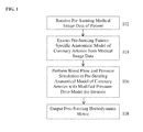
- FIG. 1 illustrates a method of predicting a hemodynamic metric for one or more coronary artery stenosis of a patient according to an embodiment of the present invention.
- pre-stenting medical image data of a patient is received.
- the pre-stenting medical image data is acquired prior to performing stenting, such as PCI for a coronary artery stenosis.
- Medical image data from one or multiple imaging modalities can be received.
- the medical image data can include, computed tomography (CT), Dyna CT, magnetic resonance (MR), Angiography, Ultrasound, Single Photon Emission computed Tomography (SPECT), and any other type of medical imaging modality.
- CT computed tomography
- MR magnetic resonance
- Angiography Ultrasound
- SPECT Single Photon Emission computed Tomography
- the medical image data can be 2D, 3D, or 4D (3D+time) medical image data.
- the medical image data can be received directly from one or more image acquisition devices, such as a CT scanner, MR scanner, Angiography scanner, Ultrasound device, etc., or the medical image data may be received by loading previously stored medical image data for a patient.
- 3D coronary CT angiography (CTA) images are acquired on a CT scanner.
- the CTA images ensure that the coronary vasculature, including the vessel(s) that contain the stenosis, is adequately imaged using a contrast agent that is injected into the patient.
- the clinician may be provided with an option of identifying lesions (stenoses) of interest by interactively viewing them on the images.
- This step can also be performed on a patient-specific anatomical model that is extracted from the image data (step 104).
- the stenoses may be automatically detected in the image data using an algorithm for automatic detection of coronary artery stenosis, such as the method for automatic detection of coronary artery stenosis described in United States Published Patent Application No. 2011/0224542 , which is incorporated herein by reference.
- algorithm for automatic detection of coronary artery stenosis such as the method for automatic detection of coronary artery stenosis described in United States Published Patent Application No. 2011/0224542 , which is incorporated herein by reference.
- other non-invasive clinical measurements such as the patient's heart rate and systolic and diastolic blood pressure may also be acquired. These non-invasive clinical measurements can be used to establish boundary conditions for CFD computations.
- a pre-stenting patient-specific anatomical model of the coronary arteries is extracted from the pre-stenting medical image data.
- the patient-specific anatomical model may be a patient-specific anatomical model of any portion of the full coronary artery tree of the patient.
- the coronary arteries can be segmented in the 3D medical image data using an automated coronary artery centerline extraction algorithm.
- the coronary arteries can be segmented in a CT volume using the method described United States Published Patent Application No. 2010/0067760 , which is incorporated herein by reference.
- cross-section contours can be generated at each point of the centerline tree.
- the cross-section contour at each centerline point gives a corresponding cross-section area measurement at that point in the coronary artery.
- a geometric surface model is then generated for the segmented coronary arteries.
- methods for anatomical modeling of the coronary arteries are described in United States Patent No. 7,860,290 and United States Patent No. 7,953,266 , both of which are incorporated herein by reference.
- the patient-specific anatomical model can include the aortic root together with the proximal part of the aorta.
- a detailed 3D model of each stenosis can also be extracted using similar algorithms, which includes the quantification of the proximal vessel diameter and area, distal vessel diameter and area, minimal lumen diameter and area, and length of stenosis.
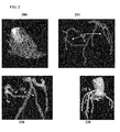
- FIG. 2 illustrates exemplary results for generating a pre-stenting patient-specific anatomical model of the coronary vessel tree.
- Image 200 of Fig. 2 shows coronary CTA data.
- Image 210 shows a centerline tree 212 extracted from the CTA data.
- Image 220 shows cross-section contours 222 extracted at each point of the centerline tree 212.
- Image 230 shows a 2D surface mesh 232 of the coronary arteries, the aortic root, and the proximal part of the aorta. It is to be understood that the anatomical model of the coronary tree of the patient can be output and displayed, for example on a display screen of the computer system.
- the above described anatomical modeling tasks can be performed automatically or can be user-driven, thereby allowing the user (clinician) to interactively make changes to the anatomical models to analyze the effects of such changes on the subsequent computation of FFR.
- the myocardium may also be segmented (either automatically or manually) in the medical image data to determine an estimate of the left ventricular mass, which in a possible implementation, may be used to estimate the absolute resting flow for the patient which is used to calculate boundary conditions for a computational blood flow and pressure simulation.
- the resting flow could also be computed based on the total volume of the segmented coronary tree, or from the outlet radius of the different coronary vessels.
- a patient-specific anatomical model of the heart that is automatically generated from the image data may be used for this purpose.
- the anatomical heart model is a multi-component model having multiple cardiac components, including the four chambers (left ventricle, left atrium, right ventricle, and right atrium).
- the anatomical heart model may also include components such as the heart valves (aortic valve, mitral valve, tricuspid valve, and pulmonary valve) and the aorta.
- Such a comprehensive model of the heart is used to capture a large variety of morphological, functional, and pathological variations.
- a modular and hierarchical approach can be used to reduce anatomical complexity and facilitate an effective and flexible estimation of individual anatomies.
- the 4D anatomical heart model can be generated by generating individual models of each heart component, for example using marginal space learning (MSL), and then integrating the heart component models by establishing mesh point correspondence. Additional details regarding generation of such a 4D patient-specific heart model are described in United States Published Patent Application No. 2012/0022843 , which is incorporated herein by reference in its entirety.
- MSL marginal space learning
- blood flow and pressure are simulated in the pre-stenting anatomical model of the coronary arteries using a modified pressure-drop model for a coronary artery stenosis.
- the blood flow is simulated in the pre-stenting anatomical model of the coronary arteries and a post stenting pressure-drop for the stenosis is calculated based on the simulated blood flow using the modified pressure-drop model.
- the blood flow and pressure can be simulated in the pre-stenting anatomical model using CFD computations or any other standard numerical technique, such as finite-element method, finite-difference method, finite volume method, boundary element method, embedded boundary method, immersed boundary method lattice Boltzmann method, etc.
- a multi-scale computational model of coronary circulation can be used to compute the blood flow and pressure in the pre-stenting anatomical model of the coronary arteries over a series of time steps.
- the simulation may be performed for a plurality of time steps corresponding to a full cardiac cycle or multiple cardiac cycles.
- the computational model of the coronary circulation models the loss of pressure across stenoses or other narrowings in the coronary arteries (e.g., calcification, thrombus, bifurcation, etc.) using pressure-drop models.
- the term stenosis is used to generally refer to any type of narrowing in a vessel.
- the pressure drop model for a particular stenosis computes the pressure drop over the stenosis due to the narrowing of the vessel without performing an explicit flow computation in that region of the vessel.
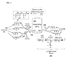
- FIG. 3 illustrates an exemplary multi-scale computational model of coronary circulation according to an embodiment of the present invention.
- a heart model 302 is coupled at the root of the aorta.
- the heart model 302 may be implemented as a lumped model parameterized through patient-specific data as shown in FIG. 3 , or may be implemented as a full 3D heart model.
- Large arteries, such as the aorta 304 together with the large arteries supplied by the aorta (e.g., subclavian, brachiocephalic, common carotid, etc.), the left coronary artery (LCA) 306, and the right coronary artery (RCA) 308 can be represented as 1 D blood flow models or full 3D models.
- LCA left coronary artery
- RCA right coronary artery
- semi-analytical circulatory models can be used either separately for certain arterial segments, or embedded within the 1 D or 3D models.
- the vessel walls can be modeled as a purely elastic or visco-elastic material.
- the wall properties may be determined through an empirical relationship fit to measured data or based on patient-specific estimations of wall compliance.
- all microvascular beds are simulated through lumped parameter models 310 which account for the resistance applied to the blood flow and for the compliance of the distal vessels.
- the coronary vascular bed is modeled through such lumped parameter models 310, which are adapted to the coronary circulation in the sense that they take into account the effects of the myocardial contraction on the flow waveform.
- Stenosis segments 312 and 314 are shown in the model of coronary arterial circulation.
- the stenosis segments 312 and 314 cannot be simulated using the 1 D blood flow models since there is a high variation in cross-sectional area and the shape of the stenosis influences the blood flow behavior and especially the trans-stenotic pressure drop which plays a major role in the assessment of the functional importance of such a stenosis.
- a reduced-order (as compared to a full 3D model) pressure-drop model can be used for each stenosis segment 312 and 314.
- a pressure-drop model is used to compute the pressure-drop across each stenosis region (e.g., 312 and 314 of FIG. 3 ) in the pre-stenting anatomical model of the coronary arteries without performing an explicit flow computation in the stenosis region.
- Various pressure-drop models can be used.
- the pressure-drop model for a stenosis may be a fully analytical model or may be a model that includes a combination of analytical and empirical terms.
- a pressure-drop model that includes a combination of analytical and empirical terms is referred to herein as a "semi-empirical pressure-drop model".
- pressure-drop models may be used as well, such as a machine-learning based pressure-drop model that is trained using a machine-learning algorithm to map anatomical and flow features derived from a stenosis to a pressure-drop associated with the stenosis.
- a machine-learning based pressure-drop model that is trained using a machine-learning algorithm to map anatomical and flow features derived from a stenosis to a pressure-drop associated with the stenosis.
- the pressure-drop model for the stenosis is directly modified and the blood flow and pressure simulation in the pre-stenting anatomical model of the coronary arteries is performed with the modified pressure drop model.
- directly modifying the pressure-drop model for the coronary artery stenosis refers to modifying parameters in the pressure-drop model without modifying the underlying pre-stent patient-specific anatomical model of the coronary arteries.
- the pressure-drop model for the stenosis can be modified to represent a fully successful treatment or a partially successful treatment.
- the goal of the modification of the pressure-drop model is to virtually simulate the effect of the enlargement of the vessel due to stenting without explicitly modifying the actual patient-specific geometry. Examples of modifying a pressure-drop model to represent partially successful and fully successful treatments are described below for a fully analytical pressure-drop model and a semi-empirical pressure drop model. It is to be understood that the present invention is not limited to these specific examples and may be similarly applied to other pressure-drop models as well.
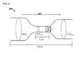
- FIG. 4 illustrates parameters of the pressure drop model for an exemplary stenosis segment.
- arrow 402 represents a direction of blood flow through a stenosed segment 400 of a coronary artery.
- the proximal or input side of the stenosed segment 400 is referred to as the "top” and the distal or output side of the stenoses segment 400 is referred to as the "bottom”. ...
- sten is the minimum cross-sectional area along the stenosis.
- ... refers to the cross section area at a given point in the stenosed segment.
- sten refers to the length of the stenosis region in which the radius is minimum and constant, and.
- Equation (1) refers to the entire length of the stenosed segment for which the pressure drop is calculated.
- the fully analytical pressure-drop model of Equation (1) includes three pressure drop calculations. The first term of Equation (1) calculates the convection pressure drop, the second and third terms calculate the viscous pressure drop, and the fourth term calculates the expansion pressure drop.
- two modified post-treatment (post-stenting) pressure-drop models can be calculated by modifying the fully analytical pressure drop model of Equation (1), corresponding to partially successful treatment of the stenosis region and fully successful treatment of the stenosis region.
- the virtual post-stenting pressure drop model can be calculated by generating assumed values for the following two parameters: the cross-sectional area in the distal (bottom) part of the stenosis segment (... out _ post ) and the minimum cross-sectional area value along the stenosis (... sten_p ost ... out_post can be set to be either equal to the pre-treatment value (... out ) or greater. For example, if the cross-section area in the proximal part of the stenosed segment (... in ) is larger than the pre-treatment value for... out ,... out_post can be set to a value greater than or equal to ... out and less than or equal to... in . ...
- sten_post is set to a value that is greater than the pre-treatment value... sten , but less than ... in and ... out_post , thus representing a case in which the enlargement of the stenosis region is only partially successful.
- ... sten_post can be set to be a predetermined percentage of... in or out_post , and the percentage can be tuned to predict the effect of the treatment at various levels of partial success.
- ... sten_post can be set to percentage of ... in or ... out_post that is automatically determined based on calcifications in the medical image data. This embodiment is described in greater detail below.
- ⁇ ⁇ P ⁇ ⁇ Q 2 2 ⁇ 1 CSA out - post 2 - 1 CSA in 2 + ⁇ 0 L vessel 8 ⁇ ⁇ ⁇ ⁇ CSA 2 ⁇ Qdx + ⁇ ⁇ Q 2 2 ⁇ 1 CSA sten - post - 1 CSA out - post 2 + 2 ⁇ 1 CSA sten - post - 1 CSA out - post ⁇ 1 CSA sten - post - 1 3 ⁇ 1 CSA out - post - 1 CSA sten - post - 1 CSA out - post 2 ⁇ 1 - ⁇ 2 ⁇
- the convection pressure drop term (first term) is computed based on the assumed value of the cross-sectional area in the distal part of the stenosed segment ... out_post .
- the first of the two viscous pressure drop terms in Equation (1) is removed and the second viscous pressure drop term (second term in Equation (2)) is used to compute the viscous pressure drop along the entire stenosed segment.
- the expansion pressure-drop term (third term of Equation (2)) is computed based on the new assumed values of the minimum and distal cross-sectional areas ... sten_post and... out_post , respectively.
- the effect of the treatment can be simulated by generating a single assumed value for the cross-sectional area in the distal part of the stenosed segment ... out_post .
- the value for... out_post can be selected to be either equal to ... out or greater.
- ... out_post can be set to a value greater than or equal to... out and less than or equal to ... in .
- ... out_post can be set equal to ... in .
- ⁇ ⁇ P ⁇ ⁇ Q 2 2 ⁇ 1 CSA out - post 2 - 1 CSA in 2 + 8 ⁇ ⁇ ⁇ ⁇ CSA in + CSA out - post / 2 2 ⁇ Q ⁇ L vessel
- the convection pressure term (first term) is computed based on the assumed value of the cross-section area in the distal part of the stenosed segment... out_post , while the viscous pressure drop is computed based on the average value of ... in and ... out_post .
- an interpolation e.g., linear, quadratic, etc.
- the expansion pressure drop term is removed completely since the turbulent flow regime is inexistent in this case.
- the semi-empirical pressure-drop model for a coronary artery stenosis can be derived by starting with a model that predicts the pressure drop based on empirical data and augmenting the empirical model with an analytical convection pressure-drop term.
- 0 denotes the vessel radius in a normal (non-stenosis) portion of the vessel, and v , t , and. u are the viscous, turbulent, and inertance coefficients, respectively, which are empirically determined. Quantities indexed with 0 refer to the normal vessel, while quantities indexed with s refer to the stenosis.
- two post-stenting pressure-drop models can be calculated by modifying the semi-empirical pressure drop model of Equation (5), corresponding to a partially successful treatment and a fully successful treatment.
- Equation (6) the first three terms are adapted by setting assumed values for out_post and ... sten_post , as described above, and by adapting. v correspondingly.
- . v can be determined as a function of the cross-sectional area along the centerline of the stenosis.
- the cross-sectional area is interpolated between ... in , ... sten_post , and ... out_post .
- Equation (7) the first two terms are adapted by setting the assumed value for out_post , as described above, and by adapting. v correspondingly, while the third term from Equation (5) is dropped completely due to the absence of the turbulent flow regime.
- the inertance term (the fourth term in Equation (5)) remains the same in both Equations (6) and (7) since this term only introduces a phase shift between pressure and flow and does not contribute to the total pressure drop.
- FIG. 5 illustrates a method for modifying a pressure drop model and calculating a pressure-drop over a coronary artery stenosis according to an embodiment of the present invention.
- the method of FIG. 5 may be used in the implementation of step 106 of FIG. 1 .
- the method of FIG. 5 can be performed interactively based on user input entered by a user using a user input device, such as a mouse or touch screen, automatically without user input, or some combination thereof.
- a stenosis is selected for virtual stenting treatment prediction (e.g., virtual PCI).
- virtual PCI virtual stenting treatment prediction
- stenosis regions in the coronary artery tree can be manually identified by a user or automatically detected.
- a stenosis region or multiple stenosis regions in the coronary artery tree are selected for stenting treatment prediction.
- the stenosis region (or multiple stenosis regions) can be selected interactively by a user.
- the patient-specific anatomical model of the coronary artery tree and/or the medical image data of the patient can be displayed on a display device of a computer and a user can select on stenosis region or multiple stenosis regions for which to predict the effect of the stenting treatment.
- the user input selecting the stenosis region may be received in response to a prompt displayed on the display device requesting that the user select a stenosis region.
- the stenosis region can be automatically selected by a computer/processor without user input.
- the computer can automatically perform the treatment prediction for multiple possible treatment scenarios, where each treatment scenario corresponds to stenting a stenosis region or multiple stenosis regions in the coronary artery tree of the patient.
- an effect of the stent placement is specified for the selected stenosis.
- the selection of partially successful treatment or fully successful treatment determines which modified pressure-drop model to use.
- the selection of partially successful treatment or fully successful treatment can be performed interactively by a user. For example, in response to a user selecting a stenosis for virtual treatment prediction, a prompt can be displayed on the displayed device providing the user a choice between the partially successful treatment and the fully successful treatment.
- the user may be given a further prompt to select a percentage corresponding to how successful the treatment is (i.e., percentage of enlargement of vessel geometry in the stenosis).
- the user may also be given the option to select a type of the pressure-drop model, such as the fully analytical model or the semi-empirical model, or the type of pressure-drop model can be preset and not selectable by the user. It is important to note that the determination of partially successful treatment or fully successful treatment is not based on modification of the anatomical model of the coronary arteries to estimate the actual enlargement of the geometry due to stent placement.
- the determination of partially successful treatment or fully successful treatment can be performed automatically based on the medical image data of the patient.
- features from the medical image may be extracted to automatically determine the probability that the PCI procedure would result in a partial or a complete enlargement of the stenosed region.
- the amount of calcification may be quantified for each stenosis by analyzing the intensity values of the image voxels in a region of interest (in this case, a region around the stenosis). A stenosis with a high amount of calcification is more likely to result in a partial opening of the stent as compared to a stenosis with no calcification.
- features such as tortuosity of the vessel or the radius of the vessel, can also be quantified similarly to determine a composite index which relies on more than one feature from the medical image data.
- the features can be used to automatically select a percentage of enlargement to use for the partially successful treatment.
- assumed values for the modified parameters of the modified pressure-drop model are set.
- the number and type of the parameters depends on the effect of the stent (i.e., partially successful treatment or fully successful treatment) specified in step 506.
- the assumed values for the modified parameters for the partially successful treatment pressure-drop model or for the fully successful treatment pressure-drop model can be set as described above.
- the pressure drop for the selected stenosis is determined based on the simulated blood flow in the pre-stenting anatomical model of the coronary arteries using the modified post-stenting pressure-drop model with the assumed values for the modified parameters determined in step 506.
- a hemodynamic metric is calculated for the coronary artery stenosis.
- the post-stenting pressure-drop is computed for the stenosis for each of a series of time steps.
- a post-stenting fractional flow reserve is calculated for the stenosis based on the post-stenting pressure drop computed for the stenosis.
- FFR is defined as the ratio of the maximal blood flow in the stenotic vessel to the maximal blood flow in a normal vessel, and is used to characterize the severity of the stenosis.
- FFR can be approximated for a stenosis by calculating the ratio of the time-averaged pressure distal to the stenosis (Pd) with respect to the average pressure in the aorta (Pa) at the hyperemic state.
- the blow flow simulation in step 106 simulates hyperemic blood flow, and the computed post-stenting pressure drop for the stenosis can be averaged over a heart cycle and subsequently used to determine the post-stenting FFR value for the stenosis.
- FFR can be computed as (Pa - ⁇ P) / Pa, where Pa is the aortic pressure.
- the aortic pressure may be assumed at a population average value or may be determined as a function of the non-invasively acquired systolic and diastolic pressures, and the heart rate.
- Other hemodynamic metrics can be calculated for the stenosis as well.
- hemodynamic metrics such as instantaneous wave-free ratio (iFR) and rest Pd/Pa, can be calculated based on the post-stenting pressure-drop for a stenosis resulting from rest-state blood flow simulations.
- the post-stenting FFR value and/or other post-stenting hemodynamic metrics can be output by displaying the values on a display device.
- one or more computed post-stenting FFR values (or other hemodynamic metrics) can be visualized on a medical image of the patient that is displayed on a display device.
- FIG. 6 illustrates a method of stent treatment planning according to an embodiment of the present invention.
- the method of FIG. 6 is described herein using the example of planning PCI treatment for coronary artery stenoses of a patient, but the present invention is not limited thereto and can be similarly applied to other stenting treatments in other types of arteries.
- the method of FIG. 6 can be used to evaluate different stenting strategies for treating coronary artery stenoses to determine which stenting strategies are effective and/or select a best stenting strategy.
- the method of FIG. 6 can be implemented as fully automated method for stent treatment planning. Referring to FIG. 6 , at step 602, medical image data of a patient is received.
- a pre-stenting patient-specific anatomical model of the coronary arteries is extracted from the medical image data.
- Steps 602 and 604 are identical to steps 102 and 104 of FIG. 1 .
- stenosis regions can be automatically detected or manually identified in the medical image data or in the pre-stenting patient-specific anatomical model of the coronary arteries.
- pre-stenting FFR values are computed for each of the stenosis regions in the pre-stenting patient-specific anatomical model of the coronary arteries.
- the pre-stenting FFR values can be computed by simulating blood flow and pressure at a hyperemic state in the pre-stenting patient-specific anatomical model of the coronary arteries.
- the computational model of coronary circulation can be used to perform the blood flow and pressure computations.
- a plurality of stenting strategies are determined based on the pre-stenting FFR values computed for the stenosis regions in the pre-stenting patient-specific anatomical model of the coronary arteries.
- stenosis regions having a pre-stenting FFR value less than a predetermined threshold are identified as target stenosis regions for stenting.
- a predetermined threshold e.g., ⁇ 0.8
- all preceding stenosis regions i.e., in the proximal direction
- all preceding stenosis regions i.e., in the proximal direction
- a plurality of stenting strategies can be generated for each set of target stenosis regions in a particular blood flow path in the coronary artery tree, where each stenting strategy corresponds to stenting a subset of the target stenosis regions.
- the stenting strategies include a stenting strategies corresponding to stenting each individual target stenosis region and stenting strategies corresponding to stenting each possible combination of multiple target stenosis regions, up to a stenting strategy corresponding to stenting all of the target stenosis regions.
- FIG. 7 illustrates exemplary pre-stenting FFR computation results for three stenosis regions in a coronary artery tree.
- the left anterior descending (LAD) artery has three serial stenoses (stenosis 1, stenosis 2, and stenosis 3), each of which may or may not be hemodynamically significant.
- Stenosis 1 has a pre-stenting FFR value of 0.885
- stenosis 2 has a pre-stenting FFR value of 0.667
- stenosis 3 has a pre-stenting FFR value of 0.621.
- the most distal stenosis (stenosis 3) has an FFR value smaller 0.8, it is clear that PCI has to be performed, and all three stenoses are identified as target stenoses for stenting.
- the goal of the virtual planning method of FIG. 6 is to determine the stenoses for which PCI should be performed.
- the pre-stenting FFR values indicate that stenosis 2 is the most significant since it leads to the largest drop in FFR value.
- stenting stenosis 2 suffices for raising the distal FFR value (of stenosis 3) above 0.8
- predicted post-stenting FFR values are computed for the each of the target stenosis regions by simulating blood flow in the pre-stenting patient-specific anatomical model of the coronary arteries using a modified pressure-drop model for the target stenosis region that is stented in the current stenting strategy.
- the method of FIG. 1 the method of FIG.
- the post-stenting FFR values for each of the stenting strategies can be computed under the assumption that each stenting will be fully successful by using a fully successful modified post-stenting pressure drop model to predict the post-stenting pressure drop for each stented stenosis region.
- step 610 can be repeated for the remaining stenting strategies that were not eliminated using the partially successful modified post-stenting pressure-drop models.
- the predicted post-stenting FFR computation results for the plurality of stenting strategies are output.
- the predicted post-stenting FFR computation results for each of stenting strategies can be displayed on a display device, either by listing the predicted post-stenting FFR values or overlaying the predicted post-stenting FFR on medical image data of the patient.
- FIGS. 8-14 illustrate predicted post-stenting FFR values for different stenting strategies for performing virtual PCI on the coronary artery stenoses of FIG. 7.
- FIG. 8 illustrates predicted post-stenting FFR values resulting from virtual PCI (stenting) for stenosis 1.
- FIG. 9 illustrates predicted post-stenting FFR values resulting from virtual PCI for stenosis 2.
- FIG. 10 illustrates predicted post-stenting FFR values resulting from virtual PCI for stenosis 3.
- FIG. 11 illustrates predicted post-stenting FFR values resulting from virtual PCI for stenoses 1 and 2.
- FIG. 12 illustrates predicted post-stenting FFR values resulting from virtual PCI for stenoses 1 and 3.
- FIG. 13 illustrates predicted post-stenting FFR values resulting from virtual PCI for stenoses 2 and 3.
- FIG. 14 illustrates predicted post-stenting FFR values resulting from virtual PCI for stenoses 1, 2, and 3.
- the results of the virtual post-PCI FFR computations indicate that stenting of all three stenoses is required in order to obtain a distal FFR value higher than 0.8. An important reason for this is due to the fact that when one stenosis is removed, the pressure drop along the remaining stenoses increases due to the increased flow rate, thus leading to a larger drop in the predicted post-stenting FFR value than in the originally computed pre-stenting FFR value.
- Computer 1502 contains a processor 1504, which controls the overall operation of the computer 1502 by executing computer program instructions which define such operation.
- the computer program instructions may be stored in a storage device 1512 (e.g., magnetic disk) and loaded into memory 1510 when execution of the computer program instructions is desired.
- An image acquisition device 1520 such as a CT scanning device, MR scanning device, Ultrasound device, etc., can be connected to the computer 1502 to input image data to the computer 1502. It is possible to implement the image acquisition device 1520 and the computer 1502 as one device. It is also possible that the image acquisition device 1520 and the computer 1502 communicate wirelessly through a network. In a possible embodiment, the computer 1502 may be located remotely with respect to the image acquisition device 1520 and the method steps are performed as part of a server or cloud based service.
- the computer 1502 also includes one or more network interfaces 1506 for communicating with other devices via a network.
- the computer 1502 also includes other input/output devices 1508 that enable user interaction with the computer 1502 (e.g., display, keyboard, mouse, speakers, buttons, etc.).
- Such input/output devices 1508 may be used in conjunction with a set of computer programs as an annotation tool to annotate volumes received from the image acquisition device 1520.
- FIG. 15 is a high level representation of some of the components of such a computer for illustrative purposes.
- the above-described methods for medical image synthesis may be implemented using computers operating in a client-server relationship.
- the client computers are located remotely from the server computer and interact via a network.
- the client-server relationship may be defined and controlled by computer programs running on the respective client and server computers.
- a server or another processor that is connected to a network communicates with one or more client computers via a network.
- a client computer may communicate with the server via a network browser application residing and operating on the client computer, for example.
- a client computer may store data on the server and access the data via the network.
- a client computer may transmit requests for data, or requests for online services, to the server via the network.
- the server may perform requested services and provide data to the client computer(s).
- the server may also transmit data adapted to cause a client computer to perform a specified function, e.g., to perform a calculation, to display specified data on a screen, etc.
- the server may transmit a request adapted to cause a client computer to perform one or more of the method steps described herein, including one or more of the steps of FIGS. 1 , 5 , and 6 .
- Certain steps of the methods described herein, including one or more of the steps of FIGS. 1 , 5 , and 6 may be performed by a server or by another processor in a network-based cloud-computing system.
- Certain steps of the methods described herein, including one or more of the steps of FIGS. 1 , 5 , and 6 may be performed by a client computer in a network-based cloud computing system.
- the steps of the methods described herein, including one or more of the steps of FIGS. 1 , 5 , and 6 may be performed by a server and/or by a client computer in a network-based cloud computing system, in any combination.
Landscapes
- Health & Medical Sciences (AREA)
- Life Sciences & Earth Sciences (AREA)
- Engineering & Computer Science (AREA)
- Medical Informatics (AREA)
- Public Health (AREA)
- Physics & Mathematics (AREA)
- Pathology (AREA)
- Biomedical Technology (AREA)
- General Health & Medical Sciences (AREA)
- Cardiology (AREA)
- Surgery (AREA)
- Physiology (AREA)
- Molecular Biology (AREA)
- Animal Behavior & Ethology (AREA)
- Heart & Thoracic Surgery (AREA)
- Biophysics (AREA)
- Veterinary Medicine (AREA)
- Data Mining & Analysis (AREA)
- Databases & Information Systems (AREA)
- General Physics & Mathematics (AREA)
- Theoretical Computer Science (AREA)
- Mathematical Physics (AREA)
- Hematology (AREA)
- Vascular Medicine (AREA)
- Primary Health Care (AREA)
- Epidemiology (AREA)
- Algebra (AREA)
- Artificial Intelligence (AREA)
- Pure & Applied Mathematics (AREA)
- Software Systems (AREA)
- General Engineering & Computer Science (AREA)
- Mathematical Analysis (AREA)
- Mathematical Optimization (AREA)
- Computational Mathematics (AREA)
- Computer Vision & Pattern Recognition (AREA)
- Psychiatry (AREA)
- Signal Processing (AREA)
- Media Introduction/Drainage Providing Device (AREA)
- Apparatus For Radiation Diagnosis (AREA)
- Measuring Pulse, Heart Rate, Blood Pressure Or Blood Flow (AREA)
Applications Claiming Priority (2)
| Application Number | Priority Date | Filing Date | Title |
|---|---|---|---|
| US201462018800P | 2014-06-30 | 2014-06-30 | |
| US14/704,233 US10130266B2 (en) | 2014-06-30 | 2015-05-05 | Method and system for prediction of post-stenting hemodynamic metrics for treatment planning of arterial stenosis |
Publications (3)
| Publication Number | Publication Date |
|---|---|
| EP2963574A2 true EP2963574A2 (de) | 2016-01-06 |
| EP2963574A3 EP2963574A3 (de) | 2016-01-20 |
| EP2963574B1 EP2963574B1 (de) | 2021-07-28 |
Family
ID=53489826
Family Applications (1)
| Application Number | Title | Priority Date | Filing Date |
|---|---|---|---|
| EP15172630.4A Active EP2963574B1 (de) | 2014-06-30 | 2015-06-18 | Verfahren und system zur vorhersage von hämodynamischen metriken nach dem stenting zur behandlungsplanung von arterieller stenose |
Country Status (3)
| Country | Link |
|---|---|
| US (1) | US10130266B2 (de) |
| EP (1) | EP2963574B1 (de) |
| CN (1) | CN105249954B (de) |
Cited By (2)
| Publication number | Priority date | Publication date | Assignee | Title |
|---|---|---|---|---|
| WO2018156961A1 (en) * | 2017-02-24 | 2018-08-30 | Heartflow, Inc. | Systems and methods for identifying anatomically relevant blood flow characteristics in a patient |
| IL309861A (en) * | 2023-12-31 | 2025-07-01 | Medhub Ltd | Method and system for determining a treatment strategy for a vessel with multiple or diffuse lesions |
Families Citing this family (41)
| Publication number | Priority date | Publication date | Assignee | Title |
|---|---|---|---|---|
| US9042613B2 (en) | 2013-03-01 | 2015-05-26 | Heartflow, Inc. | Method and system for determining treatments by modifying patient-specific geometrical models |
| CN106537392B (zh) * | 2014-04-22 | 2019-07-26 | 西门子保健有限责任公司 | 用于冠状动脉中的血液动力学计算的方法和系统 |
| US9595089B2 (en) * | 2014-05-09 | 2017-03-14 | Siemens Healthcare Gmbh | Method and system for non-invasive computation of hemodynamic indices for coronary artery stenosis |
| WO2016113646A1 (en) * | 2015-01-15 | 2016-07-21 | Koninklijke Philips N.V. | Ifr-ct |
| WO2016132164A1 (en) * | 2015-02-17 | 2016-08-25 | Siemens Healthcare Gmbh | Method and system for personalizing a vessel stent |
| EP3062248A1 (de) * | 2015-02-27 | 2016-08-31 | Pie Medical Imaging BV | Verfahren und Vorrichtung zur quantitativen Durchflussanalyse |
| CN107615335A (zh) * | 2015-05-12 | 2018-01-19 | 新加坡保健服务集团有限公司 | 医学图象处理方法和系统 |
| JP5839638B1 (ja) * | 2015-06-01 | 2016-01-06 | 川澄化学工業株式会社 | 管状治療具設計装置、管状治療具の製造方法、および管状治療具設計プログラム |
| US10872698B2 (en) * | 2015-07-27 | 2020-12-22 | Siemens Healthcare Gmbh | Method and system for enhancing medical image-based blood flow computations using physiological measurements |
| CN105078425B (zh) * | 2015-09-09 | 2016-06-08 | 苏州润心医疗科技有限公司 | 冠状动脉负荷检测系统及检测方法 |
| US10304569B2 (en) * | 2015-12-03 | 2019-05-28 | Heartflow, Inc. | Systems and methods for associating medical images with a patient |
| WO2017147500A1 (en) | 2016-02-26 | 2017-08-31 | Heartflow, Inc. | Systems and methods for identifying and modeling unresolved vessels in image-based patient-specific hemodynamic models |
| DE102016203860A1 (de) * | 2016-03-09 | 2017-09-14 | Siemens Healthcare Gmbh | Vorrichtung und Verfahren zum Ermitteln zumindest eines individuellen fluiddynamischen Kennwerts einer Stenose in einem mehrere serielle Stenosen aufweisenden Gefäßsegment |
| US10971271B2 (en) | 2016-04-12 | 2021-04-06 | Siemens Healthcare Gmbh | Method and system for personalized blood flow modeling based on wearable sensor networks |
| CN106073894B (zh) * | 2016-05-31 | 2017-08-08 | 博动医学影像科技(上海)有限公司 | 基于植入虚拟支架的血管压力降数值及血流储备分数的评估方法和系统 |
| DE102016215966A1 (de) * | 2016-08-25 | 2018-03-01 | Siemens Healthcare Gmbh | Röntgenaufnahme mit einer überlagerten Planungsinformation |
| CN109716446B (zh) * | 2016-09-28 | 2023-10-03 | 光学实验室成像公司 | 利用血管表象的支架规划系统及方法 |
| CN106780477A (zh) * | 2016-12-30 | 2017-05-31 | 上海联影医疗科技有限公司 | 一种血流分析方法和系统 |
| CN107411767B (zh) * | 2017-06-28 | 2020-10-16 | 西北工业大学 | 基于冠状动脉ct血管造影的狭窄病灶血流阻力计算方法 |
| US11589924B2 (en) | 2017-08-01 | 2023-02-28 | Siemens Healthcare Gmbh | Non-invasive assessment and therapy guidance for coronary artery disease in diffuse and tandem lesions |
| EP3714467A4 (de) * | 2017-11-22 | 2021-09-15 | Arterys Inc. | Inhaltsbasiertes wiederauffinden von bildern für die läsionsanalyse |
| EP3488774A1 (de) * | 2017-11-23 | 2019-05-29 | Koninklijke Philips N.V. | Messführung für koronarflussschätzung von bernoulli's prinzip |
| US11389130B2 (en) | 2018-05-02 | 2022-07-19 | Siemens Healthcare Gmbh | System and methods for fast computation of computed tomography based fractional flow reserve |
| JP7314183B2 (ja) * | 2018-06-15 | 2023-07-25 | パイ メディカル イメージング ビー ヴイ | 定量的血行動態フロー分析のための方法および装置 |
| CN109259751B (zh) * | 2018-08-27 | 2022-03-11 | 杭州晟视科技有限公司 | 一种评估血流储备分数的方法及装置、设备、存储介质 |
| WO2020150512A1 (en) * | 2019-01-16 | 2020-07-23 | General Electric Company | Vascular assessment using acoustic sensing |
| CN109758228A (zh) * | 2019-02-28 | 2019-05-17 | 四川大学华西医院 | 基于三维重建的经颈静脉门体分流手术支架安置优化方法 |
| US10861157B2 (en) | 2019-04-04 | 2020-12-08 | Medtronic Vascular, Inc. | System and methods for determining modified fractional flow reserve values |
| CN110598288B (zh) * | 2019-08-30 | 2020-08-07 | 上海杏脉信息科技有限公司 | 一种用于冠脉三维模型的边界条件处理方法和装置 |
| CN110742689B (zh) * | 2019-10-31 | 2021-11-23 | 北京理工大学 | 动脉夹层手术评估方法、装置、电子设备及存储介质 |
| CN110916640B (zh) * | 2019-11-06 | 2023-04-14 | 唯智医疗科技(佛山)有限公司 | 基于ffr的冠状动脉狭窄功能性缺血的检测方法及装置 |
| US20220110530A2 (en) * | 2019-12-09 | 2022-04-14 | Nordsletten David | Method and System for Estimating Pressure Difference in Turbulent Flow |
| EP3951705A1 (de) * | 2020-08-04 | 2022-02-09 | Siemens Healthcare GmbH | Verfahren, vorrichtung und system zur bestimmung von abnormalität im myokardbereich |
| CN112950537A (zh) * | 2021-01-26 | 2021-06-11 | 上海友脉科技有限责任公司 | 一种冠脉血流储备分数获取系统、方法及介质 |
| CN113827199B (zh) * | 2021-10-29 | 2024-01-23 | 苏州润迈德医疗科技有限公司 | 基于造影图像调节血管评定参数的方法、系统及存储介质 |
| CN115414163B (zh) * | 2022-11-04 | 2023-02-28 | 清华大学 | 血管支架、人体血管局部变形与血管局部动力学监测系统 |
| US20240169524A1 (en) * | 2022-11-23 | 2024-05-23 | Case Western Reserve University | Prediction of stent expansion using finite element modeling and machine learning |
| US20250054628A1 (en) * | 2023-08-09 | 2025-02-13 | Cathworks Ltd. | Post-pci coronary analysis |
| US12446965B2 (en) | 2023-08-09 | 2025-10-21 | Cathworks Ltd. | Enhanced user interface and crosstalk analysis for vascular index measurement |
| CN116919375A (zh) * | 2023-08-22 | 2023-10-24 | 上海博动医疗科技股份有限公司 | 虚拟支架ffr血流储备分数计算方法、装置、设备及介质 |
| CN119358469B (zh) * | 2024-12-27 | 2025-03-18 | 吉林大学 | 一种冠脉支架植入的血流动力学仿真分析系统 |
Citations (7)
| Publication number | Priority date | Publication date | Assignee | Title |
|---|---|---|---|---|
| US20100067760A1 (en) | 2008-09-15 | 2010-03-18 | Siemens Corporate Research, Inc. | Method and System for Automatic Coronary Artery Detection |
| US7860290B2 (en) | 2006-04-21 | 2010-12-28 | Siemens Medical Solutions Usa, Inc. | Three-dimensional (3D) modeling of coronary arteries |
| US7953266B2 (en) | 2007-02-06 | 2011-05-31 | Siemens Medical Solutions Usa, Inc. | Robust vessel tree modeling |
| US20110224542A1 (en) | 2010-03-12 | 2011-09-15 | Sushil Mittal | Method and System for Automatic Detection and Classification of Coronary Stenoses in Cardiac CT Volumes |
| US20120022843A1 (en) | 2010-07-21 | 2012-01-26 | Razvan Ioan Ionasec | Method and System for Comprehensive Patient-Specific Modeling of the Heart |
| US20130132054A1 (en) | 2011-11-10 | 2013-05-23 | Puneet Sharma | Method and System for Multi-Scale Anatomical and Functional Modeling of Coronary Circulation |
| US20130246034A1 (en) | 2012-03-13 | 2013-09-19 | Siemens Aktiengesellschaft | Method and System for Non-Invasive Functional Assessment of Coronary Artery Stenosis |
Family Cites Families (13)
| Publication number | Priority date | Publication date | Assignee | Title |
|---|---|---|---|---|
| US6236878B1 (en) | 1998-05-22 | 2001-05-22 | Charles A. Taylor | Method for predictive modeling for planning medical interventions and simulating physiological conditions |
| US8098918B2 (en) | 2007-09-21 | 2012-01-17 | Siemens Corporation | Method and system for measuring left ventricle volume |
| US8200466B2 (en) | 2008-07-21 | 2012-06-12 | The Board Of Trustees Of The Leland Stanford Junior University | Method for tuning patient-specific cardiovascular simulations |
| US8315812B2 (en) | 2010-08-12 | 2012-11-20 | Heartflow, Inc. | Method and system for patient-specific modeling of blood flow |
| US8157742B2 (en) | 2010-08-12 | 2012-04-17 | Heartflow, Inc. | Method and system for patient-specific modeling of blood flow |
| DE102010039312B4 (de) | 2010-08-13 | 2020-02-13 | Siemens Healthcare Gmbh | Verfahren zur Simulation eines Blutflusses |
| US9119540B2 (en) | 2010-09-16 | 2015-09-01 | Siemens Aktiengesellschaft | Method and system for non-invasive assessment of coronary artery disease |
| DE102010043849B3 (de) | 2010-11-12 | 2012-02-16 | Siemens Aktiengesellschaft | Vorrichtung und Computertomograph zur Bestimmung und Darstellung der Durchblutung des Herzmuskels |
| US9141763B2 (en) | 2011-02-07 | 2015-09-22 | Siemens Aktiengesellschaft | Method and system for patient-specific computational modeling and simulation for coupled hemodynamic analysis of cerebral vessels |
| US10186056B2 (en) | 2011-03-21 | 2019-01-22 | General Electric Company | System and method for estimating vascular flow using CT imaging |
| US8696584B2 (en) | 2011-10-05 | 2014-04-15 | 3Dt Holdings, Llc | Devices, systems and methods for determining fractional flow reserve in the presence of a catheter |
| WO2014099942A1 (en) | 2012-12-21 | 2014-06-26 | Volcano Corporation | Display control for a multi-sensor medical device |
| US9700219B2 (en) | 2013-10-17 | 2017-07-11 | Siemens Healthcare Gmbh | Method and system for machine learning based assessment of fractional flow reserve |
-
2015
- 2015-05-05 US US14/704,233 patent/US10130266B2/en active Active
- 2015-06-18 EP EP15172630.4A patent/EP2963574B1/de active Active
- 2015-06-30 CN CN201510386120.4A patent/CN105249954B/zh active Active
Patent Citations (8)
| Publication number | Priority date | Publication date | Assignee | Title |
|---|---|---|---|---|
| US7860290B2 (en) | 2006-04-21 | 2010-12-28 | Siemens Medical Solutions Usa, Inc. | Three-dimensional (3D) modeling of coronary arteries |
| US7953266B2 (en) | 2007-02-06 | 2011-05-31 | Siemens Medical Solutions Usa, Inc. | Robust vessel tree modeling |
| US20100067760A1 (en) | 2008-09-15 | 2010-03-18 | Siemens Corporate Research, Inc. | Method and System for Automatic Coronary Artery Detection |
| US20110224542A1 (en) | 2010-03-12 | 2011-09-15 | Sushil Mittal | Method and System for Automatic Detection and Classification of Coronary Stenoses in Cardiac CT Volumes |
| US20120022843A1 (en) | 2010-07-21 | 2012-01-26 | Razvan Ioan Ionasec | Method and System for Comprehensive Patient-Specific Modeling of the Heart |
| US20130132054A1 (en) | 2011-11-10 | 2013-05-23 | Puneet Sharma | Method and System for Multi-Scale Anatomical and Functional Modeling of Coronary Circulation |
| US20130246034A1 (en) | 2012-03-13 | 2013-09-19 | Siemens Aktiengesellschaft | Method and System for Non-Invasive Functional Assessment of Coronary Artery Stenosis |
| US20140058715A1 (en) | 2012-03-13 | 2014-02-27 | Siemens Aktiengesellschaft | Method and System for Non-Invasive Functional Assessment of Coronary Artery Stenosis |
Cited By (3)
| Publication number | Priority date | Publication date | Assignee | Title |
|---|---|---|---|---|
| WO2018156961A1 (en) * | 2017-02-24 | 2018-08-30 | Heartflow, Inc. | Systems and methods for identifying anatomically relevant blood flow characteristics in a patient |
| US11617620B2 (en) | 2017-02-24 | 2023-04-04 | Heartflow, Inc. | Systems and methods for identifying anatomically relevant blood flow characteristics in a patient |
| IL309861A (en) * | 2023-12-31 | 2025-07-01 | Medhub Ltd | Method and system for determining a treatment strategy for a vessel with multiple or diffuse lesions |
Also Published As
| Publication number | Publication date |
|---|---|
| CN105249954A (zh) | 2016-01-20 |
| CN105249954B (zh) | 2018-10-02 |
| US20150374243A1 (en) | 2015-12-31 |
| US10130266B2 (en) | 2018-11-20 |
| EP2963574B1 (de) | 2021-07-28 |
| EP2963574A3 (de) | 2016-01-20 |
Similar Documents
| Publication | Publication Date | Title |
|---|---|---|
| US10130266B2 (en) | Method and system for prediction of post-stenting hemodynamic metrics for treatment planning of arterial stenosis | |
| EP2977922B1 (de) | Verfahren und system zur automatisierten therapieplanung arterieller stenose | |
| JP7483079B2 (ja) | 患者固有の幾何学的形状モデルを変更することによって治療を決定する方法及びシステム | |
| US10463336B2 (en) | Method and system for purely geometric machine learning based fractional flow reserve | |
| US10134129B2 (en) | Method and system for hemodynamic computation in coronary arteries | |
| CN107427268B (zh) | 用于基于纯几何机器学习的血流储备分数的方法和系统 | |
| CN105580019B (zh) | 为优化诊断性能利用边界条件模型化血流的方法和系统 | |
| CN104736046B (zh) | 用于数字评估脉管系统的系统和方法 | |
| EP3140757B1 (de) | Verfahren und system zur nichtinvasiven funktionellen beurteilung von koronararterienstenose anhand von strömungsberechnungen bei modellen basierend auf erkrankten patienten und hypothetisch normalen anatomischen modellen | |
| US9629563B2 (en) | Method and system for functional assessment of renal artery stenosis from medical images | |
| US20160166209A1 (en) | Method and System for Personalized Non-Invasive Hemodynamic Assessment of Renal Artery Stenosis from Medical Images | |
| EP2942006A1 (de) | Verfahren und system zur nicht invasiven berechnung hämodynamischer indizes für eine herzarterienstenose | |
| WO2016075331A2 (en) | Method and system for purely geometric machine learning based fractional flow reserve |
Legal Events
| Date | Code | Title | Description |
|---|---|---|---|
| PUAL | Search report despatched |
Free format text: ORIGINAL CODE: 0009013 |
|
| PUAI | Public reference made under article 153(3) epc to a published international application that has entered the european phase |
Free format text: ORIGINAL CODE: 0009012 |
|
| AK | Designated contracting states |
Kind code of ref document: A2 Designated state(s): AL AT BE BG CH CY CZ DE DK EE ES FI FR GB GR HR HU IE IS IT LI LT LU LV MC MK MT NL NO PL PT RO RS SE SI SK SM TR |
|
| AX | Request for extension of the european patent |
Extension state: BA ME |
|
| AK | Designated contracting states |
Kind code of ref document: A3 Designated state(s): AL AT BE BG CH CY CZ DE DK EE ES FI FR GB GR HR HU IE IS IT LI LT LU LV MC MK MT NL NO PL PT RO RS SE SI SK SM TR |
|
| AX | Request for extension of the european patent |
Extension state: BA ME |
|
| RIC1 | Information provided on ipc code assigned before grant |
Ipc: G06F 19/00 20110101AFI20151216BHEP Ipc: A61B 5/02 20060101ALI20151216BHEP Ipc: A61B 19/00 00000000ALI20151216BHEP |
|
| RAP1 | Party data changed (applicant data changed or rights of an application transferred) |
Owner name: SIEMENS HEALTHCARE GMBH |
|
| 17P | Request for examination filed |
Effective date: 20160718 |
|
| RBV | Designated contracting states (corrected) |
Designated state(s): AL AT BE BG CH CY CZ DE DK EE ES FI FR GB GR HR HU IE IS IT LI LT LU LV MC MK MT NL NO PL PT RO RS SE SI SK SM TR |
|
| STAA | Information on the status of an ep patent application or granted ep patent |
Free format text: STATUS: EXAMINATION IS IN PROGRESS |
|
| 17Q | First examination report despatched |
Effective date: 20190410 |
|
| REG | Reference to a national code |
Ref country code: DE Ref legal event code: R079 Ref document number: 602015071634 Country of ref document: DE Free format text: PREVIOUS MAIN CLASS: G06F0019000000 Ipc: A61B0005000000 |
|
| GRAP | Despatch of communication of intention to grant a patent |
Free format text: ORIGINAL CODE: EPIDOSNIGR1 |
|
| STAA | Information on the status of an ep patent application or granted ep patent |
Free format text: STATUS: GRANT OF PATENT IS INTENDED |
|
| RIC1 | Information provided on ipc code assigned before grant |
Ipc: A61B 5/00 20060101AFI20210208BHEP |
|
| INTG | Intention to grant announced |
Effective date: 20210304 |
|
| GRAS | Grant fee paid |
Free format text: ORIGINAL CODE: EPIDOSNIGR3 |
|
| GRAA | (expected) grant |
Free format text: ORIGINAL CODE: 0009210 |
|
| STAA | Information on the status of an ep patent application or granted ep patent |
Free format text: STATUS: THE PATENT HAS BEEN GRANTED |
|
| AK | Designated contracting states |
Kind code of ref document: B1 Designated state(s): AL AT BE BG CH CY CZ DE DK EE ES FI FR GB GR HR HU IE IS IT LI LT LU LV MC MK MT NL NO PL PT RO RS SE SI SK SM TR |
|
| REG | Reference to a national code |
Ref country code: GB Ref legal event code: FG4D |
|
| REG | Reference to a national code |
Ref country code: CH Ref legal event code: EP |
|
| REG | Reference to a national code |
Ref country code: AT Ref legal event code: REF Ref document number: 1413960 Country of ref document: AT Kind code of ref document: T Effective date: 20210815 |
|
| REG | Reference to a national code |
Ref country code: IE Ref legal event code: FG4D |
|
| REG | Reference to a national code |
Ref country code: DE Ref legal event code: R096 Ref document number: 602015071634 Country of ref document: DE |
|
| REG | Reference to a national code |
Ref country code: LT Ref legal event code: MG9D |
|
| REG | Reference to a national code |
Ref country code: NL Ref legal event code: MP Effective date: 20210728 |
|
| REG | Reference to a national code |
Ref country code: AT Ref legal event code: MK05 Ref document number: 1413960 Country of ref document: AT Kind code of ref document: T Effective date: 20210728 |
|
| PG25 | Lapsed in a contracting state [announced via postgrant information from national office to epo] |
Ref country code: HR Free format text: LAPSE BECAUSE OF FAILURE TO SUBMIT A TRANSLATION OF THE DESCRIPTION OR TO PAY THE FEE WITHIN THE PRESCRIBED TIME-LIMIT Effective date: 20210728 Ref country code: RS Free format text: LAPSE BECAUSE OF FAILURE TO SUBMIT A TRANSLATION OF THE DESCRIPTION OR TO PAY THE FEE WITHIN THE PRESCRIBED TIME-LIMIT Effective date: 20210728 Ref country code: SE Free format text: LAPSE BECAUSE OF FAILURE TO SUBMIT A TRANSLATION OF THE DESCRIPTION OR TO PAY THE FEE WITHIN THE PRESCRIBED TIME-LIMIT Effective date: 20210728 Ref country code: FI Free format text: LAPSE BECAUSE OF FAILURE TO SUBMIT A TRANSLATION OF THE DESCRIPTION OR TO PAY THE FEE WITHIN THE PRESCRIBED TIME-LIMIT Effective date: 20210728 Ref country code: ES Free format text: LAPSE BECAUSE OF FAILURE TO SUBMIT A TRANSLATION OF THE DESCRIPTION OR TO PAY THE FEE WITHIN THE PRESCRIBED TIME-LIMIT Effective date: 20210728 Ref country code: NL Free format text: LAPSE BECAUSE OF FAILURE TO SUBMIT A TRANSLATION OF THE DESCRIPTION OR TO PAY THE FEE WITHIN THE PRESCRIBED TIME-LIMIT Effective date: 20210728 Ref country code: NO Free format text: LAPSE BECAUSE OF FAILURE TO SUBMIT A TRANSLATION OF THE DESCRIPTION OR TO PAY THE FEE WITHIN THE PRESCRIBED TIME-LIMIT Effective date: 20211028 Ref country code: PT Free format text: LAPSE BECAUSE OF FAILURE TO SUBMIT A TRANSLATION OF THE DESCRIPTION OR TO PAY THE FEE WITHIN THE PRESCRIBED TIME-LIMIT Effective date: 20211129 Ref country code: BG Free format text: LAPSE BECAUSE OF FAILURE TO SUBMIT A TRANSLATION OF THE DESCRIPTION OR TO PAY THE FEE WITHIN THE PRESCRIBED TIME-LIMIT Effective date: 20211028 Ref country code: AT Free format text: LAPSE BECAUSE OF FAILURE TO SUBMIT A TRANSLATION OF THE DESCRIPTION OR TO PAY THE FEE WITHIN THE PRESCRIBED TIME-LIMIT Effective date: 20210728 Ref country code: LT Free format text: LAPSE BECAUSE OF FAILURE TO SUBMIT A TRANSLATION OF THE DESCRIPTION OR TO PAY THE FEE WITHIN THE PRESCRIBED TIME-LIMIT Effective date: 20210728 |
|
| PG25 | Lapsed in a contracting state [announced via postgrant information from national office to epo] |
Ref country code: PL Free format text: LAPSE BECAUSE OF FAILURE TO SUBMIT A TRANSLATION OF THE DESCRIPTION OR TO PAY THE FEE WITHIN THE PRESCRIBED TIME-LIMIT Effective date: 20210728 Ref country code: LV Free format text: LAPSE BECAUSE OF FAILURE TO SUBMIT A TRANSLATION OF THE DESCRIPTION OR TO PAY THE FEE WITHIN THE PRESCRIBED TIME-LIMIT Effective date: 20210728 Ref country code: GR Free format text: LAPSE BECAUSE OF FAILURE TO SUBMIT A TRANSLATION OF THE DESCRIPTION OR TO PAY THE FEE WITHIN THE PRESCRIBED TIME-LIMIT Effective date: 20211029 |
|
| PG25 | Lapsed in a contracting state [announced via postgrant information from national office to epo] |
Ref country code: DK Free format text: LAPSE BECAUSE OF FAILURE TO SUBMIT A TRANSLATION OF THE DESCRIPTION OR TO PAY THE FEE WITHIN THE PRESCRIBED TIME-LIMIT Effective date: 20210728 |
|
| REG | Reference to a national code |
Ref country code: DE Ref legal event code: R097 Ref document number: 602015071634 Country of ref document: DE |
|
| PG25 | Lapsed in a contracting state [announced via postgrant information from national office to epo] |
Ref country code: SM Free format text: LAPSE BECAUSE OF FAILURE TO SUBMIT A TRANSLATION OF THE DESCRIPTION OR TO PAY THE FEE WITHIN THE PRESCRIBED TIME-LIMIT Effective date: 20210728 Ref country code: SK Free format text: LAPSE BECAUSE OF FAILURE TO SUBMIT A TRANSLATION OF THE DESCRIPTION OR TO PAY THE FEE WITHIN THE PRESCRIBED TIME-LIMIT Effective date: 20210728 Ref country code: RO Free format text: LAPSE BECAUSE OF FAILURE TO SUBMIT A TRANSLATION OF THE DESCRIPTION OR TO PAY THE FEE WITHIN THE PRESCRIBED TIME-LIMIT Effective date: 20210728 Ref country code: EE Free format text: LAPSE BECAUSE OF FAILURE TO SUBMIT A TRANSLATION OF THE DESCRIPTION OR TO PAY THE FEE WITHIN THE PRESCRIBED TIME-LIMIT Effective date: 20210728 Ref country code: CZ Free format text: LAPSE BECAUSE OF FAILURE TO SUBMIT A TRANSLATION OF THE DESCRIPTION OR TO PAY THE FEE WITHIN THE PRESCRIBED TIME-LIMIT Effective date: 20210728 Ref country code: AL Free format text: LAPSE BECAUSE OF FAILURE TO SUBMIT A TRANSLATION OF THE DESCRIPTION OR TO PAY THE FEE WITHIN THE PRESCRIBED TIME-LIMIT Effective date: 20210728 |
|
| PLBE | No opposition filed within time limit |
Free format text: ORIGINAL CODE: 0009261 |
|
| STAA | Information on the status of an ep patent application or granted ep patent |
Free format text: STATUS: NO OPPOSITION FILED WITHIN TIME LIMIT |
|
| 26N | No opposition filed |
Effective date: 20220429 |
|
| PG25 | Lapsed in a contracting state [announced via postgrant information from national office to epo] |
Ref country code: IT Free format text: LAPSE BECAUSE OF FAILURE TO SUBMIT A TRANSLATION OF THE DESCRIPTION OR TO PAY THE FEE WITHIN THE PRESCRIBED TIME-LIMIT Effective date: 20210728 |
|
| REG | Reference to a national code |
Ref country code: DE Ref legal event code: R119 Ref document number: 602015071634 Country of ref document: DE |
|
| PG25 | Lapsed in a contracting state [announced via postgrant information from national office to epo] |
Ref country code: MC Free format text: LAPSE BECAUSE OF FAILURE TO SUBMIT A TRANSLATION OF THE DESCRIPTION OR TO PAY THE FEE WITHIN THE PRESCRIBED TIME-LIMIT Effective date: 20210728 |
|
| REG | Reference to a national code |
Ref country code: CH Ref legal event code: PL |
|
| REG | Reference to a national code |
Ref country code: BE Ref legal event code: MM Effective date: 20220630 |
|
| GBPC | Gb: european patent ceased through non-payment of renewal fee |
Effective date: 20220618 |
|
| PG25 | Lapsed in a contracting state [announced via postgrant information from national office to epo] |
Ref country code: LU Free format text: LAPSE BECAUSE OF NON-PAYMENT OF DUE FEES Effective date: 20220618 Ref country code: LI Free format text: LAPSE BECAUSE OF NON-PAYMENT OF DUE FEES Effective date: 20220630 Ref country code: IE Free format text: LAPSE BECAUSE OF NON-PAYMENT OF DUE FEES Effective date: 20220618 Ref country code: FR Free format text: LAPSE BECAUSE OF NON-PAYMENT OF DUE FEES Effective date: 20220630 Ref country code: CH Free format text: LAPSE BECAUSE OF NON-PAYMENT OF DUE FEES Effective date: 20220630 |
|
| PG25 | Lapsed in a contracting state [announced via postgrant information from national office to epo] |
Ref country code: GB Free format text: LAPSE BECAUSE OF NON-PAYMENT OF DUE FEES Effective date: 20220618 Ref country code: DE Free format text: LAPSE BECAUSE OF NON-PAYMENT OF DUE FEES Effective date: 20230103 Ref country code: BE Free format text: LAPSE BECAUSE OF NON-PAYMENT OF DUE FEES Effective date: 20220630 |
|
| PG25 | Lapsed in a contracting state [announced via postgrant information from national office to epo] |
Ref country code: HU Free format text: LAPSE BECAUSE OF FAILURE TO SUBMIT A TRANSLATION OF THE DESCRIPTION OR TO PAY THE FEE WITHIN THE PRESCRIBED TIME-LIMIT; INVALID AB INITIO Effective date: 20150618 |
|
| PG25 | Lapsed in a contracting state [announced via postgrant information from national office to epo] |
Ref country code: MK Free format text: LAPSE BECAUSE OF FAILURE TO SUBMIT A TRANSLATION OF THE DESCRIPTION OR TO PAY THE FEE WITHIN THE PRESCRIBED TIME-LIMIT Effective date: 20210728 Ref country code: CY Free format text: LAPSE BECAUSE OF FAILURE TO SUBMIT A TRANSLATION OF THE DESCRIPTION OR TO PAY THE FEE WITHIN THE PRESCRIBED TIME-LIMIT Effective date: 20210728 |
|
| PG25 | Lapsed in a contracting state [announced via postgrant information from national office to epo] |
Ref country code: MT Free format text: LAPSE BECAUSE OF FAILURE TO SUBMIT A TRANSLATION OF THE DESCRIPTION OR TO PAY THE FEE WITHIN THE PRESCRIBED TIME-LIMIT Effective date: 20210728 |