EP2911578B1 - Systeme zum nachweis gehirnfundierter biosignale - Google Patents
Systeme zum nachweis gehirnfundierter biosignale Download PDFInfo
- Publication number
- EP2911578B1 EP2911578B1 EP13849498.4A EP13849498A EP2911578B1 EP 2911578 B1 EP2911578 B1 EP 2911578B1 EP 13849498 A EP13849498 A EP 13849498A EP 2911578 B1 EP2911578 B1 EP 2911578B1
- Authority
- EP
- European Patent Office
- Prior art keywords
- signal
- bio
- brain
- signals
- oral
- Prior art date
- Legal status (The legal status is an assumption and is not a legal conclusion. Google has not performed a legal analysis and makes no representation as to the accuracy of the status listed.)
- Active
Links
- 210000004556 brain Anatomy 0.000 title claims description 112
- 210000001983 hard palate Anatomy 0.000 claims description 38
- 210000000214 mouth Anatomy 0.000 claims description 16
- 238000012545 processing Methods 0.000 claims description 16
- 210000001519 tissue Anatomy 0.000 claims description 14
- 238000012880 independent component analysis Methods 0.000 claims description 8
- 238000003745 diagnosis Methods 0.000 claims description 4
- 230000009514 concussion Effects 0.000 claims description 2
- 208000001797 obstructive sleep apnea Diseases 0.000 claims description 2
- 201000000615 hard palate cancer Diseases 0.000 description 37
- 210000004761 scalp Anatomy 0.000 description 21
- 230000029058 respiratory gaseous exchange Effects 0.000 description 18
- 230000000694 effects Effects 0.000 description 16
- 230000004424 eye movement Effects 0.000 description 16
- 238000000034 method Methods 0.000 description 14
- 230000005540 biological transmission Effects 0.000 description 12
- 230000000747 cardiac effect Effects 0.000 description 10
- 238000001914 filtration Methods 0.000 description 9
- 238000007781 pre-processing Methods 0.000 description 9
- 238000001514 detection method Methods 0.000 description 8
- 230000033001 locomotion Effects 0.000 description 8
- 239000002184 metal Substances 0.000 description 8
- 229910052751 metal Inorganic materials 0.000 description 8
- 210000003205 muscle Anatomy 0.000 description 7
- 238000004458 analytical method Methods 0.000 description 6
- 230000000052 comparative effect Effects 0.000 description 6
- 210000003128 head Anatomy 0.000 description 6
- WABPQHHGFIMREM-UHFFFAOYSA-N lead(0) Chemical compound [Pb] WABPQHHGFIMREM-UHFFFAOYSA-N 0.000 description 6
- 230000007958 sleep Effects 0.000 description 5
- 230000007177 brain activity Effects 0.000 description 4
- 238000000605 extraction Methods 0.000 description 4
- 230000000541 pulsatile effect Effects 0.000 description 4
- 230000002745 absorbent Effects 0.000 description 3
- 239000002250 absorbent Substances 0.000 description 3
- 239000000853 adhesive Substances 0.000 description 3
- 230000001070 adhesive effect Effects 0.000 description 3
- QVGXLLKOCUKJST-UHFFFAOYSA-N atomic oxygen Chemical compound [O] QVGXLLKOCUKJST-UHFFFAOYSA-N 0.000 description 3
- 230000008901 benefit Effects 0.000 description 3
- 208000037265 diseases, disorders, signs and symptoms Diseases 0.000 description 3
- 238000005516 engineering process Methods 0.000 description 3
- 238000002955 isolation Methods 0.000 description 3
- 239000000463 material Substances 0.000 description 3
- 238000012544 monitoring process Methods 0.000 description 3
- 229910052760 oxygen Inorganic materials 0.000 description 3
- 239000001301 oxygen Substances 0.000 description 3
- 208000019116 sleep disease Diseases 0.000 description 3
- 208000030886 Traumatic Brain injury Diseases 0.000 description 2
- 230000003321 amplification Effects 0.000 description 2
- 238000013459 approach Methods 0.000 description 2
- 238000004891 communication Methods 0.000 description 2
- 238000013480 data collection Methods 0.000 description 2
- 210000004513 dentition Anatomy 0.000 description 2
- 208000035475 disorder Diseases 0.000 description 2
- 230000006870 function Effects 0.000 description 2
- PCHJSUWPFVWCPO-UHFFFAOYSA-N gold Chemical compound [Au] PCHJSUWPFVWCPO-UHFFFAOYSA-N 0.000 description 2
- 239000010931 gold Substances 0.000 description 2
- 229910052737 gold Inorganic materials 0.000 description 2
- 230000001939 inductive effect Effects 0.000 description 2
- 238000007726 management method Methods 0.000 description 2
- 230000003340 mental effect Effects 0.000 description 2
- 150000002739 metals Chemical class 0.000 description 2
- 230000001537 neural effect Effects 0.000 description 2
- 238000003199 nucleic acid amplification method Methods 0.000 description 2
- 230000003287 optical effect Effects 0.000 description 2
- 230000001766 physiological effect Effects 0.000 description 2
- 230000008569 process Effects 0.000 description 2
- 230000004461 rapid eye movement Effects 0.000 description 2
- 239000004065 semiconductor Substances 0.000 description 2
- 210000001584 soft palate Anatomy 0.000 description 2
- 210000004872 soft tissue Anatomy 0.000 description 2
- 239000000126 substance Substances 0.000 description 2
- 230000036346 tooth eruption Effects 0.000 description 2
- 238000013519 translation Methods 0.000 description 2
- 230000009529 traumatic brain injury Effects 0.000 description 2
- 230000002087 whitening effect Effects 0.000 description 2
- 206010010254 Concussion Diseases 0.000 description 1
- 206010010904 Convulsion Diseases 0.000 description 1
- 229920000742 Cotton Polymers 0.000 description 1
- 208000013016 Hypoglycemia Diseases 0.000 description 1
- 206010041235 Snoring Diseases 0.000 description 1
- FAPWRFPIFSIZLT-UHFFFAOYSA-M Sodium chloride Chemical compound [Na+].[Cl-] FAPWRFPIFSIZLT-UHFFFAOYSA-M 0.000 description 1
- 206010041349 Somnolence Diseases 0.000 description 1
- 208000006011 Stroke Diseases 0.000 description 1
- 230000002159 abnormal effect Effects 0.000 description 1
- 208000008784 apnea Diseases 0.000 description 1
- 238000003491 array Methods 0.000 description 1
- 230000004872 arterial blood pressure Effects 0.000 description 1
- 210000001367 artery Anatomy 0.000 description 1
- 230000000712 assembly Effects 0.000 description 1
- 238000000429 assembly Methods 0.000 description 1
- 230000006399 behavior Effects 0.000 description 1
- 230000000903 blocking effect Effects 0.000 description 1
- 239000008280 blood Substances 0.000 description 1
- 210000004369 blood Anatomy 0.000 description 1
- 150000001722 carbon compounds Chemical class 0.000 description 1
- 210000003169 central nervous system Anatomy 0.000 description 1
- 210000003710 cerebral cortex Anatomy 0.000 description 1
- 230000008859 change Effects 0.000 description 1
- 239000002131 composite material Substances 0.000 description 1
- 238000007596 consolidation process Methods 0.000 description 1
- 239000000470 constituent Substances 0.000 description 1
- 230000001054 cortical effect Effects 0.000 description 1
- 238000013523 data management Methods 0.000 description 1
- 238000013500 data storage Methods 0.000 description 1
- 230000006735 deficit Effects 0.000 description 1
- 206010012601 diabetes mellitus Diseases 0.000 description 1
- 201000010099 disease Diseases 0.000 description 1
- 239000003814 drug Substances 0.000 description 1
- 229940079593 drug Drugs 0.000 description 1
- 230000002526 effect on cardiovascular system Effects 0.000 description 1
- 230000005684 electric field Effects 0.000 description 1
- 239000003792 electrolyte Substances 0.000 description 1
- 230000002708 enhancing effect Effects 0.000 description 1
- 230000007613 environmental effect Effects 0.000 description 1
- 238000011156 evaluation Methods 0.000 description 1
- 230000000193 eyeblink Effects 0.000 description 1
- 239000004744 fabric Substances 0.000 description 1
- 210000001097 facial muscle Anatomy 0.000 description 1
- 239000006260 foam Substances 0.000 description 1
- 238000002695 general anesthesia Methods 0.000 description 1
- 230000002068 genetic effect Effects 0.000 description 1
- 229910021389 graphene Inorganic materials 0.000 description 1
- 230000002218 hypoglycaemic effect Effects 0.000 description 1
- 230000001771 impaired effect Effects 0.000 description 1
- 239000007943 implant Substances 0.000 description 1
- 230000002452 interceptive effect Effects 0.000 description 1
- 239000010977 jade Substances 0.000 description 1
- 210000001847 jaw Anatomy 0.000 description 1
- 210000004072 lung Anatomy 0.000 description 1
- 210000004373 mandible Anatomy 0.000 description 1
- 238000013507 mapping Methods 0.000 description 1
- 210000001595 mastoid Anatomy 0.000 description 1
- 238000005259 measurement Methods 0.000 description 1
- 230000006996 mental state Effects 0.000 description 1
- 229910001092 metal group alloy Inorganic materials 0.000 description 1
- 238000004377 microelectronic Methods 0.000 description 1
- 238000002156 mixing Methods 0.000 description 1
- 238000012986 modification Methods 0.000 description 1
- 230000004048 modification Effects 0.000 description 1
- 210000003739 neck Anatomy 0.000 description 1
- 210000002569 neuron Anatomy 0.000 description 1
- 229910052755 nonmetal Inorganic materials 0.000 description 1
- 150000002843 nonmetals Chemical class 0.000 description 1
- 238000010606 normalization Methods 0.000 description 1
- 238000003909 pattern recognition Methods 0.000 description 1
- 230000000737 periodic effect Effects 0.000 description 1
- 210000001428 peripheral nervous system Anatomy 0.000 description 1
- 229920000642 polymer Polymers 0.000 description 1
- 238000013139 quantization Methods 0.000 description 1
- 230000036385 rapid eye movement (rem) sleep Effects 0.000 description 1
- 230000035484 reaction time Effects 0.000 description 1
- 230000000306 recurrent effect Effects 0.000 description 1
- 230000000241 respiratory effect Effects 0.000 description 1
- 150000003839 salts Chemical class 0.000 description 1
- 238000012216 screening Methods 0.000 description 1
- 239000000932 sedative agent Substances 0.000 description 1
- 229940125723 sedative agent Drugs 0.000 description 1
- 238000000926 separation method Methods 0.000 description 1
- 210000003625 skull Anatomy 0.000 description 1
- 230000008667 sleep stage Effects 0.000 description 1
- 230000004622 sleep time Effects 0.000 description 1
- 239000011780 sodium chloride Substances 0.000 description 1
- 239000007779 soft material Substances 0.000 description 1
- 230000007480 spreading Effects 0.000 description 1
- 238000012706 support-vector machine Methods 0.000 description 1
- 230000002123 temporal effect Effects 0.000 description 1
- 210000002105 tongue Anatomy 0.000 description 1
- 238000012549 training Methods 0.000 description 1
Images


Classifications
-
- A—HUMAN NECESSITIES
- A61—MEDICAL OR VETERINARY SCIENCE; HYGIENE
- A61B—DIAGNOSIS; SURGERY; IDENTIFICATION
- A61B5/00—Measuring for diagnostic purposes; Identification of persons
- A61B5/40—Detecting, measuring or recording for evaluating the nervous system
- A61B5/4058—Detecting, measuring or recording for evaluating the nervous system for evaluating the central nervous system
- A61B5/4064—Evaluating the brain
-
- A—HUMAN NECESSITIES
- A61—MEDICAL OR VETERINARY SCIENCE; HYGIENE
- A61B—DIAGNOSIS; SURGERY; IDENTIFICATION
- A61B3/00—Apparatus for testing the eyes; Instruments for examining the eyes
- A61B3/10—Objective types, i.e. instruments for examining the eyes independent of the patients' perceptions or reactions
- A61B3/113—Objective types, i.e. instruments for examining the eyes independent of the patients' perceptions or reactions for determining or recording eye movement
-
- A—HUMAN NECESSITIES
- A61—MEDICAL OR VETERINARY SCIENCE; HYGIENE
- A61B—DIAGNOSIS; SURGERY; IDENTIFICATION
- A61B5/00—Measuring for diagnostic purposes; Identification of persons
- A61B5/0002—Remote monitoring of patients using telemetry, e.g. transmission of vital signals via a communication network
- A61B5/0015—Remote monitoring of patients using telemetry, e.g. transmission of vital signals via a communication network characterised by features of the telemetry system
- A61B5/0022—Monitoring a patient using a global network, e.g. telephone networks, internet
-
- A—HUMAN NECESSITIES
- A61—MEDICAL OR VETERINARY SCIENCE; HYGIENE
- A61B—DIAGNOSIS; SURGERY; IDENTIFICATION
- A61B5/00—Measuring for diagnostic purposes; Identification of persons
- A61B5/02—Detecting, measuring or recording pulse, heart rate, blood pressure or blood flow; Combined pulse/heart-rate/blood pressure determination; Evaluating a cardiovascular condition not otherwise provided for, e.g. using combinations of techniques provided for in this group with electrocardiography or electroauscultation; Heart catheters for measuring blood pressure
- A61B5/0205—Simultaneously evaluating both cardiovascular conditions and different types of body conditions, e.g. heart and respiratory condition
-
- A—HUMAN NECESSITIES
- A61—MEDICAL OR VETERINARY SCIENCE; HYGIENE
- A61B—DIAGNOSIS; SURGERY; IDENTIFICATION
- A61B5/00—Measuring for diagnostic purposes; Identification of persons
- A61B5/08—Detecting, measuring or recording devices for evaluating the respiratory organs
-
- A—HUMAN NECESSITIES
- A61—MEDICAL OR VETERINARY SCIENCE; HYGIENE
- A61B—DIAGNOSIS; SURGERY; IDENTIFICATION
- A61B5/00—Measuring for diagnostic purposes; Identification of persons
- A61B5/24—Detecting, measuring or recording bioelectric or biomagnetic signals of the body or parts thereof
- A61B5/242—Detecting biomagnetic fields, e.g. magnetic fields produced by bioelectric currents
-
- A—HUMAN NECESSITIES
- A61—MEDICAL OR VETERINARY SCIENCE; HYGIENE
- A61B—DIAGNOSIS; SURGERY; IDENTIFICATION
- A61B5/00—Measuring for diagnostic purposes; Identification of persons
- A61B5/24—Detecting, measuring or recording bioelectric or biomagnetic signals of the body or parts thereof
- A61B5/316—Modalities, i.e. specific diagnostic methods
-
- A—HUMAN NECESSITIES
- A61—MEDICAL OR VETERINARY SCIENCE; HYGIENE
- A61B—DIAGNOSIS; SURGERY; IDENTIFICATION
- A61B5/00—Measuring for diagnostic purposes; Identification of persons
- A61B5/48—Other medical applications
- A61B5/4806—Sleep evaluation
- A61B5/4818—Sleep apnoea
-
- A—HUMAN NECESSITIES
- A61—MEDICAL OR VETERINARY SCIENCE; HYGIENE
- A61B—DIAGNOSIS; SURGERY; IDENTIFICATION
- A61B5/00—Measuring for diagnostic purposes; Identification of persons
- A61B5/68—Arrangements of detecting, measuring or recording means, e.g. sensors, in relation to patient
- A61B5/6801—Arrangements of detecting, measuring or recording means, e.g. sensors, in relation to patient specially adapted to be attached to or worn on the body surface
- A61B5/6813—Specially adapted to be attached to a specific body part
- A61B5/6814—Head
- A61B5/682—Mouth, e.g., oral cavity; tongue; Lips; Teeth
-
- A—HUMAN NECESSITIES
- A61—MEDICAL OR VETERINARY SCIENCE; HYGIENE
- A61B—DIAGNOSIS; SURGERY; IDENTIFICATION
- A61B5/00—Measuring for diagnostic purposes; Identification of persons
- A61B5/68—Arrangements of detecting, measuring or recording means, e.g. sensors, in relation to patient
- A61B5/6801—Arrangements of detecting, measuring or recording means, e.g. sensors, in relation to patient specially adapted to be attached to or worn on the body surface
- A61B5/683—Means for maintaining contact with the body
- A61B5/6832—Means for maintaining contact with the body using adhesives
Definitions
- the present invention relates to a system for detecting a multicomponent brain-based electromagnetic bio-signal. Also disclosed is a method for detecting, processing and extracting a variety of biasignals from a brain-based multi-component signal detected in the oral cavity.
- EEG electroencephalogram
- invasive procedures including needle electrodes (sharp wires placed between the scalp and the skull); cortical electrodes, subdural electrodes and depth electrodes.
- the characteristics of brain electrical activity monitored with invasive electrodes are related to surface electrodes like EEG electrodes on the scalp or skin, but are different since attenuation and spreading of the signal by the scalp and skin is bypassed.
- WO 2012/027648 discloses an intraoral multisensor device including a mouthpiece, a plurality of sensors, e.g. EEG sensors, at least one of the sensors attached to or integrated with the mouthpiece, and a data communications unit configured to receive signals from the plurality of sensors.
- the mouthpiece has a form to permit stable arrangement at least partially within a person's mouth such that it can remain for hands-free sensing of a plurality of biological parameters.
- the present invention provides a system for detecting a multicomponent brain-based electromagnetic bio-signal as defined in claim 1.
- Particular brain-based electromagnetic bio-signals can include multiple component signals forming a multicomponent brain-based signal, that may include signals that are generated from other parts of the body including the central nervous system, heart electrical activity, lung activity (respiration), local artery movement, eye dipole electrical activity (and other dipoles), muscle electrical activity, and local tissue electrical activity such as generated by the peripheral nervous system, as well as brain-based electromagnetic signals.
- the multicomponent brain-based signal is detected by sensors positioned in the oral cavity.
- the multicomponent brain-based signal may then digitized, amplified and filtered. After filtering desired sub-component bio signals are isolated from the multicomponent brain-based signal for further analysis.
- multicomponent brain-based signal is used to describe this collection of sub-component bio-signals, as the primary component bio-signals of interest emanates from the brain.
- the multicomponent brain-based signal can include bio-electromagnetic signals, cardiac bio-electromagnetic bio-signals, local tissue bio-electromagnetic signals; eye dipole bioelectric bio-signals; muscle bio-electromagnetic bio-signals; tongue bio-electromagnetic bio-signals; cardiac related pulsatile bio-signals; respiration related pulsatile bio-signals; movement related bio-signals; biomechanical bio-signals; bio-acoustic bio-signals.
- the component signals of the multicomponent brain-based signal are important for many applications (e.g. medical, veterinary and non-medical applications). Due to the brain-based signal detector(s) of this invention being located in the oral cavity, the detector(s) may detect electrical activity from many parts of the brain that includes the cerebral cortex, as well as other parts of the brain.
- a system for detecting multi-component brain-based electromagnetic bio-signals includes a sensor in the oral cavity may be coupled to one or more electronic processors capable of electronically digitizing, amplifying, attenuating. filtering and normalizing the multi-component brain-based bio-signals as needed.
- the computer processor may also be capable of extracting, isolating or otherwise dividing sub-component signals from the multi-component brain-based bio-signals, and optionally classifying and analyzing the subcomponent signals.
- the sensor may be an electrical or magnetic sensor capable of detecting multi-component brain-based electromagnetic bio-signals.
- the sensor includes electrodes touching the hard palate, where one electrode acts as a reference for comparison with one or more other electrodes.
- the electrodes may be resistive mode electrodes, capacitive mode electrodes, current mode electrodes, or inductive mode electrodes.
- the electrodes may be passive electrodes which simply receive a signal, or may be active electrodes which are able to digitize or otherwise process the received signal with an internal electronic processor.
- the senor in the oral cavity may be included in an oral device configured to couple to the dentition or other oral tissue.
- the position of the sensor or electrodes of the sensor may be adjustable in relation to the oral device.
- the senor may be communicatively coupled to a processor in the oral device. In other embodiments the sensor may be communicatively coupled to one or more external processing device.
- the sensor and/or processor(s) may be communicatively coupled via wires and/or wirelessly, such as Bluetooth or other wireless technology,
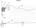
- Figure 1 is a comparison of the hard palate 10 multicomponent brain-based bio-signal 100 versus a standard scalp EEG brain wave signal.
- a sensor including reference electrode 11 and left signal electrode 12 coupled or proximate to hard-palate 10 are used to detect the multicomponent brain-based bio-signal 100.
- the sensor may also include right signal electrode 13.
- the multicomponent brain-based bio-signal 100 is the raw hard palate bio-potential signal.
- the multicomponent brain-based bio-signal 100 is a relatively unremarkable pattern of the raw hard palate bio-potential signal in comparison to the raw scalp EEG signal 200.
- the multicomponent brain-based bio-signal 100 bas a significantly larger voltage range when compared to raw scalp EEG signal 200, 100 ⁇ V versus 10 ⁇ V respectively. This demonstrates that special analysis of the hard palate 10 multicomponent brain-based bio-signal 100 is necessary to determine subcomponents of the hard palate 10 multicomponent brain-based bio-signal 100 especially the brain-based subcomponent signals.
- left signal electrode 12 and right signal electrode 13 may be gold or gold plated electrodes covered in cotton gauze. Saline may be used in some embodiments to wet the gauze.
- Figure 2 shows the hard palate 10 multicomponent brain-based bio-signal 100 being split into its various subcomponent signals after being processed according to embodiments of this invention.
- the subcomponent signals may include an 8 - 14 Hz brain wave subcomponent signal 101, an eye movement subcomponent signal 103, a cardiac subcomponent signal 104 and a respiration subcomponent signal 105.
- Figure 3 shows the strong correlation between the 8 - 14 Hz brain wave subcomponent signal 102 detected on the hard palate and the 8 - 14 Hz brain wave scalp EEG signal 201.
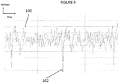
- Figure 4 shows the strong correlation between the 3.5 - 30 Hz brain wave subcomponent signal 102 detected on the hard palate and the 3.5 - 30 Hz brain wave scalp EEG signal 202 of a subject when subject was performing the mental activity of counting backwards from 100 by 7's (i.e. 100, 93, 86, 79, etc.). The subject was seated in a well-lit, environmentally controlled room.
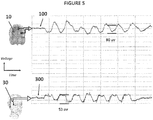
- Figure 5 shows the strong correlation between an 80 ⁇ V range multicomponent brain-based bio-signal 100 detected on the hard palate 10 and the 53 ⁇ V brain wave EOG signal 300 detected with EOG electrodes 30 on the right-side human scalp while the subject was quickly looking up and down. No filtering or isolation of subcomponents of the multicomponent brain-based bio-signal 100 was needed for this embodiment.
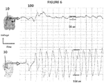
- Figure 6 shows the correlation between an 50 ⁇ V range multicomponent brain-based bio-signal 100 detected on the hard palate 10 and the 558 ⁇ V brain wave EOG signal 300 detected with EOG electrodes 30 on the right-side human scalp while the subject was quickly looking left and right. No filtering or isolation of subcomponents of the multicomponent brain-based bio-signal 100 was needed for this embodiment.
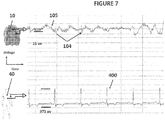
- Figure 7 shows the correlation between cardiac signals detected at 0.5 - 249 Hz in the brain wave subcomponent signal 104 on the hard palate 10 and the cardiac signals in an unfiltered ECG signal 400 detected with ECG electrode 40.
- the subject was seated in a well-lit, environmentally controlled room during recording.
- the multicomponent brain-based bio-signal 100 was filtered this application to remove DC offset.
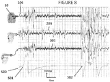
- Figure 8 shows the correlation between respiration sub component signals 106 at 1.5 Hz - 249 Hz extracted from the brain-based multicomponent bio-signals detected on the hard palate 10 and scalp EEG signals 203, right eye EOG signals 301 both of which were filtered at 1.5 Hz - 249 Hz, and a nasal cannula respiration signal 500 while the subject takes a fast deep breath 501 and holds the breath 502 for 20 seconds.
- the graph shows that the hard palate bio-potential changes at the same time the scalp EEG and EOG changes showing the strong temporal relation between the hard palate multicomponent bio-signals and the scalp related signals.
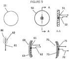
- FIG 9 shows various electrode embodiments for use in hard palate multicomponent bio-signal detection to accommodate the shape of oral tissue and provide for comfort and biocompatibility.
- Soft materials gauge, or foam
- Other materials can be used for electrodes as desired.
- the electrode assembly 50 include a metal electrode 51 and temperature sensor 52. The combination of electrode 51 with temperature sensing 52 for the oral cavity or other body locations. The electrodes detect current flow from tissue and the temperature sensor allows determination of oral temperature, motion artifact (since temperature is not a bio-potential measurement), and oral airflow. Average oral temperature can be estimated by a thermistor, semiconductor IC, thermocouple, or other appropriate sensor.
- One embodiment of the electrode may be a convex electrode assembly 60 which may include a metal electrode with a lead wire 61 and temperature sensor 62 and a soft, absorbent cover-surface 63 .
- Another embodiment of the electrode may be a concave electrode assembly 70 which may include a metal electrode with a lead wire 71 and temperature sensor 72 and a soft, absorbent cover-surface 73 .
- a third embodiment of the electrode may be a flat electrode assembly 80 which may include a metal electrode with a lead wire 81 and a soft, absorbent cover 82 .
- the reference electrode 11, may be a circular metal electrode.
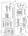
- Figure 10 is a schematic of a system to detect sleep disorders 600 including an internal oral unit 601 and an external unit 650.
- the internal oral unit 601 may include a convex electrode 60 positioned near the middle of the hard palate and one or two concave electrodes 70 positioned to the left and/or right near the gums.
- the internal oral unit 601 may include a sensor unit 602, a power source 603, a power manager 604, a microcontroller 605, and a transmission unit 606.
- the internal oral unit 601 may amplify, filter and/or digitize multicomponent bio-signals using dedicated circuitry as shown or as part of the data-management microcontroller ( ⁇ CU) Digital signals may be passed to a radiofrequency (RF) module for transmission to a remote receiver, e.g., a Smartphone or computer, or cloud etc.
- RF radiofrequency
- Detecting multicomponent brain-based signals may be accomplished by placing the internal unit 601 inside the appropriate body cavity where brain-based multicomponent bio-signal detection can automatically (or manually) be initiated. Signal detection usually begins immediately, however a temperature sensing component can be added to monitor environmental temperatures to ensure proper operating conditions and or monitor temperature during data collection. The temperature sensor can also be used to monitor changes in airflow via the mouth. Additional sensors an also be added to monitor a variety of additional physiological variables including oxygen saturation via optical PPG sensor/monitor, accelerometer, gyroscope, GPS, pressure, camera, biological or chemical monitors etc. Brain-based Detectors monitor multiparameter physiological signals including brain waves.
- the detector i.e. sensor
- the detector can be based on any of the following sensors: resistance mode electrodes, capacitive mode electrodes, current mode electrodes, passive electrodes, active electrodes, magnetic mode detectors, inductive mode detectors, acoustic mode detectors, optical or electro-optic mode detectors, chemical or biochemical mode detectors, biological mode detectors and brain-based detector arrays (brain-based detector can also comprise multiple sensors oriented in different geometric planes).
- the sensors may be of various shapes and include various metals, metal salts, or metal alloys, semiconductors, polymers, carbon compounds, conductive fabrics, composites, graphenes, non-metals; sensor comprises rigid, semi-rigid, and other flexible materials.
- the sensors may utilize microelectronics technology.
- the sensors may be disposable and/or reusable.
- the sensors may include remote sensors. Sensors may be adjustable in position and/or performance to optimzie brain-based multicomponent bio-signal detection.
- Sensor unit 602 may detect electrical signals from the hard palate picked up by electrodes and may amplify and filter the electrical signals to remove motion and other artifacts and conveyed to the microcontroller ( ⁇ CU) 605 via the SPI bus for further processing, storage and transmission.
- ⁇ CU microcontroller
- Signal and power management scheduling are performed by the ⁇ CU 605. Energy consumed from a disposable or rechargeable power supply 603 can be minimized by the ⁇ CU 605 by controlling the duration and duty cycle of data-collection devices, the transmission module 606, and the ⁇ CU 605 itself. Intelligent power management can reduce the size and complexity of the power source 603 and eliminate the need for a power line-operated system.
- Data transmission by the transmission unit 606 may be via well-known standard communications protocols, such as Bluetooth (BT) and Bluetooth LE (Low Energy) (BLE), or a proprietary protocols or frequencies. Use of standard protocols may ensure easier post-transmission processing.
- the transmission unit 606 may support both BT and BLE, which can be accessed by Smartphones and other devices.
- An antenna of the transmission unit 606 may be built into the side and/or front walls of an oral appliance attachment device as shown in Figures 12-15 .
- the external unit 650 includes a receiver unit 651, a preprocessing unit 652, an Independent Component Analysis ("ICA") processor 700, a raw brain-based multicomponent bio-signal component analyzer 750,
- ICA Independent Component Analysis
- the receiver unit 651 may be configured to receive signals from the transmission unit 606.
- the preprocessing unit 652 removes as much signal noise as possible
- the ICA processor 700 may use standard ICA algorithms to extract and isolate individual subcomponent signals. To ensure a good estimate of the components of the brain-based raw hard palate signal, brain wave filters 1-N 751, 752 and 753, eye movement signal processor 710, cardiac signal processor 720 and respiration signal processor 730.
- the preprocessing unit 652 may eliminate non-physiological noise via filtering and sensors (thermistors) built into electrodes or a sensor platform. Electrodes may be shielded on their rear surface by the oral attachment device to prevent disturbance by the tongue and or internal facial muscles. Thermistors also, provide a means to detect movement of the device relative to tissue as well as provide means to correct for large temperature changes due to breathing. Additional processing includes data filtering such as low pass filtering. Additional preprocessing may include centering and whitening. Centering removes the mean from each component by subtracting the mean of the data from the actual data.
- Preprocessing can also identify eye movements due to the unique arrangement of the electrodes (left and right) that produce significant differences in the raw signal detected by each electrode. Root mean square values can be determined and threshold detectors may be incorporated.
- Digitization by the pre-processing unit 652 may be electronically performed to enable efficient digital processing as well as signal amplification and or attenuation of the bio-signals if necessary.
- Pre-processing also seeks to remove unwanted noise by filtering, shielding, blocking, or algorithmically removing or eliminating undesirable physiological and or non-physiological signals such as electrical noise, acoustical noise, mechanical noise, other artifact, or galvanic currents from dissimilar metals, or tongue artifact etc. Undesirable artifact contained in biosignals can hamper recordings. Signal normalization can also occur at this stage.
- the ICA processor 700 determines, the individual subcomponent signals of the raw hard palate multicomponent bio-signal without previously knowing each component. To effectively determine each subcomponent the number of detectors (sensors) must be equal to or greater to the number of individual signal components. Embodiments may utilize three electrodes to detect bio-potentials each with a built-in thermistor which provides six (6) detectors overall. This embodiment may detect 4 subcomponent bio-signals. To separate the components the JADE algorthim (Joint Approximate Diagonalization Eignen Matrices), which tends to perform best for small datasets) can be incorporated used by the ICA processor 700 of a computer or Field Programmable Gate Array (FPGA).
- JADE algorthim Joint Approximate Diagonalization Eignen Matrices
- Extracting, isolating, or dividing the detected multicomponent brain-based signal into individual parasubcomponent signals may involve appropriate means to extract, isolate, and/or divide the brain-based multicomponent signal into constituent physiological signals and/or other signals as desired.
- Primary subcomponent signals may include brain-based bio-electromagnetic signals, cardiac bio-electromagnetic bio-signals, ECG, local tissue bio-electromagnetic signals; eye dipole bioelectric bio-signals, muscle bio-electromagnetic bio-signals, tongue bio-electromagnetic bio-signals, cardiovascular related pulsatile bio-signals (e.g. Blood Volume Pulse); respiration related pulsatile bio-signals, movement related bio-signals, biomechanical bio-signals and/or bio-acoustic bio-signals.
- Each subcomponent signal typically includes multiple frequencies, and may have different dynamic ranges that may overlap.
- additional physiological parameters can be derived, including heart rate, respiration rate, heart rate variability, pulse transit time, arterial blood pressure.
- Subcomponent bio-signal extraction may include use of pattern recognition, Independent Component Analysis, Principle Component analysis, Linear analysis, Frequency domain anaylsis, time - frequency and nonlinear techniques such as correlation dimension (CD), phase space plots, different entropies, wavelet based, Hilbert-Huang Transforms (HHT), and similar means as desired.
- CD correlation dimension
- HHT Hilbert-Huang Transforms
- signal isolation or extraction may not be required.
- eye movement signals tend to be larger than other oral signals so for eye movement applications extracting other signals may not be required.
- key subcomponent signal features such as data points, thresholds and/or data slope be extracted or isolated from the signal of interest. This may involve identification of brain-based signal patterns and translation into commands to extract said feature and or issue commands to perform a task.
- a desired algorithm may be used to automatically estimate/calculate a value to represent the signals by a few relevant key values.
- algorithms There are a large variety of algorithms that may be implemented from the simplistic methods such as adding, subtracting, multiplying, dividing, etc., to other complex techniques involving time-based approaches or frequency based approaches, Principle component analysis, Support vector machine, Genetic algorithm, Distinctive sensitive learning vector quantization etc.
- key features of a subcomponent bio-signal may be classified or translated to a command.
- the classification step assigns a class to a set of features extracted from the signals.
- the class can correspond to the type of mental states identified. This step can also be denoted as "feature translation".
- Key feature information may be provided or displayed to a user/operator and//or used to perform tasks, such as comparing an extracted subcomponent bio-signals to a database of baseline signals to control a device, assist in a diagnosis of a disease, disorder, or condition, and or report the status of the device function.
- subcomponent bio-signals can be displayed or utilized for other purposes such as calculating vital signs, part of a command to control another device(s), or to perform additional processing such as extract particular features.
- the raw brain- signal can be further analyzed or separated into various frequency bands using band-pass filters 750 and then displayed or used to issue a command.
- This may include brain wave filters 1-N 751.
- the filters may be programmed or maintained in hardware for bands of interest 751, 753.
- Eye movement sub component signals 710 can be displayed and observed for Rapid Eye Movement (REM) to determine sleep stage.
- REM Rapid Eye Movement
- Cardiac signals 720 can show basic heart rate and can be used to determine R wave peaks as well as heart rate variability.
- Respiration signals 730 can be displayed to determine breathing rate.
- Various data may be stored on a data storage device incorporated into the internal oral unit and/or external unit.
- the system may also include a stimulate tissue device.
- brain-based biosignal maps can be developed to allow for topographical mapping of electrical activity for internal body locations.
- FIG. 11 An alternative embodiment that incorporates multiple sensors such as oxygen saturation, head position via accelerometers, temperature, and brain-based signals is shown in Figure 11 .
- Figure 12 shows an embodiment of the internal oral unit 800 including convex electrodes 60 configured to contact the center of the hard palate, and concave electrodes 70 configured to contact the gums.
- the internal oral unit 800 may be a mouth-guard platform which may incorporate a biocompatible adhesive to maintain contact with the dentition and/or oral tissue similar to a denture adhesive.
- the internal oral unit 800 electronic circuits 601 that perform some or all of the functions described above.
- the electrodes may be positioned in the structure which provides a slight spring force against the gums and hard palate to ensure electrode contact with oral tissue.
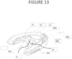
- Figure 13 shows Oral attachment device 900, which incorporates a flexible transverse support band 901 to maintain contact with the hard palate and electrodes 11,12,13 and temperature sensor 52.
- Oral attachment device 900 may include electronics 601 and transmission unit 606.
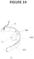
- Figure 14 shows an oral attachment device 1000 which includes a thin flexible platform that incorporates a biocompatible adhesive to maintain contact with the mandible and flexible electronic circuits.
- the electrodes 11, 12 and 13 are positioned in the structure which provides a slight spring force against the gums and hard palate to ensure electrode contact with oral tissue.
- Some embodiments may include temperature 52, electronics 601 and transmission unit 606.
- Figure 15 shows a sagittal view of a human head with an example of an oral attachment mouthguard 800 with electrodes contacting the the left and right side of the hard palate, and one electrode contacting the hard palate and transmission system, and an exemplary embodiment of the external unit 650.
- External unit can be a smartphone, computer or other computing device.
- An embodiment of the present invention may utilize subcomponent signals of the brain-based multicomponent bio-signals for screening, diagnosing and monitoring obstructive sleep apnea ("OSA")
- OSA is a breathing disorder caused by movement and upper airway blockage by the tongue and narrowing of the upper airway by soft tissues within the nose, mouth and throat that occurs during sleep. This phenomenon causes snoring and recurrent interruption of breathing due to periodic obstruction of airflow in the upper airway during inhalation.
- EEG electrocardiograph
- EMG electromyograph
- Embodiments of the invention enable detection of multicomponent brain-based bio- signals ( FIG. 2 ) from which subcomponent bio- signals can be extracted including brain-based electrical activity including alpha or other waves ( FIG. 3 ), eye movement ( FIG. 5 ), respiration ( FIG. 8 ) and ECG ( FIG. 7 ).
- Brain electrical activity subcomponent signals can enable determination of sleep state/stage and overall sleep time.
- Respiration subcomponent bio-signals may enable determination of apnea events.
- Eye movement subcomponent bio-signals can enable determination of rapid eye movement (REM) sleep
- ECG subcomponent bio-signals can enable determination of heart rate during sleep.
- signal processing including filtering, amplification, digitizing, storage etc.
- recording of some or all of the sub-component signals may occur in computer chip(s) embedded in an oral device including the sensor(s) can be accomplished.
- Resulting data can either be transmitted as it becomes available via wired or wireless technology (such as Bluetooth) to a receiving device (such as a smartphone. a computer, or dedicated device) and/or uploaded to a receiving device at a later time.
- the multicomponent brain-based bio-signal is transmitted to an external receiving device (such as a smartphone, a computer, or dedicated device) for signal processing.
- the multicomponent brain-based bio-signal may be transmitted as it is being detected by the sensor or it may be recorded on a storage device in an oral device for retrieval at a later time.
- the sensor detecting the multicomponent brain-based bio-signal may be supplemented with additional secondary sensors (i.e. accelerometers, thermocouples, O 2 saturation sensors, CO 2 sensors, air flow meters, etc.) may be used in combination with the multicomponent brain-based bio-signal to determine head position and oxygen desaturation and other events during sleep.
- additional secondary sensors i.e. accelerometers, thermocouples, O 2 saturation sensors, CO 2 sensors, air flow meters, etc.
- the oral device may automatically turn off when it is removed from the patient's mouth. In other embodiments the oral device may be turned off manually.
- the signals stored on the device may then be uploaded to a computer system including a software program for interpretation of the signal data, and be available for a diagnosis to be made by a physician or other medical personnel.
- the electrical brain activity subcomponent signals extracted from the detected brain-based multi-component signal may be used along with signals from accelerometers to detect traumatic brain injury in military personnel, sports participants, or other people in at-risk professions or activities, such as concussions, strokes and seizures. Detection of traumatic brain injury may be facilitated by comparing current subcompent signals to pre-existing baseline signals.
- the pre-existing baseline signals may be recorded from the specific patient being tested or a generic baseline derived from consolidation of multiple previously recorded signals from the patient or a segment of the population. In other embodiments these signals may be used to monitor performance.
- subcomponent signals extracted from the detected brain-based multi-component signal may be used to optimize training and provide feedback on performance of athletes and soldiers in order to enhance their capabilities during competition or in the field.
- the subcomponent signals extracted from the detected brain-based, multi-component signal may also be used in biofeedback applications.
- brain waves and muscle activity subcomponent signals extracted from the detected brain-based multi-component signal may be used to determine the level of consciousness of a patient under general anesthesia.
- subcomponent signals extracted from the detected brain-based multi-component signal may be used to detect abnormal brain wave patterns indicative of hypoglycemia in persons with diabetes.
- brain-based bio-signals, eye movement, head position and breathing signals and other subcomponent signals extracted from the detected brain-based multi-component signal may be used to assist individuals who are physically impaired but mentally capable to operate a wide variety of equipment and tools using a brain-computer interface which interprets the subcomponent signals to operate a variety of equipment's actions. For example moving an motorized wheel chair or operating an artificial limb.
- brain waves and eye movement subcomponent signals extracted from the detected brain-based multi-component signal can be monitored for advertising or media programming evaluation.
- a user can be trained to alter his brain waves in order to send a subcomponent signal extracted from the detected brain-based multi-component signal to a central computer in order to automatically control his mobile telephone, video game console, television set, music system or DVD player; change the temperature settings in the room; control an alarm system; control kitchen appliances; or control an automobile's computer system.
- subcomponent signal extracted from the detected brain-based multi-component signal may be used to detect drowsiness or sedatives or drug related impairment in the operator of a motor vehicle by monitoring sub-component signals related to respiration, eye movement, and other useful parameters.
- the device for this application may be in the form of a nose clip, a mouthpiece, or combinations thereof that collects and processes brain-based multi-component signal via an onboard computer that can subsequently trigger alarm systems and provide notification, or alarm when a driver becomes a drowsy or falls asleep at the wheel.
- a device may utilize subcomponent signal extracted from the detected brain-based multi-component signal, such as eye movement and other bio-signals to control machines such as automobiles or airplanes using thought control especially for complex, rapid or emergency maneuvers,
- subcomponent signal extracted from the detected brain-based multi-component signal such as eye movement and other bio-signals
- control machines such as automobiles or airplanes using thought control especially for complex, rapid or emergency maneuvers
- one application may be enhancing combat or drone pilots reaction times and assist in the control of aircraft during high-performance or wartime situations.
- the senor and/or other elements of the system may be implanted in soft tissue, such as the soft palate or gums; or alternatively inside teeth or tooth implants; or in a third alternative, in parts of the body other than the oral cavity.
- the sensor and/or other elements of the system can be implanted in the soft palate and self-powered via piezoelectric material within the device.
- the sensor and/or other elements of the system may be implanted beneath the skin and periodically charged inductively, capacitively, optically or other charging methods.
- the senor and/or other elements of the system may be located in a swimmer's or underwater diver's mouthpiece.
- the senor and/or other elements of the system may be mounted on a nose clip designed for comfortable placement within the nostrils of an individual.
Landscapes
- Health & Medical Sciences (AREA)
- Life Sciences & Earth Sciences (AREA)
- Engineering & Computer Science (AREA)
- Physics & Mathematics (AREA)
- General Health & Medical Sciences (AREA)
- Biophysics (AREA)
- Public Health (AREA)
- Veterinary Medicine (AREA)
- Biomedical Technology (AREA)
- Heart & Thoracic Surgery (AREA)
- Medical Informatics (AREA)
- Molecular Biology (AREA)
- Surgery (AREA)
- Animal Behavior & Ethology (AREA)
- Pathology (AREA)
- Neurology (AREA)
- Physiology (AREA)
- Cardiology (AREA)
- Pulmonology (AREA)
- Oral & Maxillofacial Surgery (AREA)
- Dentistry (AREA)
- Neurosurgery (AREA)
- Psychology (AREA)
- Computer Networks & Wireless Communication (AREA)
- Human Computer Interaction (AREA)
- Ophthalmology & Optometry (AREA)
- Measurement And Recording Of Electrical Phenomena And Electrical Characteristics Of The Living Body (AREA)
Claims (9)
- System zum Nachweis eines gehirnfundierten elektromagnetischen Biosignals (100), umfassend:mindestens zwei Biosignalsensoren (11, 12, 12, 52, 60, 70), die einen Teil einer oralen Befestigungsvorrichtung (800, 900, 1000) ausbilden, dazu ausgelegt, in einem oralen Hohlraum positioniert zu sein, um die mindestens zwei Biosignalsensoren in Kontakt mit einem oralen Gewebe (10) zu bringen,wobei die mindestens zwei Biosignalsensoren eine kabelgebundene oder kabellose Verbindung mit mindestens einer externen Rechnerverarbeitungseinheit (650) aufweisen,wobei die mindestens zwei Biosignalsensoren dazu ausgelegt sind, ein rohes, orales, elektromagnetisches Mehrkomponentenbiosignal an dem oralen Gewebe nachzuweisen und das rohe, orale, elektromagnetische Mehrkomponentenbiosignal an die mindestens eine externe Rechnerverarbeitungseinheit (650) zu senden, die einen unabhängigen Komponentenanalyseprozessor (700) beinhaltet, der mindestens ein primäres, rohes, gehirnfundiertes, elektromagnetisches Mehrkomponentenbiosignal (100) von einer Vielzahl von anderen Subkomponenten (710, 720, 730) innerhalb des rohen, oralen, elektromagnetischen Mehrkomponentenbiosignals isoliert, und wobei die externe Rechnerverarbeitungseinheit (650) ferner eine Vielzahl von Gehirnwellenbandpassfiltern (750) beinhaltet, die dazu ausgelegt sind, das rohe, gehirnbasierte Mehrkomponentensignal (100) weiter in eine Vielzahl von Frequenzgehirnwellenbändern (751, 752) zu separieren.
- System nach Anspruch 1, ferner umfassend einen Temperatursensor (52), angebracht an der oralen Anbringungsvorrichtung (800, 900, 1000).
- System nach einem der Ansprüche 1 oder 2, wobei die orale Anbringungsvorrichtung (800, 900, 1000) dazu ausgelegt ist, in Zähne einzugreifen.
- System nach einem der Ansprüche 1-3, wobei die orale Anbringungsvorrichtung (800, 900, 1000) konkave Elektroden (70) beinhaltet, die dazu ausgelegt sind, die mindestens zwei Biosignalsensoren zu Zahnfleisch zu kontaktieren.
- System nach einem der Ansprüche 1-3, wobei die orale Anbringungsvorrichtung (800, 900, 1000) konvexe Elektroden (60) beinhaltet, die dazu ausgelegt sind, die mindestens zwei Biosignalsensoren zu Hartgaumengewebe zu kontaktieren.
- System nach einem der Ansprüche 1-5, wobei das mindestens eine rohe, gehirnbasierte Mehrkomponentenbiosignal (100) mit Elektroenzephalogramm-Signaldaten (EEG-Signaldaten) korreliert.
- System nach einem der Ansprüche 1-6, wobei die mindestens eine Rechnerverarbeitungseinheit (650) dazu ausgelegt ist, einen Befehl, basierend auf Daten von dem Wellenband (751, 752), gefiltert aus dem mindestens einen primären, rohen, gehirnbasierten, elektromagnetischen Mehrkomponentenbiosignal (100), zu generieren.
- System nach einem der Ansprüche 1-7, wobei die mindestens eine Rechnerverarbeitungseinheit (650) dazu ausgelegt ist, eine Diagnose einer Bedingung in Reaktion auf ein Vergleichen von Daten von dem Wellenband (751, 752), gefiltert aus dem mindestens einen primären, rohen, gehirnbasierten, elektromagnetischen Mehrkomponentenbiosignal (100), mit vorbestimmten Basisdaten zur Diagnose der Bedingung zu generieren.
- System nach Anspruch 8, wobei die Bedingung mindestens eine von obstruktiver Schlafapnoe, Konkussion oder Iktus ist.
Applications Claiming Priority (3)
| Application Number | Priority Date | Filing Date | Title |
|---|---|---|---|
| US201261717997P | 2012-10-24 | 2012-10-24 | |
| US201361790007P | 2013-03-15 | 2013-03-15 | |
| PCT/US2013/066661 WO2014066666A2 (en) | 2012-10-24 | 2013-10-24 | Systems and methods for detecting brain-based bio-signals |
Publications (3)
| Publication Number | Publication Date |
|---|---|
| EP2911578A2 EP2911578A2 (de) | 2015-09-02 |
| EP2911578A4 EP2911578A4 (de) | 2016-08-10 |
| EP2911578B1 true EP2911578B1 (de) | 2019-12-25 |
Family
ID=50485948
Family Applications (1)
| Application Number | Title | Priority Date | Filing Date |
|---|---|---|---|
| EP13849498.4A Active EP2911578B1 (de) | 2012-10-24 | 2013-10-24 | Systeme zum nachweis gehirnfundierter biosignale |
Country Status (5)
| Country | Link |
|---|---|
| US (3) | US20140114165A1 (de) |
| EP (1) | EP2911578B1 (de) |
| JP (1) | JP6387352B2 (de) |
| ES (1) | ES2776178T3 (de) |
| WO (1) | WO2014066666A2 (de) |
Families Citing this family (37)
| Publication number | Priority date | Publication date | Assignee | Title |
|---|---|---|---|---|
| AU2014228116B2 (en) * | 2013-03-15 | 2019-01-03 | Adam J. Simon | System and signatures for the multi-modal physiological stimulation and assessment of brain health |
| US11064913B2 (en) | 2013-10-25 | 2021-07-20 | Force Impact Technologies, Inc. | Impact sensing wearable device and method |
| EP3064060A4 (de) * | 2013-10-30 | 2016-11-09 | Fujitsu Ltd | System zur biologischen messung, verfahren zur biologischen messung und programm zur biologischen messung |
| US20150286779A1 (en) * | 2014-04-04 | 2015-10-08 | Xerox Corporation | System and method for embedding a physiological signal into a video |
| AU2015274601B2 (en) | 2014-06-11 | 2020-02-27 | Arizona Board Of Regents On Behalf Of Arizona State University | Systems and methods for non-intrusive deception detection |
| CA2995803C (en) | 2014-08-21 | 2023-10-10 | Dignity Health | Systems and methods for using eye movements to determine traumatic brain injury |
| US10542961B2 (en) | 2015-06-15 | 2020-01-28 | The Research Foundation For The State University Of New York | System and method for infrasonic cardiac monitoring |
| JP6407824B2 (ja) | 2015-09-01 | 2018-10-17 | 株式会社東芝 | メガネ型ウエアラブル端末およびこの端末を用いる方法 |
| CN108471947B (zh) * | 2015-12-22 | 2021-06-08 | 皇家飞利浦有限公司 | 基于eeg信号中的心脏活动信息和大脑活动信息确定睡眠阶段的系统和方法 |
| US11000405B2 (en) | 2016-04-07 | 2021-05-11 | Achaemenid, Llc | Removable mandibular pharmaceutical delivery device |
| US11234638B2 (en) | 2016-04-07 | 2022-02-01 | Achaemenid, Llc | Intra-oral electroencephalography device and method |
| US11375951B2 (en) * | 2016-04-07 | 2022-07-05 | Achaemenid, Llc | Intra-oral electroencephalography device and method |
| US10470921B2 (en) | 2016-04-07 | 2019-11-12 | Achaemenid, Llc | Removable mandibular myo-stimulator |
| CN105997088A (zh) * | 2016-06-19 | 2016-10-12 | 河北工业大学 | 一种基于柔性力敏传感器的睡眠呼吸检测装置 |
| WO2018044959A1 (en) * | 2016-08-29 | 2018-03-08 | Smrt Ip, Llc | Sensor for continuous measurement of hydration and fatigue |
| KR101863056B1 (ko) * | 2016-09-19 | 2018-05-31 | 연암공과대학교산학협력단 | 압전소자로 자체충전이 가능한 체온계 내장형 공갈 젖꼭지 |
| WO2018089789A1 (en) * | 2016-11-10 | 2018-05-17 | The Research Foundation For The State University Of New York | System, method and biomarkers for airway obstruction |
| EP3558093A1 (de) | 2016-12-20 | 2019-10-30 | Koninklijke Philips N.V. | Patientenüberwachung |
| WO2018132435A1 (en) * | 2017-01-10 | 2018-07-19 | A.T. Still University | Dental monitoring system |
| WO2019060298A1 (en) | 2017-09-19 | 2019-03-28 | Neuroenhancement Lab, LLC | METHOD AND APPARATUS FOR NEURO-ACTIVATION |
| CA3078801A1 (en) * | 2017-10-13 | 2019-04-18 | BioAnalytics Holdings Pty Ltd | Improvements relating to sleep monitoring |
| US20190117124A1 (en) * | 2017-10-19 | 2019-04-25 | MedicusTek, Inc. | Sensor pad for monitoring user posture |
| EP3697289A4 (de) | 2017-10-20 | 2021-03-10 | Indian Institute of Technology, Guwahati | System für den einsatz am versorgungsort für die erkennung der körperlichen belastung verschiedener körperteile |
| US11717686B2 (en) | 2017-12-04 | 2023-08-08 | Neuroenhancement Lab, LLC | Method and apparatus for neuroenhancement to facilitate learning and performance |
| US11478603B2 (en) | 2017-12-31 | 2022-10-25 | Neuroenhancement Lab, LLC | Method and apparatus for neuroenhancement to enhance emotional response |
| US11364361B2 (en) | 2018-04-20 | 2022-06-21 | Neuroenhancement Lab, LLC | System and method for inducing sleep by transplanting mental states |
| WO2020056418A1 (en) | 2018-09-14 | 2020-03-19 | Neuroenhancement Lab, LLC | System and method of improving sleep |
| CN109259731A (zh) * | 2018-10-09 | 2019-01-25 | 广东数相智能科技有限公司 | 一种基于舌诊的中风前兆预警方法、电子设备及存储介质 |
| EP4070864A1 (de) | 2018-12-20 | 2022-10-12 | Force Impact Technologies, Inc. | Mundschutz zur benutzerbenachrichtigung von schlagkräften, und verfahren zur herstellung davon |
| US11786694B2 (en) | 2019-05-24 | 2023-10-17 | NeuroLight, Inc. | Device, method, and app for facilitating sleep |
| US11553871B2 (en) * | 2019-06-04 | 2023-01-17 | Lab NINE, Inc. | System and apparatus for non-invasive measurement of transcranial electrical signals, and method of calibrating and/or using same for various applications |
| FR3102054A1 (fr) | 2019-10-18 | 2021-04-23 | Devinnova | Casque pour améliorer l’équilibre de la balance sympatho-vagale d’un individu |
| AU2020378954A1 (en) * | 2019-11-04 | 2022-05-19 | Achaemenid, Llc | Intra-oral electroencephalography device and method |
| US20220378369A1 (en) * | 2019-11-08 | 2022-12-01 | The Johns Hopkins University | Oral measurement devices and methods |
| WO2021167855A1 (en) * | 2020-02-17 | 2021-08-26 | Achaemenid, Llc | Intra-oral appliance with thermoelectric power source |
| WO2022039613A1 (ru) * | 2020-08-19 | 2022-02-24 | Петр Валентинович ИВАНОВ | Капа-электрод для проведения процедур в полости рта |
| CN115348339B (zh) * | 2022-08-12 | 2023-11-21 | 北京威努特技术有限公司 | 一种基于功能码和业务数据相关性的工控异常检测方法 |
Citations (1)
| Publication number | Priority date | Publication date | Assignee | Title |
|---|---|---|---|---|
| WO2009137520A2 (en) * | 2008-05-07 | 2009-11-12 | Sonitus Medical, Inc. | Systems and methods for pulmonary monitoring and treatment |
Family Cites Families (63)
| Publication number | Priority date | Publication date | Assignee | Title |
|---|---|---|---|---|
| JPS5342490A (en) * | 1976-09-29 | 1978-04-17 | Rion Co | Artificial cover unit and method of producing same |
| US5190048A (en) * | 1991-09-17 | 1993-03-02 | Healthdyne, Inc. | Thermistor airflow sensor assembly |
| US5843093A (en) | 1994-02-09 | 1998-12-01 | University Of Iowa Research Foundation | Stereotactic electrode assembly |
| US5513649A (en) | 1994-03-22 | 1996-05-07 | Sam Technology, Inc. | Adaptive interference canceler for EEG movement and eye artifacts |
| JPH07265272A (ja) * | 1994-04-01 | 1995-10-17 | Itec Kk | 看護監視装置 |
| US5792067A (en) * | 1995-11-21 | 1998-08-11 | Karell; Manuel L. | Apparatus and method for mitigating sleep and other disorders through electromuscular stimulation |
| US6280394B1 (en) * | 1998-03-18 | 2001-08-28 | Sean R. Maloney | Apparatus and methods for detecting and processing EMG signals |
| US6539263B1 (en) * | 1999-06-11 | 2003-03-25 | Cornell Research Foundation, Inc. | Feedback mechanism for deep brain stimulation |
| FI110158B (fi) | 2000-07-12 | 2002-12-13 | Instrumentarium Corp | Potilaan sähköisten ominaisuuksien seuraaminen |
| US9326695B1 (en) | 2004-11-12 | 2016-05-03 | Orbital Research Inc | Electrode harness and method of taking biopotential measurements |
| AU2003229165A1 (en) | 2002-05-07 | 2003-11-11 | Izmail Batkin | Remote monitoring of cardiac electrical activity using a cell phone device |
| WO2004017819A2 (en) | 2002-08-21 | 2004-03-04 | New York University | Brain-machine interface systems and methods |
| KR100571811B1 (ko) | 2003-05-09 | 2006-04-17 | 삼성전자주식회사 | 귀속형 생체 신호 측정 장치 |
| US8190248B2 (en) * | 2003-10-16 | 2012-05-29 | Louisiana Tech University Foundation, Inc. | Medical devices for the detection, prevention and/or treatment of neurological disorders, and methods related thereto |
| JP4633373B2 (ja) * | 2004-03-10 | 2011-02-16 | 公立大学法人会津大学 | 生体情報処理システム |
| US7173437B2 (en) | 2004-06-10 | 2007-02-06 | Quantum Applied Science And Research, Inc. | Garment incorporating embedded physiological sensors |
| US20060058700A1 (en) * | 2004-08-26 | 2006-03-16 | Marro Dominic P | Patient sedation monitor |
| US7693566B2 (en) | 2004-10-18 | 2010-04-06 | Compumedics Limited | Method and apparatus for buffering electrophysiological signals during an MRI procedure |
| WO2006072150A1 (en) * | 2005-01-07 | 2006-07-13 | K.U. Leuven Research And Development | Muscle artifact removal from encephalograms |
| US7720530B2 (en) * | 2005-08-02 | 2010-05-18 | Brainscope Company, Inc. | Field-deployable concussion detector |
| US7904144B2 (en) * | 2005-08-02 | 2011-03-08 | Brainscope Company, Inc. | Method for assessing brain function and portable automatic brain function assessment apparatus |
| US8391948B2 (en) | 2005-09-23 | 2013-03-05 | Brainscope Company, Inc. | Electrode array |
| ES2399872T3 (es) * | 2005-10-24 | 2013-04-04 | Marcio Marc Aurelio Martins Abreu | Aparato para medir parámetros biológicos |
| US9155487B2 (en) | 2005-12-21 | 2015-10-13 | Michael Linderman | Method and apparatus for biometric analysis using EEG and EMG signals |
| US7787945B2 (en) | 2006-03-08 | 2010-08-31 | Neuropace, Inc. | Implantable seizure monitor |
| US20070238945A1 (en) * | 2006-03-22 | 2007-10-11 | Emir Delic | Electrode Headset |
| GB0611872D0 (en) * | 2006-06-15 | 2006-07-26 | Hypo Safe As | Analysis of EEG signals to detect hypoglycaemia |
| US8437843B1 (en) | 2006-06-16 | 2013-05-07 | Cleveland Medical Devices Inc. | EEG data acquisition system with novel features |
| US8161971B2 (en) | 2006-08-04 | 2012-04-24 | Ric Investments, Llc | Nasal and oral patient interface |
| KR100847898B1 (ko) | 2006-09-05 | 2008-07-23 | 삼성전자주식회사 | 압력 인가 장치 및 상기 압력 인가 장치를 구비한 생체신호 측정 장치 |
| IL178393A0 (en) * | 2006-09-28 | 2007-02-11 | Boris Kayzerman | Improved x-ray apparatus |
| US20080194953A1 (en) | 2007-02-12 | 2008-08-14 | Med-El Elektromedizinische Geraete Gmbh | Implantable Microphone Noise Suppression |
| US20080300469A1 (en) * | 2007-05-31 | 2008-12-04 | National Yang-Ming University | Miniature, wireless apparatus for processing physiological signals and use thereof |
| DK2197534T3 (en) * | 2007-09-25 | 2018-06-14 | Neosync Inc | DEVICE WITH TWO ROTATE PERMANENT MAGNETS FOR APPLYING ON THE HEAD OF AN INDIVIDUAL |
| US8380314B2 (en) * | 2007-09-26 | 2013-02-19 | Medtronic, Inc. | Patient directed therapy control |
| JP2011502647A (ja) | 2007-11-06 | 2011-01-27 | ハイドロドット, インク. | 脳波記録を実行するための装置及び方法 |
| WO2009064474A1 (en) * | 2007-11-13 | 2009-05-22 | Wavesynch Technologies, Inc. | A method of determining whether a test subject is a specific individual |
| CN101681201B (zh) | 2008-01-25 | 2012-10-17 | 松下电器产业株式会社 | 脑波接口系统、脑波接口装置、方法 |
| US8209004B2 (en) | 2008-06-23 | 2012-06-26 | Freer Logic, Llc | Body-based monitoring of brain electrical activity |
| KR101243763B1 (ko) | 2008-12-18 | 2013-03-13 | 한국전자통신연구원 | 전기전도성 섬유를 이용한 건강지표 모니터링 장치 및 방법 |
| JP4651720B2 (ja) * | 2009-03-12 | 2011-03-16 | 尚治 北島 | 眼振記録装置及びこれを用いた眼振検査システム |
| US8355769B2 (en) * | 2009-03-17 | 2013-01-15 | Advanced Brain Monitoring, Inc. | System for the assessment of sleep quality in adults and children |
| US8606371B2 (en) * | 2009-04-07 | 2013-12-10 | Dignity Health | Uterine electrical stimulation system and method |
| JP5472680B2 (ja) | 2009-04-09 | 2014-04-16 | 国立大学法人 筑波大学 | 装着式動作補助装置 |
| US20130211270A1 (en) * | 2009-07-20 | 2013-08-15 | Bryan St. Laurent | Mouth Guard for Monitoring Body Dynamics and Methods Therefor |
| WO2011017705A2 (en) | 2009-08-07 | 2011-02-10 | University Of Florida Research Foundation, Inc. | Magnetic resonance compatible and susceptibility-matched apparatus and method for mr imaging & spectroscopy |
| EP2294979B1 (de) | 2009-09-14 | 2013-12-18 | Imec | Verfahren und elektronisches medizinisches Gerät für simultane Messung sowie Impedanz und Biopotenzialsignal |
| WO2011077290A1 (en) * | 2009-12-23 | 2011-06-30 | Koninklijke Philips Electronics N.V. | Toothbrush with automatic actuation |
| US20110245633A1 (en) | 2010-03-04 | 2011-10-06 | Neumitra LLC | Devices and methods for treating psychological disorders |
| WO2011155197A1 (ja) | 2010-06-11 | 2011-12-15 | パナソニック株式会社 | 聴力判定システム、その方法およびそのプログラム |
| US8447406B2 (en) | 2010-06-29 | 2013-05-21 | Medtronic, Inc. | Medical method and device for monitoring a neural brain network |
| WO2012009204A1 (en) * | 2010-07-15 | 2012-01-19 | The Trustees Of Columbia University In The City Of New York | Methods for diagnosing and treating concussive disorders |
| US8478394B2 (en) * | 2010-08-16 | 2013-07-02 | Brainscope Company, Inc. | Field deployable concussion assessment device |
| WO2012027648A2 (en) * | 2010-08-27 | 2012-03-01 | The Johns Hopkins University | Device and system for sensing medically relevant information from the mouth |
| TWI517834B (zh) | 2010-09-10 | 2016-01-21 | 國立交通大學 | 生物感知互動裝置之互動方法 |
| JP2012110536A (ja) * | 2010-11-25 | 2012-06-14 | Sony Corp | 起床補助装置及び起床補助方法 |
| KR101749236B1 (ko) | 2011-04-01 | 2017-07-04 | 한국전자통신연구원 | 귀 속 삽입형 소형 진단 및 치료 장치 |
| JP2012239789A (ja) | 2011-05-24 | 2012-12-10 | Canon Inc | 脳機能計測装置および脳機能計測方法 |
| US20120330178A1 (en) * | 2011-06-24 | 2012-12-27 | U.S. Government As Represented By The Secretary Of The Army | Method and apparatus for multimodal mobile screening to quantitatively detect brain function impairment |
| US9615790B2 (en) | 2011-07-14 | 2017-04-11 | Verathon Inc. | Sensor device with flexible joints |
| US20130035578A1 (en) | 2011-08-01 | 2013-02-07 | Gordon Chiu | Portable Brain Activity Monitor and Method |
| SG11201400166XA (en) | 2011-08-24 | 2014-03-28 | Widex As | Eeg monitor with capacitive electrodes and method of monitoring brain waves |
| US20130116520A1 (en) | 2011-09-01 | 2013-05-09 | Masoud Roham | Single and multi node, semi-disposable wearable medical electronic patches for bio-signal monitoring and robust feature extraction |
-
2013
- 2013-10-24 ES ES13849498T patent/ES2776178T3/es active Active
- 2013-10-24 JP JP2015539805A patent/JP6387352B2/ja active Active
- 2013-10-24 EP EP13849498.4A patent/EP2911578B1/de active Active
- 2013-10-24 US US14/062,573 patent/US20140114165A1/en not_active Abandoned
- 2013-10-24 WO PCT/US2013/066661 patent/WO2014066666A2/en active Application Filing
-
2016
- 2016-05-26 US US15/165,309 patent/US20170020434A1/en not_active Abandoned
-
2018
- 2018-10-05 US US16/152,778 patent/US11071493B2/en active Active
Patent Citations (1)
| Publication number | Priority date | Publication date | Assignee | Title |
|---|---|---|---|---|
| WO2009137520A2 (en) * | 2008-05-07 | 2009-11-12 | Sonitus Medical, Inc. | Systems and methods for pulmonary monitoring and treatment |
Also Published As
| Publication number | Publication date |
|---|---|
| JP2015533580A (ja) | 2015-11-26 |
| US11071493B2 (en) | 2021-07-27 |
| JP6387352B2 (ja) | 2018-09-05 |
| WO2014066666A3 (en) | 2015-07-16 |
| US20170020434A1 (en) | 2017-01-26 |
| EP2911578A2 (de) | 2015-09-02 |
| WO2014066666A2 (en) | 2014-05-01 |
| US20190029587A1 (en) | 2019-01-31 |
| EP2911578A4 (de) | 2016-08-10 |
| US20140114165A1 (en) | 2014-04-24 |
| ES2776178T3 (es) | 2020-07-29 |
Similar Documents
| Publication | Publication Date | Title |
|---|---|---|
| US11071493B2 (en) | Multicomponent brain-based electromagnetic biosignal detection system | |
| CN108697390B (zh) | 睡眠状态测定装置、相位相干性计算装置及压力状态测定装置 | |
| KR101656611B1 (ko) | 무구속적으로 측정한 생체신호를 이용하여 산소탈포화지수를 획득하는 방법 | |
| EP1989998B1 (de) | Verfahren und vorrichtung zur überwachung von bewusstsein | |
| US20110295142A1 (en) | Detector for identifying physiological artifacts from physiological signals and method | |
| EP3927234B1 (de) | Schlafüberwachungs- und positionstherapiesystem und -verfahren | |
| US20070208269A1 (en) | Mask assembly, system and method for determining the occurrence of respiratory events using frontal electrode array | |
| EP3267880B1 (de) | Tragbare elektronische vorrichtung zur verarbeitung eines von einem lebenden körper erfassten signals und verfahren dafür | |
| CA2887393A1 (en) | Determining physiological state(s) of an organism based on data sensed with sensors in motion | |
| WO2016061381A1 (en) | Remote physiological monitor | |
| KR20170083483A (ko) | 실시간 수면장애 감시 장치 | |
| JP2018524080A (ja) | 被検者の生理学的状態を監視する装置及び方法 | |
| WO2015168151A1 (en) | System and method for tracking sleep dynamics using behavioral and physiological information | |
| US20220022809A1 (en) | Systems and methods to detect and treat obstructive sleep apnea and upper airway obstruction | |
| CN111938572A (zh) | 睡眠生理系统 | |
| Gheryani et al. | Epileptic Seizures Detection based on Inertial and Physiological Data from Wireless Body Sensors | |
| Nabavi et al. | Flexible Hybrid Intraoral Sleep Monitoring System | |
| CN115736961A (zh) | 医学扫描的控制方法、脑机接口装置及医学扫描系统 | |
| WO2024042530A1 (en) | Method and system for electrophysiological determination of a behavioral activity | |
| Baek et al. | Computer-aided detection with a portable electrocardiographic recorder and acceleration sensors for monitoring obstructive sleep apnea |
Legal Events
| Date | Code | Title | Description |
|---|---|---|---|
| PUAI | Public reference made under article 153(3) epc to a published international application that has entered the european phase |
Free format text: ORIGINAL CODE: 0009012 |
|
| 17P | Request for examination filed |
Effective date: 20150424 |
|
| AK | Designated contracting states |
Kind code of ref document: A2 Designated state(s): AL AT BE BG CH CY CZ DE DK EE ES FI FR GB GR HR HU IE IS IT LI LT LU LV MC MK MT NL NO PL PT RO RS SE SI SK SM TR |
|
| AX | Request for extension of the european patent |
Extension state: BA ME |
|
| DAX | Request for extension of the european patent (deleted) | ||
| A4 | Supplementary search report drawn up and despatched |
Effective date: 20160707 |
|
| RIC1 | Information provided on ipc code assigned before grant |
Ipc: A61B 5/04 20060101ALI20160701BHEP Ipc: A61B 5/0478 20060101ALI20160701BHEP Ipc: A61B 5/00 20060101AFI20160701BHEP |
|
| STAA | Information on the status of an ep patent application or granted ep patent |
Free format text: STATUS: EXAMINATION IS IN PROGRESS |
|
| 17Q | First examination report despatched |
Effective date: 20180213 |
|
| GRAP | Despatch of communication of intention to grant a patent |
Free format text: ORIGINAL CODE: EPIDOSNIGR1 |
|
| STAA | Information on the status of an ep patent application or granted ep patent |
Free format text: STATUS: GRANT OF PATENT IS INTENDED |
|
| INTG | Intention to grant announced |
Effective date: 20190730 |
|
| GRAS | Grant fee paid |
Free format text: ORIGINAL CODE: EPIDOSNIGR3 |
|
| GRAA | (expected) grant |
Free format text: ORIGINAL CODE: 0009210 |
|
| STAA | Information on the status of an ep patent application or granted ep patent |
Free format text: STATUS: THE PATENT HAS BEEN GRANTED |
|
| AK | Designated contracting states |
Kind code of ref document: B1 Designated state(s): AL AT BE BG CH CY CZ DE DK EE ES FI FR GB GR HR HU IE IS IT LI LT LU LV MC MK MT NL NO PL PT RO RS SE SI SK SM TR |
|
| REG | Reference to a national code |
Ref country code: GB Ref legal event code: FG4D |
|
| REG | Reference to a national code |
Ref country code: CH Ref legal event code: EP |
|
| REG | Reference to a national code |
Ref country code: DE Ref legal event code: R096 Ref document number: 602013064453 Country of ref document: DE |
|
| REG | Reference to a national code |
Ref country code: AT Ref legal event code: REF Ref document number: 1216206 Country of ref document: AT Kind code of ref document: T Effective date: 20200115 |
|
| REG | Reference to a national code |
Ref country code: IE Ref legal event code: FG4D |
|
| REG | Reference to a national code |
Ref country code: NL Ref legal event code: MP Effective date: 20191225 |
|
| PG25 | Lapsed in a contracting state [announced via postgrant information from national office to epo] |
Ref country code: NO Free format text: LAPSE BECAUSE OF FAILURE TO SUBMIT A TRANSLATION OF THE DESCRIPTION OR TO PAY THE FEE WITHIN THE PRESCRIBED TIME-LIMIT Effective date: 20200325 Ref country code: LT Free format text: LAPSE BECAUSE OF FAILURE TO SUBMIT A TRANSLATION OF THE DESCRIPTION OR TO PAY THE FEE WITHIN THE PRESCRIBED TIME-LIMIT Effective date: 20191225 Ref country code: BG Free format text: LAPSE BECAUSE OF FAILURE TO SUBMIT A TRANSLATION OF THE DESCRIPTION OR TO PAY THE FEE WITHIN THE PRESCRIBED TIME-LIMIT Effective date: 20200325 Ref country code: SE Free format text: LAPSE BECAUSE OF FAILURE TO SUBMIT A TRANSLATION OF THE DESCRIPTION OR TO PAY THE FEE WITHIN THE PRESCRIBED TIME-LIMIT Effective date: 20191225 Ref country code: FI Free format text: LAPSE BECAUSE OF FAILURE TO SUBMIT A TRANSLATION OF THE DESCRIPTION OR TO PAY THE FEE WITHIN THE PRESCRIBED TIME-LIMIT Effective date: 20191225 Ref country code: LV Free format text: LAPSE BECAUSE OF FAILURE TO SUBMIT A TRANSLATION OF THE DESCRIPTION OR TO PAY THE FEE WITHIN THE PRESCRIBED TIME-LIMIT Effective date: 20191225 Ref country code: GR Free format text: LAPSE BECAUSE OF FAILURE TO SUBMIT A TRANSLATION OF THE DESCRIPTION OR TO PAY THE FEE WITHIN THE PRESCRIBED TIME-LIMIT Effective date: 20200326 |
|
| REG | Reference to a national code |
Ref country code: LT Ref legal event code: MG4D |
|
| PG25 | Lapsed in a contracting state [announced via postgrant information from national office to epo] |
Ref country code: HR Free format text: LAPSE BECAUSE OF FAILURE TO SUBMIT A TRANSLATION OF THE DESCRIPTION OR TO PAY THE FEE WITHIN THE PRESCRIBED TIME-LIMIT Effective date: 20191225 Ref country code: RS Free format text: LAPSE BECAUSE OF FAILURE TO SUBMIT A TRANSLATION OF THE DESCRIPTION OR TO PAY THE FEE WITHIN THE PRESCRIBED TIME-LIMIT Effective date: 20191225 |
|
| PG25 | Lapsed in a contracting state [announced via postgrant information from national office to epo] |
Ref country code: AL Free format text: LAPSE BECAUSE OF FAILURE TO SUBMIT A TRANSLATION OF THE DESCRIPTION OR TO PAY THE FEE WITHIN THE PRESCRIBED TIME-LIMIT Effective date: 20191225 |
|
| REG | Reference to a national code |
Ref country code: ES Ref legal event code: FG2A Ref document number: 2776178 Country of ref document: ES Kind code of ref document: T3 Effective date: 20200729 |
|
| PG25 | Lapsed in a contracting state [announced via postgrant information from national office to epo] |
Ref country code: PT Free format text: LAPSE BECAUSE OF FAILURE TO SUBMIT A TRANSLATION OF THE DESCRIPTION OR TO PAY THE FEE WITHIN THE PRESCRIBED TIME-LIMIT Effective date: 20200520 Ref country code: CZ Free format text: LAPSE BECAUSE OF FAILURE TO SUBMIT A TRANSLATION OF THE DESCRIPTION OR TO PAY THE FEE WITHIN THE PRESCRIBED TIME-LIMIT Effective date: 20191225 Ref country code: NL Free format text: LAPSE BECAUSE OF FAILURE TO SUBMIT A TRANSLATION OF THE DESCRIPTION OR TO PAY THE FEE WITHIN THE PRESCRIBED TIME-LIMIT Effective date: 20191225 Ref country code: RO Free format text: LAPSE BECAUSE OF FAILURE TO SUBMIT A TRANSLATION OF THE DESCRIPTION OR TO PAY THE FEE WITHIN THE PRESCRIBED TIME-LIMIT Effective date: 20191225 Ref country code: EE Free format text: LAPSE BECAUSE OF FAILURE TO SUBMIT A TRANSLATION OF THE DESCRIPTION OR TO PAY THE FEE WITHIN THE PRESCRIBED TIME-LIMIT Effective date: 20191225 |
|
| PG25 | Lapsed in a contracting state [announced via postgrant information from national office to epo] |
Ref country code: IS Free format text: LAPSE BECAUSE OF FAILURE TO SUBMIT A TRANSLATION OF THE DESCRIPTION OR TO PAY THE FEE WITHIN THE PRESCRIBED TIME-LIMIT Effective date: 20200425 Ref country code: SM Free format text: LAPSE BECAUSE OF FAILURE TO SUBMIT A TRANSLATION OF THE DESCRIPTION OR TO PAY THE FEE WITHIN THE PRESCRIBED TIME-LIMIT Effective date: 20191225 Ref country code: SK Free format text: LAPSE BECAUSE OF FAILURE TO SUBMIT A TRANSLATION OF THE DESCRIPTION OR TO PAY THE FEE WITHIN THE PRESCRIBED TIME-LIMIT Effective date: 20191225 |
|
| REG | Reference to a national code |
Ref country code: DE Ref legal event code: R097 Ref document number: 602013064453 Country of ref document: DE |
|
| PG25 | Lapsed in a contracting state [announced via postgrant information from national office to epo] |
Ref country code: DK Free format text: LAPSE BECAUSE OF FAILURE TO SUBMIT A TRANSLATION OF THE DESCRIPTION OR TO PAY THE FEE WITHIN THE PRESCRIBED TIME-LIMIT Effective date: 20191225 |
|
| PLBE | No opposition filed within time limit |
Free format text: ORIGINAL CODE: 0009261 |
|
| STAA | Information on the status of an ep patent application or granted ep patent |
Free format text: STATUS: NO OPPOSITION FILED WITHIN TIME LIMIT |
|
| REG | Reference to a national code |
Ref country code: AT Ref legal event code: MK05 Ref document number: 1216206 Country of ref document: AT Kind code of ref document: T Effective date: 20191225 |
|
| PG25 | Lapsed in a contracting state [announced via postgrant information from national office to epo] |
Ref country code: SI Free format text: LAPSE BECAUSE OF FAILURE TO SUBMIT A TRANSLATION OF THE DESCRIPTION OR TO PAY THE FEE WITHIN THE PRESCRIBED TIME-LIMIT Effective date: 20191225 |
|
| 26N | No opposition filed |
Effective date: 20200928 |
|
| PG25 | Lapsed in a contracting state [announced via postgrant information from national office to epo] |
Ref country code: IT Free format text: LAPSE BECAUSE OF FAILURE TO SUBMIT A TRANSLATION OF THE DESCRIPTION OR TO PAY THE FEE WITHIN THE PRESCRIBED TIME-LIMIT Effective date: 20191225 Ref country code: AT Free format text: LAPSE BECAUSE OF FAILURE TO SUBMIT A TRANSLATION OF THE DESCRIPTION OR TO PAY THE FEE WITHIN THE PRESCRIBED TIME-LIMIT Effective date: 20191225 |
|
| PG25 | Lapsed in a contracting state [announced via postgrant information from national office to epo] |
Ref country code: PL Free format text: LAPSE BECAUSE OF FAILURE TO SUBMIT A TRANSLATION OF THE DESCRIPTION OR TO PAY THE FEE WITHIN THE PRESCRIBED TIME-LIMIT Effective date: 20191225 |
|
| REG | Reference to a national code |
Ref country code: CH Ref legal event code: PL |
|
| PG25 | Lapsed in a contracting state [announced via postgrant information from national office to epo] |
Ref country code: MC Free format text: LAPSE BECAUSE OF FAILURE TO SUBMIT A TRANSLATION OF THE DESCRIPTION OR TO PAY THE FEE WITHIN THE PRESCRIBED TIME-LIMIT Effective date: 20191225 Ref country code: LU Free format text: LAPSE BECAUSE OF NON-PAYMENT OF DUE FEES Effective date: 20201024 |
|
| REG | Reference to a national code |
Ref country code: BE Ref legal event code: MM Effective date: 20201031 |
|
| PG25 | Lapsed in a contracting state [announced via postgrant information from national office to epo] |
Ref country code: CH Free format text: LAPSE BECAUSE OF NON-PAYMENT OF DUE FEES Effective date: 20201031 Ref country code: BE Free format text: LAPSE BECAUSE OF NON-PAYMENT OF DUE FEES Effective date: 20201031 Ref country code: LI Free format text: LAPSE BECAUSE OF NON-PAYMENT OF DUE FEES Effective date: 20201031 |
|
| PG25 | Lapsed in a contracting state [announced via postgrant information from national office to epo] |
Ref country code: IE Free format text: LAPSE BECAUSE OF NON-PAYMENT OF DUE FEES Effective date: 20201024 |
|
| PG25 | Lapsed in a contracting state [announced via postgrant information from national office to epo] |
Ref country code: TR Free format text: LAPSE BECAUSE OF FAILURE TO SUBMIT A TRANSLATION OF THE DESCRIPTION OR TO PAY THE FEE WITHIN THE PRESCRIBED TIME-LIMIT Effective date: 20191225 Ref country code: MT Free format text: LAPSE BECAUSE OF FAILURE TO SUBMIT A TRANSLATION OF THE DESCRIPTION OR TO PAY THE FEE WITHIN THE PRESCRIBED TIME-LIMIT Effective date: 20191225 Ref country code: CY Free format text: LAPSE BECAUSE OF FAILURE TO SUBMIT A TRANSLATION OF THE DESCRIPTION OR TO PAY THE FEE WITHIN THE PRESCRIBED TIME-LIMIT Effective date: 20191225 |
|
| PG25 | Lapsed in a contracting state [announced via postgrant information from national office to epo] |
Ref country code: MK Free format text: LAPSE BECAUSE OF FAILURE TO SUBMIT A TRANSLATION OF THE DESCRIPTION OR TO PAY THE FEE WITHIN THE PRESCRIBED TIME-LIMIT Effective date: 20191225 |
|
| PGFP | Annual fee paid to national office [announced via postgrant information from national office to epo] |
Ref country code: GB Payment date: 20231027 Year of fee payment: 11 |
|
| PGFP | Annual fee paid to national office [announced via postgrant information from national office to epo] |
Ref country code: ES Payment date: 20231102 Year of fee payment: 11 |
|
| PGFP | Annual fee paid to national office [announced via postgrant information from national office to epo] |
Ref country code: FR Payment date: 20231025 Year of fee payment: 11 Ref country code: DE Payment date: 20231027 Year of fee payment: 11 |