EP2875360B1 - In-vitro-test zur vorhersage von renaler proximaler tubulärer toxizität - Google Patents
In-vitro-test zur vorhersage von renaler proximaler tubulärer toxizität Download PDFInfo
- Publication number
- EP2875360B1 EP2875360B1 EP13820192.6A EP13820192A EP2875360B1 EP 2875360 B1 EP2875360 B1 EP 2875360B1 EP 13820192 A EP13820192 A EP 13820192A EP 2875360 B1 EP2875360 B1 EP 2875360B1
- Authority
- EP
- European Patent Office
- Prior art keywords
- cells
- proximal tubular
- techniques
- renal proximal
- interleukin
- Prior art date
- Legal status (The legal status is an assumption and is not a legal conclusion. Google has not performed a legal analysis and makes no representation as to the accuracy of the status listed.)
- Not-in-force
Links
Images


Classifications
-
- C—CHEMISTRY; METALLURGY
- C12—BIOCHEMISTRY; BEER; SPIRITS; WINE; VINEGAR; MICROBIOLOGY; ENZYMOLOGY; MUTATION OR GENETIC ENGINEERING
- C12Q—MEASURING OR TESTING PROCESSES INVOLVING ENZYMES, NUCLEIC ACIDS OR MICROORGANISMS; COMPOSITIONS OR TEST PAPERS THEREFOR; PROCESSES OF PREPARING SUCH COMPOSITIONS; CONDITION-RESPONSIVE CONTROL IN MICROBIOLOGICAL OR ENZYMOLOGICAL PROCESSES
- C12Q1/00—Measuring or testing processes involving enzymes, nucleic acids or microorganisms; Compositions therefor; Processes of preparing such compositions
- C12Q1/68—Measuring or testing processes involving enzymes, nucleic acids or microorganisms; Compositions therefor; Processes of preparing such compositions involving nucleic acids
- C12Q1/6876—Nucleic acid products used in the analysis of nucleic acids, e.g. primers or probes
-
- G—PHYSICS
- G01—MEASURING; TESTING
- G01N—INVESTIGATING OR ANALYSING MATERIALS BY DETERMINING THEIR CHEMICAL OR PHYSICAL PROPERTIES
- G01N33/00—Investigating or analysing materials by specific methods not covered by groups G01N1/00 - G01N31/00
- G01N33/48—Biological material, e.g. blood, urine; Haemocytometers
- G01N33/50—Chemical analysis of biological material, e.g. blood, urine; Testing involving biospecific ligand binding methods; Immunological testing
- G01N33/5005—Chemical analysis of biological material, e.g. blood, urine; Testing involving biospecific ligand binding methods; Immunological testing involving human or animal cells
- G01N33/5008—Chemical analysis of biological material, e.g. blood, urine; Testing involving biospecific ligand binding methods; Immunological testing involving human or animal cells for testing or evaluating the effect of chemical or biological compounds, e.g. drugs, cosmetics
- G01N33/5014—Chemical analysis of biological material, e.g. blood, urine; Testing involving biospecific ligand binding methods; Immunological testing involving human or animal cells for testing or evaluating the effect of chemical or biological compounds, e.g. drugs, cosmetics for testing toxicity
-
- C—CHEMISTRY; METALLURGY
- C12—BIOCHEMISTRY; BEER; SPIRITS; WINE; VINEGAR; MICROBIOLOGY; ENZYMOLOGY; MUTATION OR GENETIC ENGINEERING
- C12Q—MEASURING OR TESTING PROCESSES INVOLVING ENZYMES, NUCLEIC ACIDS OR MICROORGANISMS; COMPOSITIONS OR TEST PAPERS THEREFOR; PROCESSES OF PREPARING SUCH COMPOSITIONS; CONDITION-RESPONSIVE CONTROL IN MICROBIOLOGICAL OR ENZYMOLOGICAL PROCESSES
- C12Q1/00—Measuring or testing processes involving enzymes, nucleic acids or microorganisms; Compositions therefor; Processes of preparing such compositions
- C12Q1/68—Measuring or testing processes involving enzymes, nucleic acids or microorganisms; Compositions therefor; Processes of preparing such compositions involving nucleic acids
- C12Q1/6876—Nucleic acid products used in the analysis of nucleic acids, e.g. primers or probes
- C12Q1/6883—Nucleic acid products used in the analysis of nucleic acids, e.g. primers or probes for diseases caused by alterations of genetic material
-
- G—PHYSICS
- G01—MEASURING; TESTING
- G01N—INVESTIGATING OR ANALYSING MATERIALS BY DETERMINING THEIR CHEMICAL OR PHYSICAL PROPERTIES
- G01N33/00—Investigating or analysing materials by specific methods not covered by groups G01N1/00 - G01N31/00
- G01N33/48—Biological material, e.g. blood, urine; Haemocytometers
- G01N33/50—Chemical analysis of biological material, e.g. blood, urine; Testing involving biospecific ligand binding methods; Immunological testing
- G01N33/5005—Chemical analysis of biological material, e.g. blood, urine; Testing involving biospecific ligand binding methods; Immunological testing involving human or animal cells
- G01N33/5008—Chemical analysis of biological material, e.g. blood, urine; Testing involving biospecific ligand binding methods; Immunological testing involving human or animal cells for testing or evaluating the effect of chemical or biological compounds, e.g. drugs, cosmetics
- G01N33/5044—Chemical analysis of biological material, e.g. blood, urine; Testing involving biospecific ligand binding methods; Immunological testing involving human or animal cells for testing or evaluating the effect of chemical or biological compounds, e.g. drugs, cosmetics involving specific cell types
-
- G—PHYSICS
- G01—MEASURING; TESTING
- G01N—INVESTIGATING OR ANALYSING MATERIALS BY DETERMINING THEIR CHEMICAL OR PHYSICAL PROPERTIES
- G01N33/00—Investigating or analysing materials by specific methods not covered by groups G01N1/00 - G01N31/00
- G01N33/48—Biological material, e.g. blood, urine; Haemocytometers
- G01N33/50—Chemical analysis of biological material, e.g. blood, urine; Testing involving biospecific ligand binding methods; Immunological testing
- G01N33/68—Chemical analysis of biological material, e.g. blood, urine; Testing involving biospecific ligand binding methods; Immunological testing involving proteins, peptides or amino acids
- G01N33/6863—Cytokines, i.e. immune system proteins modifying a biological response such as cell growth proliferation or differentiation, e.g. TNF, CNF, GM-CSF, lymphotoxin, MIF or their receptors
- G01N33/6869—Interleukin
-
- C—CHEMISTRY; METALLURGY
- C12—BIOCHEMISTRY; BEER; SPIRITS; WINE; VINEGAR; MICROBIOLOGY; ENZYMOLOGY; MUTATION OR GENETIC ENGINEERING
- C12Q—MEASURING OR TESTING PROCESSES INVOLVING ENZYMES, NUCLEIC ACIDS OR MICROORGANISMS; COMPOSITIONS OR TEST PAPERS THEREFOR; PROCESSES OF PREPARING SUCH COMPOSITIONS; CONDITION-RESPONSIVE CONTROL IN MICROBIOLOGICAL OR ENZYMOLOGICAL PROCESSES
- C12Q2600/00—Oligonucleotides characterized by their use
- C12Q2600/142—Toxicological screening, e.g. expression profiles which identify toxicity
-
- C—CHEMISTRY; METALLURGY
- C12—BIOCHEMISTRY; BEER; SPIRITS; WINE; VINEGAR; MICROBIOLOGY; ENZYMOLOGY; MUTATION OR GENETIC ENGINEERING
- C12Q—MEASURING OR TESTING PROCESSES INVOLVING ENZYMES, NUCLEIC ACIDS OR MICROORGANISMS; COMPOSITIONS OR TEST PAPERS THEREFOR; PROCESSES OF PREPARING SUCH COMPOSITIONS; CONDITION-RESPONSIVE CONTROL IN MICROBIOLOGICAL OR ENZYMOLOGICAL PROCESSES
- C12Q2600/00—Oligonucleotides characterized by their use
- C12Q2600/158—Expression markers
-
- C—CHEMISTRY; METALLURGY
- C12—BIOCHEMISTRY; BEER; SPIRITS; WINE; VINEGAR; MICROBIOLOGY; ENZYMOLOGY; MUTATION OR GENETIC ENGINEERING
- C12Q—MEASURING OR TESTING PROCESSES INVOLVING ENZYMES, NUCLEIC ACIDS OR MICROORGANISMS; COMPOSITIONS OR TEST PAPERS THEREFOR; PROCESSES OF PREPARING SUCH COMPOSITIONS; CONDITION-RESPONSIVE CONTROL IN MICROBIOLOGICAL OR ENZYMOLOGICAL PROCESSES
- C12Q2600/00—Oligonucleotides characterized by their use
- C12Q2600/16—Primer sets for multiplex assays
-
- G—PHYSICS
- G01—MEASURING; TESTING
- G01N—INVESTIGATING OR ANALYSING MATERIALS BY DETERMINING THEIR CHEMICAL OR PHYSICAL PROPERTIES
- G01N2333/00—Assays involving biological materials from specific organisms or of a specific nature
- G01N2333/435—Assays involving biological materials from specific organisms or of a specific nature from animals; from humans
- G01N2333/52—Assays involving cytokines
- G01N2333/54—Interleukins [IL]
- G01N2333/5412—IL-6
-
- G—PHYSICS
- G01—MEASURING; TESTING
- G01N—INVESTIGATING OR ANALYSING MATERIALS BY DETERMINING THEIR CHEMICAL OR PHYSICAL PROPERTIES
- G01N2333/00—Assays involving biological materials from specific organisms or of a specific nature
- G01N2333/435—Assays involving biological materials from specific organisms or of a specific nature from animals; from humans
- G01N2333/52—Assays involving cytokines
- G01N2333/54—Interleukins [IL]
- G01N2333/5421—IL-8
Definitions
- the present invention relates to in vitro assay methods for predicting the toxicity of a compound for renal proximal tubular cells, including predicting toxicity in vivo.
- the kidney is one of the major target organs for drug-induced toxicity.
- Nephrotoxic drugs and chemicals can induce acute kidney injury (AKI), or chronic kidney disease and subsequently end stage renal disease (ESRD) (1-3).
- AKI and ESRD patients have increased morbidity and mortality and depend on dialysis (1, 4, 5).
- alternative and new drugs become available their nephrotoxic potential is often underestimated (6), which leads again to clinical complications, as in case of COX2 inhibitors (7).
- nephrotoxicity is detected only late during drug development and accounts for 2% of drug attrition during pre-clinical studies and 19% in phase 3 (8). Also, due to the large functional reserve of the kidney nephrotoxic effects often become obvious only after regulatory approval. A recent example is tenofovir, which injures the renal proximal tubules (9, 10). Together, the problems outlined above are associated with increased risks for patients and subjects enrolled in clinical trials as well as substantial costs for the health care system and the pharmaceutical industry.
- PT-derived cell lines such as the human and porcine cell lines HK-2 (human kidney-2) and LLC-PK1 (Lewis lung cancer-porcine kidney 1), have been frequently applied in in vitro nephrotoxicology.
- immortalized cells are less sensitive than human primary renal proximal tubular cells (HPTC) (14) and insensitive to well-known nephrotoxicants (13), which is due to do functional changes and changes in drug transporter expression associated with immortalization (15-17).
- endpoints that are associated with general cytotoxicity such as cell death, metabolic activity or ATP depletion, are not useful for addressing organ-specific toxicity.
- endpoints that are associated with general cytotoxicity such as cell death, metabolic activity or ATP depletion, are not useful for addressing organ-specific toxicity.
- a recent study measuring ATP-depletion in liver-, kidney PT- and heart-derived cell lines treated with hepatotoxic, nephrotoxic and cardiotoxic compounds found that the majority of compounds had similar effects in all three cell lines (18).
- a recently developed high-throughput mitochondrial nephrotoxicant assay is based on rabbit cells (25); the use of non-human animal model may raises issues concerning interspecies variability. This applies also to a model employing PT freshly isolated from murine kidneys (23, 24).
- US 2009/220982 relates to an in vitro assay for determining the nephrotoxicity of a compound. These assays correlate well with in vivo nephrotoxicity and may be adapted to screen for nephroprotectant compounds, including those that protect cells and animals from the nephrotoxic effects of aminoglycoside antibiotics.
- the methods of the present invention relate to in vitro assays for predicting renal proximal tubule toxicity of a compound, and may include predicting in vivo toxicity.
- the methods use expression levels of IL-6 and/or IL-8 in renal proximal tubular cells to assess toxicity of a test compound.
- the in vitro assay may be performed using human cells, and thus may provide a model for the prediction of renal proximal tubular toxicity in humans.
- the predictability of the model may be, in some instances, fairly high, for example in the range of approximately 76%-85%.
- the methods may allow prediction of renal proximal tubular toxicity at an early pre-clinical stage during drug development. Such prediction ability could be important for developing safer drugs, and early detection of nephrotoxicity could also help to save substantial costs during drug development.
- the present invention provides an in vitro method for screening renal proximal tubular cell toxicity of a compound.
- the method comprises contacting a test compound with a test population of renal proximal tubular cells; and determining the expression level of an interleukin in the test population, the interleukin being interleukin-6 (IL-6) or interleukin-8 (IL-8), or both.
- the expression levels of the interleukin in the test population being greater than expression levels in a control population of renal proximal tubular cells not contacted with the test compound is indicative that the test compound is toxic for renal proximal tubular cells.
- Determining of the expression level of the interleukin may comprise determining the levels of mRNA encoding the interleukin, and may comprise one or more of quantitative PCR techniques, northern blot techniques, microarray techniques, TRAC techniques, fluorescent label techniques and phosphorimaging techniques.
- Determining of the expression level of the interleukin may comprise determining the level of secreted interleukin protein, and may comprise one or more of ELISA techniques, biosensor techniques, electrochemical detection techniques, immunoblotting techniques, cytometric bead array techniques, fluorescent label techniques, and receptor binding techniques.
- Determining of the expression level of the interleukin may comprise detecting levels of a reporter gene expressed under control of IL-6 or IL-8 regulation.
- the ratio of expression levels of the interleukin in the test population to expression levels of the interleukin in the control population being about 1.5 or greater, or about 3.5 or greater, may be indicative that the test compound is toxic for renal proximal tubular cells.
- the renal proximal tubular cells may be derived from somatic cells or from stem cells.
- the renal proximal tubular cells are derived from somatic cells and may be primary cells or cells from a stable cell line, including for example human primary renal proximal tubular cells, HK-2 cells, or LLC-PK1 cells.
- the renal proximal tubular cells may be derived from stem cells and may be differentiated from embryonic stem cells, mesenchymal stem cells, or induced pluriopotent stem cells.
- the proximal tubular cells are human renal proximal tubular cells. In some embodiments, the proximal tubular cells are non-human proximal tubular cells.
- the contacting may be performed over a period of time of about 8 hours or longer.
- the contacting may be repeated one or more times in a period of from about 3 to about 14 days.
- the contacting may comprise adding the test compound to the test population of renal proximal tubular cells at a concentration of about 0.001 to about 1000 ⁇ g/ml.
- the test population may be a confluent monolayer, a subconfluent monolayer, a confluent epithelium, an organoid culture, a confluent 2D culture, an in vitro tubule, a 3D organoid culture or a 3D culture cultivated under static or microfluidic conditions.
- the assay methods described herein relate to use of renal proximal tubular cells in vitro to assess nephrotoxicity of compounds that damage the proximal tubule.
- the methods may be used to predict the toxicity of a compound for the renal proximal tubule in vivo.
- the method uses expression levels of two cytokines, interleukin-6 (IL-6) and interleukin-8. Both IL-6 and IL-8 are expressed in the proximal tubule and in proximal tubular cells, both in vivo and in vitro. In the method, an increase in expression of one or both of IL-6 and IL-8 as compared to a control population indicates renal proximal tubular toxicity.
- IL-6 and IL-8 are expressed in the proximal tubule and in proximal tubular cells, both in vivo and in vitro.
- the method may provide sensitive and specific results, and may be performed using human primary renal proximal tubular cells (HPTCs).
- HPTCs human primary renal proximal tubular cells
- the method provides a model that thus may allow for predicting PT-specific toxicity in humans using IL-6 and/or IL-8 as an endpoint in combination with human renal proximal tubular cells (PTC).
- PTC-like cells derived from stem cells including human stem cells, may be used in the method.
- the method comprises contacting a test population of renal proximal tubular cells and examining the test population of cells with a test compound that is to be assessed for PT toxcity. Subsequent to the contacting, expression levels of IL-6, IL-8, or both, in the test population are measured and compared to the expression levels in a control population of renal proximal tubular cells that has not been contacted with the test compound. If the expression levels of IL-6 or IL-8 or both are higher in the test population, the indication is that the test compound is a toxin that damages renal proximal tubular cells.
- the cells used in the method may be any type of PTC, or may be any type of PTC-like cells that have been differentiated from stem cells.
- the cells may be from any species that has renal proximal tubular cells, including a mammal, such as a pig or a human.
- the cells may be derived from somatic cells.
- the cells may be from an established renal proximal tubular cell line, or may be primary renal proximal tubular cells.
- the cells may be primary PTC isolated from kidney samples, including PTC isolated from a human kidney.
- the cells may be primary PTC obtained from a commercial source, including for example the American Type Culture Collection (ATCC).
- ATCC American Type Culture Collection
- the cells may be an established renal proximal tubular cell line, including for example human kidney (HK)-2 cells or porcine LLC-PK1 cells.
- the cells are human renal proximal tubular cells, either primary human renal proximal tubular cells (HPTCs) or from an established cell line.
- HPTCs primary human renal proximal tubular cells
- the cells may be renal proximal tubular cell-like (PTC-like) cells differentiated from stem cells, including embryonic stem cells, adult stem cells such as mesenchymal stem cells, or induced pluripotent stem cells.
- PTC-like cells are cells that have been differentiated to express certain renal proximal tubular cell markers such as aquaporin (AQP)-1, which possess water transport functionality, CD13 (aminopeptidase N) and kidney-specific cadherin.
- PTC-like cells form differentiated and polarized epithelia sealed by tight junctions in vitro and generate tubular structures in vitro and in vivo. They display enzymatic functions typical for PTC and respond to parathyroid hormone.
- a method to differentiate stem cells into renal proximal tubular cell-like cells is published, for example in WO 2009/011663 and in Narayanan et al. (27).
- Commercially available stem cell lines may be used, including for example human embryonic stem cells.
- HPTC may be affected by inter-donor variability or may be difficult to obtain. Further, HPTC in vitro may often display a certain degree of dedifferentiation, which possibly reflects an injury-like state after cell isolation and could explain the lack of substantial up-regulation of novel AKI biomarkers after exposure to PT-specific nephrotoxins that may be observed.
- the use of stem-cell derived PTC-like cells in some embodiments may avoid some or all of the issues using HPTCs.
- renal proximal tubular cells or PTC is intended to include reference to renal proximal tubular cell-like (PTC-like) cells that have been differentiated from stem cells, where context allows.
- the same cell type should be used for the test population and control population.
- the term "cell” when referring to a renal proximal tubular cell or a renal proximal tubular-like cell is intended to refer to a single cell as well as a plurality or population of cells. Similarly, the term “cells” is also intended to refer to a single cell, where context allows.
- tissue culture methods are first cultured, in accordance with standard tissue culture methods. Methods for culturing are also described in the following Examples. Tissue culture conditions and techniques for renal proximal tubular cells are known. It should be noted that high serum concentrations in tissue culture medium may have an effect of causing the cells to de-differentiate. Thus, it may be advantageous to limit serum concentration in the tissue culture medium, for example to about 0.5% serum or less.
- the test population of cells may be cultured in any format, including as a confluent monolayer, a subconfluent monolayer, a confluent epithelium, an organoid culture, a confluent 2D culture, an in vitro tubule, a 3D organoid culture, or a 3D culture including a static 3D culture or a 3D culture grown under microfluidic conditions.
- the cells are grown in a monolayer, such as a confluent or subconfluent monolayer. It should be noted that at cell densities of confluent monolayers, good cell differentiation can be achieved.
- cells may be seed at high density (e.g. 50 000 cells/cm 2 ) in multi-well plates.
- the tissue culture polystyrene typically does not need any treating with a coating, gel etc.
- the cells may be cultivated for 3 days prior to contacting with the compound in order to provide the cells time to form a differentiated epithelium, which would be in the form of a confluent monolayer epithelium (as opposed to sub-confluent or multi-layered that may be used in other embodiments).
- Contacting with the test compound may then be performed overnight, for example for between about 8 hours and 16 hours, or even longer.
- An example of suitable culture conditions is provided for example in Li et al., Tox. Res, 2013 (DOI: 10.1039/c3tx50042j ).
- the cells may be grown in microfluidic bioreactors, including in the form of a confluent monolayer or 2D confluent epithelium.
- a microfluidic bioreactor may be useful for long-term cultivation and repeated exposure to a test compound, as described herein. This format may be useful for generating compound concentration gradients within the culture.
- the in vitro cultured cells are contacted with a compound that is to be tested for toxicity to PTC in vivo.
- the test compound may be any compound that is to be assessed for PT-specific toxicity.
- the test compound may be any compound that is expected to come into contact with a subject, including being absorbed or ingested by a subject.
- the test compound may be a pharmaceutical compound, an organic compound, a pesticide, an environmental toxin, a heavy metal-containing compound, an organic solvent, a food additive, a cosmetic or a nanoparticle.
- the contacting may be done by adding the compound to the tissue culture medium in which the cells are cultured.
- the contacting may be done over a period of time, for example by incubating the compound that is to be tested with the cells in culture.
- the contacting may be performed for about 8 hours or longer, for about 16 hours or longer, for 24 hours or longer, for 72 hours or longer.
- test compounds 1-22 display PT-specific toxicity in humans at clinically relevant concentrations.
- the test compound may be contacted with the population of cells at a concentration of about 0.001 ⁇ g/ml or higher, about 0.01 ⁇ g/ml or higher, about 0.1 ⁇ g/ml or higher, about 1 ⁇ g/ml or higher, about 10 ⁇ g/ml or higher, about 100 ⁇ g/ml or higher, or about 1000 ⁇ g/ml or higher.
- the test compound may be contacted with the population of cells at a concentration of from about 0.001 ⁇ g/ml to about 1000 ⁇ g/ml, from about 0.005 ⁇ g/ml to about 1000 ⁇ g/ml, or from about 0.01 ⁇ g/ml to about 500 ⁇ g/ml.
- control population of renal proximal tubular cells may be contacted with a negative control solution, for example the solvent or solution used to dissolve the test compound for contacting with the test population (vehicle control).
- a negative control solution for example the solvent or solution used to dissolve the test compound for contacting with the test population (vehicle control).
- the contacting may be repeated.
- the contacting may be performed two or more times, three or more times, four or more times or five or more times over a given period of time.
- the tissue culture medium may be replaced with fresh medium that contains the compound.
- the medium may be replaced with fresh medium that does not contain the test compound, and after a period of time with no contact, the test compound may then again be contacted with the test population of cells.
- the contacting thus may be repeated one or more additional times (beyond the first instance of contacting), for example over a period of about 3 to about 14 days.
- the interval without any contact of test compound i.e. exposing the cells to fresh medium
- the contacting may be repeated shortly before or immediately before repeated prior to the determining of IL-6 and/or IL-8 expression levels.
- test population is assessed with respect to expression levels of one or both of IL-6 and IL-8.
- expression levels of IL-6 or IL-8 can be assessed by examining levels of expressed protein secreted by the cells, by examining levels of expressed mRNA within the cells, or by examining the expression levels of a reporter gene under control of the IL-6 or IL-8 regulatory regions.
- a sample of cells or culture medium from the test population is harvested, and protein levels or mRNA levels are detected for the relevant interleukin, or reporter protein levels expressed under control of the relevant interleukin are detected.
- protein levels or mRNA levels are detected for the relevant interleukin, or reporter protein levels expressed under control of the relevant interleukin are detected.
- detection of mRNA levels may provide a more useful approach that may be applicable to a wider range of test compounds.
- IL-6 and IL-8 are both secreted proteins and thus may be found in the cell culture medium in which the cells are cultured.
- Protein detection methods include for example ELISA techniques, biosensor techniques, electrochemical detection techniques, immunoblotting techniques, immunostaining techniques, cytometric bead array techniques, fluorescent label techniques and receptor binding techniques.
- a cell sample or culture medium sample containing secreted proteins may be collected and used in an immunoassay, with an ⁇ -IL-6 or ⁇ -IL-8 antibody used to capture or identify any IL-6 or IL-8 present in the sample.
- the immunoassay may include a detectable label, such as a fluorescent, coloured or radiolabelled marker either linked to the ⁇ -IL-6 or ⁇ -IL-8 antibody, or to a secondary antibody directed against the primary ⁇ -IL-6 or ⁇ -IL-8 antibody.
- the ⁇ -IL-6 or ⁇ -IL-8 antibody may be adsorbed on a surface, or the interleukin-antibody bound complex may be subsequently detected from the sample.
- the detected IL-6 protein comprises, consists or consists essentially of the sequence as set forth in SEQ ID NO: 1:
- the detected IL-6 protein comprises, consists or consists essentially of the sequence as set forth in SEQ ID NO: 2:
- the detected IL-6 protein comprises, consists or consists essentially of, a protein having about 75% or greater, about 80% or greater, about 85% or greater, about 90% or greater, about 95% or greater, about 96% or greater, about 97% or greater, about 98% or greater, or about 99% or greater sequence identity with SEQ ID NO: 1 or SEQ ID NO: 2, while still possessing the function of IL-6.
- consists essentially of' or “consisting essentially of' means that the protein sequence includes one or more amino acid residues, including within the sequence or at one or both ends of the sequence, but that the additional amino acids do not materially affect the function of the protein.
- the detected IL-8 protein comprises, consists or consists essentially of the sequence as set forth in SEQ ID NO: 3:
- the detected IL-8 protein comprises, consists or consists essentially of the sequence as set forth in SEQ ID NO: 4:
- the detected IL-8 protein comprises, consists or consists essentially of, a protein having about 75% or greater, about 80% or greater, about 85% or greater, about 90% or greater, about 95% or greater, about 96% or greater, about 97% or greater, about 98% or greater, or about 99% or greater sequence identity with SEQ ID NO: 3 or SEQ ID NO: 4, while still possessing the function of IL-8.
- RNA detection methods are also known, and include for example, quantitative PCR techniques, northern blot techniques, mircoarray techniques, TRAC techniques, fluorescent label techniques and phosphorimaging techniques.
- qPCR quantitative PCR
- IL-6 and/or IL-8 expression levels may be detected using molecular genetic techniques and transgenic PTC to detect levels of a reporter gene expressed under control of the IL-6 or IL-8 promoter/regulatory region.
- reporter genes expressing a detectable product could be used that are under control of the IL-6 or IL-8 regulatory regions.
- Such reporter gene expression cassettes could be included in a vector such as a plasmid or viral vector, for transiently or stably transfecting or transducing a PTC population.
- knock-in expression cassettes could be made for inserting the reporter gene sequence into the genomic IL-6 and/or IL-8 loci of a PTC population.
- reporter genes may include genes encoding green fluorescent protein (GFP) or luciferase, or other fluorescent proteins or proteins that are able to catalyse production of a detectable product. Sequences for IL-6 and IL-8 gene regulatory regions for various different organisms are known.
- the use of computer-assisted detection techniques may assist in examining the IL-6 or IL-8 expression levels.
- the assay may be performed using robotic or automated devices or microfluidic devices in order to increase speed.
- IL-6 and IL-8 may be detected simultaneously for a given cell population, further increasing the speed and throughput of the assay.
- techniques such as high content screening may be used to visualize and measure expression levels of the reporter gene in the test population. High content screening techniques are known and used in the art. Such techniques may involve automated image analysis to assess levels of an expressed fluorescent protein under control of IL-6 and/or IL-8 regulation.
- the IL-6 and/or IL-8 expression levels obtained for the test population is compared with the value obtained for the control population of renal proximal tubular cells under the same conditions minus the contact with the test compound.
- a change in expression levels of at least one of IL-6 and IL-8 in the test population relative to the control population is indicative that the test compound is a nephrotoxin that is directly toxic for renal proximal tubular cells.
- an increase in IL-6 or IL-8 expression in response to contact with the test compound can be seen to indicate that the test compound is a nephrotoxin that directly damages renal proximal tubular cells.
- a threshold level for detection of the IL-6 or IL-8 expression levels may be set. This may be done by using positive and negative control populations, as well as a set of compounds that are known to be non-nephrotoxic, a set of compounds that are known to be nephrotoxic but not specifically toxic to renal proximal tubular cells, and a set of compounds that are known to be specifically toxic to renal proximal tubular cells.
- the numbers of true positives, false positives, true negatives and false negatives can be determined as well as major performance metrics (PPV, NPV, sensitivity and specificity).
- the area under the curve values can be calculated and such ROC analysis methods are known and used in the art.
- a threshold value may be determined in order to decide whether a test result is positive or negative.
- the threshold value relates to the fold increase in expression of IL-6 or IL-8 relative to the vehicle control.
- a test result may be positive if the increase in the expression level of at least one marker gene (IL-6 or IL-8) is similar to or greater than the threshold value.
- IL-6 or IL-8 marker gene
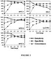
- Optimal threshold values can be determined by testing broader ranges of threshold values, as shown in the Examples below, including in Figure 3 . Actual threshold values may vary depending on the type of cell used and culture and contacting conditions used.
- the threshold i.e the ratio of IL-6 expression or IL-8 expression levels in the test population to the same levels in the control population
- the threshold is about 1.5 or greater, about 3.5 or greater, about 5 or greater, or about 10 or greater.
- a dose response curve may be calculated by testing the compound at increasing concentrations and comparing the results for each concentration to results for a control population. In this way, an EC 50 or IC 50 value may be obtained for test compounds that are found to be toxic to renal proximal tubule cells.
- the described methods may allow prediction of renal proximal tubule-specific toxicity in humans with high accuracy at an early pre-clinical stage, which has not previously been possible. Such prediction could provide additional valuable information during hit-to-lead discovery and lead optimization in developing pharmaceutical drugs, as well as allowing investigation of underlying mechanisms of PT-specific toxicity at an early stage. Pre-clinical results reliably predicting PT-specific toxicity would also help to design clinical studies and to decide whether a more extensive and more frequent clinical safety assessment would be required or patients with an increased risk of nephrotoxicity (e.g. advanced age, diabetes) should be excluded from Phase II studies until more information has been obtained.
- nephrotoxicity e.g. advanced age, diabetes
- the methods described herein may be useful to predict compounds that will be toxic for PTC in humans at clinically relevant concentrations of the compound.
- compounds 1-10 and 19-22 described in the following example all displayed -PT-specific nephrotoxicity in humans at clinically relevant concentrations.
- the method identified most of these compounds as positive when used at concentrations of 1 ⁇ g/ml to 1000 ⁇ g/ml in vitro.
- concentrations ranging from 1 ⁇ g/ml to 1000 ⁇ g/ml in vitro may allow for prediction of PT-specific toxicity at clinically relevant concentrations in vivo.
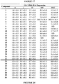
- Test compounds 41 compounds were tested. The nature of these compounds, as well as their classification into different groups, is shown in Table 1 ( Figure 9 ).
- Compounds 3-5, 8, 14, 18-20, 23, 30, 34 and 37 were obtained from Merck (Darmstadt, Germany).
- Compound 1 was purchased from PAA Laboratories GmbH (Pasching, Austria).
- Compound 10 was obtained from ChemService (West Chester, PA, USA) and compound 22 was purchased from Tocris Bioscience (Bristol, UK). All other test compounds were purchased from Sigma-Aldrich (St. Louis, MO, USA). Where possible stock solutions (10 mg/ml) of the test compounds were prepared with biotechnology grade water (1st Base, Singapore), which applied to the following compounds: 1,2, 4-6, 9-18, 23, 25, 28, 30-36, and 40.
- stock solutions (6.8 mg/ml -100 mg/ml depending on the solubility of the individual compound) were prepared with dimethyl sulfoxide (DMSO; Sigma-Aldrich; compounds 3, 7, 8, 19, 22, 27 and 41) or ethanol (compounds 20, 21, 24, 26, 29, 37 and 39). Vehicle controls were performed with the respective solvents. All stock solutions were stored in the dark at 4°C. Stock solutions of metal oxides and inorganic salts (compounds 11-16 and 18) were stored for up to 6 months. No stock solution of an organic compound was stored for longer than 3 months and most stock solutions were consumed much faster during the comprehensive test series.
- DMSO dimethyl sulfoxide
- ethanol compounds 20, 21, 24, 26, 29, 37 and 39
- HPTC HPTC were purchased from the American Type Culture Collection (ATCC, Manassas, VA, USA; HPTC 1) or were isolated from nephrectomy samples (HPTC 2-4) as described (26).
- Commercial HPTC (HPTC 1) were used at passage (P) 4 and P 5, and HPTC 2-4 were used at P 3 and P 4.
- Nephrectomy samples were derived from tumor patients and areas with normal tissue were selected for HPTC isolation after examination by a pathologist. Respective anonymized normal tissue samples were obtained from the Tissue Repository of the National University Health System (NUHS, Singapore).
- HK-2 and LLC-PK1 cells were purchased from ATCC. The different cell types were cultivated as described in the culture media recommended by the vendors (14).
- the culture medium used for HPTC contained 0.5% fetal bovine serum. Institutional Review Board approvals for the work with human kidney samples (DSRB-E/11/143) and the cell types (NUS-IRB Ref. Code: 09-148E) used have been obtained. All cells had been cryopreserved before use.
- qPCR up to 40 cycles was then performed with the 7500 Fast Real-Time PCR System (Applied Biosystems, Carlsbad, CA, USA). Procedures were carried out according to the manufacturers' instructions with the software included in the device.
- High content screening HPTC and HK-2 cells were seeded into 96-well microplates (Becton Dickinson, Franklin Lakes, NJ, USA) at a density of 50,000 cells/cm 2 and LLC-PK1 cells were seeded at 16,000 cells/cm 2 . Cells were cultivated for 72 h and were then treated for 16 h with the test compounds. After fixation for 10 min with 3.7% formaldehyde in phosphate-buffered saline, cell nuclei were stained with 4',6-diamidino-2-phenylindole (Merck) and imaged with the ImageXpress Micro High Content Screening System (Molecular Devices, Sunnyvale, CA, USA).
- HCS High content screening
- TN True negatives
- the sensitivity was calculated by dividing the number of TP by the total number of PT-specific nephrotoxins (group 1, compounds 1-22).
- the specificity was calculated by dividing the number of TN by the total number of non-PT damaging compounds (groups 2 and 3, compounds 23-41). Balanced accuracy was defined as the mean of sensitivity and specificity.
- the positive predictive value (PPV) was calculated by dividing the number of TP by the total number of positives identified by the in vitro model.
- the negative predictive value was calculated by dividing the number of TN by the total number of negatives identified by the in vitro model.
- the concordance with clinical data was calculated in the following way: TP + TN / total number of 41 drugs. When percentages were provided the numbers were multiplied with 100%.
- the receiver operating characteristic (ROC) curves were generated by plotting sensitivity against (1-specificity) at all threshold values ranging from 0.3 - 4.0 (same threshold values as in Figure 3 ).
- the unpaired t-test (Microsoft Office Excel 2010) was used for statistics. The normal distribution of the data was confirmed using SigmaStat (3.5) (Systat Software Inc., Chicago, IL, USA).
- HPTC HPTC as the cellular model for method development to avoid issues related to animal cells and cell lines. All batches of HPTC were routinely characterized by microscopical examination and by qPCR, which was used to determine the expression levels of 31 different marker genes (see Figure 5 and Materials and Methods). For some markers, proper expression at the protein level was confirmed by immunostaining and immunoblotting ( Figure 6 ). These analyses ensured a proper and comparable cell phenotype and quality. Cells were cultivated in normal uncoated multi-well plates. Uncoated tissue culture polystyrene sustains HPTC performance better than other materials with or without extracellular matrix coating (29, 30).
- Cells were seeded at high density and cultivated for three days before drug treatment to allow the formation of a differentiated epithelium, which was confirmed in control experiments (data not shown) as described before (14).
- the state of cell differentiation is of central importance for obtaining cell type-specific responses.
- Kidney injury molecule-1 Kidney injury molecule-1 (KIM-1) and neutrophil gelatinase-associated lipocalin (NGAL) are both up-regulated in the tubular epithelium after injury and are potential novel biomarkers for the early detection of AKI (31-35).
- Interleukin (IL)-18 is up-regulated in the PT epithelium in diseased and injured kidneys and might be a useful biomarker for detecting kidney toxicity (31, 36, 37).
- IL-6 and IL-8 are expressed in PT and PT-derived cells in vivo and in vitro (14, 38-41) and play a central role in pro-inflammatory processes, which occur after injury.
- Different studies demonstrated up-regulation of IL-6 and IL-8 in injured and diseased kidneys (42-44). It is thought that pro-inflammatroy cytokines play a central role in the pathophysiology of AKI, including nephrotoxin-induced AKI (45). Further, significant up-regulation of IL-6 after exposure to nephrotoxins has been demonstrate in a kidney culture model employing purified PTs (24).
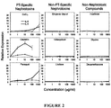
- nephrotoxins To determine the effects of nephrotoxins on these six marker genes in vitro two different batches of HPTC were treated with high doses of gentamicin and CdCl 2 and expression levels were analyzed by qPCR. All individual results were normalized to the expression levels glyceraldehyde 3-phosphate dehydrogenase (GAPDH), which were consistent with cell numbers ( Figure 8 ). If expression of a specific marker gene would be suitable as endpoint the gene should display relatively low expression levels in untreated cells and high levels of induction in response to nephrotoxins. In addition, the gene should be consistently up-regulated in different batches of HPTC and in response to different nephrotoxins. Figure 1 shows that these criteria were best fulfilled by IL-6 and IL-8. From the marker genes tested IL-8 displayed the highest levels of up-regulation after treatment with nephrotoxins.
- GPDH glyceraldehyde 3-phosphate dehydrogenase
- NGAL showed consistent up-regulation, but the levels of up-regulation ranged only between 1.8-fold and 3.5-fold and were lower than the levels of up-regulation of IL-6 and IL-8.
- VIM was up-regulated in only one cell batch.
- KIM-1 and IL-18 were up-regulated in response to only one compound in one cell batch.
- Predictive performance analyzed with 41 compounds Next, we determined the response to 41 well-characterized drugs and chemicals. Most of the 41 compounds used here were drugs that are routinely and widely applied in clinical practice. Some compounds, like CdCl 2 or lindane, are well-characterized environmental toxins and for all of the compounds a wealth of human and animal in vivo and in vitro data is available. As a starting point we selected compounds from published lists (2, 3, 18, 19) that classify compounds with regard to their nephrotoxicity in humans and their effects on various parts of the kidney and nephron. We then made an extensive literature search (PubMed) and also used Google and the ChemIDplus Advanced database to get further information on each selected compound and to confirm its classification.
- Drug exposure was performed for 16 hours after cultivating the cells for 3 days at confluent density. Initially, we tested a wide range of concentrations covering 5 orders of magnitude and ranging from 0.01 ⁇ g/ml to 1000 ⁇ g/ml. As usually no drug-induced changes (in comparison to the controls) were observed at the 2 lowest concentrations, we narrowed the range down and concentrations of 1 ⁇ g/ml, 10 ⁇ g/ml, 100 ⁇ g/ml and 1000 ⁇ g/ml were tested in all cases. Thus, the widest useful range of concentrations was applied in all experiments, with a lack of drug-induced changes at concentrations below the lower limit and compromised solubility of many compounds at concentrations exceeding the upper limit. All results were normalized to the vehicle control and expressed as fold change of IL-6 and IL-8 expression.
- Tables 3 and 4 Examples that illustrate the processing of the data are shown in Tables 3 and 4. Both tables display the same data set obtained with HPTC 1. This data set is identical with the HPTC 1 data set in Table 2 and shows the highest expression levels of IL-6 and IL-8. A threshold of 2.0 (Table 3 ( Figure 11 )) or 3.5 (Table 4 ( Figure 12 )), respectively, was applied to this data set. If the expression level of at least one of the marker genes was equal to or higher than the threshold the test result was classified as positive, and the classification is indicated in Tables 3 and 4.
- FIG. 4 shows the ROC curves obtained with each cell batch/line and the results for either IL-6 or IL-8 or the combination of markers are displayed. The respective AUC values are shown in Table 6 ( Figure 14 ).
- the results confirm that the predictability was higher when HPTC were used (compared to HK-2 and LLC-PK1 cells), and this applied to the mean and median values as well as to each single batch of HPTC. Use of a combination of both markers only slightly improved the results in comparison to the use of IL-8 alone.
- the AUC values obtained with the marker combination ranged from 0.71 (HK-2) to 0.94 (HPTC 1).
- IL-6 and IL-8 are expressed by a large variety of cell types in response to a broad variety of stimuli and injury mechanisms (see, for instance 47-50 and citations therein) and thus the specificity of our model might be surprising.
- potential contributions from other cell types do not play a role in an in vitro model based on one cell type. As long as no other parameters than HPTC injury increase marker gene expression in the in vitro model the response would be expected to be specific.
- This experiment was performed using two types of renal proximal tubular-like cells, under conditions as described in Example 1 above, using the same identified 41 compounds.
- the two cell types were cell populations differentiated from human embryonic stem cells (HUES-7 cells available from Howard Hughes Medical Institute) and from human induced pluripotent cells, which were derived from human foreskin (iPS(foreskin)-4; WiCell Research Institute, Wisconsin, USA). Both stem cell types were differentiated into renal proximal tubular-like cells using a previously described method (see reference 27).
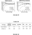
- both cell populations of renal proximal tubular-like cells showed > 70% sensitivity and specificity and overall concordance with human clinical data, as seen in Figures 29 and 30 .
- the major performance metrics of the assay using stem cell-derived renal proximal tubular-like cells ranged between 0.70 and 0.82 ( Figure 30 ).
- ROC curves for each of the cell populations tested are shown in Figure 31 . Further improvements of the assay are expected by optimizing the protocol for the differentiation of stem cells in renal proximal tubular-like cells.
Landscapes
- Health & Medical Sciences (AREA)
- Life Sciences & Earth Sciences (AREA)
- Engineering & Computer Science (AREA)
- Chemical & Material Sciences (AREA)
- Immunology (AREA)
- Biomedical Technology (AREA)
- Molecular Biology (AREA)
- Analytical Chemistry (AREA)
- Urology & Nephrology (AREA)
- Hematology (AREA)
- Proteomics, Peptides & Aminoacids (AREA)
- Organic Chemistry (AREA)
- Cell Biology (AREA)
- Microbiology (AREA)
- Biotechnology (AREA)
- General Health & Medical Sciences (AREA)
- Biochemistry (AREA)
- Physics & Mathematics (AREA)
- Pathology (AREA)
- Bioinformatics & Cheminformatics (AREA)
- Toxicology (AREA)
- Wood Science & Technology (AREA)
- Zoology (AREA)
- Medicinal Chemistry (AREA)
- Food Science & Technology (AREA)
- General Physics & Mathematics (AREA)
- Genetics & Genomics (AREA)
- Tropical Medicine & Parasitology (AREA)
- Biophysics (AREA)
- General Engineering & Computer Science (AREA)
- Measuring Or Testing Involving Enzymes Or Micro-Organisms (AREA)
Claims (14)
- In-vitro-Verfahren zum Screening auf spezifische Toxizität für renale proximale tubuläre Zellen einer Verbindung, wobei das Verfahren umfasst:Inkontaktbringen einer Testverbindung mit einer Testpopulation von renalen proximalen tubulären Zellen, vorzugsweise für einen Zeitraum von ungefähr 8 Stunden oder länger, wobei die Testpopulation eine konfluente Monoschicht, eine subkonfluente Monoschicht, ein konfluentes Epithel oder eine 2D konfluente Kultur ist; undBestimmen des Expressionsniveaus eines Interleukins in der Testpopulation, wobei das Interleukin (i) lnterleukin-6 (IL-6); oder (ii) Interleukin-8 (IL-8); oder sowohl (i) als auch (ii) ist;wobei die Expressionsniveaus des Interleukins in der Testpopulation, die größer als die Expressionsniveaus in einer Kontrollpopulation von renalen proximalen tubulären Zellen sind, die nicht mit der Testverbindung in Kontakt gebracht wurden, darauf hinweisen, dass die Testverbindung für renale proximale tubuläre Zellen toxisch ist.
- Verfahren nach Anspruch 1, wobei die Bestimmung des Expressionsniveaus des Interleukins die Bestimmung der für das Interleukin kodierenden mRNA-Spiegel umfasst.
- Verfahren nach Anspruch 2, wobei die Bestimmung eine oder mehrere quantitative PCR-Techniken, Northern Blot-Techniken, Microarray-Techniken, TRAC-Techniken, Fluoreszenzmarkierungstechniken und Phosphorimaging-Techniken umfasst.
- Verfahren nach Anspruch 1, wobei die Bestimmung des Expressionsniveaus des Interleukins die Bestimmung des Spiegels des sekretierten Interleukin-Proteins umfasst.
- Verfahren nach Anspruch 4, wobei die Bestimmung eine oder mehrere ELISA-Techniken, Biosensor-Techniken, elektrochemische Nachweistechniken, Immunblotting-Techniken, zytometrische Bead-Array-Techniken, Fluoreszenzmarkierungstechniken und Rezeptorbindungstechniken umfasst.
- Verfahren nach Anspruch 1, wobei die Bestimmung des Expressionsniveaus des Interleukins den Nachweis von Niveaus eines Reportergens umfasst, das unter der Kontrolle der IL-6- oder IL-8-Regulierung exprimiert wird.
- Verfahren nach einem der Ansprüche 1 bis 6, wobei das Verhältnis der Expressionsniveaus des Interleukins in der Testpopulation zu den Expressionsniveaus des Interleukins in der Kontrollpopulation ungefähr 1,5 oder höher, vorzugsweise 3,5 oder höher ist, was anzeigt, dass die Testverbindung für renale proximale tubuläre Zellen toxisch ist.
- Verfahren nach einem der Ansprüche 1 bis 7, wobei die renalen proximalen tubulären Zellen aus somatischen Zellen abgeleitet sind und primäre Zellen sind oder Zellen aus einer stabilen Zelllinie sind.
- Verfahren nach Anspruch 8, wobei die renalen proximalen tubulären Zellen menschliche primäre renale proximale tubuläre Zellen, HK-2-Zellen oder LLC-PK1-Zellen sind.
- Verfahren nach einem der Ansprüche 1 bis 7, wobei die renalen proximalen tubulären Zellen aus Stammzellen abgeleitet sind und sich aus embryonalen Stammzellen, mesenchymalen Stammzellen oder induzierten pluripotenten Stammzellen differenziert haben.
- Verfahren nach einem der Ansprüche 1 bis 10, wobei die proximalen tubulären Zellen menschliche renale proximale tubuläre Zellen sind.
- Verfahren nach einem der Ansprüche 1 bis 10, wobei die proximalen tubulären Zellen nicht-menschliche proximale tubuläre Zellen sind.
- Verfahren nach einem der Ansprüche 1 bis 12, wobei das Inkontaktbringen ein oder mehrmals in einem Zeitraum von ungefähr 3 bis ungefähr 14 Tagen wiederholt wird.
- Verfahren nach einem der Ansprüche 1 bis 13, wobei das Inkontaktbringen die Zugabe der Testverbindung zu der Testpopulation der renalen proximalen tubulären Zellen in einer Konzentration von ungefähr 0,001 bis ungefähr 1.000 µg/mL umfasst.
Applications Claiming Priority (3)
| Application Number | Priority Date | Filing Date | Title |
|---|---|---|---|
| US201261674018P | 2012-07-20 | 2012-07-20 | |
| US201261675680P | 2012-07-25 | 2012-07-25 | |
| PCT/IB2013/001944 WO2014013334A2 (en) | 2012-07-20 | 2013-07-22 | In vitro assay for predicting renal proximal tubular cell toxicity |
Publications (3)
| Publication Number | Publication Date |
|---|---|
| EP2875360A2 EP2875360A2 (de) | 2015-05-27 |
| EP2875360A4 EP2875360A4 (de) | 2015-12-23 |
| EP2875360B1 true EP2875360B1 (de) | 2018-08-08 |
Family
ID=49949307
Family Applications (1)
| Application Number | Title | Priority Date | Filing Date |
|---|---|---|---|
| EP13820192.6A Not-in-force EP2875360B1 (de) | 2012-07-20 | 2013-07-22 | In-vitro-test zur vorhersage von renaler proximaler tubulärer toxizität |
Country Status (5)
| Country | Link |
|---|---|
| US (1) | US20150197802A1 (de) |
| EP (1) | EP2875360B1 (de) |
| CN (1) | CN104641237B (de) |
| SG (1) | SG11201500370SA (de) |
| WO (1) | WO2014013334A2 (de) |
Families Citing this family (8)
| Publication number | Priority date | Publication date | Assignee | Title |
|---|---|---|---|---|
| US9719068B2 (en) | 2010-05-06 | 2017-08-01 | Children's Hospital Medical Center | Methods and systems for converting precursor cells into intestinal tissues through directed differentiation |
| CA2949834A1 (en) | 2014-05-28 | 2015-12-03 | James Macormack Wells | Methods and systems for converting precursor cells into gastric tissues through directed differentiation |
| JP6804438B2 (ja) | 2014-10-17 | 2020-12-23 | チルドレンズ ホスピタル メディカル センター | 多能性幹細胞を使用するヒト小腸のin vivoモデル、並びにそれを作製、及び使用する方法 |
| EP3344264B1 (de) * | 2015-09-03 | 2023-03-15 | The Brigham and Women's Hospital, Inc. | Dreidimensionale differenzierung von epiblasten-sphäroiden von nierenorganoiden modellstadienspezifischer epithelphysiologie, -morphogenese und -erkrankung |
| CA3016641A1 (en) | 2016-05-05 | 2017-11-09 | Children's Hospital Medical Center | Methods for the in vitro manufacture of gastric fundus tissue and compositions related to same |
| CN110062764A (zh) | 2016-12-05 | 2019-07-26 | 儿童医院医学中心 | 结肠类器官及其制备和使用方法 |
| CN107219201B (zh) * | 2017-04-20 | 2019-11-12 | 浙江工商大学 | 利用荧光显微镜观察桔霉素免疫后牛蛙抗体和亲和的方法 |
| CN113960302B (zh) * | 2021-09-29 | 2024-04-23 | 中国人民解放军军事科学院军事医学研究院 | 一种基于高内涵技术的肾毒性检测方法及其应用 |
Family Cites Families (10)
| Publication number | Priority date | Publication date | Assignee | Title |
|---|---|---|---|---|
| US5650435A (en) * | 1991-04-01 | 1997-07-22 | Madara; James L. | Modulation of inflammation related to columnar epithelia |
| EP1874950B1 (de) * | 2005-04-05 | 2015-06-03 | Corning Incorporated | Markierungsfreie biosensoren |
| US8603806B2 (en) * | 2005-11-02 | 2013-12-10 | The Ohio State Universtiy Research Foundation | Materials and methods for cell-based assays |
| WO2008101231A2 (en) * | 2007-02-16 | 2008-08-21 | Endocyte, Inc. | Methods and compositions for treating and diagnosing kidney disease |
| EP2176402B1 (de) | 2007-07-19 | 2016-06-01 | Agency for Science, Technology and Research | Verfahren zur differenzierung embryonaler stammzellen zu aqp-1 exprimierenden zellen |
| US20090220982A1 (en) | 2008-02-29 | 2009-09-03 | Achaogen Inc.. | Compositions and methods for determining nephrotoxicity |
| US8062866B2 (en) * | 2008-11-13 | 2011-11-22 | Femta Pharmaceuticals, Inc. | Humanized anti-IL-6 antibodies |
| MX2011005408A (es) * | 2008-11-25 | 2011-06-16 | Alder Biopharmaceuticals Inc | Antagonistas de il-6 para prevenir o tratar la trombosis. |
| WO2010064995A1 (en) * | 2008-12-02 | 2010-06-10 | Agency For Science, Technology And Research | Method for formation of renal tubules |
| US20120179381A1 (en) * | 2010-06-17 | 2012-07-12 | Mckim James M | Toxicity screening methods |
-
2013
- 2013-07-22 WO PCT/IB2013/001944 patent/WO2014013334A2/en active Application Filing
- 2013-07-22 US US14/416,020 patent/US20150197802A1/en not_active Abandoned
- 2013-07-22 CN CN201380048888.XA patent/CN104641237B/zh not_active Expired - Fee Related
- 2013-07-22 EP EP13820192.6A patent/EP2875360B1/de not_active Not-in-force
- 2013-07-22 SG SG11201500370SA patent/SG11201500370SA/en unknown
Non-Patent Citations (1)
| Title |
|---|
| None * |
Also Published As
| Publication number | Publication date |
|---|---|
| CN104641237B (zh) | 2017-04-05 |
| EP2875360A4 (de) | 2015-12-23 |
| WO2014013334A3 (en) | 2014-03-13 |
| US20150197802A1 (en) | 2015-07-16 |
| EP2875360A2 (de) | 2015-05-27 |
| WO2014013334A2 (en) | 2014-01-23 |
| SG11201500370SA (en) | 2015-02-27 |
| CN104641237A (zh) | 2015-05-20 |
Similar Documents
| Publication | Publication Date | Title |
|---|---|---|
| EP2875360B1 (de) | In-vitro-test zur vorhersage von renaler proximaler tubulärer toxizität | |
| Li et al. | An in vitro method for the prediction of renal proximal tubular toxicity in humans | |
| ES2653249T3 (es) | Composiciones y métodos para ensayos de toxigenicidad | |
| WO2017039359A1 (ko) | 트립토파닐 티알엔에이 합성효소를 이용한 감염 질환 또는 감염 합병증의 진단용 조성물과 진단 마커 검출 방법 | |
| Cui et al. | Prognostic value of levels of urine neutrophil gelatinase-associated lipocalin and interleukin-18 in patients with delayed graft function after kidney transplantation | |
| Karpukhina et al. | Analysis of genes regulated by DUX4 via oxidative stress reveals potential therapeutic targets for treatment of facioscapulohumeral dystrophy | |
| EP2478374B1 (de) | Verfahren und kit zur klassifizierung und prognose von wunden | |
| Tsai et al. | Autocrine exosomal fibulin-1 as a target of miR-1269b induces epithelial–mesenchymal transition in proximal tubule in diabetic nephropathy | |
| Zeng et al. | Urinary and kidney podocalyxin and podocin levels in diabetic kidney disease: a kidney biopsy study | |
| KR20140037633A (ko) | 신장독성 및 부작용 유발 약물 검색용 단백질 바이오마커 및 이를 이용한 신장독성 및 부작용 유발 약물 검색 방법 | |
| Muthukumar et al. | Allograft rejection and tubulointerstitial fibrosis in human kidney allografts: interrogation by urinary cell mRNA profiling | |
| CN109811051A (zh) | 一种血浆外泌体来源的miR-548o-3p分子标记及其结核病检测试剂盒 | |
| JP5328647B2 (ja) | 発がんプロモーター検出用のマーカー遺伝子及び発がんプロモーター検出方法 | |
| EP2875349B1 (de) | In-vitro-testverfahren zur vorhersage von renaler proximaler tubulärer toxizität | |
| KR102083408B1 (ko) | 줄기세포 기반의 독성 물질 스크리닝 방법 | |
| AU2011253791B2 (en) | Method and kit for the classification and prognosis of chronic wounds | |
| US20040137488A1 (en) | Methods for identifying the activity of gene products | |
| US20150253328A1 (en) | Nanomaterial-induced cytotoxicity detection composition and kit, and nanomaterial-induced cytotoxicity detection method | |
| KR101822933B1 (ko) | 엑소좀 바이오마커를 이용한 화합물 독성 평가 방법 | |
| KR20240050158A (ko) | 복막 섬유화 제어를 위한 타깃물질 리스트 발굴 | |
| CN117330752A (zh) | Slc14a1作为标志物在制备评估结直肠癌肝转移风险和/或预后情况产品中的应用 | |
| YAO | DEVELOPMENT OF HUMAN IN VITRO MODELS FOR PREDICTING ORGAN-SPECIFIC TOXICITY | |
| Ikawa | Draxin regulates hippocampal neurogenesis in the postnatal dentate gyrus | |
| PEIRIS | Role of Hepatocyte nuclear factor 4A in the Kidney |
Legal Events
| Date | Code | Title | Description |
|---|---|---|---|
| PUAI | Public reference made under article 153(3) epc to a published international application that has entered the european phase |
Free format text: ORIGINAL CODE: 0009012 |
|
| 17P | Request for examination filed |
Effective date: 20150209 |
|
| AK | Designated contracting states |
Kind code of ref document: A2 Designated state(s): AL AT BE BG CH CY CZ DE DK EE ES FI FR GB GR HR HU IE IS IT LI LT LU LV MC MK MT NL NO PL PT RO RS SE SI SK SM TR |
|
| AX | Request for extension of the european patent |
Extension state: BA ME |
|
| DAX | Request for extension of the european patent (deleted) | ||
| A4 | Supplementary search report drawn up and despatched |
Effective date: 20151123 |
|
| RIC1 | Information provided on ipc code assigned before grant |
Ipc: G01N 33/50 20060101ALI20151117BHEP Ipc: C12Q 1/68 20060101ALI20151117BHEP Ipc: G01N 33/68 20060101AFI20151117BHEP |
|
| 17Q | First examination report despatched |
Effective date: 20160928 |
|
| STAA | Information on the status of an ep patent application or granted ep patent |
Free format text: STATUS: EXAMINATION IS IN PROGRESS |
|
| REG | Reference to a national code |
Ref country code: DE Ref legal event code: R079 Ref document number: 602013041788 Country of ref document: DE Free format text: PREVIOUS MAIN CLASS: G01N0033680000 Ipc: C12Q0001688300 |
|
| GRAP | Despatch of communication of intention to grant a patent |
Free format text: ORIGINAL CODE: EPIDOSNIGR1 |
|
| STAA | Information on the status of an ep patent application or granted ep patent |
Free format text: STATUS: GRANT OF PATENT IS INTENDED |
|
| RIC1 | Information provided on ipc code assigned before grant |
Ipc: G01N 33/50 20060101ALI20180122BHEP Ipc: C12Q 1/6883 20180101AFI20180122BHEP |
|
| INTG | Intention to grant announced |
Effective date: 20180220 |
|
| GRAS | Grant fee paid |
Free format text: ORIGINAL CODE: EPIDOSNIGR3 |
|
| GRAA | (expected) grant |
Free format text: ORIGINAL CODE: 0009210 |
|
| STAA | Information on the status of an ep patent application or granted ep patent |
Free format text: STATUS: THE PATENT HAS BEEN GRANTED |
|
| AK | Designated contracting states |
Kind code of ref document: B1 Designated state(s): AL AT BE BG CH CY CZ DE DK EE ES FI FR GB GR HR HU IE IS IT LI LT LU LV MC MK MT NL NO PL PT RO RS SE SI SK SM TR |
|
| REG | Reference to a national code |
Ref country code: GB Ref legal event code: FG4D |
|
| REG | Reference to a national code |
Ref country code: CH Ref legal event code: EP Ref country code: AT Ref legal event code: REF Ref document number: 1027069 Country of ref document: AT Kind code of ref document: T Effective date: 20180815 |
|
| REG | Reference to a national code |
Ref country code: IE Ref legal event code: FG4D |
|
| REG | Reference to a national code |
Ref country code: DE Ref legal event code: R096 Ref document number: 602013041788 Country of ref document: DE |
|
| REG | Reference to a national code |
Ref country code: CH Ref legal event code: NV Representative=s name: AMMANN PATENTANWAELTE AG BERN, CH |
|
| REG | Reference to a national code |
Ref country code: NL Ref legal event code: MP Effective date: 20180808 |
|
| REG | Reference to a national code |
Ref country code: LT Ref legal event code: MG4D |
|
| REG | Reference to a national code |
Ref country code: AT Ref legal event code: MK05 Ref document number: 1027069 Country of ref document: AT Kind code of ref document: T Effective date: 20180808 |
|
| PG25 | Lapsed in a contracting state [announced via postgrant information from national office to epo] |
Ref country code: LT Free format text: LAPSE BECAUSE OF FAILURE TO SUBMIT A TRANSLATION OF THE DESCRIPTION OR TO PAY THE FEE WITHIN THE PRESCRIBED TIME-LIMIT Effective date: 20180808 Ref country code: PL Free format text: LAPSE BECAUSE OF FAILURE TO SUBMIT A TRANSLATION OF THE DESCRIPTION OR TO PAY THE FEE WITHIN THE PRESCRIBED TIME-LIMIT Effective date: 20180808 Ref country code: NO Free format text: LAPSE BECAUSE OF FAILURE TO SUBMIT A TRANSLATION OF THE DESCRIPTION OR TO PAY THE FEE WITHIN THE PRESCRIBED TIME-LIMIT Effective date: 20181108 Ref country code: BG Free format text: LAPSE BECAUSE OF FAILURE TO SUBMIT A TRANSLATION OF THE DESCRIPTION OR TO PAY THE FEE WITHIN THE PRESCRIBED TIME-LIMIT Effective date: 20181108 Ref country code: AT Free format text: LAPSE BECAUSE OF FAILURE TO SUBMIT A TRANSLATION OF THE DESCRIPTION OR TO PAY THE FEE WITHIN THE PRESCRIBED TIME-LIMIT Effective date: 20180808 Ref country code: NL Free format text: LAPSE BECAUSE OF FAILURE TO SUBMIT A TRANSLATION OF THE DESCRIPTION OR TO PAY THE FEE WITHIN THE PRESCRIBED TIME-LIMIT Effective date: 20180808 Ref country code: SE Free format text: LAPSE BECAUSE OF FAILURE TO SUBMIT A TRANSLATION OF THE DESCRIPTION OR TO PAY THE FEE WITHIN THE PRESCRIBED TIME-LIMIT Effective date: 20180808 Ref country code: IS Free format text: LAPSE BECAUSE OF FAILURE TO SUBMIT A TRANSLATION OF THE DESCRIPTION OR TO PAY THE FEE WITHIN THE PRESCRIBED TIME-LIMIT Effective date: 20181208 Ref country code: FI Free format text: LAPSE BECAUSE OF FAILURE TO SUBMIT A TRANSLATION OF THE DESCRIPTION OR TO PAY THE FEE WITHIN THE PRESCRIBED TIME-LIMIT Effective date: 20180808 Ref country code: RS Free format text: LAPSE BECAUSE OF FAILURE TO SUBMIT A TRANSLATION OF THE DESCRIPTION OR TO PAY THE FEE WITHIN THE PRESCRIBED TIME-LIMIT Effective date: 20180808 Ref country code: GR Free format text: LAPSE BECAUSE OF FAILURE TO SUBMIT A TRANSLATION OF THE DESCRIPTION OR TO PAY THE FEE WITHIN THE PRESCRIBED TIME-LIMIT Effective date: 20181109 |
|
| REG | Reference to a national code |
Ref country code: CH Ref legal event code: PK Free format text: BERICHTIGUNGEN |
|
| RIC2 | Information provided on ipc code assigned after grant |
Ipc: G01N 33/50 20060101ALI20180122BHEP Ipc: C12Q 1/6883 20180101AFI20180122BHEP |
|
| PG25 | Lapsed in a contracting state [announced via postgrant information from national office to epo] |
Ref country code: HR Free format text: LAPSE BECAUSE OF FAILURE TO SUBMIT A TRANSLATION OF THE DESCRIPTION OR TO PAY THE FEE WITHIN THE PRESCRIBED TIME-LIMIT Effective date: 20180808 Ref country code: LV Free format text: LAPSE BECAUSE OF FAILURE TO SUBMIT A TRANSLATION OF THE DESCRIPTION OR TO PAY THE FEE WITHIN THE PRESCRIBED TIME-LIMIT Effective date: 20180808 Ref country code: AL Free format text: LAPSE BECAUSE OF FAILURE TO SUBMIT A TRANSLATION OF THE DESCRIPTION OR TO PAY THE FEE WITHIN THE PRESCRIBED TIME-LIMIT Effective date: 20180808 |
|
| PG25 | Lapsed in a contracting state [announced via postgrant information from national office to epo] |
Ref country code: ES Free format text: LAPSE BECAUSE OF FAILURE TO SUBMIT A TRANSLATION OF THE DESCRIPTION OR TO PAY THE FEE WITHIN THE PRESCRIBED TIME-LIMIT Effective date: 20180808 Ref country code: EE Free format text: LAPSE BECAUSE OF FAILURE TO SUBMIT A TRANSLATION OF THE DESCRIPTION OR TO PAY THE FEE WITHIN THE PRESCRIBED TIME-LIMIT Effective date: 20180808 Ref country code: IT Free format text: LAPSE BECAUSE OF FAILURE TO SUBMIT A TRANSLATION OF THE DESCRIPTION OR TO PAY THE FEE WITHIN THE PRESCRIBED TIME-LIMIT Effective date: 20180808 Ref country code: RO Free format text: LAPSE BECAUSE OF FAILURE TO SUBMIT A TRANSLATION OF THE DESCRIPTION OR TO PAY THE FEE WITHIN THE PRESCRIBED TIME-LIMIT Effective date: 20180808 Ref country code: CZ Free format text: LAPSE BECAUSE OF FAILURE TO SUBMIT A TRANSLATION OF THE DESCRIPTION OR TO PAY THE FEE WITHIN THE PRESCRIBED TIME-LIMIT Effective date: 20180808 |
|
| REG | Reference to a national code |
Ref country code: DE Ref legal event code: R097 Ref document number: 602013041788 Country of ref document: DE |
|
| PG25 | Lapsed in a contracting state [announced via postgrant information from national office to epo] |
Ref country code: DK Free format text: LAPSE BECAUSE OF FAILURE TO SUBMIT A TRANSLATION OF THE DESCRIPTION OR TO PAY THE FEE WITHIN THE PRESCRIBED TIME-LIMIT Effective date: 20180808 Ref country code: SK Free format text: LAPSE BECAUSE OF FAILURE TO SUBMIT A TRANSLATION OF THE DESCRIPTION OR TO PAY THE FEE WITHIN THE PRESCRIBED TIME-LIMIT Effective date: 20180808 Ref country code: SM Free format text: LAPSE BECAUSE OF FAILURE TO SUBMIT A TRANSLATION OF THE DESCRIPTION OR TO PAY THE FEE WITHIN THE PRESCRIBED TIME-LIMIT Effective date: 20180808 |
|
| PLBE | No opposition filed within time limit |
Free format text: ORIGINAL CODE: 0009261 |
|
| STAA | Information on the status of an ep patent application or granted ep patent |
Free format text: STATUS: NO OPPOSITION FILED WITHIN TIME LIMIT |
|
| 26N | No opposition filed |
Effective date: 20190509 |
|
| PG25 | Lapsed in a contracting state [announced via postgrant information from national office to epo] |
Ref country code: SI Free format text: LAPSE BECAUSE OF FAILURE TO SUBMIT A TRANSLATION OF THE DESCRIPTION OR TO PAY THE FEE WITHIN THE PRESCRIBED TIME-LIMIT Effective date: 20180808 |
|
| PG25 | Lapsed in a contracting state [announced via postgrant information from national office to epo] |
Ref country code: MC Free format text: LAPSE BECAUSE OF FAILURE TO SUBMIT A TRANSLATION OF THE DESCRIPTION OR TO PAY THE FEE WITHIN THE PRESCRIBED TIME-LIMIT Effective date: 20180808 |
|
| PG25 | Lapsed in a contracting state [announced via postgrant information from national office to epo] |
Ref country code: TR Free format text: LAPSE BECAUSE OF FAILURE TO SUBMIT A TRANSLATION OF THE DESCRIPTION OR TO PAY THE FEE WITHIN THE PRESCRIBED TIME-LIMIT Effective date: 20180808 |
|
| REG | Reference to a national code |
Ref country code: BE Ref legal event code: MM Effective date: 20190731 |
|
| PG25 | Lapsed in a contracting state [announced via postgrant information from national office to epo] |
Ref country code: LU Free format text: LAPSE BECAUSE OF NON-PAYMENT OF DUE FEES Effective date: 20190722 Ref country code: BE Free format text: LAPSE BECAUSE OF NON-PAYMENT OF DUE FEES Effective date: 20190731 |
|
| PG25 | Lapsed in a contracting state [announced via postgrant information from national office to epo] |
Ref country code: PT Free format text: LAPSE BECAUSE OF FAILURE TO SUBMIT A TRANSLATION OF THE DESCRIPTION OR TO PAY THE FEE WITHIN THE PRESCRIBED TIME-LIMIT Effective date: 20181208 Ref country code: FR Free format text: LAPSE BECAUSE OF NON-PAYMENT OF DUE FEES Effective date: 20190731 |
|
| PG25 | Lapsed in a contracting state [announced via postgrant information from national office to epo] |
Ref country code: IE Free format text: LAPSE BECAUSE OF NON-PAYMENT OF DUE FEES Effective date: 20190722 |
|
| PG25 | Lapsed in a contracting state [announced via postgrant information from national office to epo] |
Ref country code: CY Free format text: LAPSE BECAUSE OF FAILURE TO SUBMIT A TRANSLATION OF THE DESCRIPTION OR TO PAY THE FEE WITHIN THE PRESCRIBED TIME-LIMIT Effective date: 20180808 |
|
| PG25 | Lapsed in a contracting state [announced via postgrant information from national office to epo] |
Ref country code: HU Free format text: LAPSE BECAUSE OF FAILURE TO SUBMIT A TRANSLATION OF THE DESCRIPTION OR TO PAY THE FEE WITHIN THE PRESCRIBED TIME-LIMIT; INVALID AB INITIO Effective date: 20130722 Ref country code: MT Free format text: LAPSE BECAUSE OF FAILURE TO SUBMIT A TRANSLATION OF THE DESCRIPTION OR TO PAY THE FEE WITHIN THE PRESCRIBED TIME-LIMIT Effective date: 20180808 |
|
| PGFP | Annual fee paid to national office [announced via postgrant information from national office to epo] |
Ref country code: GB Payment date: 20210720 Year of fee payment: 9 Ref country code: CH Payment date: 20210726 Year of fee payment: 9 Ref country code: DE Payment date: 20210729 Year of fee payment: 9 |
|
| PG25 | Lapsed in a contracting state [announced via postgrant information from national office to epo] |
Ref country code: MK Free format text: LAPSE BECAUSE OF FAILURE TO SUBMIT A TRANSLATION OF THE DESCRIPTION OR TO PAY THE FEE WITHIN THE PRESCRIBED TIME-LIMIT Effective date: 20180808 |
|
| REG | Reference to a national code |
Ref country code: DE Ref legal event code: R119 Ref document number: 602013041788 Country of ref document: DE |
|
| REG | Reference to a national code |
Ref country code: CH Ref legal event code: PL |
|
| GBPC | Gb: european patent ceased through non-payment of renewal fee |
Effective date: 20220722 |
|
| PG25 | Lapsed in a contracting state [announced via postgrant information from national office to epo] |
Ref country code: LI Free format text: LAPSE BECAUSE OF NON-PAYMENT OF DUE FEES Effective date: 20220731 Ref country code: CH Free format text: LAPSE BECAUSE OF NON-PAYMENT OF DUE FEES Effective date: 20220731 |
|
| PG25 | Lapsed in a contracting state [announced via postgrant information from national office to epo] |
Ref country code: GB Free format text: LAPSE BECAUSE OF NON-PAYMENT OF DUE FEES Effective date: 20220722 Ref country code: DE Free format text: LAPSE BECAUSE OF NON-PAYMENT OF DUE FEES Effective date: 20230201 |