EP2874544B1 - Procédé et système de traitement de données d'imagerie ultrasonore - Google Patents
Procédé et système de traitement de données d'imagerie ultrasonore Download PDFInfo
- Publication number
- EP2874544B1 EP2874544B1 EP13748109.9A EP13748109A EP2874544B1 EP 2874544 B1 EP2874544 B1 EP 2874544B1 EP 13748109 A EP13748109 A EP 13748109A EP 2874544 B1 EP2874544 B1 EP 2874544B1
- Authority
- EP
- European Patent Office
- Prior art keywords
- region
- interest
- image
- roi
- user
- Prior art date
- Legal status (The legal status is an assumption and is not a legal conclusion. Google has not performed a legal analysis and makes no representation as to the accuracy of the status listed.)
- Not-in-force
Links
- 238000012545 processing Methods 0.000 title claims description 39
- 238000000034 method Methods 0.000 title claims description 36
- 238000003384 imaging method Methods 0.000 title claims description 33
- 230000003902 lesion Effects 0.000 claims description 35
- 239000000523 sample Substances 0.000 claims description 7
- 230000011218 segmentation Effects 0.000 claims description 6
- 238000004590 computer program Methods 0.000 claims description 4
- 230000000877 morphologic effect Effects 0.000 claims description 3
- 238000004195 computer-aided diagnosis Methods 0.000 description 14
- 210000001519 tissue Anatomy 0.000 description 12
- 238000002604 ultrasonography Methods 0.000 description 8
- 238000005259 measurement Methods 0.000 description 5
- 238000010586 diagram Methods 0.000 description 3
- 238000002091 elastography Methods 0.000 description 3
- 230000003211 malignant effect Effects 0.000 description 3
- 238000004364 calculation method Methods 0.000 description 2
- 208000019425 cirrhosis of liver Diseases 0.000 description 2
- 238000002591 computed tomography Methods 0.000 description 2
- 230000001419 dependent effect Effects 0.000 description 2
- 238000003745 diagnosis Methods 0.000 description 2
- 239000000284 extract Substances 0.000 description 2
- 210000004185 liver Anatomy 0.000 description 2
- 238000011002 quantification Methods 0.000 description 2
- 208000004930 Fatty Liver Diseases 0.000 description 1
- 206010016654 Fibrosis Diseases 0.000 description 1
- 206010019708 Hepatic steatosis Diseases 0.000 description 1
- 206010028980 Neoplasm Diseases 0.000 description 1
- 208000000236 Prostatic Neoplasms Diseases 0.000 description 1
- 210000000481 breast Anatomy 0.000 description 1
- 230000007882 cirrhosis Effects 0.000 description 1
- 238000001514 detection method Methods 0.000 description 1
- 238000002059 diagnostic imaging Methods 0.000 description 1
- 230000004069 differentiation Effects 0.000 description 1
- 238000000605 extraction Methods 0.000 description 1
- 208000010706 fatty liver disease Diseases 0.000 description 1
- 238000002595 magnetic resonance imaging Methods 0.000 description 1
- 230000003287 optical effect Effects 0.000 description 1
- 238000011160 research Methods 0.000 description 1
- 230000035945 sensitivity Effects 0.000 description 1
- 231100000240 steatosis hepatitis Toxicity 0.000 description 1
- 238000012546 transfer Methods 0.000 description 1
Images
Classifications
-
- A—HUMAN NECESSITIES
- A61—MEDICAL OR VETERINARY SCIENCE; HYGIENE
- A61B—DIAGNOSIS; SURGERY; IDENTIFICATION
- A61B8/00—Diagnosis using ultrasonic, sonic or infrasonic waves
- A61B8/52—Devices using data or image processing specially adapted for diagnosis using ultrasonic, sonic or infrasonic waves
- A61B8/5215—Devices using data or image processing specially adapted for diagnosis using ultrasonic, sonic or infrasonic waves involving processing of medical diagnostic data
- A61B8/5223—Devices using data or image processing specially adapted for diagnosis using ultrasonic, sonic or infrasonic waves involving processing of medical diagnostic data for extracting a diagnostic or physiological parameter from medical diagnostic data
-
- A—HUMAN NECESSITIES
- A61—MEDICAL OR VETERINARY SCIENCE; HYGIENE
- A61B—DIAGNOSIS; SURGERY; IDENTIFICATION
- A61B8/00—Diagnosis using ultrasonic, sonic or infrasonic waves
- A61B8/46—Ultrasonic, sonic or infrasonic diagnostic devices with special arrangements for interfacing with the operator or the patient
- A61B8/467—Ultrasonic, sonic or infrasonic diagnostic devices with special arrangements for interfacing with the operator or the patient characterised by special input means
- A61B8/469—Ultrasonic, sonic or infrasonic diagnostic devices with special arrangements for interfacing with the operator or the patient characterised by special input means for selection of a region of interest
-
- A—HUMAN NECESSITIES
- A61—MEDICAL OR VETERINARY SCIENCE; HYGIENE
- A61B—DIAGNOSIS; SURGERY; IDENTIFICATION
- A61B8/00—Diagnosis using ultrasonic, sonic or infrasonic waves
- A61B8/08—Clinical applications
- A61B8/0833—Clinical applications involving detecting or locating foreign bodies or organic structures
- A61B8/085—Clinical applications involving detecting or locating foreign bodies or organic structures for locating body or organic structures, e.g. tumours, calculi, blood vessels, nodules
-
- A—HUMAN NECESSITIES
- A61—MEDICAL OR VETERINARY SCIENCE; HYGIENE
- A61B—DIAGNOSIS; SURGERY; IDENTIFICATION
- A61B8/00—Diagnosis using ultrasonic, sonic or infrasonic waves
- A61B8/48—Diagnostic techniques
- A61B8/485—Diagnostic techniques involving measuring strain or elastic properties
Definitions
- the invention relates to an ultrasound-based imaging method and system, and particularly to the processing of ultrasonic imaging data.
- Ultrasonic imaging has been widely accepted as an easy-to-use, inexpensive imaging modality to diagnose malignant cancers such as breast, liver, prostate cancers, etc.
- clinical doctors still have less confidence in the ability of using ultrasound to differentiate benign and malignant lesions because ultrasound has relatively poor image quality and operator-dependence compared to other imaging modalities such as computed tomography (CT) and Magnetic Resonance Imaging (MRI).
- CT computed tomography
- MRI Magnetic Resonance Imaging
- CAD computer aided diagnosis
- CSD computer decision support
- the current ultrasound-based CAD system relies on B-mode ultrasonic images.
- anatomical information extracted from the B-mode ultrasonic images may be used for the computer aided diagnosis in a CAD system.
- a user needs to manually set a region of interest (ROI) on the B-mode ultrasonic images. Then the anatomical information for the ROI may be extracted from the B-mode ultrasonic images and may be used for the computer aided diagnosis in the CDS system.
- ROI region of interest
- the anatomical information extracted from the B-mode ultrasonic images becomes insufficient for the CDS system. It is desirable to improve the performance of computer aided diagnosis by using for example another category of information in the ultrasound-based CAD system.
- Ultrasonic elastography for example a shear-wave ultrasonic imaging technique
- a shear-wave ultrasonic imaging technique is another ultrasonic imaging mode which can provide elasticity-related data (i.e., stiffness) of tissues.
- Philips has developed the shear-wave ultrasonic elastography point quantification (elastoPQ) technique, which can provide quantitative mechanical information (i.e., stiffness) of tissues.
- elastoPQ shear-wave ultrasonic elastography point quantification
- a user needs to manually set a ROI on the B-mode ultrasonic image to outline the relevant area, and then the shear-wave ultrasonic imaging procedure may be performed to obtain the elasticity-related information for the relevant area.
- US 2011/152687 A1 discloses an ultrasonic diagnostic apparatus that has a control unit having a first ROI setting unit and a second ROI setting unit.
- an elasticity information calculating unit sets an area larger than the ROI as a calculation area, calculates distortion and elasticity modulus of a tissue corresponding to each measurement point on a tomographic image of the calculation area, generates elasticity information based on the distortion and the elasticity modulus, and outputs the elasticity information to an elasticity information storing unit.
- the control unit changes the position/size of the ROI by means of the second ROI setting unit under a state that the frozen image is displayed.
- An elasticity image constructing unit reads the elasticity information corresponding to the changed ROI from the elasticity information storing unit, forms an elasticity image of the changed ROI and displays it on an image display unit.
- the present invention provides a method for processing ultrasonic data according to claim 1 and system for processing ultrasonic data according to claim 6 for facilitating the ultrasound-based computer aided diagnosis.
- the present invention can simplify the operation of the user for setting the two ROIs and make sure that the two ROIs target the same relevant tissue area.
- a method of processing ultrasonic data comprising: obtaining a B-mode ultrasonic image; setting a first ROI on the ultrasonic image according to a first input received from a user; measuring elasticity related data for the first ROI by using a shear wave ultrasonic imaging technique; generating a second ROI on the ultrasonic image on the basis of the first ROI; and extracting image features for the second ROI from the ultrasonic image.
- the user through using the measurement box, i.e., the first ROI, for one mode of ultrasonic imaging, i.e., shear wave ultrasonic imaging (elastoPQ as an example) as the basis for generating the second ROI for the processing of another mode of ultrasonic images, i.e., B-mode ultrasonic images, the user only needs to set the ROI once and the second ROI is automatically generated based on the ROI set by the user.
- the user operation is simplified and the first and second ROIs are sure to target the same or a corresponding relevant tissue area with respect to the two kinds of information, i.e., the elasticity-related information and the anatomical information.
- the method further comprises receiving a second input from the user.
- the step of generating the second ROI comprises:
- the second ROI may be set in a more accurate manner.
- the step of generating a predetermined shape around the first ROI as the second ROI comprises: using the first ROI as the second ROI; or generating the second region of interest by expanding from the first ROI by a predetermined factor.
- the simplest way to generate the second ROI is to use the first ROI as the second ROI. In this way the processing complexity may be reduced.
- the method further comprises receiving a third input from the user, and adjusting the second ROI according to the third input received from the user.
- the user is allowed to adjust the generated second ROI manually.
- a system for processing ultrasonic data comprising: an ultrasonic probe; a B-mode imaging unit for obtaining a B-mode ultrasonic image from ultrasonic radio-frequency data collected by the ultrasonic probe; a user interface for receiving a first input of a user and setting a first ROI on the ultrasonic image according to the first user input; an elasticity measuring unit for measuring elasticity-related data for the first ROI by using a shear wave ultrasonic imaging technique; and an image processing unit for generating a second ROI on the ultrasonic image on the basis of the first ROI and extracting image features for the second ROI from the ultrasonic image.
- the present invention provides a system in which the elasticity-related information and the anatomical information may be efficiently obtained and reliably relate to the same or a corresponding relevant tissue area .
- the user only needs to set the first ROI once and the second ROI is automatically generated by an image processing unit, based on the first ROI; in this way the user operation is simplified and two ROIs are sure to target the same or a corresponding relevant tissue area.
- the user interface is adapted for receiving a second user input
- the image processing unit may be adapted for:
- the image processing unit may be further adapted for: using the first ROI as the second ROI, or for generating the second region of interest by expanding from the first ROI by a predetermined factor.
- the user interface may be adapted for receiving a third input from the user and adjusting the second ROI according to the third input received from the user.
- a computer program product comprising machine executable instruction codes which, when executed on a machine, cause the machine to perform the above mentioned methods for processing ultrasonic data.
- an ultrasonic imaging apparatus which comprises an image processor for processing ultrasonic data, the image processor being configured to perform the above mentioned methods.
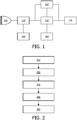
- FIG. 1 an ultrasonic system constructed in accordance with an embodiment of the present invention is shown in the block diagram.
- An ultrasonic probe 100 has a transducer array of transducer elements for transmitting and receiving ultrasonic signals.
- the transducer array can be a one-dimensional or a two-dimensional array of transducer elements. Either type of transducer array can scan a two-dimensional (2D) plane and the two-dimensional array can be used to scan a volumetric region in front of the array.
- the ultrasonic probe 100 is coupled to a B-mode imaging unit 110.
- the B-mode imaging unit 110 may obtain B-mode ultrasonic images from the ultrasonic radio-frequency data collected by the ultrasonic probe 100.
- the obtained B-mode ultrasonic images may be displayed on the display 150 which is coupled to the B-mode imaging unit 110.
- the obtained B-mode ultrasonic images may also be further processed in the image processing unit 120 which is coupled to the B-mode imaging unit 110.
- a user While viewing the displayed B-mode ultrasonic image, a user such as a clinical doctor or a radiologist sets a first ROI on the B-mode ultrasonic image via the user interface 130, which is coupled to the image processing unit 120 and/or to the elasticity measuring unit 140 (not shown in the fig.1 ).
- the user interface receives a user input and set a first ROI on the ultrasonic image according to the user input.
- the first ROI set via the user interface is used by the elasticity measuring unit 140 to perform the measurement of elasticity-related data for the first ROI.
- the measurement of elasticity-related data is performed by using a shear wave ultrasonic imaging technique.
- Such a shear wave ultrasonic imaging technique is described in Philips's patent application WO2011/064688 , which is referred to in this application.
- the measurement of elasticity-related data may be performed by using the shear-wave ultrasonic elastography point quantification (elastoPQ) technique developed by Phillips. Then the measured elasticity-related data may be provided to the CDS system 160 for the purpose of computer aided diagnosis.
- elastoPQ shear-wave ultrasonic elastography point quantification
- the image processing unit 120 generates a second ROI on the ultrasonic image on the basis of the first ROI set via the user interface. And the image processing unit 120 performs further processing of the B-mode ultrasonic images with respect to the second ROI. According to the invention, the image processing unit 120 extracts image features for the second ROI from the B-mode ultrasonic images.
- the extracted image features may present the anatomical information of the relevant tissue area outlined by the second ROI; for example, the image features extracted for the second ROI may be morphological features, texture features, margin features and so on, which may be provided in the CDS system 160 for the purpose of computer aided diagnosis.
- the extraction of image features is performed by the image processing unit 120 outside the CDS system 160.
- the functional unit for extracting image features may be implemented in the CDS system 160.
- the image processing unit 120 may provide the B-mode ultrasonic images having the second ROI thereon to the CDS system 160, and a feature extracting unit of the CDS system may extract the image features for the second ROI from the B-mode ultrasonic images.
- the measured elasticity-related data and the extracted image features are provided to the CDS system 160 for the computer aided diagnosis.
- the CDS system should not be considered as a necessary component for the implementation of the system of the present invention.
- the measured elasticity-related data and the extracted image features may be displayed to the user just for facilitating the user's diagnosis.
- the measured elasticity-related data and the extracted image features may be simultaneously displayed to the user and provided to the CDS system.
- the image processing unit 120 generates the second ROI in different manners according to different clinical applications.
- user specifies, via the user interface 130, what kind of clinical application the present diagnosis relates to; in other words, the user interface may present a prompt to the user to select the type of clinical application and receive a user input, which is referred to as a second user input hereafter.
- the image processing unit 120 may generate a contour of the lesion in the ultrasonic image on the basis of the first ROI for measuring elasticity information and use the contour as the second ROI for extracting anatomical information.
- the contour of the lesion area may be generated by a segmentation technique based on the first ROI.
- the segmentation technique may use the first ROI as the initial contour and achieve the contour of the lesion by expanding the initial contour to the real contour.
- the contour of the lesion may be achieved as long as the first ROI roughly overlaps the lesion area.
- An exemplary segmentation technique for detecting a contour of a subject on the basis of an initially set contour which roughly covers the subject is provided in " Localizing Region-Based Active Contours", Shawn Lankton, et al, IEEE TRANSACTIONS ON IMAGE PROCESSING, VOL.17, NO.11, NOVEMBER 2008 , which is referred to in this application.
- the exemplary segmentation technique may be used by the image processing unit to generate the contour of the lesion on the basis of the first ROI.
- the user may be allowed to adjust the second ROI via the user interface; in other words, the user interface may receive further input from the user and adjust the second ROI according to the user's input.
- the image processing unit 120 may generate the second ROI in a different way. For example, the image processing unit 120 may generate the second ROI around the first ROI according to a predetermined shape. In an example, the image processing unit may use the first ROI as the second ROI. In another example, the image processing unit may expand the first ROI by a predetermined factor and use the expanded shape from the first ROI as the second ROI.
- the factor may be an experimental value and may be set beforehand.
- the user is allowed to adjust the factor via the user interface in order to adjust the expanded shape; in other words, the user interface may receive further user input and adjust the second ROI according to the user input.
- a method for combined use of a shear-wave ultrasonic imaging technique and a B-mode ultrasonic imaging technique is shown in the block diagram.
- a B-mode ultrasonic image may be obtained.
- a first ROI may be set on the ultrasonic image according to a first input received from a user.
- elasticity-related data for the first ROI may be measured by using a shear wave ultrasonic imaging technique.
- a second ROI may be generated on the ultrasonic image on the basis of the first ROI.
- image features may be extracted for the second ROI from the ultrasonic image.
- step 230 may be performed in parallel with steps 240 and 250.
- the second ROI may be generated in different ways according to different clinical applications.
- the method may further comprise receiving a second input from the user. If the second input indicates a lesion application, then at step 240, a contour of the lesion in the ultrasonic image may be generated on the basis of the first ROI and may be used as the second ROI. If the second input indicates a non-lesion application, the second ROI may be generated in a different way at step 240; for example, the second ROI around the first ROI may be generated according to a predetermined shape. In an example, the first ROI may be used as the second ROI.
- the first ROI may be expanded by a predetermined factor and the shape expanded from the first ROI may be used as the second ROI.
- the factor may be an experimental value and may be set beforehand.
- a third input may be received from the user, and the second ROI may be adjusted according to the third input received from the user.
- the B-mode ultrasonic imaging unit 110, the image processing unit 120 and the shear wave ultrasonic imaging unit 140 may be implemented respectively in a dedicated processing unit such as a Digital Signal Processor (DSP) or an Application Specific Integrated Circuit (ASIC) or the like designed specifically for implementing their functions.
- DSP Digital Signal Processor
- ASIC Application Specific Integrated Circuit
- method 200 as shown in fig.2 may be implemented in software as a computer program product, the described process may be stored on or transmitted as program instructions or codes on a computer-readable medium.
- a processor such as a general purpose processor or a specific purpose processor may be used, when executing the program instructions, to perform the method as described above.
- Computer-readable media include any medium that facilitates transfer of a computer program from one place to another and that can be accessed by a computer.
- the computer-readable media may include RAM, ROM, EEPROM, CD-ROM or other optical disk storage, magnetic disk storage or other magnetic storage devices, or any other medium that can be used to carry or store desired program codes in the form of instructions or data structures and that can be accessed by a computer.
- any reference signs placed between parentheses shall not be construed as limiting the claim.
- the word “comprising” does not exclude the presence of elements or steps not listed in a claim or in the description.
- the word “a” or “an” preceding an element does not exclude the presence of a plurality of such elements.
- several of these units can be embodied by one and the same item of software and/or hardware.
- the usage of the words first, second and third, et cetera does not indicate any ordering. These words are to be interpreted as names.
Landscapes
- Health & Medical Sciences (AREA)
- Life Sciences & Earth Sciences (AREA)
- Engineering & Computer Science (AREA)
- Medical Informatics (AREA)
- Surgery (AREA)
- Pathology (AREA)
- Radiology & Medical Imaging (AREA)
- Biophysics (AREA)
- Biomedical Technology (AREA)
- Heart & Thoracic Surgery (AREA)
- Physics & Mathematics (AREA)
- Molecular Biology (AREA)
- Nuclear Medicine, Radiotherapy & Molecular Imaging (AREA)
- Animal Behavior & Ethology (AREA)
- General Health & Medical Sciences (AREA)
- Public Health (AREA)
- Veterinary Medicine (AREA)
- Physiology (AREA)
- Computer Vision & Pattern Recognition (AREA)
- Vascular Medicine (AREA)
- Ultra Sonic Daignosis Equipment (AREA)
- Image Analysis (AREA)
Claims (12)
- Procédé mis en œuvre par ordinateur de traitement de données ultrasonores comprenant:obtenir (210) une image ultrasonore en mode B;établir (220) une première région d'intérêt dans l'image ultrasonore en mode B obtenue en fonction d'une première entrée reçue d'un utilisateur;mesurer (230) des données liées à l'élasticité pour la première région d'intérêt en utilisant une technique d'imagerie ultrasonore à ondes de cisaillement;générer automatiquement (240) une deuxième région d'intérêt dans l'image ultrasonore en mode B obtenue sur la base de la première région d'intérêt; etextraire (250) des caractéristiques d'image représentant des informations anatomiques pour la deuxième région d'intérêt de l'image ultrasonore en mode B obtenue,caractérisé en ce que le procédé comprend en outre la réception d'une deuxième entrée de l'utilisateur, et où la génération (240) de la deuxième région d'intérêt comprend:- si la deuxième entrée indique une application de lésion, générer, sur la base de la première région d'intérêt, un contour de la lésion dans l'image ultrasonore en mode B obtenue comme deuxième région d'intérêt; et- si la deuxième entrée indique une application de non-lésion, générer la deuxième région d'intérêt autour de la première région d'intérêt selon une forme prédéterminée.
- Procédé selon la revendication 1, dans lequel les caractéristiques d'image représentant des informations anatomiques comprennent une caractéristique morphologique ou une caractéristique de texture.
- Procédé selon la revendication 1 ou 2, dans lequel la génération, sur la base de la première région d'intérêt, d'un contour de la lésion dans l'image ultrasonore en mode B obtenue comme deuxième région d'intérêt comprend:
générer le contour de la lésion dans l'image ultrasonore en mode B obtenue par une technique de segmentation basée sur la première région d'intérêt. - Procédé selon l'une quelconque des revendications 1 à 3, dans lequel la génération de la deuxième région d'intérêt autour de la première région d'intérêt selon la forme prédéterminée comprend:utiliser la première région d'intérêt comme deuxième région d'intérêt; ougénérer la deuxième région d'intérêt en élargissant la première région d'intérêt d'un facteur prédéterminé.
- Procédé selon l'une quelconque des revendications 1 à 4, comprenant en outre:recevoir une troisième entrée de l'utilisateur; etajuster la deuxième région d'intérêt en fonction de la troisième entrée reçue de l'utilisateur.
- Système de traitement de données ultrasonores, comprenant:une sonde ultrasonore (100);une unité d'imagerie en mode B (110) adaptée pour obtenir une image ultrasonore en mode B à partir des données radiofréquences ultrasonores collectées par la sonde ultrasonore;une interface utilisateur (130) adaptée pour recevoir une première entrée d'un utilisateur et établir une première région d'intérêt dans l'image ultrasonore en mode B obtenue selon la première entrée utilisateur;une unité de mesure d'élasticité (140) adaptée pour mesurer des données liées à l'élasticité pour la première région d'intérêt en utilisant une technique d'imagerie ultrasonore à ondes de cisaillement; etune unité de traitement d'image (120) adaptée pour générer automatiquement une deuxième région d'intérêt dans l'image ultrasonore en mode B obtenue sur la base de la première région d'intérêt, et extraire des caractéristiques d'image représentant des informations anatomiques pour la deuxième région d'intérêt de la image ultrasonore en mode B obtenue,caractérisé en ce que l'interface utilisateur (130) est en outre adaptée pour recevoir une deuxième entrée utilisateur, et où l'unité de traitement d'image (120) est adaptée pour:- si la deuxième entrée indique une application de lésion, générer, sur la base de la première région d'intérêt, un contour de la lésion dans l'image ultrasonore en mode B obtenue comme deuxième région d'intérêt; et- si la deuxième entrée indique une application de non-lésion, générer la deuxième région d'intérêt autour de la première région d'intérêt selon une forme prédéterminée.
- Système selon la revendication 6, dans lequel les caractéristiques d'image représentant des informations anatomiques comprennent une caractéristique morphologique ou une caractéristique de texture.
- Système selon la revendication 6 ou 7, dans lequel l'unité de traitement d'image (120) est en outre adaptée pour:
générer le contour de la lésion dans l'image ultrasonore en mode B obtenue par une technique de segmentation basée sur la première région d'intérêt. - Système selon l'une quelconque des revendications 6 à 8, dans lequel l'unité de traitement d'image (120) est en outre adaptée pour:utiliser la première région d'intérêt comme deuxième région d'intérêt; ougénérer la deuxième région d'intérêt en élargissant la première région d'intérêt d'un facteur prédéterminé.
- Système selon l'une quelconque des revendications 6 à 9, dans lequel l'interface utilisateur (130) est adaptée pour recevoir une troisième entrée de l'utilisateur et ajuster la deuxième région d'intérêt en fonction de la troisième entrée reçue de l'utilisateur.
- Produit de programme informatique comprenant des codes d'instructions pour exécuter le procédé selon l'une quelconque des revendications 1 à 5.
- Appareil d'imagerie ultrasonore comprenant:
un processeur d'image pour traiter des données ultrasonores, le processeur d'image étant configuré pour exécuter le procédé selon l'une quelconque des revendications 1 à 5.
Applications Claiming Priority (2)
| Application Number | Priority Date | Filing Date | Title |
|---|---|---|---|
| CN2012078816 | 2012-07-18 | ||
| PCT/IB2013/055382 WO2014013366A1 (fr) | 2012-07-18 | 2013-07-01 | Procédé et système de traitement de données d'imagerie ultrasonore |
Publications (2)
| Publication Number | Publication Date |
|---|---|
| EP2874544A1 EP2874544A1 (fr) | 2015-05-27 |
| EP2874544B1 true EP2874544B1 (fr) | 2020-09-09 |
Family
ID=48980234
Family Applications (1)
| Application Number | Title | Priority Date | Filing Date |
|---|---|---|---|
| EP13748109.9A Not-in-force EP2874544B1 (fr) | 2012-07-18 | 2013-07-01 | Procédé et système de traitement de données d'imagerie ultrasonore |
Country Status (6)
| Country | Link |
|---|---|
| US (1) | US11020094B2 (fr) |
| EP (1) | EP2874544B1 (fr) |
| JP (1) | JP6133984B2 (fr) |
| BR (1) | BR112015000820B1 (fr) |
| RU (1) | RU2636262C2 (fr) |
| WO (1) | WO2014013366A1 (fr) |
Families Citing this family (8)
| Publication number | Priority date | Publication date | Assignee | Title |
|---|---|---|---|---|
| KR20170067444A (ko) * | 2015-12-08 | 2017-06-16 | 삼성메디슨 주식회사 | 초음파 진단 장치 및 그 제어방법 |
| JP6513220B2 (ja) | 2015-12-18 | 2019-05-15 | オリンパス株式会社 | 超音波観測装置、超音波観測装置の作動方法および超音波観測装置の作動プログラム |
| KR102030567B1 (ko) * | 2015-12-23 | 2019-10-10 | 지멘스 메디컬 솔루션즈 유에스에이, 인크. | 초음파 영상을 표시하는 초음파 시스템 및 방법 |
| JP6594458B2 (ja) * | 2016-02-12 | 2019-10-23 | オリンパス株式会社 | 超音波観測装置、超音波観測装置の作動方法、及び超音波観測装置の作動プログラム |
| RU2632768C1 (ru) * | 2016-12-16 | 2017-10-09 | Федеральное государственное бюджетное образовательное учреждение высшего образования "Кубанский государственный медицинский университет" Министерства здравоохранения Российской Федерации (ФГБОУ ВО КубГМУ МЗ РФ) | Способ дифференциальной диагностики новообразований печени при ультразвуковом исследовании |
| EP3530193A1 (fr) * | 2018-02-26 | 2019-08-28 | Koninklijke Philips N.V. | Fourniture d'une image ultrasonore tridimensionnelle |
| JP7052530B2 (ja) * | 2018-04-25 | 2022-04-12 | コニカミノルタ株式会社 | 超音波診断装置、および、超音波信号処理方法 |
| CN113545806A (zh) * | 2020-04-26 | 2021-10-26 | 深圳迈瑞生物医疗电子股份有限公司 | 前列腺弹性成像方法和超声弹性成像系统 |
Family Cites Families (30)
| Publication number | Priority date | Publication date | Assignee | Title |
|---|---|---|---|---|
| US5551434A (en) | 1994-06-22 | 1996-09-03 | Kabushiki Kaisha Toshiba | Ultrasonic imaging diagnosis apparatus |
| RU2179313C2 (ru) * | 1999-07-13 | 2002-02-10 | Государственный научный центр Российской Федерации Всероссийский научно-исследовательский институт неорганических материалов им. акад. А.А. Бочвара | Ультразвуковой способ контроля изделий и материалов |
| US6306089B1 (en) * | 1999-09-24 | 2001-10-23 | Atl Ultrasound, Inc. | Ultrasonic diagnostic imaging system with customized measurements and calculations |
| US6508768B1 (en) * | 2000-11-22 | 2003-01-21 | University Of Kansas Medical Center | Ultrasonic elasticity imaging |
| US7397937B2 (en) * | 2001-11-23 | 2008-07-08 | R2 Technology, Inc. | Region growing in anatomical images |
| US7090640B2 (en) * | 2003-11-12 | 2006-08-15 | Q-Vision | System and method for automatic determination of a region of interest within an image |
| KR100686289B1 (ko) | 2004-04-01 | 2007-02-23 | 주식회사 메디슨 | 대상체 영상의 윤곽내 볼륨 데이터를 이용하는 3차원초음파 영상 형성 장치 및 방법 |
| US20050283076A1 (en) * | 2004-06-18 | 2005-12-22 | Hangiandreou Nicholas J | Non-invasive diagnosis of breast cancer using real-time ultrasound strain imaging |
| FR2880154B1 (fr) * | 2004-12-27 | 2007-06-22 | Gen Electric | Procede et systeme de visualisation rapide de structures |
| JP4732086B2 (ja) * | 2005-09-12 | 2011-07-27 | 株式会社日立メディコ | 超音波診断装置 |
| CN101291629B (zh) * | 2005-10-19 | 2010-12-01 | 株式会社日立医药 | 超声波诊断装置 |
| EP2030572B1 (fr) * | 2006-05-25 | 2016-06-08 | Hitachi Medical Corporation | Dispositif ultrasonographique |
| EP2030573A4 (fr) * | 2006-06-06 | 2013-02-27 | Hitachi Medical Corp | Dispositif d'échographie |
| US8160364B2 (en) * | 2007-02-16 | 2012-04-17 | Raytheon Company | System and method for image registration based on variable region of interest |
| JP5087341B2 (ja) * | 2007-08-13 | 2012-12-05 | 株式会社日立メディコ | 超音波診断装置 |
| US8251908B2 (en) * | 2007-10-01 | 2012-08-28 | Insightec Ltd. | Motion compensated image-guided focused ultrasound therapy system |
| US8187187B2 (en) | 2008-07-16 | 2012-05-29 | Siemens Medical Solutions Usa, Inc. | Shear wave imaging |
| EP2310829B1 (fr) * | 2008-07-30 | 2016-04-06 | Centre Hospitalier De L'Universite de Montreal | Systeme et procede de detection, de caracterisation et d'imagerie d'heterogeneite par resonance induite par ondes de cisaillement |
| JP5465671B2 (ja) * | 2008-08-29 | 2014-04-09 | 株式会社日立メディコ | 超音波診断装置 |
| WO2010044385A1 (fr) * | 2008-10-14 | 2010-04-22 | 株式会社 日立メディコ | Dispositif échographique et procédé d'affichage échographique |
| US8992426B2 (en) | 2009-05-04 | 2015-03-31 | Siemens Medical Solutions Usa, Inc. | Feedback in medical ultrasound imaging for high intensity focused ultrasound |
| US20100286520A1 (en) | 2009-05-11 | 2010-11-11 | General Electric Company | Ultrasound system and method to determine mechanical properties of a target region |
| CN101599174A (zh) | 2009-08-13 | 2009-12-09 | 哈尔滨工业大学 | 基于边缘和统计特征的水平集医学超声图像区域轮廓提取方法 |
| JP5345477B2 (ja) * | 2009-08-28 | 2013-11-20 | ジーイー・メディカル・システムズ・グローバル・テクノロジー・カンパニー・エルエルシー | 超音波診断装置及びその制御プログラム |
| BR112012012230B1 (pt) | 2009-11-25 | 2021-04-20 | Koninklijke Philips N.V. | sistema de formação de imagem diagnóstica ultrassônica para análise de onda de cisalhamento e método para operar um sistema de formação de imagem diagnóstica ultrassônica para medir as ondas de cisalhamento |
| JP2011224346A (ja) * | 2010-03-31 | 2011-11-10 | Toshiba Corp | 超音波診断装置、画像処理装置および画像処理方法 |
| JP5260602B2 (ja) * | 2010-06-11 | 2013-08-14 | ジーイー・メディカル・システムズ・グローバル・テクノロジー・カンパニー・エルエルシー | 超音波診断装置 |
| US9101289B2 (en) * | 2010-07-27 | 2015-08-11 | Hitachi Medical Corporation | Ultrasonic diagnostic apparatus |
| WO2012035472A1 (fr) * | 2010-09-16 | 2012-03-22 | Koninklijke Philips Electronics N.V. | Quantification de déformation de tissu dans des images d'élastographie ultrasonore |
| JP2012100841A (ja) * | 2010-11-10 | 2012-05-31 | Ge Medical Systems Global Technology Co Llc | 超音波診断装置 |
-
2013
- 2013-07-01 US US14/415,252 patent/US11020094B2/en active Active
- 2013-07-01 BR BR112015000820-8A patent/BR112015000820B1/pt not_active IP Right Cessation
- 2013-07-01 JP JP2015522190A patent/JP6133984B2/ja not_active Expired - Fee Related
- 2013-07-01 RU RU2015105383A patent/RU2636262C2/ru active
- 2013-07-01 WO PCT/IB2013/055382 patent/WO2014013366A1/fr active Application Filing
- 2013-07-01 EP EP13748109.9A patent/EP2874544B1/fr not_active Not-in-force
Non-Patent Citations (1)
| Title |
|---|
| None * |
Also Published As
| Publication number | Publication date |
|---|---|
| BR112015000820A2 (pt) | 2017-06-27 |
| EP2874544A1 (fr) | 2015-05-27 |
| US11020094B2 (en) | 2021-06-01 |
| JP2015522367A (ja) | 2015-08-06 |
| RU2636262C2 (ru) | 2017-11-21 |
| WO2014013366A1 (fr) | 2014-01-23 |
| JP6133984B2 (ja) | 2017-05-24 |
| BR112015000820B1 (pt) | 2021-01-19 |
| RU2015105383A (ru) | 2016-09-10 |
| US20150190120A1 (en) | 2015-07-09 |
Similar Documents
| Publication | Publication Date | Title |
|---|---|---|
| EP2874544B1 (fr) | Procédé et système de traitement de données d'imagerie ultrasonore | |
| Rella et al. | Automated breast ultrasonography (ABUS) in the screening and diagnostic setting: indications and practical use | |
| EP3806721B1 (fr) | Procédé et système pour estimer la teneur fractionnelle en matières grasses d'un tissu | |
| US9558549B2 (en) | Image processing apparatus, method of controlling the same and storage medium | |
| EP2769676B1 (fr) | Procédé et système de superposition d'images medicales | |
| US9084578B2 (en) | Diagnostic imaging apparatus and method | |
| EP2846310A2 (fr) | Procédé et appareil d'enregistrement d'images médicales | |
| US9489921B2 (en) | Method and apparatus for displaying plurality of different images of object | |
| US9324155B2 (en) | Systems and methods for determining parameters for image analysis | |
| KR20130023735A (ko) | 장기 모델 영상 생성 방법 및 장치 | |
| US20150157298A1 (en) | Apparatus and method for combining three dimensional ultrasound images | |
| US20160210774A1 (en) | Breast density estimation | |
| JP2011152416A (ja) | 3次元超音波映像に映像処理および関心物体の大きさ測定を行う超音波システムおよび方法 | |
| KR20120102447A (ko) | 진단장치 및 방법 | |
| US20120078101A1 (en) | Ultrasound system for displaying slice of object and method thereof | |
| CN104470443B (zh) | 用于处理超声成像数据的方法和系统 | |
| KR20130010732A (ko) | 복수의 3차원 볼륨 영상들을 이용하여 3차원 볼륨 파노라마 영상 생성 방법 및 장치 | |
| JP5579535B2 (ja) | 胎児の肋骨数を測定する超音波システムおよび方法 | |
| JP6538280B2 (ja) | 被験者の組織を特徴付ける装置及び方法 | |
| EP2976017B1 (fr) | Techniques de formation de faisceau pour détection de microcalcifications par ultrasons | |
| CN111260606A (zh) | 诊断装置和诊断方法 | |
| Chen et al. | 2-D ultrasound strain images for breast cancer diagnosis using nonrigid subregion registration | |
| CN114708283A (zh) | 图像目标的分割方法、装置、电子设备及存储介质 | |
| Costa | Automated Deformable Registration of Breast Images: towards a software-assisted multimodal breast image reading | |
| CN118576241A (zh) | 肝脏超声信息的显示方法和超声成像系统 |
Legal Events
| Date | Code | Title | Description |
|---|---|---|---|
| PUAI | Public reference made under article 153(3) epc to a published international application that has entered the european phase |
Free format text: ORIGINAL CODE: 0009012 |
|
| 17P | Request for examination filed |
Effective date: 20150218 |
|
| AK | Designated contracting states |
Kind code of ref document: A1 Designated state(s): AL AT BE BG CH CY CZ DE DK EE ES FI FR GB GR HR HU IE IS IT LI LT LU LV MC MK MT NL NO PL PT RO RS SE SI SK SM TR |
|
| AX | Request for extension of the european patent |
Extension state: BA ME |
|
| DAX | Request for extension of the european patent (deleted) | ||
| STAA | Information on the status of an ep patent application or granted ep patent |
Free format text: STATUS: EXAMINATION IS IN PROGRESS |
|
| 17Q | First examination report despatched |
Effective date: 20191014 |
|
| GRAP | Despatch of communication of intention to grant a patent |
Free format text: ORIGINAL CODE: EPIDOSNIGR1 |
|
| RAP1 | Party data changed (applicant data changed or rights of an application transferred) |
Owner name: KONINKLIJKE PHILIPS N.V. |
|
| STAA | Information on the status of an ep patent application or granted ep patent |
Free format text: STATUS: GRANT OF PATENT IS INTENDED |
|
| INTG | Intention to grant announced |
Effective date: 20200319 |
|
| GRAS | Grant fee paid |
Free format text: ORIGINAL CODE: EPIDOSNIGR3 |
|
| GRAA | (expected) grant |
Free format text: ORIGINAL CODE: 0009210 |
|
| STAA | Information on the status of an ep patent application or granted ep patent |
Free format text: STATUS: THE PATENT HAS BEEN GRANTED |
|
| AK | Designated contracting states |
Kind code of ref document: B1 Designated state(s): AL AT BE BG CH CY CZ DE DK EE ES FI FR GB GR HR HU IE IS IT LI LT LU LV MC MK MT NL NO PL PT RO RS SE SI SK SM TR |
|
| REG | Reference to a national code |
Ref country code: GB Ref legal event code: FG4D |
|
| REG | Reference to a national code |
Ref country code: AT Ref legal event code: REF Ref document number: 1310549 Country of ref document: AT Kind code of ref document: T Effective date: 20200915 Ref country code: CH Ref legal event code: EP |
|
| REG | Reference to a national code |
Ref country code: IE Ref legal event code: FG4D |
|
| REG | Reference to a national code |
Ref country code: DE Ref legal event code: R096 Ref document number: 602013072371 Country of ref document: DE |
|
| REG | Reference to a national code |
Ref country code: DE Ref legal event code: R084 Ref document number: 602013072371 Country of ref document: DE |
|
| REG | Reference to a national code |
Ref country code: GB Ref legal event code: 746 Effective date: 20201123 |
|
| REG | Reference to a national code |
Ref country code: LT Ref legal event code: MG4D |
|
| PG25 | Lapsed in a contracting state [announced via postgrant information from national office to epo] |
Ref country code: NO Free format text: LAPSE BECAUSE OF FAILURE TO SUBMIT A TRANSLATION OF THE DESCRIPTION OR TO PAY THE FEE WITHIN THE PRESCRIBED TIME-LIMIT Effective date: 20201209 Ref country code: FI Free format text: LAPSE BECAUSE OF FAILURE TO SUBMIT A TRANSLATION OF THE DESCRIPTION OR TO PAY THE FEE WITHIN THE PRESCRIBED TIME-LIMIT Effective date: 20200909 Ref country code: SE Free format text: LAPSE BECAUSE OF FAILURE TO SUBMIT A TRANSLATION OF THE DESCRIPTION OR TO PAY THE FEE WITHIN THE PRESCRIBED TIME-LIMIT Effective date: 20200909 Ref country code: BG Free format text: LAPSE BECAUSE OF FAILURE TO SUBMIT A TRANSLATION OF THE DESCRIPTION OR TO PAY THE FEE WITHIN THE PRESCRIBED TIME-LIMIT Effective date: 20201209 Ref country code: HR Free format text: LAPSE BECAUSE OF FAILURE TO SUBMIT A TRANSLATION OF THE DESCRIPTION OR TO PAY THE FEE WITHIN THE PRESCRIBED TIME-LIMIT Effective date: 20200909 Ref country code: LT Free format text: LAPSE BECAUSE OF FAILURE TO SUBMIT A TRANSLATION OF THE DESCRIPTION OR TO PAY THE FEE WITHIN THE PRESCRIBED TIME-LIMIT Effective date: 20200909 Ref country code: GR Free format text: LAPSE BECAUSE OF FAILURE TO SUBMIT A TRANSLATION OF THE DESCRIPTION OR TO PAY THE FEE WITHIN THE PRESCRIBED TIME-LIMIT Effective date: 20201210 |
|
| REG | Reference to a national code |
Ref country code: AT Ref legal event code: MK05 Ref document number: 1310549 Country of ref document: AT Kind code of ref document: T Effective date: 20200909 |
|
| REG | Reference to a national code |
Ref country code: NL Ref legal event code: MP Effective date: 20200909 |
|
| PG25 | Lapsed in a contracting state [announced via postgrant information from national office to epo] |
Ref country code: PL Free format text: LAPSE BECAUSE OF FAILURE TO SUBMIT A TRANSLATION OF THE DESCRIPTION OR TO PAY THE FEE WITHIN THE PRESCRIBED TIME-LIMIT Effective date: 20200909 Ref country code: RS Free format text: LAPSE BECAUSE OF FAILURE TO SUBMIT A TRANSLATION OF THE DESCRIPTION OR TO PAY THE FEE WITHIN THE PRESCRIBED TIME-LIMIT Effective date: 20200909 Ref country code: LV Free format text: LAPSE BECAUSE OF FAILURE TO SUBMIT A TRANSLATION OF THE DESCRIPTION OR TO PAY THE FEE WITHIN THE PRESCRIBED TIME-LIMIT Effective date: 20200909 |
|
| PG25 | Lapsed in a contracting state [announced via postgrant information from national office to epo] |
Ref country code: CZ Free format text: LAPSE BECAUSE OF FAILURE TO SUBMIT A TRANSLATION OF THE DESCRIPTION OR TO PAY THE FEE WITHIN THE PRESCRIBED TIME-LIMIT Effective date: 20200909 Ref country code: EE Free format text: LAPSE BECAUSE OF FAILURE TO SUBMIT A TRANSLATION OF THE DESCRIPTION OR TO PAY THE FEE WITHIN THE PRESCRIBED TIME-LIMIT Effective date: 20200909 Ref country code: SM Free format text: LAPSE BECAUSE OF FAILURE TO SUBMIT A TRANSLATION OF THE DESCRIPTION OR TO PAY THE FEE WITHIN THE PRESCRIBED TIME-LIMIT Effective date: 20200909 Ref country code: RO Free format text: LAPSE BECAUSE OF FAILURE TO SUBMIT A TRANSLATION OF THE DESCRIPTION OR TO PAY THE FEE WITHIN THE PRESCRIBED TIME-LIMIT Effective date: 20200909 Ref country code: PT Free format text: LAPSE BECAUSE OF FAILURE TO SUBMIT A TRANSLATION OF THE DESCRIPTION OR TO PAY THE FEE WITHIN THE PRESCRIBED TIME-LIMIT Effective date: 20210111 Ref country code: NL Free format text: LAPSE BECAUSE OF FAILURE TO SUBMIT A TRANSLATION OF THE DESCRIPTION OR TO PAY THE FEE WITHIN THE PRESCRIBED TIME-LIMIT Effective date: 20200909 |
|
| PG25 | Lapsed in a contracting state [announced via postgrant information from national office to epo] |
Ref country code: IS Free format text: LAPSE BECAUSE OF FAILURE TO SUBMIT A TRANSLATION OF THE DESCRIPTION OR TO PAY THE FEE WITHIN THE PRESCRIBED TIME-LIMIT Effective date: 20210109 Ref country code: ES Free format text: LAPSE BECAUSE OF FAILURE TO SUBMIT A TRANSLATION OF THE DESCRIPTION OR TO PAY THE FEE WITHIN THE PRESCRIBED TIME-LIMIT Effective date: 20200909 Ref country code: AT Free format text: LAPSE BECAUSE OF FAILURE TO SUBMIT A TRANSLATION OF THE DESCRIPTION OR TO PAY THE FEE WITHIN THE PRESCRIBED TIME-LIMIT Effective date: 20200909 Ref country code: AL Free format text: LAPSE BECAUSE OF FAILURE TO SUBMIT A TRANSLATION OF THE DESCRIPTION OR TO PAY THE FEE WITHIN THE PRESCRIBED TIME-LIMIT Effective date: 20200909 |
|
| REG | Reference to a national code |
Ref country code: DE Ref legal event code: R097 Ref document number: 602013072371 Country of ref document: DE |
|
| PG25 | Lapsed in a contracting state [announced via postgrant information from national office to epo] |
Ref country code: SK Free format text: LAPSE BECAUSE OF FAILURE TO SUBMIT A TRANSLATION OF THE DESCRIPTION OR TO PAY THE FEE WITHIN THE PRESCRIBED TIME-LIMIT Effective date: 20200909 |
|
| PLBE | No opposition filed within time limit |
Free format text: ORIGINAL CODE: 0009261 |
|
| STAA | Information on the status of an ep patent application or granted ep patent |
Free format text: STATUS: NO OPPOSITION FILED WITHIN TIME LIMIT |
|
| 26N | No opposition filed |
Effective date: 20210610 |
|
| PG25 | Lapsed in a contracting state [announced via postgrant information from national office to epo] |
Ref country code: SI Free format text: LAPSE BECAUSE OF FAILURE TO SUBMIT A TRANSLATION OF THE DESCRIPTION OR TO PAY THE FEE WITHIN THE PRESCRIBED TIME-LIMIT Effective date: 20200909 Ref country code: DK Free format text: LAPSE BECAUSE OF FAILURE TO SUBMIT A TRANSLATION OF THE DESCRIPTION OR TO PAY THE FEE WITHIN THE PRESCRIBED TIME-LIMIT Effective date: 20200909 |
|
| PG25 | Lapsed in a contracting state [announced via postgrant information from national office to epo] |
Ref country code: IT Free format text: LAPSE BECAUSE OF FAILURE TO SUBMIT A TRANSLATION OF THE DESCRIPTION OR TO PAY THE FEE WITHIN THE PRESCRIBED TIME-LIMIT Effective date: 20200909 |
|
| PGFP | Annual fee paid to national office [announced via postgrant information from national office to epo] |
Ref country code: FR Payment date: 20210726 Year of fee payment: 9 |
|
| PGFP | Annual fee paid to national office [announced via postgrant information from national office to epo] |
Ref country code: DE Payment date: 20210729 Year of fee payment: 9 Ref country code: GB Payment date: 20210726 Year of fee payment: 9 |
|
| REG | Reference to a national code |
Ref country code: CH Ref legal event code: PL |
|
| PG25 | Lapsed in a contracting state [announced via postgrant information from national office to epo] |
Ref country code: MC Free format text: LAPSE BECAUSE OF FAILURE TO SUBMIT A TRANSLATION OF THE DESCRIPTION OR TO PAY THE FEE WITHIN THE PRESCRIBED TIME-LIMIT Effective date: 20200909 |
|
| REG | Reference to a national code |
Ref country code: BE Ref legal event code: MM Effective date: 20210731 |
|
| PG25 | Lapsed in a contracting state [announced via postgrant information from national office to epo] |
Ref country code: LI Free format text: LAPSE BECAUSE OF NON-PAYMENT OF DUE FEES Effective date: 20210731 Ref country code: CH Free format text: LAPSE BECAUSE OF NON-PAYMENT OF DUE FEES Effective date: 20210731 |
|
| PG25 | Lapsed in a contracting state [announced via postgrant information from national office to epo] |
Ref country code: LU Free format text: LAPSE BECAUSE OF NON-PAYMENT OF DUE FEES Effective date: 20210701 |
|
| PG25 | Lapsed in a contracting state [announced via postgrant information from national office to epo] |
Ref country code: IE Free format text: LAPSE BECAUSE OF NON-PAYMENT OF DUE FEES Effective date: 20210701 Ref country code: BE Free format text: LAPSE BECAUSE OF NON-PAYMENT OF DUE FEES Effective date: 20210731 |
|
| REG | Reference to a national code |
Ref country code: DE Ref legal event code: R119 Ref document number: 602013072371 Country of ref document: DE |
|
| GBPC | Gb: european patent ceased through non-payment of renewal fee |
Effective date: 20220701 |
|
| PG25 | Lapsed in a contracting state [announced via postgrant information from national office to epo] |
Ref country code: FR Free format text: LAPSE BECAUSE OF NON-PAYMENT OF DUE FEES Effective date: 20220731 |
|
| PG25 | Lapsed in a contracting state [announced via postgrant information from national office to epo] |
Ref country code: HU Free format text: LAPSE BECAUSE OF FAILURE TO SUBMIT A TRANSLATION OF THE DESCRIPTION OR TO PAY THE FEE WITHIN THE PRESCRIBED TIME-LIMIT; INVALID AB INITIO Effective date: 20130701 Ref country code: GB Free format text: LAPSE BECAUSE OF NON-PAYMENT OF DUE FEES Effective date: 20220701 Ref country code: DE Free format text: LAPSE BECAUSE OF NON-PAYMENT OF DUE FEES Effective date: 20230201 |
|
| PG25 | Lapsed in a contracting state [announced via postgrant information from national office to epo] |
Ref country code: CY Free format text: LAPSE BECAUSE OF FAILURE TO SUBMIT A TRANSLATION OF THE DESCRIPTION OR TO PAY THE FEE WITHIN THE PRESCRIBED TIME-LIMIT Effective date: 20200909 |
|
| PG25 | Lapsed in a contracting state [announced via postgrant information from national office to epo] |
Ref country code: MK Free format text: LAPSE BECAUSE OF FAILURE TO SUBMIT A TRANSLATION OF THE DESCRIPTION OR TO PAY THE FEE WITHIN THE PRESCRIBED TIME-LIMIT Effective date: 20200909 |
|
| PG25 | Lapsed in a contracting state [announced via postgrant information from national office to epo] |
Ref country code: TR Free format text: LAPSE BECAUSE OF FAILURE TO SUBMIT A TRANSLATION OF THE DESCRIPTION OR TO PAY THE FEE WITHIN THE PRESCRIBED TIME-LIMIT Effective date: 20200909 |
|
| PG25 | Lapsed in a contracting state [announced via postgrant information from national office to epo] |
Ref country code: MT Free format text: LAPSE BECAUSE OF FAILURE TO SUBMIT A TRANSLATION OF THE DESCRIPTION OR TO PAY THE FEE WITHIN THE PRESCRIBED TIME-LIMIT Effective date: 20200909 |