EP2849817B1 - Systems for extracorporeal blood modification - Google Patents
Systems for extracorporeal blood modification Download PDFInfo
- Publication number
- EP2849817B1 EP2849817B1 EP13791497.4A EP13791497A EP2849817B1 EP 2849817 B1 EP2849817 B1 EP 2849817B1 EP 13791497 A EP13791497 A EP 13791497A EP 2849817 B1 EP2849817 B1 EP 2849817B1
- Authority
- EP
- European Patent Office
- Prior art keywords
- antibody
- options
- polymer
- antigen
- subject
- Prior art date
- Legal status (The legal status is an assumption and is not a legal conclusion. Google has not performed a legal analysis and makes no representation as to the accuracy of the status listed.)
- Active
Links
Images


Classifications
-
- A—HUMAN NECESSITIES
- A61—MEDICAL OR VETERINARY SCIENCE; HYGIENE
- A61M—DEVICES FOR INTRODUCING MEDIA INTO, OR ONTO, THE BODY; DEVICES FOR TRANSDUCING BODY MEDIA OR FOR TAKING MEDIA FROM THE BODY; DEVICES FOR PRODUCING OR ENDING SLEEP OR STUPOR
- A61M1/00—Suction or pumping devices for medical purposes; Devices for carrying-off, for treatment of, or for carrying-over, body-liquids; Drainage systems
- A61M1/36—Other treatment of blood in a by-pass of the natural circulatory system, e.g. temperature adaptation, irradiation ; Extra-corporeal blood circuits
- A61M1/362—Other treatment of blood in a by-pass of the natural circulatory system, e.g. temperature adaptation, irradiation ; Extra-corporeal blood circuits changing physical properties of target cells by binding them to added particles to facilitate their subsequent separation from other cells, e.g. immunoaffinity
-
- A—HUMAN NECESSITIES
- A61—MEDICAL OR VETERINARY SCIENCE; HYGIENE
- A61L—METHODS OR APPARATUS FOR STERILISING MATERIALS OR OBJECTS IN GENERAL; DISINFECTION, STERILISATION OR DEODORISATION OF AIR; CHEMICAL ASPECTS OF BANDAGES, DRESSINGS, ABSORBENT PADS OR SURGICAL ARTICLES; MATERIALS FOR BANDAGES, DRESSINGS, ABSORBENT PADS OR SURGICAL ARTICLES
- A61L29/00—Materials for catheters, medical tubing, cannulae, or endoscopes or for coating catheters
- A61L29/04—Macromolecular materials
- A61L29/06—Macromolecular materials obtained otherwise than by reactions only involving carbon-to-carbon unsaturated bonds
-
- A—HUMAN NECESSITIES
- A61—MEDICAL OR VETERINARY SCIENCE; HYGIENE
- A61L—METHODS OR APPARATUS FOR STERILISING MATERIALS OR OBJECTS IN GENERAL; DISINFECTION, STERILISATION OR DEODORISATION OF AIR; CHEMICAL ASPECTS OF BANDAGES, DRESSINGS, ABSORBENT PADS OR SURGICAL ARTICLES; MATERIALS FOR BANDAGES, DRESSINGS, ABSORBENT PADS OR SURGICAL ARTICLES
- A61L29/00—Materials for catheters, medical tubing, cannulae, or endoscopes or for coating catheters
- A61L29/14—Materials characterised by their function or physical properties, e.g. lubricating compositions
- A61L29/16—Biologically active materials, e.g. therapeutic substances
-
- A—HUMAN NECESSITIES
- A61—MEDICAL OR VETERINARY SCIENCE; HYGIENE
- A61M—DEVICES FOR INTRODUCING MEDIA INTO, OR ONTO, THE BODY; DEVICES FOR TRANSDUCING BODY MEDIA OR FOR TAKING MEDIA FROM THE BODY; DEVICES FOR PRODUCING OR ENDING SLEEP OR STUPOR
- A61M1/00—Suction or pumping devices for medical purposes; Devices for carrying-off, for treatment of, or for carrying-over, body-liquids; Drainage systems
- A61M1/36—Other treatment of blood in a by-pass of the natural circulatory system, e.g. temperature adaptation, irradiation ; Extra-corporeal blood circuits
- A61M1/3618—Magnetic separation
-
- A—HUMAN NECESSITIES
- A61—MEDICAL OR VETERINARY SCIENCE; HYGIENE
- A61M—DEVICES FOR INTRODUCING MEDIA INTO, OR ONTO, THE BODY; DEVICES FOR TRANSDUCING BODY MEDIA OR FOR TAKING MEDIA FROM THE BODY; DEVICES FOR PRODUCING OR ENDING SLEEP OR STUPOR
- A61M1/00—Suction or pumping devices for medical purposes; Devices for carrying-off, for treatment of, or for carrying-over, body-liquids; Drainage systems
- A61M1/36—Other treatment of blood in a by-pass of the natural circulatory system, e.g. temperature adaptation, irradiation ; Extra-corporeal blood circuits
- A61M1/3679—Other treatment of blood in a by-pass of the natural circulatory system, e.g. temperature adaptation, irradiation ; Extra-corporeal blood circuits by absorption
-
- A—HUMAN NECESSITIES
- A61—MEDICAL OR VETERINARY SCIENCE; HYGIENE
- A61M—DEVICES FOR INTRODUCING MEDIA INTO, OR ONTO, THE BODY; DEVICES FOR TRANSDUCING BODY MEDIA OR FOR TAKING MEDIA FROM THE BODY; DEVICES FOR PRODUCING OR ENDING SLEEP OR STUPOR
- A61M5/00—Devices for bringing media into the body in a subcutaneous, intra-vascular or intramuscular way; Accessories therefor, e.g. filling or cleaning devices, arm-rests
- A61M5/14—Infusion devices, e.g. infusing by gravity; Blood infusion; Accessories therefor
-
- A—HUMAN NECESSITIES
- A61—MEDICAL OR VETERINARY SCIENCE; HYGIENE
- A61L—METHODS OR APPARATUS FOR STERILISING MATERIALS OR OBJECTS IN GENERAL; DISINFECTION, STERILISATION OR DEODORISATION OF AIR; CHEMICAL ASPECTS OF BANDAGES, DRESSINGS, ABSORBENT PADS OR SURGICAL ARTICLES; MATERIALS FOR BANDAGES, DRESSINGS, ABSORBENT PADS OR SURGICAL ARTICLES
- A61L2300/00—Biologically active materials used in bandages, wound dressings, absorbent pads or medical devices
- A61L2300/20—Biologically active materials used in bandages, wound dressings, absorbent pads or medical devices containing or releasing organic materials
- A61L2300/252—Polypeptides, proteins, e.g. glycoproteins, lipoproteins, cytokines
- A61L2300/256—Antibodies, e.g. immunoglobulins, vaccines
Definitions
- the present disclosure generally relates to devices, systems, and methods for modification of biological fluids, e.g., targeted removal of proteins or other biomolecules from the blood of a subject.
- Attempts to modulate the inflammatory cascade have been largely unsuccessful, including trials of corticosteroids, antiendotoxin antibodies, TNF-alpha (TNF- ⁇ ) antagonists, IL-1 receptor antagonists, ibuprofen, and extracorporeal blood modification (e.g., plasmapheresis).
- TNF-alpha TNF-alpha
- IL-1 receptor antagonists IL-1 receptor antagonists
- extracorporeal blood modification e.g., plasmapheresis
- One reason for their failure may be because the sepsis syndrome changes over time.
- the early phase is characterized by a surge of inflammatory cytokines. But as sepsis persists, a shift toward an immunosuppressive state develops and is mediated by anti-inflammatory cytokines such as IL-10 and TGF-beta (TGF- ⁇ ).
- Anti-inflammatory therapies that may be beneficial during the initial phase may worsen the immune suppression that ensues due to their long-acting effects.
- extracorporeal immune modulation e.g., hemofiltration, plasma exchange, plasmapheresis
- the devices disclosed herein relate to devices comprising a chamber component of an extracorporeal circuit, the chamber comprising a surface and an antibody or antigen-binding fragment thereof, a ligand, or a receptor.
- the surface comprises a polymer.
- the surface comprises the surface of a particle contained within the chamber.
- the particle is a gel particle or a hydrogel particle.
- the particle may also be a microparticle or a nanoparticle in some cases.
- the particle has an average surface area of at least about 5 m 2 /g, and/or an average pore volume of at least about 0.005 cm 3 /g.
- the surface comprises a surface of a tube or a microfluidic device.
- the surface and antibody or antigen-binding fragment thereof, ligand, or receptor are contained within a carrier contained within the chamber.
- the antibody or antigen-binding fragment thereof, ligand, or receptor is attached to the surface. In some cases, the antibody or antigen-binding fragment thereof, ligand, or receptor is attached to the surface by crosslinkers.
- the crosslinkers comprise aminopropyltrimethoxysilane (APTMS) or the heterobifunctional molecule N-hydroxylsuccinimide (NHS) polyethylene glycol (PEG) maleimide.
- the polymer is an interpenetrating or semi-interpenetrating polymer network.
- the antibody or antigen-binding fragment thereof is specific to an inflammatory cytokine. In certain options, the antibody or antigen-binding fragment thereof is specific to IL-6. In some cases, the antibody or antigen-binding fragment thereof is specific to VEGF (including all of its isoforms).
- the biological fluid is blood, cerebrospinal fluid or a lymph fluid.
- the biological fluid is serum or plasma that has been separated from blood.
- the serum or plasma will be reconstituted with the original blood constituents and returned to the subject as filtered whole blood.
- methods further comprise reintroducing at least a portion of the sample of blood to the subject.
- the ligand, receptor, or antibody or antigen-binding fragment thereof is attached to the surface or particle.
- the blood is taken from the subject via an extracorporeal circuit.
- blood is removed from the subject and magnetically-susceptible particles are added to the blood.
- the particles may bind to the targeted protein, the blood exposed to a magnetic field to remove at least some of the particles, and the blood can then be returned to the subject.
- methods further comprise reintroducing at least a portion of the sample of blood to the subject.
- the act of removing the sample of blood comprises removing the sample of blood after a time at least sufficient to allow the magnetically-susceptible particle to circulate within the blood of the subject.
- the antibody or antigen-binding fragment thereof, ligand, or receptor is attached to the surface.
- the magnetic field is used to remove the magnetically-susceptible particle from the sample of blood.
- the magnetically-susceptible particle comprises a magnetically-susceptible core and a polymer at least partially surrounding the core.
- the magnetically-susceptible particle comprises iron.
- the present disclosure is generally directed to an extracorporeal circuit comprising tubing comprises an antibody or antigen-binding fragment thereof positioned on a surface of the tubing.
- the present disclosure is also generally directed to an article comprising tubing comprises an antibody or antigen-binding fragment thereof positioned on a surface of the tubing.
- the present disclosure also relates to a method comprising passing a biological fluid removed from a subject through tubing comprising an antibody or antigen-binding fragment thereof positioned on a surface of the tubing, and returning at least a portion of the biological fluid to the subject.
- the present disclosure generally relates to systems and methods for targeted removal of a substance or biomolecule such as a protein from a biological fluid, such as blood.
- a biological fluid such as blood.
- the blood may be withdrawn from a subject, treated, and returned to the subject.
- Previous techniques for removal of biological materials from blood such as hemodialysis and plasmapheresis, were generally non-specific (i.e., they removed a multitude of proteins/toxins from the blood).
- novel methods and devices described herein are capable of removing specific or single substances such as proteins from biological fluids such as blood in a specific manner.
- Such highly specific protein removal has a broad array of clinical applications, including treatment of inflammatory conditions and autoimmune diseases.
- the present disclosure relates to the use of an extracorporeal circuit to remove specific substances from biological fluid of a subject.
- a biological fluid such as blood may be removed from the subject, treated in some fashion, and returned to the subject, i.e., the circuit allows for the return of the biological fluid back to the subject.
- the biological fluid is blood, cerebrospinal fluid or a lymph fluid.
- compositions described herein can be applied to any substance that is targeted for removal from the biological fluid of a subject.
- the substance is a drug, a protein, a salt, hemoglobin, myoglobin, or a hormone.
- the substance to be removed from biological fluid is targeted using an antibody-antigen interaction, a ligand-receptor interaction, a drug-receptor interaction (e.g., gyrase sub-unit B and antibiotics such as coumermycin), a DNA aptamer, a chelating agent including a metal chelating agent (e.g., chelation of lead with D-Penicillamine, 2,3-Dimercaptosuccinic acid and chelation of mercury with 2,3-dimercapto-1-propanesulfonic acid), a molecular imprinted polymer (MIP) (e.g., for the removal of adrenergic hormones), a salt binding agent for the removal of salts (e.g., removal of potassium by carrageenan (1,4-linked a-D-galactose and 1,3 linked-b-D-galactose with a variable portion of sulfate groups) ions, a chiral
- the substance to be removed from the biological fluid of a subject is an inflammatory mediator that drives inflammation, such as a cytokine.
- a cytokine refers to a protein that is secreted by a cell of the immune system and that has an effect on other cells.
- cytokines include interleukins and interferons.
- interleukins include 1-18 (IL-1, IL-2, IL-3, IL-4, IL-5, IL-6, IL-7, IL-8, IL-9, IL-10, IL-11, IL-12, IL-13, IL-14, IL-15, IL-16, IL-17 and IL-18).
- IL-1 includes interleukin-1 alpha and interleukin-1 beta (IL-1 alpha and IL-1 beta).
- IL-5 is also known as eosinophil differentiation factor (EDF).
- IL-6 is also known as B-cell stimulatory factor-2 (BSF-2) and interferon beta-2.
- interferons include IFN-alpha (IFN- ⁇ ), IFN-beta (IFN- ⁇ ), IFN-omega (IFN- ⁇ ) and IFN-gamma (IFN- ⁇ ).
- cytokines include TNF-alpha (TNF- ⁇ ), TGF-beta-1 (TGF- ⁇ 1), TGF-beta-2 (TGF- ⁇ 2), TGF-beta-3 (TGF- ⁇ 3) and vascular endothelial growth factor (VEGF).
- TNF-alpha TNF-alpha
- TGF- ⁇ 1 TGF-beta-1
- TGF- ⁇ 2 TGF-beta-2
- TGF- ⁇ 3 TGF-beta-3
- VEGF vascular endothelial growth factor
- the present disclosure relates to modifying the abnormal immune response that is characteristic of many inflammatory diseases in a subject, e.g., in need thereof.
- a subject has an abnormal balance of inflammatory and anti-inflammatory cytokines, and the targeted protein removal targets one or more cytokines to alter the balance of inflammatory and anti-inflammatory cytokines in the subject.
- the subject can have a disorder affecting the immune system, such as an inflammatory disease or an autoimmune disease. Specific examples are discussed in more detail below.
- the inflammatory disease is sepsis or septic shock.
- Sepsis is the leading cause of death in critically ill patients in the United States.
- Severe sepsis and septic shock are major health care problems, affecting millions of people worldwide each year and increasing in incidence. For example, sepsis develops in 750,000 people annually in the United States, resulting in death for more than 210,000 of them.
- Septic shock may be caused by an unregulated immune response to a severe infection.
- the immune response is often biphasic, initially resulting in an inflammatory burst followed by immune suppression.
- the first phase is characterized by multi-organ failure, circulatory collapse, and death. Patients who survive the first phase go on to develop the compensatory anti-inflammatory response syndrome which is characterized by severe immune suppression and secondary infection. Many patients who develop septic shock succumb to secondary hospital acquired infection during the latter phase.
- sepsis is thought to represent an uncontrolled inflammatory response elicited by infection, based on animal studies that documented "cytokine storms" when septic shock was induced by injection of bacteria, lipopolysaccharide administration, or cecal ligation and puncture (CLP).
- Cytokines such as tissue necrosis factor alpha (TNF- ⁇ ), interleukin-1-beta (IL-1 ⁇ ), IL-6, VEGF, and many more have been implicated.
- cytokines also have beneficial effects in sepsis. In addition to being an essential part of the immune response, some of them possess anti-inflammatory properties (e.g., TGF-beta (TGF- ⁇ ), IL-10).
- IL-6 is biomarker of the activation status of the cytokine network and is known to reflect the influence of several cytokines.
- IL-6 is a pleiotropic cytokine, primarily involved in the regulation of immune and inflammatory responses.
- IL-6 can be generated by T- and B-lymphocytes, monocytes/macrophages, fibroblasts, vascular smooth muscle cells, endothelial cells, and even mesangial and tubular epithelial cells of the kidney.
- IL-6 has many biological properties that result in the up-regulation of a variety of effects; for instance, tissue factor and matrix-degrading enzyme production, C-reactive protein and fibrinogen formation in hepatocytes, and also forms part of a positive feedback loop for tissue necrosis factor-alpha. Circulating IL-6 levels are reproducibly detectable in patients with sepsis, and higher concentrations portend a poor outcome. Accordingly, in certain options of the disclosure, antibodies directed to the removal of IL-6 are used.
- the cytokine that is targeted for removal is VEGF.
- VEGF is a signal protein that stimulates vasculogenesis and angiogenesis; it has been reported to stimulate permeability, proliferation, migration, and survival of endothelial cells and to contribute to inflammation and coagulation. Sepsis has been associated with elevated expression and circulating levels of VEGF. It has been suggested that elevated VEGF levels in sepsis patients may sensitize endothelial cells to the effects of low TNF-alpha (TNF- ⁇ ) and promote endothelial permeability, thereby contributing to morbidity and mortality in sepsis. In certain options of the disclosure, antibodies directed to the removal of VEGF were therefore used.
- certain options of the present disclosure are generally directed to modification of biological fluids, e.g., extracorporeal blood modification, using an extracorporeal circuit, i.e., a circuit for fluid that exits the subject for treatment, and returns the fluid to the subject, e.g., on a continuous basis.
- a biological fluid such as blood may be removed from a subject and exposed to a molecule such as an antibody or ligand, which can be used to specifically remove one or more substances such as proteins (for example IL-6 or VEGF), or other species, from the blood.
- the biological fluid, such as blood can be modified using an extracorporeal circuit that may contain one or more surfaces and a suitable agent such as an antibody.
- one or more agents such as antibodies or ligands may be anchored to a particle or to circuitry surfaces, such that the antibody is the functional component of a system that is used to target a species, such as a cytokine.
- the surface may comprise a polymer, such as polymerized dopamine, poly(dimethylsiloxane), or other polymers as discussed herein.
- the antibody/surface system can be, for example: (1) for surface modification of extracorporeal circuits and/or (2) creation of ferromagnetic (rendering them magnetically-susceptible) particles, such as nanoparticles, with superparamagnetic cores, among other applications.
- a chamber component of an extracorporeal circuit may comprise a surface and an antibody or antigen-binding fragment thereof in one option.
- the chamber component of an extracorporeal circuit may comprise a particle and an antibody or antigen-binding fragment.
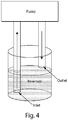
- the chamber may include a compartment or space through which the blood of a subject passes during extracorporeal circulation.
- a surface within an extracorporeal circuit can be at least partially coated with an antibody, thereby rendering it capable of scavenging cytokines from the blood that circulates through the system.
- the chamber may take a variety of forms in different options of the disclosure, but typically contains at least one surface and an antibody or antigen-binding fragment thereof, a ligand, or a receptor.
- the surface may be, for example, a surface defining a wall of the chamber, an internal surface contained within a chamber, or a surface of a particle.
- the chamber may be a well-defined rectangular chamber.
- the chamber may comprise a tube, e.g., at least a portion of the tube is coated with the polymer or particle and the antibody.
- the chamber may comprise a microfluidic device, e.g., at least a portion of the microfluidic channels is coated with the polymer or particle and the antibody.
- a fluid such as blood is withdrawn from a subject (e.g., a human), passed through a length of tubing, and returned to the subject.
- the tubing may comprise a surface as part of the tubing that defines a surface that is able to extract one or more substances such as proteins (or other species discussed herein) from the blood.
- at least a portion of the tubing may comprise a surface which is at least partially coated with an antibody as discussed herein.
- At least about 10%, at least about 20%, at least about 30%, at least about 40%, at least about 50%, at least about 60%, at least about 70%, at least about 80%, at least about 90%, or about 100% of the inner surface of the tubing may be coated.
- at least some of the antibody (or antibody or antigen-binding fragment thereof, or ligand or receptor, etc.) on the surface may be able to extract one or more substances such as proteins (or other species discussed herein) from the blood, e.g., through binding of the proteins.
- the chamber or tubing comprises or contains a carrier that carries the surface and antibody or antigen-binding fragment thereof, ligand, or receptor.
- a carrier may, for example, comprise solid particles, e.g., comprising polymer, ceramic, metal, and/or other materials.
- the carrier may have any suitable shape, e.g., as particles, fibers, ribbons, etc.
- the carrier may be relatively inert, e.g., the carrier is used to increase the surface area within the chamber.
- the carrier may be contained within the chamber, and/or be attached to a surface within the chamber.
- the surface area of a chamber is increased to increase the amount of surface and antibody exposure, such as by the incorporation of carriers, baffles, plates, grills, particles, or other suitable surfaces within the chamber.
- a biological fluid such as blood is removed from a subject and modified through interaction with a surface, and then at least a portion of the biological fluid is reintroduced to the subject.
- particles containing antibodies may be injected into a subject, allowed to bind to one or more proteins or other species within the subject, and then removed, thereby causing the removal of one or more substances such as proteins (for example IL-6 or VEGF), or other species, from the blood, e.g., via an extracorporeal circuit.
- one set of options is generally directed to magnetically-susceptible particles, such as nanoparticles, that comprise a surface and an antibody or antigen-binding fragment thereof that selectively binds to a protein suspected of being within the blood of a subject.
- the magnetically-susceptible particles are injected into a subject and allowed to circulate.
- the magnetically-susceptible particles are removed from the subject.
- blood from the subject may be removed via an extracorporeal circuit that is magnetized such that the particles (now bound to the protein or other species) are extracted from the blood circulating in the extracorporeal circuit.
- the particles are biodegradable and/or biocompatible.
- the particles are allowed to circulate in the biological fluid of a subject for up to approximately 12 hours or up to approximately 24 hours.
- Magnetically-susceptible particles such as ferromagnetic particles, may be selected in some cases so as to possess a greater density than other elements of the blood. Accordingly, in some options, these particles are removed from the blood by centrifugation, magnetization, or a combination of these and/or other techniques. In certain options, an aliquot of blood is removed from a subject, mixed with a predetermined amount of ferromagnetic particles under conditions that allow optimal binding to the desired product for removal. Upon formation of the particle:blood constituent complex, for instance, the sample can be centrifuged and/or magnetized to remove the target blood component.
- the extracorporeal circuits discussed above may be used solely to remove a substance such as a protein or other species from a biological fluid, or the extracorporeal circuit may be used in conjunction with other systems and/or methods.
- a polymer or particle and antibody are incorporated into a cardiopulmonary bypass circuit during heart surgery to prevent (or at least control) an inflammatory response.
- a surface comprising a polymer as disclosed herein may be the surface of a substrate as discussed herein.
- the substrate may be used in extracting specific substances such as proteins (or other species) from a fluid such as blood.
- the substrates may be formed from or include the polymer.
- the substrate may be formed from a material to which the polymer is coated thereon.
- the substrate may be present as particles such as microgel particles.
- Non-limiting examples of substrates and polymers compatible with the present disclosure are further described in PCT Application No. PCT/US2012/026008, filed on February 22, 2012 , and from Mizrahi et al. (2011) Advanced Materials 23:H258-H262 .
- the substrate may have any suitable shape.
- the substrate may be formed as particles, as a planar substrate, or the like.
- the substrate is polymeric.
- the substrate may include a polymer such as poly(acrylamide), e.g., formed through the polymerization of acrylamide and a suitable metal-chelating moiety, as discussed below.
- acrylamide may be polymerized to form poly(acrylamide) upon exposure to ammonium persulfate, methylenebisacrylamide, and/or N,N,N',N'-tetramethylethylendiamine ("TEMED").
- TEMED N,N,N',N'-tetramethylethylendiamine
- the polymerization may occur within an emulsion, e.g., to form particles.
- an emulsion may be formed where monomers are present within discrete droplets (e.g., in an aqueous environment) contained within a continuous phase (e.g., an organic or "oil” environment), and polymerization induced within the discrete droplets to form polymeric particles.
- a continuous phase e.g., an organic or "oil” environment
- suitable polymers include, but are not limited to, poly(styrene), poly(propylene), poly(ethylene), poly(dimethylsiloxane), agarose, and the like, e.g., in addition to and/or instead of poly(acrylamide).
- Still other examples include polyethylene, polystyrene, silicone, polyfluoroethylene, polyacrylic acid, a polyamide (e.g., nylon), polycarbonate, polysulfone, polyurethane, polybutadiene, polybutylene, polyethersulfone, polyetherimide, polyphenylene oxide, polymethylpentene, polyvinylchloride, polyvinylidene chloride, polyphthalamide, polyphenylene sulfide, polyester, polyetheretherketone, polyimide, polymethylmethacylate and/or polypropylene.
- Polymeric particles or other substrates formed using these polymers may be formed using techniques known to those of ordinary skill in the art.
- the substrate may have any suitable shape.
- the substrate may be present as particles, or as the surface of a chamber or a tube.
- the substrate may have a relatively high surface area.
- the substrate having the attached antibody or antigen-binding fragment thereof (or a ligand or a receptor, etc.) may have a surface area of attachment of the antibodies, etc.
- the substrate may have an average surface area of at least about 5 m 2 /g, at least about 7 m 2 /g, or at least about 10 m 2 /g. In some options, the substrate may have an average pore volume of at least about 0.005 cm 3 /g, at least about 0.01 cm 3 /g, or at least about 0.02 cm 3 /g.
- the substrate may take the form of one or more particles.
- the particles may include microparticles and/or nanoparticles.
- a "microparticle” is a particle having an average diameter on the order of micrometers (i.e., between about 1 micrometer and about 1 mm), while a “nanoparticle” is a particle having an average diameter on the order of nanometers (i.e., between about 1 nm and about 1 micrometer).
- the particles may have an average diameter of less than about 5 mm or 2 mm, or less than about 1 mm, or less than about 500 micrometers, less than about 200 micrometers, less than about 100 micrometers, less than about 80 micrometers, less than about 60 micrometers, less than about 50 micrometers, less than about 40 micrometers, less than about 30 micrometers, less than about 25 micrometers, less than about 10 micrometers, less than about 3 micrometers, less than about 1 micrometer, less than about 300 nm, less than about 100 nm, less than about 30 nm, or less than about 10 nm.
- the particle may have an average diameter of at least about 1 micrometer or at least about 10 micrometers.
- the particles may be spherical or non-spherical. If the particle is non-spherical, the particle may have a shape of, for instance, an ellipsoid, a cube, a fiber, a tube, a rod, or an irregular shape.
- the average diameter of a non-spherical particle is the diameter of a perfect sphere having the same volume as the non-spherical particle.
- the substrate may be a gel.
- gels include poly(acrylamide) gel or agarose gel, or other gel materials such as those describe herein.
- the substrate may take the form of microgel particles or gel microparticles.
- the gel particles may be collected together to form a gel material or a "microgel.”
- a gel typically is relatively solid or jelly-like, and may include a cross-linked polymer to form its structure.
- the gel may be a hydrogel, e.g., a gel that contains water.
- the substrate may be porous, in certain options of the disclosure.
- the substrate may have a relatively high surface area, for example, having an average surface area of at least about 5 m 2 /g, at least about 7 m 2 /g, or at least about 10 m 2 /g.
- the substrate may have an average pore volume of at least about 0.005 cm 3 /g, at least about 0.01 cm 3 /g, or at least about 0.02 cm 3 /g.
- the substrate may have an average pore width of at least about 5 nm, at least about 7 nm, or at least about 8 nm.
- porosities and dimensions may be determined using techniques known to those of ordinary skill in the art, for example, TEM, SEM, BET, or the like.
- the porosity may be created, for example, due to the nature of the polymer (e.g., certain gel polymers such as those described herein typically will form relatively porous structures), or the porosity may be induced by adding another material to the substrate that can be removed, thereby creating porosity within the substrate. For example, salts or other species that can be subsequently dissolved may be incorporated within the substrate.
- the substrate takes the form of one or more magnetically-susceptible particles, e.g., having dimensions, etc. as described herein.
- a magnetically-susceptible particle comprises at least one material that is magnetically-susceptible, e.g., such that the particle may be manipulated using a suitable magnetic field.
- the particle may comprise a ferromagnetic material or a superparamagnetic material, e.g., iron, cobalt, nickel, or the like.
- the magnetically-susceptible portion of the particle is a core of the particle, coated with polymer and/or an antibody such as is discussed herein.
- the polymer may be an interpenetrating or a semi-interpenetrating polymer, such as a semi-interpenetrating polymer network with an antibody affixed to polymer subunits throughout the network.
- An "interpenetrating polymer network” or an “IPN” typically comprises a polymeric material comprising two or more networks of two or more polymers (which can include copolymers) at least two different polymers of which are at least partially interlaced with respect to each other on a molecular scale, but not covalently bonded to each other. These polymer networks cannot be separated, even theoretically, unless one or more covalent bonds are broken.
- a mixture of two or more pre-formed polymers is not an interpenetrating polymer network.
- an interpenetrating network include [net-poly(styrene-stat-butadiene)]-ipn-[net-poly(ethyl acrylate), polyHEMA-ipn-polyurethane, or polyHEMA-ipn-polysiloxane, where "polyHEMA” is poly(2-hydroxyethylmethacrylate)].
- IPNs using suitable techniques, for example, by blending different polymer precursors which have the ability under set conditions to react to form two or more different interpenetrating polymers that do not covalently bind to each other, by forming a first polymer and allowing a precursor of a second polymer to diffuse into the first polymer in an interpenetrating manner and to react to form the second polymer under conditions that do not promote binding between the first and second polymer, by mixing two or more different families of monomers, each capable of polymerizing via different mechanisms (for example, a radical reaction and a condensation reaction) but are not capable of reacting with each other and sequentially or simultaneously polymerizing the monomers using heat and/or UV light, by blending two or more linear or branched polymers with at least one polymer having pendant reactant groups and subsequently adding a chain extender to cross-link each of the polymers into separate networks, and/or by proceeding with a multi-stage polymerization process including a first polymer network
- a “semi-interpenetrating polymer network” or a “SIPN,” typically comprises a polymeric material comprising a combination of at least one polymer network (which may include a copolymer or copolymers) and one or more linear or branched polymer(s), characterized by the penetration, on a molecular scale, of at least one of the networks by at least some of the linear or branched macromolecules.
- Semi-interpenetrating polymer networks can also be defined as similar to interpenetrating polymer networks, but distinguished in that a constituent linear or branched polymer(s) can be (at least in theory) separated from the polymer network(s) without breaking any covalent bonds.
- a specific non-limiting example of a semi-interpenetrating network is (net-polystyrene)-sipn-poly(vinyl chloride).
- interpenetrating and/or semi-interpenetrating polymer networks can also be formed from combinations of polyethylenes (e.g., tetrafluoroethylenes) and silicone polymers, or acrylates (e.g., methylmethacrylates) and urethanes and/or ureas.
- polyethylenes e.g., tetrafluoroethylenes
- silicone polymers e.g., silicone polys
- acrylates e.g., methylmethacrylates
- SIPNs are able to form SIPNs by known techniques, for example, using techniques such as those described above with reference to interpenetrating networks, or by allowing a solution of a linear or branch polymer to diffuse into a polymer network, by blending two or more linear or branched polymer networks with at least one polymer having pendant reactant groups and subsequently adding a chain extender to cross-link one of the polymer networks, by proceeding with a multi-stage polymerization process including a first polymer network that is partially polymerized to allow for high swellability and/or easy diffusion of a second polymer precursor therein, and thereafter polymerizing the first polymer network, etc.
- a material of the disclosure can include an interpenetrating network which includes a semi-interpenetrating component or components; e.g., a material of the disclosure can include two polymer networks that are interpenetrating, and a third that is semi-interpenetrating with respect to one or both of the two interpenetrating networks (and can include any number of additional interpenetrating or semi-interpenetrating components).
- An interpenetrating network can include a semi-interpenetrating component or components.
- characterization techniques for the identification and/or determination of a polymer from an interpenetrating and/or semi-interpenetrating polymer network include techniques that allow for the observation of microdomains, as opposed to polymers or polymer blends that separate into macrodomains.
- Example of such techniques include, but are not limited to, differential scanning calorimetry (DSC), which allows the identification and/or determination of multiple glass transition temperatures (i.e., in an interpenetrating and/or semi-interpenetrating polymer network, each of the polymers comprising the network may have different glass transition temperatures); transmission electron microscopy (TEM), which allows for the microscopic identification of the microdomains of the interpenetrating and/or semi-interpenetrating polymer networks; CP-MAS solid NMR of 13 C or 29 Si, which allows visualization of cross-link points within the interpenetrating and/or semi-interpenetrating networks in polymers having cross-link points; small angle neutron scattering (SANS) techniques, which allows the visualization of domains in a sample; differential mechanical analysis (DMA), which allow the modulus of each polymer within the interpenetrating and/or semi-interpenetrating polymer network to be determined; or the like.
- DSC differential scanning calorimetry
- DMA differential mechanical analysis
- antibodies and antigen-binding fragments thereof associated with certain options of the disclosure can be complexed or otherwise associated with a polymeric system according to any method known in the art.
- the polymeric system is activated through surface oxidation, such as by plasma oxidation or acid oxidation.
- the activated polymeric system may be aminated in some cases by an aminosilane, such as aminopropyltrimethoxysilane (APTMS) or aminopropyltriethoxysilane (APTES).
- the antibody is attached to polymerized dopamine by a histidine linker.
- the histidine linker is 3, 4, 5, 6, 7, 8, 9, 10, 11, 12, 13, 14, 15, or more than 15 histidines.
- the histidine linker is formed from 6-8 histidines.
- the antibody is attached to the polymeric system by an APTMS-PEG-maleimide linker.
- suitable types of crosslinkers include, but are not limited to, haloacetyls and pyridyldithiols.
- the antibody is chemically modified, such as by coupling it with N-succinimidylacrylate (NSA) or with a furan functionality (e.g., for a Diels-Alder reaction).
- NSA N-succinimidylacrylate
- furan functionality e.g., for a Diels-Alder reaction
- copolymerization is achieved by combining the modified antibody with acrylamide, aqueous ammonium persulphate and N,N,N',N'-tetramethylethylenediamine (TEMED).
- TEMED N,N,N',N'-tetramethylethylenediamine
- a surface such as a surface of a chamber component of an extracorporeal circuit
- a polymer and/or hydrogel as disclosed herein according to any method known in the art.
- methods of functionalizing a surface with polymeric microgel films are derived from methods based on plasma-induced graft polymerization of poly acrylic acid, such as is described in Singh et al. (2007) Biomacromolecules 8(10):3271-5 .
- a photoaffinity label viz., aminobenzophenone is introduced onto the surface.
- the substrate may be positively or negatively charged, e.g., to facilitate separation of proteins, or other analytes.
- an analyte may be positively charged and a negatively charged substrate may facilitate attraction of the analyte.
- a protein may be positively charged due to residues such as glutamine or asparagine on the protein, which may be attracted to negatively charged particles or other substrates.
- an analyte may be negatively charged, and a positively charged particle may facilitate attraction of the analyte.
- a nucleic acid such as DNA or RNA may be negatively charged, and the nucleic acid may be attracted to positively charged particles or other substrates.
- an acrylic acid or other monomer producing negatively charged residues may be incorporated into the polymer or otherwise added to the substrate to impart a negative charge on the substrate.
- a monomer producing positively charged residues e.g., ethylenimine
- the substrate may also comprise a metal-chelating moiety, for example, EDTA (ethylenediaminetetraacetic acid) or NTA (nitrilotriacetic acid), or derivatives thereof, to which metal ions, including divalent metal ions, are able to bind.
- EDTA ethylenediaminetetraacetic acid
- NTA nitrilotriacetic acid
- metal-chelating moieties include various polyamino carboxylic acid such as Fura-2, iminodiacetic acid, diethylene triamine pentaacetic acid (DTPA), ethylene glycol tetraacetic acid (EGTA), 1,2-bis(o-aminophenoxy)ethane-N,N,N',N'-tetraacetic acid (BAPTA), 1,4,7,10-tetraazacyclododecane-1,4,7,10-tetraacetic acid (DOTA), or the like, and derivatives thereof.
- the metal-chelating moiety may be one which is able to bind or complex with metal ions, such as divalent metal ions.
- Non-limiting examples of ions that can be chelated by metal-chelating moieties include nickel, cobalt, calcium, iron, or the like.
- a metal-chelating moiety may bind to nickel ions, so that a substrate containing the metal-chelating moiety may also contain nickel ions distributed within the substrate.
- the metal-chelating moiety may be incorporated into the polymeric structure of a substrate.
- the metal-chelating moiety may be present as a monomer as various monomers are polymerized and/or cross-linked to form a polymeric substrate, e.g., forming a copolymer or an interpenetrating or semi-interpenetrating polymer network.
- the metal-chelating moiety may be incorporated in a polymer as a monomer such that when the polymer is formed, one of the monomers or residues within the polymer is the metal-chelating moiety.
- an NTA derivative such as 2,20-(5-acrylamido-1-carboxypentylazanediyl)diacetic acid may be used, which forms NTA residues when incorporated within a polymer.
- the metal-chelating moiety may be distributed substantially evenly throughout the substrate.
- the concentration of the metal-chelating moiety on the surface of the substrate and in the bulk or the center of the substrate may be substantially the same.
- the difference in concentration of the metal-chelating moiety between the surface of the substrate and the bulk or center of the substrate may be no more than about 40%, no more than about 35%, no more than about 30%, no more than about 25%, no more than about 20%, no more than about 15%, no more than about 10%, or no more than about 5%, where the percentage is taken relative to the average of these concentrations on the surface and in the bulk or center of the substrate.
- the distribution of the metal-chelating moiety within the substrate may be relatively uniform, for example, if the metal-chelating moiety is formed as an integral part of the substrate as the substrate is formed.
- the metal-chelating moiety may be incorporated within a polymer as a monomer within the polymer, thus resulting in a relatively uniform distribution of the metal-chelating moiety within the polymeric substrate.
- metal ions may be allowed to become distributed within the substrate, e.g., by exposing or the substrate to a fluid containing the metal ions, for example, such that the ions are able to penetrate the substrate via diffusion or other forces (e.g., charge attraction).
- the substrate may be immersed in the fluid.
- the metal ions may become distributed within the substrate uniformly or non-uniformly, e.g., depending on the length of exposure. For instance, if the metal-chelating moiety is distributed relatively uniformly within the substrate, then metal ions attracted to the metal-chelating moiety may likewise become relatively uniformly within the substrate.
- the disclosure may relate to antibodies that bind selectively to proteins suspected of being within the blood of a subject.
- An antibody is a protein that typically includes at least two heavy (H) chains and two light (L) chains inter- by disulfide bonds.
- Each heavy chain is comprised of a heavy chain variable region (abbreviated herein as HCVR or V H ) and a heavy chain constant region.
- the heavy chain constant region is comprised of three domains, C H 1, C H 2 and C H 3.
- Each light chain is comprised of a light chain variable region (abbreviated herein as LCVR or V L ) and a light chain constant region.
- the light chain constant region is comprised of one domain, CL.
- V H and V L regions can be further subdivided into regions of hypervariability, termed complementarity determining regions (CDR), interspersed with regions that are more conserved, termed framework regions (FR).
- CDR complementarity determining regions
- FR framework regions
- Each V H and V L is composed of three CDRs and four FRs, arranged from amino-terminus to carboxy-terminus in the following order: FR1, CDR1, FR2, CDR2, FR3, CDR3, FR4.
- the variable regions of the heavy and light chains contain a binding domain that interacts with an antigen.
- the constant regions of the antibodies may mediate the binding of the immunoglobulin to host tissues or factors, including various cells of the immune system (e.g., effector cells) and the first component (C1q) of the classical complement system.
- antigen-binding fragment of an antibody as used herein, refers to one or more portions of an antibody that retain the ability to specifically bind to an antigen (e.g., a cytokine). It has been shown that the antigen-binding function of an antibody can be performed by fragments of a full-length antibody.
- binding fragments encompassed within the term "antigen-binding fragment" of an antibody include (i) a Fab fragment, a monovalent fragment consisting of the V L , V H , C L and C H 1 domains; (ii) a F(ab') 2 fragment, a bivalent fragment comprising two Fab fragments linked by a disulfide bridge at the hinge region; (iii) a Fd fragment consisting of the V H and CH1 domains; (iv) a Fv fragment consisting of the V L and V H domains of a single arm of an antibody, (v) a dAb fragment which consists of a V H domain or the variable domain of a heavy-chain antibody, such as a camelid heavy-chain antibody (e.g.
- V HH an isolated complementarity determining region (CDR); and (vii) polypeptide constructs comprising the antigen-binding fragments of (i) - (vi).
- V L and V H are coded for by separate genes, they can be joined, using recombinant methods, by a synthetic linker that enables them to be made as a single protein chain in which the V L and V H regions pair to form monovalent molecules (known as single chain Fv (scFv).
- single chain Fv single chain Fv
- Such single chain antibodies are also intended to be encompassed within the term "antigen-binding portion" of an antibody.
- These antibody fragments are obtained using conventional procedures, such as proteolytic fragmentation procedures, expression of recombinant nucleic acids, or the like. The fragments are screened for utility in the same manner as are intact antibodies.
- Isolated antibodies of the disclosure encompass various antibody isotypes, such as IgG1, IgG2, IgG3, IgG4, IgM, IgA1, IgA2, IgAsec, IgD, IgE.
- isotype refers to the antibody class (e.g., IgM or IgG1) that is encoded by heavy chain constant region genes.
- Antibodies as disclosed herein can be full length or can include only an antigen-binding fragment such as the antibody constant and/or variable domain of IgG1, IgG2, IgG3, IgG4, IgM, IgA1, IgA2, IgAsec, IgD or IgE or could consist of a Fab fragment, a F(ab') 2 fragment, and a Fv fragment.
- an antigen-binding fragment such as the antibody constant and/or variable domain of IgG1, IgG2, IgG3, IgG4, IgM, IgA1, IgA2, IgAsec, IgD or IgE or could consist of a Fab fragment, a F(ab') 2 fragment, and a Fv fragment.
- Antibodies of the present disclosure can be polyclonal, monoclonal, or a mixture of polyclonal and monoclonal antibodies. Antibodies of the disclosure can be produced by methods disclosed herein or by a variety of techniques known in the art, and many such antibodies can readily be obtained commercially.
- a monoclonal antibody typically refers to a preparation of antibody molecules of single molecular composition.
- a monoclonal antibody may display a single binding specificity and affinity for a particular epitope.
- a monoclonal antibody may display a single binding specificity and affinity for a particular epitope.
- the polyclonal antibody typically refers to a preparation of antibody molecules that comprises a mixture of antibodies active that specifically bind a specific antigen.
- a process of monoclonal antibody production may include obtaining immune somatic cells with the potential for producing antibody, in particular B lymphocytes, which have been previously immunized with the antigen of interest either in vivo or in vitro and that are suitable for fusion with a B-cell myeloma line.
- B lymphocytes typically are immunized by in vivo immunization of the animal (e.g., a mouse) with the desired protein or polypeptide, e.g., a cytokine. Such immunizations are repeated as necessary at intervals of up to several weeks to obtain a sufficient titer of antibodies.
- mice can be used as a source of antibody-producing lymphocytes which can be cloned and recombinantly expressed, as discussed further below. Following the last antigen boost, the animals are sacrificed and spleen cells removed. Mouse lymphocytes give a higher percentage of stable fusions with the mouse myeloma lines described herein. Of these, the BALB/c mouse is preferred. However, other mouse strains, rat, rabbit, hamster, sheep, goats, camels, llamas, frogs, etc. may also be used as hosts for preparing antibody-producing cells. Mouse strains that have human immunoglobulin genes inserted in the genome (and which cannot produce mouse immunoglobulins) can also be used. Examples include the HuMAb mouse strains produced by Medarex/GenPharm International, and the XenoMouse strains produced by Abgenix. Such mice produce fully human immunoglobulin molecules in response to immunization.
- Somatic cells may be obtained from the lymph nodes, spleens and peripheral blood of antigen-primed animals, and the lymphatic cells of choice depend to a large extent on their empirical usefulness in the particular fusion system.
- the antibody-secreting lymphocytes are then fused with (mouse) B cell myeloma cells or transformed cells, which are capable of replicating indefinitely in cell culture, thereby producing an immortal, immunoglobulin-secreting cell line.
- the resulting fused cells, or hybridomas are cultured, and the resulting colonies screened for the production of the desired monoclonal antibodies. Colonies producing such antibodies are cloned, and grown either in vivo or in vitro to produce large quantities of antibody.
- Myeloma cell lines suited for use in hybridoma-producing fusion procedures preferably are non-antibody-producing, have high fusion efficiency, and enzyme deficiencies that render them incapable of growing in certain selective media which support the growth of the desired hybridomas.
- myeloma cell lines that may be used for the production of fused cell lines include, but are not limited to Ag8, P3-X63/Ag8, X63-Ag8.653, NS1/1.Ag 4.1, Sp2/0-Ag14, FO, NSO/U, MPC-11, MPC11-X45-GTG 1.7, S194/5XX0 Bul, all derived from mice; R210.RCY3, Y3-Ag 1.2.3, IR983F and 4B210 derived from rats and U-266, GM1500-GRG2, LICR-LON-HMy2, UC729-6, all derived from humans.
- Those of ordinary skill in the art will be aware of numerous routine methods to produce monoclonal antibodies.
- Fusion with mammalian myeloma cells or other fusion partners capable of replicating indefinitely in cell culture is effected by standard and well-known techniques, for example, by using polyethylene glycol (“PEG”) or other fusing agents.
- PEG polyethylene glycol
- polyclonal antibodies may be raised by administering a polypeptide subcutaneously to New Zealand white rabbits which have first been bled to obtain pre-immune serum.
- the polypeptide can be inoculated with (e.g., injected at) a total volume of 100 microliters per site at six different sites, typically with one or more adjuvants.
- the rabbits are then bled two weeks after the first injection and periodically boosted with the same antigen three times every six weeks.
- a sample of serum is collected 10 days after each boost.
- Polyclonal antibodies are recovered from the serum, preferably by affinity chromatography using acetylated cytochrome to capture the antibody.
- antibodies may be recombinant antibodies.
- Recombinant antibodies generally include antibodies that are prepared, expressed, created or isolated by recombinant means, such as antibodies isolated from an animal (e.g., a mouse) that is transgenic for another species' immunoglobulin genes, genetically engineered antibodies, antibodies expressed using a recombinant expression vector transfected into a host cell, antibodies isolated from a recombinant, combinatorial antibody library, or antibodies prepared, expressed, created or isolated by any other means that involves splicing of immunoglobulin gene sequences to other DNA sequences.
- recombinant means such as antibodies isolated from an animal (e.g., a mouse) that is transgenic for another species' immunoglobulin genes, genetically engineered antibodies, antibodies expressed using a recombinant expression vector transfected into a host cell, antibodies isolated from a recombinant, combinatorial antibody library, or antibodies prepared, expressed, created or isolated by any other means that involves splicing of immunoglobulin gene sequence
- Antibodies or antigen-binding fragments of the disclosure are, preferably, isolated.
- the isolated antibody is not present in an organism that endogenously produces the antibody, i.e., the antibody has been isolated from the organism.
- isolated antibodies and antigen-binding fragments thereof refer to an antibody (or antigen-binding fragment thereof) that is substantially free of other antibodies (or antigen-binding fragments) having different antigenic specificities.
- an isolated antibody that specifically binds to an epitope, isoform or variant of a polypeptide may, however, have cross-reactivity to other related antigens, e.g., a mutant form of the cytokine, or a polypeptide from other species (e.g., homologs in other species).
- an isolated antibody (or antigen-binding fragment thereof) may be substantially free of other cellular material and/or chemicals.
- Antibodies of the disclosure include, but are not limited to antibodies that specifically bind to a cytokine.
- selective binding refers to antibody binding to a predetermined antigen with a preference that enables the antibody to be used to distinguish the antigen from others.
- the antibody binds to a single protein and is able to distinguish that protein from all other proteins.
- the antibody binds to several different related proteins and is able to distinguish those proteins from all other proteins.
- multiple antibodies such as multiple different monoclonal antibodies, are incorporated into the polymer system at any given time, resulting in an extracorporeal circuit that is capable of removing more than one cytokine at a time.
- the highly specific nature of the system is preserved and yet expanded to remove groups of proteins in a fashion never before described.
- an antibody or antigen-binding fragment thereof, of the invention can specifically bind to an antigen with sub-nanomolar affinity.
- the dissociation constants can be about 1 x 10 -6 , 1 x 10 -7 , 1 x 10 -8 , 1 x 10 -9 M or less, preferably about 1 x 10 -10 M or less, more preferably 1 x 10 -11 M or less.
- the binding affinity is less than about 5 x 10 -10 M.
- an antibody or antigen-binding fragment thereof may bind to a conformational epitope of a polypeptide such as a cytokine.
- a conformational epitope of a polypeptide such as a cytokine.
- each antibody can be tested in assays using native protein (e.g., non-denaturing immunoprecipitation, flow cytometric analysis of cell surface binding) and denatured protein (e.g., Western blot, immunoprecipitation of denatured proteins). A comparison of the results will indicate whether the antibodies bind conformational epitopes.
- Antibodies that bind to native protein but not denatured protein are those antibodies that bind conformational epitopes, and are preferred antibodies.
- a ligand rather than an antibody, is used for selective binding to a substance such as a protein in a biological fluid such as blood.
- An antibody or antigen-binding fragment thereof of the disclosure can be linked to a detectable label.
- a detectable label of the disclosure may be attached to antibodies or antigen-binding fragments thereof of the invention by standard protocols known in the art.
- the detectable labels may be covalently attached to an antibody or antigen-binding fragment thereof of the disclosure.
- the covalent binding can be achieved either by direct condensation of existing side chains or by the incorporation of external bridging moieties.
- Many bivalent or polyvalent agents are useful in coupling protein molecules to other proteins, polypeptides or amine functions, etc.
- the literature is replete with coupling agents such as carbodiimides, diisocyanates, glutaraldehyde, and diazobenzenes. This list is not intended to be exhaustive of the various coupling agents known in the art but, rather, is exemplary of the more common coupling agents.
- a subject in need thereof can be a subject who has an inflammatory disease or an autoimmune disease.
- a subject in need thereof can be a subject who has sepsis or septic shock, acute respiratory distress syndrome (ARDS), systemic inflammatory response syndrome (SIRS) related to cardiopulmonary bypass, myasthenia gravis, etc.
- ARDS acute respiratory distress syndrome
- SIRS systemic inflammatory response syndrome
- a subject in need thereof can be a subject who has a neurodegenerative disease, such as Alzheimer's disease.
- the terms "treatment” or "to treat” refer to both therapeutic and prophylactic treatments.
- treating the condition refers to ameliorating, reducing or eliminating one or more symptoms associated with the disorder or the severity of the disease or preventing any further progression of the disease. If the subject in need of treatment is one who is at risk of having a condition, then treating the subject refers to reducing the risk of the subject having the condition or preventing the subject from developing the condition.
- a subject as used herein means a human or other vertebrate animal or mammal including, but not limited to, a dog, cat, horse, cow, pig, sheep, goat, rodent, bird, and primate, e.g., monkey.
- an effective amount of a polymer or particle and an agent such as an antibody or antigen-binding fragment thereof associated with the disclosure may be that amount sufficient to ameliorate one or more symptoms of a disease such as an inflammatory disease or autoimmune disease.
- an effective prophylactic or therapeutic treatment regimen can be planned which is effective to treat the particular subject.
- the effective amount for any particular application can vary depending on such factors as the disease or condition being treated, the size of the subject and the severity of the disease or condition.
- One of ordinary skill in the art can empirically determine the effective amount of materials associated with the disclosure without necessitating undue experimentation.
- the present disclosure also relates to risk-stratifying subjects based on their genetic profile.
- technologies such as gene microarrays, based upon a gene expression profile, children and adults with septic shock can now be risk stratified by predicted mortality with great accuracy. Examination of the up-regulated genes that drive mortality in sepsis reveals that many of them are inflammatory proteins. Therefore, technology described herein can be used to tailor the bioactive circuit to individual patients to remove inflammatory mediators based on the genetic fingerprint from microarray technology.
- the present disclosure also relates to the use of methods and compositions described herein for detecting or analyzing the presence of a substance in a biological fluid, such as for diagnostic and/or prognostic purposes.
- biological fluid such as blood
- biological fluid is removed from a subject and is contacted with a polymer or particle attached to an agent such as an antibody, and is analyzed during or after contact with the polymer or particle.
- biological fluid, such as blood is analyzed during or after circulation through an extracorporeal circuit.
- blood from a subject is circulated through an extracorporeal circuit that contains a chamber, wherein at least one surface of the chamber is coated with a specific agent such as an antibody, allowing for the presence and/or quantity of a specific substance such as a protein in the biological fluid to be analyzed.
- a specific agent such as an antibody
- Magnetically-susceptible particles associated with the disclosure when it is desired to administer them systemically, may be formulated for parenteral administration by injection, e.g., by bolus injection or continuous infusion.
- Formulations for injection may be presented in unit dosage form, e.g., in ampoules or in multi-dose containers, with an added preservative.
- the compositions may take such forms as suspensions, solutions or emulsions in oily or aqueous vehicles, and may contain formulatory agents such as suspending, stabilizing and/or dispersing agents.
- compositions for parenteral administration include aqueous solutions of the active compounds in water soluble form. Additionally, suspensions of the active compounds may be prepared as appropriate oily injection suspensions. Suitable lipophilic solvents or vehicles include fatty oils such as sesame oil, or synthetic fatty acid esters, such as ethyl oleate or triglycerides, or liposomes. Aqueous injection suspensions may contain substances which increase the viscosity of the suspension, such as sodium carboxymethyl cellulose, sorbitol, or dextran. Optionally, the suspension may also contain suitable stabilizers or agents which increase the solubility of the compounds to allow for the preparation of highly concentrated solutions.
- the active compounds may be in powder form for constitution with a suitable vehicle, e.g., sterile pyrogen-free water, before use.
- a suitable vehicle e.g., sterile pyrogen-free water
- Non-limiting examples of inflammatory disorders that may be treated as discussed herein include: sepsis or septic shock, acute or adult respiratory distress syndrome (ARDS), acute lung injury (ALI), Acne vulgaris, Asthma, Autoimmune diseases, Celiac disease, Chronic prostatitis, Glomerulonephritis, Hypersensitivities, Inflammatory bowel diseases, Pelvic inflammatory disease, Reperfusion injury, Rheumatoid arthritis, Sarcoidosis, Transplant rejection, Vasculitis, Interstitial cystitis, Atherosclerosis, Allergies, Inflammatory myopathies such as dermatomyositis, polymyositis, and inclusion body myositis, Chediak-Higashi syndrome and chronic granulomatous disease.
- ARDS acute or adult respiratory distress syndrome
- ALI acute lung injury
- Acne vulgaris Asthma
- Autoimmune diseases Celiac disease
- Chronic prostatitis Chronic prostatitis
- Non-limiting examples of autoimmune diseases include: systemic lupus erythematosus (SLE), rheumatoid arthritis (RA), scleroderma, Sjogren's syndrome, multiple sclerosis, insulin dependent diabetes mellitus, ulcerative colitis, Acute disseminated encephalomyelitis (ADEM), Addison's Disease, Agammaglobulinemia, Alopecia areata, Amyotrophic Lateral Sclerosis, Ankylosing Spondylitis, Antiphospholipid syndrome, Antisynthetase syndrome, Atopic allergy, Atopic dermatitis, Autoimmune aplastic anemia, Autoimmune cardiomyopathy, Autoimmune enteropathy, Autoimmune hemolytic anemia, Autoimmune hepatitis, Autoimmune inner ear disease, Autoimmune lymphoproliferative syndrome, Autoimmune peripheral neuropathy, Autoimmune pancreatitis, Autoimmune polyendoc
- SLE systemic
- microfluidic channels e.g., as a surface that is at least partially coated with a suitable agent such as an antibody.
- Microfluidic channels generally have widths or diameters of less than about 1 mm, and less than about 100 micrometers in some cases. In some options, larger channels may be used instead of, or in conjunction with, microfluidic channels for any of the options discussed herein. For examples, channels having widths or diameters of less than about 10 mm, less than about 9 mm, less than about 8 mm, less than about 7 mm, less than about 6 mm, less than about 5 mm, less than about 4 mm, less than about 3 mm, or less than about 2 mm may be used in certain instances.
- specified widths can be a smallest width (i.e. a width as specified where, at that location, the article can have a larger width in a different dimension), or a largest width (i.e. where, at that location, the article has a width that is no wider than as specified, but can have a length that is greater).
- the microfluidic channel may have an average cross-sectional dimension (e.g., perpendicular to the direction of flow of fluid in the microfluidic channel) of less than about 1 mm, less than about 500 microns, less than about 300 microns, or less than about 100 microns.
- the microfluidic channel may have an average diameter of less than about 60 microns, less than about 50 microns, less than about 40 microns, less than about 30 microns, less than about 25 microns, less than about 10 microns, less than about 5 microns, less than about 3 microns, or less than about 1 micron.
- a channel may have any aspect ratio, e.g., an aspect ratio (length to average cross-sectional dimension) of at least about 1:1, at least about 2:1, more typically at least about 3:1, at least about 5:1, at least about 10:1, etc.
- a "cross-sectional dimension" in reference to a fluidic or microfluidic channel is measured in a direction generally perpendicular to fluid flow within the channel.
- a channel generally will include characteristics that facilitate control over fluid transport, e.g., structural characteristics and/or physical or chemical characteristics (hydrophobicity vs. hydrophilicity) and/or other characteristics that can exert a force (e.g., a containing force) on a fluid.
- the fluid within the channel may partially or completely fill the channel.
- the fluid may be held or confined within the channel or a portion of the channel in some fashion, for example, using surface tension (e.g., such that the fluid is held within the channel within a meniscus, such as a concave or convex meniscus).
- surface tension e.g., such that the fluid is held within the channel within a meniscus, such as a concave or convex meniscus.
- some (or all) of the channels may be of a particular size or less, for example, having a largest dimension perpendicular to fluid flow of less than about 5 mm, less than about 2 mm, less than about 1 mm, less than about 500 microns, less than about 200 microns, less than about 100 microns, less than about 60 microns, less than about 50 microns, less than about 40 microns, less than about 30 microns, less than about 25 microns, less than about 10 microns, less than about 3 microns, less than about 1 micron, less than about 300 nm, less than about 100 nm, less than about 30 nm, or less than about 10 nm or less in some cases.
- the channel is a capillary.
- the device may contain one or more chambers or reservoirs for holding fluid.
- the chambers may be in fluidic communication with one or more fluid transporters and/or one or more microfluidic channels.
- microfluidic channels e.g., microfluidic channels.
- various components of the disclosure can be formed from solid materials, in which the channels can be formed via micromachining, film deposition processes such as spin coating and chemical vapor deposition, laser fabrication, photolithographic techniques, etching methods including wet chemical or plasma processes, and the like. See, for example, Scientific American, 248:44-55, 1983 (Angell , et al ).
- various components of the systems and devices disclosed herein can be formed of a polymer, for example, an elastomeric polymer such as polydimethylsiloxane (“PDMS”), polytetrafluoroethylene (“PTFE” or Teflon®), or the like.
- a microfluidic channel may be implemented by fabricating the fluidic system separately using PDMS or other soft lithography techniques (details of soft lithography techniques suitable for this option are discussed in the references entitled “ Soft Lithography,” by Younan Xia and George M. Whitesides, published in the Annual Review of Material Science, 1998, Vol. 28, pages 153-184 , and " Soft Lithography in Biology and Biochemistry," by George M. Whitesides, Emanuele Ostuni, Shuichi Takayama, Xingyu Jiang and Donald E. Ingber, published in the Annual Review of Biomedical Engineering, 2001, Vol. 3, pages 335-373 ).
- polymers include, but are not limited to, polyethylene terephthalate (PET), polyacrylate, polymethacrylate, polycarbonate, polystyrene, polyethylene, polypropylene, polyvinylchloride, polytetrafluoroethylene, a fluorinated polymer, a silicone such as polydimethylsiloxane, polyvinylidene chloride, bis-benzocyclobutene (“BCB”), a polyimide, a fluorinated derivative of a polyimide, or the like. Combinations, copolymers, or blends involving polymers including those described above are also envisioned.
- the device may also be formed from composite materials, for example, a composite of a polymer and a semiconductor material.
- various components of the disclosure are fabricated from polymeric and/or flexible and/or elastomeric materials, and can be conveniently formed of a hardenable fluid, facilitating fabrication via molding (e.g. replica molding, injection molding, cast molding, etc.).
- the hardenable fluid can be essentially any fluid that can be induced to solidify, or that spontaneously solidifies, into a solid capable of containing and/or transporting fluids contemplated for use in and with the fluidic network.
- the hardenable fluid comprises a polymeric liquid or a liquid polymeric precursor (i.e. a "prepolymer").
- Suitable polymeric liquids can include, for example, thermoplastic polymers, thermoset polymers, waxes, metals, or mixtures or composites thereof heated above their melting point.
- a suitable polymeric liquid may include a solution of one or more polymers in a suitable solvent, which solution forms a solid polymeric material upon removal of the solvent, for example, by evaporation.
- Such polymeric materials which can be solidified from, for example, a melt state or by solvent evaporation, are well known to those of ordinary skill in the art.
- a variety of polymeric materials, many of which are elastomeric, are suitable, and are also suitable for forming molds or mold masters, for options where one or both of the mold masters is composed of an elastomeric material.
- a non-limiting list of examples of such polymers includes polymers of the general classes of silicone polymers, epoxy polymers, and acrylate polymers.
- Epoxy polymers are characterized by the presence of a three-membered cyclic ether group commonly referred to as an epoxy group, 1,2-epoxide, or oxirane.
- diglycidyl ethers of bisphenol A can be used, in addition to compounds based on aromatic amine, triazine, and cycloaliphatic backbones.
- Another example includes the well-known Novolac polymers.
- Non-limiting examples of silicone elastomers suitable for use according to the disclosure include those formed from precursors including the chlorosilanes such as methylchlorosilanes, ethylchlorosilanes, phenylchlorosilanes, etc.
- Silicone polymers are used in certain options, for example, the silicone elastomer polydimethylsiloxane.
- Non-limiting examples of PDMS polymers include those sold under the trademark Sylgard by Dow Chemical Co., Midland, MI, and particularly Sylgard 182, Sylgard 184, and Sylgard 186.
- Silicone polymers including PDMS have several beneficial properties simplifying fabrication of the microfluidic structures disclosed herein. For instance, such materials are inexpensive, readily available, and can be solidified from a prepolymeric liquid via curing with heat.
- PDMSs are typically curable by exposure of the prepolymeric liquid to temperatures of about, for example, about 65 °C to about 75 °C for exposure times of, for example, about an hour.
- silicone polymers such as PDMS
- PDMS polymethyl methacrylate copolymer
- flexible (e.g., elastomeric) molds or masters can be advantageous in this regard.
- One advantage of forming structures such as microfluidic structures of the disclosure from silicone polymers, such as PDMS, is the ability of such polymers to be oxidized, for example by exposure to an oxygen-containing plasma such as an air plasma, so that the oxidized structures contain, at their surface, chemical groups capable of cross-linking to other oxidized silicone polymer surfaces or to the oxidized surfaces of a variety of other polymeric and non-polymeric materials.
- an oxygen-containing plasma such as an air plasma
- oxidized silicone such as oxidized PDMS can also be sealed irreversibly to a range of oxidized materials other than itself including, for example, glass, silicon, silicon oxide, quartz, silicon nitride, polyethylene, polystyrene, glassy carbon, and epoxy polymers, which have been oxidized in a similar fashion to the PDMS surface (for example, via exposure to an oxygen-containing plasma).
- Oxidation and sealing methods useful in the context of the presentdisclosure, as well as overall molding techniques, are described in the art, for example, in an article entitled " Rapid Prototyping of Microfluidic Systems and Polydimethylsiloxane," Anal. Chem., 70:474-480, 1998 (Duffy et al .).
- kits typically defines a package or an assembly including one or more of the compositions of the disclosure, and/or other compositions associated with the disclosure, for example, as previously described.
- Each of the compositions of the kit if present, may be provided in liquid form (e.g., in solution), or in solid form (e.g., a dried powder).
- some of the compositions may be constitutable or otherwise processable (e.g., to an active form), for example, by the addition of a suitable solvent or other species, which may or may not be provided with the kit.
- compositions examples include, but are not limited to, solvents, surfactants, diluents, salts, buffers, emulsifiers, chelating agents, fillers, antioxidants, binding agents, bulking agents, preservatives, drying agents, antimicrobials, needles, syringes, packaging materials, tubes, bottles, flasks, beakers, dishes, frits, filters, rings, clamps, wraps, patches, containers, tapes, adhesives, and the like, for example, for using, administering, modifying, assembling, storing, packaging, preparing, mixing, diluting, and/or preserving the compositions components for a particular use, for example, to a sample and/or a subject.
- a kit as disclosed herein may, in some cases, include instructions in any form that are provided in connection with the compositions of the disclosure in such a manner that one of ordinary skill in the art would recognize that the instructions are to be associated with the compositions of the disclosure.
- the instructions may include instructions for the use, modification, mixing, diluting, preserving, administering, assembly, storage, packaging, and/or preparation of the compositions and/or other compositions associated with the kit.
- the instructions may also include instructions for the use of the compositions, for example, for a particular use, e.g., to a sample.
- the instructions may be provided in any form recognizable by one of ordinary skill in the art as a suitable vehicle for containing such instructions, for example, written or published, verbal, audible (e.g., telephonic), digital, optical, visual (e.g., videotape, DVD, etc.) or electronic communications (including Internet or web-based communications), provided in any manner.
- verbal e.g., telephonic
- digital e.g., optical, visual
- visual e.g., videotape, DVD, etc.
- electronic communications including Internet or web-based communications
- the present disclosure is directed to methods of promoting one or more options of the disclosure as discussed herein.
- "promoted” includes all methods of doing business including, but not limited to, methods of selling, advertising, assigning, licensing, contracting, instructing, educating, researching, importing, exporting, negotiating, financing, loaning, trading, vending, reselling, distributing, repairing, replacing, insuring, suing, patenting, or the like that are associated with the systems, devices, apparatuses, articles, methods, compositions, kits, etc. of the disclosure as discussed herein.
- Methods of promotion can be performed by any party including, but not limited to, personal parties, businesses (public or private), partnerships, corporations, trusts, contractual or sub-contractual agencies, educational institutions such as colleges and universities, research institutions, hospitals or other clinical institutions, governmental agencies, etc.
- Promotional activities may include communications of any form (e.g., written, oral, and/or electronic communications, such as, but not limited to, e-mail, telephonic, Internet, Web-based, etc.) that are clearly associated with the disclosure.
- the method of promotion may involve one or more instructions.
- "instructions” can define a component of instructional utility (e.g., directions, guides, warnings, labels, notes, FAQs or "frequently asked questions,” etc.), and typically involve written instructions on or associated with the disclosure and/or with the packaging of the disclosure. Instructions can also include instructional communications in any form (e.g., oral, electronic, audible, digital, optical, visual, etc.), provided in any manner such that a user will clearly recognize that the instructions are to be associated with the disclosure, e.g., as discussed herein.
- Example 1 Construction of an antibody-functionalized extracorporeal circuit capable of reducing circulating levels of a specific substance, such as a cytokine, in biological fluids
- Sepsis can be caused by severe infection and can lead to induction of either inflammatory or anti-inflammatory cytokines depending on numerous factors. Sepsis is thought to involve at least two stages: an initial inflammatory burst responsible for hypotension and organ dysfunction, followed by an anti-inflammatory response resulting in immune suppression. Using methods and devices described herein, the sepsis syndrome is modulated by targeted cytokine removal that is temporally customized to the stage of the disease. Numerous trials have been conducted in the past with agents intended to modulate the immune system in sepsis. Although early survival was improved, long term survival was no different (and perhaps even worse).
- Cytokine removal by pheresis, filtration and hemodialysis effectively attenuates cytokine levels (non-specifically, removing both types of cytokines), but does not improve clinical outcomes.
- a biologically active extracorporeal circuit is developed, capable of highly specific cytokine removal that is temporally limited to the appropriate phase of the sepsis syndrome.
- the sepsis syndrome can be attenuated at any point throughout its progression by manipulating candidate cytokines at discrete time points.
- Biologically active extracorporeal circuits are described that are capable of modifying the composition of circulating biologic fluids with great specificity.
- interleukin-6 IL-6
- IL-6 is targeted as an example of a specific cytokine.
- FNN circulating functional ferromagnetic nanoparticles
- microgels are functionalized with monoclonal antibodies (MAb) to create functional microgels capable of cytokine removal from biologic fluids.
- An example of a test biologic fluid consists of fetal bovine serum deliberately enriched with IL-6 and a control cytokine TNF-alpha (TNF- ⁇ ).
- Functional microgels are used to construct ferromagnetic nanoparticles (FFN) (microgel microparticles with a particulate iron core), or used for surface modification of experimental extracorporeal circuits (EC).
- FFN ferromagnetic nanoparticles
- EC experimental extracorporeal circuits
- modified extracorporeal circuitry immobilizes IL-6 to the luminal surface from circulating test biological fluid as it passes through the circuit. Removal efficiency, capacity and specificity are characterized by quantifying cytokine concentrations in the test fluid before and after treatment with each BAEC system.
- a semi-interpenetrating polymer network is constructed with monoclonal antibodies (such as rat anti IL-6 antibodies) anchored to subunits throughout.
- monoclonal antibodies such as rat anti IL-6 antibodies
- the resulting microgel is used to develop two types of bioactive extracorporeal circuits (targeting circulating IL-6).
- the semi-interpenetrating polymer network is modified to create nanoparticles with super paramagnetic cores.
- These ferromagnetic nanoparticles bind a specific protein such as IL-6 and form complexes to be removed from circulation when an external magnetic field is applied to a reservoir in the circuit.
- Encapsulation of magnetic particles inside organic swelling particles is accomplished according to methods known in the art. Successful binding of IL-6 and efficient magnetically driven removal from the circulation is used to modify complex biological fluids.
- the second BAEC system is developed by coating a surface, such as the luminal surface, of an extracorporeal circuit with a semi-interpenetrating polymer network.
- Surface modification with microgel systems is accomplished according to methods known in the art and is used here for covalently tethering the microgel to the surface of the circuit.
- a microgel system capable of immobilizing specific proteins from a mixture of biological molecules is described in PCT Application No. PCT/US2012/026008, filed on February 22, 2012 .
- particles are produced that have a ligand chelating moiety distributed throughout the entire matrix. This system has a high ligand density, superior performance compared to prior technologies and is easily penetrated by proteins in solution.
- Polymers such as polymers within microgel systems, can be functionalized with antibodies to targeted proteins and anchored to surfaces.
- super paramagnetic magnetite particles are incorporated into the core of the matrix to create magnetic scavenging particles that are injected and then collected by magnetizing the fluid in which they are suspended ( figure 2 ).
- Synthesis of the antibody semi-interpenetrating polymer network hydrogel is accomplished by chemically modifying monoclonal rat anti-rat IL-6 IgG (RAIL-6) by coupling it with N-succinimidylacrylate (NSA) in phosphate buffer solution, using methods known in the art ( Miyata et al. (1999) Nature 399(6738):766-9 ; Shoemaker et al. (1987) Applied Biochemistry and Biotechnology 15(1):11-24 ).
- NSA is added to a phosphate buffer solution (0.02 M, pH 7.4) containing RAIL-6 IgG (NSA/RAIL-6 IgG molar ratio is 6:1), and the reaction is incubated at 36°C for one hour to introduce the vinyl groups into the RAIL-6 IgG.
- the resultant vinyl(RAIL-6 IG) is purified with dialysis tubing.
- Magnetic microgel nanoparticles are produced by embedding the super paramagnetic magnetite within the polymer using customary suspension, emulsion or precipitation polymerization during the microgel fabrication.
- Interleukin-6 is added to fetal bovine serum (FBS) in three concentrations: 20, 110 and 200 pg/ml. These values are derived from previous investigators measuring IL-6 concentrations in septic rats over time. As a control, TNF-alpha is added to the solution at 20 pg/ml.
- circuits consist of a roller pump and a 30-cm polyvinyl chloride loop of tubing (internal diameter of 3 mm) containing a Medtronic R-14 silicone reservoir bladder and 3-way stopcock ( figure 4 ).
- the circuit is filled with the biological test solution and the roller pump is operated to obtain a flow rate of 30-40 ml/min.
- These flow rates are analogous to 100 ml/kg/min for adult male Sprague-Dawley rats (350-450 grams), which are used in the animal studies.
- the bladder surface is either modified with functional microgel to scavenge IL-6 or an external magnetic field is applied to remove circulating FFN:IL-6 complexes that form after FFN injection into the circuit.
- the biological solution undergoes treatment by one of the two methods.
- TNF-alpha TNF- ⁇
- IL-6 concentration of TNF-alpha and IL-6 in the treated biological test solution are measured by enzyme-linked immunosorbent assay (ELISA) using rat polyclonal anti-rat cytokine antibodies.
- ELISA enzyme-linked immunosorbent assay
- cytokine enriched test fluid is circulated through one of the two BAEC systems.
- IL-6 targeted FFNs are injected into the circuit and allowed to circulate for predetermined time intervals followed by removal of FFN:IL-6 complexes by application of an external magnetic field.
- the second treatment group consists of an EC in which the luminal surface has been modified by the IL-6 targeted FM, resulting in IL-6 immobilization to the EC lumen.
- Rat cardiomyocytes are cultured in the biologic fluid before and after treatment with the BAEC and assessed for contractility and viability.
- IL-6 possesses negative inotropic properties, based on experiments involving culturing cardiomyocytes in media containing cytokine enriched biological fluids. In vitro parameters of contractility as well as cytotoxicity should be influenced by IL-6 concentration. Cytokine enriched biological fluids treated with the BAEC systems described herein result in IL-6 depletion from the test fluid. The ability of each BAEC system to modify cardiomyocyte depression and cytotoxicity is demonstrable by comparing these parameters in cells cultured in biological fluids before and after IL-6 removal. These experiments provide insight into the biologic influence of these bioactive circuits. Furthermore, these experiments provide the framework for treatment of biologic fluid to remove the candidate protein(s) and characterization of the effect of protein removal on cellular function (cell type selection guided by hypothesized influence of protein on cellular function).
- Ventricular myocytes are obtained from adult male Sprague-Dawley rats (300-400 g) and prepared by the collagenase perfusion method using standard enzymatic techniques previously described ( Lee et al. (1979) Nature 278(5701):269-71 ; Louch et al. (2011) Journal of Molecular and Cellular Cardiology 51(3):288-98 ). The isolated cells are diluted to a concentration of 10 4 cells/mL and incubated in 24-well plates (2 mL aliquots) at 37°C, 5% CO 2 with biological test solution for periods of 0-48 hours.
- Ventricular myocytes in Tyrode's solution (NaCl 140 mM, KCl 6 mM, MgCl 2 1 mM, CaCl 2 2 mM, glucose 10 mM, N-hydroxy ethyl piperazine-N-2-ethane sulfonic acid 10 mM, with NaOH to adjust the pH to 7.3-7.4) are placed in a Perspex chamber on the stage of an inverted microscope and allowed to spontaneously attach to the surface.
- Tyrode's solution NaCl 140 mM, KCl 6 mM, MgCl 2 1 mM, CaCl 2 2 mM, glucose 10 mM, N-hydroxy ethyl piperazine-N-2-ethane sulfonic acid 10 mM, with NaOH to adjust the pH to 7.3-7.4
- the cells are superfused with Tyrode's solution at a rate of 1 ml/min and warmed to 37°C. Contraction is induced by field stimulation (0.5 Hz, 30 V, 0.2 msecs). An individual cell is displayed on a monitor. An edge detection device is used to measure cell length, so that changes in length with contractions are transmitted as an electrical signal.
- Myocyte contractility is characterized by contraction amplitude, speed of contraction, and speed of relaxation.
- a colormetric assay (3-(4,5-dimethylthiazol-2-yl)-2,5-diphenyl tetrazolium bromide [MTT] kit, Promega G4100; Madison, WI) is used.
- MTT methylthiazol-2-yl-2,5-diphenyl tetrazolium bromide
- a detergent is added to solubilize the crystals and the color is then quantified by spectrophotometric means.
- Cell contractility is characterized immediately before and 5 minutes after exposure to test substance to assess acute changes in myocyte contraction. Latent effects are measured after 24 - 48 hours of incubation. Cell viability is quantified after 24 and 48 hours of incubation in test solution.
- Example 3 Immobilization of circulating cytokines from septic rats using the biologically active extracorporeal circuitry.
- Interleukin-6 has been implicated in the development of myocardial depression in meningococcal sepsis, as well as acute kidney injury.
- extracorporeal circuitry techniques are used in rats that have been rendered septic by the cecal ligation and puncture (CLP) model then connected to BAEC systems for IL-6 removal.
- Cytokine concentrations are measured at predetermined time intervals.
- Myocardial function is assessed by echocardiography and kidney injury is characterized by serum BUN and creatinine, as well as postmortem histology.
- IL-6 levels are correlated with measurements of myocardial function and kidney injury.
- Echocardiography is the foremost method for imaging the cardiovascular system in small animals because it is noninvasive, versatile, widely available and well suited for serial studies. Indices of global systolic function can be easily and reliably obtained. Elevations in blood urea nitrogen (BUN) and serum creatinine have been measured with IL-6 associated kidney injury in mice, and correlated with post-mortem histologic markers of injury. For these reasons, echocardiography is used to serially measure myocardial function, and BUN and creatinine to measure renal function as IL-6 is removed from the circulation.
- BUN blood urea nitrogen
- serum creatinine have been measured with IL-6 associated kidney injury in mice, and correlated with post-mortem histologic markers of injury. For these reasons, echocardiography is used to serially measure myocardial function, and BUN and creatinine to measure renal function as IL-6 is removed from the circulation.
- IL-6 has been demonstrated to mediate acute kidney injury (AKI). Therefore, rats rendered septic by cecal ligation and puncture should manifest myocardial depression and AKI that directly correlates with circulating IL-6 concentration.
- the BAEC system is tested for attenuation of IL-6 levels in septic rats. IL-6 levels are measured over time while undergoing treatment with the bioactive circuits. Echocardiography is used to measure myocardial function; blood urea nitrogen, creatinine and postmortem histology are used to measure the degree of AKI. These parameters are correlated with circulating IL-6 levels, providing insight into how these systems perform in vivo.
- septic rats are connected to a veno-venous circuit ( figure 5 ).
- CLP cecal ligation and puncture
- Kidneys are removed from the rats following euthanasia and undergo fixation in formaldehyde, followed by dehydration with ethanol. Tissue is imbedded in Paraplast and sectioned, followed by deparafinization with xylene and staining with hematoxylin and eosin. The samples are then be evaluated for polymorphonuclear (PMN) leukocyte influx into the renal tissues. The total number of infiltrating leukocytes in cortical interstitial spaces are assessed quantitatively by counting the number of PMNs in 20 high power fields.
- PMN polymorphonuclear
- MDA levels in kidney samples are determined as an indicator of lipid peroxidation resulting from inflammation.
- Kidney tissue samples are homogenized in a 1.15% KC1 solution and the homogenate added to a reaction mixture of SDS, acetic acid, thiobarbituric acid and distilled water. After boiling the mixture, MDA is quantified by spectrophotometric means (650 nm).
- Kidney sections are incubated overnight at 4°C with primary anti-ICAM-1 or anti-P-selectin antibody [1:500 (v/v) in PBS]. After blocking endogenous avidin and biotin, specific labeling of antigen-antibody complex is visualized using chromogen diaminobenzidine.
- Example 4 Construction of a biologically active extracorporeal circuit (BAEC) involving surface modification of polymer tubing that is capable of selectively removing circulating cytokines from biological fluids.
- BAEC biologically active extracorporeal circuit
- a BAEC was developed using poly(dimethylsiloxane) (PDMS) tubing with an internal diameter of 3 mm.
- PDMS poly(dimethylsiloxane)
- VEGF was targeted as an example of a specific cytokine, and the inner surface of the PDMS tubing was functionalized such that the polymeric substrate was connected via APTMS-NHS PEG maleimide linkers to anti-VEGF antibodies. This created a surface capable of selectively removing VEGF from biological fluids passing through the tube.
- Functionalization of the luminal surface of the PDMS tubing was accomplished through four sequential reactions ( figure 6 ).
- NHS-PEG-maleimide was added at pH 8.5, resulting in an (APTMS)-(maleimide PEG) anchor attached to the PDMS surface.
- the tubes were stored in 2-(N-morpholino)ethanesulfonic acid (MES) at pH 5.5.
- MES 2-(N-morpholino)ethanesulfonic acid
- an anti-VEGF antibody was added, and the (APTMS)-(maleimide PEG) anchor was covalently bound to thiol groups on the antibody.
- the surface was incubated with 5% (w/v) bovine serum albumin (BSA) in phosphate buffered saline (pH 7.4) for 48 hours.
- BSA bovine serum albumin
- VEGF targeted cytokine
- IL-6 untargeted control cytokine
- the biological test solution was placed into a reservoir with an outlet and an inlet connected by PDMS tubing functionalized with anti-VEGF antibody. Continuous circulation of the reservoir fluid through the circuit was driven by a peristaltic pump at a physiologically relevant flow rate of 40 mL/min at 37°C.
- VEGF and IL-6 in the treated biological test solution were quantified over time by an enzyme-linked immunosorbent assay (ELISA). All circulating VEGF was removed from the solution after 4 hours, whereas IL-6 was largely unaffected ( figure 7 ).
- Example 5 Construction of a biologically active extracorporeal circuit (BAEC) involving surface modification of microfluidic channels that is capable of selectively removing circulating cytokines from biological fluids.
- BAEC biologically active extracorporeal circuit
- BAECs will be developed using microfluidic channels.
- PDMS microfluidic channels have been constructed with internal diameters ranging from 20 to 80 micrometers ( ⁇ m).
- the inner surface of the PDMS microfluidic channels will be functionalized such that the polymeric substrate is connected by maleimide linkers to anti-VEGF antibodies. This will create a system capable of selectively removing VEGF from biological fluids passing through the microfluidic channels.
- VEGF and IL-6 enhance microvascular permeability (i.e. capillary leak) in vivo and increase endothelial cell monolayer permeability in vitro.
- Endothelial cell monolayers grown on semi-permeable membranes are impermeable to albumin.
- VEGF and/or IL-6 When incubated in VEGF and/or IL-6, they become permeable over time (analogous to developing capillary leak). This can be quantified by the diffusive albumin permeability assay.
- Human umbilical vein endothelial cells (HUVEC) are incubated with media containing VEGF and/or IL-6 for predetermined time intervals (untreated cytokine media).
- Separate HUVEC monolayers are be incubated in cytokine-enriched media that has undergone circulation through functionalized circuitry for various durations of time to remove VEGF and/or IL-6 (i.e. treated media). Diffusive albumin permeability is quantified in each population to document the influence of selective cytokine filtration on the development of capillary leak.
Landscapes
- Health & Medical Sciences (AREA)
- Heart & Thoracic Surgery (AREA)
- Vascular Medicine (AREA)
- Life Sciences & Earth Sciences (AREA)
- Public Health (AREA)
- Veterinary Medicine (AREA)
- General Health & Medical Sciences (AREA)
- Animal Behavior & Ethology (AREA)
- Engineering & Computer Science (AREA)
- Biomedical Technology (AREA)
- Anesthesiology (AREA)
- Hematology (AREA)
- Cardiology (AREA)
- Chemical & Material Sciences (AREA)
- Epidemiology (AREA)
- Immunology (AREA)
- Chemical Kinetics & Catalysis (AREA)
- Medicinal Chemistry (AREA)
- Molecular Biology (AREA)
- External Artificial Organs (AREA)
Applications Claiming Priority (2)
| Application Number | Priority Date | Filing Date | Title |
|---|---|---|---|
| US201261646674P | 2012-05-14 | 2012-05-14 | |
| PCT/US2013/031744 WO2013172966A1 (en) | 2012-05-14 | 2013-03-14 | Systems and methods for extracorporeal blood modification |
Publications (3)
| Publication Number | Publication Date |
|---|---|
| EP2849817A1 EP2849817A1 (en) | 2015-03-25 |
| EP2849817A4 EP2849817A4 (en) | 2016-01-20 |
| EP2849817B1 true EP2849817B1 (en) | 2019-03-13 |
Family
ID=49584139
Family Applications (1)
| Application Number | Title | Priority Date | Filing Date |
|---|---|---|---|
| EP13791497.4A Active EP2849817B1 (en) | 2012-05-14 | 2013-03-14 | Systems for extracorporeal blood modification |
Country Status (5)
| Country | Link |
|---|---|
| US (3) | US20150132312A1 (enExample) |
| EP (1) | EP2849817B1 (enExample) |
| JP (1) | JP6404211B2 (enExample) |
| ES (1) | ES2725573T3 (enExample) |
| WO (1) | WO2013172966A1 (enExample) |
Families Citing this family (12)
| Publication number | Priority date | Publication date | Assignee | Title |
|---|---|---|---|---|
| US20150071935A1 (en) * | 2012-05-27 | 2015-03-12 | Marv Enterprises, LLC | Treatment for the rapid amelioration of clinical depression |
| ES2660595T3 (es) * | 2013-06-07 | 2018-03-23 | Baxter International Inc. | Inmovilización de un agente activo sobre un sustrato empleando compuestos que incluyen grupos trihidroxifenilo |
| BR112016013876B1 (pt) * | 2013-12-27 | 2022-03-22 | Eliaz Therapeutics, Inc. | Dispositivo para realização de plasmaferese |
| DE102015207228A1 (de) * | 2015-04-21 | 2016-10-27 | Vetter Pharma-Fertigung GmbH & Co. KG | Primärpackmittel und Verfahren zur Herstellung eines Primärpackmittels |
| US10064653B2 (en) | 2015-06-08 | 2018-09-04 | The Board Of Trustees Of The Leland Stanford Junior University | Intravascular magnetic wire for detection, retrieval or elimination of disease-associated biomarkers and toxins |
| JP7429343B2 (ja) | 2018-01-05 | 2024-02-08 | パス イーエックス,インク. | 流体からの疾患材料の捕捉および除去のためのデバイス |
| JP2022548026A (ja) * | 2019-09-13 | 2022-11-16 | ザ・トラスティーズ・オブ・インディアナ・ユニバーシティー | 血液中のadmaを調節するデバイスおよび方法 |
| US12521475B2 (en) * | 2020-04-08 | 2026-01-13 | Arizona Board Of Regents On Behalf Of Arizona State University | Method, device, and product for treating a body fluid of a patient with an inflammatory cytokine storm by binding an antibody to a cytokine antigen in the body fluid to form an antigen-antibody complex that is removed from the body fluid to reduce the amount of the cytokines in the body fluid |
| CN112090410B (zh) * | 2020-07-29 | 2023-11-10 | 健帆生物科技集团股份有限公司 | 具有互穿网络包膜的血液净化吸附剂、制备方法及灌流器 |
| WO2024030085A1 (en) * | 2022-08-05 | 2024-02-08 | Innowayrg Arastirma Gelistirme Ve Danismanlik Hizmetleri Sanayi Ve Ticaret Anonim Sirketi | A device for cleaning heavy metal-bound mediators in body fluids and organs |
| WO2024076704A1 (en) * | 2022-10-05 | 2024-04-11 | Immunicom, Inc. | Il-6 removal |
| KR102694305B1 (ko) * | 2023-05-23 | 2024-08-13 | 주식회사 월드바이오텍 | 치열교정 브라켓 |
Family Cites Families (13)
| Publication number | Priority date | Publication date | Assignee | Title |
|---|---|---|---|---|
| DE3523615A1 (de) * | 1985-07-02 | 1987-01-15 | Cytomed Medizintechnik | Medizinisches geraet, insbesondere kanuele, katheter oder implantat |
| US5589563A (en) * | 1992-04-24 | 1996-12-31 | The Polymer Technology Group | Surface-modifying endgroups for biomedical polymers |
| EP1315551B1 (en) * | 1999-06-03 | 2015-11-04 | Advanced Extravascular Systems | One step removal of unwanted molecules from circulating blood |
| US6969367B2 (en) * | 2000-02-02 | 2005-11-29 | Xepmed, Inc. | Extracorporeal pathogen reduction system |
| US20030120202A1 (en) * | 2001-12-21 | 2003-06-26 | Gordon Lucas S. | Magnetic extracorporeal circuit for removal of medical agents |
| US20040028687A1 (en) * | 2002-01-15 | 2004-02-12 | Waelti Ernst Rudolf | Methods and compositions for the targeted delivery of therapeutic substances to specific cells and tissues |
| US8211310B2 (en) * | 2006-11-20 | 2012-07-03 | Cytosorbents, Inc. | Size-selective polymer system |
| US20090191175A1 (en) * | 2006-11-21 | 2009-07-30 | University Of Southern California | Therapeutic methods using peg-conjugated agents and devices for performing the same |
| US20110008404A1 (en) * | 2007-12-19 | 2011-01-13 | Georgia Tech Research Corporation | Modification Of Biomaterials With Microgel Films |
| CA2728270A1 (en) * | 2008-06-17 | 2009-12-23 | Georgia Tech Research Corporation | Superparamagnetic nanoparticles for removal of cells, pathogens or viruses |
| US8454547B2 (en) * | 2009-02-25 | 2013-06-04 | The Invention Science Fund I, Llc | Device, system, and method for controllably reducing inflammatory mediators in a subject |
| CA2776613A1 (en) * | 2009-10-07 | 2011-04-14 | University Of Pittsburgh - Of The Commonwealth System Of Higher Educatio N | Devices, systems and methods for cell modification |
| WO2011112873A2 (en) * | 2010-03-10 | 2011-09-15 | Paul Tebbey | Extracorporeal immunoadsorption treatment |
-
2013
- 2013-03-14 US US14/400,606 patent/US20150132312A1/en not_active Abandoned
- 2013-03-14 EP EP13791497.4A patent/EP2849817B1/en active Active
- 2013-03-14 ES ES13791497T patent/ES2725573T3/es active Active
- 2013-03-14 WO PCT/US2013/031744 patent/WO2013172966A1/en not_active Ceased
- 2013-03-14 JP JP2015512646A patent/JP6404211B2/ja active Active
-
2019
- 2019-05-16 US US16/413,874 patent/US20200101214A1/en not_active Abandoned
- 2019-12-20 US US16/724,102 patent/US20200171232A1/en not_active Abandoned
Non-Patent Citations (1)
| Title |
|---|
| None * |
Also Published As
| Publication number | Publication date |
|---|---|
| US20200171232A1 (en) | 2020-06-04 |
| US20150132312A1 (en) | 2015-05-14 |
| JP6404211B2 (ja) | 2018-10-10 |
| WO2013172966A1 (en) | 2013-11-21 |
| JP2015516264A (ja) | 2015-06-11 |
| EP2849817A1 (en) | 2015-03-25 |
| US20200101214A1 (en) | 2020-04-02 |
| EP2849817A4 (en) | 2016-01-20 |
| ES2725573T3 (es) | 2019-09-24 |
Similar Documents
| Publication | Publication Date | Title |
|---|---|---|
| US20200171232A1 (en) | Systems and methods for extracorporeal blood modification | |
| US9964541B2 (en) | Method and devices for capturing circulating tumor | |
| Myerson et al. | Flexible nanoparticles reach sterically obscured endothelial targets inaccessible to rigid nanoparticles | |
| KR101472599B1 (ko) | 마이크로채널 장치를 이용한 표적 분자의 검출 또는 단리 | |
| Peng et al. | In situ surface modification of microfluidic blood–brain-barriers for improved screening of small molecules and nanoparticles | |
| US10900969B2 (en) | Biomimetic microfluid device for capturing circulating tumor cells | |
| JP3210662B2 (ja) | 毛管障壁を透過する抗体の増加した移動を達成する投与剤 | |
| US20200332240A1 (en) | Rheologically biomimetic fluid surrogate | |
| JPH02504635A (ja) | 生体特異性ポリマー | |
| JP2001517686A (ja) | 抗原性を減弱させた血球および、その利用法 | |
| CN107315086A (zh) | 使用表面涂层对生物物质的捕获、纯化和释放 | |
| CN103946686A (zh) | 增强免疫测定法中的结合动力学的长刚性间隔物 | |
| Ahmed et al. | Molecular imaging of a new multimodal microbubble for adhesion molecule targeting | |
| WO2021168511A1 (en) | A microfluidic device for investigating interactions of substances with cells | |
| Singh et al. | Unravelling the potential of zwitterionic polymers in molecular imprinting | |
| Engelhard et al. | Biology of the blood-brain and blood-brain tumor barriers | |
| KR0142876B1 (ko) | 자성 단백질 결합체, 이의 제조방법, 및 이의 용도 | |
| Ravnik et al. | Designing multivalent copolymers for selective targeting of multicomponent surfaces | |
| Ho et al. | Quantifying vascular distribution and adhesion of nanoparticles with protein corona in microflow | |
| Yao et al. | Nanobody-functionalized conduit with built-in static mixer for specific elimination of cytokines in hemoperfusion | |
| Kerby et al. | Circulating IgSF proteins inhibit adhesion of antibody targeted microspheres to endothelial inflammatory ligands | |
| HK1231171A1 (en) | Biomimetic microfluid device for capturing circulating tumor cells | |
| CN117279620A (zh) | 功能化的生物催化组合物 | |
| Wu | Cell Surface Coatings for Mammalian Cell-based Therapeutic Delivery | |
| Mandrusov | Blood plasma and platelet reactions in shear and separated flows: mechanism of thrombogenesis on prewetted artificial surfaces |
Legal Events
| Date | Code | Title | Description |
|---|---|---|---|
| PUAI | Public reference made under article 153(3) epc to a published international application that has entered the european phase |
Free format text: ORIGINAL CODE: 0009012 |
|
| 17P | Request for examination filed |
Effective date: 20141211 |
|
| AK | Designated contracting states |
Kind code of ref document: A1 Designated state(s): AL AT BE BG CH CY CZ DE DK EE ES FI FR GB GR HR HU IE IS IT LI LT LU LV MC MK MT NL NO PL PT RO RS SE SI SK SM TR |
|
| AX | Request for extension of the european patent |
Extension state: BA ME |
|
| DAX | Request for extension of the european patent (deleted) | ||
| RA4 | Supplementary search report drawn up and despatched (corrected) |
Effective date: 20151221 |
|
| RIC1 | Information provided on ipc code assigned before grant |
Ipc: A61M 1/36 20060101AFI20151215BHEP Ipc: A61M 39/08 20060101ALI20151215BHEP Ipc: A61M 1/38 20060101ALI20151215BHEP |
|
| STAA | Information on the status of an ep patent application or granted ep patent |
Free format text: STATUS: EXAMINATION IS IN PROGRESS |
|
| 17Q | First examination report despatched |
Effective date: 20161107 |
|
| GRAP | Despatch of communication of intention to grant a patent |
Free format text: ORIGINAL CODE: EPIDOSNIGR1 |
|
| STAA | Information on the status of an ep patent application or granted ep patent |
Free format text: STATUS: GRANT OF PATENT IS INTENDED |
|
| INTG | Intention to grant announced |
Effective date: 20180927 |
|
| GRAS | Grant fee paid |
Free format text: ORIGINAL CODE: EPIDOSNIGR3 |
|
| GRAA | (expected) grant |
Free format text: ORIGINAL CODE: 0009210 |
|
| STAA | Information on the status of an ep patent application or granted ep patent |
Free format text: STATUS: THE PATENT HAS BEEN GRANTED |
|
| AK | Designated contracting states |
Kind code of ref document: B1 Designated state(s): AL AT BE BG CH CY CZ DE DK EE ES FI FR GB GR HR HU IE IS IT LI LT LU LV MC MK MT NL NO PL PT RO RS SE SI SK SM TR |
|
| REG | Reference to a national code |
Ref country code: GB Ref legal event code: FG4D |
|
| REG | Reference to a national code |
Ref country code: CH Ref legal event code: EP Ref country code: AT Ref legal event code: REF Ref document number: 1106816 Country of ref document: AT Kind code of ref document: T Effective date: 20190315 |
|
| REG | Reference to a national code |
Ref country code: IE Ref legal event code: FG4D |
|
| REG | Reference to a national code |
Ref country code: DE Ref legal event code: R096 Ref document number: 602013052358 Country of ref document: DE |
|
| REG | Reference to a national code |
Ref country code: NL Ref legal event code: MP Effective date: 20190313 |
|
| REG | Reference to a national code |
Ref country code: LT Ref legal event code: MG4D |
|
| PG25 | Lapsed in a contracting state [announced via postgrant information from national office to epo] |
Ref country code: SE Free format text: LAPSE BECAUSE OF FAILURE TO SUBMIT A TRANSLATION OF THE DESCRIPTION OR TO PAY THE FEE WITHIN THE PRESCRIBED TIME-LIMIT Effective date: 20190313 Ref country code: FI Free format text: LAPSE BECAUSE OF FAILURE TO SUBMIT A TRANSLATION OF THE DESCRIPTION OR TO PAY THE FEE WITHIN THE PRESCRIBED TIME-LIMIT Effective date: 20190313 Ref country code: NO Free format text: LAPSE BECAUSE OF FAILURE TO SUBMIT A TRANSLATION OF THE DESCRIPTION OR TO PAY THE FEE WITHIN THE PRESCRIBED TIME-LIMIT Effective date: 20190613 Ref country code: LT Free format text: LAPSE BECAUSE OF FAILURE TO SUBMIT A TRANSLATION OF THE DESCRIPTION OR TO PAY THE FEE WITHIN THE PRESCRIBED TIME-LIMIT Effective date: 20190313 |
|
| PG25 | Lapsed in a contracting state [announced via postgrant information from national office to epo] |
Ref country code: BG Free format text: LAPSE BECAUSE OF FAILURE TO SUBMIT A TRANSLATION OF THE DESCRIPTION OR TO PAY THE FEE WITHIN THE PRESCRIBED TIME-LIMIT Effective date: 20190613 Ref country code: GR Free format text: LAPSE BECAUSE OF FAILURE TO SUBMIT A TRANSLATION OF THE DESCRIPTION OR TO PAY THE FEE WITHIN THE PRESCRIBED TIME-LIMIT Effective date: 20190614 Ref country code: HR Free format text: LAPSE BECAUSE OF FAILURE TO SUBMIT A TRANSLATION OF THE DESCRIPTION OR TO PAY THE FEE WITHIN THE PRESCRIBED TIME-LIMIT Effective date: 20190313 Ref country code: NL Free format text: LAPSE BECAUSE OF FAILURE TO SUBMIT A TRANSLATION OF THE DESCRIPTION OR TO PAY THE FEE WITHIN THE PRESCRIBED TIME-LIMIT Effective date: 20190313 Ref country code: LV Free format text: LAPSE BECAUSE OF FAILURE TO SUBMIT A TRANSLATION OF THE DESCRIPTION OR TO PAY THE FEE WITHIN THE PRESCRIBED TIME-LIMIT Effective date: 20190313 Ref country code: RS Free format text: LAPSE BECAUSE OF FAILURE TO SUBMIT A TRANSLATION OF THE DESCRIPTION OR TO PAY THE FEE WITHIN THE PRESCRIBED TIME-LIMIT Effective date: 20190313 |
|
| REG | Reference to a national code |
Ref country code: AT Ref legal event code: MK05 Ref document number: 1106816 Country of ref document: AT Kind code of ref document: T Effective date: 20190313 |
|
| REG | Reference to a national code |
Ref country code: ES Ref legal event code: FG2A Ref document number: 2725573 Country of ref document: ES Kind code of ref document: T3 Effective date: 20190924 |
|
| PG25 | Lapsed in a contracting state [announced via postgrant information from national office to epo] |
Ref country code: RO Free format text: LAPSE BECAUSE OF FAILURE TO SUBMIT A TRANSLATION OF THE DESCRIPTION OR TO PAY THE FEE WITHIN THE PRESCRIBED TIME-LIMIT Effective date: 20190313 Ref country code: SK Free format text: LAPSE BECAUSE OF FAILURE TO SUBMIT A TRANSLATION OF THE DESCRIPTION OR TO PAY THE FEE WITHIN THE PRESCRIBED TIME-LIMIT Effective date: 20190313 Ref country code: CZ Free format text: LAPSE BECAUSE OF FAILURE TO SUBMIT A TRANSLATION OF THE DESCRIPTION OR TO PAY THE FEE WITHIN THE PRESCRIBED TIME-LIMIT Effective date: 20190313 Ref country code: EE Free format text: LAPSE BECAUSE OF FAILURE TO SUBMIT A TRANSLATION OF THE DESCRIPTION OR TO PAY THE FEE WITHIN THE PRESCRIBED TIME-LIMIT Effective date: 20190313 Ref country code: AL Free format text: LAPSE BECAUSE OF FAILURE TO SUBMIT A TRANSLATION OF THE DESCRIPTION OR TO PAY THE FEE WITHIN THE PRESCRIBED TIME-LIMIT Effective date: 20190313 Ref country code: PT Free format text: LAPSE BECAUSE OF FAILURE TO SUBMIT A TRANSLATION OF THE DESCRIPTION OR TO PAY THE FEE WITHIN THE PRESCRIBED TIME-LIMIT Effective date: 20190713 |
|
| REG | Reference to a national code |
Ref country code: CH Ref legal event code: PL |
|
| PG25 | Lapsed in a contracting state [announced via postgrant information from national office to epo] |
Ref country code: PL Free format text: LAPSE BECAUSE OF FAILURE TO SUBMIT A TRANSLATION OF THE DESCRIPTION OR TO PAY THE FEE WITHIN THE PRESCRIBED TIME-LIMIT Effective date: 20190313 Ref country code: SM Free format text: LAPSE BECAUSE OF FAILURE TO SUBMIT A TRANSLATION OF THE DESCRIPTION OR TO PAY THE FEE WITHIN THE PRESCRIBED TIME-LIMIT Effective date: 20190313 Ref country code: LU Free format text: LAPSE BECAUSE OF NON-PAYMENT OF DUE FEES Effective date: 20190314 |
|
| REG | Reference to a national code |
Ref country code: BE Ref legal event code: MM Effective date: 20190331 |
|
| REG | Reference to a national code |
Ref country code: DE Ref legal event code: R097 Ref document number: 602013052358 Country of ref document: DE |
|
| PG25 | Lapsed in a contracting state [announced via postgrant information from national office to epo] |
Ref country code: AT Free format text: LAPSE BECAUSE OF FAILURE TO SUBMIT A TRANSLATION OF THE DESCRIPTION OR TO PAY THE FEE WITHIN THE PRESCRIBED TIME-LIMIT Effective date: 20190313 Ref country code: IS Free format text: LAPSE BECAUSE OF FAILURE TO SUBMIT A TRANSLATION OF THE DESCRIPTION OR TO PAY THE FEE WITHIN THE PRESCRIBED TIME-LIMIT Effective date: 20190713 |
|
| PLBE | No opposition filed within time limit |
Free format text: ORIGINAL CODE: 0009261 |
|
| STAA | Information on the status of an ep patent application or granted ep patent |
Free format text: STATUS: NO OPPOSITION FILED WITHIN TIME LIMIT |
|
| PG25 | Lapsed in a contracting state [announced via postgrant information from national office to epo] |
Ref country code: CH Free format text: LAPSE BECAUSE OF NON-PAYMENT OF DUE FEES Effective date: 20190331 Ref country code: IE Free format text: LAPSE BECAUSE OF NON-PAYMENT OF DUE FEES Effective date: 20190314 Ref country code: DK Free format text: LAPSE BECAUSE OF FAILURE TO SUBMIT A TRANSLATION OF THE DESCRIPTION OR TO PAY THE FEE WITHIN THE PRESCRIBED TIME-LIMIT Effective date: 20190313 Ref country code: LI Free format text: LAPSE BECAUSE OF NON-PAYMENT OF DUE FEES Effective date: 20190331 Ref country code: MC Free format text: LAPSE BECAUSE OF FAILURE TO SUBMIT A TRANSLATION OF THE DESCRIPTION OR TO PAY THE FEE WITHIN THE PRESCRIBED TIME-LIMIT Effective date: 20190313 |
|
| 26N | No opposition filed |
Effective date: 20191216 |
|
| PG25 | Lapsed in a contracting state [announced via postgrant information from national office to epo] |
Ref country code: BE Free format text: LAPSE BECAUSE OF NON-PAYMENT OF DUE FEES Effective date: 20190331 Ref country code: SI Free format text: LAPSE BECAUSE OF FAILURE TO SUBMIT A TRANSLATION OF THE DESCRIPTION OR TO PAY THE FEE WITHIN THE PRESCRIBED TIME-LIMIT Effective date: 20190313 |
|
| PG25 | Lapsed in a contracting state [announced via postgrant information from national office to epo] |
Ref country code: TR Free format text: LAPSE BECAUSE OF FAILURE TO SUBMIT A TRANSLATION OF THE DESCRIPTION OR TO PAY THE FEE WITHIN THE PRESCRIBED TIME-LIMIT Effective date: 20190313 |
|
| PG25 | Lapsed in a contracting state [announced via postgrant information from national office to epo] |
Ref country code: MT Free format text: LAPSE BECAUSE OF NON-PAYMENT OF DUE FEES Effective date: 20190314 |
|
| PG25 | Lapsed in a contracting state [announced via postgrant information from national office to epo] |
Ref country code: CY Free format text: LAPSE BECAUSE OF FAILURE TO SUBMIT A TRANSLATION OF THE DESCRIPTION OR TO PAY THE FEE WITHIN THE PRESCRIBED TIME-LIMIT Effective date: 20190313 |
|
| PG25 | Lapsed in a contracting state [announced via postgrant information from national office to epo] |
Ref country code: HU Free format text: LAPSE BECAUSE OF FAILURE TO SUBMIT A TRANSLATION OF THE DESCRIPTION OR TO PAY THE FEE WITHIN THE PRESCRIBED TIME-LIMIT; INVALID AB INITIO Effective date: 20130314 |
|
| PG25 | Lapsed in a contracting state [announced via postgrant information from national office to epo] |
Ref country code: MK Free format text: LAPSE BECAUSE OF FAILURE TO SUBMIT A TRANSLATION OF THE DESCRIPTION OR TO PAY THE FEE WITHIN THE PRESCRIBED TIME-LIMIT Effective date: 20190313 |
|
| P01 | Opt-out of the competence of the unified patent court (upc) registered |
Effective date: 20230517 |
|
| PGFP | Annual fee paid to national office [announced via postgrant information from national office to epo] |
Ref country code: ES Payment date: 20250401 Year of fee payment: 13 |
|
| PGFP | Annual fee paid to national office [announced via postgrant information from national office to epo] |
Ref country code: GB Payment date: 20260327 Year of fee payment: 14 |
|
| PGFP | Annual fee paid to national office [announced via postgrant information from national office to epo] |
Ref country code: DE Payment date: 20260327 Year of fee payment: 14 |
|
| PGFP | Annual fee paid to national office [announced via postgrant information from national office to epo] |
Ref country code: IT Payment date: 20260319 Year of fee payment: 14 |
|
| PGFP | Annual fee paid to national office [announced via postgrant information from national office to epo] |
Ref country code: FR Payment date: 20260325 Year of fee payment: 14 |