EP2828663B1 - Neue msc-oberflächenmarker - Google Patents
Neue msc-oberflächenmarker Download PDFInfo
- Publication number
- EP2828663B1 EP2828663B1 EP13720803.9A EP13720803A EP2828663B1 EP 2828663 B1 EP2828663 B1 EP 2828663B1 EP 13720803 A EP13720803 A EP 13720803A EP 2828663 B1 EP2828663 B1 EP 2828663B1
- Authority
- EP
- European Patent Office
- Prior art keywords
- antibody
- antibodies
- functional fragments
- binds
- susd2
- Prior art date
- Legal status (The legal status is an assumption and is not a legal conclusion. Google has not performed a legal analysis and makes no representation as to the accuracy of the status listed.)
- Not-in-force
Links
Images

Classifications
-
- C—CHEMISTRY; METALLURGY
- C07—ORGANIC CHEMISTRY
- C07K—PEPTIDES
- C07K16/00—Immunoglobulins [IG], e.g. monoclonal or polyclonal antibodies
- C07K16/18—Immunoglobulins [IG], e.g. monoclonal or polyclonal antibodies against material from animals or humans
- C07K16/28—Immunoglobulins [IG], e.g. monoclonal or polyclonal antibodies against material from animals or humans against receptors, cell surface antigens or cell surface determinants
-
- C—CHEMISTRY; METALLURGY
- C12—BIOCHEMISTRY; BEER; SPIRITS; WINE; VINEGAR; MICROBIOLOGY; ENZYMOLOGY; MUTATION OR GENETIC ENGINEERING
- C12N—MICROORGANISMS OR ENZYMES; COMPOSITIONS THEREOF; PROPAGATING, PRESERVING, OR MAINTAINING MICROORGANISMS; MUTATION OR GENETIC ENGINEERING; CULTURE MEDIA
- C12N5/00—Undifferentiated human, animal or plant cells, e.g. cell lines; Tissues; Cultivation or maintenance thereof; Culture media therefor
- C12N5/06—Animal cells or tissues; Human cells or tissues
- C12N5/0602—Vertebrate cells
- C12N5/0652—Cells of skeletal and connective tissues; Mesenchyme
- C12N5/0662—Stem cells
- C12N5/0663—Bone marrow mesenchymal stem cells (BM-MSC)
-
- A—HUMAN NECESSITIES
- A61—MEDICAL OR VETERINARY SCIENCE; HYGIENE
- A61K—PREPARATIONS FOR MEDICAL, DENTAL OR TOILETRY PURPOSES
- A61K35/00—Medicinal preparations containing materials or reaction products thereof with undetermined constitution
- A61K35/12—Materials from mammals; Compositions comprising non-specified tissues or cells; Compositions comprising non-embryonic stem cells; Genetically modified cells
- A61K35/28—Bone marrow; Haematopoietic stem cells; Mesenchymal stem cells of any origin, e.g. adipose-derived stem cells
-
- C—CHEMISTRY; METALLURGY
- C07—ORGANIC CHEMISTRY
- C07K—PEPTIDES
- C07K16/00—Immunoglobulins [IG], e.g. monoclonal or polyclonal antibodies
- C07K16/18—Immunoglobulins [IG], e.g. monoclonal or polyclonal antibodies against material from animals or humans
- C07K16/28—Immunoglobulins [IG], e.g. monoclonal or polyclonal antibodies against material from animals or humans against receptors, cell surface antigens or cell surface determinants
- C07K16/2803—Immunoglobulins [IG], e.g. monoclonal or polyclonal antibodies against material from animals or humans against receptors, cell surface antigens or cell surface determinants against the immunoglobulin superfamily
-
- C—CHEMISTRY; METALLURGY
- C07—ORGANIC CHEMISTRY
- C07K—PEPTIDES
- C07K16/00—Immunoglobulins [IG], e.g. monoclonal or polyclonal antibodies
- C07K16/18—Immunoglobulins [IG], e.g. monoclonal or polyclonal antibodies against material from animals or humans
- C07K16/28—Immunoglobulins [IG], e.g. monoclonal or polyclonal antibodies against material from animals or humans against receptors, cell surface antigens or cell surface determinants
- C07K16/2863—Immunoglobulins [IG], e.g. monoclonal or polyclonal antibodies against material from animals or humans against receptors, cell surface antigens or cell surface determinants against receptors for growth factors, growth regulators
-
- C—CHEMISTRY; METALLURGY
- C07—ORGANIC CHEMISTRY
- C07K—PEPTIDES
- C07K16/00—Immunoglobulins [IG], e.g. monoclonal or polyclonal antibodies
- C07K16/40—Immunoglobulins [IG], e.g. monoclonal or polyclonal antibodies against enzymes
-
- G—PHYSICS
- G01—MEASURING; TESTING
- G01N—INVESTIGATING OR ANALYSING MATERIALS BY DETERMINING THEIR CHEMICAL OR PHYSICAL PROPERTIES
- G01N33/00—Investigating or analysing materials by specific methods not covered by groups G01N1/00 - G01N31/00
- G01N33/48—Biological material, e.g. blood, urine; Haemocytometers
- G01N33/50—Chemical analysis of biological material, e.g. blood, urine; Testing involving biospecific ligand binding methods; Immunological testing
- G01N33/53—Immunoassay; Biospecific binding assay; Materials therefor
- G01N33/569—Immunoassay; Biospecific binding assay; Materials therefor for microorganisms, e.g. protozoa, bacteria, viruses
- G01N33/56966—Animal cells
-
- A—HUMAN NECESSITIES
- A61—MEDICAL OR VETERINARY SCIENCE; HYGIENE
- A61K—PREPARATIONS FOR MEDICAL, DENTAL OR TOILETRY PURPOSES
- A61K35/00—Medicinal preparations containing materials or reaction products thereof with undetermined constitution
- A61K35/12—Materials from mammals; Compositions comprising non-specified tissues or cells; Compositions comprising non-embryonic stem cells; Genetically modified cells
- A61K2035/124—Materials from mammals; Compositions comprising non-specified tissues or cells; Compositions comprising non-embryonic stem cells; Genetically modified cells the cells being hematopoietic, bone marrow derived or blood cells
-
- A—HUMAN NECESSITIES
- A61—MEDICAL OR VETERINARY SCIENCE; HYGIENE
- A61K—PREPARATIONS FOR MEDICAL, DENTAL OR TOILETRY PURPOSES
- A61K39/00—Medicinal preparations containing antigens or antibodies
- A61K2039/505—Medicinal preparations containing antigens or antibodies comprising antibodies
- A61K2039/507—Comprising a combination of two or more separate antibodies
-
- G—PHYSICS
- G01—MEASURING; TESTING
- G01N—INVESTIGATING OR ANALYSING MATERIALS BY DETERMINING THEIR CHEMICAL OR PHYSICAL PROPERTIES
- G01N2333/00—Assays involving biological materials from specific organisms or of a specific nature
- G01N2333/435—Assays involving biological materials from specific organisms or of a specific nature from animals; from humans
- G01N2333/705—Assays involving receptors, cell surface antigens or cell surface determinants
-
- G—PHYSICS
- G01—MEASURING; TESTING
- G01N—INVESTIGATING OR ANALYSING MATERIALS BY DETERMINING THEIR CHEMICAL OR PHYSICAL PROPERTIES
- G01N2333/00—Assays involving biological materials from specific organisms or of a specific nature
- G01N2333/435—Assays involving biological materials from specific organisms or of a specific nature from animals; from humans
- G01N2333/705—Assays involving receptors, cell surface antigens or cell surface determinants
- G01N2333/70503—Immunoglobulin superfamily, e.g. VCAMs, PECAM, LFA-3
-
- G—PHYSICS
- G01—MEASURING; TESTING
- G01N—INVESTIGATING OR ANALYSING MATERIALS BY DETERMINING THEIR CHEMICAL OR PHYSICAL PROPERTIES
- G01N2333/00—Assays involving biological materials from specific organisms or of a specific nature
- G01N2333/435—Assays involving biological materials from specific organisms or of a specific nature from animals; from humans
- G01N2333/705—Assays involving receptors, cell surface antigens or cell surface determinants
- G01N2333/71—Assays involving receptors, cell surface antigens or cell surface determinants for growth factors; for growth regulators
-
- G—PHYSICS
- G01—MEASURING; TESTING
- G01N—INVESTIGATING OR ANALYSING MATERIALS BY DETERMINING THEIR CHEMICAL OR PHYSICAL PROPERTIES
- G01N2333/00—Assays involving biological materials from specific organisms or of a specific nature
- G01N2333/90—Enzymes; Proenzymes
- G01N2333/914—Hydrolases (3)
- G01N2333/916—Hydrolases (3) acting on ester bonds (3.1), e.g. phosphatases (3.1.3), phospholipases C or phospholipases D (3.1.4)
Definitions
- the present invention relates to the in vitro isolation and / or identification of high purity mesenchymal stem cells by means of novel cell surface markers, as well as the use of these cell surface markers.
- MSC Mesenchymal stem cells
- mesenchymal stromal cells are multipotent cells that have the ability to differentiate into various mesenchymal tissues under appropriate in vitro and in vivo conditions. For example, they can differentiate into osteocytes, chondrocytes, adipocytes, myocytes, and form bone, cartilage, fat and muscle tissues. In addition, however, they can also differentiate into astrocytes, neurons, endothelial cells, hepatocytes, pancreatic-like cells and lung epithelial cells.
- Bone marrow MSCs express a range of surface markers such as CD105 (endoglin, SH2), CD73 (ecto-5'-nucleotidase, SH3, SH4), CD166 (ALCAM), CD29 ( ⁇ 1-integrin), CD44 (H-CAM) and CD90 (Thy-1), which in part can also be found on endothelial, epithelial cells as well as on muscle cells.
- CD105 encodedoglin, SH2
- CD73 ecto-5'-nucleotidase, SH3, SH4
- CD166 ALCAM
- CD29 ⁇ 1-integrin
- CD44 H-CAM
- CD90 Thy-1
- MSCs have the property of adhering rapidly and stably to plastic or glass surfaces and forming fibroblast colonies ("colony forming units fibroblast” (CFU-F)).
- CFU-F colony forming units fibroblast
- MSCs with a certain potential for differentiation are of great interest in medicine and research: they can be obtained in particular from the bone marrow, even from that of older people, have a high rate of division and, as mentioned, can differentiate into tissue cells of mesenchymal origin.
- stem cell therapies they could serve directly for the treatment of degenerative diseases of organs such as bones, cartilage, tendons, muscle, connective tissue, blood cells, etc.
- unfractionated bone marrow cells are used as the starting material for isolation of MSC.
- other cells such as macrophages and endothelial cells adhere to culture bottles. After defined times, the non-adherent cells (mostly hematopoietic cells) are discarded from the sample.
- the cells thus obtained are poorly defined and differentiate not only into heterogeneous MSC populations, but also into osteoblasts, and / or in osteoblast precursor cells, fat cells, reticulum cells, macrophages and endothelial cells.
- a specific treatment of degenerative diseases of an organ is therefore difficult with MSC without a particular differentiation potential or problematic due to possible side effects.
- cartilage damage could be treated by adding specifically isolated mesenchymal stem cells with chondrogenic differentiation potential either directly in situ into the affected tissue, where they differentiate into chondrocytes and thereby replace the damaged tissue (stem cell therapy).
- differentiation into chondrocytes in vitro may be of interest if, for example, for research / diagnostics / medicine, differentiated chondrocytes are to be obtained.
- the object is further achieved by an in vitro method for isolating and / or identifying MSC in which the antibodies binding to SUSD2 and to CD140b, and optionally to antibodies binding to TNAP and / or CD56, are used.
- the present disclosure also describes the stem cells isolated in this way and their use, especially in therapy.
- the object underlying the invention is completely solved in this way.
- the inventors of the present application were able to show in their own experiments that it is possible to isolate particularly pure MSC using said antibodies. These can then be differentiated or differentiated into chondrocytes, osteoblasts and adipocytes under suitable in vivo or in vitro conditions.
- a tool is made available by means of the newly identified surface mark SUSD2, with which for the first time - in combination with CD140b, and possibly additionally with CD56 and / or TNAP - MSC can be identified that represent a particularly pure MSC subpopulation.
- SUSD2 is an integral membrane protein of 822 amino acids, which has been previously detected in cells of the tonsil, lung, intestine and kidney organs. Its sequence and further details can be found in the UniProt database ( www.uniprot.org ) under the entry Q9UGT4, to which reference is explicitly made becomes. An association with the expression of this membrane protein on mesenchymal stem cells has not been previously reported in the prior art.
- the stem cells isolated in this way can be used in particular for the treatment of diseases which are characterized by a degenerated, injured or damaged tissue, for example in the context of a stem cell therapy:
- the stem cells isolated by means of the in vitro method according to the invention are transplanted into the affected tissue for this purpose (For example, in connection with certain implants), and differentiate there into the corresponding tissue. This regenerates the degenerated tissue and makes it functional again.
- the mesenchymal stem cells from both primary and cultured cells can be isolated and identified with high purity.
- anti-SUSD2 antibodies in combination with anti-CD140b antibodies can be used according to the invention in the in vitro use according to the invention and the in vitro method according to the invention ,
- the antibody W5C5 and / or the antibody W3D5 is used.
- the two antibodies W5C5 or W3D5 or from both of these antibodies, which are produced by hybridoma cell lines, according to the Budapest Treaty on February 21, 2007 under the accession numbers DSM ACC2813 (W5C5) and DSM ACC2815 (W3D5) at the German Collection of Cell Cultures and Microorganisms have been deposited; the extension of the deposit was requested.
- the term "binding-relevant" or “functional” fragments of the antibodies means any fragment of the antibodies, or a sequence derived therefrom, which is still capable of binding to the same epitope as the respective complete antibody.
- the antibodies W5C5 and W3D5 bind to different epitopes of SUSD2; although these antibodies are available in the art, the antigen to which these antibodies bind has not been known.
- anti-CD140b antibodies are known and are available, for example, from GenWay Biotec, San Diego, USA, RayBiotech Inc., Norcross, USA, AbD Serotec, Raleigh, USA, BD Biosciences, San Jose, USA, Millipore Upstate Biotechnology, USA. Biolegend, San Diego, USA, etc.
- the CD140b-specific antibody 28D4 is available, for example, from BD Biosciences, USA, and has been shown to be a marker for MSC in other studies. However, it was not previously known that a combination of antibodies directed against SUSD2 and CD140b can be used to obtain highly pure subpopulations of mesenchymal stem cells.
- the TNAP-specific antibody W8B2 has been shown to be a marker for MSC in other studies. However, it was not previously known that a combination of antibodies directed against SUSD2 and TNAP can be used to obtain highly pure subpopulations of mesenchymal stem cells.
- the cells producing the antibody W8B2 were deposited on August 14, 2002 with the German Collection of Microorganisms and Cell Cultures (DSMZ) under the Budapest Treaty, whereby the extension of their deposit was secured.
- an anti-SUSD2 and a against CD140b and optionally additionally an antibody directed against CD56 and / or TNAP, further additionally an antibody directed against CD271, or functional Fragments thereof, is used.
- the terms "functional fragments” or “functional fragments” as used in the application are intended to mean substances which are parts / portions of the disclosed antibodies and which still retain the functional properties, in particular the Show and possess cell-binding properties of antibodies from which they are derived. These fragments can be used either as such or in combination with other fragments; Also disclosed herein are modified W5C5, W3D5, W8B2, 39D5 and 28D4 antibodies that have been adapted, e.g. humanized, for appropriate uses and uses in humans.
- W5C5 and / or W3D5 with anti-CD140b antibodies in particular 28D4
- W5C5 and / or W3D5 with anti-CD140b antibodies in particular 28D4, and with anti-CD56 antibodies or anti-TNAP antibodies
- W5C5 and / or W3D5 with anti-CD140b antibodies in particular 28D4, anti-CD56 antibodies, in particular 39D5 and anti-TNAP antibodies, in particular W8B2.
- the antibodies suitable for the purposes of the present invention are monoclonal, and further antibodies directed against SUSD2, CD140b, TNAP or CD56 can be obtained using the antibodies W5C5 and W3D5, 28D4, W8B2 and 39D5. Instructions for obtaining monoclonal antibodies were published Köhler and Milstein ("Continuous cultures of fused cells secreting antibodies of predefined specificity", Nature, (1975), 256: 495-497 ).
- fragments of such antibodies such as Fab, F (ab) ' 2 or scFv fragments, and other fragments such as CDR ("complementarity-determining region"), hypervariable region) as antibodies within the meaning and scope of the present invention are meant so long as they possess their functionality, ie the specific binding properties like the "whole" antibodies from which they are derived.
- antibody fragments can also be produced recombinantly using methods known in the art.
- the antibodies W5C5 and W3D5 and 28D4 can also be appropriately humanized and used for stem cell therapy.
- Humanized antibodies are, for example, chimeric antibodies in which the constant regions of the animal antibodies (for example of mouse or rabbit antibodies) have been replaced by the corresponding regions of human antibodies, for example the Fc fragments ( Sharon et al., Nature, (1984), 309: 364-367 ).
- the CDR of the animal antibody can be linked to human antibodies, This process is called antibody reshaping.
- human antibodies are produced in transgenic animals.
- the antibodies may be incorporated or incorporated into appropriate implantable medical devices, for example, in humanized form, or functional fragments thereof, and implanted with the device in the patient to be treated at the sites / tissue defects to be treated. At the sites to be treated, mesenchymal stem cells are then recruited via the antibodies, which attach themselves to the implant, differentiate and thus form new tissue.
- Suitable medical devices here are any biocompatible implants, endoprostheses, for example. Stents, etc., of any kind, which are either permanently or temporarily introduced into the patient.
- the devices can also consist of completely or partially resorbable materials and, in addition to the antibodies, have further therapeutic active substances which are usually used in implants / transplants to be introduced into a body.
- stem cells can be identified and / or isolated, and in particular those which have a chondrogenic differentiation potential.
- the stem cells thus obtained can then be used either directly in the context of a stem cell therapy (autologous or allogeneic therapy), where they differentiate in situ into chondrocytes, and thus can regenerate degenerated or damaged cartilage tissue.
- the MSCs obtained by means of the in vitro method according to the invention can also initially be differentiated into chondrocytes in vitro , and subsequently be used for the treatment of diseased or degenerated tissue.
- stem cells with, for example, chondrogenic differentiation potential from the tissue of a patient and a rapid, efficient and targeted cultivation of chondrocyte tissue for subsequent transplantation in the Sample donor (autologous transplant) or another recipient (allogeneic transplant) to achieve.
- one or more anti-SUSD2 antibodies can be used in the in vitro method according to the invention, in particular the antibodies W5C5 and W3D5 or a combination of these two antibodies, in combination with an anti-SUSD2 antibody.
- 140b antibody or optionally in combination with a mixture of anti-140b, anti-CD56 and anti-TNAP antibodies, with the antibodies 28D4 (CD140b), 39D5 (CD56) and W8B2 (TNAP) being preferred.
- the inventors have determined in their own experiments that using the antibodies W5C5 and / or W3D5 in an in vitro method for isolation / identification of mesenchymal stem cells targeted enrichment of highly pure mesenchymal stem cells could be obtained.
- stem cells isolated and / or identified with the in vitro methods according to the invention can be used for therapy, diagnostics or research.
- the stem cells obtained with the in vitro methods of the invention can be used for the defined generation of chondrocytes, in vivo or in vitro.
- stem cells isolated and / or identified with the in vitro methods according to the invention which have been differentiated into chondrocytes, can also be used for the therapy and / or prophylaxis of degenerated or susceptible tissue.
- the stem cells obtained with the in vitro methods of the invention can be used for the therapy and / or prophylaxis of cartilage and / or bone damage, degeneration or disease, in particular of the knee and intervertebral discs, or for rheumatoid arthritis.
- Rheumatoid arthritis is an autoimmune disease, and even in this disease, the use of stem cells for tissue replacement (so-called "tissue repair”) can be used.
- the present specification also discloses a pharmaceutical composition and a kit containing a combination of at least one of the antibodies W5C5 or W3D5 or both of these antibodies with the antibody 28D4 or W8B2 or 39D5.
- the present specification also discloses a pharmaceutical composition containing stem cells isolated and / or identified according to the methods of the invention, and at least one pharmaceutically acceptable carrier and / or adjuvant, and optionally therapeutically active substances.
- pharmaceutically acceptable excipients or excipients herein is meant any substance / composition to be administered to a patient in pharmacy that does not adversely affect the efficacy of the cells / antibodies, and / or that pharmacologically supports the use of the pharmaceutical composition or can facilitate.
- terapéuticaally active substance herein is meant any substance used for the purpose of treating or ameliorating a clinical picture of a patient.
- compositions can be administered systemically, i. for example, orally, subcutaneously, intravenously, rectally, parenterally, intramuscularly, intraperitoneally, transdermally or topically, the mode of administration will depend on the nature of the disease, the clinical picture and the condition of the patients. Likewise, the administration can take place repeatedly or once, wherein the administration in the former case can take place once or several times a day, and / or over a longer period of time.
- the pharmaceutical composition may also contain buffers, diluents and / or additives.
- buffers include, for example, Tris-HCl, glycine and phosphate
- suitable diluents include, for example, aqueous NaCl solutions, lactose or mannitol.
- suitable additives include, for example, detergents, solvents, antioxidants and preservatives.
- bone marrow was obtained from the femoral shaft of patients who received an artificial hip joint after hip surgery at the Qualcommdovenschaftliche discourse.
- Peripheral blood from healthy donors was obtained from the transfusion department of the University Hospital Tübingen. Bone marrow mononuclear cells and blood mononuclear cells were isolated by Ficoll density gradient fractionation and the remaining erythrocytes were lysed in an ammonium chloride solution.
- the Ficoll-separated and FACS-enriched bone marrow cells were cultured as previously described: 2 ⁇ 10 7 unfractionated or 1 ⁇ 10 4 sorted MSCA-1 + CD56 + and MSCA-1 + CD56 - BM cells in gelatin-coated T-75 or T-25 culture flasks were cultured in the presence of 20 ml or 6 ml Knockout TM replacement medium (Invitrogen, Düsseldorf, Germany) and 5 ng / ml recombinant human fibroblast growth factor (rh-bFGF; CellSystems, Remagen, Germany). After a three day culture, the non-adherent cells were removed and fresh medium added. The adherent cells were cultured until they reached 90% confluency.
- CFU-F Colony-forming Fibroblast Assay
- CFU-F assays were performed by plating either 1 ⁇ 10 5 unselected or 500-5,000 FACS-selected bone marrow cells into gelatin-coated T-25 bottles containing Knockout TM medium and 5 ng / ml rh-bFGF. After 12 days of culture, the adherent cells were washed twice with PBS, fixed with methanol at room temperature for five minutes (Sigma-Aldrich), air dried and stained with Giemsa solution (Merck, Darmstadt, Germany). CFU-F colonies were counted macroscopically. The size of the colonies ranged from between 1 and 8 mm in diameter.
- W5C5 SUSD2
- W3D5 SUSD2
- W8B2 TNAP
- CD56-FITC clone NCAM16.2
- the SSEA-4-reactive antibody MC-813-70 was purchased from Chemicon (Hampshire, UK).
- CD271-APC and SUSD2-PE and MACS beads coupled SUSD2 were purchased from Miltenyi Boitec, Bergisch Gladbach.
- Immunofluorescent staining After blocking and specific binding with 10 mg / ml polyglobin (10 min, 4 ° C), the cells were incubated for 15 min with either 20 ⁇ l antibodies or 10 ⁇ l fluorochrome-conjugated antibodies. The cells stained with the conjugates were washed twice, suspended in 200 ⁇ l of FACS buffer and used for flow cytometry. The cells were labeled with the antibodies were incubated with 20 ul of a F (ab) 2 fragment of R-phycoerythrin (PE) -conjugated goat anti-mouse antibody (Dako Cytomations, Glostrup, Denmark) stained for 15 minutes , washed twice and analyzed by flow cytometry.
- ab F
- PE R-phycoerythrin
- cells were incubated for 15 min with 10 ⁇ l of anti-CD56-FITC, anti-TNAP-APC and anti-SUSD2-PE. After washing, the cells were used for flow cytometry.
- the cells were first labeled with the unconjugated antibody followed by 20 1 ⁇ l of goat anti-mouse secondary antibody diluted 1:25 for 15 minutes. The free binding sites of the secondary antibody were blocked by incubating the cells for 25 min with 20 ⁇ l of a mouse IgG polyclonal antibody (0.05 ⁇ g / ml, Southern Biotech, Birmingham, AL) before being incubated with TNAP-APC and / or or CD56-FITC were counterstained. After a washing step, the cells were analyzed by flow cytometry.
- Bone marrow cells labeled with anti-TNAP-APC, anti-SUSD2-PE and CD56-FITC were sorted with a FACSAria cell sorter (Becton Dickinson) by first placing a gate on the SUSD2 population and then the sorting windows in the resulting plot TNAP were set against CD56. The sorted cells were used for functional and phenotypic analyzes. The co-expression analyzes were performed with a FACSCantoll flow cytometer (Becton Dickinson). The data were analyzed using the FCS Express software (De Novo Software, Ontario, Canada).
- the bone marrow cells were presorted using MACS (Miltenyi Biotec) using SUSD2-PE and anti-PE MACS beads. The separations were carried out according to the manufacturer's recommendations.
- bone marrow cells were labeled in experiments with CD271-APC, CD45-BrilliantViolet, CD56-FITC and W5C5-PE. Because MSC CD271 + CD45 - are, a window was in this population set and then the resulting plot sorting window on the W5C5 + CD56 - and set W5C5 + CD56 + populations. After sorting, the colony forming capacity of the respective populations was examined ( Fig. 1 ).
- the present study therefore identified a combination of antigens, namely SUSD2 with CD104b, TNAP and / or CD56, via which MSC, in particular those with chondrogenic, adipocytic and osteogenic differentiation potential, can be effectively isolated and / or identified.
- the present invention offers the possibility, for example, of providing highly enriched and well-defined MSC bone marrow cells, with outstanding chondrogenic differentiation ability, which can be used for clinical use as a starting population.
- These cells can either be used directly for injection, for example into the intervertebral spaces / intervertebral discs, or expanded in vitro and differentiated into chondrocytes before they are used for clinical applications.
Landscapes
- Health & Medical Sciences (AREA)
- Chemical & Material Sciences (AREA)
- Life Sciences & Earth Sciences (AREA)
- Immunology (AREA)
- Organic Chemistry (AREA)
- General Health & Medical Sciences (AREA)
- Molecular Biology (AREA)
- Engineering & Computer Science (AREA)
- Medicinal Chemistry (AREA)
- Biochemistry (AREA)
- Genetics & Genomics (AREA)
- Biomedical Technology (AREA)
- Proteomics, Peptides & Aminoacids (AREA)
- Biophysics (AREA)
- Cell Biology (AREA)
- Hematology (AREA)
- Zoology (AREA)
- Biotechnology (AREA)
- Developmental Biology & Embryology (AREA)
- Urology & Nephrology (AREA)
- Virology (AREA)
- Microbiology (AREA)
- Wood Science & Technology (AREA)
- Bioinformatics & Cheminformatics (AREA)
- Pathology (AREA)
- Analytical Chemistry (AREA)
- Food Science & Technology (AREA)
- General Physics & Mathematics (AREA)
- Tropical Medicine & Parasitology (AREA)
- Physics & Mathematics (AREA)
- Pharmacology & Pharmacy (AREA)
- Public Health (AREA)
- Veterinary Medicine (AREA)
- Rheumatology (AREA)
- Animal Behavior & Ethology (AREA)
- Epidemiology (AREA)
- General Engineering & Computer Science (AREA)
- Micro-Organisms Or Cultivation Processes Thereof (AREA)
- Peptides Or Proteins (AREA)
- Medicines Containing Antibodies Or Antigens For Use As Internal Diagnostic Agents (AREA)
Description
- Die vorliegende Erfindung betrifft die in vitro Isolierung und/oder Identifizierung von hochreinen mesenchymalen Stammzellen mittels neuer Zelloberflächenmarker, sowie die Verwendung dieser Zelloberflächenmarker.
- Mesenchymale Stammzellen (MSC), auch mesenchymale stromale Zellen genannt, sind multipotente Zellen, die die Fähigkeit besitzen, unter geeigneten in vitro- und in vivo-Bedingungen in verschiedene mesenchymale Gewebe auszudifferenzieren. So können sie bspw. in Osteocyten, Chondrocyten, Adipozyten, Myocyten differenzieren, und Knochen-, Knorpel-, Fett-, und Muskelgewebe bilden. Darüber hinaus können sie sich aber auch in Astrozyten, Neurone, Endothelzellen, Hepatocyten, pankreas-ähnliche Zellen und Lungen-Epithelzellen differenzieren. Morphologisch sind sie an ihrem fibroblastoiden Phänotyp zu erkennen, und sind in verschiedenen adulten und embryonalen Geweben des Menschen zu finden, darunter im Gehirn, Knochenmark, Nabelschnurblut, in Blutgefäßen, im Skelettmuskel, der Haut, Leber, Zahnfleisch und der Plazenta.
- MSC aus Knochenmark exprimieren eine Reihe von Oberflächenmarkern wie bspw. CD105 (Endoglin, SH2), CD73 (Ecto-5'-Nucleotidase, SH3, SH4), CD166 (ALCAM), CD29 (β1-Integrin), CD44 (H-CAM) und CD90 (Thy-1), die z.T. auch auf endothelialen, epithelialen Zellen sowie auf Muskelzellen gefunden werden. MSC lassen sich aber von hämatopoetischen Stammzellen abgrenzen, da MSC die für hämatopoetische Stammzellen spezifischen Marker CD45, CD34 und CD133 nicht exprimieren.
- MSC haben die Eigenschaft, schnell und stabil auf Plastik- oder Glasoberflächen zu adhärieren und Fibroblastenkolonien zu bilden ("colony forming units fibroblast" (CFU-F)). Letztere sind jedoch in Bezug auf ihre Proliferations- und Differenzierungsfähigkeiten heterogen.
- MSC mit einem bestimmten Differenzierungspotential sind in der Medizin und Forschung von großem Interesse: Sie können insbesondere aus dem Knochenmark gewonnen werden, sogar aus jenem älterer Menschen, haben eine hohe Teilungsrate und können wie erwähnt in Gewebszellen mesenchymalen Ursprungs differenzieren. Sie könnten daher bspw. im Rahmen von Stammzelltherapien direkt der Behandlung degenerativer Erkrankungen von Organen wie Knochen, Knorpel, Sehnen, Muskel, Bindegewebe, Blutzellen, etc. dienen.
- Zur Gewinnung bzw. Isolierung von MSC werden gegenwärtig unfraktionierte Knochenmarkszellen als Ausgangsmaterial verwendet. In Kulturflaschen adhärieren neben MSC auch andere Zellen wie Makrophagen und Endothelzellen. Nach definierten Zeitpunkten werden die nicht-adhärenten Zellen (meist hämatopoetische Zellen) aus der Probe verworfen. Die auf diese Weise gewonnenen Zellen sind jedoch wenig definiert und differenzieren sich nicht nur in heterogene MSC Populationen, sondern auch in Osteoblasten, und/oder in Osteoblasten-Vorläuferzellen, Fettzellen, Retikulumzellen, Makrophagen und Endothelzellen. Eine spezifische Behandlung degenerativer Erkrankungen eines Organs ist daher mit MSC ohne bestimmtes Differenzierungspotential schwierig bzw. auf Grund möglicher Nebenwirkungen problematisch.
- Bühring et al. ("Phenotypic Characterization of Distinct Human Bone Marrow-Derived MSC Subsets", Hematopoietic Stern Cells VII, Ann. N.Y. Acad. Sci. 1176:124-134 (2009)) beschreiben zwei MSC-Subsets mit unterschiedlichem Expressionsmuster sowie unterschiedlicher Morphologie und Differenzierungsfähigkeit.
- Die Isolierung von besonders reinen MSC, sowohl aus Primärgewebe als auch aus kultivierten MSC ist nach dem bisherigen Stand der Technik bisher nicht möglich oder bekannt, würde aber den Vorteil bieten, derart identifizierte Stammzellen gezielt für die Therapie/Behandlung von erkranktem, degeneriertem oder beschädigtem Gewebe einzusetzen, in welches sich die derart spezifisch isolierten Stammzellen differenzieren.
- So könnten bspw. Knorpelschäden dadurch behandelt werden, dass spezifisch isolierte mesenchymale Stammzellen mit chondrogenem Differenzierungspotential entweder direkt in situ in das betroffene Gewebe gegeben werden, wo sie zu Chondrozyten ausdifferenzieren und dadurch das beschädigte Gewebe ersetzen (Stammzelltherapie). Andererseits kann aber auch eine Differenzierung in Chondrozyten in vitro von Interesse sein, wenn - bspw. für die Forschung/Diagnostik/Medizin - ausdifferenzierte Chondrozyten gewonnen werden sollen.
- Vor diesem Hintergrund ist es Aufgabe der vorliegenden Erfindung, neue Wege bereitzustellen, mit denen besonders reine mesenchymale Stammzell-Populationen in vitro isoliert werden können.
- Erfindungsgemäß wird diese Aufgabe gelöst durch die in vitro Verwendung von zumindest einem monoklonalen Antikörper, der an den Zelloberflächenmarker SUSD2 ("Sushi Domain containing protein 2" = Sushi Domäne enthaltendes Protein 2) bindet, oder funktionellen Fragmenten des Antikörpers, die an den Zelloberflächenmarker SUSD2 binden, in Kombination mit zumindest einem zweiten Antikörper, der an CD140b (PDGF-RB = Platelet derived growth factor receptor beta) bindet oder funktionellen Fragmenten des Antikörpers, sowie ggf. mit einem dritten Antikörper, der an TNAP ("tissue non-specific alkaline phosphatase" = Gewebe-unspezifische alkalische Phosphatase) und/oder CD56 bindet, oder funktionellen Fragmenten des Antikörpers. Durch den kombinierten in vitro Einsatz von diesen zumindest zwei Antikörpern wird die Signalstärke bei der Markierung erhöht, wodurch eine effektivere Reinigung bei der Selektion erzielt werden kann.
- Die Aufgabe wird ferner gelöst durch ein in vitro Verfahren zur Isolierung und/oder Identifizierung von MSC, bei welchen die an SUSD2 und die an CD140b Antikörper, sowie ggf. die an TNAP und/oder CD56 bindenden Antikörper eingesetzt werden.
- Die vorliegende Offenbarung beschreibt auch die auf diese Weise isolierten Stammzellen und deren Verwendung, insbesondere in der Therapie.
- Die der Erfindung zugrunde liegende Aufgabe wird auf diese Weise vollkommen gelöst. Die Erfinder der vorliegenden Anmeldung konnten in eigenen Versuchen zeigen, dass es unter Verwendung der genannten Antikörper möglich ist, besonders reine MSC zu isolieren. Diese können anschließend, unter geeigneten in vivo oder in vitro Bedingungen, in Chondrozyten, Osteoblasten und Adipozyten differenzieren bzw. differenziert werden.
- Damit wird zum ersten Mal ein Werkzeug bereitgestellt, mit dem hoch reine MSC-Populationen gewonnen werden können. Dies war bisher im Stand der Technik nicht möglich.
- Insbesondere wird mittels des neu identifizierten Oberflächenmarkes SUSD2 ein Tool bereitgestellt, mit welchem erstmals - in Kombination mit CD140b, sowie ggf. zusätzlich mit CD56 und/oder TNAP - gezielt MSC identifiziert werden können, die eine besonders reine MSC-Subpopulation darstellen.
- SUSD2 ist ein 822 Aminosäuren umfassendes integrales Membranprotein, das bisher in Zellen der Organe Mandeln, Lunge, Darm und Niere nachgewiesen wurde. Seine Sequenz und weitere Angaben finden sich in der UniProt Datenbank (www.uniprot.org) unter dem Eintrag Q9UGT4, auf die hiermit explizit Bezug genommen wird. Ein Zusammenhang mit der Expression dieses Membranproteins auf mesenchymalen Stammzellen wurde im Stand der Technik bisher nicht dargelegt.
- Aufgrund der neuen in vitro Verwendung und des neuen in vitro Verfahrens können also MSC bereitgestellt werden, die bspw. wiederum vorteilhafterweise in der Therapie und Prophylaxe oder aber in der Diagnostik und Forschung eingesetzt werden können. So können die derart isolierten Stammzellen insbesondere zur Behandlung von Krankheiten, die sich durch ein degeneriertes, verletztes oder beschädigtes Gewebe auszeichnen, eingesetzt werden, bspw. im Rahmen einer Stammzelltherapie: Die mittels des erfindungsgemäßen in vitro Verfahrens isolierten Stammzellen werden hierfür in das betroffene Gewebe transplantiert (bspw. auch im Zusammenhang mit bestimmten Implantaten), und differenzieren sich dort in das entsprechende Gewebe. Dadurch wird das degenerierte Gewebe regeneriert und wieder funktionsfähig.
- Mittels der neuen in vitro Verwendung können die mesenchymalen Stammzellen sowohl aus Primärgeweben als auch aus kultivierten Zellen hochrein isoliert und identifiziert werden.
- Erfindungsgemäß können also anti-SUSD2-Antikörper in Kombination mit anti-CD140b Antikörpern, , sowie diese Kombination zusätzlich mit anti-TNAP-Antikörpern und/oder anti-CD56-Antikörpern, in der erfindungsgemäßen in vitro Verwendung und dem erfindungsgemäßen in vitro Verfahren eingesetzt werden.
- In einer bevorzugten Ausführungsform der Erfindung ist der zumindest eine an SUSD2-bindende Antikörper ausgewählt aus:
- den Antikörpern W5C5 oder W3D5 oder aus einer Mischung dieser beiden Antikörper, die jeweils von Hybridomzelllinien produziert werden, die gemäß dem Budapester Vertrag jeweils am 21. Februar 2007 unter den Hinterlegungsnummern DSM ACC2813 (W5C5) und DSM ACC2815 (W3D5) bei der Deutschen Sammlung für Zellkulturen und Mikroorganismen hinterlegt wurden
- funktionellen Fragmenten der Antikörpers W5C5 oder W3D5, die noch in der Lage sind, an jeweils das gleiche Epitop zu binden wie die vollständigen Antikörper W5C5 und W3D5, und
- monoklonalen Antikörpern, die jeweils an das gleiche Epitop binden wie die Antikörper W5C5 oder W3D5.
- Insbesondere ist bevorzugt, wenn der Antikörper W5C5 und/oder der Antikörper W3D5 eingesetzt wird. Die beiden Antikörper W5C5 oder W3D5 oder aus beiden dieser Antikörper, die von Hybridomzelllinien produziert werden, die gemäß dem Budapester Vertrag jeweils am 21. Februar 2007 unter den Hinterlegungsnummern DSM ACC2813 (W5C5) und DSM ACC2815 (W3D5) bei der Deutschen Sammlung für Zellkulturen und Mikroorganismen hinterlegt wurden; die Verlängerung der Hinterlegung wurde beantragt. Vorteilhafterweise können auch funktionelle oder bindungsrelevante Fragmenten dieser Antikörper eingesetzt werden. Dabei und vorliegend wird unter "bindungsrelevanten" oder "funktionellen" Fragmenten der Antikörper jedes Fragment der Antikörper, oder eine hieraus abgeleitete Sequenz, verstanden, das noch in der Lage ist, an das gleiche Epitop zu binden wie jeweils der vollständige Antikörper.
- Die Antikörper W5C5 und W3D5 binden an unterschiedliche Epitope von SUSD2; obgleich diese Antikörper im Stand der Technik verfügbar sind, war das Antigen, an das diese Antikörper binden, bisher nicht bekannt.
- In einer Weiterbildung der erfindungsgemäßen Verwendung ist dabei bevorzugt wenn der anti-CD140b-Antikörper ausgewählt ist aus der Gruppe:
- dem Antikörper 28D4,
- funktionelle Fragmente des Antikörpers 28D4, und
- ein Antikörper, der an das gleiche Epitop bindet wie der Antikörper 28D4.
- Verschiedene anti-CD140b Antikörper sind bekannt und sind bspw. erhältlich bei GenWay Biotec, San Diego, USA, RayBiotech Inc., Norcross, USA, AbD Serotec, Raleigh, USA, BD Biosciences, San Jose, USA, Millipore Upstate Biotechnology, USA, Biolegend, San Diego, USA, etc.
- Der CD140b-spezifische Antikörper 28D4 ist bspw. bei der Firma BD Biosciences, USA, erhältlich, und hat sich in anderen Studien als Marker für MSC erwiesen. Allerdings war bisher nicht bekannt, dass mit einer Kombination von Antikörpern, die gegen SUSD2 und CD140b gerichtet sind, hochreine Subpopulationen mesenchymaler Stammzellen gewonnen werden können.
- In einer Weiterbildung der erfindungsgemäßen Verwendung ist dabei bevorzugt wenn der anti-TNAP-Antikörper ausgewählt ist aus der Gruppe:
- der Antikörper W8B2, der von der bei der Deutschen Sammlung von Mikroorganismen und Zellkulturen hinterlegten Zelllinie mit der Nr. ACC2567 produziert wird,
- funktionelle Fragmente des Antikörpers W8B2, der von der bei der Deutschen Sammlung von Mikroorganismen und Zellkulturen hinterlegten Zelllinie mit der Nr. ACC2567 produziert wird, und
- ein Antikörper, der an das gleiche Antigen oder Epitop bindet wie der Antikörper W8B2.
- Der TNAP-spezifische Antikörper W8B2 hat sich in anderen Studien als Marker für MSC erwiesen. Allerdings war bisher nicht bekannt, dass mit einer Kombination von Antikörpern, die gegen SUSD2 und TNAP gerichtet sind, hochreine Subpopulationen mesenchymaler Stammzellen gewonnen werden können. Die den Antikörper W8B2 produzierenden Zellen wurden am 14. August 2002 bei der Deutschen Sammlung für Mikroorganismen und Zellkulturen (DSMZ) gemäß dem Budapester Vertrag hinterlegt, wobei die Verlängerung ihrer Hinterlegung gesichert wurde.
- In einer bevorzugten Ausführungsform ist der an CD56 bindende Antikörper ausgewählt aus der Gruppe:
- der Antikörper 39D5, der von der bei der Deutschen Sammlung von Mikroorganismen und Zellkulturen hinterlegten Zelllinie mit der Nr. DSM ACC 2930 produziert wird,
- - funktionelle Fragmente des Antikörpers 39D5, der von der bei der Deutschen Sammlung von Mikroorganismen und Zellkulturen hinterlegten Zelllinie mit der Nr. DSM ACC 2930 produziert wird, und
- - ein Antikörper, der an das gleiche Epitop bindet wie der Antikörper 39D5, der von der bei der Deutschen Sammlung von Mikroorganismen und Zellkulturen hinterlegten Zelllinie mit der Nr. DSM ACC 2930 produziert wird.
- In einer Ausführungsform der erfindungsgemäßen in vitro Verwendung ist bevorzugt, wenn zu der Kombination aus einem anti-SUSD2 und einem gegen CD140b, sowie ggf. zusätzlich einem gegen CD56 und/oder TNAP gerichteten Antikörper, weiter zusätzlich noch ein gegen CD271 gerichteter Antikörper, oder funktionelle Fragmente davon, eingesetzt wird.
- Vorliegend sollen, wie bereits weiter oben ausgeführt, die Begriffe "funktionale Fragmente" oder "funktionelle Fragmente", wie sie in der Anmeldung verwendet werden, Substanzen bedeuten, die Teile/Abschnitte der offenbarten Antikörper darstellen und die immer noch die funktionellen Eigenschaften, insbesondere die Zell-Bindungseigenschaften der Antikörper zeigen und besitzen, von denen sie abgeleitet sind. Dabei können diese Fragmente entweder als solche oder aber in Kombination mit anderen Fragmenten eingesetzt werden; vorliegend sind auch modifizierte W5C5, W3D5, W8B2, 39D5 und 28D4 Antikörper offenbart, die für entsprechende Einsätze und Verwendungen beim Menschen angepasst, bspw. humanisiert wurden.
- Erfindungsgemäß können somit folgenden Kombinationen zur in vitro Isolierung/Identifizierung hochreiner MSC eingesetzt werden: W5C5 und/oder W3D5 mit anti-CD140b-Antikörpern, insbesondere 28D4; W5C5 und/oder W3D5 mit anti-CD140b-Antikörpern, insbesondere 28D4, und mit anti-CD56-Antiköprern oder anti-TNAP-Antikörpern; oder W5C5 und/oder W3D5 mit anti-CD140b-Antikörpern, insbesondere 28D4, anti-CD56-Antikörpern, insbesondere 39D5 und anti-TNAP-Antikörpern, insbesondere W8B2.
- Die für die Zwecke der vorliegenden Erfindung geeigneten Antikörper sind monoklonal, wobei weitere, gegen SUSD2, CD140b, TNAP oder CD56 gerichtete Antikörper unter Einsatz der Antikörper W5C5 und W3D5, 28D4, W8B2 und 39D5 gewonnen werden können. Eine Anleitung zur Gewinnung von monoklonalen Antikörpern veröffentlichten Köhler und Milstein ("Continuous cultures of fused cells secreting antibody of predefined specificity", Nature, (1975), 256:495-497).
- Vorliegend sind aber auch Fragmente solcher Antikörper, wie bspw. Fab, F(ab)'2 oder scFv Fragmente, und andere Fragmente wie CDR ("complementaritydetermining region"), hypervariable Region) als Antikörper im Sinne und Rahmen der vorliegenden Erfindung gemeint, so lange wie sie ihre Funktionalität, d.h. die spezifischen Bindungseigenschaften wie die "ganzen" Antikörper, von denen sie abgeleitet sind, besitzen. Solche Antikörper-Fragmente können bspw. auch rekombinant unter Verwendung im Stand der Technik bekannter Verfahren hergestellt werden.
- Die Antikörper W5C5 und W3D5 und 28D4 können auch entsprechend humanisiert und auch für eine Stammzelltherapie eingesetzt werden.
- Humanisierte Antikörper sind bspw. chimäre Antikörper, bei welchen die konstanten Regionen der tierischen Antikörper (bspw. von Maus- oder Kaninchen-Antikörpern) durch die entsprechenden Regionen menschlicher Antikörper, bspw. den Fc-Fragmenten, ersetzt wurden (Sharon et al., Nature, (1984), 309:364-367). Alternativ kann auch die CDR der tierischen Antikörper mit menschlichen Antikörpern verbunden werden, dieser Prozess wird Antikörper "Reshaping" genannt. In einem weiteren anderen Verfahren werden in transgenen Tieren menschliche Antikörper produziert.
- Die Antikörper können, bspw. in humanisierter Form, oder funktionelle Fragmente davon, auf entsprechende implantierbare medizinische Vorrichtungen auf- oder eingebracht werden, und zusammen mit der Vorrichtung in den zu behandelnden Patienten an die zu behandelnden Stellen/Gewebedefekten implantiert werden. An den zu behandelnden Stellen werden dann über die Antikörper mesenchymale Stammzellen rekrutiert, die sich an dem Implantat festsetzen, ausdifferenzieren und so neues Gewebe bilden. Geeignete medizinische Vorrichtungen sind hierbei irgendwelche biokompatiblen Implantate, Endoprothesen, bspw. Stents, etc., jeglicher Art, die entweder dauerhaft oder temporär in den Patienten eingebracht werden. Die Vorrichtungen können optional auch aus vollständig oder teilweise resorbierbaren Materialien bestehen und neben den Antikörpern weitere therapeutische Aktive Substanzen aufweisen, die üblicherweise bei in einen Körper einzubringenden Implantaten/Transplantaten eingesetzt werden.
- Wie bereits weiter oben erwähnt, betrifft die vorliegende Erfindung auch ein in vitro Verfahren zur Isolierung und/oder Identifizierung von mesenchymalen Stammzellen, das die folgenden Schritte aufweist:
- a) in-Kontakt-Bringen einer Probe, die mesenchymale Stammzellen enthält, mit zumindest einem Antikörper, der an Antigen SUSD2 bindet, oder mit funktionellen Fragmenten des Antikörpers,
- b) ggf. in-Kontakt-Bringen einer Probe, die mesenchymale Stammzellen enthält, mit einem weiteren Antikörper, der an das Antigen SUSD2 bindet, oder mit funktionellen Fragmenten des Antikörpers,
- c) in-Kontakt-Bringen der Probe mit zumindest einem Antikörper, der an CD140b bindet, und ggf. einem Antikörper, der an CD56 und/oder TNAP und/oder CD271 bindet, oder mit funktionellen Fragmenten der Antikörpers, und/oder
- d) Isolieren und/oder Identifizieren von Zellen, an welche i) der zumindest eine Antikörper oder Fragmente davon, der an das Antigen SUSD2 bindet, und ii) der zumindest einen Antikörper, der an CD140b bindet, sowie ggf. iii) der zumindest eine Antikörper, der an CD56, TNAP und/oder CD271 bindet, gebunden hat.
- Mit dieser Ausführungsform des erfindungsgemäßen in vitro Verfahrens können hochreine mesennchymale Stammzellen identifiziert und/oder isoliert werden, und insbesondere solche, die ein chondrogenes Differenzierungspotential besitzen. Die derart gewonnen Stammzellen können dann entweder direkt im Rahmen einer Stammzelltherapie (autologe oder allogene Therapie) eingesetzt werden, wo sie in situ in Chondrozyten differenzieren, und somit degeneriertes oder beschädigtes Knorpelgewebe regenerieren können.
- Andererseits können die mit dem erfindungsgemäßen in vitro Verfahren gewonnenen MSC auch zunächst in vitro zu Chondrozyten differenziert werden, und anschließend zur Behandlung von erkranktem oder degeneriertem Gewebe eingesetzt werden.
- Insbesondere in den letzten Jahren hat sich die autologe Chondrozyten Transplantation zu einem bevorzugten Eingriff zur Behandlung von (Gelenk-)Knorpeldefekten von Bandscheibe und Knie entwickelt, bei welchen der hyaline Knorpel wiederhergestellt werden soll. Hierzu werden dem Patienten durch eine Arthroskopie aus einem nicht beschädigten Gelenkanteil Proben entnommen, und die darin enthaltenen Knorpelzellen im Labor auf speziellen Matrices gezüchtet. Das dadurch entstehende Gewebe, also der neue Knorpel, wird dann durch eine gewebeschonende zweite Operation in das erkrankte/degenerierte Gelenk transplantiert.
- Mit dem erfindungsgemäßen in vitro Verfahren ist es nun zum ersten Mal möglich, gezielt Stammzellen mit bspw. chondrogenem Differenzierungspotential aus dem Gewebe eines Patienten zu isolieren und eine schnelle, effiziente und gezielte Anzüchtung von Chondrozyten-Gewebe für die nachfolgende Transplantation in den Proben-Spender (autologe Transplantation) oder einen anderen Empfänger (allogene Transplantation) zu erreichen.
- Bei den erfindungsgemäßen Verfahren ist dabei bevorzugt, wenn der anti-SUSD2-Antikörper wie weiter oben ausgeführt ausgewählt ist aus der Gruppe:
- den Antikörpern W5C5 oder W3D5 oder aus einer Mischung dieser Antikörper, die jeweils von Hybridomzelllinien produziert werden, die gemäß dem Budapester Vertrag jeweils am 21. Februar 2007 unter den Hinterlegungsnummern DSM ACC2813 (W5C5) und DSM ACC2815 (W3D5) bei der Deutschen Sammlung für Zellkulturen und Mikroorganismen hinterlegt wurden
- funktionellen Fragmenten der Antikörpers W5C5 oder W3D5, die noch in der Lage sind, an jeweils das gleiche Epitop zu binden wie die vollständigen Antikörper W5C5 und W3D5, und
- monoklonalen Antikörpern, die jeweils an das gleiche Epitop binden wie die Antikörper W5C5 oder W3D5.
- Ferner ist bei dem erfindungsgemäßen Verfahren bevorzugt, wenn der anti-CD140b-Antikörper ausgewählt ist aus der Gruppe:
- der Antikörper 28D4,
- funktionelle Fragmente des Antikörpers 28D4, und
- Antikörper, die an das gleiche Epitop binden wie der Antikörper 28D4.
- Ferner ist in einer Weiterbildung der erfindungsgemäßen Verfahren bevorzugt, wenn der an CD56 bindende Antikörper ausgewählt ist aus der Gruppe:
- der Antikörper 39D5, der von der bei der Deutschen Sammlung von Mikroorganismen und Zellkulturen hinterlegten Hybridomzelllinie mit der Nr. DSM ACC 2930 produziert wird,
- funktionelle Fragmente des Antikörpers 39D5, und
- Antikörper, die an das gleiche Antigen oder Epitop binden wie der Antikörper 39D5, der von der bei der Deutschen Sammlung von Mikroorganismen und Zellkulturen hinterlegten Hybridomzelllinie produziert wird.
- Wie weiter oben für die erfindungsgemäße in vitro Verwendung erläutert, kann bei dem erfindungsgemäßen in vitro Verfahren einer oder mehrere anti-SUSD2-Antikörper eingesetzt werden, insbesondere die Antikörper W5C5 und W3D5 oder eine Kombination dieser beiden Antikörper, und zwar in Kombination mit einem anti-140b-Antikörper, oder ggf. in Kombination mit einer Mischung aus anti-140b, anti-CD56- und anti-TNAP-Antikörpern, wobei die Antikörper 28D4 (CD140b), 39D5 (CD56) und W8B2 (TNAP) bevorzugt sind.
- Die Erfinder haben in eigenen Versuchen festgestellt, dass unter Verwendung der Antikörper W5C5 und/oder W3D5 in einem in vitro Verfahren zur Isolierung/Identifizierung von mesenchymalen Stammzellen eine gezielte Anreicherung von hochreinen mesenchymalen Stammzellen gewonnen werden konnte.
- Im Rahmen der vorliegenden Erfindung wurde erkannt, dass die mit den erfindungsgemäßen in vitro Verfahren isolierten und/oder identifizierten Stammzellen für die Therapie, Diagnostik oder Forschung verwendet werden können.
- Die mit den erfindungsgemäßen in vitro Verfahren gewonnenen Stammzellen können zur definierten Generierung von Chondrozyten eingesetzt werden, und zwar in vivo oder in vitro.
- Die mit den erfindungsgemäßen in vitro Verfahren isolierten und/oder identifizierten Stammzellen, die zu Chondrozyten differenziert wurden, können auch zur Therapie und/oder Prophylaxe von degeneriertem oder anfälligem Gewebe eingesetzt werden.
- Die mit den erfindungsgemäßen in vitro Verfahren gewonnenen Stammzellen können für die Therapie und/oder Prophylaxe von Knorpel- und/oder Knochenschäden, -degenerationen oder -erkrankungen, insbesondere der Knie und Bandscheiben, oder für rheumatoide Arthritis eingesetzt werden. Rheumatoide Arthritis stellt eine Autoimmunerkrankung dar, und auch bei dieser Krankheit kann die Verwendung von Stammzellen zum Gewebeersatz (also zum so genannten "tissue repair") zum Einsatz kommen.
- Die vorliegende Beschreibung offenbart auch eine pharmazeutische Zusammensetzung sowie ein Kit, die/das eine Kombination von zumindest einem der Antikörper W5C5 oder W3D5 oder beide dieser Antikörper enthält, mit dem Antikörper 28D4 oder W8B2 oder 39D5.
- Die vorliegende Beschreibung offenbart auch eine pharmazeutische Zusammensetzung, enthaltend Stammzellen, die gemäß den erfindungsgemäßen Verfahren isoliert und/oder identifiziert wurden, sowie zumindest einen pharmazeutisch akzeptierbaren Träger- und/oder Hilfsstoff, und ggf. therapeutisch wirksame Substanzen.
- Unter "pharmazeutisch akzeptierbaren Träger- oder Hilfsstoffen" wird vorliegend jede in der Pharmazie im Zusammenhang mit an einen Patienten zu verabreichende Substanz/Zusammensetzung verstanden, die die Wirksamkeit der Zellen/Antikörper nicht nachteilig beeinflusst, und/oder die pharmakologisch die Anwendung der pharmazeutischen Zusammensetzung unterstützen oder erleichtern kann.
- Unter "therapeutisch wirksamer Substanz" wird vorliegend jede Substanz verstanden, die für die Zwecke einer Behandlung oder Verbesserung eines Krankheitsbildes eines Patienten eingesetzt wird.
- Die pharmazeutischen Zusammensetzungen können systemisch verabreicht werden, d.h. bspw. oral, subkutan, intravenös, rektal, parenteral, intramuskulär, intraperitoneal, transdermal, oder topisch, wobei die Verabreichungsart von der Art der Erkrankung, dem Krankheitsbild, sowie dem Zustand der Patienten abhängen wird. Ebenso kann die Verabreichung wiederholt oder einmalig stattfinden, wobei die Verabreichung im ersteren Fall einmal oder mehrmals am Tag, und/oder über einen längeren Zeitraum hinweg erfolgen kann.
- Zusätzlich zu den aktiven Substanzen kann die pharmazeutische Zusammensetzung auch noch Puffer, Verdünnungsmittel und/oder Additive enthalten. Geeignete Puffer schließen bspw. Tris-HCI, Glycin und Phosphat mit ein, und geeignete Verdünnungsmittel bspw. wässrige NaCI-Lösungen, Lactose oder Mannitol. Geeignete Additive schließen bspw. Detergentien, Lösungsmittel, Antioxidantien und Konservierungsstoffe mit ein. Eine Übersicht über solche zusätzlichen Inhaltsstoffe findet sich bspw. in A. Kibbe.: "Handbook of Pharmaceutical Excipients", 3rd Ed., 2000, American Pharmaceutical Association and Pharmaceutical Press.
- Die Erfindung wird in der nachstehenden Beschreibung und den beigefügten Figuren näher erläutert. In diesen zeigen:
-
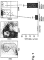
Fig. 1 : CFU-F von SUSD2+ (W5C5) Knochenmarkszellen.
Knochenmarkszellen wurden mit CD271-APC, CD45 Brilliant Violet, CD56-FITC und W5C5-PE markiert. Auf die CD271+CD45- MSC Population wurde ein Fenster gesetzt und anschließend im daraus resultierenden Plot Sortierfenster auf die W5C5+cd56- und die W5C5+CD56+ Populationen gesetzt. Nach der Sortierung wurde die Koloniebildungszahl (CFU-F) -Kapazität der jeweiligen Population getestet. -
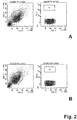
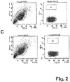
Fig. 2 : SUSD2-Identifizierung:
Die Antikörper W5C5 und W3D5 erkennen mit dem SUSD2-Gen transfizierte HEK-293 Zellen: Die FACS-Plots zeigen sowohl Kontrollfärbungen auf beiden Zelltypen (also nicht-transfizierte HEK-293 Zellen (A) und SUSD2-transfizierte HEK-293 Zellen (B) als auch spezifische Färbungen mit den jeweiligen Antikörpern (nicht-transfizierte HEK.293 Zellen (C) und SUSD2-transifizierte HEK293.Zellen (D)). - Nach Aufklärung und Einverständniserklärung wurde an der Berufsgenossenschaftlichen Klinik Knochenmark aus dem Oberschenkelschaft von Patienten gewonnen, die nach Hüftoperationen ein künstliches Hüftgelenk erhielten. Peripheres Blut von gesunden Spendern wurde von der Transfusionsabteilung des Universitätsklinikums Tübingen erhalten. Mononukleare Zellen aus dem Knochenmark und mononukleare Zellen aus dem Blut wurden mittels Ficoll-Dichtegradienten-Fraktionierung isoliert und die verbleibenden Erythrocyten in einer Ammoniumchloridlösung lysiert.
- Die Ficoll-getrennten und FACS-angereicherten Knochenmarkzellen wurden wie zuvor beschrieben kultiviert: 2 × 10⁷ unfraktionierte oder 1 × 10⁴ sortierte MSCA-1+CD56+ und MSCA-1+CD56- BM-Zellen in Gelatine-beschichteten T-75 oder T-25 Kulturflaschen kultiviert, und zwar in Anwesenheit von 20 ml oder 6 ml Knockout™ Ersatzmedium (Invitrogen, Karlsruhe, Deutschland) sowie 5 ng/ml rekombinantem humanem Fibroblasten-Wachstumsfaktor (rh-bFGF; CellSystems, Remagen, Deutschland). Nach einer dreitägigen Kultur wurden die nicht-adhärierenden Zellen entfernt und frisches Medium hinzugefügt. Die adhärierenden Zellen wurden kultiviert, bis sie eine 90%-ige Konfluenz erreichten.
- CFU-F-Assays wurden durch Ausplattieren von entweder 1 × 10⁵ unselektierten oder 500 - 5.000 FACS-selektierten Knochenmarkzellen in Gelatine-beschichtete T-25 Flaschen durchgeführt, die Knockout™-Medium und 5 ng/ml rh-bFGF enthielten. Nach einer zwölftägigen Kultivierung wurden die adhärierenden Zellen zweimal mit PBS gewaschen, fünf Minuten bei Raumtemperatur mit Methanol fixiert (Sigma-Aldrich), luftgetrocknet und mit Giemsa-Lösung gefärbt (Merck, Darmstadt, Deutschland). CFU-F-Kolonien wurden makroskopisch ausgezählt. Die Größe der Kolonien reichte von zwischen 1 und 8 mm im Durchmesser.
- Antikörper. Die folgenden Antikörper wurden eingesetzt: W5C5 (SUSD2), W3D5 (SUSD2), und W8B2 (TNAP). CD56-FITC (Klon NCAM16.2) wurde von Becton Dickinson (Heidelberg, Deutschland) gekauft. Der SSEA-4-reaktive Antikörper MC-813-70 wurde von Chemicon (Hampshire, Großbritannien) erworben. CD271-APC und SUSD2-PE und mit MACS beads gekoppeltem SUSD2 wurden von Miltenyi Boitec, Bergisch Gladbach, erworben.
- Immunofluoreszenzfärbung: Nach Blockierung und spezifischen Bindungen mit 10 mg/ml Polyglobin (10 min, 4 °C) wurden die Zellen 15 min lang mit entweder 20 µl Antikörpern oder 10 µl Fluorochrom-konjugierten Antikörpern inkubiert. Die mit den Konjugaten gefärbten Zellen wurden zweimal gewaschen, in 200 µl FACS Puffer suspendiert und für die Durchflusszytometrie eingesetzt. Die Zellen, die mit den Antikörpern markiert waren, wurden mit 20 µl eines F(ab)2-Fragment des R-Phycoerythrin (PE)-konjugierten Ziegen-anti-Maus-Antikörpers (Dako Cytomations, Glostrup, Dänemark) 15 min lang gefärbt, zweimal gewaschen und mittels Durchflusszytometrie analysiert. Für die Vielfarbenfärbung wurden die Zellen 15 min lang mit 10 µl eines Anti-CD56-FITC, Anti-TNAP-APC und anti-SUSD2-PE. Nach dem Waschen wurden die Zellen für die Durchflusszytometrie eingesetzt. Für eine kombinierte indirekte und direkte Färbung wurden die Zellen zunächst mit dem nicht-konjugierten Antikörper markiert, und anschließend mit 20 µl an 1:25 verdünntem Ziegen-anti-Maus sekundärem Antikörper 15 min lang gefärbt. Die freien Bindestellen des sekundären Antikörpers wurden durch Inkubation der Zellen für 25 min mit 20 µl an einem Maus-IgG-polyklonalen Antikörper (0,05 µg/ml; Southern Biotech, Birmingham, AL) blockiert, bevor sie mit TNAP-APC und/oder CD56-FITC gegengefärbt wurden. Nach einem Waschschritt wurden die Zellen mittels Durchflusszytometrie analysiert.
- Knochenmarkzellen, die mit Anti-TNAP-APC, Anti-SUSD2-PE und CD56-FITC markiert wurden, wurden mit einem FACSAria Zellsortierer (Becton Dickinson) sortiert, indem zunächst ein Gate auf die SUSD2 Population gesetzt wurde und anschließend die Sortierfenster im resultierenden Plot TNAP gegen CD56 gesetzt wurden. Die sortierten Zellen wurden für funktionelle und phänotypische Analysen eingesetzt. Die Koexpressionsanalysen erfolgten mit einem FACSCantoll Durchflusszytometer (Becton Dickinson). Die Daten wurden unter Verwendung der FCS-Express-Software (De Novo Software, Ontario, Kanada) analysiert.
- In ausgewählten Versuchen wurden die Knochenmarkzellen mittels MACS (Miltenyi Biotec) vorsortiert unter Verwendung von SUSD2-PE und Anti-PE MACS Beads. Die Trennungen wurden gemäß den Empfehlungen des Herstellers durchgeführt.
- Wie bereits bei der Figurenbeschreibung erwähnt, wurden in Versuchen Knochenmarkzellen mit CD271-APC, CD45-BrilliantViolet, CD56-FITC und W5C5-PE markiert. Da MSC CD271+CD45- sind, wurde auf diese Population ein Fenster gesetzt und anschließend im daraus resultierenden Plot Sortierfenster auf die W5C5+CD56- und W5C5+CD56+ Populationen gesetzt. Nach der Sortierung wurde die Koloniebildungskapazität der jeweiligen Populationen untersucht (
Fig. 1 ). - Kultivierte Knochenmark-MSC wurden simultan mit W3D5 und W5C5 markiert und mit einem Kaninchen-anti-Maus-PE-Konjugat gefärbt (durch Vorversuche mit über 20 verschiedenen Zelltypen wurde herausgefunden, dass W5C5 und W3D5 die gleichen Reaktivitätsmuster hatten). Anschließend wurden die W5C5IW3D5- und W5C5/W3D5+++ Zellen im FACS-Sorter sortiert und abzentrifugiert. Aus den Zellpellets wurde die mRNA isoliert (über Mitenyi, Deutschland) und eine Genexpressionsanalyse der beiden Fraktionen durchgeführt. Es zeigte sich, dass in der W5C5/W3D5+++ Fraktion das SUSD2-Gen um ca. den Faktor 20 höher exprimiert war als in der W5C5/W3D5- Fraktion. Da es keine monoklonalen SUSD2-Antikörper auf dem Markt gab, wurde die komplette Sequenz des SUSD2-Gens erworben (Origen) und damit HEK-293 Zellen transfiziert. 3 Tage nach der Transfektion wurde die Reaktivität von W5C5 und W3D5 auf der Transfektante und der nicht-transfizierten HEK-293 Zelle verglichen (Fig. 4). Die FACS-Plots zeigen sowohl Kontrollfärbungen auf beiden Zelltypen (Fig. 4A, B) als auch spezifische Färbungen mit den jeweiligen Antikörpern (Fig. 4C, D). Es stellte sich heraus, dass beide Antikörper selektiv die HEK293/huSUSD2 Transfektante erkennen.
- Mit den vorliegenden Ergebnissen konnte somit gezeigt werden, dass die Kombination von zumindest den Antikörpern gegen SUSD2 und CD140b,TNAP und/oder CD56 geeignet ist, alle MSC aus Knochenmark hochrein und effektiv zu isolieren, insbesondere deshalb, da die beiden Marker SUSD2 hoch-selektiv für MSC sind. TNAP ist stark auf CD56- MSC aber nur schwach auf CD56+ MSC exprimiert. SUSD2 ist jedoch auf beiden Subsets gleich stark exprimiert. Die Kombination von beiden Antikörpern ermöglicht durch die Signalverstärkung eine sichere Selektion von MSC.
- Mit der vorliegenden Studie wurde daher eine Kombination aus Antigenen identifiziert, nämlich SUSD2 mit CD104b, TNAP und/oder CD56, über welche effektiv MSC, insbesondere solche mit chondrogenem, adipozytärem und osteogenem Differenzierungspotential, isoliert und/oder identifiziert werden können.
- Diese Ergebnisse sind insbesondere im Hinblick auf den klinischen Einsatz derart isolierter Stammzellen bzw. der über diese Stammzellen gewonnenen Chondrozyten, Adipozyten und Osteoblasten relevant. So sind bspw. Verletzungen des Gelenkknorpels und von Bandscheiben regelmäßig schwierig zu behandeln, gerade aufgrund der begrenzten Regenerationsfähigkeit dieser Gewebe. Krankheiten wie rheumatoide Arthritis, Traumata, Knochenbrüche und Bandscheibenverletzungen sind direkt mit dem Mangel einer effektiven Chondrogenese verbunden. Trotz des Fortschritts in der Orthopädie und des steigenden Erfolgs bei der autologen Chondrozytentransplantation bleiben Zellbiologie-bezogene Ansätze für die Knorpelregeneration eine Herausforderung. Das Hauptproblem ist die Verwendung kultivierter Zellen für klinische Zwecke, bei welchen die Ausgangszellen lediglich schlecht charakterisiert sind.
- Daher bietet die vorliegende Erfindung die Möglichkeit, bspw. hoch angereicherte und gut definierte MSC Knochenmarkszellen bereitzustellen, mit einer herausragenden chondrogenen Differenzierungsfähigkeit, die für den klinischen Einsatz als Ausgangspopulation eingesetzt werden können. Diese Zellen können entweder direkt zur Injektion eingesetzt werden, bspw. in die Bandscheibenzwischenräume/Bandscheiben, oder in vitro expandiert und in Chondrozyten differenziert werden, bevor sie für klinische Einsätze verwendet werden.
Claims (11)
- Verwendung von zumindest einem ersten Antikörper, der an das Antigen Sushi Domäne enthaltendes Protein 2 (SUSD2) bindet, oder funktionellen Fragmenten des zumindest ersten Antikörpers, in Kombination mit zumindest einem zweiten Antikörper, der an das Antigen CD140b bindet, oder funktionellen Fragmenten des zumindest zweiten Antikörpers, zur in vitro Isolierung von mesenchymalen Stammzellen.
- Verwendung nach Anspruch 1, dadurch gekennzeichnet, dass der zumindest eine an SUSD2 bindende Antikörper ausgewählt ist aus der Gruppe:- der Antikörper W5C5, der von der bei der Deutschen Sammlung von Mikroorganismen und Zellkulturen hinterlegten Zelllinie mit der Nr. ACC 2813 produziert wird,- der Antikörper W3D5, der von der bei der Deutschen Sammlung von Mikroorganismen und Zellkulturen hinterlegten Zelllinie mit der Nr. ACC2815 produziert wird,- funktionelle Fragmente der Antikörper W5C5 oder W3D5, und- ein Antikörper, der an das gleiche Epitop bindet wie der Antikörper W5C5 oder W3D5.
- Verwendung nach Anspruch 1 oder 2, dadurch gekennzeichnet, dass der an CD140b bindende Antikörper ausgewählt ist aus der Gruppe:- der Antikörper 28D4- funktionelle Fragmente des Antikörpers 28D4, und- ein Antikörper, der an das gleiche Epitop bindet wie der Antikörper 28D4.
- Verwendung nach einem der Ansprüche 1 bis 3, dadurch gekennzeichnet, dass die Verwendung in Kombination mit zumindest einem dritten Antikörper, oder funktionellen Fragmenten davon, erfolgt, der zumindest an eines der Antigene CD56 oder TNAP (tissue non-specific alkaline phosphatase; Gewebe-unspezifische alkalische Phosphatase) bindet, wobei der an TNAP bindende Antikörper ausgewählt ist aus der Gruppe:der Antikörper W8B2, der von der bei der Deutschen Sammlung von Mikroorganismen und Zellkulturen hinterlegten Zelllinie mit der Nr. ACC2813 produziert wird,- funktionelle Fragmente des Antikörpers W8B2, und- ein Antikörper, der an das gleiche Epitop bindet wie der Antikörper W8B2.
- Verwendung nach Anspruch 4, dadurch gekennzeichnet, dass der an CD56 bindende Antikörper ausgewählt ist aus der Gruppe:- der Antikörper 39D5, der von der bei der Deutschen Sammlung von Mikroorganismen und Zellkulturen hinterlegten Zelllinie mit der Nr. ACC2930 produziert wird,- funktionelle Fragmente des Antikörpers 39D5, und- ein Antikörper, der an das gleiche Epitop bindet wie der Antikörper 39D5.
- In vitro Verfahren zur Isolierung und/oder Identifizierung von mesenchymalen Stammzellen, insbesondere von mesenchymalen Stammzellen mit chondrozytärem differenzierungspotential, dadurch gekennzeichnet, dass es die folgenden Schritte aufweist:a) in-Kontakt-Bringen einer Probe, die mesenchymale Stammzellen enthält, mit zumindest einem Antikörper, der an Antigen SUSD2 bindet, oder mit funktionellen Fragmenten des Antikörpers,b) ggf. in-Kontakt-Bringen einer Probe, die mesenchymale Stammzellen enthält, mit einem weiteren Antikörper, der an das Antigen SUSD2 bindet, oder mit funktionellen Fragmenten des Antikörpers,c) in-Kontakt-Bringen der Probe mit zumindest einem Antikörper, der an CD140b bindet, oder mit funktionellen Fragmenten des Antikörpers, undd) Isolieren und/oder Identifizieren von Zellen, an welche i) der zumindest eine Antikörper oder Fragmente davon, der an das Antigen SUSD2 bindet, und ii) der zumindest eine Antikörper, der an CD140b bindet, gebunden hat.
- Verfahren nach Anspruch 6, dadurch gekennzeichnet, dass der in Schritt a) eingesetzte anti-SUSD2-Antikörper ausgewählt ist aus der Gruppe:- der Antikörper W5C5, der von der bei der Deutschen Sammlung von Mikroorganismen und Zellkulturen hinterlegten Hybridomzelllinie mit der Nr. ACC2813 produziert wird,- der Antikörper W3D5, der von der bei der Deutschen Sammlung von Mikroorganismen und Zellkulturen hinterlegten Hybridomzelllinie mit der Nr. ACC2815 produziert wird,- funktionelle Fragmente der Antikörper W5C5 und W3D5, und Antikörper, die an das gleiche Epitop binden wie der Antikörper W5C5 oder W3D5.
- Verfahren nach einem der Ansprüche 6 oder 7, dadurch gekennzeichnet, dass der in Schritt c) eingesetzte anti-CD140b-Antikörper ausgewählt ist aus der Gruppe:der Antikörper 28D4,- funktionelle Fragmente des Antikörpers 28D4, und- Antikörper, die an das gleiche Epitop binden wie der Antikörper 28D4.
- Verfahren nach einem der Ansprüche 6 bis 8, dadurch gekennzeichnet, dass es den weiteren Schritt c1) aufweist:
c1) in-Kontakt-Bringen der Probe mit zumindest einem Antikörper, der an CD56
oder TNAP bindet, oder mit funktionellen Fragmenten des Antikörpers, wobei der in Schritt c1) eingesetzte anti-TNAP-Antikörper ausgewählt ist aus der Gruppe:- der Antikörper W8B2, der von der bei der Deutschen Sammlung von Mikroorganismen und Zellkulturen hinterlegten Hybridomzelllinie mit der Nr. ACC2567 produziert wird,- funktionelle Fragmente des Antikörpers W8B2, und- Antikörper, die an das gleiche Antigen oder Epitop binden wie der Antikörper W8B2. - Verfahren nach Anspruch 9, dadurch gekennzeichnet, dass der in Schritt c1) eingesetzte an CD56 bindende Antikörper ausgewählt ist aus der Gruppe:- der Antikörper 39D5, der von der bei der Deutschen Sammlung von Mikroorganismen und Zellkulturen hinterlegten Hybridomzelllinie mit der Nr. ACC 2930 produziert wird,- funktionelle Fragmente des Antikörpers 39D5, und- Antikörper, die an das gleiche Epitop binden wie der Antikörper 39D5.
- Verfahren nach einem der Ansprüche 6 bis 10, dadurch gekennzeichnet, dass die Schritte a), b), c) und ggf. c1) gleichzeitig, aufeinanderfolgend oder in umgekehrter Reihenfolge durchgeführt werden.
Applications Claiming Priority (2)
| Application Number | Priority Date | Filing Date | Title |
|---|---|---|---|
| DE102012102532A DE102012102532A1 (de) | 2012-03-23 | 2012-03-23 | Neue MSC-Oberflächenmarker |
| PCT/EP2013/056033 WO2013139952A2 (de) | 2012-03-23 | 2013-03-22 | Neue msc-oberflächenmarker |
Publications (2)
| Publication Number | Publication Date |
|---|---|
| EP2828663A2 EP2828663A2 (de) | 2015-01-28 |
| EP2828663B1 true EP2828663B1 (de) | 2018-09-26 |
Family
ID=48325588
Family Applications (1)
| Application Number | Title | Priority Date | Filing Date |
|---|---|---|---|
| EP13720803.9A Not-in-force EP2828663B1 (de) | 2012-03-23 | 2013-03-22 | Neue msc-oberflächenmarker |
Country Status (6)
| Country | Link |
|---|---|
| US (1) | US9580686B2 (de) |
| EP (1) | EP2828663B1 (de) |
| AU (1) | AU2013237374B2 (de) |
| DE (1) | DE102012102532A1 (de) |
| ES (1) | ES2695581T3 (de) |
| WO (1) | WO2013139952A2 (de) |
Families Citing this family (2)
| Publication number | Priority date | Publication date | Assignee | Title |
|---|---|---|---|---|
| CN104215768B (zh) * | 2014-08-07 | 2016-01-13 | 北京大学 | Susd2蛋白作为标记物的用途 |
| EP3183336B2 (de) | 2014-08-22 | 2025-07-09 | Cambridge Enterprise Limited | Rücksetzung pluripotenter stammzellen |
Family Cites Families (2)
| Publication number | Priority date | Publication date | Assignee | Title |
|---|---|---|---|---|
| DE102006043625B4 (de) * | 2006-09-12 | 2012-02-23 | Eberhard-Karls-Universität Tübingen Universitätsklinikum | Verfahren zur Isolierung und/oder Identifizierung von mesenchymalen Stammzellen (MSC) |
| ES2370794B1 (es) * | 2010-05-25 | 2012-10-31 | Servicio Andaluz De Salud | Biomarcador de células cartilaginosas humanas. |
-
2012
- 2012-03-23 DE DE102012102532A patent/DE102012102532A1/de not_active Withdrawn
-
2013
- 2013-03-22 ES ES13720803T patent/ES2695581T3/es active Active
- 2013-03-22 EP EP13720803.9A patent/EP2828663B1/de not_active Not-in-force
- 2013-03-22 AU AU2013237374A patent/AU2013237374B2/en not_active Ceased
- 2013-03-22 WO PCT/EP2013/056033 patent/WO2013139952A2/de not_active Ceased
-
2014
- 2014-09-22 US US14/493,160 patent/US9580686B2/en not_active Expired - Fee Related
Non-Patent Citations (1)
| Title |
|---|
| None * |
Also Published As
| Publication number | Publication date |
|---|---|
| WO2013139952A2 (de) | 2013-09-26 |
| US9580686B2 (en) | 2017-02-28 |
| AU2013237374A1 (en) | 2014-10-16 |
| DE102012102532A1 (de) | 2013-09-26 |
| WO2013139952A3 (de) | 2014-09-25 |
| US20150010516A1 (en) | 2015-01-08 |
| EP2828663A2 (de) | 2015-01-28 |
| ES2695581T3 (es) | 2019-01-09 |
| AU2013237374B2 (en) | 2016-02-11 |
Similar Documents
| Publication | Publication Date | Title |
|---|---|---|
| EP2313772B1 (de) | Isolierung und/oder identifizierung von stammzellen mit adipozytärem, chondrozytärem und pankreatischem differenzierungspotential | |
| DE69936720T2 (de) | Menschliche cd45+ und/oder fibroblasten+ mesenchymale stammzellen | |
| US8142773B2 (en) | Methods of implanting mesenchymal stem cells for tissue repair and formation | |
| JP4922548B2 (ja) | 体性幹細胞の同定および単離ならびにその使用 | |
| US7863043B2 (en) | Stem cell populations and methods of use | |
| US20100260721A1 (en) | Enrichment of Cells | |
| WO2003027281A2 (en) | Pluripotent stem cells originating in skeletal muscle intestinal tissue | |
| WO2002000849A1 (en) | Cell fraction containing cells capable of differentiating into nervous system cells | |
| US20080075699A1 (en) | Method for isolating and/or identifying mesenchymal stem cells (msc) | |
| Khan et al. | Evaluation of the effects of synovial multipotent cells on deep digital flexor tendon repair in a large animal model of intra‐synovial tendinopathy | |
| US11613733B2 (en) | Method for purifying mesenchymal stem cells to improve transplantation efficiency | |
| DE102012102532A1 (de) | Neue MSC-Oberflächenmarker | |
| DE102009041885B4 (de) | Separation von Mesenchymalen Stammzellen | |
| WO2004024772A1 (de) | Antikörper zur isolierung und/oder idenfifizierung neuronaler stammzellen und verfahren zur isolierung und/oder identifizierung neuronaler vorläuferzellen | |
| US20250177454A1 (en) | Human Homogeneous Amniotic Fluid Stem Cell Lines And Uses Thereof | |
| US20220313741A1 (en) | Pluripotent stem cells inducing osteochondral repair | |
| HK1123071A1 (zh) | 通过组织非特异性碱性磷酸酶分离成人多潜能细胞 |
Legal Events
| Date | Code | Title | Description |
|---|---|---|---|
| PUAI | Public reference made under article 153(3) epc to a published international application that has entered the european phase |
Free format text: ORIGINAL CODE: 0009012 |
|
| 17P | Request for examination filed |
Effective date: 20140924 |
|
| AK | Designated contracting states |
Kind code of ref document: A2 Designated state(s): AL AT BE BG CH CY CZ DE DK EE ES FI FR GB GR HR HU IE IS IT LI LT LU LV MC MK MT NL NO PL PT RO RS SE SI SK SM TR |
|
| AX | Request for extension of the european patent |
Extension state: BA ME |
|
| RIN1 | Information on inventor provided before grant (corrected) |
Inventor name: BUEHRING, HANS-JOERG Inventor name: CERABONA, FLAVIANNA Inventor name: GRIMM, SABRINA |
|
| DAX | Request for extension of the european patent (deleted) | ||
| 17Q | First examination report despatched |
Effective date: 20150805 |
|
| STAA | Information on the status of an ep patent application or granted ep patent |
Free format text: STATUS: EXAMINATION IS IN PROGRESS |
|
| GRAP | Despatch of communication of intention to grant a patent |
Free format text: ORIGINAL CODE: EPIDOSNIGR1 |
|
| STAA | Information on the status of an ep patent application or granted ep patent |
Free format text: STATUS: GRANT OF PATENT IS INTENDED |
|
| INTG | Intention to grant announced |
Effective date: 20180423 |
|
| GRAS | Grant fee paid |
Free format text: ORIGINAL CODE: EPIDOSNIGR3 |
|
| GRAA | (expected) grant |
Free format text: ORIGINAL CODE: 0009210 |
|
| STAA | Information on the status of an ep patent application or granted ep patent |
Free format text: STATUS: THE PATENT HAS BEEN GRANTED |
|
| AK | Designated contracting states |
Kind code of ref document: B1 Designated state(s): AL AT BE BG CH CY CZ DE DK EE ES FI FR GB GR HR HU IE IS IT LI LT LU LV MC MK MT NL NO PL PT RO RS SE SI SK SM TR |
|
| REG | Reference to a national code |
Ref country code: GB Ref legal event code: FG4D Free format text: NOT ENGLISH |
|
| REG | Reference to a national code |
Ref country code: CH Ref legal event code: EP |
|
| REG | Reference to a national code |
Ref country code: AT Ref legal event code: REF Ref document number: 1046635 Country of ref document: AT Kind code of ref document: T Effective date: 20181015 |
|
| REG | Reference to a national code |
Ref country code: IE Ref legal event code: FG4D Free format text: LANGUAGE OF EP DOCUMENT: GERMAN |
|
| REG | Reference to a national code |
Ref country code: DE Ref legal event code: R096 Ref document number: 502013011177 Country of ref document: DE |
|
| REG | Reference to a national code |
Ref country code: ES Ref legal event code: FG2A Ref document number: 2695581 Country of ref document: ES Kind code of ref document: T3 Effective date: 20190109 |
|
| REG | Reference to a national code |
Ref country code: NL Ref legal event code: MP Effective date: 20180926 |
|
| PG25 | Lapsed in a contracting state [announced via postgrant information from national office to epo] |
Ref country code: FI Free format text: LAPSE BECAUSE OF FAILURE TO SUBMIT A TRANSLATION OF THE DESCRIPTION OR TO PAY THE FEE WITHIN THE PRESCRIBED TIME-LIMIT Effective date: 20180926 Ref country code: LT Free format text: LAPSE BECAUSE OF FAILURE TO SUBMIT A TRANSLATION OF THE DESCRIPTION OR TO PAY THE FEE WITHIN THE PRESCRIBED TIME-LIMIT Effective date: 20180926 Ref country code: BG Free format text: LAPSE BECAUSE OF FAILURE TO SUBMIT A TRANSLATION OF THE DESCRIPTION OR TO PAY THE FEE WITHIN THE PRESCRIBED TIME-LIMIT Effective date: 20181226 Ref country code: GR Free format text: LAPSE BECAUSE OF FAILURE TO SUBMIT A TRANSLATION OF THE DESCRIPTION OR TO PAY THE FEE WITHIN THE PRESCRIBED TIME-LIMIT Effective date: 20181227 Ref country code: NO Free format text: LAPSE BECAUSE OF FAILURE TO SUBMIT A TRANSLATION OF THE DESCRIPTION OR TO PAY THE FEE WITHIN THE PRESCRIBED TIME-LIMIT Effective date: 20181226 Ref country code: RS Free format text: LAPSE BECAUSE OF FAILURE TO SUBMIT A TRANSLATION OF THE DESCRIPTION OR TO PAY THE FEE WITHIN THE PRESCRIBED TIME-LIMIT Effective date: 20180926 Ref country code: SE Free format text: LAPSE BECAUSE OF FAILURE TO SUBMIT A TRANSLATION OF THE DESCRIPTION OR TO PAY THE FEE WITHIN THE PRESCRIBED TIME-LIMIT Effective date: 20180926 |
|
| REG | Reference to a national code |
Ref country code: LT Ref legal event code: MG4D |
|
| PG25 | Lapsed in a contracting state [announced via postgrant information from national office to epo] |
Ref country code: AL Free format text: LAPSE BECAUSE OF FAILURE TO SUBMIT A TRANSLATION OF THE DESCRIPTION OR TO PAY THE FEE WITHIN THE PRESCRIBED TIME-LIMIT Effective date: 20180926 Ref country code: LV Free format text: LAPSE BECAUSE OF FAILURE TO SUBMIT A TRANSLATION OF THE DESCRIPTION OR TO PAY THE FEE WITHIN THE PRESCRIBED TIME-LIMIT Effective date: 20180926 Ref country code: HR Free format text: LAPSE BECAUSE OF FAILURE TO SUBMIT A TRANSLATION OF THE DESCRIPTION OR TO PAY THE FEE WITHIN THE PRESCRIBED TIME-LIMIT Effective date: 20180926 |
|
| PG25 | Lapsed in a contracting state [announced via postgrant information from national office to epo] |
Ref country code: EE Free format text: LAPSE BECAUSE OF FAILURE TO SUBMIT A TRANSLATION OF THE DESCRIPTION OR TO PAY THE FEE WITHIN THE PRESCRIBED TIME-LIMIT Effective date: 20180926 Ref country code: PL Free format text: LAPSE BECAUSE OF FAILURE TO SUBMIT A TRANSLATION OF THE DESCRIPTION OR TO PAY THE FEE WITHIN THE PRESCRIBED TIME-LIMIT Effective date: 20180926 Ref country code: IS Free format text: LAPSE BECAUSE OF FAILURE TO SUBMIT A TRANSLATION OF THE DESCRIPTION OR TO PAY THE FEE WITHIN THE PRESCRIBED TIME-LIMIT Effective date: 20190126 Ref country code: RO Free format text: LAPSE BECAUSE OF FAILURE TO SUBMIT A TRANSLATION OF THE DESCRIPTION OR TO PAY THE FEE WITHIN THE PRESCRIBED TIME-LIMIT Effective date: 20180926 Ref country code: CZ Free format text: LAPSE BECAUSE OF FAILURE TO SUBMIT A TRANSLATION OF THE DESCRIPTION OR TO PAY THE FEE WITHIN THE PRESCRIBED TIME-LIMIT Effective date: 20180926 Ref country code: NL Free format text: LAPSE BECAUSE OF FAILURE TO SUBMIT A TRANSLATION OF THE DESCRIPTION OR TO PAY THE FEE WITHIN THE PRESCRIBED TIME-LIMIT Effective date: 20180926 |
|
| PG25 | Lapsed in a contracting state [announced via postgrant information from national office to epo] |
Ref country code: SK Free format text: LAPSE BECAUSE OF FAILURE TO SUBMIT A TRANSLATION OF THE DESCRIPTION OR TO PAY THE FEE WITHIN THE PRESCRIBED TIME-LIMIT Effective date: 20180926 Ref country code: PT Free format text: LAPSE BECAUSE OF FAILURE TO SUBMIT A TRANSLATION OF THE DESCRIPTION OR TO PAY THE FEE WITHIN THE PRESCRIBED TIME-LIMIT Effective date: 20190126 Ref country code: SM Free format text: LAPSE BECAUSE OF FAILURE TO SUBMIT A TRANSLATION OF THE DESCRIPTION OR TO PAY THE FEE WITHIN THE PRESCRIBED TIME-LIMIT Effective date: 20180926 |
|
| REG | Reference to a national code |
Ref country code: DE Ref legal event code: R097 Ref document number: 502013011177 Country of ref document: DE |
|
| PG25 | Lapsed in a contracting state [announced via postgrant information from national office to epo] |
Ref country code: DK Free format text: LAPSE BECAUSE OF FAILURE TO SUBMIT A TRANSLATION OF THE DESCRIPTION OR TO PAY THE FEE WITHIN THE PRESCRIBED TIME-LIMIT Effective date: 20180926 |
|
| PLBE | No opposition filed within time limit |
Free format text: ORIGINAL CODE: 0009261 |
|
| STAA | Information on the status of an ep patent application or granted ep patent |
Free format text: STATUS: NO OPPOSITION FILED WITHIN TIME LIMIT |
|
| 26N | No opposition filed |
Effective date: 20190627 |
|
| PG25 | Lapsed in a contracting state [announced via postgrant information from national office to epo] |
Ref country code: SI Free format text: LAPSE BECAUSE OF FAILURE TO SUBMIT A TRANSLATION OF THE DESCRIPTION OR TO PAY THE FEE WITHIN THE PRESCRIBED TIME-LIMIT Effective date: 20180926 Ref country code: MC Free format text: LAPSE BECAUSE OF FAILURE TO SUBMIT A TRANSLATION OF THE DESCRIPTION OR TO PAY THE FEE WITHIN THE PRESCRIBED TIME-LIMIT Effective date: 20180926 |
|
| REG | Reference to a national code |
Ref country code: CH Ref legal event code: PL |
|
| REG | Reference to a national code |
Ref country code: DE Ref legal event code: R082 Ref document number: 502013011177 Country of ref document: DE Representative=s name: WITTE, WELLER & PARTNER PATENTANWAELTE MBB, DE Ref country code: DE Ref legal event code: R081 Ref document number: 502013011177 Country of ref document: DE Owner name: TETEC TISSUE ENGINEERING TECHNOLOGIES AG, DE Free format text: FORMER OWNER: EBERHARD-KARLS-UNIVERSITAET TUEBINGEN UNIVERSITAETSKLINIKUM, 72076 TUEBINGEN, DE |
|
| PG25 | Lapsed in a contracting state [announced via postgrant information from national office to epo] |
Ref country code: LU Free format text: LAPSE BECAUSE OF NON-PAYMENT OF DUE FEES Effective date: 20190322 |
|
| REG | Reference to a national code |
Ref country code: ES Ref legal event code: PC2A Owner name: TETEC TISSUE ENGINEERING TECHNOLOGIES AG Effective date: 20191129 Ref country code: BE Ref legal event code: MM Effective date: 20190331 |
|
| PG25 | Lapsed in a contracting state [announced via postgrant information from national office to epo] |
Ref country code: IE Free format text: LAPSE BECAUSE OF NON-PAYMENT OF DUE FEES Effective date: 20190322 Ref country code: LI Free format text: LAPSE BECAUSE OF NON-PAYMENT OF DUE FEES Effective date: 20190331 Ref country code: CH Free format text: LAPSE BECAUSE OF NON-PAYMENT OF DUE FEES Effective date: 20190331 |
|
| REG | Reference to a national code |
Ref country code: GB Ref legal event code: 732E Free format text: REGISTERED BETWEEN 20200116 AND 20200122 |
|
| PG25 | Lapsed in a contracting state [announced via postgrant information from national office to epo] |
Ref country code: BE Free format text: LAPSE BECAUSE OF NON-PAYMENT OF DUE FEES Effective date: 20190331 |
|
| PG25 | Lapsed in a contracting state [announced via postgrant information from national office to epo] |
Ref country code: TR Free format text: LAPSE BECAUSE OF FAILURE TO SUBMIT A TRANSLATION OF THE DESCRIPTION OR TO PAY THE FEE WITHIN THE PRESCRIBED TIME-LIMIT Effective date: 20180926 |
|
| PG25 | Lapsed in a contracting state [announced via postgrant information from national office to epo] |
Ref country code: MT Free format text: LAPSE BECAUSE OF FAILURE TO SUBMIT A TRANSLATION OF THE DESCRIPTION OR TO PAY THE FEE WITHIN THE PRESCRIBED TIME-LIMIT Effective date: 20180926 |
|
| REG | Reference to a national code |
Ref country code: AT Ref legal event code: MM01 Ref document number: 1046635 Country of ref document: AT Kind code of ref document: T Effective date: 20190322 |
|
| PG25 | Lapsed in a contracting state [announced via postgrant information from national office to epo] |
Ref country code: AT Free format text: LAPSE BECAUSE OF NON-PAYMENT OF DUE FEES Effective date: 20190322 |
|
| PGFP | Annual fee paid to national office [announced via postgrant information from national office to epo] |
Ref country code: FR Payment date: 20210319 Year of fee payment: 9 |
|
| PG25 | Lapsed in a contracting state [announced via postgrant information from national office to epo] |
Ref country code: CY Free format text: LAPSE BECAUSE OF FAILURE TO SUBMIT A TRANSLATION OF THE DESCRIPTION OR TO PAY THE FEE WITHIN THE PRESCRIBED TIME-LIMIT Effective date: 20180926 |
|
| PGFP | Annual fee paid to national office [announced via postgrant information from national office to epo] |
Ref country code: GB Payment date: 20210324 Year of fee payment: 9 |
|
| PG25 | Lapsed in a contracting state [announced via postgrant information from national office to epo] |
Ref country code: HU Free format text: LAPSE BECAUSE OF FAILURE TO SUBMIT A TRANSLATION OF THE DESCRIPTION OR TO PAY THE FEE WITHIN THE PRESCRIBED TIME-LIMIT; INVALID AB INITIO Effective date: 20130322 |
|
| PGFP | Annual fee paid to national office [announced via postgrant information from national office to epo] |
Ref country code: IT Payment date: 20210331 Year of fee payment: 9 |
|
| PGFP | Annual fee paid to national office [announced via postgrant information from national office to epo] |
Ref country code: ES Payment date: 20210421 Year of fee payment: 9 |
|
| PG25 | Lapsed in a contracting state [announced via postgrant information from national office to epo] |
Ref country code: MK Free format text: LAPSE BECAUSE OF FAILURE TO SUBMIT A TRANSLATION OF THE DESCRIPTION OR TO PAY THE FEE WITHIN THE PRESCRIBED TIME-LIMIT Effective date: 20180926 |
|
| GBPC | Gb: european patent ceased through non-payment of renewal fee |
Effective date: 20220322 |
|
| PG25 | Lapsed in a contracting state [announced via postgrant information from national office to epo] |
Ref country code: GB Free format text: LAPSE BECAUSE OF NON-PAYMENT OF DUE FEES Effective date: 20220322 Ref country code: FR Free format text: LAPSE BECAUSE OF NON-PAYMENT OF DUE FEES Effective date: 20220331 |
|
| PG25 | Lapsed in a contracting state [announced via postgrant information from national office to epo] |
Ref country code: IT Free format text: LAPSE BECAUSE OF NON-PAYMENT OF DUE FEES Effective date: 20220322 |
|
| REG | Reference to a national code |
Ref country code: ES Ref legal event code: FD2A Effective date: 20230602 |
|
| PG25 | Lapsed in a contracting state [announced via postgrant information from national office to epo] |
Ref country code: ES Free format text: LAPSE BECAUSE OF NON-PAYMENT OF DUE FEES Effective date: 20220323 |
|
| PGFP | Annual fee paid to national office [announced via postgrant information from national office to epo] |
Ref country code: DE Payment date: 20230929 Year of fee payment: 11 |
|
| REG | Reference to a national code |
Ref country code: DE Ref legal event code: R119 Ref document number: 502013011177 Country of ref document: DE |
|
| PG25 | Lapsed in a contracting state [announced via postgrant information from national office to epo] |
Ref country code: DE Free format text: LAPSE BECAUSE OF NON-PAYMENT OF DUE FEES Effective date: 20241001 |
|
| PG25 | Lapsed in a contracting state [announced via postgrant information from national office to epo] |
Ref country code: DE Free format text: LAPSE BECAUSE OF NON-PAYMENT OF DUE FEES Effective date: 20241001 |