EP2816355A2 - Detection method for detection of bacteria, method for the preparation of fusion proteins and fusion protein - Google Patents
Detection method for detection of bacteria, method for the preparation of fusion proteins and fusion protein Download PDFInfo
- Publication number
- EP2816355A2 EP2816355A2 EP14173356.8A EP14173356A EP2816355A2 EP 2816355 A2 EP2816355 A2 EP 2816355A2 EP 14173356 A EP14173356 A EP 14173356A EP 2816355 A2 EP2816355 A2 EP 2816355A2
- Authority
- EP
- European Patent Office
- Prior art keywords
- protein
- bacteriophage
- detection
- tag
- proteins
- Prior art date
- Legal status (The legal status is an assumption and is not a legal conclusion. Google has not performed a legal analysis and makes no representation as to the accuracy of the status listed.)
- Granted
Links
Images


Classifications
-
- G—PHYSICS
- G01—MEASURING; TESTING
- G01N—INVESTIGATING OR ANALYSING MATERIALS BY DETERMINING THEIR CHEMICAL OR PHYSICAL PROPERTIES
- G01N33/00—Investigating or analysing materials by specific methods not covered by groups G01N1/00 - G01N31/00
- G01N33/48—Biological material, e.g. blood, urine; Haemocytometers
- G01N33/50—Chemical analysis of biological material, e.g. blood, urine; Testing involving biospecific ligand binding methods; Immunological testing
- G01N33/53—Immunoassay; Biospecific binding assay; Materials therefor
- G01N33/569—Immunoassay; Biospecific binding assay; Materials therefor for microorganisms, e.g. protozoa, bacteria, viruses
- G01N33/56911—Bacteria
-
- C—CHEMISTRY; METALLURGY
- C12—BIOCHEMISTRY; BEER; SPIRITS; WINE; VINEGAR; MICROBIOLOGY; ENZYMOLOGY; MUTATION OR GENETIC ENGINEERING
- C12N—MICROORGANISMS OR ENZYMES; COMPOSITIONS THEREOF; PROPAGATING, PRESERVING, OR MAINTAINING MICROORGANISMS; MUTATION OR GENETIC ENGINEERING; CULTURE MEDIA
- C12N15/00—Mutation or genetic engineering; DNA or RNA concerning genetic engineering, vectors, e.g. plasmids, or their isolation, preparation or purification; Use of hosts therefor
- C12N15/09—Recombinant DNA-technology
- C12N15/11—DNA or RNA fragments; Modified forms thereof; Non-coding nucleic acids having a biological activity
- C12N15/62—DNA sequences coding for fusion proteins
-
- G—PHYSICS
- G01—MEASURING; TESTING
- G01N—INVESTIGATING OR ANALYSING MATERIALS BY DETERMINING THEIR CHEMICAL OR PHYSICAL PROPERTIES
- G01N33/00—Investigating or analysing materials by specific methods not covered by groups G01N1/00 - G01N31/00
- G01N33/48—Biological material, e.g. blood, urine; Haemocytometers
- G01N33/50—Chemical analysis of biological material, e.g. blood, urine; Testing involving biospecific ligand binding methods; Immunological testing
- G01N33/53—Immunoassay; Biospecific binding assay; Materials therefor
- G01N33/566—Immunoassay; Biospecific binding assay; Materials therefor using specific carrier or receptor proteins as ligand binding reagents where possible specific carrier or receptor proteins are classified with their target compounds
-
- C—CHEMISTRY; METALLURGY
- C12—BIOCHEMISTRY; BEER; SPIRITS; WINE; VINEGAR; MICROBIOLOGY; ENZYMOLOGY; MUTATION OR GENETIC ENGINEERING
- C12N—MICROORGANISMS OR ENZYMES; COMPOSITIONS THEREOF; PROPAGATING, PRESERVING, OR MAINTAINING MICROORGANISMS; MUTATION OR GENETIC ENGINEERING; CULTURE MEDIA
- C12N2795/00—Bacteriophages
- C12N2795/00011—Details
- C12N2795/14011—Details ssDNA Bacteriophages
- C12N2795/14111—Inoviridae
- C12N2795/14131—Uses of virus other than therapeutic or vaccine, e.g. disinfectant
Definitions
- the invention relates to a detection method for the detection of bacteria, a method for the production of fusion proteins and a fusion protein obtainable by such a method.
- microbiological detection methods have certain disadvantages inherent in the underlying method. For example, established detection methods for microorganisms are often very time-consuming, since the microorganisms must first be transferred to culture media and incubated. After this pre-enrichment, a selective enrichment followed by a biochemical screening (API test).
- API test biochemical screening
- the object of the invention is therefore to propose an improved method for the detection of bacteria, which overcomes the disadvantages mentioned above.
- a free bacterial surface protein or a bound bacterial surface protein, in particular a bacterium bound is enriched.
- the enrichment structure is provided on an immobile or a mobile surface structure.
- the enrichment structure is particularly preferably immobilized on the surface structure via a linker structure, in particular via a biotin-streptavidin linker or a metal chelate-polyhistidine tag linker.
- magnetic beads As a surface structure, magnetic beads, more particularly Ni beads, non-magnetic beads, in particular Ni-NTA, are advantageous, Chromatography column materials, sepharoses, microtiter plates, plastics, filter surfaces, glass, metal, ceramic, wood, stone, fabric, nanoparticles and / or semiconductor materials used.
- the bacteriophage protein is provided as a free bacteriophage protein.
- the bacteriophage protein may also be provided as a bacteriophage protein bound to a bacteriophage.
- bacteriophage protein that is selective for a bacterium or bacterial family.
- both the enrichment and the detection reaction are performed by specific protein-protein interaction with a bacterial surface protein.
- enrichment and detection reaction take place on different epitopes of the bacterial surface protein.
- the bacteriophage protein is used as a detection protein and an antibody as an enrichment structure.
- an antibody can be used as a detection protein and the bacteriophage protein as an enrichment structure.
- bacteriophage protein both for enrichment and for the detection reaction.
- a marker molecule in particular an enzyme or a fluorescence molecule, is attached directly to a detection protein surface.
- the detection protein used is preferably a fusion protein which is produced by linking a bacteriophage protein with a marker protein via a linker protein.
- the fusion protein is coupled in particular via a protease interface with a cleaning tag.
- the bacteriophage protein is produced by recombinant expression.
- the fusion protein is also produced by recombinant expression.
- the bacteriophage proteins produced by expression are labeled and / or stabilized with a marker molecule.
- the fusion proteins formed by the expression are stabilized.
- the stabilization is advantageously carried out by vacuum drying, freeze drying, lyophilization, sublimation drying, PEGyliseren, protein engineering and / or by adding stabilizers.
- a bacteriophage having the expressed bacteriophage protein or the expressed fusion protein is advantageously enveloped with polyethylene glycol molecules.
- the pegylation of bacteriophages advantageously leaves the contractile sheath of the bacteriophage free.
- the fusion protein thus produced can be advantageously used in the above-described detection method for detecting bacteria.
- marker proteins which are selected from the group of enzymes, proteins, fluorescent proteins, human caspases, in particular caspase-3, bioluminescent enzymes, in particular luciferase, lytic proteins and / or polymerases, in particular tag polymerase, are preferably used as marker protein genes .
- the ligation in step A) is carried out serially.
- the bacteriophage protein gene in particular N-terminal
- the marker protein gene in particular C-terminal
- restriction enzymes for the bacteriophage protein gene and the marker protein gene are used for the ligation.
- BsmBl is used as the 5 'end and Acc65I as the 3' end for the bacteriophage protein gene and Sall as the 5 'end and NotI as the 3' end for the marker protein gene.
- linker protein is generated, which allows a free three-dimensional movement of the bacteriophage protein and the marker protein to each other.
- the generated linker protein has a length of nine codons and is more particularly free from stop codons.
- a preferred fusion protein is prepared by the method described above.
- microbiological detection methods have certain disadvantages that are based on the underlying method itself. For example, established detection methods for microorganisms are often very time-consuming, since the Microorganisms must first be transferred to culture media and incubated. After this pre-enrichment, selective enrichment takes place followed by biochemical screening (API test). Often, this classical detection process shows false-positive or false-negative results, and immunological (ELISA) and gene-based methods (PCR, ISH, and FISH) must be additionally included. These methods are very expensive in the routine and also show some uncertainty in terms of identification. For example, an ELISA based on a polyclonal antibody 14 often shows cross-reactions to other organisms. Another disadvantage of this immunological detection method is the finiteness of the resource, since a polyclonal antibody is produced by immunization of the animal (rabbit, mouse, goat, horse).
- bacteria 10 are detected by the following methods: Selective Nutrient Culture, Colorful Series (API Test, VITEK), Polymerase Chain Reaction (PCR), Enzyme Linked Immunosorbent Assay (ELISA), Fluorescence In Situ Hybridization (FISH), In -Situ hybridization (ISH), aptamers.
- Recombinant bacteriophage proteins 12 which allow the adsorption of a bacteriophage 16 to a bacterial surface 18, are now produced by protein expression. These bind specifically to the bacterial surface 18 via protein-protein interactions and can be used both as an enrichment structure 19 for enrichment and for detection of bacteria 10.
- Fig. 1 shows example of a coating via polyhistidine tag 28 as a linker structure 31 n (Source: http://www.chemgapedia.de)
- the bacteriophage proteins 12 After immobilization of the materials by the bacteriophage proteins 12, they may be used to specifically enrich the bacteria 10 for the materials described above. As already mentioned, the accumulation of the bacteria takes place via the specific protein-protein interaction between bacteriophage protein 12 on one side and bacterial surface protein 32 on the other side.
- Fig. 2 shows an enrichment via magnetic or non-magnetic metal chelate beads 34, in particular nickel or cobalt ions, via a magnetic or non-magnetic metal chelate bead 34, a metal chelate 36, a polyhistidine tag 28 (His tag or 6 ⁇ His- Tag) and a bacteriophage protein 12 a bacterium 10 or only a bacterial surface protein 32 of a bacterial family 37 is immobilized.
- a magnetic or non-magnetic metal chelate beads 34 in particular nickel or cobalt ions
- Fig. 3 shows an accumulation by means of magnetic 20 or non-magnetic beads 30 or sepharose beads 38 by biotin 22-streptavidin 24 interaction, wherein magnetic 20 or non-magnetic beads 30 or sepharose beads 38, streptavidin 24, biotin 22 and a bacteriophage protein 12 a bacterium 10 or only a bacterial surface protein 32 is immobilized.
- Fig. 4 shows an immobilization of the bacteriophage proteins 12 via biotin 22 - streptavidin 24 on material surfaces 40, in particular microtiter plates 31c, semiconductors 31 m, glass 31f, metal 31g, wherein on material surfaces 40 of all kinds on streptavidin 24, biotin 22 and a bacteriophage protein 12 a bacterium10 or only a bacterial surface protein 32 is immobilized.
- Fig. 5 shows an immobilization of the bacteriophage proteins 12 via metal chelates 36 to material surfaces 40, in particular to microtiter plates 31c, semiconductors 31 m, glass 31f or metal 31 g, wherein on material surfaces 40 of all kinds via a metal chelate 36, a polyhistidine tag 28 (His-tag or 6 x His-tag) and a bacteriophage protein 12 a bacterium 10 or only a bacterial surface protein 32 is immobilized.
- a polyhistidine tag 28 His-tag or 6 x His-tag
- the bacteriophage proteins 12 can be used in combination with an antibody 14.
- an antibody 14 it would be possible to use a bifurcated antibody 14 as detection protein 42 after the capture (capture), such as in Fig. 6 shown.
- An immobilized antibody 14 could act as a scavenger, while a (labeled) bacteriophage protein 12 labeled directly on the detection protein surface 43 would be used as a detection protein 42, such as in U.S. Pat Figs. 7 and 8 shown.
- Fig. 6 shows a schematic representation of such an immunological detection of a labeled antibody 14 in the form of a "hybrid phage immunoassay (HYPIA)" after an enrichment by bacteriophage proteins 12, wherein a material surface 40 of all kinds on streptavidin 24, biotin 22 and a Bacteriophage protein 12 is a bacterium 10 or bacterial
- Surface protein 32 was immobilized and a monoclonal or polyclonal antibody 14, which has on the surface protein of the bacterium 10 and a label 44 or a label in the form of a marker molecule 45, a detection by an enzyme or fluorescence (eg HRP or fluorescein) can be performed ,
- Figs. 7 and 8 12 show schematic representations of a detection or an assay via labeled (labeled) bacteriophage proteins 12 after enrichment by antibody 14 as a "hybrid phage immunoassay (HYPIA)", using material surfaces 40 of all kinds, via protein A 46 or protein G 48 or via streptavidin 24 and biotin 22, and an antibody 14 a bacterium 10 or only a bacterial surface protein 32 is immobilized and then a bacteriophage protein 12 and a label 44 or label of the bacteriophage protein 12, the detection of an enzyme or fluorescence (eg B HRP or fluorescein) can be performed as a fluorescent molecule 49.
- an enzyme or fluorescence eg B HRP or fluorescein
- the receptor proteins of bacteriophage 16 can be used not only as so-called “capture proteins” (capture protein), but also as detection proteins 42.
- the recombinant bacteriophage proteins 12 can after their expression and purification with a classical detection molecule or enzyme 52 (labeled with HRP, phosphatase, peroxidase, fluorophores such as CFDA).
- a classical detection molecule or enzyme 52 labeled with HRP, phosphatase, peroxidase, fluorophores such as CFDA.
- Fig. 9 schematically shows representations of a fusion protein 50 consisting of bacteriophage protein 12 (capture protein, capture protein) and a Labeling protein or detector protein 54, with the following sequence from N-terminus 62 to C-terminus 64: Proetin tag, in particular a poly-histidine tag (His tag or 6 ⁇ His tag), protease recognition sequence 66, protease cleavage 60 , Bacteriophage protein 12 (capture protein or capture protein), linker 56 or loop, labeling protein or detector protein 54.
- Proetin tag in particular a poly-histidine tag (His tag or 6 ⁇ His tag)
- protease recognition sequence 66 protease cleavage 60
- Bacteriophage protein 12 capture protein or capture protein
- linker 56 or loop labeling protein or detector protein 54.
- bacteriophage protein 12 capture protein, capture protein
- any known bacteriophage 16 adsorption protein can be used.
- a bacteriophage protein 12 either a bacterium 10 can be detected extremely selectively (eg E. coli) or there is the possibility that a whole bacterial family 37 (eg coliforms) is sensitively detected.
- a known example is the bacteriophage M13 G3P protein which binds to tolA protein 84 of E. coli, as described in detail below.
- the following table shows the G3P protein with its closely related proteins, all of which could be used as a capture protein.
- Assessions no. protein name organism amino acids in length P69169 Attachment protein G3P (G3P) (Minor coat protein) Enterobacteria phage f1 (bacteriophage f1) 424 P03661 Attachment protein G3P (G3P) (Minor coat protein) Enterobacteria phage fd (bacteriophage fd) 424 P15415 Attachment protein G3P (G3P) (Minor coat protein) Enterobacteria phage 12-2 (bacteriophage 12-2) 434 080297 Attachment protein G3P (G3P) (Minor coat protein) Enterobacteria phage If1 (bacteriophage If1) 460 P03663 Attachment protein G3P (G3P) (Minor coat protein) Enterobacteria phage Ike (bacteriophage IKe) 434 P69168 Attachment protein G3P (
- Figs. 10 to 13 show schematic representations of fusion proteins 50 with different detector proteins 54, namely in Fig. 10 a fluorescent protein 72, in particular the green fluorescent protein (GFP), in Fig. 11 a human caspase 68, in particular caspase-3 70, in Fig. 12 a bioluminescence-based enzyme 52, especially luciferase 71, in Fig. 13 a polymerase (DNA and RNA) 74, in particular Taq polymerase 75.
- a fluorescent protein 72 in particular the green fluorescent protein (GFP)
- GFP green fluorescent protein
- Fig. 11 a human caspase 68
- caspase-3 70 in Fig. 12
- bioluminescence-based enzyme 52 especially luciferase 71
- Fig. 13 a polymerase (DNA and RNA) 74, in particular Taq polymerase 75.
- fusion proteins 50 can be very different assemble different assays.
- the assays are similar in structure to tests with conventionally labeled (labeled) bacteriophage proteins 12 in Figs. 7 and 8 ,
- Figs. 14 and 15 show schematic representations of a detection or an assay with a fusion protein 50 (bacteriophage protein 12 and detector protein 54) after an enrichment by antibodies 14, wherein on a material surface 40 of all kinds on protein A 46 or protein G 48 and an antibody 14, a bacterium 10 or only one bacterial surface protein 32 was immobilized.
- Various fusion proteins 50 are attached to the opposite side of the bacterium 10, having marker proteins 54 in the form of human caspases 68, in particular caspase-3 70, polymerases 74 (DNA and RNA), in particular tag polymerase 75, fluorescent proteins 72, in particular the green fluorescent protein (GFP) and bioluminescence-based enzymes 52, in particular luciferase 71.
- bacteriophage proteins 12 as fusion proteins 50 in combination with lytic proteins 76 will now be described.
- Fig. 16 shows a schematic representation of a fusion protein 50, consisting of a bacteriophage protein 12 (capture protein, capture protein) and a lytic protein 76 (eg., Lysozyme or lysostaphin) and a protein tag, in particular a poly-histidine tag (His-tag or 6 x His tag), a protease recognition sequence 66, a protease cleavage site 60, a linker 56 or loop, and a lytic protein 76.
- a bacteriophage protein 12 capture protein, capture protein
- a lytic protein 76 eg., Lysozyme or lysostaphin
- a protein tag in particular a poly-histidine tag (His-tag or 6 x His tag), a protease recognition sequence 66, a protease cleavage site 60, a linker 56 or loop, and a lytic protein 76.
- a polyclonal antibody 14 is a finite resource because the immunized animal dies at some point while the production of recombinant proteins is an endless resource.
- HYPIA hybrid phage immunoassay
- recombinant His tag protein provides an advantage in terms of assay development. Since the recombinant protein already on the beads (Ni-NTA 31, or magnetic ni-beads 26) is bound via the His tag, these can be used immediately for enrichment or assay development. It is therefore no longer necessary to elute the recombinant protein from the beads. Even with nickel ion coated microtiter plates 31 c or other vessels can be used for cleaning.
- fusion proteins 50 (bacteriophage protein 12 and detector protein 54, e.g., GFP)
- detector protein 54 e.g., GFP
- fluorophores are bound, respectively, which has a large impact on the sensitivity of the assay.
- conventional labeling is not 100% marking. Often only a fraction of the protein to be labeled is labeled (sometimes only 10%). This incomplete labeling can be virtually ruled out in the production of recombinant fusion proteins 50 (only one bacteriophage protein 12 and one reporter protein).
- human caspase-3 70 as a reporter enzyme has very great advantages since it is a purely human enzyme 52 and does not occur in the bacteria 10 to be detected.
- the enzymatic reaction is based on a proteolytic cleavage with the following amino acid recognition sequence: DEVDIX. This recognition sequence is extremely rare in bacterial proteins, so that their proteolytic cleavage is virtually eliminated.
- fluorescence substrates are N-acetyl-Asp-Glu-Vai-Asp-7-amido-4-methyl coumarin and N-acetyl-Asp-Gln-Met-Asp-7-amido-4-trifluoromethyl coumarin.
- tag polymerase 75 as a reporter enzyme in the form of a fusion protein 50 with a bacteriophage protein 12 as a "capture protein" results in the advantage of double specificity.
- the protein-protein interaction between bacteriophage protein 12 and bacterial surface protein 32 is highly specific, and second, the subsequent detection assay, in this case specific PCR, is also highly sensitive and specific detection. This dual specificity of identification enhances the safety of the assay so that false-positive results are virtually eliminated.
- This fusion protein 50 can also be used in a real-time PCR.
- the bacteriophage protein 12 is anyway a much more stable macromolecule than an antibody 14, which is composed of several fragments (light and heavy chain). Furthermore, it is possible to specifically modify the bacteriophage protein 12 by "protein engineering", so that the stability of the bacteriophage protein 12 can be significantly increased.
- the underlying step here is the adsorption of the bacteriophage 16 on the surface structure of the bacterium 10. This adsorption takes place over a very specific protein-protein interaction between virus and host cell. As a practical example here serves the bacteriophage M13 and the bacterium E. coli.
- the g3p gene is 1275 base pairs (bp) long and encodes a 424 amino acid (AS) protein.
- the G3P in the N-terminal region consists of four domains, which, as in Fig. 19 are respectively denoted by N1, L1, N2 and L2, while the C-terminal portion is characterized by a short transmembrane domain TD.
- the interaction of the bacteriophage 16 is well described in the literature and occurs first with the N2 domain which binds to the F pilus 82 of E.
- the N1 domain of bacteriophage 16 then interacts with the bacterial tolA protein 84 located in the periplasmic space 86 between the outer membrane 83 and the cytoplasmic membrane 88. After absorption via the two domains, the actual infection of the bacterium 10 by the bacteriophage 16 is then carried out by the virus injecting its genetic material (DNA) into the host cell.
- DNA genetic material
- Fig. 19 schematically shows the G3P protein with its N-terminal domains N1, L1, N2 and L2. C-terminal localized is the transmembrane domain TD.
- Fig. 20 shows schematically a representation of the absorption of bacteriophage M13 to the bacterium E. coli.
- the following describes the cloning strategy of g3p constructs into a T7-based expression vector.
- the PCR products were serially digested with the restriction enzymes Acc651 and BsmBI and ligated into a T7-based expression vector previously opened with the restriction enzymes Ncol and Acc65I, as described in U.S. Pat Fig. 26 shown.
- the transformation into E. coli NEB 5-alpha was carried out by electroporation, whereby the ligations were rendered salt-free by a butanol precipitation. There was a selection on LB-kanamycin culture media (final concentration of about 50 ug / ml kanamycin).
- the transformants were subjected to a DNA mini-plasmid preparation, after which the prepared plasmid DNA was checked by a restriction digest and by PCR for their accuracy with respect to the g3p-fragments.
- the following table shows the primers or oligonucleotides used with their restriction sites, which were used for the amplification of the five different gene fragments: fragment direction Surname sequence all fragments F g3p / Bsmbl / fw N1 R N1 / S / Ac / rv N1L1 R N1L1 / S / Ac / rv N1L1N2 R N1L1N2 / S / Ac / rv N1L2N2L2 R N1L1N2L2 / S / Ac / rv N1L2N2L2-C R g3p / OT / S / Ac / rv
- Fig. 27 shows the result of an electrophoresis of an agarose gel showing the PCR products as well as the cloning products of the g3p fragments N1, N1L1 and N1 L2N2.
- the first three columns from the left show the PCR products N1 (255bp), N1L1 (315bp) and N1 L2N2 (705bp), the fourth column from the left the positive control, the seventh to ninth columns from the left the alloy PCR products N1, N1L1s and N1 L2N2 with the T7-based expression vector and the eleventh column from the left the DNA marker M.
- the finished constructs (g3p domains in T7 expression vector) were transformed into calcium-competent E. coli BL21 (DE3) and then selected on LB-kanamycin culture media (final concentration of about 50 ⁇ g / ml medium). For the actual protein expression, more colonies of the transformants were taken from the selective plates and a preculture of 5-10 ml of LB and kanamycin 50 ⁇ g / ml inoculated. After incubation in the shaker (200 rpm) overnight at 37 ° C., the main culture (100 ml-1000 ml LB and kanamycin 50 ⁇ g / ml) was inoculated with the preculture.
- the main culture should have an optical density at 600 nm (OD 600) of about 0.05. There is then a logarithmic growth up to an OD 600 of 0.5-0.8. Upon reaching this final OD 600, cells 81 are induced with a 1 M isopropyl- ⁇ -D-thiogalactopyranoside (IPTG) solution at a dilution of 1: 1000. After induction, the temperature was immediately lowered from 37 ° C to 20-15 ° C and there was an expression time of 4 h to 24 h.
- IPTG isopropyl- ⁇ -D-thiogalactopyranoside
- a suitable lysis buffer (20 mM Tris HCl pH 8.0, 150 mM NaCl, 0.2% 10 mM NP-40 imidazole) and stored at -20 ° C to -80 ° C.
- a protease inhibitor eg Complete EDTA free
- whole cells 81 were lysed in SDS sample buffer before and after expression, and the lysate was electrophoresed in a denaturing 15% SDS gel (SDS-PAGE).
- Fig. 28 shows the verification of the expression of the G3P domains (N1, N1L1 and N1 L2N2) in a denaturing 15% SDS gel (SDS-PAGE), wherein - uninduced cells, + induced cells and M denotes the protein marker in kDa.
- the arrows indicate the recombinant protein of the G3P domains N1, N1L1 and N1 L2N2.
- the second to fourth columns from the right contain negative controls.
- the thus purified recombinant G3P domains are eluted with a single or 1 ⁇ 2-fold bed volume of elution buffer (20 mM Tris, HCl pH 8.0, 150 mM NaCl, 300 mM imidazole) from the nickel chelate column.
- elution buffer 20 mM Tris, HCl pH 8.0, 150 mM NaCl, 300 mM imidazole
- Purification of the recombinant His tag protein provides an advantage in terms of assay development. Since the recombinant protein is already bound to the beads (Ni-NTA 31, or magnetic Ni beads 26) via the His tag, they can be used immediately for enrichment or assay development. It is therefore no longer necessary to elute the recombinant protein from the beads.
- Fig. 29 shows the review of the purified recombinant G3P domains for their solubility and purity.
- Columns E1-E3 show the respective elution 1 to 3 of the nickel chelate.
- N1 is insoluble as the smallest G3P domain, no specific protein band is recognizable.
- N1L1 as a medium G3P domain is soluble, it is clearly a protein band at about 14.4 kDa recognizable.
- N1 L1 N2 as the largest G3P domain is soluble, it is clearly a protein band at about 35 kDa recognizable.
- the reporter genes were amplified from existing vectors using the PCR technique.
- the following primers were used for the amplification: reporter direction Surname sequence EmGFP F EGFP / Sall / fw EmGFP R EGFP / NotI / rv Caspase-3 F CASP3 / Sall / fw Caspase-3 R CASP3 / NotI / rv luciferase F Luci / Sall / fw luciferase R Luci / NotI / rv
- the cloning of the two genes into a fusion protein gene 91 was carried out serially, the g3p domains being ligated first as bacteriophage protein gene 92, followed by the reporter genes as marker protein gene 94.
- the g3p gene is N-terminal and the reporter gene C-terminal with a linker 54 as a linker protein gene 96 of nine codons 98 between both genes in a T7-based expression vector, as in Fig. 30 which shows a representation of the linker region between the N-terminal g3p domain N1L1N2 and the C-terminal reporter gene EmGFP. Between both genes is a linker 54 with nine codons 98. Shown with a vertical arrow is the start codon 100 (ATG) of the reporter gene EmGFP.
- Another aspect in the generation of a fusion protein 50 is the fact that both genes are preferably in the same reading frame and that preferably no stop codon is present in the linker 54 between the two genes.
- the entire construct is then transformed into E. coli NEB 5-alpha by electroporation after butanol precipitation, and cells 81 are selected on nutrient media containing 50 ⁇ g / ml kanamycin.
- the transformants obtained are examined for the existence of the g3p domains as well as those of the reporter genes after a DNA miniplasmid preparation. This analysis of the clones for correctness is carried out both with PCR and with a restriction digest.
- the expression of the fusion proteins 50 takes place under the same conditions as before the pure G3P domains.
- the EmGFP as a reporter protein, it is already possible to directly observe the success of the expression. Due to basal expression, the cells fluoresce 81 or colonies on the culture plates when exposed to UV light of 312 nm, as in Fig. 31 which shows a check of the expression of G3P-N1L1N2 and EmGFP as fusion protein 50 on a UV table at a wavelength of 312 nm.
- To the right are colonies and one negative control of a pure G3P domain (N1L1N2) without EmGFP, left colonies of basal expression of the fusion protein N1 L1 N2-EmGFP; the colonies appear bright.
- the following describes the solubility test and purification of the fusion protein 50 consisting of g3p domains and reporter enzymes EmGFP.
- the solubility test and the purification of the fusion proteins 50 were carried out according to the same protocol as the pure G3P domains.
- the solubility of the fusion protein 50 was analyzed in a denaturing 15% SDS gel (SDS-PAGE) as described in U.S. Pat FIGS. 33 and 34 shown.
- FIG. 33 Figure 1 shows the verification of solubility test and purification of the fusion protein N1L1N2-EmGFP in the 15% SDS gel (SDS-PAGE), where E denotes the elution of the fusion protein N1L1N2-EmGFP from the nickel chelate and M the protein marker in kDA 175 to 17 kDa.
- the arrow marks the protein band of the fusion protein N1 L1 N2-EmGFP with about 60 kDa.
- Fig. 34 shows the verification of solubility test and purification of the fusion proteins N1L1N2-caspase-3 and N1L1-caspase-3 in the 15% SDS gel (SDS-PAGE).
- E1 indicates the first elution of the fusion protein N1L1N2-caspase-3 from the nickel chelate
- E2 indicates the second elution of the fusion protein N1L1N2-caspase-3 from the nickel chelate
- E3 indicates the first elution of the fusion protein N1 L1-caspase-3 from the nickel chelate
- E4 denotes the second elution of the fusion protein N1 L1-caspase-3 from the nickel chelate
- M denotes the protein marker in kDA 175 to 17 kDa
- the arrows mark the protein bands of the fusion proteins N1 L1 N2 caspase3 (ca. 42kDa) and N1L1-Caspase3 (17kDa).
- the longest G3P domain N1L1N2L2-C was successfully expressed in E. coli BL21 (DE3). Based on this fact, this recombined protein was used for binding studies in the form of a pull-down or pull-down assay.
- the pull-down experiment is a test in which a binding between two proteins (protein-protein interaction) is detected. In the present results, the binding or the protein-protein interaction between the G3P protein (bacteriophage protein 12) and the tolA protein 84 (E. coli) is shown.
- the principle of the pull-down assay is based on the affinity of the bait molecule (E.
- Figs. 35 and 36 each show a schematic representation of a pull-down experiment to demonstrate the protein-protein interaction between the bacteriophage protein 12 G3P and the bacterial surface protein 32 tolA.
- Fig. 35 the binding between G3P and tolA is shown using Ni-NTA 31 as a matrix to which Ni chelate and Ni ions have the His tag of G3P, the bacteriophage protein 12 G3P or a G3P domain, via a protein Protein interaction between G3P and tolA is an E. coli surface protein tolA and thus an E. coli bound with peritrichic flagella.
- Fig. 36 shows a negative control without the bacteriophage protein 12 G3P, where no binding to the Ni-NTA matrix is observed.
- FIG. 37 Figure 4 shows a graph of the results of the pull-down experiment to demonstrate the protein-protein interaction between the bacteriophage protein 12 G3P and the bacterial protein tolA.
- the passage of a defined E. coli concentration of 2.4E + 06 was analyzed (first bar from the left).
- the second bar from the left shows a negative control, ie the passage of the bacterial concentration on the matrix without G3P (2,8E + 06).
- the third bar from the left shows the bacterial concentration after passing over the matrix with G3P (1,1E + 06).
- the fourth bar from the left shows the bacterial concentration that has adhered to the matrix (1.3E + 06).
Abstract
Die Erfindung betrifft ein Detektionsverfahren zur Detektion von Bakterien (10), aufweisend die Schritte a) spezifisches Anreichern eines bakteriellen Oberflächenproteins (32) an einer Anreicherungsstruktur (19); b) spezifische Detektionsreaktion eines Detektionsproteins (42) mit einem bakteriellen Oberflächenprotein (12); c) Erfassen des Detektionsproteins (42), wobei als Anreicherungsstruktur (19) und/oder als Detektionsprotein (42) ein Bakteriophagenprotein (12) verwendet wird. Weiter betrifft die Erfindung ein Verfahren zur Herstellung von Fusionsproteinen (50) mit den Schritten A) Klonieren eines Fusionsproteingens (91); B) Transformieren des Fusionsproteingens (91) in einen niederen prokaryotischen oder eukaryotischen Organismus (80); C) Exprimieren von Fusionsproteinen (50) in dem Organismus (80); D) Reinigen der Fusionsproteine (50), wobei in Schritt A) ein Bakteriophagenprotein-Gen (92), ein Linkerprotein-Gen (96) und ein Markerprotein-Gen (94) ligiert werden, sowie ein Fusionsprotein (50), das mit dem Verfahren erhältlich ist.The invention relates to a detection method for detecting bacteria (10), comprising the steps a) specifically enriching a bacterial surface protein (32) on an enrichment structure (19); b) specific detection reaction of a detection protein (42) with a bacterial surface protein (12); c) detecting the detection protein (42), wherein a bacteriophage protein (12) is used as the enrichment structure (19) and / or as the detection protein (42). Furthermore, the invention relates to a method for producing fusion proteins (50) with the steps A) cloning a fusion protein gene (91); B) transforming the fusion protein gene (91) into a lower prokaryotic or eukaryotic organism (80); C) expressing fusion proteins (50) in the organism (80); D) purifying the fusion proteins (50), wherein in step A) a bacteriophage protein gene (92), a linker protein gene (96) and a marker protein gene (94) are ligated, and a fusion protein (50) obtainable by the method.
Description
Die Erfindung betrifft ein Detektionsverfahren zur Detektion von Bakterien, ein Verfahren zur Herstellung von Fusionsproteinen sowie ein mit einem solchen Verfahren erhältliches Fusionsprotein.The invention relates to a detection method for the detection of bacteria, a method for the production of fusion proteins and a fusion protein obtainable by such a method.
Gegenwärtig werden Bakterien durch folgende Methoden detektiert:
- Kultivierung auf Selektivnährböden, Bunte Reihe (API-Test, VITEK), Polymerase-Kettenreaktion (PCR), Enzyme Linked Immunosorbent Assay (ELISA), Fluoreszenz-in-situ-Hybridisierung (FISH), In-situ-Hybridisierung (ISH), Aptamere
- Selective Nutrient Culture, Colorful Series (API Test, VITEK), Polymerase Chain Reaction (PCR), Enzyme Linked Immunosorbent Assay (ELISA), Fluorescence In Situ Hybridization (FISH), In Situ Hybridization (ISH), Aptamers
Die meisten spezifischen mikrobiologischen Detektionsverfahren besitzen gewisse Nachteile, die auf dem zugrundliegenden Verfahren an sich beruhen. Zum Beispiel sind etablierte Nachweismethoden für Mikroorganismen oft sehr zeitintensiv, da die Mikroorganismen zunächst auf Nährmedien überführt und bebrütet werden müssen. Nach dieser Voranreicherung erfolgt dann eine selektive Anreicherung und anschließend ein biochemisches Screening (API-Test).Most specific microbiological detection methods have certain disadvantages inherent in the underlying method. For example, established detection methods for microorganisms are often very time-consuming, since the microorganisms must first be transferred to culture media and incubated. After this pre-enrichment, a selective enrichment followed by a biochemical screening (API test).
Oftmals zeigt dieser klassische Nachweisprozess falsch-positive oder falsch-negative Ergebnisse und es müssen immunologische (ELISA) und erbgutbasierende Verfahren (PCR, ISH und FISH) zusätzlich hinzugezogen werden. Diese Verfahren sind in der Routine sehr teuer und zeigen auch in Bezug auf die Identifizierung eine gewisse Unsicherheit. Zum Beispiel zeigt ein ELISA, der auf einem polyklonalen Antikörper aufbaut, oftmals Kreuzreaktionen zu anderen Organismen. Ein weiterer Nachteil dieses immunologischen Detektionsverfahrens ist die Endlichkeit der Ressource, da ein polyklonaler Antikörper durch Immunisierung eines Tieres (Hase, Maus, Ziege, Pferd) produziert wird.Often, this classical detection process shows false-positive or false-negative results, and immunological (ELISA) and gene-based methods (PCR, ISH, and FISH) must be additionally included. These methods are very expensive in the routine and also show some uncertainty in terms of identification. For example, an ELISA based on a polyclonal antibody often shows cross-reactions to other organisms. Another disadvantage of this immunological detection method is the finiteness of the resource, since a polyclonal antibody is produced by immunizing an animal (rabbit, mouse, goat, horse).
Aufgabe der Erfindung ist es daher, ein verbessertes Verfahren zur Detektion von Bakterien vorzuschlagen, das die oben genannten Nachteile überwindet.The object of the invention is therefore to propose an improved method for the detection of bacteria, which overcomes the disadvantages mentioned above.
Diese Aufgabe wird mit einem Verfahren mit den Merkmalen des Anspruches 1 gelöst.This object is achieved by a method having the features of
Ein Verfahren zur Herstellung von Fusionsproteinen sowie ein mit diesem Verfahren erhältliches Fusionsprotein ist Gegenstand der Nebenansprüche.A process for the preparation of fusion proteins and a fusion protein obtainable by this process are the subject of the dependent claims.
Vorteilhafte Ausgestaltungen der Erfindung sind Gegenstand der Unteransprüche.Advantageous embodiments of the invention are the subject of the dependent claims.
Ein Detektionsverfahren zur Detektion von Bakterien weist die folgenden Schritte auf:
- a) spezifisches Anreichern eines bakteriellen Oberflächenproteins an einer Anreicherungsstruktur;
- b) spezifische Detektionsreaktion eines Detektionsproteins mit einem bakteriellen Oberflächenprotein;
- c) Erfassen des Detektionsproteins,
- a) specifically enriching a bacterial surface protein on an enrichment structure;
- b) specific detection reaction of a detection protein with a bacterial surface protein;
- c) detecting the detection protein,
Vorzugsweise wird ein freies bakterielles Oberflächenprotein oder ein gebundenes, insbesondere ein an ein Bakterium gebundenes, bakterielles Oberflächenprotein angereichert.Preferably, a free bacterial surface protein or a bound bacterial surface protein, in particular a bacterium bound, is enriched.
Vorteilhaft wird die Anreicherungsstruktur an einer immobilen oder einer mobilen Oberflächenstruktur bereitgestellt.Advantageously, the enrichment structure is provided on an immobile or a mobile surface structure.
Besonders bevorzugt wird die Anreicherungsstruktur über eine Linker-Struktur, insbesondere über einen Biotin-Streptavidin-Linker oder einen Metallchelat-Polyhistidin-Tag-Linker, an der Oberflächenstruktur immobilisiert.The enrichment structure is particularly preferably immobilized on the surface structure via a linker structure, in particular via a biotin-streptavidin linker or a metal chelate-polyhistidine tag linker.
Vorteilhaft werden als Oberflächenstruktur magnetische Beads, mehr insbesondere Ni-Beads, nicht magnetische Beads, insbesondere Ni-NTA, Chromatographiesäulenmaterialien, Sepharosen, Mikrotiterplatten, Kunststoffe, Filteroberflächen, Glas, Metall, Keramik, Holz, Gestein, Gewebe, Nanopartikel und/oder Halbleitermaterialien verwendet.As a surface structure, magnetic beads, more particularly Ni beads, non-magnetic beads, in particular Ni-NTA, are advantageous, Chromatography column materials, sepharoses, microtiter plates, plastics, filter surfaces, glass, metal, ceramic, wood, stone, fabric, nanoparticles and / or semiconductor materials used.
Vorteilhaft wird das Bakteriophagenprotein als freies Bakteriophagenprotein bereitgestellt.Advantageously, the bacteriophage protein is provided as a free bacteriophage protein.
Alternativ kann das Bakteriophagenprotein auch als an einem Bakteriophagen gebundenes Bakteriophagenprotein bereitgestellt werden.Alternatively, the bacteriophage protein may also be provided as a bacteriophage protein bound to a bacteriophage.
Besonders bevorzugt wird ein für ein Bakterium oder für eine Bakterienfamilie selektives Bakteriophagenprotein verwendet.Particularly preferred is a bacteriophage protein that is selective for a bacterium or bacterial family.
Bevorzugt wird sowohl das Anreichern als auch die Detektionsreaktion durch spezifische Protein-Protein-Interaktion mit einem bakteriellen Oberflächenprotein durchgeführt.Preferably, both the enrichment and the detection reaction are performed by specific protein-protein interaction with a bacterial surface protein.
Bevorzugt erfolgen Anreicherung und Detektionsreaktion an unterschiedlichen Epitopen des bakteriellen Oberflächenproteins.Preferably, enrichment and detection reaction take place on different epitopes of the bacterial surface protein.
Beispielsweise wird als Detektionsprotein das Bakteriophagenprotein und als Anreicherungsstruktur ein Antikörper verwendet.For example, the bacteriophage protein is used as a detection protein and an antibody as an enrichment structure.
Alternativ kann als Detektionsprotein ein Antikörper und als Anreicherungsstruktur das Bakteriophagenprotein verwendet werden.Alternatively, an antibody can be used as a detection protein and the bacteriophage protein as an enrichment structure.
Weiter alternativ ist es auch möglich, sowohl für das Anreichern als auch für die Detektionsreaktion ein Bakteriophagenprotein zu verwenden.As a further alternative, it is also possible to use a bacteriophage protein both for enrichment and for the detection reaction.
Vorzugsweise wird unmittelbar an eine Detektionsproteinoberfläche ein Markermolekül, insbesondere ein Enzym oder ein Fluoreszenzmolekül, angebunden.Preferably, a marker molecule, in particular an enzyme or a fluorescence molecule, is attached directly to a detection protein surface.
Bevorzugt wird als Detektionsprotein ein Fusionsprotein verwendet, das durch Verbinden eines Bakteriophagenproteins mit einem Markerprotein über ein Linkerprotein hergestellt wird.The detection protein used is preferably a fusion protein which is produced by linking a bacteriophage protein with a marker protein via a linker protein.
Vorteilhaft wird dabei das Fusionsprotein insbesondere über eine Proteaseschnittstelle mit einem Reinigungs-Tag gekoppelt.Advantageously, the fusion protein is coupled in particular via a protease interface with a cleaning tag.
Vorteilhaft wird das Bakteriophagenprotein durch rekombinante Expression hergestellt.Advantageously, the bacteriophage protein is produced by recombinant expression.
Vorzugsweise wird auch das Fusionsprotein durch rekombinante Expression hergestellt.Preferably, the fusion protein is also produced by recombinant expression.
Besonders bevorzugt wird vor Schritt a) ein Proteinherstellungsschritt mit den folgenden Schritten durchgeführt wird:
- i) rekombinantes Exprimieren eines Fusionsproteins oder eines Bakteriophagenproteins in einem niederen prokaryotischen oder eukaryotischen Organismus;
- ii) Aufschließen der Zellen des niederen prokaryotischen oder eukaryotischen Organismus;
- iii) Bilden eines Proteinextrakts;
- iv) Reinigen des Proteinextrakts.
- i) recombinantly expressing a fusion protein or a bacteriophage protein in a lower prokaryotic or eukaryotic organism;
- ii) digesting the cells of the lower prokaryotic or eukaryotic organism;
- iii) forming a protein extract;
- iv) Purification of the protein extract.
Vorteilhaft werden die durch die Expression gebildeten Bakteriophagenproteine mit einem Markermolekül markiert und/oder stabilisiert.Advantageously, the bacteriophage proteins produced by expression are labeled and / or stabilized with a marker molecule.
Vorzugsweise werden die durch die Expression gebildeten Fusionsproteine stabilisiert.Preferably, the fusion proteins formed by the expression are stabilized.
Die Stabilisierung erfolgt dabei in beiden Fällen vorteilhaft durch Vakuumtrocknen, Gefriertrocknen, Lyophilisieren, Sublimationstrocknen, PEGyliseren, Protein-Engineering und/oder durch Zugeben von Stabilisatoren.In both cases, the stabilization is advantageously carried out by vacuum drying, freeze drying, lyophilization, sublimation drying, PEGyliseren, protein engineering and / or by adding stabilizers.
Beim PEGylieren wird ein Bakteriophagen, der das exprimierte Bakteriophagenprotein aufweist, bzw. das exprimierte Fusionsprotein, vorteilhaft mit Polyethylenglycol-Molekülen umhüllt.In PEGylation, a bacteriophage having the expressed bacteriophage protein or the expressed fusion protein is advantageously enveloped with polyethylene glycol molecules.
Dabei verbleibt beim PEGylieren von Bakteriophagen vorteilhaft die kontraktile Scheide des Bakteriophagen frei.The pegylation of bacteriophages advantageously leaves the contractile sheath of the bacteriophage free.
Ein Verfahren zur Herstellung von Fusionsproteinen weist die folgenden Schritte auf:
- A) Klonieren eines Fusionsproteingens;
- B) Transformieren des Fusionsproteingens in einen niederen prokaryotischen oder eukaryotischen Organismus;
- C) Exprimieren von Fusionsproteinen in dem Organismus;
- D) Reinigen der Fusionsproteine,
- A) cloning a fusion protein gene;
- B) transforming the fusion protein gene into a lower prokaryotic or eukaryotic organism;
- C) expressing fusion proteins in the organism;
- D) purifying the fusion proteins,
Das derart erzeugte Fusionsprotein kann vorteilhaft in dem oben beschriebenen Detektionsverfahren zum Detektieren von Bakterien verwendet werden.The fusion protein thus produced can be advantageously used in the above-described detection method for detecting bacteria.
Vorzugsweise werden als Markerproteingene Gene zur Bildung von Markerproteinen verwendet, die aus der Gruppe Enzyme, Proteine, fluoreszierende Proteine, humane Caspasen, insbesondere Caspase-3, biolumineszierende Enzyme, insbesondere Luziferase, lytische Proteine und/oder Polymerasen, insbesondere Tag-Polymerase, ausgewählt werden.Genes for the formation of marker proteins which are selected from the group of enzymes, proteins, fluorescent proteins, human caspases, in particular caspase-3, bioluminescent enzymes, in particular luciferase, lytic proteins and / or polymerases, in particular tag polymerase, are preferably used as marker protein genes ,
Vorteilhaft wird die Ligation in Schritt A) seriell durchgeführt.Advantageously, the ligation in step A) is carried out serially.
Vorzugsweise wird dabei zuerst das Bakteriophagenprotein-Gen, insbesondere N-terminal, und dann das Markerprotein-Gen, insbesondere C-terminal, mit dem Linkerprotein-Gen ligiert.Preferably, first the bacteriophage protein gene, in particular N-terminal, and then the marker protein gene, in particular C-terminal, ligated with the linker protein gene.
Vorteilhaft werden für die Ligation unterschiedliche Restriktionsenzyme für das Bakteriophagenprotein-Gen und das Markerprotein-Gen verwendet.Advantageously, different restriction enzymes for the bacteriophage protein gene and the marker protein gene are used for the ligation.
Beispielsweise wird BsmBl als 5'-Ende und Acc65l als 3'-Ende für das Bakteriophagenprotein-Gen und Sall als 5'-Ende und Notl als 3'-Ende für das Markerprotein-Gen verwendet.For example, BsmBl is used as the 5 'end and Acc65I as the 3' end for the bacteriophage protein gene and Sall as the 5 'end and NotI as the 3' end for the marker protein gene.
Besonders bevorzugt wird ein Linkerprotein erzeugt, das eine freie dreidimensionale Bewegung des Bakteriophagenproteins und des Markerproteins zueinander ermöglicht.Particularly preferred is a linker protein is generated, which allows a free three-dimensional movement of the bacteriophage protein and the marker protein to each other.
Vorteilhaft weist dazu das erzeugte Linkerprotein eine Länge von neun Codons auf und ist mehr insbesondere frei von Stopcodons.Advantageously, the generated linker protein has a length of nine codons and is more particularly free from stop codons.
Ein bevorzugtes Fusionsprotein ist hergestellt mit dem oben beschriebenen Verfahren.A preferred fusion protein is prepared by the method described above.
Ausführungsbeispiele der Erfindung werden nachfolgend anhand der beigefügten Zeichnungen näher erläutert. Darin zeigt:
- Fig. 1
- ein Coating über einen Polyhistidin-Tag;
- Fig. 2
- die Anreicherung von Bakterien über Metallchelate und Bakteriophagenproteine;
- Fig. 3
- die Anreicherung von Bakterien über Biotin-Streptavidin und Bakteriophagenproteine;
- Fig. 4
- die Immobilisierung von Bakteriphagenproteinen über Biotin-Steptavidin;
- Fig. 5
- die Immobilisierung von Bakteriphagenproteinen über Metallchelate;
- Fig. 6
- einen Hybrid-Phago-Immuno-Assay mit einem Antikörper als Detektionsprotein;
- Fig. 7
- einen Hybrid-Phago-Immuno-Assay mit einem Bakteriophagenprotein als Detektionsprotein, wobei der Antikörper über Protein A oder Protein G immobilisiert ist;
- Fig. 8
- einen Hybrid-Phago-Immuno-Assay mit einem Bakteriophagenprotein als Detektionsprotein, wobei der Antikörper über Biotin-Streptavidin immobilisiert ist;
- Fig. 9
- schematische Darstellungen eines Fusionsproteins;
- Fig. 10
- ein Fusionsprotein mit einem fluoreszierenden Protein als Detektorprotein;
- Fig. 11
- ein Fusionsprotein mit Caspase-3 als Detektorprotein;
- Fig. 12
- ein Fusionsprotein mit Luziferase als Detektorprotein;
- Fig. 13
- ein Fusionsprotein mit Tag-Polymerase als Detektorprotein;
- Fig. 14
- eine schematische Darstellung eines Hybrid-Phago-Immuno-Assay mit unterschiedlichen Fusionsproteinen als Detektionsproteinen, wobei der Antiköper über Protein A oder Protein G immobilisiert ist;
- Fig. 15
- eine schematische Darstellung eines Hybrid-Phago-Immuno-Assay mit unterschiedlichen Fusionsproteinen als Detektionsproteinen, wobei der Antiköper über Biotin-Streptavidin immobilisiert ist;
- Fig. 16
- ein Fusionsprotein mit einem lytischen Protein;
- Fig. 17
- N-Acetyl-Asp-Glu-Val-Asp p-nitroanilid;
- Fig. 18
- N-Acetyl-Asp-Glu-Val-Asp-7-amido-4-trifluoromethylcoumarin;
- Fig. 19
- eine schematische Darstellung des G3P-Proteins mit seinen Domänen;
- Fig. 20
- schematisch die Absorption des Bakteriophagen M13 an das Bakterium E. coli;
- Fig. 21
- die N1-Domäne des g3p-Gens;
- Fig. 22
- die N1L1-Domäne des g3p-Gens;
- Fig. 23
- die N1L1N2-Domäne des g3p-Gens;
- Fig. 24
- die N1L1N2L2-Domäne des g3p-Gens;
- Fig. 25
- die N1L1N2L2C-Domäne des g3p-Gens;
- Fig. 26
- die WT-Domäne des g3p-Gens;
- Fig. 27
- ein Agarosegel nach Elektrophorese der Klonierungsprodukte der g3p-Domänen;
- Fig. 28
- ein SDS-Gel nach Elektrophorese zur Überprüfung der Expression der G3P-Domänen;
- Fig. 29
- ein SDS-Gel nach Elektrophorese zur Überprüfung der gereinigten G3P-Domänen auf ihre Löslichkeit und Reinheit;
- Fig. 30
- eine Darstellung einer Linkerregion in einem Fusionsprotein-Gen;
- Fig. 31
- Zellkulturen zur Überprüfung der Expression des G3P-N1 L1 N2 und EmGFP als Fusionsprotein;
- Fig. 32
- eine Analyse von E.coli nach Expression des Fusionsproteins G3P-N1L1N2 und EmGFP;
- Fig. 33
- ein SDS-Gel nach Elektrophorese zur Überprüfung des gereinigten Fusionsproteins G3P-N1 L1 N2 und EmGFP auf seine Löslichkeit und Reinheit;
- Fig. 34
- ein Agarosegel nach Elektrophorese zur Überprüfung des gereinigten Fusionsproteins G3P-N1 L1 N2 und Caspase-3 auf seine Löslichkeit und Reinheit;
- Fig. 35
- eine schematische Darstellung eines ersten Pull-Down-Experiments mit einem Bakteriophagenprotein zwischen einem Bakterium und einem Metallchelat;
- Fig. 36
- eine schematische Darstellung eines zweiten Pull-Down-Experiments ohne ein Bakteriophagenprotein zwischen dem Bakterium und dem Metallchelat;
- Fig. 37
- eine graphische Darstellung der Pull-Down-Experimente.
- Fig. 1
- a coating on a polyhistidine tag;
- Fig. 2
- the accumulation of bacteria via metal chelates and bacteriophage proteins;
- Fig. 3
- the accumulation of bacteria via biotin-streptavidin and bacteriophage proteins;
- Fig. 4
- the immobilization of bacteriophage proteins via biotin-streptavidin;
- Fig. 5
- the immobilization of bacteriophage proteins via metal chelates;
- Fig. 6
- a hybrid phage immunoassay with an antibody as a detection protein;
- Fig. 7
- a hybrid phage immunoassay with a bacteriophage protein as a detection protein, wherein the antibody is immobilized via protein A or protein G;
- Fig. 8
- a hybrid phage immunoassay with a bacteriophage protein as a detection protein, wherein the antibody is immobilized via biotin-streptavidin;
- Fig. 9
- schematic representations of a fusion protein;
- Fig. 10
- a fusion protein with a fluorescent protein as a detector protein;
- Fig. 11
- a fusion protein with caspase-3 as detector protein;
- Fig. 12
- a fusion protein with luciferase as detector protein;
- Fig. 13
- a fusion protein with tag polymerase as detector protein;
- Fig. 14
- a schematic representation of a hybrid phage immunoassay with different fusion proteins as detection proteins, wherein the antibody is immobilized on protein A or protein G;
- Fig. 15
- a schematic representation of a hybrid phage immunoassay with different fusion proteins as detection proteins, wherein the antibody is immobilized on biotin-streptavidin;
- Fig. 16
- a fusion protein with a lytic protein;
- Fig. 17
- N-acetyl-Asp-Glu-Val-Asp p-nitroanilide;
- Fig. 18
- N-acetyl-Asp-Glu-Val-Asp-7-amido-4-trifluoromethylcoumarin;
- Fig. 19
- a schematic representation of the G3P protein with its domains;
- Fig. 20
- schematically the absorption of bacteriophage M13 to the bacterium E. coli;
- Fig. 21
- the N1 domain of the g3p gene;
- Fig. 22
- the N1L1 domain of the g3p gene;
- Fig. 23
- the N1L1N2 domain of the g3p gene;
- Fig. 24
- the N1L1N2L2 domain of the g3p gene;
- Fig. 25
- the N1L1N2L2C domain of the g3p gene;
- Fig. 26
- the WT domain of the g3p gene;
- Fig. 27
- an agarose gel after electrophoresis of the cloning products of the g3p domains;
- Fig. 28
- an SDS gel after electrophoresis to check the expression of G3P domains;
- Fig. 29
- an SDS gel after electrophoresis to check the purified G3P domains for their solubility and purity;
- Fig. 30
- a representation of a linker region in a fusion protein gene;
- Fig. 31
- Cell cultures to check the expression of G3P-N1 L1 N2 and EmGFP as a fusion protein;
- Fig. 32
- an analysis of E. coli after expression of the fusion protein G3P-N1L1N2 and EmGFP;
- Fig. 33
- an SDS gel after electrophoresis to check the purified fusion protein G3P-N1 L1 N2 and EmGFP for its solubility and purity;
- Fig. 34
- an agarose gel after electrophoresis to check the purified fusion protein G3P-N1 L1 N2 and caspase-3 for its solubility and purity;
- Fig. 35
- a schematic representation of a first pull-down experiment with a bacteriophage protein between a bacterium and a metal chelate;
- Fig. 36
- a schematic representation of a second pull-down experiment without a bacteriophage protein between the bacterium and the metal chelate;
- Fig. 37
- a graphic representation of the pull-down experiments.
Nachfolgend wird ein Verfahren zur Anreicherung und Detektion von Bakterien 10 durch Bakteriophagenproteine 12 beschrieben.Hereinafter, a method for accumulating and detecting
Die meisten spezifischen mikrobiologischen Detektionsverfahren besitzen gewisse Nachteile, die auf dem zugrundlegenden Verfahren an sich beruhen. Zum Beispiel sind etablierte Nachweismethoden für Mikroorganismen oft sehr zeitintensiv, da die Mikroorganismen zunächst auf Nährmedien überführt und bebrütet werden müssen. Nach dieser Voranreicherung erfolgt dann eine selektive Anreicherungen und anschließend ein biochemisches Screening (API-Test). Oftmals zeigt dieser klassische Nachweisprozess falsch-positive oder falsch-negative Ergebnisse und es müssen immunologische (ELISA) und erbgutbasierende Verfahren (PCR, ISH und FISH) zusätzlich hinzugezogen werden. Diese Verfahren sind in der Routine sehr teuer und zeigen auch in Bezug auf die Identifizierung eine gewisse Unsicherheit. Zum Beispiel zeigt ein ELISA, der auf einem polyklonalen Antikörper 14 aufbaut, oftmals Kreuzreaktionen zu anderen Organismen. Ein weiterer Nachteil dieses immunologischen Detektionsverfahrens ist die Endlichkeit der Ressource, da ein polyklonaler Antikörper durch Immunisierung des Tieres (Hase, Maus, Ziege, Pferd) produziert wird.Most specific microbiological detection methods have certain disadvantages that are based on the underlying method itself. For example, established detection methods for microorganisms are often very time-consuming, since the Microorganisms must first be transferred to culture media and incubated. After this pre-enrichment, selective enrichment takes place followed by biochemical screening (API test). Often, this classical detection process shows false-positive or false-negative results, and immunological (ELISA) and gene-based methods (PCR, ISH, and FISH) must be additionally included. These methods are very expensive in the routine and also show some uncertainty in terms of identification. For example, an ELISA based on a
Gegenwärtig werden Bakterien 10 durch folgende Methoden detektiert: Kultivierung auf Selektivnährböden, Bunte Reihe (API-Test, VITEK), Polymerase-Kettenreaktion (PCR), Enzyme Linked Immunosorbent Assay (ELISA), Fluoreszenz-in-situ-Hybridisierung (FISH), In-situ- Hybridisierung (ISH), Aptamere.At present,
Es werden nun rekombinant Bakteriophagenproteine 12, die die Adsorption eines Bakteriophagen 16 an eine Bakterienoberfläche 18 ermöglichen, durch Proteinexpression hergestellt. Diese binden spezifisch an der Bakterienoberfläche 18 über Protein-Protein-Interaktionen und können sowohl als Anreicherungsstruktur 19 zur Anreicherung als auch zur Detektion von Bakterien 10 verwendet werden.
Im Folgenden wird Anreicherung von Bakterien 10 durch rekombinante Bakteriophagenproteine 12 beschrieben.In the following, enrichment of
Rekombinant hergestellte Bakteriophagenproteine 12 können an diverse unterschiedliche Materialien immobilisiert bzw. diese Materialien können mit den Bakteriophagenproteinen beschichtet bzw. gecoatet werden. Folgende Materialien können als Oberflächenstruktur 19a beschichtet bzw. gecoatet werden:
- 1) Magnetische
Beads 20, Coating über Biotin 22 -Streptavidin 24 als Biotin-Streptavidin-Linker 25; - 2) Magnetische Ni-
Beads 26, Coating über Polyhistidin-Tag 28 (His-Tag bzw. 6 x His-Tag) als Metallchelat-Polyhistidin-Tag-Linker 29 über Metallionen wie Nickel, Cobalt oder Kupfer; - 3)
Nicht magnetische Beads 30, z.B. Ni-NTA 31; - 4) Chromatographiesäulen 31 a aller Art der FPLC, Smartsysteme oder HPLC;
- 5) Sepharosen 31 b wie Cyanogen, bromide-activated-Sepharose;
- 6) Affinitätssepharosen Glutathione Sepharose;
- 7) Mikrotiterplatten 31 c;
- 8) Reaktionsgefäße aller
Art aus Kunststoffen 31 d; - 9)
Filter 31 e für Flüssigkeiten, Gase oder Luft; - 10) Spritzen, Schläuche und Ventile;
- 11)
Glas 31f oder Glasgefäße; - 12)
Kunststoffe 31 d aller Art; - 13)
Metall 31g oder Metallgefäße; - 14)
Keramik 31 h oder Keramikgefäße; - 15) Holz 31 i, Gestein 31j;
- 16) Gewebe 31 k, Stoffe;
- 17) Nanopartikel 31l;
- 18) Halbleitermaterialien 31m (Si, Si-Nitrit, Gallium-Nitrit).
- 1)
magnetic beads 20, coating via biotin 22 -streptavidin 24 as biotin-streptavidin linker 25; - 2)
magnetic Ni beads 26 coated on polyhistidine tag 28 (His tag or 6 x His tag) as a metal chelatepolyhistidine tag linker 29 over metal ions such as nickel, cobalt or copper; - 3)
non-magnetic beads 30, eg Ni-NTA 31; - 4)
chromatography columns 31 a of all types of FPLC, smart systems or HPLC; - 5) Sepharose 31 b such as cyanogen, bromide-activated-Sepharose;
- 6) Affinity Sepharose Glutathione Sepharose;
- 7)
microtiter plates 31 c; - 8) Reaction vessels of all kinds from
plastics 31 d; - 9)
filter 31 e for liquids, gases or air; - 10) syringes, hoses and valves;
- 11)
glass 31f or glass jars; - 12)
plastics 31 d of all kinds; - 13)
metal 31g or metal vessels; - 14) ceramic 31 h or ceramic vessels;
- 15) wood 31 i, rock 31j;
- 16)
fabric 31 k, fabrics; - 17) nanoparticles 31l;
- 18) Semiconductor materials 31m (Si, Si nitrite, gallium nitrite).
Nach der Immobilisierung der Materialien durch die Bakteriophagenproteine 12 können diese zur spezifischen Anreicherung der Bakterien 10 an die oben beschrieben Materialien verwendet werden. Wie schon erwähnt erfolgt die Anreicherung der Bakterien 10 über die spezifische Protein-Protein-Interaktion zwischen Bakteriophagenprotein 12 auf der einen Seite und bakteriellem Oberflächenprotein 32 auf der anderen Seite.After immobilization of the materials by the
Nachfolgend sind einige Beispiele für eine Anreicherung von Bakterien 10 durch Bakteriophagenproteine 12 beschrieben.In the following, some examples of accumulation of
Nachfolgend wird eine Detektion von Bakterien 10 mit Hilfe von rekombinanten Bakteriophagenproteinen 12 über Protein-Protein-Interaktion erläutert.In the following, a detection of
Nach dem Binden der Bakterien 10 über Protein-Protein-Interaktion der Bakteriophagenproteine 12 kann eine mögliche Detektion der Keime erfolgen. Nahezu alle für Bakterien 10 bekannten Detektionsverfahren können angewendet werden. Diese Detektionsverfahren sind:
- 1) Kultivierungsverfahren auf Nährböden und Selektivnährböden (Petrischalen);
- 2) Kultivierungsverfahren in Flüssigmedien;
- 3) Biochemische Detektionsverfahren der "Bunten Reihe" (API-Test, VITEK-Test);
- 4) Detektionsverfahren in Flüssigkeiten mit Indikatoren;
- 5) Immunoassays, ELISA, Sandwich-ELISA, indirekter ELISA, enzymatisch oder über Fluoreszenz (z.B. HRP oder Fluorescein), Radioimmunoassay (RIA);
- 6) Sandwich-ELISA oder Hybrid-Phago-Immuno-Assay mit Bakteriophagenprotein-Bakterium-Antikörper, Nachweis enzymatisch oder über Fluoreszenz (z.B. HRP oder Fluorescein);
- 7) PCR;
- 8) Real-time PCR;
- 9) Reverse PCR;
- 10) Detektion über Aptamere;
- 11) Hypridisierungsverfahren, FISH;
- 12) Western-Blot;
- 13) Southern-Blot;
- 14) Northern-Blot;
- 15) Dot-Blot Analyse;
- 16) Enzymatische Kinetiken;
- 17) Mikroskopie und mikroskopische Verfahren, Gramfärbung, Methylenblau, Karbolfuchsin, Karbolfuchsin-Methylenblau, CFDA;
- 18) Elektronenmikroskopie;
- 19) Ramanverfahren;
- 20) MALDI-TOF;
- 21) IMS;
- 22) GC-MS;
- 23) MS;
- 24) GC;
- 25) Elektronische Nasen, Gassensoren.
- 1) cultivation on culture media and selective nutrient media (Petri dishes);
- 2) cultivation method in liquid media;
- 3) Biochemical detection methods of the "Bunte Reihe" (API test, VITEK test);
- 4) detection method in liquids with indicators;
- 5) immunoassays, ELISA, sandwich ELISA, indirect ELISA, enzymatic or fluorescent (eg HRP or fluorescein), radioimmunoassay (RIA);
- 6) sandwich ELISA or hybrid phage immunoassay with bacteriophage protein-bacterium antibody, detection enzymatically or via fluorescence (eg HRP or fluorescein);
- 7) PCR;
- 8) Real-time PCR;
- 9) reverse PCR;
- 10) detection via aptamers;
- 11) Hypridization method, FISH;
- 12) Western blot;
- 13) Southern blot;
- 14) Northern Blot;
- 15) dot-blot analysis;
- 16) enzymatic kinetics;
- 17) microscopy and microscopic procedures, Gram staining, methylene blue, carbol fuchsin, carbol fuchsin methylene blue, CFDA;
- 18) electron microscopy;
- 19) Raman method;
- 20) MALDI-TOF;
- 21) IMS;
- 22) GC-MS;
- 23) MS;
- 24) GC;
- 25) Electronic noses, gas sensors.
Insbesondere können die Bakteriophagenproteine 12 in Kombination mit einem Antikörper 14 eingesetzt werden. So wäre es möglich, nach der Anreicherung (Capturing) einen gegabelten Antikörper 14 als Detektionsprotein 42 zu verwenden, wie beispielsweise in
Auch die umgekehrte Situation wäre möglich. Ein immobilisierter Antikörper 14 könnte als Fänger fungieren, während ein unmittelbar an der Detektionsproteinoberfläche 43 gelabeltes (markiertes) Bakteriophagenprotein 12 als Detektionsprotein 42 zur Anwendung käme, wie beispielsweise in
Es handelt sich hierbei nicht um einen klassischen Sandwich-ELISA, der rein aus Antikörpern 14 aufgebaut wurde, sondern vielmehr um ein heterogenes Konstrukt, dessen korrespondierender Assay-Test den Namen Hybrid-Phago-Immuno-Assay (HYPIA) trägt.It is not a classic sandwich ELISA constructed purely from
Oberflächenprotein 32 immobilisiert wurde und ein monoklonaler oder polyklonaler Antikörper 14, der an das Oberflächenprotein des Bakteriums 10 und eine Markierung 44 bzw. ein Label in Form eines Markermoleküls 45 aufweist, eine Detektion über ein Enzym oder Fluoreszenz (z.B. HRP oder Fluorescein) durchgeführt werden kann.
Nachfolgend wird die Detektion von Bakterien 10 durch rekombinante Bakteriophagenproteine 12 in Form von Fusionsproteinen 50 beschrieben.The detection of
Die Rezeptorproteine der Bakteriophagen 16 können nicht nur als sogenannte "Fängerproteine" (Capture-Proteins) zur Anwendung kommen, sondern auch als Detektionsproteine 42. Wie schon oben beschrieben, können die rekombinanten Bakteriophagenproteine 12 nach ihrer Expression und Reinigung mit einem klassischen Detektionsmolekül bzw. Enzym 52 (gelabelt mit HRP, Phosphatase, Peroxidase, Fluorophore wie CFDA) markiert werden. Bei der Labelung bzw. Markierung mit einem Enzym 52 ist bevorzugt, dass das "Labelingprotein" nicht in dem nachzuweisenden Organismus (Bakterium 10) vorkommt. Es könnte sonst zu Kreuzrektionen kommen, die zu falschen Ergebnisse führen könnten.The receptor proteins of
Eine weitere Möglichkeit des Labelings ist die Herstellung eines Fusionsproteins 50, das aus dem Bakteriophagenprotein 12 auf der einen Seite und dem Labelenzym bzw. Reporterenzym, d.h. einem Markerprotein 54 als Detektorprotein, auf der anderen Seite besteht. Dieses sogenannte Fusionsprotein 50 kann auch als ein rekombinantes Protein in einem Organismus (Expressionsystem) exprimiert werden. Je nach Anwendung kann das Labelingprotein (Detektorprotein 54, Reporterenzym) N-terminal oder C-terminal positioniert werden, wobei vorzugsweise zwischen Fängerprotein und Detektorprotein 54 ein Linker, Linkerprotein 56, von mehreren Aminosäuren eingebaut wird. Der eingebaute Linker, auch Loop genannt, ermöglicht eine Bewegung zwischen Fängerprotein und Detektorprotein 54, so dass dem Fusionsprotein 50 während der Adoption eine gewisse Dynamik zu eigen ist. Zusätzlich besitzt das Fusionsprotein 50 am N-Terminus einen Reinigungs-Tag 58 bzw. Protein- Tags, insbesondere einen Polyhistidin-Tag 28 (His-Tag bzw. 6 x His-Tag), der wiederum C-terminal mit einer Proteaseschnittstelle 60 bzw. Proteaseerkennungssequenz 66 gekoppelt ist. Diese Proteaseschnittstelle 60 ermöglicht es, den His-Tag proteolytisch abzutrennen, so dass nur noch das reine Fusionsprotein 50 bestehend aus Fängerprotein und Detektorprotein 54 übrig bleibt. Folgende Proteaseschnittstellen 60 können zwischen Tag und dem Fusionprotein 50 eingebaut werden:
- 1) Thorombin;
- 2) TEV-Protease;
- 3) Faktor XA;
- 4) 3C Protease (PreScission Protease);
- 5) Enterokinase.
- 1) thorombin;
- 2) TEV protease;
- 3) factor XA;
- 4) 3C protease (PreScission protease);
- 5) enterokinase.
Auch die Verwendung unterschiedlicher Reinigungs-Tags 58 ist möglich. Der N-terminale Tag kann für gewöhnlich zwei Funktionen wahrnehmen. Diese wären zum einen die nach der Expression anschließende Reinigung über den Tag mit Affinitätschromatographie und zum anderen kann der Tag sich günstig auf die Faltung des Proteins auswirken. Folgende Tags können zum Einsatz kommen:
- 1) 18A-Tag;
- 2) ACP-Tag;
- 3) Avi-Tag;
- 4) BCCP-Tag;
- 5) c-myc-Tag;
- 6) Calmodulin-Tag (CaM-Tag);
- 7) Chitin-bindendes-Protein-Tag (CBP-Tag);
- 8) ELK16-Tag;
- 9) ELP-Tag;
- 10) FLAG-Tag;
- 11) Flash-Tag;
- 12) Glutathion-S-Transferase-Tag (GST-Tag);
- 13) Hämagglutinin-Tag (HA-Tag);
- 14) poly-Histidin-Tag (His-Tag bzw. 6 x His-Tag);
- 15) Isopep Tag;
- 16) Maltosebinding protein-Tag (MBP-Tag);
- 17) Nus-Tag;
- 18) ProtA-Tag;
- 19) ProtC-Tag;
- 20) S-Tag;
- 21) SBP-Tag;
- 22) Snap-Tag;
- 23) SpyTag;
- 24)
SofTag 1 und 3; - 25) Streptavidin-Tag (Strep-Tag);
- 26) Strep-II-Tag,
- 27) Tandem-Affinity-Purification-Tag (TAP-Tag);
- 28) TC-Tag;
- 29) Thioredoxin-Tag (TRX-Tag);
- 30) Ty-Tag;
- 31) V5-Tag;
- 32) Xpress-Tag.
- 1) 18A day;
- 2) ACP tag;
- 3) Avi-day;
- 4) BCCP tag;
- 5) c-myc-tag;
- 6) calmodulin tag (CaM tag);
- 7) chitin-binding protein tag (CBP tag);
- 8) ELK16 tag;
- 9) ELP tag;
- 10) FLAG day;
- 11) Flash tag;
- 12) glutathione S-transferase tag (GST tag);
- 13) hemagglutinin day (HA day);
- 14) poly-histidine tag (His tag or 6 x His tag);
- 15) Isopep day;
- 16) maltose binding protein tag (MBP tag);
- 17) Nus-day;
- 18) ProtA tag;
- 19) ProtC tag;
- 20) S-day;
- 21) SBP day;
- 22) snap tag;
- 23) SpyTag;
- 24) SofTag 1 and 3;
- 25) streptavidin-tag (strep-day);
- 26) Strep II Day,
- 27) Tandem Affinity Purification Tag (TAP Tag);
- 28) TC tag;
- 29) thioredoxin tag (TRX tag);
- 30) Ty-day;
- 31) V5 day;
- 32) Xpress day.
In Bezug auf das Bakteriophagenprotein 12 (Fängerprotein, Captureprotein) kann jedwedes bekannte Adsorptionsprotein eines Bakteriophagen 16 verwendet werden. Gerade durch Auswahl eines Bakteriophagenproteins 12 kann entweder ein Bakterium 10 extrem selektiv detektiert werden (z.B. E. coli) oder es besteht die Möglichkeit, dass eine ganze Bakterienfamilie 37 (z.B. Coliforme) sensitiv erfasst wird. Ein bekanntes Beispiel ist das G3P-Protein des Bakteriophagen M13, das an das tolA-Protein 84 von E. coli bindet, wie weiter unten ausführlich beschrieben wird.With respect to bacteriophage protein 12 (capture protein, capture protein), any known
Die folgende Tabelle zeigt das G3P-Protein mit seinen nahverwandten Proteinen, die alle als Fängerprotein bzw. Captureprotein verwendet werden könnten.
Als Labelingprotein oder Detektorprotein 54 können die unterschiedlichsten Proteine 67 sowie alle Enzyme 52, bei denen ein Enzym-Assay etablierte wurde, zur Anwendung kommen. Bevorzugt ist, wie schon bei dem herkömmlichen Labeling beschrieben, dass das Enzym 52 in dem zu detektierenden Organismus nicht existent ist. Auch kein nah verwandtes Enzym 52 sollte sich vorzugsweise in dem Organismus befinden, da man sonst Kreuzreaktionen erwarten würde, was dann zu falsch-positiven Ergebnissen führen würde. Besonders geeignet sind alle Enzyme 52, die nicht in Bakterien 10 existieren und vorzugsweise nur in phylogenetisch weit entfernten Eukaryoten vorkommen. Insbesondere folgende Detektorproteine 54 eignen sich für die Herstellung eines Fusionsproteins 50:
- 1)
Alle fluoreszierenden Proteine 72, insbesondere das grün fluoreszierende Protein (GFP) mit seinen nahen Verwandten CFP, YFP, RFP, EmGFP und BFP; - 2) Alle bekannten humanen Caspasen 68 (CASP1, CASP2, CASP8, CASP9, CASP10, CASP3, CASP6, CASP7) insbesondere die Caspase-3 70. Mit der Verwendung der Caspase-3 70 stehen mehrere unterschiedliche Detektionssubstrate zur Verfügung, so dass eine Detektion über Kolorimetrie, Fluoreszenz oder Biolumineszenz möglich ist;
- 3) Alle auf
Biolumineszenz beruhenden Enzyme 52,insbesondere Luziferase 71, z.B. die Firefly- Luziferase; - 4) Alle Polymerasen (DNA und RNA), insbesondere Tag-
Polymerase 75. Hier sei zu erwähnen, dass mit der Verwendung der Tag-Polymerase 75 vorteilhaft zwei voneinander unabhängige Detektionverfahren, Absorption des Bakteriophagenproteins 12 und PCR, miteinander gekoppelt wurden. Diese Kopplung bietet eine doppelte Sicherheit in Bezug auf falsch-positive Ergebnisse.
- 1) All
fluorescent proteins 72, in particular the green fluorescent protein (GFP) with its close relatives CFP, YFP, RFP, EmGFP and BFP; - 2) All known human caspases 68 (CASP1, CASP2, CASP8, CASP9, CASP10, CASP3, CASP6, CASP7) in particular the caspase-3 70. With the use of Caspase-3 70 several different detection substrates are available, so that a detection via colorimetry, fluorescence or bioluminescence is possible;
- 3) all bioluminescence-based
enzymes 52, inparticular luciferase 71, eg the firefly luciferase; - 4) All polymerases (DNA and RNA), in
particular Tag polymerase 75. It should be mentioned that with the use oftag polymerase 75, two independent detection methods, absorption ofbacteriophage protein 12 and PCR, have been advantageously coupled together. This coupling offers twice the security in terms of false-positive results.
Auch mit den in den
Nachfolgend wird die Anwendung der Bakteriophagenproteine 12 als Fusionproteine 50 in Kombination mit lytischen Proteinen 76 beschrieben.The application of
Zur Erweiterung des Anwendungsbereiches wäre es auch denkbar, das absorbierende Bakteriophagenprotein 12 mit einem lytischen Bakteriophagenprotein 78 oder allgemein lytischen Poteinen 76 (z.B. Lysostaphin, Lysozym) zu fusionieren und das gesamte Konstrukt als Fusionsprotein 50 zu exprimieren. Folgende Anwendungen wären denkbar:
- 1) Desinfektion, Dekontamination;
- 2) therapeutische Anwendungen ähnlich der Bakteriophagen 16-Therapie;
- 3) Antibiotikaersatz, insbesondere gegen multiresistente Keime (MRSA, Tuberkulose);
- 4) prophylaktische Anwendung in der Lebensmittelindustrie;
- 5) in der Landwirtschaft bei der Bekämpfung bakterieller Krankheiten (z.B. Feuerbrand bei Pflanzen oder Faulbrut in der Bienenzucht)
- 1) disinfection, decontamination;
- 2) therapeutic applications similar to the
bacteriophage 16 therapy; - 3) antibiotic replacement, especially against multi-drug resistant bacteria (MRSA, tuberculosis);
- 4) prophylactic use in the food industry;
- 5) in agriculture in the control of bacterial diseases (eg fire blight in plants or foul brood in beekeeping)
Die folgenden Expressionssysteme können verwendet werden:
Bakteriophagenproteine 12 oder auch dieFusionsproteine 50 können in prokaryotischen als auch eukarytischen Organismen 80 als Expressionssysteme rekombinat hergestellt werden. Beispiele sind:- Prokaryotisch:
- 1) Escherichia coli für rekombinante Proteine, pET Expression System;
- 2) Bacillus subtilis.
- Eukaryotisch:
- 1) Hefe (Saccharomyces cerevisiae, Pichia pastoris);
- 2) Pilze hauptsächlich für Sekundärmetaboliten (z.B. Antibiotika);
- 3) Säugetierzellen (CHO, Myelomzellen);
- 4) Insektenzellen (Sf-9, Sf-21) mit Baculovirus-Expressionssystem;
- 5) transgene Tiere (z. B. Expression der Milch mit speziellem Casein-Promotor, Mäuse);
- 6) transgene Pflanzen (z. B. Mais, Tabak);
- 7) Frosch-Oocyten.
- Prokaryotisch:
-
Bacteriophage proteins 12 or also thefusion proteins 50 can be produced recombinantly in prokaryotic as well aseukarytic organisms 80 as expression systems. Examples are:- prokaryotic:
- 1) Escherichia coli for recombinant proteins, pET expression system;
- 2) Bacillus subtilis.
- eukaryotic:
- 1) yeast (Saccharomyces cerevisiae, Pichia pastoris);
- 2) fungi mainly for secondary metabolites (eg antibiotics);
- 3) mammalian cells (CHO, myeloma cells);
- 4) insect cells (Sf-9, Sf-21) with baculovirus expression system;
- 5) transgenic animals (eg, expression of milk with special casein promoter, mice);
- 6) transgenic plants (eg corn, tobacco);
- 7) frog oocytes.
- prokaryotic:
Der Aufschluss und die Reinigung der Bakteriophagenproteine 12 wird nachfolgend erläutert.The digestion and purification of the
Nach der Expression werden die Zellen 81 aufgeschlossen und bevorzugt über Chromatographie gereinigt. Folgende Methoden des Zellaufschlusses kommen zur Anwendung:
- 1) Ultraschall (BRANSON SONIFIER);
- 2) Aufschluss mit Glasbeads (Ribolyser, BeadBeater);
- 3) Aufschluss mit flüssigem Stickstoff und Zermahlen, z.B. mit Mörser und Pistille, oder mit einer Kugelmühle;
- 4) French Press;
- 5) Einfrieren bei -20°C und Auftauen;
- 6) Boilingmethode, Aufschluss bei 100°C bevorzugt bei thermostabilen Proteinen;
- 7) Homogenisator;
- 8) Biochemischer Aufschluss durch lytische Enzyme, z.B. Lysozym oder Lysostaphin.
- 1) ultrasound (BRANSON SONIFIER);
- 2) digestion with glass beads (Ribolyser, BeadBeater);
- 3) digestion with liquid nitrogen and grinding, eg with a mortar and pestle, or with a ball mill;
- 4) French Press;
- 5) freeze at -20 ° C and thaw;
- 6) Boiling method, digestion at 100 ° C preferred in thermostable proteins;
- 7) homogenizer;
- 8) Biochemical digestion by lytic enzymes, eg lysozyme or lysostaphin.
Der nächste Schritt zur Aufarbeitung des rekombinanten Proteins ist das Zentrifugieren der aufgeschlossen Zellen 81 zu einem Proteinrohextrakt 81 a. Dies kann unter Verwendung unterschiedlicher Zentrifugationstechniken und G-Zahlen erfolgen. Insbesondere wird der Rohextrakt 81 a für 30 min bei 4 °C zwischen 20.000 G und 50.000 G zentrifugiert. Auch ein Ultrazentrifugationsschritt für 1 h - 3 h bei 4 °C in den Bereichen 100.000 g bis 150.000G kann der ersten Zentrifugation folgen. Nach der Bereitung des Proteinrohextraktes erfolgt die Proteinreinigung mittels Chromatographie (Fast Protein Liquid Chromatography: FPLC). Als Proteinreinungsmethoden können folgende Techniken zu Anwendung kommen:
- Chromatographie:
- 1) Ionenaustauschchromatographie (IEX),
- 2) Größenausschlusschromatographie (SEC);
- 3) Affinitätschromatographie;
- 4) polare Chromatographie;
- 5) Hydrophobe Interaktionschromatographie (HIC);
- 6) Umkehrphasen-Chromatographie (RPC).
- Extraktion, Fällung und Filtration:
- 1) serielle Extraktionen;
- 2) serielle Fällungen (z.B. Ammoniumsulfat);
- 3) Filtration.
- chromatography:
- 1) ion exchange chromatography (IEX),
- 2) size exclusion chromatography (SEC);
- 3) affinity chromatography;
- 4) polar chromatography;
- 5) hydrophobic interaction chromatography (HIC);
- 6) reverse phase chromatography (RPC).
- Extraction, precipitation and filtration:
- 1) serial extractions;
- 2) serial precipitates (eg, ammonium sulfate);
- 3) filtration.
Mehrere Verbesserungen und Vorteile ergeben sich durch dieses Vorgehen:
- Auf Grund der rekombinanten Herstellung der Bakteriophagenproteine 12 bzw.
Fusionsproteine 50 entsteht eine immer gleichbleibende Qualität der Produkte, die sich in dem gleichbleibenden Bindungkoeffizienten der Bakteriophagenproteine 12 andie bakteriellen Oberflächenproteine 32 wiederspiegelt. Im Vergleich zu einem polyklonalen Antikörper 14, der erhebliche Schwankungen in Bezug auf Kreuzreaktionen aufweist, sind diese bei einem rekombinant hergestellten Bakteriophagenprotein 12 nahezu ausgeschlossen. Grund hierfür sind die zwei völlig unterschiedlichen Herstellungsverfahren. Bei der Herstellung des polyklonalen Antikörpers 14 wird in der Regel ein Säugetier (Hase, Maus, Ziege) immunisiert. Mit anderen Worten es wir dem Tier ein Antigen (Protein, Peptid) gespritzt, gegen das dann dasTier den Antikörper 14 bildet. Hingegen basiert die Herstellung von rekombinanten Proteinen auf der genetischen bzw. gentechnolgischen Expression des Proteins in Kulturen niederer Organismen 80 (siehe Herstellung von Impfstoffen oder Biopharmazeutik).
- Due to the recombinant production of the
bacteriophage proteins 12 and / orfusion proteins 50, the quality of the products is always the same and is reflected in the constant binding coefficient of thebacteriophage proteins 12 to thebacterial surface proteins 32. In comparison with apolyclonal antibody 14, which has significant variations in cross-reactions, these are almost excluded in a recombinantly producedbacteriophage protein 12. The reason for this are the two completely different production methods. In the production of thepolyclonal antibody 14, a mammal (rabbit, mouse, goat) is usually immunized. In other words, we inject the animal with an antigen (protein, peptide) against which the animal then forms theantibody 14. In contrast, the production of recombinant proteins is based on the genetic or genetic engineering expression of the protein in low-organism cultures 80 (see production of vaccines or biopharmaceuticals).
Außerdem ist die Immunisierung eines Hasen oder einer Ziege ein äußerst kostenaufwändiges Verfahren, wohingegen die Herstellung eines rekombinanten Proteins relativ kostengünstig ist. Auch ist die Verwendung monoklonaler Antikörper 14, die ohne Immunisierung eines Tieres hergestellt werden, ein äußerst aufwendiges und kostspieliges Verfahren.In addition, immunization of a rabbit or goat is an extremely costly process, whereas the production of a recombinant protein is relatively inexpensive. Also, the use of
Auch stellt ein polyklonaler Antikörper 14 eine endliche Ressource dar, da das immunisierte Tier irgendwann stirbt, während die Herstellung rekombinanter Proteine eine endlose Ressource ist.Also, a
Durch Verwendung eines Fermenters bzw. Bioreaktors lassen sich große Mengen des rekombinaten Proteins herstellen (Gramm- bis Kilogrammbereich). Dies ist bei der Herstellung eines polyklonalen oder monoklonalen Antikörpers 14 nicht möglich.By using a fermenter or bioreactor, large quantities of the recombinant protein can be produced (gram to kilogram range). This is not possible in the production of a polyclonal or
Auch der ethische und moralische Aspekt sei zu berücksichtigen. Bei der Herstellung von polyklonalen Antikörpern 14 werden zur Immunisierung meistens höhere Organismen (meistens Säugetiere) verwendet. Die Herstellung der rekombinanten Proteine erfolgt meisten in Bakterien 10 oder niederen Organismen 80 (z. B. Hefen).The ethical and moral aspect should also be considered. In the production of
Auf Grund der hoch spezifischen Protein-Protein-Interaktion zwischen Bakteriophagenprotein 12 und bakteriellem Oberflächenprotein 32 entstehen keine oder kaum Kreuzreaktionen und die Bindung weist eine extrem hohe Affinität auf.Due to the highly specific protein-protein interaction between
Ein äußerst großer Vorteil bei dem vorgestellten Hybrid-Phago-Immuno-Assay (HYPIA) (vgl.
Bei der Reinigung des rekombinaten His-Tag-Proteins entsteht ein Vorteil in Bezug auf die Assayentwicklung. Da das rekombinante Protein schon an den Beads (Ni-NTA 31, oder magnetische Ni- Beads 26) über den His-Tag gebunden ist, können diese umgehend zur Anreicherung oder Assayentwicklung verwendet werden. Es ist also nicht mehr notwendig, das rekombinante Protein von den Beads zu eluieren. Auch mit Nickelionen beschichtete Mikrotiterplatten 31 c oder andere Gefäße können zur Reinigung verwendet werden.Purification of the recombinant His tag protein provides an advantage in terms of assay development. Since the recombinant protein already on the beads (Ni-
Bei der rekombinanten Herstellung von Fusionsproteinen 50 (Bakteriophagenprotein 12 und Detektorprotein 54, z.B. GFP) wird nur ein Label pro Bakteriophagenprotein 12 angehängt, wohingegen bei dem herkömmlichen Labeling eine große Schwankungsbreite in Bezug auf das Coating des Proteins zu erwarten ist. Häufig werden mehre Enzyme 52 (z.B. HRP) bzw. Fluorophoren gebunden, was eine große Auswirkung in Bezug auf die Sensitivität des Assays hat. Auch ist das herkömmliche Labeling kein 100%iges Markieren. Häufig wird nur ein Bruchteil des zu labelnden Proteins markiert (manchmal nur 10 %). Dieses unvollständige Markieren kann bei der Herstellung rekombinanter Fusionsproteine 50 nahezu ausgeschlossen werden (nur ein Bakteriophagenprotein 12 und ein Reporterprotein).In the recombinant production of fusion proteins 50 (
Die Verwendung der humanen Caspase-3 70 als Reporterenzym besitzt sehr große Vorteile, da es sich um ein rein humanes Enzym 52 handelt und nicht in den nachzuweisenden Bakterien 10 vorkommt Die enzymatische Reaktion beruht auf einer proteolytischen Spaltung mit folgender Aminosäureerkennungssequenz: DEVDIX. Diese Erkennungssequenz findet sich äußerst selten in bakteriellen Proteinen, so dass deren proteolytische Spaltung so gut wie ausgeschlossen ist.The use of human caspase-3 70 as a reporter enzyme has very great advantages since it is a purely
Folgende Substrate werden bei der Caspase-3 70 für den Aufbau eines Assays verwendet:
-
Fig. 17 zeigt ein kolorimetrisches Substrat, das N-Acetyl-Asp-Glu-Val-Asp p-nitroanilid. -
Fig. 18 zeigt ein Fluoreszenzsubstrat, das N-Acetyl-Asp-Glu-Val-Asp-7-amido-4-trifluoromethylcoumarin.
-
Fig. 17 shows a colorimetric substrate containing N-acetyl-Asp-Glu-Val-Asp p-nitroanilide. -
Fig. 18 shows a fluorescent substrate containing N-acetyl-Asp-Glu-Val-Asp-7-amido-4-trifluoromethyl coumarin.
Weitere mögliche Fluoreszenzsubstrate sind N-Acetyl-Asp-Glu-Vai-Asp-7-amido-4-methylcoumarin und N-Acetyl-Asp-Gln-Met-Asp-7-amido-4-trifluoromethylcoumarin.Further possible fluorescence substrates are N-acetyl-Asp-Glu-Vai-Asp-7-amido-4-methyl coumarin and N-acetyl-Asp-Gln-Met-Asp-7-amido-4-trifluoromethyl coumarin.
Bei der Verwendung der Tag-Polymerase 75 als Reporterenzym in Form eines Fusionsproteins 50 mit einem Bakteriophagenprotein 12 als "Capture-Protein" ergibt sich der Vorteil der doppelten Spezifität. Zum einen ist die Protein-Protein-Interaktion zwischen Bakteriophagenprotein 12 und bakteriellem Oberflächenprotein 32 hoch spezifisch und zum anderen ist der nachfolgende Detektions-Assay, in dem Fall eine spezifische PCR, ebenfalls ein hoch sensitiver und spezifischer Nachweis. Diese doppelte Spezifität bei der Identifizierung erhöht die Sicherheit des Assays, so dass falsch-positive Ergebnisse so gut wie ausgeschlossen sind. Auch in einer Real-Time-PCR kann dieses Fusionsprotein 50 eingesetzt werden.The use of
In vielen Fällen ist es wünschenswert, die rekombinanten Proteine vor einem Abbau zu schützen. Durch Maßnahmen, die den Abbau verhindern bzw. verzögern, könnten erhebliche Vorteile in Bezug auf die Lagerung gegenüber herkömmlichen immunogischen oder enzymatischen Tests generiert werden. Folgende Maßnahmen können die Proteine gegen Abbau schützen:
- 1) Stabilisierung mit dem Vakuumtrocknungsverfahren;
- 2) Stabilisierung durch Gefriertrocknung bzw. Lyophilisation oder Sublimationstrocknung;
- 3) Stabilisierung durch Zugabe von Sacchariden, Sucrose, Glycerol, Polyolen, Polyethylenglykol, Aminosäuren, Methylamine und/oder anorganischen Salzen;
- 4) Stabilisierung durch PEGylierung; hier wäre auch eine therapeutische Anwendung möglich, denn das Verfahren des PEGylierens kommt häufig bei Biopharmazeutika zum Einsatz, die intravenös gespritzt werden.
- 1) stabilization with the vacuum drying process;
- 2) stabilization by freeze-drying or lyophilization or sublimation drying;
- 3) stabilization by addition of saccharides, sucrose, glycerol, polyols, polyethylene glycol, amino acids, methylamines and / or inorganic salts;
- 4) stabilization by PEGylation; Here also a therapeutic application would be possible because the process of PEGylation is often used in biopharmaceuticals, which are injected intravenously.
Auch ist das Bakteriophagenprotein 12 ohnehin ein wesentlich stabileres Makromolekül als ein Antikörper 14, der aus mehreren Fragmenten (leichte und schwere Kette) aufgebaut ist. Weiterhin besteht die Möglichkeit, das Bakteriophagenprotein 12 durch "Protein-Engineering" gezielt zu modifizieren, sodass die Stabilität des Bakteriophagenproteins 12 erheblich gesteigert werden kann.Moreover, the
Nachfolgend wird ein experimentelles Beispiel für die Herstellung und Verwendung von Bakteriophagenproteinen 12 zur Anreicherung und Detektion beschrieben.An experimental example of the preparation and use of
Zunächst wird kurz die Adsorption von Bakteriophagen 16 an ihre Wirtsbakterien am Beispiel des Bakteriophagen M13 an E. coli erläutert.First, the adsorption of
Das hier vorgestellte neuartige Anreicherungs- und Detektionverfahren beruht auf der Verwendung von rekombinierten Bakteriophagenproteinen 12, die über Protein-Protein-Interaktionen an der Bakterienoberfläche 18 binden. Bakteriophagen 16 sind Viren, die Bakterien 10 befallen und diese als Wirtszellen benutzen. Die Vermehrung der Bakteriophagen 16 verläuft oftmals in Form eines lydischen Zykluses, der mit der Adsorption des Bakteriophagen 16 an der die Bakterienoberfläche 18 beginnt. Es erfolgt dann die eigentliche Reproduktion des Bakteriophagen 16, die in mehreren Schritten abläuft:
- 1) Injektion des Bakteriophagenerbguts (DNA) in die Wirtszelle;
- 2) Latenzphase, Replikation des Bakteriophagenerbguts;
- 3) Produktionsphase, Produktion von Bakteriophagenbestandteilen;
- 4) Reifephase, Assembly zum vollständigen Bakteriophagen 16;
- 5) Lyse bzw. Freisetzung der fertigen Bakteriophagen 16 und erneute Infektion.
- 1) injection of the bacteriophage gene (DNA) into the host cell;
- 2) latency phase, replication of the bacteriophage minerals;
- 3) production phase, production of bacteriophage components;
- 4) maturity phase, assembly to complete
bacteriophage 16; - 5) Lysis or release of the
finished bacteriophage 16 and reinfection.
Der hier zugrunde liegen Schritt ist die Adsorption des Bakteriophagens 16 an der Oberflächenstruktur des Bakteriums 10. Diese Adsorption erfolgt über eine äußerst spezifische Protein- Protein-Interaktion zwischen Virus und Wirtszelle. Als praktisches Beispiel dient hier der Bakteriophage M13 und das Bakterium E. coli.The underlying step here is the adsorption of the
Die Absorption des Bakteriophagen 16 erfolgt über das Gen-3-Protein (g3p, SEQ ID NO 1, SEQ ID NO 2), das auf der Bakterienseite an das tolA-Protein 84 bindet. Das g3p-Gen besitzt eine Länge von 1275 Basenpaaren (bp) und codiert ein Protein mit der Länge von 424 Aminosäuren (AS). Auf Proteinebene besteht das G3P im N-terminalen Bereich aus vier Domänen, die, wie in
Die nachfolgende Tabelle beschreibt die Lokalisierung der g3p-Domänen aus Protein- und Genebene:
Nachfolgend wird die Klonierungstrategie von g3p-Konstrukten in einen T7-basierenden Expressionsvektor beschrieben.The following describes the cloning strategy of g3p constructs into a T7-based expression vector.
Zur Herstellung von rekombiniertem G3P-Protein wurde das Volllängenprotein zunächst in fünf unterschiedliche Genfragmente eingeteilt. Die Einteilung der Genfragmente erfolgte nach den oben beschrieben Domänen (N1, L1, N2, L2), wobei das kleinste Fragment nur die N1-Domäne und das längste Fragment alle Domänen bis hin zur Transmembrandomäne (TD) beinhaltete. Zunächst wurden g3p-spezifische Oligonukleotide bzw. Primer designt, die eine Amplifizierung dieser Fragmente mittels PCR ermöglichten. Für die spätere Klonierung der PCR-Produkte in ein T7-basierendes Expressionssystem wurde jeweils an den 5'-Enden der "forward (F)"- und "reverse (R)"-Primer eine Restriktions-schnittstelle mit sechs zusätzlichen Nukleotiden (ACGTAC) angehängt. Diese zusätzlichen Nukleotide dienten der optimalen Interaktion und Katalyse der Restriktionsendonuklease. Als Restriktionsendonukleasen wurde der F-Primer mit einer BsmBI-Schnittstelle und alle R-Primer mit einer Acc651-Schnittstelle ausgestattet. Normalerweise wird der F- Primer mit einer Ncol-Schnittstelle designt, da das Palindrom von Ncol (5'-...CvCATGG-...3') das klassische Startcodon ATG beinhaltet. Für die Verwendung von Ncol ist es aber bevorzugt, dass die erste Base nach dem Startcodon ein G ist. Dies ist bei dem g3p-Gen jedoch nicht der Fall und es muss auf eine andere Klonierungsstrategie ausgewichen werden. Durch Verwendung des Restriktionenzyms 90 BsmBI ist es jedoch möglich, ein "Sticky end" zu generieren, das dem eines Ncol-Schnitt gleichkommt. Das Palindrom von BsmBI zeigt die Sequenz 5'-...CGTCTC(N)1 v...-3', die in einem Musterprimer wie folgt aussehen würde: 5'-ACGTACCGTCTCCvCATG....3'. Nach dem Restriktionverdau ist es daher möglich, das BsmBI-PCR-Produkt mit einem "Sticky end", das von Ncol generiert wurde (z.B. der mit Ncol verdaute Expresionsvektor), zu ligieren. Insgesamt wurden folgende fünf PCR-Produkte bzw. "Inserts" mit Hilfe einer "Proof-Reading-DNA-Polymerase" amplifiziert:
- 1) N1 (255bp),
Fig. 21 ; - 2) N1L1 (315bp).
Fig. 22 ; - 3) N1L1N2 (705bp),
Fig. 23 ; - 4) N1 L1 N2L2 (822bp),
Fig. 24 ; - 5) N1 L1 N2L2-C (C:C-Terminus, Fragment bis zur TM, 1191bp),
Fig. 25 .
- 1) N1 (255bp),
Fig. 21 ; - 2) N1L1 (315bp).
Fig. 22 ; - 3) N1L1N2 (705bp),
Fig. 23 ; - 4) N1 L1 N2L2 (822bp),
Fig. 24 ; - 5) N1 L1 N2L2-C (C: C terminus, fragment to TM, 1191bp),
Fig. 25 ,
Nach der Amplifikation wurden die PCR-Produkte seriell mit den Restriktionsenzymen Acc651 und BsmBI verdaut und in einen T7-basierenden Expresionsvektor ligiert, der zuvor mit den Restriktionsenzymen Ncol und Acc65I geöffnet wurde, wie in
Die nachfolgende Tabelle zeigt die verwendeten Primer bzw. Oligonukleotide mit ihren Restriktionsschnittstellen, die für die Amplifizierung der fünf unterschiedlichen Genfragmente verwendet wurden:
![]()

![]()
![]()


![]()
![]()
![]()
Nachfolgend wird die Expression der G3P-Domänen N1, N1L1und N1 L1 N2 als rekombinantes 6 x His-Tag-Protein erläutert.The expression of the G3P domains N1, N1L1 and N1 L1 N2 as recombinant 6 x His tag protein is explained below.
Die fertigen Konstrukte (g3p-Domänen in T7-Expressionsvektor) wurden in kalziumkompetente E. coli BL21(DE3) transformiert und anschließend auf LB-Kanamycin-Nährböden (Endkonzentration von etwa 50 µg/ml Medium) selektiert. Zur eigentlichen Proteinexpression wurden mehr Kolonien der Transformanten von den Selektivplatten entnommen und es wurde mit diesen eine Vorkultur von 5 - 10 ml LB und Kanamycin 50 µg/ml inokuliert. Nach einer Inkubation im Schüttler (200 rpm) über Nacht bei 37 °C wurde die Hauptkultur (100 ml - 1000 ml LB und Kanamycin 50 µg/ml) mit der Vorkultur beimpft. Nach der Beimpfung sollte die Hauptkultur eine optische Dichte bei 600 nm (OD 600) von ca. 0.05 besitzen. Es erfolgt dann ein logarithmisches Wachstum bis zu einer OD 600 von 0,5-0,8. Bei Erreichen dieser finalen OD 600 werden die Zellen 81 mit einer 1 M Isopropyl-ß-D-thiogalactopyranosid (IPTG) - Lösung in einer Verdünnung von 1:1000 induziert. Nach Induktion wurde umgehend die Temperatur von 37 °C auf 20-15°C gesenkt und es erfolgte eine Expressionszeit von 4 h bis 24 h. Im Anschluss an die Expression wurden die Zellen in einen dementsprechenden Lysisbuffer (20mM Tris HCl pH 8.0, 150mM NaCl, 0,2% 10 mM NP-40 Imidazol) aufgenommen, resuspendiert und bei -20 °C bis -80°C gelagert. Durch gleichzeitige Zugabe eines Proteasehemmers (z. B. Complete EDTA free) wurde der proteolytische Abbau des rekombinanten Proteins durch E. coli-eigene Protease verhindert. Zur Überprüfung der Expression wurden ganze Zellen 81 vor und nach der Expression in SDS-Probenpuffer lysiert und das Lysat wurde in einem denaturierenden 15% SDS-Gel (SDS-PAGE) elektrophoretisch aufgetrennt.The finished constructs (g3p domains in T7 expression vector) were transformed into calcium-competent E. coli BL21 (DE3) and then selected on LB-kanamycin culture media (final concentration of about 50 μg / ml medium). For the actual protein expression, more colonies of the transformants were taken from the selective plates and a preculture of 5-10 ml of LB and
Nachfolgend werden Löslichkeitstest und Reinigung der G3P-Domänen N1, N1L1 und N1 L1 N2 erläutert.Solubility test and purification of the G3P domains N1, N1L1 and N1 L1 N2 are explained below.
Es kann vorkommen, dass nach der Expression die rekombinanten Proteine unlöslich in sogenannten Einschlusskörpern (inclusion bodies) vorliegen. Daher ist es bevorzugt, die exprimierten Proteine auf Ihre Löslichkeit zu testen. Aus praktischen Gründen wird dieser Löslichkeitstest gleichzeitig mit der Proteinreinigung verbunden, somit können mehrere Fragen in Bezug auf Reinheit, Proteinkonzentration und Degradation parallel abgeklärt werden. Für den Löslichkeitstest werden die Zellen 81 zunächst aufgetaut, dann erfolgt der Zellaufschluss, insbesondere über Ultraschall mit einer Sonotrode. Als erster Reinigungsschritt werden dann die aufgeschlossen Zellen 81 zentrifugiert (10.000 - 50.000 G). Sollte das exprimierte rekombinante Protein unlöslich als "Inclusion bodies" vorliegen, so wäre das Protein in dem Niederschlag zu finden. Befindet sich das Protein umgekehrt im Überstand, so liegt das Protein in löslicher Form vor, was in den meisten Fällen wünschenswert ist. Der nach der Zentrifugation entstandene Proteinrohextrakt 81 a wird dann, sollte es sich um ein His-Tag-Protein handeln, auf eine Nickel-Chelat-Säule (Nickel-NTA 31, Affinitätschromatographie) gegeben, wobei die rekombinanten G3P-Domänen über ihren His-Tag an dem Nickel-Chelat binden. Es erfolgen mehre Waschschritte mit unterschiedlichen Waschpuffern:
- Waschschritt 1: 20 bis 50faches Bettvolumen, 20mM Tris, HCl pH 8.0, 150mM NaCl, 0,2% NP-40, 10mM Imidazol.
- Waschschritt 2: 20 bis 100faches Bettvolumen, 20mM Tris, HCl pH 8.0, 150mM NaCl, 10mM Imidazol.
- Waschschritt 3: 20 bis 100faches Bettvolumen, 20mM Tris, HCl pH 8.0, 1 M NaCl, 10mM Imidazol.
- Waschschritt 4: 20 bis 100faches Bettvolumen, 20mM Tris, HCl pH 8.0, 150mM NaCl, 30mM Imidazol.
- Waschschritt 5: 100 bis 50faches Bettvolumen, 20mM Tris, HCl pH 8.0, 150mM NaCl, 10mM Imidazol.
- Washing step 1: 20 to 50 bed volumes, 20mM Tris, HCl pH 8.0, 150mM NaCl, 0.2% NP-40, 10mM imidazole.
- Washing step 2: 20 to 100 times bed volume, 20 mM Tris, HCl pH 8.0, 150 mM NaCl, 10 mM Imidazole.
- Washing step 3: 20 to 100 times bed volume, 20 mM Tris, HCl pH 8.0, 1 M NaCl, 10 mM Imidazole.
- Washing step 4: 20 to 100 times bed volume, 20 mM Tris, HCl pH 8.0, 150 mM NaCl, 30 mM Imidazole.
- Washing step 5: 100 to 50 times bed volume, 20 mM Tris, HCl pH 8.0, 150 mM NaCl, 10 mM Imidazole.
Zum Abschluss werden dann die so gereinigten rekombinanten G3P-Domänen mit einfachem oder ½-fachem Bettvolumen Elutionspuffer (20mM Tris, HCl pH 8.0, 150mM NaCl, 300mM Imidazol) von der Nickel-Chelat-Säule eluiert. Zur Überprüfung der Reinigung wir das eluierte rekombinante Protein, in diesem Fall die G3P-Domänen, auf einem SDS-Gel (SDS-PAGE) elektrophoretisch aufgetrennt. Das so entstandene rekombinante Protein kann nun durch Umsalzung oder Dialyse in einen Bindungspuffer überführt werden und steht für den Aufbau eines "Hybrid-Phago-Immuno-Assay" (HYPIA) oder eine spezifischen Anreicherung von E. coli zur Verfügung. Bei der Reinigung des rekombinaten His-Tag-Proteins entsteht ein Vorteil in Bezug auf die Assayentwicklung. Da das rekombinante Protein schon an den Beads (Ni-NTA 31, oder magnetischen Ni-Beads 26) über den His-Tag gebunden ist, können diese umgehend zur Anreicherung oder Assayentwicklung verwendet werden. Es ist also nicht mehr notwendig, das rekombinante Protein von den Beads zu eluieren.Finally, the thus purified recombinant G3P domains are eluted with a single or ½-fold bed volume of elution buffer (20 mM Tris, HCl pH 8.0, 150 mM NaCl, 300 mM imidazole) from the nickel chelate column. to By checking the purification, we electrophoretically separate the eluted recombinant protein, in this case the G3P domains, on an SDS gel (SDS-PAGE). The resulting recombinant protein can now be converted by salinization or dialysis into a binding buffer and is available for the construction of a "hybrid phage immunoassay" (HYPIA) or a specific accumulation of E. coli available. Purification of the recombinant His tag protein provides an advantage in terms of assay development. Since the recombinant protein is already bound to the beads (Ni-
Im Nachfolgenden wird eine Klonierungstrategie von Fusionsproteinen 50, bestehend aus g3p-Konstrukten und Reporterenzymen, in einen T7-basierenden Expressionsvektor erläutert.Hereinafter, a cloning strategy of
Zur Herstellung eines Fusionsproteins 50 bestehend aus dem Bakteriophagenprotein G3P und einem Reporterenzym (GFP, EmGFP (SEQ ID NO 3, SEQ ID NO 4), Caspase-3 70 (SEQ ID NO 5, SEQ ID NO 6) oder Luziferase 71 (SEQ ID NO 7, SEQ ID NO 8)) ist es vorteilhaft, wenn das N-terminal liegende Protein kein Stopcodon besitzt, da die Proteinbiosynthese von N-terminal nach C-terminal verläuft und diese vorzugsweise nicht unterbrochen werden sollte. Desweiteren ist es bevorzugt, wenn das Reportergen (Gen des Reporterenzyms) mit anderen Restriktionenzymen kloniert wird als das G3P-Gen (Bakteriophagenprotein 12, Fängerprotein bzw. Captureprotein), da bei Verwendung gleicher Restriktionsenzyme ein serielles Klonieren erschwert ist. In dem hier vorgestellten Beispiel wurden die Reportegene EmGFP und Caspase-3 70 C-terminal hinter die g3p-Domänen N1L1sowie N1 L1 N2 ligiert. Für die Ligation wurden folgende Restriktionsendonukleasen verwendet:
- g3p-Domänen (Captureprotein):
- 5'-Ende: BsmBI
- 3'-Ende: Acc651
- Reporterenzyme (EmGFP, Caspase-3 70 und Luziferase 71):
- 5'-Ende: Sall
- 3'-Ende: Notl
- g3p domains (capture protein):
- 5 'end: BsmBI
- 3'-end: Acc651
- Reporter enzymes (EmGFP, caspase-3 70 and luciferase 71):
- 5'-end: Sall
- 3'-end: Notl
Wie zuvor bei den den g3p-Domänen wurden die Reportergene mit Hilfe der PCR-Technik von bestehenden Vektoren amplifiziert. Folgende Primer wurden für die Amplifikation verwendet:


Die Klonierung der beiden Gene zu einem Fusionsprotein-Gen 91 erfolgte seriell, wobei die g3p-Domänen als Bakteriophagenprotein-Gen 92 als erstes ligiert wurden, gefolgt von den Reportergenen als Markerprotein-Gen 94. Nach der Ligation liegen das g3p-Gen N-terminal und das Reportergen C-terminal mit einem Linker 54 als Linkerprotein-Gen 96 von neun Codons 98 zwischen beiden Genen in einem T7-basierten Expressionsvektor vor, wie in
Ein weiterer Aspekt bei der Generierung eines Fusionsproteins 50 ist die Tatsache, dass beide Gene vorzugsweise im gleichen Leserahmen liegen und dass sich bevorzugt kein Stopcodon im Linker 54 zwischen den beiden Genen befindet. Das gesamte Konstrukt wird dann nach einer Butanolfällung in E. coli NEB 5-alpha mittels Elektroporation transformiert und die Zellen 81 werden auf Nährböden mit 50 µg/ml Kanamycin selektiert. Die erhaltenen Transformanden werden nach einer DNA-Miniplasmidpräparation sowohl auf die Existenz der g3p-Domänen als auch auf die der Reportergene untersucht. Diese Analyse der Klone auf Ihre Richtigkeit erfolgt sowohl mit PCR als auch mit einem Restriktionsverdau.Another aspect in the generation of a
Nachfolgend wird die Expression des Fusionsproteins 50 bestehend aus g3p-Domänen und Reporterenzymen (EmGFP und Caspase-3 70) erläutert.The following is an explanation of the expression of the
Die Expression der Fusionsproteine 50 erfolgt unter den gleichen Bedingungen wie zuvor die reinen G3P-Domänen. Bei der Verwendung des EmGFP als Reporterprotein ist es bereits möglich, das Gelingen der Expression direkt zu beobachten. Auf Grund einer basalen Expression fluoreszieren die Zellen 81 bzw. Kolonien auf den Kulturplatten bei einer Exposition mit UV-Licht von 312 nm, wie in
Auch eine Analyse der fluoreszierenden E. coli BL21(DE3) unter dem Fluoreszenzmikroskop bei einer Exzitation von 487 nm ist möglich. Hier erscheinen die Bakterien 10, wie in
Nachfolgend werden Löslichkeitstest und Reinigung des Fusionsproteins 50, bestehend aus g3p-Domänen und Reporterenzymen EmGFP, beschrieben.The following describes the solubility test and purification of the
Der Löslichkeitstest sowie die Reinigung der Fusionsproteine 50 erfolgte nach dem gleichen Protokoll wie die reinen G3P-Domänen. Die Löslichkeit des Fusionsproteins 50 wurde in einem denaturierenden 15% SDS-Gel (SDS-PAGE) analysiert, wie in
Im Folgenden wird eine Bindungsstudie der G3P-Domäne N1L1N2L2-C an E. coli (NEB-alpha F'/q) in Form eines Pull-Down-Experiments erläutert.The following is a binding study of the G3P domain N1L1N2L2-C on E. coli (NEB-alpha F '/ q) in the form of a pull-down experiment explained.
In weiteren Experimenten konnte die längste G3P-Domäne N1L1N2L2-C erfolgreich in E. coli BL21 (DE3) löslich exprimiert werden. Ausgehend von dieser Tatsache wurde dieses rekombinierte Protein für Bindungsstudien in Form eines Pull-Down-Experiments bzw. Pull-Down-Assays verwendet. Das Pull-Down-Experiment ist ein Test, in dem eine Bindung zwischen zwei Proteinen (Protein-Protein Interaktion) nachgewiesen wird. In den vorliegenden Ergebnissen wird die Bindung bzw. die Protein-Protein-Interaktion zwischen dem G3P-Protein (Bakteriophagenprotein 12) und dem tolA-Protein 84 (E. coli) gezeigt. Das Prinzip des Pull-Down-Assays basiert auf der Affinität des bait-Moleküls (E. coli mit tolA-Protein 84) zu seinem prey-Molekül (G3P-Domäne N1L1N2L2-C und HisTag), das an einer Matrix (Ni-NTA 31) gebunden ist. Bei dem eigentlichen Test wird das bait-Molekül, in diesem Fall E. coli, mit tolA über die Matrix (Ni-NTA 31 und N1 L1 N2L2-C mit His-Tag) gebunden. Nach der Bindung der beiden Interaktionspartner (G3P - tolA) werden dann alle nicht gebundenen Proteine durch intensives Waschen von der Matrix entfernt. Abschließend wird dann der Protein-Protein-Komplex von der Matrix eluiert und in einem SDS- Gel analysiert. In Bezug auf die finale Analyse der G3P-tolA-Interaktion wurde der hier vorgestellte klassische Pull-Down-Assay jedoch etwas modifiziert. In den vorliegenden Ergebnissen wurde nicht der eluierte Proteinkomplex im SDS-Gel analysiert, sondern der Durchlauf der auf die Matrix gegebenen Bakterienkonzentration. Experimentell wurde auf der mit G3P-Protein (N1 L1 N2L2-C und His-Tag) beschichteten Matrix (Ni-NTA 31) eine definierte Bakterienkonzentration (E. coli, Stamm NEB 5-alpha F'/q) von 2,4E+06 Zellen 81 gegeben. Nach Durchlaufen der mit G3P-beschichteten Ni-Matrix reduzierte sich die ursprüngliche Bakterienkonzentration auf 1,1E+06 Zellen 81, wobei 1,3E+06 Zellen 81 an der Matrix über das G3P-Protein gebunden waren. Rechnerisch wurden also 54,2 % von der ursprünglich eingesetzten Bakterienkonzentration (2,4E+06 E. coli) mit Hilfe der G3P- Domäne (N1 L1 N2L2-C) gebunden. Als Negativkontrolle diente Ni-NTA 31, das nicht mit der G3P- Domäne (N1 L1 N2L2-C) beschichtet wurde.In further experiments, the longest G3P domain N1L1N2L2-C was successfully expressed in E. coli BL21 (DE3). Based on this fact, this recombined protein was used for binding studies in the form of a pull-down or pull-down assay. The pull-down experiment is a test in which a binding between two proteins (protein-protein interaction) is detected. In the present results, the binding or the protein-protein interaction between the G3P protein (bacteriophage protein 12) and the tolA protein 84 (E. coli) is shown. The principle of the pull-down assay is based on the affinity of the bait molecule (E. coli with tolA protein 84) to its prey molecule (G3P domain N1L1N2L2-C and HisTag) attached to a matrix (Ni-NTA 31) is bound. In the actual test, the bait molecule, in this case E. coli, is bound with tolA via the matrix (Ni-
Es zeigte sich, dass im Falle der Negativkontrolle keine Bindung beobachtet werden konnte und im Durchlauf fast die ursprüngliche Bakterienkonzentration nachgewiesen wurde (2,8E+06 Zellen 81). Auf Grund der hier vorgestellten Ergebnisse konnte die Protein-Protein-Interaktion zwischen dem G3P-Bakteriophagenprotein 12 und dem bakteriellen tolA-Protein 84 (E coli) eindeutig nachgewiesen werden.It was found that in the case of the negative control no binding could be observed and in the passage almost the original bacterial concentration was detected (2.8E + 06 cells 81). Based on the results presented here, the protein-protein interaction between the
In
- 10
- Bakterium
- 12
- Bakteriophagenprotein
- 14
- Antikörper
- 16
- Bakteriophagen
- 18
- Bakterienoberfläche
- 19
- Anreicherungsstruktur
- 19a
- Oberflächenstruktur
- 20
- magnetischer Bead
- 22
- Biotin
- 24
- Streptavidin
- 25
- Biotin-Streptavidin-Linker
- 26
- Nickel-Bead
- 28
- Polyhistidin-Tag
- 29
- Metallchelat-Polyhistidin-Tag-Linker
- 30
- nichtmagnetischer Bead
- 31
- Ni-NTA
- 31a
- Chromatographiesäule
- 31b
- Sepharose
- 31c
- Mikrotiterplatte
- 31d
- Kunststoff
- 31e
- Filtermaterial
- 31f
- Glas
- 31g
- Metall
- 31h
- Keramik
- 31i
- Holz
- 31j
- Gestein
- 31k
- Gewebe
- 311
- Nanopartikel
- 31m
- Halbleitermaterial
- 31n
- Linkerstruktur
- 32
- bakterielles Oberflächenprotein
- 34
- Metallchelat-Bead
- 36
- Metallchelat
- 37
- Bakterienfamilie
- 38
- Sepharosekügelchen
- 40
- Materialoberfläche
- 42
- Detektionsprotein
- 43
- Detektionsproteinoberfläche
- 44
- Markierung
- 45
- Markermolekül
- 46
- Protein A
- 48
- Protein G
- 49
- Fluoreszenzmolekül
- 50
- Fusionsprotein
- 52
- Enzym
- 54
- Markerprotein
- 56
- Linkerprotein
- 58
- Reinigungs-Tag
- 60
- Proteaseschnittstelle
- 62
- N-Terminus
- 64
- C-Terminus
- 66
- Proteaseerkennungssequenz
- 67
- Protein
- 68
- humane Caspase
- 70
- Caspase-3
- 71
- Luziferase
- 72
- fluoreszierendes Protein
- 74
- Polymerase
- 75
- Tag-Polymerase
- 76
- lytisches Protein
- 78
- lytisches Bakteriophagenprotein
- 80
- Organismus
- 81
- Zelle
- 81a
- Proteinextrakt
- 82
- F-Pilus
- 83
- äußere Membran
- 84
- tolA-Protein
- 86
- periplasmatischer Raum
- 88
- Cytoplasmamembran
- 90
- Restriktionsenzym
- 91
- Fusionsprotein-Gen
- 92
- Bakteriophagenprotein-Gen
- 94
- Markerprotein-Gen
- 96
- Linkerprotein-Gen
- 98
- Codon
- 100
- Startcodon
- N1
- Proteindomäne
- N2
- Proteindomäne
- L1
- Proteindomäne
- L2
- Proteindomäne
- TD
- Transmembrandomäne
- M13
- Bakteriophagen
- G3P
- Protein
- G3p
- Gen
- M
- DNA-Marker
- E
- Elution
- EmGFP
- Protein
- <110> EADS Deutschland GmbH; Astrium GmbH
- <120> Detektionsverfahren zur Detektion von Bakterien, verfahren zur
Herstellung von Fusionsproteinen und Fusionsprotein - <160> 8
- <210>
SEQ ID NO 1
<211> 1275
<212> DNA
<213> Bakteriophage M13 - <400>

- <110> EADS Deutschland GmbH; Astrium GmbH
- <120> Detektionsverfahren zur Detektion von Bakterien, verfahren zur
Herstellung von Fusionsproteinen und Fusionsprotein - <160> 8
- <210>
SEQ ID NO 2
<211> 424
<212> DNA
<213> Bakteriophage M13 - <400>

- <110> EADS Deutschland GmbH; Astrium GmbH
- <120> Detektionsverfahren zur Detektion von Bakterien, verfahren zur
Herstellung von Fusionsproteinen und Fusionsprotein - <160> 8
- <210>
SEQ ID NO 3
<211> 720
<212> DNA
<213> unbekannt - <400>

- <110> EADS Deutschland GmbH; Astrium GmbH
- <120> Detektionsverfahren zur Detektion von Bakterien, verfahren zur
Herstellung von Fusionsproteinen und Fusionsprotein - <160> 8
- <210> SEQ ID NO 4
<211> 240
<212> DNA
<213> unbekannt - <400>

- <110> EADS Deutschland GmbH; Astrium GmbH
- <120> Detektionsverfahren zur Detektion von Bakterien, verfahren zur
Herstellung von Fusionsproteinen und Fusionsprotein - <160> 8
- <210> SEQ ID NO 5
<211> 834
<212> DNA
<213> unbekannt - <400>

- <110> EADS Deutschland GmbH; Astrium GmbH
- <120> Detektionsverfahren zur Detektion von Bakterien, verfahren zur
Herstellung von Fusionsproteinen und Fusionsprotein - <160> 8
- <210> SEQ ID NO 6
<211> 277
<212> DNA
<213> unbekannt - <400>


- <110> EADS Deutschland GmbH; Astrium GmbH
- <120> Detektionsverfahren zur Detektion von Bakterien, verfahren zur
Herstellung von Fusionsproteinen und Fusionsprotein - <160> 8
- <210>
SEQ ID NO 7
<211> 1653
<212> DNA
<213> unbekannt - <400>
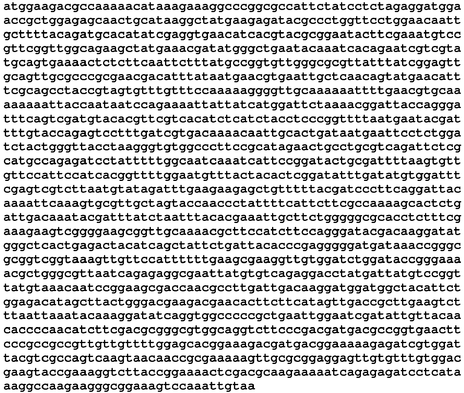
- <110> EADS Deutschland GmbH; Astrium GmbH
- <120> Detektionsverfahren zur Detektion von Bakterien, verfahren zur
Herstellung von Fusionsproteinen und Fusionsprotein - <160> 8
- <210> SEQ ID NO 8
<211> 550
<212> DNA
<213> unbekannt - <400>


- 10
- bacterium
- 12
- bacteriophage
- 14
- antibody
- 16
- bacteriophages
- 18
- bacterial surface
- 19
- enrichment structure
- 19a
- surface structure
- 20
- magnetic bead
- 22
- biotin
- 24
- streptavidin
- 25
- Biotin-streptavidin linker
- 26
- Nickel Bead
- 28
- Polyhistidine tag
- 29
- Metal chelate polyhistidine tag linker
- 30
- non-magnetic bead
- 31
- Ni-NTA
- 31a
- chromatography column
- 31b
- Sepharose
- 31c
- microtiter plate
- 31d
- plastic
- 31e
- filter material
- 31f
- Glass
- 31g
- metal
- 31h
- ceramics
- 31i
- Wood
- 31j
- rock
- 31k
- tissue
- 311
- nanoparticles
- 31m
- Semiconductor material
- 31n
- linker structure
- 32
- bacterial surface protein
- 34
- Metal chelate Bead
- 36
- metal chelate
- 37
- bacteria family
- 38
- sepharose
- 40
- material surface
- 42
- detection protein
- 43
- Detecting protein surface
- 44
- mark
- 45
- marker molecule
- 46
- Protein A
- 48
- Protein G
- 49
- fluorescent molecule
- 50
- fusion protein
- 52
- enzyme
- 54
- marker protein
- 56
- left protein
- 58
- Washing Day
- 60
- Protease cleavage site
- 62
- N-terminus
- 64
- C-terminus
- 66
- Protease recognition sequence
- 67
- protein
- 68
- human caspase
- 70
- Caspase-3
- 71
- luciferase
- 72
- fluorescent protein
- 74
- polymerase
- 75
- Taq polymerase
- 76
- lytic protein
- 78
- lytic bacteriophage protein
- 80
- organism
- 81
- cell
- 81a
- protein extract
- 82
- F-pilus
- 83
- outer membrane
- 84
- TOLA protein
- 86
- periplasmic space
- 88
- Cytoplasmamembran
- 90
- restriction enzyme
- 91
- Fusion protein gene
- 92
- Bacteriophage gene
- 94
- Marker protein gene
- 96
- Linker protein gene
- 98
- codon
- 100
- start codon
- N1
- protein domain
- N2
- protein domain
- L1
- protein domain
- L2
- protein domain
- TD
- Transmembrane domain
- M13
- bacteriophages
- G3P
- protein
- g3p
- gene
- M
- DNA-marker
- e
- elution
- EmGFP
- protein
- <110> EADS Germany GmbH; Astrium GmbH
- <120> Detection method for the detection of bacteria, method for
Production of fusion proteins and fusion protein - <160> 8
- <210>
SEQ ID NO 1
<211> 1275
<212> DNA
<213> Bacteriophage M13 - <400>
- <110> EADS Germany GmbH; Astrium GmbH
- <120> Detection method for the detection of bacteria, method for
Production of fusion proteins and fusion protein - <160> 8
- <210>
SEQ ID NO 2
<211> 424
<212> DNA
<213> Bacteriophage M13 - <400>
- <110> EADS Germany GmbH; Astrium GmbH
- <120> Detection method for the detection of bacteria, method for
Production of fusion proteins and fusion protein - <160> 8
- <210>
SEQ ID NO 3
<211> 720
<212> DNA
<213> unknown - <400>
- <110> EADS Germany GmbH; Astrium GmbH
- <120> Detection method for the detection of bacteria, method for
Production of fusion proteins and fusion protein - <160> 8
- <210> SEQ ID NO 4
<211> 240
<212> DNA
<213> unknown - <400>
- <110> EADS Germany GmbH; Astrium GmbH
- <120> Detection method for the detection of bacteria, method for
Production of fusion proteins and fusion protein - <160> 8
- <210> SEQ ID NO 5
<211> 834
<212> DNA
<213> unknown - <400>
- <110> EADS Germany GmbH; Astrium GmbH
- <120> Detection method for the detection of bacteria, method for
Production of fusion proteins and fusion protein - <160> 8
- <210> SEQ ID NO 6
<211> 277
<212> DNA
<213> unknown - <400>
- <110> EADS Germany GmbH; Astrium GmbH
- <120> Detection method for the detection of bacteria, method for
Production of fusion proteins and fusion protein - <160> 8
- <210>
SEQ ID NO 7
<211> 1653
<212> DNA
<213> unknown - <400>
- <110> EADS Germany GmbH; Astrium GmbH
- <120> Detection method for the detection of bacteria, method for
Production of fusion proteins and fusion protein - <160> 8
- <210> SEQ ID NO 8
<211> 550
<212> DNA
<213> unknown - <400>
Claims (15)
dadurch gekennzeichnet, dass ein freies bakterielles Oberflächenprotein (32) oder ein gebundenes, insbesondere ein an ein Bakterium (10) gebundenes, bakterielles Oberflächenprotein (32) angereichert wird, wobei die Anreicherungsstruktur (19) insbesondere an einer immobilen oder einer mobilen Oberflächenstruktur (19a) bereitgestellt wird.Method according to claim 1,
characterized in that a free bacterial surface protein (32) or a bound, in particular a bacterial surface protein (32) bound to a bacterium (10) is enriched, wherein the enrichment structure (19) in particular on an immobile or a mobile surface structure (19a) provided.
dadurch gekennzeichnet, dass die Anreicherungsstruktur (19) über eine Linker-Struktur (31 n), insbesondere über einen Biotin-Streptavidin-Linker (25) oder einen Metallchelat-Polyhistidin-Tag-Linker (29), an der Oberflächenstruktur (19a) immobilisiert wird, wobei als Oberflächenstruktur (19a) insbesondere magnetische Beads (20), mehr insbesondere Ni-Beads (26), nichtmagnetische Beads (30), insbesondere Ni-NTA (31), Chromatographiesäulenmaterialien (31 a), Sepharosen (31 b), Mikrotiterplatten (31 c), Kunststoffe (31 d), Filteroberflächen (31 e), Glas (31 f), Metall (31 g), Keramik (31 h), Holz (31 i), Gestein (31j), Gewebe (31 k), Nanopartikel (31l) und/oder Halbleitermaterialien (31 m) verwendet werden.Method according to claim 2,
characterized in that the enrichment structure (19) via a linker structure (31 n), in particular via a biotin-streptavidin linker (25) or a metal chelate polyhistidine tag linker (29), immobilized on the surface structure (19 a) in particular magnetic beads (20), more particularly Ni beads (26), non-magnetic beads (30), in particular Ni-NTA (31), chromatography column materials (31 a), Sepharosen (31 b), Microtiter plates (31 c), plastics (31 d), filter surfaces (31 e), glass (31 f), metal (31 g), ceramics (31 h), wood (31 i), rock (31 j), fabric (31 k), nanoparticles (31l) and / or semiconductor materials (31 m).
dadurch gekennzeichnet, dass das Bakteriophagenprotein (12) als freies Bakteriophagenprotein (12) oder als an einem Bakteriophagen (16) gebundenes Bakteriophagenprotein (12) bereitgestellt wird, wobei insbesondere ein für ein Bakterium (10) oder für eine Bakterienfamilie (37) selektives Bakteriophagenprotein (12) verwendet wird.Method according to one of claims 1 to 3,
characterized in that the bacteriophage protein (12) is provided as a free bacteriophage protein (12) or as a bacteriophage protein (12) bound to a bacteriophage (16), in particular a bacteriophage protein (B) selective for a bacterium (10) or for a bacterial family (37). 12) is used.
dadurch gekennzeichnet, dass sowohl das Anreichern als auch die Detektionsreaktion durch spezifische Protein-Protein-Interaktion mit einem bakteriellen Oberflächenprotein (32) erfolgt, wobei insbesondere als Detektionsprotein (42) das Bakteriophagenprotein (12) und als Anreicherungsstruktur (19) ein Antikörper (14) verwendet wird oder wobei insbesondere als Detektionsprotein (42) ein Antikörper (14) und als Anreicherungsstruktur (19) das Bakteriophagenprotein (12) verwendet wird.Method according to one of claims 1 to 4,
characterized in that both the enrichment and the detection reaction by specific protein-protein interaction with a bacterial surface protein (32) takes place, in particular as a detection protein (42) the bacteriophage protein (12) and as an enrichment structure (19) an antibody (14) is used or wherein in particular as a detection protein (42) an antibody (14) and as an enrichment structure (19) the bacteriophage protein (12) is used.
dadurch gekennzeichnet, dass unmittelbar an eine Detektionsproteinoberfläche (43) ein Markermolekül (45), insbesondere ein Enzym (52) oder ein Fluoreszenzmolekül (49), angebunden wird.Method according to one of claims 1 to 5,
characterized in that a marker molecule (45), in particular an enzyme (52) or a fluorescence molecule (49), is attached directly to a detection protein surface (43).
dadurch gekennzeichnet, dass als Detektionsprotein (42) ein Fusionsprotein (50) verwendet wird, das durch Verbinden eines Bakteriophagenproteins (12) mit einem Markerprotein (54) über ein Linkerprotein (56) hergestellt wird, wobei das Fusionsprotein (50) insbesondere über eine Proteaseschnittstelle (60) mit einem Reinigungs-Tag (58) gekoppelt wird.Method according to one of claims 1 to 5,
characterized in that the detection protein (42) used is a fusion protein (50) which is produced by linking a bacteriophage protein (12) with a marker protein (54) via a linker protein (56), the fusion protein (50) in particular via a protease cleavage site (60) is coupled to a cleaning tag (58).
dadurch gekennzeichnet, dass das Bakteriophagenprotein (12) durch rekombinante Expression hergestellt wird oder dass das Fusionsprotein (50) durch rekombinante Expression hergestellt wird.Method according to one of claims 1 to 6 or according to claim 7,
characterized in that the bacteriophage protein (12) is produced by recombinant expression or that the fusion protein (50) is produced by recombinant expression.
dadurch gekennzeichnet, dass vor Schritt a) ein Proteinherstellungsschritt mit den folgenden Schritten durchgeführt wird:
characterized in that before step a) a protein production step is carried out with the following steps:
dadurch gekennzeichnet, dass als Markerprotein-Gene (94) Gene zur Bildung von Markerproteinen (54), die aus der Gruppe Enzyme (52), Proteine (67), fluoreszierende Proteine (67), humane Caspasen (68), insbesondere Caspase-3 (70), biolumineszierende Enzyme (52), insbesondere Luziferase (71), lytische Proteine (76) und/oder Polymerasen (74), insbesondere Tag-Polymerase (75), verwendet werden.Method according to claim 10,
characterized in that as marker protein genes (94) genes for the formation of marker proteins (54) from the group enzymes (52), proteins (67), fluorescent proteins (67), human caspases (68), in particular caspase-3 (70), bioluminescent enzymes (52), especially luciferase (71), lytic Proteins (76) and / or polymerases (74), in particular Tag polymerase (75) can be used.
dadurch gekennzeichnet, dass in Schritt A) die Ligation seriell durchgeführt wird, wobei insbesondere zuerst das Bakteriophagenprotein-Gen (92), insbesondere N-terminal, und dann das Markerprotein-Gen (94), insbesondere C-terminal, mit dem Linkerprotein-Gen (96) ligiert wird.Method according to one of claims 10 or 11,
characterized in that in step A) the ligation is performed serially, in particular first the bacteriophage protein gene (92), in particular N-terminal, and then the marker protein gene (94), in particular C-terminal, with the linker protein gene (96) is ligated.
dadurch gekennzeichnet, dass für die Ligation unterschiedliche Restriktionsenzyme (90) für das Bakteriophagenprotein-Gen (92) und das Markerprotein-Gen (94) verwendet werden, insbesondere BsmBI als 5'-Ende und Acc65I als 3'-Ende für das Bakteriophagenprotein-Gen (92) und Sall als 5'-Ende und Notl als 3'-Ende für das Markerprotein-Gen (94).Method according to one of claims 10 to 12,
characterized in that different restriction enzymes (90) for the bacteriophage protein gene (92) and the marker protein gene (94) are used for the ligation, in particular BsmBI as the 5 'end and Acc65I as the 3' end for the bacteriophage protein gene (92) and SalI at the 5 'end and NotI at the 3' end for the marker protein gene (94).
dadurch gekennzeichnet, dass ein Linkerprotein (56) erzeugt wird, das eine freie dreidimensionale Bewegung des Bakteriophagenproteins (12) und des Markerproteins (54) zueinander ermöglicht, wobei das Linkerprotein (56) insbesondere eine Länge von neun Codons (98) aufweist und mehr insbesondere frei von Stopcodons ist.Method according to one of claims 10 to 13,
characterized in that a linker protein (56) is generated which allows a free three-dimensional movement of the bacteriophage protein (12) and the marker protein (54) to each other, wherein the linker protein (56) in particular has a length of nine codons (98), and more particularly is free from stop codons.
Applications Claiming Priority (1)
| Application Number | Priority Date | Filing Date | Title |
|---|---|---|---|
| DE102013106462.0A DE102013106462B3 (en) | 2013-06-20 | 2013-06-20 | Detection method for the detection of bacteria, methods for the production of fusion proteins and fusion protein |
Publications (4)
| Publication Number | Publication Date |
|---|---|
| EP2816355A2 true EP2816355A2 (en) | 2014-12-24 |
| EP2816355A3 EP2816355A3 (en) | 2015-04-15 |
| EP2816355B1 EP2816355B1 (en) | 2017-08-09 |
| EP2816355B8 EP2816355B8 (en) | 2017-11-08 |
Family
ID=50976526
Family Applications (1)
| Application Number | Title | Priority Date | Filing Date |
|---|---|---|---|
| EP14173356.8A Active EP2816355B8 (en) | 2013-06-20 | 2014-06-20 | Detection method for detection of bacteria, method for the preparation of fusion proteins and fusion protein |
Country Status (2)
| Country | Link |
|---|---|
| EP (1) | EP2816355B8 (en) |
| DE (1) | DE102013106462B3 (en) |
Cited By (1)
| Publication number | Priority date | Publication date | Assignee | Title |
|---|---|---|---|---|
| CN115282934A (en) * | 2022-08-02 | 2022-11-04 | 杭州博岳生物技术有限公司 | Nano antibody fluorescent affinity column and method for purifying HbeAg antigen by using same |
Families Citing this family (2)
| Publication number | Priority date | Publication date | Assignee | Title |
|---|---|---|---|---|
| DE102015121034B4 (en) | 2015-12-03 | 2022-06-23 | Airbus Defence and Space GmbH | Process and device for the enrichment of biological particles |
| DE102015121035A1 (en) | 2015-12-03 | 2017-06-08 | Airbus Defence and Space GmbH | Method for the detection of coliform bacteria |
Family Cites Families (12)
| Publication number | Priority date | Publication date | Assignee | Title |
|---|---|---|---|---|
| AU746454B2 (en) * | 1998-03-02 | 2002-05-02 | Massachusetts Institute Of Technology | Poly zinc finger proteins with improved linkers |
| DE19837751A1 (en) * | 1998-08-20 | 2000-02-24 | Siegfried Scherer | Labeling, detecting, immobilizing and enriching target cells by binding cell wall binding domains of enzymes or proteins to the cell wall or cell membrane of the target cells |
| ES2353268T3 (en) * | 1999-07-02 | 2011-02-28 | Morphosys Ag | GENERATION OF SPECIFIC UNION ELEMENTS THAT JOIN (POLI) PEPTIDES CODIFIED BY FRAGMENTS OF GENOMIC DNA OR EST. |
| JP5339658B2 (en) * | 1999-07-30 | 2013-11-13 | ビオメリュー・ソシエテ・アノニム | Detection and identification of bacterial strains |
| DE10129815A1 (en) * | 2001-06-24 | 2003-01-09 | Profos Ag | Process for the purification of bacterial cells and cell components |
| US20040197833A1 (en) * | 2003-04-03 | 2004-10-07 | Danisco A/S | Method for the enrichment of target cells by use of CBDs |
| US7329725B1 (en) * | 2003-10-29 | 2008-02-12 | Nastech Pharmaceutical Company Inc. | Phage displayed Trp cage ligands |
| US20060068421A1 (en) * | 2004-08-05 | 2006-03-30 | Biosite, Inc. | Compositions and methods for phage display of polypeptides |
| DE102006009709A1 (en) * | 2006-03-02 | 2006-10-12 | Müller, Merold, Dr. | Method for detecting microorganisms without a culturing step, useful particularly for detecting potential germ warfare agents, uses a cantilever coated with specific binding material for the target organism |
| CA2704494A1 (en) * | 2007-11-02 | 2009-05-14 | The Scripps Research Institute | Directed evolution using proteins comprising unnatural amino acids |
| WO2010036860A2 (en) * | 2008-09-26 | 2010-04-01 | Wyeth Llc | Compatible display vector systems |
| GB0903316D0 (en) * | 2009-02-26 | 2009-04-08 | Affitech As | Method |
-
2013
- 2013-06-20 DE DE102013106462.0A patent/DE102013106462B3/en not_active Expired - Fee Related
-
2014
- 2014-06-20 EP EP14173356.8A patent/EP2816355B8/en active Active
Non-Patent Citations (1)
| Title |
|---|
| None |
Cited By (1)
| Publication number | Priority date | Publication date | Assignee | Title |
|---|---|---|---|---|
| CN115282934A (en) * | 2022-08-02 | 2022-11-04 | 杭州博岳生物技术有限公司 | Nano antibody fluorescent affinity column and method for purifying HbeAg antigen by using same |
Also Published As
| Publication number | Publication date |
|---|---|
| EP2816355B8 (en) | 2017-11-08 |
| EP2816355A3 (en) | 2015-04-15 |
| EP2816355B1 (en) | 2017-08-09 |
| DE102013106462B3 (en) | 2014-10-09 |
Similar Documents
| Publication | Publication Date | Title |
|---|---|---|
| EP1675623B1 (en) | Ubiquitin or gamma-crystalline conjugates for use in therapy, diagnosis and chromatography | |
| DE112006003608T5 (en) | A method of presenting target proteins at the cell surface using Bacillus Anthracis Exosporium | |
| DE60305643T2 (en) | PROTEIN MARKER OF A BIOTINYLATION DOMAIN CONTAINS AND USES TO INCREASE THE SOLUBILITY AND DETERMINE THE FOLDING STATUS | |
| DE212012000234U1 (en) | Modified cascade ribonucleoproteins and applications thereof | |
| EP1356080B1 (en) | Detection and identification of groups of bacteria | |
| CN105177110A (en) | Detection method of nucleic acid | |
| DE69333667T2 (en) | Modified proteins containing controllable intermediate sequences and methods for their preparation | |
| EP2816355B1 (en) | Detection method for detection of bacteria, method for the preparation of fusion proteins and fusion protein | |
| EP1619208B1 (en) | Chaperonine-target protein complex, method of producing the same, method of stabilizing target protein, method of immobilizing target protein, method of analyzing the structure of target protein, sustained-release preparation and method of producing antibody against target protein | |
| Chen et al. | A split GAL4 RUBY assay for visual in planta detection of protein–protein interactions | |
| EP0547200A1 (en) | Recombinant antibodies at the surface of e. coli | |
| Chan et al. | Construction of inducible expression vector pDNLS-22n containing P. aeruginosa PAO1 ChiC | |
| EP1147419B1 (en) | Labeling, immobilisation, enrichment, purification and detection of cells using specific domains that bind to the cell walls (cbd) of proteins binding to the drawn cell walls of viruses, bacteria or eukaryotic cells | |
| DE202019005825U1 (en) | Biological synthesis of amino acid chains to produce peptides and proteins | |
| CA2785359C (en) | Protein display | |
| CN109880840B (en) | In vivo biotinylation labeling system for recombinant protein escherichia coli | |
| KR101896014B1 (en) | Bacteriophage-derived spore binding polypeptide and bioprobe for detection of Bacillus cereus spores | |
| Walter et al. | Characteristics of the surface-located carbohydrate-binding protein CbpC from Streptomyces coelicolor A3 (2) | |
| DE69937453T2 (en) | PROCESS FOR THE ISOLATION AND CHARACTERIZATION OF POTENTIAL FUNCTIONS BASED ON A BIOLOGICAL SAMPLE CONTAINING NUCLEIC ACIDS | |
| EP1069136B1 (en) | Method for the recombinant production of ribonucleoproteins | |
| DE69333106T2 (en) | polypeptide production | |
| AT511130A2 (en) | Polypeptide material with flexible pore properties | |
| DE102015108849A1 (en) | Method for protein immobilization using modified DNase domains | |
| DE102013013609A1 (en) | Autodisplay of an active lipase from Burkholderia cepacia on microorganisms | |
| DE102005011579B4 (en) | Affinity tags for protein purification, its production and use, and methods for purifying a protein |
Legal Events
| Date | Code | Title | Description |
|---|---|---|---|
| PUAI | Public reference made under article 153(3) epc to a published international application that has entered the european phase |
Free format text: ORIGINAL CODE: 0009012 |
|
| 17P | Request for examination filed |
Effective date: 20140620 |
|
| AK | Designated contracting states |
Kind code of ref document: A2 Designated state(s): AL AT BE BG CH CY CZ DE DK EE ES FI FR GB GR HR HU IE IS IT LI LT LU LV MC MK MT NL NO PL PT RO RS SE SI SK SM TR |
|
| AX | Request for extension of the european patent |
Extension state: BA ME |
|
| RIC1 | Information provided on ipc code assigned before grant |
Ipc: C07K 14/005 20060101ALI20141121BHEP Ipc: C07K 14/00 20060101ALI20141121BHEP Ipc: G01N 33/566 20060101ALI20141121BHEP Ipc: C12N 15/62 20060101ALI20141121BHEP Ipc: G01N 33/569 20060101AFI20141121BHEP |
|
| PUAL | Search report despatched |
Free format text: ORIGINAL CODE: 0009013 |
|
| AK | Designated contracting states |
Kind code of ref document: A3 Designated state(s): AL AT BE BG CH CY CZ DE DK EE ES FI FR GB GR HR HU IE IS IT LI LT LU LV MC MK MT NL NO PL PT RO RS SE SI SK SM TR |
|
| AX | Request for extension of the european patent |
Extension state: BA ME |
|
| RIC1 | Information provided on ipc code assigned before grant |
Ipc: C12N 15/62 20060101ALI20150312BHEP Ipc: G01N 33/569 20060101AFI20150312BHEP Ipc: G01N 33/566 20060101ALI20150312BHEP Ipc: C07K 14/005 20060101ALI20150312BHEP Ipc: C07K 14/00 20060101ALI20150312BHEP |
|
| R17P | Request for examination filed (corrected) |
Effective date: 20151009 |
|
| RBV | Designated contracting states (corrected) |
Designated state(s): AL AT BE BG CH CY CZ DE DK EE ES FI FR GB GR HR HU IE IS IT LI LT LU LV MC MK MT NL NO PL PT RO RS SE SI SK SM TR |
|
| 17Q | First examination report despatched |
Effective date: 20160408 |
|
| RIN1 | Information on inventor provided before grant (corrected) |
Inventor name: REIDT, ULRICH Inventor name: HUMMEL, THOMAS Inventor name: FRIEDBERGER, ALOIS |
|
| GRAP | Despatch of communication of intention to grant a patent |
Free format text: ORIGINAL CODE: EPIDOSNIGR1 |
|
| INTG | Intention to grant announced |
Effective date: 20170209 |
|
| GRAS | Grant fee paid |
Free format text: ORIGINAL CODE: EPIDOSNIGR3 |
|
| GRAA | (expected) grant |
Free format text: ORIGINAL CODE: 0009210 |
|
| AK | Designated contracting states |
Kind code of ref document: B1 Designated state(s): AL AT BE BG CH CY CZ DE DK EE ES FI FR GB GR HR HU IE IS IT LI LT LU LV MC MK MT NL NO PL PT RO RS SE SI SK SM TR |
|
| REG | Reference to a national code |
Ref country code: GB Ref legal event code: FG4D Free format text: NOT ENGLISH |
|
| REG | Reference to a national code |
Ref country code: CH Ref legal event code: EP Ref country code: AT Ref legal event code: REF Ref document number: 917407 Country of ref document: AT Kind code of ref document: T Effective date: 20170815 |
|
| REG | Reference to a national code |
Ref country code: IE Ref legal event code: FG4D Free format text: LANGUAGE OF EP DOCUMENT: GERMAN |
|
| REG | Reference to a national code |
Ref country code: DE Ref legal event code: R096 Ref document number: 502014004893 Country of ref document: DE |
|
| RAP2 | Party data changed (patent owner data changed or rights of a patent transferred) |
Owner name: AIRBUS DEFENCE AND SPACE GMBH |
|
| REG | Reference to a national code |
Ref country code: NL Ref legal event code: FP |
|
| REG | Reference to a national code |
Ref country code: LT Ref legal event code: MG4D |
|
| PG25 | Lapsed in a contracting state [announced via postgrant information from national office to epo] |
Ref country code: NO Free format text: LAPSE BECAUSE OF FAILURE TO SUBMIT A TRANSLATION OF THE DESCRIPTION OR TO PAY THE FEE WITHIN THE PRESCRIBED TIME-LIMIT Effective date: 20171109 Ref country code: HR Free format text: LAPSE BECAUSE OF FAILURE TO SUBMIT A TRANSLATION OF THE DESCRIPTION OR TO PAY THE FEE WITHIN THE PRESCRIBED TIME-LIMIT Effective date: 20170809 Ref country code: LT Free format text: LAPSE BECAUSE OF FAILURE TO SUBMIT A TRANSLATION OF THE DESCRIPTION OR TO PAY THE FEE WITHIN THE PRESCRIBED TIME-LIMIT Effective date: 20170809 Ref country code: FI Free format text: LAPSE BECAUSE OF FAILURE TO SUBMIT A TRANSLATION OF THE DESCRIPTION OR TO PAY THE FEE WITHIN THE PRESCRIBED TIME-LIMIT Effective date: 20170809 Ref country code: SE Free format text: LAPSE BECAUSE OF FAILURE TO SUBMIT A TRANSLATION OF THE DESCRIPTION OR TO PAY THE FEE WITHIN THE PRESCRIBED TIME-LIMIT Effective date: 20170809 |
|
| PG25 | Lapsed in a contracting state [announced via postgrant information from national office to epo] |
Ref country code: RS Free format text: LAPSE BECAUSE OF FAILURE TO SUBMIT A TRANSLATION OF THE DESCRIPTION OR TO PAY THE FEE WITHIN THE PRESCRIBED TIME-LIMIT Effective date: 20170809 Ref country code: IS Free format text: LAPSE BECAUSE OF FAILURE TO SUBMIT A TRANSLATION OF THE DESCRIPTION OR TO PAY THE FEE WITHIN THE PRESCRIBED TIME-LIMIT Effective date: 20171209 Ref country code: LV Free format text: LAPSE BECAUSE OF FAILURE TO SUBMIT A TRANSLATION OF THE DESCRIPTION OR TO PAY THE FEE WITHIN THE PRESCRIBED TIME-LIMIT Effective date: 20170809 Ref country code: BG Free format text: LAPSE BECAUSE OF FAILURE TO SUBMIT A TRANSLATION OF THE DESCRIPTION OR TO PAY THE FEE WITHIN THE PRESCRIBED TIME-LIMIT Effective date: 20171109 Ref country code: GR Free format text: LAPSE BECAUSE OF FAILURE TO SUBMIT A TRANSLATION OF THE DESCRIPTION OR TO PAY THE FEE WITHIN THE PRESCRIBED TIME-LIMIT Effective date: 20171110 Ref country code: PL Free format text: LAPSE BECAUSE OF FAILURE TO SUBMIT A TRANSLATION OF THE DESCRIPTION OR TO PAY THE FEE WITHIN THE PRESCRIBED TIME-LIMIT Effective date: 20170809 Ref country code: ES Free format text: LAPSE BECAUSE OF FAILURE TO SUBMIT A TRANSLATION OF THE DESCRIPTION OR TO PAY THE FEE WITHIN THE PRESCRIBED TIME-LIMIT Effective date: 20170809 |
|
| PG25 | Lapsed in a contracting state [announced via postgrant information from national office to epo] |
Ref country code: DK Free format text: LAPSE BECAUSE OF FAILURE TO SUBMIT A TRANSLATION OF THE DESCRIPTION OR TO PAY THE FEE WITHIN THE PRESCRIBED TIME-LIMIT Effective date: 20170809 Ref country code: CZ Free format text: LAPSE BECAUSE OF FAILURE TO SUBMIT A TRANSLATION OF THE DESCRIPTION OR TO PAY THE FEE WITHIN THE PRESCRIBED TIME-LIMIT Effective date: 20170809 Ref country code: RO Free format text: LAPSE BECAUSE OF FAILURE TO SUBMIT A TRANSLATION OF THE DESCRIPTION OR TO PAY THE FEE WITHIN THE PRESCRIBED TIME-LIMIT Effective date: 20170809 |
|
| REG | Reference to a national code |
Ref country code: DE Ref legal event code: R097 Ref document number: 502014004893 Country of ref document: DE |
|
| REG | Reference to a national code |
Ref country code: DE Ref legal event code: R082 Ref document number: 502014004893 Country of ref document: DE |
|
| PG25 | Lapsed in a contracting state [announced via postgrant information from national office to epo] |
Ref country code: SK Free format text: LAPSE BECAUSE OF FAILURE TO SUBMIT A TRANSLATION OF THE DESCRIPTION OR TO PAY THE FEE WITHIN THE PRESCRIBED TIME-LIMIT Effective date: 20170809 Ref country code: IT Free format text: LAPSE BECAUSE OF FAILURE TO SUBMIT A TRANSLATION OF THE DESCRIPTION OR TO PAY THE FEE WITHIN THE PRESCRIBED TIME-LIMIT Effective date: 20170809 Ref country code: EE Free format text: LAPSE BECAUSE OF FAILURE TO SUBMIT A TRANSLATION OF THE DESCRIPTION OR TO PAY THE FEE WITHIN THE PRESCRIBED TIME-LIMIT Effective date: 20170809 Ref country code: SM Free format text: LAPSE BECAUSE OF FAILURE TO SUBMIT A TRANSLATION OF THE DESCRIPTION OR TO PAY THE FEE WITHIN THE PRESCRIBED TIME-LIMIT Effective date: 20170809 |
|
| PLBE | No opposition filed within time limit |
Free format text: ORIGINAL CODE: 0009261 |
|
| STAA | Information on the status of an ep patent application or granted ep patent |
Free format text: STATUS: NO OPPOSITION FILED WITHIN TIME LIMIT |
|
| REG | Reference to a national code |
Ref country code: FR Ref legal event code: PLFP Year of fee payment: 5 |
|
| 26N | No opposition filed |
Effective date: 20180511 |
|
| PG25 | Lapsed in a contracting state [announced via postgrant information from national office to epo] |
Ref country code: SI Free format text: LAPSE BECAUSE OF FAILURE TO SUBMIT A TRANSLATION OF THE DESCRIPTION OR TO PAY THE FEE WITHIN THE PRESCRIBED TIME-LIMIT Effective date: 20170809 |
|
| PG25 | Lapsed in a contracting state [announced via postgrant information from national office to epo] |
Ref country code: MT Free format text: LAPSE BECAUSE OF FAILURE TO SUBMIT A TRANSLATION OF THE DESCRIPTION OR TO PAY THE FEE WITHIN THE PRESCRIBED TIME-LIMIT Effective date: 20170809 |
|
| REG | Reference to a national code |
Ref country code: CH Ref legal event code: PL |
|
| REG | Reference to a national code |
Ref country code: BE Ref legal event code: MM Effective date: 20180630 |
|
| REG | Reference to a national code |
Ref country code: IE Ref legal event code: MM4A |
|
| PG25 | Lapsed in a contracting state [announced via postgrant information from national office to epo] |
Ref country code: MC Free format text: LAPSE BECAUSE OF FAILURE TO SUBMIT A TRANSLATION OF THE DESCRIPTION OR TO PAY THE FEE WITHIN THE PRESCRIBED TIME-LIMIT Effective date: 20170809 Ref country code: LU Free format text: LAPSE BECAUSE OF NON-PAYMENT OF DUE FEES Effective date: 20180620 |
|
| PG25 | Lapsed in a contracting state [announced via postgrant information from national office to epo] |
Ref country code: IE Free format text: LAPSE BECAUSE OF NON-PAYMENT OF DUE FEES Effective date: 20180620 Ref country code: LI Free format text: LAPSE BECAUSE OF NON-PAYMENT OF DUE FEES Effective date: 20180630 Ref country code: CH Free format text: LAPSE BECAUSE OF NON-PAYMENT OF DUE FEES Effective date: 20180630 |
|
| PG25 | Lapsed in a contracting state [announced via postgrant information from national office to epo] |
Ref country code: BE Free format text: LAPSE BECAUSE OF NON-PAYMENT OF DUE FEES Effective date: 20180630 |
|
| PG25 | Lapsed in a contracting state [announced via postgrant information from national office to epo] |
Ref country code: TR Free format text: LAPSE BECAUSE OF FAILURE TO SUBMIT A TRANSLATION OF THE DESCRIPTION OR TO PAY THE FEE WITHIN THE PRESCRIBED TIME-LIMIT Effective date: 20170809 |
|
| PG25 | Lapsed in a contracting state [announced via postgrant information from national office to epo] |
Ref country code: HU Free format text: LAPSE BECAUSE OF FAILURE TO SUBMIT A TRANSLATION OF THE DESCRIPTION OR TO PAY THE FEE WITHIN THE PRESCRIBED TIME-LIMIT; INVALID AB INITIO Effective date: 20140620 Ref country code: PT Free format text: LAPSE BECAUSE OF FAILURE TO SUBMIT A TRANSLATION OF THE DESCRIPTION OR TO PAY THE FEE WITHIN THE PRESCRIBED TIME-LIMIT Effective date: 20170809 |
|
| PG25 | Lapsed in a contracting state [announced via postgrant information from national office to epo] |
Ref country code: CY Free format text: LAPSE BECAUSE OF FAILURE TO SUBMIT A TRANSLATION OF THE DESCRIPTION OR TO PAY THE FEE WITHIN THE PRESCRIBED TIME-LIMIT Effective date: 20170809 Ref country code: MK Free format text: LAPSE BECAUSE OF NON-PAYMENT OF DUE FEES Effective date: 20170809 |
|
| PG25 | Lapsed in a contracting state [announced via postgrant information from national office to epo] |
Ref country code: AL Free format text: LAPSE BECAUSE OF FAILURE TO SUBMIT A TRANSLATION OF THE DESCRIPTION OR TO PAY THE FEE WITHIN THE PRESCRIBED TIME-LIMIT Effective date: 20170809 |
|
| REG | Reference to a national code |
Ref country code: AT Ref legal event code: MM01 Ref document number: 917407 Country of ref document: AT Kind code of ref document: T Effective date: 20190620 |
|
| PG25 | Lapsed in a contracting state [announced via postgrant information from national office to epo] |
Ref country code: AT Free format text: LAPSE BECAUSE OF NON-PAYMENT OF DUE FEES Effective date: 20190620 |
|
| PGFP | Annual fee paid to national office [announced via postgrant information from national office to epo] |
Ref country code: NL Payment date: 20230620 Year of fee payment: 10 Ref country code: FR Payment date: 20230628 Year of fee payment: 10 Ref country code: DE Payment date: 20230620 Year of fee payment: 10 |
|
| PGFP | Annual fee paid to national office [announced via postgrant information from national office to epo] |
Ref country code: GB Payment date: 20230622 Year of fee payment: 10 |