EP2787900B1 - Spinal cord devices for promoting axonal regeneration - Google Patents
Spinal cord devices for promoting axonal regeneration Download PDFInfo
- Publication number
- EP2787900B1 EP2787900B1 EP12815796.3A EP12815796A EP2787900B1 EP 2787900 B1 EP2787900 B1 EP 2787900B1 EP 12815796 A EP12815796 A EP 12815796A EP 2787900 B1 EP2787900 B1 EP 2787900B1
- Authority
- EP
- European Patent Office
- Prior art keywords
- spinal cord
- channels
- diameter
- devices
- range
- Prior art date
- Legal status (The legal status is an assumption and is not a legal conclusion. Google has not performed a legal analysis and makes no representation as to the accuracy of the status listed.)
- Not-in-force
Links
Images

Classifications
-
- A—HUMAN NECESSITIES
- A61—MEDICAL OR VETERINARY SCIENCE; HYGIENE
- A61F—FILTERS IMPLANTABLE INTO BLOOD VESSELS; PROSTHESES; DEVICES PROVIDING PATENCY TO, OR PREVENTING COLLAPSING OF, TUBULAR STRUCTURES OF THE BODY, e.g. STENTS; ORTHOPAEDIC, NURSING OR CONTRACEPTIVE DEVICES; FOMENTATION; TREATMENT OR PROTECTION OF EYES OR EARS; BANDAGES, DRESSINGS OR ABSORBENT PADS; FIRST-AID KITS
- A61F2/00—Filters implantable into blood vessels; Prostheses, i.e. artificial substitutes or replacements for parts of the body; Appliances for connecting them with the body; Devices providing patency to, or preventing collapsing of, tubular structures of the body, e.g. stents
- A61F2/02—Prostheses implantable into the body
- A61F2/30—Joints
- A61F2/44—Joints for the spine, e.g. vertebrae, spinal discs
- A61F2/4455—Joints for the spine, e.g. vertebrae, spinal discs for the fusion of spinal bodies, e.g. intervertebral fusion of adjacent spinal bodies, e.g. fusion cages
-
- A—HUMAN NECESSITIES
- A61—MEDICAL OR VETERINARY SCIENCE; HYGIENE
- A61B—DIAGNOSIS; SURGERY; IDENTIFICATION
- A61B17/00—Surgical instruments, devices or methods, e.g. tourniquets
- A61B17/11—Surgical instruments, devices or methods, e.g. tourniquets for performing anastomosis; Buttons for anastomosis
- A61B17/1128—Surgical instruments, devices or methods, e.g. tourniquets for performing anastomosis; Buttons for anastomosis of nerves
-
- A—HUMAN NECESSITIES
- A61—MEDICAL OR VETERINARY SCIENCE; HYGIENE
- A61F—FILTERS IMPLANTABLE INTO BLOOD VESSELS; PROSTHESES; DEVICES PROVIDING PATENCY TO, OR PREVENTING COLLAPSING OF, TUBULAR STRUCTURES OF THE BODY, e.g. STENTS; ORTHOPAEDIC, NURSING OR CONTRACEPTIVE DEVICES; FOMENTATION; TREATMENT OR PROTECTION OF EYES OR EARS; BANDAGES, DRESSINGS OR ABSORBENT PADS; FIRST-AID KITS
- A61F2/00—Filters implantable into blood vessels; Prostheses, i.e. artificial substitutes or replacements for parts of the body; Appliances for connecting them with the body; Devices providing patency to, or preventing collapsing of, tubular structures of the body, e.g. stents
- A61F2/02—Prostheses implantable into the body
- A61F2/30—Joints
- A61F2/30767—Special external or bone-contacting surface, e.g. coating for improving bone ingrowth
- A61F2/30771—Special external or bone-contacting surface, e.g. coating for improving bone ingrowth applied in original prostheses, e.g. holes or grooves
-
- A—HUMAN NECESSITIES
- A61—MEDICAL OR VETERINARY SCIENCE; HYGIENE
- A61F—FILTERS IMPLANTABLE INTO BLOOD VESSELS; PROSTHESES; DEVICES PROVIDING PATENCY TO, OR PREVENTING COLLAPSING OF, TUBULAR STRUCTURES OF THE BODY, e.g. STENTS; ORTHOPAEDIC, NURSING OR CONTRACEPTIVE DEVICES; FOMENTATION; TREATMENT OR PROTECTION OF EYES OR EARS; BANDAGES, DRESSINGS OR ABSORBENT PADS; FIRST-AID KITS
- A61F2/00—Filters implantable into blood vessels; Prostheses, i.e. artificial substitutes or replacements for parts of the body; Appliances for connecting them with the body; Devices providing patency to, or preventing collapsing of, tubular structures of the body, e.g. stents
- A61F2/02—Prostheses implantable into the body
- A61F2/30—Joints
- A61F2/46—Special tools or methods for implanting or extracting artificial joints, accessories, bone grafts or substitutes, or particular adaptations therefor
- A61F2/4657—Measuring instruments used for implanting artificial joints
-
- A—HUMAN NECESSITIES
- A61—MEDICAL OR VETERINARY SCIENCE; HYGIENE
- A61L—METHODS OR APPARATUS FOR STERILISING MATERIALS OR OBJECTS IN GENERAL; DISINFECTION, STERILISATION OR DEODORISATION OF AIR; CHEMICAL ASPECTS OF BANDAGES, DRESSINGS, ABSORBENT PADS OR SURGICAL ARTICLES; MATERIALS FOR BANDAGES, DRESSINGS, ABSORBENT PADS OR SURGICAL ARTICLES
- A61L27/00—Materials for grafts or prostheses or for coating grafts or prostheses
- A61L27/50—Materials characterised by their function or physical properties, e.g. injectable or lubricating compositions, shape-memory materials, surface modified materials
- A61L27/54—Biologically active materials, e.g. therapeutic substances
-
- A—HUMAN NECESSITIES
- A61—MEDICAL OR VETERINARY SCIENCE; HYGIENE
- A61L—METHODS OR APPARATUS FOR STERILISING MATERIALS OR OBJECTS IN GENERAL; DISINFECTION, STERILISATION OR DEODORISATION OF AIR; CHEMICAL ASPECTS OF BANDAGES, DRESSINGS, ABSORBENT PADS OR SURGICAL ARTICLES; MATERIALS FOR BANDAGES, DRESSINGS, ABSORBENT PADS OR SURGICAL ARTICLES
- A61L27/00—Materials for grafts or prostheses or for coating grafts or prostheses
- A61L27/50—Materials characterised by their function or physical properties, e.g. injectable or lubricating compositions, shape-memory materials, surface modified materials
- A61L27/58—Materials at least partially resorbable by the body
-
- A—HUMAN NECESSITIES
- A61—MEDICAL OR VETERINARY SCIENCE; HYGIENE
- A61B—DIAGNOSIS; SURGERY; IDENTIFICATION
- A61B17/00—Surgical instruments, devices or methods, e.g. tourniquets
- A61B2017/00004—(bio)absorbable, (bio)resorbable, resorptive
-
- A—HUMAN NECESSITIES
- A61—MEDICAL OR VETERINARY SCIENCE; HYGIENE
- A61F—FILTERS IMPLANTABLE INTO BLOOD VESSELS; PROSTHESES; DEVICES PROVIDING PATENCY TO, OR PREVENTING COLLAPSING OF, TUBULAR STRUCTURES OF THE BODY, e.g. STENTS; ORTHOPAEDIC, NURSING OR CONTRACEPTIVE DEVICES; FOMENTATION; TREATMENT OR PROTECTION OF EYES OR EARS; BANDAGES, DRESSINGS OR ABSORBENT PADS; FIRST-AID KITS
- A61F2/00—Filters implantable into blood vessels; Prostheses, i.e. artificial substitutes or replacements for parts of the body; Appliances for connecting them with the body; Devices providing patency to, or preventing collapsing of, tubular structures of the body, e.g. stents
- A61F2/02—Prostheses implantable into the body
- A61F2/30—Joints
- A61F2002/30001—Additional features of subject-matter classified in A61F2/28, A61F2/30 and subgroups thereof
- A61F2002/30316—The prosthesis having different structural features at different locations within the same prosthesis; Connections between prosthetic parts; Special structural features of bone or joint prostheses not otherwise provided for
- A61F2002/30535—Special structural features of bone or joint prostheses not otherwise provided for
- A61F2002/30604—Special structural features of bone or joint prostheses not otherwise provided for modular
- A61F2002/30616—Sets comprising a plurality of prosthetic parts of different sizes or orientations
-
- A—HUMAN NECESSITIES
- A61—MEDICAL OR VETERINARY SCIENCE; HYGIENE
- A61F—FILTERS IMPLANTABLE INTO BLOOD VESSELS; PROSTHESES; DEVICES PROVIDING PATENCY TO, OR PREVENTING COLLAPSING OF, TUBULAR STRUCTURES OF THE BODY, e.g. STENTS; ORTHOPAEDIC, NURSING OR CONTRACEPTIVE DEVICES; FOMENTATION; TREATMENT OR PROTECTION OF EYES OR EARS; BANDAGES, DRESSINGS OR ABSORBENT PADS; FIRST-AID KITS
- A61F2/00—Filters implantable into blood vessels; Prostheses, i.e. artificial substitutes or replacements for parts of the body; Appliances for connecting them with the body; Devices providing patency to, or preventing collapsing of, tubular structures of the body, e.g. stents
- A61F2/02—Prostheses implantable into the body
- A61F2/30—Joints
- A61F2/30767—Special external or bone-contacting surface, e.g. coating for improving bone ingrowth
- A61F2/30771—Special external or bone-contacting surface, e.g. coating for improving bone ingrowth applied in original prostheses, e.g. holes or grooves
- A61F2002/30772—Apertures or holes, e.g. of circular cross section
-
- A—HUMAN NECESSITIES
- A61—MEDICAL OR VETERINARY SCIENCE; HYGIENE
- A61F—FILTERS IMPLANTABLE INTO BLOOD VESSELS; PROSTHESES; DEVICES PROVIDING PATENCY TO, OR PREVENTING COLLAPSING OF, TUBULAR STRUCTURES OF THE BODY, e.g. STENTS; ORTHOPAEDIC, NURSING OR CONTRACEPTIVE DEVICES; FOMENTATION; TREATMENT OR PROTECTION OF EYES OR EARS; BANDAGES, DRESSINGS OR ABSORBENT PADS; FIRST-AID KITS
- A61F2/00—Filters implantable into blood vessels; Prostheses, i.e. artificial substitutes or replacements for parts of the body; Appliances for connecting them with the body; Devices providing patency to, or preventing collapsing of, tubular structures of the body, e.g. stents
- A61F2/02—Prostheses implantable into the body
- A61F2/30—Joints
- A61F2/46—Special tools or methods for implanting or extracting artificial joints, accessories, bone grafts or substitutes, or particular adaptations therefor
- A61F2/4657—Measuring instruments used for implanting artificial joints
- A61F2002/4658—Measuring instruments used for implanting artificial joints for measuring dimensions, e.g. length
- A61F2002/4659—Measuring instruments used for implanting artificial joints for measuring dimensions, e.g. length for measuring a diameter
-
- A—HUMAN NECESSITIES
- A61—MEDICAL OR VETERINARY SCIENCE; HYGIENE
- A61F—FILTERS IMPLANTABLE INTO BLOOD VESSELS; PROSTHESES; DEVICES PROVIDING PATENCY TO, OR PREVENTING COLLAPSING OF, TUBULAR STRUCTURES OF THE BODY, e.g. STENTS; ORTHOPAEDIC, NURSING OR CONTRACEPTIVE DEVICES; FOMENTATION; TREATMENT OR PROTECTION OF EYES OR EARS; BANDAGES, DRESSINGS OR ABSORBENT PADS; FIRST-AID KITS
- A61F2310/00—Prostheses classified in A61F2/28 or A61F2/30 - A61F2/44 being constructed from or coated with a particular material
- A61F2310/00389—The prosthesis being coated or covered with a particular material
- A61F2310/0097—Coating or prosthesis-covering structure made of pharmaceutical products, e.g. antibiotics
-
- A—HUMAN NECESSITIES
- A61—MEDICAL OR VETERINARY SCIENCE; HYGIENE
- A61F—FILTERS IMPLANTABLE INTO BLOOD VESSELS; PROSTHESES; DEVICES PROVIDING PATENCY TO, OR PREVENTING COLLAPSING OF, TUBULAR STRUCTURES OF THE BODY, e.g. STENTS; ORTHOPAEDIC, NURSING OR CONTRACEPTIVE DEVICES; FOMENTATION; TREATMENT OR PROTECTION OF EYES OR EARS; BANDAGES, DRESSINGS OR ABSORBENT PADS; FIRST-AID KITS
- A61F2310/00—Prostheses classified in A61F2/28 or A61F2/30 - A61F2/44 being constructed from or coated with a particular material
- A61F2310/00389—The prosthesis being coated or covered with a particular material
- A61F2310/00976—Coating or prosthesis-covering structure made of proteins or of polypeptides, e.g. of bone morphogenic proteins BMP or of transforming growth factors TGF
-
- A—HUMAN NECESSITIES
- A61—MEDICAL OR VETERINARY SCIENCE; HYGIENE
- A61L—METHODS OR APPARATUS FOR STERILISING MATERIALS OR OBJECTS IN GENERAL; DISINFECTION, STERILISATION OR DEODORISATION OF AIR; CHEMICAL ASPECTS OF BANDAGES, DRESSINGS, ABSORBENT PADS OR SURGICAL ARTICLES; MATERIALS FOR BANDAGES, DRESSINGS, ABSORBENT PADS OR SURGICAL ARTICLES
- A61L2430/00—Materials or treatment for tissue regeneration
- A61L2430/32—Materials or treatment for tissue regeneration for nerve reconstruction
-
- A—HUMAN NECESSITIES
- A61—MEDICAL OR VETERINARY SCIENCE; HYGIENE
- A61L—METHODS OR APPARATUS FOR STERILISING MATERIALS OR OBJECTS IN GENERAL; DISINFECTION, STERILISATION OR DEODORISATION OF AIR; CHEMICAL ASPECTS OF BANDAGES, DRESSINGS, ABSORBENT PADS OR SURGICAL ARTICLES; MATERIALS FOR BANDAGES, DRESSINGS, ABSORBENT PADS OR SURGICAL ARTICLES
- A61L2430/00—Materials or treatment for tissue regeneration
- A61L2430/38—Materials or treatment for tissue regeneration for reconstruction of the spine, vertebrae or intervertebral discs
Definitions
- the present invention relates to devices for the treatment of Spinal Cord Injury (SCI) and is directed to biodegradable devices having dimensions adopted to the shape, level, size and dimensions of white and gray matter of the injured spinal cord. These devices are to be surgically inserted at the site of injury for promotion of axon regeneration and outgrowth for bridging a gap in the spinal cord. The devices are designed to provide motor as well as sensory connections from white to gray matter between two spinal cord ends.
- kits comprising a range of devices, as well as methods for selecting an optimal device for a specific patient.
- a spinal cord injury occurs when trauma or disease damages the spinal cord and results in partial or complete paralysis.
- the level of paralysis is determined by where the damage occurs, i.e. in the neck or in the back.
- Besides paralysis there are usually signs of sensory loss, incontinence, intractable pain and pressure sores.
- the world-wide annual incidence of SCI has been estimated to be around 22 per million with approximately 2.5 million survivors living with SCI induced paralysis. As today, there is no therapy which restores or even significantly improves the spinal cord function in those severe cases.
- peripheral nerve grafts for bridging spinal cord gaps in rats was reported by Cheng et al in 1996 (Science, 273: 510 ).
- the nerve grafts redirected descending motor pathways from cranial (proximal) non-permissive white matter to caudal (distal) permissive gray matter and ascending pathways from caudal white to cranial gray matter.
- FGF1 was added to decrease gliosis and enhance axon regeneration.
- the nerve grafts were positioned in the gap between the two spinal cord ends and kept in the right position by tissue glue. Animals subjected to the repair procedure, i.e. implantation of nerve grafts together with FGF1, significantly improved in their hind limb function.
- Preformed devices for bridging a gap in a spinal cord and methods for manufacture thereof are also known from prior art, see e.g. US 6,235,041 (Cheng and Olsson ), US 7,163,545 (Yaszemski et al ) and WO 2007/111562 (Svensson and Mattsson ).
- General designs are disclosed, and WO 2007/111562 discloses the use of techniques to produce image data relating to the size and shape of the damaged area and the cross-section of the spinal cord, but there is no teaching of devices adopted for the level, size and shape of the spinal cord injury and the positioning and size of channels for connection of white and gray matter.
- SCI Spinal Cord Injury
- the invention is directed to a spinal cord device for bridging an injured spinal cord and promoting axonal regeneration, the spinal cord device comprising a body formed of a biocompatible, biodegradable matrix.
- the body includes a proximal, cranial surface and a distal, caudal surface for connection to two ends of an injured spinal cord after removal of an injured spinal cord section, and has through channels with openings in the cranial surface and the caudal surface for connection of descending motor pathways from cranial white to caudal gray matter and ascending sensory pathways from caudal white to cranial gray matter of the two spinal cord ends.
- the spinal cord device has a transversal diameter (D t ), an anteroposterior diameter (D a ) and a length (L). wherein D t is within a range of from 9 to 13 mm and the ratio anteroposterior diameter/transverse diameter (RAPT) is in a range of from 0.5 to 1.0 and wherein the position and dimension of the channels, the RAPT value, and the cranial surface area and/or the caudal surface area of the device are adopted to the shape, level, dimension of white and gray matter, and size of the injured spinal cord for optimal connection between spinal cord tracts.
- D t is within a range of from 9 to 13 mm
- the ratio anteroposterior diameter/transverse diameter (RAPT) is in a range of from 0.5 to 1.0 and wherein the position and dimension of the channels, the RAPT value, and the cranial surface area and/or the caudal surface area of the device are adopted to the shape, level, dimension of white and gray matter, and
- the invention is directed to spinal cord device kit comprising a plurality of devices for bridging an injured spinal cord and promoting axonal regeneration, each device comprising a body formed of a biocompatible, biodegradable matrix with a proximal, cranial surface and a distal, caudal surface for connection to two ends of an injured spinal cord after removal of an injured spinal cord section, the body having through channels with openings in the cranial surface and the caudal surface for connection of descending motor pathways from cranial white to caudal gray matter and ascending sensory pathways from caudal white to cranial gray matter of the two spinal cord ends.
- Each device of the kit has a transversal diameter (D t ), an anteroposterior diameter (D a ) and a length (L), wherein the respective D t 's of the respective devices in the kit are mainly evenly distributed within a range of from 9 to 13 mm and the respective ratio anteroposterior diameter/transverse diameter (RAPT)s of the respective devices in the kit are in a range of from 0.5 to 1.0 and wherein the position and dimension of the channels, the specific RAPT value and the cranial and/or caudal surface areas of each device are adopted to a shape, level, dimension of white and gray matter, and size of an injured spinal cord for optimal connection between spinal cord tracts.
- D t transversal diameter
- D a an anteroposterior diameter
- L length
- the present disclosure also relates to a method for restoring or at least substantially restoring an injured spinal cord of a patient, the method not forming part of the claimed invention and comprising the steps of i) determining the cross-section surface area and the anteroposterior diameter (D a )/transverse diameter (D t ) ratio (RAPT) of an injured section of the spinal cord after resection of the nerve ends as necessary to reach healthy spinal cord tissue, ii) selecting a spinal cord device according to the invention having the ratio anteroposterior/transverse diameter (RAPT) determined in i) and a slightly larger surface area than the spinal cord cross-section surface area to fit the dimensions of said injured section for optimal connection between the spinal cord tracts, iii) optionally soaking the device in a solution comprising one or more pharmaceutically active substances, iv) positioning peripheral autologous nerves in the through channels, and v) implanting the device to bridge the gap in the injured spinal cord or nerve.
- RAPT anteroposterior diameter
- the devices, kits and methods provide for improvements in the treatment of spinal cord injuries.
- Various embodiments and these and additional advantages will be more fully described in the following detailed description.
- the devices, kits and methods of the present invention further improve the technique for repair of a permanently injured spinal cord by providing devices which are adopted to the neuronal level and the dimensions, the more or less ellipsoidal cross section of the spinal cord at the injury site, as well as the length of the gap, after the spinal cord ends have been resected as required.
- the amount of white tissue decreases successively from the cranial end to the caudal end and combined with the varying degree of ellipsoidal shape, this requires that the channel system of the device is adopted accordingly.
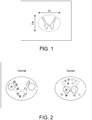
- the cross section of the human spinal cord at different levels is approximated to an elliptical shape, where the wider diameter is referred to as the transverse diameter, D t and the narrower is referred to as the anteroposterior diameter, D a (see Fig. 1 ).
- the cross section of a spinal cord includes an "H"-shape of gray matter and is surrounded by white matter.
- electromyograms are acquired from muscles innervated by spinal cord segments above the lesion (injury), at the lesion and under the lesion. All muscles are examined both during voluntary activation as well as activation of a patient's spasticity in the paretic part of the body. This yields three distinct patterns of motor unit potentials (MUP): above the lesion normal MUPs are found during voluntary activation and no MUPs during activation of spasticity. At the level of neurological loss, no MUPs are seen and evidence of chronic denervation indicated by positive sharp waves and fibrillation potentials are found.
- MUP motor unit potentials
- a device can be designed to replace only an injured section of the spinal cord. Instead of cuts perpendicular to the length of the spinal cord, the cord is cut in a different plane and the device is designed to replace the missing part.
- the ratio between anteroposterior and transverse diameters, D a /D t is referred to as RAPT.
- RAPT The ratio between anteroposterior and transverse diameters
- the present inventors have now found that devices having combinations of transverse diameters D t in the range of around 9 - 13 mm, where RAPT is within the range of 0.5-1.0, and more specifically within the range of from 0.6 - 0.9, fulfills the need in the majority of situations with thoracic injuries, considering the spinal cord dimensions of the population.
- the anteroposterior diameters D a are in the range of around 6-10 mm.
- the devices can roughly be divided into three groups i) Round with an RAPT value approaching 1.00, i.e.
- the length may vary considerably, but a series of devices having respective lengths in the range of from 15-40 mm fulfills this basic need in many situations. However, equipments for moulding spinal cord devices can easily be adjusted to a less usual, patient specific device length, if necessary. Accordingly, devices according to the present invention can be used in surgical treatment of a majority of spinal cord injuries.
- a kit comprising a series of preformed devices according to the invention, which are substantially evenly distributed within the dimensional ranges defined above, provides a very important tool for surgical treatment.
- a hospital or similar institution equipped with device kits according to the invention is well prepared for treatment of a patient having a spinal cord injury which has been identified as a candidate for this type of surgical treatment. Even if a complete traumatic spinal cord injury, in particular at the thoracic level, is a major criterion for use of a device designed for replacing a gap between two fully resected ends of the spinal cord, alternative devices can be produced for replacement of only a section of the surface area as appropriate.
- biocompatible and/or biodegradable materials have been suggested in prior art literature for use as implants in various parts of the body, including the use for manufacture of spinal cord devices, and are suitable for use in the present devices.
- materials are fibrin glue, poly-L-lactic acid (PLA) polymers, poly(lactide-co-glycolide acid (PLGA) polymers, poly-glycolic acid (PGA) polymers, polycaprolactones and calcium sulphate, just to mention a few.
- Important functions of the material are to provide a sufficient stable matrix for manipulation of the nerve channels and to provide the desired slow-release of medicaments of various types, to be administered at the site for surgery, in particular growth factors.
- the matrix must remain over a time sufficient for the nerve ends to grow together for sufficiently firm attachment to resist mechanical forces due to movements by the patient.
- the matrix is preferably porous so that it provides sufficient surface for adsorption/absorption of the substances to be administered.
- the invention will, in the following, be exemplified by one embodiment where ⁇ -calcium sulfate hemihydrate is used for manufacture of the device, but other materials known by a person skilled in the art can of course be used instead.
- a device is characterized by a "cylindrical" body with more or less ellipsoidal cross section and end surfaces as indicated by the various RAPT values discussed above.
- the device is made of a biocompatible and biodegradable material, having nerve guiding channels which are open, as delivered, or can optionally be opened in connection with final preparation for surgery.
- a kit of devices according to the invention comprises a set of devices with dimensions distributed substantially evenly within the ranges defined above. With such a kit, a hospital is well equipped for handling a majority of candidates for this kind of treatment. According to a further embodiment of the invention, kits covering a more narrow range of dimensions can be provided.
- such a narrow kit is chosen after initial pre-surgical determinations of the dimensions, while the final choice is made immediately prior to implantation, i.e., during surgery, when the final gap in the spinal cord has been created and the more exact dimensions can be measured in situ.
- a patient specific kit can be selected and ready for the final implantation procedure, comprising device selection, soaking in the appropriate solutions, and introduction of nerve tissue into the channels immediately before implantation.
- the device In situations with only a partly injured and removed section of the spinal cord, the device is designed to cover a gap which is only a section (fraction) of the "cylindrical" end surface, where these sections (fractions) even may be different at the two end surfaces.
- “cylindrical" steel moulds are used.
- the moulds preferably have two ellipsoidal end plates separated by a distance h which corresponds to the length of the device to be produced.
- several through structures spanning the interior between the two ellipsoidal end plates are used during moulding. When the through structures are removed, the channels are created.
- the end of each channel is positioned at a specific location at each end surface for optimal contact between white and gray matter.
- the channels can have the same cross-section over the full length, i.e. between the two end surfaces but can also have a funnel shape in order to connect the gray and white nerve tissue as effectively as possible at the two ends.
- the cross-section area to be connected to the white matter is larger than the cross-section area to be connected to the gray matter.
- threads for example suture threads, or tubings which are fixed between specific positions at the mould end surfaces defined by the topography of the white and gray substances in the spinal cord.
- cylindrical steel moulds are mounted in a mould fixature with end plates of a desired ellipsoidal dimension to fit the neuroanatomical shape of a resected spinal cord.
- Polymer for example, polytetrafluoroethylene (PTFE), tubings or threads are inserted between the upper end plate and the lower end plate, thereby spanning the interior of the mould and forming the channels for later positioning of the nerve grafts.
- PTFE polytetrafluoroethylene
- ⁇ -Calcium sulphate hemihydrate powder (or other biocompatible, biodegradable material) is mixed with water for injection in suitable proportions, e.g. 1:0.30 (w/v), and injected into the mould.
- the mould is vibrated to remove air bubbles from the calcium sulphate paste. After around 1 h setting at room temperature, the device is released from the mould. Moulding generates a spinal cord device composed of calcium sulphate dihydrate (CaSO 4 x 2H 2 O) as determined by x-ray diffraction analysis.
- the threads or tubings are at some stage removed and channels for positioning of peripheral nerve grafts are formed. This may be done at the manufacturing step or just prior to implantation. By using tubings with different dimensions, the channels for positioning of the peripheral nerves can be adopted for optimal connection between spinal cord tracts of various dimensions and at different vertebrae levels, after implantation and nerve growth.
- the preformed device can be provided to the surgical team with open channels or with channels which are opened by removing the through structures (threads or tubing), e.g. by pulling the structure out of the device, as one step in the final preparation for surgery.
- the devices are delivered with open channels and each of the open channels comprises a thin thread which later on can be used for pulling peripheral nerves into the channel.
- the device is easily equipped with such threads when the through structure is removed.
- Other ways for inserting peripheral nerves into the channels are also available, e.g. by suction.
- growth factors e.g. FGF1
- FGF1 growth factors
- one or more growth factors, as well as other pharmaceutically active components are optionally administered to the site of implantation of a spinal cord device according to the invention.
- Such administration can be made in many different ways, e.g. by providing the components adsorbed or absorbed to the device.
- a sufficient surface area is available for binding the component(s).
- the device prior to implantation is contacted with an aqueous solution comprising the component or a cocktail of components, e.g. by soaking the device in the solution for a given time period, e.g. 10-50 ml.
- Such a standard solution may comprise one or more components selected from the following groups of substances: FGF1, Brain derived neurotrophic factor (BDNF), Glial derived neurotrophic factor (GDNF), neurotrophic factor (NGF), ciliary neutrophic factor (CNTF), Chondroitinase ABC, Calcium flow antagonists increasing the regeneration, e.g. nimodipine, peptidases, S-mRNA, autologous activated macrophages, macrophages from donors, olfactory ensheating cells, autologous stem cells, oligodendrocyte progenitors, Schwann cells, Cortisone, angiogenesis inhibitors, erytropoetin, inactivators of Rho (e.g.
- Cethrin broad spectrum antibiotics (e.g. Minocycline), Riluzole and fysiological antagonists to the NMDA-receptor, e.g magnesium.
- additional components may be of importance , e.g. pre-degenerated peripheral nerve grafts, EGF, NT-3, PDGF, IGF1, Insulin, bFGF, HGF, Calpain inhibitor, Hematopoetic inhibitor, Induced pluripotent stem cells, Neuronal stem cells, Embryonic stem cells, Mesenchymal stem cells, Anti Nogo, Rho antagonist, PEG and/or EPO.
- the device For loading the device with an effective amount of FGF1, for example, the device is soaked in a solution comprising about 0.005-50 microgram/ml of FGF1 for about 0.5-5 hours. In one example, the concentration of the solution is 0.5 microgram/ml and the device is soaked for 1 hour in 30 ml solution. In additional embodiments, for even better effect of FGF1, the FGF1 is mixed with heparin which binds and activates FGF1, e.g. at a molar ratio of around 1:100.
- a suitable dose of the heparin-activated FGF1 to be delivered by the device is in the range of 0.01-100 ng/mg device, more specifically, 0.1-10 ng/mg device, or more specifically, 0.5-5 ng/mg device, and in particular around 1 ng/mg device.
- the device will deliver the active component(s) over a few weeks, with the main amount being delivered over a few days.
- peripheral nerves are positioned in the open channels of the device. This can be done by pulling a nerve into each of the channels.
- the nerve can be an autologous peripheral nerve (taken from the patient), but can also be artificial nerve tissue produced by nerve cell culturing, preferably of nerve cells taken from the patient.
- nerve tissue is applied in each of the channels and the device is ready for implantation.
- the device For creating descending motor pathways from cranial (proximal) white matter to caudal (distal) gray matter over the gap between the two ends of the spinal cord and ascending sensory pathways from caudal (distal) white matter to cranial (proximal) gray matter, the device is equipped with several through channels. In order to obtain an appropriate combination of pathways in the two directions, at least some of the channels are allowed to be non-linear.
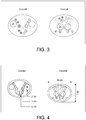
- the device has a first set of channels having a first diameter and a second set of channels having a second diameter, where the first diameter is larger than the second diameter, as illustrated by channels A, B, C, D, E and F in Fig. 2 .
- the channels A have a first diameter in the range of from 2.0-2.9 mm
- one or more of channels B, C, D, E and F have a second diameter in the range of from 1.1-1.6 mm.
- the spinal cord device according to the invention comprises channels A, B, C, D, E and F positioned as shown in Fig. 2 , or comprises channels A, B, C, D, E and F positioned as shown in Figs. 3 and 5 .
- the spinal cord device according to the invention comprises channels A, B, C, D, E and F positioned and relatively sized as shown in Figs. 3 and 5 .
- the device comprises a third set of channels (B, C, D and F) having a shorter diameter than the first set of channels (A) and the second set of channels (E).
- the channels A have a first diameter in the range of from 2.0-2.9 mm
- the channels E have a second diameter in the range of from 1.1-1.6 mm
- the channels B, C, D and F have a third diameter of 0.5-0.9 mm.
- Fig. 4 A more specific embodiment is shown in Fig. 4 .
- One or more additional sets of channels may be introduced in further embodiments. In all figures, only the channels on the left side of the symmetry plane are marked but the designations given are relevant also for the corresponding channels on the right side.
- the position and diameter of the through channels in each of the end surfaces of the device is of importance in order to obtain the best conditions for guidance of the important longitudinal tracts over the spinal cord gap.
- the inventors have found that, in accordance with the invention, these conditions can be obtained by 12 channels, the channels being positioned as illustrated in Fig. 2 or as illustrated in Fig. 3 .
- each channel needs to be thick enough so as not to be crushed during manufacture and manipulations prior to implantation.
- the requirement in this respect may vary depending on the material used and the manufacturing method.
- the inventors have found that a preferred wall thickness, in particular with the ⁇ -calcium sulphate hemihydrate used in the embodiment illustrating the invention, is around 0.3 mm or somewhat higher.
- Channels A have a diameter in the range of 2.0-2.9 mm
- channels B, C, D and F have a diameter in the range of 0.6-0.9 mm
- channels E have a diameter in the range of 1.1-1.6 mm.
- the position in each surface of the channels is illustrated in Figs. 2 and 3 according to specific embodiments of the invention.
- channels creating the descending motor pathway from white matter on the cranial (proximal) side to gray matter on the caudal (distal) side have a substantially 100% connection to the white matter on the proximal side, "the motor channel entrance".
- the percentage of the channel surface that connects to gray matter is more than 50%, e.g. in the interval 50-60%, or preferably 50-70%. According to one embodiment where the funnel type of channels discussed above are used, an even higher percentage can be achieved, e.g. higher than 60%, 70%, 80% or 90%.
- Channels creating the ascending sensory pathway from white matter on the caudal (distal) side to gray matter on the cranial (proximal) side preferably have substantially 100 % connection to the white matter on the distal side. At least 60%, e.g. in the interval 60-80% of the channel openings connects gray matter on the proximal side. This percentage can be increased in embodiments with suitable modification of the channel dimensions, in particular funnel shaped channels.
- the corresponding percentage of channel areas connected to ascending sensory white matter on the caudal (distal) side is at least 5%, preferably greater than 10%, e.g. in the interval 10-20% in one embodiment of the invention.
- kits comprising a number of devices covering a desired number of alternative dimensions.
- One example of such a kit comprises around 10 devices with different end surface dimensions and configurations, i.e., designs, and will cover the major number of cross-section alternatives in the thoracic example.
- devices with different lengths are produced.
- at least two devices are provided having the same D t , D a , RAPT and Channel Diameter dimensions and differing lengths L.
- at least two devices are provided having differing lengths L.
- devices of 6 different lengths are provided for each surface configuration or design, whereby an example of such a kit having ten different end surface designs contains 60 devices. Again, it should be stressed that this is only one example of a device kit according to the invention.
- kits comprising devices according to the invention, fulfilling a hospital's basic need for devices for selection in preparation for implantation in a thoracic injury situation is given below.
- Each of these device designs is provided with various lengths, e.g. in the 15-40 mm range. With 6 lengths for each, evenly distributed within the range, a typical kit comprises in total around 60 devices (dimensions given in mm): Design No.
- the kit comprises 10 devices with different surface area dimensions and configurations, i.e., designs.
- the number of devices in a kit can of course be different depending on the ambition to cover gaps with different configurations.
- the spinal cord ends are resected as necessary.
- the surface dimensions, i.e. D t and D a , and the length L of the gap between the two spinal cord ends, are determined. This can be done in many different ways and a series of dummies with a wide range of dimensions (D t , D a and L) are preferably used in situ for this determination.
- a device with the same shape, but approximately 1 mm larger diameter, is selected.
- the device is contacted with the solution comprising one or more pharmaceutically active component(s) to be administered to the site of surgery, e.g. heparin-activated FGF1.
- pharmaceutically active component(s) e.g. heparin-activated FGF1.
- nerve tissue is introduced into all channels in the device, and the device is then implanted.
- This example evaluates a biodegradable calcium sulphate device with heparin-activated rhFGF1 for treatment of spinal cord injury in rat.
- SCI devices fabricated from ⁇ -calcium sulfate hemihydrate with 12 channels with similar geometry as shown in Fig. 5 were loaded with heparin-activated rhFGF1.
- the spinal cord of the control groups were either transected (group 1, negative control) or left intact (group 2, positive control).
- the spinal cords of the rats of groups 3-5 were transected and the removed spinal cord tissue was replaced by devices containing nerve grafts.
- the SCI -devices employed for study group 4 were soaked in 500 ⁇ g/ml heparin-activated rhFGF1 (rhFGF1:heparin, 1:1, w/w).
- the SCI-devices were soaked in 50 ⁇ g/ml heparin-activated rhFGF1. Due to autophagy, a few animals had to be sacrificed prior to the end of the 20 week study. The animals sacrificed before week 20 were evenly spread among the groups.
- MEPs motor evoked potentials
- This example evaluates dose-finding of heparin-activated rhFGF1 administered in a biodegradable calcium sulphate device for treatment of spinal cord injury in rat.
- Example 2 The same type of devices as in Example 1 were used. Each device was soaked in heparin-activated rhFGF1 (FGF1:heparin, 1:100 molar ratio) solution for 1 h at room temperature. Heparin solution without preservatives (10 000 IE/ml H 2 O, Leo Pharma Denmark) was employed. The devices were soaked in 50 ⁇ g/ml, 0.5 ⁇ g/ml, 0.005 ⁇ g/ml and 0 ⁇ g/ml concentrations of heparin-activated rhFGF1 corresponding to a dose of 45, 0.9, 0.01 and 0 ng/mg device (based on solution uptake and adsorption).
- FGF1:heparin, 1:100 molar ratio Heparin solution without preservatives
- heparin-activated rhFGF1 FGF1-dependent recovery of bilateral MEPs in the hindlimbs of treated animals is illustrated in Fig. 6 . All animals showed undetectable MEPs in the hindlimbs 1 week post-surgery, while already after 2 weeks positive MEPs were recorded. The results indicate that an effective dose of heparin-activated rhFGF1 in the disclosed device in the treatment of complete SCI in rat is achieved with a concentration of at least 0.5 ⁇ g/ml heparin-activated rhFGF1 solution (yielding 0.9 ng heparin-activated rhFGF1/mg device).
- Clinical SCI-devices as illustrated in Fig. 5 , 15 mm high and having an oval cross section of 9.0-6.9 mm, comprising 12 channels for nerve graft positioning are made from ⁇ -calcium sulphate hemihydrate with subsequent sterilization.
- Each device is placed in 30 ml of a soaking solution consisting of 5 ⁇ g/ml rhFGF1, 80 ⁇ g/ml Gentamicin, 10 mM NaPO 4 , 150 mM NaCl, 0.3 mM EDTA at pH 7 for 1 hour to allow the solution to be adsorbed into the device.
- the heparin concentration is 430 ⁇ g/ml and the ratio rhFGF1:heparin 1:100 (molar).
- the animals are female Landrace pig. After 10 days of acclimatization, the pig is anaesthetized with a combination of fentanyl, midazolam and propofol and prepared for surgery. During the surgical procedure, the intravenous anaesthesia is maintained with fentanyl 0.004 mg/kg/h, midazolam 0.5 mg/kg/h and propofol 3 mg/kg/h.
- the preparation for surgery comprises disinfection of the areas subjected to surgery, combined with antibiotic treatment.
- the pig is placed with the back up. An incision is made above the thoracic spinal cord and a laminectomy is performed. The dura mater is incised and the spinal cord is exposed. A segment large enough to fit the SCI device of the lower thoracic spinal cord is resected.
Priority Applications (1)
| Application Number | Priority Date | Filing Date | Title |
|---|---|---|---|
| PL12815796T PL2787900T3 (pl) | 2011-12-06 | 2012-12-03 | Urządzenia dla rdzenia kręgowego do promowania regeneracji aksonów |
Applications Claiming Priority (2)
| Application Number | Priority Date | Filing Date | Title |
|---|---|---|---|
| US201161567450P | 2011-12-06 | 2011-12-06 | |
| PCT/IB2012/056924 WO2013084137A1 (en) | 2011-12-06 | 2012-12-03 | Spinal cord devices and methods for promoting axonal regeneration |
Publications (2)
| Publication Number | Publication Date |
|---|---|
| EP2787900A1 EP2787900A1 (en) | 2014-10-15 |
| EP2787900B1 true EP2787900B1 (en) | 2018-10-24 |
Family
ID=47559593
Family Applications (1)
| Application Number | Title | Priority Date | Filing Date |
|---|---|---|---|
| EP12815796.3A Not-in-force EP2787900B1 (en) | 2011-12-06 | 2012-12-03 | Spinal cord devices for promoting axonal regeneration |
Country Status (13)
| Country | Link |
|---|---|
| US (2) | US9895234B2 (pl) |
| EP (1) | EP2787900B1 (pl) |
| JP (1) | JP6319847B2 (pl) |
| CN (1) | CN104093367B (pl) |
| AU (1) | AU2012349725B2 (pl) |
| CA (1) | CA2857619C (pl) |
| DK (1) | DK2787900T3 (pl) |
| ES (1) | ES2702333T3 (pl) |
| LT (1) | LT2787900T (pl) |
| PL (1) | PL2787900T3 (pl) |
| PT (1) | PT2787900T (pl) |
| TR (1) | TR201819261T4 (pl) |
| WO (1) | WO2013084137A1 (pl) |
Families Citing this family (8)
| Publication number | Priority date | Publication date | Assignee | Title |
|---|---|---|---|---|
| US9265620B2 (en) | 2011-03-18 | 2016-02-23 | Raed M. Ali, M.D., Inc. | Devices and methods for transpedicular stabilization of the spine |
| US10687962B2 (en) | 2013-03-14 | 2020-06-23 | Raed M. Ali, M.D., Inc. | Interbody fusion devices, systems and methods |
| EP2967909A4 (en) | 2013-03-14 | 2016-10-05 | Raed M Ali M D Inc | DEVICES, SYSTEMS AND METHOD FOR LATERAL INTERVERTEBRAL FUSION |
| US10813643B2 (en) * | 2017-10-19 | 2020-10-27 | Axogen Corporation | Materials and methods for breast neurotization with nerve grafts |
| US11147558B2 (en) | 2018-11-15 | 2021-10-19 | Axogen Corporation | Materials and methods for nerve repair with animal-sourced grafts |
| CA3159546A1 (en) * | 2019-11-01 | 2021-05-06 | Allegro 3D, Inc. | 3d-bioprinted scaffolds for tissue regeneration |
| FR3108260B1 (fr) | 2020-03-17 | 2024-01-05 | Neurobiomat | Hydrogel hétérogène hybride, procédé de fabrication et utilisation comme implant de comblement non-dégradable in-situ |
| EP4082448A1 (en) * | 2021-04-26 | 2022-11-02 | Tissium SA | Nerve conduit |
Family Cites Families (11)
| Publication number | Priority date | Publication date | Assignee | Title |
|---|---|---|---|---|
| IT1247157B (it) * | 1991-02-11 | 1994-12-12 | Fidia Spa | Canali di guida biodegradabili e bioassorbibili da impiegare per la rigenerazione nervosa. |
| SE9602879D0 (sv) | 1996-07-26 | 1996-07-26 | Henrich Cheng | Medical device |
| US5925053A (en) | 1997-09-02 | 1999-07-20 | Children's Medical Center Corporation | Multi-lumen polymeric guidance channel, method for promoting nerve regeneration, and method of manufacturing a multi-lumen nerve guidance channel |
| US7163545B2 (en) | 2002-07-29 | 2007-01-16 | Mayo Foundation For Medical Education And Research | Spinal cord surgical implant |
| US20040199186A1 (en) | 2003-04-04 | 2004-10-07 | Kuffler Suzanne Elizabeth | Implant to promote axon regeneration across spinal cord and peripheral nerve gaps |
| US20090169596A1 (en) * | 2006-03-29 | 2009-07-02 | Mikael Svensson | Device for spinal cord nerve regeneration |
| EP2148634A1 (en) * | 2007-05-15 | 2010-02-03 | Axongen Ab | Fibrin-based nerve repair conduit and method of producing the same |
| TWI414328B (zh) * | 2008-09-18 | 2013-11-11 | Nat Health Research Institutes | 可植入性神經再生導管 |
| US8447409B2 (en) * | 2008-10-15 | 2013-05-21 | Cochlear Limited | Electroneural interface for a medical implant |
| ES2405789T3 (es) * | 2010-04-15 | 2013-06-03 | National University Of Ireland | Conducto para nervios multicanal de colágeno para la reparación de nervios |
| CA2919374C (en) * | 2013-07-30 | 2019-12-03 | Musculoskeletal Transplant Foundation | Acellular soft tissue-derived matrices and methods for preparing same |
-
2012
- 2012-12-03 PL PL12815796T patent/PL2787900T3/pl unknown
- 2012-12-03 JP JP2014545410A patent/JP6319847B2/ja active Active
- 2012-12-03 PT PT12815796T patent/PT2787900T/pt unknown
- 2012-12-03 CN CN201280069171.9A patent/CN104093367B/zh not_active Expired - Fee Related
- 2012-12-03 US US14/361,650 patent/US9895234B2/en not_active Expired - Fee Related
- 2012-12-03 ES ES12815796T patent/ES2702333T3/es active Active
- 2012-12-03 DK DK12815796.3T patent/DK2787900T3/en active
- 2012-12-03 LT LTEP12815796.3T patent/LT2787900T/lt unknown
- 2012-12-03 WO PCT/IB2012/056924 patent/WO2013084137A1/en active Application Filing
- 2012-12-03 AU AU2012349725A patent/AU2012349725B2/en not_active Ceased
- 2012-12-03 TR TR2018/19261T patent/TR201819261T4/tr unknown
- 2012-12-03 EP EP12815796.3A patent/EP2787900B1/en not_active Not-in-force
- 2012-12-03 CA CA2857619A patent/CA2857619C/en not_active Expired - Fee Related
-
2018
- 2018-01-09 US US15/865,516 patent/US20180140434A1/en not_active Abandoned
Non-Patent Citations (1)
| Title |
|---|
| None * |
Also Published As
| Publication number | Publication date |
|---|---|
| EP2787900A1 (en) | 2014-10-15 |
| JP6319847B2 (ja) | 2018-05-09 |
| PT2787900T (pt) | 2019-01-08 |
| US9895234B2 (en) | 2018-02-20 |
| JP2015500692A (ja) | 2015-01-08 |
| LT2787900T (lt) | 2019-01-10 |
| CN104093367A (zh) | 2014-10-08 |
| CN104093367B (zh) | 2017-10-24 |
| US20150088257A1 (en) | 2015-03-26 |
| AU2012349725A1 (en) | 2014-06-19 |
| TR201819261T4 (tr) | 2019-01-21 |
| AU2012349725B2 (en) | 2018-01-18 |
| US20180140434A1 (en) | 2018-05-24 |
| ES2702333T3 (es) | 2019-02-28 |
| CA2857619C (en) | 2019-11-12 |
| CA2857619A1 (en) | 2013-06-13 |
| WO2013084137A1 (en) | 2013-06-13 |
| PL2787900T3 (pl) | 2019-04-30 |
| DK2787900T3 (en) | 2019-01-14 |
Similar Documents
| Publication | Publication Date | Title |
|---|---|---|
| EP2787900B1 (en) | Spinal cord devices for promoting axonal regeneration | |
| Zhang et al. | Functional polymer‐based nerve guide conduits to promote peripheral nerve regeneration | |
| AU732199B2 (en) | Medical device for treatment of a gap or defect in the central nerve system | |
| Mukhatyar et al. | Tissue engineering strategies designed to realize the endogenous regenerative potential of peripheral nerves | |
| Yin et al. | Taxol-modified collagen scaffold implantation promotes functional recovery after long-distance spinal cord complete transection in canines | |
| US20170354417A1 (en) | Device for induction of cellular activity | |
| Oria et al. | In vivo evaluation of novel PLA/PCL polymeric patch in rats for potential spina bifida coverage | |
| Carlstedt | Central nerve plexus injury | |
| US20030134414A1 (en) | Nerve growth assistance improvement | |
| TW202116293A (zh) | 免疫調節劑改善神經再生之用途 | |
| AU2007229982B2 (en) | A method and a mould for manufacturing a nerve regeneration device | |
| RU2801469C1 (ru) | Способ лечения травмы спинного мозга с восстановлением его функций конъюгатом ПЭГ-хитозана "НЕЙРО-ПЭГ" | |
| AU751620B2 (en) | A method for treating a deficiency in a spinal cord | |
| Shah | An injectable gelatin-based conjugate incorporating EGF promotes tissue repair and functional recovery after spinal cord injury in a rat model | |
| Tabakow et al. | CT-1239 Accepted 09/08/2014 for publication in “Cell Transplantation” Functional regeneration of supraspinal connections in a patient with transected spinal cord following transplantation of bulbar olfactory ensheathing cells with peripheral nerve bridging | |
| Elsayed et al. | Update in facial nerve paralysis: tissue engineering and new technologies | |
| Nordblom | SPINAL CORD INJURY | |
| Carlstedt | Central Nerve Plexus Injury (With Cd-rom) | |
| Garde | Regenerative peripheral neurointerfacing of upper extremity prostheses |
Legal Events
| Date | Code | Title | Description |
|---|---|---|---|
| PUAI | Public reference made under article 153(3) epc to a published international application that has entered the european phase |
Free format text: ORIGINAL CODE: 0009012 |
|
| 17P | Request for examination filed |
Effective date: 20140702 |
|
| AK | Designated contracting states |
Kind code of ref document: A1 Designated state(s): AL AT BE BG CH CY CZ DE DK EE ES FI FR GB GR HR HU IE IS IT LI LT LU LV MC MK MT NL NO PL PT RO RS SE SI SK SM TR |
|
| DAX | Request for extension of the european patent (deleted) | ||
| 17Q | First examination report despatched |
Effective date: 20160607 |
|
| GRAP | Despatch of communication of intention to grant a patent |
Free format text: ORIGINAL CODE: EPIDOSNIGR1 |
|
| INTG | Intention to grant announced |
Effective date: 20171023 |
|
| GRAJ | Information related to disapproval of communication of intention to grant by the applicant or resumption of examination proceedings by the epo deleted |
Free format text: ORIGINAL CODE: EPIDOSDIGR1 |
|
| GRAP | Despatch of communication of intention to grant a patent |
Free format text: ORIGINAL CODE: EPIDOSNIGR1 |
|
| INTC | Intention to grant announced (deleted) | ||
| INTG | Intention to grant announced |
Effective date: 20180316 |
|
| RAP1 | Party data changed (applicant data changed or rights of an application transferred) |
Owner name: BIOARCTIC NEUROSCIENCE AB |
|
| RAP1 | Party data changed (applicant data changed or rights of an application transferred) |
Owner name: BIOARCTIC AB |
|
| GRAS | Grant fee paid |
Free format text: ORIGINAL CODE: EPIDOSNIGR3 |
|
| GRAJ | Information related to disapproval of communication of intention to grant by the applicant or resumption of examination proceedings by the epo deleted |
Free format text: ORIGINAL CODE: EPIDOSDIGR1 |
|
| GRAL | Information related to payment of fee for publishing/printing deleted |
Free format text: ORIGINAL CODE: EPIDOSDIGR3 |
|
| INTC | Intention to grant announced (deleted) | ||
| GRAR | Information related to intention to grant a patent recorded |
Free format text: ORIGINAL CODE: EPIDOSNIGR71 |
|
| GRAA | (expected) grant |
Free format text: ORIGINAL CODE: 0009210 |
|
| INTG | Intention to grant announced |
Effective date: 20180912 |
|
| AK | Designated contracting states |
Kind code of ref document: B1 Designated state(s): AL AT BE BG CH CY CZ DE DK EE ES FI FR GB GR HR HU IE IS IT LI LT LU LV MC MK MT NL NO PL PT RO RS SE SI SK SM TR |
|
| REG | Reference to a national code |
Ref country code: CH Ref legal event code: EP |
|
| REG | Reference to a national code |
Ref country code: IE Ref legal event code: FG4D |
|
| REG | Reference to a national code |
Ref country code: AT Ref legal event code: REF Ref document number: 1055741 Country of ref document: AT Kind code of ref document: T Effective date: 20181115 |
|
| REG | Reference to a national code |
Ref country code: DE Ref legal event code: R096 Ref document number: 602012052671 Country of ref document: DE |
|
| REG | Reference to a national code |
Ref country code: CH Ref legal event code: NV Representative=s name: NOVAGRAAF INTERNATIONAL SA, CH |
|
| REG | Reference to a national code |
Ref country code: PT Ref legal event code: SC4A Ref document number: 2787900 Country of ref document: PT Date of ref document: 20190108 Kind code of ref document: T Free format text: AVAILABILITY OF NATIONAL TRANSLATION Effective date: 20181218 |
|
| REG | Reference to a national code |
Ref country code: DK Ref legal event code: T3 Effective date: 20190106 |
|
| REG | Reference to a national code |
Ref country code: NL Ref legal event code: FP |
|
| REG | Reference to a national code |
Ref country code: SE Ref legal event code: TRGR |
|
| PGFP | Annual fee paid to national office [announced via postgrant information from national office to epo] |
Ref country code: SE Payment date: 20181219 Year of fee payment: 13 |
|
| REG | Reference to a national code |
Ref country code: NO Ref legal event code: T2 Effective date: 20181024 |
|
| REG | Reference to a national code |
Ref country code: ES Ref legal event code: FG2A Ref document number: 2702333 Country of ref document: ES Kind code of ref document: T3 Effective date: 20190228 |
|
| REG | Reference to a national code |
Ref country code: EE Ref legal event code: FG4A Ref document number: E016706 Country of ref document: EE Effective date: 20190115 |
|
| PG25 | Lapsed in a contracting state [announced via postgrant information from national office to epo] |
Ref country code: IS Free format text: LAPSE BECAUSE OF FAILURE TO SUBMIT A TRANSLATION OF THE DESCRIPTION OR TO PAY THE FEE WITHIN THE PRESCRIBED TIME-LIMIT Effective date: 20190224 Ref country code: BG Free format text: LAPSE BECAUSE OF FAILURE TO SUBMIT A TRANSLATION OF THE DESCRIPTION OR TO PAY THE FEE WITHIN THE PRESCRIBED TIME-LIMIT Effective date: 20190124 Ref country code: HR Free format text: LAPSE BECAUSE OF FAILURE TO SUBMIT A TRANSLATION OF THE DESCRIPTION OR TO PAY THE FEE WITHIN THE PRESCRIBED TIME-LIMIT Effective date: 20181024 |
|
| PG25 | Lapsed in a contracting state [announced via postgrant information from national office to epo] |
Ref country code: GR Free format text: LAPSE BECAUSE OF FAILURE TO SUBMIT A TRANSLATION OF THE DESCRIPTION OR TO PAY THE FEE WITHIN THE PRESCRIBED TIME-LIMIT Effective date: 20190125 Ref country code: RS Free format text: LAPSE BECAUSE OF FAILURE TO SUBMIT A TRANSLATION OF THE DESCRIPTION OR TO PAY THE FEE WITHIN THE PRESCRIBED TIME-LIMIT Effective date: 20181024 Ref country code: AL Free format text: LAPSE BECAUSE OF FAILURE TO SUBMIT A TRANSLATION OF THE DESCRIPTION OR TO PAY THE FEE WITHIN THE PRESCRIBED TIME-LIMIT Effective date: 20181024 |
|
| REG | Reference to a national code |
Ref country code: DE Ref legal event code: R097 Ref document number: 602012052671 Country of ref document: DE |
|
| PG25 | Lapsed in a contracting state [announced via postgrant information from national office to epo] |
Ref country code: CZ Free format text: LAPSE BECAUSE OF FAILURE TO SUBMIT A TRANSLATION OF THE DESCRIPTION OR TO PAY THE FEE WITHIN THE PRESCRIBED TIME-LIMIT Effective date: 20181024 |
|
| PG25 | Lapsed in a contracting state [announced via postgrant information from national office to epo] |
Ref country code: SK Free format text: LAPSE BECAUSE OF FAILURE TO SUBMIT A TRANSLATION OF THE DESCRIPTION OR TO PAY THE FEE WITHIN THE PRESCRIBED TIME-LIMIT Effective date: 20181024 Ref country code: SM Free format text: LAPSE BECAUSE OF FAILURE TO SUBMIT A TRANSLATION OF THE DESCRIPTION OR TO PAY THE FEE WITHIN THE PRESCRIBED TIME-LIMIT Effective date: 20181024 Ref country code: RO Free format text: LAPSE BECAUSE OF FAILURE TO SUBMIT A TRANSLATION OF THE DESCRIPTION OR TO PAY THE FEE WITHIN THE PRESCRIBED TIME-LIMIT Effective date: 20181024 |
|
| PLBE | No opposition filed within time limit |
Free format text: ORIGINAL CODE: 0009261 |
|
| STAA | Information on the status of an ep patent application or granted ep patent |
Free format text: STATUS: NO OPPOSITION FILED WITHIN TIME LIMIT |
|
| 26N | No opposition filed |
Effective date: 20190725 |
|
| PG25 | Lapsed in a contracting state [announced via postgrant information from national office to epo] |
Ref country code: SI Free format text: LAPSE BECAUSE OF FAILURE TO SUBMIT A TRANSLATION OF THE DESCRIPTION OR TO PAY THE FEE WITHIN THE PRESCRIBED TIME-LIMIT Effective date: 20181024 |
|
| PGFP | Annual fee paid to national office [announced via postgrant information from national office to epo] |
Ref country code: LU Payment date: 20191119 Year of fee payment: 8 |
|
| PG25 | Lapsed in a contracting state [announced via postgrant information from national office to epo] |
Ref country code: MT Free format text: LAPSE BECAUSE OF NON-PAYMENT OF DUE FEES Effective date: 20181203 |
|
| PGFP | Annual fee paid to national office [announced via postgrant information from national office to epo] |
Ref country code: FI Payment date: 20191118 Year of fee payment: 8 Ref country code: IE Payment date: 20191118 Year of fee payment: 8 Ref country code: PT Payment date: 20191104 Year of fee payment: 8 Ref country code: MC Payment date: 20191120 Year of fee payment: 8 Ref country code: NO Payment date: 20191118 Year of fee payment: 8 Ref country code: DE Payment date: 20191119 Year of fee payment: 8 Ref country code: NL Payment date: 20191217 Year of fee payment: 8 |
|
| PGFP | Annual fee paid to national office [announced via postgrant information from national office to epo] |
Ref country code: EE Payment date: 20191125 Year of fee payment: 8 Ref country code: BE Payment date: 20191118 Year of fee payment: 8 Ref country code: LV Payment date: 20191129 Year of fee payment: 8 Ref country code: PL Payment date: 20191104 Year of fee payment: 8 Ref country code: DK Payment date: 20191119 Year of fee payment: 8 Ref country code: IT Payment date: 20191121 Year of fee payment: 8 Ref country code: FR Payment date: 20191118 Year of fee payment: 8 |
|
| PGFP | Annual fee paid to national office [announced via postgrant information from national office to epo] |
Ref country code: CH Payment date: 20191119 Year of fee payment: 8 Ref country code: AT Payment date: 20191118 Year of fee payment: 8 |
|
| PGFP | Annual fee paid to national office [announced via postgrant information from national office to epo] |
Ref country code: GB Payment date: 20191118 Year of fee payment: 8 Ref country code: ES Payment date: 20200102 Year of fee payment: 8 |
|
| PG25 | Lapsed in a contracting state [announced via postgrant information from national office to epo] |
Ref country code: MK Free format text: LAPSE BECAUSE OF NON-PAYMENT OF DUE FEES Effective date: 20181024 Ref country code: HU Free format text: LAPSE BECAUSE OF FAILURE TO SUBMIT A TRANSLATION OF THE DESCRIPTION OR TO PAY THE FEE WITHIN THE PRESCRIBED TIME-LIMIT; INVALID AB INITIO Effective date: 20121203 Ref country code: CY Free format text: LAPSE BECAUSE OF FAILURE TO SUBMIT A TRANSLATION OF THE DESCRIPTION OR TO PAY THE FEE WITHIN THE PRESCRIBED TIME-LIMIT Effective date: 20181024 |
|
| REG | Reference to a national code |
Ref country code: AT Ref legal event code: UEP Ref document number: 1055741 Country of ref document: AT Kind code of ref document: T Effective date: 20181024 |
|
| REG | Reference to a national code |
Ref country code: LT Ref legal event code: MM4D Effective date: 20191203 |
|
| PG25 | Lapsed in a contracting state [announced via postgrant information from national office to epo] |
Ref country code: LT Free format text: LAPSE BECAUSE OF NON-PAYMENT OF DUE FEES Effective date: 20191203 |
|
| REG | Reference to a national code |
Ref country code: DE Ref legal event code: R119 Ref document number: 602012052671 Country of ref document: DE |
|
| REG | Reference to a national code |
Ref country code: FI Ref legal event code: MAE |
|
| REG | Reference to a national code |
Ref country code: EE Ref legal event code: MM4A Ref document number: E016706 Country of ref document: EE Effective date: 20201231 |
|
| REG | Reference to a national code |
Ref country code: DK Ref legal event code: EBP Effective date: 20201231 |
|
| REG | Reference to a national code |
Ref country code: NO Ref legal event code: MMEP |
|
| PG25 | Lapsed in a contracting state [announced via postgrant information from national office to epo] |
Ref country code: FI Free format text: LAPSE BECAUSE OF NON-PAYMENT OF DUE FEES Effective date: 20201203 Ref country code: PT Free format text: LAPSE BECAUSE OF NON-PAYMENT OF DUE FEES Effective date: 20210604 |
|
| REG | Reference to a national code |
Ref country code: CH Ref legal event code: PL |
|
| REG | Reference to a national code |
Ref country code: SE Ref legal event code: EUG |
|
| REG | Reference to a national code |
Ref country code: NL Ref legal event code: MM Effective date: 20210101 |
|
| REG | Reference to a national code |
Ref country code: AT Ref legal event code: MM01 Ref document number: 1055741 Country of ref document: AT Kind code of ref document: T Effective date: 20201203 |
|
| GBPC | Gb: european patent ceased through non-payment of renewal fee |
Effective date: 20201203 |
|
| PG25 | Lapsed in a contracting state [announced via postgrant information from national office to epo] |
Ref country code: MC Free format text: LAPSE BECAUSE OF NON-PAYMENT OF DUE FEES Effective date: 20210104 Ref country code: LV Free format text: LAPSE BECAUSE OF NON-PAYMENT OF DUE FEES Effective date: 20201203 |
|
| REG | Reference to a national code |
Ref country code: BE Ref legal event code: MM Effective date: 20201231 |
|
| PG25 | Lapsed in a contracting state [announced via postgrant information from national office to epo] |
Ref country code: NL Free format text: LAPSE BECAUSE OF NON-PAYMENT OF DUE FEES Effective date: 20210101 |
|
| PG25 | Lapsed in a contracting state [announced via postgrant information from national office to epo] |
Ref country code: LU Free format text: LAPSE BECAUSE OF NON-PAYMENT OF DUE FEES Effective date: 20201203 Ref country code: EE Free format text: LAPSE BECAUSE OF NON-PAYMENT OF DUE FEES Effective date: 20201231 Ref country code: FR Free format text: LAPSE BECAUSE OF NON-PAYMENT OF DUE FEES Effective date: 20201231 Ref country code: AT Free format text: LAPSE BECAUSE OF NON-PAYMENT OF DUE FEES Effective date: 20201203 Ref country code: IE Free format text: LAPSE BECAUSE OF NON-PAYMENT OF DUE FEES Effective date: 20201203 Ref country code: IT Free format text: LAPSE BECAUSE OF NON-PAYMENT OF DUE FEES Effective date: 20201203 |
|
| PG25 | Lapsed in a contracting state [announced via postgrant information from national office to epo] |
Ref country code: SE Free format text: LAPSE BECAUSE OF NON-PAYMENT OF DUE FEES Effective date: 20201204 Ref country code: LI Free format text: LAPSE BECAUSE OF NON-PAYMENT OF DUE FEES Effective date: 20201231 Ref country code: NO Free format text: LAPSE BECAUSE OF NON-PAYMENT OF DUE FEES Effective date: 20201231 Ref country code: DE Free format text: LAPSE BECAUSE OF NON-PAYMENT OF DUE FEES Effective date: 20210701 Ref country code: GB Free format text: LAPSE BECAUSE OF NON-PAYMENT OF DUE FEES Effective date: 20201203 Ref country code: CH Free format text: LAPSE BECAUSE OF NON-PAYMENT OF DUE FEES Effective date: 20201231 |
|
| PG25 | Lapsed in a contracting state [announced via postgrant information from national office to epo] |
Ref country code: DK Free format text: LAPSE BECAUSE OF NON-PAYMENT OF DUE FEES Effective date: 20201231 |
|
| REG | Reference to a national code |
Ref country code: ES Ref legal event code: FD2A Effective date: 20220221 |
|
| PG25 | Lapsed in a contracting state [announced via postgrant information from national office to epo] |
Ref country code: ES Free format text: LAPSE BECAUSE OF NON-PAYMENT OF DUE FEES Effective date: 20201204 |
|
| PG25 | Lapsed in a contracting state [announced via postgrant information from national office to epo] |
Ref country code: TR Free format text: LAPSE BECAUSE OF NON-PAYMENT OF DUE FEES Effective date: 20191203 |
|
| PG25 | Lapsed in a contracting state [announced via postgrant information from national office to epo] |
Ref country code: BE Free format text: LAPSE BECAUSE OF NON-PAYMENT OF DUE FEES Effective date: 20201231 |
|
| PG25 | Lapsed in a contracting state [announced via postgrant information from national office to epo] |
Ref country code: PL Free format text: LAPSE BECAUSE OF NON-PAYMENT OF DUE FEES Effective date: 20201203 |