EP2769993A1 - Anticorps contre NKG2D humain et usages correspondants - Google Patents
Anticorps contre NKG2D humain et usages correspondants Download PDFInfo
- Publication number
- EP2769993A1 EP2769993A1 EP20140165436 EP14165436A EP2769993A1 EP 2769993 A1 EP2769993 A1 EP 2769993A1 EP 20140165436 EP20140165436 EP 20140165436 EP 14165436 A EP14165436 A EP 14165436A EP 2769993 A1 EP2769993 A1 EP 2769993A1
- Authority
- EP
- European Patent Office
- Prior art keywords
- antibody
- antibodies
- nkg2d
- hnkg2d
- binding
- Prior art date
- Legal status (The legal status is an assumption and is not a legal conclusion. Google has not performed a legal analysis and makes no representation as to the accuracy of the status listed.)
- Withdrawn
Links
Images


Classifications
-
- C—CHEMISTRY; METALLURGY
- C07—ORGANIC CHEMISTRY
- C07K—PEPTIDES
- C07K16/00—Immunoglobulins [IGs], e.g. monoclonal or polyclonal antibodies
- C07K16/18—Immunoglobulins [IGs], e.g. monoclonal or polyclonal antibodies against material from animals or humans
- C07K16/28—Immunoglobulins [IGs], e.g. monoclonal or polyclonal antibodies against material from animals or humans against receptors, cell surface antigens or cell surface determinants
- C07K16/2851—Immunoglobulins [IGs], e.g. monoclonal or polyclonal antibodies against material from animals or humans against receptors, cell surface antigens or cell surface determinants against the lectin superfamily, e.g. CD23, CD72
-
- A—HUMAN NECESSITIES
- A61—MEDICAL OR VETERINARY SCIENCE; HYGIENE
- A61P—SPECIFIC THERAPEUTIC ACTIVITY OF CHEMICAL COMPOUNDS OR MEDICINAL PREPARATIONS
- A61P1/00—Drugs for disorders of the alimentary tract or the digestive system
-
- A—HUMAN NECESSITIES
- A61—MEDICAL OR VETERINARY SCIENCE; HYGIENE
- A61P—SPECIFIC THERAPEUTIC ACTIVITY OF CHEMICAL COMPOUNDS OR MEDICINAL PREPARATIONS
- A61P1/00—Drugs for disorders of the alimentary tract or the digestive system
- A61P1/04—Drugs for disorders of the alimentary tract or the digestive system for ulcers, gastritis or reflux esophagitis, e.g. antacids, inhibitors of acid secretion, mucosal protectants
-
- A—HUMAN NECESSITIES
- A61—MEDICAL OR VETERINARY SCIENCE; HYGIENE
- A61P—SPECIFIC THERAPEUTIC ACTIVITY OF CHEMICAL COMPOUNDS OR MEDICINAL PREPARATIONS
- A61P17/00—Drugs for dermatological disorders
-
- A—HUMAN NECESSITIES
- A61—MEDICAL OR VETERINARY SCIENCE; HYGIENE
- A61P—SPECIFIC THERAPEUTIC ACTIVITY OF CHEMICAL COMPOUNDS OR MEDICINAL PREPARATIONS
- A61P17/00—Drugs for dermatological disorders
- A61P17/06—Antipsoriatics
-
- A—HUMAN NECESSITIES
- A61—MEDICAL OR VETERINARY SCIENCE; HYGIENE
- A61P—SPECIFIC THERAPEUTIC ACTIVITY OF CHEMICAL COMPOUNDS OR MEDICINAL PREPARATIONS
- A61P19/00—Drugs for skeletal disorders
- A61P19/02—Drugs for skeletal disorders for joint disorders, e.g. arthritis, arthrosis
-
- A—HUMAN NECESSITIES
- A61—MEDICAL OR VETERINARY SCIENCE; HYGIENE
- A61P—SPECIFIC THERAPEUTIC ACTIVITY OF CHEMICAL COMPOUNDS OR MEDICINAL PREPARATIONS
- A61P21/00—Drugs for disorders of the muscular or neuromuscular system
-
- A—HUMAN NECESSITIES
- A61—MEDICAL OR VETERINARY SCIENCE; HYGIENE
- A61P—SPECIFIC THERAPEUTIC ACTIVITY OF CHEMICAL COMPOUNDS OR MEDICINAL PREPARATIONS
- A61P25/00—Drugs for disorders of the nervous system
-
- A—HUMAN NECESSITIES
- A61—MEDICAL OR VETERINARY SCIENCE; HYGIENE
- A61P—SPECIFIC THERAPEUTIC ACTIVITY OF CHEMICAL COMPOUNDS OR MEDICINAL PREPARATIONS
- A61P29/00—Non-central analgesic, antipyretic or antiinflammatory agents, e.g. antirheumatic agents; Non-steroidal antiinflammatory drugs [NSAID]
-
- A—HUMAN NECESSITIES
- A61—MEDICAL OR VETERINARY SCIENCE; HYGIENE
- A61P—SPECIFIC THERAPEUTIC ACTIVITY OF CHEMICAL COMPOUNDS OR MEDICINAL PREPARATIONS
- A61P3/00—Drugs for disorders of the metabolism
-
- A—HUMAN NECESSITIES
- A61—MEDICAL OR VETERINARY SCIENCE; HYGIENE
- A61P—SPECIFIC THERAPEUTIC ACTIVITY OF CHEMICAL COMPOUNDS OR MEDICINAL PREPARATIONS
- A61P37/00—Drugs for immunological or allergic disorders
-
- A—HUMAN NECESSITIES
- A61—MEDICAL OR VETERINARY SCIENCE; HYGIENE
- A61P—SPECIFIC THERAPEUTIC ACTIVITY OF CHEMICAL COMPOUNDS OR MEDICINAL PREPARATIONS
- A61P37/00—Drugs for immunological or allergic disorders
- A61P37/02—Immunomodulators
-
- A—HUMAN NECESSITIES
- A61—MEDICAL OR VETERINARY SCIENCE; HYGIENE
- A61P—SPECIFIC THERAPEUTIC ACTIVITY OF CHEMICAL COMPOUNDS OR MEDICINAL PREPARATIONS
- A61P37/00—Drugs for immunological or allergic disorders
- A61P37/02—Immunomodulators
- A61P37/06—Immunosuppressants, e.g. drugs for graft rejection
-
- C—CHEMISTRY; METALLURGY
- C07—ORGANIC CHEMISTRY
- C07K—PEPTIDES
- C07K2299/00—Coordinates from 3D structures of peptides, e.g. proteins or enzymes
-
- C—CHEMISTRY; METALLURGY
- C07—ORGANIC CHEMISTRY
- C07K—PEPTIDES
- C07K2317/00—Immunoglobulins specific features
- C07K2317/20—Immunoglobulins specific features characterized by taxonomic origin
- C07K2317/21—Immunoglobulins specific features characterized by taxonomic origin from primates, e.g. man
-
- C—CHEMISTRY; METALLURGY
- C07—ORGANIC CHEMISTRY
- C07K—PEPTIDES
- C07K2317/00—Immunoglobulins specific features
- C07K2317/20—Immunoglobulins specific features characterized by taxonomic origin
- C07K2317/24—Immunoglobulins specific features characterized by taxonomic origin containing regions, domains or residues from different species, e.g. chimeric, humanized or veneered
-
- C—CHEMISTRY; METALLURGY
- C07—ORGANIC CHEMISTRY
- C07K—PEPTIDES
- C07K2317/00—Immunoglobulins specific features
- C07K2317/30—Immunoglobulins specific features characterized by aspects of specificity or valency
- C07K2317/34—Identification of a linear epitope shorter than 20 amino acid residues or of a conformational epitope defined by amino acid residues
-
- C—CHEMISTRY; METALLURGY
- C07—ORGANIC CHEMISTRY
- C07K—PEPTIDES
- C07K2317/00—Immunoglobulins specific features
- C07K2317/50—Immunoglobulins specific features characterized by immunoglobulin fragments
- C07K2317/55—Fab or Fab'
-
- C—CHEMISTRY; METALLURGY
- C07—ORGANIC CHEMISTRY
- C07K—PEPTIDES
- C07K2317/00—Immunoglobulins specific features
- C07K2317/50—Immunoglobulins specific features characterized by immunoglobulin fragments
- C07K2317/56—Immunoglobulins specific features characterized by immunoglobulin fragments variable (Fv) region, i.e. VH and/or VL
- C07K2317/565—Complementarity determining region [CDR]
-
- C—CHEMISTRY; METALLURGY
- C07—ORGANIC CHEMISTRY
- C07K—PEPTIDES
- C07K2317/00—Immunoglobulins specific features
- C07K2317/70—Immunoglobulins specific features characterized by effect upon binding to a cell or to an antigen
- C07K2317/74—Inducing cell proliferation
-
- C—CHEMISTRY; METALLURGY
- C07—ORGANIC CHEMISTRY
- C07K—PEPTIDES
- C07K2317/00—Immunoglobulins specific features
- C07K2317/70—Immunoglobulins specific features characterized by effect upon binding to a cell or to an antigen
- C07K2317/76—Antagonist effect on antigen, e.g. neutralization or inhibition of binding
-
- C—CHEMISTRY; METALLURGY
- C07—ORGANIC CHEMISTRY
- C07K—PEPTIDES
- C07K2317/00—Immunoglobulins specific features
- C07K2317/90—Immunoglobulins specific features characterized by (pharmaco)kinetic aspects or by stability of the immunoglobulin
- C07K2317/92—Affinity (KD), association rate (Ka), dissociation rate (Kd) or EC50 value
Definitions
- the present invention relates to antibodies against human NKG2D (hNKG2D) and their use in treating or preventing diseases and disorders in human patients.
- hNKG2D human NKG2D
- the immunoreceptor NKG2D is normally expressed on human CD8 + T cells and NK cells.
- the human NKG2D (hNKG2D) homodimeric receptor functions as a co-stimulator of TCR and CD28+TCR signalling via its DAP10 association, whereas in NK cells it functions as a direct activator.
- Various ligands for hNKG2D have been identified and characterized, including the MHC Class I-related ligands MICA and MICB, the UL16-binding protein (ULBP) family, and the retinoic acid early transcript-1 (RAET1) family.
- hNKG2D In chronic autoimmune diseases such as rheumatoid arthritis, hNKG2D is expressed on a sub-set of CD4 + CD28 - T cells and is involved in stimulation of their proliferation and IFN ⁇ production, and MIC expression is upregulated ( Groh et al., PNAS 2003;100:9452 ). It has also been shown that CD4+ hNKG2D-expressing T cells in Crohn's disease mediate inflammatory and cytotoxic responses through MICA interactions ( Allez et al., Gastroenterology 2007;132:2346-2358 ).
- NKG2D is an essential driver in autoimmune inflammation
- NOD mice a murine model of diabetes (NOD mice) by a monoclonal antibody (mAb) binding to and blocking murine NKG2D (CX5) ( Ogasawara et al., Immunity 2004; 20:757-767 ), suggesting therapeutic applications for anti-NKG2D antibodies.
- mAb monoclonal antibody
- CX5 murine NKG2D
- Such applications have been described in, e.g., US20050158307 , WO2005097160 , WO2005115517 , and WO2006024367 .
- the present invention provides isolated anti-hNKG2D monoclonal antibodies useful for therapeutic applications in humans.
- the antibodies are fully human or humanized to minimize the risk for immune responses against the antibodies when administered to a patient.
- other antigen-binding molecules such as, e.g., antigen-binding antibody fragments, antibody derivatives, and multi-specific molecules, can be designed or derived from such antibodies.
- the antibodies are characterized by one or more functional properties, or by a combination of functional properties.
- Exemplary properties include, e.g., preventing hNKG2D-mediated activation of hNKG2D-expressing NK or T cell; competing with at least one natural hNKG2D ligand, or with several ligands, in binding to hNKG2D; reducing the amount of hNKG2D on the surface of a hNKG2D-expressing NK or T cell; binding also cynomolgous and/or rhesus NKG2D; binding only one antibody molecule per hNKG2D dimer; cross-linking no more than 2 hNKG2D dimers when added to hNKG2D-expressing NK and/or T cells; having insignificant agonist effect on hNKG2D signalling upon binding; and/or binding to hNKG2D with a dissociation constant (KD) of 1 nM or less.
- KD dissoci
- Certain anti-hNKG2D antibodies of the invention may also or alternatively compete with, bind to essentially the same epitope as, or bind with the same or higher affinity as, one or more particular human anti-hNKG2D antibodies described herein, including antibodies MS and 21 F2.
- the antibodies are also or alternatively more capable of competing with or blocking hNKG2D-binding of MS and/or 21 F2 than known murine anti-hNKG2D antibodies (e.g., the ones described above).
- the antibodies bind to the same hNKG2D epitope as MS and/or 21F2.
- the antibodies also or alternatively bind the same epitope as MS.
- the antibodies also or alternatively bind the same epitope as 21F2.
- the skilled person will understand that antibodies provided by and/or used in embodiments of this invention may exhibit three, four, or more of the above-referenced features.
- the antibodies also or alternatively comprise one or more paratopes and/or antigen-binding sequences that are identical or similar to MS or 21 F2 paratopes and/or antigen-binding sequences described herein.
- the invention provides for nucleic acids encoding antibodies of the invention, expression vectors comprising such nucleic acids, host cells comprising such nucleic acids, host cells producing antibodies of the invention, and methods of producing anti-hNKG2D antibodies by culturing such host cells under appropriate conditions.
- Antibody-binding fragments of such antibodies as well as molecules comprising such antigen-binding fragments, including engineered antibody fragments, antibody derivatives, bispecific antibodies and other multispecific molecules, are also provided.
- compositions and kits or other articles that comprise such antibodies or other molecules also are provided.
- autoimmune and/or inflammatory diseases or disorders including, but not limited to rheumatoid arthritis, inflammatory bowel disease (IBD) including Crohn's disease and ulcerative colitis, systemic erythromatosis lupus (SLE), psoriasis, psoriatic arthritis, multiple sclerosis, celiac disease, viral disease (such as, e.g., viral hepatitis), and transplant rejection of various organs and tissues (including, but not limited to, heart and bone marrow), using such antibodies, molecules, and compositions.
- IBD inflammatory bowel disease
- SLE systemic erythromatosis lupus
- psoriasis psoriatic arthritis
- multiple sclerosis celiac disease
- viral disease such as, e.g., viral hepatitis
- transplant rejection of various organs and tissues including, but not limited to, heart and bone marrow
- hNKG2D and, unless otherwise stated or contradicted by context, the terms “NKG2D,” also known as “NKG2-D,” “CD314,” “D12S2489E,” “KLRK1,” “killer cell lectin-like receptor subfamily K, member 1,” and “KLRK1,” refer to a human killer cell activating receptor gene, its mRNA (e.g., NCBI RefSeq NM_007360; SEQ ID NO:1), and its gene product (NCBI RefSeq NP_031386; SEQ ID NO:2), or naturally occurring variants thereof.
- mRNA e.g., NCBI RefSeq NM_007360; SEQ ID NO:1
- NCBI RefSeq NP_031386 SEQ ID NO:2
- the ligand-binding form of the hNKG2D receptor is a homodimer ( Li et al,, Nat Immunol 2001;2:443-451 ).
- the hNKG2D receptor is typically presented at the surface in complex with DAP10 ( Wu et al, J Exp Med 2000;192:1059 et seq.; NCBI Accession No. AAG29425, AAD50293) and has been suggested to also form higher order complexes.
- hNKG2D Any activity attributed herein to hNKG2D, e.g ., cell activation, antibody recognition, etc., can also be attributed to hNKG2D in the form of a complex or higher-order complexes with DAP10, and/or other components.
- antibody herein is used in the broadest sense and specifically includes full-length monoclonal antibodies, polyclonal antibodies, and, unless otherwise stated or contradicted by context, antigen-binding fragments, antibody variants, and multispecific molecules thereof, so long as they exhibit the desired biological activity.
- a full-length antibody is a glycoprotein comprising at least two heavy (H) chains and two light (L) chains inter-connected by disulfide bonds, or an antigen binding portion thereof.
- Each heavy chain is comprised of a heavy chain variable region (abbreviated herein as VH) and a heavy chain constant region.
- the heavy chain constant region is comprised of three domains, CH1, CH2 and CH3.
- Each light chain is comprised of a light chain variable region (abbreviated herein as VL) and a light chain constant region.
- the light chain constant region is comprised of one domain, CL.
- the VH and VL regions can be further subdivided into regions of hypervariabil-ity, termed complementarily determining regions (CDR), interspersed with regions that are more conserved, termed framework regions (FR).
- CDR complementarily determining regions
- FR framework regions
- Each VH and VL is composed of three CDRs and four FRs, arranged from amino-terminus to carboxy-terminus in the following order: FR1, CDR1, FR2, CDR2, FR3, CDR3, FR4.
- the variable regions of the heavy and light chains contain a binding domain that interacts with an antigen.
- an "antigen-binding fragment" of an antibody is a molecule that comprises a portion of a full-length antibody which is capable of detectably binding to the antigen, typically comprising one or more portions of at least the VH region.
- Antigen-binding fragments include multivalent molecules comprising one, two, three, or more antigen-binding portions of an antibody, and single-chain constructs wherein the VL and VH regions, or selected portions thereof, are joined by synthetic linkers or by recombinant methods to form a functional, antigen-binding molecule. While some antigen-binding fragments of an antibody can be obtained by actual fragmentation of a larger antibody molecule (e.g ., enzymatic cleavage), most are typically produced by recombinant techniques.
- antibody derivative and “immunoconjugate” are used interchangeably herein to denote molecules comprising a full-length antibody or an antigen-binding fragment thereof, wherein one or more amino acids are chemically modified, e.g ., by alkylation, PEGylation, acylation, ester formation or amide formation or the like, e.g ., for linking the antibody to a second molecule.
- modifications include PEGylation (e.g ., cysteine-PEGylation), biotinylation, radiolabelling, and conjugation with a second agent (such as a cytotoxic agent),
- a “multispecific molecule” comprises an antibody, or an antigen-binding fragment thereof, which is associated with or linked to at least one other functional molecule (e.g . another peptide or protein such as another antibody or ligand for a receptor) thereby forming a molecule that binds to at least two different binding sites or target molecules.
- exemplary multispecific molecules include bi-specific antibodies and antibodies linked to soluble receptor fragments or ligands.
- human antibody is intended to include antibodies having variable regions in which both the framework and CDR regions are derived from ( i.e ., are identical or essentially identical to) human germline immunoglobulin sequences. Furthermore, if the antibody contains a constant region, the constant region also is "derived from" human germline immunoglobulin sequences.
- the human antibodies of the invention may include amino acid residues not encoded by human germline immunoglobulin sequences ( e.g. , mutations introduced by random or site-specific mutagenesis in vitro or by somatic mutation in viva ). However, the term “human antibody”, as used herein, is not intended to include antibodies in which CDR sequences derived from the germline of another mammalian species, such as a mouse, have been grafted onto human framework sequences.
- humanized antibody is a human/non-human chimeric antibody that contains a minimal sequence derived from non-human immunoglobulin.
- humanized antibodies are human immunoglobulins (recipient antibody) in which residues from a hypervariable region of the recipient are replaced by residues from a hypervariable region of a non-human species (donor antibody) such as mouse, rat, rabbit, or non-human primate having the desired specificity, affinity, and capacity.
- donor antibody such as mouse, rat, rabbit, or non-human primate having the desired specificity, affinity, and capacity.
- FR residues of the human immunoglobulin are replaced by corresponding non-human residues.
- humanized antibodies may comprise residues that are not found in the recipient antibody or in the donor antibody. These modifications are made to further refine antibody performance.
- a humanized antibody will comprise substantially all of at least one, and typically two, variable domains, in which all or substantially all of the hypervariable loops correspond to those of a non-human immunoglobulin and all or substantially all of the FR residues are those of a human immunoglobulin sequence.
- the humanized antibody can optionally also comprise at least a portion of an immunoglobulin constant region (Fc), typically that of a human immunoglobulin.
- Fc immunoglobulin constant region
- hypervariable region when used herein refers to the amino acid residues of an antibody that are responsible for antigen binding.
- the hypervariable region generally comprises amino acid residues from a "complementarity-determining region” or “CDR” (residues 24-34 (L1), 50-56 (L2) and 89-97 (L3) in the light-chain variable domain and 31-35 (H1), 50-65 (H2) and 95-102 (H3) in the heavy-chain variable domain; ( Kabat et al. (1991) Sequences of Proteins of Immunological Interest, Fifth Edition, U.S. Department of Health and Human Services, NIH Publication No.
- CDR complementarity-determining region
- phrases such as "Kabat position”, “variable domain residue numbering as in Kabat” and “according to Kabat” herein refer to this numbering system for heavy chain variable domains or light chain variable domains.
- the actual linear amino acid sequence of a peptide may contain fewer or additional amino acids corresponding to a shortening of, or insertion into, a FR or CDR of the variable domain.
- a heavy chain variable domain may include a single amino acid insert (residue 52a according to Kabat) after residue 52 of CDR H2 and inserted residues (e.g . residues 82a, 82b, and 82c, etc. according to Kabat) after heavy chain FR residue 82.
- the Kabat numbering of residues may be determined for a given antibody by alignment at regions of homology of the sequence of the antibody with a "standard” Kabat numbered sequence.
- Framework region or "FR” residues are those VH or VL residues other than the CDRs as herein defined.
- an “epitope” or “binding site” is an area or region on an antigen to which an antigen-binding peptide (such as an antibody) specifically binds.
- a protein epitope may comprise amino acid residues directly involved in the binding (also called the immunodominant component of the epitope) and other amino acid residues, which are not directly involved in the binding, such as amino acid residues which are effectively blocked by the specifically antigen binding peptide (in other words, the amino acid residue is within the "solvent-excluded surface” and/or "footprint” of the specifically antigen binding peptide).
- epitope herein includes both types of amino acid binding sites in any particular region of a hNKG2D that specifically binds to an anti-hNKG2D antibody, or another hNKG2D-specific agent according to the invention, unless otherwise stated ( e.g. , in some contexts the invention relates to antibodies that bind directly to particular amino acid residues).
- NKG2Ds may comprise a number of different epitopes, which may include, without limitation, (1) linear peptide antigenic determinants, (2) conformational antigenic determinants which consist of one or more non-contiguous amino acids located near each other in a mature NKG2D conformation; and (3) post-translational antigenic determinants which consist, either in whole or part, of molecular structures covalently attached to a NKG2D, such as carbohydrate groups.
- conformational antigenic determinants comprise NKG2D amino acid residues within about 4 A distance from an atom of an antigen-binding peptide.
- the "solvent excluded surface” is the area of a molecule which, in a computer calculation, cannot be reached by any water molecule, e.g. , because of binding of the molecule to a ligand ( Lee and Richards, J Mol Biol 1971;55:379-400 , which is incorporated herein by reference).
- an antibody of interest e.g., MS or 21 F2
- an antibody of interest e.g., MS or 21 F2
- a “paratope” is an area or region of an antigen-binding portion of an antibody that specifically binds an antigen. Unless otherwise stated or clearly contradicted by context, a paratope may comprise amino acid residues directly involved in epitope binding, several of which are typically in CDRs, and other amino acid residues, which are not directly involved in the binding, such as amino acid residues which are effectively blocked by the specifically bound antigen (in other words, the amino acid residue is within the "solvent-excluded surface” and/or "footprint” of the specifically bound antigen).
- an anti-NKG2D antibody to "block" the binding of a NKG2D molecule to a natural NKG2D-ligand (e.g., MICA), means that the antibody, in an assay using soluble or cell-surface associated NKG2D and ligand molecules, can detectably reduce the binding of a NKG2D-molecule to the ligand in a dose-dependent fashion, where the NKG2D molecule detectably binds to the ligand in the absence of the antibody.
- An exemplary assay for determining whether an anti-NKG2D antibody is capable of blocking MICA-binding is provided in Example 3. The same assay can be used for testing antibody-mediated blocking of other NKG2D ligands.
- a “variant" of a polypeptide refers to a polypeptide having an amino acid sequence that is substantially identical to a reference polypeptide, typically a native or “parent” polypeptide.
- the polypeptide variant may possess one or more amino acid substitutions, deletions, and/or insertions at certain positions within the native amino acid sequence and/or additions at one or both termini.
- substantially identical in the context of two amino acid sequences means that the sequences, when optimally aligned, such as by the programs GAP or BEST-FIT using default gap weights, share at least about 50 percent sequence identity. Typically sequences that are substantially identical will exhibit at least about 60, at least about 70, at least about 80, at least about 90, at least about 95, at least about 98, or at least about 99 percent sequence identity.
- a nucleic acid sequence (or element) is "operably linked" to another nucleic acid sequence (or element) when it is placed into a functional relationship with the other nucleic acid sequence.
- DNA for a pre-sequence or secretory leader is operably linked to DNA for ( i.e ., coding for expression of) a polypeptide if it is expressed as a pre-protein that participates in the secretion of the polypeptide;
- a promoter or enhancer is operably linked to a coding sequence if it affects the transcription of the sequence; or a ribosome-binding site is operably linked to a coding sequence if it is positioned so as to facilitate translation.
- operably linked means that the DNA sequences being linked are contiguous, and, in the case of a secretory leader, contiguous and in reading phase. However, some elements, such as enhancers, do not have to be contiguous with a coding sequence in order to be operably linked. Linking typically is accomplished by ligation at convenient restriction sites. If such sites do not exist, the synthetic oligonucleotide adaptors or linkers may be used in accordance with conventional practice.
- an "isolated" molecule is a molecule that is the predominant species in the composition wherein it is found with respect to the class of molecules to which it belongs ( i.e ., it makes up at least about 50% of the type of molecule in the composition and typically will make up at least about 70%, at least about 80%, at least about 85%, at least about 90%, at least about 95%, or more of the species of molecule, e.g. , peptide, in the composition).
- a composition of an antibody molecule will exhibit 98%, 98%, or 99% homogeneity for antibody molecules in the context of all present peptide species in the composition or at least with respect to substantially active peptide species in the context of proposed use.
- treatment refers to preventing, alleviating, managing, curing or reducing one or more symptoms or clinically relevant manifestations of a disease or disorder, unless contradicted by context.
- “treatment” of a patient in whom no symptoms or clinically relevant manifestations of a disease or disorder have been identified is preventive or prophylactic therapy
- clinical, curative, or palliative "treatment” of a patient in whom symptoms or clinically relevant manifestations of a disease or disorder have been identified generally does not constitute preventive or prophylactic therapy.
- Each form of treatment may be considered a distinct aspect of the invention.
- the present invention is based, in part, on anti-NKG2D antibodies with properties suitable for treating human patients suffering from NKG2D-related conditions, such as, e.g. , autoimmune and inflammatory diseases and disorders.
- Antibodies of the invention are typically either fully human or humanized in order to minimize the risk for an immune response against the antibody by the patient's own immune system, and bind to hNKG2D in its active form, i.e., a homodimer on the surface of a cell and associated with DAP10.
- the antibodies of the invention are typically useful for treatment of conditions where NKG2D activity should be reduced.
- Such antibodies can reduce or inhibit activation of NKG2D-expressing NK and/or T cells by, e.g. , competing with or blocking one or more endogenous NKG2D-ligands for binding to NKG2D, down-modulating or otherwise reducing the amount of cell-surface NKG2D upon binding, and/or eliciting an ADCC or CDC response against the cells.
- antibodies of the invention are antagonists and compete with one or more natural ligands such as MICA for binding to human NKG2D, thereby reducing ligand-induced NKG2D-activation.
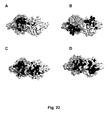
- MICA molecules have been clearly implicated in inflammatory diseases, and, as shown in Example 3, several human antibodies were effective at blocking MICA-binding to cell-surface NKG2D, particularly MS and 21 F2, and epitope determination showed that MS Fab obstructed MICA from binding (Example 11, Figure 20 ). Both MS and 21 F2 were also highly efficient in blocking NK-cell mediated cytotoxicity (Example 6). Thus, these results demonstrate that the invention provides antibodies having such properties.
- antibodies of the invention are efficient antagonists, but also have insignificant agonistic effect on hNKG2D signalling, thus not contributing to NKG2D-driven inflammation.
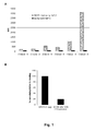
- hNKG2D signalling no co-stimulation of immobilized MS on CD3-triggered proliferation of PBMCs could be detected, whereas immobilized ON72 resulted in a small but significant co-stimulation.
- this difference may at least in part be due to the differences in epitopes, shown in Figures 20-22 .
- An antigen-binding portion of bivalent MS antibody binds strongly to one monomer in an hNKG2D dimer complex, but blocks binding of a second MS antibody (or a second antigen-binding portion of the same antibody) to the second monomer.
- an antigen-binding portion of a bivalent hzON72 antibody binds a first monomer in an hNKG2D dimer, it does not block the binding of a second hzON72 antibody (or a second antigen-binding portion of the same antibody) to the second monomer.
- the invention provides human or humanized anti-NKG2D antibodies which, when added to NKG2D-expressing NK or T cells, cross-link not more than 2 hNKG2D dimers.
- such antibodies are bivalent.
- a bivalent antibody (such as, e.g., MS) for which the binding of the antigen-binding portion to an NKG2D monomer unit blocks further binding to the second NKG2D monomer unit can at most crosslink 2 hNKG2D dimers only.
- a bivalent antibody which can bind an NKG2D monomer unit in an hNKG2D dimer without blocking binding to the second NKG2D monomer unit in an hNKG2D dimer can result in cross-linking of any number of hNKG2D dimers. Clustering of surface receptors commonly occurs in receptor activation.
- the invention provides human or humanized anti-NKG2D antibodies which, when added to NKG2D-expressing NK or T cells, binds strongly only to one monomer in an hNKG2D dimer complex.
- strong binding to both monomers of the dimer can be a prerequisite for activation of the NKG2D-receptor.
- MICA and hzON72 bind strongly to both monomer units in an hNKG2D dimer.
- MS binding to hNKG2D dimer is, however, dominated by binding to one of the monomer units while binding to the second monomer unit is weak and unspecific, and with a smaller solvent-excluded surface area on the second hNKG2D monomer (Example 11).
- the ratio of the solvent-excluded surface areas from the first and second NKG2D monomer units by the binding of an antibody of the invention is more than about 1:1, at least about 2:1, or at least about 3:1.
- the invention provides human or humanized anti-NKG2D antibodies which bind essentially the same epitope as MS. Without being limited to theory, interactions of a ligand with particular residues, or residue combinations, on the hNKG2D dimer could avoid or minimize agonist activity.
- the epitope of an antibody of the invention comprises at least one residue selected from, at least 3 residues selected from, at least 5 residues selected from, at least 8 residues selected from, at least 10 residues selected from, at least 12 residues selected from, or all of the residues selected from the group consisting of Lys 150, Ser 151, Tyr 152, Thr 180, Ile 181, Ile 182, Glu 183, Met 184, Gln 185, Leu 191, Lys 197, Tyr 199, Glu 201, Thr 205, Pro 206, Asn 207 and Thr 208 of hNKG2D (SEQ ID NO: 2).
- the present invention provides a fully human antibody, or antigen-binding fragment thereof, that effectively prevents NKG2D-mediated cytotoxicity of a hNKG2D-expressing NK or T cell, competes with at least MICA in binding to hNKG2D; reduces the amount of cell-surface hNKG2D upon binding via, e.g., stimulating down-modulation of hNKG2D, internalization of hNKG2D and/or preventing reappearance of hNKG2D; has an affinity to hNKG2D of 10 nM or less, cross-reacts with cynomolgus and/or rhesus NKG2D; and is non-depleting, e.g., by having an IgG4 isotype.
- the antibody is a non-depleting fully human antibody of the IgG4 isotype, with an affinity to hNKG2D of 1 nM or less, preferably 300 pM or less, which blocks at least 50%, at least 70%, or at least 90% of endogenous hNKG2D-ligand binding, and reduces the amount of cell-surface hNKG2D with at least 10%, at least 30%, or at least 50%.
- the antibody is a bivalent non-depleting fully human antibody of the IgG4 isotype, with an affinity below 100 pM, which has an EC50 concentration below 0.01 ng/ml for blocking the binding of full saturation dose of MICA-Fc to cell-surface associated NKG2D, is capable of reducing the amount of cell-surface NKG2D with at least 75% upon binding, and, optionally, has an EC50 concentration for reducing a ligand-induced NK cell cytotoxicity that is lower than the EC50 concentration required for binding to cell-curface associated NKG2D.
- the antibody may further be capable of achieving, in an assay using NKG2D-expressing cells, its maximum level of hNKG2D down-modulation at a concentration lower than that required to obtain saturation of the hNKG2D receptors ( i.e ., saturation dose).
- the antibodies of the invention are characterized by particular functional and/or structural features or properties. Assays to evaluate the functional activities of anti-hNKG2D antibodies are described in detail in the Examples, and structural properties such as, e.g., amino acid sequences, are described below.
- the antibodies of the invention bind to hNKG2D.
- an antibody of the invention binds to hNKG2D with high affinity, for example with a KD of 10 -7 M or less, a KD of 10 -8 M or less, a KD of 1 nM or less, a KD of 0.3 nM or less, a KD of 0.2 nM or less, 0.1 nM or less, 0.05 nM or less, or 0.01 nM or less.
- the antibody binds to hNKG2D with an affinity of 0.1 nM or less.
- the invention provides antibodies also binding to one or more NKG2D orthologs in monkey such as cynomolgous monkey ( Macaca fascicularis, NCBI accession No. AJ426429) and rhesus monkey (Macaca mulatta, NCBI accession No. AJ554302), and/or to hNKG2D homodimer, correctly folded monomeric full-length hNKG2D, hNKG2D fragment comprising an extracellular portion of hNKG2D, denatured hNKG2D, or to any combination of the preceding NKG2D forms.
- monkey such as cynomolgous monkey ( Macaca fascicularis, NCBI accession No. AJ426429) and rhesus monkey (Macaca mulatta, NCBI accession No. AJ554302)
- hNKG2D homodimer homodimer
- correctly folded monomeric full-length hNKG2D, hNKG2D fragment comprising an extra
- an antibody of the invention binds to cynomolgous and/or rhesus NKG2D with similar affinity or efficacy as it binds to hNKG2D.
- an antibody can bind to NKG2D-expressing cynomolgous or rhesus NK or T cells with an EC50 of about 50% or more, about 65% or more, or about 75% or more, of the corresponding EC50 for a corresponding population of NKG2D-expressing human NK or T cells.
- an antibody can bind to cynomolgous or rhesus NKG2D with an affinity of about 30% or more, about 50% or more, about 65% or more, or about 75% or more, about 80% or more, about 85% or more, or about 90% or more, of the affinity for hNKG2D.
- Such antibodies have the advantage of allowing for toxicity testing in the most suitable animal model (or models) prior to use in humans.
- antibodies of the invention also bind a form of NKG2D that known murine anti-hNKG2D antibodies such as ON72 do not bind. Specifically, as described in Example 3, pre-incubation with ON72 only blocked about 82% of subsequently added human 16F16 antibody from binding to hNKG2D, while pre-incubation with 16F16 blocked about 95% of subsequently added ON72 from binding to hNKG2D.
- the antibodies of the invention can reduce or inhibit hNKG2D-mediated activation of NK or T cells, i.e., antagonize the hNKG2D receptor. This may be tested in, e.g., one or more cytotoxicity assays described herein or known in the art.
- an antibody inhibits hNKG2D-mediated activation of an NK or T cell if it inhibits the NK- or T cell-mediated killing of an NKG2D-ligand-expressing target cell by at least 10%, more preferably by at least 30%, even more preferably by at least 40%, at least 50%, at least 60%, at least 70%, at least 80% or at least 90%, as compared to target cell killing in the absence of any anti-hNKG2D antibody or in the presence of a non-specific, control antibody.
- Antibodies of the invention that are hNKG2D antagonists can have no or low agonist activity.
- such antibodies are human or humanized.
- Agonist activity may be tested in one of the assay described herein, or an assay known in the art.
- one type of assay is a co-stimulation assay measuring proliferation of peripheral blood lymphocytes (PBMCs) stimulated with low levels of CD3 in the presence or absence of immobilized anti-NKG2D antibody (see Example 10).
- proliferation in the presence of an antibody of the invention is not more than 30%, not more than 20%, not more than 10%, not more than 5% or not significantly higher than in the absence of antibody.
- proliferation in the presence of an antibody of the invention is not significantly higher than in the absence of antibody.
- hNKG2D agonist activity of an antibody of the invention in an agonist assay is not more than 30%, not more than 20%, not more than 10%, not more than 5%, or not significantly higher than a control value.
- the control is preferably a negative control, such as, e.g., in the absence of antibody, in the absence of cell or another reagent, and/or in the presence of an irrelevant antibody.
- agonist activity of an antibody of the invention is not significantly higher than a control value.
- the invention provides antibodies that have a lower, preferably substantially lower, EC50 concentration for blocking ligand-induced cytotoxicity than for binding to cell-surface NKG2D of an NK or T cell.
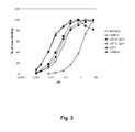
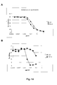
- the EC50 concentration for binding to cell-surface NKG2D expressed on BaF/3 cells was similar to the EC50 concentration for blocking NK-cell mediated killing of ligand- (ULBP3-) expressing target cells (0.065 ⁇ g/ml), whereas 21 F2 had a lower, and MS a substantially lower, EC50 for blocking cytotoxicity (21 F2: 0.021 ⁇ g/ml; MS: 0.012 ⁇ g/ml) than for binding to cell-surface NKG2D (21 F2: 0.033 ⁇ g/ml; MS: 0.032 g/ml) (see Examples 6 and 9).
- the invention provides antibodies, preferably human or humanized antibodies, that have a lower EC50 concentration for blocking ligand-induced cytotoxicity than for binding to cell-surface NKG2D of an NK or T cell.
- the EC50 for blocking cytotoxicity of NK or T cells of a cell line or other suitable preparation can be, e.g., about 95% or less, about 90% or less, about 85% or less, about 80% or less, about 70% or less, about 50% or less, or about 40% or less, of the EC50 for binding to cell-surface NKG2D of the same cell line or preparation.
- Exemplary cell lines for testing include NK-92 and NKL cells.
- the invention provides antibodies that achieve maximum blockage of NK cell cytotoxicity at a concentration lower than the concentration required to saturate the available hNKG2D-receptors.
- the antibodies also compete with MS in binding to hNKG2D.
- such antibodies bind to essentially the same hNKG2D epitope as MS.
- the antibodies may reduce or inhibit NKG2D-mediated activation by, e.g., interfering with the hNKG2D-binding of one or more endogeous hNKG2D-ligands.
- the antibodies may reduce or inhibit the hNKG2D-binding of MICA; MICB; ULBP1; ULBP2; ULBP4; and/or RAET1-family member; e.g., by reducing or inhibiting the hNKG2D-binding of MICA; or of MICA and MICB; or of MICA and ULBP3; or of MICA, MICB, and ULBP3; or of MICA, MICB, and all ULBP1, -2, -3, and 4; or of MICA, MICB, and one or more RAET1 family members.
- antibodies of the invention are capable of inhibiting at least 30% of ligand binding, or at least 50% of ligand binding, or at least 70% of ligand binding, or at least 80%, or at least 90% of ligand binding.
- the IC50 for an antibody of the invention to inhibit the hNKG2D-binding of 1 ⁇ g MICA-mFc is 1 nM or less, 0.5 nM or less, 0.2 nM or less, 0.1 nM or less, 0.05 nM or less, or 0.02 nM or less, 0.01 nM or less, 0.005 or less, or 0.002 or less.
- full blockage of 1 ⁇ g MICA-mFc binding is achieved at an antibody concentration of 5 nM or less, 1 nM or less, 0.7 nM or less, 0.5 nM or less, or 0.2 nM or less, 0.1 nM or less, 0.05 nM or less, or about 0.02 nM or less.
- the invention provides antibodies, especially human antibodies, that are as efficient or more efficient in reducing or inhibiting ligand hNKG2D-binding, such as, e.g., MICA binding to hNKG2D, than any of ON72, BAT221, 5C6, 1D11, ECM217, and 149810.
- an anti-hNKG2D antibody of the invention can be capable of reducing the amount of cell-surface hNKG2D upon ( i.e ., following) binding.
- Reduction of cell-surface associated hNKG2D upon binding of an antibody can be an advantageous feature, since it reduces the number of hNKG2D receptors available for ligand binding and subsequent activation. Without being limited to theory, this reduction may be caused by NKG2D down-modulation, internalization, or other mechanism.
- anti-hNKG2D antibodies having a human Fc-region such as human antibodies, are capable of effectively reducing the amount of cell-curface hNKG2D.
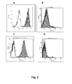
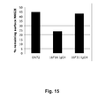
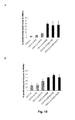
- human anti-hNKG2D antibodies 16F16, MS, and 21 F2 all reduced the amount of cell-surface hNKG2D with about 75% or more after overnight incubation in the absence of serum, with MS being the most effective, achieving 75-90% downmodulation at a low concentration ( Figures 15-17 ).
- an MS concentration corresponding to less than saturating concentration on hNKG2D-expressing BaF/3 cells achieved maximum downmodulation ( Figure 16B ).
- the invention provides antibodies binding to hNKG2D that are able to achieve maximum down-modulation of hNKG2D at less than saturating concentrations.
- such antibodies also compete with MS in binding to hNKG2D.
- such antibodies also bind to essentially the same hNKG2D epitope as MS.
- An antibody of the invention can be capable of reducing cell surface hNKG2D by at least 10%, at least 20%, at least 30%, at least 50%, at least 70%, or at least 90% as compared to cell-surface hNKG2D in the absence of anti-hNKG2D antibody or in the presence of a non-specific control antibody.
- the antibodies achieve reduction of cell-surface NKG2D while causing no or minimal activation of NKG2D-receptor signalling, i.e., with no or minimal agonist activity.
- the invention provides antibodies, particularly human antibodies, which are capable of a higher degree of down-modulation than a control antibody selected from ON72, BAT221, 5C6, 1D11, ECM217, and 149810.
- an anti-hNKG2D antibody of the invention can be capable of achieving maximum down-modulation of cell-surface NKG2D expressed by a cell or cell-line at a concentration lower than a saturating concentration.
- the invention provides antibodies that compete with and/or bind to the same epitope on hNKG2D as 16F16, 16F31, MS, and/or 21F2, more preferably MS and/or 21 F2.
- Such antibodies can be identified based on their ability to cross-compete with 16F16, 16F31, MS, or 21 F2 in standard hNKG2D binding assays as described herein.
- test antibody to inhibit the binding of 16F16, 16F31, MS, or 21 F2 to hNKG2D demonstrates that the test antibody can compete with 16F16, 16F31, MS, or 21 F2 for binding to hNKG2D and thus can bind to the same epitope on hNKG2D as 16F16, 16F31, MS, or 21 F2.
- the antibody that binds to the same epitope on hNKG2D as 16F16, 16F31, MS or 21 F2 is a human monoclonal antibody.
- human monoclonal antibodies can be prepared and isolated as described in the Examples.
- the antibody binds to a different epitope than any of the mouse monoclonal antibodies ON72, BAT221, 5C6, 1D11, ECM217, and 149810, and cross-competes more with 16F16, 16F31, MS, or 21 F2 than with either of the listed mouse monoclonal antibodies.
- the epitope of an antibody of the invention comprises one or more residues selected from Lys 150, Ser 151, Tyr 152, Thr 180, Ile 181, Ile 182, Glu 183, Met 184, Gln 185, Leu 191, Lys 197, Tyr 199, Glu 201, Thr 205, Pro 206, Asn 207 and Thr 208 of hNKG2D (SEQ ID NO: 2).
- the epitope of an antibody of the invention comprises 5 or more residues selected from Lys 150, Ser 151, Tyr 152, Thr 180, Ile 181, Ile 182, Glu 183, Met 184, Gln 185, Leu 191, Lys 197, Tyr 199, Glu 201, Thr 205, Pro 206, Asn 207 and Thr 208 of hNKG2D (SEQ ID NO: 2).
- the epitope of an antibody of the invention comprises 8, 10, 12 or more residues selected from Lys 150, Ser 151, Tyr 152, Thr 180, Ile 181, Ile 182, Glu 183, Met 184, Gln 185, Leu 191, Lys 197, Tyr 199, Glu 201, Thr 205, Pro 206, Asn 207 and Thr 208 of hNKG2D (SEQ ID NO: 2).
- the epitope of an antibody of the invention comprises the residues Lys 150, Ser 151, Tyr 152, Thr 180, Ile 181, Ile 182, Glu 183, Met 184, Gln 185, Leu 191, Lys 197, Tyr 199, Glu 201, Thr 205, Pro 206, Asn 207 and Thr 208 of hNKG2D (SEQ ID NO: 2).
- the epitope of an antibody of the invention consists essentially of the residues Lys 150, Ser 151, Tyr 152, Thr 180, Ile 181, Ile 182, Glu 183, Met 184, Gln 185, Leu 191, Lys 197, Tyr 199, Glu 201, Thr 205, Pro 206, Asn 207 and Thr 208 of hNKG2D (SEQ ID NO: 2).
- the epitope of an antibody of the invention consists of one or more residues selected from Lys 150, Ser 151, Tyr 152, Thr 180, Ile 181, Ile 182, Glu 183, Met 184, Gln 185, Leu 191, Lys 197, Tyr 199, Glu 201, Thr 205, Pro 206, Asn 207 and Thr 208 of hNKG2D (SEQ ID NO: 2).
- the epitope of an antibody of the invention consists of the residues Lys 150, Ser 151, Tyr 152, Thr 180, Ile 181, Ile 182, Glu 183, Met 184, Gln 185, Leu 191, Lys 197, Tyr 199, Glu 201, Thr 205, Pro 206, Asn 207 and Thr 208 of hNKG2D (SEQ ID NO: 2).
- the epitope of an antibody of the invention comprises one or more residues involved in hydrogen-binding selected from Lys 150, Ser 151, Tyr 152, Ile 181, Met 184, Gln 185, Lys 197, Thr 205, and Asn 207 of hNKG2D (SEQ ID NO: 2). In one embodiment, the epitope of an antibody of the invention comprises 5 or more residues involved in hydrogen-binding selected from Lys 150, Ser 151, Tyr 152, Ile 181, Met 184, Gln 185, Lys 197, Thr 205, and Asn 207 of hNKG2D (SEQ ID NO: 2).
- the epitope of an antibody of the invention comprises Lys 150, Ser 151, Tyr 152, Ile 181, Met 184, Gln 185, Lys 197, Thr 205, and Asn 207 of hNKG2D (SEQ ID NO: 2).
- Preferred antibodies of the invention exhibit at least one, more preferably two, three, four, five or more, of the following properties: (a) prevents NKG2D-mediated activation of an NKG2D-expressing NK or T cell, optionally with an EC50 for reducing ligand-induced cytotoxicity lower than the EC50 for binding to the cell; (b) competes with at least one NKG2D ligand in binding to NKG2D, preferably with at least MICA and ULBP3; (c) reduces the amount of NKG2D on the surface of a NKG2D-expressing NK or T cell, preferably with at least 75%; (d) binds to cynomolgous and/or rhesus NKG2D, preferably with no less than 50% of the affinity by which it binds to hNKG2D; (e) binds to more than one form or conformation of NKG2D; (f) binds to NKG2D with a Kd of 1 nM
- Preferred antibodies of the invention are the human monoclonal antibodies 16F16, 16F31, MS, and 21 F2 produced, isolated, and structurally and functionally characterized as described in the Examples. Full-length, variable, and CDR sequences of these antibodies are set forth in Table 1.
- Certain anti-NKG2D antibodies of the invention has the same or a similar paratope as MS.
- the antibody has a paratope comprising residues corresponding to one or more of Tyr 33 and Trp 97 of the MS L chain (SEQ ID NO: 41), and/or to one or more of Gln 1, Asp 26, Asp 27, Ser 30, Ser 31, Tyr 32, Tyr 33, His 50, Ser 52, Tyr 53, Ser 54, Ser 56, Ala 57, Asn 58, Trp 98 and Asp 99 of the MS H chain (SEQ ID NO: 40).
- the antibody has a paratope comprising residues corresponding to Tyr 33 and Trp 97 of the MS L chain (SEQ ID NO: 41), and/or to 3, 5, 7, 10 or more of Gln 1, Asp 26, Asp 27, Ser 30, Ser 31, Tyr 32, Tyr 33, His 50, Ser 52, Tyr 53, Ser 54, Ser 56, Ala 57, Asn 58, Trp 98 and Asp 99 of the MS H chain (SEQ ID NO: 40).
- the antibody has a paratope comprising residues corresponding to Tyr 33 and Trp 97 of the MS L chain (SEQ ID NO: 41), and Gln 1, Asp 26, Asp 27, Ser 30, Ser 31, Tyr 32, Tyr 33, His 50, Ser 52, Tyr 53, Ser 54, Ser 56, Ala 57, Asn 58, Trp 98 and Asp 99 of the MS H chain (SEQ ID NO: 40).
- the antibody has a paratope consisting essentially of residues corresponding to Tyr 33 and Trp 97 of the MS L chain (SEQ ID NO: 41), and Gln 1, Asp 26, Asp 27, Ser 30, Ser 31, Tyr 32, Tyr 33, His 50, Ser 52, Tyr 53, Ser 54, Ser 56, Ala 57, Asn 58, Trp 98 and Asp 99 of the MS H chain (SEQ ID NO: 40).
- the antibody has a paratope consisting of residues corresponding to Tyr 33 and Trp 97 of the MS L chain (SEQ ID NO: 41), and Gln 1, Asp 26, Asp 27, Ser 30, Ser 31, Tyr 32, Tyr 33, His 50, Ser 52, Tyr 53, Ser 54, Ser 56, Ala 57, Asn 58, Trp 98 and Asp 99 of the MS H chain (SEQ ID NO: 40).
- VH and VL sequences of these antibodies can be tested using the binding assays described herein (e.g. flow cytometry, Biacore, ELISAs) and/or using a cytotoxicity assay as described herein.
- a VH sequence from a particular VH/VL pairing is replaced with a structurally similar VH sequence.
- a VL sequence from a particular VH/VL pairing is replaced with a structurally similar VL sequence.
- the invention provides an isolated monoclonal antibody, or antigen binding portion thereof, comprising: (a) a VH region comprising an amino acid sequence selected from the group consisting of SEQ ID NOS: 11, 13, 44, and 46, and (b) a VL region comprising an amino acid sequence selected from the group consisting of SEQ ID NOs: 12, 14, 45, and 47; wherein the antibody binds hNKG2D.
- Preferred heavy and light chain combinations include: (a) a VH region comprising the amino acid sequence of SEQ ID NO: 11; and (b) a light chain variable region comprising the amino acid sequence of SEQ ID NO: 12; (a) a heavy chain variable region comprising the amino acid sequence of SEQ ID NO: 13; and (b) a light chain variable region comprising the amino acid sequence of SEQ ID NO: 14; (a) a VH region comprising the amino acid sequence of SEQ ID NO: 44; and (b) a light chain variable region comprising the amino acid sequence of SEQ ID NO: 46; or (a) a heavy chain variable region comprising the amino acid sequence of SEQ ID NO: 45; and (b) a light chain variable region comprising the amino acid sequence of SEQ ID NO: 47.
- the invention provides antibodies that comprise the heavy chain and light chain CDR1s, CDR2s and/or CDR3s of 16F16, 16F31, MS, or 21 F2, or combinations thereof.
- the CDR regions are delineated using the Kabat system ( Kabat et al. (1991) Sequences of Proteins of Immunological Interest, Fifth Edition, U.S. Department of Health and Human Services, NIH Publication No. 91-3242 ). See, e.g., Figures 4 and 5 .
- VH CDR1, 2 and 3 sequences and VL CDR1, 2 and 3 sequences can be "mixed and matched" (i.e ., CDRs from different antibodies can be mixed and match, although each antibody can contain a VH CDR1, 2 and 3 and a VL CDR1, 2 and 3) to create other anti-hNKG2D binding molecules of the invention.
- the hNKG2D-binding of such "mixed and matched" antibodies can be tested using the binding assays described above and in the Examples (e.g. flow cytometry, Biacore, or ELISAs).
- the CDR1, CDR2 and/or CDR3 sequence from a particular VH sequence is replaced with a structurally similar CDR sequence(s).
- the CDR1, CDR2 and/or CDR3 sequence from a particular VL sequence preferably is replaced with a structurally similar CDR sequence(s).
- the VL CDR1s and CDR3s of 16F16, 16F31, MS, and 21 F2 and the VL CDR2 sequences of MS and 21 F2 share some structural similarity and therefore are amenable to mixing and matching.
- VH and VL sequences can be created by substituting one or more VH and/or VL CDR region sequences with structurally similar sequences from the CDR sequences disclosed herein for monoclonal antibodies antibodies 16F16, 16F31, MS, and 21 F2.
- the invention provides an isolated monoclonal antibody, or antigen binding portion thereof comprising: (a) a VH CDR1 comprising an amino acid sequence selected from the group consisting of SEQ ID NOs: 15, 21, 48, and 54; (b) a VH CDR2 comprising an amino acid sequence selected from the group consisting of SEQ ID NOs: 16 , 22, 49,and 55; (c) a VH CDR3 comprising an amino acid sequence selected from the group consisting of SEQ ID NOs: 17, 23, 50, and 56; (d) a VL CDR1 comprising an amino acid sequence selected from the group consisting of SEQ ID NOs:18, 24, 51, and 57; (e) a VL CDR2 comprising an amino acid sequence selected from the group consisting of SEQ ID NOs:19, 25, 52, and 57; and (f) a VL CDR3 comprising an amino acid sequence selected from the group consisting of SEQ ID NOs: 20, 26, 53, and 59; wherein the antibody
- the antibody comprises: (a) a VH CDR1 comprising SEQ ID NO:15; (b) a VH CDR2 comprising SEQ ID NO:16; (c) a VH CDR3 comprising SEQ ID NO:17; (d) a VL CDR1 comprising SEQ ID NO:18; (e) a VL CDR2 comprising SEQ ID NO: 19; and (f) a VL CDR3 comprising SEQ ID NO: 20.
- the antibody comprises: (a) a VH CDR1 comprising SEQ ID NO: 21; (b) a VH CDR2 comprising SEQ ID NO:22; (c) a VH CDR3 comprising SEQ ID NO:23; (d) a VL region CDR1 comprising SEQ ID NO:24; (e) a VL CDR2 comprising SEQ ID NO:25; and (f) a VL CDR3 comprising SEQ ID NO: 26.
- the antibody comprises: (a) a VH CDR1 comprising SEQ ID NO: 48; (b) a VH CDR2 comprising SEQ ID NO:49; (c) a VH CDR3 comprising SEQ ID NO:50; (d) a VL region CDR1 comprising SEQ ID NO:51; (e) a VL CDR2 comprising SEQ ID NO:52; and (f) a VL CDR3 comprising SEQ ID NO: 53.
- the antibody comprises: (a) a VH CDR1 comprising SEQ ID NO: 54; (b) a VH CDR2 comprising SEQ ID NO:55; (c) a VH CDR3 comprising SEQ ID NO:56; (d) a VL region CDR1 comprising SEQ ID NO:57; (e) a VL CDR2 comprising SEQ ID NO:58; and (f) a VL CDR3 comprising SEQ ID NO: 59.
- the antibody comprises: (a) a VH CDR1 consisting of SEQ ID NO:15; (b) a VH CDR2 consisting of SEQ ID NO:16; (c) a VH CDR3 consisting of SEQ ID NO:17; (d) a VL CDR1 consisting of SEQ ID NO: 18; (e) a VL CDR2 consisting of SEQ ID NO: 19; and (f) a VL CDR3 consisting of SEQ ID NO: 20.
- the antibody comprises: (a) a VH CDR1 consisting of SEQ ID NO: 21; (b) a VH CDR2 consisting of SEQ ID NO:22; (c) a VH CDR3 consisting of SEQ ID NO:23; (d) a VL region CDR1 consisting of SEQ ID NO:24; (e) a VL CDR2 consisting of SEQ ID NO:25; and (f) a VL CDR3 consisting of SEQ ID NO: 26.
- the antibody comprises: (a) a VH CDR1 consisting of SEQ ID NO: 48; (b) a VH CDR2 consisting of SEQ ID NO:49; (c) a VH CDR3 consisting of SEQ ID NO:50; (d) a VL region CDR1 consisting of SEQ ID NO:51; (e) a VL CDR2 consisting of SEQ ID NO:52; and (f) a VL CDR3 consisting of SEQ ID NO: 53.
- the antibody comprises: (a) a VH CDR1 consisting of SEQ ID NO: 48; (b) a VH CDR2 consisting of SEQ ID NO:49; (c) a VH CDR3 consisting of SEQ ID NO:50; (d) a VL region CDR1 consisting of SEQ ID NO:51; (e) a VL CDR2 consisting of SEQ ID NO:52; and (f) a VL CDR3 consisting of SEQ ID NO: 53, and residues corresponding to one, two, or all of Gln 1, Asp 26, and Asp 27 in the MS H chain (SEQ ID NO: 40).
- the antibody comprises: (a) a VH CDR1 consisting of SEQ ID NO: 54; (b) a VH CDR2 consisting of SEQ ID NO:55; (c) a VH CDR3 consisting of SEQ ID NO:56; (d) a VL region CDR1 consisting of SEQ ID NO:57; (e) a VL CDR2 consisting of SEQ ID NO:58; and (f) a VL CDR3 consisting of SEQ ID NO: 59.
- an antibody of the invention comprises a VH region from a particular germline H chain immunoglobulin gene, or a combination of particular germline H chain immunoglobulin genes; and/or a VL region from a particular germline L chain immunoglobulin gene, or a combination of particular germline L chain immunoglobulin genes.
- Such combinations can be obtained, e.g., in vivo via somatic recombination in a B cell.
- the invention provides an isolated anti-hNKG2D antibody, or an antigen-binding fragment thereof, wherein the antibody: (a) comprises a VH region from a human VH3_21, VH3_20, VH4_59, or VH5_51 gene recombined with a human D3-9, D3-10, or D3_10_R3 gene and a JH3, JH4 or JH6 gene, (b) comprises a VL region derived from a human VKI_L15 or VKIII_A27 or VKIII_L6 gene recombined with a human JK1, JK2 or JK3 gene, and (c) the antibody binds to hNKG2D.
- the antibody comprises a VH region from a human VH3_21, VH3_20, VH4_59, or VH5_51 gene recombined with a human D3-9, D3-10, or D3_10_R3 gene and a JH3, JH
- the invention provides an isolated anti-hNKG2D antibody, or an antigen-binding fragment thereof, comprising a VH region obtained by a recombination of human VH3_21, D3-9, and JH4 genes and a VL region obtained by a recombination of human VKI_L15 and JK2 genes.
- the invention provides an isolated anti-hNKG2D antibody, or an antigen-binding fragment thereof, comprising a VH region obtained by a recombination of human VH3_20, D3-10, and JH6 genes and a VL region obtained by a recombination of human VKIII_A27 and JK3 genes.
- the invention provides an isolated anti-hNKG2D antibody, or an antigen-binding fragment thereof, comprising a VH region obtained by a recombination of human VH4_59, a D gene, and JH3 genes and a VL region obtained by a recombination of human VKIII_A27 and JK1 genes.
- the invention provides an isolated anti-hNKG2D antibody, or an antigen-binding fragment thereof, comprising a VH region obtained by a recombination of human VH5_51, D3_10_R3, and JH4 genes and a VL region obtained by a recombination of human VKIII_L6 and JK1 genes.
- the invention provides isolated anti-NKG2D antbodies obtained by introducing one, two, three, four or more amino acid substitutions and/or somatic hypermutations in the VH and/or VL region of an anti-hNKG2D antibody described above.
- a human antibody comprises heavy or light chain variable regions "of” or “derived from” or that are “the product of” a particular germline sequence if the variable regions of the antibody are obtained from a system (as described below) that uses human germline immunoglobulin genes.
- systems include immunizing a transgenic mouse carrying human immunoglobulin genes with the antigen of interest or screening a human immunoglobulin gene library displayed on phage with the antigen of interest.
- a human antibody that is "of” or “derived from” or “the product of” a human germline immunoglobulin sequence can be identified as such by comparing the amino acid sequence of the human antibody to the amino acid sequences of human germline immunoglobulins and selecting the human germline immunoglobulin sequence that is closest in sequence ( i.e ., greatest % identity) to the sequence of the human antibody.
- a human antibody that is "of” or “derived from” or “the product of” a particular human germline immunoglobulin sequence may contain amino acid differences as compared to the germline sequence, due to, for example, naturally-occurring somatic mutations or intentional introduction of site-directed mutation(s) (which may be selected substitutions).
- a human antibody is typically at least 90% identical in amino acid sequence to an amino acid sequence encoded by a recombined germline immunoglobulin sequence and can usually be identified as human when compared to the germline immunoglobulin amino acid sequences of other species (e.g. , murine germline sequences). In certain cases, a human antibody may be at least 95%, or even at least 96%, 97%, 98%, or 99% identical in amino acid sequence to the amino acid sequence encoded by the recombined germline immunoglobulin gene.
- a human antibody derived from a particular human germline sequence will display no more than 10 amino acid differences from the amino acid sequence encoded by the human germline immunoglobulin gene.
- the human antibody may display no more than 8, no more than 5, or even no more than 4, 3, 2, or 1 amino acid difference, or no amino acid difference, from the amino acid sequence encoded by the recombined germline immunoglobulin gene.
- an antibody of the invention comprises heavy and light chain variable regions comprising amino acid sequences that are homologous to the amino acid sequences of the preferred antibodies described herein, and wherein the antibodies retain the desired functional properties of the anti-hNKG2D antibodies of the invention.
- the invention provides an isolated antibody, or antigen binding portion thereof, comprising a heavy chain variable region and a light chain variable region, wherein: (a) the VH region comprises an amino acid sequence that is at least 80% identical to an amino acid sequence selected from the group consisting of SEQ ID NOs: 11, 13, 44, and 46; (b) the VL region comprises an amino acid sequence that is at least 80% identical to an amino acid sequence selected from the group consisting of SEQ ID NOs: 12, 14, 45, and 47; (c) the antibody binds to hNKG2D and exhibits at least one of the functional properties described herein, preferably several of the functional properties described herein.
- the VH and/or VL amino acid sequences may be 85%, 90%, 95%, 96%, 97%, 98% or 99% identical to the sequences set forth above.
- An antibody having VH and VL regions having high (i.e ., 80% or greater) identity to the VH and VL regions of the sequences set forth above, can be obtained by mutagenesis (e.g., site-directed or PCR-mediated mutagenesis) of nucleic acid molecules encoding SEQ ID NOs:11-14 or 44-47, followed by testing of the encoded altered antibody for retained function (e.g., hNKG2D binding affinity, hNKG2D-ligand blocking, hNKG2D downmodulation, or reduction of NKG2D-mediated activation of an NK or T cell) using the functional assays described herein.
- mutagenesis e.g., site-directed or PCR-mediated mutagenesis
- the encoded altered antibody for retained function e.g., hNKG
- the comparison of sequences and determination of percent identity between two sequences can be accomplished using a mathematical algorithm in sequence-analysis software. Protein analysis software matches similar sequences using measures of similarity assigned to various substitutions, deletions and other modifications, including conservative amino acid substitutions.
- the percent identity between two amino acid sequences can be determined, e.g., using the Needleman and Wunsch (J. Mol. Biol. 48:444-453 (1970 )) algorithm which has been incorporated into the GAP program in the GCG software package (available at http://www.gcg.com), using either a Blossum 62 matrix or a PAM250 matrix, and a gap weight of 16, 14, 12, 10, 8, 6, or 4 and a length weight of 1, 2, 3, 4, 5, or 6.
- Polypeptide sequences can also be compared using FASTA, applying default or recommended parameters.
- FASTA e.g., FASTA2 and FASTA3
- FASTA3 provides alignments and percent sequence identity of the regions of the best overlap between the query and search sequences ( Pearson, Methods Enzymol. 1990; 183:63-98 ; Pearson, Methods Mol. Biol. 2000;132:185-219 ).
- the percent identity between two amino acid sequences can also be determined using the algorithm of E. Meyers and W. Miller (Comput. Appl. Biosci., 1988;11-17 ) which has been incorporated into the ALIGN program (version 2.0), using a PAM120 weight residue table, a gap length penalty of 12 and a gap penalty of 4.
- Another algorithm for comparing a sequence to a other sequences contained in a database is the computer program BLAST, especially blastp, using default parameters. See, e.g ., Altschul et al., J. Mol. Biol. 1990;215:403-410 ; Altschul et al., Nucleic Acids Res. 1997;25:3389-402 (1997 ); each herein incorporated by reference.
- the protein sequences of the present invention can there be used as a "query sequence" to perform a search against public databases to, for example, identify related sequences. Such searches can be performed using the XBLAST program (version 2.0) of Altschul, et al. 1990 ( supra ) .
- Gapped BLAST can be utilized as described in Altschul et al., 1997 ( supra ) ..
- the default parameters of the respective programs e.g. , XBLAST and NBLAST
- XBLAST and NBLAST the default parameters of the respective programs
- an antibody of the invention comprises a VH region comprising CDR1, CDR2 and CDR3 sequences and a VL region comprising CDR1, CDR2 and CDR3 sequences, wherein one or more of these CDR sequences comprise specified amino acid sequences based on the preferred antibodies described herein; 16F16, 16F31, MS, or 21 F2, wherein one or more CDRs optionally contains one or more conservative amino acid modifications, and wherein the antibodies retain the desired functional properties of the anti-hNKG2D antibodies of the invention.
- the invention provides an isolated antibody, or antigen-binding fragment thereof, comprising a heavy chain variable region comprising CDR1, CDR2, and CDR3 sequences and a light chain variable region comprising CDR1, CDR2, and CDR3 sequences, wherein: (a) the VH region CDR3 sequence comprises an amino acid sequence selected from the group consisting of amino acid sequences of SEQ ID NOs:17, 23, 50, and 56; (b) the VL region CDR3 sequence comprises an amino acid sequence selected from the group consisting of amino acid sequences of SEQ ID NOs: 20, 26, 53 and 59; (c) one or more CDRs optionally contains one or more conservative amino acid modifications, and (d) the antibody binds to hNKG2D and exhibits at least one of the functional properties described herein, more preferably several of the functional properties described herein.
- the VH region CDR2 sequence comprises an amino acid sequence selected from the group consisting of amino acid sequences of SEQ ID NOs: 16, 22, 49, and 55; and the VL region CDR2 sequence comprises an amino acid sequence selected from the group consisting of amino acid sequences of SEQ ID NOs: 19, 25, 52, and 58, wherein one or more CDRs optionally contains one or more conservative amino acid modifications.
- the VH region CDR1 sequence comprises an amino acid sequence selected from the group consisting of amino acid sequences of SEQ ID NOs:15, 21, 48, and 54, and conservative modifications thereof; and the VL region CDR1 sequence comprises an amino acid sequence selected from the group consisting of amino acid sequences of SEQ ID NOs:18, 24, 51, and 57, wherein one or more CDRs optionally contains one or more conservative amino acid modifications.
- conservative amino acid modifications is intended to refer to amino acid modifications that do not significantly affect or alter the binding characteristics of the antibody containing the amino acid sequence. Such conservative modifications include amino acid substitutions, additions and deletions. Modifications can be introduced into an antibody of the invention by standard techniques known in the art, such as, e.g ., site-directed mutagenesis and PCR-mediated mutagenesis.
- An antibody sequence comprising amino acid modifications as compared to a parent antibody is typically at least 90%, preferably at least 95%, 98%, or 99% identical to the corresponding amino acid sequence in the parent and/or comprises at most 10, preferably at most 5, 4, 3, 2 amino acid modifications as compared to the parent antibody sequence.
- Constant amino acid substitutions are typically those in which an amino acid residue is replaced with an amino acid residue having a side chain with similar physicochemical properties. Families of amino acid residues having similar side chains have been defined in the art. These families include amino acids with basic side chains (e.g ., lysine, arginine, histidine), acidic side chains ( e.g ., aspartic acid, glutamic acid), uncharged polar side chains (e.g. glycine, asparagine, glutamine, serine, threonine, tyrosine, cysteine, tryptophan), nonpolar side chains (e.g.
- one or more amino acid residues within the CDR regions of an antibody of the invention can be replaced with other amino acid residues from the same side chain family and the altered antibody can be tested for retained function (i.e ., the functions set forth in (c), (d) and (e) above) using the functional assays described herein.
- anti-hNKG2D antibodies of the invention may be prepared as full-length antibodies or antigen-binding fragments thereof.
- antigen-binding fragments include Fab, Fab', F(ab)2, F(ab')2, F(ab)3, Fv (typically the VL and VH domains of a single arm of an antibody), single-chain Fv (scFv; see e.g., Bird et al., Science 1988;242:423-426 ; and Huston et al.
- dsFv, Fd typically the VH and CH1 domain
- dAb typically a VH domain
- VH, VL, VhH, and V-NAR domains monovalent molecules comprising a single VH and a single VL chain
- minibodies, diabodies, triabodies, tetrabodies, and kappa bodies see, e.g., III et al., Protein Eng 1997;10:949-57
- Antibody fragments can be obtained using conventional recombinant or protein engineering techniques, and the fragments can be screened for antigen-binding or other function in the same manner as are intact antibodies.
- F(ab')2 fragments can be isolated directly from recombinant host cell culture.
- the antibody of choice is a single-chain Fv fragment (scFv). See WO 1993/16185 ; U.S. Pat. No. 5,571,894 ; and U.S. Pat. No. 5,587,458 .
- the antibody fragment may also be a "linear antibody", e.g., as described in U.S. Pat. No. 5,641,870 , for example. Such linear antibody fragments may be monospecific or bispecific.
- the present invention features multispecific molecules comprising an anti-hNKG2D antibody, or an antigen-fragment thereof, of the invention.
- multispecific molecules include bispecific molecules comprising at least one first binding specificity for hNKG2D and a second binding specificity for a second target epitope.
- bispecific antibodies are antibodies that have binding specificities for at least two different epitopes. Methods for making bispecific antibodies are known in the art, and traditional production of full-length bispecific antibodies is usually based on the coexpression of two immunoglobulin heavy-chain-light-chain pairs, where the two chains have different specificities ( Millstein et al., Nature, 305: 537-539 (1983 )). Bispecific antibodies can be prepared as full-length antibodies or antibody fragments ( e.g. F(ab')2 bispecific antibodies) or any other antigen-binding fragments described herein.
- the bispecific antibodies In the bispecific antibodies according to the present invention, at least one binding epitope is on the hNKG2D protein.
- the anti-NKG2D-binding moiety may be combined with second moiety that binds to a molecule on a pro-inflammatory leukocyte, e.g. , a T-cell receptor molecule (e.g. CD2, CD3, CD4, or CD8), so as to focus cellular defense mechanisms to a pro-inflammatory hNKG2D-expressing cell.
- the bispecific antibodies can, e.g. , be used to direct cytotoxic agents to, or an ADCC/CDC attack on, pro-inflammatory cells that express NKG2D.
- the cytotoxic agent could be, e.g. , saporin, an anti-interferon-alpha agent, a vinca alkaloid, the ricin A chain, methotrexate, or a radioactive isotope.
- the second moiety binds a cell-associated target that is presented on or expressed by cells associated with a disease state normally regulated by effector lymphocytes, such as cancer, viral infection, or the like.
- a typical target may be a cell stress-associated molecule such as a MIC molecule ( e.g. , MIC-A or MIC-B) or a ULBP ( e.g ., Rae-1, H-60, ULBP2, ULBP3, HCMV UL18, or Rae-1 ⁇ ) or a pathogen-associated molecule such as a viral hemagglutinin.
- multispecific molecules include those produced from the fusion of a hNKG2D-binding antibody moiety to one or more other non-antibody proteins.
- Such multispecific proteins and how to construct them have been described in the art. See, e.g. , Dreier et al. (Bio-conjug. Chem. 9(4): 482-489 (1998 )); U.S. Patent 6,046,310 ; U.S. Patent Publication No. 20030103984 ; European Patent Application 1 413 316 ; US Patent Publication No. 20040038339 ; von Strandmann et al., Blood (2006;107:1955-1962 .), and WO 2004056873 .
- the non-antibody protein could be, for example, a suitable ligand for any of the antigens of "second moiety" described I the preceding section; e.g. , a ligand for a T-cell or Fc receptor, or a cell-stress molecule such as MIC-A, MIC-B, ULBP, or a pathogen-associated molecule such as a viral hemagglutinin.
- a suitable ligand for any of the antigens of "second moiety" described I the preceding section e.g. , a ligand for a T-cell or Fc receptor, or a cell-stress molecule such as MIC-A, MIC-B, ULBP, or a pathogen-associated molecule such as a viral hemagglutinin.
- Multispecific molecules with more than two valencies are also contemplated.
- trispecific antibodies can be prepared. Tutt et al., J. Immunol, 147: 60 (1991 ).
- the multispecific molecules of the present invention can be prepared by conjugating the constituent binding specificities using methods known in the art. For example, each binding specificity of the multispecific molecule can be generated separately and then conjugated to one another. When the binding specificities are proteins or peptides, a variety of coupling or cross-linking agents can be used for covalent conjugation.
- cross-linking agents examples include protein A, carbodiimide, N-succinimidyl-S-acetyl-thioacetate (SATA), 5,5'-dithiobis(2-nitrobenzoic acid) (DTNB), o-phenylenedimaleimide (oPDM), N-succinimidyl-3-(2-pyridyldithio)propionate (SPDP), and sulfosuccinimidyl 4-(N-maleimidomethyl) cyclohaxane-1-carboxylate (sulfo-SMCC) (see e.g., Karpovsky et al. (1984) J. Exp. Med.
- Preferred conjugating agents are SATA and sulfo-SMCC, both available from Pierce Chemical Co. (Rockford, IL).
- the binding specificities are antibodies, they can be conjugated via sulthydryl bonding of the C-terminus hinge regions of the two heavy chains.
- the hinge region is modified to contain an odd number of sulfhydryl residues, preferably one, prior to conjugation.
- both binding specificities can be encoded in the same vector and expressed and assembled in the same host cell.
- This method is particularly useful where the bispecific molecule is a mAb x mAb, mAb x Fab, Fab x F(ab')2 or ligand x Fab fusion protein.
- a bispecific molecule of the invention can be a single chain molecule comprising one single chain antibody and a binding determinant, or a single chain bispecific molecule comprising two binding determinants. Bispecific molecules may comprise at least two single chain molecules. Methods for preparing bispecific molecules are described or reviewed in, for example in U.S. Patent Number 5,260,203 ; U.S. Patent Number 5,455,030 ; U.S.
- Patent Number 4,881,175 U.S. Patent Number 5,132,405 ; U.S. Patent Number 5,091 , 513 ; U.S. Patent Number 5,476,786 ; U.S. Patent Number: 5,013,653 ; U.S. Patent Number 5,258,498 ; U.S. Patent Number 5,482,858 ; U.S. Patent application publication 20030078385 , Kontermann et al., (2005) Acta Pharmacological Sinica 26(1):1-9 ; Kostelny et al., (1992) J. Immunol. 148(5):1547-1553 ; Hollinger et al., (1993) PNAS (USA) 90:6444-6448 ; and Gruber et al. (1994) J. Immunol. 152: 5368 .
- An antibody of the invention further can be prepared using an antibody having one or more of the VH and/or VL sequences disclosed herein as starting material to engineer a modified antibody or antibody "variant", which modified antibody may have altered properties from the parent antibody.
- An antibody can be engineered by modifying one or more residues within one or both variable regions (i.e ., VH and/or VL), for example within one or more CDR regions and/or within one or more framework regions. Additionally or alternatively, an antibody can be engineered by modifying residues within the constant region(s), for example to alter the effector function(s) of the antibody.
- other constructs such as antigen-binding fragments, antibody derivatives, immunoconjugates, and multispecific molecules can be prepared.
- Standard molecular biology techniques can be used to prepare and express the altered antibody sequence.
- an antibody variant or derivative typically has at least one altered property as compared to the "parent" antibody
- the antibody variant or derivative can retain one, some or most of the functional properties of the anti-hNKG2D antibodies described herein, which functional properties include, but are not limited to: (a) prevents NKG2D-mediated activation of an NKG2D-expressing NK or T cell, optionally with an EC50 for reducing ligand-induced cytotoxicity lower than the EC50 for binding to the cell; (b) competes with at least one NKG2D ligand in binding to NKG2D, preferably with at least MICA and ULBP3; (c) reduces the amount of NKG2D on the surface of a NKG2D-expressing NK or T cell, preferably with at least 75%; (d) binds to cynomolgous and/or rhesus NKG2D, preferably with substantially similar efficacy or affinity; (e) binds to more than one form or conformation of NKG2D
- the functional properties of the antibody variants and derivatives can be assessed using standard assays available in the art and/or described herein.
- the ability of the antibody to bind hNKG2D can be determined using standard binding assays, such as those set forth in the Examples ( e.g. , Biacore, flow cytometry, or ELISAs).
- CDR grafting One type of variable region engineering that can be performed is CDR grafting.
- Antibodies interact with target antigens predominantly through amino acid residues that are located in the six heavy and light chain complementarily determining regions (CDRs). For this reason, the amino acid sequences within CDRs are more diverse between individual antibodies than sequences outside of CDRs. Because CDR sequences are responsible for most antibody-antigen interactions, it is possible to express recombinant antibodies that mimic the properties of specific naturally occurring antibodies by constructing expression vectors that include CDR sequences from the specific naturally occurring antibody grafted onto framework sequences from a different antibody with different properties (see, e.g ., Riechmann, L. et al. (1998) Nature 332:323-327 ; Jones, P. et al.
- another embodiment of the invention pertains to an isolated antibody, or antigen binding portion thereof, comprising: a VH region comprising CDR1, CDR2, and CDR3 sequences comprising an amino acid sequence selected from the group consisting of SEQ ID NOs: 15, 21, 48, and 54, SEQ ID NOs: 16, 22, 49, and 55, and SEQ ID NOs: 17, 23, 50, and 56, respectively, and a VL region comprising CDR1, CDR2, and CDR3 sequences comprising an amino acid sequence selected from the group consisting of SEQ ID NOs: 18, 24, 51, and 57, SEQ ID NOs: 19, 25, 52, and 58, and SEQ ID NOs: 20, 26, 53, and 59, respectively.
- such antibodies contain the VH and VL CDR sequences of antibodies 16F16, 16F31, MS, or 21F2, yet may contain framework sequences different from these antibodies.
- the invention also provides a chimeric or humanized version of a murine anti-hNKG2D monoclonal antibody, or antigen-binding fragment thereof, which binds hNKG2D, and the use of such antibodies (e.g. , in the modulation of hNKG2D-mediated physiological processes in a mammalian host).
- the murine antibody is one of ON72, BAT221, 5C6, 1D11, 149810, and ECM217.
- the murine antibody is not one of ON72, BAT221, 5C6, 1D11, 149810, and ECM217.
- such antibodies contain the VH and VL CDR sequences of ON72, BAT221, 5C6, 1D11, 149810, or ECM217, or murine monoclonal antibody different from ON72, BAT221, 5C6, 1D11, 149810, ECM217, framework sequences different from these antibodies.
- the humanized antibody is a humanized version of ON72, comprising e.g. the amino acid sequences of SEQ ID NOS:70 and 71 heavy- and light chain, respectively.
- Such framework sequences can be obtained from public DNA databases or published references that include germline antibody gene sequences.
- germline DNA sequences for human heavy and light chain variable region genes can be found in the "dBase" human germline sequence database (available on the Internet at www.mrc-cpe.cam.ac.uk/vbase), as well as in Kabat, E. A., et al. (1991) Sequences of Proteins of Immunological Interest, Fifth Edition, U.S. Department of Health and Human Services, NIH Publication No. 91-3242 ; Tomlinson, I. M., et al.
- Preferred framework sequences for use in the antibodies of the invention are those that are structurally similar to the framework sequences used by selected antibodies of the invention, e.g., similar to the VH3_21, D3-9, JH4, VKI_L15, and JK2, or VH3_20, D3-10, JH6, VKIII_A27, and JK3, or VH4_59, JH3, VKIII_A27, and JK1, orVH5_51, D3_10_R3, JH4, VKIII_L6, and JK1 framework sequences used by the 16F16, 16F31, MS, and 21 F2 antibodies.
- VH CDR1, 2 and 3 sequences of 16F16, 16F31, MS, or 21 F2 and the VL CDR1, 2 and 3 sequences of 16F6, 16F31, MS, or 21 F2 can be grafted onto framework regions that have the same sequence as that found in the germline immunoglobulin gene from which the framework sequence derive, or the CDR sequences can be grafted onto framework regions that contain one or more mutations as compared to the germline sequences. For example, it has been found that in certain instances it is beneficial to mutate residues within the framework regions to maintain or enhance the antigen binding ability of the antibody (see e.g., U.S. Patent Nos. 5,530,101 ; 5,585,089 ; 5,693,762 and 6,180,370 to Queen et al. ).
- the structural features of anti-hNKG2D antibodies of the invention are used to create structurally related anti-hNKG2D antibodies that retain at least one functional property of the antibodies of the invention, such as binding to hNKG2D.
- one or more CDR regions of 16F16 or 16F31, or variants thereof can be combined recombinantly with known framework regions and/or other CDRs to create additional, recombinantly-engineered, anti-hNKG2D antibodies of the invention.
- the starting material for the engineering method is one or more of the VH and/or VL sequences provided herein, or one or more CDR regions thereof.
- the engineered antibody it is not necessary to actually prepare (i.e ., express as a protein) an antibody having one or more of the VH and/or VL sequences provided herein, or one or more CDR regions thereof. Rather, the information contained in the sequence(s) is used as the starting material to create a "second generation" sequence(s) derived from the original sequence(s) and then the "second generation" sequence(s) is prepared and expressed as a protein.
- the invention provides a method for preparing an anti-hNKG2D antibody comprising: (a) providing: (i) a heavy chain variable region antibody sequence comprising a CDR1 sequence selected from SEQ ID NOs:15, 21, 48, and 54, a CDR2 sequence selected from SEQ ID NOs:16, 22, 49, and 55, and/or a CDR3 sequence selected from SEQ ID NOs:17, 23, 50, and 56; and (ii) a light chain variable region antibody sequence comprising a CDR1 sequence selected from SEQ ID NOs:18, 24, 51, and 57, a CDR2 sequence selected from SEQ ID NOs:19, 25, 53, and 59 and/or a CDR3 sequence selected from SEQ ID NOs:20, 26, 53, and 59; (b) altering at least one amino acid residue within the first antibody sequence and/or the second antibody sequence to create at least one altered antibody sequence; and (c) preparing the altered antibody sequence; and (d) expressing the altered antibody sequence as
- variable region modification is to mutate amino acid residues within the VH and/or VL CDR1, CDR2 and/or CDR3 regions to thereby improve one or more binding properties (e.g. , affinity) of the antibody of interest.
- Site-directed mutagenesis or PCR-mediated mutagenesis can be performed to introduce the mutation(s) and the effect on antibody binding, or other functional property of interest, can be evaluated in in vitro or in vivo assays as described herein and provided in the Examples.
- Preferably conservative modifications are introduced.
- the mutations may be amino acid substitutions, additions or deletions. Moreover, typically no more than 8, more typically no more than 5 residues are altered within a single CDR region.
- the invention provides isolated anti-hNKG2D antibodies, comprising a heavy chain variable region comprising: (a) a VH CDR1 region comprising an amino acid sequence selected from SEQ ID NOs: 15, 21, 48, and 54, or an amino acid sequence having one, two, three, four, five, six, seven, or eight amino acid substitutions, deletions or additions as compared to an amino acid sequence selected from SEQ ID NOs:15, 21, 48, and 54; (b) a VH CDR2 region comprising an amino acid sequence selected from SEQ ID NOs:16, 22, 49, and 55, or an amino acid sequence having one, two, three, four, five, six, seven or eight amino acid substitutions, deletions or additions as compared to an amino acid sequence selected from SEQ ID NOs:16, 22, 49, and 55; (c) a VH CDR3 region comprising an amino acid sequence selected from SEQ ID NOs:17, 23, 50, and 56, or an amino acid sequence having one, two, three, four, five
- Engineered antibodies of the invention include those in which modifications have been made to framework residues within VH and/or VL, e.g. to improve the properties of the antibody. Typically such framework modifications are made to decrease the immunogenicity of the antibody. For example, one approach is to "backmutate" one or more framework residues to the corresponding germline sequence. More specifically, an antibody that has undergone somatic mutation may contain framework residues that differ from the germline sequence from which the antibody is derived. Such residues can be identified by comparing the antibody framework sequences to the germline sequences from which the antibody is derived.
- amino acid residue R111 (Kabat residue 103; within FR4) of VH is an arginine whereas this residue in the corresponding germline sequence is a tryptophan (see Figure 5A ).
- some or all of the somatic mutations can be "backmutated" to the germline sequence by, for example, site-directed mutagenesis or PCR-mediated mutagenesis (e.g. , residue 111 of the VH of 16F16 can be "backmutated” from threonine to alanine).
- amino acid residue Y95 (within FR3) of the VH region is a tyrisone whereas this residue in the corresponding germline sequence is a histidine (see Figure 5C ).
- the somatic mutation can be "backmutated” from tyrosine to histidine.
- Kabat residues 3, 6, and 7 of the VH region are histidine (H), aspartic acid (D), and D, respectively, whereas these residues in the corresponding germline sequences are glutamine (Q), glycine (G), and G, respectively (see Figure 5E ).
- Another type of framework modification involves mutating one or more residues within the framework region, or even within one or more CDR regions, to remove T cell epitopes to thereby reduce the potential immunogenicity of the antibody. This approach is also referred to as "deimmunization" and is described in futher detail in U.S. Patent Publication No. 20030153043 by Carr et al.
- antibodies of the invention may be engineered to include modifications within the Fc region, typically to alter one or more functional properties of the antibody, such as serum half-life, complement fixation, Fc receptor binding, protein stability and/or antigen-dependent cellular cytotoxicity, or lack thereof.
- an antibody of the invention may be chemically modified (e.g ., one or more chemical moieties can be attached to the antibody) or be modified to alter its glycosylation, again to alter one or more functional properties of the antibody.
- the class of an antibody may be "switched" by known techniques. Such techniques include, e.g ., the use of direct recombinant techniques (see e.g ., US Patent 4816397 ) and cell-cell fusion techniques (see e.g. , US Patent 5916771 ).
- an antibody that was originally produced as an IgM molecule may be class switched to an IgG antibody.
- Class switching techniques also may be used to convert one IgG subclass to another, e.g ., from IgG1 to IgG2.
- the effector function of the antibodies of the invention may be changed by isotype switching to, e.g.
- an IgG1, IgG2, IgG3, IgG4, IgD, IgA, IgE, or IgM antibody for various therapeutic uses.
- Exemplary cDNA sequences for constant regions are available via, e.g ., GenBank (accessible via NCBI and other public websites), each of which incorporated by reference in its entirety, are as follows:
- the hinge region of CH1 is modified such that the number of cysteine residues in the hinge region is altered, e.g. , increased or decreased.
- This approach is described further in U.S. Patent No. 5677425 by Bodmer et al.
- the number of cysteine residues in the hinge region of CH1 is altered to, for example, facilitate assembly of the light and heavy chains or to increase or decrease the stability of the antibody.
- the Fc hinge region of an antibody is mutated to decrease the biological half life of the antibody. More specifically, one or more amino acid mutations are introduced into the CH2-CH3 domain interface region of the Fc-hinge fragment such that the antibody has impaired Staphylococcyl protein A (SpA) binding relative to native Fc-hinge domain SpA binding.
- SpA Staphylococcyl protein A
- the antibody is modified to increase its biological half life.
- one or more of the following mutations can be introduced: T252L, T254S, and T256F, as described in U.S. Patent No. 6277375 to Ward .
- the antibody can be altered within the CH1 or CL region to contain a salvage receptor binding epitope taken from two loops of a CH2 domain of an Fc region of an IgG, as described in U.S. Patent Nos. 5869046 and 6121022 by Presta et al.
- the Fc region is altered by replacing at least one amino acid residue with a different amino acid residue to alter the effecter function(s) of the antibody.
- one or more amino acids selected from amino acid residues 234, 235, 236, 237, 297, 318, 320 and 322 can be replaced with a different amino acid residue such that the antibody has an altered affinity for an effector ligand but retains the antigen-binding ability of the parent antibody.
- the effector ligand to which affinity is altered can be, for example, an Fc receptor or the C1 component of complement. This approach is described in further detail in U.S. Patent Nos. 5624821 and 5648260, both to Winter et al.
- one or more amino acids selected from amino acid residues 329, 331 and 322 can be replaced with a different amino acid residue such that the antibody has altered C1q binding and/or reduced or abolished complement dependent cytotoxicity (CDC).
- CDC complement dependent cytotoxicity
- the Fc region is modified to increase the ability of the antibody to mediate antibody dependent cellular cytotoxicity (ADCC) and/or to increase the affinity of the antibody for an Fcy receptor by modifying one or more amino acids at the following positions: 238, 239, 248, 249, 252, 254, 255, 256, 258, 265, 267, 268, 269, 270, 272, 276, 278, 280, 283, 285, 286, 289, 290, 292, 293, 294, 295, 296, 298, 301, 303, 305, 307, 309, 312, 315, 320, 322, 324, 326, 327, 329, 330, 331, 333, 334, 335, 337, 338, 340, 360, 373, 376, 378, 382, 388, 389, 398, 414, 416, 419, 430, 434, 435, 437, 438 or 439.
- ADCC antibody dependent cellular cytotoxicity
- the constant region may further be modified to stabilize the antibody, e.g ., to reduce the risk of a bivalent antibody separating into two monovalent VH-VL fragments.
- residue S241 may be mutated to a proline (P) residue to allow complete disulphide bridge formation at the hinge (see, e.g., Angal et al., Mol Immunol. 1993;30:105-8 ).
- the glycosylation of an antibody is modified.
- an aglycoslated antibody can be made (i.e., the antibody lacks glycosylation).
- Glycosylation can be altered to, for example, increase the affinity of the antibody for antigen.
- carbohydrate modifications can be accomplished by, for example, altering one or more sites of glycosylation within the antibody sequence.
- one or more amino acid substitutions can be made that result in elimination of one or more variable region framework glycosylation sites to thereby eliminate glycosylation at that site.
- Such aglycosylation may increase the affinity of the antibody for antigen.
- Such an approach is described in further detail in U.S. Patent Nos. 5714350 and 6350861 by Co et al.
- an antibody can be made that has an altered type of glycosylation, such as a hypofucosylated antibody having reduced amounts of fucosyl residues or an antibody having increased bisecting GlcNac structures.
- altered glycosylation patterns have been demonstrated to increase the ADCC ability of antibodies.
- carbohydrate modifications can be accomplished by, for example, expressing the antibody in a host cell with altered glycosylation "machinery". Cells with such alterations have been described in the art and can be used as host cells in which to express recombinant antibodies of the invention to thereby produce an antibody with altered glycosylation.
- EP1176195 by Hanai et al.
- PCT Publication WO 03/035835 by Presta describes a variant CHO cell line, LecI3 cells, with reduced ability to attach fucose to Asn(297)-linked carbohydrates, also resulting in hypofucosylation of antibodies expressed in that host cell (see also Shields, R.L. et al. (2002) J. Biol. Chem. 277:26733-26740 ).
- PCT Publication WO 99/54342 by Umana et al.
- glycoprotein-modifying glycosyl transferases e.g ., beta(l,4)-N-acetylglucosaminyltransferase III (GnTIII)
- GnTIII glycoprotein-modifying glycosyl transferases
- mutations can be introduced randomly or selectively along all or part of an anti-hNKG2D antibody coding sequence (e.g., 16F16, 16F31, MS, or 21 F2 coding sequence) and the resulting modified antibodies can be screened for binding activity and/or other functional properties as described herein.
- Mutational methods have been described in the art. For example, PCT Publication WO 02/092780 by Short describes methods for creating and screening antibody mutations using saturation mutagenesis, synthetic ligation assembly, or a combination thereof.
- PCT Publication WO 03/074679 by Lazar et al. describes methods of using computational screening methods to optimize physiochemical properties of antibodies.
- Antibody derivatives within the scope of this invention include anti-hNKG2D antibodies conjugated or covalently bound to a second agent.
- the invention provides immunoconjugates comprising an antibody conjugated or covalently bonded to a cytotoxic agent.
- cytotoxic agent as used herein is a molecule that is capable of killing a cell bearing a hNKG2D receptor on its cell surface.
- any type of moiety with a cytotoxic or cytoinhibitory effect can be conjugated to the present antibodies to form a cytotoxic conjugate of the present invention and to inhibit or kill specific NK receptor expressing cells, including therapeutic radioisotopes, toxic proteins, toxic small molecules, such as drugs, toxins, immunomodulators, hormones, hormone antagonists, enzymes, oligonucleotides, enzyme inhibitors, therapeutic radionuclides, angiogenesis inhibitors, chemotherapeutic drugs, vinca alkaloids, anthracyclines, epidophyllotoxins, taxanes, antimetabolites, alkylating agents, antibiotics, COX-2 inhibitors, SN-38, antimitotics, antiangiogenic and apoptotoic agents, particularly doxorubicin, methotrexate, taxol, CPT-11, camptothecans, nitrogen mustards, gemcitabine, alkyl sulfonates, nitrosoureas, triazenes, folic acid analog
- a toxin can be of animal, plant, fungal, or microbial origin, or can be created de novo by chemical synthesis.
- the antibody is derivatized with a radioactive isotope, such as a therapeutic radionuclide or a radionuclide suitable for detection purposes.
- a radioactive isotope such as a therapeutic radionuclide or a radionuclide suitable for detection purposes.
- Any of a number of suitable radioactive isotopes can be used, including, but not limited to, I-131, Indium-111, Lutetium-171, Bismuth-212, Bismuth-213, Astatine-211, Copper-62, Copper-64, Copper-67, Yttrium-90, Iodine-125, Iodine-131, Phosphorus-32, Phosphorus-33, Scandium-47, Silver-111, Gallium-67, Praseodymium-142, Samarium-153, Terbium-161, Dysprosium-166, Holmium-166, Rhenium-186, Rhenium-188, Rhenium-189, Lead-212, Radium-223,
- the radionuclide preferably has a decay energy in the range of 20 to 6,000 keV, preferably in the ranges 60 to 200 keV for an Auger emitter, 100-2,500 keV for a beta emitter, and 4,000-6,000 keV for an alpha emitter. Also preferred are radionuclides that substantially decay with generation of alpha-particles.
- the antibody conjugates of the invention can be used to modify a given biological response, where the drug moiety is not to be construed as limited to classical chemical therapeutic agents.
- the drug moiety may be a protein or polypeptide possessing a desired biological activity.
- proteins may include, for example, an enzymatically active toxin, or active fragment thereof, such as abrin, ricin A, pseudomonas exotoxin, or diphtheria toxin; a protein such as tumor necrosis factor or interferon-y; or, biological response modifiers such as, for example, lymphokines, interleukin-I (“IL-1"), interleukin-2 (“IL-2”), interleukin-6 (“IL-6”), granulocyte macrophage colony stimulating factor (“GM-CSF”), granulocyte colony stimulating factor (“G-CSF”), or other growth factors.
- IL-1 interleukin-I
- IL-2 interleukin-2
- IL-6 interleukin-6
- the second agent can be linked to the antibody directly or indirectly, using any of a large number of available methods.
- an agent can be attached at the hinge region of the reduced antibody component via disulfide bond formation, using cross-linkers such as N-succinyl 3-(2-pyridyldithio)proprionate (SPDP), or via a carbohydrate moiety in the Fc region of the antibody (see, e.g. , Yu et al. (1994) Int. J.
- the second agent is a detectable moiety, which can be any molecule that can be quantitatively or qualitatively observed or measured.
- detectable markers useful in the conjugated antibodies of this invention are radioisotopes, fluorescent dyes, or a member of a complementary binding pair, such as a member of any one of: and antigen/antibody (other than an antibody to NKG2D), lectin/carbohydrate; avidin/biotin; receptor/ligand; or molecularly imprinted polymer/print molecule systems.
- the second agent may also or alternatively be a polymer, intended to, e.g., increase the circulating half-life of the antibody.
- exemplary polymers and methods to attach such polymers to peptides are illustrated in, e.g., U.S. Pat. Nos. 4766106 ; 4179337 ; 4495285 ; and 4609546 .
- Additional illustrative polymers include polyoxyethylated polyols and polyethylene glycol (PEG) moieties.
- polyethylene glycol is intended to encompass any of the forms of PEG that have been used to derivatize other proteins, such as mono (C1-C10) alkoxy-or aryloxy-polyethylene glycol or polyethylene glycol-maleimide.
- a full-length antibody or antibody fragment can be conjugated to one or more PEG molecules with a molecular weight of between about 1,000 and about 40,000, such as between about 2000 and about 20,000, e.g ., about 3,000-12,000.
- the antibody or fragment typically is reacted with polyethylene glycol (PEG), such as a reactive ester or aldehyde derivative of PEG, under conditions in which one or more PEG groups become attached to the antibody or antibody fragment.
- PEG polyethylene glycol
- the pegylation is carried out via an acylation reaction or an alkylation reaction with a reactive PEG molecule (or an analogous reactive water-soluble polymer).
- the antibody to be pegylated is an aglycosylated antibody.
- Methods for pegylating proteins are known in the art and can be applied to the antibodies of the invention. See for example, EP154316 by Nishimura et al. , International patent application PCT/US04/11494 , and EP401384 by Ishikawa et al.
- nucleic acid molecules that encode the antibodies of the invention.
- the nucleic acids may be present in whole cells, in a cell lysate, or in a partially purified or substantially pure form.
- a nucleic acid is "isolated” or “rendered substantially pure” when purified away from other cellular components or other contaminants, e.g. , other cellular nucleic acids or proteins, by standard techniques, including alkaline/SDS treatment, CsCl banding, column chromatography, agarose gel electrophoresis and others well known in the art. See, F. Ausubel, et al., ed. (1987) Current Protocols in Molecular Biology, Greene Publishing and Wiley Interscience, New York .
- a nucleic acid of the invention can be, for example, DNA or RNA and may or may not contain intronic sequences.
- the nucleic acid is a cDNA molecule. While the following paragraphs refer to DNA sequences or use thereof, the same methods or principles can generally be applied to mRNA sequences.
- Nucleic acids of the invention can be obtained using standard molecular biology techniques.
- hybridomas e.g ., hybridomas prepared from transgenic mice carrying human immunoglobulin genes as described further below
- cDNAs encoding the light and heavy chains of the antibody made by the hybridoma can be obtained by standard PCR amplification or cDNA cloning techniques.
- nucleic acids encoding the antibody can be recovered from the library.
- Preferred nucleic acids molecules of the invention are those that encode (or comprise a nucleic acid sequence that encodes) the H and L chain sequences of the 16F16, 16F31, MS, or 21 F2 antibodies of the IgG4 isotype.
- DNA sequences encoding the 16F16 VH and VL sequences are shown in SEQ ID NOs: 3 and 4, respectively.
- DNA sequences encoding the 16F31 VH and VL sequences are shown in SEQ ID NOs: 5 and 6, respectively.
- DNA sequences encoding the MS VH and VL sequences are those that encode for SEQ ID NOS:44 and 45, respectively.
- DNA sequences encoding the 21F2 VH and VL sequences are those that encode for SEQ ID NOS:46 and 47, respectively.
- VH and VL segments are obtained, these DNA fragments can be further manipulated by standard recombinant DNA techniques, for example to convert the variable region genes to full-length antibody chain genes, to Fab fragment genes or to a scFv gene.
- a VL- or VH-encoding DNA fragment is operatively linked to another DNA fragment encoding another protein, such as an antibody constant region or a flexible linker.
- the term "operatively linked”, as used in this context, is intended to mean that the two DNA fragments are joined such that the amino acid sequences encoded by the two DNA fragments remain in-frame.
- the isolated DNA encoding the VH region can be converted to a full-length heavy chain gene by operatively linking the VH-encoding DNA to another DNA molecule encoding heavy chain constant regions (CH1, CH2 and CH3).
- heavy chain constant regions CH1, CH2 and CH3
- the sequences of human heavy chain constant region genes are known in the art (see e.g ., Kabat, E. A., el al. (1991) Sequences of Proteins of Immunological Interest, Fifth Edition, U.S. Department of Health and Human Services, NIH Publication No. 91-3242 ) and DNA fragments encompassing these regions can be obtained by standard PCR amplification.
- the heavy chain constant region can be an IgG1, IgG2, IgG3, IgG4, IgA, IgE, IgM or IgD constant region, but most preferably is an IgG4 constant region.
- the VH-encoding DNA can be operatively linked to another DNA molecule encoding only the heavy chain CH1 constant region.
- the isolated DNA encoding the VL region can be converted to a full-length light chain gene (as well as a Fab light chain gene) by operatively linking the VL-encoding DNA to another DNA molecule encoding the light chain constant region, CL.
- the sequences of human light chain constant region genes are known in the art (see e.g ., Kabat, E. A., et al. (1991) Sequences of Proteins of Immunological Interest, Fifth Edition, U. S. Department of Health and Human Services, NIH Publication No. 91-3242 ) and DNA fragments encompassing these regions can be obtained by standard PCR amplification.
- the light chain constant region can be a kappa or lambda constant region, but most preferably is a kappa constant region.
- the VH-and VL-encoding DNA fragments are operatively linked to another fragment encoding a flexible linker, e.g ., encoding the amino acid sequence (Gly 4 -Ser) 3 , such that the VH and VL sequences can be expressed as a contiguous single-chain protein, with the VL and VH regions joined by the flexible linker (see e.g ., Bird et al. (1988) Science 242:423-426 ; Huston et al. (1988) Proc. Natl. Acad. Sci. USA 85:5879-5883 ; McCafferty et al., (1990) Nature 348:552-554 ).
- a flexible linker e.g ., encoding the amino acid sequence (Gly 4 -Ser) 3
- Monoclonal antibodies (mAbs) of the present invention can be produced by a variety of techniques, including conventional monoclonal antibody methodology e.g., the standard somatic cell hybridization technique of Kohler and Milstein (1975) Nature 256: 495 . Although somatic cell hybridization procedures are preferred, in principle, other techniques for producing monoclonal antibody can be employed e.g ., viral or oncogenic transformation of B lymphocytes.
- hybridomas One preferred animal system for preparing hybridomas is the murine system. Immunization protocols and techniques for isolation of immunized splenocytes for fusion are known in the art, as are fusion partners (e.g ., murine myeloma cells) and fusion procedures. Chimeric or humanized antibodies of the present invention can also be prepared based on the sequence of a murine monoclonal antibody using established techniques. For example, DNA encoding the heavy and light chain immunoglobulins can be obtained from the murine hybridoma of interest and engineered to contain non-murine ( e.g ., human) immunoglobulin sequences using standard molecular biology techniques.
- fusion partners e.g ., murine myeloma cells
- Chimeric or humanized antibodies of the present invention can also be prepared based on the sequence of a murine monoclonal antibody using established techniques. For example, DNA encoding the heavy and light chain immunoglobulins can be obtained from the murine hybridoma of
- the murine variable regions can be linked to human constant regions using methods known in the art (see e.g ., U.S. Patent No. 4816567 to Cabilly et al .).
- the murine CDR regions can be inserted into a human framework using methods known in the art (see e.g., U.S. Patent No. 5225539 to Winter , and U.S. Patent Nos. 5530101 ; 5585089 ; 5693762 and 6180370 to Queen et al .).
- the antibodies of the invention are human monoclonal antibodies.
- Such human monoclonal antibodies directed against hNKG2D can be generated using transgenic or transchromosomic mice carrying parts of the human immune system rather than the mouse system.
- transgenic and transchromosomic mice include mice referred to herein as HuMAb mice and KM mice, respectively, and are collectively referred to herein as "human Ig mice.”
- the HuMAb mouse (Medarex, Inc.) contains human immunoglobulin gene miniloci that encode unrearranged human heavy (p and y) and K light chain immunoglobulin sequences, together with targeted mutations that inactivate the endogenous, u and K chain loci (see e.g. , Lonberg, et al.
- mice exhibit reduced expression of mouse IgM or K, and, in response to immunization, the introduced human heavy and light chain transgenes undergo class switching and somatic mutation to generate high affinity human IgGK monoclonal (Lonberg, N. et al. (1994), supra; reviewed in Lonberg, N. (1994) Handbook of Experimental Pharmacology 113:49-101 ; Lonberg, N. and Huszar, D. (1995) Intern. Rev. Immunol. 13: 65-93 , and Harding, F. and Lonberg, N. (1995) Ann. N. Y. Acad. Sci. 764:536-546 ).
- human antibodies of the invention can be raised using a mouse that carries human immunoglobulin sequences on transgenes and transchomosomes, such as a mouse that carries a human heavy chain transgene and a human light chain transchromosome.
- KM mice are described in detail in PCT Publication WO 02/43478 to Ishida et al.
- transgenic animal systems expressing human immunoglobulin genes are available in the art and can be used to raise anti-hNKG2D antibodies of the invention.
- an alternative transgenic system referred to as the Xenomouse (Abgenix, Inc.) can be used; such mice are described in, for example, U.S. Patent Nos. 5939598 ; 6075181 ; 6114598 ; 6150584 and 6162963 to Kucherlapati et al.
- alternative transchromosomic animal systems expressing human immunoglobulin genes are available in the art and can be used to raise antihNKG2D antibodies of the invention.
- mice carrying both a human heavy chain transchromosome and a human light chain tranchromosome referred to as "TC mice” can be used; such mice are described in Tomizuka et al. (2000) Proc. Natl. Acad. Sci. USA 97:722-727 .
- cows carrying human heavy and light chain transchromosomes have been described in the art ( Kuroiwa et al. (2002) Nature Biotechnology 20:889-894 ) and can be used to raise anti-hNKG2D antibodies of the invention.
- Human monoclonal antibodies of the invention can also be prepared using phage display methods for screening libraries of human immunoglobulin genes. Such phage display methods for isolating human antibodies are established in the art. See for example: U.S. Patent Nos. 5223409 ; 5403484 ; and 5571698 to Ladner et al. ; U.S. Patent Nos. 5427908 and 5580717 to Dower et al. ; U.S. Patent Nos. 5969108 and 6172197 to McCafferty et al. ; and U.S. Patent Nos.
- Human monoclonal antibodies of the invention can also be prepared using SCID mice into which human immune cells have been reconstituted such that a human antibody response can be generated upon immunization. Such mice are described in, for example, U.S. Patent Nos. 5476996 and 5698767 to Wilson et al.
- mice When human Ig mice are used to raise human antibodies of the invention, such mice can be immunized with a purified or enriched preparation of hNKG2D antigen and/or cells expressing hNKG2D, as described by Lonberg, N. et al. (1994) Nature 368(6474): 856-859 ; Fishwild, D. et al. (1996) NatureBiotechnolo,gy 14: 845-851 ; and PCT Publication WO 98/24884 and WO 01/14424 .
- the mice will be 6-16 weeks of age upon the first infusion.
- a purified or enriched preparation (5-50 ⁇ g) of hNKG2D antigen can be used to immunize the human Ig mice intraperitoneally.
- mice can also be immunized with cells expressing hNKG2D, e.g., a human NK or T-cell line, or a mammalian cell expressing recombinant hNKG2D with or without DAP10, to promote immune responses.
- Example 1 Detailed procedures to generate fully human monoclonal antibodies to hNKG2D are described in Example 1 below.
- the form and amount of antigen administered e.g., hNKG2D polypeptide or cell expressing hNKG2D
- adjuvants such as, e.g., complete Freund's adjuvant or incomplete Freund's adjuvant, are typically optimized for each antigen-mouse system according to established methods in the art.
- the immune response can be monitored over the course of the immunization protocol with plasma samples being obtained by retroorbital bleeds, and the plasma or serum can be screened by ELISA (as described below), and mice with sufficient titers of anti-hNKG2D human immunoglobulin can be used for fusions. Mice can be boosted intravenously with antigen 3 days before sacrifice and removal of the spleen. It is expected that 2-3 fusions for each immunization may need to be performed.
- splenocytes and/or lymph node cells from immunized mice can be isolated and fused to an appropriate immortalized cell line, such as a mouse myeloma cell line.
- an appropriate immortalized cell line such as a mouse myeloma cell line.
- the resulting hybridomas can be screened for the production of antigen-specific antibodies.
- single cell suspensions of splenic lymphocytes from immunized mice can be fused to one-sixth the number of P3X63-Ag8.653 nonsecreting mouse myeloma cells (ATCC, CRL 1580) with 50% PEG.
- the cells can be fused by electrofusion.
- Cells are plated at approximately 2 x 10 5 in a flat bottom microtiter plate, followed by a two week incubation in selective medium containing 20% fetal Clone Serum, 18% "653" conditioned media, 5% origen (IGEN), 4 mM L-glutamine, 1 mM sodium pyruvate, 5mM HEPES, 0.055 mM 2-mercaptoethanol, 50 units/ml penicillin, 50 mg/ml streptomycin, 50 mg/ml gentamycin and 1X HAT (Sigma; the HAT is added 24 hours after the fusion). After approximately two weeks, cells can be cultured in medium in which the HAT is replaced with HT.
- selective medium containing 20% fetal Clone Serum, 18% "653" conditioned media, 5% origen (IGEN), 4 mM L-glutamine, 1 mM sodium pyruvate, 5mM HEPES, 0.055 mM 2-mercaptoethanol, 50 units/ml pen
- Supernatants can be filtered and concentrated before affinity chromatography with protein A-sepharose (Pharmacia, Piscataway, N.J.). Eluted IgG can be checked by gel electrophoresis and high performance liquid chromatography to ensure purity.
- the buffer solution can be exchanged into PBS, and the concentration can be determined by spectroscopy.
- the monoclonal antibodies can be aliquoted and stored at -80°
- Antibodies of the invention can also be produced in a host cell transfectoma using, for example, a combination of recombinant DNA techniques and gene transfection methods as is well known in the art (e.g ., Morrison, S. (1985) Science 229:1202 ).
- DNAs encoding partial or full-length light and heavy chains can be obtained by standard molecular biology techniques (e.g. PCR amplification or cDNA cloning using a hybridoma that expresses the antibody of interest) and the DNAs can be inserted into expression vectors such that the genes are operatively linked to transcriptional and translational control sequences and may serve their intended function of regulating the transcription and translation of the antibody gene.
- standard molecular biology techniques e.g. PCR amplification or cDNA cloning using a hybridoma that expresses the antibody of interest
- the expression vector and expression control sequences are chosen to be compatible with the expression host cell used.
- the antibody light chain gene and the antibody heavy chain gene can be inserted into separate vector or, more typically, both genes are inserted into the same expression vector.
- the antibody genes are inserted into the expression vector by standard methods (e.g ., ligation of complementary restriction sites on the antibody gene fragment and vector, or blunt end ligation if no restriction sites are present).
- the light and heavy chain variable regions of the antibodies described herein can be used to create full-length antibody genes of any antibody isotype by inserting them into expression vectors already encoding heavy chain constant and light chain constant regions of the desired isotype such that the VH segment is operatively linked to the CH segment(s) within the vector and the VL segment is operatively linked to the CL segment within the vector.
- the recombinant expression vector can encode a signal peptide that facilitates secretion of the antibody chain from a host cell.
- the antibody chain gene can be cloned into the vector such that the signal peptide is linked in-frame to the amino terminus of the antibody chain gene.
- the signal peptide can be an immunoglobulin signal peptide or a heterologous signal peptide ( i.e., a signal peptide from a non-immunoglobulin protein).
- the recombinant expression vectors of the invention carry regulatory sequences that control the expression of the antibody chain genes in a host cell.
- the term "regulatory sequence” is intended to include promoters, enhancers and other expression control elements (e.g. polyadenylation signals) that control the transcription or translation of the antibody chain genes.
- Such regulatory sequences are described, for example, in Goeddel (Gene Expression Technology. Methods in Enzymology 185, Academic Press, San Diego, CA (1990 )).
- regulatory sequences for mammalian host cell expression include viral elements that direct high levels of protein expression in mammalian cells, such as promoters and/or enhancers derived from cytomegalovirus (CMV), Simian Virus 40 (SV40), adenovirus, (e.g ., the adenovirus major late promoter (AdMLP) and polyoma.
- CMV cytomegalovirus
- SV40 Simian Virus 40
- AdMLP adenovirus major late promoter
- nonviral regulatory sequences may be used, such as the ubiquitin promoter or p-globin promoter.
- regulatory elements composed of sequences from different sources, such as the SRa promoter system, which contains sequences from the SV40 early promoter and the long terminal repeat of human T cell leukemia virus type 1 ( Takebe, Y. et al. (1988) Mol. Cell. Biol. 8:466-472 ).
- the recombinant expression vectors of the invention may carry additional sequences, such as sequences that regulate replication of the vector in host cells (e.g . origins of replication) and selectable marker genes.
- the selectable marker gene facilitates selection of host cells into which the vector has been introduced (see, e.g. U.S. Pat. Nos. 4399216 , 4634665 and 5179017 , all by Axel et al .).
- the selectable marker gene confers resistance to drugs, such as G418, hygromycin or methotrexate, on a host cell into which the vector has been introduced.
- Preferred selectable marker genes include the dihydrofolate reductase (DHFR) gene (for use in dhfr-host cells with methotrexate selection/amplification) and the neo gene (for G418 selection).
- DHFR dihydrofolate reductase
- the expression vector(s) encoding the heavy and light chains is transfected into a host cell by standard techniques.
- the various forms of the term "transfection" are intended to encompass a wide variety of techniques commonly used for the introduction of exogenous DNA into a prokaryotic or eukaryotic host cell, e.g. , electroporation, calcium-phosphate precipitation, DEAE-dextran transfection and the like.
- Preferred mammalian host cells for expressing the recombinant antibodies of the invention include Chinese Hamster Ovary (CHO cells) (including dhfr-CHO cells, described in Urlaub and Chasin, (1980) Proc. Nail. Acad. Sci. USA 77:4216-4220 , used with a DHFR selectable marker, e. g., as described in R. J. Kaufman and P. A. Sharp (1982) Mol. Biol. 159:601-621 ), NSO myeloma cells, COS cells and SP2 cells.
- Chinese Hamster Ovary CHO cells
- dhfr-CHO cells described in Urlaub and Chasin, (1980) Proc. Nail. Acad. Sci. USA 77:4216-4220 , used with a DHFR selectable marker, e. g., as described in R. J. Kaufman and P. A. Sharp (1982) Mol. Biol. 159:601-621
- another preferred expression system is the GS gene expression system disclosed in WO 87/04462 , WO 89/01036 and EP 338,841 .
- the antibodies are produced by culturing the host cells for a period of time sufficient to allow for expression of the antibody in the host cells or, more preferably, secretion of the antibody into the culture medium in which the host cells are grown.
- Antibodies can be recovered from the culture medium using standard protein purification methods.
- Functional properties of interest include, e.g ., antibody binding specificity for hNKG2D, antibody competition with hNKG2D-ligands, antibody competition with reference antibodies (such as, e.g ., 16F16, 16F31, MS, and 21 F2), the epitope to which the antibody binds, the affinity of the antibody-antigen interaction, and antagonistic/agonistic properties of the antibody.
- the present invention provides for antibodies, and antigen-binding fragments and immunoconjugates thereof, that bind hNKG2D.
- Any of a wide variety of assays can be used to assess binding of an antibody to hNKG2D. Protocols based upon ELISAs, radioimmunoassays, Western blotting, BIACORE, and other competition assays, inter alia, are suitable for use and are well known in the art. Further, several binding assays, including competition assays, are described in the Examples.
- simple binding assays can be used, in which a test antibody is incubated in the presence of a target protein or epitope (e.g ., NKG2D or a portion thereof), unbound antibodies are washed off, and the presence of bound antibodies is assessed using, e.g. , radiolabels, physical methods such as mass spectrometry, or direct or indirect fluorescent labels detected using, e.g ., cytofluorometric analysis (e.g . FACScan). Such methods are well known to those of skill in the art. Any amount of binding above the amount seen with a control, non-specific antibody indicates that the antibody binds specifically to the target.
- a target protein or epitope e.g ., NKG2D or a portion thereof
- unbound antibodies are washed off
- the presence of bound antibodies is assessed using, e.g. , radiolabels, physical methods such as mass spectrometry, or direct or indirect fluorescent labels detected using, e.g ., cytofluoro
- the ability of the test antibody to bind to the target cell or human NKG2D can be compared with the ability of a (negative) control protein, e.g . an antibody raised against a structurally unrelated antigen, or a non-Ig peptide or protein, to bind to the same target.
- a (negative) control protein e.g . an antibody raised against a structurally unrelated antigen, or a non-Ig peptide or protein
- Antibodies or fragments that bind to the target cells or NKG2D using any suitable assay with 25%, 50%, 100%, 200%, 1000%, or higher increased affinity relative to the control protein, are said to "specifically bind to" or "specifically interact with” the target, and are preferred for use in the therapeutic methods described below.
- the ability of a test antibody to affect the binding of a (positive) control antibody against NKG2D e.g . 16F16, 16F31, MS, or 21 F2 may also be assessed.
- the invention provides for anti-hNKG2D antibodies sharing biological characteristics and/or substantial VH and/or VL sequence identity with 16F16, 16F31, MS, or 21 F2.
- One exemplary biological characteristic is the binding to the 16F16, 16F31, MS, or 21 F2 epitope, i.e., the respective regions in the extracellular domain of hNKG2D to which the 16F16, 16F31, MS, or 21 F2 antibodies bind.
- a routine cross-blocking assay such as that described in Antibodies, A Laboratory Manual, Cold Spring Harbor Laboratory, Ed Harlow and David Lane (1988 ), can be performed.
- 16F16, 16F31, MS, or 21 F2 (control) antibody and a test antibody are admixed (or pre-adsorbed) and applied to a sample containing NKG2D.
- control antibodies e.g. , 1:10 or 1:100
- the control and varying amounts of test antibody can simply be admixed during exposure to the antigen/target sample. As long as one can distinguish bound from free antibodies (e.g.
- test antibody e.g., by using separation or washing techniques to eliminate unbound antibodies
- control antibody from test antibody e.g., by using species- or isotype-specific secondary antibodies, by specifically labeling the control antibody with a detectable label, or by using physical methods such as mass spectrometry to distinguish between different compounds
- the binding of the (labeled) control antibody in the presence of a completely irrelevant antibody is the control high value.
- the control low value is be obtained by incubating the labeled (positive) control antibody with unlabeled control antibody, where competition would occur and reduce binding of the labeled antibody.
- a significant reduction in labeled antibody reactivity in the presence of a test antibody is indicative of a test antibody that recognizes the same epitope, i.e., one that "cross-reacts" with the labeled control antibody.
- Any test antibody or compound that reduces the binding of the labeled control to the antigen/target by at least 50% or more preferably 70%, at any ratio of control:test antibody or compound between about 1:10 and about 1:100 is considered to be an antibody or compound that binds to substantially the same epitope or determinant as the control.
- such test antibody or compound will reduce the binding of the control to the antigen/target by at least 90%. Nevertheless, any compound or antibody that reduces the binding of a control antibody or compound to any measurable extent can be used in the present invention.
- competition can be assessed by a flow cytometry test.
- Cells bearing hNKG2D are incubated first with a control antibody that is known to specifically bind to the receptor (e.g ., T or NK cells expressing hNKG2D or BaF/3 cell recombinanly expressing hNKG2D, and 16F16, 16F31, MS, or 21 F2 antibody), and then with the test antibody that may be labeled with, e.g ., a fluorochrome or biotin.
- test antibody is said to compete with the control if the binding obtained with preincubation with saturating amounts of control antibody is 80%, preferably, 50%, 40% or less of the binding (mean of fluorescence) obtained by the antibody without preincubation with the control.
- a test antibody is said to compete with the control if the binding obtained with a labeled control (by a fluorochrome or biotin) on cells preincubated with saturating amount of antibody to test is 80%, preferably 50%, 40%, or less of the binding obtained without preincubation with the antibody. See Example 5 for an exemplary antibody competition assay.
- Similar cross-blocking assays can also be used to evaluate whether a test (humanized) antibody affects the binding of a natural ligand for human NKG2D, such as MICA, MICB, ULBP1, ULBP2, ULBP3, ULBP4, or a member of the RAET1 family, simply by exchanging 16F16, 16F31, MS, or 21 F2 for a suitable form of the hNKG2D-ligand.
- a suitable form described in the Examples, are fusion proteins of the ligand (e.g., MICA) with the Fc-portion of an antibody.
- Having the ligand conjugated to an Fc-region allows for detection of the fusion protein by antibodies specific for the animal species from which the Fc-region derives, using, e.g., goat-anti-mouse antibodies to detect a murine Fc-region.
- a cellular assay is used in which hNKG2D-expressing cells, e.g., CD4 + CD28 - cells from rheumatoid arthritis patients (or the equivalent cells from another autoimmune or inflammatory disorder) are incubated with an NKG2D ligand such as MICA, MICB, or a ULBP protein, e.g., in the form of an Fc-fusion protein, or a cell expressing any of these ligands, and the ability of an anti-NKG2D antibody or other molecule to block the activation of the cell is assessed.
- an NKG2D ligand such as MICA, MICB, or a ULBP protein
- a baseline level of activity for the NKG2D receptor is obtained in the absence of a ligand, and the ability of the antibody or compound to cause a decrease in the baseline activity level is detected.
- a high-throughput screening approach is used to identify compounds capable of blocking the activation of the receptor, or otherwise downregulating it. See Example 3 for an exemplary ligand competition assay.
- monoclonal antibodies that recognize an NKG2D epitope will react with an epitope that is present on a substantial percentage of CD4+ T cells, particularly CD4+CD28- T cells, in patients such as rheumatoid arthritis patients, but will not significantly react with other cells, i.e., immune or non-immune cells that do not express NKG2D. Accordingly, once an antibody that specifically recognizes hNKG2D on NK or T cells, it can be tested for its ability to bind to T cells taken from patients with autoimmune or inflammatory disorders such as rheumatoid arthritis.
- the present invention can be used for the treatment of any disorder in which NKG2D activity is linked to the pathology of the disorder, regardless of the cell type expressing the receptor (e.g., CD4+ T cells, CD8+ T cells, NK cells, etc.), and the antibodies can be tested for their ability to bind to the receptor on whichever cell type is relevant for the particular disorder. For example, if it is observed that a particular disorder is associated with excess activity or proliferation of NKG2D-expressing NK cells, then the antibodies can be developed and tested using NK cells expressing the same receptor.
- the cell type expressing the receptor e.g., CD4+ T cells, CD8+ T cells, NK cells, etc.
- the antibodies are validated in an immunoassay to test its ability to bind to NKG2D-expressing cells, e.g. CD4+CD28- T cells taken from patients with rheumatoid arthritis.
- NKG2D-expressing cells e.g. CD4+CD28- T cells taken from patients with rheumatoid arthritis.
- peripheral blood lymphocytes PBLs
- CD4+, preferably CD4+ CD28-, cells are enriched from the PBLs, e.g., by flow cytometry using relevant antibodies.
- the ability of a given antibody to bind to the cells is then assessed using standard methods well known to those in the art.
- a substantial proportion e.g. 20%, 30%, 40%, 50%, 60%, 70%, 80% or more
- NK cells e.g. NK cells, CD8 T cells, CD4 T cells from RA patients, etc.
- a significant percentage of patients e.g., 5%, 10%, 20%, 30%, 40%, 50%
- the antibodies can either be directly or indirectly labeled.
- indirectly labeled a secondary, labeled antibody is typically added.
- the binding of the antibodies to the cells can then be detected using, e.g., cytofluorometric analysis (e.g. FACS).
- FACS cytofluorometric analysis
- the antibodies of the invention preferably do not demonstrate substantial specific binding to Fc receptors.
- Such antibodies may comprise constant regions of various heavy chains that are known not to bind Fc receptors.
- One such example is an IgG4 constant region.
- antibody fragments that do not comprise contstant regions, such as Fab or F(ab')2 fragments, can be used to avoid Fc receptor binding.
- Fc receptor binding can be assessed according to methods known in the art, including for example testing binding of an antibody to Fc receptor protein in a BIACORE assay.
- any other antibody type can be used in which the Fc portion is modified to minimize or eliminate binding to Fc receptors (see, e.g., WO03101485 , the disclosure of which is herein incorporated by referece).
- Assays such as, e.g., cell based assays, to assess Fc receptor binding are well known in the art, and are described in, e.g., WO03101485 .
- any suitable physiological change that reflects NKG2D activity can be used to assess the utility of a test compound or antibody.
- the activity of the receptor can be assessed by detecting the expression of NKG2D-responsive genes, e.g., CD25, IFN-gamma, or TNF-alpha (see, e.g., Groh et al.
- NKG2D activity can be assessed by incubating CD4+CD28-NKG2D+ cells in the presence of a ligand or activating anti-NKG2D antibody, as well as an anti-CD3 antibody, to evaluate the ability of the compound or test antibody to inhibit the release of TNF-alpha or IFN-gamma by the T cells.
- CD4+CD28-NKG2D+ T cells can be incubated in the presence of ligand, e.g., MICA, MICB, ULBP-1, ULBP-2, ULBP-3, etc., or ligand-producing cells, e.g., autologous MIC+ RA synoviocytes, and the ability of the test antibody or compound to inhibit cytokine production (e.g., IFN-gamma or TNF-alpha), or T cell proliferation assessed.
- ligand e.g., MICA, MICB, ULBP-1, ULBP-2, ULBP-3, etc.
- ligand-producing cells e.g., autologous MIC+ RA synoviocytes
- cytokine production e.g., IFN-gamma or TNF-alpha
- In vitro assays can optionally use cells taken from patients with autoimmune or inflammatory disorders such as RA, e.g. CD4+CD28- cells expressing NKG2D taken from (or cell lines derived therefrom) patients with RA, but in general any NKG2D-expressing cells can be used.
- RA autoimmune or inflammatory disorders
- non-RA immune cell lines e.g. T cell lines
- Cell lines can, for example, be established using CD4+CD28-NKG2D+ cells from RA patients, e.g. PBLs or T cells isolated from synovial tissue. Such cells can be cultured in the presence of IL-15 to ensure continued expression of NKG2D (see, e. g., Groh et al. (2003) PNAS 100: 9452-9457 , the entire disclosure of which is herein incorporated by reference).
- an anti-hNKG2D antibody reduces or blocks NKG2D interactions with one or more of its ligands, or competes with an antibody known to block hNKG2D ligand interaction, it can be useful for reducing NKG2D-mediated activation of NK or T cells. This can be evaluated by a typical cytotoxicity assays.
- Example 6 describes an exemplary cytotoxicity assay, NKG2D-ligand mediated killing of target cells.
- the ability of anti-hNKG2D antibodies to reduce or inhibit the NK cell-mediated killing of MICA-transfected BaF/3 is assessed by measuring target cell release of 51 Cr.
- antibodies of the invention may lack substantial agonistic activity.
- assays can be used for this purpose, including the following.
- One assay can evaluate proliferation and cytokine production after activation with antibodies, either soluble or plate-bound, in combination with anti-CD3 and/or anti-CD28 antibodies, of PBMCs from healthy volunteers or IBD patients.
- PBMCs are purified by conventional methods from healthy subjects or inflammatory bowel disease (IBD) patients.
- the cells are stained with CFSE (from Molecular probes, cat #C34554).
- CFSE from Molecular probes, cat #C34554
- To 10 7 cells in 0.5 ml PBS with 2% FCS
- 1 ⁇ l CFSE 0.5 mM
- FCS 2 ml FCS is added, and the mixture is left for 1 min at room temperature.
- the cells are then washed 3 times by centrifugation with RPMI-1640 medium (12 ml). After wash, the cells are resuspended in 1 ml media (e.g. RPMI-1640) with 2% FCS.
- PBMCs 100.000 CFSE-labelled PBMCs are added and left for 3 days. Supernatant is then collected for analysis of cytokines, and the PBMCs are analysed by flow cytometry with regard to the type of lymphocyte with anti-CD56, anti-CD4, anti-CD8, and CFSE labelling for proliferation.
- IL-2 stimulated PBMC from healthy subjects are incubated with p815 cells expressing MICA, or with untransfected p815 cells and an anti-CD3 antibody, (which will lead to redirected killing by binding to the Fc receptors on p815 cells) and CD8 cytotoxic T cells.
- an anti-NKG2D antibody that does not bind to p815 cells e.g., an antibody of human IgG4 isotype
- an anti-NKG2D antibody that does not bind to p815 cells blocks MICA-NKG2D-directed binding and/or if the antibody boosts CD3-p815 redirected binding.
- an anti-NKG2D antibody that does not bind to p815 cells e.g., an antibody of human IgG4 isotype
- NK cell lines such as, e.g., NKL cells or NK-92 cells
- human NK or CD8+ T cells isolated from peripheral blood can be used.
- NKL cells can be incubated with a human anti-NKG2D antibody in solution or plate bound, with, e.g., Fc-MICA or irradiated MICA expressing cells as a control.
- the cells After incubation for suitable time periods, (e.g., 5, 10, 30 min), the cells are lysed in the presence of protease and phosphatase inhibitors on ice, and analyzed for the levels of one or more phosphorylated signalling molecules that are known to be downstream of stimulation of NKG2D (e.g., Pi3K, Akt, and vav), by standard Western blotting techniques.
- suitable time periods e.g., 5, 10, 30 min
- any physiological or pathological consequence of NKG2D activation in cells within the animal can be used to assess antibody or test compound activity.
- CD4+CD28-NKG2D+ cells can be introduced into the joints of an animal model, with or without co-administration of ligand producing cells such as MICA-producing synoviocytes, and inflammation or tissue damage is assessed. Test compounds or antibodies can then be introduced, and their ability to inhibit, slow, reverse, or in any way affect the inflammation or tissue damage is detected.
- ligand producing cells such as MICA-producing synoviocytes
- rheumatoid arthritis (RA) synovial explants can also be performed to study the effects of blocking NKG2D on spontaneous release of pro-inflammatory cytokines (see, e.g., Brennan et al., Lancet 1989; 2 (8657);244-247 ).
- human or humanized anti-hNKG2D antibodies are tested on RA synovial membrane cultures and compared to, e.g., murine anti-hNKG2D antibodies at concentrations shown to be useful to block ligand binding and function of NKG2D.
- RA synovial cells are cultured for 48 hrs in the absence or presence of anti-NKG2D antibodies or an isotype control antibody.
- Known anti-inflammatory drugs can be used as positive controls.
- the effects of the anti-NKG2D antibodies are initially tested at concentrations up to 30 ⁇ g/ml on 6 RA synovial membranes. Viability of the cells is analysed in a assay staining living cells (e.g. a MTT assay) to determine if the added reagent has any cytotoxicity.
- ELISA is then used to detect cytokines such as, e.g., TNF ⁇ , IL-1 ⁇ and IL-6 levels in culture supernatants.
- antibodies of the invention can be tested in experimental models of, e.g., psoriasis or ulcerative colitis.
- Psoriasis-affected skin sample can be transplanted onto a SCID mouse together with the patients own PBMC's, and the effect of introduction of a test compound and their ability to inhibit, slow, reverse, or in any way affect the inflammation or tissue damage, can be detected.
- Kjellev et al. (Eur J Immunol 2008;37:1397-1406 ) and Ito et al. (Am J Physiol Gastrointest Liver Physiol 2008;294:G199-G207 ) describe experimental models for assessing treatment of ulcerative colitis using anti-murine NKG2D antibody.
- the present invention provides a pharmaceutical composition or formulation comprising anti-hNKG2D antibodies as described herein together with one or more carriers.
- one exemplary aspect of the invention is a pharmaceutical formulation comprising such an antibody which is present in a concentration from 1 mg/ml to 500 mg/ml, and wherein said formulation has a pH from 2.0 to 10.0.
- the formulation may further comprise a buffer system, preservative(s), tonicity agent(s), chelating agent(s), stabilizers, and/or surfactants.
- the pharmaceutical formulation is an aqueous formulation, i.e., formulation comprising water. Such formulation is typically a solution or a suspension.
- the pharmaceutical formulation is an aqueous solution.
- aqueous formulation is defined as a formulation comprising at least 50 %w/w water.
- aqueous solution is defined as a solution comprising at least 50 %w/w water
- aqueous suspension is defined as a suspension comprising at least 50 %w/w water.
- the pharmaceutical formulation is a freeze-dried formulation, whereto the physician or the patient may add solvents and/or diluents prior to administration.
- the pharmaceutical formulation is a dried formulation (e.g. freeze-dried or spray-dried) ready for use without any prior dissolution.
- the pharmaceutical formulation comprises an aqueous solution of such an antibody, and a buffer, wherein the anitbody is present in a concentration from 1 mg/ml or above, and wherein said formulation has a pH from about 2.0 to about 10.0.
- the pH of the formulation is in the range selected from the list consisting of from about 2.0 to about 10.0, about 3.0 to about 9.0, about 4.0 to about 8.5, about 5.0 to about 8.0, and about 5.5 to about 7.5.
- the formulation includes a buffer that is selected from the group consisting of sodium acetate, sodium carbonate, citrate, glycylglycine, histidine, glycine, lysine, arginine, sodium dihydrogen phosphate, disodium hydrogen phosphate, sodium phosphate, and tris(hydroxymethyl)-aminomethan, bicine, tricine, malic acid, succinate, maleic acid, fumaric acid, tartaric acid, aspartic acid or mixtures thereof.
- a buffer that is selected from the group consisting of sodium acetate, sodium carbonate, citrate, glycylglycine, histidine, glycine, lysine, arginine, sodium dihydrogen phosphate, disodium hydrogen phosphate, sodium phosphate, and tris(hydroxymethyl)-aminomethan, bicine, tricine, malic acid, succinate, maleic acid, fumaric acid, tartaric acid, aspartic acid or mixtures thereof.
- the formulation also or alternatively comprises a pharmaceutically acceptable preservative.
- the preservative may be selected from, e.g., the group consisting of phenol, o-cresol, m-cresol, p-cresol, methyl p-hydroxybenzoate, propyl p-hydroxybenzoate, 2-phenoxyethanol, butyl p-hydroxybenzoate, 2-phenylethanol, benzyl alcohol, chlorobutanol, and thiomerosal, bronopol, benzoic acid, imidurea, chlorohexidine, sodium dehydroacetate, chlorocresol, ethyl p-hydroxybenzoate, benzethonium chloride, chlorphenesine (3p-chlorphenoxypropane-1,2-diol) or mixtures thereof.
- the preservative may, e.g., be present in a concentration from 0.1 mg/ml to 20 mg/ml, from 0.1 mg/ml to 5 mg/ml, from 5 mg/ml to 10 mg/ml, or from 10 mg/ml to 20 mg/ml.
- concentration from 0.1 mg/ml to 20 mg/ml, from 0.1 mg/ml to 5 mg/ml, from 5 mg/ml to 10 mg/ml, or from 10 mg/ml to 20 mg/ml.
- the formulation also or alternatively comprises an isotonic agent.
- the isotonic agent may be, e.g., selected from the group consisting of a salt (e.g. sodium chloride), a sugar or sugar alcohol, an amino acid (e.g. L-glycine, L-histidine, arginine, lysine, isoleucine, aspartic acid, tryptophan, threonine), an alditol (e.g. glycerol (glycerine), 1,2-propanediol (propyleneglycol), 1,3-propanediol, 1,3-butanediol) polyethyleneglycol (e.g.
- Any sugar such as mono-, di-, or polysaccharides, or water-soluble glucans, including for example fructose, glucose, mannose, sorbose, xylose, maltose, lactose, sucrose, trehalose, dextran, pullulan, dextrin, cyclodextrin, soluble starch, hydroxyethyl starch and carboxymethylcellulose-Na may be used.
- the sugar additive is sucrose.
- Sugar alcohol is defined as a C4-C8 hydrocarbon having at least one --OH group and includes, for example, mannitol, sorbitol, inositol, galactitol, dulcitol, xylitol, and arabitol.
- the sugar alcohol additive is mannitol.
- the sugars or sugar alcohols mentioned above may be used individually or in combination. There is no fixed limit to the amount used, as long as the sugar or sugar alcohol is soluble in the liquid preparation and does not adversely effect the stabilizing effects achieved using the methods of the invention.
- the sugar or sugar alcohol concentration can, e.g., be between about 1 mg/ml and about 150 mg/ml.
- the isotonic agent can be present in a concentration from, e.g., 1 mg/ml to 50 mg/ml, from 1 mg/ml to 7 mg/ml, from 8 mg/ml to 24 mg/ml, or from 25 mg/ml to 50 mg/ml.
- concentration from, e.g., 1 mg/ml to 50 mg/ml, from 1 mg/ml to 7 mg/ml, from 8 mg/ml to 24 mg/ml, or from 25 mg/ml to 50 mg/ml.
- the formulation also or alternatively comprises a chelating agent.
- the chelating agent can, for example, be selected from salts of ethylenediaminetetraacetic acid (EDTA), citric acid, and aspartic acid, and mixtures thereof.
- the chelating agent may, for example, be present in a concentration from 0.1 mg/ml to 5 mg/ml, from 0.1 mg/ml to 2 mg/ml, or from 2 mg/ml to 5 mg/ml. Each one of these specific chelating agents constitutes an alternative embodiment of the invention.
- the use of a chelating agent in pharmaceutical compositions is well-known to the skilled person. For convenience reference is made to Remington: The Science and Practice of Pharmacy, 19th edition, 1995 .
- compositions of the invention can be stabilized liquid pharmaceutical compositions whose therapeutically active components include a polypeptide that possibly exhibits aggregate formation during storage in liquid pharmaceutical formulations.
- aggregate formation is intended a physical interaction between the polypeptide molecules that results in formation of oligomers, which may remain soluble, or large visible aggregates that precipitate from the solution.
- during storage is intended a liquid pharmaceutical composition or formulation once prepared, is not immediately administered to a subject.
- liquid pharmaceutical composition or formulation is dried either by freeze drying (i.e., lyophilization; see, for example, Williams and Polli (1984) J. Parenteral Sci. Technol. 38:48-59 ), spray drying (see Masters (1991) in Spray-Drying Handbook (5th ed; Longman Scientific and Technical, Essez, U.K.), pp. 491-676 ; Broadhead et al. (1992) Drug Devel. Ind. Pharm.
- compositions of the invention may alternatively or further comprise an amount of an amino acid base sufficient to decrease aggregate formation by the polypeptide during storage of the composition.
- amino acid base is intended an amino acid or a combination of amino acids, where any given amino acid is present either in its free base form or in its salt form. Where a combination of amino acids is used, all of the amino acids may be present in their free base forms, all may be present in their salt forms, or some may be present in their free base forms while others are present in their salt forms.
- amino acids to use in preparing the compositions of the invention are those carrying a charged side chain, such as arginine, lysine, aspartic acid, and glutamic acid.
- Any stereoisomer i.e., L, D, or a mixture thereof
- a particular amino acid e.g. methionine, histidine, imidazole, arginine, lysine, isoleucine, aspartic acid, tryptophan, threonine and mixtures thereof
- a particular amino acid e.g. methionine, histidine, imidazole, arginine, lysine, isoleucine, aspartic acid, tryptophan, threonine and mixtures thereof
- Compositions of the invention may also be formulated with analogues of these amino acids.
- amino acid analogue is intended a derivative of the naturally occurring amino acid that brings about the desired effect of decreasing aggregate formation by the polypeptide during storage of the liquid pharmaceutical compositions of the invention.
- Suitable arginine analogues include, for example, aminoguanidine, ornithine and N-monoethyl L-arginine
- suitable methionine analogues include ethionine and buthionine
- suitable cysteine analogues include S-methyl-L cysteine.
- the amino acid analogues are incorporated into the compositions in either their free base form or their salt form.
- the amino acids or amino acid analogues are used in a concentration, which is sufficient to prevent or delay aggregation of the protein.
- methionine (or other sulphuric amino acids or amino acid analogous) may be added to inhibit oxidation of methionine residues to methionine sulfoxide when the polypeptide acting as the therapeutic agent is a polypeptide comprising at least one methionine residue susceptible to such oxidation.
- the term "inhibit" in this context is intended to mean minimal accumulation of methionine oxidized species over time. Inhibiting methionine oxidation results in greater retention of the polypeptide in its proper molecular form. Any stereoisomer of methionine (L or D) or combinations thereof can be used.
- the amount to be added should be an amount sufficient to inhibit oxidation of the methionine residues such that the amount of methionine sulfoxide is acceptable to regulatory agencies. Typically, this means that the composition contains no more than about 10% to about 30% methionine sulfoxide. Generally, this can be achieved by adding methionine such that the ratio of methionine added to methionine residues ranges from about 1:1 to about 1000:1, such as 10:1 to about 100:1.
- the formulation further or alternatively comprises a stabilizer selected from the group of high molecular weight polymers or low molecular compounds.
- the stabilizer is selected from polyethylene glycol (e.g. PEG 3350), polyvinyl alcohol (PVA), polyvinylpyrrolidone, carboxy-/hydroxycellulose or derivates thereof (e.g. HPC, HPC-SL, HPC-L and HPMC), cyclodextrins, sulphur-containing substances as monothioglycerol, thioglycolic acid and 2-methylthioethanol, and different salts (e.g. sodium chloride).
- PEG 3350 polyethylene glycol
- PVA polyvinyl alcohol
- PVpyrrolidone polyvinylpyrrolidone
- carboxy-/hydroxycellulose or derivates thereof e.g. HPC, HPC-SL, HPC-L and HPMC
- cyclodextrins e.g. HPC, HPC-SL, HPC-L and HP
- compositions may also or alternatively comprise additional stabilizing agents, which further enhance stability of a therapeutically active polypeptide therein.
- Stabilizing agents of particular interest to the present invention include, but are not limited to, methionine and EDTA, which protect the polypeptide against methionine oxidation, and a nonionic surfactant, which protects the polypeptide against aggregation associated with freeze-thawing or mechanical shearing.
- the formulation further or alternatively comprises a surfactant.
- the surfactant may, for example, be selected from a detergent, ethoxylated castor oil, polyglycolyzed glycerides, acetylated monoglycerides, sorbitan fatty acid esters, polyoxypropylene-polyoxyethylene block polymers (eg. poloxamers such as Pluronic ® F68, poloxamer 188 and 407, Triton X-100), polyoxyethylene sorbitan fatty acid esters, polyoxyethylene and polyethylene derivatives such as alkylated and alkoxylated derivatives (tweens, e.g.
- Tween-20, Tween-40, Tween-80 and Brij-35 monoglycerides or ethoxylated derivatives thereof, diglycerides or polyoxyethylene derivatives thereof, alcohols, glycerol, lectins and phospholipids (eg. phosphatidyl serine, phosphatidyl choline, phosphatidyl ethanolamine, phosphatidyl inositol, diphosphatidyl glycerol and sphingomyelin), derivates of phospholipids (eg. dipalmitoyl phosphatidic acid) and lysophospholipids (eg.
- phospholipids eg. dipalmitoyl phosphatidic acid
- lysophospholipids eg.
- ceramides ceramides, gangliosides), dodecylphosphocholine, hen egg lysolecithin, fusidic acid derivatives- (e.g. sodium tauro-dihydrofusidate etc.), long-chain fatty acids and salts thereof C6-C12 (e.g., oleic acid and caprylic acid), acylcarnitines and derivatives, N ⁇ -acylated derivatives of lysine, arginine or histidine, or side-chain acylated derivatives of lysine or arginine, N ⁇ -acylated derivatives of dipeptides comprising any combination of lysine, arginine or histidine and a neutral or acidic amino acid, N ⁇ -acylated derivative of a tripeptide comprising any combination of a neutral amino acid and two charged amino acids, DSS (docusate sodium, CAS registry no [577-11-7]), docusate calcium, CAS registry
- N-alkyl-N,N-dimethylammonio-1-propanesulfonates 3-cholamido-1-propyldimethylammonio-1-propanesulfonate
- cationic surfactants quaternary ammonium bases
- cetyl-trimethylammonium bromide cetylpyridinium chloride
- non-ionic surfactants eg. Dodecyl ⁇ -D-glucopyranoside
- poloxamines eg.
- Tetronic's which are tetrafunctional block copolymers derived from sequential addition of propylene oxide and ethylene oxide to ethylenediamine, or the surfactant may be selected from the group of imidazoline derivatives, or mixtures thereof. Each one of these specific surfactants constitutes an alternative embodiment of the invention.
- the formulation further or alternatively comprises protease inhibitors such as EDTA (ethylenediamine tetraacetic acid) and benzamidineHCl, but other commercially available protease inhibitors may also be used.
- protease inhibitors such as EDTA (ethylenediamine tetraacetic acid) and benzamidineHCl, but other commercially available protease inhibitors may also be used.
- EDTA ethylenediamine tetraacetic acid
- benzamidineHCl benzamidineHCl
- the use of a protease inhibitor is particular useful in pharmaceutical compositions comprising zymogens of proteases in order to inhibit autocatalysis.
- ingredients may also or alternatively be present in the peptide pharmaceutical formulation of the present invention.
- additional ingredients may include wetting agents, emulsifiers, antioxidants, bulking agents, tonicity modifiers, chelating agents, metal ions, oleaginous vehicles, proteins (e.g., human serum albumin, gelatine or proteins) and a zwitterion (e.g., an amino acid such as betaine, taurine, arginine, glycine, lysine and histidine).
- proteins e.g., human serum albumin, gelatine or proteins
- a zwitterion e.g., an amino acid such as betaine, taurine, arginine, glycine, lysine and histidine.
- Such additional ingredients should not adversely affect the overall stability of the pharmaceutical formulation of the present invention.
- compositions containing an antibody according to the present invention may be administered to a patient in need of such treatment at several sites, for example, at topical sites, for example, skin and mucosal sites, at sites which bypass absorption, for example, administration in an artery, in a vein, in the heart, and at sites which involve absorption, for example, administration in the skin, under the skin, in a muscle or in the abdomen.
- topical sites for example, skin and mucosal sites
- sites which bypass absorption for example, administration in an artery, in a vein, in the heart
- sites which involve absorption for example, administration in the skin, under the skin, in a muscle or in the abdomen.
- Administration of pharmaceutical compositions according to the invention may be through several routes of administration, for example, lingual, sublingual, buccal, in the mouth, oral, in the stomach and intestine, nasal, pulmonary, for example, through the bronchioles and alveoli or a combination thereof, epidermal, dermal, transdermal, vaginal, rectal, ocular, for examples through the conjunctiva, uretal, and parenteral to patients in need of such a treatment.
- routes of administration for example, lingual, sublingual, buccal, in the mouth, oral, in the stomach and intestine, nasal, pulmonary, for example, through the bronchioles and alveoli or a combination thereof, epidermal, dermal, transdermal, vaginal, rectal, ocular, for examples through the conjunctiva, uretal, and parenteral to patients in need of such a treatment.
- compositions of the current invention may be administered in several dosage forms, for example, as solutions, suspensions, emulsions, microemulsions, multiple emulsion, foams, salves, pastes, plasters, ointments, tablets, coated tablets, rinses, capsules, for example, hard gelatine capsules and soft gelatine capsules, suppositories, rectal capsules, drops, gels, sprays, powder, aerosols, inhalants, eye drops, ophthalmic ointments, ophthalmic rinses, vaginal pessaries, vaginal rings, vaginal ointments, injection solution, in situ transforming solutions, for example in situ gelling, in situ setting, in situ precipitating, in situ crystallization, infusion solution, and implants.
- solutions for example, suspensions, emulsions, microemulsions, multiple emulsion, foams, salves, pastes, plasters, ointments, tablets, coated tablets, rinses,
- compositions of the invention may further be compounded in, or attached to, for example through covalent, hydrophobic and electrostatic interactions, a drug carrier, drug delivery system and advanced drug delivery system in order to further enhance stability of the antibody, increase bioavailability, increase solubility, decrease adverse effects, achieve chronotherapy well known to those skilled in the art, and increase patient compliance or any combination thereof.
- carriers, drug delivery systems and advanced drug delivery systems include, but are not limited to, polymers, for example cellulose and derivatives, polysaccharides, for example dextran and derivatives, starch and derivatives, poly(vinyl alcohol), acrylate and methacrylate polymers, polylactic and polyglycolic acid and block copolymers thereof, polyethylene glycols, carrier proteins, for example albumin, gels, for example, thermogelling systems, for example block co-polymeric systems well known to those skilled in the art, micelles, liposomes, microspheres, nanoparticulates, liquid crystals and dispersions thereof, L2 phase and dispersions there of, well known to those skilled in the art of phase behaviour in lipid-water systems, polymeric micelles, multiple emulsions, self-emulsifying, self-microemulsifying, cyclodextrins and derivatives thereof, and dendrimers.
- polymers for example cellulose and derivatives, polysaccharides, for example dextran and derivatives
- compositions of the current invention are useful in the formulation of solids, semisolids, powder and solutions for pulmonary administration of an antibody, using, for example a metered dose inhaler, dry powder inhaler and a nebulizer, all being devices well known to those skilled in the art.
- compositions of the current invention are specifically useful in the formulation of controlled, sustained, protracting, retarded, and slow release drug delivery systems. More specifically, but not limited to, compositions are useful in formulation of parenteral controlled release and sustained release systems (both systems leading to a many-fold reduction in number of administrations), well known to those skilled in the art. Even more preferably, are controlled release and sustained release systems administered subcutaneous.
- examples of useful controlled release system and compositions are hydrogels, oleaginous gels, liquid crystals, polymeric micelles, microspheres, nanoparticles,
- Methods to produce controlled release systems useful for compositions of the current invention include, but are not limited to, crystallization, condensation, co-crystallization, precipitation, co-precipitation, emulsification, dispersion, high pressure homogenisation, encapsulation, spray drying, microencapsulating, coacervation, phase separation, solvent evaporation to produce microspheres, extrusion and supercritical fluid processes.
- General reference is made to Handbook of Pharmaceutical Controlled Release (Wise, D.L., ed. Marcel Dekker, New York, 2000 ) and Drug and the Pharmaceutical Sciences vol. 99: Protein Formulation and Delivery (MacNally, E.J., ed. Marcel Dekker, New York, 2000 ).
- Parenteral administration may be performed by subcutaneous, intramuscular, intraperitoneal or intravenous injection by means of a syringe, optionally a pen-like syringe.
- parenteral administration can be performed by means of an infusion pump.
- a further option is a composition which may be a solution or suspension for the administration of the antibody compound in the form of a nasal or pulmonal spray.
- the pharmaceutical compositions containing an antibody of the invention can also be adapted to transdermal administration, e.g. by needle-free injection or from a patch, optionally an iontophoretic patch, or transmucosal, e.g. buccal, administration.
- the antibody can be administered via the pulmonary route in a vehicle, as a solution, suspension or dry powder using any of known types of devices suitable for pulmonary drug delivery.
- devices suitable for pulmonary drug delivery comprise of, but are not limited to, the three general types of aerosol-generating for pulmonary drug delivery, and may include jet or ultrasonic nebulizers, metered-dose inhalers, or dry powder inhalers (Cf. Yu J, Chien YW. Pulmonary drug delivery: Physiologic and mechanistic aspects. Crit Rev Ther Drug Carr Sys 14(4) (1997) 395-453 ).
- the aerodynamic diameter (d a ) of a particle is defined as the geometric equivalent diameter of a reference standard spherical particle of unit density (1 g/cm 3 ).
- Mass median aerodynamic diameter (MMAD) and mass median effective aerodynamic diameter (MMEAD) are used inter-changeably, are statistical parameters, and empirically describe the size of aerosol particles in relation to their potential to deposit in the lungs, independent of actual shape, size, or density (cf. Edwards DA, Ben-Jebria A, Langer R. Recent advances in pulmonary drug delivery using large, porous inhaled particles. J Appl Physiol 84(2) (1998) 379-385 ).
- MMAD is normally calculated from the measurement made with impactors, an instrument that measures the particle inertial behaviour in air.
- the formulation could be aerosolized by any known aerosolisation technology, such as nebulisation, to achieve a MMAD of aerosol particles less than 10 ⁇ m, more preferably between 1-5 ⁇ m, and most preferably between 1-3 ⁇ m.
- the preferred particle size is based on the most effective size for delivery of drug to the deep lung, where protein is optimally absorbed (cf. Edwards DA, Ben-Jebria A, Langer A, Recent advances in pulmonary drug delivery using large, porous inhaled particles. J Appl Physiol 84(2) (1998) 379-385 ).
- Deep lung deposition of the pulmonal formulations comprising the antibody may optional be further optimized by using modifications of the inhalation techniques, for example, but not limited to: slow inhalation flow (eg. 30 L/min), breath holding and timing of actuation.
- stabilized formulation refers to a formulation with increased physical stability, increased chemical stability or increased physical and chemical stability.
- physical stability of the protein formulation as used herein refers to the tendency of the antibody to form biologically inactive and/or insoluble aggregates as a result of exposure of the antibody to thermo-mechanical stresses and/or interaction with interfaces and surfaces that are destabilizing, such as hydrophobic surfaces and interfaces.
- Physical stability of the aqueous antibody formulations is evaluated by means of visual inspection and/or turbidity measurements after exposing the formulation filled in suitable containers (e.g. cartridges or vials) to mechanical/physical stress (e.g. agitation) at different temperatures for various time periods. Visual inspection of the formulations is performed in a sharp focused light with a dark background.
- the turbidity of the formulation is characterized by a visual score ranking the degree of turbidity for instance on a scale from 0 to 3 (a formulation showing no turbidity corresponds to a visual score 0, and a formulation showing visual turbidity in daylight corresponds to visual score 3).
- a formulation is classified physical unstable with respect to antibody aggregation, when it shows visual turbidity in daylight.
- the turbidity of the formulation can be evaluated by simple turbidity measurements well-known to the skilled person.
- Physical stability of the aqueous antibody formulations can also be evaluated by using a spectroscopic agent or probe of the conformational status of the antibody.
- the probe is preferably a small molecule that preferentially binds to a non-native conformer of the antibody.
- Thioflavin T is a fluorescent dye that has been widely used for the detection of amyloid fibrils. In the presence of fibrils, and perhaps other protein configurations as well, Thioflavin T gives rise to a new excitation maximum at about 450 nm and enhanced emission at about 482 nm when bound to a fibril protein form. Unbound Thioflavin T is essentially non-fluorescent at the wavelengths.
- hydrophobic patch probes that bind preferentially to exposed hydrophobic patches of a protein.
- the hydrophobic patches are generally buried within the tertiary structure of a protein in its native state, but become exposed as a protein begins to unfold or denature.
- these small molecular, spectroscopic probes are aromatic, hydrophobic dyes, such as antrhacene, acridine, phenanthroline or the like.
- spectroscopic probes are metal-amino acid complexes, such as cobalt metal complexes of hydrophobic amino acids, such as phenylalanine, leucine, isoleucine, methionine, and valine, or the like.
- chemical stability of the antibody formulation refers to chemical covalent changes in the antibody structure leading to formation of chemical degradation products with potential less biological potency and/or potential increased immunogenic properties compared to the native antibody structure.
- chemical degradation products can be formed depending on the type and nature of the native antibody and the environment to which the antibody is exposed. Elimination of chemical degradation can most probably not be completely avoided and increasing amounts of chemical degradation products is often seen during storage and use of the antibody formulation as well-known by the person skilled in the art.
- Most proteins are prone to deamidation, a process in which the side chain amide group in glutaminyl or asparaginyl residues is hydrolysed to form a free carboxylic acid.
- a “stabilized formulation” refers to a formulation with increased physical stability, increased chemical stability or increased physical and chemical stability.
- a formulation must be stable during use and storage (in compliance with recommended use and storage conditions) until the expiration date is reached.
- the pharmaceutical formulation comprising the antibody is stable for more than 6 weeks of usage and for more than 3 years of storage.
- the pharmaceutical formulation comprising the antibody is stable for more than 4 weeks of usage and for more than 3 years of storage.
- the pharmaceutical formulation comprising the antibody is stable for more than 4 weeks of usage and for more than two years of storage.
- the pharmaceutical formulation comprising the antibody is stable for more than 2 weeks of usage and for more than two years of storage.
- Suitable antibody formulations can also be determined by examining experiences with other already developed therapeutic monoclonal antibodies.
- monoclonal antibodies have been shown to be efficient in clinical situations, such as Rituxan (Rituximab), Herceptin (Trastuzumab) Xolair (Omalizumab), Bexxar (Tositumomab), Campath (Alemtuzumab), Zevalin, Oncolym, Humira and similar formulations may be used with the antibodies of this invention.
- a monoclonal antibody can be supplied at a concentration of 10 mg/mL in either 100 mg (10 mL) or 500 mg (50 mL) single-use vials, formulated for IV administration in 9.0 mg/mL sodium chloride, 7.35 mg/mL sodium citrate dihydrate, 0.7 mg/mL polysorbate 80, and sterile water for injection. The pH is adjusted to 6.5.
- the antibody can be formulated in a solution comprising histidin, sucrose, and Polysorbate 80.
- anti-hNKG2D antibodies may also be useful in diagnostic assays for NKG2D protein, e.g. detecting its expression in specific cells, tissues, or serum.
- antihNKG2D antibodies could be used in assays selecting patients for anti-hNKG2D treatment.
- the anti-hNKG2D antibodies could be used for analyzing for the presence of hNKG2D in serum or tissue specimens, testing for the presence of CD4+ T cells expressing NKG2D, or the presence of disease promoting cells expressing NKG2D (e.g., NK or CD4+ or CD8+ T cells).
- analyses could be combined with analyses testing, e.g., for the levels of soluble MICA in blood (see, e.g., WO2003089616 by Spies et al .).
- the antibody typically will be labeled with a detectable moiety.
- a detectable moiety Numerous labels are available that can be generally grouped into the following categories:
- enzyme-substrate combinations include, for example:
- the label is indirectly conjugated with the antibody.
- the antibody can be conjugated with biotin, and any of the three broad categories of labels mentioned above can be conjugated with avidin, or vice versa. Biotin binds selectively to avidin, and thus, the label can be conjugated with the antibody in this indirect manner.
- the antibody is conjugated with a small hapten (e.g., digoxin) and one of the different types of labels mentioned above is conjugated with an anti-hapten antibody (e.g., anti-digoxin antibody).
- a small hapten e.g., digoxin
- an anti-hapten antibody e.g., anti-digoxin antibody
- the anti-NKG2D antibody need not be labeled, and the presence thereof can be detected using a labeled secondary antibody that binds to the NKG2D antibody.
- the antibodies of the present invention may be employed in any known assay method, such as competitive-binding assays, direct and indirect sandwich assays, and immunoprecipitation assays. Zola, Monoclonal Antibodies: A Manual of Techniques, pp. 147-158 (CRC Press, Inc. 1987 ).
- the tissue sample may be fresh or frozen or may be embedded in paraffin and fixed with a preservative such as formalin, for example.
- the antibodies may also be used for in vivo diagnostic assays.
- the antibody is labeled with a radionuclide or a non-radioactive indicator detectable by, e.g., nuclear magnetic resonance, or other means known in the art.
- the label is a radiolabel, such as, e.g., 125 I, 131 I, 67 Cu, 99m Tc, or 111 In.
- the labeled antibody is administered to a host, preferably via the bloodstream, and the presence and location of the labeled antibody in the host is assayed. This imaging technique is suitably used in the detection, staging and treatment of neoplasms.
- the radioisotope is conjugated to the protein by any means, including metal-chelating compounds or lactoperoxidase, or iodogen techniques for iodination.
- the antibodies of the present invention can be provided in a kit, i.e., a packaged combination of reagents in predetermined amounts with instructions for performing the diagnostic assay.
- the kit will include substrates and cofactors required by the enzyme (e.g., a substrate precursor that provides the detectable chromophore or fluorophore).
- substrates and cofactors required by the enzyme e.g., a substrate precursor that provides the detectable chromophore or fluorophore
- other additives may be included such as stabilizers, buffers (e.g., a block buffer or lysis buffer) and the like.
- the relative amounts of the various reagents may be varied widely to provide for concentrations in solution of the reagents that substantially optimize the sensitivity of the assay.
- the reagents may be provided as dry powders, usually lyophilized, including excipients that on dissolution will provide a reagent solution having the appropriate concentration.
- Methods of treating a patient using a human or humanized anti-hNKG2D antibody as described herein are also provided for by the present invention.
- the invention provides for the use of a human or humanized antibody as described herein in the preparation of a pharmaceutical composition for administration to a human patient.
- the patient suffers from, or is at risk for, an autoimmune or inflammatory disease or disorder.
- the invention provides a method of reducing or inhibiting hNKG2D-mediated activition of NK or T cells in a patient in need thereof, comprising the step of administering a human or humanized anti-NKG2D antibody to the patient, which antibody reduces or prevents ligand-mediated activation of the NKG2D receptor.
- the method directed at decreasing the activity of such lymphocytes in patients having a disease in which increased NK or T cell activity is detrimental, which involves, affects or is caused by cells susceptible to lysis by NK or T cells, or which is caused or characterized by increased NK and/or T cell activity, such as an autoimmune disease or disorder or an inflammatory condition.
- the invention provides a method of reducing chronic inflammation in the patient.
- Exemplary conditions or disorders to be treated with the polypeptides, antibodies and other compounds of the invention include, but are not limited to systemic lupus erythematosis, rheumatoid arthritis, juvenile chronic arthritis, psoriatic arthritis, osteoarthritis, spondyloarthropathies (ankylosing spondylitis), systemic sclerosis (scleroderma), idiopathic inflammatory myopathies (dermatomyositis, polymyositis), Sjogren's syndrome, vasculitis, systemic vasculitis, temporal arteritis, atherosclerosis, sarcoidosis, myasthenia gravis, autoimmune hemolytic anemia (immune pancytopenia, paroxysmal nocturnal hemoglobinuria), pernicious anemia, autoimmune thrombocytopenia (idiopathic thrombocytopenic purpura, immune-mediated thrombocytopenia), thyroiditis (Grave's disease, Hashi
- autoimmune chronic active hepatitis viral hepatitis, primary biliary cirrhosis, granulomatous hepatitis, Wegener's granulomatosis, Behcet's disease, and sclerosing cholangitis
- inflammatory bowel diseases such as ulcerative colitis or Crohn's disease, celiac disease, gluten-sensitive enteropathy, and Whipple's disease
- autoimmune or immune-mediated skin diseases including bullous skin diseases, erythema multiforme and contact dermatitis, dermitis herpetiformis, psoriasis, pemphigus vulgaris, vitiligo (leukoderma), allergic diseases such as asthma, allergic rhinitis, atopic dermatitis, food hypersensitivity and urticaria, immunologic diseases of the lung such as eosinophilic pneumonias, idiopathic pulmonary fibrosis and hypersensitivity pneumonitis, chronic
- the anti-NKG2D antibody is used in combination with one or more other anti-inflammatory agents, including, but not limited to, analgesic agents, immunosuppressive agents (e.g., B- or T-cell antagonists such as B-cell depletion agents and T cell inhibiting agents; complement inhibiting agents), corticosteroids, and anti-TNFalpha agents or other anti-cytokine or anti-cytokine receptor agents, and anti-angiogenic agents.
- analgesic agents e.g., immunosuppressive agents (e.g., B- or T-cell antagonists such as B-cell depletion agents and T cell inhibiting agents; complement inhibiting agents), corticosteroids, and anti-TNFalpha agents or other anti-cytokine or anti-cytokine receptor agents, and anti-angiogenic agents.
- immunosuppressive agents e.g., B- or T-cell antagonists such as B-cell depletion agents and T cell inhibiting agents; complement inhibiting agents
- corticosteroids e
- the combined results are additive of the effects observed when each treatment is conducted separately. Although at least additive effects are generally desirable, any decrease in NKG2D activity or other beneficial effect above one of the single therapies would be of benefit. Also, there is no particular requirement for the combined treatment to exhibit synergistic effects, although this is certainly possible and advantageous.
- the NKG2D-based treatment may precede, or follow, the other treatment by, e.g., intervals ranging from minutes to weeks and months. It also is envisioned that more than one administration of either the anti-NKG2D composition or the other agent will be utilized.
- the agents may be administered interchangeably, on alternate days or weeks; or a cycle of anti-NKG2D treatment may be given, followed by a cycle of the other agent therapy. In any event, all that is required is to deliver both agents in a combined amount effective to exert a therapeutically beneficial effect, irrespective of the times for administration.
- the anti-hNKG2D antibody is full-length bivalent MS or 21 F2, or an antigen-binding fragment, variant or derivative thereof.
- Rheumatoid arthritis is a chronic systemic autoimmune inflammatory disease that mainly involves the synovial membrane of multiple joints with resultant injury to the articular cartilage.
- the pathogenesis is T lymphocyte dependent and is associated with the production of rheumatoid factors, auto antibodies directed against self IgG, with the resultant formation of immune complexes that attain high levels in joint fluid and blood. These complexes in the joint may induce the marked infiltrate of lymphocytes and monocytes into the synovium and subsequent marked synovial changes; the joint space is infiltrated by similar cells with the addition of numerous neutrophils.
- the pathological T cell produces cytokines and other soluble factors adding to the attraction and activation of other cells, and to the destruction of the tissue.
- Tissues affected are primarily the joints, often in symmetrical pattern.
- extra-articular disease also occurs in two major forms.
- One form is the development of extra-articular lesions with ongoing progressive joint disease and typical lesions of pulmonary fibrosis, vasculitis, and cutaneous ulcers.
- the second form of extra-articular disease is the so called Felty's syndrome which occurs late in the RA disease course, sometimes after joint disease has become quiescent, and involves the presence of neutropenia, thrombocytopenia and splenomegaly. This can be accompanied by vasculitis in multiple organs with formations of infarcts, skin ulcers and gangrene.
- RA rheumatoid nodules
- pericarditis pleuritis
- coronary arteritis intestitial pneumonitis with pulmonary fibrosis
- keratoconjunctivitis sicca and rhematoid nodules.
- the invention provides a method for treating and/or preventing rheumatoid arthritis (RA).
- the method comprises delivering an effective amount of an anti-hNKG2D antibody to a patient having RA or being identified/diagnosed as being at substantial risk of developing RA, such that RA is treated or prevented.
- the anti-NKG2D antibody is demonstrated to be effective in ameliorating RA in an acceptable model of RA, such as is described in US Patent No. 6414218 and US Patent Publication No. 20030005469 (related principles and models are described in, e.g., Wooley, P. H., Animal Models of Arthritis, eds. J. H. Klippel and P. A.
- the antibody that is capable of detectably reducing ligand-induced NKG2D activation of NKG2D-expressing leukocytes and/or impairing expansion of NKG2D+ T cells or NK cells (e.g., impairing the expansion and/or function of autoreactive CD8+ T cells) (in contrast to, e.g., at least some of the antibodies described in US Patent Publication No. 20040115198 ), without significantly depleting such cells (e;g., causing a reduction of about 10% or less of such cells as compared to a suitable control).
- the method results in a modulation of one or more biomarkers in a manner consistent with the treatment or prevention (as applicable) of RA (e.g., serum IL-6, TNF- ⁇ , IL-1, VEGF, TIFF R, IL-2R, shed CD4, shed CD8, and/or C reactive protein).
- RA e.g., serum IL-6, TNF- ⁇ , IL-1, VEGF, TIFF R, IL-2R, shed CD4, shed CD8, and/or C reactive protein.
- the practice of the method results in a detectable reduction of synovial inflammation in the peripheral joints of the patient/host.
- the method results in preventing radiographic deterioration and improving physical function in the patient or host as exhibited by, e.g., a reduction in radiographic progression in the patient or host, reduction in swollen and tender joints (as determined by acceptable analytical criteria), and/or significantly improved quality of life (e.g., as determined by a reduction in disability scores on the RA Health Assessment Questionnaire).
- the antibody can be used alone or in combination with one or more other anti-RA agent, such as a non-steroidal anti-inflammatory drug (NSAID), a COX-2 inhibitor, an analgesic, a corticosteroid (e.g., predinisone, hydrocortisone), gold, an immunosuppressant (e.g., methotrexate), a B-cell depletion agent (e.g., Rituxan®), a B-cell agonist (e.g., LymphoStat-B®) and an anti-TNFalpha agent (e.g., Embrel®, Humira® and Remicade®), an anti-IL1 receptor antagonist (e.g., Kineret®), an anti-IL-15 antibody, or a disease-modifying antirheumatic drug (DMARD).
- NSAID non-steroidal anti-inflammatory drug
- COX-2 inhibitor e.g., a COX-2 inhibitor
- an analgesic e.g., analges
- Demyelinating diseases of the central and peripheral nervous systems including Multiple Sclerosis (MS); idiopathic demyelinating polyneuropathy or Guillain-Barre syndrome; and Chronic Inflammatory Demyelinating Polyneuropathy, are believed to have an autoimmune basis and result in nerve demyelination as a result of damage caused to oligodendrocytes or to myelin directly.
- MS is a demyelinating disease that is T lymphocyte-dependent and has either a relapsing-remitting course or a chronic progressive course.
- the etiology is unknown; however, viral infections, genetic predisposition, environment, and autoimmunity all contribute. Lesions contain infiltrates of predominantly T lymphocyte mediated, microglial cells and infiltrating macrophages; CD4+ T lymphocytes are the predominant cell type at lesions.
- the invention provides a method for treating and/or preventing MS.
- the method comprises delivering an effective amount of an anti-hNKG2D antibody to a human patient having MS or being identified/diagnosed as being at substantial risk of developing MS, such that MS is treated or prevented in the patient or host.
- the anti-NKG2D monoclonal antibody is capable of detectably reducing ligand-induced NKG2D activation of NKG2D-expression leukocytes and/or impairing expansion of NKG2D+ T cells or NK cells, without significantly depleting such cells.
- the antibody can be used alone or in combination with other anti-MS agents such as Tyzabri®.
- NKG2D acts as a co-stimulator of CD28 - cells ( Roberts et al., J Immunol 2001;167:5527-30 ). Furthermore, in inflammation of the intestine (celiac disease), NKG2D is upregulated, and intestinal epithelial lymphocytes are stimulated via NKG2D to kill and produce cytokines ( Hüe et al., Immunity 2004;21:367-77 ; Meresse et al., Immunity 2004;21:357-66 ).
- IL-15 often found during intestinal inflammation, upregulates NKG2D on intestinal epithelial lymphocytes ( Roberts et al., J Immunology 2001;167:5527-30 ). Furthermore, during intestinal inflammation, MICA, a ligand for NKG2D, is upregulated (Meresse et al, supra, Hüe et al., supra). NKG2D is also upregulated on pro-inflammatory lymphocytes of patients with Crohn's disease ( Allez et al., presentation at 16th European Congress of Immunology (ECI2006), September 6-9, 2006; Paris, France ).
- the invention provides a method for treating and/or preventing inflammatory bowel disease (IBD), such as Crohn's disease or ulcerative colitis.
- IBD inflammatory bowel disease
- Kjellev et al. Eur J Immunol 2008;37:1397-1406
- the method of treating an inflammatory bowel disease comprises delivering an effective amount of an anti-NKG2D antibody to a human patient having IBD or being identified/diagnosed as being at substantial risk of developing IBD, such that IBD is treated or prevented in the patient.
- the inventive IBD treatment/prevention method is practiced by use of an anti-NKG2D monoclonal antibody that is capable of detectably reducing ligand-induced NKG2D activation of NKG2D-expressing leukocytes and/or impairing expansion of NKG2D+ T cells or NK cells, without significantly depleting such cells.
- the antibody can be used alone or in combination with other anti-IBD agents, such as drugs containing mesalamine (including sulfasalazine and other agents containing 5-aminosalicylic acid (5-ASA), such as olsalazine and balsalazide), non-steroidal anti-inflammatory drugs (NSAIDs), analgesics, corticosteroids (e.g., predinisone, hydrocortisone), TNF-inhibitors (including adilimumab (Humira®, etanercept (Enbrel® and infliximab (Remicade®), anti-IL12 antibodies, immunosuppressants (such as 6-mercaptopurine, azathioprine and cyclosporine A), and antibiotics.
- drugs containing mesalamine including sulfasalazine and other agents containing 5-aminosalicylic acid (5-ASA), such as olsalazine and balsalazide
- Psoriasis is a T lymphocyte-mediated inflammatory disease. Lesions contain infiltrates of T lymphocytes, macrophages and antigen processing cells, and some neutrophils.
- the invention provides a method for treating and/or preventing psoriasis.
- the method comprises delivering an effective amount of an anti-hNKG2D antibody to a human patient having psoriasis or being identified/diagnosed as being at substantial risk of developing psoriasis, such that psoriasis is treated or prevented in the patient.
- the agent is an anti-NKG2D monoclonal antibody that is capable of detectably reducing ligand-induced NKG2D activation of NKG2D-expressing leukocytes and/or impairing expansion of NKG2D+ T cells or NK cells, without significantly depleting such cells.
- the antibody can be used alone or in combination with one or more other antipsoriasis treatments such as phototherapy, topical therapy (e.g., tar, topical glucocorticoids), or systemic therapy (e.g., methotrexate, a synthetic retinoid, cyclosporine), an anti-TNFalpha agent (e.g., Embrel®, Humira®, Remicade®), a T-cell inhibitor (e.g., Raptiva®), vitamin D analogs, p38 mitogen-activated protein kinase (MAPK) inhibitors, as well as a biologic agent such as Rituxan®.
- topical therapy e.g., tar, topical glucocorticoids
- systemic therapy e.g., methotrexate, a synthetic retinoid, cyclosporine
- an anti-TNFalpha agent e.g., Embrel®, Humira®, Remicade®
- T-cell inhibitor e
- Psoriatic arthritis is a chronic inflammatory arthritic condition affecting the skin, the joints, the insertion sites of tendons, ligaments, and fascia, and is commonly associated with psoriasis. (approximately 7% of patients with psoriasis develop psoriatic arthritis). Much evidence suggests that a T-cell-mediated process drives the pathophysiology of psoriatic arthritis. Monocytes also play a role in psoriatic arthritis and are responsible for the production of matrix metalloproteinases, which may mediate the destructive changes in the joints of patients with psoriatic arthritis. Furthermore, NK cells are also found in affected joints, suggesting a role in the disease pathology.
- the invention provides a method for treating and/or preventing psoriatic arthritis.
- the method comprises delivering an effective amount of an antihNKG2D antibody to a human patient having psoriatic arthritis or being identified/diagnosed as being at substantial risk of developing psoriatic arthritis, such that the psoriatic arthritis is treated or prevented in the patient.
- the agent is an anti-NKG2D monoclonal antibody that is capable of detectably reducing ligand-induced NKG2D activation of NKG2D-expressing leukocytes and/or impairing expansion of NKG2D+ T cells or NK cells, without significantly depleting such cells.
- the antibody can be used alone or in combination with one or more other anti-psoriatic arthritis treatments such as nonsteroidal anti-inflammatory drugs (aspirin, ibuprofen), methotrexate, a synthetic retinoid, cyclosporine, a corticosteroid, an anti-TNFalpha agent (e.g., Embrel®, Humira®, Remicade®).
- nonsteroidal anti-inflammatory drugs aspirin, ibuprofen
- methotrexate e.g., methotrexate
- a synthetic retinoid e.g., cyclosporine
- corticosteroid e.g., a corticosteroid
- an anti-TNFalpha agent e.g., Embrel®, Humira®, Remicade®
- T lymphocytes In systemic lupus erythematosus (SLE), the central mediator of disease is the production of auto-reactive antibodies to self proteins/tissues and the subsequent generation of immune-mediated inflammation. Antibodies either directly or indirectly mediate tissue injury. Though T lymphocytes have not been shown to be directly involved in tissue damage, T lymphocytes are required for the development of auto-reactive antibodies. The genesis of the disease is thus T lymphocyte dependent. Multiple organs and systems are affected clinically including kidney, lung, musculoskeletal system, mucocutaneous, eye, central nervous system, cardiovascular system, gastrointestinal tract, bone marrow and blood.
- the invention provides a method for treating and/or preventing SLE.
- the method comprises delivering an effective amount of an anti-hNKG2D antibody to a human patient having SLE or being identified/diagnosed as being at substantial risk of developing SLE, such that the SLE is treated or prevented in the patient.
- the agent is an anti-NKG2D monoclonal antibody that is capable of detectably reducing ligand-induced NKG2D activation of NKG2D-expressing leukocytes and/or impairing expansion of NKG2D+ T cells or NK cells, without significantly depleting such cells.
- the antibody can be used alone or in combination with other anti-SLE agents, such as non-steroidal anti-inflammatory drugs (NSAIDs), analgesics, corticosteroids (e.g., predinisone, hydrocortisone), immunosuppressants (such as cyclophosphamide, azathioprine, and methotrexate), antimalarials (such as hydroxychloroquine) and biologic drugs that inhibit the production of dsDNA antibodies (e.g. LIP 394).
- NSAIDs non-steroidal anti-inflammatory drugs
- analgesics e.g., analgesics
- corticosteroids e.g., predinisone, hydrocortisone
- immunosuppressants such as cyclophosphamide, azathioprine, and methotrexate
- antimalarials such as hydroxychloroquine
- biologic drugs that inhibit the production of dsDNA antibodies
- Type I diabetes mellitus or insulin-dependent diabetes is the autoimmune destruction of pancreatic islet B cells; this destruction is mediated by auto-antibodies and autoreactive T cells.
- Antibodies to insulin or the insulin receptor can also produce the phenotype of insulin-non-responsiveness.
- an anti-NKG2D antibody is delivered to a patient suffering from or at substantial risk of developing type I diabetes mellitus in an amount and under conditions sufficient to treat or prevent the condition in the patient.
- the antibody can be used alone or in combination with other anti-diabetic agents, such as insulin, or beta cell growth or survival factors, or immunomodulatory antibodies such as anti-CD3 antibodies.
- Transplantation associated diseases including graft rejection and Graft-Versus-Host-Disease (GVHD) are T lymphocyte-dependent; inhibition of T lymphocyte function is ameliorative.
- graft rejection and Graft-Versus-Host-Disease GVHD
- the invention provides methods of reducing the likelihood of transplant rejection (or reducing the severity or prolonging the time to onset of a transplant rejection-related condition, i.e., to prolong allograft survival).
- the method comprises delivering an effective amount of an anti-hNKG2D antibody to a human patient that is about to be, is, or recently was the recipient of a tissue/organ transplant, such that the likelihood of rejection is detectably reduced (e.g., as compared to a control).
- the anti-NKG2D antibody is capable of detectably reducing ligand-induced NKG2D activation of NKG2D-expression leukocytes and/or impairing expansion of NKG2D+ T cells or NK cells, without significantly depleting such cells.
- tissue transplants that can be treated include, but are not limited to, liver, lung, kidney, heart, small bowel, and pancreatic islet cells, as well as in bone marrow-transplantation and in the treatment of graft versus host disease (GVHD).
- the antibody can be used alone or in combination with other agents for inhibiting transplant rejection, such as immunosuppressive agents (e.g.
- cyclosporine azathioprine, methylprednisolone, prednisolone, prednisone, mycophenolate mofetil, sirilimus, rapamycin, tacrolimus
- anti-infective agents e.g., acyclovir, clotrimazole, ganciclovir, nystatin, trimethoprimsulfarnethoxazole
- diuretics e.g. bumetanide, furosemide, metolazone
- ulcer medications e.g., cimetidine, farnotidine, lansoprazole, omeprazole, ranitidine, sucralfate.
- hematopoietic growth factor(s) e.g., erythropoietin, G-CSF, GM-CSF, IL-3, IL-11, thrombopoietin, etc.
- antimicrobial(s) e.g., antibiotic, antiviral, antifungal
- the invention provides methods for treating and/or preventing other autoimmune or inflammatory diseases or disorders, comprising delivering an effective amount of an anti-hNKG2D antibody to a human patient having the disease or disorder or being identified/diagnosed as being at substantial risk of developing the disease or disorder, such that it is treated or prevented in the patient, where the disease or disorder is one described below.
- the agent is an anti-NKG2D monoclonal antibody that is capable of detectably reducing ligand-induced NKG2D activation of NKG2D-expressing leukocytes and/or impairing expansion of NKG2D+ T cells or NK cells, without significantly depleting such cells.
- the antibody can be used alone or in combination with one or more other therapeutic agents used for treating the disease or disorder.
- Juvenile chronic arthritis is a chronic idiopathic inflammatory disease which begins often at less than 16 years of age. Its phenotype has some similarities to RA; some patients which are rhematoid factor positive are classified as juvenile rheumatoid arthritis. The disease is sub-classified into three major categories: pauciarticular, polyarticular, and systemic. The arthritis can be severe and is typically destructive and leads to joint ankylosis and retarded growth. Other manifestations can include chronic anterior uveitis and systemic amyloidosis.
- Spondyloarthropathies are a group of disorders with some common clinical features and the common association with the expression of HLA-B27 gene product.
- the disorders include: ankylosing sponylitis, Reiter's syndrome (reactive arthritis), arthritis associated with inflammatory bowel disease, spondylitis associated with psoriasis, juvenile onset spondyloarthropathy and undifferentiated spondyloarthropathy.
- Distinguishing features include sacroileitis with or without spondylitis; inflammatory asymmetric arthritis; association with HLA-B27 (a serologically defined allele of the HLA-B locus of class I MHC); ocular inflammation, and absence of autoantibodies associated with other rheumatoid disease.
- the cell most implicated as key to induction of the disease is the CD8+ T lymphocyte, a cell which targets antigen presented by class I MHC molecules.
- CD8+ T cells may react against the class I MHC allele HLA B27 as if it were a foreign peptide expressed by MHC class I molecules. It has been hypothesized that an epitope of HLA-B27 may mimic a bacterial or other microbial antigenic epitope and thus induce a CD8+ T cells response.
- Systemic sclerosis has an unknown etiology.
- a hallmark of the disease is induration of the skin; likely this is induced by an active inflammatory process.
- Scleroderma can be localized or systemic; vascular lesions are common and endothelial cell injury in the microvasculature is an early and important event in the development of systemic sclerosis; the vascular injury may be immune mediated.
- An immunologic basis is implied by the presence of mononuclear cell infiltrates in the cutaneous lesions and the presence of anti-nuclear antibodies in many patients.
- ICAM-1 is often unregulated on the cell surface of fibroblasts in skin lesions suggesting that T cell interaction with these cells may have a role in the pathogenesis of the disease.
- organs involved include: the gastrointestinal tract: smooth muscle atrophy and fibrosis resulting in abnormal peristalsis/motility; kidney: concentric subendothelial intimal proliferation affecting small arcuate and interlobular arteries with resultant reduced renal cortical blood flow, results in proteinuria, azotemia and hypertension; skeletal muscle: atrophy, interstitial fibrosis; inflammation; lung: interstitial pneumonitis and interstitial fibrosis; and heart: contraction band necrosis, scarring/fibrosis.
- Idiopathic inflammatory myopathies including dermatomyositis, polymyositis and others are disorders of chronic muscle inflammation of unknown etiology resulting in muscle weakness. Muscle injury/inflammation is often symmetric and progressive. Autoantibodies are associated with most forms. These myositis-specific autoantibodies are directed against and inhibit the function of components, proteins and RNA's, involved in protein synthesis.
- Sjogren's syndrome is due to immune-mediated inflammation and subsequent functional destruction of the tear glands and salivary glands.
- the disease can be associated with or accompanied by inflammatory connective tissue diseases.
- the disease is associated with autoantibody production against Ro and La antigens, both of which are small RNA-protein complexes. Lesions result in keratoconjunctivitis sicca, xerostomia, with other manifestations or associations including bilary cirrhosis, peripheral or sensory neuropathy, and palpable purpura.
- Systemic vasculitis are diseases in which the primary lesion is inflammation and subsequent damage to blood vessels which results in ischemia/necrosis/degeneration to tissues supplied by the affected vessels and eventual end-organ dysfunction in some cases.
- Vasculitides can also occur as a secondary lesion or sequelae to other immune-inflammatory mediated diseases such as rheumatoid arthritis, systemic sclerosis, etc., particularly in diseases also associated with the formation of immune complexes.
- Systemic necrotizing vasculitis polyarteritis nodosa, allergic angiitis and granulomatosis, polyangiitis; Wegener's granulomatosis; lymphomatoid granulomatosis; and giant cell arteritis.
- Miscellaneous vasculitides include: mucocutaneous lymph node syndrome (MLNS or Kawasaki's disease), isolated CNS vasculitis, Behet's disease, thromboangiitis obliterans (Buerger's disease) and cutaneous necrotizing venulitis.
- MLNS mucocutaneous lymph node syndrome
- isolated CNS vasculitis Behet's disease
- thromboangiitis obliterans Buerger's disease
- cutaneous necrotizing venulitis The pathogenic mechanism of most of the types of vasculitis listed is believed to be primarily due to the deposition of immunoglobulin complexes in the vessel wall and subsequent induction of an inflammatory response either via
- Sarcoidosis is a condition of unknown etiology which is characterized by the presence of epithelioid granulomas in nearly any tissue in the body; involvement of the lung is most common.
- the pathogenesis involves the persistence of activated macrophages and lymphoid cells at sites of the disease with subsequent chronic sequelae resultant from the release of locally and systemically active products released by these cell types.
- Autoimmune hemolytic anemia including autoimmune hemolytic anemia, immune pancytopenia, and paroxysmal noctural hemoglobinuria is a result of production of antibodies that react with antigens expressed on the surface of red blood cells (and in some cases other blood cells including platelets as well) and is a reflection of the removal of those antibody coated cells via complement mediated lysis and/or ADCC/Fc-receptor-mediated mechanisms.
- platelet destruction/removal occurs as a result of either antibody or complement attaching to platelets and subsequent removal by complement lysis, ADCC or Fc-receptor mediated mechanisms.
- Thyroiditis including Grave's disease, Hashimoto's thyroiditis, juvenile lymphocytic thyroiditis, and atrophic thyroiditis, are the result of an autoimmune response against thyroid antigens with production of antibodies that react with proteins present in and often specific for the thyroid gland.
- Experimental models exist including spontaneous models: rats (BUF and BB rats) and chickens (obese chicken strain); inducible models: immunization of animals with either thyroglobulin, thyroid microsomal antigen (thyroid peroxidase).
- Immune mediated renal diseases are the result of antibody or T lymphocyte mediated injury to renal tissue either directly as a result of the production of autoreactive antibodies or T cells against renal antigens or indirectly as a result of the deposition of antibodies and/or immune complexes in the kidney that are reactive against other, non-renal antigens.
- immune-mediated diseases that result in the formation of immune-complexes can also induce immune mediated renal disease as an indirect sequelae.
- Both direct and indirect immune mechanisms result in inflammatory response that produces/induces lesion development in renal tissues with resultant organ function impairment and in some cases progression to renal failure. Both humoral and cellular immune mechanisms can be involved in the pathogenesis of lesions.
- Inflammatory and Fibrotic Lung Disease including Eosinophilic Pneumonias; Idiopathic Pulmonary Fibrosis, and Hypersensitivity Pneumonitis may involve a disregulated immune-inflammatory response. Inhibition of that response would be of therapeutic benefit.
- Autoimmune or Immune-mediated Skin Disease including Bullous Skin Diseases, Erythema Multiforme, and Contact Dermatitis are mediated by auto-antibodies, the genesis of which is T lymphocyte dependent.
- Allergic diseases including asthma; allergic rhinitis; atopic dermatitis; food hypersensitivity; and urticaria are T lymphocyte dependent. These diseases are predominantly mediated by T lymphocyte induced inflammation, IgE mediated-inflammation or a combination of both.
- Another disease suitable for treatment with a human anti-NKG2D antibody is viral hepatitis, as shown in WO2007130642 and by Chen et al. (Hepatology, Vol. 46 (3) pp. 706-715 (2007 )).
- the effective amount of the NKG2D modulator may vary according to the disease and the patient's clinical status, which, in turn, may be reflected in one or more clinical parameters such as clinically accepted disease scores.
- the severity of disease and/or outcome of treatment may be evaluated by monitoring number of swollen joints; pain; mobility; and/or the official disease score ACR 20/50 or 70.
- severity of disease and/or outcome of treatment may be evaluated by measuring blood glucose levels or variations thereof, HblC levels, the amount of insulin needed, and the like.
- brain inflammation can be assessed through scanning the brain.
- severity of the disease (failure to engraft) and/or outcome of treatment may be evaluated by evidence of prolonged neutropenia, thrombocytopenia, and red-cell transfusion dependence in patients that have undergone myeloablative conditioning, and by failure to observe chimerism in patients that have undergone non-myeloablative conditioning.
- detectable effects on treatment outcome using the methods and compositions of the present invention include a decrease in the necessity for other treatments (including, e.g., a decrease in the amount and/or duration of other drugs or treatments), a decrease in number and/or duration of hospital stays, a decrease in lost work days due to illness, and the like. It will be further understood that the effective amount may be determined by those of ordinary skill in the art by routine experimentation, by constructing a matrix of values and testing different points in the matrix.
- the dosage ranges from about 0.0001 to 100 mg/kg, and more usually 0.01 to 5 mg/kg, of the host body weight.
- dosages can be about 0.3 mg/kg body weight, about 1 mg/kg body weight, about 3 mg/kg body weight, about 5 mg/kg body weight or about 10 mg/kg body weight or within the range of 1-10 mg/kg.
- An exemplary treatment regime entails administration twice per week, once per week, once every two weeks, once every three weeks, once every four weeks, once a month, once every 3 months or once every three to 6 months.
- Preferred dosage regimens for an anti-hNKG2D antibody of the invention include about 1, 3 or 10 mg/kg body weight body weight via intravenous administration or subcutaneous injection, with the antibody being given using one of the following dosing schedules: (i) loading doses every 1-3 weeks for 2-4 dosages, then every two; months (ii) every four weeks; (iii) every week, or any other optimal dosing.
- two or more monoclonal antibodies with different binding specificities are administered simultaneously, in which case the dosage of each antibody administered falls within the ranges indicated.
- Antibody is usually administered on multiple occasions. Intervals between single dosages can be, for example, weekly, monthly, every three months or yearly.
- Intervals can also be irregular as indicated by measuring blood levels of antibody to the target antigen in the patient.
- dosage is adjusted to achieve a plasma antibody concentration of about 1-1000 ⁇ g /ml and in some methods about 25-300 ⁇ g/ml.
- antibody can be administered as a sustained release formulation, in which case less frequent administration is required. Dosage and frequency vary depending on the half-life of the antibody in the patient. In general, human antibodies show the longest half life, followed by humanized antibodies, chimeric antibodies, and nonhuman antibodies. The dosage and frequency of administration can vary depending on whether the treatment is prophylactic or non-prophylactic (e.g., palliative or curative).
- a relatively low dosage is administered at relatively infrequent intervals over a long period of time. Some patients continue to receive treatment for the rest of their lives. In palliative or curative applications, a relatively high dosage at relatively short intervals is sometimes required until progression of the disease is reduced or terminated, and preferably until the patient shows partial or complete amelioration of symptoms of disease. Thereafter, the patient can be administered a prophylactic regime.
- anti-inflammatory agents will approximate those already employed in clinical therapies wherein the anti-inflammatory agents are administered alone or in combination with other agents. Variation in dosage will likely occur depending on the condition being treated. The physician administering treatment will be able to determine the appropriate dose for the individual subject.
- an article of manufacture containing materials useful for the treatment of the disorders described above can comprise a container containing a human or humanized antihNKG2D antibody as described herein together with instructions directing a user to treat a disorder such as an autoimmune or inflammatory disease or disorder in a human with the antibody in an effective amount.
- the article of manufacture typically comprises a container and a label or package insert on or associated with the container. Suitable containers include, for example, bottles, vials, syringes, etc.
- the containers may be formed from a variety of materials such as glass or plastic.
- the container holds a composition that is effective for treating the condition and may have a sterile access port (for example, the container may be an intravenous solution bag or a vial having a stopper pierceable by a hypodermic injection needle).
- At least one active agent in the composition is the human or humanized antihNKG2D antibody herein, or an antigen-binding fragment or antibody derivative (e.g., an immunoconjugate) comprising such an antibody.
- the label or package insert indicates that the composition is used for treating the condition of choice, such as, e.g., rheumatoid arthiritis.
- the article of manufacture may comprise (a) a first container with a composition contained therein, wherein the composition comprises the human or humanized antibody herein, and (b) a second container with a composition contained therein, wherein the composition comprises a therapeutic agent other than the human or humanized antibody.
- the article of manufacture in this embodiment of the invention may further comprise a package insert indicating that the first and second compositions can be used in combination to treat an autoimmune or inflammatory disease or disorder.
- Such therapeutic agents may be any of the adjunct therapies described in the preceding section..
- the article of manufacture may further comprise a second (or third) container comprising a pharmaceutically acceptable buffer, such as bacteriostatic water for injection (BWFI), phosphate-buffered saline, Ringer's solution and dextrose solution. It may further include other materials desirable from a commercial and user standpoint, including other buffers, diluents, filters, needles, and syringes.
- BWFI bacteriostatic water for injection
- phosphate-buffered saline such as bacteriostatic water for injection (BWFI), phosphate-buffered saline, Ringer's solution and dextrose solution.
- BWFI bacteriostatic water for injection
- phosphate-buffered saline such as phosphate-buffered saline, Ringer's solution and dextrose solution.
- dextrose solution such as bacteriostatic water for injection (BWFI), phosphate-buffered saline, Ringer's solution
- Example 1 Generation and initial screening of human monoclonal antibodies against hNKG2D
- Soluble NKG2D-hFc fusion protein (R&D, cat: 1299-NK) or NKG2D expressed on the surface of cells (NK, BAF, or CHO) were used as antigens for immunization.
- the BAF cells were co-transfected with full-length NKG2D and DAP10.
- the CHO cells were transfected with an NKG2D point mutant that transports to the cell surface without DAP10 ( Wu et al., Science 1999;385:730-2 ).
- the NK cells were primary NK cells naturally expressing NKG2D.
- mice Fully human monoclonal antibodies against NKG2D were produced in the KM mouseTM strain of transgenic mice that express human antibody genes ( PCT publication WO 02/43478 to Ishida et al .).
- the endogenous kappa light chain gene has been homozygously disrupted as described in Chen et al (1993) EMBO J. 12:811-820
- the endogenous mouse heavy chain has been homozygously disrupted as described in Example 1 of PCT Publication WO 01/09187 for Humab mice.
- the mouse strain carries a human kappa light chain transgene, KC05, as described in Fishwild et al (1996) Nature Biotechnology 14:845-851 .
- the mouse strain also carries a human heavy chain transchromosome, SC20, as described in WO0243478 .
- mice were immunized intraperitoneally with alternating injections of NKG2D-transfected BAF cells and NKG2D-transfected CHO cells, or primary human NK cells with or without any adjuvant. Each mouse was immunized IP with 5x10 6 cells every or every other week (6 times in total). The mice were boosted with 5x10 6 NKG2D-transfected BAF cells intravenously 3 and 2 days before sacrifice and removal of the spleen. The animal experiments were performed according to Danish National Research Council guidelines.
- mice were immunized intraperitoneally and in the foot path with NKG2D-hFc with different adjuvant.
- Each mouse was immunized 7 x 25ug NKG2F-hFc/Ribi/ip/sc, 1 x 25ug NKG2D-hFc/CFA/ip/sc, 1 x 25ug NKG2D-hFc/IFA/ip/sc, 1 x 30ug anti-CTLA4 + 40ug NKG2D-hFc/IFA/ip/sc, 1 x 25ug NKG2D-hFc/Ribi/ip/sc and boosted 2 x 30ug/PBS/ip/iv 3 and 2 days before sacrifice and removal of the spleen.
- the animal experiments were performed according to American National Research Council guidelines.
- mice were screened by flow cytometry analysis for NKG2D-specificity and selected sera were also tested for their ability to neutralize binding of the MICA ligand, as described in Example 3. Mice that had generated high titers of antibodies that specifically bound NKG2D and neutralized MICA binding were selected for hybridoma production.
- Hybridomas The spleen from each selected immunized mouse was homogenised and a single cell suspension of splenocytes used for fusion to X61 Ag8653 myeloma cells (ATCC, CRL 1580). The fusions were performed using polyethyleneglycol (PEG) 1500 as previously described ( Harlow and Lane, ANTIBODIES: A LABORATORY MANUAL, Cold Spring Harbor Laboratory Press, Cold Spring Harbor, N.Y., (1988 )) and electrofusion using the The Cyto PulseTM CEEF-50 Electrofusion System (Cyto Pulse Sciences, Inc.).
- PEG polyethyleneglycol
- the fused cells were initially seeded in 96-well tissue culture plates in selective DMEM HAT medium, supplemented with 10% FBS and 5% origin (Hybridoma cloning Factor, BioVeris). The plates were incubated for 10-14 days with 1-2 medium changes, respectively, to DMEM HT medium supplemented with 5% FBS and 0.7% origin, before harvest and screening of the supernatants. Clones tested positive were expanded and subcloned by limiting dilution until stable clones had been generated. The selected clones were continuously screened for the presence of anti-NKG2D specific antibodies by FACS analysis as well as for their ability to neutralize MICA binding.
- Hybridoma Supernatants The primary screening of the hybridoma supernatants from the first series of immunizations was performed using direct ELISA or flow cytometry analysis (FACS) to test for the presence of anti-NKG2D specific antibodies. Briefly, the ELISA was performed by coating maxisorp plates with 50 ⁇ l 0.4 ⁇ g/ml mFc-NKG2D (comprising the extracellular portion of NKG2D fused to murine Fc and expressed in CHO cells) overnight in PBS at 4°C, followed by blocking with PBS, 0.05% Tween 20, for 15 min at room temperature.
- FACS flow cytometry analysis
- the plates were subsequently incubated with 50 ⁇ l hybridoma supernatant, and NKG2D-specific antibodies detected using Goat-Anti-human IgG-HRP Fc ⁇ Fragment specific (Jackson, 109-036-098). These incubations were performed for 1 hr at room temperature, and between each step the plates were washed with PBS, 0.05% Tween 20. Bound antibodies were visualized using 100 ⁇ l TMB substrate (Kem-En-Tec), and stopped with 4M H 3 PO 4 . The plates were read at 450 and 620 nm.
- binding to NKG2D-expressing BaF/3 cells and control BaF/3 cells not expressing NKG2D was analyzed by incubation of 50000 cells in 10 ⁇ l with 90 ⁇ l hybridoma supernatant for 30 min at 4°C, followed by washing with PBS with 2% FCS, and subsequently incubated with secondary Goat-Anti-human IgG-HRP Fc ⁇ Fragment specific (Jackson, 109-036-098). The cells were then analysed on a B&D FACSArray (BD Biosciences). Antibodies that only stained NKG2D-expressing BaF/3 cells and not control cells were deemed NKG2D-specific.
- the primary screen for the second series of immunizations was a direct ELISA to test for the presence of anti-NKG2D specific antibodies. Briefly, the ELISA was performed by coating maxisorp plates with 1-2 mg/ml hFc-NKG2D (R&D Systems) overnight in PBS at 4°C, followed by blocking with PBS, 0.05% Tween 20, 5% chicken serum for 30-60 min at room temperature. The plates were subsequently incubated with 50 ⁇ l hybridoma supernatant and 50 ⁇ l blocking buffer, and NKG2D-specific antibodies detected using Anti-human IgG-HRP (Bethyl, A80-115P) in blocoking buffer.
- Anti-human IgG-HRP Bethyl, A80-115P
- Hybridomas selected from an ELISA primary screen were subjected to a secondary screen using FACS, as described above.
- a second batch of several hundreds of hybridomas from fusions mice spleens expressing human antibodies were obtained from a separate round of immunization(s). These were screened for NKG2D-specificity using FACS in the same manner as described in Example 1. Antibodies from one hybridoma, MS, were selected for recombinant production and further testing.
- variable regions of the heavy and light chains of the antibodies were identified by PCR and subsequent sequencing of the isolated product, of mRNA from the hybridoma.
- RNA purification Total RNA was purified using RNeasy from Qiagen according to the manufactures instructions, except that ⁇ -mercaptoethanol was omitted from the procedure. The quality of the RNA was checked by light spectroscopy (260/280 nm, 1.8 ⁇ ratio ⁇ 2.0) and occasionally RNA degradation was evaluated using a bioanalyser.
- RT-PCR Full length cDNA was synthesised by SMART-RACE (kit from Clonetech).
- PCR was performed with the HFII polymerase from Clonetech.
- the 5' primer (with EcoRI) annealed to a conserved sequence introduced during SMART-RACE.
- Two 3' primers were designed that anneal to conserved regions of the IgG (VH) and kappa chains (VL), respectively. Restriction sites were also present in the 3' primers (BsiWI (VL) and Nhel (VH)).
- the PCR was performed in duplicate (to check for PCR introduced mutations) for all VH and VL amplifications. If the PCR reaction failed, the VL and VH were amplified using a degenerate 5' primer mix from Novagen.
- PCR product purification The PCR product (- 550 bp) was separated on a 1% agarose gel, excised, purified on GFX columns (from Amersham) and eluted in DNAse free water.
- the PCR products and the expression vector were cut with appropriate restriction enzymes (VH, EcoRI + Nhel and VL, EcoRI + BsiWI).
- the ligation of the variable domains into the isotype-dictating vector (IgG4 for NKG2D) was catalyzed by the T4-ligase (Roche).
- the plasmid used was pTT5 ( Durocher et al., Nucleic Acids Res 2002;30(2):e9 ; Pham et al., Biotechnol Bioeng 2003;84(3):332-42 ).
- variable regions of heavy and light chains were inserted into heavy and light chain human IgG4 framework respectively and expressed from two vectors in HEK293 cells at a high level.
- the antibodies were purified on a protein A column.
- HEK293 cells were passaged in Freestyle293 medium from Gibco. On the day of transfection, cells were diluted to a concentration of 1 million cells/ml. For a 30 ml transfection, 15 ⁇ g of heavy-chain vector and 15 ⁇ g of light-chain vector were mixed with 2 ml Opti-MEM and 40 ⁇ l 293fectin (then Freestyle293 medium to a total volume of 30 ml). After 6 days of incubation, cells were pelleted by centrifugation (1000 rpm, 10 min) and the supernatant was harvested for protein A purification.
- the recombinantly expressed IgG4 variants of the human antibodies was purified on MabSelectTM SuRe protein-A columns. After column application of antibody, the column was washed with 10 column volumes of PBS buffer, and antibody eluted with 100 mM Glycine, 100 mM NaCl buffer, pH 3.0, followed by buffer exchange into PBS buffer using a HighTrapTM Desalting column. All operations were controlled by an Aktaxpress system from GE Healthcare Amersham Biosciences AB.
- the typical concentration range of purified antibody was from 10 - 130 mg/l (0.3 - 3.3 mg/30 ml).
- cDNA sequences encoding 16F16 (IgG4) H chain, 16F16 L chain, 16F31 (IgG4) H chain, and 16F31 L chain are set forth in SEQ ID NOS:3-6, respectively, and respective sequence identifiers of full-length, variable, and CDR amino acid sequences of 16F16 (IgG4), 16F31 (IgG4), MS (IgG4) and 21 F2 (IgG4) are set forth in Table 1.
- Figure 4 shows the amino acid sequences for 16F16, 16F31, MS, and 21 F2 of IgG4 isotype, high-lighting variable (bold) and CDR (bold underline) regions.
- 16F16 VH VH3_21/D3-9/JH4 (SEQ ID NOS:31/32/33, respectively)
- 16F16 VL VKI_L15/JK2 (SEQ ID NOS:34/35, respectively)
- 16F31 VH VH3_20/D3-10/JH6 (SEQ ID NOS:36/(EL)/37, respectively)
- 16F31 VL VKIII_A27/JK3 (SEQ ID NOS:38/39, respectively)
- MS VH: VH4_59/D3_27_R3/JH3 SEQ ID NOS:64/(NWG)/65, respectively
- Flow cytometry assays - MICA blockade For analysis of blockade of ligand binding, 50000 NKG2D/DAP10-expressing BaF/3 cells were incubated in 100 ⁇ l total (PBS with 2%FBS at pH7.4) with varying amounts of hybridoma supernatant or purified antibody for 1h at 16°C, followed by incubation with mFc-MICA (for human antibodies) or hFc-MICA (for ON72) (1 ⁇ g) for 30 min at 4°C. The cells were thereafter washed, and secondary Goat-Anti-mouse IgG-HRP Fc ⁇ Fragment specific, Jackson (109-036-151) was added for detection of MICA-mFc binding.
- mFc-MICA for human antibodies
- hFc-MICA for ON72
- the cells were then analysed on a B&D FACSArray flow cytometer.
- the degree of reduction of MICA binding by preincubation was analysed as MFI (mean fluorescence intensity) of binding with pre-incubation in % of binding of MICA without pre-incubation.
- a more detailed dose-response curve was also performed, analysing the concentrations of recombinantly expressed and purified antibody needed for 50% inhibition (IC50) and full blockade of 1 ⁇ g MICA-mFc binding.
- Flow cytometry assay - competition with murine antibodies For analysis of blockade of commercially available murine anti-hNKG2D antibodies, 50000 NKG2D-expressing cells were incubated in 100 ⁇ l final (PBS with 2%FBS at pH7.4) with hybridoma supernatant or purified and recombinantly expressed antibody (at 0.3 ⁇ g or as indicated) for 1h at 16°C, followed by incubation with a murine anti-hNKG2D antibody (ON72, 149810, 1D11 or 5C6 (for 1D11 and 5C6, see, e.g., Bauer et al., Science 1999:285:727-9 and WO02068615 ); at 0.3 ⁇ g or as indicated) for 30 min at 4°C.
- a murine anti-hNKG2D antibody ON72, 149810, 1D11 or 5C6 (for 1D11 and 5C6, see, e.g., Bauer et al., Science 1999:2
- the cells were thereafter washed, and secondary Goat-Anti-mouse IgG-HRP Fc ⁇ Fragment specific, Jackson (109-036-151) was added for detection of binding of the murine antibody.
- the cells were analysed on a B&D FACSArray. This setup was also performed by pre-incubation with murine antibody followed by the hybridoma supernatant or purified human antibody, using secondary Goat-anti-human IgG-HRP Fc ⁇ Fragment specific (Jackson, 109-036-098) for detection.
- the degree of reduction of binding by pre-incubation was analysed as MFI of binding with pre-incubation in % of binding without pre-incubation.
- PBMC Perifieral blood mononuclear cells
- PMBCs Perifieral blood mononuclear cells
- NK, CD8+, CD4+ and ⁇ T cells were isolated from humans, or from Cynomologous or Rhesus monkey. All animal work were performed according to Danish National Research Council guidelines.
- Each PBMC sample was labelled with a marker for the different cellular subsets (NK, CD8+, CD4+ and ⁇ T cells, as well as for NKG2D, with either ON72 or recombinantly expressed and purified 16F16.
- the cells were washed and analysed on a BD FACSDiva (BD Biosource) for staining of subsets of cells for NKG2D with the two antibodies. The MFI of the staining was calculated for the individual antibodies.
- NKG2D-ligand-driven cytotoxicity assay was developed.
- a 51Cr-release assay was used, where the target cells were loaded with the radioactive dye and its release measured as a consequence of NK killing of the cell.
- NKG2D-MICA interaction mediated killing assay Biological assays for measuring NKG2D-ligand mediated killing of target cells are suitable for testing anti-NKG2D antibodies.
- the NK cell lines NK92 or NKL (ATCC No. CRL-2407; Robertson et al., Exp Hematol 1996;24:406-15 ) both kill MICA-transfected BaF/3 cells in an NKG2D-dependent fashion, and can be used as effector cells, killing 51Cr-loaded target cells expressing an NKG2D-ligand (either MICA, MICB or ULBP1-4).
- NKL cells were incubated for 4h with 51Cr-loaded, MICA-expressing BaF/3 cells in the ratio 10:1, in the presence or absence of 1 or 5 ⁇ g ON72 or recombinantly expressed 16F16 of IgG4 isotype. After incubation, the supernatant was transferred to microtiter plates, scintillant added, and the release of 51 Cr was measured, as a result of killing of the target cells, in a Topcounter (Wallach). The reduction in release of 51 Cr was a measure of inhibition of killing by the added antibody, and the percentage of cells that were killed was calculated.
- NK-92 cells was incubated with either 51 Cr-labelled MICA- or ULBP3 expressing BaF/3 cells, and the reduction in killing by addition of increasing concentration of recombinantly expressed and purified 16F16,16F31, MS, or 21 F2 of IgG4 isotype. The results are presented as % inhibition of killing.
- 16F16 also inhibited killing of both MICA- and ULBP-bearing BaF/3 target cells by NK-92 cells in a dose dependent manner ( Figures 12A and 12B ), depicted as % inhibition of the killing, with near total blockade at 0.8 ⁇ g/ml of both MICA- and ULBP-NKG2D induced killing.
- 16F31 (IgG4) blocked about 75% of killing at the highest dose tested (20 ⁇ g/ml; Figures 13A and 13B ).
- NK cells were incubated with different amounts of MS (0; 0,003; 0.01; 0.03; 0.1; 0.3; 1 ⁇ g) or 21F2 (0.1 ⁇ g) in the presence of 10% human serum to mimic the situation in full blood, with the presence of IgGs with higher affinity for the Fc receptors.
- the difference in staining between untreated and antibody-treated staining levels was analyzed as a measure of NKG2D down-modulation, and the % remaining cell surface NKG2D was calculated as % staining after pre-treatment compared to staining of untreated cells.
- ON72, 16F16, and 16F31 all induced NKG2D down-modulation in NKG2D-expressing BAF/3 cells.
- ON72 and 16F31 approximately 55% down-modulation was observed, as compared to about 75% down-modulation using 16F16. This suggests that 16F16 induces down-modulation more effectively, as it has a similar Kd value to that of ON72. Without being limited to theory, this might be due to different binding epitopes.
- Example 8 Non-depleting IgG4 versions of human antibodies
- Antibodies cross-linking cells in blood may induce depletion of the antibody-bound cells.
- affinity of IgG4 antibodies to the activating Fc-receptors is so low compared to that of IgG1 that IgG4 antibodies do not lead to depletion.
- Biacore 1000 upgrade apparatus Biacore GE Healthcare; Biacore Upgrade CA0396
- HBS-EP buffer Biacore GE Healthcare; BR-1001-88
- sensorgrams were analyzed with Biaevaluation 4.1 software.
- Recombinant MICA-Fc proteins were obtained from R&D systems or were recombinantly produced.
- Recombinant ULBP-1, 2, 3, MICB and NKG2D-Fc were purchased from R&D systems.
- Recombinant NKG2D-Fc proteins were immobilized covalently to carboxyl groups in the dextran layer on a Sensor Chip CM5 (Biacore GE Healthcare; BR-1000-14).
- the sensor chip surface was activated with EDC/NHS (N-ethyl-N'-(3-dimethylaminopropyl) carbodiimidehydrochloride and N-hydroxysuccinimide (Biacore GE Healthcare; BR-1000-50)).
- Proteins were diluted to 10 ⁇ g/ml in coupling buffer (10 mM acetate, pH 5.2) and injected until the appropriate immobilization level was reached (i.e . 500 to 1000 RU). Deactivation of the remaining activated groups was performed using 100 mM ethanolamine pH 8 (Biacore GE Healthcare; BR-1000-50).
- Figure 3 demonstrates dose-dependent NKG2D binding of recombinantly produced and purified human antibodies 16F16, 16F31, MS and 21F2 to NKG2D- and DAP10-expressing BaF/3 cells, as compared to commercially available murine antibodies (ON72 and 149810), using flow cytometry.
- the EC50 values for binding were as follows:
- Example 10 Agonist activity of immobilized anti-NKG2D antibodies
- PBMCs peripheral blood lymphocytes
- PBMCs peripheral blood lymphocytes
- the stimulation was done under circumstances where NKG2D have been shown to act as a co-stimulatory molecule ( Mashoo et al, Immunol. 2005;174;4480-4484 ), believed to reflect the triggering of NKG2D in the presence of pro-inflammatory cytokines as under chronic inflammatory conditions.
- PBMCs were stimulated for 3 days with surface bound antibodies, followed by 4 days of IL-2 stimulation, and the proliferation assessed by CFSE dilution in either all lymfocytes, CD8+, or CD4+ T cells.
- PBMC prolfieration assay PBMCs were purified by gradient centrifugation. Ninety-six-well Maxisorp plates were coated with anti-Fc antibody (Jackson - Immuno Resarch 115-006-008), then washed and followed by addition of anti-CD3 (0.1 or 0.3 ng/ml, Bioscience cat#14-0037-82), anti-NKG2D (MS or ON72, 0.2 ⁇ g/ml) and/or anti-CD28 antibody (0.2 ⁇ g/ml, Becton Dickison cat# 348040). Onehundred-and-fifty thousand PBMCs were added to each well, and the cells incubated at 37°C for 3 days.
- CFSE molecular probes cat# C34554
- Ten million cells were incubated in 0.5 ml 1 ⁇ M CFSE for 10 min at 37°C, followed by a wash, and 150.000 PBMCs per well in 60-well plates were incubated for 4 days with IL-2 (10 U/ml).
- IL-2 10 U/ml
- the cells were stained with anti-CD8 and anti-CD4 antibody and the proliferation measured by CFSE dilution in either all lymphocytes (total presented in Figure 19 ) or CD8+ or CD4+ T cells (similar results obtained)
- MS did not significantly co-stimulate proliferation of lymphocytes at either 0.1 or 0.3 ng/ml CD3 stimulation, whereas ON72 resulted in a small but significant co-stimulation at both CD3 concentrations. See Table 7. In both cases the control, anti-CD28, gave strong co-stimulation, and anti-NKG2D did not add significantly to this. This shows that there is a difference in binding mode of the two antibodies, with immobilized ON72 having detectable agonistic activity, whereas MS is a more pure antagonist.
- Table 7 Results from PBMC proliferation assay Stimulation % proliferation SEM CD3 0.1 2,82 0,39 CD3 0.1+MS 3,60 0,65 CD3 0.1+ON72 7,12 1,35 CD3 0.1+CD28 24,53 4,71 CD3 0.1+CD28+MS 21,64 7,59 CD3 0.1+CD28+ON72 22,86 6,09 CD3 0.3 15,80 5,30 CD3 0.3+MS 17,32 4,41 CD3 0.3+ON72 28,38 7,00 CD3 0.3+CD28 39,90 6,91 CD3 0.3+CD28+MS 46,08 5,71 CD3 0.3+CD28+ON72 40,12 7,93
- Example 12 A list of literature referred to in this Example is provided in Example 12.
- Soluble hNKG2D (residues 89-216 of SEQ ID NO:2) and MS Fab (comprising a light chain corresponding to SEQ ID NO:41 and a heavy chain fragment corresponding to residues 1-213 of SEQ ID NO: 40) were mixed with a slight molar excess of hNKG2D and the complex was purified on a gel-filtration column. The complex was then concentrated to about 9.5 mg/ml. Crystals were grown with the hanging drop-technique in 17 % PEG3350, 200 mM sodium malonate and 100 mM bis-tris-propane buffer with a pH of 7.5.
- Crystals were transferred to a cryo-solution containing 75 % of the precipitant solution and 25 % of glycerole. The crystal was allowed to soak for about 15 seconds. The crystal was then flash frozen in liquid N 2 and kept at 100 K during data collection by a cryogenic N 2 gas stream. Crystallographic data were collected, originally to 2.4 ⁇ resolution at a Rigaku 007HF rotating anode source and thereafter, using a new crystal, to 1.7 ⁇ resolution at beam-line BL911-3(2) at MAX-lab, Lund, Sweden. Space group determination, integration and scaling of the data were made in the XDS software package(3).
- Cell parameters for the synchrotron data were determined to be 82.1, 54.2, 169.4 ⁇ , 90°, 102.62° and 90°, respectively.
- Molecular replacement using the MOLREP(4) and PHASER software program(5;6) of the CCP4 suite(7), a Fab molecule from the PDB-deposited(8) structure 1 L71 (9) and a hNKG2D molecule from the deposited 1 MPU structure(10), were used for structure determination.
- the Fab molecule was divided into two domains, the variable and the constant domains, and for the NKG2D a monomer was used as a search model in the molecular replacement calculations.
- the PHASER software program was used for a new round of molecular replacement, using the preliminary models prepared in the first round of molecular replacement in the C2 space group.
- MS-Fab effectively blocks MICA binding to both monomers of the hNKG2D dimer.
- MICA MICA molecule bound to both monomers of the NKG2D dimer(1)
- MS Fab bound primarily to one of the two monomers, herein denoted "NKG2D monomer unit 1" ( Fig. 21A and 21C ).
- the interactions between MS and NKG2D monomer unit 2 were found to be less specific (e.g., comprise no or fewer hydrogen-bonds), and less important in keeping the MS Fab/NKG2D complex together.
- the resulting hNKG2D epitope for MS was found to comprise the following residues of hNKG2D (SEQ ID NO: 2): Lys 150, Ser 151, Tyr 152, Thr 180, Ile 181, Ile 182, Glu 183, Met 184, Gln 185, Leu 191, Lys 197, Tyr 199, Glu 201, Thr 205, Pro 206, Asn 207 and Thr 208 ( Fig. 21A ).
- NKG2D monomer unit 2 only 5 interactions were found, and only one residue (Tyr 152) was present in both of the crystallographically independent complexes. Further, the Lys 150 side chain atom N ⁇ was only involved in hydrogen-binding in one of the complexes, and the remaining interactions were of weaker polar and hydrophobic type.
- the MS hNKG2D epitope comprised residues located in the loop just before and the beginning of ⁇ -strand ⁇ 3'(1), Lys 150-Tyr 152; in ⁇ 5' and the loop after, Thr 180-Gln 185; in ⁇ 5, Leu 191; in ⁇ 6, Lys 197, Tyr 199 and Glu 201; and in the loop preceding and in the ⁇ 7 strand, Thr 205-Thr 208.
- These contact areas agreed very well with what have been reported as the binding site for MICA on hNKG2D (1).
- MICA binds asymmetrically to the symmetric homodimer of NKG2D(10). This is also the case for the MS Fab binding to hNKG2D. Therefore, there will be two possible binding orientations to NKG2D for MS Fab relative to MICA. This is shown in Figure 20A, B and Figure 22 , where it is clearly seen that the MS Fab blocks both possible MICA relative binding orientations.
- the MS paratope for hNKG2D included residues Tyr 33 and Trp 97 of the MS light (L) chain (SEQ ID NO: 41, Tables 9-12), and residues Gln 1, Asp 26, Asp 27, Ser 30, Ser 31, Tyr 32, Tyr 33, His 50, Ser 52, Tyr 53, Ser 54, Ser 56, Ala 57, Asn 58, Trp 98 and Asp 99 of the heavy (H) chain (SEQ ID NO: 40, Tables 9-12).
- the hNKG2D epitope, and the residues involved in hydrogen-binding, are also indicated in the amino-acid sequence of hNKG2D in Figure 21A .
- Example 12 Crystal structure of soluble hNKG2D in complex with hzON72-Fab
- a soluble hNKG2D fragment (corresponding to residues 81-216 of SEQ ID NO:2) and hzON72 Fab (SEQ ID NO:70 and SEQ ID NO:71, heavy chain fragment and light chain, respectively) were mixed with a slight molar excess of hNKG2D and the complex was purified on a gel-filtration column. The complex was then concentrated to about 7.5 mg/ml. Crystals were grown with the hanging drop-technique in 1 M LiSO 4 and 100 mM MES buffer pH 6.5. Crystals were transferred to a cryo-solution containing 75 % of the precipitant solution and 25 % of glycerole. The crystal was allowed to soak for about 15 seconds.
- Crystallographic data to 3.15 ⁇ resolution were collected using beam-line BL911-5(2) at MAX-lab, Lund, Sweden. Space group determination, integration and scaling of the data were made in the XDS software package(3). Cell parameters were determined to be 65.7, 93.3, 128.9 ⁇ , 90°, 93.83° and 90°, respectively. Space group was determined to be P2 1 with space for one NKG2D dimer and two hzON72 Fab molecules, in the asymmetric unit.
- a MICA molecule binds strongly to both monomers of NKG2D(1), but two hzON72-Fab molecules instead bound independently to each of the hNKG2D monomers, effectively blocking MICA binding to the NKG2D dimer ( Figures 20C , 21B, C and 22 ).
- Calculation by the software program AREAIMOL of the CCP4 program suite(7) of the average areas excluded in pair-wise interactions gave for the two crystallographically independent soluble hNKG2D/hzON72-Fab molecular complexes (one hzON72 Fab molecule in complex with one hNKG2D monomer) in the determined crystals structure a total of 791 and 801 ⁇ 2 , respectively.
- the average areas excluded in pair-wise interaction between the soluble hNKG2D monomers and the heavy chains of hzON72-Fab were calculated to be, for the two crystallographically independent complexes, 642 and 631 ⁇ 2 , respectively, while for the light chains 208 and 242 ⁇ 2 , respectively.
- the direct contacts between the hNKG2D to hzON72-Fab were identified by running the CONTACTS software of the CCP4 program suite using a cut-off distance of 4.0 ⁇ between the hzON72-Fab and hNKG2D molecules.
- the results for the two independent soluble hNKG2D/hzON72-Fab molecules of the crystal structure are shown in Tables 14-15.
- the resulting hNKG2D epitope for hzON72 was found to comprise the following residues of hNKG2D (SEQ ID NO: 2): Ser 165, Trp 166, Leu 174, Ser 175, Pro 176, Asn 177, Leu 179, Thr 180, Ile 181, Ile 182, Glu 183, Met 184, Lys 186, Ala 193, Ser 194, Ser 195, Lys 197 and Tyr 199.
- the hzON72 paratope for hNKG2D included residues Tyr 1, Lys 92, Thr 93 and Leu 94 of the hzON72 light (L) chain (SEQ ID NO:71, Table 14-15), and residues Trp 33, Asp 52, Asp 55, Tyr 57, Asn 59, Tyr 60, Tyr 101, Asp 102, Gly 103, Tyr 104, Tyr 105 and Val 106 of the hzON72 heavy (H) chain (SEQ ID NO:70)
- the hNKG2D epitope for hzON72, and the residues involved in hydrogen-binding, are also indicated in the amino-acid sequence of hNKG2D in Figure 21B .
- the hNKG2D epitope was comprised of residues located in the beginning of ⁇ -strand ⁇ 4(1), Ser 165-Trp 166; in the loop before ⁇ 5', Leu 174-Asn 177; in the ⁇ 5' strand and the loop thereafter, Leu 179-Lys 186; the loop before ⁇ 6, Ala 193-Ser 195; and in the ⁇ 6 strand, Lys 197 and Tyr 199.
- These contact areas agreed very well with what have been reported as the binding site for MICA on hNKG2D(1) and it was clear that hzON72 antibody can block the MICA binding. This is shown in Figures 20C , 21B, C and 22 . Table 8.
- REMARK 3 CROSS-VALIDATION METHOD THROUGHOUT REMARK 3 FREE R VALUE TEST SET SELECTION : RANDOM REMARK 3 R VALUE (WORKING + TEST SET) : 0.21870 REMARK 3 R VALUE (WORKING SET) : 0.21608 REMARK 3 FREE R VALUE : 0.26854 REMARK 3 FREE R VALUE TEST SET SIZE (%) : 5.0 REMARK 3 FREE R VALUE TEST SET COUNT : 1350 REMARK 3 REMARK 3 FIT IN THE HIGHEST RESOLUTION BIN.
- REMARK 3 ALL ATOMS 8737 REMARK 3 REMARK 3 B VALUES.
- REMARK 3 FROM WILSON PLOT (A**2) NULL REMARK 3 MEAN B VALUE (OVERALL, A**2) : 49.049 REMARK 3 OVERALL ANISOTROPIC B VALUE.
- REMARK 3 B11 (A**2) : -0.66 REMARK 3 B22 (A**2) : -3.44 REMARK 3 B33 (A**2) : 3.95 REMARK 3 B12 (A**2) : 0.00 REMARK 3 B13 (A**2) : -1.15 REMARK 3 B23 (A**2) : 0.00 REMARK 3 REMARK 3 ESTIMATED OVERALL COORDINATE ERROR.
- REMARK 3 METHOD USED MASK REMARK 3 PARAMETERS FOR MASK CALCULATION REMARK 3 VDW PROBE RADIUS : 1.20 REMARK 3 ION PROBE RADIUS : 0.80 REMARK 3 SHRINKAGE RADIUS : 0.80 REMARK 3 REMARK 3 OTHER REFINEMENT REMARKS: NULL Table 14 hNKG2D monomer "N" (SEQ ID NO: 2) interactions with the "H”, hzON72-Fab heavy chain (SEQ ID NO:70) and "L", hzON72-Fab light chain (SEQ ID NO:71). This is for the first of the crystallographically independent hNKG2D/hzON72-Fab complex molecules in the crystal.
Priority Applications (1)
| Application Number | Priority Date | Filing Date | Title |
|---|---|---|---|
| EP20140165436 EP2769993A1 (fr) | 2007-12-14 | 2008-12-15 | Anticorps contre NKG2D humain et usages correspondants |
Applications Claiming Priority (4)
| Application Number | Priority Date | Filing Date | Title |
|---|---|---|---|
| EP2007063979 | 2007-12-14 | ||
| EP08163163 | 2008-08-28 | ||
| EP20140165436 EP2769993A1 (fr) | 2007-12-14 | 2008-12-15 | Anticorps contre NKG2D humain et usages correspondants |
| EP08861706.3A EP2222706B2 (fr) | 2007-12-14 | 2008-12-15 | Anticorps contre nkg2d humain et usages correspondants |
Related Parent Applications (2)
| Application Number | Title | Priority Date | Filing Date |
|---|---|---|---|
| EP08861706.3A Division-Into EP2222706B2 (fr) | 2007-12-14 | 2008-12-15 | Anticorps contre nkg2d humain et usages correspondants |
| EP08861706.3A Division EP2222706B2 (fr) | 2007-12-14 | 2008-12-15 | Anticorps contre nkg2d humain et usages correspondants |
Publications (1)
| Publication Number | Publication Date |
|---|---|
| EP2769993A1 true EP2769993A1 (fr) | 2014-08-27 |
Family
ID=40336450
Family Applications (2)
| Application Number | Title | Priority Date | Filing Date |
|---|---|---|---|
| EP20140165436 Withdrawn EP2769993A1 (fr) | 2007-12-14 | 2008-12-15 | Anticorps contre NKG2D humain et usages correspondants |
| EP08861706.3A Active EP2222706B2 (fr) | 2007-12-14 | 2008-12-15 | Anticorps contre nkg2d humain et usages correspondants |
Family Applications After (1)
| Application Number | Title | Priority Date | Filing Date |
|---|---|---|---|
| EP08861706.3A Active EP2222706B2 (fr) | 2007-12-14 | 2008-12-15 | Anticorps contre nkg2d humain et usages correspondants |
Country Status (17)
| Country | Link |
|---|---|
| US (5) | US9127064B2 (fr) |
| EP (2) | EP2769993A1 (fr) |
| JP (2) | JP5591712B2 (fr) |
| KR (1) | KR101615935B1 (fr) |
| CN (3) | CN110698561A (fr) |
| AU (1) | AU2008337517B2 (fr) |
| BR (1) | BRPI0821658B8 (fr) |
| CA (4) | CA3102704A1 (fr) |
| DK (1) | DK2222706T4 (fr) |
| ES (1) | ES2483942T5 (fr) |
| IL (1) | IL205866A (fr) |
| PL (1) | PL2222706T5 (fr) |
| PT (1) | PT2222706E (fr) |
| RU (1) | RU2563343C2 (fr) |
| TW (1) | TWI468174B (fr) |
| WO (1) | WO2009077483A1 (fr) |
| ZA (1) | ZA201003759B (fr) |
Cited By (1)
| Publication number | Priority date | Publication date | Assignee | Title |
|---|---|---|---|---|
| WO2023281120A1 (fr) | 2021-07-09 | 2023-01-12 | Luxembourg Institute Of Health (Lih) | Complexes protéiques dimères et leurs utilisations |
Families Citing this family (73)
| Publication number | Priority date | Publication date | Assignee | Title |
|---|---|---|---|---|
| CA3102704A1 (fr) * | 2007-12-14 | 2009-06-25 | Novo Nordisk A/S | Anticorps humains anti-nkg2d et leurs utilisations |
| JP2011526591A (ja) | 2008-06-30 | 2011-10-13 | ノボ・ノルデイスク・エー/エス | 抗ヒトインターロイキン−20抗体 |
| WO2010017103A2 (fr) * | 2008-08-04 | 2010-02-11 | The United States Of America, As Represented By The Secretary, Department Of Health And Human Servic | Anticorps monoclonaux anti-nkg2d humain entièrement humains |
| MX2012001999A (es) * | 2009-08-17 | 2012-02-29 | Univ California | Uso de inhibidores nkg2d para tratar enfermedades cardiovasculares y metabolicas, tal como diabetes tipo 2. |
| US8454956B2 (en) | 2009-08-31 | 2013-06-04 | National Cheng Kung University | Methods for treating rheumatoid arthritis and osteoporosis with anti-IL-20 antibodies |
| ES2603559T5 (es) | 2010-02-08 | 2021-02-22 | Regeneron Pharma | Cadena ligera común de ratón |
| US20130045492A1 (en) | 2010-02-08 | 2013-02-21 | Regeneron Pharmaceuticals, Inc. | Methods For Making Fully Human Bispecific Antibodies Using A Common Light Chain |
| US9796788B2 (en) | 2010-02-08 | 2017-10-24 | Regeneron Pharmaceuticals, Inc. | Mice expressing a limited immunoglobulin light chain repertoire |
| RU2012139181A (ru) * | 2010-02-26 | 2014-04-10 | Ново Нордиск А/С | Стабильная композиция, содержащая антитело |
| SI3345615T1 (sl) | 2010-03-01 | 2020-03-31 | Bayer Healthcare Llc | Optimizirana monoklonska protitelesa proti zaviralcu poti tkivnega faktorja (TFPI) |
| WO2011147921A1 (fr) | 2010-05-28 | 2011-12-01 | Novo Nordisk A/S | Compositions stables multi-doses comprenant un anticorps et un agent conservateur |
| EP3443981B1 (fr) | 2010-06-29 | 2021-03-10 | Centre National de la Recherche Scientifique (CNRS) | Anticorps llt-1 avec de nouvelles propriétés fonctionnelles |
| US9198911B2 (en) | 2010-11-02 | 2015-12-01 | The Trustees Of Columbia University In The City Of New York | Methods for treating hair loss disorders |
| CN102020717B (zh) * | 2010-11-05 | 2012-09-26 | 扬州大学 | 一种增强淋巴细胞靶向杀伤肿瘤的活性因子及其制备方法 |
| PE20140633A1 (es) | 2010-11-19 | 2014-05-30 | Eisai Randd Man Co Ltd | Anticuerpos neutralizadores anti-ccl20 |
| SG10201606158TA (en) | 2011-08-05 | 2016-09-29 | Regeneron Pharma | Humanized universal light chain mice |
| WO2013032943A1 (fr) * | 2011-08-26 | 2013-03-07 | The Regents Of The University Of California | Procédés et compositions pour le traitement de pathologies respiratoires par inhibition de nkg2d |
| US10040853B2 (en) * | 2011-09-09 | 2018-08-07 | Fred Hutchinson Cancer Research Center | Methods and compositions involving NKG2D inhibitors and cancer |
| EP2825036B1 (fr) | 2012-03-16 | 2018-05-02 | Regeneron Pharmaceuticals, Inc. | Anticorps à chaînes légères modifiés avec de l'histidine et rongeurs génétiquement modifiés pour générer ces anticorps |
| KR102459666B1 (ko) | 2012-03-16 | 2022-10-27 | 리제너론 파마슈티칼스 인코포레이티드 | pH-민감성 면역글로불린 서열을 발현하는 비-사람 동물 |
| EP2866834B1 (fr) | 2012-06-28 | 2019-05-22 | University of Central Florida Research Foundation, Inc. | Procédés et compositions pour des cellules tueuses naturelles |
| US9221904B2 (en) | 2012-07-19 | 2015-12-29 | National Cheng Kung University | Treatment of osteoarthritis using IL-20 antagonists |
| US8603470B1 (en) | 2012-08-07 | 2013-12-10 | National Cheng Kung University | Use of IL-20 antagonists for treating liver diseases |
| US8852588B2 (en) | 2012-08-07 | 2014-10-07 | National Cheng Kung University | Treating allergic airway disorders using anti-IL-20 receptor antibodies |
| US8613919B1 (en) | 2012-08-31 | 2013-12-24 | Bayer Healthcare, Llc | High concentration antibody and protein formulations |
| US9592297B2 (en) | 2012-08-31 | 2017-03-14 | Bayer Healthcare Llc | Antibody and protein formulations |
| US20160176943A1 (en) * | 2013-07-05 | 2016-06-23 | Inserm (Insititut National De La Sante Et De La Recherche Medicale) | Novel alternative splice transcripts for mhc class i related chain alpha (mica) and uses thereof |
| DE102014001481A1 (de) | 2013-10-28 | 2015-04-30 | Euroimmun Medizinische Labordiagnostika Ag | Verbesserte Vorrichtung und Verfahren für Reaktionen zwischen einer festen und einer flüssigen Phase |
| KR102601491B1 (ko) | 2014-03-21 | 2023-11-13 | 리제너론 파마슈티칼스 인코포레이티드 | 단일 도메인 결합 단백질을 생산하는 비-인간 동물 |
| HUE055460T2 (hu) | 2014-12-15 | 2021-11-29 | Univ Washington | Készítmények és módszerek a célzott citokin bejuttatására |
| CN107438622A (zh) | 2015-03-19 | 2017-12-05 | 瑞泽恩制药公司 | 选择结合抗原的轻链可变区的非人动物 |
| EP3270937A4 (fr) | 2015-03-26 | 2018-09-12 | The Trustees Of Dartmouth College | Fragments de liaison à l'antigène anti-mica, molécules hybrides, cellules exprimant ceux-ci et procédés d'utilisation |
| EP3411414A4 (fr) * | 2016-02-05 | 2019-10-23 | Washington University | Compositions et méthodes pour l'administration ciblée de cytokines |
| US11542332B2 (en) | 2016-03-26 | 2023-01-03 | Bioatla, Inc. | Anti-CTLA4 antibodies, antibody fragments, their immunoconjugates and uses thereof |
| AT518590A3 (de) * | 2016-04-29 | 2022-09-15 | Mosbach Klaus | Speicherung von binärkodes in molekular geprägten polymeren |
| KR101926166B1 (ko) * | 2016-05-24 | 2018-12-06 | 재단법인 아산사회복지재단 | 수용체 시너지 활성을 이용한 nk 세포의 활성도 검사 방법 및 이를 이용한 nk 세포의 활성도가 관련된 질환의 진단 방법 |
| GB201613167D0 (en) | 2016-07-29 | 2016-09-14 | Univ Southampton | Cancer and b-cell related disease therapy |
| EP3500293A4 (fr) * | 2016-08-19 | 2020-08-19 | Janssen Biotech, Inc. | Méthodes de traitement de la maladie de crohn avec un anticorps anti-nkg2d |
| EP3552416A4 (fr) * | 2016-12-12 | 2020-05-13 | Commscope Technologies LLC | Découverte de voisin de grappe dans un réseau d'accès radio centralisé utilisant une découverte d'adresse de couche de réseau de transport (tnl) |
| US20200095327A1 (en) * | 2017-02-08 | 2020-03-26 | Dragonfly Therapeutics, Inc. | Antibody heavy chain variable domains targeting the nkg2d receptor |
| KR20240023449A (ko) | 2017-02-08 | 2024-02-21 | 드래곤플라이 쎄라퓨틱스, 인크. | 천연 킬러 세포의 활성화를 위한 다중-특이적 결합 단백질 및 암 치료에서의 그의 치료적 용도 |
| KR20190120770A (ko) * | 2017-02-10 | 2019-10-24 | 드래곤플라이 쎄라퓨틱스, 인크. | Psma, nkg2d 및 cd16에 결합하는 단백질 |
| EP4273258A3 (fr) * | 2017-02-20 | 2024-01-17 | Dragonfly Therapeutics, Inc. | Protéines se liant à her2, nkg2d et cd16 |
| MX2019009943A (es) * | 2017-02-20 | 2019-12-19 | Dragonfly Therapeutics Inc | Proteínas que se unen a gd2, nkg2d y cd16. |
| JP2020510646A (ja) * | 2017-02-20 | 2020-04-09 | ドラゴンフライ セラピューティクス, インコーポレイテッド | Cd33、nkg2d及びcd16と結合するタンパク質 |
| MX2019010156A (es) * | 2017-02-27 | 2020-01-09 | Dragonfly Therapeutics Inc | Proteínas multiespecíficas de unión de direccionamiento a anhidrasa carbónica 9 (caix), anoctamina-1 (ano1), mesotelina, antígeno de superficie celular de trofoblasto (trop2), antígeno carcinoembrionario (cea) o claudina-18.2. |
| WO2018165913A1 (fr) * | 2017-03-15 | 2018-09-20 | 南京凯地生物科技有限公司 | Récepteur antigénique chimérique spécifique ciblant nkg2dl, cellule car-t de celui-ci et son utilisation |
| EP3630183A4 (fr) * | 2017-05-23 | 2021-03-03 | Dragonfly Therapeutics, Inc. | Protéine de liaison à nkg2d, cd16 et ror1 ou ror2 |
| CN111278455A (zh) * | 2017-05-23 | 2020-06-12 | 蜻蜓疗法股份有限公司 | 结合nkg2d、cd16和肿瘤相关抗原的蛋白质 |
| CN111132733A (zh) | 2017-06-30 | 2020-05-08 | Xencor股份有限公司 | 含有IL-15/IL-15Rα和抗原结合结构域的靶向异源二聚体Fc融合蛋白 |
| EP3661554A4 (fr) * | 2017-07-31 | 2021-04-21 | Dragonfly Therapeutics, Inc. | Protéines se liant à nkg2d, cd16 et flt3 |
| KR20200037388A (ko) | 2017-08-16 | 2020-04-08 | 드래곤플라이 쎄라퓨틱스, 인크. | Nkg2d, cd16, 및 egfr, hla-e, ccr4 또는 pd-l1에 결합하는 단백질 |
| MX2020002036A (es) * | 2017-08-23 | 2020-03-24 | Dragonfly Therapeutics Inc | Proteinas de union a nkg2d, cd16 y un antigeno asociado al tumor. |
| CN111432832A (zh) * | 2017-09-14 | 2020-07-17 | 蜻蜓疗法股份有限公司 | 结合nkg2d、cd16和c-型凝集素样分子-1(cll-1)的蛋白质 |
| JP2021512630A (ja) | 2018-02-08 | 2021-05-20 | ドラゴンフライ セラピューティクス, インコーポレイテッド | Nkg2d受容体を標的とする抗体可変ドメイン |
| EA202091888A1 (ru) * | 2018-08-08 | 2020-10-23 | Драгонфлай Терапьютикс, Инк. | Вариабельные домены антител, нацеленные на рецептор nkg2d |
| EP3836951A1 (fr) | 2018-08-13 | 2021-06-23 | Signablok, Inc. | Peptides et compositions pour traitement et imagerie ciblés |
| WO2020132646A1 (fr) | 2018-12-20 | 2020-06-25 | Xencor, Inc. | PROTÉINES DE FUSION FC HÉTÉRODIMÈRES CIBLÉES CONTENANT LES IL-15/IL-15Rα ET DES DOMAINES DE LIAISON À L'ANTIGÈNE NKG2D |
| KR20210109564A (ko) | 2018-12-21 | 2021-09-06 | 옹쎄오 | 신규의 컨쥬게이티드 핵산 분자 및 이의 용도 |
| AR119393A1 (es) * | 2019-07-15 | 2021-12-15 | Hoffmann La Roche | Anticuerpos que se unen a nkg2d |
| WO2021030588A1 (fr) * | 2019-08-13 | 2021-02-18 | Courier Therapeutics, Inc. | Protéine du complexe majeur d'histocompatibilité (omcp) de classe i d'orthopoxvirus pour le traitement d'une maladie auto-immune |
| EP4069748A4 (fr) * | 2019-12-05 | 2023-11-15 | Vycellix, Inc. | Modulateurs du mécanisme d'échappement immunitaire pour une thérapie cellulaire universelle |
| TW202214857A (zh) | 2020-06-19 | 2022-04-16 | 法商昂席歐公司 | 新型結合核酸分子及其用途 |
| US20230364230A1 (en) | 2020-09-11 | 2023-11-16 | Engmab Sàrl | Combination therapy for cancer |
| US20230365709A1 (en) | 2020-10-08 | 2023-11-16 | Affimed Gmbh | Trispecific binders |
| US20240002534A1 (en) * | 2020-11-30 | 2024-01-04 | Merck Sharp & Dohme Llc | Arginase 1 binders for inhibiting arginase 1 activity |
| AU2022320948A1 (en) | 2021-07-30 | 2024-01-18 | Affimed Gmbh | Duplexbodies |
| AU2022324456A1 (en) | 2021-08-05 | 2024-02-15 | Go Therapeutics, Inc. | Anti-glyco-muc4 antibodies and their uses |
| CN113502267A (zh) * | 2021-08-24 | 2021-10-15 | 羽铂精制生物技术(成都)有限公司 | 一种用于外周血中nk细胞扩增的培养基及方法 |
| WO2023034571A1 (fr) | 2021-09-03 | 2023-03-09 | Go Therapeutics, Inc. | Anticorps anti-glyco-lamp1 et leurs utilisations |
| AU2022339667A1 (en) | 2021-09-03 | 2024-04-11 | Go Therapeutics, Inc. | Anti-glyco-cmet antibodies and their uses |
| WO2023111203A1 (fr) | 2021-12-16 | 2023-06-22 | Onxeo | Nouvelles molécules d'acide nucléique conjuguées et leurs utilisations |
| WO2024056862A1 (fr) | 2022-09-15 | 2024-03-21 | Avidicure Ip B.V. | Protéines multispécifiques de liaison à l'antigène pour le ciblage tumoral de cellules nk et leur utilisation |
Citations (109)
| Publication number | Priority date | Publication date | Assignee | Title |
|---|---|---|---|---|
| US4179337A (en) | 1973-07-20 | 1979-12-18 | Davis Frank F | Non-immunogenic polypeptides |
| US4275149A (en) | 1978-11-24 | 1981-06-23 | Syva Company | Macromolecular environment control in specific receptor assays |
| US4318980A (en) | 1978-04-10 | 1982-03-09 | Miles Laboratories, Inc. | Heterogenous specific binding assay employing a cycling reactant as label |
| US4399216A (en) | 1980-02-25 | 1983-08-16 | The Trustees Of Columbia University | Processes for inserting DNA into eucaryotic cells and for producing proteinaceous materials |
| US4495285A (en) | 1981-10-30 | 1985-01-22 | Kimihiro Shimizu | Plasminogen activator derivatives |
| EP0154316A2 (fr) | 1984-03-06 | 1985-09-11 | Takeda Chemical Industries, Ltd. | Lymphokine chimiquement modifiée et son procédé de préparation |
| US4609546A (en) | 1982-06-24 | 1986-09-02 | Japan Chemical Research Co., Ltd. | Long-acting composition |
| US4634665A (en) | 1980-02-25 | 1987-01-06 | The Trustees Of Columbia University In The City Of New York | Processes for inserting DNA into eucaryotic cells and for producing proteinaceous materials |
| WO1987004462A1 (fr) | 1986-01-23 | 1987-07-30 | Celltech Limited | Sequences d'adn recombinant, vecteurs les contenant et procede d'utilisation de ces sequences |
| US4737456A (en) | 1985-05-09 | 1988-04-12 | Syntex (U.S.A.) Inc. | Reducing interference in ligand-receptor binding assays |
| US4766106A (en) | 1985-06-26 | 1988-08-23 | Cetus Corporation | Solubilization of proteins for pharmaceutical compositions using polymer conjugation |
| WO1989001036A1 (fr) | 1987-07-23 | 1989-02-09 | Celltech Limited | Vecteurs d'expression a base d'adn recombinant |
| US4816567A (en) | 1983-04-08 | 1989-03-28 | Genentech, Inc. | Recombinant immunoglobin preparations |
| US4816397A (en) | 1983-03-25 | 1989-03-28 | Celltech, Limited | Multichain polypeptides or proteins and processes for their production |
| EP0338841A1 (fr) | 1988-04-18 | 1989-10-25 | Celltech Limited | Procédés d'ADN recombinant, vecteurs et cellules hôtes |
| US4881175A (en) | 1986-09-02 | 1989-11-14 | Genex Corporation | Computer based system and method for determining and displaying possible chemical structures for converting double- or multiple-chain polypeptides to single-chain polypeptides |
| EP0401384A1 (fr) | 1988-12-22 | 1990-12-12 | Kirin-Amgen, Inc. | Facteur de stimulation de colonies de granulocytes modifies chimiquement |
| US5013653A (en) | 1987-03-20 | 1991-05-07 | Creative Biomolecules, Inc. | Product and process for introduction of a hinge region into a fusion protein to facilitate cleavage |
| WO1992002190A1 (fr) | 1990-08-09 | 1992-02-20 | Silverman Harvey N | Procede et systeme de blanchiment des dents |
| US5091513A (en) | 1987-05-21 | 1992-02-25 | Creative Biomolecules, Inc. | Biosynthetic antibody binding sites |
| WO1992003918A1 (fr) | 1990-08-29 | 1992-03-19 | Genpharm International, Inc. | Animaux non humains transgeniques capables de produire des anticorps heterologues |
| US5132405A (en) | 1987-05-21 | 1992-07-21 | Creative Biomolecules, Inc. | Biosynthetic antibody binding sites |
| US5179017A (en) | 1980-02-25 | 1993-01-12 | The Trustees Of Columbia University In The City Of New York | Processes for inserting DNA into eucaryotic cells and for producing proteinaceous materials |
| WO1993012227A1 (fr) | 1991-12-17 | 1993-06-24 | Genpharm International, Inc. | Animaux transgeniques non humains capables de produire des anticorps heterologues |
| US5223409A (en) | 1988-09-02 | 1993-06-29 | Protein Engineering Corp. | Directed evolution of novel binding proteins |
| US5225539A (en) | 1986-03-27 | 1993-07-06 | Medical Research Council | Recombinant altered antibodies and methods of making altered antibodies |
| WO1993016185A2 (fr) | 1992-02-06 | 1993-08-19 | Creative Biomolecules, Inc. | Proteine de liaison biosynthetique pour marqueur de cancer |
| US5258498A (en) | 1987-05-21 | 1993-11-02 | Creative Biomolecules, Inc. | Polypeptide linkers for production of biosynthetic proteins |
| US5260203A (en) | 1986-09-02 | 1993-11-09 | Enzon, Inc. | Single polypeptide chain binding molecules |
| WO1994025585A1 (fr) | 1993-04-26 | 1994-11-10 | Genpharm International, Inc. | Animaux transgeniques capables de produire des anticorps heterologues |
| WO1994029351A2 (fr) | 1993-06-16 | 1994-12-22 | Celltech Limited | Anticorps |
| US5427908A (en) | 1990-05-01 | 1995-06-27 | Affymax Technologies N.V. | Recombinant library screening methods |
| US5476996A (en) | 1988-06-14 | 1995-12-19 | Lidak Pharmaceuticals | Human immune system in non-human animal |
| US5476786A (en) | 1987-05-21 | 1995-12-19 | Creative Biomolecules, Inc. | Biosynthetic antibody binding sites |
| US5530101A (en) | 1988-12-28 | 1996-06-25 | Protein Design Labs, Inc. | Humanized immunoglobulins |
| US5545806A (en) | 1990-08-29 | 1996-08-13 | Genpharm International, Inc. | Ransgenic non-human animals for producing heterologous antibodies |
| US5545807A (en) | 1988-10-12 | 1996-08-13 | The Babraham Institute | Production of antibodies from transgenic animals |
| US5571894A (en) | 1991-02-05 | 1996-11-05 | Ciba-Geigy Corporation | Recombinant antibodies specific for a growth factor receptor |
| US5587458A (en) | 1991-10-07 | 1996-12-24 | Aronex Pharmaceuticals, Inc. | Anti-erbB-2 antibodies, combinations thereof, and therapeutic and diagnostic uses thereof |
| WO1997013852A1 (fr) | 1995-10-10 | 1997-04-17 | Genpharm International, Inc. | Animaux non humains transgeniques pouvant produire des anticorps heterologues |
| US5624821A (en) | 1987-03-18 | 1997-04-29 | Scotgen Biopharmaceuticals Incorporated | Antibodies with altered effector functions |
| US5625126A (en) | 1990-08-29 | 1997-04-29 | Genpharm International, Inc. | Transgenic non-human animals for producing heterologous antibodies |
| US5633425A (en) | 1990-08-29 | 1997-05-27 | Genpharm International, Inc. | Transgenic non-human animals capable of producing heterologous antibodies |
| US5641870A (en) | 1995-04-20 | 1997-06-24 | Genentech, Inc. | Low pH hydrophobic interaction chromatography for antibody purification |
| US5661016A (en) | 1990-08-29 | 1997-08-26 | Genpharm International Inc. | Transgenic non-human animals capable of producing heterologous antibodies of various isotypes |
| US5677425A (en) | 1987-09-04 | 1997-10-14 | Celltech Therapeutics Limited | Recombinant antibody |
| US5714350A (en) | 1992-03-09 | 1998-02-03 | Protein Design Labs, Inc. | Increasing antibody affinity by altering glycosylation in the immunoglobulin variable region |
| WO1998024884A1 (fr) | 1996-12-02 | 1998-06-11 | Genpharm International | Animaux transgeniques non humains capables de produire des anticorps heterologues |
| US5766886A (en) | 1991-12-13 | 1998-06-16 | Xoma Corporation | Modified antibody variable domains |
| US5777085A (en) | 1991-12-20 | 1998-07-07 | Protein Design Labs, Inc. | Humanized antibodies reactive with GPIIB/IIIA |
| US5789650A (en) | 1990-08-29 | 1998-08-04 | Genpharm International, Inc. | Transgenic non-human animals for producing heterologous antibodies |
| US5814318A (en) | 1990-08-29 | 1998-09-29 | Genpharm International Inc. | Transgenic non-human animals for producing heterologous antibodies |
| US5821337A (en) | 1991-06-14 | 1998-10-13 | Genentech, Inc. | Immunoglobulin variants |
| US5869046A (en) | 1995-04-14 | 1999-02-09 | Genentech, Inc. | Altered polypeptides with increased half-life |
| US5869619A (en) | 1991-12-13 | 1999-02-09 | Xoma Corporation | Modified antibody variable domains |
| US5874299A (en) | 1990-08-29 | 1999-02-23 | Genpharm International, Inc. | Transgenic non-human animals capable of producing heterologous antibodies |
| US5877397A (en) | 1990-08-29 | 1999-03-02 | Genpharm International Inc. | Transgenic non-human animals capable of producing heterologous antibodies of various isotypes |
| US5877293A (en) | 1990-07-05 | 1999-03-02 | Celltech Therapeutics Limited | CDR grafted anti-CEA antibodies and their production |
| US5885793A (en) | 1991-12-02 | 1999-03-23 | Medical Research Council | Production of anti-self antibodies from antibody segment repertoires and displayed on phage |
| US5886152A (en) | 1991-12-06 | 1999-03-23 | Sumitomo Pharmaceuticals Company, Limited | Humanized B-B10 |
| US5895205A (en) | 1995-08-07 | 1999-04-20 | General Electric Co. | Method for repairing partitions of a turbine diaphragm |
| US5916771A (en) | 1996-10-11 | 1999-06-29 | Abgenix, Inc. | Production of a multimeric protein by cell fusion method |
| US5929212A (en) | 1989-12-21 | 1999-07-27 | Celltech Therapeutics Limited | CD3 specific recombinant antibody |
| US5939598A (en) | 1990-01-12 | 1999-08-17 | Abgenix, Inc. | Method of making transgenic mice lacking endogenous heavy chains |
| WO1999045962A1 (fr) | 1998-03-13 | 1999-09-16 | Genpharm International, Inc. | Animaux transgeniques capables de fabriquer des anticorps heterologues |
| US5969108A (en) | 1990-07-10 | 1999-10-19 | Medical Research Council | Methods for producing members of specific binding pairs |
| WO1999054342A1 (fr) | 1998-04-20 | 1999-10-28 | Pablo Umana | Modification par glycosylation d'anticorps aux fins d'amelioration de la cytotoxicite cellulaire dependant des anticorps |
| US6046310A (en) | 1996-03-13 | 2000-04-04 | Protein Design Labs., Inc. | FAS ligand fusion proteins and their uses |
| US6054297A (en) | 1991-06-14 | 2000-04-25 | Genentech, Inc. | Humanized antibodies and methods for making them |
| US6075181A (en) | 1990-01-12 | 2000-06-13 | Abgenix, Inc. | Human antibodies derived from immunized xenomice |
| US6077499A (en) | 1996-05-03 | 2000-06-20 | Immunomedics, Inc. | Targeted combination immunotherapy of cancer |
| WO2000042072A2 (fr) | 1999-01-15 | 2000-07-20 | Genentech, Inc. | Variants polypeptidiques ayant une fonction effectrice alteree |
| US6121022A (en) | 1995-04-14 | 2000-09-19 | Genentech, Inc. | Altered polypeptides with increased half-life |
| US6150584A (en) | 1990-01-12 | 2000-11-21 | Abgenix, Inc. | Human antibodies derived from immunized xenomice |
| US6162963A (en) | 1990-01-12 | 2000-12-19 | Abgenix, Inc. | Generation of Xenogenetic antibodies |
| US6165745A (en) | 1992-04-24 | 2000-12-26 | Board Of Regents, The University Of Texas System | Recombinant production of immunoglobulin-like domains in prokaryotic cells |
| US6172197B1 (en) | 1991-07-10 | 2001-01-09 | Medical Research Council | Methods for producing members of specific binding pairs |
| WO2001009187A2 (fr) | 1999-07-29 | 2001-02-08 | Medarex, Inc. | Anticorps monoclonaux humains diriges contre her2/neu |
| US6194551B1 (en) | 1998-04-02 | 2001-02-27 | Genentech, Inc. | Polypeptide variants |
| WO2001014424A2 (fr) | 1999-08-24 | 2001-03-01 | Medarex, Inc. | Anticorps contre l'antigene ctla-4 humain et utilisation |
| US6277375B1 (en) | 1997-03-03 | 2001-08-21 | Board Of Regents, The University Of Texas System | Immunoglobulin-like domains with increased half-lives |
| EP1176195A1 (fr) | 1999-04-09 | 2002-01-30 | Kyowa Hakko Kogyo Co., Ltd. | Methode de regulation de l'activite d'une molecule immunologiquement fonctionnelle |
| WO2002043478A2 (fr) | 2000-11-30 | 2002-06-06 | Medarex, Inc. | Rongeurs transgeniques et transchromosomiques pour la fabrication d'anticorps humains |
| US6414218B1 (en) | 2000-01-18 | 2002-07-02 | The General Hospital Corporation | Mouse model for rheumatoid arthritis |
| WO2002068615A2 (fr) | 2001-02-28 | 2002-09-06 | Fred Hutchinson Cancer Research Center, Inc. | Activation de populations de lymphocytes exprimant la cellule nkg2d a l'aide d'anticorps et de derives de ligands anti-nkg2d |
| US20020161201A1 (en) | 1997-04-30 | 2002-10-31 | Enzon, Inc. | Single-chain antigen-binding proteins capable of glycosylation, production and uses thereof |
| WO2002092780A2 (fr) | 2001-05-17 | 2002-11-21 | Diversa Corporation | Nouvelles molecules de liaison a un antigene destinees a des applications therapeutiques, diagnostiques, prophylactiques, enzymatiques, industrielles et agricoles et procedes de generation et de criblage de telles molecules |
| US20030005469A1 (en) | 2001-01-18 | 2003-01-02 | Faustman Denise L. | Mouse model for rheumatoid arthritis |
| US6548640B1 (en) | 1986-03-27 | 2003-04-15 | Btg International Limited | Altered antibodies |
| US20030078385A1 (en) | 1997-05-02 | 2003-04-24 | Genentech, Inc. | Method for making multispecific antibodies having heteromultimeric and common components |
| WO2003035835A2 (fr) | 2001-10-25 | 2003-05-01 | Genentech, Inc. | Compositions de glycoproteine |
| US20030103984A1 (en) | 1998-05-04 | 2003-06-05 | Heinz Kohler | Fusion proteins of biologically active peptides and antibodies |
| US20030153043A1 (en) | 1997-05-21 | 2003-08-14 | Biovation Limited | Method for the production of non-immunogenic proteins |
| WO2003074679A2 (fr) | 2002-03-01 | 2003-09-12 | Xencor | Optimisation d'anticorps |
| US6632927B2 (en) | 1989-12-21 | 2003-10-14 | Celltech Therapeutics Limited | Humanized antibodies |
| WO2003089616A2 (fr) | 2002-04-22 | 2003-10-30 | Fred Hutchinson Cancer Research Center | Polypeptides mic solubles utilises en tant que marqueurs pour le diagnostic, le pronostic et le traitement d'un cancer et de maladies ou d'affections auto-immunes |
| WO2003101485A1 (fr) | 2002-05-30 | 2003-12-11 | Macrogenics, Inc. | Proteines de liaison a cd16a et leur utilisation pour le traitement de troubles immunitaires |
| US20040038339A1 (en) | 2000-03-24 | 2004-02-26 | Peter Kufer | Multifunctional polypeptides comprising a binding site to an epitope of the nkg2d receptor complex |
| EP1413316A1 (fr) | 2002-09-27 | 2004-04-28 | Bruno Robert | Conjugués bifonctionnels ou protéines de fusions |
| US6750325B1 (en) | 1989-12-21 | 2004-06-15 | Celltech R&D Limited | CD3 specific recombinant antibody |
| US20040115198A1 (en) | 2001-02-28 | 2004-06-17 | Fred Hutchinson Cancer Research Center | Activation of lymphocyte populations expressing NKG2D using anti-NKG2D antibodies and ligand derivatives |
| WO2004056873A1 (fr) | 2002-12-20 | 2004-07-08 | Medinnova Ges Med Innovationen | Accroissement de la reponse immunitaire par des substances qui influencent la fonction de cellules k naturelles |
| WO2005040219A1 (fr) | 2003-10-28 | 2005-05-06 | Novo Nordisk A/S | Peptides fixant la laminine-5 gamma-2, compositions associees et leurs applications |
| US20050158307A1 (en) | 2003-04-22 | 2005-07-21 | Fred Hutchinson Cancer Research Center | Methods and compositions for treating autoimmune diseases or conditions |
| WO2005097160A2 (fr) | 2004-04-05 | 2005-10-20 | The Regents Of The University Of California | Modulation de nkg2d |
| US20050238646A1 (en) | 2001-01-17 | 2005-10-27 | Trubion Pharmaceuticals, Inc. | Binding domain-immunoglobulin fusion proteins |
| WO2005115517A2 (fr) | 2004-04-28 | 2005-12-08 | Joslin Diabetes Center, Inc. | Methodes de traitement du diabete |
| WO2006024367A2 (fr) | 2004-08-30 | 2006-03-09 | Eberhard-Karls- Universität Tübingen | Utilisation d'agents bloquant l'interaction entre le récepteur nkg2d et le ligand nkg2d, pour traiter des maladies auto-immunes |
| US20060280755A1 (en) * | 2004-04-05 | 2006-12-14 | The Regents Of The University Of California | Modulation of NKG2D |
Family Cites Families (5)
| Publication number | Priority date | Publication date | Assignee | Title |
|---|---|---|---|---|
| KR101299599B1 (ko) | 2003-07-24 | 2013-08-23 | 위니베르시따 데글리 스뚜디 디 뻬루지아 | Nk 세포 강화 화합물을 사용하여 치료용 항체의 효율을 높이는 방법 및 조성물 |
| CN1291998C (zh) | 2004-11-26 | 2006-12-27 | 中国科学技术大学 | Nkg2d受体的配体及其应用 |
| HUE026039T2 (en) * | 2005-07-01 | 2016-05-30 | Squibb & Sons Llc | Human monoclonal antibodies programmed for death ligand 1 (PD-L1) |
| CN101300272B (zh) † | 2005-10-14 | 2013-09-18 | 依奈特制药公司 | 用于治疗增生性病症的组合物和方法 |
| CA3102704A1 (fr) * | 2007-12-14 | 2009-06-25 | Novo Nordisk A/S | Anticorps humains anti-nkg2d et leurs utilisations |
-
2008
- 2008-12-15 CA CA3102704A patent/CA3102704A1/fr not_active Abandoned
- 2008-12-15 KR KR1020107014371A patent/KR101615935B1/ko active IP Right Grant
- 2008-12-15 BR BRPI0821658A patent/BRPI0821658B8/pt not_active IP Right Cessation
- 2008-12-15 ES ES08861706.3T patent/ES2483942T5/es active Active
- 2008-12-15 CN CN201910308859.1A patent/CN110698561A/zh active Pending
- 2008-12-15 PL PL08861706T patent/PL2222706T5/pl unknown
- 2008-12-15 TW TW097148871A patent/TWI468174B/zh not_active IP Right Cessation
- 2008-12-15 CA CA2982321A patent/CA2982321C/fr active Active
- 2008-12-15 EP EP20140165436 patent/EP2769993A1/fr not_active Withdrawn
- 2008-12-15 CA CA3102679A patent/CA3102679A1/fr not_active Abandoned
- 2008-12-15 AU AU2008337517A patent/AU2008337517B2/en not_active Ceased
- 2008-12-15 RU RU2010125034/10A patent/RU2563343C2/ru active
- 2008-12-15 EP EP08861706.3A patent/EP2222706B2/fr active Active
- 2008-12-15 CA CA2708854A patent/CA2708854C/fr active Active
- 2008-12-15 JP JP2010537469A patent/JP5591712B2/ja active Active
- 2008-12-15 DK DK08861706.3T patent/DK2222706T4/en active
- 2008-12-15 PT PT88617063T patent/PT2222706E/pt unknown
- 2008-12-15 WO PCT/EP2008/067499 patent/WO2009077483A1/fr active Application Filing
- 2008-12-15 CN CN201510025514.7A patent/CN105001333B/zh not_active Expired - Fee Related
- 2008-12-15 US US12/747,095 patent/US9127064B2/en not_active Expired - Fee Related
- 2008-12-15 CN CN200880127171.3A patent/CN101945893B/zh not_active Expired - Fee Related
-
2009
- 2009-10-23 US US12/605,187 patent/US7879985B2/en active Active
-
2010
- 2010-05-20 IL IL205866A patent/IL205866A/en active IP Right Grant
- 2010-05-26 ZA ZA2010/03759A patent/ZA201003759B/en unknown
-
2014
- 2014-07-29 JP JP2014154178A patent/JP5933646B2/ja not_active Expired - Fee Related
-
2015
- 2015-07-27 US US14/809,680 patent/US20160024214A1/en not_active Abandoned
-
2017
- 2017-09-18 US US15/707,393 patent/US10526409B2/en not_active Expired - Fee Related
-
2019
- 2019-10-17 US US16/655,646 patent/US20200031939A1/en not_active Abandoned
Patent Citations (136)
| Publication number | Priority date | Publication date | Assignee | Title |
|---|---|---|---|---|
| US4179337A (en) | 1973-07-20 | 1979-12-18 | Davis Frank F | Non-immunogenic polypeptides |
| US4318980A (en) | 1978-04-10 | 1982-03-09 | Miles Laboratories, Inc. | Heterogenous specific binding assay employing a cycling reactant as label |
| US4275149A (en) | 1978-11-24 | 1981-06-23 | Syva Company | Macromolecular environment control in specific receptor assays |
| US4399216A (en) | 1980-02-25 | 1983-08-16 | The Trustees Of Columbia University | Processes for inserting DNA into eucaryotic cells and for producing proteinaceous materials |
| US4634665A (en) | 1980-02-25 | 1987-01-06 | The Trustees Of Columbia University In The City Of New York | Processes for inserting DNA into eucaryotic cells and for producing proteinaceous materials |
| US5179017A (en) | 1980-02-25 | 1993-01-12 | The Trustees Of Columbia University In The City Of New York | Processes for inserting DNA into eucaryotic cells and for producing proteinaceous materials |
| US4495285A (en) | 1981-10-30 | 1985-01-22 | Kimihiro Shimizu | Plasminogen activator derivatives |
| US4495285B1 (fr) | 1981-10-30 | 1986-09-23 | Nippon Chemiphar Co | |
| US4609546A (en) | 1982-06-24 | 1986-09-02 | Japan Chemical Research Co., Ltd. | Long-acting composition |
| US4816397A (en) | 1983-03-25 | 1989-03-28 | Celltech, Limited | Multichain polypeptides or proteins and processes for their production |
| US4816567A (en) | 1983-04-08 | 1989-03-28 | Genentech, Inc. | Recombinant immunoglobin preparations |
| EP0154316A2 (fr) | 1984-03-06 | 1985-09-11 | Takeda Chemical Industries, Ltd. | Lymphokine chimiquement modifiée et son procédé de préparation |
| US4737456A (en) | 1985-05-09 | 1988-04-12 | Syntex (U.S.A.) Inc. | Reducing interference in ligand-receptor binding assays |
| US4766106A (en) | 1985-06-26 | 1988-08-23 | Cetus Corporation | Solubilization of proteins for pharmaceutical compositions using polymer conjugation |
| WO1987004462A1 (fr) | 1986-01-23 | 1987-07-30 | Celltech Limited | Sequences d'adn recombinant, vecteurs les contenant et procede d'utilisation de ces sequences |
| US6548640B1 (en) | 1986-03-27 | 2003-04-15 | Btg International Limited | Altered antibodies |
| US5225539A (en) | 1986-03-27 | 1993-07-06 | Medical Research Council | Recombinant altered antibodies and methods of making altered antibodies |
| US4881175A (en) | 1986-09-02 | 1989-11-14 | Genex Corporation | Computer based system and method for determining and displaying possible chemical structures for converting double- or multiple-chain polypeptides to single-chain polypeptides |
| US5455030A (en) | 1986-09-02 | 1995-10-03 | Enzon Labs, Inc. | Immunotheraphy using single chain polypeptide binding molecules |
| US5260203A (en) | 1986-09-02 | 1993-11-09 | Enzon, Inc. | Single polypeptide chain binding molecules |
| US5648260A (en) | 1987-03-18 | 1997-07-15 | Scotgen Biopharmaceuticals Incorporated | DNA encoding antibodies with altered effector functions |
| US5624821A (en) | 1987-03-18 | 1997-04-29 | Scotgen Biopharmaceuticals Incorporated | Antibodies with altered effector functions |
| US5013653A (en) | 1987-03-20 | 1991-05-07 | Creative Biomolecules, Inc. | Product and process for introduction of a hinge region into a fusion protein to facilitate cleavage |
| US5258498A (en) | 1987-05-21 | 1993-11-02 | Creative Biomolecules, Inc. | Polypeptide linkers for production of biosynthetic proteins |
| US5476786A (en) | 1987-05-21 | 1995-12-19 | Creative Biomolecules, Inc. | Biosynthetic antibody binding sites |
| US5132405A (en) | 1987-05-21 | 1992-07-21 | Creative Biomolecules, Inc. | Biosynthetic antibody binding sites |
| US5482858A (en) | 1987-05-21 | 1996-01-09 | Creative Biomolecules, Inc. | Polypeptide linkers for production of biosynthetic proteins |
| US5091513A (en) | 1987-05-21 | 1992-02-25 | Creative Biomolecules, Inc. | Biosynthetic antibody binding sites |
| WO1989001036A1 (fr) | 1987-07-23 | 1989-02-09 | Celltech Limited | Vecteurs d'expression a base d'adn recombinant |
| US5677425A (en) | 1987-09-04 | 1997-10-14 | Celltech Therapeutics Limited | Recombinant antibody |
| EP0338841A1 (fr) | 1988-04-18 | 1989-10-25 | Celltech Limited | Procédés d'ADN recombinant, vecteurs et cellules hôtes |
| US5698767A (en) | 1988-06-14 | 1997-12-16 | Lidak Pharmaceuticals | Human immune system in non-human animal |
| US5476996A (en) | 1988-06-14 | 1995-12-19 | Lidak Pharmaceuticals | Human immune system in non-human animal |
| US5223409A (en) | 1988-09-02 | 1993-06-29 | Protein Engineering Corp. | Directed evolution of novel binding proteins |
| US5571698A (en) | 1988-09-02 | 1996-11-05 | Protein Engineering Corporation | Directed evolution of novel binding proteins |
| US5403484A (en) | 1988-09-02 | 1995-04-04 | Protein Engineering Corporation | Viruses expressing chimeric binding proteins |
| US5545807A (en) | 1988-10-12 | 1996-08-13 | The Babraham Institute | Production of antibodies from transgenic animals |
| EP0401384A1 (fr) | 1988-12-22 | 1990-12-12 | Kirin-Amgen, Inc. | Facteur de stimulation de colonies de granulocytes modifies chimiquement |
| US5530101A (en) | 1988-12-28 | 1996-06-25 | Protein Design Labs, Inc. | Humanized immunoglobulins |
| US5693762A (en) | 1988-12-28 | 1997-12-02 | Protein Design Labs, Inc. | Humanized immunoglobulins |
| US5693761A (en) | 1988-12-28 | 1997-12-02 | Protein Design Labs, Inc. | Polynucleotides encoding improved humanized immunoglobulins |
| US5585089A (en) | 1988-12-28 | 1996-12-17 | Protein Design Labs, Inc. | Humanized immunoglobulins |
| US6180370B1 (en) | 1988-12-28 | 2001-01-30 | Protein Design Labs, Inc. | Humanized immunoglobulins and methods of making the same |
| US20060073137A1 (en) | 1989-12-21 | 2006-04-06 | Celltech R&D Limited | Humanised antibodies |
| US6632927B2 (en) | 1989-12-21 | 2003-10-14 | Celltech Therapeutics Limited | Humanized antibodies |
| US5929212A (en) | 1989-12-21 | 1999-07-27 | Celltech Therapeutics Limited | CD3 specific recombinant antibody |
| US6750325B1 (en) | 1989-12-21 | 2004-06-15 | Celltech R&D Limited | CD3 specific recombinant antibody |
| US6162963A (en) | 1990-01-12 | 2000-12-19 | Abgenix, Inc. | Generation of Xenogenetic antibodies |
| US6150584A (en) | 1990-01-12 | 2000-11-21 | Abgenix, Inc. | Human antibodies derived from immunized xenomice |
| US6075181A (en) | 1990-01-12 | 2000-06-13 | Abgenix, Inc. | Human antibodies derived from immunized xenomice |
| US5939598A (en) | 1990-01-12 | 1999-08-17 | Abgenix, Inc. | Method of making transgenic mice lacking endogenous heavy chains |
| US6114598A (en) | 1990-01-12 | 2000-09-05 | Abgenix, Inc. | Generation of xenogeneic antibodies |
| US5427908A (en) | 1990-05-01 | 1995-06-27 | Affymax Technologies N.V. | Recombinant library screening methods |
| US5580717A (en) | 1990-05-01 | 1996-12-03 | Affymax Technologies N.V. | Recombinant library screening methods |
| US5877293A (en) | 1990-07-05 | 1999-03-02 | Celltech Therapeutics Limited | CDR grafted anti-CEA antibodies and their production |
| US5969108A (en) | 1990-07-10 | 1999-10-19 | Medical Research Council | Methods for producing members of specific binding pairs |
| WO1992002190A1 (fr) | 1990-08-09 | 1992-02-20 | Silverman Harvey N | Procede et systeme de blanchiment des dents |
| US5545806A (en) | 1990-08-29 | 1996-08-13 | Genpharm International, Inc. | Ransgenic non-human animals for producing heterologous antibodies |
| US5874299A (en) | 1990-08-29 | 1999-02-23 | Genpharm International, Inc. | Transgenic non-human animals capable of producing heterologous antibodies |
| US5661016A (en) | 1990-08-29 | 1997-08-26 | Genpharm International Inc. | Transgenic non-human animals capable of producing heterologous antibodies of various isotypes |
| WO1992003918A1 (fr) | 1990-08-29 | 1992-03-19 | Genpharm International, Inc. | Animaux non humains transgeniques capables de produire des anticorps heterologues |
| US5569825A (en) | 1990-08-29 | 1996-10-29 | Genpharm International | Transgenic non-human animals capable of producing heterologous antibodies of various isotypes |
| US5770429A (en) | 1990-08-29 | 1998-06-23 | Genpharm International, Inc. | Transgenic non-human animals capable of producing heterologous antibodies |
| US5625126A (en) | 1990-08-29 | 1997-04-29 | Genpharm International, Inc. | Transgenic non-human animals for producing heterologous antibodies |
| US5789650A (en) | 1990-08-29 | 1998-08-04 | Genpharm International, Inc. | Transgenic non-human animals for producing heterologous antibodies |
| US5814318A (en) | 1990-08-29 | 1998-09-29 | Genpharm International Inc. | Transgenic non-human animals for producing heterologous antibodies |
| US5877397A (en) | 1990-08-29 | 1999-03-02 | Genpharm International Inc. | Transgenic non-human animals capable of producing heterologous antibodies of various isotypes |
| US5633425A (en) | 1990-08-29 | 1997-05-27 | Genpharm International, Inc. | Transgenic non-human animals capable of producing heterologous antibodies |
| US5571894A (en) | 1991-02-05 | 1996-11-05 | Ciba-Geigy Corporation | Recombinant antibodies specific for a growth factor receptor |
| US5821337A (en) | 1991-06-14 | 1998-10-13 | Genentech, Inc. | Immunoglobulin variants |
| US6639055B1 (en) | 1991-06-14 | 2003-10-28 | Genentech, Inc. | Method for making humanized antibodies |
| US6407213B1 (en) | 1991-06-14 | 2002-06-18 | Genentech, Inc. | Method for making humanized antibodies |
| US6054297A (en) | 1991-06-14 | 2000-04-25 | Genentech, Inc. | Humanized antibodies and methods for making them |
| US6172197B1 (en) | 1991-07-10 | 2001-01-09 | Medical Research Council | Methods for producing members of specific binding pairs |
| US5587458A (en) | 1991-10-07 | 1996-12-24 | Aronex Pharmaceuticals, Inc. | Anti-erbB-2 antibodies, combinations thereof, and therapeutic and diagnostic uses thereof |
| US6582915B1 (en) | 1991-12-02 | 2003-06-24 | Medical Research Council | Production of anti-self bodies from antibody segment repertories and displayed on phage |
| US6593081B1 (en) | 1991-12-02 | 2003-07-15 | Medical Research Council | Production of anti-self antibodies from antibody segment repertoires and displayed on phage |
| US6521404B1 (en) | 1991-12-02 | 2003-02-18 | Medical Research Council | Production of anti-self antibodies from antibody segment repertoires and displayed on phage |
| US5885793A (en) | 1991-12-02 | 1999-03-23 | Medical Research Council | Production of anti-self antibodies from antibody segment repertoires and displayed on phage |
| US6555313B1 (en) | 1991-12-02 | 2003-04-29 | Medical Research Council | Production of anti-self antibodies from antibody segment repertoires and displayed on phage |
| US6544731B1 (en) | 1991-12-02 | 2003-04-08 | Medical Research Council | Production of anti-self antibodies from antibody segment repertories and displayed on phage |
| US5886152A (en) | 1991-12-06 | 1999-03-23 | Sumitomo Pharmaceuticals Company, Limited | Humanized B-B10 |
| US5869619A (en) | 1991-12-13 | 1999-02-09 | Xoma Corporation | Modified antibody variable domains |
| US5821123A (en) | 1991-12-13 | 1998-10-13 | Xoma Corporation | Modified antibody variable domains |
| US5770196A (en) | 1991-12-13 | 1998-06-23 | Xoma Corporation | Modified antibody variable domains and therapeutic uses thereof |
| US5766886A (en) | 1991-12-13 | 1998-06-16 | Xoma Corporation | Modified antibody variable domains |
| WO1993012227A1 (fr) | 1991-12-17 | 1993-06-24 | Genpharm International, Inc. | Animaux transgeniques non humains capables de produire des anticorps heterologues |
| US5777085A (en) | 1991-12-20 | 1998-07-07 | Protein Design Labs, Inc. | Humanized antibodies reactive with GPIIB/IIIA |
| WO1993016185A2 (fr) | 1992-02-06 | 1993-08-19 | Creative Biomolecules, Inc. | Proteine de liaison biosynthetique pour marqueur de cancer |
| US5714350A (en) | 1992-03-09 | 1998-02-03 | Protein Design Labs, Inc. | Increasing antibody affinity by altering glycosylation in the immunoglobulin variable region |
| US6350861B1 (en) | 1992-03-09 | 2002-02-26 | Protein Design Labs, Inc. | Antibodies with increased binding affinity |
| US6165745A (en) | 1992-04-24 | 2000-12-26 | Board Of Regents, The University Of Texas System | Recombinant production of immunoglobulin-like domains in prokaryotic cells |
| WO1994025585A1 (fr) | 1993-04-26 | 1994-11-10 | Genpharm International, Inc. | Animaux transgeniques capables de produire des anticorps heterologues |
| WO1994029351A2 (fr) | 1993-06-16 | 1994-12-22 | Celltech Limited | Anticorps |
| US5869046A (en) | 1995-04-14 | 1999-02-09 | Genentech, Inc. | Altered polypeptides with increased half-life |
| US6121022A (en) | 1995-04-14 | 2000-09-19 | Genentech, Inc. | Altered polypeptides with increased half-life |
| US5641870A (en) | 1995-04-20 | 1997-06-24 | Genentech, Inc. | Low pH hydrophobic interaction chromatography for antibody purification |
| US5895205A (en) | 1995-08-07 | 1999-04-20 | General Electric Co. | Method for repairing partitions of a turbine diaphragm |
| WO1997013852A1 (fr) | 1995-10-10 | 1997-04-17 | Genpharm International, Inc. | Animaux non humains transgeniques pouvant produire des anticorps heterologues |
| US6046310A (en) | 1996-03-13 | 2000-04-04 | Protein Design Labs., Inc. | FAS ligand fusion proteins and their uses |
| US6077499A (en) | 1996-05-03 | 2000-06-20 | Immunomedics, Inc. | Targeted combination immunotherapy of cancer |
| US5916771A (en) | 1996-10-11 | 1999-06-29 | Abgenix, Inc. | Production of a multimeric protein by cell fusion method |
| WO1998024884A1 (fr) | 1996-12-02 | 1998-06-11 | Genpharm International | Animaux transgeniques non humains capables de produire des anticorps heterologues |
| US6277375B1 (en) | 1997-03-03 | 2001-08-21 | Board Of Regents, The University Of Texas System | Immunoglobulin-like domains with increased half-lives |
| US20020161201A1 (en) | 1997-04-30 | 2002-10-31 | Enzon, Inc. | Single-chain antigen-binding proteins capable of glycosylation, production and uses thereof |
| US20030078385A1 (en) | 1997-05-02 | 2003-04-24 | Genentech, Inc. | Method for making multispecific antibodies having heteromultimeric and common components |
| US20030153043A1 (en) | 1997-05-21 | 2003-08-14 | Biovation Limited | Method for the production of non-immunogenic proteins |
| WO1999045962A1 (fr) | 1998-03-13 | 1999-09-16 | Genpharm International, Inc. | Animaux transgeniques capables de fabriquer des anticorps heterologues |
| US6194551B1 (en) | 1998-04-02 | 2001-02-27 | Genentech, Inc. | Polypeptide variants |
| WO1999054342A1 (fr) | 1998-04-20 | 1999-10-28 | Pablo Umana | Modification par glycosylation d'anticorps aux fins d'amelioration de la cytotoxicite cellulaire dependant des anticorps |
| US20030103984A1 (en) | 1998-05-04 | 2003-06-05 | Heinz Kohler | Fusion proteins of biologically active peptides and antibodies |
| WO2000042072A2 (fr) | 1999-01-15 | 2000-07-20 | Genentech, Inc. | Variants polypeptidiques ayant une fonction effectrice alteree |
| EP1176195A1 (fr) | 1999-04-09 | 2002-01-30 | Kyowa Hakko Kogyo Co., Ltd. | Methode de regulation de l'activite d'une molecule immunologiquement fonctionnelle |
| WO2001009187A2 (fr) | 1999-07-29 | 2001-02-08 | Medarex, Inc. | Anticorps monoclonaux humains diriges contre her2/neu |
| WO2001014424A2 (fr) | 1999-08-24 | 2001-03-01 | Medarex, Inc. | Anticorps contre l'antigene ctla-4 humain et utilisation |
| US6414218B1 (en) | 2000-01-18 | 2002-07-02 | The General Hospital Corporation | Mouse model for rheumatoid arthritis |
| US20040038339A1 (en) | 2000-03-24 | 2004-02-26 | Peter Kufer | Multifunctional polypeptides comprising a binding site to an epitope of the nkg2d receptor complex |
| WO2002043478A2 (fr) | 2000-11-30 | 2002-06-06 | Medarex, Inc. | Rongeurs transgeniques et transchromosomiques pour la fabrication d'anticorps humains |
| US20050238646A1 (en) | 2001-01-17 | 2005-10-27 | Trubion Pharmaceuticals, Inc. | Binding domain-immunoglobulin fusion proteins |
| US20030005469A1 (en) | 2001-01-18 | 2003-01-02 | Faustman Denise L. | Mouse model for rheumatoid arthritis |
| WO2002068615A2 (fr) | 2001-02-28 | 2002-09-06 | Fred Hutchinson Cancer Research Center, Inc. | Activation de populations de lymphocytes exprimant la cellule nkg2d a l'aide d'anticorps et de derives de ligands anti-nkg2d |
| US20040115198A1 (en) | 2001-02-28 | 2004-06-17 | Fred Hutchinson Cancer Research Center | Activation of lymphocyte populations expressing NKG2D using anti-NKG2D antibodies and ligand derivatives |
| WO2002092780A2 (fr) | 2001-05-17 | 2002-11-21 | Diversa Corporation | Nouvelles molecules de liaison a un antigene destinees a des applications therapeutiques, diagnostiques, prophylactiques, enzymatiques, industrielles et agricoles et procedes de generation et de criblage de telles molecules |
| WO2003035835A2 (fr) | 2001-10-25 | 2003-05-01 | Genentech, Inc. | Compositions de glycoproteine |
| WO2003074679A2 (fr) | 2002-03-01 | 2003-09-12 | Xencor | Optimisation d'anticorps |
| WO2003089616A2 (fr) | 2002-04-22 | 2003-10-30 | Fred Hutchinson Cancer Research Center | Polypeptides mic solubles utilises en tant que marqueurs pour le diagnostic, le pronostic et le traitement d'un cancer et de maladies ou d'affections auto-immunes |
| WO2003101485A1 (fr) | 2002-05-30 | 2003-12-11 | Macrogenics, Inc. | Proteines de liaison a cd16a et leur utilisation pour le traitement de troubles immunitaires |
| EP1413316A1 (fr) | 2002-09-27 | 2004-04-28 | Bruno Robert | Conjugués bifonctionnels ou protéines de fusions |
| WO2004056873A1 (fr) | 2002-12-20 | 2004-07-08 | Medinnova Ges Med Innovationen | Accroissement de la reponse immunitaire par des substances qui influencent la fonction de cellules k naturelles |
| US20050158307A1 (en) | 2003-04-22 | 2005-07-21 | Fred Hutchinson Cancer Research Center | Methods and compositions for treating autoimmune diseases or conditions |
| WO2005040219A1 (fr) | 2003-10-28 | 2005-05-06 | Novo Nordisk A/S | Peptides fixant la laminine-5 gamma-2, compositions associees et leurs applications |
| WO2005097160A2 (fr) | 2004-04-05 | 2005-10-20 | The Regents Of The University Of California | Modulation de nkg2d |
| US20060280755A1 (en) * | 2004-04-05 | 2006-12-14 | The Regents Of The University Of California | Modulation of NKG2D |
| WO2005115517A2 (fr) | 2004-04-28 | 2005-12-08 | Joslin Diabetes Center, Inc. | Methodes de traitement du diabete |
| WO2006024367A2 (fr) | 2004-08-30 | 2006-03-09 | Eberhard-Karls- Universität Tübingen | Utilisation d'agents bloquant l'interaction entre le récepteur nkg2d et le ligand nkg2d, pour traiter des maladies auto-immunes |
| WO2007130642A2 (fr) | 2006-05-05 | 2007-11-15 | The Regents Of The University Of California | Modulation du nkg2d |
Non-Patent Citations (160)
| Title |
|---|
| "Goodman and Gilman's The Pharmacological Basis of Therapeutics", MCGRAW HILL |
| "Monoclonal Antibodies For Cancer Detection And Therapy", 1985, ACADEMIC PRESS, article "Analysis, Results, And Future Prospective Of The Therapeutic Use Of Radiolabeled Antibody In Cancer Therapy", pages: 303 - 16 |
| "Remington's Pharmaceutical Sciences, 19th Ed.", 1995, MACK PUBLISHING CO. |
| AHERN. T.J.; MANNING M.C.: "Stability of Protein Pharmaceuticals", 1992, PLENUM PRESS |
| ALLEN, T.M., NAT. REV. CANCER, vol. 2, 2002, pages 750 - 763 |
| ALLEZ ET AL., 16TH EUROPEAN CONGRESS OF IMMUNOLOGY (EC12006, 6 September 2006 (2006-09-06) |
| ALLEZ ET AL., GASTROENTEROLOGY, vol. 132, 2007, pages 2346 - 2358 |
| ALTSCHUL ET AL., J. MOL. BIOL., vol. 215, 1990, pages 403 - 410 |
| ALTSCHUL ET AL., NUCLEIC ACIDS RES., vol. 25, 1997, pages 3389 - 402 |
| ANDRÉ ET AL., EUR J IMMUNOL, vol. 34, 2004, pages 1 - 11 |
| ANDRE ET AL., EUR. J. IMMUNOL, vol. 34, 2004, pages 1 - 11 |
| ANGAL ET AL., MOL IMMUNOL., vol. 30, 1993, pages 105 - 8 |
| ANTHONY ET AL., CLIN EXP RHEUMATOL, vol. 17, no. 2, 1999, pages 240 - 4 |
| ARNON ET AL.: "Monoclonal Antibodies And Cancer Therapy", 1985, ALAN R. LISS, INC., article "Monoclonal Antibodies For Immunotargeting Of Drugs In Cancer Therapy", pages: 243 - 56 |
| ARPICCO ET AL., BIOCONJUGATE CHERNISTRY, vol. 8, 1997, pages 3 |
| BAILEY, S., ACTA CRYSTALLOGR. SECT. D-BIOL. CRYSTALLOGR., vol. 50, 1994, pages 760 - 763 |
| BAUER ET AL., SCIENCE, vol. 285, 1999, pages 727 - 9 |
| BAUER S ET AL: "Activation of NK cells and T cells by NKG2D, a receptor for stress-inducible MICA", SCIENCE, AMERICAN ASSOCIATION FOR THE ADVANCEMENT OF SCIENCE, US, WASHINGTON, DC, vol. 285, no. 5428, 30 July 1999 (1999-07-30), pages 727 - 729, XP002177592, ISSN: 0036-8075 * |
| BERMAN, H. M.; WESTBROOK, J.; FENG, Z.; GILLILAND, G.; BHAT, T. N.; WEISSIG, H.; SHINDYALOV, . N.; BOURNE, P. E., NUCLEIC ACIDS RES., vol. 28, 2000, pages 235 - 242 |
| BIRD ET AL., SCIENCE, vol. 242, 1988, pages 423 - 426 |
| BOSS, M. A.; WOOD, C. R., IMMUNOLOGY TODAY, vol. 6, 1985, pages 12 - 13 |
| BRENNAN ET AL., LANCET, vol. 2, no. 8657, 1989, pages 244 - 247 |
| BRENNAN ET AL., SCIENCE, vol. 229, 1985, pages 81 |
| BRENNAN ET AL., SCIENCE, vol. 229, 1985, pages 81 - 83 |
| BROADHEAD ET AL., DRUG DEVEL. IND. PHARM., vol. 18, 1992, pages 1169 - 1206 |
| CANCER CELL, vol. 3, 2003, pages 207 - 212 |
| CARPENTER; CROWE, CRYOBIOLOGY, vol. 25, 1988, pages 459 - 470 |
| CARTER ET AL., BIO/TECHNOLOGY, vol. 10, 1992, pages 163 - 167 |
| CASTRICONI ET AL., PNAS, vol. 100, 2003, pages 4120 - 25 |
| CATTEL ET AL., CHEMISTRY TODAY, vol. 7, 1989, pages 51 - 58 |
| CHEN ET AL., EMBO J., vol. 12, 1993, pages 811 - 820 |
| CHEN ET AL., HEPATOLOGY, vol. 46, no. 3, 2007, pages 706 - 715 |
| CHEN, J. ET AL., EMBO J., vol. 12, 1993, pages 821 - 830 |
| CHEN, J. ET AL., INTERNATIONAL IMMUNOLOGY, vol. 5, 1993, pages 647 - 656 |
| CHOI ET AL., NATURE GENETICS, vol. 4, 1993, pages 117 - 123 |
| CHOTHIA; LESK, J. MOL. BIOL, vol. 196, 1987, pages 901 - 917 |
| COLIGEN ET AL.: "Current Protocols in Immunology", vol. 1, 2, 1991, WILEY-INTERSCIENCE |
| COX, J. P. L. ET AL.: "A Directory of Human Germ-line VH Segments Reveals a Strong Bias in their Usage", EUR. J. IMMUNOL., vol. 24, 1994, pages 827 - 836, XP003004702, DOI: doi:10.1002/eji.1830240409 |
| DELPRINO ET AL., J. PHARM. SCI, vol. 82, 1993, pages 699 - 704 |
| DREIER ET AL., BIOCONJUG. CHEM., vol. 9, no. 4, 1998, pages 482 - 489 |
| DURIE ET AL., CLIN IMMUNOL IMMUNOPATHOL, vol. 73, no. 1, 1994, pages 11 - 8 |
| DUROCHER ET AL., NUCLEIC ACIDS RES, vol. 30, no. 2, 2002, pages E9 |
| E. MEYERS; W. MILLER, COMPUT. APPL. BIOSCI., 1988, pages 11 - 17 |
| ED HARLOW; DAVID LANE: "Antibodies, A Laboratory Manual", 1988, COLD SPRING HARBOR LABORATORY |
| EDWARDS DA; BEN-JEBRIA A; LANGER A: "Recent advances in pulmonary drug delivery using large, porous inhaled particles", J APPL PHYSIOL, vol. 84, no. 2, 1998, pages 379 - 385, XP000856794 |
| EDWARDS DA; BEN-JEBRIA A; LANGER R.: "Recent advances in pulmonary drug delivery using large, porous inhaled particles", J APPL PHYSIOL, vol. 84, no. 2, 1998, pages 379 - 385, XP000856794 |
| EDWARDS DA; BEN-JEBRIA A; LANGER R: "Recent advances in pulmonary drug delivery using large, porous inhaled particles", J APPL PHYSIOL, vol. 84, no. 2, 1998, pages 379 - 385, XP000856794 |
| EMSLEY, P.; COWTAN, K., ACTA CRYSTALLOGR. SECT. D-BIOL. CRYSTALLOGR., vol. 60, 2004, pages 2126 - 2132 |
| ERNING ET AL., ARTHRITIS RES, vol. 4, no. 3, 2002, pages 133 - 40 |
| EVANS, P., ACTA CRYSTALLOGR D BIOL CRYSTALLOGR., 1962, pages 72 - 82 |
| F. AUSUBEL, ET AL.,: "Current Protocols in Molecular Biology", 1987, GREENE PUBLISHING AND WILEY INTERSCIENCE |
| FISHWILD ET AL., NATURE BIO TECHNOLOGY, vol. 14, 1996, pages 845 - 851 |
| FISHWILD, D. ET AL., NATURE BIOTECHNOLOGY, vol. 14, 1996, pages 845 - 851 |
| FISHWILD, D. ET AL., NATUREBIOTECHNOLO,GY, vol. 14, 1996, pages 845 - 851 |
| GENNARO: "Remington: The Science and Practice of Pharmacy 19th edition,", 1995 |
| GLENNIE ET AL., J. IMMUNOL., vol. 139, 1987, pages 2367 - 2375 |
| GOEDDEL: "Gene Expression Technology. Methods in Enzymology", vol. 185, 1990, ACADEMIC PRESS |
| GOLDENBERG, CANCER JOURNAL FOR CLINICIANS, vol. 44, 1994, pages 43 |
| GROH ET AL., PNAS, vol. 100, 2003, pages 9452 |
| GROH ET AL., PNAS, vol. 100, 2003, pages 9452 - 9457 |
| GRUBER ET AL., J. IMMUNOL., vol. 152, 1994, pages 5368 |
| HARDING, F.; LONBERG, N., ANN. N. Y. ACAD. SCI., vol. 764, 1995, pages 536 - 546 |
| HARLOW; LANE: "ANTIBODIES: A LABORATORY MANUAL", 1988, COLD SPRING HARBOR LABORATORY PRESS |
| HELLSTROM ET AL.: "Controlled Drug Delivery", 1987, MARCEL DEKKER, INC., article "Antibodies For Drug Delivery", pages: 623 - 53 |
| HOLLIGER; HUDSON, NAT BIOTECHNOL, vol. 23, 2005, pages 1126 - 1136 |
| HOLLINGER; 1993 ET AL., PNAS (USA, vol. 90, pages 6444 - 6448 |
| HOLMDAHL ET AL., AGEING RES REV, vol. 1, no. 1, 2002, pages 135 - 47 |
| HUE ET AL., IMMUNITY, vol. 21, 2004, pages 367 - 77 |
| HUSTON ET AL., PNAS, vol. 85, 1988, pages 5879 - 5883 |
| HUSTON ET AL., PROC. NATL. ACAD. SCI. USA, vol. 85, 1988, pages 5879 - 5883 |
| III ET AL., PROTEIN ENG, vol. 10, 1997, pages 949 - 57 |
| ITO ET AL., AM J PHYSIOL GASTROINTEST LIVER PHYSIOL, vol. 294, 2008, pages G199 - G207 |
| JONES ET AL., NATURE, vol. 321, 1986, pages 522 - 525 |
| JONES, P. ET AL., NATURE, vol. 321, 1986, pages 522 - 525 |
| KABAT, E. A. ET AL.: "Sequences of Proteins of Immunological Interest, Fifth Edition,", 1991, NIH PUBLICATION NO. 91-3242 |
| KABAT, E. A.: "Sequences of Proteins of Immunological Interest Fifth Edition,", 1991, NIH PUBLICATION NO. 91-3242 |
| KABSCH, W., J. APPL. CRYSTALLOGR., vol. 26, 1993, pages 795 - 800 |
| KARPOVSKY ET AL., J. EXP. MED., vol. 160, 1984, pages 1686 |
| KJELLEV ET AL., EUR J IMMUNOL, vol. 37, 2008, pages 1397 - 1406 |
| KOHLER; MILSTEIN, NATURE, vol. 256, 1975, pages 495 |
| KONTERMANN ET AL., ACTA PHARMACOLOGICAL SINICA, vol. 26, no. 1, 2005, pages 1 - 9 |
| KOSTELNY ET AL., J. IMMUNOL., vol. 148, no. 5, 1992, pages 1547 - 1553 |
| KUROIWA ET AL., NATURE BIOTECHNOLOGY, vol. 20, 2002, pages 889 - 894 |
| KWONG ET AL., J MOL BIOL, vol. 384, 2008, pages 1143 - 1156 |
| LEE; RICHARDS, J MOL BIOL, vol. 55, 1971, pages 379 - 400 |
| LI ET AL., NAT IMMUNOL, vol. 2, 2001, pages 443 - 451 |
| LI, P.; MORRIS, D. L.; WILLCOX, B. E.; STEINLE, A.; SPIES, T.; STRONG, R. K., NATURE IMMUNOLOGY, vol. 2, 2001, pages 443 - 451 |
| LIU, MA ET AL., PROC. NATL. ACAD. SCI. USA, vol. 82, 1985, pages 8648 |
| LONBERG ET AL., NATURE, vol. 368, 1994, pages 856 - 859 |
| LONBERG, N ET AL., NATURE, vol. 368, no. 6474, 1994, pages 856 - 859 |
| LONBERG, N., HANDBOOK OF EXPERIMENTAL PHARMACOLOGY, vol. 113, 1994, pages 49 - 101 |
| LONBERG, N.; HUSZAR, D., INTERN. REV. IMMUNOL., vol. 13, 1995, pages 65 - 93 |
| MACNALLY, E.J.: "Drug and the Pharmaceutical Sciences vol. 99: Protein Formulation and Delivery", vol. 99, 2000, MARCEL DEKKER |
| MASHOO ET AL., IMMUNOL., vol. 174, 2005, pages 4480 - 4484 |
| MASTERS: "Spray-Drying Handbook 5th ed;", 1991, LONGMAN SCIENTIFIC AND TECHNICAL, pages: 491 - 676 |
| MCCAFFERTY ET AL., NATURE, vol. 348, 1990, pages 552 - 554 |
| MCCOY, A. J.; GROSSE-KUNSTLEVE, R. W.; ADAMS, P. D.; WINN, M. D.; STORONI, L. C.; READ, R. J., J. APPL. CRYSTALLOGR., vol. 40, 2007, pages 658 - 674 |
| MCCOY, A. J.; GROSSE-KUNSTLEVE, R. W.; STORONI, L. C.; READ, R. J., ACTA CRYSTALLOGRAPHICA SECTION D BIOLOGICAL CRYSTALLOGRAPHY, vol. 61, 2005, pages 458 - 464 |
| MCFARLAND, B. J.; KORTEMME, T.; YU, S. F.; BAKER, D.; STRONG, R. K., STRUCTURE (CAMBRIDGE, vol. 11, 2003, pages 411 - 422 |
| MERESSE ET AL., IMMUNITY, vol. 21, 2004, pages 357 - 66 |
| MILLSTEIN ET AL., NATURE, vol. 305, 1983, pages 537 - 539 |
| MORIMOTO ET AL., JOURNAL OF BIOCHEMICAL AND BIOPHYSICAL METHODS, vol. 24, 1992, pages 107 - 117 |
| MORRISON, S., SCIENCE, vol. 229, 1985, pages 1202 |
| MULLER-LADNER ET AL., DRUGS TODAY (BARE, vol. 35, no. 4- 5, 1999, pages 379 - 88 |
| MUMENTHALER ET AL., PHARM. RES., vol. 11, 1994, pages 12 - 20 |
| MURSHUDOV, G. N.; VAGIN, A. A.; DODSON, E. J., ACTA CRYSTALLOGR. SECT. D-BIOL. CRYSTALLOGR., vol. 53, 1997, pages 240 - 255 |
| NEEDLEMAN; WUNSCH, J. MOL. BIOL., vol. 48, 1970, pages 444 - 453 |
| OGASAWARA ET AL., IMMUNITY, vol. 20, 2004, pages 757 - 767 |
| OHM-LAURSEN ET AL., IMMUNOLOGY, vol. 119, 2006, pages 265 - 77 |
| OHTO, U.; MIZUTANI, R.; NAKAMURA, M.; ADACHI, H.; SATOW, Y., J SYNCHROTRON RADIAT, vol. 11, 2004, pages 105 - 108 |
| O'SULLIVAN ET AL.: "Methods in Enzym.", vol. 73, 1981, ACADEMIC PRESS, article "Methods for the Preparation of Enzyme-Antibody Conjugates for use in Enzyme Immunoassay", pages: 147 - 166 |
| PASTAN ET AL., CELL, vol. 47, 1986, pages 641 |
| PASTAN; KREITMAN, R. J., CURR. OPIN. INVESTIG. DRUGS, vol. 3, 2002, pages 1089 - 1091 |
| PAULUS, BEHRING INS. MITT. NO. 78, 1985, pages 118 - 132 |
| PEARSON, METHODS ENZYMOL., vol. 183, 1990, pages 63 - 98 |
| PEARSON, METHODS MOL. BIOL., vol. 132, 2000, pages 185 - 219 |
| PENDE ET AL., EUR J IMMUNOL, vol. 31, 2001, pages 1076 - 86 |
| PHAM ET AL., BIOTECHNOL BIOENG, vol. 84, no. 3, 2003, pages 332 - 42 |
| PRESTA, CURR. OP. STRUCT. BIOL., vol. 2, 1992, pages 593 - 596 |
| PRICE ET AL.: "Monoclonal antibodies: Production, engineering and clinical application", 1995, CAMBRIDGE UNIVERSITY PRESS, article "Production and Characterization of Synthetic Peptide-Derived Antibodies", pages: 60 - 84 |
| QUEEN, C. ET AL., PROC. NATL. ACAD. SCI. U.S.A., vol. 86, 1989, pages 10029 - 10033 |
| R. J. KAUFMAN; P. A. SHARP, MOL. BIOL., vol. 159, 1982, pages 601 - 621 |
| REISFELD ET AL., ANTI-HODY, IMMUNICON. RADIOPHARRN, vol. 2, 1989, pages 217 |
| RIECHMANN ET AL., NATURE, vol. 332, 1988, pages 323 - 329 |
| RIECHMANN, L. ET AL., NATURE, vol. 332, 1998, pages 323 - 327 |
| ROBERTS ET AL., J IMMUNOL, vol. 167, 2001, pages 5527 - 30 |
| ROBERTS ET AL., J IMMUNOLOGY, vol. 167, 2001, pages 5527 - 30 |
| ROBERTSON ET AL., EXP HEMATOL, vol. 24, 1996, pages 406 - 15 |
| ROSER, BIOPHARM., vol. 4, 1991, pages 47 - 53 |
| SAITO, G. ET AL., ADV. DRUG DELIV. REV., vol. 55, 2003, pages 199 - 215 |
| SENTER, P.D.; SPRINGER, C.J., ADV. DRUG DELIV. REV., vol. 53, 2001, pages 247 - 264 |
| SHELDRICK, G., ACT CRYST A, vol. 64, 2008, pages 112 - 122 |
| SHIELDS, R.L ET AL., J. BIOL. CHEM., vol. 277, 2002, pages 26733 - 26740 |
| SHIELDS, R.L. ET AL., J. BIOL. CHEM., vol. 276, 2001, pages 6591 - 6604 |
| TAKEBE, Y ET AL., MOL. CELL. BIOL., vol. 8, 1988, pages 466 - 472 |
| TAYLOR, L ET AL., INTERNATIONAL IMMUNOLOGY, vol. 6, 1994, pages 579 - 591 |
| TAYLOR, L. ET AL., NUCLEIC ACIDS RESEARCH, vol. 20, 1992, pages 6287 - 6295 |
| THORPE ET AL.: "Monoclonal Antibodies '84: Biological And Clinical Applications", 1985, article "Antibody Carriers Of Cytotoxic Agents In Cancer Therapy: A Review", pages: 475 - 506 |
| THORPE: "The Preparation And Cytotoxic Properties Of Antibody-Toxin Conjugates", IMMUNOL. REV., vol. 62, 1982, pages 119 - 58, XP001179872, DOI: doi:10.1111/j.1600-065X.1982.tb00392.x |
| TOMIZUKA ET AL., PROC. NATL. ACAD. SCI. USA, vol. 97, 2000, pages 722 - 727 |
| TOMLINSON, . M. ET AL.: "The Repertoire of Human Germline VH Sequences Reveals about Fifty Groups of VH Segments with Different Hypervariable Loops", J. MOL. BIOL., vol. 227, 1992, pages 776 - 798, XP024020607, DOI: doi:10.1016/0022-2836(92)90223-7 |
| TRAIL, P.A. ET AL., CANCER IMMUNOL. IMMUNOTHER., vol. 52, 2003, pages 328 - 337 |
| TUAIL-ION ET AL., J. IMMUNOL., vol. 152, 1994, pages 2912 - 2920 |
| TUAILLON ET AL., PROC. NATL. ACAD. SCI. USA, vol. 90, 1993, pages 3720 - 3724 |
| UMANA ET AL., NAT. BIOTECH., vol. 7, 1999, pages 176 - 180 |
| UPESLACIS ET AL.: "Monoclonal antibodies: principles and applications", 1995, WILEY-LISS, INC, article "Modification of Antibodies by Chemical Methods", pages: 187 - 230 |
| URLAUB; CHASIN, PROC. NAIL. ACAD. SCI. USA, vol. 77, 1980, pages 4216 - 4220 |
| URSBY, T.; MAMMEN, C. B.; CERENIUS, Y.; SVENSSON, C.; SOMMARIN, B.; FODJE, M. N.; KVICK, A.; LOGAN, D. T.; ALS-NIELSEN, J.; THUNNI, THE NEW MACROMOLECULAR CRYSTALLOGRAPHY STATIONS AT MAX-LAB: THE MAD STATION, 2004 |
| VAGIN, A.; TEPLYAKOV, A., J. APPL. CRYSTALLOGR., vol. 30, 1997, pages 1022 - 1025 |
| VAJDOS, F. F.; ADAMS, C. W.; BREECE, T. N.; PRESTA, L. G.; DE VOS, A. M.; SIDHU, S. S., JOURNAL OF MOLECULAR BIOLOGY, vol. 320, 2002, pages 415 - 428 |
| VON STRANDMANN ET AL., BLOOD, vol. 107, 2006, pages 1955 - 1962 |
| WILLIAMS; POLLI, J. PARENTERAL SCI. TECHNOL., vol. 38, 1984, pages 48 - 59 |
| WISE, D.L.,: "Handbook of Pharmaceutical Controlled Release", 2000, MARCEL DEKKER |
| WONG: "Chemistry of Protein Conjugation and Cross-linking", 1991, CRC PRESS |
| WOOLEY, P. H.: "Animal Models of Arthritis", 1998, MOSBY PUBLISHERS |
| WU ET AL., J EXP MED, vol. 192, 2000, pages 1059 |
| WU ET AL., SCIENCE, vol. 385, 1999, pages 730 - 2 |
| YU ET AL., INT. J. CANCER, vol. 56, 1994, pages 244 |
| YU J; CHIEN YW.: "Pulmonary drug delivery: Physiologic and mechanistic aspects", CRIT REV THER DRUG CARR SYS, vol. 14, no. 4, 1997, pages 395 - 453 |
| ZOLA: "Monoclonal Antibodies: A Manual of Techniques", 1987, CRC PRESS, INC., pages: 147 - 158 |
Cited By (1)
| Publication number | Priority date | Publication date | Assignee | Title |
|---|---|---|---|---|
| WO2023281120A1 (fr) | 2021-07-09 | 2023-01-12 | Luxembourg Institute Of Health (Lih) | Complexes protéiques dimères et leurs utilisations |
Also Published As
Similar Documents
| Publication | Publication Date | Title |
|---|---|---|
| US10526409B2 (en) | Antibodies against human NKG2D and uses thereof | |
| US8287861B2 (en) | Anti-human interleukin-20 antibodies | |
| US20140120105A1 (en) | Therapeutic targeting of ficolin-3 |
Legal Events
| Date | Code | Title | Description |
|---|---|---|---|
| PUAI | Public reference made under article 153(3) epc to a published international application that has entered the european phase |
Free format text: ORIGINAL CODE: 0009012 |
|
| 17P | Request for examination filed |
Effective date: 20140422 |
|
| AC | Divisional application: reference to earlier application |
Ref document number: 2222706 Country of ref document: EP Kind code of ref document: P |
|
| AK | Designated contracting states |
Kind code of ref document: A1 Designated state(s): AT BE BG CH CY CZ DE DK EE ES FI FR GB GR HR HU IE IS IT LI LT LU LV MC MT NL NO PL PT RO SE SI SK TR |
|
| R17P | Request for examination filed (corrected) |
Effective date: 20150227 |
|
| RBV | Designated contracting states (corrected) |
Designated state(s): AT BE BG CH CY CZ DE DK EE ES FI FR GB GR HR HU IE IS IT LI LT LU LV MC MT NL NO PL PT RO SE SI SK TR |
|
| 17Q | First examination report despatched |
Effective date: 20150511 |
|
| STAA | Information on the status of an ep patent application or granted ep patent |
Free format text: STATUS: THE APPLICATION IS DEEMED TO BE WITHDRAWN |
|
| 18D | Application deemed to be withdrawn |
Effective date: 20160223 |