EP2768985B1 - Biomarqueurs des acides nucléiques circulants associés au cancer colorectal - Google Patents
Biomarqueurs des acides nucléiques circulants associés au cancer colorectal Download PDFInfo
- Publication number
- EP2768985B1 EP2768985B1 EP12846333.8A EP12846333A EP2768985B1 EP 2768985 B1 EP2768985 B1 EP 2768985B1 EP 12846333 A EP12846333 A EP 12846333A EP 2768985 B1 EP2768985 B1 EP 2768985B1
- Authority
- EP
- European Patent Office
- Prior art keywords
- colorectal cancer
- patient
- level
- sequence
- sample
- Prior art date
- Legal status (The legal status is an assumption and is not a legal conclusion. Google has not performed a legal analysis and makes no representation as to the accuracy of the status listed.)
- Active
Links
- 208000001333 Colorectal Neoplasms Diseases 0.000 title claims description 128
- 206010009944 Colon cancer Diseases 0.000 title claims description 126
- 239000000090 biomarker Substances 0.000 title description 80
- 150000007523 nucleic acids Chemical class 0.000 title description 42
- 102000039446 nucleic acids Human genes 0.000 title description 38
- 108020004707 nucleic acids Proteins 0.000 title description 38
- 239000000523 sample Substances 0.000 claims description 110
- 230000002759 chromosomal effect Effects 0.000 claims description 104
- 108091092240 circulating cell-free DNA Proteins 0.000 claims description 92
- 238000000034 method Methods 0.000 claims description 68
- 239000002773 nucleotide Substances 0.000 claims description 68
- 125000003729 nucleotide group Chemical group 0.000 claims description 68
- 108020004414 DNA Proteins 0.000 claims description 56
- 210000004369 blood Anatomy 0.000 claims description 36
- 239000008280 blood Substances 0.000 claims description 36
- 210000002966 serum Anatomy 0.000 claims description 34
- 238000009396 hybridization Methods 0.000 claims description 26
- 238000012163 sequencing technique Methods 0.000 claims description 26
- 210000002381 plasma Anatomy 0.000 claims description 20
- 230000003247 decreasing effect Effects 0.000 claims description 18
- 210000000349 chromosome Anatomy 0.000 claims description 13
- 239000007787 solid Substances 0.000 claims description 13
- 241000282414 Homo sapiens Species 0.000 claims description 11
- 238000012216 screening Methods 0.000 claims description 9
- 210000004027 cell Anatomy 0.000 claims description 7
- 108091034117 Oligonucleotide Proteins 0.000 description 40
- 238000001514 detection method Methods 0.000 description 27
- JLCPHMBAVCMARE-UHFFFAOYSA-N [3-[[3-[[3-[[3-[[3-[[3-[[3-[[3-[[3-[[3-[[3-[[5-(2-amino-6-oxo-1H-purin-9-yl)-3-[[3-[[3-[[3-[[3-[[3-[[5-(2-amino-6-oxo-1H-purin-9-yl)-3-[[5-(2-amino-6-oxo-1H-purin-9-yl)-3-hydroxyoxolan-2-yl]methoxy-hydroxyphosphoryl]oxyoxolan-2-yl]methoxy-hydroxyphosphoryl]oxy-5-(5-methyl-2,4-dioxopyrimidin-1-yl)oxolan-2-yl]methoxy-hydroxyphosphoryl]oxy-5-(6-aminopurin-9-yl)oxolan-2-yl]methoxy-hydroxyphosphoryl]oxy-5-(6-aminopurin-9-yl)oxolan-2-yl]methoxy-hydroxyphosphoryl]oxy-5-(6-aminopurin-9-yl)oxolan-2-yl]methoxy-hydroxyphosphoryl]oxy-5-(6-aminopurin-9-yl)oxolan-2-yl]methoxy-hydroxyphosphoryl]oxyoxolan-2-yl]methoxy-hydroxyphosphoryl]oxy-5-(5-methyl-2,4-dioxopyrimidin-1-yl)oxolan-2-yl]methoxy-hydroxyphosphoryl]oxy-5-(4-amino-2-oxopyrimidin-1-yl)oxolan-2-yl]methoxy-hydroxyphosphoryl]oxy-5-(5-methyl-2,4-dioxopyrimidin-1-yl)oxolan-2-yl]methoxy-hydroxyphosphoryl]oxy-5-(5-methyl-2,4-dioxopyrimidin-1-yl)oxolan-2-yl]methoxy-hydroxyphosphoryl]oxy-5-(6-aminopurin-9-yl)oxolan-2-yl]methoxy-hydroxyphosphoryl]oxy-5-(6-aminopurin-9-yl)oxolan-2-yl]methoxy-hydroxyphosphoryl]oxy-5-(4-amino-2-oxopyrimidin-1-yl)oxolan-2-yl]methoxy-hydroxyphosphoryl]oxy-5-(4-amino-2-oxopyrimidin-1-yl)oxolan-2-yl]methoxy-hydroxyphosphoryl]oxy-5-(4-amino-2-oxopyrimidin-1-yl)oxolan-2-yl]methoxy-hydroxyphosphoryl]oxy-5-(6-aminopurin-9-yl)oxolan-2-yl]methoxy-hydroxyphosphoryl]oxy-5-(4-amino-2-oxopyrimidin-1-yl)oxolan-2-yl]methyl [5-(6-aminopurin-9-yl)-2-(hydroxymethyl)oxolan-3-yl] hydrogen phosphate Polymers Cc1cn(C2CC(OP(O)(=O)OCC3OC(CC3OP(O)(=O)OCC3OC(CC3O)n3cnc4c3nc(N)[nH]c4=O)n3cnc4c3nc(N)[nH]c4=O)C(COP(O)(=O)OC3CC(OC3COP(O)(=O)OC3CC(OC3COP(O)(=O)OC3CC(OC3COP(O)(=O)OC3CC(OC3COP(O)(=O)OC3CC(OC3COP(O)(=O)OC3CC(OC3COP(O)(=O)OC3CC(OC3COP(O)(=O)OC3CC(OC3COP(O)(=O)OC3CC(OC3COP(O)(=O)OC3CC(OC3COP(O)(=O)OC3CC(OC3COP(O)(=O)OC3CC(OC3COP(O)(=O)OC3CC(OC3COP(O)(=O)OC3CC(OC3COP(O)(=O)OC3CC(OC3COP(O)(=O)OC3CC(OC3COP(O)(=O)OC3CC(OC3CO)n3cnc4c(N)ncnc34)n3ccc(N)nc3=O)n3cnc4c(N)ncnc34)n3ccc(N)nc3=O)n3ccc(N)nc3=O)n3ccc(N)nc3=O)n3cnc4c(N)ncnc34)n3cnc4c(N)ncnc34)n3cc(C)c(=O)[nH]c3=O)n3cc(C)c(=O)[nH]c3=O)n3ccc(N)nc3=O)n3cc(C)c(=O)[nH]c3=O)n3cnc4c3nc(N)[nH]c4=O)n3cnc4c(N)ncnc34)n3cnc4c(N)ncnc34)n3cnc4c(N)ncnc34)n3cnc4c(N)ncnc34)O2)c(=O)[nH]c1=O JLCPHMBAVCMARE-UHFFFAOYSA-N 0.000 description 22
- 102000053602 DNA Human genes 0.000 description 18
- 239000013615 primer Substances 0.000 description 17
- 238000004458 analytical method Methods 0.000 description 16
- 102000040430 polynucleotide Human genes 0.000 description 15
- 108091033319 polynucleotide Proteins 0.000 description 15
- 239000002157 polynucleotide Substances 0.000 description 15
- 230000003321 amplification Effects 0.000 description 14
- 238000003745 diagnosis Methods 0.000 description 14
- 238000003752 polymerase chain reaction Methods 0.000 description 14
- 238000003199 nucleic acid amplification method Methods 0.000 description 13
- 206010028980 Neoplasm Diseases 0.000 description 12
- 239000002751 oligonucleotide probe Substances 0.000 description 12
- 108020005187 Oligonucleotide Probes Proteins 0.000 description 11
- 201000011510 cancer Diseases 0.000 description 11
- 230000003252 repetitive effect Effects 0.000 description 11
- 238000012360 testing method Methods 0.000 description 11
- 239000003153 chemical reaction reagent Substances 0.000 description 10
- 201000010099 disease Diseases 0.000 description 10
- 208000037265 diseases, disorders, signs and symptoms Diseases 0.000 description 10
- 239000012634 fragment Substances 0.000 description 10
- 230000000295 complement effect Effects 0.000 description 9
- 238000012544 monitoring process Methods 0.000 description 9
- 238000013103 analytical ultracentrifugation Methods 0.000 description 8
- 238000005516 engineering process Methods 0.000 description 8
- 239000000758 substrate Substances 0.000 description 8
- 108091028043 Nucleic acid sequence Proteins 0.000 description 7
- 238000004590 computer program Methods 0.000 description 7
- 238000002493 microarray Methods 0.000 description 7
- 239000013610 patient sample Substances 0.000 description 7
- 238000012549 training Methods 0.000 description 7
- 238000010200 validation analysis Methods 0.000 description 7
- 239000011324 bead Substances 0.000 description 6
- 230000006870 function Effects 0.000 description 6
- 238000003786 synthesis reaction Methods 0.000 description 6
- 238000003556 assay Methods 0.000 description 5
- 230000015572 biosynthetic process Effects 0.000 description 5
- 238000004422 calculation algorithm Methods 0.000 description 5
- 238000006243 chemical reaction Methods 0.000 description 5
- 238000010606 normalization Methods 0.000 description 5
- 238000000018 DNA microarray Methods 0.000 description 4
- 206010066901 Treatment failure Diseases 0.000 description 4
- ISAKRJDGNUQOIC-UHFFFAOYSA-N Uracil Chemical compound O=C1C=CNC(=O)N1 ISAKRJDGNUQOIC-UHFFFAOYSA-N 0.000 description 4
- 238000003491 array Methods 0.000 description 4
- 230000000875 corresponding effect Effects 0.000 description 4
- -1 e.g. Proteins 0.000 description 4
- 239000000839 emulsion Substances 0.000 description 4
- 238000004519 manufacturing process Methods 0.000 description 4
- 238000013507 mapping Methods 0.000 description 4
- 238000003499 nucleic acid array Methods 0.000 description 4
- 238000002560 therapeutic procedure Methods 0.000 description 4
- RWQNBRDOKXIBIV-UHFFFAOYSA-N thymine Chemical compound CC1=CNC(=O)NC1=O RWQNBRDOKXIBIV-UHFFFAOYSA-N 0.000 description 4
- 238000011282 treatment Methods 0.000 description 4
- 238000001712 DNA sequencing Methods 0.000 description 3
- GHASVSINZRGABV-UHFFFAOYSA-N Fluorouracil Chemical class FC1=CNC(=O)NC1=O GHASVSINZRGABV-UHFFFAOYSA-N 0.000 description 3
- 239000003814 drug Substances 0.000 description 3
- 229960002949 fluorouracil Drugs 0.000 description 3
- RAXXELZNTBOGNW-UHFFFAOYSA-N imidazole Natural products C1=CNC=N1 RAXXELZNTBOGNW-UHFFFAOYSA-N 0.000 description 3
- 239000012071 phase Substances 0.000 description 3
- RYYWUUFWQRZTIU-UHFFFAOYSA-K thiophosphate Chemical compound [O-]P([O-])([O-])=S RYYWUUFWQRZTIU-UHFFFAOYSA-K 0.000 description 3
- RFLVMTUMFYRZCB-UHFFFAOYSA-N 1-methylguanine Chemical compound O=C1N(C)C(N)=NC2=C1N=CN2 RFLVMTUMFYRZCB-UHFFFAOYSA-N 0.000 description 2
- FZWGECJQACGGTI-UHFFFAOYSA-N 2-amino-7-methyl-1,7-dihydro-6H-purin-6-one Chemical compound NC1=NC(O)=C2N(C)C=NC2=N1 FZWGECJQACGGTI-UHFFFAOYSA-N 0.000 description 2
- OIVLITBTBDPEFK-UHFFFAOYSA-N 5,6-dihydrouracil Chemical compound O=C1CCNC(=O)N1 OIVLITBTBDPEFK-UHFFFAOYSA-N 0.000 description 2
- CKOMXBHMKXXTNW-UHFFFAOYSA-N 6-methyladenine Chemical compound CNC1=NC=NC2=C1N=CN2 CKOMXBHMKXXTNW-UHFFFAOYSA-N 0.000 description 2
- VVIAGPKUTFNRDU-UHFFFAOYSA-N 6S-folinic acid Natural products C1NC=2NC(N)=NC(=O)C=2N(C=O)C1CNC1=CC=C(C(=O)NC(CCC(O)=O)C(O)=O)C=C1 VVIAGPKUTFNRDU-UHFFFAOYSA-N 0.000 description 2
- LRFVTYWOQMYALW-UHFFFAOYSA-N 9H-xanthine Chemical class O=C1NC(=O)NC2=C1NC=N2 LRFVTYWOQMYALW-UHFFFAOYSA-N 0.000 description 2
- GAGWJHPBXLXJQN-UORFTKCHSA-N Capecitabine Chemical compound C1=C(F)C(NC(=O)OCCCCC)=NC(=O)N1[C@H]1[C@H](O)[C@H](O)[C@@H](C)O1 GAGWJHPBXLXJQN-UORFTKCHSA-N 0.000 description 2
- GAGWJHPBXLXJQN-UHFFFAOYSA-N Capecitabine Natural products C1=C(F)C(NC(=O)OCCCCC)=NC(=O)N1C1C(O)C(O)C(C)O1 GAGWJHPBXLXJQN-UHFFFAOYSA-N 0.000 description 2
- 230000006820 DNA synthesis Effects 0.000 description 2
- 108060002716 Exonuclease Proteins 0.000 description 2
- HYVABZIGRDEKCD-UHFFFAOYSA-N N(6)-dimethylallyladenine Chemical compound CC(C)=CCNC1=NC=NC2=C1N=CN2 HYVABZIGRDEKCD-UHFFFAOYSA-N 0.000 description 2
- CZPWVGJYEJSRLH-UHFFFAOYSA-N Pyrimidine Chemical compound C1=CN=CN=C1 CZPWVGJYEJSRLH-UHFFFAOYSA-N 0.000 description 2
- 238000012300 Sequence Analysis Methods 0.000 description 2
- VYPSYNLAJGMNEJ-UHFFFAOYSA-N Silicium dioxide Chemical compound O=[Si]=O VYPSYNLAJGMNEJ-UHFFFAOYSA-N 0.000 description 2
- PYMYPHUHKUWMLA-LMVFSUKVSA-N aldehydo-D-ribose Chemical compound OC[C@@H](O)[C@@H](O)[C@@H](O)C=O PYMYPHUHKUWMLA-LMVFSUKVSA-N 0.000 description 2
- 239000002246 antineoplastic agent Substances 0.000 description 2
- PYMYPHUHKUWMLA-UHFFFAOYSA-N arabinose Natural products OCC(O)C(O)C(O)C=O PYMYPHUHKUWMLA-UHFFFAOYSA-N 0.000 description 2
- 238000002820 assay format Methods 0.000 description 2
- SRBFZHDQGSBBOR-UHFFFAOYSA-N beta-D-Pyranose-Lyxose Natural products OC1COC(O)C(O)C1O SRBFZHDQGSBBOR-UHFFFAOYSA-N 0.000 description 2
- 229960000397 bevacizumab Drugs 0.000 description 2
- 229960004117 capecitabine Drugs 0.000 description 2
- 229960005395 cetuximab Drugs 0.000 description 2
- 238000010276 construction Methods 0.000 description 2
- OPTASPLRGRRNAP-UHFFFAOYSA-N cytosine Chemical compound NC=1C=CNC(=O)N=1 OPTASPLRGRRNAP-UHFFFAOYSA-N 0.000 description 2
- 229940127089 cytotoxic agent Drugs 0.000 description 2
- 238000002405 diagnostic procedure Methods 0.000 description 2
- 238000006073 displacement reaction Methods 0.000 description 2
- 238000011156 evaluation Methods 0.000 description 2
- 102000013165 exonuclease Human genes 0.000 description 2
- 238000000605 extraction Methods 0.000 description 2
- VVIAGPKUTFNRDU-ABLWVSNPSA-N folinic acid Chemical compound C1NC=2NC(N)=NC(=O)C=2N(C=O)C1CNC1=CC=C(C(=O)N[C@@H](CCC(O)=O)C(O)=O)C=C1 VVIAGPKUTFNRDU-ABLWVSNPSA-N 0.000 description 2
- 235000008191 folinic acid Nutrition 0.000 description 2
- 239000011672 folinic acid Substances 0.000 description 2
- UYTPUPDQBNUYGX-UHFFFAOYSA-N guanine Chemical compound O=C1NC(N)=NC2=C1N=CN2 UYTPUPDQBNUYGX-UHFFFAOYSA-N 0.000 description 2
- FDGQSTZJBFJUBT-UHFFFAOYSA-N hypoxanthine Chemical class O=C1NC=NC2=C1NC=N2 FDGQSTZJBFJUBT-UHFFFAOYSA-N 0.000 description 2
- 229960001691 leucovorin Drugs 0.000 description 2
- YACKEPLHDIMKIO-UHFFFAOYSA-N methylphosphonic acid Chemical compound CP(O)(O)=O YACKEPLHDIMKIO-UHFFFAOYSA-N 0.000 description 2
- 238000010369 molecular cloning Methods 0.000 description 2
- 238000010899 nucleation Methods 0.000 description 2
- DWAFYCQODLXJNR-BNTLRKBRSA-L oxaliplatin Chemical compound O1C(=O)C(=O)O[Pt]11N[C@@H]2CCCC[C@H]2N1 DWAFYCQODLXJNR-BNTLRKBRSA-L 0.000 description 2
- 229960001756 oxaliplatin Drugs 0.000 description 2
- 229960001972 panitumumab Drugs 0.000 description 2
- 150000004713 phosphodiesters Chemical class 0.000 description 2
- PTMHPRAIXMAOOB-UHFFFAOYSA-L phosphoramidate Chemical compound NP([O-])([O-])=O PTMHPRAIXMAOOB-UHFFFAOYSA-L 0.000 description 2
- 102000054765 polymorphisms of proteins Human genes 0.000 description 2
- 238000002360 preparation method Methods 0.000 description 2
- 239000002987 primer (paints) Substances 0.000 description 2
- 239000011541 reaction mixture Substances 0.000 description 2
- 235000000346 sugar Nutrition 0.000 description 2
- 229940124597 therapeutic agent Drugs 0.000 description 2
- 238000013518 transcription Methods 0.000 description 2
- 230000035897 transcription Effects 0.000 description 2
- 229940035893 uracil Drugs 0.000 description 2
- 230000000007 visual effect Effects 0.000 description 2
- 238000005406 washing Methods 0.000 description 2
- CADQNXRGRFJSQY-UOWFLXDJSA-N (2r,3r,4r)-2-fluoro-2,3,4,5-tetrahydroxypentanal Chemical compound OC[C@@H](O)[C@@H](O)[C@@](O)(F)C=O CADQNXRGRFJSQY-UOWFLXDJSA-N 0.000 description 1
- WJNGQIYEQLPJMN-IOSLPCCCSA-N 1-methylinosine Chemical compound C1=NC=2C(=O)N(C)C=NC=2N1[C@@H]1O[C@H](CO)[C@@H](O)[C@H]1O WJNGQIYEQLPJMN-IOSLPCCCSA-N 0.000 description 1
- RHCSKNNOAZULRK-APZFVMQVSA-N 2,2-dideuterio-2-(3,4,5-trimethoxyphenyl)ethanamine Chemical compound NCC([2H])([2H])C1=CC(OC)=C(OC)C(OC)=C1 RHCSKNNOAZULRK-APZFVMQVSA-N 0.000 description 1
- HLYBTPMYFWWNJN-UHFFFAOYSA-N 2-(2,4-dioxo-1h-pyrimidin-5-yl)-2-hydroxyacetic acid Chemical compound OC(=O)C(O)C1=CNC(=O)NC1=O HLYBTPMYFWWNJN-UHFFFAOYSA-N 0.000 description 1
- SGAKLDIYNFXTCK-UHFFFAOYSA-N 2-[(2,4-dioxo-1h-pyrimidin-5-yl)methylamino]acetic acid Chemical compound OC(=O)CNCC1=CNC(=O)NC1=O SGAKLDIYNFXTCK-UHFFFAOYSA-N 0.000 description 1
- YSAJFXWTVFGPAX-UHFFFAOYSA-N 2-[(2,4-dioxo-1h-pyrimidin-5-yl)oxy]acetic acid Chemical compound OC(=O)COC1=CNC(=O)NC1=O YSAJFXWTVFGPAX-UHFFFAOYSA-N 0.000 description 1
- KQKXKPADJJEYHY-UHFFFAOYSA-N 2-benzyl-n-tert-butyl-7h-purin-6-amine Chemical compound N=1C=2N=CNC=2C(NC(C)(C)C)=NC=1CC1=CC=CC=C1 KQKXKPADJJEYHY-UHFFFAOYSA-N 0.000 description 1
- ASJSAQIRZKANQN-CRCLSJGQSA-N 2-deoxy-D-ribose Chemical compound OC[C@@H](O)[C@@H](O)CC=O ASJSAQIRZKANQN-CRCLSJGQSA-N 0.000 description 1
- XMSMHKMPBNTBOD-UHFFFAOYSA-N 2-dimethylamino-6-hydroxypurine Chemical compound N1C(N(C)C)=NC(=O)C2=C1N=CN2 XMSMHKMPBNTBOD-UHFFFAOYSA-N 0.000 description 1
- SMADWRYCYBUIKH-UHFFFAOYSA-N 2-methyl-7h-purin-6-amine Chemical compound CC1=NC(N)=C2NC=NC2=N1 SMADWRYCYBUIKH-UHFFFAOYSA-N 0.000 description 1
- KOLPWZCZXAMXKS-UHFFFAOYSA-N 3-methylcytosine Chemical compound CN1C(N)=CC=NC1=O KOLPWZCZXAMXKS-UHFFFAOYSA-N 0.000 description 1
- GJAKJCICANKRFD-UHFFFAOYSA-N 4-acetyl-4-amino-1,3-dihydropyrimidin-2-one Chemical class CC(=O)C1(N)NC(=O)NC=C1 GJAKJCICANKRFD-UHFFFAOYSA-N 0.000 description 1
- OVONXEQGWXGFJD-UHFFFAOYSA-N 4-sulfanylidene-1h-pyrimidin-2-one Chemical compound SC=1C=CNC(=O)N=1 OVONXEQGWXGFJD-UHFFFAOYSA-N 0.000 description 1
- MQJSSLBGAQJNER-UHFFFAOYSA-N 5-(methylaminomethyl)-1h-pyrimidine-2,4-dione Chemical compound CNCC1=CNC(=O)NC1=O MQJSSLBGAQJNER-UHFFFAOYSA-N 0.000 description 1
- WPYRHVXCOQLYLY-UHFFFAOYSA-N 5-[(methoxyamino)methyl]-2-sulfanylidene-1h-pyrimidin-4-one Chemical compound CONCC1=CNC(=S)NC1=O WPYRHVXCOQLYLY-UHFFFAOYSA-N 0.000 description 1
- LQLQRFGHAALLLE-UHFFFAOYSA-N 5-bromouracil Chemical class BrC1=CNC(=O)NC1=O LQLQRFGHAALLLE-UHFFFAOYSA-N 0.000 description 1
- VKLFQTYNHLDMDP-PNHWDRBUSA-N 5-carboxymethylaminomethyl-2-thiouridine Chemical compound O[C@@H]1[C@H](O)[C@@H](CO)O[C@H]1N1C(=S)NC(=O)C(CNCC(O)=O)=C1 VKLFQTYNHLDMDP-PNHWDRBUSA-N 0.000 description 1
- ZFTBZKVVGZNMJR-UHFFFAOYSA-N 5-chlorouracil Chemical class ClC1=CNC(=O)NC1=O ZFTBZKVVGZNMJR-UHFFFAOYSA-N 0.000 description 1
- KSNXJLQDQOIRIP-UHFFFAOYSA-N 5-iodouracil Chemical class IC1=CNC(=O)NC1=O KSNXJLQDQOIRIP-UHFFFAOYSA-N 0.000 description 1
- KELXHQACBIUYSE-UHFFFAOYSA-N 5-methoxy-1h-pyrimidine-2,4-dione Chemical compound COC1=CNC(=O)NC1=O KELXHQACBIUYSE-UHFFFAOYSA-N 0.000 description 1
- ZLAQATDNGLKIEV-UHFFFAOYSA-N 5-methyl-2-sulfanylidene-1h-pyrimidin-4-one Chemical compound CC1=CNC(=S)NC1=O ZLAQATDNGLKIEV-UHFFFAOYSA-N 0.000 description 1
- LRSASMSXMSNRBT-UHFFFAOYSA-N 5-methylcytosine Chemical compound CC1=CNC(=O)N=C1N LRSASMSXMSNRBT-UHFFFAOYSA-N 0.000 description 1
- LMEHJKJEPRYEEB-UHFFFAOYSA-N 5-prop-1-ynylpyrimidine Chemical compound CC#CC1=CN=CN=C1 LMEHJKJEPRYEEB-UHFFFAOYSA-N 0.000 description 1
- DCPSTSVLRXOYGS-UHFFFAOYSA-N 6-amino-1h-pyrimidine-2-thione Chemical compound NC1=CC=NC(S)=N1 DCPSTSVLRXOYGS-UHFFFAOYSA-N 0.000 description 1
- MSSXOMSJDRHRMC-UHFFFAOYSA-N 9H-purine-2,6-diamine Chemical compound NC1=NC(N)=C2NC=NC2=N1 MSSXOMSJDRHRMC-UHFFFAOYSA-N 0.000 description 1
- DLFVBJFMPXGRIB-UHFFFAOYSA-N Acetamide Chemical compound CC(N)=O DLFVBJFMPXGRIB-UHFFFAOYSA-N 0.000 description 1
- GFFGJBXGBJISGV-UHFFFAOYSA-N Adenine Chemical compound NC1=NC=NC2=C1N=CN2 GFFGJBXGBJISGV-UHFFFAOYSA-N 0.000 description 1
- 229930024421 Adenine Natural products 0.000 description 1
- 208000023275 Autoimmune disease Diseases 0.000 description 1
- 238000012935 Averaging Methods 0.000 description 1
- 206010006187 Breast cancer Diseases 0.000 description 1
- 208000026310 Breast neoplasm Diseases 0.000 description 1
- KXDHJXZQYSOELW-UHFFFAOYSA-M Carbamate Chemical compound NC([O-])=O KXDHJXZQYSOELW-UHFFFAOYSA-M 0.000 description 1
- BVKZGUZCCUSVTD-UHFFFAOYSA-L Carbonate Chemical compound [O-]C([O-])=O BVKZGUZCCUSVTD-UHFFFAOYSA-L 0.000 description 1
- ZAQJHHRNXZUBTE-WUJLRWPWSA-N D-xylulose Chemical compound OC[C@@H](O)[C@H](O)C(=O)CO ZAQJHHRNXZUBTE-WUJLRWPWSA-N 0.000 description 1
- 102000012410 DNA Ligases Human genes 0.000 description 1
- 108010061982 DNA Ligases Proteins 0.000 description 1
- 102000004594 DNA Polymerase I Human genes 0.000 description 1
- 108010017826 DNA Polymerase I Proteins 0.000 description 1
- 230000004544 DNA amplification Effects 0.000 description 1
- 239000003155 DNA primer Substances 0.000 description 1
- 230000004568 DNA-binding Effects 0.000 description 1
- 102000016928 DNA-directed DNA polymerase Human genes 0.000 description 1
- 108010014303 DNA-directed DNA polymerase Proteins 0.000 description 1
- 238000009007 Diagnostic Kit Methods 0.000 description 1
- 102100031780 Endonuclease Human genes 0.000 description 1
- UGQMRVRMYYASKQ-UHFFFAOYSA-N Hypoxanthine nucleoside Chemical class OC1C(O)C(CO)OC1N1C(NC=NC2=O)=C2N=C1 UGQMRVRMYYASKQ-UHFFFAOYSA-N 0.000 description 1
- UGQMRVRMYYASKQ-KQYNXXCUSA-N Inosine Chemical compound O[C@@H]1[C@H](O)[C@@H](CO)O[C@H]1N1C2=NC=NC(O)=C2N=C1 UGQMRVRMYYASKQ-KQYNXXCUSA-N 0.000 description 1
- 229930010555 Inosine Natural products 0.000 description 1
- SGSSKEDGVONRGC-UHFFFAOYSA-N N(2)-methylguanine Chemical compound O=C1NC(NC)=NC2=C1N=CN2 SGSSKEDGVONRGC-UHFFFAOYSA-N 0.000 description 1
- 229930182474 N-glycoside Natural products 0.000 description 1
- 101000892457 Neisseria lactamica Type II restriction enzyme NlaIII Proteins 0.000 description 1
- 208000012902 Nervous system disease Diseases 0.000 description 1
- 208000025966 Neurological disease Diseases 0.000 description 1
- 239000004677 Nylon Substances 0.000 description 1
- TTZMPOZCBFTTPR-UHFFFAOYSA-N O=P1OCO1 Chemical compound O=P1OCO1 TTZMPOZCBFTTPR-UHFFFAOYSA-N 0.000 description 1
- KDCGOANMDULRCW-UHFFFAOYSA-N Purine Natural products N1=CNC2=NC=NC2=C1 KDCGOANMDULRCW-UHFFFAOYSA-N 0.000 description 1
- 108010066717 Q beta Replicase Proteins 0.000 description 1
- 108010092799 RNA-directed DNA polymerase Proteins 0.000 description 1
- 241000239226 Scorpiones Species 0.000 description 1
- 108020004682 Single-Stranded DNA Proteins 0.000 description 1
- 229960000643 adenine Drugs 0.000 description 1
- 239000011543 agarose gel Substances 0.000 description 1
- PYMYPHUHKUWMLA-WDCZJNDASA-N arabinose Chemical compound OC[C@@H](O)[C@@H](O)[C@H](O)C=O PYMYPHUHKUWMLA-WDCZJNDASA-N 0.000 description 1
- 230000000903 blocking effect Effects 0.000 description 1
- 210000000481 breast Anatomy 0.000 description 1
- 230000005773 cancer-related death Effects 0.000 description 1
- 231100000504 carcinogenesis Toxicity 0.000 description 1
- 230000015556 catabolic process Effects 0.000 description 1
- 238000001311 chemical methods and process Methods 0.000 description 1
- 239000003795 chemical substances by application Substances 0.000 description 1
- 238000003776 cleavage reaction Methods 0.000 description 1
- 238000002052 colonoscopy Methods 0.000 description 1
- 230000002596 correlated effect Effects 0.000 description 1
- 229940104302 cytosine Drugs 0.000 description 1
- 238000007405 data analysis Methods 0.000 description 1
- 238000006731 degradation reaction Methods 0.000 description 1
- 230000001419 dependent effect Effects 0.000 description 1
- 230000029087 digestion Effects 0.000 description 1
- NAGJZTKCGNOGPW-UHFFFAOYSA-K dioxido-sulfanylidene-sulfido-$l^{5}-phosphane Chemical compound [O-]P([O-])([S-])=S NAGJZTKCGNOGPW-UHFFFAOYSA-K 0.000 description 1
- KPUWHANPEXNPJT-UHFFFAOYSA-N disiloxane Chemical class [SiH3]O[SiH3] KPUWHANPEXNPJT-UHFFFAOYSA-N 0.000 description 1
- 229940079593 drug Drugs 0.000 description 1
- 239000000975 dye Substances 0.000 description 1
- 230000000694 effects Effects 0.000 description 1
- 238000001962 electrophoresis Methods 0.000 description 1
- 238000002866 fluorescence resonance energy transfer Methods 0.000 description 1
- 239000007850 fluorescent dye Substances 0.000 description 1
- 239000000499 gel Substances 0.000 description 1
- 238000013412 genome amplification Methods 0.000 description 1
- 239000011521 glass Substances 0.000 description 1
- 150000002402 hexoses Chemical class 0.000 description 1
- 210000003917 human chromosome Anatomy 0.000 description 1
- 150000002460 imidazoles Chemical class 0.000 description 1
- 230000003100 immobilizing effect Effects 0.000 description 1
- 238000012296 in situ hybridization assay Methods 0.000 description 1
- 238000010348 incorporation Methods 0.000 description 1
- 230000000977 initiatory effect Effects 0.000 description 1
- 229960003786 inosine Drugs 0.000 description 1
- 150000002500 ions Chemical class 0.000 description 1
- 238000002032 lab-on-a-chip Methods 0.000 description 1
- 238000012177 large-scale sequencing Methods 0.000 description 1
- 238000007834 ligase chain reaction Methods 0.000 description 1
- 239000007788 liquid Substances 0.000 description 1
- 230000005381 magnetic domain Effects 0.000 description 1
- 239000000463 material Substances 0.000 description 1
- 238000005259 measurement Methods 0.000 description 1
- 238000002844 melting Methods 0.000 description 1
- 230000008018 melting Effects 0.000 description 1
- 239000012528 membrane Substances 0.000 description 1
- XJVXMWNLQRTRGH-UHFFFAOYSA-N n-(3-methylbut-3-enyl)-2-methylsulfanyl-7h-purin-6-amine Chemical compound CSC1=NC(NCCC(C)=C)=C2NC=NC2=N1 XJVXMWNLQRTRGH-UHFFFAOYSA-N 0.000 description 1
- 238000007481 next generation sequencing Methods 0.000 description 1
- 239000002777 nucleoside Substances 0.000 description 1
- 229920001778 nylon Polymers 0.000 description 1
- 229940124276 oligodeoxyribonucleotide Drugs 0.000 description 1
- 238000002966 oligonucleotide array Methods 0.000 description 1
- 239000013307 optical fiber Substances 0.000 description 1
- 238000004806 packaging method and process Methods 0.000 description 1
- 239000000123 paper Substances 0.000 description 1
- OJMIONKXNSYLSR-UHFFFAOYSA-N phosphorous acid Chemical class OP(O)O OJMIONKXNSYLSR-UHFFFAOYSA-N 0.000 description 1
- 239000004033 plastic Substances 0.000 description 1
- 229920000642 polymer Polymers 0.000 description 1
- 238000006116 polymerization reaction Methods 0.000 description 1
- 238000004393 prognosis Methods 0.000 description 1
- IGFXRKMLLMBKSA-UHFFFAOYSA-N purine Chemical compound N1=C[N]C2=NC=NC2=C1 IGFXRKMLLMBKSA-UHFFFAOYSA-N 0.000 description 1
- 150000003212 purines Chemical class 0.000 description 1
- 238000011002 quantification Methods 0.000 description 1
- 238000003753 real-time PCR Methods 0.000 description 1
- 230000002040 relaxant effect Effects 0.000 description 1
- 230000010076 replication Effects 0.000 description 1
- 230000002441 reversible effect Effects 0.000 description 1
- 238000012552 review Methods 0.000 description 1
- 238000005070 sampling Methods 0.000 description 1
- 230000007017 scission Effects 0.000 description 1
- 239000004065 semiconductor Substances 0.000 description 1
- 238000000926 separation method Methods 0.000 description 1
- 230000008054 signal transmission Effects 0.000 description 1
- 239000000377 silicon dioxide Substances 0.000 description 1
- 239000007790 solid phase Substances 0.000 description 1
- 238000002563 stool test Methods 0.000 description 1
- 238000003860 storage Methods 0.000 description 1
- 150000008163 sugars Chemical class 0.000 description 1
- 150000003457 sulfones Chemical class 0.000 description 1
- 230000000153 supplemental effect Effects 0.000 description 1
- 210000003411 telomere Anatomy 0.000 description 1
- 102000055501 telomere Human genes 0.000 description 1
- 108091035539 telomere Proteins 0.000 description 1
- 150000003568 thioethers Chemical class 0.000 description 1
- ZEMGGZBWXRYJHK-UHFFFAOYSA-N thiouracil Chemical compound O=C1C=CNC(=S)N1 ZEMGGZBWXRYJHK-UHFFFAOYSA-N 0.000 description 1
- 229940113082 thymine Drugs 0.000 description 1
- 230000007704 transition Effects 0.000 description 1
- 238000011269 treatment regimen Methods 0.000 description 1
- 239000001226 triphosphate Substances 0.000 description 1
- 235000011178 triphosphate Nutrition 0.000 description 1
- 239000000107 tumor biomarker Substances 0.000 description 1
- 239000000439 tumor marker Substances 0.000 description 1
- WCNMEQDMUYVWMJ-JPZHCBQBSA-N wybutoxosine Chemical compound C1=NC=2C(=O)N3C(CC([C@H](NC(=O)OC)C(=O)OC)OO)=C(C)N=C3N(C)C=2N1[C@@H]1O[C@H](CO)[C@@H](O)[C@H]1O WCNMEQDMUYVWMJ-JPZHCBQBSA-N 0.000 description 1
- 229940075420 xanthine Drugs 0.000 description 1
Images

Classifications
-
- C—CHEMISTRY; METALLURGY
- C12—BIOCHEMISTRY; BEER; SPIRITS; WINE; VINEGAR; MICROBIOLOGY; ENZYMOLOGY; MUTATION OR GENETIC ENGINEERING
- C12Q—MEASURING OR TESTING PROCESSES INVOLVING ENZYMES, NUCLEIC ACIDS OR MICROORGANISMS; COMPOSITIONS OR TEST PAPERS THEREFOR; PROCESSES OF PREPARING SUCH COMPOSITIONS; CONDITION-RESPONSIVE CONTROL IN MICROBIOLOGICAL OR ENZYMOLOGICAL PROCESSES
- C12Q1/00—Measuring or testing processes involving enzymes, nucleic acids or microorganisms; Compositions therefor; Processes of preparing such compositions
- C12Q1/68—Measuring or testing processes involving enzymes, nucleic acids or microorganisms; Compositions therefor; Processes of preparing such compositions involving nucleic acids
- C12Q1/6869—Methods for sequencing
- C12Q1/6874—Methods for sequencing involving nucleic acid arrays, e.g. sequencing by hybridisation
-
- C—CHEMISTRY; METALLURGY
- C12—BIOCHEMISTRY; BEER; SPIRITS; WINE; VINEGAR; MICROBIOLOGY; ENZYMOLOGY; MUTATION OR GENETIC ENGINEERING
- C12Q—MEASURING OR TESTING PROCESSES INVOLVING ENZYMES, NUCLEIC ACIDS OR MICROORGANISMS; COMPOSITIONS OR TEST PAPERS THEREFOR; PROCESSES OF PREPARING SUCH COMPOSITIONS; CONDITION-RESPONSIVE CONTROL IN MICROBIOLOGICAL OR ENZYMOLOGICAL PROCESSES
- C12Q1/00—Measuring or testing processes involving enzymes, nucleic acids or microorganisms; Compositions therefor; Processes of preparing such compositions
- C12Q1/68—Measuring or testing processes involving enzymes, nucleic acids or microorganisms; Compositions therefor; Processes of preparing such compositions involving nucleic acids
- C12Q1/6876—Nucleic acid products used in the analysis of nucleic acids, e.g. primers or probes
- C12Q1/6883—Nucleic acid products used in the analysis of nucleic acids, e.g. primers or probes for diseases caused by alterations of genetic material
- C12Q1/6886—Nucleic acid products used in the analysis of nucleic acids, e.g. primers or probes for diseases caused by alterations of genetic material for cancer
-
- C—CHEMISTRY; METALLURGY
- C12—BIOCHEMISTRY; BEER; SPIRITS; WINE; VINEGAR; MICROBIOLOGY; ENZYMOLOGY; MUTATION OR GENETIC ENGINEERING
- C12Q—MEASURING OR TESTING PROCESSES INVOLVING ENZYMES, NUCLEIC ACIDS OR MICROORGANISMS; COMPOSITIONS OR TEST PAPERS THEREFOR; PROCESSES OF PREPARING SUCH COMPOSITIONS; CONDITION-RESPONSIVE CONTROL IN MICROBIOLOGICAL OR ENZYMOLOGICAL PROCESSES
- C12Q2600/00—Oligonucleotides characterized by their use
- C12Q2600/156—Polymorphic or mutational markers
Definitions
- Colorectal cancer is the third most common cancer diagnosis in the United States and the second leading cause of cancer-related deaths. Methods to detect colorectal cancer, including colonoscopy and stool tests are available, however there are drawback to these various testing methods (see, e.g., McFarland et al., Radiology 248:717-720, 2008 ).
- WO 2011/011426 A2 reports methods and biomarkers for assessing a subject's risk for a disease, such as cancer, an autoimmune disease or a neurological disease.
- US 2010/136560 A1 reports integrated analyses of breast and colorectal cancers. Boni et al (Surgical Oncology 16: 29-31 (2007 )) reports free circulating DNA as possible tumour marker in colorectal cancer.
- the claimed invention provides a method of diagnosing or screening for colorectal cancer in a patient, comprising detecting, in a sample that is blood, serum or plasma from said patient,
- the disclosure provides a method of analyzing CNA in a sample (blood, serum or plasma) from a patient comprising detecting the level of at least one cell-free DNA having a nucleotide sequence falling within a chromosomal region set forth in Table 2 in the sample.
- detecting the level of the at least one biomarker comprises detecting a cell-free DNA molecule having between at least 20 to at least 500 consecutive nucleotides, or, e.g., between at least 50 and at least 400 consecutive nucleotides of a unique sequence within a chromosomal region as set forth in Table 2.
- a method of analyzing circulating free DNA in a patient sample comprising determining, in a sample that is blood, serum or plasma, the level of at least 2, 3, 4, 5, 7, 8, 9, 10, 15, 20, 30, 40, 45, 50, 55, 60, 65, 70, 75, 80 or 81 cell-free DNA molecules each having a sequence falling within a different chromosomal region set forth in Table 2, and preferably the sequences of the cell-free DNA molecules are free of repetitive element.
- the present disclosure provides a kit including two or more (e.g., at least 2, 3, 4, 5, 7, 8, 9, 10, 15, 20, 30, 40, 45, 50, 55, 60, 65, 70, 75, 80, or 81) sets of oligonucleotides.
- the kit includes 82 or fewer sets of oligonucleotides.
- Each set comprises one or more oligonucleotides with a nucleotide sequence falling within one single chromosomal region that is set forth in Table 2.
- different oligonucleotide sets correspond to different chromosomal regions within Table 2.
- the oligonucleotides are free of repetitive elements.
- the oligonucleotides are attached to one or more solid substrates such as microchips and beads.
- the present disclosure provides a method of diagnosing or screening for colorectal cancer in a patient.
- the method includes the steps of: (a) detecting, in a sample that is blood, serum or plasma from a patient, the level of at least 2, 3, 4, 5, 7, 8, 9, 10, 15, 20, 30, 40, 45, 50, 55, 60, 65, 70, 75, 80 or 81 of the cell-free DNA molecules each having a sequence falling within a different chromosomal region set forth in Table 2; and (b) correlating the level of said first and second cell-free DNAs with an increased likelihood that the patient has colorectal cancer.
- the sequences of the cell-free DNA molecules are free of repetitive elements.
- the disclosure provides a method of identifying a patient that has a CNA biomarker associated with colorectal cancer, the method comprising detecting an increase in the level, relative to normal, of at least one biomarker designated as "UP" in Table 2 in a CNA sample obtained from serum or plasma from the patient.
- a biomarker can be identified using any number of methods, including sequencing of CNA as well as use of a probe or probe set to detect the presence of the biomarker.
- the disclosure provides a method of identifying a patient that has a CNA biomarker associated with colorectal cancer, the method comprising detecting a decrease in the level, relative to normal, of at least one biomarker designated as "DOWN" in Table 2 in a CNA sample from serum or plasma from the patient.
- a biomarker can be identified using any number of methods, including sequencing of CNA as well as use of a probe or probe set to detect the presence of the biomarker.
- the disclosure provides a kit for identifying a patient that has a biomarker for colorectal cancer, wherein the kit comprises at least one polynucleotide probe to a biomarker set forth in Table 2.
- the kit comprises probes to multiple biomarkers, e.g., at least 2, 3, 4, 5, 10, 20, 30, 40, 50, 55, 60, 65, 70, 75, 80, or all 81, of the biomarkers set forth in Table 2.
- the kit also includes an electronic device or computer software to compare the hybridization patterns of the CNA in the patient sample to a colorectal cancer data set comprising a listing of the levels of biomarkers in colorectal cancer patients compared to normal individuals.
- the level of the at least one biomarker in CNA is determined by sequencing. In some embodiments, the level of the at least one biomarker in CNA is determined using an array. In some embodiments, the level of the at least one biomarker in CNA is determined using an assay that comprises an amplification reaction, such as a polymerase chain reaction (PCR). In some embodiments, a nucleic acid array forming a probe set comprising probes to two or more chromosomal regions set forth in Tables 2 is employed.
- PCR polymerase chain reaction
- nucleic acid array forming a probe set comprising 2, 3, 4, 5, 6, 7, 8, 9, 10, 15, 20, 25, 30, 35, 40, 45, 50, 55, 60, 65, 70, 75, 80, or all 81 of the chromosomal regions, set forth in Table 2 is employed.
- the disclosure provides a method of detecting colorectal cancer in a patient that has, or is suspected of having, colorectal cancer, the method comprising contacting DNA from the serum or plasma sample with a probe that selectively hybridizes to a sequence, e.g., of at least 15, 20, 25, 50, 100, or 500, or greater, nucleotides in length present on a chromosomal region set forth in Table 2 under conditions in which the probe selectively hybridizes to the sequence; and detecting the level of hybridization of the probe, wherein the level of hybridization to the sequence is indicative of colorectal cancer.
- a sequence e.g., of at least 15, 20, 25, 50, 100, or 500, or greater, nucleotides in length present on a chromosomal region set forth in Table 2 under conditions in which the probe selectively hybridizes to the sequence
- detecting the level of hybridization of the probe wherein the level of hybridization to the sequence is indicative of colorectal cancer.
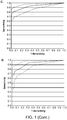
- Figure 1 provides an example of ROC curves for the various combinations of regions.
- Panel A Global Normalization all regions (46) AUC: 0.95 0.88 - 0.99;
- Panel B Local Normalization all regions (35) AUC: 0.93 0.86 - 0.98;
- Panel C Direction Up (35) AUC: 0.93 0.87 - 0.97;
- Panel D Direction Down (46) AUC: 0.92 0.84 - 0.97.
- a “biomarker” refers to a nucleic acid sequence that corresponds to a chromosomal region, where the level of the nucleic acid in CNA relative to normal is associated with colorectal cancer.
- a biomarker in which a biomarker is indicated as "UP” in Table 2, the level in CNA of a colorectal cancer patient is increased relative to normal.
- a biomarker in which a biomarker is indicated as "DOWN” in Table 2, the level in CNA of a colorectal cancer patient is decreased relative to normal.
- a "chromosomal region” listed in Table 2 refers to the region of the chromosome that corresponds to the nucleotide positions indicated in the tables.
- the nucleotide positions on the chromosomes are numbered according to Homo sapiens (human) genome, hg18/build 36.1 genome version released March 2006.
- each chromosome region listed in Table 2 encompasses allelic variants as well as the particular sequence in the database.
- allelic variant typically has at least 95% identity, often at least 96%, at least 97%, at least 98%, or at least 99% identity to the sequence of a chromosomal region that is present in a particular database, e.g., the National Center for Biotechnology Information ( Homo sapiens Build 36.1 at the website http address www.ncbi.nlm.nih.gov/mapview/). Percent identity can be determined using well known algorithms, including the BLAST algorithm, e.g., set to the default parameters. Further, it is understood that the nucleotide sequences of the chromosomes may be improved upon as errors in the current database are discovered and corrected.
- chromosomal region encompasses any variant or corrected version of the same region as defined in Table 2. Given the information provided in Table 2 in the present disclosure and the available genome databases, a skilled person in the art will be able to understand the chromosomal regions used for the present invention even after new variants are discovered or errors are corrected.
- Detecting a chromosomal region in CNA in the context of this invention refers to detecting the level of any sequence from a chromosomal region shown in Table 2, where the sequence detected can be assigned unambiguously to that chromosomal region. Thus, this term refers to the detection of unique sequences from the chromosomal regions.
- the level of at least one region, typically multiple regions used in combination, in a CNA sample is compared to the range found for such region in a group of "normal" individuals, i.e., in the context of this invention, individuals who do not have cancer or at least have not been diagnosed with cancer.
- a result is typically considered to be increased if the result for the sample is higher than the 60 th , 70 th , 75 th , 80 th , 85 th , 90 th , 95 th , or 99 th percentile.
- regions that are decreased in level in colorectal cancer patients i.e ., regions listed as DOWN in Table 2
- a result is typically considered to be decreased if the result for the sample is below the 40 th , 30 th , 25 th , 20 th , 15 th , 10 th , 5 th , or 1 st percentile in normal individuals.
- Methods of removing repetitive sequences from the analysis include use of blocking DNA, e.g., when the target nucleic acids are identified by hybridization.
- blocking DNA e.g., when the target nucleic acids are identified by hybridization.
- well known computer programs and manipulations can be used to remove repetitive sequences from the analysis (see, e.g., the EXAMPLES section).
- sequences that have multiple equally fitting alignment to the reference database are typically omitted from further analyses.
- detecting a biomarker refers to detecting a polynucleotide, e.g., DNA, from a chromosomal region listed in Table 2 in CNA.
- detecting the level of a biomarker encompasses quantitative measurements as well as detecting the presence, or absence, of the biomarker.
- the term "detecting an increase in the level of' a biomarker, relative to normal includes qualitative embodiments in which the biomarker is detected in a patient sample, but not a normal sample.
- the term "detecting a decrease in the level of' a biomarker, relative to normal, includes embodiments in which the biomarker is not detected in a patient sample, but is detected in normal samples.
- a biomarker is considered to be "present” if any nucleic acid sequence in the CNA is unambiguously assigned to the chromosomal region.
- the term "unambiguously assigned" in the context of this invention refers to determining that a DNA detected in the CNA of a patient is from a particular chromosomal region.
- the probe hybridizes specifically to that region.
- the primer(s) hybridizes specifically to that region.
- the sequence is assigned to that region based on well-known algorithms for identity, such as the BLAST algorithm using high stringent parameters, such as e ⁇ 0.0001. In addition, such a sequence does not have a further equally fitting hit on the used database.
- circulating nucleic acids refers to acellular nucleic acids that are present in the blood.
- circulating cell-free DNA means free DNA molecules of 25 nucleotides or longer that are not contained within any intact cells in human blood, and can be obtained from human serum or plasma.
- hybridization refers to the formation of a duplex structure by two single stranded nucleic acids due to complementary base pairing. Hybridization can occur between exactly complementary nucleic acid strands or between nucleic acid strands that contain minor regions of mismatch. As used herein, the term “substantially complementary” refers to sequences that are complementary except for minor regions of mismatch. Typically, the total number of mismatched nucleotides over a hybridizing region is not more than 3 nucleotides for sequences about 15 nucleotides in length. Conditions under which only exactly complementary nucleic acid strands will hybridize are referred to as “stringent” or “sequence-specific” hybridization conditions.
- Stable duplexes of substantially complementary nucleic acids can be achieved under less stringent hybridization conditions.
- Those skilled in the art of nucleic acid technology can determine duplex stability empirically considering a number of variables including, for example, the length and base pair concentration of the oligonucleotides, ionic strength, and incidence of mismatched base pairs.
- computer software for calculating duplex stability is commercially available from National Biosciences, Inc. (Plymouth, Minn.); e.g., OLIGO version 5, or from DNA Software (Ann Arbor, Michigan), e.g., Visual OMP 6.
- Stringent, sequence-specific hybridization conditions under which an oligonucleotide will hybridize only to the target sequence, are well known in the art (see, e.g., the general references provided in the section on detecting polymorphisms in nucleic acid sequences).
- Stringent conditions are sequence-dependent and will be different in different circumstances. Generally, stringent conditions are selected to be about 5°C lower to 5°C higher than the thermal melting point (Tm) for the specific sequence at a defined ionic strength and pH. The Tm is the temperature (under defined ionic strength and pH) at which 50% of the duplex strands have dissociated. Relaxing the stringency of the hybridizing conditions will allow sequence mismatches to be tolerated; the degree of mismatch tolerated can be controlled by suitable adjustment of the hybridization conditions.
- primer refers to an oligonucleotide that acts as a point of initiation of DNA synthesis under conditions in which synthesis of a primer extension product complementary to a nucleic acid strand is induced, i.e., in the presence of four different nucleoside triphosphates and an agent for polymerization (i.e., DNA polymerase or reverse transcriptase) in an appropriate buffer and at a suitable temperature.
- a primer is preferably a single-stranded oligodeoxyribonucleotide.
- the primer includes a "hybridizing region" exactly or substantially complementary to the target sequence, preferably about 15 to about 35 nucleotides in length.
- a primer oligonucleotide can either consist entirely of the hybridizing region or can contain additional features which allow for the detection, immobilization, or manipulation of the amplified product, but which do not alter the ability of the primer to serve as a starting reagent for DNA synthesis.
- a nucleic acid sequence tail can be included at the 5' end of the primer that hybridizes to a capture oligonucleotide.
- probe refers to an oligonucleotide that selectively hybridizes to a target nucleic acid under suitable conditions.
- a probe for detection of the biomarker sequences described herein can be any length, e.g., from 15-500 bp in length. Typically, in probe-based assays, hybridization probes that are less than 50 bp are preferred.
- target sequence or “target region” refers to a region of a nucleic acid that is to be analyzed and comprises the sequence of interest.
- nucleic acid refers to primers, probes, and oligomer fragments.
- the terms are not limited by length and are generic to linear polymers of polydeoxyribonucleotides (containing 2-deoxy-D-ribose), polyribonucleotides (containing D-ribose), and any other N-glycoside of a purine or pyrimidine base, or modified purine or pyrimidine bases. These terms include double- and single-stranded DNA, as well as double- and single-stranded RNA.
- Oligonucleotides for use in the invention may be used as primers and/or probes.
- a nucleic acid, polynucleotide or oligonucleotide can comprise phosphodiester linkages or modified linkages including, but not limited to phosphotriester, phosphoramidate, siloxane, carbonate, carboxymethylester, acetamidate, carbamate, thioether, bridged phosphoramidate, bridged methylene phosphonate, phosphorothioate, methylphosphonate, phosphorodithioate, bridged phosphorothioate or sulfone linkages, and combinations of such linkages.
- a nucleic acid, polynucleotide or oligonucleotide can comprise the five biologically occurring bases (adenine, guanine, thymine, cytosine and uracil) and/or bases other than the five biologically occurring bases. These bases may serve a number of purposes, e.g., to stabilize or destabilize hybridization; to promote or inhibit probe degradation; or as attachment points for detectable moieties or quencher moieties.
- bases may serve a number of purposes, e.g., to stabilize or destabilize hybridization; to promote or inhibit probe degradation; or as attachment points for detectable moieties or quencher moieties.
- a polynucleotide of the invention can contain one or more modified, non-standard, or derivatized base moieties, including, but not limited to, N6-methyl-adenine, N6-tert-butylbenzyl-adenine, imidazole, substituted imidazoles, 5-fluorouracil, 5 bromouracil, 5-chlorouracil, 5-iodouracil, hypoxanthine, xanthine, 4-acetylcytosine, 5 (carboxyhydroxymethyl)uracil, 5 carboxymethylaminomethyl-2-thiouridine, 5 carboxymethylaminomethyluracil, dihydrouracil, beta-D-galactosylqueosine, inosine, N6 isopentenyladenine, 1-methylguanine, 1-methylinosine, 2,2-dimethylguanine, 2-methyladenine, 2-methylguanine, 3-methylcytosine, 5-methylcytosine, N6-methyladeno,
- nucleic acid, polynucleotide or oligonucleotide can comprise one or more modified sugar moieties including, but not limited to, arabinose, 2-fluoroarabinose, xylulose, and a hexose.
- repetitive element refers to a stretch of DNA sequence of at least 25 nucleotides in length that is present in the human genome in at least 50 copies.
- arrays are used herein interchangeably to refer to an array of distinct polynucleotides affixed to a substrate, such as glass, plastic, paper, nylon or other type of membrane, filter, chip, bead, or any other suitable solid support.
- a substrate such as glass, plastic, paper, nylon or other type of membrane, filter, chip, bead, or any other suitable solid support.
- the polynucleotides can be synthesized directly on the substrate, or synthesized separate from the substrate and then affixed to the substrate.
- the arrays are prepared using known methods.
- the invention is based, at least in part, on the identification of nucleic acid biomarkers in CNA having sequences from particular chromosomal regions that are present in an increased level, relative to normal, in the blood of patients that have colorectal cancer.
- the invention is also based, in part, on the identification of biomarkers in the CNA that are present in a decreased level, relative to normal, in the blood of patients that have colorectal cancer.
- the invention provides methods for analyzing the presence and level in CNA of polynucleotide molecules from a chromosomal region corresponding to at least one of the chromosomal regions set forth in Table 2.
- the disclosure further provides devices for analyzing the presence and level in CNA of polynucleotide molecules from a chromosomal region corresponding to at least one of the chromosomal regions set forth in Table 2.
- the disclosure provides a method of analyzing CNA in a sample (blood, serum or plasma) from a patient comprising detecting a level of at least one circulating cell-free DNA having a nucleotide sequence of at least 25 nucleotides falling within a chromosomal region set forth in Table 2.
- the circulating cell-free DNA is free of repetitive elements
- the patient is an individual suspected of or diagnosed with cancer, e.g., colorectal cancer.
- nucleotide sequence of a circulating cell-free DNA is substantially identical (e.g., greater than 95% identical) to a part of the nucleotide sequence of a chromosome region and can be unambiguously assigned to the chromosome region.
- the circulating cell-free DNA can hybridize to under stringent conditions, or be derived from, the chromosomal region.
- a method of analyzing circulating cell-free DNA in a patient sample comprising determining, in a sample that is blood, serum or plasma, a level of a plurality of circulating cell-free DNA molecules each having a sequence of at least 25 consecutive nucleotides in length, or at least 40, 50, 60, 75, or 100 or more consecutive nucleotides falling within the same one single chromosomal region set forth in Table 2.
- a level of a plurality of circulating cell-free DNA molecules each having a sequence of at least 25 consecutive nucleotides in length, or at least 40, 50, 60, 75, or 100 or more consecutive nucleotides falling within the same one single chromosomal region set forth in Table 2.
- sequences of the circulating cell-free DNA molecules are free of repetitive elements.
- a method of analyzing circulating cell-free DNA in a patient sample comprising determining, in a sample that is blood, serum or plasma, a level of at least 2, 3, 4, 5, 7, 8, 9, 10, 15, 20, 30, 40, 50, 55, 60, 65, 70, 75, or at least 80 or of 81 circulating cell-free DNA molecules each having a sequence of at least 25 consecutive nucleotides, or at least 40, 50 60, 75, or 100, or more consecutive nucleotides falling within a different chromosomal region set forth in Table 2.
- the sequences of the circulating cell-free DNA molecules are free of repetitive elements.
- the cell-free DNA molecules have sequences falling within different chromosomal regions in Table 2.
- the levels of at least 2, 3, 4, 5, 7, 8, 9, 10, 15, 20, 25, 30, 35, 40, 45, 50, 55, 60, 65, 70, 75, or at least 80, or of 81, circulating cell-free DNA molecules are determined, the sequence of each falling within a different chromosomal region set forth in Table 2.
- the method of analyzing circulating cell-free DNA includes the steps of: isolating, from blood, serum or plasma sample of a patient, substantially all circulating cell-free DNA molecules having a length of at least 20, 25, 30, 40, 50, 75 or 100 consecutive nucleotides in length, or between 50 and 400 nucleotides in length, obtaining the sequence of each of the circulating cell-free DNA molecules, determining whether the sequence falls within a chromosomal region set forth in Table 2 and the level of said sequence.
- the method of analyzing circulating cell-free DNA includes the steps of: isolating, from blood, serum or plasma sample of a patient, substantially all circulating cell-free DNA molecules having a length of at least 20, 25, 30, 40, 50, 75 or 100 consecutive nucleotides in length, or between 50 and 400 nucleotides in length, and contacting the circulating cell-free DNA molecules to a plurality of oligonucleotides (e.g., on a DNA chip or microarray) to determine if one or more of the circulating cell-free DNA molecules hybridizes to any one of the plurality of oligonucleotide probes under stringent conditions.
- a plurality of oligonucleotides e.g., on a DNA chip or microarray
- Each of the oligonucleotide probes has a nucleotide sequence identical to a part of the sequence of a chromosomal region set forth in Table 2.
- a circulating DNA molecule hybridizes under stringent conditions to one of the oligonucleotide probes, it indicates that the circulating DNA molecule has a nucleotide sequence falling within a chromosomal region set forth in Table 2 and indicates the presence of the circulating DNA molecule.
- the level of the circulating DNA molecule can be determined by determining the amount of hybridized probe(s).
- the circulating cell-free DNA molecules have at least 25 consecutive nucleotides in length (preferably at least 50, 70, 80, 100, 120 or 200 consecutive nucleotides in length). More preferably, the circulating cell-free DNA molecules have between about 50 and about 300 or 400, preferably from about 75 and about 300 or 400, more preferably from about 100 to about 200 consecutive nucleotides of a unique sequence within a chromosomal region as set forth in Table 2.
- the present invention provides a method of diagnosing or screening for colorectal cancer in a patient.
- the method includes the steps of: (a) determining, in a sample that is blood, serum or plasma from a patient, the level of at least 1, 2, 3, 4, 5, 6, 7, 8, 9, 10, 15, 20, at least 30 or more, or of 35, circulating cell-free DNA molecules each having a sequence of at least 25 nucleotides in length falling within a different chromosomal region designated as "UP" Table 2; and (b) correlating the presence of an increased level of the circulating cell-free DNAs, relative to normal, with an increased likelihood that the patient has colorectal cancer.
- the method of invention includes the steps of: (a) determining, in a sample that is blood, serum or plasma from a patient, the level of at least 1, 2, 3, 4, 5, 6, 7, 8, 9, 10, 15, 20, 25, 30, 35, 40, of at least 45, or of 46, circulating cell-free DNA molecules each having a sequence of at least 25 nucleotides in length falling within a different chromosomal region designated as "DOWN" in Table 2; and (b) correlating the presence of a decreased level of the circulating cell-free DNAs, relative to normal, with an increased likelihood that the patient has colorectal cancer.
- the patient may be monitored for the status of colorectal cancer, or for determining the treatment effect of a particular treatment regimen, or detecting cancer recurrence or relapse.
- the sequences of the circulating cell-free DNA molecules are free of repetitive elements.
- the cell-free DNA molecules have sequences falling within different chromosomal regions in set forth in Table 2.
- a method of diagnosing colorectal cancer in an individual comprising (a) determining the levels of at least 1, 2, 3, 4, 5, 6, 7, 8, 9, 10, 15, 20, at least 30 or more, or of 35, circulating cell-free DNA molecules each having a sequence of at least 25 nucleotides in length falling within a different chromosomal region designated as "UP" Table 2; and (b) correlating the presence of an increased level, relative to normal, of one or more of the circulating cell-free DNA molecules with an increased likelihood that the individual has colorectal cancer or a recurrence of colorectal cancer or a failure of treatment for colorectal cancer.
- a method of diagnosing/monitoring colorectal cancer in an individual comprising (a) determining the levels of at least 1, 2, 3, 4, 5, 6, 7, 8, 9, 10, 15, 20, 25, 30, 35, 40, of at least 45, or of 46, circulating cell-free DNA molecules each having a sequence of at least 25 nucleotides in length falling within a different chromosomal region designated as "DOWN" in Table 2; and (b) correlating the presence of a decreased level, relative to normal, of one or more of the circulating cell-free DNA molecules with an increased likelihood that the individual has colorectal cancer or a recurrence of colorectal cancer or a failure of treatment for colorectal cancer.
- the method of diagnosing, monitoring or screening for colorectal cancer in a patient includes determining, in a sample that is blood, serum or plasma from the patient, the level of each and all circulating cell-free DNAs, each having a sequence falling within the same one single chromosomal region designated as "UP" in Table 2; and correlating an increased total level of said circulating cell-free DNAs, with an increased likelihood that said patient has colorectal, or recurrence of colorectal cancer.
- the method of diagnosing, monitoring or screening for colorectal cancer in a patient includes determining, in a sample that is blood, serum or plasma from the patient, the level of each and all circulating cell-free DNAs, each having a sequence falling within the same one single chromosomal region designated as "DOWN" in Table 2; and correlating a decreased level of said circulating cell-free DNAs with an increased likelihood that said patient has colorectal, or recurrence of colorectal cancer.
- substantially all circulating cell-free DNA molecules having a length of at least 20, 25, 30, 40, 50, 75 or 100 consecutive nucleotides in length, or between 50 and 400 nucleotides in length are isolated from a blood, serum or plasma sample of a patient.
- the sequence of at least some representative portion of each of the isolated circulating cell-free DNA molecules is determined, and compared with one or more of the sequences of the chromosomal regions set forth in Table 2 to determine whether the sequence of a circulating cell-free DNA falls within a chromosomal region designated as "UP" in Table 2 and the level of the circulating DNA having said sequence. If the level is increased relative to normal, a diagnosis of colorectal cancer is made.
- recurrence is indicated if an increase, relative to normal, in the level of a circulating cell-free DNA that falls within a chromosomal region designated as "UP" in Table 2 is detected.
- a diagnosis of colorectal cancer or colorectal cancer treatment failure or recurrence is indicated if two or more circulating cell-free DNA molecules that fall within 2, 3, 4, 5, 6, 7, 8, 9, 10, or more chromosomal regions designated as "UP" in Table 2 are increased.
- substantially all circulating cell-free DNA molecules having a length of at least 20, 25, 30, 40, 50, 75 or 100 consecutive nucleotides in length, or between 50 and 400 nucleotides in length are isolated from a blood, serum or plasma sample of a patient.
- These circulating cell-free DNA molecules, or a representative portion thereof, are hybridized to a microarray that is described above in the context of the kit of the disclosure to determine if one of the circulating cell-free DNA molecules hybridizes to any one of a plurality of oligonucleotide probes under stringent conditions.
- Each of the oligonucleotide probes has a nucleotide sequence identical to a part of the sequence of a chromosomal region designated as "UP" in Table 2.
- UP a chromosomal region
- a circulating DNA molecule hybridizes under stringent conditions to one of the oligonucleotide probes, it indicates that the circulating DNA molecule has a nucleotide sequence falling within a chromosomal region set forth in Table 2 and the level is determined. If the level is increased, relative to normal, a diagnosis of colorectal cancer is made.
- recurrence is indicated if there is an increase in the level of a circulating cell-free DNA falls within a chromosomal region designated as "UP" in Table 2 is detected.
- a diagnosis of colorectal cancer or colorectal cancer treatment failure or recurrence is indicated if two or more circulating cell-free DNA molecules fall within 2, 3, 4, 5, 6, 7, 8, 9, 10, or more chromosomal regions designated as "UP" in Table 2 are increased.
- substantially all circulating cell-free DNA molecules having a length of at least 20, 25, 30, 40, 50, 75 or 100 consecutive nucleotides in length, or between 50 and 400 nucleotides in length are isolated from a blood, serum or plasma sample of a patient.
- the sequence of at least some representative portion of each of the isolated circulating cell-free DNA molecules is determined, and compared with one or more of the sequences of the chromosomal regions set forth in Table 2 to determine whether the sequence of a circulating cell-free DNA falls within a chromosomal region designated as "DOWN" in Table 2 and the level of the polynucleotide having said sequence. If the level is decreased relative to normal, a diagnosis of colorectal cancer is made.
- recurrence is indicated if a decrease, relative to normal, in the level of a circulating cell-free DNA that falls within a chromosomal region designated as "DOWN" in Table 2 is detected.
- a diagnosis of colorectal cancer or colorectal cancer treatment failure or recurrence is indicated if two or more circulating cell-free DNA molecules that fall within 2, 3, 4, 5, 6, 7, 8, 9, 10, or more chromosomal regions designated as "DOWN" in Table 2 are decreased.
- substantially all circulating cell-free DNA molecules having a length of at least 20, 25, 30, 40, 50, 75 or 100 consecutive nucleotides in length, or between 50 and 400 nucleotides in length are isolated from a blood, serum or plasma sample of a patient.
- These circulating cell-free DNA molecules, or a representative portion thereof, are hybridized to a microarray that is described above in the context of the kit of the disclosure to determine if one of the circulating cell-free DNA molecules hybridizes to any one of a plurality of oligonucleotide probes under stringent conditions.
- Each of the oligonucleotide probes has a nucleotide sequence identical to a part of the sequence of a chromosomal region designated as "DOWN" in Table 2.
- a circulating DNA molecule hybridizes under stringent conditions to one of the oligonucleotide probes, it indicates that the circulating DNA molecule has a nucleotide sequence falling within a chromosomal region set forth in Table 2 and the level is determined. If the level is decreased, relative to normal, a diagnosis of colorectal cancer is made.
- recurrence is indicated if there is a decrease in the level of a circulating cell-free DNA falls within a chromosomal region designated as "DOWN" in Table 2 is detected.
- a diagnosis of colorectal cancer or colorectal cancer treatment failure or recurrence is indicated if two or more circulating cell-free DNA molecules fall within2, 3, 4, 5, 6, 7, 8, 9, 10, or more chromosomal regions designated as "UP" in Table 2 are decreased.
- the circulating cell-free DNA molecules have at least 25 consecutive nucleotides in length (preferably at least 50, 70, 80, 100, 120 or 200 consecutive nucleotides in length). More preferably, the circulating cell-free DNA molecules have between about 50 and about 300 or 400, preferably from about 75 and about 300 or 400, more preferably from about 100 to about 200 consecutive nucleotides of a unique sequence within a chromosomal region as set forth in Table 2.
- nucleic acids can be isolated from serum or plasma using well known techniques, see, e.g., the example sections.
- the nucleic acid sequences that are analyzed are DNA sequences.
- Detection techniques for evaluating nucleic acids for the presence and level of a biomarker involve procedures well known in the field of molecular genetics. Further, many of the methods involve amplification of nucleic acids. Ample guidance for performing is provided in the art. Exemplary references include manuals such as PCR Technology: Principles and Applications for DNA Amplification (ed. H. A. Erlich, Freeman Press, NY, N.Y., 1992 ); PCR Protocols: A Guide to Methods and Applications (eds.
- Suitable amplification methods include ligase chain reaction (see, e.g., Wu & Wallace, Genomics 4:560-569, 1988 ); strand displacement assay (see, e.g., Walker et al., Proc. Natl. Acad. Sci. USA 89:392-396, 1992 ; U.S. Pat. No. 5,455,166 ); and several transcription-based amplification systems, including the methods described in U.S. Pat. Nos. 5,437,990 ; 5,409,818 ; and 5,399,491 ; the transcription amplification system (TAS) ( Kwoh et al., Proc. Natl.
- TAS transcription amplification system
- the detection of biomarker in the CNA of a patient is performed using oligonucleotide primers and/or probes to detect a target sequence, wherein the target sequence is present in ( e.g., comprises some unambiguously assigned portion of) any of the chromosomal regions listed in Table 2).
- Oligonucleotides can be prepared by any suitable method, usually chemical synthesis, and can also be purchased through commercial sources.
- Oligonucleotides can include modified phosphodiester linkages (e.g., phosphorothioate, methylphosphonates, phosphoamidate, or boranophosphate) or linkages other than a phosphorous acid derivative into an oligonucleotide may be used to prevent cleavage at a selected site.
- modified phosphodiester linkages e.g., phosphorothioate, methylphosphonates, phosphoamidate, or boranophosphate
- linkages other than a phosphorous acid derivative into an oligonucleotide may be used to prevent cleavage at a selected site.
- 2'-amino modified sugars tends to favor displacement over digestion of the oligonucleotide when hybridized to a nucleic acid that is also the template for synthesis of a new nucleic acid strand.
- the biomarker is identified by hybridization under sequence-specific hybridization conditions with a probe that targets a chromosomal region, e.g., targets some unambiguously assigned portion of, any of the chromosomal regions listed in Table 2) described herein.
- the probe used for this analysis can be a long probe or sets for short oligonculeotide probes, e.g., from about 20 to about 150 nucleotides in length may be employed.
- Suitable hybridization formats are well known in the art, including but not limited to, solution phase, solid phase, oligonucleotide array formats, mixed phase, or in situ hybridization assays.
- solution phase hybridizations both the target nucleic acid and the probe or primers are free to interact in the reaction mixture.
- Techniques such as real-time PCR systems have also been developed that permit analysis, e.g., quantification, of amplified products during a PCR reaction.
- hybridization with a specific oligonucleotide probe occurs during the amplification program to identify the presence of a target nucleic acid.
- Hybridization of oligonucleotide probes ensure the highest specificity due to thermodynamically controlled two state transition.
- Examples for this assay formats are fluorescence resonance energy transfer hybridization probes, molecular beacons, molecular scorpions, and exonuclease hybridization probes ( e.g., reviewed in Bustin, J. Mol. Endocrin. 25:169-93, 2000 ).
- Suitable assay formats include array-based formats, described in greater detail below in the " Device " section, where probe is typically immobilized. Alternatively, the target may be immobilized.
- amplified target DNA is immobilized on a solid support and the target complex is incubated with the probe under suitable hybridization conditions, unhybridized probe is removed by washing under suitably stringent conditions, and the solid support is monitored for the presence of bound probe.
- the probes are immobilized on a solid support
- the target DNA is typically labeled, usually during amplification.
- the immobilized probe is incubated with the amplified target DNA under suitable hybridization conditions, unhybridized target DNA is removed by washing under suitably stringent conditions, and the solid support/probe is monitored for the presence of bound target DNA.
- multiple probes are immobilized on a solid support and the target chromosomal regions in the CNA from a patient are analyzed using the multiple probes simultaneously.
- nucleic acid arrays are described by WO 95/11995 .
- amplified nucleic acid corresponding to a target nucleic acid present in a chromosomal region is performed using nucleic acid primers to the chromosomal region and is detected by monitoring the increase in the total level of double-stranded DNA in the reaction mixture, is described, e.g., in U.S. Pat. No. 5,994,056 ; and European Patent Publication Nos. 487,218 and 512,334 .
- the detection of double-stranded target DNA relies on the increased fluorescence various DNA-binding dyes, e.g., SYBR Green, exhibit when bound to double-stranded DNA.
- a sequence from a chromosomal region set forth in Table 2 in the CNA from a patient undergoing evaluation is detected by direct sequencing.
- Such sequencing especially using the Roche 454, Illumina, and Applied Biosystems sequencing systems mentioned below or similar advanced sequencing systems, can include quantitation of nucleic acids having a particular sequence to determine the level of a biomarker.
- CNA from a patient is sequenced using a large-scale sequencing method that provides the ability to obtain sequence information from many reads.
- sequencing platforms includes those commercialized by Roche 454 Life Sciences (GS systems), Illumina (e.g., HiSeq, MiSeq) and Applied Biosystems (e.g., SOLiD systems).
- the Roche 454 Life Sciences sequencing platform involves using emulsion PCR and immobilizing DNA fragments onto bead. Incorporation of nucleotides during synthesis is detected by measuring light that is generated when a nucleotide is incorporated.
- the Illumina technology involves the attachment of randomly fragmented genomic DNA to a planar, optically transparent surface. Attached DNA fragments are extended and bridge amplified to create an ultra-high density sequencing flow cell with clusters containing copies of the same template. These templates are sequenced using a sequencing-by-synthesis technology that employs reversible terminators with removable fluorescent dyes.
- Such methods involves emulsion PCR that immobilizes DNA fragments onto beads followed by the use of a pool of all possible oligonucleotides of a fixed length, labeled according to the sequenced position. Oligonucleotides are annealed and ligated; the preferential ligation by DNA ligase for matching sequences results in a signal informative of the nucleotide at that position.
- the sequence can be determined using any other DNA sequencing method including, e.g., methods that use semiconductor technology to detect nucleotides that are incorporated into an extended primer by measuring changes in current that occur when a nucleotide is incorporated (see, e.g., U.S. Patent Application Publication Nos. 20090127589 and 20100035252 ).
- the disclosure provides diagnostic devices and kits useful for identifying and determining the level of one or more colorectal cancer-associated biomarkers in the CNA from a patient where the one or more biomarkers has a sequence unambiguously assigned to any of the chromosomal regions set forth in Table 2.
- the kit of the present disclosure is useful in the above-discussed method for analyzing circulating cell-free DNA in a patient sample and in diagnosing, screening or monitoring colorectal cancer as described above.
- the present disclosure provides the use of at least one oligonucleotide for the manufacture of a diagnostic kit useful in diagnosing, screening or monitoring colorectal cancer.
- the nucleotide sequence of the oligonucleotide falls within a chromosomal region set forth in Table 2.
- the kit of the present disclosure includes one, two or more (e.g., at least 1, 2, 3, 4, 5, 6, 7, 8, 9, 10, 12, 15, 20, 25, 30, 40 or at least 50, but preferably less than 81, preferably from one to about 50, more preferably from 2 to about 50, or from 3 to about 50 sets of oligonucleotides.
- Each set comprises one or more oligonucleotides (e.g., from about one to about 10,000, preferably from 50, 100, 200 or 300 to about 10,000). All of the nucleotide sequences of such one or more oligonucleotides in each set fall within the same one single chromosomal region that is set forth in Table 2 (or match a part of the same one single sequence set forth in Table A).
- Each oligonucleotide should have from about 18 to 100 nucleotides, or from 20 to about 50 nucleotides, and is capable of hybridizing, under stringent hybridization conditions, to the chromosomal region in which its sequence falls.
- the oligonucleotides are useful as probes for detecting circulating cell-free DNA molecules derived from the chromosomal regions.
- each set includes a sufficient number of oligonucleotides with sequences mapped to one chromosomal region such that any circulating cell-free DNA molecules derived from the chromosomal region can be detected with the oligonucleotide set.
- the number of oligonucleotides required in each set is determined by the total length of unique nucleotide sequence of a particular chromosomal region, as will be apparent to skilled artisans. Such total lengths are indicated in Table 2.
- kits of the present disclosure different oligonucleotide sets correspond to different chromosomal regions within the same table.
- the oligonucleotides are free of repetitive element.
- the oligonucleotides are attached to one or more solid substrates such as microchips and beads.
- the kit is a microarray with the above oligonucleotides.
- oligonucleotides included in the kit described for the manufacture of the kit useful for diagnosing, screening or monitoring colorectal cancer is also contemplated. The manufacturing of such kit should be apparent to a skilled artisan.
- a diagnostic device comprises probes to detect at least 2, 3, 4, 5, 6, 7, 8, 9, 10, 15, 20, 30, 40, 50, 60, 75, 80, or all 81 chromosomal regions set forth in Table 2.
- the present disclosure provides probes attached to a solid support, such as an array slide or chip, e.g., as described in DNA Microarrays: A Molecular Cloning Manual, 2003, Eds. Bowtell and Sambrook, Cold Spring Harbor Laboratory Press . Construction of such devices are well known in the art, for example as described in US Patents and Patent Publications U.S. Patent No. 5,837,832 ; PCT application WO95/11995 ; U.S. Patent No. 5,807,522 ; US Patent Nos.
- probes may be implemented in an array.
- a probe set that hybridizes to different, preferably unique, segments of a chromosomal region may be used where the probe set detects any part of the chromosomal region.
- a single probe to a chromosomal region may be immobilized to a solid surface.
- Polynucleotide probe can be synthesized at designated areas (or synthesized separately and then affixed to designated areas) on a substrate, e.g., using a light-directed chemical process. Typical synthetic polynucleotides can be about 15-200 nucleotides in length.
- the kit can include multiple biomarker detection reagents, or one or more biomarker detection reagents in combination with one or more other types of elements or components (e.g., other types of biochemical reagents, containers, packages such as packaging intended for commercial sale, substrates to which biomarker detection reagents are attached, electronic hardware components, etc.). Accordingly, the present disclosure further provides biomarker detection kits and systems, including but not limited to arrays/microarrays of nucleic acid molecules, and beads that contain one or more probes or other detection reagents for detecting one or more biomarkers of the present disclosure.
- kits can optionally include various electronic hardware components; for example, arrays ("DNA chips") and microfluidic systems ("lab-on-a-chip” systems) provided by various manufacturers typically comprise hardware components.
- Other kits may not include electronic hardware components, but may be comprised of, for example, one or more biomarker detection reagents (along with, optionally, other biochemical reagents) packaged in one or more containers.
- Biomarker detection kits/systems may contain, for example, one or more probes, or sets of probes, that hybridize to a nucleic acid molecule present in a chromosomal region set forth in Table 2.
- a biomarker detection kit of the present disclosure may include components that are used to prepare CNA from a blood sample from a patient for the subsequent amplification and/or detection of a biomarker.
- the present invention provides methods for detecting the level of a biomarker in CNA from a patient that has colorectal cancer or that is being evaluated to determine if the patient may have colorectal cancer.
- the disclosure further provides reagents for detecting the level of a biomarker in CNA from a patient that has colorectal cancer or that is being evaluated to determine if the patient may have colorectal cancer.
- detection or “identification” or “identifying the presence” or “detecting the presence” of a biomarker associated with colorectal cancer in a CNA sample from a patient refers to determining any level of the biomarker in the CNA of the patient where the level is greater than a threshold value that distinguishes between colorectal cancer and non-colorectal cancer CNA samples for a given assay.
- an increase in the level of any one of the chromosomal regions (i.e., biomarkers) designated as "UP" in Table 2 is indicative of colorectal cancer.
- a biomarker may have been observed to be present infrequently in CNA obtained from normal individuals; however, given the low frequency of occurrence in normal samples relative to a higher frequency of occurrence in colorectal cancer, the presence of the biomarker in a patient indicates that the patient has a greater likelihood, e.g., 95% or greater likelihood, of having colorectal cancer.
- the biomarkers designated as "UP” in Table 2 are associated with colorectal cancer, i.e., they are over-represented in colorectal cancer patients compared to individuals not diagnosed with colorectal cancer.
- the detection of an increase, relative to non-colorectal cancer patients, in the level of one or more of the biomarkers designated as "UP” in Table 2 is indicative of colorectal cancer, i.e., the patient has an increased probability of having colorectal cancer compared to a patient that does not have an increase in the level of the biomarker.
- the detection and increase in the level of two or more biomarkers designated as "UP" in Table 2 in the CNA of a patient is indicative of a greater probability for colorectal cancer.
- a therapeutic agent for colorectal cancer such as one or more chemotherapeutic agents, e.g., 5-fluorouracil, leucovorin, or oxaliplatin or capecitabine; and/or a monoclonal antibody, such as bevacizumab, cetuximab, or panitumumab, or alternative monoclonal antibody.
- chemotherapeutic agents e.g., 5-fluorouracil, leucovorin, or oxaliplatin or capecitabine
- a monoclonal antibody such as bevacizumab, cetuximab, or panitumumab, or alternative monoclonal antibody.
- the biomarkers designated as "DOWN” in Table 2 are associated with colorectal cancer, i.e., they are under-represented in colorectal cancer patients compared to individuals not diagnosed with colorectal cancer. Thus, the detection of a decrease, relative to non-colorectal cancer patients, in the level of one or more of the biomarkers designated as "DOWN” in Table 2 is indicative of colorectal cancer, i.e., the patient has an increased probability of having colorectal cancer compared to a patient that does not have a decrease in the level of the biomarker.
- a biomarker may have been observed to be present infrequently in CNA obtained from cancer patients; however, given the low frequency of occurrence in cancer samples relative to a higher frequency of occurrence in normal individuals, the presence of the biomarker in a patient indicates that the patient has a decreased likelihood, e.g., 5% or less likelihood, of having colorectal cancer.
- other criteria e.g., clinical criteria, etc., are also employed to diagnose colorectal cancer in the patient. Accordingly, patients that have a biomarker associated with colorectal cancer also undergo other diagnostic procedures.
- the patient is administered a therapeutic agent for colorectal cancer, such as one or more chemotherapeutic agents, e.g., 5-fluorouracil, leucovorin, or oxaliplatin or capecitabine; and/or a monoclonal antibody, such as bevacizumab, cetuximab, or panitumumab, or alternative monoclonal antibody.
- chemotherapeutic agents e.g., 5-fluorouracil, leucovorin, or oxaliplatin or capecitabine
- a monoclonal antibody such as bevacizumab, cetuximab, or panitumumab, or alternative monoclonal antibody.
- “Over-represented” or “increased level” means that the level of one or more circulating cell-free DNAs is higher than normal levels. Generally this means an increase in the level as compared to an index value. Conversely “under-represented” or “decreased level” means that the level of one or more particular circulating cell-free DNA molecules is lower than normal levels. Generally this means a decrease in the level as compared to an index value.
- the test value representing the level of a particular circulating cell-free DNA is compared to one or more reference values (or index values), and optionally correlated to colorectal cancer or cancer recurrence.
- reference values or index values
- an increased likelihood of colorectal cancer is indicated if the test value is greater than the reference value for CNA listed as "UP” in Table 2 or less than the reference value for CNA listed as "DOWN” in Table 2.
- the index value may represent the copy number or concentration of a particular cell-free DNA listed as "UP" in Table 2 in a blood sample from a patient of interest in a healthy state, in which case a copy number or concentration in a sample from the patient at a different time or state significantly higher (e.g., 1.01-fold, 1.05-fold, 1.10-fold, 1.2-fold, 1.3-fold, 1.4-fold, 1.5-fold, 1.6-fold, 1.7-fold, 1.8-fold, 1.9-fold, 2-fold, 3-fold, 4-fold, 5-fold, 10-fold, 20-fold, 30-fold, 40-fold, 50-fold, 100-fold or more higher) than this index value would indicate, e.g., colorectal cancer or increased likelihood of colorectal cancer recurrence.
- the level of the CNA is "increased” if it is at least 1, 2, 3, 4, 5, 10, 15, 20 or more standard deviations greater than the index value in normal subjects.
- an index value may represent the copy number or concentration of a particular cell-free DNA listed as "DOWN" in Table 2 in a blood sample from a patient of interest in a healthy state, in which case a copy number or concentration in a sample from the patient at a different time or state significantly lower ( e.g., 1.01-fold, 1.05-fold, 1.10-fold, 1.2-fold, 1.3-fold, 1.4-fold, 1.5-fold, 1.6-fold, 1.7-fold, 1.8-fold, 1.9-fold, 2-fold, 3-fold, 4-fold, 5-fold, 10-fold, 20-fold, 30-fold, 40-fold, 50-fold, 100-fold or more lower) than this index value would indicate, e.g., colorectal cancer or increased likelihood of colorectal cancer recurrence.
- the level of the CNA is "increased
- the index value may represent the average concentration or copy number of a particular circulating cell-free DNA for a set of individuals from a diverse cancer population or a subset of the population. For example, one may determine the average copy number or concentration of a circulating cell-free DNA in a random sampling of patients with colorectal cancer. Thus, patients having a copy number or concentration (test value) comparable to or higher than, this value identified as having an increased likelihood of having colorectal cancer or colorectal cancer recurrence than those having a test value lower than this value.
- a useful index value may represent the copy number or concentration of a particular circulating cell-free DNA or of a combination (weighted or straight addition) of two or more circulating cell-free DNAs corresponding to the same chromosomal region or different chromosomal regions.
- the level of each biomarker or circulating cell-free DNA can be weighted and combined.
- a test value may be provided by (a) weighting the determined level of each circulating cell-free DNA molecule with a predefined coefficient, and (b) combining the weighted level to provide a test value.
- the combining step can be either by straight addition or averaging (i.e., weighted equally) or by a different predefined coefficient.
- the information obtained from the biomarker analysis may be stored in a computer readable form.
- a computer system typically comprises major subsystems such as a central processor, a system memory (typically RAM), an input/output (I/O) controller, an external device such as a display screen via a display adapter, serial ports, a keyboard, a fixed disk drive via a storage interface and a floppy disk drive operative to receive a floppy disc, and a CD-ROM (or DVD-ROM) device operative to receive a CD-ROM.
- Many other devices can be connected, such as a network interface connected via a serial port.
- the computer system may also be linked to a network, comprising a plurality of computing devices linked via a data link, such as an Ethernet cable (coax or 10BaseT), telephone line, ISDN line, wireless network, optical fiber, or other suitable signal transmission medium, whereby at least one network device (e.g., computer, disk array, etc.) comprises a pattern of magnetic domains (e.g., magnetic disk) and/or charge domains (e.g., an array of DRAM cells) composing a bit pattern encoding data acquired from an assay of the invention.
- a network device e.g., computer, disk array, etc.
- a pattern of magnetic domains e.g., magnetic disk
- charge domains e.g., an array of DRAM cells
- the computer system can comprise code for interpreting the results of a study evaluating the presence of one or more of the biomarkers.
- the biomarker analysis results are provided to a computer where a central processor executes a computer program for determining the likelihood of a patient that has colorectal cancer.
- the disclosure also provides the use of a computer system, such as that described above, which comprises: (1) a computer; (2) a stored bit pattern encoding the biomarker testing results obtained by the methods of the invention, which may be stored in the computer; (3) and, optionally, (4) a program for determining the likelihood of a patient having colorectal cancer.
- the disclosure further provides methods of generating a report based on the detection of one or more biomarkers set forth in Table 2.
- the present disclosure provides systems related to the above methods of the invention.
- the disclosure provides a system for analyzing circulating cell-free DNA, comprising: (1) a sample analyzer for executing the method of analyzing circulating cell-free DNA in a patient's blood, serum or plasma as described in the various embodiments above ; (2) a computer system for automatically receiving and analyzing data obtained in step (1) to provide a test value representing the status (concentration or copy number) of one or more circulating cell-free DNA molecules having a nucleotide sequence of at least 25 nucleotides falling within a chromosomal region set forth in Table 2, and optionally for comparing the test value to one or more reference values each associated with a predetermined status of colorectal cancer.
- the system further comprises a display module displaying the comparison between the test value and the one or more reference values, or displaying a result of the comparing step.
- the sample analyzer may be, e.g., a sequencing machine (e.g., Illumina HiSeqTM, Ion Torrent PGM, Applied Biosystems SOLiDTM sequencer, PacBio RS, Helicos HeliscopeTM, etc.), a PCR machine (e.g., Applied Biosystems 7900, Fluidigm BioMarkTM, etc.), a microarray instrument, etc.
- a sequencing machine e.g., Illumina HiSeqTM, Ion Torrent PGM, Applied Biosystems SOLiDTM sequencer, PacBio RS, Helicos HeliscopeTM, etc.
- a PCR machine e.g., Applied Biosystems 7900, Fluidigm BioMarkTM, etc.
- a microarray instrument e.g., a microarray instrument, etc.
- the sample analyzer is a sequencing instrument, e.g ., a next-generation sequencing instrument such as Roche's GS systems, Illumina's HiSeq and MiSeq, and Applied Biosystems' SOLiD systems.
- Circulating cell-free DNA molecules are isolated from a patient's blood or serum or plasma, and the sequences of all of the circulating cell-free DNA molecules are obtained using the sample analyzer.
- the sequencing instrument is used in sequencing the circulating cell-free DNA molecules, and obtaining the sequences of these molecules.
- a computer system is then employed for automatically analyzing the sequences to determine the level of a circulating cell-free DNA molecule having a nucleotide sequence of at least 25 nucleotides falling within a chromosomal region set forth in Table 2 in the sample.
- the computer system may compare the sequence of each circulating cell-free DNA molecule in the sample to the sequence, available in the human sequence database, of the chromosomal region to determine if there is a match, i.e., if the sequence of a circulating cell-free DNA molecule falls within a chromosomal region set forth in Table 2.
- the copy number of a particular circulating cell-free DNA molecule is also automatically determined by the computer system.
- the computer system automatically correlates the sequence analysis result with a diagnosis regarding colorectal cancer. For example, if one, and preferably two or more, circulating cell-free DNA molecules are identified to be derived from chromosomal regions designated as "UP" in Table 2 and present at an increased level, then the computer system automatically correlates this analysis result with a diagnosis of colorectal cancer. If one, and preferably two or more, circulating cell-free DNA molecules are identified to be derived from chromosomal regions designated as "DOWN" in Table 2 and present at a decreased level, then the computer system automatically correlates this analysis result with a diagnosis of colorectal cancer.
- the computer system further comprises a display module displaying the results of sequence analysis and/or the result of the correlating step.
- the display module may be for example, a display screen, such as a computer monitor, TV monitor, or the touch screen, a printer, and audio speakers.
- the computer-based analysis function can be implemented in any suitable language and/or browsers. For example, it may be implemented with C language and preferably using object-oriented high-level programming languages such as Visual Basic, SmallTalk, C++, and the like.
- the application can be written to suit environments such as the Microsoft WindowsTM environment including WindowsTM 98, WindowsTM 2000, WindowsTM NT, and the like.
- the application can also be written for the MacIntoshTM, SUNTM, UNIX or LINUX environment.
- the functional steps can also be implemented using a universal or platform-independent programming language.
- multi-platform programming languages include, but are not limited to, hypertext markup language (HTML), JAVATM, JavaScriptTM, Flash programming language, common gateway interface/structured query language (CGI/SQL), practical extraction report language (PERL), AppleScriptTM and other system script languages, programming language/structured query language (PL/SQL), and the like.
- JavaTM- or JavaScriptTM-enabled browsers such as HotJavaTM, MicrosoftTM ExplorerTM, or NetscapeTM can be used.
- active content web pages may include JavaTM applets or ActiveXTM controls or other active content technologies.
- the analysis function can also be embodied in computer program products and used in the systems described above or other computer- or internet-based systems. Accordingly, another aspect of the present disclosure relates to a computer program product comprising a computer-usable medium having computer-readable program codes or instructions embodied thereon for enabling a processor to carry out the analysis and correlating functions as described above.
- These computer program instructions may be loaded onto a computer or other programmable apparatus to produce a machine, such that the instructions which execute on the computer or other programmable apparatus create means for implementing the functions or steps described above.
- These computer program instructions may also be stored in a computer-readable memory or medium that can direct a computer or other programmable apparatus to function in a particular manner, such that the instructions stored in the computer-readable memory or medium produce an article of manufacture including instruction means which implement the analysis.
- the computer program instructions may also be loaded onto a computer or other programmable apparatus to cause a series of operational steps to be performed on the computer or other programmable apparatus to produce a computer implemented process such that the instructions which execute on the computer or other programmable apparatus provide steps for implementing the functions or steps described above.
- Sequencing of the libraries was performed on a SOLiD4+ Instrument (Applied Biosystems) equipped with an EZBead-System (Applied Biosystems) for conducting the emulsion PCRs. All necessary reagents were purchased from Applied Biosystems. Emulsion PCRs and sequencing was performed as recommended by the manufacturer. For each fragment, 50 bp and 10 bp of molecular barcode were sequenced.
- the sequence reads were assigned to the different samples according to the sequence of the molecular barcode.
- the sequences were mapped to the human genome (Build 36.1/Hg18) using the BioScope software suite (Applied Biosystems) using the default parameterization for 50 bp reads. Briefly, the local mapping algorithm of the software employing a 25 bases seeding scheme with two serial seedings starting from base 1 and base 16 was used. During extension of the seeds, a match received a score of 1 and a mismatch received a score of -2. For reads that mapped to more than one position within the genome, the best mapping position was recorded when its quality score was five-fold better than the quality score of the second best mapping (clear zone procedure). All mapping results were recorded for each individual sample. The number of reads mapped in genomic windows of 100,000 bp was determined.
- the windows (each of 100,000 bp in size) were moved along the chromosomes by intervals of 50,000 bp starting at a position of 200,000 of each chromosome in order to exclude telomere regions.
- One tabulated text file was produced for each of the human chromosomes and each sample. The tabulated text file contained the following information:
- the first step of the unsupervised cluster search (UCS) was:
- the 300 rounds of random training/validation sets show a separation of the groups in the validation set as given in the table.
- the AUCs of ROC curves for each round was constructed by using the sum of read calls under different conditions (e.g., global and local normalization and up or down in disease).
- Table 1 show AUCs from ROC curves with standard deviations. Table 1 All Global Up Down All Local Up Down AUC Mean 88.5% 88.9% 87.6% 88.1% 86.7% 90.1% StDev 5.9% 5.8% 7.4% 5.8% 6.0% 5.2%
- a final model was constructed from the 300 rounds and applied to all samples.
- the biomarker regions for colorectal cancers defined in this way are provided in Table 2. These regions can be used in different combination for detection of sample status.
- the "Rank” is calculated from the number of randomizations (see, above) in which a region was identified.
- the graphs presented in Figure 1 with the AUC values are based on the combination of such regions, called positive at 95% specificity level.
Claims (5)
- Procédé de diagnostic ou de dépistage de cancer colorectal chez un patient, comprenant la détection, dans un échantillon qui est du sang, du sérum ou du plasma provenant dudit patient,(a) du niveau total de tous les ADN acellulaires circulant, chacun comportant une séquence se situant dans la même région chromosomique unique appelée « UP » dans le tableau 2 ; et la corrélation d'un niveau total augmenté avec une probabilité augmentée selon laquelle ledit patient est atteint de cancer colorectal lorsque le niveau total de l'ADN acellulaire comportant une séquence se situant dans la même région chromosomique unique est d'au moins 1, 2, 3, 4, 5, 10, 15 ou 20 écarts types supérieurs à une valeur d'indice provenant de sujets normaux ; ou(b) le niveau total de tous les ADN acellulaires circulant, chacun comportant une séquence se situant dans la même région chromosomique unique appelée « DOWN » dans le tableau 2 ; et la corrélation d'un niveau total augmenté avec une probabilité augmentée selon laquelle ledit patient est atteint de cancer colorectal lorsque le niveau total de l'ADN acellulaire comportant une séquence se situant dans la même région chromosomique unique est au moins de 1, 2, 3, 4, 5, 10 ou 20 écarts types inférieurs à une valeur d'indice provenant de sujets normaux ;
les positions nucléotidiques sur les régions chromosomiques dans le tableau 2 étant numérotées selon la version de génome hg18/structure 36,1 du génome humain du National Center for Biotechnology Information publiée en mars 2006. - Procédé selon la revendication 1, dans lequel l'étape de détection comprend le séquençage d'ACN à partir de l'échantillon provenant du patient.
- Procédé selon la revendication 1, dans lequel l'étape de détection comprend la mise en contact d'au moins une sonde qui est sélective pour une région chromosomique indiquée dans le tableau 2 avec un échantillon d'ADN obtenu à partir de l'échantillon issu du patient dans des conditions dans lesquelles la sonde s'hybride sélectivement avec une séquence cible présente sur la région chromosomique ; et la détection d'hybridation de la sonde.
- Procédé selon la revendication 3, dans lequel la sonde est attachée à une surface solide.
- Procédé selon la revendication 3 ou 4, comprenant en outre la mise en contact de l'échantillon d'ADN avec au moins 20, 25, 30, 50, 55, 60, 65, 70, 75, 80 ou 81 sondes, chaque sonde étant sélective pour une région chromosomique indiquée dans le tableau 2.
Priority Applications (1)
| Application Number | Priority Date | Filing Date | Title |
|---|---|---|---|
| PL12846333T PL2768985T3 (pl) | 2011-10-21 | 2012-10-19 | Biomarkery będące krążącymi kwasami nukleinowymi związane z rakiem jelita grubego |
Applications Claiming Priority (2)
| Application Number | Priority Date | Filing Date | Title |
|---|---|---|---|
| US201161550098P | 2011-10-21 | 2011-10-21 | |
| PCT/US2012/061044 WO2013066641A1 (fr) | 2011-10-21 | 2012-10-19 | Biomarqueurs des acides nucléiques circulants associés au cancer colorectal |
Publications (3)
| Publication Number | Publication Date |
|---|---|
| EP2768985A1 EP2768985A1 (fr) | 2014-08-27 |
| EP2768985A4 EP2768985A4 (fr) | 2015-07-01 |
| EP2768985B1 true EP2768985B1 (fr) | 2019-03-20 |
Family
ID=48192610
Family Applications (1)
| Application Number | Title | Priority Date | Filing Date |
|---|---|---|---|
| EP12846333.8A Active EP2768985B1 (fr) | 2011-10-21 | 2012-10-19 | Biomarqueurs des acides nucléiques circulants associés au cancer colorectal |
Country Status (6)
| Country | Link |
|---|---|
| US (2) | US20140303008A1 (fr) |
| EP (1) | EP2768985B1 (fr) |
| CA (1) | CA2852098C (fr) |
| ES (1) | ES2729554T3 (fr) |
| PL (1) | PL2768985T3 (fr) |
| WO (1) | WO2013066641A1 (fr) |
Families Citing this family (18)
| Publication number | Priority date | Publication date | Assignee | Title |
|---|---|---|---|---|
| IN2013MN00522A (fr) | 2010-09-24 | 2015-05-29 | Univ Leland Stanford Junior | |
| US9260753B2 (en) | 2011-03-24 | 2016-02-16 | President And Fellows Of Harvard College | Single cell nucleic acid detection and analysis |
| US10876152B2 (en) | 2012-09-04 | 2020-12-29 | Guardant Health, Inc. | Systems and methods to detect rare mutations and copy number variation |
| US20160040229A1 (en) | 2013-08-16 | 2016-02-11 | Guardant Health, Inc. | Systems and methods to detect rare mutations and copy number variation |
| US11913065B2 (en) | 2012-09-04 | 2024-02-27 | Guardent Health, Inc. | Systems and methods to detect rare mutations and copy number variation |
| EP2893040B1 (fr) | 2012-09-04 | 2019-01-02 | Guardant Health, Inc. | Procédés pour détecter des mutations rares et une variation de nombre de copies |
| US20160161495A1 (en) * | 2013-07-30 | 2016-06-09 | Health Researc Inc. | Method of Treatment |
| CN111534580A (zh) | 2013-12-28 | 2020-08-14 | 夸登特健康公司 | 用于检测遗传变异的方法和系统 |
| US11062789B2 (en) | 2014-07-18 | 2021-07-13 | The Chinese University Of Hong Kong | Methylation pattern analysis of tissues in a DNA mixture |
| SG11201805119QA (en) | 2015-12-17 | 2018-07-30 | Guardant Health Inc | Methods to determine tumor gene copy number by analysis of cell-free dna |
| KR102482616B1 (ko) | 2016-05-30 | 2022-12-28 | 더 차이니즈 유니버시티 오브 홍콩 | 혈액 내 무세포 dna를 사용하는 혈액 장애의 검출 |
| US9850523B1 (en) | 2016-09-30 | 2017-12-26 | Guardant Health, Inc. | Methods for multi-resolution analysis of cell-free nucleic acids |
| CA3126055A1 (fr) | 2016-09-30 | 2018-04-05 | Guardant Health, Inc. | Procedes d'analyse multiresolution d'acides nucleiques acellulaires |
| SG11201903509QA (en) | 2016-10-24 | 2019-05-30 | Univ Hong Kong Chinese | Methods and systems for tumor detection |
| JP2020503003A (ja) | 2016-11-30 | 2020-01-30 | ザ チャイニーズ ユニバーシティ オブ ホンコン | 尿および他のサンプルにおける無細胞dnaの分析 |
| AU2018212272B2 (en) | 2017-01-25 | 2022-04-28 | Grail, Inc. | Diagnostic applications using nucleic acid fragments |
| EP3918089A1 (fr) | 2019-01-31 | 2021-12-08 | Guardant Health, Inc. | Compositions et méthodes pour isoler de l'adn acellulaire |
| CN113728116A (zh) * | 2019-02-22 | 2021-11-30 | 安可济控股有限公司 | 用于早期癌症检测的方法和组合物 |
Family Cites Families (38)
| Publication number | Priority date | Publication date | Assignee | Title |
|---|---|---|---|---|
| WO1989001050A1 (fr) | 1987-07-31 | 1989-02-09 | The Board Of Trustees Of The Leland Stanford Junior University | Accroissement selectif de sequences de polynucleotides cibles |
| CA1340807C (fr) | 1988-02-24 | 1999-11-02 | Lawrence T. Malek | Procede d'amplification d'une sequence d'acide nucleique |
| US5143854A (en) | 1989-06-07 | 1992-09-01 | Affymax Technologies N.V. | Large scale photolithographic solid phase synthesis of polypeptides and receptor binding screening thereof |
| CA2020958C (fr) | 1989-07-11 | 2005-01-11 | Daniel L. Kacian | Methodes d'amplification de sequences d'acide nucleique |
| US5955589A (en) | 1991-12-24 | 1999-09-21 | Isis Pharmaceuticals Inc. | Gapped 2' modified oligonucleotides |
| EP0487218B1 (fr) | 1990-10-31 | 1997-12-29 | Tosoh Corporation | Procédé pour la détection ou détermination quantitative d'acides nucléiques cibles |
| ZA918965B (en) | 1990-11-13 | 1992-08-26 | Siska Diagnostics Inc | Nucleic acid amplification by two-enzyme,self-sustained sequence replication |
| YU187991A (sh) | 1990-12-11 | 1994-09-09 | Hoechst Aktiengesellschaft | 3-(2)-amino-ali tiol-modifikovani, s fluorescentnom bojom vezani nukleozidi, nukleotidi i oligonukleotidi, postupak za njihovo dobijanje i njihova upotreba |
| US5455166A (en) | 1991-01-31 | 1995-10-03 | Becton, Dickinson And Company | Strand displacement amplification |
| US5994056A (en) | 1991-05-02 | 1999-11-30 | Roche Molecular Systems, Inc. | Homogeneous methods for nucleic acid amplification and detection |
| US5652099A (en) | 1992-02-12 | 1997-07-29 | Conrad; Michael J. | Probes comprising fluorescent nucleosides and uses thereof |
| US5837832A (en) | 1993-06-25 | 1998-11-17 | Affymetrix, Inc. | Arrays of nucleic acid probes on biological chips |
| JPH09507121A (ja) | 1993-10-26 | 1997-07-22 | アフィマックス テクノロジーズ ナームロゼ ベノートスハップ | 生物学的チップ上の核酸プローブアレー |
| US5446137B1 (en) | 1993-12-09 | 1998-10-06 | Behringwerke Ag | Oligonucleotides containing 4'-substituted nucleotides |
| DE4415370A1 (de) | 1994-05-02 | 1995-11-09 | Hoechst Ag | Modifizierte Oligonukleotide, deren Herstellung sowie deren Verwendung |
| US5807522A (en) | 1994-06-17 | 1998-09-15 | The Board Of Trustees Of The Leland Stanford Junior University | Methods for fabricating microarrays of biological samples |
| DE4438918A1 (de) | 1994-11-04 | 1996-05-09 | Hoechst Ag | Modifizierte Oligonukleotide, deren Herstellung sowie deren Verwendung |
| EP0866071B1 (fr) | 1997-03-20 | 2004-10-20 | F. Hoffmann-La Roche Ag | Amorces modifiées |
| IL137901A (en) | 1998-02-23 | 2004-07-25 | Wisconsin Alumni Res Found | Method and apparatus for synthesis of arrays of dna probes |
| WO1999063385A1 (fr) | 1998-06-04 | 1999-12-09 | Board Of Regents, The University Of Texas System | Imageur numerique a micromiroir pour chimie optique |
| US6315958B1 (en) | 1999-11-10 | 2001-11-13 | Wisconsin Alumni Research Foundation | Flow cell for synthesis of arrays of DNA probes and the like |
| AU2002312445A1 (en) * | 2001-06-12 | 2002-12-23 | Board Of Regents, The University Of Texas System | Methods of detecting, diagnosing and treating cancer and identifying neoplastic progression |
| US7422851B2 (en) | 2002-01-31 | 2008-09-09 | Nimblegen Systems, Inc. | Correction for illumination non-uniformity during the synthesis of arrays of oligomers |
| US20040126757A1 (en) | 2002-01-31 | 2004-07-01 | Francesco Cerrina | Method and apparatus for synthesis of arrays of DNA probes |
| US7157229B2 (en) | 2002-01-31 | 2007-01-02 | Nimblegen Systems, Inc. | Prepatterned substrate for optical synthesis of DNA probes |
| US7083975B2 (en) | 2002-02-01 | 2006-08-01 | Roland Green | Microarray synthesis instrument and method |
| US7498176B2 (en) | 2002-09-27 | 2009-03-03 | Roche Nimblegen, Inc. | Microarray with hydrophobic barriers |
| US20040110212A1 (en) | 2002-09-30 | 2004-06-10 | Mccormick Mark | Microarrays with visual alignment marks |
| US20090215036A1 (en) * | 2004-12-10 | 2009-08-27 | Bayer Healthcare Ag | Genetic Alterations Useful For The Response Prediction of Malignant Neoplasia to Taxane-Based Medical Treatments |
| JP2008536480A (ja) * | 2005-02-17 | 2008-09-11 | ダナ−ファーバー キャンサー インスティテュート,インコーポレイテッド | 癌の同定、評価、予防、および治療用組成物、キット、および方法、ならびに癌の治療法 |
| CN102260742A (zh) * | 2005-10-21 | 2011-11-30 | 基因信息股份有限公司 | 用于使生物标志产物水平与疾病相关联的方法和装置 |
| EP2653861B1 (fr) | 2006-12-14 | 2014-08-13 | Life Technologies Corporation | Procédé pour le séquençage d'un acide nucléique en utilisant des matrices de FET à grande échelle |
| US20100035252A1 (en) | 2008-08-08 | 2010-02-11 | Ion Torrent Systems Incorporated | Methods for sequencing individual nucleic acids under tension |
| GB2463401B (en) * | 2008-11-12 | 2014-01-29 | Caris Life Sciences Luxembourg Holdings S A R L | Characterizing prostate disorders by analysis of microvesicles |
| US20100136560A1 (en) | 2008-12-02 | 2010-06-03 | The Johns Hopkins University | Integrated Analyses of Breast and Colorectal Cancers |
| US20120220478A1 (en) | 2009-07-20 | 2012-08-30 | Bar Harbor Biotechnology, Inc. | Methods for assessing disease risk |
| AU2011237669B2 (en) * | 2010-04-06 | 2016-09-08 | Caris Life Sciences Switzerland Holdings Gmbh | Circulating biomarkers for disease |
| PL2558854T3 (pl) * | 2010-04-16 | 2019-04-30 | Chronix Biomedical | Biomarkery będące krążącymi kwasami nukleinowymi związanymi z rakiem piersi |
-
2012
- 2012-10-19 WO PCT/US2012/061044 patent/WO2013066641A1/fr active Application Filing
- 2012-10-19 PL PL12846333T patent/PL2768985T3/pl unknown
- 2012-10-19 ES ES12846333T patent/ES2729554T3/es active Active
- 2012-10-19 US US14/352,826 patent/US20140303008A1/en not_active Abandoned
- 2012-10-19 EP EP12846333.8A patent/EP2768985B1/fr active Active
- 2012-10-19 CA CA2852098A patent/CA2852098C/fr active Active
-
2018
- 2018-06-13 US US16/007,795 patent/US20190136314A1/en active Pending
Non-Patent Citations (1)
| Title |
|---|
| None * |
Also Published As
| Publication number | Publication date |
|---|---|
| ES2729554T3 (es) | 2019-11-04 |
| EP2768985A1 (fr) | 2014-08-27 |
| CA2852098C (fr) | 2023-05-02 |
| PL2768985T3 (pl) | 2019-10-31 |
| EP2768985A4 (fr) | 2015-07-01 |
| US20140303008A1 (en) | 2014-10-09 |
| CA2852098A1 (fr) | 2013-05-10 |
| WO2013066641A1 (fr) | 2013-05-10 |
| US20190136314A1 (en) | 2019-05-09 |
Similar Documents
| Publication | Publication Date | Title |
|---|---|---|
| US20230087365A1 (en) | Prostate cancer associated circulating nucleic acid biomarkers | |
| EP2768985B1 (fr) | Biomarqueurs des acides nucléiques circulants associés au cancer colorectal | |
| US11377695B2 (en) | Breast cancer associated circulating nucleic acid biomarkers | |
| US20190185928A1 (en) | Prostate cancer associated circulating nucleic acid biomarkers | |
| EP2931922B1 (fr) | Biomarqueurs personnalisés pour le cancer | |
| CN109952383B (zh) | 用于预测恩扎妥林活性的方法和组合物 | |
| KR20180121695A (ko) | 대규모 병렬 게놈 서열분석을 이용한 태아 염색체 이수성의 진단 방법 | |
| JP2007509613A (ja) | 遺伝子発現プロファイリングのためのqRT−PCRアッセイシステム | |
| KR101501826B1 (ko) | 위암에 대한 예후 예측 모형의 제조방법 | |
| KR20170051747A (ko) | 피부 주름 발생 민감도 진단용 단일염기다형성 마커 및 이의 용도 | |
| KR101777911B1 (ko) | 골다공증 골절 발생 예측용 바이오마커 | |
| AU2022238235A1 (en) | Combinations of biomarkers for methods for detecting trisomy 21 | |
| WO2021061623A1 (fr) | Méthodes de prédiction d'évolution de lam |
Legal Events
| Date | Code | Title | Description |
|---|---|---|---|
| PUAI | Public reference made under article 153(3) epc to a published international application that has entered the european phase |
Free format text: ORIGINAL CODE: 0009012 |
|
| 17P | Request for examination filed |
Effective date: 20140513 |
|
| AK | Designated contracting states |
Kind code of ref document: A1 Designated state(s): AL AT BE BG CH CY CZ DE DK EE ES FI FR GB GR HR HU IE IS IT LI LT LU LV MC MK MT NL NO PL PT RO RS SE SI SK SM TR |
|
| DAX | Request for extension of the european patent (deleted) | ||
| RA4 | Supplementary search report drawn up and despatched (corrected) |
Effective date: 20150601 |
|
| RIC1 | Information provided on ipc code assigned before grant |
Ipc: C12Q 1/68 20060101AFI20150526BHEP |
|
| STAA | Information on the status of an ep patent application or granted ep patent |
Free format text: STATUS: EXAMINATION IS IN PROGRESS |
|
| 17Q | First examination report despatched |
Effective date: 20161107 |
|
| REG | Reference to a national code |
Ref country code: DE Ref legal event code: R079 Ref document number: 602012058132 Country of ref document: DE Free format text: PREVIOUS MAIN CLASS: C12Q0001680000 Ipc: C12Q0001688600 |
|
| RIC1 | Information provided on ipc code assigned before grant |
Ipc: C12Q 1/6886 20180101AFI20180703BHEP |
|
| GRAP | Despatch of communication of intention to grant a patent |
Free format text: ORIGINAL CODE: EPIDOSNIGR1 |
|
| STAA | Information on the status of an ep patent application or granted ep patent |
Free format text: STATUS: GRANT OF PATENT IS INTENDED |
|
| INTG | Intention to grant announced |
Effective date: 20180810 |
|
| GRAJ | Information related to disapproval of communication of intention to grant by the applicant or resumption of examination proceedings by the epo deleted |
Free format text: ORIGINAL CODE: EPIDOSDIGR1 |
|
| STAA | Information on the status of an ep patent application or granted ep patent |
Free format text: STATUS: EXAMINATION IS IN PROGRESS |
|
| GRAP | Despatch of communication of intention to grant a patent |
Free format text: ORIGINAL CODE: EPIDOSNIGR1 |
|
| STAA | Information on the status of an ep patent application or granted ep patent |
Free format text: STATUS: GRANT OF PATENT IS INTENDED |
|
| INTG | Intention to grant announced |
Effective date: 20181220 |
|
| GRAS | Grant fee paid |
Free format text: ORIGINAL CODE: EPIDOSNIGR3 |
|
| RIC1 | Information provided on ipc code assigned before grant |
Ipc: C12Q 1/6886 20180101AFI20180703BHEP |
|
| GRAA | (expected) grant |
Free format text: ORIGINAL CODE: 0009210 |
|
| STAA | Information on the status of an ep patent application or granted ep patent |
Free format text: STATUS: THE PATENT HAS BEEN GRANTED |
|
| AK | Designated contracting states |
Kind code of ref document: B1 Designated state(s): AL AT BE BG CH CY CZ DE DK EE ES FI FR GB GR HR HU IE IS IT LI LT LU LV MC MK MT NL NO PL PT RO RS SE SI SK SM TR |
|
| REG | Reference to a national code |
Ref country code: GB Ref legal event code: FG4D |
|
| REG | Reference to a national code |
Ref country code: CH Ref legal event code: EP |
|
| REG | Reference to a national code |
Ref country code: DE Ref legal event code: R096 Ref document number: 602012058132 Country of ref document: DE |
|
| REG | Reference to a national code |
Ref country code: AT Ref legal event code: REF Ref document number: 1110573 Country of ref document: AT Kind code of ref document: T Effective date: 20190415 |
|
| REG | Reference to a national code |
Ref country code: IE Ref legal event code: FG4D |
|
| REG | Reference to a national code |
Ref country code: CH Ref legal event code: NV Representative=s name: VENI GMBH, CH |
|
| REG | Reference to a national code |
Ref country code: RO Ref legal event code: EPE |
|
| REG | Reference to a national code |
Ref country code: NL Ref legal event code: FP |
|
| REG | Reference to a national code |
Ref country code: SE Ref legal event code: TRGR |
|
| PG25 | Lapsed in a contracting state [announced via postgrant information from national office to epo] |
Ref country code: NO Free format text: LAPSE BECAUSE OF FAILURE TO SUBMIT A TRANSLATION OF THE DESCRIPTION OR TO PAY THE FEE WITHIN THE PRESCRIBED TIME-LIMIT Effective date: 20190620 Ref country code: FI Free format text: LAPSE BECAUSE OF FAILURE TO SUBMIT A TRANSLATION OF THE DESCRIPTION OR TO PAY THE FEE WITHIN THE PRESCRIBED TIME-LIMIT Effective date: 20190320 Ref country code: LT Free format text: LAPSE BECAUSE OF FAILURE TO SUBMIT A TRANSLATION OF THE DESCRIPTION OR TO PAY THE FEE WITHIN THE PRESCRIBED TIME-LIMIT Effective date: 20190320 |
|
| REG | Reference to a national code |
Ref country code: LT Ref legal event code: MG4D |
|
| PG25 | Lapsed in a contracting state [announced via postgrant information from national office to epo] |
Ref country code: GR Free format text: LAPSE BECAUSE OF FAILURE TO SUBMIT A TRANSLATION OF THE DESCRIPTION OR TO PAY THE FEE WITHIN THE PRESCRIBED TIME-LIMIT Effective date: 20190621 Ref country code: RS Free format text: LAPSE BECAUSE OF FAILURE TO SUBMIT A TRANSLATION OF THE DESCRIPTION OR TO PAY THE FEE WITHIN THE PRESCRIBED TIME-LIMIT Effective date: 20190320 Ref country code: HR Free format text: LAPSE BECAUSE OF FAILURE TO SUBMIT A TRANSLATION OF THE DESCRIPTION OR TO PAY THE FEE WITHIN THE PRESCRIBED TIME-LIMIT Effective date: 20190320 Ref country code: LV Free format text: LAPSE BECAUSE OF FAILURE TO SUBMIT A TRANSLATION OF THE DESCRIPTION OR TO PAY THE FEE WITHIN THE PRESCRIBED TIME-LIMIT Effective date: 20190320 Ref country code: BG Free format text: LAPSE BECAUSE OF FAILURE TO SUBMIT A TRANSLATION OF THE DESCRIPTION OR TO PAY THE FEE WITHIN THE PRESCRIBED TIME-LIMIT Effective date: 20190620 |
|
| PG25 | Lapsed in a contracting state [announced via postgrant information from national office to epo] |
Ref country code: AL Free format text: LAPSE BECAUSE OF FAILURE TO SUBMIT A TRANSLATION OF THE DESCRIPTION OR TO PAY THE FEE WITHIN THE PRESCRIBED TIME-LIMIT Effective date: 20190320 Ref country code: CZ Free format text: LAPSE BECAUSE OF FAILURE TO SUBMIT A TRANSLATION OF THE DESCRIPTION OR TO PAY THE FEE WITHIN THE PRESCRIBED TIME-LIMIT Effective date: 20190320 Ref country code: EE Free format text: LAPSE BECAUSE OF FAILURE TO SUBMIT A TRANSLATION OF THE DESCRIPTION OR TO PAY THE FEE WITHIN THE PRESCRIBED TIME-LIMIT Effective date: 20190320 Ref country code: SK Free format text: LAPSE BECAUSE OF FAILURE TO SUBMIT A TRANSLATION OF THE DESCRIPTION OR TO PAY THE FEE WITHIN THE PRESCRIBED TIME-LIMIT Effective date: 20190320 Ref country code: PT Free format text: LAPSE BECAUSE OF FAILURE TO SUBMIT A TRANSLATION OF THE DESCRIPTION OR TO PAY THE FEE WITHIN THE PRESCRIBED TIME-LIMIT Effective date: 20190720 |
|
| REG | Reference to a national code |
Ref country code: ES Ref legal event code: FG2A Ref document number: 2729554 Country of ref document: ES Kind code of ref document: T3 Effective date: 20191104 |
|
| PG25 | Lapsed in a contracting state [announced via postgrant information from national office to epo] |
Ref country code: SM Free format text: LAPSE BECAUSE OF FAILURE TO SUBMIT A TRANSLATION OF THE DESCRIPTION OR TO PAY THE FEE WITHIN THE PRESCRIBED TIME-LIMIT Effective date: 20190320 |
|
| PG25 | Lapsed in a contracting state [announced via postgrant information from national office to epo] |
Ref country code: IS Free format text: LAPSE BECAUSE OF FAILURE TO SUBMIT A TRANSLATION OF THE DESCRIPTION OR TO PAY THE FEE WITHIN THE PRESCRIBED TIME-LIMIT Effective date: 20190720 |
|
| REG | Reference to a national code |
Ref country code: DE Ref legal event code: R097 Ref document number: 602012058132 Country of ref document: DE |
|
| PLBE | No opposition filed within time limit |
Free format text: ORIGINAL CODE: 0009261 |
|
| STAA | Information on the status of an ep patent application or granted ep patent |
Free format text: STATUS: NO OPPOSITION FILED WITHIN TIME LIMIT |
|
| PG25 | Lapsed in a contracting state [announced via postgrant information from national office to epo] |
Ref country code: DK Free format text: LAPSE BECAUSE OF FAILURE TO SUBMIT A TRANSLATION OF THE DESCRIPTION OR TO PAY THE FEE WITHIN THE PRESCRIBED TIME-LIMIT Effective date: 20190320 |
|
| 26N | No opposition filed |
Effective date: 20200102 |
|
| PG25 | Lapsed in a contracting state [announced via postgrant information from national office to epo] |
Ref country code: SI Free format text: LAPSE BECAUSE OF FAILURE TO SUBMIT A TRANSLATION OF THE DESCRIPTION OR TO PAY THE FEE WITHIN THE PRESCRIBED TIME-LIMIT Effective date: 20190320 |
|
| PG25 | Lapsed in a contracting state [announced via postgrant information from national office to epo] |
Ref country code: TR Free format text: LAPSE BECAUSE OF FAILURE TO SUBMIT A TRANSLATION OF THE DESCRIPTION OR TO PAY THE FEE WITHIN THE PRESCRIBED TIME-LIMIT Effective date: 20190320 |
|
| PG25 | Lapsed in a contracting state [announced via postgrant information from national office to epo] |
Ref country code: MC Free format text: LAPSE BECAUSE OF FAILURE TO SUBMIT A TRANSLATION OF THE DESCRIPTION OR TO PAY THE FEE WITHIN THE PRESCRIBED TIME-LIMIT Effective date: 20190320 |
|
| PG25 | Lapsed in a contracting state [announced via postgrant information from national office to epo] |
Ref country code: LU Free format text: LAPSE BECAUSE OF NON-PAYMENT OF DUE FEES Effective date: 20191019 |
|
| PG25 | Lapsed in a contracting state [announced via postgrant information from national office to epo] |
Ref country code: IE Free format text: LAPSE BECAUSE OF NON-PAYMENT OF DUE FEES Effective date: 20191019 |
|
| REG | Reference to a national code |
Ref country code: AT Ref legal event code: UEP Ref document number: 1110573 Country of ref document: AT Kind code of ref document: T Effective date: 20190320 |
|
| PG25 | Lapsed in a contracting state [announced via postgrant information from national office to epo] |
Ref country code: CY Free format text: LAPSE BECAUSE OF FAILURE TO SUBMIT A TRANSLATION OF THE DESCRIPTION OR TO PAY THE FEE WITHIN THE PRESCRIBED TIME-LIMIT Effective date: 20190320 |
|
| PG25 | Lapsed in a contracting state [announced via postgrant information from national office to epo] |
Ref country code: MT Free format text: LAPSE BECAUSE OF FAILURE TO SUBMIT A TRANSLATION OF THE DESCRIPTION OR TO PAY THE FEE WITHIN THE PRESCRIBED TIME-LIMIT Effective date: 20190320 Ref country code: HU Free format text: LAPSE BECAUSE OF FAILURE TO SUBMIT A TRANSLATION OF THE DESCRIPTION OR TO PAY THE FEE WITHIN THE PRESCRIBED TIME-LIMIT; INVALID AB INITIO Effective date: 20121019 |
|
| PG25 | Lapsed in a contracting state [announced via postgrant information from national office to epo] |
Ref country code: MK Free format text: LAPSE BECAUSE OF FAILURE TO SUBMIT A TRANSLATION OF THE DESCRIPTION OR TO PAY THE FEE WITHIN THE PRESCRIBED TIME-LIMIT Effective date: 20190320 |
|
| P01 | Opt-out of the competence of the unified patent court (upc) registered |
Effective date: 20230404 |
|
| PGFP | Annual fee paid to national office [announced via postgrant information from national office to epo] |
Ref country code: RO Payment date: 20230925 Year of fee payment: 12 Ref country code: NL Payment date: 20230915 Year of fee payment: 12 Ref country code: IT Payment date: 20230913 Year of fee payment: 12 Ref country code: GB Payment date: 20230831 Year of fee payment: 12 |
|
| PGFP | Annual fee paid to national office [announced via postgrant information from national office to epo] |
Ref country code: SE Payment date: 20230810 Year of fee payment: 12 Ref country code: PL Payment date: 20230807 Year of fee payment: 12 Ref country code: FR Payment date: 20230808 Year of fee payment: 12 Ref country code: BE Payment date: 20230918 Year of fee payment: 12 |
|
| PGFP | Annual fee paid to national office [announced via postgrant information from national office to epo] |
Ref country code: ES Payment date: 20231106 Year of fee payment: 12 |
|
| PGFP | Annual fee paid to national office [announced via postgrant information from national office to epo] |
Ref country code: DE Payment date: 20230822 Year of fee payment: 12 Ref country code: CH Payment date: 20231102 Year of fee payment: 12 Ref country code: AT Payment date: 20230925 Year of fee payment: 12 |