EP2722012A1 - Vorrichtung zur Stoßwellentherapie eines menschlichen Gehirns - Google Patents
Vorrichtung zur Stoßwellentherapie eines menschlichen Gehirns Download PDFInfo
- Publication number
- EP2722012A1 EP2722012A1 EP12189007.3A EP12189007A EP2722012A1 EP 2722012 A1 EP2722012 A1 EP 2722012A1 EP 12189007 A EP12189007 A EP 12189007A EP 2722012 A1 EP2722012 A1 EP 2722012A1
- Authority
- EP
- European Patent Office
- Prior art keywords
- shockwave
- dose
- human
- transducer
- shockwaves
- Prior art date
- Legal status (The legal status is an assumption and is not a legal conclusion. Google has not performed a legal analysis and makes no representation as to the accuracy of the status listed.)
- Granted
Links
- 210000004556 brain Anatomy 0.000 title claims abstract description 25
- 230000035939 shock Effects 0.000 title description 2
- 238000013507 mapping Methods 0.000 claims abstract description 43
- 241001465754 Metazoa Species 0.000 claims abstract description 16
- 238000011156 evaluation Methods 0.000 claims abstract description 13
- 238000000034 method Methods 0.000 claims description 13
- 239000003550 marker Substances 0.000 claims description 10
- 230000005477 standard model Effects 0.000 claims description 4
- 231100000673 dose–response relationship Toxicity 0.000 claims description 2
- 210000003128 head Anatomy 0.000 description 10
- 210000001519 tissue Anatomy 0.000 description 9
- 210000000988 bone and bone Anatomy 0.000 description 7
- 239000000523 sample Substances 0.000 description 5
- 238000010521 absorption reaction Methods 0.000 description 4
- 210000005013 brain tissue Anatomy 0.000 description 4
- 230000001419 dependent effect Effects 0.000 description 4
- 230000001225 therapeutic effect Effects 0.000 description 3
- 210000005036 nerve Anatomy 0.000 description 2
- 230000002537 thrombolytic effect Effects 0.000 description 2
- 208000024827 Alzheimer disease Diseases 0.000 description 1
- 102000013455 Amyloid beta-Peptides Human genes 0.000 description 1
- 108010090849 Amyloid beta-Peptides Proteins 0.000 description 1
- 208000032843 Hemorrhage Diseases 0.000 description 1
- 241000282412 Homo Species 0.000 description 1
- 208000018737 Parkinson disease Diseases 0.000 description 1
- 210000004958 brain cell Anatomy 0.000 description 1
- 239000003086 colorant Substances 0.000 description 1
- 238000012937 correction Methods 0.000 description 1
- 208000037265 diseases, disorders, signs and symptoms Diseases 0.000 description 1
- 210000003094 ear ossicle Anatomy 0.000 description 1
- 230000000694 effects Effects 0.000 description 1
- 230000005284 excitation Effects 0.000 description 1
- 238000003384 imaging method Methods 0.000 description 1
- 230000010354 integration Effects 0.000 description 1
- 230000004807 localization Effects 0.000 description 1
- 238000005259 measurement Methods 0.000 description 1
- 230000004060 metabolic process Effects 0.000 description 1
- 238000012986 modification Methods 0.000 description 1
- 230000004048 modification Effects 0.000 description 1
- 210000000056 organ Anatomy 0.000 description 1
- 230000000149 penetrating effect Effects 0.000 description 1
- 230000035515 penetration Effects 0.000 description 1
- 210000004872 soft tissue Anatomy 0.000 description 1
- 238000001228 spectrum Methods 0.000 description 1
- 230000000638 stimulation Effects 0.000 description 1
- 230000001755 vocal effect Effects 0.000 description 1
Images

Classifications
-
- A—HUMAN NECESSITIES
- A61—MEDICAL OR VETERINARY SCIENCE; HYGIENE
- A61B—DIAGNOSIS; SURGERY; IDENTIFICATION
- A61B17/00—Surgical instruments, devices or methods, e.g. tourniquets
- A61B17/22—Implements for squeezing-off ulcers or the like on the inside of inner organs of the body; Implements for scraping-out cavities of body organs, e.g. bones; Calculus removers; Calculus smashing apparatus; Apparatus for removing obstructions in blood vessels, not otherwise provided for
- A61B17/225—Implements for squeezing-off ulcers or the like on the inside of inner organs of the body; Implements for scraping-out cavities of body organs, e.g. bones; Calculus removers; Calculus smashing apparatus; Apparatus for removing obstructions in blood vessels, not otherwise provided for for extracorporeal shock wave lithotripsy [ESWL], e.g. by using ultrasonic waves
- A61B17/2256—Implements for squeezing-off ulcers or the like on the inside of inner organs of the body; Implements for scraping-out cavities of body organs, e.g. bones; Calculus removers; Calculus smashing apparatus; Apparatus for removing obstructions in blood vessels, not otherwise provided for for extracorporeal shock wave lithotripsy [ESWL], e.g. by using ultrasonic waves with means for locating or checking the concrement, e.g. X-ray apparatus, imaging means
- A61B17/2258—Implements for squeezing-off ulcers or the like on the inside of inner organs of the body; Implements for scraping-out cavities of body organs, e.g. bones; Calculus removers; Calculus smashing apparatus; Apparatus for removing obstructions in blood vessels, not otherwise provided for for extracorporeal shock wave lithotripsy [ESWL], e.g. by using ultrasonic waves with means for locating or checking the concrement, e.g. X-ray apparatus, imaging means integrated in a central portion of the shock wave apparatus
-
- A—HUMAN NECESSITIES
- A61—MEDICAL OR VETERINARY SCIENCE; HYGIENE
- A61B—DIAGNOSIS; SURGERY; IDENTIFICATION
- A61B17/00—Surgical instruments, devices or methods, e.g. tourniquets
- A61B17/22—Implements for squeezing-off ulcers or the like on the inside of inner organs of the body; Implements for scraping-out cavities of body organs, e.g. bones; Calculus removers; Calculus smashing apparatus; Apparatus for removing obstructions in blood vessels, not otherwise provided for
- A61B17/225—Implements for squeezing-off ulcers or the like on the inside of inner organs of the body; Implements for scraping-out cavities of body organs, e.g. bones; Calculus removers; Calculus smashing apparatus; Apparatus for removing obstructions in blood vessels, not otherwise provided for for extracorporeal shock wave lithotripsy [ESWL], e.g. by using ultrasonic waves
- A61B17/2256—Implements for squeezing-off ulcers or the like on the inside of inner organs of the body; Implements for scraping-out cavities of body organs, e.g. bones; Calculus removers; Calculus smashing apparatus; Apparatus for removing obstructions in blood vessels, not otherwise provided for for extracorporeal shock wave lithotripsy [ESWL], e.g. by using ultrasonic waves with means for locating or checking the concrement, e.g. X-ray apparatus, imaging means
-
- A—HUMAN NECESSITIES
- A61—MEDICAL OR VETERINARY SCIENCE; HYGIENE
- A61N—ELECTROTHERAPY; MAGNETOTHERAPY; RADIATION THERAPY; ULTRASOUND THERAPY
- A61N7/00—Ultrasound therapy
- A61N7/02—Localised ultrasound hyperthermia
-
- A—HUMAN NECESSITIES
- A61—MEDICAL OR VETERINARY SCIENCE; HYGIENE
- A61B—DIAGNOSIS; SURGERY; IDENTIFICATION
- A61B34/00—Computer-aided surgery; Manipulators or robots specially adapted for use in surgery
- A61B34/20—Surgical navigation systems; Devices for tracking or guiding surgical instruments, e.g. for frameless stereotaxis
- A61B2034/2046—Tracking techniques
- A61B2034/2051—Electromagnetic tracking systems
-
- A—HUMAN NECESSITIES
- A61—MEDICAL OR VETERINARY SCIENCE; HYGIENE
- A61B—DIAGNOSIS; SURGERY; IDENTIFICATION
- A61B34/00—Computer-aided surgery; Manipulators or robots specially adapted for use in surgery
- A61B34/20—Surgical navigation systems; Devices for tracking or guiding surgical instruments, e.g. for frameless stereotaxis
- A61B2034/2046—Tracking techniques
- A61B2034/2055—Optical tracking systems
-
- A—HUMAN NECESSITIES
- A61—MEDICAL OR VETERINARY SCIENCE; HYGIENE
- A61B—DIAGNOSIS; SURGERY; IDENTIFICATION
- A61B34/00—Computer-aided surgery; Manipulators or robots specially adapted for use in surgery
- A61B34/20—Surgical navigation systems; Devices for tracking or guiding surgical instruments, e.g. for frameless stereotaxis
- A61B2034/2046—Tracking techniques
- A61B2034/2063—Acoustic tracking systems, e.g. using ultrasound
-
- A—HUMAN NECESSITIES
- A61—MEDICAL OR VETERINARY SCIENCE; HYGIENE
- A61B—DIAGNOSIS; SURGERY; IDENTIFICATION
- A61B34/00—Computer-aided surgery; Manipulators or robots specially adapted for use in surgery
- A61B34/20—Surgical navigation systems; Devices for tracking or guiding surgical instruments, e.g. for frameless stereotaxis
- A61B2034/2074—Interface software
-
- A—HUMAN NECESSITIES
- A61—MEDICAL OR VETERINARY SCIENCE; HYGIENE
- A61B—DIAGNOSIS; SURGERY; IDENTIFICATION
- A61B34/00—Computer-aided surgery; Manipulators or robots specially adapted for use in surgery
- A61B34/25—User interfaces for surgical systems
- A61B2034/252—User interfaces for surgical systems indicating steps of a surgical procedure
-
- A—HUMAN NECESSITIES
- A61—MEDICAL OR VETERINARY SCIENCE; HYGIENE
- A61B—DIAGNOSIS; SURGERY; IDENTIFICATION
- A61B90/00—Instruments, implements or accessories specially adapted for surgery or diagnosis and not covered by any of the groups A61B1/00 - A61B50/00, e.g. for luxation treatment or for protecting wound edges
- A61B90/39—Markers, e.g. radio-opaque or breast lesions markers
- A61B2090/3937—Visible markers
- A61B2090/3945—Active visible markers, e.g. light emitting diodes
-
- A—HUMAN NECESSITIES
- A61—MEDICAL OR VETERINARY SCIENCE; HYGIENE
- A61N—ELECTROTHERAPY; MAGNETOTHERAPY; RADIATION THERAPY; ULTRASOUND THERAPY
- A61N7/00—Ultrasound therapy
- A61N2007/0004—Applications of ultrasound therapy
- A61N2007/0021—Neural system treatment
- A61N2007/0026—Stimulation of nerve tissue
Definitions
- the invention relates to a device for Shock Wave Treatment of the human or animal body and preferably the human brain.
- Focused shockwaves may be used for treatment of the human or animal body. They are known to have a therapeutic effect on soft tissue and nerves and may be used for treatment of functional disorders as Alzheimer or Parkinson disease and for thrombolysis.
- the international patent application publication WO 1997/010758 discloses a device for generating shockwaves for the treatment of body tissue and for crushing calculi. To achieve a certain therapeutic effect, the location and the dose of the shockwaves must be controlled.
- the European patent application EP 2078 503 A1 discloses to use a locating probe being inserted into the human body, which can be located by a magnetic locating system. Such a locating probe may be introduced into hollow organs or bones, but they are not applicable to the human brain.
- controlling the dose may be simply done by controlling the pulse energy and the number of pulses applied.
- a larger area or volume must be treated by shockwaves. This cannot be monitored by using such a locating probe, as the probe cannot be introduced into the brain and the probe is only suitable for applying the shockwaves to a small spot having the size of the focus spot of the shockwave transducer.
- shockwaves to the brain these must penetrate the cranium, which attenuates the shockwaves. As the thickness of the cranium is not constant, it is not possible to calculate the applied shockwave dose.
- the problem to be solved by the invention is to provide a device for treating the human or animal brain with shockwaves by means of a shockwave transducer, which allows to apply a predetermined dose into a treated area and/or volume of the brain tissue, which is significantly larger than the size of the focus spot of the shockwave transducer.
- a further aspect of the invention is an automated treatment of a larger surface and/or volume of brain tissue.
- At least one shockwave transducer for generating acoustic energy in the form of shock- or pressure-waves.
- the term of shockwaves is used herein for any shock- or pressure-waves.
- At least one means for detecting the position of the shockwave transducer herein referred to as a position sensor, is provided.
- at least one means for evaluating the signals of the at least one position sensor and calculating the position of the focus spot of the at least one shockwave transducer is provided, which herein is referred to as an evaluation means.
- the size of the focus spot (focal area) of the shockwave transducer is calculated.
- the focus spot has an ellipsoidal shape, which may vary dependent on various parameters, like frequency or tissue.
- Mapping of the movement of the at least one shockwave transducer over a plurality of positions together with the applied shockwave dose at each of the positions is done by a mapping device.
- a series of measurements at human craniums has shown that there is a significant absorption of shockwaves penetrating the cranium, depending of the position of the cranium. Furthermore, the absorption depends on the frequency spectrum used. Specifically at higher frequencies there is a higher absorption compared to lower frequencies. Therefore, the mapping device has to calculate the applied shockwave dose in dependence of the thickness of the cranium through which the shockwaves penetrate.
- the term of dose is used for the shockwave power or energy, depending on the specific kind of treatment. It may be specified in J/cm 3 when referring to energy delivered into a specific volume, or in J/mm 2 when referring to energy delivered to a specific surface area.
- the dose may vary by using different pulse durations, pulse energies, pulse repetition rates, pulse counts and treatment times.
- energy of 0.01 to 1 mJ/mm 2 most preferably 0.1 to 0.3 mJ/mm 2 is applied after correction for absorption.
- the preferred pulse rate is between 1 and 20, most preferably 3 - 8 pulses per second. It is important to keep the dose in a certain range. There must be a minimum dose to obtain a therapeutic effect, e.g. for thrombolysis, increased circulation, metabolism, removal of Amyloid beta or stimulation of nerves or brain cells. Exceeding a maximum dose must be prevented under any circumstances, as this may lead to dangerous side effects like hemorrhage.
- the at least one position sensor may be a means for detecting and/or indicating the position of the shockwave transducer within the 3- dimensional space or on a 2- dimensional surface like the cranium surface. It may comprise at least one colored marker, like a colored ball attached to the shockwave transducer, which is monitored by a stereotactic camera system.
- the at least one marker may also be of reflector type or active LED type. It may also be an infrared, ultrasound-, radio- or x-ray location system. Preferably, it does not only detect the position of the shockwave transducer, but also its orientation to determine the direction of the shockwave path.
- the orientation of the shockwave transducer may be calculated by using a model or an actual image of the head, assuming that the shockwave transducer is closely positioned to the cranium.
- the position sensor and/or the mapping device may calculate the position of the shockwave transducer relative to the at least one marker fixed to the head.
- the at least one evaluation means evaluates the signals of the at least one position sensor. It calculates the position of the focus spot. Furthermore, it preferably calculates and/or estimates the size and/or form of the focus spot. The results of the evaluation means are forwarded to a mapping device.
- the mapping device preferably is a computer or imaging system. It may evaluate and/or record the shockwave dose dependent on the position and/or orientation of the shockwave transducer. This may be done at a plurality of positions as indicated by the at least one position sensor, whereas the plurality of positions preferably forms an area or volume.
- the mapping device may further indicate the applied dose preferably on a display like a video screen, for example by using a colored map. Furthermore, a warning may be issued, if the dose exceeds a threshold value.
- the mapping device may further store the applied dose and/or the position and/or orientation of the shockwave transducer on a storage medium and/or forward the information to another device like a computer.
- indication may be done acoustically by varying a tone frequency, a sound level, or giving verbal indications about the dose applied.
- indication of the applied dose may either be done two dimensional, e.g. by indicating the treated area or three-dimensional, by indicating the treated volume.
- the evaluation means does not only provide information about the applied dose, but indicates which area to be treated next to achieve a predetermined, preferably an even distribution of shockwave dose. This may also be done as described previously, for example by colored indication or by acoustic means, for example by using a speaker or headphones.
- There may also be an indicator at the shockwave transducer, indicating the applied shockwave dose and/or indicating the suggested direction of move.
- a standard model of a human cranium may be used, as the variation of locations of the parts of the brain does not vary largely between the individual humans. Therefore, it is even not necessary to make an X-ray image of the brain, although using a precise three-dimensional image of the brain would allow a more precise dosage. If a precise dosage would be required, such an image may be generated by using a CT scanner or MRI. Based on a model or actual image, a two - or three-dimensional image of the treated area may be made. Furthermore, dose calculations may be made considering the structure and preferably the thickness of the cranium bone structure. In the case of a model, there may be estimates.
- a sensor for measuring the thickness of a bone below the shockwave transducer attached to the shockwave transducer or integrated within the shockwave transducer may be provided at the center of the shockwave transducer.
- the signals of this sensor may be delivered to the evaluation means and/or mapping device to correct their outputs in dependency of the thickness of the bone.
- the mapping device may issue a control signal to the signal generator driving the shockwave transducer or to the shockwave transducer itself to modify the dose.
- the dose may be modified for example by modifying pulse length, pulse energy, pulse rate dependent on the position and/or orientation and/or speed of movement of the shockwave transducer.
- the shockwave dose is controlled in such a way that it meets a predetermined value and preferably is constant over a predetermined region. As an example, if the transducer is moved over an area already treated, the dose would be reduced significantly or even the transducer may be stopped emitting shockwaves. If the area did not receive the predetermined dose, the transducer may emit the required remaining dose.
- the focus spot of the shockwave transducer may be modified by adapting the excitation frequency and/or by mechanically moving parts like a reflector of the transducer. This preferably allows modifying the depth of penetration into the tissue.
- To reach a certain volume in depth there may be a sequence of shockwave pulses with alternating focus points. For example there may be staggering focus depths of 15mm, 20mm, 25mm in three consecutive pulses.
- the shockwave transducer may be automatically moved to scan a predetermined area.
- the shockwave transducer has a plurality of focus spots, either all focus spots may be mapped by the mapping device or a selection of at least one focus spot is mapped.
- the focus spot having the highest intensity is selected.
- the device and the method disclosed herein are not limited to application to brain tissue. Instead, they may be used to any kind of tissue, where a volume or surface area larger than the focus spot of a shockwave transducer has to be treated.
- Another embodiment relates to a method of mapping of the shockwave dose applied to a certain area or volume of tissue. This method may include the steps of:
- the method may further comprise at least one of the steps of:
- a further embodiment relates to a method of treatment of the human or animal body by using shockwaves.
- the preferred energy and pulse rate is as mentioned above.
- shockwaves are emitted into human or animal tissue as described above, preferably into animal or human brain.
- the method may include at least one of the steps of
- the method may further comprise at least one of the steps of:
- a shockwave transducer 10 is applied to the head 80 of a patient.
- a signal generator 11 delivers the electrical signals and/or power to generate shockwaves by the transducer 10.
- a position sensor 20 detects the position of the shockwave transducer 10 in 3- dimensional space or on the 2- dimensional surface of the cranium. It may use a beam of light 21 detecting a preferably colored marker 22, which is attached to the shockwave transducer 10. From the position and orientation of the marker, which may even, have the color pattern and/or a non-symmetrical shape, the sensor may also derive the orientation of the transducer 10.
- a mapping device 30 is connected to the position sensor 20 and receives position information thereof.
- the mapping device itself provides means for mapping the movement of the shockwave transducer over a plurality of positions together with the applied shockwave dose at each of the positions.
- the mapping device further comprises an evaluation means 25 for evaluating the signals of a position sensor 20 and calculating the position of the focus spot of the shockwave transducer 10.
- the evaluation means may be separated from the mapping device, although and integration would be economic, as the evaluation function may be implemented by hardware and/or software on a microprocessor or microcomputer which may already be contained in the mapping device 30.
- the mapping device may use a standard model of a human cranium, which would be sufficient in most cases. Therefore, x-ray images are no more required.
- x-ray images may still be used for mapping the shockwave dose related to the tissue location.
- the calculation of the focus spot may further be improved in accuracy. This may be done either by the evaluation means or by the mapping device.
- the mapping device 30 is also connected to the signal generator 11 and may receive information about the shockwave dose. Furthermore, it may control the signal output of the signal generator, therefore controlling the shockwaves emitted by shockwave transducer 10. It may modifying pulse length, pulse energy, pulse rate dependent on the position and/or orientation and/or speed of movement of the shockwave transducer. There may also be a means for modifying the position of the focus spot of the shockwave transducer to reach a required position within the tissue. The mapping device furthermore may give information, where the shockwave transducer should be moved next. This may be done optically, like using a display or acoustically like by tone with varying amplitude and/or frequency or by spoken information. In an alternative embodiment, there may be a handling device (not shown herein) for moving the shockwave transducer controlled by the mapping device.
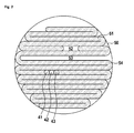
- an exemplary area of treatment 50 is shown.
- An image similar as shown in this figure may be drawn on a display to indicate which area has already been treated and which area needs further treatment.
- the first track 51 produced by moving the shockwave transducer over the head was interrupted producing a gap 52. After this gap, movement was resumed, resulting in a second track 54.
- region 53 was not treated. This will be indicated, therefore offering the surgeon the opportunity to re-treat this region, if it was not intentionally omitted.
- exemplary positions 41, 42 and 43 of the shockwave transducer are shown.
- the mapping device evaluates, displays and stores the shockwave dose applied to each of such positions resulting in a global dose image.
- the tracks shown herein consist of a plurality of such adjacent positions. For clarity, only the exemplary positions are shown.
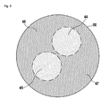
- FIG 3 a different way of indicating the treatment area is shown.
- the dose of shockwaves is shown, preferably by color shades.
- this image there are areas of lower dose 44, 45, which may be marked by a lighter color and areas of higher dose 46, 47 that may be marked by a darker color.
- the lighter colors of areas of lower dose 44, 45 may indicate that a further treatment of these areas is required.
Priority Applications (2)
| Application Number | Priority Date | Filing Date | Title |
|---|---|---|---|
| EP12189007.3A EP2722012B1 (de) | 2012-10-18 | 2012-10-18 | Vorrichtung zur Stoßwellentherapie eines menschlichen Gehirns |
| US14/057,295 US10143483B2 (en) | 2012-10-18 | 2013-10-18 | Device and method for shock wave treatment of the human brain |
Applications Claiming Priority (1)
| Application Number | Priority Date | Filing Date | Title |
|---|---|---|---|
| EP12189007.3A EP2722012B1 (de) | 2012-10-18 | 2012-10-18 | Vorrichtung zur Stoßwellentherapie eines menschlichen Gehirns |
Publications (2)
| Publication Number | Publication Date |
|---|---|
| EP2722012A1 true EP2722012A1 (de) | 2014-04-23 |
| EP2722012B1 EP2722012B1 (de) | 2015-06-03 |
Family
ID=47080333
Family Applications (1)
| Application Number | Title | Priority Date | Filing Date |
|---|---|---|---|
| EP12189007.3A Active EP2722012B1 (de) | 2012-10-18 | 2012-10-18 | Vorrichtung zur Stoßwellentherapie eines menschlichen Gehirns |
Country Status (2)
| Country | Link |
|---|---|
| US (1) | US10143483B2 (de) |
| EP (1) | EP2722012B1 (de) |
Families Citing this family (3)
| Publication number | Priority date | Publication date | Assignee | Title |
|---|---|---|---|---|
| US11484724B2 (en) | 2015-09-30 | 2022-11-01 | Btl Medical Solutions A.S. | Methods and devices for tissue treatment using mechanical stimulation and electromagnetic field |
| US10639233B2 (en) | 2016-03-11 | 2020-05-05 | The Regents Of The University Of California | Optimal dosages for low energy shock wave treatment of vital organs |
| EP4342394A1 (de) | 2022-09-23 | 2024-03-27 | MTS Medical UG | Vorrichtung und system zur behandlung von alzheimererkrankungen |
Citations (7)
| Publication number | Priority date | Publication date | Assignee | Title |
|---|---|---|---|---|
| WO1997010758A1 (de) | 1995-09-20 | 1997-03-27 | Storz Medical Ag | Vorrichtung zur behandlung von körpergewebe und zur zertrümmerung von körpersteinen |
| US20040122323A1 (en) * | 2002-12-23 | 2004-06-24 | Insightec-Txsonics Ltd | Tissue aberration corrections in ultrasound therapy |
| US20050020945A1 (en) * | 2002-07-02 | 2005-01-27 | Tosaya Carol A. | Acoustically-aided cerebrospinal-fluid manipulation for neurodegenerative disease therapy |
| EP2078503A1 (de) | 2007-12-18 | 2009-07-15 | Storz Medical Ag | Navigation bei der fokussierten Druckwellenbehandlung |
| US20100286518A1 (en) * | 2009-05-11 | 2010-11-11 | General Electric Company | Ultrasound system and method to deliver therapy based on user defined treatment spaces |
| US20110077559A1 (en) * | 2003-12-30 | 2011-03-31 | Medicis Technologies Corporation | Ultrasound therapy head with movement control |
| EP2494925A1 (de) * | 2011-03-03 | 2012-09-05 | Koninklijke Philips Electronics N.V. | Berechnung der Geschwindigkeit von Ultraschall in mindestens zwei Gewebetypen |
Family Cites Families (10)
| Publication number | Priority date | Publication date | Assignee | Title |
|---|---|---|---|---|
| US4736751A (en) * | 1986-12-16 | 1988-04-12 | Eeg Systems Laboratory | Brain wave source network location scanning method and system |
| FR2660761B1 (fr) * | 1990-04-06 | 1995-10-06 | Thomson Csf | Dispositif de detection de rayonnements dangereux pour les etres vivants. |
| US6961405B2 (en) * | 2002-10-07 | 2005-11-01 | Nomos Corporation | Method and apparatus for target position verification |
| US7507213B2 (en) * | 2004-03-16 | 2009-03-24 | General Patent Llc | Pressure pulse/shock wave therapy methods for organs |
| US7544171B2 (en) * | 2004-10-22 | 2009-06-09 | General Patent Llc | Methods for promoting nerve regeneration and neuronal growth and elongation |
| US8663083B2 (en) * | 2006-12-08 | 2014-03-04 | Koninklijke Philips N.V. | System, method, computer-readable medium, and use for planning combined therapy |
| US20120220812A1 (en) * | 2011-02-27 | 2012-08-30 | Mishelevich David J | Ultrasound neuromodulation for stroke mitigation and rehabilitation |
| US20130178693A1 (en) * | 2011-06-03 | 2013-07-11 | Nexstim Oy | Method and system for combining anatomical connectivity patterns and navigated brain stimulation |
| US20140213904A1 (en) * | 2011-08-26 | 2014-07-31 | Elekta Ab (Publ) | Intra-fraction motion management system |
| US20140200489A1 (en) * | 2011-09-01 | 2014-07-17 | Perseus-Biomed Inc | Method and system for tissue modulation |
-
2012
- 2012-10-18 EP EP12189007.3A patent/EP2722012B1/de active Active
-
2013
- 2013-10-18 US US14/057,295 patent/US10143483B2/en active Active
Patent Citations (7)
| Publication number | Priority date | Publication date | Assignee | Title |
|---|---|---|---|---|
| WO1997010758A1 (de) | 1995-09-20 | 1997-03-27 | Storz Medical Ag | Vorrichtung zur behandlung von körpergewebe und zur zertrümmerung von körpersteinen |
| US20050020945A1 (en) * | 2002-07-02 | 2005-01-27 | Tosaya Carol A. | Acoustically-aided cerebrospinal-fluid manipulation for neurodegenerative disease therapy |
| US20040122323A1 (en) * | 2002-12-23 | 2004-06-24 | Insightec-Txsonics Ltd | Tissue aberration corrections in ultrasound therapy |
| US20110077559A1 (en) * | 2003-12-30 | 2011-03-31 | Medicis Technologies Corporation | Ultrasound therapy head with movement control |
| EP2078503A1 (de) | 2007-12-18 | 2009-07-15 | Storz Medical Ag | Navigation bei der fokussierten Druckwellenbehandlung |
| US20100286518A1 (en) * | 2009-05-11 | 2010-11-11 | General Electric Company | Ultrasound system and method to deliver therapy based on user defined treatment spaces |
| EP2494925A1 (de) * | 2011-03-03 | 2012-09-05 | Koninklijke Philips Electronics N.V. | Berechnung der Geschwindigkeit von Ultraschall in mindestens zwei Gewebetypen |
Also Published As
| Publication number | Publication date |
|---|---|
| EP2722012B1 (de) | 2015-06-03 |
| US10143483B2 (en) | 2018-12-04 |
| US20140114326A1 (en) | 2014-04-24 |
Similar Documents
| Publication | Publication Date | Title |
|---|---|---|
| US20060184075A1 (en) | Method and device for applying pressure waves to the body of an organism | |
| KR102548194B1 (ko) | 경두개 초음파 치료 및 영상화 절차 수행을 위한 시스템 및 방법 | |
| US9364164B2 (en) | Non-invasive device and method for locating a structure such as a nerve | |
| JP4629034B2 (ja) | 剪断モード治療用超音波 | |
| JP4618810B2 (ja) | 剪断モード診断用超音波 | |
| CN101820949B (zh) | 用于引导放疗和其他过程的mri引导的hifu标记法 | |
| US20130281890A1 (en) | Neuromodulation devices and methods | |
| US20110208094A1 (en) | Ultrasound neuromodulation of the reticular activating system | |
| US20130184728A1 (en) | Ultrasound Neuromodulation for Diagnosis and Other-Modality Preplanning | |
| US11730452B2 (en) | Systems and methods for regulating microbubbles in ultrasound procedures | |
| KR101572898B1 (ko) | 초음파 모듈 및 이를 구비하는 헬멧형 저강도 초음파 집속 자극장치 | |
| JP2005517488A (ja) | 脂肪組織の超音波処理および画像化 | |
| US20210204915A1 (en) | Focused ultrasound system with optimized monitoring of cavitation | |
| US20100094178A1 (en) | Systems and Methods for Ultrasound Treatment of Thyroid and Parathyroid | |
| KR20150103047A (ko) | 조직의 처치를 위한 영상 유도 치료 장치 및 영상 유도 치료 장치의 준비 방법 | |
| EP2722012B1 (de) | Vorrichtung zur Stoßwellentherapie eines menschlichen Gehirns | |
| US20140213904A1 (en) | Intra-fraction motion management system | |
| JP6473149B2 (ja) | 超音波治療装置及び超音波治療システム | |
| US20240091565A1 (en) | Pre-treatment tissue sensitization for focused ultrasound procedures | |
| JP6386384B2 (ja) | 超音波治療装置および超音波治療システム | |
| US11872413B2 (en) | Control method for the treatment of brain tissue using an ultrasonic probe and an implanted acoustic window on the cranium | |
| JP6297411B2 (ja) | 超音波治療装置及び超音波治療システム | |
| US20200390416A1 (en) | On-Screen Markers For Out-Of-Plane Needle Guidance | |
| US9408624B2 (en) | Intervertebral disc treatment apparatus | |
| JP2015146952A (ja) | 超音波治療装置及び超音波治療システム |
Legal Events
| Date | Code | Title | Description |
|---|---|---|---|
| PUAI | Public reference made under article 153(3) epc to a published international application that has entered the european phase |
Free format text: ORIGINAL CODE: 0009012 |
|
| 17P | Request for examination filed |
Effective date: 20130625 |
|
| AK | Designated contracting states |
Kind code of ref document: A1 Designated state(s): AL AT BE BG CH CY CZ DE DK EE ES FI FR GB GR HR HU IE IS IT LI LT LU LV MC MK MT NL NO PL PT RO RS SE SI SK SM TR |
|
| AX | Request for extension of the european patent |
Extension state: BA ME |
|
| REG | Reference to a national code |
Ref country code: DE Ref legal event code: R079 Ref document number: 602012007713 Country of ref document: DE Free format text: PREVIOUS MAIN CLASS: A61B0017225000 Ipc: A61N0007000000 |
|
| RBV | Designated contracting states (corrected) |
Designated state(s): AL AT BE BG CH CY CZ DE DK EE ES FI FR GB GR HR HU IE IS IT LI LT LU LV MC MK MT NL NO PL PT RO RS SE SI SK SM TR |
|
| GRAP | Despatch of communication of intention to grant a patent |
Free format text: ORIGINAL CODE: EPIDOSNIGR1 |
|
| RIC1 | Information provided on ipc code assigned before grant |
Ipc: A61N 7/00 20060101AFI20141119BHEP |
|
| RAP1 | Party data changed (applicant data changed or rights of an application transferred) |
Owner name: STORZ MEDICAL AG |
|
| INTG | Intention to grant announced |
Effective date: 20141215 |
|
| GRAS | Grant fee paid |
Free format text: ORIGINAL CODE: EPIDOSNIGR3 |
|
| GRAA | (expected) grant |
Free format text: ORIGINAL CODE: 0009210 |
|
| AK | Designated contracting states |
Kind code of ref document: B1 Designated state(s): AL AT BE BG CH CY CZ DE DK EE ES FI FR GB GR HR HU IE IS IT LI LT LU LV MC MK MT NL NO PL PT RO RS SE SI SK SM TR |
|
| REG | Reference to a national code |
Ref country code: GB Ref legal event code: FG4D |
|
| REG | Reference to a national code |
Ref country code: CH Ref legal event code: EP |
|
| REG | Reference to a national code |
Ref country code: AT Ref legal event code: REF Ref document number: 729606 Country of ref document: AT Kind code of ref document: T Effective date: 20150715 Ref country code: IE Ref legal event code: FG4D |
|
| REG | Reference to a national code |
Ref country code: DE Ref legal event code: R096 Ref document number: 602012007713 Country of ref document: DE |
|
| REG | Reference to a national code |
Ref country code: AT Ref legal event code: MK05 Ref document number: 729606 Country of ref document: AT Kind code of ref document: T Effective date: 20150603 |
|
| REG | Reference to a national code |
Ref country code: FR Ref legal event code: PLFP Year of fee payment: 4 |
|
| PG25 | Lapsed in a contracting state [announced via postgrant information from national office to epo] |
Ref country code: HR Free format text: LAPSE BECAUSE OF FAILURE TO SUBMIT A TRANSLATION OF THE DESCRIPTION OR TO PAY THE FEE WITHIN THE PRESCRIBED TIME-LIMIT Effective date: 20150603 Ref country code: FI Free format text: LAPSE BECAUSE OF FAILURE TO SUBMIT A TRANSLATION OF THE DESCRIPTION OR TO PAY THE FEE WITHIN THE PRESCRIBED TIME-LIMIT Effective date: 20150603 Ref country code: ES Free format text: LAPSE BECAUSE OF FAILURE TO SUBMIT A TRANSLATION OF THE DESCRIPTION OR TO PAY THE FEE WITHIN THE PRESCRIBED TIME-LIMIT Effective date: 20150603 Ref country code: NO Free format text: LAPSE BECAUSE OF FAILURE TO SUBMIT A TRANSLATION OF THE DESCRIPTION OR TO PAY THE FEE WITHIN THE PRESCRIBED TIME-LIMIT Effective date: 20150903 Ref country code: LT Free format text: LAPSE BECAUSE OF FAILURE TO SUBMIT A TRANSLATION OF THE DESCRIPTION OR TO PAY THE FEE WITHIN THE PRESCRIBED TIME-LIMIT Effective date: 20150603 |
|
| REG | Reference to a national code |
Ref country code: NL Ref legal event code: MP Effective date: 20150603 |
|
| REG | Reference to a national code |
Ref country code: LT Ref legal event code: MG4D |
|
| PG25 | Lapsed in a contracting state [announced via postgrant information from national office to epo] |
Ref country code: BG Free format text: LAPSE BECAUSE OF FAILURE TO SUBMIT A TRANSLATION OF THE DESCRIPTION OR TO PAY THE FEE WITHIN THE PRESCRIBED TIME-LIMIT Effective date: 20150903 Ref country code: AT Free format text: LAPSE BECAUSE OF FAILURE TO SUBMIT A TRANSLATION OF THE DESCRIPTION OR TO PAY THE FEE WITHIN THE PRESCRIBED TIME-LIMIT Effective date: 20150603 Ref country code: LV Free format text: LAPSE BECAUSE OF FAILURE TO SUBMIT A TRANSLATION OF THE DESCRIPTION OR TO PAY THE FEE WITHIN THE PRESCRIBED TIME-LIMIT Effective date: 20150603 Ref country code: GR Free format text: LAPSE BECAUSE OF FAILURE TO SUBMIT A TRANSLATION OF THE DESCRIPTION OR TO PAY THE FEE WITHIN THE PRESCRIBED TIME-LIMIT Effective date: 20150904 Ref country code: RS Free format text: LAPSE BECAUSE OF FAILURE TO SUBMIT A TRANSLATION OF THE DESCRIPTION OR TO PAY THE FEE WITHIN THE PRESCRIBED TIME-LIMIT Effective date: 20150603 |
|
| PG25 | Lapsed in a contracting state [announced via postgrant information from national office to epo] |
Ref country code: EE Free format text: LAPSE BECAUSE OF FAILURE TO SUBMIT A TRANSLATION OF THE DESCRIPTION OR TO PAY THE FEE WITHIN THE PRESCRIBED TIME-LIMIT Effective date: 20150603 |
|
| PG25 | Lapsed in a contracting state [announced via postgrant information from national office to epo] |
Ref country code: PL Free format text: LAPSE BECAUSE OF FAILURE TO SUBMIT A TRANSLATION OF THE DESCRIPTION OR TO PAY THE FEE WITHIN THE PRESCRIBED TIME-LIMIT Effective date: 20150603 Ref country code: IS Free format text: LAPSE BECAUSE OF FAILURE TO SUBMIT A TRANSLATION OF THE DESCRIPTION OR TO PAY THE FEE WITHIN THE PRESCRIBED TIME-LIMIT Effective date: 20151003 Ref country code: PT Free format text: LAPSE BECAUSE OF FAILURE TO SUBMIT A TRANSLATION OF THE DESCRIPTION OR TO PAY THE FEE WITHIN THE PRESCRIBED TIME-LIMIT Effective date: 20151006 Ref country code: SK Free format text: LAPSE BECAUSE OF FAILURE TO SUBMIT A TRANSLATION OF THE DESCRIPTION OR TO PAY THE FEE WITHIN THE PRESCRIBED TIME-LIMIT Effective date: 20150603 Ref country code: RO Free format text: LAPSE BECAUSE OF NON-PAYMENT OF DUE FEES Effective date: 20150603 Ref country code: CZ Free format text: LAPSE BECAUSE OF FAILURE TO SUBMIT A TRANSLATION OF THE DESCRIPTION OR TO PAY THE FEE WITHIN THE PRESCRIBED TIME-LIMIT Effective date: 20150603 |
|
| REG | Reference to a national code |
Ref country code: DE Ref legal event code: R097 Ref document number: 602012007713 Country of ref document: DE |
|
| PLBE | No opposition filed within time limit |
Free format text: ORIGINAL CODE: 0009261 |
|
| STAA | Information on the status of an ep patent application or granted ep patent |
Free format text: STATUS: NO OPPOSITION FILED WITHIN TIME LIMIT |
|
| PG25 | Lapsed in a contracting state [announced via postgrant information from national office to epo] |
Ref country code: DK Free format text: LAPSE BECAUSE OF FAILURE TO SUBMIT A TRANSLATION OF THE DESCRIPTION OR TO PAY THE FEE WITHIN THE PRESCRIBED TIME-LIMIT Effective date: 20150603 |
|
| 26N | No opposition filed |
Effective date: 20160304 |
|
| PG25 | Lapsed in a contracting state [announced via postgrant information from national office to epo] |
Ref country code: SI Free format text: LAPSE BECAUSE OF FAILURE TO SUBMIT A TRANSLATION OF THE DESCRIPTION OR TO PAY THE FEE WITHIN THE PRESCRIBED TIME-LIMIT Effective date: 20150603 Ref country code: LU Free format text: LAPSE BECAUSE OF FAILURE TO SUBMIT A TRANSLATION OF THE DESCRIPTION OR TO PAY THE FEE WITHIN THE PRESCRIBED TIME-LIMIT Effective date: 20151018 |
|
| PG25 | Lapsed in a contracting state [announced via postgrant information from national office to epo] |
Ref country code: MC Free format text: LAPSE BECAUSE OF FAILURE TO SUBMIT A TRANSLATION OF THE DESCRIPTION OR TO PAY THE FEE WITHIN THE PRESCRIBED TIME-LIMIT Effective date: 20150603 |
|
| REG | Reference to a national code |
Ref country code: IE Ref legal event code: MM4A |
|
| REG | Reference to a national code |
Ref country code: FR Ref legal event code: PLFP Year of fee payment: 5 |
|
| PG25 | Lapsed in a contracting state [announced via postgrant information from national office to epo] |
Ref country code: IE Free format text: LAPSE BECAUSE OF NON-PAYMENT OF DUE FEES Effective date: 20151018 |
|
| PG25 | Lapsed in a contracting state [announced via postgrant information from national office to epo] |
Ref country code: BE Free format text: LAPSE BECAUSE OF FAILURE TO SUBMIT A TRANSLATION OF THE DESCRIPTION OR TO PAY THE FEE WITHIN THE PRESCRIBED TIME-LIMIT Effective date: 20150603 |
|
| PG25 | Lapsed in a contracting state [announced via postgrant information from national office to epo] |
Ref country code: SM Free format text: LAPSE BECAUSE OF FAILURE TO SUBMIT A TRANSLATION OF THE DESCRIPTION OR TO PAY THE FEE WITHIN THE PRESCRIBED TIME-LIMIT Effective date: 20150603 Ref country code: HU Free format text: LAPSE BECAUSE OF FAILURE TO SUBMIT A TRANSLATION OF THE DESCRIPTION OR TO PAY THE FEE WITHIN THE PRESCRIBED TIME-LIMIT; INVALID AB INITIO Effective date: 20121018 |
|
| PG25 | Lapsed in a contracting state [announced via postgrant information from national office to epo] |
Ref country code: CY Free format text: LAPSE BECAUSE OF FAILURE TO SUBMIT A TRANSLATION OF THE DESCRIPTION OR TO PAY THE FEE WITHIN THE PRESCRIBED TIME-LIMIT Effective date: 20150603 Ref country code: SE Free format text: LAPSE BECAUSE OF FAILURE TO SUBMIT A TRANSLATION OF THE DESCRIPTION OR TO PAY THE FEE WITHIN THE PRESCRIBED TIME-LIMIT Effective date: 20150603 Ref country code: NL Free format text: LAPSE BECAUSE OF FAILURE TO SUBMIT A TRANSLATION OF THE DESCRIPTION OR TO PAY THE FEE WITHIN THE PRESCRIBED TIME-LIMIT Effective date: 20150603 |
|
| PG25 | Lapsed in a contracting state [announced via postgrant information from national office to epo] |
Ref country code: MT Free format text: LAPSE BECAUSE OF FAILURE TO SUBMIT A TRANSLATION OF THE DESCRIPTION OR TO PAY THE FEE WITHIN THE PRESCRIBED TIME-LIMIT Effective date: 20150603 |
|
| REG | Reference to a national code |
Ref country code: FR Ref legal event code: PLFP Year of fee payment: 6 |
|
| PG25 | Lapsed in a contracting state [announced via postgrant information from national office to epo] |
Ref country code: MK Free format text: LAPSE BECAUSE OF FAILURE TO SUBMIT A TRANSLATION OF THE DESCRIPTION OR TO PAY THE FEE WITHIN THE PRESCRIBED TIME-LIMIT Effective date: 20150603 |
|
| REG | Reference to a national code |
Ref country code: FR Ref legal event code: PLFP Year of fee payment: 7 |
|
| PG25 | Lapsed in a contracting state [announced via postgrant information from national office to epo] |
Ref country code: TR Free format text: LAPSE BECAUSE OF FAILURE TO SUBMIT A TRANSLATION OF THE DESCRIPTION OR TO PAY THE FEE WITHIN THE PRESCRIBED TIME-LIMIT Effective date: 20150603 Ref country code: AL Free format text: LAPSE BECAUSE OF FAILURE TO SUBMIT A TRANSLATION OF THE DESCRIPTION OR TO PAY THE FEE WITHIN THE PRESCRIBED TIME-LIMIT Effective date: 20150603 |
|
| P01 | Opt-out of the competence of the unified patent court (upc) registered |
Effective date: 20231110 |
|
| PGFP | Annual fee paid to national office [announced via postgrant information from national office to epo] |
Ref country code: GB Payment date: 20231025 Year of fee payment: 12 |
|
| PGFP | Annual fee paid to national office [announced via postgrant information from national office to epo] |
Ref country code: IT Payment date: 20231031 Year of fee payment: 12 Ref country code: FR Payment date: 20231023 Year of fee payment: 12 Ref country code: DE Payment date: 20231017 Year of fee payment: 12 Ref country code: CH Payment date: 20231102 Year of fee payment: 12 |