EP2707389B1 - Réactifs de dosage pour kit de diagnostic de la neurogranine - Google Patents
Réactifs de dosage pour kit de diagnostic de la neurogranine Download PDFInfo
- Publication number
- EP2707389B1 EP2707389B1 EP12782967.9A EP12782967A EP2707389B1 EP 2707389 B1 EP2707389 B1 EP 2707389B1 EP 12782967 A EP12782967 A EP 12782967A EP 2707389 B1 EP2707389 B1 EP 2707389B1
- Authority
- EP
- European Patent Office
- Prior art keywords
- neurogranin
- antibody
- antigen
- binding
- sample
- Prior art date
- Legal status (The legal status is an assumption and is not a legal conclusion. Google has not performed a legal analysis and makes no representation as to the accuracy of the status listed.)
- Active
Links
Images


Classifications
-
- G—PHYSICS
- G01—MEASURING; TESTING
- G01N—INVESTIGATING OR ANALYSING MATERIALS BY DETERMINING THEIR CHEMICAL OR PHYSICAL PROPERTIES
- G01N33/00—Investigating or analysing materials by specific methods not covered by groups G01N1/00 - G01N31/00
- G01N33/48—Biological material, e.g. blood, urine; Haemocytometers
- G01N33/50—Chemical analysis of biological material, e.g. blood, urine; Testing involving biospecific ligand binding methods; Immunological testing
- G01N33/68—Chemical analysis of biological material, e.g. blood, urine; Testing involving biospecific ligand binding methods; Immunological testing involving proteins, peptides or amino acids
- G01N33/6893—Chemical analysis of biological material, e.g. blood, urine; Testing involving biospecific ligand binding methods; Immunological testing involving proteins, peptides or amino acids related to diseases not provided for elsewhere
- G01N33/6896—Neurological disorders, e.g. Alzheimer's disease
-
- C—CHEMISTRY; METALLURGY
- C07—ORGANIC CHEMISTRY
- C07K—PEPTIDES
- C07K16/00—Immunoglobulins [IGs], e.g. monoclonal or polyclonal antibodies
- C07K16/18—Immunoglobulins [IGs], e.g. monoclonal or polyclonal antibodies against material from animals or humans
-
- C—CHEMISTRY; METALLURGY
- C12—BIOCHEMISTRY; BEER; SPIRITS; WINE; VINEGAR; MICROBIOLOGY; ENZYMOLOGY; MUTATION OR GENETIC ENGINEERING
- C12N—MICROORGANISMS OR ENZYMES; COMPOSITIONS THEREOF; PROPAGATING, PRESERVING, OR MAINTAINING MICROORGANISMS; MUTATION OR GENETIC ENGINEERING; CULTURE MEDIA
- C12N15/00—Mutation or genetic engineering; DNA or RNA concerning genetic engineering, vectors, e.g. plasmids, or their isolation, preparation or purification; Use of hosts therefor
- C12N15/09—Recombinant DNA-technology
- C12N15/11—DNA or RNA fragments; Modified forms thereof; Non-coding nucleic acids having a biological activity
- C12N15/115—Aptamers, i.e. nucleic acids binding a target molecule specifically and with high affinity without hybridising therewith ; Nucleic acids binding to non-nucleic acids, e.g. aptamers
-
- C—CHEMISTRY; METALLURGY
- C12—BIOCHEMISTRY; BEER; SPIRITS; WINE; VINEGAR; MICROBIOLOGY; ENZYMOLOGY; MUTATION OR GENETIC ENGINEERING
- C12Q—MEASURING OR TESTING PROCESSES INVOLVING ENZYMES, NUCLEIC ACIDS OR MICROORGANISMS; COMPOSITIONS OR TEST PAPERS THEREFOR; PROCESSES OF PREPARING SUCH COMPOSITIONS; CONDITION-RESPONSIVE CONTROL IN MICROBIOLOGICAL OR ENZYMOLOGICAL PROCESSES
- C12Q1/00—Measuring or testing processes involving enzymes, nucleic acids or microorganisms; Compositions therefor; Processes of preparing such compositions
- C12Q1/68—Measuring or testing processes involving enzymes, nucleic acids or microorganisms; Compositions therefor; Processes of preparing such compositions involving nucleic acids
-
- G—PHYSICS
- G01—MEASURING; TESTING
- G01N—INVESTIGATING OR ANALYSING MATERIALS BY DETERMINING THEIR CHEMICAL OR PHYSICAL PROPERTIES
- G01N33/00—Investigating or analysing materials by specific methods not covered by groups G01N1/00 - G01N31/00
- G01N33/48—Biological material, e.g. blood, urine; Haemocytometers
- G01N33/50—Chemical analysis of biological material, e.g. blood, urine; Testing involving biospecific ligand binding methods; Immunological testing
- G01N33/53—Immunoassay; Biospecific binding assay; Materials therefor
- G01N33/566—Immunoassay; Biospecific binding assay; Materials therefor using specific carrier or receptor proteins as ligand binding reagents where possible specific carrier or receptor proteins are classified with their target compounds
-
- C—CHEMISTRY; METALLURGY
- C07—ORGANIC CHEMISTRY
- C07K—PEPTIDES
- C07K2317/00—Immunoglobulins specific features
- C07K2317/30—Immunoglobulins specific features characterized by aspects of specificity or valency
-
- C—CHEMISTRY; METALLURGY
- C12—BIOCHEMISTRY; BEER; SPIRITS; WINE; VINEGAR; MICROBIOLOGY; ENZYMOLOGY; MUTATION OR GENETIC ENGINEERING
- C12N—MICROORGANISMS OR ENZYMES; COMPOSITIONS THEREOF; PROPAGATING, PRESERVING, OR MAINTAINING MICROORGANISMS; MUTATION OR GENETIC ENGINEERING; CULTURE MEDIA
- C12N2310/00—Structure or type of the nucleic acid
- C12N2310/10—Type of nucleic acid
- C12N2310/16—Aptamers
-
- G—PHYSICS
- G01—MEASURING; TESTING
- G01N—INVESTIGATING OR ANALYSING MATERIALS BY DETERMINING THEIR CHEMICAL OR PHYSICAL PROPERTIES
- G01N2333/00—Assays involving biological materials from specific organisms or of a specific nature
- G01N2333/435—Assays involving biological materials from specific organisms or of a specific nature from animals; from humans
- G01N2333/46—Assays involving biological materials from specific organisms or of a specific nature from animals; from humans from vertebrates
- G01N2333/47—Assays involving proteins of known structure or function as defined in the subgroups
-
- G—PHYSICS
- G01—MEASURING; TESTING
- G01N—INVESTIGATING OR ANALYSING MATERIALS BY DETERMINING THEIR CHEMICAL OR PHYSICAL PROPERTIES
- G01N2800/00—Detection or diagnosis of diseases
- G01N2800/28—Neurological disorders
Definitions
- Brain injuries are complex and can have multiple severe clinical outcomes. Damage of the brain and spinal cord can result from head trauma, stroke, traumatic birth, heart surgery, cardiac arrest and patients requiring cardiovascular support with ventricular assist devices or extracorporeal membrane oxygenation (ECMO). Moreover, detection of subclinical brain injury is difficult, especially in children and neonates with birth-related injury. In addition, children with sickle cell disease are at high risk for subclinical brain injury. Untreated subclinical brain injuries in children can progress to overt stroke, neurological damage, learning problems and memory loss.
- ECMO extracorporeal membrane oxygenation
- Laterza et al., 2006 (Clinical Chemistry, 2006, 52:1713-1721 ) summarizes the developments in investigating novel brain biomarkers which are measured directly as messenger RNA (mRNA) levels in brain tissue.
- Laterza et al., 2008 (Biomarkers Med., 2008, 2:81-92 ) provides an overview of biomarkers of tissue injury, but does neither teach nor suggest neurogranin as biomarker for brain injury.
- Iliuk et al. (Analytical Chemistry, 2011, 83:4440-4452 ) generally refers to the usability of aptamers in bioanalytical applications.
- Watson et al. (Molecular Brain Research, 1994, 27:323-328 ) teaches the localization of neurogranin in rat brain subcellular fractions.
- Neuner-Jehle et al. (Brain Research, 1995, 685:143-153 ) investigates the effect of sleep deprivation on neurogranin levels in rat brain.
- Tang et al., 2006 (European Journal of Pharmacology, 2006, 542:106-107 ) and Tang et al., 2007 (Brain Research, 2007, 1143:78-82 ) investigate the effect of antisense oligonucleotides targeting neurogranin on acute opioid dependence.
- the present invention is defined by the claims.
- the present invention is based, at least in part, on the use of antibodies that bind neurogranin.
- the use and method of the present invention makes use of an isolated antibody or fragment thereof that specifically binds to neurogranin.
- the isolated antibody or fragment thereof specifically binds to amino acids 1-78 of SEQ ID NO:4.
- an isolated antibody or fragment thereof specifically binds to amino acids 55-78 of SEQ ID NO:4.
- the antibody or fragment thereof is polyclonal.
- the antibody or fragment thereof is monoclonal.
- the antibody or fragment thereof is mammalian.
- the antibody or fragment thereof can be human. Further disclosed is a hybridoma cell which produces the antibody or fragment described herein.
- the antibody fragment can be selected from the group consisting of a Fab fragment; a F(ab') 2 fragment; a Fv fragment; and a single chain fragment.
- the isolated antibody or fragment thereof can further comprise a detectable substance coupled to the antibody.
- the detectable substance is selected from the group consisting of an enzyme; a fluorescent label; a radioisotope; and chemiluminescent label.
- the isolated antibody or fragment thereof specifically binds to neurogranin in an ELISA. In another embodiment, the isolated antibody or fragment thereof specifically binds to neurogranin in a competitive-binding assay. In yet another embodiment, the isolated antibody or fragment thereof specifically binds to neurogranin in a radioimmunoassay. In a further embodiment, the isolated antibody or fragment thereof specifically binds to neurogranin in a fluorescence-activated cell sorting (FACS) assay.
- FACS fluorescence-activated cell sorting
- kits useful for detecting neurogranin may comprise (a) an isolated antibody described herein; and (b) at least one component to detect binding of the isolated antibody to neurogranin.
- the isolated antibody is obtained from an animal that has been immunized with neurogranin, wherein the antibody specifically binds to an antigenic epitope-bearing polypeptide fragment of neurogranin.
- the monoclonal antibody is designated 30.5.2 that specifically binds neurogranin.
- the anti-neurogranin monoclonal antibody is produced by a hybridoma.
- aptamers that bind neurogranin.
- a polynucleotide aptamer that specifically binds neurogranin.
- the neurogranin is human neurogranin.
- the aptamer may bind to neurogranin with a Kd of less than about 1000nM.
- the aptamer may bind to neurogranin with a Kd of less than about 100 nM.
- the aptamer may bind to neurogranin with a Kd of less than about 20 nM.
- the polynucleotide aptamer can consist of about 10 to about 100 nucleotides.
- the polynucleotide aptamer may consist of about 20 to about 80 nucleotides.
- the polynucleotide aptamer may consist of about 30 to about 50 nucleotides.
- the polynucleotide aptamer may be an RNA aptamer.
- the polynucleotide aptamer may comprise a nucleotide sequence at least 80% identical to any one of SEQ ID NOS: 4-6 or a fragment thereof of at least ten contiguous nucleotides.
- the polynucleotide aptamer may comprise a nucleotide sequence at least 90% identical to any one of SEQ ID NOS: 4-6 or a fragment thereof of at least ten contiguous nucleotides.
- the polynucleotide aptamer may comprise a nucleotide sequence at least 95% identical to any one of SEQ ID NOS: 4-6 or a fragment thereof of at least ten contiguous nucleotides.
- a polynucleotide aptamer may comprise the nucleotide sequence of any one of SEQ ID NOS: 4-6 or a fragment thereof of at least ten contiguous nucleotides.
- a polynucleotide aptamer may consist of the nucleotide sequence of any one of SEQ ID NOS: 4-6 or a fragment thereof of at least ten contiguous nucleotides.
- the polynucleotide aptamer can comprise at least one modified internucleotide linker.
- the polynucleotide aptamer can comprise at least one terminal blocker.
- the polynucleotide aptamer can be linked to a conjugate.
- a vector comprising a polynucleotide described herein.
- a cell comprising a polynucleotide aptamer described herein.
- a cell may comprise two or more different polynucleotide aptamers.
- the method may comprise measuring the level of neurogranin in the subject by binding neurogranin with a polynucleotide aptamer and determining the amount of aptamer bound to neurogranin. The binding may occur in a sample obtained from the subject.
- a method for determining the amount of neurogranin in a sample may comprise the step of detecting a peptide specific to neurogranin using a triple quadrupole mass spectrometer and multiple reaction monitoring, wherein the peptide specific to neurogranin comprises SEQ ID NO:7.
- a neurogranin RNA aptamer (NRGN-A1) with a sequence comprising the sequence of: TCTAACGCCTCCCGTATGTTTTCCTTTTTCCATTGCGGAT (SEQ ID NO:4), which can bind to neurogranin protein and is useful for a detection assay.
- a neurogranin RNA aptamer (NRGN-A6) with a sequence comprising the sequence of: TTTTCATTTTCATTTTTTTCCAAATCGATCCGCCGGACCTTAT (SEQ ID NO:6), which can bind to neurogranin protein and is useful for a detection assay.
- the monoclonal antibody is identified as 30.5.2 to human neurogranin and is useful for a diagnostic detection assay for brain injury.
- a method of using a neurogranin RNA aptamer in a diagnostic assay for brain injury in a mammalian subject comprises the steps of (a) obtaining a sample from the subject suspected of having a brain injury, and (b) performing an assay using the aptamer to detect a level of neurogranin in the sample, wherein a level of neurogranin in the sample that is significantly different than in a sample obtained from a control subject that does not have a brain injury is diagnostic of a brain injury.
- a method of using a neurogranin monoclonal antibody in a diagnostic assay for brain injury in a mammalian subject comprises the steps of (a) obtaining a sample from the subject suspected of having a brain injury, and (b) performing an assay using the antibody to detect the level of neurogranin in the sample, wherein a level of neurogranin in the sample that is significantly different than in a sample obtained from a control subject that does not have a brain injury is diagnostic of a brain injury.
- the brain injury is selected from the group consisting of subclinical brain injury and overt brain injury.
- a diagnostic /prognostic kit for brain injury comprising capture and detection reagents for neurogranin.
- an "injury” is an alteration in cellular or molecular integrity, activity, level, robustness, state, or other alteration that is traceable to an event.
- an injury includes a physical, mechanical, chemical, biological, functional, infectious, or other modulator of cellular or molecular characteristics.
- An event can include a physical trauma such as an impact (percussive) or a biological abnormality such as a stroke resulting from either blockade or leakage of a blood vessel.
- An event is optionally an infection by an infectious agent.
- brain injury refers to a condition that results in central nervous system damage, irrespective of its pathophysiological basis.
- stroke is classified into hemorrhagic and non-hemorrhagic.
- hemorrhagic stroke include cerebral hemorrhage, subarachnoid hemorrhage, and intracranial hemorrhage secondary to cerebral arterial malformation, while examples of non-hemorrhagic stroke include cerebral infarction.
- traumatic brain injury or "TBI” refer to traumatic injuries to the brain which occur when physical trauma causes brain damage.
- TBI can result from a closed head injury or a penetrating head injury.
- a "non-traumatic brain injury” refers to brain injuries that do not involve ischemia or external mechanical force (e.g., stroke, Alzheimer's disease, Parkinson's disease, Huntington's disease, multiple sclerosis, amyotrophic lateral sclerosis, brain hemorrhage, brain infections, brain tumor, and the like).
- brain injury also refers to subclinical brain injury, spinal cord injury, and anoxic-ischemic brain injury.
- subclinical brain injury (SCI) refers to brain injury without overt clinical evidence of brain injury. A lack of clinical evidence of brain injury when brain injury actually exists could result from degree of injury, type of injury, level of consciousness, and medications, particularly sedation and anesthesia.
- spinal cord injury refers to a condition in which the spinal cord receives compression/detrition due to a vertebral fracture or dislocation to cause dysfunction.
- anoxic-ischemic brain injury refers to deprivation of oxygen supply to brain tissue resulting in compromised brain function and includes cerebral hypoxia.
- anoxic-ischemic brain injury includes focal cerebral ischemia, global cerebral ischemia, hypoxic hypoxia (i.e., limited oxygen in the environment causes reduced brain function, such as with divers, aviators, mountain climbers, and fire fighters, all of whom are at risk for this kind of cerebral hypoxia), obstructions in the lungs (e.g., hypoxia resulting from choking, strangulation, the crushing of the windpipe).
- hypoxic hypoxia i.e., limited oxygen in the environment causes reduced brain function, such as with divers, aviators, mountain climbers, and fire fighters, all of whom are at risk for this kind of cerebral hypoxia
- obstructions in the lungs e.g., hypoxia resulting from choking, strangulation, the crushing of the windpipe.
- comparing refers to making an assessment of how the proportion, level or cellular localization of one or more biomarkers in a sample from a patient relates to the proportion, level or cellular localization of the corresponding one or more biomarkers in a standard or control sample.
- comparing may refer to assessing whether the proportion, level, or cellular localization of one or more biomarkers in a sample from a patient is the same as, more or less than, or different from the proportion, level, or cellular localization of the corresponding one or more biomarkers in standard or control sample.
- the term may refer to assessing whether the proportion, level, or cellular localization of one or more biomarkers in a sample from a patient is the same as, more or less than, different from or otherwise corresponds (or not) to the proportion, level, or cellular localization of predefined biomarker levels that correspond to, for example, a patient having subclinical brain injury (SCI), not having SCI, is responding to treatment for SCI, is not responding to treatment for SCI, is/is not likely to respond to a particular SCI treatment, or having /not having another disease or condition.
- SCI subclinical brain injury
- the term "comparing" refers to assessing whether the level of one or more biomarkers of the present invention (neurogranin) in a sample from a patient is the same as, more or less than, different from other otherwise correspond (or not) to levels of the same biomarkers in a control sample (e.g., predefined levels that correlate to uninfected individuals, standard SCI levels, etc.).
- the terms “indicates” or “correlates” in reference to a parameter, e.g., a modulated proportion, level, or cellular localization in a sample from a patient, may mean that the patient has SCI.
- the parameter may comprise the level of biomarkers of the present invention (neurogranin).
- a particular set or pattern of the amounts of one or more biomarkers may indicate that a patient has SCI (i.e., correlates to a patient having SCI).
- a particular set or pattern of the amounts of one or more biomarkers may be correlated to a patient being unaffected (i.e., indicates a patient does not have SCI).
- "indicating," or “correlating,” as used according to the present invention may be by any linear or non-linear method of quantifying the relationship between levels of biomarkers to a standard, control or comparative value for the assessment of the diagnosis, prediction of SCI or SCI progression, assessment of efficacy of clinical treatment, identification of a patient that may respond to a particular treatment regime or pharmaceutical agent, monitoring of the progress of treatment, and in the context of a screening assay, for the identification of an anti-SCI therapeutic.
- patient refers to a mammal, particularly, a human.
- the patient may have mild, intermediate or severe disease.
- the patient may be treatment naive, responding to any form of treatment, or refractory.
- the patient may be an individual in need of treatment or in need of diagnosis based on particular symptoms or family history.
- the terms may refer to treatment in experimental animals, in veterinary application, and in the development of animal models for disease, including, but not limited to, rodents including mice, rats, and hamsters; and primates.
- measuring and determining are used interchangeably throughout, and refer to methods which include obtaining a patient sample and/or detecting the level of a biomarker(s) in a sample. In one embodiment, the terms refer to obtaining a patient sample and detecting the level of one or more biomarkers in the sample. In another embodiment, the terms “measuring” and “determining” mean detecting the level of one or more biomarkers in a patient sample. Measuring can be accomplished by methods known in the art and those further described herein. The term “measuring” is also used interchangeably throughout with the term "detecting.”
- sample encompass a variety of sample types obtained from a patient, individual, or subject and can be used in a diagnostic or monitoring assay.
- the patient sample may be obtained from a healthy subject, a diseased patient or a patient having associated symptoms of SCI.
- a sample obtained from a patient can be divided and only a portion may be used for diagnosis. Further, the sample, or a portion thereof, can be stored under conditions to maintain sample for later analysis.
- the definition specifically encompasses blood and other liquid samples of biological origin (including, but not limited to, plasma, serum, peripheral blood, cerebrospinal fluid, urine, saliva, stool and synovial fluid), solid tissue samples such as a biopsy specimen or tissue cultures or cells derived therefrom and the progeny thereof.
- a sample comprises a blood sample.
- a serum sample is used.
- the definition also includes samples that have been manipulated in any way after their procurement, such as by centrifugation, filtration, precipitation, dialysis, chromatography, treatment with reagents, washed, or enriched for certain cell populations.
- the terms further encompass a clinical sample, and also include cells in culture, cell supernatants, tissue samples, organs, and the like. Samples may also comprise fresh-frozen and/or formalin-fixed, paraffin-embedded tissue blocks, such as blocks prepared from clinical or pathological biopsies, prepared for pathological analysis or study by immunohistochemistry.
- Various methodologies of the instant invention include a step that involves comparing a value, level, feature, characteristic, property, etc. to a "suitable control,” referred to interchangeably herein as an “appropriate control” or a “control sample.”
- a “suitable control,” “appropriate control” or a “control sample” is any control or standard familiar to one of ordinary skill in the art useful for comparison purposes.
- a "suitable control” or “appropriate control” is a value, level, feature, characteristic, property, etc., determined in a cell, organ, or patient, e.g., a control or normal cell, organ, or patient, exhibiting, for example, normal traits.
- the biomarkers of the present invention may be assayed for levels in a sample from an unaffected individual (UI) or a normal control individual (NC) (both terms are used interchangeably herein).
- a "suitable control” or “appropriate control” is a value, level, feature, characteristic, property, etc. determined prior to performing a therapy (e.g., an SCI treatment) on a patient.
- a transcription rate, mRNA level, translation rate, protein level, biological activity, cellular characteristic or property, genotype, phenotype, etc. can be determined prior to, during, or after administering a therapy into a cell, organ, or patient.
- a "suitable control” or “appropriate control” is a predefined value, level, feature, characteristic, property, etc.
- a “suitable control” can be a profile or pattern of levels of biomarkers of the present invention (neurogranin) that correlates to SCI, to which a patient sample can be compared. The patient sample can also be compared to a negative control, i.e., a profile that correlates to not having SCI.
- isolated designates a biological material (nucleic acid or protein) that has been removed from its original environment (the environment in which it is naturally present). For example, a polynucleotide present in its natural state in a plant or an animal is not isolated, however the same polynucleotide separated from the adjacent nucleic acids in which it is naturally present, is considered “isolated”.
- purified does not require the material to be present in a form exhibiting absolute purity, exclusive of the presence of other compounds. It is rather a relative definition.
- nucleic acid refers to the phosphate ester polymeric form of ribonucleosides (adenosine, guanosine, uridine or cytidine; "RNA molecules”) or deoxyribonucleosides (deoxyadenosine, deoxyguanosine, deoxythymidine, or deoxycytidine; "DNA molecules”), or any phosphoester anologs thereof, such as phosphorothioates and thioesters, in either single stranded form, or a double-stranded helix. Double stranded DNA-DNA, DNA-RNA and RNA-RNA helices are possible.
- nucleic acid molecule refers only to the primary and secondary structure of the molecule, and does not limit it to any particular tertiary forms.
- this term includes double-stranded DNA found, inter alia, in linear or circular DNA molecules (e.g., restriction fragments), plasmids, and chromosomes.
- sequences may be described herein according to the normal convention of giving only the sequence in the 5' to 3' direction along the non-transcribed strand of DNA (i.e., the strand having a sequence homologous to the mRNA).
- a "recombinant DNA molecule” is a DNA molecule that has undergone a molecular biological manipulation.
- fragment will be understood to mean a nucleotide sequence of reduced length relative to the reference nucleic acid and comprising, over the common portion, a nucleotide sequence identical to the reference nucleic acid.
- a nucleic acid fragment according to the invention may be, where appropriate, included in a larger polynucleotide of which it is a constituent.
- Such fragments comprise, or alternatively consist of, oligonucleotides ranging in length from at least 6, 8, 9, 10, 12, 15, 18, 20, 21, 22, 23, 24, 25, 30, 39, 40, 42, 45, 48, 50, 51, 54, 57, 60, 63, 66, 70, 75, 78, 80, 90, 100, 105, 120, 135, 150, 200, 300, 500, 720, 900, 1000 or 1500 consecutive nucleotides of a nucleic acid according to the invention.
- identity is a relationship between two or more polypeptide sequences or two or more polynucleotide sequences, as determined by comparing the sequences.
- identity also means the degree of sequence relatedness between polypeptide or polynucleotide sequences, as the case may be, as determined by the match between strings of such sequences.
- Identity and similarity can be readily calculated by known methods, including but not limited to those described in: Computational Molecular Biology (Lesk, A. M., ed.) Oxford University Press, New York (1988 ); Biocomputing: Informatics and Genome Projects (Smith, D.
- sequence analysis software refers to any computer algorithm or software program that is useful for the analysis of nucleotide or amino acid sequences.
- Sequence analysis software may be commercially available or independently developed. Typical sequence analysis software will include but is not limited to the GCG suite of programs (Wisconsin Package Version 9.0, Genetics Computer Group (GCG), Madison, Wis.), BLASTP, BLASTN, BLASTX ( Altschul et al., J. Mol. Biol. 215:403-410 (1990 ), and DNASTAR (DNASTAR, Inc. 1228 S. Park St. Madison, Wis. 53715 USA).
- binding refers to that binding which occurs between such paired species as antibody/antigen, aptamer/target, enzyme/substrate, receptor/agonist and lectin/carbohydrate which may be mediated by covalent or non-covalent interactions or a combination of covalent and non-covalent interactions.
- the binding which occurs is typically electrostatic, hydrogen-bonding, or the result of lipophilic interactions.
- "specific binding" occurs between a paired species where there is interaction between the two which produces a bound complex having the characteristics of, for example, an antibody/antigen or enzyme/substrate interaction.
- the specific binding is characterized by the binding of one member of a pair to a particular species and to no other species within the family of compounds to which the corresponding member of the binding member belongs.
- an antibody typically binds to a single epitope and to no other epitope within the family of proteins.
- specific binding between an antigen and an antibody will have a binding affinity of at least 10 -6 M.
- the antigen and antibody will bind with affinities of at least 10 -7 M, 10 -8 M to 10 -9 M, 10 -10 M, 10 -11 M, or 10 -12 M.
- the term refers to a molecule (e.g., an aptamer) that binds to a target (e.g., a protein) with at least five-fold greater affinity as compared to any non-targets, e.g., at least 10-, 20-, 50-, or 100-fold greater affinity.
- a target e.g., a protein
- an “antibody” is an immunoglobulin molecule that recognizes and specifically binds to a target, such as a protein, polypeptide, peptide, carbohydrate, polynucleotide, lipid, etc., through at least one antigen recognition site within the variable region of the immunoglobulin molecule.
- the term is used in the broadest sense and encompasses intact polyclonal antibodies, intact monoclonal antibodies, antibody fragments (such as Fab, Fab', F(ab') 2 , and Fv fragments), single chain Fv (scFv) mutants, multispecific antibodies such as bispecific antibodies generated from at least two intact antibodies, fusion proteins comprising an antibody portion, and any other modified immunoglobulin molecule comprising an antigen recognition site so long as the antibodies exhibit the desired biological activity.
- antibody fragments such as Fab, Fab', F(ab') 2 , and Fv fragments
- scFv single chain Fv mutants
- multispecific antibodies such as bispecific antibodies generated from at least two intact antibodies, fusion proteins comprising an antibody portion, and any other modified immunoglobulin molecule comprising an antigen recognition site so long as the antibodies exhibit the desired biological activity.
- An antibody can be of any the five major classes of immunoglobulins: IgA, IgD, IgE, IgG, and IgM, or subclasses (isotypes) thereof (e.g., IgG1, IgG2, IgG3, IgG4, IgA1 and IgA2), based on the identity of their heavy-chain constant domains referred to as alpha, delta, epsilon, gamma, and mu, respectively.
- the different classes of immunoglobulins have different and well known subunit structures and three-dimensional configurations.
- Antibodies can be naked or conjugated to other molecules such as toxins, radioisotopes, etc.
- antibody fragments refer to a portion of an intact antibody.
- antibody fragments include, but are not limited to, linear antibodies; single-chain antibody molecules; Fc or Fc' peptides, Fab and Fab fragments, and multispecific antibodies formed from antibody fragments.
- the terms also refer to fragments that binding an antigen of a target molecule (e.g., neurogranin) and can be referred to as "antigen-binding fragments.”
- humanized forms of non-human (e.g., murine) antibodies are chimeric antibodies that contain minimal sequence, or no sequence, derived from non-human immunoglobulin.
- humanized antibodies are human immunoglobulins (recipient antibody) in which residues from a hypervariable region of the recipient are replaced by residues from a hypervariable region of a non-human species (donor antibody) such as mouse, rat, rabbit or nonhuman primate having the desired specificity, affinity, and capacity.
- donor antibody such as mouse, rat, rabbit or nonhuman primate having the desired specificity, affinity, and capacity.
- Fv framework region (FR) residues of the human immunoglobulin are replaced by corresponding non-human residues.
- humanized antibodies can comprise residues that are not found in the recipient antibody or in the donor antibody.
- the humanized antibody will comprise substantially all of at least one, and typically two, variable domains, in which all or substantially all of the hypervariable loops correspond to those of a nonhuman immunoglobulin and all or substantially all of the FR residues are those of a human immunoglobulin sequence.
- the humanized antibody can also comprise at least a portion of an immunoglobulin constant region (Fc), typically that of a human immunoglobulin. Examples of methods used to generate humanized antibodies are described in U.S. Patent No. 5,225,539 .
- human antibody as used herein means an antibody produced by a human or an antibody having an amino acid sequence corresponding to an antibody produced by a human made using any of the techniques known in the art. This definition of a human antibody includes intact or full-length antibodies, fragments thereof, and/or antibodies comprising at least one human heavy and/or light chain polypeptide such as, for example, an antibody comprising murine light chain and human heavy chain polypeptides.
- Hybrid antibodies are immunoglobulin molecules in which pairs of heavy and light chains from antibodies with different antigenic determinant regions are assembled together so that two different epitopes or two different antigens can be recognized and bound by the resulting tetramer.
- chimeric antibodies refers to antibodies wherein the amino acid sequence of the immunoglobulin molecule is derived from two or more species.
- the variable region of both light and heavy chains corresponds to the variable region of antibodies derived from one species of mammals (e.g., mouse, rat, rabbit, etc) with the desired specificity, affinity, and capability while the constant regions are homologous to the sequences in antibodies derived from another (usually human) to avoid eliciting an immune response in that species.
- epitopes or "antigenic determinant” are used interchangeably herein and refer to that portion of an antigen capable of being recognized and specifically bound by a particular antibody.
- the antigen is a polypeptide
- epitopes can be formed both from contiguous amino acids and noncontiguous amino acids juxtaposed by tertiary folding of a protein. Epitopes formed from contiguous amino acids are typically retained upon protein denaturing, whereas epitopes formed by tertiary folding are typically lost upon protein denaturing.
- An epitope typically includes at least 3, and more usually, at least 5 or 8-10 amino acids in a unique spatial conformation.
- An antigenic determinant can compete with the intact antigen (i.e., the "immunogen" used to elicit the immune response) for binding to an antibody.
- polynucleotide aptamers that specifically bind to neurogranin.
- the aptamers may be used for neurogranin detection.
- the sequence of examples of polynucleotide aptamers are disclosed herein, and further aptamers may be selected by any method known in the art. Aptamers may be selected by an iterative selection process such as Systemic Evolution of Ligands by Exponential Enrichment (SELEX).
- a random pool of oligonucleotides (e.g., about 10 5 to about 10 15 random oligonucleotides) is exposed to a target protein and the oligonucleotides that bind to the target are isolated and mutagenized and the process repeated until oligonucleotides that bind with the desired affinity to the target are identified.
- Aptamers may be selected by starting with the sequences and structural requirements of the aptamers disclosed herein and modifying the sequences to produce other aptamers.
- the aptamers may be directed to a mammalian neurogranin protein.
- the aptamers may be directed to human, mouse or rat neurogranin.
- the aptamers may be directed to human, mouse and rat neurogranin.
- the aptamers may bind neurogranin with a K d of less than about 1000 nM, e.g., less than about 500, 200, 100, 50, or 20 nM.
- the aptamers may be directed to any isoform or post-translationally modified form of neurogranin or any combination of isoforms, post-translationally modified forms and the like.
- the length of the aptamers is not limited, but typical aptamers have a length of about 10 to about 100 nucleotides, e.g., about 20 to about 80 nucleotides, about 30 to about 50 nucleotides, or about 40 nucleotides.
- the aptamer may have additional nucleotides attached to the 5'- and/or 3' end.
- the additional nucleotides may be, e.g., part of primer sequences, restriction endonuclease sequences, or vector sequences useful for producing the aptamer.
- the polynucleotide aptamers may be comprised of, ribonucleotides only (RNA aptamers), deoxyribonucleotides only (DNA aptamers), or a combination of ribonucleotides and deoxyribonucleotides.
- the nucleotides may be naturally occurring nucleotides (e.g., ATP, TTP, GTP, CTP, UTP) or modified nucleotides.
- Modified nucleotides refers to nucleotides comprising bases such as, for example, adenine, guanine, cytosine, thymine, and uracil, xanthine, inosine, and queuosine that have been modified by the replacement or addition of one or more atoms or groups.
- bases such as, for example, adenine, guanine, cytosine, thymine, and uracil, xanthine, inosine, and queuosine that have been modified by the replacement or addition of one or more atoms or groups.
- Some examples of types of modifications that can comprise nucleotides that are modified with respect to the base moieties include but are not limited to, alkylated, halogenated, thiolated, aminated, amidated, or acetylated bases, in various combinations.
- More specific examples include 5-propynyluridine, 5-propynylcytidine, 6-methyladenine, 6-methylguanine, N,N,-dimethyladenine, 2-propyladenine, 2-propylguanine, 2-aminoadenine, 1-methylinosine, 3-methyluridine, 5-methylcytidine, 5-methyluridine and other nucleotides having a modification at the 5 position, 5-(2-amino)propyl uridine, 5-halocytidine, 5-halouridine, 4-acetylcytidine, 1-methyladenosine, 2-methyladenosine, 3-methylcytidine, 6-methyluridine, 2-methylguanosine, 7-methylguanosine, 2,2-dimethylguanosine, 5-methylaminoethyluridine, 5-methyloxyuridine, deazanucleotides such as 7-deaza-adenosine, 6-azouridine, 6-azocytidine, 6-azothymidine
- Modified nucleotides also include those nucleotides that are modified with respect to the sugar moiety (e.g., 2'-fluoro or 2'-O-methyl nucleotides), as well as nucleotides having sugars or analogs thereof that are not ribosyl.
- the sugar moieties may be, or be based on, mannoses, arabinoses, glucopyranoses, galactopyranoses, 4'-thioribose, and other sugars, heterocycles, or carbocycles.
- nucleotide is also meant to include what are known in the art as universal bases.
- universal bases include but are not limited to 3-nitropyrrole, 5-nitroindole, or nebularine.
- Modified nucleotides include labeled nucleotides such as radioactively, enzymatically, or chromogenically labeled nucleotides.
- the aptamer may be a RNA aptamer and comprises a nucleotide sequence that is identical to any of SEQ ID NOS:4-6.
- the RNA aptamer may consist of a nucleotide sequence that is identical to any of SEQ ID NOS:4-6.
- the RNA aptamer may comprise a nucleotide sequence that is at least 70% identical, e.g., at least 75%, 80%, 85%, 90%, 91%, 92%, 93%, 94%, 95%, 96%, 97%, 98%, or 99% identical to any of SEQ ID NOS:4-6.
- the aptamer may consist of a nucleotide sequence that is at least 70% identical, e.g., at least 75%, 80%, 85%, 90%, 91%, 92%, 93%, 94%, 95%, 96%, 97%, 98%, or 99% identical to any of SEQ ID NOS:4-6.
- the aptamer may comprise a nucleotide sequence that is identical to a fragment of any of SEQ ID NOS:4-6 of at least 10 contiguous nucleotides, e.g., at least about 15, 20, 25, 30, or 35 contiguous nucleotides.
- the aptamer may comprise a nucleotide sequence that is at least 70% identical, e.g., at least 75%, 80%, 85%, 90%, 91%, 92%, 93%, 94%, 95%; 96%, 97%, 98%, or 99% identical to a fragment of any of SEQ ID NOS:4-6 of at least contiguous 10 nucleotides, e.g., at least about 15, 20, 25, 30, or 35 contiguous nucleotides.
- One or more ribonucleotides in the RNA aptamers described above are substituted by a deoxyribonucleotide.
- the fragments and/or analogs of the aptamers of SEQ ID NOS:4-6 may have a substantially similar binding and/or inhibitory activity as one or more of the aptamers of SEQ ID NOS:4-6.
- substantially similar refers to a binding and/or an inhibitory activity on one or more neurogranin functions that is at least about 20% of the binding and/or inhibitory activity of one or more of the aptamers of SEQ ID NOS:4-6.
- Changes to the aptamer sequences may be made based on structural requirements for binding of the aptamers to neurogranin.
- the structural requirements may be readily determined by one of skill in the art by analyzing common sequences between the disclosed aptamers and/or by mutagenizing the disclosed aptamers and measuring neurogranin binding affinity.
- each of NRGN-A1, NRGN-A2, NRGN-A3, NRGN-A4 and NRGN-A5 comprise 2T-rich motifs which are separated by CC, suggesting that this sequence is important for binding activity.
- the aptamer may by synthesized by any method known to those of skill in the art. Aptamers may be produced by chemical synthesis of oligonucleotides and/or ligation of shorter oligonucleotides. Further disclosed are polynucleotides encoding the aptamers. The polynucleotides may be used to express the aptamers, e.g., by in vitro transcription, polymerase chain reaction amplification, or cellular expression. The polynucleotide may be DNA and/or RNA and may be single-stranded or double-stranded. The polynucleotide may be a vector which may be used to express the aptamer.
- the vector may be, e.g., a plasmid vector or a viral vector and may be suited for use in any type of cell, such as mammalian, insect, plant, fungal, or bacterial cells.
- the vector may comprise one or more regulatory elements necessary for expressing the aptamers, e.g., a promoter, enhancer, transcription control elements, etc.
- a cell comprising a polynucleotide encoding the aptamers.
- a cell comprising the aptamers.
- the cell may be any type of cell, e.g., mammalian, insect, plant, fungal, or bacterial cells.
- the aptamers can be used as binding agents in assays for measuring the level of neurogranin in a subject. Such measurements can be used to determine if neurogranin levels are abnormal. Such measurements can further be used to diagnose a disease or disorder associated with neurogranin, e.g., associated with neurogranin overexpression or underexpression.
- the aptamers can be used in neurogranin receptor competitive binding assays to measure the abundance of neurogranin receptors and/or the binding affinity and specificity of neurogranin for the receptors.
- the aptamers can also be used for in vivo imaging or histological analysis. Numerous suitable binding assays are well known to those of skill in the art.
- Diagnostic assays can be carried out in vitro on isolated cells or cell lines for research purposes. Diagnostic assays can also be carried out on samples from a subject (e.g., tissue samples (biopsies, aspirates, scrapings, etc.) or body fluid samples (blood, plasma, serum, saliva, urine, cerebrospinal fluid, etc.)) or carried out in vivo.
- tissue samples biopsies, aspirates, scrapings, etc.
- body fluid samples blood, plasma, serum, saliva, urine, cerebrospinal fluid, etc.
- the aptamers can be labeled using methods and labels known in the art including, but not limited to, fluorescent, luminescent, phosphorescent, radioactive, and/or colorimetric compounds.
- a method of measuring the level of neurogranin in a subject comprising the step of using the polynucleotide aptamer to bind neurogranin.
- a method of diagnosing a disease or disorder associated with neurogranin in a subject comprising the step of measuring the level of neurogranin in the subject using the polynucleotide aptamer. The level of neurogranin can then be correlated with the presence or absence of a disease or disorder associated with neurogranin.
- the methods may be carried out using a single aptamer targeted to neurogranin. Further disclosed is that the methods may be carried out using two or more different aptamers targeted to neurogranin, e.g., three, four, five, or six different aptamers.
- the use and method of the present invention comprise antibodies to neurogranin that are useful for diagnostic or screening purposes.
- the antibodies described herein are isolated. In certain embodiments, the antibodies described herein are substantially pure.
- the antibodies are monoclonal antibodies. In certain embodiments, the antibodies are chimeric, humanized, or human antibodies. The invention further provides the use of bispecific antibodies. In certain embodiments, the antibodies are antibody fragments, such as Fab fragments.
- the present invention provides the use of isolated antibodies against neurogranin.
- the antibodies are specific for SEQ ID NO:11.
- the antibodies specifically bind amino acids 55-78 of SEQ ID NO:11.
- the antibody, or antibody fragment thereof can be any monoclonal or polyclonal antibody that specifically recognizes neurogranin.
- the present invention provides the use of monoclonal antibodies, or fragments thereof, that specifically bind to neurogranin.
- the monoclonal antibodies, or fragments thereof are chimeric or humanized antibodies that specifically bind to neurogranin or an eptiope or antigenic determinant thereof.
- the antibodies against neurogranin find use in the experimental and diagnostic methods described herein.
- the antibodies of the present invention are used to detect the expression of a neurogranin protein in biological samples such as, for example, a tissue, blood, plasma, serum, cerebrospinal fluid sample and the like.
- Tissue biopsies can be sectioned and neurogranin protein detected using, for example, immunofluorescence or immunohistochemistry.
- individual cells from a sample are isolated, and protein expression detected on fixed or live cells by FACS analysis.
- the antibodies can be used on protein arrays to detect expression of neurogranin, for example, on cells, in cell lysates, or in other protein samples.
- Polyclonal antibodies can be prepared by any known method. Polyclonal antibodies can be raised by immunizing an animal (e.g., a rabbit, rat, mouse, donkey, etc) by multiple subcutaneous or intraperitoneal injections of the relevant antigen (a purified peptide fragment, full-length recombinant protein, fusion protein, etc) optionally conjugated to keyhole limpet hemocyanin (KLH), serum albumin, etc. diluted in sterile saline and combined with an adjuvant (e.g., Complete or Incomplete Freund's Adjuvant) to form a stable emulsion. The polyclonal antibody is then recovered from blood, ascites and the like, of an animal so immunized.
- an adjuvant e.g., Complete or Incomplete Freund's Adjuvant
- the polyclonal antibodies can be purified from serum or ascites according to standard methods in the art including affinity chromatography, ion-exchange chromatography, gel electrophoresis, dialysis, etc.
- Monoclonal antibodies can be prepared using hybridoma methods, such as those described by Kohler and Milstein (1975) Nature 256:495 .
- a mouse, hamster, or other appropriate host animal is immunized as described above to elicit the production by lymphocytes of antibodies that will specifically bind to an immunizing antigen.
- lymphocytes can be immunized in vitro.
- the lymphocytes are isolated and fused with a suitable myeloma cell line using, for example, polyethylene glycol, to form hybridoma cells that can then be selected away from unfused lymphocytes and myeloma cells.
- Hybridomas that produce monoclonal antibodies directed specifically against a chosen antigen as determined by immunoprecipitation, immunoblotting, or by an in vitro binding assay such as radioimmunoassay (RIA) or enzyme-linked immunosorbent assay (ELISA) can then be propagated either in vitro culture using standard methods ( Goding, Monoclonal Antibodies: Principles and Practice, Academic Press, 1986 ) or in vivo as ascites tumors in an animal.
- the monoclonal antibodies can then be purified from the culture medium or ascites fluid as described for polyclonal antibodies above.
- monoclonal antibodies can also be made using recombinant DNA methods as described in U.S. Pat. No. 4,816,567 .
- the polynucleotides encoding a monoclonal antibody are isolated, such as from mature B-cells or hybridoma cell, such as by RT-PCR using oligonucleotide primers that specifically amplify the genes encoding the heavy and light chains of the antibody, and their sequence is determined using conventional procedures.
- the isolated polynucleotides encoding the heavy and light chains are then cloned into suitable expression vectors, which when transfected into host cells such as E.
- monoclonal antibodies are generated by the host cells.
- recombinant monoclonal antibodies or fragments thereof of the desired species can be isolated from phage display libraries as described ( McCafferty et al., 1990, Nature, 348:552-554 ; Clackson et al., 1991, Nature, 352:624-628 ; and Marks et al., 1991, J. Mol. Biol., 222:581-597 ).
- the polynucleotide(s) encoding a monoclonal antibody can further be modified in a number of different ways using recombinant DNA technology to generate alternative antibodies.
- the constant domains of the light and heavy chains of, for example, a mouse monoclonal antibody can be substituted 1) for those regions of, for example, a human antibody to generate a chimeric antibody or 2) for a non-immunoglobulin polypeptide to generate a fusion antibody.
- the constant regions are truncated or removed to generate the desired antibody fragment of a monoclonal antibody.
- site-directed or high-density mutagenesis of the variable region can be used to optimize specificity, affinity, etc. of a monoclonal antibody.
- the monoclonal antibody against neurogranin is a humanized antibody.
- Humanized antibodies are antibodies that contain minimal sequences from non-human (e.g., murine) antibodies within the variable regions. In practice, humanized antibodies are typically human antibodies with minimum to no non-human sequences.
- a human antibody is an antibody produced by a human or an antibody having an amino acid sequence corresponding to an antibody produced by a human.
- Humanized antibodies can be produced using various techniques known in the art.
- An antibody can be humanized by substituting the CDR of a human antibody with that of a non-human antibody (e.g., mouse, rat, rabbit, hamster, etc.) having the desired specificity, affinity, and capability ( Jones et al., 1986, Nature, 321:522-525 ; Riechmann et al., 1988, Nature, 332:323-327 ; Verhoeyen et al., 1988, Science, 239:1534-1536 ).
- the humanized antibody can be further modified by the substitution of additional residue either in the Fv framework region and/or within the replaced non-human residues to refine and optimize antibody specificity, affinity, and/or capability.
- Human antibodies can be directly prepared using various techniques known in the art. Immortalized human B lymphocytes immunized in vitro or isolated from an immunized individual that produce an antibody directed against a target antigen can be generated (See, for example, Cole et al., Monoclonal Antibodies and Cancer Therapy, Alan R. Liss, p. 77 (1985 ); Boerner et al., 1991, J. Immunol., 147 (1):86-95 ; and U.S. Pat. No. 5,750,373 ).
- the human antibody can be selected from a phage library, where that phage library expresses human antibodies ( Vaughan et al., 1996, Nature Biotechnology, 14:309-314 ; Sheets et al., 1998, PNAS, 95:6157-6162 ; Hoogenboom and Winter, 1991, J. Mol. Biol., 227:381 ; Marks et al., 1991, J. Mol. Biol., 222:581 ).
- Humanized antibodies can also be made in transgenic mice containing human immunoglobulin loci that are capable upon immunization of producing the full repertoire of human antibodies in the absence of endogenous immunoglobulin production. This approach is described in U.S. Pat. Nos. 5,545,807 ; 5,545,806 ; 5,569,825 ; 5,625,126 ; 5,633,425 ; and 5,661,016 .
- an antibody fragment rather than an intact antibody.
- Various techniques are known for the production of antibody fragments. Traditionally, these fragments are derived via proteolytic digestion of intact antibodies (for example Morimoto et al., 1993, Journal of Biochemical and Biophysical Methods 24:107-117 and Brennan et al., 1985, Science, 229:81 ). However, these fragments are now typically produced directly by recombinant host cells as described above. Thus Fab, Fv, and scFv antibody fragments can all be expressed in and secreted from E. coli or other host cells, thus allowing the production of large amounts of these fragments. Alternatively, such antibody fragments can be isolated from the antibody phage libraries discussed above. The antibody fragment can also be linear antibodies as described in U.S. Pat. No. 5,641,870 , for example, and can be monospecific or bispecific. Other techniques for the production of antibody fragments will be apparent.
- variants and equivalents which are substantially homologous to the chimeric, humanized and human antibodies, or antibody fragments thereof, set forth herein.
- These can contain, for example, conservative substitution mutations, i.e., the substitution of one or more amino acids by similar amino acids.
- conservative substitution refers to the substitution of an amino acid with another within the same general class such as, for example, one acidic amino acid with another acidic amino acid, one basic amino acid with another basic amino acid or one neutral amino acid by another neutral amino acid. What is intended by a conservative amino acid substitution is well known in the art.
- kits and articles of manufacture comprising one or more antibodies.
- the kits may comprise at least two antibodies.
- the kits may comprise at least one antibody that specifically binds a neurogranin protein.
- biomarkers of the present invention may also be detected by mass spectrometry, a method that employs a mass spectrometer to detect gas phase ions.
- mass spectrometers are time-of-flight, magnetic sector, quadrupole filter, ion trap, ion cyclotron resonance, electrostatic sector analyzer, hybrids or combinations of the foregoing, and the like.
- the mass spectrometric method may comprise matrix assisted laser desorption/ionization time-of-flight (MALDI-TOF MS or MALDI-TOF). Further disclosed is a method that comprises MALDI-TOF tandem mass spectrometry (MALDI-TOF MS/MS).
- Mass spectrometry can be combined with another appropriate method(s) as may be contemplated by one of ordinary skill in the art.
- MALDI-TOF can be utilized with trypsin digestion and tandem mass spectrometry as described herein.
- the mass spectrometric technique may be multiple reaction monitoring (MRM) or quantitative MRM.
- the mass spectrometric technique may comprise surface enhanced laser desorption and ionization or "SELDI," as described, for example, in U.S. Patents No. 6,225,047 and No. 5,719,060 .
- SELDI refers to a method of desorption/ionization gas phase ion spectrometry (e.g. mass spectrometry) in which an analyte (here, one or more of the biomarkers) is captured on the surface of a SELDI mass spectrometry probe.
- SELDI SELDI-Enhanced Desorption Mass Spectrometry
- SEAC Surface-Enhanced Affinity Capture
- SEND Surface-Enhanced Neat Desorption
- Another SELDI method is called Surface-Enhanced Photolabile Attachment and Release (SEPAR), which involves the use of probes having moieties attached to the surface that can covalently bind an analyte, and then release the analyte through breaking a photolabile bond in the moiety after exposure to light, e.g., to laser light (see, U.S. Patent No. 5,719,060 ).
- SEPAR and other forms of SELDI are readily adapted to detecting a biomarker or biomarker panel, pursuant to the present invention (including neurogranin).
- the biomarkers can be first captured on a chromatographic resin having chromatographic properties that bind the biomarkers.
- a chromatographic resin having chromatographic properties that bind the biomarkers.
- a cation exchange resin such as CM Ceramic HyperD F resin
- wash the resin elute the biomarkers and detect by MALDI.
- this method could be preceded by fractionating the sample on an anion exchange resin before application to the cation exchange resin.
- one could fractionate on an anion exchange resin and detect by MALDI directly.
- the biomarkers of the present invention can be detected and/or measured by immunoassay.
- Immunoassay requires biospecific capture reagents, such as antibodies, to capture the biomarkers. Many antibodies are available commercially. Antibodies also can be produced by methods well known in the art, e.g., by immunizing animals with the biomarkers. Biomarkers can be isolated from samples based on their binding characteristics. Alternatively, if the amino acid sequence of a polypeptide biomarker is known, the polypeptide can be synthesized and used to generate antibodies by methods well-known in the art.
- the present invention contemplates traditional immunoassays including, for example, sandwich immunoassays including ELISA or fluorescence-based immunoassays, immunoblots, Western Blots (WB), as well as other enzyme immunoassays.
- Nephelometry is an assay performed in liquid phase, in which antibodies are in solution. Binding of the antigen to the antibody results in changes in absorbance, which is measured.
- a biospecific capture reagent for the biomarker is attached to the surface of an MS probe, such as a pre-activated protein chip array. The biomarker is then specifically captured on the biochip through this reagent, and the captured biomarker is detected by mass spectrometry.
- any other suitable agent e.g., a peptide, an aptamer, or a small organic molecule
- a biomarker of the present invention e.g., an aptamer that specifically binds a biomarker of the present invention (neurogranin)
- an aptamer that specifically binds all neurogranin and/or one or more of its breakdown products might be used.
- Aptamers are nucleic acid-based molecules that bind specific ligands. Methods for making aptamers with a particular binding specificity are known as detailed in U.S. Patents No. 5,475,096 ; No. 5,670,637 ; No. 5,696,249 ; No.
- the biomarker biomarkers of the present invention may be detected by means of an electrochemicaluminescent assay developed by Meso Scale Discovery (Gaithersrburg, MD). Electrochemiluminescence detection uses labels that emit light when electrochemically stimulated. Background signals are minimal because the stimulation mechanism (electricity) is decoupled from the signal (light). Labels are stable, non-radioactive and offer a choice of convenient coupling chemistries. They emit light at ⁇ 620 nm, eliminating problems with color quenching. See U.S. Patents No. 7,497,997 ; No. 7,491,540 ; No. 7,288,410 ; No. 7,036,946 ; No. 7,052,861 ; No.
- the biomarkers of the present invention can be detected by other suitable methods.
- Detection paradigms that can be employed to this end include optical methods, electrochemical methods (voltametry and amperometry techniques), atomic force microscopy, and radio frequency methods, e.g., multipolar resonance spectroscopy.
- Biochips generally comprise solid substrates and have a generally planar surface, to which a capture reagent (also called an adsorbent or affinity reagent) is attached. Frequently, the surface of a biochip comprises a plurality of addressable locations, each of which has the capture reagent bound there.
- Protein biochips are biochips adapted for the capture of polypeptides. Many protein biochips are described in the art. These include, for example, protein biochips produced by Ciphergen Biosystems, Inc. (Fremont, CA.), Invitrogen Corp. (Carlsbad, CA), Affymetrix, Inc.
- kits for qualifying brain injury status which kits are used to detect neurogranin and other biomarkers.
- the kit may be provided as an ELISA kit comprising antibodies to neurogranin.
- a kit can comprise antibodies to one or more of ASTN1, BAI3, CNDP1, ERMIN, GFAP, GRM3, KLH32, MAGE2, NRG3, OMG, SLC39A12, RTN1, and MT3.
- the ELISA kit may comprise a solid support, such as a chip, microtiter plate (e.g., a 96-well plate), bead, or resin having biomarker capture reagents attached thereon.
- the kit may further comprise a means for detecting the biomarkers, such as antibodies, and a secondary antibody-signal complex such as horseradish peroxidase (HRP)-conjugated goat anti-rabbit IgG antibody and tetramethyl benzidine (TMB) as a substrate for HRP.
- HRP horseradish peroxidase
- TMB tetramethyl benzidine
- the kit for qualifying brain injury status may be provided as an immuno-chromatography strip comprising a membrane on which the antibodies are immobilized, and a means for detecting, e.g., gold particle bound antibodies, where the membrane, includes NC membrane and PVDF membrane.
- the kit may comprise a plastic plate on which a sample application pad, gold particle bound antibodies temporally immobilized on a glass fiber filter, a nitrocellulose membrane on which antibody bands and a secondary antibody band are immobilized and an absorbent pad are positioned in a serial manner, so as to keep continuous capillary flow of blood serum.
- a patient can be diagnosed by adding blood or blood serum from the patient to the kit and detecting the relevant biomarkers conjugated with antibodies, specifically, by a method which comprises the steps of: (i) collecting blood or blood serum from the patient; (ii) separating blood serum from the patient's blood; (iii) adding the blood serum from patient to a diagnostic kit; and, (iv) detecting the biomarkers conjugated with antibodies.
- the antibodies are brought into contact with the patient's blood. If the biomarkers are present in the sample, the antibodies will bind to the sample, or a portion thereof.
- blood or blood serum need not be collected from the patient (i.e., it is already collected).
- the sample may comprise a tissue sample or a clinical sample.
- the kit can also comprise a washing solution or instructions for making a washing solution, in which the combination of the capture reagents and the washing solution allows capture of the biomarkers on the solid support for subsequent detection by, e.g., antibodies or mass spectrometry.
- a kit can comprise instructions for suitable operational parameters in the form of a label or separate insert. For example, the instructions may inform a consumer about how to collect the sample, how to wash the probe or the particular biomarkers to be detected, etc.
- the kit can comprise one or more containers with biomarker samples, to be used as standard(s) for calibration.
- reaction conditions e.g., component concentrations, desired solvents, solvent mixtures, temperatures, pressures and other reaction ranges and conditions that can be used to optimize the product purity and yield obtained from the described process. Only reasonable and routine experimentation will be required to optimize such process conditions.
- the Identification of Human NRGN Specific Aptamers Systematic Evolution of Ligands by EXponential enrichment (SELEX) procedure was used to identify the human NRGN specific aptamers. Briefly, the specific aptamer was selected from a pool of single strand RNA by filter immobilization. The RNA-NRGN target complex can bind to nitrocellulose filter, and free RNA went through filtration. The specific aptamer was recovered from the filter and PCR amplified. After several round of selection, the specific aptamer with highest affinity with NRGN was enriched and sequencing identified.
- SELEX EXponential enrichment
- Single strand DNA oligo pool was chemically synthesized.
- the DNA oligo has 40mer random central core, which flanked by 2 constant sequences, and the library can be amplified by a pair of primers which target the 5' and 3' conserve ends of the library.
- the sequence of the library is 5'-TCTCGGATCCTCAGCGAG TCGTCTG (N40) CCGCATCGTCCTCCCTA-3' (SEQ ID NO:1).
- RNA Library The single strand DNA library first was annealed with Sel2 5' primer, the sequence is 5'-GGGGGAATTCTAATACGACTCACTATAGGGAGGACG ATGCGG-3' (SEQ ID NO:2), which contains a T7 promoter for in vitro transcription. The gap was filled in by Klenow reaction, which was performed at 37°C for 1.5 hours. The reaction was purified by phenol: chloroform: isoamyl (25:24:1) and chloroform: isoamyl (24:1) extraction once each and further concentrated with Centricon 30 at 4°C. TE buffer, pH7.4 was used to wash the reaction twice while concentration process. The final OD260 was measured and concentration was determined.
- RNA library was used to generat RNA library for SELEX by in vitro transcription, using the DuraScribe® T7 Transcription Kits (Epicentre, DS010910), following the manufacture's reaction condition.
- 2'-Fluorine-CTP (2'-F-dCTP)
- 2'-Fluorine-UTP (2'-F-dUTP)
- the final DuraScript® RNA (2'-fluorine-modified RNA) is completely resistant to RNase A.
- DNase I was used to treat the reaction and the RNA was extracted with phenol: chloroform: isoamyl (25:24:1) and chloroform once each, followed by concentrate and desalt with Centricon 30 at 4°C.
- RNA was further purified by denatured PAGE (12%, 7M Urea) gel purification.
- the RNA band was cut from the PAGE gel, RNA was eluted in 2ml TE buffer overnight at 4°C.
- the pure RNA was concentrate again using Centricon 30, and concentration of RNA was determined using conventional method.
- Nitrocellulose Filter Pre-clear .
- a 13mm Swin-Lok Filter holder (Whatman) (13-mm diameter), 0.45um pore size HAWP nitrocellulose disk filters (Millipore) was assembled.
- the filter was pre-wet with 1 ml washing buffer, which contains 20mM Hepes pH7.4, 50mM NaCl and 2mM CaCl 2 .
- 500pmol RNA was diluted in 100ul 1X binding buffer (the formula is the same as washing buffer except 0.1% BSA was added), and applied into the reservoir of the filter holder.
- the filter holder was sealed into a 50 ml conical tube and incubated at 37°C for 30 minutes.
- RNA was recovered by pass through the filter unite using 1 ml syringe, and 100 ⁇ l of IX binding buffer was used to wash once. The RNA passed through the filter was collected; total pre-cleared RNA was about 180 ⁇ l.
- Binding Reaction The binding reaction was assembled by adding 50 pmol human NRGN protein into the pre-cleared RNA, the molecular ratio of RNA:protein was about 10:1. The total volume was brought to 200 ⁇ l in IX binding buffer. The reaction was incubated at 37°C for 15 minutes. A new filter holder was assembled and prewet as above, the binding reaction to the filter was applied, a 5ml syringe was used to push the binding sample through, and the filter was washed with 5ml wash buffer.
- the filter holder was disassembled, and the filter was transferred into a 1.5ml centrifuge tube which contained 600ul phenol: chloroform: isoamyl (25:24:1). The tube was vortexed vigorously for approximately 1 min, then incubated at RT for 30 minutes. Two hundred microliters of H 2 O was added and vortexed again, then spun at 14,000 rpm for 10 minutes.
- RNA was synthesized by RT-PCR .
- Five microliters of recovered RNA was used to synthesize the first strand of DNA.
- Two micromolar of primer Sel2 3' was added into the reaction.
- the sequence of Sel2 3' primer is: 5'-TCT CGG ATC CTC AGC GAG TCG TC-3' (SEQ ID NO:3).
- Reverse Transcriptase from Roche (Cat. No. 10 109 118 001) was used in the reaction, the conditions were as recommended by the manufacture.
- the PCR reaction was assembled as follows: 5 ⁇ l first stand DNA (from above step), 3 ⁇ l of each 10 ⁇ M primers (Sel2 3' from above step and Sel2 5' from above step), 39 ⁇ l H2O and 50 ⁇ l 2 X TopTaq Master Mix (Qiagen). A total of 8 reactions (800ul) were performed.
- the PCR cycle condition was as follow: 94°C/5' --> (94°C/30" --> 55°C/30" --> 72°C/30”) X 20 cycles --> 4°C.
- the PCR product was confirmed by 3% agarose gel electrophoresis, and the rest of PCR product were desalted and concentrated using a Centricon 30 at 4°C. The concentration of PCR product was determined by measuring OD/260nm.
- the final PCR product after 13 rounds of selection was cloned into pGEM-T Easy vector (Promega), the enriched aptamers were identified by DNA sequencing.
- the clones containing the full primers and 40mer insert were aligned using ClustalW2 at EMBL-EBI website.
- NRGN-A1 and NRGN-A6 were selected as targets for validation. The following showed the alignment of the aptamers:
- NRGN-A1 and NRGN-A6 RNA were chemically synthesized based on the sequences identified; adding a biotin linker to the RNA 3' end. Two different strategies were used to test NRGN aptamer and NRGN recombinant protein interaction. The details are as described below.
- RNA-protein complex can be retained on nitrocellulose membrane
- a dot blot was used to detect the biotin labeled RNA aptamer.
- a 2-fold series dilution of His-NRGN recombinant protein was made, the amount of protein range from 10 pmol to 0; and then each sample was mixed with 1pmol of NRGN aptamer RNA.
- the volume of final binding reaction was kept at 20 ⁇ l in IX binding buffer F, the reactions were incubated at 37°C for 15 min.
- the dot blot apparatus (Bio-Rad, #170-6545) was setup.
- the nitrocellulose membrane was cut and shaken in IX washing buffer F for 30 min prior to use.
- the nitrocellulose membrane was put on top of pre-wet Whatman paper and placed on the bottom of the apparatus, then the vacuum was assembled and hooked up.
- the membrane was washed with 100ul 1X washing buffer F per well once, then the binding reaction was applied. After the reactions were passed through, the membrane was washed with 200ul IX washing buffer F once, and then drained by vacuuming.
- the membrane was then UV-crosslinked (Bio-Rad, #165-5031).
- the Chemiluminescent Nucleic Acid Detection Module (Pierce, #89880) was used to detect the Biotin labeled RNA aptamer retained on nitrocellulose, following the manufacturer's instruction. Briefly, the membrane was incubated with streptavidin-HRP conjugate, and detected with the chemiluminescent substrate of HRP.
- both NRGN aptamers NRGN-A1 and A6 bound to NRGN protein, the minimum amount of protein needed for detection using this method was 1.25pmol and 0.63pmol respectfully.
- the same molar of human albumin was used as control, no signal could be detected, despite the amount of albumin used. This result indicated that both of these 2 aptamers bind to His-NRGN protein specifically. See FIG. 1 .
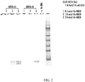
- Biotin labeled aptamers were immobilized on streptavidin particles and a pull-down assay was performed. Aptamers were diluted to 1 pmol/ ⁇ l in TEN100 buffer (10mM Tris-HCl, pH7.5, 1mM EDTA, 100mM NaCl), heated at 65°C for 5', then left at RT for 20 minutes to let the RNA fold into its natural conformation. Streptavidin magnetic particles (Roche, 11641778001) were washed 3 times with twice volume of TEN100 buffer, then the aptamer samples were added and incubated at RT for 30minute with rotation.
- TEN100 buffer 10mM Tris-HCl, pH7.5, 1mM EDTA, 100mM NaCl
- the aptamers protein complex was dissociated by incubating in 26 ⁇ l elution buffer (1M NaCl, 10mM EDTA) at room temperature for 10 min. The eluted protein was subjected to SDS-PAGE, and the protein bands were visualized by coomassie staining. The human albumin protein was used as negative control. A typical stained gel is shown in FIG. 2 .
- a neurogranin signature peptide was developed for a mass spectroscopy quantitative MRM assay.
- the peptide sequence and transitions are shown in the table below.
- Labeled GPGPGGPGGAGVAR (SEQ ID NO:7) was spiked in the samples to make standard curve to measure the concentration of signature peptide GPGPGGPGGAGVAR (SEQ ID NO:7).
- Peptide KGPGPGGPGGAGVAR (SEQ ID NO:8) is also monitored to make sure there is no miscleavage in tryptic digestion. Table 8.
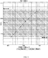
- the signals of neurogranin signature peptide and labeled standard peptide using an ABI Sciex Qtrap 4000 triple quadrapole mass spectrometer are shown in FIG. 3 .
- Human NRGN cDNA clone was purchased from Origene (Cat. No. RC201209). The coding sequence was cloned into destination vector (Origene, pEX-N-His, Cat. No. PS 100030) by restriction enzymes (Sgf I + Mlu I) fragment swapping to generate pEX-N-His-NRGN expression plasmid. The coding sequence and reading frame were confirmed by DNA sequencing.
- pEX-N-His-NRGN plasmid was transformed into Rosetta 2 (DE3) competent cells (Novagen #71397) according to manufacturer's instruction.
- the bacteria were cultured in the Overnight Express Instant TB Medium (Novagen #71491) at 37°C for 16-18 hours, then harvested and suspended in TEN buffer (50 mM Tris, pH8.0, 0.5 mM EDTA and 0.5 M NaCl).
- the bacteria were lysated by adding 1% NP-40, 25 mg lysozyme and complete proteinase inhibitors (Roche), sitting on ice for 30 minutes, then freeze-thaw one time.
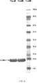
- FIG. 4 shows the typical His-NRGN on PAGE gel after coomassie staining; the predicted molecular weight of His-NRGN is 8.5 Kd.
- a Neurogranin anti-Human monoclonal antibody (Johns Hopkins) at concentration of 75 ng/well was used as a capture antibody and a rabbit polyclonal to neurogranin (Johns Hopkins) at a concentration of 0.5 ⁇ g/ml was used as detection.
- SULFO-TAG anti rabbit antibody (MSD Cat#R32AB-1) at a concentration of 1 ⁇ g/ml was used as a labeled reporter at a concentration of 1 ⁇ g/ml.
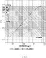
- GST_NRGN recombinant protein (Johns Hopkins) was used as a standard at a starting concentration of 20 ng/ml then at 1:2 for 7 dilutions in PBS/1%BSA. PBS/1%BSA was used as blank. The standard curve for this assay is shown in FIG. 6 .
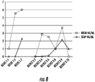
- Neurogranin is Biomarker of Acute Brain Injury .
- serum samples from an infant on ECMO support for 27 days for cardio-respiratory failure The infant had normal daily head ultrasounds and at the time of death was thought to not have brain injury.
- the brain had multiple cortical infarcts they were not diagnosed by ultrasound.
- GFAP levels were unchanged during the entire coarse of ECMO support.
- neurogranin levels increased to a peak 15 fold over baseline over 14 days of ECMO support.
- neurogranin is a gray matter, neuronal marker it was more sensitive to cortical gray matter injury than GFAP a marker of white matter injury. This provides evidence that neurogranin is a circulating biomarker of acute cortical brain injury and in combination with GFAP is able to discrimate white matter from gray matter injury to the brain.
- Example 2 Proteomic Study to Identify Brain Proteins Reveals Elevations of Neurogranin in Children with Sickle Cell Disease.
- SCI Silent cerebral infarct
- MRI magnetic resonance images
- biomarkers of SCI would provide important progress in detection and therapy for children with SCD. Specifically, these potential biomarkers would aid in the identification of children who are at risk for SCI, provide a cost-effective alternative to MRI for early diagnosis, and would allow for monitoring response to treatment. Importantly, biomarkers of SCI would provide insight into the pathological mechanisms involved in the disease. Proteomics methods based on mass spectrometry (MS) provide a platform for the identification, quantification and characterization of these potential biomarkers.
- MS mass spectrometry
- proteomics has been used for biomarker discovery of brain proteins in a number of disease states, including brain cancer, alzheimer's disease, traumatic brain injury, and stroke. Mass spectrometry-based approaches have also been used to gain insight into the pathophysiology of SCD. However, very few studies have used plasma proteomics for clinical biomarker discovery in SCD, and none have been published about SCD and subclinical brain injury. Kakhniashvili et al used two-dimensional fluorescence difference gel electrophoresis (2D DIGE) and tandem MS (LC-MS/MS) to evaluate quantitative changes in the red blood cell (RBC) membrane proteome and reported on elevations of proteins involved in repair after oxidative stress.
- 2D DIGE two-dimensional fluorescence difference gel electrophoresis
- LC-MS/MS tandem MS
- Study Population The study population was comprised of three groups: children with SCD and SCI, children with SCD and no SCI, and healthy, age-matched controls with no SCD or SCI. Proteomic analysis was done using plasma from seven children with SCD and SCI and six children with SCD but without SCI who were matched for age, hemoglobin and WBC based on current knowledge of risk factors for SCI, as well as on six age-matched African American controls (three with sickle cell trait [SCT]). Cross-sectional plasma samples from a total of 115 children with SCD (64 with SCI and 51 without SCI) and 46 age-matched, African-American controls were used to measure plasma concentrations of identified proteins.
- SCT sickle cell trait
- Plasma from children with SCD was randomly selected from those enrolled in the Silent Infarct Transfusion (SIT) Trial (ClinicalTrials.gov identifier NCT00072761).
- SIT Silent Infarct Transfusion
- the SIT trial is a multicenter, international clinical study to determine if chronic blood transfusions can effectively prevent the progression of SCI to stroke, in children with SCD aged 5-14 years.
- Ni-NTA nickel-nitrilotriacetic acid
- Protein Enrichment and Purification Using the ProteomeLabTM IgY-12 LC10 column kit (Beckman Coulter, Inc., Fullerton, CA), second dimension separation and immunoaffinity depletion of 12 abundant plasma proteins (albumin, IgG, fibrinogen, transferrin, IgA, IgM, HDL, apo A-I and apo A-II, haptoglobin, ⁇ 1-antitrypsin, ⁇ 1-acid glycoprotein and ⁇ 2-macroglobulin) was done according to the manufacturer's protocol.
- Tandem (LC-MS/MS) experiments were performed on a LTQ-Orbitrap hybrid mass spectrometer (ThermoFisher, San Jose, CA) equipped with an on-line nano-HPLC (Agilent technologies, 1200 Series, Wilmington, DE), as previously described.
- the LTQ raw data were analyzed using PASS (Integrated Analysis, Bethesda, MD) with X!Tandem searches (www.thegpm.org; version 2008.12.01) of the non-redundant International Protein Index (IPI) peptide database (human, 3.19).
- IPI International Protein Index
- Peptide identifications were accepted if they could be established at greater than 95% probability and contained at least 2 average unique identified spectra, with probability based Mowse scores greater than 35 (p ⁇ 0.05). A change in charge state of a peptide was not considered a unique identification. The dataset was filtered to 90% sequence identity with CD-HIT.
- Proteins were given a brain tissue specificity score based on their relative expression of transcript in human brain, liver, and 26 other normal human tissues as assessed by available microarray, expressed sequence tags (EST), and serial analysis of gene expression (SAGE) data. Specifically, each protein was scored according to the following criteria: microarray data showing greater than ten-fold increase in expression over baseline, EST and SAGE data showing presence of the protein in less than two other tissues. Proteins received either a score of 1 or 0 for each category, and a maximum score of 3 was assigned if the protein had greater than a ten-fold increase in expression by microarray data, and was found in less than two tissues by EST and SAGE.
- EST expressed sequence tags
- SAGE serial analysis of gene expression
- Ingenuity Pathway Analysis The Ingenuity pathway analysis program (http://www.ingenuity.com) was used to analyze the pathway network of the proteins with abundance changes that were identified through MS.
- the protein accession numbers were uploaded as an Excel spreadsheet file into the Ingenuity software, which uses the data to navigate the Ingenuity pathways database and extract networks between the proteins. A score better than 2 is usually considered as a valid network.
- An electrochemiluminescent sandwich immunoassay was developed for measuring Neurogranin using the MesoScale Discovery platform (MesoScale Discovery, Gaithersburg, MD).
- the monoclonal anti-Nrgn described in Example 1 was used for the capture antibody (ab).
- a polyclonal anti-Nrgn (Covance, Berkeley, CA) and anti-species Sulfo-Tag (MesoScale Discovery) mixture was used for detection.
- the standard curve was constructed by serial dilution of purified Neurogranin in 1% bovine serum albumin (SeraCare Life Sciences, Milford, MA), from 40 ng/ml to 0.055 ng/ml.
- Plasma spiked with Neurogranin shows an average of 99% recovery at 10 ng/mL when compared to a standard curve generated with bovine serum albumin.
- the lower limit of quantification defined as the lowest dilution with a calculated concentration that does not exceed 20% of the coefficient of variation, was 0.2 ng/mL.
- the specificity of the assay was tested using GFAP antibody.
- Standard bind plates (MesoScale Discovery) were coated with 30 ⁇ l of monoclonal anti-Nrgn pool that was diluted 1:1000 with phosphate-buffered saline (PBS). The plates were incubated overnight and then each well was blocked with 5% BSA/PBS and incubated with shaking (600 rpm) at room temperature for one hour. From a starting concentration of 40 ng/ml, purified Neurogranin was diluted 1:3 with 1%BSA/PBS. Plasma samples were diluted 1:1 using 1%BSA/PBS, then 25 ⁇ l of standards and diluted sample were added in duplicate to the plate.
- PBS phosphate-buffered saline
- a PCR cloning strategy was used to clone Neurogranin cDNA into bacteria expression vectors to express recombinant proteins.
- the primers were designed based on human Neurogranin cDNA sequence, Sgfl (in forward primer) and Mlul (in reverse primer) cutting sites were introduced in to the primers respectively to ensure the correct reading frame and cloning convenience.
- the sequences for primers are: forward 5'-GAGGCGATCGCCATGGACTGCTGCACCGAGAAC-3' (SEQ ID NO:9) and Reverse 5'-GCGACGCGTCTAGTCTCCGCTGGGGCCGC-3' (SEQ ID NO:10).
- Human Neurogranin cDNA (Origene, RC201209) was used as template for PCR amplification.
- the PCR product was digested with Sgfl and Mlul, gel purified and ligated into pre-digested vectors pEX-N-His-GST (Origene, PS 100028) and pEX-N-His (Origene, PS100029) respectively.
- DH5 ⁇ competent cells (Invitrogen, 18265017) were used for transformation and plasmid DNA propagation. Positive clones were identified by restriction enzymes digestion analysis and further confirmed by DNA sequencing. The proper plasmids containing correct Neurogranin cDNA were then used to transform Rosetta (Novagen) strain for protein expression.
- Mouse anti-Nrgn monoclonal antibody was produced at The Monoclonal Antibody Core Facility (MACF) at Johns Hopkins University, Department of Neuroscience. Briefly, five 6 weeks old BALB/c female mice (Charles River, Wilmington, MA) were immunized with intraperitoneal injection of 100ug of His-Nrgn and boosted on day 21 with using a same route. Then the mice were subcutaneous injection with 50ug of His-Nrgn in Incomplete Freund's Adjuvant twice. Blood was collected 10 days following the third immunization. The sera were tested by direct ELISA using His-GST-Nrgn protein as target protein. The mouse with best titer in the ELISA was selected for a final i.v boost. On day 89, the mouse was sacrificed and the spleen was removed for the fusion process.
- MAF Monoclonal Antibody Core Facility
- Polyethylene glucol (MW1500, Sigma) was used to fuse P3x653.Ag8 mouse myeloma cells with spleen cells from the immunized mouse. Fused cells were distributed in 96-well tissue culture plate containing feeder cells harvested from the peritoneum of a normal BALA/c mouse primed intraperitoneally with 0.5ml Incomplete Freund Adjuvants 4 days earlier. After 10 days of undisturbed culture in selection medium (DMEM containing 20% FCS, Hyclone, supplemented 1x OPI (Sigma), 100 uM hypoxanthine, 0.4 uM aminopterin, and 160 uM thymidine), supernatants were tested with ELISA as described above. Positive colonies were cloned twice by limiting dilution on splenocytes from normal BALA/c mice as feeder cells.
- selection medium DMEM containing 20% FCS, Hyclone, supplemented 1x OPI (Sigma), 100 uM hypoxanthin
- hybridoma line (30.5.2) was grown in DMEM containing 10% defined FCS (Hyclone) supplemented with 1x OPI for 4 to 6 days. Then, hybridoma cells were adapted to growth in serum free media, which would have allowed antibody production in the in vitro system. When cells were in log growth phase, 2x10 7 cells were inoculated into the in vitro system. The culture supernatant (antibody) was collected every five days.
- WBC and reticulocyte counts were significantly higher in children with SCD.
- Retic reticulocyte
- Hb hemoglobin
- Hct hematocrit
- WBC white blood cell count
- Plt platelet
- TotBili total bilirubin.
- the MS data was filtered against a list of proteins with increased protein expression in brain to produce a composite list of brain proteins that are found in children with SCD. Using this methodology, a total of twenty-seven brain proteins were identified in children with SCD (data not shown). None of these proteins were identified in non-SCD controls. Among the brain proteins identified, Neurogranin had greater than a ten-fold increase in expression in the brain and was found in less than two tissues by EST and SAGE. It also had the highest confidence score and total number of spectral counts.
- Neurogranin a calcium-sensitive calmodulin-binding neuron-specific protein that has been implicated in synaptic development and remodeling, was found in SCD children with and without SCI. As shown in FIG. 9A , median plasma Neurogranin levels were significantly higher in children with SCD (0.72 ⁇ g/ml) as compared to non-SCD controls (0.12 ⁇ g/ml, P ⁇ .001). Among children with SCD, sixty-three percent of SCI (40/64) and non-SCI (32/51) children had median Neurogranin values that were above the 95 th percentile for non-SCD values ( FIG. 9B ).
- Neurogranin did not correlate with known risk factors for SCI, including hb, hct, WBC, platelet, and reticulocyte counts, systolic blood pressure and percent of hb F.
- Biomarkers of subclinical brain injury in SCD are needed to diagnosis and aid in the development of molecular targeted therapies, as well as to monitor disease response. Proteomics provides an opportunity to discover these biochemical markers in complex mixtures, such as plasma. However, an established methodology for the discovery of brain biomarkers in children with SCD has not been previously reported. A proteomic-based approach was used to test the hypothesis that children with SCD and SCI have brain proteins circulating in their plasma proteome that is associated with subclinical brain injury.
- Neurogranin was measured in children with SCD and age-matched, non-SCD controls and found that children with SCD had significantly higher plasma levels of Neurogranin. In fact, greater than sixty percent of SCD children, with and without SCI, had Neurogranin values that were greater than the 95 th percentile for observed Neurogranin values in non-SCD controls. Neurogranin has not been previously studied in children with SCD. In fact, previous studies have largely been done in adults with schizophrenia and Alzheimer disease, and have related Neurogranin to impairments in learning and memory. Neurocognitive deficits in academic achievement and memory in children with SCD have been well documented.
- GFAP glial fibrillary acidic protein
- the present inventors have developed and verified a proteomic workflow for brain biomarker discovery in children with SCD.
- the present inventors are the first to report significant elevations of Neurogranin in children with SCD as compared to age-matched, non-SCD controls, and provide additional insight into the pathophysiology of subclinical brain injury.
- these findings support clinical investigation for biomarker discovery in children with SCD and SCI, and provide new potential targets for therapeutic drug discovery.
Claims (9)
- Utilisation d'un anticorps monoclonal isolé ou d'un fragment de liaison à l'antigène de celui-ci qui se lie spécifiquement à la neurogranine humaine dans un essai de diagnostic in vitro pour la détection d'une lésion cérébrale chez un sujet humain,
la lésion cérébrale étant diagnostiquée dans l'essai par détection d'un taux plus élevé de neurogranine dans un échantillon de liquide organique comprenant du sang, du sérum ou du plasma obtenu à partir du sujet par comparaison avec le taux dans un échantillon de liquide organique comparable obtenu à partir d'un sujet témoin qui n'a pas de lésion cérébrale,
le taux plus élevé de neurogranine dans l'échantillon de liquide organique étant détecté par détection d'un degré accru de liaison entre l'anticorps monoclonal isolé ou le fragment de liaison à l'antigène de celui-ci et la neurogranine dans l'échantillon de liquide organique provenant du sujet par comparaison avec le degré de liaison dans un échantillon de liquide organique comparable obtenu à partir du sujet témoin qui n'a pas de lésion cérébrale. - Procédé pour la détection d'une lésion cérébrale chez un sujet humain, le procédé comprenant l'utilisation d'un anticorps monoclonal isolé ou d'un fragment de liaison à l'antigène de celui-ci qui se lie spécifiquement à la neurogranine humaine pour détecter le taux de neurogranine dans un échantillon de liquide organique comprenant du sang, du sérum ou du plasma obtenu à partir du sujet, la lésion cérébrale étant diagnostiquée par détection dans l'échantillon du sujet d'un taux plus élevé de neurogranine qui se lie à l'anticorps ou à un fragment de liaison à l'antigène de celui-ci par comparaison avec le taux dans un échantillon de liquide organique comparable provenant d'un sujet témoin qui n'a pas de lésion cérébrale.
- Utilisation ou procédé selon l'une quelconque des revendications 1 ou 2 :(a) l'anticorps isolé ou un fragment de liaison à l'antigène de celui-ci étant de mammifère ; ou(b) l'anticorps isolé ou un fragment de liaison à l'antigène de celui-ci étant humain ; ou(c) le fragment de liaison à l'antigène de l'anticorps isolé étant choisi dans le groupe constitué par un fragment Fab ; un fragment F(ab')2 ; un fragment Fv ; et un fragment monocaténaire ; ou(d) l'anticorps isolé ou un fragment de liaison à l'antigène de celui-ci comprenant une substance détectable couplée à l'anticorps ou à un fragment de liaison à l'antigène de celui-ci.
- Utilisation ou procédé selon la revendication 3(d), la substance détectable étant choisie dans le groupe constitué par une enzyme ; un marqueur fluorescent ; un radioisotope ; et un marqueur chimiluminescent.
- Utilisation ou procédé selon l'une quelconque des revendications précédentes, la liaison spécifique entre la neurogranine présente dans l'échantillon et l'anticorps monoclonal isolé ou un fragment de liaison à l'antigène de celui-ci étant détectée par un immunoessai, un essai immunoenzymatique (ELISA), un essai de liaison par compétition, un radio-immunoessai ou un essai de tri cellulaire induit par fluorescence (FACS).
- Utilisation ou procédé selon la revendication 1 ou 2, l'anticorps isolé ou un fragment de liaison à l'antigène de celui-ci étant fourni dans un kit pour la détection de neurogranine, le kit comprenant : (a) l'anticorps isolé ou un fragment de liaison à l'antigène de celui-ci ; et (b) au moins un composant pour détecter la liaison de l'anticorps isolé ou d'un fragment de liaison à l'antigène de celui-ci à la neurogranine.
- Utilisation ou procédé selon l'une quelconque des revendications 1-6, l'échantillon de liquide organique du sujet étant un échantillon de sang.
- Utilisation ou procédé selon l'une quelconque des revendications 1-6, l'échantillon de liquide organique du sujet étant un échantillon de sérum.
- Utilisation ou procédé selon l'une quelconque des revendications 1-6, l'échantillon de liquide organique du sujet étant un échantillon de plasma.
Applications Claiming Priority (2)
| Application Number | Priority Date | Filing Date | Title |
|---|---|---|---|
| US201161485375P | 2011-05-12 | 2011-05-12 | |
| PCT/US2012/037774 WO2012155134A2 (fr) | 2011-05-12 | 2012-05-14 | Réactifs de dosage pour kit de diagnostic de la neurogranine |
Publications (3)
| Publication Number | Publication Date |
|---|---|
| EP2707389A2 EP2707389A2 (fr) | 2014-03-19 |
| EP2707389A4 EP2707389A4 (fr) | 2015-08-12 |
| EP2707389B1 true EP2707389B1 (fr) | 2019-10-30 |
Family
ID=47140053
Family Applications (1)
| Application Number | Title | Priority Date | Filing Date |
|---|---|---|---|
| EP12782967.9A Active EP2707389B1 (fr) | 2011-05-12 | 2012-05-14 | Réactifs de dosage pour kit de diagnostic de la neurogranine |
Country Status (4)
| Country | Link |
|---|---|
| US (3) | US20140141458A1 (fr) |
| EP (1) | EP2707389B1 (fr) |
| JP (1) | JP2014518624A (fr) |
| WO (1) | WO2012155134A2 (fr) |
Families Citing this family (8)
| Publication number | Priority date | Publication date | Assignee | Title |
|---|---|---|---|---|
| WO2012051519A2 (fr) | 2010-10-14 | 2012-04-19 | The Johns Hopkins University | Biomarqueurs d'une lésion cérébrale |
| JP2014518624A (ja) | 2011-05-12 | 2014-08-07 | ザ・ジョンズ・ホプキンス・ユニバーシティー | ニューログラニン診断キットのためのアッセイ試薬 |
| WO2013138509A1 (fr) | 2012-03-13 | 2013-09-19 | The Johns Hopkins University | Protéines cérébrales et neurologiques citrullinées en tant que biomarqueurs de lésions cérébrales ou de neurodégénérescence |
| WO2014193800A2 (fr) | 2013-05-28 | 2014-12-04 | The Johns Hopkins University | Aptamères utiles dans le traitement de la drépanocytose |
| US10534003B2 (en) | 2013-07-17 | 2020-01-14 | The Johns Hopkins University | Multi-protein biomarker assay for brain injury detection and outcome |
| FR3025317B1 (fr) * | 2014-08-26 | 2022-09-23 | Imabiotech | Methode de caracterisation d'un echantillon par imagerie par spectrometrie de masse |
| EP3242134A1 (fr) | 2016-05-04 | 2017-11-08 | Euroimmun Medizinische Labordiagnostika AG | Dosage pour le diagnostic d'une maladie neurologique |
| WO2018217792A1 (fr) * | 2017-05-23 | 2018-11-29 | Immunarray USA, Inc. | Niveaux de biomarqueurs et neuroimagerie pour la détection, la surveillance et le traitement d'une lésion ou d'un traumatisme cérébral |
Family Cites Families (59)
| Publication number | Priority date | Publication date | Assignee | Title |
|---|---|---|---|---|
| US4816567A (en) | 1983-04-08 | 1989-03-28 | Genentech, Inc. | Recombinant immunoglobin preparations |
| US5225539A (en) | 1986-03-27 | 1993-07-06 | Medical Research Council | Recombinant altered antibodies and methods of making altered antibodies |
| GB8823869D0 (en) | 1988-10-12 | 1988-11-16 | Medical Res Council | Production of antibodies |
| SE462454B (sv) | 1988-11-10 | 1990-06-25 | Pharmacia Ab | Maetyta foer anvaendning i biosensorer |
| US5683867A (en) | 1990-06-11 | 1997-11-04 | Nexstar Pharmaceuticals, Inc. | Systematic evolution of ligands by exponential enrichment: blended SELEX |
| US5496938A (en) | 1990-06-11 | 1996-03-05 | Nexstar Pharmaceuticals, Inc. | Nucleic acid ligands to HIV-RT and HIV-1 rev |
| US5707796A (en) | 1990-06-11 | 1998-01-13 | Nexstar Pharmaceuticals, Inc. | Method for selecting nucleic acids on the basis of structure |
| US5270163A (en) | 1990-06-11 | 1993-12-14 | University Research Corporation | Methods for identifying nucleic acid ligands |
| US5567588A (en) | 1990-06-11 | 1996-10-22 | University Research Corporation | Systematic evolution of ligands by exponential enrichment: Solution SELEX |
| US5660985A (en) | 1990-06-11 | 1997-08-26 | Nexstar Pharmaceuticals, Inc. | High affinity nucleic acid ligands containing modified nucleotides |
| US6011020A (en) | 1990-06-11 | 2000-01-04 | Nexstar Pharmaceuticals, Inc. | Nucleic acid ligand complexes |
| ES2259800T3 (es) | 1990-06-11 | 2006-10-16 | Gilead Sciences, Inc. | Procedimientos de uso de ligandos de acido nucleico. |
| US5637459A (en) | 1990-06-11 | 1997-06-10 | Nexstar Pharmaceuticals, Inc. | Systematic evolution of ligands by exponential enrichment: chimeric selex |
| ES2108048T3 (es) | 1990-08-29 | 1997-12-16 | Genpharm Int | Produccion y utilizacion de animales inferiores transgenicos capaces de producir anticuerpos heterologos. |
| US5633425A (en) | 1990-08-29 | 1997-05-27 | Genpharm International, Inc. | Transgenic non-human animals capable of producing heterologous antibodies |
| US5661016A (en) | 1990-08-29 | 1997-08-26 | Genpharm International Inc. | Transgenic non-human animals capable of producing heterologous antibodies of various isotypes |
| US5545806A (en) | 1990-08-29 | 1996-08-13 | Genpharm International, Inc. | Ransgenic non-human animals for producing heterologous antibodies |
| US5625126A (en) | 1990-08-29 | 1997-04-29 | Genpharm International, Inc. | Transgenic non-human animals for producing heterologous antibodies |
| EP0564531B1 (fr) | 1990-12-03 | 1998-03-25 | Genentech, Inc. | Methode d'enrichissement pour des variantes de l'hormone de croissance avec des proprietes de liaison modifiees |
| DE69432791T2 (de) | 1993-05-28 | 2004-06-03 | Baylor College Of Medicine, Houston | Verfahren und massenspektrometer zur desorption und ionisierung von analyten |
| US5866434A (en) | 1994-12-08 | 1999-02-02 | Meso Scale Technology | Graphitic nanotubes in luminescence assays |
| US6140045A (en) | 1995-03-10 | 2000-10-31 | Meso Scale Technologies | Multi-array, multi-specific electrochemiluminescence testing |
| CN1192097C (zh) | 1995-03-10 | 2005-03-09 | 梅索磅秤技术有限公司 | 多阵列、多特异性的电化学发光检验 |
| US6673533B1 (en) | 1995-03-10 | 2004-01-06 | Meso Scale Technologies, Llc. | Multi-array multi-specific electrochemiluminescence testing |
| US6207369B1 (en) | 1995-03-10 | 2001-03-27 | Meso Scale Technologies, Llc | Multi-array, multi-specific electrochemiluminescence testing |
| US5641870A (en) | 1995-04-20 | 1997-06-24 | Genentech, Inc. | Low pH hydrophobic interaction chromatography for antibody purification |
| US6319670B1 (en) | 1995-05-09 | 2001-11-20 | Meso Scale Technology Llp | Methods and apparatus for improved luminescence assays using microparticles |
| NZ516848A (en) | 1997-06-20 | 2004-03-26 | Ciphergen Biosystems Inc | Retentate chromatography apparatus with applications in biology and medicine |
| US6413783B1 (en) | 1997-09-18 | 2002-07-02 | Meso Scale Technologies, Llc | Assay sonication apparatus and methodology |
| CA2323638A1 (fr) | 1998-04-03 | 1999-10-14 | Phylos, Inc. | Systemes de proteines adressables |
| US6406921B1 (en) | 1998-07-14 | 2002-06-18 | Zyomyx, Incorporated | Protein arrays for high-throughput screening |
| US6780606B1 (en) | 1999-02-26 | 2004-08-24 | Synx Pharma, Inc. | Method for diagnosing and distinguishing stroke and diagnostic devices for use therein |
| AU4025300A (en) | 1999-03-24 | 2000-10-09 | Packard Bioscience Company | Continuous porous matrix arrays |
| WO2002024867A2 (fr) | 2000-09-22 | 2002-03-28 | University Of Aarhus | Nouvelles compositions et nouvelles methodes pour le diagnostic et le traitement des lymphomes et des leucemies |
| WO2003016910A1 (fr) | 2001-08-20 | 2003-02-27 | Biosite, Inc. | Marqueurs diagnostiques d'ictus et de lesions cerebrales et procedes d'utilisation de ces marqueurs |
| EP2420824B1 (fr) | 2001-06-29 | 2018-11-28 | Meso Scale Technologies LLC | Plaque multipuits ayant un réseau de puits et un kit pour l'utilisation dans la conduite d'un essai ECL |
| EP2135943A1 (fr) | 2001-07-30 | 2009-12-23 | Meso Scale Technologies, LLC. | Électrode de réseau dotée de couches de lipide/protéine immobilisées, son procédé de fabrication et procédés d'utilisation associés pour les mesures de test de luminescence |
| EP1436598B1 (fr) | 2001-09-10 | 2013-11-27 | Meso Scale Technologies LLC | Tampon de dosage, compositions contenant ce tampon et procedes d'utilisation correspondants |
| AU2002341621A1 (en) | 2001-09-10 | 2003-03-24 | Meso Scale Technologies, Llc | Methods, reagents, kits and apparatus for protein function analysis |
| US20070098728A1 (en) * | 2001-09-24 | 2007-05-03 | Pedersen Finn S | Novel compositions and methods in cancer |
| DE60219952T2 (de) | 2001-12-05 | 2008-01-17 | Sense Proteomic Ltd., Maidenhead | Proteinarrays für allelische Varianten und deren Verwendungen |
| US20040014143A1 (en) * | 2002-05-29 | 2004-01-22 | Haskins William E. | Method and apparatus for detecting and monitoring peptides, and peptides identified therewith |
| US7036946B1 (en) | 2002-09-13 | 2006-05-02 | Rockwell Collins, Inc. | LCD backlight with UV light-emitting diodes and planar reactive element |
| CA2941139C (fr) | 2002-12-26 | 2021-07-20 | Meso Scale Technologies, Llc. | Cartouches d'essai et procedes d'utilisation |
| US7981362B2 (en) | 2003-11-04 | 2011-07-19 | Meso Scale Technologies, Llc | Modular assay plates, reader systems and methods for test measurements |
| EP2207033B1 (fr) | 2004-04-15 | 2014-06-18 | University of Florida Research Foundation, Inc. | Protéines neuronales en tant que biomarqueurs pour blessure traumatique du système nerveux et autres troubles neuronaux |
| ATE428931T1 (de) | 2004-06-03 | 2009-05-15 | Meso Scale Technologies Llc | Verfahren zur durchführung von vollbluttests |
| EP1774023B1 (fr) * | 2004-06-25 | 2013-05-01 | Washington University School Of Medicine | Marqueurs pour des lésions cérébrales |
| US20060257943A1 (en) | 2005-01-25 | 2006-11-16 | Cis Biotech, Inc. | Ischemic biomarkers and their use to predict adverse neurological events from surgery |
| US7884260B2 (en) | 2005-06-14 | 2011-02-08 | University Of Chicago | Cell-based screen for agents useful for reducing neuronal demyelination or promoting neuronal remyelination |
| GB2428240A (en) | 2005-07-14 | 2007-01-24 | Univ Gen Ve | Diagnostic method for brain damage-related disorders |
| EP2008100A4 (fr) | 2006-04-18 | 2009-12-16 | Univ Leland Stanford Junior | Établissement de profils d'anticorps pour la détermination de la sensibilité des patients à un traitement |
| WO2007136617A2 (fr) | 2006-05-18 | 2007-11-29 | Walter Reed Army Institute Of Research (Wrair) | Polypeptide ii d'activation de monocytes endothéliaux, biomarqueur destiné à être utilisé dans le diagnostic et le traitement de lésion cérébrale |
| US8088584B2 (en) | 2006-10-16 | 2012-01-03 | Bayer Schering Pharma Aktiengesellschaft | LTBP2 as a biomarker and diagnostic target |
| EP2015196A1 (fr) | 2007-06-28 | 2009-01-14 | Siemens Aktiengesellschaft | Interface de programmateur pour la fabrication d'un système d'exécution |
| WO2009143519A2 (fr) * | 2008-05-23 | 2009-11-26 | The Johns Hopkins University | Biomarqueurs pour ischémie myocardique |
| EP4235181A3 (fr) | 2008-08-11 | 2024-02-28 | Banyan Biomarkers Inc | Procédé de détection de biomarqueur et dosage d'état neurologique |
| GB201008541D0 (en) | 2010-05-21 | 2010-07-07 | Univ Geneve | Diagnostic methods |
| JP2014518624A (ja) | 2011-05-12 | 2014-08-07 | ザ・ジョンズ・ホプキンス・ユニバーシティー | ニューログラニン診断キットのためのアッセイ試薬 |
-
2012
- 2012-05-14 JP JP2014510535A patent/JP2014518624A/ja active Pending
- 2012-05-14 WO PCT/US2012/037774 patent/WO2012155134A2/fr active Application Filing
- 2012-05-14 EP EP12782967.9A patent/EP2707389B1/fr active Active
- 2012-05-14 US US14/117,111 patent/US20140141458A1/en not_active Abandoned
-
2016
- 2016-09-29 US US15/280,780 patent/US9709578B2/en active Active
-
2017
- 2017-06-30 US US15/639,210 patent/US10215764B2/en active Active
Non-Patent Citations (2)
| Title |
|---|
| DI LAZZARO ET AL: "BDNF plasma levels in acute stroke", NEUROSCIENCE LETTERS, LIMERICK, IE, vol. 422, no. 2, 11 July 2007 (2007-07-11), pages 128 - 130, XP022162402, ISSN: 0304-3940, DOI: 10.1016/J.NEULET.2007.06.001 * |
| GRACE SOPHIA GRIESBACH ET AL: "Alterations in BDNF and Synapsin I within the Occipital Cortex and Hippocampus after Mild Traumatic Brain Injury in the Developing Rat: Reflections of Injury-Induced Neuroplasticity", JOURNAL OF NEUROTRAUMA., vol. 19, no. 7, 1 July 2002 (2002-07-01), US, pages 803 - 814, XP055283564, ISSN: 0897-7151, DOI: 10.1089/08977150260190401 * |
Also Published As
| Publication number | Publication date |
|---|---|
| US10215764B2 (en) | 2019-02-26 |
| WO2012155134A2 (fr) | 2012-11-15 |
| US20140141458A1 (en) | 2014-05-22 |
| US20170038397A1 (en) | 2017-02-09 |
| WO2012155134A3 (fr) | 2013-06-13 |
| US20170315138A1 (en) | 2017-11-02 |
| EP2707389A4 (fr) | 2015-08-12 |
| US9709578B2 (en) | 2017-07-18 |
| EP2707389A2 (fr) | 2014-03-19 |
| JP2014518624A (ja) | 2014-08-07 |
Similar Documents
| Publication | Publication Date | Title |
|---|---|---|
| US10215764B2 (en) | Assay reagents for a neurogranin diagnostic kit | |
| EP2333116B1 (fr) | Marqueurs de rejet de greffe de rein et de lésions rénales | |
| US10914749B2 (en) | Biomarkers of cognitive dysfunction | |
| JP2017500562A (ja) | アルツハイマー病に関するバイオマーカー及び方法 | |
| US20230107651A1 (en) | Methods of treating spinal muscular atrophy | |
| US20200103419A1 (en) | Blood biomarkers and diagnostic methods for small vessel diseases | |
| EP3835424A1 (fr) | Médicament de diagnostic et procédé de diagnostic pour la maladie d'alzheimer | |
| WO2022104136A2 (fr) | Méthodes et compositions pour le diagnostic et le traitement de la tauopathie | |
| KR101883515B1 (ko) | 알츠하이머 병의 진단약 및 진단 방법 | |
| KR20210127145A (ko) | 다중 분석 및 이의 사용 방법 | |
| Turčić et al. | Higher MRI lesion load in multiple sclerosis is related to the N-glycosylation changes of cerebrospinal fluid immunoglobulin G | |
| EP2795337B1 (fr) | Méthodes de criblage pour identifier des composés utiles dans la prévention et/ou le traitement de conditions inflammatoires | |
| EP3267197B1 (fr) | Procédé de détection de dysfonctionnement pancréatique et kit de détection | |
| EP2631656B1 (fr) | Marqueur de la sclérose latérale amyotrophique, et son utilisation | |
| WO2023220276A1 (fr) | PROCÉDÉS DE DÉTECTION DE PROTÉOFORMES Aβ ET LEUR UTILISATION | |
| WO2024049357A1 (fr) | Agent de diagnostic et procédé pour la maladie d'alzheimer | |
| Begcevic | Proteomic-based Signature of Brain-related Proteins as Novel Candidate Biomarkers for Alzheimer's Disease Diagnosis | |
| CA3089120A1 (fr) | Methodes de traitement de l'amyotrophie musculaire | |
| JPWO2006035702A1 (ja) | Cペプチド特異的結合分子及びその用途 |
Legal Events
| Date | Code | Title | Description |
|---|---|---|---|
| PUAI | Public reference made under article 153(3) epc to a published international application that has entered the european phase |
Free format text: ORIGINAL CODE: 0009012 |
|
| 17P | Request for examination filed |
Effective date: 20131129 |
|
| AK | Designated contracting states |
Kind code of ref document: A2 Designated state(s): AL AT BE BG CH CY CZ DE DK EE ES FI FR GB GR HR HU IE IS IT LI LT LU LV MC MK MT NL NO PL PT RO RS SE SI SK SM TR |
|
| DAX | Request for extension of the european patent (deleted) | ||
| RIC1 | Information provided on ipc code assigned before grant |
Ipc: C07K 16/18 20060101AFI20150216BHEP Ipc: G01N 33/532 20060101ALI20150216BHEP Ipc: C12Q 1/68 20060101ALI20150216BHEP Ipc: G01N 33/53 20060101ALI20150216BHEP Ipc: C12N 15/115 20100101ALI20150216BHEP |
|
| A4 | Supplementary search report drawn up and despatched |
Effective date: 20150710 |
|
| RIC1 | Information provided on ipc code assigned before grant |
Ipc: C12Q 1/68 20060101ALI20150706BHEP Ipc: C07K 16/18 20060101AFI20150706BHEP Ipc: G01N 33/566 20060101ALI20150706BHEP Ipc: C12N 15/115 20100101ALI20150706BHEP Ipc: G01N 33/532 20060101ALI20150706BHEP Ipc: G01N 33/53 20060101ALI20150706BHEP |
|
| 17Q | First examination report despatched |
Effective date: 20160630 |
|
| STAA | Information on the status of an ep patent application or granted ep patent |
Free format text: STATUS: EXAMINATION IS IN PROGRESS |
|
| GRAP | Despatch of communication of intention to grant a patent |
Free format text: ORIGINAL CODE: EPIDOSNIGR1 |
|
| STAA | Information on the status of an ep patent application or granted ep patent |
Free format text: STATUS: GRANT OF PATENT IS INTENDED |
|
| INTG | Intention to grant announced |
Effective date: 20190606 |
|
| GRAS | Grant fee paid |
Free format text: ORIGINAL CODE: EPIDOSNIGR3 |
|
| GRAA | (expected) grant |
Free format text: ORIGINAL CODE: 0009210 |
|
| STAA | Information on the status of an ep patent application or granted ep patent |
Free format text: STATUS: THE PATENT HAS BEEN GRANTED |
|
| AK | Designated contracting states |
Kind code of ref document: B1 Designated state(s): AL AT BE BG CH CY CZ DE DK EE ES FI FR GB GR HR HU IE IS IT LI LT LU LV MC MK MT NL NO PL PT RO RS SE SI SK SM TR |
|
| REG | Reference to a national code |
Ref country code: GB Ref legal event code: FG4D |
|
| REG | Reference to a national code |
Ref country code: CH Ref legal event code: EP |
|
| REG | Reference to a national code |
Ref country code: AT Ref legal event code: REF Ref document number: 1196035 Country of ref document: AT Kind code of ref document: T Effective date: 20191115 |
|
| REG | Reference to a national code |
Ref country code: DE Ref legal event code: R096 Ref document number: 602012065285 Country of ref document: DE |
|
| REG | Reference to a national code |
Ref country code: IE Ref legal event code: FG4D |
|
| REG | Reference to a national code |
Ref country code: LT Ref legal event code: MG4D |
|
| PG25 | Lapsed in a contracting state [announced via postgrant information from national office to epo] |
Ref country code: PT Free format text: LAPSE BECAUSE OF FAILURE TO SUBMIT A TRANSLATION OF THE DESCRIPTION OR TO PAY THE FEE WITHIN THE PRESCRIBED TIME-LIMIT Effective date: 20200302 Ref country code: FI Free format text: LAPSE BECAUSE OF FAILURE TO SUBMIT A TRANSLATION OF THE DESCRIPTION OR TO PAY THE FEE WITHIN THE PRESCRIBED TIME-LIMIT Effective date: 20191030 Ref country code: BG Free format text: LAPSE BECAUSE OF FAILURE TO SUBMIT A TRANSLATION OF THE DESCRIPTION OR TO PAY THE FEE WITHIN THE PRESCRIBED TIME-LIMIT Effective date: 20200130 Ref country code: NL Free format text: LAPSE BECAUSE OF FAILURE TO SUBMIT A TRANSLATION OF THE DESCRIPTION OR TO PAY THE FEE WITHIN THE PRESCRIBED TIME-LIMIT Effective date: 20191030 Ref country code: PL Free format text: LAPSE BECAUSE OF FAILURE TO SUBMIT A TRANSLATION OF THE DESCRIPTION OR TO PAY THE FEE WITHIN THE PRESCRIBED TIME-LIMIT Effective date: 20191030 Ref country code: LV Free format text: LAPSE BECAUSE OF FAILURE TO SUBMIT A TRANSLATION OF THE DESCRIPTION OR TO PAY THE FEE WITHIN THE PRESCRIBED TIME-LIMIT Effective date: 20191030 Ref country code: SE Free format text: LAPSE BECAUSE OF FAILURE TO SUBMIT A TRANSLATION OF THE DESCRIPTION OR TO PAY THE FEE WITHIN THE PRESCRIBED TIME-LIMIT Effective date: 20191030 Ref country code: NO Free format text: LAPSE BECAUSE OF FAILURE TO SUBMIT A TRANSLATION OF THE DESCRIPTION OR TO PAY THE FEE WITHIN THE PRESCRIBED TIME-LIMIT Effective date: 20200130 Ref country code: ES Free format text: LAPSE BECAUSE OF FAILURE TO SUBMIT A TRANSLATION OF THE DESCRIPTION OR TO PAY THE FEE WITHIN THE PRESCRIBED TIME-LIMIT Effective date: 20191030 Ref country code: GR Free format text: LAPSE BECAUSE OF FAILURE TO SUBMIT A TRANSLATION OF THE DESCRIPTION OR TO PAY THE FEE WITHIN THE PRESCRIBED TIME-LIMIT Effective date: 20200131 Ref country code: LT Free format text: LAPSE BECAUSE OF FAILURE TO SUBMIT A TRANSLATION OF THE DESCRIPTION OR TO PAY THE FEE WITHIN THE PRESCRIBED TIME-LIMIT Effective date: 20191030 |
|
| REG | Reference to a national code |
Ref country code: NL Ref legal event code: MP Effective date: 20191030 |
|
| PG25 | Lapsed in a contracting state [announced via postgrant information from national office to epo] |
Ref country code: HR Free format text: LAPSE BECAUSE OF FAILURE TO SUBMIT A TRANSLATION OF THE DESCRIPTION OR TO PAY THE FEE WITHIN THE PRESCRIBED TIME-LIMIT Effective date: 20191030 Ref country code: RS Free format text: LAPSE BECAUSE OF FAILURE TO SUBMIT A TRANSLATION OF THE DESCRIPTION OR TO PAY THE FEE WITHIN THE PRESCRIBED TIME-LIMIT Effective date: 20191030 Ref country code: IS Free format text: LAPSE BECAUSE OF FAILURE TO SUBMIT A TRANSLATION OF THE DESCRIPTION OR TO PAY THE FEE WITHIN THE PRESCRIBED TIME-LIMIT Effective date: 20200229 |
|
| PG25 | Lapsed in a contracting state [announced via postgrant information from national office to epo] |
Ref country code: AL Free format text: LAPSE BECAUSE OF FAILURE TO SUBMIT A TRANSLATION OF THE DESCRIPTION OR TO PAY THE FEE WITHIN THE PRESCRIBED TIME-LIMIT Effective date: 20191030 |
|
| PG25 | Lapsed in a contracting state [announced via postgrant information from national office to epo] |
Ref country code: EE Free format text: LAPSE BECAUSE OF FAILURE TO SUBMIT A TRANSLATION OF THE DESCRIPTION OR TO PAY THE FEE WITHIN THE PRESCRIBED TIME-LIMIT Effective date: 20191030 Ref country code: DK Free format text: LAPSE BECAUSE OF FAILURE TO SUBMIT A TRANSLATION OF THE DESCRIPTION OR TO PAY THE FEE WITHIN THE PRESCRIBED TIME-LIMIT Effective date: 20191030 Ref country code: CZ Free format text: LAPSE BECAUSE OF FAILURE TO SUBMIT A TRANSLATION OF THE DESCRIPTION OR TO PAY THE FEE WITHIN THE PRESCRIBED TIME-LIMIT Effective date: 20191030 Ref country code: RO Free format text: LAPSE BECAUSE OF FAILURE TO SUBMIT A TRANSLATION OF THE DESCRIPTION OR TO PAY THE FEE WITHIN THE PRESCRIBED TIME-LIMIT Effective date: 20191030 |
|
| REG | Reference to a national code |
Ref country code: DE Ref legal event code: R097 Ref document number: 602012065285 Country of ref document: DE |
|
| REG | Reference to a national code |
Ref country code: AT Ref legal event code: MK05 Ref document number: 1196035 Country of ref document: AT Kind code of ref document: T Effective date: 20191030 |
|
| PG25 | Lapsed in a contracting state [announced via postgrant information from national office to epo] |
Ref country code: SM Free format text: LAPSE BECAUSE OF FAILURE TO SUBMIT A TRANSLATION OF THE DESCRIPTION OR TO PAY THE FEE WITHIN THE PRESCRIBED TIME-LIMIT Effective date: 20191030 Ref country code: IT Free format text: LAPSE BECAUSE OF FAILURE TO SUBMIT A TRANSLATION OF THE DESCRIPTION OR TO PAY THE FEE WITHIN THE PRESCRIBED TIME-LIMIT Effective date: 20191030 Ref country code: SK Free format text: LAPSE BECAUSE OF FAILURE TO SUBMIT A TRANSLATION OF THE DESCRIPTION OR TO PAY THE FEE WITHIN THE PRESCRIBED TIME-LIMIT Effective date: 20191030 |
|
| PLBE | No opposition filed within time limit |
Free format text: ORIGINAL CODE: 0009261 |
|
| STAA | Information on the status of an ep patent application or granted ep patent |
Free format text: STATUS: NO OPPOSITION FILED WITHIN TIME LIMIT |
|
| 26N | No opposition filed |
Effective date: 20200731 |
|
| PG25 | Lapsed in a contracting state [announced via postgrant information from national office to epo] |
Ref country code: AT Free format text: LAPSE BECAUSE OF FAILURE TO SUBMIT A TRANSLATION OF THE DESCRIPTION OR TO PAY THE FEE WITHIN THE PRESCRIBED TIME-LIMIT Effective date: 20191030 Ref country code: SI Free format text: LAPSE BECAUSE OF FAILURE TO SUBMIT A TRANSLATION OF THE DESCRIPTION OR TO PAY THE FEE WITHIN THE PRESCRIBED TIME-LIMIT Effective date: 20191030 |
|
| PG25 | Lapsed in a contracting state [announced via postgrant information from national office to epo] |
Ref country code: CH Free format text: LAPSE BECAUSE OF NON-PAYMENT OF DUE FEES Effective date: 20200531 Ref country code: LI Free format text: LAPSE BECAUSE OF NON-PAYMENT OF DUE FEES Effective date: 20200531 Ref country code: MC Free format text: LAPSE BECAUSE OF FAILURE TO SUBMIT A TRANSLATION OF THE DESCRIPTION OR TO PAY THE FEE WITHIN THE PRESCRIBED TIME-LIMIT Effective date: 20191030 |
|
| REG | Reference to a national code |
Ref country code: BE Ref legal event code: MM Effective date: 20200531 |
|
| PG25 | Lapsed in a contracting state [announced via postgrant information from national office to epo] |
Ref country code: LU Free format text: LAPSE BECAUSE OF NON-PAYMENT OF DUE FEES Effective date: 20200514 |
|
| PG25 | Lapsed in a contracting state [announced via postgrant information from national office to epo] |
Ref country code: IE Free format text: LAPSE BECAUSE OF NON-PAYMENT OF DUE FEES Effective date: 20200514 |
|
| PG25 | Lapsed in a contracting state [announced via postgrant information from national office to epo] |
Ref country code: BE Free format text: LAPSE BECAUSE OF NON-PAYMENT OF DUE FEES Effective date: 20200531 |
|
| PG25 | Lapsed in a contracting state [announced via postgrant information from national office to epo] |
Ref country code: TR Free format text: LAPSE BECAUSE OF FAILURE TO SUBMIT A TRANSLATION OF THE DESCRIPTION OR TO PAY THE FEE WITHIN THE PRESCRIBED TIME-LIMIT Effective date: 20191030 Ref country code: MT Free format text: LAPSE BECAUSE OF FAILURE TO SUBMIT A TRANSLATION OF THE DESCRIPTION OR TO PAY THE FEE WITHIN THE PRESCRIBED TIME-LIMIT Effective date: 20191030 Ref country code: CY Free format text: LAPSE BECAUSE OF FAILURE TO SUBMIT A TRANSLATION OF THE DESCRIPTION OR TO PAY THE FEE WITHIN THE PRESCRIBED TIME-LIMIT Effective date: 20191030 |
|
| PG25 | Lapsed in a contracting state [announced via postgrant information from national office to epo] |
Ref country code: MK Free format text: LAPSE BECAUSE OF FAILURE TO SUBMIT A TRANSLATION OF THE DESCRIPTION OR TO PAY THE FEE WITHIN THE PRESCRIBED TIME-LIMIT Effective date: 20191030 |
|
| P01 | Opt-out of the competence of the unified patent court (upc) registered |
Effective date: 20230328 |
|
| PGFP | Annual fee paid to national office [announced via postgrant information from national office to epo] |
Ref country code: FR Payment date: 20230525 Year of fee payment: 12 Ref country code: DE Payment date: 20230530 Year of fee payment: 12 |
|
| PGFP | Annual fee paid to national office [announced via postgrant information from national office to epo] |
Ref country code: GB Payment date: 20230529 Year of fee payment: 12 |