EP2670856B1 - Methods and compositions for highly specific capture and release of biological materials - Google Patents
Methods and compositions for highly specific capture and release of biological materials Download PDFInfo
- Publication number
- EP2670856B1 EP2670856B1 EP12742728.4A EP12742728A EP2670856B1 EP 2670856 B1 EP2670856 B1 EP 2670856B1 EP 12742728 A EP12742728 A EP 12742728A EP 2670856 B1 EP2670856 B1 EP 2670856B1
- Authority
- EP
- European Patent Office
- Prior art keywords
- cells
- cell
- hydrogel
- antibody
- peg
- Prior art date
- Legal status (The legal status is an assumption and is not a legal conclusion. Google has not performed a legal analysis and makes no representation as to the accuracy of the status listed.)
- Active
Links
Images


Classifications
-
- G—PHYSICS
- G01—MEASURING; TESTING
- G01N—INVESTIGATING OR ANALYSING MATERIALS BY DETERMINING THEIR CHEMICAL OR PHYSICAL PROPERTIES
- G01N33/00—Investigating or analysing materials by specific methods not covered by groups G01N1/00 - G01N31/00
- G01N33/48—Biological material, e.g. blood, urine; Haemocytometers
- G01N33/50—Chemical analysis of biological material, e.g. blood, urine; Testing involving biospecific ligand binding methods; Immunological testing
- G01N33/53—Immunoassay; Biospecific binding assay; Materials therefor
- G01N33/569—Immunoassay; Biospecific binding assay; Materials therefor for microorganisms, e.g. protozoa, bacteria, viruses
- G01N33/56966—Animal cells
-
- C—CHEMISTRY; METALLURGY
- C12—BIOCHEMISTRY; BEER; SPIRITS; WINE; VINEGAR; MICROBIOLOGY; ENZYMOLOGY; MUTATION OR GENETIC ENGINEERING
- C12Q—MEASURING OR TESTING PROCESSES INVOLVING ENZYMES, NUCLEIC ACIDS OR MICROORGANISMS; COMPOSITIONS OR TEST PAPERS THEREFOR; PROCESSES OF PREPARING SUCH COMPOSITIONS; CONDITION-RESPONSIVE CONTROL IN MICROBIOLOGICAL OR ENZYMOLOGICAL PROCESSES
- C12Q1/00—Measuring or testing processes involving enzymes, nucleic acids or microorganisms; Compositions therefor; Processes of preparing such compositions
-
- B—PERFORMING OPERATIONS; TRANSPORTING
- B01—PHYSICAL OR CHEMICAL PROCESSES OR APPARATUS IN GENERAL
- B01F—MIXING, e.g. DISSOLVING, EMULSIFYING OR DISPERSING
- B01F33/00—Other mixers; Mixing plants; Combinations of mixers
- B01F33/30—Micromixers
-
- G—PHYSICS
- G01—MEASURING; TESTING
- G01N—INVESTIGATING OR ANALYSING MATERIALS BY DETERMINING THEIR CHEMICAL OR PHYSICAL PROPERTIES
- G01N1/00—Sampling; Preparing specimens for investigation
- G01N1/28—Preparing specimens for investigation including physical details of (bio-)chemical methods covered elsewhere, e.g. G01N33/50, C12Q
- G01N1/34—Purifying; Cleaning
-
- G—PHYSICS
- G01—MEASURING; TESTING
- G01N—INVESTIGATING OR ANALYSING MATERIALS BY DETERMINING THEIR CHEMICAL OR PHYSICAL PROPERTIES
- G01N33/00—Investigating or analysing materials by specific methods not covered by groups G01N1/00 - G01N31/00
- G01N33/48—Biological material, e.g. blood, urine; Haemocytometers
- G01N33/50—Chemical analysis of biological material, e.g. blood, urine; Testing involving biospecific ligand binding methods; Immunological testing
- G01N33/53—Immunoassay; Biospecific binding assay; Materials therefor
- G01N33/543—Immunoassay; Biospecific binding assay; Materials therefor with an insoluble carrier for immobilising immunochemicals
- G01N33/54366—Apparatus specially adapted for solid-phase testing
- G01N33/54386—Analytical elements
-
- G—PHYSICS
- G01—MEASURING; TESTING
- G01N—INVESTIGATING OR ANALYSING MATERIALS BY DETERMINING THEIR CHEMICAL OR PHYSICAL PROPERTIES
- G01N33/00—Investigating or analysing materials by specific methods not covered by groups G01N1/00 - G01N31/00
- G01N33/48—Biological material, e.g. blood, urine; Haemocytometers
- G01N33/50—Chemical analysis of biological material, e.g. blood, urine; Testing involving biospecific ligand binding methods; Immunological testing
- G01N33/53—Immunoassay; Biospecific binding assay; Materials therefor
- G01N33/543—Immunoassay; Biospecific binding assay; Materials therefor with an insoluble carrier for immobilising immunochemicals
- G01N33/544—Immunoassay; Biospecific binding assay; Materials therefor with an insoluble carrier for immobilising immunochemicals the carrier being organic
- G01N33/548—Carbohydrates, e.g. dextran
-
- G—PHYSICS
- G01—MEASURING; TESTING
- G01N—INVESTIGATING OR ANALYSING MATERIALS BY DETERMINING THEIR CHEMICAL OR PHYSICAL PROPERTIES
- G01N33/00—Investigating or analysing materials by specific methods not covered by groups G01N1/00 - G01N31/00
- G01N33/48—Biological material, e.g. blood, urine; Haemocytometers
- G01N33/50—Chemical analysis of biological material, e.g. blood, urine; Testing involving biospecific ligand binding methods; Immunological testing
- G01N33/53—Immunoassay; Biospecific binding assay; Materials therefor
- G01N33/543—Immunoassay; Biospecific binding assay; Materials therefor with an insoluble carrier for immobilising immunochemicals
- G01N33/551—Immunoassay; Biospecific binding assay; Materials therefor with an insoluble carrier for immobilising immunochemicals the carrier being inorganic
- G01N33/553—Metal or metal coated
-
- B—PERFORMING OPERATIONS; TRANSPORTING
- B01—PHYSICAL OR CHEMICAL PROCESSES OR APPARATUS IN GENERAL
- B01F—MIXING, e.g. DISSOLVING, EMULSIFYING OR DISPERSING
- B01F25/00—Flow mixers; Mixers for falling materials, e.g. solid particles
- B01F25/40—Static mixers
- B01F25/42—Static mixers in which the mixing is affected by moving the components jointly in changing directions, e.g. in tubes provided with baffles or obstructions
- B01F25/43—Mixing tubes, e.g. wherein the material is moved in a radial or partly reversed direction
- B01F25/431—Straight mixing tubes with baffles or obstructions that do not cause substantial pressure drop; Baffles therefor
- B01F25/4317—Profiled elements, e.g. profiled blades, bars, pillars, columns or chevrons
- B01F25/43172—Profiles, pillars, chevrons, i.e. long elements having a polygonal cross-section
-
- B—PERFORMING OPERATIONS; TRANSPORTING
- B01—PHYSICAL OR CHEMICAL PROCESSES OR APPARATUS IN GENERAL
- B01F—MIXING, e.g. DISSOLVING, EMULSIFYING OR DISPERSING
- B01F25/00—Flow mixers; Mixers for falling materials, e.g. solid particles
- B01F25/40—Static mixers
- B01F25/42—Static mixers in which the mixing is affected by moving the components jointly in changing directions, e.g. in tubes provided with baffles or obstructions
- B01F25/43—Mixing tubes, e.g. wherein the material is moved in a radial or partly reversed direction
- B01F25/431—Straight mixing tubes with baffles or obstructions that do not cause substantial pressure drop; Baffles therefor
- B01F25/43197—Straight mixing tubes with baffles or obstructions that do not cause substantial pressure drop; Baffles therefor characterised by the mounting of the baffles or obstructions
- B01F25/431971—Mounted on the wall
-
- B—PERFORMING OPERATIONS; TRANSPORTING
- B01—PHYSICAL OR CHEMICAL PROCESSES OR APPARATUS IN GENERAL
- B01L—CHEMICAL OR PHYSICAL LABORATORY APPARATUS FOR GENERAL USE
- B01L2200/00—Solutions for specific problems relating to chemical or physical laboratory apparatus
- B01L2200/06—Fluid handling related problems
- B01L2200/0636—Focussing flows, e.g. to laminate flows
-
- B—PERFORMING OPERATIONS; TRANSPORTING
- B01—PHYSICAL OR CHEMICAL PROCESSES OR APPARATUS IN GENERAL
- B01L—CHEMICAL OR PHYSICAL LABORATORY APPARATUS FOR GENERAL USE
- B01L2200/00—Solutions for specific problems relating to chemical or physical laboratory apparatus
- B01L2200/06—Fluid handling related problems
- B01L2200/0647—Handling flowable solids, e.g. microscopic beads, cells, particles
- B01L2200/0652—Sorting or classification of particles or molecules
-
- B—PERFORMING OPERATIONS; TRANSPORTING
- B01—PHYSICAL OR CHEMICAL PROCESSES OR APPARATUS IN GENERAL
- B01L—CHEMICAL OR PHYSICAL LABORATORY APPARATUS FOR GENERAL USE
- B01L2300/00—Additional constructional details
- B01L2300/06—Auxiliary integrated devices, integrated components
- B01L2300/069—Absorbents; Gels to retain a fluid
-
- B—PERFORMING OPERATIONS; TRANSPORTING
- B01—PHYSICAL OR CHEMICAL PROCESSES OR APPARATUS IN GENERAL
- B01L—CHEMICAL OR PHYSICAL LABORATORY APPARATUS FOR GENERAL USE
- B01L2300/00—Additional constructional details
- B01L2300/08—Geometry, shape and general structure
- B01L2300/0861—Configuration of multiple channels and/or chambers in a single devices
- B01L2300/0867—Multiple inlets and one sample wells, e.g. mixing, dilution
-
- B—PERFORMING OPERATIONS; TRANSPORTING
- B01—PHYSICAL OR CHEMICAL PROCESSES OR APPARATUS IN GENERAL
- B01L—CHEMICAL OR PHYSICAL LABORATORY APPARATUS FOR GENERAL USE
- B01L2400/00—Moving or stopping fluids
- B01L2400/04—Moving fluids with specific forces or mechanical means
- B01L2400/0475—Moving fluids with specific forces or mechanical means specific mechanical means and fluid pressure
- B01L2400/0478—Moving fluids with specific forces or mechanical means specific mechanical means and fluid pressure pistons
-
- B—PERFORMING OPERATIONS; TRANSPORTING
- B01—PHYSICAL OR CHEMICAL PROCESSES OR APPARATUS IN GENERAL
- B01L—CHEMICAL OR PHYSICAL LABORATORY APPARATUS FOR GENERAL USE
- B01L3/00—Containers or dishes for laboratory use, e.g. laboratory glassware; Droppers
- B01L3/50—Containers for the purpose of retaining a material to be analysed, e.g. test tubes
- B01L3/502—Containers for the purpose of retaining a material to be analysed, e.g. test tubes with fluid transport, e.g. in multi-compartment structures
- B01L3/5027—Containers for the purpose of retaining a material to be analysed, e.g. test tubes with fluid transport, e.g. in multi-compartment structures by integrated microfluidic structures, i.e. dimensions of channels and chambers are such that surface tension forces are important, e.g. lab-on-a-chip
- B01L3/502761—Containers for the purpose of retaining a material to be analysed, e.g. test tubes with fluid transport, e.g. in multi-compartment structures by integrated microfluidic structures, i.e. dimensions of channels and chambers are such that surface tension forces are important, e.g. lab-on-a-chip specially adapted for handling suspended solids or molecules independently from the bulk fluid flow, e.g. for trapping or sorting beads, for physically stretching molecules
Definitions
- the invention is generally directed to medicine and engineering. More specifically, the field is directed to isolation of biological materials, such as cells, for tissue engineering and regenerative medicine.
- Cellular isolation techniques are an essential component in studying specific populations, allowing for growth, genomic, and proteomic investigations.
- the detachment of cells adhered to any surface requires the application of a force that is greater in magnitude to that of adhesion.
- Fluid shear forces have been shown to be a simple method for cell detachment. Although this is a local and simple method of cell release, excessive exposure to fluid shear results in cell damage and reduction in viability.
- An alternative approach is to cleave the protein ligand that is bound to the capture surface using enzymes, such as trypsin.
- enzymes such as trypsin
- enzymatic exposure can cause morphological changes due to a disruption of the cell membrane and glycocalyx, leading to losses in cellular activity.
- enzymatic digestion has been shown to directly affect both the behavior and chemical makeup of the cells themselves.
- FACS fluorescent activated cell sorting
- MCS magnetic activated cell sorting
- Mahou, R.; Lacik, I.; Wandrey, C.; XVIIth International Conference on Bio encapsulation, 24 September 2009 , posted P 63, Groningen, the Netherlands disclose the synthesis and physical properties of an alginate-poly(ethylene glycol) hydrogel for immobilisation and delivery.
- WO 98/12228 discloses materials which contain polysaccharide chains, particularly alginate or modified alginate chains.
- the polysaccharide chains may be included as side chains or auxiliary chains from a backbone polymer chain, which may also be a polysaccharide. Further, the polysaccharide chains may be cross-linked between side chains, auxiliary chains and/or backbone chains.
- These materials and non-modified or otherwise modified alginate materials are modified by covalent bonding thereto of a biologically active molecule for cell adhesion or other cellular interaction. Processes for preparation of these alginate materials and methods for using them, particularly for cell transplantation and tissue engineering applications are also disclosed.
- WO 03/040235 discloses biomaterial that comprises a three-dimensional polymeric network obtainable from the reaction of a at least a first and second precursor molecule.
- the first precursor molecule is at least a trifunctional, branched component comprising at least three arms substantially similar in molecular weight and the second precursor molecule is at least a bifunctional component.
- the ratio of equivalent weight of the functional groups of the first and second precursor molecule is in a range of between 0.9 and 1.1.
- the molecular weight of the arms of the first precursor molecule, the molecular weight of the second precursor molecule and the functionality of the branching points are selected so that the water content of the polymeric networks is between the equilibrium weight % and 92 weight of the total weight of the polymeric network after completion of water uptake.
- the synthetic matrices are described as useful for wound healing applications.
- EP2177236 discloses a preparation method for biocompatible rapid-gelating hydrogel.
- hydrogel is formed by rapid chemical-crosslinking using the mixing and chemical-crosslinking reaction under specified conditions between several active compound components.
- the preparation method comprises the following steps: (1) The solution containing biocompatible thiolated macromolecular derivatives (component A) and biocompatible thiol reactive crosslinking agents (component B) mutually mix to form reactive mixture with specified crosslinking conditions; (2) the reactive mixture forms the hydrogel.
- WO2010/124227 discloses a method of capturing a Circulating Tumor Cell (CTC) from a sample that includes introducing a sample into a microfluidic device having a cell capture surface and a flow modification surface under conditions that allow a CTC to bind to a cell rolling-inducing agent and a capturing agent disposed on the cell capture surface.
- the flow modification surface induces a rotational flow within the sample as it flows through the microfluidic device.
- the present disclosure relates to compositions and methods for the capture and release of biological materials, such as cells.
- the capture is highly specific.
- the present invention provides a composition comprising an alginate hydrogel in which alginic acid is in the presence of divalent cations; wherein the alginate hydrogel comprises branched polyethylene glycol, wherein the branched polyethylene glycol is conjugated to binding agents, and wherein the branched polyethylene glycol is conjugated to alginic acid molecules in the hydrogel.
- the present invention provides a method of separating cells comprising: allowing an alginate hydrogel comprising alginic acid molecules, branched polyethylene glycol, divalent cations, and a binding agent to form in situ in a microfluidic device, wherein the branched polyethylene glycol is conjugated to the binding agent, and the branched polyethylene glycol is conjugated to alginic acid molecules in the hydrogel; passing a sample comprising a cell of interest through the microfluidic device; allowing the binding agent within the hydrogel to capture the cell of interest; and releasing the captured cell of interest using a releasing agent.
- Hydrogel compositions comprising a plurality of alginic acid molecules conjugated to or blended with branched polymer molecules or one or more binding agents to form a hydrogel are disclosed.
- the disclosed methods and compositions provide surface coatings for the selective capture of a target cell type from a heterogeneous suspension with the additional capability to release captured cells nondestructively.
- the formulations and techniques disclosed herein allow for altered chemical compositions of alginate hydrogels, which have the ability to bind and release cells but which are prone to significant non-specific cell adhesion, with branched poly(ethylene glycol) (PEG).
- PEG poly(ethylene glycol)
- the incorporation of the branched polymer into the hydrogel structure is carried out in a way that also enables the functionalization of the alginate pre-polymer with a binding agent (e.g., an antibody, antibody fragment, peptidomimetic compound, peptide, small molecule, or nucleic acid) to provide specificity of capture.
- a binding agent e.g., an antibody, antibody fragment, peptidomimetic compound, peptide, small molecule, or nucleic acid
- the synthesis technique is designed for in situ assembly of the hydrogel within confined structures, such as microfluidic channels.
- the assembly techniques disclosed herein enables coating of channels made from any material, without a requirement for a particular type of material.
- Flow cytometric analyses of cells captured and detached using this approach from whole blood have indicated that the process is chemically and biologically nondestructive; specifically, there is no or little change in cell viability or phenotypic identity.
- the inclusion of the branched PEG within the hydrogel structure overcomes many of the problems associated with known hydrogel capture systems.
- the literature describes the design of surface coatings that can facilitate cell detachment when an external stimulus is applied, such as an electrical potential or a small temperature change.
- An example of the former is a surface coating that consists of ligands bound to the surface via an electroactive chemical functional group.
- the electroactive quinoine ester undergoes a chemical change to lactone upon applying an electrical potential.
- This approach requires electrode incorporation into the capture device and careful optimization of release parameters.
- a thermally-responsive polymer such as poly(N-isopropylacrylamide), which is hydrophobic at 37°C and hydrophilic at 20 °C, is another recently-described approach.
- the hydrophobic surface is adhesive to cells and its transformation results in nearly-complete cell release.
- Alginate hydrogels have been employed for cell capture and release in microfluidic systems, but without chemical modification with non-adhesive molecules, these hydrogels are extremely prone to non-specific cell and protein adhesion and do not have high efficiencies of cell release.
- the disclosed methods and devices allow release of target cells from substrates either in static cell culture or flow-based cell separation.
- the instant disclosure does not require mechanical, enzymatic, electrical or optical interfaces for cell detachment.
- the disclosed methods and devices can be used without extensive physical or chemical perturbations to the biological environment.
- Prior techniques on the other hand, require an external stimulus or require physical or chemical perturbations that compromise, for example, the cellular environment.
- the instant disclosure can be used to selectively capture and release biological materials to isolate, for example, stem and progenitor cell populations.
- the isolated populations can be used for seeding on engineered scaffolds.
- Engineered replacement organs, and regenerative medicine generally, require pure populations of rare cells to produce a functional organ.
- compositions of the disclosure can be alternately formulated to comprise, consist of, or consist essentially of, any appropriate components disclosed in this disclosure.
- the compositions of the disclosure can additionally, or alternatively, be formulated so as to be devoid, or substantially free, of any components, materials, ingredients, adjuvants or species used in the prior art compositions or that are otherwise not necessary to the achievement of the function and/or objectives of the present disclosure.
- an element means one element or more than one element.
- hydrogel is a three-dimensional, semi-solid network of one or more molecules in which a relatively large amount of water is present.
- the hydrogel can be a polymer.
- a "polymer” is a structure composed of monomers.
- “Monomers” are molecules having one or more groups that can react with each other or other types of monomers to form a polymer.
- a non-limiting example of a monomer is vinyl chloride, which can give a plastic known as "vinyl.”
- Another non-limiting example of a vinyl monomer is acrylamide which can give a gel known as a polyacrylamide gel.
- compositions comprising alginate hydrogels in which alginic acid is in the presence of divalent cations. Such compositions are capable of easily dissolving in the presence of chelators.
- the presently disclosed hydrogels are biocompatible and can be functionalized ( i.e., conjugated) with cell-adhesive molecules.
- the alginate hydrogels can be functionalized with binding agents.
- binding agent means a molecule that binds to another molecule or complex structure. Binding agents include antibodies, antibody fragments, peptidomimetic compounds, peptides, small molecules, and nucleic acids. Antibodies are selected from the group consisting of antibodies against GPR49, LGR5, CD24, FLK1, CD45, CD31, CD34, sca-1, and various other proteins.
- the alginate hydrogels can also include branched polyethylene glycol ("PEG").
- PEG polyethylene glycol
- the PEG is conjugated to or blended with (that is, functionalized) binding agents.
- the PEG is conjugated to or blended with alginic acid molecules to form a hydrogel.
- the hydrogels utilize 4-arm PEG molecules with primary amine terminations at the end of each arm.
- a 4-arm PEG molecule has four attachment points for functionalization with other agents such as alginic acid, binding agents, or linkers.
- one arm of each 4-arm PEG molecule binds to a carboxylic acid group to the alginate hydrogel backbone, leaving up to three primary amine groups for functionalization with a binding agent.
- the 4-arm arrangement allows for triple the binding agent (e.g ., antibody) content of the hydrogel and provides protection against non-specific cell binding relative to non PEG-y-lated alginate hydrogels.
- Methods of making hydrogel compositions comprise reacting polyethylene glycol molecules with one or more binding agents in a buffer and reacting the polyethylene glycol-binding agent solution with at least one alginic acid molecule to form a functionalized hydrogel, the functionalized hydrogel comprising each of the polyethylene glycol molecules conjugated to one or more binding agents and further conjugated to at least one alginic acid molecule.
- the methods described herein ensure that at least one attachment point in a branched polymer, such as a PEG molecule, is available for binding with an alginate gel matrix, leaving at least another attachment point for functionalization with binding agents, such as antibodies.
- Antibodies include but are not limited to antibodies against GPR49, LGR5, CD24, FLK1, CD45, CD31, CD34, sca-1, and various other proteins.
- the methods involve conjugating alginic acid to a binding agent such as an antibody and providing the antibody/alginic acid conjugate to a branched polymer such as PEG to form a hydrogel. The alginic acid-antibody conjugate is reacted with amine-terminated PEG molecules.
- the amine-terminated PEG molecule is a 4-arm PEG molecule.
- the binding agent, antibody, and PEG are reacted at the same time to create an antibody/alginic acid/PEG hydrogel.
- the PEG and binding agent are conjugated. In these embodiments, the conjugate is reacted with alginic acid.
- the methods further comprise utilizing protecting groups, such as fluorenylmethyloxycarbonyl (FMOC) groups, to achieve control over binding agent conjugation to primary amine groups.
- protecting groups such as fluorenylmethyloxycarbonyl (FMOC) groups
- the methods also comprise adding the antibody/alginic acid/PEG hydrogels to a microfluidic device to coat the inner surface of the device.
- the hydrogel is allowed to form in situ and coats the inner surfaces of one or more chambers of the device.
- micro fluidic devices comprising a substrate; and one or more chambers for receiving a sample comprising target biological materials, the one or more chambers comprise a surface coated with a hydrogel composition, the hydrogel composition comprising a plurality of alginic acid molecules and a plurality of polyethylene glycol molecules in which each of the polyethylene glycol molecule comprises a plurality of groups, at least one group of each polyethylene glycol molecule is conjugated to an alginic acid molecule and at least one other group of each polyethylene glycol molecule is conjugated to a binding agent.
- the devices disclosed herein further comprise a mixing chamber for mixing bound target biological materials with a neutralizing agent and one or more additional surfaces coated with a hydrogel composition.
- the hydrogel composition comprises a plurality of alginic acid molecules and a plurality of polyethylene glycol molecules in which each of the polyethylene glycol molecule comprises a plurality of groups and at least one group of each polyethylene glycol molecule is conjugated to an alginic acid molecule. Furthermore, at least one other group of each polyethylene glycol molecule is conjugated to a binding agent that is different from the binding agent in step (i).
- the substrate is a silica-containing material (e.g., glass, PDMS).
- the substrate is a polymeric material (both biocompatible and non-biocompatible), and the polymer is either bonded to itself or to other silica substrates.
- the substrate is a thermosetting plastic, such as epoxies, including fiber-reinforced plastics.
- the substrate is a metal (for example, gold, silver, platinum, copper, aluminum); metal alloy; metal oxide (copper oxide, aluminum oxide, silver oxide, indium tin oxide, etc.); an inorganic material, including but not limited to semiconductors and magnetic materials.
- the substrate is a combination of the silica, polymeric, metallic, or inorganic materials described herein.
- Microfluidic devices known in the art can also be utilized for the methods disclosed herein.
- the methods can be used to separate, for example, EPCs from blood for subsequent use in vascular tissue engineering or cell-based regenerative repair of vascular tissue in vivo.
- the methods involve allowing an alginic acid/PEG hydrogel to form in situ in a microfluidic device.
- the methods further entail providing a sample to the device and allowing the binding agent conjugated to the hydrogel to capture a target biological material, such as a particular cell type. The sample is allowed to pass through the device and the captured cells are released using a releasing agent.
- the releasing agent is a release buffer including, for example, a chelator such as ethylenediaminetetraacetic acid (EDTA), ethylene glycol tetraacetic acid (EGTA), and sodium citrate.
- EDTA ethylenediaminetetraacetic acid
- EGTA ethylene glycol tetraacetic acid
- samples include but are not limited to whole blood, serum, saliva, lymph, bile, urine, and any other biological fluid.
- Figures 11A-D illustrate the devices and methods using multiple chambers.
- a sample was injected via a syringe pump into the first alginate-based capture stage ("Marker 1 isolation"/ Figure 11A ).
- stage B which was a 2-way valve. In its "closed" configuration, this valve allowed the waste from stage A to pass through to a collection tube. After the waste went through, the waste stream was closed using, for example, a pinch valve.
- Figure 11B The purpose of the calcium chloride was to neutralize the EDTA in the cell suspension emerging from stage ( Figure 11A ).
- the combined output (which was in laminar flow) was sent into a mixing chamber ( Figure 11C ) containing herringbone features.
- the mixed solution then entered stage ( Figure 11D ), where the cells expressing receptors for the second capture molecule were captured.
- stage 11D stage
- the final step in the separation process was the injection of an EDTA solution into the stage A ( Figure 11A ) inlet, which releases the captured cells from stage B ( Figure 11B ). This solution was collected in a tube containing an excess of culture medium to minimize any deleterious effect of the EDTA on the cells.
- the methods further comprise adding culture medium to the released biological materials. In some embodiments, (d) through (f) can be repeated using a different binding agent. In some embodiments, the methods further comprise detecting the target biological materials after release from the hydrogel composition. In some embodiments, the methods further comprise maintaining the cells under conditions effective to culture, detect, analyze, or transform the cells, including living cells.
- the cells are rare cells, including but not limited to adult stem cells, fetal stem cells, progenitor cells, peripheral hematopoietic stem cells, endothelial progenitor cells, circulating tumor cell, mature circulating endothelial cells, amniotic stem cells, mesenchymal stem cells, adipose-derived stem cells, intestinal stem cells, skin stem cells, neural stem cells, and cancer stem cells.
- the cell is a living cell captured from the sample.
- the chelating agent is selected from the group consisting of EDTA, EGTA, and sodium citrate.
- Figures 4A-C illustrates various synthetic methods for the production of hydrogels (designated gel types I through VII). The progressive improvement in EPC capture yield and purity from gel type II-VII is shown.
- all reagents including PEG, antibody, alginic acid
- Gel Type V utilizes a two-step protocol in which the PEG, antibody, EDC, and sulfo-NHS are combined in a single first step.
- Gel Types VI-VII has pre-mixing of PEG and antibody prior to mixing other components. Pre-mixing allows optimal dispersion of antibody molecules among the PEG chains.
- Figure 3A-B depict results after 300 ⁇ L of whole blood collected in heparin tubes was directly injected into individual microfluidic devices, and 10 devices were run in parallel. Cells released from each device were pooled into a single suspension to allow enumeration by flow cytometry. Data reported represent yield and purity for EPCs recovered from a total blood volume of 3 mL. Error bars denote standard deviations based on 3 independent measurements of EPC and total cell counts made with the same sample. Increased yield and purity were observed with the incorporation of 10k MW PEG (gel types II vs. IV).
- the methods and compositions disclosed can also be used with 20k MW PEG, as well as other molecular weight PEG molecules so long as size constraints, such as steric forces, do not affect cell binding efficiency.
- the first step in the synthesis was the combination of PEG and antibody with the coupling agents EDC and sulfo-NHS prior to the addition of alginic acid in the second step.
- gel type V provides slightly greater EPC capture but with a lower degree of scatter, indicating better mixing of the antibody molecules with the PEG.
- the accessible antibody content of gel type V is similar to that of gel type IV ( Figure 2 ), demonstrating that better PEG-antibody mixing is the distinguishing factor.
- a bicinchoninic acid (BCA) assay kit was utilized to measure the relative amount of antibody accessible to a solution flowing through each device. A lower absorbance is associated with a greater amount of accessible antibody.
- Error bars denote standard errors based on 8 independent measurements for each gel type. Better mixing also allows for more effective interspersing of PEG and antibody molecules on the hydrogel surface, which is consistent with the higher EPC purity obtained with gel type V relative to gel type IV. Fewer PEG particles were observed in the PEG- and antibody-functionalized alginic acid solution, which is consistent with better PEG-antibody mixing.
- the two-step synthesis protocol for gel types VI and VII allows for pre-mixing by providing time for antibody and PEG molecules to mix 'undisturbed' without the constraining presence of EDC and sulfo-NHS.
- pre-mixing is not necessarily required for the methods and compositions disclosed herein, longer mixing time can improve EPC capture performance in terms of yield and purity, as can be seen when comparing gel types VI and VII to gel type V.
- the longer mixing and incubation times provided for gel type VII relative to gel type VI provided the good yield ( ⁇ 10 4 EPCs recovered) and purity (74%) as well.
- Example 1 describes methods and compositions for the highly specific capture and release of biological materials, such as cells.
- Anti-human CD133-PE, anti-human CD45-FITC, and anti-goat IgG-PerCP antibodies were obtained from eBioscience (San Diego, CA). Rabbit IgG was purchased from Vector Labs (Burlingame, CA). Calcium chloride dihydrate and alginic acid were purchased from Sigma (St. Louis, MO). Amine-terminated 4-arm PEG (PEG-NH2) with molecular weights of 10,000 (10k MW) and 20,000 (20k MW) were purchased from Laysan Bio (Arab, AL).
- the device used a post array design similar to that used by Nagrath et al, Nature, 450 (7173), 123-U10 (2007 ).
- the posts were arranged in a hexagonal layout as described by Gleghorn et al, Lap Chip, 10(1), 27-29 (2010 ).
- the posts had a diameter of 100 ⁇ m and a transverse spacing of 150 ⁇ m from center to center. Rows had a center to center spacing of 125 ⁇ m and each is offset by 50 ⁇ m.
- the post array was 0.7 cm long and 0.5 cm wide.
- the posts heights were approximately 50 ⁇ m for the devices fabricated by soft lithography as described below.
- PDMS poly(dimethyl siloxane)
- the silicone elastomer and curing agents were mixed in a 10:1 (w/w) ratio and poured on top of the negative master wafers, degassed, and allowed to cure overnight at 65°C.
- PDMS replicas were then pulled off the wafers prior to punching inlet and outlet holes with a 19-gauge blunt-nose needle.
- the replicas and glass slides were exposed to oxygen plasma (100 mW with 8% oxygen for 30 s) in a PX-250 plasma chamber (March Instruments, Concord, MA) and immediately placed in contact with each other.
- the irreversible bonding between PDMS and glass was completed by baking for 5 min at 65°C.
- Gel Types I-VII Seven different hydrogel formulations were investigated in this study, and these are designated as Gel Types I-VII.
- Gel Type I 45 mg of alginic acid, 4.8 mg EDC, 13.2 mg sulfo-NHS, and 20 ⁇ L inert IgG (1 g/mL) were added to 2 ml of MES buffer solution and mixed using an IKA Ultra Turrax Tube Disperser for 29 min and allowed to incubate for 60 min.
- Gel Type II 45 mg of alginic acid, 4.8 mg EDC, 13.2 mg sulfo-NHS and 100 ⁇ L anti-human CD34 (200 ⁇ g/mL) were added to 2 mL of MES buffer, mixed as before, and incubated for 60 min.
- Gel Type III 45 mg alginic acid, 4.8 mg EDC, 13.2 mg sulfo-NHS, 22.5 mg 20k MW PEG, and 100 ⁇ L anti sheep CD34 were added to 2 mL of MES buffer, mixed for 29 min, and allowed to incubate for 60 min.
- Gel type IV consisted of 45 mg alginic acid, 4.8 mg EDC, 13.2 mg sulfo-NHS, 22.5 mg 10k MW PEG, and 100 ⁇ L anti sheep CD34 added to 2 mL of MES buffer, mixed for 29 min and allowed to incubate for 60 min.
- Gel Type V was created by mixing 4.8 mg EDC, 13.2 mg sulfo-NHS, 22.5 mg 10k MW PEG, and 100 ⁇ L anti sheep CD34 in 2 ml of MES buffer for 29 min and then adding 45 mg of alginic acid followed by 29 min of mixing and 60 min of incubation.
- Gels VI and VII were formed by mixing 22.5 mg 10k MW PEG with 100 ⁇ L antibody in 2 mL of MES buffer and mixing for 10 min and 29 min, respectively, and incubating for an additional 15 min and 60 min, respectively.
- 4.8 mg EDC, 13.2 mg sulfo-NHS, and 45 mg alginic acid were then added to the mixture, mixed for 29 min and allowed to incubate for 60 min.
- each functionalized alginic acid solution for each gel type was injected into a Slide-A-Lyzer Dialysis Cassette 10,000 molecular weight cut-off (Fisher) and dialyzed against MES buffer for 48 hours to remove unreacted sulfo-NHS and EDC.
- Table 1 summarizes the synthetic steps and components for each gel type. Steps 1 and 2 indicate the sequential nature of the protocol followed for combining the respective reagents. Table 1. Summary of Synthesis Protocols for Different Hydrogel Formulations.
- a 1 g/mL solution of CaCl 2 in deionized water was injected into each device (by hand, using a 1 mL syringe) and allowed to incubate overnight.
- the CaCl 2 solution was then withdrawn by hand using a 1 mL syringe.
- the PEG- and antibody-functionalized alginate solution prepared for each gel type was then injected into the devices by hand and allowed to adsorb for 1 hour.
- the devices were rinsed with MES buffer at 10 ⁇ /min for 10 min using a Harvard Apparatus PHD 2000 syringe pump (Holliston, MA), followed by a 100mM CaCl 2 solution in MES buffer at 10 ⁇ l/min for 10 min to form a thin layer of hydrogel on the walls of the microchannels. Finally, the devices were rinsed with MES buffer at 5 ⁇ l/min for 10 min to remove unreacted CaCl 2 .
- a BCA protein assay solution was prepared according to manufacturer instructions. The solution was then injected into each device at 5 ⁇ l/min for 40 min. The output was collected in a microplate and absorption at 562 nm was measured using a Bio-Tek Powerwave XS spectrometer.
- EPC enumeration cells released from each device were mixed with 10 ⁇ l each of anti-human CD133 PE, anti-human CD45 FITC, anti-goat FLK-1, and anti-goat IgG PerCP. The mixture was stored in the dark for 30 min and centrifuged at 130 x g for 10 min. The supernatant was decanted and cells were suspended in 200 ⁇ L of PBS for enumeration using a Beckman Coulter Cell Lab Quanta SC flow cytometer. Cells that were CD133+, CD45-, and FLK-1+ were counted as EPCs.
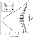
- Figure 1 shows infrared spectroscopy data for quantification of antibody loading within the functionalized alginic acid solutions emerging from the one- or two-step synthesis protocol.
- all of the alginic acid solutions have comparable antibody content between 0.05 and 0.06 mg/mL.
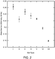
- Figure 2 shows the relative total protein measurements made using a BCA assay kit.
- the BCA solution becomes more transparent as it comes in contact with proteins such as antibodies. Hence, by flowing this solution through hydrogel-coated microfluidic devices, the amount of accessible antibody on each gel type can be compared.
- the protein content of the solutions exiting the devices is shown as a function of gel type in Figure 2 and is expressed in arbitrary units of absorbance rather than as a calibrated mass or concentration.
- the relative measurement allows comparison of the accessible anti-CD34 capture antibody between each gel type.
- Figure 2 shows an increase in accessible antibody from gel types I-VII while the total amount of antibody added to the mixture remains constant ( Figure 1 ), indicating an increase in the efficiency of conjugation between the gelled surface and the antibody.
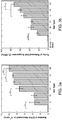
- Figure 3 shows yield and purity data for the capture of EPCs from whole blood using the hydrogel-coated microfluidic devices.
- gel type I which has an inert antibody conjugated to it, shows negligible EPC adhesion as expected.
- Gel type II which contains the anti-CD34 antibody, shows significantly higher EPC adhesion relative to gel type I (p ⁇ 0.005), albeit with a high degree of scatter.
- the purity of capture achieved with gel type II is, however, relatively low ( ⁇ 23%; Figure 3B ).
- the effect of adding the 4-arm PEG to the hydrogel structure is shown clearly by comparing gel types II and IV, whose synthesis protocol is otherwise identical.
- gel types V-VII a two step protocol for combining reagents was followed.
- the conjugation of the antibody molecules to the 4-arm PEG is carried out first before introducing alginic acid.
- This formulation improved yield and purity of EPC capture relative to gel type IV.
- the two-step protocol was modified such that EDC and sulfo-NHS were added in the second step with alginic acid and the first step was restricted to the mixing together of PEG and antibody.
- short times were provided for mixing and incubation for the first step (10 min and 15 min, respectively, for gel type VI), the yield did not improve relative to gel type V, but purity was higher.
- Example 2 discloses devices and methods for the microfluidic capture and release of intestinal stem cells using two binding agents, specifically, GPR49 and Lgr5 antibody receptors.
- this Example allows for multiplexing and larger sample volume to be processed while retaining viability of the eluded target population.
- culture methods have been developed to induce hyperplasia and organoid forming units derived from single cells.
- Sato, T. et al. Single Lgr5 stem cells build crypt-villus structures in vitro without a mesenchymal niche. Nature 2009, 459 (7244), 262-U147 ). These cells do not require a mesenchymal niche to develop into these units and rely on growth factors to induce differentiation cues.
- enriched lgr5 positive populations have been captured and released using the methods disclosed, and the cells yielded similar morphological responses as produced by previous groups. Furthermore, the addition of wnt3a in culture facilitated an increase in plating efficiency. Immunohistochemical analysis coupled with confocal microscopy shed light on lgr5 and cd24 expression within the central lumen coinciding with recent reports. (See Gracz, A. D. et al., Sox9 expression marks a subset of CD24-expressing small intestine epithelial stem cells that form organoids in vitro. Am J Physiol-Gastr L 2010, 298 (5), G590-G600 .).
- the capture and release mechanism resides in cross-linking the hydrogel with calcium with a chelation release.
- This Example demonstrates the ability to selectively capture and release GPR49/Lgr5 positive cells from wild-type rat colon crypts digestate. Through a one-pass approach, a 24-fold enrichment from the starting suspension to a final purity of 49% GPR49/Lgr5 cells was obtained. The presented microfluidics platform retains viability of the target cells, while giving the end user the ability to multiplex samples.
- the disclosure allows for intestinal stem cell isolation that has the potential in advancing the field of tissue engineering and applications with co-cultures.
- mice Male and female neonatal Lewis rats (Charles River) were used and harbored in room temperature conditions with a 12-hour light/dark cycle following U.S. Eastern Standard Time.
- neonatal rats between the ages of 2 to 5 days were utilized and sacrificed via decapitation. All studies and protocols were approved by the Institutional Animal Care and Use Committee (IACUC) at Northeastern University.
- IACUC Institutional Animal Care and Use Committee
- Intestinal tissue samples were obtained from neonatal Lewis rats. Large intestine was extracted, split laterally, and fragmented into 1 mm segments. Fragmented tissue was incubated in 2 mM EDTA at 4°C for 30 minutes. Tissue samples were separated from the solution and placed in 20 mL of phosphate buffered saline (PBS, Gibco) for 10 minutes of agitation. The supernatant fluid was then collected and centrifuged at 150xg for three minutes; the pellet was collected, suspended in 10 mL of serum-free Dulbecco's Modified Eagle's Medium (DMEM, Cellgrow) and centrifuged again at 150xg. The pellet was suspended in 5 mL of serum-free DMEM solution and filtered through a 100 ⁇ m cell strainer. The solution was then filtered through 20 ⁇ m cell strainers into 1 mL eppendorf tubes.
- PBS phosphate buffered saline
- Microfluidic devices were fabricated using traditional soft lithography at the George J. Kostas Nanoscale Technology and Manufacturing Research Center at Northeastern University. The physical dimensions and design of the devices were identical to those of devices described by Hatch et al. These devices consist of polydimethylsiloxane (PDMS) patterned with 100 ⁇ m diameter pillars bonded to glass slides.
- PDMS polydimethylsiloxane
- Antibody-functionalized alginate reaction underwent six different scenarios but stoichiometric ratios of reagents remained constant through out each scheme.
- 1940 ⁇ E MES (Thermo-fisher), .04 mg Anti-GPCR GPR49 (Abcam), and 22.5 mg 10 KD 4-arm star PEG () was mixed for 30 minutes.
- MES pH was altered for each respective scenario which was either held at ph 4.7 or 6.0; the pH was titrated with NaOH to a pH of 6.0.
- the amalgam was allowed to incubate for 60 minutes in scenario II, but the subsequent reagents were added immediately in the remaining scenarios.
- Microfluidic devices with a hexagonal post array were utilized for cell separation. Each device was filled with alginate functionalized with Anti-GPCR GPR49 and allowed to incubate for 60 minutes. Channels were formed by flowing through 100 ⁇ L of pH 6 MES buffer at 10 ⁇ L/min, 100 ⁇ L of 100 mM CaCl 2 at 10 ⁇ L/min, and 100 ⁇ L of 0.1% bovine serum albumin at 10 ⁇ L/min. A Harvard Apparatus syringe pump was used to obtained precise flow rates. Cell solutions obtained were mixed to ensure homogenous suspension and 200 ⁇ L were drawn into 1 mL syringes.
- 100 ⁇ L of cell solution was pumped through each device at a rate of 3 ⁇ L/min followed by 100 ⁇ L of pH 6 MES buffer at 3 ⁇ L/min to rinse. Then 100 ⁇ L of 100 mM EDTA solution was pumped through the device at 10 ⁇ L/min to release the cells from the device. For culture, cells were released into eppendorf tubes containing 50 ⁇ L of Matrigel (BD Bioscience) on ice.
- Lgr5 basal media contained the following constituents: Advanced DMEM F-12, 5 ml N2 supplement, 10 mL B27 without vit. A, 5mL HEPES, 6.25 mL glutamax. Each sample was rinsed with 350 ⁇ L of Lgr5 basal media in to remove EDTA from the cell culture. Then 17 ⁇ L of ROCK inhibitor (y-27632, Sigma-Aldrich) was added to 10 mL of Lgr5 media.
- ROCK inhibitor y-27632, Sigma-Aldrich
- Enriched organoids were fixed with 4% paraformadahyde and rinsed with 2mM glycine in PBS. 6 U/ml dispase (stem cell technologies) was added and incubated for 1 hour to release organoids from matrigel. Organoids were pipetted into 200 ⁇ L Lgr5 media blocking solution containing: 3% BSA, 10% goat serum, .1% triton X-100, 10mM HEPES, and 10mM glycine. 1:50 of respective antibodies, anti-GPCR GPR49 and anti-CD24, to blocking solution was added and incubated at 4°C overnight.
- Organoids were pipetted out of solution and into 200 ⁇ L of blocking solution containing normalized concentrations of Alexfluor 488, Alexafluor 568, and .5 ⁇ g/ml DAPI for 3 hours. Organoids were mounted on glass cover slides and confocal images were taken via Nikon confocal microscope.
- Lgr5 capture encompassed four variables including flow rate, pacification, pH, and reaction time.
- Bovine serum albumin (BSA) a pacifying agent, allowed for a decrease in fouling within the microfluidics channels, which facilitated establishment of consistent flow across the channel and inhibition of non specific binding to the alginate/antibody conjugate.
- Flow rates, adjusted between 3 and 5 ⁇ L/min resulted in a fairly significant disparity in which alluded to possible shear effects upon the target cells at higher flow rates ( Figure 6B ).
- Lgr5 positive cells Released enriched Lgr5 positive cells were imbedded in Matrigel and grown under similar conditions as described in Sato et al.; Single Lgr5 stem cells build crypt-villus structures in vitro without a mesenchymal niche. Nature 2009, 459 (7244), 262-U147 .
- the culture technique for lgr5 positive cells included growth factor constituents that were altered slightly to take into account species dependent factors. Rat endothelial growth factor (EGF) and murine rspondin-1 were used, in contrast to the literature sources that have implemented a hybridized mouse model. Y-26743, rock inhibitor, was used to improve culture stability and to prevent anoikis in a single cell suspension.
- EGF endothelial growth factor
- Y-26743 rock inhibitor
- the inhibitor was also used concurrently in the micro fluidic enrichment technique, and it was observed to result in an increase in plating efficiency (data not shown), but exhibited little affect in unenriched cultures ( Figure 7A-C ). Progression of organoids, from enriched single lgr5 cells, was viewed up to 4 days and compared against an unenriched population ( Figure 7D-E ). Growth was noticed at day 2 and progressed into hyperplasia stage at day 3. Small lumen formation coupled with an increase of hyperplasia is observed at day 4.
- Figure 7A-C show that unenriched organoid progression yielded significant larger cyst-like organoids surrounded by extraneous populations.
- Figures 7D-F show four-day progression of enriched organoid derived from single cell suspension. Expansion of single cell ( Figure 7D ) at day 2, induced hyperplasia at day 3 ( Figure 7E ), and small lumen formation noticed with surrounding secreted apoptotic cells, at day 4 ( Figure 7F ) are shown. Scale bars represent 100 ⁇ m
- the day progressions of the released lgr5 positive cells were cultured in parallel against an unenriched population.
- the unenriched suspension was seeded at the same volume as the released population and cultured under the same conditions. Multiple morphologies were apparent in the unenriched culture, ranging from organoids with central lumen, harboring apoptotic cells ( Figure 7C and 8C ), to fibroblastic morphologies. Growth rate of the unenriched suspension was more accelerated in comparison to the enriched population.
- Plating efficiency was improved amongst the enriched population by the addition of Wnt3a to the culture system ( Figure 8B ).
- the addition of wnt3a to culture medium sustains viability and independence from paneth cell ( Figure 8D ).
- the unenriched population did not have an any increase in plate efficiency in the presence of Wnt3a protein.
- the majority of the organoids formed in the injected culture expressed a cyst-like structure harboring apoptotic cells( Figure 8C ).
- the enriched population did have an increase in plating efficiency leading to more single derived organoids proliferating.
- Enriched cells exhibited similar morphology to the wnt3a absence study ( Figure 8D ) at analogous time points. Images were taken at 3 days in culture; scale bar represents 100 ⁇ m.
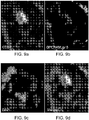
- Enriched and unenriched organoids were released from culture at day 4 via a dispase treatment to degrade the Matrigel. Stained organoids were exposed to anti-GPR GPRCR49, anti-CD24, and DAPI, each conjugated with alexa fluor 488 (green) and 524 (red) ( Figures 9 and 10 ). Confocal microscopy facilitated determination of the morphology of the organoids and protein expression. Unenriched organoids ( Figure 10 ) had a significant population of apoptotic cells within the central domain. The organoid did not undergo hyperplastia for the culture duration and exhibited a bright CD-24 signal in an elliptical pattern.
- Anti-Lgr5/GPRCR49 expression was faint ( Figure 10b ), and expression was limited to the lumendomain of the organoid. Localization of anti-Lgr5/GPRCR49 diminished in the significantly low CD24 populations( Figure 10A-B ).
- the topography of the unenriched culture exhibited an elliptical planar morphology ( Figure 10D ) in contrast to the enriched organoid, which was spherical ( Figure 9D ).
- the central domain expressed CD-24(green) and anti-Lgr5/GPCR49 (red), localized in the apical membrane ( Figure 9A-B ).
- CD-24 expression was localized along 4 different membranes ( Figure 9A ), and expression was lower in intensity compared to the unenriched organoid.
- Localized anti-Lgr5/GPCR49 were centered in the apical membrane and expressed in 2 membranes ( Figure 9B ). Expression of both markers was strictly limited to the central domain, coinciding with Sox9 (CD-24) and Lgr5 genomic trends.
- the instant disclosure fulfills the need in developing a cost-effective and fluorescent-free cell isolation devices and methods for application such as tissue engineering.
- Conventional methods in intestinal stem cell isolation rely on hybridized mice models and complex instrumentation, such as FACS.
- the instant Example describes a microfluidics method that enriches intestinal stem cell populations using alginate coupled with anti-GPCR49/Lgr5.
- the enriched lgr5 cells have been grown in appropriate culture medium. After adding the cells in medium, CD24 expression coinciding with Lgr5 expression in the organoid central domain was investigated.
- This Example describes methods and devices that enrich a select target population while retaining viability, expression, and growth morphology.
- This Example describes a one-pass microfluidic alginate capture and release model capable of a 24-fold enrichment to a GPCR49/Lgr5 purity of 49%.
- a pacifying agent BSA
- BSA a pacifying agent
- the phenomena generates a cascading affect in which coagulated cell types containing lgr5 positive cells adhere to the alginate coating; immediate injection of strained cells was performed to facilitate in dispersion.
- Chemical interactions and stability between alginate, EDC, 4-arm star PEG, Anti-GPCR49/Lgr5 were increased as the reaction pH became more basic.
- the disclosed methods can be used for multiplexing, allowing many devices to be run in parallel and increasing throughput. Furthermore, the disclosed methods allowed for fluorescent-label free isolation of intestinal stem cells while retaining similar growth morphology in situ.
- the unenriched population contain doublets of paneth-lgr5 postive cells, which sustain the necessary Wnt signaling; thus, a null effect was noticed in the presence of the cofactor.
- Wnt3a it was noted that the plating efficiency amongst the unenriched population was slightly higher than the enriched suspension; this being indicative of paneth cell niche signaling allowing for improved long-term organoid viability.
- Enriched organoids were plated in similar fashion to the injected suspension, but the significant difference resided in the morphological changes and plating efficiency of the released GPCR49/lgr5 positive cells.
- the images indicate a smaller organoid with a small central lumen formed yet to harbor any apoptotic cells.
- CD24 and GPCR49/Lgr5 expression was bound in the central domain with similar expression patterns as the latter.
- the presented images eluded to that the microfluidics enrichment process retained similar morphological outcomes as previously reported.
- the Example discloses methods and devices that can be used for cell sorting and tissue engineering.
- the Example describes an intestinal stem cell isolation technique from wild-type intestinal digestate.
- the current convention is limited to transgenic mice models and complex instrumentation to isolate these cells.
- the disclosed methods allow the end-user to isolate cell subtypes in a speedy process while retaining cell viability.
- This Example relates to compositions and methods for a multistage, highly specific capture and release of biological materials, such as cells.
- Figure 11A-D represent a configuration of alginate-hydrogel based devices that include capture stages for each of two antibodies. In some embodiments, more than two antibodies are contemplated.
- FIG 11A-D a sample was injected via a syringe pump into the first alginate-based capture stage ("Marker 1 isolation"/ Figure 11A ).
- This stage was connected to stage B, which was a 2-way valve. In its "closed” configuration, this valve allowed the waste from stage A to pass through to a collection tube. After the waste went through, the waste stream was closed using, for example, a pinch valve.
- Figure 11B The purpose of the calcium chloride was to neutralize the EDTA in the cell suspension emerging from stage ( Figure 11A ). To ensure mixing of the calcium chloride solution with this cell suspension, the combined output (which was in laminar flow) was sent into a mixing chamber ( Figure 11C ) containing herringbone features.
- stage 11D The mixed solution then entered stage ( Figure 11D ), where the cells expressing receptors for the second capture molecule were captured.
- stage A Figure 11A
- stage B Figure 11B
- This solution was collected in a tube containing an excess of culture medium to minimize any deleterious effect of the EDTA on the cells.
- This Example showed the ability of this dual-stage capture system to isolate endothelial progenitor cells (EPCs) from untreated whole blood.
- the objective was to capture cells that are CD34+/FLK1+.
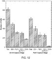
- Figure 12 shows cell counts (obtained by flow cytometry) of the cells emerging from stage A and stage B.
- the various populations shown represent categories of CD34+ cells and the "total" column represents the total number of cells released.
- the objective of the second capture device was to remove CD34+ cells that do not express the second marker, FLK-1, namely the CD34+ cells that are also CD45+. The sharp decrease in the number of CD45+ cells coming out of the second capture stage relative to the first capture stage shows this enrichment.
Landscapes
- Health & Medical Sciences (AREA)
- Life Sciences & Earth Sciences (AREA)
- Immunology (AREA)
- Engineering & Computer Science (AREA)
- Chemical & Material Sciences (AREA)
- Molecular Biology (AREA)
- Biomedical Technology (AREA)
- Urology & Nephrology (AREA)
- Hematology (AREA)
- Cell Biology (AREA)
- Analytical Chemistry (AREA)
- Physics & Mathematics (AREA)
- Biochemistry (AREA)
- General Health & Medical Sciences (AREA)
- General Physics & Mathematics (AREA)
- Pathology (AREA)
- Microbiology (AREA)
- Biotechnology (AREA)
- Food Science & Technology (AREA)
- Medicinal Chemistry (AREA)
- Zoology (AREA)
- Tropical Medicine & Parasitology (AREA)
- Virology (AREA)
- Organic Chemistry (AREA)
- Chemical Kinetics & Catalysis (AREA)
- Wood Science & Technology (AREA)
- Proteomics, Peptides & Aminoacids (AREA)
- Bioinformatics & Cheminformatics (AREA)
- Genetics & Genomics (AREA)
- Inorganic Chemistry (AREA)
- General Engineering & Computer Science (AREA)
- Biophysics (AREA)
- Micro-Organisms Or Cultivation Processes Thereof (AREA)
- Apparatus Associated With Microorganisms And Enzymes (AREA)
- Measuring Or Testing Involving Enzymes Or Micro-Organisms (AREA)
- Immobilizing And Processing Of Enzymes And Microorganisms (AREA)
- Medicinal Preparation (AREA)
- Physical Or Chemical Processes And Apparatus (AREA)
- Solid-Sorbent Or Filter-Aiding Compositions (AREA)
- Compositions Of Macromolecular Compounds (AREA)
Applications Claiming Priority (2)
| Application Number | Priority Date | Filing Date | Title |
|---|---|---|---|
| US201161439166P | 2011-02-03 | 2011-02-03 | |
| PCT/US2012/023859 WO2012106658A1 (en) | 2011-02-03 | 2012-02-03 | Methods and compositions for highly specific capture and release of biological materials |
Publications (3)
| Publication Number | Publication Date |
|---|---|
| EP2670856A1 EP2670856A1 (en) | 2013-12-11 |
| EP2670856A4 EP2670856A4 (en) | 2014-12-03 |
| EP2670856B1 true EP2670856B1 (en) | 2018-06-27 |
Family
ID=46603107
Family Applications (1)
| Application Number | Title | Priority Date | Filing Date |
|---|---|---|---|
| EP12742728.4A Active EP2670856B1 (en) | 2011-02-03 | 2012-02-03 | Methods and compositions for highly specific capture and release of biological materials |
Country Status (7)
| Country | Link |
|---|---|
| US (1) | US9927334B2 (enExample) |
| EP (1) | EP2670856B1 (enExample) |
| JP (2) | JP6120778B2 (enExample) |
| KR (1) | KR101922741B1 (enExample) |
| ES (1) | ES2687144T3 (enExample) |
| SG (2) | SG192587A1 (enExample) |
| WO (1) | WO2012106658A1 (enExample) |
Cited By (1)
| Publication number | Priority date | Publication date | Assignee | Title |
|---|---|---|---|---|
| US20200378925A1 (en) * | 2019-05-30 | 2020-12-03 | The Regents Of The University Of California | Microfluidic Acoustic Devices and Methods |
Families Citing this family (19)
| Publication number | Priority date | Publication date | Assignee | Title |
|---|---|---|---|---|
| EP2919910B1 (en) | 2012-11-09 | 2017-12-20 | Roche Diagnostics GmbH | In vitro capture and analysis of circulating tumor cells |
| WO2014121204A1 (en) * | 2013-02-01 | 2014-08-07 | The General Hospital Corporation | Capture and release of particles from liquid samples |
| WO2015139022A1 (en) | 2014-03-14 | 2015-09-17 | Northeastern University | Microfluidic system and method for real-time measurement of antibody-antigen binding and analyte detection |
| WO2015148512A1 (en) | 2014-03-24 | 2015-10-01 | Qt Holdings Corp | Shaped articles including hydrogels and methods of manufacture and use thereof |
| ES2858600T3 (es) * | 2014-12-22 | 2021-09-30 | Ecole Polytechnique Fed Lausanne Epfl | Dispositivos para la agregación, de alto rendimiento, y la manipulación de células de mamífero |
| US9790467B2 (en) | 2015-09-22 | 2017-10-17 | Qt Holdings Corp | Methods and compositions for activation or expansion of T lymphocytes |
| ES2871137T3 (es) * | 2015-12-14 | 2021-10-28 | Quanta Matrix Co Ltd | Método de diferenciación de células particulares a partir de la mezcla de células heterogéneas |
| EP3430407B1 (en) * | 2016-03-18 | 2021-10-06 | QT Holdings Corp | Kit, composition, device and method for cell separation |
| US10429387B2 (en) * | 2016-07-27 | 2019-10-01 | Universiteit Tweate | Simple and affordable method for immuophenotyping using a microfluidic chip sample preparation with image cytometry |
| US11118163B2 (en) * | 2017-02-02 | 2021-09-14 | Sartorius Stedim Biotech Gmbh | Separation of cell populations by marker identification and sedimentation velocity |
| DE102017105195A1 (de) | 2017-03-10 | 2018-09-13 | Leibniz-Institut Für Polymerforschung Dresden E.V. | Verfahren zur differenzierten Sequestrierung von Stoffen verschiedener Stoffgruppen mit Hilfe von sulfatierte oder sulfonierte Komponenten enthaltenden Hydrogelen |
| CA3056891A1 (en) * | 2017-03-20 | 2018-09-27 | Qt Holdings Corp | Methods and compositions for modulation of immune cells |
| US12138843B2 (en) | 2018-06-13 | 2024-11-12 | University Of Washington | Extruded hydrogel tubes and coaxial fibers and applications thereof |
| EP3626814B1 (en) * | 2018-09-21 | 2023-03-08 | Technische Universität Wien | Production of cellular spheroids |
| JP7120547B2 (ja) * | 2019-02-22 | 2022-08-17 | 国立研究開発法人理化学研究所 | 物質固定化剤、及び当該物質固定化剤を用いた物質固定化方法 |
| EP3941630B1 (en) * | 2019-03-18 | 2025-04-16 | Cellular Research, Inc. | Precise delivery of components into fluids |
| CN112708150B (zh) * | 2020-12-08 | 2021-11-16 | 济南国科医工科技发展有限公司 | 用于循环肿瘤细胞的捕获及定点释放的水凝胶体系 |
| WO2023034430A1 (en) | 2021-09-01 | 2023-03-09 | Bio-Techne Corporation | Human vitronectin fragments and uses thereof |
| US11805995B1 (en) | 2022-11-22 | 2023-11-07 | King Faisal University | Saliva collection kit |
Citations (1)
| Publication number | Priority date | Publication date | Assignee | Title |
|---|---|---|---|---|
| WO2010124227A2 (en) * | 2009-04-24 | 2010-10-28 | The Board Of Trustees Of The University Of Illinois | Methods and devices for capturing circulating tumor cells |
Family Cites Families (10)
| Publication number | Priority date | Publication date | Assignee | Title |
|---|---|---|---|---|
| CA2266581C (en) * | 1996-09-19 | 2007-03-13 | The Regents Of The University Of Michigan | Polymers containing polysaccharides such as alginates or modified alginates |
| HK1080500B (zh) * | 2001-11-07 | 2008-01-18 | Universität Zürich | 用於控制细胞向内生长和组织再生的合成基质 |
| ES2301697T3 (es) * | 2001-12-18 | 2008-07-01 | Eidgenossisch Technische Hochschule Zurich | Matrices de proteina modificada con factor de crecimiento para ingenieria de tejidos. |
| AU2003248273A1 (en) * | 2002-07-12 | 2004-02-02 | Mitsubishi Chemical Corporation | Analytical chip, analytical chip unit, analyzing apparatus, method of analysis using the apparatus, and method of producing the analytical chip |
| EP1569510B1 (en) | 2002-09-27 | 2011-11-02 | The General Hospital Corporation | Microfluidic device for cell separation and uses thereof |
| CA2563329C (en) * | 2004-04-30 | 2016-01-26 | Orbus Medical Technologies, Inc. | Medical device with coating for capturing genetically-altered cells and methods for using same |
| SE0402476D0 (sv) * | 2004-10-13 | 2004-10-13 | Biacore Ab | Preparation and use of a reactive solid support surface |
| CN101338036B (zh) * | 2007-07-06 | 2010-11-03 | 常州百瑞吉生物医药有限公司 | 生物相容快速凝胶化水凝胶及其喷雾剂的制备方法 |
| US9267109B2 (en) * | 2007-12-10 | 2016-02-23 | Koninklijke Philips N.V. | Patterned cell sheets and a method for production of the same |
| WO2012094642A2 (en) * | 2011-01-06 | 2012-07-12 | On-Q-ity | Circulating tumor cell capture on a microfluidic chip incorporating both affinity and size |
-
2012
- 2012-02-03 EP EP12742728.4A patent/EP2670856B1/en active Active
- 2012-02-03 SG SG2013058797A patent/SG192587A1/en unknown
- 2012-02-03 ES ES12742728.4T patent/ES2687144T3/es active Active
- 2012-02-03 US US13/982,680 patent/US9927334B2/en active Active
- 2012-02-03 JP JP2013552696A patent/JP6120778B2/ja active Active
- 2012-02-03 KR KR1020137023006A patent/KR101922741B1/ko active Active
- 2012-02-03 WO PCT/US2012/023859 patent/WO2012106658A1/en not_active Ceased
- 2012-02-03 SG SG10201600803SA patent/SG10201600803SA/en unknown
-
2016
- 2016-12-08 JP JP2016238320A patent/JP2017077245A/ja not_active Abandoned
Patent Citations (1)
| Publication number | Priority date | Publication date | Assignee | Title |
|---|---|---|---|---|
| WO2010124227A2 (en) * | 2009-04-24 | 2010-10-28 | The Board Of Trustees Of The University Of Illinois | Methods and devices for capturing circulating tumor cells |
Cited By (2)
| Publication number | Priority date | Publication date | Assignee | Title |
|---|---|---|---|---|
| US20200378925A1 (en) * | 2019-05-30 | 2020-12-03 | The Regents Of The University Of California | Microfluidic Acoustic Devices and Methods |
| US11668676B2 (en) * | 2019-05-30 | 2023-06-06 | The Regents Of The University Of California | Microfluidic acoustic devices and methods |
Also Published As
| Publication number | Publication date |
|---|---|
| US9927334B2 (en) | 2018-03-27 |
| US20140057280A1 (en) | 2014-02-27 |
| SG10201600803SA (en) | 2016-03-30 |
| JP2017077245A (ja) | 2017-04-27 |
| SG192587A1 (en) | 2013-09-30 |
| ES2687144T3 (es) | 2018-10-23 |
| EP2670856A4 (en) | 2014-12-03 |
| JP2014506671A (ja) | 2014-03-17 |
| EP2670856A1 (en) | 2013-12-11 |
| KR101922741B1 (ko) | 2019-02-20 |
| WO2012106658A1 (en) | 2012-08-09 |
| JP6120778B2 (ja) | 2017-04-26 |
| KR20140016896A (ko) | 2014-02-10 |
Similar Documents
| Publication | Publication Date | Title |
|---|---|---|
| EP2670856B1 (en) | Methods and compositions for highly specific capture and release of biological materials | |
| Moore et al. | M0 and M2 macrophages enhance vascularization of tissue engineering scaffolds | |
| CA2712496C (en) | Stem cell aggregates and methods for making and using | |
| Park et al. | Development of a novel dual reproductive organ on a chip: recapitulating bidirectional endocrine crosstalk between the uterine endometrium and the ovary | |
| Kwizera et al. | Greatly enhanced CTC culture enabled by capturing CTC heterogeneity using a PEGylated PDMS–Titanium–Gold electromicrofluidic device with glutathione-controlled gentle cell release | |
| KR20160005037A (ko) | 단리된 신장 세포를 포함하는 오가노이드 및 이의 용도 | |
| Genchi et al. | Bio/non-bio interfaces: a straightforward method for obtaining long term PDMS/muscle cell biohybrid constructs | |
| EP3839038A2 (en) | Microfluidic device for cerebrovascular simulation and high-efficiency blood-brain barrier simulation system comprising same | |
| WO2020172670A1 (en) | Microfluidic proximal tubule kidney-on-chip | |
| EP3017301B1 (en) | Microtissues | |
| Jiang et al. | Dissolvable microgel-templated macroporous hydrogels for controlled cell assembly | |
| Unal et al. | 3D co-culture with vascular cells supports long-term hepatocyte phenotype and function in vitro | |
| Takahashi et al. | 3D in vitro co-culture disc for spatiotemporal image analysis of cancer–stromal cell interaction | |
| Lansche et al. | Bioengineering a Patient‐Derived Vascularized Lung Tumor‐on‐Chip Model to Decipher Immunomodulation by the Endothelium | |
| US11648320B2 (en) | Non-covalently assembled biomatrix layer | |
| Hatch | Microfluidic isolation of endothelial progenitor cells for vascular tissue engineering | |
| Kevlahan | A microfluidic capture and release method for isolation intestinal progenitor and stem cells from native rat tissue enabling advances in vasculogenic co-cultures | |
| Huang | Engineering Patient-specific Liver Microtissues with Prolonged Phenotypic Maintenance and Disease Modeling Potential | |
| Rao | Poly (Ethylene Glycol) Hydrogels to Engineer the Mesenchymal Stromal Cell Secretome | |
| WO2024052906A1 (en) | Activation and proliferation of cytotoxic lymphocytes | |
| Marrero Feitosa-Afonsso | Development of a blood-retina barrier on-a-chip with human iPSC-derived retinal pigment epithelium and endothelium | |
| Rodgers et al. | Disassembly of Self-Assembling Peptide Hydrogels as a Versatile Method for 2 Cell Extraction and Manipulation |
Legal Events
| Date | Code | Title | Description |
|---|---|---|---|
| PUAI | Public reference made under article 153(3) epc to a published international application that has entered the european phase |
Free format text: ORIGINAL CODE: 0009012 |
|
| 17P | Request for examination filed |
Effective date: 20130902 |
|
| AK | Designated contracting states |
Kind code of ref document: A1 Designated state(s): AL AT BE BG CH CY CZ DE DK EE ES FI FR GB GR HR HU IE IS IT LI LT LU LV MC MK MT NL NO PL PT RO RS SE SI SK SM TR |
|
| RIN1 | Information on inventor provided before grant (corrected) |
Inventor name: MURTHY, SHASHI, K. Inventor name: HATCH, ADAM Inventor name: HANSMANN, GEORGE |
|
| DAX | Request for extension of the european patent (deleted) | ||
| A4 | Supplementary search report drawn up and despatched |
Effective date: 20141031 |
|
| RIC1 | Information provided on ipc code assigned before grant |
Ipc: G01N 33/543 20060101ALI20141027BHEP Ipc: C12Q 1/00 20060101AFI20141027BHEP Ipc: G01N 33/553 20060101ALI20141027BHEP |
|
| 17Q | First examination report despatched |
Effective date: 20150909 |
|
| STAA | Information on the status of an ep patent application or granted ep patent |
Free format text: STATUS: EXAMINATION IS IN PROGRESS |
|
| GRAP | Despatch of communication of intention to grant a patent |
Free format text: ORIGINAL CODE: EPIDOSNIGR1 |
|
| STAA | Information on the status of an ep patent application or granted ep patent |
Free format text: STATUS: GRANT OF PATENT IS INTENDED |
|
| INTG | Intention to grant announced |
Effective date: 20180119 |
|
| INTG | Intention to grant announced |
Effective date: 20180119 |
|
| GRAS | Grant fee paid |
Free format text: ORIGINAL CODE: EPIDOSNIGR3 |
|
| GRAA | (expected) grant |
Free format text: ORIGINAL CODE: 0009210 |
|
| STAA | Information on the status of an ep patent application or granted ep patent |
Free format text: STATUS: THE PATENT HAS BEEN GRANTED |
|
| AK | Designated contracting states |
Kind code of ref document: B1 Designated state(s): AL AT BE BG CH CY CZ DE DK EE ES FI FR GB GR HR HU IE IS IT LI LT LU LV MC MK MT NL NO PL PT RO RS SE SI SK SM TR |
|
| REG | Reference to a national code |
Ref country code: GB Ref legal event code: FG4D |
|
| REG | Reference to a national code |
Ref country code: AT Ref legal event code: REF Ref document number: 1012393 Country of ref document: AT Kind code of ref document: T Effective date: 20180715 |
|
| REG | Reference to a national code |
Ref country code: IE Ref legal event code: FG4D |
|
| REG | Reference to a national code |
Ref country code: DE Ref legal event code: R096 Ref document number: 602012047829 Country of ref document: DE |
|
| REG | Reference to a national code |
Ref country code: NL Ref legal event code: FP |
|
| REG | Reference to a national code |
Ref country code: ES Ref legal event code: FG2A Ref document number: 2687144 Country of ref document: ES Kind code of ref document: T3 Effective date: 20181023 |
|
| PG25 | Lapsed in a contracting state [announced via postgrant information from national office to epo] |
Ref country code: LT Free format text: LAPSE BECAUSE OF FAILURE TO SUBMIT A TRANSLATION OF THE DESCRIPTION OR TO PAY THE FEE WITHIN THE PRESCRIBED TIME-LIMIT Effective date: 20180627 Ref country code: NO Free format text: LAPSE BECAUSE OF FAILURE TO SUBMIT A TRANSLATION OF THE DESCRIPTION OR TO PAY THE FEE WITHIN THE PRESCRIBED TIME-LIMIT Effective date: 20180927 Ref country code: BG Free format text: LAPSE BECAUSE OF FAILURE TO SUBMIT A TRANSLATION OF THE DESCRIPTION OR TO PAY THE FEE WITHIN THE PRESCRIBED TIME-LIMIT Effective date: 20180927 Ref country code: SE Free format text: LAPSE BECAUSE OF FAILURE TO SUBMIT A TRANSLATION OF THE DESCRIPTION OR TO PAY THE FEE WITHIN THE PRESCRIBED TIME-LIMIT Effective date: 20180627 Ref country code: FI Free format text: LAPSE BECAUSE OF FAILURE TO SUBMIT A TRANSLATION OF THE DESCRIPTION OR TO PAY THE FEE WITHIN THE PRESCRIBED TIME-LIMIT Effective date: 20180627 |
|
| REG | Reference to a national code |
Ref country code: LT Ref legal event code: MG4D |
|
| PG25 | Lapsed in a contracting state [announced via postgrant information from national office to epo] |
Ref country code: GR Free format text: LAPSE BECAUSE OF FAILURE TO SUBMIT A TRANSLATION OF THE DESCRIPTION OR TO PAY THE FEE WITHIN THE PRESCRIBED TIME-LIMIT Effective date: 20180928 Ref country code: RS Free format text: LAPSE BECAUSE OF FAILURE TO SUBMIT A TRANSLATION OF THE DESCRIPTION OR TO PAY THE FEE WITHIN THE PRESCRIBED TIME-LIMIT Effective date: 20180627 Ref country code: LV Free format text: LAPSE BECAUSE OF FAILURE TO SUBMIT A TRANSLATION OF THE DESCRIPTION OR TO PAY THE FEE WITHIN THE PRESCRIBED TIME-LIMIT Effective date: 20180627 Ref country code: HR Free format text: LAPSE BECAUSE OF FAILURE TO SUBMIT A TRANSLATION OF THE DESCRIPTION OR TO PAY THE FEE WITHIN THE PRESCRIBED TIME-LIMIT Effective date: 20180627 |
|
| REG | Reference to a national code |
Ref country code: AT Ref legal event code: MK05 Ref document number: 1012393 Country of ref document: AT Kind code of ref document: T Effective date: 20180627 |
|
| PG25 | Lapsed in a contracting state [announced via postgrant information from national office to epo] |
Ref country code: CZ Free format text: LAPSE BECAUSE OF FAILURE TO SUBMIT A TRANSLATION OF THE DESCRIPTION OR TO PAY THE FEE WITHIN THE PRESCRIBED TIME-LIMIT Effective date: 20180627 Ref country code: SK Free format text: LAPSE BECAUSE OF FAILURE TO SUBMIT A TRANSLATION OF THE DESCRIPTION OR TO PAY THE FEE WITHIN THE PRESCRIBED TIME-LIMIT Effective date: 20180627 Ref country code: IS Free format text: LAPSE BECAUSE OF FAILURE TO SUBMIT A TRANSLATION OF THE DESCRIPTION OR TO PAY THE FEE WITHIN THE PRESCRIBED TIME-LIMIT Effective date: 20181027 Ref country code: EE Free format text: LAPSE BECAUSE OF FAILURE TO SUBMIT A TRANSLATION OF THE DESCRIPTION OR TO PAY THE FEE WITHIN THE PRESCRIBED TIME-LIMIT Effective date: 20180627 Ref country code: AT Free format text: LAPSE BECAUSE OF FAILURE TO SUBMIT A TRANSLATION OF THE DESCRIPTION OR TO PAY THE FEE WITHIN THE PRESCRIBED TIME-LIMIT Effective date: 20180627 Ref country code: PL Free format text: LAPSE BECAUSE OF FAILURE TO SUBMIT A TRANSLATION OF THE DESCRIPTION OR TO PAY THE FEE WITHIN THE PRESCRIBED TIME-LIMIT Effective date: 20180627 Ref country code: RO Free format text: LAPSE BECAUSE OF FAILURE TO SUBMIT A TRANSLATION OF THE DESCRIPTION OR TO PAY THE FEE WITHIN THE PRESCRIBED TIME-LIMIT Effective date: 20180627 |
|
| PG25 | Lapsed in a contracting state [announced via postgrant information from national office to epo] |
Ref country code: SM Free format text: LAPSE BECAUSE OF FAILURE TO SUBMIT A TRANSLATION OF THE DESCRIPTION OR TO PAY THE FEE WITHIN THE PRESCRIBED TIME-LIMIT Effective date: 20180627 |
|
| REG | Reference to a national code |
Ref country code: DE Ref legal event code: R097 Ref document number: 602012047829 Country of ref document: DE |
|
| PLBE | No opposition filed within time limit |
Free format text: ORIGINAL CODE: 0009261 |
|
| STAA | Information on the status of an ep patent application or granted ep patent |
Free format text: STATUS: NO OPPOSITION FILED WITHIN TIME LIMIT |
|
| PG25 | Lapsed in a contracting state [announced via postgrant information from national office to epo] |
Ref country code: DK Free format text: LAPSE BECAUSE OF FAILURE TO SUBMIT A TRANSLATION OF THE DESCRIPTION OR TO PAY THE FEE WITHIN THE PRESCRIBED TIME-LIMIT Effective date: 20180627 |
|
| 26N | No opposition filed |
Effective date: 20190328 |
|
| PG25 | Lapsed in a contracting state [announced via postgrant information from national office to epo] |
Ref country code: SI Free format text: LAPSE BECAUSE OF FAILURE TO SUBMIT A TRANSLATION OF THE DESCRIPTION OR TO PAY THE FEE WITHIN THE PRESCRIBED TIME-LIMIT Effective date: 20180627 |
|
| REG | Reference to a national code |
Ref country code: CH Ref legal event code: PL |
|
| PG25 | Lapsed in a contracting state [announced via postgrant information from national office to epo] |
Ref country code: MC Free format text: LAPSE BECAUSE OF FAILURE TO SUBMIT A TRANSLATION OF THE DESCRIPTION OR TO PAY THE FEE WITHIN THE PRESCRIBED TIME-LIMIT Effective date: 20180627 Ref country code: LU Free format text: LAPSE BECAUSE OF NON-PAYMENT OF DUE FEES Effective date: 20190203 |
|
| REG | Reference to a national code |
Ref country code: IE Ref legal event code: MM4A |
|
| PG25 | Lapsed in a contracting state [announced via postgrant information from national office to epo] |
Ref country code: AL Free format text: LAPSE BECAUSE OF FAILURE TO SUBMIT A TRANSLATION OF THE DESCRIPTION OR TO PAY THE FEE WITHIN THE PRESCRIBED TIME-LIMIT Effective date: 20180627 |
|
| PG25 | Lapsed in a contracting state [announced via postgrant information from national office to epo] |
Ref country code: CH Free format text: LAPSE BECAUSE OF NON-PAYMENT OF DUE FEES Effective date: 20190228 Ref country code: LI Free format text: LAPSE BECAUSE OF NON-PAYMENT OF DUE FEES Effective date: 20190228 |
|
| PG25 | Lapsed in a contracting state [announced via postgrant information from national office to epo] |
Ref country code: IE Free format text: LAPSE BECAUSE OF NON-PAYMENT OF DUE FEES Effective date: 20190203 |
|
| PG25 | Lapsed in a contracting state [announced via postgrant information from national office to epo] |
Ref country code: TR Free format text: LAPSE BECAUSE OF FAILURE TO SUBMIT A TRANSLATION OF THE DESCRIPTION OR TO PAY THE FEE WITHIN THE PRESCRIBED TIME-LIMIT Effective date: 20180627 |
|
| PG25 | Lapsed in a contracting state [announced via postgrant information from national office to epo] |
Ref country code: PT Free format text: LAPSE BECAUSE OF FAILURE TO SUBMIT A TRANSLATION OF THE DESCRIPTION OR TO PAY THE FEE WITHIN THE PRESCRIBED TIME-LIMIT Effective date: 20181029 Ref country code: MT Free format text: LAPSE BECAUSE OF NON-PAYMENT OF DUE FEES Effective date: 20190203 |
|
| PG25 | Lapsed in a contracting state [announced via postgrant information from national office to epo] |
Ref country code: CY Free format text: LAPSE BECAUSE OF FAILURE TO SUBMIT A TRANSLATION OF THE DESCRIPTION OR TO PAY THE FEE WITHIN THE PRESCRIBED TIME-LIMIT Effective date: 20180627 |
|
| PG25 | Lapsed in a contracting state [announced via postgrant information from national office to epo] |
Ref country code: HU Free format text: LAPSE BECAUSE OF FAILURE TO SUBMIT A TRANSLATION OF THE DESCRIPTION OR TO PAY THE FEE WITHIN THE PRESCRIBED TIME-LIMIT; INVALID AB INITIO Effective date: 20120203 |
|
| PG25 | Lapsed in a contracting state [announced via postgrant information from national office to epo] |
Ref country code: MK Free format text: LAPSE BECAUSE OF FAILURE TO SUBMIT A TRANSLATION OF THE DESCRIPTION OR TO PAY THE FEE WITHIN THE PRESCRIBED TIME-LIMIT Effective date: 20180627 |
|
| P01 | Opt-out of the competence of the unified patent court (upc) registered |
Effective date: 20230523 |
|
| PGFP | Annual fee paid to national office [announced via postgrant information from national office to epo] |
Ref country code: NL Payment date: 20250227 Year of fee payment: 14 |
|
| PGFP | Annual fee paid to national office [announced via postgrant information from national office to epo] |
Ref country code: DE Payment date: 20250227 Year of fee payment: 14 |
|
| PGFP | Annual fee paid to national office [announced via postgrant information from national office to epo] |
Ref country code: ES Payment date: 20250303 Year of fee payment: 14 |
|
| PGFP | Annual fee paid to national office [announced via postgrant information from national office to epo] |
Ref country code: BE Payment date: 20250227 Year of fee payment: 14 |
|
| PGFP | Annual fee paid to national office [announced via postgrant information from national office to epo] |
Ref country code: FR Payment date: 20250225 Year of fee payment: 14 |
|
| PGFP | Annual fee paid to national office [announced via postgrant information from national office to epo] |
Ref country code: IT Payment date: 20250220 Year of fee payment: 14 Ref country code: GB Payment date: 20250227 Year of fee payment: 14 |