EP2651305B1 - Ultrasound imaging system and method with peak intensity detection - Google Patents
Ultrasound imaging system and method with peak intensity detection Download PDFInfo
- Publication number
- EP2651305B1 EP2651305B1 EP11799497.0A EP11799497A EP2651305B1 EP 2651305 B1 EP2651305 B1 EP 2651305B1 EP 11799497 A EP11799497 A EP 11799497A EP 2651305 B1 EP2651305 B1 EP 2651305B1
- Authority
- EP
- European Patent Office
- Prior art keywords
- peak
- mean intensity
- ultrasound
- time
- intensity value
- Prior art date
- Legal status (The legal status is an assumption and is not a legal conclusion. Google has not performed a legal analysis and makes no representation as to the accuracy of the status listed.)
- Active
Links
Images

Classifications
-
- G—PHYSICS
- G06—COMPUTING OR CALCULATING; COUNTING
- G06T—IMAGE DATA PROCESSING OR GENERATION, IN GENERAL
- G06T7/00—Image analysis
- G06T7/0002—Inspection of images, e.g. flaw detection
- G06T7/0012—Biomedical image inspection
-
- A—HUMAN NECESSITIES
- A61—MEDICAL OR VETERINARY SCIENCE; HYGIENE
- A61B—DIAGNOSIS; SURGERY; IDENTIFICATION
- A61B8/00—Diagnosis using ultrasonic, sonic or infrasonic waves
- A61B8/46—Ultrasonic, sonic or infrasonic diagnostic devices with special arrangements for interfacing with the operator or the patient
- A61B8/467—Ultrasonic, sonic or infrasonic diagnostic devices with special arrangements for interfacing with the operator or the patient characterised by special input means
- A61B8/469—Ultrasonic, sonic or infrasonic diagnostic devices with special arrangements for interfacing with the operator or the patient characterised by special input means for selection of a region of interest
-
- A—HUMAN NECESSITIES
- A61—MEDICAL OR VETERINARY SCIENCE; HYGIENE
- A61B—DIAGNOSIS; SURGERY; IDENTIFICATION
- A61B8/00—Diagnosis using ultrasonic, sonic or infrasonic waves
- A61B8/48—Diagnostic techniques
- A61B8/481—Diagnostic techniques involving the use of contrast agents, e.g. microbubbles introduced into the bloodstream
-
- A—HUMAN NECESSITIES
- A61—MEDICAL OR VETERINARY SCIENCE; HYGIENE
- A61B—DIAGNOSIS; SURGERY; IDENTIFICATION
- A61B8/00—Diagnosis using ultrasonic, sonic or infrasonic waves
- A61B8/54—Control of the diagnostic device
-
- A—HUMAN NECESSITIES
- A61—MEDICAL OR VETERINARY SCIENCE; HYGIENE
- A61B—DIAGNOSIS; SURGERY; IDENTIFICATION
- A61B8/00—Diagnosis using ultrasonic, sonic or infrasonic waves
- A61B8/58—Testing, adjusting or calibrating the diagnostic device
-
- A—HUMAN NECESSITIES
- A61—MEDICAL OR VETERINARY SCIENCE; HYGIENE
- A61B—DIAGNOSIS; SURGERY; IDENTIFICATION
- A61B8/00—Diagnosis using ultrasonic, sonic or infrasonic waves
- A61B8/58—Testing, adjusting or calibrating the diagnostic device
- A61B8/585—Automatic set-up of the device
-
- A—HUMAN NECESSITIES
- A61—MEDICAL OR VETERINARY SCIENCE; HYGIENE
- A61B—DIAGNOSIS; SURGERY; IDENTIFICATION
- A61B8/00—Diagnosis using ultrasonic, sonic or infrasonic waves
- A61B8/46—Ultrasonic, sonic or infrasonic diagnostic devices with special arrangements for interfacing with the operator or the patient
- A61B8/461—Displaying means of special interest
-
- A—HUMAN NECESSITIES
- A61—MEDICAL OR VETERINARY SCIENCE; HYGIENE
- A61B—DIAGNOSIS; SURGERY; IDENTIFICATION
- A61B8/00—Diagnosis using ultrasonic, sonic or infrasonic waves
- A61B8/46—Ultrasonic, sonic or infrasonic diagnostic devices with special arrangements for interfacing with the operator or the patient
- A61B8/467—Ultrasonic, sonic or infrasonic diagnostic devices with special arrangements for interfacing with the operator or the patient characterised by special input means
Definitions
- the present invention relates to a system and method for acquiring and analyzing ultrasound images.
- the invention also relates to a computer program for implementing said method.
- ultrasound The advantages of ultrasound include the real time imaging capability, low cost, flexibility in its application, and the fact that no ionizing radiation is used.
- non-enhanced ultrasound including the commonly used gray-scale ultrasound imaging, may not be able to visualize a particular target (e.g., a tumor) with the desired contrast, and in some cases, may not visualize the target at all.
- Contrast-enhanced ultrasound can provide superior visualization of tumors, vascularity, and other tissues of interest compared to non-contrast enhanced ultrasound imaging.
- the contrast enhancement after injection of the contrast agent is a transient phenomenon.
- the concentration of the contrast agent (which corresponds to the intensities in the contrast-enhanced images) increases until a peak is reached.
- the time between injection and peak is called time-to-peak.
- the contrast agent is washed out and the intensity in the contrast-enhanced images decreases again. This dynamic behavior yields a characteristic time intensity curve.
- US 5,743,266 discloses a method for producing real-time colorized, contrast enhanced images from a sequence of grey-scale video images obtained during diagnostic ultrasound.
- the particular colorizing scheme varies according to which information parameter is desired to be displayed in real-time.
- the invention is defined in independent claims 1, 9 and 10.
- an ultrasound imaging system that comprises:
- a method for acquiring and analyzing ultrasound images comprises
- a computer program that comprises program code means for causing a computer, when said computer program is carried out on the computer, to carry out the steps of:
- the acquisition of ultrasound images is not independent of the acquired images, but instead it is adapted at the intensities that are measured.
- the basic quantitative analysis of the images (performed by determining a mean intensity value) is done in real-time, which allows adapting parameters of the acquisition during the acquisition procedure. For example, the spatial and temporal accuracy of the acquired ultrasound images can be increased shortly before the peak intensity frame, and reduced after the peak intensity has been determined.
- the ultrasound images are acquired at subsequent time points, they are sometimes also referred to as frames of an ultrasound video.
- image and frame can refer to 2D images or 3D image volumes.
- Real-time typically means that the processing is so quick, e.g. only several ms, that the user does not notice any delay.
- it can also refer to processing that is finished while image acquisition is still ongoing, for example a computation of some quantitative measure could take one or more seconds and still be considered as real-time in the context of this invention.
- the processor is configured to compare the mean intensity value of a current image with the mean intensity values of one or more previous images for determining when the mean intensity value has reached a peak.
- a moving average can be computed and the peak is determined based on the derivative of this moving average.
- the ultrasound imaging system further comprises a notification unit, in particular an LED, a display, or a loudspeaker 30, wherein the processor is configured to control the notification unit to notify a user when it has determined that the mean intensity value has reached a peak.
- Notifying the user about the detection of the peak allows the user, e.g. a physician, to react accordingly. For example, if the peak is determined unusually early or late after administration of a contrast agent, the physician might want to perform additional exams and it can be helpful if he is acoustically notified of the peak detection result (in addition to displaying the peak time on a display).
- the present invention is not limited to be used only in situations where a contrast agent is applied.
- the ultrasound imaging system of the present invention can also be used when a user is searching for an ultrasound reflector that yields a bright spot on the acquired ultrasound image. For example, microcalcifications in the breast (which may be an indication of a precancerous condition) can result in increased intensity on the ultrasound image. While the user moves the transducer over the breast, the system can automatically detect when the mean pixel intensity in the region of interest has a peak and convey to the user that the likelihood of finding a microcalcification near the current location of the transducer is particularly high.
- the system may emit a sound or display a message to the user to let her know that she should focus her attention on that particular image or that particular area where the current image was acquired.
- the processor could adapt the image acquisition, e.g. acquire images with higher quality near the suspected location of the microcalcification.
- the ultrasound imaging system also useful for users who are not physicians. For example, women could even use the ultrasound imaging system according to the present invention for self-examination. Once the system has detected a peak in the mean intensity value, the images could be acquired with an increased storage frame rate and these stored images could then be reviewed by an experienced physician.
- the ultrasound imaging system further comprises a storage configured to store the ultrasound images, wherein the adjustable acquisition parameters include a storage frame rate at which the transducer stores ultrasound images in the storage.
- the processor can modify the storage frame rate after a peak in the intensity has been determined.
- the processor is configured to reduce the storage frame rate after it has determined that the mean intensity value has reached a peak. This is useful for example when a contrast agent was administered to an object in the field of view of the transducer.
- the patient's volume of interest is supposed to contain a contrast agent.
- the contrast agent Prior to the diagnostic imaging of, for example, a tumor, the contrast agent is brought to the volume of interest, e.g. by means of a liquid contrast agent which is injected into the body of the patient (object) or otherwise administered, e.g. orally, to the patient.
- the contrast agent in case of a patient into whose body the contrast agent is to be introduced, can be introduced by surgical and non-surgical methods, and there are both methods which require an expert (such as a medical practitioner) and methods which do not require an expert, which can, e.g., be carried out by laypersons or persons of ordinary skill or the patient himself / herself.
- surgical methods there are potentially non-risky and/or safe routine interventions, e.g. involving an invasive step such as an inject of contrast agent into a blood vessel (if such an injection is at all to be considered a surgical method), i.e. interventions which do not require considerable professional medical expertise to be carried out and which do not involve serious health risks.
- non-surgical methods like swallowing or inhalation can be applied.
- the contrast agents are pre-delivered or pre-administered before the data acquisition is started. In embodiments, it is, however, also possible that further contrast agent is delivered / administered into the field of view.
- the concentration of contrast agent in the volume of interest will increase up to a peak and then decrease, during the so called wash-out period.
- the concentration of contrast agent in the target volume has decreased too much due to wash-out, the CEUS image intensities decrease accordingly and the images are less important for diagnosis.
- the wash-out phase begins and the mean intensity in the images is decreasing.
- the details of the time-intensity curve after the peak intensity are less important for diagnosis.
- the storage frame rate is preferably only reduced a predetermined time after the peak of the mean intensities has been detected.
- the processor is configured to continuously reduce the storage frame rate after it has determined that the mean intensity value has reached a peak. Reducing the storage rate continuously corresponds better to the continuously decreasing expected importance of the acquired intensities.
- the processor is configured to control the transducer to stop acquiring ultrasound images a predetermined time after it has determined that the mean intensity has reached a peak. This also contributes to reducing the file size. If it is known that an almost complete wash-out of contrast agent from an organ is expected, e.g. 90s after the peak, a longer acquisition of ultrasound images only increases the overall file size but does not contribute to improved diagnosis.
- the ultrasound imaging system further comprises a first user interface for identifying the object as one of several objects, wherein the predetermined time depends on the selected object.
- these several objects can for example be a pre-determined selection of organs. If specific properties of these organs are known (for example the expected wash-out time or a preferred location of the transmit focus, or a preferred number of focal zones), these parameters can automatically be set accordingly.
- the mean intensity value is the mean intensity of a region of interest within the ultrasound images.
- the acquired ultrasound images do not only show the object or organ of interest, but also other structures, where the intensities might also be varying over time. It is thus beneficial to delineate a region of interest which only includes the object or organ of interest and to determine the peak based only on the mean intensity values that are determined for this region of interest.
- the ultrasound imaging system further comprises a second user interface, wherein the second user interface is adapted for allowing a user to define the region of interest within the ultrasound images.
- the second user interface could be a touch screen, which allows a physician to quickly indicate the outline of an organ.
- the ultrasound imaging system further comprises a region identification unit configured to automatically define a region of interest within the ultrasound images. Determining the region of interest automatically, e.g. based on the organ selection that the user indicated through the first user interface, can further accelerate the clinical workflow.
- a region identification unit configured to automatically define a region of interest within the ultrasound images. Determining the region of interest automatically, e.g. based on the organ selection that the user indicated through the first user interface, can further accelerate the clinical workflow.
- Different techniques are known for automatically identifying organs in ultrasound images, for example US 6,385,332 discloses an automated ultrasound segmentation method comprising the steps of automated initial contour identification, followed by application of a geometrically deformable model (GDM). Even though fully automatic segmentation would be ideal, this is currently not always feasible given the quality of ultrasound images.
- GDM geometrically deformable model
- the analyzer is configured to analyze the ultrasound image for a time to peak, a transit time, a wash-in time and/or a wash-out time.
- the ultrasound imaging system further comprises a display, wherein the mean intensity value, the time to peak, the transit time, the wash-in time and/or the wash-out time are displayed in real-time on the display.
- Fig. 1 is a block diagram of one embodiment of an ultrasound imaging system 10 in accordance with the present invention
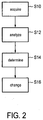
- Fig. 2 is a flow chart of a corresponding method.
- a transducer 12 acquires (S10) ultrasound images 14 from an object.
- the ultrasound images 14 are sent to a first user interface 16, which in this embodiment also comprises a display 18 for showing the ultrasound images 14.
- the first user interface 16 is adapted to allow a user to define a region of interest 20 in the images 14.
- the first user interface 16 could comprise a touch screen, where the user could identify the region of interest 20 by pointing at the target area.
- the system 10 preferably first acquires and displays at least one ultrasound image 14, such that the user can identify the region 20 within this image 14.
- the region identification unit 21 can be used to automatically determine a region of interest 20.
- the region identification unit 21 can comprise a first user interface which lets the user select an object from a selection of objects, e.g. different organs. The region identification unit 21 could then use template images or reference points of these organs in order to identify the organs within the ultrasound images 14.
- the automatically identified region of interest 20 can be shown to the user on display 18. The user can verify that the region 20 is placed correctly, e.g. by pushing an OK button, and the region 20 is subsequently used for determining a mean intensity value 24 within this region 20.
- the transducer 12 also sends ultrasound images 14b to an analyzer 22.
- the analyzer 22 analyzes (S12) the defined region 20 of the ultrasound images 14b for mean intensity values 24.
- the mean intensity values 24 and further quantitative characteristics of the time intensity curve such as the rise time and the area under the curve can be shown in real-time on a display 26. This has the advantage that the user can obtain quantitative values of the region of interest 20 while he is performing the exam.
- the mean intensity values 24 are processed by a processor 28 in order to determine in real time when the mean intensity value 24 has reached a peak 38. It should be noted that ultrasound images 14 are noisy and hence also the mean intensity values 24 are assumed to be noisy. Therefore, as will be described below, different algorithms can be used in order to identify a true peak 38 of the mean intensity values 24, which is not just a local outlier.
- the processor 28 controls the display 26 to show that the peak 38 has been detected.
- the detection of the peak 38 is performed by comparing the mean intensity values 24 of recently acquired images 14 with the mean intensity values 24 of previously acquired images 14. For example, it could compare the average mean intensity I3 of the last three acquired images with the average of the mean intensity I12 of the last 12 acquired images and the average of the mean intensity I6 of the last six acquired images and determine (S14) a peak if I6 is significantly higher than I12 and I3.
- the slope changes are tracked over time.
- the change in mean echo intensity of the region of interest, from image 14 to image 14 could be stored to track changes in slope from a positive trend (increasing mean intensity value 24) to a negative trend (decreasing mean intensity value 24). This is quite a common technique for detecting changes in polarity/direction/sign, etc.
- an industry standard curve fitting algorithm (as is currently done, off-line/non-real-time, in quantification software such as the Philips QLAB ultrasound quantification software) is applied to determine the peak intensity as each image 14 arrives, in real-time.
- the curve fitting algorithms usually require mean intensity values and time vectors. Presumably, during the wash-in phase, the peak intensity frame 38 will change continuously until the wash-out phase when the mean intensity value 24 will start to decrease.
- "local" peaks are determined, in particular as an adaptation to an existing algorithm used in QLAB to determine peak frames in a subset.
- the general trend for mean echo intensity is to increase; however, in any subset of frames (3, 5, or 7 for example) the mean intensity value 24 may rise or fall from image 14 to image 14.
- a trend of the mean intensity value 24 can be stored and compared dynamically as each new image 14 arrives. When the trend is decreasing, it can be deduced that the wash-out phase 52 has begun.
- the display 26 can show the time to peak.
- the time to peak could either be computed as the time between the beginning of the acquisition of ultrasound images 14 and the detection of the peak 38 or, preferably, by showing the time between a start time that was indicated by the user and the time of the peak detection.
- the second user interface 32 comprises a button 34.
- the button 34 By pressing the button 34 the user can indicate that the time to peak should be determined relative to the time of pushing the button.
- the user could press the button when he administers the contrast agent into a patient.
- the administration of the contrast agent can be performed by injection or by swallowing or by inhalation. It can be performed either by a physician, a medical assistant, a person of ordinary skill or the patient himself.
- the button 34 of the second user interface 32 can also be pressed by the patient himself or by another person who is operating the ultrasound imaging system 10.
- the transducer 12 acquires the ultrasound images 14 based on a number of acquisition parameters.
- the processor 28 is configured to change (S16) these parameters based on when it has detected a peak intensity. For example, it can reduce (S16) the storage frame rate at which the ultrasound images 14c are stored in storage 36 a few seconds after the peak 38 was detected (S14).
- the rate at which the transducer 12 acquires ultrasound images 14 is not necessarily identical to the rate at which images 14c are stored in the storage 36, the rate at which images 14a are shown on the display 18 of the first user interface 16 or the rate at which images 14b are used for computing (S12) a mean intensity value 24.
- the rate at which the images 14 are acquired (S10) is identical to the rate at which images 14a, 14b are used for displaying and for determining a mean intensity value.
- the storage rate 46, 48 can be lower than the acquisition rate. In particular, a certain time, for example ten seconds, after the peak has been determined the storage rate can be reduced because it is assumed that these images are less informative about the imaged object.
- the intensities keep decreasing and after some time the acquired images comprise no more additional useful information.
- the time until the wash-out of the contrast agent is completed depends, among other things, on the object that is being imaged.
- the second user interface 32 comprises a selection unit 35 that lets the user choose one of several objects, e.g. different organs such as liver, heart, gall bladder, etc.
- the processor 28 can control the transducer 12 to stop acquiring ultrasound images 14 a pre-determined time after the peak (this is not shown in the figures). For example, for liver imaging 90 seconds after the peak intensity is reached, typically there are no more relevant changes happening in the intensity in the region of interest 20.
- the processor 28 could stop the acquisition 90 seconds after the peak intensity. This further simplifies acquisition workflow (the user does not need to physically press a button to end image capture) and also ensures an optimized size of the files to be exported for post-acquisition analysis.
- the processor 28 can indicate the end of the image acquisition to the user by controlling loudspeaker 30 to generate an acoustic signal.
- the storage 36 is not necessarily located in the same device as the reminder of the ultrasound imaging system.
- the storage could be a network attached storage that is located in a separate room.
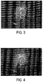
- Fig. 3 and Fig. 4 show examples of ultrasound images 14 that are acquired with an ultrasound imaging system according to the present invention.
- Fig. 3 shows a rectangular region of interest 20
- Fig. 4 shows a circular region of interest 20. Any other shape of the region 20 where the mean intensity values 24 are determined is also conceivable.
- the region 20 may also correspond to the complete ultrasound image 14.
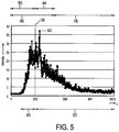
- Fig. 5 shows an example of a time intensity curve that is obtained by plotting the mean intensity values of the subsequently acquired ultrasound images 14.
- the area from the beginning of intensity enhancement to the peak intensity frame 38 is called washed-in region 40.
- the peak intensity frame 38 does not necessarily correspond to the image 14 that has the highest mean intensity value in the region of interest 20.
- the maximum intensity frame 42 can occur at a different location than the peak intensity frame 38 either because the real time detection of the peak intensity was false or because the maximum intensity is caused by a local outlier. Because of noisy data it might be unclear where the true peak of the curve is located. In such cases an experienced physician might want to form his own opinion and it is important that the area around the assumed peak intensity frame 38 is acquired and stored with a high frame rate. From the beginning of the image acquisition until a predetermined time 44 after the peak intensity frame 38 the ultrasound images 14 are acquired with a high storage frame rate 46. Afterwards the images are still acquired at the same acquisition frame rate but stored in the storage at a reduced storage frame rate 48.
- the processor 28 can control the display 26 to show the time to peak 50 and the wash-in time 40.
- the processor 28 determines a second peak intensity frame 38 also after it has already determined a first peak intensity frame. This way, if an image has been falsely identified as peak intensity frame 38, the processor can determine a second peak intensity frame 38, which is then assumed to be the true peak intensity frame 38. In this case the display values of time to peak 50 and wash-in time 40 on the display 26 are updated.
- the period after the peak intensity 38 is called wash-out region 52.
- the processor 28 can control the transducer 12 to stop the acquisition of ultrasound images 14 either when the mean intensity values 24 are below a predefined threshold or, as previously described, when an object-dependent predetermined time after the acquisition of the peak intensity frame 38 has passed.
- the proposed invention allows real-time detection of a peak of mean intensity in an ultrasound image and changing of an acquisition parameter once a peak has been determined.
- the acquisition parameter that is changed after peak detection depends on the concrete application.
- the acquisition parameter storage frame rate can be reduced after detection of a peak of mean intensity.
- the mean intensities could increase when some part of an organ of a person has moved into the field of view of the ultrasound device. In this case, an acquisition and storage frame rate could be increased after a detection of a peak of mean intensities.
- Other acquisition parameters that could be changed based on a peak intensity detection include, but are not limited to, depth of an image, the location of the transmit focus, the number of focal zones, whether to use the B mode or color Doppler mode, and whether harmonic or fundamental frequencies are to be used for imaging.
- a computer program may be stored / distributed on a suitable non-transitory medium, such as an optical storage medium or a solid-state medium supplied together with or as part of other hardware, but may also be distributed in other forms, such as via the Internet or other wired or wireless telecommunication systems.
- a suitable non-transitory medium such as an optical storage medium or a solid-state medium supplied together with or as part of other hardware, but may also be distributed in other forms, such as via the Internet or other wired or wireless telecommunication systems.
Landscapes
- Health & Medical Sciences (AREA)
- Life Sciences & Earth Sciences (AREA)
- Engineering & Computer Science (AREA)
- Medical Informatics (AREA)
- General Health & Medical Sciences (AREA)
- Nuclear Medicine, Radiotherapy & Molecular Imaging (AREA)
- Physics & Mathematics (AREA)
- Radiology & Medical Imaging (AREA)
- Pathology (AREA)
- Public Health (AREA)
- Biomedical Technology (AREA)
- Molecular Biology (AREA)
- Surgery (AREA)
- Animal Behavior & Ethology (AREA)
- Biophysics (AREA)
- Heart & Thoracic Surgery (AREA)
- Veterinary Medicine (AREA)
- Hematology (AREA)
- Quality & Reliability (AREA)
- Computer Vision & Pattern Recognition (AREA)
- General Physics & Mathematics (AREA)
- Theoretical Computer Science (AREA)
- Ultra Sonic Daignosis Equipment (AREA)
Applications Claiming Priority (2)
| Application Number | Priority Date | Filing Date | Title |
|---|---|---|---|
| US42276410P | 2010-12-14 | 2010-12-14 | |
| PCT/IB2011/055513 WO2012080905A1 (en) | 2010-12-14 | 2011-12-07 | Ultrasound imaging system and method with peak intensity detection |
Publications (2)
| Publication Number | Publication Date |
|---|---|
| EP2651305A1 EP2651305A1 (en) | 2013-10-23 |
| EP2651305B1 true EP2651305B1 (en) | 2016-03-16 |
Family
ID=45390140
Family Applications (1)
| Application Number | Title | Priority Date | Filing Date |
|---|---|---|---|
| EP11799497.0A Active EP2651305B1 (en) | 2010-12-14 | 2011-12-07 | Ultrasound imaging system and method with peak intensity detection |
Country Status (5)
| Country | Link |
|---|---|
| US (1) | US9058649B2 (enExample) |
| EP (1) | EP2651305B1 (enExample) |
| JP (1) | JP5972279B2 (enExample) |
| CN (1) | CN103260526B (enExample) |
| WO (1) | WO2012080905A1 (enExample) |
Families Citing this family (15)
| Publication number | Priority date | Publication date | Assignee | Title |
|---|---|---|---|---|
| US9529080B2 (en) | 2012-12-06 | 2016-12-27 | White Eagle Sonic Technologies, Inc. | System and apparatus having an application programming interface for flexible control of execution ultrasound actions |
| US10076313B2 (en) * | 2012-12-06 | 2018-09-18 | White Eagle Sonic Technologies, Inc. | System and method for automatically adjusting beams to scan an object in a body |
| US9530398B2 (en) | 2012-12-06 | 2016-12-27 | White Eagle Sonic Technologies, Inc. | Method for adaptively scheduling ultrasound system actions |
| US9983905B2 (en) | 2012-12-06 | 2018-05-29 | White Eagle Sonic Technologies, Inc. | Apparatus and system for real-time execution of ultrasound system actions |
| US10499884B2 (en) | 2012-12-06 | 2019-12-10 | White Eagle Sonic Technologies, Inc. | System and method for scanning for a second object within a first object using an adaptive scheduler |
| US10154826B2 (en) | 2013-07-17 | 2018-12-18 | Tissue Differentiation Intelligence, Llc | Device and method for identifying anatomical structures |
| US10716536B2 (en) | 2013-07-17 | 2020-07-21 | Tissue Differentiation Intelligence, Llc | Identifying anatomical structures |
| CN103886576B (zh) * | 2013-11-22 | 2017-06-06 | 沈阳东软医疗系统有限公司 | 一种腺体组织特征灰度检测方法及装置 |
| CN105939671B (zh) * | 2014-01-28 | 2019-06-04 | 皇家飞利浦有限公司 | 用于利用单-或双-平面实时成像的多平面采集的超声系统以及其操作的方法 |
| US11986341B1 (en) | 2016-05-26 | 2024-05-21 | Tissue Differentiation Intelligence, Llc | Methods for accessing spinal column using B-mode imaging to determine a trajectory without penetrating the the patient's anatomy |
| US11701086B1 (en) | 2016-06-21 | 2023-07-18 | Tissue Differentiation Intelligence, Llc | Methods and systems for improved nerve detection |
| GB201614950D0 (en) * | 2016-09-02 | 2016-10-19 | Ntnu Tech Transfer As | Enhanced-resolution ultrasound imaging of fluid paths |
| US10588596B2 (en) | 2017-03-14 | 2020-03-17 | Clarius Mobile Health Corp. | Systems and methods for detecting and enhancing viewing of a needle during ultrasound imaging |
| CN108158607A (zh) * | 2017-12-28 | 2018-06-15 | 深圳开立生物医疗科技股份有限公司 | 一种超声造影检查提示方法、装置及超声设备 |
| US11030742B2 (en) * | 2019-03-29 | 2021-06-08 | GE Precision Healthcare LLC | Systems and methods to facilitate review of liver tumor cases |
Family Cites Families (24)
| Publication number | Priority date | Publication date | Assignee | Title |
|---|---|---|---|---|
| US3909521A (en) * | 1972-03-06 | 1975-09-30 | Spectrotherm Corp | Infrared imaging system |
| US3798366A (en) * | 1972-03-06 | 1974-03-19 | R Winkler | Infrared imaging system |
| US4504908A (en) * | 1982-03-15 | 1985-03-12 | General Electric Company | Matched filter for X-ray temporal subtraction |
| US4888795A (en) * | 1987-06-30 | 1989-12-19 | Nec Corporation | Videotelephone apparatus for transmitting high and low resolution video signals over telephone exchange lines |
| US5119195A (en) * | 1991-01-31 | 1992-06-02 | Thomson Consumer Electronics, Inc. | Video noise reduction system employing plural frequency bands |
| US5224141A (en) * | 1991-02-06 | 1993-06-29 | General Electric Company | Fluoroscopic method with reduced x-ray dosage |
| US5313948A (en) * | 1991-11-28 | 1994-05-24 | Aloka Co., Ltd. | Ultrasonic diagnostic apparatus |
| US5743266A (en) * | 1995-04-25 | 1998-04-28 | Molecular Biosystems, Inc. | Method for processing real-time contrast enhanced ultrasonic images |
| JP3683945B2 (ja) * | 1995-07-13 | 2005-08-17 | 株式会社東芝 | 超音波診断装置 |
| JPH11137552A (ja) * | 1997-11-13 | 1999-05-25 | Ge Yokogawa Medical Systems Ltd | 造影画像表示方法および装置並びに医用画像装置 |
| US6385332B1 (en) | 1999-02-19 | 2002-05-07 | The John P. Roberts Research Institute | Automated segmentation method for 3-dimensional ultrasound |
| JP2003339701A (ja) * | 2002-03-19 | 2003-12-02 | Fuji Photo Film Co Ltd | 超音波診断装置 |
| US7596277B2 (en) * | 2002-04-09 | 2009-09-29 | Senthil Govindaswamy | Apparatus and method for detecting error in a digital image |
| JP3748848B2 (ja) * | 2002-11-11 | 2006-02-22 | ジーイー・メディカル・システムズ・グローバル・テクノロジー・カンパニー・エルエルシー | 超音波診断装置 |
| US7780602B2 (en) * | 2004-12-27 | 2010-08-24 | General Electric Company | Method and system for controlling an ultrasound system |
| US8057408B2 (en) * | 2005-09-22 | 2011-11-15 | The Regents Of The University Of Michigan | Pulsed cavitational ultrasound therapy |
| US20070083120A1 (en) * | 2005-09-22 | 2007-04-12 | Cain Charles A | Pulsed cavitational ultrasound therapy |
| US7486304B2 (en) * | 2005-12-21 | 2009-02-03 | Nokia Corporation | Display device with dynamic color gamut |
| JP2007197403A (ja) * | 2006-01-30 | 2007-08-09 | Hitachi Ltd | 薬物キャリアー及び超音波装置 |
| KR100827237B1 (ko) * | 2006-08-10 | 2008-05-07 | 삼성전기주식회사 | 다색 광원의 전력 제어를 지원하는 영상 표시 장치 및 방법 |
| JP2009100971A (ja) | 2007-10-24 | 2009-05-14 | Ge Medical Systems Global Technology Co Llc | 超音波撮像装置 |
| CN101951839B (zh) * | 2008-01-23 | 2013-11-06 | M·阿韦基乌 | 利用超声造影剂的呼吸门控治疗评估 |
| JP5395396B2 (ja) * | 2008-10-15 | 2014-01-22 | 株式会社東芝 | 超音波診断装置、医用画像処理装置、及び医用画像処理プログラム |
| US9288428B2 (en) * | 2010-05-11 | 2016-03-15 | Olympus Corporation | Shooting apparatus and method for controlling shooting apparatus |
-
2011
- 2011-12-07 CN CN201180059889.5A patent/CN103260526B/zh active Active
- 2011-12-07 US US13/885,406 patent/US9058649B2/en active Active
- 2011-12-07 WO PCT/IB2011/055513 patent/WO2012080905A1/en not_active Ceased
- 2011-12-07 EP EP11799497.0A patent/EP2651305B1/en active Active
- 2011-12-07 JP JP2013543924A patent/JP5972279B2/ja active Active
Also Published As
| Publication number | Publication date |
|---|---|
| JP2013545574A (ja) | 2013-12-26 |
| US20130251221A1 (en) | 2013-09-26 |
| US9058649B2 (en) | 2015-06-16 |
| EP2651305A1 (en) | 2013-10-23 |
| CN103260526B (zh) | 2015-09-16 |
| JP5972279B2 (ja) | 2016-08-17 |
| CN103260526A (zh) | 2013-08-21 |
| WO2012080905A1 (en) | 2012-06-21 |
Similar Documents
| Publication | Publication Date | Title |
|---|---|---|
| EP2651305B1 (en) | Ultrasound imaging system and method with peak intensity detection | |
| US11191518B2 (en) | Ultrasound system and method for detecting lung sliding | |
| CN114246611B (zh) | 用于超声成像系统的自适应接口的系统和方法 | |
| CN110325119B (zh) | 卵巢卵泡计数和大小确定 | |
| CN112971844B (zh) | 一种超声图像的采集质量的评估方法及超声成像设备 | |
| CN110786880B (zh) | 超声波诊断装置以及超声波图像处理方法 | |
| US11607200B2 (en) | Methods and system for camera-aided ultrasound scan setup and control | |
| CN106659473B (zh) | 超声成像装置 | |
| US9489921B2 (en) | Method and apparatus for displaying plurality of different images of object | |
| US20160120491A1 (en) | Image processing apparatus and storage medium | |
| US10470744B2 (en) | Ultrasound diagnosis apparatus, ultrasound diagnosis method performed by the ultrasound diagnosis apparatus, and computer-readable storage medium having the ultrasound diagnosis method recorded thereon | |
| WO2011041244A1 (en) | Contrast-enhanced ultrasound assessment of liver blood flow for monitoring liver therapy | |
| JP7346266B2 (ja) | 超音波撮像システムおよび対象物体品質レベルを表示するための方法 | |
| EP3143939B1 (en) | Ultrasound apparatus and method of obtaining information from contrast image | |
| JP2020503099A (ja) | 出産前超音波イメージング | |
| EP2390839A1 (en) | 3D ultrasound apparatus and method for operating the same | |
| TWI788629B (zh) | 圖像處理方法、裝置及系統、電子設備及電腦可讀儲存媒體 | |
| CN113040823B (zh) | 一种超声成像设备及超声图像的分析方法 | |
| US20180042578A1 (en) | Automated ultrasound image measurement system and method | |
| KR20120102447A (ko) | 진단장치 및 방법 | |
| US20210192718A1 (en) | Methods and systems for automatic measurement of strains and strain-ratio calculation for sonoelastography | |
| CN105451662A (zh) | 用于医学成像与信息显示的方法和系统 | |
| JP2023077810A (ja) | 超音波画像分析装置、超音波診断装置および超音波画像分析装置の制御方法 | |
| JP2025531104A (ja) | 超音波剪断波エラストグラフィを使用して剛性測定を実行するための方法及びシステム | |
| CN116416193A (zh) | 超声图像处理方法、装置、设备及可读存储介质 |
Legal Events
| Date | Code | Title | Description |
|---|---|---|---|
| PUAI | Public reference made under article 153(3) epc to a published international application that has entered the european phase |
Free format text: ORIGINAL CODE: 0009012 |
|
| 17P | Request for examination filed |
Effective date: 20130715 |
|
| AK | Designated contracting states |
Kind code of ref document: A1 Designated state(s): AL AT BE BG CH CY CZ DE DK EE ES FI FR GB GR HR HU IE IS IT LI LT LU LV MC MK MT NL NO PL PT RO RS SE SI SK SM TR |
|
| DAX | Request for extension of the european patent (deleted) | ||
| GRAP | Despatch of communication of intention to grant a patent |
Free format text: ORIGINAL CODE: EPIDOSNIGR1 |
|
| RIC1 | Information provided on ipc code assigned before grant |
Ipc: A61B 8/00 20060101AFI20150615BHEP Ipc: A61B 8/08 20060101ALI20150615BHEP Ipc: G06T 7/00 20060101ALI20150615BHEP |
|
| INTG | Intention to grant announced |
Effective date: 20150702 |
|
| GRAR | Information related to intention to grant a patent recorded |
Free format text: ORIGINAL CODE: EPIDOSNIGR71 |
|
| GRAS | Grant fee paid |
Free format text: ORIGINAL CODE: EPIDOSNIGR3 |
|
| INTG | Intention to grant announced |
Effective date: 20151112 |
|
| GRAA | (expected) grant |
Free format text: ORIGINAL CODE: 0009210 |
|
| AK | Designated contracting states |
Kind code of ref document: B1 Designated state(s): AL AT BE BG CH CY CZ DE DK EE ES FI FR GB GR HR HU IE IS IT LI LT LU LV MC MK MT NL NO PL PT RO RS SE SI SK SM TR |
|
| REG | Reference to a national code |
Ref country code: GB Ref legal event code: FG4D |
|
| REG | Reference to a national code |
Ref country code: CH Ref legal event code: EP |
|
| REG | Reference to a national code |
Ref country code: IE Ref legal event code: FG4D |
|
| REG | Reference to a national code |
Ref country code: AT Ref legal event code: REF Ref document number: 780471 Country of ref document: AT Kind code of ref document: T Effective date: 20160415 |
|
| REG | Reference to a national code |
Ref country code: DE Ref legal event code: R096 Ref document number: 602011024133 Country of ref document: DE |
|
| REG | Reference to a national code |
Ref country code: DE Ref legal event code: R084 Ref document number: 602011024133 Country of ref document: DE |
|
| REG | Reference to a national code |
Ref country code: NL Ref legal event code: MP Effective date: 20160316 |
|
| REG | Reference to a national code |
Ref country code: LT Ref legal event code: MG4D |
|
| PG25 | Lapsed in a contracting state [announced via postgrant information from national office to epo] |
Ref country code: FI Free format text: LAPSE BECAUSE OF FAILURE TO SUBMIT A TRANSLATION OF THE DESCRIPTION OR TO PAY THE FEE WITHIN THE PRESCRIBED TIME-LIMIT Effective date: 20160316 Ref country code: HR Free format text: LAPSE BECAUSE OF FAILURE TO SUBMIT A TRANSLATION OF THE DESCRIPTION OR TO PAY THE FEE WITHIN THE PRESCRIBED TIME-LIMIT Effective date: 20160316 Ref country code: NO Free format text: LAPSE BECAUSE OF FAILURE TO SUBMIT A TRANSLATION OF THE DESCRIPTION OR TO PAY THE FEE WITHIN THE PRESCRIBED TIME-LIMIT Effective date: 20160616 Ref country code: GR Free format text: LAPSE BECAUSE OF FAILURE TO SUBMIT A TRANSLATION OF THE DESCRIPTION OR TO PAY THE FEE WITHIN THE PRESCRIBED TIME-LIMIT Effective date: 20160617 |
|
| REG | Reference to a national code |
Ref country code: AT Ref legal event code: MK05 Ref document number: 780471 Country of ref document: AT Kind code of ref document: T Effective date: 20160316 |
|
| PG25 | Lapsed in a contracting state [announced via postgrant information from national office to epo] |
Ref country code: SE Free format text: LAPSE BECAUSE OF FAILURE TO SUBMIT A TRANSLATION OF THE DESCRIPTION OR TO PAY THE FEE WITHIN THE PRESCRIBED TIME-LIMIT Effective date: 20160316 Ref country code: RS Free format text: LAPSE BECAUSE OF FAILURE TO SUBMIT A TRANSLATION OF THE DESCRIPTION OR TO PAY THE FEE WITHIN THE PRESCRIBED TIME-LIMIT Effective date: 20160316 Ref country code: LV Free format text: LAPSE BECAUSE OF FAILURE TO SUBMIT A TRANSLATION OF THE DESCRIPTION OR TO PAY THE FEE WITHIN THE PRESCRIBED TIME-LIMIT Effective date: 20160316 Ref country code: NL Free format text: LAPSE BECAUSE OF FAILURE TO SUBMIT A TRANSLATION OF THE DESCRIPTION OR TO PAY THE FEE WITHIN THE PRESCRIBED TIME-LIMIT Effective date: 20160316 Ref country code: LT Free format text: LAPSE BECAUSE OF FAILURE TO SUBMIT A TRANSLATION OF THE DESCRIPTION OR TO PAY THE FEE WITHIN THE PRESCRIBED TIME-LIMIT Effective date: 20160316 |
|
| PG25 | Lapsed in a contracting state [announced via postgrant information from national office to epo] |
Ref country code: IS Free format text: LAPSE BECAUSE OF FAILURE TO SUBMIT A TRANSLATION OF THE DESCRIPTION OR TO PAY THE FEE WITHIN THE PRESCRIBED TIME-LIMIT Effective date: 20160716 Ref country code: EE Free format text: LAPSE BECAUSE OF FAILURE TO SUBMIT A TRANSLATION OF THE DESCRIPTION OR TO PAY THE FEE WITHIN THE PRESCRIBED TIME-LIMIT Effective date: 20160316 Ref country code: PL Free format text: LAPSE BECAUSE OF FAILURE TO SUBMIT A TRANSLATION OF THE DESCRIPTION OR TO PAY THE FEE WITHIN THE PRESCRIBED TIME-LIMIT Effective date: 20160316 |
|
| PG25 | Lapsed in a contracting state [announced via postgrant information from national office to epo] |
Ref country code: SM Free format text: LAPSE BECAUSE OF FAILURE TO SUBMIT A TRANSLATION OF THE DESCRIPTION OR TO PAY THE FEE WITHIN THE PRESCRIBED TIME-LIMIT Effective date: 20160316 Ref country code: SK Free format text: LAPSE BECAUSE OF FAILURE TO SUBMIT A TRANSLATION OF THE DESCRIPTION OR TO PAY THE FEE WITHIN THE PRESCRIBED TIME-LIMIT Effective date: 20160316 Ref country code: CZ Free format text: LAPSE BECAUSE OF FAILURE TO SUBMIT A TRANSLATION OF THE DESCRIPTION OR TO PAY THE FEE WITHIN THE PRESCRIBED TIME-LIMIT Effective date: 20160316 Ref country code: PT Free format text: LAPSE BECAUSE OF FAILURE TO SUBMIT A TRANSLATION OF THE DESCRIPTION OR TO PAY THE FEE WITHIN THE PRESCRIBED TIME-LIMIT Effective date: 20160718 Ref country code: ES Free format text: LAPSE BECAUSE OF FAILURE TO SUBMIT A TRANSLATION OF THE DESCRIPTION OR TO PAY THE FEE WITHIN THE PRESCRIBED TIME-LIMIT Effective date: 20160316 Ref country code: RO Free format text: LAPSE BECAUSE OF FAILURE TO SUBMIT A TRANSLATION OF THE DESCRIPTION OR TO PAY THE FEE WITHIN THE PRESCRIBED TIME-LIMIT Effective date: 20160316 Ref country code: AT Free format text: LAPSE BECAUSE OF FAILURE TO SUBMIT A TRANSLATION OF THE DESCRIPTION OR TO PAY THE FEE WITHIN THE PRESCRIBED TIME-LIMIT Effective date: 20160316 |
|
| REG | Reference to a national code |
Ref country code: DE Ref legal event code: R097 Ref document number: 602011024133 Country of ref document: DE |
|
| REG | Reference to a national code |
Ref country code: FR Ref legal event code: PLFP Year of fee payment: 6 |
|
| PG25 | Lapsed in a contracting state [announced via postgrant information from national office to epo] |
Ref country code: BE Free format text: LAPSE BECAUSE OF FAILURE TO SUBMIT A TRANSLATION OF THE DESCRIPTION OR TO PAY THE FEE WITHIN THE PRESCRIBED TIME-LIMIT Effective date: 20160316 |
|
| PLBE | No opposition filed within time limit |
Free format text: ORIGINAL CODE: 0009261 |
|
| STAA | Information on the status of an ep patent application or granted ep patent |
Free format text: STATUS: NO OPPOSITION FILED WITHIN TIME LIMIT |
|
| PG25 | Lapsed in a contracting state [announced via postgrant information from national office to epo] |
Ref country code: DK Free format text: LAPSE BECAUSE OF FAILURE TO SUBMIT A TRANSLATION OF THE DESCRIPTION OR TO PAY THE FEE WITHIN THE PRESCRIBED TIME-LIMIT Effective date: 20160316 |
|
| 26N | No opposition filed |
Effective date: 20161219 |
|
| PG25 | Lapsed in a contracting state [announced via postgrant information from national office to epo] |
Ref country code: BG Free format text: LAPSE BECAUSE OF FAILURE TO SUBMIT A TRANSLATION OF THE DESCRIPTION OR TO PAY THE FEE WITHIN THE PRESCRIBED TIME-LIMIT Effective date: 20160616 |
|
| PG25 | Lapsed in a contracting state [announced via postgrant information from national office to epo] |
Ref country code: SI Free format text: LAPSE BECAUSE OF FAILURE TO SUBMIT A TRANSLATION OF THE DESCRIPTION OR TO PAY THE FEE WITHIN THE PRESCRIBED TIME-LIMIT Effective date: 20160316 |
|
| REG | Reference to a national code |
Ref country code: CH Ref legal event code: PL |
|
| GBPC | Gb: european patent ceased through non-payment of renewal fee |
Effective date: 20161207 |
|
| PG25 | Lapsed in a contracting state [announced via postgrant information from national office to epo] |
Ref country code: MC Free format text: LAPSE BECAUSE OF FAILURE TO SUBMIT A TRANSLATION OF THE DESCRIPTION OR TO PAY THE FEE WITHIN THE PRESCRIBED TIME-LIMIT Effective date: 20160316 |
|
| REG | Reference to a national code |
Ref country code: IE Ref legal event code: MM4A |
|
| PG25 | Lapsed in a contracting state [announced via postgrant information from national office to epo] |
Ref country code: CH Free format text: LAPSE BECAUSE OF NON-PAYMENT OF DUE FEES Effective date: 20161231 Ref country code: LU Free format text: LAPSE BECAUSE OF NON-PAYMENT OF DUE FEES Effective date: 20161207 Ref country code: LI Free format text: LAPSE BECAUSE OF NON-PAYMENT OF DUE FEES Effective date: 20161231 |
|
| PG25 | Lapsed in a contracting state [announced via postgrant information from national office to epo] |
Ref country code: GB Free format text: LAPSE BECAUSE OF NON-PAYMENT OF DUE FEES Effective date: 20161207 Ref country code: IE Free format text: LAPSE BECAUSE OF NON-PAYMENT OF DUE FEES Effective date: 20161207 |
|
| REG | Reference to a national code |
Ref country code: FR Ref legal event code: PLFP Year of fee payment: 7 |
|
| PG25 | Lapsed in a contracting state [announced via postgrant information from national office to epo] |
Ref country code: CY Free format text: LAPSE BECAUSE OF FAILURE TO SUBMIT A TRANSLATION OF THE DESCRIPTION OR TO PAY THE FEE WITHIN THE PRESCRIBED TIME-LIMIT Effective date: 20160316 Ref country code: HU Free format text: LAPSE BECAUSE OF FAILURE TO SUBMIT A TRANSLATION OF THE DESCRIPTION OR TO PAY THE FEE WITHIN THE PRESCRIBED TIME-LIMIT; INVALID AB INITIO Effective date: 20111207 |
|
| PG25 | Lapsed in a contracting state [announced via postgrant information from national office to epo] |
Ref country code: MK Free format text: LAPSE BECAUSE OF FAILURE TO SUBMIT A TRANSLATION OF THE DESCRIPTION OR TO PAY THE FEE WITHIN THE PRESCRIBED TIME-LIMIT Effective date: 20160316 |
|
| PG25 | Lapsed in a contracting state [announced via postgrant information from national office to epo] |
Ref country code: MT Free format text: LAPSE BECAUSE OF NON-PAYMENT OF DUE FEES Effective date: 20161207 |
|
| PG25 | Lapsed in a contracting state [announced via postgrant information from national office to epo] |
Ref country code: TR Free format text: LAPSE BECAUSE OF FAILURE TO SUBMIT A TRANSLATION OF THE DESCRIPTION OR TO PAY THE FEE WITHIN THE PRESCRIBED TIME-LIMIT Effective date: 20160316 Ref country code: AL Free format text: LAPSE BECAUSE OF FAILURE TO SUBMIT A TRANSLATION OF THE DESCRIPTION OR TO PAY THE FEE WITHIN THE PRESCRIBED TIME-LIMIT Effective date: 20160316 |
|
| PGFP | Annual fee paid to national office [announced via postgrant information from national office to epo] |
Ref country code: IT Payment date: 20231221 Year of fee payment: 13 Ref country code: FR Payment date: 20231226 Year of fee payment: 13 |
|
| PGFP | Annual fee paid to national office [announced via postgrant information from national office to epo] |
Ref country code: DE Payment date: 20241227 Year of fee payment: 14 |
|
| PG25 | Lapsed in a contracting state [announced via postgrant information from national office to epo] |
Ref country code: IT Free format text: LAPSE BECAUSE OF NON-PAYMENT OF DUE FEES Effective date: 20241207 |
|
| PG25 | Lapsed in a contracting state [announced via postgrant information from national office to epo] |
Ref country code: FR Free format text: LAPSE BECAUSE OF NON-PAYMENT OF DUE FEES Effective date: 20241231 |