EP2636731B1 - Verfahren zur herstellung von darmzellen - Google Patents
Verfahren zur herstellung von darmzellen Download PDFInfo
- Publication number
- EP2636731B1 EP2636731B1 EP11837965.0A EP11837965A EP2636731B1 EP 2636731 B1 EP2636731 B1 EP 2636731B1 EP 11837965 A EP11837965 A EP 11837965A EP 2636731 B1 EP2636731 B1 EP 2636731B1
- Authority
- EP
- European Patent Office
- Prior art keywords
- cells
- intestinal
- differentiation
- pluripotent stem
- stem cells
- Prior art date
- Legal status (The legal status is an assumption and is not a legal conclusion. Google has not performed a legal analysis and makes no representation as to the accuracy of the status listed.)
- Not-in-force
Links
Images


Classifications
-
- C—CHEMISTRY; METALLURGY
- C12—BIOCHEMISTRY; BEER; SPIRITS; WINE; VINEGAR; MICROBIOLOGY; ENZYMOLOGY; MUTATION OR GENETIC ENGINEERING
- C12N—MICROORGANISMS OR ENZYMES; COMPOSITIONS THEREOF; PROPAGATING, PRESERVING, OR MAINTAINING MICROORGANISMS; MUTATION OR GENETIC ENGINEERING; CULTURE MEDIA
- C12N5/00—Undifferentiated human, animal or plant cells, e.g. cell lines; Tissues; Cultivation or maintenance thereof; Culture media therefor
- C12N5/06—Animal cells or tissues; Human cells or tissues
- C12N5/0602—Vertebrate cells
- C12N5/0679—Cells of the gastro-intestinal tract
-
- C—CHEMISTRY; METALLURGY
- C12—BIOCHEMISTRY; BEER; SPIRITS; WINE; VINEGAR; MICROBIOLOGY; ENZYMOLOGY; MUTATION OR GENETIC ENGINEERING
- C12N—MICROORGANISMS OR ENZYMES; COMPOSITIONS THEREOF; PROPAGATING, PRESERVING, OR MAINTAINING MICROORGANISMS; MUTATION OR GENETIC ENGINEERING; CULTURE MEDIA
- C12N5/00—Undifferentiated human, animal or plant cells, e.g. cell lines; Tissues; Cultivation or maintenance thereof; Culture media therefor
-
- C—CHEMISTRY; METALLURGY
- C12—BIOCHEMISTRY; BEER; SPIRITS; WINE; VINEGAR; MICROBIOLOGY; ENZYMOLOGY; MUTATION OR GENETIC ENGINEERING
- C12Q—MEASURING OR TESTING PROCESSES INVOLVING ENZYMES, NUCLEIC ACIDS OR MICROORGANISMS; COMPOSITIONS OR TEST PAPERS THEREFOR; PROCESSES OF PREPARING SUCH COMPOSITIONS; CONDITION-RESPONSIVE CONTROL IN MICROBIOLOGICAL OR ENZYMOLOGICAL PROCESSES
- C12Q1/00—Measuring or testing processes involving enzymes, nucleic acids or microorganisms; Compositions therefor; Processes of preparing such compositions
- C12Q1/02—Measuring or testing processes involving enzymes, nucleic acids or microorganisms; Compositions therefor; Processes of preparing such compositions involving viable microorganisms
- C12Q1/04—Determining presence or kind of microorganism; Use of selective media for testing antibiotics or bacteriocides; Compositions containing a chemical indicator therefor
-
- C—CHEMISTRY; METALLURGY
- C12—BIOCHEMISTRY; BEER; SPIRITS; WINE; VINEGAR; MICROBIOLOGY; ENZYMOLOGY; MUTATION OR GENETIC ENGINEERING
- C12Q—MEASURING OR TESTING PROCESSES INVOLVING ENZYMES, NUCLEIC ACIDS OR MICROORGANISMS; COMPOSITIONS OR TEST PAPERS THEREFOR; PROCESSES OF PREPARING SUCH COMPOSITIONS; CONDITION-RESPONSIVE CONTROL IN MICROBIOLOGICAL OR ENZYMOLOGICAL PROCESSES
- C12Q1/00—Measuring or testing processes involving enzymes, nucleic acids or microorganisms; Compositions therefor; Processes of preparing such compositions
- C12Q1/68—Measuring or testing processes involving enzymes, nucleic acids or microorganisms; Compositions therefor; Processes of preparing such compositions involving nucleic acids
- C12Q1/6876—Nucleic acid products used in the analysis of nucleic acids, e.g. primers or probes
- C12Q1/6881—Nucleic acid products used in the analysis of nucleic acids, e.g. primers or probes for tissue or cell typing, e.g. human leukocyte antigen [HLA] probes
-
- G—PHYSICS
- G01—MEASURING; TESTING
- G01N—INVESTIGATING OR ANALYSING MATERIALS BY DETERMINING THEIR CHEMICAL OR PHYSICAL PROPERTIES
- G01N33/00—Investigating or analysing materials by specific methods not covered by groups G01N1/00 - G01N31/00
- G01N33/15—Medicinal preparations ; Physical properties thereof, e.g. dissolubility
-
- G—PHYSICS
- G01—MEASURING; TESTING
- G01N—INVESTIGATING OR ANALYSING MATERIALS BY DETERMINING THEIR CHEMICAL OR PHYSICAL PROPERTIES
- G01N33/00—Investigating or analysing materials by specific methods not covered by groups G01N1/00 - G01N31/00
- G01N33/48—Biological material, e.g. blood, urine; Haemocytometers
- G01N33/50—Chemical analysis of biological material, e.g. blood, urine; Testing involving biospecific ligand binding methods; Immunological testing
- G01N33/5005—Chemical analysis of biological material, e.g. blood, urine; Testing involving biospecific ligand binding methods; Immunological testing involving human or animal cells
- G01N33/5008—Chemical analysis of biological material, e.g. blood, urine; Testing involving biospecific ligand binding methods; Immunological testing involving human or animal cells for testing or evaluating the effect of chemical or biological compounds, e.g. drugs, cosmetics
- G01N33/5044—Chemical analysis of biological material, e.g. blood, urine; Testing involving biospecific ligand binding methods; Immunological testing involving human or animal cells for testing or evaluating the effect of chemical or biological compounds, e.g. drugs, cosmetics involving specific cell types
- G01N33/5064—Endothelial cells
-
- G—PHYSICS
- G01—MEASURING; TESTING
- G01N—INVESTIGATING OR ANALYSING MATERIALS BY DETERMINING THEIR CHEMICAL OR PHYSICAL PROPERTIES
- G01N33/00—Investigating or analysing materials by specific methods not covered by groups G01N1/00 - G01N31/00
- G01N33/48—Biological material, e.g. blood, urine; Haemocytometers
- G01N33/50—Chemical analysis of biological material, e.g. blood, urine; Testing involving biospecific ligand binding methods; Immunological testing
- G01N33/5005—Chemical analysis of biological material, e.g. blood, urine; Testing involving biospecific ligand binding methods; Immunological testing involving human or animal cells
- G01N33/5008—Chemical analysis of biological material, e.g. blood, urine; Testing involving biospecific ligand binding methods; Immunological testing involving human or animal cells for testing or evaluating the effect of chemical or biological compounds, e.g. drugs, cosmetics
- G01N33/5044—Chemical analysis of biological material, e.g. blood, urine; Testing involving biospecific ligand binding methods; Immunological testing involving human or animal cells for testing or evaluating the effect of chemical or biological compounds, e.g. drugs, cosmetics involving specific cell types
- G01N33/5073—Stem cells
-
- C—CHEMISTRY; METALLURGY
- C12—BIOCHEMISTRY; BEER; SPIRITS; WINE; VINEGAR; MICROBIOLOGY; ENZYMOLOGY; MUTATION OR GENETIC ENGINEERING
- C12N—MICROORGANISMS OR ENZYMES; COMPOSITIONS THEREOF; PROPAGATING, PRESERVING, OR MAINTAINING MICROORGANISMS; MUTATION OR GENETIC ENGINEERING; CULTURE MEDIA
- C12N2501/00—Active agents used in cell culture processes, e.g. differentation
- C12N2501/10—Growth factors
- C12N2501/115—Basic fibroblast growth factor (bFGF, FGF-2)
-
- C—CHEMISTRY; METALLURGY
- C12—BIOCHEMISTRY; BEER; SPIRITS; WINE; VINEGAR; MICROBIOLOGY; ENZYMOLOGY; MUTATION OR GENETIC ENGINEERING
- C12N—MICROORGANISMS OR ENZYMES; COMPOSITIONS THEREOF; PROPAGATING, PRESERVING, OR MAINTAINING MICROORGANISMS; MUTATION OR GENETIC ENGINEERING; CULTURE MEDIA
- C12N2501/00—Active agents used in cell culture processes, e.g. differentation
- C12N2501/10—Growth factors
- C12N2501/117—Keratinocyte growth factors (KGF-1, i.e. FGF-7; KGF-2, i.e. FGF-12)
-
- C—CHEMISTRY; METALLURGY
- C12—BIOCHEMISTRY; BEER; SPIRITS; WINE; VINEGAR; MICROBIOLOGY; ENZYMOLOGY; MUTATION OR GENETIC ENGINEERING
- C12N—MICROORGANISMS OR ENZYMES; COMPOSITIONS THEREOF; PROPAGATING, PRESERVING, OR MAINTAINING MICROORGANISMS; MUTATION OR GENETIC ENGINEERING; CULTURE MEDIA
- C12N2501/00—Active agents used in cell culture processes, e.g. differentation
- C12N2501/10—Growth factors
- C12N2501/119—Other fibroblast growth factors, e.g. FGF-4, FGF-8, FGF-10
-
- C—CHEMISTRY; METALLURGY
- C12—BIOCHEMISTRY; BEER; SPIRITS; WINE; VINEGAR; MICROBIOLOGY; ENZYMOLOGY; MUTATION OR GENETIC ENGINEERING
- C12N—MICROORGANISMS OR ENZYMES; COMPOSITIONS THEREOF; PROPAGATING, PRESERVING, OR MAINTAINING MICROORGANISMS; MUTATION OR GENETIC ENGINEERING; CULTURE MEDIA
- C12N2501/00—Active agents used in cell culture processes, e.g. differentation
- C12N2501/10—Growth factors
- C12N2501/16—Activin; Inhibin; Mullerian inhibiting substance
-
- C—CHEMISTRY; METALLURGY
- C12—BIOCHEMISTRY; BEER; SPIRITS; WINE; VINEGAR; MICROBIOLOGY; ENZYMOLOGY; MUTATION OR GENETIC ENGINEERING
- C12N—MICROORGANISMS OR ENZYMES; COMPOSITIONS THEREOF; PROPAGATING, PRESERVING, OR MAINTAINING MICROORGANISMS; MUTATION OR GENETIC ENGINEERING; CULTURE MEDIA
- C12N2501/00—Active agents used in cell culture processes, e.g. differentation
- C12N2501/40—Regulators of development
- C12N2501/415—Wnt; Frizzeled
-
- C—CHEMISTRY; METALLURGY
- C12—BIOCHEMISTRY; BEER; SPIRITS; WINE; VINEGAR; MICROBIOLOGY; ENZYMOLOGY; MUTATION OR GENETIC ENGINEERING
- C12N—MICROORGANISMS OR ENZYMES; COMPOSITIONS THEREOF; PROPAGATING, PRESERVING, OR MAINTAINING MICROORGANISMS; MUTATION OR GENETIC ENGINEERING; CULTURE MEDIA
- C12N2501/00—Active agents used in cell culture processes, e.g. differentation
- C12N2501/40—Regulators of development
- C12N2501/42—Notch; Delta; Jagged; Serrate
-
- C—CHEMISTRY; METALLURGY
- C12—BIOCHEMISTRY; BEER; SPIRITS; WINE; VINEGAR; MICROBIOLOGY; ENZYMOLOGY; MUTATION OR GENETIC ENGINEERING
- C12N—MICROORGANISMS OR ENZYMES; COMPOSITIONS THEREOF; PROPAGATING, PRESERVING, OR MAINTAINING MICROORGANISMS; MUTATION OR GENETIC ENGINEERING; CULTURE MEDIA
- C12N2501/00—Active agents used in cell culture processes, e.g. differentation
- C12N2501/999—Small molecules not provided for elsewhere
-
- C—CHEMISTRY; METALLURGY
- C12—BIOCHEMISTRY; BEER; SPIRITS; WINE; VINEGAR; MICROBIOLOGY; ENZYMOLOGY; MUTATION OR GENETIC ENGINEERING
- C12N—MICROORGANISMS OR ENZYMES; COMPOSITIONS THEREOF; PROPAGATING, PRESERVING, OR MAINTAINING MICROORGANISMS; MUTATION OR GENETIC ENGINEERING; CULTURE MEDIA
- C12N2502/00—Coculture with; Conditioned medium produced by
- C12N2502/13—Coculture with; Conditioned medium produced by connective tissue cells; generic mesenchyme cells, e.g. so-called "embryonic fibroblasts"
-
- C—CHEMISTRY; METALLURGY
- C12—BIOCHEMISTRY; BEER; SPIRITS; WINE; VINEGAR; MICROBIOLOGY; ENZYMOLOGY; MUTATION OR GENETIC ENGINEERING
- C12N—MICROORGANISMS OR ENZYMES; COMPOSITIONS THEREOF; PROPAGATING, PRESERVING, OR MAINTAINING MICROORGANISMS; MUTATION OR GENETIC ENGINEERING; CULTURE MEDIA
- C12N2506/00—Differentiation of animal cells from one lineage to another; Differentiation of pluripotent cells
- C12N2506/02—Differentiation of animal cells from one lineage to another; Differentiation of pluripotent cells from embryonic cells
-
- C—CHEMISTRY; METALLURGY
- C12—BIOCHEMISTRY; BEER; SPIRITS; WINE; VINEGAR; MICROBIOLOGY; ENZYMOLOGY; MUTATION OR GENETIC ENGINEERING
- C12Q—MEASURING OR TESTING PROCESSES INVOLVING ENZYMES, NUCLEIC ACIDS OR MICROORGANISMS; COMPOSITIONS OR TEST PAPERS THEREFOR; PROCESSES OF PREPARING SUCH COMPOSITIONS; CONDITION-RESPONSIVE CONTROL IN MICROBIOLOGICAL OR ENZYMOLOGICAL PROCESSES
- C12Q2600/00—Oligonucleotides characterized by their use
- C12Q2600/158—Expression markers
-
- G—PHYSICS
- G01—MEASURING; TESTING
- G01N—INVESTIGATING OR ANALYSING MATERIALS BY DETERMINING THEIR CHEMICAL OR PHYSICAL PROPERTIES
- G01N2500/00—Screening for compounds of potential therapeutic value
Definitions
- the present invention relates to a method of producing intestinal cells. More particularly, the present invention relates to a method of producing intestinal cells by use of pluripotent stem cells as a starting material.
- Pluripotent stem cells such as embryonic stem cells (ES cells) or induced pluripotent stem cells (iPS cells) are cells having a capability of differentiating into various cells, and they possess a capability of almost indefinitely proliferating.
- ES cells embryonic stem cells
- iPS cells induced pluripotent stem cells
- a method in which mesoderm-derived cells are used as feeder cells, and embryonic stem cells are cultured in the presence of said feeder cells to thereby induce differentiation of them into endodermal cells see WO2006/126574 .
- the patent document WO2006/126574 describes induction of differentiation thereof into mature cells of endoderm-derived organs such as liver, lung, and small intestine, but the disclosed method cannot efficiently differentiate the cells into various matured intestinal cells.
- ES cells are culture on a monolayer of M15 cells in vitro to thereby induce the ES cells sequentially into the mesendoderm, the definitive endoderm, and, finally, various organs derived from the regional-specific definitive endoderm, as they mimic in vivo induction of early embryos [see Shiraki, N., Umeda, K., Sakashita, N., Takeya, M., Kume, K. and Kume, S. (2008). Differentiation of mouse and human embryonic stem cells into hepatic lineages.
- An object of the present invention is to provide a method of producing intestinal cells by use of pluripotent stem cells as a starting material.
- the present inventors conducted extensive studies to solve the above-mentioned problem, and, as a result, the present inventors discovered that, after inducing differentiation of embryonic stem cells, which are pluripotent stem cells, into definitive endoderm cells, the definitive endoderm cells be cultured in the presence of BIO [(2'Z, 3'E)-6-bromoindirubin-3'-oxime] and DAPT [N-[(3,5-difluorophenyl)acetyl]-L-Ala-2-phenyl-L-Gly-tert-butyl-OH], whereby differentiation thereof into various mature intestinal cells can be induced.
- BIO (2'Z, 3'E)-6-bromoindirubin-3'-oxime]
- DAPT N-[(3,5-difluorophenyl)acetyl]-L-Ala-2-phenyl-L-Gly-tert-butyl-OH
- the present invention relates to the followings.
- various mature intestinal cells such as absorptive enterocytes of the intestine, Paneth cells, goblet cells and enteroendocrine cells, can be produced massively and efficiently from pluripotent stem cells.
- various mature intestinal cells can be produced, and the produced cells can be practically utilized in the field of regeneration medicine.
- the method of producing intestinal cells includes the steps of: (A) inducing differentiation of pluripotent stem cells into definitive endoderm cells; and (B) culturing the definitive endoderm cells in the presence of (2'Z, 3'E)-6-bromoindirubin-3'-oxime (BIO) and N-[(3,5-difluorophenyl)acetyl]-L-Ala-2-phenyl-L-Gly-tert-butyl-OH (DAPT) to thereby induce differentiation of the definitive endoderm cells into intestinal cells.
- (2'Z, 3'E)-6-bromoindirubin-3'-oxime BIO
- N-[(3,5-difluorophenyl)acetyl]-L-Ala-2-phenyl-L-Gly-tert-butyl-OH DAPT
- pluripotent stem cells means cells which have a capability of proliferating under artificially-created conditions such as in a test tube ( in vitro ) and which can differentiate into cells found in all the tissues of living bodies.
- embryonic stem cells or induced pluripotent stem cells are preferably used as the pluripotent stem cells, and embryonic stem cells are more preferably used.
- the embryonic stem (ES) cells described herein may be mammalian-derived ES cells, and the types thereof are not particularly limited.
- ES cells derived from a mouse, monkey, human, or the like can be used.
- cells into which a reporter gene is introduced in the vicinity of the Pdxl gene can be used in order to facilitate confirmation of the level of their differentiation.
- a 129/Sv-derived ES cell line in which the LacZ gene is introduced into the Pdxl locus, or a ES cell line SK7, having the GFP reporter transgene under the control of the Pdxl promoter can be used.
- a ES cell line PH3 having the mRFP1 reporter transgene under the control of the Hnf3 ⁇ -endoderm-specific-enhancer fragment and having the GFP reporter transgene under the controlled of the Pdxl promoter also can be used.
- the mouse ES cell line R1 can be used while, with regard to those derived from humans, human ES cell lines KhES-1, KhES-2, and KhES-3 represent reference cell lines. Among them, the mouse ES cell line R1 can be preferably used.
- any ordinarly method can be adopted, and for example, the cells can be maintained in the Glasgow Minimum Essential Medium (Invitrogen) containing 1,000 units/mL of leukemia inhibitory factor (LIF; Chemicon), 15% Knockout Serum Replacement (KSR; Gibco), 1% Fetal Bovine Serum (FBS; Hyclone), 100 ⁇ M of Nonessential Amino Acid (NEAA; Invitrogen), 2 mM of L-glutamine (L-Gln; Invitrogen), 1 mM of sodium pyruvate (Invitrogen), 50 units/mL of penicillin and 50 ⁇ g/mL of streptomycin (PS; Invitrogen), and 100 ⁇ M of ⁇ -mercaptoethanol ( ⁇ -ME; Sigma).
- Invitrogen Glasgow Minimum Essential Medium
- LIF leukemia inhibitory factor
- KSR Knockout Serum Replacement
- FBS Hyclone
- NEAA Nonessential Amino Acid
- L-Gln L-glutamine
- the induced pluripotent stem cells (iPS cells) used in the present invention can be prepared by way of reprogramming somatic cells.
- the somatic cells used therein are not particularly limited to certain types, and any somatic cells can be used. That is, the somatic cells as referred to as in the present invention include all cells, other than germ cells, among cells constituting living bodies, and any differentiated somatic cells or undifferentiated stem cells are eligible.
- the somatic cells may be any of those derived from mammals, birds, fishes, reptiles and amphibians, and are not particularly limited. However, they are preferably those derived from mammals (e.g. rodents such as mice or primates such as humans), and particularly preferably those derived from mice or humans.
- human somatic cells those derived from any of fetuses, newborn infants and adults may be used.
- the iPS cells described herein are referred to as stem cells having self-renewal capability over an extended period of time under predetermined culturing conditions (such as conditions where ES cells are cultured) and having pluripotency into the ectoderm, the mesoderm, and the endoderm under predetermined conditions for differentiation.
- predetermined culturing conditions such as conditions where ES cells are cultured
- the induced pluripotent stem cells described herein may be stem cells having an ability to form teratomas when they are implanted into a test animal such as a mouse.
- reprogramming gene is a gene coding for a reprogramming factor that has an activity to reprogram somatic cells to form into iPS cells.
- Specific examples of combinations of reprogramming genes include the following combinations, but the combinations are not limited thereto.
- the Oct gene, the Klf gene, the Sox gene and the Myc gene include their respective plural family genes. With regard to specific examples of their respective family genes, those described in pages 11 to 13 of the specification of International Publication No. WO 2007/069666 can be used. Specifically, they are as follows.
- Oct3/4 NM_002701
- Oct1A NM_002697
- Oct6 NM_002699
- genes belonging to the Oct gene can be mentioned (those in the parentheses indicate NCBI accession numbers for human genes).
- Preferable one is Oct3/4.
- Oct3/4 is a transcription factor belonging to the POU family, and is known as an undifferentiation marker, and there has been a report that Oct3/4 be involved in maintenance of pluripotency.
- Klfl (NM_006563), Klf2 (NM_016270), Klf4 (NM_004235), Klf5 (NM_001730) and the like can be mentioned (those in the parentheses indicate NCBI accession numbers for human genes).
- Klf4 (Kruppel like factor-4) has been reported as a tumor inhibitory factor.
- Sox1 (NM_005986), Sox2 (NM_003106), Sox3 (NM_005634), Sox7 (NM_031439), Sox15 (NM_006942), Sox17 (NM_0022454), and Sox18 (NM_018419) can be mentioned (those in the parentheses indicate NCBI accession numbers for human genes).
- Sox2 is expressed in an early development process, and is a gene coding for a transcription factor.
- c-Myc NM_002467
- N-Myc NM_005378
- L-Myc NM_005376
- c-Myc is a transcriptional regulator that is involved in differentiation and proliferation of cells, and there has been a report that c-Myc be involved in maintenance of pluripotency.
- genes which commonly exist in mammals including humans, and genes derived from any mammals (e.g. derived from mammals such as humans, mice, rats, and monkeys) can be used in the present invention.
- a mutant gene in which several nucleotides (e.g. 1 to 30, preferably 1 to 20, more preferably 1 to 10, yet more preferably 1 to 5, particularly preferably 1 to 3) are substituted, inserted and/or deleted with respect to the wild-type gene and which has the same function as the wild-type gene can also be used.
- the combination of the Oct3/4 gene, the Klf4 gene, the Sox2 gene, and the c-Myc gene can be particularly preferably used.
- a method for introducing reprogramming genes into somatic cells is not particularly limited as long as the introduced reprogramming genes can be expressed therein to thereby achieve reprogramming of somatic cells.
- an expression vector containing at least one or more reprogramming genes can be used to introduce the reprogramming genes into somatic cells.
- said two or more genes may be integrated into one expression vector, and said expression vector may be introduced into somatic cells; or said two or more expression vectors, into each of which one reprogramming gene is inserted, may be prepared, and these may be introduced into somatic cells.
- Types of expression vectors are not particularly limited, and the expression vectors may be virus vectors or plasmid vectors. However, virus vectors are preferable, and a virus vector that integrates inserted reprogramming genes into chromosomes of somatic cells is particularly preferable. With regard to virus vectors applicable to the present invention, retrovirus vectors (including lentivirus vectors), adenovirus vectors, adeno-associated virus vectors, and the like can be mentioned. Among the above-mentioned vectors, retrovirus vectors are preferable, and lentivirus vectors are particularly preferable.
- packaging cells used for preparing recombinant virus vectors any cells can be used as long as the cells can compensate for a deficient protein of gene, which is deficient in the recombinant virus vector plasmid and which is at least one of genes required for viral packaging.
- packaging cells based on human-kidney-derived cells HEK293 or mouse fibroblast cells NIH3T3 can be used.
- the recombinant virus vectors can be prepared by way of introducing a recombinant virus vector plasmid into packaging cells.
- a method used for introducing the virus vector plasmid into the above-mentioned packaging cells is not particularly limited, and said introduction can be carried out by any known techniques for gene introduction, such as the calcium phosphate method, the lipofection method or the electroporation method.
- Culture media which can maintain undifferentiation and puluripotency of ES cells have been heretofore known in the art, and the induced pluripotent stem cells described herein can be separated and cultured by using suitable media in combination. That is, with regard to culture media used for culturing the induced pluripotent stem cells of the present invention, an ES culture medium; an MEF-conditioned ES culture medium that is a culture supernatant obtained by way of adding 10 ng/mL of EGF-2 (bFGF) to an ES culture medium and then culturing mouse embryonic fibroblasts therein for 24 hours (hereinafter referred to as "MEF-conditioned ES culture medium"); and the like can be mentioned.
- an ES culture medium an MEF-conditioned ES culture medium that is a culture supernatant obtained by way of adding 10 ng/mL of EGF-2 (bFGF) to an ES culture medium and then culturing mouse embryonic fibroblasts therein for 24 hours
- induced pluripotent stem cells of the present invention may be added various growth factors, cytokines, hormones and the like (e.g. components involved in proliferation/maintenance of human ES cells, such as FGF-2 (bFGF), TGFb-1, Activin A, Noggin, BDNF, NGF, NT-1, NT-2, and NT-3).
- FGF-2 bFGF
- TGFb-1 TGFb-1
- Activin A Activin A
- Noggin BDNF
- NGF NT-1
- NT-2 NT-3
- differentiation potency and proliferation potency of separated induced pluripotent stem cells can be confirmed by any known confirmation means for ES cells.
- definitive endoderm cells in the present invention means cells which can differentiate into all gastrointestinal tracts including esophagus, stomach, small intestine and large intestine, as well as intestinal-tract-derived organs such as lung, liver, thymus, parathyroid gland, thyroid gland, gallbladder, or pancreas, and specifically refers to endoderm cells which are positive for E-cadherin (ECD) and CXCR4, or E-cadherin and CD55, serving as their marker genes ( Shiraki N, Harada S, Ogaki S, Kume K. and Kume S. Identification of DAF1/CD55, a novel definitive endoderm marker. Cell Struct. Funct. 35, 73-80, 2010 ; Japanese Patent Application No. 2009-225758 ).
- Step (A) of inducing differentiation of pluripotent stem cells into definitive endoderm cells is not particularly limited, and can be carried out by various known methods.
- the method described in Japanese Patent Publication No. 2007-516728 published Japanese Translation of the PCT International Publication), etc. can be used, but a preferable method will be explained below.
- the above-mentioned pluripotent stem cells can be cultured in the presence of appropriate feeder cells and in the presence of activin and/or a basic fibroblast growth factor (bFGF) to thereby induce differentiation thereof into desired definitive endoderm cells.
- bFGF basic fibroblast growth factor
- the above-mentioned feeder cells used in the present invention are not particularly limited as long as the cells can induce differentiation of the pluripotent stem cells into definitive endoderm cells.
- mesoderm-derived cells can be preferably used as the feeder cells.
- M15 cells, MEF cells, ST2 cells and the like can be mentioned.
- those which has been caused to lose their cell proliferation by a Mitomycin C treatment or exposure to radiation can be used as the feeder cells.
- M15 cells (mouse, mesonephros) used in the present invention has been registered as Registration No. ECACC 95102517 in Cell Bank [CAMR Centre for Applied Microbiology & Research (ECACC, Salisbury, Wiltshire)].
- the M15 cells can be obtained in accordance with the description of the reference [ Larsson, S. H., Charlieu, J. P., Miyagawa, K., et al. (1995). Subnuclear localization of WT1 in splicing or transcription factor domains is regulated by alternative splicing. Cell 81, 391-401 ].
- the bank information of M15 cells will be described below.
- the MEF cells (from ICR mice) have been registered as Catalogue No.ATCC#SCRC-1046 in the ATCC.
- the MEF cells can be obtained in accordance with the description of the reference ( Nagy A, et al. Manipulating The Mouse Embryo: A Laboratory Manual. Third Edition Cold Spring Harbor Press; 2003 ).
- the ST2 cells have been registered as RCB0224 in RIKEN, Tsukuba Institute, BioResource Center.
- the ST2 cells can be obtained in accordance with the description of the reference ( Ogawa, M., Nishikawa, S., Ikuta, K., Yamamura, F., Naito, M., Takahashi, K. and Nishikawa, S. EMBO J 1988; 7: 1337-1343 ).
- feeder cells can be cultured according to ordinary techniques using general media for animal cells supplemented with serum and the like (e.g., RPMI medium and DMEM medium).
- general media for animal cells supplemented with serum and the like e.g., RPMI medium and DMEM medium.
- Step (A) of the present invention methods used for culturing the pluripotent stem cells in the presence of the above-mentioned feeder cells are not particularly limited, and, for example, the above-mentioned feeder cells can be used as feeder cells to co-culture the pluripotent stem cells therewith.
- the pluripotent stem cells can be inoculated on a plate to which the above-mentioned feeder cells has been plated in advance so as to form monolayer, and thus, the pluripotent stem cells can be co-cultured with them.
- the co-culture may be carried out for several days whereby differentiation of definitive endoderm cells from the pluripotent stem cells can be achieved.
- any general culture media used for animal cells such as DMEM medium or RPMI medium, can be used, and activin and/or bFGF can be added to the culture media for use.
- the medium used in the step (A) may contain optional components which may be, for example, a serum such as fetal bovine serum; knockout serum replacement (KSR); or glucose, if desired.
- Activin A is preferably used as activin.
- the activin concentration in the medium is not particularly limited as long as the concentration can induce the differentiation into definitive endoderm cells. However, the concentration can be 5-300 ng/mL, and preferably 10-200 ng/mL.
- the bFGF concentration in the medium is not also particularly limited as long as the concentration can induce the differentiation into definitive endoderm cells. However, for example, the concentration is 5-300 ng/mL, and preferably 10-200 ng/mL.
- Step (A) of the present invention whether or not pluripotent stem cells have been differentiated into definitive endoderm cells can be confirmed by examining expression of the above-mentioned ECD and CXCR4.
- the definitive endoderm cells can also be separated from a culture product obtained in Step (A), and the separated definitive endoderm cells can be subjected to Step (B) of the present invention.
- the definitive endoderm cells can be separated by flow cytometry using fluorescently-labelled antibodies against ECD and CXCR4.
- Step (B) of the present invention the definitive endoderm cells obtained in Step (A) are cultured in the presence of (2'Z, 3'E)-6-bromoindirubin-3'-oxime (BIO) and N-[(3,5-difluorophenyl)acetyl]-L-Ala-2-phenyl-L-Gly-tert-butyl-OH (DAPT) to thereby induce differentiation of the definitive endoderm cells into intestinal cells.
- (2'Z, 3'E)-6-bromoindirubin-3'-oxime BIO
- N-[(3,5-difluorophenyl)acetyl]-L-Ala-2-phenyl-L-Gly-tert-butyl-OH DAPT
- Step (B) of the present invention allows induction of differentiation into intestinal cells in which expression of various intestinal cell marker genes can be recognized.
- concentration of BIO and DAPT in the medium may be within a range that can induce differentiation of the definitive endoderm cells into intestinal cells, and is not particularly limited.
- the concentration of BIO in the medium may be, for example, within ranges of 1 to 500 ⁇ M, preferably 1 to 100 ⁇ M, more preferably 1 to 50 ⁇ M, yet more preferably 1 to 20 ⁇ M, and yet more preferably 1 to 10 ⁇ M.
- the concentration of DAPT in the medium may be, for example, within ranges of 1 to 500 ⁇ M, preferably 1 to 100 ⁇ M, more preferably 1 to 50 ⁇ M, and yet more preferably 1 to 20 ⁇ M.
- additional substances that activate induction of differentiation of the definitive endoderm cells into intestinal cells can also be added to the culture medium besides BIO and DAPT.
- a substance which activates the FGF signal transmission system a substance which activates the BMP signaling, a substance which activates the hedgehog (Hh) signaling and the like can be mentioned.
- FGF2 can be mentioned as a specific example of the substance which activates the FGF signaling
- BMP4 can be mentioned as a specific example of the substance which activates the BMP signaling
- SAG Smoothened Agonist; N-Methyl-N'-(3-pyridinylbenzyl)-N'-(3-chlorobenzo[b]thiophene-2-carbonyl)-1,4-diaminocyclohexane, SAG 1.3
- SAG Smoothened Agonist; N-Methyl-N'-(3-pyridinylbenzyl)-N'-(3-chlorobenzo[b]thiophene-2-carbonyl)-1,4-diaminocyclohexane, SAG 1.3
- mouse ES cells When mouse ES cells are used as a starting material, these substances can particularly promote induction of differentiation of said cells into intestinal cells, and therefore, it is preferable to use these substances when mouse ES cells are used as a starting material, and, in that case, these substances may be used alone or in combination.
- Step (B) it is preferable that the above-mentioned definitive endoderm cells be cultured in the presence of feeder cells of M15 cells or MEF cells. This is because, when the definitive endoderm cells are cultured in the presence of the above-mentioned BIO and DAPT and in the presence of these feeder cells, various types of differentiated intestinal cells that more strongly express the above-mentioned marker genes for intestinal cells can be obtained.
- intestinal cells produced according to the present invention include cells in which expression of various intestinal cell-type markers such as Tff3 (goblet cell marker), mucin2 (Muc2) (goblet cell marker), DBA (Dolilchos biflorus agglutinin) (goblet cell marker), lysozyme (Paneth cell marker), Sox9 (Paneth cell marker), somatostatin (Sst) (enteroendocrine cell marker), chromogranin A (enteroendocrine cell marker), gastrin (enteroendocrine cell marker), synaptophysin (enteroendocrine cell marker), Sst (enteroendocrine cell marker), and Sct (enteroendocrine cell) can be recognized.
- various intestinal cell-type markers such as Tff3 (goblet cell marker), mucin2 (Muc2) (goblet cell marker), DBA (Dolilchos biflorus agglutinin) (go
- the present invention enables production of all the cell types of intestinal cell lineages. Accordingly, in the present invention, after induction of differentiation into intestinal cells is carried out in Step (B), a step may be provided, in which expression of the above-mentioned marker genes for intestinal cells, and/or marker genes for various cell types of intestinal cell lineages is detected in the levels of mRNA and/or protein. With regard to methods used for detecting expression of such marker genes, various known methods such as a RT-PCR method and Western blotting can be adopted. Furthermore, in the method of the present invention, a step in which differentiated intestinal cells or various intestine cells are separated, respectively, by use of various known techniques such as flow cytometry (FACS analysis) may be further provided.
- FACS analysis flow cytometry
- Step (B) As for types of the culture medium, conditions for culturing definitive endoderm cells, methods for culturing M15 cells or MEF cells, etc. in Step (B), those mentioned for above Step (A) can be adopted.
- all the cell types of intestinal cell lineages can be produced, and thus, these various intestinal cells can be utilized in regeneration medicine for diseases such as various digestive-system malignant tumors, ulcerative colitis, and Crohn disease. Additionally, the various intestinal cells produced by the present invention can be used for toxicological tests (safety tests) or drug efficacy/pharmacology tests of pharmaceuticals.
- a method of screening for substances which promote or inhibit induction of differentiation of pluripotent stem cells into intestinal cells including: culturing pluripotent stem cells in the presence of a test substance in producing intestinal cells by Step (A) inducing differentiation of pluripotent stem cells into definitive endoderm cells and Step (B) culturing the definitive endoderm cells in the presence of BIO and DAPT to thereby induce differentiation of the definitive endoderm cells into intestinal cells ; and comparing a level of differentiation of the pluripotent stem cells into intestinal cells in a case where the pluripotent stem cells are cultured in the presence of the test substance with a level of differentiation of pluripotent stem cells into intestinal cells in a case where the pluripotent stem cells are cultured in the absence of the test substance.
- Growth factors, low-molecular-weight compounds, etc. can be subjected thereto as the test substance.
- an amount of maker transcript or a protein thereof expressed in intestinal cells, or both of them can be used as indicators to thereby determine the levels of differentiation into intestinal cells.
- a cell line R1 was used as mouse ES cells.
- the cell line R1 was maintained on mouse embryonic fibroblast (MEF) feeders in 2000 mg/L-glucose-containing DMEM supplemented with Leukemia Inhibitory Factor (LIF), 10% fetal bovine serum (FBS), 100 ⁇ M of non-essential amino acids (NEAA), 2mM of L-Gln, 50 units/mL of penicillin and 50 ⁇ g/mL of streptomycin (PS), and 100 ⁇ M ⁇ -mercaptoethanol.
- LIF Leukemia Inhibitory Factor
- FBS fetal bovine serum
- NEAA non-essential amino acids
- PS streptomycin
- the MEF was isolated from a mouse embryo of embryonic day (E) 12.5-14.5.
- the mesonephric cell line M15 was those provided by Dr. T. Noce (Keio University) and Dr. M. Rassoulzadegan (University of Nice-Sophia Antipolis, Antipolis, France).
- the R1 ES cells were those provided by Dr. Andras Nagy.
- the MEF and M15 cells were treated with mitomycin C (Sigma), and were used as previously reported ( Shiraki, N., Higuchi, Y., Harada, S., Umeda, K., Isagawa, T., Aburatani, H., Kume, K. and Kume, S. (2009). Differentiation and characterization of embryonic stem cells into three germ layers.
- the ES cells were culture on M15 cells added with 20 ng/mL of activin and 50 ng/mL of bFGF in DMEM medium containing 10% fetal bovine sera and 4500 mg/mL of glucose for 5 days, and were analyzed using flow cytometry for definitive endoderm.
- the ES cells were further cultured on M15 or MEF cells, in the presence of BIO and DAPT, or without BIO and DAPT but with FGFs, in media with 10% KSR at a glucose concentration of 2000 mg/mL.
- KhES-3 Human ES cells (KhES-3) (PMID: 16707099) was those provided by Dr. N. Nakatsuji and Dr. H. Suemori (Kyoto University, Kyoto, Japan), and were used in accordance with the hES cell guidelines of the Japanese government.
- the undifferentiated hES cells were maintained on a feeder layer of MEF in Knockout DMEM/F12 (Invitrogen) supplemented with 20% KSR, L-Gln, NEAA and ⁇ -ME under 3% CO 2 .
- hES cell colonies were detached from the feeder layer by treating them with 0.25% trypsin and 0.1 mg/mL of collagenase IV in PBS containing 20% KSR and 1 mM of CaCl 2 at 37°C for 5 minutes, followed by adding a culture medium thereto and gently pipetting them several times to disaggregate ES cell clumps into smaller pieces (5-20 cells).
- the human ES cells were pre-treated with Y27632 (Wako) for 24 hours, and then, they were plated at 50,000 cells per well in 24-well plates that had been pre-coated with M15 cells.
- the ES cells were dissociated with 0.25% trypsin-EDTA (Invitrogen), and cultured in Y27632 containing an ES maintenance medium for one day.
- trypsin-EDTA Invitrogen
- the cells were cultured in a first differentiation medium [RPMI1640 (Invitrogen) supplemented with 2% B-27 (Invitrogen), NEAA, L-Gln, PS and ⁇ -ME] from day 0 to day 10, and then, the medium was switched to a second differentiation medium (DMEM supplemented with 10% KSR, NEAA, L-Gln, PS and ⁇ -ME) on day 10, and the cells were cultured up to day 35.
- Activin A 100 ng/mL
- BIO and DAPT were added during day 10 to day 35.
- the Medium was replaced every 2 days with a fresh medium supplemented with growth factors.
- the cells were dissociated with Cell Dissociation Buffer (Invitrogen), adjusted to 1 x 10 6 cells/50 ⁇ L, and stained with appropriate antibodies.
- a biotin- or Alexa 488- conjugated anti-E-cadherin monoclonal antibody ECCD2, and a phycoerythrin (PE)-conjugated anti-Cxcr4 mAb 2B11 (BD Pharmingen) were used as the antibodies.
- the stained cells were purified with FACS Aria (BD Pharmingen). Data were recorded using the BD FACSDiva Software program (BD Pharmingen), and were analyzed using the Flowjo program (Tree Star).
- the PCR conditions for each cycle include initial denaturation at 96°C for one minute, and the second and subsequent cycles of denaturation at 96°C for 30 seconds, annealing at 60°C for 2 seconds and extension at 72°C for 20 seconds, and the final cycle of extension at 72°C for 7 minutes.
- RT-PCR products were separated by 5% non-denaturing polyacrylamide gel electrophoresis, stained with SYBR Green I (Molecular Probes), and visualized using Gel Logic 200 Imaging System (Kodak).
- Table 1 Mouse Primers Sequences Number of Cycles Pax8-U TGCCTTTCCCCATGCTGCCTCCGTGTA (SEQ ID NO: 1) 27 Pax8-D GGTGGGTGGTGCGCTTGGCCTTGATGTAG (SEQ ID NO: 2) Cdx2-U TGGTGTACACAGACCATCAGC (SEQ ID NO: 3) 25 Cdx2-D CCTTGGCTCTGCGGTTCT (SEQ ID NO: 4) Ifabp-U GGAAAGGAGCTGATTGCTGTCC (SEQ ID NO: 5) 25 Ifabp-D CTTTGACAAGGCTGGAGACCAG (SEQ ID NO: 6) Isx-U AGTTTGCCCAGACCACAAAG (SEQ ID NO: 7) 25 Isx-D CAGGGTAATGGG
- mouse anti-Cdx2 BioGenex, San Ramon, CA
- rat anti-mouse E-cadherin TaKaRa BIO INC., Japan
- goat anti-HNF4a Sura cruz Biotechnology Inc
- mouse anti-Villin BD Transduction Laboratories
- rabbit anti-Lysozyme Diagnostic Biosystems
- rabbit anti-Chromogranin A Epitomics,Inc.,
- goat anti-somatostatin Santa cruz Biotechnology Inc
- mouse anti-Muc2 visionbiosystems novocastra
- rabbit anti-Sox9 Millipore
- rabbit anti-Claudin-7 Abcam
- the cultured cells were fixed in 4% paraformaldehyde for 10 min. After washing the with Phosphate buffered saline containing 0.1% Tween-20 (TBST) for 20 minutes, a coloring reaction was carried out with 35 ⁇ g/mL of nitroblue tetrazolium (NBT) and 17.5 ⁇ g/mL of 5-bromo-4-chloro-3-indolyl phosphate in NTMT (100 mM Tris-HCl [pH 9.5], 100 mM of NaCl, 50 mM of MgCl 2 , 0.1% Tween-20, 2 mM of levamisole).
- NBT nitroblue tetrazolium
- the cultured cells were fixed in 4% paraformaldehyde for 10 minutes.
- PAS staining solution Moto Pure Chemicals, Tokyo, Japan
- Alucian blue 8GX SIGMA
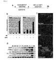
- Cdx2 Caudal type homeobox2
- Cdx2 is one type of intestine-specific transcription factor, and is useful as a marker gene for intestinal cells [ Silberg, D. G., Swain, G. P., Suh, E. R. and Traber, P. G. (2000). Cdx1 and cdx2 expression during intestinal development. Gastroenterology 119, 961-71 ].
- the intestinal fatty acid binding protein (Ifabp) is useful as a gene marker for intestinal cells [ Green, R. P., Cohn, S. M., Sacchettini, J. C., Jackson, K. E. and Gordon, J. I. (1992).
- the mouse intestinal fatty acid binding protein gene nucleotide sequence, pattern of developmental and regional expression, and proposed structure of its protein product. DNA Cell Biol 11, 31-41 ].
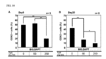
- BIO and DAPT dramatically increased expression of Cdx2 and Ifabp ( Figures 1B and 1C ).
- Villin1 (Villin) [ Maunoury, R., Robine, S., Pringault, E., Leonard, N., Gaillard, J. A. and Louvard, D. (1992). Developmental regulation of villin gene expression in the epithelial cell lineages of mouse digestive and urogenital tracts. Development 115, 717-28 ] was induced on d5, and expression of Cdx2 was induced at a substantial level on d10 of the differentiation. Furthermore, induction of Ifabp was recognized on Day 15; and expression of Lactase ( Lct ) [ Bosse, T., Fialkovich, J. J., Piaseckyj, C.
- Gata4 and Hnfl alpha are partially required for the expression of specific intestinal genes during development.
- Am J Physiol Gastrointest Liver Physiol 292, G1302-14 ] and intestine specific homeobox ( Isx ) [ Choi, M. Y., Romer, A. I., Hu, M., Lepourcelet, M., Mechoor, A., Yesilaltay, A., Krieger, M., Gray, P. A. and Shivdasani, R. A. (2006).
- a dynamic expression survey identifies transcription factors relevant in mouse digestive tract development.
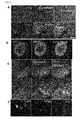
- the definitive endoderm was recovered by flow cytometry on Day 4 of the differentiation ( Figure 2A ), and the cells were re-cultured until Day 15. The definitive endoderm cells were further differentiated into Cdx2- or villin- expressing intestinal cells upon addition of BIO and DAPT ( Figure 2B ). This result revealed that the Cdx2- and villin- expressing intestinal cells were of a definitive endoderm origin.
- Isx and Homeobox C8 ( Hoxc8 ) [ Kawazoe, Y., Sekimoto, T., Araki, M., Takagi, K., Araki, K. and Yamamura, K. (2002). Region-specific gastrointestinal Hox code during murine embryonal gut development. Dev Growth Differ 44, 77-84 ] were induced at high concentrations of BIO and DAPT ( Figure 2C ). These results suggested that intestinal regionalization be specified by graded concentrations of BIO and DAPT.
- the ES cells were further treated with BIO (5 ⁇ M) or DAPT (10 ⁇ M), and effects of the graded concentrations of the other (either BIO or DAPT) on expressions of Pax8 and Hoxc8 were tested.
- BIO 5 ⁇ M
- the high concentration of DAPT turned off the anterior marker Pax8 while inducing the posterior marker Hoxc8 ( Figure 2D ).
- the ES cells were treated with DAPT (10 ⁇ M) and the graded concentrations of BIO, the high BIO concentration turned off Pax8 while turning on Hoxc8 .
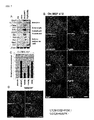
- MEF is more potent than M15 cells in inducing ES cell differentiation into intestinal lineages in the presence of BIO and DAPT.
- Definitive endoderm cells were obtained by culturing ES cells on M15 cells in the presence of activin and bFGF for 4 days, and then, they were sorted by flow cytometry. The sorted definitive endoderm cells were re-cultured in the presence of BIO and DAPT on M15 cells, MEF cells or PA6 cells until Day 15 ( Figure 3A ). Cdx2-expressing differentiated cells were also observed when MEF cells used, but such differentiated cells were not recognized when PA6 cells were used ( Figure 3B ).
- RT-PCR analysis showed that an even stronger expression of intestinal markers induced when they were grown on MEF cells, compared to M15 cells ( Figure 3C ).
- the expression of Cdx2 can be detected at a high level from Day 12 of the differentiation ( Figure 3D ).
- Other markers such as Trefoil factor 3 ( Tff3 , a goblet cell marker); Lysozyme ( Lyzl , a Paneth cell marker); Somatostatin ( Sst , an enteroendocrine marker); and Lct were also detected from Day 12 or Day 15 ( Figure 3E ) [ Hocker, M. and Wiedenmann, B. (1998). Molecular mechanisms of enteroendocrine differentiation.
- Cdx2-expressing intestinal cells are epithelium cells which are indicated by expression of E-cadherin (epithelial marker) [ Lugo-Martinez, V. H., Petit, C. S., Fouquet, S., Le Beyec, J., Chambaz, J., Pincon-Raymond, M., Cardot, P. and Thenet, S. (2009).
- Epidermal growth factor receptor is involved in enterocyte anoikis through the dismantling of E-cadherin-mediated junctions. Am JPhysiol Gastrointest Liver Physiol 296, G235-44 ].
- the Cdx2-expressing cells also co-express Hepatic nuclear factor 4 alpha (HNF4a) (endoderm marker) [ Cattin, A. L., Le Beyec, J., Barreau, F., Saint-Just, S., Houllier, A., Gonzalez, F. J., Robine, S., Pincon-Raymond, M., Cardot, P., Lacasa, M. et al. (2009). Hepatocyte nuclear factor 4alpha, a key factor for homeostasis, cell architecture, and barrier function of the adult intestinal epithelium.
- HNF4a endoderm marker
- Glut2 enterocyte marker
- Claudin7 (tight junction marker) [ Fujita, H., Chiba, H., Yokozaki, H., Sakai, N., Sugimoto, K., Wada, T., Kojima, T., Yamashita, T. and Sawada, N. (2006). Differential expression and subcellular localization of claudin-7, -8, -12, -13, and -15 along the mouse intestine. J Histochem Cytochem 54, 933-44 ] ( Figures 4A-4C ). Furthermore, other markers were also examined.
- Paneth cells (Lysozyme expression), and cells expressing endocrine markers, such as ChromograninA and Somatostatin, were induced ( Figure 4D ). These cells also expressed mucin2 [ van Klinken, B. J., Einerhand, A. W., Duits, L. A., Makkink, M. K., Tytgat, K. M., Renes, I. B., Verburg, M., Buller, H. A. and Dekker, J. (1999). Gastrointestinal expression and partial cDNA cloning of murine Muc2.
- Differentiation of human ES cells into intestinal cells are potentiated by activation of the canonical Wnt signaling and inhibition of the Notch signaling.
- khES-3 human ES cell line
- BIO and DAPT were added to the KhES-3 culture, and this was continuously cultured until Day 35. Then, this was assayed by immunohistochemistry or RT-PCR. khES-3 expressing Cdx2 was detected by immunohistochemistry at Day 25 ( Figure 6A ) and RT-PCR at an early stage of Day 15 ( Figure 6B ).
- molecular markers for enterocytes hVillin, hIfabp, hIsx
- goblet cells hTff3
- rat intestinal trefoil factor tissue- and cell-specific member of the trefoil protein family. Proc Natl Acad Sci U S A 88, 11017-21 ]; Paneth cells ( hLyz ) [ Ouellette, A. J. (1997). Paneth cells and innate immunity in the crypt microenvironment. Gastroenterology 113, 1779-84 ]; and endocrine cells (Gastrin, hGast; Synaptophysin , hSyp; Somatostain, h Sst) [ Hocker, M. and Wiedenmann, B. (1998). Molecular mechanisms of enteroendocrine differentiation.
- ES cell differentiation into intestinal lineages is potentiated by the FGF signaling, which is meditated through PI3K but not MAPK.
- the RT-PCR analysis demonstrated that, when they were cultured in the presence of BIO and DAPT, molecular markers for enterocytes (Ifabp, Isx), goblet cells (Tff3), Paneth cells (Lyz1) and enteroendocrine cells [Sct ( Gouyon, F., Caillaud, L., Carriere, V., Klein, C., Dalet, V., Citadelle, D., Kellett, G. L., Thorens, B., Leturque, A. and Brot-Laroche, E. (2003). Simple-sugar meals target GLUT2 at enterocyte apical membranes to improve sugar absorption: a study in GLUT2-null mice.
- BIO, DAPT and the FGF signaling were investigated.
- An antagonist of FGF receptor "SU5402” and an inhibitor of PI3K “LY294002” were used therefor.
- the blockade of FGF signaling by SU5402 or the blockade of PI3K by LY294002 partially inhibited the intestinal differentiation which was mediated by BIO and DAPT ( Figures 7C and 7D ).
- intestinal stem cells ISCs and progenitor cells present in the crypts proliferate vigorously, and provide differentiated cells.
- ISCs intestinal stem cells
- progenitor cells present in the crypts proliferate vigorously, and provide differentiated cells.
- the intestinal stem cell Genes Dev 22, 1856-64 ].
- the ES-cell-derived definitive endoderm cells were cultured on M15 cells or MEF cells, whereby it was confirmed that activation of the canonical Wnt signaling pathways by addition of BIO, and inhibition of the Notch pathway by addition of DAPT, simultaneously induced the gut endoderm to express the posterior markers, and enhanced intestinal differentiation.
- Fgf emitted from M15 and MEF cells assists the establishment of intestinal characters ( Figure 7 ). Therefore, the results of this Example indicate that the FGF, Wnt and Notch signaling function cooperatively to promote differentiation of ES cells into the intestinal lineages.
- FGF2 specifies hESC-derived definitive endoderm into foregut/midgut cell lineages in a concentration-dependent manner. Stem Cells 28, 45-56 ]. It has been known that, at high FGF2 levels, specification of midgut endoderm into small intestinal progenitors is increased at the expense of Pdx1+ pancreatic progenitors (the above reference of Ameri et al.).
- activation of the Notch signaling is capable of amplifying the intestinal progenitor pool while inhibiting the goblet and enteroendocrine cell differentiation [ Zecchini, V., Domaschenz, R., Winton, D. and Jones, P. (2005). Notch signaling regulates the differentiation of post-mitotic intestinal epithelial cells. Genes Dev 19, 1686-91 ]. Furthermore, after conditional removal of the common Notch pathway transcription factor CSL/RBP-J, a rapid, massive conversion of proliferative crypt cells into post-mitotic goblet cells has been observed ( van Es, J. H., van Gijn, M.
- Notch/gamma-secretase inhibition turns proliferative cells in intestinal crypts and adenomas into goblet cells. Nature 435, 959-63 ). Additionally, it has been known that a similar phenotype was obtained by blocking the Notch cascade with a gamma-secretase inhibitor (the above reference of van Es et al.).
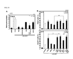
- Example 2 shows cases in which inhibitors or activators against various signal transduction systems were further added besides BIO and DAPT in the method of the present invention.
- the mouse ES cells were differentiated into definitive endoderm cells on M15 cells, and then, the definitive endoderm cells were sorted by flow cytometry, and were re-cultured on MEF cells.
- the above-sorted definitive endoderm cells were cultured in the presence of BIO (5 ⁇ M) and DAPT (10 ⁇ M) as well as the above-mentioned inhibitor for 8 days (until the 12th day of cultivation) in accordance with the method described in Example 1, RNAs were extracted from the cells by the method described in Example 1, and expression of the Cdx2 gene, namely an intestinal marker, was analyzed by real-time PCR.
- the real-time PCR was carried out by use of the primer pairs used in Example 1 (see Table 1), Thunderbird SYBR qPCR mix (Toyobo), and 7500 Fast Real-Time PCR system (ABI Company).
- the PCR reaction cycles are shown in Table 1.
- the results are shown in Figure 8A .
- mice ES cells were plated on a gelatin-coated dish at 6,900 cells/cm 2 .
- the ES cells were cultured for 7 days in DMEM medium (Dulbecco's Modified Eagle Medium) (Invitrogen, Glasgow, UK) containing 4,500 mg/L of glucose, supplemented with NEAA, L-Gln, PS, ⁇ -ME, 10 ⁇ g/mL of Insulin (Sigma-Aldrich), 5.5 ⁇ g/mL of Transferin (Sigma-Aldrich), 6.7 pg/mL of Selenium (Sigma-Aldrich), 0.25% AlbuMax (Invitrogen), and 10 ng/mL of recombinant human activin A (R&D Systems, Minneapolis, MN), and then, the culture medium was replaced with 10% KSR containing 2,000 mg/mL of glucose, BIO (5 ⁇ M)
- FGF2 acts to promote differentiation of mouse ES cells into intestine, and that the BMP signaling or the Hh signaling is activated by BMP4 or SAG in the latter period of cultivation to thereby further promote the differentiation.
- Test Example 3 Addition of various activators in human ES cells
- human ES cells were plated on gelatin-coated dishes at 69,000 cells/cm 2 .
- the ES cells were cultured for seven days in RPMI 1640 medium (Invitrogen) supplemented with NEAA, L-Gln, PS, ⁇ -ME, 10 ⁇ g/mL of Insulin (Sigma-Aldrich), 100 ng/mL of recombinant human activin A (R&D Systems, Minneapolis, MN), and B27 supplement (Invitrogen).
- the culture medium was replaced with 10% KSR supplemented with 2,000 mg/mL of glucose, BIO (5 ⁇ M), DAPT (10 ⁇ M) and activators and growth factors at the concentrations as described in Test Example 2.
Claims (11)
- Verfahren zur Herstellung von Intestinalzellen, umfassend die folgenden Schritte:(A) Induzieren von Differenzierung pluripotenter Stammzellen in definitive Endodermzellen; und(B) Kultivieren der definitiven Endodermzellen in Gegenwart von (2'Z,3'E)-6-Bromindirubin-3'-oxim (BIO) und N-[(3,5-Difluorphenyl)acetyl]-L-Ala-2-Phenyl-L-Gly-tert-butyl-OH (DAPT), um dadurch Differenzierung der definitiven Endodermzellen in Intestinalzellen zu induzieren.
- Verfahren nach Anspruch 1, wobei die definitiven Endodermzellen aus einer in Schritt (A) erhaltenen Zellkultur mittels Durchflusszytometrie unter Verwendung von fluoreszenzmarkierten Antikörpern gegen E-Cadherin (ECD) und CXCR4 abgetrennt werden, und die abgetrennten definitiven Endodermzellen in Schritt (B) verwendet werden.
- Verfahren nach Anspruch 1 oder 2, wobei in Schritt (A) die pluripotenten Stammzellen in Gegenwart von Feeder-Zellen und in Gegenwart von Activin und/oder bFGF kultiviert werden, um dadurch Differenzierung der pluripotenten Stammzellen in die definitiven Endodermzellen zu induzieren.
- Verfahren nach Anspruch 3, wobei die Feeder-Zellen Zellen sind, die von einem Mesoderm abstammen.
- Verfahren nach Anspruch 3 oder 4, wobei die Feeder-Zellen M15-Zellen, MEF-Zellen oder ST2-Zellen sind.
- Verfahren nach einem der Ansprüche 1 bis 5, wobei die definitiven Endodermzellen in Gegenwart von M15-Zellen oder MEF-Zellen in Schritt (B) kultiviert werden.
- Verfahren nach einem der Ansprüche 1 bis 6, wobei die pluripotenten Stammzellen embryonale Stammzellen oder induzierte pluripotente Stammzellen sind.
- Verfahren nach einem der Ansprüche 1 bis 7, wobei die pluripotenten Stammzellen humane embryonale Stammzellen oder murine embryonale Stammzellen sind.
- Verfahren zum Screening nach Substanzen, die Induzierung von Differenzierung pluripotenter Stammzellen in Intestinalzellen fördern oder inhibieren, wobei das Verfahren umfasst:Kultivieren pluripotenter Stammzellen in Gegenwart einer Testsubstanz beim Induzieren von Differenzierung der pluripotenten Stammzellen in Intestinalzellen durch das Verfahren nach einem der Ansprüche 1 bis 8; undVergleichen eines Levels der Differenzierung der pluripotenten Stammzellen in Intestinalzellen in einem Fall, wo die pluripotenten Stammzellen in Gegenwart der Testsubstanz kultiviert werden, mit einem Level der Differenzierung der pluripotenten Stammzellen in Intestinalzellen in einem Fall, wo die pluripotenten Stammzellen in Abwesenheit der Testsubstanz kultiviert werden
- Verfahren zum Screening nach Anspruch 9, wobei die Testsubstanz ein Wachstumsfaktor oder eine niedermolekulare Verbindung ist.
- Verfahren zum Screening nach Anspruch 9 oder 10, wobei eine Menge von Marker-Transkript oder ein Protein davon, welche in Intestinalzellen exprimiert werden, oder beide davon, als Indikatoren verwendet werden, um dadurch die Level der Differenzierung in Intestinalzellen zu bestimmen.
Applications Claiming Priority (2)
| Application Number | Priority Date | Filing Date | Title |
|---|---|---|---|
| JP2010246161 | 2010-11-02 | ||
| PCT/JP2011/075041 WO2012060315A1 (ja) | 2010-11-02 | 2011-10-31 | 腸細胞の製造方法 |
Publications (3)
| Publication Number | Publication Date |
|---|---|
| EP2636731A1 EP2636731A1 (de) | 2013-09-11 |
| EP2636731A4 EP2636731A4 (de) | 2014-04-30 |
| EP2636731B1 true EP2636731B1 (de) | 2016-07-13 |
Family
ID=46024428
Family Applications (1)
| Application Number | Title | Priority Date | Filing Date |
|---|---|---|---|
| EP11837965.0A Not-in-force EP2636731B1 (de) | 2010-11-02 | 2011-10-31 | Verfahren zur herstellung von darmzellen |
Country Status (7)
| Country | Link |
|---|---|
| US (1) | US9376665B2 (de) |
| EP (1) | EP2636731B1 (de) |
| JP (1) | JP5875007B2 (de) |
| KR (1) | KR20130101557A (de) |
| CN (1) | CN103459591B (de) |
| CA (1) | CA2821562A1 (de) |
| WO (1) | WO2012060315A1 (de) |
Cited By (1)
| Publication number | Priority date | Publication date | Assignee | Title |
|---|---|---|---|---|
| US11859212B2 (en) | 2017-05-09 | 2024-01-02 | Public University Corporation Nagoya City University | Method for producing intestinal organoid derived from pluripotent stem cells |
Families Citing this family (21)
| Publication number | Priority date | Publication date | Assignee | Title |
|---|---|---|---|---|
| DK2970890T3 (da) | 2013-03-14 | 2020-05-04 | Brigham & Womens Hospital Inc | Sammensætninger og fremgangsmåder til opformering og dyrkning af epitelstamceller |
| US10568883B2 (en) | 2014-09-03 | 2020-02-25 | Massachusetts Institute Of Technology | Compositions, systems, and methods for generating inner ear hair cells for treatment of hearing loss |
| WO2017069376A1 (ko) * | 2015-10-20 | 2017-04-27 | 전남대학교병원 | IKKε 억제제를 함유하는 염증질환의 치료용 조성물 |
| EP3400286A1 (de) * | 2016-01-08 | 2018-11-14 | Massachusetts Institute Of Technology | Herstellung von differenzierten enteroendokrinen zellen und insulinproduzierenden zellen |
| AU2017213795A1 (en) | 2016-02-01 | 2018-08-16 | Cedars-Sinai Medical Center | Systems and methods for growth of intestinal cells in microfluidic devices |
| US10201540B2 (en) | 2016-03-02 | 2019-02-12 | Frequency Therapeutics, Inc. | Solubilized compositions for controlled proliferation of stem cells / generating inner ear hair cells using GSK3 inhibitors: I |
| US11260130B2 (en) | 2016-03-02 | 2022-03-01 | Frequency Therapeutics, Inc. | Solubilized compositions for controlled proliferation of stem cells / generating inner ear hair cells using a GSK3 inhibitor: IV |
| US10213511B2 (en) | 2016-03-02 | 2019-02-26 | Frequency Therapeutics, Inc. | Thermoreversible compositions for administration of therapeutic agents |
| MA45479A (fr) * | 2016-04-14 | 2019-02-20 | Janssen Biotech Inc | Différenciation de cellules souches pluripotentes en cellules de l'endoderme de l'intestin moyen |
| US20190263912A1 (en) * | 2016-11-11 | 2019-08-29 | The Broad Institute, Inc. | Modulation of intestinal epithelial cell differentiation, maintenance and/or function through t cell action |
| MX2019007890A (es) | 2016-12-30 | 2020-01-20 | Frequency Therapeutics Inc | Compuestos de 1h-pirrol-2,5-diona y metodos de uso de los mismos para inducir la auto-renovacion de celulas madre/progenitoras de soporte. |
| WO2018140647A1 (en) | 2017-01-25 | 2018-08-02 | Cedars-Sinai Medical Center | In vitro induction of mammary-like differentiation from human pluripotent stem cells |
| KR20190105237A (ko) * | 2017-02-20 | 2019-09-16 | 고리츠다이가쿠호징 나고야시리츠다이가쿠 | 인공 다능성 줄기 세포 유래 장관 줄기 세포의 유지 배양 |
| US11767513B2 (en) | 2017-03-14 | 2023-09-26 | Cedars-Sinai Medical Center | Neuromuscular junction |
| WO2018176001A2 (en) | 2017-03-24 | 2018-09-27 | Cedars-Sinai Medical Center | Methods and compositions for production of fallopian tube epithelium |
| CN112513253A (zh) * | 2018-07-27 | 2021-03-16 | 富士胶片株式会社 | 肠上皮细胞的制造方法及肠上皮细胞 |
| WO2020037326A1 (en) | 2018-08-17 | 2020-02-20 | Frequency Therapeutics, Inc. | Compositions and methods for generating hair cells by downregulating foxo |
| CA3109647A1 (en) | 2018-08-17 | 2020-02-20 | Frequency Therapeutics, Inc. | Compositions and methods for generating hair cells by upregulating jag-1 |
| JP7377486B2 (ja) * | 2018-09-10 | 2023-11-10 | 国立大学法人東京工業大学 | 多能性幹細胞から腸細胞の作製方法 |
| EP3851518A4 (de) * | 2018-09-10 | 2022-06-22 | Tokyo Institute of Technology | Verfahren zur herstellung von darmzellen aus pluripotenten stammzellen |
| EP3901250A4 (de) | 2018-11-02 | 2022-08-10 | Public University Corporation Nagoya City University | Verfahren zur herstellung von aus pluripotenten stammzellen abgeleitetem intestinalen organoid |
Family Cites Families (12)
| Publication number | Priority date | Publication date | Assignee | Title |
|---|---|---|---|---|
| US5523226A (en) * | 1993-05-14 | 1996-06-04 | Biotechnology Research And Development Corp. | Transgenic swine compositions and methods |
| US7704738B2 (en) | 2003-12-23 | 2010-04-27 | Cythera, Inc. | Definitive endoderm |
| JP5092124B2 (ja) | 2005-05-24 | 2012-12-05 | 国立大学法人 熊本大学 | Es細胞の分化誘導方法 |
| JP5087004B2 (ja) * | 2005-10-24 | 2012-11-28 | エージェンシー フォー サイエンス,テクノロジー アンド リサーチ | 中胚葉、内胚葉及び中内胚葉細胞の細胞運命を指定する方法 |
| EP2206724A1 (de) | 2005-12-13 | 2010-07-14 | Kyoto University | Nukleärer Reprogramierungsfaktor |
| US7695965B2 (en) | 2006-03-02 | 2010-04-13 | Cythera, Inc. | Methods of producing pancreatic hormones |
| AU2007277364B2 (en) * | 2006-07-26 | 2010-08-12 | Viacyte, Inc. | Methods of producing pancreatic hormones |
| JP5111187B2 (ja) | 2008-03-25 | 2012-12-26 | 松山株式会社 | 農作業機 |
| CN102165058B (zh) * | 2008-07-25 | 2015-07-01 | 佐治亚大学研究基金会 | 中胚层来源的ISL 1+多潜能细胞(IMPs)的组合物、心外膜祖细胞(EPCs)和多潜能CXCR4+CD56+细胞(C56Cs)及其使用方法 |
| CA2742268C (en) * | 2008-10-31 | 2020-02-18 | Centocor Ortho Biotech Inc. | Differentiation of human embryonic stem cells to the pancreatic endocrine lineage |
| WO2010108005A2 (en) * | 2009-03-18 | 2010-09-23 | University Of Georgia Research Foundation | Novel neural progenitors from pluripotent stem cells, methods of producing same and use to produce neural cells |
| WO2011140441A2 (en) * | 2010-05-06 | 2011-11-10 | Children's Hospital Medical Center | Methods and systems for converting precursor cells into intestinal tissues through directed differentiation |
-
2011
- 2011-10-31 EP EP11837965.0A patent/EP2636731B1/de not_active Not-in-force
- 2011-10-31 JP JP2012541844A patent/JP5875007B2/ja active Active
- 2011-10-31 KR KR1020137013739A patent/KR20130101557A/ko not_active Application Discontinuation
- 2011-10-31 CN CN201180063607.9A patent/CN103459591B/zh not_active Expired - Fee Related
- 2011-10-31 US US13/882,904 patent/US9376665B2/en active Active
- 2011-10-31 CA CA2821562A patent/CA2821562A1/en not_active Abandoned
- 2011-10-31 WO PCT/JP2011/075041 patent/WO2012060315A1/ja active Application Filing
Non-Patent Citations (1)
| Title |
|---|
| SOICHIRO OGAKI ET AL: "Wnt and Notch Signals Guide Embryonic Stem Cell Differentiation into the Intestinal Lineages", STEM CELLS, vol. 31, no. 6, 22 June 2013 (2013-06-22), pages 1086 - 1096, XP055177743, ISSN: 1066-5099, DOI: 10.1002/stem.1344 * |
Cited By (1)
| Publication number | Priority date | Publication date | Assignee | Title |
|---|---|---|---|---|
| US11859212B2 (en) | 2017-05-09 | 2024-01-02 | Public University Corporation Nagoya City University | Method for producing intestinal organoid derived from pluripotent stem cells |
Also Published As
| Publication number | Publication date |
|---|---|
| CN103459591B (zh) | 2016-05-04 |
| EP2636731A4 (de) | 2014-04-30 |
| CA2821562A1 (en) | 2012-05-10 |
| JPWO2012060315A1 (ja) | 2014-05-12 |
| US9376665B2 (en) | 2016-06-28 |
| JP5875007B2 (ja) | 2016-03-02 |
| WO2012060315A1 (ja) | 2012-05-10 |
| KR20130101557A (ko) | 2013-09-13 |
| CN103459591A (zh) | 2013-12-18 |
| EP2636731A1 (de) | 2013-09-11 |
| US20140199700A1 (en) | 2014-07-17 |
Similar Documents
| Publication | Publication Date | Title |
|---|---|---|
| EP2636731B1 (de) | Verfahren zur herstellung von darmzellen | |
| Ogaki et al. | Wnt and Notch signals guide embryonic stem cell differentiation into the intestinal lineages | |
| JP6759293B2 (ja) | 膵臓前駆細胞及び機能的β細胞をhPSCから生成するための方法及び組成物 | |
| EP3149156B1 (de) | Verfahren und systeme zur umwandlung von vorläuferzellen in magengewebe durch gezielte differenzierung | |
| Teo et al. | PDX1 binds and represses hepatic genes to ensure robust pancreatic commitment in differentiating human embryonic stem cells | |
| JPWO2006126574A1 (ja) | Es細胞の分化誘導方法 | |
| KR20160099079A (ko) | SC-β 세포 및 조성물 그리고 그 생성 방법 | |
| AU2017373767A1 (en) | Colonic organoids and methods of making and using same | |
| JP2016093192A (ja) | hPS細胞由来の胚体内胚葉からの特定の内胚葉の派生 | |
| CA2902857C (en) | Generation of thymic epithelial progenitor cells in vitro | |
| Dolci et al. | Gonadal development and germ cell tumors in mouse and humans | |
| JPWO2008149807A1 (ja) | Es細胞の分化誘導方法 | |
| Kopper et al. | Stepwise differentiation of human embryonic stem cells into early endoderm derivatives and their molecular characterization | |
| Satoh et al. | Establishment and directed differentiation of induced pluripotent stem cells from glycogen storage disease type I b patient | |
| US20200138870A1 (en) | Recreation of pancreatic niche allows for novel methods for human, mature beta derivation from pluripotent stem cells | |
| US7776593B2 (en) | Hes6 as a marker of pancreatic endocrine cells | |
| RU2772585C2 (ru) | КЛЕТКИ SC-β И КОМПОЗИЦИИ И СПОСОБЫ ДЛЯ ИХ СОЗДАНИЯ | |
| Ameri | FGF signaling in specification of hESC-derived definitive endoderm | |
| SAGAYARAJ | GENETIC REGULATION OF HUMAN FETAL LIVER AND ITS CLINICAL APPLICATION | |
| Shah | NGN3 Expression in Definitive Endoderm Progenitors Specifies Pancreatic Endocrine Precursors | |
| Wright et al. | Adrian Kee Keong Teo, Norihiro Tsuneyoshi, Shawn Hoon, 2 Ee Kim Tan, Lawrence W. Stanton, 3 |
Legal Events
| Date | Code | Title | Description |
|---|---|---|---|
| PUAI | Public reference made under article 153(3) epc to a published international application that has entered the european phase |
Free format text: ORIGINAL CODE: 0009012 |
|
| 17P | Request for examination filed |
Effective date: 20130531 |
|
| AK | Designated contracting states |
Kind code of ref document: A1 Designated state(s): AL AT BE BG CH CY CZ DE DK EE ES FI FR GB GR HR HU IE IS IT LI LT LU LV MC MK MT NL NO PL PT RO RS SE SI SK SM TR |
|
| RAP1 | Party data changed (applicant data changed or rights of an application transferred) |
Owner name: LSIP, LLC Owner name: NATIONAL UNIVERSITY CORPORATION KUMAMOTO UNIVERSIT |
|
| DAX | Request for extension of the european patent (deleted) | ||
| A4 | Supplementary search report drawn up and despatched |
Effective date: 20140328 |
|
| RIC1 | Information provided on ipc code assigned before grant |
Ipc: C12N 5/074 20100101ALI20140324BHEP Ipc: C12Q 1/68 20060101ALI20140324BHEP Ipc: C12Q 1/02 20060101ALI20140324BHEP Ipc: G01N 33/50 20060101ALI20140324BHEP Ipc: G01N 33/15 20060101ALI20140324BHEP Ipc: C12N 5/073 20100101AFI20140324BHEP Ipc: C12N 5/071 20100101ALI20140324BHEP |
|
| 17Q | First examination report despatched |
Effective date: 20150402 |
|
| GRAP | Despatch of communication of intention to grant a patent |
Free format text: ORIGINAL CODE: EPIDOSNIGR1 |
|
| INTG | Intention to grant announced |
Effective date: 20160216 |
|
| GRAS | Grant fee paid |
Free format text: ORIGINAL CODE: EPIDOSNIGR3 |
|
| GRAA | (expected) grant |
Free format text: ORIGINAL CODE: 0009210 |
|
| AK | Designated contracting states |
Kind code of ref document: B1 Designated state(s): AL AT BE BG CH CY CZ DE DK EE ES FI FR GB GR HR HU IE IS IT LI LT LU LV MC MK MT NL NO PL PT RO RS SE SI SK SM TR |
|
| REG | Reference to a national code |
Ref country code: GB Ref legal event code: FG4D |
|
| REG | Reference to a national code |
Ref country code: AT Ref legal event code: REF Ref document number: 812361 Country of ref document: AT Kind code of ref document: T Effective date: 20160715 Ref country code: CH Ref legal event code: EP |
|
| REG | Reference to a national code |
Ref country code: IE Ref legal event code: FG4D |
|
| REG | Reference to a national code |
Ref country code: DE Ref legal event code: R096 Ref document number: 602011028219 Country of ref document: DE |
|
| REG | Reference to a national code |
Ref country code: FR Ref legal event code: PLFP Year of fee payment: 6 |
|
| REG | Reference to a national code |
Ref country code: LT Ref legal event code: MG4D |
|
| REG | Reference to a national code |
Ref country code: NL Ref legal event code: MP Effective date: 20160713 |
|
| REG | Reference to a national code |
Ref country code: AT Ref legal event code: MK05 Ref document number: 812361 Country of ref document: AT Kind code of ref document: T Effective date: 20160713 |
|
| PG25 | Lapsed in a contracting state [announced via postgrant information from national office to epo] |
Ref country code: IS Free format text: LAPSE BECAUSE OF FAILURE TO SUBMIT A TRANSLATION OF THE DESCRIPTION OR TO PAY THE FEE WITHIN THE PRESCRIBED TIME-LIMIT Effective date: 20161113 Ref country code: RS Free format text: LAPSE BECAUSE OF FAILURE TO SUBMIT A TRANSLATION OF THE DESCRIPTION OR TO PAY THE FEE WITHIN THE PRESCRIBED TIME-LIMIT Effective date: 20160713 Ref country code: FI Free format text: LAPSE BECAUSE OF FAILURE TO SUBMIT A TRANSLATION OF THE DESCRIPTION OR TO PAY THE FEE WITHIN THE PRESCRIBED TIME-LIMIT Effective date: 20160713 Ref country code: LT Free format text: LAPSE BECAUSE OF FAILURE TO SUBMIT A TRANSLATION OF THE DESCRIPTION OR TO PAY THE FEE WITHIN THE PRESCRIBED TIME-LIMIT Effective date: 20160713 Ref country code: IT Free format text: LAPSE BECAUSE OF FAILURE TO SUBMIT A TRANSLATION OF THE DESCRIPTION OR TO PAY THE FEE WITHIN THE PRESCRIBED TIME-LIMIT Effective date: 20160713 Ref country code: NL Free format text: LAPSE BECAUSE OF FAILURE TO SUBMIT A TRANSLATION OF THE DESCRIPTION OR TO PAY THE FEE WITHIN THE PRESCRIBED TIME-LIMIT Effective date: 20160713 Ref country code: NO Free format text: LAPSE BECAUSE OF FAILURE TO SUBMIT A TRANSLATION OF THE DESCRIPTION OR TO PAY THE FEE WITHIN THE PRESCRIBED TIME-LIMIT Effective date: 20161013 Ref country code: HR Free format text: LAPSE BECAUSE OF FAILURE TO SUBMIT A TRANSLATION OF THE DESCRIPTION OR TO PAY THE FEE WITHIN THE PRESCRIBED TIME-LIMIT Effective date: 20160713 |
|
| REG | Reference to a national code |
Ref country code: DE Ref legal event code: R082 Ref document number: 602011028219 Country of ref document: DE Representative=s name: VOSSIUS & PARTNER PATENTANWAELTE RECHTSANWAELT, DE Ref country code: DE Ref legal event code: R081 Ref document number: 602011028219 Country of ref document: DE Owner name: TOKYO INSTITUTE OF TECHNOLOGY, JP Free format text: FORMER OWNERS: LSIP, LLC, TOKYO, JP; NATIONAL UNIVERSITY CORPORATION KUMAMOTO UNIVERSITY, KUMAMOTO-SHI, KUMAMOTO, JP |
|
| PG25 | Lapsed in a contracting state [announced via postgrant information from national office to epo] |
Ref country code: PL Free format text: LAPSE BECAUSE OF FAILURE TO SUBMIT A TRANSLATION OF THE DESCRIPTION OR TO PAY THE FEE WITHIN THE PRESCRIBED TIME-LIMIT Effective date: 20160713 Ref country code: SE Free format text: LAPSE BECAUSE OF FAILURE TO SUBMIT A TRANSLATION OF THE DESCRIPTION OR TO PAY THE FEE WITHIN THE PRESCRIBED TIME-LIMIT Effective date: 20160713 Ref country code: ES Free format text: LAPSE BECAUSE OF FAILURE TO SUBMIT A TRANSLATION OF THE DESCRIPTION OR TO PAY THE FEE WITHIN THE PRESCRIBED TIME-LIMIT Effective date: 20160713 Ref country code: AT Free format text: LAPSE BECAUSE OF FAILURE TO SUBMIT A TRANSLATION OF THE DESCRIPTION OR TO PAY THE FEE WITHIN THE PRESCRIBED TIME-LIMIT Effective date: 20160713 Ref country code: BE Free format text: LAPSE BECAUSE OF NON-PAYMENT OF DUE FEES Effective date: 20160713 Ref country code: PT Free format text: LAPSE BECAUSE OF FAILURE TO SUBMIT A TRANSLATION OF THE DESCRIPTION OR TO PAY THE FEE WITHIN THE PRESCRIBED TIME-LIMIT Effective date: 20161114 Ref country code: GR Free format text: LAPSE BECAUSE OF FAILURE TO SUBMIT A TRANSLATION OF THE DESCRIPTION OR TO PAY THE FEE WITHIN THE PRESCRIBED TIME-LIMIT Effective date: 20161014 Ref country code: LV Free format text: LAPSE BECAUSE OF FAILURE TO SUBMIT A TRANSLATION OF THE DESCRIPTION OR TO PAY THE FEE WITHIN THE PRESCRIBED TIME-LIMIT Effective date: 20160713 |
|
| RAP2 | Party data changed (patent owner data changed or rights of a patent transferred) |
Owner name: TOKYO INSTITUTE OF TECHNOLOGY |
|
| REG | Reference to a national code |
Ref country code: GB Ref legal event code: 732E Free format text: REGISTERED BETWEEN 20170316 AND 20170323 |
|
| REG | Reference to a national code |
Ref country code: DE Ref legal event code: R097 Ref document number: 602011028219 Country of ref document: DE |
|
| PG25 | Lapsed in a contracting state [announced via postgrant information from national office to epo] |
Ref country code: EE Free format text: LAPSE BECAUSE OF FAILURE TO SUBMIT A TRANSLATION OF THE DESCRIPTION OR TO PAY THE FEE WITHIN THE PRESCRIBED TIME-LIMIT Effective date: 20160713 Ref country code: RO Free format text: LAPSE BECAUSE OF FAILURE TO SUBMIT A TRANSLATION OF THE DESCRIPTION OR TO PAY THE FEE WITHIN THE PRESCRIBED TIME-LIMIT Effective date: 20160713 |
|
| PLBE | No opposition filed within time limit |
Free format text: ORIGINAL CODE: 0009261 |
|
| STAA | Information on the status of an ep patent application or granted ep patent |
Free format text: STATUS: NO OPPOSITION FILED WITHIN TIME LIMIT |
|
| PG25 | Lapsed in a contracting state [announced via postgrant information from national office to epo] |
Ref country code: SM Free format text: LAPSE BECAUSE OF FAILURE TO SUBMIT A TRANSLATION OF THE DESCRIPTION OR TO PAY THE FEE WITHIN THE PRESCRIBED TIME-LIMIT Effective date: 20160713 Ref country code: DK Free format text: LAPSE BECAUSE OF FAILURE TO SUBMIT A TRANSLATION OF THE DESCRIPTION OR TO PAY THE FEE WITHIN THE PRESCRIBED TIME-LIMIT Effective date: 20160713 Ref country code: CZ Free format text: LAPSE BECAUSE OF FAILURE TO SUBMIT A TRANSLATION OF THE DESCRIPTION OR TO PAY THE FEE WITHIN THE PRESCRIBED TIME-LIMIT Effective date: 20160713 Ref country code: SK Free format text: LAPSE BECAUSE OF FAILURE TO SUBMIT A TRANSLATION OF THE DESCRIPTION OR TO PAY THE FEE WITHIN THE PRESCRIBED TIME-LIMIT Effective date: 20160713 Ref country code: BG Free format text: LAPSE BECAUSE OF FAILURE TO SUBMIT A TRANSLATION OF THE DESCRIPTION OR TO PAY THE FEE WITHIN THE PRESCRIBED TIME-LIMIT Effective date: 20161013 |
|
| REG | Reference to a national code |
Ref country code: CH Ref legal event code: PL |
|
| 26N | No opposition filed |
Effective date: 20170418 |
|
| REG | Reference to a national code |
Ref country code: IE Ref legal event code: MM4A |
|
| PG25 | Lapsed in a contracting state [announced via postgrant information from national office to epo] |
Ref country code: LI Free format text: LAPSE BECAUSE OF NON-PAYMENT OF DUE FEES Effective date: 20161031 Ref country code: CH Free format text: LAPSE BECAUSE OF NON-PAYMENT OF DUE FEES Effective date: 20161031 |
|
| REG | Reference to a national code |
Ref country code: FR Ref legal event code: PLFP Year of fee payment: 7 |
|
| PG25 | Lapsed in a contracting state [announced via postgrant information from national office to epo] |
Ref country code: LU Free format text: LAPSE BECAUSE OF NON-PAYMENT OF DUE FEES Effective date: 20161031 Ref country code: SI Free format text: LAPSE BECAUSE OF FAILURE TO SUBMIT A TRANSLATION OF THE DESCRIPTION OR TO PAY THE FEE WITHIN THE PRESCRIBED TIME-LIMIT Effective date: 20160713 |
|
| REG | Reference to a national code |
Ref country code: GB Ref legal event code: 732E Free format text: REGISTERED BETWEEN 20170914 AND 20170920 |
|
| REG | Reference to a national code |
Ref country code: FR Ref legal event code: TP Owner name: TOKYO INSTITUTE OF TECHNOLOGY, JP Effective date: 20170906 |
|
| PG25 | Lapsed in a contracting state [announced via postgrant information from national office to epo] |
Ref country code: IE Free format text: LAPSE BECAUSE OF NON-PAYMENT OF DUE FEES Effective date: 20161031 |
|
| PG25 | Lapsed in a contracting state [announced via postgrant information from national office to epo] |
Ref country code: HU Free format text: LAPSE BECAUSE OF FAILURE TO SUBMIT A TRANSLATION OF THE DESCRIPTION OR TO PAY THE FEE WITHIN THE PRESCRIBED TIME-LIMIT; INVALID AB INITIO Effective date: 20111031 Ref country code: CY Free format text: LAPSE BECAUSE OF FAILURE TO SUBMIT A TRANSLATION OF THE DESCRIPTION OR TO PAY THE FEE WITHIN THE PRESCRIBED TIME-LIMIT Effective date: 20160713 |
|
| PG25 | Lapsed in a contracting state [announced via postgrant information from national office to epo] |
Ref country code: MT Free format text: LAPSE BECAUSE OF NON-PAYMENT OF DUE FEES Effective date: 20161031 Ref country code: MC Free format text: LAPSE BECAUSE OF FAILURE TO SUBMIT A TRANSLATION OF THE DESCRIPTION OR TO PAY THE FEE WITHIN THE PRESCRIBED TIME-LIMIT Effective date: 20160713 Ref country code: MK Free format text: LAPSE BECAUSE OF FAILURE TO SUBMIT A TRANSLATION OF THE DESCRIPTION OR TO PAY THE FEE WITHIN THE PRESCRIBED TIME-LIMIT Effective date: 20160713 |
|
| REG | Reference to a national code |
Ref country code: FR Ref legal event code: PLFP Year of fee payment: 8 |
|
| PG25 | Lapsed in a contracting state [announced via postgrant information from national office to epo] |
Ref country code: TR Free format text: LAPSE BECAUSE OF FAILURE TO SUBMIT A TRANSLATION OF THE DESCRIPTION OR TO PAY THE FEE WITHIN THE PRESCRIBED TIME-LIMIT Effective date: 20160713 Ref country code: AL Free format text: LAPSE BECAUSE OF FAILURE TO SUBMIT A TRANSLATION OF THE DESCRIPTION OR TO PAY THE FEE WITHIN THE PRESCRIBED TIME-LIMIT Effective date: 20160713 |
|
| PGFP | Annual fee paid to national office [announced via postgrant information from national office to epo] |
Ref country code: GB Payment date: 20210930 Year of fee payment: 11 |
|
| PGFP | Annual fee paid to national office [announced via postgrant information from national office to epo] |
Ref country code: DE Payment date: 20211029 Year of fee payment: 11 |
|
| PGFP | Annual fee paid to national office [announced via postgrant information from national office to epo] |
Ref country code: FR Payment date: 20211021 Year of fee payment: 11 |
|
| REG | Reference to a national code |
Ref country code: DE Ref legal event code: R119 Ref document number: 602011028219 Country of ref document: DE |
|
| GBPC | Gb: european patent ceased through non-payment of renewal fee |
Effective date: 20221031 |
|
| PG25 | Lapsed in a contracting state [announced via postgrant information from national office to epo] |
Ref country code: FR Free format text: LAPSE BECAUSE OF NON-PAYMENT OF DUE FEES Effective date: 20221031 Ref country code: DE Free format text: LAPSE BECAUSE OF NON-PAYMENT OF DUE FEES Effective date: 20230503 |
|
| PG25 | Lapsed in a contracting state [announced via postgrant information from national office to epo] |
Ref country code: GB Free format text: LAPSE BECAUSE OF NON-PAYMENT OF DUE FEES Effective date: 20221031 |