EP2602624A1 - Test de diagnostic pour exclure une importante lésion rénale - Google Patents
Test de diagnostic pour exclure une importante lésion rénale Download PDFInfo
- Publication number
- EP2602624A1 EP2602624A1 EP13157838.7A EP13157838A EP2602624A1 EP 2602624 A1 EP2602624 A1 EP 2602624A1 EP 13157838 A EP13157838 A EP 13157838A EP 2602624 A1 EP2602624 A1 EP 2602624A1
- Authority
- EP
- European Patent Office
- Prior art keywords
- value
- ngal
- renal failure
- risk
- acute renal
- Prior art date
- Legal status (The legal status is an assumption and is not a legal conclusion. Google has not performed a legal analysis and makes no representation as to the accuracy of the status listed.)
- Withdrawn
Links
Images
Classifications
-
- G—PHYSICS
- G01—MEASURING; TESTING
- G01N—INVESTIGATING OR ANALYSING MATERIALS BY DETERMINING THEIR CHEMICAL OR PHYSICAL PROPERTIES
- G01N33/00—Investigating or analysing materials by specific methods not covered by groups G01N1/00 - G01N31/00
- G01N33/48—Biological material, e.g. blood, urine; Haemocytometers
- G01N33/50—Chemical analysis of biological material, e.g. blood, urine; Testing involving biospecific ligand binding methods; Immunological testing
- G01N33/68—Chemical analysis of biological material, e.g. blood, urine; Testing involving biospecific ligand binding methods; Immunological testing involving proteins, peptides or amino acids
- G01N33/6893—Chemical analysis of biological material, e.g. blood, urine; Testing involving biospecific ligand binding methods; Immunological testing involving proteins, peptides or amino acids related to diseases not provided for elsewhere
-
- G—PHYSICS
- G01—MEASURING; TESTING
- G01N—INVESTIGATING OR ANALYSING MATERIALS BY DETERMINING THEIR CHEMICAL OR PHYSICAL PROPERTIES
- G01N33/00—Investigating or analysing materials by specific methods not covered by groups G01N1/00 - G01N31/00
- G01N33/48—Biological material, e.g. blood, urine; Haemocytometers
- G01N33/50—Chemical analysis of biological material, e.g. blood, urine; Testing involving biospecific ligand binding methods; Immunological testing
- G01N33/68—Chemical analysis of biological material, e.g. blood, urine; Testing involving biospecific ligand binding methods; Immunological testing involving proteins, peptides or amino acids
-
- G—PHYSICS
- G01—MEASURING; TESTING
- G01N—INVESTIGATING OR ANALYSING MATERIALS BY DETERMINING THEIR CHEMICAL OR PHYSICAL PROPERTIES
- G01N2800/00—Detection or diagnosis of diseases
- G01N2800/34—Genitourinary disorders
- G01N2800/347—Renal failures; Glomerular diseases; Tubulointerstitial diseases, e.g. nephritic syndrome, glomerulonephritis; Renovascular diseases, e.g. renal artery occlusion, nephropathy
-
- G—PHYSICS
- G01—MEASURING; TESTING
- G01N—INVESTIGATING OR ANALYSING MATERIALS BY DETERMINING THEIR CHEMICAL OR PHYSICAL PROPERTIES
- G01N2800/00—Detection or diagnosis of diseases
- G01N2800/70—Mechanisms involved in disease identification
- G01N2800/7095—Inflammation
Definitions
- the present invention relates to the identification of those individuals within a certain population or group of patients who, while being at general risk of suffering renal injury, do not have significant renal injury and are not at immediate risk of developing acute renal failure. As such it is relevant to internal medicine and in particular to critical or intensive care medicine, but also to surgery, oncology and diagnostic imaging, where procedures that may injure the kidneys are carried out.
- Acute renal failure is a sudden loss of functionality of kidney tissue that may sometimes progress to chronic renal failure. If managed appropriately the kidney may regain full functionality. Early intervention maximizes the chances of renal recovery (Molitoris, 2003). To permit such early intervention, it is necessary either to make an early diagnosis or to treat on suspicion.
- alpha 1-microglobulin, beta 2-microglobulin (Penders and Delanghe, 2004) and N-acetyl-beta-D-glucosaminidase (NAG) are elevated in cases of established acute tubular necrosis (ATN), but may first appear as late as 4 or 5 days after the ischemic or toxic insult in experimental animals.
- Kidney injury molecule-1 (KIM-1) is also elevated in the urine of patients with ATN, but the time course, though probably early, has not been established in human patients (Han et al., 2002).
- Cysteine rich protein 61 peaks in the urine at 6 to 9 hours after ischemic injury in experimental animals (Muramatsu et al., 2002). However, levels in humans are still unknown.
- Other markers that have been studied include clusterin (Aulitzky et al., 1992) and lipocalin-type prostaglandin D synthase (L-PGDS) (Tsuchida et al., 2004), but their usefulness for the early diagnosis or prediction of ATN is still unclear.
- clusterin Aulitzky et al., 1992
- L-PGDS lipocalin-type prostaglandin D synthase
- the present invention relates to the diagnostic use of a new marker molecule for renal pathology that appears in both blood and urine, to exclude the presence of significant renal tubular injury in a population of patients at risk of such injury.
- the marker molecule is neutrophil gelatinase-associated lipocalin (NGAL), also known as neutrophil lipocalin (NL; HNL in the case of human neutrophil lipocalin), lipocalin 2 (LCN2), 25-kDa alpha 2-microglobulin-related protein (in the rat) or 24P3 (in the mouse).
- NGAL neutrophil gelatinase-associated lipocalin
- NL neutrophil lipocalin
- LN2 lipocalin 2
- 25-kDa alpha 2-microglobulin-related protein in the rat
- 24P3 in the mouse
- NGAL neu-related lipocalin
- MMP-9 matrix metalloproteinase 9
- gelatinase B gelatinase B
- NGAL was initially disclosed as a marker of neutrophil activation, being released into the blood when invading microorganisms, in particular pyogenic bacteria, cause degranulation of the neutrophils and exocytosis of the granule proteins.
- the measurement of elevated levels of NGAL in a plasma or serum sample from a human is believed to be indicative of the individual suffering from an inflammation, especially one caused by a bacterial infection (Venge, 2000; U.S. Patent 6,136,526 ; PCT application WO95/29404 ).
- NGAL 24P3 was identified as an acute phase protein of type 1 in the mouse, where expression was mainly located in the liver during the acute phase response (Liu and Nilsen-Hamilton, 1995).
- US patent application 2004/0219603 describes the use of NGAL as a urinary biomarker for detecting the early onset of renal tubular cell injury. However, it is not described whether or how renal tubular cell injury can be discriminated from systemic inflammation, or bacterial infection, or cancers as the cause of the elevated NGAL level.
- ARF is treated after a positive diagnosis, but as this diagnosis is only obtainable after some time, typically one or more days after the pathological insult that precipitated ARF, and as early diagnosis of ARF is important for successful management of this condition, the present invention addresses this problem by employing the opposite strategy: identifying patients at risk of having or developing ARF by means of a diagnosis of exclusion that identifies the patients who are not at risk of having or developing ARF. Patients in the group at risk of developing ARF can accordingly be subjected to a closer diagnostic surveillance and/or earlier intervention than would otherwise be the case.
- the identification of patients who do not have ARF and are not at immediate risk of developing ARF is achieved by determining the concentration of NGAL in a sample of bodily fluid from the individual and comparing the result to a cutoff value representing the upper limit of NGAL concentrations reached in critically ill patients who have not developed ARF at any time during the course of their illness.
- An NGAL concentration below this value indicates that the patient is not at immediate risk of developing ARF.
- An NGAL concentration above this value indicates that the patient is at risk of having or developing ARF, the risk rising with magnitude of the NGAL elevation.
- the methods of the present invention are particularly useful in patients admitted to intensive or critical care departments and will also be useful in patients who not being critically ill have suffered a well-defined insult such as a surgical operation that may lead to ischemic injury of the kidney or exposure to nephrotoxic agents such as the intravenous administration of contrast media for diagnostic imaging or the administration of nephrotoxic chemotherapeutic agents.
- a well-defined insult such as a surgical operation that may lead to ischemic injury of the kidney or exposure to nephrotoxic agents
- contrast media for diagnostic imaging or the administration of nephrotoxic chemotherapeutic agents such as the intravenous administration of contrast media for diagnostic imaging or the administration of nephrotoxic chemotherapeutic agents.
- Levels of NGAL in bodily fluids are preferably determined by an immunochemical method.
- immunochemical methods include, but are in no way limited to a sandwich ELISA (enzyme-linked immunosorbent assay), a lateral flow immunochromatographic method, a dipstick, or an automated immunochemical method based on antibody-coated microparticles.
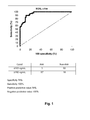
- a cutoff value for the NGAL concentration could be chosen that gave 100% sensitivity for the diagnosis of ARF, while retaining a diagnostic specificity of 74%.
- the lowest cutoff value that gave 100% diagnostic specificity for ARF gave a diagnostic sensitivity of 56%.
- the cutoff value giving 100% diagnostic sensitivity corresponds to a value between the highest value found in the patients who did not develop ARF and the lowest value found in the patients who did develop ARF. Using this value or indeed any lower value as the cutoff gives a diagnostic test with a negative predictive value of 100%, i.e. predicting that patients from this population with a urinary NGAL concentration below this value do not have ARF.
- the present invention relates to measurement of NGAL in a sample of bodily fluid, preferably urine or plasma, in order to determine the likelihood of ARF in a human subject, whereby it is possible to discriminate between a subject who is "not at risk of having or developing ARF" and a subject who is "at risk of having or developing ARF", the said invention comprising the steps of
- cutoff level so determined will depend on the characteristics of the patient population or group to which the test is applied. It is possible that certain patient groups that are not critically ill, such as patients subjected to elective surgery, cancer chemotherapy or diagnostic imaging with intravenous contrast agents, may require a lower cutoff value to provide a negative predictive value of 100%.
- the cutoff level below which the urinary concentration of NGAL has a negative predictive value of 100% for ARF in a determined group of patients is preferably a concentration of 250 ng/mL or less, such any value between 250 ng/mL and 200 ng/mL, such as 225 ng/mL, or any value between 200 ng/mL and 150 ng/mL, such as 175 ng/mL or 160 ng/mL or 155 ng/mL, or any value between 150 ng/mL and 100 ng/mL, such as 125 ng/mL, or any value between 100 ng/mL and 50 ng/mL, such as 75 ng/mL.
- the present invention comprises the steps of measuring the concentration of NGAL in a sample of plasma or serum from the subject to be diagnosed, and comparing the measured concentration with a selected cutoff value below which the plasma or serum concentration has a negative predictive value of 100% for ARF in the group of patients to which the subject belongs. If the measured NGAL concentration is below the cutoff level, this is an indication that the subject has not suffered renal injury and is not at immediate risk of developing ARF.
- the cutoff level below which the NGAL concentration in plasma or serum has a negative predictive value of 100% for ARF in a determined group of patients is similar to that for urine and is preferably a concentration of 250 ng/mL or less, such any value between 250 ng/mL and 200 ng/mL, such as 225 ng/mL, or any value between 200 ng/mL and 150 ng/mL, such as 175 ng/mL or 160 ng/mL or 155 ng/mL, or any value between 150 ng/mL and 100 ng/mL, such as 125 ng/mL.
- a further aspect of the present invention is that the method can be used to monitor patients throughout the course of an illness or at various times after a diagnostic or therapeutic intervention that carries a risk of provoking ARF. Comparison of the measured NGAL levels with the cutoff value will determine when the patient leaves the category of "not at risk of having or developing ARF" and enters the category of "at risk of having or developing ARF". The intervals at which samples of bodily fluids are taken for monitoring can be short, thus providing the earliest possible indication of the change of risk category and thus permitting the early institution of more intensive surveillance and any therapeutic measures.
- Monitoring of NGAL levels in bodily fluids for this purpose is preferably carried out at intervals not longer than 24 hours, and more preferably at shorter intervals down to a suggested period of not longer than 3 h, or even shorter, such as 30 minutes or 1 hour, for instance if a potential renal insult is known to have occurred, e.g. during a surgical or medical procedure.
- Measurement of human NGAL in a sample of bodily fluid can be performed by any method that provides satisfactory analytical specificity, sensitivity and precision.
- Preferred methods are binding assays using one or more binding molecules specific to human NGAL.
- binding molecules include, but are not limited to, polyclonal or monoclonal antibodies against NGAL or specific NGAL binding molecules generated by other means.
- monoclonal antibodies raised against recombinant human NGAL are used.
- One antibody is linked to a solid support to capture NGAL from a sample, such as a urine sample, while the other is linked to a label such as a dye complex, or biotin or enzyme that can be detected by any of many methods known to those skilled in the art.
- the solid support may e.g. be a polystyrene or polyvinyl chloride surface for enzyme-linked immunosorbent assay (ELISA), or latex (polystyrene) particles, or a filter frit composed of compressed polyethylene particles, or a porous nitrocellulose matrix, or indeed any suitable support used in immunochemical analyses.
- a preferred means for measuring NGAL in accordance with the present invention in a sample of human urine includes a dipstick, lateral flow (immunochromatographic) or minicolumn test, which allows for the rapid, near-patient analysis of a sample.
- a dipstick dipstick
- lateral flow immunochromatographic
- minicolumn test which allows for the rapid, near-patient analysis of a sample.
- other means for measuring NGAL can be used, including automated methods in central laboratories in which the apparatus permits the random access of samples for urgent analysis.
- the method of the invention does not comprise a surgical, therapeutic or diagnostic step practiced on the human body.
- the analytical area of a dipstick comprised of a polystyrene surface is coated with a capture antibody against human NGAL.
- An aliquot of the centrifuged, diluted sample is added to a solution of enzyme-labeled detection antibody against NGAL in the first tube, into which the dipstick is immersed.
- Complexes of enzyme-labeled detection antibody with NGAL are bound to the dipstick, which is then washed with tap water and placed in a chromogenic substrate solution in a second tube.
- the color developed in the substrate solution within a given time is read either by eye and compared with a chart of color intensities which indicates the concentration of NGAL in the urine sample, or in a simple colorimeter that can, for example, be programmed to indicate the NGAL concentration directly.
- a lateral flow device comprised of a strip of porous nitrocellulose is coated near its distal end with a capture antibody against NGAL applied as a transverse band.
- a further transverse band of antibody against antibodies of the species from which the detection antibody is derived is placed distally to the capture antibody band and serves as a control of strip function.
- the proximal end of the strip contains the detection antibody against NGAL adsorbed or linked to labeled polystyrene particles or particles of dye complex.
- the intensity of the labeled bands can be read by eye in the case of colored particles or by means of the appropriate detection device for the label used. A positive result is indicated by color development or the accumulation of label in both bands, while a negative result is indicated by color development or other label only in the control band. Failure of color development or other label in the control band indicates inadequate strip function.
- the sensitivity of the test can be regulated by the dilution of the sample applied, which is adjusted so that only NGAL concentrations above the determined cutoff values give rise to a positive result.
- the sensitivity of the test can also be adjusted by linking the detection antibody to a mixture of labeled and unlabeled particles. Batches of strips can be pre-calibrated and equipped with a calibration code that can be read by the detection device, so that a quantitative or semi-quantitative result can be read from the device. Many variations of the individual aspects of this lateral flow technology are possible, as known to those skilled in the art.
- a minicolumn contains a frit made of compressed polyethylene particles allowing the passage of fluid and cells.
- the frit is coated with capture antibody against human NGAL.
- the minicolumn is incorporated into a device, which by means of automated liquid handling allows the diluted sample to be applied at a fixed flow rate and volume, followed by detection antibody complexed with dye. After the passage of wash solution, the color intensity of the frit is read by light diffusion photometry.
- the batches of frits are pre-calibrated and the minicolumns equipped with a calibration code that can be read by the device, so that a quantitative result can be displayed by the instrument without the need for prior calibration with standards.
- Purified recombinant human NGAL for use as a standard and as calibrator material was prepared as described by Kjeldsen et al. (1996). Antibodies against NGAL were those described by Kjeldsen et al. (1993; 1996). Polystyrene ELISA plates were coated overnight at 4°C with antibody 211-1 at a concentration of 2 ⁇ g/mL in 0.05 M sodium carbonate buffer, pH 9.4, applied at 100 ⁇ L/well. The wells were emptied, washed 3 times with wash buffer of phosphate-buffered saline, pH 7.4, containing 0.05% Tween 20, and blotted.
- Dilutions of calibrator and samples in dilution buffer were applied to the wells in 100- ⁇ L volumes and incubated for 1 hour at room temperature on a shaking table. The wells were then emptied, washed and blotted as before.
- Biotinylated antibody 211-2 at 0.25 ⁇ g/mL in dilution buffer was added to each well at 100 ⁇ L/well and incubated for 1 hour at room temperature on a shaking table. The wells were then emptied, washed and blotted as before.
- a complex of horseradish peroxidase and streptavidin (Zymed, CA) at a dilution of 1/2000 in dilution buffer was added to each well at 100 ⁇ L/well and incubated for 1 hour at room temperature on a shaking table. The wells were then emptied, washed and blotted as before.
- a substrate solution containing tetramethylbenzidine and peroxide (TMB-ONE, Kem-En-Tech, Denmark) was then applied to each well at 100 ⁇ L/well and incubated at room temperature in the dark for exactly 8 minutes, after which the color reaction was stopped by adding 50 ⁇ L of 1 M sulfuric acid to each well.
- the light absorbances of the wells at a wavelength of 450 nm were then read in an ELISA plate reader, subtracting the light absorbances at 650 nm.
- the concentrations of NGAL in the samples were then calculated from the standard curve generated from the light absorbance readings of the calibrators of known concentration.
- the assay had a range of 0.02 ng/mL to 1 ng/mL, with a detection limit (95% confidence limit of difference from zero) of 2.4 pg/mL, and showed parallelism between dilutions of purified calibrator and samples.
- the concentration of NGAL was 90 ng/mL in a pool of normal human serum and 5.4 ng/mL in a pool of normal human urine.
Applications Claiming Priority (2)
| Application Number | Priority Date | Filing Date | Title |
|---|---|---|---|
| US83608806P | 2006-08-07 | 2006-08-07 | |
| EP07785730.8A EP2064553B2 (fr) | 2006-08-07 | 2007-08-03 | Test de diagnostic pour exclure une importante lésion rénale |
Related Parent Applications (2)
| Application Number | Title | Priority Date | Filing Date |
|---|---|---|---|
| EP07785730.8 Division | 2007-08-03 | ||
| EP07785730.8A Division-Into EP2064553B2 (fr) | 2006-08-07 | 2007-08-03 | Test de diagnostic pour exclure une importante lésion rénale |
Publications (1)
| Publication Number | Publication Date |
|---|---|
| EP2602624A1 true EP2602624A1 (fr) | 2013-06-12 |
Family
ID=38645725
Family Applications (2)
| Application Number | Title | Priority Date | Filing Date |
|---|---|---|---|
| EP07785730.8A Active EP2064553B2 (fr) | 2006-08-07 | 2007-08-03 | Test de diagnostic pour exclure une importante lésion rénale |
| EP13157838.7A Withdrawn EP2602624A1 (fr) | 2006-08-07 | 2007-08-03 | Test de diagnostic pour exclure une importante lésion rénale |
Family Applications Before (1)
| Application Number | Title | Priority Date | Filing Date |
|---|---|---|---|
| EP07785730.8A Active EP2064553B2 (fr) | 2006-08-07 | 2007-08-03 | Test de diagnostic pour exclure une importante lésion rénale |
Country Status (4)
| Country | Link |
|---|---|
| US (4) | US20100210031A2 (fr) |
| EP (2) | EP2064553B2 (fr) |
| JP (1) | JP2010500535A (fr) |
| WO (1) | WO2008017306A1 (fr) |
Families Citing this family (7)
| Publication number | Priority date | Publication date | Assignee | Title |
|---|---|---|---|---|
| AU2009237710A1 (en) * | 2008-04-15 | 2009-10-22 | Rainer Oberbauer | Markers of acute kidney failure |
| US7977110B2 (en) | 2008-06-02 | 2011-07-12 | Children's Hospital Medical Center | Method for distinguishing between kidney dysfunctions |
| CA2751430A1 (fr) * | 2009-02-06 | 2010-08-12 | Astute Medical, Inc. | Diagnostic et pronostic de lesion renale et d'insuffisance renale |
| WO2011000938A1 (fr) * | 2009-07-02 | 2011-01-06 | Mosaiques Diagnostics And Therapeutics Ag | Procédé et marqueur permettant de diagnostiquer une insuffisance rénale aiguë |
| WO2011045244A1 (fr) | 2009-10-14 | 2011-04-21 | Rainer Oberbauer | Test de risque de lésion rénale aigüe |
| CN102759623A (zh) * | 2012-07-06 | 2012-10-31 | 南京基蛋生物科技有限公司 | 一种检测ngal胶体金试纸条及其制备方法 |
| CN104569417B (zh) * | 2013-10-12 | 2016-06-01 | 广州瑞博奥生物科技有限公司 | 一种用于早期诊断急性肾损伤的抗体芯片试剂盒 |
Citations (3)
| Publication number | Priority date | Publication date | Assignee | Title |
|---|---|---|---|---|
| WO1995029404A1 (fr) | 1994-04-21 | 1995-11-02 | Per Venge | Utilisation de la lipocaline neutrophile humaine (hnl) comme marqueur diagnostique et preparation d'anticorps anti-hnl |
| WO2004088276A2 (fr) * | 2003-03-27 | 2004-10-14 | Children's Hospital Medical Center | Procede et trousse permettant de detecter l'apparition precoce de lesions de cellules tubulaires renales |
| WO2006066587A1 (fr) * | 2004-12-20 | 2006-06-29 | Antibodyshop A/S | Determination de la lipocaline neutrophile associee a la gelatinase (ngal) utile comme marqueur diagnostic dans les troubles renaux |
Family Cites Families (43)
| Publication number | Priority date | Publication date | Assignee | Title |
|---|---|---|---|---|
| US3635091A (en) * | 1970-08-31 | 1972-01-18 | Frederick D Linzer | Midstream urine specimen and fractional fluid collectors |
| IT1074038B (it) * | 1976-08-05 | 1985-04-17 | Simes | Esteri della epinina |
| US4376110A (en) * | 1980-08-04 | 1983-03-08 | Hybritech, Incorporated | Immunometric assays using monoclonal antibodies |
| US4357343A (en) * | 1981-06-26 | 1982-11-02 | Baxter Travenol Laboratories, Inc. | Nutritional composition for management of renal failure |
| US4640909A (en) * | 1985-05-07 | 1987-02-03 | J. T. Baker Chemical Company | Bonded phase of silica and carboalkoxyalkyl silanes for solid phase extraction |
| US5939272A (en) * | 1989-01-10 | 1999-08-17 | Biosite Diagnostics Incorporated | Non-competitive threshold ligand-receptor assays |
| JP2912413B2 (ja) * | 1990-03-28 | 1999-06-28 | 東亜医用電子株式会社 | 粒度分布作成方法 |
| US5405832A (en) * | 1991-11-27 | 1995-04-11 | Immtech International Inc. | Method of treating non-streptococcal bacterial infections |
| US5273961A (en) * | 1992-09-22 | 1993-12-28 | Genentech, Inc. | Method of prophylaxis of acute renal failure |
| US5552313A (en) * | 1994-11-21 | 1996-09-03 | Kansas University | DNA encoding mouse phosphotriesterase-related protein |
| US5750345A (en) * | 1995-10-31 | 1998-05-12 | Evanston Hospital Corporation | Detection of human α-thalassemia mutations and their use as predictors of blood-related disorders |
| US5627034A (en) * | 1995-12-05 | 1997-05-06 | Wisconsin Alumni Research Foundation | Assay for carcinoma proliferative status by measuring NGAL expression level |
| NZ336467A (en) * | 1996-05-24 | 2000-10-27 | Gen Hospital Corp | Modulators of tissue regeneration using DNA enclding a kidney injury molecule KIM |
| AU8899298A (en) * | 1997-08-06 | 1999-03-01 | Zymogenetics Inc. | Lipocalin homologs |
| US6309888B1 (en) * | 1998-09-04 | 2001-10-30 | Leuven Research & Development Vzw | Detection and determination of the stages of coronary artery disease |
| AUPP784398A0 (en) * | 1998-12-21 | 1999-01-21 | Monash University | Kidney disease detection and treatment |
| AU5330200A (en) * | 1999-06-18 | 2001-01-09 | Michigan State University | Method and apparatus for the detection of volatile products in a sample |
| US6762032B1 (en) * | 1999-08-23 | 2004-07-13 | Biocrystal, Ltd. | Compositions, assay kits, and methods for use related to a disease condition comprising multiple sclerosis and/or a pro-MS immune response |
| AU2001293964B2 (en) * | 2000-10-03 | 2007-06-14 | Rowett Research Institute | Method of assaying pyrrole-containing biological compounds |
| JP2004528814A (ja) * | 2000-10-13 | 2004-09-24 | チルドレンズ メディカル センター コーポレーション | 組織再構築関連状態についての非侵襲性酵素スクリーニング |
| FI20010019A (fi) * | 2001-01-05 | 2002-07-06 | Biohit Oyj | Menetelmä atrofisen diagnostisoimiseksi |
| US20040203083A1 (en) * | 2001-04-13 | 2004-10-14 | Biosite, Inc. | Use of thrombus precursor protein and monocyte chemoattractant protein as diagnostic and prognostic indicators in vascular diseases |
| US7713705B2 (en) * | 2002-12-24 | 2010-05-11 | Biosite, Inc. | Markers for differential diagnosis and methods of use thereof |
| DE60233301D1 (de) * | 2001-05-04 | 2009-09-24 | Biosite Inc | Diagnostische marker für akute herzerkrankungen und verfahren des gebrauchs |
| WO2003029462A1 (fr) * | 2001-09-27 | 2003-04-10 | Pieris Proteolab Ag | Muteines de la lipocaline neutrophile humaine associee a la gelatinase et de proteines apparentees |
| US20030119209A1 (en) * | 2001-12-21 | 2003-06-26 | Kaylor Rosann Marie | Diagnostic methods and devices |
| US6986995B2 (en) * | 2002-02-28 | 2006-01-17 | Prometheus Laboratories, Inc. | Methods of diagnosing liver fibrosis |
| US6847451B2 (en) * | 2002-05-01 | 2005-01-25 | Lifescan, Inc. | Apparatuses and methods for analyte concentration determination |
| GB0215509D0 (en) * | 2002-07-04 | 2002-08-14 | Novartis Ag | Marker genes |
| US20070275424A1 (en) * | 2002-11-22 | 2007-11-29 | Andrew Gewirtz | Diagnostic Tests and Methods for Diagnosing Inflammatory Bowel Disease |
| US7056702B2 (en) * | 2002-12-16 | 2006-06-06 | Kimberly Clark Co | Detecting lipocalin |
| US20050148029A1 (en) * | 2003-09-29 | 2005-07-07 | Biosite, Inc. | Methods and compositions for determining treatment regimens in systemic inflammatory response syndromes |
| US7776824B2 (en) * | 2004-05-06 | 2010-08-17 | The Trustees Of Columbia University | NGAL for reduction and amelioration of ischemic and nephrotoxic injuries |
| US20050272101A1 (en) * | 2004-06-07 | 2005-12-08 | Prasad Devarajan | Method for the early detection of renal injury |
| CA2511269A1 (fr) * | 2004-07-07 | 2006-01-07 | F. Hoffmann-La Roche Ag | Groupe de marqueurs multiples fonde sur le facteur de croissance placentaire pour les diabetes de type 1 et 2 |
| US20060105419A1 (en) * | 2004-08-16 | 2006-05-18 | Biosite, Inc. | Use of a glutathione peroxidase 1 as a marker in cardiovascular conditions |
| US20070037232A1 (en) * | 2005-03-31 | 2007-02-15 | Barasch Jonathan M | Detection of NGAL in chronic renal disease |
| US20080090304A1 (en) * | 2006-10-13 | 2008-04-17 | Barasch Jonathan Matthew | Diagnosis and monitoring of chronic renal disease using ngal |
| PL2035835T3 (pl) * | 2006-05-30 | 2012-05-31 | Antibodyshop As | Sposoby szybkiej oceny ciężkości urazu |
| US20080061149A1 (en) * | 2006-09-11 | 2008-03-13 | Colin Tanner | Proximity payment card with security interlock |
| US20090123946A1 (en) * | 2007-10-19 | 2009-05-14 | Abbott Laboratories | Immunoassays and kits for the detection of ngal |
| US20090124022A1 (en) * | 2007-10-19 | 2009-05-14 | Abbott Laboratories | Antibodies that bind to mammalian ngal and uses thereof |
| US20090123970A1 (en) * | 2007-10-19 | 2009-05-14 | Abbott Laboratories | Glycosylated mammalian ngal and use thereof |
-
2007
- 2007-08-03 JP JP2009523149A patent/JP2010500535A/ja active Pending
- 2007-08-03 WO PCT/DK2007/000366 patent/WO2008017306A1/fr active Application Filing
- 2007-08-03 US US12/375,585 patent/US20100210031A2/en not_active Abandoned
- 2007-08-03 EP EP07785730.8A patent/EP2064553B2/fr active Active
- 2007-08-03 EP EP13157838.7A patent/EP2602624A1/fr not_active Withdrawn
-
2013
- 2013-01-18 US US13/744,784 patent/US20130137124A1/en not_active Abandoned
-
2015
- 2015-12-29 US US14/982,153 patent/US20160109463A1/en not_active Abandoned
-
2019
- 2019-02-07 US US16/270,379 patent/US20190170770A1/en not_active Abandoned
Patent Citations (5)
| Publication number | Priority date | Publication date | Assignee | Title |
|---|---|---|---|---|
| WO1995029404A1 (fr) | 1994-04-21 | 1995-11-02 | Per Venge | Utilisation de la lipocaline neutrophile humaine (hnl) comme marqueur diagnostique et preparation d'anticorps anti-hnl |
| US6136526A (en) | 1994-04-21 | 2000-10-24 | Venge; Per | Use of human neutrophil lipocalin (HNL) as a diagnostic marker and anti-HNL-antibody preparation |
| WO2004088276A2 (fr) * | 2003-03-27 | 2004-10-14 | Children's Hospital Medical Center | Procede et trousse permettant de detecter l'apparition precoce de lesions de cellules tubulaires renales |
| US20040219603A1 (en) | 2003-03-27 | 2004-11-04 | Prasad Devarajan | Method and kit for detecting the early onset of renal tubular cell injury |
| WO2006066587A1 (fr) * | 2004-12-20 | 2006-06-29 | Antibodyshop A/S | Determination de la lipocaline neutrophile associee a la gelatinase (ngal) utile comme marqueur diagnostic dans les troubles renaux |
Non-Patent Citations (18)
| Title |
|---|
| AULITZKY WK; SCHLEGEL PN; WU DF; CHENG CY; CHEN CL; LI PS; GOLDSTEIN M; REIDENBERG M; BARDIN CW: "Measurement of urinary clusterin as an index of nephrotoxicity", PROC SOC EXP BIOL MED, vol. 199, 1992, pages 93 - 96, XP008154078, DOI: doi:10.3181/00379727-199-43335 |
| BANGERT ET AL: "NGAL is significantly increased in urine and plasma in acute renal failure", September 2006, INTENSIVE CARE MEDICINE ;, SPRINGER-VERLAG, BE, PAGE(S) S10, ISSN: 1432-1238, XP009091902 * |
| BANGERT K ET AL: "NGAL as a marker for renal injury in sepsis", March 2007, IMFLAMMATION RESEARCH, BIRKHAEUSER VERLAG, BASEL, CH, PAGE(S) S104-S105, ISSN: 1023-3830, XP008082913 * |
| HAN WK; BAILLY V; ABICHANDANI R; THADHANI R; BONVENTRE JV: "Kidney Injury Molecule-1 (KIM-1): a novel biomarker for human renal proximal tubule injury", KIDNEY INT, vol. 62, 2002, pages 237 - 244, XP002371104, DOI: doi:10.1046/j.1523-1755.2002.00433.x |
| KJELDSEN L; JOHNSEN AH; SENGELOV H; BORREGAARD N: "Isolation and primary structure of NGAL, a novel protein associated with human neutrophil gelatinase", J BIOL CHEM, vol. 268, 1993, pages 10425 - 10432 |
| KJELDSEN L; KOCH C; ARNLJOTS K; BORREGAARD N: "Characterization of two ELISAs for NGAL, a newly described lipocalin in human neutrophils", J IMMUNOL METHODS, vol. 198, 1996, pages 155 - 164, XP004071802, DOI: doi:10.1016/S0022-1759(96)00153-6 |
| KOTANKO P; MARGREITER R; PFALLER W: "Urinary N-acetyl-beta-D-glucosaminidase and neopterin aid in the diagnosis of rejection and acute tubular necrosis in initially nonfunctioning kidney grafts", NEPHRON, vol. 84, 2000, pages 228 - 235 |
| LIU Q; NILSEN-HAMILTON M: "Identification of a new acute phase protein", J BIOL CHEM, vol. 270, 1995, pages 22565 - 22570, XP002448530, DOI: doi:10.1074/jbc.270.38.22565 |
| MISHRA J ET AL: "Identification of neutrophil gelatinase-associated lipocalin as a novel early urinary biomarker for ischemic renal injury", JOURNAL OF THE AMERICAN SOCIETY OF NEPHROLOGY, WILLIAMS AND WILKINS, BALTIMORE, MD, US, vol. 14, no. 10, October 2003 (2003-10-01), pages 2534 - 2543, XP002377943, ISSN: 1046-6673 * |
| MISHRA J ET AL: "Neutrophil gelatinase-associated lipocalin (NGAL) as a biomarker for acute renal injury after cardiac surgery", 2 April 2005, LANCET THE, LANCET LIMITED. LONDON, GB, PAGE(S) 1231-1238, ISSN: 0140-6736, XP004824250 * |
| MOLITORIS BA: "Transitioning to therapy in ischemic acute renal failure", J AM SOC NEPHROL, vol. 14, 2003, pages 265 - 267 |
| MONIER F; SURLA A; GUILLOT M; MOREL F: "Gelatinase isoforms in urine from bladder cancer patients", CLIN CHIM ACTA, vol. 299, 2000, pages 11 - 23, XP027214113 |
| MURAMATSU Y; TSUJIE M; KOHDA Y; PHAM B; PERANTONI AO; ZHAO H; JO SK; YUEN PS; CRAIG L; HU X: "Early detection of cysteine rich protein 61 (CYR61, CCN1) in urine following renal ischemic reperfusion injury", KIDNEY INT, vol. 62, 2002, pages 1601 - 1610, XP002966736, DOI: doi:10.1046/j.1523-1755.2002.00633.x |
| PENDERS J; DELANGHE JR: "Alpha 1-microglobulin: clinical laboratory aspects and applications", CLIN CHIM ACTA, vol. 346, 2004, pages 107 - 118, XP008152082, DOI: doi:10.1016/j.cccn.2004.03.037 |
| STOESZ SP; GOULD MN: "Overexpression of neu-related lipocalin (NRL) in neu- initiated but not ras or chemically initiated rat mammary carcinomas", ONCOGENE, vol. 11, 1995, pages 2233 - 2241, XP008015618 |
| TRIEBEL S; BLASER J; REINKE H; TSCHESCHE H: "A 25 kDa alpha 2-microglobulin-related protein is a component of the 125 kDa form of human gelatinase", FEBS LETT, vol. 314, 1992, pages 386 - 388, XP055036654, DOI: doi:10.1016/0014-5793(92)81511-J |
| TSUCHIDA T; EGUCHI N; EGUCHI Y; NUMABE A; NAKAJIMA H; ODA H; SEIKI K; HAKAMADA-TAGUCHI R; URADE Y; UEHARA Y: "Lipocalin-type prostaglandin D synthase in urine in adriamycin-induced nephropathy of mice", NEPHRON PHYSIOL, vol. 96, 2004, pages 42 - 51 |
| YAN L; BORREGAARD N; KJELDSEN L; MOSES MA: "The high molecular weight urinary matrix metalloproteinase (MMP) activity is a complex of gelatinase B/MMP-9 and neutrophil gelatinase-associated lipocalin (NGAL). Modulation of MMP-9 activity by NGAL", J BIOL CHEM, vol. 276, pages 37258 - 37265, XP002226670, DOI: doi:10.1074/jbc.M106089200 |
Also Published As
| Publication number | Publication date |
|---|---|
| US20160109463A1 (en) | 2016-04-21 |
| US20130137124A1 (en) | 2013-05-30 |
| EP2064553B1 (fr) | 2014-09-03 |
| JP2010500535A (ja) | 2010-01-07 |
| WO2008017306A1 (fr) | 2008-02-14 |
| EP2064553B2 (fr) | 2023-06-07 |
| US20100210031A2 (en) | 2010-08-19 |
| US20090311801A1 (en) | 2009-12-17 |
| US20190170770A1 (en) | 2019-06-06 |
| EP2064553A1 (fr) | 2009-06-03 |
Similar Documents
| Publication | Publication Date | Title |
|---|---|---|
| EP1831699B1 (fr) | Determination de la lipocaline neutrophile associee a la gelatinase (ngal) utile comme marqueur diagnostic dans les troubles renaux | |
| US20190170770A1 (en) | Diagnostic Test to Exclude Significant Renal Injury | |
| EP2137538B1 (fr) | Essai diagnostique de lesion rénale | |
| Class et al. | Patent application title: DIAGNOSTIC TEST TO EXCLUDE SIGNIFICANT RENAL INJURY Inventors: Lars Otto Uttenthal (Salamanca, ES) Antibodyshop A/s (Gentofte, DE) Kristian Bangert (Holte, DE) Assignees: AntibodyShop A/S | |
| WO2023277130A1 (fr) | Biomarqueur pour la détection de troubles tubulointerstitiels et son utilisation |
Legal Events
| Date | Code | Title | Description |
|---|---|---|---|
| PUAI | Public reference made under article 153(3) epc to a published international application that has entered the european phase |
Free format text: ORIGINAL CODE: 0009012 |
|
| AC | Divisional application: reference to earlier application |
Ref document number: 2064553 Country of ref document: EP Kind code of ref document: P |
|
| AK | Designated contracting states |
Kind code of ref document: A1 Designated state(s): AT BE BG CH CY CZ DE DK EE ES FI FR GB GR HU IE IS IT LI LT LU LV MC MT NL PL PT RO SE SI SK TR |
|
| STAA | Information on the status of an ep patent application or granted ep patent |
Free format text: STATUS: THE APPLICATION IS DEEMED TO BE WITHDRAWN |
|
| 18D | Application deemed to be withdrawn |
Effective date: 20131213 |