EP2585830B1 - A method and device for determining content of the middle and protein bound uremic toxins in a biological fluid - Google Patents
A method and device for determining content of the middle and protein bound uremic toxins in a biological fluid Download PDFInfo
- Publication number
- EP2585830B1 EP2585830B1 EP11740556.3A EP11740556A EP2585830B1 EP 2585830 B1 EP2585830 B1 EP 2585830B1 EP 11740556 A EP11740556 A EP 11740556A EP 2585830 B1 EP2585830 B1 EP 2585830B1
- Authority
- EP
- European Patent Office
- Prior art keywords
- concentration
- dialysis
- fluorescence
- biological fluid
- uremic toxins
- Prior art date
- Legal status (The legal status is an assumption and is not a legal conclusion. Google has not performed a legal analysis and makes no representation as to the accuracy of the status listed.)
- Not-in-force
Links
- 239000002441 uremic toxin Substances 0.000 title claims description 44
- 102000004169 proteins and genes Human genes 0.000 title claims description 32
- 108090000623 proteins and genes Proteins 0.000 title claims description 32
- 239000013060 biological fluid Substances 0.000 title claims description 26
- 238000000034 method Methods 0.000 title description 87
- BXFFHSIDQOFMLE-UHFFFAOYSA-N indoxyl sulfate Chemical compound C1=CC=C2C(OS(=O)(=O)O)=CNC2=C1 BXFFHSIDQOFMLE-UHFFFAOYSA-N 0.000 claims description 132
- 102000015736 beta 2-Microglobulin Human genes 0.000 claims description 52
- 108010081355 beta 2-Microglobulin Proteins 0.000 claims description 52
- 230000003287 optical effect Effects 0.000 claims description 52
- 238000005259 measurement Methods 0.000 claims description 35
- 238000012545 processing Methods 0.000 claims description 10
- 230000001131 transforming effect Effects 0.000 claims description 9
- 238000004422 calculation algorithm Methods 0.000 claims description 6
- 238000013079 data visualisation Methods 0.000 claims description 3
- 238000004364 calculation method Methods 0.000 claims description 2
- 238000001228 spectrum Methods 0.000 claims description 2
- 238000000502 dialysis Methods 0.000 description 80
- SEOVTRFCIGRIMH-UHFFFAOYSA-N indole-3-acetic acid Chemical compound C1=CC=C2C(CC(=O)O)=CNC2=C1 SEOVTRFCIGRIMH-UHFFFAOYSA-N 0.000 description 31
- 239000000126 substance Substances 0.000 description 29
- QIAFMBKCNZACKA-UHFFFAOYSA-N N-benzoylglycine Chemical compound OC(=O)CNC(=O)C1=CC=CC=C1 QIAFMBKCNZACKA-UHFFFAOYSA-N 0.000 description 27
- XSQUKJJJFZCRTK-UHFFFAOYSA-N Urea Chemical compound NC(N)=O XSQUKJJJFZCRTK-UHFFFAOYSA-N 0.000 description 26
- DDRJAANPRJIHGJ-UHFFFAOYSA-N creatinine Chemical compound CN1CC(=O)NC1=N DDRJAANPRJIHGJ-UHFFFAOYSA-N 0.000 description 26
- 210000004369 blood Anatomy 0.000 description 22
- 239000008280 blood Substances 0.000 description 22
- 238000002835 absorbance Methods 0.000 description 15
- 238000012544 monitoring process Methods 0.000 description 15
- 230000005284 excitation Effects 0.000 description 14
- 239000003617 indole-3-acetic acid Substances 0.000 description 14
- 239000004202 carbamide Substances 0.000 description 13
- 150000001875 compounds Chemical class 0.000 description 13
- 229940109239 creatinine Drugs 0.000 description 13
- LEHOTFFKMJEONL-UHFFFAOYSA-N Uric Acid Chemical compound N1C(=O)NC(=O)C2=C1NC(=O)N2 LEHOTFFKMJEONL-UHFFFAOYSA-N 0.000 description 12
- TVWHNULVHGKJHS-UHFFFAOYSA-N Uric acid Natural products N1C(=O)NC(=O)C2NC(=O)NC21 TVWHNULVHGKJHS-UHFFFAOYSA-N 0.000 description 12
- 238000011282 treatment Methods 0.000 description 12
- 229940116269 uric acid Drugs 0.000 description 12
- 239000012530 fluid Substances 0.000 description 8
- 230000006870 function Effects 0.000 description 8
- 239000007788 liquid Substances 0.000 description 7
- 208000037157 Azotemia Diseases 0.000 description 6
- 210000004027 cell Anatomy 0.000 description 6
- 238000012937 correction Methods 0.000 description 6
- 238000000338 in vitro Methods 0.000 description 6
- 239000003550 marker Substances 0.000 description 6
- 229910019142 PO4 Inorganic materials 0.000 description 5
- 238000004458 analytical method Methods 0.000 description 5
- 238000002189 fluorescence spectrum Methods 0.000 description 5
- 230000014759 maintenance of location Effects 0.000 description 5
- 239000000203 mixture Substances 0.000 description 5
- NBIIXXVUZAFLBC-UHFFFAOYSA-K phosphate Chemical compound [O-]P([O-])([O-])=O NBIIXXVUZAFLBC-UHFFFAOYSA-K 0.000 description 5
- 239000010452 phosphate Substances 0.000 description 5
- 210000002966 serum Anatomy 0.000 description 5
- 238000010989 Bland-Altman Methods 0.000 description 4
- 102000004127 Cytokines Human genes 0.000 description 4
- 108090000695 Cytokines Proteins 0.000 description 4
- 108010001336 Horseradish Peroxidase Proteins 0.000 description 4
- SIKJAQJRHWYJAI-UHFFFAOYSA-N Indole Chemical compound C1=CC=C2NC=CC2=C1 SIKJAQJRHWYJAI-UHFFFAOYSA-N 0.000 description 4
- FFFHZYDWPBMWHY-VKHMYHEASA-N L-homocysteine Chemical compound OC(=O)[C@@H](N)CCS FFFHZYDWPBMWHY-VKHMYHEASA-N 0.000 description 4
- QIVBCDIJIAJPQS-VIFPVBQESA-N L-tryptophane Chemical class C1=CC=C2C(C[C@H](N)C(O)=O)=CNC2=C1 QIVBCDIJIAJPQS-VIFPVBQESA-N 0.000 description 4
- 230000006399 behavior Effects 0.000 description 4
- 238000004587 chromatography analysis Methods 0.000 description 4
- 230000000694 effects Effects 0.000 description 4
- 230000008569 process Effects 0.000 description 4
- 229940045136 urea Drugs 0.000 description 4
- 208000009852 uremia Diseases 0.000 description 4
- 210000002700 urine Anatomy 0.000 description 4
- 229930192334 Auxin Natural products 0.000 description 3
- 102000004889 Interleukin-6 Human genes 0.000 description 3
- QIVBCDIJIAJPQS-UHFFFAOYSA-N Tryptophan Natural products C1=CC=C2C(CC(N)C(O)=O)=CNC2=C1 QIVBCDIJIAJPQS-UHFFFAOYSA-N 0.000 description 3
- 208000016463 Wild type ABeta2M amyloidosis Diseases 0.000 description 3
- 238000013459 approach Methods 0.000 description 3
- 239000002363 auxin Substances 0.000 description 3
- WPYMKLBDIGXBTP-UHFFFAOYSA-N benzoic acid Chemical compound OC(=O)C1=CC=CC=C1 WPYMKLBDIGXBTP-UHFFFAOYSA-N 0.000 description 3
- 238000001514 detection method Methods 0.000 description 3
- 239000003814 drug Substances 0.000 description 3
- 229940079593 drug Drugs 0.000 description 3
- 238000001631 haemodialysis Methods 0.000 description 3
- 230000000322 hemodialysis Effects 0.000 description 3
- 229940100601 interleukin-6 Drugs 0.000 description 3
- 210000004185 liver Anatomy 0.000 description 3
- 238000004519 manufacturing process Methods 0.000 description 3
- 239000012528 membrane Substances 0.000 description 3
- 230000005855 radiation Effects 0.000 description 3
- 230000003595 spectral effect Effects 0.000 description 3
- 238000000108 ultra-filtration Methods 0.000 description 3
- 239000002699 waste material Substances 0.000 description 3
- XLYOFNOQVPJJNP-UHFFFAOYSA-N water Substances O XLYOFNOQVPJJNP-UHFFFAOYSA-N 0.000 description 3
- 208000024172 Cardiovascular disease Diseases 0.000 description 2
- 241000196324 Embryophyta Species 0.000 description 2
- DHMQDGOQFOQNFH-UHFFFAOYSA-N Glycine Chemical compound NCC(O)=O DHMQDGOQFOQNFH-UHFFFAOYSA-N 0.000 description 2
- ZRALSGWEFCBTJO-UHFFFAOYSA-N Guanidine Chemical compound NC(N)=N ZRALSGWEFCBTJO-UHFFFAOYSA-N 0.000 description 2
- 108090001005 Interleukin-6 Proteins 0.000 description 2
- 101000878457 Macrocallista nimbosa FMRFamide Proteins 0.000 description 2
- AIJULSRZWUXGPQ-UHFFFAOYSA-N Methylglyoxal Chemical compound CC(=O)C=O AIJULSRZWUXGPQ-UHFFFAOYSA-N 0.000 description 2
- 208000001647 Renal Insufficiency Diseases 0.000 description 2
- 108060008682 Tumor Necrosis Factor Proteins 0.000 description 2
- 102000000852 Tumor Necrosis Factor-alpha Human genes 0.000 description 2
- 238000009825 accumulation Methods 0.000 description 2
- 239000012491 analyte Substances 0.000 description 2
- 230000001580 bacterial effect Effects 0.000 description 2
- 230000008901 benefit Effects 0.000 description 2
- 230000008033 biological extinction Effects 0.000 description 2
- 208000003295 carpal tunnel syndrome Diseases 0.000 description 2
- 239000003795 chemical substances by application Substances 0.000 description 2
- 230000001684 chronic effect Effects 0.000 description 2
- 208000020832 chronic kidney disease Diseases 0.000 description 2
- CVSVTCORWBXHQV-UHFFFAOYSA-N creatine Chemical compound NC(=[NH2+])N(C)CC([O-])=O CVSVTCORWBXHQV-UHFFFAOYSA-N 0.000 description 2
- 238000011161 development Methods 0.000 description 2
- 230000018109 developmental process Effects 0.000 description 2
- 230000008030 elimination Effects 0.000 description 2
- 238000003379 elimination reaction Methods 0.000 description 2
- 238000005516 engineering process Methods 0.000 description 2
- RWSXRVCMGQZWBV-WDSKDSINSA-N glutathione Chemical compound OC(=O)[C@@H](N)CCC(=O)N[C@@H](CS)C(=O)NCC(O)=O RWSXRVCMGQZWBV-WDSKDSINSA-N 0.000 description 2
- PZOUSPYUWWUPPK-UHFFFAOYSA-N indole Natural products CC1=CC=CC2=C1C=CN2 PZOUSPYUWWUPPK-UHFFFAOYSA-N 0.000 description 2
- RKJUIXBNRJVNHR-UHFFFAOYSA-N indolenine Natural products C1=CC=C2CC=NC2=C1 RKJUIXBNRJVNHR-UHFFFAOYSA-N 0.000 description 2
- 230000000968 intestinal effect Effects 0.000 description 2
- 210000003734 kidney Anatomy 0.000 description 2
- 201000006370 kidney failure Diseases 0.000 description 2
- 238000011005 laboratory method Methods 0.000 description 2
- 238000004811 liquid chromatography Methods 0.000 description 2
- 230000007774 longterm Effects 0.000 description 2
- 239000000463 material Substances 0.000 description 2
- 238000012986 modification Methods 0.000 description 2
- 230000004048 modification Effects 0.000 description 2
- 210000004789 organ system Anatomy 0.000 description 2
- 230000036542 oxidative stress Effects 0.000 description 2
- AYEKKSTZQYEZPU-RYUDHWBXSA-N pentosidine Chemical compound OC(=O)[C@@H](N)CCCCN1C=CC=C2N=C(NCCC[C@H](N)C(O)=O)N=C12 AYEKKSTZQYEZPU-RYUDHWBXSA-N 0.000 description 2
- 239000002243 precursor Substances 0.000 description 2
- 102000004196 processed proteins & peptides Human genes 0.000 description 2
- 108090000765 processed proteins & peptides Proteins 0.000 description 2
- 230000035755 proliferation Effects 0.000 description 2
- 238000011002 quantification Methods 0.000 description 2
- 230000009467 reduction Effects 0.000 description 2
- 238000011160 research Methods 0.000 description 2
- 230000000717 retained effect Effects 0.000 description 2
- 238000005070 sampling Methods 0.000 description 2
- 238000000926 separation method Methods 0.000 description 2
- 150000003384 small molecules Chemical class 0.000 description 2
- 239000000243 solution Substances 0.000 description 2
- 238000004611 spectroscopical analysis Methods 0.000 description 2
- 238000012360 testing method Methods 0.000 description 2
- 231100000419 toxicity Toxicity 0.000 description 2
- 230000001988 toxicity Effects 0.000 description 2
- APJYDQYYACXCRM-UHFFFAOYSA-N tryptamine Chemical compound C1=CC=C2C(CCN)=CNC2=C1 APJYDQYYACXCRM-UHFFFAOYSA-N 0.000 description 2
- LOGFVTREOLYCPF-KXNHARMFSA-N (2s,3r)-2-[[(2r)-1-[(2s)-2,6-diaminohexanoyl]pyrrolidine-2-carbonyl]amino]-3-hydroxybutanoic acid Chemical compound C[C@@H](O)[C@@H](C(O)=O)NC(=O)[C@H]1CCCN1C(=O)[C@@H](N)CCCCN LOGFVTREOLYCPF-KXNHARMFSA-N 0.000 description 1
- AAWZDTNXLSGCEK-LNVDRNJUSA-N (3r,5r)-1,3,4,5-tetrahydroxycyclohexane-1-carboxylic acid Chemical compound O[C@@H]1CC(O)(C(O)=O)C[C@@H](O)C1O AAWZDTNXLSGCEK-LNVDRNJUSA-N 0.000 description 1
- MZOFCQQQCNRIBI-VMXHOPILSA-N (3s)-4-[[(2s)-1-[[(2s)-1-[[(1s)-1-carboxy-2-hydroxyethyl]amino]-4-methyl-1-oxopentan-2-yl]amino]-5-(diaminomethylideneamino)-1-oxopentan-2-yl]amino]-3-[[2-[[(2s)-2,6-diaminohexanoyl]amino]acetyl]amino]-4-oxobutanoic acid Chemical compound OC[C@@H](C(O)=O)NC(=O)[C@H](CC(C)C)NC(=O)[C@H](CCCN=C(N)N)NC(=O)[C@H](CC(O)=O)NC(=O)CNC(=O)[C@@H](N)CCCCN MZOFCQQQCNRIBI-VMXHOPILSA-N 0.000 description 1
- ZGCHLOWZNKRZSN-NTSWFWBYSA-N 3-deoxyglucosone Chemical compound OC[C@@H](O)[C@@H](O)CC(=O)C=O ZGCHLOWZNKRZSN-NTSWFWBYSA-N 0.000 description 1
- UHPMJDGOAZMIID-UHFFFAOYSA-N 3-deoxyglucosone Natural products OCC1OC(O)C(=O)CC1O UHPMJDGOAZMIID-UHFFFAOYSA-N 0.000 description 1
- IGXUUWYVUGBMFT-UHFFFAOYSA-N 3-methyleneoxindole Chemical compound C1=CC=C2C(=C)C(=O)NC2=C1 IGXUUWYVUGBMFT-UHFFFAOYSA-N 0.000 description 1
- LRFVTYWOQMYALW-UHFFFAOYSA-N 9H-xanthine Chemical class O=C1NC(=O)NC2=C1NC=N2 LRFVTYWOQMYALW-UHFFFAOYSA-N 0.000 description 1
- 108010088751 Albumins Proteins 0.000 description 1
- 102000009027 Albumins Human genes 0.000 description 1
- 201000001320 Atherosclerosis Diseases 0.000 description 1
- 241000894006 Bacteria Species 0.000 description 1
- 239000005711 Benzoic acid Substances 0.000 description 1
- BVKZGUZCCUSVTD-UHFFFAOYSA-M Bicarbonate Chemical compound OC([O-])=O BVKZGUZCCUSVTD-UHFFFAOYSA-M 0.000 description 1
- 206010005133 Bleeding tendencies Diseases 0.000 description 1
- AAWZDTNXLSGCEK-UHFFFAOYSA-N Cordycepinsaeure Natural products OC1CC(O)(C(O)=O)CC(O)C1O AAWZDTNXLSGCEK-UHFFFAOYSA-N 0.000 description 1
- 208000030453 Drug-Related Side Effects and Adverse reaction Diseases 0.000 description 1
- 238000002965 ELISA Methods 0.000 description 1
- 238000012286 ELISA Assay Methods 0.000 description 1
- 206010048554 Endothelial dysfunction Diseases 0.000 description 1
- 102000003951 Erythropoietin Human genes 0.000 description 1
- 108090000394 Erythropoietin Proteins 0.000 description 1
- 206010016654 Fibrosis Diseases 0.000 description 1
- 206010016717 Fistula Diseases 0.000 description 1
- 208000002705 Glucose Intolerance Diseases 0.000 description 1
- 206010018429 Glucose tolerance impaired Diseases 0.000 description 1
- 108010024636 Glutathione Proteins 0.000 description 1
- 239000004471 Glycine Substances 0.000 description 1
- 206010019280 Heart failures Diseases 0.000 description 1
- HTTJABKRGRZYRN-UHFFFAOYSA-N Heparin Chemical compound OC1C(NC(=O)C)C(O)OC(COS(O)(=O)=O)C1OC1C(OS(O)(=O)=O)C(O)C(OC2C(C(OS(O)(=O)=O)C(OC3C(C(O)C(O)C(O3)C(O)=O)OS(O)(=O)=O)C(CO)O2)NS(O)(=O)=O)C(C(O)=O)O1 HTTJABKRGRZYRN-UHFFFAOYSA-N 0.000 description 1
- 208000033892 Hyperhomocysteinemia Diseases 0.000 description 1
- 206010020772 Hypertension Diseases 0.000 description 1
- 206010061218 Inflammation Diseases 0.000 description 1
- 206010022489 Insulin Resistance Diseases 0.000 description 1
- 102000003777 Interleukin-1 beta Human genes 0.000 description 1
- 108090000193 Interleukin-1 beta Proteins 0.000 description 1
- FFEARJCKVFRZRR-BYPYZUCNSA-N L-methionine Chemical compound CSCC[C@H](N)C(O)=O FFEARJCKVFRZRR-BYPYZUCNSA-N 0.000 description 1
- 208000002720 Malnutrition Diseases 0.000 description 1
- 102000016397 Methyltransferase Human genes 0.000 description 1
- 108060004795 Methyltransferase Proteins 0.000 description 1
- YDGMGEXADBMOMJ-LURJTMIESA-N N(g)-dimethylarginine Chemical compound CN(C)C(\N)=N\CCC[C@H](N)C(O)=O YDGMGEXADBMOMJ-LURJTMIESA-N 0.000 description 1
- CHJJGSNFBQVOTG-UHFFFAOYSA-N N-methyl-guanidine Natural products CNC(N)=N CHJJGSNFBQVOTG-UHFFFAOYSA-N 0.000 description 1
- 102000004722 NADPH Oxidases Human genes 0.000 description 1
- 108010002998 NADPH Oxidases Proteins 0.000 description 1
- 206010028813 Nausea Diseases 0.000 description 1
- 206010028980 Neoplasm Diseases 0.000 description 1
- 201000011252 Phenylketonuria Diseases 0.000 description 1
- 206010036105 Polyneuropathy Diseases 0.000 description 1
- AAWZDTNXLSGCEK-ZHQZDSKASA-N Quinic acid Natural products O[C@H]1CC(O)(C(O)=O)C[C@H](O)C1O AAWZDTNXLSGCEK-ZHQZDSKASA-N 0.000 description 1
- ZJUKTBDSGOFHSH-WFMPWKQPSA-N S-Adenosylhomocysteine Chemical compound O[C@@H]1[C@H](O)[C@@H](CSCC[C@H](N)C(O)=O)O[C@H]1N1C2=NC=NC(N)=C2N=C1 ZJUKTBDSGOFHSH-WFMPWKQPSA-N 0.000 description 1
- MEFKEPWMEQBLKI-AIRLBKTGSA-N S-adenosyl-L-methioninate Chemical compound O[C@@H]1[C@H](O)[C@@H](C[S+](CC[C@H](N)C([O-])=O)C)O[C@H]1N1C2=NC=NC(N)=C2N=C1 MEFKEPWMEQBLKI-AIRLBKTGSA-N 0.000 description 1
- 208000005770 Secondary Hyperparathyroidism Diseases 0.000 description 1
- NINIDFKCEFEMDL-UHFFFAOYSA-N Sulfur Chemical compound [S] NINIDFKCEFEMDL-UHFFFAOYSA-N 0.000 description 1
- 239000005864 Sulphur Substances 0.000 description 1
- 206010070863 Toxicity to various agents Diseases 0.000 description 1
- 206010047700 Vomiting Diseases 0.000 description 1
- 230000005856 abnormality Effects 0.000 description 1
- 230000009471 action Effects 0.000 description 1
- 229960001570 ademetionine Drugs 0.000 description 1
- 230000032683 aging Effects 0.000 description 1
- 150000001413 amino acids Chemical class 0.000 description 1
- 206010002022 amyloidosis Diseases 0.000 description 1
- 208000007502 anemia Diseases 0.000 description 1
- 239000003963 antioxidant agent Substances 0.000 description 1
- 230000003078 antioxidant effect Effects 0.000 description 1
- 239000007864 aqueous solution Substances 0.000 description 1
- 235000010233 benzoic acid Nutrition 0.000 description 1
- 238000003339 best practice Methods 0.000 description 1
- 238000002306 biochemical method Methods 0.000 description 1
- 230000008827 biological function Effects 0.000 description 1
- 230000015572 biosynthetic process Effects 0.000 description 1
- 230000017531 blood circulation Effects 0.000 description 1
- 230000037396 body weight Effects 0.000 description 1
- 230000008416 bone turnover Effects 0.000 description 1
- 201000011510 cancer Diseases 0.000 description 1
- 125000002915 carbonyl group Chemical group [*:2]C([*:1])=O 0.000 description 1
- 210000000748 cardiovascular system Anatomy 0.000 description 1
- 229940000032 cardiovascular system drug Drugs 0.000 description 1
- 230000015556 catabolic process Effects 0.000 description 1
- 230000035567 cellular accumulation Effects 0.000 description 1
- 210000003169 central nervous system Anatomy 0.000 description 1
- 239000003153 chemical reaction reagent Substances 0.000 description 1
- 208000037976 chronic inflammation Diseases 0.000 description 1
- 230000006020 chronic inflammation Effects 0.000 description 1
- 208000022831 chronic renal failure syndrome Diseases 0.000 description 1
- 238000005345 coagulation Methods 0.000 description 1
- 230000015271 coagulation Effects 0.000 description 1
- 235000016213 coffee Nutrition 0.000 description 1
- 235000013353 coffee beverage Nutrition 0.000 description 1
- 239000000470 constituent Substances 0.000 description 1
- 229960003624 creatine Drugs 0.000 description 1
- 239000006046 creatine Substances 0.000 description 1
- 231100000433 cytotoxic Toxicity 0.000 description 1
- 230000001472 cytotoxic effect Effects 0.000 description 1
- 230000006378 damage Effects 0.000 description 1
- 230000034994 death Effects 0.000 description 1
- 238000006114 decarboxylation reaction Methods 0.000 description 1
- 230000007423 decrease Effects 0.000 description 1
- 230000017858 demethylation Effects 0.000 description 1
- 238000010520 demethylation reaction Methods 0.000 description 1
- 230000003544 deproteinization Effects 0.000 description 1
- 206010012601 diabetes mellitus Diseases 0.000 description 1
- 238000010586 diagram Methods 0.000 description 1
- 235000005911 diet Nutrition 0.000 description 1
- 230000000378 dietary effect Effects 0.000 description 1
- 235000018823 dietary intake Nutrition 0.000 description 1
- SWSQBOPZIKWTGO-UHFFFAOYSA-N dimethylaminoamidine Natural products CN(C)C(N)=N SWSQBOPZIKWTGO-UHFFFAOYSA-N 0.000 description 1
- 235000013399 edible fruits Nutrition 0.000 description 1
- 238000001962 electrophoresis Methods 0.000 description 1
- 208000028208 end stage renal disease Diseases 0.000 description 1
- 201000000523 end stage renal failure Diseases 0.000 description 1
- 210000002889 endothelial cell Anatomy 0.000 description 1
- 230000008694 endothelial dysfunction Effects 0.000 description 1
- 230000003511 endothelial effect Effects 0.000 description 1
- 229940105423 erythropoietin Drugs 0.000 description 1
- 238000002474 experimental method Methods 0.000 description 1
- 230000004761 fibrosis Effects 0.000 description 1
- 230000003890 fistula Effects 0.000 description 1
- 238000012921 fluorescence analysis Methods 0.000 description 1
- 238000001917 fluorescence detection Methods 0.000 description 1
- 238000009472 formulation Methods 0.000 description 1
- BFSYFTQDGRDJNV-AYHFEMFVSA-N fructosyllysine Chemical compound OC(=O)[C@@H](N)CCCCNCC(=O)[C@@H](O)[C@H](O)[C@H](O)CO BFSYFTQDGRDJNV-AYHFEMFVSA-N 0.000 description 1
- 206010061989 glomerulosclerosis Diseases 0.000 description 1
- 229960003180 glutathione Drugs 0.000 description 1
- LEQAOMBKQFMDFZ-UHFFFAOYSA-N glyoxal Chemical compound O=CC=O LEQAOMBKQFMDFZ-UHFFFAOYSA-N 0.000 description 1
- 229960004198 guanidine Drugs 0.000 description 1
- 238000005534 hematocrit Methods 0.000 description 1
- 230000002489 hematologic effect Effects 0.000 description 1
- 229960002897 heparin Drugs 0.000 description 1
- 229920000669 heparin Polymers 0.000 description 1
- 238000004128 high performance liquid chromatography Methods 0.000 description 1
- 230000003225 hyperhomocysteinemia Effects 0.000 description 1
- 230000036737 immune function Effects 0.000 description 1
- 210000000987 immune system Anatomy 0.000 description 1
- 238000010978 in-process monitoring Methods 0.000 description 1
- 230000001939 inductive effect Effects 0.000 description 1
- 230000004054 inflammatory process Effects 0.000 description 1
- 238000002329 infrared spectrum Methods 0.000 description 1
- 230000002401 inhibitory effect Effects 0.000 description 1
- 230000002452 interceptive effect Effects 0.000 description 1
- 108010088383 interleukin-6 receptor alpha Proteins 0.000 description 1
- 230000035987 intoxication Effects 0.000 description 1
- 231100000566 intoxication Toxicity 0.000 description 1
- 230000001071 malnutrition Effects 0.000 description 1
- 235000000824 malnutrition Nutrition 0.000 description 1
- 230000007246 mechanism Effects 0.000 description 1
- 230000003923 mental ability Effects 0.000 description 1
- 230000007102 metabolic function Effects 0.000 description 1
- 230000004060 metabolic process Effects 0.000 description 1
- 229930182817 methionine Natural products 0.000 description 1
- 238000013508 migration Methods 0.000 description 1
- 230000005012 migration Effects 0.000 description 1
- 229910000403 monosodium phosphate Inorganic materials 0.000 description 1
- 235000019799 monosodium phosphate Nutrition 0.000 description 1
- 230000008693 nausea Effects 0.000 description 1
- 210000000885 nephron Anatomy 0.000 description 1
- 208000015380 nutritional deficiency disease Diseases 0.000 description 1
- 238000011275 oncology therapy Methods 0.000 description 1
- 210000000056 organ Anatomy 0.000 description 1
- 210000000963 osteoblast Anatomy 0.000 description 1
- 238000007833 oxidative deamination reaction Methods 0.000 description 1
- 238000005895 oxidative decarboxylation reaction Methods 0.000 description 1
- 230000000849 parathyroid Effects 0.000 description 1
- 230000001717 pathogenic effect Effects 0.000 description 1
- 208000008494 pericarditis Diseases 0.000 description 1
- 210000001428 peripheral nervous system Anatomy 0.000 description 1
- WVDDGKGOMKODPV-ZQBYOMGUSA-N phenyl(114C)methanol Chemical compound O[14CH2]C1=CC=CC=C1 WVDDGKGOMKODPV-ZQBYOMGUSA-N 0.000 description 1
- 229930195732 phytohormone Natural products 0.000 description 1
- 239000003375 plant hormone Substances 0.000 description 1
- 230000007824 polyneuropathy Effects 0.000 description 1
- 230000004793 poor memory Effects 0.000 description 1
- 239000011148 porous material Substances 0.000 description 1
- OXCMYAYHXIHQOA-UHFFFAOYSA-N potassium;[2-butyl-5-chloro-3-[[4-[2-(1,2,4-triaza-3-azanidacyclopenta-1,4-dien-5-yl)phenyl]phenyl]methyl]imidazol-4-yl]methanol Chemical compound [K+].CCCCC1=NC(Cl)=C(CO)N1CC1=CC=C(C=2C(=CC=CC=2)C2=N[N-]N=N2)C=C1 OXCMYAYHXIHQOA-UHFFFAOYSA-N 0.000 description 1
- 239000003755 preservative agent Substances 0.000 description 1
- 230000002335 preservative effect Effects 0.000 description 1
- 238000002203 pretreatment Methods 0.000 description 1
- 238000000513 principal component analysis Methods 0.000 description 1
- 230000007425 progressive decline Effects 0.000 description 1
- 230000000750 progressive effect Effects 0.000 description 1
- 239000003642 reactive oxygen metabolite Substances 0.000 description 1
- 230000002829 reductive effect Effects 0.000 description 1
- 239000012088 reference solution Substances 0.000 description 1
- 238000000611 regression analysis Methods 0.000 description 1
- 230000001105 regulatory effect Effects 0.000 description 1
- 230000002441 reversible effect Effects 0.000 description 1
- 210000003296 saliva Anatomy 0.000 description 1
- AJPJDKMHJJGVTQ-UHFFFAOYSA-M sodium dihydrogen phosphate Chemical compound [Na+].OP(O)([O-])=O AJPJDKMHJJGVTQ-UHFFFAOYSA-M 0.000 description 1
- 239000001488 sodium phosphate Substances 0.000 description 1
- 239000002594 sorbent Substances 0.000 description 1
- 241000894007 species Species 0.000 description 1
- 230000004936 stimulating effect Effects 0.000 description 1
- 238000003756 stirring Methods 0.000 description 1
- 238000006467 substitution reaction Methods 0.000 description 1
- 230000004083 survival effect Effects 0.000 description 1
- 229940065721 systemic for obstructive airway disease xanthines Drugs 0.000 description 1
- 238000002560 therapeutic procedure Methods 0.000 description 1
- 150000003573 thiols Chemical class 0.000 description 1
- 210000001519 tissue Anatomy 0.000 description 1
- 231100000331 toxic Toxicity 0.000 description 1
- 231100000167 toxic agent Toxicity 0.000 description 1
- 230000002588 toxic effect Effects 0.000 description 1
- 239000003440 toxic substance Substances 0.000 description 1
- 238000012546 transfer Methods 0.000 description 1
- 208000001072 type 2 diabetes mellitus Diseases 0.000 description 1
- 238000000825 ultraviolet detection Methods 0.000 description 1
- 210000004509 vascular smooth muscle cell Anatomy 0.000 description 1
- 235000013311 vegetables Nutrition 0.000 description 1
- 230000009385 viral infection Effects 0.000 description 1
- 230000008673 vomiting Effects 0.000 description 1
- 238000005303 weighing Methods 0.000 description 1
Images


Classifications
-
- G—PHYSICS
- G01—MEASURING; TESTING
- G01N—INVESTIGATING OR ANALYSING MATERIALS BY DETERMINING THEIR CHEMICAL OR PHYSICAL PROPERTIES
- G01N21/00—Investigating or analysing materials by the use of optical means, i.e. using sub-millimetre waves, infrared, visible or ultraviolet light
- G01N21/62—Systems in which the material investigated is excited whereby it emits light or causes a change in wavelength of the incident light
- G01N21/63—Systems in which the material investigated is excited whereby it emits light or causes a change in wavelength of the incident light optically excited
- G01N21/64—Fluorescence; Phosphorescence
- G01N21/6486—Measuring fluorescence of biological material, e.g. DNA, RNA, cells
-
- G—PHYSICS
- G01—MEASURING; TESTING
- G01N—INVESTIGATING OR ANALYSING MATERIALS BY DETERMINING THEIR CHEMICAL OR PHYSICAL PROPERTIES
- G01N33/00—Investigating or analysing materials by specific methods not covered by groups G01N1/00 - G01N31/00
- G01N33/48—Biological material, e.g. blood, urine; Haemocytometers
- G01N33/50—Chemical analysis of biological material, e.g. blood, urine; Testing involving biospecific ligand binding methods; Immunological testing
- G01N33/52—Use of compounds or compositions for colorimetric, spectrophotometric or fluorometric investigation, e.g. use of reagent paper and including single- and multilayer analytical elements
-
- A—HUMAN NECESSITIES
- A61—MEDICAL OR VETERINARY SCIENCE; HYGIENE
- A61B—DIAGNOSIS; SURGERY; IDENTIFICATION
- A61B5/00—Measuring for diagnostic purposes; Identification of persons
- A61B5/145—Measuring characteristics of blood in vivo, e.g. gas concentration or pH-value ; Measuring characteristics of body fluids or tissues, e.g. interstitial fluid or cerebral tissue
-
- G—PHYSICS
- G01—MEASURING; TESTING
- G01N—INVESTIGATING OR ANALYSING MATERIALS BY DETERMINING THEIR CHEMICAL OR PHYSICAL PROPERTIES
- G01N21/00—Investigating or analysing materials by the use of optical means, i.e. using sub-millimetre waves, infrared, visible or ultraviolet light
- G01N21/17—Systems in which incident light is modified in accordance with the properties of the material investigated
- G01N21/25—Colour; Spectral properties, i.e. comparison of effect of material on the light at two or more different wavelengths or wavelength bands
- G01N21/31—Investigating relative effect of material at wavelengths characteristic of specific elements or molecules, e.g. atomic absorption spectrometry
-
- G—PHYSICS
- G01—MEASURING; TESTING
- G01N—INVESTIGATING OR ANALYSING MATERIALS BY DETERMINING THEIR CHEMICAL OR PHYSICAL PROPERTIES
- G01N33/00—Investigating or analysing materials by specific methods not covered by groups G01N1/00 - G01N31/00
Definitions
- This invention relates to a novel method and a device for determining and quantitative concentration measurements of compounds in the biological fluids such as middle and protein bound uremic toxins in the biological fluids. More specifically, the present invention relates to an optical method utilizing fluorescence, preferable fluorescence of the spent dialysate, and a specific model, including a unique set of optical spectral components at certain wavelengths, to determine, preferable on-line, the concentration of the middle and protein bound uremic toxins such as beta2-microglobulin (B2M), and indoxyl sulfate (IS).
- B2M beta2-microglobulin
- IS indoxyl sulfate
- the uremic syndrome is attributed to the progressive retention of a large number of compounds, which under normal conditions are excreted by healthy kidneys. These compounds are called uremic retention solutes, or uremic toxins, when they interact negatively with biologic functions.
- the uremic syndrome is a complex "intoxication" of the retention of waste products resulting in multifactorial problems where disturbances in several metabolic functions are reflected in clinical problems.
- organs and organ systems are affected: cardio-vascular system (hypertension, pericarditis and heart failure), peripheral nervous system (polyneuropathy), central nervous system (poor memory, loss of concentration and slower mental ability), hematology (anemia, bleeding tendencies), coagulation, immune status (immunosupression), nausea, vomiting etc.
- uremic toxins are divided into three groups: 1) small molecules (MW ⁇ 500 Da); 2) middle molecules (MW > 500 Da); 3) protein-bound solutes.
- uremic toxins have effect to the patient by many different ways and extent, and to ensure the best survival, quality of the treatment and the quality of life for the dialysis patients monitoring of several uremic toxins is essential.
- Small molecular weight solutes (MW ⁇ 500 g/mol): Urea, Creatinine, Uric acid, Guanidine -ADMA (asymmetric dimethylarginine), Phosphate.
- Middle molecules (MW > 500 g/mol): ⁇ 2-microglobulin, Cytokines (Interleukin 6), Parathyroid hormon (PTH) - (at the same time belongs to the protein-bound group). Protein-bound solutes: Indoxyl sulfate, Homocysteine, P-cresol, AGE products, Hippuric acid.
- MM middle molecules
- MM uremic toxin group e.g. ⁇ 2-microglobulin
- the above middle molecular compounds have a pathogenic role or are markers of the most frequent long-term complications and causes of death in HD patients such as dialysis related amyloidosis, cardio-vascular disease, secondary hyperparathyroidism, inflammation and malnutrition. Reduction of the accumulation and lower long-term levels of these compounds may prevent or delay the appearance of such complications.
- B2M ⁇ 2-microglobulin
- B2M MW 11 818 D
- Uremia-related amyloid is to a large extent composed of B2M and is essentially found in the osteoarticular system and in the carpal tunnel. Uremia related amyloidosis becomes clinically apparent after several years of chronic renal failure and/or in the aged. B2M has become a frequently used marker for the dialytic removal of middle molecules. Behavior of B2M during dialysis is, however, not necessarily representative of that of other middle molecules.
- Cytokines are small molecules and connected with uremia- and dialysis-induced chronic inflammation. There is about 150 cytokines known, but about 300 cytokines are supposed to exist. Uremic toxins are Interleukin-1-beta, Interleukin 6 and Tumor Necrosis Factor Alpha. The accumulation of TNF-Alpha may contribute to the development of neurologic and hematologic complications in uremia. Interleukin-6 (MW 24 500 D) is regulating immune system and increases with bacterial and virus infection.
- Homocysteine (MW 135 D), a sulphur-containing amino acid, is produced by the demethylation of dietary methionine. Retention results in the cellular accumulation of S-adenosyl homocysteine, an extremely toxic compound, which competes with S-adenosyl-methionine and inhibits methyltransferase. Moderate hyperhomocysteinemia is an independent risk factor for cardiovascular disease. Hcy increases the proliferation of vascular smooth muscle cells, one of the most prominent hallmarks of atherosclerosis. P-cresol (MW 108 D) is cleared by the kidney and metabolized by the liver. P-cresol is uremic toxin involved in the depression of immune function. The protein binding is high and removal with dialysis treatment is low.
- AGE products (3-Deoxyglucosone, Fructoselysine, Glyoxal (ethanedial), Methylglyoxal, N-Epsilon-(Carboxymethyl)lysine, Pentosidine (MW 342 D)) are retained not only in renal failure but also in diabetes mellitus and aging, where they are held responsible for tissular damage and functional disturbances.
- Hippuric acid may be derived from the intake of coffee, fruits, or vegetables that contain quinic acid. This compound is converted to benzoic acid by bacterial activity in the gut; the benzoate is conjugated with glycine by the liver to form hippurate. In addition to dietary intake of hippuric acid and its precursors, patients on hippuric acid receive a further load of hippuric acid precursor in the form of benzyl alcohol, which may be used as a preservative in some heparin solutions and in multidose formulations of erythropoietin. Hippuric acid may enhance drug toxicity and the toxicity of other protein-bound uremic solutes due to competition for protein binding. It has been related to insulin resistance and glucose intolerance.
- Hippuric acid has been mentioned as one compound suitable for monitoring utilizing UV-absorbance ( US6666840, 23.12.2003, Falkvall et al ).
- US6666840, 23.12.2003, Falkvall et al the latest research has been demonstrated that hippuric acid monitoring by the above described method can be hardly realized. (Trifonov 2009) For this reason new approaches are needed.
- IS Indoxyl sulfate
- indole is metabolized by the liver from indole, which is produced by the intestinal flora as a metabolite of tryptophan.
- the production of indole in the gut may be greater in uremic patients than in normal subjects because of the effect the uremic milieu has on the composition of intestinal flora.
- IS is a circulating uremic toxin stimulating glomerular sclerosis and interstitial fibrosis and its removal by PD or by oral sorbent administration retards the progression of intact nephron loss.
- Indoxyl sulfate is one of the well-known substances of a group of protein-bound uremic retention solutes that increases the rate of progression of renal failure.
- IS is a protein-bound uremic solute that induces endothelial dysfunction by inhibiting endothelial proliferation and migration in vitro.
- serum levels of IS are associated with levels of pentosidine, a marker of carbonyl and oxidative stress; in vitro, indoxyl sulfate increases reactive oxygen species production in tubular cells, and increases NAD(P)H oxidase activity in endothelial cells. Indoxyl sulfate impairs osteoblast function, induces abnormalities of bone turnover and strongly decreases the levels of glutathione, one of the most active antioxidant systems of the cell. (Wishart DS, Knox C et al. 2009)
- Indole-3-acetic acid is a breakdown product of tryptophan metabolism and is often produced by the action of bacteria in the mammalian gut. Some endogenous production of I3AA in mammalian tissues also occurs. It may be produced by the decarboxylation of tryptamine or the oxidative deamination of tryptophan. I3AA frequently occurs at low levels in urine and has been found in elevated levels in the urine of patients with phenylketonuria. Using material extracted from human urine, it was discovered by Kogl in 1933 that Indoleacetic acid is also an important plant hormone. Specifically I3AA is a member of the group of phytohormones called auxins. I3AA is generally considered to be the most important native auxin.
- Plant cells synthesize I3AA from tryptophan.
- I3AA and some derivatives can be oxidised by horseradish peroxidase (HRP) to cytotoxic species.
- HRP horseradish peroxidase
- I3AA is only toxic after oxidative decarboxylation; the effect of I3AA/HRP is thought to be due in part to the formation of methylene-oxindole, which may conjugate with DNA bases and protein thiols.
- I3AA/HRP could be used as the basis for targeted cancer, a potential new role for plant auxins in cancer therapy. (Wishart DS, Knox C et al. 2009).
- ⁇ 2-microglobulin is mainly determined by ELISA assay method.
- the method is automated as an automatic bio-analyzer, the merits of the ELISA itself reside in mass detecting discrepancy and complicacy of the method. It cannot be applied in routine or household detecting because it requires professionals to operate, is expensive, and there is hard to store the detecting agents. Great care has been taken to ensure the quality and reliability of the method but however, it is possible that in certain cases unusual results may be obtained due to high levels of interfering factors.
- MM and protein bound uremic toxins are determined mostly utilizing the high performance reverse liquid chromatography (HPLC) method.
- HPLC high performance reverse liquid chromatography
- indoxyl sulfate has been determined by fluorescence detection (excitation 280 nm, emission 340 nm)
- hippuric acid has been analyzed by ultraviolet detection at 254 nm in the serum and in the spent dialysate (Dhondt, Vanholder et al. 2003).
- the demerits of this method include: 1) separation of the compounds may be difficult due to similar properties which affects the test accuracy; 2) the operation is complex, needs lots of agents and should be operated by professionals; 3) the sample needs pretreatment for deproteinization; and 4) the necessary equipment is expensive.
- the merits of the described method are that it does not need blood samples, no disposables or chemicals, and is fast.
- the described method is general and does not specify methodology to measure exclusively a single compound and is meant to apply only for dialysis monitoring. Moreover, no results about the concentration measurements are presented. More exact description about the uric acid and urea measurements using the abovementioned method is given in a scientific papers (Uhlin, Lindberg et al. 2005), (Uhlin, Fridolin et al. 2005).
- Another method relates to a method for dialysis monitoring method and apparatus using near infrared radiation, described in WO9819592,14.05.1998 , RIO GRANDE MEDICAL TECH INC.
- the merits of the described method are similar to that of the UV-radiation.
- the described method measures urea and creatinine by utilizing near infrared radiation spectrometry with different technical and optical considerations.
- near infrared radiation spectrometry the principal component analysis using calibration and prediction stage is described in US5886347 .
- US patent application US2003/0231294, 18.12.2003 discloses a system for measuring an analyte in a biological fluid using its intrinsic optical absorbance.
- the system has a light source forming an optical beam that is directed on the fluid sample and further has a process that selectively removes the intrinsic absorbance of the analyte in the fluid sample wherein the fluid sample has a first optical absorbance before the process and further having a second optical absorbance after the process.
- the system has a light detector receiving the optical beam directed through the fluid sample to produce a first signal indicative of the first optical absorbance and receiving the optical beam directed through the fluid sample after the process to produce a second signal indicative of the second optical absorbance.
- UV absorbance was determined by using a double-beam spectrophotometer on dialysate samples taken at pre-determined times during dialysis, over a wavelength range of 180-380nm. Concentrations of several removed substances, such as urea, creatinine, uric acid, phosphate and flz-microglobulin, were determined in the blood and in the spent dialysate samples using standard laboratory techniques.
- Millimolar extinction coefficients for urea, creatinine, monosodium phosphate and uric acid were determined during laboratory bench experiments. The correlation between UV absorbance and substances both in the dialysate and in the blood was calculated at all wavelengths. In conclusion the relatively high correlation between UV absorbance and the substances in the spent dialysate and in the blood indicates that the UV-absorbance technique can estimate the removal of several retained solutes known to accumulate in dialysis patients.
- the apparatus is a non-invasive, on-line, real time dialysis monitor which can monitor the progress of dialysis via monitoring the concentration of uremic toxin with near-infrared spectra.
- the uremic toxins concentration of urea and creatinine besides the clinically important bicarbonate, hematocrit, total protein and albumin can be optically monitored as well as pH.
- the concentration of urea, creatinine and total protein can be optically monitored as well as similar parameters can be monitored optically in the serum ultrafiltrate, on certain hemodialyzers having separate ultrafiltration means.
- the purpose of the invention is, therefore, a new method and a device for determining content of the middle and protein bound uremic toxins in the biological fluids. More specifically, the present invention relates to an optical method utilizing fluorescence of the biological fluid, preferable fluorescence of the spent dialysate and concentration calculation algorithm adapted to perform the transforming function to determine on the samples or on-line the concentration of the substances, which can be effected directly at the bed-side. The method and device determines the concentration of the substances in-vitro or on-line utilizing a measuring cuvette (cell) suitable for specified measurements.
- Another object of the present invention is to provide a practical optical method and device determining quantitatively concentration or removal of the middle and protein bound uremic toxins in the biological fluids.
- the determined values can be represented directly and easily on the monitor or screen printed.
- the method and device does not require any chemical disposables, neither expensive separation techniques, and can be easily made and mass-produced providing an environment-friendly optical method.
- a still further object of the present invention is to provide a method for assessing routine clinical monitoring in order to face risks of higher mortality in patients (e.g. in dialysis).
- a still further object of the present invention is to provide a novel, rapid, convenient and safe method for detecting concentration of substances in a liquid sample.
- the liquid sample can be directly dropped on the detecting cuvette for in-vitro measurements or sent a flowing stream of fluid through a flow-through cell for on-line monitoring.
- the method is suitable for household use when being applied to detect the concentration of substances in the biological fluids.
- the device for determining content of the middle and protein bound uremic toxins 5 (e.g. B2M) in a biological fluid 1 comprises:
- the light source can be either a broadband light source or a narrowband light source. If broadband light source is used, either a broadband detector and a filter can be used, or narrowband detectors. According to one embodiment, the light source is operating in the particular optical region (wavelength range 190-890 nm).
- the fluorescence light detector is operating in wavelength range of 190-900 nm.
- the detectable fluorescent light may be emitted directly by the measurable substance in the biofluid or the emission may be intermediated by some other natural or purposefully added constituents of the biofluid through some energy transfer mechanism between molecules in the fluid under the consideration.
- the measuring cuvette can be, e.g., adapted for in-vitro measurements, or designed for the on-line measurements.
- the spectra processing module is adapted to execute a concentration or removal calculation algorithm adapted to perform a transforming function calculating the concentration of certain substance in the biological fluid.
- the data representing module adapted for executing a program for data representation and comprises or is connected to a data visualization module, e.g., a monitor, a display, or a printing device.
- a data visualization module e.g., a monitor, a display, or a printing device.
- the duration of the ol-HDF treatments varied between 180 to 270 minutes, the dialysate flow was 500 mL/min, the blood flow varied between 280-350 mL/min. All patients were dialyzed via artery-venous fistulas using a "two-needle" system.
- the auto sub system mode for calculation of the on-line prepared substitution volume varied between 12.2 to 29.7 litres per session.
- Samples from the drain tube were taken at (min) 9, 30, 60, 120, 180 and at the end of ol-HDF session if longer than 180 min.
- One sample was taken from the dialysate/ultrafiltrate collection tank after careful stirring and weighing was performed. If a self-test of the dialysis machine occurred during the planned sampling time, the sample was taken when the UV-absorbance curve reached baseline level again which occurred within 2-3 minutes. Pure dialysate was collected before the start of a dialysis session, used as the reference solution, when the dialysis machine was prepared for starting and the conductivity was stable.
- Spectrofluorophotometer (SHIMADZU RF-5301) was used for the fluorescence measurements. Fluorescence analysis was performed over an excitation wavelength range of 220 - 900 nm (preferably 220-500nm), emission wavelength range of 220-890 nm (preferably 220-800 nm) and with excitation increment 10 nm. An optical cuvette with an optical path length of 0.4 and 1 cm was used. The obtained fluorescence values were processed and presented by software Panorama fluorescence and the final data processing was performed in EXCEL (Microsoft Office Excel 2003).
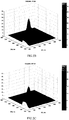
- Fig. 2 illustrates the examples of 3D fluorescence spectra obtained over the excitation wavelength range of 240 - 500 nm and emission wavelength range of 250 - 800 nm
- A on the pure dialysate sample
- B on the spent dialysate sample taken at 10 min after the start of a dialysis session
- C on the spent dialysate sample taken at 240 min after the start of a dialysis session.
- the fluorescence amplitude is proportional to the content of eliminated uremic toxins in the spent dialysate being higher in the beginning of the dialysis treatment (10 min) and lower at the end of the dialysis (207 min) at specific regions of the fluorescence spectra.
- BIAS Accuracy
- Table 1 summarises all results about the B2M and IS concentrations as mean and standard deviation values (Mean +/- SD) from the standardised methods (Lab) and from the new method (F).
- Table 1 Summary results about the concentration mean and standard deviation values (Mean +/- SD) from the standardised methods (Lab) and new method (F), linear correlation coefficient (R) and the R-squared value (R 2 ) between the uremic toxins concentration from the optical method and concentration measured at the laboratory, the accuracy (BIAS) and precision (SE) for the different methods to measure concentration of B2M and IS.
- the concentration of B2M from optical measurements is utilized below to calculate the dialysis dose for B2M, being representative in its kinetic behavior of other MM and peptides of similar size.
- the dialysis dose for the B2M from blood, spKt/Vb_B2M and eKt/Vb_B2M can be calculated using the pre-and post- dialysis blood B2M concentrations (Co and Ct).
- the fluorescence value in the beginning, F0 (10 min dialysate sample) and the fluorescence value at the end of dialysis, Ft were utilized.
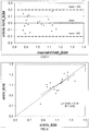
- Table 2 summarises all results about the dialysis dose for the B2M as spKt/V_B2M and eKt/V_B2M calculated using the pre-and post- dialysis blood B2M concentrations and the fluorescence values from totally 19 HDF sessions.
- the linear correlation coefficient (R) and the R-squared value (R2) between the dialysis dose for B2M from the optical method and dialysis dose for B2M from the blood concentrations are given.
- the accuracy (BIAS) and precision (SE) for the optical method was calculated using dialysis dose for the B2M from blood as reference after bias correction.
- Table 2 Summary of dialysis dose as spKt/V_B2M and eKt/V_B2M, calculated using the pre-and post- dialysis blood B2M concentrations (Blood) and the fluorescence values (F), the linear correlation coefficient (R) and the R-squared value (R2) between the dialysis dose for B2M from the optical method and from the blood concentrations, the accuracy (BIAS) and precision (SE) for the optical method.
- the concentration of IS from optical measurements is utilized below to calculate the dialysis dose during a single HDF session for the protein bound uremic toxin IS as: 1) removal rate for IS (RR IS), and 2) total removed amount for IS (TR_IS).
- the removal rate for IS (RRd_IS) and total removed amount for IS (TRd_IS) were used as the reference to corresponding parameters estimated by the optical method (RRf_IS and TRf_IS), since the elimination rate on blood values could be misleading due to specific kinetic behavior of the protein bound uremic toxins.
- RRd_IS The removal rate for IS in the spent dialysis
- TRf_IS the corresponding value for Dtotal in mg/L, estimated by the fluorescence measurements, were utilized.
- Table 3 summarises all results about the dialysis dose for the IS as RR_IS and TR_IS calculated using the spent dialysis IS concentrations from the laboratory in the beginning (10 min dialysate sample) and at the end of dialysis, and corresponding values from the fluorescence measurements.
- the linear correlation coefficient (R) and the R-squared value (R2) between the dialysis dose for IS from the optical method and dialysis dose for IS from the blood concentrations are given.
- the accuracy (BIAS) and precision (SE) for the optical method was calculated using dialysis dose for the IS from blood as reference after bias correction.
- Table 3 Summary of the dialysis dose for the IS as RR_IS and TR_IS calculated using the spent dialysis IS concentrations from the laboratory in the beginning (10 min dialysate sample) and at the end of dialysis, and corresponding values from the fluorescence measurements (F), the linear correlation coefficient (R) and the R-squared value (R2) between the dialysis dose for IS from the optical method and from the spent dialysate concentrations, the accuracy (BIAS) and precision (SE) for the optical method.
- F fluorescence measurements
- R linear correlation coefficient
- R2 the R-squared value
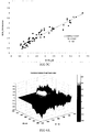
- HPLC peaks were identified, such as creatinine (Cr), uric acid (UA), hippuric acid (HA), trypthophane (Trp), indoxyl sulfate (IS), indol-3-acetic acid (I3AA). Moreover, 3 prevalent, but non-identified peaks - Peak A, Peak B and Peak C - were detected at different wavelengths.
- the HPLC profiles of the spent dialysate samples demonstrate selectivity for different solutes at different wavelengths. By this way, appropriate wavelength choice for the optical method enables to determine specific uremic toxins.
- the present invention is directed to a device for determining content of the middle and protein bound uremic toxins in a biological fluid
- a device for determining content of the middle and protein bound uremic toxins in a biological fluid comprising an optical module, comprising a fluorimetrical system, comprising a light source and a fluorescence light detectorand a measuring fluorimetrical cuvette for holding a sample of the biological fluid where the light with predetermined wavelengths can be led onto the sample and an fluorescence signal of the sample can be detected, and a signal processing module consisting of data acquisition module and a signal processing module incorporating concentration or removal calculation algorithm adapted to perform the transforming function.
- the device is adapted for introducing flowing stream of the biological fluid through fluorometrical flow-cuvette and outputting the concentration of the substance to a display device or to a printer.
- the excitation wavelength from the light source is in the wavelength range of 190-890 nm thereafter the emission wavelength detected by the light detector is operating in wavelength range of 190-900 nm.
- the substance is ⁇ 2-microglobulin or indoxyl sulfate.
- the light source is operating in the wavelength range of 360-380 nm, and the fluorescence light detector is operating in the wavelength range of 440-470 nm, suitable for beta2-microglobulin measurements.
- the light source is operating in the wavelength range of 290-310 nm, and the fluorescence light detector in the wavelength range of 340-370 nm, suitable for indoxyl sulfate measurements.
- the sample of the biological fluid is dropped onto in-vitro cuvette.
- the device is adopted for obtaining the sample and introducing the sample into cuvette in home conditions.
- the device is adapted for recording the concentration of the substance in a memory device and further for comparison of the substance concentration level in the fluid with predetermined limits of the concentration and generating an alarm signal, if the concentration does not fall between predetermined limits.
Landscapes
- Health & Medical Sciences (AREA)
- Life Sciences & Earth Sciences (AREA)
- Physics & Mathematics (AREA)
- Engineering & Computer Science (AREA)
- Immunology (AREA)
- Chemical & Material Sciences (AREA)
- General Health & Medical Sciences (AREA)
- Pathology (AREA)
- Molecular Biology (AREA)
- Biochemistry (AREA)
- Analytical Chemistry (AREA)
- General Physics & Mathematics (AREA)
- Biomedical Technology (AREA)
- Hematology (AREA)
- Nuclear Medicine, Radiotherapy & Molecular Imaging (AREA)
- Food Science & Technology (AREA)
- Spectroscopy & Molecular Physics (AREA)
- Medicinal Chemistry (AREA)
- Urology & Nephrology (AREA)
- Cell Biology (AREA)
- Microbiology (AREA)
- Optics & Photonics (AREA)
- Biophysics (AREA)
- Heart & Thoracic Surgery (AREA)
- Medical Informatics (AREA)
- Surgery (AREA)
- Animal Behavior & Ethology (AREA)
- Public Health (AREA)
- Veterinary Medicine (AREA)
- Biotechnology (AREA)
- Investigating, Analyzing Materials By Fluorescence Or Luminescence (AREA)
- Investigating Or Analysing Biological Materials (AREA)
- Investigating Or Analysing Materials By The Use Of Chemical Reactions (AREA)
- External Artificial Organs (AREA)
Applications Claiming Priority (3)
| Application Number | Priority Date | Filing Date | Title |
|---|---|---|---|
| EEP201000056A EE05676B1 (et) | 2010-06-28 | 2010-06-28 | Optiline meetod ja seade keskmise suurusega ja valkudega seotud ureemiliste toksiinide mramiseks bioloogilistes vedelikes |
| EEP201000085A EE05669B1 (et) | 2010-12-10 | 2010-12-10 | Optiline meetod ja seade keskmise suurusega ja valkudega seotud ureemiliste toksiinide mramiseks bioloogilistes vedelikes |
| PCT/EE2011/000008 WO2012000521A1 (en) | 2010-06-28 | 2011-06-28 | A method and device for determining content of the middle and protein bound uremic toxins in a biological fluid |
Publications (2)
| Publication Number | Publication Date |
|---|---|
| EP2585830A1 EP2585830A1 (en) | 2013-05-01 |
| EP2585830B1 true EP2585830B1 (en) | 2018-04-25 |
Family
ID=44508544
Family Applications (1)
| Application Number | Title | Priority Date | Filing Date |
|---|---|---|---|
| EP11740556.3A Not-in-force EP2585830B1 (en) | 2010-06-28 | 2011-06-28 | A method and device for determining content of the middle and protein bound uremic toxins in a biological fluid |
Country Status (10)
| Country | Link |
|---|---|
| US (1) | US9103789B2 (enExample) |
| EP (1) | EP2585830B1 (enExample) |
| JP (1) | JP5957450B2 (enExample) |
| KR (1) | KR101848527B1 (enExample) |
| CN (1) | CN102985822A (enExample) |
| AU (1) | AU2011274009B2 (enExample) |
| BR (1) | BR112012033625A2 (enExample) |
| CA (1) | CA2837488C (enExample) |
| NZ (1) | NZ605685A (enExample) |
| WO (1) | WO2012000521A1 (enExample) |
Families Citing this family (8)
| Publication number | Priority date | Publication date | Assignee | Title |
|---|---|---|---|---|
| DE102012112790A1 (de) * | 2012-12-20 | 2014-06-26 | B. Braun Avitum Ag | Verfahren und Vorrichtung zur Bestimmung von Abfallprodukten wie Indoxyl Sulfate in der Dialyse |
| US10150983B2 (en) * | 2013-02-19 | 2018-12-11 | Nipro Corporation | Method for measuring indoxyl sulfuric acid |
| CN104991026A (zh) * | 2015-06-29 | 2015-10-21 | 苏州东辰林达检测技术有限公司 | 尿液中甲基马尿酸的检测方法 |
| EP3537135A1 (en) * | 2018-03-05 | 2019-09-11 | Tallinn University of Technology | A device and method for assessment of a concentration of free pentosidine in a spent dialysate |
| JP7207702B2 (ja) * | 2018-11-06 | 2023-01-18 | 国立研究開発法人農業・食品産業技術総合研究機構 | 成分抽出方法、蛍光指紋測定装置、及びコンピュータが実行可能なプログラム |
| GB202104698D0 (en) * | 2021-03-31 | 2021-05-12 | Tallinn Univ Of Technology | Multiparametric optical method and apparatus for the determination of uremic solutes, including uremic toxins, in biological fluids |
| TWI838830B (zh) * | 2022-04-08 | 2024-04-11 | 財團法人工業技術研究院 | β2-微球蛋白濃度的分析方法及裝置 |
| CN120948421A (zh) * | 2025-10-15 | 2025-11-14 | 天津医科大学 | 一种定量-判别一体化尿毒症智能检测系统 |
Family Cites Families (15)
| Publication number | Priority date | Publication date | Assignee | Title |
|---|---|---|---|---|
| JP3261264B2 (ja) | 1994-07-13 | 2002-02-25 | 株式会社堀場製作所 | 多成分水溶液の分析方法およびその分析装置 |
| WO1998019592A1 (en) * | 1996-11-01 | 1998-05-14 | Rio Grande Medical Technologies, Inc. | Dialysis monitoring method and apparatus |
| US6261848B1 (en) * | 1998-05-08 | 2001-07-17 | The Johns Hopkins University | Miniature immuno-optical rapid analyte sensor platform |
| SE525639C2 (sv) | 1998-06-04 | 2005-03-22 | Thore Falkvall | Bestämning av slaggprodukter i dialysvätska med hjälp av optisk sensor |
| AUPR748501A0 (en) * | 2001-09-04 | 2001-09-27 | Life Therapeutics Limited | Renal dialysis |
| US8337444B2 (en) * | 2001-05-22 | 2012-12-25 | Alfred E. Mann Institute For Biomedical Engineering At The University Of Southern California | Measurement of cardiac output and blood volume by non-invasive detection of indicator dilution for hemodialysis |
| RU2212029C1 (ru) | 2001-12-03 | 2003-09-10 | Общество с ограниченной ответственностью "Клиника экстракорпоральной гемокоррекции" | Способ анализа жидкой биологической среды в процессе мониторинга |
| US7002670B2 (en) * | 2002-06-12 | 2006-02-21 | Baxter International Inc. | Optical sensor and method for measuring concentration of a chemical constituent using its intrinsic optical absorbance |
| EP1643240A4 (en) * | 2003-07-03 | 2010-09-22 | Wako Pure Chem Ind Ltd | METHOD FOR MEASURING CERTAIN COMPONENTS BY MEANS OF SPECTRAL MEASUREMENT |
| US7427508B2 (en) | 2003-10-09 | 2008-09-23 | Organotek Defense System Corporation | Method for assaying multi-component mixtures |
| WO2005069737A2 (en) * | 2004-01-27 | 2005-08-04 | Ramot At Tel Aviv University Ltd. | Method and system for detecting analytes |
| EP1751523B1 (en) | 2004-05-13 | 2017-10-04 | NarTest AS | A portable device and method for on-site detection and quantification of drugs |
| US7966051B2 (en) * | 2005-01-11 | 2011-06-21 | Olympus Corporation | Fluorescent agent concentration measuring apparatus, dose control apparatus, administration system, fluorescent agent concentration measuring method, and dose control method |
| DE102005020384A1 (de) * | 2005-05-02 | 2006-11-09 | Therainvention Gmbh | Spektroskopisches Verfahren zum Nachweis von Analyten |
| WO2009071102A1 (en) | 2007-12-04 | 2009-06-11 | Tallinn University Of Technology | Optical method and device for measuring concentrations of substances in biological fluids |
-
2011
- 2011-06-28 CN CN2011800323962A patent/CN102985822A/zh active Pending
- 2011-06-28 CA CA2837488A patent/CA2837488C/en active Active
- 2011-06-28 EP EP11740556.3A patent/EP2585830B1/en not_active Not-in-force
- 2011-06-28 JP JP2013517023A patent/JP5957450B2/ja active Active
- 2011-06-28 BR BR112012033625A patent/BR112012033625A2/pt not_active Application Discontinuation
- 2011-06-28 US US13/807,688 patent/US9103789B2/en active Active
- 2011-06-28 NZ NZ60568511A patent/NZ605685A/en unknown
- 2011-06-28 KR KR1020137002066A patent/KR101848527B1/ko active Active
- 2011-06-28 WO PCT/EE2011/000008 patent/WO2012000521A1/en not_active Ceased
- 2011-06-28 AU AU2011274009A patent/AU2011274009B2/en active Active
Non-Patent Citations (1)
| Title |
|---|
| None * |
Also Published As
| Publication number | Publication date |
|---|---|
| AU2011274009B2 (en) | 2016-06-23 |
| WO2012000521A1 (en) | 2012-01-05 |
| EP2585830A1 (en) | 2013-05-01 |
| US20130193347A1 (en) | 2013-08-01 |
| CA2837488A1 (en) | 2012-01-05 |
| CN102985822A (zh) | 2013-03-20 |
| BR112012033625A2 (pt) | 2016-10-25 |
| JP2013534630A (ja) | 2013-09-05 |
| US9103789B2 (en) | 2015-08-11 |
| KR101848527B1 (ko) | 2018-04-12 |
| NZ605685A (en) | 2014-10-31 |
| AU2011274009A1 (en) | 2013-01-31 |
| JP5957450B2 (ja) | 2016-07-27 |
| KR20130038347A (ko) | 2013-04-17 |
| CA2837488C (en) | 2021-10-26 |
Similar Documents
| Publication | Publication Date | Title |
|---|---|---|
| EP2585830B1 (en) | A method and device for determining content of the middle and protein bound uremic toxins in a biological fluid | |
| Lesaffer et al. | Intradialytic removal of protein-bound uraemic toxins: role of solute characteristics and of dialyser membrane | |
| CN103228301B (zh) | 用于体外血液处理的装置 | |
| KR101980389B1 (ko) | 환자 치료를 모니터하기 위한, 바람직하게는 혈액투석, 혈액투석여과 및/또는 복막 투석을 모니터하기 위한 방법 및 장치 | |
| EP2577270A1 (en) | Method and device for measuring and monitoring concentration of substances in a biological fluid | |
| US9423338B2 (en) | Apparatus and apparatus control method for the quantitative concentration determination of selected substances filtered out of a patient's body in a fluid | |
| WO2015164620A1 (en) | System and method for monitoring the health of dialysis patients | |
| US20130153474A1 (en) | Apparatus for extracorporeal blood treatment, comprising a measuring device for determining the luminescence of the spent dialysate | |
| Lauri et al. | Removal of urea, β2-microglobulin, and indoxyl sulfate assessed by absorbance and fluorescence in the spent dialysate during hemodialysis | |
| Uhlin et al. | Estimating total urea removal and protein catabolic rate by monitoring UV absorbance in spent dialysate | |
| Fridolin et al. | On-line monitoring of solutes in dialysate using wavelength-dependent absorption of ultraviolet radiation | |
| Holmar et al. | Estimation of removed uremic toxin indoxyl sulphate during hemodialysis by using optical data of the spent dialysate | |
| Lauri et al. | HPLC study of uremic fluids related to optical dialysis adequacy monitoring | |
| Lee et al. | Advances in uremic toxin detection and monitoring in the management of chronic kidney disease progression to end-stage renal disease | |
| Holmar et al. | Quantification of indoxyl sulphate in the spent dialysate using fluorescence spectra | |
| Lauri et al. | Behaviour of uremic toxins and UV absorbance in respect to low and high flux dialyzers. | |
| Lauri et al. | Optical dialysis adequacy sensor: contribution of chromophores to the ultra violet absorbance in the spent dialysate | |
| Jerotskaja et al. | A multicenter study of removed uric acid estimated by ultra violet absorbance in the spent dialysate | |
| Lauri et al. | Optical dialysis adequacy monitoring: small uremic toxins and contribution to UV-absorbance studied by HPLC | |
| Holmar et al. | New optical method for estimation of protein bound uremic toxins elimination | |
| Fridolin et al. | Nutrition estimation of dialysis patients by on-line monitoring and kinetic modelling. | |
| Holmar et al. | Beta2-microglobulin measurements in the spent dialysate using fluorescence spectra | |
| Fridolin | Online Uric Acid Concentration Estimation in Blood from Spent Dialysate Measurements Using an Optical Sensor | |
| Atanya | The Effects of Acid-Base Parameters, Oxygen and Heparin on the Ability to Detect Changes in the Blood Status of End-Stage Renal Disease Patients Undergoing Hemodialysis Using Whole Blood-Based Optical Spectroscopy | |
| Jerotskaja et al. | A multicentre study of an enhanced optical method for measuring concentration of uric acid removed during dialysis |
Legal Events
| Date | Code | Title | Description |
|---|---|---|---|
| PUAI | Public reference made under article 153(3) epc to a published international application that has entered the european phase |
Free format text: ORIGINAL CODE: 0009012 |
|
| 17P | Request for examination filed |
Effective date: 20130128 |
|
| AK | Designated contracting states |
Kind code of ref document: A1 Designated state(s): AL AT BE BG CH CY CZ DE DK EE ES FI FR GB GR HR HU IE IS IT LI LT LU LV MC MK MT NL NO PL PT RO RS SE SI SK SM TR |
|
| DAX | Request for extension of the european patent (deleted) | ||
| RAP1 | Party data changed (applicant data changed or rights of an application transferred) |
Owner name: OUE OPTOFLUID TECHNOLOGIES |
|
| 17Q | First examination report despatched |
Effective date: 20160107 |
|
| STAA | Information on the status of an ep patent application or granted ep patent |
Free format text: STATUS: EXAMINATION IS IN PROGRESS |
|
| REG | Reference to a national code |
Ref country code: DE Ref legal event code: R079 Ref document number: 602011047814 Country of ref document: DE Free format text: PREVIOUS MAIN CLASS: G01N0033520000 Ipc: G01N0021640000 |
|
| RIC1 | Information provided on ipc code assigned before grant |
Ipc: G01N 21/64 20060101AFI20170929BHEP Ipc: G01N 21/31 20060101ALI20170929BHEP Ipc: G01N 33/52 20060101ALI20170929BHEP Ipc: A61B 5/145 20060101ALI20170929BHEP |
|
| GRAP | Despatch of communication of intention to grant a patent |
Free format text: ORIGINAL CODE: EPIDOSNIGR1 |
|
| STAA | Information on the status of an ep patent application or granted ep patent |
Free format text: STATUS: GRANT OF PATENT IS INTENDED |
|
| INTG | Intention to grant announced |
Effective date: 20171117 |
|
| GRAA | (expected) grant |
Free format text: ORIGINAL CODE: 0009210 |
|
| GRAS | Grant fee paid |
Free format text: ORIGINAL CODE: EPIDOSNIGR3 |
|
| STAA | Information on the status of an ep patent application or granted ep patent |
Free format text: STATUS: THE PATENT HAS BEEN GRANTED |
|
| AK | Designated contracting states |
Kind code of ref document: B1 Designated state(s): AL AT BE BG CH CY CZ DE DK EE ES FI FR GB GR HR HU IE IS IT LI LT LU LV MC MK MT NL NO PL PT RO RS SE SI SK SM TR |
|
| REG | Reference to a national code |
Ref country code: GB Ref legal event code: FG4D |
|
| REG | Reference to a national code |
Ref country code: CH Ref legal event code: EP |
|
| REG | Reference to a national code |
Ref country code: AT Ref legal event code: REF Ref document number: 993446 Country of ref document: AT Kind code of ref document: T Effective date: 20180515 |
|
| REG | Reference to a national code |
Ref country code: IE Ref legal event code: FG4D |
|
| REG | Reference to a national code |
Ref country code: DE Ref legal event code: R096 Ref document number: 602011047814 Country of ref document: DE |
|
| REG | Reference to a national code |
Ref country code: FR Ref legal event code: PLFP Year of fee payment: 8 |
|
| REG | Reference to a national code |
Ref country code: CH Ref legal event code: NV Representative=s name: ROSENICH PAUL; KUENSCH JOACHIM PATENTBUERO PAU, LI |
|
| REG | Reference to a national code |
Ref country code: SE Ref legal event code: TRGR |
|
| REG | Reference to a national code |
Ref country code: NL Ref legal event code: MP Effective date: 20180425 |
|
| REG | Reference to a national code |
Ref country code: LT Ref legal event code: MG4D |
|
| PG25 | Lapsed in a contracting state [announced via postgrant information from national office to epo] |
Ref country code: NL Free format text: LAPSE BECAUSE OF FAILURE TO SUBMIT A TRANSLATION OF THE DESCRIPTION OR TO PAY THE FEE WITHIN THE PRESCRIBED TIME-LIMIT Effective date: 20180425 |
|
| PG25 | Lapsed in a contracting state [announced via postgrant information from national office to epo] |
Ref country code: NO Free format text: LAPSE BECAUSE OF FAILURE TO SUBMIT A TRANSLATION OF THE DESCRIPTION OR TO PAY THE FEE WITHIN THE PRESCRIBED TIME-LIMIT Effective date: 20180725 Ref country code: ES Free format text: LAPSE BECAUSE OF FAILURE TO SUBMIT A TRANSLATION OF THE DESCRIPTION OR TO PAY THE FEE WITHIN THE PRESCRIBED TIME-LIMIT Effective date: 20180425 Ref country code: PL Free format text: LAPSE BECAUSE OF FAILURE TO SUBMIT A TRANSLATION OF THE DESCRIPTION OR TO PAY THE FEE WITHIN THE PRESCRIBED TIME-LIMIT Effective date: 20180425 Ref country code: LT Free format text: LAPSE BECAUSE OF FAILURE TO SUBMIT A TRANSLATION OF THE DESCRIPTION OR TO PAY THE FEE WITHIN THE PRESCRIBED TIME-LIMIT Effective date: 20180425 Ref country code: FI Free format text: LAPSE BECAUSE OF FAILURE TO SUBMIT A TRANSLATION OF THE DESCRIPTION OR TO PAY THE FEE WITHIN THE PRESCRIBED TIME-LIMIT Effective date: 20180425 Ref country code: BG Free format text: LAPSE BECAUSE OF FAILURE TO SUBMIT A TRANSLATION OF THE DESCRIPTION OR TO PAY THE FEE WITHIN THE PRESCRIBED TIME-LIMIT Effective date: 20180725 |
|
| PG25 | Lapsed in a contracting state [announced via postgrant information from national office to epo] |
Ref country code: RS Free format text: LAPSE BECAUSE OF FAILURE TO SUBMIT A TRANSLATION OF THE DESCRIPTION OR TO PAY THE FEE WITHIN THE PRESCRIBED TIME-LIMIT Effective date: 20180425 Ref country code: LV Free format text: LAPSE BECAUSE OF FAILURE TO SUBMIT A TRANSLATION OF THE DESCRIPTION OR TO PAY THE FEE WITHIN THE PRESCRIBED TIME-LIMIT Effective date: 20180425 Ref country code: GR Free format text: LAPSE BECAUSE OF FAILURE TO SUBMIT A TRANSLATION OF THE DESCRIPTION OR TO PAY THE FEE WITHIN THE PRESCRIBED TIME-LIMIT Effective date: 20180726 Ref country code: HR Free format text: LAPSE BECAUSE OF FAILURE TO SUBMIT A TRANSLATION OF THE DESCRIPTION OR TO PAY THE FEE WITHIN THE PRESCRIBED TIME-LIMIT Effective date: 20180425 |
|
| REG | Reference to a national code |
Ref country code: AT Ref legal event code: MK05 Ref document number: 993446 Country of ref document: AT Kind code of ref document: T Effective date: 20180425 |
|
| PG25 | Lapsed in a contracting state [announced via postgrant information from national office to epo] |
Ref country code: PT Free format text: LAPSE BECAUSE OF FAILURE TO SUBMIT A TRANSLATION OF THE DESCRIPTION OR TO PAY THE FEE WITHIN THE PRESCRIBED TIME-LIMIT Effective date: 20180827 |
|
| REG | Reference to a national code |
Ref country code: EE Ref legal event code: FG4A Ref document number: E016480 Country of ref document: EE Effective date: 20180831 |
|
| REG | Reference to a national code |
Ref country code: DE Ref legal event code: R097 Ref document number: 602011047814 Country of ref document: DE |
|
| PG25 | Lapsed in a contracting state [announced via postgrant information from national office to epo] |
Ref country code: SK Free format text: LAPSE BECAUSE OF FAILURE TO SUBMIT A TRANSLATION OF THE DESCRIPTION OR TO PAY THE FEE WITHIN THE PRESCRIBED TIME-LIMIT Effective date: 20180425 Ref country code: DK Free format text: LAPSE BECAUSE OF FAILURE TO SUBMIT A TRANSLATION OF THE DESCRIPTION OR TO PAY THE FEE WITHIN THE PRESCRIBED TIME-LIMIT Effective date: 20180425 Ref country code: AT Free format text: LAPSE BECAUSE OF FAILURE TO SUBMIT A TRANSLATION OF THE DESCRIPTION OR TO PAY THE FEE WITHIN THE PRESCRIBED TIME-LIMIT Effective date: 20180425 Ref country code: CZ Free format text: LAPSE BECAUSE OF FAILURE TO SUBMIT A TRANSLATION OF THE DESCRIPTION OR TO PAY THE FEE WITHIN THE PRESCRIBED TIME-LIMIT Effective date: 20180425 Ref country code: RO Free format text: LAPSE BECAUSE OF FAILURE TO SUBMIT A TRANSLATION OF THE DESCRIPTION OR TO PAY THE FEE WITHIN THE PRESCRIBED TIME-LIMIT Effective date: 20180425 |
|
| PG25 | Lapsed in a contracting state [announced via postgrant information from national office to epo] |
Ref country code: SM Free format text: LAPSE BECAUSE OF FAILURE TO SUBMIT A TRANSLATION OF THE DESCRIPTION OR TO PAY THE FEE WITHIN THE PRESCRIBED TIME-LIMIT Effective date: 20180425 Ref country code: IT Free format text: LAPSE BECAUSE OF FAILURE TO SUBMIT A TRANSLATION OF THE DESCRIPTION OR TO PAY THE FEE WITHIN THE PRESCRIBED TIME-LIMIT Effective date: 20180425 |
|
| PLBE | No opposition filed within time limit |
Free format text: ORIGINAL CODE: 0009261 |
|
| STAA | Information on the status of an ep patent application or granted ep patent |
Free format text: STATUS: NO OPPOSITION FILED WITHIN TIME LIMIT |
|
| REG | Reference to a national code |
Ref country code: BE Ref legal event code: MM Effective date: 20180630 |
|
| 26N | No opposition filed |
Effective date: 20190128 |
|
| PG25 | Lapsed in a contracting state [announced via postgrant information from national office to epo] |
Ref country code: BE Free format text: LAPSE BECAUSE OF NON-PAYMENT OF DUE FEES Effective date: 20180630 Ref country code: SI Free format text: LAPSE BECAUSE OF FAILURE TO SUBMIT A TRANSLATION OF THE DESCRIPTION OR TO PAY THE FEE WITHIN THE PRESCRIBED TIME-LIMIT Effective date: 20180425 |
|
| PGFP | Annual fee paid to national office [announced via postgrant information from national office to epo] |
Ref country code: DE Payment date: 20190624 Year of fee payment: 9 Ref country code: MC Payment date: 20190627 Year of fee payment: 9 Ref country code: IE Payment date: 20190621 Year of fee payment: 9 Ref country code: LU Payment date: 20190625 Year of fee payment: 9 |
|
| PGFP | Annual fee paid to national office [announced via postgrant information from national office to epo] |
Ref country code: SE Payment date: 20190621 Year of fee payment: 9 Ref country code: FR Payment date: 20190621 Year of fee payment: 9 Ref country code: EE Payment date: 20190621 Year of fee payment: 9 |
|
| PGFP | Annual fee paid to national office [announced via postgrant information from national office to epo] |
Ref country code: CH Payment date: 20190626 Year of fee payment: 9 |
|
| PGFP | Annual fee paid to national office [announced via postgrant information from national office to epo] |
Ref country code: GB Payment date: 20190621 Year of fee payment: 9 |
|
| PG25 | Lapsed in a contracting state [announced via postgrant information from national office to epo] |
Ref country code: AL Free format text: LAPSE BECAUSE OF FAILURE TO SUBMIT A TRANSLATION OF THE DESCRIPTION OR TO PAY THE FEE WITHIN THE PRESCRIBED TIME-LIMIT Effective date: 20180425 |
|
| PG25 | Lapsed in a contracting state [announced via postgrant information from national office to epo] |
Ref country code: MT Free format text: LAPSE BECAUSE OF NON-PAYMENT OF DUE FEES Effective date: 20180628 |
|
| PG25 | Lapsed in a contracting state [announced via postgrant information from national office to epo] |
Ref country code: TR Free format text: LAPSE BECAUSE OF FAILURE TO SUBMIT A TRANSLATION OF THE DESCRIPTION OR TO PAY THE FEE WITHIN THE PRESCRIBED TIME-LIMIT Effective date: 20180425 |
|
| PG25 | Lapsed in a contracting state [announced via postgrant information from national office to epo] |
Ref country code: HU Free format text: LAPSE BECAUSE OF FAILURE TO SUBMIT A TRANSLATION OF THE DESCRIPTION OR TO PAY THE FEE WITHIN THE PRESCRIBED TIME-LIMIT; INVALID AB INITIO Effective date: 20110628 |
|
| PG25 | Lapsed in a contracting state [announced via postgrant information from national office to epo] |
Ref country code: MK Free format text: LAPSE BECAUSE OF NON-PAYMENT OF DUE FEES Effective date: 20180425 Ref country code: CY Free format text: LAPSE BECAUSE OF FAILURE TO SUBMIT A TRANSLATION OF THE DESCRIPTION OR TO PAY THE FEE WITHIN THE PRESCRIBED TIME-LIMIT Effective date: 20180425 |
|
| PG25 | Lapsed in a contracting state [announced via postgrant information from national office to epo] |
Ref country code: IS Free format text: LAPSE BECAUSE OF FAILURE TO SUBMIT A TRANSLATION OF THE DESCRIPTION OR TO PAY THE FEE WITHIN THE PRESCRIBED TIME-LIMIT Effective date: 20180825 |
|
| REG | Reference to a national code |
Ref country code: CH Ref legal event code: PCAR Free format text: NEW ADDRESS: ROTENBODENSTRASSE 12, 9497 TRIESENBERG (LI) |
|
| REG | Reference to a national code |
Ref country code: DE Ref legal event code: R119 Ref document number: 602011047814 Country of ref document: DE |
|
| REG | Reference to a national code |
Ref country code: EE Ref legal event code: MM4A Ref document number: E016480 Country of ref document: EE Effective date: 20200630 |
|
| PG25 | Lapsed in a contracting state [announced via postgrant information from national office to epo] |
Ref country code: MC Free format text: LAPSE BECAUSE OF NON-PAYMENT OF DUE FEES Effective date: 20200630 |
|
| REG | Reference to a national code |
Ref country code: CH Ref legal event code: PL |
|
| GBPC | Gb: european patent ceased through non-payment of renewal fee |
Effective date: 20200628 |
|
| PG25 | Lapsed in a contracting state [announced via postgrant information from national office to epo] |
Ref country code: LU Free format text: LAPSE BECAUSE OF NON-PAYMENT OF DUE FEES Effective date: 20200628 |
|
| PG25 | Lapsed in a contracting state [announced via postgrant information from national office to epo] |
Ref country code: GB Free format text: LAPSE BECAUSE OF NON-PAYMENT OF DUE FEES Effective date: 20200628 Ref country code: FR Free format text: LAPSE BECAUSE OF NON-PAYMENT OF DUE FEES Effective date: 20200630 Ref country code: LI Free format text: LAPSE BECAUSE OF NON-PAYMENT OF DUE FEES Effective date: 20200630 Ref country code: IE Free format text: LAPSE BECAUSE OF NON-PAYMENT OF DUE FEES Effective date: 20200628 Ref country code: EE Free format text: LAPSE BECAUSE OF NON-PAYMENT OF DUE FEES Effective date: 20200630 Ref country code: CH Free format text: LAPSE BECAUSE OF NON-PAYMENT OF DUE FEES Effective date: 20200630 |
|
| PG25 | Lapsed in a contracting state [announced via postgrant information from national office to epo] |
Ref country code: SE Free format text: LAPSE BECAUSE OF NON-PAYMENT OF DUE FEES Effective date: 20200629 Ref country code: DE Free format text: LAPSE BECAUSE OF NON-PAYMENT OF DUE FEES Effective date: 20210101 |
|
| REG | Reference to a national code |
Ref country code: SE Ref legal event code: EUG |