EP2576609B1 - Epitop-tag für affinitätsbasierte anwendungen - Google Patents
Epitop-tag für affinitätsbasierte anwendungen Download PDFInfo
- Publication number
- EP2576609B1 EP2576609B1 EP11721338.9A EP11721338A EP2576609B1 EP 2576609 B1 EP2576609 B1 EP 2576609B1 EP 11721338 A EP11721338 A EP 11721338A EP 2576609 B1 EP2576609 B1 EP 2576609B1
- Authority
- EP
- European Patent Office
- Prior art keywords
- seq
- protein
- epitope tag
- fusion protein
- nanobody
- Prior art date
- Legal status (The legal status is an assumption and is not a legal conclusion. Google has not performed a legal analysis and makes no representation as to the accuracy of the status listed.)
- Active
Links
Images






Classifications
-
- C—CHEMISTRY; METALLURGY
- C07—ORGANIC CHEMISTRY
- C07K—PEPTIDES
- C07K5/00—Peptides containing up to four amino acids in a fully defined sequence; Derivatives thereof
- C07K5/04—Peptides containing up to four amino acids in a fully defined sequence; Derivatives thereof containing only normal peptide links
- C07K5/10—Tetrapeptides
- C07K5/1021—Tetrapeptides with the first amino acid being acidic
-
- C—CHEMISTRY; METALLURGY
- C07—ORGANIC CHEMISTRY
- C07K—PEPTIDES
- C07K1/00—General methods for the preparation of peptides, i.e. processes for the organic chemical preparation of peptides or proteins of any length
- C07K1/14—Extraction; Separation; Purification
- C07K1/16—Extraction; Separation; Purification by chromatography
- C07K1/22—Affinity chromatography or related techniques based upon selective absorption processes
-
- C—CHEMISTRY; METALLURGY
- C07—ORGANIC CHEMISTRY
- C07K—PEPTIDES
- C07K2319/00—Fusion polypeptide
- C07K2319/40—Fusion polypeptide containing a tag for immunodetection, or an epitope for immunisation
Definitions
- the present invention relates to an epitope tag useful in affinity-based applications.
- the specification further includes fusion proteins, methods for preparing fusion proteins, nucleic acid molecules encoding these fusion proteins and recombinant host cells which contain these nucleic acid molecules.
- the invention also relates to nanobodies and other affinity ligands specifically recognizing said epitope tag, and uses thereof in affinity-based applications.
- Epitope tagging is a technique in which a known epitope tag (typically 6 to 30 amino acids) is fused to a recombinant protein by placing sequence encoding the epitope within the same open reading frame of the protein by means of genetic engineering. By choosing an epitope tag for which an antibody is available, the technique makes it possible to detect tagged proteins for which no antibody is available. By selection of the appropriate epitope tag and antibody pair, it is possible to find a combination with properties that are suitable for the desired experimental application, such as Western blot analysis, immunoprecipitation, immunochemistry, affinity purification, amongst others.
- the first commercially available tags were originally designed for protein purification. Examples of these early commercial products include FLAG, 6 x His, and glutathione-S-transferase (GST) systems.
- the FLAG tagging system in its original version included the anti-FLAG M1 monoclonal antibody with calcium-dependent binding. FLAG-tagged proteins can be eluted from the M1 antibody with EDTA (Hopp et al. 1988).
- the 6xHis tag is used for purification of recombinant proteins by means of metal chelate chromatography (Hochuli et al. 1988).
- GST-tagged proteins can be purified using glutathione agarose (Smith et al. 1988).
- tags In addition to the commercial tags, the development of other tags such as HA (Field et al. 1988) and c-myc (Evan et al. 1985) were reported. As research progressed and recombinant DNA technology evolved, the utility of epitope tags in the study of protein interaction was recognized. For example, the anti-FLAG M2 monoclonal antibody was made available commercially (Brizzard et al. 1994) as were monoclonal antibodies for the 6xHis tag (Kaufmann et al. 2002), HA tag, and c-myc tag. Today, there are numerous types of tags with different features suited to diverse applications (Jarvik and Telmer 1998).
- epitope tags In addition to the traditional antibody and epitope combinations, other types of epitope tags have been discovered.
- An early example is the protein A tag, which binds to IgG (Uhlen et al. 1983).
- Other examples include those based on interaction with streptavidin (Schmidt and Skerra 2007) and biotin (Tucker and Grisshammer 1996), maltose-binding peptide (MBP tag) and maltose (di Guan et al. 1988), chitin-binding domain (CBD) and chitin (Chong et al. 1997), and the calmodulin-binding peptide that binds to calmodulin (Rigaut et al. 1999).
- Another type of tag is the S-peptide tag that binds to the S-protein derived from pancreatic RNAse A (Hackbarth et al. 2004).
- multiple epitope tags to increase signal strength and signal-to-noise ratio, e.g. multiple copies of FLAG, His, c-myc and HA tags, or the use of tandem copies of different tags.
- TAP tandem affinity purification
- an IgG-binding domain and calmodulin-binding peptide with an intervening tobacco etch virus (TEV) protease cleavage site are used (Rigaut et al. 1999).
- TAP tobacco etch virus
- epitope-tagging facilitates the study and characterization of newly discovered proteins
- the technique does have limitations.
- the insertion of (an) epitope tag(s) can alter protein function, especially when a large tag is used (e.g. GST and MBP tag).
- a large tag e.g. GST and MBP tag.
- the relatively short epitope tags, such as FLAG rarely affect the properties of the heterologous protein of interest and are very specific for their respective primary antibodies.
- antibody affinity chromatography often involves low or high pH elution which can irreversibly affect the properties of the fusion protein. It also employs resin which has limited reusability.
- Poly-Histidine typically in the form of a hexa- His tag, appended to the N or C-terminus of a recombinant polypeptide, has been widely used to purify recombinant proteins using immobilised metal ion chromatography (IMAC).
- IMAC immobilised metal ion chromatography
- Escherichia coli extracts and relatively poor purification from yeast, Drosophila and HeLa extracts are retrieved using a (His)6 tag (Lichty et al. 2005).
- aspecific binding of contaminating proteins e.g. E.
- epitope tagging has the potential to make major contributions to the emerging fields of functional genomics and proteomics, there remains a need for alternative epitope tags. Indeed, future applications in these fields will increasingly depend on the separate tagging of individual proteins, requiring multiple epitope tags that can be used in combination.
- Proteins do not naturally lend themselves to high-throughput analysis because of their diverse physiochemical properties. Consequently, epitope tags have become indispensable tools for structural and functional proteomics initiatives. High-throughput protein production and analysis must, almost by definition, entail a generic strategy for purification. This is obviously a key attribute of epitope tags.
- the small 4 amino acid tag described in the specification provide a good alternative for the His tag and other peptide tags that can be engineered into recombinant proteins.
- the C-terminal EPEA-tag (SEQ ID NO: 1) is short and is specifically recognized by a small single domain antibody fragment that can be immobilized to produce effective affinity resins to be used in the purification of tagged proteins.
- the EPEA-tag (SEQ ID NO: 1) can also be used for the visualisation/identification of tagged proteins (ELISA, Western blot), for the copurification of protein complexes (Tap-tag applications), for (in vivo) imaging, for localizing the protein in the cell, for identifying interactions with other proteins/determining subunit structure, for determining the function of the protein and how it responds to changes in cell conditions, for determining what happens to the protein during the lifetime of the cell, for characterizing new proteins derived from cloned genes, and many others.
- WO2006/045037 truncated fragments of ⁇ -synuclein are disclosed, which naturally contain the EPEA sequence at the C-terminus and uses thereof in the field of drug discovery, diagnostics, therapeutics, amongst other applications.
- US 2004/0001827 discloses conjugates of an albumin-binding peptide and an active domain.
- One of the listed albumin-binding peptides has EPEA as the last 4 amino acids.
- the present invention relates to a complex of (i) an antibody fragment or a NanobodyTM specifically binding to the C-terminal epitope tag defined by SEQ ID NO: 1; and (ii) a fusion protein comprising a target protein and a heterologous C-terminal epitope tag defined by SEQ ID NO:1, further characterized in that said SEQ ID NO:1 represents the last four C-terminal amino acid residues of said fusion protein; and (iii) optionally a ligand bound to the target protein comprised in said fusion protein.
- the solid surface comprising an antibody fragment or a NanobodyTM specifically binding to the C-terminal epitope tag defined by SEQ ID NO: 1.
- Another aspect of the invention relates to the use of an antibody fragment or a NanobodyTM specifically binding to the C-terminal epitope tag defined by SEQ ID NO: 1 for affinity chromatography, affinity purification, immunoprecipitation, in-vivo imaging, tandem affinity purification, detection of a fusion protein comprising a target protein and a heterologous C-terminal epitope tag defined by SEQ ID NO:1, further characterized in that said SEQ ID NO:1 represents the last four C-terminal amino acid residues of said fusion protein, immunochemistry, surface-display, FRET-type applications or co-crystallisation. More particularly, said use for detection of a fusion protein is performed in an ELISA detection assay or Western blot analysis.
- Further embodiments relate to the use of the complex of the invention for affinity chromatography, affinity purification, immunoprecipitation, in-vivo imaging, tandem affinity purification, protein detection, immunochemistry, surface-display, FRET-type applications or co-crystallisation. More particularly, said use for protein detection is performed in an ELISA detection assay or Western blot analysis. Also specifically, said use of the complex or antibody fragment or Nanobody of the invention for affinity purification is performed on a column or on beads.
- the use of the C-terminal epitope tag as defined by SEQ ID NO:1 for affinity chromatography, affinity purification, immunoprecipitation, in-vivo imaging, tandem affinity purification, protein detection, immunochemistry, surface-display, FRET-type applications or co-crystallisation is disclosed.
- Another aspect of the invention relates to a method for purifying a fusion protein comprising the steps of
- Another embodiment relates to a method for immobilizing and/or capturing a fusion protein on a solid support comprising the steps of:
- Another embodiments relates to a method for detecting a fusion protein comprising a target protein and a heterologous C-terminal epitope tag defined by SEQ ID NO:1, further characterized in that said SEQ ID NO:1 represents the last four C-terminal amino acid residues of said fusion protein, in a sample or composition by using an antibody fragment or a NanobodyTM specifically binding to said C-terminal epitope tag defined by SEQ ID NO: 1.
- kits comprising a fusion protein comprising a target protein and a heterologous C-terminal epitope tag defined by SEQ ID NO:1, further characterized in that said SEQ ID NO:1 represents the last four C-terminal amino acid residues of said fusion protein, and an antibody fragment or a NanobodyTM specifically binding to the C-terminal epitope tag defined by SEQ ID NO: 1, said antibody fragment or said NanobodyTM being pre-immobilized to a non-soluble matrix or solid support, and/or the complex of the present invention.
- the use of said kit for affinity chromatography, affinity purification, immunoprecipitation, in-vivo imaging, tandem affinity purification, protein detection, immunochemistry, surface-display, FRET-type applications or co-crystallisation is disclosed.
- a method for producing a fusion protein comprising a target protein and a heterologous C-terminal epitope tag defined by SEQ ID NO:1, further characterized in that said SEQ ID NO:1 represents the last four C-terminal amino acid residues of said fusion protein, comprising the steps of:
- said method using genetic engineering in step a) comprises a vector comprising a DNA construct capable of expressing the fusion protein.
- a first aspect of the specification describes an epitope tag consisting of a 4 amino acid sequence, wherein said sequence is defined by the amino acid sequence EPEA (SEQ ID NO: 1) or by an amino acid sequence which has at least 25%, at least 50%, preferably at least 75% sequence identity with SEQ ID NO: 1, or by an amino acid sequence wherein at least 25%, at least 50% or at least 75% of the amino acids have been substituted by a conservative amino acid.
- epitope tag refers to a short amino acid sequence or peptide enabling a specific interaction with a protein or a ligand.
- Conservative substitutions are those in which at least one residue in the amino acid sequence has been removed and a different residue inserted in its place. Such substitutions generally are made in accordance with Table 1.
- Table 1 shows amino acids which can be substituted for an amino acid in a protein and which are typically regarded as conservative substitutions. TABLE 1 Residue Conservative Substitutions Ala Ser Arg Lys Asn Gln, His Asp Glu Gln Asn Cys Ser Glu Asp Gly Pro His Asn, Gln Ile Leu, Val Leu Ile, Val Lys Arg, Gln Met Leu, Ile Phe Met, Leu, Tyr Ser Thr, Gly Thr Ser, Val Trp Tyr Tyr Trp, Phe Val Ile, Leu
- the terms “identical” or percent “identity” in the context of two or more polypeptide sequences refer to two or more sequences or subsequences that are the same or have a specified percentage of amino acid residues that are the same (e.g., 75% identity over a specified region) when compared and aligned for maximum correspondence over a comparison window or designated region as measured using sequence comparison algorithms or by manual alignment and visual inspection.
- identity exists over a region that is 4 amino acids in length.
- a second aspect of the specification describes the epitope tag linked to or coupled to a molecule, said combination being referred to herein as a "fusion molecule” or a "fusion construct". Accordingly, the specification describes a fusion molecule comprising the epitope tag as described herein.
- the affinity tag is linked to a protein of interest or otherwise "target protein”.
- the epitope tag can be linked directly or indirectly to said molecule.
- the tag can be linked to any site of the molecule, e.g. to or near the end or terminus of the molecule, to one or more internal sites, attached to a side chain, or to the amino-terminal amino acid (N-terminal) or to the carboxy-terminal amino acid (C-terminal).
- epitope tag can be linked to the molecule.
- the specification describes that the epitope tag can be linked to the C-terminal end of a target protein by placing a DNA sequence encoding the epitope tag within the same open reading frame of the recombinant protein by means of genetic engineering, which is a well-known technique by the person skilled in the art.
- fusion molecule or "fusion construct” as used herein generally denotes a composition that does not occur in nature, or otherwise a non-naturally occurring fusion molecule or fusion construct.
- linker sequence is inserted between the desired molecule and the epitope tag.
- the epitope tag can then be removed chemically or enzymatically if a cleavage site is present in the linker sequence, using methods known in the art.
- Preferred linkers are peptides of 1 to 30 amino acids long and include, but are not limited to the peptides EEGEPK ( Kjeldsen et al. in WO98/28429 ; SEQ ID NO: 13) or EEAEPK ( Kjeldsen et al.
- Suitable linkers are carbohydrates, PEG based linkers, and other available in the art.
- the amino acid linker sequence is short and does not interfere with the biological activity of the protein and the specific interaction with the antibody fragment.
- suitable short linker sequences are as described in the Example section, e.g. GAA and GAGA (SEQ ID NO: 12), as well as in Table 2.
- molecule refers to proteins, including antibodies and enzymes, peptides, nucleotides, lipids, carbohydrates, drugs and cofactors, or combinations thereof.
- the nature of the molecule is not critical to the invention.
- the molecule may be of varying length, size or molecular weight, and can have any activity known and/or desired by the skilled person.
- the molecule may be a protein. More particular, the epitope tag as disclosed is coupled to a protein, or a fragment thereof, said combination also being referred to as a fusion protein.
- a "fusion protein” refers to a polypeptide which comprises the amalgamation of two amino acid sequences derived from heterogeneous sources, and as such can be referred to as a "non-naturally occurring fusion protein".
- the protein can have any activity known and/or desired by the skilled person, e.g. immunogenic activity, enzymatic activity, or binding activity.
- the protein can also be a structural protein.
- Specific proteins including fragments thereof, which can be linked to the epitope tag as disclosed, include for example hormones, enzymes, cytokines, intracellular signaling peptides, receptors, antibodies, growth factors, blood factors, vaccines, reporter proteins, synthetic peptides.
- Non-limiting examples of specific proteins are described in the Example section, e.g. fluorescent proteins, antibody fragments, viral regulatory proteins, membrane proteins, cell cycle proteins, amongst others.
- the proteins are derived from bacteria, yeasts, viruses, mammalians, plants, insects, and the like.
- polypeptide As used herein, the terms “polypeptide”, “protein”, “peptide” are used interchangeably and refer to a polymeric form of amino acids of any length, which can include coded and non-coded amino acids, chemically or biochemically modified or derivatized amino acids, and polypeptides having modified peptide backbones.
- fragments of the herein described proteins may be combined.
- Said fragment(s) may be of varying length, although it is generally preferred that these are at least 4 amino acids long, and up to the length of the entire protein.
- the preparation of the fusion molecules as described herein can be carried out using standard recombinant DNA methods known in the art.
- the specification describes an isolated nucleic acid fragment that encodes the epitope tag described herein, an isolated nucleic acid comprising said nucleic acid fragment, an isolated nucleic acid encoding the fusion molecule as disclosed herein, as well as to a composition comprising such a nucleic acid molecule.
- nucleic acid polynucleotide
- polynucleic acid are used interchangeably and refer to a polymeric form of nucleotides of any length, either deoxyribonucleotides or ribonucleotides, or combinations and/or analogs thereof.
- nucleic acid segments that encode the molecule and the epitope tag may be contiguous, such that in the transcription and/or translation products of the coding segments, the segments are juxtaposed.
- the coding sequences of the tag described herein and a molecule may be separated by a linker encoding nucleic acid sequence, or by one or more sequences that are non-coding.
- the specification encompasses nucleic acid molecules containing one or more intervening sequences (e.g. introns) that may be transcribed from a DNA molecule into an RNA molecule and subsequently removed (e.g. by splicing) prior to translation of the RNA molecule into protein.
- Nucleic acid molecules as disclosed herein may be synthesized in vitro, in vivo, or by the action of cell- free transcription.
- a nucleotide sequence coding for the epitope tag is first synthesized and then linked to a nucleotide sequence coding for the desired molecule or protein.
- the thus-obtained hybrid gene can be incorporated into an expression or cloning vector using standard methods.
- Vectors as described herein can be doublestranded or single- stranded and may be DNA, RNA, or DNA/RNA hybrid molecules, in any conformation including but not limited to linear, circular, coiled, supercoiled, torsional, nicked and the like.
- vectors described herein include but are not limited to plasmid vectors and viral vectors, such as a bacteriophage, baculo virus, retrovirus, lentivirus, adenovirus, vaccinia virus, semliki forest virus andadeno-associated virus vectors, all of which are well-known and can be purchased from commercial sources. Any vector may be used to construct the fusion molecules used in the methods disclosed herein. In particular, vectors known in the art and those commercially available (and variants or derivatives thereof) may as disclosed in the specification be engineered to include one or more recombination sites for use in the methods described herein.
- vectors of particular interest include prokaryotic and/or eukaryotic cloning vectors, expression vectors, fusion vectors, two-hybrid or reverse two- hybrid vectors, phage and ribosome display vectors, TAP vectors, shuttle vectors for use in different hosts, mutagenesis vectors, transcription vectors, vectors for receiving large inserts and the like.
- vectors of interest include viral origin vectors (M 13 vectors, bacterial phage 8 vectors, adenovirus vectors, and retrovirus vectors), and high, low and adjustable copy number vectors. Most of the requisite methodology can be found in Ausubel et al. 2007.
- Vector constructs prepared for introduction into a prokaryotic or eukaryotic host will typically comprise a replication system recognized by the host, including the intended DNA fragment encoding the fusion molecule as disclosed herein, and will preferably also include transcription and translational initiation regulatory sequences operably linked to the molecule- encoding segment.
- Expression systems may include, for example, an origin of replication or autonomously replicating sequence (ARS) and expression control sequences, a promoter, an enhancer and necessary processing information sites, such as ribosome-binding sites, RNA splice sites, polyadenylation sites, transcriptional terminator sequences, and mRNA stabilizing sequences.
- Signal peptides may also be included, where appropriate, from secreted polypeptides of the same or related species, which allow the protein to cross and/or lodge in cell membranes, or be secreted from the cell.
- an appropriate promoter and other necessary vector sequences will be selected so as to be functional in the host. Examples of workable combinations of cell lines and expression vectors are described in Sambrook et al. (1989), Ausubel et al. (Eds.) (2007), and Metzger et al. (1988). Many useful vectors for expression in bacteria, yeast, fungal, mammalian, insect, plant or other cells are well known in the art. In addition, the construct may be joined to an amplifiable gene (e.g., DHFR) so that multiple copies of the gene may be made. For appropriate enhancer and other expression control sequences, see also Enhancers and Eukaryotic Gene Expression (1983) Cold Spring Harbor Press, N.Y . While such expression vectors may replicate autonomously, they may less preferably replicate by being inserted into the genome of the host cell.
- amplifiable gene e.g., DHFR
- Expression and cloning vectors will likely contain a selectable marker, that is, a gene encoding a protein necessary for the survival or growth of a host cell transformed with the vector. Although such a marker gene may be carried on another polynucleotide sequence co-introduced into the host cell, it is most often contained on the cloning vector. Only those host cells into which the marker gene has been introduced will survive and/or grow under selective conditions. Typical selection genes encode proteins that (a) confer resistance to antibiotics e.g., kanamycin, tetracycline, etc. or other toxic substances; (b) complement auxotrophic deficiencies; or (c) supply critical nutrients not available from complex media. The choice of the proper selectable marker will depend on the host cell; appropriate markers for different hosts are known in the art.
- Recombinant host cells are those which have been genetically modified to contain an isolated DNA molecule as disclosed herein.
- the DNA can be introduced by any means known to the art which are appropriate for the particular type of cell, including without limitation, transformation, lipofection, electroporation or viral mediated transduction.
- a DNA construct capable of enabling the expression of the fusion molecule as described herein can be easily prepared by the art-known techniques such as cloning, hybridization screening and Polymerase Chain Reaction (PCR). Standard techniques for cloning, DNA isolation, amplification and purification, for enzymatic reactions involving DNA ligase, DNA polymerase, restriction endonucleases and the like, and various separation techniques are those known and commonly employed by those skilled in the art. A number of standard techniques are described in Sambrook et al. (1989), Maniatis et al. (1982), Wu (ed.) (1993) and Ausubel et al. (1992).
- isolated polypeptide or an “isolated polynucleotide”, as used herein, refers to respectively an amino acid sequence or a polynucleotide sequence which is not naturally-occurring or no longer occurring in the natural environment wherein it was originally present.
- host cells comprising one or more nucleic acid molecules disclosed herein (e.g. a nucleic acid molecule encoding one or more fusion molecules as described herein).
- Representative host cells as described herein include, but are not limited to, bacterial cells, yeast cells, plant cells and animal cells.
- Bacterial host cells suitable for use as described herein include Escherichia spp. cells, Bacillus spp. cells, Streptomyces spp. cells, Erwinia spp. cells, Klebsiella spp. cells, Serratia spp. cells, Pseudomonas spp. cells, and Salmonella spp. cells.
- Animal host cells suitable for use as described herein include insect cells (most particularly Drosophila melanogaster cells, Spodoptera frugiperda Sf9 and SfZI cells and Trichoplusa High-Five cells), nematode cells (particularly C. elegant cells), avian cells, amphibian cells (particularly Xenopus laevis cells), reptilian cells, and mammalian cells (most particularly derived from Chinese hamster (e.g. CHO), monkey (e.g. COS and Vero cells), baby hamster kidney (BHK), pig kidney (PKI 5), rabbit kidney 13 cells (RKI 3), the human osteosarcoma cell line 143 B, the human cell line HeLa and human hepatoma cell lines like Hep G2).
- insect cells most particularly Drosophila melanogaster cells, Spodoptera frugiperda Sf9 and SfZI cells and Trichoplusa High-Five cells
- nematode cells particularly C. elegant cells
- avian cells
- Yeast host cells suitable for use with the matter described herein include species within Saccharomyces, Schizosaccharomyces, Kluyveromyces, Pichia (e.g. Pichia pastoris), Hansenula (e.g. Hansenula polymorpha), Yarowia, Schwaniomyces, Schizosaccharomyces, Zygosaccharomyces and the like. Saccharomyces cerevisiae, S. carlsbergensis and K. lactis are the most commonly used yeast hosts, and are convenient fungal hosts.
- the host cells may be provided in suspension or flask cultures, tissue cultures, organ cultures and the like. Alternatively the host cells may also be transgenic animals.
- nucleic acid molecules and/or vectors as disclosed herein into the host cells described herein, to produce host cells comprising one or more of the nucleic acid molecules and/or vectors as described herein, will be familiar to those of ordinary skill in the art.
- the nucleic acid molecules and/or vectors as described herein may be introduced into host cells using well known techniques of infection, transduction, transfection, and transformation.
- the nucleic acid molecules and/or vectors as described herein may be introduced alone or in conjunction with other nucleic acid molecules and/or vectors.
- the nucleic acid molecules and/or vectors as described herein may be introduced into host cells as a precipitate, such as a calcium phosphate precipitate, or in a complex with a lipid.
- Electroporation also may be used to introduce the nucleic acid molecules and/or vectors as described herein into a host. Likewise, such molecules may be introduced into chemically competent cells such as E. coli. If the vector is a virus, it may be packaged in vitro or introduced into a packaging cell and the packaged virus may be transduced into cells.
- a wide variety of techniques suitable for introducing the nucleic acid molecules and/or vectors as described herein into cells in accordance with this disclosure are well known and routine to those of skill in the art. Such techniques are reviewed at length, for example, in Sambrook et al. (1989), Watson et al. (1992), and Winnacker (1987), which are illustrative of the many laboratory manuals that detail these techniques.
- the specification describes an affinity ligand specifically binding to the above described epitope tags.
- the "affinity ligand” binds non-covalently to the tags disclosed herein and can be any molecule and more particular a metal affinity ligand, an antibody, an antibody fragment, a nanobody, a small molecule or a synthetic affinity ligand.
- the affinity ligand is a nanobody specifically binding to the above described epitope tag. It should be clear that the specific binding of the affinity ligand to the epitope tag, when said epitope tag is directly or indirectly coupled to a target molecule.
- telomere binding refers to the ability of an antibody, an antibody fragment or a nanobody to preferentially bind to a particular antigen that is present in a homogeneous mixture of different antigens. In certain embodiments, a specific binding interaction will discriminate between desirable and undesirable antigens in a sample, in some embodiments more than about 10 to 100-fold or more (e.g., more than about 1000- or 10,000-fold). "Specificity” refers to the ability of an immunoglobulin or an immunoglobulin fragment, such as a nanobody, to bind preferentially to one antigenic target versus a different antigenic target and does not necessarily imply high affinity. Antibodies specifically binding the herein described tag are also described herein.
- Antibodies may be polyclonal and/or monoclonal.
- antibody is meant to include whole antibodies, including single-chain whole antibodies, and antigen-binding fragments.
- antigen-binding fragments may be mammalian antigen-binding antibody fragments that include, but are not limited to, Fab, Fab' and F(ab')2, Fd, single-chain Fvs (scFv), single-chain antibodies, disulfide- linked Fvs (sdFv) and fragments comprising either a VL or VH domain, as well as nanobodies (see further herein).
- One or more of the epitope tags and/or fusion molecules described herein may be used as immunogens to prepare polyclonal and/or monoclonal antibodies capable of binding the epitope tags and/or fusion molecules using techniques well known in the art (Harlow & Lane, 1988). In brief, antibodies are prepared by immunization of suitable subjects (e.g. mice, rats, rabbits, goats, etc.) with all or a part of the epitope tags and/or fusion molecules as disclosed herein. If the epitope tag and/or fusion molecule, or a fragment thereof, is sufficiently immunogenic, it may be used to immunize the subject. If necessary or desired to increase immunogenicity, the epitope tag and/or fusion molecule, or fragment, may be conjugated to a suitable carrier molecule (e.g., BSA, KLH, and the like).
- a suitable carrier molecule e.g., BSA, KLH, and the like.
- the specification also describes a method for generating antibodies specifically binding to the above described epitope tags comprising the steps of:
- Monoclonal antibodies can be prepared from the immune cells of animals (e.g. mice, rats, etc.) immunized with all or a portion of one or more epitope tags and/or fusion molecules as described herein using conventional procedures, such as those described by Kohler and Milstein (1975). Thus, the specification provides for monoclonal antibodies specific to the epitope tag and/or fusion molecule as described herein, as well as cell lines producing such monoclonal antibodies. Antibodies disclosed herein may be prepared from any animal origin including birds and mammals.
- a nanobody (Nb) is the smallest functional fragment or single variable domain (V H H) of a naturally occurring single-chain antibody and is known to the person skilled in the art. They are derived from heavy chain only antibodies, seen in camelids (Hamers-Casterman et al. 1993; Desmyter et al. 1996). In the family of "camelids” immunoglobulins devoid of light polypeptide chains are found. "Camelids” comprise old world camelids ( Camelus bactrianus and Camelus dromedarius ) and new world camelids (for example Lama paccos, Lama glama, Lama guanicoe and Lama vicugna ). Said single variable domain heavy chain antibody is herein designated as a Nanobody or a V H H antibody. NanobodyTM, NanobodiesTM and NanocloneTM are trademarks of Ablynx NV (Belgium).
- Nbs excel conventional antibody fragments for the recognition of uncommon or hidden epitopes and for binding into cavities or active sites of protein targets. Further, Nbs can be designed as bispecific and bivalent antibodies or attached to reporter molecules (Conrath et al. 2001). Nbs are stable, survive the gastro-intestinal system and can easily be manufactured. Therefore, Nbs can be used in many applications including drug discovery and therapy (Saerens et al. 2008).
- 'epitope' refers to an antigenic determinant of a polypeptide.
- An epitope could comprise 3 amino acids in a spatial conformation, which is unique to the epitope.
- an epitope consists of at least 4, 5, 6, 7 such amino acids, and more usually, consists of at least 8, 9, 10 such amino acids.
- Methods of determining the spatial conformation of amino acids are known in the art, and include, for example, x-ray crystallography and 2- dimensional nuclear magnetic resonance.
- the nanobodies disclosed herein generally comprise a single amino acid chain that can be considered to comprise 4 "framework sequences" or FR's and 3 "complementary determining regions” or CDR's.
- the term "complementary determining region” or “CDR” refers to variable regions in nanobodies and contains the amino acid sequences capable of specifically binding to antigenic targets. These CDR regions account for the basic specificity of the nanobody for a particular antigenic determinant structure. Such regions are also referred to as “hypervariable regions.”
- the nanobodies have 3 CDR regions, each non-contiguous with the others (termed CDR1, CDR2, CDR3). Some preferred nanobodies described in the specification as well as their CDR's are as described herein (see Table 3 and 4). The delineation of the CDR sequences is based on the IMGT unique numbering system for V-domains and V-like domains (Lefranc et al. 2003).
- nanobody that is bivalent and formed by bonding, chemically or by recombinant DNA techniques, together two monovalent single domain of heavy chains.
- the nanobody is bi-specific and formed by bonding together two variable domains of heavy chains, each with a different specificity.
- polypeptides comprising multivalent or multi-specific nanobodies are included here as non-limiting examples.
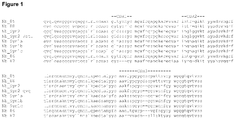
- nanobodies examples include, but are not limited to, nanobodies as defined by SEQ ID NOs: 18-26 (see Fig. 1 ; Table 3).
- a non-limiting example of a bivalent nanobody is defined by SEQ ID NO: 68.
- the above nanobodies can comprise at least one of the complementary determining regions (CDRs) with an amino acid sequence selected from SEQ ID NOs: 27-42. (see Fig. 1 ; Table 4). More specifically, the above nanobodies can be selected from the group comprising SEQ ID NOs: 18-26 (see Fig. 1 ; Table 3).
- the term nanobody as used herein in its broadest sense is not limited to a specific biological source or to a specific method of preparation.
- the nanobodies as disclosed herein can generally be obtained: (1) by isolating the V H H domain of a naturally occurring heavy chain antibody; (2) by expression of a nucleotide sequence encoding a naturally occurring V H H domain; (3) by "humanization” of a naturally occurring V H H domain or by expression of a nucleic acid encoding a such humanized V H H domain; (4) by "camelization” of a naturally occurring VH domain from any animal species, and in particular from a mammalian species, such as from a human being, or by expression of a nucleic acid encoding such a camelized VH domain; (5) by "camelisation” of a "domain antibody” or “Dab” as described in the art, or by expression of a nucleic acid encoding such a camelized VH domain; (6) by using synthetic or semi
- analogs of the antibodies as disclosed herein.
- analogs synthetic analogs, mutants, variants, alleles, homologs and orthologs (herein collectively referred to as “analogs") of the antibodies as disclosed herein.
- analogs synthetic analogs, mutants, variants, alleles, homologs and orthologs (herein collectively referred to as “analogs") of the antibodies as disclosed herein.
- the specification describes that the term “antibody as described herein” or “antibody as disclosed herein” or “antibody fragment as disclosed herein” in its broadest sense also covers such analogs.
- one or more amino acid residues may have been replaced, deleted and/or added, compared to the antibodies as disclosed herein.
- a substitution may for example be a conservative substitution (as described above, see also Table 1) and/or an amino acid residue may be replaced by another amino acid residue that naturally occurs at the same position.
- any one or more substitutions, deletions or insertions, or any combination thereof, that either improve the properties of the antibody as described herein or that at least do not detract too much from the desired properties or from the balance or combination of desired properties of the antibody as described herein (i.e. to the extent that the antibody is no longer suited for its intended use) are included.
- a skilled person will generally be able to determine and select suitable substitutions, deletions or insertions, or suitable combinations of thereof, based on the disclosure herein and optionally after a limited degree of routine experimentation, which may for example involve introducing a limited number of possible substitutions and determining their influence on the properties of the antibodies thus obtained.
- Yet another modification may comprise the introduction of one or more detectable labels or other signal-generating groups or moieties, depending on the intended use of the labelled antibody.
- Suitable labels and techniques for attaching, using and detecting them will be clear to the skilled person, and for example include, but are not limited to, fluorescent labels (such as fluorescein, isothiocyanate, rhodamine, phycoerythrin, phycocyanin, allophycocyanin, o-phthaldehyde, and fluorescamine and fluorescent metals such as Eu or others metals from the lanthanide series), phosphorescent labels, chemiluminescent labels or bioluminescent labels (such as luminal, isoluminol, theromatic acridinium ester, imidazole, acridinium salts, oxalate ester, dioxetane or GFP and its analogs), radio-isotopes, metals, metals chelates or metallic cations or other metals or
- Suitable labels will be clear to the skilled person, and for example include moieties that can be detected using NMR or ESR spectroscopy.
- Such labelled antibodies as disclosed herein may for example be used for in vitro, in vivo or in situ assays (including immunoassays known per se such as ELISA, RIA, EIA and other "sandwich assays", etc.) as well as in vivo imaging purposes, depending on the choice of the specific label.
- another modification may involve the introduction of a chelating group, for example to chelate one of the metals or metallic cations referred to above.
- Suitable chelating groups for example include, without limitation, diethylenetriaminepentaacetic acid (DTPA) or ethylenediaminetetraacetic acid (EDTA).
- DTPA diethylenetriaminepentaacetic acid
- EDTA ethylenediaminetetraacetic acid
- Yet another modification may comprise the introduction of a functional group that is one part of a specific binding pair, such as the biotin-(strept)avidin binding pair.
- a functional group may be used to link the antibody as disclosed herein to another protein, polypeptide or chemical compound that is bound to the other half of the binding pair, i.e. through formation of the binding pair.
- an antibody as disclosed herein may be conjugated to biotin, and linked to another protein, polypeptide, compound or carrier conjugated to avidin or streptavidin.
- such a conjugated antibody may be used as a reporter, for example in a system where a detectable signal-producing agent is conjugated to avidin or streptavidin.
- the specification further describes a nucleic acid sequence (as defined herein) encoding any of the antibodies, antibody fragments, and/or nanobodies as described herein.
- a vector (as defined herein) comprising a nucleic acid sequence encoding any of the antibodies, antibody fragments, and/or nanobodies as described herein is also provided, as well as a host cell (as defined herein) comprising such a vector or such a nucleic acid.
- a complex of (i) an affinity ligand as described above, and (ii) a non-naturally occurring fusion protein, wherein said fusion protein comprises said target protein and a heterologous C-terminal epitope tag defined by SEQ ID NO:1, further characterized in that said SEQ ID NO:1 represents the last four C-terminal amino acid residues of said fusion protein, and (iii) optionally a ligand specifically bound to the target protein comprised in said non-naturally occurring fusion protein
- said complex is crystalline.
- ligand means a molecule that specifically binds to a target protein.
- a ligand may be, for example a polypeptide, a lipid, a small molecule, an antibody or an antibody fragment (such as a nanobody).
- a ligand also includes a "native ligand" which is a ligand that is an endogenous, natural ligand for a native target protein.
- a complex refers to a group of two or more associated molecules.
- a complex can be a protein complex.
- protein complex or “multiprotein complex” refer to a group of two or more associated polypeptide chains. Proteins in a protein complex are linked by non-covalent protein-protein. The quaternary structure is the structural arrangement of the associated folded proteins in the protein complex.
- epitope tags as described herein, as well as the affinity ligands specifically binding to these epitope tags, as well as complexes of epitope tags and affinity ligands, and/or any combinations thereof, may serve any purpose including but not limited to:
- the epitope tags as disclosed herein, as well as the affinity ligands specifically binding to these epitope tags, as well as complexes of epitope tags and affinity ligands, and/or any combinations thereof can be used for affinity-based applications, such as affinity chromatography, affinity purification, immunoprecipitation, in-vivo imaging, tandem affinity purification, protein detection, immunochemistry, surface-display, FRET-type applications, crystallography, and the like.
- affinity-based applications such as affinity chromatography, affinity purification, immunoprecipitation, in-vivo imaging, tandem affinity purification, protein detection, immunochemistry, surface-display, FRET-type applications, crystallography, and the like.
- the specification describes the use of any of the above described epitope tags and/or affinity ligands for the purification of fusion molecules comprising one or more epitope tags as disclosed herein.
- molecules can be purified using generalized protocols in contrast to highly customized procedures associated with conventional chromatography.
- Fusion molecules may be purified from the host cell or from the host cell culture medium into which they have been secreted.
- the host cell is lysed using standard techniques (e.g., enzymatic digestion, sonication, French press, etc.) to form a lysate comprising the fusion molecule.
- Said fusion molecule may be purified from a lysate or from a host cell culture medium material by contacting the lysate or medium with a suitable chromatography medium under conditions suitable for binding of the fusion molecule to the chromatography medium.
- the lysate or culture medium may be contacted with a chromatography medium in either a batchwise technique (e.g. by mixing the chromatography medium with the lysate or culture medium) or column technique.
- the resin bound fusion molecule may be washed one or more times to remove any weakly bounded materials, i.e. materials that do not bind as tightly as the fusion molecule to the chromatography medium.
- the molecule may then be eluted from the medium by contacting the medium with a suitable elution buffer known to the skilled person.
- a suitable elution buffer known to the skilled person.
- the elution of the fusion molecule from the column can be carried out at a constant pH or with linear or discontinuously falling or raising pH gradients.
- the optimal elution conditions depend on the amount and type of impurities which are present, the amount of material to be purified, the column dimensions, the chromatography resin used, etc. and are easily determined by routine experimentation on a case-by-case basis.
- the specification further describes a method for purifying a non-naturally occurring fusion molecule comprising the steps of
- a fusion molecule may comprise a cleavage site for a protease, for example, located between the tag as described herein and a molecule of interest. After elution from the chromatography medium or while still bound to the medium, a fusion molecule as described herein may be contacted with a solution comprising a protease enzyme that cleaves at the cleavage site. Accordingly, the above described purification method may further comprise a step (e) wherein the epitope tag is removed.
- the purification method may be any method known in the art. Suitable purification methods include but are not limited to affinity chromatography (e.g. immunoaffinity chromatography, Immobilized Metal Ion Affinity Chromatography (IMAC)), metal-affinity precipitation, immobilized-metal-ion-affinity electrophoresis and Immunoprecipitation.
- affinity chromatography e.g. immunoaffinity chromatography, Immobilized Metal Ion Affinity Chromatography (IMAC)
- metal-affinity precipitation e.g., immobilized Metal Ion Affinity Chromatography (IMAC)
- IMAC Immobilized Metal Ion Affinity Chromatography
- the affinity medium contains a solid support (e.g. resin, magnetic bead,...) coupled to one or more of the following affinity ligands: Fe, Co, Ni, Cu, Zn, or Al charged IMAC ligand, an antibody, an antibody fragment, a nanobody, a small molecule or a synthetic affinity ligand (e.g. aptamers or ligands derived using VersaffmTM).
- a solid support e.g. resin, magnetic bead,
- affinity ligands Fe, Co, Ni, Cu, Zn, or Al charged IMAC ligand
- an antibody an antibody fragment
- a nanobody e.g. a small molecule
- a synthetic affinity ligand e.g. aptamers or ligands derived using VersaffmTM.
- the use of the epitope tag and/or affinity ligand as described herein for purification results in highly purified proteins with a good yield.
- the term "purity” or “purified” as applied to proteins herein implies that the desired protein preferably comprises at least 60%, more preferably at least 70%, more preferably at least about 80%, still more preferably at least about 90%, and most preferably at least about 95% of the total protein component.
- the elution step can be performed amongst others with a small compound, a peptide, using solutions of high ionic strenght or by changing the pH.
- the fusion molecule can be eluted from the affinity chromatography medium by excess amounts of the epitope tag or excess amounts of the affinity ligand.
- the specification further encompasses a method for immobilizing and/or capturing a non-naturally occurring fusion molecule on a solid support comprising the steps of:
- the method may be performed using immobilized elements and the immobilization may be carried out using a variety of immobilization means on various solid supports (e.g., columns, beads, adsorbents, nitrocellulose paper, etc.).
- the immobilization assay can, for example, be used to screen a sample or a composition for antibodies against the molecule linked to the tag, or can be part of a screening assay in order to screen large libraries of test compounds (e.g., drugs, new antimicrobials, etc.).
- the screening assay is preferably conducted in a microplate format. Any means or method of detection can be used.
- the detection means might be a plate reader, a scintillation counter, a mass spectrometer or fluorometer.
- the epitope tag as disclosed herein can be used in detection of a fusion molecule using the affinity ligand fused to a reporter molecule (radioactive label, alkaline fosphatase, GFP, peroxidases, and the like, see above).
- a reporter molecule radioactive label, alkaline fosphatase, GFP, peroxidases, and the like, see above.
- the specification specifies detection of a fusion protein as described herein with antibodies specifically binding to the epitope tag, whichcan be useful in tissue or subcellular localization experiments, ELISA, western blotting or other immuno-analytical methods.
- antibodies including antibody fragments or nanobodies, that may be used for the detection of the epitope tag in an immunoassay, such as ELISA, Western blot, radioimmunoassay, enzyme immunoassay, and may be used in immunocytochemistry.
- an immunoassay such as ELISA, Western blot, radioimmunoassay, enzyme immunoassay, and may be used in immunocytochemistry.
- an anti-tag antibody that may be in solution and the tag to be recognized may be in solution (e.g. an immunoprecipitation) or may be on or attached to a solid surface (e.g. a Western blot).
- the antibody may be attached to a solid surface and the tag may be in solution (e.g. immuno affinity chromatography or ELISA).
- Antibodies, including antibody fragments or nanobodies, to the tags and/or fusion molecules as described herein may be used to determine the presence, absence or amount of one or more molecules in a sample.
- the amount of specifically bound tag and/or fusion molecule may be determined using an antibody to which a reporter molecule is attached, such as a radioactive, a fluorescent, or an enzymatic label.
- a labeled secondary antibody e.g. an antibody that recognizes the antibody that is specific to the polypeptide
- the epitope tag as described herein can also be useful in crystallography of proteins, where a short tag or no tag is preferred.
- tags are removed as it is a disadvantage for crystallization to have unstructured parts in the protein. Limited proteases are used to remove the flexible parts.
- an affinity ligand in particular a nanobody, can bind the tag, making the whole complex more soluble, easier to crystallize and helping to solve the structure of the complex. It has been shown that Nb can help in crystallisation by fixating flexible loops or making crystal contacts (Loris et al., 2003, Korotkov et al., 2009).
- the specification provides for the use of the epitope tag and/or the affinity ligand as disclosed herein in a Tandem Affinity Purification (TAP) method in order to purify protein complexes under native conditions.
- TAP Tandem Affinity Purification
- the TAP tagging method (Rigaut et al., 1999; Puig et al. 2001) can be applied to the yeast proteome very successfully, and has been adapted to various cells and organisms.
- the size of the TAP tag can be in the region of 21 kDa and the larger a tag is, the greater the possibility that it will interfere with the function or structure of the polypeptide to be isolated, or alternatively be cleaved or digested by the polypeptide target.
- Commonly used tag combinations for TAP are well-known and summarized in Li (2010).
- the epitope tags as described herein can be incorporated in the development of TAP tags for affinity-based applications.
- kit for use in any of the affinity-based applications as described herein.
- a kit may comprise, without the purpose of being limitative, one or more of the following elements:
- Epitope tags, peptides, linker sequences Amino acid sequence SEQ ID NO EPEA 1 GYQDYEPEA 2 DYEPEA 3 YQDYEPEA 4 QDYEPEA 5 GYQDYEPE 6 SEEGYQDYEPEA 7 GAAEPEA 8 GAGAEPEA 9 GAGAGYQDYEPEA 10 YEPEA 11 GAGA 12 EEGEPK 13 EEAEPK 14 IEGR 15 LVPR 16 DDDDK 17 TABLE 3: Nanobodies that specifically bind to ⁇ -synuclein NAME (see Fig.
- Example 1 Isolation, expression and purification of nanobodies against ⁇ -synuclein
- Camelid nanobodies e.g. NbSynla, NbSynlb, NbSynlc and NbSyn2, Nb85, Nb86, Nb87 and Nb88 were isolated from either a dromedary immunized with ⁇ -synuclein (Uniprot accession number P37840) or a dromedary immunized with two proteins: the ⁇ -synuclein Ala53Thr mutant and ⁇ -synuclein (Uniprot accession number is Q16143).
- ⁇ -Synuclein and ⁇ -synuclein share 85% identity in the N-terminal part of the protein, in the C-terminal part both proteins are less homologous ( ⁇ 40%). Both proteins share the same C-terminal EPEA sequence. All nanobodies were selected by phage-display, essentially according to published protocols (Lauwereys et al. 1998; Conrath et al. 2001) or as described in materials and methods. In vitro selection on immobilized ⁇ -synuclein only revealed one family of binders: NbSynla, NbSynlb, and NbSynlc.
- a second in vitro selection was performed on immobilized ⁇ -synuclein in the presence of 10 ⁇ M of NbSynla. From this selection, we obtained NbSyn2 after 3 rounds of panning. From the dromedary immunized with two proteins (the ⁇ -synuclein A53T mutant and ⁇ -synuclein) one independent phage display library of 10 7 independent clones was constructed. This library was panned in parrallel against ⁇ -synuclein and the ⁇ -synuclein A53T mutant in two separate in vitro selections. Nb85, Nb86 and Nb87 were recovered from the beta-synuclein panning.
- Nb85, Nb86 Two of these binders (Nb85, Nb86) were also selected on Ala53Thr ⁇ -synuclein.
- One add binder Nb88 was also recovered from the Ala53Thr ⁇ -synuclein panning. It thus appears that Nb85 and Nb86 bind ⁇ -synuclein A53T as w ell as ⁇ -synuclein.
- Nb88 only differs from Nb85 by a few point mutations in framework 1 and CDR1 while the CDR2 and CDR3 are identical ( Fig 1 ).
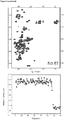
- Fig. 2 The effect of binding of NbSyn2, NbSyn85 and Nb87 to the HSQC spectrum of labeled ⁇ -synuclein is shown in Fig. 2 .
- the top panel represents the raw spectra obtained upon addition of Nb, where the red peaks correspond to the free and the blue peaks correspond to the signals of ⁇ -synuclein in complex with the different nanobodies.
- the lower panel of Figure 2 summerizes a titration experiment, using subequimolar to equimolar concentrations of Nb as the titrant.
- ITC Isothermal titration calorimetry
- the C ⁇ of Ala140 is deeply buried in a pocket formed between the CDR3 and CDR2 loops of the nanobody ( Fig. 6b ), while the carbonyl groups of the C-terminus of Ala140 make a salt-bridge with Arg50 of NbSyn2 ( Fig. 7 ).
- the peptide main-chain atoms, apart from the carbonyl groups of Ala140 and the carbonyl oxygens of Pro138 and Glu137 -which make direct hydrogen bonds with residues of NbSyn2- are either exposed to solvent or make indirect contact with the nanobody through bridging water molecules.
- NbSyn2 The determination of the crystal structure of NbSyn2 bound to a peptide encompassing the nine C-terminal residues of ⁇ -synuclein (PDB-code 2X6M) confirms that NbSyn2 binds the last four C-terminal residues of ⁇ synuclein and more specifically the C-terminal sequence EPEA (SEQ ID NO: 1) ( Fig 6 ).
- the NMR experiments (Example 2) and the ICT data (Example 3) show that the nanobodies NbSyn2, Nb85 and Nb87 bind the C-terminal end of ⁇ -synuclein.
- the X-ray structure (Example 4) confirms that NbSyn2 interacts mainly to the last 4 amino acids.
- GFP-peptide hybrids By standard DNA cloning techniques we made 4 different constructs of a GFP (Green Fluorescent Protein) fused to different tags at the C-terminal end:
- the four constructs were cloned into the pBADmycHis vector as described in material and methods.
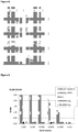
- Nanobodies Nb85, Nb87, Nb88 and NbSyn2 were expressed by standard procedures in the periplasm of E.coli. Next, periplasmatic fractions containing one of the nanobodies were mixed with cellular lysates containing one of the four tagged GFP proteins and incubated for one hour.
- an affinity column was prepared by immobilizing NbSyn2 on a sepharose matrix.
- NbSyn2 was coupled to a CNBr activated sepharose according to the manufactures instructions.
- GFP fused to the GYQDYEPEA tag (construct CA1508) was expressed in E.coli. Cells were recovered by centrifugation and lysed. This crude lysate containing the tagged GFP (construct CA1508) was then mixed with agarose slurry containing the immobilized NbSyn2 and incubated for one hour at 4°C.
- a nanobody that is specific for another protein
- pMESy1 and pMESy2 that are described in detail in Material and Methods.
- pMESy1 the original His-tag was replaced by the longer GYQDYEPEA.
- pMESy2 we cloned the 4 amino acid EPEA tag behind a small GAA-linker.
- VHH, Nb458 recognizing an unrelated antigen or Aox1 of Pichia pastoris; SEQ ID NO: 66
- Example 7 C-terminally EPEA tagged proteins can be detected in ELISA with NbSyn2-AP.
- NbSyn2 to screen large phage displayed libraries of EPEA-tagged VHH's.
- VHH nanobody library
- pMESy2 phage display vector
- NbSyn2 or NbSynla were fused to mKO1 or GFP and cloned into pcDNA to give NbSyn1a-mkO1, NbSyn2-mkO1, NbSynla-GFP and NbSyn2-GFP fusions.
- HeLa cells were transfected with these vectors or with combinations of these vectors to see whether we could detect the expression and specify the location of the EPEA-tagged proteins with GFP or mKO1 fused to NbSynla or NbSyn2.
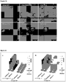
- Fig 13 shows that the cells that are transfected with NbSyn2 or NbSynla fused to mKO1 or GFP colour the cell uniformly.
- Cells that are double transfected with Rev-GAGAGYQDYEPEA or Rev-GAGAEPEA and NbSyn2 or NbSynla fused to mKO1 or GFP have a deeper colour in the nucleoli. It is known that wild type Rev shuttles between the nucleoli and the cytoplasm of the cell with the majority of the protein localized in the nucleoli (Perkins et al., 1989).
- Example 11 Improvement of the sensitivity of the in vitro detection using a bivalent nanobody
- Example 9 To improve the sensitivity of the ELISA, we constructed a bivalent version of NbSyn2, named biNbSyn2 (SEQ ID NO: 68). For this purpose, we amplified the NbSyn2 gene and cloned it as a bivalent construct with a GGGGSGGGS (SEQ ID NO: 69) linker between both NbSyn2 sequences in the phage display vector pMES4. Expression, purification and conjugation of the biNbSyn2 to alkaline phosphatase was done as described in Material and Methods to the Examples.
- this C-terminal His tag was replaced with a sequence encoding GGEPEA (SEQ ID NO: 70) as described in detail in Material and Methods.
- a small scale culture of E.coli cells were grown, IPTG was added to induce the production of the EPEA tagged CsgG (SEQ ID NO: 72).
- Western blot was done on 4 individual clones of this construct to prove the presence of the CsgG protein. A clear band running at the correct height could be detected ( Fig 15 ).
- the plasmid was transformed to BL21 cells for a large scale production of the EPEA tagged CsgG. Cell membranes were prepared as described in the material an methods.
- Plasmids pHEN4 (Ghahroudi et al., 1997), pHEN6 (Lauwereys et al., 1998), pMES4 (genbank GQ907248), pBadMycHis (Invitrogen), pcDNA3.1. (Invitrogen) and pMC2 (Robinson et al. 2006) were used.
- VHH genes were cloned in the pHEN4 vector (Ghahroudi et al., 1997) using the Nco I and the Not I restriction enzymes and transformed into TG1 cells (genepulser, Biorad) according to standard protocols. From dromedary two the gene fragments encoding the VHH genes were amplified and cloned as described by Conrad et al. (2001). The VHH repertoire of both libraries was expressed on the phage after superinfection with M13K07.
- VHH genes were amplified as described above.
- vector and PCR products were digested with Pst I and the Eco91 I restriction enzymes, purified and ligated with T4 DNA ligase overnight. Insert analysis was done by PCR with primers RP and Gill.
- the VHH repertoire of the library was expressed on the phage after superinfection with helperphage.
- binders were selected in vitro from the library on solid-phase coated protein (2x 5 ⁇ g/well) in the wells of a maxisorp microtitre plate (Nunc) Phages were recovered by incubating the coated wells with 100mM triethylamine pH10 for 10min and the eluate (containing phages) was neutralised by adding TrisHCl pH 6.8. This eluate was added to freshly grown TG1 cells. The coated wells were then washed once with TrisHCl pH 6.8 and several times with PBS and freshly grown TG1 cells were added to the wells to recover the non eluted phages. Colony screening for specific binders was done by ELISA using the NbSyn-AP as described above.
- pMESy4 (referred to as CA3198) was made as followed:
- Standard 15 N- 1 H HSQC experiments were carried out on a Bruker Avance 700 MHz instrument equipped with a cryo-probe. Spectra of 15 N labeled ⁇ -synuclein were recorded at different molar equivalents of unlabeled NbSyn2 (0.25, 0.5, 0.75, 1.0, 1.25, 1.5, 1.75, 2.0, 2.5, 3.0, 3.25) by titration of small volumes of a concentrated stock solution of NbSyn2. All experiments were recorded in 10mM phosphate buffer pH 7.4 and at 10°C. The amide resonances and C ⁇ and C ⁇ chemical shifts were determined previously (Dedmon et al., 2005a; 2005b) and used in the further analysis of the data.
- thermodynamic analysis was performed using the Microcal analysis software (Origin 7.0) with a 1:1 binding model.
- the temperature dependence of ⁇ H, allowed the calculation of ⁇ Cp through the relationship ⁇ Cp ⁇ H/ ⁇ T.
- the contribution of protonation to the ⁇ H was evaluated using a 10mM Tris-HCL buffer 100mM NaCl at pH 7.4.
- primer EP126 5'-TTCGAATT-CATTACGCTTCCGGTTCATAATCCTGATATCCTTTGTAGAGCTCATCCATGC 3'
- primer EP127 5'-TTCGAATTCATTACGCTTCCGGTTCATAATCCTGATATTTGTAGAGCT-CATCCATGCC-3'
- QDYEPEA primer EP128 5'-TTCGAATTCATTACGCTTCCGGTTCATAAT-CCTGTTTGTAGAGCTCATCCATGCC-3') (SEQ ID NO: 51).
- Amplification conditions were 4min 94°C, 28 times (40sec 94°C, 40sec 50°C, 1min 72°C) and 10 min 72°C.
- the amplified gfp fusions coding for GFP fusion molecules with different epitope tags were cut with Bsp HI and Eco RI.
- the PCR products were ligated into the pBADmycHis vector, opened with Ncol and Eco RI. Finally the ligation product was transformed into E.coli Top10.
- the cells from a 330ml LB culture were harvested by centrifugation (10 min, 4000rpm, 4 °C) and the pellet was resuspended in 20ml 50mM Tris-HCI pH 7.5 with 1 mM PMST. Cells were lysed by a C5 EmulsiFlex High Pressure Homogeniser 86-001 (AVESTIN, Germany). The insoluble material was pelleted by centrifugation (30 min, 4000rpm, 4 C) and the supernatant (cytoplasmatic fraction) was collected.
- a periplasmatic fraction of the His-tagged NbSyn2 was mixed with one cytoplasmatic fraction of each tagged GFP.
- An IMAC was done as described before.
- the NbSyn2-GFP tagged fusion complexes were eluted with 1M imidazol.
- pMES4 On 1ng of pMES4 a PCR was performed with Takara Ex Taq polymerase using primers EP138 (5'-ACCCAGGTCACCGTCTCCTCAGGGTATCAAGACTACGAACCTGAAGCCTAGTACCCGTACGACGT) (SEQ ID NO: 53) and FP (5'-CGCCAGGGTTTTCCCAGTCACGAC-3') (SEQ ID NO: 54) to construct pMESy1 (CA2857) and using primers EP139 (5'-ACCCAGGTCACCGTCTCCTCAGGGGCAGCGGAACCTGAAGCCTAGTACCCGTACGACGTTC-3') (SEQ ID NO: 55) and FP to construct pMESy2 (CA2741).
- Amplification conditions were 4min 94°C, 28 times (40sec 94°C, 40sec 55°C, 1min 72°C) and 10 min 72°C. Both PCR products and the pMES4 vector were cut with Bam HI and Eco 91I and purified. Ligation with T4 DNA polymerase of 100ng vector to 50ng PCR product was carried out overnight at 4°C. The mixture was transformed into WK6 cells and overnight grown at 37°C. Analysis was done by PCR using primers MP57 (5'-TTATGCTTCCGGCTCGTATG -3') (SEQ ID NO: 56) and Gill (5'- CCACAGACAGCCCTCATAG -3') (SEQ ID NO: 57) and Spe I digestion. Clones were sequenced and from the correct ones DNA was prepared (Qiagen).
- the Nb458 was amplified using primer RP and primer EP101 (5'-TGAGGAGACGGTGACCTGGGT-3') (SEQ ID NO: 58) with the same amplifications conditions as written above and cloned in the pMESy1 (CA2857) or the pMESy2 (CA2741) vector using the Pst I and the Eco9 I restriction enzymes. Insert analysis was done by PCR with primers RP and Gill. Expression was done as described by Conrad et al. (2001). Nb458 in pMESy1 is fused to the sequence of the GYQDYEPEA tag and is referred to as CA2944 while in pMESy2, it has the gaaEPEA tag and is referred to as CA2945.
- elution buffer was either 50mM TrisHCI pH7.5 with 1M or 2M NaCl, 50mM Tris-HCI pH7.5 with 0.5M adipic acid or 50mM Tris-HCI pH7.5 with 750 ⁇ M of peptide (GYQDYEPEA).
- Crystallization of the NbSyn2 complexed with a synthetic peptide (sequence NH2-GYQDYEPEA-COOH) (BIO-SYNTHESIS, INC., Texas) was achieved using a crystallization robot Phoenix (Art Robbins Instruments) and a sitting drop vapor diffusion assay.
- 133 ⁇ l of 10 mg/ml of NbSyn2 was first mixed with 15 ⁇ l of peptide (at 20 mg/ml) before dispension in a JB Classic 1-4 (G10) screen (Jena Biosciences). 100 nl drops were mixed with 100 nl of the precipitant solution.
- Crystals were obtained under the following conditions: 25% w/v PEG 6000, 100mM HEPES, pH 7.5 and 100 mM LiCI. Crystals were subsequently harvested and transferred in a cryo-protectant solution (25% w/v PEG 6000, 100mM HEPES, pH 7.5 and 100 mM LiCI and 10% PEG400) and flash-frozen in liquid nitrogen for data collection.
- a cryo-protectant solution (25% w/v PEG 6000, 100mM HEPES, pH 7.5 and 100 mM LiCI and 10% PEG400
- Diffraction patterns for frozen crystals of the NbSyn2-peptide complex were obtained at the X11 beam line equipped with a MAR555 detector, at the European Molecular Biology Laboratory (EMBL) using the Deutsches Elektronen Synchrotron (DESY Hamburg, Germany). Data were processed using Mosfilm65, and Pointless and Scala were used to determine the spacegroup and scale and merge the data respectively.
- the graphics program Coot70 was used to interpret the electron density maps and for refinement of the model.
- the change in solvent accessible surface area (ASA) upon the formation of the NbSyn2: ⁇ -synuclein complex was calculated using web based server PROTORP71.
- the two different tags GYQDYEPEA and EPEA were C-terminal fused to Rev by PCR using Takara Ex Taq polymerase (TAKARA BIO INC).
- a PCR was performed on pcDNARev-mKO1 kindly given to us by Dirk Daelemans with primer T7 (5'-ATTAATACGACTCACTATAGG-3') (SEQ ID NO: 59) and primer EP136 (5'-TCGAGCGGCCGCTTAGGCTTCAGGTTCGTAGTCTTGATACCCTGAAGCTCCTGCACCATTCTC-3') (SEQ ID NO: 60) to amply the rev gene fused to the DNA sequence of GYQDYEPEA at the C-terminus.
- both tags were also fused to GFP as described above, using primer EP180 (5'-GCTTGGTACCTCATGAGCAAAGGAGAAGAAC-3') (SEQ ID NO: 62) and primer EP126 (5'-TTCGAATTCATTACGCT-TCCGGTTCATAATCCTGATATCCTTTGTAGAGCTCATCCATGC-3') (SEQ ID NO: 63) to fuse it to the GYQDYEPEA and primer EP180 and primer EP181 (5'-TTCGAATTCATTACGCTTCCGGTTCT-GCGGCTCCTTTGTAGA-GCTCATCCATGCC-3') (SEQ ID NO: 64) to fuse GFP to EPEA.
- the amplified DNA fragments were cut with Kpnl and EcoRI and ligated into the pcDNA3.1 vector. The ligation product was then electroporated into E.coli Top10, clones were analysed by PCR and sequence analysis.
- HeLa human epithelial cells were maintained using standard procedures untill confluent in DMEM medium with 10% Fetal calf serum.
- DMEM medium 10% Fetal calf serum.
- cells were harvested by a Trypsin/EDTA treatment, counted and were plated onto glass bottom microwell dishes (MatTek corporation). Transfections were preformed according to manufactures manual: the 4 x 10 5 cells were seeded in a 35mm dish. 12 ⁇ g DNA or in case 2 different plasmids are cotransfected twice 1 ⁇ g is mixed with 4.5 ⁇ l Transfect (Qiagen Valencia, California), dropped onto the cells and incubated overnight.
- Transfected HeLa cells were imaged with a laser scanning SP5 confocal microscope (Leica Microsystems) equipped with an AF 6000 microscope and an AOBS, using a HCX PLAPO ⁇ 63 (NA1.2) water immersion objective magnification.
- GFP was monitored with the Ar laser using the 488-nm line for excitation, and emission was detected between 492 and 570 nm.
- mKusabira orange fluorescent protein (mKO) was imaged using the DPSS-561 nm laser for excitation, and emission was detected between 570 and 675 nm.
- Amplification conditions were 4min 94°C, 28 times (40sec 94°C, 40sec 55°C, 1min 72°C) and 10min 72°C.
- the PCR product generated by using primer SM17 and VHH1-reverse-5GS-BamHI was cut with Nco I and Bam HI
- the second PCR product generated by using primers VHH2-forward-4GS-BamHI and VHH2-reverse-TVSS-Bstell was cut with Bam HI and Eco 91I
- the pMES4 vector was cut with Nco I and Bam HI and Eco 91I . All digest were purified and the DNA concentration was measured.
- a three -point-ligation with T4 DNA polymerase of 100ng vector to 30ng of both PCR products was carried out in 10 ⁇ l for 2h at room temperature. The mixture was diluted and cut with Xba I to eliminate the self-ligated vectors. The digest was purified and transformed into WK6 cells and overnight grown at 37°C. Analysis was done by PCR using primers MP57 (5'-TTATGCTTCCGGCTCGTATG -3') (SEQ ID NO: 56) and Gill (5'- CCACAGACAGCCCTCATAG -3') (SEQ ID NO: 57) and Bam HI digestion.
- pMES4biNbSyn2 CA4394
- Expression and purification was done as described by Conrad et al. (2001). After IMAC purification, the sample was concentrated and ran on preparative superdex75 size exclusion column to separate the bivalent biNbSyn2 from contaminants or degradation products.
- Detection of the tags was performed with a dilution of 1/250 to 1/4000 of both NbSyn2-AP conjugate and biNbsyn2-AP conjugate.
- the absorption at 405 nm was measured 60min after adding the enzyme substrate p-nitrophenyl phosphate and after overnight incubation.
- the pMC2 plasmid was used in the SLIM method (Chiu et al. 2004) with the following primers: 5'-GGCGGCGAACCGGAAGCGTGACTGCAGGCATGCAAGCTTGGCT-3' (SEQ ID NO: 77), 5'-TGACTGCAGGC-ATGCAAGCTTGGCT-3' (SEQ ID NO: 78), 5'-CGCTTCCGGTTCGCCGCCATGGTGATGGTGATGGTGGGATTCC-3' (SEQ ID NO: 79), 5'- ATGGTGATGGTGATGGTGGGATTCC 3'(SEQ ID NO: 80). PCR and sequence analysis confirmed the correct replacement.
- the pellet was washed by resuspending it 50ml 25mM Tris, 150mM NaCl, 1mM DTT, 1mM EDTA pH8.0 with 0.002% DDM. Suspension was centrifuged (100000g for 50min at 4°C) to collect the outer membranes. These were resuspended in 100ml 25mM Tris, 150mM NaCl, 1mM DTT and 1% DDM and left to shake overnight at 4°C to solubilize the membranes.
Landscapes
- Chemical & Material Sciences (AREA)
- Organic Chemistry (AREA)
- Genetics & Genomics (AREA)
- Biochemistry (AREA)
- Biophysics (AREA)
- General Health & Medical Sciences (AREA)
- Health & Medical Sciences (AREA)
- Medicinal Chemistry (AREA)
- Molecular Biology (AREA)
- Proteomics, Peptides & Aminoacids (AREA)
- Life Sciences & Earth Sciences (AREA)
- Analytical Chemistry (AREA)
- Peptides Or Proteins (AREA)
- Micro-Organisms Or Cultivation Processes Thereof (AREA)
Claims (15)
- Komplex von(i) einem Antikörperfragment oder einem Nanobody™, das bzw. der spezifisch an das C-terminale Epitop-Tag, das von SEQ ID Nr. 1 definiert wird, umfasst; und(ii) einem Fusionsprotein, das ein Zielprotein und ein heterologes C-terminales Epitop-Tag, das von SEQ ID Nr. 1 definiert wird, umfasst, weiterhin dadurch gekennzeichnet, dass die SEQ ID Nr. 1 die letzten vier C-terminalen Aminosäurereste des Fusionsproteins darstellt; und(iii) fakultativ einem Liganden, der an das Zielprotein gebunden ist, das von dem Fusionsprotein umfasst ist.
- Komplex nach Anspruch 1, wobei der Komplex kristallin ist.
- Feste Oberfläche, die ein Antikörperfragment oder einen Nanobody™, das bzw. der spezifisch an das C-terminale Epitop-Tag, das von SEQ ID Nr. 1 definiert wird, umfasst.
- Verwendung eines Antikörperfragments oder eines Nanobody™, das bzw. der spezifisch an das C-terminale Epitop-Tag, das von SEQ ID Nr. 1 definiert wird, für Affinitätschromatographie, Affinitätsaufreinigung, Immunpräzipitation, In-vivo-Bildgebung, Tandem-Affinitätsaufreinigung, Nachweis eines Fusionsproteins, das ein Zielprotein und ein heterologes C-terminales Epitop-Tag, das von SEQ ID Nr. 1 definiert wird, umfasst, weiterhin dadurch gekennzeichnet, dass die SEQ ID Nr. 1 die letzten vier C-terminalen Aminosäurereste des Fusionsproteins darstellt, Immunchemie, Oberflächen-Display, Anwendungen vom FRET-Typ oder Kokristallisation.
- Verwendung des Komplexes nach Anspruch 1 oder 2 für Affinitätschromatographie, Affinitätsaufreinigung, Immunpräzipitation, In-vivo-Bildgebung, Tandem-Affinitätsaufreinigung, Proteinnachweis, Immunchemie, Oberflächen-Display, Anwendungen vom FRET-Typ oder Kokristallisation.
- Verwendung nach Anspruch 4 oder 5, wobei der Proteinnachweis in einem ELISA-Nachweis-Assay oder einer Western-Blot-Analyse durchgeführt wird.
- Verwendung nach Anspruch 4 oder 5, wobei die Affinitätsaufreinigung auf einer Säule oder auf Kügelchen durchgeführt wird.
- Verfahren zum Aufreinigen eines Fusionsproteins, wobei das Verfahren die folgenden Schritte umfasst:a) Aufbringen einer Lösung, die das Fusionsprotein, das ein Zielprotein und ein heterologes C-terminales Epitop-Tag, das von SEQ ID Nr. 1 definiert wird, umfasst, enthält, weiterhin dadurch gekennzeichnet, dass die SEQ ID Nr. 1 die letzten vier C-terminalen Aminosäurereste des Fusionsproteins darstellt, auf einen festen Träger, der über ein immobilisiertes Antikörperfragment oder einen Nanobody™, das bzw. der spezifisch an das C-terminale Epitop-Tag bindet, verfügt, undb) Bilden eines Komplexes zwischen dem immobilisierten Antikörperfragment oder dem Nanobody™ und dem Fusionsprotein undc) Entfernen von schwach gebundenen oder ungebundenen Molekülen undd) Elutieren der gebundenen Moleküle.
- Verfahren zum Immobilisieren und/oder Einfangen eines Fusionsproteins auf einem festen Träger, wobei das Verfahren die folgenden Schritte umfasst:a) Aufbringen einer Lösung, die das Fusionsprotein, das ein Zielprotein und ein heterologes C-terminales Epitop-Tag, das von SEQ ID Nr. 1 definiert wird, umfasst, enthält, weiterhin dadurch gekennzeichnet, dass die SEQ ID Nr. 1 die letzten vier C-terminalen Aminosäurereste des Fusionsproteins darstellt, auf einen festen Träger, der über ein immobilisiertes Antikörperfragment oder einen Nanobody™, das bzw. der spezifisch an das C-terminale Epitop-Tag bindet, verfügt, undb) Bilden eines Komplexes zwischen dem immobilisierten Antikörperfragment oder dem Nanobody™ und dem Fusionsprotein undc) Entfernen von schwach gebundenen oder ungebundenen Molekülen.
- Verfahren zum Nachweisen eines Fusionsproteins, das ein Zielprotein und ein heterologes C-terminales Epitop-Tag, das von SEQ ID Nr. 1 definiert wird, umfasst, weiterhin dadurch gekennzeichnet, dass die SEQ ID Nr. 1 die letzten vier C-terminalen Aminosäurereste des Fusionsproteins darstellt, in einer Probe oder Zusammensetzung durch Verwenden eines Antikörperfragments oder eines Nanobody™, das bzw. der spezifisch an das C-terminale Epitop-Tag, das von SEQ ID Nr. 1definiert wird, bindet.
- Kit, das ein Fusionsprotein, das ein Zielprotein und ein heterologes C-terminales Epitop-Tag, das von SEQ ID Nr. 1 definiert wird, umfasst, weiterhin dadurch gekennzeichnet, dass die SEQ ID Nr. 1 die letzten vier C-terminalen Aminosäurereste des Fusionsproteins darstellt, und ein Antikörperfragment oder einen Nanobody™, das bzw. der spezifisch an das C-terminale Epitop-Tag, das von SEQ ID Nr. 1 definiert wird, bindet, wobei das Antikörperfragment oder der Nanobody™ auf einer nichtlöslichen Matrix oder einem festen Träger vorimmobilisiert ist, und/oder den Komplex nach Anspruch 1 oder 2 umfasst.
- Verwendung des Kits nach Anspruch 11 für Affinitätschromatographie, Affinitätsaufreinigung, Immunpräzipitation, In-vivo-Bildgebung, Tandem-Affinitätsaufreinigung, Proteinnachweis, Immunchemie, Oberflächen-Display, Anwendungen vom FRET-Typ oder Kokristallisation.
- Verwendung des wie von SEQ ID Nr. 1 definierten C-terminalen Epitop-Tag für Affinitätschromatographie, Affinitätsaufreinigung, Immunpräzipitation, In-vivo-Bildgebung, Tandem-Affinitätsaufreinigung, Proteinnachweis, Immunchemie, Oberflächen-Display, Anwendungen vom FRET-Typ oder Kokristallisation.
- Verfahren zum Produzieren eines Fusionsproteins, das ein Zielprotein und ein heterologes C-terminales Epitop-Tag, das von SEQ ID Nr. 1 definiert wird, umfasst, weiterhin dadurch gekennzeichnet, dass die SEQ ID Nr. 1 die letzten vier C-terminalen Aminosäurereste des Fusionsproteins darstellt, wobei das Verfahren die folgenden Schritte umfasst:a. Platzieren der Nukleinsäuresequenz, die das wie von SEQ ID Nr. 1 definierte Epitop-Tag kodiert, in demselben offenen Leseraster des Zielproteins unter Verwendung von Gentechnik,b. fakultativ Insertieren einer geeigneten Linker-Sequenz zwischen der Zielproteinsequenz und dem Epitop-Tag unter Verwendung von Gentechnik,wobei Gentechnik standardmäßige rekombinante DNA-Verfahren umfasst und wobei das Epitop-Tag an dem C-terminalen Ende des Zielproteins insertiert wird.
- Verfahren nach Anspruch 14, wobei Gentechnik einen Vektor umfasst, der ein DNA-Konstrukt umfasst, das das Fusionsprotein exprimieren kann.
Applications Claiming Priority (2)
| Application Number | Priority Date | Filing Date | Title |
|---|---|---|---|
| GBGB1008682.5A GB201008682D0 (en) | 2010-05-25 | 2010-05-25 | Epitope tag for affinity based applications |
| PCT/EP2011/058591 WO2011147890A1 (en) | 2010-05-25 | 2011-05-25 | Epitope tag for affinity-based applications |
Publications (2)
| Publication Number | Publication Date |
|---|---|
| EP2576609A1 EP2576609A1 (de) | 2013-04-10 |
| EP2576609B1 true EP2576609B1 (de) | 2018-11-14 |
Family
ID=42341251
Family Applications (1)
| Application Number | Title | Priority Date | Filing Date |
|---|---|---|---|
| EP11721338.9A Active EP2576609B1 (de) | 2010-05-25 | 2011-05-25 | Epitop-tag für affinitätsbasierte anwendungen |
Country Status (8)
| Country | Link |
|---|---|
| US (1) | US9518084B2 (de) |
| EP (1) | EP2576609B1 (de) |
| JP (1) | JP6053672B2 (de) |
| AU (1) | AU2011257271B2 (de) |
| CA (1) | CA2799009C (de) |
| DK (1) | DK2576609T3 (de) |
| GB (1) | GB201008682D0 (de) |
| WO (1) | WO2011147890A1 (de) |
Cited By (2)
| Publication number | Priority date | Publication date | Assignee | Title |
|---|---|---|---|---|
| WO2021123360A1 (en) | 2019-12-20 | 2021-06-24 | Vib Vzw | Nanobody exchange chromatography |
| WO2023135198A1 (en) | 2022-01-12 | 2023-07-20 | Vib Vzw | Human ntcp binders for therapeutic use and liver-specific targeted delivery |
Families Citing this family (35)
| Publication number | Priority date | Publication date | Assignee | Title |
|---|---|---|---|---|
| GB201008682D0 (en) | 2010-05-25 | 2010-07-07 | Vib Vzw | Epitope tag for affinity based applications |
| WO2014135528A1 (en) | 2013-03-04 | 2014-09-12 | INSERM (Institut National de la Santé et de la Recherche Médicale) | Fusion proteins and immunoconjugates and uses thereof |
| WO2015042464A1 (en) * | 2013-09-19 | 2015-03-26 | University Of Washington Through Its Center For Commercialization | Affinity tags and processes for purifying and immobilizing proteins using same |
| AU2014368892B2 (en) | 2013-12-20 | 2019-06-13 | Fred Hutchinson Cancer Center | Tagged chimeric effector molecules and receptors thereof |
| WO2016176652A2 (en) | 2015-04-29 | 2016-11-03 | Fred Hutchinson Cancer Research Center | Modified stem cells and uses thereof |
| GB201512203D0 (en) | 2015-07-13 | 2015-08-19 | Lundbeck & Co As H | Agents,uses and methods |
| EP3168306A1 (de) * | 2015-11-16 | 2017-05-17 | ChromoTek GmbH | Epitop-tag und verfahren zur detektion und/oder reinigung von markierten polypeptiden |
| EP3443001B1 (de) | 2016-04-11 | 2025-04-30 | Obsidian Therapeutics, Inc. | Regulierte bioschaltkreissysteme |
| MA46836A (fr) | 2016-11-15 | 2019-09-25 | H Lundbeck As | Agents, utilisations et procédés pour le traitement d'une synucléinopathie |
| EP3555127B1 (de) * | 2016-12-16 | 2024-12-18 | H. Lundbeck A/S | Wirkstoffe, verwendungen und verfahren |
| CN110831961B (zh) | 2017-03-03 | 2024-01-02 | 黑曜石疗法公司 | 用于免疫疗法的cd19组合物和方法 |
| CN110913954A (zh) | 2017-03-03 | 2020-03-24 | 黑曜石疗法公司 | 免疫疗法的组合物和方法 |
| EP3634992B1 (de) | 2017-06-08 | 2024-03-27 | The Brigham And Women's Hospital, Inc. | Verfahren und zusammensetzungen zur identifizierung von epitopen |
| EP4635987A3 (de) | 2017-10-31 | 2026-01-14 | Vib Vzw | Neue antigenbindende chimäre proteine sowie verfahren und verwendungen davon |
| WO2019155041A1 (en) | 2018-02-12 | 2019-08-15 | Vib Vzw | Gβγ COMPLEX ANTIBODIES AND USES THEREOF |
| US20220403001A1 (en) | 2018-06-12 | 2022-12-22 | Obsidian Therapeutics, Inc. | Pde5 derived regulatory constructs and methods of use in immunotherapy |
| WO2020053239A1 (en) | 2018-09-11 | 2020-03-19 | Nanotag Biotechnologies Gmbh | Epitope tags recognized by specific binders |
| WO2020086742A1 (en) | 2018-10-24 | 2020-04-30 | Obsidian Therapeutics, Inc. | Er tunable protein regulation |
| US20220073574A1 (en) * | 2018-12-21 | 2022-03-10 | Vib Vzw | Fusion protein with a toxin and scaffold protein |
| US20220064245A1 (en) | 2018-12-21 | 2022-03-03 | Vib Vzw | Fusion proteins comprising a cytokine and scaffold protein |
| KR20210149251A (ko) | 2019-03-08 | 2021-12-08 | 옵시디안 테라퓨틱스, 인크. | 조율가능한 조절을 위한 인간 탄산 무수화효소 2 조성물 및 방법 |
| JP2022537670A (ja) | 2019-06-12 | 2022-08-29 | オブシディアン セラピューティクス, インコーポレイテッド | Ca2の組成物および調整可能な制御方法 |
| WO2020252404A1 (en) | 2019-06-12 | 2020-12-17 | Obsidian Therapeutics, Inc. | Ca2 compositions and methods for tunable regulation |
| WO2021046451A1 (en) | 2019-09-06 | 2021-03-11 | Obsidian Therapeutics, Inc. | Compositions and methods for dhfr tunable protein regulation |
| JP7814714B2 (ja) | 2020-02-25 | 2026-02-17 | ブイアイビー ブイゼットダブリュ | ロイシンリッチリピートキナーゼ2のアロステリック調節因子 |
| WO2021215194A1 (ja) * | 2020-04-24 | 2021-10-28 | パナソニックIpマネジメント株式会社 | 検出方法、検出装置、及び、誘電体粒子 |
| JP7849854B2 (ja) * | 2020-07-29 | 2026-04-22 | 国立大学法人東北大学 | シヌクレイノパチーを予防又は治療するために使用されるペプチド |
| IL305318A (en) | 2021-02-19 | 2023-10-01 | Vib Vzw | Cation-independent mannose-6-phosphate receptor binders |
| HUP2100233A1 (hu) * | 2021-06-16 | 2023-10-28 | Pannon Egyetem | Integrált módszer a vizelet prosztata-specifikus antigén N-glikozilációs profiljának kapilláris elektroforézissel történõ meghatározására |
| WO2022268993A1 (en) | 2021-06-23 | 2022-12-29 | Vib Vzw | Means and methods for selection of specific binders |
| WO2023008415A1 (ja) * | 2021-07-27 | 2023-02-02 | STAND Therapeutics株式会社 | ペプチドタグおよび当該ペプチドタグをコードする核酸 |
| JP2024546360A (ja) * | 2022-01-04 | 2024-12-19 | ロナウク, インク. | 遺伝的に再プログラミングされた細胞外マトリックス |
| CN116559434B (zh) * | 2022-08-25 | 2025-12-12 | 南京大学 | 一种基于配对单域重链抗体的免疫层析试纸条及检测新型冠状病毒受体结合区蛋白的方法 |
| CN120282978A (zh) * | 2022-12-29 | 2025-07-08 | 深圳华大生命科学研究院 | 纳米孔蛋白及其突变体和应用 |
| WO2024158722A1 (en) * | 2023-01-23 | 2024-08-02 | Rutgers, The State University Of New Jersey | Sars-cov-2 vaccine constructs |
Citations (4)
| Publication number | Priority date | Publication date | Assignee | Title |
|---|---|---|---|---|
| WO1995006407A1 (en) * | 1993-08-30 | 1995-03-09 | The Regents Of The University Of California | Novel component of amyloid in alzheimer's disease and methods for use of same |
| US20080044484A1 (en) * | 2004-07-08 | 2008-02-21 | The Regents Of The University Of California | Use of polymeric nanoparticles for vaccine delivery |
| WO2008087102A1 (en) * | 2007-01-15 | 2008-07-24 | Glaxosmithkline Biologicals Sa | Vaccine |
| WO2009051555A2 (en) * | 2007-10-15 | 2009-04-23 | Akademisch Ziekenhuis Leiden | Modified mhc class i binding peptides |
Family Cites Families (12)
| Publication number | Priority date | Publication date | Assignee | Title |
|---|---|---|---|---|
| SE501013C2 (sv) * | 1990-10-01 | 1994-10-17 | Pharmacia Biosensor Ab | Förfarande för förbättring av analys med bindning till fast fas |
| GB9320897D0 (en) * | 1993-10-11 | 1993-12-01 | Peptide Therapeutics Ltd | Compounds useful in anti-allergy treatment |
| ZA9610456B (en) | 1995-12-20 | 1997-06-20 | Novo Nordisk As | N-terminally extended proteins expressed in yeast |
| ES2258797T3 (es) | 1996-12-20 | 2006-09-01 | Novo Nordisk A/S | Proteinas con extension n-terminal expresadas en la levadura. |
| US20050287153A1 (en) | 2002-06-28 | 2005-12-29 | Genentech, Inc. | Serum albumin binding peptides for tumor targeting |
| US20040001827A1 (en) * | 2002-06-28 | 2004-01-01 | Dennis Mark S. | Serum albumin binding peptides for tumor targeting |
| US20030219854A1 (en) | 2002-03-21 | 2003-11-27 | Micrologix Biotech Inc. | Methods for producing modified anti-infective peptides |
| WO2004016230A2 (en) * | 2002-08-16 | 2004-02-26 | Duke University | Identification of the endothelial receptor for the angiostatin kringle-5 |
| US8506959B2 (en) * | 2002-11-01 | 2013-08-13 | Neotope Biosciences Limited | Prevention and treatment of synucleinopathic and amyloidogenic disease |
| US7358331B2 (en) | 2003-05-19 | 2008-04-15 | Elan Pharmaceuticals, Inc. | Truncated fragments of alpha-synuclein in Lewy body disease |
| GB0815675D0 (en) * | 2008-08-28 | 2008-10-08 | Mabtech Ab | Antibody secreting cell elispot |
| GB201008682D0 (en) | 2010-05-25 | 2010-07-07 | Vib Vzw | Epitope tag for affinity based applications |
-
2010
- 2010-05-25 GB GBGB1008682.5A patent/GB201008682D0/en not_active Ceased
-
2011
- 2011-05-25 EP EP11721338.9A patent/EP2576609B1/de active Active
- 2011-05-25 JP JP2013511675A patent/JP6053672B2/ja not_active Expired - Fee Related
- 2011-05-25 CA CA2799009A patent/CA2799009C/en active Active
- 2011-05-25 WO PCT/EP2011/058591 patent/WO2011147890A1/en not_active Ceased
- 2011-05-25 DK DK11721338.9T patent/DK2576609T3/en active
- 2011-05-25 US US13/698,624 patent/US9518084B2/en active Active
- 2011-05-25 AU AU2011257271A patent/AU2011257271B2/en active Active
Patent Citations (4)
| Publication number | Priority date | Publication date | Assignee | Title |
|---|---|---|---|---|
| WO1995006407A1 (en) * | 1993-08-30 | 1995-03-09 | The Regents Of The University Of California | Novel component of amyloid in alzheimer's disease and methods for use of same |
| US20080044484A1 (en) * | 2004-07-08 | 2008-02-21 | The Regents Of The University Of California | Use of polymeric nanoparticles for vaccine delivery |
| WO2008087102A1 (en) * | 2007-01-15 | 2008-07-24 | Glaxosmithkline Biologicals Sa | Vaccine |
| WO2009051555A2 (en) * | 2007-10-15 | 2009-04-23 | Akademisch Ziekenhuis Leiden | Modified mhc class i binding peptides |
Non-Patent Citations (2)
| Title |
|---|
| DATABASE EMBL [online] 4 December 1998 (1998-12-04), "Homo sapiens (human) XllL", retrieved from EBI accession no. EMBLCDS:BAA34734 * |
| DATABASE UniProt [online] 25 July 2006 (2006-07-25), "RecName: Full=WD repeat-containing protein 81; Flags: Precursor;", retrieved from EBI accession no. UNIPROT:Q5ND34 Database accession no. Q5ND34 * |
Cited By (2)
| Publication number | Priority date | Publication date | Assignee | Title |
|---|---|---|---|---|
| WO2021123360A1 (en) | 2019-12-20 | 2021-06-24 | Vib Vzw | Nanobody exchange chromatography |
| WO2023135198A1 (en) | 2022-01-12 | 2023-07-20 | Vib Vzw | Human ntcp binders for therapeutic use and liver-specific targeted delivery |
Also Published As
| Publication number | Publication date |
|---|---|
| CA2799009C (en) | 2018-10-23 |
| CA2799009A1 (en) | 2011-12-01 |
| US9518084B2 (en) | 2016-12-13 |
| WO2011147890A1 (en) | 2011-12-01 |
| JP6053672B2 (ja) | 2016-12-27 |
| AU2011257271A1 (en) | 2013-01-10 |
| EP2576609A1 (de) | 2013-04-10 |
| AU2011257271B2 (en) | 2015-05-28 |
| DK2576609T3 (en) | 2019-03-18 |
| JP2013529903A (ja) | 2013-07-25 |
| US20130115635A1 (en) | 2013-05-09 |
| GB201008682D0 (en) | 2010-07-07 |
Similar Documents
| Publication | Publication Date | Title |
|---|---|---|
| DK2576609T3 (en) | EPITOP TAGS FOR AFFINITY-BASED APPLICATIONS | |
| ES2603589T3 (es) | Ácidos nucleicos que codifican polipéptidos quiméricos para la identificación sistemática de bibliotecas | |
| CN113195516B (zh) | 由特异性结合物识别的表位标签 | |
| Pardon et al. | A general protocol for the generation of Nanobodies for structural biology | |
| US20170114149A1 (en) | Methods and compositions for in vivo non-covalent linking | |
| JP2003536048A (ja) | タンパク質の単離および分析 | |
| US8865871B2 (en) | Antibodies and kits for immunodetection of epitope tags | |
| JP6687525B2 (ja) | オピオイド受容体結合剤およびその使用 | |
| EP3399052A2 (de) | Zusammensetzungen und verfahren zur schnellen herstellung von vielseitigen nanokörperrepertoires | |
| US20210340249A1 (en) | Muscarinic acetylcholine receptor binding agents and uses thereof | |
| EP4047017B1 (de) | Monoklonaler antikörper, messreagens für cytokeratin-18-fragment, reagenzienkit und messverfahren | |
| US10125166B2 (en) | Antibody, epitope tag and method for detection, capture and/or purification of tagged polypeptides | |
| US8481310B2 (en) | Tag peptide having a protease recognition sequence and use thereof | |
| EP1565490B1 (de) | Holo-transcobalamin bindungspartner und ihre verwendung in cobalamintest | |
| JP6636917B2 (ja) | 標的細胞に特異的な標的エピトープの同定のための、標的抗原探索、表現型スクリーニングおよびそれらの使用 | |
| US20240287501A1 (en) | Means and Methods for Selection of Specific Binders | |
| US20120202244A1 (en) | Antibodies Capable of Specifically Binding to a Specific Amino Acid Sequence | |
| CN117836308A (zh) | 用于选择特异性结合剂的手段和方法 |
Legal Events
| Date | Code | Title | Description |
|---|---|---|---|
| PUAI | Public reference made under article 153(3) epc to a published international application that has entered the european phase |
Free format text: ORIGINAL CODE: 0009012 |
|
| 17P | Request for examination filed |
Effective date: 20121221 |
|
| AK | Designated contracting states |
Kind code of ref document: A1 Designated state(s): AL AT BE BG CH CY CZ DE DK EE ES FI FR GB GR HR HU IE IS IT LI LT LU LV MC MK MT NL NO PL PT RO RS SE SI SK SM TR |
|
| DAX | Request for extension of the european patent (deleted) | ||
| 17Q | First examination report despatched |
Effective date: 20151113 |
|
| STAA | Information on the status of an ep patent application or granted ep patent |
Free format text: STATUS: EXAMINATION IS IN PROGRESS |
|
| GRAP | Despatch of communication of intention to grant a patent |
Free format text: ORIGINAL CODE: EPIDOSNIGR1 |
|
| STAA | Information on the status of an ep patent application or granted ep patent |
Free format text: STATUS: GRANT OF PATENT IS INTENDED |
|
| INTG | Intention to grant announced |
Effective date: 20180615 |
|
| TPAC | Observations filed by third parties |
Free format text: ORIGINAL CODE: EPIDOSNTIPA |
|
| GRAS | Grant fee paid |
Free format text: ORIGINAL CODE: EPIDOSNIGR3 |
|
| GRAA | (expected) grant |
Free format text: ORIGINAL CODE: 0009210 |
|
| STAA | Information on the status of an ep patent application or granted ep patent |
Free format text: STATUS: THE PATENT HAS BEEN GRANTED |
|
| AK | Designated contracting states |
Kind code of ref document: B1 Designated state(s): AL AT BE BG CH CY CZ DE DK EE ES FI FR GB GR HR HU IE IS IT LI LT LU LV MC MK MT NL NO PL PT RO RS SE SI SK SM TR |
|
| REG | Reference to a national code |
Ref country code: GB Ref legal event code: FG4D |
|
| REG | Reference to a national code |
Ref country code: CH Ref legal event code: EP Ref country code: AT Ref legal event code: REF Ref document number: 1064682 Country of ref document: AT Kind code of ref document: T Effective date: 20181115 |
|
| REG | Reference to a national code |
Ref country code: DE Ref legal event code: R096 Ref document number: 602011053850 Country of ref document: DE |
|
| REG | Reference to a national code |
Ref country code: IE Ref legal event code: FG4D |
|
| REG | Reference to a national code |
Ref country code: SE Ref legal event code: TRGR |
|
| REG | Reference to a national code |
Ref country code: DK Ref legal event code: T3 Effective date: 20190311 |
|
| REG | Reference to a national code |
Ref country code: LT Ref legal event code: MG4D |
|
| REG | Reference to a national code |
Ref country code: NL Ref legal event code: FP |
|
| REG | Reference to a national code |
Ref country code: AT Ref legal event code: MK05 Ref document number: 1064682 Country of ref document: AT Kind code of ref document: T Effective date: 20181114 |
|
| PG25 | Lapsed in a contracting state [announced via postgrant information from national office to epo] |
Ref country code: FI Free format text: LAPSE BECAUSE OF FAILURE TO SUBMIT A TRANSLATION OF THE DESCRIPTION OR TO PAY THE FEE WITHIN THE PRESCRIBED TIME-LIMIT Effective date: 20181114 Ref country code: LV Free format text: LAPSE BECAUSE OF FAILURE TO SUBMIT A TRANSLATION OF THE DESCRIPTION OR TO PAY THE FEE WITHIN THE PRESCRIBED TIME-LIMIT Effective date: 20181114 Ref country code: AT Free format text: LAPSE BECAUSE OF FAILURE TO SUBMIT A TRANSLATION OF THE DESCRIPTION OR TO PAY THE FEE WITHIN THE PRESCRIBED TIME-LIMIT Effective date: 20181114 Ref country code: HR Free format text: LAPSE BECAUSE OF FAILURE TO SUBMIT A TRANSLATION OF THE DESCRIPTION OR TO PAY THE FEE WITHIN THE PRESCRIBED TIME-LIMIT Effective date: 20181114 Ref country code: LT Free format text: LAPSE BECAUSE OF FAILURE TO SUBMIT A TRANSLATION OF THE DESCRIPTION OR TO PAY THE FEE WITHIN THE PRESCRIBED TIME-LIMIT Effective date: 20181114 Ref country code: BG Free format text: LAPSE BECAUSE OF FAILURE TO SUBMIT A TRANSLATION OF THE DESCRIPTION OR TO PAY THE FEE WITHIN THE PRESCRIBED TIME-LIMIT Effective date: 20190214 Ref country code: ES Free format text: LAPSE BECAUSE OF FAILURE TO SUBMIT A TRANSLATION OF THE DESCRIPTION OR TO PAY THE FEE WITHIN THE PRESCRIBED TIME-LIMIT Effective date: 20181114 Ref country code: NO Free format text: LAPSE BECAUSE OF FAILURE TO SUBMIT A TRANSLATION OF THE DESCRIPTION OR TO PAY THE FEE WITHIN THE PRESCRIBED TIME-LIMIT Effective date: 20190214 Ref country code: IS Free format text: LAPSE BECAUSE OF FAILURE TO SUBMIT A TRANSLATION OF THE DESCRIPTION OR TO PAY THE FEE WITHIN THE PRESCRIBED TIME-LIMIT Effective date: 20190314 |
|
| PG25 | Lapsed in a contracting state [announced via postgrant information from national office to epo] |
Ref country code: AL Free format text: LAPSE BECAUSE OF FAILURE TO SUBMIT A TRANSLATION OF THE DESCRIPTION OR TO PAY THE FEE WITHIN THE PRESCRIBED TIME-LIMIT Effective date: 20181114 Ref country code: GR Free format text: LAPSE BECAUSE OF FAILURE TO SUBMIT A TRANSLATION OF THE DESCRIPTION OR TO PAY THE FEE WITHIN THE PRESCRIBED TIME-LIMIT Effective date: 20190215 Ref country code: PT Free format text: LAPSE BECAUSE OF FAILURE TO SUBMIT A TRANSLATION OF THE DESCRIPTION OR TO PAY THE FEE WITHIN THE PRESCRIBED TIME-LIMIT Effective date: 20190314 Ref country code: RS Free format text: LAPSE BECAUSE OF FAILURE TO SUBMIT A TRANSLATION OF THE DESCRIPTION OR TO PAY THE FEE WITHIN THE PRESCRIBED TIME-LIMIT Effective date: 20181114 |
|
| PG25 | Lapsed in a contracting state [announced via postgrant information from national office to epo] |
Ref country code: CZ Free format text: LAPSE BECAUSE OF FAILURE TO SUBMIT A TRANSLATION OF THE DESCRIPTION OR TO PAY THE FEE WITHIN THE PRESCRIBED TIME-LIMIT Effective date: 20181114 Ref country code: IT Free format text: LAPSE BECAUSE OF FAILURE TO SUBMIT A TRANSLATION OF THE DESCRIPTION OR TO PAY THE FEE WITHIN THE PRESCRIBED TIME-LIMIT Effective date: 20181114 Ref country code: PL Free format text: LAPSE BECAUSE OF FAILURE TO SUBMIT A TRANSLATION OF THE DESCRIPTION OR TO PAY THE FEE WITHIN THE PRESCRIBED TIME-LIMIT Effective date: 20181114 |
|
| REG | Reference to a national code |
Ref country code: DE Ref legal event code: R097 Ref document number: 602011053850 Country of ref document: DE |
|
| PG25 | Lapsed in a contracting state [announced via postgrant information from national office to epo] |
Ref country code: EE Free format text: LAPSE BECAUSE OF FAILURE TO SUBMIT A TRANSLATION OF THE DESCRIPTION OR TO PAY THE FEE WITHIN THE PRESCRIBED TIME-LIMIT Effective date: 20181114 Ref country code: SM Free format text: LAPSE BECAUSE OF FAILURE TO SUBMIT A TRANSLATION OF THE DESCRIPTION OR TO PAY THE FEE WITHIN THE PRESCRIBED TIME-LIMIT Effective date: 20181114 Ref country code: SK Free format text: LAPSE BECAUSE OF FAILURE TO SUBMIT A TRANSLATION OF THE DESCRIPTION OR TO PAY THE FEE WITHIN THE PRESCRIBED TIME-LIMIT Effective date: 20181114 Ref country code: RO Free format text: LAPSE BECAUSE OF FAILURE TO SUBMIT A TRANSLATION OF THE DESCRIPTION OR TO PAY THE FEE WITHIN THE PRESCRIBED TIME-LIMIT Effective date: 20181114 |
|
| PLBE | No opposition filed within time limit |
Free format text: ORIGINAL CODE: 0009261 |
|
| STAA | Information on the status of an ep patent application or granted ep patent |
Free format text: STATUS: NO OPPOSITION FILED WITHIN TIME LIMIT |
|
| 26N | No opposition filed |
Effective date: 20190815 |
|
| PG25 | Lapsed in a contracting state [announced via postgrant information from national office to epo] |
Ref country code: SI Free format text: LAPSE BECAUSE OF FAILURE TO SUBMIT A TRANSLATION OF THE DESCRIPTION OR TO PAY THE FEE WITHIN THE PRESCRIBED TIME-LIMIT Effective date: 20181114 |
|
| PG25 | Lapsed in a contracting state [announced via postgrant information from national office to epo] |
Ref country code: MC Free format text: LAPSE BECAUSE OF FAILURE TO SUBMIT A TRANSLATION OF THE DESCRIPTION OR TO PAY THE FEE WITHIN THE PRESCRIBED TIME-LIMIT Effective date: 20181114 |
|
| PG25 | Lapsed in a contracting state [announced via postgrant information from national office to epo] |
Ref country code: TR Free format text: LAPSE BECAUSE OF FAILURE TO SUBMIT A TRANSLATION OF THE DESCRIPTION OR TO PAY THE FEE WITHIN THE PRESCRIBED TIME-LIMIT Effective date: 20181114 |
|
| PG25 | Lapsed in a contracting state [announced via postgrant information from national office to epo] |
Ref country code: CY Free format text: LAPSE BECAUSE OF FAILURE TO SUBMIT A TRANSLATION OF THE DESCRIPTION OR TO PAY THE FEE WITHIN THE PRESCRIBED TIME-LIMIT Effective date: 20181114 |
|
| PG25 | Lapsed in a contracting state [announced via postgrant information from national office to epo] |
Ref country code: HU Free format text: LAPSE BECAUSE OF FAILURE TO SUBMIT A TRANSLATION OF THE DESCRIPTION OR TO PAY THE FEE WITHIN THE PRESCRIBED TIME-LIMIT; INVALID AB INITIO Effective date: 20110525 Ref country code: MT Free format text: LAPSE BECAUSE OF FAILURE TO SUBMIT A TRANSLATION OF THE DESCRIPTION OR TO PAY THE FEE WITHIN THE PRESCRIBED TIME-LIMIT Effective date: 20181114 |
|
| PG25 | Lapsed in a contracting state [announced via postgrant information from national office to epo] |
Ref country code: MK Free format text: LAPSE BECAUSE OF FAILURE TO SUBMIT A TRANSLATION OF THE DESCRIPTION OR TO PAY THE FEE WITHIN THE PRESCRIBED TIME-LIMIT Effective date: 20181114 |
|
| P01 | Opt-out of the competence of the unified patent court (upc) registered |
Effective date: 20230424 |
|
| PGFP | Annual fee paid to national office [announced via postgrant information from national office to epo] |
Ref country code: NL Payment date: 20250521 Year of fee payment: 15 |
|
| PGFP | Annual fee paid to national office [announced via postgrant information from national office to epo] |
Ref country code: DE Payment date: 20250521 Year of fee payment: 15 |
|
| PGFP | Annual fee paid to national office [announced via postgrant information from national office to epo] |
Ref country code: GB Payment date: 20250521 Year of fee payment: 15 Ref country code: DK Payment date: 20250526 Year of fee payment: 15 |
|
| PGFP | Annual fee paid to national office [announced via postgrant information from national office to epo] |
Ref country code: BE Payment date: 20250521 Year of fee payment: 15 Ref country code: LU Payment date: 20250521 Year of fee payment: 15 |
|
| PGFP | Annual fee paid to national office [announced via postgrant information from national office to epo] |
Ref country code: FR Payment date: 20250528 Year of fee payment: 15 |
|
| PGFP | Annual fee paid to national office [announced via postgrant information from national office to epo] |
Ref country code: CH Payment date: 20250601 Year of fee payment: 15 |
|
| PGFP | Annual fee paid to national office [announced via postgrant information from national office to epo] |
Ref country code: IE Payment date: 20250521 Year of fee payment: 15 |
|
| PGFP | Annual fee paid to national office [announced via postgrant information from national office to epo] |
Ref country code: SE Payment date: 20250521 Year of fee payment: 15 |