EP2555804B1 - Hemostatic sponge - Google Patents
Hemostatic sponge Download PDFInfo
- Publication number
- EP2555804B1 EP2555804B1 EP11712579.9A EP11712579A EP2555804B1 EP 2555804 B1 EP2555804 B1 EP 2555804B1 EP 11712579 A EP11712579 A EP 11712579A EP 2555804 B1 EP2555804 B1 EP 2555804B1
- Authority
- EP
- European Patent Office
- Prior art keywords
- sponge
- collagen
- peg
- pad
- biomaterial
- Prior art date
- Legal status (The legal status is an assumption and is not a legal conclusion. Google has not performed a legal analysis and makes no representation as to the accuracy of the status listed.)
- Active
Links
Images


Classifications
-
- A—HUMAN NECESSITIES
- A61—MEDICAL OR VETERINARY SCIENCE; HYGIENE
- A61L—METHODS OR APPARATUS FOR STERILISING MATERIALS OR OBJECTS IN GENERAL; DISINFECTION, STERILISATION OR DEODORISATION OF AIR; CHEMICAL ASPECTS OF BANDAGES, DRESSINGS, ABSORBENT PADS OR SURGICAL ARTICLES; MATERIALS FOR BANDAGES, DRESSINGS, ABSORBENT PADS OR SURGICAL ARTICLES
- A61L15/00—Chemical aspects of, or use of materials for, bandages, dressings or absorbent pads
- A61L15/16—Bandages, dressings or absorbent pads for physiological fluids such as urine or blood, e.g. sanitary towels, tampons
- A61L15/42—Use of materials characterised by their function or physical properties
- A61L15/44—Medicaments
-
- A—HUMAN NECESSITIES
- A61—MEDICAL OR VETERINARY SCIENCE; HYGIENE
- A61F—FILTERS IMPLANTABLE INTO BLOOD VESSELS; PROSTHESES; DEVICES PROVIDING PATENCY TO, OR PREVENTING COLLAPSING OF, TUBULAR STRUCTURES OF THE BODY, e.g. STENTS; ORTHOPAEDIC, NURSING OR CONTRACEPTIVE DEVICES; FOMENTATION; TREATMENT OR PROTECTION OF EYES OR EARS; BANDAGES, DRESSINGS OR ABSORBENT PADS; FIRST-AID KITS
- A61F13/00—Bandages or dressings; Absorbent pads
- A61F13/00051—Accessories for dressings
- A61F13/00063—Accessories for dressings comprising medicaments or additives, e.g. odor control, PH control, debriding, antimicrobic
-
- A—HUMAN NECESSITIES
- A61—MEDICAL OR VETERINARY SCIENCE; HYGIENE
- A61L—METHODS OR APPARATUS FOR STERILISING MATERIALS OR OBJECTS IN GENERAL; DISINFECTION, STERILISATION OR DEODORISATION OF AIR; CHEMICAL ASPECTS OF BANDAGES, DRESSINGS, ABSORBENT PADS OR SURGICAL ARTICLES; MATERIALS FOR BANDAGES, DRESSINGS, ABSORBENT PADS OR SURGICAL ARTICLES
- A61L15/00—Chemical aspects of, or use of materials for, bandages, dressings or absorbent pads
- A61L15/16—Bandages, dressings or absorbent pads for physiological fluids such as urine or blood, e.g. sanitary towels, tampons
- A61L15/18—Bandages, dressings or absorbent pads for physiological fluids such as urine or blood, e.g. sanitary towels, tampons containing inorganic materials
-
- A—HUMAN NECESSITIES
- A61—MEDICAL OR VETERINARY SCIENCE; HYGIENE
- A61L—METHODS OR APPARATUS FOR STERILISING MATERIALS OR OBJECTS IN GENERAL; DISINFECTION, STERILISATION OR DEODORISATION OF AIR; CHEMICAL ASPECTS OF BANDAGES, DRESSINGS, ABSORBENT PADS OR SURGICAL ARTICLES; MATERIALS FOR BANDAGES, DRESSINGS, ABSORBENT PADS OR SURGICAL ARTICLES
- A61L15/00—Chemical aspects of, or use of materials for, bandages, dressings or absorbent pads
- A61L15/16—Bandages, dressings or absorbent pads for physiological fluids such as urine or blood, e.g. sanitary towels, tampons
- A61L15/22—Bandages, dressings or absorbent pads for physiological fluids such as urine or blood, e.g. sanitary towels, tampons containing macromolecular materials
- A61L15/225—Mixtures of macromolecular compounds
-
- A—HUMAN NECESSITIES
- A61—MEDICAL OR VETERINARY SCIENCE; HYGIENE
- A61L—METHODS OR APPARATUS FOR STERILISING MATERIALS OR OBJECTS IN GENERAL; DISINFECTION, STERILISATION OR DEODORISATION OF AIR; CHEMICAL ASPECTS OF BANDAGES, DRESSINGS, ABSORBENT PADS OR SURGICAL ARTICLES; MATERIALS FOR BANDAGES, DRESSINGS, ABSORBENT PADS OR SURGICAL ARTICLES
- A61L15/00—Chemical aspects of, or use of materials for, bandages, dressings or absorbent pads
- A61L15/16—Bandages, dressings or absorbent pads for physiological fluids such as urine or blood, e.g. sanitary towels, tampons
- A61L15/22—Bandages, dressings or absorbent pads for physiological fluids such as urine or blood, e.g. sanitary towels, tampons containing macromolecular materials
- A61L15/26—Macromolecular compounds obtained otherwise than by reactions only involving carbon-to-carbon unsaturated bonds; Derivatives thereof
-
- A—HUMAN NECESSITIES
- A61—MEDICAL OR VETERINARY SCIENCE; HYGIENE
- A61L—METHODS OR APPARATUS FOR STERILISING MATERIALS OR OBJECTS IN GENERAL; DISINFECTION, STERILISATION OR DEODORISATION OF AIR; CHEMICAL ASPECTS OF BANDAGES, DRESSINGS, ABSORBENT PADS OR SURGICAL ARTICLES; MATERIALS FOR BANDAGES, DRESSINGS, ABSORBENT PADS OR SURGICAL ARTICLES
- A61L15/00—Chemical aspects of, or use of materials for, bandages, dressings or absorbent pads
- A61L15/16—Bandages, dressings or absorbent pads for physiological fluids such as urine or blood, e.g. sanitary towels, tampons
- A61L15/22—Bandages, dressings or absorbent pads for physiological fluids such as urine or blood, e.g. sanitary towels, tampons containing macromolecular materials
- A61L15/28—Polysaccharides or their derivatives
-
- A—HUMAN NECESSITIES
- A61—MEDICAL OR VETERINARY SCIENCE; HYGIENE
- A61L—METHODS OR APPARATUS FOR STERILISING MATERIALS OR OBJECTS IN GENERAL; DISINFECTION, STERILISATION OR DEODORISATION OF AIR; CHEMICAL ASPECTS OF BANDAGES, DRESSINGS, ABSORBENT PADS OR SURGICAL ARTICLES; MATERIALS FOR BANDAGES, DRESSINGS, ABSORBENT PADS OR SURGICAL ARTICLES
- A61L15/00—Chemical aspects of, or use of materials for, bandages, dressings or absorbent pads
- A61L15/16—Bandages, dressings or absorbent pads for physiological fluids such as urine or blood, e.g. sanitary towels, tampons
- A61L15/22—Bandages, dressings or absorbent pads for physiological fluids such as urine or blood, e.g. sanitary towels, tampons containing macromolecular materials
- A61L15/32—Proteins, polypeptides; Degradation products or derivatives thereof, e.g. albumin, collagen, fibrin, gelatin
- A61L15/325—Collagen
-
- A—HUMAN NECESSITIES
- A61—MEDICAL OR VETERINARY SCIENCE; HYGIENE
- A61L—METHODS OR APPARATUS FOR STERILISING MATERIALS OR OBJECTS IN GENERAL; DISINFECTION, STERILISATION OR DEODORISATION OF AIR; CHEMICAL ASPECTS OF BANDAGES, DRESSINGS, ABSORBENT PADS OR SURGICAL ARTICLES; MATERIALS FOR BANDAGES, DRESSINGS, ABSORBENT PADS OR SURGICAL ARTICLES
- A61L15/00—Chemical aspects of, or use of materials for, bandages, dressings or absorbent pads
- A61L15/16—Bandages, dressings or absorbent pads for physiological fluids such as urine or blood, e.g. sanitary towels, tampons
- A61L15/42—Use of materials characterised by their function or physical properties
- A61L15/425—Porous materials, e.g. foams or sponges
-
- A—HUMAN NECESSITIES
- A61—MEDICAL OR VETERINARY SCIENCE; HYGIENE
- A61L—METHODS OR APPARATUS FOR STERILISING MATERIALS OR OBJECTS IN GENERAL; DISINFECTION, STERILISATION OR DEODORISATION OF AIR; CHEMICAL ASPECTS OF BANDAGES, DRESSINGS, ABSORBENT PADS OR SURGICAL ARTICLES; MATERIALS FOR BANDAGES, DRESSINGS, ABSORBENT PADS OR SURGICAL ARTICLES
- A61L24/00—Surgical adhesives or cements; Adhesives for colostomy devices
- A61L24/001—Use of materials characterised by their function or physical properties
- A61L24/0036—Porous materials, e.g. foams or sponges
-
- A—HUMAN NECESSITIES
- A61—MEDICAL OR VETERINARY SCIENCE; HYGIENE
- A61L—METHODS OR APPARATUS FOR STERILISING MATERIALS OR OBJECTS IN GENERAL; DISINFECTION, STERILISATION OR DEODORISATION OF AIR; CHEMICAL ASPECTS OF BANDAGES, DRESSINGS, ABSORBENT PADS OR SURGICAL ARTICLES; MATERIALS FOR BANDAGES, DRESSINGS, ABSORBENT PADS OR SURGICAL ARTICLES
- A61L24/00—Surgical adhesives or cements; Adhesives for colostomy devices
- A61L24/0047—Composite materials, i.e. containing one material dispersed in a matrix of the same or different material
- A61L24/0073—Composite materials, i.e. containing one material dispersed in a matrix of the same or different material with a macromolecular matrix
- A61L24/0094—Composite materials, i.e. containing one material dispersed in a matrix of the same or different material with a macromolecular matrix containing macromolecular fillers
-
- A—HUMAN NECESSITIES
- A61—MEDICAL OR VETERINARY SCIENCE; HYGIENE
- A61P—SPECIFIC THERAPEUTIC ACTIVITY OF CHEMICAL COMPOUNDS OR MEDICINAL PREPARATIONS
- A61P17/00—Drugs for dermatological disorders
- A61P17/02—Drugs for dermatological disorders for treating wounds, ulcers, burns, scars, keloids, or the like
-
- C—CHEMISTRY; METALLURGY
- C12—BIOCHEMISTRY; BEER; SPIRITS; WINE; VINEGAR; MICROBIOLOGY; ENZYMOLOGY; MUTATION OR GENETIC ENGINEERING
- C12Y—ENZYMES
- C12Y304/00—Hydrolases acting on peptide bonds, i.e. peptidases (3.4)
- C12Y304/21—Serine endopeptidases (3.4.21)
- C12Y304/21005—Thrombin (3.4.21.5)
-
- A—HUMAN NECESSITIES
- A61—MEDICAL OR VETERINARY SCIENCE; HYGIENE
- A61F—FILTERS IMPLANTABLE INTO BLOOD VESSELS; PROSTHESES; DEVICES PROVIDING PATENCY TO, OR PREVENTING COLLAPSING OF, TUBULAR STRUCTURES OF THE BODY, e.g. STENTS; ORTHOPAEDIC, NURSING OR CONTRACEPTIVE DEVICES; FOMENTATION; TREATMENT OR PROTECTION OF EYES OR EARS; BANDAGES, DRESSINGS OR ABSORBENT PADS; FIRST-AID KITS
- A61F13/00—Bandages or dressings; Absorbent pads
- A61F2013/00089—Wound bandages
- A61F2013/00106—Wound bandages emergency bandages, e.g. for first aid
- A61F2013/0011—Wound bandages emergency bandages, e.g. for first aid spray
-
- A—HUMAN NECESSITIES
- A61—MEDICAL OR VETERINARY SCIENCE; HYGIENE
- A61L—METHODS OR APPARATUS FOR STERILISING MATERIALS OR OBJECTS IN GENERAL; DISINFECTION, STERILISATION OR DEODORISATION OF AIR; CHEMICAL ASPECTS OF BANDAGES, DRESSINGS, ABSORBENT PADS OR SURGICAL ARTICLES; MATERIALS FOR BANDAGES, DRESSINGS, ABSORBENT PADS OR SURGICAL ARTICLES
- A61L2300/00—Biologically active materials used in bandages, wound dressings, absorbent pads or medical devices
- A61L2300/20—Biologically active materials used in bandages, wound dressings, absorbent pads or medical devices containing or releasing organic materials
- A61L2300/23—Carbohydrates
- A61L2300/232—Monosaccharides, disaccharides, polysaccharides, lipopolysaccharides
-
- A—HUMAN NECESSITIES
- A61—MEDICAL OR VETERINARY SCIENCE; HYGIENE
- A61L—METHODS OR APPARATUS FOR STERILISING MATERIALS OR OBJECTS IN GENERAL; DISINFECTION, STERILISATION OR DEODORISATION OF AIR; CHEMICAL ASPECTS OF BANDAGES, DRESSINGS, ABSORBENT PADS OR SURGICAL ARTICLES; MATERIALS FOR BANDAGES, DRESSINGS, ABSORBENT PADS OR SURGICAL ARTICLES
- A61L2300/00—Biologically active materials used in bandages, wound dressings, absorbent pads or medical devices
- A61L2300/20—Biologically active materials used in bandages, wound dressings, absorbent pads or medical devices containing or releasing organic materials
- A61L2300/252—Polypeptides, proteins, e.g. glycoproteins, lipoproteins, cytokines
- A61L2300/254—Enzymes, proenzymes
-
- A—HUMAN NECESSITIES
- A61—MEDICAL OR VETERINARY SCIENCE; HYGIENE
- A61L—METHODS OR APPARATUS FOR STERILISING MATERIALS OR OBJECTS IN GENERAL; DISINFECTION, STERILISATION OR DEODORISATION OF AIR; CHEMICAL ASPECTS OF BANDAGES, DRESSINGS, ABSORBENT PADS OR SURGICAL ARTICLES; MATERIALS FOR BANDAGES, DRESSINGS, ABSORBENT PADS OR SURGICAL ARTICLES
- A61L2300/00—Biologically active materials used in bandages, wound dressings, absorbent pads or medical devices
- A61L2300/40—Biologically active materials used in bandages, wound dressings, absorbent pads or medical devices characterised by a specific therapeutic activity or mode of action
- A61L2300/418—Agents promoting blood coagulation, blood-clotting agents, embolising agents
-
- A—HUMAN NECESSITIES
- A61—MEDICAL OR VETERINARY SCIENCE; HYGIENE
- A61L—METHODS OR APPARATUS FOR STERILISING MATERIALS OR OBJECTS IN GENERAL; DISINFECTION, STERILISATION OR DEODORISATION OF AIR; CHEMICAL ASPECTS OF BANDAGES, DRESSINGS, ABSORBENT PADS OR SURGICAL ARTICLES; MATERIALS FOR BANDAGES, DRESSINGS, ABSORBENT PADS OR SURGICAL ARTICLES
- A61L2300/00—Biologically active materials used in bandages, wound dressings, absorbent pads or medical devices
- A61L2300/60—Biologically active materials used in bandages, wound dressings, absorbent pads or medical devices characterised by a special physical form
- A61L2300/606—Coatings
-
- A—HUMAN NECESSITIES
- A61—MEDICAL OR VETERINARY SCIENCE; HYGIENE
- A61L—METHODS OR APPARATUS FOR STERILISING MATERIALS OR OBJECTS IN GENERAL; DISINFECTION, STERILISATION OR DEODORISATION OF AIR; CHEMICAL ASPECTS OF BANDAGES, DRESSINGS, ABSORBENT PADS OR SURGICAL ARTICLES; MATERIALS FOR BANDAGES, DRESSINGS, ABSORBENT PADS OR SURGICAL ARTICLES
- A61L2400/00—Materials characterised by their function or physical properties
- A61L2400/04—Materials for stopping bleeding
-
- A—HUMAN NECESSITIES
- A61—MEDICAL OR VETERINARY SCIENCE; HYGIENE
- A61L—METHODS OR APPARATUS FOR STERILISING MATERIALS OR OBJECTS IN GENERAL; DISINFECTION, STERILISATION OR DEODORISATION OF AIR; CHEMICAL ASPECTS OF BANDAGES, DRESSINGS, ABSORBENT PADS OR SURGICAL ARTICLES; MATERIALS FOR BANDAGES, DRESSINGS, ABSORBENT PADS OR SURGICAL ARTICLES
- A61L2420/00—Materials or methods for coatings medical devices
- A61L2420/02—Methods for coating medical devices
Definitions
- the present invention relates to the field of hemostatic sponges, a method of producing said sponges and their various uses.
- tissue adhesives based on fibrinogen and factor XIII have been described in US 4,362,567 , US 4,298,598 and US 4,377,572 .
- the tissue adhesives are usually applied together with a separate component containing thrombin, which is enzymatically acting on fibrinogen to form fibrin, and on factor XIII to form the active factor Xllla, which cross-links the fibrin to obtain a stable fibrin clot.
- Collagen pads have been used for many years to improve wound healing or to stop bleeding. Their mechanism of action in hemostasis is based on platelets aggregation and activation, the formation of thrombin on the surface of activated platelets and the formation of a hemostatic fibrin clot by the catalytic action of thrombin on fibrinogen. To improve the hemostatic action of collagen pads or sheets it has been suggested to include factors of hemostasis within such pads.
- WO 97/37694 discloses a hemostatic sponge based on collagen and an activator or proactivator of blood coagulation homogeneously distributed therein.
- This sponge is provided in a dry form, which could be air-dried or lyophilized. However, it still contains a water content of at least 2%.
- US 5,614,587 discusses bioadhesive compositions comprising cross-linked collagen using a multifunctionally activated synthetic hydrophilic polymer, as well as methods of using such compositions to effect adhesion between a first surface and a second surface, wherein at least one of the first and second surfaces can be a native tissue surface.
- WO2004028404 describes a tissue sealant composed of a synthetic collagen or gelatin and a electrophilic cross-linking agent which are provided in a dry state. Upon wetting of this composition at an appropriate pH a reaction between the 2 components takes place and a gel with sealing properties is formed.
- a sealant works in essential analogously to other known two component sealants (composed of a reagent with multiple electrophilic- and a reagent with multiple nucleophilic groups) which are known in the state of the art or which are available on the market, e.g. CosealTM.
- the two components of the sealant are coated onto a biomaterial.
- Collagen-containing compositions which have been mechanically disrupted to alter their physical properties are described in US 5,428,024 , US 5,352,715 , and US 5,204,382 . These patents generally relate to fibrillar and insoluble collagens.
- An injectable collagen composition is described in US 4,803,075 .
- An injectable bone/cartilage composition is described in US 5,516,532.
- a collagen-based delivery matrix comprising dry particles in the size range from 5 ⁇ m to 850 ⁇ m which may be suspended in water and which has a particular surface charge density is described in WO 96/39159 .
- a collagen preparation having a particle size from 1 ⁇ m to 50 ⁇ m useful as an aerosol spray to form a wound dressing is described in US 5,196,185 .
- Other patents describing collagen compositions include US 5,672,336 and US 5,356,614 .
- US 2008/187591 A1 discloses a hemostatic composition comprising a hydrogel-forming component and at least two crosslinkers.
- WO 90/13320 A1 discloses a hemostatic sponge comprising thrombin and a thrombin stabiliser.
- US 2006/0258560 A1 discloses a tissue sealant composition, comprising a crosslinker, and a synthetic collagen or a synthetic gelatin, in a dry state, optionally provided on a solid matrix.
- WO 02/072128 A1 discloses gelatin compositions comprising "a wetting agent".
- the subject of the invention is a hemostatic porous composite sponge consisting essentially of
- the sponge according to the present invention improves hemostasis. Furthermore, the sponge according to the present invention shows a strong adherence to the tissue when applied to a wound. The sponge of the present invention further shows improved swelling behavior, i.e. low swelling, after application to a wound.
- a further aspect relates to a method of treating an injury comprising administering a hemostatic porous composite sponge to the site of injury.
- kits for preparing a wound coverage comprising a sponge as herein disclosed and a buffer solution.
- This kit and its components are in particular for the manufacture of a medical sponge for the treatment of an injury.
- the object of the invention is a hemostatic porous composite sponge comprising a hemostatic porous composite sponge as defined in the claims
- impregnated includes the term absorption of polymeric material in a matrix of a biomaterial.
- sponge, pad and fleece are used interchangeably in the description of the present invention.
- the biomaterial is collagen, a protein, a biopolymer, or a polysaccharide.
- a biomaterial selected from the group consisting of collagen, gelatin (especially cross-linked gelatin), fibrin, a polysaccharide (especially chitosan, oxidized cellulose, aldehyde activated dextrans, starch based polyaldehydes (obtainable by periodate oxidation)), a synthetic biodegradable biomaterial (especially polylactic acid or polyglycolic acid, and derivatives thereof, more preferred collagen.
- porous composite material comprising a water insoluble matrix of a biomaterial with hemostatic properties and a hydrophilic polymeric cross-linking agent in association therewith is provided.
- cross-linking reaction of the hydrophilic polymeric cross-linker with the blood proteins leads to formation of a gel with sealing and hemostatic properties.
- Cross-linking also occurs to the tissue surface proteins and, depending on the nature of the water insoluble matrix biomaterial, may also occur to the matrix biomaterial. The latter reaction contributes to an improved adhesion of the composite material to the wounded tissue surface.
- the matrix of the biomaterial has soaking capabilities, i.e. is able to soak/absorb liquids such as blood, serum, plasma.
- Such soaking capabilities are especially dependent on the hydrophilic nature of the polymer the matrix is made of, and a three-dimensional structure of open interconnected pores, or of a three-dimensional meshwork of hydrophilic fibers.
- the pore size and the elasticity of the matrix are also important for the soaking capacity.
- Elasticity means that the matrix can be compressed in aqueous solution and returns to its initial volume after the force causing compression is relieved.
- the sponge is a porous network of a biomaterial able to absorb body fluids when applied to the site of an injury. This allows the blood of a wound (including all the blood components, such as blood cells or coagulation proteins) to enter into the sponge.
- the porous sponge according to the present invention has therefore an inside volume which is accessible for external fluids, such as blood, when applied to a patient.
- a porous collagen sponge can be made by lyophilization of a collagen gel, suspension or solution by freeze-drying (whereas normal air-drying leads to a collagen film). It follows that in the case of collagen, the resulting porous sponge according to the present invention has typically from 5 to 100 mg collagen/cm 3 , whereas collagen films have from 650 to 800 mg collagen/cm 3 .
- the hydrophilic polymeric component comprising reactive groups can react with the blood components and/or with the surface of the matrix of the biomaterial so as to crosslink the components which bind to the (at least two) reactive groups.
- the sponge is usually flexible and suitable to be applied on diverse tissues and locations with various shapes.
- the collagen used for the present invention can be from any collagen material including liquid, pasty, fibrous or powdery materials that can be processed to a porous, especially a porous and fibrous matrix.
- the preparation of a collagen gel for the production of a sponge is e.g. described in the EP 0891193 and may include acidification until gel formation occurs and subsequent pH neutralization.
- the collagen may be (partially) hydrolyzed or modified, as long as the property to form a stable sponge when dried is not diminished.
- the collagen or gelatin of the sponge matrix is preferably of animal origin, preferably bovine or equine.
- human collagen might be used in case of a hypersensitivity of the patient towards xenogenic proteins.
- synthetic or recombinant collagen may be used.
- the further components of the sponge are preferably of human origin, which makes the sponge suitable especially for the application to a human.
- the porous collagen sponge contains about 5 to about 50, e.g. about 10 to about 30, preferably about 25 mg collagen / cm 3 of dry sponge.
- the biomaterial may be non-crosslinked or crosslinked, preferably the biomaterial has been crosslinked.
- the hydrophilic polymeric component of the sponge according to the present invention is a hydrophilic crosslinker which is able to react with its electrophilic reactive groups once the sponge is applied to a patient (e.g. to a wound of a patient or another place where the patient is in need of a hemostatic activity). Therefore it is important for the present invention that the electrophilic reactive groups of the polymeric component are reactive when applied to the patient. It is therefore necessary to manufacture the sponge according to the present invention so that the reactive groups of the polymeric component which should react once they are applied to a wound are retained during the manufacturing process.
- hydrophilic polymeric components have reactive groups which are susceptible to hydrolysis after contact with water. Accordingly, premature contact with water or aqueous liquids has to be prevented before administration of the sponge to the patient, especially during manufacture.
- processing of the hydrophilic polymeric component during manufacturing may be possible also in an aqueos medium at conditions where the reactions of the reactive-groups are inhibited (e.g. at a low pH). If the hydrophilic polymeric components can be melted, the melted hydrophilic polymeric components can be sprayed or printed onto the matrix of the biopolymer. It is also possible to sprinkle a dry form (e.g. a powder) of the hydrophilic polymeric component onto the matrix.
- these hydrophilic polymeric components can be taken up into inert organic solvents (inert vis-à-vis the reactive groups of the hydrophilic polymeric components) and brought onto the matrix of the biomaterial.
- organic solvents are dry ethanol, dry acetone or dry dichloromethane (which are e.g. inert for hydrophilic polymeric components, such as NHS-ester substituted PEGs).
- the hydrophilic polymer component is a single hydrophilic polymer component and is a polyalkylene oxide polymer, esp. preferred a PEG comprising polymer, in the following called "the material”.
- the reactive groups of said material are electrophilic groups.
- the material may be a multi-electrophilic polyalkylene oxide polymer, e.g. a multi-electrophilic PEG.
- Preferred electrophilic groups of the hydrophilic polymeric crosslinker according to the present invention are groups reactive to the amino-, carboxy-, thiol- and hydroxy- groups of proteins, or mixtures thereof.
- Preferred carboxy-group specific reactive groups are amino-groups in the presence of carbodiimides.
- Preferred thiol group-specific reactive groups are maleiimides or haloacetyls.
- Preferred hydroxy group-specific reactive group is the isocyanate group.
- the electrophilic reactive groups on the hydrophilic cross-linker may be identical (homo-functional) or different (hetero-functional).
- the hydrophilic polymeric component can have two reactive groups (homo-bifunctional or heterobifunctional) or more (homo/hetero-trifunctional or more).
- the material is a synthetic polymer, preferably comprising PEG.
- the polymer can be a derivative of PEG comprising active side groups suitable for cross-linking and adherence to a tissue,
- the hydrophilic polymer has the ability to cross-link blood proteins and also tissue surface proteins. Cross-linking to the biomaterial is also possible.
- the multi-electrophilic polyalkylene oxide may include two or more succinimidyl groups.
- the multi-electrophilic polyalkylene oxide may include two or more maleimidyl groups.
- the multi-electrophilic polyalkylene oxide is a polyethylene glycol or a derivative thereof.
- the polymeric component e.g. COH102
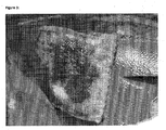
- the coating is a discontinuous coating, e.g. such as shown in Fig. 6 .
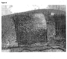
- the coating is a thin continuous coating, as obtained e.g. by spraying the polymeric component from the melt onto the matrix of biomaterial.
- a coating is comparable to a film-like or glass-like structure, e.g. such as shown in Fig. 7 .
- the molecular weight of the polymeric component is preferably in a range of 500 to 50000, most preferred about 10000.
- the amount of coating of polymeric component on the sponge of said biomaterial is preferably from about 1 mg/cm 2 to about 20 mg/cm 2 , more preferred about 2mg/cm 2 to about 14 mg/cm 2 for the coated sponge.
- the concentration of polymeric component is preferably from about 5mg/cm 3 to about 100mg/cm 3 , more preferred from about 10mg/cm 3 to about 70 mg/cm 3 for an impregnated sponge.
- the sponge of the present invention comprises a combination of impregnated and coated forms.
- the sponge according to the present invention preserves reactivity of the reactive groups of the hydrophilic polymeric component comprising reactive groups by being dry, e.g. having an overall water content of below 10%, especially below 2%, and especially below 1% in case the polymeric component has hydrolysable reactive groups, e.g. NHS-PEG.
- Higher water contents e.g. higher than 10%
- water contents of below 2% (w/w) are preferred; below 1% is even more preferred; below 0.5 % is specifically preferred.
- a further layer of a further biomaterial is present.
- the further layer can be from the same biomaterial as the matrix or it can be a different biomaterial, e.g. matrix of biomaterial is collagen and the further layer is oxidized cellulose. All combinations of biomaterials as mentioned above may be included.
- the sponge as a whole can be biodegradable, being suitable for biological decomposition in vivo, or bioresorbable, i.e. able to be resorbed in vivo, e.g. via degradation by proteases which are present in vivo and groups which are hydrolyseable in vivo. Full resorption means that no significant extracellular fragments remain.
- a biodegradable material differs from a non-biodegradable material in that a biodegradable material can be biologically decomposed into units which may either be removed from the biological system and/or chemically incorporated into the biological system.
- the particular material, the matrix material or sponge as a whole can be degraded by a subject, in particular a human subject, in less than 6 month, less than 3 month, less than 1 month, less than 2 weeks.
- a sponge of the present invention may further contains a dye, e.g. riboflavin, or other dye known from the prior art to be biocompatible.
- the dye may be included e.g. as a further layer (coating) and may especially help the surgeon to identify which one of the surfaces of a coated sponge of the present invention is the active or inactive surface, respectively.
- the sponge of the present invention preferably has an overall thickness of less than 3 cm, preferably about 1 mm to about 3cm, more preferably about 1 mm to about 2cm, most preferred about 1 mm to about 2mm.
- the thickness of the coating is preferably from about 0.01 mm to about 1 mm.
- the sponge of the present invention is preferably used in minimal invasive surgery, e.g. for laparoscopic application.
- the sponge may be dried and after drying, the sponge may have a water content of at least 0.5 (percentages given in w/w here). In certain embodiments the sponge can be freeze-dried or air-dried.
- the present invention also provides a wound coverage comprising a sponge according to the invention.
- the sponge and all additional layers can be provided in a ready to use wound coverage in suitable dimensions.
- the sponge and/or the coverage can be a pad or a sheet, preferably having a thickness of at least 1 mm or at least 2mm a or at least 5mm and/or up to 20mm, depending on the indication.
- Fixing may be achieved by melting the polymeric component onto the sponge in a preheated oven, e.g. at temperatures between 30°C to 80°C, preferably between 60°C to 65°C, for a time period sufficient for fixing, e.g. between 1 minute to 10 minutes, preferably about 4 minutes.
- fixing can be achieved by an infrared heater or any other heat source.
- the distance between the pad and the heater, the intensity of the heater and the time of exposure to infrared irradiation are adjusted to achieve melting of the coating at a minimum of heat exposure.
- Contacting for achieving impregnation may be done by placing the polymeric solution on top of the sponge and let the solution be soaked into said sponge for a time period sufficient for said absorption, e.g. from about 2 minutes to about 2 hours, preferably 30 minutes.
- Drying may include freeze drying or air drying and comprises removing volatile components of the fluid.
- the present invention provides a hemostatic sponge obtainable by a method of manufacturing according to process (I) or (II).
- Another aspect of the invention relates to the use of a sponge of the present invention for the treatment of an injury selected from the group consisting of a wound, a hemorrhage, damaged tissue and/or bleeding tissue.
- a sponge of the present invention is used for the sealing of tissues, e.g. lung, spleen, liver; and for hemostasis.
- the composite of the present invention can also be used as a ready to use tissue sealant, wherever the concentration of body fluids in proteins is high enough to allow the formation of a sealing gel as described above.
- the sponge of the present invention is especially indicated in open and endoscopic / laparoscopic / thoracoscopic/ MIS (minimal invasive surgery) surgical procedures as an adjunct to hemostatsis, to address surgical bleeding, from oozing to active, when control of bleeding by ligature or conventional procedures is ineffective or impractical.
- the sponge of the present invention is applied together with a buffer solution, e.g. an alkaline buffer solution, such as a bicarbonate solution, such as 8.4% NaHCO 3 , pH 8,3, e.g. on a gauze.
- a buffer solution e.g. an alkaline buffer solution, such as a bicarbonate solution, such as 8.4% NaHCO 3 , pH 8,3, e.g. on a gauze.
- the present invention further provides a kit comprising a sponge of the present invention and a buffer solution, e.g. an alkaline buffer solution, such as a bicarbonate or carbonate, together with instructions for its use.
- a buffer solution e.g. an alkaline buffer solution, such as a bicarbonate or carbonate, together with instructions for its use.
- the alkaline buffer solution preferably has a pH about 8, such as 8,3.
- a hemostatic composite comprising a water insoluble haemostatic material (matrix) and a hydrophilic polymeric cross-linker with electrophilic reactive groups, said composite comprising pores which allow external fluids, especially human blood, to access into said composite.
- the haemostatic material may be any material mentioned above as "matrix of a biomaterial" which by itself already has a certain haemostatic property. Such materials are known in principle in the art as well as their haemostatic property.
- the composite material according to the present invention has pores which allow external fluids to access the inner part of the composite so that e.g. if applied to a wound, blood of this wound can enter the composite. The composite can get soaked by these pores.
- non-woven or woven fabric of a haemostatic fiber Preferably, this haemostatic material is fabric of oxidized regenerated cellulose. It is specifically preferred, that the collagen sponge is essential native collagen (i.e. native collagen fiber structure is to a large extend preserved or regenerated by fibrillogenesis during processing).
- the reactivity of the hydrophilic polymeric crosslinker in the composite according to the present invention is retained. This means that the reactive groups of the crosslinker have not yet reacted with the (surface of the) haemostatic material and are not hydrolyzed by water. This can be achieved by combining the hemostatic material with the crosslinker in a way which does not lead to reaction of the reactive groups of the crosslinker with the hemostaic material or with water, e.g. as disclosed herein by melting, spraying, soaking under inert conditions, etc. Usually, this includes the omitting of aqueous conditions (or wetting), especially wetting without the presence of acidic conditions (if crosslinkers are not reactive under acidic conditions). This allows the provision of reactive haemostatic materials.
- PEG polyethylene glycol
- the matrix material which forms the porous network of the sponge constitutes of between 1-50%, 1-10%, or about 3% of the dried porous sponge (w/w-%).
- the matrix of a biomaterial, especially the collagen, according to the present invention in general is not soluble, in particular not water-soluble.
- the sponge is be porous and/or hygroscopic, it is allowed to swell when it is brought together with aqueous fluids, especially blood, serum, plasma, etc. or other fluids present in wounds and takes up these fluids.
- the hemostatic sponge according to the present invention is fluid absorbing.
- Fluid absorbing shall be considered as the physical process to hold fluids upon contacting which may or may not provoke swelling of the sponge.
- the sponge can hold an amount of a fluid, in particular blood, of at least 1 time, at least 2 times, at least 4 times or at least 10 times and/or up to 100 times, up to 20 times or up to 10 of the dry weight of the sponge.
- the sponge material according to the present invention can take up fluids even under pressure.
- the porous sponge material according to the present invention preferably has a pore size of 5 to 500 ⁇ m, preferably of 10 to 200 ⁇ m. This pore size can properly be adjusted in the course of production of the sponge biomaterial, especially by way of directing a drying process in the course of such production.
- the sponge according to the present invention is preferably provided in a "ready-to-use" form so that it is directly applicable to a patient in need thereof, e.g. to a wound of this patient (whereafter crosslinking starts).
- the sponge according to the present invention is therefore packed into a sterile package which protects the sponge from contamination (e.g. by moisture or microorganisms) during storage. Before use, the package can be opened (preferably also under sterile conditions) and the sponge can directly be applied to the patient ("ready-to use").
- the hydrophilic polymeric component is a hydrophilic crosslinker.
- this crosslinker has more than two electrophilic reactive groups for crosslinking ("arms"), for example three, four, five, six, seven, eight, or more arms with reactive groups for crosslinking.
- arms electrophilic reactive groups for crosslinking
- NHS-PEG-NHS is an effective hydrophilic crosslinker according to the present invention.
- a 4-arm polymer e.g. 4-arms-p-NP-PEG
- an 8-arm polymer e.g. 8-arms-NHS-PEG
- multi-reactive crosslinking is beneficial.
- the hydrophilic crosslinker according to the present invention is a polymer, i.e. a large molecule (macromolecule) composed of repeating structural units which are typically connected by covalent chemical bonds.
- Polymers according to the present invention should have a molecular weight of at least 1000 Da (to properly serve as crosslinkers for the sponge according to the present invention); preferably the crosslinking polymers according to the present invention has a molecular weight of at least 5000 Da, especially of at least 8000 Da.
- hydrophilic crosslinkers For some hydrophilic crosslinkers, the presence of basic reaction conditions (e.g. at the administration site) is preferred or necessary for functional performance (e.g. for a faster cross-linking reaction at the administration site).
- carbonate or bicarbonate ions e.g. as a buffer with a pH of 7.6 or above, preferably of 8.0 or above, especially of 8.3 and above
- may be additionally provided at the site of administration e.g. as a buffer solution or as a fabric or pad soaked with such a buffer), so as to allow an improved performance of the sponge according to the present invention or to allow efficient use as a hemostatic and/or wound adherent material.
- the present invention is further exemplified by the following examples without being limited thereto.
- 50g of sliced bovine corium are dispersed in 500ml of a 2M NaOH-solution and stirred approx. 90min at 25°C.
- the corium is sieved out and rinsed with distilled H 2 O until effluent H 2 O reaches a pH of about 8.0.
- the washed corium slices are re-suspended in H 2 O and the pH is adjusted with HCl to approx. 2.0.
- the suspension obtained is stirred overnight at approx. 25°C and a collagen solution is obtained.
- the solution obtained is cooled to 5°C and the pH is adjusted with NaOH to neutral. Collagen precipitation is carried out overnight by keeping the solution at 18°C without stirring. Precipitated collagen obtained is separated by filtration.
- the collagen concentration of the material obtained is determined by gravimetry.
- a chemical crosslinking with glutaraldehyde may be carried out in that a 1% aq. collagen suspension is prepared and 5000ppm of glutaraldehyde are added at 12°C. The suspension obtained is stirred overnight. Crosslinked collagen obtained is filtered and washed with H 2 O. The collagen concentration of the material obtained is determined as described above.
- Example 2 Collagen pad coated with NHS-PEG
- COH102 powder is homogeneously distributed onto one surface of a commercially available collagen sponge (Matristypt ® , Dr. Suwelack Skin- and Healthcare, Germany, thickness 1 mm or 2mm). COH102 amounts of 2mg/cm 2 , 7mg/cm 2 , 10mg/cm 2 , 14mg/cm 2 , 20mg/cm 2 are used for the coating.
- the COH102 powder is fixed on the surface of the sponge by melting. This is performed at 60°C to 65°C for 4 min by placing the sponge with the PEG powder mixture into a preheated oven.
- a dried sponge obtained is sealed together with a sachet of desiccant in a gas-impermeable pouch and ⁇ -sterilized at 25kGray.
- Example 3 Collagen pad impregnated with NHS-PEG
- Aq. acidic solutions (pH 3.0, AcOH) of COH102 with concentrations of 10mg/cm 3 , 20mg/cm 3 , 30mg/cm 3 and 40mg/cm 3 are prepared and filled into 9x7 cm PET-trays.
- Commercial available bovine collagen sponges (Matristypt®), 9x7x0.1 or 0.2 cm, with the same volume as the previously filled COH102 solution are placed on the top of the solutions for impregnation for 20 min.
- COH102 solution is absorbed and the collagen material obtained is lyophilized. Sponges obtained can be additionally coated with COH102 as described in example 2.
- each dried sponge obtained is sealed together with a sachet containing desiccant in a gas impermeable pouch and sterilized by ⁇ -irradiation at 25kGray.
- Example 4 Collagen pad containing oxidized cellulose powder coated with NHS-PEG
- Traumastem® P powder (Bioster, Czech Republic) is homogenously distributed into 22ml of neutral aqueous collagen suspension (2.15mg/ml; 4.3mg/ml and 10mg/ml) produced according to example 1.
- the mixture obtained is filled into flat 9x7cm PET-trays and lyophilized.
- a fleece obtained has a thickness of about 3-4mm and is coated with COH102 as described in example 2.
- each sponge obtained is sealed together with a sachet containing desiccant in a gas impermeable pouch and sterilized by ⁇ -irradiation at 25kGray.
- Example 5 Collagen pad containing oxidized cellulose fabric coated with NHS-PEG
- a 6x5cm Traumastem® TAF light-fabric (Bioster, Czech Republic) is immersed into a 1 % bovine collagen suspension as described in example 1.
- the 6x5cm oxidized cellulose fabric retains approximately 6g of the collagen suspension.
- a fabric soaked with the collagen suspension is obtained and laid in a tray and lyophilized.
- a fleece obtained has a thickness of about 3-4mm and is coated with COH102 as described in example 2.
- each sponge obtained is sealed together with a sachet containing desiccant in a gas impermeable pouch and sterilized by ⁇ -irradiation at 25kGray.
- Example 6 Oxidized cellulose fabric coated with NHS-PEG
- Traumastem® P fleece (Bioster, Czech Republic) is coated with 14mg/cm 2 COH102 as described in example 2.
- the thickness of the pad obtained is about 1-2mm.
- Example 7 Collagen pad containing fucoidan as hemostasis enhancing substance coated with NHS-PEG
- a bovine collagen sponge Matristypt® (9x7x0.2cm) is impregnated with the same volume of a Fucoidan solution of A. nodosum (10 ⁇ M and 200 ⁇ M in 40mM Ca 2+ -solution) and lyophilized.
- a sponge obtained is coated with COH102 as described in example 2.
- Example 8 Collagen pad containing thrombin as hemostasis enhancing substance coated with NHS-PEG
- a bovine collagen sponge Matristypt® (9x7x0.2cm) is impregnated with the same volume of a thrombin solution (500IU/ml) and lyophilized.
- a sponge obtained is coated with COH102 as described in example 2.
- Example 9 Sealing efficacy of a collagen pad coated with NHS-PEG
- a hemostatic pad coated with 14mg/cm 2 COH102 is produced according to example 2.
- a lesion of around 1.5 to 2cm in diameter is set by a scalpel on the lung of a pig.
- a sample of 3x3cm of the said pad is applied onto the wound and hold in place by exerting slight pressure with the aid of gauze for 2 min.
- the gauze is pre-wetted either with saline or basic bicarbonate solution (pH 8.3).
- the pad is adhering firmly to the lung surface (see Figure 6 ).
- the speed of obtaining adherence is increased using gauze wetted with bicarbonate.
- the chest is filled with Ringer's solution after 10 min. No gas leakage or detachment of the pad is observed.
- Example 10 Sealing efficacy of a collagen pad impregnated with NHS-PEG
- a hemostatic pad impregnated with 40mg/cm 3 COH102 is produced according to example 3.
- a lesion of around 1.5 to 2cm in diameter is set by a scalpel on the lung of a pig.
- a sample of 3x3cm of the said pad is applied onto the wound and hold in place by exerting slight pressure with the aid of gauze for 2 min.
- the gauze is pre-wetted with basic bicarbonate solution (pH 8.3). After application the pad is adhering firmly to the lung surface. Air tightness and pad-adherence to the tissue are determined as described in Example 9.
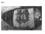
- a mask made of a stainless steel plate (1 mm thickness) with a pattern of holes is placed on one side of a 1 or 2 mm thick collagen sponge (Matristypt®, Dr. Suwelack Skin- and Healthcare, Germany).
- the holes of the mask have a diameter of 2mm and are placed at a distance of 1 cm from each other in the nodes of an upright square lattice.
- a 0.5% aqueous Erioglaucine (Fluka, Switzerland) solution is sprayed with a standard airbrush device over the holes of the mask.
- the mask is removed and a collagen sheet with the blue dot pattern obtained is dried at ambient atmosphere, in a vacuum oven or in a desiccator.
- the dot pattern on one side has the role to distinguish the active and inactive surface of a coated pad. It is possible to apply the coating either on the dotted side or the non-dotted side.
- Example 12 Preparation of a fibrin fleece
- a solution of 2.5 mg/ml of fibrinogen, 10 mM Tris/HCl, 150 mM NaCl, pH 7.4 and an equal volume of 55 IU thrombin/ml, 10 mM CaCl 2 are mixed using a static mixer and immediately filled into a tray at a height of 0.7 cm. A fibrin clot is obtained in the tray. By freeze-drying of the clot a fibrin fleece is obtained.
- Example 13 Preparation of collagen pad coated with NHS-PEG-NHS and its testing in animal model
- Sponges obtained are sealed together with a sachet containing desiccant in a gas-impermeable pouch.
- Example 14 Preparation of collagen pad coated with 8-arm-NHS-PEG and its testing in animal model
- the hemostatic performance of said pad is tested in pig in the liver abrasion model as described above. After 2 minutes hemostasis is achieved. No rebleeding after 10 minutes is observed. The adherence of the pad on the tissue is sufficient.
- Example 15a Preparation of collagen pad coated with 4-arm-p-NP-PEG and its testing in animal model
- 14mg/cm 2 4-arm-p-NP-PEG (MW 10000, NOF Corporation, Japan) are homogeneously distributed and fixed by melting. This is performed at 65°C for 4 min by placing the sponge with the PEG powder into a preheated oven.
- a sponge obtained is sealed together with a sachet containing desiccant in a gas-impermeable pouch.
- the hemostatic performance of the said pad is tested in pig in the liver abrasion model as described above. After 2 minutes hemostasis is achieved. No rebleeding after 10 minutes is observed. The adherence of the pad on the tissue is not sufficient.
- Example 15b Preparation of collagen pad coated with 4-arm-p-NP-PEG and its testing in animal model
- the hemostatic performance of the pad as prepared in Ex. 15a is tested in pig in the liver abrasion model as described above but with the modification, that the pad is applied with gauze pre-wetted with basic 8 % Na-bicarbonate solution. After 2 minutes hemostasis is achieved. No rebleeding after 10 minutes is observed. The adherence of the pad on the tissue is sufficient.
- Example 16a Preparation of collagen pad coated with CHO-PEG-CHO and its testing in animal model
- a 6x6cm collagen pad made as described in example 11, 9.5mg/cm 2 CHO-PEG-CHO (MW 3400, Interchim, France) are homogeneously distributed and fixed by melting. This is performed at 70°C for 4 min by placing the sponge with the PEG powder into a preheated oven.
- a sponge obtained is sealed together with a sachet containing desiccant in a gas-impermeable pouch.
- the hemostatic performance of said pad is tested in pig in the liver abrasion model as described above. After 2 minutes hemostasis is achieved. No rebleeding after 10 minutes is observed. The adherence of the pad on the tissue is sufficient.
- Example 16b Preparation of collagen pad coated with CHO-PEG-CHO and its testing in animal model
- the hemostatic performance of the pad as prepared in Ex.16a is tested in pig in the liver abrasion model as described above but with the modification, that the pad is applied with gauze pre-wetted with basic Na-bicarbonate solution. After 2 min hemostasis is achieved. No rebleeding after 10 min is observed. The adherence of the pad on the tissue is sufficient.
- Example 17a Preparation of collagen pad coated with Epoxy-PEG-Epoxy and its testing in animal model
- Epoxy-PEG-Epoxy (MW 3400, Interchim, France) are homogeneously distributed and fixed by melting. This is performed at 70°C for 4 min by placing the sponge with the PEG powder into a preheated oven.
- a sponge obtained is sealed together with a sachet containing desiccant in a gas-impermeable pouch.
- the hemostatic performance of said pad is tested in pig in the liver abrasion model as described above. After 2 min no hemostasis is achieved. The adherence of the pad on the tissue is not sufficient.
- Example 17b Preparation of collagen pad coated with Epoxy-PEG-Epoxy and its testing in animal model
- the hemostatic performance of the pad as prepared in Ex.17a is tested in pig in the liver abrasion model as described above but with the modification, that the pad is applied with gauze pre-wetted with basic Na-bicarbonate solution. After 2 min hemostasis is achieved. No rebleeding after 5 min is observed. The adherence of the pad on the tissue is sufficient.
- Example 18 Preparation of collagen pad coated with 4-arm-Epoxy-PEG and its testing in animal model
- 14mg/cm 2 4-arm-epoxy-PEG (MW 10000, Interchim, France) are homogeneously distributed and fixed by melting. This is performed at 70°C for 4 min by placing the sponge with the PEG powder into a preheated oven.
- a sponge obtained is sealed together with a sachet containing desiccant in a gas-impermeable pouch.
- the hemostatic performance of said pad is tested in pig in the liver abrasion model as described above, but with the modification, that the pad is applied with gauze pre-wetted with basic Na-bicarbonate solution. After 2 min hemostasis is achieved. No rebleeding after 5 min is observed. The adherence of the pad on the tissue is sufficient.
- a sponge obtained is sealed together with a sachet containing desiccant in a gas-impermeable pouch.
- the hemostatic performance of said pad is tested in pig in the liver abrasion model as described above. After 2 min hemostasis is achieved. No rebleeding after 10 min is observed. The adherence of the pad on the tissue is sufficient.
- Example 20 Preparation of collagen pad coated with AA-dextran and its testing in animal model.
- a 6x6cm collagen pad made as described in example 11 14mg/cm 2 of a mixture of 0.1mg/cm 2 AA-dextran (MW 40000, Pierce, USA) and 13.9mg/cm 2 unsubstituted PEG (MW 10000, Sigma Aldrich, Germany) are homogeneously distributed and fixed by melting. This is performed at 80°C for 4 min by placing the sponge with the powder mixture into a preheated oven.
- a sponge obtained is sealed together with a sachet containing desiccant in a gas-impermeable pouch.
- the hemostatic performance of said pad is tested in pig in the abrasive liver lobe model as described above but with the modification, that the.pad is applied with gauze pre-wetted with basic Na-bicarbonate solution. After 2 min hemostasis is achieved. No rebleeding after 10 min is observed. The adherence of the pad on the tissue is sufficient.
- Example 21a Preparation of collagen pad coated with DSS and its testing in animal model
- a sponge obtained is sealed together with a sachet containing desiccant in a gas-impermeable pouch.
- the hemostatic performance of said pad is tested in pig in the abrasive liver lobe model as described above. After 2 min hemostasis is not achieved. The adherence of the pad on the tissue is not sufficient.
- Example 21b Preparation of collagen pad coated with DSS and its testing in animal model
- the hemostatic performance of the pad as prepared in Ex.21 a is tested in pig in the abrasive liver lobe model as described above but with the modification, that the pad is applied with gauze pre-wetted with basic bicarbonate solution. After 2 min hemostasis is achieved. No rebleeding after 10 min is observed. The adherence of the pad on the tissue is sufficient.
- Example 22a Preparation of collagen pad coated with EGS and its testing in animal model
- a sponge obtained is sealed together with a sachet containing desiccant in a gas-impermeable pouch.
- the hemostatic performance of said pad is tested in pig in the liver abrasion model as described above. After 2 min hemostasis is not achieved. The adherence of the pad on the tissue is not sufficient.
- Example 22b Preparation of collagen pad coated with EGS and its testing in animal model
- the hemostatic performance of the pad as prepared in Ex.22a is tested in pig in the liver abrasion model as described above but with the modification, that the pad is applied with gauze pre-wetted with basic Na-bicarbonate solution. After 2 min hemostasis is achieved. No rebleeding after 10 min is observed. The adherence of the pad on the tissue is sufficient.
- a sponge obtained is sealed together with a sachet containing desiccant in a gas-impermeable pouch.
- the hemostatic performance of said pad is tested in pig in the liver abrasion model as described above. After 2 min hemostasis is achieved. No rebleeding after 10 min is observed. The adherence of the pad on the tissue is sufficient.
- Example 24 Correlation between the adherence force to the tissue and the cross-linker used for collagen pad coating
- Cross-linker Adherence score 13 NHS-PEG-NHS 1 14 8-arms-NHS-PEG 1 15a 4-arms-p-NP-PEG 3 15b 4-arms-p-NP-PEG - basic application 2 16a CHO-PEG-CHO 1 16b CHO-PEG-CHO - basic application 2 17a Epoxy-PEG-Epoxy 3 17b Epoxy-PEG-Epoxy - basic application 2 18 4-arm-Epoxy-PEG - basic application 2 19 ISC-PEG-ISC 1 20 AA-dextran - basic application 1 21a DSS 3 21b DSS - basic application 2 22a EGS 3 22b EGS - basic application 2
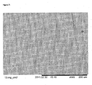
- Example 25 Chitosan/Gelatine sponge coated with NHS-PEG and its testing in animal model
- chitosan/gelatin sponge (Chitoskin®, Beese Medical, Germany) 14mg/cm 2 of COH102 are homogeneously distributed and fixed by melting. This is performed at 65°C for 4 min by placing the sponge with the PEG powder into a preheated oven. A sponge obtained is sealed together with a sachet containing desiccant in a gas-impermeable pouch.
- the hemostatic performance of said pad is tested in pig in the liver abrasion model as described above. After 2 min hemostasis is achieved. No rebleeding after 10 min is observed ( Figure 9 ). The adherence of the pad on the tissue is sufficient.
- Example 26 Preparation of a gelatin pad coated with NHS-PEG and its testing in animal model
- a 2 x 2 cm piece of a dry collagen sponge (Matristypt®, Dr. Suwelack, Germany) or of a dry cross-linked gelatin sponge (Gelfoam®, Pfizer) are placed onto the surface of distilled H 2 O into a beaker.
- the dry sponges are floating on the water surface and take up water over the 2 x 2 cm contact surface. After 6 s Matristypt® is totally soaked by H 2 O and removed from the water surface. The thicker Gelfoam® sponge is not totally soaked by H 2 O after 13 s, but removed after 13s from the water surface.
- the initial water uptake velocities of the sponges are 35 mg x cm -1 s -1 for Matristypt® and 0.8 mg x cm -1 s -1 for Gelfoam®.
Description
- The present invention relates to the field of hemostatic sponges, a method of producing said sponges and their various uses.
- Biological glues based on coagulation factors of human or animal origin have long been known. A method for producing tissue adhesives based on fibrinogen and factor XIII has been described in
US 4,362,567 ,US 4,298,598 andUS 4,377,572 . The tissue adhesives are usually applied together with a separate component containing thrombin, which is enzymatically acting on fibrinogen to form fibrin, and on factor XIII to form the active factor Xllla, which cross-links the fibrin to obtain a stable fibrin clot. - Collagen pads have been used for many years to improve wound healing or to stop bleeding. Their mechanism of action in hemostasis is based on platelets aggregation and activation, the formation of thrombin on the surface of activated platelets and the formation of a hemostatic fibrin clot by the catalytic action of thrombin on fibrinogen. To improve the hemostatic action of collagen pads or sheets it has been suggested to include factors of hemostasis within such pads.
- In
US 4,600,574 a tissue adhesive based on collagen combined with fibrinogen and factor XIII is described. This material is provided in the lyophilized form, ready for use. The fibrinogen and factor XIII are combined with the collagen by impregnating the collagenous flat material with a solution comprising fibrinogen and factor XIII, and lyophilizing said material. -
WO 97/37694 -
US 5,614,587 discusses bioadhesive compositions comprising cross-linked collagen using a multifunctionally activated synthetic hydrophilic polymer, as well as methods of using such compositions to effect adhesion between a first surface and a second surface, wherein at least one of the first and second surfaces can be a native tissue surface. -
WO2004028404 describes a tissue sealant composed of a synthetic collagen or gelatin and a electrophilic cross-linking agent which are provided in a dry state. Upon wetting of this composition at an appropriate pH a reaction between the 2 components takes place and a gel with sealing properties is formed. Such a sealant works in essential analogously to other known two component sealants (composed of a reagent with multiple electrophilic- and a reagent with multiple nucleophilic groups) which are known in the state of the art or which are available on the market, e.g. Coseal™. In a special embodiment of the invention the two components of the sealant (the electrophilic cross-linker and the synthetic collagen/gelatin) are coated onto a biomaterial. - Collagen-containing compositions which have been mechanically disrupted to alter their physical properties are described in
US 5,428,024 ,US 5,352,715 , andUS 5,204,382 . These patents generally relate to fibrillar and insoluble collagens. An injectable collagen composition is described inUS 4,803,075 . An injectable bone/cartilage composition is described inUS 5,516,532. A collagen-based delivery matrix comprising dry particles in the size range from 5 µm to 850 µm which may be suspended in water and which has a particular surface charge density is described inWO 96/39159 US 5,196,185 . Other patents describing collagen compositions includeUS 5,672,336 andUS 5,356,614 . -
US 2008/187591 A1 discloses a hemostatic composition comprising a hydrogel-forming component and at least two crosslinkers.WO 90/13320 A1 US 2006/0258560 A1 discloses a tissue sealant composition, comprising a crosslinker, and a synthetic collagen or a synthetic gelatin, in a dry state, optionally provided on a solid matrix.WO 02/072128 A1 - The subject of the invention is a hemostatic porous composite sponge consisting essentially of
- i) a matrix of a biomaterial and
- ii) a single hydrophilic polymeric component comprising electrophilic reactive groups, wherein said hydrophilic polymeric component is a hydrophilic crosslinker,
wherein i) and ii) are associated with each other so that the reactivity of the polymeric component is retained, wherein associated means that- said polymeric component is coated onto a surface of said matrix of a biomaterial, or
- said matrix is impregnated with said polymeric material, or
- both.
- It has been found that previous pads of fibrous biomaterials, in particular collagen pads, for wound healing failed to induce hemostasis at conditions with impaired hemostasis (e.g. after heparinization). The sponge according to the present invention improves hemostasis. Furthermore, the sponge according to the present invention shows a strong adherence to the tissue when applied to a wound. The sponge of the present invention further shows improved swelling behavior, i.e. low swelling, after application to a wound.
- A further aspect relates to a method of treating an injury comprising administering a hemostatic porous composite sponge to the site of injury.
- Also provided is a kit for preparing a wound coverage, comprising a sponge as herein disclosed and a buffer solution. This kit and its components are in particular for the manufacture of a medical sponge for the treatment of an injury.
- Those skilled in the art will readily understand that all preferred embodiments disclosed in the following are examples of specific embodiments, but are not necessarily limiting the general inventive concept. Furthermore, all special embodiments can be read on all inventive aspects and embodiments in any combination, if not mutually exclusive. All equivalents or obvious alterations or modifications as recognized by those skilled in the art are included by the present invention.
- The object of the invention is a hemostatic porous composite sponge comprising a hemostatic porous composite sponge as defined in the claims
- The term impregnated as meant herein includes the term absorption of polymeric material in a matrix of a biomaterial.
- The terms sponge, pad and fleece are used interchangeably in the description of the present invention.
- Preferably the biomaterial is collagen, a protein, a biopolymer, or a polysaccharide. Especially preferred is a biomaterial selected from the group consisting of collagen, gelatin (especially cross-linked gelatin), fibrin, a polysaccharide (especially chitosan, oxidized cellulose, aldehyde activated dextrans, starch based polyaldehydes (obtainable by periodate oxidation)), a synthetic biodegradable biomaterial (especially polylactic acid or polyglycolic acid, and derivatives thereof, more preferred collagen.
- According to the present invention a porous composite material comprising a water insoluble matrix of a biomaterial with hemostatic properties and a hydrophilic polymeric cross-linking agent in association therewith is provided.
- Upon contact with bleeding tissue, a cross-linking reaction of the hydrophilic polymeric cross-linker with the blood proteins leads to formation of a gel with sealing and hemostatic properties. Cross-linking also occurs to the tissue surface proteins and, depending on the nature of the water insoluble matrix biomaterial, may also occur to the matrix biomaterial. The latter reaction contributes to an improved adhesion of the composite material to the wounded tissue surface.
- Furthermore, it is important for the hemostatic efficacy of the composite according to the present invention that the matrix of the biomaterial has soaking capabilities, i.e. is able to soak/absorb liquids such as blood, serum, plasma.
- Such soaking capabilities are especially dependent on the hydrophilic nature of the polymer the matrix is made of, and a three-dimensional structure of open interconnected pores, or of a three-dimensional meshwork of hydrophilic fibers. The pore size and the elasticity of the matrix are also important for the soaking capacity. Elasticity means that the matrix can be compressed in aqueous solution and returns to its initial volume after the force causing compression is relieved.
- The sponge is a porous network of a biomaterial able to absorb body fluids when applied to the site of an injury. This allows the blood of a wound (including all the blood components, such as blood cells or coagulation proteins) to enter into the sponge. The porous sponge according to the present invention has therefore an inside volume which is accessible for external fluids, such as blood, when applied to a patient. For example, a porous collagen sponge can be made by lyophilization of a collagen gel, suspension or solution by freeze-drying (whereas normal air-drying leads to a collagen film). It follows that in the case of collagen, the resulting porous sponge according to the present invention has typically from 5 to 100 mg collagen/cm3, whereas collagen films have from 650 to 800 mg collagen/cm3. If external fluids, such as blood get in contact with the sponge according to the present invention, the hydrophilic polymeric component comprising reactive groups can react with the blood components and/or with the surface of the matrix of the biomaterial so as to crosslink the components which bind to the (at least two) reactive groups. Furthermore, the sponge is usually flexible and suitable to be applied on diverse tissues and locations with various shapes.
- The collagen used for the present invention can be from any collagen material including liquid, pasty, fibrous or powdery materials that can be processed to a porous, especially a porous and fibrous matrix. The preparation of a collagen gel for the production of a sponge is e.g. described in the
EP 0891193 and may include acidification until gel formation occurs and subsequent pH neutralization. To improve gel forming capabilities or solubility the collagen may be (partially) hydrolyzed or modified, as long as the property to form a stable sponge when dried is not diminished. - The collagen or gelatin of the sponge matrix is preferably of animal origin, preferably bovine or equine. However, also human collagen might be used in case of a hypersensitivity of the patient towards xenogenic proteins. Also synthetic or recombinant collagen may be used. The further components of the sponge are preferably of human origin, which makes the sponge suitable especially for the application to a human.
- In a preferred embodiment the porous collagen sponge contains about 5 to about 50, e.g. about 10 to about 30, preferably about 25 mg collagen / cm3 of dry sponge.
- The biomaterial may be non-crosslinked or crosslinked, preferably the biomaterial has been crosslinked.
- The hydrophilic polymeric component of the sponge according to the present invention is a hydrophilic crosslinker which is able to react with its electrophilic reactive groups once the sponge is applied to a patient (e.g. to a wound of a patient or another place where the patient is in need of a hemostatic activity). Therefore it is important for the present invention that the electrophilic reactive groups of the polymeric component are reactive when applied to the patient. It is therefore necessary to manufacture the sponge according to the present invention so that the reactive groups of the polymeric component which should react once they are applied to a wound are retained during the manufacturing process.
- This can be done in various ways. For example, usual hydrophilic polymeric components have reactive groups which are susceptible to hydrolysis after contact with water. Accordingly, premature contact with water or aqueous liquids has to be prevented before administration of the sponge to the patient, especially during manufacture. However, processing of the hydrophilic polymeric component during manufacturing may be possible also in an aqueos medium at conditions where the reactions of the reactive-groups are inhibited (e.g. at a low pH). If the hydrophilic polymeric components can be melted, the melted hydrophilic polymeric components can be sprayed or printed onto the matrix of the biopolymer. It is also possible to sprinkle a dry form (e.g. a powder) of the hydrophilic polymeric component onto the matrix. If necessary, then an increase of the temperature can be applied to melt the sprinkled hydrophilic polymeric component to the matrix to achieve a permanent coating of the sponge. Alternatively, these hydrophilic polymeric components can be taken up into inert organic solvents (inert vis-à-vis the reactive groups of the hydrophilic polymeric components) and brought onto the matrix of the biomaterial. Examples of such organic solvents are dry ethanol, dry acetone or dry dichloromethane (which are e.g. inert for hydrophilic polymeric components, such as NHS-ester substituted PEGs).
- In a preferred embodiment the hydrophilic polymer component is a single hydrophilic polymer component and is a polyalkylene oxide polymer, esp. preferred a PEG comprising polymer, in the following called "the material". The reactive groups of said material are electrophilic groups.
- The material may be a multi-electrophilic polyalkylene oxide polymer, e.g. a multi-electrophilic PEG. The material can include two or more electrophilic groups such as -CON(COCH2)2, -CHO, -N=C=O, and/or -N(COCH2)2, e.g. a component as disclosed in the
WO2008/016983 (incorporated herein by reference in its entirety) and one of the components of the commercially available ones under the trademark CoSeal®. - Preferred electrophilic groups of the hydrophilic polymeric crosslinker according to the present invention are groups reactive to the amino-, carboxy-, thiol- and hydroxy- groups of proteins, or mixtures thereof.
- Preferred amino group-specific reactive groups are NHS-ester groups, imidoester groups, aldehyde-groups, carboxy-groups in the presence of carbodiimdes, isocyanates, or THPP (beta-[Tris(hydroxymethyl)phosphino] propionic acid), especially preferred is Pentaerythritolpoly(ethyleneglycol)ether tetrasuccinimidyl glutarate (= Pentaerythritol tetrakis[1-1'-oxo-5'-succinimidylpentanoate-2-poly-oxoethyleneglycole]ether (= an NHS-PEG with MW 10,000).
- Preferred carboxy-group specific reactive groups are amino-groups in the presence of carbodiimides.
- Preferred thiol group-specific reactive groups are maleiimides or haloacetyls.
- Preferred hydroxy group-specific reactive group is the isocyanate group. The electrophilic reactive groups on the hydrophilic cross-linker may be identical (homo-functional) or different (hetero-functional). The hydrophilic polymeric component can have two reactive groups (homo-bifunctional or heterobifunctional) or more (homo/hetero-trifunctional or more).
- In special embodiments the material is a synthetic polymer, preferably comprising PEG. The polymer can be a derivative of PEG comprising active side groups suitable for cross-linking and adherence to a tissue,
- By the electrophilic reactive groups the hydrophilic polymer has the ability to cross-link blood proteins and also tissue surface proteins. Cross-linking to the biomaterial is also possible.
- The multi-electrophilic polyalkylene oxide may include two or more succinimidyl groups. The multi-electrophilic polyalkylene oxide may include two or more maleimidyl groups.
- Preferably, the multi-electrophilic polyalkylene oxide is a polyethylene glycol or a derivative thereof.
- In a most preferred embodiment the polymeric component is pentaerythritolpoly(ethyleneglycol)ether tetrasuccinimidyl glutarate (=COH102, also pentaerythritol tetrakis[1-1'-oxo-5'-succinimidylpentanoate-2-poly-oxoethyleneglycole]ether).
- In one preferred embodiment the sponge of the present invention comprises collagen as the biomaterial and the polymeric component, e.g. COH102, is coated onto the surface of the collagen (=coated form).
- Especially preferred the coating is a discontinuous coating, e.g. such as shown in
Fig. 6 . - In another preferred embodiment the coating is a thin continuous coating, as obtained e.g. by spraying the polymeric component from the melt onto the matrix of biomaterial. Such a coating is comparable to a film-like or glass-like structure, e.g. such as shown in
Fig. 7 . - In another preferred embodiment the sponge of the present invention comprises collagen as the biomaterial and the polymeric component, e.g. COH102, is impregnated into the collagen (= impregnated form).
- The molecular weight of the polymeric component is preferably in a range of 500 to 50000, most preferred about 10000.
- The amount of coating of polymeric component on the sponge of said biomaterial is preferably from about 1 mg/cm2 to about 20 mg/cm2, more preferred about 2mg/cm2 to about 14 mg/cm2for the coated sponge. The concentration of polymeric component is preferably from about 5mg/cm3 to about 100mg/cm3, more preferred from about 10mg/cm3 to about 70 mg/cm3 for an impregnated sponge.
- In another preferred embodiment the sponge of the present invention comprises a combination of impregnated and coated forms. Further, the sponge according to the present invention preserves reactivity of the reactive groups of the hydrophilic polymeric component comprising reactive groups by being dry, e.g. having an overall water content of below 10%, especially below 2%, and especially below 1% in case the polymeric component has hydrolysable reactive groups, e.g. NHS-PEG. Higher water contents (e.g. higher than 10%) would also result in a functional sponge but storage stability would be worsened. Accordingly, water contents of below 2% (w/w) are preferred; below 1% is even more preferred; below 0.5 % is specifically preferred.
- In another preferred embodiment a further layer of a further biomaterial is present. The further layer can be from the same biomaterial as the matrix or it can be a different biomaterial, e.g. matrix of biomaterial is collagen and the further layer is oxidized cellulose. All combinations of biomaterials as mentioned above may be included.
- The sponge as a whole can be biodegradable, being suitable for biological decomposition in vivo, or bioresorbable, i.e. able to be resorbed in vivo, e.g. via degradation by proteases which are present in vivo and groups which are hydrolyseable in vivo. Full resorption means that no significant extracellular fragments remain. A biodegradable material differs from a non-biodegradable material in that a biodegradable material can be biologically decomposed into units which may either be removed from the biological system and/or chemically incorporated into the biological system. In a preferred embodiment the particular material, the matrix material or sponge as a whole can be degraded by a subject, in particular a human subject, in less than 6 month, less than 3 month, less than 1 month, less than 2 weeks.
- A sponge of the present invention may further contains a dye, e.g. riboflavin, or other dye known from the prior art to be biocompatible. The dye may be included e.g. as a further layer (coating) and may especially help the surgeon to identify which one of the surfaces of a coated sponge of the present invention is the active or inactive surface, respectively.
- The sponge of the present invention preferably has an overall thickness of less than 3 cm, preferably about 1 mm to about 3cm, more preferably about 1 mm to about 2cm, most preferred about 1 mm to about 2mm.
- In a sponge of the present invention the thickness of the coating is preferably from about 0.01 mm to about 1 mm.
- The sponge of the present invention is preferably used in minimal invasive surgery, e.g. for laparoscopic application.
- The sponge may be dried and after drying, the sponge may have a water content of at least 0.5 (percentages given in w/w here). In certain embodiments the sponge can be freeze-dried or air-dried.
- The present invention also provides a wound coverage comprising a sponge according to the invention. The sponge and all additional layers can be provided in a ready to use wound coverage in suitable dimensions. The sponge and/or the coverage can be a pad or a sheet, preferably having a thickness of at least 1 mm or at least 2mm a or at least 5mm and/or up to 20mm, depending on the indication. When the relatively thick flexible sponge is applied to a wound it is important that blood and fibrinogen can be absorbed throughout the sponge before fibrin is formed that might act as a barrier for the absorption of further wound secret.
- Another aspect of the invention relates to a method of manufacturing a hemostatic sponge (= process I) comprising
- a) providing a porous sponge comprising a matrix of a biomaterial in dried form,
- b) providing a single hydrophilic polymeric component comprising electrophilic reactive groups in the form of dry powder, wherein said
single reactive polymeric material is a hydrophilic crosslinker, - c) contacting a) and b) so that the material of b) is present on at least one surface of said sponge, and
- d) fixing the material of b) on the sponge of a).
- Fixing may be achieved by melting the polymeric component onto the sponge in a preheated oven, e.g. at temperatures between 30°C to 80°C, preferably between 60°C to 65°C, for a time period sufficient for fixing, e.g. between 1 minute to 10 minutes, preferably about 4 minutes.
- Alternatively fixing can be achieved by an infrared heater or any other heat source. The distance between the pad and the heater, the intensity of the heater and the time of exposure to infrared irradiation are adjusted to achieve melting of the coating at a minimum of heat exposure.
- Another aspect of the invention relates to a method of manufacturing a hemostatic sponge (=process II) comprising
- a) providing a porous sponge comprising a matrix of a biomaterial in dried form,
- b) providing a single hydrophilic polymeric component comprising electrophilic reactive groups in the form of a solution, wherein said
single reactive polymeric material is a hydrophilic crosslinker. - c) contacting a) and b) so that)the material of a) is impregnated with b), and
- d) drying the material obtained in step c).
- Contacting for achieving impregnation may be done by placing the polymeric solution on top of the sponge and let the solution be soaked into said sponge for a time period sufficient for said absorption, e.g. from about 2 minutes to about 2 hours, preferably 30 minutes.
- Drying may include freeze drying or air drying and comprises removing volatile components of the fluid.
- In another aspect the present invention provides a hemostatic sponge obtainable by a method of manufacturing according to process (I) or (II).
- Another aspect of the invention relates to the use of a sponge of the present invention for the treatment of an injury selected from the group consisting of a wound, a hemorrhage, damaged tissue and/or bleeding tissue. Preferably a sponge of the present invention is used for the sealing of tissues, e.g. lung, spleen, liver; and for hemostasis.
- The composite of the present invention can also be used as a ready to use tissue sealant, wherever the concentration of body fluids in proteins is high enough to allow the formation of a sealing gel as described above.
- The sponge of the present invention is especially indicated in open and endoscopic / laparoscopic / thoracoscopic/ MIS (minimal invasive surgery) surgical procedures as an adjunct to hemostatsis, to address surgical bleeding, from oozing to active, when control of bleeding by ligature or conventional procedures is ineffective or impractical.
- In a preferred embodiment the sponge of the present invention is applied together with a buffer solution, e.g. an alkaline buffer solution, such as a bicarbonate solution, such as 8.4% NaHCO3, pH 8,3, e.g. on a gauze.
- It has been found that the speed of reaction is increased after application with 8.4% NaHCO3-solution soaked gauze compared to saline soaked gauze. This was observed by an higher adherence of the sponge to the tissue after 2 minutes in the case of the NaHCO3-application.
- The present invention further provides a kit comprising a sponge of the present invention and a buffer solution, e.g. an alkaline buffer solution, such as a bicarbonate or carbonate, together with instructions for its use. The alkaline buffer solution preferably has a pH about 8, such as 8,3.
- Another aspect of the present invention relates to a hemostatic composite comprising a water insoluble haemostatic material (matrix) and a hydrophilic polymeric cross-linker with electrophilic reactive groups, said composite comprising pores which allow external fluids, especially human blood, to access into said composite. The haemostatic material may be any material mentioned above as "matrix of a biomaterial" which by itself already has a certain haemostatic property. Such materials are known in principle in the art as well as their haemostatic property. The composite material according to the present invention has pores which allow external fluids to access the inner part of the composite so that e.g. if applied to a wound, blood of this wound can enter the composite. The composite can get soaked by these pores. Practically important examples include non-woven or woven fabric of a haemostatic fiber. Preferably, this haemostatic material is fabric of oxidized regenerated cellulose. It is specifically preferred, that the collagen sponge is essential native collagen (i.e. native collagen fiber structure is to a large extend preserved or regenerated by fibrillogenesis during processing).
- The reactivity of the hydrophilic polymeric crosslinker in the composite according to the present invention is retained. This means that the reactive groups of the crosslinker have not yet reacted with the (surface of the) haemostatic material and are not hydrolyzed by water. This can be achieved by combining the hemostatic material with the crosslinker in a way which does not lead to reaction of the reactive groups of the crosslinker with the hemostaic material or with water, e.g. as disclosed herein by melting, spraying, soaking under inert conditions, etc. Usually, this includes the omitting of aqueous conditions (or wetting), especially wetting without the presence of acidic conditions (if crosslinkers are not reactive under acidic conditions). This allows the provision of reactive haemostatic materials. Preferably, the haemostatic composite according to the present invention contains a polyethylene glycol (PEG) as hydrophilic polymeric crosslinker with reactive groups, especially a PEG comprising two or more, preferably 4, reactive groups selected from succinimidylesters (-CON(COCH2)2), aldehydes (-CHO) and isocyanates (-N=C=O), especially preferred succinimidylesters, such as component COH102 as defined below of Coseal.
- In a preferred embodiment the matrix material which forms the porous network of the sponge constitutes of between 1-50%, 1-10%, or about 3% of the dried porous sponge (w/w-%).
- The matrix of a biomaterial, especially the collagen, according to the present invention in general is not soluble, in particular not water-soluble. However, since the sponge is be porous and/or hygroscopic, it is allowed to swell when it is brought together with aqueous fluids, especially blood, serum, plasma, etc. or other fluids present in wounds and takes up these fluids.
- The hemostatic sponge according to the present invention is fluid absorbing. "Fluid absorbing" shall be considered as the physical process to hold fluids upon contacting which may or may not provoke swelling of the sponge. Preferably the sponge can hold an amount of a fluid, in particular blood, of at least 1 time, at least 2 times, at least 4 times or at least 10 times and/or up to 100 times, up to 20 times or up to 10 of the dry weight of the sponge. The sponge material according to the present invention can take up fluids even under pressure.
- The porous sponge material according to the present invention preferably has a pore size of 5 to 500 µm, preferably of 10 to 200 µm. This pore size can properly be adjusted in the course of production of the sponge biomaterial, especially by way of directing a drying process in the course of such production.
- The sponge according to the present invention is preferably provided in a "ready-to-use" form so that it is directly applicable to a patient in need thereof, e.g. to a wound of this patient (whereafter crosslinking starts). The sponge according to the present invention is therefore packed into a sterile package which protects the sponge from contamination (e.g. by moisture or microorganisms) during storage. Before use, the package can be opened (preferably also under sterile conditions) and the sponge can directly be applied to the patient ("ready-to use").
- As already mentioned, the hydrophilic polymeric component is a hydrophilic crosslinker. According to a preferred embodiment, this crosslinker has more than two electrophilic reactive groups for crosslinking ("arms"), for example three, four, five, six, seven, eight, or more arms with reactive groups for crosslinking. For example, NHS-PEG-NHS is an effective hydrophilic crosslinker according to the present invention. However, for some embodiments, a 4-arm polymer (e.g. 4-arms-p-NP-PEG) may be more preferred; based on the same rationale, an 8-arm polymer (e.g. 8-arms-NHS-PEG) may even be more preferred for those embodiments where multi-reactive crosslinking is beneficial. Moreover, the hydrophilic crosslinker according to the present invention is a polymer, i.e. a large molecule (macromolecule) composed of repeating structural units which are typically connected by covalent chemical bonds. Polymers according to the present invention should have a molecular weight of at least 1000 Da (to properly serve as crosslinkers for the sponge according to the present invention); preferably the crosslinking polymers according to the present invention has a molecular weight of at least 5000 Da, especially of at least 8000 Da.
- For some hydrophilic crosslinkers, the presence of basic reaction conditions (e.g. at the administration site) is preferred or necessary for functional performance (e.g. for a faster cross-linking reaction at the administration site). For example, carbonate or bicarbonate ions (e.g. as a buffer with a pH of 7.6 or above, preferably of 8.0 or above, especially of 8.3 and above) may be additionally provided at the site of administration (e.g. as a buffer solution or as a fabric or pad soaked with such a buffer), so as to allow an improved performance of the sponge according to the present invention or to allow efficient use as a hemostatic and/or wound adherent material.
-
-
Figure 1 : Hemostatic efficacy of a collagen pad coated with NHS-PEG A hemostatic pad is produced according to example 2 and coated with 14 mg/cm2 COH102 (as defined below). The hemostatic efficacy is evaluated according to the animal as described below. The bleeding is stopped 2 min after the pad application. No rebleeding is observed. -
Figure 2 : Hemostatic efficacy of a collagen pad impregnated with NHS-PEG A hemostatic pad is produced according to example 3 and impregnated with 8 mg/cm2 COH102. The hemostatic efficacy is evaluated according to the animal as described below. The bleeding is stopped 2 min after the pad application. No rebleeding is observed. -
Figure 3 : Hemostatic efficacy of a collagen pad containing oxidized cellulose fabric coated with NHS-PEG
A hemostatic pad is produced according to example 5 and coated with 14 mg/cm2 COH102. The hemostatic efficacy is evaluated according to the animal as described below. The bleeding is stopped 2 min after the pad application. No rebleeding is observed. -
Figure 4 : Hemostatic efficacy of an oxidized cellulose fabric coated with NHS-PEG A hemostatic pad is produced according to example 6 and coated with 14 mg/cm2 COH102. The hemostatic efficacy is evaluated according to the animal as described below. The bleeding is stopped 2 min after the pad application. No rebleeding is observed. -
Figure 5 : Hemostatic efficacy of a collagen pad containing fucoidan as hemostasis enhancing substance coated with NHS-PEG
A hemostatic pad is produced according to example 7 and coated with 14 mg/cm2 COH102. The hemostatic efficacy is evaluated according to the animal as described below. The bleeding is stopped 2 min after the pad application. No rebleeding is observed. -
Figure 6 : Scanning electron microscopy image (magnification: x500) of the surface of a discontinuously coated collagen sponge -
Figure 7 : Scanning electron microscopy image (magnification: x500) of the surface of a continuously coated collagen sponge -
Figure 8 : Gelfoam coated with 14mg/cm2 COH102 in the liver lobe abrasion model -
Figure 9 : Chitoskin coated with 14mg/cm2 COH102 in the liver lobe abrasion model - The present invention is further exemplified by the following examples without being limited thereto.
- In the subsequent sections the following abbreviations are used:
- ACT
- activated coagulation time
- AcOH
- acetic acid
- NaOAc
- sodium acetate
- aq.
- aqueous
- COH102
- Pentaerythritolpoly(ethyleneglycol)ether tetrasuccinimidyl glutarate = Pentaerythritol tetrakis[1-1'-oxo-5'-succinimidylpentanoate-2-poly-oxoethyleneglycole]ether (= an NHS-PEG with MW 10,000)
- EtOH
- ethanol
- PEG
- polyethylene glycol
- PET
- polyethylene terephthalate
- min
- minutes
- NHS-PEG-NHS
- α-[6-[(2,5-dioxo-1-pyrrolidinyl)oxy]-6-oxohexyl]-ω-[6-[(2,5-dioxo-1-pyrrolidinyl)oxy]-6-oxohexyloxy]-polyoxyethylene
- 8-arms-NHS-PEG
- Hexaglycerol octa(succinimidyloxyglutaryl)polyoxyethylene
- 4-arms-p-NP-PEG
- Pentaerythrioltetra(4-nitrophenoxycarbonyl) polyoxyethylene
- CHO-PEG-CHO
- homobifunctional aldehyd-polyethylenglycole
- Epoxy-PEG-Epoxy
- homobifunctional epoxy-polyethylenglycole
- 4-arm-Epoxy-PEG
- homomultifunctional epoxy polyethylenglycole
- ISC-PEG-ISC
- homobifunctional isocyanate-polyethylenglycole
- AA-dextran
- aldehyde-activate dextran
- DSS
- Disuccinimidyl suberate
- EGS
- Ethylene glycol-bis(succinic acid N-hydroxysuccinimide ester)
- The efficacy of the hemostatic pads of the present invention is tested in a liver surface abrasion model on heparinized (2xACT) pigs. With a flat, round, rotating abrasion tool a circular bleeding wound (1.8 cm diameter) is created on the liver surface. A pad of the present invention (size = 3x3 cm) is applied in its dry state onto the bleeding wound and hold in place by exerting slight pressure with a saline wetted gauze for 2 min. The efficacy in stopping the bleeding is evaluated.
- 50g of sliced bovine corium are dispersed in 500ml of a 2M NaOH-solution and stirred approx. 90min at 25°C. The corium is sieved out and rinsed with distilled H2O until effluent H2O reaches a pH of about 8.0. The washed corium slices are re-suspended in H2O and the pH is adjusted with HCl to approx. 2.0. The suspension obtained is stirred overnight at approx. 25°C and a collagen solution is obtained. The solution obtained is cooled to 5°C and the pH is adjusted with NaOH to neutral. Collagen precipitation is carried out overnight by keeping the solution at 18°C without stirring. Precipitated collagen obtained is separated by filtration. The collagen concentration of the material obtained is determined by gravimetry. Optionally a chemical crosslinking with glutaraldehyde may be carried out in that a 1% aq. collagen suspension is prepared and 5000ppm of glutaraldehyde are added at 12°C. The suspension obtained is stirred overnight. Crosslinked collagen obtained is filtered and washed with H2O. The collagen concentration of the material obtained is determined as described above.
- COH102 powder is homogeneously distributed onto one surface of a commercially available collagen sponge (Matristypt®, Dr. Suwelack Skin- and Healthcare, Germany, thickness 1 mm or 2mm). COH102 amounts of 2mg/cm2, 7mg/cm2, 10mg/cm2, 14mg/cm2, 20mg/cm2 are used for the coating. The COH102 powder is fixed on the surface of the sponge by melting. This is performed at 60°C to 65°C for 4 min by placing the sponge with the PEG powder mixture into a preheated oven.
- A dried sponge obtained is sealed together with a sachet of desiccant in a gas-impermeable pouch and γ-sterilized at 25kGray.
- Aq. acidic solutions (pH 3.0, AcOH) of COH102 with concentrations of 10mg/cm3, 20mg/cm3, 30mg/cm3 and 40mg/cm3 are prepared and filled into 9x7 cm PET-trays. Commercial available bovine collagen sponges (Matristypt®), 9x7x0.1 or 0.2 cm, with the same volume as the previously filled COH102 solution are placed on the top of the solutions for impregnation for 20 min. COH102 solution is absorbed and the collagen material obtained is lyophilized. Sponges obtained can be additionally coated with COH102 as described in example 2.
- After lyophilization and/or coating each dried sponge obtained is sealed together with a sachet containing desiccant in a gas impermeable pouch and sterilized by γ-irradiation at 25kGray.
- 0.5g or 1 g Traumastem® P powder (Bioster, Czech Republic) is homogenously distributed into 22ml of neutral aqueous collagen suspension (2.15mg/ml; 4.3mg/ml and 10mg/ml) produced according to example 1. The mixture obtained is filled into flat 9x7cm PET-trays and lyophilized. A fleece obtained has a thickness of about 3-4mm and is coated with COH102 as described in example 2.
- After coating, each sponge obtained is sealed together with a sachet containing desiccant in a gas impermeable pouch and sterilized by γ-irradiation at 25kGray.
- A 6x5cm Traumastem® TAF light-fabric (Bioster, Czech Republic) is immersed into a 1 % bovine collagen suspension as described in example 1. The 6x5cm oxidized cellulose fabric retains approximately 6g of the collagen suspension. A fabric soaked with the collagen suspension is obtained and laid in a tray and lyophilized. A fleece obtained has a thickness of about 3-4mm and is coated with COH102 as described in example 2.
- After coating, each sponge obtained is sealed together with a sachet containing desiccant in a gas impermeable pouch and sterilized by γ-irradiation at 25kGray.
- A double layer Traumastem® P fleece (Bioster, Czech Republic) is coated with 14mg/cm2 COH102 as described in example 2. The thickness of the pad obtained is about 1-2mm.
- A bovine collagen sponge Matristypt® (9x7x0.2cm) is impregnated with the same volume of a Fucoidan solution of A. nodosum (10µM and 200µM in 40mM Ca2+-solution) and lyophilized. A sponge obtained is coated with COH102 as described in example 2.
- A bovine collagen sponge Matristypt® (9x7x0.2cm) is impregnated with the same volume of a thrombin solution (500IU/ml) and lyophilized. A sponge obtained is coated with COH102 as described in example 2.
- A hemostatic pad coated with 14mg/cm2 COH102 is produced according to example 2. A lesion of around 1.5 to 2cm in diameter is set by a scalpel on the lung of a pig. A sample of 3x3cm of the said pad is applied onto the wound and hold in place by exerting slight pressure with the aid of gauze for 2 min. The gauze is pre-wetted either with saline or basic bicarbonate solution (pH 8.3). After application, the pad is adhering firmly to the lung surface (see
Figure 6 ). The speed of obtaining adherence is increased using gauze wetted with bicarbonate. In order to control the air tightness and the pad-adherence to the tissue the chest is filled with Ringer's solution after 10 min. No gas leakage or detachment of the pad is observed. - A hemostatic pad impregnated with 40mg/cm3 COH102 is produced according to example 3.
- A lesion of around 1.5 to 2cm in diameter is set by a scalpel on the lung of a pig. A sample of 3x3cm of the said pad is applied onto the wound and hold in place by exerting slight pressure with the aid of gauze for 2 min. The gauze is pre-wetted with basic bicarbonate solution (pH 8.3). After application the pad is adhering firmly to the lung surface. Air tightness and pad-adherence to the tissue are determined as described in Example 9.
- A mask made of a stainless steel plate (1 mm thickness) with a pattern of holes is placed on one side of a 1 or 2 mm thick collagen sponge (Matristypt®, Dr. Suwelack Skin- and Healthcare, Germany). The holes of the mask have a diameter of 2mm and are placed at a distance of 1 cm from each other in the nodes of an upright square lattice. A 0.5% aqueous Erioglaucine (Fluka, Switzerland) solution is sprayed with a standard airbrush device over the holes of the mask. The mask is removed and a collagen sheet with the blue dot pattern obtained is dried at ambient atmosphere, in a vacuum oven or in a desiccator. The dot pattern on one side has the role to distinguish the active and inactive surface of a coated pad. It is possible to apply the coating either on the dotted side or the non-dotted side.
- A solution of 2.5 mg/ml of fibrinogen, 10 mM Tris/HCl, 150 mM NaCl, pH 7.4 and an equal volume of 55 IU thrombin/ml, 10 mM CaCl2 are mixed using a static mixer and immediately filled into a tray at a height of 0.7 cm. A fibrin clot is obtained in the tray. By freeze-drying of the clot a fibrin fleece is obtained.
- On the non-colored side of a 6x6cm collagen pad (made as described in example 11) 14mg/cm2 and 28mg/cm2 of bifunctional NHS-PEG-NHS (MW 10000, NOF Corporation, Japan) are homogeneously distributed and fixed by melting. This is performed at approx. 70°C for 4 min by placing the sponge coated with the PEG powder into a preheated oven.
- Sponges obtained are sealed together with a sachet containing desiccant in a gas-impermeable pouch.
- The hemostatic performance of said pads are tested in pig in the liver abrasion model as described above. After 2 minutes hemostasis is achieved. No rebleeding after 10 minutes is observed. The adherence of the pad on the tissue is sufficient.
- On the non-colored side of a 6x6cm collagen pad, made as described in example 11,14mg/cm2 8-arm-NHS-PEG (MW 15000, NOF Corporation, Japan) are homogeneously distributed and fixed by melting. This is performed at 65°C for 4 min by placing the sponge with the PEG powder into a preheated oven. A sponge obtained is sealed together with a sachet containing desiccant in a gas-impermeable pouch.
- The hemostatic performance of said pad is tested in pig in the liver abrasion model as described above. After 2 minutes hemostasis is achieved. No rebleeding after 10 minutes is observed. The adherence of the pad on the tissue is sufficient.
- On the non-colored side of a 6x6cm collagen pad, made as described in example 11, 14mg/cm2 4-arm-p-NP-PEG (MW 10000, NOF Corporation, Japan) are homogeneously distributed and fixed by melting. This is performed at 65°C for 4 min by placing the sponge with the PEG powder into a preheated oven.
- A sponge obtained is sealed together with a sachet containing desiccant in a gas-impermeable pouch.
- The hemostatic performance of the said pad is tested in pig in the liver abrasion model as described above. After 2 minutes hemostasis is achieved. No rebleeding after 10 minutes is observed. The adherence of the pad on the tissue is not sufficient.
- The hemostatic performance of the pad as prepared in Ex. 15a is tested in pig in the liver abrasion model as described above but with the modification, that the pad is applied with gauze pre-wetted with basic 8 % Na-bicarbonate solution. After 2 minutes hemostasis is achieved. No rebleeding after 10 minutes is observed. The adherence of the pad on the tissue is sufficient.
- On the non-colored side of a 6x6cm collagen pad, made as described in example 11, 9.5mg/cm2 CHO-PEG-CHO (MW 3400, Interchim, France) are homogeneously distributed and fixed by melting. This is performed at 70°C for 4 min by placing the sponge with the PEG powder into a preheated oven.
- A sponge obtained is sealed together with a sachet containing desiccant in a gas-impermeable pouch.
- The hemostatic performance of said pad is tested in pig in the liver abrasion model as described above. After 2 minutes hemostasis is achieved. No rebleeding after 10 minutes is observed. The adherence of the pad on the tissue is sufficient.
- The hemostatic performance of the pad as prepared in Ex.16a is tested in pig in the liver abrasion model as described above but with the modification, that the pad is applied with gauze pre-wetted with basic Na-bicarbonate solution. After 2 min hemostasis is achieved. No rebleeding after 10 min is observed. The adherence of the pad on the tissue is sufficient.
- On the non-colored side of a 6x6cm collagen pad made, as described in example 11, 9.5mg/cm2 Epoxy-PEG-Epoxy (MW 3400, Interchim, France) are homogeneously distributed and fixed by melting. This is performed at 70°C for 4 min by placing the sponge with the PEG powder into a preheated oven.
- A sponge obtained is sealed together with a sachet containing desiccant in a gas-impermeable pouch.
- The hemostatic performance of said pad is tested in pig in the liver abrasion model as described above. After 2 min no hemostasis is achieved. The adherence of the pad on the tissue is not sufficient.
- The hemostatic performance of the pad as prepared in Ex.17a is tested in pig in the liver abrasion model as described above but with the modification, that the pad is applied with gauze pre-wetted with basic Na-bicarbonate solution. After 2 min hemostasis is achieved. No rebleeding after 5 min is observed. The adherence of the pad on the tissue is sufficient.
- On the non-colored side of a 6x6cm collagen pad, made as described in example 11, 14mg/cm2 4-arm-epoxy-PEG (MW 10000, Interchim, France) are homogeneously distributed and fixed by melting. This is performed at 70°C for 4 min by placing the sponge with the PEG powder into a preheated oven.
- A sponge obtained is sealed together with a sachet containing desiccant in a gas-impermeable pouch.
- The hemostatic performance of said pad is tested in pig in the liver abrasion model as described above, but with the modification, that the pad is applied with gauze pre-wetted with basic Na-bicarbonate solution. After 2 min hemostasis is achieved. No rebleeding after 5 min is observed. The adherence of the pad on the tissue is sufficient.
- On the non-colored side of a 6x6cm collagen pad, made as described in example 11, 9.5mg/cm2 ISC-PEG-ISC (MW 3400, Interchim, France) are homogeneously distributed and fixed by melting. This is performed at 70°C for 4 min by placing the sponge with the PEG powder into a preheated oven.
- A sponge obtained is sealed together with a sachet containing desiccant in a gas-impermeable pouch.
- The hemostatic performance of said pad is tested in pig in the liver abrasion model as described above. After 2 min hemostasis is achieved. No rebleeding after 10 min is observed. The adherence of the pad on the tissue is sufficient.
- On the non-colored side of a 6x6cm collagen pad made as described in example 11 14mg/cm2 of a mixture of 0.1mg/cm2 AA-dextran (MW 40000, Pierce, USA) and 13.9mg/cm2 unsubstituted PEG (MW 10000, Sigma Aldrich, Germany) are homogeneously distributed and fixed by melting. This is performed at 80°C for 4 min by placing the sponge with the powder mixture into a preheated oven.
- A sponge obtained is sealed together with a sachet containing desiccant in a gas-impermeable pouch.
- The hemostatic performance of said pad is tested in pig in the abrasive liver lobe model as described above but with the modification, that the.pad is applied with gauze pre-wetted with basic Na-bicarbonate solution. After 2 min hemostasis is achieved. No rebleeding after 10 min is observed. The adherence of the pad on the tissue is sufficient.
- On the non-colored side of a 6x6cm collagen pad made as described in example 11 20mg/cm2 of a 1:1 mixture of DSS (MW 368.35, Sigma Aldrich, Germany) and unsubstituted PEG (MW 10000, Sigma Aldrich, Germany) are homogeneously distributed and fixed by melting. This is performed at 80°C for 4 min by placing the sponge with the powder mixture into a preheated oven.
- A sponge obtained is sealed together with a sachet containing desiccant in a gas-impermeable pouch.
- The hemostatic performance of said pad is tested in pig in the abrasive liver lobe model as described above. After 2 min hemostasis is not achieved. The adherence of the pad on the tissue is not sufficient.
- The hemostatic performance of the pad as prepared in Ex.21 a is tested in pig in the abrasive liver lobe model as described above but with the modification, that the pad is applied with gauze pre-wetted with basic bicarbonate solution. After 2 min hemostasis is achieved. No rebleeding after 10 min is observed. The adherence of the pad on the tissue is sufficient.
- On the non-colored side of a 6x6cm collagen pad made as described in example 11 26mg/cm2 of a 1:1 mixture of EGS (MW 456.36, Sigma Aldrich, Germany) and unsubstituted PEG (MW 10000, Sigma Aldrich, Germany) are homogeneously distributed and fixed by melting. This is performed at 80°C for 4 min by placing the sponge with the powder mixture into a preheated oven.
- A sponge obtained is sealed together with a sachet containing desiccant in a gas-impermeable pouch.
- The hemostatic performance of said pad is tested in pig in the liver abrasion model as described above. After 2 min hemostasis is not achieved. The adherence of the pad on the tissue is not sufficient.
- The hemostatic performance of the pad as prepared in Ex.22a is tested in pig in the liver abrasion model as described above but with the modification, that the pad is applied with gauze pre-wetted with basic Na-bicarbonate solution. After 2 min hemostasis is achieved. No rebleeding after 10 min is observed. The adherence of the pad on the tissue is sufficient.
- On one side of the fibrin fleece, made as described in example 12, 14mg/cm2 of COH102 are homogeneously distributed and fixed by melting. This is performed at 65°C for 4 min by placing the sponge with the PEG powder into a preheated oven.
- A sponge obtained is sealed together with a sachet containing desiccant in a gas-impermeable pouch.
- The hemostatic performance of said pad is tested in pig in the liver abrasion model as described above. After 2 min hemostasis is achieved. No rebleeding after 10 min is observed. The adherence of the pad on the tissue is sufficient.
- After application of the pads to the bleeding tissue in the liver abrasion model the adherance of the pad to the liver tissue is evaluated. Slight tangential force is applied with the lateral part of a forceps. Presence of adherence (bonding to the tissue) is considered if it is not possible to displace the pad from the site of application. Adherence score: 1 = no displacement at 5 min after application; 2 = no displacement 10 min after application; 3 = displacement (no adherence) 10 min after application.
Example No. Cross-linker Adherence score 13 NHS-PEG-NHS 1 14 8-arms-NHS-PEG 1 15a 4-arms-p-NP-PEG 3 15b 4-arms-p-NP-PEG - basic application 2 16a CHO-PEG-CHO 1 16b CHO-PEG-CHO - basic application 2 17a Epoxy-PEG-Epoxy 3 17b Epoxy-PEG-Epoxy - basic application 2 18 4-arm-Epoxy-PEG - basic application 2 19 ISC-PEG-ISC 1 20 AA-dextran - basic application 1 21a DSS 3 21b DSS - basic application 2 22a EGS 3 22b EGS - basic application 2 - On a commercially available chitosan/gelatin sponge (Chitoskin®, Beese Medical, Germany) 14mg/cm2 of COH102 are homogeneously distributed and fixed by melting. This is performed at 65°C for 4 min by placing the sponge with the PEG powder into a preheated oven. A sponge obtained is sealed together with a sachet containing desiccant in a gas-impermeable pouch.
- The hemostatic performance of said pad is tested in pig in the liver abrasion model as described above. After 2 min hemostasis is achieved. No rebleeding after 10 min is observed (
Figure 9 ). The adherence of the pad on the tissue is sufficient. - On a commercially available gelatin sponge (Gelfoam®, Pfizer, USA) 14mg/cm2 COH102 are homogeneously distributed and fixed by melting. This is performed at approx. 70°C for 4 min by placing the sponges coated with the PEG powder into a preheated oven. The sponge obtained is sealed together with a sachet containing desiccant in a gas-impermeable pouch.
- The hemostatic performance of said pads are tested in pig in the liver surface abrasion model as described above. After 10 minutes hemostasis is not achieved due to a lack of adherence on the tissue and slow liquid uptake of the sponge.
- A 2 x 2 cm piece of a dry collagen sponge (Matristypt®, Dr. Suwelack, Germany) or of a dry cross-linked gelatin sponge (Gelfoam®, Pfizer) are placed onto the surface of distilled H2O into a beaker. The dry sponges are floating on the water surface and take up water over the 2 x 2 cm contact surface. After 6 s Matristypt® is totally soaked by H2O and removed from the water surface. The thicker Gelfoam® sponge is not totally soaked by H2O after 13 s, but removed after 13s from the water surface. From the weights of the 2 x 2 cm sponges before and after the contact with the water surface, the time of contact with the water surface and the area of contact with the water surface the initial water uptake velocities of the sponges (in mg water/s) per surface of contact (in cm2) is calculated. The initial water uptake velocities are 35 mg x cm-1s-1 for Matristypt® and 0.8 mg x cm-1s-1 for Gelfoam®.
Claims (15)
- A hemostatic porous composite sponge consisting essentially ofi) a matrix of a biomaterial andii) a single hydrophilic polymeric component comprising electrophilic reactive groups, wherein said hydrophilic polymeric component is a hydrophilic crosslinker,
wherein i) and ii) are associated with each other so that the reactivity of the polymeric component is retained, wherein associated means that- said polymeric component is coated onto a surface of said matrix of a biomaterial, or- said matrix is impregnated with said polymeric material, or- both. - A sponge of claim 1, wherein said biomaterial is selected from the group consisting of collagen, gelatin, fibrin, a polysaccharide, e.g. chitosan, a synthetic biodegradable biomaterial, e.g. polylactic acid or polyglycolic acid, and derivatives thereof.
- A sponge of claim 1 or 2, wherein the hydrophilic polymer is a polyalkylene oxide polymer, esp. preferred a PEG comprising polymer, e.g. a multi-electrophilic polyalkylene oxide polymer, e.g. a multi-electrophilic PEG, such as pentaerythritolpoly(ethyleneglycol)ether tetrasuccinimidyl glutarate.
- A sponge of any one of claims 1 to 3, wherein the biomaterial is collagen and the polymeric component is pentaerythritolpoly(ethyleneglycol)ether tetrasuccinimidyl glutarate, wherein the polymeric form is coated onto the collagen.
- A sponge of any one of claims 1 to 3, wherein the biomaterial is collagen and the polymeric component is pentaerythritolpoly(ethyleneglycol)ether tetrasuccinimidyl glutarate, wherein the polymeric form is impregnated into the collagen.
- A sponge of any one of claims 1 to 5 for use in the treatment of an injury selected from the group consisting of a wound, a hemorrhage, damaged tissue and/or bleeding tissue.
- A sponge for use according to claim 6, wherein the treatment comprises administering the sponge to the site of injury.
- A sponge according to claim 7, wherein the sponge is applied to said injury together with a buffer solution, especially a bicarbonate solution, preferably on a gauze.
- A kit comprising a sponge of any one claims 1 to 5 and a buffer solution, e.g. an alkaline buffer solution, such as a bicarbonate, together with instructions for its use.
- A method of manufacturing a hemostatic porous composite sponge consisting essentially ofa) providing a porous sponge comprising a matrix of a biomaterial in dried form,b) providing a single hydrophilic polymeric component comprising electrophilic reactive groups in the form of dry powder, wherein said
single reactive polymeric material is a hydrophilic crosslinker,c) contacting a) and b) so that the material of b) is present on at least one surface of said sponge, andd) fixing the material of b) on the sponge of a). - Method of claim 10 wherein fixing is achieved by melting at temperatures between 30°C to 80°C, preferably between 60°C to 65°C, for a time period sufficient for fixing, preferably between 1 minute to 10 minutes, especially about 4 minutes.
- A method of manufacturing a hemostatic porous composite sponge comprisinga) providing a porous sponge comprising a matrix of a biomaterial in dried form,b) providing a single hydrophilic polymeric component comprising electrophilic reactive groups in the form of a solution, wherein said
single reactive polymeric material is a hydrophilic crosslinker,c) contacting a) and b) so that the material of a) is impregnated with b), andd) drying the material obtained in step c). - Hemostatic composite consisting essentially of a haemostatic material and a single hydrophilic polymeric crosslinker with electrophilic reactive groups, said composite comprising pores which allow external fluids, especially human blood, to access into said composite, wherein said haemostatic material is a non-woven or woven fabric of a haemostatic fiber.
- Haemostatic composite according to claim 13, wherein said haemostatic material is a fabric of oxidized cellulose.
- Haemostatic composite according to claim 13 or 14, wherein said hydrophilic polymeric crosslinker with electrophilic reactive groups is a polyethylene glycol (PEG), preferably a PEG comprising two or more reactive from succinimidylesters (-CON(COCH2)2), aldehydes (-CHO) and isocyanates (-N=C=O), especially preferred succinimidylesters.
Priority Applications (5)
| Application Number | Priority Date | Filing Date | Title |
|---|---|---|---|
| EP15165862.2A EP2939697B1 (en) | 2010-04-07 | 2011-04-07 | Hemostatic sponge |
| EP23209359.1A EP4302788A3 (en) | 2010-04-07 | 2011-04-07 | Hemostatic sponge |
| PL11712579T PL2555804T3 (en) | 2010-04-07 | 2011-04-07 | Hemostatic sponge |
| EP16178209.9A EP3103481B1 (en) | 2010-04-07 | 2011-04-07 | Hemostatic sponge |
| HRP20150896TT HRP20150896T1 (en) | 2010-04-07 | 2015-08-26 | Hemostatic sponge |
Applications Claiming Priority (3)
| Application Number | Priority Date | Filing Date | Title |
|---|---|---|---|
| US32166110P | 2010-04-07 | 2010-04-07 | |
| US42403110P | 2010-12-16 | 2010-12-16 | |
| PCT/EP2011/055418 WO2011124640A1 (en) | 2010-04-07 | 2011-04-07 | Hemostatic sponge |
Related Child Applications (4)
| Application Number | Title | Priority Date | Filing Date |
|---|---|---|---|
| EP15165862.2A Division EP2939697B1 (en) | 2010-04-07 | 2011-04-07 | Hemostatic sponge |
| EP15165862.2A Division-Into EP2939697B1 (en) | 2010-04-07 | 2011-04-07 | Hemostatic sponge |
| EP16178209.9A Division EP3103481B1 (en) | 2010-04-07 | 2011-04-07 | Hemostatic sponge |
| EP23209359.1A Division EP4302788A3 (en) | 2010-04-07 | 2011-04-07 | Hemostatic sponge |
Publications (2)
| Publication Number | Publication Date |
|---|---|
| EP2555804A1 EP2555804A1 (en) | 2013-02-13 |
| EP2555804B1 true EP2555804B1 (en) | 2015-06-03 |
Family
ID=43992901
Family Applications (4)
| Application Number | Title | Priority Date | Filing Date |
|---|---|---|---|
| EP11712579.9A Active EP2555804B1 (en) | 2010-04-07 | 2011-04-07 | Hemostatic sponge |
| EP23209359.1A Pending EP4302788A3 (en) | 2010-04-07 | 2011-04-07 | Hemostatic sponge |
| EP15165862.2A Active EP2939697B1 (en) | 2010-04-07 | 2011-04-07 | Hemostatic sponge |
| EP16178209.9A Active EP3103481B1 (en) | 2010-04-07 | 2011-04-07 | Hemostatic sponge |
Family Applications After (3)
| Application Number | Title | Priority Date | Filing Date |
|---|---|---|---|
| EP23209359.1A Pending EP4302788A3 (en) | 2010-04-07 | 2011-04-07 | Hemostatic sponge |
| EP15165862.2A Active EP2939697B1 (en) | 2010-04-07 | 2011-04-07 | Hemostatic sponge |
| EP16178209.9A Active EP3103481B1 (en) | 2010-04-07 | 2011-04-07 | Hemostatic sponge |
Country Status (27)
| Country | Link |
|---|---|
| US (6) | US20130028975A1 (en) |
| EP (4) | EP2555804B1 (en) |
| JP (1) | JP5856142B2 (en) |
| KR (1) | KR101832518B1 (en) |
| CN (1) | CN102939113B (en) |
| AR (1) | AR087327A1 (en) |
| AU (1) | AU2011237901B2 (en) |
| BR (1) | BR112012025391B1 (en) |
| CA (1) | CA2795099C (en) |
| CL (1) | CL2012002804A1 (en) |
| CO (1) | CO6630171A2 (en) |
| CR (1) | CR20120492A (en) |
| DK (1) | DK2555804T3 (en) |
| ES (2) | ES2546667T3 (en) |
| HK (1) | HK1179904A1 (en) |
| HR (1) | HRP20150896T1 (en) |
| MX (1) | MX2012011635A (en) |
| NZ (1) | NZ602630A (en) |
| PE (1) | PE20130695A1 (en) |
| PL (1) | PL2555804T3 (en) |
| PT (1) | PT2555804E (en) |
| RU (1) | RU2562569C2 (en) |
| SA (1) | SA111320355B1 (en) |
| TW (1) | TWI511753B (en) |
| UY (1) | UY33320A (en) |
| WO (1) | WO2011124640A1 (en) |
| ZA (1) | ZA201207502B (en) |
Families Citing this family (51)
| Publication number | Priority date | Publication date | Assignee | Title |
|---|---|---|---|---|
| US8932560B2 (en) | 2007-09-04 | 2015-01-13 | University of Maryland, College Parke | Advanced functional biocompatible polymeric matrix used as a hemostatic agent and system for damaged tissues and cells |
| PT2442835E (en) | 2009-06-16 | 2015-03-23 | Baxter Healthcare Sa | Hemostatic sponge |
| WO2011060390A2 (en) | 2009-11-13 | 2011-05-19 | University Of Maryland, College Park | Advanced functional biocompatible foam used as a hemostatic agent for compressible and non-compressible acute wounds |
| KR101811070B1 (en) | 2009-12-16 | 2017-12-20 | 백스터 인터내셔널 인코포레이티드 | Hemostatic sponge |
| SA111320355B1 (en) | 2010-04-07 | 2015-01-08 | Baxter Heathcare S A | Hemostatic sponge |
| CA2824471C (en) | 2011-01-04 | 2020-07-21 | Bender Analytical Holding B.V. | Cross-linked polymers and implants derived from electrophilically activated polyoxazoline |
| GB201206190D0 (en) * | 2012-04-05 | 2012-05-23 | Health Prot Agency | Decontaminant product and method |
| US9616088B2 (en) | 2013-03-13 | 2017-04-11 | Gel-E, Inc. | Advanced functional biocompatible polymer putty used as a hemostatic agent for treating damaged tissue and cells |
| US10384156B2 (en) | 2014-09-12 | 2019-08-20 | Hollingsworth & Vose Company | Filter media comprising fibers including charged particles |
| JP6683398B2 (en) * | 2014-10-06 | 2020-04-22 | ガット テクノロジーズ ビー.ブイ.Gatt Technologies B.V. | Tissue adhesive porous hemostatic product |
| GB2533600A (en) * | 2014-12-23 | 2016-06-29 | Nokia Technologies Oy | Music Playing Service |
| US10660945B2 (en) | 2015-08-07 | 2020-05-26 | Victor Matthew Phillips | Flowable hemostatic gel composition and its methods of use |
| US10751444B2 (en) | 2015-08-07 | 2020-08-25 | Victor Matthew Phillips | Flowable hemostatic gel composition and its methods of use |
| WO2017077525A1 (en) | 2015-11-08 | 2017-05-11 | Omrix Biopharmaceuticals Ltd. | Hemostatic composition |
| EP3248578A1 (en) * | 2016-05-24 | 2017-11-29 | Biocer-Entwicklungs-GmbH | Unit for hemostasis and arrangement containing the same |
| RU2635465C1 (en) * | 2016-08-10 | 2017-11-13 | Александр Евгеньевич Федотов | Hemostatic material, hemostatic means and material based thereon |
| CN106267328B (en) * | 2016-09-20 | 2019-04-12 | 安徽思维特生物科技有限公司 | A kind of preparation method of polycaprolactone-abortive calfskin collagenous fibres compound hemostatic gel |
| CN107050502B (en) * | 2016-12-20 | 2018-11-27 | 欣乐加生物科技有限公司 | A kind of super water-absorbent macromolecule hydrogel dry glue sponge and its preparation method and application |
| WO2018165409A1 (en) * | 2017-03-09 | 2018-09-13 | Baxter International Inc. | Solvent deposition system and methods |
| RU2652444C1 (en) * | 2017-04-07 | 2018-04-26 | федеральное государственное бюджетное образовательное учреждение высшего образования "Вологодская государственная молочнохозяйственная академия имени Н.В. Верещагина" (ФГБОУ ВО Вологодская ГМХА) | Method of using fish skin mucus for activation of platelet aggregation and blood clotting in vitro |
| RU179063U1 (en) * | 2017-07-03 | 2018-04-25 | Федеральное государственное бюджетное образовательное учреждение высшего образования "Амурская государственная медицинская академия" Министерства здравоохранения Российской Федерации | Napkin with fibrin |
| CN111050812B (en) * | 2017-10-13 | 2022-02-25 | 郡是株式会社 | Biological tissue reinforcing material kit and biological tissue reinforcing material |
| KR20200093600A (en) * | 2017-11-28 | 2020-08-05 | 백스터 인터내셔널 인코포레이티드 | Wound-treatment absorbent |
| US20190192724A1 (en) * | 2017-12-12 | 2019-06-27 | Bio Med Sciences, Inc. | Debriding wound dressing, process of manufacture and useful articles thereof |
| AU2018389853A1 (en) | 2017-12-20 | 2020-07-02 | Stryker European Operations Holdings Llc | Biomedical foam |
| CN108478849B (en) * | 2018-02-07 | 2021-04-16 | 广州迈普再生医学科技股份有限公司 | Absorbable and adherable hemostatic sponge and preparation method thereof |
| RU2681184C1 (en) * | 2018-02-26 | 2019-03-04 | Общество с ограниченной ответственностью "Специальная и медицинская техника" (ООО "Спецмедтехника") | Hemostatic sponge |
| CN108815560B (en) * | 2018-06-21 | 2021-03-12 | 广州迈普再生医学科技股份有限公司 | Porous tissue plugging material, preparation method thereof and plugging product |
| CN108742750B (en) * | 2018-06-21 | 2020-09-22 | 广州迈普再生医学科技股份有限公司 | Tissue plugging material, preparation method thereof and plugging product |
| CN108785732B (en) * | 2018-06-21 | 2021-04-09 | 广州迈普再生医学科技股份有限公司 | Hemostatic plugging material, preparation method thereof and hemostatic plugging product |
| CN109106974B (en) * | 2018-06-21 | 2021-05-18 | 广州迈普再生医学科技股份有限公司 | Gel tissue plugging material, preparation method thereof and plugging product |
| WO2020069376A1 (en) * | 2018-09-27 | 2020-04-02 | Watershed Medical, Inc. | Device and method for treating infections |
| JP2022506896A (en) * | 2018-11-02 | 2022-01-17 | コバロン テクノロジーズ インコーポレイテッド | Foam composition, foam matrix and method |
| WO2020246964A1 (en) * | 2019-06-04 | 2020-12-10 | Halliburton Energy Services, Inc. | Calcium carbonate coated materials and methods of making and using same |
| CN114096285A (en) | 2019-07-12 | 2022-02-25 | 加特技术公司 | Biocompatible flexible hemostatic tablet |
| ES2950186T3 (en) | 2019-07-12 | 2023-10-05 | Gatt Tech B V | Hemostatic, flexible and biocompatible sheet |
| ES2962625T3 (en) | 2019-07-12 | 2024-03-20 | Cilag Gmbh Int | Method for preparing an adhesive sheet for fabrics |
| US20210100950A1 (en) | 2019-10-02 | 2021-04-08 | Watershed Medical, Inc. | Device and method for improving retention of a therapy in the bladder |
| KR20210052983A (en) * | 2019-11-01 | 2021-05-11 | 인제대학교 산학협력단 | Medical negative pressure system and negative pressure device |
| CN111187347B (en) * | 2020-01-17 | 2023-03-24 | 陕西慧康生物科技有限责任公司 | Recombinant collagen, recombinant collagen sponge material, and preparation method and application thereof |
| KR102164819B1 (en) * | 2020-03-02 | 2020-10-13 | 주식회사 이노테라피 | Apparatus for hemostasis and method for manufacturing the same |
| US11739166B2 (en) | 2020-07-02 | 2023-08-29 | Davol Inc. | Reactive polysaccharide-based hemostatic agent |
| US20220062492A1 (en) * | 2020-08-31 | 2022-03-03 | Ethicon, Inc. | Sealant Dressing with Protected Reactive Components |
| WO2022169911A1 (en) * | 2021-02-03 | 2022-08-11 | Datascope Corp. | Laminated collagen material and process for producing same |
| CN116916976A (en) * | 2021-02-10 | 2023-10-20 | 爱惜康股份有限公司 | Two-component sealing system comprising a synthetic matrix and a biosynthetic adhesive |
| CN113209357B (en) * | 2021-05-14 | 2023-02-17 | 南方科技大学 | Composite styptic powder |
| CN113855845B (en) * | 2021-09-28 | 2022-11-01 | 四川大学 | Modified chitosan porous gel hemostatic gauze with hydrophilic surface and preparation method thereof |
| USD1014764S1 (en) * | 2021-11-16 | 2024-02-13 | Raymond Lovell Francis | Skin-attachable block set that provides no-touch protection for skin insults |
| WO2023111353A1 (en) | 2021-12-17 | 2023-06-22 | Medskin Solutions Dr. Suwelack Ag | Composition comprising a biomaterial-based porous material coated with a powder |
| KR20230164270A (en) | 2022-05-24 | 2023-12-04 | 단국대학교 천안캠퍼스 산학협력단 | Hemostatic agent with film and method for preparing thereof |
| CN115300665A (en) * | 2022-08-08 | 2022-11-08 | 北京化工大学 | Antibacterial absorbable nasal cavity hemostatic sponge and preparation method and application thereof |
Citations (2)
| Publication number | Priority date | Publication date | Assignee | Title |
|---|---|---|---|---|
| WO2002072128A1 (en) * | 2001-03-12 | 2002-09-19 | Sub-Q, Inc. | Cross-linked gelatin composition comprising a wetting agent |
| WO2011079336A1 (en) * | 2009-12-16 | 2011-07-07 | Baxter International Inc. | Hemostatic sponge |
Family Cites Families (189)
| Publication number | Priority date | Publication date | Assignee | Title |
|---|---|---|---|---|
| US2507244A (en) | 1947-04-14 | 1950-05-09 | Upjohn Co | Surgical gelatin dusting powder and process for preparing same |
| CH264752A (en) | 1947-06-03 | 1949-10-31 | Hoffmann La Roche | Process for the manufacture of carriers for pharmaceuticals. |
| US3089815A (en) | 1951-10-11 | 1963-05-14 | Lieb Hans | Injectable pharmaceutical preparation, and a method of making same |
| SE420565B (en) | 1974-06-06 | 1981-10-19 | Pharmacia Ab | AID FOR INTRAVASCULAR ADMINISTRATION FOR USE IN CONNECTION WITH INTRAVASCULAR ADMINISTRATION OF A SOLUTION OR SUSPENSION OF A DIAGNOSTIC AGENT |
| US4013078A (en) | 1974-11-25 | 1977-03-22 | Feild James Rodney | Intervertebral protector means |
| JPS5823410B2 (en) | 1974-11-12 | 1983-05-14 | 株式会社クラレ | Hydrogel Youkizai |
| JPS51125156U (en) | 1975-03-31 | 1976-10-09 | ||
| US4006220A (en) | 1975-06-04 | 1977-02-01 | Gottlieb Sheldon K | Compositions and methods useful for repairing depressed cutaneous scars |
| US4164559A (en) | 1977-09-21 | 1979-08-14 | Cornell Research Foundation, Inc. | Collagen drug delivery device |
| DE2843963A1 (en) | 1978-10-09 | 1980-04-24 | Merck Patent Gmbh | BODY-RESORBABLE SHAPED MATERIAL BASED ON COLLAGEN AND THEIR USE IN MEDICINE |
| US4265233A (en) | 1978-04-12 | 1981-05-05 | Unitika Ltd. | Material for wound healing |
| US4179400A (en) | 1978-05-09 | 1979-12-18 | W. R. Grace & Co. | Process for preparing catalytic solutions of sulfonium salts |
| AT359653B (en) | 1979-02-15 | 1980-11-25 | Immuno Ag | METHOD FOR PRODUCING A TISSUE ADHESIVE |
| AT359652B (en) | 1979-02-15 | 1980-11-25 | Immuno Ag | METHOD FOR PRODUCING A TISSUE ADHESIVE |
| DE3036033A1 (en) | 1980-09-24 | 1982-05-06 | Max-Planck-Gesellschaft zur Förderung der Wissenschaften e.V., 3400 Göttingen | POWDERED WOUND TREATMENT AND METHOD FOR THE PRODUCTION THEREOF |
| US4300494A (en) | 1979-09-26 | 1981-11-17 | Shell Oil Company | Thermal insulated intake ports |
| US4292972A (en) | 1980-07-09 | 1981-10-06 | E. R. Squibb & Sons, Inc. | Lyophilized hydrocolloio foam |
| DE3105624A1 (en) | 1981-02-16 | 1982-09-02 | Hormon-Chemie München GmbH, 8000 München | MATERIAL FOR SEALING AND HEALING Wounds |
| US4424208A (en) | 1982-01-11 | 1984-01-03 | Collagen Corporation | Collagen implant material and method for augmenting soft tissue |
| EP0086627B1 (en) | 1982-02-12 | 1985-08-28 | Unitika Ltd. | Anti-cancer device |
| US4482386A (en) | 1982-03-26 | 1984-11-13 | Warner-Lambert Company | Method of conditioning a water swellable hydrocolloid |
| US4543332A (en) | 1982-03-29 | 1985-09-24 | Miles Laboratories, Inc. | Method for the preparation of spherical microorganism cell aggregates |
| US4540410A (en) | 1982-11-16 | 1985-09-10 | Serono Pharmaceutical Partners | Lyophilized compositions, preparation and use thereof |
| JPS59113889A (en) | 1982-12-17 | 1984-06-30 | Sumitomo Chem Co Ltd | Preparation of immobilized enzyme or immobilized microbial cell |
| JPS59113889U (en) | 1983-01-24 | 1984-08-01 | 西部電機工業株式会社 | Counter encoder |
| JPS605509A (en) | 1983-06-24 | 1985-01-12 | Hitachi Ltd | Molecular beam epitaxy equipment |
| EP0132983B2 (en) | 1983-07-14 | 1991-06-12 | Hitachi Chemical Co., Ltd. | Production of gelatin spherical gels and their use |
| JPS60100516A (en) | 1983-11-04 | 1985-06-04 | Takeda Chem Ind Ltd | Preparation of sustained release microcapsule |
| US4515637A (en) | 1983-11-16 | 1985-05-07 | Seton Company | Collagen-thrombin compositions |
| AT389815B (en) | 1984-03-09 | 1990-02-12 | Immuno Ag | METHOD FOR INACTIVATING VARIABLE FILTERABLE DISEASERS IN BLOOD PRODUCTS |
| US4600574A (en) | 1984-03-21 | 1986-07-15 | Immuno Aktiengesellschaft Fur Chemisch-Medizinische Produkte | Method of producing a tissue adhesive |
| US4837285A (en) | 1984-03-27 | 1989-06-06 | Medimatrix | Collagen matrix beads for soft tissue repair |
| SE456346B (en) | 1984-07-23 | 1988-09-26 | Pharmacia Ab | GEL TO PREVENT ADHESION BETWEEN BODY TISSUE AND SET FOR ITS PREPARATION |
| JPS6144825A (en) | 1984-08-09 | 1986-03-04 | Unitika Ltd | Hemostatic agent |
| GB8422950D0 (en) | 1984-09-11 | 1984-10-17 | Warne K J | Hydrogel |
| JPS61122222A (en) | 1984-11-19 | 1986-06-10 | Koken:Kk | Hemostatic agent composed of collagen or gelatin and protamine |
| US5165938A (en) | 1984-11-29 | 1992-11-24 | Regents Of The University Of Minnesota | Wound healing agents derived from platelets |
| US5178883A (en) | 1984-11-29 | 1993-01-12 | Regents Of The University Of Minnesota | Method for promoting hair growth |
| US4600533A (en) | 1984-12-24 | 1986-07-15 | Collagen Corporation | Collagen membranes for medical use |
| US5007916A (en) | 1985-08-22 | 1991-04-16 | Johnson & Johnson Medical, Inc. | Method and material for prevention of surgical adhesions |
| IE59361B1 (en) | 1986-01-24 | 1994-02-09 | Akzo Nv | Pharmaceutical preparation for obtaining a highly viscous hydrogel or suspension |
| IL78826A (en) | 1986-05-19 | 1991-05-12 | Yissum Res Dev Co | Precursor composition for the preparation of a biodegradable implant for the sustained release of an active material and such implants prepared therefrom |
| US5300494A (en) | 1986-06-06 | 1994-04-05 | Union Carbide Chemicals & Plastics Technology Corporation | Delivery systems for quaternary and related compounds |
| US4946870A (en) | 1986-06-06 | 1990-08-07 | Union Carbide Chemicals And Plastics Company Inc. | Delivery systems for pharmaceutical or therapeutic actives |
| US4832686A (en) | 1986-06-24 | 1989-05-23 | Anderson Mark E | Method for administering interleukin-2 |
| US4803075A (en) | 1986-06-25 | 1989-02-07 | Collagen Corporation | Injectable implant composition having improved intrudability |
| FR2601371B1 (en) * | 1986-07-11 | 1989-05-12 | Merieux Inst | PROCESS FOR TREATING COLLAGEN WITH A VIEW TO, IN PARTICULAR, FACILITATING CROSS-LINKING AND COLLAGEN OBTAINED BY APPLICATION OF SAID PROCESS |
| CA1305069C (en) | 1987-03-11 | 1992-07-14 | John Cornell | Wound dressings in sheet or gelled paste form |
| US4885161A (en) | 1987-03-11 | 1989-12-05 | Medi-Tech International Corporation | Wound dressings in gelled paste form |
| US5080893A (en) | 1988-05-31 | 1992-01-14 | University Of Florida | Method for preventing surgical adhesions using a dilute solution of polymer |
| US5017229A (en) | 1990-06-25 | 1991-05-21 | Genzyme Corporation | Water insoluble derivatives of hyaluronic acid |
| US5350573A (en) | 1988-05-31 | 1994-09-27 | University Of Florida Research Foundation, Inc. | Method and composition for preventing surgical adhesions |
| US5140016A (en) | 1988-05-31 | 1992-08-18 | University Of Florida | Method and composition for preventing surgical adhesions using a dilute solution of polymer |
| US5447966A (en) | 1988-07-19 | 1995-09-05 | United States Surgical Corporation | Treating bioabsorbable surgical articles by coating with glycerine, polalkyleneoxide block copolymer and gelatin |
| US4925677A (en) | 1988-08-31 | 1990-05-15 | Theratech, Inc. | Biodegradable hydrogel matrices for the controlled release of pharmacologically active agents |
| US5041292A (en) | 1988-08-31 | 1991-08-20 | Theratech, Inc. | Biodegradable hydrogel matrices for the controlled release of pharmacologically active agents |
| US5126141A (en) | 1988-11-16 | 1992-06-30 | Mediventures Incorporated | Composition and method for post-surgical adhesion reduction with thermo-irreversible gels of polyoxyalkylene polymers and ionic polysaccharides |
| US5135751A (en) | 1988-11-16 | 1992-08-04 | Mediventures Incorporated | Composition for reducing postsurgical adhesions |
| US5614587A (en) | 1988-11-21 | 1997-03-25 | Collagen Corporation | Collagen-based bioadhesive compositions |
| US5162430A (en) | 1988-11-21 | 1992-11-10 | Collagen Corporation | Collagen-polymer conjugates |
| US5510418A (en) | 1988-11-21 | 1996-04-23 | Collagen Corporation | Glycosaminoglycan-synthetic polymer conjugates |
| US4891359A (en) | 1988-12-08 | 1990-01-02 | Johnson & Johnson Patient Care, Inc. | Hemostatic collagen paste composition |
| DE3903672C1 (en) | 1989-02-08 | 1990-02-01 | Lohmann Gmbh & Co Kg | |
| DK223389D0 (en) * | 1989-05-05 | 1989-05-05 | Ferrosan As | SAUCE FUNGI |
| DE69004098T2 (en) | 1989-08-10 | 1994-08-04 | Gore & Ass | MEDICAL DISPENSING SYSTEM FOR TISSUE ADHESIVE COMPONENTS. |
| US5196185A (en) | 1989-09-11 | 1993-03-23 | Micro-Collagen Pharmaceutics, Ltd. | Collagen-based wound dressing and method for applying same |
| FR2652573B1 (en) | 1989-10-03 | 1991-12-13 | Atochem | PROCESS FOR THE MANUFACTURE OF 1,1,1-CHLORODIFLUOROETHANE. |
| US5061274A (en) | 1989-12-04 | 1991-10-29 | Kensey Nash Corporation | Plug device for sealing openings and method of use |
| US5219328A (en) | 1990-01-03 | 1993-06-15 | Cryolife, Inc. | Fibrin sealant delivery method |
| US5134229A (en) | 1990-01-12 | 1992-07-28 | Johnson & Johnson Medical, Inc. | Process for preparing a neutralized oxidized cellulose product and its method of use |
| JPH0813750B2 (en) | 1990-03-01 | 1996-02-14 | 持田製薬株式会社 | Oral thrombin formulation |
| US5306501A (en) | 1990-05-01 | 1994-04-26 | Mediventures, Inc. | Drug delivery by injection with thermoreversible gels containing polyoxyalkylene copolymers |
| US5595735A (en) | 1990-05-23 | 1997-01-21 | Johnson & Johnson Medical, Inc. | Hemostatic thrombin paste composition |
| US5634943A (en) | 1990-07-12 | 1997-06-03 | University Of Miami | Injectable polyethylene oxide gel implant and method for production |
| US5209776A (en) | 1990-07-27 | 1993-05-11 | The Trustees Of Columbia University In The City Of New York | Tissue bonding and sealing composition and method of using the same |
| US5292362A (en) | 1990-07-27 | 1994-03-08 | The Trustees Of Columbia University In The City Of New York | Tissue bonding and sealing composition and method of using the same |
| US5192300A (en) | 1990-10-01 | 1993-03-09 | Quinton Instrument Company | Insertion assembly and method of inserting a vessel plug into the body of a patient |
| US5108421A (en) | 1990-10-01 | 1992-04-28 | Quinton Instrument Company | Insertion assembly and method of inserting a vessel plug into the body of a patient |
| ZA918168B (en) | 1990-10-16 | 1993-04-14 | Takeda Chemical Industries Ltd | Prolonged release preparation and polymers thereof. |
| US5129882A (en) | 1990-12-27 | 1992-07-14 | Novoste Corporation | Wound clotting device and method of using same |
| US5690675A (en) | 1991-02-13 | 1997-11-25 | Fusion Medical Technologies, Inc. | Methods for sealing of staples and other fasteners in tissue |
| US5605938A (en) | 1991-05-31 | 1997-02-25 | Gliatech, Inc. | Methods and compositions for inhibition of cell invasion and fibrosis using dextran sulfate |
| AU654574B2 (en) | 1991-06-14 | 1994-11-10 | Amgen, Inc. | Collagen film drug delivery for proteins |
| NL9101051A (en) | 1991-06-18 | 1993-01-18 | Ashridge Ag | CLOSING DEVICE FOR A VESSEL OR THE LIKE. |
| AT398079B (en) | 1991-11-04 | 1994-09-26 | Immuno Ag | PREPARATION WITH THROMBINE ACTIVITY AND METHOD FOR THEIR PRODUCTION |
| JP3267972B2 (en) | 1992-02-28 | 2002-03-25 | コラーゲン コーポレイション | High concentration homogenized collagen composition |
| DE69331096T2 (en) | 1992-02-28 | 2002-08-14 | Cohesion Tech Inc | INJECTABLE, CERAMIC COMPOUNDS AND METHOD FOR THE PRODUCTION AND USE THEREOF |
| US5468505A (en) | 1992-02-28 | 1995-11-21 | Board Of Regents, The University Of Texas System | Local delivery of fibrinolysis enhancing agents |
| US5204382A (en) | 1992-02-28 | 1993-04-20 | Collagen Corporation | Injectable ceramic compositions and methods for their preparation and use |
| US5384333A (en) | 1992-03-17 | 1995-01-24 | University Of Miami | Biodegradable injectable drug delivery polymer |
| JPH07506991A (en) | 1992-04-23 | 1995-08-03 | シメッド ライフ システムズ インコーポレイテッド | Apparatus and method for sealing vascular punctures |
| IL105529A0 (en) | 1992-05-01 | 1993-08-18 | Amgen Inc | Collagen-containing sponges as drug delivery for proteins |
| JPH05308969A (en) | 1992-05-13 | 1993-11-22 | Japan Vilene Co Ltd | Enzyme holder and its production |
| WO1993024476A1 (en) | 1992-06-04 | 1993-12-09 | Clover Consolidated, Limited | Water-soluble polymeric carriers for drug delivery |
| US5385606A (en) | 1992-07-06 | 1995-01-31 | Kowanko; Nicholas | Adhesive composition and method |
| US5413571A (en) | 1992-07-16 | 1995-05-09 | Sherwood Medical Company | Device for sealing hemostatic incisions |
| US5428022A (en) | 1992-07-29 | 1995-06-27 | Collagen Corporation | Composition of low type III content human placental collagen |
| US5514379A (en) | 1992-08-07 | 1996-05-07 | The General Hospital Corporation | Hydrogel compositions and methods of use |
| DE4227681C2 (en) | 1992-08-21 | 1995-05-18 | Becker & Co Naturinwerk | Wound covering material based on collagen fibers and process for its production |
| CA2149221C (en) | 1992-11-12 | 2005-02-08 | Neville Alleyne | Cardiac protection device |
| JPH0824325B2 (en) | 1993-01-25 | 1996-03-06 | 日本電気株式会社 | Mobile phone |
| US5667839A (en) | 1993-01-28 | 1997-09-16 | Collagen Corporation | Human recombinant collagen in the milk of transgenic animals |
| JPH08131B2 (en) | 1993-03-05 | 1996-01-10 | 新田ゼラチン株式会社 | Hemostasis pad |
| DE69427908T2 (en) | 1993-05-31 | 2001-11-22 | Kaken Pharma Co Ltd | A GEL PREPARATION MADE FROM CROSSLINKED GELATINE, CONTAINING A BASIC GROWTH FACTOR FOR FIBROBLASTS |
| JPH0790241A (en) | 1993-09-22 | 1995-04-04 | Menicon Co Ltd | Temporary adhesive for eye lens material |
| AU1086795A (en) | 1993-11-03 | 1995-05-23 | Clarion Pharmaceuticals, Inc. | Hemostatic patch |
| FR2715309B1 (en) | 1994-01-24 | 1996-08-02 | Imedex | Adhesive composition, for surgical use, based on collagen modified by oxidative cutting and not crosslinked. |
| US5674275A (en) | 1994-04-06 | 1997-10-07 | Graphic Controls Corporation | Polyacrylate and polymethacrylate ester based hydrogel adhesives |
| US5531759A (en) | 1994-04-29 | 1996-07-02 | Kensey Nash Corporation | System for closing a percutaneous puncture formed by a trocar to prevent tissue at the puncture from herniating |
| WO1995031223A1 (en) | 1994-05-13 | 1995-11-23 | Kuraray Co., Ltd. | Medical polymer gel |
| JP3107726B2 (en) | 1994-05-13 | 2000-11-13 | 株式会社クラレ | Water-swellable polymer gel |
| GB9415739D0 (en) | 1994-07-30 | 1994-09-21 | Scimat Ltd | Gel wound dressing |
| US5516532A (en) | 1994-08-05 | 1996-05-14 | Children's Medical Center Corporation | Injectable non-immunogenic cartilage and bone preparation |
| US5931165A (en) | 1994-09-06 | 1999-08-03 | Fusion Medical Technologies, Inc. | Films having improved characteristics and methods for their preparation and use |
| WO1996010374A1 (en) | 1994-10-03 | 1996-04-11 | Otogen Corporation | Differentially biodegradable biomedical implants |
| FR2726571B1 (en) | 1994-11-03 | 1997-08-08 | Izoret Georges | BIOLOGICAL GLUE, PREPARATION METHOD AND APPLICATION DEVICE FOR BIOLOGICAL GLUE, AND HARDENERS FOR BIOLOGICAL GLUE |
| US20030039695A1 (en) | 2001-08-10 | 2003-02-27 | Ed. Geistlich Soehne Ag Fuer Chemische Industrie | Collagen carrier of therapeutic genetic material, and method |
| US5698213A (en) | 1995-03-06 | 1997-12-16 | Ethicon, Inc. | Hydrogels of absorbable polyoxaesters |
| US5580923A (en) | 1995-03-14 | 1996-12-03 | Collagen Corporation | Anti-adhesion films and compositions for medical use |
| US6287341B1 (en) | 1995-05-19 | 2001-09-11 | Etex Corporation | Orthopedic and dental ceramic implants |
| US5677284A (en) | 1995-06-06 | 1997-10-14 | Regen Biologics, Inc. | Charged collagen particle-based delivery matrix |
| US6129761A (en) | 1995-06-07 | 2000-10-10 | Reprogenesis, Inc. | Injectable hydrogel compositions |
| CA2164262A1 (en) * | 1995-12-01 | 1997-06-02 | Charles J. Doillon | Biostable porous material comprising composite biopolymers |
| US5752974A (en) | 1995-12-18 | 1998-05-19 | Collagen Corporation | Injectable or implantable biomaterials for filling or blocking lumens and voids of the body |
| EP1704878B1 (en) | 1995-12-18 | 2013-04-10 | AngioDevice International GmbH | Crosslinked polymer compositions and methods for their use |
| US6458889B1 (en) | 1995-12-18 | 2002-10-01 | Cohesion Technologies, Inc. | Compositions and systems for forming crosslinked biomaterials and associated methods of preparation and use |
| US5748318A (en) | 1996-01-23 | 1998-05-05 | Brown University Research Foundation | Optical stress generator and detector |
| US5782917A (en) | 1996-02-26 | 1998-07-21 | Sunmed, Inc. | Intramedullary bone plug |
| CZ318998A3 (en) | 1996-04-04 | 1999-09-15 | Baxter Aktiengesellschaft | Haemostatic sponge based on collagen, process of its preparation, cover on a frame and kit for preparing such cover |
| AU725654B2 (en) | 1996-05-03 | 2000-10-19 | Innogenetics N.V. | New medicaments containing gelatin cross-linked with oxidized polysaccharides |
| FR2749759B1 (en) | 1996-06-17 | 1999-11-26 | Adir | USE OF STRONTIUM SALTS FOR THE PRODUCTION OF PHARMACEUTICAL COMPOSITIONS FOR THE TREATMENT OF ARTHROSIS |
| US5902832A (en) | 1996-08-20 | 1999-05-11 | Menlo Care, Inc. | Method of synthesizing swollen hydrogel for sphincter augmentation |
| US8303981B2 (en) | 1996-08-27 | 2012-11-06 | Baxter International Inc. | Fragmented polymeric compositions and methods for their use |
| US6706690B2 (en) | 1999-06-10 | 2004-03-16 | Baxter Healthcare Corporation | Hemoactive compositions and methods for their manufacture and use |
| US6063061A (en) | 1996-08-27 | 2000-05-16 | Fusion Medical Technologies, Inc. | Fragmented polymeric compositions and methods for their use |
| US7320962B2 (en) | 1996-08-27 | 2008-01-22 | Baxter International Inc. | Hemoactive compositions and methods for their manufacture and use |
| US7871637B2 (en) | 1996-08-27 | 2011-01-18 | Baxter International Inc. | Dry hemostatic compositions and methods for their preparation |
| US7435425B2 (en) | 2001-07-17 | 2008-10-14 | Baxter International, Inc. | Dry hemostatic compositions and methods for their preparation |
| US6066325A (en) | 1996-08-27 | 2000-05-23 | Fusion Medical Technologies, Inc. | Fragmented polymeric compositions and methods for their use |
| AU736784B2 (en) | 1997-06-03 | 2001-08-02 | Celltran Limited | New medicaments based on polymers composed of methacrylamide-modified gelatin |
| US5908054A (en) | 1997-06-16 | 1999-06-01 | Fusion Medical Technologies, Inc. | Fluid dispersion and delivery assembly and method |
| JP2003510101A (en) | 1997-09-16 | 2003-03-18 | インテグラ・ライフサイエンスィーズ・コーポレーション | Collagen-containing products for promoting dural or meningeal tissue growth |
| US5997895A (en) | 1997-09-16 | 1999-12-07 | Integra Lifesciences Corporation | Dural/meningeal repair product using collagen matrix |
| FR2772746B1 (en) | 1997-12-23 | 2000-01-28 | Commissariat Energie Atomique | PROCESS FOR THE MANUFACTURE OF AN APATITIC CERAMIC, PARTICULARLY FOR BIOLOGICAL USE |
| US6179872B1 (en) | 1998-03-17 | 2001-01-30 | Tissue Engineering | Biopolymer matt for use in tissue repair and reconstruction |
| US6110484A (en) | 1998-11-24 | 2000-08-29 | Cohesion Technologies, Inc. | Collagen-polymer matrices with differential biodegradability |
| US6328229B1 (en) | 1998-12-18 | 2001-12-11 | Cohesion Technologies, Inc. | Low volume mixing spray head for mixing and dispensing of two reactive fluid components |
| US20020081732A1 (en) * | 2000-10-18 | 2002-06-27 | Bowlin Gary L. | Electroprocessing in drug delivery and cell encapsulation |
| US6312725B1 (en) | 1999-04-16 | 2001-11-06 | Cohesion Technologies, Inc. | Rapid gelling biocompatible polymer composition |
| JP5133482B2 (en) * | 1999-07-21 | 2013-01-30 | イムデ ビオマテリオー | Adhesive protein foam for surgical and / or therapeutic use, and methods and kits for its production |
| EP2093245B1 (en) * | 1999-08-27 | 2012-02-22 | AngioDevice International GmbH | Biocompatible polymer device |
| US6221109B1 (en) | 1999-09-15 | 2001-04-24 | Ed. Geistlich Söhne AG fur Chemische Industrie | Method of protecting spinal area |
| US6312474B1 (en) | 1999-09-15 | 2001-11-06 | Bio-Vascular, Inc. | Resorbable implant materials |
| CN1114728C (en) | 2000-04-21 | 2003-07-16 | 中国石油化工集团公司 | Staltic fibre and its manufacture method |
| CN1455659A (en) | 2000-09-12 | 2003-11-12 | 弗吉尼亚州立大学 | Treatment for high pressure bleeding |
| EP1320390A2 (en) | 2000-09-18 | 2003-06-25 | Organogenesis Inc. | Bioengineered flat sheet graft prosthesis and its use |
| AU2002249528B2 (en) | 2001-01-25 | 2007-03-29 | Topaz Investment As | A method of preparing a collagen sponge, a device for extracting a part of a collagen foam, and an elongated collagen sponge |
| AU2002342613A1 (en) | 2001-05-09 | 2002-11-25 | Geron Corporation | Treatment for wounds |
| DE10225420A1 (en) | 2002-06-07 | 2003-12-24 | Sanatis Gmbh | Strontium apatite cement preparations, the cements formed therefrom and the use thereof |
| US20060258560A1 (en) * | 2002-09-30 | 2006-11-16 | Chunlin Yang | Dry tissue sealant compositions |
| CN1756571A (en) * | 2002-12-30 | 2006-04-05 | 血管技术国际股份公司 | Tissue reactive compounds and compositions and uses thereof |
| US8834864B2 (en) | 2003-06-05 | 2014-09-16 | Baxter International Inc. | Methods for repairing and regenerating human dura mater |
| BRPI0410919B8 (en) | 2003-06-05 | 2021-06-22 | Baxter Healthcare Sa | use of an equine collagen foil |
| JP4056001B2 (en) | 2003-07-11 | 2008-03-05 | テセラ・インターコネクト・マテリアルズ,インコーポレイテッド | Method for manufacturing printed circuit board |
| WO2005046516A2 (en) | 2003-11-10 | 2005-05-26 | Angiotech International Ag | Medical implants and anti-scarring agents |
| EP1691852A2 (en) * | 2003-11-10 | 2006-08-23 | Angiotech International AG | Medical implants and fibrosis-inducing agents |
| NZ550964A (en) * | 2004-04-28 | 2011-05-27 | Angiodevice Internat Gmbh | Compositions and systems for forming crosslinked biomaterials and associated methods of preparation and use |
| WO2006031358A2 (en) | 2004-08-13 | 2006-03-23 | Hyperbranch Medical Technology, Inc. | Dendritic polymers, crosslinked gels, and their uses as ophthalmic sealants and lenses |
| US20080091277A1 (en) | 2004-08-13 | 2008-04-17 | Kai Deusch | Surgical prosthesis having biodegradable and nonbiodegradable regions |
| GB2418145A (en) * | 2004-09-17 | 2006-03-22 | Ethicon Inc | Wound treatment system |
| US7201918B2 (en) * | 2004-11-16 | 2007-04-10 | Microvention, Inc. | Compositions, systems and methods for treatment of defects in blood vessels |
| WO2006118460A1 (en) | 2005-05-04 | 2006-11-09 | Suprapolix B.V. | Hydrogen bonded hydrogels |
| WO2007001926A2 (en) | 2005-06-24 | 2007-01-04 | Hyperbranch Medical Technology, Inc. | Low-swelling hydrogel sealants for wound repair |
| US7597882B2 (en) | 2006-04-24 | 2009-10-06 | Incept Llc | Protein crosslinkers, crosslinking methods and applications thereof |
| ES2575933T3 (en) | 2006-05-31 | 2016-07-04 | Baxter International Inc. | Collagen for use in the prevention of epidural fibrosis formation after spinal surgery |
| TWI436793B (en) | 2006-08-02 | 2014-05-11 | Baxter Int | Rapidly acting dry sealant and methods for use and manufacture |
| DK2136852T3 (en) | 2007-03-26 | 2012-04-16 | Baxter Int | Injectable cavity filler for soft tissue augmentation |
| US8932619B2 (en) * | 2007-06-27 | 2015-01-13 | Sofradim Production | Dural repair material |
| JP2009054505A (en) | 2007-08-29 | 2009-03-12 | Sumitomo Wiring Syst Ltd | Waterproof connector |
| TWI461227B (en) | 2007-10-30 | 2014-11-21 | Baxter Int | Use of a regenerative biofunctional collagen biomatrix for treating visceral or parietal defects |
| EP2259803B2 (en) * | 2008-02-29 | 2019-03-13 | Ferrosan Medical Devices A/S | Device for promotion of hemostasis and/or wound healing |
| US20100100123A1 (en) | 2008-10-17 | 2010-04-22 | Confluent Surgical, Inc. | Hemostatic implant |
| CN105833337B (en) | 2008-11-19 | 2020-01-21 | 阿克塔马克斯手术器材有限责任公司 | Fibrous tissue sealant and method of use |
| US9039783B2 (en) | 2009-05-18 | 2015-05-26 | Baxter International, Inc. | Method for the improvement of mesh implant biocompatibility |
| US20100297235A1 (en) * | 2009-05-20 | 2010-11-25 | Cpc Of America, Inc. | Vascular puncture closure systems, devices, and methods using biocompatible synthetic hydrogel compositions |
| US20110104280A1 (en) * | 2009-05-20 | 2011-05-05 | Olexander Hnojewyj | Wound treatment systems, devices, and methods using biocompatible synthetic hydrogel compositions |
| PT2442835E (en) * | 2009-06-16 | 2015-03-23 | Baxter Healthcare Sa | Hemostatic sponge |
| SA111320355B1 (en) | 2010-04-07 | 2015-01-08 | Baxter Heathcare S A | Hemostatic sponge |
| US20120101519A1 (en) * | 2010-10-25 | 2012-04-26 | Boston Scientific Scimed, Inc. | Porous vascular closure plug with starch powder |
-
2011
- 2011-04-06 SA SA111320355A patent/SA111320355B1/en unknown
- 2011-04-07 US US13/639,490 patent/US20130028975A1/en not_active Abandoned
- 2011-04-07 PT PT117125799T patent/PT2555804E/en unknown
- 2011-04-07 KR KR1020127029061A patent/KR101832518B1/en active IP Right Grant
- 2011-04-07 PE PE2012001998A patent/PE20130695A1/en not_active Application Discontinuation
- 2011-04-07 DK DK11712579.9T patent/DK2555804T3/en active
- 2011-04-07 US US13/082,114 patent/US8703170B2/en active Active
- 2011-04-07 CN CN201180028729.4A patent/CN102939113B/en active Active
- 2011-04-07 CA CA2795099A patent/CA2795099C/en active Active
- 2011-04-07 WO PCT/EP2011/055418 patent/WO2011124640A1/en active Application Filing
- 2011-04-07 AR ARP110101177A patent/AR087327A1/en unknown
- 2011-04-07 JP JP2013503115A patent/JP5856142B2/en active Active
- 2011-04-07 NZ NZ602630A patent/NZ602630A/en not_active IP Right Cessation
- 2011-04-07 TW TW100111985A patent/TWI511753B/en active
- 2011-04-07 RU RU2012147275/15A patent/RU2562569C2/en not_active IP Right Cessation
- 2011-04-07 EP EP11712579.9A patent/EP2555804B1/en active Active
- 2011-04-07 MX MX2012011635A patent/MX2012011635A/en active IP Right Grant
- 2011-04-07 EP EP23209359.1A patent/EP4302788A3/en active Pending
- 2011-04-07 PL PL11712579T patent/PL2555804T3/en unknown
- 2011-04-07 ES ES11712579.9T patent/ES2546667T3/en active Active
- 2011-04-07 EP EP15165862.2A patent/EP2939697B1/en active Active
- 2011-04-07 AU AU2011237901A patent/AU2011237901B2/en active Active
- 2011-04-07 UY UY0001033320A patent/UY33320A/en not_active Application Discontinuation
- 2011-04-07 EP EP16178209.9A patent/EP3103481B1/en active Active
- 2011-04-07 BR BR112012025391-3A patent/BR112012025391B1/en not_active IP Right Cessation
- 2011-04-07 ES ES15165862.2T patent/ES2605902T3/en active Active
-
2012
- 2012-09-27 CR CR20120492A patent/CR20120492A/en unknown
- 2012-10-05 CL CL2012002804A patent/CL2012002804A1/en unknown
- 2012-10-05 ZA ZA2012/07502A patent/ZA201207502B/en unknown
- 2012-10-31 CO CO12196255A patent/CO6630171A2/en not_active Application Discontinuation
-
2013
- 2013-06-20 HK HK13107218.6A patent/HK1179904A1/en unknown
-
2014
- 2014-02-19 US US14/184,543 patent/US9375505B2/en active Active
-
2015
- 2015-08-26 HR HRP20150896TT patent/HRP20150896T1/en unknown
-
2016
- 2016-06-13 US US15/181,280 patent/US10441674B2/en active Active
-
2019
- 2019-10-02 US US16/590,837 patent/US11478566B2/en active Active
-
2022
- 2022-09-16 US US17/946,673 patent/US20230092332A1/en active Pending
Patent Citations (2)
| Publication number | Priority date | Publication date | Assignee | Title |
|---|---|---|---|---|
| WO2002072128A1 (en) * | 2001-03-12 | 2002-09-19 | Sub-Q, Inc. | Cross-linked gelatin composition comprising a wetting agent |
| WO2011079336A1 (en) * | 2009-12-16 | 2011-07-07 | Baxter International Inc. | Hemostatic sponge |
Also Published As
Similar Documents
| Publication | Publication Date | Title |
|---|---|---|
| EP2555804B1 (en) | Hemostatic sponge | |
| KR101699992B1 (en) | Hemostatic sponge | |
| KR101811070B1 (en) | Hemostatic sponge | |
| KR20190123316A (en) | Solvent Deposition Systems and Methods |
Legal Events
| Date | Code | Title | Description |
|---|---|---|---|
| PUAI | Public reference made under article 153(3) epc to a published international application that has entered the european phase |
Free format text: ORIGINAL CODE: 0009012 |
|
| 17P | Request for examination filed |
Effective date: 20121005 |
|
| AK | Designated contracting states |
Kind code of ref document: A1 Designated state(s): AL AT BE BG CH CY CZ DE DK EE ES FI FR GB GR HR HU IE IS IT LI LT LU LV MC MK MT NL NO PL PT RO RS SE SI SK SM TR |
|
| AX | Request for extension of the european patent |
Extension state: BA ME |
|
| REG | Reference to a national code |
Ref country code: HK Ref legal event code: DE Ref document number: 1179904 Country of ref document: HK |
|
| 17Q | First examination report despatched |
Effective date: 20131002 |
|
| GRAP | Despatch of communication of intention to grant a patent |
Free format text: ORIGINAL CODE: EPIDOSNIGR1 |
|
| INTG | Intention to grant announced |
Effective date: 20150227 |
|
| GRAS | Grant fee paid |
Free format text: ORIGINAL CODE: EPIDOSNIGR3 |
|
| GRAA | (expected) grant |
Free format text: ORIGINAL CODE: 0009210 |
|
| AK | Designated contracting states |
Kind code of ref document: B1 Designated state(s): AL AT BE BG CH CY CZ DE DK EE ES FI FR GB GR HR HU IE IS IT LI LT LU LV MC MK MT NL NO PL PT RO RS SE SI SK SM TR |
|
| AX | Request for extension of the european patent |
Extension state: BA ME |
|
| REG | Reference to a national code |
Ref country code: GB Ref legal event code: FG4D |
|
| REG | Reference to a national code |
Ref country code: CH Ref legal event code: EP |
|
| REG | Reference to a national code |
Ref country code: CH Ref legal event code: NV Representative=s name: KIRKER AND CIE S.A., CH |
|
| REG | Reference to a national code |
Ref country code: NL Ref legal event code: T3 |
|
| REG | Reference to a national code |
Ref country code: AT Ref legal event code: REF Ref document number: 729574 Country of ref document: AT Kind code of ref document: T Effective date: 20150715 Ref country code: IE Ref legal event code: FG4D |
|
| REG | Reference to a national code |
Ref country code: DE Ref legal event code: R096 Ref document number: 602011016912 Country of ref document: DE |
|
| REG | Reference to a national code |
Ref country code: DK Ref legal event code: T3 Effective date: 20150728 |
|
| REG | Reference to a national code |
Ref country code: HR Ref legal event code: TUEP Ref document number: P20150896 Country of ref document: HR |
|
| REG | Reference to a national code |
Ref country code: SE Ref legal event code: TRGR |
|
| REG | Reference to a national code |
Ref country code: HR Ref legal event code: T1PR Ref document number: P20150896 Country of ref document: HR Ref country code: ES Ref legal event code: FG2A Ref document number: 2546667 Country of ref document: ES Kind code of ref document: T3 Effective date: 20150925 Ref country code: PT Ref legal event code: SC4A Free format text: AVAILABILITY OF NATIONAL TRANSLATION Effective date: 20150902 |
|
| PG25 | Lapsed in a contracting state [announced via postgrant information from national office to epo] |
Ref country code: LT Free format text: LAPSE BECAUSE OF FAILURE TO SUBMIT A TRANSLATION OF THE DESCRIPTION OR TO PAY THE FEE WITHIN THE PRESCRIBED TIME-LIMIT Effective date: 20150603 |
|
| REG | Reference to a national code |
Ref country code: HK Ref legal event code: GR Ref document number: 1179904 Country of ref document: HK |
|
| REG | Reference to a national code |
Ref country code: NO Ref legal event code: T2 Effective date: 20150603 |
|
| REG | Reference to a national code |
Ref country code: GR Ref legal event code: EP Ref document number: 20150401770 Country of ref document: GR Effective date: 20150929 |
|
| REG | Reference to a national code |
Ref country code: LT Ref legal event code: MG4D |
|
| PG25 | Lapsed in a contracting state [announced via postgrant information from national office to epo] |
Ref country code: LV Free format text: LAPSE BECAUSE OF FAILURE TO SUBMIT A TRANSLATION OF THE DESCRIPTION OR TO PAY THE FEE WITHIN THE PRESCRIBED TIME-LIMIT Effective date: 20150603 Ref country code: RS Free format text: LAPSE BECAUSE OF FAILURE TO SUBMIT A TRANSLATION OF THE DESCRIPTION OR TO PAY THE FEE WITHIN THE PRESCRIBED TIME-LIMIT Effective date: 20150603 |
|
| REG | Reference to a national code |
Ref country code: PL Ref legal event code: T3 |
|
| PG25 | Lapsed in a contracting state [announced via postgrant information from national office to epo] |
Ref country code: EE Free format text: LAPSE BECAUSE OF FAILURE TO SUBMIT A TRANSLATION OF THE DESCRIPTION OR TO PAY THE FEE WITHIN THE PRESCRIBED TIME-LIMIT Effective date: 20150603 |
|
| PG25 | Lapsed in a contracting state [announced via postgrant information from national office to epo] |
Ref country code: RO Free format text: LAPSE BECAUSE OF NON-PAYMENT OF DUE FEES Effective date: 20150603 Ref country code: SK Free format text: LAPSE BECAUSE OF FAILURE TO SUBMIT A TRANSLATION OF THE DESCRIPTION OR TO PAY THE FEE WITHIN THE PRESCRIBED TIME-LIMIT Effective date: 20150603 Ref country code: IS Free format text: LAPSE BECAUSE OF FAILURE TO SUBMIT A TRANSLATION OF THE DESCRIPTION OR TO PAY THE FEE WITHIN THE PRESCRIBED TIME-LIMIT Effective date: 20151003 |
|
| REG | Reference to a national code |
Ref country code: DE Ref legal event code: R097 Ref document number: 602011016912 Country of ref document: DE |
|
| PLBE | No opposition filed within time limit |
Free format text: ORIGINAL CODE: 0009261 |
|
| STAA | Information on the status of an ep patent application or granted ep patent |
Free format text: STATUS: NO OPPOSITION FILED WITHIN TIME LIMIT |
|
| REG | Reference to a national code |
Ref country code: FR Ref legal event code: PLFP Year of fee payment: 6 |
|
| 26N | No opposition filed |
Effective date: 20160304 |
|
| PG25 | Lapsed in a contracting state [announced via postgrant information from national office to epo] |
Ref country code: SI Free format text: LAPSE BECAUSE OF FAILURE TO SUBMIT A TRANSLATION OF THE DESCRIPTION OR TO PAY THE FEE WITHIN THE PRESCRIBED TIME-LIMIT Effective date: 20150603 |
|
| PG25 | Lapsed in a contracting state [announced via postgrant information from national office to epo] |
Ref country code: BE Free format text: LAPSE BECAUSE OF NON-PAYMENT OF DUE FEES Effective date: 20160430 |
|
| REG | Reference to a national code |
Ref country code: HR Ref legal event code: PBON Ref document number: P20150896 Country of ref document: HR Effective date: 20160407 |
|
| REG | Reference to a national code |
Ref country code: DK Ref legal event code: EBP Effective date: 20160430 |
|
| REG | Reference to a national code |
Ref country code: NO Ref legal event code: MMEP |
|
| REG | Reference to a national code |
Ref country code: SE Ref legal event code: EUG |
|
| PG25 | Lapsed in a contracting state [announced via postgrant information from national office to epo] |
Ref country code: CZ Free format text: LAPSE BECAUSE OF NON-PAYMENT OF DUE FEES Effective date: 20160407 |
|
| REG | Reference to a national code |
Ref country code: CH Ref legal event code: PL |
|
| REG | Reference to a national code |
Ref country code: NL Ref legal event code: MM Effective date: 20160501 |
|
| PG25 | Lapsed in a contracting state [announced via postgrant information from national office to epo] |
Ref country code: LU Free format text: LAPSE BECAUSE OF FAILURE TO SUBMIT A TRANSLATION OF THE DESCRIPTION OR TO PAY THE FEE WITHIN THE PRESCRIBED TIME-LIMIT Effective date: 20160407 |
|
| REG | Reference to a national code |
Ref country code: IE Ref legal event code: MM4A |
|
| PG25 | Lapsed in a contracting state [announced via postgrant information from national office to epo] |
Ref country code: NO Free format text: LAPSE BECAUSE OF NON-PAYMENT OF DUE FEES Effective date: 20160430 Ref country code: HR Free format text: LAPSE BECAUSE OF NON-PAYMENT OF DUE FEES Effective date: 20160407 Ref country code: NL Free format text: LAPSE BECAUSE OF NON-PAYMENT OF DUE FEES Effective date: 20160501 Ref country code: FI Free format text: LAPSE BECAUSE OF NON-PAYMENT OF DUE FEES Effective date: 20160407 Ref country code: LI Free format text: LAPSE BECAUSE OF NON-PAYMENT OF DUE FEES Effective date: 20160430 Ref country code: CH Free format text: LAPSE BECAUSE OF NON-PAYMENT OF DUE FEES Effective date: 20160430 |
|
| REG | Reference to a national code |
Ref country code: GR Ref legal event code: ML Ref document number: 20150401770 Country of ref document: GR Effective date: 20161104 |
|
| PG25 | Lapsed in a contracting state [announced via postgrant information from national office to epo] |
Ref country code: SE Free format text: LAPSE BECAUSE OF NON-PAYMENT OF DUE FEES Effective date: 20160408 Ref country code: GR Free format text: LAPSE BECAUSE OF NON-PAYMENT OF DUE FEES Effective date: 20161104 Ref country code: BG Free format text: LAPSE BECAUSE OF NON-PAYMENT OF DUE FEES Effective date: 20161031 |
|
| REG | Reference to a national code |
Ref country code: FR Ref legal event code: PLFP Year of fee payment: 7 |
|
| REG | Reference to a national code |
Ref country code: AT Ref legal event code: UEP Ref document number: 729574 Country of ref document: AT Kind code of ref document: T Effective date: 20150603 |
|
| PG25 | Lapsed in a contracting state [announced via postgrant information from national office to epo] |
Ref country code: DK Free format text: LAPSE BECAUSE OF NON-PAYMENT OF DUE FEES Effective date: 20160430 Ref country code: IE Free format text: LAPSE BECAUSE OF NON-PAYMENT OF DUE FEES Effective date: 20160407 Ref country code: PT Free format text: LAPSE BECAUSE OF NON-PAYMENT OF DUE FEES Effective date: 20170109 |
|
| PG25 | Lapsed in a contracting state [announced via postgrant information from national office to epo] |
Ref country code: PL Free format text: LAPSE BECAUSE OF NON-PAYMENT OF DUE FEES Effective date: 20160407 |
|
| REG | Reference to a national code |
Ref country code: FR Ref legal event code: PLFP Year of fee payment: 8 |
|
| PG25 | Lapsed in a contracting state [announced via postgrant information from national office to epo] |
Ref country code: SM Free format text: LAPSE BECAUSE OF FAILURE TO SUBMIT A TRANSLATION OF THE DESCRIPTION OR TO PAY THE FEE WITHIN THE PRESCRIBED TIME-LIMIT Effective date: 20150603 Ref country code: CY Free format text: LAPSE BECAUSE OF FAILURE TO SUBMIT A TRANSLATION OF THE DESCRIPTION OR TO PAY THE FEE WITHIN THE PRESCRIBED TIME-LIMIT Effective date: 20150603 Ref country code: HU Free format text: LAPSE BECAUSE OF FAILURE TO SUBMIT A TRANSLATION OF THE DESCRIPTION OR TO PAY THE FEE WITHIN THE PRESCRIBED TIME-LIMIT; INVALID AB INITIO Effective date: 20110407 |
|
| PG25 | Lapsed in a contracting state [announced via postgrant information from national office to epo] |
Ref country code: MK Free format text: LAPSE BECAUSE OF FAILURE TO SUBMIT A TRANSLATION OF THE DESCRIPTION OR TO PAY THE FEE WITHIN THE PRESCRIBED TIME-LIMIT Effective date: 20150603 Ref country code: MT Free format text: LAPSE BECAUSE OF NON-PAYMENT OF DUE FEES Effective date: 20160430 Ref country code: MC Free format text: LAPSE BECAUSE OF FAILURE TO SUBMIT A TRANSLATION OF THE DESCRIPTION OR TO PAY THE FEE WITHIN THE PRESCRIBED TIME-LIMIT Effective date: 20150603 |
|
| PG25 | Lapsed in a contracting state [announced via postgrant information from national office to epo] |
Ref country code: AL Free format text: LAPSE BECAUSE OF FAILURE TO SUBMIT A TRANSLATION OF THE DESCRIPTION OR TO PAY THE FEE WITHIN THE PRESCRIBED TIME-LIMIT Effective date: 20150603 |
|
| PGFP | Annual fee paid to national office [announced via postgrant information from national office to epo] |
Ref country code: FR Payment date: 20230307 Year of fee payment: 13 |
|
| PGFP | Annual fee paid to national office [announced via postgrant information from national office to epo] |
Ref country code: IT Payment date: 20230214 Year of fee payment: 13 Ref country code: GB Payment date: 20230308 Year of fee payment: 13 |
|
| PGFP | Annual fee paid to national office [announced via postgrant information from national office to epo] |
Ref country code: ES Payment date: 20230505 Year of fee payment: 13 Ref country code: DE Payment date: 20230309 Year of fee payment: 13 |
|
| PGFP | Annual fee paid to national office [announced via postgrant information from national office to epo] |
Ref country code: AT Payment date: 20230306 Year of fee payment: 13 |