EP2552481B1 - Bispecific antibodies - Google Patents
Bispecific antibodies Download PDFInfo
- Publication number
- EP2552481B1 EP2552481B1 EP11711313.4A EP11711313A EP2552481B1 EP 2552481 B1 EP2552481 B1 EP 2552481B1 EP 11711313 A EP11711313 A EP 11711313A EP 2552481 B1 EP2552481 B1 EP 2552481B1
- Authority
- EP
- European Patent Office
- Prior art keywords
- antibody
- heavy chain
- igf
- light chain
- seq
- Prior art date
- Legal status (The legal status is an assumption and is not a legal conclusion. Google has not performed a legal analysis and makes no representation as to the accuracy of the status listed.)
- Active
Links
Images




Classifications
-
- C—CHEMISTRY; METALLURGY
- C07—ORGANIC CHEMISTRY
- C07K—PEPTIDES
- C07K16/00—Immunoglobulins [IG], e.g. monoclonal or polyclonal antibodies
- C07K16/18—Immunoglobulins [IG], e.g. monoclonal or polyclonal antibodies against material from animals or humans
- C07K16/22—Immunoglobulins [IG], e.g. monoclonal or polyclonal antibodies against material from animals or humans against growth factors ; against growth regulators
-
- A—HUMAN NECESSITIES
- A61—MEDICAL OR VETERINARY SCIENCE; HYGIENE
- A61K—PREPARATIONS FOR MEDICAL, DENTAL OR TOILETRY PURPOSES
- A61K39/00—Medicinal preparations containing antigens or antibodies
- A61K39/395—Antibodies; Immunoglobulins; Immune serum, e.g. antilymphocytic serum
-
- A—HUMAN NECESSITIES
- A61—MEDICAL OR VETERINARY SCIENCE; HYGIENE
- A61P—SPECIFIC THERAPEUTIC ACTIVITY OF CHEMICAL COMPOUNDS OR MEDICINAL PREPARATIONS
- A61P35/00—Antineoplastic agents
-
- C—CHEMISTRY; METALLURGY
- C07—ORGANIC CHEMISTRY
- C07K—PEPTIDES
- C07K16/00—Immunoglobulins [IG], e.g. monoclonal or polyclonal antibodies
- C07K16/18—Immunoglobulins [IG], e.g. monoclonal or polyclonal antibodies against material from animals or humans
- C07K16/28—Immunoglobulins [IG], e.g. monoclonal or polyclonal antibodies against material from animals or humans against receptors, cell surface antigens or cell surface determinants
-
- C—CHEMISTRY; METALLURGY
- C07—ORGANIC CHEMISTRY
- C07K—PEPTIDES
- C07K16/00—Immunoglobulins [IG], e.g. monoclonal or polyclonal antibodies
- C07K16/18—Immunoglobulins [IG], e.g. monoclonal or polyclonal antibodies against material from animals or humans
- C07K16/28—Immunoglobulins [IG], e.g. monoclonal or polyclonal antibodies against material from animals or humans against receptors, cell surface antigens or cell surface determinants
- C07K16/2863—Immunoglobulins [IG], e.g. monoclonal or polyclonal antibodies against material from animals or humans against receptors, cell surface antigens or cell surface determinants against receptors for growth factors, growth regulators
-
- C—CHEMISTRY; METALLURGY
- C07—ORGANIC CHEMISTRY
- C07K—PEPTIDES
- C07K16/00—Immunoglobulins [IG], e.g. monoclonal or polyclonal antibodies
- C07K16/46—Hybrid immunoglobulins
-
- C—CHEMISTRY; METALLURGY
- C07—ORGANIC CHEMISTRY
- C07K—PEPTIDES
- C07K16/00—Immunoglobulins [IG], e.g. monoclonal or polyclonal antibodies
- C07K16/46—Hybrid immunoglobulins
- C07K16/468—Immunoglobulins having two or more different antigen binding sites, e.g. multifunctional antibodies
-
- A—HUMAN NECESSITIES
- A61—MEDICAL OR VETERINARY SCIENCE; HYGIENE
- A61K—PREPARATIONS FOR MEDICAL, DENTAL OR TOILETRY PURPOSES
- A61K39/00—Medicinal preparations containing antigens or antibodies
- A61K2039/505—Medicinal preparations containing antigens or antibodies comprising antibodies
-
- C—CHEMISTRY; METALLURGY
- C07—ORGANIC CHEMISTRY
- C07K—PEPTIDES
- C07K2317/00—Immunoglobulins specific features
- C07K2317/20—Immunoglobulins specific features characterized by taxonomic origin
- C07K2317/21—Immunoglobulins specific features characterized by taxonomic origin from primates, e.g. man
-
- C—CHEMISTRY; METALLURGY
- C07—ORGANIC CHEMISTRY
- C07K—PEPTIDES
- C07K2317/00—Immunoglobulins specific features
- C07K2317/20—Immunoglobulins specific features characterized by taxonomic origin
- C07K2317/24—Immunoglobulins specific features characterized by taxonomic origin containing regions, domains or residues from different species, e.g. chimeric, humanized or veneered
-
- C—CHEMISTRY; METALLURGY
- C07—ORGANIC CHEMISTRY
- C07K—PEPTIDES
- C07K2317/00—Immunoglobulins specific features
- C07K2317/30—Immunoglobulins specific features characterized by aspects of specificity or valency
- C07K2317/31—Immunoglobulins specific features characterized by aspects of specificity or valency multispecific
-
- C—CHEMISTRY; METALLURGY
- C07—ORGANIC CHEMISTRY
- C07K—PEPTIDES
- C07K2317/00—Immunoglobulins specific features
- C07K2317/40—Immunoglobulins specific features characterized by post-translational modification
- C07K2317/41—Glycosylation, sialylation, or fucosylation
-
- C—CHEMISTRY; METALLURGY
- C07—ORGANIC CHEMISTRY
- C07K—PEPTIDES
- C07K2317/00—Immunoglobulins specific features
- C07K2317/50—Immunoglobulins specific features characterized by immunoglobulin fragments
- C07K2317/52—Constant or Fc region; Isotype
- C07K2317/526—CH3 domain
-
- C—CHEMISTRY; METALLURGY
- C07—ORGANIC CHEMISTRY
- C07K—PEPTIDES
- C07K2317/00—Immunoglobulins specific features
- C07K2317/50—Immunoglobulins specific features characterized by immunoglobulin fragments
- C07K2317/56—Immunoglobulins specific features characterized by immunoglobulin fragments variable (Fv) region, i.e. VH and/or VL
-
- C—CHEMISTRY; METALLURGY
- C07—ORGANIC CHEMISTRY
- C07K—PEPTIDES
- C07K2317/00—Immunoglobulins specific features
- C07K2317/60—Immunoglobulins specific features characterized by non-natural combinations of immunoglobulin fragments
- C07K2317/62—Immunoglobulins specific features characterized by non-natural combinations of immunoglobulin fragments comprising only variable region components
- C07K2317/622—Single chain antibody (scFv)
-
- C—CHEMISTRY; METALLURGY
- C07—ORGANIC CHEMISTRY
- C07K—PEPTIDES
- C07K2317/00—Immunoglobulins specific features
- C07K2317/60—Immunoglobulins specific features characterized by non-natural combinations of immunoglobulin fragments
- C07K2317/62—Immunoglobulins specific features characterized by non-natural combinations of immunoglobulin fragments comprising only variable region components
- C07K2317/624—Disulfide-stabilized antibody (dsFv)
-
- C—CHEMISTRY; METALLURGY
- C07—ORGANIC CHEMISTRY
- C07K—PEPTIDES
- C07K2317/00—Immunoglobulins specific features
- C07K2317/60—Immunoglobulins specific features characterized by non-natural combinations of immunoglobulin fragments
- C07K2317/64—Immunoglobulins specific features characterized by non-natural combinations of immunoglobulin fragments comprising a combination of variable region and constant region components
-
- C—CHEMISTRY; METALLURGY
- C07—ORGANIC CHEMISTRY
- C07K—PEPTIDES
- C07K2317/00—Immunoglobulins specific features
- C07K2317/70—Immunoglobulins specific features characterized by effect upon binding to a cell or to an antigen
- C07K2317/72—Increased effector function due to an Fc-modification
-
- C—CHEMISTRY; METALLURGY
- C07—ORGANIC CHEMISTRY
- C07K—PEPTIDES
- C07K2317/00—Immunoglobulins specific features
- C07K2317/70—Immunoglobulins specific features characterized by effect upon binding to a cell or to an antigen
- C07K2317/73—Inducing cell death, e.g. apoptosis, necrosis or inhibition of cell proliferation
-
- C—CHEMISTRY; METALLURGY
- C07—ORGANIC CHEMISTRY
- C07K—PEPTIDES
- C07K2317/00—Immunoglobulins specific features
- C07K2317/70—Immunoglobulins specific features characterized by effect upon binding to a cell or to an antigen
- C07K2317/73—Inducing cell death, e.g. apoptosis, necrosis or inhibition of cell proliferation
- C07K2317/732—Antibody-dependent cellular cytotoxicity [ADCC]
-
- C—CHEMISTRY; METALLURGY
- C07—ORGANIC CHEMISTRY
- C07K—PEPTIDES
- C07K2317/00—Immunoglobulins specific features
- C07K2317/70—Immunoglobulins specific features characterized by effect upon binding to a cell or to an antigen
- C07K2317/76—Antagonist effect on antigen, e.g. neutralization or inhibition of binding
-
- C—CHEMISTRY; METALLURGY
- C07—ORGANIC CHEMISTRY
- C07K—PEPTIDES
- C07K2317/00—Immunoglobulins specific features
- C07K2317/90—Immunoglobulins specific features characterized by (pharmaco)kinetic aspects or by stability of the immunoglobulin
- C07K2317/92—Affinity (KD), association rate (Ka), dissociation rate (Kd) or EC50 value
Definitions
- the present invention relates to bispecific antibodies, methods for their production, pharmaceutical compositions containing said antibodies, and uses thereof.
- a wide variety of multispecific recombinant antibody formats have been developed in the recent past, e.g. tetravalent bispecific antibodies by fusion of, e.g., an IgG antibody format and single chain domains (see e.g. Coloma, M.J., et al., Nature Biotech 15 (1997) 159-163 ; WO 2001/077342 ; and Morrison, S.L., Nature Biotech. 25 (2007) 1233-1234 ).
- WO 2007/024715 are reported dual variable domain immunoglobulins as engineered multivalent and multispecific binding proteins.
- a process for the preparation of biologically active antibody dimers is reported in US 6,897,044 .
- Multivalent F V antibody construct having at least four variable domains which are linked with each over via peptide linkers are reported in US 7,129,330 .
- Dimeric and multimeric antigen binding structures are reported in US 2005/0079170 .
- Tri- or tetra-valent monospecific antigen-binding protein comprising three or four Fab fragments bound to each other covalently by a connecting structure, which protein is not a natural immunoglobulin are reported in US 6,511,663 .
- bispecific antibodies are reported that can be efficiently expressed in prokaryotic and eukaryotic cells, and are useful in therapeutic and diagnostic methods.
- a method of separating or preferentially synthesizing dimers which are linked via at least one interchain disulfide linkage from dimers which are not linked via at least one interchain disulfide linkage from a mixture comprising the two types of polypeptide dimers is reported in US 2005/0163782 .
- Bispecific tetravalent receptors are reported in US 5,959,083 .
- Engineered antibodies with three or more functional antigen binding sites are reported in WO 2001/077342 .
- Multispecific and multivalent antigen-binding polypeptides are reported in WO 1997/001580 .
- WO 1992/004053 reports homoconjugates, typically prepared from monoclonal antibodies of the IgG class which bind to the same antigenic determinant are covalently linked by synthetic cross-linking.
- Oligomeric monoclonal antibodies with high avidity for antigen are reported in WO 1991/06305 whereby the oligomers, typically of the IgG class, are secreted having two or more immunoglobulin monomers associated together to form tetravalent or hexavalent IgG molecules.
- Bispecific antibodies against EGFR and IGF-1R are known from Lu, D., et al., Biochemical and Biophysical Research Communications 318 (2004) 507-513 ; Lu, D., et al., J. Biol. Chem., 279 (2004) 2856-2865 ; and Lu, D., et al., J. Biol Chem. 280 (2005) 19665-72 .
- WO 2009/080253 relates to bispecific bivalent antibodies built up of four polypeptide chains designed in order to improve the side product profile.
- multispecific antibodies like e.g. pharmacokinetic and biological properties, stability, aggregation, expression yield, side products
- the invention is directed to a bispecific antibody of three polypeptide chains comprising
- the bispecific antibody according to the invention is characterized in that both CH3 domains are further altered by the introduction of cysteine (C) as amino acid in the corresponding positions of each CH3 domain such that a disulfide bridge between both CH3 domains can be formed.
- C cysteine
- the bispecific antibody according to the invention is characterized in that the antibody comprises a constant region of IgG1.
- the invention further provides expression vectors containing said nucleic acid according to the invention capable of expressing said nucleic acid in a prokaryotic or eukaryotic host cell, and host cells containing such vectors for the recombinant production of a bispecific antibody according to the invention.
- Another aspect of the invention is a method for the preparation of a bispecific antibody according to the invention comprising the steps of
- peptide linker denotes a peptide with amino acid sequences, which is preferably of synthetic origin. These peptides according to invention are used to connect the C-terminus of the light chain to the N-terminus of heavy chain of the the second full length antibody (that specifically binds to a second antigen) via a peptide linker.
- the peptide linker within the second full length antibody heavy and light chain is a peptide with an amino acid sequence with a length of at least 30 amino acids, preferably with a length of 32 to 50 amino acids. In one the peptide linker is a peptide with an amino acid sequence with a length of 32 to 40 amino acids.
- said linker is (G 4 S) 6 G 2 .
- the CH3 domains of the bispecific antibody according to the invention can be altered by the "knob-into-holes" technology which is described in detail with several examples in e.g.
- WO 96/027011 Ridgway J.B., et al., Protein Eng 9 (1996) 617-621 ; and Merchant, A.M., et al., Nat Biotechnol 16 (1998) 677-681 .
- the interaction surfaces of the two CH3 domains are altered to increase the heterodimerisation of both heavy chains containing these two CH3 domains.
- Each of the two CH3 domains (of the two heavy chains) can be the "knob", while the other is the "hole”.
- the introduction of a disulfide bridge stabilizes the heterodimers ( Merchant, A.M, et al., Nature Biotech 16 (1998) 677-681 ; Atwell, S., et al. J. Mol. Biol. 270 (1997) 26-35 ) and increases the yield.
- amino acid residue having a larger side chain volume is selected from the group consisting of arginine (R), phenylalanine (F), tyrosine (Y), tryptophan (W).
- amino acid residue having a smaller side chain volume is selected from the group consisting of alanine (A), serine (S), threonine (T), valine (V).
- the bispecific antibody comprises a T366W mutation in the CH3 domain of the "knobs chain” and T366S, L368A, Y407V mutations in the CH3 domain of the "hole chain”.
- An additional interchain disulfide bridge between the CH3 domains can also be used ( Merchant, A.M, et al., Nature Biotech 16 (1998) 677-681 ) e.g. by introducing a Y349C mutation into the CH3 domain of the "knobs chain” and a E356C mutation or a S354C mutation into the CH3 domain of the "hole chain”.
- the antibody heavy chain variable domain (VH) and the antibody light chain variable domain (VL) of the heavy and light chain of the second full length antibody are disulfide stabilized by introduction of a disulfide bond between the following positions:
- Such further disulfide stabilization is achieved by the introduction of a disulfide bond between the variable domains VH and VL of the second full length antibody heavy and light chain.
- Techniques to introduce unnatural disulfide bridges for stabilization for a single chain Fv are described e.g. in WO 94/029350 , Rajagopal, V., et al, Prot. Engin. 10 (1997) 1453-59 ; Kobayashi, H., et al., Nuclear Medicine & Biology, Vol. 25, (1998) 387-393 ; or Schmidt, M., et al., Oncogene (1999) 18, 1711-1721 .
- the optional disulfide bond between the variable domains of the second full length antibody heavy and light chain is between heavy chain variable domain position 44 and light chain variable domain position 100. In one embodiment the optional disulfide bond between the variable domains is between heavy chain variable domain position 105 and light chain variable domain position 43 (numbering always according to EU index of Kabat).
- a bispecific antibody according to the invention with said optional disulfide stabilization between the variable domains VH and VL of the second full length antibody heavy and light chain is preferred.
- a bispecific antibody according to the invention without said optional disulfide stabilization between the variable domains VH and VL of the second full length antibody heavy and light chain is preferred.
- Both parts of the bispecific antibody according to the invention comprise antigen-binding sites (the first full length antibody heavy and light chain comprise one antigen binding site, and the second full length antibody heavy and light chain comprise one antigen binding site).
- binding site or "antigen-binding site” as used herein denotes the region(s) of said bispecific antibody according to the invention to which the respective antigen actually binds.
- the antigen binding sites either in the first full length antibody heavy and light chain and the second full length antibody heavy and light chain are formed each by a pair consisting of an antibody light chain variable domain (VL) and an antibody heavy chain variable domain (VH).
- the antigen-binding sites that bind to the desired antigen can be derived a) from known antibodies to the antigen (e.g anti-EGFR antibodies) or b) from new antibodies or antibody fragments obtained by de novo immunization methods using inter alia either the antigen protein or nucleic acid or fragments thereof or by phage display.
- EGFR human epidermal growth factor receptor
- HER-1 or Erb-B1 SEQ ID NO: 13
- HER-1 or Erb-B1 SEQ ID NO: 13
- HER-1 or Erb-B1 SEQ ID NO: 13
- HER-1 or Erb-B1 SEQ ID NO: 13
- HER-1 or Erb-B1 SEQ ID NO: 13
- HER-1 or Erb-B1 SEQ ID NO: 13
- HER-1 or Erb-B1 SEQ ID NO: 13
- tyrosine kinase activity Modjtahedi, H., et al., Br. J. Cancer 73 (1996) 228-235 ; Herbst, R.S., and Shin, D.M., Cancer 94 (2002) 1593-1611 .
- SwissProt database entry P00533 provides the sequence of EGFR.
- EGFR regulates numerous cellular processes via tyrosine-kinase mediated signal transduction pathways, including, but not limited to, activation of signal transduction pathways that control cell proliferation, differentiation, cell survival, apoptosis, angiogenesis, mitogenesis, and metastasis ( Atalay, G., et al., Ann. Oncology 14 (2003) 1346-1363 ; Tsao, A.S., and Herbst, R.S., Signal 4 (2003) 4-9 ; Herbst, R.S., and Shin, D.M., Cancer 94 (2002) 1593-1611 ; Modjtahedi, H., et al., Br. J. Cancer 73 (1996) 228-235 ).
- IGF-IR also binds to IGF-II, however with slightly lower affinity. IGF-IR overexpression promotes the neoplastic transformation of cells and there exists evidence that IGF-IR is involved in malignant transformation of cells and is therefore a useful target for the development of therapeutic agents for the treatment of cancer ( Adams, T.E., et al., Cell. Mol. Life Sci. 57 (2000) 1050-1063 ).
- the bispecific ⁇ EGFR-IGF-1R> antibodies according to the invention show benefits for human patients in need of a EGFR and IGF-1R targeting therapy.
- the antibodies according to the invention have highly valuable properties causing a benefit for a patient suffering from such a disease, especially suffering from cancer.
- the bispecific ⁇ EGFR-IGF-1R> antibodies according to the invention show e.g. a reduction of the internalization of IGF-1R receptor compared to the monospecific parent ⁇ IGF-1R> antibody.
- they show good targeting of tumor cells expressing both antigens EGFR and IGF-1R which represents a benefit with respect to the efficacy/toxicity ratio for patients suffering from a cancer expressing both antigens EGFR and IGF-1R.
- the bispecific antibody is an anti-IGF-1R/ anti-EGFR antibody and is characterized in comprising the amino acid sequences of SEQ ID NO: 5, of SEQ ID NO: 6, and of SEQ ID NO: 7. Accordingly one aspect of the invention is a bispecific antibody that specifically binds to human IGF-1R and to human EGFR, characterized in comprising the amino acid sequences of SEQ ID NO: 5, of SEQ ID NO: 6, and of SEQ ID NO: 7.
- the bispecific antibody is an anti-IGF-1R/ anti-EGFR antibody and is characterized in comprising the amino acid sequences of SEQ ID NO: 5, of SEQ ID NO: 6, and of SEQ ID NO: 8. Accordingly one aspect of the invention is a bispecific antibody that specifically binds to human IGF-1R and to human EGFR, characterized in comprising the amino acid sequences of SEQ ID NO: 5, of SEQ ID NO: 6, and of SEQ ID NO: 8.
- the bispecific antibody is an anti-IGF-1R/ anti-EGFR antibody and is characterized in
- said bispecific antibody anti-IGF-1R/ anti-EGFR antibody is characterized in having one or more of the following properties (determined in assays as described in Example 4 and 5):
- VEGF is a homodimeric glycoprotein that has been isolated from several sources. VEGF shows highly specific mitogenic activity for endothelial cells.
- ANG-2 refers to human angiopoietin-2 (ANG-2) (alternatively abbreviated with ANGPT2 or ANG2) (SEQ ID No: 16) which is described in Maisonpierre, P.C., et al, Science 277 (1997) 55-60 and Cheung, A.H., et al., Genomics 48 (1998) 389-91 .
- the angiopoietins-1 and -2 and ANG-2 were discovered as ligands for the Ties, a family of tyrosine kinases that is selectively expressed within the vascular endothelium. Yancopoulos, G.D., et al., Nature 407 (2000) 242-48 .
- Angiopoietin-3 and -4 may represent widely diverged counterparts of the same gene locus in mouse and man.
- ANG-1 and ANG-2 were originally identified in tissue culture experiments as agonist and antagonist, respectively (see for ANG-1: Davis, S., et al., Cell 87 (1996) 1161-69 ; and for ANG-2: Maisonpierre, P.C., et al., Science 277 (1997) 55- 60 ) All of the known angiopoietins bind primarily to Tie2, and both Ang-1 and -2 bind to Tie2 with an affinity of 3 nM (Kd). Maisonpierre, P.C., et al., Science 277 (1997) 55-60 .
- said bispecific antibody according to the invention specifically binds to human VEGF as well as to human ANG-2 (i.e. the bispecific antibody according to the invention is a bispecific anti-VEGF/anti-ANG-2 antibody).
- the bispecific antibody preferably based on the antigen-binding sites of the anti-VEGF antibody bevacizumab and ANG2i-LC06 (which is decribed in the WO2010/040508 (PCT application No. PCT/EP2009/007182 ) and which was obtained via phage display).
- the bispecific antibody is an anti-VEGF/ anti-ANG-2 antibody and is characterized in comprising the amino acid sequences of SEQ ID NO: 9, of SEQ ID NO: 10, and of SEQ ID NO: 11.
- the bispecific antibody is an anti-VEGF/ anti-ANG-2 antibody and is characterized in comprising the amino acid sequences of SEQ ID NO: 9, of SEQ ID NO: 10, and of SEQ ID NO: 12.
- the full length antibodies of the invention comprise immunoglobulin constant regions of one or more immunoglobulin classes.
- Immunoglobulin classes include IgG, IgM, IgA, IgD, and IgE isotypes and, in the case of IgG and IgA, their subtypes.
- an full length antibody of the invention has a constant domain structure of an IgG type antibody.
- monoclonal antibody or “monoclonal antibody composition” as used herein refer to a preparation of antibody molecules of a single amino acid composition.
- chimeric antibody refers to an antibody comprising a variable region, i.e., binding region, from one source or species and at least a portion of a constant region derived from a different source or species, usually prepared by recombinant DNA techniques. Chimeric antibodies comprising a murine variable region and a human constant region are preferred. Other preferred forms of “chimeric antibodies” encompassed by the present invention are those in which the constant region has been modified or changed from that of the original antibody to generate the properties according to the invention, especially in regard to C1q binding and/or Fc receptor (FcR) binding. Such chimeric antibodies are also referred to as "class-switched antibodies.”
- Chimeric antibodies are the product of expressed immunoglobulin genes comprising DNA segments encoding immunoglobulin variable regions and DNA segments encoding immunoglobulin constant regions. Methods for producing chimeric antibodies involve conventional recombinant DNA and gene transfection techniques are well known in the art. See, e.g., Morrison, S.L., et al., Proc. Natl. Acad. Sci. USA 81 (1984) 6851-6855 ; US 5,202,238 and US 5,204,244 .
- humanized antibody refers to antibodies in which the framework or "complementarity determining regions” (CDR) have been modified to comprise the CDR of an immunoglobulin of different specificity as compared to that of the parent immunoglobulin.
- CDR complementarity determining regions
- a murine CDR is grafted into the framework region of a human antibody to prepare the "humanized antibody.” See, e.g., Riechmann, L., et al., Nature 332 (1988) 323-327 ; and Neuberger, M.S., et al., Nature 314 (1985) 268-270 .
- Particularly preferred CDRs correspond to those representing sequences recognizing the antigens noted above for chimeric antibodies.
- humanized antibodies encompassed by the present invention are those in which the constant region has been additionally modified or changed from that of the original antibody to generate the properties according to the invention, especially in regard to C1q binding and/or Fc receptor (FcR) binding.
- FcR Fc receptor
- human antibody is intended to include antibodies having variable and constant regions derived from human germ line immunoglobulin sequences. Human antibodies are well-known in the state of the art ( van Dijk, M.A., and van de Winkel, J.G., Curr. Opin. Chem. Biol. 5 (2001) 368-374 ). Human antibodies can also be produced in transgenic animals (e.g., mice) that are capable, upon immunization, of producing a full repertoire or a selection of human antibodies in the absence of endogenous immunoglobulin production.
- Human antibodies can also be produced in phage display libraries ( Hoogenboom, H.R., and Winter, G., J. Mol. Biol.
- human antibody as used herein also comprises such antibodies which are modified in the constant region to generate the properties according to the invention, especially in regard to C1q binding and/or FcR binding, e.g. by "class switching” i.e. change or mutation of Fc parts (e.g. from IgG1 to IgG4 and/or IgG1/IgG4 mutation).
- class switching i.e. change or mutation of Fc parts (e.g. from IgG1 to IgG4 and/or IgG1/IgG4 mutation).
- recombinant human antibody is intended to include all human antibodies that are prepared, expressed, created or isolated by recombinant means, such as antibodies isolated from a host cell such as a NS0 or CHO cell or from an animal (e.g. a mouse) that is transgenic for human immunoglobulin genes or antibodies expressed using a recombinant expression vector transfected into a host cell.
- recombinant human antibodies have variable and constant regions in a rearranged form.
- the recombinant human antibodies according to the invention have been subjected to in vivo somatic hypermutation.
- the amino acid sequences of the VH and VL regions of the recombinant antibodies are sequences that, while derived from and related to human germ line VH and VL sequences, may not naturally exist within the human antibody germ line repertoire in vivo.
- variable domain (variable domain of a light chain (VL), variable region of a heavy chain (VH) as used herein denotes each of the pair of light and heavy chains which is involved directly in binding the antibody to the antigen.
- the domains of variable human light and heavy chains have the same general structure and each domain comprises four framework (FR) regions whose sequences are widely conserved, connected by three "hypervariable regions” (or complementarity determining regions, CDRs).
- the framework regions adopt a ⁇ -sheet conformation and the CDRs may form loops connecting the ⁇ -sheet structure.
- the CDRs in each chain are held in their three-dimensional structure by the framework regions and form together with the CDRs from the other chain the antigen binding site.
- the antibody heavy and light chain CDR3 regions play a particularly important role in the binding specificity/affinity of the antibodies according to the invention and therefore provide a further object of the invention.
- hypervariable region or "antigen-binding portion of an antibody” when used herein refer to the amino acid residues of an antibody which are responsible for antigen-binding.
- the hypervariable region comprises amino acid residues from the "complementarity determining regions” or "CDRs".
- “Framework” or "FR” regions are those variable domain regions other than the hypervariable region residues as herein defined. Therefore, the light and heavy chains of an antibody comprise from N- to C-terminus the domains FR1, CDR1, FR2, CDR2, FR3, CDR3, and FR4. CDRs on each chain are separated by such framework amino acids. Especially, CDR3 of the heavy chain is the region which contributes most to antigen binding.
- CDR and FR regions are determined according to the standard definition of Kabat et al., Sequences of Proteins of Immunological Interest, 5th ed., Public Health Service, National Institutes of Health, Bethesda, MD (1991 ).
- binding to or “which specifically binds to” or “specifically binding to” refers to the binding of the antibody to an epitope of the antigen in an in vitro assay, preferably in an plasmon resonance assay (BIAcore, GE-Healthcare Uppsala, Sweden) with purified wild-type antigen.
- the affinity of the binding is defined by the terms ka (rate constant for the association of the antibody from the antibody/antigen complex), k D (dissociation constant), and K D (k D /ka).
- binding or specifically binding means a binding affinity (K D ) of 10 -8 mol/l or less, preferably 10 -9 M to 10 -13 mol/l.
- a bispecific antibody according to the invention is preferably specifically binding to each antigen for which it is specific with a binding affinity (K D ) of 10 -8 mol/l or less, preferably 10 -9 M to 10 -13 mol/l.
- Binding of the antibody to the Fc ⁇ RIII can be investigated by a BIAcore assay (GE-Healthcare Uppsala, Sweden).
- the affinity of the binding is defined by the terms ka (rate constant for the association of the antibody from the antibody/antigen complex), k D (dissociation constant), and K D (k D /ka).
- epitope includes any polypeptide determinant capable of specific binding to an antibody.
- epitope determinant include chemically active surface groupings of molecules such as amino acids, sugar side chains, phosphoryl, or sulfonyl, and, in certain embodiments, may have specific three dimensional structural characteristics, and or specific charge characteristics.
- An epitope is a region of an antigen that is bound by an antibody.
- an antibody is said to specifically bind an antigen when it preferentially recognizes its target antigen in a complex mixture of proteins and/or macromolecules.
- constant region denotes the sum of the domains of an antibody other than the variable region.
- the constant region is not involved directly in binding of an antigen, but exhibit various effector functions.
- antibodies are divided in the classes: IgA, IgD, IgE, IgG and IgM, and several of these may be further divided into subclasses, such as IgG1, IgG2, IgG3, and IgG4, IgA1 and IgA2.
- the heavy chain constant regions that correspond to the different classes of antibodies are called ⁇ , ⁇ , ⁇ , ⁇ , and ⁇ , respectively.
- the light chain constant regions (CL) which can be found in all five antibody classes are called ⁇ (kappa) and ⁇ (lambda).
- constant region derived from human origin denotes a constant heavy chain region of a human antibody of the subclass IgG1, IgG2, IgG3, or IgG4 and/or a constant light chain kappa or lambda region.
- constant regions are well known in the state of the art and e.g. described by Kabat, E.A., (see e.g. Johnson, G. and Wu, T.T., Nucleic Acids Res. 28 (2000) 214-218 ; Kabat, E.A., et al., Proc. Natl. Acad. Sci. USA 72 (1975) 2785-2788 ).
- complement-dependent cytotoxicity denotes a process initiated by binding of complement factor C1q to the Fc part of most IgG antibody subclasses. Binding of C1q to an antibody is caused by defined protein-protein interactions at the so called binding site.
- Fc part binding sites are known in the state of the art (see above). Such Fc part binding sites are, e.g., characterized by the amino acids L234, L235, D270, N297, E318, K320, K322, P331, and P329 (numbering according to EU index of Kabat).
- Antibodies of subclass IgG1, IgG2, and IgG3 usually show complement activation including C1q and C3 binding, whereas IgG4 does not activate the complement system and does not bind C1q and/or C3.
- an antibody according to the invention has a reduced FcR binding compared to an IgG1 antibody and the full length parent antibody is in regard to FcR binding of IgG4 subclass or of IgG1 or IgG2 subclass with a mutation in S228, L234, L235 and/or D265, and/ or contains the PVA236 mutation.
- the mutations in the full length parent antibody are S228P, L234A, L235A, L235E and/or PVA236.
- the mutations in the full length parent antibody are in IgG4 S228P and L235E and in IgG1 L234A and L235A.
- the bispecific antibody according to the invention is characterized in that said full length antibody is of human IgG1 subclass.
- ADCC antibody-dependent cellular cytotoxicity
- complement-dependent cytotoxicity denotes a process initiated by binding of complement factor C1q to the Fc part of most IgG antibody subclasses. Binding of C1q to an antibody is caused by defined protein-protein interactions at the so called binding site.
- Fc part binding sites are known in the state of the art (see above). Such Fc part binding sites are, e.g., characterized by the amino acids L234, L235, D270, N297, E318, K320, K322, P331, and P329 (numbering according to EU index of Kabat).
- Antibodies of subclass IgG1, IgG2, and IgG3 usually show complement activation including C1q and C3 binding, whereas IgG4 does not activate the complement system and does not bind C1q and/or C3.
- ADCC antibody-dependent cell-mediated cytotoxicity
- CDC complement-dependent cytotoxicity
- C1q complement factor C1q
- binding site IgG antibody subclasses.
- binding sites are known in the state of the art and described e.g. by Lukas, T.J., et al., J. Immunol. 127 (1981) 2555-2560 ; Brunhouse, R., and Cebra, J.J., Mol. Immunol.
- Such constant region binding sites are, e.g., characterized by the amino acids L234, L235, D270, N297, E318, K320, K322, P331, and P329 (numbering according to EU index of Kabat).
- the bispecific antibodies according to the invention comprise a constant region of IgG1 or IgG3 subclass (preferably of IgG1 subclass), which is preferably derived from human origin. In one embodiment the bispecific antibodies according to the invention comprise a Fc part of IgG1 or IgG3 subclass (preferably of IgG1 subclass), which is preferably derived from human origin.
- Antibody-dependent cell-mediated cytotoxicity (ADCC) of monoclonal antibodies can be enhanced by engineering their oligosaccharide component as described in Umana, P., et al., Nature Biotechnol. 17 (1999) 176-180 , and US 6,602,684 .
- IgG1 type antibodies the most commonly used therapeutic antibodies, are glycoproteins that have a conserved N-linked glycosylation site at Asn297 in each CH2 domain.
- ADCC antibody dependent cellular cytotoxicity
- CHO type glycosylation of antibody Fc parts is e.g. described by Routier, F.H., Glycoconjugate J. 14 (1997) 201-207 .
- Antibodies which are recombinantly expressed in non-glycomodified CHO host cells usually are fucosylated at Asn297 in an amount of at least 85 %.
- the modified oligosaccharides of the full length parent antibody may be hybrid or complex.
- the bisected, reduced/not-fucosylated oligosaccharides are hybrid.
- the bisected, reduced/not-fucosylated oligosaccharides are complex.
- amount of fucose means the amount of said sugar within the sugar chain at Asn297, related to the sum of all glycostructures attached to Asn297 (e.g. complex, hybrid and high mannose structures) measured by MALDI-TOF mass spectrometry and calculated as average value.
- the relative amount of fucose is the percentage of fucose-containing structures related to all glycostructures identified in an N-Glycosidase F treated sample (e.g. complex, hybrid and oligo- and high-mannose structures, resp.) by MALDI-TOF (see Example 10).
- the bispecific ⁇ EGFR-IGF-1R> antibodies according to the invention show a reduction of the internalization of EGFR and IGF-1R receptor compared to their parent ⁇ EGFR > and/ or ⁇ IGF-1R> antibodies. Therefore in one preferred embodiment of the invention, the bispecific ⁇ EGFR-IGF-1R> antibody is glycosylated (IgG1 or IgG3 subclass, preferably IgG1 subclass) with a sugar chain at Asn297 whereby the amount of fucose within said sugar chain is 65 % or lower (Numbering according to Kabat). In another embodiment is the amount of fucose within said sugar chain is between 5 % and 65 %, preferably between 20 % and 40 %.
- the bispecific ⁇ EGFR-IGF-1R> antibody is glycosylated (IgG1 or IgG3 subclass, preferably IgG1 subclass) with a sugar chain at Asn297 whereby the amount of fucose within said sugar chain is 65
- Align297 refers to amino acid asparagine located at about position 297 in the Fc region. Based on minor sequence variations of antibodies, Asn297 can also be located some amino acids (usually not more than ⁇ 3 amino acids) upstream or downstream of position 297, i.e. between position 294 and 300. Such glycoengineered antibodies are also refer to as afocusylated antibodies herein.
- the afucosylated bispecific antibody according to the invention can be expressed in a glycomodified host cell engineered to express at least one nucleic acid encoding a polypeptide having GnTIII activity in an amount sufficient to partially fucosylate the oligosaccharides in the Fc region.
- the polypeptide having GnTIII activity is a fusion polypeptide.
- ⁇ 1,6-fucosyltransferase activity of the host cell can be decreased or eliminated according to US 6,946,292 to generate glycomodified host cells.
- the amount of antibody fucosylation can be predetermined e.g. either by fermentation conditions (e.g. fermentation time) or by combination of at least two antibodies with different fucosylation amount.
- Such afucosylated antibodies and respective glycoengineering methods are described in WO 2005/044859 , WO 2004/065540 , WO2007/031875 , Umana, P., et al., Nature Biotechnol. 17 (1999) 176-180 , WO 99/154342 , WO 2005/018572 , WO 2006/116260 , WO 2006/114700 , WO 2005/011735 , WO 2005/027966 , WO 97/028267 , US 2006/0134709 , US 2005/0054048 , US 2005/0152894 , WO 2003/035835 , WO 2000/061739 .
- These glycoengineered antibodies have an increased ADCC.
- One embodiment is a method of preparation of the bispecific antibody of IgG1 or IgG3 subclass which is glycosylated with a sugar chain at Asn297 whereby the amount of fucose within said sugar chain is 65 % or lower, using the procedure described in WO 2005/044859 , WO 2004/065540 , WO 2007/031875 , Umana, P., et al., Nature Biotechnol.
- One embodiment is a method of preparation of the bispecific antibody of IgG1 or IgG3 subclass which is glycosylated with a sugar chain at Asn297 whereby the amount of fucose within said sugar chain is 65 % or lower, using the procedure described in Niwa, R., et al., J. Immunol. Methods 306 (2005) 151-160 ; Shinkawa, T., et al., J Biol Chem, 278 (2003) 3466-3473 ; WO 03/055993 or US 2005/0249722 .
- the antibody according to the invention is produced by recombinant means.
- one aspect of the current invention is a nucleic acid encoding the antibody according to the invention and a further aspect is a cell comprising said nucleic acid encoding an antibody according to the invention.
- Methods for recombinant production are widely known in the state of the art and comprise protein expression in prokaryotic and eukaryotic cells with subsequent isolation of the antibody and usually purification to a pharmaceutically acceptable purity.
- nucleic acids encoding the respective modified light and heavy chains are inserted into expression vectors by standard methods.
- the bispecific antibodies according to the invention are suitably separated from the culture medium by conventional immunoglobulin purification procedures such as, for example, protein A-Sepharose, hydroxylapatite chromatography, gel electrophoresis, dialysis, or affinity chromatography.
- DNA and RNA encoding the monoclonal antibodies is readily isolated and sequenced using conventional procedures.
- the hybridoma cells can serve as a source of such DNA and RNA.
- the DNA may be inserted into expression vectors, which are then transfected into host cells such as HEK 293 cells, CHO cells, or myeloma cells that do not otherwise produce immunoglobulin protein, to obtain the synthesis of recombinant monoclonal antibodies in the host cells.
- Amino acid sequence variants (or mutants) of the bispecific antibody are prepared by introducing appropriate nucleotide changes into the antibody DNA, or by nucleotide synthesis. Such modifications can be performed, however, only in a very limited range. For example, the modifications do not alter the above mentioned antibody characteristics such as the IgG isotype and antigen binding, but may improve the yield of the recombinant production, protein stability or facilitate the purification.
- host cell denotes any kind of cellular system which can be engineered to generate the antibodies according to the current invention.
- HEK293 cells and CHO cells are used as host cells.
- the expressions "cell,” “cell line,” and “cell culture” are used interchangeably and all such designations include progeny.
- the words “transformants” and “transformed cells” include the primary subject cell and cultures derived therefrom without regard for the number of transfers. It is also understood that all progeny may not be precisely identical in DNA content, due to deliberate or inadvertent mutations. Variant progeny that have the same function or biological activity as screened for in the originally transformed cell are included. Where distinct designations are intended, it will be clear from the context.
- NS0 cells Expression in NS0 cells is described by, e.g., Barnes, L.M., et al., Cytotechnology 32 (2000) 109-123 ; Barnes, L.M., et al., Biotech. Bioeng. 73 (2001) 261-270 .
- Transient expression is described by, e.g., Durocher, Y., et al., Nucl. Acids. Res. 30 (2002) E9 .
- Cloning of variable domains is described by Orlandi, R., et al., Proc. Natl. Acad. Sci. USA 86 (1989) 3833-3837 ; Carter, P., et al., Proc. Natl. Acad. Sci.
- HEK 293 A preferred transient expression system (HEK 293) is described by Schlaeger, E.-J., and Christensen, K., in Cytotechnology 30 (1999) 71-83 and by Schlaeger, E.-J., in J. Immunol. Methods 194 (1996) 191-199 .
- control sequences that are suitable for prokaryotes include a promoter, optionally an operator sequence, and a ribosome binding site.
- Eukaryotic cells are known to utilize promoters, enhancers and polyadenylation signals.
- a nucleic acid is "operably linked" when it is placed in a functional relationship with another nucleic acid sequence.
- DNA for a pre-sequence or secretory leader is operably linked to DNA for a polypeptide if it is expressed as a pre-protein that participates in the secretion of the polypeptide;
- a promoter or enhancer is operably linked to a coding sequence if it affects the transcription of the sequence; or
- a ribosome binding site is operably linked to a coding sequence if it is positioned so as to facilitate translation.
- "operably linked” means that the DNA sequences being linked are contiguous, and, in the case of a secretory leader, contiguous and in reading frame. However, enhancers do not have to be contiguous. Linking is accomplished by ligation at convenient restriction sites. If such sites do not exist, the synthetic oligonucleotide adaptors or linkers are used in accordance with conventional practice.
- Purification of antibodies is performed in order to eliminate cellular components or other contaminants, e.g. other cellular nucleic acids or proteins, by standard techniques, including alkaline/SDS treatment, CsCl banding, column chromatography, agarose gel electrophoresis, and others well known in the art. See Ausubel, F., et al., ed. Current Protocols in Molecular Biology, Greene Publishing and Wiley Interscience, New York (1987 ). Different methods are well established and widespread used for protein purification, such as affinity chromatography with microbial proteins (e.g. protein A or protein G affinity chromatography), ion exchange chromatography (e.g.
- cation exchange (carboxymethyl resins), anion exchange (amino ethyl resins) and mixed-mode exchange), thiophilic adsorption (e.g. with beta-mercaptoethanol and other SH ligands), hydrophobic interaction or aromatic adsorption chromatography (e.g. with phenyl-sepharose, aza-arenophilic resins, or m-aminophenylboronic acid), metal chelate affinity chromatography (e.g. with Ni(II)- and Cu(II)-affinity material), size exclusion chromatography, and electrophoretical methods (such as gel electrophoresis, capillary electrophoresis) ( Vijayalakshmi, M.A. Appl. Biochem. Biotech. 75 (1998) 93-102 ).
- transfection refers to process of transfer of a vectors/nucleic acid into a host cell. If cells without daunting cell wall barriers are used as host cells, transfection is carried out e.g. by the calcium phosphate precipitation method as described by Graham, F.L., and van der Eb, A.J., Virology 52 (1973) 456ff . However, other methods for introducing DNA into cells such as by nuclear injection or by protoplast fusion may also be used. If prokaryotic cells or cells which contain substantial cell wall constructions are used, e.g. one method of transfection is calcium treatment using calcium chloride as described by Cohen, F.N, et al, PNAS. 69 (1972) 711off .
- expression refers to the process by which a nucleic acid is transcribed into mRNA and/or to the process by which the transcribed mRNA (also referred to as transcript) is subsequently being translated into peptides, polypeptides, or proteins.
- the transcripts and the encoded polypeptides are collectively referred to as gene product. If the polynucleotide is derived from genomic DNA, expression in a eukaryotic cell may include splicing of the mRNA.
- a "vector” is a nucleic acid molecule, in particular self-replicating, which transfers an inserted nucleic acid molecule into and/or between host cells.
- the term includes vectors that function primarily for insertion of DNA or RNA into a cell (e.g., chromosomal integration), replication of vectors that function primarily for the replication of DNA or RNA, and expression vectors that function for transcription and/or translation of the DNA or RNA. Also included are vectors that provide more than one of the functions as described.
- an "expression vector” is a polynucleotide which, when introduced into an appropriate host cell, can be transcribed and translated into a polypeptide.
- An “expression system” usually refers to a suitable host cell comprised of an expression vector that can function to yield a desired expression product.
- the bispecific antibodies according to the invention have valuable characteristics such as good expression yields in mammalian cells ,stability, biological or pharmacological activity, pharmacokinetic properties or toxicity. They can be used e.g. for the treatment of diseases such as cancer.
- the antibodies according to the invention, especially the bispecific ⁇ IGF-1R - EGFR > antibodies show highly valuable properties like growth inhibition of cancer cells expressing both receptors IGF-1R and EGFR, and antitumor efficacy causing a benefit for a patient suffering from cancer.
- the bispecific ⁇ IGF-1R - EGFR > antibodies according to the invention show reduced internalization of both receptors IGF-1R and EGFR when compared to their parent monospecific ⁇ IGF-1R> and ⁇ EGFR > antibodies on cancer cells expressing both receptors IGF-1R and EGFR.
- One aspect of the invention is a pharmaceutical composition comprising an antibody according to the invention.
- Another aspect of the invention is the use of an antibody according to the invention for the manufacture of a pharmaceutical composition.
- a further aspect of the invention is a method for the manufacture of a pharmaceutical composition comprising an antibody according to the invention.
- the present invention provides a composition, e.g. a pharmaceutical composition, containing an antibody according to the present invention, formulated together with a pharmaceutical carrier.
- One embodiment of the invention is the bispecific antibody according to the invention for the treatment of cancer.
- Another aspect of the invention is said pharmaceutical composition for the treatment of cancer.
- Another aspect of the invention is the use of an antibody according to the invention for the manufacture of a medicament for the treatment of cancer.
- Another aspect of the invention is method of treatment of patient suffering from cancer by administering an antibody according to the invention to a patient in the need of such treatment.
- pharmaceutical carrier includes any and all solvents, dispersion media, coatings, antibacterial and antifungal agents, isotonic and absorption delaying agents, and the like that are physiologically compatible.
- the carrier is suitable for intravenous, intramuscular, subcutaneous, parenteral, spinal or epidermal administration (e.g. by injection or infusion).
- a composition of the present invention can be administered by a variety of methods known in the art. As will be appreciated by the skilled artisan, the route and/or mode of administration will vary depending upon the desired results. To administer a compound of the invention by certain routes of administration, it may be necessary to coat the compound with, or co-administer the compound with, a material to prevent its inactivation.
- the compound may be administered to a subject in an appropriate carrier, for example, liposomes, or a diluent.
- Pharmaceutically acceptable diluents include saline and aqueous buffer solutions.
- Pharmaceutical carriers include sterile aqueous solutions or dispersions and sterile powders for the extemporaneous preparation of sterile injectable solutions or dispersion. The use of such media and agents for pharmaceutically active substances is known in the art.
- parenteral administration and “administered parenterally” as used herein means modes of administration other than enteral and topical administration, usually by injection, and includes, without limitation, intravenous, intramuscular, intra-arterial, intrathecal, intracapsular, intraorbital, intracardiac, intradermal, intraperitoneal, transtracheal, subcutaneous, subcuticular, intra-articular, subcapsular, subarachnoid, intraspinal, epidural and intrasternal injection and infusion.
- cancer refers to proliferative diseases, such as lymphomas, lymphocytic leukemias, lung cancer, non small cell lung (NSCL) cancer, bronchioloalviolar cell lung cancer, bone cancer, pancreatic cancer, skin cancer, cancer of the head or neck, cutaneous or intraocular melanoma, uterine cancer, ovarian cancer, rectal cancer, cancer of the anal region, stomach cancer, gastric cancer, colon cancer, breast cancer, uterine cancer, carcinoma of the fallopian tubes, carcinoma of the endometrium, carcinoma of the cervix, carcinoma of the vagina, carcinoma of the vulva, Hodgkin's Disease, cancer of the esophagus, cancer of the small intestine, cancer of the endocrine system, cancer of the thyroid gland, cancer of the parathyroid gland, cancer of the adrenal gland, sarcoma of soft tissue, cancer of the urethra, cancer of the penis, prostate cancer, cancer of the bladder, cancer of the kidney or ure
- compositions may also contain adjuvants such as preservatives, wetting agents, emulsifying agents and dispersing agents. Prevention of presence of microorganisms may be ensured both by sterilization procedures, supra, and by the inclusion of various antibacterial and antifungal agents, for example, paraben, chlorobutanol, phenol, sorbic acid, and the like. It may also be desirable to include isotonic agents, such as sugars, sodium chloride, and the like into the compositions. In addition, prolonged absorption of the injectable pharmaceutical form may be brought about by the inclusion of agents which delay absorption such as aluminum monostearate and gelatin.
- the compounds of the present invention which may be used in a suitable hydrated form, and/or the pharmaceutical compositions of the present invention, are formulated into pharmaceutically acceptable dosage forms by conventional methods known to those of skill in the art.
- Actual dosage levels of the active ingredients in the pharmaceutical compositions of the present invention may be varied so as to obtain an amount of the active ingredient which is effective to achieve the desired therapeutic response for a particular patient, composition, and mode of administration, without being toxic to the patient.
- the selected dosage level will depend upon a variety of pharmacokinetic factors including the activity of the particular compositions of the present invention employed, the route of administration, the time of administration, the rate of excretion of the particular compound being employed, the duration of the treatment, other drugs, compounds and/or materials used in combination with the particular compositions employed, the age, sex, weight, condition, general health and prior medical history of the patient being treated, and like factors well known in the medical arts.
- composition must be sterile and fluid to the extent that the composition is deliverable by syringe.
- carrier preferably is an isotonic buffered saline solution.
- Proper fluidity can be maintained, for example, by use of coating such as lecithin, by maintenance of required particle size in the case of dispersion and by use of surfactants.
- isotonic agents for example, sugars, polyalcohols such as mannitol or sorbitol, and sodium chloride in the composition.
- DNA sequences were determined by double strand sequencing performed at SequiServe (Vaterstetten, Germany) and Geneart AG (Regensburg, Germany).
- Desired gene segments were prepared by Geneart AG (Regensburg, Germany) from synthetic oligonucleotides and PCR products by automated gene synthesis.
- the gene segments encoding "knobs-into-hole” antibody heavy chains carrying S354C and T366W mutations and "knobs-into-hole” heavy chains carrying Y349C, T366S, L368A and Y407V mutations in the CH3 domain in combination with umodified VH domains or scFab antibody fragments as well as antibody light chains are flanked by singular restriction endonuclease cleavage sites (BamHI - XbaI or BamHI - KpnI) and were cloned into pGA18 (ampR) plasmids.
- the plasmid DNA was purified from transformed bacteria and concentration determined by UV spectroscopy. The DNA sequence of the subcloned gene fragments was confirmed by DNA sequencing. All constructs were designed with a 5'-end DNA sequence coding for a leader peptide (MGWSCIILFLVATATGVHS), which targets proteins for secretion in eukaryotic cells.
- MGWSCIILFLVATATGVHS leader peptide
- a Roche expression vector was used for the construction of all "knobs-into-hole” heavy chain as well as antibody light chain encoding expression plasmids.
- the vector is composed of the following elements:
- the immunoglobulin genes comprising the "knobs-into-hole” heavy chains with unmodified VH domains or scFab fragments as well as unmodified light chains were prepared by gene synthesis and cloned into pGA18 (ampR) plasmids as described.
- the pG18 (ampR) plasmids carrying the synthesized DNA segments and the Roche expression vector were digested with BamHI and XbaI or BamHI and KpnI restriction enzymes (Roche Molecular Biochemicals) and subjected to agarose gel electrophoresis.
- Recombinant bispecific antibodies were expressed by transient transfection of human embryonic kidney 293-F cells using the FreeStyleTM 293 Expression System according to the manufacturer's instruction (Invitrogen, USA). Briefly, suspension FreeStyleTM 293-F cells were cultivated in FreeStyleTM 293 Expression medium at 37°C/8 % CO 2 and the cells were seeded in fresh medium at a density of 1-2x10 6 viable cells/ml on the day of transfection.
- Bispecific antibodies were purified from cell culture supernatants by affinity chromatography using Protein A-SepharoseTM (GE Healthcare, Sweden) and Superdex200 size exclusion chromatography. Briefly, sterile filtered cell culture supernatants were applied on a HiTrap ProteinA HP (5 ml) column equilibrated with PBS buffer (10 mM Na 2 HPO 4 , 1 mM KH 2 PO 4 , 137 mM NaCl and 2.7 mM KCl, pH 7.4). Unbound proteins were washed out with equilibration buffer.
- PBS buffer 10 mM Na 2 HPO 4 , 1 mM KH 2 PO 4 , 137 mM NaCl and 2.7 mM KCl, pH 7.4
- Antibody and antibody variants were eluted with 0.1 M citrate buffer, pH 2.8, and the protein containing fractions were neutralized with 0.1 ml 1 M Tris, pH 8.5. Then, the eluted protein fractions were pooled, concentrated with an Amicon Ultra centrifugal filter device (MWCO: 30 K, Millipore) to a volume of 3 ml and loaded on a Superdex200 HiLoad 120 ml 16/60 gel filtration column (GE Healthcare, Sweden) equilibrated with 20mM Histidin, 140 mM NaCl, pH 6.0. Fractions containing purified bispecific antibodies with less than 5 % high molecular weight aggregates were pooled and stored as 1.0 mg/ml aliquots at -80°C.
- MWCO Amicon Ultra centrifugal filter device
- the protein concentration of purified protein samples was determined by measuring the optical density (OD) at 280 nm, using the molar extinction coefficient calculated on the basis of the amino acid sequence.
- Purity and molecular weight of bispecific and control antibodies were analyzed by SDS-PAGE in the presence and absence of a reducing agent (5 mM 1,4-dithiotreitol) and staining with Coomassie brilliant blue.
- the NuPAGE® Pre-Cast gel system (Invitrogen, USA) was used according to the manufacturer's instruction (4-20 % Tris-Glycine gels).
- the binding affinity is determined with a standard binding assay at 25°C, such as surface plasmon resonance technique (BIAcore®, GE-Healthcare Uppsala, Sweden).
- a standard binding assay such as surface plasmon resonance technique (BIAcore®, GE-Healthcare Uppsala, Sweden).
- anti Fc ⁇ antibodies from goat, Jackson Immuno Research
- CM-5 sensor chip 30 ⁇ g/ml of anti Fc ⁇ antibodies (from goat, Jackson Immuno Research) were coupled to the surface of a CM-5 sensor chip by standard amine-coupling and blocking chemistry on a SPR instrument (Biacore T100).
- mono- or bispecific Her3/cMet antibodies were injected at 25°C at a flow rate of 5 ⁇ L/min, followed by a dilution series (0 nM to 1000 nM) of human HER3 or c-Met ECD at 30 ⁇ L/min.
- As running buffer for the binding experiment PBS/0.1 % BSA was used.
- IGF-1R or EGFR were immobilized on the surface of a CM5 biosensorchip using standard amine-coupling chemistry. IGF-1R or EGFR were injected in sodium acetate, pH 5.0 at 1 ⁇ g/ml using the immobilization wizard procedure with an aim for 200 RU (IGF-1R) or 100 RU (EGFR). Reference control flow cells were treated in the same way but with vehicle buffer only.
- the antibodies were diluted in 1xPBS pH 7.4, 0.05 % Tween20 (Roche Diagnostics GmbH) and injected at increasing concentrations between 3.125 and 50 nM with a flow rate of 30 ⁇ l/min.
- the contact time (association phase) was 3 min (EGFR binding) and 5 min (IGF-1R binding), the dissociation time was 10 min (EGFR) and 3 min (IGF-1R).
- EGFR binding was regenerated with an inject of 0.85 % phosphoric acid for 30 s at a flow rate of 5 ⁇ l/min.
- IGF-1R binding was regenerated with an inject of 4 M magnesium chloride for 1 min at 5 ⁇ l/min.
- Kinetic rate constants and equilibrium dissociation constants were calculated by using the 1:1 Langmuir binding model within the Biaevaluation software
- the bispecific antibodies are injected onto the EGFR surface at 25 nM for 1 min, 5 ⁇ l/min flow rate.
- IGF-1R is injected at increasing concentrations between 2.5 and 80 nM with a flow rate of 30 ⁇ l/min.
- the surface is regenerated with an inject of 0.85 % phosphoric acid for 30s at a flow rate of 5 ⁇ l/min.
- Kinetic rate constants and equilibrium dissociation constants are calculated by using the 1:1 Langmuir binding model within the Biaevaluation software.
- Binding of the antibodies to the antigen e.g. human ANG-2 is investigated by surface plasmon resonance using a BIACORE T100 instrument (GE Healthcare Biosciences AB, Uppsala, Sweden). Briefly, for affinity measurements goat ⁇ hIgG-Fcgamma> polyclonal antibodies were immobilized on a CM5 chip via amine coupling for presentation of the antibodies against human ANG-2 ( Figure 6B). Binding was measured in HBS buffer (HBS-P (10 mM HEPES, 150 mM NaCl, 0.005 % Tween 20, ph 7.4), 25°C.
- HBS buffer HBS-P (10 mM HEPES, 150 mM NaCl, 0.005 % Tween 20, ph 7.4
- ANG-2-His (R&D systems or in house purified) was added in various concentrations between 6,25 nM and 200 nM in solution. Association was measured by an ANG-2-injection of 3 minutes; dissociation was measured by washing the chip surface with HBS buffer for 3 minutes and a KD value was estimated using a 1:1 Langmuir binding model. Due to heterogenity of the ANG-2 preparation no 1:1 binding could be observed; KD values are thus only relative estimations. Negative control data (e.g. buffer curves) were subtracted from sample curves for correction of system intrinsic baseline drift and for noise signal reduction. Biacore T100 Evaluation Software version 1.1.1 was used for analysis of sensorgrams and for calculation of affinity data.
- Ang-2 could be captured with a capture level of 2000-1700 RU via a PentaHisAntibody (PentaHis-Ab BSA-free, Qiagen No. 34660) that was immobilized on a CM5 chip via amine coupling (BSA-free) (see below).
- PentaHisAntibody PentaHis-Ab BSA-free, Qiagen No. 34660
- BSA-free amine coupling
- VEGF binding of bispecific ⁇ VEGF-Ang-2> antibodies is analyzed using surface plasmon resonance technology on a Biacore T100 instrument according to the following protocol and analyzed using the T100 software package: Briefly ⁇ VEGF> antibodies were captured on a CM5-Chip via binding to a Goat Anti Human IgG (JIR 109-005-098). The capture antibody was immobilized by amino coupling using standard amino coupling as follows: HBS-N buffer served as running buffer, activation was done by mixture of EDC/NHS with the aim for a ligand density of 700 RU.
- rhVEGF rhVEGF (rhVEGF, R&D-Systems Cat.-No, 293-VE) was used as analyte.
- the kinetic characterization of VEGF binding to ⁇ VEGF> antibodies was performed at 37°C in PBS + 0.005 % (v/v) Tween20 as running buffer.
- the sample was injected with a flow of 50 ⁇ L/min and an association of time 80 sec. and a dissociation time of 1200 sec with a concentration series of rhVEGF from 300 - 0.29 nM.
- Regeneration of free capture antibody surface was performed with 10 mM Glycin pH 1.5 and a contact time of 60 sec after each analyte cycle.
- Kinetic constants were calculated by using the usual double referencing method (control reference: binding of rhVEGF to capture molecule Goat Anti Human IgG, blanks on the measuring flow cell, rhVEGF concentration "0", Model: Langmuir binding 1:1, (Rmax set to local because of capture molecule binding).
- HEK293-Tie cell line In order to determine the interference of Angiopoietin-2 antibodies with ANG2 stimulated Tie2 phosphorylation and binding of ANG2 to Tie2 on cells a recombinant HEK293-Tie cell line was generated. Briefly, a pcDNA3 based plasmid (RB22-pcDNA3 Topo hTie2) coding for full-length human Tie2 (SEQ ID 108) under control of a CMV promoter and a Neomycin resistance marker was transfected using Fugene (Roche Applied Science) as transfection reagent into HEK293 cells (ATCC) and resistant cells were selected in DMEM 10 % FCS, 500 ⁇ g/ml G418.
- Fugene Roche Applied Science
- VEGF induced HUVEC Human Umbilical Vein Endothelial Cells, Promocell #C-12200 proliferation was chosen to measure the cellular function of VEGF antibodies. Briefly, 5000 HUVEC cells (low passage number, ⁇ 5 passages) per 96 well were incubated in 100 ⁇ l starvation medium (EBM-2 Endothelial basal medium 2, Promocell # C-22211, 0.5 % FCS, Penicilline/Streptomycine) in a collagen I-coated BD Biocoat Collagen I 96-well microtiter plate (BD #354407 / 35640 over night.
- EBM-2 Endothelial basal medium 2 Promocell # C-22211, 0.5 % FCS, Penicilline/Streptomycine
- Varying concentrations of antibody were mixed with rhVEGF (30 ng1/ml final concentration, BD # 354107) and pre-incubated for 15 minutes at room temperature. Subsequently, the mix was added to the HUVEC cells and they were incubated for 72 h at 37°C, 5 % CO2. On the day of analysis the plate was equilibrated to room temperature for 30 min and cell viability/proliferation was determined using the CellTiter-GloTM Luminescent Cell Viability Assay kit according to the manual (Promega, # G7571/2/3). Luminescence was determined in a spectrophotometer.
- HEK293-Tie2 clone22 was stimulated with ANG2 for 5 minutes in the absence or presence of ⁇ ANG2-VEGF> antibodies and P-Tie2 was quantified by a sandwich ELISA. Briefly, 2x10⁵ HEK293-Tie2 clone 22 cells per well were grown over night on a Poly-D-Lysine coated 96 well- microtiter plate in 100 ⁇ l DMEM, 10 % FCS, 500 ⁇ g/ml Geneticin.
- ⁇ ANG2-VEGF> antibodies was prepared in a microtiter plate (4-fold concentrated, 75 ⁇ l final volume/well, duplicates) and mixed with 75 ⁇ l of an ANGPT2 (R&D systems # 623-AN] dilution (3.2 ⁇ g/ml as 4-fold concentrated solution).
- Antibodies and ANG2 were pre-incubated for 15 min at room temperature. 100 ⁇ l of the mix were added to the HEK293-Tie2 clone 22 cells (pre-incubated for 5 min with 1 mM NaV3O4, Sigma #S6508) and incubated for 5 min at 37°C.
- cells were washed with 200 ⁇ l ice-cold PBS + 1mM NaV3O4 per well and lysed by addition of 120 ⁇ l lysis buffer (20 mM Tris, pH 8.0, 137 mM NaCl, 1 % NP-40, 10 % glycerol, 2mM EDTA, 1 mM NaV3O4, 1 mM PMSF and 10 ⁇ g/ml Aprotinin) per well on ice.
- 120 ⁇ l lysis buffer (20 mM Tris, pH 8.0, 137 mM NaCl, 1 % NP-40, 10 % glycerol, 2mM EDTA, 1 mM NaV3O4, 1 mM PMSF and 10 ⁇ g/ml Aprotinin
- Cell viability and proliferation was quantified using the cell titer glow assay (Promega). The assay was performed according to the manufacturer's instructions. Briefly, cells were cultured in 96-well plates in a total volume of 100 ⁇ L for the desired period of time. For the proliferation assay, cells were removed from the incubator and placed at room temperature for 30 min. 100 ⁇ L of cell titer glow reagent were added and multi-well plates were placed on an orbital shaker for 2 min. Luminescence was quantified after 15 min on a microplate reader (Tecan).
- the bispecific, bivalent ⁇ VEGF-ANG-2> antibody molecules Ang2-VEGF OA-Ava-N-scFabLC06SS and Ang2-VEGF OA-Ava-N-scFabLC06 were expressed and purified.
- the VH and VL of ⁇ VEGF> part are based on bevacizumab.
- the VH and VL of ⁇ ANG2> part are based on VH and VL sequences of ANG2i-LC06 (which is decribed in the PCT application No. PCT/EP2009/007182 and which was obtained via phage display).
- the bispecific, bivalent ⁇ IGF-1R - EGFR> antibody molecules OA-Ak18-scFab-GA201 and OA-GA201-scFab-Ak18 were expressed with a 1:1:1 plasmid ratio and purified.
- the bispecific antibody is based on the antigen-binding sites of ⁇ IGF-1R> HUMAB Clone 18 (DSM ACC 2587; WO 2005/005635 , abbreviated as ⁇ IGF-1R>Clone18 or ⁇ IGF-1R> AK18) and humanized ⁇ EGFR>ICR62 ( WO 2006/082515 abbreviated as ⁇ EGFR>ICR62).
- SEQ ID NO: 1 SEQ ID NO: 2
- SEQ ID NO: 3 for OA-Ak1B-scFab-GA201
- SEQ ID NO: 5 SEQ ID NO: 6
- SEQ ID NO: 7 for OA-GA201-scFab-Ak1B
- the OA-GA201-scFab-Ak18 was also expressed with a 1:2:1 plasmid ratio and subjected to Prot A and SEC purification. In comparison to the 1:1:1 expression ratio, the amount of half antibody after Prot A was reduced from 30 % to 6 % with a 1:2:1 expression level as detected by a Bioanalyzer (Caliper analysis). Mass Spec analysis of the SEC purified proteins confirmed the efficient removal of the half heavy chain 1 humanized ⁇ EGFR>ICR62 antibody by change of the plasmid ratio upon transfection. The OA-GA201-scFab-Ak18 purification yield was increased by 40 % with a 1:2:1 plasmid ratio upon expression in HEK293 cells.
- the OA-scFab constructs with only 3 plasmids has the advantage of a valuable side product profile over similar heterodimeric approaches using the knobs-into-hole technology to generate bispecific molecules with 4 expression plasmids (see e.g. WO 2009/080253 .
- the antibodies accorsign to the invention show a complete absence of wrongly paired light chains or antibodies lacking the light chain (data not shown).
- the bispecific, bivalent ⁇ IGF-1R - EGFR> antibody molecule OA-Ak18-scFab-GA201_WT (with the relevant light and heavy chain amino acid sequences given in in SEQ ID NO: 1, SEQ ID NO: 2, SEQ ID NO: 4 (for OA-Ak18-scFab-GA201_WT), can be expressed and purified analogously.
- bispecific antibody formats were compared to the binding of the 'wildtype' IgGs from which the binding modules and bispecific antibodies were derived. These analyses were carried out by applying Surface Plasmon Resonance (Biacore) as described above. Simultaneous binding of bispecific antibody OA-GA201-scFab-Ak18_WT to IGF-1R and EGFR could be detected.
- Biacore Surface Plasmon Resonance
- Standard amine coupling on flow cells 1 to 4 according to the manufacturer's instructions: running buffer: HBS-N buffer, activation by mixture of EDC/NHS, aim for ligand density.
- Amine coupled EGFR on flow cell 1 was used as reference control surface for correction of possible buffer-effects or non specific binding.
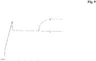
- Bispecific Abs OA-GA201-scFab-Ak18_WT showed simultaneous binding of amine coupled human EGFR and human IGF1R (see sensogram Figure 9 ).
- the human anti-IGF-1R antibody ⁇ IGF-1R> HUMAB Clone 18 (DSM ACC 2587) inhibits IGF-1R signaling and induces internalization and subsequent downregulation of IGF-1R.
- DSM ACC 2587 The human anti-IGF-1R antibody ⁇ IGF-1R> HUMAB Clone 18 (DSM ACC 2587) inhibits IGF-1R signaling and induces internalization and subsequent downregulation of IGF-1R.
- DSM ACC 2587 The human anti-IGF-1R antibody ⁇ IGF-1R> HUMAB Clone 18
- IGF-1 receptor IGF-1 receptor
- Human H322M tumor cells obtained from NCI were cultivated in 96 well plates (1x10 4 cells/well) over night at 37°C and 5 % CO 2 in RPMI medium supplemented with 10 % fetal bovine serum, 2 mM L-glutamine and 1 % PenStrep.
- the medium was carefully removed and replaced by bispecific ⁇ EGFR-IGF1R> antibody solution diluted in RPMI medium in a total volume of 100 ⁇ l. Cells were incubated at 37°C and 5 % CO 2 for at least 3 but no more than 24 hours.
- MES-lysis buffer 25 mM MES pH 6.5, 2 % Triton X-100, 60 nM Octylglucoside, 150 mM NaCl, 10 mM Na 3 VO 4 , and Complete ® protease inhibitor. Plates were stored at -20°C until further analysis.
- the MTPs were then incubated for another hour at RT with agitation and washed 3 times with PBS containing 0.1 % Tween ® 20 subsequently.
- IGF-1R ⁇ rabbit antibody 200 ⁇ g/ml, Santa Cruz Biotechnology, Cat. No. sc-713 diluted 1:750 in PBS, 3 %BSA and 0.2 % Tween ® 20 was used to detect IGF-1R bound by the capture antibody AK1a. 100 ⁇ l was added per well and incubated for 1 hour at RT with constant agitation. The solution was removed subsequently and the wells were washed three times with 200 ⁇ l of PBS containing 0.1 % Tween ® 20. The peroxidase labeled anti-rabbit IgG-HRP (Cell signaling Cat. No.
- the bispecific ⁇ EGFR-IGF1R> antibody OA-GA201-scFab-Ak18_WT induces less downregulation than the human anti-IGF-1R antibody ⁇ IGF-1R> HUMAB Clone 18. Downregulation by OA-GA201-scFab-Ak18_WT was reduced by > 50% compared to ⁇ IGF-1R> HUMAB Clone 18.
- the human anti-IGF-1R antibody ⁇ IGF-1R> HUMAB Clone 18 (DSM ACC 2587) inhibits IGF-1R-signaling and the humanized rat anti-EGFR antibody ICR62 inhibits the signaling by EGFR.
- DSM ACC 2587 The human anti-IGF-1R antibody ⁇ IGF-1R> HUMAB Clone 18 (DSM ACC 2587) inhibits IGF-1R-signaling and the humanized rat anti-EGFR antibody ICR62 inhibits the signaling by EGFR.
- Human tumor cells (H322M, 3 x 10 4 cells/well) in RPMI medium supplemented with 10 % fetal bovine serum, 2 mM L-glutamine and 1 % PenStrep were seeded in 96-well microtiter plates and cultivated over night at 37°C and 5 % CO 2 .
- the medium was carefully removed and replaced by 100 ⁇ l serum-free DMEM medium (supplemented with 1 mg/ml RSA, 10 mM Hepes, 1 % PenStrep) and incubated for at least 2.5 hours at 37°C and 5 % CO 2 .
- the medium was again carefully removed and replaced by a dilution of bispecific antibodies, and control antibodies ( ⁇ IGF-1R> HUMAB Clone 18 and ⁇ EGFR>ICR62 final concentration 0.01 mg/ml) in serum-free DMEM medium at a total volume of 50 ⁇ l followed by an incubation for 30 min at 37°C and 5 % CO 2 .
- Cells were stimulated by the addition of 50 ⁇ l IGF-1 (10 nM) or EGF (20 ng/ml) (diluted in serum-free DMEM medium) and incubated for 10 min at 37°C and 5 % CO 2 .
- EGFR and IGF-1R phosphorylation in filtered cell lysates was analysed with a Luminex system using the P-EGFR (Tyr) bead kit (Millipore Cat. # 46-603) for the analysis of EGFR phosphorylation and the P-IGF-1R (Tyr1131) bead kit (BioRad Cat. # 171V27343) for the analysis of IGF-1R phosphorylation.
- Luminex assays were performed as described in the BioPlex Phosphoprotein Detection manual (BioRad Bulletin # 2903) using Phosphoprotein Detection Reagent Kits (BioRad Cat. # 171-304004).
- the bispecific ⁇ EGFR-IGF1R> antibody OA-GA201-scFab-Ak18_WT effectively inhibits the phosphorylation of IGF-1R (IC50: 1 nM, maximal inhibition: > 70%) and the phosphorylation of EGFR (IC50: 1 nM, maximal inhibition: > 90%) on H322M tumor cells.
- the human anti-IGF-1R antibody ⁇ IGF-1R> HUMAB Clone 18 inhibits the growth of tumor cell lines that express IGF-1R ( WO 2005/005635 ).
- the humanized rat anti-EGFR antibody ⁇ EGFR>ICR62 has been shown to inhibit the growth of tumor cell lines that express EGFR ( WO 2006/082515 ).
- the degree of inhibition in H322M cells which express EGFR as well as IGF-1R was analyzed.
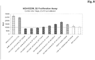
- H322M lung carcinoma cells were cultured in RPMI 1640 medium (PAA, Pasching, Austria) supplemented with 10 % FBS (PAA), 1mM sodium pyruvate (Gibco, Darmstadt, Germany), non-essential amino acids (Gibco) and 2mM L-glutamine (Sigma, Steinheim, Germany). 25000 cells/well were seeded in 96-well poly-HEMA (poly(2-hydroxyethylmethacrylate) (Polysciences, Warrington, PA, USA)) coated plates containing the culture medium. Concomitantly, different concentrations of bispecific antibodies were added and incubated for 7 days. The CellTiterGlo® (Promega , Madison, WI, USA) assay was used to detect cell viability by measuring the ATP-content of the cells according to the manufacturer's instructions.
- results were shown in Figure 8 .
- the bispecific ⁇ EGFR-IGF1R> antibody OA-GA201-scFab-Ak18_WT dose-dependently inhibits proliferation of H322M cells.
- the bispecific ⁇ EGFR-IGF1R> antibody OA-GA201-scFab-Ak18_WT showed improved inhibition of proliferation when compared to the parental monospecific antibodies ⁇ IGF-1R> HUMAB Clone 18 or ⁇ EGFR>ICR62.
- the human anti-IGF-1R antibody ⁇ IGF-1R> HUMAB Clone 18 inhibits the growth of tumor cell lines that express IGF-1R ( WO 2005/005635 ).
- the humanized rat anti-EGFR antibody ⁇ EGFR>ICR62 has been shown to inhibit the growth of tumor cell lines that express EGFR ( WO 2006/082515 ).
- the subcutaneous xenograft model BxPC-3 that was characterized by the expression of EGFR as well as IGF-1R was used.
- BxPC-3 human pancreatic carcinoma cell line BxPC-3 (obtained from ATCC) were cultured in RPMI 1640 medium (PANTM Biotech GmbH), supplemented with 10 % fetal bovine serum (Sera Plus; PANTM Biotech GmbH) and 2 mM L-glutamine (PANTM Biotech GmbH) at 37°C in a water saturated atmosphere at 5 % CO 2 .
- BxPC-3 tumor cells were harvested (1x trypsin-EDTA, Roche Diagnostics) from culture flasks and transferred into culture medium, centrifuged, washed once and re-suspended in PBS. For injection of cells, the final titer was adjusted to 1 ⁇ 10 8 cells/ml.
- Glycoengineered derivatives of bispecific ⁇ EGFR-IGF1R> antibodies were produced by co-transfecting HEK293-EBNA cells with the mammalian antibody heavy and light chain expression vectors using a calcium phosphate-transfection approach. Exponentially growing HEK293-EBNA cells were transfected by the calcium phosphate method. For the production of unmodified antibody, the cells were transfected only with antibody heavy and light chain expression vectors in a 1:1 ratio. For the production of the glycoengineered antibody, the cells are co-transfected with four plasmids, two for antibody expression, one for a fusion GnTIII polypeptide expression, and one for mannosidase II expression at a ratio of 4:4:1:1, respectively.
- Cells were grown as adherent monolayer cultures in T flasks using DMEM culture medium supplemented with 10 % FCS, and were transfected when they were between 50 and 80 % confluent.
- DMEM culture medium supplemented with 10 % FCS
- For the transfection of a T75 flask 7.5 (to 8) million cells were seeded 24 hours before transfection in ca 14 ml DMEM culture medium supplemented with FCS (at 10 % V/V final), (eventually 250 ⁇ g/ml neomycin,) and cells were placed at 37°C in an incubator with a 5 % CO2 atmosphere overnight.
- a solution of DNA, CaCl2 and water was prepared by mixing 47 ⁇ g total plasmid vector DNA divided equally between the light and heavy chain expression vectors, 235 ⁇ l of a 1M CaCl2 solution, and adding water to a final volume of 469 ⁇ l.
- 469 ⁇ l of a 50mM HEPES, 280 mM NaCl, 1.5 mM Na2HPO4 solution at pH 7.05 were added, mixed immediately for 10 sec and left to stand at room temperature for 20 sec.
- the suspension was diluted with ca. 12 ml of DMEM supplemented with 2 % FCS, and added to the T75 in place of the existing medium.
- the cells were incubated at 37°C, 5 % CO2 for about 17 to 20 hours, then medium was replaced with ca. 12 ml DMEM, 10 % FCS.
- the conditioned culture medium was harvested 5 to 7 days post-transfection centrifuged for 5 min at 210 - 300 *g, sterile filtered through a 0.22 ⁇ m filter (or alternatively centrifuged for 5 min at 1200 rpm, followed by a second centrifugation for 10 min at 4000 rpm) and kept at 4°C.
- the secreted antibodies were purified by Protein A affinity chromatography, and a final size exclusion chromatographic step on a Superdex 200 column (Amersham Pharmacia) exchanging the buffer to phosphate buffer saline and collecting the pure monomeric IgG1 antibodies.
- Antibody concentration ia estimated using a spectrophotometer from the absorbance at 280 nm.
- the antibodies are formulated in a 25 mM potassium phosphate, 125 mM sodium chloride, 100 mM glycine solution of pH 6.7.
- Glycoengineered variants of the bispecific antibodies were produced by co-transfection of the antibody expression vectors together with a GnT-III glycosyltransferase expression vector, or together with a GnT-III expression vector plus a Golgi mannosidase II expression vector.
- Glycoengineered antibodies were purified and formulated as described above for the non-glycoengineered antibodies.
- the oligosaccharides attached to the Fc region of the antibodies were analysed by MALDI/TOF-MS as described below to determine the amount of fucose.
- Oligosaccharides are enzymatically released from the antibodies by PNGaseF digestion, with the antibodies being either immobilized on a PVDF membrane or in solution.
- the resulting digest solution containing the released oligosaccharides is either prepared directly for MALDI/TOF-MS analysis or is further digested with EndoH glycosidase prior to sample preparation for MALDI/TOF-MS analysis.
- GE means glycoengineered.
- the degree of ADCC mediation by a given antibody depends not only on the antigen that is bound, but is also dependent on affinities of constant regions to the FcgRIIIa, which is known as the Fc receptor that triggers the ADCC reaction.
- FcgRIIIa Fc receptor that triggers the ADCC reaction.
- a Biacore technology is applied for the analysis of binding of the bispecific ⁇ EGFR-IGF1R> antibodies to the FcgRIIIa. By this technology, binding of bispecific ⁇ EGFR-IGF1R> antibodies to recombinantly produced FcgRIIIa domains is assessed.
- the running and dilution buffer was PBS (1 mM KH2PO4, 10mM Na2HPO4, 137 mM NaCl, 2.7mM KCl), pH6.0, 0.005 % (v/v) Tween20.
- the soluble human FcgRIIIa was diluted in 10 mM sodium-acetate, pH 5.0 and immobilized on a CM5 biosensor chip using the standard amine coupling kit (GE Healthcare Biosciences AB, Sweden) to obtain FcgRIIIa surface densities of approximately 1000 RU.
- the PBMC are counted and washed a second time by centrifugation at 400 x g for 10 minutes at RT. The cells are then resuspended in the appropriate medium for the subsequent procedures.
- the effector to target ratio used for the ADCC assays is 25:1 for PBMC.
- the effector cells are prepared in AIM-V medium at the appropriate concentration in order to add 50 ⁇ l per well of round bottom 96 well plates.
- Target cells were human EGFR/IGFR expressing cells (e.g., H322M, A549, or MCF-7) grown in DMEM containing 10 % FCS.
- Target cells are washed in PBS, counted and adjusted at 1x10E6 cells/ml.
- Cells are labeled with Calcein AM (10 ⁇ M) for 30min at 37°/5%CO2.
- After labeling cells are washed twice in PBS and seeded at 5000 cells/well in 50 ⁇ l (AIM-V medium) in 96-well round bottom plates.
- Antibodies are diluted in AIM-V, added in 50 ⁇ l to the pre-plated target cells. Then the effector cells are added and the plate is incubated for 4 hours at 37°C in a humidified atmosphere containing 5 % CO2.
- released glycans of purified antibody material are analyzed by MALDI-Tof-mass spectrometry.
- the antibody sample (about 50 ⁇ g) is incubated over night at 37°C with 5mU N-Glycosidase F (Prozyme# GKE-5010B) in 0.1M sodium phosphate buffer, pH 6.0, in order to release the oligosaccharide from the protein backbone.
- the glycan structures released are isolated and desalted using NuTip-Carbon pipet tips (obtained from Glygen: NuTip1-10 ⁇ l, Cat.Nr#NT1CAR).
- NuTip-Carbon pipet tips obtained from Glygen: NuTip1-10 ⁇ l, Cat.Nr#NT1CAR.
- the NuTip-Carbon pipet tips are prepared for binding of the oligosaccharides by washing them with 3 ⁇ L 1M NaOH followed by 20 ⁇ L pure water (e.g. HPLC-gradient grade from Baker, # 4218), 3 ⁇ L 30 % v/v acetic acid and again 20 ⁇ l pure water.
- the respective solutions are loaded onto the top of the chromatography material in the NuTip-Carbon pipet tip and pressed through it.
- the glycan structures corresponding to 10 ⁇ g antibody are bound to the material in the NuTip-Carbon pipet tips by pulling up and down the N-Glycosidase F digest described above four to five times.
- the glycans bound to the material in the NuTip-Carbon pipet tip are washed with 20 ⁇ L pure water in the way as described above and are eluted stepwise with 0.5 ⁇ L 10 % and 2.0 ⁇ L 20 % acetonitrile, respectively.
- the elution solutions are filled in a 0.5 mL reaction vails and are pulled up and down four to five times each.
- MALDI-Tof mass spectrometry both eluates are combined.
- the peaks are assigned to fucose or a-fucose (non-fucose) containing glycol structures by comparing the masses calculated and the masses theoretically expected for the respective structures (e.g. complex, hybride and oligo-or high-mannose, respectively, with and without fucose).
- the antibody sample are digested with N-Glycosidase F and Endo-Glycosidase H concommitantlyN-glycosidase F releases all N-linked glycan structures (complex, hybride and oligo- and high mannose structures) from the protein backbone and the Endo-Glycosidase H cleaves all the hybride type glycans additionally between the two GlcNAc-residue at the reducing end of the glycan.
- This digest is subsequently treated and analysed by MALDI-Tof mass spectrometry in the same way as described above for the N-Glycosidase F digested sample.
- the degree of reduction of the signals of a specific glyco structure is used to estimate the relative content of hybride structures.
- the relative amount of each glycostructure is calculated from the ratio of the peak height of an individual glycol structure and the sum of the peak heights of all glyco structures detected.
- the amount of fucose is the percentage of fucose-containing structures related to all glyco structures identified in the N-Glycosidase F treated sample (e.g. complex, hybride and oligo- and high-mannose structures, resp.).
- the amount of afucosylation is the percentage of fucose-lacking structures related to all glyco structures identified in the N-Glycosidase F treated sample (e.g. complex, hybride and oligo- and high-mannose structures, resp.).
- the amount of fucose determined for OA-GA201-scFab-Ak18_WT was between 25% and 40%.
- the human anti-IGF-1R antibody ⁇ IGF-1R> HUMAB Clone 18 (DSM ACC 2587) binds to cells expressing IGF-1R and the humanized rat anti-EGFR antibody ICR62 binds to cells that express EGFR on their surface.
- DSM ACC 2587 The human anti-IGF-1R antibody ⁇ IGF-1R> HUMAB Clone 18 binds to cells expressing IGF-1R and the humanized rat anti-EGFR antibody ICR62 binds to cells that express EGFR on their surface.
- Human tumor cells e.g. A549, TC-71, MDA-MB-231, 2 x 10⁵ cells/well
- ice-cold buffer PBS + 2 % FCS, Gibco
- labeled monospecific antibodies HUMAB Clone 18 or humanized rat anti-EGFR antibody ICR62
- concentration of 1 ⁇ g/ml and different concentrations of unlabeled ⁇ EGFR-IGF1R> antibodies or unlabeled monospecific antibodies or Fab fragments as controls (final titration range of 100 to 0.002 ⁇ g/ml) in a 96-well microtiter plate.
- the mixture was incubated on ice for 45 minutes.
- OA-GA201-scFab-Ak18_WT On TC-71 tumor cells, expressing IGF-1R but not EGFR binding of ⁇ EGFR-IGF1R> antibody OA-GA201-scFab-Ak18_WT was comparable to the IGF-1R> HUMAB Clone 18 Fab fragment. In this setting, were only IGF-1R but not EGFR is expressed OA-GA201-scFab-Ak18_WT can only bind to IGF-1R with one binding arm.
Landscapes
- Health & Medical Sciences (AREA)
- Chemical & Material Sciences (AREA)
- Organic Chemistry (AREA)
- Immunology (AREA)
- Medicinal Chemistry (AREA)
- Life Sciences & Earth Sciences (AREA)
- General Health & Medical Sciences (AREA)
- Biochemistry (AREA)
- Genetics & Genomics (AREA)
- Molecular Biology (AREA)
- Proteomics, Peptides & Aminoacids (AREA)
- Biophysics (AREA)
- Veterinary Medicine (AREA)
- Public Health (AREA)
- Animal Behavior & Ethology (AREA)
- Pharmacology & Pharmacy (AREA)
- Nuclear Medicine, Radiotherapy & Molecular Imaging (AREA)
- General Chemical & Material Sciences (AREA)
- Chemical Kinetics & Catalysis (AREA)
- Engineering & Computer Science (AREA)
- Bioinformatics & Cheminformatics (AREA)
- Microbiology (AREA)
- Mycology (AREA)
- Epidemiology (AREA)
- Peptides Or Proteins (AREA)
- Preparation Of Compounds By Using Micro-Organisms (AREA)
- Medicines Containing Antibodies Or Antigens For Use As Internal Diagnostic Agents (AREA)
- Micro-Organisms Or Cultivation Processes Thereof (AREA)
- Agricultural Chemicals And Associated Chemicals (AREA)
Description
- The present invention relates to bispecific antibodies, methods for their production, pharmaceutical compositions containing said antibodies, and uses thereof.
- A wide variety of multispecific recombinant antibody formats have been developed in the recent past, e.g. tetravalent bispecific antibodies by fusion of, e.g., an IgG antibody format and single chain domains (see e.g. Coloma, M.J., et al., Nature Biotech 15 (1997) 159-163;
WO 2001/077342 ; and Morrison, S.L., Nature Biotech. 25 (2007) 1233-1234). - Also several other new formats wherein the antibody core structure (IgA, IgD, IgE, IgG or IgM) is no longer retained such as dia-, tria- or tetrabodies, minibodies, several single chain formats (scFv, Bis-scFv), which are capable of binding two or more antigens, have been developed (Holliger, P., et al., Nature Biotech 23 (2005) 1126-1136; Fischer, N., Léger O., Pathobiology 74 (2007) 3-14; Shen, J., et al., Journal of Immunological Methods 318 (2007) 65-74; Wu, C., et al., Nature Biotech. 25 (2007) 1290-1297).
- All such formats use linkers either to fuse the antibody core (IgA, IgD, IgE, IgG or IgM) to a further binding protein (e.g. scFv) or to fuse e.g. two Fab fragments or scFvs (Fischer N., Léger O., Pathobiology 74 (2007) 3-14). It has to be kept in mind that one may want to retain effector functions, such as e.g. complement-dependent cytotoxicity (CDC) or antibody dependent cellular cytotoxicity (ADCC), which are mediated through the Fc receptor binding, by maintaining a high degree of similarity to naturally occurring antibodies.
- In
WO 2007/024715 are reported dual variable domain immunoglobulins as engineered multivalent and multispecific binding proteins. A process for the preparation of biologically active antibody dimers is reported inUS 6,897,044 . Multivalent FV antibody construct having at least four variable domains which are linked with each over via peptide linkers are reported inUS 7,129,330 . Dimeric and multimeric antigen binding structures are reported inUS 2005/0079170 . Tri- or tetra-valent monospecific antigen-binding protein comprising three or four Fab fragments bound to each other covalently by a connecting structure, which protein is not a natural immunoglobulin are reported inUS 6,511,663 . InWO 2006/020258 tetravalent bispecific antibodies are reported that can be efficiently expressed in prokaryotic and eukaryotic cells, and are useful in therapeutic and diagnostic methods. A method of separating or preferentially synthesizing dimers which are linked via at least one interchain disulfide linkage from dimers which are not linked via at least one interchain disulfide linkage from a mixture comprising the two types of polypeptide dimers is reported inUS 2005/0163782 . Bispecific tetravalent receptors are reported inUS 5,959,083 . Engineered antibodies with three or more functional antigen binding sites are reported inWO 2001/077342 . - Multispecific and multivalent antigen-binding polypeptides are reported in
WO 1997/001580 .WO 1992/004053 reports homoconjugates, typically prepared from monoclonal antibodies of the IgG class which bind to the same antigenic determinant are covalently linked by synthetic cross-linking. Oligomeric monoclonal antibodies with high avidity for antigen are reported inWO 1991/06305 US 6,350,860 , which can be used to treat diseases wherein interferon gamma activity is pathogenic. InUS 2005/0100543 are reported targetable constructs that are multivalent carriers of bi-specific antibodies, i.e., each molecule of a targetable construct can serve as a carrier of two or more bi-specific antibodies. - Genetically engineered bispecific tetravalent antibodies are reported in
WO 1995/009917 . InWO 2007/109254 stabilized binding molecules that consist of or comprise a stabilized scFv are reported. - Bispecific antibodies against EGFR and IGF-1R are known from Lu, D., et al., Biochemical and Biophysical Research Communications 318 (2004) 507-513; Lu, D., et al., J. Biol. Chem., 279 (2004) 2856-2865; and Lu, D., et al., J. Biol Chem. 280 (2005) 19665-72.
-
US 2007/0274985 relates to synthetic antibody molecules comprising single chain Fab (scFab) proteins which can also be associated to dimers, including heteromeric antibodies, wherein at least two single chain antibody molecules are associated. -
WO 2009/080253 relates to bispecific bivalent antibodies built up of four polypeptide chains designed in order to improve the side product profile. However in view of different problems and aspects of multispecific antibodies (like e.g. pharmacokinetic and biological properties, stability, aggregation, expression yield, side products) there is a need of further alternative multispecific antibody formats. - In one aspect the invention is directed to a bispecific antibody of three polypeptide chains comprising
- a) the heavy chain and the light chain of a first full length antibody that specifically binds to a first antigen;
- b) the heavy chain and the light chain of a second full length antibody that specifically binds to a second antigen, wherein the N-terminus of the heavy chain is connected to the C-terminus of the light chain via a peptide linker, wherein the bispecific antibody is characterized in that
- i) in the CH3 domain of one heavy chain
an amino acid residue is replaced with an amino acid residue having a larger side chain volume, thereby generating a protuberance within the interface of the CH3 domain of one heavy chain which is positionable in a cavity within the interface of the CH3 domain of the other heavy chain
and wherein - ii) in the CH3 domain of the other heavy chain
- In another aspect the bispecific antibody according to the invention is characterized in that both CH3 domains are further altered by the introduction of cysteine (C) as amino acid in the corresponding positions of each CH3 domain such that a disulfide bridge between both CH3 domains can be formed.
- In another aspect of the invention the bispecific antibody according to the invention is characterized in that
- the antibody heavy chain variable domain (VH) and the antibody light chain variable domain (VL) of the heavy and light chain of the second full length antibody under b) are disulfide stabilized by introduction of a disulfide bond between the following positions:
- i) heavy chain
variable domain position 44 to light chain variable domain position 100, - ii) heavy chain variable domain position 105 to light chain variable domain position 43, or
- iii) heavy chain variable domain position 101 to light chain variable domain position 100.
- i) heavy chain
- In another aspect of the invention the bispecific antibody according to the invention is characterized in that
- a) the first full length antibody specifically binds to IGF-1R and comprises a heavy chain with the amino acid sequence of SEQ ID NO: 1, and a light chain with the amino acid sequence of SEQ ID NO: 2, and
- b) the second full length antibody specifically binds to EGFR and comprises a heavy chain connected to the light chain via a peptide linker wherein said peptide connected heavy and light chain have the amino acid sequence of SEQ ID NO: 3.
- In another aspect of the invention the bispecific antibody according to the invention is characterized in that
- a) the first full length antibody specifically binds to IGF-1R and comprises a heavy chain with the amino acid sequence of SEQ ID NO: 1, and a light chain with the amino acid sequence of SEQ ID NO: 2, and
- b) the second full length antibody specifically binds to EGFR and comprises a heavy chain connected to the light chain via a peptide linker wherein said peptide connected heavy and light chain have amino acid sequence of SEQ ID NO: 4.
- In another aspect of the invention the bispecific antibody according to the invention is characterized in that
- a) the first full length antibody specifically binds to EGFR and comprises a heavy chain with the amino acid sequence of SEQ ID NO: 5, and a light chain with the amino acid sequence of SEQ ID NO: 6, and
- b) the second full length antibody specifically binds to IGF-1R and comprises a heavy chain connected to the light chain via a peptide linker wherein said peptide connected heavy and light chain have amino acid sequence of SEQ ID NO: 7.
- In another aspect of the invention the bispecific antibody according to the invention is characterized in that
- a) the first full length antibody specifically binds to EGFR and comprises a heavy chain with the amino acid sequence of SEQ ID NO: 5, and a light chain with the amino acid sequence of SEQ ID NO: 6, and
- b) the second full length antibody specifically binds to IGF-1R and comprises a heavy chain connected to the light chain via a peptide linker wherein said peptide connected heavy and light chain have amino acid sequence of SEQ ID NO: 8.
- In another aspect of the invention the bispecific antibody according to the invention is characterized in that the antibody comprises a constant region of IgG1.
- In another aspect of the invention the bispecific antibody according to the invention is characterized in that the antibody is glycosylated with a sugar chain at Asn297 wherein the amount of fucose within the sugar chain is 65 % or lower.
- Still further aspects of the invention are a pharmaceutical composition comprising said bispecific antibody, said composition for the treatment of cancer, the use of said bispecific antibody for the manufacture of a medicament for the treatment of cancer, a method of treatment of patient suffering from cancer by administering said bispecific antibody. to a patient in the need of such treatment.
- A further aspect of the invention is a nucleic acid molecule encoding a chain of a bispecific antibody according to the invention.
- The invention further provides expression vectors containing said nucleic acid according to the invention capable of expressing said nucleic acid in a prokaryotic or eukaryotic host cell, and host cells containing such vectors for the recombinant production of a bispecific antibody according to the invention.
- The invention further comprises a prokaryotic or eukaryotic host cell comprising a vector according to the invention.
- The invention further comprises a method for the production of a bispecific antibody according to the invention, characterized by expressing a nucleic acid according to the invention in a prokaryotic or eukaryotic host cell and recovering said bispecific antibody from said cell or the cell culture supernatant. The invention further comprises the antibody obtained by such method for the production of a bispecific antibody.
- Another aspect of the invention is a method for the preparation of a bispecific antibody according to the invention comprising the steps of
- a) transforming a host cell with vectors comprising nucleic acid molecules encoding
- aa) the heavy chain and the light chain of a first full length antibody that specifically binds to a first antigen;; and
- ab) the heavy chain and the light chain of a second full length antibody that specifically binds to a second antigen, wherein the N-terminus of the heavy chain is connected to the C-terminus of light chain via a peptide linker; and
- b) culturing the host cell under conditions that allow synthesis of said antibody molecule; and
- c) recovering said antibody molecule from said culture.
- It has now been found that the bispecific antibodies according to the invention have valuable characteristics such as good expression yields in mammalian cells (like HEK293 cells and CHO cells), stability, biological or pharmacological activity, pharmacokinetic properties. They can be used e.g. for the treatment of diseases such as cancer. These bispecific antibodies according to the invention comprising 3 poylpeptide chains especially have a valuable side product profile during expression in mammalian cells.
-
- Figure 1
- Schematic structure of a full length antibody without CH4 domain specifically binding to a
first antigen 1 with two pairs of heavy and light chain which comprise variable and constant domains in a typical order. - Figure 2
- Schematic structure of the bispecific antibody according to the invention.
- Figure 3a and 3b
- Schematic structure of the bispecific antibody according to the invention including knobs-into hole modified CH3 domains.
- Figure 4 a and 4b
- Schematic structure of the bispecific antibody according to the invention including knobs-into hole modified CH3 domains and disulfide stabilization of the VH and VL domain of the second antibody heavy and light chain.
- Figure 5
- Inhibition of the HUVEC proliferation by bispecific antibody according to the invention Ang2-VEGF OA-Ava-N-scFabLC06.
- Figure 6
- Inhibition of the Tie2 phosphorylation by bispecific antibody according to the invention Ang2-VEGF OA-Ava-N-scFabLC06.
- Figure 7
- Western blot (reduced) of OA-Ak1B-scFab-GA201 (
Figure 7a ) and OA-GA201-scFab-Ak18 (Figure 7b ). - Figure 8
- Growth inhibition of H322M cancer cells by the bispecific <EGFR-IGF1R> antibody OA-GA201-scFab-Ak18_WT (dose-dependently) compared to the parental monospecific antibodies <IGF-1R>
HUMAB Clone 18 or <EGFR>ICR62. - Figure 9
- Biacore (Surface plasmon resonance)-Sensogram: Bispecific antibody OA-GA201-scFab-Ak18_WT showed simultaneous binding of amine coupled human EGFR and human IGF1R (x-axis: response, y-axis: time).
- Figure 10
- Tumor growth inhibition in BxPC3 Xenograft model by OA-GA201-scFab-Ak18_WT compared to the combination of parental monospecific antibodies <IGF-1R>
HUMAB Clone 18 and <EGFR>ICR62 - In one aspect the invention is directed to a bispecific antibody comprising
- a) the heavy chain and the light chain of a first full length antibody that specifically binds to a first antigen;
- b) the heavy chain and the light chain of a second full length antibody that specifically binds to a second antigen, wherein the N-terminus of the heavy chain is connected to the C-terminus of light chain via a peptide linker.
- The term "full length antibody" denotes an antibody consisting of two "full length antibody heavy chains" and two "full length antibody light chains" (see
Fig. 1 ). A "full length antibody heavy chain" is a polypeptide consisting in N-terminal to C-terminal direction of an antibody heavy chain variable domain (VH), an antibody constant heavy chain domain 1 (CH1), an antibody hinge region (HR), an antibody heavy chain constant domain 2 (CH2), and an antibody heavy chain constant domain 3 (CH3), abbreviated as VH-CH1-HR-CH2-CH3; and optionally an antibody heavy chain constant domain 4 (CH4) in case of an antibody of the subclass IgE. Preferably the "full length antibody heavy chain" is a polypeptide consisting in N-terminal to C-terminal direction of VH, CH1, HR, CH2 and CH3. A "full length antibody light chain" is a polypeptide consisting in N-terminal to C-terminal direction of an antibody light chain variable domain (VL), and an antibody light chain constant domain (CL), abbreviated as VL-CL. The antibody light chain constant domain (CL) can be κ (kappa) or λ (lambda). The two full length antibody chains are linked together via inter-polypeptide disulfide bonds between the CL domain and the CH1 domain and between the hinge regions of the full length antibody heavy chains. Examples of typical full length antibodies are natural antibodies like IgG (e.g. IgG 1 and IgG2), IgM, IgA, IgD, and IgE. The full length antibodies according to the invention can be from a single species e.g. human, or they can be chimerized or humanized antibodies. The full length antibodies according to the invention comprise two antigen binding sites each formed by a pair of VH and VL, which both specifically bind to the same antigen. The C-terminus of the heavy or light chain of said full length antibody denotes the last amino acid at the C-terminus of said heavy or light chain. The N-terminus of the heavy or light chain of said full length antibody denotes the last amino acid at the N- terminus of said heavy or light chain. - The term "peptide linker" as used within the invention denotes a peptide with amino acid sequences, which is preferably of synthetic origin. These peptides according to invention are used to connect the C-terminus of the light chain to the N-terminus of heavy chain of the the second full length antibody (that specifically binds to a second antigen) via a peptide linker. The peptide linker within the second full length antibody heavy and light chain is a peptide with an amino acid sequence with a length of at least 30 amino acids, preferably with a length of 32 to 50 amino acids. In one the peptide linker is a peptide with an amino acid sequence with a length of 32 to 40 amino acids. In one embodiment said linker is (GxS)n with G = glycine, S = serine, (x =3, n= 8, 9 or 10 and m= 0, 1, 2 or 3) or (x = 4 and n= 6, 7 or 8 and m= 0, 1, 2 or 3), preferably with x = 4, n= 6 or 7 and m= 0, 1, 2 or 3, more preferably with x = 4, n= 7 and m= 2. In one embodiment said linker is (G4S)6G2.Preferably the CH3 domains of the bispecific antibody according to the invention can be altered by the "knob-into-holes" technology which is described in detail with several examples in e.g.
WO 96/027011 - In one aspect of the invention the bispecific antibody according to the invention is characterized in that
the CH3 domain of one heavy chain and the CH3 domain of the other heavy chain each meet at an interface which comprises an original interface between the antibody CH3 domains;
wherein said interface is altered to promote the formation of the bispecific antibody, wherein the alteration is characterized in that: - a) the CH3 domain of one heavy chain is altered,
so that within the original interface the CH3 domain of one heavy chain that meets the original interface of the CH3 domain of the other heavy chain within the bispecific antibody,
an amino acid residue is replaced with an amino acid residue having a larger side chain volume, thereby generating a protuberance within the interface of the CH3 domain of one heavy chain which is positionable in a cavity within the interface of the CH3 domain of the other heavy chain
and - b) the CH3 domain of the other heavy chain is altered,
- Thus the antibody according to invention is preferably characterized in that
- the CH3 domain of the heavy chain of the full length antibody of a) and the CH3 domain of the heavy chain of the full length antibody of b) each meet at an interface which comprises an alteration in the original interface between the antibody CH3 domains;
wherein- i) in the CH3 domain of one heavy chain
an amino acid residue is replaced with an amino acid residue having a larger side chain volume, thereby generating a protuberance within the interface of the CH3 domain of one heavy chain which is positionable in a cavity within the interface of the CH3 domain of the other heavy chain
and wherein - ii) in the CH3 domain of the other heavy chain
- i) in the CH3 domain of one heavy chain
- Preferably said amino acid residue having a larger side chain volume is selected from the group consisting of arginine (R), phenylalanine (F), tyrosine (Y), tryptophan (W).
- Preferably said amino acid residue having a smaller side chain volume is selected from the group consisting of alanine (A), serine (S), threonine (T), valine (V).
- In one aspect of the invention both CH3 domains are further altered by the introduction of cysteine (C) as amino acid in the corresponding positions of each CH3 domain such that a disulfide bridge between both CH3 domains can be formed.
- In one embodiment, the bispecific antibody comprises a T366W mutation in the CH3 domain of the "knobs chain" and T366S, L368A, Y407V mutations in the CH3 domain of the "hole chain". An additional interchain disulfide bridge between the CH3 domains can also be used (Merchant, A.M, et al., Nature Biotech 16 (1998) 677-681) e.g. by introducing a Y349C mutation into the CH3 domain of the "knobs chain" and a E356C mutation or a S354C mutation into the CH3 domain of the "hole chain".
- In another embodiment, the bispecific antibody according to the invention comprises Y349C, T366W mutations in one of the two CH3 domains and E356C, T366S, L368A, Y407V mutations in the other of the two CH3 domains. In a another preferred embodiment the bispecific antibody comprises Y349C, T366W mutations in one of the two CH3 domains and S354C, T366S, L368A, Y407V mutations in the other of the two CH3 domains (the additional Y349C mutation in one CH3 domain and the additional E356C or S354C mutation in the other CH3 domain forming a interchain disulfide bridge) (numbering always according to EU index of Kabat). But also other knobs-in-holes technologies as described by
EP 1870459A1 , can be used alternatively or additionally. Thus another example for the bispecific antibody are R409D; K370E mutations in the CH3 domain of the "knobs chain" and D399K; E357K mutations in the CH3 domain of the "hole chain" (numbering always according to EU index of Kabat). - In another embodiment the bispecific antibody comprises a T366W mutation in the CH3 domain of the "knobs chain" and T366S, L368A, Y407V mutations in the CH3 domain of the "hole chain" and additionally R409D; K370E mutations in the CH3 domain of the "knobs chain" and D399K; E357K mutations in the CH3 domain of the "hole chain".
- In another embodiment the bispecific antibody comprises Y349C, T366W mutations in one of the two CH3 domains and S354C, T366S, L368A, Y407V mutations in the other of the two CH3 domains or said trivalent, bispecific antibody comprises Y349C, T366W mutations in one of the two CH3 domains and S354C, T366S, L368A, Y407V mutations in the other of the two CH3 domains and additionally R409D; K370E mutations in the CH3 domain of the "knobs chain" and D399K; E357K mutations in the CH3 domain of the "hole chain".
- In one embodiment the antibody heavy chain variable domain (VH) and the antibody light chain variable domain (VL) of the heavy and light chain of the second full length antibody (that specifically binds to a second antigen) are disulfide stabilized by introduction of a disulfide bond between the following positions:
- i) heavy chain
variable domain position 44 to light chain variable domain position 100, - ii) heavy chain variable domain position 105 to light chain variable domain position 43, or
- iii) heavy chain variable domain position 101 to light chain variable domain position 100 (numbering always according to EU index of Kabat).
- In one embodiment the antibody heavy chain variable domain (VH) and the antibody light chain variable domain (VL) of the heavy and light chain of the second full length antibody (that specifically binds to a second antigen) are disulfide stabilized by introduction of a disulfide bond between the following positions: heavy chain
variable domain position 44 to light chain variable domain position 100. - Such further disulfide stabilization is achieved by the introduction of a disulfide bond between the variable domains VH and VL of the second full length antibody heavy and light chain. Techniques to introduce unnatural disulfide bridges for stabilization for a single chain Fv are described e.g. in
WO 94/029350 variable domain position 44 and light chain variable domain position 100. In one embodiment the optional disulfide bond between the variable domains is between heavy chain variable domain position 105 and light chain variable domain position 43 (numbering always according to EU index of Kabat). - In one embodiment a bispecific antibody according to the invention with said optional disulfide stabilization between the variable domains VH and VL of the second full length antibody heavy and light chain is preferred.
- In one embodiment a bispecific antibody according to the invention without said optional disulfide stabilization between the variable domains VH and VL of the second full length antibody heavy and light chain is preferred.
- Both parts of the bispecific antibody according to the invention comprise antigen-binding sites (the first full length antibody heavy and light chain comprise one antigen binding site, and the second full length antibody heavy and light chain comprise one antigen binding site). The terms "binding site" or "antigen-binding site" as used herein denotes the region(s) of said bispecific antibody according to the invention to which the respective antigen actually binds. The antigen binding sites either in the first full length antibody heavy and light chain and the second full length antibody heavy and light chain are formed each by a pair consisting of an antibody light chain variable domain (VL) and an antibody heavy chain variable domain (VH).
- The antigen-binding sites that bind to the desired antigen (e.g EGFR) can be derived a) from known antibodies to the antigen (e.g anti-EGFR antibodies) or b) from new antibodies or antibody fragments obtained by de novo immunization methods using inter alia either the antigen protein or nucleic acid or fragments thereof or by phage display.
- An antigen-binding site of an antibody of the invention contains six complementarity determining regions (CDRs) which contribute in varying degrees to the affinity of the binding site for antigen. There are three heavy chain variable domain CDRs (CDRH1, CDRH2 and CDRH3) and three light chain variable domain CDRs (CDRL1, CDRL2 and CDRL3). The extent of CDR and framework regions (FRs) is determined by comparison to a compiled database of amino acid sequences in which those regions have been defined according to variability among the sequences.
- Antibody specificity refers to selective recognition of the antibody for a particular epitope of an antigen. Natural antibodies, for example, are monospecific. The term "bispecific" antibody as used herein denotes an antibody that has two or more anigen-binding sites and binds to two different antigens or two different epitopes of the same antigen. "Bispecific antibodies" according to the invention are antibodies which have two different antigen-binding specificities. In one embodiment antibodies of the present invention are bispecific for two different antigens, i.e. VEGF as first antigen and ANG-2 as second antigen or e.g. EGFR as first antigen and IGF-1R as second antigen, or vice versa.
- The term "monospecific" antibody as used herein denotes an antibody that has one or more binding sites each of which bind to the same epitope of the same antigen.
- The term "valent" as used within the current application denotes the presence of a specified number of binding sites in an antibody molecule. As such, the terms "trivalent", "tetravalent", "pentavalent" and "hexavalent" denote the presence of three binding sites, four binding sites, five binding sites, and six binding sites, respectively, in an antibody molecule. A natural antibody for example or a bispecific antibody according to the invention has two binding sites and is bivalent.
- The term "EGFR" as used herein refers to human epidermal growth factor receptor (also known as HER-1 or Erb-B1, SEQ ID NO: 13) is a 170 kDa transmembrane receptor encoded by the c-erbB proto-oncogene, and exhibits intrinsic tyrosine kinase activity (Modjtahedi, H., et al., Br. J. Cancer 73 (1996) 228-235; Herbst, R.S., and Shin, D.M., Cancer 94 (2002) 1593-1611). SwissProt database entry P00533 provides the sequence of EGFR. There are also isoforms and variants of EGFR (e.g., alternative RNA transcripts, truncated versions, polymorphisms, etc.) including but not limited to those identified by Swissprot database entry numbers P00533-1, P00533-2, P00533-3, and P00533-4. EGFR is known to bind ligands including α), epidermal growth factor (EGF), transforming growth factor-α (TGf-amphiregulin, heparin-binding EGF (hb-EGF), betacellulin, and epiregulin (Herbst, R.S., and Shin, D.M., Cancer 94 (2002) 1593-1611; Mendelsohn, J., and Baselga, J., Oncogene 19 (2000) 6550-6565). EGFR regulates numerous cellular processes via tyrosine-kinase mediated signal transduction pathways, including, but not limited to, activation of signal transduction pathways that control cell proliferation, differentiation, cell survival, apoptosis, angiogenesis, mitogenesis, and metastasis (Atalay, G., et al., Ann. Oncology 14 (2003) 1346-1363; Tsao, A.S., and Herbst, R.S., Signal 4 (2003) 4-9; Herbst, R.S., and Shin, D.M., Cancer 94 (2002) 1593-1611; Modjtahedi, H., et al., Br. J. Cancer 73 (1996) 228-235).
- The term "IGF-1R" as used herein refers to human Insulin-like growth factor I receptor (IGF-IR, CD 221 antigen; SEQ ID NO: 14) belongs to the family of transmembrane protein tyrosine kinases (LeRoith, D., et al., Endocrin. Rev. 16 (1995) 143-163; and Adams, T.E., et al., Cell. Mol. Life Sci. 57 (2000) 1050-1063). SwissProt database entry P08069 provides the sequence of IGF-1R. IGF-IR binds IGF-I with high affinity and initiates the physiological response to this ligand in vivo. IGF-IR also binds to IGF-II, however with slightly lower affinity. IGF-IR overexpression promotes the neoplastic transformation of cells and there exists evidence that IGF-IR is involved in malignant transformation of cells and is therefore a useful target for the development of therapeutic agents for the treatment of cancer (Adams, T.E., et al., Cell. Mol. Life Sci. 57 (2000) 1050-1063).
- In a preferred aspect of the invention the bispecific antibody according to the invention specifically binds to human IGF-IR as well as to human EGFR (i.e. the bispecific antibody according to the invention is a bispecific anti-IGF-1R/ anti-EGFR antibody). The bispecific antibody is based on the antigen-binding sites of human <IGF-1R> HUMAB Clone 18 (DSM ACC 2587;
WO 2005/005635 , abbreviated as <IGF-1R>Clone18 or <IGF-1R> AK18) and humanized <EGFR>ICR62 (WO 2006/082515 abbreviated as <EGFR>ICR62). The relevant light and heavy chain amino acid sequences of these bispecific, bivalent antibodies are given in SEQ ID NO: 1, SEQ ID NO: 2, SEQ ID NO: 3 (for OA-Ak18-scFab-GA201); in SEQ ID NO: 5, SEQ ID NO: 6, SEQ ID NO: 7 (for OA-GA201-scFab-Ak18); in SEQ ID NO: 1, SEQ ID NO: 2, SEQ ID NO: 4 (for OA-Ak18-scFab-GA201_WT), and in SEQ ID NO: 5, SEQ ID NO: 6, SEQ ID NO: 8 (for OA-GA201-scFab-Ak18_WT). - The bispecific <EGFR-IGF-1R> antibodies according to the invention show benefits for human patients in need of a EGFR and IGF-1R targeting therapy. The antibodies according to the invention have highly valuable properties causing a benefit for a patient suffering from such a disease, especially suffering from cancer. The bispecific <EGFR-IGF-1R> antibodies according to the invention show e.g. a reduction of the internalization of IGF-1R receptor compared to the monospecific parent <IGF-1R> antibody. Furthermore they show good targeting of tumor cells expressing both antigens EGFR and IGF-1R which represents a benefit with respect to the efficacy/toxicity ratio for patients suffering from a cancer expressing both antigens EGFR and IGF-1R.
- Thus in one aspect of the invention the bispecific antibody according to the invention is characterized in that
- a) the first full length antibody specifically binds to IGF-1R and comprises a heavy chain with the amino acid sequence of SEQ ID NO: 1, and a light chain with the amino acid sequence of SEQ ID NO: 2, and
- b) the second full length antibody specifically binds to EGFR and comprises a heavy chain connected to the light chain via a peptide linker wherein said connected heavy and light chain have the amino acid sequence of SEQ ID NO: 3.
- In another aspect of the invention the bispecific antibody according to the invention is characterized in that
- a) the first full length antibody specifically binds to IGF-1R and comprises a heavy chain with the amino acid sequence of SEQ ID NO: 1, and a light chain with the amino acid sequence of SEQ ID NO: 2, and
- b) the second full length antibody specifically binds to EGFR and comprises a heavy chain connected to the light chain via a peptide linker wherein said connected heavy and light chain have the amino acid sequence of SEQ ID NO: 4.
- In another aspect of the invention the bispecific antibody according to the invention is characterized in that
- a) the first full length antibody specifically binds to EGFR and comprises a heavy chain with the amino acid sequence of SEQ ID NO: 5, and a light chain with the amino acid sequence of SEQ ID NO: 6, and
- b) the second full length antibody specifically binds to IGF-1R and comprises a heavy chain connected to the light chain via a peptide linker wherein said connected heavy and light chain have the amino acid sequence of SEQ ID NO: 7.
- In another aspect of the invention the bispecific antibody according to the invention is characterized in that
- a) the first full length antibody specifically binds to EGFR and comprises a heavy chain with the amino acid sequence of SEQ ID NO: 5, and a light chain with the amino acid sequence of SEQ ID NO: 6, and
- b) the second full length antibody specifically binds to IGF-1R and comprises a heavy chain connected to the light chain via a peptide linker wherein said connected heavy and light chain have the amino acid sequence of SEQ ID NO: 8.
- Thus in one embodiment of the invention the bispecific antibody is an anti-IGF-1R/ anti-EGFR antibody and is characterized in comprising the amino acid sequences of SEQ ID NO: 1, of SEQ ID NO: 2, and of SEQ ID NO: 3. 22. Accordingly one aspect of the invention is a bispecific antibody that specifically binds to human IGF-1R and to human EGFR, characterized in comprising the amino acid sequences of SEQ ID NO: 1, of SEQ ID NO: 2, and of SEQ ID NO: 3.
- Thus in one embodiment of the invention the bispecific antibody is an anti-IGF-1R/ anti-EGFR antibody and is characterized in comprising the amino acid sequences of SEQ ID NO: 1, of SEQ ID NO: 2, and of SEQ ID NO: 4. Accordingly one aspect of the invention is a bispecific antibody that specifically binds to human IGF-1R and to human EGFR, characterized in comprising the amino acid sequences of SEQ ID NO: 1, of SEQ ID NO: 2, and of SEQ ID NO: 4.
- Thus in one embodiment of the invention the bispecific antibody is an anti-IGF-1R/ anti-EGFR antibody and is characterized in comprising the amino acid sequences of SEQ ID NO: 5, of SEQ ID NO: 6, and of SEQ ID NO: 7. Accordingly one aspect of the invention is a bispecific antibody that specifically binds to human IGF-1R and to human EGFR, characterized in comprising the amino acid sequences of SEQ ID NO: 5, of SEQ ID NO: 6, and of SEQ ID NO: 7.
- Thus in one embodiment of the invention the bispecific antibody is an anti-IGF-1R/ anti-EGFR antibody and is characterized in comprising the amino acid sequences of SEQ ID NO: 5, of SEQ ID NO: 6, and of SEQ ID NO: 8. Accordingly one aspect of the invention is a bispecific antibody that specifically binds to human IGF-1R and to human EGFR, characterized in comprising the amino acid sequences of SEQ ID NO: 5, of SEQ ID NO: 6, and of SEQ ID NO: 8.
- In one embodiment of the invention the bispecific antibody is an anti-IGF-1R/ anti-EGFR antibody and is characterized in
- a) comprising the amino acid sequences of SEQ ID NO: 1, of SEQ ID NO: 2, and of SEQ ID NO: 3.
- b) comprising the amino acid sequences of SEQ ID NO: 1, of SEQ ID NO: 2, and of SEQ ID NO: 4.
- c) comprising the amino acid sequences of SEQ ID NO: 5, of SEQ ID NO: 6, and of SEQ ID NO: 7, or
- d) comprising the amino acid sequences of SEQ ID NO: 5, of SEQ ID NO: 6, and of SEQ ID NO: 8.
- In one embodiment of the invention said bispecific antibody anti-IGF-1R/ anti-EGFR antibody is characterized in having one or more of the following properties (determined in assays as described in Example 4 and 5):
- the anti-IGF-1R/anti-EGFR antibody inhibits the phosphorylation of IGF-1R with an IC50 of 5 nM or less (preferably 2 nM or less) on H322M tumor cells;
- the bispecific anti-IGF-1R/anti-EGFR antibody inhibits the phosphorylation of EGFR with an IC50 of 5 nM or less (preferably 2n M or less) on H322M tumor cells;
- the bispecific anti-IGF-1R/ anti-EGFR antibody reduces the downregulation of IGF-1R by 50% or more compared to the anti-IGF-1R antibody <IGF-1R> HUMAB Clone 18 (DSM ACC 2587).
- In another aspect of the invention the bispecific antibody according to the invention is characterized in that
- a) the first full length antibody specifically binds to EGFR and comprises a heavy chain with the amino acid sequence of SEQ ID NO: 5, and a light chain with the amino acid sequence of SEQ ID NO: 6, and
- b) the second full length antibody specifically binds to IGF-1R and comprises a heavy chain connected to the light chain via a peptide linker wherein said connected heavy and light chain have the amino acid sequence of SEQ ID NO: 7 with no more than 1 amino acid residue substitutions in the CDRs, and wherein the KD value of binding affinity is equal or is reduced less than 4fold when compared to the KD value of binding affinity of unmutated amino acid sequence of SEQ ID NO: 7.
- In another aspect of the invention the bispecific antibody according to the invention is characterized in that
- a) the first full length antibody specifically binds to EGFR and comprises a heavy chain with the amino acid sequence of SEQ ID NO: 5, and a light chain with the amino acid sequence of SEQ ID NO: 6, and
- b) the second full length antibody specifically binds to IGF-1R and comprises a heavy chain connected to the light chain via a peptide linker wherein said connected heavy and light chain have the amino acid sequence of SEQ ID NO: 8 with no more than 1 amino acid residue substitutions in the CDRs, and wherein the KD value of binding affinity is equal or is reduced less than 4fold when compared to the KD value of binding affinity of unmutated amino acid sequence of SEQ ID NO: 8.
- In another aspect of the invention the bispecific antibody according to the invention is characterized in that
- a) the first full length antibody specifically binds to EGFR and comprises a heavy chain with the amino acid sequence of SEQ ID NO: 5, and a light chain with the amino acid sequence of SEQ ID NO: 6, and
- b) the second full length antibody specifically binds to IGF-1R and comprises a heavy chain connected to the light chain via a peptide linker wherein said connected heavy and light chain have the amino acid sequence of SEQ ID NO: 7 with no more than 1 amino acid residue substitutions in the CDR3H, and wherein the KD value of binding affinity is equal or is reduced less than 4fold when compared to the KD value of binding affinity of unmutated amino acid sequence of SEQ ID NO:7.
- In another aspect of the invention the bispecific antibody according to the invention is characterized in that
- a) the first full length antibody specifically binds to EGFR and comprises a heavy chain with the amino acid sequence of SEQ ID NO: 5, and a light chain with the amino acid sequence of SEQ ID NO: 6, and
- b) the second full length antibody specifically binds to IGF-1R and comprises a heavy chain connected to the light chain via a peptide linker wherein said connected heavy and light chain have the amino acid sequence of SEQ ID NO: 8 with no more than 1 amino acid residue substitutions in the CDR3H, and wherein the KD value of binding affinity is equal or is reduced less than 4fold when compared to the KD value of binding affinity of unmutated amino acid sequence of SEQ ID NO:8.
- In another aspect of the invention the bispecific antibody according to the invention is characterized in that
- a) the first full length antibody specifically binds to EGFR and comprises a heavy chain with the amino acid sequence of SEQ ID NO: 5, and a light chain with the amino acid sequence of SEQ ID NO: 6, and
- b) the second full length antibody specifically binds to IGF-1R and comprises a heavy chain connected to the light chain via a peptide linker wherein said connected heavy and light chain have the amino acid sequence of SEQ ID NO: 7 with no more than 1 amino acid residue substitutions in the CDRs, and wherein the KD value of binding affinity is equal or is reduced less than 4fold when compared to the KD value of binding affinity of unmutated amino acid sequence of SEQ ID NO: 7;
- the anti-IGF-1R/ anti-EGFR antibody inhibits the phosphorylation of IGF-1R with an IC50 of 5nM or less (preferably 2nM or less) on H322M tumor cells
- the bispecific anti-IGF-1R/ anti-EGFR antibody inhibits the phosphorylation of EGFR with an IC50 of 5nM or less (preferably 2nM or less) on H322M tumor cells
- the bispecific anti-IGF-1R/ anti-EGFR antibody reduces the downregulation of IGF-1R by 50% or more compared to the anti-IGF-1R antibody <IGF-1R> HUMAB Clone 18 (DSM ACC 2587).
- In another aspect of the invention the bispecific antibody according to the invention is characterized in that
- a) the first full length antibody specifically binds to EGFR and comprises a heavy chain with the amino acid sequence of SEQ ID NO: 5, and a light chain with the amino acid sequence of SEQ ID NO: 6, and
- b) the second full length antibody specifically binds to IGF-1R and comprises a heavy chain connected to the light chain via a peptide linker wherein said connected heavy and light chain have the amino acid sequence of SEQ ID NO: 8 with no more than 1 amino acid residue substitutions in the CDRs, and wherein the KD value of binding affinity is equal or is reduced less than 4fold when compared to the KD value of binding affinity of unmutated amino acid sequence of SEQ ID NO: 8;
- the anti-IGF-1R/ anti-EGFR antibody inhibits the phosphorylation of IGF-1R with an IC50 of 5nM or less (preferably 2nM or less) on H322M tumor cells
- the bispecific anti-IGF-1R/ anti-EGFR antibody inhibits the phosphorylation of EGFR with an IC50 of 5nM or less (preferably 2nM or less) on H322M tumor cells
- the bispecific anti-IGF-1R/ anti-EGFR antibody reduces the downregulation of IGF-1R by 50% or more compared to the anti-IGF-1R antibody <IGF-1R> HUMAB Clone 18 (DSM ACC 2587).
- In another aspect of the invention the bispecific antibody according to the invention is characterized in that
- a) the first full length antibody specifically binds to EGFR and comprises a heavy chain with the amino acid sequence of SEQ ID NO: 5, and a light chain with the amino acid sequence of SEQ ID NO: 6, and
- b) the second full length antibody specifically binds to IGF-1R and comprises a heavy chain connected to the light chain via a peptide linker wherein said connected heavy and light chain have the amino acid sequence of SEQ ID NO: 7 with no more than 1 amino acid residue substitutions in the CDR3H, and wherein the KD value of binding affinity is equal or is reduced less than 4fold when compared to the KD value of binding affinity of unmutated amino acid sequence of SEQ ID NO:7;
- the anti-IGF-1R/ anti-EGFR antibody inhibits the phosphorylation of IGF-1R with an IC50 of 5nM or less (preferably 2nM or less) on H322M tumor cells
- the bispecific anti-IGF-1R/ anti-EGFR antibody inhibits the phosphorylation of EGFR with an IC50 of 5nM or less (preferably 2nM or less) on H322M tumor cells
- the bispecific anti-IGF-1R/ anti-EGFR antibody reduces the downregulation of IGF-1R by 50% or more compared to the anti-IGF-1R antibody <IGF-1R> HUMAB Clone 18 (DSM ACC 2587).
- In another aspect of the invention the bispecific antibody according to the invention is characterized in that
- a) the first full length antibody specifically binds to EGFR and comprises a heavy chain with the amino acid sequence of SEQ ID NO: 5, and a light chain with the amino acid sequence of SEQ ID NO: 6, and
- b) the second full length antibody specifically binds to IGF-1R and comprises a heavy chain connected to the light chain via a peptide linker wherein said connected heavy and light chain have the amino acid sequence of SEQ ID NO: 8 with no more than 1 amino acid residue substitutions in the CDR3H, and wherein the KD value of binding affinity is equal or is reduced less than 4fold when compared to the KD value of binding affinity of unmutated amino acid sequence of SEQ ID NO:8;
- the anti-IGF-1R/ anti-EGFR antibody inhibits the phosphorylation of IGF-1R with an IC50 of 5nM or less (preferably 2nM or less) on H322M tumor cells
- the bispecific anti-IGF-1R/ anti-EGFR antibody inhibits the phosphorylation of EGFR with an IC50 of 5nM or less (preferably 2nM or less) on H322M tumor cells
- the bispecific anti-IGF-1R/ anti-EGFR antibody reduces the downregulation of IGF-1R by 50% or more compared to the anti-IGF-1R antibody <IGF-1R> HUMAB Clone 18 (DSM ACC 2587).
- Examples of amino acid residue substitutions in the CDR3H of SEQ ID NO: 7 or of SEQ ID NO: 8 wherein the KD value of binding affinity is equal or is reduced less than 4fold when compared to the KD value of binding affinity of unmutated amino acid sequence, are described e.g in
EP10166860.6 - The term "VEGF" as used herein refers to human vascular endothelial growth factor (VEGF/VEGF-A) (SEQ ID No: 15) which is described in e.g. Leung, D.W., et al., Science 246 (1989) 1306-9; Keck, P.J., et al., Science 246 (1989) 1309-12 and Connolly, D.T., et al., J. Biol. Chem. 264 (1989) 20017-24. VEGF is involved in the regulation of normal and abnormal angiogenesis and neovascularization associated with tumors and intraocular disorders (Ferrara, N., et al., Endocr. Rev. 18 (1997) 4-25; Berkman, R.A., et al., J. Clin. Invest. 91 (1993) 153-159; Brown, L.F., et al., Human Pathol. 26 (1995) 86-91; Brown, L.F., et al., Cancer Res. 53 (1993) 4727-4735; Mattern, J., et al., Brit. J. Cancer. 73 (1996) 931-934; and Dvorak, H., et al., Am. J. Pathol. 146 (1995) 1029-1039). VEGF is a homodimeric glycoprotein that has been isolated from several sources. VEGF shows highly specific mitogenic activity for endothelial cells.
- The term "ANG-2" as used herein refers to human angiopoietin-2 (ANG-2) (alternatively abbreviated with ANGPT2 or ANG2) (SEQ ID No: 16) which is described in Maisonpierre, P.C., et al, Science 277 (1997) 55-60 and Cheung, A.H., et al., Genomics 48 (1998) 389-91. The angiopoietins-1 and -2 and ANG-2 were discovered as ligands for the Ties, a family of tyrosine kinases that is selectively expressed within the vascular endothelium. Yancopoulos, G.D., et al., Nature 407 (2000) 242-48. There are now four definitive members of the angiopoietin family. Angiopoietin-3 and -4 (Ang-3 and Ang-4) may represent widely diverged counterparts of the same gene locus in mouse and man. Kim, I., et al., FEBS Let, 443 (1999) 353-56; Kim, I., et al., J Biol Chem 274 (1999) 26523-28. ANG-1 and ANG-2 were originally identified in tissue culture experiments as agonist and antagonist, respectively (see for ANG-1: Davis, S., et al., Cell 87 (1996) 1161-69; and for ANG-2: Maisonpierre, P.C., et al., Science 277 (1997) 55- 60) All of the known angiopoietins bind primarily to Tie2, and both Ang-1 and -2 bind to Tie2 with an affinity of 3 nM (Kd). Maisonpierre, P.C., et al., Science 277 (1997) 55-60.
- In a preferred embodiment said bispecific antibody according to the invention specifically binds to human VEGF as well as to human ANG-2 (i.e. the bispecific antibody according to the invention is a bispecific anti-VEGF/anti-ANG-2 antibody). The bispecific antibody preferably based on the antigen-binding sites of the anti-VEGF antibody bevacizumab and ANG2i-LC06 (which is decribed in the
WO2010/040508 (PCT application No.PCT/EP2009/007182 - Thus in one aspect of the invention the bispecific antibody according to the invention is characterized in that
- a) the first full length antibody specifically binds to VEGF and comprises a heavy chain with the amino acid sequence of SEQ ID NO: 9, and a light chain with the amino acid sequence of SEQ ID NO: 10, and
- b) the second full length antibody specifically binds to ANG-2 and comprises a heavy chain connected to the light chain via a peptide linker wherein said connected heavy and light chain have the amino acid sequence of SEQ ID NO: 11.
- In another aspect of the invention the bispecific antibody according to the invention is characterized in that
- a) the first full length antibody specifically binds to VEGF and comprises a heavy chain with the amino acid sequence of SEQ ID NO: 9, and a light chain with the amino acid sequence of SEQ ID NO: 10, and
- b) the second full length antibody specifically binds to ANG-2 and comprises a heavy chain connected to the light chain via a peptide linker wherein said connected heavy and light chain have the amino acid sequence of SEQ ID NO: 12.
- Thus in one embodiment of the invention the bispecific antibody is an anti-VEGF/ anti-ANG-2 antibody and is characterized in comprising the amino acid sequences of SEQ ID NO: 9, of SEQ ID NO: 10, and of SEQ ID NO: 11.
- Thus in one embodiment of the invention the bispecific antibody is an anti-VEGF/ anti-ANG-2 antibody and is characterized in comprising the amino acid sequences of SEQ ID NO: 9, of SEQ ID NO: 10, and of SEQ ID NO: 12.
- The full length antibodies of the invention comprise immunoglobulin constant regions of one or more immunoglobulin classes. Immunoglobulin classes include IgG, IgM, IgA, IgD, and IgE isotypes and, in the case of IgG and IgA, their subtypes. In a preferred embodiment, an full length antibody of the invention has a constant domain structure of an IgG type antibody.
- The terms "monoclonal antibody" or "monoclonal antibody composition" as used herein refer to a preparation of antibody molecules of a single amino acid composition.
- The term "chimeric antibody" refers to an antibody comprising a variable region, i.e., binding region, from one source or species and at least a portion of a constant region derived from a different source or species, usually prepared by recombinant DNA techniques. Chimeric antibodies comprising a murine variable region and a human constant region are preferred. Other preferred forms of "chimeric antibodies" encompassed by the present invention are those in which the constant region has been modified or changed from that of the original antibody to generate the properties according to the invention, especially in regard to C1q binding and/or Fc receptor (FcR) binding. Such chimeric antibodies are also referred to as "class-switched antibodies.". Chimeric antibodies are the product of expressed immunoglobulin genes comprising DNA segments encoding immunoglobulin variable regions and DNA segments encoding immunoglobulin constant regions. Methods for producing chimeric antibodies involve conventional recombinant DNA and gene transfection techniques are well known in the art. See, e.g., Morrison, S.L., et al., Proc. Natl. Acad. Sci. USA 81 (1984) 6851-6855;
US 5,202,238 andUS 5,204,244 . - The term "humanized antibody" refers to antibodies in which the framework or "complementarity determining regions" (CDR) have been modified to comprise the CDR of an immunoglobulin of different specificity as compared to that of the parent immunoglobulin. In a preferred embodiment, a murine CDR is grafted into the framework region of a human antibody to prepare the "humanized antibody." See, e.g., Riechmann, L., et al., Nature 332 (1988) 323-327; and Neuberger, M.S., et al., Nature 314 (1985) 268-270. Particularly preferred CDRs correspond to those representing sequences recognizing the antigens noted above for chimeric antibodies. Other forms of "humanized antibodies" encompassed by the present invention are those in which the constant region has been additionally modified or changed from that of the original antibody to generate the properties according to the invention, especially in regard to C1q binding and/or Fc receptor (FcR) binding.
- The term "human antibody", as used herein, is intended to include antibodies having variable and constant regions derived from human germ line immunoglobulin sequences. Human antibodies are well-known in the state of the art (van Dijk, M.A., and van de Winkel, J.G., Curr. Opin. Chem. Biol. 5 (2001) 368-374). Human antibodies can also be produced in transgenic animals (e.g., mice) that are capable, upon immunization, of producing a full repertoire or a selection of human antibodies in the absence of endogenous immunoglobulin production. Transfer of the human germ-line immunoglobulin gene array in such germ-line mutant mice will result in the production of human antibodies upon antigen challenge (see, e.g., Jakobovits, A., et al., Proc. Natl. Acad. Sci. USA 90 (1993) 2551-2555; Jakobovits, A., et al., Nature 362 (1993) 255-258; Bruggemann, M., et al., Year Immunol. 7 (1993) 33-40). Human antibodies can also be produced in phage display libraries (Hoogenboom, H.R., and Winter, G., J. Mol. Biol. 227 (1992) 381-388; Marks, J.D., et al., J. Mol. Biol. 222 (1991) 581-597). The techniques of Cole, et al. and Boerner, et al., are also available for the preparation of human monoclonal antibodies (Cole, et al., Monoclonal Antibodies and Cancer Therapy, Alan R. Liss, p. 77 (1985); and Boerner, P., et al., J. Immunol. 147 (1991) 86-95). As already mentioned for chimeric and humanized antibodies according to the invention the term "human antibody" as used herein also comprises such antibodies which are modified in the constant region to generate the properties according to the invention, especially in regard to C1q binding and/or FcR binding, e.g. by "class switching" i.e. change or mutation of Fc parts (e.g. from IgG1 to IgG4 and/or IgG1/IgG4 mutation).
- The term "recombinant human antibody", as used herein, is intended to include all human antibodies that are prepared, expressed, created or isolated by recombinant means, such as antibodies isolated from a host cell such as a NS0 or CHO cell or from an animal (e.g. a mouse) that is transgenic for human immunoglobulin genes or antibodies expressed using a recombinant expression vector transfected into a host cell. Such recombinant human antibodies have variable and constant regions in a rearranged form. The recombinant human antibodies according to the invention have been subjected to in vivo somatic hypermutation. Thus, the amino acid sequences of the VH and VL regions of the recombinant antibodies are sequences that, while derived from and related to human germ line VH and VL sequences, may not naturally exist within the human antibody germ line repertoire in vivo.
- The "variable domain" (variable domain of a light chain (VL), variable region of a heavy chain (VH) as used herein denotes each of the pair of light and heavy chains which is involved directly in binding the antibody to the antigen. The domains of variable human light and heavy chains have the same general structure and each domain comprises four framework (FR) regions whose sequences are widely conserved, connected by three "hypervariable regions" (or complementarity determining regions, CDRs). The framework regions adopt a β-sheet conformation and the CDRs may form loops connecting the β-sheet structure. The CDRs in each chain are held in their three-dimensional structure by the framework regions and form together with the CDRs from the other chain the antigen binding site. The antibody heavy and light chain CDR3 regions play a particularly important role in the binding specificity/affinity of the antibodies according to the invention and therefore provide a further object of the invention.
- The terms "hypervariable region" or "antigen-binding portion of an antibody" when used herein refer to the amino acid residues of an antibody which are responsible for antigen-binding. The hypervariable region comprises amino acid residues from the "complementarity determining regions" or "CDRs". "Framework" or "FR" regions are those variable domain regions other than the hypervariable region residues as herein defined. Therefore, the light and heavy chains of an antibody comprise from N- to C-terminus the domains FR1, CDR1, FR2, CDR2, FR3, CDR3, and FR4. CDRs on each chain are separated by such framework amino acids. Especially, CDR3 of the heavy chain is the region which contributes most to antigen binding. CDR and FR regions are determined according to the standard definition of Kabat et al., Sequences of Proteins of Immunological Interest, 5th ed., Public Health Service, National Institutes of Health, Bethesda, MD (1991).
- As used herein, the terms "binding to" or "which specifically binds to" or "specifically binding to" refers to the binding of the antibody to an epitope of the antigen in an in vitro assay, preferably in an plasmon resonance assay (BIAcore, GE-Healthcare Uppsala, Sweden) with purified wild-type antigen. The affinity of the binding is defined by the terms ka (rate constant for the association of the antibody from the antibody/antigen complex), kD (dissociation constant), and KD (kD/ka). In one embodiment binding or specifically binding means a binding affinity (KD) of 10-8 mol/l or less, preferably 10-9 M to 10-13 mol/l. Thus, a bispecific antibody according to the invention is preferably specifically binding to each antigen for which it is specific with a binding affinity (KD) of 10-8 mol/l or less, preferably 10-9 M to 10-13 mol/l.
- Binding of the antibody to the FcγRIII can be investigated by a BIAcore assay (GE-Healthcare Uppsala, Sweden). The affinity of the binding is defined by the terms ka (rate constant for the association of the antibody from the antibody/antigen complex), kD (dissociation constant), and KD (kD/ka).
- The term "epitope" includes any polypeptide determinant capable of specific binding to an antibody. In certain embodiments, epitope determinant include chemically active surface groupings of molecules such as amino acids, sugar side chains, phosphoryl, or sulfonyl, and, in certain embodiments, may have specific three dimensional structural characteristics, and or specific charge characteristics. An epitope is a region of an antigen that is bound by an antibody.
- In certain embodiments, an antibody is said to specifically bind an antigen when it preferentially recognizes its target antigen in a complex mixture of proteins and/or macromolecules.
- The term "constant region" as used within the current applications denotes the sum of the domains of an antibody other than the variable region. The constant region is not involved directly in binding of an antigen, but exhibit various effector functions. Depending on the amino acid sequence of the constant region of their heavy chains, antibodies are divided in the classes: IgA, IgD, IgE, IgG and IgM, and several of these may be further divided into subclasses, such as IgG1, IgG2, IgG3, and IgG4, IgA1 and IgA2. The heavy chain constant regions that correspond to the different classes of antibodies are called α, δ, ε, γ, and µ, respectively. The light chain constant regions (CL) which can be found in all five antibody classes are called κ (kappa) and λ (lambda).
- The term "constant region derived from human origin" as used in the current application denotes a constant heavy chain region of a human antibody of the subclass IgG1, IgG2, IgG3, or IgG4 and/or a constant light chain kappa or lambda region. Such constant regions are well known in the state of the art and e.g. described by Kabat, E.A., (see e.g. Johnson, G. and Wu, T.T., Nucleic Acids Res. 28 (2000) 214-218; Kabat, E.A., et al., Proc. Natl. Acad. Sci. USA 72 (1975) 2785-2788).
- The term "complement-dependent cytotoxicity (CDC)" denotes a process initiated by binding of complement factor C1q to the Fc part of most IgG antibody subclasses. Binding of C1q to an antibody is caused by defined protein-protein interactions at the so called binding site. Such Fc part binding sites are known in the state of the art (see above). Such Fc part binding sites are, e.g., characterized by the amino acids L234, L235, D270, N297, E318, K320, K322, P331, and P329 (numbering according to EU index of Kabat). Antibodies of subclass IgG1, IgG2, and IgG3 usually show complement activation including C1q and C3 binding, whereas IgG4 does not activate the complement system and does not bind C1q and/or C3.
- While antibodies of the IgG4 subclass show reduced Fc receptor (FcγRIIIa) binding, antibodies of other IgG subclasses show strong binding. However Pro238, Asp265, Asp270, Asn297 (loss of Fc carbohydrate), Pro329, Leu234, Leu235, Gly236, Gly237, Ile253, Ser254, Lys288, Thr307, Gln311, Asn434, and His435 are residues which, if altered, provide also reduced Fc receptor binding (Shields, R.L., et al., J. Biol. Chem. 276 (2001) 6591-6604; Lund, J., et al., FASEB J. 9 (1995) 115-119; Morgan, A., et al., Immunology 86 (1995) 319-324;
EP 0 307 434 - In one embodiment an antibody according to the invention has a reduced FcR binding compared to an IgG1 antibody and the full length parent antibody is in regard to FcR binding of IgG4 subclass or of IgG1 or IgG2 subclass with a mutation in S228, L234, L235 and/or D265, and/ or contains the PVA236 mutation. In one embodiment the mutations in the full length parent antibody are S228P, L234A, L235A, L235E and/or PVA236. In another embodiment the mutations in the full length parent antibody are in IgG4 S228P and L235E and in IgG1 L234A and L235A.
- In a further embodiment the bispecific antibody according to the invention is characterized in that said full length antibody is of human IgG1 subclass.
- The term "antibody-dependent cellular cytotoxicity (ADCC)" refers to lysis of human target cells by an antibody according to the invention in the presence of effector cells. ADCC is measured preferably by the treatment of a preparation of EGFR and IGF-1R expressing cells with an antibody according to the invention in the presence of effector cells such as freshly isolated PBMC or purified effector cells from buffy coats, like monocytes or natural killer (NK) cells or a permanently growing NK cell line.
- The term "complement-dependent cytotoxicity (CDC)" denotes a process initiated by binding of complement factor C1q to the Fc part of most IgG antibody subclasses. Binding of C1q to an antibody is caused by defined protein-protein interactions at the so called binding site. Such Fc part binding sites are known in the state of the art (see above). Such Fc part binding sites are, e.g., characterized by the amino acids L234, L235, D270, N297, E318, K320, K322, P331, and P329 (numbering according to EU index of Kabat). Antibodies of subclass IgG1, IgG2, and IgG3 usually show complement activation including C1q and C3 binding, whereas IgG4 does not activate the complement system and does not bind C1q and/or C3.
- The constant region of an antibody is directly involved in ADCC (antibody-dependent cell-mediated cytotoxicity) and CDC (complement-dependent cytotoxicity). Complement activation (CDC) is initiated by binding of complement factor C1q to the constant region of most IgG antibody subclasses. Binding of C1q to an antibody is caused by defined protein-protein interactions at the so called binding site. Such constant region binding sites are known in the state of the art and described e.g. by Lukas, T.J., et al., J. Immunol. 127 (1981) 2555-2560; Brunhouse, R., and Cebra, J.J., Mol. Immunol. 16 (1979) 907-917; Burton, D.R., et al., Nature 288 (1980) 338-344; Thommesen, J.E., et al., Mol. Immunol. 37 (2000) 995-1004; Idusogie, E.E., et al., J. Immunol. 164 (2000) 4178-4184; Hezareh, M., et al., J. Virol. 75 (2001) 12161-12168; Morgan, A., et al., Immunology 86 (1995) 319-324; and
EP 0 307 434 - In one embodiment the bispecific antibodies according to the invention comprise a constant region of IgG1 or IgG3 subclass (preferably of IgG1 subclass), which is preferably derived from human origin. In one embodiment the bispecific antibodies according to the invention comprise a Fc part of IgG1 or IgG3 subclass (preferably of IgG1 subclass), which is preferably derived from human origin.
- Antibody-dependent cell-mediated cytotoxicity (ADCC) of monoclonal antibodies can be enhanced by engineering their oligosaccharide component as described in Umana, P., et al., Nature Biotechnol. 17 (1999) 176-180, and
US 6,602,684 . IgG1 type antibodies, the most commonly used therapeutic antibodies, are glycoproteins that have a conserved N-linked glycosylation site at Asn297 in each CH2 domain. The two complex biantennary oligosaccharides attached to Asn297 are buried between the CH2 domains, forming extensive contacts with the polypeptide backbone, and their presence is essential for the antibody to mediate effector functions such as antibody dependent cellular cytotoxicity (ADCC) (Lifely, M.R., et al., Glycobiology 5 (1995) 813-822; Jefferis, R., et al., Immunol. Rev. 163 (1998) 59-76; Wright, A., and Morrison, S.L., Trends Biotechnol. 15 (1997) 26-32). Umana, P., et al. Nature Biotechnol. 17 (1999) 176-180 andWO 99/154342 - Methods to enhance cell-mediated effector functions of monoclonal antibodies by reducing the amount of fucose are described e.g. in
WO 2005/018572 ,WO 2006/116260 ,WO 2006/114700 ,WO 2004/065540 ,WO 2005/011735 ,WO 2005/027966 ,WO 1997/028267 ,US 2006/0134709 ,US 2005/0054048 ,US 2005/0152894 ,WO 2003/035835 ,WO 2000/061739 , Niwa, R., et al., J. Immunol. Methods 306 (2005) 151-160; Shinkawa, T., et al., J Biol Chem, 278 (2003) 3466-3473;WO 03/055993 US 2005/0249722 . - Glycosylation of human IgG1 or IgG3 occurs at Asn297 as core fucosylated biantennary complex oligosaccharide glycosylation terminated with up to two Gal residues. Human constant heavy chain regions of the IgG1 or IgG3 subclass are reported in detail by Kabat, E.A., et al., Sequences of Proteins of Immunological Interest, 5th Ed. Public Health Service, National Institutes of Health, Bethesda, MD. (1991), and by Brüggemann, M., et al., J. Exp. Med. 166 (1987) 1351-1361; Love, T.W., et al., Methods Enzymol. 178 (1989) 515-527. These structures are designated as G0, G1 (α-1,6- or α-1,3-), or G2 glycan residues, depending from the amount of terminal Gal residues (Raju, T.S., Bioprocess Int. 1 (2003) 44-53). CHO type glycosylation of antibody Fc parts is e.g. described by Routier, F.H., Glycoconjugate J. 14 (1997) 201-207. Antibodies which are recombinantly expressed in non-glycomodified CHO host cells usually are fucosylated at Asn297 in an amount of at least 85 %. The modified oligosaccharides of the full length parent antibody may be hybrid or complex. Preferably the bisected, reduced/not-fucosylated oligosaccharides are hybrid. In another embodiment, the bisected, reduced/not-fucosylated oligosaccharides are complex.
- According to the invention "amount of fucose" means the amount of said sugar within the sugar chain at Asn297, related to the sum of all glycostructures attached to Asn297 (e.g. complex, hybrid and high mannose structures) measured by MALDI-TOF mass spectrometry and calculated as average value. The relative amount of fucose is the percentage of fucose-containing structures related to all glycostructures identified in an N-Glycosidase F treated sample (e.g. complex, hybrid and oligo- and high-mannose structures, resp.) by MALDI-TOF (see Example 10).
- The bispecific <EGFR-IGF-1R> antibodies according to the invention show a reduction of the internalization of EGFR and IGF-1R receptor compared to their parent < EGFR > and/ or <IGF-1R> antibodies. Therefore in one preferred embodiment of the invention, the bispecific <EGFR-IGF-1R> antibody is glycosylated (IgG1 or IgG3 subclass, preferably IgG1 subclass) with a sugar chain at Asn297 whereby the amount of fucose within said sugar chain is 65 % or lower (Numbering according to Kabat). In another embodiment is the amount of fucose within said sugar chain is between 5 % and 65 %, preferably between 20 % and 40 %. "Asn297" according to the invention means amino acid asparagine located at about position 297 in the Fc region. Based on minor sequence variations of antibodies, Asn297 can also be located some amino acids (usually not more than ±3 amino acids) upstream or downstream of position 297, i.e. between position 294 and 300. Such glycoengineered antibodies are also refer to as afocusylated antibodies herein.
- The afucosylated bispecific antibody according to the invention can be expressed in a glycomodified host cell engineered to express at least one nucleic acid encoding a polypeptide having GnTIII activity in an amount sufficient to partially fucosylate the oligosaccharides in the Fc region. In one embodiment, the polypeptide having GnTIII activity is a fusion polypeptide. Alternatively α1,6-fucosyltransferase activity of the host cell can be decreased or eliminated according to
US 6,946,292 to generate glycomodified host cells. The amount of antibody fucosylation can be predetermined e.g. either by fermentation conditions (e.g. fermentation time) or by combination of at least two antibodies with different fucosylation amount. Such afucosylated antibodies and respective glycoengineering methods are described inWO 2005/044859 ,WO 2004/065540 ,WO2007/031875 , Umana, P., et al., Nature Biotechnol. 17 (1999) 176-180,WO 99/154342 WO 2005/018572 ,WO 2006/116260 ,WO 2006/114700 ,WO 2005/011735 ,WO 2005/027966 ,WO 97/028267 US 2006/0134709 ,US 2005/0054048 ,US 2005/0152894 ,WO 2003/035835 ,WO 2000/061739 . These glycoengineered antibodies have an increased ADCC. Other glycoengineering methods yielding afocusylated antibodies according to the invention are described e.g. in Niwa, R., et al., J. Immunol. Methods 306 (2005) 151-160; Shinkawa, T., et al., J Biol Chem, 278 (2003) 3466-3473;WO 03/055993 US 2005/0249722 . - One embodiment is a method of preparation of the bispecific antibody of IgG1 or IgG3 subclass which is glycosylated with a sugar chain at Asn297 whereby the amount of fucose within said sugar chain is 65 % or lower, using the procedure described in
WO 2005/044859 ,WO 2004/065540 ,WO 2007/031875 , Umana, P., et al., Nature Biotechnol. 17 (1999) 176-180,WO 99/154342 WO 2005/018572 ,WO 2006/116260 ,WO 2006/114700 ,WO 2005/011735 ,WO 2005/027966 ,WO 97/028267 US 2006/0134709 ,US 2005/0054048 ,US 2005/0152894 ,WO 2003/035835 orWO 2000/061739 . - One embodiment is a method of preparation of the bispecific antibody of IgG1 or IgG3 subclass which is glycosylated with a sugar chain at Asn297 whereby the amount of fucose within said sugar chain is 65 % or lower, using the procedure described in Niwa, R., et al., J. Immunol. Methods 306 (2005) 151-160; Shinkawa, T., et al., J Biol Chem, 278 (2003) 3466-3473;
WO 03/055993 US 2005/0249722 . - The antibody according to the invention is produced by recombinant means. Thus, one aspect of the current invention is a nucleic acid encoding the antibody according to the invention and a further aspect is a cell comprising said nucleic acid encoding an antibody according to the invention. Methods for recombinant production are widely known in the state of the art and comprise protein expression in prokaryotic and eukaryotic cells with subsequent isolation of the antibody and usually purification to a pharmaceutically acceptable purity. For the expression of the antibodies as aforementioned in a host cell, nucleic acids encoding the respective modified light and heavy chains are inserted into expression vectors by standard methods. Expression is performed in appropriate prokaryotic or eukaryotic host cells like CHO cells, NS0 cells, SP2/0 cells, HEK293 cells, COS cells, PER.C6 cells, yeast, or E.coli cells, and the antibody is recovered from the cells (supernatant or cells after lysis). General methods for recombinant production of antibodies are well-known in the state of the art and described, for example, in the review articles of Makrides, S.C., Protein Expr. Purif. 17 (1999) 183-202; Geisse, S., et al., Protein Expr. Purif. 8 (1996) 271-282; Kaufman, R.J., Mol. Biotechnol. 16 (2000) 151-161; Werner, R.G., Drug Res. 48 (1998) 870-880.
- The bispecific antibodies according to the invention are suitably separated from the culture medium by conventional immunoglobulin purification procedures such as, for example, protein A-Sepharose, hydroxylapatite chromatography, gel electrophoresis, dialysis, or affinity chromatography. DNA and RNA encoding the monoclonal antibodies is readily isolated and sequenced using conventional procedures. The hybridoma cells can serve as a source of such DNA and RNA. Once isolated, the DNA may be inserted into expression vectors, which are then transfected into host cells such as HEK 293 cells, CHO cells, or myeloma cells that do not otherwise produce immunoglobulin protein, to obtain the synthesis of recombinant monoclonal antibodies in the host cells.
- Amino acid sequence variants (or mutants) of the bispecific antibody are prepared by introducing appropriate nucleotide changes into the antibody DNA, or by nucleotide synthesis. Such modifications can be performed, however, only in a very limited range. For example, the modifications do not alter the above mentioned antibody characteristics such as the IgG isotype and antigen binding, but may improve the yield of the recombinant production, protein stability or facilitate the purification.
- The term "host cell" as used in the current application denotes any kind of cellular system which can be engineered to generate the antibodies according to the current invention. In one embodiment HEK293 cells and CHO cells are used as host cells.
- As used herein, the expressions "cell," "cell line," and "cell culture" are used interchangeably and all such designations include progeny. Thus, the words "transformants" and "transformed cells" include the primary subject cell and cultures derived therefrom without regard for the number of transfers. It is also understood that all progeny may not be precisely identical in DNA content, due to deliberate or inadvertent mutations. Variant progeny that have the same function or biological activity as screened for in the originally transformed cell are included. Where distinct designations are intended, it will be clear from the context.
- Expression in NS0 cells is described by, e.g., Barnes, L.M., et al., Cytotechnology 32 (2000) 109-123; Barnes, L.M., et al., Biotech. Bioeng. 73 (2001) 261-270. Transient expression is described by, e.g., Durocher, Y., et al., Nucl. Acids. Res. 30 (2002) E9. Cloning of variable domains is described by Orlandi, R., et al., Proc. Natl. Acad. Sci. USA 86 (1989) 3833-3837; Carter, P., et al., Proc. Natl. Acad. Sci. USA 89 (1992) 4285-4289; and Norderhaug, L., et al., J. Immunol. Methods 204 (1997) 77-87. A preferred transient expression system (HEK 293) is described by Schlaeger, E.-J., and Christensen, K., in Cytotechnology 30 (1999) 71-83 and by Schlaeger, E.-J., in J. Immunol. Methods 194 (1996) 191-199.
- The control sequences that are suitable for prokaryotes, for example, include a promoter, optionally an operator sequence, and a ribosome binding site. Eukaryotic cells are known to utilize promoters, enhancers and polyadenylation signals.
- A nucleic acid is "operably linked" when it is placed in a functional relationship with another nucleic acid sequence. For example, DNA for a pre-sequence or secretory leader is operably linked to DNA for a polypeptide if it is expressed as a pre-protein that participates in the secretion of the polypeptide; a promoter or enhancer is operably linked to a coding sequence if it affects the transcription of the sequence; or a ribosome binding site is operably linked to a coding sequence if it is positioned so as to facilitate translation. Generally, "operably linked" means that the DNA sequences being linked are contiguous, and, in the case of a secretory leader, contiguous and in reading frame. However, enhancers do not have to be contiguous. Linking is accomplished by ligation at convenient restriction sites. If such sites do not exist, the synthetic oligonucleotide adaptors or linkers are used in accordance with conventional practice.
- Purification of antibodies is performed in order to eliminate cellular components or other contaminants, e.g. other cellular nucleic acids or proteins, by standard techniques, including alkaline/SDS treatment, CsCl banding, column chromatography, agarose gel electrophoresis, and others well known in the art. See Ausubel, F., et al., ed. Current Protocols in Molecular Biology, Greene Publishing and Wiley Interscience, New York (1987). Different methods are well established and widespread used for protein purification, such as affinity chromatography with microbial proteins (e.g. protein A or protein G affinity chromatography), ion exchange chromatography (e.g. cation exchange (carboxymethyl resins), anion exchange (amino ethyl resins) and mixed-mode exchange), thiophilic adsorption (e.g. with beta-mercaptoethanol and other SH ligands), hydrophobic interaction or aromatic adsorption chromatography (e.g. with phenyl-sepharose, aza-arenophilic resins, or m-aminophenylboronic acid), metal chelate affinity chromatography (e.g. with Ni(II)- and Cu(II)-affinity material), size exclusion chromatography, and electrophoretical methods (such as gel electrophoresis, capillary electrophoresis) (Vijayalakshmi, M.A. Appl. Biochem. Biotech. 75 (1998) 93-102).
- The term "transformation" as used herein refers to process of transfer of a vectors/nucleic acid into a host cell. If cells without formidable cell wall barriers are used as host cells, transfection is carried out e.g. by the calcium phosphate precipitation method as described by Graham, F.L., and van der Eb, A.J., Virology 52 (1973) 456ff. However, other methods for introducing DNA into cells such as by nuclear injection or by protoplast fusion may also be used. If prokaryotic cells or cells which contain substantial cell wall constructions are used, e.g. one method of transfection is calcium treatment using calcium chloride as described by Cohen, F.N, et al, PNAS. 69 (1972) 711off.
- As used herein, "expression" refers to the process by which a nucleic acid is transcribed into mRNA and/or to the process by which the transcribed mRNA (also referred to as transcript) is subsequently being translated into peptides, polypeptides, or proteins. The transcripts and the encoded polypeptides are collectively referred to as gene product. If the polynucleotide is derived from genomic DNA, expression in a eukaryotic cell may include splicing of the mRNA.
- A "vector" is a nucleic acid molecule, in particular self-replicating, which transfers an inserted nucleic acid molecule into and/or between host cells. The term includes vectors that function primarily for insertion of DNA or RNA into a cell (e.g., chromosomal integration), replication of vectors that function primarily for the replication of DNA or RNA, and expression vectors that function for transcription and/or translation of the DNA or RNA. Also included are vectors that provide more than one of the functions as described.
- An "expression vector" is a polynucleotide which, when introduced into an appropriate host cell, can be transcribed and translated into a polypeptide. An "expression system" usually refers to a suitable host cell comprised of an expression vector that can function to yield a desired expression product.
- It has now been found that the bispecific antibodies according to the invention have valuable characteristics such as good expression yields in mammalian cells ,stability, biological or pharmacological activity, pharmacokinetic properties or toxicity. They can be used e.g. for the treatment of diseases such as cancer. The antibodies according to the invention, especially the bispecific < IGF-1R - EGFR > antibodies show highly valuable properties like growth inhibition of cancer cells expressing both receptors IGF-1R and EGFR, and antitumor efficacy causing a benefit for a patient suffering from cancer. The bispecific < IGF-1R - EGFR > antibodies according to the invention show reduced internalization of both receptors IGF-1R and EGFR when compared to their parent monospecific < IGF-1R> and <EGFR > antibodies on cancer cells expressing both receptors IGF-1R and EGFR.
- One aspect of the invention is a pharmaceutical composition comprising an antibody according to the invention. Another aspect of the invention is the use of an antibody according to the invention for the manufacture of a pharmaceutical composition. A further aspect of the invention is a method for the manufacture of a pharmaceutical composition comprising an antibody according to the invention. In another aspect, the present invention provides a composition, e.g. a pharmaceutical composition, containing an antibody according to the present invention, formulated together with a pharmaceutical carrier.
- One embodiment of the invention is the bispecific antibody according to the invention for the treatment of cancer.
- Another aspect of the invention is said pharmaceutical composition for the treatment of cancer.
- Another aspect of the invention is the use of an antibody according to the invention for the manufacture of a medicament for the treatment of cancer.
- Another aspect of the invention is method of treatment of patient suffering from cancer by administering an antibody according to the invention to a patient in the need of such treatment.
- As used herein, "pharmaceutical carrier" includes any and all solvents, dispersion media, coatings, antibacterial and antifungal agents, isotonic and absorption delaying agents, and the like that are physiologically compatible. Preferably, the carrier is suitable for intravenous, intramuscular, subcutaneous, parenteral, spinal or epidermal administration (e.g. by injection or infusion).
- A composition of the present invention can be administered by a variety of methods known in the art. As will be appreciated by the skilled artisan, the route and/or mode of administration will vary depending upon the desired results. To administer a compound of the invention by certain routes of administration, it may be necessary to coat the compound with, or co-administer the compound with, a material to prevent its inactivation. For example, the compound may be administered to a subject in an appropriate carrier, for example, liposomes, or a diluent. Pharmaceutically acceptable diluents include saline and aqueous buffer solutions. Pharmaceutical carriers include sterile aqueous solutions or dispersions and sterile powders for the extemporaneous preparation of sterile injectable solutions or dispersion. The use of such media and agents for pharmaceutically active substances is known in the art.
- The phrases "parenteral administration" and "administered parenterally" as used herein means modes of administration other than enteral and topical administration, usually by injection, and includes, without limitation, intravenous, intramuscular, intra-arterial, intrathecal, intracapsular, intraorbital, intracardiac, intradermal, intraperitoneal, transtracheal, subcutaneous, subcuticular, intra-articular, subcapsular, subarachnoid, intraspinal, epidural and intrasternal injection and infusion.
- The term cancer as used herein refers to proliferative diseases, such as lymphomas, lymphocytic leukemias, lung cancer, non small cell lung (NSCL) cancer, bronchioloalviolar cell lung cancer, bone cancer, pancreatic cancer, skin cancer, cancer of the head or neck, cutaneous or intraocular melanoma, uterine cancer, ovarian cancer, rectal cancer, cancer of the anal region, stomach cancer, gastric cancer, colon cancer, breast cancer, uterine cancer, carcinoma of the fallopian tubes, carcinoma of the endometrium, carcinoma of the cervix, carcinoma of the vagina, carcinoma of the vulva, Hodgkin's Disease, cancer of the esophagus, cancer of the small intestine, cancer of the endocrine system, cancer of the thyroid gland, cancer of the parathyroid gland, cancer of the adrenal gland, sarcoma of soft tissue, cancer of the urethra, cancer of the penis, prostate cancer, cancer of the bladder, cancer of the kidney or ureter, renal cell carcinoma, carcinoma of the renal pelvis, mesothelioma, hepatocellular cancer, biliary cancer, neoplasms of the central nervous system (CNS), spinal axis tumors, brain stem glioma, glioblastoma multiforme, astrocytomas, schwanomas, ependymonas, medulloblastomas, meningiomas, squamous cell carcinomas, pituitary adenoma and Ewings sarcoma, including refractory versions of any of the above cancers, or a combination of one or more of the above cancers.
- These compositions may also contain adjuvants such as preservatives, wetting agents, emulsifying agents and dispersing agents. Prevention of presence of microorganisms may be ensured both by sterilization procedures, supra, and by the inclusion of various antibacterial and antifungal agents, for example, paraben, chlorobutanol, phenol, sorbic acid, and the like. It may also be desirable to include isotonic agents, such as sugars, sodium chloride, and the like into the compositions. In addition, prolonged absorption of the injectable pharmaceutical form may be brought about by the inclusion of agents which delay absorption such as aluminum monostearate and gelatin.
- Regardless of the route of administration selected, the compounds of the present invention, which may be used in a suitable hydrated form, and/or the pharmaceutical compositions of the present invention, are formulated into pharmaceutically acceptable dosage forms by conventional methods known to those of skill in the art.
- Actual dosage levels of the active ingredients in the pharmaceutical compositions of the present invention may be varied so as to obtain an amount of the active ingredient which is effective to achieve the desired therapeutic response for a particular patient, composition, and mode of administration, without being toxic to the patient. The selected dosage level will depend upon a variety of pharmacokinetic factors including the activity of the particular compositions of the present invention employed, the route of administration, the time of administration, the rate of excretion of the particular compound being employed, the duration of the treatment, other drugs, compounds and/or materials used in combination with the particular compositions employed, the age, sex, weight, condition, general health and prior medical history of the patient being treated, and like factors well known in the medical arts.
- The composition must be sterile and fluid to the extent that the composition is deliverable by syringe. In addition to water, the carrier preferably is an isotonic buffered saline solution.
- Proper fluidity can be maintained, for example, by use of coating such as lecithin, by maintenance of required particle size in the case of dispersion and by use of surfactants. In many cases, it is preferable to include isotonic agents, for example, sugars, polyalcohols such as mannitol or sorbitol, and sodium chloride in the composition.
-
SEQ ID NO: 1 <IGF-1R> heavy chain, OA-Ak18-scFab-GA201 (+WT) SEQ ID NO: 2 <IGF-1R> light chain, OA-Ak18-scFab-GA201 (+WT) SEQ ID NO: 3 <EGFR> peptide connected heavy and light chain with disulfide stabilization VH 44 /VL100 of OA-Ak18-scFab-GA201 SEQ ID NO: 4 <EGFR> peptide connected heavy and light chain of OA-Ak18-scFab-GA201_WT SEQ ID NO: 5 <EGFR> heavy chain, OA-GA201-scFab-Ak18 (+WT) SEQ ID NO: 6 <EGFR> light chain, OA-GA201-scFab-Ak18 (+WT) SEQ ID NO: 7 <IGF-1R> peptide connected heavy and light chain with disulfide stabilization VH 44 /VL100- OA-GA201-scFab-Ak18 SEQ ID NO: 8 <IGF-1R> peptide connected heavy and light chain of OA-GA201-scFab-Ak18_WT SEQ ID NO: 9 <VEGF> heavy chain, Ang2-VEGF OA-Ava-N-scFabLC06 (+SS) SEQ ID NO: 10 <VEGF> light chain, Ang2-VEGF OA-Ava-N-scFabLC06 (+SS) SEQ ID NO: 11 <ANG-2> peptide connected heavy and light chain with disulfide stabilization VH 44 /VL100 of Ang2-VEGF OA-Ava-N-scFabLC06SS SEQ ID NO: 12 <ANG-2> peptide connected heavy and light chain of Ang2-VEGF OA-Ava-N-scFabLC06 SEQ ID NO: 13 Human EGFR SEQ ID NO: 14 Human IGF-1R SEQ ID NO: 15 Human VEGF SEQ ID NO: 16 Human ANG-2 - The following examples, sequence listing and figures are provided to aid the understanding of the present invention, the true scope of which is set forth in the appended claims.
- Standard methods were used to manipulate DNA as described in Sambrook, J. et al., Molecular cloning: A laboratory manual; Cold Spring Harbor Laboratory Press, Cold Spring Harbor, New York, 1989. The molecular biological reagents were used according to the manufacturer's instructions.
- General information regarding the nucleotide sequences of human immunoglobulins light and heavy chains is given in: Kabat, E.A., et al., (1991) Sequences of Proteins of Immunological Interest, Fifth Ed., NIH Publication No 91-3242. Amino acids of antibody chains are numbered according to EU numbering (Edelman, G.M., et al., PNAS 63 (1969) 78-85; Kabat, E.A., et al., (1991) Sequences of Proteins of Immunological Interest, Fifth Ed., NIH Publication No 91-3242). The GCG's (Genetics Computer Group, Madison, Wisconsin) software package version 10.2 and Infomax's Vector NTI Advance suite version 8.0 was used for sequence creation, mapping, analysis, annotation and illustration.
- DNA sequences were determined by double strand sequencing performed at SequiServe (Vaterstetten, Germany) and Geneart AG (Regensburg, Germany).
- Desired gene segments were prepared by Geneart AG (Regensburg, Germany) from synthetic oligonucleotides and PCR products by automated gene synthesis. The gene segments encoding "knobs-into-hole" antibody heavy chains carrying S354C and T366W mutations and "knobs-into-hole" heavy chains carrying Y349C, T366S, L368A and Y407V mutations in the CH3 domain in combination with umodified VH domains or scFab antibody fragments as well as antibody light chains are flanked by singular restriction endonuclease cleavage sites (BamHI - XbaI or BamHI - KpnI) and were cloned into pGA18 (ampR) plasmids. The plasmid DNA was purified from transformed bacteria and concentration determined by UV spectroscopy. The DNA sequence of the subcloned gene fragments was confirmed by DNA sequencing. All constructs were designed with a 5'-end DNA sequence coding for a leader peptide (MGWSCIILFLVATATGVHS), which targets proteins for secretion in eukaryotic cells.
- A Roche expression vector was used for the construction of all "knobs-into-hole" heavy chain as well as antibody light chain encoding expression plasmids. The vector is composed of the following elements:
- a hygromycin resistance gene as a selection marker,
- an origin of replication, oriP, of Epstein-Barr virus (EBV),
- an origin of replication from the vector pUC18 which allows replication of this plasmid in E. coli
- a beta-lactamase gene which confers ampicillin resistance in E. coli,
- the immediate early enhancer and promoter from the human cytomegalovirus (HCMV),
- the human 1-immunoglobulin polyadenylation ("poly A") signal sequence, and
- unique BamHI and XbaI restriction sites.
- The immunoglobulin genes comprising the "knobs-into-hole" heavy chains with unmodified VH domains or scFab fragments as well as unmodified light chains were prepared by gene synthesis and cloned into pGA18 (ampR) plasmids as described. The pG18 (ampR) plasmids carrying the synthesized DNA segments and the Roche expression vector were digested with BamHI and XbaI or BamHI and KpnI restriction enzymes (Roche Molecular Biochemicals) and subjected to agarose gel electrophoresis. Purified "knobs-into-hole" heavy and unmodified light chain encoding DNA segments were then ligated to the isolated Roche expression vector BamHI/XbaI or BamHI/KpnI fragment resulting in the final expression vectors. The final expression vectors were transformed into E. coli cells, expression plasmid DNA was isolated (Miniprep) and subjected to restriction enzyme analysis and DNA sequencing. Correct clones were grown in 150 ml LB-Amp medium, again plasmid DNA was isolated (Maxiprep) and sequence integrity confirmed by DNA sequencing.
- Recombinant bispecific antibodies were expressed by transient transfection of human embryonic kidney 293-F cells using the FreeStyle™ 293 Expression System according to the manufacturer's instruction (Invitrogen, USA). Briefly, suspension FreeStyle™ 293-F cells were cultivated in FreeStyle™ 293 Expression medium at 37°C/8 % CO2 and the cells were seeded in fresh medium at a density of 1-2x10⁶ viable cells/ml on the day of transfection. "Knobs-into-hole" DNA-293fectin complexes were prepared in Opti-MEM® I medium (Invitrogen, USA) using 325 µl of 293fectin™ (Invitrogen, Germany) and 250 µg of "Knobs-into-hole"
heavy chain - Bispecific antibodies were purified from cell culture supernatants by affinity chromatography using Protein A-Sepharose™ (GE Healthcare, Sweden) and Superdex200 size exclusion chromatography. Briefly, sterile filtered cell culture supernatants were applied on a HiTrap ProteinA HP (5 ml) column equilibrated with PBS buffer (10 mM Na2HPO4, 1 mM KH2PO4, 137 mM NaCl and 2.7 mM KCl, pH 7.4). Unbound proteins were washed out with equilibration buffer. Antibody and antibody variants were eluted with 0.1 M citrate buffer, pH 2.8, and the protein containing fractions were neutralized with 0.1 ml 1 M Tris, pH 8.5. Then, the eluted protein fractions were pooled, concentrated with an Amicon Ultra centrifugal filter device (MWCO: 30 K, Millipore) to a volume of 3 ml and loaded on a Superdex200 HiLoad 120 ml 16/60 gel filtration column (GE Healthcare, Sweden) equilibrated with 20mM Histidin, 140 mM NaCl, pH 6.0. Fractions containing purified bispecific antibodies with less than 5 % high molecular weight aggregates were pooled and stored as 1.0 mg/ml aliquots at -80°C.
- The protein concentration of purified protein samples was determined by measuring the optical density (OD) at 280 nm, using the molar extinction coefficient calculated on the basis of the amino acid sequence. Purity and molecular weight of bispecific and control antibodies were analyzed by SDS-PAGE in the presence and absence of a reducing agent (5
mM 1,4-dithiotreitol) and staining with Coomassie brilliant blue. The NuPAGE® Pre-Cast gel system (Invitrogen, USA) was used according to the manufacturer's instruction (4-20 % Tris-Glycine gels). The aggregate content of bispecific and control antibody samples was analyzed by high-performance SEC using aSuperdex 200 analytical size-exclusion column (GE Healthcare, Sweden) in 200 mM KH2PO4, 250 mM KCl, pH 7.0 running buffer at 25°C. 25 µg protein were injected on the column at a flow rate of 0.5 ml/min and eluted isocratic over 50 minutes. For stability analysis, concentrations of 1 mg/ml of purified proteins were incubated at 4°C and 40°C for 7 days and then evaluated by high-performance SEC The integrity of the amino acid backbone of reduced bispecific antibody light and heavy chains was verified by NanoElectrospray Q-TOF mass spectrometry after removal of N-glycans by enzymatic treatment with Peptide-N-Glycosidase F (Roche Molecular Biochemicals). - The binding affinity is determined with a standard binding assay at 25°C, such as surface plasmon resonance technique (BIAcore®, GE-Healthcare Uppsala, Sweden). For affinity measurements, 30 µg/ml of anti Fcγ antibodies (from goat, Jackson Immuno Research) were coupled to the surface of a CM-5 sensor chip by standard amine-coupling and blocking chemistry on a SPR instrument (Biacore T100). After conjugation, mono- or bispecific Her3/cMet antibodies were injected at 25°C at a flow rate of 5 µL/min, followed by a dilution series (0 nM to 1000 nM) of human HER3 or c-Met ECD at 30 µL/min. As running buffer for the binding experiment PBS/0.1 % BSA was used. The chip was then regenerated with a 60s pulse of 10 mM glycine-HCl, pH 2.0 solution.
- SPR experiments were performed using a Biacore T100 instrument (GE Healthcare Biosciences AB, Uppsala, Sweden). IGF-1R or EGFR were immobilized on the surface of a CM5 biosensorchip using standard amine-coupling chemistry. IGF-1R or EGFR were injected in sodium acetate, pH 5.0 at 1 µg/ml using the immobilization wizard procedure with an aim for 200 RU (IGF-1R) or 100 RU (EGFR). Reference control flow cells were treated in the same way but with vehicle buffer only. The antibodies were diluted in 1xPBS pH 7.4, 0.05 % Tween20 (Roche Diagnostics GmbH) and injected at increasing concentrations between 3.125 and 50 nM with a flow rate of 30 µl/min. The contact time (association phase) was 3 min (EGFR binding) and 5 min (IGF-1R binding), the dissociation time was 10 min (EGFR) and 3 min (IGF-1R). EGFR binding was regenerated with an inject of 0.85 % phosphoric acid for 30 s at a flow rate of 5 µl/min. IGF-1R binding was regenerated with an inject of 4 M magnesium chloride for 1 min at 5 µl/min. Kinetic rate constants and equilibrium dissociation constants were calculated by using the 1:1 Langmuir binding model within the Biaevaluation software
- To demonstrate simultaneous binding, the bispecific antibodies are injected onto the EGFR surface at 25 nM for 1 min, 5 µl/min flow rate. After capturing the antibody to the EGFR surface, IGF-1R is injected at increasing concentrations between 2.5 and 80 nM with a flow rate of 30 µl/min. The surface is regenerated with an inject of 0.85 % phosphoric acid for 30s at a flow rate of 5 µl/min. Kinetic rate constants and equilibrium dissociation constants are calculated by using the 1:1 Langmuir binding model within the Biaevaluation software.
- Binding of the antibodies to the antigen e.g. human ANG-2 is investigated by surface plasmon resonance using a BIACORE T100 instrument (GE Healthcare Biosciences AB, Uppsala, Sweden). Briefly, for affinity measurements goat<hIgG-Fcgamma> polyclonal antibodies were immobilized on a CM5 chip via amine coupling for presentation of the antibodies against human ANG-2 (Figure 6B). Binding was measured in HBS buffer (HBS-P (10 mM HEPES, 150 mM NaCl, 0.005
% Tween 20, ph 7.4), 25°C. Purified ANG-2-His (R&D systems or in house purified) was added in various concentrations between 6,25 nM and 200 nM in solution. Association was measured by an ANG-2-injection of 3 minutes; dissociation was measured by washing the chip surface with HBS buffer for 3 minutes and a KD value was estimated using a 1:1 Langmuir binding model. Due to heterogenity of the ANG-2 preparation no 1:1 binding could be observed; KD values are thus only relative estimations. Negative control data (e.g. buffer curves) were subtracted from sample curves for correction of system intrinsic baseline drift and for noise signal reduction. Biacore T100 Evaluation Software version 1.1.1 was used for analysis of sensorgrams and for calculation of affinity data. Alternatively, Ang-2 could be captured with a capture level of 2000-1700 RU via a PentaHisAntibody (PentaHis-Ab BSA-free, Qiagen No. 34660) that was immobilized on a CM5 chip via amine coupling (BSA-free) (see below). - VEGF binding of bispecific <VEGF-Ang-2> antibodies is analyzed using surface plasmon resonance technology on a Biacore T100 instrument according to the following protocol and analyzed using the T100 software package: Briefly <VEGF> antibodies wer captured on a CM5-Chip via binding to a Goat Anti Human IgG (JIR 109-005-098). The capture antibody was immobilized by amino coupling using standard amino coupling as follows: HBS-N buffer served as running buffer, activation was done by mixture of EDC/NHS with the aim for a ligand density of 700 RU. The Capture-Antibody was diluted in coupling buffer NaAc, pH 5.0, c = 2 µg/mL, finally still activated carboxyl groups were blocked by injection of 1 M Ethanolamine. Capturing of Mabs <VEGF> antibodies was done at a flow of 5 µL/min and c(Mabs<VEGF>) = 10 nM, diluted with running buffer + 1 mg/mL BSA; a capture level of approx. 30 RU should be reached. rhVEGF (rhVEGF, R&D-Systems Cat.-No, 293-VE) was used as analyte. The kinetic characterization of VEGF binding to<VEGF> antibodies was performed at 37°C in PBS + 0.005 % (v/v) Tween20 as running buffer. The sample was injected with a flow of 50 µL/min and an association of time 80 sec. and a dissociation time of 1200 sec with a concentration series of rhVEGF from 300 - 0.29 nM. Regeneration of free capture antibody surface was performed with 10 mM Glycin pH 1.5 and a contact time of 60 sec after each analyte cycle. Kinetic constants were calculated by using the usual double referencing method (control reference: binding of rhVEGF to capture molecule Goat Anti Human IgG, blanks on the measuring flow cell, rhVEGF concentration "0", Model: Langmuir binding 1:1, (Rmax set to local because of capture molecule binding).
- In order to determine the interference of Angiopoietin-2 antibodies with ANG2 stimulated Tie2 phosphorylation and binding of ANG2 to Tie2 on cells a recombinant HEK293-Tie cell line was generated. Briefly, a pcDNA3 based plasmid (RB22-pcDNA3 Topo hTie2) coding for full-length human Tie2 (SEQ ID 108) under control of a CMV promoter and a Neomycin resistance marker was transfected using Fugene (Roche Applied Science) as transfection reagent into HEK293 cells (ATCC) and resistant cells were selected in DMEM 10 % FCS, 500µg/ml G418. Individual clones were isolated via a cloning cylinder, and subsequently analyzed for Tie2 expression by FACS. Clone 22 was identified as clone with high and stable Tie2 expression even in the absence of G418 (HEK293-Tie2 clone22). HEK293-Tie2 clone22 was subsequently used for cellular assays: ANG2 induced Tie2 phosphorylation and ANG2 cellular ligand binding assay.
- VEGF induced HUVEC (Human Umbilical Vein Endothelial Cells, Promocell #C-12200) proliferation was chosen to measure the cellular function of VEGF antibodies. Briefly, 5000 HUVEC cells (low passage number, ≤5 passages) per 96 well were incubated in 100µl starvation medium (EBM-2 Endothelial
basal medium 2, Promocell # C-22211, 0.5 % FCS, Penicilline/Streptomycine) in a collagen I-coated BD Biocoat Collagen I 96-well microtiter plate (BD #354407 / 35640 over night. Varying concentrations of antibody were mixed with rhVEGF (30 ng1/ml final concentration, BD # 354107) and pre-incubated for 15 minutes at room temperature. Subsequently, the mix was added to the HUVEC cells and they were incubated for 72 h at 37°C, 5 % CO2. On the day of analysis the plate was equilibrated to room temperature for 30 min and cell viability/proliferation was determined using the CellTiter-GloTM Luminescent Cell Viability Assay kit according to the manual (Promega, # G7571/2/3). Luminescence was determined in a spectrophotometer. - Inhibition of ANG2 induced Tie2 phosphorylation by the bispecific <ANG2-VEGF> antibodies was measured according to the following assay principle. HEK293-Tie2 clone22 was stimulated with ANG2 for 5 minutes in the absence or presence of <ANG2-VEGF> antibodies and P-Tie2 was quantified by a sandwich ELISA. Briefly, 2x10⁵ HEK293-Tie2 clone 22 cells per well were grown over night on a Poly-D-Lysine coated 96 well- microtiter plate in 100µl DMEM, 10 % FCS, 500 µg/ml Geneticin. The next day a titration row of <ANG2-VEGF> antibodies was prepared in a microtiter plate (4-fold concentrated, 75µl final volume/well, duplicates) and mixed with 75µl of an ANGPT2 (R&D systems # 623-AN] dilution (3.2 µg/ml as 4-fold concentrated solution). Antibodies and ANG2 were pre-incubated for 15 min at room temperature. 100 µl of the mix were added to the HEK293-Tie2 clone 22 cells (pre-incubated for 5 min with 1 mM NaV3O4, Sigma #S6508) and incubated for 5 min at 37°C. Subsequently, cells were washed with 200µl ice-cold PBS + 1mM NaV3O4 per well and lysed by addition of 120µl lysis buffer (20 mM Tris, pH 8.0, 137 mM NaCl, 1 % NP-40, 10 % glycerol, 2mM EDTA, 1 mM NaV3O4, 1 mM PMSF and 10 µg/ml Aprotinin) per well on ice. Cells were lysed for 30 min at 4°C on a microtiter plate shaker and 100 µl lysate were transferred directly into a p-Tie2 ELISA microtiter plate (R&D Systems, R&D #DY990) without previous centrifugation and without total protein determination. P-Tie2 amounts were quantified according to the manufacturer's instructions and IC50 values for inhibition were determined using XLfit4 analysis plug-in for Excel (Dose-response one site, model 205).
- Cell viability and proliferation was quantified using the cell titer glow assay (Promega). The assay was performed according to the manufacturer's instructions. Briefly, cells were cultured in 96-well plates in a total volume of 100 µL for the desired period of time. For the proliferation assay, cells were removed from the incubator and placed at room temperature for 30 min. 100 µL of cell titer glow reagent were added and multi-well plates were placed on an orbital shaker for 2 min. Luminescence was quantified after 15 min on a microplate reader (Tecan).
- According the procedures described in the materials and methods above, the bispecific, bivalent <VEGF-ANG-2> antibody molecules Ang2-VEGF OA-Ava-N-scFabLC06SS and Ang2-VEGF OA-Ava-N-scFabLC06, were expressed and purified. The VH and VL of <VEGF> part are based on bevacizumab. The VH and VL of <ANG2> part are based on VH and VL sequences of ANG2i-LC06 (which is decribed in the PCT application No.
PCT/EP2009/007182 Clone Supernatant Protein A SEC Yield Mono. Yield Mono. OA-Ava-N-scFabLC06 0.5 L 42.5 mg 77 % 27.0 mg 94.8 % OA-Ava-N-scFabLC06SS 1.5 L 160 mg 85 % 93 mg > 99 % * MassSpec: no homomeric heavy chain dimers were detected - Binding and other properties were determined as described.
-
Table: Overview of bispecific, bivalent <IGF-1R - EGFR> antibody molecules Construct Sequence scFab VH44-VL100 Disulfide OA-Ak18-scFab-GA201 WT SEQ ID NO: 4 (Heavy chain 1) SEQ ID NO: 1 (Heavy chain 2) SEQ ID NO: 2 (Light chain) - OA-Ak18-scFab-GA201 SEQ ID NO: 3 (Heavy chain 1) SEQ ID NO: 1 (Heavy chain 2) SEQ ID NO: 2 (Light chain) + OA-GA201-scFab- Ak18 WT SEQ ID NO: 8 (Heavy chain 1) SEQ ID NO: 5 (Heavy chain 2) SEQ ID NO: 6 (Light chain) - OA-GA201-scFab-Ak18 SEQ ID NO: 7 (Heavy chain 1) SEQ ID NO: 5 (Heavy chain 2) SEQ ID NO: 6 (Light chain) + - According the procedures described in the materials and methods above, the bispecific, bivalent <IGF-1R - EGFR> antibody molecules OA-Ak18-scFab-GA201 and OA-GA201-scFab-Ak18, were expressed with a 1:1:1 plasmid ratio and purified. The bispecific antibody is based on the antigen-binding sites of <IGF-1R> HUMAB Clone 18 (DSM ACC 2587;
WO 2005/005635 , abbreviated as <IGF-1R>Clone18 or <IGF-1R> AK18) and humanized <EGFR>ICR62 (WO 2006/082515 abbreviated as <EGFR>ICR62). The relevant light and heavy chain amino acid sequences of these bispecific, bivalent antibodies are given in SEQ ID NO: 1, SEQ ID NO: 2, SEQ ID NO: 3 (for OA-Ak1B-scFab-GA201), and in SEQ ID NO: 5, SEQ ID NO: 6, SEQ ID NO: 7 (for OA-GA201-scFab-Ak1B). Expression of OA-Ak18-scFab-GA201 and OA-GA201-scFab-Ak18 was confirmed by Western blot.After Protein A purification of cell culture supernatants both constructs showed between 50 and 58 % of bispecific antibody with the expected molecular weight of approximately 148 kDa as detected by analytical SEC and a high amount of half antibody with a molecular weight of 100 kDa. After SEC purification both constructs showed between 86 and 90 % homogenous monomer with a molecular weight of 148 kDa and a residual side product of 100 kDa. The OA-GA201-scFab-Ak18 was also expressed with a 1:2:1 plasmid ratio and subjected to Prot A and SEC purification. In comparison to the 1:1:1 expression ratio, the amount of half antibody after Prot A was reduced from 30 % to 6 % with a 1:2:1 expression level as detected by a Bioanalyzer (Caliper analysis). Mass Spec analysis of the SEC purified proteins confirmed the efficient removal of the halfheavy chain 1 humanized <EGFR>ICR62 antibody by change of the plasmid ratio upon transfection. The OA-GA201-scFab-Ak18 purification yield was increased by 40 % with a 1:2:1 plasmid ratio upon expression in HEK293 cells. - The bispecific, bivalent <IGF-1R - EGFR> antibody molecule OA-GA201-scFab-Ak18_WT, (with the relevant light and heavy chain amino acid sequences given in SEQ ID NO: 5, SEQ ID NO: 6, SEQ ID NO: 8) was expressed with a 1:1:1 and 1:2:1 plasmid ratio and purified analogously.
- Results for the 1:2:1 plasmid ratio:
Purification Construct Protein A SEC Yield Monomer Yield Monomer OA-GA201-scFab-Ak18_WT (9.9 L) 283.5 mg 85.0 % (Analyt. SEC) 204.1 mg 98.0 % (Analyt. SEC) 95.0 % (BioAnalyzer) 86.0 % (BioAnalyzer) - The OA-scFab constructs with only 3 plasmids has the advantage of a valuable side product profile over similar heterodimeric approaches using the knobs-into-hole technology to generate bispecific molecules with 4 expression plasmids (see e.g.
WO 2009/080253 . The antibodies accorsign to the invention show a complete absence of wrongly paired light chains or antibodies lacking the light chain (data not shown). - The described 1:2:1 method was shown to yield bispecific molecules with high purity and a clear reduction of half antibodies and a complete absence of wrongly paired light chains or antibodies lacking the light chain (data not shown).
- Binding and other properties were determined as described.
- The bispecific, bivalent <IGF-1R - EGFR> antibody molecule OA-Ak18-scFab-GA201_WT, (with the relevant light and heavy chain amino acid sequences given in in SEQ ID NO: 1, SEQ ID NO: 2, SEQ ID NO: 4 (for OA-Ak18-scFab-GA201_WT), can be expressed and purified analogously.
- The binding of the different bispecific antibody formats were compared to the binding of the 'wildtype' IgGs from which the binding modules and bispecific antibodies were derived. These analyses were carried out by applying Surface Plasmon Resonance (Biacore) as described above. Simultaneous binding of bispecific antibody OA-GA201-scFab-Ak18_WT to IGF-1R and EGFR could be detected.
- Instrument:
- Biacore T100 (GE Healthcare),T200 sensitivity enhanced Software: T200 Control, Version 1.0 Software: T200 Evaluation, Version 1.0
- Standard amine coupling on
flow cells 1 to 4 according to the manufacturer's instructions: running buffer: HBS-N buffer, activation by mixture of EDC/NHS, aim for ligand density. EGFR was diluted in coupling buffer NaAc, pH 4.5, c = 15 µg/mL; finally remaining activated carboxyl groups were blocked by injection of 1 M Ethanolamine. - Amine coupled EGFR on
flow cell 1 was used as reference control surface for correction of possible buffer-effects or non specific binding. - The simultaneuos binding was measured at a flow rate of 30 µL/min at 25°C. Bispecific Ab was injected for 2 minutes at a concentration of c = 10 nM followed immediately by a consecutive injection of either human or cyno IGF1R (association time: 2 minute, dissociation time: 3 minutes, c = 150 nM).
- All samples were diluted with running buffer + 1 mg/mL BSA.
- After each cycle the regeneration was performed using 15 mM NaOH,
contact time 1 minute, flowrate 30 µL/min. Negative control: Instead of IGF1R dilution buffer was injected as negative control. - Bispecific Abs: OA-GA201-scFab-Ak18_WT showed simultaneous binding of amine coupled human EGFR and human IGF1R (see sensogram
Figure 9 ). - Inhibition of VEGF induced HUVEC proliferation by the bispecific <ANG2-VEGF> antibodies was measured according to the assay principle described above the Results are shown in
Figure 5 . - Inhibition of ANG2 induced Tie2 phosphorylation by the bispecific <ANG2-VEGF> antibodies was measured according to the assay principle. Described above the Results are shown in
Figure 6 . - The human anti-IGF-1R antibody <IGF-1R> HUMAB Clone 18 (DSM ACC 2587) inhibits IGF-1R signaling and induces internalization and subsequent downregulation of IGF-1R. To evaluate the potential inhibitory activity of bispecific <EGFR-IGF1R> antibodies, the degree of downregulation of IGF-1R was analyzed.
- In order to detect effects of the antibody of the invention on the amount of IGF-1 receptor (IGF-1R) in tumor cells, time-course experiments and subsequent ELISA analysis with IGF-1R and EGFR specific antibodies were performed.
- Human H322M tumor cells (obtained from NCI) were cultivated in 96 well plates (1x10⁴ cells/well) over night at 37°C and 5 % CO2 in RPMI medium supplemented with 10 % fetal bovine serum, 2 mM L-glutamine and 1 % PenStrep.
- The medium was carefully removed and replaced by bispecific <EGFR-IGF1R> antibody solution diluted in RPMI medium in a total volume of 100 µl. Cells were incubated at 37°C and 5 % CO2 for at least 3 but no more than 24 hours.
- The medium was carefully removed by aspiration and cells were lysed with 120 µl/well of cold MES-lysis buffer (25 mM MES pH 6.5, 2 % Triton X-100, 60 nM Octylglucoside, 150 mM NaCl, 10 mM Na3VO4, and Complete® protease inhibitor). Plates were stored at -20°C until further analysis.
- A 1:200 dilution of antibody AK1a-Biotinylated (<IGF-1R> HUMAB Clone 1a (DSM ACC 2586) described in
WO2004/087756 , Roche, Germany) in PBS, 3 % BSA and 0.2% Tween ®20 at a final concentration of 2.4 µg/ml was added to each well of a streptavidin-coated MTP (Roche ID. No.: 11965891001). The streptavidin-MTP was agitated for 1 hour at RT and then washed three times with 200 µl per well of PBS containing 0.1% Tween ®20. - After removal of the PBS/Tween solution, 100 µl cell lysate was added to each well of the antibody coated streptavidin-MTP.
- The MTPs were then incubated for another hour at RT with agitation and washed 3 times with PBS containing 0.1
% Tween ®20 subsequently. - IGF-1Rβ rabbit antibody (200 µg/ml, Santa Cruz Biotechnology, Cat. No. sc-713) diluted 1:750 in PBS, 3 %BSA and 0.2
% Tween ®20 was used to detect IGF-1R bound by the capture antibody AK1a. 100 µl was added per well and incubated for 1 hour at RT with constant agitation. The solution was removed subsequently and the wells were washed three times with 200 µl of PBS containing 0.1% Tween ®20. The peroxidase labeled anti-rabbit IgG-HRP (Cell signaling Cat. No. 7074) was used as secondary detection antibody in a dilution of 1:4000 in PBS, 3 % BSA and 0.2% Tween ®20. 100 µl of it was added to each well and incubated for 1 hour at RT with agitation. The plate was then washed six times with PBS containing 0.1% Tween ®20 solution. 100 µl per well of theperoxidase substrate 3,3'-5,5'-Tetramethylbenzidin (Roche, BM-Blue ID-No.: 11484581) were added and incubated for 20 minutes at RT with agitation. The colourigenic reaction was stopped by adding 25 µl per well of 1M H2SO4 and incubating for another 5 minutes at RT. The absorbance was measured at 450nm. - The bispecific <EGFR-IGF1R> antibody OA-GA201-scFab-Ak18_WT induces less downregulation than the human anti-IGF-1R antibody <IGF-1R>
HUMAB Clone 18. Downregulation by OA-GA201-scFab-Ak18_WT was reduced by > 50% compared to <IGF-1R>HUMAB Clone 18. - The human anti-IGF-1R antibody <IGF-1R> HUMAB Clone 18 (DSM ACC 2587) inhibits IGF-1R-signaling and the humanized rat anti-EGFR antibody ICR62 inhibits the signaling by EGFR. To evaluate the potential inhibitory activity of bispecific <EGFR-IGF1R> antibodies, the degree of inhibition of signaling towards both pathways was analyzed.
- Human tumor cells (H322M, 3 x 10⁴ cells/well) in RPMI medium supplemented with 10 % fetal bovine serum, 2 mM L-glutamine and 1 % PenStrep were seeded in 96-well microtiter plates and cultivated over night at 37°C and 5 % CO2.
- The medium was carefully removed and replaced by 100 µl serum-free DMEM medium (supplemented with 1 mg/ml RSA, 10 mM Hepes, 1 % PenStrep) and incubated for at least 2.5 hours at 37°C and 5 % CO2.
- The medium was again carefully removed and replaced by a dilution of bispecific antibodies, and control antibodies (<IGF-1R>
HUMAB Clone 18 and <EGFR>ICR62 final concentration 0.01 mg/ml) in serum-free DMEM medium at a total volume of 50 µl followed by an incubation for 30 min at 37°C and 5 % CO2. Cells were stimulated by the addition of 50 µl IGF-1 (10 nM) or EGF (20 ng/ml) (diluted in serum-free DMEM medium) and incubated for 10 min at 37°C and 5 % CO2. - The medium was carefully removed and cells were washed once with 100 µl/well of ice cold PBS. Cells were lysed by the addition of 100 µl/well BioRad Cell Lysis buffer (BioRad Cell Lysis Kit (BioRad Cat #171-304012) Plates were stored at-20°C until further analysis.
- Cell debris was removed by filtering cell lysates through MultiScreen HTS-filter plates by centrifugation at 500 g for 5 min. EGFR and IGF-1R phosphorylation in filtered cell lysates was analysed with a Luminex system using the P-EGFR (Tyr) bead kit (Millipore Cat. # 46-603) for the analysis of EGFR phosphorylation and the P-IGF-1R (Tyr1131) bead kit (BioRad Cat. # 171V27343) for the analysis of IGF-1R phosphorylation. The Luminex assays were performed as described in the BioPlex Phosphoprotein Detection manual (BioRad Bulletin # 2903) using Phosphoprotein Detection Reagent Kits (BioRad Cat. # 171-304004).
- The bispecific <EGFR-IGF1R> antibody OA-GA201-scFab-Ak18_WT effectively inhibits the phosphorylation of IGF-1R (IC50: 1 nM, maximal inhibition: > 70%) and the phosphorylation of EGFR (IC50: 1 nM, maximal inhibition: > 90%) on H322M tumor cells.
- The human anti-IGF-1R antibody <IGF-1R> HUMAB Clone 18 (DSM ACC 2587) inhibits the growth of tumor cell lines that express IGF-1R (
WO 2005/005635 ). In a similar manner, the humanized rat anti-EGFR antibody <EGFR>ICR62 has been shown to inhibit the growth of tumor cell lines that express EGFR (WO 2006/082515 ). To evaluate the potential inhibitory activity of bispecific <EGFR-IGF1R> antibodies in growth assays of tumor cell lines, the degree of inhibition in H322M cells which express EGFR as well as IGF-1R was analyzed. - H322M lung carcinoma cells (NCI) were cultured in RPMI 1640 medium (PAA, Pasching, Austria) supplemented with 10 % FBS (PAA), 1mM sodium pyruvate (Gibco, Darmstadt, Germany), non-essential amino acids (Gibco) and 2mM L-glutamine (Sigma, Steinheim, Germany). 25000 cells/well were seeded in 96-well poly-HEMA (poly(2-hydroxyethylmethacrylate) (Polysciences, Warrington, PA, USA)) coated plates containing the culture medium. Concomitantly, different concentrations of bispecific antibodies were added and incubated for 7 days. The CellTiterGlo® (Promega , Madison, WI, USA) assay was used to detect cell viability by measuring the ATP-content of the cells according to the manufacturer's instructions.
- Results were shown in
Figure 8 . The bispecific <EGFR-IGF1R> antibody OA-GA201-scFab-Ak18_WT dose-dependently inhibits proliferation of H322M cells. At a dose of 1000 nM the bispecific <EGFR-IGF1R> antibody OA-GA201-scFab-Ak18_WT showed improved inhibition of proliferation when compared to the parental monospecific antibodies <IGF-1R>HUMAB Clone 18 or <EGFR>ICR62. - The human anti-IGF-1R antibody <IGF-1R> HUMAB Clone 18 (DSM ACC 2587) inhibits the growth of tumor cell lines that express IGF-1R (
WO 2005/005635 ). In a similar manner, the humanized rat anti-EGFR antibody <EGFR>ICR62 has been shown to inhibit the growth of tumor cell lines that express EGFR (WO 2006/082515 ). To evaluate the potential inhibitory activity of bispecific <EGFR-IGF1R> antibodies on in vivo tumor growth the subcutaneous xenograft model BxPC-3 that was characterized by the expression of EGFR as well as IGF-1R was used. - Cells of the human pancreatic carcinoma cell line BxPC-3 (obtained from ATCC) were cultured in RPMI 1640 medium (PAN™ Biotech GmbH), supplemented with 10 % fetal bovine serum (Sera Plus; PAN™ Biotech GmbH) and 2 mM L-glutamine (PAN™ Biotech GmbH) at 37°C in a water saturated atmosphere at 5 % CO2. At the day of inoculation, BxPC-3 tumor cells were harvested (1x trypsin-EDTA, Roche Diagnostics) from culture flasks and transferred into culture medium, centrifuged, washed once and re-suspended in PBS. For injection of cells, the final titer was adjusted to 1×10⁸ cells/ml. Subsequently 100 µl of this suspension (corresponding to 1×10⁷ cells) were injected subcutaneously into the right flank of female SCID beige mice. Treatment with vehicle, <EGFR-IGF1R> antibodies and control antibodies (<IGF-1R>
HUMAB Clone 18 and <EGFR>ICR62) started after tumors were established and have reached an average size of 150-250 mm3. Tumor volume was measured twice a week and animal weights were monitored in parallel. Single treatments and combination of the single antibodies were compared to the therapy with bispecific antibody. - The bispecific <EGFR-IGF1R> antibody OA-GA201-scFab-Ak18_WT (20 mg/kg; i.p., once weekly (q7d)) showed strong anti-tumor efficacy in the s.c. BxPC3 xenograft model see
Fig.10 and Table below) and inhbited tumor growth slightly better than the combination of monospecific antibodies <IGF-1R> HUMAB Clone 18 (10 mg/kg; i.p., once weekly (q7d)) and <EGFR>ICR62 (10 mg/kg; i.p., once weekly (q7d))TGI TCR OA-GA201-scFab-Ak18_WT (20 mg/kg; i.p. q7d) 93,5% 0,26 HUMAB Clone 18 (10 mg/kg; i.p. q7d) + ICR62 (10 mg/kg; i.p. q7d) 91,5% 0,27 - Glycoengineered derivatives of bispecific <EGFR-IGF1R> antibodies were produced by co-transfecting HEK293-EBNA cells with the mammalian antibody heavy and light chain expression vectors using a calcium phosphate-transfection approach. Exponentially growing HEK293-EBNA cells were transfected by the calcium phosphate method. For the production of unmodified antibody, the cells were transfected only with antibody heavy and light chain expression vectors in a 1:1 ratio. For the production of the glycoengineered antibody, the cells are co-transfected with four plasmids, two for antibody expression, one for a fusion GnTIII polypeptide expression, and one for mannosidase II expression at a ratio of 4:4:1:1, respectively. Cells were grown as adherent monolayer cultures in T flasks using DMEM culture medium supplemented with 10 % FCS, and were transfected when they were between 50 and 80 % confluent. For the transfection of a T75 flask, 7.5 (to 8) million cells were seeded 24 hours before transfection in ca 14 ml DMEM culture medium supplemented with FCS (at 10 % V/V final), (eventually 250 µg/ml neomycin,) and cells were placed at 37°C in an incubator with a 5 % CO2 atmosphere overnight. For each T75 flask to be transfected, a solution of DNA, CaCl2 and water was prepared by mixing 47 µg total plasmid vector DNA divided equally between the light and heavy chain expression vectors, 235 µl of a 1M CaCl2 solution, and adding water to a final volume of 469 µl. To this solution, 469 µl of a 50mM HEPES, 280 mM NaCl, 1.5 mM Na2HPO4 solution at pH 7.05 were added, mixed immediately for 10 sec and left to stand at room temperature for 20 sec. The suspension was diluted with ca. 12 ml of DMEM supplemented with 2 % FCS, and added to the T75 in place of the existing medium. The cells were incubated at 37°C, 5 % CO2 for about 17 to 20 hours, then medium was replaced with ca. 12 ml DMEM, 10 % FCS. The conditioned culture medium was harvested 5 to 7 days post-transfection centrifuged for 5 min at 210 - 300 *g, sterile filtered through a 0.22 µm filter (or alternatively centrifuged for 5 min at 1200 rpm, followed by a second centrifugation for 10 min at 4000 rpm) and kept at 4°C.
- The secreted antibodies were purified by Protein A affinity chromatography, and a final size exclusion chromatographic step on a
Superdex 200 column (Amersham Pharmacia) exchanging the buffer to phosphate buffer saline and collecting the pure monomeric IgG1 antibodies. Antibody concentration ia estimated using a spectrophotometer from the absorbance at 280 nm. The antibodies are formulated in a 25 mM potassium phosphate, 125 mM sodium chloride, 100 mM glycine solution of pH 6.7. - Glycoengineered variants of the bispecific antibodies were produced by co-transfection of the antibody expression vectors together with a GnT-III glycosyltransferase expression vector, or together with a GnT-III expression vector plus a Golgi mannosidase II expression vector. Glycoengineered antibodies were purified and formulated as described above for the non-glycoengineered antibodies. The oligosaccharides attached to the Fc region of the antibodies were analysed by MALDI/TOF-MS as described below to determine the amount of fucose.
Purification Glycoengineered Construct Protein A SEC Yield Monomer Yield Monomer OA-GA201-scFab-Ak18_WT GE GE (4.2 L) 87.3 mg 92.0% (Analyt. SEC) 65.1 mg 100.0 % (Analyt. SEC) - Oligosaccharides are enzymatically released from the antibodies by PNGaseF digestion, with the antibodies being either immobilized on a PVDF membrane or in solution.
- The resulting digest solution containing the released oligosaccharides is either prepared directly for MALDI/TOF-MS analysis or is further digested with EndoH glycosidase prior to sample preparation for MALDI/TOF-MS analysis. For all bispecific antibodies according to the invention, GE means glycoengineered.
- The degree of ADCC mediation by a given antibody depends not only on the antigen that is bound, but is also dependent on affinities of constant regions to the FcgRIIIa, which is known as the Fc receptor that triggers the ADCC reaction. For the analysis of binding of the bispecific <EGFR-IGF1R> antibodies to the FcgRIIIa, a Biacore technology is applied. By this technology, binding of bispecific <EGFR-IGF1R> antibodies to recombinantly produced FcgRIIIa domains is assessed.
- All surface plasmon resonance measurements are performed on a BIAcore 3000 instrument (GE Healthcare Biosciences AB, Sweden) at 25°C. The running and dilution buffer was PBS (1 mM KH2PO4, 10mM Na2HPO4, 137 mM NaCl, 2.7mM KCl), pH6.0, 0.005 % (v/v) Tween20. The soluble human FcgRIIIa was diluted in 10 mM sodium-acetate, pH 5.0 and immobilized on a CM5 biosensor chip using the standard amine coupling kit (GE Healthcare Biosciences AB, Sweden) to obtain FcgRIIIa surface densities of approximately 1000 RU. HBS-P (10 mM HEPES, pH 7.4, 150 mM NaCl, 0.005 % Surfactant P20; GE Healthcare Biosciences AB, Sweden) is used as running buffer during immobilization. XGFR bispecific antibodies are diluted with PBS, 0.005 %(v/v) Tween20, pH6.0 to a concentration of 450 nM and injected over 3 minutes at a flow rate of 30µl/minute. Then, the sensor chip is regenerated for 1 minute with PBS, pH8.0, 0.005 % (v/v) Tween20. Data analysis is performed with the BIAevaluation software (BIAcore, Sweden).
- To analyze to what degree the binding competency of bispecific <EGFR-IGF1R> antibodies to FcgRIIIa translates also into in-vitro ADCC activity towards tumor cells, ADCC competency is determined in cellular assays. For these assays, glycomodified derivatives bispecific <EGFR-IGF1R> antibodies are prepared (see above) and tested in a BIAcore ADCC-competence assay format and also an in-vitro ADCC assay as described below.
- Human peripheral blood mononuclear cells (PBMC) are used as effector cells and were prepared using Histopaque-1077 (Greiner Leucosep # 227288) following essentially the manufacturer's instructions. In brief, venous blood is taken with heparinized syringes from healthy volunteers. The blood is diluted 1:0.75-1.3 with PBS (not containing Ca++ or Mg++) and layered on Histopaque-1077. The gradient was centrifuged at 800 x g for 30 min at room temperature (RT) without breaks. The interphase containing the PBMC is collected and washed with PBS (50 ml per cells from two gradients) and harvested by centrifugation at 400 x g for 10 minutes at RT. After resuspension of the pellet with PBS, the PBMC are counted and washed a second time by centrifugation at 400 x g for 10 minutes at RT. The cells are then resuspended in the appropriate medium for the subsequent procedures.
- The effector to target ratio used for the ADCC assays is 25:1 for PBMC. The effector cells are prepared in AIM-V medium at the appropriate concentration in order to add 50 µl per well of round bottom 96 well plates. Target cells were human EGFR/IGFR expressing cells (e.g., H322M, A549, or MCF-7) grown in DMEM containing 10 % FCS.
- Target cells are washed in PBS, counted and adjusted at 1x10E6 cells/ml. Cells are labeled with Calcein AM (10µM) for 30min at 37°/5%CO2. After labeling cells are washed twice in PBS and seeded at 5000 cells/well in 50µl (AIM-V medium) in 96-well round bottom plates. Antibodies are diluted in AIM-V, added in 50 µl to the pre-plated target cells. Then the effector cells are added and the plate is incubated for 4 hours at 37°C in a humidified atmosphere containing 5 % CO2. After the incubation period plates are centrifuged at 200g for 10min and 80µl supernatant is transferred to a black fluorescent plate/transparent bottom and fluorescence (Ex 485nm/Em 535nm) is measured with a Tecan Infinite reader.
- Following controls are included in the assay:
- background: 50µl supernatant-aliquot after labeling of cells + 100µl medium
- spontaneous lysis: 50µl target cell suspension + 100µl medium
- maximum lysis: 50µl target cell suspension + 100µl medium/1,5% Triton X-100
- lysis control w/o antibody: 50µl target cell suspension + 50µl medium + 50µl PBL's
- For determination of the relative ratios of fucose- and non-fucose (a-fucose) containing oligosaccharide structures, released glycans of purified antibody material are analyzed by MALDI-Tof-mass spectrometry. For this, the antibody sample (about 50µg) is incubated over night at 37°C with 5mU N-Glycosidase F (Prozyme# GKE-5010B) in 0.1M sodium phosphate buffer, pH 6.0, in order to release the oligosaccharide from the protein backbone. Subsequently, the glycan structures released are isolated and desalted using NuTip-Carbon pipet tips (obtained from Glygen: NuTip1-10 µl, Cat.Nr#NT1CAR). As a first step, the NuTip-Carbon pipet tips are prepared for binding of the oligosaccharides by washing them with 3 µL 1M NaOH followed by 20 µL pure water (e.g. HPLC-gradient grade from Baker, # 4218), 3 µL 30 % v/v acetic acid and again 20 µl pure water. For this, the respective solutions are loaded onto the top of the chromatography material in the NuTip-Carbon pipet tip and pressed through it. Afterwards, the glycan structures corresponding to 10 µg antibody are bound to the material in the NuTip-Carbon pipet tips by pulling up and down the N-Glycosidase F digest described above four to five times. The glycans bound to the material in the NuTip-Carbon pipet tip are washed with 20 µL pure water in the way as described above and are eluted stepwise with 0.5 µL 10 % and 2.0 µL 20 % acetonitrile, respectively. For this step, the elution solutions are filled in a 0.5 mL reaction vails and are pulled up and down four to five times each. For the analysis by MALDI-Tof mass spectrometry, both eluates are combined. For this measurement, 0.4 µL of the combined eluates are mixed on the MALDI target with 1.6 µL SDHB matrix solution (2.5-Dihydroxybenzoic acid/2-Hydorxy-5-Methoxybenzoic acid [Bruker Daltonics #209813] dissolved in 20 % ethanol/5mM NaCl at 5 mg/ml) and analysed with a suitably tuned Bruker Ultraflex TOF/TOF instrument. Routinely, 50-300 shots are recorded and sumed up to a single experiment. The spectra obtained are evaluated by the flex analysis software (Bruker Daltonics) and masses are determined for the each of the peaks detected. Subsequently, the peaks are assigned to fucose or a-fucose (non-fucose) containing glycol structures by comparing the masses calculated and the masses theoretically expected for the respective structures (e.g. complex, hybride and oligo-or high-mannose, respectively, with and without fucose).
- For determination of the ratio of hybride structures, the antibody sample are digested with N-Glycosidase F and Endo-Glycosidase H concommitantlyN-glycosidase F releases all N-linked glycan structures (complex, hybride and oligo- and high mannose structures) from the protein backbone and the Endo-Glycosidase H cleaves all the hybride type glycans additionally between the two GlcNAc-residue at the reducing end of the glycan. This digest is subsequently treated and analysed by MALDI-Tof mass spectrometry in the same way as described above for the N-Glycosidase F digested sample. By comparing the pattern from the N-Glycosidase F digest and the combined N-glycosidase F / Endo H digest, the degree of reduction of the signals of a specific glyco structure is used to estimate the relative content of hybride structures.
- The relative amount of each glycostructure is calculated from the ratio of the peak height of an individual glycol structure and the sum of the peak heights of all glyco structures detected. The amount of fucose is the percentage of fucose-containing structures related to all glyco structures identified in the N-Glycosidase F treated sample (e.g. complex, hybride and oligo- and high-mannose structures, resp.). The amount of afucosylation is the percentage of fucose-lacking structures related to all glyco structures identified in the N-Glycosidase F treated sample (e.g. complex, hybride and oligo- and high-mannose structures, resp.).
- The amount of fucose determined for OA-GA201-scFab-Ak18_WT was between 25% and 40%.
- The human anti-IGF-1R antibody <IGF-1R> HUMAB Clone 18 (DSM ACC 2587) binds to cells expressing IGF-1R and the humanized rat anti-EGFR antibody ICR62 binds to cells that express EGFR on their surface. To evaluate the binding properties of bispecific <EGFR-IGF1R> antibodies compared to monospecific bivalent antibodies towards EGFR and IGF-1R, competitive binding assays were performed on cells with different IGF-1R / EGFR expression ratio.
- Human tumor cells (e.g. A549, TC-71, MDA-MB-231, 2 x 10⁵ cells/well) diluted in ice-cold buffer (PBS + 2 % FCS, Gibco) were added to a mixture of labeled monospecific antibodies (
HUMAB Clone 18 or humanized rat anti-EGFR antibody ICR62) (final concentration of 1 µg/ml) and different concentrations of unlabeled <EGFR-IGF1R> antibodies or unlabeled monospecific antibodies or Fab fragments as controls (final titration range of 100 to 0.002 µg/ml) in a 96-well microtiter plate. The mixture was incubated on ice for 45 minutes. Cells were washed 2 times by addition of 150-200 µl buffer (PBS + 2% FCS) and subsequent centrifugation (300g; 5 min, 4°C). The cells were then resuspended in 200 µl fixation buffer (1x CellFix, BD #340181) containing 6.25 µl/ml 7-AAD (BD #559925) and incubated for 10-20 min on ice to allow for fixation and penetration of 7-AAD in dead cells. Fluorescent signal of the samples was analysed by FACS and IC50 values were calculated. - The results of the competitive binding analysis versus <IGF-1R> HUMAB Clone 18 (IC50 values) are shown in Table X. On A549 tumor cells, expressing both IGF-1R and EGFR, binding of the bispecific <EGFR-IGF1R> antibody OA-GA201-scFab-Ak18_WT was superior to <IGF-1R> HUMAB Clone 18 (∼ 3x) and superior to the <IGF-1R>
HUMAB Clone 18 Fab fragment (∼ 30x) likely due to the ability of the bispecific antibody to bind both IGF-1R and EGFR simultaneously (avidity effects). On TC-71 tumor cells, expressing IGF-1R but not EGFR binding of <EGFR-IGF1R> antibody OA-GA201-scFab-Ak18_WT was comparable to the IGF-1R>HUMAB Clone 18 Fab fragment. In this setting, were only IGF-1R but not EGFR is expressed OA-GA201-scFab-Ak18_WT can only bind to IGF-1R with one binding arm. - This capacity of bispecific <EGFR-IGF1R> antibodies to bind stronger to cells expressing IGF-1R and EGFR can be exploited to achieve superior targeting to tumor tissue and potentially resulting in favorable safety profiles and PK properties compared to monospecific IGF-1R and EGFR targeting antibodies.
Cell line Test compound (unlabeled) Competing compound (labeled) IC50 [nM] Exp. # 1IC50 [nM] Exp. # 2A549 <IGF-1R> HUMAB Clone 18<IGF-1R> HUMAB Clone 181.6 1.5 A549 <IGF-1R> HUMAB Clone 18 Fab fragment<IGF-1R> HUMAB Clone 1821.1 19.3 A549 <EGFR-IGF1R> antibody OA-GA201-scFab-Ak18 WT <IGF-1R> HUMAB Clone 180.6 0.6 TC-71 <IGF-1R> HUMAB Clone 18<IGF-1R> HUMAB Clone 181.4 1.5 TC-71 <IGF-1R> HUMAB Clone 18 Fab fragment<IGF-1R> HUMAB Clone 1824.6 26.2 TC-71 <EGFR-IGF1R> antibody OA-GA201-scFab-Ak18_WT <IGF-1R> HUMAB Clone 1824.3 24.9 -
- <110> Roche Glycart AG
- <120> Bispecific antibodies
- <130> 26643 WO
- <150> EP10003270
<151> 2010-03-26 - <160> 16
- <170> PatentIn version 3.5
- <210> 1
<211> 448
<212> PRT
<213> Artificial - <220>
<223> <IGF-1R> heavy chain, OA-Ak18-scFab-GA201 (+WT) - <400> 1


- <210> 2
<211> 215
<212> PRT
<213> Artificial - <220>
<223> <IGF-1R> light chain, OA-Ak18-scFab-GA201 (+WT) - <400> 2


- <210> 3
<211> 695
<212> PRT
<213> Artificial - <220>
<223> <EGFR> peptide connected heavy and light chain withdisulfide stabilization VH 44 /VL100 of OA-Ak18-scFab-GA201 - <400> 3


- <210> 4
<211> 695
<212> PRT
<213> Artificial - <220>
<223> <EGFR> peptide connected heavy and light chain of OA-Ak18-scFab-GA201_WT - <400> 4
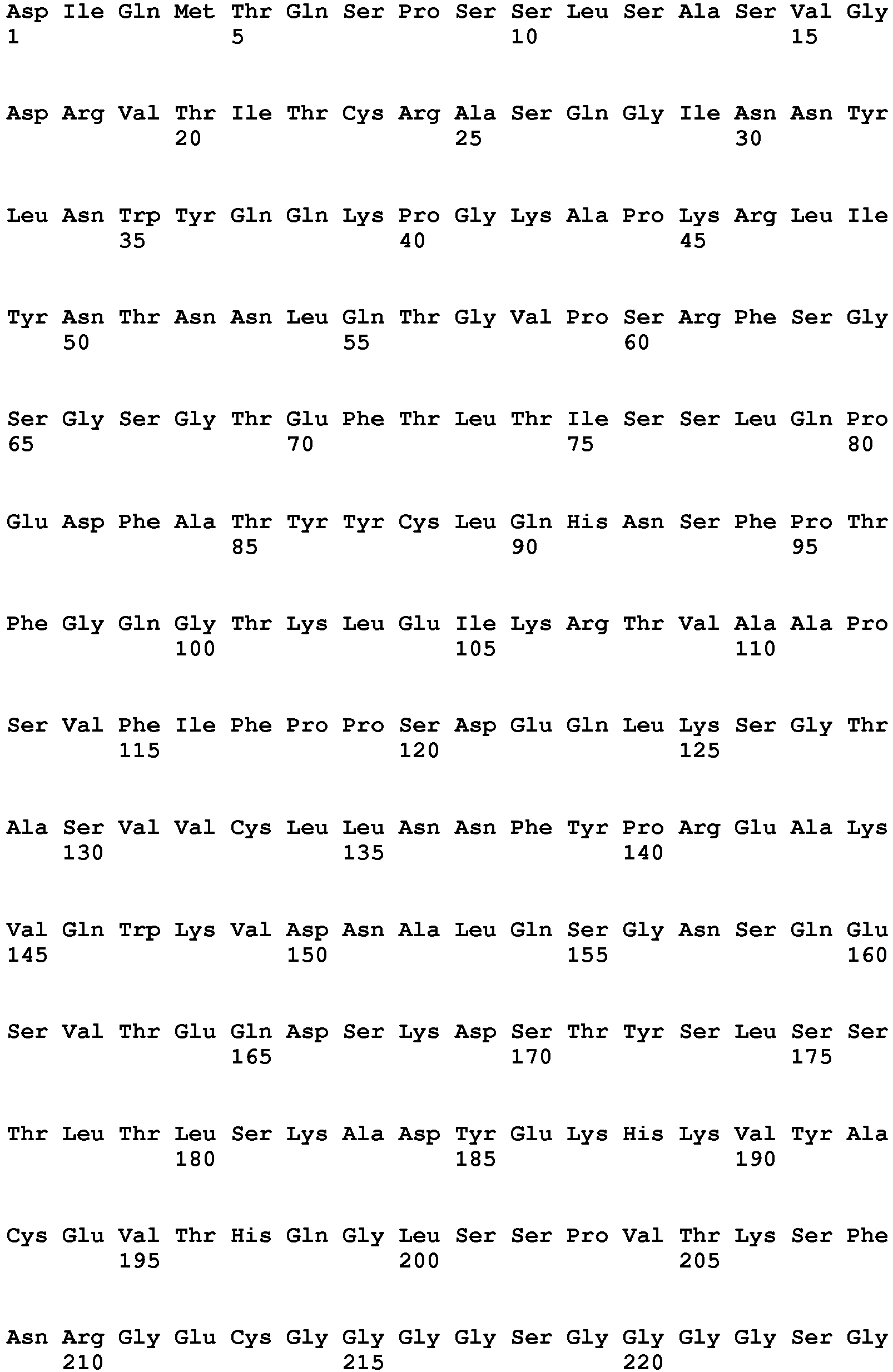


- <210> 5
<211> 450
<212> PRT
<213> Artificial - <220>
<223> <EGFR> heavy chain, OA-GA201-scFab-Ak18 (+WT) - <400> 5


- <210> 6
<211> 213
<212> PRT
<213> Artificial - <220>
<223> <EGFR> light chain, OA-GA201-scFab-Ak18 (+WT) - <400> 6


- <210> 7
<211> 695
<212> PRT
<213> Artificial - <220>
<223> <IGF-1R> peptide connected heavy and light chain withdisulfide stabilization VH 44 /VL100- OA-GA201-scFab-Akl8 - <400> 7


- <210> 8
<211> 695
<212> PRT
<213> Artificial - <220>
<223> <IGF-1R> peptide connected heavy and light chain of OA-GA201-scFab-Ak18_WT - <400> 8


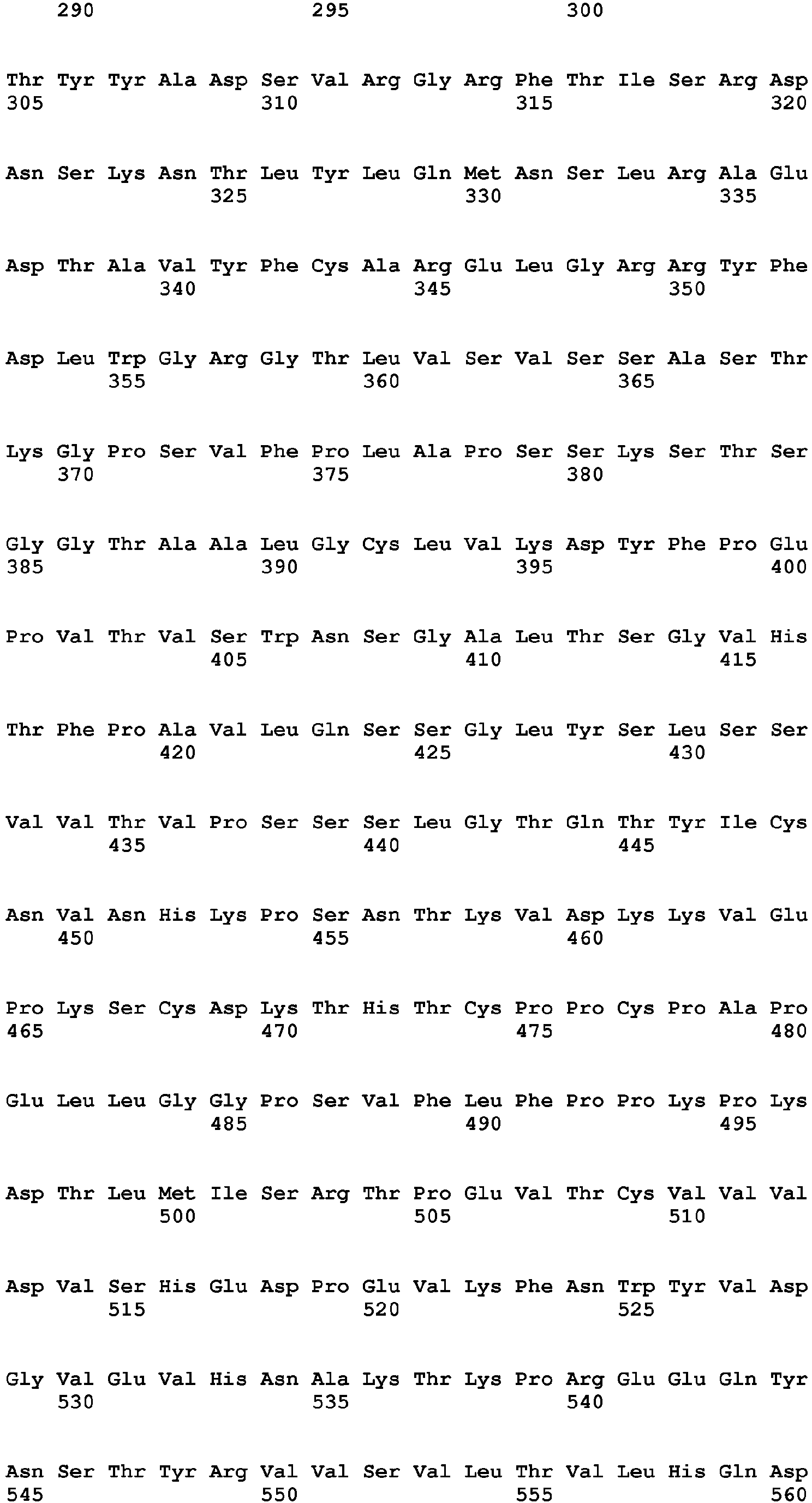

- <210> 9
<211> 453
<212> PRT
<213> Artificial - <220>
<223> <VEGF> heavy chain, Ang2-VEGF OA-Ava-N-scFabLC06 (+SS) - <400> 9


- <210> 10
<211> 214
<212> PRT
<213> Artificial - <220>
<223> <VEGF> light chain, Ang2-VEGF OA-Ava-N-scFabLC06 (+SS) - <400> 10


- <210> 11
<211> 706
<212> PRT
<213> Artificial - <220>
<223> <ANG-2> peptide connected heavy and light chain withdisulfide stabilization VH 44 /VL100 of Ang2-VEGF OA-Ava-N-scFabLC06SS - <400> 11


- <210> 12
<211> 706
<212> PRT
<213> Artificial - <220>
<223> <ANG-2> peptide connected heavy and light chain of Ang2-VEGF OA-Ava-N-scFabLC06 - <400> 12


- <210> 13
<211> 1210
<212> PRT
<213> Homo sapiens - <400> 13


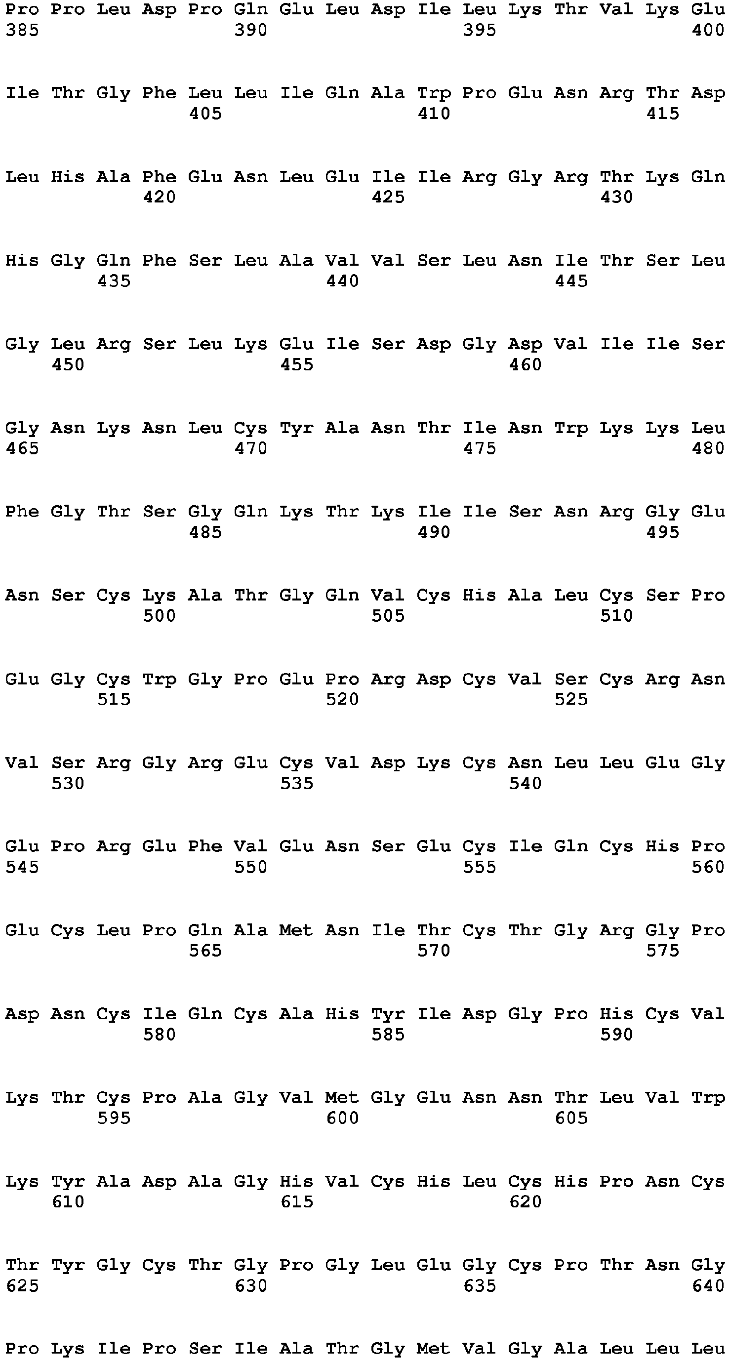


- <210> 14
<211> 1367
<212> PRT
<213> Homo sapiens - <400> 14


- <210> 15
<211> 191
<212> PRT
<213> Homo sapiens - <400> 15


- <210> 16
<211> 504
<212> PRT
<213> Artificial - <220>
<223> Human angiopoietin-2 (ANG-2) with leader and His-tag - <400> 16


wherein
an amino acid residue is replaced with an amino acid residue having a smaller side chain volume, thereby generating a cavity within the interface of the second CH3 domain within which a protuberance within the interface of the first CH3 domain is positionable.

Claims (12)
- A bispecific antibody of three polypeptide chains, comprisinga) the heavy chain and the light chain of a first full length antibody that specifically binds to a first antigen;b) the heavy chain and the light chain of a second full length antibody that specifically binds to a second antigen, wherein the N-terminus of the heavy chain is connected to the C-terminus of the light chain via a peptide linker,characterized in that
the CH3 domain of the heavy chain of the full length antibody of a) and the CH3 domain of the heavy chain of the full length antibody of b) each meet at an interface which comprises an alteration in the original interface between the antibody CH3 domains;
whereini) in the CH3 domain of one heavy chain
an amino acid residue is replaced with an amino acid residue having a larger side chain volume, thereby generating a protuberance within the interface of the CH3 domain of one heavy chain which is positionable in a cavity within the interface of the CH3 domain of the other heavy chain
and whereinii) in the CH3 domain of the other heavy chainan amino acid residue is replaced with an amino acid residue having a smaller side chain volume, thereby generating a cavity within the interface of the second CH3 domain within which a protuberance within the interface of the first CH3 domain is positionable. - The antibody according to claim 1, characterized in that
the said amino acid residue having a larger side chain volume is selected from the group consisting of arginine (R), phenylalanine (F), tyrosine (Y), tryptophan (W) and
said amino acid residue having a smaller side chain volume is selected from the group consisting of alanine (A), serine (S), threonine (T), valine (V). - The antibody according to claim 1 or 2, characterized in that
both CH3 domains are further altered by the introduction of cysteine (C) as amino acid in the corresponding positions of each CH3 domain such that a disulfide bridge between both CH3 domains can be formed. - The antibody according to one of claims 1 to 3, characterized in that
the antibody heavy chain variable domain (VH) and the antibody light chain variable domain (VL) of the heavy and light chain of the second full lenght antibody under b) are disulfide stabilized by introduction of a disulfide bond between the following positions:i) heavy chain variable domain position 44 to light chain variable domain position 100,ii) heavy chain variable domain position 105 to light chain variable domain position 43, oriii) heavy chain variable domain position 101 to light chain variable domain position 100. - The bispecific antibody according to one of claims 1 to 4, wherein the antibody comprises a constant region of IgG1.
- A pharmaceutical composition comprising an antibody according to one of claims 1 to 5.
- The bispecific antibody according to any one of claims 1 to 5 for use in the treatment of cancer.
- Use of the bispecific antibody according to any one of claims 1 to 5 for the preparation of a medicament for the treatment of cancer.
- A nucleic acid molecule encoding a chain of a bispecific antibody according to any one of claims 1 to 5.
- Expression vector containing said nucleic acid according to claim 9 capable of expressing said nucleic acid in a prokaryotic or eukaryotic host cell.
- A prokaryotic or eukaryotic host cell comprising a vector according to claim 10.
- A method for the preparation of a bispecific antibody according to one of claims 1 to 5
comprising the steps ofa) transforming a host cell with vectors comprising nucleic acid molecules encodingaa) the heavy chain and the light chain of a first full length antibody that specifically binds to a first antigen; andab) the heavy chain and the light chain of a second full length antibody that specifically binds to a second antigen, wherein the N-terminus of the heavy chain is connected to the C-terminus of the light chain via a peptide linker; andb) culturing the host cell under conditions that allow synthesis of said antibody molecule; andc) recovering said antibody molecule from said culture.
Priority Applications (1)
| Application Number | Priority Date | Filing Date | Title |
|---|---|---|---|
| EP11711313.4A EP2552481B1 (en) | 2010-03-26 | 2011-03-24 | Bispecific antibodies |
Applications Claiming Priority (3)
| Application Number | Priority Date | Filing Date | Title |
|---|---|---|---|
| EP10003270 | 2010-03-26 | ||
| PCT/EP2011/054505 WO2011117330A1 (en) | 2010-03-26 | 2011-03-24 | Bispecific antibodies |
| EP11711313.4A EP2552481B1 (en) | 2010-03-26 | 2011-03-24 | Bispecific antibodies |
Publications (2)
| Publication Number | Publication Date |
|---|---|
| EP2552481A1 EP2552481A1 (en) | 2013-02-06 |
| EP2552481B1 true EP2552481B1 (en) | 2017-02-22 |
Family
ID=42330134
Family Applications (1)
| Application Number | Title | Priority Date | Filing Date |
|---|---|---|---|
| EP11711313.4A Active EP2552481B1 (en) | 2010-03-26 | 2011-03-24 | Bispecific antibodies |
Country Status (12)
| Country | Link |
|---|---|
| US (1) | US10106600B2 (en) |
| EP (1) | EP2552481B1 (en) |
| JP (1) | JP5726287B2 (en) |
| KR (1) | KR101498346B1 (en) |
| CN (1) | CN102946902B (en) |
| AR (1) | AR080793A1 (en) |
| BR (1) | BR112012024312A2 (en) |
| CA (1) | CA2789074A1 (en) |
| MX (1) | MX340124B (en) |
| RU (1) | RU2573588C2 (en) |
| TW (1) | TW201138821A (en) |
| WO (1) | WO2011117330A1 (en) |
Families Citing this family (118)
| Publication number | Priority date | Publication date | Assignee | Title |
|---|---|---|---|---|
| CN103429620B (en) | 2010-11-05 | 2018-03-06 | 酵活有限公司 | Antibody design for stable heterodimerization with mutations in the Fc domain |
| HRP20180028T4 (en) * | 2011-03-16 | 2020-10-02 | F. Hoffmann - La Roche Ag | CHROMATOGRAPHY OF ION EXCHANGE WITH IMPROVED SELECTIVITY FOR SEPARATION OF POLYPEPTIDE MONOMERS, AGGREGATES AND FRAGMENTS BY MOTION PHASE MODULATION |
| KR101870555B1 (en) | 2011-08-23 | 2018-06-22 | 로슈 글리카트 아게 | Bispecific antibodies specific for t-cell activating antigens and a tumor antigen and methods of use |
| US9574010B2 (en) | 2011-11-04 | 2017-02-21 | Zymeworks Inc. | Stable heterodimeric antibody design with mutations in the Fc domain |
| WO2013113615A1 (en) | 2012-02-03 | 2013-08-08 | F. Hoffmann-La Roche Ag | Bispecific antibody molecules with antigen-transfected t-cells and their use in medicine |
| KR101674784B1 (en) | 2012-04-05 | 2016-11-09 | 에프. 호프만-라 로슈 아게 | Bispecific antibodies against human tweak and human il17 and uses thereof |
| US9062120B2 (en) * | 2012-05-02 | 2015-06-23 | Janssen Biotech, Inc. | Binding proteins having tethered light chains |
| WO2014004586A1 (en) | 2012-06-25 | 2014-01-03 | Zymeworks Inc. | Process and methods for efficient manufacturing of highly pure asymmetric antibodies in mammalian cells |
| SI3495387T1 (en) | 2012-07-13 | 2021-12-31 | Roche Glycart Ag | Bispecific antibodies against VEGF / against ANG-2 and their use in the treatment of vascular diseases |
| CN104640561A (en) * | 2012-07-23 | 2015-05-20 | 酵活有限公司 | Immunoglobulin constructs comprising selective pairing of light and heavy chains |
| MX2015003616A (en) | 2012-10-08 | 2015-06-05 | Roche Glycart Ag | Fc-free antibodies comprising two fab-fragments and methods of use. |
| MX385344B (en) | 2012-11-28 | 2025-03-18 | Zymeworks Bc Inc | GENETICALLY MODIFIED IMMUNOGLOBULIN HEAVY CHAIN-LIGHT CHAIN PAIRS AND THEIR USES. |
| US9914785B2 (en) | 2012-11-28 | 2018-03-13 | Zymeworks Inc. | Engineered immunoglobulin heavy chain-light chain pairs and uses thereof |
| CN110437337B (en) | 2013-02-26 | 2024-07-30 | 罗切格利卡特公司 | Bispecific T cell activating antigen binding molecules |
| WO2014131694A1 (en) | 2013-02-26 | 2014-09-04 | Roche Glycart Ag | Bispecific t cell activating antigen binding molecules |
| CA2904805A1 (en) | 2013-04-29 | 2014-11-06 | F. Hoffmann-La Roche Ag | Fc-receptor binding modified asymmetric antibodies and methods of use |
| CA2904806C (en) | 2013-04-29 | 2021-11-23 | F. Hoffmann-La Roche Ag | Human fcrn-binding modified antibodies and methods of use |
| CN103509117B (en) * | 2013-05-06 | 2016-03-09 | 江苏匡亚生物医药科技有限公司 | Bi-specific antibody of anti-human HER2 and people IGF-IR and its production and use |
| US10239951B2 (en) | 2013-05-08 | 2019-03-26 | Zymeworks Inc. | Bispecific HER2 and HER3 antigen binding constructs |
| EP3027649B1 (en) | 2013-08-01 | 2020-04-01 | F.Hoffmann-La Roche Ag | Tnfa-il-17 bispecific antibodies |
| CN118271429A (en) * | 2013-08-19 | 2024-07-02 | 豪夫迈·罗氏有限公司 | Separation of bispecific antibodies and bispecific antibody production byproducts using hydroxyapatite chromatography |
| WO2015025054A1 (en) | 2013-08-22 | 2015-02-26 | Medizinische Universität Wien | Dye-specific antibodies for prestained molecular weight markers and methods producing the same |
| EP3068892A4 (en) * | 2013-11-13 | 2017-05-31 | Zymeworks Inc. | Monovalent antigen binding constructs targeting egfr and/or her2 and uses thereof |
| LT3074424T (en) | 2013-11-27 | 2025-05-26 | Zymeworks Bc Inc. | BISPECIFIC ANTIGEN-BINDING CONSTRUCTS TARGETED TO HER2 |
| JP6510532B2 (en) | 2013-12-20 | 2019-05-08 | エフ・ホフマン−ラ・ロシュ・アクチェンゲゼルシャフト | Bispecific HER2 antibodies and methods of use |
| AU2014374055B2 (en) | 2013-12-30 | 2018-11-08 | Epimab Biotherapeutics Inc. | Fabs-in-tandem immunoglobulin and uses thereof |
| CN110981957B (en) | 2014-01-15 | 2024-12-24 | 豪夫迈·罗氏有限公司 | Fc region variants with improved protein A binding |
| CN103864935A (en) * | 2014-03-06 | 2014-06-18 | 中国药科大学 | Targeted EGFR/KDR (Epidermal Growth Factor Receptor/Kinase Insert Domain Receptor) specific diabody |
| CN104974260B (en) * | 2014-04-01 | 2019-06-14 | 三生国健药业(上海)股份有限公司 | Anti- HGF/VEGF bispecific antibody, preparation method and application |
| MX378281B (en) | 2014-04-25 | 2025-03-10 | Pf Medicament | DRUG-ANTIBODY CONJUGATE AND ITS USE FOR CANCER TREATMENT. |
| CN106459204B (en) * | 2014-04-25 | 2021-02-02 | 皮埃尔法布雷医药公司 | IGF-1R antibody and its use as a targeting carrier for the treatment of cancer |
| MA39909B1 (en) | 2014-04-25 | 2019-05-31 | Pf Medicament | Conjugate antibodies to igf-1r-drug and its use for the treatment of cancer |
| DK3107938T3 (en) | 2014-05-28 | 2022-07-11 | Zymeworks Inc | MODIFIED, ANTIGEN-BINDING POLYPEPTIDE CONSTRUCTS AND USES THEREOF |
| US10973920B2 (en) | 2014-06-30 | 2021-04-13 | Glykos Finland Oy | Saccharide derivative of a toxic payload and antibody conjugates thereof |
| PL3177643T3 (en) | 2014-08-04 | 2019-09-30 | F.Hoffmann-La Roche Ag | Bispecific t cell activating antigen binding molecules |
| JP2017537896A (en) * | 2014-11-10 | 2017-12-21 | エフ.ホフマン−ラ ロシュ アーゲーF. Hoffmann−La Roche Aktiengesellschaft | Bispecific antibodies and methods for use in ophthalmology |
| ES2808853T3 (en) | 2014-11-20 | 2021-03-02 | Hoffmann La Roche | Common light chains and usage procedures |
| SG11201704056XA (en) | 2014-11-20 | 2017-06-29 | Hoffmann La Roche | Combination therapy of t cell activating bispecific antigen binding molecules cd3 abd folate receptor 1 (folr1) and pd-1 axis binding antagonists |
| RS61010B1 (en) | 2014-11-20 | 2020-11-30 | Hoffmann La Roche | T cell activating bispecific antigen binding molecules against folr1 and cd3 |
| FI3223848T3 (en) | 2014-11-27 | 2025-02-28 | Zymeworks Bc Inc | METHODS USING BISPECIFIC ANTIGEN-BINDING STRUCTURES THAT TARGET HER2 |
| WO2016087514A1 (en) | 2014-12-02 | 2016-06-09 | Cemm - Forschungszentrum Für Molekulare Medizin Gmbh | Anti-mutant calreticulin antibodies and their use in the diagnosis and therapy of myeloid malignancies |
| WO2016087416A1 (en) | 2014-12-03 | 2016-06-09 | F. Hoffmann-La Roche Ag | Multispecific antibodies |
| AR103477A1 (en) | 2015-01-28 | 2017-05-10 | Lilly Co Eli | VEGFA / ANG2 COMPOUNDS |
| CA2986066A1 (en) * | 2015-06-30 | 2017-01-05 | Boehringer Ingelheim International Gmbh | Multi-specific binding proteins |
| AR106188A1 (en) | 2015-10-01 | 2017-12-20 | Hoffmann La Roche | ANTI-CD19 HUMANIZED HUMAN ANTIBODIES AND METHODS OF USE |
| SI3356404T1 (en) | 2015-10-02 | 2021-11-30 | F. Hoffmann-La Roche Ag | Anti-pd1 antibodies and methods of use |
| JP7044700B2 (en) | 2015-10-02 | 2022-03-30 | エフ・ホフマン-ラ・ロシュ・アクチェンゲゼルシャフト | Bispecific anti-CEAXCD3 T cell activating antigen binding molecule |
| CN108283001B (en) | 2015-10-08 | 2022-08-19 | 酵活有限公司 | Antigen binding polypeptide constructs comprising kappa and lambda light chains and uses thereof |
| IL313608A (en) | 2015-12-09 | 2024-08-01 | Hoffmann La Roche | Type ii anti-cd20 antibody for reducing formation of anti-drug antibodies |
| CA3007898A1 (en) * | 2015-12-28 | 2017-07-06 | Innate Pharma | Variable regions for nkp46 binding proteins |
| BR112018011029A2 (en) | 2016-01-08 | 2018-11-21 | Hoffmann La Roche | methods for treating or delaying cancer progression and improving immune function in a cancer individual, uses of a binding antagonist and a bispecific antibody, compositions and kits |
| UA126657C2 (en) * | 2016-02-03 | 2023-01-11 | Емджен Рісерч (Мюнік) Ґмбг | Bcma and cd3 bispecific t cell engaging antibody constructs |
| JP7014724B2 (en) | 2016-02-06 | 2022-02-01 | エピムアブ バイオセラピューティクス インコーポレイテッド | Tandem-type Fab immunoglobulin and its use |
| US11291721B2 (en) | 2016-03-21 | 2022-04-05 | Marengo Therapeutics, Inc. | Multispecific and multifunctional molecules and uses thereof |
| JP7015244B2 (en) | 2016-03-22 | 2022-02-02 | エフ・ホフマン-ラ・ロシュ・アクチェンゲゼルシャフト | Protease-activated T cell bispecific molecule |
| ES3023551T3 (en) * | 2016-03-25 | 2025-06-02 | Biomunex Pharmaceuticals | Binding molecules to cd38 and pd-l1 |
| JP6675017B2 (en) * | 2016-05-02 | 2020-04-01 | エフ.ホフマン−ラ ロシュ アーゲーF. Hoffmann−La Roche Aktiengesellschaft | Contrast body-single chain target binding substance |
| CN109843926B (en) | 2016-09-30 | 2023-01-20 | 豪夫迈·罗氏有限公司 | Bispecific antibodies against CD3 |
| TW201829463A (en) | 2016-11-18 | 2018-08-16 | 瑞士商赫孚孟拉羅股份公司 | anti-HLA-G antibody and use thereof |
| AU2017363309A1 (en) | 2016-11-23 | 2019-07-11 | Bioverativ Therapeutics Inc. | Mono- and bispecific antibodies binding to coagulation factor IX and coagulation factor X |
| WO2018151820A1 (en) | 2017-02-16 | 2018-08-23 | Elstar Therapeutics, Inc. | Multifunctional molecules comprising a trimeric ligand and uses thereof |
| CN106946989B (en) * | 2017-03-02 | 2020-02-18 | 深圳市国创纳米抗体技术有限公司 | anti-CEA antigen VHH domain and bispecific antibody containing same |
| EP3630836A1 (en) | 2017-05-31 | 2020-04-08 | Elstar Therapeutics, Inc. | Multispecific molecules that bind to myeloproliferative leukemia (mpl) protein and uses thereof |
| WO2019000105A1 (en) | 2017-06-30 | 2019-01-03 | Zymeworks Inc. | Stabilized chimeric fabs |
| WO2019035938A1 (en) | 2017-08-16 | 2019-02-21 | Elstar Therapeutics, Inc. | Multispecific molecules that bind to bcma and uses thereof |
| PE20210844A1 (en) | 2017-11-01 | 2021-05-10 | Hoffmann La Roche | CONTORSBODIES 2 + BI-SPECIFIC |
| CN111448217A (en) | 2017-12-22 | 2020-07-24 | 阿根思公司 | Bispecific antigen binding constructs |
| US12247060B2 (en) | 2018-01-09 | 2025-03-11 | Marengo Therapeutics, Inc. | Calreticulin binding constructs and engineered T cells for the treatment of diseases |
| CN111699004A (en) | 2018-02-06 | 2020-09-22 | 豪夫迈·罗氏有限公司 | Treatment of ophthalmic disorders |
| CR20200391A (en) | 2018-02-08 | 2020-10-19 | Genentech Inc | Bispecific antigen-binding molecules and methods of use |
| GB201802487D0 (en) | 2018-02-15 | 2018-04-04 | Argenx Bvba | Cytokine combination therapy |
| EP3765517A1 (en) | 2018-03-14 | 2021-01-20 | Elstar Therapeutics, Inc. | Multifunctional molecules that bind to calreticulin and uses thereof |
| EP3765516A2 (en) | 2018-03-14 | 2021-01-20 | Elstar Therapeutics, Inc. | Multifunctional molecules and uses thereof |
| AR114789A1 (en) | 2018-04-18 | 2020-10-14 | Hoffmann La Roche | ANTI-HLA-G ANTIBODIES AND THE USE OF THEM |
| CN112074541A (en) | 2018-05-03 | 2020-12-11 | 上海岸迈生物科技有限公司 | High affinity antibodies to PD-1 and LAG-3 and bispecific binding proteins prepared therefrom |
| CN110540593B (en) * | 2018-05-29 | 2022-05-17 | 上海药明生物技术有限公司 | Novel anti-CD3/anti-CD20 bispecific antibodies |
| TWI848953B (en) | 2018-06-09 | 2024-07-21 | 德商百靈佳殷格翰國際股份有限公司 | Multi-specific binding proteins for cancer treatment |
| WO2020010250A2 (en) | 2018-07-03 | 2020-01-09 | Elstar Therapeutics, Inc. | Anti-tcr antibody molecules and uses thereof |
| WO2020014974A1 (en) | 2018-07-20 | 2020-01-23 | Eucure (Beijing) Biopharma Co., Ltd | Anti-cd40 antibodies and uses thereof |
| CN119033684A (en) | 2018-10-29 | 2024-11-29 | 豪夫迈·罗氏有限公司 | Antibody formulations |
| WO2020136566A1 (en) | 2018-12-24 | 2020-07-02 | Sanofi | Multispecific binding proteins with mutant fab domains |
| WO2020172598A1 (en) | 2019-02-21 | 2020-08-27 | Elstar Therapeutics, Inc. | Multifunctional molecules that bind to t cells and uses thereof to treat autoimmune disorders |
| AU2020226893B2 (en) | 2019-02-21 | 2025-02-27 | Marengo Therapeutics, Inc. | Multifunctional molecules that bind to T cell related cancer cells and uses thereof |
| JP7579795B2 (en) | 2019-02-21 | 2024-11-08 | マレンゴ・セラピューティクス,インコーポレーテッド | Antibody molecules that bind to NKp30 and uses thereof |
| SG11202109122SA (en) | 2019-02-21 | 2021-09-29 | Marengo Therapeutics Inc | Anti-tcr antibody molecules and uses thereof |
| SG11202109056TA (en) | 2019-02-21 | 2021-09-29 | Marengo Therapeutics Inc | Multifunctional molecules that bind to calreticulin and uses thereof |
| CN110294806A (en) * | 2019-07-08 | 2019-10-01 | 徐州医科大学 | A kind of phospho-AB and its application |
| CN110577603B (en) * | 2019-08-15 | 2021-12-28 | 北京东方百泰生物科技股份有限公司 | anti-CD 3 and anti-CD 19 bispecific antibody |
| CN110669137B (en) | 2019-10-24 | 2021-07-16 | 高新 | A kind of multispecific antibody and its preparation method and use |
| EP4084821A4 (en) | 2020-01-03 | 2024-04-24 | Marengo Therapeutics, Inc. | CD33-BINDING MULTIFUNCTIONAL MOLECULES AND THEIR USES |
| WO2021138474A2 (en) | 2020-01-03 | 2021-07-08 | Marengo Therapeutics, Inc. | Anti-tcr antibody molecules and uses thereof |
| AU2021261420A1 (en) | 2020-04-24 | 2022-12-01 | Marengo Therapeutics, Inc. | Multifunctional molecules that bind to T cell related cancer cells and uses thereof |
| CR20220637A (en) | 2020-06-19 | 2023-01-31 | Hoffmann La Roche | ANTIBODIES THAT BIND CD3 AND CD19 |
| AU2021331075A1 (en) | 2020-08-26 | 2023-04-06 | Marengo Therapeutics, Inc. | Multifunctional molecules that bind to calreticulin and uses thereof |
| JP2023539645A (en) | 2020-08-26 | 2023-09-15 | マレンゴ・セラピューティクス,インコーポレーテッド | Antibody molecules that bind to NKP30 and uses thereof |
| CA3190573A1 (en) | 2020-08-26 | 2022-03-03 | Andreas Loew | Methods of detecting trbc1 or trbc2 |
| EP4244246A1 (en) * | 2020-11-10 | 2023-09-20 | Amgen Inc. | Novel linkers of multispecific antigen binding domains |
| US20250270320A1 (en) | 2020-12-10 | 2025-08-28 | Eutilex Co., Ltd. | Anti-pd-1 antibody and uses thereof |
| WO2022129120A1 (en) | 2020-12-17 | 2022-06-23 | F. Hoffmann-La Roche Ag | Anti-hla-g antibodies and use thereof |
| CN114716548B (en) | 2021-01-05 | 2024-11-05 | (株)爱恩德生物 | Anti-FGFR3 antibodies and uses thereof |
| CA3214757A1 (en) | 2021-04-08 | 2022-10-13 | Andreas Loew | Multifuntional molecules binding to tcr and uses thereof |
| CA3213632A1 (en) | 2021-04-30 | 2022-11-03 | F. Hoffmann-La Roche Ag | Dosing for combination treatment with anti-cd20/anti-cd3 bispecific antibody and anti-cd79b antibody drug conjugate |
| CN113278071B (en) | 2021-05-27 | 2021-12-21 | 江苏荃信生物医药股份有限公司 | Anti-human interferon alpha receptor1 monoclonal antibody and application thereof |
| IL311003A (en) | 2021-08-25 | 2024-04-01 | Denali Therapeutics Inc | Anti-her2 antibodies and methods of use thereof |
| CN113603775B (en) | 2021-09-03 | 2022-05-20 | 江苏荃信生物医药股份有限公司 | Anti-human interleukin-33 monoclonal antibody and application thereof |
| CN113683694B (en) | 2021-09-03 | 2022-05-13 | 江苏荃信生物医药股份有限公司 | Anti-human TSLP monoclonal antibody and application thereof |
| KR20240128067A (en) | 2021-12-29 | 2024-08-23 | 브리스톨-마이어스 스큅 컴퍼니 | Generation of landing pad cell lines |
| IL318546A (en) | 2022-07-26 | 2025-03-01 | Aimed Bio Inc | Anti-ror1 antibody and use thereof |
| CN115724975B (en) | 2022-10-20 | 2026-06-09 | 江苏荃信生物医药股份有限公司 | Anti-human interleukin 36 receptor monoclonal antibody and application thereof |
| EP4722249A1 (en) | 2023-06-02 | 2026-04-08 | Aimed Bio Inc. | Anti-cd200r1 antibody and use thereof |
| JP2026520211A (en) | 2023-06-16 | 2026-06-22 | ハイデルべルク イミュノセラピューティクス ゲーエムベーハー | Novel anti-HSV antibody |
| EP4737482A1 (en) | 2023-06-27 | 2026-05-06 | Atheon Bio, Inc. | Humanized anti-myct1 antibody and use thereof |
| AU2024371049A1 (en) * | 2023-10-31 | 2026-05-28 | Modex Therapeutics, Inc. | Targeted lnp delivery |
| WO2025121861A1 (en) | 2023-12-04 | 2025-06-12 | 아테온바이오 주식회사 | Novel anti-myct1 human antibody and use thereof |
| CN119431577A (en) | 2024-01-15 | 2025-02-14 | 江苏荃信生物医药股份有限公司 | Anti-human CD117 monoclonal antibody and its application |
| WO2026011013A1 (en) | 2024-07-02 | 2026-01-08 | Epibiologics, Inc. | Binding agents and uses thereof |
| WO2026037841A1 (en) | 2024-08-12 | 2026-02-19 | ONA Therapeutics S.L. | Anti-fgfr4 molecules and uses thereof |
| WO2026050572A2 (en) | 2024-08-29 | 2026-03-05 | Marengo Therapeutics, Inc. | Multifunctional molecules binding to tcr and uses thereof |
Family Cites Families (366)
| Publication number | Priority date | Publication date | Assignee | Title |
|---|---|---|---|---|
| US3773919A (en) | 1969-10-23 | 1973-11-20 | Du Pont | Polylactide-drug mixtures |
| US3896111A (en) | 1973-02-20 | 1975-07-22 | Research Corp | Ansa macrolides |
| IL47062A (en) | 1975-04-10 | 1979-07-25 | Yeda Res & Dev | Process for diminishing antigenicity of tissues to be usedas transplants by treatment with glutaraldehyde |
| US4150149A (en) | 1976-11-29 | 1979-04-17 | Professional Staff Association Of The Los Angeles County Harbor General Hospital | Method and means for the early detection and diagnosis of certain types of cancers |
| US4151042A (en) | 1977-03-31 | 1979-04-24 | Takeda Chemical Industries, Ltd. | Method for producing maytansinol and its derivatives |
| US4137230A (en) | 1977-11-14 | 1979-01-30 | Takeda Chemical Industries, Ltd. | Method for the production of maytansinoids |
| USRE30985E (en) | 1978-01-01 | 1982-06-29 | Serum-free cell culture media | |
| US4265814A (en) | 1978-03-24 | 1981-05-05 | Takeda Chemical Industries | Matansinol 3-n-hexadecanoate |
| US4307016A (en) | 1978-03-24 | 1981-12-22 | Takeda Chemical Industries, Ltd. | Demethyl maytansinoids |
| JPS5562090A (en) | 1978-10-27 | 1980-05-10 | Takeda Chem Ind Ltd | Novel maytansinoid compound and its preparation |
| JPS55164687A (en) | 1979-06-11 | 1980-12-22 | Takeda Chem Ind Ltd | Novel maytansinoid compound and its preparation |
| US4256746A (en) | 1978-11-14 | 1981-03-17 | Takeda Chemical Industries | Dechloromaytansinoids, their pharmaceutical compositions and method of use |
| JPS5566585A (en) | 1978-11-14 | 1980-05-20 | Takeda Chem Ind Ltd | Novel maytansinoid compound and its preparation |
| JPS55102583A (en) | 1979-01-31 | 1980-08-05 | Takeda Chem Ind Ltd | 20-acyloxy-20-demethylmaytansinoid compound |
| US4665077A (en) | 1979-03-19 | 1987-05-12 | The Upjohn Company | Method for treating rejection of organ or skin grafts with 6-aryl pyrimidine compounds |
| JPS55162791A (en) | 1979-06-05 | 1980-12-18 | Takeda Chem Ind Ltd | Antibiotic c-15003pnd and its preparation |
| JPS55164685A (en) | 1979-06-08 | 1980-12-22 | Takeda Chem Ind Ltd | Novel maytansinoid compound and its preparation |
| JPS55164686A (en) | 1979-06-11 | 1980-12-22 | Takeda Chem Ind Ltd | Novel maytansinoid compound and its preparation |
| US4309428A (en) | 1979-07-30 | 1982-01-05 | Takeda Chemical Industries, Ltd. | Maytansinoids |
| JPS5645483A (en) | 1979-09-19 | 1981-04-25 | Takeda Chem Ind Ltd | C-15003phm and its preparation |
| JPS5645485A (en) | 1979-09-21 | 1981-04-25 | Takeda Chem Ind Ltd | Production of c-15003pnd |
| EP0028683A1 (en) | 1979-09-21 | 1981-05-20 | Takeda Chemical Industries, Ltd. | Antibiotic C-15003 PHO and production thereof |
| US4361544A (en) | 1980-03-03 | 1982-11-30 | Goldenberg Milton David | Tumor localization and therapy with labeled antibodies specific to intracellular tumor-associated markers |
| US4444744A (en) | 1980-03-03 | 1984-04-24 | Goldenberg Milton David | Tumor localization and therapy with labeled antibodies to cell surface antigens |
| WO1982001188A1 (en) | 1980-10-08 | 1982-04-15 | Takeda Chemical Industries Ltd | 4,5-deoxymaytansinoide compounds and process for preparing same |
| US4450254A (en) | 1980-11-03 | 1984-05-22 | Standard Oil Company | Impact improvement of high nitrile resins |
| US4419446A (en) | 1980-12-31 | 1983-12-06 | The United States Of America As Represented By The Department Of Health And Human Services | Recombinant DNA process utilizing a papilloma virus DNA as a vector |
| US4315929A (en) | 1981-01-27 | 1982-02-16 | The United States Of America As Represented By The Secretary Of Agriculture | Method of controlling the European corn borer with trewiasine |
| US4313946A (en) | 1981-01-27 | 1982-02-02 | The United States Of America As Represented By The Secretary Of Agriculture | Chemotherapeutically active maytansinoids from Trewia nudiflora |
| JPS57192389A (en) | 1981-05-20 | 1982-11-26 | Takeda Chem Ind Ltd | Novel maytansinoid |
| US4601978A (en) | 1982-11-24 | 1986-07-22 | The Regents Of The University Of California | Mammalian metallothionein promoter system |
| US4560655A (en) | 1982-12-16 | 1985-12-24 | Immunex Corporation | Serum-free cell culture medium and process for making same |
| US4657866A (en) | 1982-12-21 | 1987-04-14 | Sudhir Kumar | Serum-free, synthetic, completely chemically defined tissue culture media |
| US4948882A (en) | 1983-02-22 | 1990-08-14 | Syngene, Inc. | Single-stranded labelled oligonucleotides, reactive monomers and methods of synthesis |
| US4816567A (en) | 1983-04-08 | 1989-03-28 | Genentech, Inc. | Recombinant immunoglobin preparations |
| US4767704A (en) | 1983-10-07 | 1988-08-30 | Columbia University In The City Of New York | Protein-free culture medium |
| US4965199A (en) | 1984-04-20 | 1990-10-23 | Genentech, Inc. | Preparation of functional human factor VIII in mammalian cells using methotrexate based selection |
| US4737456A (en) | 1985-05-09 | 1988-04-12 | Syntex (U.S.A.) Inc. | Reducing interference in ligand-receptor binding assays |
| GB8516415D0 (en) | 1985-06-28 | 1985-07-31 | Celltech Ltd | Culture of animal cells |
| US4868105A (en) | 1985-12-11 | 1989-09-19 | Chiron Corporation | Solution phase nucleic acid sandwich assay |
| SE8505922D0 (en) | 1985-12-13 | 1985-12-13 | Kabigen Ab | CONSTRUCTION OF AN IGG BINDING PROTEIN TO FACILITATE DOWNSTREAM PROCESSING USING PROTEIN ENGINEERING |
| US6548640B1 (en) | 1986-03-27 | 2003-04-15 | Btg International Limited | Altered antibodies |
| US4927762A (en) | 1986-04-01 | 1990-05-22 | Cell Enterprises, Inc. | Cell culture medium with antioxidant |
| IL85035A0 (en) | 1987-01-08 | 1988-06-30 | Int Genetic Eng | Polynucleotide molecule,a chimeric antibody with specificity for human b cell surface antigen,a process for the preparation and methods utilizing the same |
| JP3101690B2 (en) | 1987-03-18 | 2000-10-23 | エス・ビィ・2・インコーポレイテッド | Modifications of or for denatured antibodies |
| IL86164A0 (en) | 1987-04-28 | 1988-11-15 | Tamir Biotechnology Ltd | Improved dna probes |
| US4975278A (en) | 1988-02-26 | 1990-12-04 | Bristol-Myers Company | Antibody-enzyme conjugates in combination with prodrugs for the delivery of cytotoxic agents to tumor cells |
| US5585481A (en) | 1987-09-21 | 1996-12-17 | Gen-Probe Incorporated | Linking reagents for nucleotide probes |
| JP3012244B2 (en) | 1987-09-21 | 2000-02-21 | ジェン―プローブ インコーポレイテッド | Non-nucleotide ligation reagent for nucleotide probes |
| US4914210A (en) | 1987-10-02 | 1990-04-03 | Cetus Corporation | Oligonucleotide functionalizing reagents |
| US5202238A (en) | 1987-10-27 | 1993-04-13 | Oncogen | Production of chimeric antibodies by homologous recombination |
| US5204244A (en) | 1987-10-27 | 1993-04-20 | Oncogen | Production of chimeric antibodies by homologous recombination |
| US5770701A (en) | 1987-10-30 | 1998-06-23 | American Cyanamid Company | Process for preparing targeted forms of methyltrithio antitumor agents |
| US5053394A (en) | 1988-09-21 | 1991-10-01 | American Cyanamid Company | Targeted forms of methyltrithio antitumor agents |
| US5606040A (en) | 1987-10-30 | 1997-02-25 | American Cyanamid Company | Antitumor and antibacterial substituted disulfide derivatives prepared from compounds possessing a methyl-trithio group |
| IL85746A (en) | 1988-03-15 | 1994-05-30 | Yeda Res & Dev | Preparations comprising t-lymphocyte cells treated with 8-methoxypsoralen or cell membranes separated therefrom for preventing or treating autoimmune diseases |
| ATE111918T1 (en) | 1988-04-01 | 1994-10-15 | Carter Wallace | SPECIFIC CLEAVAGE OF PEPTIDE BONDS BY ORGANIC TERTIARY PHOSPHINES WITH NUCLEOPHILIC SIDE CHAINS. |
| FI891226L (en) | 1988-04-28 | 1989-10-29 | Univ Leland Stanford Junior | RESEPTORDETERMINANTER I ANTI-T-CELL FOER BEHANDLING AV AUTOIMMUNSJUKDOM. |
| FR2632955B1 (en) | 1988-06-20 | 1991-12-27 | Oris Ind | NUCLEOSIDE DERIVATIVES FOR USE IN THE SYNTHESIS OF MARKED OLIGONUCLEOTIDES, OLIGONUCLEOTIDES OBTAINED FROM SUCH DERIVATIVES AND THEIR SYNTHESIS |
| WO1990003430A1 (en) | 1988-09-23 | 1990-04-05 | Cetus Corporation | Cell culture medium for enhanced cell growth, culture longevity and product expression |
| FI903489A7 (en) | 1988-11-11 | 1990-07-10 | Medical Res Council | Ligands containing one moiety, receptors containing these ligands, methods for their preparation and uses of the ligands and receptors |
| WO1990008187A1 (en) | 1989-01-19 | 1990-07-26 | Dana Farber Cancer Institute | Soluble two domain cd2 protein |
| FR2642074B1 (en) | 1989-01-20 | 1994-04-29 | Oris Ind | POLYHYDROXYLATED MOLECULE DERIVATIVES FOR THE INTRODUCTION OF AT LEAST ONE BRANCH INTO AN OLIGONUCLEOTIDE |
| CA2046909A1 (en) | 1989-03-21 | 1990-09-22 | Mark D. Howell | Vaccination and methods against diseases resulting from pathogenic responses by specific t cell populations |
| DE3920358A1 (en) | 1989-06-22 | 1991-01-17 | Behringwerke Ag | BISPECIFIC AND OLIGO-SPECIFIC, MONO- AND OLIGOVALENT ANTI-BODY CONSTRUCTS, THEIR PRODUCTION AND USE |
| JP3072330B2 (en) | 1989-07-19 | 2000-07-31 | ジ・イミューン・レスポンス・コーポレーション | T cell receptor peptides for the treatment of autoimmune and malignant diseases |
| US5451463A (en) | 1989-08-28 | 1995-09-19 | Clontech Laboratories, Inc. | Non-nucleoside 1,3-diol reagents for labeling synthetic oligonucleotides |
| US5208020A (en) | 1989-10-25 | 1993-05-04 | Immunogen Inc. | Cytotoxic agents comprising maytansinoids and their therapeutic use |
| CA2026147C (en) | 1989-10-25 | 2006-02-07 | Ravi J. Chari | Cytotoxic agents comprising maytansinoids and their therapeutic use |
| US5959177A (en) | 1989-10-27 | 1999-09-28 | The Scripps Research Institute | Transgenic plants expressing assembled secretory antibodies |
| WO1991006305A1 (en) | 1989-11-07 | 1991-05-16 | Bristol-Myers Squibb Company | Oligomeric immunoglobulins |
| GB9015198D0 (en) | 1990-07-10 | 1990-08-29 | Brien Caroline J O | Binding substance |
| EP0547137A4 (en) | 1990-08-31 | 1993-12-08 | Bristol-Myers Squibb Company | Homoconjugated immunoglobulins |
| US5122469A (en) | 1990-10-03 | 1992-06-16 | Genentech, Inc. | Method for culturing Chinese hamster ovary cells to improve production of recombinant proteins |
| US5508192A (en) | 1990-11-09 | 1996-04-16 | Board Of Regents, The University Of Texas System | Bacterial host strains for producing proteolytically sensitive polypeptides |
| US5264365A (en) | 1990-11-09 | 1993-11-23 | Board Of Regents, The University Of Texas System | Protease-deficient bacterial strains for production of proteolytically sensitive polypeptides |
| US5290925A (en) | 1990-12-20 | 1994-03-01 | Abbott Laboratories | Methods, kits, and reactive supports for 3' labeling of oligonucleotides |
| US5571894A (en) | 1991-02-05 | 1996-11-05 | Ciba-Geigy Corporation | Recombinant antibodies specific for a growth factor receptor |
| DE4118120A1 (en) | 1991-06-03 | 1992-12-10 | Behringwerke Ag | TETRAVALENT BISPECIFIC RECEPTORS, THEIR PRODUCTION AND USE |
| US6511663B1 (en) | 1991-06-11 | 2003-01-28 | Celltech R&D Limited | Tri- and tetra-valent monospecific antigen-binding proteins |
| EP0590058B1 (en) | 1991-06-14 | 2003-11-26 | Genentech, Inc. | HUMANIZED Heregulin ANTIBODy |
| CA2071137A1 (en) | 1991-07-10 | 1993-01-11 | Clarence C. Lee | Composition and method for revitalizing scar tissue |
| GB9114948D0 (en) | 1991-07-11 | 1991-08-28 | Pfizer Ltd | Process for preparing sertraline intermediates |
| IE922292A1 (en) | 1991-07-15 | 1993-01-27 | Jolla Pharma | Modified phosphorous intermediates for providing functional¹groups on the 5' end of oligonucleotides |
| ATE163308T1 (en) | 1991-08-28 | 1998-03-15 | Boehringer Mannheim Gmbh | PRIMER FOR MATTRICE-DEPENDENT ENZYMATIC NUCLEIC ACID SYNTHESIS |
| EP0604580A1 (en) | 1991-09-19 | 1994-07-06 | Genentech, Inc. | EXPRESSION IN E. COLI OF ANTIBODY FRAGMENTS HAVING AT LEAST A CYSTEINE PRESENT AS A FREE THIOL, USE FOR THE PRODUCTION OF BIFUNCTIONAL F(ab') 2? ANTIBODIES |
| US5362852A (en) | 1991-09-27 | 1994-11-08 | Pfizer Inc. | Modified peptide derivatives conjugated at 2-hydroxyethylamine moieties |
| FI941572L (en) | 1991-10-07 | 1994-05-27 | Oncologix Inc | Combination and method of use of anti-erbB-2 monoclonal antibodies |
| DE69233528T2 (en) | 1991-11-25 | 2006-03-16 | Enzon, Inc. | Process for the preparation of multivalent antigen-binding proteins |
| US5932448A (en) | 1991-11-29 | 1999-08-03 | Protein Design Labs., Inc. | Bispecific antibody heterodimers |
| PT1024191E (en) | 1991-12-02 | 2008-12-22 | Medical Res Council | Production of anti-self antibodies from antibody segment repertoires and displayed on phage |
| CA2372813A1 (en) | 1992-02-06 | 1993-08-19 | L.L. Houston | Biosynthetic binding protein for cancer marker |
| ZA932522B (en) | 1992-04-10 | 1993-12-20 | Res Dev Foundation | Immunotoxins directed against c-erbB-2(HER/neu) related surface antigens |
| WO1994004550A1 (en) | 1992-08-21 | 1994-03-03 | Triplex Pharmaceutical Corporation | Cholesteryl-modified triple-helix forming oligonucleotides and uses thereof |
| GB9221657D0 (en) | 1992-10-15 | 1992-11-25 | Scotgen Ltd | Recombinant bispecific antibodies |
| PT672141E (en) | 1992-10-23 | 2003-09-30 | Immunex Corp | METHODS OF PREPARATION OF SOLUVEAL OLIGOMERIC PROTEINS |
| KR100335584B1 (en) | 1992-10-28 | 2002-11-29 | 제넨테크, 인코포레이티드 | Antagonists Against Vascular Endothelial Growth Factor |
| ATE139900T1 (en) | 1992-11-13 | 1996-07-15 | Idec Pharma Corp | THERAPEUTIC USE OF CHIMERIC AND LABELED ANTIBODIES AGAINST HUMAN B LYMPHOCYTE RESTRICTED DIFFERENTIATION ANTIGEN FOR THE TREATMENT OF B CELL LYMPHOMA |
| US5635483A (en) | 1992-12-03 | 1997-06-03 | Arizona Board Of Regents Acting On Behalf Of Arizona State University | Tumor inhibiting tetrapeptide bearing modified phenethyl amides |
| US5780588A (en) | 1993-01-26 | 1998-07-14 | Arizona Board Of Regents | Elucidation and synthesis of selected pentapeptides |
| US5532142A (en) | 1993-02-12 | 1996-07-02 | Board Of Regents, The University Of Texas System | Method of isolation and purification of fusion polypeptides |
| DE4310141A1 (en) | 1993-03-29 | 1994-10-06 | Boehringer Mannheim Gmbh | Homobidental trifunctional linkers |
| US5747654A (en) | 1993-06-14 | 1998-05-05 | The United States Of America As Represented By The Department Of Health And Human Services | Recombinant disulfide-stabilized polypeptide fragments having binding specificity |
| US6476198B1 (en) | 1993-07-13 | 2002-11-05 | The Scripps Research Institute | Multispecific and multivalent antigen-binding polypeptide molecules |
| UA40577C2 (en) | 1993-08-02 | 2001-08-15 | Мерк Патент Гмбх | Bispecific antigen molecule for lysis of tumor cells, method for preparing of bispecific antigen molecule, monoclonal antibody (variants), pharmaceutical preparation, pharmaceutical kit for lysis of tumor cells (variants), method of lysis of tumor cells |
| US5635602A (en) | 1993-08-13 | 1997-06-03 | The Regents Of The University Of California | Design and synthesis of bispecific DNA-antibody conjugates |
| WO1995009917A1 (en) | 1993-10-07 | 1995-04-13 | The Regents Of The University Of California | Genetically engineered bispecific tetravalent antibodies |
| JPH08507549A (en) | 1993-12-27 | 1996-08-13 | バクスター、インターナショナル、インコーポレイテッド | Water-soluble non-immunogenic polyamide crosslinker |
| SE9400088D0 (en) | 1994-01-14 | 1994-01-14 | Kabi Pharmacia Ab | Bacterial receptor structures |
| US5824483A (en) | 1994-05-18 | 1998-10-20 | Pence Inc. | Conformationally-restricted combinatiorial library composition and method |
| US5773001A (en) | 1994-06-03 | 1998-06-30 | American Cyanamid Company | Conjugates of methyltrithio antitumor agents and intermediates for their synthesis |
| US5814464A (en) | 1994-10-07 | 1998-09-29 | Regeneron Pharma | Nucleic acids encoding TIE-2 ligand-2 |
| US5849879A (en) | 1994-11-03 | 1998-12-15 | The Regents Of The University Of California | Methods for the diagnosis of glaucoma |
| US5639635A (en) | 1994-11-03 | 1997-06-17 | Genentech, Inc. | Process for bacterial production of polypeptides |
| US5789199A (en) | 1994-11-03 | 1998-08-04 | Genentech, Inc. | Process for bacterial production of polypeptides |
| US5663149A (en) | 1994-12-13 | 1997-09-02 | Arizona Board Of Regents Acting On Behalf Of Arizona State University | Human cancer inhibitory pentapeptide heterocyclic and halophenyl amides |
| US5731168A (en) | 1995-03-01 | 1998-03-24 | Genentech, Inc. | Method for making heteromultimeric polypeptides |
| US5840523A (en) | 1995-03-01 | 1998-11-24 | Genetech, Inc. | Methods and compositions for secretion of heterologous polypeptides |
| GB9504344D0 (en) | 1995-03-03 | 1995-04-19 | Unilever Plc | Antibody fragment production |
| US5641870A (en) | 1995-04-20 | 1997-06-24 | Genentech, Inc. | Low pH hydrophobic interaction chromatography for antibody purification |
| US5869046A (en) | 1995-04-14 | 1999-02-09 | Genentech, Inc. | Altered polypeptides with increased half-life |
| AU6163196A (en) | 1995-06-07 | 1996-12-30 | Smithkline Beecham Corporation | Method for obtaining receptor agonist antibodies |
| US5712374A (en) | 1995-06-07 | 1998-01-27 | American Cyanamid Company | Method for the preparation of substantiallly monomeric calicheamicin derivative/carrier conjugates |
| US5714586A (en) | 1995-06-07 | 1998-02-03 | American Cyanamid Company | Methods for the preparation of monomeric calicheamicin derivative/carrier conjugates |
| WO1997005156A1 (en) | 1995-07-27 | 1997-02-13 | Carsten Behrens | A new achiral linker reagent for the incorporation of multiple amino groups into oligonucleotides |
| US6267958B1 (en) | 1995-07-27 | 2001-07-31 | Genentech, Inc. | Protein formulation |
| US5989830A (en) | 1995-10-16 | 1999-11-23 | Unilever Patent Holdings Bv | Bifunctional or bivalent antibody fragment analogue |
| JP2000500019A (en) | 1995-11-16 | 2000-01-11 | ロシュ ダイアグノスティクス ゲゼルシャフト ミット ベシュレンクテル ハフツング | Method for producing peptide via streptavidin fusion protein |
| US5736626A (en) | 1996-01-29 | 1998-04-07 | The Perkin-Elmer Corporation | Solid support reagents for the direct synthesis of 3'-labeled polynucleotides |
| US6750334B1 (en) | 1996-02-02 | 2004-06-15 | Repligen Corporation | CTLA4-immunoglobulin fusion proteins having modified effector functions and uses therefor |
| WO1997038102A1 (en) | 1996-04-04 | 1997-10-16 | Unilever Plc | Multivalent and multispecific antigen-binding protein |
| AU2660397A (en) | 1996-04-05 | 1997-10-29 | Board Of Regents, The University Of Texas System | Methods for producing soluble, biologically-active disulfide bond-containing eukaryotic proteins in bacterial cells |
| WO1997043451A1 (en) | 1996-05-15 | 1997-11-20 | Biogenex Laboratories | Non-nucleotide linking reagents |
| CN104231078A (en) | 1997-04-07 | 2014-12-24 | 基因技术股份有限公司 | Anti-vegf antibodies |
| DE69829891T2 (en) | 1997-04-07 | 2005-10-06 | Genentech, Inc., South San Francisco | Anti-VEGF antibody |
| EP0915987A2 (en) | 1997-04-21 | 1999-05-19 | Donlar Corporation | POLY-($g(a)-L-ASPARTIC ACID), POLY-($g(a)-L-GLUTAMIC ACID) AND COPOLYMERS OF L-ASP AND L-GLU, METHOD FOR THEIR PRODUCTION AND THEIR USE |
| EP0979281B1 (en) | 1997-05-02 | 2005-07-20 | Genentech, Inc. | A method for making multispecific antibodies having heteromultimeric and common components |
| US6083715A (en) | 1997-06-09 | 2000-07-04 | Board Of Regents, The University Of Texas System | Methods for producing heterologous disulfide bond-containing polypeptides in bacterial cells |
| US6171586B1 (en) | 1997-06-13 | 2001-01-09 | Genentech, Inc. | Antibody formulation |
| WO1999006587A2 (en) | 1997-08-01 | 1999-02-11 | Morphosys Ag | Novel method and phage for the identification of nucleic acid sequences encoding members of a multimeric (poly)peptide complex |
| US6040498A (en) | 1998-08-11 | 2000-03-21 | North Caroline State University | Genetically engineered duckweed |
| US6350860B1 (en) | 1997-08-18 | 2002-02-26 | Innogenetics N.V. | Interferon-gamma-binding molecules for treating septic shock, cachexia, immune diseases and skin disorders |
| EP1049787B1 (en) | 1998-01-23 | 2004-11-24 | Vlaams Interuniversitair Instituut voor Biotechnologie | Multipurpose antibody derivatives |
| US6194551B1 (en) | 1998-04-02 | 2001-02-27 | Genentech, Inc. | Polypeptide variants |
| CA2323757C (en) | 1998-04-02 | 2011-08-02 | Genentech, Inc. | Antibody variants and fragments thereof |
| JP4334141B2 (en) | 1998-04-20 | 2009-09-30 | グリカート バイオテクノロジー アクチェンゲゼルシャフト | Engineering glycosylation of antibodies to improve antibody-dependent cytotoxicity |
| DE19819846B4 (en) | 1998-05-05 | 2016-11-24 | Deutsches Krebsforschungszentrum Stiftung des öffentlichen Rechts | Multivalent antibody constructs |
| US6117986A (en) | 1998-06-10 | 2000-09-12 | Intergen Company, L.P. | Pyrimidines linked to a quencher |
| US7074405B1 (en) | 1998-06-22 | 2006-07-11 | Immunomedics, Inc. | Use of bi-specific antibodies for pre-targeting diagnosis and therapy |
| US7138103B2 (en) | 1998-06-22 | 2006-11-21 | Immunomedics, Inc. | Use of bi-specific antibodies for pre-targeting diagnosis and therapy |
| US6573043B1 (en) | 1998-10-07 | 2003-06-03 | Genentech, Inc. | Tissue analysis and kits therefor |
| ATE348163T1 (en) | 1998-10-23 | 2007-01-15 | Amgen Inc | THROMBOPOIETIN SUBSTITUTE |
| US6660843B1 (en) | 1998-10-23 | 2003-12-09 | Amgen Inc. | Modified peptides as therapeutic agents |
| IL127127A0 (en) | 1998-11-18 | 1999-09-22 | Peptor Ltd | Small functional units of antibody heavy chain variable regions |
| US20030035798A1 (en) | 2000-08-16 | 2003-02-20 | Fang Fang | Humanized antibodies |
| WO2000035956A1 (en) | 1998-12-16 | 2000-06-22 | Kyowa Hakko Kogyo Co., Ltd. | Antihuman vegf monoclonal antibody |
| US6737056B1 (en) | 1999-01-15 | 2004-05-18 | Genentech, Inc. | Polypeptide variants with altered effector function |
| US6897044B1 (en) | 1999-01-28 | 2005-05-24 | Biogen Idec, Inc. | Production of tetravalent antibodies |
| CN1232039A (en) | 1999-04-02 | 1999-10-20 | 中国人民解放军海军总医院 | Genetic engineering double specific antibody and its use |
| EP2275540B1 (en) | 1999-04-09 | 2016-03-23 | Kyowa Hakko Kirin Co., Ltd. | Method for controlling the activity of immunologically functional molecule |
| US6872806B1 (en) | 1999-06-25 | 2005-03-29 | The Governors Of The University Of Alberta | Polypeptide compositions formed using a coiled-coil template and methods of use |
| EP1074563A1 (en) | 1999-08-02 | 2001-02-07 | F. Hoffmann-La Roche Ag | Chimeric polypeptides enhancing dimer formation through electrostatic interactions and disulfide bond, method for production and uses thereof |
| KR100797667B1 (en) | 1999-10-04 | 2008-01-23 | 메디카고 인코포레이티드 | How to regulate transcription of foreign genes |
| US7125978B1 (en) | 1999-10-04 | 2006-10-24 | Medicago Inc. | Promoter for regulating expression of foreign genes |
| US6727356B1 (en) | 1999-12-08 | 2004-04-27 | Epoch Pharmaceuticals, Inc. | Fluorescent quenching detection reagents and methods |
| AU767394C (en) | 1999-12-29 | 2005-04-21 | Immunogen, Inc. | Cytotoxic agents comprising modified doxorubicins and daunorubicins and their therapeutic use |
| US7449443B2 (en) | 2000-03-23 | 2008-11-11 | California Institute Of Technology | Method for stabilization of proteins using non-natural amino acids |
| EP1266014A2 (en) | 2000-03-24 | 2002-12-18 | Peter Kufer | Multifunctional polypeptides comprising a binding site to an epitope of the nkg2d receptor complex |
| PT2857516T (en) | 2000-04-11 | 2017-08-28 | Genentech Inc | Multivalent antibodies and uses therefor |
| FR2807767B1 (en) | 2000-04-12 | 2005-01-14 | Lab Francais Du Fractionnement | MONOCLONAL ANTIBODIES ANTI-D |
| DE10021678A1 (en) | 2000-05-05 | 2002-04-18 | Stefan Duebel | Recombinant polyspecific antibody constructs, useful for diagnosis and treatment of cancer, comprises three antibody fragments,where at least one comprises a disulfide bridge |
| AU2001264946A1 (en) | 2000-05-24 | 2001-12-03 | Imclone Systems Incorporated | Bispecific immunoglobulin-like antigen binding proteins and method of production |
| US6586207B2 (en) | 2000-05-26 | 2003-07-01 | California Institute Of Technology | Overexpression of aminoacyl-tRNA synthetases for efficient production of engineered proteins containing amino acid analogues |
| US6511809B2 (en) | 2000-06-13 | 2003-01-28 | E. I. Du Pont De Nemours And Company | Method for the detection of an analyte by means of a nucleic acid reporter |
| AU2001270609A1 (en) | 2000-06-30 | 2002-01-14 | Vlaams Interuniversitair Instituut Voor Biotechnologie Vzw | Heterodimeric fusion proteins |
| DE10044373A1 (en) | 2000-09-08 | 2002-03-21 | Roche Diagnostics Gmbh | New reagent for labeling nucleic acids |
| US6946292B2 (en) | 2000-10-06 | 2005-09-20 | Kyowa Hakko Kogyo Co., Ltd. | Cells producing antibody compositions with increased antibody dependent cytotoxic activity |
| RU2430927C2 (en) | 2000-10-20 | 2011-10-10 | Тугаи Сейяку Кабусики Кайся | Atomic compound capable of specifically identifying and cross linkinking cell surface molecule or intracellular molecule |
| US6610472B1 (en) | 2000-10-31 | 2003-08-26 | Genetastix Corporation | Assembly and screening of highly complex and fully human antibody repertoire in yeast |
| ES2376454T3 (en) | 2000-12-22 | 2012-03-14 | Grad, Carole, Legal Representative Of Howard, Kaplan | EXPRESSION BOOKS IN FAGO OF VH HUMAN FRAGMENTS. |
| AU2002327164A1 (en) | 2001-01-29 | 2002-12-09 | Idec Pharmaceuticals Corporation | Engineered tetravalent antibodies and methods of use |
| US20050118164A1 (en) | 2001-03-09 | 2005-06-02 | William Herman | Targeted ligands |
| US20030027751A1 (en) | 2001-04-10 | 2003-02-06 | Genvec, Inc. | VEGF fusion proteins |
| US6884869B2 (en) | 2001-04-30 | 2005-04-26 | Seattle Genetics, Inc. | Pentapeptide compounds and uses related thereto |
| HU229910B1 (en) | 2001-05-11 | 2014-12-29 | Amgen Inc Thousand Oaks | Peptides and related molecules that bind to tall-1 |
| US6833441B2 (en) | 2001-08-01 | 2004-12-21 | Abmaxis, Inc. | Compositions and methods for generating chimeric heteromultimers |
| US20030082547A1 (en) | 2001-08-27 | 2003-05-01 | Ewing Gregory J. | Non-fluorescent quencher compounds and biomolecular assays |
| ATE346866T1 (en) | 2001-09-14 | 2006-12-15 | Affimed Therapeutics Ag | MULTIMERIC, SINGLE CHAIN, TANDEM FV ANTIBODIES |
| US7658924B2 (en) | 2001-10-11 | 2010-02-09 | Amgen Inc. | Angiopoietin-2 specific binding agents |
| US7205275B2 (en) | 2001-10-11 | 2007-04-17 | Amgen Inc. | Methods of treatment using specific binding agents of human angiopoietin-2 |
| US7138370B2 (en) | 2001-10-11 | 2006-11-21 | Amgen Inc. | Specific binding agents of human angiopoietin-2 |
| US7521053B2 (en) | 2001-10-11 | 2009-04-21 | Amgen Inc. | Angiopoietin-2 specific binding agents |
| US7332474B2 (en) | 2001-10-11 | 2008-02-19 | Amgen Inc. | Peptides and related compounds having thrombopoietic activity |
| US7053202B2 (en) | 2001-10-19 | 2006-05-30 | Millennium Pharmaceuticals, Inc. | Immunoglobulin DNA cassette molecules, monobody constructs, methods of production, and methods of use therefor |
| JP2005289809A (en) | 2001-10-24 | 2005-10-20 | Vlaams Interuniversitair Inst Voor Biotechnologie Vzw (Vib Vzw) | Mutant heavy chain antibody |
| PL213948B1 (en) | 2001-10-25 | 2013-05-31 | Genentech Inc | Glycoprotein compositions |
| US20040093621A1 (en) | 2001-12-25 | 2004-05-13 | Kyowa Hakko Kogyo Co., Ltd | Antibody composition which specifically binds to CD20 |
| US20030170230A1 (en) | 2002-02-05 | 2003-09-11 | Caterer Nigel Robert | Compositions and methods for assembly and stabilization of antibody Fv fragments via antiparallel heterogeneous coiled-coil peptide regions and uses thereof |
| US20030157091A1 (en) | 2002-02-14 | 2003-08-21 | Dyax Corporation | Multi-functional proteins |
| EP1490677A4 (en) | 2002-02-27 | 2006-01-18 | California Inst Of Techn | COMPUTER PROCESS FOR CONSTRUCTING ENZYMES FOR THE INSTALLATION OF AMINO ACID ANALOGS IN PROTEINS |
| US20040018557A1 (en) | 2002-03-01 | 2004-01-29 | Immunomedics, Inc. | Bispecific antibody point mutations for enhancing rate of clearance |
| US7332585B2 (en) | 2002-04-05 | 2008-02-19 | The Regents Of The California University | Bispecific single chain Fv antibody molecules and methods of use thereof |
| AU2003233197A1 (en) | 2002-04-29 | 2003-11-17 | Genpat77 Pharmacogenetics Ag | Novel antibody binding tcr and tirc7 and its use in therapy and diagnosis |
| US7081443B2 (en) | 2002-05-21 | 2006-07-25 | Korea Advanced Institutes Of Science And Technology (Kaist) | Chimeric comp-ang1 molecule |
| KR100527334B1 (en) | 2002-06-07 | 2005-11-09 | (주)넥스젠 | Novel Linker for Oligonucleotides |
| CN1678634A (en) | 2002-06-28 | 2005-10-05 | 多曼蒂斯有限公司 | Immunoglobulin single variant antigen-binding region and specific constructs thereof |
| EP1391213A1 (en) | 2002-08-21 | 2004-02-25 | Boehringer Ingelheim International GmbH | Compositions and methods for treating cancer using maytansinoid CD44 antibody immunoconjugates and chemotherapeutic agents |
| US6919426B2 (en) | 2002-09-19 | 2005-07-19 | Amgen Inc. | Peptides and related molecules that modulate nerve growth factor activity |
| JP2006505546A (en) | 2002-10-10 | 2006-02-16 | メルク パテント ゲゼルシャフト ミット ベシュレンクテル ハフトング | Bispecific anti-ErbB antibodies and their use in tumor therapy |
| EP1431298B1 (en) | 2002-12-20 | 2006-03-08 | Roche Diagnostics GmbH | Mannitol and glucitol derivatives |
| US7534427B2 (en) | 2002-12-31 | 2009-05-19 | Immunomedics, Inc. | Immunotherapy of B cell malignancies and autoimmune diseases using unconjugated antibodies and conjugated antibodies and antibody combinations and fusion proteins |
| US20040157330A1 (en) | 2003-01-09 | 2004-08-12 | Arizeke Pharmaceuticals, Inc. | Compositions and methods for targeted biological delivery of molecular carriers |
| US7355008B2 (en) | 2003-01-09 | 2008-04-08 | Macrogenics, Inc. | Identification and engineering of antibodies with variant Fc regions and methods of using same |
| KR101498588B1 (en) | 2003-01-22 | 2015-03-05 | 로슈 글리카트 아게 | FUSION CONSTRUCTS AND USE OF SAME TO PRODUCE ANTIBODIES WITH INCREASED Fc RECEPTOR BINDING AFFINITY AND EFFECTOR FUNCTION |
| WO2004065417A2 (en) * | 2003-01-23 | 2004-08-05 | Genentech, Inc. | Methods for producing humanized antibodies and improving yield of antibodies or antigen binding fragments in cell culture |
| MXPA05008521A (en) | 2003-02-13 | 2005-10-20 | Pharmacia Corp | Antibodies to c-met for the treatment of cancers. |
| US20060104968A1 (en) | 2003-03-05 | 2006-05-18 | Halozyme, Inc. | Soluble glycosaminoglycanases and methods of preparing and using soluble glycosaminogly ycanases |
| US7871607B2 (en) | 2003-03-05 | 2011-01-18 | Halozyme, Inc. | Soluble glycosaminoglycanases and methods of preparing and using soluble glycosaminoglycanases |
| GB0305702D0 (en) | 2003-03-12 | 2003-04-16 | Univ Birmingham | Bispecific antibodies |
| US7238792B2 (en) | 2003-03-18 | 2007-07-03 | Washington State University Research Foundation | Foldable polymers as probes |
| CA2519113C (en) | 2003-04-02 | 2012-06-05 | F. Hoffmann-La Roche Ag | Antibodies against insulin-like growth factor i receptor and uses thereof |
| TWI353991B (en) | 2003-05-06 | 2011-12-11 | Syntonix Pharmaceuticals Inc | Immunoglobulin chimeric monomer-dimer hybrids |
| US8088387B2 (en) | 2003-10-10 | 2012-01-03 | Immunogen Inc. | Method of targeting specific cell populations using cell-binding agent maytansinoid conjugates linked via a non-cleavable linker, said conjugates, and methods of making said conjugates |
| WO2004106375A1 (en) | 2003-05-30 | 2004-12-09 | Merus Biopharmaceuticals B.V. I.O. | Fab library for the preparation of anti vegf and anti rabies virus fabs |
| RS20150135A1 (en) | 2003-05-30 | 2015-08-31 | Genentech Inc. | TREATMENT WITH ANTI-VEGF ANTIBODIES |
| US7700097B2 (en) | 2003-06-27 | 2010-04-20 | Biogen Idec Ma Inc. | Purification and preferential synthesis of binding molecules |
| JP5087274B2 (en) | 2003-06-30 | 2012-12-05 | ドマンティス リミテッド | Polypeptide |
| CA2531118C (en) | 2003-07-01 | 2013-01-08 | Immunomedics, Inc. | Multivalent carriers of bi-specific antibodies |
| AR046071A1 (en) * | 2003-07-10 | 2005-11-23 | Hoffmann La Roche | ANTIBODIES AGAINST RECEIVER I OF THE INSULINAL TYPE GROWTH FACTOR AND THE USES OF THE SAME |
| US20050054048A1 (en) | 2003-07-29 | 2005-03-10 | Luigi Grasso | Antibodies and methods for generating genetically altered antibodies with enhanced effector function |
| WO2005044853A2 (en) | 2003-11-01 | 2005-05-19 | Genentech, Inc. | Anti-vegf antibodies |
| US20050106667A1 (en) | 2003-08-01 | 2005-05-19 | Genentech, Inc | Binding polypeptides with restricted diversity sequences |
| AU2004266159A1 (en) | 2003-08-22 | 2005-03-03 | Biogen Idec Ma Inc. | Improved antibodies having altered effector function and methods for making the same |
| AU2004273791A1 (en) | 2003-09-05 | 2005-03-31 | Genentech, Inc. | Antibodies with altered effector functions |
| US20050064509A1 (en) | 2003-09-23 | 2005-03-24 | The Regents Of The University Of California | Use of templated self assembly to create novel multifunctional species |
| CN1326881C (en) | 2003-09-29 | 2007-07-18 | 中国人民解放军军事医学科学院基础医学研究所 | Trivalent bispecific antibody and its preparation process and use |
| EP1675620B1 (en) | 2003-10-09 | 2019-05-08 | Ambrx, Inc. | Polymer derivatives |
| HUE038955T2 (en) | 2003-11-05 | 2018-12-28 | Roche Glycart Ag | Antigen binding molecules with increased Fc receptor binding affinity and effector function |
| ES2605443T3 (en) | 2003-11-06 | 2017-03-14 | Seattle Genetics, Inc. | Auristatin conjugates with anti-HER2 or anti-CD22 antibodies and their use in therapy |
| WO2005051976A2 (en) | 2003-11-20 | 2005-06-09 | Ansata Therapeutics, Inc. | Protein and peptide ligation processes and one-step purification processes |
| CA2494571C (en) | 2003-12-02 | 2010-02-09 | F.Hoffmann-La Roche Ag | Oligonucleotides containing molecular rods |
| PT1718677E (en) | 2003-12-19 | 2012-07-18 | Genentech Inc | Monovalent antibody fragments useful as therapeutics |
| BRPI0507169A (en) | 2004-02-02 | 2007-06-26 | Ambrx Inc | modified human growth hormone polypeptides and their uses |
| US7705150B2 (en) | 2004-02-04 | 2010-04-27 | Biosearch Technologies, Inc. | Cyanine dyes |
| EP1725585A2 (en) | 2004-03-10 | 2006-11-29 | Lonza Ltd | Method for producing antibodies |
| US20060102423A1 (en) | 2004-07-12 | 2006-05-18 | Lang Tracy H | Safety harnesses |
| EP1786918A4 (en) | 2004-07-17 | 2009-02-11 | Imclone Systems Inc | Novel tetravalent bispecific antibody |
| CA2577133A1 (en) | 2004-08-19 | 2006-03-23 | Genentech, Inc. | Polypeptide variants with altered effector function |
| NZ553118A (en) | 2004-09-02 | 2009-12-24 | Genentech Inc | Anti-FC-gamma RIIB receptor antibody and uses therefor |
| TR201808537T4 (en) | 2004-09-23 | 2018-07-23 | Genentech Inc | Cysteine modified antibodies and conjugates. |
| JO3000B1 (en) | 2004-10-20 | 2016-09-05 | Genentech Inc | Antibody Formulations. |
| CN101137758B (en) | 2004-11-03 | 2012-10-10 | 意力速分子诊断股份有限公司 | Homogeneous analyte detection |
| AU2005335714B2 (en) | 2004-11-10 | 2012-07-26 | Macrogenics, Inc. | Engineering Fc antibody regions to confer effector function |
| ZA200705695B (en) | 2004-12-21 | 2009-02-25 | Astrazeneca Ab | Antibodies directed to angiopoietin-2 and uses thereof |
| BRPI0607315B1 (en) * | 2005-02-07 | 2022-05-17 | Roche Glycart Ag | Humanized anti-egfr antigen binding molecule and its uses, pharmaceutical composition, and method for detecting the presence of egfr in a sample |
| CA2597743A1 (en) | 2005-02-24 | 2006-08-31 | The University Of Queensland | Peptide networks |
| WO2006093794A1 (en) | 2005-02-28 | 2006-09-08 | Centocor, Inc. | Heterodimeric protein binding compositions |
| KR101374454B1 (en) | 2005-03-31 | 2014-03-17 | 추가이 세이야쿠 가부시키가이샤 | Methods for producing polypeptides by regulating polypeptide association |
| TW200720289A (en) | 2005-04-01 | 2007-06-01 | Hoffmann La Roche | Antibodies against CCR5 and uses thereof |
| JP5838021B2 (en) | 2005-04-15 | 2015-12-24 | マクロジェニクス,インコーポレーテッド | Covalently bonded diabody and its use |
| CA2605697A1 (en) | 2005-04-26 | 2006-11-02 | Bioren, Inc. | Method of producing human igg antibodies with enhanced effector functions |
| CA2606102C (en) | 2005-04-26 | 2014-09-30 | Medimmune, Inc. | Modulation of antibody effector function by hinge domain engineering |
| US7795009B2 (en) | 2005-06-15 | 2010-09-14 | Saint Louis University | Three-component biosensors for detecting macromolecules and other analytes |
| US8008453B2 (en) | 2005-08-12 | 2011-08-30 | Amgen Inc. | Modified Fc molecules |
| KR20140053410A (en) | 2005-08-19 | 2014-05-07 | 아보트 러보러터리즈 | Dual variable domain immunoglobulin and uses thereof |
| US7612181B2 (en) | 2005-08-19 | 2009-11-03 | Abbott Laboratories | Dual variable domain immunoglobulin and uses thereof |
| AU2006290433B2 (en) | 2005-08-26 | 2012-06-07 | Roche Glycart Ag | Modified antigen binding molecules with altered cell signaling activity |
| EA016577B1 (en) | 2005-09-26 | 2012-06-29 | Медарекс, Инк. | Antibody-drug conjugates and use thereof |
| WO2007044887A2 (en) | 2005-10-11 | 2007-04-19 | Transtarget, Inc. | Method for producing a population of homogenous tetravalent bispecific antibodies |
| US7666622B2 (en) | 2005-10-19 | 2010-02-23 | Regeneron Pharmaceuticals, Inc. | Monomeric self-associating fusion polypeptides and therapeutic uses thereof |
| AR056142A1 (en) | 2005-10-21 | 2007-09-19 | Amgen Inc | METHODS TO GENERATE THE MONOVALENT IGG ANTIBODY |
| ES2332139T3 (en) | 2005-11-23 | 2010-01-27 | F. Hoffmann-La Roche Ag | POLINUCLEOTIDOS WITH PHOSPHATE MIMETIC. |
| EP1962903B1 (en) | 2005-12-15 | 2013-03-13 | MedImmune Limited | Combination of angiopoietin-2 antagonist and of vegf-a, kdr and/or fltl antagonist for treating cancer |
| FR2894959B1 (en) | 2005-12-15 | 2008-02-29 | Galderma Res & Dev | RAR-GAMMA RECEPTOR SELECTIVE AGONIST BIPHENYL DERIVATIVES |
| KR101388320B1 (en) | 2005-12-15 | 2014-04-22 | 뽈리쁠뤼스-트랑스펙씨옹 | Cationic oligonucleotides, automated methods for preparing same and their uses |
| WO2007084692A2 (en) | 2006-01-20 | 2007-07-26 | Welson Pharmaceuticals, Inc. | Immunoconjugates for treatment of infectious diseases |
| AR059066A1 (en) | 2006-01-27 | 2008-03-12 | Amgen Inc | COMBINATIONS OF THE ANGIOPOYETINE INHIBITOR -2 (ANG2) AND THE VASCULAR ENDOTELIAL GROWTH FACTOR INHIBITOR (VEGF) |
| CA2638794A1 (en) | 2006-02-15 | 2007-08-23 | Imclone Systems Incorporated | Functional antibodies |
| EA015992B1 (en) | 2006-03-17 | 2012-01-30 | Байоджен Айдек Эмэй Инк. | Stabilized antibody and multivalent antibinding molecule based thereon, methods for making thereof and use such stabilized antibody |
| WO2007108013A2 (en) | 2006-03-22 | 2007-09-27 | National Institute Of Immunology | Novel bioconjugates as therapeutic agent and synthesis thereof |
| DK1999154T3 (en) * | 2006-03-24 | 2012-12-03 | Merck Patent Gmbh | MANUFACTURED HETERODIMERED PROTEIN DOMAINS |
| JP5164971B2 (en) | 2006-04-21 | 2013-03-21 | ピープルバイオ,アイ エヌ シー | A method for differential detection of multimers from monomers of multimer-forming polypeptides using three-dimensional interactions |
| US20070274985A1 (en) * | 2006-05-26 | 2007-11-29 | Stefan Dubel | Antibody |
| WO2007147901A1 (en) | 2006-06-22 | 2007-12-27 | Novo Nordisk A/S | Production of bispecific antibodies |
| WO2008005828A2 (en) | 2006-06-30 | 2008-01-10 | Novo Nordisk A/S | PHARMACEUTICALLY ACCEPTABLE COMPOSITIONS COMPRISING ANTIBODY MOLECULES SPECIFIC TO LAMININ-5 α3 CHAIN DOMAINS G1G2 AND USE THEREOF |
| AR062223A1 (en) | 2006-08-09 | 2008-10-22 | Glycart Biotechnology Ag | MOLECULES OF ADHESION TO THE ANTIGEN THAT ADHER TO EGFR, VECTORS THAT CODE THEM, AND THEIR USES OF THESE |
| WO2008052774A2 (en) | 2006-10-31 | 2008-05-08 | Noxxon Pharma Ag | Methods for detection of a single- or double-stranded nucleic acid molecule |
| CN101205255A (en) | 2006-12-14 | 2008-06-25 | 上海中信国健药业有限公司 | Anti CD20 tetravalent antibody, preparation method and uses thereof |
| US20080248033A1 (en) | 2006-12-19 | 2008-10-09 | Genentech, Inc. | VEGF-specific antagonists for adjuvant and neoadjuvant therapy and the treatment of early stage tumors |
| US20080226635A1 (en) | 2006-12-22 | 2008-09-18 | Hans Koll | Antibodies against insulin-like growth factor I receptor and uses thereof |
| CN101037671B (en) | 2007-02-14 | 2010-07-07 | 中国人民解放军军事医学科学院野战输血研究所 | Hybridoma cell line and anti-human erythrocyte surface H antigen monoclonal antibodies generated thereof |
| NZ579644A (en) | 2007-02-16 | 2012-05-25 | Merrimack Pharmaceuticals Inc | Antibodies against erbb3 and uses thereof |
| US10259860B2 (en) | 2007-02-27 | 2019-04-16 | Aprogen Inc. | Fusion proteins binding to VEGF and angiopoietin |
| WO2008137120A1 (en) | 2007-05-03 | 2008-11-13 | Tethys Bioscience, Inc. | Binding reagents that contain small epitope binding molecules |
| MX2009012343A (en) | 2007-05-14 | 2010-02-10 | Biogen Idec Inc | REGIONS FC (SC FC) OF SIMPLE CHAIN, LINK POLYPEPTIDES THAT INCLUDE THE SAME, AND METHODS RELATED TO IT. |
| PT2170959E (en) | 2007-06-18 | 2014-01-07 | Merck Sharp & Dohme | ANTIBODIES FOR THE PD-1 PROGRAMMED DEATH HUMAN RECEPTOR |
| EP2014680A1 (en) | 2007-07-10 | 2009-01-14 | Friedrich-Alexander-Universität Erlangen-Nürnberg | Recombinant, single-chain, trivalent tri-specific or bi-specific antibody derivatives |
| CN101952312A (en) | 2007-07-31 | 2011-01-19 | 米迪缪尼有限公司 | Multispecific epitope binding proteins and uses thereof |
| AU2008286759B2 (en) | 2007-08-15 | 2014-05-08 | Isp Investments Inc. | Polyvinylamide polymers containing polymerizable functionalities |
| CA2696263C (en) | 2007-08-15 | 2017-06-13 | Bing Liu | Monospecific and multispecific antibodies and method of use |
| DE102007038753A1 (en) | 2007-08-16 | 2009-02-19 | Giesecke & Devrient Gmbh | Device and method for the calibration of a sensor system |
| CA2697922A1 (en) | 2007-08-28 | 2009-03-12 | Biogen Idec Ma Inc. | Compositions that bind multiple epitopes of igf-1r |
| EP2195461B1 (en) | 2007-09-17 | 2015-05-27 | Mattias Strömberg | Magnetic detection of small entities |
| EP2050764A1 (en) | 2007-10-15 | 2009-04-22 | sanofi-aventis | Novel polyvalent bispecific antibody format and uses thereof |
| US20090117105A1 (en) | 2007-11-01 | 2009-05-07 | Her Majesty The Queen In Right Of Canada As Represented By The Minister Of National Defence | Humanized anti-venezuelan equine encephalitis virus recombinant antibody |
| CN101918027A (en) | 2007-11-02 | 2010-12-15 | 森托科尔奥索生物科技公司 | Semi-synthetic GLP-1 peptide-Fc fusion structure, method and use thereof |
| US9266967B2 (en) | 2007-12-21 | 2016-02-23 | Hoffmann-La Roche, Inc. | Bivalent, bispecific antibodies |
| US8227577B2 (en) | 2007-12-21 | 2012-07-24 | Hoffman-La Roche Inc. | Bivalent, bispecific antibodies |
| US8242247B2 (en) * | 2007-12-21 | 2012-08-14 | Hoffmann-La Roche Inc. | Bivalent, bispecific antibodies |
| US20090162359A1 (en) | 2007-12-21 | 2009-06-25 | Christian Klein | Bivalent, bispecific antibodies |
| EP3663318A1 (en) | 2008-01-07 | 2020-06-10 | Amgen Inc. | Method for making antibody fc-heterodimeric molecules using electrostatic steering effects |
| JP2009181819A (en) | 2008-01-31 | 2009-08-13 | Hitachi High-Technologies Corp | Charged particle beam equipment |
| ES2951646T3 (en) | 2008-02-21 | 2023-10-24 | Burnham Institute For Medical Res | Methods and compositions related to peptides and proteins with C-terminal elements |
| JP4438875B2 (en) | 2008-02-27 | 2010-03-24 | 三菱自動車工業株式会社 | Vehicle fuel storage amount estimation device |
| NZ603059A (en) | 2008-04-11 | 2014-07-25 | Emergent Product Dev Seattle | Cd37 immunotherapeutic and combination with bifunctional chemotherapeutic thereof |
| KR20110047255A (en) | 2008-09-26 | 2011-05-06 | 로슈 글리카트 아게 | Bispecific Anti-EGFF / Anti-IFF-1R Antibody |
| US8268314B2 (en) * | 2008-10-08 | 2012-09-18 | Hoffmann-La Roche Inc. | Bispecific anti-VEGF/anti-ANG-2 antibodies |
| AU2009303526B2 (en) | 2008-10-14 | 2015-01-15 | Genentech, Inc. | Immunoglobulin variants and uses thereof |
| SG171812A1 (en) | 2008-12-04 | 2011-07-28 | Abbott Lab | Dual variable domain immunoglobulins and uses thereof |
| US8133979B2 (en) * | 2008-12-16 | 2012-03-13 | Hoffmann-La Roche Inc. | Antibodies against human angiopoietin 2 |
| US8940501B2 (en) | 2009-01-30 | 2015-01-27 | Whitehead Institute For Biomedical Research | Methods for ligation and uses thereof |
| BRPI1014089A2 (en) | 2009-04-02 | 2016-04-19 | Roche Glycart Ag | multispecific antibodies comprising full length antibodies and single chain fab fragments |
| WO2010112194A1 (en) | 2009-04-02 | 2010-10-07 | F. Hoffmann-La Roche Ag | Antigen-binding polypeptides and multispecific antibodies comprising them |
| CN102378768A (en) * | 2009-04-07 | 2012-03-14 | 罗氏格黎卡特股份公司 | Bispecific anti-erbb-3/anti-c-met antibodies |
| CN102361884A (en) | 2009-04-07 | 2012-02-22 | 罗氏格黎卡特股份公司 | Bispecific anti-ErbB-2/anti-c-Met antibody |
| DE102009016373A1 (en) | 2009-04-07 | 2010-10-21 | V. KRÜTTEN MEDIZINISCHE EINMALGERÄTE GmbH | Connector for the probe tube of an enteral feeding tube and assembly of an enteral feeding tube and an enteral transfer system |
| PT2417156E (en) | 2009-04-07 | 2015-04-29 | Roche Glycart Ag | Trivalent, bispecific antibodies |
| WO2010118169A2 (en) | 2009-04-08 | 2010-10-14 | The Regents Of The University Of California | Human protein scaffold with controlled serum pharmacokinetics |
| US9067986B2 (en) | 2009-04-27 | 2015-06-30 | Oncomed Pharmaceuticals, Inc. | Method for making heteromultimeric molecules |
| SG176219A1 (en) | 2009-05-27 | 2011-12-29 | Hoffmann La Roche | Tri- or tetraspecific antibodies |
| US9676845B2 (en) | 2009-06-16 | 2017-06-13 | Hoffmann-La Roche, Inc. | Bispecific antigen binding proteins |
| US8703132B2 (en) | 2009-06-18 | 2014-04-22 | Hoffmann-La Roche, Inc. | Bispecific, tetravalent antigen binding proteins |
| US9050375B2 (en) | 2009-07-06 | 2015-06-09 | Hoffmann-La Roche, Inc. | Bi-specific digoxigenin binding antibodies |
| US9493578B2 (en) | 2009-09-02 | 2016-11-15 | Xencor, Inc. | Compositions and methods for simultaneous bivalent and monovalent co-engagement of antigens |
| CN102712696A (en) | 2009-09-16 | 2012-10-03 | 弗·哈夫曼-拉罗切有限公司 | Protein complexes comprising coiled-coils and/or tethers and uses thereof |
| PL2483310T3 (en) | 2009-09-29 | 2015-01-30 | Roche Glycart Ag | Bispecific death receptor agonistic antibodies |
| US20130089554A1 (en) | 2009-12-29 | 2013-04-11 | Emergent Product Development Seattle, Llc | RON Binding Constructs and Methods of Use Thereof |
| WO2011133886A2 (en) | 2010-04-23 | 2011-10-27 | Genentech, Inc. | Production of heteromultimeric proteins |
| WO2011143545A1 (en) | 2010-05-14 | 2011-11-17 | Rinat Neuroscience Corporation | Heterodimeric proteins and methods for producing and purifying them |
| PT2591006T (en) | 2010-07-09 | 2019-07-29 | Bioverativ Therapeutics Inc | Processable single chain molecules and polypeptides made using same |
| BR112013002167A2 (en) | 2010-08-24 | 2016-05-31 | Roche Glycart Ag | bispecific antibody, pharmaceutical composition, use, method of treatment of a cancer patient and a patient suffering from inflammation |
| CN103068846B9 (en) | 2010-08-24 | 2016-09-28 | 弗·哈夫曼-拉罗切有限公司 | Bispecific antibodies comprising disulfide-stabilized Fv fragments |
| CN103429620B (en) | 2010-11-05 | 2018-03-06 | 酵活有限公司 | Antibody design for stable heterodimerization with mutations in the Fc domain |
| CN103384681B (en) | 2010-12-23 | 2018-05-18 | 霍夫曼-拉罗奇有限公司 | Binding agent |
| CA2817455C (en) | 2010-12-23 | 2019-04-16 | F. Hoffmann-La Roche Ag | Detection of a polypeptide dimer by a bivalent binding agent |
| EP2655413B1 (en) | 2010-12-23 | 2019-01-16 | F.Hoffmann-La Roche Ag | Polypeptide-polynucleotide-complex and its use in targeted effector moiety delivery |
| WO2012116926A1 (en) | 2011-02-28 | 2012-09-07 | F. Hoffmann-La Roche Ag | Antigen binding proteins |
| JP5768147B2 (en) | 2011-02-28 | 2015-08-26 | エフ.ホフマン−ラ ロシュ アーゲーF. Hoffmann−La Roche Aktiengesellschaft | Monovalent antigen binding protein |
| EP2726494B1 (en) | 2011-06-28 | 2017-01-04 | Whitehead Institute For Biomedical Research | Using sortases to install click chemistry handles for protein ligation |
| EP2729488A4 (en) | 2011-07-06 | 2015-01-14 | Medimmune Llc | Methods for making multimeric polypeptides |
| KR101721301B1 (en) | 2011-08-23 | 2017-03-29 | 로슈 글리카트 아게 | Bispecific antigen binding molecules |
| KR101886983B1 (en) | 2011-08-23 | 2018-08-08 | 로슈 글리카트 아게 | Fc-free antibodies comprising two fab fragments and methods of use |
| LT2748201T (en) | 2011-08-23 | 2018-02-26 | Roche Glycart Ag | Bispecific t cell activating antigen binding molecules |
| US9527927B2 (en) | 2011-12-20 | 2016-12-27 | Medimmune, Llc | Modified polypeptides for bispecific antibody scaffolds |
| RU2612910C2 (en) | 2011-12-21 | 2017-03-13 | Ф. Хоффманн-Ля Рош Аг | Quick method of cloning and expression of related antibody variable region gene segments |
| MX2014009565A (en) | 2012-02-10 | 2014-11-10 | Genentech Inc | Single-chain antibodies and other heteromultimers. |
| WO2013157953A1 (en) | 2012-04-20 | 2013-10-24 | Merus B.V. | Methods and means for the production of ig-like molecules |
| MX2014014162A (en) | 2012-05-24 | 2015-02-04 | Hoffmann La Roche | Multispecific antibodies. |
| ES2597228T3 (en) | 2012-06-27 | 2017-01-17 | F. Hoffmann-La Roche Ag | Procedure for the selection and production of objectification entities, such as targets, highly selective and multi-specific, customized, which contain at least two different binding entities, and use of these |
| KR20150030744A (en) | 2012-06-27 | 2015-03-20 | 에프. 호프만-라 로슈 아게 | Method for making antibody fc-region conjugates comprising at least one binding entity that specifically binds to a target and uses thereof |
| CA2871386A1 (en) | 2012-06-27 | 2014-01-03 | F. Hoffmann-La Roche Ag | Method for the selection and production of tailor-made, selective and multi-specific therapeutic molecules comprising at least two different targeting entities and uses thereof |
| BR112015000798A2 (en) | 2012-07-13 | 2017-06-27 | Zymeworks Inc | Bispecific asymmetric heterodimers comprising anti-cd3 constructs |
| KR20150052085A (en) | 2012-09-14 | 2015-05-13 | 에프. 호프만-라 로슈 아게 | Method for the production and selection of molecules comprising at least two different entities and uses thereof |
| US20150239991A1 (en) | 2012-09-25 | 2015-08-27 | Glenmark Pharmaceuticals S.A. | Purification of hetero-dimeric immunoglobulins |
| EP2970485A1 (en) | 2013-03-15 | 2016-01-20 | Merck Patent GmbH | Tetravalent bispecific antibodies |
| WO2016087416A1 (en) | 2014-12-03 | 2016-06-09 | F. Hoffmann-La Roche Ag | Multispecific antibodies |
-
2011
- 2011-03-23 TW TW100109958A patent/TW201138821A/en unknown
- 2011-03-23 AR ARP110100961A patent/AR080793A1/en unknown
- 2011-03-24 RU RU2012143798/10A patent/RU2573588C2/en not_active IP Right Cessation
- 2011-03-24 BR BR112012024312A patent/BR112012024312A2/en not_active IP Right Cessation
- 2011-03-24 KR KR1020127027833A patent/KR101498346B1/en not_active Expired - Fee Related
- 2011-03-24 WO PCT/EP2011/054505 patent/WO2011117330A1/en not_active Ceased
- 2011-03-24 US US13/070,582 patent/US10106600B2/en active Active
- 2011-03-24 CA CA2789074A patent/CA2789074A1/en not_active Abandoned
- 2011-03-24 JP JP2013501761A patent/JP5726287B2/en active Active
- 2011-03-24 MX MX2012010559A patent/MX340124B/en active IP Right Grant
- 2011-03-24 EP EP11711313.4A patent/EP2552481B1/en active Active
- 2011-03-24 CN CN201180016218.0A patent/CN102946902B/en active Active
Non-Patent Citations (1)
| Title |
|---|
| None * |
Also Published As
| Publication number | Publication date |
|---|---|
| US20110293613A1 (en) | 2011-12-01 |
| WO2011117330A1 (en) | 2011-09-29 |
| BR112012024312A2 (en) | 2018-10-23 |
| JP2013523104A (en) | 2013-06-17 |
| KR101498346B1 (en) | 2015-03-03 |
| TW201138821A (en) | 2011-11-16 |
| MX2012010559A (en) | 2012-11-23 |
| CA2789074A1 (en) | 2011-09-29 |
| KR20120130276A (en) | 2012-11-29 |
| CN102946902B (en) | 2015-04-08 |
| JP5726287B2 (en) | 2015-05-27 |
| RU2012143798A (en) | 2014-05-10 |
| MX340124B (en) | 2016-06-28 |
| AR080793A1 (en) | 2012-05-09 |
| CN102946902A (en) | 2013-02-27 |
| RU2573588C2 (en) | 2016-01-20 |
| US10106600B2 (en) | 2018-10-23 |
| HK1182625A1 (en) | 2013-12-06 |
| EP2552481A1 (en) | 2013-02-06 |
Similar Documents
| Publication | Publication Date | Title |
|---|---|---|
| US10106600B2 (en) | Bispecific antibodies | |
| US10640555B2 (en) | Bispecific antigen binding proteins | |
| EP2443153B1 (en) | Bispecific, tetravalent antigen binding proteins | |
| EP2417156B1 (en) | Trivalent, bispecific antibodies | |
| JP5612663B2 (en) | Bispecific anti-ErbB-1 / anti-c-Met antibody | |
| US20140135482A1 (en) | Bispecific Anti ErbB3 / Anti cMet Antibodies | |
| AU2010230563A1 (en) | Multispecific antibodies comprising full length antibodies and single chain Fab fragments | |
| HK1182625B (en) | Bispecific antibodies |
Legal Events
| Date | Code | Title | Description |
|---|---|---|---|
| PUAI | Public reference made under article 153(3) epc to a published international application that has entered the european phase |
Free format text: ORIGINAL CODE: 0009012 |
|
| 17P | Request for examination filed |
Effective date: 20121026 |
|
| AK | Designated contracting states |
Kind code of ref document: A1 Designated state(s): AL AT BE BG CH CY CZ DE DK EE ES FI FR GB GR HR HU IE IS IT LI LT LU LV MC MK MT NL NO PL PT RO RS SE SI SK SM TR |
|
| DAX | Request for extension of the european patent (deleted) | ||
| 17Q | First examination report despatched |
Effective date: 20131004 |
|
| GRAP | Despatch of communication of intention to grant a patent |
Free format text: ORIGINAL CODE: EPIDOSNIGR1 |
|
| INTG | Intention to grant announced |
Effective date: 20160520 |
|
| GRAJ | Information related to disapproval of communication of intention to grant by the applicant or resumption of examination proceedings by the epo deleted |
Free format text: ORIGINAL CODE: EPIDOSDIGR1 |
|
| INTC | Intention to grant announced (deleted) | ||
| GRAP | Despatch of communication of intention to grant a patent |
Free format text: ORIGINAL CODE: EPIDOSNIGR1 |
|
| INTG | Intention to grant announced |
Effective date: 20161206 |
|
| GRAS | Grant fee paid |
Free format text: ORIGINAL CODE: EPIDOSNIGR3 |
|
| GRAA | (expected) grant |
Free format text: ORIGINAL CODE: 0009210 |
|
| RIN1 | Information on inventor provided before grant (corrected) |
Inventor name: KLEIN, CHRISTIAN Inventor name: DUERR, HARALD Inventor name: CROASDALE-WOOD, REBECCA Inventor name: REGULA, JOERG, THOMAS Inventor name: LAU, WILMA Inventor name: BRINKMANN, ULRICH Inventor name: WARTHA, KATHARINA Inventor name: UMANA, PABLO Inventor name: KOPETZKI, ERHARD Inventor name: SCHANZER, JUERGEN, MICHAEL |
|
| AK | Designated contracting states |
Kind code of ref document: B1 Designated state(s): AL AT BE BG CH CY CZ DE DK EE ES FI FR GB GR HR HU IE IS IT LI LT LU LV MC MK MT NL NO PL PT RO RS SE SI SK SM TR |
|
| REG | Reference to a national code |
Ref country code: GB Ref legal event code: FG4D |
|
| REG | Reference to a national code |
Ref country code: CH Ref legal event code: EP |
|
| REG | Reference to a national code |
Ref country code: AT Ref legal event code: REF Ref document number: 868762 Country of ref document: AT Kind code of ref document: T Effective date: 20170315 |
|
| REG | Reference to a national code |
Ref country code: IE Ref legal event code: FG4D Ref country code: FR Ref legal event code: PLFP Year of fee payment: 7 |
|
| REG | Reference to a national code |
Ref country code: DE Ref legal event code: R096 Ref document number: 602011035189 Country of ref document: DE |
|
| REG | Reference to a national code |
Ref country code: LT Ref legal event code: MG4D |
|
| REG | Reference to a national code |
Ref country code: NL Ref legal event code: MP Effective date: 20170222 |
|
| REG | Reference to a national code |
Ref country code: AT Ref legal event code: MK05 Ref document number: 868762 Country of ref document: AT Kind code of ref document: T Effective date: 20170222 |
|
| PG25 | Lapsed in a contracting state [announced via postgrant information from national office to epo] |
Ref country code: NO Free format text: LAPSE BECAUSE OF FAILURE TO SUBMIT A TRANSLATION OF THE DESCRIPTION OR TO PAY THE FEE WITHIN THE PRESCRIBED TIME-LIMIT Effective date: 20170522 Ref country code: FI Free format text: LAPSE BECAUSE OF FAILURE TO SUBMIT A TRANSLATION OF THE DESCRIPTION OR TO PAY THE FEE WITHIN THE PRESCRIBED TIME-LIMIT Effective date: 20170222 Ref country code: GR Free format text: LAPSE BECAUSE OF FAILURE TO SUBMIT A TRANSLATION OF THE DESCRIPTION OR TO PAY THE FEE WITHIN THE PRESCRIBED TIME-LIMIT Effective date: 20170523 Ref country code: HR Free format text: LAPSE BECAUSE OF FAILURE TO SUBMIT A TRANSLATION OF THE DESCRIPTION OR TO PAY THE FEE WITHIN THE PRESCRIBED TIME-LIMIT Effective date: 20170222 Ref country code: LT Free format text: LAPSE BECAUSE OF FAILURE TO SUBMIT A TRANSLATION OF THE DESCRIPTION OR TO PAY THE FEE WITHIN THE PRESCRIBED TIME-LIMIT Effective date: 20170222 |
|
| PG25 | Lapsed in a contracting state [announced via postgrant information from national office to epo] |
Ref country code: ES Free format text: LAPSE BECAUSE OF FAILURE TO SUBMIT A TRANSLATION OF THE DESCRIPTION OR TO PAY THE FEE WITHIN THE PRESCRIBED TIME-LIMIT Effective date: 20170222 Ref country code: RS Free format text: LAPSE BECAUSE OF FAILURE TO SUBMIT A TRANSLATION OF THE DESCRIPTION OR TO PAY THE FEE WITHIN THE PRESCRIBED TIME-LIMIT Effective date: 20170222 Ref country code: AT Free format text: LAPSE BECAUSE OF FAILURE TO SUBMIT A TRANSLATION OF THE DESCRIPTION OR TO PAY THE FEE WITHIN THE PRESCRIBED TIME-LIMIT Effective date: 20170222 Ref country code: BG Free format text: LAPSE BECAUSE OF FAILURE TO SUBMIT A TRANSLATION OF THE DESCRIPTION OR TO PAY THE FEE WITHIN THE PRESCRIBED TIME-LIMIT Effective date: 20170522 Ref country code: NL Free format text: LAPSE BECAUSE OF FAILURE TO SUBMIT A TRANSLATION OF THE DESCRIPTION OR TO PAY THE FEE WITHIN THE PRESCRIBED TIME-LIMIT Effective date: 20170222 Ref country code: SE Free format text: LAPSE BECAUSE OF FAILURE TO SUBMIT A TRANSLATION OF THE DESCRIPTION OR TO PAY THE FEE WITHIN THE PRESCRIBED TIME-LIMIT Effective date: 20170222 Ref country code: LV Free format text: LAPSE BECAUSE OF FAILURE TO SUBMIT A TRANSLATION OF THE DESCRIPTION OR TO PAY THE FEE WITHIN THE PRESCRIBED TIME-LIMIT Effective date: 20170222 Ref country code: PT Free format text: LAPSE BECAUSE OF FAILURE TO SUBMIT A TRANSLATION OF THE DESCRIPTION OR TO PAY THE FEE WITHIN THE PRESCRIBED TIME-LIMIT Effective date: 20170622 |
|
| PG25 | Lapsed in a contracting state [announced via postgrant information from national office to epo] |
Ref country code: EE Free format text: LAPSE BECAUSE OF FAILURE TO SUBMIT A TRANSLATION OF THE DESCRIPTION OR TO PAY THE FEE WITHIN THE PRESCRIBED TIME-LIMIT Effective date: 20170222 Ref country code: CZ Free format text: LAPSE BECAUSE OF FAILURE TO SUBMIT A TRANSLATION OF THE DESCRIPTION OR TO PAY THE FEE WITHIN THE PRESCRIBED TIME-LIMIT Effective date: 20170222 Ref country code: RO Free format text: LAPSE BECAUSE OF FAILURE TO SUBMIT A TRANSLATION OF THE DESCRIPTION OR TO PAY THE FEE WITHIN THE PRESCRIBED TIME-LIMIT Effective date: 20170222 Ref country code: IT Free format text: LAPSE BECAUSE OF FAILURE TO SUBMIT A TRANSLATION OF THE DESCRIPTION OR TO PAY THE FEE WITHIN THE PRESCRIBED TIME-LIMIT Effective date: 20170222 Ref country code: SK Free format text: LAPSE BECAUSE OF FAILURE TO SUBMIT A TRANSLATION OF THE DESCRIPTION OR TO PAY THE FEE WITHIN THE PRESCRIBED TIME-LIMIT Effective date: 20170222 |
|
| REG | Reference to a national code |
Ref country code: DE Ref legal event code: R097 Ref document number: 602011035189 Country of ref document: DE |
|
| PG25 | Lapsed in a contracting state [announced via postgrant information from national office to epo] |
Ref country code: DK Free format text: LAPSE BECAUSE OF FAILURE TO SUBMIT A TRANSLATION OF THE DESCRIPTION OR TO PAY THE FEE WITHIN THE PRESCRIBED TIME-LIMIT Effective date: 20170222 Ref country code: PL Free format text: LAPSE BECAUSE OF FAILURE TO SUBMIT A TRANSLATION OF THE DESCRIPTION OR TO PAY THE FEE WITHIN THE PRESCRIBED TIME-LIMIT Effective date: 20170222 Ref country code: SM Free format text: LAPSE BECAUSE OF FAILURE TO SUBMIT A TRANSLATION OF THE DESCRIPTION OR TO PAY THE FEE WITHIN THE PRESCRIBED TIME-LIMIT Effective date: 20170222 Ref country code: MC Free format text: LAPSE BECAUSE OF FAILURE TO SUBMIT A TRANSLATION OF THE DESCRIPTION OR TO PAY THE FEE WITHIN THE PRESCRIBED TIME-LIMIT Effective date: 20170222 |
|
| REG | Reference to a national code |
Ref country code: IE Ref legal event code: MM4A |
|
| PLBE | No opposition filed within time limit |
Free format text: ORIGINAL CODE: 0009261 |
|
| STAA | Information on the status of an ep patent application or granted ep patent |
Free format text: STATUS: NO OPPOSITION FILED WITHIN TIME LIMIT |
|
| 26N | No opposition filed |
Effective date: 20171123 |
|
| PG25 | Lapsed in a contracting state [announced via postgrant information from national office to epo] |
Ref country code: LU Free format text: LAPSE BECAUSE OF NON-PAYMENT OF DUE FEES Effective date: 20170324 |
|
| REG | Reference to a national code |
Ref country code: FR Ref legal event code: PLFP Year of fee payment: 8 |
|
| PG25 | Lapsed in a contracting state [announced via postgrant information from national office to epo] |
Ref country code: SI Free format text: LAPSE BECAUSE OF FAILURE TO SUBMIT A TRANSLATION OF THE DESCRIPTION OR TO PAY THE FEE WITHIN THE PRESCRIBED TIME-LIMIT Effective date: 20170222 Ref country code: IE Free format text: LAPSE BECAUSE OF NON-PAYMENT OF DUE FEES Effective date: 20170324 |
|
| REG | Reference to a national code |
Ref country code: BE Ref legal event code: MM Effective date: 20170331 |
|
| PG25 | Lapsed in a contracting state [announced via postgrant information from national office to epo] |
Ref country code: BE Free format text: LAPSE BECAUSE OF NON-PAYMENT OF DUE FEES Effective date: 20170331 |
|
| PG25 | Lapsed in a contracting state [announced via postgrant information from national office to epo] |
Ref country code: MT Free format text: LAPSE BECAUSE OF NON-PAYMENT OF DUE FEES Effective date: 20170324 |
|
| PG25 | Lapsed in a contracting state [announced via postgrant information from national office to epo] |
Ref country code: HU Free format text: LAPSE BECAUSE OF FAILURE TO SUBMIT A TRANSLATION OF THE DESCRIPTION OR TO PAY THE FEE WITHIN THE PRESCRIBED TIME-LIMIT; INVALID AB INITIO Effective date: 20110324 |
|
| PG25 | Lapsed in a contracting state [announced via postgrant information from national office to epo] |
Ref country code: CY Free format text: LAPSE BECAUSE OF NON-PAYMENT OF DUE FEES Effective date: 20170222 |
|
| PG25 | Lapsed in a contracting state [announced via postgrant information from national office to epo] |
Ref country code: MK Free format text: LAPSE BECAUSE OF FAILURE TO SUBMIT A TRANSLATION OF THE DESCRIPTION OR TO PAY THE FEE WITHIN THE PRESCRIBED TIME-LIMIT Effective date: 20170222 |
|
| PG25 | Lapsed in a contracting state [announced via postgrant information from national office to epo] |
Ref country code: TR Free format text: LAPSE BECAUSE OF FAILURE TO SUBMIT A TRANSLATION OF THE DESCRIPTION OR TO PAY THE FEE WITHIN THE PRESCRIBED TIME-LIMIT Effective date: 20170222 |
|
| PG25 | Lapsed in a contracting state [announced via postgrant information from national office to epo] |
Ref country code: AL Free format text: LAPSE BECAUSE OF FAILURE TO SUBMIT A TRANSLATION OF THE DESCRIPTION OR TO PAY THE FEE WITHIN THE PRESCRIBED TIME-LIMIT Effective date: 20170222 Ref country code: IS Free format text: LAPSE BECAUSE OF FAILURE TO SUBMIT A TRANSLATION OF THE DESCRIPTION OR TO PAY THE FEE WITHIN THE PRESCRIBED TIME-LIMIT Effective date: 20170622 |
|
| REG | Reference to a national code |
Ref country code: CH Ref legal event code: U11 Free format text: ST27 STATUS EVENT CODE: U-0-0-U10-U11 (AS PROVIDED BY THE NATIONAL OFFICE) Effective date: 20260401 |
|
| PGFP | Annual fee paid to national office [announced via postgrant information from national office to epo] |
Ref country code: GB Payment date: 20260219 Year of fee payment: 16 |
|
| PGFP | Annual fee paid to national office [announced via postgrant information from national office to epo] |
Ref country code: DE Payment date: 20260219 Year of fee payment: 16 |
|
| PGFP | Annual fee paid to national office [announced via postgrant information from national office to epo] |
Ref country code: FR Payment date: 20260219 Year of fee payment: 16 |
|
| PGFP | Annual fee paid to national office [announced via postgrant information from national office to epo] |
Ref country code: CH Payment date: 20260401 Year of fee payment: 16 |