EP2549986B1 - Administration multicompartimentale de macrophages - Google Patents
Administration multicompartimentale de macrophages Download PDFInfo
- Publication number
- EP2549986B1 EP2549986B1 EP11760255.7A EP11760255A EP2549986B1 EP 2549986 B1 EP2549986 B1 EP 2549986B1 EP 11760255 A EP11760255 A EP 11760255A EP 2549986 B1 EP2549986 B1 EP 2549986B1
- Authority
- EP
- European Patent Office
- Prior art keywords
- delivery system
- cells
- agent
- compartment
- dna
- Prior art date
- Legal status (The legal status is an assumption and is not a legal conclusion. Google has not performed a legal analysis and makes no representation as to the accuracy of the status listed.)
- Active
Links
- 238000012384 transportation and delivery Methods 0.000 title claims description 97
- 210000002540 macrophage Anatomy 0.000 title description 88
- 108090000623 proteins and genes Proteins 0.000 claims description 85
- 108010010803 Gelatin Proteins 0.000 claims description 50
- 239000008273 gelatin Substances 0.000 claims description 50
- 229920000159 gelatin Polymers 0.000 claims description 50
- 235000019322 gelatine Nutrition 0.000 claims description 50
- 235000011852 gelatine desserts Nutrition 0.000 claims description 50
- 108090000174 Interleukin-10 Proteins 0.000 claims description 41
- 102000003814 Interleukin-10 Human genes 0.000 claims description 38
- 239000003795 chemical substances by application Substances 0.000 claims description 37
- 239000003814 drug Substances 0.000 claims description 37
- 230000002209 hydrophobic effect Effects 0.000 claims description 32
- -1 poly(epsilon-caprolactone) Polymers 0.000 claims description 31
- 239000011230 binding agent Substances 0.000 claims description 30
- 208000037265 diseases, disorders, signs and symptoms Diseases 0.000 claims description 28
- 230000009870 specific binding Effects 0.000 claims description 28
- 201000010099 disease Diseases 0.000 claims description 25
- 229940124597 therapeutic agent Drugs 0.000 claims description 21
- 108020004459 Small interfering RNA Proteins 0.000 claims description 19
- 239000012216 imaging agent Substances 0.000 claims description 19
- 206010028980 Neoplasm Diseases 0.000 claims description 18
- 239000003921 oil Substances 0.000 claims description 18
- 235000019198 oils Nutrition 0.000 claims description 18
- 150000003384 small molecules Chemical class 0.000 claims description 18
- 108090000765 processed proteins & peptides Proteins 0.000 claims description 16
- 201000001320 Atherosclerosis Diseases 0.000 claims description 13
- 235000019485 Safflower oil Nutrition 0.000 claims description 13
- 230000003110 anti-inflammatory effect Effects 0.000 claims description 13
- 235000005713 safflower oil Nutrition 0.000 claims description 13
- 239000003813 safflower oil Substances 0.000 claims description 13
- 239000012634 fragment Substances 0.000 claims description 12
- 238000003384 imaging method Methods 0.000 claims description 11
- 239000003242 anti bacterial agent Substances 0.000 claims description 10
- 229940088710 antibiotic agent Drugs 0.000 claims description 10
- 230000027455 binding Effects 0.000 claims description 8
- 239000002872 contrast media Substances 0.000 claims description 8
- 239000003446 ligand Substances 0.000 claims description 8
- 206010039073 rheumatoid arthritis Diseases 0.000 claims description 8
- 239000003443 antiviral agent Substances 0.000 claims description 7
- 229940121357 antivirals Drugs 0.000 claims description 7
- 201000011510 cancer Diseases 0.000 claims description 7
- 229940121363 anti-inflammatory agent Drugs 0.000 claims description 6
- 239000002260 anti-inflammatory agent Substances 0.000 claims description 6
- JEIPFZHSYJVQDO-UHFFFAOYSA-N ferric oxide Chemical compound O=[Fe]O[Fe]=O JEIPFZHSYJVQDO-UHFFFAOYSA-N 0.000 claims description 6
- 229960005191 ferric oxide Drugs 0.000 claims description 6
- UQSXHKLRYXJYBZ-UHFFFAOYSA-N iron oxide Inorganic materials [Fe]=O UQSXHKLRYXJYBZ-UHFFFAOYSA-N 0.000 claims description 6
- 235000013980 iron oxide Nutrition 0.000 claims description 6
- 235000014113 dietary fatty acids Nutrition 0.000 claims description 5
- 239000000194 fatty acid Substances 0.000 claims description 5
- 229930195729 fatty acid Natural products 0.000 claims description 5
- 150000004665 fatty acids Chemical class 0.000 claims description 5
- PCHJSUWPFVWCPO-UHFFFAOYSA-N gold Chemical compound [Au] PCHJSUWPFVWCPO-UHFFFAOYSA-N 0.000 claims description 5
- 239000013078 crystal Substances 0.000 claims description 4
- 102000052116 epidermal growth factor receptor activity proteins Human genes 0.000 claims description 4
- 108700015053 epidermal growth factor receptor activity proteins Proteins 0.000 claims description 4
- 102000006495 integrins Human genes 0.000 claims description 4
- 108010044426 integrins Proteins 0.000 claims description 4
- 239000002096 quantum dot Substances 0.000 claims description 4
- FHVDTGUDJYJELY-UHFFFAOYSA-N 6-{[2-carboxy-4,5-dihydroxy-6-(phosphanyloxy)oxan-3-yl]oxy}-4,5-dihydroxy-3-phosphanyloxane-2-carboxylic acid Chemical compound O1C(C(O)=O)C(P)C(O)C(O)C1OC1C(C(O)=O)OC(OP)C(O)C1O FHVDTGUDJYJELY-UHFFFAOYSA-N 0.000 claims description 3
- 208000011231 Crohn disease Diseases 0.000 claims description 3
- 208000022559 Inflammatory bowel disease Diseases 0.000 claims description 3
- 229940072056 alginate Drugs 0.000 claims description 3
- 235000010443 alginic acid Nutrition 0.000 claims description 3
- 229920000615 alginic acid Polymers 0.000 claims description 3
- 230000006907 apoptotic process Effects 0.000 claims description 3
- YOHYSYJDKVYCJI-UHFFFAOYSA-N n-[3-[[6-[3-(trifluoromethyl)anilino]pyrimidin-4-yl]amino]phenyl]cyclopropanecarboxamide Chemical group FC(F)(F)C1=CC=CC(NC=2N=CN=C(NC=3C=C(NC(=O)C4CC4)C=CC=3)C=2)=C1 YOHYSYJDKVYCJI-UHFFFAOYSA-N 0.000 claims description 3
- 229920001610 polycaprolactone Polymers 0.000 claims description 3
- 210000004027 cell Anatomy 0.000 description 128
- 239000000203 mixture Substances 0.000 description 123
- 238000009472 formulation Methods 0.000 description 112
- 108020004414 DNA Proteins 0.000 description 106
- 239000013612 plasmid Substances 0.000 description 105
- 239000000839 emulsion Substances 0.000 description 75
- 239000002105 nanoparticle Substances 0.000 description 58
- 108010048367 enhanced green fluorescent protein Proteins 0.000 description 57
- 238000000034 method Methods 0.000 description 50
- 230000014509 gene expression Effects 0.000 description 47
- 102000004169 proteins and genes Human genes 0.000 description 46
- 235000018102 proteins Nutrition 0.000 description 43
- 238000012637 gene transfection Methods 0.000 description 42
- 108060008682 Tumor Necrosis Factor Proteins 0.000 description 38
- 102000000852 Tumor Necrosis Factor-alpha Human genes 0.000 description 38
- 239000002245 particle Substances 0.000 description 34
- 238000011282 treatment Methods 0.000 description 29
- 239000008346 aqueous phase Substances 0.000 description 28
- 108010043121 Green Fluorescent Proteins Proteins 0.000 description 25
- 102000004144 Green Fluorescent Proteins Human genes 0.000 description 25
- 239000005090 green fluorescent protein Substances 0.000 description 24
- 210000001519 tissue Anatomy 0.000 description 24
- 102000004127 Cytokines Human genes 0.000 description 23
- 230000001225 therapeutic effect Effects 0.000 description 23
- 108090000695 Cytokines Proteins 0.000 description 22
- 150000001875 compounds Chemical class 0.000 description 22
- 239000000243 solution Substances 0.000 description 22
- XLYOFNOQVPJJNP-UHFFFAOYSA-N water Substances O XLYOFNOQVPJJNP-UHFFFAOYSA-N 0.000 description 22
- 238000002965 ELISA Methods 0.000 description 20
- HMNZFMSWFCAGGW-XPWSMXQVSA-N [3-[hydroxy(2-hydroxyethoxy)phosphoryl]oxy-2-[(e)-octadec-9-enoyl]oxypropyl] (e)-octadec-9-enoate Chemical compound CCCCCCCC\C=C\CCCCCCCC(=O)OCC(COP(O)(=O)OCCO)OC(=O)CCCCCCC\C=C\CCCCCCCC HMNZFMSWFCAGGW-XPWSMXQVSA-N 0.000 description 20
- 102000005962 receptors Human genes 0.000 description 20
- 108020003175 receptors Proteins 0.000 description 20
- 230000008569 process Effects 0.000 description 19
- WEVYAHXRMPXWCK-UHFFFAOYSA-N Acetonitrile Chemical compound CC#N WEVYAHXRMPXWCK-UHFFFAOYSA-N 0.000 description 18
- 238000001415 gene therapy Methods 0.000 description 18
- 150000007523 nucleic acids Chemical class 0.000 description 18
- 229920002307 Dextran Polymers 0.000 description 17
- 206010061218 Inflammation Diseases 0.000 description 17
- 229960002086 dextran Drugs 0.000 description 17
- 230000004054 inflammatory process Effects 0.000 description 17
- 238000001476 gene delivery Methods 0.000 description 16
- 102000039446 nucleic acids Human genes 0.000 description 15
- 108020004707 nucleic acids Proteins 0.000 description 15
- 229920000642 polymer Polymers 0.000 description 15
- 239000004055 small Interfering RNA Substances 0.000 description 15
- 230000008685 targeting Effects 0.000 description 15
- 238000001890 transfection Methods 0.000 description 15
- 239000003981 vehicle Substances 0.000 description 15
- 230000000694 effects Effects 0.000 description 14
- 238000001000 micrograph Methods 0.000 description 14
- 210000001132 alveolar macrophage Anatomy 0.000 description 13
- 230000008878 coupling Effects 0.000 description 13
- 238000010168 coupling process Methods 0.000 description 13
- 238000005859 coupling reaction Methods 0.000 description 13
- 238000011068 loading method Methods 0.000 description 13
- 108020004999 messenger RNA Proteins 0.000 description 13
- 239000012071 phase Substances 0.000 description 13
- 230000004700 cellular uptake Effects 0.000 description 12
- 238000005538 encapsulation Methods 0.000 description 12
- MZOFCQQQCNRIBI-VMXHOPILSA-N (3s)-4-[[(2s)-1-[[(2s)-1-[[(1s)-1-carboxy-2-hydroxyethyl]amino]-4-methyl-1-oxopentan-2-yl]amino]-5-(diaminomethylideneamino)-1-oxopentan-2-yl]amino]-3-[[2-[[(2s)-2,6-diaminohexanoyl]amino]acetyl]amino]-4-oxobutanoic acid Chemical compound OC[C@@H](C(O)=O)NC(=O)[C@H](CC(C)C)NC(=O)[C@H](CCCN=C(N)N)NC(=O)[C@H](CC(O)=O)NC(=O)CNC(=O)[C@@H](N)CCCCN MZOFCQQQCNRIBI-VMXHOPILSA-N 0.000 description 11
- 108091032973 (ribonucleotides)n+m Proteins 0.000 description 11
- 238000004519 manufacturing process Methods 0.000 description 11
- 239000000463 material Substances 0.000 description 11
- 230000000770 proinflammatory effect Effects 0.000 description 11
- MWUXSHHQAYIFBG-UHFFFAOYSA-N Nitric oxide Chemical compound O=[N] MWUXSHHQAYIFBG-UHFFFAOYSA-N 0.000 description 10
- 108700019146 Transgenes Proteins 0.000 description 10
- 238000006243 chemical reaction Methods 0.000 description 10
- 239000003153 chemical reaction reagent Substances 0.000 description 10
- 239000002158 endotoxin Substances 0.000 description 10
- 239000000499 gel Substances 0.000 description 10
- 238000002347 injection Methods 0.000 description 10
- 239000007924 injection Substances 0.000 description 10
- 239000004005 microsphere Substances 0.000 description 10
- 210000001616 monocyte Anatomy 0.000 description 10
- PYWVYCXTNDRMGF-UHFFFAOYSA-N rhodamine B Chemical compound [Cl-].C=12C=CC(=[N+](CC)CC)C=C2OC2=CC(N(CC)CC)=CC=C2C=1C1=CC=CC=C1C(O)=O PYWVYCXTNDRMGF-UHFFFAOYSA-N 0.000 description 10
- 239000007787 solid Substances 0.000 description 10
- 238000003786 synthesis reaction Methods 0.000 description 10
- 239000006144 Dulbecco’s modified Eagle's medium Substances 0.000 description 9
- LFQSCWFLJHTTHZ-UHFFFAOYSA-N Ethanol Chemical compound CCO LFQSCWFLJHTTHZ-UHFFFAOYSA-N 0.000 description 9
- 230000004913 activation Effects 0.000 description 9
- 238000003556 assay Methods 0.000 description 9
- 230000015572 biosynthetic process Effects 0.000 description 9
- 238000004113 cell culture Methods 0.000 description 9
- 238000011534 incubation Methods 0.000 description 9
- 229920006008 lipopolysaccharide Polymers 0.000 description 9
- 125000003729 nucleotide group Chemical group 0.000 description 9
- 102000004196 processed proteins & peptides Human genes 0.000 description 9
- 102100039620 Granulocyte-macrophage colony-stimulating factor Human genes 0.000 description 8
- 241001529936 Murinae Species 0.000 description 8
- 238000010240 RT-PCR analysis Methods 0.000 description 8
- 210000001744 T-lymphocyte Anatomy 0.000 description 8
- 230000001464 adherent effect Effects 0.000 description 8
- 239000002299 complementary DNA Substances 0.000 description 8
- 229940079593 drug Drugs 0.000 description 8
- 230000002757 inflammatory effect Effects 0.000 description 8
- 210000000865 mononuclear phagocyte system Anatomy 0.000 description 8
- 210000004940 nucleus Anatomy 0.000 description 8
- 239000000047 product Substances 0.000 description 8
- 241000588724 Escherichia coli Species 0.000 description 7
- 241000725303 Human immunodeficiency virus Species 0.000 description 7
- 102000004889 Interleukin-6 Human genes 0.000 description 7
- 108090001005 Interleukin-6 Proteins 0.000 description 7
- 102000015696 Interleukins Human genes 0.000 description 7
- 108010063738 Interleukins Proteins 0.000 description 7
- 238000000246 agarose gel electrophoresis Methods 0.000 description 7
- 230000008901 benefit Effects 0.000 description 7
- 230000006870 function Effects 0.000 description 7
- 230000003834 intracellular effect Effects 0.000 description 7
- 239000002773 nucleotide Substances 0.000 description 7
- 239000002953 phosphate buffered saline Substances 0.000 description 7
- 230000001629 suppression Effects 0.000 description 7
- 230000009885 systemic effect Effects 0.000 description 7
- OISVCGZHLKNMSJ-UHFFFAOYSA-N 2,6-dimethylpyridine Chemical compound CC1=CC=CC(C)=N1 OISVCGZHLKNMSJ-UHFFFAOYSA-N 0.000 description 6
- WFDIJRYMOXRFFG-UHFFFAOYSA-N Acetic anhydride Chemical compound CC(=O)OC(C)=O WFDIJRYMOXRFFG-UHFFFAOYSA-N 0.000 description 6
- YMWUJEATGCHHMB-UHFFFAOYSA-N Dichloromethane Chemical compound ClCCl YMWUJEATGCHHMB-UHFFFAOYSA-N 0.000 description 6
- ZYFVNVRFVHJEIU-UHFFFAOYSA-N PicoGreen Chemical compound CN(C)CCCN(CCCN(C)C)C1=CC(=CC2=[N+](C3=CC=CC=C3S2)C)C2=CC=CC=C2N1C1=CC=CC=C1 ZYFVNVRFVHJEIU-UHFFFAOYSA-N 0.000 description 6
- HEMHJVSKTPXQMS-UHFFFAOYSA-M Sodium hydroxide Chemical compound [OH-].[Na+] HEMHJVSKTPXQMS-UHFFFAOYSA-M 0.000 description 6
- 108091007433 antigens Proteins 0.000 description 6
- 102000036639 antigens Human genes 0.000 description 6
- 239000012620 biological material Substances 0.000 description 6
- 238000013270 controlled release Methods 0.000 description 6
- 238000004945 emulsification Methods 0.000 description 6
- 238000002474 experimental method Methods 0.000 description 6
- 230000012010 growth Effects 0.000 description 6
- 230000001965 increasing effect Effects 0.000 description 6
- 208000027866 inflammatory disease Diseases 0.000 description 6
- 150000002632 lipids Chemical class 0.000 description 6
- 239000007788 liquid Substances 0.000 description 6
- 230000001404 mediated effect Effects 0.000 description 6
- 239000002609 medium Substances 0.000 description 6
- 244000052769 pathogen Species 0.000 description 6
- 238000002360 preparation method Methods 0.000 description 6
- 238000003757 reverse transcription PCR Methods 0.000 description 6
- 229960005486 vaccine Drugs 0.000 description 6
- 208000030507 AIDS Diseases 0.000 description 5
- 108010074328 Interferon-gamma Proteins 0.000 description 5
- 108091034117 Oligonucleotide Proteins 0.000 description 5
- 206010057249 Phagocytosis Diseases 0.000 description 5
- JLCPHMBAVCMARE-UHFFFAOYSA-N [3-[[3-[[3-[[3-[[3-[[3-[[3-[[3-[[3-[[3-[[3-[[5-(2-amino-6-oxo-1H-purin-9-yl)-3-[[3-[[3-[[3-[[3-[[3-[[5-(2-amino-6-oxo-1H-purin-9-yl)-3-[[5-(2-amino-6-oxo-1H-purin-9-yl)-3-hydroxyoxolan-2-yl]methoxy-hydroxyphosphoryl]oxyoxolan-2-yl]methoxy-hydroxyphosphoryl]oxy-5-(5-methyl-2,4-dioxopyrimidin-1-yl)oxolan-2-yl]methoxy-hydroxyphosphoryl]oxy-5-(6-aminopurin-9-yl)oxolan-2-yl]methoxy-hydroxyphosphoryl]oxy-5-(6-aminopurin-9-yl)oxolan-2-yl]methoxy-hydroxyphosphoryl]oxy-5-(6-aminopurin-9-yl)oxolan-2-yl]methoxy-hydroxyphosphoryl]oxy-5-(6-aminopurin-9-yl)oxolan-2-yl]methoxy-hydroxyphosphoryl]oxyoxolan-2-yl]methoxy-hydroxyphosphoryl]oxy-5-(5-methyl-2,4-dioxopyrimidin-1-yl)oxolan-2-yl]methoxy-hydroxyphosphoryl]oxy-5-(4-amino-2-oxopyrimidin-1-yl)oxolan-2-yl]methoxy-hydroxyphosphoryl]oxy-5-(5-methyl-2,4-dioxopyrimidin-1-yl)oxolan-2-yl]methoxy-hydroxyphosphoryl]oxy-5-(5-methyl-2,4-dioxopyrimidin-1-yl)oxolan-2-yl]methoxy-hydroxyphosphoryl]oxy-5-(6-aminopurin-9-yl)oxolan-2-yl]methoxy-hydroxyphosphoryl]oxy-5-(6-aminopurin-9-yl)oxolan-2-yl]methoxy-hydroxyphosphoryl]oxy-5-(4-amino-2-oxopyrimidin-1-yl)oxolan-2-yl]methoxy-hydroxyphosphoryl]oxy-5-(4-amino-2-oxopyrimidin-1-yl)oxolan-2-yl]methoxy-hydroxyphosphoryl]oxy-5-(4-amino-2-oxopyrimidin-1-yl)oxolan-2-yl]methoxy-hydroxyphosphoryl]oxy-5-(6-aminopurin-9-yl)oxolan-2-yl]methoxy-hydroxyphosphoryl]oxy-5-(4-amino-2-oxopyrimidin-1-yl)oxolan-2-yl]methyl [5-(6-aminopurin-9-yl)-2-(hydroxymethyl)oxolan-3-yl] hydrogen phosphate Polymers Cc1cn(C2CC(OP(O)(=O)OCC3OC(CC3OP(O)(=O)OCC3OC(CC3O)n3cnc4c3nc(N)[nH]c4=O)n3cnc4c3nc(N)[nH]c4=O)C(COP(O)(=O)OC3CC(OC3COP(O)(=O)OC3CC(OC3COP(O)(=O)OC3CC(OC3COP(O)(=O)OC3CC(OC3COP(O)(=O)OC3CC(OC3COP(O)(=O)OC3CC(OC3COP(O)(=O)OC3CC(OC3COP(O)(=O)OC3CC(OC3COP(O)(=O)OC3CC(OC3COP(O)(=O)OC3CC(OC3COP(O)(=O)OC3CC(OC3COP(O)(=O)OC3CC(OC3COP(O)(=O)OC3CC(OC3COP(O)(=O)OC3CC(OC3COP(O)(=O)OC3CC(OC3COP(O)(=O)OC3CC(OC3COP(O)(=O)OC3CC(OC3CO)n3cnc4c(N)ncnc34)n3ccc(N)nc3=O)n3cnc4c(N)ncnc34)n3ccc(N)nc3=O)n3ccc(N)nc3=O)n3ccc(N)nc3=O)n3cnc4c(N)ncnc34)n3cnc4c(N)ncnc34)n3cc(C)c(=O)[nH]c3=O)n3cc(C)c(=O)[nH]c3=O)n3ccc(N)nc3=O)n3cc(C)c(=O)[nH]c3=O)n3cnc4c3nc(N)[nH]c4=O)n3cnc4c(N)ncnc34)n3cnc4c(N)ncnc34)n3cnc4c(N)ncnc34)n3cnc4c(N)ncnc34)O2)c(=O)[nH]c1=O JLCPHMBAVCMARE-UHFFFAOYSA-N 0.000 description 5
- 239000000427 antigen Substances 0.000 description 5
- 238000013459 approach Methods 0.000 description 5
- 208000037976 chronic inflammation Diseases 0.000 description 5
- 230000007423 decrease Effects 0.000 description 5
- XEYBRNLFEZDVAW-ARSRFYASSA-N dinoprostone Chemical compound CCCCC[C@H](O)\C=C\[C@H]1[C@H](O)CC(=O)[C@@H]1C\C=C/CCCC(O)=O XEYBRNLFEZDVAW-ARSRFYASSA-N 0.000 description 5
- 239000006185 dispersion Substances 0.000 description 5
- 238000000605 extraction Methods 0.000 description 5
- 239000012091 fetal bovine serum Substances 0.000 description 5
- 210000001035 gastrointestinal tract Anatomy 0.000 description 5
- 238000000338 in vitro Methods 0.000 description 5
- 230000010189 intracellular transport Effects 0.000 description 5
- 230000004048 modification Effects 0.000 description 5
- 238000012986 modification Methods 0.000 description 5
- 230000008782 phagocytosis Effects 0.000 description 5
- 239000000843 powder Substances 0.000 description 5
- 238000011002 quantification Methods 0.000 description 5
- 210000002966 serum Anatomy 0.000 description 5
- 239000000126 substance Substances 0.000 description 5
- 239000004094 surface-active agent Substances 0.000 description 5
- 230000002459 sustained effect Effects 0.000 description 5
- 238000012360 testing method Methods 0.000 description 5
- 210000004881 tumor cell Anatomy 0.000 description 5
- JUDOLRSMWHVKGX-UHFFFAOYSA-N 1,1-dioxo-1$l^{6},2-benzodithiol-3-one Chemical compound C1=CC=C2C(=O)SS(=O)(=O)C2=C1 JUDOLRSMWHVKGX-UHFFFAOYSA-N 0.000 description 4
- 229920001817 Agar Polymers 0.000 description 4
- 229920000936 Agarose Polymers 0.000 description 4
- IJGRMHOSHXDMSA-UHFFFAOYSA-N Atomic nitrogen Chemical compound N#N IJGRMHOSHXDMSA-UHFFFAOYSA-N 0.000 description 4
- 241000894006 Bacteria Species 0.000 description 4
- 108010012236 Chemokines Proteins 0.000 description 4
- 102000019034 Chemokines Human genes 0.000 description 4
- 102000053602 DNA Human genes 0.000 description 4
- NTYJJOPFIAHURM-UHFFFAOYSA-N Histamine Chemical compound NCCC1=CN=CN1 NTYJJOPFIAHURM-UHFFFAOYSA-N 0.000 description 4
- MHAJPDPJQMAIIY-UHFFFAOYSA-N Hydrogen peroxide Chemical compound OO MHAJPDPJQMAIIY-UHFFFAOYSA-N 0.000 description 4
- 108090001007 Interleukin-8 Proteins 0.000 description 4
- 102000004890 Interleukin-8 Human genes 0.000 description 4
- 229920003171 Poly (ethylene oxide) Polymers 0.000 description 4
- JUJWROOIHBZHMG-UHFFFAOYSA-N Pyridine Chemical compound C1=CC=NC=C1 JUJWROOIHBZHMG-UHFFFAOYSA-N 0.000 description 4
- VYPSYNLAJGMNEJ-UHFFFAOYSA-N Silicium dioxide Chemical compound O=[Si]=O VYPSYNLAJGMNEJ-UHFFFAOYSA-N 0.000 description 4
- FAPWRFPIFSIZLT-UHFFFAOYSA-M Sodium chloride Chemical compound [Na+].[Cl-] FAPWRFPIFSIZLT-UHFFFAOYSA-M 0.000 description 4
- 108010084754 Tuftsin Proteins 0.000 description 4
- 241000700605 Viruses Species 0.000 description 4
- 238000002835 absorbance Methods 0.000 description 4
- 239000008272 agar Substances 0.000 description 4
- 230000030741 antigen processing and presentation Effects 0.000 description 4
- 230000001413 cellular effect Effects 0.000 description 4
- 230000006020 chronic inflammation Effects 0.000 description 4
- 239000012228 culture supernatant Substances 0.000 description 4
- 230000006378 damage Effects 0.000 description 4
- 238000011549 displacement method Methods 0.000 description 4
- 238000012377 drug delivery Methods 0.000 description 4
- 235000019441 ethanol Nutrition 0.000 description 4
- 238000011156 evaluation Methods 0.000 description 4
- 239000000284 extract Substances 0.000 description 4
- 239000011521 glass Substances 0.000 description 4
- 238000000265 homogenisation Methods 0.000 description 4
- 239000010410 layer Substances 0.000 description 4
- 239000011159 matrix material Substances 0.000 description 4
- 239000002679 microRNA Substances 0.000 description 4
- WTJKGGKOPKCXLL-RRHRGVEJSA-N phosphatidylcholine Chemical compound CCCCCCCCCCCCCCCC(=O)OC[C@H](COP([O-])(=O)OCC[N+](C)(C)C)OC(=O)CCCCCCCC=CCCCCCCCC WTJKGGKOPKCXLL-RRHRGVEJSA-N 0.000 description 4
- 150000008300 phosphoramidites Chemical class 0.000 description 4
- 229920000729 poly(L-lysine) polymer Polymers 0.000 description 4
- 229920001223 polyethylene glycol Polymers 0.000 description 4
- 229920000053 polysorbate 80 Polymers 0.000 description 4
- 230000002829 reductive effect Effects 0.000 description 4
- 230000001105 regulatory effect Effects 0.000 description 4
- 230000002441 reversible effect Effects 0.000 description 4
- 239000002904 solvent Substances 0.000 description 4
- UCSJYZPVAKXKNQ-HZYVHMACSA-N streptomycin Chemical compound CN[C@H]1[C@H](O)[C@@H](O)[C@H](CO)O[C@H]1O[C@@H]1[C@](C=O)(O)[C@H](C)O[C@H]1O[C@@H]1[C@@H](NC(N)=N)[C@H](O)[C@@H](NC(N)=N)[C@H](O)[C@H]1O UCSJYZPVAKXKNQ-HZYVHMACSA-N 0.000 description 4
- 230000001839 systemic circulation Effects 0.000 description 4
- 230000002123 temporal effect Effects 0.000 description 4
- 238000002560 therapeutic procedure Methods 0.000 description 4
- 229930003799 tocopherol Natural products 0.000 description 4
- 239000011732 tocopherol Substances 0.000 description 4
- 125000002640 tocopherol group Chemical class 0.000 description 4
- 235000019149 tocopherols Nutrition 0.000 description 4
- 210000004981 tumor-associated macrophage Anatomy 0.000 description 4
- 239000008307 w/o/w-emulsion Substances 0.000 description 4
- 239000011534 wash buffer Substances 0.000 description 4
- PXGPLTODNUVGFL-BRIYLRKRSA-N (E,Z)-(1R,2R,3R,5S)-7-(3,5-Dihydroxy-2-((3S)-(3-hydroxy-1-octenyl))cyclopentyl)-5-heptenoic acid Chemical compound CCCCC[C@H](O)C=C[C@H]1[C@H](O)C[C@H](O)[C@@H]1CC=CCCCC(O)=O PXGPLTODNUVGFL-BRIYLRKRSA-N 0.000 description 3
- 102100022900 Actin, cytoplasmic 1 Human genes 0.000 description 3
- 108010085238 Actins Proteins 0.000 description 3
- 102100021943 C-C motif chemokine 2 Human genes 0.000 description 3
- 238000012286 ELISA Assay Methods 0.000 description 3
- 102100037850 Interferon gamma Human genes 0.000 description 3
- 102000013462 Interleukin-12 Human genes 0.000 description 3
- 108010065805 Interleukin-12 Proteins 0.000 description 3
- 102000007651 Macrophage Colony-Stimulating Factor Human genes 0.000 description 3
- 108010046938 Macrophage Colony-Stimulating Factor Proteins 0.000 description 3
- 241001465754 Metazoa Species 0.000 description 3
- 101710163270 Nuclease Proteins 0.000 description 3
- 108091005804 Peptidases Proteins 0.000 description 3
- 108010038512 Platelet-Derived Growth Factor Proteins 0.000 description 3
- 102000010780 Platelet-Derived Growth Factor Human genes 0.000 description 3
- 239000004365 Protease Substances 0.000 description 3
- 244000097202 Rathbunia alamosensis Species 0.000 description 3
- 235000009776 Rathbunia alamosensis Nutrition 0.000 description 3
- 102000039471 Small Nuclear RNA Human genes 0.000 description 3
- 108060008683 Tumor Necrosis Factor Receptor Proteins 0.000 description 3
- 239000002253 acid Substances 0.000 description 3
- 208000038016 acute inflammation Diseases 0.000 description 3
- 230000006022 acute inflammation Effects 0.000 description 3
- 239000011543 agarose gel Substances 0.000 description 3
- 230000003321 amplification Effects 0.000 description 3
- 238000010171 animal model Methods 0.000 description 3
- 210000003719 b-lymphocyte Anatomy 0.000 description 3
- 230000001580 bacterial effect Effects 0.000 description 3
- 230000004888 barrier function Effects 0.000 description 3
- 238000011088 calibration curve Methods 0.000 description 3
- 238000005119 centrifugation Methods 0.000 description 3
- 238000012512 characterization method Methods 0.000 description 3
- 238000001514 detection method Methods 0.000 description 3
- 238000011161 development Methods 0.000 description 3
- 230000018109 developmental process Effects 0.000 description 3
- 239000000032 diagnostic agent Substances 0.000 description 3
- 230000002708 enhancing effect Effects 0.000 description 3
- 238000010195 expression analysis Methods 0.000 description 3
- 238000000799 fluorescence microscopy Methods 0.000 description 3
- 239000007850 fluorescent dye Substances 0.000 description 3
- 210000002865 immune cell Anatomy 0.000 description 3
- 230000028709 inflammatory response Effects 0.000 description 3
- 230000000968 intestinal effect Effects 0.000 description 3
- 238000002955 isolation Methods 0.000 description 3
- VNYSSYRCGWBHLG-AMOLWHMGSA-N leukotriene B4 Chemical compound CCCCC\C=C/C[C@@H](O)\C=C\C=C\C=C/[C@@H](O)CCCC(O)=O VNYSSYRCGWBHLG-AMOLWHMGSA-N 0.000 description 3
- GWNVDXQDILPJIG-NXOLIXFESA-N leukotriene C4 Chemical compound CCCCC\C=C/C\C=C/C=C/C=C/[C@H]([C@@H](O)CCCC(O)=O)SC[C@@H](C(=O)NCC(O)=O)NC(=O)CC[C@H](N)C(O)=O GWNVDXQDILPJIG-NXOLIXFESA-N 0.000 description 3
- YEESKJGWJFYOOK-IJHYULJSSA-N leukotriene D4 Chemical compound CCCCC\C=C/C\C=C/C=C/C=C/[C@H]([C@@H](O)CCCC(O)=O)SC[C@H](N)C(=O)NCC(O)=O YEESKJGWJFYOOK-IJHYULJSSA-N 0.000 description 3
- 230000000670 limiting effect Effects 0.000 description 3
- 244000005700 microbiome Species 0.000 description 3
- 238000000386 microscopy Methods 0.000 description 3
- 238000012544 monitoring process Methods 0.000 description 3
- 239000002539 nanocarrier Substances 0.000 description 3
- 238000003199 nucleic acid amplification method Methods 0.000 description 3
- 235000020660 omega-3 fatty acid Nutrition 0.000 description 3
- 210000000056 organ Anatomy 0.000 description 3
- 230000001717 pathogenic effect Effects 0.000 description 3
- 239000000816 peptidomimetic Substances 0.000 description 3
- 210000001986 peyer's patch Anatomy 0.000 description 3
- 239000008194 pharmaceutical composition Substances 0.000 description 3
- 229920001983 poloxamer Polymers 0.000 description 3
- 229920000136 polysorbate Polymers 0.000 description 3
- BHMBVRSPMRCCGG-OUTUXVNYSA-N prostaglandin D2 Chemical compound CCCCC[C@H](O)\C=C\[C@@H]1[C@@H](C\C=C/CCCC(O)=O)[C@@H](O)CC1=O BHMBVRSPMRCCGG-OUTUXVNYSA-N 0.000 description 3
- 239000003642 reactive oxygen metabolite Substances 0.000 description 3
- 230000010837 receptor-mediated endocytosis Effects 0.000 description 3
- 108010078070 scavenger receptors Proteins 0.000 description 3
- 102000014452 scavenger receptors Human genes 0.000 description 3
- 230000028327 secretion Effects 0.000 description 3
- 101150054147 sina gene Proteins 0.000 description 3
- 229940126586 small molecule drug Drugs 0.000 description 3
- 108091029842 small nuclear ribonucleic acid Proteins 0.000 description 3
- 230000000638 stimulation Effects 0.000 description 3
- 229960005322 streptomycin Drugs 0.000 description 3
- 239000000758 substrate Substances 0.000 description 3
- 239000000725 suspension Substances 0.000 description 3
- 239000003826 tablet Substances 0.000 description 3
- 231100000331 toxic Toxicity 0.000 description 3
- 230000002588 toxic effect Effects 0.000 description 3
- 239000012096 transfection reagent Substances 0.000 description 3
- 230000032258 transport Effects 0.000 description 3
- 239000008243 triphasic system Substances 0.000 description 3
- 230000004614 tumor growth Effects 0.000 description 3
- 102000003298 tumor necrosis factor receptor Human genes 0.000 description 3
- 238000002255 vaccination Methods 0.000 description 3
- 239000013603 viral vector Substances 0.000 description 3
- LOGFVTREOLYCPF-KXNHARMFSA-N (2s,3r)-2-[[(2r)-1-[(2s)-2,6-diaminohexanoyl]pyrrolidine-2-carbonyl]amino]-3-hydroxybutanoic acid Chemical compound C[C@@H](O)[C@@H](C(O)=O)NC(=O)[C@H]1CCCN1C(=O)[C@@H](N)CCCCN LOGFVTREOLYCPF-KXNHARMFSA-N 0.000 description 2
- FYGDTMLNYKFZSV-URKRLVJHSA-N (2s,3r,4s,5s,6r)-2-[(2r,4r,5r,6s)-4,5-dihydroxy-2-(hydroxymethyl)-6-[(2r,4r,5r,6s)-4,5,6-trihydroxy-2-(hydroxymethyl)oxan-3-yl]oxyoxan-3-yl]oxy-6-(hydroxymethyl)oxane-3,4,5-triol Chemical compound O[C@@H]1[C@@H](O)[C@H](O)[C@@H](CO)O[C@H]1OC1[C@@H](CO)O[C@@H](OC2[C@H](O[C@H](O)[C@H](O)[C@H]2O)CO)[C@H](O)[C@H]1O FYGDTMLNYKFZSV-URKRLVJHSA-N 0.000 description 2
- MCTWTZJPVLRJOU-UHFFFAOYSA-N 1-methyl-1H-imidazole Chemical compound CN1C=CN=C1 MCTWTZJPVLRJOU-UHFFFAOYSA-N 0.000 description 2
- XWKFPIODWVPXLX-UHFFFAOYSA-N 2-methyl-5-methylpyridine Natural products CC1=CC=C(C)N=C1 XWKFPIODWVPXLX-UHFFFAOYSA-N 0.000 description 2
- YRNWIFYIFSBPAU-UHFFFAOYSA-N 4-[4-(dimethylamino)phenyl]-n,n-dimethylaniline Chemical compound C1=CC(N(C)C)=CC=C1C1=CC=C(N(C)C)C=C1 YRNWIFYIFSBPAU-UHFFFAOYSA-N 0.000 description 2
- APKFDSVGJQXUKY-KKGHZKTASA-N Amphotericin-B Natural products O[C@H]1[C@@H](N)[C@H](O)[C@@H](C)O[C@H]1O[C@H]1C=CC=CC=CC=CC=CC=CC=C[C@H](C)[C@@H](O)[C@@H](C)[C@H](C)OC(=O)C[C@H](O)C[C@H](O)CC[C@@H](O)[C@H](O)C[C@H](O)C[C@](O)(C[C@H](O)[C@H]2C(O)=O)O[C@H]2C1 APKFDSVGJQXUKY-KKGHZKTASA-N 0.000 description 2
- 102000004452 Arginase Human genes 0.000 description 2
- 108700024123 Arginases Proteins 0.000 description 2
- 229920002498 Beta-glucan Polymers 0.000 description 2
- 101710155857 C-C motif chemokine 2 Proteins 0.000 description 2
- 102100032367 C-C motif chemokine 5 Human genes 0.000 description 2
- 108090000342 C-Type Lectins Proteins 0.000 description 2
- 102000003930 C-Type Lectins Human genes 0.000 description 2
- CURLTUGMZLYLDI-UHFFFAOYSA-N Carbon dioxide Chemical compound O=C=O CURLTUGMZLYLDI-UHFFFAOYSA-N 0.000 description 2
- 108010055166 Chemokine CCL5 Proteins 0.000 description 2
- 208000035473 Communicable disease Diseases 0.000 description 2
- 229940021995 DNA vaccine Drugs 0.000 description 2
- 206010061818 Disease progression Diseases 0.000 description 2
- 241000196324 Embryophyta Species 0.000 description 2
- 102000016359 Fibronectins Human genes 0.000 description 2
- 108010067306 Fibronectins Proteins 0.000 description 2
- 241000233866 Fungi Species 0.000 description 2
- 102000003886 Glycoproteins Human genes 0.000 description 2
- 108090000288 Glycoproteins Proteins 0.000 description 2
- 108010017080 Granulocyte Colony-Stimulating Factor Proteins 0.000 description 2
- 102000004269 Granulocyte Colony-Stimulating Factor Human genes 0.000 description 2
- 241000282412 Homo Species 0.000 description 2
- 108010001336 Horseradish Peroxidase Proteins 0.000 description 2
- 241000713772 Human immunodeficiency virus 1 Species 0.000 description 2
- VEXZGXHMUGYJMC-UHFFFAOYSA-N Hydrochloric acid Chemical compound Cl VEXZGXHMUGYJMC-UHFFFAOYSA-N 0.000 description 2
- 108010054477 Immunoglobulin Fab Fragments Proteins 0.000 description 2
- 102000001706 Immunoglobulin Fab Fragments Human genes 0.000 description 2
- 208000026350 Inborn Genetic disease Diseases 0.000 description 2
- 102000008070 Interferon-gamma Human genes 0.000 description 2
- 102000003777 Interleukin-1 beta Human genes 0.000 description 2
- 108090000193 Interleukin-1 beta Proteins 0.000 description 2
- 102100026018 Interleukin-1 receptor antagonist protein Human genes 0.000 description 2
- 101710144554 Interleukin-1 receptor antagonist protein Proteins 0.000 description 2
- 101800003050 Interleukin-16 Proteins 0.000 description 2
- 102000049772 Interleukin-16 Human genes 0.000 description 2
- 108090000978 Interleukin-4 Proteins 0.000 description 2
- GUBGYTABKSRVRQ-QKKXKWKRSA-N Lactose Natural products OC[C@H]1O[C@@H](O[C@H]2[C@H](O)[C@@H](O)C(O)O[C@@H]2CO)[C@H](O)[C@@H](O)[C@H]1O GUBGYTABKSRVRQ-QKKXKWKRSA-N 0.000 description 2
- 108010031099 Mannose Receptor Proteins 0.000 description 2
- 206010027476 Metastases Diseases 0.000 description 2
- 102000035092 Neutral proteases Human genes 0.000 description 2
- 108091005507 Neutral proteases Proteins 0.000 description 2
- 229930182555 Penicillin Natural products 0.000 description 2
- JGSARLDLIJGVTE-MBNYWOFBSA-N Penicillin G Chemical compound N([C@H]1[C@H]2SC([C@@H](N2C1=O)C(O)=O)(C)C)C(=O)CC1=CC=CC=C1 JGSARLDLIJGVTE-MBNYWOFBSA-N 0.000 description 2
- 102100029251 Phagocytosis-stimulating peptide Human genes 0.000 description 2
- 238000010802 RNA extraction kit Methods 0.000 description 2
- 102100037486 Reverse transcriptase/ribonuclease H Human genes 0.000 description 2
- 108091060271 Small temporal RNA Proteins 0.000 description 2
- 102000002689 Toll-like receptor Human genes 0.000 description 2
- 108020000411 Toll-like receptor Proteins 0.000 description 2
- 108020004566 Transfer RNA Proteins 0.000 description 2
- 108010073929 Vascular Endothelial Growth Factor A Proteins 0.000 description 2
- 102000005789 Vascular Endothelial Growth Factors Human genes 0.000 description 2
- 108010019530 Vascular Endothelial Growth Factors Proteins 0.000 description 2
- 208000027418 Wounds and injury Diseases 0.000 description 2
- 230000001154 acute effect Effects 0.000 description 2
- 230000033289 adaptive immune response Effects 0.000 description 2
- 239000000443 aerosol Substances 0.000 description 2
- 125000005103 alkyl silyl group Chemical group 0.000 description 2
- YVPYQUNUQOZFHG-UHFFFAOYSA-N amidotrizoic acid Chemical compound CC(=O)NC1=C(I)C(NC(C)=O)=C(I)C(C(O)=O)=C1I YVPYQUNUQOZFHG-UHFFFAOYSA-N 0.000 description 2
- 125000003277 amino group Chemical group 0.000 description 2
- APKFDSVGJQXUKY-INPOYWNPSA-N amphotericin B Chemical compound O[C@H]1[C@@H](N)[C@H](O)[C@@H](C)O[C@H]1O[C@H]1/C=C/C=C/C=C/C=C/C=C/C=C/C=C/[C@H](C)[C@@H](O)[C@@H](C)[C@H](C)OC(=O)C[C@H](O)C[C@H](O)CC[C@@H](O)[C@H](O)C[C@H](O)C[C@](O)(C[C@H](O)[C@H]2C(O)=O)O[C@H]2C1 APKFDSVGJQXUKY-INPOYWNPSA-N 0.000 description 2
- 229960003942 amphotericin b Drugs 0.000 description 2
- 230000033115 angiogenesis Effects 0.000 description 2
- 239000012296 anti-solvent Substances 0.000 description 2
- 229940124691 antibody therapeutics Drugs 0.000 description 2
- 230000003078 antioxidant effect Effects 0.000 description 2
- 229940009098 aspartate Drugs 0.000 description 2
- 229960000074 biopharmaceutical Drugs 0.000 description 2
- 229920001222 biopolymer Polymers 0.000 description 2
- 229960002685 biotin Drugs 0.000 description 2
- 239000011616 biotin Substances 0.000 description 2
- 210000004369 blood Anatomy 0.000 description 2
- 239000008280 blood Substances 0.000 description 2
- 210000001185 bone marrow Anatomy 0.000 description 2
- 239000000872 buffer Substances 0.000 description 2
- 239000002775 capsule Substances 0.000 description 2
- 230000015556 catabolic process Effects 0.000 description 2
- 239000006143 cell culture medium Substances 0.000 description 2
- 230000006037 cell lysis Effects 0.000 description 2
- 210000000170 cell membrane Anatomy 0.000 description 2
- 210000003855 cell nucleus Anatomy 0.000 description 2
- 108091092328 cellular RNA Proteins 0.000 description 2
- 230000030570 cellular localization Effects 0.000 description 2
- 239000001913 cellulose Substances 0.000 description 2
- 229920002678 cellulose Polymers 0.000 description 2
- 235000010980 cellulose Nutrition 0.000 description 2
- 238000002512 chemotherapy Methods 0.000 description 2
- HVYWMOMLDIMFJA-DPAQBDIFSA-N cholesterol Chemical compound C1C=C2C[C@@H](O)CC[C@]2(C)[C@@H]2[C@@H]1[C@@H]1CC[C@H]([C@H](C)CCCC(C)C)[C@@]1(C)CC2 HVYWMOMLDIMFJA-DPAQBDIFSA-N 0.000 description 2
- 230000002759 chromosomal effect Effects 0.000 description 2
- 230000004087 circulation Effects 0.000 description 2
- 238000011260 co-administration Methods 0.000 description 2
- 230000008045 co-localization Effects 0.000 description 2
- 239000003086 colorant Substances 0.000 description 2
- 238000002591 computed tomography Methods 0.000 description 2
- 238000012937 correction Methods 0.000 description 2
- 230000001472 cytotoxic effect Effects 0.000 description 2
- 230000003013 cytotoxicity Effects 0.000 description 2
- 231100000135 cytotoxicity Toxicity 0.000 description 2
- 238000006731 degradation reaction Methods 0.000 description 2
- 239000008367 deionised water Substances 0.000 description 2
- 229910021641 deionized water Inorganic materials 0.000 description 2
- 230000001419 dependent effect Effects 0.000 description 2
- 238000006642 detritylation reaction Methods 0.000 description 2
- 229940039227 diagnostic agent Drugs 0.000 description 2
- 230000004069 differentiation Effects 0.000 description 2
- 239000003085 diluting agent Substances 0.000 description 2
- 238000010790 dilution Methods 0.000 description 2
- 239000012895 dilution Substances 0.000 description 2
- 229960002986 dinoprostone Drugs 0.000 description 2
- LOKCTEFSRHRXRJ-UHFFFAOYSA-I dipotassium trisodium dihydrogen phosphate hydrogen phosphate dichloride Chemical compound P(=O)(O)(O)[O-].[K+].P(=O)(O)([O-])[O-].[Na+].[Na+].[Cl-].[K+].[Cl-].[Na+] LOKCTEFSRHRXRJ-UHFFFAOYSA-I 0.000 description 2
- 230000005750 disease progression Effects 0.000 description 2
- 208000035475 disorder Diseases 0.000 description 2
- 239000002552 dosage form Substances 0.000 description 2
- 239000000975 dye Substances 0.000 description 2
- 230000009881 electrostatic interaction Effects 0.000 description 2
- 210000002889 endothelial cell Anatomy 0.000 description 2
- 238000005516 engineering process Methods 0.000 description 2
- IDGUHHHQCWSQLU-UHFFFAOYSA-N ethanol;hydrate Chemical compound O.CCO IDGUHHHQCWSQLU-UHFFFAOYSA-N 0.000 description 2
- 230000005284 excitation Effects 0.000 description 2
- 208000004313 familial eosinophilia Diseases 0.000 description 2
- ZXQYGBMAQZUVMI-GCMPRSNUSA-N gamma-cyhalothrin Chemical compound CC1(C)[C@@H](\C=C(/Cl)C(F)(F)F)[C@H]1C(=O)O[C@H](C#N)C1=CC=CC(OC=2C=CC=CC=2)=C1 ZXQYGBMAQZUVMI-GCMPRSNUSA-N 0.000 description 2
- 230000030279 gene silencing Effects 0.000 description 2
- 238000012226 gene silencing method Methods 0.000 description 2
- 208000016361 genetic disease Diseases 0.000 description 2
- 229960001340 histamine Drugs 0.000 description 2
- 210000003630 histaminocyte Anatomy 0.000 description 2
- 239000000017 hydrogel Substances 0.000 description 2
- 238000010191 image analysis Methods 0.000 description 2
- 230000028993 immune response Effects 0.000 description 2
- 238000001727 in vivo Methods 0.000 description 2
- 208000015181 infectious disease Diseases 0.000 description 2
- 238000001802 infusion Methods 0.000 description 2
- 208000014674 injury Diseases 0.000 description 2
- 229960003130 interferon gamma Drugs 0.000 description 2
- 239000000543 intermediate Substances 0.000 description 2
- NBQNWMBBSKPBAY-UHFFFAOYSA-N iodixanol Chemical compound IC=1C(C(=O)NCC(O)CO)=C(I)C(C(=O)NCC(O)CO)=C(I)C=1N(C(=O)C)CC(O)CN(C(C)=O)C1=C(I)C(C(=O)NCC(O)CO)=C(I)C(C(=O)NCC(O)CO)=C1I NBQNWMBBSKPBAY-UHFFFAOYSA-N 0.000 description 2
- NTHXOOBQLCIOLC-UHFFFAOYSA-N iohexol Chemical compound OCC(O)CN(C(=O)C)C1=C(I)C(C(=O)NCC(O)CO)=C(I)C(C(=O)NCC(O)CO)=C1I NTHXOOBQLCIOLC-UHFFFAOYSA-N 0.000 description 2
- XQZXYNRDCRIARQ-LURJTMIESA-N iopamidol Chemical compound C[C@H](O)C(=O)NC1=C(I)C(C(=O)NC(CO)CO)=C(I)C(C(=O)NC(CO)CO)=C1I XQZXYNRDCRIARQ-LURJTMIESA-N 0.000 description 2
- DGAIEPBNLOQYER-UHFFFAOYSA-N iopromide Chemical compound COCC(=O)NC1=C(I)C(C(=O)NCC(O)CO)=C(I)C(C(=O)N(C)CC(O)CO)=C1I DGAIEPBNLOQYER-UHFFFAOYSA-N 0.000 description 2
- UUMLTINZBQPNGF-UHFFFAOYSA-N ioxilan Chemical compound OCC(O)CN(C(=O)C)C1=C(I)C(C(=O)NCCO)=C(I)C(C(=O)NCC(O)CO)=C1I UUMLTINZBQPNGF-UHFFFAOYSA-N 0.000 description 2
- 239000008101 lactose Substances 0.000 description 2
- 210000004185 liver Anatomy 0.000 description 2
- 230000004807 localization Effects 0.000 description 2
- 230000033001 locomotion Effects 0.000 description 2
- HQKMJHAJHXVSDF-UHFFFAOYSA-L magnesium stearate Chemical compound [Mg+2].CCCCCCCCCCCCCCCCCC([O-])=O.CCCCCCCCCCCCCCCCCC([O-])=O HQKMJHAJHXVSDF-UHFFFAOYSA-L 0.000 description 2
- 230000014759 maintenance of location Effects 0.000 description 2
- GGGDNPWHMNJRFN-UHFFFAOYSA-N metrizoic acid Chemical compound CC(=O)N(C)C1=C(I)C(NC(C)=O)=C(I)C(C(O)=O)=C1I GGGDNPWHMNJRFN-UHFFFAOYSA-N 0.000 description 2
- 108091070501 miRNA Proteins 0.000 description 2
- 239000013586 microbial product Substances 0.000 description 2
- 238000002156 mixing Methods 0.000 description 2
- 239000013642 negative control Substances 0.000 description 2
- 230000031990 negative regulation of inflammatory response Effects 0.000 description 2
- 230000007935 neutral effect Effects 0.000 description 2
- 229910052757 nitrogen Inorganic materials 0.000 description 2
- 108091027963 non-coding RNA Proteins 0.000 description 2
- 102000042567 non-coding RNA Human genes 0.000 description 2
- 239000002674 ointment Substances 0.000 description 2
- 238000002515 oligonucleotide synthesis Methods 0.000 description 2
- 235000020665 omega-6 fatty acid Nutrition 0.000 description 2
- 210000003463 organelle Anatomy 0.000 description 2
- 230000003647 oxidation Effects 0.000 description 2
- 238000007254 oxidation reaction Methods 0.000 description 2
- 230000001575 pathological effect Effects 0.000 description 2
- 229940049954 penicillin Drugs 0.000 description 2
- 210000001539 phagocyte Anatomy 0.000 description 2
- 239000000546 pharmaceutical excipient Substances 0.000 description 2
- 238000005191 phase separation Methods 0.000 description 2
- 239000013641 positive control Substances 0.000 description 2
- 238000002600 positron emission tomography Methods 0.000 description 2
- 239000003755 preservative agent Substances 0.000 description 2
- 238000012545 processing Methods 0.000 description 2
- 230000002035 prolonged effect Effects 0.000 description 2
- 230000001737 promoting effect Effects 0.000 description 2
- XEYBRNLFEZDVAW-UHFFFAOYSA-N prostaglandin E2 Natural products CCCCCC(O)C=CC1C(O)CC(=O)C1CC=CCCCC(O)=O XEYBRNLFEZDVAW-UHFFFAOYSA-N 0.000 description 2
- 238000009163 protein therapy Methods 0.000 description 2
- 238000000746 purification Methods 0.000 description 2
- UMJSCPRVCHMLSP-UHFFFAOYSA-N pyridine Natural products COC1=CC=CN=C1 UMJSCPRVCHMLSP-UHFFFAOYSA-N 0.000 description 2
- 230000009467 reduction Effects 0.000 description 2
- 238000011160 research Methods 0.000 description 2
- 108091008146 restriction endonucleases Proteins 0.000 description 2
- 108020004418 ribosomal RNA Proteins 0.000 description 2
- 150000003839 salts Chemical class 0.000 description 2
- 238000003118 sandwich ELISA Methods 0.000 description 2
- 230000003248 secreting effect Effects 0.000 description 2
- 239000000377 silicon dioxide Substances 0.000 description 2
- 239000011780 sodium chloride Substances 0.000 description 2
- 210000000952 spleen Anatomy 0.000 description 2
- 239000012089 stop solution Substances 0.000 description 2
- 239000000829 suppository Substances 0.000 description 2
- 239000000375 suspending agent Substances 0.000 description 2
- 238000007910 systemic administration Methods 0.000 description 2
- 231100001274 therapeutic index Toxicity 0.000 description 2
- RYYWUUFWQRZTIU-UHFFFAOYSA-K thiophosphate Chemical compound [O-]P([O-])([O-])=S RYYWUUFWQRZTIU-UHFFFAOYSA-K 0.000 description 2
- 230000000451 tissue damage Effects 0.000 description 2
- 231100000827 tissue damage Toxicity 0.000 description 2
- 230000001988 toxicity Effects 0.000 description 2
- 231100000419 toxicity Toxicity 0.000 description 2
- 238000013518 transcription Methods 0.000 description 2
- 230000035897 transcription Effects 0.000 description 2
- VMPHSYLJUKZBJJ-UHFFFAOYSA-N trilaurin Chemical compound CCCCCCCCCCCC(=O)OCC(OC(=O)CCCCCCCCCCC)COC(=O)CCCCCCCCCCC VMPHSYLJUKZBJJ-UHFFFAOYSA-N 0.000 description 2
- 125000002221 trityl group Chemical group [H]C1=C([H])C([H])=C([H])C([H])=C1C([*])(C1=C(C(=C(C(=C1[H])[H])[H])[H])[H])C1=C([H])C([H])=C([H])C([H])=C1[H] 0.000 description 2
- 201000008827 tuberculosis Diseases 0.000 description 2
- 229940035670 tuftsin Drugs 0.000 description 2
- IESDGNYHXIOKRW-LEOABGAYSA-N tuftsin Chemical group C[C@@H](O)[C@H](N)C(=O)N[C@@H](CCCCN)C(=O)N1CCC[C@H]1C(=O)N[C@H](CCCNC(N)=N)C(O)=O IESDGNYHXIOKRW-LEOABGAYSA-N 0.000 description 2
- 239000013598 vector Substances 0.000 description 2
- 238000012800 visualization Methods 0.000 description 2
- 229960002555 zidovudine Drugs 0.000 description 2
- HBOMLICNUCNMMY-XLPZGREQSA-N zidovudine Chemical compound O=C1NC(=O)C(C)=CN1[C@@H]1O[C@H](CO)[C@@H](N=[N+]=[N-])C1 HBOMLICNUCNMMY-XLPZGREQSA-N 0.000 description 2
- WCDDVEOXEIYWFB-VXORFPGASA-N (2s,3s,4r,5r,6r)-3-[(2s,3r,5s,6r)-3-acetamido-5-hydroxy-6-(hydroxymethyl)oxan-2-yl]oxy-4,5,6-trihydroxyoxane-2-carboxylic acid Chemical compound CC(=O)N[C@@H]1C[C@H](O)[C@@H](CO)O[C@H]1O[C@@H]1[C@@H](C(O)=O)O[C@@H](O)[C@H](O)[C@H]1O WCDDVEOXEIYWFB-VXORFPGASA-N 0.000 description 1
- 102000040650 (ribonucleotides)n+m Human genes 0.000 description 1
- QFMZQPDHXULLKC-UHFFFAOYSA-N 1,2-bis(diphenylphosphino)ethane Chemical compound C=1C=CC=CC=1P(C=1C=CC=CC=1)CCP(C=1C=CC=CC=1)C1=CC=CC=C1 QFMZQPDHXULLKC-UHFFFAOYSA-N 0.000 description 1
- DDMOUSALMHHKOS-UHFFFAOYSA-N 1,2-dichloro-1,1,2,2-tetrafluoroethane Chemical compound FC(F)(Cl)C(F)(F)Cl DDMOUSALMHHKOS-UHFFFAOYSA-N 0.000 description 1
- KILNVBDSWZSGLL-KXQOOQHDSA-N 1,2-dihexadecanoyl-sn-glycero-3-phosphocholine Chemical compound CCCCCCCCCCCCCCCC(=O)OC[C@H](COP([O-])(=O)OCC[N+](C)(C)C)OC(=O)CCCCCCCCCCCCCCC KILNVBDSWZSGLL-KXQOOQHDSA-N 0.000 description 1
- IIZPXYDJLKNOIY-JXPKJXOSSA-N 1-palmitoyl-2-arachidonoyl-sn-glycero-3-phosphocholine Chemical compound CCCCCCCCCCCCCCCC(=O)OC[C@H](COP([O-])(=O)OCC[N+](C)(C)C)OC(=O)CCC\C=C/C\C=C/C\C=C/C\C=C/CCCCC IIZPXYDJLKNOIY-JXPKJXOSSA-N 0.000 description 1
- HVAUUPRFYPCOCA-AREMUKBSSA-N 2-O-acetyl-1-O-hexadecyl-sn-glycero-3-phosphocholine Chemical compound CCCCCCCCCCCCCCCCOC[C@@H](OC(C)=O)COP([O-])(=O)OCC[N+](C)(C)C HVAUUPRFYPCOCA-AREMUKBSSA-N 0.000 description 1
- HUHDYASLFWQVOL-WZTVWXICSA-N 3-[[2-[[3-[acetyl(methyl)amino]-2,4,6-triiodo-5-(methylcarbamoyl)benzoyl]amino]acetyl]amino]-5-(2-hydroxyethylcarbamoyl)-2,4,6-triiodobenzoic acid;(2r,3r,4r,5s)-6-(methylamino)hexane-1,2,3,4,5-pentol Chemical compound CNC[C@H](O)[C@@H](O)[C@H](O)[C@H](O)CO.CNC(=O)C1=C(I)C(N(C)C(C)=O)=C(I)C(C(=O)NCC(=O)NC=2C(=C(C(=O)NCCO)C(I)=C(C(O)=O)C=2I)I)=C1I HUHDYASLFWQVOL-WZTVWXICSA-N 0.000 description 1
- KGIJOOYOSFUGPC-MSFIICATSA-N 5-Hydroxyeicosatetraenoic acid Chemical compound CCCCCC=CCC=CCC=C\C=C\[C@@H](O)CCCC(O)=O KGIJOOYOSFUGPC-MSFIICATSA-N 0.000 description 1
- KGIJOOYOSFUGPC-XRXZHELTSA-N 5-hydroxyeicosatetraenoic acid Natural products CCCCCC=CCC=CCC=C\C=C\C(O)CCCC(O)=O KGIJOOYOSFUGPC-XRXZHELTSA-N 0.000 description 1
- 208000030090 Acute Disease Diseases 0.000 description 1
- 102100027211 Albumin Human genes 0.000 description 1
- 108010088751 Albumins Proteins 0.000 description 1
- 102000002260 Alkaline Phosphatase Human genes 0.000 description 1
- 108020004774 Alkaline Phosphatase Proteins 0.000 description 1
- 206010065558 Aortic arteriosclerosis Diseases 0.000 description 1
- 208000021768 Autosomal recessive spastic ataxia Diseases 0.000 description 1
- 229940122361 Bisphosphonate Drugs 0.000 description 1
- 108010039209 Blood Coagulation Factors Proteins 0.000 description 1
- 102000015081 Blood Coagulation Factors Human genes 0.000 description 1
- 102000004506 Blood Proteins Human genes 0.000 description 1
- 108010017384 Blood Proteins Proteins 0.000 description 1
- 108091003079 Bovine Serum Albumin Proteins 0.000 description 1
- 206010006187 Breast cancer Diseases 0.000 description 1
- 208000026310 Breast neoplasm Diseases 0.000 description 1
- 238000009631 Broth culture Methods 0.000 description 1
- 102100032366 C-C motif chemokine 7 Human genes 0.000 description 1
- 101710155834 C-C motif chemokine 7 Proteins 0.000 description 1
- 102100040840 C-type lectin domain family 7 member A Human genes 0.000 description 1
- 108700012434 CCL3 Proteins 0.000 description 1
- 101150013553 CD40 gene Proteins 0.000 description 1
- 102000005701 Calcium-Binding Proteins Human genes 0.000 description 1
- 108010045403 Calcium-Binding Proteins Proteins 0.000 description 1
- 108090000007 Carboxypeptidase M Proteins 0.000 description 1
- 102100032936 Carboxypeptidase M Human genes 0.000 description 1
- 206010008342 Cervix carcinoma Diseases 0.000 description 1
- 102000000013 Chemokine CCL3 Human genes 0.000 description 1
- 206010008617 Cholecystitis chronic Diseases 0.000 description 1
- 208000015943 Coeliac disease Diseases 0.000 description 1
- 102000029816 Collagenase Human genes 0.000 description 1
- 108060005980 Collagenase Proteins 0.000 description 1
- 206010009944 Colon cancer Diseases 0.000 description 1
- 108010071942 Colony-Stimulating Factors Proteins 0.000 description 1
- 241000218631 Coniferophyta Species 0.000 description 1
- 229920002261 Corn starch Polymers 0.000 description 1
- WQZGKKKJIJFFOK-QTVWNMPRSA-N D-mannopyranose Chemical compound OC[C@H]1OC(O)[C@@H](O)[C@@H](O)[C@@H]1O WQZGKKKJIJFFOK-QTVWNMPRSA-N 0.000 description 1
- 108010041986 DNA Vaccines Proteins 0.000 description 1
- 238000007400 DNA extraction Methods 0.000 description 1
- 206010012438 Dermatitis atopic Diseases 0.000 description 1
- 239000004338 Dichlorodifluoromethane Substances 0.000 description 1
- BXZVVICBKDXVGW-NKWVEPMBSA-N Didanosine Chemical compound O1[C@H](CO)CC[C@@H]1N1C(NC=NC2=O)=C2N=C1 BXZVVICBKDXVGW-NKWVEPMBSA-N 0.000 description 1
- 108010024212 E-Selectin Proteins 0.000 description 1
- 102100023471 E-selectin Human genes 0.000 description 1
- 108010041308 Endothelial Growth Factors Proteins 0.000 description 1
- 108050009340 Endothelin Proteins 0.000 description 1
- 102000002045 Endothelin Human genes 0.000 description 1
- 241000792859 Enema Species 0.000 description 1
- 108010008165 Etanercept Proteins 0.000 description 1
- 108010037362 Extracellular Matrix Proteins Proteins 0.000 description 1
- 102000010834 Extracellular Matrix Proteins Human genes 0.000 description 1
- 102000018233 Fibroblast Growth Factor Human genes 0.000 description 1
- 108050007372 Fibroblast Growth Factor Proteins 0.000 description 1
- 206010016654 Fibrosis Diseases 0.000 description 1
- 101150066002 GFP gene Proteins 0.000 description 1
- 108010017213 Granulocyte-Macrophage Colony-Stimulating Factor Proteins 0.000 description 1
- 239000012981 Hank's balanced salt solution Substances 0.000 description 1
- 101000868273 Homo sapiens CD44 antigen Proteins 0.000 description 1
- 101001046677 Homo sapiens Integrin alpha-V Proteins 0.000 description 1
- 101000599852 Homo sapiens Intercellular adhesion molecule 1 Proteins 0.000 description 1
- 101000914514 Homo sapiens T-cell-specific surface glycoprotein CD28 Proteins 0.000 description 1
- 102000004157 Hydrolases Human genes 0.000 description 1
- 108090000604 Hydrolases Proteins 0.000 description 1
- 206010021143 Hypoxia Diseases 0.000 description 1
- 108010091135 Immunoglobulin Fc Fragments Proteins 0.000 description 1
- 102000018071 Immunoglobulin Fc Fragments Human genes 0.000 description 1
- 206010062016 Immunosuppression Diseases 0.000 description 1
- 102100022337 Integrin alpha-V Human genes 0.000 description 1
- 102100037877 Intercellular adhesion molecule 1 Human genes 0.000 description 1
- 102000004560 Interleukin-12 Receptors Human genes 0.000 description 1
- 108010017515 Interleukin-12 Receptors Proteins 0.000 description 1
- 102000003812 Interleukin-15 Human genes 0.000 description 1
- 108090000172 Interleukin-15 Proteins 0.000 description 1
- 102000003810 Interleukin-18 Human genes 0.000 description 1
- 108090000171 Interleukin-18 Proteins 0.000 description 1
- 108010002350 Interleukin-2 Proteins 0.000 description 1
- 108010007622 LDL Lipoproteins Proteins 0.000 description 1
- 108090001090 Lectins Proteins 0.000 description 1
- 102000004856 Lectins Human genes 0.000 description 1
- 208000006136 Leigh Disease Diseases 0.000 description 1
- 208000017507 Leigh syndrome Diseases 0.000 description 1
- 241000713666 Lentivirus Species 0.000 description 1
- 240000007472 Leucaena leucocephala Species 0.000 description 1
- 235000010643 Leucaena leucocephala Nutrition 0.000 description 1
- 241000209510 Liliopsida Species 0.000 description 1
- 102000004882 Lipase Human genes 0.000 description 1
- 108090001060 Lipase Proteins 0.000 description 1
- 239000004367 Lipase Substances 0.000 description 1
- 208000015439 Lysosomal storage disease Diseases 0.000 description 1
- 210000004322 M2 macrophage Anatomy 0.000 description 1
- 102000043131 MHC class II family Human genes 0.000 description 1
- 108091054438 MHC class II family Proteins 0.000 description 1
- 102000009571 Macrophage Inflammatory Proteins Human genes 0.000 description 1
- 108010009474 Macrophage Inflammatory Proteins Proteins 0.000 description 1
- 235000019759 Maize starch Nutrition 0.000 description 1
- 229920000057 Mannan Polymers 0.000 description 1
- 229920000168 Microcrystalline cellulose Polymers 0.000 description 1
- 206010058799 Mitochondrial encephalomyopathy Diseases 0.000 description 1
- 101710151805 Mitochondrial intermediate peptidase 1 Proteins 0.000 description 1
- 102000016943 Muramidase Human genes 0.000 description 1
- 108010014251 Muramidase Proteins 0.000 description 1
- 241000187479 Mycobacterium tuberculosis Species 0.000 description 1
- QPCDCPDFJACHGM-UHFFFAOYSA-N N,N-bis{2-[bis(carboxymethyl)amino]ethyl}glycine Chemical compound OC(=O)CN(CC(O)=O)CCN(CC(=O)O)CCN(CC(O)=O)CC(O)=O QPCDCPDFJACHGM-UHFFFAOYSA-N 0.000 description 1
- HRNLUBSXIHFDHP-UHFFFAOYSA-N N-(2-aminophenyl)-4-[[[4-(3-pyridinyl)-2-pyrimidinyl]amino]methyl]benzamide Chemical compound NC1=CC=CC=C1NC(=O)C(C=C1)=CC=C1CNC1=NC=CC(C=2C=NC=CC=2)=N1 HRNLUBSXIHFDHP-UHFFFAOYSA-N 0.000 description 1
- 108010062010 N-Acetylmuramoyl-L-alanine Amidase Proteins 0.000 description 1
- 102000003945 NF-kappa B Human genes 0.000 description 1
- 108010057466 NF-kappa B Proteins 0.000 description 1
- 102000011779 Nitric Oxide Synthase Type II Human genes 0.000 description 1
- 108010076864 Nitric Oxide Synthase Type II Proteins 0.000 description 1
- 241000283973 Oryctolagus cuniculus Species 0.000 description 1
- 108010058846 Ovalbumin Proteins 0.000 description 1
- 229910019142 PO4 Inorganic materials 0.000 description 1
- 208000002193 Pain Diseases 0.000 description 1
- 102000016387 Pancreatic elastase Human genes 0.000 description 1
- 108010067372 Pancreatic elastase Proteins 0.000 description 1
- 208000029082 Pelvic Inflammatory Disease Diseases 0.000 description 1
- 102000035195 Peptidases Human genes 0.000 description 1
- 102000004160 Phosphoric Monoester Hydrolases Human genes 0.000 description 1
- 108090000608 Phosphoric Monoester Hydrolases Proteins 0.000 description 1
- 108010053210 Phycocyanin Proteins 0.000 description 1
- 108010004729 Phycoerythrin Proteins 0.000 description 1
- 108010001014 Plasminogen Activators Proteins 0.000 description 1
- 102000001938 Plasminogen Activators Human genes 0.000 description 1
- 108010003541 Platelet Activating Factor Proteins 0.000 description 1
- RVGRUAULSDPKGF-UHFFFAOYSA-N Poloxamer Chemical compound C1CO1.CC1CO1 RVGRUAULSDPKGF-UHFFFAOYSA-N 0.000 description 1
- 239000004793 Polystyrene Substances 0.000 description 1
- 102100038567 Properdin Human genes 0.000 description 1
- 108010005642 Properdin Proteins 0.000 description 1
- 206010060862 Prostate cancer Diseases 0.000 description 1
- 208000000236 Prostatic Neoplasms Diseases 0.000 description 1
- 229940022005 RNA vaccine Drugs 0.000 description 1
- 238000001069 Raman spectroscopy Methods 0.000 description 1
- 206010039085 Rhinitis allergic Diseases 0.000 description 1
- 108091028664 Ribonucleotide Proteins 0.000 description 1
- 240000004808 Saccharomyces cerevisiae Species 0.000 description 1
- 206010040047 Sepsis Diseases 0.000 description 1
- 206010040070 Septic Shock Diseases 0.000 description 1
- 201000010001 Silicosis Diseases 0.000 description 1
- 102000042773 Small Nucleolar RNA Human genes 0.000 description 1
- 108020003224 Small Nucleolar RNA Proteins 0.000 description 1
- DBMJMQXJHONAFJ-UHFFFAOYSA-M Sodium laurylsulphate Chemical compound [Na+].CCCCCCCCCCCCOS([O-])(=O)=O DBMJMQXJHONAFJ-UHFFFAOYSA-M 0.000 description 1
- NWGKJDSIEKMTRX-AAZCQSIUSA-N Sorbitan monooleate Chemical compound CCCCCCCC\C=C/CCCCCCCC(=O)OC[C@@H](O)[C@H]1OC[C@H](O)[C@H]1O NWGKJDSIEKMTRX-AAZCQSIUSA-N 0.000 description 1
- 229920002472 Starch Polymers 0.000 description 1
- 241000287181 Sturnus vulgaris Species 0.000 description 1
- 230000024932 T cell mediated immunity Effects 0.000 description 1
- 102100027213 T-cell-specific surface glycoprotein CD28 Human genes 0.000 description 1
- 230000029662 T-helper 1 type immune response Effects 0.000 description 1
- QYTDEUPAUMOIOP-UHFFFAOYSA-N TEMPO Chemical group CC1(C)CCCC(C)(C)N1[O] QYTDEUPAUMOIOP-UHFFFAOYSA-N 0.000 description 1
- GKLVYJBZJHMRIY-OUBTZVSYSA-N Technetium-99 Chemical compound [99Tc] GKLVYJBZJHMRIY-OUBTZVSYSA-N 0.000 description 1
- 108010000499 Thromboplastin Proteins 0.000 description 1
- 102000002262 Thromboplastin Human genes 0.000 description 1
- 102000004887 Transforming Growth Factor beta Human genes 0.000 description 1
- 108090001012 Transforming Growth Factor beta Proteins 0.000 description 1
- 102000046299 Transforming Growth Factor beta1 Human genes 0.000 description 1
- 101800002279 Transforming growth factor beta-1 Proteins 0.000 description 1
- 102100040245 Tumor necrosis factor receptor superfamily member 5 Human genes 0.000 description 1
- 208000006105 Uterine Cervical Neoplasms Diseases 0.000 description 1
- 241000251539 Vertebrata <Metazoa> Species 0.000 description 1
- 108700005077 Viral Genes Proteins 0.000 description 1
- 206010047505 Visceral leishmaniasis Diseases 0.000 description 1
- ZWBTYMGEBZUQTK-PVLSIAFMSA-N [(7S,9E,11S,12R,13S,14R,15R,16R,17S,18S,19E,21Z)-2,15,17,32-tetrahydroxy-11-methoxy-3,7,12,14,16,18,22-heptamethyl-1'-(2-methylpropyl)-6,23-dioxospiro[8,33-dioxa-24,27,29-triazapentacyclo[23.6.1.14,7.05,31.026,30]tritriaconta-1(32),2,4,9,19,21,24,26,30-nonaene-28,4'-piperidine]-13-yl] acetate Chemical compound CO[C@H]1\C=C\O[C@@]2(C)Oc3c(C2=O)c2c4NC5(CCN(CC(C)C)CC5)N=c4c(=NC(=O)\C(C)=C/C=C/[C@H](C)[C@H](O)[C@@H](C)[C@@H](O)[C@@H](C)[C@H](OC(C)=O)[C@@H]1C)c(O)c2c(O)c3C ZWBTYMGEBZUQTK-PVLSIAFMSA-N 0.000 description 1
- 230000002159 abnormal effect Effects 0.000 description 1
- 238000009825 accumulation Methods 0.000 description 1
- 108010022164 acetyl-LDL Proteins 0.000 description 1
- 230000009471 action Effects 0.000 description 1
- 230000003213 activating effect Effects 0.000 description 1
- 239000004480 active ingredient Substances 0.000 description 1
- 230000010398 acute inflammatory response Effects 0.000 description 1
- 239000000654 additive Substances 0.000 description 1
- 239000002671 adjuvant Substances 0.000 description 1
- 230000002776 aggregation Effects 0.000 description 1
- 238000004220 aggregation Methods 0.000 description 1
- 238000005904 alkaline hydrolysis reaction Methods 0.000 description 1
- 125000000217 alkyl group Chemical group 0.000 description 1
- 230000000172 allergic effect Effects 0.000 description 1
- 201000010105 allergic rhinitis Diseases 0.000 description 1
- 125000000539 amino acid group Chemical group 0.000 description 1
- 229960000723 ampicillin Drugs 0.000 description 1
- AVKUERGKIZMTKX-NJBDSQKTSA-N ampicillin Chemical compound C1([C@@H](N)C(=O)N[C@H]2[C@H]3SC([C@@H](N3C2=O)C(O)=O)(C)C)=CC=CC=C1 AVKUERGKIZMTKX-NJBDSQKTSA-N 0.000 description 1
- 238000004458 analytical method Methods 0.000 description 1
- 238000000137 annealing Methods 0.000 description 1
- 239000005557 antagonist Substances 0.000 description 1
- 230000000259 anti-tumor effect Effects 0.000 description 1
- 210000000612 antigen-presenting cell Anatomy 0.000 description 1
- 230000000890 antigenic effect Effects 0.000 description 1
- 239000003963 antioxidant agent Substances 0.000 description 1
- 235000006708 antioxidants Nutrition 0.000 description 1
- 201000001962 aortic atherosclerosis Diseases 0.000 description 1
- 230000001640 apoptogenic effect Effects 0.000 description 1
- 239000012736 aqueous medium Substances 0.000 description 1
- 239000007864 aqueous solution Substances 0.000 description 1
- 239000008135 aqueous vehicle Substances 0.000 description 1
- 206010003246 arthritis Diseases 0.000 description 1
- 125000003118 aryl group Chemical group 0.000 description 1
- 208000006673 asthma Diseases 0.000 description 1
- 230000036523 atherogenesis Effects 0.000 description 1
- 125000004429 atom Chemical group 0.000 description 1
- QVGXLLKOCUKJST-UHFFFAOYSA-N atomic oxygen Chemical compound [O] QVGXLLKOCUKJST-UHFFFAOYSA-N 0.000 description 1
- 201000008937 atopic dermatitis Diseases 0.000 description 1
- 208000010668 atopic eczema Diseases 0.000 description 1
- 230000002238 attenuated effect Effects 0.000 description 1
- 230000008952 bacterial invasion Effects 0.000 description 1
- 230000009286 beneficial effect Effects 0.000 description 1
- 239000003833 bile salt Substances 0.000 description 1
- 229940093761 bile salts Drugs 0.000 description 1
- 230000003115 biocidal effect Effects 0.000 description 1
- 230000031018 biological processes and functions Effects 0.000 description 1
- 230000033228 biological regulation Effects 0.000 description 1
- 238000001815 biotherapy Methods 0.000 description 1
- 150000004663 bisphosphonates Chemical class 0.000 description 1
- OHJMTUPIZMNBFR-UHFFFAOYSA-N biuret Chemical compound NC(=O)NC(N)=O OHJMTUPIZMNBFR-UHFFFAOYSA-N 0.000 description 1
- 229920001400 block copolymer Polymers 0.000 description 1
- 230000017531 blood circulation Effects 0.000 description 1
- 239000003114 blood coagulation factor Substances 0.000 description 1
- 210000004556 brain Anatomy 0.000 description 1
- 210000000481 breast Anatomy 0.000 description 1
- 201000009267 bronchiectasis Diseases 0.000 description 1
- 239000000337 buffer salt Substances 0.000 description 1
- FUFJGUQYACFECW-UHFFFAOYSA-L calcium hydrogenphosphate Chemical compound [Ca+2].OP([O-])([O-])=O FUFJGUQYACFECW-UHFFFAOYSA-L 0.000 description 1
- 239000001569 carbon dioxide Substances 0.000 description 1
- 229910002092 carbon dioxide Inorganic materials 0.000 description 1
- 229960004424 carbon dioxide Drugs 0.000 description 1
- 239000000969 carrier Substances 0.000 description 1
- 125000002091 cationic group Chemical group 0.000 description 1
- 229920006317 cationic polymer Polymers 0.000 description 1
- 230000003915 cell function Effects 0.000 description 1
- 230000010261 cell growth Effects 0.000 description 1
- 239000008004 cell lysis buffer Substances 0.000 description 1
- 239000006285 cell suspension Substances 0.000 description 1
- 230000003833 cell viability Effects 0.000 description 1
- 201000010881 cervical cancer Diseases 0.000 description 1
- 238000007385 chemical modification Methods 0.000 description 1
- 239000002975 chemoattractant Substances 0.000 description 1
- 230000003399 chemotactic effect Effects 0.000 description 1
- 230000035605 chemotaxis Effects 0.000 description 1
- 201000001352 cholecystitis Diseases 0.000 description 1
- 235000012000 cholesterol Nutrition 0.000 description 1
- 230000001684 chronic effect Effects 0.000 description 1
- 208000037893 chronic inflammatory disorder Diseases 0.000 description 1
- 239000011248 coating agent Substances 0.000 description 1
- 238000000576 coating method Methods 0.000 description 1
- 229940110456 cocoa butter Drugs 0.000 description 1
- 235000019868 cocoa butter Nutrition 0.000 description 1
- 210000001072 colon Anatomy 0.000 description 1
- 208000029742 colonic neoplasm Diseases 0.000 description 1
- 238000004040 coloring Methods 0.000 description 1
- 230000000295 complement effect Effects 0.000 description 1
- 230000001010 compromised effect Effects 0.000 description 1
- 238000009833 condensation Methods 0.000 description 1
- 230000005494 condensation Effects 0.000 description 1
- 210000002808 connective tissue Anatomy 0.000 description 1
- 238000011109 contamination Methods 0.000 description 1
- 229920001577 copolymer Polymers 0.000 description 1
- 230000002596 correlated effect Effects 0.000 description 1
- 239000006071 cream Substances 0.000 description 1
- 230000001351 cycling effect Effects 0.000 description 1
- 230000016396 cytokine production Effects 0.000 description 1
- 108010057085 cytokine receptors Proteins 0.000 description 1
- 210000000172 cytosol Anatomy 0.000 description 1
- 230000001086 cytosolic effect Effects 0.000 description 1
- 231100000433 cytotoxic Toxicity 0.000 description 1
- 108010025838 dectin 1 Proteins 0.000 description 1
- 239000003405 delayed action preparation Substances 0.000 description 1
- CYQFCXCEBYINGO-IAGOWNOFSA-N delta1-THC Chemical compound C1=C(C)CC[C@H]2C(C)(C)OC3=CC(CCCCC)=CC(O)=C3[C@@H]21 CYQFCXCEBYINGO-IAGOWNOFSA-N 0.000 description 1
- 238000004925 denaturation Methods 0.000 description 1
- 230000036425 denaturation Effects 0.000 description 1
- 230000008021 deposition Effects 0.000 description 1
- 238000001212 derivatisation Methods 0.000 description 1
- 210000003595 dermal dendritic cell Anatomy 0.000 description 1
- 238000013461 design Methods 0.000 description 1
- 230000000368 destabilizing effect Effects 0.000 description 1
- 239000003599 detergent Substances 0.000 description 1
- 238000003745 diagnosis Methods 0.000 description 1
- 238000010586 diagram Methods 0.000 description 1
- 229960005423 diatrizoate Drugs 0.000 description 1
- 235000019700 dicalcium phosphate Nutrition 0.000 description 1
- PXBRQCKWGAHEHS-UHFFFAOYSA-N dichlorodifluoromethane Chemical compound FC(F)(Cl)Cl PXBRQCKWGAHEHS-UHFFFAOYSA-N 0.000 description 1
- 235000019404 dichlorodifluoromethane Nutrition 0.000 description 1
- 229940042935 dichlorodifluoromethane Drugs 0.000 description 1
- 229940087091 dichlorotetrafluoroethane Drugs 0.000 description 1
- 229960002656 didanosine Drugs 0.000 description 1
- 238000001152 differential interference contrast microscopy Methods 0.000 description 1
- 239000007884 disintegrant Substances 0.000 description 1
- 239000002270 dispersing agent Substances 0.000 description 1
- 238000009826 distribution Methods 0.000 description 1
- 230000009189 diving Effects 0.000 description 1
- 230000002222 downregulating effect Effects 0.000 description 1
- 239000003937 drug carrier Substances 0.000 description 1
- 150000002066 eicosanoids Chemical class 0.000 description 1
- 230000005684 electric field Effects 0.000 description 1
- 238000001962 electrophoresis Methods 0.000 description 1
- 239000003995 emulsifying agent Substances 0.000 description 1
- 230000012202 endocytosis Effects 0.000 description 1
- 230000003511 endothelial effect Effects 0.000 description 1
- 239000007920 enema Substances 0.000 description 1
- 229940079360 enema for constipation Drugs 0.000 description 1
- 230000002255 enzymatic effect Effects 0.000 description 1
- 210000003979 eosinophil Anatomy 0.000 description 1
- 150000002148 esters Chemical class 0.000 description 1
- 229960000403 etanercept Drugs 0.000 description 1
- BEFDCLMNVWHSGT-UHFFFAOYSA-N ethenylcyclopentane Chemical compound C=CC1CCCC1 BEFDCLMNVWHSGT-UHFFFAOYSA-N 0.000 description 1
- ZMMJGEGLRURXTF-UHFFFAOYSA-N ethidium bromide Chemical compound [Br-].C12=CC(N)=CC=C2C2=CC=C(N)C=C2[N+](CC)=C1C1=CC=CC=C1 ZMMJGEGLRURXTF-UHFFFAOYSA-N 0.000 description 1
- 229960005542 ethidium bromide Drugs 0.000 description 1
- OBISGMNJKBVZBT-UHFFFAOYSA-N ethyl 3,5-diacetamido-2,4,6-triiodobenzoate Chemical compound CCOC(=O)C1=C(I)C(NC(C)=O)=C(I)C(NC(C)=O)=C1I OBISGMNJKBVZBT-UHFFFAOYSA-N 0.000 description 1
- 241001233957 eudicotyledons Species 0.000 description 1
- 230000028023 exocytosis Effects 0.000 description 1
- 239000003925 fat Substances 0.000 description 1
- 235000019197 fats Nutrition 0.000 description 1
- 229940126864 fibroblast growth factor Drugs 0.000 description 1
- 230000004761 fibrosis Effects 0.000 description 1
- 239000000945 filler Substances 0.000 description 1
- 239000000796 flavoring agent Substances 0.000 description 1
- ZFKJVJIDPQDDFY-UHFFFAOYSA-N fluorescamine Chemical compound C12=CC=CC=C2C(=O)OC1(C1=O)OC=C1C1=CC=CC=C1 ZFKJVJIDPQDDFY-UHFFFAOYSA-N 0.000 description 1
- GNBHRKFJIUUOQI-UHFFFAOYSA-N fluorescein Chemical compound O1C(=O)C2=CC=CC=C2C21C1=CC=C(O)C=C1OC1=CC(O)=CC=C21 GNBHRKFJIUUOQI-UHFFFAOYSA-N 0.000 description 1
- 238000002073 fluorescence micrograph Methods 0.000 description 1
- 235000013305 food Nutrition 0.000 description 1
- 235000003599 food sweetener Nutrition 0.000 description 1
- IECPWNUMDGFDKC-MZJAQBGESA-N fusidic acid Chemical class O[C@@H]([C@@H]12)C[C@H]3\C(=C(/CCC=C(C)C)C(O)=O)[C@@H](OC(C)=O)C[C@]3(C)[C@@]2(C)CC[C@@H]2[C@]1(C)CC[C@@H](O)[C@H]2C IECPWNUMDGFDKC-MZJAQBGESA-N 0.000 description 1
- 230000004927 fusion Effects 0.000 description 1
- 239000007789 gas Substances 0.000 description 1
- 230000004153 glucose metabolism Effects 0.000 description 1
- 230000006377 glucose transport Effects 0.000 description 1
- 125000005456 glyceride group Chemical group 0.000 description 1
- 201000003702 glycerol kinase deficiency Diseases 0.000 description 1
- 239000010931 gold Substances 0.000 description 1
- 229910052737 gold Inorganic materials 0.000 description 1
- 210000003714 granulocyte Anatomy 0.000 description 1
- 239000003102 growth factor Substances 0.000 description 1
- 239000001963 growth medium Substances 0.000 description 1
- 230000035876 healing Effects 0.000 description 1
- 210000002443 helper t lymphocyte Anatomy 0.000 description 1
- XDZLHTBOHLGGCJ-UHFFFAOYSA-N hexyl 2-cyanoprop-2-enoate Chemical compound CCCCCCOC(=O)C(=C)C#N XDZLHTBOHLGGCJ-UHFFFAOYSA-N 0.000 description 1
- 238000004128 high performance liquid chromatography Methods 0.000 description 1
- 229940014041 hyaluronate Drugs 0.000 description 1
- 239000000416 hydrocolloid Substances 0.000 description 1
- 239000001257 hydrogen Substances 0.000 description 1
- 229910052739 hydrogen Inorganic materials 0.000 description 1
- 239000001866 hydroxypropyl methyl cellulose Substances 0.000 description 1
- 235000010979 hydroxypropyl methyl cellulose Nutrition 0.000 description 1
- 229920003088 hydroxypropyl methyl cellulose Polymers 0.000 description 1
- UFVKGYZPFZQRLF-UHFFFAOYSA-N hydroxypropyl methyl cellulose Chemical compound OC1C(O)C(OC)OC(CO)C1OC1C(O)C(O)C(OC2C(C(O)C(OC3C(C(O)C(O)C(CO)O3)O)C(CO)O2)O)C(CO)O1 UFVKGYZPFZQRLF-UHFFFAOYSA-N 0.000 description 1
- 230000007954 hypoxia Effects 0.000 description 1
- 230000002519 immonomodulatory effect Effects 0.000 description 1
- 230000003832 immune regulation Effects 0.000 description 1
- 230000037451 immune surveillance Effects 0.000 description 1
- 210000000987 immune system Anatomy 0.000 description 1
- 230000005847 immunogenicity Effects 0.000 description 1
- 230000004957 immunoregulator effect Effects 0.000 description 1
- 230000003308 immunostimulating effect Effects 0.000 description 1
- 230000001506 immunosuppresive effect Effects 0.000 description 1
- 238000002513 implantation Methods 0.000 description 1
- 230000006872 improvement Effects 0.000 description 1
- 239000012535 impurity Substances 0.000 description 1
- 208000026203 inborn glycerol kinase deficiency Diseases 0.000 description 1
- 230000006698 induction Effects 0.000 description 1
- 230000003960 inflammatory cascade Effects 0.000 description 1
- 230000004968 inflammatory condition Effects 0.000 description 1
- 229960000598 infliximab Drugs 0.000 description 1
- 239000004615 ingredient Substances 0.000 description 1
- 230000005764 inhibitory process Effects 0.000 description 1
- 230000015788 innate immune response Effects 0.000 description 1
- 210000005007 innate immune system Anatomy 0.000 description 1
- 238000011081 inoculation Methods 0.000 description 1
- 238000009413 insulation Methods 0.000 description 1
- 230000010354 integration Effects 0.000 description 1
- 230000003993 interaction Effects 0.000 description 1
- 102000009634 interleukin-1 receptor antagonist activity proteins Human genes 0.000 description 1
- 108040001669 interleukin-1 receptor antagonist activity proteins Proteins 0.000 description 1
- 229940117681 interleukin-12 Drugs 0.000 description 1
- 230000019734 interleukin-12 production Effects 0.000 description 1
- 229940100601 interleukin-6 Drugs 0.000 description 1
- 230000021995 interleukin-8 production Effects 0.000 description 1
- 210000000936 intestine Anatomy 0.000 description 1
- 238000007918 intramuscular administration Methods 0.000 description 1
- 238000010255 intramuscular injection Methods 0.000 description 1
- 239000007927 intramuscular injection Substances 0.000 description 1
- 238000007912 intraperitoneal administration Methods 0.000 description 1
- 238000001990 intravenous administration Methods 0.000 description 1
- 238000010253 intravenous injection Methods 0.000 description 1
- 229960004359 iodixanol Drugs 0.000 description 1
- 229960001025 iohexol Drugs 0.000 description 1
- 239000003456 ion exchange resin Substances 0.000 description 1
- 229920003303 ion-exchange polymer Polymers 0.000 description 1
- 229960004647 iopamidol Drugs 0.000 description 1
- 229960002603 iopromide Drugs 0.000 description 1
- 229940029407 ioxaglate Drugs 0.000 description 1
- TYYBFXNZMFNZJT-UHFFFAOYSA-N ioxaglic acid Chemical compound CNC(=O)C1=C(I)C(N(C)C(C)=O)=C(I)C(C(=O)NCC(=O)NC=2C(=C(C(=O)NCCO)C(I)=C(C(O)=O)C=2I)I)=C1I TYYBFXNZMFNZJT-UHFFFAOYSA-N 0.000 description 1
- 229960002611 ioxilan Drugs 0.000 description 1
- 208000028867 ischemia Diseases 0.000 description 1
- 150000002540 isothiocyanates Chemical class 0.000 description 1
- 210000002510 keratinocyte Anatomy 0.000 description 1
- 230000002147 killing effect Effects 0.000 description 1
- 210000001865 kupffer cell Anatomy 0.000 description 1
- 210000001821 langerhans cell Anatomy 0.000 description 1
- 238000002386 leaching Methods 0.000 description 1
- 235000010445 lecithin Nutrition 0.000 description 1
- 239000000787 lecithin Substances 0.000 description 1
- 229940067606 lecithin Drugs 0.000 description 1
- 239000002523 lectin Substances 0.000 description 1
- 231100000518 lethal Toxicity 0.000 description 1
- 230000001665 lethal effect Effects 0.000 description 1
- 210000000265 leukocyte Anatomy 0.000 description 1
- 235000019421 lipase Nutrition 0.000 description 1
- 239000006193 liquid solution Substances 0.000 description 1
- 239000012160 loading buffer Substances 0.000 description 1
- 239000007937 lozenge Substances 0.000 description 1
- 239000000314 lubricant Substances 0.000 description 1
- 238000004020 luminiscence type Methods 0.000 description 1
- 210000004072 lung Anatomy 0.000 description 1
- 210000001165 lymph node Anatomy 0.000 description 1
- 230000002132 lysosomal effect Effects 0.000 description 1
- 210000003712 lysosome Anatomy 0.000 description 1
- 230000001868 lysosomic effect Effects 0.000 description 1
- 235000010335 lysozyme Nutrition 0.000 description 1
- 239000004325 lysozyme Substances 0.000 description 1
- 229960000274 lysozyme Drugs 0.000 description 1
- 229920002521 macromolecule Polymers 0.000 description 1
- 108091005446 macrophage receptors Proteins 0.000 description 1
- 235000019359 magnesium stearate Nutrition 0.000 description 1
- 238000002595 magnetic resonance imaging Methods 0.000 description 1
- 238000005259 measurement Methods 0.000 description 1
- 239000012528 membrane Substances 0.000 description 1
- QSHDDOUJBYECFT-UHFFFAOYSA-N mercury Chemical compound [Hg] QSHDDOUJBYECFT-UHFFFAOYSA-N 0.000 description 1
- 229910052753 mercury Inorganic materials 0.000 description 1
- 229910052751 metal Inorganic materials 0.000 description 1
- 239000002184 metal Substances 0.000 description 1
- 208000037819 metastatic cancer Diseases 0.000 description 1
- 208000011575 metastatic malignant neoplasm Diseases 0.000 description 1
- 206010061289 metastatic neoplasm Diseases 0.000 description 1
- 125000002496 methyl group Chemical group [H]C([H])([H])* 0.000 description 1
- 235000010270 methyl p-hydroxybenzoate Nutrition 0.000 description 1
- GDOPTJXRTPNYNR-UHFFFAOYSA-N methyl-cyclopentane Natural products CC1CCCC1 GDOPTJXRTPNYNR-UHFFFAOYSA-N 0.000 description 1
- 229960004712 metrizoic acid Drugs 0.000 description 1
- 239000004530 micro-emulsion Substances 0.000 description 1
- 230000003641 microbiacidal effect Effects 0.000 description 1
- 229940016286 microcrystalline cellulose Drugs 0.000 description 1
- 235000019813 microcrystalline cellulose Nutrition 0.000 description 1
- 239000008108 microcrystalline cellulose Substances 0.000 description 1
- 238000007431 microscopic evaluation Methods 0.000 description 1
- 239000003226 mitogen Substances 0.000 description 1
- 230000035772 mutation Effects 0.000 description 1
- 239000007922 nasal spray Substances 0.000 description 1
- 210000000822 natural killer cell Anatomy 0.000 description 1
- 210000000440 neutrophil Anatomy 0.000 description 1
- 239000002687 nonaqueous vehicle Substances 0.000 description 1
- 230000009871 nonspecific binding Effects 0.000 description 1
- 231100000956 nontoxicity Toxicity 0.000 description 1
- 229940012843 omega-3 fatty acid Drugs 0.000 description 1
- 238000011275 oncology therapy Methods 0.000 description 1
- 229920000620 organic polymer Polymers 0.000 description 1
- 229940092253 ovalbumin Drugs 0.000 description 1
- 239000001301 oxygen Substances 0.000 description 1
- 229910052760 oxygen Inorganic materials 0.000 description 1
- 238000010979 pH adjustment Methods 0.000 description 1
- 230000000242 pagocytic effect Effects 0.000 description 1
- 230000005298 paramagnetic effect Effects 0.000 description 1
- 238000007911 parenteral administration Methods 0.000 description 1
- 230000008506 pathogenesis Effects 0.000 description 1
- 230000004963 pathophysiological condition Effects 0.000 description 1
- 230000037361 pathway Effects 0.000 description 1
- 238000003909 pattern recognition Methods 0.000 description 1
- 239000008188 pellet Substances 0.000 description 1
- NBIIXXVUZAFLBC-UHFFFAOYSA-K phosphate Chemical compound [O-]P([O-])([O-])=O NBIIXXVUZAFLBC-UHFFFAOYSA-K 0.000 description 1
- 239000010452 phosphate Substances 0.000 description 1
- 125000002467 phosphate group Chemical group [H]OP(=O)(O[H])O[*] 0.000 description 1
- 150000003904 phospholipids Chemical class 0.000 description 1
- 125000005496 phosphonium group Chemical group 0.000 description 1
- 230000008884 pinocytosis Effects 0.000 description 1
- 230000036470 plasma concentration Effects 0.000 description 1
- 239000013600 plasmid vector Substances 0.000 description 1
- 229940127126 plasminogen activator Drugs 0.000 description 1
- 206010035653 pneumoconiosis Diseases 0.000 description 1
- 229960000502 poloxamer Drugs 0.000 description 1
- 235000010482 polyoxyethylene sorbitan monooleate Nutrition 0.000 description 1
- 229920001184 polypeptide Polymers 0.000 description 1
- 229920002223 polystyrene Polymers 0.000 description 1
- 235000013855 polyvinylpyrrolidone Nutrition 0.000 description 1
- 239000001267 polyvinylpyrrolidone Substances 0.000 description 1
- 229920000036 polyvinylpyrrolidone Polymers 0.000 description 1
- 238000010837 poor prognosis Methods 0.000 description 1
- 238000012809 post-inoculation Methods 0.000 description 1
- 229920001592 potato starch Polymers 0.000 description 1
- 239000002243 precursor Substances 0.000 description 1
- 230000002335 preservative effect Effects 0.000 description 1
- 230000002265 prevention Effects 0.000 description 1
- 229940002612 prodrug Drugs 0.000 description 1
- 239000000651 prodrug Chemical group 0.000 description 1
- 230000000135 prohibitive effect Effects 0.000 description 1
- 230000035755 proliferation Effects 0.000 description 1
- 239000003380 propellant Substances 0.000 description 1
- 235000010232 propyl p-hydroxybenzoate Nutrition 0.000 description 1
- QELSKZZBTMNZEB-UHFFFAOYSA-N propylparaben Chemical class CCCOC(=O)C1=CC=C(O)C=C1 QELSKZZBTMNZEB-UHFFFAOYSA-N 0.000 description 1
- 210000002307 prostate Anatomy 0.000 description 1
- 238000002731 protein assay Methods 0.000 description 1
- 238000004451 qualitative analysis Methods 0.000 description 1
- 238000004445 quantitative analysis Methods 0.000 description 1
- 238000011158 quantitative evaluation Methods 0.000 description 1
- 230000002285 radioactive effect Effects 0.000 description 1
- 238000001959 radiotherapy Methods 0.000 description 1
- 239000002464 receptor antagonist Substances 0.000 description 1
- 229940044551 receptor antagonist Drugs 0.000 description 1
- 208000023504 respiratory system disease Diseases 0.000 description 1
- 230000004044 response Effects 0.000 description 1
- 208000037803 restenosis Diseases 0.000 description 1
- 230000000717 retained effect Effects 0.000 description 1
- 238000010839 reverse transcription Methods 0.000 description 1
- 238000012552 review Methods 0.000 description 1
- 229940043267 rhodamine b Drugs 0.000 description 1
- 239000002336 ribonucleotide Substances 0.000 description 1
- 125000002652 ribonucleotide group Chemical group 0.000 description 1
- 229960000885 rifabutin Drugs 0.000 description 1
- 230000000580 secretagogue effect Effects 0.000 description 1
- 238000000926 separation method Methods 0.000 description 1
- 230000036303 septic shock Effects 0.000 description 1
- 230000001743 silencing effect Effects 0.000 description 1
- 239000002356 single layer Substances 0.000 description 1
- 210000000813 small intestine Anatomy 0.000 description 1
- 235000019333 sodium laurylsulphate Nutrition 0.000 description 1
- 229940079832 sodium starch glycolate Drugs 0.000 description 1
- 239000008109 sodium starch glycolate Substances 0.000 description 1
- 229920003109 sodium starch glycolate Polymers 0.000 description 1
- 239000002047 solid lipid nanoparticle Substances 0.000 description 1
- 239000012453 solvate Substances 0.000 description 1
- 235000010199 sorbic acid Nutrition 0.000 description 1
- 239000004334 sorbic acid Substances 0.000 description 1
- 229940075582 sorbic acid Drugs 0.000 description 1
- 239000000600 sorbitol Substances 0.000 description 1
- 235000010356 sorbitol Nutrition 0.000 description 1
- 239000007921 spray Substances 0.000 description 1
- 230000006641 stabilisation Effects 0.000 description 1
- 238000011105 stabilization Methods 0.000 description 1
- 239000003381 stabilizer Substances 0.000 description 1
- 230000000087 stabilizing effect Effects 0.000 description 1
- 239000008107 starch Substances 0.000 description 1
- 229940032147 starch Drugs 0.000 description 1
- 235000019698 starch Nutrition 0.000 description 1
- 150000003431 steroids Chemical class 0.000 description 1
- 230000004936 stimulating effect Effects 0.000 description 1
- 238000003860 storage Methods 0.000 description 1
- 238000007920 subcutaneous administration Methods 0.000 description 1
- 239000006228 supernatant Substances 0.000 description 1
- 230000001502 supplementing effect Effects 0.000 description 1
- 239000002511 suppository base Substances 0.000 description 1
- 230000004083 survival effect Effects 0.000 description 1
- 239000003765 sweetening agent Substances 0.000 description 1
- 208000024891 symptom Diseases 0.000 description 1
- 239000006188 syrup Substances 0.000 description 1
- 235000020357 syrup Nutrition 0.000 description 1
- 239000000454 talc Substances 0.000 description 1
- 229910052623 talc Inorganic materials 0.000 description 1
- ZRKFYGHZFMAOKI-QMGMOQQFSA-N tgfbeta Chemical compound C([C@H](NC(=O)[C@H](C(C)C)NC(=O)CNC(=O)[C@H](CCC(O)=O)NC(=O)[C@H](CCCNC(N)=N)NC(=O)[C@H](CC(N)=O)NC(=O)[C@H](CC(C)C)NC(=O)[C@H]([C@@H](C)O)NC(=O)[C@H](CCC(O)=O)NC(=O)[C@H]([C@@H](C)O)NC(=O)[C@H](CC(C)C)NC(=O)CNC(=O)[C@H](C)NC(=O)[C@H](CO)NC(=O)[C@H](CCC(N)=O)NC(=O)[C@@H](NC(=O)[C@H](C)NC(=O)[C@H](C)NC(=O)[C@@H](NC(=O)[C@H](CC(C)C)NC(=O)[C@@H](N)CCSC)C(C)C)[C@@H](C)CC)C(=O)N[C@@H]([C@@H](C)O)C(=O)N[C@@H](C(C)C)C(=O)N[C@@H](CC=1C=CC=CC=1)C(=O)N[C@@H](C)C(=O)N1[C@@H](CCC1)C(=O)N[C@@H]([C@@H](C)O)C(=O)N[C@@H](CC(N)=O)C(=O)N[C@@H](CCC(O)=O)C(=O)N[C@@H](C)C(=O)N[C@@H](CC=1C=CC=CC=1)C(=O)N[C@@H](CCCNC(N)=N)C(=O)N[C@@H](C)C(=O)N[C@@H](CC(C)C)C(=O)N1[C@@H](CCC1)C(=O)N1[C@@H](CCC1)C(=O)N[C@@H](CCCNC(N)=N)C(=O)N[C@@H](CCC(O)=O)C(=O)N[C@@H](CCCNC(N)=N)C(=O)N[C@@H](CO)C(=O)N[C@@H](CCCNC(N)=N)C(=O)N[C@@H](CC(C)C)C(=O)N[C@@H](CC(C)C)C(O)=O)C1=CC=C(O)C=C1 ZRKFYGHZFMAOKI-QMGMOQQFSA-N 0.000 description 1
- 150000003595 thromboxanes Chemical class 0.000 description 1
- 210000001541 thymus gland Anatomy 0.000 description 1
- 230000017423 tissue regeneration Effects 0.000 description 1
- 238000011200 topical administration Methods 0.000 description 1
- 230000000699 topical effect Effects 0.000 description 1
- 230000009466 transformation Effects 0.000 description 1
- 238000000844 transformation Methods 0.000 description 1
- 238000013519 translation Methods 0.000 description 1
- CYRMSUTZVYGINF-UHFFFAOYSA-N trichlorofluoromethane Chemical compound FC(Cl)(Cl)Cl CYRMSUTZVYGINF-UHFFFAOYSA-N 0.000 description 1
- 229940029284 trichlorofluoromethane Drugs 0.000 description 1
- 230000001960 triggered effect Effects 0.000 description 1
- 238000003211 trypan blue cell staining Methods 0.000 description 1
- 230000006433 tumor necrosis factor production Effects 0.000 description 1
- 230000002476 tumorcidal effect Effects 0.000 description 1
- 230000029069 type 2 immune response Effects 0.000 description 1
- 238000005199 ultracentrifugation Methods 0.000 description 1
- 241001430294 unidentified retrovirus Species 0.000 description 1
- 235000015112 vegetable and seed oil Nutrition 0.000 description 1
- 239000008158 vegetable oil Substances 0.000 description 1
- 238000005406 washing Methods 0.000 description 1
- 239000000080 wetting agent Substances 0.000 description 1
- 210000005253 yeast cell Anatomy 0.000 description 1
Images


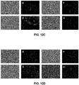


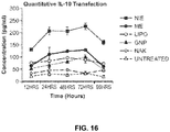


Classifications
-
- A—HUMAN NECESSITIES
- A61—MEDICAL OR VETERINARY SCIENCE; HYGIENE
- A61K—PREPARATIONS FOR MEDICAL, DENTAL OR TOILETRY PURPOSES
- A61K9/00—Medicinal preparations characterised by special physical form
- A61K9/10—Dispersions; Emulsions
- A61K9/107—Emulsions ; Emulsion preconcentrates; Micelles
- A61K9/113—Multiple emulsions, e.g. oil-in-water-in-oil
-
- A—HUMAN NECESSITIES
- A61—MEDICAL OR VETERINARY SCIENCE; HYGIENE
- A61K—PREPARATIONS FOR MEDICAL, DENTAL OR TOILETRY PURPOSES
- A61K38/00—Medicinal preparations containing peptides
- A61K38/16—Peptides having more than 20 amino acids; Gastrins; Somatostatins; Melanotropins; Derivatives thereof
- A61K38/17—Peptides having more than 20 amino acids; Gastrins; Somatostatins; Melanotropins; Derivatives thereof from animals; from humans
- A61K38/19—Cytokines; Lymphokines; Interferons
- A61K38/191—Tumor necrosis factors [TNF], e.g. lymphotoxin [LT], i.e. TNF-beta
-
- A—HUMAN NECESSITIES
- A61—MEDICAL OR VETERINARY SCIENCE; HYGIENE
- A61K—PREPARATIONS FOR MEDICAL, DENTAL OR TOILETRY PURPOSES
- A61K38/00—Medicinal preparations containing peptides
- A61K38/16—Peptides having more than 20 amino acids; Gastrins; Somatostatins; Melanotropins; Derivatives thereof
- A61K38/17—Peptides having more than 20 amino acids; Gastrins; Somatostatins; Melanotropins; Derivatives thereof from animals; from humans
- A61K38/19—Cytokines; Lymphokines; Interferons
- A61K38/21—Interferons [IFN]
- A61K38/217—IFN-gamma
-
- A—HUMAN NECESSITIES
- A61—MEDICAL OR VETERINARY SCIENCE; HYGIENE
- A61K—PREPARATIONS FOR MEDICAL, DENTAL OR TOILETRY PURPOSES
- A61K39/00—Medicinal preparations containing antigens or antibodies
- A61K39/39—Medicinal preparations containing antigens or antibodies characterised by the immunostimulating additives, e.g. chemical adjuvants
-
- A—HUMAN NECESSITIES
- A61—MEDICAL OR VETERINARY SCIENCE; HYGIENE
- A61K—PREPARATIONS FOR MEDICAL, DENTAL OR TOILETRY PURPOSES
- A61K47/00—Medicinal preparations characterised by the non-active ingredients used, e.g. carriers or inert additives; Targeting or modifying agents chemically bound to the active ingredient
- A61K47/50—Medicinal preparations characterised by the non-active ingredients used, e.g. carriers or inert additives; Targeting or modifying agents chemically bound to the active ingredient the non-active ingredient being chemically bound to the active ingredient, e.g. polymer-drug conjugates
- A61K47/69—Medicinal preparations characterised by the non-active ingredients used, e.g. carriers or inert additives; Targeting or modifying agents chemically bound to the active ingredient the non-active ingredient being chemically bound to the active ingredient, e.g. polymer-drug conjugates the conjugate being characterised by physical or galenical forms, e.g. emulsion, particle, inclusion complex, stent or kit
- A61K47/6921—Medicinal preparations characterised by the non-active ingredients used, e.g. carriers or inert additives; Targeting or modifying agents chemically bound to the active ingredient the non-active ingredient being chemically bound to the active ingredient, e.g. polymer-drug conjugates the conjugate being characterised by physical or galenical forms, e.g. emulsion, particle, inclusion complex, stent or kit the form being a particulate, a powder, an adsorbate, a bead or a sphere
- A61K47/6925—Medicinal preparations characterised by the non-active ingredients used, e.g. carriers or inert additives; Targeting or modifying agents chemically bound to the active ingredient the non-active ingredient being chemically bound to the active ingredient, e.g. polymer-drug conjugates the conjugate being characterised by physical or galenical forms, e.g. emulsion, particle, inclusion complex, stent or kit the form being a particulate, a powder, an adsorbate, a bead or a sphere the form being a microcapsule, nanocapsule, microbubble or nanobubble
-
- A—HUMAN NECESSITIES
- A61—MEDICAL OR VETERINARY SCIENCE; HYGIENE
- A61K—PREPARATIONS FOR MEDICAL, DENTAL OR TOILETRY PURPOSES
- A61K47/00—Medicinal preparations characterised by the non-active ingredients used, e.g. carriers or inert additives; Targeting or modifying agents chemically bound to the active ingredient
- A61K47/50—Medicinal preparations characterised by the non-active ingredients used, e.g. carriers or inert additives; Targeting or modifying agents chemically bound to the active ingredient the non-active ingredient being chemically bound to the active ingredient, e.g. polymer-drug conjugates
- A61K47/69—Medicinal preparations characterised by the non-active ingredients used, e.g. carriers or inert additives; Targeting or modifying agents chemically bound to the active ingredient the non-active ingredient being chemically bound to the active ingredient, e.g. polymer-drug conjugates the conjugate being characterised by physical or galenical forms, e.g. emulsion, particle, inclusion complex, stent or kit
- A61K47/6921—Medicinal preparations characterised by the non-active ingredients used, e.g. carriers or inert additives; Targeting or modifying agents chemically bound to the active ingredient the non-active ingredient being chemically bound to the active ingredient, e.g. polymer-drug conjugates the conjugate being characterised by physical or galenical forms, e.g. emulsion, particle, inclusion complex, stent or kit the form being a particulate, a powder, an adsorbate, a bead or a sphere
- A61K47/6927—Medicinal preparations characterised by the non-active ingredients used, e.g. carriers or inert additives; Targeting or modifying agents chemically bound to the active ingredient the non-active ingredient being chemically bound to the active ingredient, e.g. polymer-drug conjugates the conjugate being characterised by physical or galenical forms, e.g. emulsion, particle, inclusion complex, stent or kit the form being a particulate, a powder, an adsorbate, a bead or a sphere the form being a solid microparticle having no hollow or gas-filled cores
- A61K47/6929—Medicinal preparations characterised by the non-active ingredients used, e.g. carriers or inert additives; Targeting or modifying agents chemically bound to the active ingredient the non-active ingredient being chemically bound to the active ingredient, e.g. polymer-drug conjugates the conjugate being characterised by physical or galenical forms, e.g. emulsion, particle, inclusion complex, stent or kit the form being a particulate, a powder, an adsorbate, a bead or a sphere the form being a solid microparticle having no hollow or gas-filled cores the form being a nanoparticle, e.g. an immuno-nanoparticle
- A61K47/6931—Medicinal preparations characterised by the non-active ingredients used, e.g. carriers or inert additives; Targeting or modifying agents chemically bound to the active ingredient the non-active ingredient being chemically bound to the active ingredient, e.g. polymer-drug conjugates the conjugate being characterised by physical or galenical forms, e.g. emulsion, particle, inclusion complex, stent or kit the form being a particulate, a powder, an adsorbate, a bead or a sphere the form being a solid microparticle having no hollow or gas-filled cores the form being a nanoparticle, e.g. an immuno-nanoparticle the material constituting the nanoparticle being a polymer
- A61K47/6935—Medicinal preparations characterised by the non-active ingredients used, e.g. carriers or inert additives; Targeting or modifying agents chemically bound to the active ingredient the non-active ingredient being chemically bound to the active ingredient, e.g. polymer-drug conjugates the conjugate being characterised by physical or galenical forms, e.g. emulsion, particle, inclusion complex, stent or kit the form being a particulate, a powder, an adsorbate, a bead or a sphere the form being a solid microparticle having no hollow or gas-filled cores the form being a nanoparticle, e.g. an immuno-nanoparticle the material constituting the nanoparticle being a polymer the polymer being obtained otherwise than by reactions involving carbon to carbon unsaturated bonds, e.g. polyesters, polyamides or polyglycerol
-
- A—HUMAN NECESSITIES
- A61—MEDICAL OR VETERINARY SCIENCE; HYGIENE
- A61K—PREPARATIONS FOR MEDICAL, DENTAL OR TOILETRY PURPOSES
- A61K49/00—Preparations for testing in vivo
- A61K49/001—Preparation for luminescence or biological staining
- A61K49/0013—Luminescence
- A61K49/0017—Fluorescence in vivo
- A61K49/0019—Fluorescence in vivo characterised by the fluorescent group, e.g. oligomeric, polymeric or dendritic molecules
- A61K49/0021—Fluorescence in vivo characterised by the fluorescent group, e.g. oligomeric, polymeric or dendritic molecules the fluorescent group being a small organic molecule
- A61K49/0041—Xanthene dyes, used in vivo, e.g. administered to a mice, e.g. rhodamines, rose Bengal
-
- A—HUMAN NECESSITIES
- A61—MEDICAL OR VETERINARY SCIENCE; HYGIENE
- A61K—PREPARATIONS FOR MEDICAL, DENTAL OR TOILETRY PURPOSES
- A61K49/00—Preparations for testing in vivo
- A61K49/001—Preparation for luminescence or biological staining
- A61K49/0013—Luminescence
- A61K49/0017—Fluorescence in vivo
- A61K49/0019—Fluorescence in vivo characterised by the fluorescent group, e.g. oligomeric, polymeric or dendritic molecules
- A61K49/0045—Fluorescence in vivo characterised by the fluorescent group, e.g. oligomeric, polymeric or dendritic molecules the fluorescent agent being a peptide or protein used for imaging or diagnosis in vivo
- A61K49/0047—Green fluorescent protein [GFP]
-
- A—HUMAN NECESSITIES
- A61—MEDICAL OR VETERINARY SCIENCE; HYGIENE
- A61K—PREPARATIONS FOR MEDICAL, DENTAL OR TOILETRY PURPOSES
- A61K49/00—Preparations for testing in vivo
- A61K49/001—Preparation for luminescence or biological staining
- A61K49/0013—Luminescence
- A61K49/0017—Fluorescence in vivo
- A61K49/005—Fluorescence in vivo characterised by the carrier molecule carrying the fluorescent agent
- A61K49/0054—Macromolecular compounds, i.e. oligomers, polymers, dendrimers
-
- A—HUMAN NECESSITIES
- A61—MEDICAL OR VETERINARY SCIENCE; HYGIENE
- A61K—PREPARATIONS FOR MEDICAL, DENTAL OR TOILETRY PURPOSES
- A61K49/00—Preparations for testing in vivo
- A61K49/001—Preparation for luminescence or biological staining
- A61K49/0063—Preparation for luminescence or biological staining characterised by a special physical or galenical form, e.g. emulsions, microspheres
- A61K49/0069—Preparation for luminescence or biological staining characterised by a special physical or galenical form, e.g. emulsions, microspheres the agent being in a particular physical galenical form
- A61K49/0076—Preparation for luminescence or biological staining characterised by a special physical or galenical form, e.g. emulsions, microspheres the agent being in a particular physical galenical form dispersion, suspension, e.g. particles in a liquid, colloid, emulsion
-
- A—HUMAN NECESSITIES
- A61—MEDICAL OR VETERINARY SCIENCE; HYGIENE
- A61K—PREPARATIONS FOR MEDICAL, DENTAL OR TOILETRY PURPOSES
- A61K49/00—Preparations for testing in vivo
- A61K49/001—Preparation for luminescence or biological staining
- A61K49/0063—Preparation for luminescence or biological staining characterised by a special physical or galenical form, e.g. emulsions, microspheres
- A61K49/0069—Preparation for luminescence or biological staining characterised by a special physical or galenical form, e.g. emulsions, microspheres the agent being in a particular physical galenical form
- A61K49/0089—Particulate, powder, adsorbate, bead, sphere
- A61K49/0091—Microparticle, microcapsule, microbubble, microsphere, microbead, i.e. having a size or diameter higher or equal to 1 micrometer
- A61K49/0093—Nanoparticle, nanocapsule, nanobubble, nanosphere, nanobead, i.e. having a size or diameter smaller than 1 micrometer, e.g. polymeric nanoparticle
-
- A—HUMAN NECESSITIES
- A61—MEDICAL OR VETERINARY SCIENCE; HYGIENE
- A61K—PREPARATIONS FOR MEDICAL, DENTAL OR TOILETRY PURPOSES
- A61K9/00—Medicinal preparations characterised by special physical form
- A61K9/14—Particulate form, e.g. powders, Processes for size reducing of pure drugs or the resulting products, Pure drug nanoparticles
- A61K9/141—Intimate drug-carrier mixtures characterised by the carrier, e.g. ordered mixtures, adsorbates, solid solutions, eutectica, co-dried, co-solubilised, co-kneaded, co-milled, co-ground products, co-precipitates, co-evaporates, co-extrudates, co-melts; Drug nanoparticles with adsorbed surface modifiers
- A61K9/145—Intimate drug-carrier mixtures characterised by the carrier, e.g. ordered mixtures, adsorbates, solid solutions, eutectica, co-dried, co-solubilised, co-kneaded, co-milled, co-ground products, co-precipitates, co-evaporates, co-extrudates, co-melts; Drug nanoparticles with adsorbed surface modifiers with organic compounds
-
- A—HUMAN NECESSITIES
- A61—MEDICAL OR VETERINARY SCIENCE; HYGIENE
- A61K—PREPARATIONS FOR MEDICAL, DENTAL OR TOILETRY PURPOSES
- A61K9/00—Medicinal preparations characterised by special physical form
- A61K9/48—Preparations in capsules, e.g. of gelatin, of chocolate
- A61K9/50—Microcapsules having a gas, liquid or semi-solid filling; Solid microparticles or pellets surrounded by a distinct coating layer, e.g. coated microspheres, coated drug crystals
- A61K9/5005—Wall or coating material
- A61K9/5021—Organic macromolecular compounds
- A61K9/5031—Organic macromolecular compounds obtained otherwise than by reactions only involving carbon-to-carbon unsaturated bonds, e.g. polyethylene glycol, poly(lactide-co-glycolide)
-
- A—HUMAN NECESSITIES
- A61—MEDICAL OR VETERINARY SCIENCE; HYGIENE
- A61K—PREPARATIONS FOR MEDICAL, DENTAL OR TOILETRY PURPOSES
- A61K9/00—Medicinal preparations characterised by special physical form
- A61K9/48—Preparations in capsules, e.g. of gelatin, of chocolate
- A61K9/50—Microcapsules having a gas, liquid or semi-solid filling; Solid microparticles or pellets surrounded by a distinct coating layer, e.g. coated microspheres, coated drug crystals
- A61K9/51—Nanocapsules; Nanoparticles
- A61K9/5107—Excipients; Inactive ingredients
- A61K9/513—Organic macromolecular compounds; Dendrimers
- A61K9/5161—Polysaccharides, e.g. alginate, chitosan, cellulose derivatives; Cyclodextrin
-
- A—HUMAN NECESSITIES
- A61—MEDICAL OR VETERINARY SCIENCE; HYGIENE
- A61P—SPECIFIC THERAPEUTIC ACTIVITY OF CHEMICAL COMPOUNDS OR MEDICINAL PREPARATIONS
- A61P1/00—Drugs for disorders of the alimentary tract or the digestive system
-
- A—HUMAN NECESSITIES
- A61—MEDICAL OR VETERINARY SCIENCE; HYGIENE
- A61P—SPECIFIC THERAPEUTIC ACTIVITY OF CHEMICAL COMPOUNDS OR MEDICINAL PREPARATIONS
- A61P1/00—Drugs for disorders of the alimentary tract or the digestive system
- A61P1/04—Drugs for disorders of the alimentary tract or the digestive system for ulcers, gastritis or reflux esophagitis, e.g. antacids, inhibitors of acid secretion, mucosal protectants
-
- A—HUMAN NECESSITIES
- A61—MEDICAL OR VETERINARY SCIENCE; HYGIENE
- A61P—SPECIFIC THERAPEUTIC ACTIVITY OF CHEMICAL COMPOUNDS OR MEDICINAL PREPARATIONS
- A61P19/00—Drugs for skeletal disorders
- A61P19/02—Drugs for skeletal disorders for joint disorders, e.g. arthritis, arthrosis
-
- A—HUMAN NECESSITIES
- A61—MEDICAL OR VETERINARY SCIENCE; HYGIENE
- A61P—SPECIFIC THERAPEUTIC ACTIVITY OF CHEMICAL COMPOUNDS OR MEDICINAL PREPARATIONS
- A61P29/00—Non-central analgesic, antipyretic or antiinflammatory agents, e.g. antirheumatic agents; Non-steroidal antiinflammatory drugs [NSAID]
-
- A—HUMAN NECESSITIES
- A61—MEDICAL OR VETERINARY SCIENCE; HYGIENE
- A61P—SPECIFIC THERAPEUTIC ACTIVITY OF CHEMICAL COMPOUNDS OR MEDICINAL PREPARATIONS
- A61P35/00—Antineoplastic agents
-
- A—HUMAN NECESSITIES
- A61—MEDICAL OR VETERINARY SCIENCE; HYGIENE
- A61P—SPECIFIC THERAPEUTIC ACTIVITY OF CHEMICAL COMPOUNDS OR MEDICINAL PREPARATIONS
- A61P37/00—Drugs for immunological or allergic disorders
-
- A—HUMAN NECESSITIES
- A61—MEDICAL OR VETERINARY SCIENCE; HYGIENE
- A61P—SPECIFIC THERAPEUTIC ACTIVITY OF CHEMICAL COMPOUNDS OR MEDICINAL PREPARATIONS
- A61P43/00—Drugs for specific purposes, not provided for in groups A61P1/00-A61P41/00
-
- A—HUMAN NECESSITIES
- A61—MEDICAL OR VETERINARY SCIENCE; HYGIENE
- A61P—SPECIFIC THERAPEUTIC ACTIVITY OF CHEMICAL COMPOUNDS OR MEDICINAL PREPARATIONS
- A61P9/00—Drugs for disorders of the cardiovascular system
- A61P9/10—Drugs for disorders of the cardiovascular system for treating ischaemic or atherosclerotic diseases, e.g. antianginal drugs, coronary vasodilators, drugs for myocardial infarction, retinopathy, cerebrovascula insufficiency, renal arteriosclerosis
-
- B—PERFORMING OPERATIONS; TRANSPORTING
- B82—NANOTECHNOLOGY
- B82Y—SPECIFIC USES OR APPLICATIONS OF NANOSTRUCTURES; MEASUREMENT OR ANALYSIS OF NANOSTRUCTURES; MANUFACTURE OR TREATMENT OF NANOSTRUCTURES
- B82Y5/00—Nanobiotechnology or nanomedicine, e.g. protein engineering or drug delivery
-
- A—HUMAN NECESSITIES
- A61—MEDICAL OR VETERINARY SCIENCE; HYGIENE
- A61K—PREPARATIONS FOR MEDICAL, DENTAL OR TOILETRY PURPOSES
- A61K39/00—Medicinal preparations containing antigens or antibodies
- A61K2039/555—Medicinal preparations containing antigens or antibodies characterised by a specific combination antigen/adjuvant
- A61K2039/55511—Organic adjuvants
- A61K2039/55555—Liposomes; Vesicles, e.g. nanoparticles; Spheres, e.g. nanospheres; Polymers
-
- A—HUMAN NECESSITIES
- A61—MEDICAL OR VETERINARY SCIENCE; HYGIENE
- A61K—PREPARATIONS FOR MEDICAL, DENTAL OR TOILETRY PURPOSES
- A61K9/00—Medicinal preparations characterised by special physical form
- A61K9/48—Preparations in capsules, e.g. of gelatin, of chocolate
- A61K9/50—Microcapsules having a gas, liquid or semi-solid filling; Solid microparticles or pellets surrounded by a distinct coating layer, e.g. coated microspheres, coated drug crystals
- A61K9/51—Nanocapsules; Nanoparticles
- A61K9/5107—Excipients; Inactive ingredients
- A61K9/513—Organic macromolecular compounds; Dendrimers
- A61K9/5169—Proteins, e.g. albumin, gelatin
Definitions
- This disclosure is in the field of medicine, and more specifically relates to a multi-compartmental nanoparticulate system for diagnosing, monitoring, and treating inflammation and/or disease. These systems are capable of targeting specific cell types, co-administration of incompatible materials, as well as temporal control of payload release.
- Macrophages are important immune cells in the body that regulate different biological processes involved in inflammatory and infectious diseases. Macrophages originate from the mononuclear phagocyte system (MPS) in the bone marrow.
- the MPS system is comprised of monocytes and tissue macrophages in their various forms.
- the primary function of monocytes is phagocytosis.
- monocytes Upon exiting the bone marrow, monocytes circulate in the blood and become activated in various tissues. Once the monocyte reaches the extravascular tissue, it matures into a larger phagocytic cell called a macrophage. They reside in the liver (Kupffer cells), lungs (alveolar macrophages), spleen, lymph nodes, thymus, gut, marrow, brain, connective tissue and serous cavities.
- macrophages are converted from the rested to an activated state.
- the activated cells have an enhanced capability to attack and kill infecting pathogens and tumor cells.
- the active state is marked by an increase in size and the development of more psuedopods (cellular projections).
- the cytokines released from T-lymphocytes such as interferon-gamma (IFN- ⁇ ), tumor necrosis factor-alpha (TNF- ⁇ ) and granulocyte-monocyte colony stimulating factor (GM-CSF) can act as signals to activate macrophages.
- the activating signal can come from microbial products such as lipopolysaccharides, immune complexes, chemical mediators, and extracellular matrix proteins including fibronectin.
- WO2009/051837 discloses compositions and systems for delivery of nanocarriers to cells of the immune system.
- vaccine nanocarriers capable of stimulating an immune response in T cells and/or in B cells, in some embodiments, comprising at least one immunomodulatory agent, and optionally comprising at least one targeting moiety and optionally at least one immunostimulatory agent.
- the present invention provides a multicompartment delivery system for use in imaging a tissue, the multicompartment delivery system comprising one or more inner aqueous compartments encapsulated within an outer hydrophobic compartment, the outer hydrophobic compartment encapsulated in an exterior aqueous compartment, the one or more inner aqueous compartments comprising a polymeric carrier, the outer hydrophobic compartment comprising an oil, the polymeric carrier encapsulating an imaging agent.
- the present invention provides a multicompartment delivery system for use in treating a disease comprising one or more inner aqueous compartments encapsulated within an outer hydrophobic compartment, the outer hydrophobic compartment encapsulated in an exterior aqueous compartment, the one or more inner aqueous compartments comprising a polymeric carrier, the outer hydrophobic compartment comprising an oil, the polymeric carrier encapsulating a first therapeutic agent, and the inner aqueous compartment further comprising a target-specific binding agent attached to the polymeric carrier.
- the multicompartmental delivery systems described herein allow for the administration of one or more agents such as, but not limited to, imaging agents, diagnostic agents, therapeutic agents, vaccines, proteins, gene constructs, and small molecules. These systems allow for co-administration of incompatible agents (i.e., hydrophobic and hydrophilic agents), as well as temporal control over the release of administered agents.
- agents such as, but not limited to, imaging agents, diagnostic agents, therapeutic agents, vaccines, proteins, gene constructs, and small molecules.
- incompatible agents i.e., hydrophobic and hydrophilic agents
- the invention is as defined in the present claims. This disclosure is related in part to a composition of matter for safe and efficient delivery of image contrast agents, drugs, genes, small interfering RNA (siRNA), and vaccination molecules to macrophages upon administration in the body by oral, nasal, inhalation, injection or any other route known to those skilled in the art.
- image contrast agents drugs, genes, small interfering RNA (siRNA), and vaccination molecules to macrophages upon administration in the body by oral, nasal, inhalation, injection or any other route known to those skilled in the art.
- the instant disclosure is directed towards a multicompartment delivery system comprising one or more inner aqueous compartments encapsulated within an outer hydrophobic compartment, the outer hydrophobic compartment being surrounded by an exterior aqueous compartment.
- an imaging agent is encapsulated in a polymeric carrier disposed within the one or more inner aqueous compartmentsln an embodiment, a second agent is encapsulated within the outer hydrophobic compartment.
- Encapsulated is defined herein to include dissolved in a material, associated with a material, meaning that an agent or nanoparticle can be on the surface, within the material, or surrounded by the material that comprises the compartment.
- the second agent can be selected from the group consisting of an imaging agent, a diagnostic agent, a therapeutic agent, a vaccine, a protein, or a gene construct.
- the multicompartment delivery system disclosed herein can also comprise an outer compartment that forms a microsphere.
- Methods of producing a microsphere are known in the art and include the single emulsion method and solvent displacement method (see, e.g., Furtado Mosqueira et al. Journal of Pharmaceutical Sciences Special Issue: Papers from the 1999 Macromolecular Drug Delivery Conference (2000) 89(5): 614-626 ; Rosca et al. Journal of Controlled Release (2004) 99(2): 271-280 ).
- the microsphere can be about 0.5 to 500 microns in size.
- the inner aqueous compartment is about 50 nm to about 300 nm.
- the inner aqueous compartment comprises nanoparticles.
- the nanoparticles comprise gelatin or alginate.
- the outer hydrophobic compartment comprises safflower oil.
- one or more target-specific binding agents are attached to the surface of the outer hydrophobic compartment.
- the one or more target-specific binding agents attached to the surface of the outer hydrophobic compartment is tuftsin. Methods of attaching binding agents to a particle or carrier are known in the art ( see, e.g., Magadala et al. AAPS J. (2008) 10(4): 565-576 ).
- the exterior aqueous compartment comprises water.
- the multicompartment delivery system further comprising one or more target-specific binding agents attached to the surface of the inner aqueous compartment or the outer hydrophobic compartment.
- one or more target-specific binding agents are attached to the inner aqueous compartment and the outer hydrophobic compartment.
- Further embodiments further comprise one or more target-specific binding agents attached to the inner aqueous compartment, direct the inner compartment to an intracellular location and the one or more target-specific binding agents attached to the outer hydrophobic compartment direct the microsphere to a cell.
- the delivery vehicles disclosed herein are specifically designed to have multiple compartments (multicompartment) to afford the following two advantages: (1) ability to sequester the payload in each of the compartment due to incompatibility (e.g., poor solubility), instability, or desire for temporal-controlled release and delivery; and (2) ability to overcome the different tissue, cell, and intracellular barriers to afford efficient delivery of the payload to the desired biological target site.
- incompatibility e.g., poor solubility
- instability e.g., instability
- siRNA siRNA
- the multi-compartmental delivery systems are designed to meet the needs of the payload (e.g., hydrophobic versus hydrophilic), the site of delivery in the body, the intended application (e.g., drugs versus vaccines), and the disease of interest.
- the nanoparticles can be from about 20 nanometers ("nm") to about 300 nanometers; from about 20 nm to about 30 nm; from about 30 nm to about 40 nm; from about 40 nm to about 50 nm; from 50 nm to about 100 nm; from about 100 nm to about 150 nm; from about 150 nm to about 200 nm; from about 200 nm to about 250 nm; and from about 250 to about 300 nm.
- nm nanometers
- the multicompartment delivery systems disclosed herein has more than two distinct compartments ( Figure 2 ).
- the nanoparticles are solid and hydrophilic.
- the nanoparticles can be encapsulated in a hydrophobic microsphere, which forms an outer hydrophobic compartment.
- the hydrophobic microsphere is composed of oil, such as safflower oil or any other biologically compatible oilThe microsphere is surrounded by water.
- the multicompartment delivery systems of the present disclosure can also be modified by target-specific binding agents that target the delivery systems to a cell or tissue.
- target-specific binding agents are antibodies, Fab fragments and other antibody binding fragments, ligands, peptides, small molecules, and peptidomimetic compounds.
- the target-specific binding agent is Tuftsin.
- the target-specific binding agent is arginine-glycine-aspartate ("RGD") tripeptide.
- the multicompartment delivery systems are targeted to macrophages using specific peptides functionalized on the surface of the outer compartment. For instance, the outer compartment of the multicompartmental delivery system can have target-specific binding agents attached to its surface.
- the multicompartment delivery systems encapsulate therapeutic or imaging agents.
- the agent can promote pro- or anti-inflammatory properties (e.g., ovalbumin for pro-inflammation and immune-adjuvant response or omega-3 fatty acid-containing oils for anti-inflammatory response) to further enhance the therapeutic efficiency of the system.
- the agents can be therapeutic agents such as chemotherapeutics, radioisotopes, antibiotics, peptides, vaccines, small molecules, proteins, and antibody therapeutics.
- the therapeutic agents can be nucleic acids used for gene therapy.
- the therapeutic agents are DNA molecules. Agents can be siRNA molecules delivered to silence genes in specific tissues.
- One of the many advantages of the multicompartmental delivery systems disclosed herein is they utilize a compartmentalized delivery system which can be used to shield payloads prone to degradation until they have reached the target of interest. This is important for successful gene therapy, as macrophages, a preferred target cell, contain large amounts of nucleases within their lysosomes, making it likely that without the protection of these disclosed systems, that the DNA payload delivered to a cell would be degraded.
- the present technology also provides a multicompartment delivery system for use in imaging a tissue.
- the imaging agent is colloidal gold, iron-oxide crystals, quantum dots, fluorophores, radiolabels, X-ray contrast agents, and positron emission tomographic agents.
- the therapeutic agent is selected from the group consisting of chemotherapeutics, antibiotics, antivirals, small molecules, siRNA, and DNA.
- the outer compartment encapsulates a second agent.
- the second agent is an imaging agent.
- This imaging agent can be selected from the group consisting of fluorophores, radiolabels, X-ray contrast agents, and positron emission tomographic agents.
- the second agent is a therapeutic agent.
- the outer compartment or inner compartment of the delivery vehicle can encapsulate the one or more imaging agents.
- the term "encapsulate" means to enclose within or associate with the polymeric carrier. The encapsulation can include association of a molecule or compound at the surface of the polymeric carrier or within the polymeric carrier.
- imaging agents include fluorophores, radiolabels, X-ray contrast, RAMAN spectroscopy agents, and positron emission tomography agents.
- fluorophores include rhodamine, fluorescein, isothiocyanate, phycoerythrin, phycocyanin, fluorescamine, and metal chelates.
- radiolabels include 3 H , 123 I, 125 I, 131 I, 32 P, 35 S, 14 C, 51 Cr, 36 Cl, 57 Co, 55 Fe, 59 Fe, 90 Y, 99 mTc (metastable isomer of Technetium 99), and 75 Se.
- X-ray contrast agents include Diatrizoate (Hypaque 50), Metrizoate (Isopaque 370), Ioxaglate (Hexabrix), Iopamidol (Isovue 370), Iohexol (Omnipaque 350), Ioxilan (Oxilan 350), Iopromide (Ultravist 370), and Iodixanol (Visipaque 320).
- Other agents include 2,2,6,6-tetramethylpiperidine-1-oxyl (TEMPO) group, DID fluorescent dye analogs, and perdeuterated alkyl or aryl phosphonium groups.
- the solvent displacement method has been used to encapsulating nucleic acids, small molecules, and peptides (see, e.g., Oster et al. Journal of Controlled Release (2006) 111(3): 371-381 ; Ribeiro et al. Food Hydrocolloids 22(1): 12-17 ).
- the second agent is selected from the group consisting of chemotherapeutics, antibiotics, antivirals, small molecules, anti-inflammatory agents, siRNA, and DNA.
- the one or more inner compartments comprises a first material, which is poly(epsilon-caprolactone).
- the outer compartment comprises a second material, which is safflower oil.
- a target-specific binding agent is attached to the surface of the outer compartment.
- the target-specific binding agent is selected from the group consisting of antibodies and binding fragments thereof, peptide sequences, and ligands and binding fragments thereof.
- the target-specific binding agent is an EGFR-specific antibody.
- the target-specific binding agents bind to alpha-V-beta III integrin receptor.
- Derivatization of organic polymers can be performed to expose selected reactive groups at their surface( see , e.g., Townsend, S. A, et al. Biomaterials (2007) 28(34): 5176-5184 ; Weiss, B., et al. Bioconjugate Chemistry (2007) 18(4): 1087-1094 ; and Gindy, M. E., et al. PMSE Preprints (2006) 95:989-990 ).
- target-specific binding agents such as proteins, peptides, antibodies, and small molecules
- a target-specific binding agent can be a small molecule that binds a target, ligand, peptide (e.g ., arginine-glycine-aspartate ("RGD") tripeptide), protein, antibody, Fab fragment, F(ab')2 fragments, and peptidomimetic compounds.
- exemplary targets include receptors (e.g ., alpha-V-beta III integrin receptor and EGFR receptor), phospholipids, glycoproteins, and proteins.
- the instant disclosure also provides a multicompartment delivery system for use in treating a disease.
- the disease is selected from the group consisting of atherosclerosis, Crohn's disease, inflammatory bowel disease, rheumatoid arthritis and cancer.
- the one or more inner compartment is in the size range of about 10 nm to about 30 nm.
- the one or more inner compartments or the outer compartments encapsulate an imaging agent.
- the imaging agent is selected from the group consisting of colloidal gold, iron-oxide crystals, quantum dots, fluorophores, radiolabels, X-ray contrast agents, and positron emission tomographic agents.
- the therapeutic agent is selected from the group consisting of chemotherapeutics, anti-inflammatories, fatty acids, antibiotics, antivirals, small molecules, siRNA, and DNA.
- the second therapeutic agent is selected from the group consisting of chemotherapeutics, anti-inflammatories, fatty acids, antibiotics, antivirals, small molecules, siRNA, and DNA.
- the second therapeutic agent is encapsulated by the outer compartment. In still other aspects, the second therapeutic agent is encapsulated by the inner compartment.
- the one or more inner compartments comprises a first material which is poly(epsilon-caprolactone).
- the outer compartment comprises a second material which is safflower oil.
- a target-specific binding agent is attached to the surface of the outer compartment.
- the target-specific binding agent is selected from the group consisting of antibodies and binding fragments thereof and peptide sequences.
- the target-specific binding agent is an EGFR-specific antibody.
- the target-specific binding agents bind to alpha-V-beta III integrin receptor.
- the first agent is a gene construct.
- the methods further comprise expressing the gene construct in the target tissue or cell.
- the gene construct encodes IL-10.
- the gene construct causes apoptosis in the target tissue or cells.
- the gene construct is siRNA.
- the target cell is a macrophage.
- Toxicity and therapeutic efficacy of such compounds can be determined by standard pharmaceutical procedures in cell cultures or experimental animals, e.g., for determining the LD 50 (The Dose Lethal To 50% Of The Population) And The ED 50 (the dose therapeutically effective in 50% of the population).
- the dose ratio between toxic and therapeutic effects is the therapeutic index and it can be expressed as the ratio LD 50 /ED 50 .
- Compounds that exhibit large therapeutic induces are preferred. While compounds that exhibit toxic side effects may be used, care should be taken to design a delivery system that targets such compounds to the site of affected tissue in order to minimize potential damage to uninfected cells and, thereby, reduce side effects.
- the data obtained from the cell culture assays and animal studies can be used in formulating a range of dosage for use in humans.
- the dosage of such compounds lies preferably within a range of circulating concentrations that include the ED 50 with little or no toxicity.
- the dosage may vary within this range depending upon the dosage form employed and the route of administration utilized.
- the therapeutically effective dose can be estimated initially from cell culture assays.
- a dose may be formulated in animal models to achieve a circulating plasma concentration range that includes the IC 50 (i.e., the concentration of the test compound which achieves a half-maximal inhibition of symptoms) as determined in cell culture.
- IC 50 i.e., the concentration of the test compound which achieves a half-maximal inhibition of symptoms
- levels in plasma may be measured, for example, by high performance liquid chromatography.
- compositions for use in accordance with the disclosed systems may be formulated in conventional manner using one or more physiologically acceptable carriers or excipients.
- the compounds and their physiologically acceptable salts and solvates may be formulated for administration by, for example, injection, inhalation or insulation (either through the mouth or the nose) or oral, buccal, parenteral or rectal administration.
- the compounds can be formulated for a variety of loads of administration, including systemic and topical or localized administration. Techniques and formulations generally may be found in Remington's Pharmaceutical Sciences, Meade Publishing Co., Easton, Pa .
- systemic administration injection is preferred, including intramuscular, intravenous, intraperitoneal, and subcutaneous.
- the compounds can be formulated in liquid solutions, preferably in physiologically compatible buffers such as Hank's solution or Ringer's solution.
- the compounds may be formulated in solid form and redissolved or suspended immediately prior to use. Lyophilized forms are also included.
- the pharmaceutical compositions may take the form of, for example, tablets or capsules prepared by conventional means with pharmaceutically acceptable excipients such as binding agents (e.g ., pregelatinised maize starch, polyvinylpyrrolidone or hydroxypropyl methylcellulose); fillers ( e.g., lactose, microcrystalline cellulose or calcium hydrogen phosphate); lubricants ( e.g., magnesium stearate, talc or silica); disintegrants ( e.g., potato starch or sodium starch glycolate); or wetting agents (e.g ., sodium lauryl sulfate).
- binding agents e.g ., pregelatinised maize starch, polyvinylpyrrolidone or hydroxypropyl methylcellulose
- fillers e.g., lactose, microcrystalline cellulose or calcium hydrogen phosphate
- lubricants e.g., magnesium stearate, talc or silica
- disintegrants e
- Liquid preparations for oral administration may take the form of, for example, solutions, syrups or suspensions, or they may be presented as a dry product for constitution with water or other suitable vehicle before use.
- Such liquid preparations may be prepared by conventional means with pharmaceutically acceptable additives such as suspending agents (e.g ., sorbitol syrup, cellulose derivatives or hydrogenated edible fats); emulsifying agents (e.g. , lecithin or acacia); non-aqueous vehicles (e.g., ationd oil, oily esters, ethyl alcohol or fractionated vegetable oils); and preservatives (e.g., methyl or propyl-p-hydroxybenzoates or sorbic acid).
- the preparations may also contain buffer salts, flavoring, coloring and sweetening agents as appropriate.
- Embodiments of the multicompartment systems disclosed herein can be designed for oral delivery to a subject.
- the multicompartment delivery systems can comprise solid alginate and/or gelatin nanoparticles.
- Preparations for oral administration may be suitably formulated to give controlled release of the active compound.
- the compositions may take the form of tablets or lozenges formulated in conventional manner.
- the compounds for use according to the present disclosure are conveniently delivered in the form of an aerosol spray presentation from pressurized packs or a nebuliser, with the use of a suitable propellant, e.g., dichlorodifluoromethane, trichlorofluoromethane, dichlorotetrafluoroethane, carbon dioxide or other suitable gas.
- a suitable propellant e.g., dichlorodifluoromethane, trichlorofluoromethane, dichlorotetrafluoroethane, carbon dioxide or other suitable gas.
- a pressurized aerosol the dosage unit may be determined by providing a valve to deliver a metered amount.
- Capsules and cartridges of e.g., gelatin for use in an inhaler or insufflator may be
- the compounds can be formulated for parenteral administration by injection, e.g., by bolus injection or continuous infusion.
- Formulations for injection may be presented in unit dosage form, e.g., in ampoules or in multi-dose containers, with an added preservative.
- the compositions may take such forms as suspensions, solutions or emulsions in oily or aqueous vehicles, and may contain formulatory agents such as suspending, stabilizing and/or dispersing agents.
- the active ingredient may be in powder form for constitution with a suitable vehicle, e.g., sterile pyrogen-free water, before use.
- the compounds may also be formulated in rectal compositions such as suppositories or retention enemas, e.g., containing conventional suppository bases such as cocoa butter or other glycerides.
- the compounds may also be formulated as a depot preparation. Such long acting formulations may be administered by implantation (for example subcutaneously or intramuscularly) or by intramuscular injection.
- the compounds may be formulated with suitable polymeric or hydrophobic materials (for example as an emulsion in an acceptable oil) or ion exchange resins, or as sparingly soluble derivatives, for example, as a sparingly soluble salt.
- suitable delivery systems include microspheres which offer the possibility of local noninvasive delivery of drugs over an extended period of time. This technology utilizes microspheres of precapillary size which can be injected via a coronary catheter into any selected part of the e.g. heart or other organs without causing inflammation or ischemia. The administered therapeutic is slowly released from these microspheres and taken up by surrounding tissue cells (e.g. endothelial cells).
- Systemic administration can also be by transmucosal or transdermal means.
- penetrants appropriate to the barrier to be permeated are used in the formulation.
- penetrants are generally known in the art, and include, for example, for transmucosal administration bile salts and fusidic acid derivatives. in addition, detergents may be used to facilitate permeation.
- Transmucosal administration may be through nasal sprays or using suppositories.
- oligomers can be formulated into ointments, salves, gels, or creams as generally known in the art.
- a wash solution can be used locally to treat an injury or inflammation to accelerate healing.
- the multicompartment delivery systems in the instant disclosure can be introduced into a patient by any of a number of methods, each of which is familiar in the art.
- the multicompartment delivery systems can be introduced systemically, e.g., by intravenous injection.
- nucleic acid-encapsulated solid nanoparticles were designed that are further encased in the innermost water phase of the water-in-oil-in-water (W/O/W) multiple emulsion to develop nanoparticles-in-emulsion (NiE) multi-compartmental delivery system.
- macrophages behave as a Trojan horse vector comes from the knowledge that micro-organisms, such as bacteria and viruses, use monocytes and macrophages to proliferate and hide from immune-surveillance system.
- micro-organisms such as bacteria and viruses
- monocytes and macrophages to proliferate and hide from immune-surveillance system.
- HIV-1 has the ability to infect and replicate in non-diving cells such as cells of monocyte/macrophage lineage; although CD4+ T-lymphocytes appear to be the primary target of HIV virus.
- the virus may affect the macrophages by influencing the cytokine production profile and this may result in chronic inflammation and extensive tissue damage.
- the infected macrophages appear to be resistant to the toxic effects of the virus and thus serve as a Trojan horse to the HIV virus.
- the systems described in the instant disclosure can be used for a variety of purposes including imaging, diagnostics, and treatment of conditions and diseases.
- the multicompartmental delivery systems can be utilized for gene therapy, gene silencing, cancer treatments, and vaccination.
- the systems described herein can be targeted to any tissue or cell type.
- One such type of cell are macrophages.
- the over or under-activity of macrophages can result in the onset and/or progression of pathologic conditions, including, but not limited to, growth and spread of malignant tumors, sepsis, chronic inflammation in rheumatoid arthritis, lysosomal storage disease, atherosclerosis and major infections including HIV/AIDS and tuberculosis.
- Passive targeting refers to the accumulation of the drug or drug-carrier nanosystem by exploiting the patho-physiological condition and anatomical route.
- nanoparticle systems that are>100 nm are readily opsonized and removed from the circulation by reticuloendothelial system (RES) and degraded by the macrophages in the liver and spleen.
- RES reticuloendothelial system
- the tendency of unmodified nanoparticles to localize in the RES has been exploited as a way to passively deliver the pay load to macrophages.
- Nanoparticles have been observed to passively accumulate at inflamed sites due to the enhanced permeation and retention (EPR) effect. The presence of macrophages at these sites presents an opportunity to passively target these cells.
- EPR enhanced permeation and retention
- Active targeting approaches are based on the surface modification of the systems with an agent (e.g., ligand, antibody, peptide) that has the selective affinity for recognizing and interacting with the particular cell type, tissue, or organ in the body.
- agent e.g., ligand, antibody, peptide
- the phenomenon of active targeting can be achieved by utilizing the presence of various receptors and lipid components on the plasma membrane of cells. Macrophages are a preferred cell type for the systems disclosed herein. These receptors may be uniquely expressed on specific cells or may show a differentially higher expression on diseased cells in comparison to the normal cells. Table 1 below briefly describes some of the key properties of different receptors that are present on the surface of macrophages and can be utilized for actively targeting these cells. These different receptors can be downregulated or upregulated using known gene therapy techniques.
- receptors can be used as targets for multicompartment delivery systems.
- Such systems can have target-specific binding agents attached to the surface of the inner or outer compartment.
- the target-specific binding agents can be ligands for a receptor, peptidomimetic compounds that bind to a receptor, or small molecules capable of binding to a receptor.
- Table 1 Examples of Receptor Systems Utilized for Targeting Macrophages Macrophage-Receptor Targeted Brief Description Mannose Receptor - C-Type lectin - Involved in pathogen recognition - Ligand binding to the receptor is dependent on Ca 2+ concentration - Facilitates receptor-mediated endocytosis Scavenger Receptor - Receptors bind a variety of poly-aniomc macro-molecules and modified (acetylated) LDL - Three classes of receptor are known. S.R-A-B, and C - Scavenger receptors are implicated in pathological deposition of cholesterol during atherogenesis Dectin-1 Receptor - C-Type lectin.
- Tuftsin peptide Tetra-peptide sequence L-Thr-L-Lys-L-Pro-L-Arg (TKPR) (40,41) - Synthesized by the enzymatic processing of CH2 domain of the Fc fragment of the heavy chain of IgG molecule - Activates M ⁇ and enhances their phagocytic ability Hyaluronate - for CD44 receptor (42, 43) -
- the receptor is a glycoprotein and is known to be involved in phagocytosis of large particles
- multiple emulsion globules are hydrophobic, they are readily internalized by intestinal macrophages upon oral administration through the Peyer's patch. Furthermore, when making these multicompartmental delivery systems, they can be generated in sizes that are preferentially taken up by macrophages. Once internalized by macrophages, these cells can travel and deliver the transfected protein in different parts of the body through systemic circulation. Alternatively, this method can also silence critical genes in activated macrophages and promote pro- or anti-inflammatory status as desired for effective disease treatment of disease.
- uptake by macrophages and efficient intracellular delivery also provides an opportunity for vaccination using peptides, proteins, attenuated microorganisms as antigens as well as DNA- and RNA-based vaccines that will require transfection and production of the appropriate human and cellular immune responses.
- macrophages are known to phagocytose and kill various microorganisms, but with the advancement in the field of molecular biology, it has been established that many of these pathogens have developed subtle means for residing in the macrophages by mainly avoiding phagocytosis or developing lysosomal tolerance.
- nanoparticle based drug delivery systems can improve the therapeutic index of such drugs by lowering the toxicity and enhancing the targeting ability to macrophages. See Table 2 for examples of nano-platform systems containing small molecule drugs for targeting macrophages.
- macrophages that have taken up the multicompartmental delivery vehicles of the disclosed herein can be used to carry the payload delivered by said vehicle to a target site within the body, such as a tumor or site of inflammation.
- the payload can be a therapeutic treatment, such as, but not limited to, anti-inflammatory therapeutics, including, but not limited to steroids, small molecule drugs (i.e. bisphosphonates), fatty acids, and antioxidants, as well as chemotherapy, antibiotics, peptides, vaccines, small molecules, proteins, and antibody therapeutics.
- the present methodologies can also treat a range of anti-inflammatory diseases or conditions.
- inflammatory disease or "inflammatory condition” as used herein is meant any disease, condition, trait, genotype or phenotype characterized by an inflammatory or allergic process as is known in the art, such as inflammation, acute inflammation, chronic inflammation, respiratory disease, atherosclerosis, restenosis, asthma, allergic rhinitis, atopic dermatitis, septic shock, rheumatoid arthritis, inflammatory bowl disease, inflammatory pelvic disease, pain, ocular inflammatory disease, celiac disease, Leigh Syndrome, Glycerol Kinase Deficiency, Familial eosinophilia (FE), autosomal recessive spastic ataxia, laryngeal inflammatory disease; Tuberculosis, Chronic cholecystitis, Bronchiectasis, Silicosis and other pneumoconioses, and any other inflammatory disease, condition, trait, genotype or phenotype that can respond to the modulation of disease related gene expression in a cell or
- Biological therapies such as therapeutic gene delivery or gene silencing can be designed to target and exploit macrophages.
- the therapeutic gene can be transferred into the cells, which are then used as "guided missiles.”
- Macrophages are capable of secreting various biological products (see Table 3 below). Depending on the products being secreted, if the process is left unchecked, this can result in tissue damage and fibrosis, which are characteristic of chronic inflammation. As the macrophage is capable of a number of processes in addition to secreting various bio-molecules, it is an ideal target for gene therapy for treatment of a variety of diseases and conditions, including, but not limited to, inflammation, cancer, Crohn's disease, inflammatory bowel disease, and atherosclerosis.
- the targets enumerated in Table 3 can be either up or downregulated depending on the desired result.
- increasing expression of anti-inflammatory cytokines can be accomplished by delivering a gene construct to the cell using the multicompartmental delivery systems disclosed in the instant application. This would result in a reduction of the inflammatory response of the macrophages.
- the anti-inflammatory cytokine to be upregulated is IL-10.
- siRNA can be administered to the cell using the multicompartmental delivery systems disclosed in the instant application.
- the inflammatory cytokine to be downregulated or silenced is TNF- ⁇ .
- Particles such as colloidal gold, iron-oxide crystals, and quantum dots are some of the examples of inorganic nanoparticles that are in the size range of 1-30 nm. Methods of making such nanoparticles are known in the art (see, e.g.,, Luna Martinez et al. Mater. Sci. Forum: Adv. Elec. Microscop. Nanomat. (2010) 644: 51-55 ; Gardea-Torresdey et al. Journal of Nanoparticle Research (1999) 1(3): 397-404 ; Zhou et al. Chem. Mater. (1999) 11 (9): 2310-2312 ).
- MRI magnetic resonance imaging
- PET positron emission tomography
- CT computed tomography
- Macrophages are suitable imaging targets in diseases such as inflammation, atherosclerosis, and rheumatoid arthritis. This is because these cells are present in abundance and also are involved in multiple functions leading to inflammation and angiogenesis. Therefore, monitoring their role in such conditions can provide valuable insight into the progression of a disease.
- Nanoparticles designed to target macrophage cells have the potential to improve the detection and characterization of the human aortic atherosclerosis.
- Specific examples where the efficacy of the nanoparticles as contrast agents and targeting macrophages has been demonstrated in an in vivo animal model will be discussed in the examples of the instant disclosure.
- Table 4 below considers select examples of nano-platform systems, containing suitable imaging agents, that have been used to target macrophages and demonstrated the advantage of such systems in the treatment of atherosclerosis and rheumatoid arthritis disease models.
- Table 4 Nanoparticle Systems Targeting Macrophages in Atherosclerosis and Rheumatoid Arthritis Nanoparticle System Particle Size Disease State Crystalline ethyl-3,5bis(acetylamino)-2,4,6-triiodobenzoate iodinated particles dispersed in surfactant for stabilization.
- 259 nm Atherosclerosis Dextranated and DTPA modified magnetofluorescent labeled with 64 Cu.
- Atherosclerosis Self-assembled gold coated iron oxide NPs stabilized with dextran ⁇ 30 nm Atherosclerosis
- the classical activation state is characterized by killing of intracellular pathogens and tumor resistance and can be induced by interferon- ⁇ (IFN- ⁇ ) alone or in conjunction with microbial products such as lipo-polysaccharide (LPS) or cytokine such as tumor-necrosis factor alpha (TNF- ⁇ ).
- IFN- ⁇ interferon- ⁇
- LPS lipo-polysaccharide
- TNF- ⁇ tumor-necrosis factor alpha
- Activation of macrophage via the classical pathway is marked by high antigen presentation capacity, high IL-12, IL-23, nitric oxide (NO), and reactive oxygen species production.
- the alternative state can be induced by cytokines such as IL-4 and IL-13 and mainly results in anti-inflammatory responses and resolution of injury. Furthermore, the alternate activation stage is characterized by an increase in the IL-10 and IL-1ra cytokines, mannose and scavenger receptors, arginase production and decrease in the production of inducible nitric oxide synthase enzyme.
- Figure 3 below highlights the central role played by macrophages in mediating innate as well as adaptive immune response.
- inflammatory cytokines including but not limited to, tumor necrosis factor (TNF- ⁇ )
- TNF- ⁇ tumor necrosis factor
- siRNA small interfering RNA
- Other embodiments of the systems and methods disclosed herein include treating inflammation by delivering plasmid gene encoding for anti-inflammatory cytokine protein such as IL-10 to macrophages, and comparing it to the silencing effect of TNF- ⁇ .
- Table 5 describes some of the inflammatory mediators that are secreted by macrophages. Depending on the desired effect, the methods described herein can be used to modify the expression of any one of these mediators in order to control a disease state. Table 5. Inflammatory mediators secreted by macrophages LTB 4 , LTC 4 , LTD 4 , 5-hydroxyeicosatetraenoic acid and thromboxanes Platelet-activating factor Platelet-derived growth factor (PDGF) PGE 2 , PGF 2 and PGD 2 Histamine Macrophage-derived mucous secretagogues (MMS-68) Proinflammatory cytokines: IL-1, TNF- ⁇ , IL-6, IL-8 Chemokines: MIP1 ⁇ , RANTES, MIP1 ⁇ , MCP-1, MCP-3 Immunoregulatory cytokines: IL-10, IL-12, IL-16 (lymphocyte chemoattractant factor), GM-CSF Nitric oxide (NO), endothelins, super
- IL-10 is produced by T cells, B cells, antigen presenting cells, mast cells and granulocytes amongst which macrophages are the major producers of IL-10.
- IL-10 plays a significant role in immune regulation by its action on different cell types (See Table 6). When exposed to IL-10, monocytes and macrophages synthesize and release soluble immune mediators which bring about resolution of inflammation and help in tissue repair. These mediators include IL-1RA and soluble TNF- ⁇ receptor. Additionally, IL-10 inhibits the release of anti-inflammatory cytokines including IL-1 ⁇ , IL-6, G-CSF, GM-CSF and TNF ⁇ .
- the anti-inflammatory cytokine IL-10 is delivered to a macrophage using the disclosed multicompartment delivery vehicle. Once within the macrophage, IL-10 is expressed by the cell, thereby reducing the inflammatory response caused by the macrophage.
- siRNA is delivered to a macrophage in order to stop the expression of inflammatory proteins secreted by the macrophage.
- an IL-10 gene construct is administered utilizing the multicompartment delivery system disclosed herein. Once the multicompartment delivery system has been administered, it is allowed to selectively bind to a target tissue or a cell where it delivers its payload into the cell. Certain embodiments further comprise encapsulating the IL-10 gene construct in a nanoparticle. In some aspects the nanoparticle is designed to target an organelle within the cell. In certain aspects the organelle is the nucleus. In other embodiments, the multicompartment delivery system is comprises a second agent selected from the group consisting of a diagnostic, imaging, or therapeutic agent.
- Gene therapy involves the delivery of genes to specific cells of the body where the gene encoding a protein is expressed. The resulting high concentrations of the desired protein should result in the treatment or prevention of a disease. Initially gene therapy was restricted only to genetic diseases where an abnormal or missing gene was delivered to replenish function of the missing gene. However, gene therapy can also be used for treatment of non-genetic diseases by supplementing genes encoding proteins capable of modulating cellular function.
- RNA nucleic acid that encodes an RNA
- a gene or target gene can also encode a functional RNA (fRNA) or noncoding RNA (ncRNA), such as small temporal RNA (stRNA), micro RNA (miRNA), small nuclear RNA (snRNA), short interfering RNA (siRNA), small nucleolar RNA (snRNA), ribosomal RNA (rRNA), transfer RNA (tRNA) and precursor RNAs thereof.
- fRNA small temporal RNA
- miRNA micro RNA
- snRNA small nuclear RNA
- siRNA small interfering RNA
- snRNA small nucleolar RNA
- rRNA ribosomal RNA
- tRNA transfer RNA
- the cell containing the target gene can be derived from or contained in any organism, for example a plant, animal, protozoan, virus, bacterium, or fungus.
- plants include monocots, dicots, or gymnosperms.
- animals include vertebrates or invertebrates.
- fungi include molds or yeasts.
- nucleic acids greater than 100 nucleotides in length is difficult using automated methods, and the therapeutic cost of such molecules is prohibitive.
- small nucleic acid motifs (“small” refers to nucleic acid motifs no more than 100 nucleotides in length, no more than 80 nucleotides in length, and no more than 50 nucleotides in length; e.g., individual siNA oligonucleotide sequences or siNA sequences synthesized in tandem) are preferably used for exogenous delivery.
- the simple structure of these molecules increases the ability of the nucleic acid to invade targeted regions of protein and/or RNA structure.
- Exemplary molecules of the instant invention are chemically synthesized, and others can similarly be synthesized.
- Oligonucleotides are synthesized using protocols known in the art, for example as described in Caruthers et al., 1992, Methods in Enzymology 211, 3-19 , Thompson et al., International PCT Publication No. WO 99/54459 , Wincott et al., 1995, Nucleic Acids Res. 23, 2677-2684 , Wincott et al., 1997, Methods Mol. Bio., 74, 59 , Brennan et al., 1998, Biotechnol Bioeng., 61, 33-45 , and Brennan, U.S. Pat. No. 6,001,311 .
- oligonucleotides makes use of common nucleic acid protecting and coupling groups, such as dimethoxytrityl at the 5'-end, and phosphoramidites at the 3'-end.
- small scale syntheses are conducted on a 394 Applied Biosystems, Inc. synthesizer using a 0.2 .mu.mol scale protocol with a 2.5 min coupling step for 2'-O-methylated nucleotides and a 45 second coupling step for 2'-deoxy nucleotides or 2'-deoxy-2'-fluoro nucleotides.
- syntheses at the 0.2 .mu.mol scale can be performed on a 96-well plate synthesizer, such as the instrument produced by Protogene (Palo Alto, Calif.) with minimal modification to the cycle.
- Average coupling yields on the 394 Applied Biosystems, Inc. synthesizer, determined by colorimetric quantitation of the trityl fractions, are typically 97.5-99%.
- synthesizer include the following: detritylation solution is 3% TCA in methylene chloride (ABI); capping is performed with 16% N-methyl imidazole in THF (ABI) and 10% acetic anhydride/10% 2,6-lutidine in THF (ABI); and oxidation solution is 16.9 mM I.sub.2, 49 mM pyridine, 9% water in THF (PerSeptive Biosystems, Inc.). Burdick & Jackson Synthesis Grade acetonitrile is used directly from the reagent bottle. S-Ethyltetrazole solution (0.25 M in acetonitrile) is made up from the solid obtained from American International Chemical, Inc. Alternately, for the introduction of phosphorothioate linkages, Beaucage reagent (3H-1,2-benzodithiol-3-one 1,1-dioxide, 0.05 M in acetonitrile) is used.
- RNA including certain siNA molecules of the invention follows the procedure as described in Usman et al., 1987, J. Am. Chem. Soc., 109, 7845 ; Scaringe et al., 1990, Nucleic Acids Res., 18, 5433 ; and Wincott et al., 1995, Nucleic Acids Res. 23, 2677-2684 Wincott et al., 1997, Methods Mol. Bio., 74, 59 , and makes use of common nucleic acid protecting and coupling groups, such as dimethoxytrityl at the 5'-end, and phosphoramidites at the 3'-end.
- common nucleic acid protecting and coupling groups such as dimethoxytrityl at the 5'-end, and phosphoramidites at the 3'-end.
- small scale syntheses are conducted on a 394 Applied Biosystems, Inc. synthesizer using a 0.2 .mu.mol scale protocol with a 7.5 min coupling step for alkylsilyl protected nucleotides and a 2.5 min coupling step for 2'-O-methylated nucleotides.
- syntheses at the 0.2 .mu.mol scale can be done on a 96-well plate synthesizer, such as the instrument produced by Protogene (Palo Alto, Calif.) with minimal modification to the cycle.
- Average coupling yields on the 394 Applied Biosystems, Inc. synthesizer, determined by colorimetric quantitation of the trityl fractions, are typically 97.5-99%.
- synthesizer include the following: detritylation solution is 3% TCA in methylene chloride (ABI); capping is performed with 16% N-methyl imidazole in THF (ABI) and 10% acetic anhydride/10% 2,6-lutidine in THF (ABI); oxidation solution is 16.9 mM I.sub.2, 49 mM pyridine, 9% water in THF (PerSeptive Biosystems, Inc.). Burdick & Jackson Synthesis Grade acetonitrile is used directly from the reagent bottle. S-Ethyltetrazole solution (0.25 M in acetonitrile) is made up from the solid obtained from American International Chemical, Inc. Alternately, for the introduction of phosphorothioate linkages, Beaucage reagent (3H-1,2-Benzodithiol-3-one 1,1-dioxide 0.05 M in acetonitrile) is used.
- cytokine targeted protein and gene therapies have been explored in recent past. Antagonists of pro-inflammatory cytokines such as soluble TNF receptor molecule, IFN- ⁇ R-IgG fusion molecules and IL-12 receptor antagonist have been found useful in normalizing inflammatory response.
- Protein therapies such as soluble TNF-receptor (etanercept), anti-TNF antibody (infliximab), IL-1 receptor antagonist (IL-1Ra), were found to be efficacious but these required repeated high dose injections/infusions because of very short protein half life.
- Another limitation of protein therapy is due to the high systemic concentration of the protein therapeutic after injection. This may lead to undesired side effects of the therapeutic protein molecule at sites other than the target site in the body.
- the present disclosed multicompartment delivery systems improve specificity of gene therapy approaches. Due to the higher specificity of targeted gene therapy, the methods disclosed herein lead to reduced systemic side effects. Furthermore, repeated administration of therapeutic proteins is not necessary when gene therapy is used since a single copy of plasmid DNA delivered inside a cell can undergo multiple expression cycles over a prolonged period of time. This allows for reduced dosing frequency.
- Effective gene delivery systems would protect DNA from degradation and prevent their clearance by reticular endothelial systems. Furthermore, the gene delivery system should release its payload into the cytosol by endosomal escape, should facilitate nuclear entry of DNA and bring about high levels of gene transfection for a prolonged period of time.
- the multicompartment systems disclosed herein can comprise polymers.
- the polymers can be chemically modified.
- chemical modification of cationic polymers can be carried out.
- aggregation of positively charged particles can be reduced by increasing aqueous solubility of poly(L-lysine). This can be achieved by attachment of dextran to the epsilon amino groups of poly(L-lysine).
- aqueous solubility can be increased by covalent coupling of PEG with amino groups of PEI.
- modification of poly(L- lysine) with PEG can reduce its cytotoxic effect on cells in culture.
- non-condensing polymers are capable of encapsulating DNA by non-electrostatic interaction. These polymers posses either a neutral charge or a slight negative charge, which helps protect DNA from exogenous nucleases. Absence of positive charge prevents electrostatic interactions between polymer and negatively charged DNA, making DNA encapsulation more feasible. Also, since the DNA is encapsulated within polymeric matrix, repulsion between the DNA payload and the cell membrane which may prevent endocytosis is restricted. Moreover, absence of positive charge limits recognition of non-condensing polymeric delivery vehicle by the mononuclear phagocyte system and hence limits early clearance of the delivery vehicle. In addition, non-cationic charge on polymer also avoids possibility of activation of innate immune system by mononuclear phagocyte system causing acute inflammatory response.
- DNA encapsulating polymers like PEG, poly(ethylene oxide) (PEO), poly(D,L-lactide-co-glicolide) (PLGA), poloxamer block copolymers, gelatin and cellulose derivatives have been studied previously and have shown noticeable gene transfection efficiency in various studies.
- PEG and PEO bring about DNA encapsulation by means of hydrogen bonds between nucleotide bases and phosphate groups of DNA and molecules of polymeric matrix.
- Poloxamers Pluronics® which are PEO-based copolymers have also been used for DNA encapsulation.
- Type B gelatin-based nanoparticle vector for gene transfection has shown efficient systemic and local delivery, intracellular trafficking, and expression in various local and systemic studies ( see, e.g., Magadala et al. AAPS J. (2008) 10(4): 565-576 ).
- Nanoparticles formulated from Type B gelatin have several advantages like biocompatibility, biodegradability, high DNA loading capacity, high gene transfection efficiency, high cellular uptake by non specific or receptor mediated endocytosis.
- Type B gelatin which is obtained by alkaline hydrolysis, has an isoelectric point between 4.8 and 5.
- Type B gelatin At neutral pH Type B gelatin has net negative charge on its surface hence type B gelatin and DNA interact in a charge independent manner and DNA gets physically entrapped in hydrogel like matrix of gelatin biopolymer. These properties of gelatin make it a polymer of choice for its use as an efficient and safe non-viral gene transfection vector.
- GI tract can also be utilized as a target for the delivery of DNA vaccines, where encoded protein antigen can bring about the development of local and systemic immune responses.
- DNA can be provided with additional protection by encapsulation within nanoparticles, which can be then encapsulated in the innermost aqueous phase of a multiple emulsion.
- a multi-compartmental DNA delivery vehicle can be formulated which can provide enhanced protection and controlled release property to encapsulated gene.
- Multiple emulsion globules containing plasmid encapsulated nano-particles can release their payload either before its uptake by M cells of Peyer's patches in small intestine or after their engulfment by phagocytic cells present within Peyer's patches. Since multiple emulsion globules are hydrophobic, they are readily internalized by intestinal macrophages, as well as other cell types. Once internalized by macrophages, these cells can travel and deliver the protein in different parts of the body through systemic circulation.
- the delivery systems of described herein involve the encapsulation of DNA within nanoparticles, allowing for the loading of a larger DNA payload in the emulsion system. This is because the DNA is protected within the nanoparticles and thus can withstand the high shear homogenization steps that occur during the process of preparing a multiple emulsion. Based on results obtained from gene transfection studies using gelatin nano-particles, it was believed that if these nanoparticles can be provided with further protection by their encapsulation in the innermost phase of water-in-oil-in-water (W/O/W) multiple emulsion system, controlled and enhanced gene delivery and transfection could be achieved both in vitro and in vivo.
- W/O/W water-in-oil-in-water
- Cancer is defined as the rapid and uncontrolled progression of cell growth. Tumor cells achieve this growth by undergoing an array of processes such as angiogenesis, metastases, and immunosuppression. Tumor-associated macrophages (TAM) seem to play a prominent role in tumor survival by helping the tumor to carry out abovementioned processes.
- TAM Tumor-associated macrophages
- CSF-1 colony-stimulating factor-1
- CCL2, 3, 4, 5, and 8 chemokines
- VEGF vascular endothelial growth factor
- TAMs are characterized by low expression of the differentiation-associated macrophage antigens, carboxypeptidase M and CD51, high constitutive expression of interleukin (IL-1) and IL-6, and low-expression of TNF- ⁇ . It is believed that tumor-derived cytokines, chemokines, and proteases such as IL-4, 6, 10, TGF- ⁇ 1, and PGE2 act on macrophages so that they develop into polarized type II or M2 macrophages. As a result, unlike in healthy tissue, the anti-tumor activity of the macrophages is compromised in the tumor micro-environment.
- the systems and methods disclosed herein provide for targeting macrophages. This allows the administration of therapeutics, such as chemotherapy or radioactive molecules for localized radiation therapy. Furthermore, the instant disclosure enables one of skill in the art to administer gene therapy using the multicompartmental delivery systems described herein. Gene constructs can be used to retrain TAMs to attack tumor cells, up or downregulate various biomolecule secretions on the part of the macrophage, as well as to delivery payloads to a tumor site. These aspects have been described throughout this disclosure.
- Nanoparticle distribution within the body is based on various parameters, such as their relatively small size resulting in longer circulation times and their ability to take advantage of tumor characteristics.
- W/O/W multiple emulsions are triphasic systems composed of three phases, inner aqueous droplets contained in oil globules surrounded by outer aqueous phase.
- the W/O/W multiple emulsion was formulated by a two step emulsification.
- Safflower oil (Jedwards, Inc., Quincy, MA), which is rich in omega-3 and omega-6 polyunsaturated fatty acids, was used for the oil phase of the emulsion.
- Safflower oil contains high levels of tocopherols, which are reported to have a negative effect on levels of pro-inflammatory cytokines tumor necrosis factor alpha and interleukin 1-beta.
- Tocopherols have shown to exert antioxidant activity and may provide protection against acute inflammation mediated by reactive oxygen species (10).
- the first step consisted of formulation of W/O primary emulsion using an oil soluble surfactant Span® 80. Naked plasmid (ME) or plasmid encapsulated in the gelatin nanoparticles (NiE) was incorporated in the aqueous phase of the primary emulsion to give the final concentration of 50 ⁇ g/ml of the multiple emulsion.
- a stable primary W/O emulsion with encapsulated naked plasmid DNA or as dispersion with DNA-encapsulated nanoparticles was formed using the Silverson homogenizer L4RT at the speed of 9,000 rpm for 15 minutes.
- the primary emulsion of dispersion was mixed with additional aqueous phase consisting of a water soluble surfactant Tween® 80 and the formulation was prepared by homogenization at 4,000 rpm for 4 minutes. Increasing the speed to greater than 4,000 rpm during this step resulted in rupture of the multiple emulsion globules.
- the cells were fed with multiple emulsions containing 0.0025% (w/v) rhodamine-dextran either solubilized in the inner aqueous phase or encapsulated in gelatin particles which were then added to the internal aqueous phase of W/O/W emulsion.
- Multiple emulsions prepared with blank aqueous phase or with blank gelatin nanoparticles were used as controls.
- the cells were washed with sterile phosphate buffered saline (PBS, pH 7.4) and the cover slip was removed from the wells and mounted on a clean glass slide.
- PBS sterile phosphate buffered saline
- the microscopy images in Figure 1 (top) shows that that the rhodamine-dextran containing W/O/W emulsion and NiE formulations were rapidly internalized in murine alveolar macrophages J774A. 1 cells. As time progressed, an increase in fluorescence intensity was observed, which became maximum after 120 minutes of exposure.
- the microscopy images in Figure 1 (bottom) shows that maximum gene transfection was observed in cells treated with NiE formulations followed by ME and GNP formulations. Maximum green fluorescence was observed in cells treated with NiE formulation starting 24 hour post treatment with sustained levels at day 2, 3 and 4 followed by a decline at day 5 and 6.
- NiE formulation was better compared to ME or GNP formulations in causing gene transfection since NiE formulation offer more protection to plasmid DNA during formulation resulting in higher loading efficiency of plasmid DNA per ml of formulation as well as during intracellular transport of plasmid DNA in endosomal/lysozomal compartments.
- NiE solid nanoparticles-in-emulsion
- GNP gelatin nanoparticles
- Results of this study show that NiE formulations were more efficient in transfection of EGFP-N1 in alveolar macrophages as compared to the control formulations such as DNA dissolved in the aqueous phase of the W/O/W multiple emulsion (ME), DNA encapsulated in GNP, or when DNA was complexed with Lipofectin®, a cationic lipid-based transfection reagent.
- ME W/O/W multiple emulsion
- Lipofectin® a cationic lipid-based transfection reagent.
- Gene therapy involves delivery of therapeutic genes into specific cells followed by transcription and translation into biologically-active protein for treatment of different acute and chronic diseased conditions.
- Gene sequences can be used for production of desired protein within specific cells in the body (1).
- development of a delivery vehicle that is able to efficiently deliver genes into specific cells is useful.
- viral vectors have high gene transfection efficiency, their clinical use has been limited due to cytotoxicity, immunogenicity, and chromosomal integration of delivered genes.
- Non-viral gene delivery systems targeted towards macrophages provide a unique way to control acute and chronic inflammatory diseases.
- NiE was developed as a novel macrophage-targeted system for gene delivery and transfection, especially following mucosal administration.
- Such a multi-compartmental delivery system also provides a way to encapsulate different types of therapeutic or imaging payloads in the inner and outer compartments for greater stability or temporal-controlled delivery.
- the disclosure provides results obtained from in vitro gene transfection experiments using EGFP-N1 plasmid DNA in murine adherent alveolar macrophages J774.A1 cells.
- Type B GNP was prepared by ethanol-water solvent displacement method as described previously (3). Particle size and charge were determined using Malvern Zetasizer®. W/O/W multiple emulsion was formulated by a two step emulsification process using a Silverson homogenizer® L4RT (5).
- rhodamine-labeled dextran a water-soluble fluorophore
- Cellular uptake studies were performed by fluorescence microscopic imaging of control and treated J77A. 1 cells.
- EGFP-N1 was encapsulated in GNP, which was then incorporated in the inner aqueous phase of the ME.
- EGFP-N1 loading in different formulations was evaluated using PicoGreen® dsDNA reagent.
- EGFP-N1 stability in different formulations was characterized by using agarose gel electrophoresis.
- Cells treated with different EGFP-N1 encapsulated formulations and complexed with Lipofectin® was examined for transgene expression qualitatively by fluorescent microscopy and quantitatively by green fluorescent protein (GFP)-specific enzyme-linked immunosorbent assay (ELISA).
- GFP green fluorescent protein
- the blank and EGFP-N1-loaded GNP had an average particle size of 164.3 nm and 166.0 nm, respectively.
- the surface charge of blank and EGFP-N1-loaded gelatin nanoparticles was found to be - 10.7 mV and -11.2 mV, respectively.
- ME and NiE had globule size of 5 ⁇ m or less with no signs of leakage of contents from the inner aqueous phase ( Figure 5 ).
- Loading efficiency of EGFP-N1 in GNP, ME, and NiE was found to be 99%, 54%, and 70%, respectively.
- Agarose gel electrophoresis results from all the three formulations showed a single band at 4.7 kb size, which was similar to that of pure EGFP-N1. These results showed that the plasmid was not affected negatively by the formulation processing conditions.
- Cellular uptake studies showed rapid uptake of ME and NiE formulation with saturation in 2 hour post-treatment ( Figure 5 ).
- GFP green fluorescent protein
- Quantitative GFP expression analysis in J774A.1 macrophages was determined using a specific ELISA. Transfection efficiency was reported as nanograms of GFP produced per milligram of total cellular proteins. The ELISA results, as shown in Figure 7 , indicated that NiE formulation had the highest transfection efficiency as compared to other formulations in this in vitro system with J774A.1 cells.
- a gelatin, non-condensing polymer When encapsulated in the innermost phase of a multiple emulsion, a gelatin, non-condensing polymer can bring about efficient gene transfection. Evaluations of green fluorescent protein expressing plasmid DNA (EGFP-N1) transfection efficiency were carried out murine adherent alveolar macrophages J77A.1.
- Gelatin nanoparticles were prepared by a solvent displacement method. Briefly, 100 mg of type B gelatin bloom strength 225 (Sigma Aldrich) was dissolved in 10 ml of water at 37°C. After this step, the pH was adjusted to 7 using 0.2 M NaOH solution. Gelatin particles were precipitated from the aqueous solution using ethanol as an anti-solvent. The final ratio of ethanol water was optimized to 4:1. Prior to its addition to gelatin solution, the temperature of ethanol solution was brought down to -40°C in order to enhance the anti-solvent effect.
- type B gelatin bloom strength 225 Sigma Aldrich
- Enhanced green fluorescent protein (EGFP) expressing plasmid DNA (EGFP-N1) was added at concentration of 1% w/w of solubilized gelatin.
- rhodamine-conjugated dextran (Mol. wt. 70kDa) was added at final concentration of 0.5% w/w of gelatin to gelatin solution after pH adjustment for its physical encapsulation.
- the precipitated particles were centrifuged at 35,000 rpm for 45 minutes using a Beckman ultracentrifuge.
- the nanoparticle pellet was collected, washed with deionized water, flash frozen in liquid nitrogen, and lyophilized for 24 hours to give a dry powder.
- Nanoparticles-in-Emulsion Formulation of Nanoparticles-in-Emulsion (NiE) Formulation: Water-in-oil-in-water (W/O/W) multiple emulsions are triphasic systems composed of three phases, inner aqueous droplets contained in oil globules surrounded by outer aqueous phase. The W/O/W multiple emulsion was formulated by a two step emulsification method. Extra pure safflower oil (Jedwards, Inc., Quincy, MA), which is rich in omega-3 and omega-6 polyunsaturated fatty acids, was used for the oil phase of the emulsion.
- Extra pure safflower oil Jedwards, Inc., Quincy, MA
- Safflower oil contains high levels of tocopherols, which are reported to have a negative effect on levels of pro-inflammatory cytokines tumor necrosis factor alpha and interleukin 1 beta. Also, tocopherols have been shown to exert antioxidant activity and may provide protection against acute inflammation mediated by reactive oxygen species.
- the first step consisted of the formulation of W/O primary emulsion using an oil soluble surfactant, Span® 80. Naked plasmid (ME) or plasmid encapsulated in the gelatin nanoparticles (NiE) was incorporated in the aqueous phase of the primary emulsion to a final concentration of 50 ⁇ g/ml of the W/O/W multiple emulsion.
- the appropriate amounts of each ingredient in the optimized formulations are listed in Table 7.
- a stable primary W/O emulsion with encapsulated naked plasmid DNA or as dispersion with DNA-encapsulated nanoparticles was formed using the Silverson homogenizer L4RT at the speed of 9,000 rpm for 15 minutes.
- the primary emulsion of dispersion was mixed with additional aqueous phase consisting of a water soluble surfactant Tween® 80 and the formulation was prepared by homogenization at 4,000 rpm for 4 minutes. Increasing the speed to greater than 4,000 rpm in this step resulted in rupture of the multiple emulsion globules.
- Table 7 The Composition of the Optimized Primary and Multiple Emulsion or Dispersion Formulations Composition of W/O/W Step 1 - Primary Emulsion Safflower Oil 1.6ml Span 80 20% Water 2ml Step 2 - Multiple Emulsion W/O emulsion from Step 1 4ml Tween 80 0.25% Water 4ml
- control and plasmid DNA encapsulated nanoparticle size and surface charge (zeta potential) values were measured using a Malvern's Zetasizer® either before or after ultracentrifugation.
- the results shown in Table 8 show that the particle size was in the range of 160 nm to 170 nm in diameter and the zeta potential values negative.
- the isoelectric point of type B gelatin is in the range of 4.5 to 5.5, the formed nanoparticles will have a net negative charge at pH 7.4.
- Plasmid DNA is physically encapsulated in a hydrogel type matrix of type B gelatin biopolymer in contrast to electrostatic condensation by positively charged lipids and polymers. Table 8.
- NiE nanoparticles-in-emulsion tri-phasic system
- Stability of the nanoparticles-in-emulsion (NiE) tri-phasic system was evaluated using accelerated centrifugation and evaluation of phase separation under different conditions.
- the multiple emulsion or NiE was centrifuged at up to 4,000 rpm for 1 hour, there was no phase separation observed under microscopic evaluations.
- the effect of up to 1,000 fold dilution in aqueous media on the leaching and release by fluorescence microscopy of rhodamine-labeled dextran either when administered directly into the W/O primary emulsion or upon nanoparticle-encapsulation was examined.
- PicoGreen® dsDNA Reagent is a highly sensitive fluorescent stain for quantification of double stranded DNA.
- NiE and ME were loaded with pEGFP-N1 at final concentration of 50 ⁇ g/ml in the internal phase of multiple emulsion.
- GNPs were loaded with 1%w/w of pEGFP-N1, 40 mg of which were incorporated in the internal phase of the multiple emulsion.
- Extraction of encapsulated pEGFP-N1 from lyophilized GNP was carried out by incubating 1 mg of gelatin particles with 1 ml of PBS containing 0.2 mg/ml protease for 30 minutes at 37°C.
- pEGFP-N1 was extracted by first destabilizing emulsion by addition of 1/10 th volume of 5M NaCl followed by centrifugation at 20,000 rpm for 30 minutes.
- the oil and water layers became separated in to two distinct layers.
- the aqueous layer was diluted 200 times to concentration of 2 ⁇ g/ml with PBS. Ten ⁇ l of this diluted solution was used for estimation of DNA loading.
- NiE 1 ml of emulsion was de-stabilized with 100 ⁇ l of 5M NaCl and centrifuged at 20,000 rpm for 30 minutes.
- the GNP palette obtained was treated with 5ml of protease buffer for 30 minutes at 37°C for release of EGFP-N1 plasmid DNA from the particles.
- Blank gelatin particles and emulsions with blank inner aqueous phase or blank gelatin particles in the inner aqueous phase were used as controls after same extraction treatment as plasmid loaded formulations was performed.
- Stability of EGFP-N1 Plasmid DNA A gel retardation assay was performed to evaluate stability of plasmid DNA encapsulated in NiE, ME and GNP to ensure physical and chemical stability of plasmid DNA during the process of formulation.
- E-Gel® 1.2% with SYBR SafeTM (Invitrogen, Carlsbad, CA) were used.
- SYBR safe DNA stain has an advantage that it is safer compared to ethidium bromide which can cause mutations and chromosomal transformations.
- Agarose gel electrophoresis involves movement of DNA through agarose gel under influence of electric field in which negatively charged DNA moves towards positive end through the gel.
- EGFP-N1 plasmid DNA was extracted from NiE, ME and GNP formulations as mentioned above and loaded at concentration of 100ng in 20 ⁇ l of loading buffer per well of 1.2% E-Gel cassette® (Invitrogen, Carlsbad, CA).
- EGFP-N1 plasmid DNA loading efficiency was calculated as percentage of plasmid loaded compared to plasmid added initially. Results shown in Table 9 show that plasmid loading efficiency was increased, when plasmid was protected by encapsulation in GNP in NiE formulation compared to ME formulation in which unprotected naked plasmid was present in the internal phase of ME. Later a separate group of blank emulsion formulations were terminally spiked with EGFP-N1 plasmid DNA after completion of both steps of emulsification process. Then similar plasmid extraction procedure was carried out. Amount of plasmid assayed from these experiments was found to be 99.16% ⁇ 0.35 of that added terminally.
- Adherent murine alveolar macrophages J774A.1 cells were cultured in T75 culture flasks using Dulbecco's Modified Eagle Medium (DMEM) supplemented with fetal bovine serum and penicillin/streptomycin antibiotics. Cells were allowed to divide until they reached desired density. Cell count was estimated by placing 20 ⁇ l of the cell suspension mixture on a heamocytometer slide. Cell viability studies were performed using Trypan blue dye exclusion assay.
- DMEM Dulbecco's Modified Eagle Medium
- rhodamine-dextran (Mol. wt. 70kDa) was encapsulated in the internal aqueous phase of the primary W/O emulsion or in the gelatin nanoparticles prior to formulating into the multiple emulsions.
- the final concentration of rhodamine-dextran incorporated in both the formulations was 0.0025% (w/v).
- the cells were fed with multiple emulsions containing 0.0025% (w/v) rhodamine-dextran either solubilized in the inner aqueous phase or encapsulated in gelatin particles which were then added to the internal aqueous phase of W/O/W emulsion.
- Multiple emulsions prepared with blank aqueous phase or with blank gelatin nanoparticles were used as controls.
- the cells were washed with sterile phosphate buffered saline (PBS, pH 7.4) and the cover slip was removed from the wells and mounted on a clean glass slide. The samples were observed with an Olympus fluorescence microscope equipped with a BioQuant image analysis system.
- PBS sterile phosphate buffered saline
- NiE was labeled using a fluorescent dye Rhodamine B isothiocyanate (St. Louis, MO) which was covalently conjugated to GNP at concentration of 0.5% w/w, resulting in final dye concentration of 0.0025%w/v in NiE.
- DNA was labeled using PicoGreen® reagent (Invitrogen, Carlsbad, CA). The nuclei of the cells were stained using Hoechst® 33245 dye (Invitrogen, Carlsbad, CA).
- Approximately 200,000 J774A.1 macrophage cells were plated in a six well plate and treated with fluorescently labeled NiE formulation. After incubation of cells with NiE formulation for 1 , 2, 4 and 6 hours, images were acquired using a Nikon Eclipse® TE 200 microscope having mercury arc lamp for visualizing blue, red and green fluorescence at excitation/emission wavelength of 460/50 nm, 515/30 nm and 560/55 nm. Based on different fluorescent signals emitted by NiE, plasmid EGFP-N1 and nuclei of cells, localization of NiE and plasmid EGFP-N1 relative to nuclei of cells was visualized.
- microscopy images in Figure 10 shows that that the rhodamine-dextran containing W/O/W emulsion and NiE formulations were rapidly internalized in murine alveolar macrophages J774A. 1 cells. As time progressed, an increase in fluorescence intensity was observed, which became maximum after 120 minutes of exposure.
- DNA trafficking studies were also performed in order to assess ability of different formulations for delivery of DNA within the nuclei of the cells. Results of these studies are shown in Figure 11 .
- Cellular internalization of GNP, ME and NiE was observed starting 30 minute time point post formulation treatment. At the initial 30 minute time point, particles were further away from cell nuclei. Starting from 2 and 4 hour time point post treatment with different formulations, the distance between particles and nuclei was reduced and also co localization of green and blue colors was observed. This indicated that DNA delivered through GNP, ME or NiE formulations was able to reach the nuclear compartment of the cell within two to four hours of formulation incubation.
- BCA® (Biscinchoninic acid) protein assay by Thermo Fisher Scientific was used for estimation of total protein concentration.
- BCA assay involves reaction of amino acid residues with Cu 2+ ions which in turn leads to formation of Cu 1+ ions by a Biuret reaction.
- Cu 1+ ions formed in above reaction reacts with biscinchoninic acid resulting in formation of a soluble purple colored complex which exhibits linear absorbance at 562 nm.
- a calibration curve using different concentrations of albumin standard was plotted and used for estimation of total protein concentration from each sample. After estimation of total protein concentration green fluorescent protein specific ELISA was performed. ELISA plate for sandwich ELISA was prepared.
- mIL-10 plasmid transformed E. coli bacteria were obtained from Invivogen (San Diego, CA). Liquid agar broth was prepared by dissolving 35 g of LB Agar powder obtained from Sigma Aldrich (St. Louis, MO) in 1 L deionized water and sterilized by autoclaving at 121°C for 15 minutes. Lyophilized E. coli bacteria disc was suspended in liquid broth agar medium. The bacterial suspension was then streaked on solid agar plates and incubated for 12 hours at 37°C for growth of bacterial colonies.
- agarose gel electrophoresis was performed either with undigested plasmid or plasmid digested with Nhel and Ncol restriction enzymes obtained from New England Biolabs (Ipswich, MA).
- Reverse transcriptase polymerase chain reaction is a technique for identification of gene expression at mRNA level.
- IL-10 expression at the level of mRNA 200,000 J774A.1 cells were incubated with NiE, ME, GNP, Lipofectin® or naked IL-10 plasmid for 6, 12 and 24 hours. After completion of each time point, total cellular RNA was extracted from cells using High Pure RNA Isolation Kit (Roche, Indianapolis, IN) as per the kit protocol.
- RNA fractions were converted to final PCR products using selective primers for IL-10 and beta-actin using One Step RT-PCR kit (Qiagen, Valencia, CA) as per kit manufacturer protocol.
- the forward and reverse primer sequences used for conversion of IL-10 mRNA in to cDNA were 5'-CCAGCCTTATCGGAAATGA-3' and 5'-TCTCACCCAGGGAATTCAAA-3' respectively.
- the forward and reverse primer sequences used for conversion of ⁇ -actin mRNA into cDNA were 5'-GTTACCAACTGGGACGACA-3' and 5'-TGGCCATCTCCTGCTCGAA-3' respectively.
- PCR cycler settings for cDNA amplification are shown in Table 10. Final PCR products were run on 1.2% agarose E-gels (Invitrogen, CA) for visualization of cDNA bands using Kodak UV/NIR image station. Table 10: PCR cycler settings for RT-PCR experiments Step Temperature (degrees Celsius) Time (minutes) Reverse Transcription 50 30 Initial PCR Activation 95 15 3-Step PCR Cycling - Denaturation 94 1 - Annealing 60 1 - Extension 72 1 Number of Cycles - 35 Final Extension 72 10
- Colorimetric sandwich ELISA plates for mIL-10 protein were purchased from R&D Systems (Minneapolis, MN). ELISA assay was performed as per the kit protocol. Briefly, 50 ⁇ l of assay diluent and 50 ⁇ l of standard, sample or control were added to different wells. After gentle mixing, plates were covered with a plate sealer and incubated for two hours. After two hours solution from each well was aspirated and wells were washed 5 times with 1X wash buffer. After this step, 100 ⁇ l of secondary antibody conjugated with horseradish peroxidase was added to each well and incubated for two hours. After two hours, again contents from each well was aspirated and wells were washed 5-times with 1X wash buffer.
- LPS stimulation studies were performed. Towards this end, 200,000 J774A.1 cells were plated in different wells of six well plates. Following cell attachment, cells were treated with mIL-10 containing formulations in serum free DMEM media for six hours. Following six hours, cell culture medium was completely replaced with fresh DMEM medium containing 10% FBS. Cell culture supernatant was collected at 12 hours, 24 hours and 48 hour time points. Six hours prior to each time point, lipopolysaccharide (LPS) was added to each well at final concentration of 100 ng/ml in each well.
- LPS lipopolysaccharide
- RT PCR analysis was used for detection of TNF ⁇ and IL1 ⁇ mRNA levels at each time point.
- TNF ⁇ and IL1 ⁇ specific ELISA assay plates obtained from R&D systems (Minneapolis, MN) were used for detection of TNF ⁇ and IL1 ⁇ protein levels in cell culture supernatant at each time point.
- TNF ⁇ and IL1 ⁇ RT-PCR Analysis After completion of each time point, total cellular RNA was extracted from cells using High Pure RNA Isolation Kit (Roche, Germany) as per the kit protocol. Quantification of isolated RNA was performed using NanoDrop® 2000c instrument (Thermo-Scientific, Delaware). The isolated RNA fractions were converted to final PCR products using selective primers for TNF ⁇ and IL1 ⁇ and beta-actin using One Step RT-PCR kit (Qiagen, Valencia, CA) as per kit manufacturer protocol. The forward and reverse primer sequences used for conversion of mRNA into cDNA for TNF ⁇ were 5'-CATGAGCACAGAAAGCATGATC-3' and 5'-CCTTCTCCAGCTGGAAGACT-3' respectively.
- the forward and reverse primer sequences used for conversion of mRNA into cDNA for IL-1 ⁇ were 5'-GGCTGCTTCCAAACCTTTGA-3' and 5'-GCTCATATGGGTCCGACAGC-3' respectively.
- the amount of template RNA added for each reaction was 1ug.
- PCR cycler settings for cDNA amplification are shown in Table 10. Final PCR products were run on 1.2% agarose E-gels® (Invitrogen, Carlsbad, CA) for visualization of cDNA bands using Kodak UV/NIR image station.
- TNF ⁇ and IL1 ⁇ ELISA Analysis were performed as per the kit protocols. Briefly, 50 ⁇ l of assay diluent and 50 ⁇ l of standard, sample or control were added to different wells in both plates. After gentle mixing, plates were covered with a plate sealer and incubated for two hours. After two hours solution from each well was aspirated and wells were washed 5 times with 1x wash buffer. After this step, 100 ⁇ l of secondary antibody conjugated with horse-radish peroxidase was added to each well and incubated for two hours. After two hours, again contents from each well was aspirated and wells were washed 5 times with 1X wash buffer.
- Plasmid DNA from Transformed E. coli For determination of purity of plasmid extraction, A260/A280 ratio was determined. A260/280 values for all extractions was within the window of 1.9 to 2.0 which indicated plasmid extracts contained none or negligible protein contamination. Further, when plasmid extracts were run on 1.2 % agarose E-gels®, an intact DNA band of size approximately 3.7 kilobase pair was observed. In order to determine presence of IL-10 transgene in the extracted plasmid, plasmid extract was digested with two restriction enzymes Nhel and Ncol.
- Results obtained from IL-10 RT-PCR showed highest expression of IL-10 mRNA in cells treated with NiE formulation followed by ME and GNP formulations at different time points. Compared to Lipofectamine, a standard transfecting reagent, NiE showed higher IL-10 mRNA expression. Results are shown in Figure 15 .
- TNF ⁇ and IL1 ⁇ RT-PCR Analysis LPS triggered expression and release of pro-inflammatory cytokines was evaluated using RT-PCR analysis. At the end of 12 hour and 48 hour time points, levels of TNF ⁇ and IL1 ⁇ in cells pretreated with IL-10 gene containing formulations was lower compared to untreated cells. Among the different formulations tested, NiE formulation which was most efficient in causing mIL-10 gene transfection showed highest suppression of TNF ⁇ and IL1 ⁇ gene transcription followed by ME and GNP formulations. Results of this study are shown in Figure 17 .
- TNF ⁇ and IL1 ⁇ Expression Analysis by ELISA ELISA for TNF ⁇ and IL1 ⁇ was performed in order to quantitate levels of these cytokines in cell culture supernatant at different time points following LPS stimulation on IL-10 transfected J774A.1 cells. The results ( Figures 18 and 19 ) showed highest suppression of both TNF ⁇ and IL1 ⁇ in cells treated with NiE followed by ME and GNP formulations. These results indicate that IL-10 plasmid delivered through NiE has therapeutic activity where it can suppress expression of pro inflammatory cytokines such as TNF ⁇ and IL1 ⁇ .
- DNA-containing solid gelatin nanoparticles can be encapsulated in the innermost aqueous phase of the W/O/W multiple emulsions to form NiE formulations.
- EGFP-N1 or mIL-10 plasmid DNA-loaded NiE formulations were capable of producing sustained gene transfection in J774A.1 murine adherent alveolar macrophage cell lines.
- plasmid EGFP-N1 or mIL-10 encapsulated in NiE had superior gene transfection efficiency compared to plasmid EGFP-N1 or mIL-10 encapsulated in ME or GNP or complexed with Lipofectin®, a cationic-lipid based transfection reagent.
- mIL-10 delivered through NiE was capable of down regulating levels of proinflammatory cytokines TNF ⁇ and IL1 ⁇ in LPS stimulated J774A.1 cells.
Claims (15)
- Système d'administration à compartiments multiples pour utilisation dans l'imagerie d'un tissu, le système d'administration à compartiments multiples comprenant un ou plusieurs compartiments aqueux internes encapsulés à l'intérieur d'un compartiment hydrophobe externe, le compartiment hydrophobe externe étant encapsulé dans un compartiment aqueux extérieur, lesdits un ou plusieurs compartiments aqueux internes comprenant un vecteur polymère, le compartiment hydrophobe externe comprenant une huile, le vecteur polymère encapsulant un agent d'imagerie.
- Système d'administration à compartiments multiples selon la revendication 1, ledit compartiment hydrophobe externe encapsulant un second agent.
- Système d'administration à compartiments multiples selon la revendication 2, ledit second agent étant un agent d'imagerie ; éventuellement ledit second agent étant choisi dans le groupe constitué par les fluorophores, les marqueurs radioactifs, les agents de contraste aux rayons X, et les agents de tomographie par émission de positrons.
- Système d'administration à compartiments multiples selon la revendication 2, ledit second agent étant un agent thérapeutique ; éventuellement ledit second agent étant choisi dans le groupe constitué par les agents chimiothérapeutiques, les antibiotiques, les antiviraux, les petites molécules, les agents anti-inflammatoires, un ARNsi et un ADN.
- Système d'administration à compartiments multiples pour utilisation dans le traitement d'une maladie comprenant un ou plusieurs compartiments aqueux internes encapsulés à l'intérieur d'un compartiment hydrophobe externe, le compartiment hydrophobe externe étant encapsulé dans un compartiment aqueux extérieur, lesdits un ou plusieurs compartiments aqueux internes comprenant un vecteur polymère, le compartiment hydrophobe externe comprenant une huile, le vecteur polymère encapsulant un premier agent thérapeutique, et les compartiment aqueux internes comprenant en outre un agent de liaison spécifique à une cible lié au vecteur polymère.
- Système d'administration à compartiments multiples selon la revendication 5, ladite maladie étant choisie dans le groupe constitué par une athérosclérose, la maladie de Crohn, une maladie intestinale inflammatoire, une polyarthrite rhumatoïde et un cancer.
- Système d'administration à compartiments multiples selon la revendication 5 ou 6, un ou plusieurs compartiments aqueux internes ou le compartiment hydrophobe externe encapsulant un agent d'imagerie.
- Système d'administration à compartiments multiples selon l'une quelconque des revendications 1 à 4 ou la revendication 7, ledit agent d'imagerie étant choisi dans le groupe constitué par l'or colloïdal, les cristaux d'oxyde de fer, les points quantiques, les fluorophores, les marqueurs radioactifs, les agents de contraste aux rayons X, et les agents de tomographie par émission de positrons.
- Système d'administration à compartiments multiples selon l'une quelconque des revendications 4 à 8, ledit agent thérapeutique étant choisi dans le groupe constitué par les agents chimiothérapeutiques, les anti-inflammatoires, les acides gras, les antibiotiques, les antiviraux, les petites molécules, un ARNsi et un ADN.
- Système d'administration à compartiments multiples selon l'une quelconque des revendications 5 à 9, comprenant en outre un second agent thérapeutique, ledit second agent thérapeutique étant choisi dans le groupe constitué par les agents chimiothérapeutiques, les anti-inflammatoires, les acides gras, les antibiotiques, les antiviraux, les petites molécules, un ARNsi et un ADN ; éventuellement ledit compartiment externe encapsulant le second agent thérapeutique ; et/ou lesdits un ou plusieurs compartiments aqueux internes encapsulant le second agent thérapeutique.
- Système d'administration à compartiments multiples selon l'une quelconque des revendications précédentes, lesdits un ou plusieurs compartiments aqueux internes se trouvant dans la plage de taille de 10 nm à 30 nm.
- Système d'administration à compartiments multiples selon l'une quelconque des revendications précédentes, ledit vecteur polymère étant la poly(epsilon-caprolactone), une gélatine ou un alginate.
- Système d'administration à compartiments multiples selon l'une quelconque des revendications précédentes, ladite huile étant l'huile de carthame.
- Système d'administration à compartiments multiples selon l'une quelconque des revendications 5 à 13, ledit agent de liaison spécifique à une cible étant choisi dans le groupe constitué par les anticorps et des fragments de liaison de ceux-ci et des séquences peptidiques, et des ligands et des fragments de liaison de ceux-ci ; et éventuellement en outre ledit agent de liaison spécifique à une cible étant un anticorps spécifique à l'EGFR ; ou lesdits agents de liaison spécifiques à une cible se liant au récepteur de l'intégrine alpha V-bêta III.
- Système d'administration à compartiments multiples selon l'une quelconque des revendications 5 à 8 ou 10 à 14, ledit premier agent thérapeutique étant une construction génique ; éventuellement ladite construction génique codant l'IL-10 ; et/ou ladite construction génique provoquant l'apoptose dans un tissu cible ou des cellules ; et/ou ladite construction génique étant un ARNsi.
Applications Claiming Priority (2)
| Application Number | Priority Date | Filing Date | Title |
|---|---|---|---|
| US31705210P | 2010-03-24 | 2010-03-24 | |
| PCT/US2011/029860 WO2011119881A1 (fr) | 2010-03-24 | 2011-03-24 | Administration multicompartimentale de macrophages |
Publications (3)
| Publication Number | Publication Date |
|---|---|
| EP2549986A1 EP2549986A1 (fr) | 2013-01-30 |
| EP2549986A4 EP2549986A4 (fr) | 2016-03-02 |
| EP2549986B1 true EP2549986B1 (fr) | 2019-10-30 |
Family
ID=44673636
Family Applications (1)
| Application Number | Title | Priority Date | Filing Date |
|---|---|---|---|
| EP11760255.7A Active EP2549986B1 (fr) | 2010-03-24 | 2011-03-24 | Administration multicompartimentale de macrophages |
Country Status (4)
| Country | Link |
|---|---|
| US (1) | US10500156B2 (fr) |
| EP (1) | EP2549986B1 (fr) |
| JP (1) | JP2013523651A (fr) |
| WO (1) | WO2011119881A1 (fr) |
Families Citing this family (23)
| Publication number | Priority date | Publication date | Assignee | Title |
|---|---|---|---|---|
| CN101400343B (zh) | 2006-03-16 | 2012-01-11 | 特瑞斯制药股份有限公司 | 含有药物-离子交换树脂复合物的经修饰释放的制剂 |
| CN103764265A (zh) | 2011-07-06 | 2014-04-30 | 哈佛学院院长等 | 多重乳剂和用于配制多重乳剂的技术 |
| KR20140098739A (ko) * | 2011-09-21 | 2014-08-08 | 이슘 리서치 디벨롭먼트 컴퍼니 오브 더 히브루 유니버시티 오브 예루살렘, 엘티디. | Sirna에 대한 나노 전달 시스템 |
| WO2014008283A2 (fr) * | 2012-07-02 | 2014-01-09 | Northeastern University | Tampons polymères biodégradables |
| US10752949B2 (en) | 2012-08-14 | 2020-08-25 | 10X Genomics, Inc. | Methods and systems for processing polynucleotides |
| US9701998B2 (en) | 2012-12-14 | 2017-07-11 | 10X Genomics, Inc. | Methods and systems for processing polynucleotides |
| WO2014028537A1 (fr) | 2012-08-14 | 2014-02-20 | 10X Technologies, Inc. | Compositions de microcapsule et procédés |
| US10323279B2 (en) | 2012-08-14 | 2019-06-18 | 10X Genomics, Inc. | Methods and systems for processing polynucleotides |
| US11591637B2 (en) | 2012-08-14 | 2023-02-28 | 10X Genomics, Inc. | Compositions and methods for sample processing |
| US10584381B2 (en) | 2012-08-14 | 2020-03-10 | 10X Genomics, Inc. | Methods and systems for processing polynucleotides |
| EP2884961B1 (fr) | 2012-08-15 | 2019-03-06 | Tris Pharma Inc. | Comprimé à croquer de méthylphénidate à libération modifiée |
| US10533221B2 (en) | 2012-12-14 | 2020-01-14 | 10X Genomics, Inc. | Methods and systems for processing polynucleotides |
| EP3862435A1 (fr) | 2013-02-08 | 2021-08-11 | 10X Genomics, Inc. | Génération de codes à barres de polynucléotides |
| WO2014145242A1 (fr) * | 2013-03-15 | 2014-09-18 | The Board Of Trustees Of The University Of Illinois | Charges polymères revêtues de peptides |
| CN106795553B (zh) | 2014-06-26 | 2021-06-04 | 10X基因组学有限公司 | 分析来自单个细胞或细胞群体的核酸的方法 |
| US9498485B2 (en) | 2014-08-28 | 2016-11-22 | Lipocine Inc. | Bioavailable solid state (17-β)-hydroxy-4-androsten-3-one esters |
| GB201420080D0 (en) * | 2014-11-11 | 2014-12-24 | Midatech Ltd And Q Chip Ltd | Sustained release encapsulated nanoparticles |
| JP2017537776A (ja) * | 2014-11-24 | 2017-12-21 | ザ プロクター アンド ギャンブル カンパニー | 液滴及び他の区画内での活性物質のカプセル化のためのシステム |
| PL412787A1 (pl) | 2015-06-22 | 2017-01-02 | Magdalena Król | Oparty na makrofagach celowany system dostarczania związków związanych z ferrytyną |
| US11590228B1 (en) | 2015-09-08 | 2023-02-28 | Tris Pharma, Inc | Extended release amphetamine compositions |
| WO2019002942A1 (fr) * | 2017-06-30 | 2019-01-03 | Avectas Limited | Cathéter d'électronébulisation |
| US11590081B1 (en) | 2017-09-24 | 2023-02-28 | Tris Pharma, Inc | Extended release amphetamine tablets |
| CN111164068B (zh) | 2018-08-21 | 2023-11-14 | 株式会社东芝 | 生物降解性化合物、脂质粒子、含有脂质粒子的组合物、及试剂盒 |
Family Cites Families (20)
| Publication number | Priority date | Publication date | Assignee | Title |
|---|---|---|---|---|
| US6479258B1 (en) | 1995-12-07 | 2002-11-12 | Diversa Corporation | Non-stochastic generation of genetic vaccines |
| US6210707B1 (en) | 1996-11-12 | 2001-04-03 | The Regents Of The University Of California | Methods of forming protein-linked lipidic microparticles, and compositions thereof |
| US6001311A (en) | 1997-02-05 | 1999-12-14 | Protogene Laboratories, Inc. | Apparatus for diverse chemical synthesis using two-dimensional array |
| JP2003525017A (ja) | 1998-04-20 | 2003-08-26 | リボザイム・ファーマシューティカルズ・インコーポレーテッド | 遺伝子発現を調節しうる新規な化学組成を有する核酸分子 |
| DE10157046A1 (de) * | 2001-11-18 | 2003-06-12 | Novosom Ag | Nano- und Mikrokapseln umfassend Reaktivpolymere |
| US20050191358A1 (en) * | 2003-01-14 | 2005-09-01 | Derek O' Hagan | Microparticles with adsorbed polynucleotide-containing species |
| JP2007516216A (ja) * | 2003-09-12 | 2007-06-21 | バンクラプシー エステート オブ ファークス, インコーポレイテッド | 生物学的に活性な因子の部位特異的送達のための、磁気成分および生体適合性ポリマーを含む磁気標的化可能な粒子 |
| CA2583389A1 (fr) * | 2004-10-07 | 2006-04-20 | Emory University | Conjugues mutifonctionnels de nanoparticules et utilisation de ceux-ci |
| US20060088533A1 (en) * | 2004-10-08 | 2006-04-27 | Charalabos Pothoulakis | Methods of treating inflammatory bowel disease |
| CA2604441A1 (fr) * | 2005-04-12 | 2006-10-19 | Intradigm Corporation | Composition d'agents therapeutiques a arn interferent (arni) et procedes pour traiter le cancer et d'autres maladies de neovascularisation |
| DK1757276T3 (da) * | 2005-08-01 | 2008-02-04 | Univ Muenchen Tech | Fremgangsmåde til fremstilling af nano- og mikrokapsler af edderkoppesilkeprotein |
| AU2006325225B2 (en) * | 2005-12-14 | 2013-07-04 | Cytos Biotechnology Ag | Immunostimulatory nucleic acid packaged particles for the treatment of hypersensitivity |
| CN100467114C (zh) * | 2007-03-01 | 2009-03-11 | 上海交通大学 | 高分子微纳米囊的制备方法 |
| WO2008115854A2 (fr) * | 2007-03-19 | 2008-09-25 | Government Of The United States Of America, Represented By The Secretary, Department Of Health And Human Services | Nanoparticules multifonctions, compositions et procédés d'utilisation correspondants |
| EP2214646B1 (fr) | 2007-10-05 | 2021-06-23 | Wayne State University | Dendrimères pour la libération prolongée de composés |
| US10736848B2 (en) * | 2007-10-12 | 2020-08-11 | Massachusetts Institute Of Technology | Vaccine nanotechnology |
| KR20100037389A (ko) * | 2008-10-01 | 2010-04-09 | 연세대학교 산학협력단 | 다중 약물방출조절이 가능한 솔리드 마이크로구조체 및 이의 제조방법 |
| WO2010148106A1 (fr) * | 2009-06-16 | 2010-12-23 | Washington State University Research Foundation | Systèmes et procédés impliquant des matériaux à base de phosphate de calcium |
| WO2011026111A1 (fr) * | 2009-08-31 | 2011-03-03 | The United States Of America, As Represented By The Secretary, Department Of Health And Human Services | Distribution par voie orale d'un vaccin au gros intestin pour induire une immunité mucosale |
| US20110229580A1 (en) | 2010-03-22 | 2011-09-22 | Indian Institute of Technology Bombay, School of Biosciences and Bioengineering | Compositions and methods for nano-in-micro particles |
-
2011
- 2011-03-24 US US13/636,900 patent/US10500156B2/en active Active
- 2011-03-24 WO PCT/US2011/029860 patent/WO2011119881A1/fr active Application Filing
- 2011-03-24 JP JP2013501498A patent/JP2013523651A/ja not_active Withdrawn
- 2011-03-24 EP EP11760255.7A patent/EP2549986B1/fr active Active
Non-Patent Citations (1)
| Title |
|---|
| None * |
Also Published As
| Publication number | Publication date |
|---|---|
| EP2549986A4 (fr) | 2016-03-02 |
| EP2549986A1 (fr) | 2013-01-30 |
| US20130243689A1 (en) | 2013-09-19 |
| US10500156B2 (en) | 2019-12-10 |
| JP2013523651A (ja) | 2013-06-17 |
| WO2011119881A1 (fr) | 2011-09-29 |
Similar Documents
| Publication | Publication Date | Title |
|---|---|---|
| EP2549986B1 (fr) | Administration multicompartimentale de macrophages | |
| Kapadia et al. | Spherical nucleic acid nanoparticles: therapeutic potential | |
| Zhang et al. | A superoxide dismutase/catalase mimetic nanomedicine for targeted therapy of inflammatory bowel disease | |
| Yang et al. | Single-step assembly of cationic lipid–polymer hybrid nanoparticles for systemic delivery of siRNA | |
| Laroui et al. | Functional TNFα gene silencing mediated by polyethyleneimine/TNFα siRNA nanocomplexes in inflamed colon | |
| Xu et al. | Prevention of colorectal cancer liver metastasis by exploiting liver immunity via chitosan-TPP/nanoparticles formulated with IL-12 | |
| US20170165375A1 (en) | Antibiotic protocells and related pharmaceutical formulations and methods of treatment | |
| Kotmakçı et al. | Extracellular vesicles as natural nanosized delivery systems for small-molecule drugs and genetic material: steps towards the future nanomedicines | |
| KR20150132131A (ko) | 약물 전달 담체로서 생체분해가능하고 임상적으로 상용가능한 나노입자 | |
| US20220347109A1 (en) | Exosome-Mediated Transfection for Delivery of Nucleic Acids | |
| Alamoudi et al. | Thermoresponsive pegylated bubble liposome nanovectors for efficient siRNA delivery via endosomal escape | |
| Chevalier | si RNA Targeting and Treatment of Gastrointestinal Diseases | |
| WO2021171088A1 (fr) | Nanovecteurs pour l'administration de molécules à des types de cellules cliniquement pertinents | |
| Wu et al. | Nanotechnology reinforced neutrophil-based therapeutic strategies for inflammatory diseases therapy | |
| Tang et al. | A simple self-assembly nanomicelle based on brain tumor-targeting peptide-mediated siRNA delivery for glioma immunotherapy via intranasal administration | |
| Cui et al. | Bacteria-derived outer membrane vesicles engineered with over-expressed pre-miRNA as delivery nanocarriers for cancer therapy | |
| Li et al. | Nanoassembly of doxorubicin-conjugated polyphosphoester and siRNA simultaneously elicited macrophage-and T cell-mediated anticancer immune response for cancer therapy | |
| JP5787323B2 (ja) | 脂質膜構造体 | |
| US20200369710A1 (en) | Rna nanostructures, methods of making, and uses thereof | |
| WO2010067617A1 (fr) | Structure de membrane lipidique | |
| Mao et al. | Immune-awakening Saccharomyces-inspired nanocarrier for oral target delivery to lymph and tumors | |
| Ma et al. | Design and fabrication of intracellular therapeutic cargo delivery systems based on nanomaterials: current status and future perspectives | |
| Chen et al. | Progress in oncolytic viruses modified with nanomaterials for intravenous application | |
| CN114869858B (zh) | 同源性癌细胞膜包被的核酸-化疗药物复合物纳米粒子 | |
| WO2022225737A1 (fr) | Excipient immunofonctionnel, méthodes d'utilisation et compositions en guise d'immunothérapie antitumorale |
Legal Events
| Date | Code | Title | Description |
|---|---|---|---|
| PUAI | Public reference made under article 153(3) epc to a published international application that has entered the european phase |
Free format text: ORIGINAL CODE: 0009012 |
|
| 17P | Request for examination filed |
Effective date: 20121019 |
|
| AK | Designated contracting states |
Kind code of ref document: A1 Designated state(s): AL AT BE BG CH CY CZ DE DK EE ES FI FR GB GR HR HU IE IS IT LI LT LU LV MC MK MT NL NO PL PT RO RS SE SI SK SM TR |
|
| DAX | Request for extension of the european patent (deleted) | ||
| RA4 | Supplementary search report drawn up and despatched (corrected) |
Effective date: 20160201 |
|
| RIC1 | Information provided on ipc code assigned before grant |
Ipc: A61K 35/12 20060101ALI20160126BHEP Ipc: A61K 9/51 20060101AFI20160126BHEP |
|
| STAA | Information on the status of an ep patent application or granted ep patent |
Free format text: STATUS: EXAMINATION IS IN PROGRESS |
|
| 17Q | First examination report despatched |
Effective date: 20170614 |
|
| RIC1 | Information provided on ipc code assigned before grant |
Ipc: A61K 9/50 20060101ALI20181016BHEP Ipc: A61K 49/00 20060101ALI20181016BHEP Ipc: A61P 37/00 20060101ALI20181016BHEP Ipc: A61K 9/113 20060101ALI20181016BHEP Ipc: A61K 35/12 20060101ALI20181016BHEP Ipc: B82Y 5/00 20110101ALI20181016BHEP Ipc: A61P 35/00 20060101ALI20181016BHEP Ipc: A61K 47/69 20130130ALI20181016BHEP Ipc: A61K 9/51 20060101AFI20181016BHEP |
|
| GRAP | Despatch of communication of intention to grant a patent |
Free format text: ORIGINAL CODE: EPIDOSNIGR1 |
|
| STAA | Information on the status of an ep patent application or granted ep patent |
Free format text: STATUS: GRANT OF PATENT IS INTENDED |
|
| INTG | Intention to grant announced |
Effective date: 20181207 |
|
| RIC1 | Information provided on ipc code assigned before grant |
Ipc: A61K 9/51 20060101AFI20181016BHEP Ipc: A61K 9/113 20060101ALI20181016BHEP Ipc: A61K 9/50 20060101ALI20181016BHEP Ipc: B82Y 5/00 20110101ALI20181016BHEP Ipc: A61K 49/00 20060101ALI20181016BHEP Ipc: A61K 47/69 20170101ALI20181016BHEP Ipc: A61P 37/00 20060101ALI20181016BHEP Ipc: A61K 35/12 20150101ALI20181016BHEP Ipc: A61P 35/00 20060101ALI20181016BHEP |
|
| RIC1 | Information provided on ipc code assigned before grant |
Ipc: B82Y 5/00 20110101ALI20181016BHEP Ipc: A61K 49/00 20060101ALI20181016BHEP Ipc: A61P 35/00 20060101ALI20181016BHEP Ipc: A61K 47/69 20170101ALI20181016BHEP Ipc: A61P 37/00 20060101ALI20181016BHEP Ipc: A61K 9/50 20060101ALI20181016BHEP Ipc: A61K 9/51 20060101AFI20181016BHEP Ipc: A61K 35/12 20150101ALI20181016BHEP Ipc: A61K 9/113 20060101ALI20181016BHEP |
|
| GRAJ | Information related to disapproval of communication of intention to grant by the applicant or resumption of examination proceedings by the epo deleted |
Free format text: ORIGINAL CODE: EPIDOSDIGR1 |
|
| STAA | Information on the status of an ep patent application or granted ep patent |
Free format text: STATUS: EXAMINATION IS IN PROGRESS |
|
| GRAR | Information related to intention to grant a patent recorded |
Free format text: ORIGINAL CODE: EPIDOSNIGR71 |
|
| GRAS | Grant fee paid |
Free format text: ORIGINAL CODE: EPIDOSNIGR3 |
|
| STAA | Information on the status of an ep patent application or granted ep patent |
Free format text: STATUS: GRANT OF PATENT IS INTENDED |
|
| INTC | Intention to grant announced (deleted) | ||
| INTG | Intention to grant announced |
Effective date: 20190325 |
|
| GRAA | (expected) grant |
Free format text: ORIGINAL CODE: 0009210 |
|
| STAA | Information on the status of an ep patent application or granted ep patent |
Free format text: STATUS: THE PATENT HAS BEEN GRANTED |
|
| AK | Designated contracting states |
Kind code of ref document: B1 Designated state(s): AL AT BE BG CH CY CZ DE DK EE ES FI FR GB GR HR HU IE IS IT LI LT LU LV MC MK MT NL NO PL PT RO RS SE SI SK SM TR |
|
| REG | Reference to a national code |
Ref country code: GB Ref legal event code: FG4D |
|
| REG | Reference to a national code |
Ref country code: CH Ref legal event code: EP |
|
| REG | Reference to a national code |
Ref country code: AT Ref legal event code: REF Ref document number: 1195404 Country of ref document: AT Kind code of ref document: T Effective date: 20191115 |
|
| REG | Reference to a national code |
Ref country code: DE Ref legal event code: R096 Ref document number: 602011063057 Country of ref document: DE |
|
| REG | Reference to a national code |
Ref country code: IE Ref legal event code: FG4D |
|
| REG | Reference to a national code |
Ref country code: LT Ref legal event code: MG4D |
|
| PG25 | Lapsed in a contracting state [announced via postgrant information from national office to epo] |
Ref country code: ES Free format text: LAPSE BECAUSE OF FAILURE TO SUBMIT A TRANSLATION OF THE DESCRIPTION OR TO PAY THE FEE WITHIN THE PRESCRIBED TIME-LIMIT Effective date: 20191030 Ref country code: LV Free format text: LAPSE BECAUSE OF FAILURE TO SUBMIT A TRANSLATION OF THE DESCRIPTION OR TO PAY THE FEE WITHIN THE PRESCRIBED TIME-LIMIT Effective date: 20191030 Ref country code: PT Free format text: LAPSE BECAUSE OF FAILURE TO SUBMIT A TRANSLATION OF THE DESCRIPTION OR TO PAY THE FEE WITHIN THE PRESCRIBED TIME-LIMIT Effective date: 20200302 Ref country code: PL Free format text: LAPSE BECAUSE OF FAILURE TO SUBMIT A TRANSLATION OF THE DESCRIPTION OR TO PAY THE FEE WITHIN THE PRESCRIBED TIME-LIMIT Effective date: 20191030 Ref country code: LT Free format text: LAPSE BECAUSE OF FAILURE TO SUBMIT A TRANSLATION OF THE DESCRIPTION OR TO PAY THE FEE WITHIN THE PRESCRIBED TIME-LIMIT Effective date: 20191030 Ref country code: NL Free format text: LAPSE BECAUSE OF FAILURE TO SUBMIT A TRANSLATION OF THE DESCRIPTION OR TO PAY THE FEE WITHIN THE PRESCRIBED TIME-LIMIT Effective date: 20191030 Ref country code: FI Free format text: LAPSE BECAUSE OF FAILURE TO SUBMIT A TRANSLATION OF THE DESCRIPTION OR TO PAY THE FEE WITHIN THE PRESCRIBED TIME-LIMIT Effective date: 20191030 Ref country code: NO Free format text: LAPSE BECAUSE OF FAILURE TO SUBMIT A TRANSLATION OF THE DESCRIPTION OR TO PAY THE FEE WITHIN THE PRESCRIBED TIME-LIMIT Effective date: 20200130 Ref country code: GR Free format text: LAPSE BECAUSE OF FAILURE TO SUBMIT A TRANSLATION OF THE DESCRIPTION OR TO PAY THE FEE WITHIN THE PRESCRIBED TIME-LIMIT Effective date: 20200131 Ref country code: SE Free format text: LAPSE BECAUSE OF FAILURE TO SUBMIT A TRANSLATION OF THE DESCRIPTION OR TO PAY THE FEE WITHIN THE PRESCRIBED TIME-LIMIT Effective date: 20191030 Ref country code: BG Free format text: LAPSE BECAUSE OF FAILURE TO SUBMIT A TRANSLATION OF THE DESCRIPTION OR TO PAY THE FEE WITHIN THE PRESCRIBED TIME-LIMIT Effective date: 20200130 |
|
| REG | Reference to a national code |
Ref country code: NL Ref legal event code: MP Effective date: 20191030 |
|
| PG25 | Lapsed in a contracting state [announced via postgrant information from national office to epo] |
Ref country code: HR Free format text: LAPSE BECAUSE OF FAILURE TO SUBMIT A TRANSLATION OF THE DESCRIPTION OR TO PAY THE FEE WITHIN THE PRESCRIBED TIME-LIMIT Effective date: 20191030 Ref country code: RS Free format text: LAPSE BECAUSE OF FAILURE TO SUBMIT A TRANSLATION OF THE DESCRIPTION OR TO PAY THE FEE WITHIN THE PRESCRIBED TIME-LIMIT Effective date: 20191030 Ref country code: IS Free format text: LAPSE BECAUSE OF FAILURE TO SUBMIT A TRANSLATION OF THE DESCRIPTION OR TO PAY THE FEE WITHIN THE PRESCRIBED TIME-LIMIT Effective date: 20200229 |
|
| PG25 | Lapsed in a contracting state [announced via postgrant information from national office to epo] |
Ref country code: AL Free format text: LAPSE BECAUSE OF FAILURE TO SUBMIT A TRANSLATION OF THE DESCRIPTION OR TO PAY THE FEE WITHIN THE PRESCRIBED TIME-LIMIT Effective date: 20191030 |
|
| PG25 | Lapsed in a contracting state [announced via postgrant information from national office to epo] |
Ref country code: RO Free format text: LAPSE BECAUSE OF FAILURE TO SUBMIT A TRANSLATION OF THE DESCRIPTION OR TO PAY THE FEE WITHIN THE PRESCRIBED TIME-LIMIT Effective date: 20191030 Ref country code: DK Free format text: LAPSE BECAUSE OF FAILURE TO SUBMIT A TRANSLATION OF THE DESCRIPTION OR TO PAY THE FEE WITHIN THE PRESCRIBED TIME-LIMIT Effective date: 20191030 Ref country code: EE Free format text: LAPSE BECAUSE OF FAILURE TO SUBMIT A TRANSLATION OF THE DESCRIPTION OR TO PAY THE FEE WITHIN THE PRESCRIBED TIME-LIMIT Effective date: 20191030 Ref country code: CZ Free format text: LAPSE BECAUSE OF FAILURE TO SUBMIT A TRANSLATION OF THE DESCRIPTION OR TO PAY THE FEE WITHIN THE PRESCRIBED TIME-LIMIT Effective date: 20191030 |
|
| REG | Reference to a national code |
Ref country code: DE Ref legal event code: R097 Ref document number: 602011063057 Country of ref document: DE |
|
| REG | Reference to a national code |
Ref country code: AT Ref legal event code: MK05 Ref document number: 1195404 Country of ref document: AT Kind code of ref document: T Effective date: 20191030 |
|
| PG25 | Lapsed in a contracting state [announced via postgrant information from national office to epo] |
Ref country code: SM Free format text: LAPSE BECAUSE OF FAILURE TO SUBMIT A TRANSLATION OF THE DESCRIPTION OR TO PAY THE FEE WITHIN THE PRESCRIBED TIME-LIMIT Effective date: 20191030 Ref country code: IT Free format text: LAPSE BECAUSE OF FAILURE TO SUBMIT A TRANSLATION OF THE DESCRIPTION OR TO PAY THE FEE WITHIN THE PRESCRIBED TIME-LIMIT Effective date: 20191030 Ref country code: SK Free format text: LAPSE BECAUSE OF FAILURE TO SUBMIT A TRANSLATION OF THE DESCRIPTION OR TO PAY THE FEE WITHIN THE PRESCRIBED TIME-LIMIT Effective date: 20191030 |
|
| PLBE | No opposition filed within time limit |
Free format text: ORIGINAL CODE: 0009261 |
|
| STAA | Information on the status of an ep patent application or granted ep patent |
Free format text: STATUS: NO OPPOSITION FILED WITHIN TIME LIMIT |
|
| 26N | No opposition filed |
Effective date: 20200731 |
|
| PG25 | Lapsed in a contracting state [announced via postgrant information from national office to epo] |
Ref country code: MC Free format text: LAPSE BECAUSE OF FAILURE TO SUBMIT A TRANSLATION OF THE DESCRIPTION OR TO PAY THE FEE WITHIN THE PRESCRIBED TIME-LIMIT Effective date: 20191030 |
|
| REG | Reference to a national code |
Ref country code: CH Ref legal event code: PL |
|
| PG25 | Lapsed in a contracting state [announced via postgrant information from national office to epo] |
Ref country code: SI Free format text: LAPSE BECAUSE OF FAILURE TO SUBMIT A TRANSLATION OF THE DESCRIPTION OR TO PAY THE FEE WITHIN THE PRESCRIBED TIME-LIMIT Effective date: 20191030 Ref country code: AT Free format text: LAPSE BECAUSE OF FAILURE TO SUBMIT A TRANSLATION OF THE DESCRIPTION OR TO PAY THE FEE WITHIN THE PRESCRIBED TIME-LIMIT Effective date: 20191030 |
|
| REG | Reference to a national code |
Ref country code: BE Ref legal event code: MM Effective date: 20200331 |
|
| PG25 | Lapsed in a contracting state [announced via postgrant information from national office to epo] |
Ref country code: LU Free format text: LAPSE BECAUSE OF NON-PAYMENT OF DUE FEES Effective date: 20200324 |
|
| PG25 | Lapsed in a contracting state [announced via postgrant information from national office to epo] |
Ref country code: IE Free format text: LAPSE BECAUSE OF NON-PAYMENT OF DUE FEES Effective date: 20200324 Ref country code: LI Free format text: LAPSE BECAUSE OF NON-PAYMENT OF DUE FEES Effective date: 20200331 Ref country code: CH Free format text: LAPSE BECAUSE OF NON-PAYMENT OF DUE FEES Effective date: 20200331 |
|
| PG25 | Lapsed in a contracting state [announced via postgrant information from national office to epo] |
Ref country code: BE Free format text: LAPSE BECAUSE OF NON-PAYMENT OF DUE FEES Effective date: 20200331 |
|
| PG25 | Lapsed in a contracting state [announced via postgrant information from national office to epo] |
Ref country code: TR Free format text: LAPSE BECAUSE OF FAILURE TO SUBMIT A TRANSLATION OF THE DESCRIPTION OR TO PAY THE FEE WITHIN THE PRESCRIBED TIME-LIMIT Effective date: 20191030 Ref country code: MT Free format text: LAPSE BECAUSE OF FAILURE TO SUBMIT A TRANSLATION OF THE DESCRIPTION OR TO PAY THE FEE WITHIN THE PRESCRIBED TIME-LIMIT Effective date: 20191030 Ref country code: CY Free format text: LAPSE BECAUSE OF FAILURE TO SUBMIT A TRANSLATION OF THE DESCRIPTION OR TO PAY THE FEE WITHIN THE PRESCRIBED TIME-LIMIT Effective date: 20191030 |
|
| PG25 | Lapsed in a contracting state [announced via postgrant information from national office to epo] |
Ref country code: MK Free format text: LAPSE BECAUSE OF FAILURE TO SUBMIT A TRANSLATION OF THE DESCRIPTION OR TO PAY THE FEE WITHIN THE PRESCRIBED TIME-LIMIT Effective date: 20191030 |
|
| PGFP | Annual fee paid to national office [announced via postgrant information from national office to epo] |
Ref country code: FR Payment date: 20230327 Year of fee payment: 13 |
|
| PGFP | Annual fee paid to national office [announced via postgrant information from national office to epo] |
Ref country code: GB Payment date: 20230327 Year of fee payment: 13 Ref country code: DE Payment date: 20230329 Year of fee payment: 13 |
|
| P01 | Opt-out of the competence of the unified patent court (upc) registered |
Effective date: 20230523 |