EP2546341B1 - Fc binding protein and method for producing same - Google Patents
Fc binding protein and method for producing same Download PDFInfo
- Publication number
- EP2546341B1 EP2546341B1 EP11753056.8A EP11753056A EP2546341B1 EP 2546341 B1 EP2546341 B1 EP 2546341B1 EP 11753056 A EP11753056 A EP 11753056A EP 2546341 B1 EP2546341 B1 EP 2546341B1
- Authority
- EP
- European Patent Office
- Prior art keywords
- seq
- binding protein
- pcr
- amino acid
- seconds
- Prior art date
- Legal status (The legal status is an assumption and is not a legal conclusion. Google has not performed a legal analysis and makes no representation as to the accuracy of the status listed.)
- Active
Links
Images


Classifications
-
- C—CHEMISTRY; METALLURGY
- C07—ORGANIC CHEMISTRY
- C07K—PEPTIDES
- C07K14/00—Peptides having more than 20 amino acids; Gastrins; Somatostatins; Melanotropins; Derivatives thereof
- C07K14/435—Peptides having more than 20 amino acids; Gastrins; Somatostatins; Melanotropins; Derivatives thereof from animals; from humans
- C07K14/705—Receptors; Cell surface antigens; Cell surface determinants
- C07K14/70503—Immunoglobulin superfamily
- C07K14/70535—Fc-receptors, e.g. CD16, CD32, CD64 (CD2314/705F)
-
- C—CHEMISTRY; METALLURGY
- C07—ORGANIC CHEMISTRY
- C07K—PEPTIDES
- C07K1/00—General methods for the preparation of peptides, i.e. processes for the organic chemical preparation of peptides or proteins of any length
- C07K1/14—Extraction; Separation; Purification
- C07K1/16—Extraction; Separation; Purification by chromatography
- C07K1/22—Affinity chromatography or related techniques based upon selective absorption processes
-
- C—CHEMISTRY; METALLURGY
- C07—ORGANIC CHEMISTRY
- C07K—PEPTIDES
- C07K17/00—Carrier-bound or immobilised peptides; Preparation thereof
- C07K17/02—Peptides being immobilised on, or in, an organic carrier
- C07K17/08—Peptides being immobilised on, or in, an organic carrier the carrier being a synthetic polymer
-
- C—CHEMISTRY; METALLURGY
- C07—ORGANIC CHEMISTRY
- C07K—PEPTIDES
- C07K2319/00—Fusion polypeptide
- C07K2319/20—Fusion polypeptide containing a tag with affinity for a non-protein ligand
- C07K2319/21—Fusion polypeptide containing a tag with affinity for a non-protein ligand containing a His-tag
Definitions
- the present invention relates to an Fc binding protein having an affinity for an immunoglobulin. More specifically, the present invention relates to an Fc binding protein showing characteristics such as higher thermal stability than a wild-type, which can be used as, for example, an immunoglobulin affinity ligand for affinity chromatography.
- the present invention also relates to a method for manufacturing the protein, and an adsorbent which uses the protein as a component and adsorbs an immunoglobulin, for example.
- IgG immunoglobulin G
- Fc receptor is a group of protein molecules which binds to the Fc region of IgG, and each molecular species has an Fc recognition domain belonging to an immunoglobulin super family and recognizes a single variety of immunoglobulin or the immunoglobulin belonging to the same subtype. This determines which accessory cell is recruited in each immune response (Non-Patent Literature 1).
- the Fc receptor can be further classified into subtypes and it has been reported that Fc ⁇ RI, Fc ⁇ RIIa, Fc ⁇ RIIb, and Fc ⁇ RIII exist as receptors for IgG (Non-Patent Literature 1). Especially, the binding affinity of Fc ⁇ RI to IgG is high and the equilibrium dissociation constant (K D ) thereof is 10 -8 M or less (Non-Patent Literature 2).
- Fc ⁇ RI is divided roughly into a signal peptide region, an extracellular region, a transmembrane region, and an intracytoplasmic region.
- the binding thereof to IgG occurs between the Fc region of IgG and the extracellular region of Fc ⁇ RI, and subsequently the signal of their binding is transmitted into cytoplasm.
- Fc ⁇ RI includes two kinds of subunits, an ⁇ chain directly associated with binding to IgG, and a ⁇ chain.
- the ⁇ chain forms a homodimer with a covalent bond through cysteine on the boundary between the transmembrane region and the extracellular region (Non-Patent Literature 1).
- Non-Patent Literature 3 The amino acid sequence and the gene base sequence of the ⁇ chain of Fc ⁇ RI have been revealed by Non-Patent Literature 3, and thereafter an example of the expression thereof has been reported by genetic modification techniques with E. coli (Patent Literature 1) or animal cells as host cells (Non-Patent Literature 4).
- the protein which constitutes the extracellular region of Fc ⁇ RI (hereinafter, referred to as an Fc binding protein) as described above has excellent ability to identify a human antibody on the basis of high affinity. Based on this high affinity, the method for utilizing the Fc binding protein as a ligand of affinity chromatography used in a process of manufacturing diagnostic reagents, tools for research of antibody drugs, or antibody drugs such as of IgG, has been reported (Patent Literature 1).
- the Fc binding protein is a protein originated from a protein which works in a human living body, and it has stronger tendency to be denatured by heat and extreme change of pH, etc. than a protein present outside a living body such as on the outer surface of a bacterial cell.
- the Fc binding protein is required to have stability to acid since the ligand may be exposed to a solution of low pH, such as a citric acid buffer solution, in eluting adsorbed IgG through chromatography procedure using a gel with the ligand immobilized thereto.
- the Fc binding protein is also required to have stability to alkali since the ligand may also be exposed to a solution of high pH, such as a sodium hydroxide solution, when the gel is washed or regenerated. Further, in view of the long-term storage of the gel, the Fc binding protein is also required to have stability to heat.
- a biological substance such as a protein
- an enzyme protein etc.
- many examples have been reported in which a mutation is artificially introduced into a polynucleotide encoding the enzyme protein and a mutant which acquires a desired trait is obtained after screening.
- the Fc binding protein which is a receptor protein no example of modification has been reported in which stability to heat, acid, or alkali is increased and thus no example has been industrially utilized so far.
- Patent Literature 1 Japanese Translation of PCT Patent Application No. 2002-531086
- the present invention provides an Fc binding protein having increased stability to heat, acid, and/or alkali compared with a wild-type human Fc receptor Fc ⁇ RI, and a method for manufacturing the Fc binding protein.
- the present invention also provides a method for specifically isolating a protein containing an Fc binding protein binding site using the Fc binding protein as a ligand for affinity chromatography.
- the present inventors has identified an amino acid residue associated with the structural stability in the Fc binding protein, and found that a mutant in which the amino acid residue is substituted by another amino acid residue has excellent stability to heat, acid, and/or alkali and is useful as a ligand for affinity chromatography, thereby completing the present invention.
- the Fc binding protein of the present invention is a protein in which at least one specific amino acid residue in the extracellular region of the wild-type human Fc receptor Fc ⁇ RI is substituted by another amino acid.
- the protein has increased stability to heat, acid, and/or alkali as compared with the wild-type human Fc ⁇ RI. Accordingly, an antibody such as human IgG can be stably purified by using the Fc binding protein of the present invention as a ligand for affinity chromatography.
- the present invention is also useful in industrial manufacture of the Fc binding protein.
- the Fc binding protein of the present invention has increased stability to heat, acid, and/or alkali as compared with the wild-type human Fc ⁇ RI. Accordingly, with regard to the adsorbent for the protein containing the Fc binding protein binding site, the adsorbent obtained by immobilizing to the solid phase the Fc binding protein of the present invention, adsorption performance is hardly decreased, for example, even when the adsorbent is regenerated by an alkali treatment (for example, a 100 mM sodium hydroxide aqueous solution). Thus, it can be said that the adsorbent of the present invention is especially preferred in the application of bulk purification of antibodies such as human IgG.
- the Fc binding protein of the present embodiment will be described below in detail.
- Fig. 1 shows the structure of the ⁇ chain of the human Fc receptor Fc ⁇ RI.
- the ⁇ chain of the human Fc receptor Fc ⁇ RI comprises the signal peptide region including 15 amino acids from the N-terminal side (SS, the region from positions 1 to 15 in the amino acid sequence described in SEQ ID NO: 1), the extracellular region including 277 amino acids (EC, the region from positions 16 to 292 in the amino acid sequence described in SEQ ID NO: 1), the transmembrane region including 21 amino acids (TM, the region from positions 293 to 313 in the amino acid sequence described in SEQ ID NO: 1), and the intracellular region including 61 amino acids (C, the region from positions 314 to 374 in amino acid sequence described in SEQ ID NO: 1).
- the Fc binding protein of the present embodiment contains at least the region from glutamine at position 16 to valine at position 289 in the extracellular region (EC region in Fig. 1 ) from glutamine at position 16 to histidine at position 292 in the amino acid sequence described in SEQ ID NO: 1.
- the Fc binding protein of the present embodiment may contain all or a part of the signal peptide region (SS region in Fig. 1 ) at the N-terminal side of the EC region, or may include the transmembrane region (TM region in Fig. 1 ) or the intracellular region (C region in Fig. 1 ) at the C-terminal side of the EC region.
- the Fc binding protein of the present embodiment may have a tag peptide, such as a polyhistidine tag, for purification or others added to the N-terminal side or the C terminal side.
- the Fc binding protein of the present embodiment is a polypeptide in which at least one amino acid residue in the region from glutamine at position 16 to valine at position 289, which is a part of the extracellular region (EC region in Fig. 1 ) in the amino acid sequence described in SEQ ID NO: 1 is substituted by another amino acid residue.
- the substitution allows the Fc binding protein of the present embodiment to have significantly increased thermal stability, acid stability, and/or alkali stability as compared to wild-type human Fc ⁇ RI including the amino acid sequence described in SEQ ID NO: 1, and thereby to become useful as a ligand for affinity chromatography.
- the Fc binding protein illustrated in Example possesses the antibody binding activity more than or equal to the wild-type human Fc ⁇ RI which does not have any amino acid substitution, even if subjected to a heat-treatment at 43°C for 10 minutes, a heat-treatment at 70°C for 20 minutes, a treatment under the acidic conditions of pH 3.0 for 24 hours, or a treatment under the alkaline conditions of pH 10 at 53°C for 20 minutes.
- the Fc binding protein of the present embodiment contains at least the amino acid sequence of the region from glutamine at position 16 to valine at position 289 in the amino acid sequence described in SEQ ID NO: 1.
- the amino acid sequence is a polypeptide which has at least any one of the following substitutions : Phe114Leu, Phe114Ala, Phe114Ile, Phe114Met, Phe114Pro, Phe114Thr and Phe114Val, and which may have any one or more of the following substitutions: Thr20Pro (This letter means that threonine at position 20 of SEQ ID NO: 1 is substituted by proline.
- the amino acid sequence preferably has the following substitution Phe114Leu and may have any one of the following substitutions to further increase thermal stability: Thr20Pro, Thr25Lys, Thr38Ser, Leu46Pro, Thr63Ile, Ser69Thr, Arg71His, Val77Glu, Asn78Asp, Ile100Val, Ile160Met, Asn163Ser, Asn195Thr, Asn206Thr, Leu207Pro, Asn240Asp, Leu283His, and Leu285Gln.
- the amino acid sequence preferably has any one of the following substitutions Phe114Leu, Phe114Ala, Phe114Ile, Phe114Met, Phe114Pro, Phe114Thr, and Phe114Val and may have any one or more of the following substitutions to further increase stability of the Fc binding protein to heat, acid, and alkali: Leu46Arg, Leu46Pro, Leu46Ala, Leu46Asn, Leu46Asp, Leu46Gln, Leu46Gly, Leu46His, Leu46Lys, Leu46Ser, Leu46Trp.
- the amino acid at a specific position may be substituted by an amino acid other than that in the substitution described above as long as the Fc binding protein has the antibody binding activity.
- Examples thereof include a conserved substitution which is a substitution between amino acids having both or either of similar physical properties and chemical properties. It is known by a person skilled in the art that the conserved substitution generally can keep the function of proteins between those with substitution and those without substitution not only for the Fc binding protein. Examples of the conserved substitution include the substitution between glycine and alanine, between aspartic acid and glutamic acid, between serine and proline, or between glutamic acid and alanine (Non-Patent Literature 5).
- the number of the amino acid to be substituted is not particularly limited.
- Examples include the following substitution products (a) to (n).
- substitution products particularly, the substitution products described in (c) to (n) are preferred in terms of further increased stability to heat, acid, and alkali.
- Examples of the methods for manufacturing the polynucleotide having the nucleotide sequence encoding the Fc binding protein of the present embodiment include, the methods (I) and (II) described below.
- the amino acid sequence is preferably converted to the nucleotide sequence in consideration of the frequency in use of codons in a host to be transformed.
- E. coli Esscherichia coli
- GGA in glycine (Gly) Gly
- CCC in proline (Pro) are less frequently used (so-called rare codons) respectively, and thus these codons may be avoided in conversion.
- the frequency in use of codons can be also analyzed by utilizing public databases (for example, Codon Usage Database in the homepage of Kazusa DNA Research Institute, etc.).
- a so-called error-prone PCR method can also be used as a DNA amplification method.
- the reaction conditions in the error-prone PCR method are not particularly limited as long as a desired mutation can be introduced into the polynucleotide encoding human Fc ⁇ RI.
- a mutation can be introduced into the polynucleotide by making uneven concentration of four kinds of deoxynucleotide (dATP / dTTP / dCTP / dGTP) which is a substrate, and adding MnCl 2 with a concentration of 0.01 to 10 mM (preferably 0.1 to 1 mM) to a PCR solution to perform PCR.
- deoxynucleotide dATP / dTTP / dCTP / dGTP
- MnCl 2 with a concentration of 0.01 to 10 mM (preferably 0.1 to 1 mM)
- the methods for introducing a mutation into the polynucleotide of the present embodiment also include the method (III) described below.
- (III) the method for causing a drug to be a mutagen to contact and act on the polynucleotide including the whole or a partial sequence of human Fc ⁇ RI, or irradiating the polynucleotide with ultraviolet light to introduce a mutation to the polynucleotide.
- the drug to be a mutagen the mutagenic drugs which are generally used by a person skilled in the art may be used, such as hydroxylamine, N-methyl-N'-nitro-N-nitrosoguanidine, nitrous acid, sulfurous acid, and hydrazine.
- the 5' terminal side of the polynucleotide of the present embodiment which is produced by the method (I), (II), or (III), may have the polynucleotide encoding a signal peptide added.
- the signal peptide includes signal peptides causing the periplasm such as pelB, DsbA, MalE (SEQ ID NO: 182), and TorT to secrete proteins (Japanese Patent Application No. 2009-256180 ).
- the polynucleotide of the present embodiment itself may be used, but an expression vector (for example, a bacteriophage, a cosmid, or a plasmid which is generally used for transformation of a prokaryotic cell or an eukaryotic cell, etc.) having the polynucleotide of the present embodiment inserted at a suitable position is more preferably used.
- the expression vector is not particularly limited as long as it is stable and can be replicated in a host to be transformed. In the case of E.
- examples of the expression vector include a pET plasmid vector, a pUC plasmid vector, a pTrc plasmid vector, a pCDF plasmid vector, and a pBBR plasmid vector.
- the suitable position means the position at which the region associated with replication function of the expression vector, a desired antibiotic marker and transmissibility is not destroyed.
- the polynucleotide of the present embodiment is preferably inserted into the expression vector with being linked to a functional polynucleotide such as a promoter necessary for expression. Examples of the promoter include, in the case of E.
- coli as a host, a trp promoter, a tac promoter, a trc promoter, a lac promoter, a T7 promoter, a recA promoter, a lpp promoter, as well as a ⁇ PL promoter and a ⁇ PR promoter of a ⁇ phage.
- Transformation of a host with the expression vector produced by the aforementioned method and having the polynucleotide of the present embodiment inserted may be carried out by the methods which are generally used by a person skilled in the art. For example, when microorganisms belonging to genus Escherichia (E. coli JM 109 strain, BL21 (DE3) strain, etc.) are selected as a host, transformation may be carried out by the methods such as described in Non-Patent Literature 6. The transformants obtained by transformation with such a method are screened by a suitable method to obtain a transformant which expresses the Fc binding protein of the present embodiment having increased stability to heat, acid, and alkali.
- E. coli JM 109 strain, BL21 (DE3) strain, etc. transformation may be carried out by the methods such as described in Non-Patent Literature 6.
- the transformants obtained by transformation with such a method are screened by a suitable method to obtain a transformant which expresses the Fc binding protein of the present embodiment having
- the expression vector having the polynucleotide of the present embodiment inserted may be prepared using an alkaline extraction method, or a commercially available extraction kit such as QIAprep Spin Miniprep kit (produced by QIAGEN).
- the transformant which expresses the Fc binding protein of the present embodiment can be screened, for example, by determining the binding activity of antibodies to the Fc binding protein expressed.
- the binding activity of antibodies, for example, to IgG can be determined by ELISA or surface plasmon resonance, etc.
- IgG used for determining the binding activity is preferably human IgG, and particularly preferably human IgG1.
- the Fc binding protein of the present embodiment can be manufactured by culturing the host (transformant) which is transformed with the expression vector containing the polynucleotide of the present embodiment.
- the method for manufacturing the Fc binding protein specifically, includes culturing the transformant, and recovering the Fc binding proteins of the present embodiment from the culture.
- the culture conceptually refers to not only the cell itself of the cultured transformant but also the medium used for culturing, etc. Recovery can be carried out by, for example, extracting the Fc binding protein from the culture.
- the transformant used in the method for manufacturing the Fc binding protein of the present embodiment may be cultured in a medium suitable for culturing a target host, and, in the case of E.
- a LB (Luria-Bertani) medium supplemented with essential nutrients is included as an example of a preferable medium.
- the drug corresponding to a drug resistance gene included in the expression vector be added to a medium for culturing.
- the expression vector includes the kanamycin resistant gene
- kanamycin may be added to a medium.
- sources of carbon, nitrogen, and an inorganic salt as well as suitable nutrients may be added.
- the medium may include one or more kinds of reducing agents selected from the group consisting of glutathione, cysteine, cystamine, thioglycolate, and dithiothreitol.
- the reagent such as glycine, for enhancing protein secretion of the transformant to a broth medium may be added and, specifically, it is preferable that glycine be added at 2% (w/v) or less to a medium in the case of E. coli as a host.
- the incubation temperature in the case of E. coli as a host, is generally 10°C to 40°C, preferably 25°C to 35°C, and more preferably around 30°C, but may be selected according to characteristics of the Fc binding protein to be expressed.
- the pH of a medium in the case of E. coli as a host, is pH 6.8 to pH 7.4, and preferably around pH 7.0.
- an inducible promoter When an inducible promoter is included in the expression vector containing the polynucleotide of the present embodiment, it may be induced under the conditions such that the polypeptide containing the Fc binding protein of the present embodiment can be favorably expressed.
- exemplary inducers include IPTG (isopropyl- ⁇ -D-thiogalactopyranoside).
- IPTG isopropyl- ⁇ -D-thiogalactopyranoside
- the concentration of IPTG added may be properly selected from the range of 0.005 mM to 1.0 mM, preferably in the range of 0.01 mM to 0.5 mM.
- Various conditions about IPTG induction may be in accordance with the conditions which are well-known in the art.
- an extraction method may be properly selected according to the form of expression.
- bacterial cells When the Fc binding protein is expressed in a culture supernatant, bacterial cells may be separated by centrifugation to extract the Fc binding protein from the obtained culture supernatant.
- the Fc binding protein when the Fc binding protein is intracellularly expressed (including a periplasm in a procaryote), bacterial cells may be collected by centrifugation and then lysed by adding an enzyme treatment agent or a surfactant, etc. to extract the Fc binding protein. Isolation and purification of the Fc binding protein from the extracted protein may be carried out using well-known methods in the art.
- liquid chromatography examples include isolation and purification using liquid chromatography.
- liquid chromatography examples include ion exchange chromatography, hydrophobic interaction chromatography, gel filtration chromatography, and affinity chromatography. Purification procedure in combination with these chromatographies allows high purity of the Fc binding protein of the present embodiment to be prepared.
- the Fc binding protein of the present embodiment is, for example, useful as a ligand for affinity chromatography to isolate and purify the protein containing an Fc binding protein binding site.
- the Fc binding protein of the present embodiment can be used in the mode of an adsorbent with the Fc binding protein of the present embodiment immobilized to the solid phase, which adsorbs the protein containing an Fc binding protein binding site (hereinafter, simply referred to as the adsorbent of the present embodiment).
- Exemplary solid phases to which the Fc binding protein of the present embodiment is immobilized include substances containing hydrophilic vinyl polymer, silica, glass, Sepharose (registered trademark), agarose, cellulose, hydroxyapatite, and polystyrene.
- the form of the solid phase is preferably the form with pores controlled or the form of a membrane in terms of higher resolution.
- an active group such as an epoxy group, a formyl group, an amino group, and a carboxyl group may be introduced into the surface of the solid phase, and subsequently the amino acid residue on the surface of the Fc binding protein of the present embodiment may be covalently bonded to the active group introduced into the surface of the solid phase.
- Examples of the protein containing an Fc binding protein binding site to be isolated and purified using the adsorbent of the present embodiment include the protein containing the constant region of IgG. Specific examples thereof include IgG, and a fused protein of the Fc binding protein binding site of IgG with another protein.
- the constant region of IgG may include, for example, a hinge, or CH2 and CH3 domains which are produced from IgG by protease treatment such as papain.
- Examples of the target protein to be isolated and purified using the adsorbent of the present embodiment include human IgG, humanized IgG, mouse IgG, rat IgG, rabbit IgG, and camel IgG.
- human IgG or humanized IgG is preferred.
- human IgGs to be isolated and purified using the adsorbent of the present embodiment include human IgG1, human IgG2, human IgG3, and human IgG4.
- Other examples of the target protein to be isolated and purified using the adsorbent of the present embodiment include a monoclonal IgG antibody, a polyclonal IgG antibody, and an IgG fragment.
- the method for isolating and purifying the target protein such as an antibody using the adsorbent of the present embodiment comprises, for example, the steps (1) and (2) described below:
- the target protein, such as an antibody, to be isolated and purified using the adsorbent of the present embodiment can be expressed and produced in animal cells such as CHO cells (Chinese hamster ovary cells), insect cells, bacteria such as E. coli and Bacillus genus bacteria, yeasts such as baker's yeast (Saccharomyces cerevisiae) and pombe yeast (Schizosaccharomyces pombe), or filamentous fungi such as Aspergillus, using genetic modification technology, for example.
- animal cells such as CHO cells (Chinese hamster ovary cells), insect cells, bacteria such as E. coli and Bacillus genus bacteria, yeasts such as baker's yeast (Saccharomyces cerevisiae) and pombe yeast (Schizosaccharomyces pombe), or filamentous fungi such as Aspergillus, using genetic modification technology, for example.
- animal cells, insect cells, yeasts, and filamentous fungi which are eukaryotic cells, may be selected, and CHO cells are more preferably used.
- the solution containing the target protein to be isolated and purified using the adsorbent of the present embodiment include broth medium of a host (such as animal cells, insect cells, bacteria, yeasts, filamentous fungi) capable of expressing the target protein, a solution of crushed plant capable of expressing the target protein, and milk of an animal such as cow.
- a host capable of expressing the target protein may be contained or the host may be removed by a suitable pretreatment.
- Exemplary suitable pretreatments as used here may include centrifugation, microfiltration membrane and ultrafiltration membrane, and methods well-known to a person skilled in the art in may be adopted.
- Examples of the step of causing the target protein to be adsorbed to the adsorbent of the present embodiment include a step of adding the solution containing the target protein directly or with adjusted pH using a liquid delivery system suitable for the column for isolation and purification using the adsorbent of the present embodiment, thereby attaining adsorption of the target protein.
- the column for isolation and purification can be produced by, for example, filling an empty column of suitable volume with the adsorbent of the present embodiment.
- a high-pressure pump and a peristaltic pump which are used in liquid chromatography can be used.
- pH to be adjusted is not particularly limited and may be neutral, acidic, or alkaline.
- pH is adjusted preferably in the range of pH 3 to pH 10, and particularly preferably in the range of pH 4 to pH 8.
- the target protein adsorbed to the adsorbent of the present embodiment When the target protein adsorbed to the adsorbent of the present embodiment is eluted, after the adsorption step, the target protein may be eluted immediately, but preferably eluted after a washing step for removing contaminating proteins using a suitable washing liquid before the elution step.
- the washing liquid may be any buffer solution having adjusted pH of the conditions under which the target protein is not eluted.
- components of the buffer solution used as the washing liquid include acetic acid, citric acid, histidine, phosphoric acid, boric acid, ammonium salts (for example, ammonium acetate and ammonium succinate), MES (2-Morpholinoethanesulfonic acid monohydrate), MOPS (3-Morpholinopropanesulfonic acid), HEPES (2-[4-(2-Hydroxyethyl)-1-piperazinyl] ethanesulfonic acid), Tris (Tris(hydroxymethyl) aminomethane), and combinations thereof.
- acetic acid for example, ammonium acetate and ammonium succinate
- MES 2-Morpholinoethanesulfonic acid monohydrate
- MOPS (3-Morpholinopropanesulfonic acid
- HEPES (2-[4-(2-Hydroxyethyl)-1-piperazinyl] ethanesulfonic acid
- Tris
- the pH of the buffer solution used as the washing liquid changes depending on the target protein, but generally it is preferably in the range of pH 3 to pH 10, and particularly preferably in the range of pH 4 to pH 8.
- the buffer solution used as the washing liquid may contain an additive, including, for example, salts such as sodium chloride, sodium sulfate, and potassium chloride for adjusting ionic strength of the buffer solution, amino acids such as glycine, histidine, and arginine, chaotropic agents such as urea, alcohols such as ethanol, mannitol, glycerol, and benzyl alcohol, carbohydrates such as sucrose, maltose, trehalose, and fructose.
- salts such as sodium chloride, sodium sulfate, and potassium chloride for adjusting ionic strength of the buffer solution
- amino acids such as glycine, histidine, and arginine
- chaotropic agents such as urea
- alcohols such as ethanol, mannitol,
- the target protein is dissociated from the adsorbent with a suitable eluate and recovered in a suitable container.
- the eluate may be any buffer solution having adjusted pH of the conditions under which the target protein is eluted and not denatured.
- the pH of the buffer solution used as the eluate changes depending on the target protein, but generally it is preferably in the range of pH 2 to pH 11, particularly preferably in the range of pH 2.5 to pH 6.
- the adsorbent of the present embodiment is the adsorbent obtained by immobilizing to the solid phase the Fc binding protein including the amino acid sequence described in any one of SEQ ID NOs: 114, 118, 130, 134, 148, 154, 164, 170, 174, and 176
- the pH of the buffer solution used as the eluate is in the range of pH 3.0 to pH 4.5.
- the buffer solution used as the eluate may contain an additive, including, for example, surfactants, salts, and carbohydrates for stabilizing the target protein.
- the target protein isolated and purified using the adsorbent of the present embodiment can also be further highly purified by column chromatography.
- Exemplary chromatographies to be used include ion exchange chromatography, hydrophobic interaction chromatography, gel filtration chromatography, and hydroxyapatite chromatography.
- an oligonucleotide having a sequence shown in SEQ ID NO: 10 (5'-ATGTGGTTTCTGACCACGCTGTTGCTGTGGGTGCCGGT-3') and an oligonucleotide having a sequence shown in SEQ ID NO: 61 ('-GGTCGCGCCCTGCGGCTCCTTACGATGCAC-3') were used as PCR primers. After completion of the reaction, a product was subjected to 0.9% agarose gel electrophoresis, and DNA band with a designed size (about 1.1 kbp) was confirmed.
- the DNA band was extracted (by QIAquick Gel extraction kit, QIAGEN), the 5'-terminal of the extracted DNA was phosphorylated (by TaKaRa BKL Kit, Takara Bio), and the DNA was inserted in a pUC19 plasmid vector digested with a restriction enzyme SmaI.
- E. coli strain JM109 (Takara Bio) was transformed with the plasmid vector prepared in (4).
- a plasmid was extracted from the transformant in accordance with ordinary methods (by QIAprep Spin Miniprep kit, QIAGEN) to obtain a plasmid pUCFcR. The outline of a structure is shown in Fig. 2 .
- a system using MalE signal peptide (amino acid sequence; MKIKTGARILALSALTTMMFSASALA, SEQ ID NO: 182) was constructed to express an Fc binding protein in E. coli.
- the outline of a production process is shown in Fig. 3 .
- Oligonucleotides shown below were ligated by the PCR method in order to produce a polynucleotide encoding the MalE signal peptide.
- An oligonucleotide having a sequence shown in SEQ ID NO: 62 (5'-TATA[CATATG]AAAATAAAAACAGGTGCACGCATCC-3'; bases in square brackets represent a restriction enzyme NdeI site)
- An oligonucleotide having a sequence shown in SEQ ID NO: 63 (5'-GCATTAACGACGATGATGTTTTCCGCCTCGGCTCTCGCC-3')
- An oligonucleotide having a sequence shown in SEQ ID NO: 64 (5'-ATCGTCGTTAATGCGGATAATGCGAGGATGCGTGCACCTG-3')
- An oligonucleotide having a sequence shown in SEQ ID NO: 65 (5'-TTGTC[CCATGG]CTTCTTCGATTTTGGCGAGAGCCG-3'; bases in square brackets represent a restriction enzyme NcoI site)
- a PCR was carried out for 5 cycles consisting of 10 seconds at 98°C in the first step, 5 seconds at 55°
- PCR was carried out for 30 cycles consisting of 10 seconds at 98°C in the first step, 5 seconds at 55°C in the second step, and 1 minute at 72°C in the third step using a reaction solution shown in Table 4.
- Table 4 Composition Volume Template DNA Proper quantity 10 pmol/ ⁇ L PCR Primers Each 2 ⁇ L 2.5 U/ ⁇ L PrimeSTAR HS (Takara Bio) 0.5 ⁇ L 5 ⁇ PrimeSTAR buffer (Takara Bio) 10 ⁇ L dNTPs 4 ⁇ L H 2 O up to 50 ⁇ L (3)
- the polynucleotide encoding the MalE signal peptide produced in (2) was digested with restriction enzymes NdeI and NcoI.
- This product was ligated into pET26b(+) plasmid vector (Novagen) digested with the restriction enzymes NdeI and NcoI, and E. coli strain BL21 (DE3) was transformed by a heat-shock method.
- the obtained transformant was cultured in LB medium containing 50 ⁇ g/mL of kanamycin. Subsequently, plasmid DNA was extracted from the cultured transformant to prepare a plasmid pETMalE. The outline of a structure is shown in Fig. 4 .
- a polynucleotide encoding a polypeptide containing an Fc binding protein was produced by the PCR using pUCFcR ( Fig.
- Example 2 produced in Example 1 as a template.
- oligonucleotides described below were each used as a PCR primer.
- An oligonucleotide having a sequence shown in SEQ ID NO: 66 (5'-TCAG[CCATGG]GACAAGTAGATACCACCAAAGCTGTGATTA-3'; bases in square brackets represent a restriction enzyme NcoI site)
- An oligonucleotide having a sequence shown in SEQ ID NO: 67 (5'-CC[AAGCTT]AATGATGATGATGATGATGGACCGGGGTCGGCAGTTGAAGACCCA G-3'; bases in square brackets represent a restriction enzyme HindIII site)
- the PCR was carried out for 30 cycles consisting of 10 seconds at 98°C in the first step, 5 seconds at 55°C in the second step, and 1 minute at 72°C in the third step using the reaction solution composition shown in Table 4.
- the polynucleotide obtained in (5) was digested with the restriction enzymes NcoI and HindIII and ligated into the plasmid pETMalE ( Fig. 4 ) which had been digested with the restriction enzymes NcoI and HindIII and produced in Example 2, and E. coli strain BL21 (DE3) was transformed by the heat-shock method.
- the obtained transformant was cultured in LB medium containing 50 ⁇ g/mL of kanamycin. Plasmid DNA was extracted from cultured bacteria cells (transformant), to prepare a plasmid pETFcR in which a polynucleotide encoding an Fc binding protein was inserted. The outline of a structure thereof is shown in Fig. 5 .
- an oligonucleotide having a sequence shown in SEQ ID NO: 68 (5'-TAATACGACTCACTATAGGG-3') and an oligonucleotide having a sequence shown in SEQ ID NO: 69 (5'-TATGCTAGTTATTGCTCAG-3') were used as primers for sequencing.
- sequences of the polynucleotides inserted in pUCFcR and pETFcR were confirmed to be as designed.
- the sequence of the polynucleotide inserted in pUCFcR is shown in SEQ ID NO: 70, and an amino acid sequence of an Fc binding protein translated from the polynucleotide is shown in SEQ ID NO: 71.
- the sequence of the polynucleotide inserted in pETFcR is shown in SEQ ID NO: 72, and an amino acid sequence of an Fc binding protein translated from the polynucleotide is shown in SEQ ID NO: 73.
- an x axis (horizontal axis) represents the dilution rate of a sample and a y axis (vertical axis) represents absorbance at 450 nm (unit is optional).
- the antibody binding activity can be confirmed on the basis of the absorbance.
- the concentration of a soluble protein extract obtained from the transformant is increased (the dilution rate is decreased), the absorbance is increased.
- the transformant obtained by transforming E. coli by pETFcR ( Fig. 5 ) that is a recombinant plasmid can be confirmed to express an Fc binding protein.
- the mutation was introduced well into the polynucleotide encoding the Fc binding protein by the error-prone PCR, and the average mutation introduction rate was 0.14%.
- the PCR product obtained in (1) was purified, digested with the restriction enzymes NcoI and HindIII, and inserted in pETMalE ( Fig. 4 ), which had been digested with the same restriction enzymes and produced in Example 2, by a ligation reaction.
- a reaction solution was introduced into the cells of E. coli strain JM109 by an electroporation method. The cells were cultured on LB agar plates containing 50 ⁇ g/mL of kanamycin (at 37°C for 18 hours).
- Plasmid DNA was extracted from mixed colonies obtained by mixing the colonies to obtain a plasmid library.
- sites of amino acid substitution in the Fc binding protein with improved thermal stability or increased expression amount are as follows.
- Amino acid substitutions involved in improved stability of a structure of the Fc binding protein shown in Example 6 were integrated and the stability was tried to be further improved.
- the integration of substituted amino acids was carried out mainly by the PCR method, and 4 types of Fc binding proteins shown below were produced.
- PCR was carried out using as a template the plasmid pETFcR containing the polynucleotide encoding the Fc binding protein with the substitution shown as Leu46Pro which had been obtained by screening.
- the oligonucleotide including the sequence shown in SEQ ID NO: 68 and an oligonucleotide having a sequence shown in SEQ ID NO: 74 (5'-AGGAACCATTGGGTTGAACTTGACCCA-3') were used as PCR primers.
- the PCR was carried out for 30 cycles consisting of 10 seconds at 98°C in the first step, 5 seconds at 55°C in the second step, and 1 minute at 72°C in the third step using the reaction solution composition shown in Table 4.
- PCR was carried out using as a template the plasmid pETFcR containing the polynucleotide encoding the Fc binding protein with Thr63Ile which had been obtained by screening.
- This PCR was carried out in the same manner as in (a-1) except that an oligonucleotide having a sequence shown in SEQ ID NO: 75 (5'-TGGGTCAAGTTCAACCCAATGGTTCCT-3') and an oligonucleotide having a sequence shown in SEQ ID NO: 76 (5'-AGCAGCCAGCCACGATGAATTTCAAGTTGTATCGGATCGC-3') were used as PCR primers.
- PCR was carried out using as a template the plasmid pETFcR containing the polynucleotide encoding the Fc binding protein with substitution shown as Phe114Leu which had been obtained by screening.
- This PCR was carried out in the same manner as in (a-1) except that an oligonucleotide having a sequence shown in SEQ ID NO: 77 (5'-GTGGCCTGAGCGGCCGTAGCGATCCGATACAACTTGAAAT-3') and an oligonucleotide having a sequence shown in SEQ ID NO: 78 (5'-CCGCGCAGGGTTTTGCTGCCCATATAGAACGAGAAATACA-3') were used as PCR primers.
- PCR was carried out using as a template the plasmid pETFcR containing the polynucleotide encoding the Fc binding protein with Asn240Asp which had been obtained by screening.
- This PCR was carried out in the same manner as in (a-1) except that an oligonucleotide having a sequence shown in SEQ ID NO: 79 (5'-CGTCCCGGCCTGCAGCTGTATTTCTCGTTCTATATGGGCA-3') and the oligonucleotide including the sequence shown in SEQ ID NO: 69 were used as PCR primers.
- SEQ ID NO: 79 5'-CGTCCCGGCCTGCAGCTGTATTTCTCGTTCTATATGGGCA-3'
- oligonucleotide including the sequence shown in SEQ ID NO: 69 were used as PCR primers.
- a-5 Four types of PCR products obtained in (a-1), (a-2), (a-3), and (a-4) were purified.
- the purified PCR products were mixed, the PCR was carried out for 5 cycles consisting of 10 seconds at 98°C in the first step, 5 seconds at 55°C in the second step, and 1 minute at 72°C in the third step using reaction solution composition shown in Table 7, and the PCR products were ligated.
- the PCR was carried out for 30 cycles consisting of 10 seconds at 98°C in the first step, 5 seconds at 55°C in the second step, and 1 minute at 72°C in the third step using the reaction solution composition shown in Table 4.
- a polynucleotide encoding FcRm4 with four amino acid substitutions was produced.
- (a-7) The polynucleotide obtained in (a-6) was digested with the restriction enzymes NcoI and HindIII, and ligated into the expression vector pETMalE digested with the restriction enzymes NcoI and HindIII. E. coli strain BL21 (DE3) was transformed with this product.
- the obtained transformant was cultured in LB medium containing 50 ⁇ g/mL of kanamycin.
- a plasmid was extracted from the cultured bacterial cells (transformant) to obtain a plasmid pETFcRm4 containing the polynucleotide encoding polypeptide FcRm4 in which four amino acids of a wild-type Fc binding protein were substituted with amino acids.
- pETFcRm4 The nucleotide sequence of pETFcRm4 was analyzed in the same manner as in Example 3.
- the amino acid sequence of FcRm4 plus a signal sequence and a polyhistidine tag is shown in SEQ ID NO: 2, and the sequence of the polynucleotide encoding the FcRm4 is shown in SEQ ID NO: 6.
- the MalE signal peptide is from methionine at position 1 to alanine at position 26
- the linker peptide is from lysine at position 27 to glycine at position 33
- the amino acid sequence of FcRm4 is from glutamine at position 34 to valine at position 307
- the polyhistidine tag is histidine at positions 308 to 313.
- proline of Leu46Pro is at position 64
- isoleucine of Thr63Ile is at position 81
- leucine of Phe114Leu is at position 132
- aspartic acid of Asn240Asp is at position 258.
- Thr38Ser, Leu46Pro, Thr63Ile, Ile100Val, Phe114Leu, and Asn240Asp were selected.
- the selected substitutions were integrated in a wild-type Fc binding protein to produce FcRm6.
- nucleotide sequence substitutions causing the produced plasmid pETFcRm4 to amino acid substitutions of Thr38Ser and Ile100Val were carried out to integrate Thr38Ser and Ile100Val in the pETFcRm4.
- the nucleotide sequence substitution causing the amino acid substitution of Thr38Ser was carried out using an oligonucleotide having a sequence shown in SEQ ID NO: 80 (5'-TGCAACGTCACGGATTCTTCCTGGAA-3') and an oligonucleotide having a sequence shown in SEQ ID NO: 81 (5'-TTCCAGGAAGAATCCGTGACGTTGCA-3').
- nucleotide sequence substitution causing the amino acid substitution of Ile100Val was carried out using an oligonucleotide having a sequence shown in SEQ ID NO: 82 (5'-CAGCCAGCCACGATGAACTTCAAGTT-3') and an oligonucleotide having a sequence shown in SEQ ID NO: 83 (5'-AACTTGAAGTTCATCGTGGCTGGCTG-3').
- the amino acid sequence of FcRm6 plus a signal sequence and a polyhistidine tag is shown in SEQ ID NO: 3, and the sequence of the polynucleotide encoding the FcRm6 is shown in SEQ ID NO: 7.
- the MalE signal peptide is from methionine at position 1 to alanine at position 26, the linker peptide is from lysine at position 27 to glycine at position 33, the amino acid sequence of FcRm6 is from glutamine at position 34 to valine at position 307, and the polyhistidine tag is histidine at positions 308 to 313.
- serine of Thr38Ser is at position 56
- proline of Leu46Pro is at position 64
- isoleucine of Thr63Ile is at position 81
- valine of Ile100Val is at position 118
- leucine of Phe114Leu is at position 132
- aspartic acid of Asn240Asp is at position 258.
- nucleotide sequence substitutions causing the amino acid substitutions of Ile160Met and Asn163Ser were carried out using the oligonucleotide including the sequence shown in SEQ ID NO: 84 (5'-TACGTCCCGCTGTGGGACATGTTCGTCTTCAGA-3') and an oligonucleotide having a sequence shown in SEQ ID NO: 85 (5'-TCTGAAGACGAACATGTCCCACAGCGGGACGTA-3').
- the amino acid sequence of FcRm8 plus a signal sequence and a polyhistidine tag is shown in SEQ ID NO: 4, and the sequence of the polynucleotide encoding the FcRm8 is shown in SEQ ID NO: 8.
- the MalE signal peptide is from methionine at position 1 to alanine at position 26, the linker peptide is from lysine at position 27 to glycine at position 33, the amino acid sequence of FcRm8 is from glutamine at position 34 to valine at position 307, and the polyhistidine tag is histidine at positions 308 to 313.
- SEQ ID NO: 4 serine of Thr38Ser is at position 56, proline of Leu46Pro is at position 64, isoleucine of Thr63Ile is at position 81, valine of Ile100Val is at position 118, leucine of Phe114Leu is at position 132, methionine of Ile160Met is at position 178, serine of Asn163Ser is at position 181, and aspartic acid of Asn240Asp is at position 258.
- nucleotide sequence substitutions causing the produced plasmid pETFcRm8 to amino acid substitutions of Thr20Pro, Thr25Lys, Ser69Thr, Arg71His, Val77Glu, Asn78Asp, Asn195Thr, Asn206Thr, Leu207Pro, Leu283His, and Leu285Gln were carried out. Due to the nucleotide sequence substitutions, Thr20Pro, Thr25Lys, Ser69Thr, Arg71His, Val77Glu, Asn78Asp, Asn195Thr, Asn206Thr, Leu207Pro, Leu283His, and Leu285Gln were integrated in the pETFcRm8.
- nucleotide sequence substitution causing two amino acid substitutions of Thr20Pro and Thr25Lys was carried out using an oligonucleotide having a sequence shown in SEQ ID NO: 86 (5'-TGCAGCTTAATCACAGCTTTGGGGGTAT-3') and an oligonucleotide having a sequence shown in SEQ ID NO: 87 (5'-ATACCCCCAAAGCTGTGATTAAGCTGCA-3').
- nucleotide sequence substitution causing four amino acid substitutions of Ser69Thr, Arg71His, Val77Glu, and Asn78Asp was carried out using an oligonucleotide having a sequence shown in SEQ ID NO: 88 (5'-CGCTCGCGGAGGTAATGTGGTAAGTCGGGGT-3') and an oligonucleotide having a sequence shown in SEQ ID NO: 89 (5'-ATTACCTCCGCGAGCGAAGACGATTCG-3').
- nucleotide sequence substitution causing three amino acid substitutions of Asnl95Thr, Asn206Thr, and Leu207Pro was carried out using an oligonucleotide having a sequence shown in SEQ ID NO: 90 (5'-TCAAGCAGCGGGCTTGTCACACTCGCAGTCAGCA-3') and an oligonucleotide having a sequence shown in SEQ ID NO: 91 (5'-ACAAGCCCGCTGCTTGAAGGCACTCCGGTGA-3').
- nucleotide sequence substitution causing two amino acid substitutions of Leu283His and Leu285Gln was carried out using an oligonucleotide having a sequence shown in SEQ ID NO: 92 (5'-TCGGCTGTTGATGACCCAGCACTTGCAA-3') and an oligonucleotide having a sequence shown in SEQ ID NO: 93 (5'-TTGCAAGTGCTGGGTCATCAACAGCCGA-3').
- the amino acid sequence of FcRm19 plus a signal sequence and a polyhistidine tag is shown in SEQ ID NO: 5, and the sequence of the polynucleotide encoding the FcRm19 is shown in SEQ ID NO: 9.
- the MalE signal peptide is from methionine at position 1 to alanine at position 26
- the linker peptide is from lysine at position 27 to glycine at position 33
- the amino acid sequence of FcRm19 is from glutamine at position 34 to valine at position 307
- the polyhistidine tag is histidine at positions 308 to 313.
- proline of Thr20Pro is at position 38, lysine of Thr25Lys is at position 43, serine of Thr38Ser is at position 56, proline of Leu46Pro is at position 64, isoleucine of Thr63Ile is at position 81, threonine of Ser69Thr is at position 87, histidine of Arg71His is at position 89, glutamic acid of Val77Glu is at position 95, aspartic acid of Asn78Asp is at position 96, valine of Ile100Val is at position 118, leucine of Phe114Leu is at position 132, methionine of Ile160Met is at position 178, serine of Asn163Ser is at position 181, threonine of Asn195Thr is at position 213, threonine of Asn206Thr is at position 224, proline of Leu207Pro is at position 225, aspartic acid of Asn240Asp is at position 258, his
- Fig. 7 shows a result of comparison of productivities of Fc binding proteins expressed by the transformants produced in Examples 2 and 7 as the production amount (mg / L-broth medium / OD600) per turbidity (Optical Density at 600 nm, OD600) of the culture broth.
- FcR in Fig. 7 shows a (wild-type) Fc binding protein with no amino acid substitution expressed by the transformant in Example 2.
- the FcRm4, FcRm6, FcRm8, and FcRm19 that were Fc binding proteins expressed by the transformants produced in Example 7 were confirmed to have higher productivity than this Fc binding protein.
- the result in Fig. 7 reveals that the productivity of the Fc binding protein is improved by integration of amino acid substitutions involved in improved stability.
- FcR in Table 8 shows a wild-type Fc binding protein expressed by the transformant produced in Example 2.
- the FcRm4, FcRm6, FcRm8, and FcRm19 that were the Fc binding proteins expressed by the transformant produced in Example 7 were confirmed to have increased Tm and improved thermal stability in comparison with this Fc binding protein. Further, the thermal stability of the Fc binding protein was improved by integration of amino acid substitutions involved in improved stability.
- a mutation was randomly introduced into the polynucleotide encoding the FcRm8 produced in Example 7 (c) by the error-prone PCR.
- a reaction solution composition in the error-prone PCR is shown in the Table 9.
- the plasmid pETFcRm8 described in Example 7(c) was used as a template DNA and the oligonucleotide including the sequence shown in SEQ ID NO: 66 and the oligonucleotide including the sequence shown in SEQ ID NO: 67 were used as PCR primers.
- Plasmid DNA was extracted from mixed colonies obtained by mixing the colonies to obtain an FcRm8 random mutation plasmid library.
- E. coli strain BL21 (DE3) was transformed using the plasmid library produced in (3), and colonies were formed in LB agar medium containing 50 ⁇ g/mL of kanamycin. Thereby, an FcRm8 random mutation transformant library was produced.
- the substitutions of amino acid in the Fc binding protein with improved stability in comparison with the FcRm8 are as follows (provided that the substitution of amino acid existing in the FcRm8 is not included.)
- amino acid substitutions shown as Glu36Gly, Val45Ala, Pro49Ser, Gly60Asp, Thr65Ala, Tyr133His, Arg139His, Trp149Arg, Leu156Pro, Ile160Thr, Lys173Arg, Ile181Thr, Leu203His, Leu207Gln, and Met231Lys were carried out using oligonucleotides described below.
- the amino acid sequence of FcRm32 plus a signal sequence and a polyhistidine tag is shown in SEQ ID NO: 114, and the sequence of the polynucleotide encoding the FcRm32 is shown in SEQ ID NO: 115.
- the MalE signal peptide is from methionine at position 1 to alanine at position 26, the linker peptide is from lysine at position 27 to glycine at position 33, the amino acid sequence of FcRm32 is from glutamine at position 34 to valine at position 307, and the polyhistidine tag is histidine at positions 308 to 313.
- proline of Thr20Pro that is a substituted amino acid is at position 38, lysine of Thr25Lys is at position 43, glycine of Glu36Gly is at position 54, serine of Thr38Ser is at position 56, alanine of Val45Ala is at position 63, proline of Leu46Pro is at position 64, serine of Pro49Ser is at position 67, aspartic acid of Gly60Asp is at position 78, isoleucine of Thr63Ile is at position 81, alanine of Thr65Ala is at position 83, threonine of Ser69Thr is at position 87, histidine of Arg71His is at position 89, glutamic acid of Val77Glu is at position 95, aspartic acid of Asn78Asp is at position 96, valine of Ile100Val is at position 118, leucine of Phe114Leu is at position 132, hist
- Fig. 9 shows a result of comparison of stability against alkali.
- FcR in Fig. 9 shows the wild-type Fc binding protein expressed by the transformant in Example 2.
- FcRm19 shows the Fc binding protein expressed by the transformant in Example 7 (d).
- FcRm32 that was the Fc binding protein expressed by the transformant produced in Example 13 was confirmed to have higher stability against alkali than the FcR and FcRm19.
- the result in Fig. 9 reveals that the stability of the Fc binding protein against alkali is improved by integration of amino acid substitutions (mutation) involved in improved stability.
- positions of substitutions of amino acid in the Fc binding protein are as follows (provided that the substitution of amino acid existing in the FcRm32 is not included).
- substitution for example, Gly60Gly
- substitution represents that a substituted (mutated) amino acid returns to a wild-type amino acid (same as above).
- the residual rate (remaining activity) of antibody binding activity after heating and results of analysis of the amino acid substitutions are shown in Table 11.
- Table 11 reveals that when the FcRm32 was further substituted with amino acids, the Fc binding protein has improved stability.
- the amino acid sequence of FcRm36b plus a signal sequence and a polyhistidine tag is shown in SEQ ID NO: 118, and the sequence of the polynucleotide encoding the FcRm36b is shown in SEQ ID NO: 119.
- the MalE signal peptide is from methionine at position 1 to alanine at position 26, the linker peptide is from lysine at position 27 to glycine at position 33, the amino acid sequence of FcRm36b is from glutamine at position 34 to valine at position 307, and the polyhistidine tag is histidine at positions 308 to 313.
- proline of Thr20Pro that is a substituted amino acid is at position 38, lysine of Thr25Lys is at position 43, leucine of Gln35Leu is at position 53, glycine of Glu36Gly is at position 54, serine of Thr38Ser is at position 56, methionine of Leu41Met is at position 59, alanine of Val45Ala is at position 63, proline of Leu46Pro is at position 64, serine of Pro49Ser is at position 67, glycine of Ser52Gly is at position 70, aspartic acid of Gly60Asp is at position 78, isoleucine of Thr63Ile is at position 81, alanine of Thr65Ala is at position 83, threonine of Ser69Thr is at position 87, histidine of Arg71His is at position 89, glutamic acid of Val77Glu is at position 95, aspartic
- the transformants produced in Examples 18 and 2 were prepared in the same manner as in Example 14.
- the productivities of Fc binding proteins measured by the ELISA method described in Example 12(4) were compared as the production amount (mg / L-broth medium / OD600) per turbidity (Optical Density at 600 nm, OD600) of the culture broth.
- FcR in Fig. 10 shows the wild-type Fc binding protein expressed by the transformant in Example 2.
- FcRm32 in Fig. 10 shows the Fc binding protein expressed by the transformant in Example 13.
- the FcRm36b that was the Fc binding protein expressed by the transformant produced in Example 18 was confirmed to have higher productivity than these Fc binding proteins.
- the results in Fig. 10 reveal that the productivity of the Fc binding protein is improved by integration of amino acid substitutions (mutation) involved in improved stability.
- Fig. 11 shows a result of comparison of stability against alkali.
- FcR in Fig. 11 shows the wild-type Fc binding protein expressed by the transformant in Example 2.
- FcRm32 in Fig. 11 shows the Fc binding protein expressed by the transformant in Example 13.
- the FcRm36b that was the Fc binding protein expressed by the transformant produced in Example 18 was confirmed to have higher stability against alkali than the FcR and FcRm32.
- the results in Fig. 11 reveal that the stability of the Fc binding protein against alkali is improved by integration of amino acid substitutions (mutation) involved in improved stability.
- positions of substitutions of amino acid in the Fc binding protein are accumulated as follows (provided that the substitution of amino acid existing in the FcRm36b is not included).
- substitution for example, Tyr133Tyr
- substitution in which specified amino acids are not changed represents that a substituted (mutated) amino acid returns to a wild-type amino acid.
- Results of analysis of amino acid substitutions are shown in Table 12. Table 12 reveals that when the FcRm36b was further substituted with amino acids, the Fc binding protein has improved stability.
- the amino acid sequence of FcRm44 plus a signal sequence and a polyhistidine tag is shown in SEQ ID NO: 130, and the sequence of the polynucleotide encoding the FcRm44 is shown in SEQ ID NO: 131.
- the MalE signal peptide is from methionine at position 1 to alanine at position 26, the linker peptide is from lysine at position 27 to glycine at position 33, the amino acid sequence of FcRm44 is from glutamine at position 34 to valine at position 307, and the polyhistidine tag is histidine at positions 308 to 313.
- proline of Thr20Pro that is a substituted amino acid is at position 38, lysine of Thr25Lys is at position 43, leucine of Gln35Leu is at position 53, glycine of Glu36Gly is at position 54, serine of Thr38Ser is at position 56, methionine of Leu41Met is at position 59, alanine of Val45Ala is at position 63, proline of Leu46Pro is at position 64, serine of Pro49Ser is at position 67, glycine of Ser52Gly is at position 70, aspartic acid of Gly60Asp is at position 78, isoleucine of Thr63Ile is at position 81, alanine of Thr65Ala is at position 83, threonine of Ser69Thr is at position 87, histidine of Arg71His is at position 89, glutamic acid of Val77Glu is at position 95, aspartic acid
- the transformants produced in Examples 23 and 2 were prepared in the same manner as in Example 14.
- the productivities of Fc binding proteins measured by the ELISA method described in Example 12(4) were compared as the production amount (mg / L-broth medium / OD600) per turbidity (Optical Density at 600 nm, OD600) of the culture broth.
- FcR in Fig. 12 shows the wild-type Fc binding protein expressed by the transformant in Example 2.
- the FcRm44 that was the Fc binding protein expressed by the transformant produced in Example 23 was confirmed to have higher productivity than the wild-type Fc binding protein.
- the results in Fig. 12 reveal that the productivity of the Fc binding protein is improved by integration of amino acid substitutions (mutation) involved in improved stability.
- Fig. 13 shows a result of comparison of stability against alkali.
- FcR in Fig. 13 shows the wild-type Fc binding protein expressed by the transformant in Example 2.
- FcRm36b in Fig. 13 shows the Fc binding protein expressed by the transformant in Example 18.
- FcRm44 that was the Fc binding protein expressed by the transformant produced in Example 23 was confirmed to have higher stability against alkali than these Fc binding proteins.
- the result in Fig. 13 reveals that the stability of the Fc binding protein against alkali is improved by integration of amino acid substitutions (mutation) involved in improved stability.
- positions of substitutions of amino acid in the Fc binding protein are accumulated as follows (provided that the substitution of amino acid existing in the FcRm44 is not included).
- substitution for example, Asn195Asn
- substitution in which the represented amino acids are not changed represents that a substituted (mutated) amino acid returns to a wild-type amino acid.
- Results of analysis of amino acid substitutions are shown in Table 13. Table 13 reveals that when the FcRm44 was further substituted with amino acids, the Fc binding protein has improved stability.
- the amino acid sequence of FcRm48 plus a signal sequence and a polyhistidine tag is shown in SEQ ID NO: 134, and the sequence of the polynucleotide encoding the FcRm48 is shown in SEQ ID NO: 135.
- the MalE signal peptide is from methionine at position 1 to alanine at position 26, the linker peptide is from lysine at position 27 to glycine at position 33, the amino acid sequence of FcRm48 is from glutamine at position 34 to valine at position 307, and the polyhistidine tag is histidine at positions 308 to 313.
- proline of Thr20Pro that is a substituted amino acid is at position 38, lysine of Thr25Lys is at position 43, leucine of Gln35Leu is at position 53, glycine of Glu36Gly is at position 54, serine of Thr38Ser is at position 56, methionine of Leu41Met is at position 59, alanine of Val45Ala is at position 63, proline of Leu46Pro is at position 64, serine of Pro49Ser is at position 67, threonine of Ser51Thr is at position 69, glycine of Ser52Gly is at position 70, aspartic acid of Gly60Asp is at position 78, isoleucine of Thr63Ile is at position 81, alanine of Thr65Ala is at position 83, threonine of Ser69Thr is at position 87, histidine of Arg71His is at position 89, glutacine of Thr63Ile is at
- the transformants produced in Examples 28 and 2 were prepared in the same manner as in Example 14.
- the productivities of Fc binding proteins measured by the ELISA method described in Example 12(4) were compared as the production amount (mg / L-broth medium / OD600) per turbidity (Optical Density at 600 nm, OD600) of the culture broth.
- FcR in Fig. 14 shows the wild-type Fc binding protein expressed by the transformant in Example 2.
- the FcRm48 that was the Fc binding protein expressed by the transformant produced in Example 28 was confirmed to have higher productivity than the wild-type Fc binding protein.
- the results in Fig. 14 reveal that the productivity of the Fc binding protein is improved by integration of amino acid substitutions (mutation) involved in improved stability.
- Fig. 15 shows a result of comparison of stability against alkali.
- FcR in Fig. 15 shows the wild-type Fc binding protein expressed by the transformant in Example 2.
- FcRm44 in Fig. 15 shows the Fc binding protein expressed by the transformant in Example 23.
- the FcRm48 that was the Fc binding protein expressed by the transformant produced in Example 28 was confirmed to have higher stability against alkali than these Fc binding proteins.
- the result in Fig. 15 reveals that the alkali stability of the Fc binding protein is improved by integration of amino acid substitutions (mutation) involved in improved stability.
- positions of substitutions of amino acid in the Fc binding protein are integrated as follows (provided that the substitution of amino acid existing in the FcRm48 is not included).
- substitution for example, Val77Val
- substitution represents that a substituted (mutated) amino acid returns to a wild-type amino acid.
- Results of analysis of amino acid substitutions are shown in Table 14. Table 14 reveals that when the FcRm48 was further substituted with amino acids, the Fc binding protein has improved stability.
- the amino acid sequence of FcRm54b plus a signal sequence and a polyhistidine tag is shown in SEQ ID NO: 148, and the sequence of the polynucleotide encoding the FcRm54b is shown in SEQ ID NO: 149.
- the MalE signal peptide is from methionine at position 1 to alanine at position 26, the linker peptide is from lysine at position 27 to glycine at position 33, the amino acid sequence of FcRm54b is from glutamine at position 34 to valine at position 307, and the polyhistidine tag is histidine at positions 308 to 313.
- proline of Thr20Pro that is a substituted amino acid is at position 38, lysine of Thr25Lys is at position 43, leucine of Gln35Leu is at position 53, glycine of Gln35Leu is at position 54, serine of Thr38Ser is at position 56, methionine of Leu41Met is at position 59, leucine of His42Leu is at position 60, proline of Leu46Pro is at position 64, serine of Pro49Ser is at position 67, alanine of Ser51Ala is at position 69, glycine of Ser52Gly is at position 70, aspartic acid of Gly60Asp is at position 78, isoleucine of Thr63Ile is at position 81, alanine of Thr65Ala is at position 83, threonine of Ser69Thr is at position 87, histidine of Arg71His is at position 89, a
- the transformants produced in Examples 33 and 2 were prepared in the same manner as in Example 14.
- the productivities of Fc binding proteins measured by the ELISA method described in Example 12(4) were compared as the production amount (mg / L-broth medium / OD600) per turbidity (Optical Density at 600 nm, OD600) of the culture broth.
- FcR in Fig. 16 shows the wild-type Fc binding protein expressed by the transformant in Example 2.
- the FcRm54b that was the Fc binding protein expressed by the transformant produced in Example 33 was confirmed to have higher productivity than the wild-type Fc binding protein.
- the results in Fig. 16 reveal that the productivity of the Fc binding protein is improved by integration of amino acid substitutions (mutation) involved in improved stability.
- Fig. 17 shows a result of comparison of stability against alkali.
- FcR in Fig. 17 shows the wild-type Fc binding protein expressed by the transformant in Example 2.
- FcRm48 in Fig. 17 shows the Fc binding protein expressed by the transformant in Example 28.
- FcRm54b that was the Fc binding protein expressed by the transformant produced in Example 33 was confirmed to have higher stability against alkali than these Fc binding proteins.
- the result in Fig. 17 reveals that the alkali stability of the Fc binding protein is improved by integration of amino acid substitutions (mutation) involved in improved stability.
- positions of substitutions of amino acid in the Fc binding protein are accumulated as follows (provided that the substitution of amino acid existing in the FcRm54b is not included).
- substitution for example, Gln35Gln
- substitution represents that a substituted (mutated) amino acid returns to a wild-type amino acid.
- Results of analysis of amino acid substitutions are shown in Table 15. Table 15 reveals that when the FcRm54b was substituted with amino acids, the Fc binding protein has improved stability.
- the amino acid sequence of FcRm56b plus a signal sequence and a polyhistidine tag is shown in SEQ ID NO: 154, and the sequence of the polynucleotide encoding the FcRm56b is shown in SEQ ID NO: 155.
- the MalE signal peptide is from methionine at position 1 to alanine at position 26, the linker peptide is from lysine at position 27 to glycine at position 33, the amino acid sequence of FcRm56b is from glutamine at position 34 to valine at position 307, and the polyhistidine tag is histidine at positions 308 to 313.
- proline of Thr20Pro that is a substituted amino acid is at position 38
- lysine of Thr25Lys is at position 43
- leucine of Gln35Leu is at position 53
- glycine of Glu36Gly is at position 54
- serine of Thr38Ser is at position 56
- methionine of Leu41Met is at position 59
- leucine of His42Leu is at position 60
- proline of Leu46Pro is at position 64
- serine of Pro49Ser is at position 67
- alanine of Ser51Ala is at position 69
- glycine of Ser52Gly is at position 70
- arginine of Leu58Arg is at position 76
- aspartic acid of Gly60Asp is at position 78
- isoleucine of Thr63Ile is at position 81
- alanine of Thr65Ala is at position 83
- threonine of Ser69Thr is at position
- the transformants produced in Examples 38 and 2 were prepared in the same manner as in Example 14.
- the productivities of Fc binding proteins measured by the ELISA method described in Example 12(4) were compared as the production amount (mg / L-broth medium / OD600) per turbidity (Optical Density at 600 nm, OD600) of the culture broth.
- FcR in Fig. 18 shows the wild-type Fc binding protein expressed by the transformant in Example 2.
- the FcRm56b that was the Fc binding protein expressed by the transformant produced in Example 38 was confirmed to have higher productivity than the wild-type Fc binding protein.
- the result in Fig. 18 reveals that the productivity of the Fc binding protein is improved by integration of amino acid substitutions (mutation) involved in improved stability.
- Fig. 19 shows a result of comparison of stability against alkali.
- FcR in Fig. 19 shows the wild-type Fc binding protein expressed by the transformant in Example 2.
- FcRm54b in Fig. 19 shows the Fc binding protein expressed by the transformant in Example 33.
- the FcRm56b that was the Fc binding protein expressed by the transformant produced in Example 38 was confirmed to have higher stability against alkali than these Fc binding proteins.
- the result in Fig. 19 reveals that the alkali stability of the Fc binding protein is improved by integration of amino acid substitutions (mutation) involved in improved stability.
- positions of substitutions of amino acid in the Fc binding protein are accumulated as follows (provided that the substitution of amino acid existing in the FcRm56b is not included).
- substitution for example, Thr25Thr
- substitution in which a specified amino acid is not changed represents that a substituted (mutated) amino acid returns to a wild-type amino acid.
- Table 16 reveals that when the FcRm56b was substituted with amino acids, the Fc binding protein has improved stability.
- the amino acid sequence of FcRm57b plus a signal sequence and a polyhistidine tag is shown in SEQ ID NO: 164, and the sequence of the polynucleotide encoding the FcRm57b is shown in SEQ ID NO: 165.
- the MalE signal peptide is from methionine at position 1 to alanine at position 26, the linker peptide is from lysine at position 27 to glycine at position 33, the amino acid sequence of FcRm57b is from glutamine at position 34 to valine at position 307, and the polyhistidine tag is histidine at positions 308 to 313.
- proline of Thr20Pro that is a substituted amino acid is at position 38
- lysine of Thr25Lys is at position 43
- leucine of Gln35Leu is at position 53
- glycine of Glu36Gly is at position 54
- serine of Thr38Ser is at position 56
- methionine of Leu41Met is at position 59
- leucine of His42Leu is at position 60
- proline of Leu46Pro is at position 64
- serine of Pro49Ser is at position 67
- alanine of Ser51Ala is at position 69
- glycine of Ser52Gly is at position 70
- arginine of Leu58Arg is at position 76
- aspartic acid of Gly60Asp is at position 78
- isoleucine of Thr63Ile is at position 81
- alanine of Thr65Ala is at position 83
- threonine of Ser69Thr is at position
- the transformants produced in Examples 43 and 2 were prepared in the same manner as in Example 14.
- the productivities of Fc binding proteins measured by the ELISA method described in Example 12(4) were compared as the production amount (mg / L-broth medium / OD600) per turbidity (Optical Density at 600 nm, OD600) of the culture broth.
- FcR in Fig. 20 shows the wild-type Fc binding protein expressed by the transformant in Example 2.
- the FcRm57b that was the Fc binding protein expressed by the transformant produced in Example 43 was confirmed to have higher productivity than the wild-type Fc binding protein.
- the result in Fig. 20 reveals that the productivity of the Fc binding protein is improved by integration of amino acid substitutions (mutation) involved in improved stability.
- Fig. 21 shows a result of comparison of stability against alkali.
- FcR in Fig. 21 shows the wild-type Fc binding protein expressed by the transformant in Example 2.
- FcRm56b in Fig. 21 shows the Fc binding protein expressed by the transformant in Example 38.
- FcRm57b that was the Fc binding protein expressed by the transformant produced in Example 43 was confirmed to have higher stability against alkali than these Fc binding proteins.
- the result in Fig. 21 reveals that the alkali stability of the Fc binding protein is improved by integration of amino acid substitutions (mutation) involved in improved stability.
- positions of substitutions of amino acid in the Fc binding protein are accumulated as follows (provided that the substitution of amino acid existing in the FcRm57b is not included).
- substitution for example, Leu41Leu
- substitution represents that a substituted (mutated) amino acid returns to a wild-type amino acid.
- Results of analysis of amino acid substitutions are shown in Table 17. Table 17 reveals that when the FcRm57b was further substituted with amino acids, the Fc binding protein has improved stability.
- the amino acid sequence of FcRm60c plus a signal sequence and a polyhistidine tag is shown in SEQ ID NO: 174, and the sequence of the polynucleotide encoding the FcRm60c is shown in SEQ ID NO: 175.
- the MalE signal peptide is from methionine at position 1 to alanine at position 26, the linker peptide is from lysine at position 27 to glycine at position 33, the amino acid sequence of FcRm60c is from glutamine at position 34 to valine at position 307, and the polyhistidine tag is histidine at positions 308 to 313.
- proline of Thr20Pro that is a substituted amino acid is at position 38
- lysine of Thr25Lys is at position 43
- leucine of Gln35Leu is at position 53
- glycine of Glu36Gly is at position 54
- serine of Thr38Ser is at position 56
- methionine of Leu41Met is at position 59
- leucine of His42Leu is at position 60
- proline of Leu46Pro is at position 64
- serine of Pro49Ser is at position 67
- alanine of Ser51Ala is at position 69
- glycine of Ser52Gly is at position 70
- arginine of Leu58Arg is at position 76
- aspartic acid of Gly60Asp is at position 78
- isoleucine of Thr63Ile is at position 81
- alanine of Thr65Ala is at position 83
- threonine of Ser69Thr is at position
- Glu118Asp, Thr165Met, Leu217Gln, Gln246Lys, and Val289Asp that were amino acid substitutions involved in improved stability were selected.
- the selected substitutions were integrated with respect to the FcRm57b described in Example 43 to produce an Fc binding protein FcRm62 in which 62 amino acids of a wild-type Fc binding protein were substituted with amino acids.
- stability was further improved.
- the oligonucleotide including the sequence shown in SEQ ID NO: 167 was used.
- the oligonucleotide including the sequence shown in SEQ ID NO: 168 was used.
- the amino acid sequence of FcRm62 plus a signal sequence and a polyhistidine tag is shown in SEQ ID NO: 176, and the sequence of the polynucleotide encoding the FcRm62 is shown in SEQ ID NO: 177.
- the MalE signal peptide is from methionine at position 1 to alanine at position 26, the linker peptide is from lysine at position 27 to glycine at position 33, the amino acid sequence of FcRm62 is from glutamine at position 34 to valine at position 307, and the polyhistidine tag is histidine at positions 308 to 313.
- proline of Thr20Pro that is a substituted amino acid is at position 38
- lysine of Thr25Lys is at position 43
- leucine of Gln35Leu is at position 53
- glycine of Glu36Gly is at position 54
- serine of Thr38Ser is at position 56
- methionine of Leu41Met is at position 59
- leucine of His42Leu is at position 60
- proline of Leu46Pro is at position 64
- serine of Pro49Ser is at position 67
- alanine of Ser51Ala is at position 69
- glycine of Ser52Gly is at position 70
- arginine of Leu58Arg is at position 76
- aspartic acid of Gly60Asp is at position 78
- isoleucine of Thr63Ile is at position 81
- alanine of Thr65Ala is at position 83
- threonine of Ser69Thr is at position
- the transformants produced in Examples 2 and 48 to 50 were prepared in the same manner as in Example 14.
- the productivities of Fc binding proteins measured by the ELISA method described in Example 12(4) were compared as the production amount (mg / L-broth medium / OD600) per turbidity (Optical Density at 600 nm, OD600) of the culture broth.
- FcR in Fig. 22 shows the wild-type Fc binding protein expressed by the transformant in Example 2.
- the FcRm61, FcRm60c, and FcRm62 that were the Fc binding protein expressed by the transformants produced in Examples 48 to 50 was confirmed to have higher productivity than the wild-type Fc binding protein.
- the results in Fig. 22 reveal that the productivity of the Fc binding protein is improved by integration of amino acid substitutions (mutation) involved in improved stability.
- Fig. 23 shows a result of comparison of stability against alkali.
- FcR in Fig. 23 shows the wild-type Fc binding protein expressed by the transformant in Example 2.
- FcRm57b in Fig. 23 shows the Fc binding protein expressed by the transformant in Example 43.
- the FcRm61, FcRm60c, and FcRm62 that were the Fc binding proteins expressed by the transformants produced in Examples 48 to 50 were confirmed to have higher stability against alkali than the FcR and FcRm57b.
- the result in Fig. 23 reveals that the alkali stability of the Fc binding protein is improved by integration of amino acid substitutions (mutation) involved in improved stability.
- Fc binding proteins (FcRm32, FcRm36b, FcRm44, FcRm48, FcRm54b, FcRm56b, and FcRm57b) produced in Examples 13, 18, 23, 28, 33, 38, and 43 were prepared by the method described in Example 9.
- the transition midpoints (Tm) of the prepared Fc binding proteins were measured by the method same as in Example 10. The results thus obtained are shown in Table 18. [Table 18] Fc binding protein Transition midpoint (Tm) (°C) FCR 48.5 FCRm32 73.0 FCRm36b 77.3 FCRm44 78.1 FCRm48 78.2 FCRm54b 80.0 FCRm56b 81.9 FCRm57b 79.4
- FcR in Fig. 18 shows an Fc binding protein with no amino acid substitution (Fc receptor) expressed by the transformant produced in Example 2.
- the Fc binding proteins (FcRm32, FcRm36b, FcRm44, FcRm48, FcRm54b, FcRm56b, and FcRm57b) produced in Examples 13, 18, 23, 28, 33, 38, and 43 were confirmed to have increased Tm and improved thermal stability in comparison with this wild-type Fc binding protein.
- FIG. 24 shows a result of comparison of stability against acid.
- FcR in Fig. 24 shows the wild-type Fc binding protein expressed by the transformant in Example 2.
- FcRm19, FcRm32, FcRm36b, FcRm44, FcRm48, FcRm54b, FcRm56b, FcRm57b, FcRm60c, FcRm61, and FcRm62 in Fig. 24 show the Fc binding proteins expressed by the transformants produced in Examples 7 (d), 13, 18, 23, 28, 33, 38, 43, 49, 48, and 50. The Fc binding proteins were confirmed to have higher stability against acid than that of the wild-type Fc binding protein.
- the PCR was carried out using as a template the plasmid pETFcR containing the polynucleotide encoding the wild-type Fc binding protein produced in Example 2.
- the oligonucleotide including the sequence shown in SEQ ID NO: 68 and the oligonucleotide including the sequence shown in SEQ ID NO: 179 were used as PCR primers.
- the PCR was carried out for 30 cycles consisting of 10 seconds at 98°C in the first step, 5 seconds at 55°C in the second step, and 1 minute at 72°C in the third step using a reaction solution composition shown in Table 19.
- the PCR product was designated as L46p1.
- the PCR was carried out for 30 cycles consisting of 10 seconds at 98°C in the first step, 5 seconds at 55°C in the second step, and 1 minute at 72°C in the third step using the reaction solution composition shown in Table 19.
- the PCR product was designated as L46p2.
- Two types of the PCR products L46p1 and L46p2 were purified. The purified L46p1 and L46p2 were mixed, the PCR was carried out for 5 cycles consisting of 10 seconds at 98°C in the first step, 5 seconds at 55°C in the second step, and 1 minute at 72°C in the third step using the reaction solution composition shown in Table 20, and the PCR products were ligated.
- the PCR was carried out using this PCR product as a template and the oligonucleotide including the sequence shown in SEQ ID NO: 68 and the oligonucleotide including the sequence shown in SEQ ID NO: 69 as PCR primers.
- the PCR was carried out for 30 cycles consisting of 10 seconds at 98°C in the first step, 5 seconds at 55°C in the second step, and 1 minute at 72°C in the third step using the reaction solution composition shown in Table 19.
- a polynucleotide encoding the Fc binding protein in which the amino acid at position 46 of the wild-type Fc binding protein was substituted with an arbitrary amino acid was obtained.
- the PCR product was designated as L46p3.
- the obtained transformant was cultured in LB medium containing 50 ⁇ g/mL of kanamycin.
- a plasmid was extracted from the cultured bacterial cells (transformant) and sequence analysis was carried out by the method described in Example 3.
- Table 21 reveals that the activity is improved by substituting the leucine (Leu) at position 46 in the amino acid sequence described in SEQ ID NO: 1 with Ala, Arg, Asn, Asp, Gln, Gly, His, Lys, Pro, Ser, or Trp. The substitution with Asp, Pro, or Ser is preferable, and the substitution with Pro is more preferable.
- an oligonucleotide including the sequence shown in SEQ ID NO: 180 (5'-CAGGTTAGCTCCCGCGTTNNKACCGAAGGCGA-3') and an oligonucleotide including the sequence shown in SEQ ID NO: 181 ('-AAGCGGTTCGCCTTCGGTMNNAACGCGGGAGC-3') were used to substitute the phenylalanine at position 114 with an arbitrary amino acid.
- the amount of the eluted IgG was determined to be 2.10 mg by measurement of absorbance at 280 nm (calculated as an absorbance of 1% IgG solution of 14 using a 10-mm cell). As a result, the amount of adsorbed IgG per mL of the gel with immobilized FcRm19 prepared in Example 59 was confirmed to be 21.0 mg.
- Fig. 25 shows the results of percentages of antibody elution at each pH.
- the amount of eluted IgG was determined to be 1.98 mg by the measurement of absorbance at 280 nm described in Example 60. As a result, the amount of adsorbed IgG per mL of the gel with immobilized FcRm32 prepared in Example 62 was confirmed to be 19.8 mg.
- the percentages of antibody elution at pHs of 3.0, 3.4, 3.8, and 4.2 were 98.7%, 99.3%, 97.9%, and 82.8%, respectively.
- the elution of IgG in a buffer having higher pH (more neutral) was confirmed to be largely improved as compared with the evaluation of elution of an antibody in the gel with immobilized FcRm19 carried out in Example 59.
- Fig. 26 shows a comparison of percentages of antibody elution in the gel with immobilized FcRm32 at each pH with those in the gel with immobilized FcRm19.
- the amount of eluted IgG was determined to be 1.44 mg by the measurement of absorbance at 280 nm described in Example 60. As a result, the amount of adsorbed IgG per mL of the gel with immobilized FcRm36b prepared in Example 65 was confirmed to be 28.8 mg.
- the percentages of antibody elution at pHs of 3.0, 3.4, 3.8, and 4.2 were 99.5%, 99.4%, 92.2%, and 64.0%, respectively.
- the elution of IgG in a buffer having higher pH (more neutral) was confirmed to be largely improved as compared with the evaluation of elution of an antibody in the gel with immobilized FcRm19 carried out in Example 59.
- Fig. 27 shows a comparison of percentages of antibody elution in the gel with immobilized FcRm36b at each pH with those in the gel with immobilized FcRm19.
- the amount of eluted IgG was determined to be 0.94 mg by the measurement of absorbance at 280 nm described in Example 60. As a result, the amount of adsorbed IgG per mL of the gel with immobilized FcRm48 prepared in Example 68 was confirmed to be 18.8 mg.
- the percentages of antibody elution at pHs of 3.0, 3.4, 3.8, and 4.2 were 99.2%, 100.0%, 98.2%, and 81.9%, respectively.
- the elution of IgG in a buffer having higher pH (more neutral) was confirmed to be largely improved as compared with the evaluation of elution of an antibody in the gel with immobilized FcRm19 carried out in Example 59.
- Fig. 28 shows a comparison of percentages of antibody elution in the gel with immobilized FcRm48 at each pH with those in the gel with immobilized FcRm19.
- the amount of eluted IgG was determined to be 11.5 mg by the measurement of absorbance at 280 nm described in Example 60. As a result, the amount of adsorbed IgG per mL of the gel with immobilized FcRm56b prepared in Example 71 was confirmed to be 23.0 mg.
- the percentages of antibody elution at pHs of 3.0, 3.4, 3.8, and 4.2 were 99.7%, 99.5%, 94.5%, and 73.0%, respectively.
- the elution of IgG in a buffer having higher pH (more neutral) was confirmed to be largely improved as compared with the evaluation of elution of an antibody in the gel with immobilized FcRm19 carried out in Example 59.
- Fig. 29 shows a comparison of percentages of antibody elution in the gel with immobilized FcRm56b at each pH with those in the gel with immobilized FcRm19.
- the amount of eluted IgG was determined to be 6.67 mg by the measurement of absorbance at 280 nm described in Example 60. As a result, the amount of adsorbed IgG per mL of the gel with immobilized FcRm56b prepared in Example 74 was confirmed to be 16.7 mg.
- the percentages of antibody elution at pHs of 3.0, 3.4, 3.8, and 4.2 were 99.3%, 99.6%, 90.2%, and 66.2%, respectively.
- the elution of IgG in a buffer having higher pH (more neutral) was confirmed to be largely improved as compared with the evaluation of elution of an antibody in the gel with immobilized FcRm19 carried out in Example 59.
- Fig. 30 shows a comparison of percentages of antibody elution in the gel with immobilized FcRm57b at each pH with those in the gel with immobilized FcRm19.
- a column was packed with 0. 2 mL of the gel with immobilized Fc binding protein (FcRm56b) prepared in Example 71, and buffers were passed repeatedly through a cycle shown in Table 23. Further, buffers and an IgG solution were passed at first, 30th, 50th, 70th, 90th, 110th, 130th, and 150th passings through a cycle shown in Table 24, and the amount of adsorbed IgG in the column was calculated.
- FcRm56b immobilized Fc binding protein
- a wild-type Fc binding protein (FcR) was prepared using the transformant prepared in Example 2 by the method described in Example 9, FcR was immobilized in 0.2 mL of gel in the same manner as in Example 59, and a column was packed with the gel. Further, buffers and an IgG solution were passed through the column through the cycle shown in Table 24, and change of amount of adsorbed IgG in the column was confirmed. The amount of adsorbed IgG in the column was measured with time. When the amount of adsorbed IgG at the first passing is regarded as 100%, the amount decreases by 60.0% at the second passing and by 41.5% at the third passing. This shows that it is actually difficult that a gel with immobilized FcR is recycled by washing with alkali.
- Fig. 32 shows a comparison of repeatable stability of the gel with each immobilized Fc binding protein by washing with alkali.
Landscapes
- Chemical & Material Sciences (AREA)
- Health & Medical Sciences (AREA)
- Organic Chemistry (AREA)
- Life Sciences & Earth Sciences (AREA)
- General Health & Medical Sciences (AREA)
- Genetics & Genomics (AREA)
- Proteomics, Peptides & Aminoacids (AREA)
- Molecular Biology (AREA)
- Biochemistry (AREA)
- Biophysics (AREA)
- Medicinal Chemistry (AREA)
- Immunology (AREA)
- Zoology (AREA)
- Cell Biology (AREA)
- Toxicology (AREA)
- Gastroenterology & Hepatology (AREA)
- Analytical Chemistry (AREA)
- Peptides Or Proteins (AREA)
- Micro-Organisms Or Cultivation Processes Thereof (AREA)
- Preparation Of Compounds By Using Micro-Organisms (AREA)
Description
- The present invention relates to an Fc binding protein having an affinity for an immunoglobulin. More specifically, the present invention relates to an Fc binding protein showing characteristics such as higher thermal stability than a wild-type, which can be used as, for example, an immunoglobulin affinity ligand for affinity chromatography. The present invention also relates to a method for manufacturing the protein, and an adsorbent which uses the protein as a component and adsorbs an immunoglobulin, for example.
- An immune signal from immunoglobulin G (hereinafter, referred to as IgG) is transmitted by binding of IgG after antigen trapping to an Fc receptor on the surface of an immunocyte. The Fc receptor is a group of protein molecules which binds to the Fc region of IgG, and each molecular species has an Fc recognition domain belonging to an immunoglobulin super family and recognizes a single variety of immunoglobulin or the immunoglobulin belonging to the same subtype. This determines which accessory cell is recruited in each immune response (Non-Patent Literature 1). The Fc receptor can be further classified into subtypes and it has been reported that FcγRI, FcγRIIa, FcγRIIb, and FcγRIII exist as receptors for IgG (Non-Patent Literature 1). Especially, the binding affinity of FcγRI to IgG is high and the equilibrium dissociation constant (KD) thereof is 10-8 M or less (Non-Patent Literature 2).
- FcγRI is divided roughly into a signal peptide region, an extracellular region, a transmembrane region, and an intracytoplasmic region. The binding thereof to IgG occurs between the Fc region of IgG and the extracellular region of FcγRI, and subsequently the signal of their binding is transmitted into cytoplasm. FcγRI includes two kinds of subunits, an α chain directly associated with binding to IgG, and a γ chain. The γ chain forms a homodimer with a covalent bond through cysteine on the boundary between the transmembrane region and the extracellular region (Non-Patent Literature 1). The amino acid sequence and the gene base sequence of the α chain of FcγRI have been revealed by Non-Patent
Literature 3, and thereafter an example of the expression thereof has been reported by genetic modification techniques with E. coli (Patent Literature 1) or animal cells as host cells (Non-Patent Literature 4). - The protein which constitutes the extracellular region of FcγRI (hereinafter, referred to as an Fc binding protein) as described above has excellent ability to identify a human antibody on the basis of high affinity. Based on this high affinity, the method for utilizing the Fc binding protein as a ligand of affinity chromatography used in a process of manufacturing diagnostic reagents, tools for research of antibody drugs, or antibody drugs such as of IgG, has been reported (Patent Literature 1).
- The Fc binding protein is a protein originated from a protein which works in a human living body, and it has stronger tendency to be denatured by heat and extreme change of pH, etc. than a protein present outside a living body such as on the outer surface of a bacterial cell. On the other hand, when the Fc binding protein is used as a ligand for affinity chromatography to manufacture IgG, the Fc binding protein is required to have stability to acid since the ligand may be exposed to a solution of low pH, such as a citric acid buffer solution, in eluting adsorbed IgG through chromatography procedure using a gel with the ligand immobilized thereto. In addition, the Fc binding protein is also required to have stability to alkali since the ligand may also be exposed to a solution of high pH, such as a sodium hydroxide solution, when the gel is washed or regenerated. Further, in view of the long-term storage of the gel, the Fc binding protein is also required to have stability to heat.
- To industrially utilize a biological substance such as a protein, it may be required that the native structure of the biological substance is modified to newly produce a substance which is stable under the predetermined conditions. Regarding an enzyme protein, etc., many examples have been reported in which a mutation is artificially introduced into a polynucleotide encoding the enzyme protein and a mutant which acquires a desired trait is obtained after screening. However, as for the Fc binding protein which is a receptor protein, no example of modification has been reported in which stability to heat, acid, or alkali is increased and thus no example has been industrially utilized so far.
- Patent Literature 1: Japanese Translation of
PCT Patent Application No. 2002-531086 -
- Non-Patent Literature 1: J. V. Ravetch, et al. , Annu. Rev. Immunol., 9, 457, 1991
- Non-Patent Literature 2: Toshiyuki Takai, Jpn. J. Clin. Immunol., 28, 318, 2005
- Non-Patent Literature 3: J. M. Allen, et al., Science, 243, 378, 1989
- Non-Patent Literature 4: A. Paetz, et al., Biochem. Biophys. Res. Commun., 338, 1811, 2005
- Non-Patent Literature 5: Protein Structure and Function, Medical Sciences International, Ltd., 9, 2005
- Non-Patent Literature 6: Molecular Cloning, Cold Spring Horbor Laboratory, 256, 1992
- DATABASE Geneseq [Online] 6 October 2003 (2003-10-06), "Cynomolgus monkey FcgammaRIalpha-chain protein.", XP002712833, retrieved from EBI accession no. GSP:AA031200, Database accession no. AA031200, describes a sequence containing amino acids from
position 16 to position 289 of an amino acid sequence described in SEQ ID NO: 1 with the following mutations : Leu46Pro, Leu89Pro, Gly103Asp and a number of other mutations, but no mutation at position 114. - DATABASE Geneseq [Online] 10 May 1999 (1999-05-10), "Human Fc receptor I.", XP002712834, retrieved from EBI accession no. GSP:AAW86195, Database accession no. AAW86195, describes a amino acid sequence which differs from the amino acid sequence described in SEQ ID NO: 1, in that Q (Glutamine) found in position 64 of SEQ ID NO: 1 is replaced by two amino acids GT (Glycine, Threonine), thus shifting by one position the amino acids of SEQ ID NO: 1 from position 64.
- The present invention provides an Fc binding protein having increased stability to heat, acid, and/or alkali compared with a wild-type human Fc receptor FcγRI, and a method for manufacturing the Fc binding protein. The present invention also provides a method for specifically isolating a protein containing an Fc binding protein binding site using the Fc binding protein as a ligand for affinity chromatography. Solution To Problem
- As a result of intensive studies to solve the aforementioned problems, the present inventors has identified an amino acid residue associated with the structural stability in the Fc binding protein, and found that a mutant in which the amino acid residue is substituted by another amino acid residue has excellent stability to heat, acid, and/or alkali and is useful as a ligand for affinity chromatography, thereby completing the present invention.
- That is, this application encompasses the aspects described below in (A) to (J):
- (A) An Fc binding protein comprising the amino acids from
position 16 to position 289 of SEQ ID NO: 1, except that it contains at least a mutation selected from the following :- Phenylalanine at position 114 of SEQ ID NO: 1 substituted by Leucine,
- Phenylalanine at position 114 of SEQ ID NO: 1 substituted by Alanine,
- Phenylalanine at position 114 of SEQ ID NO: 1 substituted by Isoleucine,
- Phenylalanine at position 114 of SEQ ID NO: 1 substituted by Methionine,
- Phenylalanine at position 114 of SEQ ID NO: 1 substituted by Proline,
- Phenylalanine at position 114 of SEQ ID NO: 1 substituted by Threonine, or
- Phenylalanine at position 114 of SEQ ID NO: 1 substituted by Valine.
- (B) The Fc binding protein described in (A), containing the amino acids from positions 34 to 307 in the amino acid sequence described in any one of SEQ ID NOS: 2, 3, 4, 5, 114, 118, 130, 134, 148, 154, 164, 170, 174, and 176.
- (C) The Fc binding protein described in (A), comprising the amino acid sequence described in any one of SEQ ID NOS: 2, 3, 4, 5, 114, 118, 130, 134, 148, 154, 164, 170, 174, and 176.
- (D) A polynucleotide, encoding the Fc binding protein described in any one of (A) to (C).
- (E) An expression vector, containing the polynucleotide described in (D).
- (F) A transformant obtained by transforming a host with the expression vector described in (E).
- (G) The transformant described in (F), wherein the host is Escherichia coli.
- (H) A method for manufacturing an Fc binding protein, comprising culturing the transformant described in (F) or (G) to produce the Fc binding protein, and recovering the produced Fc binding protein from its culture.
- (I) An adsorbent for a protein containing an Fc binding protein binding site, the adsorbent obtained by immobilizing to a solid phase the Fc binding protein described in any one of (A) to (C).
- (J) An antibody purification method including: (1) adding a solution containing an antibody to an adsorbent for a protein containing an Fc binding protein binding site to cause the antibody to be adsorbed to the adsorbent, the adsorbent obtained by immobilizing to a solid phase an Fc binding protein including the amino acid sequence described in any one of SEQ ID NOS: 114, 118, 130, 134, 148, 154, 164, 170, 174, and 176; and (2) eluting the antibody adsorbed to the adsorbent with a buffer solution of pH 3.0 to pH 4.5.
- The Fc binding protein of the present invention is a protein in which at least one specific amino acid residue in the extracellular region of the wild-type human Fc receptor FcγRI is substituted by another amino acid. The protein has increased stability to heat, acid, and/or alkali as compared with the wild-type human FcγRI. Accordingly, an antibody such as human IgG can be stably purified by using the Fc binding protein of the present invention as a ligand for affinity chromatography.
- In the method for manufacturing the Fc binding protein, which includes culturing the transformant obtained by transforming a host with the expression vector containing the polynucleotide encoding the Fc binding protein to produce the Fc binding protein, and recovering the produced Fc binding protein from the culture, protein productivity will increase when the Fc binding protein of the present invention is used as an Fc binding protein. Accordingly, the present invention is also useful in industrial manufacture of the Fc binding protein.
- Furthermore, the Fc binding protein of the present invention has increased stability to heat, acid, and/or alkali as compared with the wild-type human FcγRI. Accordingly, with regard to the adsorbent for the protein containing the Fc binding protein binding site, the adsorbent obtained by immobilizing to the solid phase the Fc binding protein of the present invention, adsorption performance is hardly decreased, for example, even when the adsorbent is regenerated by an alkali treatment (for example, a 100 mM sodium hydroxide aqueous solution). Thus, it can be said that the adsorbent of the present invention is especially preferred in the application of bulk purification of antibodies such as human IgG.
-
-
Fig. 1 is a schematic diagram of the structure of the human Fc receptor FcγRI. -
Fig. 2 is a schematic diagram of the structure of the plasmid pUCFcR inserted with the polynucleotide encoding the human FcγRI. -
Fig. 3 is a schematic diagram of production of the plasmid pETFcR inserted with the polynucleotide encoding the Fc binding protein. -
Fig. 4 is a schematic diagram of the structure of the plasmid pETMalE. -
Fig. 5 is a schematic diagram of the structure of the plasmid pETFcR. -
Fig. 6 is a graph for evaluating the antibody binding activity of the Fc binding protein expressed in a plasmid pETFcR transformant. -
Fig. 7 is a graph for evaluating the productivity of the Fc binding proteins (FcRm4, FcRm6, FcRm8, FcRm19). -
Fig. 8 is a graph for evaluating the productivity of the Fc binding protein (FcRm32). -
Fig. 9 is a graph for evaluating the alkali stability of the Fc binding protein (FcRm32). -
Fig. 10 is a graph for evaluating the productivity of the Fc binding protein (FcRm36b). -
Fig. 11 is a graph for evaluating the alkali stability of the Fc binding protein (FcRm36b). -
Fig. 12 is a graph for evaluating the productivity of the Fc binding protein (FcRm44). -
Fig. 13 is a graph for evaluating the alkali stability of the Fc binding protein (FcRm44). -
Fig. 14 is a graph for evaluating the productivity of the Fc binding protein (FcRm48). -
Fig. 15 is a graph for evaluating the alkali stability of the Fc binding protein (FcRm48). -
Fig. 16 is a graph for evaluating the productivity of the Fc binding protein (FcRm54b). -
Fig. 17 is a graph for evaluating the alkali stability of the Fc binding protein (FcRm54b). -
Fig. 18 is a graph for evaluating the productivity of the Fc binding protein (FcRm56b). -
Fig. 19 is a graph for evaluating the alkali stability of the Fc binding protein (FcRm56b). -
Fig. 20 is a graph for evaluating the productivity of the Fc binding protein (FcRm57b). -
Fig. 21 is a graph for evaluating the alkali stability of the Fc binding protein (FcRm57b). -
Fig. 22 is a graph for evaluating the productivity of the Fc binding proteins (FcRm60c, FcRm61, FcRm62). -
Fig. 23 is a graph for evaluating the alkali stability of the Fc binding proteins (FcRm60c, FcRm61, FcRm62). -
Fig. 24 is a graph for evaluating the acid stability of the Fc binding proteins. -
Fig. 25 is a graph for evaluating the antibody elution of the Fc binding protein (FcRm19)-immobilized gel. -
Fig. 26 is a graph for evaluating the antibody elution of the Fc binding protein (FcRm32)-immobilized gel. -
Fig. 27 is a graph for evaluating the antibody elution of the Fc binding protein (FcRm36b)-immobilized gel. -
Fig. 28 is a graph for evaluating the antibody elution of the Fc binding protein (FcRm48)-immobilized gel. -
Fig. 29 is a graph for evaluating the antibody elution of the Fc binding protein (FcRm56b)-immobilized gel. -
Fig. 30 is a graph for evaluating the antibody elution of the Fc binding protein (FcRm57b)-immobilized gel. -
Fig. 31 is a graph for evaluating the alkali stability of the Fc binding protein (FcRm32, FcRm36b, FcRm56b)-immobilized gels. -
Fig. 32 is a graph for evaluating the repeat stability of the Fc binding protein (FcRm56b)-immobilized gel. Description Of Embodiments - The Fc binding protein of the present embodiment will be described below in detail.
-
Fig. 1 shows the structure of the α chain of the human Fc receptor FcγRI. The α chain of the human Fc receptor FcγRI comprises the signal peptide region including 15 amino acids from the N-terminal side (SS, the region frompositions 1 to 15 in the amino acid sequence described in SEQ ID NO: 1), the extracellular region including 277 amino acids (EC, the region frompositions 16 to 292 in the amino acid sequence described in SEQ ID NO: 1), the transmembrane region including 21 amino acids (TM, the region frompositions 293 to 313 in the amino acid sequence described in SEQ ID NO: 1), and the intracellular region including 61 amino acids (C, the region frompositions 314 to 374 in amino acid sequence described in SEQ ID NO: 1). - The Fc binding protein of the present embodiment contains at least the region from glutamine at
position 16 to valine at position 289 in the extracellular region (EC region inFig. 1 ) from glutamine atposition 16 to histidine atposition 292 in the amino acid sequence described in SEQ ID NO: 1. The Fc binding protein of the present embodiment may contain all or a part of the signal peptide region (SS region inFig. 1 ) at the N-terminal side of the EC region, or may include the transmembrane region (TM region inFig. 1 ) or the intracellular region (C region inFig. 1 ) at the C-terminal side of the EC region. Further, the Fc binding protein of the present embodiment may have a tag peptide, such as a polyhistidine tag, for purification or others added to the N-terminal side or the C terminal side. - The Fc binding protein of the present embodiment is a polypeptide in which at least one amino acid residue in the region from glutamine at
position 16 to valine at position 289, which is a part of the extracellular region (EC region inFig. 1 ) in the amino acid sequence described in SEQ ID NO: 1 is substituted by another amino acid residue. The substitution allows the Fc binding protein of the present embodiment to have significantly increased thermal stability, acid stability, and/or alkali stability as compared to wild-type human FcγRI including the amino acid sequence described in SEQ ID NO: 1, and thereby to become useful as a ligand for affinity chromatography. For example, the Fc binding protein illustrated in Example possesses the antibody binding activity more than or equal to the wild-type human FcγRI which does not have any amino acid substitution, even if subjected to a heat-treatment at 43°C for 10 minutes, a heat-treatment at 70°C for 20 minutes, a treatment under the acidic conditions of pH 3.0 for 24 hours, or a treatment under the alkaline conditions ofpH 10 at 53°C for 20 minutes. - Specifically, the Fc binding protein of the present embodiment contains at least the amino acid sequence of the region from glutamine at
position 16 to valine at position 289 in the amino acid sequence described in SEQ ID NO: 1. Further, the amino acid sequence is a polypeptide which has at least any one of the following substitutions : Phe114Leu, Phe114Ala, Phe114Ile, Phe114Met, Phe114Pro, Phe114Thr and Phe114Val, and which may have any one or more of the following substitutions: Thr20Pro (This letter means that threonine atposition 20 of SEQ ID NO: 1 is substituted by proline. The same applies to the following letters.), Thr25Lys, Thr38Ala, Thr38Ser, Leu46Arg, Leu46Pro, Ala62Val, Thr63Ile, Ser69Phe, Ser69Thr, Arg71His, Val77Ala, Val77Glu, Asn78Asp, Asp94Glu, Ile100Val, Ser110Asn, His125Arg, Leu131Arg, Leu131Pro, Trp149Leu, Leu156Pro, Ile160Met, Asn163Ser, Asn195Thr, Thr199Ser, Asn206Lys, Asn206Ser, Asn206Thr, Leu207Pro, Leu218Val, Asn240Asp, Leu248Ser, Leu283His, Leu285Gln, Val17Gly, Val17Glu, Thr19Ile, Thr20Ile, Thr25Met, Thr25Arg, Gln27Pro, Gln27Lys, Gln35Leu, Gln35Met, Gln35Arg, Glu36Gly, Leu41Met, His42Leu, Glu44Asp, Val45Ala, Leu46Ala, Leu46Asn, Leu46Asp, Leu46Gln, Leu46Gly, Leu46His, Leu46Lys, Leu46Ser, Leu46Trp, His47Gln, His47Leu, His47Asn, Pro49Ser, Pro49Ala, Gly50Arg, Gly50Glu, Ser51Ala, Ser51Thr, Ser51Leu, Ser51Pro, Ser51Val, Ser52Gly, Ser53Leu, Ser53Thr, Ser53Pro, Gln55Arg, Phe57Tyr, Leu58Arg, Gly60Asp, Thr61Ala, Thr61Ser, Ala62Glu, Thr63Leu, Thr63Phe, Gln64Pro, Gln64His, Gln64Leu, Gln64Lys, Thr65Ala, Thr65Val, Ser66Thr, Thr67Ala, Thr67Ser, Ser69Ala, Tyr70His, Tyr70Phe, Arg71Tyr, Thr73Ala, Thr73Ser, Ser74Phe, Ser76Asn, Val77Asp, Val77Lys, Asn78Ser, Asn78Gly, Ser80Ala, Arg84Ser, Gly88Ser, Leu89Gln, Leu89Pro, Ser90Gly, Arg92Cys, Arg92Leu, Ile96Val, Ile96Lys, Gln97Leu, Gln97Lys, His101Leu, Arg102Ser, Arg102Leu, Gly103Asp, Gly103Ser, Ser111Ala, Thr115Ile, Thr115Phe, Glu118Asp, Ala121Thr, Ala121Val, Lys128Arg, Lys128Gly, Asp129Gly, Leu131Gln, Tyr133His, Tyr133Arg, Asn134Ser, Tyr137Phe, Tyr138His, Arg139His, Asn140Asp, Gly141Asp, Gly141Val, Lys142Glu, Lys142Arg, Phe144Ile, Phe147Ser, His148Arg, His148Gln, Trp149Arg, Ser151Thr, Asn152Thr, Asn152Ile, Asn152Pro, Thr154Ser, Leu156His, Lys157Arg, Asn159Thr, Asn159Asp, Ile160Thr, Ile160Val, Ile160Leu, Ser161Thr, Thr165Met, Met171Thr, Lys173Arg, His174Gln, Thr177Ser, Ile181Thr, Ser182Thr, Ser182Leu, Ser182Val, Ser182Glu, Thr184Ser, Pro190Ser, Val193Leu, Asn195Ala, Ala196Ser, Val198Gly, Val198Met, Thr199Ala, Ser200Gly, Ser200Arg, Leu202Met, Leu203His, Leu203Gln, Leu203Tyr, Leu203Arg, Leu203Pro, Glu204Val, Leu207Gln, Leu207His, Leu207Arg, Thr209Ala, Ser211Arg, Ser211Gly, Glu213Val, Glu213Ile, Lys215Arg, Lys215Glu, Leu217Arg, Leu217Gln, Leu218Ile, Leu218Met, Leu218Lys, Gln219Pro, Gln219Arg, Leu223Arg, Leu223Gln, Leu223Met, Gln224Arg, Leu225Gln, Phe227Ile, Tyr230His, Tyr230Phe, Met231Lys, Met231Arg, Ser233Gly, Ser233Asn, Lys234Glu, Asn240Gly, Glu244Val, Tyr245His, Tyr245Glu, Gln246Arg, Gln246Lys, Leu248Ile, Thr249Ala, Thr249Ser, Ala250Val, Arg251Ser, Arg252His, Glu253Gly, Leu257Arg, Leu257Gln, Glu261Val, Glu261Ala, Ala262Val, Ala263Ser, Thr264Ser, Glu265Ala, Glu265Gly, Asn268Ser, Asn268Ile, Asn268Thr, Leu270His, Leu270Arg, Leu270Val, Lys271Arg, Arg272Gln, Glu277Val, Gln279Arg, Gln279His, Gly282Asp, Leu283Pro, Leu285Arg, Leu285His, Pro286Gln, Pro286Arg, Pro286Glu, Thr287Ile, Thr287Pro, Thr287Ala, Thr287Val, Pro288Ala, Pro288Ser, Pro288Thr, Val289Ala, Val289Asp, Val289Gly, Val289Leu, and Val289Ile. - Moreover, among the aforementioned substitutions, the amino acid sequence preferably has the following substitution Phe114Leu and may have any one of the following substitutions to further increase thermal stability: Thr20Pro, Thr25Lys, Thr38Ser, Leu46Pro, Thr63Ile, Ser69Thr, Arg71His, Val77Glu, Asn78Asp, Ile100Val, Ile160Met, Asn163Ser, Asn195Thr, Asn206Thr, Leu207Pro, Asn240Asp, Leu283His, and Leu285Gln.
- Furthermore, among the aforementioned substitutions, the amino acid sequence preferably has any one of the following substitutions Phe114Leu, Phe114Ala, Phe114Ile, Phe114Met, Phe114Pro, Phe114Thr, and Phe114Val and may have any one or more of the following substitutions to further increase stability of the Fc binding protein to heat, acid, and alkali: Leu46Arg, Leu46Pro, Leu46Ala, Leu46Asn, Leu46Asp, Leu46Gln, Leu46Gly, Leu46His, Leu46Lys, Leu46Ser, Leu46Trp.
- When the Fc binding protein of the present embodiment is produced by causing the amino acid substitution, the amino acid at a specific position may be substituted by an amino acid other than that in the substitution described above as long as the Fc binding protein has the antibody binding activity. Examples thereof include a conserved substitution which is a substitution between amino acids having both or either of similar physical properties and chemical properties. It is known by a person skilled in the art that the conserved substitution generally can keep the function of proteins between those with substitution and those without substitution not only for the Fc binding protein. Examples of the conserved substitution include the substitution between glycine and alanine, between aspartic acid and glutamic acid, between serine and proline, or between glutamic acid and alanine (Non-Patent Literature 5).
- In the Fc binding protein of the present embodiment, the number of the amino acid to be substituted is not particularly limited. Examples include the following substitution products (a) to (n). Among these substitution products, particularly, the substitution products described in (c) to (n) are preferred in terms of further increased stability to heat, acid, and alkali.
- (a) the tetrasubstitution product (the Fc binding protein containing the amino acid sequence from positions 34 to 307 in the amino acid sequence described in SEQ ID NO: 2) having the substitutions of Leu46Pro, Thr63Ile, Phe114Leu, and Asn240Asp,
- (b) the hexasubstitution product (the Fc binding protein containing the amino acid sequence from positions 34 to 307 in the amino acid sequence described in SEQ ID NO: 3) having the substitutions of Thr38Ser, Leu46Pro, Thr63Ile, Ile100Val, Phe114Leu, and Asn240Asp,
- (c) the octasubstitution product (the Fc binding protein containing the amino acid sequence from positions 34 to 307 in the amino acid sequence described in SEQ ID NO: 4) having the substitutions of Thr38Ser, Leu46Pro, Thr63Ile, Ile100Val, Phe114Leu, Ile160Met, Asn163Ser, and Asn240Asp,
- (d) the nonadecasubstitution product (the Fc binding protein containing the amino acid sequence from positions 34 to 307 in the amino acid sequence described in SEQ ID NO: 5) having the substitutions of Thr20Pro, Thr25Lys, Thr38Ser, Leu46Pro, Thr63Ile, Ser69Thr, Arg71His, Val77Glu, Asn78Asp, Ile100Val, Phe114Leu, Ile160Met, Asn163Ser, Asn195Thr, Asn206Thr, Leu207Pro, Asn240Asp, Leu283His, and Leu285Gln,
- (e) the dotriacontasubstitution product (the Fc binding protein containing the amino acid sequence from positions 34 to 307 in the amino acid sequence described in SEQ ID NO: 114) having the substitutions of Thr20Pro, Thr25Lys, Glu36Gly, Thr38Ser, Val45Ala, Leu46Pro, Pro49Ser, Gly60Asp, Thr63Ile, Thr65Ala, Ser69Thr, Arg71His, Val77Glu, Asn78Asp, Ile100Val, Phe114Leu, Tyr133His, Arg139His, Trp149Arg, Leu156Pro, Ile160Thr, Asn163Ser, Lys173Arg, Ile181Thr, Asn195Thr, Leu203His, Asn206Thr, Leu207Gln, Met231Lys, Asn240Asp, Leu283His, and Leu285Gln,
- (f) the hexatriacontasubstitution product (the Fc binding protein containing the amino acid sequence from positions 34 to 307 in the amino acid sequence described in SEQ ID NO: 118) having the substitutions of Thr20Pro, Thr25Lys, Gln35Leu, Glu36Gly, Thr38Ser, Leu41Met, Val45Ala, Leu46Pro, Pro49Ser, Ser52Gly, Gly60Asp, Thr63Ile, Thr65Ala, Ser69Thr, Arg71His, Val77Glu, Asn78Asp, Ile100Val, Phe114Leu, Tyr133His, Arg139His, Trp149Arg, Asn152Thr, Leu156Pro, Ile160Thr, Asn163Ser, Lys173Arg, Ile181Thr, Asn195Thr, Leu203His, Asn206Thr, Leu207Gln, Met231Lys, Asn240Asp, Leu283His, and Leu285Gln,
- (g) the tetratetracontasubstitution product (the Fc binding protein containing the amino acid sequence from positions 34 to 307 in the amino acid sequence described in SEQ ID NO: 130) having the substitutions of Thr20Pro, Thr25Lys, Gln35Leu, Glu36Gly, Thr38Ser, Leu41Met, Val45Ala, Leu46Pro, Pro49Ser, Ser52Gly, Gly60Asp, Thr63Ile, Thr65Ala, Ser69Thr, Arg71His, Val77Glu, Asn78Asp, Gln97Leu, Ile100Val, Phe114Leu, Lys128Arg, Tyr133His, Arg139His, Trp149Arg, Asn152Thr, Leu156Pro, Lys157Arg, Ile160Thr, Asn163Ser, Lys173Arg, Ile181Thr, Ser182Leu, Asn195Thr, Leu203His, Asn206Thr, Leu207Gln, Glu213Val, Leu218Ile, Met231Lys, Asn240Asp, Thr249Ala, Glu261Val, Leu283His, and Leu285Gln,
- (h) the octatetracontasubstitution product (the Fc binding protein containing the amino acid sequence from positions 34 to 307 in the amino acid sequence described in SEQ ID NO: 134) having the substitutions of Thr20Pro, Thr25Lys, Gln35Leu, Glu36Gly, Thr38Ser, Leu41Met, Val45Ala, Leu46Pro, Pro49Ser, Ser51Thr, Ser52Gly, Gly60Asp, Thr63Ile, Thr65Ala, Ser69Thr, Arg71His, Val77Glu, Asn78Asp, Gln97Leu, Ile100Val, Phe114Leu, Lys128Arg, Leu131Gln, Tyr133His, Tyr137Phe, Arg139His, Trp149Arg, Asn152Thr, Leu156Pro, Lys157Arg, Ile160Thr, Asn163Ser, Lys173Arg, Ile181Thr, Ser182Leu, Thr184Ser, Asn195Thr, Leu203His, Asn206Thr, Leu207Gln, Glu213Val, Leu218Ile, Met231Lys, Asn240Asp, Thr249Ala, Glu261Val, Leu283His, and Leu285Gln,
- (i) the tetrapentacontasubstitution product (the Fc binding protein containing the amino acid sequence from positions 34 to 307 in the amino acid sequence described in SEQ ID NO: 148) having the substitutions of Thr20Pro, Thr25Lys, Gln35Leu, Glu36Gly, Thr38Ser, Leu41Met, His42Leu, Leu46Pro, Pro49Ser, Ser51Ala, Ser52Gly, Gly60Asp, Thr63Ile, Thr65Ala, Ser69Thr, Arg71His, Thr73Ala, Val77Glu, Asn78Asp, Gln97Leu, Ile100Val, Phe114Leu, Ala121Val, Lys128Arg, Leu131Gln, Tyr133His, Tyr137Phe, Arg139His, Trp149Arg, Ser151Thr, Asn152Thr, Leu156Pro, Lys157Arg, Ile160Thr, Asn163Ser, Lys173Arg, Ile181Thr, Ser182Leu, Thr184Ser, Asn195Thr, Thr199Ala, Leu203His, Asn206Thr, Leu207Gln, Glu213Val, Leu218Ile, Met231Lys, Lys234Glu, Asn240Asp, Thr249Ala, Glu261Val, Leu270Val, Leu283His, and Leu285Gln,
- (j) the hexapentacontasubstitution product (the Fc binding protein containing the amino acid sequence from positions 34 to 307 in the amino acid sequence described in SEQ ID NO: 154) having the substitutions of Thr20Pro, Thr25Lys, Gln35Leu, Glu36Gly, Thr38Ser, Leu41Met, His42Leu, Leu46Pro, Pro49Ser, Ser51Ala, Ser52Gly, Leu58Arg, Gly60Asp, Thr63Ile, Thr65Ala, Ser69Thr, Arg71His, Thr73Ala, Val77Glu, Asn78Asp, Gln97Leu, Ile100Val, Ser111Ala, Phe114Leu, Thr115Ile, Ala121Val, Lys128Arg, Leu131Gln, Tyr133His, Tyr137Phe, Arg139His, Trp149Arg, Ser151Thr, Asn152Thr, Leu156Pro, Lys157Arg, Ile160Thr, Asn163Ser, Lys173Arg, Ile181Thr, Ser182Leu, Thr184Ser, Asn195Thr, Thr199Ala, Leu203His, Asn206Thr, Leu207Gln, Glu213Val, Leu218Ile, Met231Lys, Lys234Glu, Asn240Asp, Thr249Ala, Leu270Val, Leu283His, and Leu285Gln,
- (k) the heptapentacontasubstitution product (the Fc binding protein containing the amino acid sequence from positions 34 to 307 in the amino acid sequence described in SEQ ID NO: 164) having the substitutions of Thr20Pro, Thr25Lys, Gln35Leu, Glu36Gly, Thr38Ser, Leu41Met, His42Leu, Leu46Pro, Pro49Ser, Ser51Ala, Ser52Gly, Leu58Arg, Gly60Asp, Thr63Ile, Thr65Ala, Ser69Thr, Tyr70Phe, Arg71His, Thr73Ala, Val77Glu, Asn78Asp, Gln97Leu, Ile100Val, Ser111Ala, Phe114Leu, Thr115Ile, Ala121Val, Lys128Arg, Tyr133His, Tyr137Phe, Arg139His, Trp149Arg, Ser151Thr, Asn152Thr, Leu156Pro, Lys157Arg, Ile160Thr, Asn163Ser, Lys173Arg, Ile181Thr, Ser182Leu, Thr184Ser, Asn195Thr, Thr199Ala, Asn206Thr, Leu207Pro, Glu213Val, Leu218Ile, Tyr230Phe, Met231Lys, Ser233Gly, Lys234Glu, Asn240Asp, Thr249Ala, Leu270Val, Leu283His, and Leu285Gln,
- (l) the hexacontasubstitution product (the Fc binding protein containing the amino acid sequence from positions 34 to 307 in the amino acid sequence described in SEQ ID NO: 174) having the substitutions of Thr20Pro, Thr25Lys, Gln35Leu, Glu36Gly, Thr38Ser, Leu41Met, His42Leu, Leu46Pro, Pro49Ser, Ser51Ala, Ser52Gly, Leu58Arg, Gly60Asp, Thr63Ile, Thr65Ala, Ser69Thr, Tyr70Phe, Arg71His, Thr73Ala, Val77Glu, Asn78Asp, Gln97Leu, Ile100Val, Ser111Ala, Phe114Leu, Thr115I1e, Ala121Val, Lys128Arg, Tyr133His, Tyr137Phe, Arg139His, Trp149Arg, Ser151Thr, Asn152Thr, Leu156Pro, Lys157Arg, Ile160Thr, Asn163Ser, Thr165Met, Lys173Arg, Ile181Thr, Ser182Leu, Thr184Ser, Asn195Thr, Thr199Ala, Asn206Thr, Leu207Pro, Glu213Val, Leu217Gln, Leu218Ile, Tyr230Phe, Met231Lys, Ser233Gly, Lys234Glu, Asn240Asp, Thr249Ala, Leu270Val, Leu283His, Leu285Gln, and Val289Asp,
- (m) the henhexacontasubstitution product (the Fc binding protein containing the amino acid sequence from positions 34 to 307 in the amino acid sequence described in SEQ ID NO: 170) having the substitutions of Thr20Pro, Thr25Lys, Gln35Leu, Glu36Gly, Thr38Ser, Leu41Met, His42Leu, Leu46Pro, Pro49Ser, Ser51Ala, Ser52Gly, Leu58Arg, Gly60Asp, Thr63Ile, Thr65Ala, Ser69Thr, Tyr70Phe, Arg71His, Thr73Ala, Val77Glu, Asn78Asp, Gln97Leu, Ile100Val, Ser111Ala, Phe114Leu, Thr115Ile, Glu118Asp, Ala121Val, Lys128Arg, Tyr133His, Tyr137Phe, Arg139His, Trp149Arg, Ser151Thr, Asn152Thr, Leu156Pro, Lys157Arg, Ile160Thr, Asn163Ser, Thr165Met, Lys173Arg, Ile181Thr, Ser182Leu, Thr184Ser, Asn195Thr, Thr199Ala, Asn206Thr, Leu207Pro, Glu213Val, Leu218Ile, Tyr230Phe, Met231Lys, Ser233Gly, Lys234Glu, Asn240Asp, Gln246Lys, Thr249Ala, Leu270Val, Leu283His, Leu285Gln, and Val289Asp,
- (n) the dohexacontasubstitution product (the Fc binding protein containing the amino acid sequence from positions 34 to 307 in the amino acid sequence described in SEQ ID NO: 176) having the substitutions of Thr20Pro, Thr25Lys, Gln35Leu, Glu36Gly, Thr38Ser, Leu41Met, His42Leu, Leu46Pro, Pro49Ser, Ser51Ala, Ser52Gly, Leu58Arg, Gly60Asp, Thr63Ile, Thr65Ala, Ser69Thr, Tyr70Phe, Arg71His, Thr73Ala, Val77Glu, Asn78Asp, Gln97Leu, Ile100Val, Ser111Ala, Phe114Leu, Thr115Ile, Glu118Asp, Ala121Val, Lys128Arg, Tyr133His, Tyr137Phe, Arg139His, Trp149Arg, Ser151Thr, Asn152Thr, Leu156Pro, Lys157Arg, Ile160Thr, Asn163Ser, Thr165Met, Lys173Arg, Ile181Thr, Ser182Leu, Thr184Ser, Asn195Thr, Thr199Ala, Asn206Thr, Leu207Pro, Glu213Val, Leu217Gln, Leu218Ile, Tyr230Phe, Met231Lys, Ser233Gly, Lys234Glu, Asn240Asp, Gln246Lys, Thr249Ala, Leu270Val, Leu283His, Leu285Gln, and Val289Asp.
- Examples of the methods for manufacturing the polynucleotide having the nucleotide sequence encoding the Fc binding protein of the present embodiment (hereinafter, simply referred to as the polynucleotide of the present embodiment) include, the methods (I) and (II) described below.
- (I) The method for converting the amino acid sequence of the Fc binding protein of the present embodiment to a nucleotide sequence to artificially synthesize the polynucleotide including the nucleotide sequence.
- (II) The method for preparing the polynucleotide including the whole or a partial sequence of human FcγRI directly and artificially or using a DNA amplification method, such as a PCR method, from human FcγRI cDNA, etc., to link the prepared polynucleotides together in a suitable manner.
- In addition, the amino acid sequence is preferably converted to the nucleotide sequence in consideration of the frequency in use of codons in a host to be transformed. As an example, in the case of E. coli (Escherichia coli) as a host, AGA/AGG/CGG/CGA in arginine (Arg), ATA in isoleucine (Ile), CTA in leucine (Leu), GGA in glycine (Gly), and CCC in proline (Pro) are less frequently used (so-called rare codons) respectively, and thus these codons may be avoided in conversion. The frequency in use of codons can be also analyzed by utilizing public databases (for example, Codon Usage Database in the homepage of Kazusa DNA Research Institute, etc.).
- When a mutation is introduced into the polynucleotide of the present embodiment by the method (II), a so-called error-prone PCR method can also be used as a DNA amplification method. The reaction conditions in the error-prone PCR method are not particularly limited as long as a desired mutation can be introduced into the polynucleotide encoding human FcγRI. For example, a mutation can be introduced into the polynucleotide by making uneven concentration of four kinds of deoxynucleotide (dATP / dTTP / dCTP / dGTP) which is a substrate, and adding MnCl2 with a concentration of 0.01 to 10 mM (preferably 0.1 to 1 mM) to a PCR solution to perform PCR.
- Moreover, the methods for introducing a mutation into the polynucleotide of the present embodiment also include the method (III) described below. (III) the method for causing a drug to be a mutagen to contact and act on the polynucleotide including the whole or a partial sequence of human FcγRI, or irradiating the polynucleotide with ultraviolet light to introduce a mutation to the polynucleotide. As the drug to be a mutagen, the mutagenic drugs which are generally used by a person skilled in the art may be used, such as hydroxylamine, N-methyl-N'-nitro-N-nitrosoguanidine, nitrous acid, sulfurous acid, and hydrazine.
- The 5' terminal side of the polynucleotide of the present embodiment, which is produced by the method (I), (II), or (III), may have the polynucleotide encoding a signal peptide added. In the case of E. coli as a host, the signal peptide includes signal peptides causing the periplasm such as pelB, DsbA, MalE (SEQ ID NO: 182), and TorT to secrete proteins (Japanese Patent Application No.
2009-256180 - In the case of transformation of a host using the polynucleotide of the present embodiment, the polynucleotide of the present embodiment itself may be used, but an expression vector (for example, a bacteriophage, a cosmid, or a plasmid which is generally used for transformation of a prokaryotic cell or an eukaryotic cell, etc.) having the polynucleotide of the present embodiment inserted at a suitable position is more preferably used. The expression vector is not particularly limited as long as it is stable and can be replicated in a host to be transformed. In the case of E. coli as a host, examples of the expression vector include a pET plasmid vector, a pUC plasmid vector, a pTrc plasmid vector, a pCDF plasmid vector, and a pBBR plasmid vector. The suitable position means the position at which the region associated with replication function of the expression vector, a desired antibiotic marker and transmissibility is not destroyed. In inserting the polynucleotide of the present embodiment into the expression vector, the polynucleotide is preferably inserted into the expression vector with being linked to a functional polynucleotide such as a promoter necessary for expression. Examples of the promoter include, in the case of E. coli as a host, a trp promoter, a tac promoter, a trc promoter, a lac promoter, a T7 promoter, a recA promoter, a lpp promoter, as well as a λPL promoter and a λPR promoter of a λ phage.
- Transformation of a host with the expression vector produced by the aforementioned method and having the polynucleotide of the present embodiment inserted may be carried out by the methods which are generally used by a person skilled in the art. For example, when microorganisms belonging to genus Escherichia (E. coli JM 109 strain, BL21 (DE3) strain, etc.) are selected as a host, transformation may be carried out by the methods such as described in Non-Patent Literature 6. The transformants obtained by transformation with such a method are screened by a suitable method to obtain a transformant which expresses the Fc binding protein of the present embodiment having increased stability to heat, acid, and alkali. From this transformant, the expression vector having the polynucleotide of the present embodiment inserted may be prepared using an alkaline extraction method, or a commercially available extraction kit such as QIAprep Spin Miniprep kit (produced by QIAGEN). The transformant which expresses the Fc binding protein of the present embodiment can be screened, for example, by determining the binding activity of antibodies to the Fc binding protein expressed. The binding activity of antibodies, for example, to IgG can be determined by ELISA or surface plasmon resonance, etc. IgG used for determining the binding activity is preferably human IgG, and particularly preferably human IgG1.
- The Fc binding protein of the present embodiment can be manufactured by culturing the host (transformant) which is transformed with the expression vector containing the polynucleotide of the present embodiment. The method for manufacturing the Fc binding protein, specifically, includes culturing the transformant, and recovering the Fc binding proteins of the present embodiment from the culture. In this specification, the culture conceptually refers to not only the cell itself of the cultured transformant but also the medium used for culturing, etc. Recovery can be carried out by, for example, extracting the Fc binding protein from the culture. The transformant used in the method for manufacturing the Fc binding protein of the present embodiment may be cultured in a medium suitable for culturing a target host, and, in the case of E. coli as a host, a LB (Luria-Bertani) medium supplemented with essential nutrients is included as an example of a preferable medium. In order to make possible selective growth of the transformant according to the presence or absence of introduction of the expression vector containing the polynucleotide of the present embodiment, it is preferable that the drug corresponding to a drug resistance gene included in the expression vector be added to a medium for culturing. For example, when the expression vector includes the kanamycin resistant gene, kanamycin may be added to a medium. To a medium, sources of carbon, nitrogen, and an inorganic salt as well as suitable nutrients may be added. The medium, if desired, may include one or more kinds of reducing agents selected from the group consisting of glutathione, cysteine, cystamine, thioglycolate, and dithiothreitol. Furthermore, the reagent, such as glycine, for enhancing protein secretion of the transformant to a broth medium may be added and, specifically, it is preferable that glycine be added at 2% (w/v) or less to a medium in the case of E. coli as a host.
- The incubation temperature, in the case of E. coli as a host, is generally 10°C to 40°C, preferably 25°C to 35°C, and more preferably around 30°C, but may be selected according to characteristics of the Fc binding protein to be expressed. The pH of a medium, in the case of E. coli as a host, is pH 6.8 to pH 7.4, and preferably around pH 7.0.
- When an inducible promoter is included in the expression vector containing the polynucleotide of the present embodiment, it may be induced under the conditions such that the polypeptide containing the Fc binding protein of the present embodiment can be favorably expressed. Exemplary inducers include IPTG (isopropyl-β-D-thiogalactopyranoside). In the case of E. coli as a host, the turbidity (the absorbance at 600 nm) of a broth medium is determined and, when it is from about 0.5 to 1.0, an adequate amount of IPTG is added to the broth medium, and subsequent culture can induce expression of the Fc binding protein of the present embodiment. The concentration of IPTG added may be properly selected from the range of 0.005 mM to 1.0 mM, preferably in the range of 0.01 mM to 0.5 mM. Various conditions about IPTG induction may be in accordance with the conditions which are well-known in the art.
- In order to extract the Fc binding protein from the broth medium of the transformant of the present embodiment, an extraction method may be properly selected according to the form of expression. When the Fc binding protein is expressed in a culture supernatant, bacterial cells may be separated by centrifugation to extract the Fc binding protein from the obtained culture supernatant. On the other hand, when the Fc binding protein is intracellularly expressed (including a periplasm in a procaryote), bacterial cells may be collected by centrifugation and then lysed by adding an enzyme treatment agent or a surfactant, etc. to extract the Fc binding protein. Isolation and purification of the Fc binding protein from the extracted protein may be carried out using well-known methods in the art. Examples thereof include isolation and purification using liquid chromatography. Examples of liquid chromatography include ion exchange chromatography, hydrophobic interaction chromatography, gel filtration chromatography, and affinity chromatography. Purification procedure in combination with these chromatographies allows high purity of the Fc binding protein of the present embodiment to be prepared.
- The Fc binding protein of the present embodiment is, for example, useful as a ligand for affinity chromatography to isolate and purify the protein containing an Fc binding protein binding site. In application of the Fc binding protein of the present embodiment to the aforementioned chromatography, for example, it can be used in the mode of an adsorbent with the Fc binding protein of the present embodiment immobilized to the solid phase, which adsorbs the protein containing an Fc binding protein binding site (hereinafter, simply referred to as the adsorbent of the present embodiment).
- Exemplary solid phases to which the Fc binding protein of the present embodiment is immobilized include substances containing hydrophilic vinyl polymer, silica, glass, Sepharose (registered trademark), agarose, cellulose, hydroxyapatite, and polystyrene. The form of the solid phase is preferably the form with pores controlled or the form of a membrane in terms of higher resolution. To immobilize the Fc binding protein of the present embodiment to the solid phase, for example, an active group, such as an epoxy group, a formyl group, an amino group, and a carboxyl group may be introduced into the surface of the solid phase, and subsequently the amino acid residue on the surface of the Fc binding protein of the present embodiment may be covalently bonded to the active group introduced into the surface of the solid phase.
- Examples of the protein containing an Fc binding protein binding site to be isolated and purified using the adsorbent of the present embodiment (hereinafter, referred to as the target protein) include the protein containing the constant region of IgG. Specific examples thereof include IgG, and a fused protein of the Fc binding protein binding site of IgG with another protein. The constant region of IgG, as used here, may include, for example, a hinge, or CH2 and CH3 domains which are produced from IgG by protease treatment such as papain. Examples of the target protein to be isolated and purified using the adsorbent of the present embodiment include human IgG, humanized IgG, mouse IgG, rat IgG, rabbit IgG, and camel IgG. Particularly, human IgG or humanized IgG is preferred. Examples of human IgGs to be isolated and purified using the adsorbent of the present embodiment include human IgG1, human IgG2, human IgG3, and human IgG4. Other examples of the target protein to be isolated and purified using the adsorbent of the present embodiment include a monoclonal IgG antibody, a polyclonal IgG antibody, and an IgG fragment.
- The method for isolating and purifying the target protein such as an antibody using the adsorbent of the present embodiment comprises, for example, the steps (1) and (2) described below:
- (1) causing the target protein to be adsorbed to the adsorbent, by adding a solution containing the target protein to the adsorbent for the protein containing the Fc binding protein binding site, the adsorbent obtained by immobilizing the Fc binding protein of the present embodiment to the solid phase.
- (2) eluting the target protein adsorbed to the adsorbent with a suitable eluate.
- The target protein, such as an antibody, to be isolated and purified using the adsorbent of the present embodiment can be expressed and produced in animal cells such as CHO cells (Chinese hamster ovary cells), insect cells, bacteria such as E. coli and Bacillus genus bacteria, yeasts such as baker's yeast (Saccharomyces cerevisiae) and pombe yeast (Schizosaccharomyces pombe), or filamentous fungi such as Aspergillus, using genetic modification technology, for example. When the target protein is produced in the form with a carbohydrate chain required on the surface thereof, animal cells, insect cells, yeasts, and filamentous fungi, which are eukaryotic cells, may be selected, and CHO cells are more preferably used. Examples of the solution containing the target protein to be isolated and purified using the adsorbent of the present embodiment include broth medium of a host (such as animal cells, insect cells, bacteria, yeasts, filamentous fungi) capable of expressing the target protein, a solution of crushed plant capable of expressing the target protein, and milk of an animal such as cow. With respect to broth medium among these examples, a host capable of expressing the target protein may be contained or the host may be removed by a suitable pretreatment. Exemplary suitable pretreatments as used here may include centrifugation, microfiltration membrane and ultrafiltration membrane, and methods well-known to a person skilled in the art in may be adopted.
- Examples of the step of causing the target protein to be adsorbed to the adsorbent of the present embodiment include a step of adding the solution containing the target protein directly or with adjusted pH using a liquid delivery system suitable for the column for isolation and purification using the adsorbent of the present embodiment, thereby attaining adsorption of the target protein. The column for isolation and purification can be produced by, for example, filling an empty column of suitable volume with the adsorbent of the present embodiment. For the liquid delivery system, for example, a high-pressure pump and a peristaltic pump which are used in liquid chromatography can be used. When the pH of the solution containing the target protein is adjusted, as long as under the conditions that do not cause denatured target protein, pH to be adjusted is not particularly limited and may be neutral, acidic, or alkaline. In addition, in the case of normal target protein, pH is adjusted preferably in the range of
pH 3 topH 10, and particularly preferably in the range of pH 4 to pH 8. - When the target protein adsorbed to the adsorbent of the present embodiment is eluted, after the adsorption step, the target protein may be eluted immediately, but preferably eluted after a washing step for removing contaminating proteins using a suitable washing liquid before the elution step. The washing liquid may be any buffer solution having adjusted pH of the conditions under which the target protein is not eluted. Examples of components of the buffer solution used as the washing liquid include acetic acid, citric acid, histidine, phosphoric acid, boric acid, ammonium salts (for example, ammonium acetate and ammonium succinate), MES (2-Morpholinoethanesulfonic acid monohydrate), MOPS (3-Morpholinopropanesulfonic acid), HEPES (2-[4-(2-Hydroxyethyl)-1-piperazinyl] ethanesulfonic acid), Tris (Tris(hydroxymethyl) aminomethane), and combinations thereof. The pH of the buffer solution used as the washing liquid changes depending on the target protein, but generally it is preferably in the range of
pH 3 topH 10, and particularly preferably in the range of pH 4 to pH 8. In addition, the buffer solution used as the washing liquid, if necessary, may contain an additive, including, for example, salts such as sodium chloride, sodium sulfate, and potassium chloride for adjusting ionic strength of the buffer solution, amino acids such as glycine, histidine, and arginine, chaotropic agents such as urea, alcohols such as ethanol, mannitol, glycerol, and benzyl alcohol, carbohydrates such as sucrose, maltose, trehalose, and fructose. - In the step of eluting the target protein adsorbed to the adsorbent of the present embodiment, the target protein is dissociated from the adsorbent with a suitable eluate and recovered in a suitable container. The eluate may be any buffer solution having adjusted pH of the conditions under which the target protein is eluted and not denatured. The pH of the buffer solution used as the eluate changes depending on the target protein, but generally it is preferably in the range of
pH 2 to pH 11, particularly preferably in the range of pH 2.5 to pH 6. When the adsorbent of the present embodiment is the adsorbent obtained by immobilizing to the solid phase the Fc binding protein including the amino acid sequence described in any one of SEQ ID NOs: 114, 118, 130, 134, 148, 154, 164, 170, 174, and 176, the pH of the buffer solution used as the eluate is in the range of pH 3.0 to pH 4.5. In addition, the buffer solution used as the eluate, if necessary, may contain an additive, including, for example, surfactants, salts, and carbohydrates for stabilizing the target protein. - The target protein isolated and purified using the adsorbent of the present embodiment can also be further highly purified by column chromatography. Exemplary chromatographies to be used include ion exchange chromatography, hydrophobic interaction chromatography, gel filtration chromatography, and hydroxyapatite chromatography.
- Although the following provides a detailed explanation of the present invention with reference to Examples, the present invention is not limited to these Examples.
-
(1) An amino acid sequence of a human Fc receptor FcγRI described in SEQ ID NO: 1 in an extracellular region, a cell transmembrane region, and an intracellular region (region atpositions 16 to 374) was converted using an E. coli codon into a nucleotide sequence.
(2) Based on the nucleotide sequence, 52 types of oligonucleotides were synthesized to produce a polynucleotide encoding the human FcγRI. The synthesized oligonucleotides are shown in SEQ ID NOs: 10 to 61.
(3) Two-step PCR described below was carried out to produce a full-length polynucleotide encoding the human FcγRI from the oligonucleotide synthesized in (2).
(3-1) A first PCR was carried out by heating for 5 minutes at 94°C, carrying out 25 cycles consisting of 30 seconds at 94°C in the first step, 30 seconds at 62°C in the second step, and 1 minute at 72°C in the third step, and finally heating for 7 minutes at 72°C using a reaction solution shown in Table 1.[Table 1] Composition Volume 10×Pyrobest buffer(Takara Bio) 5 µL 2.5 mM dNTPs 5 µL DNA mixture 1 µL 5 U/µL Pyrobest (Takara Bio) 0.5 µL H2O 38.5 µL
(3-2) A second PCR was carried out by heating for 5 minutes at 94°C, carrying out 25 cycles consisting of 30 seconds at 94°C in the first step, 30 seconds at 65°C in the second step, and 1 minute at 72°C in the third step, and finally heating for 7 minutes at 72°C using a reaction solution shown in Table 2.[Table 2] Composition Volume 10×Pyrobest buffer (Takara Bio) 5 µL 2.5 mM dNTPs 5 µL 10 pmol/µL oligonucleotide of SEQ ID NO: 61 2 µL 10 pmol/µL oligonucleotide of SEQ ID NO: 10 2 µL PCR reaction solution in first step 1 µL Pyrobest (Takara Bio) 0.5 µL H2O 34.5 µL
(4) The DNA band was extracted (by QIAquick Gel extraction kit, QIAGEN), the 5'-terminal of the extracted DNA was phosphorylated (by TaKaRa BKL Kit, Takara Bio), and the DNA was inserted in a pUC19 plasmid vector digested with a restriction enzyme SmaI.
(5) E. coli strain JM109 (Takara Bio) was transformed with the plasmid vector prepared in (4).
(6) A plasmid was extracted from the transformant in accordance with ordinary methods (by QIAprep Spin Miniprep kit, QIAGEN) to obtain a plasmid pUCFcR. The outline of a structure is shown inFig. 2 . - A system using MalE signal peptide (amino acid sequence; MKIKTGARILALSALTTMMFSASALA, SEQ ID NO: 182) was constructed to express an Fc binding protein in E. coli. The outline of a production process is shown in
Fig. 3 .
(1) Oligonucleotides shown below were ligated by the PCR method in order to produce a polynucleotide encoding the MalE signal peptide.
An oligonucleotide having a sequence shown in SEQ ID NO: 62 (5'-TATA[CATATG]AAAATAAAAACAGGTGCACGCATCC-3'; bases in square brackets represent a restriction enzyme NdeI site)
An oligonucleotide having a sequence shown in SEQ ID NO: 63 (5'-GCATTAACGACGATGATGTTTTCCGCCTCGGCTCTCGCC-3')
An oligonucleotide having a sequence shown in SEQ ID NO: 64 (5'-ATCGTCGTTAATGCGGATAATGCGAGGATGCGTGCACCTG-3')
An oligonucleotide having a sequence shown in SEQ ID NO: 65 (5'-TTGTC[CCATGG]CTTCTTCGATTTTGGCGAGAGCCG-3'; bases in square brackets represent a restriction enzyme NcoI site)
A PCR was carried out for 5 cycles consisting of 10 seconds at 98°C in the first step, 5 seconds at 55°C in the second step, and 1 minute at 72°C in the third step using a reaction solution shown in Table 3.[Table 3] Composition Concentration / Volume Oligonucleotides Each 2.5 mM 2.5 U/µL PrimeSTAR HS (Takara Bio) 0.5 µL 5×PrimeSTAR buffer (Takara Bio) 10 µL 2.5 mM dNTPs 4 µL H2O up to 50 µL
(2) The PCR was carried out using the PCR product obtained in (1) as a template to produce a polynucleotide encoding the MalE signal peptide. In the PCR , the oligonucleotide including the sequence shown in SEQ ID NO: 62 and the oligonucleotide including the sequence shown in SEQ ID NO: 65 were used as PCR primers. Further, the PCR was carried out for 30 cycles consisting of 10 seconds at 98°C in the first step, 5 seconds at 55°C in the second step, and 1 minute at 72°C in the third step using a reaction solution shown in Table 4.[Table 4] Composition Volume Template DNA Proper quantity 10 pmol/µL PCR Primers Each 2 µL 2.5 U/µL PrimeSTAR HS (Takara Bio) 0.5 µL 5×PrimeSTAR buffer (Takara Bio) 10 µL dNTPs 4 µL H2O up to 50 µL
(3) The polynucleotide encoding the MalE signal peptide produced in (2) was digested with restriction enzymes NdeI and NcoI. This product was ligated into pET26b(+) plasmid vector (Novagen) digested with the restriction enzymes NdeI and NcoI, and E. coli strain BL21 (DE3) was transformed by a heat-shock method.
(4) The obtained transformant was cultured in LB medium containing 50 µg/mL of kanamycin. Subsequently, plasmid DNA was extracted from the cultured transformant to prepare a plasmid pETMalE. The outline of a structure is shown inFig. 4 .
(5) A polynucleotide encoding a polypeptide containing an Fc binding protein was produced by the PCR using pUCFcR (Fig. 2 ) produced in Example 1 as a template. In the PCR reaction, oligonucleotides described below were each used as a PCR primer.
An oligonucleotide having a sequence shown in SEQ ID NO: 66 (5'-TCAG[CCATGG]GACAAGTAGATACCACCAAAGCTGTGATTA-3'; bases in square brackets represent a restriction enzyme NcoI site)
An oligonucleotide having a sequence shown in SEQ ID NO: 67 (5'-CC[AAGCTT]AATGATGATGATGATGATGGACCGGGGTCGGCAGTTGAAGACCCA G-3'; bases in square brackets represent a restriction enzyme HindIII site)
Further, the PCR was carried out for 30 cycles consisting of 10 seconds at 98°C in the first step, 5 seconds at 55°C in the second step, and 1 minute at 72°C in the third step using the reaction solution composition shown in Table 4.
(6) The polynucleotide obtained in (5) was digested with the restriction enzymes NcoI and HindIII and ligated into the plasmid pETMalE (Fig. 4 ) which had been digested with the restriction enzymes NcoI and HindIII and produced in Example 2, and E. coli strain BL21 (DE3) was transformed by the heat-shock method.
(7) The obtained transformant was cultured in LB medium containing 50 µg/mL of kanamycin. Plasmid DNA was extracted from cultured bacteria cells (transformant), to prepare a plasmid pETFcR in which a polynucleotide encoding an Fc binding protein was inserted. The outline of a structure thereof is shown inFig. 5 . - The sequences of the polynucleotides inserted in pUCFcR (
Fig. 2 ) produced in Example 1 and pETFcR (Fig. 5 ) produced in Example 2 were subjected to a cycle sequencing reaction using a Big Dye Terminator Cycle Sequencing FS read Reaction kit (PE Applied Biosystems) on the basis of a chain termination method, and analyzed using a fully automated DNA sequencer ABI Prism 3700 DNA analyzer (PE Applied Biosystems). Further, an oligonucleotide having a sequence shown in SEQ ID NO: 68 (5'-TAATACGACTCACTATAGGG-3') and an oligonucleotide having a sequence shown in SEQ ID NO: 69 (5'-TATGCTAGTTATTGCTCAG-3') were used as primers for sequencing. - From results of the analysis, the sequences of the polynucleotides inserted in pUCFcR and pETFcR were confirmed to be as designed. The sequence of the polynucleotide inserted in pUCFcR is shown in SEQ ID NO: 70, and an amino acid sequence of an Fc binding protein translated from the polynucleotide is shown in SEQ ID NO: 71. Further, the sequence of the polynucleotide inserted in pETFcR is shown in SEQ ID NO: 72, and an amino acid sequence of an Fc binding protein translated from the polynucleotide is shown in SEQ ID NO: 73.
-
- (1) E. coli strain BL21 (DE3) transformed with pETFcR (
Fig. 5 ) was cultured in LB medium containing 50 µg/mL of kanamycin (at 37°C for 18 hours) . And then the culture broth was inoculated into a fresh LB medium containing prepared 50 µg/mL of kanamycin. - (2) When the turbidity (Optical Density at 600 nm) of the culture broth reached 0.5, the culture temperature was changed into 20°C and the culture broth was cultured for 30 minutes. Thereafter, IPTG was added to the culture broth so as to have a concentration of 0.01 mM and was cultured (at 20°C for 18 hours).
- (3) After completion of the culturing, bacterial cells were recovered by centrifugal separation, and a protein was prepared from the cells using a BugBuster Protein extraction kit (Takara Bio).
- (4) The antibody binding activity of the prepared protein was evaluated by an ELISA method described below.
- (4-1) A γ-globulin formulation (KAKETSUKEN) that was a human antibody was immobilized in wells of a 96-well microplate (at 4°C for 18 hours) in a concentration of 1 µg/well. After completion of the immobilization, the well was blocked with Starting Block Blocking Buffers (PIERCE).
- (4-2) The well was washed with a wash buffer (10 mM Tris-HCl buffer (pH 8.0) containing 0.2% (w/v)
Tween - (4-3) After completion of the reaction, the resultant was washed with the wash buffer again, and a Horse radish Peroxidase (HRP) labeled anti-His-Tag antibody reagent (BETHYL) was added.
- (4-4) After a reaction at 30°C for 2 hours, the resultant was washed with the wash buffer, and TMB Peroxidase Substrate (KPL) was added thereto. The absorbance at 450 nm of the mixture was measured.
- The measurement results are shown in
Fig. 6 . InFig. 6 , an x axis (horizontal axis) represents the dilution rate of a sample and a y axis (vertical axis) represents absorbance at 450 nm (unit is optional). The antibody binding activity can be confirmed on the basis of the absorbance. As shown inFig. 6 , as the concentration of a soluble protein extract obtained from the transformant is increased (the dilution rate is decreased), the absorbance is increased. Namely, the transformant obtained by transforming E. coli by pETFcR (Fig. 5 ) that is a recombinant plasmid can be confirmed to express an Fc binding protein. -
(1) A mutation was randomly introduced into a polynucleotide site encoding the Fc binding protein by the error-prone PCR. A reaction solution composition in the error-prone PCR is shown in the Table 5.[Table 5] Composition Concentration Template DNA (pETFcR) 0.05 ng/µL PCR primer (SEQ ID NO: 66) 0.4 µM PCR primer (SEQ ID NO: 67) 0.4 µM MnCl2 0.4 mM dATP 0.2 mM dGTP 0.2 mM dCTP 1 mM dTTP 1 mM Buffer (Adjust MnCl2 to 5mM) ×1 GoTaq polymerase (Promega K.K.) 0.05 U/µL H2O up to 50 µL
(2) The PCR product obtained in (1) was purified, digested with the restriction enzymes NcoI and HindIII, and inserted in pETMalE (Fig. 4 ), which had been digested with the same restriction enzymes and produced in Example 2, by a ligation reaction.
(3) After completion of the reaction, a reaction solution was introduced into the cells of E. coli strain JM109 by an electroporation method. The cells were cultured on LB agar plates containing 50 µg/mL of kanamycin (at 37°C for 18 hours). - After the culturing, about 50,000 colonies were formed on the plates. Plasmid DNA was extracted from mixed colonies obtained by mixing the colonies to obtain a plasmid library.
-
- (1) E. coli strain BL21 (DE3) was transformed using the plasmid library produced in Example 5.
- (2) The transformant obtained in (1) was inoculated into 200 µL of 2YT broth medium (16 g/L Tryptone, 10 g/L Yeast extract, and 5 g/L NaCl) containing 50 µg/mL of kanamycin, and was shake-cultured using a 96-deep well plate at 30°C overnight.
- (3) After the culturing, 50 µL of culture broth was subcultured into 500 µL of 2YT broth medium (containing 0.05 mM IPTG, 0.3% glycine, and 50 µg/mL of kanamycin), and was shake-cultured using a 96-deep well plate at 20°C overnight.
- (4) After the culturing, a culture supernatant obtained by centrifugation was diluted with 50 mM Tris-HCl buffer (pH 8. 0) five times, and the antibody binding activity was measured by the ELISA method described in Example 4(4). The culture supernatant was heated for 10 minutes at 43°C, and the antibody binding activity was measured by the ELISA method in the same manner. About 2,500 transformants were evaluated. A transformant expressing an Fc binding protein having improved thermal stability in comparison with the Fc binding protein expressed in the transformant by pETFcR was obtained.
- (5) A plasmid was prepared from a transformant expressing an Fc binding protein with improved thermal stability or increased expression amount. The sequence of a polynucleotide region encoding an Fc binding protein inserted in the obtained plasmid was analyzed to identify mutation sites of the amino acid.
- From results of analysis of nucleotide sequence, sites of amino acid substitution in the Fc binding protein with improved thermal stability or increased expression amount are as follows.
- Specifically, in the amino acid sequence described in SEQ ID NO: 1, substitution shown as Thr20Pro, Thr25Lys, Thr38Ala, Thr38Ser, Leu46Arg, Leu46Pro, Ala62Val, Thr63Ile, Ser69Phe, Ser69Thr, Arg71His, Val77Ala, Val77Glu, Asn78Asp, Asp94Glu, Ile100Val, Ser110Asn, Phe114Leu, His125Arg, Leu131Arg, Leu131Pro, Trp149Leu, Leu156Pro, Ile160Met, Asn163Ser, Asn195Thr, Thr199Ser, Asn206Lys, Asn206Ser, Asn206Thr, Leu207Pro, Leu218Val, Asn240Asp, Leu248Ser, Leu283His, or Leu285Gln was caused. The residual rate (remaining activity) of antibody binding activity after heating and results of analysis of the amino acid substitutions are shown in Table 6. Table 6 reveals that when an Fc binding protein is subjected to amino acid substitution (mutation), the Fc binding protein has improved thermal stability.
[Table 6] Amino acid substitution Remaining Activity (%) Amino acid substitution Remaining Activity(%) Thr20Pro 38.5 Leu131Arg 30.6 Thr25Lys 58.5 Leu131Pro 68.5 Thr38Ala 51.8 Trp149Leu 37.5 Thr38Ser 49.5 Leu156Pro 52.6 Leu46Arg 25.5 Ile160Met 53.0 Leu46Pro 73.4 Asn163Ser 58.3 Ala62Val 40.1 Asn195Thr 35.6 Thr63Ile 40.4 Thr199Ser 41.1 Ser69Phe 38.7 Asn206Lys 52.6 Ser69Thr 66.7 Asn206Ser 39.1 Arg71His 58.1 Asn206Thr 26.5 val77Ala 40.8 Leu207Pro 52.2 Val77Glu 45.1 Leu218Val 36.3 Asn78Asp 49.3 Asn240Asp 36.5 Asp94Glu 36.6 Leu248Ser 31.2 Ile100Val 65.3 Leu283His 50.5 Ser110Asn 44.0 Leu285Gln 33.5 Phe114Leu 73.4 His125Arg 49.4 wild-type 25.5 - Amino acid substitutions involved in improved stability of a structure of the Fc binding protein shown in Example 6 were integrated and the stability was tried to be further improved. The integration of substituted amino acids was carried out mainly by the PCR method, and 4 types of Fc binding proteins shown below were produced.
- (a) FcRm4 with four amino acid substitutions
- (b) FcRm6 with six amino acid substitutions obtained by substituting two amino acids of FcRm4 with amino acids
- (c) FcRm8 with eight amino acid substitutions obtained by substituting two amino acids of FcRm6 with amino acids
- (d) FcRm19 with 19 amino acid substitutions obtained by substituting 11 amino acids of FcRm8 with amino acids
- Hereinafter, a method for producing each Fc binding protein will be described in detail.
- From the amino acid substitutions involved in improved stability of the Fc binding protein shown in Example 6, Leu46Pro, Thr63Ile, Phe114Leu, and Asn240Asp were selected. The selected substitutions were integrated in a wild-type Fc binding protein (Fc binding protein with no amino acid substitution) to produce FcRm4. The integration of the amino acid substitutions shown as Leu46Pro, Thr63Ile, Phe114Leu, and Asn240Asp was carried out using the polynucleotide encoding the Fc binding protein inserted in pETFcR of the transformant expressing each amino acid-substituted Fc binding protein which was obtained by screening.
-
(a-1) The PCR was carried out using as a template the plasmid pETFcR containing the polynucleotide encoding the Fc binding protein with the substitution shown as Leu46Pro which had been obtained by screening. In the PCR, the oligonucleotide including the sequence shown in SEQ ID NO: 68 and an oligonucleotide having a sequence shown in SEQ ID NO: 74 (5'-AGGAACCATTGGGTTGAACTTGACCCA-3') were used as PCR primers. The PCR was carried out for 30 cycles consisting of 10 seconds at 98°C in the first step, 5 seconds at 55°C in the second step, and 1 minute at 72°C in the third step using the reaction solution composition shown in Table 4.
(a-2) The PCR was carried out using as a template the plasmid pETFcR containing the polynucleotide encoding the Fc binding protein with Thr63Ile which had been obtained by screening. This PCR was carried out in the same manner as in (a-1) except that an oligonucleotide having a sequence shown in SEQ ID NO: 75 (5'-TGGGTCAAGTTCAACCCAATGGTTCCT-3') and an oligonucleotide having a sequence shown in SEQ ID NO: 76 (5'-AGCAGCCAGCCACGATGAATTTCAAGTTGTATCGGATCGC-3') were used as PCR primers.
(a-3) The PCR was carried out using as a template the plasmid pETFcR containing the polynucleotide encoding the Fc binding protein with substitution shown as Phe114Leu which had been obtained by screening. This PCR was carried out in the same manner as in (a-1) except that an oligonucleotide having a sequence shown in SEQ ID NO: 77 (5'-GTGGCCTGAGCGGCCGTAGCGATCCGATACAACTTGAAAT-3') and an oligonucleotide having a sequence shown in SEQ ID NO: 78 (5'-CCGCGCAGGGTTTTGCTGCCCATATAGAACGAGAAATACA-3') were used as PCR primers.
(a-4) The PCR was carried out using as a template the plasmid pETFcR containing the polynucleotide encoding the Fc binding protein with Asn240Asp which had been obtained by screening. This PCR was carried out in the same manner as in (a-1) except that an oligonucleotide having a sequence shown in SEQ ID NO: 79 (5'-CGTCCCGGCCTGCAGCTGTATTTCTCGTTCTATATGGGCA-3') and the oligonucleotide including the sequence shown in SEQ ID NO: 69 were used as PCR primers.
(a-5) Four types of PCR products obtained in (a-1), (a-2), (a-3), and (a-4) were purified. The purified PCR products were mixed, the PCR was carried out for 5 cycles consisting of 10 seconds at 98°C in the first step, 5 seconds at 55°C in the second step, and 1 minute at 72°C in the third step using reaction solution composition shown in Table 7, and the PCR products were ligated.[Table 7] Composition Concentration/Volume PCR product Each equimolar 2.5 U/µL PrimeSTAR HS (Takara Bio) 0.5 µL 5×PrimeSTAR buffer (Takara Bio) 10 µL 2.5 mM dNTPs 4 µL H2O up to 50 µL
(a-6) The PCR was carried out using the PCR product obtained in (a-5) as a template and the oligonucleotide including the sequence shown in SEQ ID NO: 68 and the oligonucleotide including the sequence shown in SEQ ID NO: 69 as PCR primers. The PCR was carried out for 30 cycles consisting of 10 seconds at 98°C in the first step, 5 seconds at 55°C in the second step, and 1 minute at 72°C in the third step using the reaction solution composition shown in Table 4. Thus, a polynucleotide encoding FcRm4 with four amino acid substitutions was produced.
(a-7) The polynucleotide obtained in (a-6) was digested with the restriction enzymes NcoI and HindIII, and ligated into the expression vector pETMalE digested with the restriction enzymes NcoI and HindIII. E. coli strain BL21 (DE3) was transformed with this product.
(a-8) The obtained transformant was cultured in LB medium containing 50 µg/mL of kanamycin. A plasmid was extracted from the cultured bacterial cells (transformant) to obtain a plasmid pETFcRm4 containing the polynucleotide encoding polypeptide FcRm4 in which four amino acids of a wild-type Fc binding protein were substituted with amino acids.
(a-9) The nucleotide sequence of pETFcRm4 was analyzed in the same manner as in Example 3. - The amino acid sequence of FcRm4 plus a signal sequence and a polyhistidine tag is shown in SEQ ID NO: 2, and the sequence of the polynucleotide encoding the FcRm4 is shown in SEQ ID NO: 6. In SEQ ID NO: 2, the MalE signal peptide is from methionine at
position 1 to alanine at position 26, the linker peptide is from lysine at position 27 to glycine at position 33, the amino acid sequence of FcRm4 is from glutamine at position 34 to valine at position 307, and the polyhistidine tag is histidine at positions 308 to 313. Further, in SEQ ID NO: 2, proline of Leu46Pro is at position 64, isoleucine of Thr63Ile is at position 81, leucine of Phe114Leu is at position 132, and aspartic acid of Asn240Asp is at position 258. - From the amino acid substitutions involved in improved stability of the Fc binding protein shown in Example 6, Thr38Ser, Leu46Pro, Thr63Ile, Ile100Val, Phe114Leu, and Asn240Asp were selected. The selected substitutions were integrated in a wild-type Fc binding protein to produce FcRm6. Specifically, nucleotide sequence substitutions causing the produced plasmid pETFcRm4 to amino acid substitutions of Thr38Ser and Ile100Val were carried out to integrate Thr38Ser and Ile100Val in the pETFcRm4. The nucleotide sequence substitution causing the amino acid substitution of Thr38Ser was carried out using an oligonucleotide having a sequence shown in SEQ ID NO: 80 (5'-TGCAACGTCACGGATTCTTCCTGGAA-3') and an oligonucleotide having a sequence shown in SEQ ID NO: 81 (5'-TTCCAGGAAGAATCCGTGACGTTGCA-3'). Further, the nucleotide sequence substitution causing the amino acid substitution of Ile100Val was carried out using an oligonucleotide having a sequence shown in SEQ ID NO: 82 (5'-CAGCCAGCCACGATGAACTTCAAGTT-3') and an oligonucleotide having a sequence shown in SEQ ID NO: 83 (5'-AACTTGAAGTTCATCGTGGCTGGCTG-3').
- (b-1) The PCR was carried out using the plasmid pETFcRm4 as a template and the oligonucleotide including the sequence shown in SEQ ID NO: 68 and the oligonucleotide including the sequence shown in SEQ ID NO: 80 as PCR primers. The PCR was carried out for 30 cycles consisting of 10 seconds at 98°C in the first step, 5 seconds at 55°C in the second step, and 1 minute at 72°C in the third step using the reaction solution composition shown in Table 4.
- (b-2) The PCR was carried out using the plasmid pETFcRm4 as a template. This PCR was carried out in the same manner as in (b-1) except that the oligonucleotide including the sequence shown in SEQ ID NO: 81 and the oligonucleotide including the sequence shown in SEQ ID NO: 82 were used as PCR primers.
- (b-3) The PCR was carried out using the plasmid pETFcRm4 as a template. This PCR was carried out in the same manner as in (b-1) except that the oligonucleotide including the sequence shown in SEQ ID NO: 83 and the oligonucleotide including the sequence shown in SEQ ID NO: 69 were used as PCR primers.
- (b-4) Three types of the PCR products obtained in (b-1), (b-2), and (b-3) were purified. The purified PCR products were mixed, the PCR was carried out for 5 cycles consisting of 10 seconds at 98°C in the first step, 5 seconds at 55°C in the second step, and 1 minute at 72°C in the third step using the reaction solution composition shown in Table 7, and the PCR products were ligated.
- (b-5) The PCR was carried out using the PCR product obtained in (b-4) as a template and the oligonucleotide including the sequence shown in SEQ ID NO: 68 and the oligonucleotide including the sequence shown in SEQ ID NO: 69 as PCR primers. The PCR was carried out for 30 cycles consisting of 10 seconds at 98°C in the first step, 5 seconds at 55°C in the second step, and 1 minute at 72°C in the third step using the reaction solution composition shown in Table 4. Thus, a polynucleotide encoding FcRm6 with six amino acid substitutions was produced.
- (b-6) The polynucleotide obtained in (b-5) was digested with the restriction enzymes NcoI and HindIII, and ligated into the expression vector pETMalE digested with the restriction enzymes NcoI and HindIII. E. coli strain BL21 (DE3) was transformed with this product.
- (b-7) The obtained transformant was cultured in LB medium containing 50 µg/mL of kanamycin. A plasmid was extracted from cultured bacteria cells (transformant) to obtain a plasmid pETFcRm6 containing the polynucleotide encoding polypeptide FcRm6 in which six amino acids of a wild-type Fc binding protein were substituted with amino acids.
- (b-8) The sequence of pETFcRm6 was analyzed in the same manner as in Example 3.
- The amino acid sequence of FcRm6 plus a signal sequence and a polyhistidine tag is shown in SEQ ID NO: 3, and the sequence of the polynucleotide encoding the FcRm6 is shown in SEQ ID NO: 7. In SEQ ID NO: 3, the MalE signal peptide is from methionine at
position 1 to alanine at position 26, the linker peptide is from lysine at position 27 to glycine at position 33, the amino acid sequence of FcRm6 is from glutamine at position 34 to valine at position 307, and the polyhistidine tag is histidine at positions 308 to 313. Further, in SEQ ID NO: 3, serine of Thr38Ser is at position 56, proline of Leu46Pro is at position 64, isoleucine of Thr63Ile is at position 81, valine of Ile100Val is at position 118, leucine of Phe114Leu is at position 132, and aspartic acid of Asn240Asp is at position 258. - From the amino acid substitutions involved in improved stability of the Fc binding protein shown in Example 6, Thr38Ser, Leu46Pro, Thr63Ile, Ile100Val, Phe114Leu, Ile160Met, Asn163Ser, and Asn240Asp were selected. The selected substitutions were accumulated in a wild-type Fc binding protein to produce FcRm8. Specifically, nucleotide sequence substitutions causing amino acid substitutions of Ile160Met and Asn163Ser in the produced plasmid pETFcRm6 were carried out to integrate Ile160Met and Asn163Ser in the pETFcRm6. The nucleotide sequence substitutions causing the amino acid substitutions of Ile160Met and Asn163Ser were carried out using the oligonucleotide including the sequence shown in SEQ ID NO: 84 (5'-TACGTCCCGCTGTGGGACATGTTCGTCTTCAGA-3') and an oligonucleotide having a sequence shown in SEQ ID NO: 85 (5'-TCTGAAGACGAACATGTCCCACAGCGGGACGTA-3').
- (c-1) The PCR was carried out using the plasmid pETFcRm6 as a template and the oligonucleotide including the sequence shown in SEQ ID NO: 68 and an oligonucleotide having a sequence shown in SEQ ID NO: 84 as PCR primers. The PCR was carried out for 30 cycles consisting of 10 seconds at 98°C in the first step, 5 seconds at 55°C in the second step, and 1 minute at 72°C in the third step using the reaction solution composition shown in Table 4.
- (c-2) The PCR was carried out in the same manner as in (c-1) using the plasmid pETFcRm6 as a template and the oligonucleotide including the sequence shown in SEQ ID NO: 85 and the oligonucleotide including the sequence shown in SEQ ID NO: 69 as PCR primers.
- (c-3) Two types of the PCR products obtained in (c-1) and (b-2) were purified. The purified PCR products were mixed, the PCR was carried out for a reaction cycle consisting of 10 seconds at 98°C in the first step, 5 seconds at 55°C in the second step, and 1 minute at 72°C in the third step using the reaction solution composition shown in Table 7, and the PCR products were ligated.
- (c-4) The PCR was carried out using the PCR product obtained in (c-3) as a template and the oligonucleotide including the sequence shown in SEQ ID NO: 68 and the oligonucleotide including the sequence shown in SEQ ID NO: 69 as PCR primers. The PCR was carried out for 30 cycles consisting of 10 seconds at 98°C in the first step, 5 seconds at 55°C in the second step, and 1 minute at 72°C in the third step using the reaction solution composition shown in Table 4. Thus, a polynucleotide encoding FcRm8 with eight amino acid substitutions was produced.
- (c-5) The polynucleotide obtained in (c-4) was digested with the restriction enzymes NcoI and HindIII, and ligated into the expression vector pETMalE digested with the restriction enzymes NcoI and HindIII. E. coli strain BL21 (DE3) was transformed with this product.
- (c-6) The obtained transformant was cultured in LB medium containing 50 µg/mL of kanamycin. A plasmid was extracted from cultured bacteria cells (transformant) to obtain a plasmid pETFcRm8 containing the polynucleotide encoding polypeptide FcRm8 in which eight amino acids of a wild-type Fc binding protein were substituted with amino acids.
- (c-7) The nucleotide sequence of pETFcRm8 was analyzed in the same manner as in Example 3.
- The amino acid sequence of FcRm8 plus a signal sequence and a polyhistidine tag is shown in SEQ ID NO: 4, and the sequence of the polynucleotide encoding the FcRm8 is shown in SEQ ID NO: 8. In SEQ ID NO: 4, the MalE signal peptide is from methionine at
position 1 to alanine at position 26, the linker peptide is from lysine at position 27 to glycine at position 33, the amino acid sequence of FcRm8 is from glutamine at position 34 to valine at position 307, and the polyhistidine tag is histidine at positions 308 to 313. Further, in SEQ ID NO: 4, serine of Thr38Ser is at position 56, proline of Leu46Pro is at position 64, isoleucine of Thr63Ile is at position 81, valine of Ile100Val is at position 118, leucine of Phe114Leu is at position 132, methionine of Ile160Met is at position 178, serine of Asn163Ser is at position 181, and aspartic acid of Asn240Asp is at position 258. - From the amino acid substitutions involved in improved stability of the Fc binding protein shown in Example 6, Thr20Pro, Thr25Lys, Thr38Ser, Leu46Pro, Thr63Ile, Ser69Thr, Arg71His, Val77Glu, Asn78Asp, Ile100Val, Phe114Leu, Ile160Met, Asn163Ser, Asn195Thr, Asn206Thr, Leu207Pro, Asn240Asp, Leu283His, and Leu285Gln were selected. The selected substitutions were integrated in a wild-type Fc binding protein to produce FcRm19.
- Specifically, nucleotide sequence substitutions causing the produced plasmid pETFcRm8 to amino acid substitutions of Thr20Pro, Thr25Lys, Ser69Thr, Arg71His, Val77Glu, Asn78Asp, Asn195Thr, Asn206Thr, Leu207Pro, Leu283His, and Leu285Gln were carried out. Due to the nucleotide sequence substitutions, Thr20Pro, Thr25Lys, Ser69Thr, Arg71His, Val77Glu, Asn78Asp, Asn195Thr, Asn206Thr, Leu207Pro, Leu283His, and Leu285Gln were integrated in the pETFcRm8.
- Further, the nucleotide sequence substitution causing two amino acid substitutions of Thr20Pro and Thr25Lys was carried out using an oligonucleotide having a sequence shown in SEQ ID NO: 86 (5'-TGCAGCTTAATCACAGCTTTGGGGGTAT-3') and an oligonucleotide having a sequence shown in SEQ ID NO: 87 (5'-ATACCCCCAAAGCTGTGATTAAGCTGCA-3'). The nucleotide sequence substitution causing four amino acid substitutions of Ser69Thr, Arg71His, Val77Glu, and Asn78Asp was carried out using an oligonucleotide having a sequence shown in SEQ ID NO: 88 (5'-CGCTCGCGGAGGTAATGTGGTAAGTCGGGGT-3') and an oligonucleotide having a sequence shown in SEQ ID NO: 89 (5'-ATTACCTCCGCGAGCGAAGACGATTCG-3'). The nucleotide sequence substitution causing three amino acid substitutions of Asnl95Thr, Asn206Thr, and Leu207Pro was carried out using an oligonucleotide having a sequence shown in SEQ ID NO: 90 (5'-TCAAGCAGCGGGCTTGTCACACTCGCAGTCAGCA-3') and an oligonucleotide having a sequence shown in SEQ ID NO: 91 (5'-ACAAGCCCGCTGCTTGAAGGCACTCCGGTGA-3'). The nucleotide sequence substitution causing two amino acid substitutions of Leu283His and Leu285Gln was carried out using an oligonucleotide having a sequence shown in SEQ ID NO: 92 (5'-TCGGCTGTTGATGACCCAGCACTTGCAA-3') and an oligonucleotide having a sequence shown in SEQ ID NO: 93 (5'-TTGCAAGTGCTGGGTCATCAACAGCCGA-3').
- (d-1) The PCR was carried out using the plasmid pETFcRm8 as a template and the oligonucleotide including the sequence shown in SEQ ID NO: 68 and the oligonucleotide including the sequence shown in SEQ ID NO: 86 as PCR primers. The PCR was carried out for 30 cycles consisting of 10 seconds at 98°C in the first step, 5 seconds at 55°C in the second step, and 1 minute at 72°C in the third step using the reaction solution composition shown in Table 4.
- (d-2) The PCR was carried out using the plasmid pETFcRm8 as a template. This PCR was carried out in the same manner as in (d-1) except that the oligonucleotide including the sequence shown in SEQ ID NO: 87 and the oligonucleotide including the sequence shown in SEQ ID NO: 88 were used as PCR primers.
- (d-3) The PCR was carried out using the plasmid pETFcRm8 as a template. This PCR was carried out in the same manner as in (d-1) except that the oligonucleotide including the sequence shown in SEQ ID NO: 89 and the oligonucleotide including the sequence shown in SEQ ID NO: 90 were used as PCR primers.
- (d-4) The PCR was carried out using the plasmid pETFcRm8 as a template. This PCR was carried out in the same manner as in (d-1) except that the oligonucleotide including the sequence shown in SEQ ID NO: 91 and the oligonucleotide including the sequence shown in SEQ ID NO: 92 were used as PCR primers.
- (d-5) The PCR was carried out using the plasmid pETFcRm8 as a template. This PCR was carried out in the same manner as in (d-1) except that the oligonucleotide including the sequence shown in SEQ ID NO: 93 and the oligonucleotide including the sequence shown in SEQ ID NO: 69 as PCR primers.
- (d-6) Five types of PCR products obtained in (d-1), (d-2), (d-3), (d-4), and (d-5) were purified. The purified PCR products were mixed, the PCR was carried out for a reaction cycle consisting of 10 seconds at 98°C in the first step, 5 seconds at 55°C in the second step, and 1 minute at 72°C in the third step using the reaction solution composition shown in Table 7, and the PCR products were ligated.
- (d-7) The PCR was carried out using the PCR product obtained in (d-6) as a template and the oligonucleotide including the sequence shown in SEQ ID NO: 68 and the oligonucleotide including the sequence shown in SEQ ID NO: 69 as PCR primers. The PCR was carried out for 30 cycles consisting of 10 seconds at 98°C in the first step, 5 seconds at 55°C in the second step, and 1 minute at 72°C in the third step using the reaction solution composition shown in Table 4. Thus, a polynucleotide encoding FcRm19 with 19 amino acid substitutions was produced.
- (d-8) The polynucleotide obtained in (d-7) was digested with the restriction enzymes NcoI and HindIII, and ligated into the plasmid DNA pETMalE digested with the restriction enzymes NcoI and HindIII. E. coli strain BL21 (DE3) was transformed with this product.
- (d-9) The obtained transformant was cultured in LB medium containing 50 µg/mL of kanamycin. A plasmid was extracted from cultured bacteria cells (transformant) to obtain a plasmid pETFcRm19 containing the polynucleotide encoding polypeptide FcRm19 in which 19 amino acids of a wild-type Fc binding protein were substituted with amino acids.
- (d-10) The nucleotide sequence of pETFcRm19 was analyzed in the same manner as in Example 3.
- The amino acid sequence of FcRm19 plus a signal sequence and a polyhistidine tag is shown in SEQ ID NO: 5, and the sequence of the polynucleotide encoding the FcRm19 is shown in SEQ ID NO: 9. In SEQ ID NO: 5, the MalE signal peptide is from methionine at
position 1 to alanine at position 26, the linker peptide is from lysine at position 27 to glycine at position 33, the amino acid sequence of FcRm19 is from glutamine at position 34 to valine at position 307, and the polyhistidine tag is histidine at positions 308 to 313. In SEQ ID NO: 5, proline of Thr20Pro is at position 38, lysine of Thr25Lys is at position 43, serine of Thr38Ser is at position 56, proline of Leu46Pro is at position 64, isoleucine of Thr63Ile is at position 81, threonine of Ser69Thr is at position 87, histidine of Arg71His is atposition 89, glutamic acid of Val77Glu is at position 95, aspartic acid of Asn78Asp is at position 96, valine of Ile100Val is at position 118, leucine of Phe114Leu is at position 132, methionine of Ile160Met is at position 178, serine of Asn163Ser is at position 181, threonine of Asn195Thr is at position 213, threonine of Asn206Thr is at position 224, proline of Leu207Pro is at position 225, aspartic acid of Asn240Asp is at position 258, histidine of Leu283His is at position 301, and glutamine of Leu285Gln is at position 303. -
- (1) Each of the transformants obtained in Examples 2 and 7 was inoculated into 2YT broth medium containing 50 µg/mL of kanamycin. The precultures were carried out by shake-culturing at 37°C overnight.
- (2) Each of preculture broths was inoculated into 2YT broth medium containing 50 µg/mL of kanamycin, followed by shake-culturing at 37°C. After 1.5 hours from the starting of the culturing, the culture temperature was changed to 20°C followed by shake-culturing for 30 minutes. IPTG was added so that the final concentration was 0.01 mM, followed by shake-culturing at 20°C overnight.
- (3) Soluble proteins were extracted from the cultured bacterial cells, and each protein concentration of the extracted Fc binding protein was measured using human FcγRI in the known concentration as a control by the ELISA method described in Example 4.
-
Fig. 7 shows a result of comparison of productivities of Fc binding proteins expressed by the transformants produced in Examples 2 and 7 as the production amount (mg / L-broth medium / OD600) per turbidity (Optical Density at 600 nm, OD600) of the culture broth. FcR inFig. 7 shows a (wild-type) Fc binding protein with no amino acid substitution expressed by the transformant in Example 2. The FcRm4, FcRm6, FcRm8, and FcRm19 that were Fc binding proteins expressed by the transformants produced in Example 7 were confirmed to have higher productivity than this Fc binding protein. The result inFig. 7 reveals that the productivity of the Fc binding protein is improved by integration of amino acid substitutions involved in improved stability. -
- (1) Each of the transformants produced in Examples 2 and 7 was precultured in the same manner as in Example 8.
- (2) Each of preculture broths was inoculated into 2YT broth medium containing 50 µg/mL of kanamycin followed by shake-culturing at 37°C.
- (3) When the turbidity (OD600) of each of the culture broth was from 1.5 to 2.0, the culture broth was cooled to 15°C, and 0.1 mM IPTG was added, followed by shake-culturing at 15°C overnight.
- (4) After completion of culturing, each of the bacterial cells obtained by centrifugal separation was suspended in 20 mM Tris-HCl buffer (pH 8.0) containing 150 mM NaCl and 0.1 mM PMSF (phenylmethylsulfonyl fluoride), and disintegrated by ultrasonic waves with cooling to 6°C. Centrifugation separation was carried out to recover each supernatant.
- (5) From each of the recovered supernatants, an Fc binding protein was purified by a method described below.
- (5-1) Imidazole was added to each of the recovered supernatants so that the final concentration was 10 mM, and was applied to a nickel chelate column equilibrated by a 20 mM Tris-HCl buffer (pH 8.0) containing 150 mM NaCl precedently (His-Bind Resin: Novagen). The column was then washed with the 20 mM Tris-HCl buffer (pH 8.0) containing 150 mM NaCl, and an Fc binding protein was eluted with PBS (137 mM NaCl, 8.10 mM disodium hydrogen phosphate, 2.68 mM potassium chloride, 1.47 mM potassium hydrogen phosphate) (pH 7.0) containing 500 mM imidazole.
- (5-2) The elute containing the Fc binding protein obtained in (5-1) was applied to IgG Sepharose (registered trademark) 6 Fast Flow (GE Healthcare Biosciences, hereinafter referred to as FF) equilibrated by PBS (pH 7.0) in advance. Subsequently, FF was washed with PBS (pH 7.0), and an Fc binding protein was eluted in 20 mM citric acid buffer (pH 3.0) containing 150 mM NaCl and 10% glycerol. Herein, the elution was carried out in a container charging a 1M Tris-HCl buffer (pH 8.0) in a volume equal to 1/4 of the eluted volume.
- The purified solution of each Fc binding protein was dialyzed using PBS (pH 7.0) at 4°C, and the transition midpoint (Tm) was measured by a differential scanning calorimetry (DSC) (VP capillary DSC platform, Nihon SiberHegner) under conditions of a temperature increasing rate of 60°C/hour. The results are shown in Table 8.
[Table 8] Fc binding protein Transition midpoint (Tm)(°C) FCR 48.5 FcRm4 56.0 FcRm6 60.4 FcRm8 60.2 FcRm19 65.6 - FcR in Table 8 shows a wild-type Fc binding protein expressed by the transformant produced in Example 2. The FcRm4, FcRm6, FcRm8, and FcRm19 that were the Fc binding proteins expressed by the transformant produced in Example 7 were confirmed to have increased Tm and improved thermal stability in comparison with this Fc binding protein. Further, the thermal stability of the Fc binding protein was improved by integration of amino acid substitutions involved in improved stability.
-
(1) A mutation was randomly introduced into the polynucleotide encoding the FcRm8 produced in Example 7 (c) by the error-prone PCR. A reaction solution composition in the error-prone PCR is shown in the Table 9. The plasmid pETFcRm8 described in Example 7(c) was used as a template DNA and the oligonucleotide including the sequence shown in SEQ ID NO: 66 and the oligonucleotide including the sequence shown in SEQ ID NO: 67 were used as PCR primers.[Table 9] Composition Concentration Template DNA 0.05 ng/µL Each PCR primer 0.4 µM MnCl2 0.4 mM dATP 0.2 mM dGTP 0.2 mM dCTP 1 mM dTTP 1 mM Buffer (Adjust MnCl2 to 5mM) ×1 GoTaq polymerase (Promega K.K.) 0.05 U/µL H2O up to 50 µL
(2) The PCR product obtained in (1) was purified, digested with the restriction enzymes NcoI and HindIII, and inserted in the plasmid pETMalE (Fig. 4 ), which had been digested with the restriction enzymes NcoI and HindIII and produced in Example 2, by a ligation reaction.
(3) After completion of the reaction, the cells of E. coli strain JM109 were transformed using the obtained ligation product by the electroporation method, and were cultured on LB agar plates containing 50 µg/mL of kanamycin (at 37°C for 18 hours). After the culturing, about 4,000 colonies were formed on the plates. Plasmid DNA was extracted from mixed colonies obtained by mixing the colonies to obtain an FcRm8 random mutation plasmid library.
(4) E. coli strain BL21 (DE3) was transformed using the plasmid library produced in (3), and colonies were formed in LB agar medium containing 50 µg/mL of kanamycin. Thereby, an FcRm8 random mutation transformant library was produced. -
- (1) The FcRm8 random mutation transformant library produced in Example 11 was inoculated into 200 µL of an LB broth medium containing 50 µg/mL of kanamycin, and was shake-cultured using a 96-deep well plate at 37°C overnight.
- (2) After the culturing, 5 µL of culture broth was subcultured into 500 µL of 2YT broth medium (containing 0.05 mM IPTG, 0.3% glycine, and 50 µg/mL of kanamycin), and was shake-cultured using a 96-deep well plate at 20°C overnight.
- (3) After the culturing, a culture supernatant obtained by centrifugation was diluted with pure water ten times, and the diluted supernatant and a 0.1 M glycine-sodium hydroxide buffer (pH 10.0) were mixed in equal volumes. The mixture was heated at 53°C for 20 minutes, and neutralized with a 1M Tris-HCl buffer (pH 8.0).
- (4) The antibody binding activities of the Fc binding proteins obtained from about 3, 000 transformants were measured by the ELISA method described below.
- (4-1) A γ-globulin formulation (KAKETSUKEN) that was a human antibody was immobilized in wells of a 96-well microplate (at 4°C for 18 hours) in a concentration of 1 µg/well. After completion of the immobilization, the well was blocked with a 50 mM Tris-HCl buffer (pH 8.0) containing 2%(w/v) skim milk.
- (4-2) The γ-globulin was washed with a wash buffer (10 mM Tris-HCl buffer (pH 8.0) containing 0.2%(w/v)
Tween - (4-3) After completion of the reaction, the resultant was washed with the wash buffer, and a Horse radish Peroxidase (HRP) labeled anti-His-Tag antibody reagent (BETHYL) was added.
- (4-4) After a reaction at 30°C for 1 hour, the resultant was washed with the wash buffer, and TMB Peroxidase Substrate (KPL) was added. The absorbance at 450 nm of the mixture was measured.
- (5) The antibody binding activities of the Fc binding proteins heated (at 53°C for 20 minutes) was divided by the antibody binding activities of unheated Fc binding proteins to obtain percentages of remaining activity.
- (6) A plasmid was prepared from a transformant expressing an Fc binding protein with improved stability in comparison with the FcRm8. The sequence of a polynucleotide region encoding an Fc binding protein inserted in the obtained plasmid was analyzed through the method described in Example 3 to identify mutation sites of amino acid existing in the Fc binding protein with improved stability in comparison with the FcRm8.
- As shown from the results of analysis of the nucleotide sequence, the substitutions of amino acid in the Fc binding protein with improved stability in comparison with the FcRm8 are as follows (provided that the substitution of amino acid existing in the FcRm8 is not included.)
- Specifically, in the amino acid sequence described in SEQ ID NO: 1, a substitution shown as Thr20Ile, Glu36Gly, Glu44Asp, Val45Ala, Pro49Ser, Gly60Asp, Thr63Leu, Thr65Ala, Ser66Thr, Ser69Thr, Thr73Ala, Val77Glu, Asn78Ser, Arg102Ser, His125Arg, Leu131Pro, Tyr133His, Arg139His, Lys142Glu, Phe147Ser, His148Arg, His148Gln, Trp149Arg, Asn152Ile, Asn152Thr, Leu156His, Leu156Pro, Ile160Thr, Ile160Val, Ile160Leu, Met171Thr, Lys173Arg, Ile181Thr, Leu203His, Asn206Ser, Leu207Gln, Gln219Pro, Leu225Gln, Met231Lys, Arg251Ser, Leu257Arg, Leu257Gln, Gly282Asp, Leu285Gln, Leu285Arg, or Val289Asp was caused. The residual rate (remaining activity) of antibody binding activity after heating and results of analysis of the amino acid substitutions are shown in Table 10. Table 10 reveals that when the FcRm8 is further subjected to the amino acid substitution, the Fc binding protein has improved stability.
[Table 10] Amino acid substitution Remaining activity (%) Amino acid substituion Remaining activity (%) Glu36Gly 66.4 Met171Thr 37.4 Glu44Asp 34.0 Leu203Pro 28.4 Pro49Ser 30.7 Leu203His 77.3 Ala62Val 27.5 Asn206Ser 33.8 Thr63Leu 54.7 Leu207Gln 37.8 Gln64Lys 24.0 Gln219Pro 40.3 Thr65Ala 55.9 Leu225Gln 45.5 Ser66Thr 30.8 Ser228Thr 28.7 Ser69Thr 49.5 Gln244Arg 28.8 Thr73Ala 41.7 Arg252His 24.8 val77Glu 55.9 Leu257Arg 46.3 Asn78Ser 43.3 Leu257Gln 46.7 Arg102Ser 44.0 Pro274Gln 28.2 Val109Ala 16.8 Gly282Asp 40.3 Leu131Pro 40.1 Leu285Gln 70.2 Tyr133His 36.1 Leu285Arg 37.3 Lys142Glu 39.2 Val289Asp 36.6 Phel47Ser 38.9 Thr20Ile, Met231Lys 56.1 His148Gln 44.2 Gly60Asp, Arg139His 59.7 Trp149Arg 63.9 His125Arg, Met171Thr 35.4 Asn152Ile 56.5 His148Arg, Arg251Ser 61.7 Leu156His 49.5 Trp149Arg, Asn152Thr 68.2 Leu156Pro 33.6 Ile181Thr, Leu285Gln 85.1 Ile160Thr 54.3 Val45Ala, Ile060Leu, Lys173Arg 98.9 Ile160val 39.0 FcRm8 30.6 - From the amino acid substitutions involved in improved stability of the Fc binding protein shown in Example 12, Glu36Gly, Val45Ala, Pro49Ser, Gly60Asp, Thr65Ala, Tyr133His, Arg139His, Trp149Arg, Leu156Pro, Ile160Thr, Lys173Arg, Ile181Thr, Leu203His, Leu207Gln, and Met231Lys that were amino acid substitutions involved in improved stability were selected. The substitutions were integrated with respect to the FcRm19 described in Example 7(d) to produce an Fc binding protein FcRm32 in which 32 amino acids of a wild-type Fc binding protein were substituted with amino acids. Thus, stability was further improved. Further, the amino acid substitutions shown as Glu36Gly, Val45Ala, Pro49Ser, Gly60Asp, Thr65Ala, Tyr133His, Arg139His, Trp149Arg, Leu156Pro, Ile160Thr, Lys173Arg, Ile181Thr, Leu203His, Leu207Gln, and Met231Lys were carried out using oligonucleotides described below.
- (i) amino acid substitution Glu36Gly: an oligonucleotide having a sequence shown in SEQ ID NO: 94 (5'-ACGTCACGGATTCTCCCTGGAACACGCTCA-3') and an oligonucleotide having a sequence shown in SEQ ID NO: 95 (5'-TGAGCGTGTTCCAGGGAGAATCCGTGACGT-3')
- (ii) amino acid substitution Val45Ala: an oligonucleotide having a sequence shown in SEQ ID NO: 96 (5'-AGACAGATGCGGTGCTTCGCAGTGCAA-3') and an oligonucleotide having a sequence shown in SEQ ID NO: 97 (5'-TTGCACTGCGAAGCACCGCATCTGTCT-3')
- (iii) amino acid substitution Pro49Ser: an oligonucleotide having a sequence shown in SEQ ID NO: 98 (5'-AACTTGACCCAGACAGATGCGGTACTT-3') and an oligonucleotide having a sequence shown in SEQ ID NO: 99 (5'-AAGTACCGCATCTGTCTGGGTCAAGTT-3')
- (iv) amino acid substitutions Gly60Asp and Thr65Ala: an oligonucleotide having a sequence shown in SEQ ID NO: 100 (5'-GGGTGGAGGCCTGGATCGCGGTGTCATTCAGGA-3') and an oligonucleotide having a sequence shown in SEQ ID NO: 101 (5'-TCCTGAATGACACCGCGATCCAGGCCTCCACCC-3')
- (v) amino acid substitutions Tyr133His and Arg139His: an oligonucleotide having a sequence shown in SEQ ID NO: 102 (5'-TTGTGGTAGTAAAGCACGTTGTGCACCAGCTT-3') and an oligonucleotide having a sequence shown in SEQ ID NO: 103 (5'-TGCACAACGTGCTTTACTACCACAACGGCAA-3')
- (vi) amino acid substitution Trp149Arg: an oligonucleotide having a sequence shown in SEQ ID NO: 104 (5'-AGGTTGGAGTTCCGGTGGAAGAACTTAA-3') and an oligonucleotide having a sequence shown in SEQ ID NO: 105 (5'-TTAAGTTCTTCCACCGGAACTCCAACCT-3')
- (viii) amino acid substitutions Leu156Pro and Ile160Thr: an oligonucleotide having a sequence shown in SEQ ID NO: 106 (5'-TGTGGGACGTGTTCGTCTTCGGAATGGTCA-3') and an oligonucleotide having a sequence shown in SEQ ID NO: 107 (5'-TGACCATTCCGAAGACGAACACGTCCCACA-3')
- (ix) amino acid substitution Lys173Arg: an oligonucleotide having a sequence shown in SEQ ID NO: 108 (5'-TCCCGCCGACGTATAACGATGTCTGCCCAT-3')
- (x) amino acid substitution Ile181Thr: an oligonucleotide having a sequence shown in SEQ ID NO: 109 (5'-ACATCGTTATACGTCGGCGGGAACCTCGGTCA-3')
- (xi) amino acid substitutions Leu203His and Leu207Gln: an oligonucleotide having a sequence shown in SEQ ID NO: 110 (5'-AGGGTCACCTGAGTGCCTTCATGCAGCGG-3') and an oligonucleotide having a sequence shown in SEQ ID NO: 111 (5'-CCGCTGCATGAAGGCACTCAGGTGACCCT-3')
- (xii) amino acid substitution Met231Lys: an oligonucleotide having a sequence shown in SEQ ID NO: 112 (5'-CGCAGGGTTTTGCTGCCCTTATAGAACGA-3') and an oligonucleotide having a sequence shown in SEQ ID NO: 113 (5'-TCGTTCTATAAGGGCAGCAAAACCCTGCG-3')
- (1) The PCR was carried out using the plasmid pETFcRm19 described in Example 7 (d) as a template and the oligonucleotide including the sequence shown in SEQ ID NO: 68 and the oligonucleotide including the sequence shown in SEQ ID NO: 94 as PCR primers . The PCR was carried out for 30 cycles consisting of 10 seconds at 98°C in the first step, 5 seconds at 50°C in the second step, and 1 minute at 72°C in the third step using the reaction solution composition shown in Table 4. The PCR product was designated as m32p1.
- (2) The PCR was carried out using the plasmid pETFcRm19 described in Example 7 (d) as a template and the oligonucleotide including the sequence shown in SEQ ID NO: 95 and the oligonucleotide including the sequence shown in SEQ ID NO: 106 as PCR primers. The PCR was carried out for 30 cycles consisting of 10 seconds at 98°C in the first step, 5 seconds at 50°C in the second step, and 1 minute at 72°C in the third step using the reaction solution composition shown in Table 4. The PCR product was designated as m32p2.
- (3) The PCR was carried out using the plasmid pETFcRm19 described in Example 7 (d) as a template and the oligonucleotide including the sequence shown in SEQ ID NO: 107 and the oligonucleotide including the sequence shown in SEQ ID NO: 110 as PCR primers. The PCR was carried out for 30 cycles consisting of 10 seconds at 98°C in the first step, 5 seconds at 50°C in the second step, and 1 minute at 72°C in the third step using the reaction solution composition shown in Table 4. The PCR product was designated as m32p3.
- (4) The PCR was carried out using the plasmid pETFcRm19 described in Example 7 (d) as a template and the oligonucleotide including the sequence shown in SEQ ID NO: 111 and the oligonucleotide including the sequence shown in SEQ ID NO: 69 as PCR primers. The PCR was carried out for 30 cycles consisting of 10 seconds at 98°C in the first step, 5 seconds at 50°C in the second step, and 1 minute at 72°C in the third step using the reaction solution composition shown in Table 4. The PCR product was designated as m32p4.
- (5) Four types of the PCR products m32p1, m32p2, m32p3, and m32p4 were purified. The purified m32p1, m32p2, m32p3, and m32p4 were mixed, the PCR was carried out for 5 cycles consisting of 10 seconds at 98°C in the first step, 5 seconds at 55°C in the second step, and 1 minute at 72°C in the third step using the reaction solution composition shown in Table 7, and the PCR products were ligated.
- (6) The PCR was carried out using the PCR product obtained in (5) as a template and the oligonucleotide including the sequence shown in SEQ ID NO: 68 and the oligonucleotide including the sequence shown in SEQ ID NO: 69 as PCR primers. The PCR was carried out for 30 cycles consisting of 10 seconds at 98°C in the first step, 5 seconds at 50°C in the second step, and 1 minute at 72°C in the third step using the reaction solution composition shown in Table 4. The PCR product was designated as m32p5.
- (7) The m32p5 was purified. The PCR was carried out using the purified m32p5 as a template and the oligonucleotide including the sequence shown in SEQ ID NO: 68 and the oligonucleotide including the sequence shown in SEQ ID NO: 98 as PCR primers. The PCR was carried out for 30 cycles consisting of 10 seconds at 98°C in the first step, 5 seconds at 50°C in the second step, and 1 minute at 72°C in the third step using the reaction solution composition shown in Table 4. The PCR product was designated as m32p6.
- (8) The m32p5 was purified. The PCR was carried out using the purified m32p5 as a template and the oligonucleotide including the sequence shown in SEQ ID NO: 99 and the oligonucleotide including the sequence shown in SEQ ID NO: 104 as PCR primers. The PCR was carried out for 30 cycles consisting of 10 seconds at 98°C in the first step, 5 seconds at 50°C in the second step, and 1 minute at 72°C in the third step using the reaction solution composition shown in Table 4. The PCR product was designated as m32p7.
- (9) The m32p5 was purified. The PCR was carried out using the purified m32p5 as a template and the oligonucleotide including the sequence shown in SEQ ID NO: 105 and the oligonucleotide including the sequence shown in SEQ ID NO: 69 as PCR primers. The PCR was carried out for 30 cycles consisting of 10 seconds at 98°C in the first step, 5 seconds at 50°C in the second step, and 1 minute at 72°C in the third step using the reaction solution composition shown in Table 4. The PCR product was designated as m32p8.
- (10) Three types of the PCR products m32p6, m32p7, and m32p8 were purified. The purified m32p6, m32p7, and m32p8 were mixed, the PCR was carried out for 5 cycles consisting of 10 seconds at 98°C in the first step, 5 seconds at 55°C in the second step, and 1 minute at 72°C in the third step using the reaction solution composition shown in Table 7, and the PCR products were ligated. The PCR reaction was carried out using this PCR product as a template and the oligonucleotide including the sequence shown in SEQ ID NO: 68 and the oligonucleotide including the sequence shown in SEQ ID NO: 69 as PCR primers. The PCR reaction was carried out for 30 cycles consisting of 10 seconds at 98°C in the first step, 5 seconds at 50°C in the second step, and 1 minute at 72°C in the third step using the reaction solution composition shown in Table 4. The PCR product was designated as m32p9.
- (11) The m32p9 was purified. The PCR was carried out using the purified m32p9 as a template and the oligonucleotide including the sequence shown in SEQ ID NO: 68 and the oligonucleotide including the sequence shown in SEQ ID NO: 100 as PCR primers. The PCR was carried out for 30 cycles consisting of 10 seconds at 98°C in the first step, 5 seconds at 50°C in the second step, and 1 minute at 72°C in the third step using the reaction solution composition shown in Table 4. The PCR product was designated as m32p10.
- (12) The m32p9 was purified. The PCR was carried out using the purified m32p9 as a template and the oligonucleotide including the sequence shown in SEQ ID NO: 101 and the oligonucleotide including the sequence shown in SEQ ID NO: 108 as PCR primers. The PCR was carried out for 30 cycles consisting of 10 seconds at 98°C in the first step, 5 seconds at 50°C in the second step, and 1 minute at 72°C in the third step using the reaction solution composition shown in Table 4. The PCR product was designated as m32p11.
- (13) The m32p9 was purified. The PCR was carried out using the purified m32p9 as a template and the oligonucleotide including the sequence shown in SEQ ID NO: 109 and the oligonucleotide including the sequence shown in SEQ ID NO: 69 as PCR primers. The PCR was carried out for 30 cycles consisting of 10 seconds at 98°C in the first step, 5 seconds at 50°C in the second step, and 1 minute at 72°C in the third step using the reaction solution composition shown in Table 4. The PCR product was designated as m32p12.
- (14) Three types of the PCR products, m32p10, m32p11, and m32p12 were purified. The purified m32p10, m32p11, and m32p12 were mixed, the PCR was carried out for 5 cycles consisting of 10 seconds at 98°C in the first step, 5 seconds at 55°C in the second step, and 1 minute at 72°C in the third step using the reaction solution composition shown in Table 7, and the PCR products were ligated. The PCR was carried out using this PCR product as a template and the oligonucleotide including the sequence shown in SEQ ID NO: 68 and the oligonucleotide including the sequence shown in SEQ ID NO: 69 as PCR primers. The PCR was carried out for 30 cycles consisting of 10 seconds at 98°C in the first step, 5 seconds at 50°C in the second step, and 1 minute at 72°C in the third step using the reaction solution composition shown in Table 4. The PCR product was designated as m32p13.
- (15) The m32p13 was purified. The PCR was carried out using the purified m32p13 as a template and the oligonucleotide including the sequence shown in SEQ ID NO: 68 and the oligonucleotide including the sequence shown in SEQ ID NO: 96 as PCR primers. The PCR was carried out for 30 cycles consisting of 10 seconds at 98°C in the first step, 5 seconds at 50°C in the second step, and 1 minute at 72°C in the third step using the reaction solution composition shown in Table 4. The PCR product was designated as m32p14.
- (16) The m32p13 was purified. The PCR was carried out using the purified m32p13 as a template and the oligonucleotide including the sequence shown in SEQ ID NO: 97 and the oligonucleotide including the sequence shown in SEQ ID NO: 102 as PCR primers. The PCR was carried out for 30 cycles consisting of 10 seconds at 98°C in the first step, 5 seconds at 50°C in the second step, and 1 minute at 72°C in the third step using the reaction solution composition shown in Table 4. The PCR product was designated as m32p15.
- (17) The m32p13 was purified. The PCR was carried out using the purified m32p13 as a template and the oligonucleotide including the sequence shown in SEQ ID NO: 103 and the oligonucleotide including the sequence shown in SEQ ID NO: 112 as PCR primers. The PCR was carried out for 30 cycles consisting of 10 seconds at 98°C in the first step, 5 seconds at 50°C in the second step, and 1 minute at 72°C in the third step using the reaction solution composition shown in Table 4. The PCR product was designated as m32p16.
- (18) The m32p13 was purified. The PCR was carried out using the purified m32p13 as a template and the oligonucleotide including the sequence shown in SEQ ID NO: 113 and the oligonucleotide including the sequence shown in SEQ ID NO: 69 as PCR primers. The PCR was carried out for 30 cycles consisting of 10 seconds at 98°C in the first step, 5 seconds at 50°C in the second step, and 1 minute at 72°C in the third step using the reaction solution composition shown in Table 4. The PCR product was designated as m32p17.
- (19) Four types of the PCR products, m32p14, m32p15, m32p16, and m32p17 were purified. The purified m32p14, m32p15, m32p16, and m32p17 were mixed, the PCR was carried out for 5 cycles consisting of 10 seconds at 98°C in the first step, 5 seconds at 55°C in the second step, and 1 minute at 72°C in the third step using the reaction solution composition shown in Table 7, and the PCR products were ligated.
- (20) The PCR was carried out using the PCR product obtained in (19) as a template and the oligonucleotide including the sequence shown in SEQ ID NO: 68 and the oligonucleotide including the sequence shown in SEQ ID NO: 69 as PCR primers. The PCR was carried out for 30 cycles consisting of 10 seconds at 98°C in the first step, 5 seconds at 50°C in the second step, and 1 minute at 72°C in the third step using the reaction solution composition shown in Table 4.
- (21) The PCR product obtained in (20) was purified to obtain a polynucleotide encoding the Fc binding protein FcRm32 in which 32 amino acids of a wild-type Fc binding protein were substituted with amino acids.
- (22) The polynucleotide encoding the FcRm32 was digested with the restriction enzymes NcoI and HindIII, and ligated into the expression vector pETMalE (
Fig. 4 ) which had been digested with the restriction enzymes NcoI and HindIII and described in Example 2. E. coli strain BL21 (DE3) was transformed with this product. - (23) The obtained transformant was cultured in LB medium containing 50 µg/mL of kanamycin. A plasmid was extracted from the cultured bacterial cells (transformant) to obtain a plasmid pETFcRm32 containing the polynucleotide encoding the FcRm32.
- (24) The nucleotide sequence of pETFcRm32 was analyzed in the same manner as in Example 3.
- The amino acid sequence of FcRm32 plus a signal sequence and a polyhistidine tag is shown in SEQ ID NO: 114, and the sequence of the polynucleotide encoding the FcRm32 is shown in SEQ ID NO: 115. In SEQ ID NO: 114, the MalE signal peptide is from methionine at
position 1 to alanine at position 26, the linker peptide is from lysine at position 27 to glycine at position 33, the amino acid sequence of FcRm32 is from glutamine at position 34 to valine at position 307, and the polyhistidine tag is histidine at positions 308 to 313. Further, in SEQ ID NO: 114, proline of Thr20Pro that is a substituted amino acid is at position 38, lysine of Thr25Lys is at position 43, glycine of Glu36Gly is at position 54, serine of Thr38Ser is at position 56, alanine of Val45Ala is at position 63, proline of Leu46Pro is at position 64, serine of Pro49Ser is at position 67, aspartic acid of Gly60Asp is at position 78, isoleucine of Thr63Ile is at position 81, alanine of Thr65Ala is at position 83, threonine of Ser69Thr is at position 87, histidine of Arg71His is at position 89, glutamic acid of Val77Glu is at position 95, aspartic acid of Asn78Asp is at position 96, valine of Ile100Val is at position 118, leucine of Phe114Leu is at position 132, histidine of Tyr133His is at position 151, histidine of Arg139His is at position 157, arginine of Trp149Arg is at position 167, proline of Leu156Pro is at position 174, threonine of Ile160Thr is at position 178, serine of Asn163Ser is at position 181, arginine of Lys173Arg is at position 191, threonine of Ile181Thr is at position 199, threonine of Asn195Thr is at position 213, histidine of Leu203His is at position 221, threonine of Asn206Thr is at position 224, glutamine of Leu207Gln is at position 225, lysine of Met231Lys is at position 249, aspartic acid of Asn240Asp is at position 258, histidine of Leu283His is at position 301, and glutamine of Leu285Gln is at position 303. -
- (1) The transformant produced in Example 13 was inoculated into an LB broth medium containing 50 µg/mL of kanamycin. The precultures were carried out by shake-culturing at 37°C overnight.
- (2) A preculture broth was inoculated into an LB broth medium containing 50 µg/mL of kanamycin followed by shake-culturing at 37°C. After 1.5 hours from the start of culturing, the culture temperature was changed to 20°C followed by shake-culturing for 30 minutes. IPTG was added so that the final concentration was 0.01 mM followed by shake-culturing at 20°C overnight.
- (3) After completion of the culturing, bacterial cells were recovered by centrifugal separation, and a protein was prepared from the cells using a BugBuster Protein extraction kit (Takara Bio).
- (4) The antibody binding activity of the prepared protein was evaluated by the ELISA method described in Example 12 (4), and thus the productivity of the Fc binding protein was measured.
Fig. 8 shows a result of comparison of productivity of the Fc binding protein as the production amount (mg / L-broth medium / OD600) per turbidity (Optical Density at 600 nm, OD600) of the culture broth. FcR inFig. 8 shows the wild-type Fc binding protein expressed by the transformant in Example 2. FcRm19 shows the Fc binding protein expressed by the transformant in Example 7 (d). FcRm32 that was the Fc binding protein expressed by the transformant produced in Example 13 was confirmed to have higher productivity than these Fc binding proteins. The result inFig. 8 reveals that the productivity of the Fc binding protein is improved by integration of amino acid substitutions (mutation) involved in improved stability. -
- (1) The Fc binding proteins (FcRm19 and FcRm32) and a wild-type Fc binding protein were prepared by the method described in Example 14, and the concentrations thereof were measured by the ELISA method described in Example 12(4).
- (2) The proteins used for the concentration measurement were diluted so that the concentration was 5 µg/mL. To each of the resultant, an equal amount of a 600 mM sodium hydroxide solution was added and the mixture was allowed to stand at 25°C for 5 minutes.
- (3) After standing, the pH was adjusted to a neutral region with 1M Tris-HCl buffer (pH 8.0), and the antibody binding activities of alkali-treated and untreated samples were measured by the ELISA method described in Example 12(4).
- (4) The antibody binding activities of the alkali-treated samples were divided by the antibody binding activities of the untreated samples to obtain a percentage of remaining activity of each of Fc binding proteins by alkali treatment.
-
Fig. 9 shows a result of comparison of stability against alkali. FcR inFig. 9 shows the wild-type Fc binding protein expressed by the transformant in Example 2. FcRm19 shows the Fc binding protein expressed by the transformant in Example 7 (d). As compared to these Fc binding proteins, FcRm32 that was the Fc binding protein expressed by the transformant produced in Example 13 was confirmed to have higher stability against alkali than the FcR and FcRm19. The result inFig. 9 reveals that the stability of the Fc binding protein against alkali is improved by integration of amino acid substitutions (mutation) involved in improved stability. -
- (1) A mutation was randomly introduced into the polynucleotide encoding the FcRm32 produced in Example 13 by an error-prone PCR. Reaction solution composition in the error-prone PCR is shown in the Table 9. The plasmid pETFcRm32 described in Example 13 was used as a template DNA and the oligonucleotide including the sequence shown in SEQ ID NO: 66 and the oligonucleotide including the sequence shown in SEQ ID NO: 67 were used as PCR primers. A PCR was carried out by heating for 2 minutes at 95°C, carrying out 30 cycles consisting of 30 seconds at 95°C in the first step, 30 seconds at 60°C in the second step, and 90 seconds at 72°C in the third step, and finally heating for 7 minutes at 72°C.
- (2) The PCR product obtained in (1) was purified, digested with the restriction enzymes NcoI and HindIII, and inserted in the plasmid pETMalE (
Fig. 4 ), which had been digested with the restriction enzymes NcoI and HindIII and produced in Example 2, by a ligation reaction. - (3) After completion of the reaction, the cells of E. coli strain JM109 were transformed using the resulting ligation product by the electroporation method, and cultured on an LB agar plates containing 50 µg/mL of kanamycin (at 37°C for 18 hours). After the culturing, about 19,000 colonies were formed on the plates. Plasmid DNA was extracted from mixed colonies obtained by mixing the colonies to obtain an FcRm32 random mutation plasmid library.
- (4) E. coli strain BL21 (DE3) was transformed using the plasmid library produced in (3), and colonies were formed on LB agar medium containing 50 µg/mL of kanamycin. Thus, an FcRm32 random mutation transformant library was produced.
-
- (1) The FcRm32 random mutation transformant library produced in Example 16 was inoculated into 200 µL of an LB broth medium containing 50 µg/mL of kanamycin, and was shake-cultured using a 96-deep well plate at 37°C overnight.
- (2) After the culturing, 5 µL of culture broth was subcultured into 500 µL of 2YT broth medium (containing 0.05 mM IPTG, 0.3% glycine, and 50 µg/mL of kanamycin), and was shake-cultured using a 96-deep well plate at 20°C overnight.
- (3) After the culturing, a culture supernatant obtained by centrifugation was diluted with pure water ten times, and the diluted supernatant and a 0.1 M glycine-sodium hydroxide buffer (pH 10.0) were mixed in equal volumes. The mixture was heated at 70°C for 20 minutes, and neutralized with a 1M Tris-HCl buffer (pH 8.0).
- (4) The antibody binding activities of the Fc binding proteins obtained from about 3, 000 transformants were measured by the ELISA method described in Example 12 (4). The antibody binding activities of the Fc binding proteins heated (at 70°C for 20 minutes) were divided by the antibody binding activities of unheated Fc binding proteins to obtain percentages of remaining activity.
- (5) A plasmid was prepared from the transformant expressing the Fc binding protein with improved stability in comparison with the FcRm32. The sequence of a polynucleotide region encoding an Fc binding protein inserted in the obtained plasmid was analyzed through the method described in Example 3 to identify mutation sites of amino acid existing in the Fc binding protein with improved stability in comparison with the FcRm32.
- As shown from the results of analysis of the nucleotide sequence, positions of substitutions of amino acid in the Fc binding protein are as follows (provided that the substitution of amino acid existing in the FcRm32 is not included).
- Specifically, in the amino acid sequence described in SEQ ID NO: 1, substitution shown as Gln27Pro, Gln35Leu, Leu41Met, Ser51Thr, Ser51Pro, Ser52Gly, Ser53Leu, Gly60Gly, Thr63Leu, Gln64Pro, Thr73Ser, Val77Asp, Ser80Ala, Leu89Gln, Arg92Cys, Gln97Leu, Asp129Gly, Leu131Gln, Leu131Pro, Tyr133Arg, Asn134Ser, Tyr138His, Phe144Ile, His148Arg, Asn152Thr, Lys157Arg, Ser182Thr, Val193Leu, Val198Gly, Ser200Gly, Ser200Arg, Leu207Pro, Leu207His, Ser211Arg, Leu223Arg, Asn240Gly, Gln246Arg, Ala250Val, Thr264Ser, Asn268Ser, Glu277Val, or Thr287Ile was caused. Further, substitution (for example, Gly60Gly) in which specified amino acids are not changed represents that a substituted (mutated) amino acid returns to a wild-type amino acid (same as above). The residual rate (remaining activity) of antibody binding activity after heating and results of analysis of the amino acid substitutions are shown in Table 11. Table 11 reveals that when the FcRm32 was further substituted with amino acids, the Fc binding protein has improved stability.
[Table 11] Amino acid substitution Remaining activity (%) Amino acid substitution Remaining activity (%) Gln35Leu 72.1 Leu207Pro 45.6 Leu41Met 63.4 Asn240Gly 23.8 Ser52Gly 77.5 Ala250val 28.3 Gln64Arg 10.9 Gln35Leu, Ser51Thr 17.7 Thr73Ser 38.6 Ser51Thr, Thr63Leu 40.0 Val77Asp 18.1 Gln64Pro, His133Arg 46.5 Leu89Gln 38.7 Gly60Gly, Ser182Thr 18.5 Asp129Gly 92.6 Ser80Ala, Tyr133Arg, Glu277val 44.7 Leu131Gln 40.7 His148Arg, Asn268ser 26.4 Leu131Pro 23.9 Ser200Arg, Thr287Ile 56.9 Tyr133Arg 63.3 Gln27Pro, Ser51Pro, Arg92Cys 31.1 Asn134ser 33.1 Gln97Leu, Ser211Arg, Thr264Ser 42.7 Tyr138His 16.5 Gln35Leu, Asp129Gly, His148Arg, Gln246Arg 50.0 Asn152Thr 93.7 Lys157Arg 21.0 Ser53Leu, Phe144Ile, Val193Leu, Leu207His, Leu223Arg 43.1 Val198Gly 31.4 Ser200Gly 13.1 FcRm32 11.7 - From the amino acid substitutions involved in improved stability of the Fc binding protein shown in Example 17, Gln35Leu, Leu41Met, Ser52Gly, and Asn152Thr that were amino acid substitutions involved in improved stability were selected. The substitutions were integrated with respect to the FcRm32 described in Example 13 to produce an Fc binding protein FcRm36b in which 36 amino acids of a wild-type Fc binding protein were substituted with amino acids. Thus, stability was further improved. Further, an oligonucleotide including the sequence shown in SEQ ID NO: 116 (5'-ATGCGGTGCTTCGCAGTGCATCGTCACGGATTCTCCCAGGAACA-3') and an oligonucleotide including the sequence shown in SEQ ID NO: 117 (5'-GACGATGCACTGCGAAGCACCGCATCTGTCTGGGTCAGGTTCAA-3') were used for amino acid substitutions shown as Gln35Leu, Leu41Met, and Ser52Gly.
- (1) The PCR was carried out using the plasmid pETFcRm32 described in Example 13 as a template and the oligonucleotide including the sequence shown in SEQ ID NO: 68 and the oligonucleotide including the sequence shown in SEQ ID NO: 116 as PCR primers. The PCR was carried out for 30 cycles consisting of 10 seconds at 98°C in the first step, 5 seconds at 50°C in the second step, and 1 minute at 72°C in the third step using the reaction solution composition shown in Table 4. The PCR product was designated as m36bp1.
- ] (2) The PCR was carried out using as a template the plasmid containing the polynucleotide encoding the Fc binding protein in which the Fc binding protein FcRm32 obtained by screening in Example 17 was subjected to an additional substitution shown as Asn152Thr. The PCR was carried out using the oligonucleotide including the sequence shown in SEQ ID NO: 117 and the oligonucleotide including the sequence shown in SEQ ID NO: 92 as PCR primers. The PCR was carried out for 30 cycles consisting of 10 seconds at 98°C in the first step, 5 seconds at 50°C in the second step, and 1 minute at 72°C in the third step using the reaction solution composition shown in Table 4. The PCR product was designated as m36bp2.
- (3) The PCR was carried out using the plasmid pETFcRm32 described in Example 13 as a template and the oligonucleotide including the sequence shown in SEQ ID NO: 93 and the oligonucleotide including the sequence shown in SEQ ID NO: 69 as PCR primers. The PCR was carried out for 30 cycles consisting of 10 seconds at 98°C in the first step, 5 seconds at 50°C in the second step, and 1 minute at 72°C in the third step using the reaction solution composition shown in Table 4. The PCR product was designated as m36bp3.
- (4) Three types of the PCR products, m36bp1, m36bp2, and m36bp3 were purified. The purified m36bp1, m36bp2, and m36bp3 were mixed, the PCR was carried out for 5 cycles consisting of 10 seconds at 98°C in the first step, 5 seconds at 55°C in the second step, and 1 minute at 72°C in the third step using the reaction solution composition shown in Table 7, and the PCR products were ligated. The PCR was carried out using this PCR product as a template and the oligonucleotide including the sequence shown in SEQ ID NO: 68 and the oligonucleotide including the sequence shown in SEQ ID NO: 69 as PCR primers. The PCR was carried out for 30 cycles consisting of 10 seconds at 98°C in the first step, 5 seconds at 50°C in the second step, and 1 minute at 72°C in the third step using the reaction solution composition shown in Table 4. The resulting PCR product obtained was purified to obtain a polynucleotide encoding the Fc binding protein FcRm36b in which 36 amino acids of a wild-type Fc binding protein were substituted with amino acids.
- (5) The polynucleotide encoding the FcRm36b was digested with the restriction enzymes NcoI and HindIII, and ligated into the expression vector pETMalE (
Fig. 4 ) which had been digested with the restriction enzymes NcoI and HindIII and described in Example 2. E. coli strain BL21 (DE3) was transformed with this product. - (6) The obtained transformant was cultured in LB medium containing 50 µg/mL of kanamycin. A plasmid was extracted from the cultured bacterial cells (transformant) to obtain a plasmid pETFcRm36b containing the polynucleotide encoding the FcRm36b.
- (7) The nucleotide sequence of pETFcRm36b was analyzed in the same manner as in Example 3.
- The amino acid sequence of FcRm36b plus a signal sequence and a polyhistidine tag is shown in SEQ ID NO: 118, and the sequence of the polynucleotide encoding the FcRm36b is shown in SEQ ID NO: 119. In SEQ ID NO: 118, the MalE signal peptide is from methionine at
position 1 to alanine at position 26, the linker peptide is from lysine at position 27 to glycine at position 33, the amino acid sequence of FcRm36b is from glutamine at position 34 to valine at position 307, and the polyhistidine tag is histidine at positions 308 to 313. Further, in SEQ ID NO: 118, proline of Thr20Pro that is a substituted amino acid is at position 38, lysine of Thr25Lys is at position 43, leucine of Gln35Leu is at position 53, glycine of Glu36Gly is at position 54, serine of Thr38Ser is at position 56, methionine of Leu41Met is at position 59, alanine of Val45Ala is at position 63, proline of Leu46Pro is at position 64, serine of Pro49Ser is at position 67, glycine of Ser52Gly is at position 70, aspartic acid of Gly60Asp is at position 78, isoleucine of Thr63Ile is at position 81, alanine of Thr65Ala is at position 83, threonine of Ser69Thr is at position 87, histidine of Arg71His is at position 89, glutamic acid of Val77Glu is at position 95, aspartic acid of Asn78Asp is at position 96, valine of Ile100Val is at position 118, leucine of Phe114Leu is at position 132, histidine of Tyr133His is at position 151, histidine of Arg139His is at position 157, arginine of Trp149Arg is at position 167, threonine of Asn152Thr is at position 170, proline of Leu156Pro is at position 174, threonine of Ile160Thr is at position 178, serine of Asn163Ser is at position 181, arginine of Lys173Arg is at position 191, threonine of Ile181Thr is at position 199, threonine of Asn195Thr is at position 213, histidine of Leu203His is at position 221, threonine of Asn206Thr is at position 224, glutamine of Leu207Gln is at position 225, lysine of Met231Lys is at position 249, aspartic acid of Asn240Asp is at position 258, histidine of Leu283His is at position 301, and glutamine of Leu285Gln is at position 303. - The transformants produced in Examples 18 and 2 were prepared in the same manner as in Example 14. The productivities of Fc binding proteins measured by the ELISA method described in Example 12(4) were compared as the production amount (mg / L-broth medium / OD600) per turbidity (Optical Density at 600 nm, OD600) of the culture broth.
- The results are shown in
Fig. 10 . FcR inFig. 10 shows the wild-type Fc binding protein expressed by the transformant in Example 2. FcRm32 inFig. 10 shows the Fc binding protein expressed by the transformant in Example 13. The FcRm36b that was the Fc binding protein expressed by the transformant produced in Example 18 was confirmed to have higher productivity than these Fc binding proteins. The results inFig. 10 reveal that the productivity of the Fc binding protein is improved by integration of amino acid substitutions (mutation) involved in improved stability. -
- (1) The Fc binding proteins (FcRm32 and FcRm36b) and a wild-type Fc binding protein were prepared by the method described in Example 14, and the concentrations thereof were measured by the ELISA method described in Example 12(4).
- (2) The proteins used for the concentration measurement were diluted so that the concentration was 5 µg/mL. To each of the resultant, an equal amount of a 600 mM sodium hydroxide solution was added and the mixture was allowed to stand at 25°C for 5 minutes.
- (3) After standing, the pH was adjusted to a neutral region with 1M Tris-HCl buffer (pH 8.0), and the antibody binding activities of the alkali-treated sample in (2) and a sample which was not alkali-treated in (2) for comparison were measured by the ELISA method described in Example 12(4).
- (4) The antibody binding activity of the alkali-treated sample was divided by the antibody binding activity of the sample not alkali-treated to obtain a percentage of remaining activity of each of Fc binding proteins by alkali treatment.
-
Fig. 11 shows a result of comparison of stability against alkali. FcR inFig. 11 shows the wild-type Fc binding protein expressed by the transformant in Example 2. FcRm32 inFig. 11 shows the Fc binding protein expressed by the transformant in Example 13. The FcRm36b that was the Fc binding protein expressed by the transformant produced in Example 18 was confirmed to have higher stability against alkali than the FcR and FcRm32. The results inFig. 11 reveal that the stability of the Fc binding protein against alkali is improved by integration of amino acid substitutions (mutation) involved in improved stability. -
- (1) A mutation was randomly introduced into the polynucleotide encoding the FcRm36b produced in Example 18 by the error-prone PCR. The reaction solution composition in the error-prone PCR is shown in the Table 9. The plasmid pETFcRm36b described in Example 18 was used as a template DNA. Further, an oligonucleotide including the sequence shown in SEQ ID NO: 120 (5'-TCAGCCATGGGACAAGTAGATACCCCCAAAGCTGTGATTA-3') and an oligonucleotide including the sequence shown in SEQ ID NO: 121 (5'-CCAAGCTTAATGATGATGATGATGATGGACCGGGGTCGGCTGTTGATGACCCAG-3') were used as PCR primers. A PCR was carried out by heating for 2 minutes at 95°C, carrying out 30 cycles consisting of 30 seconds at 95°C in the first step, 30 seconds at 60°C in the second step, and 90 seconds at 72°C in the third step, and finally heating for 7 minutes at 72°C.
- (2) The PCR product obtained in (1) was purified, digested with the restriction enzymes NcoI and HindIII, and inserted in the plasmid pETMalE (
Fig. 4 ), which had been digested with the restriction enzymes NcoI and HindIII and produced in Example 2, by a ligation reaction. - (3) After completion of the reaction, E. coli strain BL21 (DE3) was transformed using the resulting ligation product by the electroporation method, and cultured in LB agar medium containing 50 µg/mL of kanamycin to form colonies. Thus, an FcRm36b random mutation transformant library was produced.
-
- (1) The FcRm36b random mutation transformant library produced in Example 21 was inoculated into 200 µL of an LB broth medium containing 50 µg/mL of kanamycin, and was shake-cultured using a 96-deep well plate at 37°C overnight.
- (2) After the culturing, 5 µL of culture broth was subcultured into 500 µL of 2YT broth medium (containing 0.05 mM IPTG, 0.3% glycine, and 50 µg/mL of kanamycin), and was shake-cultured using a 96-deep well plate at 20°C overnight.
- (3) After the culturing, a culture supernatant obtained by centrifugation was diluted with pure water five times, and the diluted supernatant and a 400 mM sodium hydroxide solution were mixed in equal volumes. The mixture was alkali-treated at 30°C for 60 minutes, and the pH was adjusted to a neutral region with 1M Tris-HCl buffer (pH 8.0).
- (4) The antibody binding activities of the Fc binding proteins obtained from about 3,000 transformants were measured by the ELISA method described in Example 12(4). The antibody binding activities of the Fc binding proteins alkali-treated in (3) were divided by the antibody binding activities of Fc binding proteins not alkali-treated in (3) to determine percentages of remaining activity.
- (5) A plasmid was prepared from a transformant expressing an Fc binding protein with improved stability in comparison with the FcRm36b. The sequence of a polynucleotide region encoding an Fc binding protein inserted in the obtained plasmid was analyzed through the method described in Example 3 to identify mutation sites of amino acid existing in the Fc binding protein with improved stability in comparison with the FcRm36b.
- As shown from the results of analysis of the nucleotide sequence, positions of substitutions of amino acid in the Fc binding protein are accumulated as follows (provided that the substitution of amino acid existing in the FcRm36b is not included).
- Specifically, in the amino acid sequence described in SEQ ID NO: 1, substitution shown as Thr25Met, Gln35Met, His42Leu, Ser53Thr, Gln64His, Thr67Ala, Thr67Ser, Val77Lys, Leu89Pro, Ser90Gly, Gln97Leu, Gln97Lys, Arg102Leu, Gly103Asp, Thr115Ile, Glu118Asp, Lys128Arg, Asp129Gly, Leu131Gln, Tyr133Tyr, Lys142Arg, Asn152Pro, Lys157Arg, Ser182Thr, Ser182Leu, Thr199Ala, Leu203Leu, Glu213Val, Lys215Arg, Lys215Glu, Leu218Ile, Gln224Arg, Tyr230His, Ser233Gly, Lys234Glu, Glu244Val, Thr249Ala, Thr249Ser, Glu253Gly, Glu261Val, Ala262Val, Ala263Ser, Glu265Ala, Glu265Gly, Leu270His, Lys271Arg, Gln279Arg, Gln279His, Leu283Pro, Pro286Gln, Thr287Ile, Thr287Pro, Val289Ala, Val289Asp, or Val289Gly was caused. Further, among the substitutions, substitution (for example, Tyr133Tyr) in which specified amino acids are not changed represents that a substituted (mutated) amino acid returns to a wild-type amino acid. Results of analysis of amino acid substitutions are shown in Table 12. Table 12 reveals that when the FcRm36b was further substituted with amino acids, the Fc binding protein has improved stability.
[Table 12] Amino acid substitution Remaining activity (%) Amino acid substitution Remaining activity (%) His42Leu 8.3 Thr25Met, Gln64His 8.5 Gln55His 7.4 Thr25met, Thr115Ile 12.1 Thr67Ser 8.9 Arg71Tyr, Val289Ala 6.8 Asp129Gly 14.8 Val77Lys, Ala263Ser 13.8 Leu131Gln 13.2 Leu89Pro, Gln97Lys 9.2 Tyr133Tyr 11.8 Ser90Gly, Lys271Arg 9.9 Ser182Thr 10.2 Gln97Leu, Glu261Val 17.0 Thr199Ala 9.9 Lys128Arg, Thr249Ala 20.2 Leu203Leu 11.5 Tyr133Tyr, Ala262Val 10.9 Tyr230His 11.9 Tyr133Tyr, Leu270His 10.1 Ser233Gly 89.0 Lys157Arg, Leu218Ile 21.3 Lys234Glu 8.3 Ser182Leu, Glu213Val 23.2 Glu244Val 10.4 Ser182Thr, Val289Gly 10.2 Thr249Ser 13.2 Lys215Arg, Gln224Arg 13.3 Glu261Val 14.1 Lys215Glu, Lys271Arg 10.3 Glu265Gly 17.7 Gln224Arg, Gln279Arg 13.0 Glu265Ala 12.5 Glu253Gly, Val289Asp 13.9 Lys271Arg 13.0 Leu283Pro, Thr287Ile 8.3 Pro285Gln 7.6 Thr25Met, Lys142Arg, Thr287Pro 14.4 Val289Asp 13.1 Gln35Met, Thr249Ser, Gln279His 11.0 Val289Ile 7.3 Ser53Thr, Asn152Pro, Val289Ala 16.8 Thr67Ala, Thr199Ala, Val289Asp 11.9 Arg102Leu, Gly103Asp, Glu244Val 18.6 Glu118Asp, Asp129Gly, Ser233Gly 29.2 FcRm36b 7.5 - From the amino acid substitutions involved in improved stability of the Fc binding protein shown in Example 22, Gln97Leu, Lys128Arg, Lys157Arg, Ser182Leu, Glu213Val, Leu218Ile, Thr249Ala, and Glu261Val that were amino acid substitutions involved in improved stability were selected. The substitutions were accumulated with respect to the FcRm36b described in Example 18 to produce an Fc binding protein FcRm44 in which 44 amino acids of a wild-type Fc binding protein were substituted with amino acids. Thus, stability was further improved. Further, the amino acid substitutions shown as Lys128Arg, Lys157Arg, Glu213Val, Leu218Ile, and Thr249Ala were carried out using oligonucleotides described below.
- (i) amino acid substitution Lys128Arg: an oligonucleotide including the sequence shown in SEQ ID NO: 122 (5'-CAGCTTATCTCTCCATGCGTGGCAA-3') and an oligonucleotide including the sequence shown in SEQ ID NO: 123 (5'-TTGCCACGCATGGAGAGATAAGCTG-3')
- (ii) amino acid substitution Lys157Arg: an oligonucleotide including the sequence shown in SEQ ID NO: 124 (5'-ACGTGTTCGTCCTCGGAATGGTCAGGGT-3') and an oligonucleotide including the sequence shown in SEQ ID NO: 125 (5'-ACCCTGACCATTCCGAGGACGAACACGT-3')
- (iii) amino acid substitutions Glu213Val and Leu218Ile: an oligonucleotide including the sequence shown in SEQ ID NO: 126 (5'-ACGCTGTATCAGCAGTTTGGTTACGCAGCTCA-3') and an oligonucleotide including the sequence shown in SEQ ID NO: 127 (5'-TGAGCTGCGTAACCAAACTGCTGATACAGCGT-3')
- (iv) amino acid substitution Thr249Ala: an oligonucleotide including the sequence shown in SEQ ID NO: 128 (5'-ACGACGCGCGGCTAAAATCTGATACT-3') and an oligonucleotide including the sequence shown in SEQ ID NO: 129 (5'-AGTATCAGATTTTAGCCGCGCGTCGT-3')
- (1) The PCR was carried out using as a template a plasmid containing the polynucleotide encoding the Fc binding protein in which the Fc binding protein FcRm36b obtained by screening in Example 22 was subjected to additional substitutions shown as Gln97Leu and Glu261Val. The oligonucleotide including the sequence shown in SEQ ID NO: 68 and the oligonucleotide including the sequence shown in SEQ ID NO: 122 were used as PCR primers. The PCR was carried out for 30 cycles consisting of 10 seconds at 98°C in the first step, 5 seconds at 50°C in the second step, and 1 minute at 72°C in the third step using the reaction solution composition shown in Table 4. The PCR product was designated as m44p1.
- (2) The PCR was carried out using as a template a plasmid containing a polynucleotide encoding an Fc binding protein in which the Fc binding protein FcRm36b obtained by screening in Example 22 was subjected to additional substitutions shown as Gln97Leu and Glu261Val. The oligonucleotide including the sequence shown in SEQ ID NO: 123 and the oligonucleotide including the sequence shown in SEQ ID NO: 128 were used as PCR primers. The PCR was carried out for 30 cycles consisting of 10 seconds at 98°C in the first step, 5 seconds at 50°C in the second step, and 1 minute at 72°C in the third step using the reaction solution composition shown in Table 4. The PCR product was designated as m44p2.
- (3) The PCR was carried out using as a template a plasmid containing a polynucleotide encoding an Fc binding protein in which the Fc binding protein FcRm36b obtained by screening in Example 22 was subjected to additional substitutions shown as Gln97Leu and Glu261Val. The PCR was carried out using the oligonucleotide including the sequence shown in SEQ ID NO: 129 and the oligonucleotide including the sequence shown in SEQ ID NO: 69 were used as PCR primers. The PCR was carried out for 30 cycles consisting of 10 seconds at 98°C in the first step, 5 seconds at 50°C in the second step, and 1 minute at 72°C in the third step using the reaction solution composition shown in Table 4. The PCR product was designated as m44p3.
- (4) Three types of the PCR products, m44p1, m44p2, and m44p3 were purified, and mixed. The PCR was carried out for 5 cycles consisting of 10 seconds at 98°C in the first step, 5 seconds at 55°C in the second step, and 1 minute at 72°C in the third step using the reaction solution composition shown in Table 7, and the PCR products were ligated. The PCR was carried out using this PCR product as a template and the oligonucleotide including the sequence shown in SEQ ID NO: 68 and the oligonucleotide including the sequence shown in SEQ ID NO: 69 as PCR primers. The PCR was carried out for 30 cycles consisting of 10 seconds at 98°C in the first step, 5 seconds at 50°C in the second step, and 1 minute at 72°C in the third step using the reaction solution composition shown in Table 4. The PCR product was designated as m44p4.
- (5) The m44p4 was purified. The PCR was carried out using the purified m44p4 as a template and the oligonucleotide including the sequence shown in SEQ ID NO: 68 and the oligonucleotide including the sequence shown in SEQ ID NO: 124 as PCR primers. The PCR was carried out for 30 cycles consisting of 10 seconds at 98°C in the first step, 5 seconds at 50°C in the second step, and 1 minute at 72°C in the third step using the reaction solution composition shown in Table 4. The PCR product was designated as m44p5.
- (6) The PCR was carried out using as a template a plasmid containing a polynucleotide encoding an Fc binding protein in which the Fc binding protein FcRm36b obtained by screening in Example 22 was subjected to additional substitutions shown as Ser182Leu and Glu213Val. The PCR was carried out using the oligonucleotide including the sequence shown in SEQ ID NO: 125 and the oligonucleotide including the sequence shown in SEQ ID NO: 126 were used as PCR primers. The PCR was carried out for 30 cycles consisting of 10 seconds at 98°C in the first step, 5 seconds at 50°C in the second step, and 1 minute at 72°C in the third step using the reaction solution composition shown in Table 4. The PCR product was designated as m44p6.
- (7) The m44p4 was purified. The PCR was carried out using the purified m44p4 as a template and the oligonucleotide including the sequence shown in SEQ ID NO: 127 and the oligonucleotide including the sequence shown in SEQ ID NO: 69 as PCR primers. The PCR was carried out for 30 cycles consisting of 10 seconds at 98°C in the first step, 5 seconds at 50°C in the second step, and 1 minute at 72°C in the third step using the reaction solution composition shown in Table 4. The PCR product was designated as m44p7.
- (8) Three types of the PCR products, m44p5, m44p6, and m44p7 were purified. The purified m44p5, m44p6, and m44p7 were mixed, the PCR was carried out for 5 cycles consisting of 10 seconds at 98°C in the first step, 5 seconds at 55°C in the second step, and 1 minute at 72°C in the third step using the reaction solution composition shown in Table 7, and the PCR products were ligated. The PCR was carried out using the PCR product as a template. The oligonucleotide including the sequence shown in SEQ ID NO: 68 and the oligonucleotide including the sequence shown in SEQ ID NO: 69 were used as PCR primers. The PCR was carried out for 30 cycles consisting of 10 seconds at 98°C in the first step, 5 seconds at 50°C in the second step, and 1 minute at 72°C in the third step using the reaction solution composition shown in Table 4. The resulting PCR product obtained was purified to obtain a polynucleotide encoding the Fc binding protein FcRm44 in which 44 amino acids of a wild-type Fc binding protein were substituted with amino acids.
- (9) The polynucleotide encoding the FcRm44 was digested with the restriction enzymes NcoI and HindIII, and ligated into the expression vector pETMalE (
Fig. 4 ) which had been digested with the restriction enzymes NcoI and HindIII and described in Example 2. E. coli strain BL21 (DE3) was transformed with this product. - (10) The obtained transformant was cultured in LB medium containing 50 µg/mL of kanamycin. A plasmid was extracted from the cultured bacterial cells (transformant) to obtain a plasmid pETFcRm44 containing the polynucleotide encoding the FcRm44.
- (11) The nucleotide sequence of pETFcRm44 was analyzed in the same manner as in Example 3.
- The amino acid sequence of FcRm44 plus a signal sequence and a polyhistidine tag is shown in SEQ ID NO: 130, and the sequence of the polynucleotide encoding the FcRm44 is shown in SEQ ID NO: 131. In SEQ ID NO: 130, the MalE signal peptide is from methionine at
position 1 to alanine at position 26, the linker peptide is from lysine at position 27 to glycine at position 33, the amino acid sequence of FcRm44 is from glutamine at position 34 to valine at position 307, and the polyhistidine tag is histidine at positions 308 to 313. Further, in SEQ ID NO: 130, proline of Thr20Pro that is a substituted amino acid is at position 38, lysine of Thr25Lys is at position 43, leucine of Gln35Leu is at position 53, glycine of Glu36Gly is at position 54, serine of Thr38Ser is at position 56, methionine of Leu41Met is at position 59, alanine of Val45Ala is at position 63, proline of Leu46Pro is at position 64, serine of Pro49Ser is at position 67, glycine of Ser52Gly is at position 70, aspartic acid of Gly60Asp is at position 78, isoleucine of Thr63Ile is at position 81, alanine of Thr65Ala is at position 83, threonine of Ser69Thr is at position 87, histidine of Arg71His is at position 89, glutamic acid of Val77Glu is at position 95, aspartic acid of Asn78Asp is at position 96, leucine of Gln97Leu is at position 115, valine of Ile100Val is at position 118, leucine of Phe114Leu is at position 132, arginine of Lys128Arg is at position 146, histidine of Tyr133His is at position 151, histidine of Arg139His is at position 157, arginine of Trp149Arg is at position 167, threonine of Asn152Thr is at position 170, proline of Leu156Pro is at position 174, arginine of Lys157Arg is at position 175, threonine of Ile160Thr is at position 178, serine of Asn163Ser is at position 181, arginine of Lys173Arg is at position 191, threonine of Ile181Thr is at position 199, leucine of Ser182Leu is at position 200, threonine of Asn195Thr is at position 213, histidine of Leu203His is at position 221, threonine of Asn206Thr is at position 224, glutamine of Leu207Gln is at position 225, valine of Glu213Val is at position 231, isoleucine of Leu218Ile is at position 236, lysine of Met231Lys is at position 249, aspartic acid of Asn240Asp is at position 258, alanine of Thr249Ala is at position 267, valine of Glu261Val is at position 279, histidine of Leu283His is at position 301, and glutamine of Leu285Gln is at position 303. - The transformants produced in Examples 23 and 2 were prepared in the same manner as in Example 14. The productivities of Fc binding proteins measured by the ELISA method described in Example 12(4) were compared as the production amount (mg / L-broth medium / OD600) per turbidity (Optical Density at 600 nm, OD600) of the culture broth.
- The results are shown in
Fig. 12 . FcR inFig. 12 shows the wild-type Fc binding protein expressed by the transformant in Example 2. The FcRm44 that was the Fc binding protein expressed by the transformant produced in Example 23 was confirmed to have higher productivity than the wild-type Fc binding protein. The results inFig. 12 reveal that the productivity of the Fc binding protein is improved by integration of amino acid substitutions (mutation) involved in improved stability. -
- (1) The Fc binding proteins (FcRm36b and FcRm44) and a wild-type Fc binding protein were prepared by the method described in Example 14, and the concentrations thereof were measured by the ELISA method described in Example 12(4).
- (2) Each of the Fc binding proteins was diluted so that the concentration was 5 µg/mL. To each of the resultant, an equal amount of a 600 mM sodium hydroxide solution was added and the mixture was allowed to stand at 25°C for 10 minutes.
- (3) After standing, the pH was adjusted to a neutral region with 1M Tris-HCl buffer (pH 8.0), and the antibody binding activities of the alkali-treated sample in (2) and a sample which was not alkali-treated in (2) for comparison were measured by the ELISA method described in Example 12(4).
- (4) The antibody binding activity of the alkali-treated sample was divided by the antibody binding activity of the sample not alkali-treated to obtain a percentage of remaining activity of each of Fc binding proteins by alkali treatment.
-
Fig. 13 shows a result of comparison of stability against alkali. FcR inFig. 13 shows the wild-type Fc binding protein expressed by the transformant in Example 2. FcRm36b inFig. 13 shows the Fc binding protein expressed by the transformant in Example 18. FcRm44 that was the Fc binding protein expressed by the transformant produced in Example 23 was confirmed to have higher stability against alkali than these Fc binding proteins. The result inFig. 13 reveals that the stability of the Fc binding protein against alkali is improved by integration of amino acid substitutions (mutation) involved in improved stability. -
- (1) A mutation was randomly introduced into the polynucleotide encoding the FcRm44 produced in Example 23 by the error-prone PCR. The reaction solution composition in the error-prone PCR is shown in the Table 9. The plasmid pETFcRm44 described in Example 23 was used as a template DNA. The oligonucleotide including the sequence shown in SEQ ID NO: 120 and the oligonucleotide including the sequence shown in SEQ ID NO: 121 were used as PCR primers. A PCR was carried out by heating for 2 minutes at 95°C, carrying out 30 cycles consisting of 30 seconds at 95°C in the first step, 30 seconds at 60°C in the second step, and 90 seconds at 72°C in the third step, and finally heating for 7 minutes at 72°C.
- (2) The PCR product obtained in (1) was purified, digested with the restriction enzymes NcoI and HindIII, and inserted in the plasmid pETMalE (
Fig. 4 ), which had been digested with the restriction enzymes NcoI and HindIII and produced in Example 2, by a ligation reaction. - (3) After completion of the reaction, E. coli strain BL21 (DE3) was transformed using the resulting ligation product by the electroporation method, and cultured in LB agar medium containing 50 µg/mL of kanamycin to form colonies. Thus, an FcRm44 random mutation transformant library was produced.
-
- (1) The FcRm44 random mutation transformant library produced in Example 26 was inoculated into 200 µL of an LB broth medium containing 50 µg/mL of kanamycin, and was shake-cultured using a 96-deep well plate at 37°C overnight.
- (2) After the culturing, 5 µL of culture broth was subcultured into 500 µL of 2YT broth medium (containing 0.05 mM IPTG, 0.3% glycine, and 50 µg/mL of kanamycin), and was shake-cultured using a 96-deep well plate at 20°C overnight.
- (3) After the culturing, a culture supernatant obtained by centrifugation was diluted with pure water five times, and the diluted supernatant and a 400 mM sodium hydroxide solution were mixed in equal volumes. The mixture was alkali-treated at 30°C for 120 minutes, and the pH was adjusted to a neutral region with 1M Tris-HCl buffer (pH 8.0).
- (4) The antibody binding activities of the Fc binding proteins obtained from about 3, 000 transformants were measured by the ELISA method described in Example 12(4). The antibody binding activities of the Fc binding proteins alkali-treated in (3) were divided by the antibody binding activities of Fc binding proteins not alkali-treated in (3) to obtain percentages of remaining activity.
- (5) A plasmid was prepared from a transformant expressing an Fc binding protein with improved stability in comparison with the FcRm44. The sequence of a polynucleotide region encoding an Fc binding protein inserted in the plasmid was analyzed through the method described in Example 3 to identify mutation sites of amino acid existing in the Fc binding protein with improved stability in comparison with the FcRm44.
- As shown from the results of analysis of the nucleotide sequences, positions of substitutions of amino acid in the Fc binding protein are accumulated as follows (provided that the substitution of amino acid existing in the FcRm44 is not included).
- Specifically, in the amino acid sequence described in SEQ ID NO: 1, substitution shown as Val17Gly, Thr19Ile, Thr25Met, Thr25Arg, Gln35Met, Glu44Asp, Leu46Ser, His47Gln, His47Leu, Pro49Ala, Gly50Arg, Ser51Thr, Ser51Leu, Ser51Pro, Gln55Arg, Ala62Val, Gln64Leu, Ser69Ala, Thr73Ala, Asn78Gly, Ser80Ala, Gly88Ser, Ser90Gly, His101Leu, Gly103Ser, Ala121Thr, Ala121Val, Lys128Gly, Leu131Gln, Asn134Ser, Tyr137Phe, Ser151Thr, Asn159Thr, Thr165Met, Thr184Ser, Asn195Asn, Asn195Ala, Ala196Ser, Thr199Ser, Leu203Gln, Glu204Val, Ser211Gly, Gln219Arg, Gln224Arg, Phe227Ile, Ser233Asn, Lys234Glu, Gln246Arg, Leu248Ile, Arg252His, Leu257Gln, Asn268Ile, Gln279Arg, Gly282Asp, Pro286Arg, Thr287Pro, Thr287Ala, Thr287Val, Val289Ala, Val289Asp, or Val289Leu was caused. Further, among the substitutions, substitution (for example, Asn195Asn) in which the represented amino acids are not changed represents that a substituted (mutated) amino acid returns to a wild-type amino acid. Results of analysis of amino acid substitutions are shown in Table 13. Table 13 reveals that when the FcRm44 was further substituted with amino acids, the Fc binding protein has improved stability.
[Table 13] Amino acid substitution Remaining activity (%) Amino acid substitution Remaining activity (%) Val17Gly 29.9 Gln279Arg 19.8 Thr19Ile 26.2 Gly282Asp 31.2 Thr25Met 41.3 Thr287Pro 28.6 Thr25Arg 32.2 Val289Ala 28.1 Leu46ser 35.9 Val289Asp 18.9 His47Gln 43.7 Gln35Met, Arg252His 22.6 His47Leu 16.4 Glu44Asp, Ala62Val 33.1 Gln55Arg 19.6 Pro49Ala, Gly88Ser 22.9 Ser69Ala 27.2 Gly50Arg, Ser51Pro 19.4 Ser80Ala 31.7 Asn78Gly, Val289Leu 22.8 His101Leu 21.7 Ser90Gly, Asn268Ile 26.5 Lys128Gly 48.2 Gly103Ser, Leu257Gln 29.7 Leu131Gln 47.4 Asn195Asn, Lys234Glu 30.4 Asn134Ser 27.6 Ser211Gly, Ser233Asn 16.2 Asn159Thr 30.3 Gln219Arg, Thr287Val 34.8 Thr165Met 19.8 His47Leu, Asn195Ala, Gln246Arg 25.3 Asn195Ser 15.1 Ser51Leu, Ala121Thr, Thr199Ser 50.5 Ser200Gly 14.9 Ser51Thr, Tyr137Phe, Thr184Ser 65.6 Leu203Gln 17.1 Gln64Leu, Ala196Ser, Pro286Arg 27.8 Gln224Arg 27.5 Thr73Ala, Ala121Val, Thr287Ala 40.5 Phe227Ile 16.5 Thr73Ala, Ser151Thr, Thr199Ser, Glu204Val 30.0 Leu248Ile 17.0 Trp259Arg 12.8 FcRm44 16.0 - From the amino acid substitutions involved in improved stability of the Fc binding protein shown in Example 27, Ser51Thr, Leu131Gln, Tyr137Phe and Thr184Ser that were amino acid substitutions involved in improved stability were selected. The substitutions were integrated with respect to the FcRm44 described in Example 23 to produce an Fc binding protein FcRm48 in which 48 amino acids of a wild-type Fc binding protein were substituted with amino acids. Thus, stability was further improved. Further, an oligonucleotide including the sequence shown in SEQ ID NO: 132 (5'-GTGCACCTGCTTATCTCTCCATGCGT-3') and an oligonucleotide including the sequence shown in SEQ ID NO: 133 (5'-ACGCATGGAGAGATAAGCAGGTGCAC-3') were used for amino acid substitution shown as Leu131Gln.
- (1) The PCR was carried out using as a template a plasmid containing a polynucleotide encoding an Fc binding protein in which the Fc binding protein FcRm44 obtained by screening in Example 27 was subjected to additional substitutions shown as Ser51Thr, Tyr137Phe, and Thr184Ser. The oligonucleotide including the sequence shown in SEQ ID NO: 68 and the oligonucleotide including the sequence shown in SEQ ID NO: 132 were used as PCR primers. The PCR was carried out for 30 cycles consisting of 10 seconds at 98°C in the first step, 5 seconds at 50°C in the second step, and 1 minute at 72°C in the third step using the reaction solution composition shown in Table 4. The PCR product was designated as m48p1.
- (2) The PCR was carried out using as a template a plasmid containing a polynucleotide encoding an Fc binding protein in which the Fc binding protein FcRm44 obtained by screening in Example 27 was subjected to additional substitutions shown as Ser51Thr, Tyr137Phe, and Thr184Ser. The oligonucleotide including the sequence shown in SEQ ID NO: 133 and the oligonucleotide including the sequence shown in SEQ ID NO: 69 were used as PCR primers. The PCR was carried out for 30 cycles consisting of 10 seconds at 98°C in the first step, 5 seconds at 50°C in the second step, and 1 minute at 72°C in the third step using the reaction solution composition shown in Table 4. The PCR product was designated as m48p2.
- (3) Two types of the PCR products m48p1 and m48p2 were purified. The purified m48p1 and m48p2 were mixed, the PCR was carried out for 5 cycles consisting of 10 seconds at 98°C in the first step, 5 seconds at 55°C in the second step, and 1 minute at 72°C in the third step using the reaction solution composition shown in Table 7, and the PCR products were ligated. The PCR was carried out using this PCR product as a template and the oligonucleotide including the sequence shown in SEQ ID NO: 68 and the oligonucleotide including the sequence shown in SEQ ID NO: 69 as PCR primers. The PCR was carried out for 30 cycles consisting of 10 seconds at 98°C in the first step, 5 seconds at 50°C in the second step, and 1 minute at 72°C in the third step using the reaction solution composition shown in Table 4. The PCR product was purified to obtain a polynucleotide encoding the Fc binding protein FcRm48 in which 48 amino acids of a wild-type Fc binding protein were substituted with amino acids.
- (4) The polynucleotide encoding the FcRm48 was digested with the restriction enzymes NcoI and HindIII, and ligated into the expression vector pETMalE (
Fig. 4 ) digested with the restriction enzymes NcoI and HindIII and produced in Example 2. E. coli strain BL21 (DE3) was transformed with this product. - (5) The obtained transformant was cultured in LB medium containing 50 µg/mL of kanamycin. A plasmid was extracted from the cultured bacterial cells (transformant) to obtain a plasmid pETFcRm48 containing the polynucleotide encoding the FcRm48.
- (6) The nucleotide sequence of pETFcRm48 was analyzed in the same manner as in Example 3.
- The amino acid sequence of FcRm48 plus a signal sequence and a polyhistidine tag is shown in SEQ ID NO: 134, and the sequence of the polynucleotide encoding the FcRm48 is shown in SEQ ID NO: 135. In SEQ ID NO: 134, the MalE signal peptide is from methionine at
position 1 to alanine at position 26, the linker peptide is from lysine at position 27 to glycine at position 33, the amino acid sequence of FcRm48 is from glutamine at position 34 to valine at position 307, and the polyhistidine tag is histidine at positions 308 to 313. Further, in SEQ ID NO: 134, proline of Thr20Pro that is a substituted amino acid is at position 38, lysine of Thr25Lys is at position 43, leucine of Gln35Leu is at position 53, glycine of Glu36Gly is at position 54, serine of Thr38Ser is at position 56, methionine of Leu41Met is at position 59, alanine of Val45Ala is at position 63, proline of Leu46Pro is at position 64, serine of Pro49Ser is at position 67, threonine of Ser51Thr is at position 69, glycine of Ser52Gly is at position 70, aspartic acid of Gly60Asp is at position 78, isoleucine of Thr63Ile is at position 81, alanine of Thr65Ala is at position 83, threonine of Ser69Thr is at position 87, histidine of Arg71His is at position 89, glutamic acid of Val77Glu is at position 95, aspartic acid of Asn78Asp is at position 96, leucine of Gln97Leu is at position 115, valine of Ile100Val is at position 118, leucine of Phe114Leu is at position 132, arginine of Lys128Arg is at position 146, glutamine of Leu131Gln is at position 149, histidine of Tyr133His is at position 151, phenylalanine of Tyr137Phe is at position 155, histidine of Arg139His is at position 157, arginine of Trp149Arg is at position 167, threonine of Asn152Thr is at position 170, proline of Leu156Pro is at position 174, arginine of Lys157Arg is at position 175, threonine of Ile160Thr is at position 178, serine of Asn163Ser is at position 181, arginine of Lys173Arg is at position 191, threonine of Ile181Thr is at position 199, leucine of Ser182Leu is at position 200, serine of Thr184Ser is at position 202, threonine of Asn195Thr is at position 213, histidine of Leu203His is at position 221, threonine of Asn206Thr is at position 224, glutamine of Leu207Gln is at position 225, valine of Glu213Val is at position 231, isoleucine of Leu218Ile is at position 236, lysine of Met231Lys is at position 249, aspartic acid of Asn240Asp is at position 258, alanine of Thr249Ala is at position 267, valine of Glu261Val is at position 279, histidine of Leu283His is at position 301, and glutamine of Leu285Gln is at position 303. - The transformants produced in Examples 28 and 2 were prepared in the same manner as in Example 14. The productivities of Fc binding proteins measured by the ELISA method described in Example 12(4) were compared as the production amount (mg / L-broth medium / OD600) per turbidity (Optical Density at 600 nm, OD600) of the culture broth.
- The results are shown in
Fig. 14 . FcR inFig. 14 shows the wild-type Fc binding protein expressed by the transformant in Example 2.
The FcRm48 that was the Fc binding protein expressed by the transformant produced in Example 28 was confirmed to have higher productivity than the wild-type Fc binding protein. The results inFig. 14 reveal that the productivity of the Fc binding protein is improved by integration of amino acid substitutions (mutation) involved in improved stability. -
- (1) The Fc binding proteins (FcRm44 and FcRm48) and a wild-type Fc binding protein were prepared by the method described in Example 14, and the concentrations thereof were measured by the ELISA method described in Example 12(4).
- (2) Each of the Fc binding proteins was diluted so that the concentration was 5 µg/mL. To each of the resultant, an equal amount of a 600 mM sodium hydroxide solution was added and the mixture was allowed to stand at 25°C for 50 minutes.
- (3) After standing, the pH was adjusted to a neutral region with 1M Tris-HCl buffer (pH 8.0), and the antibody binding activities of the alkali-treated sample in (2) and a sample which was not alkali-treated in (2) for comparison were measured by the ELISA method described in Example 12(4).
- (4) The antibody binding activity of the alkali-treated sample was divided by the antibody binding activity of the sample not alkali-treated to obtain a percentage of remaining activity of each of Fc binding proteins by alkali treatment.
-
Fig. 15 shows a result of comparison of stability against alkali. FcR inFig. 15 shows the wild-type Fc binding protein expressed by the transformant in Example 2. FcRm44 inFig. 15 shows the Fc binding protein expressed by the transformant in Example 23. The FcRm48 that was the Fc binding protein expressed by the transformant produced in Example 28 was confirmed to have higher stability against alkali than these Fc binding proteins. The result inFig. 15 reveals that the alkali stability of the Fc binding protein is improved by integration of amino acid substitutions (mutation) involved in improved stability. -
- (1) A mutation was randomly introduced into the polynucleotide encoding the FcRm48 produced in Example 28 by the error-prone PCR. The reaction solution composition in the error-prone PCR is shown in the Table 9. The plasmid pETFcRm48 described in Example 28 was used as a template DNA. The oligonucleotide including the sequence shown in SEQ ID NO: 120 and the oligonucleotide including the sequence shown in SEQ ID NO: 121 were used as PCR primers. A PCR was carried out by heating for 2 minutes at 95°C, carrying out 30 cycles consisting of 30 seconds at 95°C in the first step, 30 seconds at 60°C in the second step, and 90 seconds at 72°C in the third step, and finally heating for 7 minutes at 72°C.
- (2) The PCR product obtained in (1) was purified, digested with the restriction enzymes NcoI and HindIII, and inserted in the plasmid pETMalE (
Fig. 4 ), which had been digested with the restriction enzymes NcoI and HindIII and produced in Example 2, by a ligation reaction. - (3) After completion of the reaction, E. coli strain BL21 (DE3) was transformed using the resulting ligation product by the electroporation method, and cultured in LB agar medium containing 50 µg/mL of kanamycin to form colonies. Thus, an FcRm48 random mutation transformant library was produced.
-
- (1) The FcRm48 random mutation transformant library produced in Example 31 was inoculated into 200 µL of an LB broth medium containing 50 µg/mL of kanamycin, and was shake-cultured using a 96-deep well plate at 37°C overnight.
- (2) After the culturing, 5 µL of culture broth was subcultured into 500 µL of 2YT broth medium (containing 0.05 mM IPTG, 0.3% glycine, and 50 µg/mL of kanamycin), and was shake-cultured using a 96-deep well plate at 20°C overnight.
- (3) After the culturing, a culture supernatant obtained by centrifugation was diluted with pure water five times, and the diluted supernatant and a 600 mM sodium hydroxide solution were mixed in equal volumes. The mixture was alkali-treated at 30°C for 120 minutes, and the pH was adjusted to a neutral region with 1M Tris-HCl buffer (pH 8.0).
- (4) The antibody binding activities of the Fc binding proteins obtained from about 3, 000 transformants were measured by the ELISA method described in Example 12(4). The antibody binding activities of the Fc binding proteins alkali-treated in (3) were divided by the antibody binding activities of Fc binding proteins not alkali-treated in (3) to obtain percentages of remaining activity.
- (5) A plasmid was prepared from the transformant expressing the Fc binding protein with improved stability in comparison with the FcRm48. The sequence of a polynucleotide region encoding an Fc binding protein inserted in the obtained plasmid was analyzed through the method described in Example 3 to identify mutation sites of amino acid existing in the Fc binding protein with improved stability in comparison with the FcRm48.
- As shown from the results of analysis of the nucleotide sequence, positions of substitutions of amino acid in the Fc binding protein are integrated as follows (provided that the substitution of amino acid existing in the FcRm48 is not included).
- Specifically, in the amino acid sequence described in SEQ ID NO: 1, substitution shown as Thr25Met, Gln35Arg, His42Leu, Val45Val, His47Asn, Ser51Ala, Ser53Pro, Gln64Lys, Arg71Tyr, Val77Val, Ile96Val, Ser151Thr, Asn159Asp, Thr199Ser, Thr199Ala, Leu207Arg, Thr209Ala, Glu213Ile, Lys215Glu, Leu223Arg, Leu223Gln, Lys234Glu, Asn240Gly, Glu261Ala, Asn268Thr, Leu270Arg, Leu270Val, Arg272Gln, Pro286Glu, Pro286Gln, Thr287Pro, Thr287Ser, Pro288Ala, Pro288Ser, Val289Ala, Val289Asp, or Val289Gly was caused. Further, substitution (for example, Val77Val) in which specified amino acids are not changed represents that a substituted (mutated) amino acid returns to a wild-type amino acid. Results of analysis of amino acid substitutions are shown in Table 14. Table 14 reveals that when the FcRm48 was further substituted with amino acids, the Fc binding protein has improved stability.
[Table 14] Amino acid substitution Remaining activity(%) Amino acid substitution Remaining activity(%) Gln35Arg 41.8 Val289Asp 41.0 His47Asn 34.4 Val289Gly 43.6 Leu55Gln 19.2 Thr25Met, Ser151Thr 42.5 Ser69Ala 24.4 His42Leu, Lys234Glu 36.0 His101Arg 20.1 Ser53Pro, Pro286Glu 37.5 Ser151Thr 52.5 Gln64Lys, Val289Ala 51.6 Asn159Asp 39.9 Thr67Ser, Leu218Leu 32.8 Thr199Ala 40.3 Arg71Tyr, Lys215Glu 34.5 Thr199ser 36.2 Val77Val, Ser151Thr 47.6 Leu207Arg 35.9 Gly103Asp, Leu131Arg 33.6 Leu223Arg 37.7 Thr199Ala, Glu213Glu 23.8 Leu223Gln 35.2 Pro286Gln, Val289Asp 41.5 Leu248Ile 25.7 Thr19Ile, Thr25Met, Ser52Val 34.1 Asn268Thr 36.1 Val45Val, Ser51Ala, Thr199Ala 55.0 Asn268Ser 32.7 His47Gln, Arg71Leu, Gly238Glu 21.7 Leu270Val 48.2 Val109Ala, Trp149Gln, Val289Phe 33.8 Leu270Arg 43.6 Glu261Ala, Leu270Arg, Pro288Ser 37.7 Pro286Ser 33.3 Arg272Gln, Thr287Ser, Val289Asp 39.5 Pro286Gln 43.2 Ile96Val, Thr209Ala, Glu213Ile, Asn240Gly 35.6 Thr287Pro 40.1 Pro288Ala 39.4 FcRm48 34.3 Val289Ala 43.9 - From the amino acid substitutions involved in improved stability of the Fc binding protein shown in Example 32, His42Leu, Ser51Ala, Ser151Thr, Thr199Ala, Lys234Glu, and Leu270Val that were amino acid substitutions involved in improved stability were selected. From the amino acid substitutions involved in improved stability of the Fc binding protein shown in Example 27, Thr73Ala and Ala121Val that were amino acid substitutions involved in improved stability were selected. The selected substitutions were integrated with respect to the FcRm48 described in Example 28, and the amino acid of the amino acid substitution shown as Val45Ala was substituted with valine to produce an Fc binding protein FcRm54b in which 54 amino acids of a wild-type Fc binding protein were substituted with amino acids. Thus, stability was further improved. Further, the amino acid substitutions shown as His42Leu, Thr73Ala, Ala121Val, Ser151Thr, Lys234Glu, and Leu270Val were carried out using oligonucleotides described below.
- (i) amino acid substitution His42Leu: an oligonucleotide including the sequence shown in SEQ ID NO: 136 (5'-CTTCGCAGAGCATCGTCACGGATTCT-3') and an oligonucleotide including the sequence shown in SEQ ID NO: 137 (5'-AGAATCCGTGACGATGCTCTGCGAAG-3'
- (ii) amino acid substitution Thr73Ala: an oligonucleotide including the sequence shown in SEQ ID NO: 138 (5'-TCGCTCGCGGAGGCAATGTGGTAAGT-3') and an oligonucleotide including the sequence shown in SEQ ID NO: 139 (5'-ACTTACCACATTGCCTCCGCGAGCGA-3')
- (iii) amino acid substitution Ala121Val: an oligonucleotide including the sequence shown in SEQ ID NO: 140 (5'-TGGCAACGTAATACAAGCGGTTCGCCT-3') and an oligonucleotide including the sequence shown in SEQ ID NO: 141 (5'-AGGCGAACCGCTTGTATTACGTTGCCA-3')
- (iv) amino acid substitution Ser151Thr: an oligonucleotide including the sequence shown in SEQ ID NO: 142 (5'-TGGTCAGGGTTGTGTTCCGGTGGAAGAA-3') and an oligonucleotide including the sequence shown in SEQ ID NO: 143 (5'-TTCTTCCACCGGAACACAACCCTGACCA-3')
- (v) amino acid substitution Lys234Glu: an oligonucleotide including the sequence shown in SEQ ID NO: 144 (5'-TCCGCGCAGGGTTTCGCTGCCCTTATA-3') and an oligonucleotide including the sequence shown in SEQ ID NO: 145 (5'-TATAAGGGCAGCGAAACCCTGCGCGGA-3')
- (vi) amino acid substitution Leu270Val: an oligonucleotide including the sequence shown in SEQ ID NO: 146 (5'-TGGGCTCCGTTTAACCACATTGCCAT-3') and an oligonucleotide including the sequence shown in SEQ ID NO: 147 (5'-ATGGCAATGTGGTTAAACGGAGCCCA-3')
- (1) The PCR was carried out using as a template a plasmid containing a polynucleotide encoding an Fc binding protein in which the Fc binding protein FcRm48 obtained by screening in Example 32 was subjected to additional amino acid substitutions shown as Ser51Ala and Thr199Ala, and the amino acid of the amino acid substitution shown as Val45Ala was changed to valine. The oligonucleotide including the sequence shown in SEQ ID NO: 68 and the oligonucleotide including the sequence shown in SEQ ID NO: 138 were used as PCR primers. The PCR was carried out for 30 cycles consisting of 10 seconds at 98°C in the first step, 5 seconds at 50°C in the second step, and 1 minute at 72°C in the third step using the reaction solution composition shown in Table 4. The PCR product was designated as m54bp1.
- (2) The PCR was carried out using as a template a plasmid containing a polynucleotide encoding an Fc binding protein in which the Fc binding protein FcRm48 obtained by screening in Example 32 was subjected to additional amino acid substitutions shown as Ser51Ala and Thr199Ala, and the amino acid shown as Val45Ala was changed to valine. The oligonucleotide including the sequence shown in SEQ ID NO: 139 and the oligonucleotide including the sequence shown in SEQ ID NO: 140 were used as PCR primers. The PCR was carried out for 30 cycles consisting of 10 seconds at 98°C in the first step, 5 seconds at 50°C in the second step, and 1 minute at 72°C in the third step using the reaction solution composition shown in Table 4. The PCR product was designated as m54bp2.
- (3) The PCR was carried out using as a template a plasmid containing a polynucleotide encoding an Fc binding protein in which the Fc binding protein FcRm48 obtained by screening in Example 32 was subjected to additional amino acid substitutions shown as Ser51Ala and Thr199Ala, and the amino acid of the amino acid substitution shown as Val45Ala was changed to valine. The oligonucleotide including the sequence shown in SEQ ID NO: 141 and the oligonucleotide including the sequence shown in SEQ ID NO: 69 were used as PCR primers. The PCR was carried out for 30 cycles consisting of 10 seconds at 98°C in the first step, 5 seconds at 50°C in the second step, and 1 minute at 72°C in the third step using the reaction solution composition shown in Table 4. The PCR product was designated as m54bp3.
- (4) Three types of the PCR products, m54bp1, m54bp2, and m54bp3 were purified. The purified m54bp1, m54bp2, and m54bp3 were mixed, the PCR was carried out for 5 cycles consisting of 10 seconds at 98°C in the first step, 5 seconds at 55°C in the second step, and 1 minute at 72°C in the third step using the reaction solution composition shown in Table 7, and the PCR products were ligated. The PCR was carried out using this PCR product as a template and the oligonucleotide including the sequence shown in SEQ ID NO: 68 and the oligonucleotide including the sequence shown in SEQ ID NO: 69 as PCR primers. The PCR was carried out for 30 cycles consisting of 10 seconds at 98°C in the first step, 5 seconds at 50°C in the second step, and 1 minute at 72°C in the third step using the reaction solution composition shown in Table 4. The PCR product was designated as m54bp4.
- (5) The m54bp4 was purified. The PCR was carried out using the purified m54bp4 as a template and the oligonucleotide including the sequence shown in SEQ ID NO: 68 and the oligonucleotide including the sequence shown in SEQ ID NO: 142 as PCR primers. The PCR was carried out for 30 cycles consisting of 10 seconds at 98°C in the first step, 5 seconds at 50°C in the second step, and 1 minute at 72°C in the third step reaction solution composition shown in Table 4. The PCR product was designated as m54bp5.
- (6) The m54bp4 was purified. The PCR was carried out using the purified m54bp4 as a template and the oligonucleotide including the sequence shown in SEQ ID NO: 143 and the oligonucleotide including the sequence shown in SEQ ID NO: 146 as PCR primers. The PCR was carried out for 30 cycles consisting of 10 seconds at 98°C in the first step, 5 seconds at 50°C in the second step, and 1 minute at 72°C in the third step using the reaction solution composition shown in Table 4. The PCR product was designated as m54bp6.
- (7) The m54bp4 was purified. The PCR was carried out using the purified m54bp4 as a template and the oligonucleotide including the sequence shown in SEQ ID NO: 147 and the oligonucleotide including the sequence shown in SEQ ID NO: 69 as PCR primers. The PCR was carried out for 30 cycles consisting of 10 seconds at 98°C in the first step, 5 seconds at 50°C in the second step, and 1 minute at 72°C in the third step using the reaction solution composition shown in Table 4. The PCR product was designated as m54bp7.
- (8) Three types of the PCR products, m54bp5, m54bp6, and m54bp7 were purified. The purified m54bp5, m54bp6, and m54bp7 were mixed, the PCR was carried out for 5 cycles consisting of 10 seconds at 98°C in the first step, 5 seconds at 55°C in the second step, and 1 minute at 72°C in the third step using the reaction solution composition shown in Table 7, and the PCR products were ligated. The PCR was carried out using this PCR product as a template and the oligonucleotide including the sequence shown in SEQ ID NO: 68 and the oligonucleotide including the sequence shown in SEQ ID NO: 69 as PCR primers. The PCR was carried out for 30 cycles consisting of 10 seconds at 98°C in the first step, 5 seconds at 50°C in the second step, and 1 minute at 72°C in the third step using the reaction solution composition shown in Table 4. The PCR product was designated as m54bp8.
- (9) The m54bp8 was purified. The PCR was carried out using the purified m54bp8 as a template and the oligonucleotide including the sequence shown in SEQ ID NO: 68 and the oligonucleotide including the sequence shown in SEQ ID NO: 136 as PCR primers. The PCR was carried out for 30 cycles consisting of 10 seconds at 98°C in the first step, 5 seconds at 50°C in the second step, and 1 minute at 72°C in the third step using the reaction solution composition shown in Table 4. The PCR product was designated as m54bp9.
- (10) The m54bp8 was purified. The PCR was carried out using the purified m54bp8 as a template and the oligonucleotide including the sequence shown in SEQ ID NO: 137 and the oligonucleotide including the sequence shown in SEQ ID NO: 144 as PCR primers. The PCR was carried out for 30 cycles consisting of 10 seconds at 98°C in the first step, 5 seconds at 50°C in the second step, and 1 minute at 72°C in the third step using the reaction solution composition shown in Table 4. The PCR product was designated as m54bp10.
- (11) The m54bp8 was purified. The PCR was carried out using the purified m54bp8 as a template and the oligonucleotide including the sequence shown in SEQ ID NO: 145 and the oligonucleotide including the sequence shown in SEQ ID NO: 69 as PCR primers. The PCR was carried out for 30 cycles consisting of 10 seconds at 98°C in the first step, 5 seconds at 50°C in the second step, and 1 minute at 72°C in the third step using the reaction solution composition shown in Table 4. The PCR product was designated as m54bp11.
- (12) Three types of the PCR products, m54bp9, m54bp10, and m54bp11 were purified. The purified m54bp9, m54bp10, and m54bp11 were mixed, the PCR was carried out for 5 cycles consisting of 10 seconds at 98°C in the first step, 5 seconds at 55°C in the second step, and 1 minute at 72°C in the third step using the reaction solution composition shown in Table 7, and the PCR products were ligated. The PCR was carried out using this PCR product as a template and the oligonucleotide including the sequence shown in SEQ ID NO: 68 and the oligonucleotide including the sequence shown in SEQ ID NO: 69 as PCR primers. The PCR was carried out for 30 cycles consisting of 10 seconds at 98°C in the first step, 5 seconds at 50°C in the second step, and 1 minute at 72°C in the third step using the reaction solution composition shown in Table 4. The PCR product was purified to obtain a polynucleotide encoding an Fc binding protein FcRm54b in which 54 amino acids of a wild-type Fc binding protein were substituted with amino acids.
- (13) The polynucleotide encoding the FcRm54b was digested with the restriction enzymes NcoI and HindIII, and ligated into the expression vector pETMalE (
Fig. 4 ) digested with the restriction enzymes NcoI and HindIII and produced in Example 2. E. coli strain BL21 (DE3) was transformed with this product. - (14) The obtained transformant was cultured in LB medium containing 50 µg/mL of kanamycin. A plasmid was extracted from the cultured bacterial cells to obtain a plasmid pETFcRm54b containing the polynucleotide encoding the FcRm54b.
- (15) The nucleotide sequence of pETFcRm54b was analyzed in the same manner as in Example 3.
- The amino acid sequence of FcRm54b plus a signal sequence and a polyhistidine tag is shown in SEQ ID NO: 148, and the sequence of the polynucleotide encoding the FcRm54b is shown in SEQ ID NO: 149. In SEQ ID NO: 148, the MalE signal peptide is from methionine at
position 1 to alanine at position 26, the linker peptide is from lysine at position 27 to glycine at position 33, the amino acid sequence of FcRm54b is from glutamine at position 34 to valine at position 307, and the polyhistidine tag is histidine at positions 308 to 313. Further, in SEQ ID NO: 148, proline of Thr20Pro that is a substituted amino acid is at position 38, lysine of Thr25Lys is at position 43, leucine of Gln35Leu is at position 53, glycine of Gln35Leu is at position 54, serine of Thr38Ser is at position 56, methionine of Leu41Met is at position 59, leucine of His42Leu is at position 60, proline of Leu46Pro is at position 64, serine of Pro49Ser is at position 67, alanine of Ser51Ala is at position 69, glycine of Ser52Gly is at position 70, aspartic acid of Gly60Asp is at position 78, isoleucine of Thr63Ile is at position 81, alanine of Thr65Ala is at position 83, threonine of Ser69Thr is at position 87, histidine of Arg71His is at position 89, alanine of Thr73Ala is at position 91, glutamic acid of Val77Glu is at position 95, aspartic acid of Asn78Asp is at position 96, leucine of Gln97Leu is at position 115, valine of Ile100Val is at position 118, leucine of Phe114Leu is at position 132, valine of Ala121Val is at position 139, arginine of Lys128Arg is at position 146, glutamine of Leu131Gln is at position 149, histidine of Tyr133His is at position 151, phenylalanine of Tyr137Phe is at position 155, histidine of Arg139His is at position 157, arginine of Trp149Arg is at position 167, threonine of Ser151Thr is at position 169, threonine of Asn152Thr is at position 170, proline of Leu156Pro is at position 174, arginine of Lys157Arg is at position 175, threonine of Ile160Thr is at position 178, serine of Asn163Ser is at position 181, arginine of Lys173Arg is at position 191, threonine of Ile181Thr is at position 199, leucine of Ser182Leu is at position 200, serine of Thr184Ser is at position 202, threonine of Asn195Thr is at position 213, alanine of Thr199Ala is at position 217, histidine of Leu203His is at position 221, threonine of Asn206Thr is at position 224, glutamine of Leu207Gln is at position 225, valine of Glu213Val is at position 231, isoleucine of Leu218Ile is at position 236, lysine of Met231Lys is at position 249, glutamic acid of Lys234Glu is at position 252, aspartic acid of Asn240Asp is at position 258, alanine of Thr249Ala is at position 267, valine of Glu261Val is at position 279, valine of Leu270Val is at position 288, histidine of Leu283His is at position 301, and glutamine of Leu285Gln is at position 303. - The transformants produced in Examples 33 and 2 were prepared in the same manner as in Example 14. The productivities of Fc binding proteins measured by the ELISA method described in Example 12(4) were compared as the production amount (mg / L-broth medium / OD600) per turbidity (Optical Density at 600 nm, OD600) of the culture broth.
- The results are shown in
Fig. 16 . FcR inFig. 16 shows the wild-type Fc binding protein expressed by the transformant in Example 2.
The FcRm54b that was the Fc binding protein expressed by the transformant produced in Example 33 was confirmed to have higher productivity than the wild-type Fc binding protein. The results inFig. 16 reveal that the productivity of the Fc binding protein is improved by integration of amino acid substitutions (mutation) involved in improved stability. -
- (1) The Fc binding proteins (FcRm48 and FcRm54b) and a wild-type Fc binding protein were prepared by the method described in Example 14, and the concentrations thereof were measured by the ELISA method described in Example 12(4).
- (2) Each of the Fc binding proteins was diluted so that the concentration was 5 µg/mL. To each of the resultant, an equal amount of a 600 mM sodium hydroxide solution was added and the mixture was allowed to stand at 25°C for 120 minutes.
- (3) After standing, the pH was adjusted to a neutral region with 1M Tris-HCl buffer (pH 8.0), and the antibody binding activities of the alkali-treated sample in (2) and a sample which was not alkali-treated in (2) for comparison were measured by the ELISA method described in Example 12(4).
- (4) The antibody binding activity of the alkali-treated sample was divided by the antibody binding activity of the sample not alkali-treated to obtain a percentage of remaining activity of each of Fc binding proteins by alkali treatment.
-
Fig. 17 shows a result of comparison of stability against alkali. FcR inFig. 17 shows the wild-type Fc binding protein expressed by the transformant in Example 2. FcRm48 inFig. 17 shows the Fc binding protein expressed by the transformant in Example 28. FcRm54b that was the Fc binding protein expressed by the transformant produced in Example 33 was confirmed to have higher stability against alkali than these Fc binding proteins. The result inFig. 17 reveals that the alkali stability of the Fc binding protein is improved by integration of amino acid substitutions (mutation) involved in improved stability. -
- (1) A mutation was randomly introduced into the polynucleotide encoding the FcRm54b produced in Example 33 by the error-prone PCR. The reaction solution composition in the error-prone PCR is shown in the Table 9. The plasmid pETFcRm54b described in Example 33 was used as a template DNA. The oligonucleotide including the sequence shown in SEQ ID NO: 120 and the oligonucleotide including the sequence shown in SEQ ID NO: 121 were used as PCR primers. A PCR was carried out by heating for 2 minutes at 95°C, carrying out 30 cycles consisting of 30 seconds at 95°C in the first step, 30 seconds at 60°C in the second step, and 90 seconds at 72°C in the third step, and finally heating for 7 minutes at 72°C.
- (2) The PCR product obtained in (1) was purified, digested with the restriction enzymes NcoI and HindIII, and inserted in the plasmid pETMalE (
Fig. 4 ), which had been digested with the restriction enzymes NcoI and HindIII and produced in Example 2, by a ligation reaction. After completion of the reaction, E. coli strain BL21 (DE3) was transformed using the resulting ligation product by the electroporation method, and cultured in LB agar medium containing 50 µg/mL of kanamycin to form colonies. Thus, an FcRm54b random mutation transformant library was produced. -
- (1) The FcRm54b random mutation transformant library produced in Example 36 was inoculated into 200 µL of an LB broth medium containing 50 µg/mL of kanamycin, and was shake-cultured using a 96-deep well plate at 37°C overnight.
- (2) After the culturing, 5 µL of culture broth was subcultured into 500 µL of 2YT broth medium (containing 0.05 mM IPTG, 0.3% glycine, and 50 µg/mL of kanamycin), and was shake-cultured using a 96-deep well plate at 20°C overnight.
- (3) After the culturing, a culture supernatant obtained by centrifugation was diluted with pure water five times, and the diluted supernatant and a 700 mM sodium hydroxide solution were mixed in equal volumes. The mixture was alkali-treated at 30°C for 120 minutes, and the pH was adjusted to a neutral region with 1M Tris-HCl buffer (pH 8.0).
- (4) The antibody binding activities of the Fc binding proteins obtained from about 3,000 transformants were measured by the ELISA method described in Example 12(4). The antibody binding activities of the Fc binding proteins alkali-treated in (3) were divided by the antibody binding activities of Fc binding proteins not alkali-treated in (3) to obtain percentages of remaining activity.
- (5) A plasmid was prepared from a transformant expressing an Fc binding protein with improved stability in comparison with the FcRm54b. The sequence of a polynucleotide region encoding an Fc binding protein inserted in the obtained plasmid was analyzed through the method described in Example 3 to identify mutation sites of amino acid existing in the Fc binding protein with improved stability in comparison with the FcRm54b.
- As shown from the results of analysis of the nucleotide sequence, positions of substitutions of amino acid in the Fc binding protein are accumulated as follows (provided that the substitution of amino acid existing in the FcRm54b is not included).
- Specifically, in the amino acid sequence described in SEQ ID NO: 1, substitution shown as Thr25Met, Gln35Gln, Leu41Leu, His47Gln, His47Asn, Ser53Pro, Phe57Tyr, Leu58Arg, Ala62Glu, Tyr70His, Arg92Leu, Ser111Ala, Thr115Ile, Glu118Asp, Tyr133Tyr, Gly141Asp, Gly141Val, Thr154Ser, Ser182Val, Thr184Thr, Pro190Ser, Leu202Met, Leu203Tyr, Leu207Pro, Glu213Glu, Leu217Arg, Leu218Met, Leu218Lys, Gln219Arg, Met231Arg, Glu244Val, Tyr245His, Leu248Ser, Glu261Glu, Ala263Ser, Pro286Gln, Thr287Ala, Pro288Thr, Val289Gly, Val289Asp, or Val289Leu was caused. Further, among the substitutions, substitution (for example, Gln35Gln) in which specified amino acids are not changed represents that a substituted (mutated) amino acid returns to a wild-type amino acid. Results of analysis of amino acid substitutions are shown in Table 15. Table 15 reveals that when the FcRm54b was substituted with amino acids, the Fc binding protein has improved stability.
[Table 15] Amino acid substitution Remaining activity(%) Amino acid substitution Remaining activity(%) Leu41Leu 55.0 Thr25Met, Pro286Gln 50.3 Glu44Asp 35.7 His47Asn, Pro286Gln 51.3 His47Gln 45.3 Ser53Pro, Val289Leu 40.6 Phe57Tyr 39.2 Leu58Arg, Thr115Ile 72.3 Arg92Leu 45.8 Thr61Ser, Gly88Ser 38.4 Gly141Asp 45.0 Ala62Glu, Leu217Arg 50.8 Ser182Val 59.3 Ser111Ala, Val289Asp 57.9 Pro190Ser 51.0 Glu118Asp, Tyr133Tyr 45.9 Glu213Glu 44.5 Met171Lys, Val289Ala 35.2 Gln219Arg 51.5 Thr184Thr, Leu218Met 44.0 Tyr245His 46.7 Leu203Tyr, Leu218Lys 38.9 Leu248Ser 47.9 Leu207Pro, Val289Asp 56.0 Glu261Glu 39.6 Thr287Ala, Pro288Thr 39.0 Pro286Gln 49.9 Gln35Gln, Tyr70His, Leu202Met 41.4 Thr287Asn 37.1 Gly141val, Thr154Ser, Met231Arg, Glu244Val, Ala263Ser 43.7 Val289Gly 57.8 Val289Asp 57.6 FCRm54b 38.6 - From the amino acid substitutions involved in improved stability of the Fc binding protein shown in Example 37, Leu58Arg, Ser111Ala, and Thr115Ile that were amino acid substitutions involved in improved stability were selected. The selected substitutions were integrated with respect to the FcRm54b described in Example 33, and the amino acid of the amino acid substitution shown as Glu261Val was changed to glutamine to produce an Fc binding protein FcRm56b in which 56 amino acids of a wild-type Fc binding protein were substituted with amino acids. Thus, stability was further improved. Further, in order to cause amino acid substitution shown as Ser111Ala and change the amino acid of the amino acid substitution shown as Glu261Val to previous glutamine (Glu), oligonucleotides described below were used.
- (i) amino acid substitution Ser111Ala: an oligonucleotide including the sequence shown in SEQ ID NO: 150 (5'-AAACGCGGGCGCTAACCTGTAAAAGCA-3') and an oligonucleotide including the sequence shown in SEQ ID NO: 151 (5'-TGCTTTTACAGGTTAGCGCCCGCGTTT-3')
- (ii) change of amino acid of amino acid substitution Glu261Val to previous glutamine (Glu): an oligonucleotide including the sequence shown in SEQ ID NO: 152 (5'-TCGGTCGCCGCTTCACACCAGTACA-3') and an oligonucleotide including the sequence shown in SEQ ID NO: 153 (5'-TGTACTGGTGTGAAGCGGCGACCGA-3')
- (1) The PCR was carried out using as a template a plasmid containing a polynucleotide encoding an Fc binding protein in which the Fc binding protein FcRm54b obtained by screening in Example 37 was subjected to additional substitutions shown as Leu58Arg and Thr115Ile. The oligonucleotide including the sequence shown in SEQ ID NO: 68 and the oligonucleotide including the sequence shown in SEQ ID NO: 152 were used as PCR primers. The PCR was carried out for 30 cycles consisting of 10 seconds at 98°C in the first step, 5 seconds at 50°C in the second step, and 1 minute at 72°C in the third step using the reaction solution composition shown in Table 4. The PCR product was designated as m56bp1.
- (2) The PCR was carried out using as a template a plasmid containing a polynucleotide encoding an Fc binding protein in which the Fc binding protein FcRm54b obtained by screening in Example 37 was subjected to additional substitutions shown as Leu58Arg and Thr115Ile. The oligonucleotide including the sequence shown in SEQ ID NO: 153 and the oligonucleotide including the sequence shown in SEQ ID NO: 69 were used as PCR primers. The PCR was carried out for 30 cycles consisting of 10 seconds at 98°C in the first step, 5 seconds at 50°C in the second step, and 1 minute at 72°C in the third step using the reaction solution composition shown in Table 4. The PCR product was designated as m56bp2.
- (3) Two types of the PCR products m56bp1 and m56bp2 were purified. The purified m56bp1 and m56bp2 were mixed, the PCR was carried out for 5 cycles consisting of 10 seconds at 98°C in the first step, 5 seconds at 55°C in the second step, and 1 minute at 72°C in the third step using the reaction solution composition shown in Table 7, and the PCR products were ligated. The PCR was carried out using this PCR product as a template and the oligonucleotide including the sequence shown in SEQ ID NO: 68 and the oligonucleotide including the sequence shown in SEQ ID NO: 69 as PCR primers. The PCR was carried out for 30 cycles consisting of 10 seconds at 98°C in the first step, 5 seconds at 50°C in the second step, and 1 minute at 72°C in the third step using the reaction solution composition shown in Table 4. The PCR product was designated as m56bp3.
- (4) The m56bp3 was purified. The PCR was carried out using the purified m56bp3 as a template and the oligonucleotide including the sequence shown in SEQ ID NO: 68 and the oligonucleotide including the sequence shown in SEQ ID NO: 150 as PCR primers. The PCR was carried out for 30 cycles consisting of 10 seconds at 98°C in the first step, 5 seconds at 50°C in the second step, and 1 minute at 72°C in the third step using the reaction solution composition shown in Table 4. The PCR product was designated as m56bp4.
- (5) The m56bp3 was purified. The PCR was carried out using the purified m56bp3 as a template and the oligonucleotide including the sequence shown in SEQ ID NO: 151 and the oligonucleotide including the sequence shown in SEQ ID NO: 69 as PCR primers. The PCR was carried out for 30 cycles consisting of 10 seconds at 98°C in the first step, 5 seconds at 50°C in the second step, and 1 minute at 72°C in the third step using the reaction solution composition shown in Table 4. The PCR product was designated as m56bp5.
- (6) Two types of the PCR products m56bp4 and m56bp5 were purified. The purified m56bp4 and m56bp5 were mixed, the PCR was carried out for 5 cycles consisting of 10 seconds at 98°C in the first step, 5 seconds at 55°C in the second step, and 1 minute at 72°C in the third step using the reaction solution composition shown in Table 7, and the PCR products were ligated. The PCR was carried out using this PCR product as a template and the oligonucleotide including the sequence shown in SEQ ID NO: 68 and the oligonucleotide including the sequence shown in SEQ ID NO: 69 as PCR primers. The PCR was carried out for 30 cycles consisting of 10 seconds at 98°C in the first step, 5 seconds at 50°C in the second step, and 1 minute at 72°C in the third step using the reaction solution composition shown in Table 4. The PCR product was purified to obtain a polynucleotide encoding tan Fc binding protein FcRm56b in which 56 amino acids of a wild-type Fc binding protein were substituted with amino acids.
- (7) The polynucleotide encoding the FcRm56b was digested with the restriction enzymes NcoI and HindIII, and ligated into the expression vector pETMalE (
Fig. 4 ) which had been digested with the restriction enzymes NcoI and HindIII and described in Example 2. E. coli strain BL21 (DE3) was transformed with this product. - (8) The obtained transformant was cultured in LB medium containing 50 µg/mL of kanamycin. A plasmid was extracted from the cultured bacterial cells (transformant) to obtain a plasmid pETFcRm56b containing the polynucleotide encoding the FcRm56b.
- (9) The nucleotide sequence of pETFcRm56b was analyzed in the same manner as in Example 3.
- The amino acid sequence of FcRm56b plus a signal sequence and a polyhistidine tag is shown in SEQ ID NO: 154, and the sequence of the polynucleotide encoding the FcRm56b is shown in SEQ ID NO: 155. In SEQ ID NO: 154, the MalE signal peptide is from methionine at
position 1 to alanine at position 26, the linker peptide is from lysine at position 27 to glycine at position 33, the amino acid sequence of FcRm56b is from glutamine at position 34 to valine at position 307, and the polyhistidine tag is histidine at positions 308 to 313. Further, in SEQ ID NO: 154, proline of Thr20Pro that is a substituted amino acid is at position 38, lysine of Thr25Lys is at position 43, leucine of Gln35Leu is at position 53, glycine of Glu36Gly is at position 54, serine of Thr38Ser is at position 56, methionine of Leu41Met is at position 59, leucine of His42Leu is at position 60, proline of Leu46Pro is at position 64, serine of Pro49Ser is at position 67, alanine of Ser51Ala is at position 69, glycine of Ser52Gly is at position 70, arginine of Leu58Arg is at position 76, aspartic acid of Gly60Asp is at position 78, isoleucine of Thr63Ile is at position 81, alanine of Thr65Ala is at position 83, threonine of Ser69Thr is at position 87, histidine of Arg71His is at position 89, alanine of Thr73Ala is at position 91, glutamic acid of Val77Glu is at position 95, aspartic acid of Asn78Asp is at position 96, leucine of Gln97Leu is at position 115, valine of Ile100Val is at position 118, alanine of Ser111Ala is at position 129, leucine of Phe114Leu is at position 132, isoleucine of Thr115Ile is at position 133, valine of Ala121Val is at position 139, arginine of Lys128Arg is at position 146, glutamine of Leu131Gln is at position 149, histidine of Tyr133His is at position 151, phenylalanine of Tyr137Phe is at position 155, histidine of Arg139His is at position 157, arginine of Trp149Arg is at position 167, threonine of Ser151Thr is at position 169, threonine of Asn152Thr is at position 170, proline of Leu156Pro is at position 174, arginine of Lys157Arg is at position 175, threonine of Ile160Thr is at position 178, serine of Asn163Ser is at position 181, arginine of Lys173Arg is at position 191, threonine of Ile181Thr is at position 199, leucine of Ser182Leu is at position 200, serine of Thr184Ser is at position 202, threonine of Asn195Thr is at position 213, alanine of Thr199Ala is at position 217, histidine of Leu203His is at position 221, threonine of Asn206Thr is at position 224, glutamine of Leu207Gln is at position 225, valine of Glu213Val is at position 231, isoleucine of Leu218Ile is at position 236, lysine of Met231Lys is at position 249, glutamic acid of Lys234Glu is at position 252, aspartic acid of Asn240Asp is at position 258, alanine of Thr249Ala is at position 267, valine of Leu270Val is at position 288, histidine of Leu283His is at position 301, and glutamine of Leu285Gln is at position 303. - The transformants produced in Examples 38 and 2 were prepared in the same manner as in Example 14. The productivities of Fc binding proteins measured by the ELISA method described in Example 12(4) were compared as the production amount (mg / L-broth medium / OD600) per turbidity (Optical Density at 600 nm, OD600) of the culture broth.
- The results are shown in
Fig. 18 . FcR inFig. 18 shows the wild-type Fc binding protein expressed by the transformant in Example 2. The FcRm56b that was the Fc binding protein expressed by the transformant produced in Example 38 was confirmed to have higher productivity than the wild-type Fc binding protein. The result inFig. 18 reveals that the productivity of the Fc binding protein is improved by integration of amino acid substitutions (mutation) involved in improved stability. -
- (1) The Fc binding proteins (FcRm54b and FcRm56b) and a wild-type Fc binding protein were prepared by the method described in Example 14, and the concentrations thereof were measured by the ELISA method described in Example 12(4).
- (2) Each of the Fc binding proteins was diluted so that the concentration was 5 µg/mL. To each of the resultant, an equal amount of a 600 mM sodium hydroxide solution was added and the mixture was allowed to stand at 25°C for 180 minutes.
- (3) After standing, the pH was adjusted to a neutral region with 1M Tris-HCl buffer (pH 8.0), and the antibody binding activities of the alkali-treated sample in (2) and a sample which was not alkali-treated in (2) for comparison were measured by the ELISA method described in Example 12(4).
- (4) The antibody binding activity of the alkali-treated sample was divided by the antibody binding activity of the sample not alkali-treated to obtain a percentage of remaining activity of each of Fc binding proteins by alkali treatment.
-
Fig. 19 shows a result of comparison of stability against alkali. FcR inFig. 19 shows the wild-type Fc binding protein expressed by the transformant in Example 2. FcRm54b inFig. 19 shows the Fc binding protein expressed by the transformant in Example 33. The FcRm56b that was the Fc binding protein expressed by the transformant produced in Example 38 was confirmed to have higher stability against alkali than these Fc binding proteins. The result inFig. 19 reveals that the alkali stability of the Fc binding protein is improved by integration of amino acid substitutions (mutation) involved in improved stability. -
- (1) A mutation was randomly introduced into the polynucleotide encoding the FcRm56b produced in Example 38 by the error-prone PCR. The reaction solution composition in the error-prone PCR is shown in the Table 9. The plasmid pETFcRm56b described in Example 38 was used as a template DNA. The oligonucleotide including the sequence shown in SEQ ID NO: 120 and the oligonucleotide including the sequence shown in SEQ ID NO: 121 were used as PCR primers. A PCR was carried out by heating for 2 minutes at 95°C, carrying out 30 cycles consisting of 30 seconds at 95°C in the first step, 30 seconds at 60°C in the second step, and 90 seconds at 72°C in the third step, and finally heating for 7 minutes at 72°C.
- (2) The PCR product was purified, digested with the restriction enzymes NcoI and HindIII, and inserted in the plasmid pETMalE (
Fig. 4 ), which had been digested with the restriction enzymes NcoI and HindIII and produced in Example 2, by a ligation reaction. After completion of the reaction, E. coli strain BL21 (DE3) was transformed using the resulting ligation product by the electroporation method, and cultured in LB agar medium containing 50 µg/mL of kanamycin to form colonies. Thus, an FcRm56b random mutation transformant library was produced. -
- (1) The FcRm56b random mutation transformant library produced in Example 41 was inoculated into 200 µL of an LB broth medium containing 50 µg/mL of kanamycin, and was shake-cultured using a 96-deep well plate at 37°C overnight.
- (2) After the culturing, 5 µL of culture broth was subcultured into 500 µL of 2YT broth medium (containing 0.05 mM IPTG, 0.3% glycine, and 50 µg/mL of kanamycin), and was shake-cultured using a 96-deep well plate at 20°C overnight.
- (3) After the culturing, a culture supernatant obtained by centrifugation was diluted with pure water five times, and the diluted supernatant and a 800 mM sodium hydroxide solution were mixed in equal volumes. The mixture was alkali-treated at 30°C for 180 minutes, and the pH was adjusted to a neutral region with 1M Tris-HCl buffer (pH 8.0).
- (4) The antibody binding activities of the Fc binding proteins obtained from about 3,000 transformants were measured by the ELISA method described in Example 12(4). The antibody binding activities of the Fc binding proteins alkali-treated in (3) were divided by the antibody binding activities of Fc binding proteins not alkali-treated in (3) to obtain percentages of remaining activity.
- (5) A plasmid was prepared from the transformant expressing the Fc binding protein with improved stability in comparison with the FcRm56b. The sequence of a polynucleotide region encoding an Fc binding protein inserted in the obtained plasmid was analyzed through the method described in Example 3 to identify mutation sites of amino acid existing in the Fc binding protein with improved stability in comparison with the FcRm56b.
- As shown from the results of analysis of the nucleotide sequence, positions of substitutions of amino acid in the Fc binding protein are accumulated as follows (provided that the substitution of amino acid existing in the FcRm56b is not included).
- Specifically, in the amino acid sequence described in SEQ ID NO: 1, substitution shown as Thr19Ile, Thr25Thr, Thr25Met, Thr38Ala, Ser53Thr, Thr61Ala, Thr63Phe, Tyr70Phe, Asn78Gly, Leu131Leu, Asn140Asp, Thr154Ser, Ser161Thr, Thr177Ser, Leu203Leu, Leu203Arg, Leu207Pro, Lys215Arg, Tyr230Phe, Ser233Gly, Asn268Ser, Leu283Leu, Leu285His, Pro286Ser, Thr287Pro, Val289Ala, Val289Asp, Val289Leu, or Val289Ile was caused. Further, among the substitutions, substitution (for example, Thr25Thr) in which a specified amino acid is not changed represents that a substituted (mutated) amino acid returns to a wild-type amino acid. The results of analysis of amino acid substitutions are shown in Table 16. Table 16 reveals that when the FcRm56b was substituted with amino acids, the Fc binding protein has improved stability.
[Table 16] Amino acid substitution Remaining activity(%) Amino acid substitution Remaining activity(%) Thr19Ile 47.4 Thr287Pro 48.1 Thr25Thr 47.9 Thr287Asn 37.9 Thr38Ala 40.8 Val289Leu 47.7 Ser53Thr 41.1 Thr25Met, Asn268Ser 44.6 Thr61ser 38.1 Glu44Val, Thr67Ala 38.7 Thr63Phe 41.4 Tyr70Phe, Lys215Arg 58.8 Tyr70Phe 59.3 Asn78Gly, Val289Asp 43.6 His101Tyr 39.6 Leu131Leu, Leu203Leu 54.7 Asn140Asp 41.9 Thr154Ser, Ser233Gly 43.6 Leu203Arg 50.7 Ser161Thr, Val289Asp 42.5 Leu207Pro 51.6 Thr177ser, Asn268Ser 41.2 Tyr230Phe 60.6 Leu203Arg, Ser233Gly 56.3 Ser233Gly 50.8 Pro286Ser, \/al289Ala 42.3 Leu283Leu 42.5 Thr61Ala, Asn268ser, Val289Ile 42.3 Leu285His 46.7 FcRm56b 40.2 - From the amino acid substitutions involved in improved stability of the Fc binding protein shown in Example 42, Tyr70Phe, Leu207Pro, Tyr230Phe, and Ser233Gly that were amino acid substitutions involved in improved stability were selected. The selected substitutions were integrated with respect to the FcRm56b described in Example 38, and the amino acids of the amino acid substitutions shown as Leu131Gln and Leu203His were substituted with leucine to produce an Fc binding protein FcRm57b in which 57 amino acids of a wild-type Fc binding protein were substituted with amino acids. Further, the amino acid substitutions shown as Tyr70Phe, Leu207Pro, Tyr230Phe, and Ser233Gly were carried out using oligonucleotides described below.
- (i) amino acid substitution Tyr70Phe: an oligonucleotide including the sequence shown in SEQ ID NO: 156 (5'-AGGCAATGTGGAAAGTCGGGGTGGA-3') and an oligonucleotide including the sequence shown in SEQ ID NO: 157 (5'-TCCACCCCGACTTTCCACATTGCCT-3')
- (ii) amino acid substitution Leu207Pro: an oligonucleotide including the sequence shown in SEQ ID NO: 158 (5'-AGCTCAGGGTCACCGGAGTGCCTTCA-3') and an oligonucleotide including the sequence shown in SEQ ID NO: 159 (5'-TGAAGGCACTCCGGTGACCCTGAGCT-3)
- (iii) amino acid substitution Tyr230Phe: an oligonucleotide including the sequence shown in SEQ ID NO: 160 (5'-GCCCTTAAAGAACGAGAAATACAGCT-3') and an oligonucleotide including the sequence shown in SEQ ID NO: 161 (5'-AGCTGTATTTCTCGTTCTTTAAGGGC-3')
- (iv) amino acid substitution Ser233Gly: an oligonucleotide including the sequence shown in SEQ ID NO: 162 (5'-TCCGCGCAGGGTTTCGCCGCCCTTA-3') and an oligonucleotide including the sequence shown in SEQ ID NO: 163 (5'-TAAGGGCGGCGAAACCCTGCGCGGA-3')
- (1) The PCR was carried out using as a template a plasmid containing a polynucleotide encoding an Fc binding protein in which each amino acid of the amino acid substitutions Leu131Gln and Leu203His of the Fc binding protein FcRm56b obtained by screening in Example 42 was changed to leucine. The oligonucleotide including the sequence shown in SEQ ID NO: 68 and the oligonucleotide including the sequence shown in SEQ ID NO: 156 were used as PCR primers. The PCR was carried out for 30 cycles consisting of 10 seconds at 98°C in the first step, 5 seconds at 50°C in the second step, and 1 minute at 72°C in the third step using the reaction solution composition shown in Table 4. The PCR product was designated as m57bp1.
- (2) The PCR was carried out using as a template a plasmid containing a polynucleotide encoding an Fc binding protein in which each amino acid of the amino acid substitutions Leu131Gln and Leu203His of the Fc binding protein FcRm56b obtained by screening in Example 42 was changed to leucine. The oligonucleotide including the sequence shown in SEQ ID NO: 157 and the oligonucleotide including the sequence shown in SEQ ID NO: 162 were used as PCR primers. The PCR was carried out for 30 cycles consisting of 10 seconds at 98°C in the first step, 5 seconds at 50°C in the second step, and 1 minute at 72°C in the third step using the reaction solution composition shown in Table 4. The PCR product was designated as m57bp2.
- (3) The PCR was carried out using as a template a plasmid containing a polynucleotide encoding an Fc binding protein in which each amino acid of the amino acid substitutions Leu131Gln and Leu203His of the Fc binding protein FcRm56b obtained by screening in Example 42 was changed to leucine. The oligonucleotide including the sequence shown in SEQ ID NO: 163 and the oligonucleotide including the sequence shown in SEQ ID NO: 69 were used as PCR primers. The PCR was carried out for 30 cycles consisting of 10 seconds at 98°C in the first step, 5 seconds at 50°C in the second step, and 1 minute at 72°C in the third step using the reaction solution composition shown in Table 4. The PCR product was designated as m57bp3.
- (4) Three types of the PCR products, m57bp1, m57bp2, and m57bp3 were purified. The purified m57bp1, m57bp2, and m57bp3 were mixed, the PCR was carried out for 5 cycles consisting of 10 seconds at 98°C in the first step, 5 seconds at 55°C in the second step, and 1 minute at 72°C in the third step using the reaction solution composition shown in Table 7, and the PCR products were ligated. The PCR was carried out using this PCR product as a template and the oligonucleotide including the sequence shown in SEQ ID NO: 68 and the oligonucleotide including the sequence shown in SEQ ID NO: 69 as PCR primers. The PCR was carried out for 30 cycles consisting of 10 seconds at 98°C in the first step, 5 seconds at 50°C in the second step, and 1 minute at 72°C in the third step using the reaction solution composition shown in Table 4. The PCR product was designated as m57bp4.
- (5) The m57bp4 was purified. The PCR was carried out using the purified m57bp4 as a template and the oligonucleotide including the sequence shown in SEQ ID NO: 68 and the oligonucleotide including the sequence shown in SEQ ID NO: 158 as PCR primers. The PCR was carried out for 30 cycles consisting of 10 seconds at 98°C in the first step, 5 seconds at 50°C in the second step, and 1 minute at 72°C in the third step using the reaction solution composition shown in Table 4. The PCR product was designated as m57bp5.
- (6) The m57bp4 was purified. The PCR was carried out using the purified m57bp4 as a template and the oligonucleotide including the sequence shown in SEQ ID NO: 159 and the oligonucleotide including the sequence shown in SEQ ID NO: 69 as PCR primers. The PCR was carried out for 30 cycles consisting of 10 seconds at 98°C in the first step, 5 seconds at 50°C in the second step, and 1 minute at 72°C in the third step using the reaction solution composition shown in Table 4. The PCR product was designated as m57bp6.
- (7) Two types of the PCR products m57bp5 and m57bp6 were purified. The purified m57bp5 and m57bp6 were mixed, the PCR was carried out for 5 cycles consisting of 10 seconds at 98°C in the first step, 5 seconds at 55°C in the second step, and 1 minute at 72°C in the third step using the reaction solution composition shown in Table 7, and the PCR products were ligated. The PCR was carried out using this PCR product as a template and the oligonucleotide including the sequence shown in SEQ ID NO: 68 and the oligonucleotide including the sequence shown in SEQ ID NO: 69 as PCR primers. The PCR was carried out for 30 cycles consisting of 10 seconds at 98°C in the first step, 5 seconds at 50°C in the second step, and 1 minute at 72°C in the third step using the reaction solution composition shown in Table 4. The PCR product was designated as m57bp7.
- (8) The m57bp7 was purified. The PCR was carried out using the purified m57bp7 as a template and the oligonucleotide including the sequence shown in SEQ ID NO: 68 and the oligonucleotide including the sequence shown in SEQ ID NO: 160 as PCR primers. The PCR was carried out for 30 cycles consisting of 10 seconds at 98°C in the first step, 5 seconds at 50°C in the second step, and 1 minute at 72°C in the third step using the reaction solution composition shown in Table 4. The PCR product was designated as m57bp8.
- (9) The m57bp7 was purified. The PCR was carried out using the purified m57bp7 as a template and the oligonucleotide including the sequence shown in SEQ ID NO: 161 and the oligonucleotide including the sequence shown in SEQ ID NO: 69 as PCR primers. The PCR was carried out for 30 cycles consisting of 10 seconds at 98°C in the first step, 5 seconds at 50°C in the second step, and 1 minute at 72°C in the third step using the reaction solution composition shown in Table 4. The PCR product was designated as m57bp9.
- (10) Two types of the PCR products m57bp8 and m57bp9 were purified. The purified m57bp8 and m57bp9 were mixed, the PCR was carried out for 5 cycles consisting of 10 seconds at 98°C in the first step, 5 seconds at 55°C in the second step, and 1 minute at 72°C in the third step using the reaction solution composition shown in Table 7, and the PCR products were ligated. The PCR was carried out using this PCR product as a template and the oligonucleotide including the sequence shown in SEQ ID NO: 68 and the oligonucleotide including the sequence shown in SEQ ID NO: 69 as PCR primers. The PCR was carried out for 30 cycles consisting of 10 seconds at 98°C in the first step, 5 seconds at 50°C in the second step, and 1 minute at 72°C in the third step using the reaction solution composition shown in Table 4. The PCR product was purified to obtain a polynucleotide encoding an Fc binding protein FcRm57b in which 57 amino acids of a wild-type Fc binding protein were substituted with amino acids.
- (11) The polynucleotide encoding the FcRm57b was digested with the restriction enzymes NcoI and HindIII, and ligated into the expression vector pETMalE (
Fig. 4 ) which had been digested with the restriction enzymes NcoI and HindIII and described in Example 2. E. coli strain BL21 (DE3) was transformed with this product. - (12) The obtained transformant was cultured in LB medium containing 50 µg/mL of kanamycin. A plasmid was extracted from the cultured bacterial cells (transformant) to obtain a plasmid pETFcRm57b containing the polynucleotide encoding the FcRm57b.
- (13) The nucleotide sequence of pETFcRm57b was analyzed in the same manner as in Example 3.
- The amino acid sequence of FcRm57b plus a signal sequence and a polyhistidine tag is shown in SEQ ID NO: 164, and the sequence of the polynucleotide encoding the FcRm57b is shown in SEQ ID NO: 165. In SEQ ID NO: 164, the MalE signal peptide is from methionine at
position 1 to alanine at position 26, the linker peptide is from lysine at position 27 to glycine at position 33, the amino acid sequence of FcRm57b is from glutamine at position 34 to valine at position 307, and the polyhistidine tag is histidine at positions 308 to 313. Further, in SEQ ID NO: 154, proline of Thr20Pro that is a substituted amino acid is at position 38, lysine of Thr25Lys is at position 43, leucine of Gln35Leu is at position 53, glycine of Glu36Gly is at position 54, serine of Thr38Ser is at position 56, methionine of Leu41Met is at position 59, leucine of His42Leu is at position 60, proline of Leu46Pro is at position 64, serine of Pro49Ser is at position 67, alanine of Ser51Ala is at position 69, glycine of Ser52Gly is at position 70, arginine of Leu58Arg is at position 76, aspartic acid of Gly60Asp is at position 78, isoleucine of Thr63Ile is at position 81, alanine of Thr65Ala is at position 83, threonine of Ser69Thr is at position 87, phenylalanine of Tyr70Phe is at position 88, histidine of Arg71His is at position 89, alanine of Thr73Ala is at position 91, glutamic acid of Val77Glu is at position 95, aspartic acid of Asn78Asp is at position 96, leucine of Gln97Leu is at position 115, valine of Ile100Val is at position 118, alanine of Ser111Ala is at position 129, leucine of Phe114Leu is at position 132, isoleucine of Thr115Ile is at position 133, valine of Ala121Val is at position 139, arginine of Lys128Arg is at position 146, histidine of Tyr133His is at position 151, phenylalanine of Tyr137Phe is at position 155, histidine of Arg139His is at position 157, arginine of Trp149Arg is at position 167, threonine of Ser151Thr is at position 169, threonine of Asn152Thr is at position 170, proline of Leu156Pro is at position 174, arginine of Lys157Arg is at position 175, threonine of Ile160Thr is at position 178, serine of Asn163Ser is at position 181, arginine of Lys173Arg is at position 191, threonine of Ile181Thr is at position 199, leucine of Ser182Leu is at position 200, serine of Thr184Ser is at position 202, threonine of Asn195Thr is at position 213, alanine of Thr199Ala is at position 217, threonine of Asn206Thr is at position 224, proline of Leu207Pro is at position 225, valine of Glu213Val is at position 231, isoleucine of Leu218Ile is at position 236, phenylalanine of Tyr230Phe is at position 248, lysine of Met231Lys is at position 249, glycine of Ser233Gly is at position 251, glutamic acid of Lys234Glu is at position 252, aspartic acid of Asn240Asp is at position 258, alanine of Thr249Ala is at position 267, valine of Leu270Val is at position 288, histidine of Leu283His is at position 301, and glutamine of Leu285Gln is at position 303. - The transformants produced in Examples 43 and 2 were prepared in the same manner as in Example 14. The productivities of Fc binding proteins measured by the ELISA method described in Example 12(4) were compared as the production amount (mg / L-broth medium / OD600) per turbidity (Optical Density at 600 nm, OD600) of the culture broth.
- The results are shown in
Fig. 20 . FcR inFig. 20 shows the wild-type Fc binding protein expressed by the transformant in Example 2.
The FcRm57b that was the Fc binding protein expressed by the transformant produced in Example 43 was confirmed to have higher productivity than the wild-type Fc binding protein. The result inFig. 20 reveals that the productivity of the Fc binding protein is improved by integration of amino acid substitutions (mutation) involved in improved stability. -
- (1) The Fc binding proteins (FcRm56b and FcRm57b) and a wild-type Fc binding protein were prepared by the method described in Example 14, and the concentrations thereof were measured by the ELISA method described in Example 12(4).
- (2) Each of the Fc binding proteins was diluted so that the concentration was 5 µg/mL. To each of the resultant, an equal amount of a 600 mM sodium hydroxide solution was added and the mixture was allowed to stand at 25°C for 120 minutes.
- (3) After standing, the pH was adjusted to a neutral region with 1M Tris-HCl buffer (pH 8.0), and the antibody binding activities of the alkali-treated sample in (2) and a sample which was not alkali-treated in (2) for comparison were measured by the ELISA method described in Example 12(4).
- (4) The antibody binding activity of the alkali-treated sample was divided by the antibody binding activity of the sample not alkali-treated to obtain a percentage of remaining activity of each of Fc binding proteins by alkali treatment.
-
Fig. 21 shows a result of comparison of stability against alkali. FcR inFig. 21 shows the wild-type Fc binding protein expressed by the transformant in Example 2. FcRm56b inFig. 21 shows the Fc binding protein expressed by the transformant in Example 38. FcRm57b that was the Fc binding protein expressed by the transformant produced in Example 43 was confirmed to have higher stability against alkali than these Fc binding proteins. The result inFig. 21 reveals that the alkali stability of the Fc binding protein is improved by integration of amino acid substitutions (mutation) involved in improved stability. -
- (1) A mutation was randomly introduced into the polynucleotide encoding the FcRm57b produced in Example 43 by the error-prone PCR. The reaction solution composition in the error-prone PCR is shown in the Table 9. The plasmid pETFcRm57b described in Example 43 was used as a template DNA and the oligonucleotide including the sequence shown in SEQ ID NO: 120 and the oligonucleotide including the sequence shown in SEQ ID NO: 121 were used as PCR primers. A PCR was carried out by heating for 2 minutes at 95°C, carrying out 30 cycles consisting of 30 seconds at 95°C in the first step, 30 seconds at 60°C in the second step, and 90 seconds at 72°C in the third step, and finally heating for 7 minutes at 72°C.
- (2) The PCR product was purified, digested with the restriction enzymes NcoI and HindIII, and inserted in the plasmid pETMalE (
Fig. 4 ), which had been digested with the restriction enzymes NcoI and HindIII and produced in Example 2, by a ligation reaction. - (3) After completion of the reaction, E. coli strain BL21 (DE3) was transformed using the resulting ligation product by the electroporation method, and cultured in LB agar medium containing 50 µg/mL of kanamycin to form colonies. Thus, an FcRm57b random mutation transformant library was produced.
-
- (1) The FcRm57b random mutation transformant library produced in Example 46 was inoculated into 200 µL of an LB broth medium containing 50 µg/mL of kanamycin, and was shake-cultured using a 96-deep well plate at 37°C overnight.
- (2) After the culturing, 5 µL of culture broth was subcultured into 500 µL of 2YT broth medium (containing 0.05 mM IPTG, 0.3% glycine, and 50 µg/mL of kanamycin), and was shake-cultured using a 96-deep well plate at 20°C overnight.
- (3) After the culturing, a culture supernatant obtained by centrifugation was diluted with pure water five times, and the diluted supernatant and a 1000 mM sodium hydroxide solution were mixed in equal volumes. The mixture was alkali-treated at 30°C for 180 minutes, and the pH was adjusted to a neutral region with 1M Tris-HCl buffer (pH 8.0).
- (4) The antibody binding activities of the Fc binding proteins obtained from about 3, 000 transformants were measured by the ELISA method described in Example 12(4). The antibody binding activities of the Fc binding proteins alkali-treated in (3) were divided by the antibody binding activities of Fc binding proteins not alkali-treated in (3) to obtain percentages of remaining activity.
- (5) A plasmid was prepared from a transformant expressing an Fc binding protein with improved stability in comparison with the FcRm57b. The sequence of a polynucleotide region encoding an Fc binding protein inserted in the obtained plasmid was analyzed through the method described in Example 3 to identify mutation sites of amino acid existing in the Fc binding protein with improved stability in comparison with the FcRm57b.
- As shown from the results of analysis of the nucleotide sequence, positions of substitutions of amino acid in the Fc binding protein are accumulated as follows (provided that the substitution of amino acid existing in the FcRm57b is not included).
- Specifically, in the amino acid sequence described in SEQ ID NO: 1, substitution shown as Va117Glu, Gln27Lys, Leu41Leu, Gly50Glu, Ser51Val, Ser53Leu, Thr61Ser, Thr65Val, Ser74Phe, Ser76Asn, Arg84Ser, Gly88Ser, Leu89Pro, Ile96Lys, Thr115Phe, Glu118Asp, Asp129Gly, Gly141Asp, Asn159Asp, Thr165Met, Lys173Lys, His174Gln, Ser182Glu, Val198Met, Leu202Met, Leu203Pro, Glu213Ile, Leu217Gln, Leu223Met, Tyr245Glu, Gln246Lys, Leu248Ile, Gln279Arg, Pro286Gln, Thr287Pro, Val289Ala, Val289Asp, or Val289Gly was caused. Further, substitution (for example, Leu41Leu) in which specified amino acid is not changed represents that a substituted (mutated) amino acid returns to a wild-type amino acid. Results of analysis of amino acid substitutions are shown in Table 17. Table 17 reveals that when the FcRm57b was further substituted with amino acids, the Fc binding protein has improved stability.
[Table 17] Amino acid substitution Remaining activity(%) Amino acid substitution Remaining activity(%) Val17Glu 63.2 Thr287Ser 60.1 Lys25Met 54.6 Thr287Pro 69.4 Leu41Leu 63.7 Val289Asp 69.6 Gln55Leu 58.6 Val289Gly 69.5 Phe57Leu 38.9 Lys25Met, Phe144Ile 59.2 Ser74Phe 65.3 Lys25Met, Ala250Thr 56.9 ser76Asn 65.3 Lru46Ser, Asp94Glu 59.5 Gly88Ser 69.9 Gly50Glu, Thr65Val 62.7 Glu118Asp 71.0 Ser51Val, Leu203Pro 68.8 Lys157Lys 57.7 Arg84Ser, Gln279Arg 61.8 Thr165Met 75.5 Leu89Pro, Val289Gly 71.3 Lys173Lys 71.8 Ile96Lys, Thr287Pro 62.8 His174Gln 63.1 Gly141Asp, Val198Met 64.5 Leu202Met 67.4 Ser182Glu, Glu213Ile 65.0 Ser211Gly 53.6 Leu217Gln, Val289Asp 75.5 Leu223Met 67.8 Gln27Lys, Thr115Phe, Asp129Gly 62.6 Tyr245Glu 63.7 Ser53Leu, Leu248Ile, Val289Ala 68.8 Gln246Lys 76.2 Thr61Ser, Asn159Asp, Val289Ala 67.1 Pro286Gln 66.5 FCRm57b 61.7 - From the amino acid substitutions involved in improved stability of the Fc binding protein shown in Example 47, Glu118Asp, Thr165Met, Gln246Lys, and Val289Asp that were amino acid substitutions involved in improved stability were selected. The substitutions were integrated with respect to the FcRm57b described in Example 43 to produce an Fc binding protein FcRm61 in which 61 amino acids of a wild-type Fc binding protein were substituted with amino acids. Thus, stability was further improved. Further, in order to cause amino acid substitutions shown as Glu118Asp and Gln246Lys, oligonucleotides described below were used.
- (i) amino acid substitution Glu118Asp: an oligonucleotide including the sequence shown in SEQ ID NO: 166 (5'-TACAAGCGGGTCGCCTTCGATTAAA-3') and an oligonucleotide including the sequence shown in SEQ ID NO: 167 (5'-TTTAATCGAAGGCGACCCGCTTGTA-3')
- (ii) amino acid substitution Gln246Lys: an oligonucleotide including the sequence shown in SEQ ID NO: 168 (5'-GGCTAAAATCTTATACTCACTCGAG-3') and an oligonucleotide including the sequence shown in SEQ ID NO: 169 (5'-CTCGAGTGAGTATAAGATTTTAGCC-3')
- (1) The PCR was carried out using as a template a plasmid containing a polynucleotide encoding an Fc binding protein in which the Fc binding protein FcRm57b produced by screening in Example 47 was subjected to an additional substitution shown as Thr165Met. The oligonucleotide including the sequence shown in SEQ ID NO: 68 and the oligonucleotide including the sequence shown in SEQ ID NO: 168 were used as PCR primers. The PCR was carried out for 30 cycles consisting of 10 seconds at 98°C in the first step, 5 seconds at 50°C in the second step, and 1 minute at 72°C in the third step using the reaction solution composition shown in Table 4. The PCR product was designated as m61p1.
- (2) The PCR was carried out using as a template a plasmid containing a polynucleotide encoding an Fc binding protein in which the Fc binding protein FcRm57b obtained by screening in Example 47 was subjected to additional substitutions shown as Leu217Gln and Val289Asp. The oligonucleotide including the sequence shown in SEQ ID NO: 169 and the oligonucleotide including the sequence shown in SEQ ID NO: 69 were used as PCR primers. The PCR was carried out for 30 cycles consisting of 10 seconds at 98°C in the first step, 5 seconds at 50°C in the second step, and 1 minute at 72°C in the third step using the reaction solution composition shown in Table 4. The PCR product was designated as m61p2.
- (3) Two types of the PCR products m61p1 and m61p2 were purified and mixed, the PCR was carried out for 5 cycles consisting of 10 seconds at 98°C in the first step, 5 seconds at 55°C in the second step, and 1 minute at 72°C in the third step using the reaction solution composition shown in Table 7, and the PCR products were ligated. The PCR was carried out using this PCR product as a template and the oligonucleotide including the sequence shown in SEQ ID NO: 68 and the oligonucleotide including the sequence shown in SEQ ID NO: 69 as PCR primers. The PCR was carried out for 30 cycles consisting of 10 seconds at 98°C in the first step, 5 seconds at 50°C in the second step, and 1 minute at 72°C in the third step using the reaction solution composition shown in Table 4. The PCR product was designated as m61p3.
- (4) The m61p3 was purified. The PCR was carried out using the purified m61p3 as a template and the oligonucleotide including the sequence shown in SEQ ID NO: 68 and the oligonucleotide including the sequence shown in SEQ ID NO: 166 as PCR primers. The PCR was carried out for 30 cycles consisting of 10 seconds at 98°C in the first step, 5 seconds at 50°C in the second step, and 1 minute at 72°C in the third step using the reaction solution composition shown in Table 4. The PCR product was designated as m61p4.
- (5) The m61p3 was purified. The PCR was carried out using the purified m61p3 as a template and the oligonucleotide including the sequence shown in SEQ ID NO: 167 and the oligonucleotide including the sequence shown in SEQ ID NO: 69 as PCR primers. The PCR was carried out for 30 cycles consisting of 10 seconds at 98°C in the first step, 5 seconds at 50°C in the second step, and 1 minute at 72°C in the third step using the reaction solution composition shown in Table 4. The PCR product was designated as m61p5.
- (6) Two types of the PCR products m61p4 and m61p5 were purified and mixed, the PCR was carried out for 5 cycles consisting of 10 seconds at 98°C in the first step, 5 seconds at 55°C in the second step, and 1 minute at 72°C in the third step using the reaction solution composition shown in Table 7, and the PCR products were ligated. The PCR was carried out using this PCR product as a template and the oligonucleotide including the sequence shown in SEQ ID NO: 68 and the oligonucleotide including the sequence shown in SEQ ID NO: 69 as PCR primers. The PCR was carried out for 30 cycles consisting of 10 seconds at 98°C in the first step, 5 seconds at 50°C in the second step, and 1 minute at 72°C in the third step using the reaction solution composition shown in Table 4. The PCR product was purified to obtain a polynucleotide encoding an Fc binding protein FcRm61 in which 61 amino acids of a wild-type Fc binding protein were substituted with amino acids.
- (7) The polynucleotide encoding the FcRm61 was digested with the restriction enzymes NcoI and HindIII, and ligated into the expression vector pETMalE (
Fig. 4 ) which had been digested with the restriction enzymes NcoI and HindIII and described in Example 2. E. coli strain BL21 (DE3) was transformed with this product. - (8) The obtained transformant was cultured in LB medium containing 50 µg/mL of kanamycin. A plasmid was extracted from the cultured bacterial cells to obtain a plasmid pETFcRm61 containing the polynucleotide encoding the FcRm61.
- (9) The nucleotide sequence of pETFcRm61 was analyzed in the same manner as in Example 3. The amino acid sequence of FcRm61 plus a signal sequence and a polyhistidine tag is shown in SEQ ID NO: 170, and the sequence of the polynucleotide encoding the FcRm61 is shown in SEQ ID NO: 171. In SEQ ID NO: 170, the MalE signal peptide is from methionine at
position 1 to alanine at position 26, the linker peptide is from lysine at position 27 to glycine at position 33, the amino acid sequence of FcRm61 is from glutamine at position 34 to valine at position 307, and the polyhistidine tag is histidine at positions 308 to 313. Further, in SEQ ID NO: 170, proline of Thr20Pro that is a substituted amino acid is at position 38, lysine of Thr25Lys is at position 43, leucine of Gln35Leu is at position 53, glycine of Glu36Gly is at position 54, serine of Thr38Ser is at position 56, methionine of Leu41Met is at position 59, leucine of His42Leu is at position 60, proline of Leu46Pro is at position 64, serine of Pro49Ser is at position 67, alanine of Ser51Ala is at position 69, glycine of Ser52Gly is at position 70, arginine of Leu58Arg is at position 76, aspartic acid of Gly60Asp is at position 78, isoleucine of Thr63Ile is at position 81, alanine of Thr65Ala is at position 83, threonine of Ser69Thr is at position 87, phenylalanine of Tyr70Phe is at position 88, histidine of Arg71His is at position 89, alanine of Thr73Ala is at position 91, glutamic acid of Val77Glu is at position 95, aspartic acid of Asn78Asp is at position 96, leucine of Gln97Leu is at position 115, valine of Ile100Val is at position 118, alanine of Ser111Ala is at position 129, leucine of Phe114Leu is at position 132, isoleucine of Thr115Ile is at position 133, aspartic acid of Glu118Asp is at position 136, valine of Ala121Val is at position 139, arginine of Lys128Arg is at position 146, histidine of Tyr133His is at position 151, phenylalanine of Tyr137Phe is at position 155, histidine of Arg139His is at position 157, arginine of Trp149Arg is at position 167, threonine of Ser151Thr is at position 169, threonine of Asn152Thr is at position 170, proline of Leu156Pro is at position 174, arginine of Lys157Arg is at position 175, threonine of Ile160Thr is at position 178, serine of Asn163Ser is at position 181, methionine of Thr165Met is at position 183, arginine of Lys173Arg is at position 191, threonine of Ile181Thr is at position 199, leucine of Ser182Leu is at position 200, serine of Thr184Ser is at position 202, threonine of Asn195Thr is at position 213, alanine of Thr199Ala is at position 217, threonine of Asn206Thr is at position 224, proline of Leu207Pro is at position 225, valine of Glu213Val is at position 231, isoleucine of Leu218Ile is at position 236, phenylalanine of Tyr230Phe is at position 248, lysine of Met231Lys is at position 249, glycine of Ser233Gly is at position 251, glutamic acid of Lys234Glu is at position 252, aspartic acid of Asn240Asp is at position 258, lysine of Gln246Lys is at position 264, alanine of Thr249Ala is at position 267, valine of Leu270Val is at position 288, histidine of Leu283His is at position 301, glutamine of Leu285Gln is at position 303, and aspartic acid of Val289Asp is at position 307. - From the amino acid substitutions involved in improved stability of the Fc binding protein shown in Example 47, Thr165Met, Leu217Gln, and Val289Asp that were amino acid substitutions involved in improved stability were selected. The selected substitutions were integrated with respect to the FcRm57b described in Example 43 to produce an Fc binding protein FcRm60c in which 60 amino acids of a wild-type Fc binding protein were substituted with amino acids. Thus, stability was further improved. Further, an oligonucleotide including the sequence shown in SEQ ID NO: 172 (5'-AGCAATGGTACATCCCGCTGTGGGA-3') and an oligonucleotide including the sequence shown in SEQ ID NO: 173 (5'-TCCCACAGCGGGATGTACCATTGCT-3') were used for amino acid substitution shown as Thr165Met.
- (1) The PCR was carried out using as a template a plasmid containing a polynucleotide encoding an Fc binding protein in which the Fc binding protein FcRm57b obtained by screening in Example 47 was subjected to additional substitutions shown as Leu217Gln and Val289Asp. The oligonucleotide including the sequence shown in SEQ ID NO: 68 and the oligonucleotide including the sequence shown in SEQ ID NO: 172 were used as PCR primers to carry out the PCR. The PCR was carried out for 30 cycles consisting of 10 seconds at 98°C in the first step, 5 seconds at 50°C in the second step, and 1 minute at 72°C in the third step using the reaction solution composition shown in Table 4. The PCR product was designated as m60cp1.
- (2) The PCR was carried out using as a template a plasmid containing a polynucleotide encoding an Fc binding protein in which the Fc binding protein FcRm57b obtained by screening in Example 47 was subjected to additional substitutions shown as Leu217Gln and Val289Asp, and as PCR primers the oligonucleotide including the sequence shown in SEQ ID NO: 173 and the oligonucleotide including the sequence shown in SEQ ID NO: 69. The PCR was carried out for 30 cycles consisting of 10 seconds at 98°C in the first step, 5 seconds at 50°C in the second step, and 1 minute at 72°C in the third step using the reaction solution composition shown in Table 4. The PCR product was designated as m60cp2.
- (3) Two types of the PCR products m60cp1 and m60cp2 were purified. The purified m60cp1 and m60cp2 were mixed, the PCR was carried out for 5 cycles consisting of 10 seconds at 98°C in the first step, 5 seconds at 55°C in the second step, and 1 minute at 72°C in the third step using the reaction solution composition shown in Table 7, and the PCR products were ligated. The PCR was carried out using this PCR product as a template and the oligonucleotide including the sequence shown in SEQ ID NO: 68 and the oligonucleotide including the sequence shown in SEQ ID NO: 69 as PCR primers. The PCR was carried out for 30 cycles consisting of 10 seconds at 98°C in the first step, 5 seconds at 50°C in the second step, and 1 minute at 72°C in the third step using the reaction solution composition shown in Table 4. The PCR product was purified to obtain a polynucleotide encoding the Fc binding protein FcRm60c in which 60 amino acids of a wild-type Fc binding protein were substituted with amino acids.
- (4) The polynucleotide encoding the FcRm60c was digested with the restriction enzymes NcoI and HindIII, and ligated into the expression vector pETMalE (
Fig. 4 ) which had been digested with the restriction enzymes NcoI and HindIII and described in Example 2. E. coli strain BL21 (DE3) was transformed with this product. - (5) The obtained transformant was cultured in LB medium containing 50 µg/mL of kanamycin. A plasmid was extracted from the cultured bacterial cells to obtain a plasmid pETFcRm60c containing the polynucleotide encoding the FcRm60c.
- (6) The nucleotide sequence of pETFcRm60c was analyzed in the same manner as in Example 3.
- The amino acid sequence of FcRm60c plus a signal sequence and a polyhistidine tag is shown in SEQ ID NO: 174, and the sequence of the polynucleotide encoding the FcRm60c is shown in SEQ ID NO: 175. In SEQ ID NO: 174, the MalE signal peptide is from methionine at
position 1 to alanine at position 26, the linker peptide is from lysine at position 27 to glycine at position 33, the amino acid sequence of FcRm60c is from glutamine at position 34 to valine at position 307, and the polyhistidine tag is histidine at positions 308 to 313. Further, in SEQ ID NO: 174, proline of Thr20Pro that is a substituted amino acid is at position 38, lysine of Thr25Lys is at position 43, leucine of Gln35Leu is at position 53, glycine of Glu36Gly is at position 54, serine of Thr38Ser is at position 56, methionine of Leu41Met is at position 59, leucine of His42Leu is at position 60, proline of Leu46Pro is at position 64, serine of Pro49Ser is at position 67, alanine of Ser51Ala is at position 69, glycine of Ser52Gly is at position 70, arginine of Leu58Arg is at position 76, aspartic acid of Gly60Asp is at position 78, isoleucine of Thr63Ile is at position 81, alanine of Thr65Ala is at position 83, threonine of Ser69Thr is at position 87, phenylalanine of Tyr70Phe is at position 88, histidine of Arg71His is at position 89, alanine of Thr73Ala is at position 91, glutamic acid of Val77Glu is at position 95, aspartic acid of Asn78Asp is at position 96, leucine of Gln97Leu is at position 115, valine of Ile100Val is at position 118, alanine of Ser111Ala is at position 129, leucine of Phe114Leu is at position 132, isoleucine of Thr115Ile is at position 133, valine of Ala121Val is at position 139, arginine of Lys128Arg is at position 146, histidine of Tyr133His is at position 151, phenylalanine of Tyr137Phe is at position 155, histidine of Arg139His is at position 157, arginine of Trp149Arg is at position 167, threonine of Ser151Thr is at position 169, threonine of Asn152Thr is at position 170, proline of Leu156Pro is at position 174, arginine of Lys157Arg is at position 175, threonine of Ile160Thr is at position 178, serine of Asn163Ser is at position 181, methionine of Thr165Met is at position 183, arginine of Lys173Arg is at position 191, threonine of Ile181Thr is at position 199, leucine of Ser182Leu is at position 200, serine of Thr184Ser is at position 202, threonine of Asn195Thr is at position 213, alanine of Thr199Ala is at position 217, threonine of Asn206Thr is at position 224, proline of Leu207Pro is at position 225, valine of Glu213Val is at position 231, glutamine of Leu217Gln is at position 235, isoleucine of Leu218Ile is at position 236, phenylalanine of Tyr230Phe is at position 248, lysine of Met231Lys is at position 249, glycine of Ser233Gly is at position 251, glutamic acid of Lys234Glu is at position 252, aspartic acid of Asn240Asp is at position 258, alanine of Thr249Ala is at position 267, valine of Leu270Val is at position 288, histidine of Leu283His is at position 301, glutamine of Leu285Gln is at position 303, and aspartic acid of Val289Asp is at position 307. - From the amino acid substitutions involved in improved stability of the Fc binding protein shown in Example 47, Glu118Asp, Thr165Met, Leu217Gln, Gln246Lys, and Val289Asp that were amino acid substitutions involved in improved stability were selected. The selected substitutions were integrated with respect to the FcRm57b described in Example 43 to produce an Fc binding protein FcRm62 in which 62 amino acids of a wild-type Fc binding protein were substituted with amino acids. Thus, stability was further improved. Further, in order to cause amino acid substitution shown as Glu118Asp, the oligonucleotide including the sequence shown in SEQ ID NO: 167 was used. Further, in order to cause amino acid substitution shown as Gln246Lys, the oligonucleotide including the sequence shown in SEQ ID NO: 168 was used.
- (1) The PCR was carried out using the plasmid pETFcRm60c of Example 49 as a template and the oligonucleotide including the sequence shown in SEQ ID NO: 167 and the oligonucleotide including the sequence shown in SEQ ID NO: 168 as PCR primers. The PCR was carried out for 30 cycles consisting of 10 seconds at 98°C in the first step, 5 seconds at 50°C in the second step, and 1 minute at 72°C in the third step using the reaction solution composition shown in Table 4. The PCR product was designated as m62p1.
- (2) The PCR product m62p1 was purified and mixed in each of purified PCR products m61p4 and m61p2 of Example 48. The PCR was carried out for 5 cycles consisting of 10 seconds at 98°C in the first step, 5 seconds at 55°C in the second step, and 1 minute at 72°C in the third step using the reaction solution composition shown in Table 7, and the PCR products were ligated. The PCR was carried out using this PCR product as a template and the oligonucleotide including the sequence shown in SEQ ID NO: 68 and the oligonucleotide including the sequence shown in SEQ ID NO: 69 as PCR primers. The PCR was carried out for 30 cycles consisting of 10 seconds at 98°C in the first step, 5 seconds at 50°C in the second step, and 1 minute at 72°C in the third step using the reaction solution composition shown in Table 4. The PCR product was purified to obtain a polynucleotide encoding the Fc binding protein FcRm62 in which 62 amino acids of a wild-type Fc binding protein were substituted with amino acids.
- (3) The polynucleotide encoding the FcRm62 was digested with the restriction enzymes NcoI and HindIII, and ligated into the expression vector pETMalE (
Fig. 4 ) which had been digested with the restriction enzymes NcoI and HindIII and described in Example 2. E. coli strain BL21 (DE3) was transformed with this product. - (4) The obtained transformant was cultured in LB medium containing 50 µg/mL of kanamycin. A plasmid was extracted from the cultured bacterial cells (transformant) to obtain a plasmid pETFcRm62 containing the polynucleotide encoding the FcRm62.
- (5) The nucleotide sequence of pETFcRm62 was analyzed in the same manner as in Example 3.
- The amino acid sequence of FcRm62 plus a signal sequence and a polyhistidine tag is shown in SEQ ID NO: 176, and the sequence of the polynucleotide encoding the FcRm62 is shown in SEQ ID NO: 177. In SEQ ID NO: 176, the MalE signal peptide is from methionine at
position 1 to alanine at position 26, the linker peptide is from lysine at position 27 to glycine at position 33, the amino acid sequence of FcRm62 is from glutamine at position 34 to valine at position 307, and the polyhistidine tag is histidine at positions 308 to 313. Further, in SEQ ID NO: 176, proline of Thr20Pro that is a substituted amino acid is at position 38, lysine of Thr25Lys is at position 43, leucine of Gln35Leu is at position 53, glycine of Glu36Gly is at position 54, serine of Thr38Ser is at position 56, methionine of Leu41Met is at position 59, leucine of His42Leu is at position 60, proline of Leu46Pro is at position 64, serine of Pro49Ser is at position 67, alanine of Ser51Ala is at position 69, glycine of Ser52Gly is at position 70, arginine of Leu58Arg is at position 76, aspartic acid of Gly60Asp is at position 78, isoleucine of Thr63Ile is at position 81, alanine of Thr65Ala is at position 83, threonine of Ser69Thr is at position 87, phenylalanine of Tyr70Phe is at position 88, histidine of Arg71His is at position 89, alanine of Thr73Ala is at position 91, glutamic acid of Val77Glu is at position 95, aspartic acid of Asn78Asp is at position 96, leucine of Gln97Leu is at position 115, valine of Ile100Val is at position 118, alanine of Ser111Ala is at position 129, leucine of Phe114Leu is at position 132, isoleucine of Thr115Tle is at position 133, aspartic acid of Glu118Asp is at position 136, valine of Ala121Val is at position 139, arginine of Lys128Arg is at position 146, histidine of Tyr133His is at position 151, phenylalanine of Tyr137Phe is at position 155, histidine of Arg139His is at position 157, arginine of Trp149Arg is at position 167, threonine of Ser151Thr is at position 169, threonine of Asn152Thr is at position 170, proline of Leu156Pro is at position 174, arginine of Lys157Arg is at position 175, threonine of Ile160Thr is at position 178, serine of Asn163Ser is at position 181, methionine of Thr165Met is at position 183, arginine of Lys173Arg is at position 191, threonine of Ile181Thr is at position 199, leucine of Ser182Leu is at position 200, serine of Thr184Ser is at position 202, threonine of Asn195Thr is at position 213, alanine of Thr199Ala is at position 217, threonine of Asn206Thr is at position 224, proline of Leu207Pro is at position 225, valine of Glu213Val is at position 231, glutamine of Leu217Gln is at position 235, isoleucine of Leu218Ile is at position 236, phenylalanine of Tyr230Phe is at position 248, lysine of Met231Lys is at position 249, glycine of Ser233Gly is at position 251, glutamic acid of Lys234Glu is at position 252, aspartic acid of Asn240Asp is at position 258, lysine of Gln246Lys is at position 264, alanine of Thr249Ala is at position 267, valine of Leu270Val is at position 288, histidine of Leu283His is at position 301, glutamine of Leu285Gln is at position 303, and aspartic acid of Val289Asp is at position 307. - The transformants produced in Examples 2 and 48 to 50 were prepared in the same manner as in Example 14. The productivities of Fc binding proteins measured by the ELISA method described in Example 12(4) were compared as the production amount (mg / L-broth medium / OD600) per turbidity (Optical Density at 600 nm, OD600) of the culture broth.
- The results are shown in
Fig. 22 . FcR inFig. 22 shows the wild-type Fc binding protein expressed by the transformant in Example 2.
The FcRm61, FcRm60c, and FcRm62 that were the Fc binding protein expressed by the transformants produced in Examples 48 to 50 was confirmed to have higher productivity than the wild-type Fc binding protein. The results inFig. 22 reveal that the productivity of the Fc binding protein is improved by integration of amino acid substitutions (mutation) involved in improved stability. -
- (1) The Fc binding proteins (FcRm61, FcRm60c, and FcRm62) and a wild-type Fc binding protein were prepared by the method described in Example 14, and the concentrations thereof were measured by the ELISA method described in Example 12(4).
- (2) Each of the Fc binding proteins was diluted so that the concentration was 5 µg/mL. To each of the resultant, an equal amount of a 600 mM sodium hydroxide solution was added and the mixture was allowed to stand at 25°C for 180 minutes.
- (3) After standing, the pH was adjusted to a neutral region with 1M Tris-HCl buffer (pH 8.0), and the antibody binding activities of the alkali-treated sample in (2) and a sample which was not alkali-treated in (2) for comparison were measured by the ELISA method described in Example 12(4).
- (4) The antibody binding activity of the alkali-treated sample was divided by the antibody binding activity of the sample not alkali-treated to obtain a percentage of remaining activity of each of Fc binding proteins by alkali treatment.
-
Fig. 23 shows a result of comparison of stability against alkali. FcR inFig. 23 shows the wild-type Fc binding protein expressed by the transformant in Example 2. FcRm57b inFig. 23 shows the Fc binding protein expressed by the transformant in Example 43. The FcRm61, FcRm60c, and FcRm62 that were the Fc binding proteins expressed by the transformants produced in Examples 48 to 50 were confirmed to have higher stability against alkali than the FcR and FcRm57b. The result inFig. 23 reveals that the alkali stability of the Fc binding protein is improved by integration of amino acid substitutions (mutation) involved in improved stability. - The Fc binding proteins (FcRm32, FcRm36b, FcRm44, FcRm48, FcRm54b, FcRm56b, and FcRm57b) produced in Examples 13, 18, 23, 28, 33, 38, and 43 were prepared by the method described in Example 9. The transition midpoints (Tm) of the prepared Fc binding proteins were measured by the method same as in Example 10. The results thus obtained are shown in Table 18.
[Table 18] Fc binding protein Transition midpoint (Tm) (°C) FCR 48.5 FCRm32 73.0 FCRm36b 77.3 FCRm44 78.1 FCRm48 78.2 FCRm54b 80.0 FCRm56b 81.9 FCRm57b 79.4 - FcR in
Fig. 18 shows an Fc binding protein with no amino acid substitution (Fc receptor) expressed by the transformant produced in Example 2. The Fc binding proteins (FcRm32, FcRm36b, FcRm44, FcRm48, FcRm54b, FcRm56b, and FcRm57b) produced in Examples 13, 18, 23, 28, 33, 38, and 43 were confirmed to have increased Tm and improved thermal stability in comparison with this wild-type Fc binding protein. -
- (1) The wild-type Fc binding protein and Fc binding proteins produced in Examples 2, 7(d), 13, 18, 23, 28, 33, 38, 43, 48, 49, and 50 were prepared by the method described in Example 14, and the concentrations thereof were measured by the ELISA method described in Example 12(4).
- (2) Each of the Fc binding proteins was diluted so that the concentration was 2 µg/mL. To each resultant, an equal amount of a 100 mM citric acid buffer (pH 3.0) was added and the mixture was allowed to stand at 25°C for 24 hours.
- (3) After standing, the pH was adjusted to a neutral region with 1M Tris-HCl buffer (pH 8.0), and the antibody binding activities of the alkali-treated sample in (2) and a sample which was not acid-treated (2) for comparison were measured by the ELISA method described in Example 12(4).
- (4) The antibody binding activities of the acid-treated samples were divided by the antibody binding activities of the samples not treated with acid to obtain percentages of remaining activity of each Fc binding proteins by acid treatment.
-
Fig. 24 shows a result of comparison of stability against acid. FcR inFig. 24 shows the wild-type Fc binding protein expressed by the transformant in Example 2. FcRm19, FcRm32, FcRm36b, FcRm44, FcRm48, FcRm54b, FcRm56b, FcRm57b, FcRm60c, FcRm61, and FcRm62 inFig. 24 show the Fc binding proteins expressed by the transformants produced in Examples 7 (d), 13, 18, 23, 28, 33, 38, 43, 49, 48, and 50. The Fc binding proteins were confirmed to have higher stability against acid than that of the wild-type Fc binding protein. - From the amino acid substitutions involved in improved thermal stability of the Fc binding protein shown in Example 5, the substitution of leucine at position 46 (Leu46) in the amino acid sequence described in SEQ ID NO: 1 with proline (Pro) has particularly remarkably improved thermal stability (Table 6). In order to reevaluate the availability of substitution of leucine at position 46, a plasmid containing a polynucleotide encoding an Fc binding protein in which leucine at position 46 was substituted with another amino acid and a transformant thereof was produced. Further, an oligonucleotide including the sequence shown in SEQ ID NO: 178 (5'-ACGTTGCACTGCGAAGTANNKCATCTGCCTGGG-3') and an oligonucleotide including the sequence shown in SEQ ID NO: 179 (5'-ACTTGACCCAGGCAGATGMNNTACTTCGCAGTG-3') were used to substitute the leucine at position 46 with an arbitrary amino acid.
-
(1) The PCR was carried out using as a template the plasmid pETFcR containing the polynucleotide encoding the wild-type Fc binding protein produced in Example 2. The oligonucleotide including the sequence shown in SEQ ID NO: 68 and the oligonucleotide including the sequence shown in SEQ ID NO: 179 were used as PCR primers. The PCR was carried out for 30 cycles consisting of 10 seconds at 98°C in the first step, 5 seconds at 55°C in the second step, and 1 minute at 72°C in the third step using a reaction solution composition shown in Table 19. The PCR product was designated as L46p1.[Table 19] Composition Concentration Template DNA Proper quantity 100 pmol/µL PCR primer Each 1 µL 2.5 U/µL PrimeSTAR HS (Takara Bio) 0.5 µL 5×PrimeSTAR buffer (Takara Bio) 10 µL 2.5 mM dNTPs 4 µL H2O up to 50 µL
(2) The PCR was carried out using as a template the plasmid pETFcR containing the polynucleotide encoding the wild-type Fc binding protein produced in Example 2. The oligonucleotide including the sequence shown in SEQ ID NO: 178 and the oligonucleotide including the sequence shown in SEQ ID NO: 69 were used as PCR primers. The PCR was carried out for 30 cycles consisting of 10 seconds at 98°C in the first step, 5 seconds at 55°C in the second step, and 1 minute at 72°C in the third step using the reaction solution composition shown in Table 19. The PCR product was designated as L46p2.
(3) Two types of the PCR products L46p1 and L46p2 were purified. The purified L46p1 and L46p2 were mixed, the PCR was carried out for 5 cycles consisting of 10 seconds at 98°C in the first step, 5 seconds at 55°C in the second step, and 1 minute at 72°C in the third step using the reaction solution composition shown in Table 20, and the PCR products were ligated. The PCR was carried out using this PCR product as a template and the oligonucleotide including the sequence shown in SEQ ID NO: 68 and the oligonucleotide including the sequence shown in SEQ ID NO: 69 as PCR primers. The PCR was carried out for 30 cycles consisting of 10 seconds at 98°C in the first step, 5 seconds at 55°C in the second step, and 1 minute at 72°C in the third step using the reaction solution composition shown in Table 19. A polynucleotide encoding the Fc binding protein in which the amino acid at position 46 of the wild-type Fc binding protein was substituted with an arbitrary amino acid was obtained. The PCR product was designated as L46p3.[Table 20] Composition Concentration PCR Product Each equimolar 2.5 U/µL PrimeSTAR HS (Takara Bio) 0.5 µL 5×PrimeSTAR buffer (Takara Bio) 6 µL 2.5 mM dNTPs 2.4 µL H2O up to 30 µL
(4) The L46p3 was purified and digested with the restriction enzymes NcoI and HindIII, and ligated into the expression vector pETMalE (Fig. 4 ) which had been digested with the restriction enzymes NcoI and HindIII and described in Example 2. E. coli strain BL21 (DE3) was transformed with this product.
(5) The obtained transformant was cultured in LB medium containing 50 µg/mL of kanamycin. A plasmid was extracted from the cultured bacterial cells (transformant) and sequence analysis was carried out by the method described in Example 3. As a result, a transformant expressing an Fc binding protein in which the leucine (Leu) at position 46 was substituted with alanine (Ala), arginine (Arg), asparagine (Asn), aspartic acid (Asp), glutamine (Gln), glycine (gly), histidine (His), isoleucine (ILe), lysine (Lys), proline (Pro), serine (Ser), or tryptophan (Trp) was obtained. -
- (1) The transformant was inoculated into 200 µL of an LB broth medium containing 50 µg/mL of kanamycin, and was shake-cultured using a 96-deep well plate at 37°C overnight.
- (2) After the culturing, 50 µL of the culture broth was subcultured into 500 µL of 2YT broth medium (containing 0.05 mM IPTG, 0.3% glycine, and 50 µg/mL of kanamycin), and was shake-cultured using a 96-deep well plate at 20°C overnight.
- (3) After the culturing, a culture supernatant obtained by centrifugation was diluted with 50 mM Tris-HCl buffer (pH 8.0) five times, and the antibody binding activity was measured by the ELISA method described in Example 12(4).
- The results are shown in Table 21. Table 21 reveals that the activity is improved by substituting the leucine (Leu) at position 46 in the amino acid sequence described in SEQ ID NO: 1 with Ala, Arg, Asn, Asp, Gln, Gly, His, Lys, Pro, Ser, or Trp. The substitution with Asp, Pro, or Ser is preferable, and the substitution with Pro is more preferable.
[Table 21] Amino acid substitution OD450nm Ala 0.39 Arg 0.35 Asn 0.38 Asp 0.45 Gln 0.29 Gly 0.31 His 0.29 Ile 0.14 Lys 0.35 Pro 0.53 Ser 0.43 Trp 0.29 Leu 0.22 - From the amino acid substitutions involved in improved thermal stability of the Fc binding protein shown in Example 5, the substitutions of phenylalanine at position 114 (Phe114) in the amino acid sequence described in SEQ ID NO: 1 with leucine (Leu) has particularly rearkably improved thermal stability (Table 6). In order to reevaluate the availability of substitution of phenylalanine at position 114, a plasmid containing a polynucleotide encoding a modified Fc binding protein in which phenylalanine at position 114 was substituted with another amino acid and a transformant thereof was produced. Further, an oligonucleotide including the sequence shown in SEQ ID NO: 180 (5'-CAGGTTAGCTCCCGCGTTNNKACCGAAGGCGA-3') and an oligonucleotide including the sequence shown in SEQ ID NO: 181 (5'-AAGCGGTTCGCCTTCGGTMNNAACGCGGGAGC-3') were used to substitute the phenylalanine at position 114 with an arbitrary amino acid.
- (1) The PCR was carried out using as a template the plasmid pETFcR containing the polynucleotide encoding the wild-type Fc binding protein produced in Example 2. The oligonucleotide including the sequence shown in SEQ ID NO: 68 and the oligonucleotide including the sequence shown in SEQ ID NO: 181 were used as PCR primers. The PCR was carried out for 30 cycles consisting of 10 seconds at 98°C in the first step, 5 seconds at 55°C in the second step, and 1 minute at 72°C in the third step using the reaction solution composition shown in Table 19. The PCR product was designated as F114p1.
- (2) The PCR was carried out using as a template the plasmid pETFcR containing the polynucleotide encoding the wild-type Fc binding protein produced in Example 2. The oligonucleotide including the sequence shown in SEQ ID NO: 180 and the oligonucleotide including the sequence shown in SEQ ID NO: 69 were used as PCR primers. The PCR was carried out for 30 cycles consisting of 10 seconds at 98°C in the first step, 5 seconds at 55°C in the second step, and 1 minute at 72°C in the third step using the reaction solution composition shown in Table 19. The PCR product was designated as F114p2.
- (3) Two types of the PCR products F114p1 and F114p2 were purified. The purified F114p1 and F114p2 were mixed, the PCR was carried out for 5 cycles consisting of 10 seconds at 98°C in the first step, 5 seconds at 55°C in the second step, and 1 minute at 72°C in the third step using the reaction solution composition shown in Table 20, and the PCR products were ligated. The PCR was carried out using this PCR product as a template and the oligonucleotide including the sequence shown in SEQ ID NO: 68 and the oligonucleotide including the sequence shown in SEQ ID NO: 69 as PCR primers. The PCR was carried out for 30 cycles consisting of 10 seconds at 98°C in the first step, 5 seconds at 55°C in the second step, and 1 minute at 72°C in the third step using the reaction solution composition shown in Table 19. A polynucleotide encoding the Fc binding protein in which the amino acid at position 114 of the wild-type Fc binding protein was substituted with an arbitrary amino acid was obtained. The PCR product was designated as F114p3.
- (4) The F114p3 was purified and digested with the restriction enzymes NcoI and HindIII, and ligated into the expression vector pETMalE which had been digested with the restriction enzymes NcoI and HindIII and described in Example 2. E. coli strain BL21 (DE3) was transformed with this product.
- (5) The obtained transformant was cultured in LB medium containing 50 µg/mL of kanamycin. A plasmid was extracted from the cultured bacterial cells (transformant) and sequence analysis was carried out by the method described in Example 3. As a result, a transformant expressing an Fc binding protein in which the phenylalanine (Phe) at position 114 was substituted with alanine (Ala), arginine (Arg), aspartic acid (Asp), cysteine (Cys), glutamine (Gln), glutamic acid (Glu), glycine (gly), histidine (His), isoleucine (ILe), leucine (Leu), lysine (Lys), methionine (Met), proline (Pro), serine (Ser), threonine (Thr), tryptophan (Trp), tyrosine (Tyr), or valine (Val) was obtained.
-
- (1) The transformant was inoculated into 200 µL of an LB broth medium containing 50 µg/mL of kanamycin, and was shake-cultured using a 96-deep well plate at 37°C overnight.
- (2) After the culturing, 50 µL of the culture broth was subcultured into 500 µL of 2YT broth medium (containing 0.05 mM IPTG, 0.3% glycine, and 50 µg/mL of kanamycin), and was shake-cultured using a 96-deep well plate at 20°C overnight.
- (3) After the culturing, a culture supernatant obtained by centrifugation was diluted with 50 mM Tris-HCl buffer (pH 8.0) five times, and the antibody binding activity was measured by the ELISA method described in Example 12(4).
- The results are shown in Table 22. As shown from Table 22, the activity was confirmed to be improved by substituting the phenylalanine (Phe) at position 114 in the amino acid sequence described in SEQ ID NO: 1 with Ala, Ile, Leu, Met, Pro, Thr, or Val. The substitution with Ile, Leu, Met, or Val is preferable, and the substitution with Ile, Leu, or Val is more preferable.
[Table 22] Amino acid substitution OD450nm Ala 0.19 Arg 0.08 Asp 0.05 Cys 0.05 Gln 0.13 Glu 0.09 Gly 0.06 His 0.06 Ile 0.87 Leu 0.50 Lys 0.07 Met 0.43 Pro 0.16 Ser 0.10 Thr 0.24 Trp 0.05 Tyr 0.08 Val 0.74 Phe 0.14 -
- (1) An Fc binding protein FcRm19 was prepared using the transformant produced in Example 7 (d) by the method described in Example 9.
- (2) 0.4 mL of the obtained 6.9 mg/mL FcRm19 solution and 0.1 mL of Toyopearl gel (Tosoh Corp.) with epoxy groups introduced were mixed, and a potassium phosphate buffer was added so that the final concentration was 0.6 M, to cause a reaction at 20°C for 14 hours.
- (3) After the reaction, a reaction residue was removed from the gel by filtration through a filter (pore size: 10 µm), and the gel was washed with 20 mM PBS (pH 7.0) to prepare a gel with immobilized FcRm19.
- (4) The amount of an Fc binding protein as the reaction residue was determined by the Bradford method (protein assay kit, BioRad). An immobilization rate was calculated by dividing the amount by the amount of charged Fc binding protein.
- As a result, an immobilization rate of 83.3% and an amount of immobilized FcRm19 per mL of the gel of 23. 0 mg were confirmed.
-
- (1) 0. 5 mL of 10 mg/mL γ-globulin formulation (KAKETSUKEN, hereinafter referred to as IgG) dissolved in 20 mM PBS (pH 7.0) was added to 0.1 mL of the gel with immobilized Fc binding protein (FcRm19) prepared in Example 59, followed by shaking at 15°C for 1 hour.
- (2) After the shaking, the IgG solution was removed from the gel by filtration through a filter (pore size: 10 µm), and the gel was washed with 0.2 mL of 20 mM PBS (pH 7.0) five times.
- (3) The gel was finally rinsed with 0.2 mL of 100 mM citric acid buffer (pH 3.0) three times to elute the IgG adsorbed by the gel.
- The amount of the eluted IgG was determined to be 2.10 mg by measurement of absorbance at 280 nm (calculated as an absorbance of 1% IgG solution of 14 using a 10-mm cell). As a result, the amount of adsorbed IgG per mL of the gel with immobilized FcRm19 prepared in Example 59 was confirmed to be 21.0 mg.
-
- (1) A column was packed with 0.1 mL of the gel with immobilized Fc binding protein (FcRm19) prepared in Example 59, and 3 mg/mL IgG dissolved in 2 mL of 20 mM PBS (pH 7.0) was added to allow the gel to adsorb IgG.
- (2) The column was sufficiently washed with 20 mM PBS (pH 7.0), and 2 mL of 100 mM citric acid buffer having an arbitrary pH within a range of 3.0 to 4.2 was passed through the column to elute IgG (referred to as an eluate 1).
- (3) 2 mL of 100 mM citric acid buffer (pH 3.0) was finally passed to completely elute the IgG adsorbed by the gel (referred to as an eluate 2).
- (4) The amounts of IgG in the
eluates 
In theequation 1,- R : percentage of IgG elution (%)
- A : amount of IgG in
eluate 1 - B : amount of IgG in
eluate 2
- As a result, the percentages of antibody elution at pHs of 3.0, 3.4, 3.8, and 4.2 were 99.1%, 87.4%, 51.3%, and 22.0%, respectively.
Fig. 25 shows the results of percentages of antibody elution at each pH. -
- (1) An Fc binding protein FcRm32 was prepared using the transformant produced in Example 13 by the method described in Example 9.
- (2) 0.3 mL of the obtained 8.1 mg/mL Fc binding protein (FcRm32) solution and 0.1 mL of Toyopearl gel (Tosoh Corp.) with an epoxy group introduced were mixed, and a potassium phosphate buffer was added so that the final concentration was 1.0 M, to cause a reaction at 20°C for 14 hours.
- (3) After the reaction, a reaction residue was removed from the gel by filtration through a filter (pore size: 10 µm), and the gel was washed with 20 mM PBS (pH 7.0) to prepare a gel with immobilized FcRm32.
- (4) The amount of FcRm32 as the reaction residue was determined by the Bradford method, and an immobilization rate was calculated by dividing the amount by the amount of charged Fc binding protein.
- As a result, an immobilization rate of 79.3% and an amount of immobilized FcRm32 per mL of the gel of 19.3 mg were confirmed.
-
- (1) 0. 5 mL of 10 mg/mL IgG dissolved in 20 mM PBS (pH 7.0) was added to 0.1 mL of the gel with immobilized Fc binding protein (FcRm32) prepared in Example 62 followed by shaking at 15°C for 1 hour.
- (2) After the shaking, the IgG solution was removed from the gel by filtration through a filter (pore size: 10 µm), and the gel was washed with 0.2 mL of 20 mM PBS (pH 7.0) five times.
- (3) The gel was finally rinsed with 0.2 mL of 100 mM citric acid buffer (pH 3.0) three times to elute the IgG adsorbed by the gel.
- The amount of eluted IgG was determined to be 1.98 mg by the measurement of absorbance at 280 nm described in Example 60. As a result, the amount of adsorbed IgG per mL of the gel with immobilized FcRm32 prepared in Example 62 was confirmed to be 19.8 mg.
-
- (1) A column was packed with 0.1 mL of the gel with immobilized Fc binding protein (FcRm32) prepared in Example 62, and 3 mg/mL IgG (KAKETSUKEN) dissolved in 2 mL of 20 mM PBS (pH 7.0) was passed through the column to adsorb IgG by the gel.
- (2) The column was sufficiently washed with 20 mM PBS (pH 7.0), and 2 mL of 100 mM citric acid buffer having an arbitrary pH within a range of 3.0 to 4.2 was passed through the column to elute IgG (referred to as an eluate 1).
- (3) 2 mL of 100 mM citric acid buffer (pH 3.0) was finally passed to completely elute the IgG adsorbed by the gel (referred to as an eluate 2).
- (4) The amounts of IgG in the
eluates equation 1 described in Example 61. - As a result, the percentages of antibody elution at pHs of 3.0, 3.4, 3.8, and 4.2 were 98.7%, 99.3%, 97.9%, and 82.8%, respectively. The elution of IgG in a buffer having higher pH (more neutral) was confirmed to be largely improved as compared with the evaluation of elution of an antibody in the gel with immobilized FcRm19 carried out in Example 59.
Fig. 26 shows a comparison of percentages of antibody elution in the gel with immobilized FcRm32 at each pH with those in the gel with immobilized FcRm19. -
- (1) An Fc binding protein FcRm36b was prepared using the transformant produced in Example 18 by the method described in Example 9.
- (2) 0.1 mL of obtained 15.5 mg/mL FcRm36b solution and 0.05 mL of Toyopearl gel (Tosoh Corp.) with epoxy groups introduced were mixed, and a potassium phosphate buffer was added so that the final concentration was 0.8 M, to cause a reaction at 35°C for 5 hours.
- (3) After the reaction, a reaction residue was removed from the gel by filtration through a filter (pore size: 10 µm), and the gel was washed with 20 mM PBS (pH 7.0) to prepare a gel with immobilized FcRm36b.
- (4) The amount of the Fc binding protein as the reaction residue was determined by the Bradford method. An immobilization rate was calculated by dividing the amount by the amount of charged Fc binding protein.
- As a result, an immobilization rate of 67.7% and an amount of immobilized FcRm36b per mL of the gel of 21.0 mg were confirmed.
-
- (1) 0.5 mL of 10 mg/mL IgG dissolved in 20 mM PBS (pH 7.0) was added to 0.05 mL of the gel with immobilized Fc binding protein (FcRm36b) prepared in Example 65 followed by shaking at 15°C for 1 hour.
- (2) After the shaking, the IgG solution was removed from the gel by filtration through a filter (pore size: 10 µm), and the gel was washed with 0.2 mL of 20 mM PBS (pH 7.0) five times.
- (3) The gel was finally rinsed with 0.2 mL of 100 mM citric acid buffer (pH 3.0) three times to elute the IgG adsorbed by the gel.
- The amount of eluted IgG was determined to be 1.44 mg by the measurement of absorbance at 280 nm described in Example 60. As a result, the amount of adsorbed IgG per mL of the gel with immobilized FcRm36b prepared in Example 65 was confirmed to be 28.8 mg.
-
- (1) A column was packed with 0.05 mL of the gel with immobilized Fc binding protein (FcRm36b) prepared in Example 65, and 3 mg/mL IgG (KAKETSUKEN) dissolved in 2 mL of 20 mM PBS (pH 7.0) was passed through the column to allow the gel to adsorb IgG.
- (2) The column was sufficiently washed with 20 mM PBS (pH 7.0), and 2 mL of 100 mM citric acid buffer having an arbitrary pH within a range of 3.0 to 4.2 was passed through the column to elute IgG (referred to as an eluate 1).
- (3) 2 mL of 100 mM citric acid buffer (pH 3.0) was finally passed to completely elute the IgG adsorbed by the gel (referred to as an eluate 2).
- (4) The amounts of IgG in the
eluates equation 1 described in Example 61. - As a result, the percentages of antibody elution at pHs of 3.0, 3.4, 3.8, and 4.2 were 99.5%, 99.4%, 92.2%, and 64.0%, respectively. As a result, the elution of IgG in a buffer having higher pH (more neutral) was confirmed to be largely improved as compared with the evaluation of elution of an antibody in the gel with immobilized FcRm19 carried out in Example 59.
Fig. 27 shows a comparison of percentages of antibody elution in the gel with immobilized FcRm36b at each pH with those in the gel with immobilized FcRm19. -
- (1) An Fc binding protein FcRm48 was prepared using the transformant produced in Example 28 by the method described in Example 9.
- (2) 0.1 mL of obtained 8.8 mg/mL FcRm48 solution and 0.05 mL of Toyopearl gel (Tosoh Corp.) with epoxy groups introduced were mixed, and a potassium phosphate buffer was added so that the final concentration was 0.8 M, to cause a reaction at 35°C for 5 hours.
- (3) After the reaction, a reaction residue was removed from the gel by filtration through a filter (pore size: 10 µm), and the gel was washed with 20 mM PBS (pH 7.0) to prepare a gel with immobilized Fc binding protein (FcRm48).
- (4) The amount of an Fc binding protein as the reaction residue was determined by the Bradford method, and an immobilization rate was calculated by dividing the amount by the amount of charged Fc binding protein.
- As a result, an immobilization rate of 77.3% and an amount of immobilized FcRm48 per mL of the gel of 13. 6 mg were confirmed.
-
- (1) 0.5 mL of 10 mg/mL IgG dissolved in 20 mM PBS (pH 7.0) was added to 0.05 mL of the gel with immobilized Fc binding protein (FcRm48) prepared in Example 68 followed by shaking at 15°C for 1 hour.
- (2) After that, the IgG solution was removed from the gel by filtration through a filter (pore size: 10 µm), and the gel was washed with 0.2 mL of 20 mM PBS (pH 7.0) five times.
- (3) The gel was finally rinsed with 0.2 mL of 100 mM citric acid buffer (pH 3.0) three times to elute the IgG adsorbed by the gel.
- The amount of eluted IgG was determined to be 0.94 mg by the measurement of absorbance at 280 nm described in Example 60. As a result, the amount of adsorbed IgG per mL of the gel with immobilized FcRm48 prepared in Example 68 was confirmed to be 18.8 mg.
-
- (1) A column was packed with 0.05 mL of the gel with immobilized Fc binding protein (FcRm48) prepared in Example 68, and 3 mg/mL IgG (KAKETSUKEN) dissolved in 2 mL of 20 mM PBS (pH 7.0) was passed through the column to allow the gel to adsorb IgG.
- (2) The column was sufficiently washed with 20 mM PBS (pH 7.0), and 2 mL of 100 mM citric acid buffer having an arbitrary pH within a range of 3.0 to 4.2 was passed through the column to elute IgG (referred to as an eluate 1).
- (3) 2 mL of 100 mM citric acid buffer (pH 3.0) was finally passed to completely elute the IgG adsorbed by the gel (referred to as an eluate 2).
- (4) The amounts of IgG in the
eluates equation 1 described in Example 61. - As a result, the percentages of antibody elution at pHs of 3.0, 3.4, 3.8, and 4.2 were 99.2%, 100.0%, 98.2%, and 81.9%, respectively. As a result, the elution of IgG in a buffer having higher pH (more neutral) was confirmed to be largely improved as compared with the evaluation of elution of an antibody in the gel with immobilized FcRm19 carried out in Example 59.
Fig. 28 shows a comparison of percentages of antibody elution in the gel with immobilized FcRm48 at each pH with those in the gel with immobilized FcRm19. -
- (1) An Fc binding protein FcRm56b was prepared using the transformant produced in Example 38 by the method described in Example 9.
- (2) 1.0 mL of obtained 13.2 mg/mL FcRm56b solution and 0.5 mL of Toyopearl gel (Tosoh Corp.) with epoxy groups introduced were mixed, and a potassium phosphate buffer was added so that the final concentration was 0.8 M, to cause a reaction at 35°C for 5 hours.
- (3) After the reaction, a reaction residue was removed from the gel by filtration through a filter (pore size: 10 µm), and the gel was washed with 20 mM PBS (pH 7.0) to prepare a gel with immobilized Fc binding protein (FcRm56b).
- (4) The amount of an Fc binding protein as the reaction residue was determined by the Bradford method. An immobilization rate was calculated by dividing the amount by the amount of charged Fc binding protein.
- As a result, an immobilization rate of 70.8% and an amount of immobilized FcRm56b per mL of the gel of 18.7 mg were confirmed.
-
- (1) 3.0 mL of 10 mg/mL IgG dissolved in 20 mM PBS (pH 7.0) was added to 0. 5 mL of the gel with immobilized Fc binding protein (FcRm56b) prepared in Example 71 followed by shaking at 15°C for 1 hour.
- (2) After the shaking, the IgG solution was removed from the gel by filtration through a filter (pore size: 10 µm), and the gel was washed with 1.0 mL of 20 mM PBS (pH 7.0) five times.
- (3) The gel was finally rinsed with 1.0 mL of 100 mM citric acid buffer (pH 3.0) three times to elute the IgG adsorbed by the gel.
- The amount of eluted IgG was determined to be 11.5 mg by the measurement of absorbance at 280 nm described in Example 60. As a result, the amount of adsorbed IgG per mL of the gel with immobilized FcRm56b prepared in Example 71 was confirmed to be 23.0 mg.
-
- (1) A column was packed with 0.1 mL of the gel with immobilized Fc binding protein (FcRm56b) prepared in Example 71, and 3 mg/mL IgG (KAKETSUKEN) dissolved in 2 mL of 20 mM PBS (pH 7.0) was passed through the column to allow the gel to adsorb IgG.
- (2) The column was sufficiently washed with 20 mM PBS (pH 7.0), and 2 mL of 100 mM citric acid buffer having an arbitrary pH within a range of 3.0 to 4.2 was passed through the column to elute IgG (referred to as an eluate 1).
- (3) 2 mL of 100 mM citric acid buffer (pH 3.0) was finally passed to completely elute the IgG adsorbed by the gel (referred to as an eluate 2).
- (4) The amounts of IgG in the
eluates equation 1 described in Example 61. - As a result, the percentages of antibody elution at pHs of 3.0, 3.4, 3.8, and 4.2 were 99.7%, 99.5%, 94.5%, and 73.0%, respectively. As a result, the elution of IgG in a buffer having higher pH (more neutral) was confirmed to be largely improved as compared with the evaluation of elution of an antibody in the gel with immobilized FcRm19 carried out in Example 59.
Fig. 29 shows a comparison of percentages of antibody elution in the gel with immobilized FcRm56b at each pH with those in the gel with immobilized FcRm19. -
- (1) An Fc binding protein FcRm57b was prepared using the transformant produced in Example 43 by the method described in Example 9.
- (2) 1.0 mL of obtained 8.9 mg/mL FcRm57b solution and 0.4 mL of Toyopearl gel (Tosoh Corp.) with epoxy groups introduced were mixed, and a potassium phosphate buffer was added so that the final concentration was 0.8 M, to cause a reaction at 35°C for 5 hours.
- (3) After the reaction, a reaction residue was removed from the gel by filtration through a filter (pore size: 10 µm), and the gel was washed with 20 mM PBS (pH 7.0) to prepare a gel with immobilized Fc binding protein (FcRm57b).
- (4) The amount of an Fc binding protein as the reaction residue was determined by the Bradford method. An immobilization rate was calculated by dividing the amount by the amount of charged Fc binding protein.
- As a result, an immobilization rate of 64.0% and an amount of immobilized FcRm57b per mL of the gel of 14.2 mg were confirmed.
-
- (1) 3.0 mL of 10 mg/mL IgG dissolved in 20 mM PBS (pH 7.0) was added to 0.4 mL of the gel with immobilized Fc binding protein (FcRm57b) prepared in Example 74 followed by shaking at 15°C for 1 hour.
- (2) After that, the IgG solution was removed from the gel by filtration through a filter (pore size: 10 µm), and the gel was washed with 1.0 mL of 20 mM PBS (pH 7.0) five times.
- (3) The gel was finally rinsed with 1.0 mL of 100 mM citric acid buffer (pH 3.0) three times to elute the IgG adsorbed by the gel.
- The amount of eluted IgG was determined to be 6.67 mg by the measurement of absorbance at 280 nm described in Example 60. As a result, the amount of adsorbed IgG per mL of the gel with immobilized FcRm56b prepared in Example 74 was confirmed to be 16.7 mg.
-
- (1) A column was packed with 0.1 mL of the gel with immobilized Fc binding protein (FcRm57b) prepared in Example 74, and 3 mg/mL IgG (KAKETSUKEN) dissolved in 2 mL of 20 mM PBS (pH 7.0) was passed through the column to allow the gel to adsorb IgG.
- (2) The column was sufficiently washed with 20 mM PBS (pH 7.0), and 2 mL of 100 mM citric acid buffer having an arbitrary pH within a range of 3.0 to 4.2 was passed through the column to elute IgG (referred to as an eluate 1).
- (3) 2 mL of 100 mM citric acid buffer (pH 3.0) was finally passed to completely elute the IgG adsorbed by the gel (referred to as an eluate 2).
- (4) The amounts of IgG in the
eluates equation 1 described in Example 61. - As a result, the percentages of antibody elution at pHs of 3.0, 3.4, 3.8, and 4.2 were 99.3%, 99.6%, 90.2%, and 66.2%, respectively. As a result, the elution of IgG in a buffer having higher pH (more neutral) was confirmed to be largely improved as compared with the evaluation of elution of an antibody in the gel with immobilized FcRm19 carried out in Example 59.
Fig. 30 shows a comparison of percentages of antibody elution in the gel with immobilized FcRm57b at each pH with those in the gel with immobilized FcRm19. -
- (1) Each of the gels with immobilized Fc binding proteins produced in Examples 62, 65, and 71 was diluted with 20 mM PBS (pH 7.0) to make a 20% slurry. 50 µL of each slurry was dispensed in each of four spin columns with filter (micro bio-spin column, BioRad).
- (2) In three of the columns produced in (1), PBS was removed from the gel slurry by filtration through a filter, the gel was washed with 0.15 mL of 0.1 M sodium hydroxide solution one time, and 0.15 mL of sodium hydroxide solution was added again.
- (3) After the columns were each allowed to stand at 20°C for 1 hour, 3 hours, and 24 hours, the columns were washed with 0.2 mL of 20 mM PBS (PH 7.0) three times.
- (4) To each of the resulting columns (alkali-treating time: 0, 1, 3, and 24 hours), 10 mg/mL IgG (KAKETSUKEN) dissolved in 20 mM PBS (pH 7.0) was added followed by shaking at 15°C for 1 hour.
- (5) After the shaking, the IgG solution was removed from the respective gels by filtration through a filter, and the respective gels were washed with 0.2 mL of 20 mM PBS (pH 7.0) five times.
- (6) The respective gels were finally rinsed with 0.1 mL of 100 mM citric acid buffer (pH 3.0) three times to elute the IgG adsorbed by the respective gels.
- (7) The amount of eluted IgG was determined by the measurement of absorbance at 280 nm described in Example 60 to calculate the amount of adsorbed IgG in each gel.
- When the amount of bound IgG in a gel not alkali-treated was assumed to be 100% activity, those in the gel with immobilized FcRm32 (Example 62) which had been treated for 1, 3, and 24 hours were 63.6%, 41.8%, and 18.2%, respectively. Those in the gel with immobilized FcRm36b (Example 65) which had been treated for 1, 3, and 24 hours were 84.2%, 60.4%, and 16.8%, respectively. Those in the gel with immobilized FcRm56b (Example 71) which had been treated for 1, 3, and 24 hours were 96.8%, 92.1%, and 61.9%, respectively. This reveals that the stability against alkali is improved by Fc binding proteins accumulating amino acid substitutions (mutations) involved in improved stability.
Fig. 31 shows the results of stability of the gel with each immobilized Fc binding protein against alkali. - A column was packed with 0. 2 mL of the gel with immobilized Fc binding protein (FcRm56b) prepared in Example 71, and buffers were passed repeatedly through a cycle shown in Table 23. Further, buffers and an IgG solution were passed at first, 30th, 50th, 70th, 90th, 110th, 130th, and 150th passings through a cycle shown in Table 24, and the amount of adsorbed IgG in the column was calculated.
[Table 23] Name of solution Amount passed Flow rate 20 mM PBS (pH7.2) 1.0 mL 0.5 mL/ min 100 mM Citric acid buffer (pH3.5) 2.0 mL 0.5 mL/ min 20 mM PBS (pH7.2) 1.0 mL 0.5 mL/ min 100 mM NaOH 3.0 mL 0.2 mL/min H2O 1.0 mL 0.5 mL/ min 20 mM PBS (pH7.2) 2.0 mL 0.5 mL/min [Table 24] Name of solution Amount passed Flow rate 20 mM PBS (pH7.2) 0.4 mL 0.5 mL/ min 3 mg/mL IgG in 20 mM PBS (pH7.2) 2.0 mL 0.5 mL/ min 20 mM PBS (pH7.2) 3.0 mL 0.5 mL/ min 100 mM citric acid buffer (pH3.5) 2.0 mL 0.5 mL/ min 100 mM NaOH 3.0 mL 0.2 mL/min H2O 1.0 mL 0.5 mL/ min 20 mM PBS (pH7.2) 0.8 mL 0.5 mL/min - Measurement was carried out with time. When the amount of adsorbed IgG at the first passing is assumed to be 100%, the amount of adsorbed IgG in the column at 30th, 50th, 70th, 90th, 110th, 130th, and 150th passings are 98.9%, 94.7%, 89.0%, 84.9%, 84.9%, 80.2%, and 80.7%, respectively.
- As a comparative example, a wild-type Fc binding protein (FcR) was prepared using the transformant prepared in Example 2 by the method described in Example 9, FcR was immobilized in 0.2 mL of gel in the same manner as in Example 59, and a column was packed with the gel. Further, buffers and an IgG solution were passed through the column through the cycle shown in Table 24, and change of amount of adsorbed IgG in the column was confirmed. The amount of adsorbed IgG in the column was measured with time. When the amount of adsorbed IgG at the first passing is regarded as 100%, the amount decreases by 60.0% at the second passing and by 41.5% at the third passing. This shows that it is actually difficult that a gel with immobilized FcR is recycled by washing with alkali.
Fig. 32 shows a comparison of repeatable stability of the gel with each immobilized Fc binding protein by washing with alkali. -
- <110> Sagami Chemical Research Institute TOSOH Corporation
- <120> Human Fc receptor and Fc receptor production
- <130> PA211-3485
- <150>
JP 2010-052789
<151> 2010-03-10 - <150>
JP 2011-038500
<151> 2011-02-24 - <160> 182
- <170> PatentIn version 3.1
- <210> 1
<211> 374
<212> PRT
<213> Homo sapiens - <300>
<308> UniProt/P12314
<309> 2004-07-19 - <313> (1)..(374)
- <400> 1
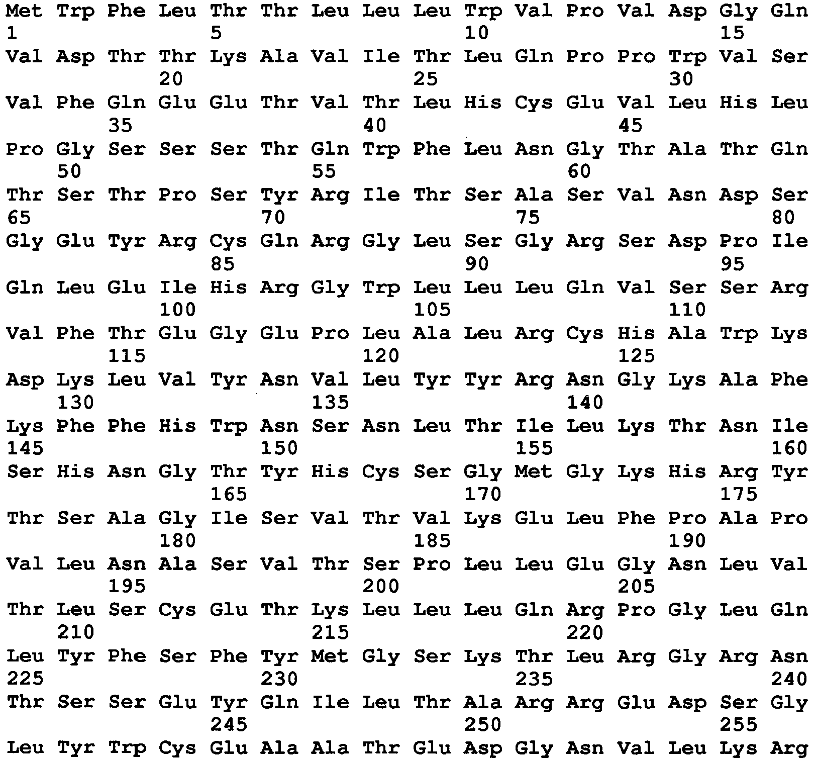

- <210> 2
<211> 313
<212> PRT
<213> Artificial sequence - <220>
<223> FcRm4 - <400> 2
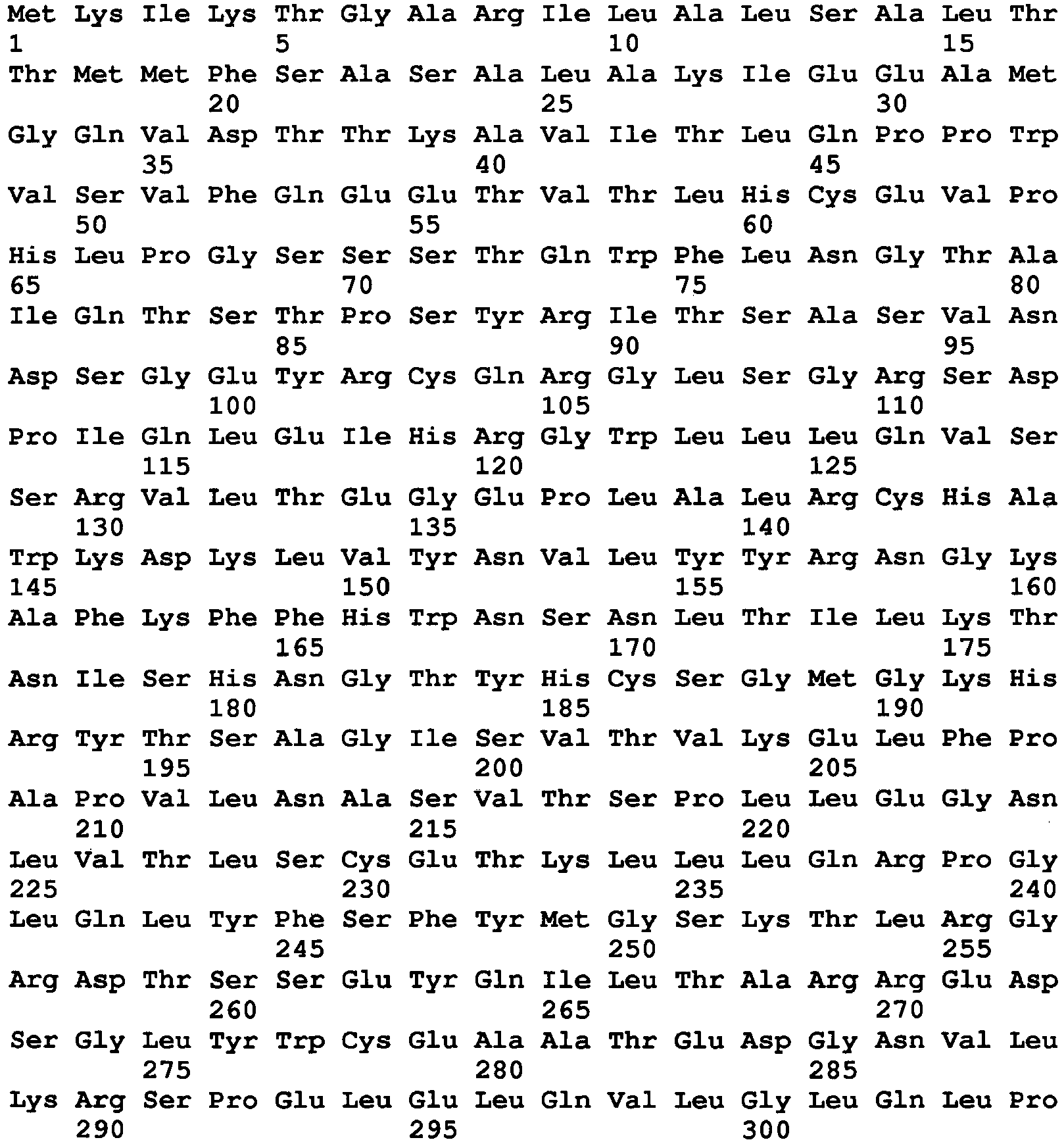

- <210> 3
<211> 313
<212> PRT
<213> Artificial sequence - <220>
<223> FcRm6 - <400> 3

- <210> 4
<211> 313
<212> PRT
<213> Artificial sequence - <220>
<223> FcRm8 - <400> 4

- <210> 5
<211> 313
<212> PRT
<213> Artificial sequence - <220>
<223> FcRm19 - <400> 5
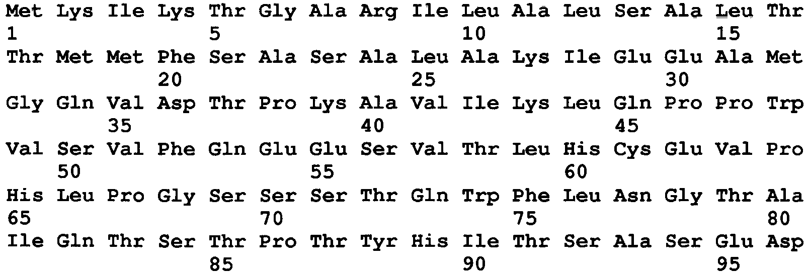
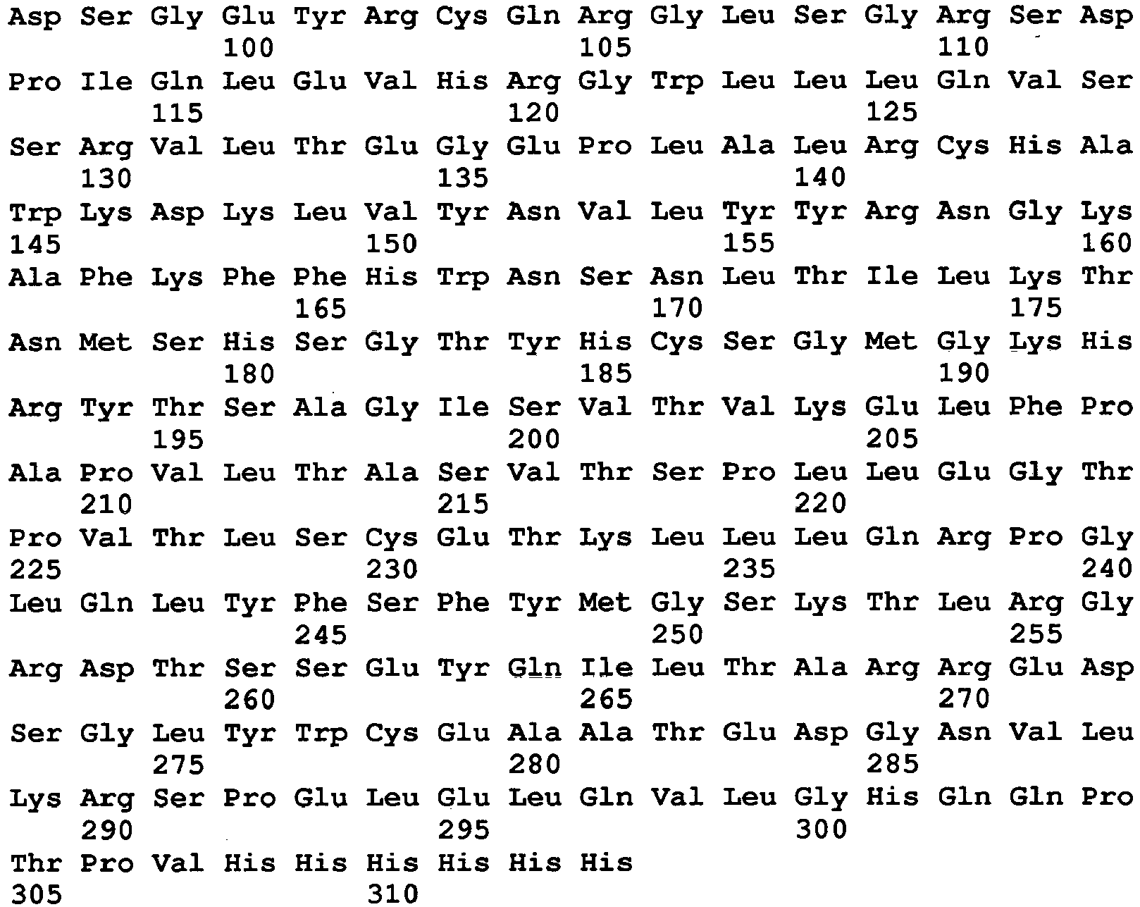
- <210> 6
<211> 939
<212> DNA
<213> Artificial sequence - <220>
<223> DNA encoding FcRm4 - <400> 6
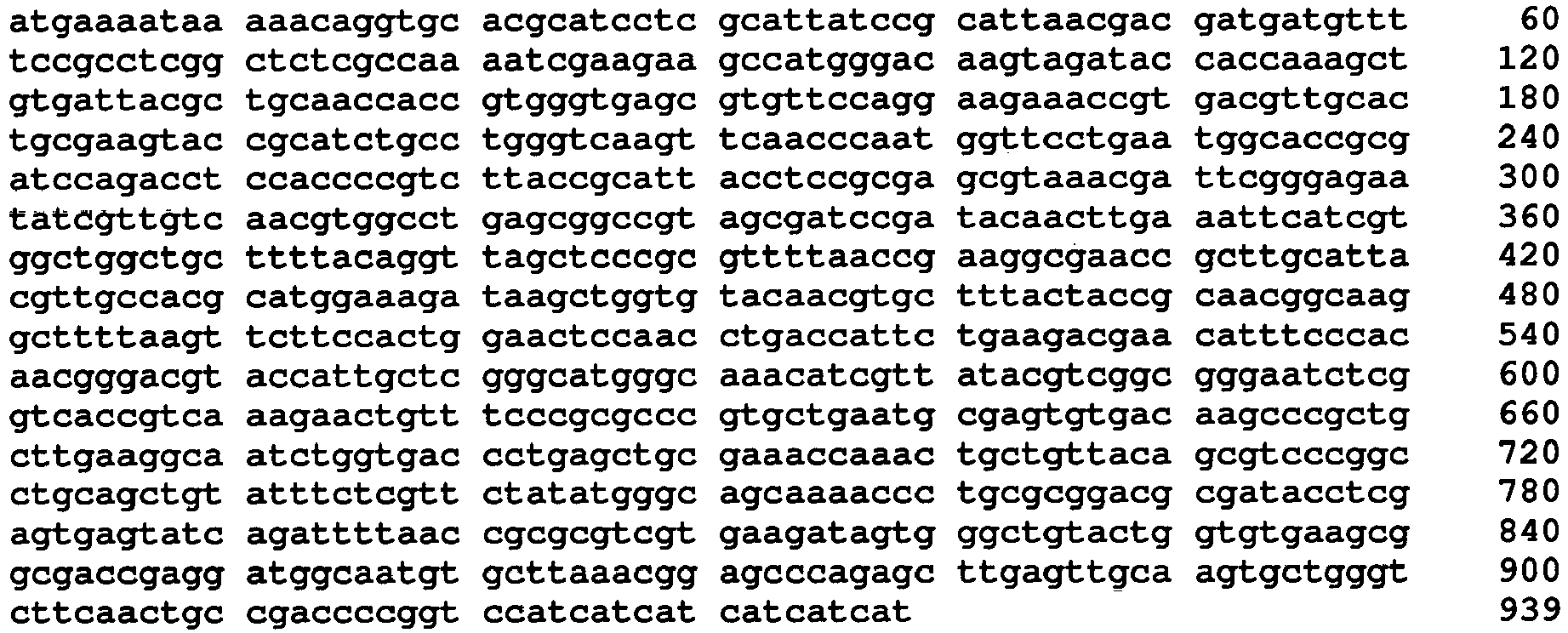
- <210> 7
<211> 939
<212> DNA
<213> Artificial sequence - <220>
<223> DNA encoding FcRm6 - <400> 7

- <210> 8
<211> 939
<212> DNA
<213> Artificial sequence - <220>
<223> DNA encoding FcRm8 - <400> 8

- <210> 9
<211> 939
<212> DNA
<213> Artificial sequence - <220>
<223> DNA encoding FcRm19 - <400> 9


- <210> 10
<211> 38
<212> DNA
<213> Artificial sequence - <220>
<223> Oligonucleotide - <400> 10
atgtggtttc tgaccacgct gttgctgtgg gtgccggt 38 - <210> 11
<211> 40
<212> DNA
<213> Artificial sequence - <220>
<223> Oligonucleotide - <400> 11
tttggtggta tctacttggc catcaaccgg cacccacagc 40 - <210> 12
<211> 40
<212> DNA
<213> Artificial sequence - <220>
<223> Oligonucleotide - <400> 12
ggccaagtag ataccaccaa agctgtgatt acgctgcaac 40 - <210> 13
<211> 40
<212> DNA
<213> Artificial sequence - <220>
<223> Oligonucleotide - <400> 13
ggaacacgct cacccacggt ggttgcagcg taatcacagc 40 - <210> 14
<211> 40
<212> DNA
<213> Artificial sequence - <220>
<223> Oligonucleotide - <400> 14
gtgggtgagc gtgttccagg aagaaaccgt gacgttgcac 40 - <210> 15
<211> 40
<212> DNA
<213> Artificial sequence - <220>
<223> Oligonucleotide - <400> 15
aggcagatgc agtacttcgc agtgcaacgt cacggtttct 40 - <210> 16
<211> 40
<212> DNA
<213> Artificial sequence - <220>
<223> Oligonucleotide - <400> 16
cgaagtactg catctgcctg ggtcaagttc aacccaatgg 40 - <210> 17
<211> 40
<212> DNA
<213> Artificial sequence - <220>
<223> Oligonucleotide - <400> 17
gtcgcggtgc cattcaggaa ccattgggtt gaacttgacc 40 - <210> 18
<211> 40
<212> DNA
<213> Artificial sequence - <220>
<223> Oligonucleotide - <400> 18
ctgaatggca ccgcgaccca gacctccacc ccgtcttacc 40 - <210> 19
<211> 40
<212> DNA
<213> Artificial sequence - <220>
<223> Oligonucleotide - <400> 19
tttacgctcg cggaggtaat gcggtaagac ggggtggagg 40 - <210> 20
<211> 40
<212> DNA
<213> Artificial sequence - <220>
<223> Oligonucleotide - <400> 20
acctccgcga gcgtaaacga ttcgggagaa tatcgttgtc 40 - <210> 21
<211> 40
<212> DNA
<213> Artificial sequence - <220>
<223> Oligonucleotide - <400> 21
ggccgctcag gccacgttga caacgatatt ctcccgaatc 40 - <210> 22
<211> 40
<212> DNA
<213> Artificial sequence - <220>
<223> Oligonucleotide - <400> 22
gtggcctgag cggccgtagc gatccgatac aacttgaaat 40 - <210> 23
<211> 40
<212> DNA
<213> Artificial sequence - <220>
<223> Oligonucleotide - <400> 23
agcagccagc cacgatgaat ttcaagttgt atcggatcgc 40 - <210> 24
<211> 40
<212> DNA
<213> Artificial sequence - <220>
<223> Oligonucleotide - <400> 24
atcgtggctg gctgctttta caggttagct cccgcgtttt 40 - <210> 25
<211> 40
<212> DNA
<213> Artificial sequence - <220>
<223> Oligonucleotide - <400> 25
tgcaagcggt tcgccttcgg taaaaacgcg ggagctaacc 40 - <210> 26
<211> 40
<212> DNA
<213> Artificial sequence - <220>
<223> Oligonucleotide - <400> 26
aggcgaaccg cttgcattac gttgccacgc atggaaagat 40 - <210> 27
<211> 40
<212> DNA
<213> Artificial sequence - <220>
<223> Oligonucleotide - <400> 27
aagcacgttg tacaccagct tatctttcca tgcgtggcaa 40 - <210> 28
<211> 40
<212> DNA
<213> Artificial sequence - <220>
<223> Oligonucleotide - <400> 28
gctggtgtac aacgtgcttt actaccgcaa cggcaaggct 40 - <210> 29
<211> 40
<212> DNA
<213> Artificial sequence - <220>
<223> Oligonucleotide - <400> 29
tggagttcca gtggaagaac ttaaaagcct tgccgttgcg 40 - <210> 30
<211> 40
<212> DNA
<213> Artificial sequence - <220>
<223> Oligonucleotide - <400> 30
gttcttccac tggaactcca acctgaccat tctgaagacg 40 - <210> 31
<211> 40
<212> DNA
<213> Artificial sequence - <220>
<223> Oligonucleotide - <400> 31
gtcccgttgt gggaaatgtt cgtcttcaga atggtcaggt 40 - <210> 32
<211> 40
<212> DNA
<213> Artificial sequence - <220>
<223> Oligonucleotide - <400> 32
acatttccca caacgggacg taccattgct cgggcatggg 40 - <210> 33
<211> 40
<212> DNA
<213> Artificial sequence - <220>
<223> Oligonucleotide - <400> 33
ccgccgacgt ataacgatgt ttgcccatgc ccgagcaatg 40 - <210> 34
<211> 40
<212> DNA
<213> Artificial sequence - <220>
<223> Oligonucleotide - <400> 34
catcgttata cgtcggcggg aatctcggtc accgtcaaag 40 - <210> 35
<211> 40
<212> DNA
<213> Artificial sequence - <220>
<223> Oligonucleotide - <400> 35
cacgggcgcg ggaaacagtt ctttgacggt gaccgagatt 40 - <210> 36
<211> 40
<212> DNA
<213> Artificial sequence - <220>
<223> Oligonucleotide - <400> 36
ttcccgcgcc cgtgctgaat gcgagtgtga caagcccgct 40 - <210> 37
<211> 40
<212> DNA
<213> Artificial sequence - <220>
<223> Oligonucleotide - <400> 37
gggtcaccag attgccttca agcagcgggc ttgtcacact 40 - <210> 38
<211> 40
<212> DNA
<213> Artificial sequence - <220>
<223> Oligonucleotide - <400> 38
gaaggcaatc tggtgaccct gagctgcgaa accaaactgc 40 - <210> 39
<211> 40
<212> DNA
<213> Artificial sequence - <220>
<223> Oligonucleotide - <400> 39
tgcaggccgg gacgctgtaa cagcagtttg gtttcgcagc 40 - <210> 40
<211> 40
<212> DNA
<213> Artificial sequence - <220>
<223> Oligonucleotide - <400> 40
cgtcccggcc tgcagctgta tttctcgttc tatatgggca 40 - <210> 41
<211> 40
<212> DNA
<213> Artificial sequence - <220>
<223> Oligonucleotide - <400> 41
ccgcgcaggg ttttgctgcc catatagaac gagaaataca 40 - <210> 42
<211> 40
<212> DNA
<213> Artificial sequence - <220>
<223> Oligonucleotide - <400> 42
gcaaaaccct gcgcggacgc aatacctcga gtgagtatca 40 - <210> 43
<211> 40
<212> DNA
<213> Artificial sequence - <220>
<223> Oligonucleotide - <400> 43
cgacgcgcgg ttaaaatctg atactcactc gaggtattgc 40 - <210> 44
<211> 40
<212> DNA
<213> Artificial sequence - <220>
<223> Oligonucleotide - <400> 44
gattttaacc gcgcgtcgtg aagatagtgg gctgtactgg 40 - <210> 45
<211> 40
<212> DNA
<213> Artificial sequence - <220>
<223> Oligonucleotide - <400> 45
cctcggtcgc cgcttcacac cagtacagcc cactatcttc 40 - <210> 46
<211> 40
<212> DNA
<213> Artificial sequence - <220>
<223> Oligonucleotide - <400> 46
aagcggcgac cgaggatggc aatgtgctta aacggagccc 40 - <210> 47
<211> 40
<212> DNA
<213> Artificial sequence - <220>
<223> Oligonucleotide - <400> 47
agcacttgca actcaagctc tgggctccgt ttaagcacat 40 - <210> 48
<211> 40
<212> DNA
<213> Artificial sequence - <220>
<223> Oligonucleotide - <400> 48
agcttgagtt gcaagtgctg ggtcttcaac tgccgacccc 40 - <210> 49
<211> 40
<212> DNA
<213> Artificial sequence - <220>
<223> Oligonucleotide - <400> 49
aaacagcaca tgaaaccaga ccggggtcgg cagttgaaga 40 - <210> 50
<211> 40
<212> DNA
<213> Artificial sequence - <220>
<223> Oligonucleotide - <400> 50
gtctggtttc atgtgctgtt ttacctggct gttgggatca 40 - <210> 51
<211> 40
<212> DNA
<213> Artificial sequence - <220>
<223> Oligonucleotide - <400> 51
agcaccgtgt tgaccagaaa catgatccca acagccaggt 40 - <210> 52
<211> 40
<212> DNA
<213> Artificial sequence - <220>
<223> Oligonucleotide - <400> 52
tctggtcaac acggtgcttt gggtaaccat tcgcaaggaa 40 - <210> 53
<211> 40
<212> DNA
<213> Artificial sequence - <220>
<223> Oligonucleotide - <400> 53
cacttcttct tgcgcttcag ttccttgcga atggttaccc 40 - <210> 54
<211> 40
<212> DNA
<213> Artificial sequence - <220>
<223> Oligonucleotide - <400> 54
ctgaagcgca agaagaagtg ggacttggag atcagtctgg 40 - <210> 55
<211> 40
<212> DNA
<213> Artificial sequence - <220>
<223> Oligonucleotide - <400> 55
tttcttttca tggccgctgt ccagactgat ctccaagtcc 40 - <210> 56
<211> 40
<212> DNA
<213> Artificial sequence - <220>
<223> Oligonucleotide - <400> 56
acagcggcca tgaaaagaaa gttatcagca gcttgcagga 40 - <210> 57
<211> 40
<212> DNA
<213> Artificial sequence - <220>
<223> Oligonucleotide - <400> 57
cttcctcaag gtgacgatcc tcctgcaagc tgctgataac 40 - <210> 58
<211> 40
<212> DNA
<213> Artificial sequence - <220>
<223> Oligonucleotide - <400> 58
ggatcgtcac cttgaggaag agctgaagtg ccaagaacaa 40 - <210> 59
<211> 40
<212> DNA
<213> Artificial sequence - <220>
<223> Oligonucleotide - <400> 59
tcctgcagtt gctcctcttt ttgttcttgg cacttcagct 40 - <210> 60
<211> 40
<212> DNA
<213> Artificial sequence - <220>
<223> Oligonucleotide - <400> 60
agaggagcaa ctgcaggaag gcgtgcatcg taaggagccg 40 - <210> 61
<211> 30
<212> DNA
<213> Artificial sequence - <220>
<223> Oligonucleotide - <400> 61
ggtcgcgccc tgcggctcct tacgatgcac 30 - <210> 62
<211> 35
<212> DNA
<213> Artificial sequence - <220>
<223> Oligonucleotide - <400> 62
tatacatatg aaaataaaaa caggtgcacg catcc 35 - <210> 63
<211> 39
<212> DNA
<213> Artificial sequence - <220>
<223> Oligonucleotide - <400> 63
gcattaacga cgatgatgtt ttccgcctcg gctctcgcc 39 - <210> 64
<211> 40
<212> DNA
<213> Artificial sequence - <220>
<223> Oligonucleotide - <400> 64
atcgtcgtta atgcggataa tgcgaggatg cgtgcacctg 40 - <210> 65
<211> 35
<212> DNA
<213> Artificial sequence - <220>
<223> Oligonucleotide - <400> 65
ttgtcccatg gcttcttcga ttttggcgag agccg 35 - <210> 66
<211> 40
<212> DNA
<213> Artificial sequence - <220>
<223> Oligonucleotide - <400> 66
tcagccatgg gacaagtaga taccaccaaa gctgtgatta 40 - <210> 67
<211> 54
<212> DNA
<213> Artificial sequence - <220>
<223> Oligonucleotide - <400> 67
ccaagcttaa tgatgatgat gatgatggac cggggtcggc agttgaagac ccag 54 - <210> 68
<211> 20
<212> DNA
<213> Artificial sequence - <220>
<223> Oligonucleotide - <400> 68
taatacgact cactataggg 20 - <210> 69
<211> 19
<212> DNA
<213> Artificial sequence - <220>
<223> Oligonucleotide - <400> 69
tatgctagtt attgctcag 19 - <210> 70
<211> 1122
<212> DNA
<213> Artificial sequence - <220>
<223> DNA encoding FcgammaRI - <400> 70

- <210> 71
<211> 374
<212> PRT
<213> Artificial sequence - <220>
<223> FcgammaRI - <400> 71


- <210> 72
<211> 949
<212> DNA
<213> Artificial sequence - <220>
<223> DNA encoding FcR - <400> 72
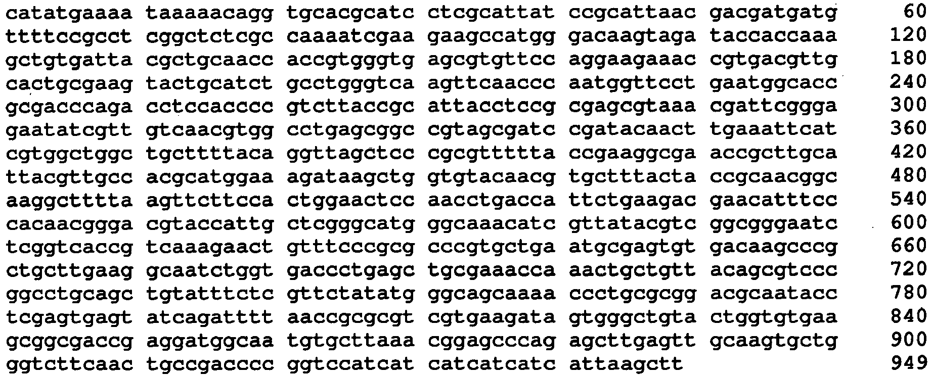
- <210> 73
<211> 313
<212> PRT
<213> Artificial sequence - <220>
<223> FcR - <400> 73
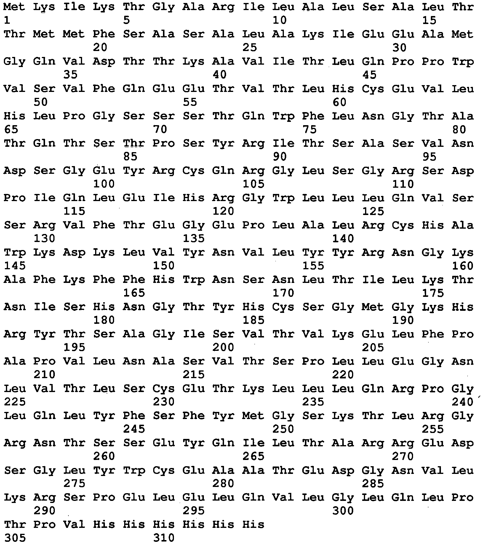
- <210> 74
<211> 27
<212> DNA
<213> Artificial sequence - <220>
<223> Oligonucleotide - <400> 74
aggaaccatt gggttgaact tgaccca 27 - <210> 75
<211> 27
<212> DNA
<213> Artificial sequence - <220>
<223> Oligonucleotide - <400> 75
tgggtcaagt tcaacccaat ggttcct 27 - <210> 76
<211> 40
<212> DNA
<213> Artificial sequence - <220>
<223> Oligonucleotide - <400> 76
agcagccagc cacgatgaat ttcaagttgt atcggatcgc 40 - <210> 77
<211> 40
<212> DNA
<213> Artificial sequence - <220>
<223> Oligonucleotide - <400> 77
gtggcctgag cggccgtagc gatccgatac aacttgaaat 40 - <210> 78
<211> 40
<212> DNA
<213> Artificial sequence - <220>
<223> Oligonucleotide - <400> 78
ccgcgcaggg ttttgctgcc catatagaac gagaaataca 40 - <210> 79
<211> 40
<212> DNA
<213> Artificial sequence - <220>
<223> Oligonucleotide - <400> 79
cgtcccggcc tgcagctgta tttctcgttc tatatgggca 40 - <210> 80
<211> 26
<212> DNA
<213> Artificial sequence - <220>
<223> Oligonucleotide - <400> 80
tgcaacgtca cggattcttc ctggaa 26 - <210> 81
<211> 26
<212> DNA
<213> Artificial sequence - <220>
<223> Oligonucleotide - <400> 81
ttccaggaag aatccgtgac gttgca 26 - <210> 82
<211> 26
<212> DNA
<213> Artificial sequence - <220>
<223> Oligonucleotide - <400> 82
cagccagcca cgatgaactt caagtt 26 - <210> 83
<211> 26
<212> DNA
<213> Artificial sequence - <220>
<223> Oligonucleotide - <400> 83
aacttgaagt tcatcgtggc tggctg 26 - <210> 84
<211> 33
<212> DNA
<213> Artificial sequence - <220>
<223> Oligonucleotide - <400> 84
tacgtcccgc tgtgggacat gttcgtcttc aga 33 - <210> 85
<211> 33
<212> DNA
<213> Artificial sequence - <220>
<223> Oligonucleotide - <400> 85
tctgaagacg aacatgtccc acagcgggac gta 33 - <210> 86
<211> 28
<212> DNA
<213> Artificial sequence - <220>
<223> Oligonucleotide - <400> 86
tgcagcttaa tcacagcttt gggggtat 28 - <210> 87
<211> 28
<212> DNA
<213> Artificial sequence - <220>
<223> Oligonucleotide - <400> 87
atacccccaa agctgtgatt aagctgca 28 - <210> 88
<211> 31
<212> DNA
<213> Artificial sequence - <220>
<223> Oligonucleotide - <400> 88
cgctcgcgga ggtaatgtgg taagtcgggg t 31 - <210> 89
<211> 27
<212> DNA
<213> Artificial sequence - <220>
<223> Oligonucleotide - <400> 89
attacctccg cgagcgaaga cgattcg 27 - <210> 90
<211> 34
<212> DNA
<213> Artificial sequence - <220>
<223> Oligonucleotide - <400> 90
tcaagcagcg ggcttgtcac actcgcagtc agca 34 - <210> 91
<211> 31
<212> DNA
<213> Artificial sequence - <220>
<223> Oligonucleotide - <400> 91
acaagcccgc tgcttgaagg cactccggtg a 31 - <210> 92
<211> 28
<212> DNA
<213> Artificial sequence - <220>
<223> Oligonucleotide - <400> 92
tcggctgttg atgacccagc acttgcaa 28 - <210> 93
<211> 28
<212> DNA
<213> Artificial sequence - <220>
<223> Oligonucleotide - <400> 93
ttgcaagtgc tgggtcatca acagccga 28 - <210> 94
<211> 30
<212> DNA
<213> Artificial sequence - <220>
<223> Oligonucleotide - <400> 94
acgtcacgga ttctccctgg aacacgctca 30 - <210> 95
<211> 30
<212> DNA
<213> Artificial sequence - <220>
<223> Oligonucleotide - <400> 95
tgagcgtgtt ccagggagaa tccgtgacgt 30 - <210> 96
<211> 27
<212> DNA
<213> Artificial sequence - <220>
<223> Oligonucleotide - <400> 96
agacagatgc ggtgcttcgc agtgcaa 27 - <210> 97
<211> 27
<212> DNA
<213> Artificial sequence - <220>
<223> Oligonucleotide - <400> 97
ttgcactgcg aagcaccgca tctgtct 27 - <210> 98
<211> 27
<212> DNA
<213> Artificial sequence - <220>
<223> Oligonucleotide - <400> 98
aacttgaccc agacagatgc ggtactt 27 - <210> 99
<211> 27
<212> DNA
<213> Artificial sequence - <220>
<223> Oligonucleotide - <400> 99
aagtaccgca tctgtctggg tcaagtt 27 - <210> 100
<211> 33
<212> DNA
<213> Artificial sequence - <220>
<223> Oligonucleotide - <400> 100
gggtggaggc ctggatcgcg gtgtcattca gga 33 - <210> 101
<211> 33
<212> DNA
<213> Artificial sequence - <220>
<223> Oligonucleotide - <400> 101
tcctgaatga caccgcgatc caggcctcca ccc 33 - <210> 102
<211> 32
<212> DNA
<213> Artificial sequence - <220>
<223> Oligonucleotide - <400> 102
ttgtggtagt aaagcacgtt gtgcaccagc tt 32 - <210> 103
<211> 31
<212> DNA
<213> Artificial sequence - <220>
<223> Oligonucleotide - <400> 103
tgcacaacgt gctttactac cacaacggca a 31 - <210> 104
<211> 28
<212> DNA
<213> Artificial sequence - <220>
<223> Oligonucleotide - <400> 104
aggttggagt tccggtggaa gaacttaa 28 - <210> 105
<211> 28
<212> DNA
<213> Artificial sequence - <220>
<223> Oligonucleotide - <400> 105
ttaagttctt ccaccggaac tccaacct 28 - <210> 106
<211> 30
<212> DNA
<213> Artificial sequence - <220>
<223> Oligonucleotide - <400> 106
tgtgggacgt gttcgtcttc ggaatggtca 30 - <210> 107
<211> 30
<212> DNA
<213> Artificial sequence - <220>
<223> Oligonucleotide - <400> 107
tgaccattcc gaagacgaac acgtcccaca 30 - <210> 108
<211> 30
<212> DNA
<213> Artificial sequence - <220>
<223> Oligonucleotide - <400> 108
tcccgccgac gtataacgat gtctgcccat 30 - <210> 109
<211> 32
<212> DNA
<213> Artificial sequence - <220>
<223> Oligonucleotide - <400> 109
acatcgttat acgtcggcgg gaacctcggt ca 32 - <210> 110
<211> 29
<212> DNA
<213> Artificial sequence - <220>
<223> Oligonucleotide - <400> 110
agggtcacct gagtgccttc atgcagcgg 29 - <210> 111
<211> 29
<212> DNA
<213> Artificial sequence - <220>
<223> Oligonucleotide - <400> 111
ccgctgcatg aaggcactca ggtgaccct 29 - <210> 112
<211> 29
<212> DNA
<213> Artificial sequence - <220>
<223> Oligonucleotide - <400> 112
cgcagggttt tgctgccctt atagaacga 29 - <210> 113
<211> 29
<212> DNA
<213> Artificial sequence - <220>
<223> Oligonucleotide - <400> 113
tcgttctata agggcagcaa aaccctgcg 29 - <210> 114
<211> 313
<212> PRT
<213> Artificial sequence - <220>
<223> FcRm32 - <400> 114

- <210> 115
<211> 939
<212> DNA
<213> Artificial sequence - <220>
<223> DNA encoding FcRm32 - <400> 115

- <210> 116
<211> 44
<212> DNA
<213> Artificial sequence - <220>
<223> Oligonucleotide - <400> 116
atgcggtgct tcgcagtgca tcgtcacgga ttctcccagg aaca 44 - <210> 117
<211> 44
<212> DNA
<213> Artificial sequence - <220>
<223> Oligonucleotide - <400> 117
gacgatgcac tgcgaagcac cgcatctgtc tgggtcaggt tcaa 44 - <210> 118
<211> 313
<212> PRT
<213> Artificial sequence - <220>
<223> FcRm36b - <400> 118

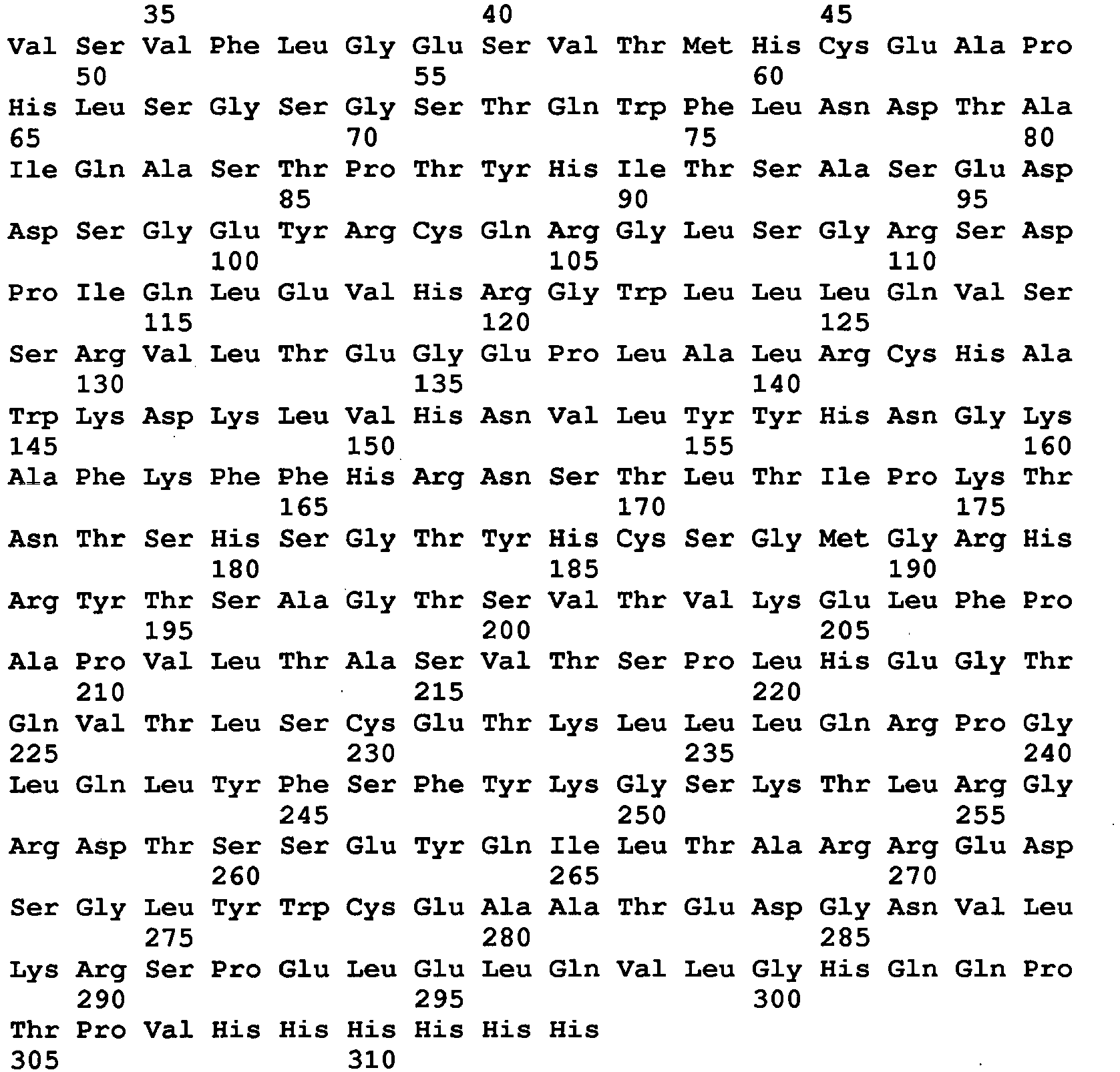
- <210> 119
<211> 939
<212> DNA
<213> Artificial sequence - <220>
<223> DNA encoding FcRm36b - <400> 119

- <210> 120
<211> 40
<212> DNA
<213> Artificial sequence - <220>
<223> Oligonucleotide - <400> 120
tcagccatgg gacaagtaga tacccccaaa gctgtgatta 40 - <210> 121
<211> 54
<212> DNA
<213> Artificial sequence - <220>
<223> Oligonucleotide - <400> 121
ccaagcttaa tgatgatgat gatgatggac cggggtcggc tgttgatgac ccag 54 - <210> 122
<211> 25
<212> DNA
<213> Artificial sequence - <220>
<223> Oligonucleotide - <400> 122
cagcttatct ctccatgcgt ggcaa 25 - <210> 123
<211> 25
<212> DNA
<213> Artificial sequence - <220>
<223> Oligonucleotide - <400> 123
ttgccacgca tggagagata agctg 25 - <210> 124
<211> 28
<212> DNA
<213> Artificial sequence - <220>
<223> Oligonucleotide - <400> 124
acgtgttcgt cctcggaatg gtcagggt 28 - <210> 125
<211> 28
<212> DNA
<213> Artificial sequence - <220>
<223> Oligonucleotide - <400> 125
accctgacca ttccgaggac gaacacgt 28 - <210> 126
<211> 32
<212> DNA
<213> Artificial sequence - <220>
<223> Oligonucleotide - <400> 126
acgctgtatc agcagtttgg ttacgcagct ca 32 - <210> 127
<211> 32
<212> DNA
<213> Artificial sequence - <220>
<223> Oligonucleotide - <400> 127
tgagctgcgt aaccaaactg ctgatacagc gt 32 - <210> 128
<211> 26
<212> DNA
<213> Artificial sequence - <220>
<223> Oligonucleotide - <400> 128
acgacgcgcg gctaaaatct gatact 26 - <210> 129
<211> 26
<212> DNA
<213> Artificial sequence - <220>
<223> Oligonucleotide - <400> 129
agtatcagat tttagccgcg cgtcgt 26 - <210> 130
<211> 313
<212> PRT
<213> Artificial sequence - <220>
<223> FcRm44 - <400> 130


- <210> 131
<211> 939
<212> DNA
<213> Artificial sequence - <220>
<223> DNA encoding FcRm44 - <400> 131


- <210> 132
<211> 26
<212> DNA
<213> Artificial sequence - <220>
<223> Oligonucleotide - <400> 132
gtgcacctgc ttatctctcc atgcgt 26 - <210> 133
<211> 26
<212> DNA
<213> Artificial sequence - <220>
<223> Oligonucleotide - <400> 133
acgcatggag agataagcag gtgcac 26 - <210> 134
<211> 313
<212> PRT
<213> Artificial sequence - <220>
<223> FcRm48 - <400> 134
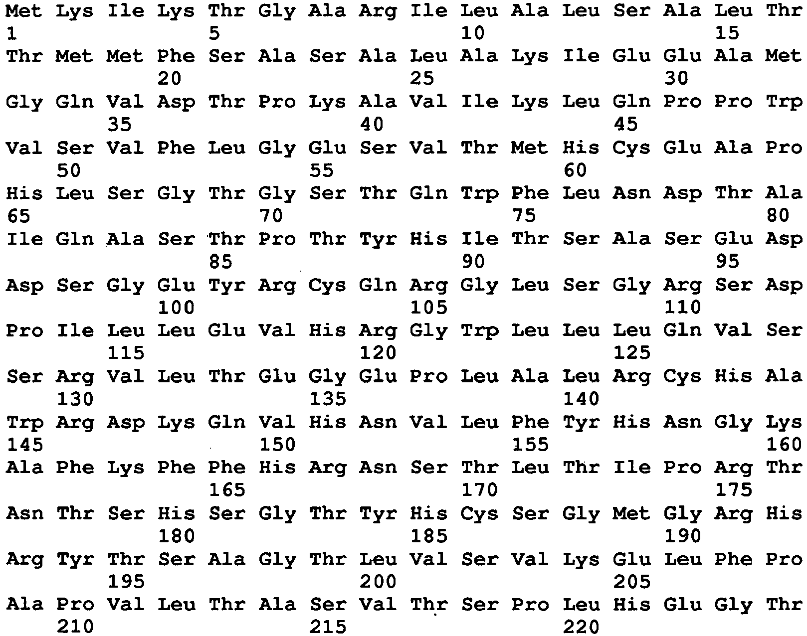

- <210> 135
<211> 939
<212> DNA
<213> Artificial sequence - <220>
<223> DNA encoding FcRm48 - <400> 135

- <210> 136
<211> 26
<212> DNA
<213> Artificial sequence - <220>
<223> Oligonucleotide - <400> 136
cttcgcagag catcgtcacg gattct 26 - <210> 137
<211> 26
<212> DNA
<213> Artificial sequence - <220>
<223> Oligonucleotide - <400> 137
agaatccgtg acgatgctct gcgaag 26 - <210> 138
<211> 26
<212> DNA
<213> Artificial sequence - <220>
<223> Oligonucleotide - <400> 138
tcgctcgcgg aggcaatgtg gtaagt 26 - <210> 139
<211> 26
<212> DNA
<213> Artificial sequence - <220>
<223> Oligonucleotide - <400> 139
acttaccaca ttgcctccgc gagcga 26 - <210> 140
<211> 27
<212> DNA
<213> Artificial sequence - <220>
<223> Oligonucleotide - <400> 140
tggcaacgta atacaagcgg ttcgcct 27 - <210> 141
<211> 27
<212> DNA
<213> Artificial sequence - <220>
<223> Oligonucleotide - <400> 141
aggcgaaccg cttgtattac gttgcca 27 - <210> 142
<211> 28
<212> DNA
<213> Artificial sequence - <220>
<223> Oligonucleotide - <400> 142
tggtcagggt tgtgttccgg tggaagaa 28 - <210> 143
<211> 28
<212> DNA
<213> Artificial sequence - <220>
<223> Oligonucleotide - <400> 143
ttcttccacc ggaacacaac cctgacca 28 - <210> 144
<211> 27
<212> DNA
<213> Artificial sequence - <220>
<223> Oligonucleotide - <400> 144
tccgcgcagg gtttcgctgc ccttata 27 - <210> 145
<211> 27
<212> DNA
<213> Artificial sequence - <220>
<223> Oligonucleotide - <400> 145
tataagggca gcgaaaccct gcgcgga 27 - <210> 146
<211> 26
<212> DNA
<213> Artificial sequence - <220>
<223> Oligonucleotide - <400> 146
tgggctccgt ttaaccacat tgccat 26 - <210> 147
<211> 26
<212> DNA
<213> Artificial sequence - <220>
<223> Oligonucleotide - <400> 147
atggcaatgt ggttaaacgg agccca 26 - <210> 148
<211> 313
<212> PRT
<213> Artificial sequence - <220>
<223> FcRm54b - <400> 148

- <210> 149
<211> 939
<212> DNA
<213> Artificial sequence - <220>
<223> DNA encoding FcRm54b - <400> 149
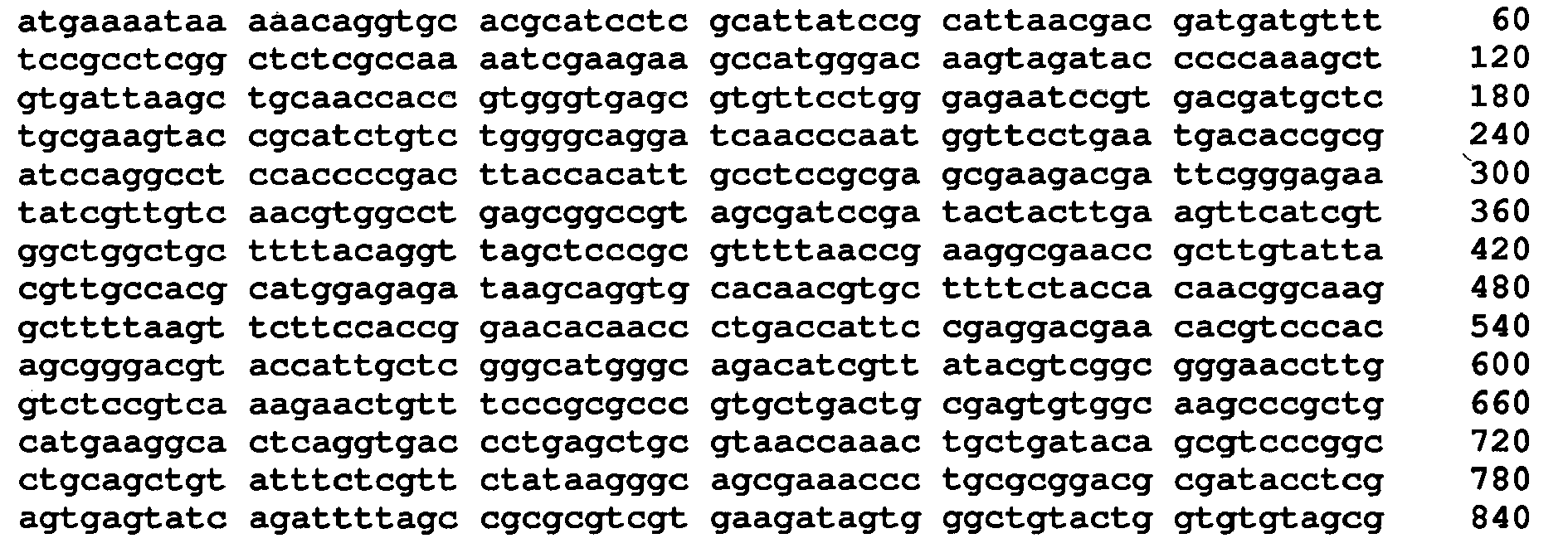

- <210> 150
<211> 27
<212> DNA
<213> Artificial sequence - <220>
<223> Oligonucleotide - <400> 150
aaacgcgggc gctaacctgt aaaagca 27 - <210> 151
<211> 27
<212> DNA
<213> Artificial sequence - <220>
<223> Oligonucleotide - <400> 151
tgcttttaca ggttagcgcc cgcgttt 27 - <210> 152
<211> 25
<212> DNA
<213> Artificial sequence - <220>
<223> Oligonucleotide - <400> 152
tcggtcgccg cttcacacca gtaca 25 - <210> 153
<211> 25
<212> DNA
<213> Artificial sequence - <220>
<223> Oligonucleotide - <400> 153
tgtactggtg tgaagcggcg accga 25 - <210> 154
<211> 313
<212> PRT
<213> Artificial sequence - <220>
<223> FcRm56b - <400> 154


- <210> 155
<211> 939
<212> DNA
<213> Artificial sequence - <220>
<223> DNA encoding FcRm56b - <400> 155

- <210> 156
<211> 25
<212> DNA
<213> Artificial sequence - <220>
<223> Oligonucleotide - <400> 156
aggcaatgtg gaaagtcggg gtgga 25 - <210> 157
<211> 25
<212> DNA
<213> Artificial sequence - <220>
<223> Oligonucleotide - <400> 157
tccaccccga ctttccacat tgcct 25 - <210> 158
<211> 26
<212> DNA
<213> Artificial sequence - <220>
<223> Oligonucleotide - <400> 158
agctcagggt caccggagtg ccttca 26 - <210> 159
<211> 26
<212> DNA
<213> Artificial sequence - <220>
<223> Oligonucleotide - <400> 159
tgaaggcact ccggtgaccc tgagct 26 - <210> 160
<211> 26
<212> DNA
<213> Artificial sequence - <220>
<223> Oligonucleotide - <400> 160
gcccttaaag aacgagaaat acagct 26 - <210> 161
<211> 26
<212> DNA
<213> Artificial sequence - <220>
<223> Oligonucleotide - <400> 161
agctgtattt ctcgttcttt aagggc 26 - <210> 162
<211> 25
<212> DNA
<213> Artificial sequence - <220>
<223> Oligonucleotide - <400> 162
tccgcgcagg gtttcgccgc cctta 25 - <210> 163
<211> 25
<212> DNA
<213> Artificial sequence - <220>
<223> Oligonucleotide - <400> 163
taagggcggc gaaaccctgc gcgga 25 - <210> 164
<211> 313
<212> PRT
<213> Artificial sequence - <220>
<223> FcRm57b - <400> 164


- <210> 165
<211> 939
<212> DNA
<213> Artificial sequence - <220>
<223> DNA encoding FcRm57b - <400> 165

- <210> 166
<211> 25
<212> DNA
<213> Artificial sequence - <220>
<223> Oligonucleotide - <400> 166
tacaagcggg tcgccttcga ttaaa 25 - <210> 167
<211> 25
<212> DNA
<213> Artificial sequence - <220>
<223> Oligonucleotide - <400> 167
tttaatcgaa ggcgacccgc ttgta 25 - <210> 168
<211> 25
<212> DNA
<213> Artificial sequence - <220>
<223> Oligonucleotide - <400> 168
ggctaaaatc ttatactcac tcgag 25 - <210> 169
<211> 25
<212> DNA
<213> Artificial sequence - <220>
<223> Oligonucleotide - <400> 169
ctcgagtgag tataagattt tagcc 25 - <210> 170
<211> 313
<212> PRT
<213> Artificial sequence - <220>
<223> FcRm61 - <400> 170

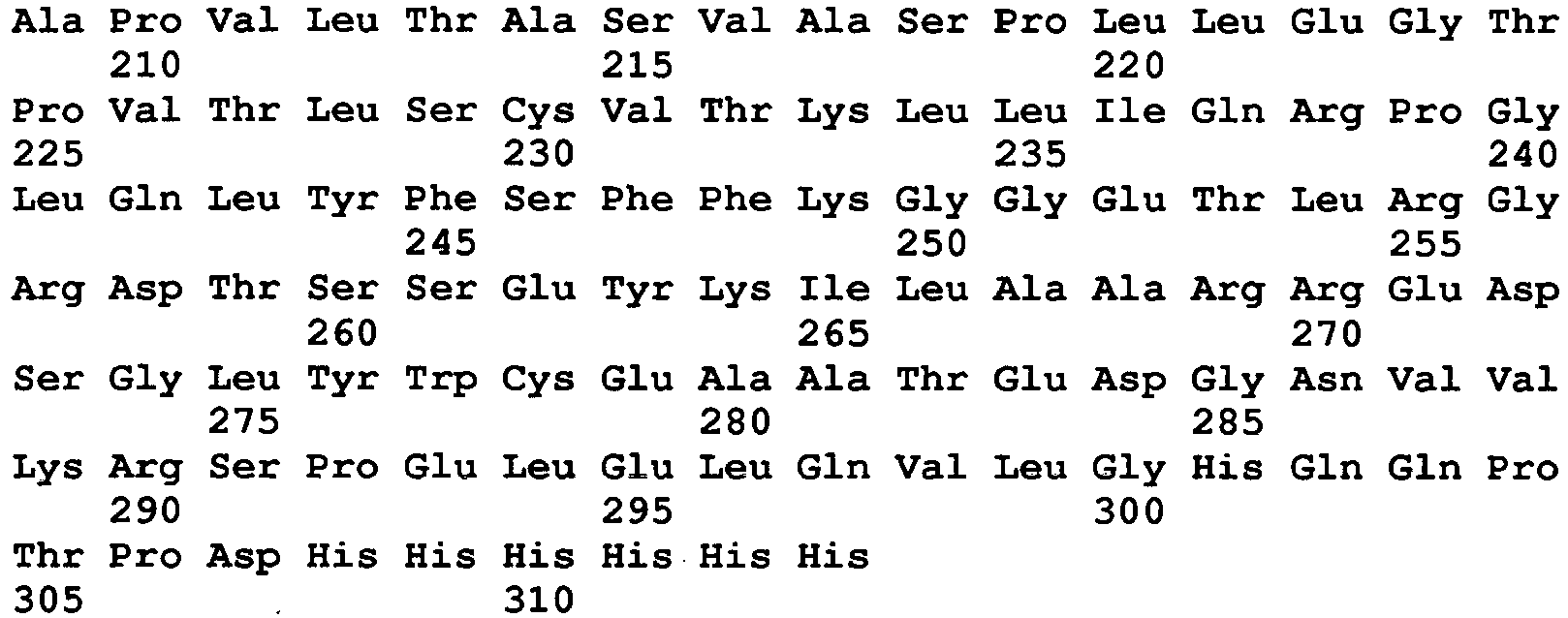
- <210> 171
<211> 939
<212> DNA
<213> Artificial sequence - <220>
<223> DNA encoding FcRm61 - <400> 171
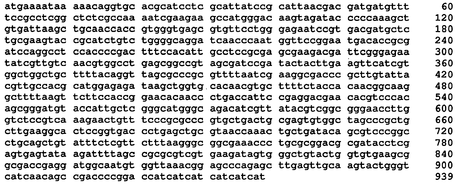
- <210> 172
<211> 25
<212> DNA
<213> Artificial sequence - <220>
<223> Oligonucleotide - <400> 172
agcaatggta catcccgctg tggga 25 - <210> 173
<211> 25
<212> DNA
<213> Artificial sequence - <220>
<223> Oligonucleotide - <400> 173
tcccacagcg ggatgtacca ttgct 25 - <210> 174
<211> 313
<212> PRT
<213> Artificial sequence - <220>
<223> FcRm60c - <400> 174

- <210> 175
<211> 939
<212> DNA
<213> Artificial sequence - <220>
<223> DNA encoding FcRm60c - <400> 175

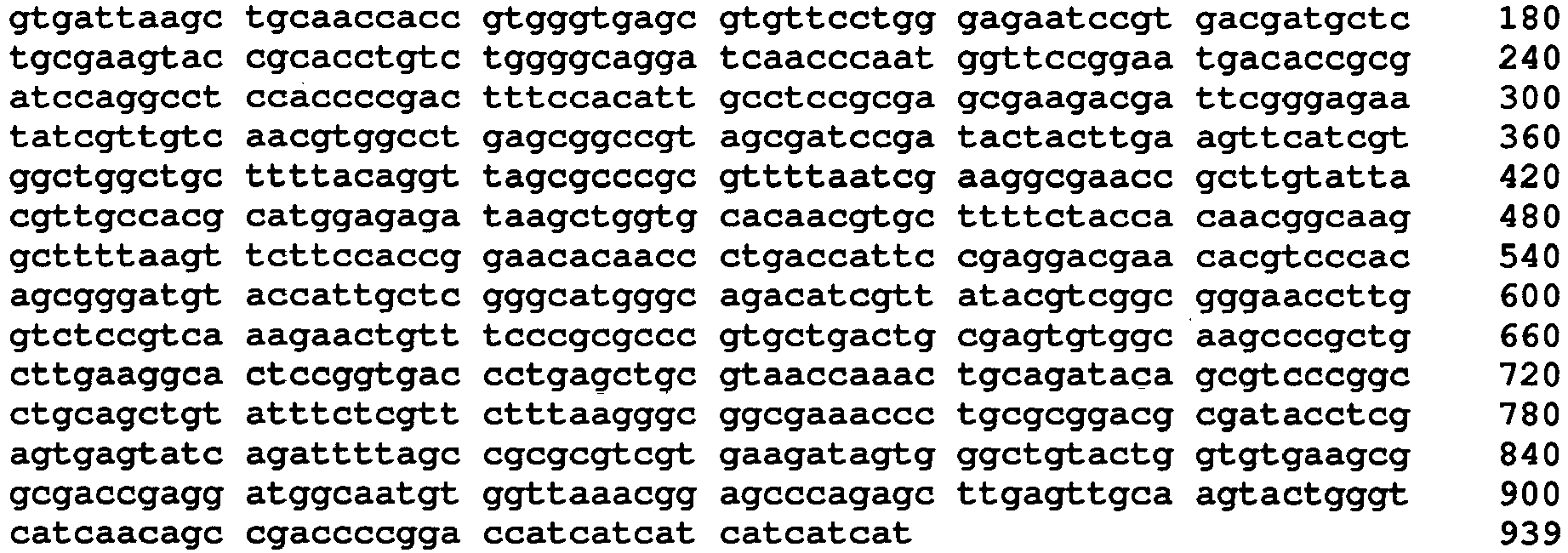
- <210> 176
<211> 313
<212> PRT
<213> Artificial sequence - <220>
<223> FcRm62 - <400> 176


- <210> 177
<211> 939
<212> DNA
<213> Artificial sequence - <220>
<223> DNA encoding FcRm62 - <400> 177

- <210> 178
<211> 33
<212> DNA
<213> Artificial sequence - <220>
<223> Oligonucleotide - <220>
<221> misc_feature
<222> (19)..(19)
<223> n stands for any base - <220>
<221> misc_feature
<222> (20)..(20)
<223> n stands for any base - <220>
<221> misc_feature
<222> (21)..(21)
<223> k stands for either g or t - <400> 178
acgttgcact gcgaagtann kcatctgcct ggg 33 - <210> 179
<211> 33
<212> DNA
<213> Artificial sequence - <220>
<223> Oligonucleotide - <220>
<221> misc_feature
<222> (19)..(19)
<223> m stands for either a or c - <220>
<221> misc_feature
<222> (20)..(20)
<223> n stands for any base - <220>
<221> misc_feature
<222> (21)..(21)
<223> n stands for any base - <400> 179
acttgaccca ggcagatgmn ntacttcgca gtg 33 - <210> 180
<211> 32
<212> DNA
<213> Artificial sequence - <220>
<223> Oligonucleotide - <220>
<221> misc_feature
<222> (19)..(19)
<223> n stands for any base - <220>
<221> misc_feature
<222> (20)..(20)
<223> n stands for any base - <220>
<221> misc_feature
<222> (21)..(21)
<223> k stands for either g or t - <400> 180
caggttagct cccgcgttnn kaccgaaggc ga 32 - <210> 181
<211> 32
<212> DNA
<213> Artificial sequence - <220>
<223> Oligonucleotide - <220>
<221> misc_feature
<222> (19)..(19)
<223> m stands for either a or c - <220>
<221> misc_feature
<222> (20)..(20)
<223> n stands for any base ' - <220>
<221> misc_feature
<222> (21)..(21)
<223> n stands for any base - <400> 181
aagcggttcg ccttcggtmn naacgcggga gc 32 - <210> 182
<211> 26
<212> PRT
<213> Escherichia coli - <300>
<308> UniProt P0AEX9
<309> 1986-07-21 - <313> (1)..(26)
- <400> 182

Claims (10)
- An Fc binding protein comprising the amino acids from position 16 to position 289 of SEQ ID NO: 1, except that it contains at least a mutation selected from the following :Phenylalanine at position 114 of SEQ ID NO: 1 substituted by Leucine,Phenylalanine at position 114 of SEQ ID NO: 1 substituted by Alanine,Phenylalanine at position 114 of SEQ ID NO: 1 substituted by Isoleucine,Phenylalanine at position 114 of SEQ ID NO: 1 substituted by Methionine,Phenylalanine at position 114 of SEQ ID NO: 1 substituted by Proline,Phenylalanine at position 114 of SEQ ID NO: 1 substituted by Threonine, orPhenylalanine at position 114 of SEQ ID NO: 1 substituted by Valine.
- The Fc binding protein according to claim 1, containing an amino acid sequence from positions 34 to 307 in the amino acid sequence described in any one of SEQ ID NOS: 2, 3, 4, 5, 114, 118, 130, 134, 148, 154, 164, 170, 174, and 176.
- The Fc binding protein according to claim 1, consisting of the amino acid sequence described in any one of SEQ ID NOS: 2, 3, 4, 5, 114, 118, 130, 134, 148, 154, 164, 170, 174, and 176.
- A polynucleotide, encoding the Fc binding protein according to any one of claims 1 to 3.
- An expression vector, containing the polynucleotide according to claim 4.
- A transformant obtained by transforming a host with the expression vector according to claim 5.
- The transformant according to claim 6, wherein the host is E. coli.
- A method for manufacturing an Fc binding protein, comprising:culturing the transformant according to claim 6 or 7 to produce the Fc binding protein; andrecovering the produced Fc binding protein from its culture.
- An adsorbent for a protein containing an Fc binding protein binding site, the adsorbent obtained by immobilizing to a solid phase the Fc binding protein according to any one of claims 1 to 3.
- An antibody purification method comprising:(1) adding a solution containing an antibody to an adsorbent for a protein containing an Fc binding protein binding site to cause the antibody to be adsorbed to the adsorbent, the adsorbent obtained by immobilizing to a solid phase an Fc binding protein consisting of the amino acid sequence described in any one of SEQ ID NOS: 114, 118, 130, 134, 148, 154, 164, 170, 174, and 176; and(2) eluting the antibody adsorbed to the adsorbent with a buffer solution of pH 3.0 to pH 4.5.
Applications Claiming Priority (3)
| Application Number | Priority Date | Filing Date | Title |
|---|---|---|---|
| JP2010052789 | 2010-03-10 | ||
| JP2011038500A JP5812627B2 (en) | 2010-03-10 | 2011-02-24 | Improved Fc receptor and method for producing the same |
| PCT/JP2011/001413 WO2011111393A1 (en) | 2010-03-10 | 2011-03-10 | Fc binding protein and method for producing same |
Publications (3)
| Publication Number | Publication Date |
|---|---|
| EP2546341A1 EP2546341A1 (en) | 2013-01-16 |
| EP2546341A4 EP2546341A4 (en) | 2013-10-23 |
| EP2546341B1 true EP2546341B1 (en) | 2014-12-31 |
Family
ID=44563224
Family Applications (1)
| Application Number | Title | Priority Date | Filing Date |
|---|---|---|---|
| EP11753056.8A Active EP2546341B1 (en) | 2010-03-10 | 2011-03-10 | Fc binding protein and method for producing same |
Country Status (5)
| Country | Link |
|---|---|
| US (1) | US9453066B2 (en) |
| EP (1) | EP2546341B1 (en) |
| JP (1) | JP5812627B2 (en) |
| CN (1) | CN103038344B (en) |
| WO (1) | WO2011111393A1 (en) |
Families Citing this family (34)
| Publication number | Priority date | Publication date | Assignee | Title |
|---|---|---|---|---|
| JP5812627B2 (en) * | 2010-03-10 | 2015-11-17 | 公益財団法人相模中央化学研究所 | Improved Fc receptor and method for producing the same |
| JP2012072091A (en) * | 2010-09-29 | 2012-04-12 | Tosoh Corp | Purification method for antibody |
| JP2013085531A (en) * | 2011-10-20 | 2013-05-13 | Tosoh Corp | Method for producing protein with recombinant e.coli |
| JP2013172655A (en) * | 2012-02-23 | 2013-09-05 | Tosoh Corp | Fc-BINDING PROTEIN AND METHOD FOR PRODUCING THE SAME |
| JP2013188141A (en) * | 2012-03-12 | 2013-09-26 | Tosoh Corp | Method for producing protein with gene recombination escherichia coli |
| JP6116884B2 (en) * | 2012-06-28 | 2017-04-19 | 東ソー株式会社 | Improved Fc-binding protein and method for producing the same |
| JP6136236B2 (en) * | 2012-12-19 | 2017-05-31 | 東ソー株式会社 | Purification method of recombinant protein |
| JP6171331B2 (en) * | 2012-12-25 | 2017-08-02 | 東ソー株式会社 | Purification method and quantification method of Fc binding protein |
| JP6115126B2 (en) * | 2012-12-27 | 2017-04-19 | 東ソー株式会社 | Purification method of human IgG3 |
| JP6236948B2 (en) * | 2013-07-17 | 2017-11-29 | 東ソー株式会社 | Antibody eluate and antibody purification method using the eluate |
| WO2015041303A1 (en) | 2013-09-18 | 2015-03-26 | 東ソー株式会社 | Fc-BINDING PROTEIN, METHOD FOR PRODUCING SAID PROTEIN, AND ANTIBODY ADSORBENT USING SAID PROTEIN, AND METHODS FOR PURIFYING AND IDENTIFYING ANTIBODY USING SAID ADSORBENT |
| EP3048112B1 (en) * | 2013-09-18 | 2020-03-11 | Tosoh Corporation | Fc-BINDING PROTEIN, METHOD FOR PRODUCING SAID PROTEIN, AND ANTIBODY ADSORBENT USING SAID PROTEIN, AND METHODS FOR PURIFYING AND IDENTIFYING ANTIBODY USING SAID ADSORBENT |
| JP6699096B2 (en) * | 2014-06-27 | 2020-05-27 | 東ソー株式会社 | Improved Fc-binding protein, method for producing the protein, and antibody adsorbent using the protein |
| JP6451118B2 (en) * | 2014-07-17 | 2019-01-16 | 東ソー株式会社 | Antibody separation method |
| EP3162895B1 (en) * | 2014-06-27 | 2020-11-25 | Tosoh Corporation | Improved fc-binding protein, method for producing said protein, antibody adsorbent using said protein, and method for separating antibody using said adsorbent |
| JP6451119B2 (en) * | 2014-07-17 | 2019-01-16 | 東ソー株式会社 | Method for separating antibodies based on the strength of antibody-dependent cytotoxic activity |
| WO2015199154A1 (en) * | 2014-06-27 | 2015-12-30 | 東ソー株式会社 | IMPROVED Fc-BINDING PROTEIN, METHOD FOR PRODUCING SAID PROTEIN, ANTIBODY ADSORBENT USING SAID PROTEIN, AND METHOD FOR SEPARATING ANTIBODY USING SAID ADSORBENT |
| JP6665414B2 (en) * | 2015-03-25 | 2020-03-13 | 東ソー株式会社 | Method of storing Fc binding protein |
| JP6932923B2 (en) * | 2015-12-24 | 2021-09-08 | 東ソー株式会社 | Improved Fc-binding protein, method for producing the protein, and antibody adsorbent using the protein |
| JP6712323B2 (en) * | 2015-12-31 | 2020-06-17 | ハンミ ファーマシューティカル カンパニー リミテッド | Triple activator with activity on all glucagon, GLP-1 and GIP receptors |
| JP6710451B2 (en) * | 2016-03-31 | 2020-06-17 | 東ソー株式会社 | Modified recombinant FcγRIIb |
| WO2017191748A1 (en) * | 2016-05-02 | 2017-11-09 | 株式会社カネカ | Modified immunoglobulin κ chain variable region-binding peptide |
| JP2018050616A (en) * | 2016-09-23 | 2018-04-05 | 東ソー株式会社 | IMPROVED RECOMBINANT FcγRII |
| JP7047425B2 (en) | 2017-02-20 | 2022-04-05 | 東ソー株式会社 | Fc-binding protein with improved antibody separation ability and antibody separation method using it |
| JP6911490B2 (en) | 2017-04-26 | 2021-07-28 | 東ソー株式会社 | Stable Fc-binding protein, method for producing the protein, and antibody adsorbent using the protein |
| JP7009913B2 (en) * | 2017-10-27 | 2022-01-26 | 東ソー株式会社 | Amino acid-substituted Fc-binding protein |
| EP3702462B1 (en) | 2017-10-27 | 2024-10-09 | Tosoh Corporation | Fc-BINDING PROTEIN EXHIBITING IMPROVED ALKALINE RESISTANCE, METHOD FOR PRODUCING SAID PROTEIN, ANTIBODY ADSORBENT USING SAID PROTEIN, AND METHOD FOR SEPARATING ANTIBODY USING SAID ANTIBODY ADSORBENT |
| JP7293657B2 (en) * | 2018-02-22 | 2023-06-20 | 東ソー株式会社 | Fc-binding protein with improved acid stability, method for producing said protein, and antibody adsorbent using said protein |
| EP3757216B1 (en) * | 2018-02-22 | 2024-08-07 | Tosoh Corporation | Fc-binding protein having improved acid stability, production method for said protein, and antibody-adsorbing agent using said protein |
| JP7469584B2 (en) | 2018-06-20 | 2024-04-17 | 東ソー株式会社 | Method for isolating antibodies and method for testing diseases |
| JP7159642B2 (en) * | 2018-06-26 | 2022-10-25 | 東ソー株式会社 | How to measure column retention for antibodies |
| JP7527764B2 (en) | 2018-08-13 | 2024-08-05 | 東ソー株式会社 | Fucose-binding protein, its production method and use |
| JP7435939B2 (en) * | 2019-11-07 | 2024-02-21 | 東ソー株式会社 | Highly productive Fc-binding protein and method for producing the same |
| JP7676866B2 (en) * | 2021-03-24 | 2025-05-15 | 東ソー株式会社 | Stock solution of human FcγRIIIa |
Family Cites Families (5)
| Publication number | Priority date | Publication date | Assignee | Title |
|---|---|---|---|---|
| EP1006183A1 (en) | 1998-12-03 | 2000-06-07 | Max-Planck-Gesellschaft zur Förderung der Wissenschaften e.V. | Recombinant soluble Fc receptors |
| US6911321B2 (en) * | 2001-12-19 | 2005-06-28 | Genentech, Inc. | Non-human primate Fc receptors and methods of use |
| JP4965589B2 (en) | 2008-02-26 | 2012-07-04 | ローム アンド ハース カンパニー | Method for producing borohydride compound |
| JP2009201403A (en) * | 2008-02-27 | 2009-09-10 | Tosoh Corp | POLYNUCLEOTIDE ENCODING HUMAN Fc RECEPTOR, AND METHOD FOR PRODUCING HUMAN Fc RECEPTOR UTILIZING THE SAME |
| JP5812627B2 (en) * | 2010-03-10 | 2015-11-17 | 公益財団法人相模中央化学研究所 | Improved Fc receptor and method for producing the same |
-
2011
- 2011-02-24 JP JP2011038500A patent/JP5812627B2/en active Active
- 2011-03-10 CN CN201180023599.5A patent/CN103038344B/en active Active
- 2011-03-10 US US13/583,550 patent/US9453066B2/en active Active
- 2011-03-10 EP EP11753056.8A patent/EP2546341B1/en active Active
- 2011-03-10 WO PCT/JP2011/001413 patent/WO2011111393A1/en not_active Ceased
Also Published As
| Publication number | Publication date |
|---|---|
| JP5812627B2 (en) | 2015-11-17 |
| US20130079499A1 (en) | 2013-03-28 |
| CN103038344B (en) | 2015-07-22 |
| EP2546341A4 (en) | 2013-10-23 |
| WO2011111393A1 (en) | 2011-09-15 |
| CN103038344A (en) | 2013-04-10 |
| US9453066B2 (en) | 2016-09-27 |
| JP2011206046A (en) | 2011-10-20 |
| EP2546341A1 (en) | 2013-01-16 |
Similar Documents
| Publication | Publication Date | Title |
|---|---|---|
| US9453066B2 (en) | FC binding protein and method for manufacturing same | |
| EP3048112B1 (en) | Fc-BINDING PROTEIN, METHOD FOR PRODUCING SAID PROTEIN, AND ANTIBODY ADSORBENT USING SAID PROTEIN, AND METHODS FOR PURIFYING AND IDENTIFYING ANTIBODY USING SAID ADSORBENT | |
| JP6507522B2 (en) | Improved Fc binding protein, method for producing the protein and antibody adsorbent using the protein | |
| EP3162895B1 (en) | Improved fc-binding protein, method for producing said protein, antibody adsorbent using said protein, and method for separating antibody using said adsorbent | |
| US11603548B2 (en) | Fc-binding protein exhibiting improved alkaline resistance, method for producing said protein, antibody adsorbent using said protein, and method for separating antibody using said antibody adsorbent | |
| WO2015199154A1 (en) | IMPROVED Fc-BINDING PROTEIN, METHOD FOR PRODUCING SAID PROTEIN, ANTIBODY ADSORBENT USING SAID PROTEIN, AND METHOD FOR SEPARATING ANTIBODY USING SAID ADSORBENT | |
| JP2011097898A (en) | RECOMBINANT Fc RECEPTOR AND METHOD FOR PRODUCING THE SAME | |
| JP2017118871A (en) | Improved Fc binding protein, method for producing the protein, and antibody adsorbent using the protein | |
| JP2016169197A (en) | Improved fc-binding proteins, methods for producing the same and antibody adsorbing agents using the same | |
| EP3757216B1 (en) | Fc-binding protein having improved acid stability, production method for said protein, and antibody-adsorbing agent using said protein | |
| JP7562962B2 (en) | Fc-binding protein with improved heat stability, method for producing said protein, and antibody adsorbent using said protein | |
| JP6116884B2 (en) | Improved Fc-binding protein and method for producing the same | |
| JP7009913B2 (en) | Amino acid-substituted Fc-binding protein | |
| JP2022076998A (en) | Fc-binding protein having improved stability against acid, method for producing the protein, and antibody adsorbent including the protein | |
| JP7293657B2 (en) | Fc-binding protein with improved acid stability, method for producing said protein, and antibody adsorbent using said protein | |
| EP4056595A1 (en) | Monoclonal antibodies against common carp beta-enolase | |
| JP2021073877A (en) | Fc-binding proteins with improved acid stability, production methods thereof and antibody-adsorbing agents using same | |
| CN110878116A (en) | Stable recombinant cardiac troponin, and coding gene and application thereof | |
| JP2011200147A (en) | POLYNUCLEOTIDE ENCODING Fc RECEPTOR, AND METHOD FOR PRODUCING Fc RECEPTOR UTILIZING THE SAME |
Legal Events
| Date | Code | Title | Description |
|---|---|---|---|
| PUAI | Public reference made under article 153(3) epc to a published international application that has entered the european phase |
Free format text: ORIGINAL CODE: 0009012 |
|
| 17P | Request for examination filed |
Effective date: 20120918 |
|
| AK | Designated contracting states |
Kind code of ref document: A1 Designated state(s): AL AT BE BG CH CY CZ DE DK EE ES FI FR GB GR HR HU IE IS IT LI LT LU LV MC MK MT NL NO PL PT RO RS SE SI SK SM TR |
|
| DAX | Request for extension of the european patent (deleted) | ||
| A4 | Supplementary search report drawn up and despatched |
Effective date: 20130925 |
|
| RIC1 | Information provided on ipc code assigned before grant |
Ipc: C12N 15/09 20060101AFI20130916BHEP Ipc: C12N 1/21 20060101ALI20130916BHEP Ipc: C07K 14/735 20060101ALI20130916BHEP Ipc: C12P 21/02 20060101ALI20130916BHEP |
|
| 17Q | First examination report despatched |
Effective date: 20140404 |
|
| GRAP | Despatch of communication of intention to grant a patent |
Free format text: ORIGINAL CODE: EPIDOSNIGR1 |
|
| INTG | Intention to grant announced |
Effective date: 20140827 |
|
| GRAS | Grant fee paid |
Free format text: ORIGINAL CODE: EPIDOSNIGR3 |
|
| GRAA | (expected) grant |
Free format text: ORIGINAL CODE: 0009210 |
|
| AK | Designated contracting states |
Kind code of ref document: B1 Designated state(s): AL AT BE BG CH CY CZ DE DK EE ES FI FR GB GR HR HU IE IS IT LI LT LU LV MC MK MT NL NO PL PT RO RS SE SI SK SM TR |
|
| REG | Reference to a national code |
Ref country code: CH Ref legal event code: EP Ref country code: GB Ref legal event code: FG4D |
|
| REG | Reference to a national code |
Ref country code: CH Ref legal event code: NV Representative=s name: NOVAGRAAF INTERNATIONAL SA, CH |
|
| REG | Reference to a national code |
Ref country code: IE Ref legal event code: FG4D |
|
| REG | Reference to a national code |
Ref country code: AT Ref legal event code: REF Ref document number: 704438 Country of ref document: AT Kind code of ref document: T Effective date: 20150215 |
|
| REG | Reference to a national code |
Ref country code: DE Ref legal event code: R096 Ref document number: 602011012748 Country of ref document: DE Effective date: 20150219 |
|
| REG | Reference to a national code |
Ref country code: SE Ref legal event code: TRGR |
|
| PG25 | Lapsed in a contracting state [announced via postgrant information from national office to epo] |
Ref country code: LT Free format text: LAPSE BECAUSE OF FAILURE TO SUBMIT A TRANSLATION OF THE DESCRIPTION OR TO PAY THE FEE WITHIN THE PRESCRIBED TIME-LIMIT Effective date: 20141231 Ref country code: NO Free format text: LAPSE BECAUSE OF FAILURE TO SUBMIT A TRANSLATION OF THE DESCRIPTION OR TO PAY THE FEE WITHIN THE PRESCRIBED TIME-LIMIT Effective date: 20150331 Ref country code: FI Free format text: LAPSE BECAUSE OF FAILURE TO SUBMIT A TRANSLATION OF THE DESCRIPTION OR TO PAY THE FEE WITHIN THE PRESCRIBED TIME-LIMIT Effective date: 20141231 |
|
| REG | Reference to a national code |
Ref country code: NL Ref legal event code: VDEP Effective date: 20141231 |
|
| REG | Reference to a national code |
Ref country code: LT Ref legal event code: MG4D |
|
| PG25 | Lapsed in a contracting state [announced via postgrant information from national office to epo] |
Ref country code: GR Free format text: LAPSE BECAUSE OF FAILURE TO SUBMIT A TRANSLATION OF THE DESCRIPTION OR TO PAY THE FEE WITHIN THE PRESCRIBED TIME-LIMIT Effective date: 20150401 Ref country code: HR Free format text: LAPSE BECAUSE OF FAILURE TO SUBMIT A TRANSLATION OF THE DESCRIPTION OR TO PAY THE FEE WITHIN THE PRESCRIBED TIME-LIMIT Effective date: 20141231 Ref country code: RS Free format text: LAPSE BECAUSE OF FAILURE TO SUBMIT A TRANSLATION OF THE DESCRIPTION OR TO PAY THE FEE WITHIN THE PRESCRIBED TIME-LIMIT Effective date: 20141231 Ref country code: LV Free format text: LAPSE BECAUSE OF FAILURE TO SUBMIT A TRANSLATION OF THE DESCRIPTION OR TO PAY THE FEE WITHIN THE PRESCRIBED TIME-LIMIT Effective date: 20141231 |
|
| PG25 | Lapsed in a contracting state [announced via postgrant information from national office to epo] |
Ref country code: NL Free format text: LAPSE BECAUSE OF FAILURE TO SUBMIT A TRANSLATION OF THE DESCRIPTION OR TO PAY THE FEE WITHIN THE PRESCRIBED TIME-LIMIT Effective date: 20141231 |
|
| PG25 | Lapsed in a contracting state [announced via postgrant information from national office to epo] |
Ref country code: CZ Free format text: LAPSE BECAUSE OF FAILURE TO SUBMIT A TRANSLATION OF THE DESCRIPTION OR TO PAY THE FEE WITHIN THE PRESCRIBED TIME-LIMIT Effective date: 20141231 Ref country code: SK Free format text: LAPSE BECAUSE OF FAILURE TO SUBMIT A TRANSLATION OF THE DESCRIPTION OR TO PAY THE FEE WITHIN THE PRESCRIBED TIME-LIMIT Effective date: 20141231 Ref country code: ES Free format text: LAPSE BECAUSE OF FAILURE TO SUBMIT A TRANSLATION OF THE DESCRIPTION OR TO PAY THE FEE WITHIN THE PRESCRIBED TIME-LIMIT Effective date: 20141231 Ref country code: RO Free format text: LAPSE BECAUSE OF FAILURE TO SUBMIT A TRANSLATION OF THE DESCRIPTION OR TO PAY THE FEE WITHIN THE PRESCRIBED TIME-LIMIT Effective date: 20141231 |
|
| PG25 | Lapsed in a contracting state [announced via postgrant information from national office to epo] |
Ref country code: PL Free format text: LAPSE BECAUSE OF FAILURE TO SUBMIT A TRANSLATION OF THE DESCRIPTION OR TO PAY THE FEE WITHIN THE PRESCRIBED TIME-LIMIT Effective date: 20141231 Ref country code: IS Free format text: LAPSE BECAUSE OF FAILURE TO SUBMIT A TRANSLATION OF THE DESCRIPTION OR TO PAY THE FEE WITHIN THE PRESCRIBED TIME-LIMIT Effective date: 20150430 |
|
| REG | Reference to a national code |
Ref country code: DE Ref legal event code: R097 Ref document number: 602011012748 Country of ref document: DE |
|
| PG25 | Lapsed in a contracting state [announced via postgrant information from national office to epo] |
Ref country code: LU Free format text: LAPSE BECAUSE OF FAILURE TO SUBMIT A TRANSLATION OF THE DESCRIPTION OR TO PAY THE FEE WITHIN THE PRESCRIBED TIME-LIMIT Effective date: 20150310 Ref country code: MC Free format text: LAPSE BECAUSE OF FAILURE TO SUBMIT A TRANSLATION OF THE DESCRIPTION OR TO PAY THE FEE WITHIN THE PRESCRIBED TIME-LIMIT Effective date: 20141231 Ref country code: DK Free format text: LAPSE BECAUSE OF FAILURE TO SUBMIT A TRANSLATION OF THE DESCRIPTION OR TO PAY THE FEE WITHIN THE PRESCRIBED TIME-LIMIT Effective date: 20141231 Ref country code: EE Free format text: LAPSE BECAUSE OF FAILURE TO SUBMIT A TRANSLATION OF THE DESCRIPTION OR TO PAY THE FEE WITHIN THE PRESCRIBED TIME-LIMIT Effective date: 20141231 |
|
| PLBE | No opposition filed within time limit |
Free format text: ORIGINAL CODE: 0009261 |
|
| STAA | Information on the status of an ep patent application or granted ep patent |
Free format text: STATUS: NO OPPOSITION FILED WITHIN TIME LIMIT |
|
| 26N | No opposition filed |
Effective date: 20151001 |
|
| PG25 | Lapsed in a contracting state [announced via postgrant information from national office to epo] |
Ref country code: IT Free format text: LAPSE BECAUSE OF FAILURE TO SUBMIT A TRANSLATION OF THE DESCRIPTION OR TO PAY THE FEE WITHIN THE PRESCRIBED TIME-LIMIT Effective date: 20141231 |
|
| REG | Reference to a national code |
Ref country code: IE Ref legal event code: MM4A |
|
| PG25 | Lapsed in a contracting state [announced via postgrant information from national office to epo] |
Ref country code: IE Free format text: LAPSE BECAUSE OF NON-PAYMENT OF DUE FEES Effective date: 20150310 |
|
| REG | Reference to a national code |
Ref country code: AT Ref legal event code: UEP Ref document number: 704438 Country of ref document: AT Kind code of ref document: T Effective date: 20141231 |
|
| PG25 | Lapsed in a contracting state [announced via postgrant information from national office to epo] |
Ref country code: SI Free format text: LAPSE BECAUSE OF FAILURE TO SUBMIT A TRANSLATION OF THE DESCRIPTION OR TO PAY THE FEE WITHIN THE PRESCRIBED TIME-LIMIT Effective date: 20141231 |
|
| REG | Reference to a national code |
Ref country code: FR Ref legal event code: PLFP Year of fee payment: 6 |
|
| PG25 | Lapsed in a contracting state [announced via postgrant information from national office to epo] |
Ref country code: BE Free format text: LAPSE BECAUSE OF FAILURE TO SUBMIT A TRANSLATION OF THE DESCRIPTION OR TO PAY THE FEE WITHIN THE PRESCRIBED TIME-LIMIT Effective date: 20141231 |
|
| PG25 | Lapsed in a contracting state [announced via postgrant information from national office to epo] |
Ref country code: MT Free format text: LAPSE BECAUSE OF FAILURE TO SUBMIT A TRANSLATION OF THE DESCRIPTION OR TO PAY THE FEE WITHIN THE PRESCRIBED TIME-LIMIT Effective date: 20141231 |
|
| REG | Reference to a national code |
Ref country code: FR Ref legal event code: PLFP Year of fee payment: 7 |
|
| PG25 | Lapsed in a contracting state [announced via postgrant information from national office to epo] |
Ref country code: HU Free format text: LAPSE BECAUSE OF FAILURE TO SUBMIT A TRANSLATION OF THE DESCRIPTION OR TO PAY THE FEE WITHIN THE PRESCRIBED TIME-LIMIT; INVALID AB INITIO Effective date: 20110310 Ref country code: SM Free format text: LAPSE BECAUSE OF FAILURE TO SUBMIT A TRANSLATION OF THE DESCRIPTION OR TO PAY THE FEE WITHIN THE PRESCRIBED TIME-LIMIT Effective date: 20141231 Ref country code: BG Free format text: LAPSE BECAUSE OF FAILURE TO SUBMIT A TRANSLATION OF THE DESCRIPTION OR TO PAY THE FEE WITHIN THE PRESCRIBED TIME-LIMIT Effective date: 20141231 |
|
| PG25 | Lapsed in a contracting state [announced via postgrant information from national office to epo] |
Ref country code: CY Free format text: LAPSE BECAUSE OF FAILURE TO SUBMIT A TRANSLATION OF THE DESCRIPTION OR TO PAY THE FEE WITHIN THE PRESCRIBED TIME-LIMIT Effective date: 20141231 |
|
| PG25 | Lapsed in a contracting state [announced via postgrant information from national office to epo] |
Ref country code: PT Free format text: LAPSE BECAUSE OF FAILURE TO SUBMIT A TRANSLATION OF THE DESCRIPTION OR TO PAY THE FEE WITHIN THE PRESCRIBED TIME-LIMIT Effective date: 20150501 |
|
| PG25 | Lapsed in a contracting state [announced via postgrant information from national office to epo] |
Ref country code: TR Free format text: LAPSE BECAUSE OF FAILURE TO SUBMIT A TRANSLATION OF THE DESCRIPTION OR TO PAY THE FEE WITHIN THE PRESCRIBED TIME-LIMIT Effective date: 20141231 |
|
| REG | Reference to a national code |
Ref country code: FR Ref legal event code: PLFP Year of fee payment: 8 |
|
| PG25 | Lapsed in a contracting state [announced via postgrant information from national office to epo] |
Ref country code: MK Free format text: LAPSE BECAUSE OF FAILURE TO SUBMIT A TRANSLATION OF THE DESCRIPTION OR TO PAY THE FEE WITHIN THE PRESCRIBED TIME-LIMIT Effective date: 20141231 |
|
| PG25 | Lapsed in a contracting state [announced via postgrant information from national office to epo] |
Ref country code: AL Free format text: LAPSE BECAUSE OF FAILURE TO SUBMIT A TRANSLATION OF THE DESCRIPTION OR TO PAY THE FEE WITHIN THE PRESCRIBED TIME-LIMIT Effective date: 20141231 |
|
| P01 | Opt-out of the competence of the unified patent court (upc) registered |
Effective date: 20230523 |
|
| PGFP | Annual fee paid to national office [announced via postgrant information from national office to epo] |
Ref country code: CH Payment date: 20250401 Year of fee payment: 15 |
|
| REG | Reference to a national code |
Ref country code: CH Ref legal event code: U11 Free format text: ST27 STATUS EVENT CODE: U-0-0-U10-U11 (AS PROVIDED BY THE NATIONAL OFFICE) Effective date: 20260401 |
|
| PGFP | Annual fee paid to national office [announced via postgrant information from national office to epo] |
Ref country code: SE Payment date: 20260211 Year of fee payment: 16 |
|
| PGFP | Annual fee paid to national office [announced via postgrant information from national office to epo] |
Ref country code: GB Payment date: 20260202 Year of fee payment: 16 |
|
| PGFP | Annual fee paid to national office [announced via postgrant information from national office to epo] |
Ref country code: DE Payment date: 20260128 Year of fee payment: 16 |
|
| PGFP | Annual fee paid to national office [announced via postgrant information from national office to epo] |
Ref country code: AT Payment date: 20260225 Year of fee payment: 16 |
|
| PGFP | Annual fee paid to national office [announced via postgrant information from national office to epo] |
Ref country code: FR Payment date: 20260209 Year of fee payment: 16 |