EP2522751B1 - Nachweis von Nukleinsäuren durch einem verbesserten INVADER-assay - Google Patents
Nachweis von Nukleinsäuren durch einem verbesserten INVADER-assay Download PDFInfo
- Publication number
- EP2522751B1 EP2522751B1 EP12172849.7A EP12172849A EP2522751B1 EP 2522751 B1 EP2522751 B1 EP 2522751B1 EP 12172849 A EP12172849 A EP 12172849A EP 2522751 B1 EP2522751 B1 EP 2522751B1
- Authority
- EP
- European Patent Office
- Prior art keywords
- oligonucleotide
- mirna
- probe
- dna
- target
- Prior art date
- Legal status (The legal status is an assumption and is not a legal conclusion. Google has not performed a legal analysis and makes no representation as to the accuracy of the status listed.)
- Active
Links
Images


Classifications
-
- C—CHEMISTRY; METALLURGY
- C12—BIOCHEMISTRY; BEER; SPIRITS; WINE; VINEGAR; MICROBIOLOGY; ENZYMOLOGY; MUTATION OR GENETIC ENGINEERING
- C12Q—MEASURING OR TESTING PROCESSES INVOLVING ENZYMES, NUCLEIC ACIDS OR MICROORGANISMS; COMPOSITIONS OR TEST PAPERS THEREFOR; PROCESSES OF PREPARING SUCH COMPOSITIONS; CONDITION-RESPONSIVE CONTROL IN MICROBIOLOGICAL OR ENZYMOLOGICAL PROCESSES
- C12Q1/00—Measuring or testing processes involving enzymes, nucleic acids or microorganisms; Compositions therefor; Processes of preparing such compositions
- C12Q1/68—Measuring or testing processes involving enzymes, nucleic acids or microorganisms; Compositions therefor; Processes of preparing such compositions involving nucleic acids
- C12Q1/6813—Hybridisation assays
- C12Q1/6816—Hybridisation assays characterised by the detection means
-
- C—CHEMISTRY; METALLURGY
- C12—BIOCHEMISTRY; BEER; SPIRITS; WINE; VINEGAR; MICROBIOLOGY; ENZYMOLOGY; MUTATION OR GENETIC ENGINEERING
- C12Q—MEASURING OR TESTING PROCESSES INVOLVING ENZYMES, NUCLEIC ACIDS OR MICROORGANISMS; COMPOSITIONS OR TEST PAPERS THEREFOR; PROCESSES OF PREPARING SUCH COMPOSITIONS; CONDITION-RESPONSIVE CONTROL IN MICROBIOLOGICAL OR ENZYMOLOGICAL PROCESSES
- C12Q1/00—Measuring or testing processes involving enzymes, nucleic acids or microorganisms; Compositions therefor; Processes of preparing such compositions
- C12Q1/68—Measuring or testing processes involving enzymes, nucleic acids or microorganisms; Compositions therefor; Processes of preparing such compositions involving nucleic acids
- C12Q1/6813—Hybridisation assays
- C12Q1/6834—Enzymatic or biochemical coupling of nucleic acids to a solid phase
-
- C—CHEMISTRY; METALLURGY
- C12—BIOCHEMISTRY; BEER; SPIRITS; WINE; VINEGAR; MICROBIOLOGY; ENZYMOLOGY; MUTATION OR GENETIC ENGINEERING
- C12Q—MEASURING OR TESTING PROCESSES INVOLVING ENZYMES, NUCLEIC ACIDS OR MICROORGANISMS; COMPOSITIONS OR TEST PAPERS THEREFOR; PROCESSES OF PREPARING SUCH COMPOSITIONS; CONDITION-RESPONSIVE CONTROL IN MICROBIOLOGICAL OR ENZYMOLOGICAL PROCESSES
- C12Q2521/00—Reaction characterised by the enzymatic activity
- C12Q2521/30—Phosphoric diester hydrolysing, i.e. nuclease
- C12Q2521/301—Endonuclease
-
- C—CHEMISTRY; METALLURGY
- C12—BIOCHEMISTRY; BEER; SPIRITS; WINE; VINEGAR; MICROBIOLOGY; ENZYMOLOGY; MUTATION OR GENETIC ENGINEERING
- C12Q—MEASURING OR TESTING PROCESSES INVOLVING ENZYMES, NUCLEIC ACIDS OR MICROORGANISMS; COMPOSITIONS OR TEST PAPERS THEREFOR; PROCESSES OF PREPARING SUCH COMPOSITIONS; CONDITION-RESPONSIVE CONTROL IN MICROBIOLOGICAL OR ENZYMOLOGICAL PROCESSES
- C12Q2525/00—Reactions involving modified oligonucleotides, nucleic acids, or nucleotides
- C12Q2525/10—Modifications characterised by
- C12Q2525/161—Modifications characterised by incorporating target specific and non-target specific sites
-
- C—CHEMISTRY; METALLURGY
- C12—BIOCHEMISTRY; BEER; SPIRITS; WINE; VINEGAR; MICROBIOLOGY; ENZYMOLOGY; MUTATION OR GENETIC ENGINEERING
- C12Q—MEASURING OR TESTING PROCESSES INVOLVING ENZYMES, NUCLEIC ACIDS OR MICROORGANISMS; COMPOSITIONS OR TEST PAPERS THEREFOR; PROCESSES OF PREPARING SUCH COMPOSITIONS; CONDITION-RESPONSIVE CONTROL IN MICROBIOLOGICAL OR ENZYMOLOGICAL PROCESSES
- C12Q2525/00—Reactions involving modified oligonucleotides, nucleic acids, or nucleotides
- C12Q2525/10—Modifications characterised by
- C12Q2525/207—Modifications characterised by siRNA, miRNA
-
- C—CHEMISTRY; METALLURGY
- C12—BIOCHEMISTRY; BEER; SPIRITS; WINE; VINEGAR; MICROBIOLOGY; ENZYMOLOGY; MUTATION OR GENETIC ENGINEERING
- C12Q—MEASURING OR TESTING PROCESSES INVOLVING ENZYMES, NUCLEIC ACIDS OR MICROORGANISMS; COMPOSITIONS OR TEST PAPERS THEREFOR; PROCESSES OF PREPARING SUCH COMPOSITIONS; CONDITION-RESPONSIVE CONTROL IN MICROBIOLOGICAL OR ENZYMOLOGICAL PROCESSES
- C12Q2561/00—Nucleic acid detection characterised by assay method
- C12Q2561/109—Invader technology
-
- C—CHEMISTRY; METALLURGY
- C12—BIOCHEMISTRY; BEER; SPIRITS; WINE; VINEGAR; MICROBIOLOGY; ENZYMOLOGY; MUTATION OR GENETIC ENGINEERING
- C12Q—MEASURING OR TESTING PROCESSES INVOLVING ENZYMES, NUCLEIC ACIDS OR MICROORGANISMS; COMPOSITIONS OR TEST PAPERS THEREFOR; PROCESSES OF PREPARING SUCH COMPOSITIONS; CONDITION-RESPONSIVE CONTROL IN MICROBIOLOGICAL OR ENZYMOLOGICAL PROCESSES
- C12Q2600/00—Oligonucleotides characterized by their use
- C12Q2600/178—Oligonucleotides characterized by their use miRNA, siRNA or ncRNA
Definitions
- the present invention relates to compositions and methods for the detection and characterization of nucleic acid molecules (e.g., RNA (e.g., small RNAs such as micro RNAs (miRNAs) and small interfering RNAs (siRNAs)) and other short nucleic acid molecules). More particularly, the present invention relates to methods for the detection and quantification of RNA expression. The present invention further provides for the detection of miRNA and siRNA mutants (e.g., deletion mutants) and variants.
- RNA e.g., small RNAs such as micro RNAs (miRNAs) and small interfering RNAs (siRNAs)
- miRNAs small RNAs
- siRNAs small interfering RNAs
- MicroRNAs are a new class of noncoding RNAs, which are encoded as short inverted repeats in the genomes of invertebrates and vertebrates ( Ambros, (2001) Cell 107, 823-826 ; Moss (2002) Curr. Biol. 12, R138-R140 ). miRNAs are modulators of target mRNA translation and stability, although most target mRNAs remain to be identified.
- miRNAs sequence-specifically control translation of target mRNAs by binding to sites of antisense complementarity in 3' untranslated regions (UTRs) (Ambros, supra; Moss, supra; Lagos-Quintana et al., (2001) Science 294, 853-858 ; Lau et al., (2001) Science 294, 858-862 ; Lee et al., (2001) Science 294, 862-864 ).
- MiRNAs may also inhibit gene expression by other mechanisms (See, e.g., Pillai et al., Science 309, 1573-1576 (2005 ); Humphreys et al., Proc Natl Acad Sci USA 102, 16961-16966 (2005 )).
- miRNAs such as let-7 RNA, miR-1, miR-34, miR-60, and miR-87, are highly conserved between invertebrates and vertebrates, implicating that they may recognize multiple sites and/or multiple targets of presumably conserved function (Lagos-Quintana et al., supra; Lau et al., supra; Lee et al., supra; Pasquinelli et al., (2000) Nature 408:86 ).
- the small temporal RNAs (stRNAs) lin-4 and let-7 represent a subclass of miRNAs identified by genetic analysis in Caenorhabditis elegans, which are developmentally regulated and themselves control developmental programs, such as timing of neuronal rewiring, Treasure larva formation, vulva formation, and the terminal differentiation of hypodermal cells.
- miRNAs are typically excised from 60- to 70-nucleotide foldback RNA precursor structures, which are sometimes detected at the onset of miRNA precursor expression ( Grishok et al., (2001) Cell 106, 23-34 ; Hutvagner et al. (2001) Science 93, 834-838 ; Ketting et al., (2001) Genes Dev. 15, 2654-2659 ) or during expression of very abundant miRNAs (Lagos-Quintana et al., supra; Lau et al., supra; Lee et al., supra). Generally, only one of the strands of the hairpin precursor molecule is excised and accumulates, presumably because it is protected by associated proteins from RNA degradation.
- the miRNA precursor processing reaction requires Dicer RNase III and Argonaute family members (Grishok et al., supra; Hutvagner et al., supra; Ketting et al., supra).
- small RNAs may find utility in areas of therapeutics and drug discovery (e.g. as drug targets or as pharmaceutical agents).
- drug discovery e.g. as drug targets or as pharmaceutical agents.
- deletions and downregulation of miRNA genes have been associated with cancer (e.g., B-cell chronic lymphocytic leukemia (CLL)), providing a need in the art to be able to detect and characterize miRNA expression (See, e.g., Calin et al., Proc Natl Acad Sci USA, 99, 15524-15529 (2002 ). In some cases, it may also be important to compare levels of miRNA in different tissue types or before and after application of a stimulus, e.g. a chemical or physical intervention.
- CLL B-cell chronic lymphocytic leukemia
- siRNAs and miRNAs may be present in low amounts in cells, it is desirable that methods of detection be both sensitive and specific. Moreover, for certain applications, it may be beneficial to identify methods suitable for high throughput screening, e.g. homogeneous methods, multiplexed methods, or those suitable to highly parallel automated manipulation and limited temperature changes.
- miRNAs play important roles in the regulation of gene expression, effective techniques for the detection and quantitation of miRNA expression are lacking. Methods used for quantitation of miRNAs have been based on gel electrophoresis. The miRNAs are detected either by Northern blotting or by the presence of radioactive RNase-resistant duplexes. Northern blotting and chip hybridization methods have relatively low analytical sensitivity (Krichevsky et al. 2003), so microgram quantities of RNA are needed for analyses; moreover, transfer of small RNAs to filters can introduce problems with reproducibility of quantitation and is not typically amendable to high-throughput. Moreover, detection methods based on RNase resistance require highly radioactive probes.
- assays based solely on probe hybridization may not provide adequate discrimination between isotypes closely related in sequence.
- Alternative approaches involve cloning the miRNAs and then sequencing the inserts. While this approach may be suitable for discriminating single-base differences between closely related miRNA species, it is time consuming and laborious.
- siRNAs small interfering RNAs
- RNAi RNA interference
- Dicer enzyme RNA-induced silencing complex
- RISC RNA-induced silencing complex
- siRNAs Another class of siRNAs is synthetic and encompasses short duplexes, usually 21-23 nt with characteristic dinucleotide overhangs ( Elbashir, S.M. et al., EMBO J. 20: 6877-6888 (2001 )) introduced directly into cells via transfection or expression from an introduced vector ( Paul, C.P. et al., Nature Biotechnology 20: 505-508 (2002 ), US Patent Application Publication No. 2003/0148519A1 ). In some cases, siRNAs appear to persist as defined sequences, making them analogous in function and composition to miRNAs (Elbashir, S.M. et al., supra). Efficient and accurate methods of detecting and characterizing (e.g., quantitating) miRNA and siRNA levels are needed.
- miRNA to form an invasive cleavage structure
- prior art e. g. WO-A-2004/057017 and ALLAWI et al., "Quantitation of micro RNAs using a modified Invader assay", RNA, vol. 10, no. 7, pages 1153-1161 .
- a major distinction over said prior art is that in said prior art the miRNA itself is used to form the invasive cleavage structure, while in the technology of present invention as defined in the claims the miRNA does not itself participate in formation of an invasive cleavage structure. Instead, the miRNA is amplified through the use of the primer + stacker combinations, and then the amplicon is detected by formation of an invasive cleavage structure.
- the cited art does not suggest the use of primers that hybridize to both the miRNA and to a stacker oligonucleotide as shown in the examples.
- Eis et al., Nature Biotechnology, 19: 673-676 (2001 ) is directed to detection of messenger RNAs and uses stacker oligonucleotides only in formation of an invasive cleavage structure on the RNA target molecule. Said prior art does not suggest stacker oligonucleotides that are complementary primers in an amplification reaction.
- the present invention relates to compositions and methods for the detection and characterization of nucleic acid molecules (e.g., RNA (e.g., small RNAs such as micro RNAs (miRNAs) and small interfering RNAs (siRNAs)) and other short nucleic acid molecules. More particularly, the present invention relates to improved methods for the detection and characterization (e.g., quantification) of RNA expression.
- nucleic acid molecules e.g., RNA (e.g., small RNAs such as micro RNAs (miRNAs) and small interfering RNAs (siRNAs)
- siRNAs small interfering RNAs
- the present description provides a method, comprising: hybridizing at least one nucleic acid (e.g., that contains sequence that is not complementary to the interfering RNA) to a interfering RNA target to generate a detection structure and detecting the detection structure.
- the interfering RNA target may be an miRNA, an siRNA.
- the siRNA may be double stranded.
- the detection structure may comprise an invasive cleavage structure.
- the nucleic acid comprises a first oligonucleotide and a probe oligonucleotide configured to form an invasive cleavage structure in combination with the miRNA.
- the nucleic acid may comprise a first oligonucleotide configured to form an invasive cleavage structure in combination with said miRNA.
- the first oligonucleotide may comprise a 5' portion and a 3' portion, wherein said 3' portion is configured to hybridize to said target sequence, and wherein said 5' portion is configured to not hybridize to said target sequence.
- the probe oligonucleotide comprises a 5' portion and a 3' portion, wherein said 5' portion is configured to hybridize to said target sequence, and wherein said 3' portion is configured to not hybridize to said target sequence.
- the detecting step may comprise use of an INVADER assay.
- the detection structure may comprise a circular oligonucleotide hybridized to said small RNA to generate a circular detection structure.
- the detecting step may comprise use of a rolling circle replication assay.
- the detection structure may comprise a nucleic acid molecule with a free 3'-OH group that is extended by a polymerase (e.g., template dependent extension) and the extended sequence is directly or indirectly detected.
- a polymerase e.g., template dependent extension
- the detecting step(s) may comprise use of a detection assay including, but not limited to, sequencing assays, polymerase chain reaction assays, hybridization assays, hybridization assays employing a probe complementary to a mutation, microarray assays, bead array assays, primer extension assays, enzyme mismatch cleavage assays, branched hybridization assays, NASBA assays, molecular beacon assays, cycling probe assays, ligase chain reaction assays, invasive cleavage structure assays, ARMS assays, and sandwich hybridization assays.
- the detecting step may be carried out in cell lysate.
- the methods of the present invention may comprise detecting a second nucleic acid target.
- the second nucleic acid target may be RNA.
- the second nucleic acid target may be U6 RNA or GAPDH mRNA.
- the nucleic acid used to form the detection structure may comprise a template with one or more sites sufficiently complementary to the small RNA so as to allow the RNA to hybridize to the template and be extended in an extension reaction.
- the extension reaction may be a polymerase chain reaction wherein one or more RNAs are used as primers in the polymerase chain reaction.
- a single type of RNA may bind to two locations on the template to provide the polymerase chain reaction primers. Two or more RNAs may be used as primers. Then the detection of an amplification product signifies the presence of the two or more RNAs in the sample (i.e., an miRNA multiplex assay).
- RNA may be used as a template for modification of a detection complex by extension of a primer across at least part of the RNA template.
- the method may comprise detection of a plurality of miRNAs.
- the plurality of miRNAs may comprise polymorphisms of the same miRNA.
- the plurality of miRNAs may comprise different miRNAs (e.g ., Let-7, miR-1, miR-1d, miR-135, miR-15, miR-16, miR-124a, or miR125b).
- kits for conducting of the claimed methods comprising kits comprising a nucleic acid configured for forming a detection structure when hybridized to an RNA target sequence.
- the kits may be configured to detect an miRNA.
- the kits may be configured to detect a Let-7, miR-1, miR-135, miR-15, miR-16, miR-1b, miR-124a, or miR125b miRNA.
- the kits may be configured to co-detect a second RNA target with an miRNA target.
- the present description also provides a method for detecting a miRNA target, comprising providing (i) a miRNA target; (ii) a first unlabled oligonucleotide; (iii) a second unlabeled oligonucleotide; (iv) a reverse transcriptase; (v) a polymerase; and (vi) a probe oligonucleotide; incubating (i) through (vi) under conditions such that a detection structure forms; and detecting the detection structure.
- the first unlabeled oligonucleotide may comprise a first region that is complementary to the miRNA target and a second region that is not complementary to the miRNA target.
- the second unlabeled oligonucleotide may comprise a first region that is identical to a second region of the miRNA target and a second region that lacks sequence identity to the second region of the miRNA target.
- Detecting may comprise forming an invasive cleavage structure, cleaving the invasive cleavage structure, and detecting the cleavage of the invasive cleavage structure.
- Said incubating may further comprise incubating with an enzyme capable of cleaving a detection structure and lacking polymerase activity.
- the enzyme may be a 5' nuclease, while in some examples, the enzyme comprises a FEN-1 nuclease.
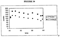
- Cleaving the invasive cleavage structure may occur at a temperature of between 45 °C and 60 °C. Said cleaving the invasive cleavage structure may occur at a temperature of approximately 50 °C.
- the first unlabled oligonucleotide may be used as a primer for reverse transcription.
- the first unlabled oligonucleotide may be used as an INVADER oligonucleotide in an invasive cleavage reaction.
- (i) through (vi) are present within the same reaction vessel.
- the method may further comprise providing (vii) a probe oligonucleotide.
- the first unlabeled oligonucleotide and the reverse transcriptase may reverse transcribe the miRNA target.
- the reverse transcribed miRNA target (i.e., an miRNA cDNA target) may be amplified by the first unlabeled oligonucleotide and the second unlabeled oligonucleotide and the DNA polymerase in a polymerase chain reaction.
- the amplified reverse transcribed miRNA target may form a detection structure in the presence of the probe oligonucleotide.
- the first unlabeled oligonucleotide may comprises nucleic acid sequence such that a duplex of about 6-7 base pairs is formed between the oligonucleotide and the miRNA target.
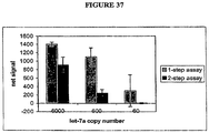
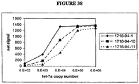
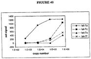
- the present method is capable of detecting miRNA present in very small copy numbers. For example, less than 200 copies of miRNA in a sample can be detected. Furthermore, less than 100 copies of miRNA in a sample can be detected.
- the second unlabeled oligonucleotide may comprise nucleic acid sequence such that a duplex of about 9 basepairs is formed between the oligonucleotide and the miRNA target.
- the probe oligonucleotide may comprise nucleic acid sequence such that a duplex of about 8-10 basepairs is formed between the oligonucleotide and the miRNA target or the amplified copy of the miRNA target.
- the second region of the first unlabeled oligonucleotide probe may comprise a first portion and a second portion, wherein the first portion and the second portion can hybridize to each other.
- a hairpin structure may be formed in the first unlabeled oligonucleotide probe when the first portion and the second portion hybridize to each other.
- the second region of the second unlabeled oligonucleotide probe may comprise a first portion and a second portion, wherein the first portion and the second portion can hybridize to each other.
- a hairpin structure may be formed in the second unlabeled oligonucleotide probe when the first portion and the second portion hybridize to each other.
- the method may further comprise providing (vii) an oligonucleotide complementary to a region of the first unlabeled oligonucleotide probe.
- the method may further comprise providing (vii) an oligonucleotide complementary to a region of the second unlabeled oligonucleotide probe.
- cleaving the invasive cleavage structure at a temperature of approximately 50 °C permits high fidelity discrimination of target sequences, although other termperatures may be selected based on sequence, buffer components, etc. for optimum performance.
- the target sequences may comprise variant miRNAs of a single species.
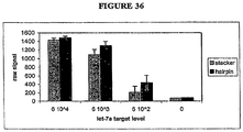
- increasing the concentration of the probe oligonucleotide increases the sensitivity of detecting the miRNA target.
- detecting comprises use of a labeled probe.
- the labeled probe is fluorescently labeled.
- the labeled probe is configured for FRET detection.
- the labeled probe has a first conformation when not hybridized in a duplex and a second conformation when hybridized in a duplex.
- the labeled probe exhibits increased fluorescence when hybridized in a duplex. The present description is not limited by the miRNA detected.
- miRNAs can be detected using the compositions and methods of the present description including, but not limited to, Let-7, miR-1, miR-135, miR-15, miR-16, miR125b, miR-1d, and miR124a.
- the present description also provides a kit comprising one or more of a first unlabled oligonucleotide that comprises a first region that is complementary to a miRNA target and a second region that is not complementary to the miRNA target; a second unlabeled oligonucleotide that comprises a first region that is identical to a second region of the miRNA target and a second region that lacks sequence identity to the second region of the miRNA target; a reverse transcriptase; a DNA polymerase; a probe oligonucleotide; and an enzyme capable of cleaving a detection structure.
- the detection structure comprises an invasive cleavage structure.
- the kit is configured to detect a miRNA target and at least one other RNA target.
- the kit is configured to detect the miRNA target sequence in a cell lysate.
- miRNA refers to micro RNA.
- miRNA target sequence refers to a miRNA that is to be detected ( e.g., in the presence of other nucleic acids).
- a miRNA target sequence is a variant of a miRNA.
- RNA detection structure and “detection structure” refer to a structure formed by hybridizing a nucleic acid (e.g., an oligonucleotide) to an RNA target, e.g., an miRNA or siRNA.
- the nucleic acid may be a single nucleic acid (e.g., a larger nucleic acid with a small region (or regions) of homology to the miRNA).
- the nucleic acid may comprise two nucleic acids (e.g., that hybridize to the miRNA to form a hairpin (e.g ., single or double hairpin) structure).
- Said miRNA detection structures may be capable of detection using known nucleic acid detection methods, including, but not limited to, those disclosed herein.
- RNA detection structures may be further modified following the hybridization step.
- one or more components of the detection structure may provide a template for extension by a nucleic acid polymerase.
- One or more components of the detection structure may be contacted with a ligase and ligated to an additional nucleic acid.
- siRNAs refers to short interfering RNAs.
- Said siRNAs may comprise a duplex, or double-stranded region, where each strand of the double-stranded region is about 18 to 25 nucleotides long; the double-stranded region can be as short as 16, and as long as 29, base pairs long, where the length is determined by the antisense strand.
- siRNAs contain from about two to four unpaired nucleotides at the 3' end of each strand. siRNAs appear to function as key intermediates in triggering RNA interference in invertebrates and in vertebrates, and in triggering sequence-specific RNA degradation during posttranscriptional gene silencing in plants.
- At least one strand of the duplex or double-stranded region of a siRNA is substantially homologous to or substantially complementary to a target RNA molecule.
- the strand complementary to a target RNA molecule is the "antisense” strand; the strand homologous to the target RNA molecule is the "sense” strand and is also complementary to the siRNA antisense strand.
- One strand of the double stranded region need not be the exact length of the opposite strand, thus, one strand may have at least one fewer nucleotides than the opposite complementary strand, resulting in a "bubble” or at least one unmatched base in the opposite strand.
- One strand of the double-stranded region need not be exactly complementary to the opposite strand; thus, the strand, preferably the sense strand, may have at least one mismatched base pair.
- siRNAs may also contain additional sequences; non-limiting examples of such sequences include linking sequences, or loops, which connect the two strands of the duplex region.
- This form of siRNAs may be referred to "si-like RNA", “short hairpin siRNA” where the short refers to the duplex region of the siRNA, or "hairpin siRNA".
- Additional non-limiting examples of additional sequences present in siRNAs include stem and other folded structures.
- the additional sequences may or may not have known functions; non-limiting examples of such functions include increasing stability of an siRNA molecule, or providing a cellular destination signal.
- subject and “patient” refer to any organisms including plants, microorganisms and animals (e.g ., mammals such as dogs, cats, livestock, and humans).
- the term "INVADER assay reagents" or "invasive cleavage assay reagents” refers to one or more reagents for detecting target sequences, said reagents comprising oligonucleotides capable of forming an invasive cleavage structure in the presence of the target sequence.
- the INVADER assay reagents may further comprise an agent for detecting the presence of an invasive cleavage structure (e.g ., a cleavage agent).
- the oligonucleotides may comprise a first oligonucleotide and a probe oligonucleotide, said first oligonucleotide comprising a 5' portion complementary to a first region of the target nucleic acid and said probe oligonucleotide comprising a 3' portion and a 5' portion, said 5' portion complementary to a second region of the target nucleic acid downstream of and contiguous to the first portion.
- the 3' portion of the second oligonucleotide may comprise a 3' terminal nucleotide not complementary to the target nucleic acid.
- the 3' portion of the probe oligonucleotide may consist of a single nucleotide not complementary to the target nucleic acid.
- the first and the probe oligonucleotides may be covalently coupled to one another ( e.g ., through a linker).
- the INVADER assay reagents may further comprise a solid support.
- the one or more oligonucleotides of the assay reagents e.g ., first and/or second oligonucleotide, whether bridging or non-bridging
- the INVADER assay reagents may further comprise a buffer solution.
- the buffer solution may comprise a source of divalent cations (e.g ., Mn 2+ and/or Mg 2+ ions).
- Individual ingredients e.g ., oligonucleotides, enzymes, buffers, target nucleic acids
- INVADER assay reagent components Individual ingredients (e.g ., oligonucleotides, enzymes, buffers, target nucleic acids) that collectively make up INVADER assay reagents.
- the INVADER assay reagents may further comprise a third oligonucleotide complementary to a third portion of the target nucleic acid upstream of the first portion of the first target nucleic acid.
- the INVADER assay reagents may further comprise a target nucleic acid.
- the INVADER assay reagents may further comprise a second target nucleic acid.
- the INVADER assay reagents may further comprise a third oligonucleotide comprising a 5' portion complementary to a first region of the second target nucleic acid. The 3' portion of the third oligonucleotide may be covalently linked to the second target nucleic acid.
- the second target nucleic acid may further comprises a 5' portion, wherein the 5' portion of the second target nucleic acid is the third oligonucleotide.
- the INVADER assay reagents may further comprise an ARRESTOR molecule ( e.g. , ARRESTOR oligonucleotide).
- the INVADER assay reagents may further comprise reagents for detecting a nucleic acid cleavage product.
- One or more oligonucleotides in the INVADER assay reagents may comprise a label.
- Said first oligonucleotide may comprise a label or said third oligonucleotide may comprise a label.
- the reagents may comprise a first and/or a third oligonucleotide labeled with moieties that produce a fluorescence resonance energy transfer (FRET) effect.
- FRET fluorescence resonance energy transfer
- One or more the INVADER assay reagents may be provided in a predispensed format (i.e., premeasured for use in a step of the procedure without re-measurement or re-dispensing).
- Selected INVADER assay reagent components may be mixed and predispensed together.
- Said predispensed assay reagent components may be predispensed and provided in a reaction vessel (including but not limited to a reaction tube or a well, as in, e.g., a microtiter plate).
- Said predispensed INVADER assay reagent components may be dried down ( e.g ., desiccated or lyophilized) in a reaction vessel.
- kits refers to any delivery system for delivering materials.
- reaction reagents e.g ., oligonucleotides, enzymes, etc. in the appropriate containers
- supporting materials e.g ., buffers, written instructions for performing the assay etc.
- kits include one or more enclosures (e.g ., boxes) containing the relevant reaction reagents and/or supporting materials.
- fragmented kit refers to delivery systems comprising two or more separate containers that each contains a subportion of the total kit components.
- the containers may be delivered to the intended recipient together or separately.
- a first container may contain an enzyme for use in an assay, while a second container contains oligonucleotides.
- fragmented kit is intended to encompass kits containing Analyte specific reagents (ASR's) regulated under section 520(e) of the Federal Food, Drug, and Cosmetic Act, but are not limited thereto.
- ASR's Analyte specific reagents
- any delivery system comprising two or more separate containers that each contains a subportion of the total kit components are included in the term “fragmented kit.”
- a “combined kit” refers to a delivery system containing all of the components of a reaction assay in a single container ( e.g ., in a single box housing each of the desired components).
- kit includes both fragmented and combined kits.
- kits comprising one or more of the components necessary for practicing the present invention.
- the present description provides kits for storing or delivering the enzymes and/or the reaction components necessary to practice an INVADER assay.
- the kit may include any and all components necessary or desired for assays including, but not limited to, the reagents themselves, buffers, control reagents (e.g ., tissue samples, positive and negative control target oligonucleotides, etc.), solid supports, labels, written and/or pictorial instructions and product information, inhibitors, labeling and/or detection reagents, package environmental controls ( e.g., ice, desiccants, etc.), and the like.
- kits may provide a sub-set of the required components, wherein it is expected that the user will supply the remaining components.
- the kits may comprise two or more separate containers wherein each container houses a subset of the components to be delivered.
- a first container e.g ., box
- an enzyme e.g ., structure specific cleavage enzyme in a suitable storage buffer and container
- a second box may contain oligonucleotides (e.g ., INVADER oligonucleotides, probe oligonucleotides, control target oligonucleotides, etc.).
- label refers to any atom or molecule that can be used to provide a detectable (preferably quantifiable) effect, and that can be attached to a nucleic acid or protein. Labels include, but are not limited to, dyes; radiolabels such as 32 P; binding moieties such as biotin; haptens such as digoxgenin; luminogenic, phosphorescent or fluorogenic moieties; mass tags; and fluorescent dyes alone or in combination with moieties that can suppress or shift emission spectra by fluorescence resonance energy transfer (FRET).
- FRET fluorescence resonance energy transfer
- Labels may provide signals detectable by fluorescence, radioactivity, colorimetry, gravimetry, X-ray diffraction or absorption, magnetism, enzymatic activity, characteristics of mass or behavior affected by mass (e.g ., MALDI time-of-flight mass spectrometry; fluorescence polarization), and the like.
- a label may be a charged moiety (positive or negative charge) or alternatively, may be charge neutral.
- Labels can include or consist of nucleic acid or protein sequence, so long as the sequence comprising the label is detectable.
- the term "distinct" in reference to signals refers to signals that can be differentiated one from another, e.g., by spectral properties such as fluorescence emission wavelength, color, absorbance, mass, size, fluorescence polarization properties, charge, etc., or by capability of interaction with another moiety, such as with a chemical reagent, an enzyme, an antibody, etc.
- the terms “complementary” or “complementarity” are used in reference to polynucleotides (i.e., a sequence of nucleotides such as an oligonucleotide or a target nucleic acid) related by the base-pairing rules.
- polynucleotides i.e., a sequence of nucleotides such as an oligonucleotide or a target nucleic acid
- sequence “5'-A-G-T-3"' is complementary to the sequence "3'-T-C-A-5'.”
- Complementarity may be “partial,” in which only some of the nucleic acids' bases are matched according to the base pairing rules. Or, there may be “complete” or “total” complementarity between the nucleic acids.
- the degree of complementarity between nucleic acid strands has significant effects on the efficiency and strength of hybridization between nucleic acid strands. This is of particular importance in amplification reactions, as well as detection methods that depend upon binding between nucleic acids. Either term may also be used in reference to individual nucleotides, especially within the context of polynucleotides. For example, a particular nucleotide within an oligonucleotide may be noted for its complementarity, or lack thereof, to a nucleotide within another nucleic acid strand, in contrast or comparison to the complementarity between the rest of the oligonucleotide and the nucleic acid strand.
- homologous refers to a degree of identity. There may be partial homology or complete homology. A partially homologous sequence is one that is less than 100% identical to another sequence.
- hybridization is used in reference to the pairing of complementary nucleic acids. Hybridization and the strength of hybridization (i.e ., the strength of the association between the nucleic acids) is influenced by such factors as the degree of complementary between the nucleic acids, stringency of the conditions involved, and the T m of the formed hybrid. "Hybridization” methods involve the annealing of one nucleic acid to another, complementary nucleic acid, i.e., a nucleic acid having a complementary nucleotide sequence. The ability of two polymers of nucleic acid containing complementary sequences to find each other and anneal through base pairing interaction is a well-recognized phenomenon.
- nucleic acid sequence refers to an oligonucleotide which, when aligned with the nucleic acid sequence such that the 5' end of one sequence is paired with the 3' end of the other, is in "antiparallel association.”
- Certain bases not commonly found in natural nucleic acids may be included in the nucleic acids of the present description and include, for example, inosine and 7-deazaguanine. Complementarity need not be perfect; stable duplexes may contain mismatched base pairs or unmatched bases.
- nucleic acid technology can determine duplex stability empirically considering a number of variables including, for example, the length of the oligonucleotide, base composition and sequence of the oligonucleotide, ionic strength and incidence of mismatched base pairs.
- T m is used in reference to the "melting temperature.”
- the melting temperature is the temperature at which a population of double-stranded nucleic acid molecules becomes half dissociated into single strands.
- RNA having a non-coding function e.g ., a ribosomal or transfer RNA
- the RNA or polypeptide can be encoded by a full length coding sequence or by any portion of the coding sequence so long as the desired activity or function is retained.
- wild-type refers to a gene or a gene product that has the characteristics of that gene or gene product when isolated from a naturally occurring source.
- a wild-type gene is that which is most frequently observed in a population and is thus arbitrarily designated the “normal” or “wild-type” form of the gene.
- modified refers to a gene or gene product that displays modifications in sequence and or functional properties (i.e ., altered characteristics) when compared to the wild-type gene or gene product. It is noted that naturally-occurring mutants can be isolated; these are identified by the fact that they have altered characteristics when compared to the wild-type gene or gene product.
- heterologous sequence refers to DNA sequences containing a desired heterologous sequence.
- the heterologous sequence may be a coding sequence and appropriate DNA sequences necessary for either the replication of the coding sequence in a host organism, or the expression of the operably linked coding sequence in a particular host organism.
- DNA sequences necessary for expression in prokaryotes include a promoter, optionally an operator sequence, a ribosome binding site and possibly other sequences. Eukaryotic cells are known to utilize promoters, polyadenlyation signals and enhancers.
- oligonucleotide as used herein is defined as a molecule comprising two or more deoxyribonucleotides or ribonucleotides, preferably at least 5 nucleotides, more preferably at least about 10-15 nucleotides and more preferably at least about 15 to 30 nucleotides. The exact size will depend on many factors, which in turn depend on the ultimate function or use of the oligonucleotide.
- the oligonucleotide may be generated in any manner, including chemical synthesis, DNA replication, reverse transcription, PCR, or a combination thereof.
- an end of an oligonucleotide is referred to as the "5' end” if its 5' phosphate is not linked to the 3' oxygen of a mononucleotide pentose ring and as the "3' end” if its 3' oxygen is not linked to a 5' phosphate of a subsequent mononucleotide pentose ring.
- a nucleic acid sequence even if internal to a larger oligonucleotide, also may be said to have 5' and 3' ends.
- a first region along a nucleic acid strand is said to be upstream of another region if the 3' end of the first region is before the 5' end of the second region when moving along a strand of nucleic acid in a 5' to 3' direction.
- the former When two different, non-overlapping oligonucleotides anneal to different regions of the same linear complementary nucleic acid sequence, and the 3' end of one oligonucleotide points towards the 5' end of the other, the former may be called the "upstream” oligonucleotide and the latter the "downstream” oligonucleotide.
- the first oligonucleotide when two overlapping oligonucleotides are hybridized to the same linear complementary nucleic acid sequence, with the first oligonucleotide positioned such that its 5' end is upstream of the 5' end of the second oligonucleotide, and the 3' end of the first oligonucleotide is upstream of the 3' end of the second oligonucleotide, the first oligonucleotide may be called the "upstream” oligonucleotide and the second oligonucleotide may be called the "downstream" oligonucleotide.
- primer refers to an oligonucleotide that is capable of acting as a point of initiation of synthesis when placed under conditions in which primer extension is initiated.
- An oligonucleotide “primer” may occur naturally, as in a purified restriction digest or may be produced synthetically.
- a primer is selected to be "substantially" complementary to a strand of specific sequence of the template.
- a primer must be sufficiently complementary to hybridize with a template strand for primer elongation to occur.
- a primer sequence need not reflect the exact sequence of the template.
- a non-complementary nucleotide fragment may be attached to the 5' end of the primer, with the remainder of the primer sequence being substantially complementary to the strand.
- Non-complementary bases or longer sequences can be interspersed into the primer, provided that the primer sequence has sufficient complementarity with the sequence of the template to hybridize and thereby form a template primer complex for synthesis of the extension product of the primer.
- cleavage structure refers to a structure that is formed by the interaction of at least one probe oligonucleotide and a target nucleic acid, forming a structure comprising a duplex, the resulting structure being cleavable by a cleavage means, including but not limited to an enzyme.
- the cleavage structure is a substrate for specific cleavage by the cleavage means in contrast to a nucleic acid molecule that is a substrate for non-specific cleavage by agents such as phosphodiesterases, which cleave nucleic acid molecules without regard to secondary structure ( i.e., no formation of a duplexed structure is required).
- cleavage means or "cleavage agent” as used herein refers to any means that is capable of cleaving a cleavage structure, including but not limited to enzymes.
- Structure-specific nucleases or “structure-specific enzymes” are enzymes that recognize specific secondary structures in a nucleic molecule and cleave these structures.
- the cleavage means of the invention cleave a nucleic acid molecule in response to the formation of cleavage structures; it is not necessary that the cleavage means cleave the cleavage structure at any particular location within the cleavage structure.
- the cleavage means may include nuclease activity provided from a variety of sources including the CLEAVASE enzymes (Third Wave Technologies, Madison, WI), the FEN-1 endonucleases (including RAD2 and XPG proteins, and FEN-1 endonucleases derived from archaeabacteria), Taq DNA polymerase and E. coli DNA polymerase I.
- the cleavage means may include enzymes having 5' nuclease activity (e.g ., Taq DNA polymerase (DNAP), E. coli DNA polymerase I).
- the cleavage means may also include modified DNA polymerases having 5' nuclease activity but lacking synthetic activity.
- cleavage means suitable for use in the methods and kits of the present description are provided in U.S. Patent Nos. 5,614,402 ; 5,795,763 ; 5,843,669 ; PCT Appln. Nos WO 98/23774 ; WO 02/070755A2 ; and WO0190337A2 .
- thermostable when used in reference to an enzyme, such as a 5' nuclease, indicates that the enzyme is functional or active (i.e., can perform catalysis) at an elevated temperature, i.e., at about 55°C or higher (e.g ., including, but not limited to, 60°C, 65°C, 70°C, 75°C, 80°C, 85°C or 90°C).
- cleavage products refers to products generated by the reaction of a cleavage means with a cleavage structure (i.e., the treatment of a cleavage structure with a cleavage means).
- non-target cleavage product refers to a product of a cleavage reaction that is not derived from the target nucleic acid. As discussed above, in some of the methods of the present description, cleavage of the cleavage structure generally occurs within the probe oligonucleotide. The fragments of the probe oligonucleotide generated by this target nucleic acid-dependent cleavage are "non-target cleavage products.”
- probe oligonucleotide in the context of an INVADER assay reaction, refers to an oligonucleotide that interacts with a target nucleic acid to form a cleavage structure in the presence or absence of an INVADER oligonucleotide.
- probe oligonucleotide and target form a cleavage structure and cleavage occurs within the probe oligonucleotide.
- the term "INVADER oligonucleotide” refers to an oligonucleotide that hybridizes to a target nucleic acid at a location near the region of hybridization between a probe and the target nucleic acid, wherein the INVADER oligonucleotide comprises a portion ( e.g., a chemical moiety, or nucleotide-whether complementary to that target or not) that overlaps with the region of hybridization between the probe and target.
- the INVADER oligonucleotide may contain sequences at its 3' end that are substantially the same as sequences located at the 5' end of a probe oligonucleotide.
- ARRESTOR molecule refers to an agent added to or included in an invasive cleavage reaction in order to stop one or more reaction components from participating in a subsequent action or reaction. This may be done by sequestering or inactivating some reaction component (e.g ., by binding or base-pairing a nucleic acid component, or by binding to a protein component).
- ARRESTOR oligonucleotide refers to an oligonucleotide included in an invasive cleavage reaction in order to stop or arrest one or more aspects of any reaction (e.g ., the first reaction and/or any subsequent reactions or actions; it is not intended that the ARRESTOR oligonucleotide be limited to any particular reaction or reaction step).
- reaction component e.g ., base-pairing to another nucleic acid, or binding to a protein component.
- some reaction component e.g ., base-pairing to another nucleic acid, or binding to a protein component.
- the term it is not intended that the term be so limited as to just situations in which a reaction component is sequestered.
- cassette refers to an oligonucleotide or combination of oligonucleotides configured to generate a detectable signal in response to cleavage of a probe oligonucleotide in an INVADER assay.
- the cassette may hybridize to a non-target cleavage product from cleavage of the probe oligonucleotide to form a second invasive cleavage structure, such that the cassette can then be cleaved.
- Secondary cleavage reactions in some preferred examples of the present description include the use of FRET cassettes. Such molecules provide both a secondary target (Secondary Reaction Target or SRT) and a FRET labeled cleavable sequence, allowing homogeneous detection (i.e., without product separation or other manipulation after the reaction) of the sequential invasive cleavage reaction.
- SRT Secondary Reaction Target
- Other preferred examples use a secondary reaction system in which the FRET probe and synthetic target are provided as separate oligonucleotides. The cleaved 5'-flaps from a primary reaction act as invasive oligonucleotides in a secondary reaction, in which they bind to the appropriate secondary-reaction target (SRT).
- the cassette may be a single oligonucleotide comprising a hairpin portion (i.e., a region wherein one portion of the cassette oligonucleotide hybridizes to a second portion of the same oligonucleotide under reaction conditions, to form a duplex).
- a cassette comprises at least two oligonucleotides comprising complementary portions that can form a duplex under reaction conditions.
- the cassette comprises a label.
- cassette comprises labeled moieties that produce a fluorescence resonance energy transfer (FRET) effect.
- FRET fluorescence resonance energy transfer
- substantially single-stranded when used in reference to a nucleic acid substrate means that the substrate molecule exists primarily as a single strand of nucleic acid in contrast to a double-stranded substrate which exists as two strands of nucleic acid which are held together by inter-strand base pairing interactions.
- non-amplified oligonucleotide detection assay refers to a detection assay configured to detect the presence or absence of a particular target sequence (e.g., miRNA, SNP, repeat sequence, etc.) that has not been amplified ( e.g., by PCR), without creating copies of the target sequence.
- a "non-amplified oligonucleotide detection assay” may, for example, amplify a signal used to indicate the presence or absence of a particular target sequence or polymorphism within a target sequence, so long as the target sequence is not copied.
- non-amplifying oligonucleotide detection assay refers to a detection assay configured to detect the presence or absence of a target sequence (e.g ., miRNA, SNP, repeat sequence, etc.), without creating copies of the target sequence.
- a "non-amplifying oligonucleotide detection assay” may, for example, amplify a signal used to indicate the presence or absence of a particular target sequence or polymorphism in a target sequence, so long as the target sequence is not copied.
- sequence variation refers to differences in nucleic acid sequence between two nucleic acids.
- a wild-type structural gene and a mutant form of this wild-type structural gene may vary in sequence by the presence of single base substitutions and/or deletions or insertions of one or more nucleotides. These two forms of the structural gene are said to vary in sequence from one another.
- a second mutant form of the structural gene may exist. This second mutant form is said to vary in sequence from both the wild-type gene and the first mutant form of the gene.
- liberating refers to the release of a nucleic acid fragment from a larger nucleic acid fragment, such as an oligonucleotide, by the action of, for example, a 5' nuclease such that the released fragment is no longer covalently attached to the remainder of the oligonucleotide.
- K m refers to the Michaelis-Menten constant for an enzyme and is defined as the concentration of the specific substrate at which a given enzyme yields one-half its maximum velocity in an enzyme catalyzed reaction.
- nucleotide analog refers to modified or non-naturally occurring nucleotides including but not limited to analogs that have altered stacking interactions such as 7-deaza purines (i.e., 7-deaza-dATP and 7-deaza-dGTP); base analogs with alternative hydrogen bonding configurations (e.g ., such as Iso-C and Iso-G and other non-standard base pairs described in U.S. Patent No. 6,001,983 to S. Benner ); non-hydrogen bonding analogs (e.g ., non-polar, aromatic nucleoside analogs such as 2,4-difluorotoluene, described by B.A. Schweitzer and E.T.
- 7-deaza purines i.e., 7-deaza-dATP and 7-deaza-dGTP
- base analogs with alternative hydrogen bonding configurations e.g ., such as Iso-C and Iso-G and other non-standard base pairs described in U.S. Patent
- Nucleotide analogs include nucleotides having modification on the sugar moiety, such as dideoxy nucleotides and 2'-O-methyl nucleotides. Nucleotide analogs include modified forms of deoxyribonucleotides as well as ribonucleotides.
- polymorphic locus is a locus present in a population that shows variation between members of the population (e.g ., the most common allele has a frequency of less than 0.95).
- a "monomorphic locus” is a genetic locus at little or no variations seen between members of the population (generally taken to be a locus at which the most common allele exceeds a frequency of 0.95 in the gene pool of the population).
- microorganism as used herein means an organism too small to be observed with the unaided eye and includes, but is not limited to bacteria, virus, protozoans, fungi, and ciliates.
- microbial gene sequences refers to gene sequences derived from a microorganism.
- bacteria refers to any bacterial species including eubacterial and archaebacterial species.
- virus refers to obligate, ultramicroscopic, intracellular parasites incapable of autonomous replication (i.e., replication requires the use of the host cell's machinery).
- sample in the present specification and claims is used in its broadest sense. On the one hand it is meant to include a specimen or culture (e.g ., microbiological cultures). On the other hand, it is meant to include both biological and environmental samples.
- a sample may include a specimen of synthetic origin.
- Biological samples may be animal, including human, fluid, solid (e.g., stool) or tissue, as well as liquid and solid food and feed products and ingredients such as dairy items, vegetables, meat and meat by-products, and waste.
- Biological samples may be obtained from all of the various families of domestic animals, as well as feral or wild animals, including, but not limited to, such animals as ungulates, bear, fish, lagomorphs, rodents, etc.
- Environmental samples include environmental material such as surface matter, soil, water and industrial samples, as well as samples obtained from food and dairy processing instruments, apparatus, equipment, utensils, disposable and non-disposable items. These examples are not to be construed as limiting the sample types applicable to the present invention.
- source of target nucleic acid refers to any sample that contains nucleic acids (RNA (e.g., miRNA) or DNA).
- RNA e.g., miRNA
- Particularly preferred sources of target nucleic acids are biological samples including, but not limited to blood, saliva, cerebral spinal fluid, pleural fluid, milk, lymph, sputum and semen.
- An oligonucleotide is said to be present in "excess" relative to another oligonucleotide (or target nucleic acid sequence) if that oligonucleotide is present at a higher molar concentration that the other oligonucleotide (or target nucleic acid sequence).
- an oligonucleotide such as a probe oligonucleotide is present in a cleavage reaction in excess relative to the concentration of the complementary target nucleic acid sequence, the reaction may be used to indicate the amount of the target nucleic acid present.
- the probe oligonucleotide when present in excess, will be present in at least a 100-fold molar excess; typically at least 1 pmole of each probe oligonucleotide would be used when the target nucleic acid sequence was present at about 10 fmoles or less.
- a sample "suspected of containing" a first and a second target nucleic acid may contain either, both or neither target nucleic acid molecule.
- the term "reactant" is used herein in its broadest sense.
- the reactant can comprise, for example, an enzymatic reactant, a chemical reactant or light (e.g ., ultraviolet light, particularly short wavelength ultraviolet light is known to break oligonucleotide chains).
- a chemical reactant or light e.g ., ultraviolet light, particularly short wavelength ultraviolet light is known to break oligonucleotide chains.
- Any agent capable of reacting with an oligonucleotide to either shorten ( e.g., cleave) or elongate the oligonucleotide is encompassed within the term "reactant.”
- purified refers to the removal of contaminants from a sample.
- recombinant CLEAVASE nucleases may be expressed in bacterial host cells and the nucleases are purified by the removal of host cell proteins; the percent of these recombinant nucleases is thereby increased in the sample.
- portion when in reference to a protein (as in “a portion of a given protein”) refers to fragments of that protein.
- the fragments may range in size from four amino acid residues to the entire amino acid sequence minus one amino acid ( e.g., 4, 5, 6, ..., n-1).
- nucleic acid sequence refers to an oligonucleotide, nucleotide or polynucleotide, and fragments or portions thereof, and to DNA or RNA of genomic or synthetic origin, which may be single or double stranded, and represent the sense or antisense strand.
- amino acid sequence refers to peptide or protein sequence.
- the terms “purified” or “substantially purified” refer to molecules, either nucleic or amino acid sequences, that are removed from their natural environment, isolated or separated, and are at least 60% free, preferably 75% free, and most preferably 90% free from other components with which they are naturally associated.
- An "isolated polynucleotide” or “isolated oligonucleotide” is therefore a substantially purified polynucleotide.
- continuous strand of nucleic acid is means a strand of nucleic acid that has a continuous, covalently linked, backbone structure, without nicks or other disruptions.
- the disposition of the base portion of each nucleotide, whether base-paired, single-stranded or mismatched, is not an element in the definition of a continuous strand.
- the backbone of the continuous strand is not limited to the ribose-phosphate or deoxyribose-phosphate compositions that are found in naturally occurring, unmodified nucleic acids.
- a nucleic acid of the present invention may comprise modifications in the structure of the backbone, including but not limited to phosphorothioate residues, phosphonate residues, 2' substituted ribose residues ( e.g., 2'-O-methyl ribose) and alternative sugar (e.g., arabinose) containing residues.
- modifications in the structure of the backbone including but not limited to phosphorothioate residues, phosphonate residues, 2' substituted ribose residues (e.g., 2'-O-methyl ribose) and alternative sugar (e.g., arabinose) containing residues.
- continuous duplex refers to a region of double stranded nucleic acid in which there is no disruption in the progression of basepairs within the duplex (i.e., the base pairs along the duplex are not distorted to accommodate a gap, bulge or mismatch with the confines of the region of continuous duplex).
- the term refers only to the arrangement of the basepairs within the duplex, without implication of continuity in the backbone portion of the nucleic acid strand.
- Duplex nucleic acids with uninterrupted basepairing, but with nicks in one or both strands are within the definition of a continuous duplex.
- duplex refers to the state of nucleic acids in which the base portions of the nucleotides on one strand are bound through hydrogen bonding the their complementary bases arrayed on a second strand.
- the condition of being in a duplex form reflects on the state of the bases of a nucleic acid.
- the strands of nucleic acid also generally assume the tertiary structure of a double helix, having a major and a minor groove. The assumption of the helical form is implicit in the act of becoming duplexed.
- template refers to a strand of nucleic acid on which a complementary copy is built from nucleoside triphosphates through the activity of a template-dependent nucleic acid polymerase. Within a duplex the template strand is, by convention, depicted and described as the "bottom” strand. Similarly, the non-template strand is often depicted and described as the "top” strand.
- the present invention relates to compositions and methods for the detection and characterization of nucleic acid molecules (e.g., RNA (e.g., small RNAs such as micro RNAs (miRNAs) and small interfering RNAs (siRNAs)) and other short nucleic acid molecules).
- RNA e.g., small RNAs such as micro RNAs (miRNAs) and small interfering RNAs (siRNAs)
- siRNAs small interfering RNAs
- the present description provides methods of detecting, characterizing and quantitating miRNA expression.
- the present description provides methods of detecting miRNA expression comprising adding a nucleic acid to a miRNA to aid in detection.
- the resulting "miRNA detection structure" is then detected using any suitable method including, but not limited to, those disclosed herein.
- miRNA as an example. However, it should be understood that the methods may be applied to other small nucleic acid molecules.
- the present description provides methods of generating miRNA detection structures to aid in the detection of miRNAs.
- miRNAs are small in size (approximately 21 nucleotides (e.g., around 18-25 nucleotides)) and are thus difficult to detect using standardized hybridization methods.
- the methods of the present description comprise adding a nucleic acid molecule to an miRNA ( e.g., via hybridization, extension, or ligation) to generate a detection structure. Such detection structures can then be detected using any suitable method.
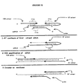
- the detection structure described in Figure 2 is generated for detection of miRNAs.
- two oligonucleotides are annealed to the miRNA to form a double loop or "dumbbell" like structure.
- the dumbbell structure creates a larger region of double-stranded nucleic acid by extending the ends of the miRNA with a double-stranded region of oligonucleotide.
- each end of the miRNA is extended between 2 and 5 nucleotides.
- the ends of the oligonucleotides comprise additional nucleic acid sequences that do not hybridize to the miRNA.
- these additional sequences form invasive cleavage structures (e.g., INVADER assay invasive cleavage structures).
- invasive cleavage structures are detected by the INVADER assay (See e.g., below description).
- oligonucleotides described in Example 18 may be utilized to detect miRNAs associated with cancer (e.g., that form an invasive cleavage structure that can be detected by INVADER assay).
- the detection structure described in Figure 3 is generated for the detection of miRNAs.
- one oligonucleotide is annealed to the miRNA to generate an arched structure.
- the miRNA brings the ends of the oligonucleotide together with greater efficiency than in the absence of the miRNA.
- the ends of the oligonucleotide comprise additional sequences that extend beyond the miRNA and do not hybridize to the miRNA.
- these additional sequences form invasive cleavage structures (e.g., INVADER assay invasive cleavage structures).
- invasive cleavage structures are detected by the INVADER assay (See e.g., below description).
- the resulting ends are ligated to form a circular structure.
- one oligonucleotide is hybridized to a miRNA such that the ends of the oligonucleotide are brought in close proximity (e.g., hybridized to adjacent nucleotides of the miRNA) and are then ligated.
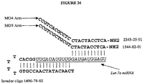
- the detection structures described in Figures 24 and 25 are generated.
- either probe or INVADER oligonucleotides are extended to create a single hairpin loop or "half dumbbell" structure.
- oligonucleotides described in Example 18 may be utilized to detect miRNAs associated with cancer (e.g., that form an invasive cleavage structure that can be detected by INVADER assay).
- the ends of the oligonucleotides comprise additional nucleic acid sequences that do not hybridize to the miRNA (See, e.g., Examples 19G and 19H).
- these additional sequences form invasive cleavage structures (e.g., INVADER assay invasive cleavage structures).
- invasive cleavage structures are detected by the INVADER assay (See e.g., below description).
- these additional sequences are complementary to additional oligonucleotides added to reaction mixtures to stabilize a cleavage structure, e.g. an INVADER assay invasive cleavage structure ( Figure 4 ).
- circular structures generated as described above are detected using a rolling circle replication assay (See e.g., below description of rolling circle replication).
- detection structures are generated from long oligonucleotides (e.g., greater than 50, 100, 1000 or more nucleotides) with short region(s) of homology to miRNAs.
- One or more miRNAs are hybridized to the oligonucleotides to generate detection structures.
- these detection structures are detected by extension of miRNAs ( e.g., via ligation or polymerization reactions such as RT-PCR).
- these detection structures are further detected by hybridization to oligonucleotides conjugated to solid supports, such as microspheres, or other surfaces or structures.
- the non-miRNA component is extended or ligated to another nucleic acid and directly or indirectly detected.
- oligonucleotides used to form detection structures comprise one or more nucleotide analogs.
- 2'-O-methyl nucleotides may be utilized.
- the present invention is not limited to a particular mechanism. Indeed, an understanding of the mechanism is not necessary to practice the present invention. Nonetheless, it is contemplated that the presence of 2'-O-methyl bases increases the stability of the hybridized detection structure and aids in further detection methods.
- nucleic acids e.g., interfering RNAs
- the present description provides methods of detecting miRNAs.
- the present description is not limited to a particular detection assay. Any suitable method may be utilized including, but not limited to, those disclosed herein.
- miRNA detection methods are quantitative.
- the present invention is not limited to a particular mechanism. Indeed, an understanding of the mechanism is not necessary to practice the present invention. Nonetheless, it is contemplated that levels of a particular miRNA in the body are associated with a level of gene expression from their cognate genes.
- the present invention thus provides methods of correlated miRNAs with gene expression of particular genes (e.g., genes involved in disease states (e.g., cancer) or metabolism).
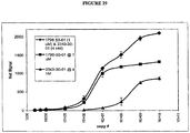
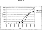
- the methods of the present invention may be utilized to determine the presence of abnormal (e.g., high or low) levels of a particular miRNA (e.g., miRNA expression associated with cancer (See, e.g., Calin et al., Proc Natl Acad Sci USA, 99, 15524-15529 (2002 ), e.g., using oligonucleotides described in Example 18 and Figure 31 ) or to determine the effect of an intervention (e.g., drug) on miRNA expression.
- a particular miRNA e.g., miRNA expression associated with cancer (See, e.g., Calin et al., Proc Natl Acad Sci USA, 99, 15524-15529 (2002 ), e.g., using oligonucleotides described in Example 18 and Figure 31
- an intervention e.g., drug
- heterologous miRNAs e.g., from expression vectors, transgenic constructs, transfection, etc.
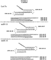
- the present description provides methods of detecting a particular miRNA (e.g., a miRNA such as mir-1 or mir-135). In other examples, the methods of the present description are used to distinguish between variants (e.g., polymorphisms or mutations) in a particular miRNA. In still further examples, the present description provides methods of lysing cells to be tested for the presence of miRNAs.
- a miRNA e.g., a miRNA such as mir-1 or mir-135
- the methods of the present description are used to distinguish between variants (e.g., polymorphisms or mutations) in a particular miRNA.
- the present description provides methods of lysing cells to be tested for the presence of miRNAs.
- the INVADER assay is used for the detection of miRNAs.
- the INVADER assay comprises forming a nucleic acid cleavage structure that is dependent upon the presence of a target nucleic acid and cleaving the nucleic acid cleavage structure so as to release distinctive cleavage products. 5' nuclease activity, for example, is used to cleave the target-dependent cleavage structure and the resulting cleavage products or the cleavage of the cleavage structure is indicative of the presence of specific target nucleic acid sequences in the sample.
- invasive cleavage can occur.
- a cleavage agent e.g., a 5' nuclease
- the cleavage agent can be made to cleave the downstream oligonucleotide at an internal site in such a way that a distinctive fragment is produced.
- INVADER assay Third Wave Technologies
- the INVADER assay detects hybridization of probes to a target by enzymatic cleavage of specific structures by structure specific enzymes ( See, INVADER assays, Third Wave Technologies; See e.g., U.S. Patent Nos. 5,846,717 ; 6,090,543 ; 6,001,567 ; 5,985,557 ; 5,994,069 ; 6,090,543 ; 6,348,314 ; 6,458,535 ; U.S. Patent App. Nos. 20030186238 (Ser. No. 10/084839 ); 20030104378A1 (Ser. No. 09/864636 ); Lyamichev et al., Nat. Biotech., 17:292 (1999 ), Hall et al., PNAS, USA, 97:8272 (2000 ), WO97/27214 and WO98/42873 ).
- the INVADER assay detects specific DNA and RNA sequences by using structure-specific enzymes (e.g. FEN endonucleases) to cleave a complex formed by the hybridization of overlapping oligonucleotide probes ( See, e.g. Figure 1 ). Elevated temperature and an excess of one of the probes enable multiple probes to be cleaved for each target sequence present without temperature cycling. These cleaved probes then may direct cleavage of a second labeled probe.
- the secondary probe oligonucleotide can be 5'-end labeled with fluorescein that is quenched by an internal dye. Upon cleavage, the de-quenched fluorescein labeled product may be detected using a standard fluorescence plate reader.
- the INVADER assay detects specific sequences, mutations, and SNPs in unamplified, as well as amplified (See, e.g., Example 19, Figure 32 ), RNA and DNA, including genomic DNA.
- the INVADER assay uses two cascading steps (a primary and a secondary reaction) both to generate and then to amplify the target-specific signal.
- WT wild-type
- MT mutant
- the primary reaction Figure 1 , panel A
- the WT primary probe and the INVADER oligonucleotide hybridize in tandem to the target nucleic acid to form an overlapping structure.
- a structure-specific enzyme e.g. the CLEAVASE enzyme, Third Wave Technologies
- this cleaved product serves as an INVADER oligonucleotide on the WT fluorescence resonance energy transfer (WT-FRET) probe to again create the structure recognized by the structure specific enzyme (panel A).
- WT-FRET WT fluorescence resonance energy transfer
- FRET probes having different labels may be provided for each allele or locus to be detected, such that the different alleles or loci can be detected in a single reaction.
- the primary probe sets and the different FRET probes may be combined in a single assay, allowing comparison of the signals from each allele or locus in the same sample.
- the primary probe oligonucleotide and the target nucleotide sequence do not match perfectly at the cleavage site (e.g., as with the MT primary probe and the WT target, Figure 1 , panel B), the overlapped structure does not form and cleavage is suppressed.
- the structure specific enzyme e.g., CLEAVASE VIII enzyme, Third Wave Technologies
- each target-specific product can enable the cleavage of many FRET probes.
- the primary INVADER assay reaction is directed against the target DNA (or RNA) being detected.
- the target DNA is the limiting component in the first invasive cleavage, since the INVADER and primary probe are supplied in molar excess.
- the second invasive cleavage it is the released flap that is limiting.
- the INVADER assay or other nucleotide detection assays, are performed with accessible site-designed oligonucleotides and/or bridging oligonucleotides.
- accessible site-designed oligonucleotides and/or bridging oligonucleotides are described in U.S. Pat. 6,194,149 , 6,358,691 , 6355, 437 , U.S. Pat. Application Ser. No. 09/882,945 , and PCT Applications WO9850403 , and WO0198537 .
- the exposing of the sample e.g., nucleic acid sequence (e.g., interfering RNA (e.g., miRNA or siRNA))
- the agent comprises exposing the sample to the oligonucleotides and the agent under conditions wherein an invasive cleavage structure is formed between said target sequence and said oligonucleotides if said target sequence is present in said sample, wherein said invasive cleavage structure is cleaved by said cleavage agent to form a cleavage product.
- the target sequence (e.g. miRNA) comprises a first region and a second region, the second region downstream of and contiguous to the first region, and the oligonucleotides comprise first and second oligonucleotides, wherein at least a portion of the first oligonucleotide is completely complementary to the first portion of the target sequence and wherein the second oligonucleotide comprises a 3' portion and a 5' portion, wherein the 5' portion is completely complementary to the second portion of the target nucleic acid.
- the exposing of the sample to the oligonucleotides and the agent comprises exposing the sample to the oligonucleotides and the agent under conditions wherein an invasive cleavage structure is formed between the target sequence and the oligonucleotides if the target sequence is present in the sample, wherein the invasive cleavage structure is cleaved by the cleavage agent to form a cleavage product.
- the target sequence comprises a first region and a second region, said second region downstream of and contiguous to said first region, and said oligonucleotides comprise first and second oligonucleotides, wherein at least a portion of said first oligonucleotide is completely complementary to said first portion of said target sequence and wherein said second oligonucleotide comprises a 3' portion and a 5' portion, wherein said 5' portion is completely complementary to said second portion of said target nucleic acid.
- kits for assaying a pooled sample e.g., a pooled blood sample or pooled cell lysates
- INVADER detection reagents e.g. primary probe, INVADER probe, and FRET cassette
- the kit further comprises instructions on how to perform the INVADER assay, and in some examples, how to apply the INVADER detection assay to pooled samples from many individuals, or to "pooled" samples from many cells ( e.g., from a biopsy sample) from a single subject.
- the present description further provides assays in which the target nucleic acid is reused or recycled during multiple rounds of hybridization with oligonucleotide probes and cleavage of the probes without the need to use temperature cycling (i.e., for periodic denaturation of target nucleic acid strands) or nucleic acid synthesis (i.e., for the polymerization-based displacement of target or probe nucleic acid strands).
- temperature cycling i.e., for periodic denaturation of target nucleic acid strands
- nucleic acid synthesis i.e., for the polymerization-based displacement of target or probe nucleic acid strands.
- a cleavage reaction is run under conditions in which the probes are continuously replaced on the target strand (e.g . through probe-probe displacement or through an equilibrium between probe/target association and disassociation, or through a combination comprising these mechanisms, ( Reynaldo et al., J. Mol. Biol. 97: 511-520
- two oligonucleotides hybridize in tandem to the target DNA to form an overlapping structure.
- the 5'-end of the primary probe includes a 5'-flap that does not hybridize to the target DNA ( Figure 1 ).
- the 3'-nucleotide of the bound INVADER oligonucleotide overlaps the primary probe, but need not hybridize to the target DNA (See, e.g., Examples 15 and 16).
- the CLEAVASE enzyme recognizes this overlapping structure and cleaves off the unpaired 5'-flap of the primary probe, releasing it as a target-specific product.
- the primary probe is designed to have a melting temperature close to the reaction temperature.
- each released 5'-flap can serve as an INVADER oligonucleotide on a fluorescence resonance energy transfer (FRET) Cassette to create another overlapping structure that is recognized and cleaved by the CLEAVASE enzyme ( Figure 1 ).
- FRET fluorescence resonance energy transfer
- the fluorophore (F) and quencher (Q) are separated, generating detectable fluorescence signal.
- the released 5'-flap and the FRET Cassette cycle resulting in amplified fluorescence signal.
- the initial and secondary reactions run concurrently in the same well.
- the biplex format of the INVADER DNA Assay enables simultaneous detection of two DNA sequences in a single well (See, e.g., Examples 17 and 19(L)). Most often, this involves detection of two variants of a particular polymorphism ( e.g., in a miRNA).
- the biplex format uses two different discriminatory Primary Probes, each with a unique 5'-flap, and two different FRET Cassettes, each with a spectrally distinct fluorophore. By design, the released 5'-flaps will bind only to their respective FRET Cassettes to generate a target-specific signal.
- kits comprising one or more of the components necessary for practicing the present invention.
- the present description provides kits for storing or delivering the enzymes of the present invention and/or the reaction components necessary to practice a cleavage assay (e.g., the INVADER assay).
- kits of the present description may provide the following reagents: CLEAVASE enzyme Primary Probe Oligos DNA Reaction Buffer 1 INVADER Oligo FRET Cassette 1 ( e.g ., F) FRET Cassette 2 ( e.g ., R) Mutant DNA controls Wild type DNA controls "No Target" Blank control
- the kits of the present description are configured for direct detection of RNA.
- kits may provide the following reagents: CLEAVASE enzyme Primary Probe oligonucleotides DNA Reaction Buffer 1 INVADER Oligo FRET Probe 1 ( e.g ., F) FRET Probe 2 ( e.g ., R) Secondary Reaction Target 1 Secondary Reaction Target 2 ARRESTOR oligonucleotide 1 ARRESTOR oligonucleotide 2 Mutant DNA controls Wild type DNA controls "No Target" Blank control
- the designs of the probes sets are adapted for use in miRNA detection assays using the guidelines for reaction design and optimization provided herein (See e.g., the Experimental Section).
- the reaction temperature is reduced ( e.g., to 50-60°C) to account for the smaller region of hybridization.
- a kit of the present description provides a list of additional components (e.g., reagents, supplies, and/or equipment) to be supplied by a user in order to perform the methods of the description.
- additional components e.g., reagents, supplies, and/or equipment
- a list of additional components comprises the following: ⁇ Clear CHILLOUT-14 liquid wax (MJ Research) or RNase-free, optical grade mineral oil (Sigma, Cat. No. M-5904) ⁇ 96-well polypropylene microplate (MJ Research, Cat. No.
- MSP-9601 Sterile 1.5-ml or 2.0-ml microcentrifuge tubes ⁇ Sterile, DNase/RNase free disposable aerosol barrier pipet tips ⁇ Multichannel pipets (0.5-10 ⁇ l, 2.5-20 ⁇ l) ⁇ Thermal cycler or other heat source (e.g., lab oven or heating block). ⁇ Miscellaneous laboratory equipment (tube racks, micropipetors, multichannel pipet, microcentrifuge, vortex mixer).
- Fluorescence microplate reader (a preferred plate reader is top-reading and equipped with light filters have the following characteristics: Excitation (Wavelength/Bandwidth) Emission (Wavelength/Bandwidth) 485 nm/20 nm 530 nm/25 nm 560 nm/20 nm 620 nm/40 nm
- kits of the present description provides a list of optional components (e.g., reagents, supplies, and/or equipment) to be supplied by a user to facilitate performance of the methods of the invention.
- optional components e.g., reagents, supplies, and/or equipment
- one example of such a list comprises the following:
- a kit of the present description provides a list of required components to be supplied by a user to facilitate performance of the methods of the description for which multiple alternatives are acceptable (e.g. sample preparation kits).
- a kit of the present description provides a list of required components to be supplied by a user to facilitate performance of the methods of the description for which multiple alternatives are acceptable (e.g. sample preparation kits).
- sample preparation kits e.g. sample preparation kits
- protocols for the assembly of INVADER assay reactions e.g., formulations and preferred procedures for making reaction mixtures
- protocols for assembly of reaction mixtures include computational or graphical aids to reduce risk of error in the performance of the methods of the present description (e.g., tables to facilitate calculation of volumes of reagents needed for multiple reactions, and plate-layout guides to assist in configuring multi-well assay plates to contain numerous assay reactions).
- supplementary documentation such as protocols for ancillary procedures, e.g ., for the preparation of additional reagents, or for preparation of samples for use in the methods of the present description, are provided.
- supplementary documentation includes guidelines and lists of precautions provided to facilitate successful use of the methods and kits by unskilled or inexperienced users.
- supplementary documentation includes a troubleshooting guide, e.g., a guide describing possible problems that may be encountered by users, and providing suggested solutions or corrections to intended to aid the user in resolving or avoiding such problems.
- samples are diluted to concentrations that correspond to a 10- ⁇ l addition per reaction.
- concentration of a 100-ng sample should be 15ng/ ⁇ l.
- rolling circle replication methods are utilized for detection of miRNA detection structures (See e.g., U.S. Patents 6,344,329 ; 6,143,495 ; 6,316,229 ; 6,210,884 , 6,183,960 and 6,235,502 ).
- rolling circle replication is used to detect circular miRNA detection structures generated from the annealing of the ends of a single oligonucleotide annealed to a miRNA.
- the ends of the oligonucleotide hybridize to the miRNA with no overlap.
- This oligonucleotide can be ligated in the presence or absence of miRNA. However, the ligation reaction is more efficient in the presence of the miRNA. In such examples, the level of circular molecules detected over time is compared to a control reaction lacking miRNA.
- the ends of the oligonucleotide hybridize to the miRNA with overlapping ends to generate an invasive cleavage structure.
- Such structures are cleaved prior to ligation, thus improving the specificity of the generation of the circular detection structure.
- Rolling circle amplification involves replication of circular single-stranded DNA molecules.
- a rolling circle replication primer hybridizes to circular nucleic acid molecules followed by rolling circle replication of the nucleic acid molecules using a strand-displacing DNA polymerase. Amplification takes place during rolling circle replication in a single reaction cycle.
- Rolling circle replication results in large DNA molecules containing tandem repeats of the nucleic acid sequence. This DNA molecule is referred to as a tandem sequence DNA (TS-DNA).
- TS-DNA tandem sequence DNA
- LM-RCA ligation-mediated rolling circle amplification
- an probe hybridizes to its cognate target nucleic acid sequence, if present, followed by ligation of the ends of the hybridized probe to form a covalently closed, single-stranded nucleic acid.
- a rolling circle replication primer hybridizes to probe molecules followed by rolling circle replication of the circular molecules using a strand-displacing DNA polymerase.
- LM-RCA comprises mixing an open circle probe with a target sample, resulting in an probe-target sample mixture, and incubating the probe-target sample mixture under conditions promoting hybridization between the open circle probe and a target sequence, mixing ligase with the probe-target sample mixture, resulting in a ligation mixture, and incubating the ligation mixture under conditions promoting ligation of the open circle probe to form an amplification target circle (ATC), mixing a rolling circle replication primer (RCRP) with the ligation mixture, resulting in a primer-ATC mixture, and incubating the primer-ATC mixture under conditions that promote hybridization between the amplification target circle and the rolling circle replication primer, mixing DNA polymerase with the primer-ATC mixture, resulting in a polymerase-ATC mixture, and incubating the polymerase-ATC mixture under conditions promoting replication of the amplification target circle, where replication of the amplification target circle results in formation of tandem sequence DNA (TS-DNA).
- ATC amplification target circle
- RCRP
- the present description is not limited to INVADER assay or rolling circle assay detection. Any method that allows for the detection of miRNA detection structures may be utilized. Exemplary, non-limiting detection assay that find use in the methods of the present description are described below.
- detection structures are detected using a hybridization assay.
- a hybridization assay the presence of absence of a given nucleic acid sequence is determined based on the ability of the DNA from the sample to hybridize to a complementary DNA molecule (e.g., a oligonucleotide probe).
- a complementary DNA molecule e.g., a oligonucleotide probe.
- sequences are detected using a DNA chip hybridization assay.
- a DNA chip hybridization assay a series of oligonucleotide probes are affixed to a solid support.
- the oligonucleotide probes are designed to be unique to a given target sequence (e.g., component of a detection complex).
- the sample of interest is contacted with the DNA "chip” and hybridization is detected.
- the DNA chip assay is a GeneChip (Affymetrix, Santa Clara, CA; See e.g., U.S. Patent Nos. 6,045,996 ; 5,925,525 ; and 5,858,659 ) assay.
- the GeneChip technology uses miniaturized, high-density arrays of oligonucleotide probes affixed to a "chip.” Probe arrays are manufactured by Affymetrix's light-directed chemical synthesis process, which combines solid-phase chemical synthesis with photolithographic fabrication techniques employed in the semiconductor industry.
- the process constructs high-density arrays of oligonucleotides, with each probe in a predefined position in the array. Multiple probe arrays are synthesized simultaneously on a large glass wafer. The wafers are then diced, and individual probe arrays are packaged in injection-molded plastic cartridges, which protect them from the environment and serve as chambers for hybridization.
- the nucleic acid to be analyzed is isolated, amplified by PCR, and labeled with a fluorescent reporter group.
- the labeled DNA is then incubated with the array using a fluidics station.
- the array is then inserted into the scanner, where patterns of hybridization are detected.
- the hybridization data are collected as light emitted from the fluorescent reporter groups already incorporated into the target, which is bound to the probe array. Probes that perfectly match the target generally produce stronger signals than those that have mismatches. Since the sequence and position of each probe on the array are known, by complementarity, the identity of the target nucleic acid applied to the probe array can be determined.
- a DNA microchip containing electronically captured probes (Nanogen, San Diego, CA) is utilized (see e.g., U.S. Patent Nos. 6,017,696 ; 6,068,818 ; and 6,051,380 ).
- Nanogen's technology enables the active movement and concentration of charged molecules to and from designated test sites on its semiconductor microchip.
- DNA capture probes unique to a given target sequence are electronically placed at, or "addressed" to, specific sites on the microchip. Since DNA has a strong negative charge, it can be electronically moved to an area of positive charge.
- a test site or a row of test sites on the microchip is electronically activated with a positive charge.
- a solution containing the DNA probes is introduced onto the microchip.
- the negatively charged probes rapidly move to the positively charged sites, where they concentrate and are chemically bound to a site on the microchip.
- the microchip is then washed and another solution of distinct DNA probes is added until the array of specifically bound DNA probes is complete.
- a test sample is then analyzed for the presence of target sequences by determining which of the DNA capture probes hybridize, with target sequences.
- An electronic charge is also used to move and concentrate target molecules to one or more test sites on the microchip.
- the electronic concentration of sample DNA at each test site promotes rapid hybridization of sample DNA with complementary capture probes (hybridization may occur in minutes).
- the polarity or charge of the site is reversed to negative, thereby forcing any unbound or nonspecifically bound DNA back into solution away from the capture probes.
- a laser-based fluorescence scanner is used to detect binding.
- an array technology based upon the segregation of fluids on a flat surface (chip) by differences in surface tension is utilized (see e.g., U.S. Patent Nos. 6,001,311 ; 5,985,551 ; and 5,474,796 ).
- Protogene's technology is based on the fact that fluids can be segregated on a flat surface by differences in surface tension that have been imparted by chemical coatings. Once so segregated, oligonucleotide probes are synthesized directly on the chip by ink-jet printing of reagents.
- the array with its reaction sites defined by surface tension is mounted on a X/Y translation stage under a set of four piezoelectric nozzles, one for each of the four standard DNA bases.
- the translation stage moves along each of the rows of the array and the appropriate reagent is delivered to each of the reaction sites.
- the A amidite is delivered only to the sites where amidite A is to be coupled during that synthesis step and so on.
- Common reagents and washes are delivered by flooding the entire surface and then removing them by spinning.
- DNA probes unique for the target sequence (e.g., component of a detection complex) of interest are affixed to the chip using Protogene's technology.
- the chip is then contacted with the PCR-amplified genes of interest.
- unbound DNA is removed and hybridization is detected using any suitable method (e.g., by fluorescence de-quenching of an incorporated fluorescent group).
- a "bead array” is used for the detection of polymorphisms (Illumina, San Diego, CA; See e.g., PCT Publications WO 99/67641 and WO 00/39587 ).
- Illumina uses a BEAD ARRAY technology that combines fiber optic bundles and beads that self-assemble into an array. Each fiber optic bundle contains thousands to millions of individual fibers depending on the diameter of the bundle.
- the beads are coated with an oligonucleotide specific for the detection of a given SNP or mutation. Batches of beads are combined to form a pool specific to the array.
- the BEAD ARRAY is contacted with a prepared subject sample (e.g., nucleic acid sample). Hybridization is detected using any suitable method.
- hybridization is detected by enzymatic cleavage of specific structures.
- hybridization of a bound probe is detected using a TaqMan assay (PE Biosystems, Foster City, CA; See e.g., U.S. Patent Nos. 5,962,233 and 5,538,848 ).
- the assay is performed during a PCR reaction.
- the TaqMan assay exploits the 5'-3' exonuclease activity of the AMPLITAQ GOLD DNA polymerase.
- a probe, specific for a given allele or mutation, is included in the PCR reaction.
- the probe consists of an oligonucleotide with a 5'-reporter dye (e.g., a fluorescent dye) and a 3'-quencher dye.
- the 5'-3' nucleolytic activity of the AMPLITAQ GOLD polymerase cleaves the probe between the reporter and the quencher dye.
- the separation of the reporter dye from the quencher dye results in an increase of fluorescence.
- the signal accumulates with each cycle of PCR and can be monitored with a fluorimeter.
- polymorphisms are detected using the SNP-IT primer extension assay (Orchid Biosciences, Princeton, NJ; See e.g., U.S. Patent Nos. 5,952,174 and 5,919,626 ).
- SNPs are identified by using a specially synthesized DNA primer and a DNA polymerase to selectively extend the DNA chain by one base at the suspected SNP location. DNA in the region of interest is amplified and denatured. Polymerase reactions are then performed using miniaturized systems called microfluidics. Detection is accomplished by adding a label to the nucleotide suspected of being at the target sequence location. Incorporation of the label into the DNA can be detected by any suitable method ( e.g., if the nucleotide contains a biotin label, detection is via a fluorescently labeled antibody specific for biotin).
- Additional detection assays useful in the detection of miRNA detection structures include, but are not limited to, enzyme mismatch cleavage methods (e.g., Variagenics, U.S. Pat. Nos. 6,110,684 , 5,958,692 , 5,851,770 ); polymerase chain reaction (See, e.g., Example 19 and Figure 32 ); branched hybridization methods (e. g ., Chiron, U.S. Pat. Nos. 5,849,481 , 5,710,264 , 5,124,246 , and 5,624,802 ); NASBA ( e.g., U.S. Pat. No. 5,409,818 ); molecular beacon technology (e.g., U.S. Pat.
- the oligonucleotide design for let-7 is shown in Figure 5 .
- the oligonucleotide design for mir-1 is shown in Figure 5 .
- the following primary mixes were made and incubated at 50°C ⁇ 10°C in a 96 well plate for 30 minutes.
- a no target master mix was prepared (addition of H 2 O in place of RNA). All reactions were covered with mineral oil to prevent evaporation.
- Probe oligonucleotide SEQ ID NOs: 2, 6, or 9 for let 7; SEQ ID NOs: 12, 16, or 19 for miR-1) 10 ⁇ M 1 ⁇ l INVADER oligonucleotide (SEQ ID NOs: 1, 5, or 8 for let 7; SEQ ID NOs: 11, 15, or 18 for miR-1) 10 ⁇ M 1 ⁇ l CLEAVASE IX or XII enzyme 40 ng/ ⁇ l CLEAVASE IX enzyme or 60 ng/ ⁇ l CLEAVASE XII enzyme 0.5 ⁇ l tRNA 20 ng/ ⁇ l 1.5 ⁇ l Synthetic miRNA (SEQ ID NO: 4 for let-7a; SEQ ID NO: 14 for miR-1) 100 pM 2 ⁇ l Total 10 ⁇ l
- Extending the 2'-O-methylated bases of the INVADER oligonucleotide so that they base pair with the first two bases of the miRNA (SEQ ID NO: 8 (1496-96-02) vs. SEQ ID NO: 23 (1496-96-03) in design SEQ ID NO: 9 (1496-96-01R) of let-7a) increases the temperature optimum of the described reaction but does not enhance the detection.
- the LOD for let-7 and mir-1 was next tested on human RNA samples.
- the protocol described above was utilized. 50 - 100 ng of tissue specific total human RNA samples (Clonetech, Palo Alto, CA) was used. Results are shown in Figures 8 and 10 .
- tissue specific total human RNA samples (Clonetech, Palo Alto, CA) was used. Results are shown in Figures 8 and 10 .
- the let-7a INVADER assay detects the same tissue expression profile as seen before for let-7a expression levels depending on the source of tissue (Pasquinelli et al., 408:86 (2000)).
- This Example describes an analysis of the cross reactivity of probe and/or INVADER oligonucleotides directed against one sub-type of let-7 for another sub-type.
- the protocol for synthetic let-7a miRNA setup described in Example 3 was utilized.
- Figure 5 shows the oligonucleotide designs.
- the following plate setup was used: 10 nM 1 nM 100 pM 10 pM 1 pM 100 fM 10 fM H2O 1 2 3 4 5 6 7 8 Let 7 A A Let 7 A B Let 7 C C Let 7 C D Let 7 E E Let 7 E F Let 7 F G Let 7 F H
- This Example describes the optimization of CLEAVASE enzymes for use in miRNA assays.
- the protocol for temperature optimization described above was utilized. Either 20 ng of the CLEAVASE IX enzyme (Third Wave Technologies, Madison, WI) or 30 ng of the CLEAVASE XII enzyme (Third Wave Technologies, Madison, WI) was used. The following buffer was used for the CLEAVASE IX enzyme:
- This example describes assay design and LOD determination for miRNA miR135. Experiments were performed as described in Examples 2 and 3 for miR-1. The oligonucleotide designs are described in Figure 5 . Each of the designs (A-D) utilizes different INVADER and probe oligonucleotides for the detection of mir-135 miRNA. Results of the temperature optimization experiments comparing performance of all of the designs are shown in Figure 13 . Design D gave the highest signal. Results of LOD experiments using assay design D are shown in Figure 14 .
- Figure 14A presents the raw counts generated from four replicate assays at each of the indicated target concentrations. The average counts obtained with each target concentration are indicated as are the net signal and fold-over-zero (FOZ). The limit of detection of the miR-135 target in this experiment was 164 zmoles, equivalent to 98,743 molecules.
- Figure 14B contains a graphical representation of the average counts obtained at each concentration and indicates that the INVADER assay is linear throughout much of the concentration range tested.
- RNA generally present in all cells at constant levels along with one or more miRNA species, which may be expressed in a tissue-specific manner.
- INVADER assays were therefore designed to two distinct RNAs generally found in all cell types: human glyceraldehydes-3-phosphate dehydrogenase (hGAPDH) and U6 RNA.
- oligonucleotides have been used in biplex miRNA detection assays: INVADER oligonucleotide (SEQ ID NO: 41); probe (SEQ ID NO: 42); ARRESTOR oligonucleotide (SEQ ID NO: 43); SRT oligonucleotide (SEQ ID NO:49), FRET oligonucleotide (red dye) (SEQ ID NO: 48).
- INVADER oligonucleotide SEQ ID NO: 41
- probe SEQ ID NO: 42
- ARRESTOR oligonucleotide SEQ ID NO: 43
- SRT oligonucleotide SEQ ID NO:49
- FRET oligonucleotide red dye
- This example describes detection of the let-7 miRNA directly in total cell RNA as well as in uninduced fibroblast cells from a human osteosarcoma cell line, MG63 (Third Wave Technologies, Madison, WI; catalog number CRL-1427).
- Total cell RNA was extracted using TRIZOL (Gibco-BRL), as previously described ( Chomczynski et al., Anal. Biochem. 162: 156-156 (1987 )), and cell lysates were prepared as described by Eis et al., Nature Biotechnology, 19: 673-6 (2001 ).
- Reactions were set up as follows. Aliquots of 5 ⁇ l of either cell lysate, synthetic miRNA target in lysis buffer ( Eis et al., Nature Biotechnology, 19: 673-6 (2001 )) at the indicated concentrations, or 5 ⁇ l of 20 ng/ ⁇ l of tRNA (for the no target controls) were pipetted into the appropriate wells of a microtiter plate. A primary reaction master mix was made for 96 reactions containing the following reagents.
- Probe oligonucleotide 1496-78-01 R SEQ ID NO:6
- INVADER oligonucleotide 1496-78-02 SEQ ID NO: 5
- CLEAVASE XII enzyme 60 ng/ ⁇ l 0.5
- Hela cells (7.5 X 10 6 cells) were suspended in 100 ⁇ l of a solution of 10 mM MOPS buffer, pH 7.5, with 100 mM KCl. Aliquots of 10 ⁇ l were added to separate tubes and lysed with 100 ⁇ l of two different lysis buffers prepared as follows: MOPS lysis w/Mg ++ MOPS lysis w/out Mg ++ 180 ⁇ l 11 ⁇ g/ml tRNA 180 ⁇ l 11 ⁇ g/ml tRNA 0.5 ml NP40 0.5 ml NP40 4 ml 0.5 M MOPS 4 ml 0.5 M MOPS 0.5 ml 1 M MgCl 2 N/A 4.82 ml H 2 O 5.32 mls H 2 O 10 mls 10 mls
- Secondary reaction mixture included SRTs (SEQ ID NO: 22 for let-7; SEQ ID NO: 49 for GAPDH and U6) target, FRET oligonucleotides (SEQ ID NO: 21 for let-7; SEQ ID NO: 48 for GAPDH and U6), ARRESTORs (SEQ ID NO:7 for let-7, SEQ ID NO:43 for GAPDH, and SEQ ID NO: 47 for U6) at the concentrations indicated in Example 7A.
- This example describes the creation and testing of alternative oligonucleotide designs for detection of the let-7a miRNA.
- a set of alternative designs was created in which the target specific regions of both the INVADER oligonucleotide and the probe oligonucleotide were eleven nucleotides long.
- a second set of designs was created in which the target specific regions of the probe oligonucleotides were 10 nucleotides long and the target specific regions of the INVADER oligonucleotides were 12 nucleotides long.
- Figure 5 shows sets of alternative oligonucleotide designs for detection of the let-7a miRNA in which the target specific regions of both the probe and INVADER oligonucleotides are 11 nucleotides long.
- SEQ ID NOs: 50-51 provide a design in which both the INVADER and probe oligonucleotides are linear.
- SEQ ID NO: 6 contains a probe oligonucleotide that forms a stem-loop structure); SEQ ID NO: 5, an INVADER oligonucleotide that forms a stem-loop structure); SEQ ID NOs: 5-6, both probe and INVADER oligonucleotides with stem-loops.
- Figure 5 shows a set of alternative oligonucleotide designs for detection of the let-7a miRNA in which the target specific regions of the probe comprise 10 nucleotides, and those of the INVADER oligonucleotides, 12 nucleotides.
- SEQ ID NOs: 52-53 provide a design in which both the INVADER and probe oligonucleotides are linear.
- SEQ ID NO: 2 contains a probe oligonucleotide that forms a stem-loop structure);
- SEQ ID NO: 1 an INVADER oligonucleotide that forms a stem-loop structure).
- Temperature optimization experiments were carried out as follows. A master mix was made for 24 reactions. Each reaction contained the following: Stock concentration Volume per reaction Final concentration 2.5 X Primary reaction buffer for the CLEAVASE XII enzyme (as described in Example 5) 4 ⁇ l 1 X 10 ⁇ M probe* 1 ⁇ l 1 ⁇ M 100 ⁇ M INVADER oligonucleotide* 1 ⁇ l 10 ⁇ M 60 ng/ ⁇ l CLEAVASE 12 0.5 ⁇ l 30 ng H 2 O 2.5 ⁇ l N/A 30 pM miRNA (for the 11-mer temperature optimizations) OR 10 nM miRNA (for the 10-mer probe/12-mer INVADER oligonucleotide temperature optimizations) 1 ⁇ l 3 pM OR 1 nM 20 ng/ ⁇ l tRNA (for no target controls only) 1 ⁇ l 2 ng TOTAL 10 ⁇ l *Various combinations of probe and INVADER oligonucleotides were used in this experiment as indicated in Figures 16-17 .
- the primary reactions were run at 50 ⁇ 9°C for 1 hour followed by a 15 minute secondary reaction at 60°C as described in Example 2.
- the primary reactions were run at 50 ⁇ 9°C for 15 minutes followed by a 15 minute secondary reaction at 60°C.
- Results for the designs in which the target specific portions of the INVADER and probe oligonucleotides were 11 nucleotides long are presented in Figure 18.
- Figure 18A shows the temperature optimization profiles of each design.
- Figure 18B shows the normalized maximum performance of each design, including the optimum temperature for each.
- Results for the designs in which the target specific portion of the probe oligonucleotide was 10 bases and that of the INVADER oligonucleotide was 12 are presented in Figure 19.
- Figure 19A shows the temperature optimization profiles, and Figure 19B , the normalized maximum performance of each design.
- Example 3 Experiments were set up as described in Example 3 to compare the LOD of the double loop design and the single loop design, in which the INVADER oligonucleotide forms a stem-loop structure.
- Reactions to determine LOD were run in quadruplicate.
- Reaction mixtures contained the following reagents (final concentrations): Stock concentration Volume per reaction Final concentration 2.5 X
- Primary reaction buffer for CLEAVASE XII enzyme 4 ⁇ l 1 X 10 ⁇ M probe/200 ⁇ M INVADER oligonucleotide mix (sequences as indicated in Figure 20 ) 0.5 ⁇ l 1 ⁇ M probe/20 ⁇ M INVADER oligonucleotide 60 ng/ ⁇ l CLEAVASE XII enzyme 0.5 ⁇ l 30 ng Total 5 ⁇ l
- Reactions were set up as follows to detect synthetic let-7a miRNA: Component Stock concentration Amount added per reaction PI mix (probe SEQ ID NO: 6; INVADER oligonucleotide SEQ ID NO: 5) 10 ⁇ M probe 1 ⁇ l 50 ⁇ M INVADER oligo CLEAVASE XII enzyme (60ng/ ⁇ l) 0.5 ⁇ l H 2 O 0.5 ⁇ l Primary Reaction Buffer 2.5 X 4 ⁇ l Total 6 ⁇ l
- This example describes experiments aimed at assessing the effect of substituting 2'-deoxy residues for some or all of the 2'-O-methyl residues incorporated in the probe and INVADER oligonucleotides used for detecting miRNAs. All of the designs presented in the preceding examples include 2'-O-methyl residues in the loop regions as described in Example 2. Experiments were conducted to test the effect of substituting 2' deoxy residues for some or all of the 2'-O-methyl residues in the INVADER and probe oligonucleotides designed to detect the let-7a miRNA.
- Figure 5 shows the modified let-7a designs.
- SEQ ID NOs: 5-6 contain 2'-O-methyl residues as described in Example 2.
- the design in SEQ ID NOs: 73-74 contain 2'deoxy residues at all positions; and those in SEQ ID NOs: 75-76, 2'-O-methyl residues in the portions of the stems adjacent to the target.
- INVADER reactions were set up to compare the signal generation and temperature optima of the three different designs. Reactions were as described in the LOD experiments in Example 8 and included 100 pM synthetic miRNA, 1 ⁇ M probe, and 10 ⁇ M INVADER oligonucleotide. Primary reactions were run for 15 minutes at the temperatures indicated; secondary reactions were run for 5 minutes at 60°C.
- Secondary reaction mixes were made as follows. Secondary reaction component Concentration Amount Added FAM FRET oligonucleotide 13.4 ⁇ l FAM FRET 0.75 ⁇ l (SEQ ID NO: 21) and Secondary Reaction Target (SRT) (SEQ ID NO: 40) 2 ⁇ M SRT ARRESTOR oligonucleotide (SEQ ID NO: 66) 54 ⁇ M 0.75 ⁇ l H 2 O 3.5 ⁇ l Total 5 ⁇ l
- RNA e.g. lysate, purified total RNA, synthetic miRNA
- additional experiments in which various oligonucleotides were gel purified as described in Example 1 indicated that standard gel purification of all three types of oligonucleotides gives maximal signal. It is possible to achieve signal levels approximately equal to the maximal levels with gel purified probes if the other oligonucleotides, i.e. the INVADER and ARRESTOR oligonucleotides, are desalted following synthesis.
- This example describes experiments carried out to test the suitability of the INVADER assay to detect different miRNA species in total RNA extracted from diverse tissue types.
- temperature optima and LODs were first determined for each design.
- INVADER assay oligonucleotides were designed to detect the miR-15, miR-16, and miR-125b miRNA species. The designs for these assays are presented in Figure 5 . The designs for let-7a and miR-135 are described in Examples 2 and 6, respectively.
- Example 8 Temperature optimization experiments were conducted for each of these oligonucleotide sets as described in Example 8. Each primary reaction included 1 nM of the targeted miRNA and was carried out for 15 minutes at temperatures ranging from 50 ⁇ 9°C. Secondary reactions were as described in Example 2 and were run for 1 to 1.5 hours at 60°C. Optimum temperatures were as follows: let-7a 53 °C miR-15 53 °C miR-16 56 °C MiR-125b 52 °C MiR-135 45 °C
- let-7a 50 ng of total RNA was tested in each reaction; for the other miRNA species, 100 ng of total RNA was tested. All reactions were set up as described in Example 8; primary reactions were run at the temperature optima for 1.5 hours; secondary reactions were as described above.
- the gene expression profiles for each miRNA species are presented in Figure 23 . These results indicate that the INVADER assay can be used to examine miRNA expression in different tissue types. These data further suggest that let-7a and miR-125b are expressed in a wide variety of tissues; the other miRNA species appear to be more specific to a limited number of tissue types.
- This example describes the impact of alterations in probe and INVADER oligonucleotide length on detection of the let-7a 22-nt miRNA.
- these experiments compare detection of an miRNA that forms perfect stacking interactions between the ends of the probe and INVADER oligonucleotides to detection of an miRNA that forms both 5' and 3' overlaps as well as to one that results in a single nucleotide gap at both the 5' and 3' ends.
- Figure 24 shows the results of analyzing three different types of designs.
- SEQ ID NOs: 5-6 shows a perfect stack between the 22-nt target and the flanking ends of the looped probe and INVADER oligonucleotides.
- SEQ ID NOs: 83-84 both the probe and INVADER oligonucleotides are extended by a single base, resulting in both 5' and 3' overlaps.
- SEQ ID NOs: 85-86 both the probe and INVADER oligonucleotides are shortened by a single base, relative to the designs in SEQ ID NOs: 5-6, resulting in a single nucleotide gap at both ends.
- INVADER assays were set up to test the performance of these oligonucleotide sets for detection of synthetic let-7a miRNAs. Reactions were carried out as described in Example 8 and included 100 pM synthetic let-7a miRNA, 1 ⁇ M probe and 10 ⁇ M INVADER oligonucleotide. Primary reactions were run for 15 minutes at 53°C; secondary reactions, for 5 minutes at 60°C, as described in Example 2. The results are presented in Figure 24 .
- Precursor let-7 RNA (SEQ ID NO: 87) was transcribed in vitro and analyzed by capillary electrophoresis to determine whether it contained any fragments that might mimic the let-7a miRNA. The shortest contaminating fragment was estimated to be approximately 45 nt. LOD reactions were run essentially as described in Example 3 at precursor or synthetic 5' P let-7a mi-RNA concentrations as indicated in the table below. PI mixes contained 10 ⁇ M probe SEQ ID NO: 6 and 100 ⁇ M INVADER oligonucleotide SEQ ID NO: 5.
- RNAse A Qiagen, Inc.
- Reactions were run to detect let-7a miRNA in cell lysates as described in Example 7.
- aliquots 1 ⁇ l of 8 ⁇ g/ ⁇ l RNAse A (Qiagen, Inc.) were added to 80 ⁇ l of cell lysate and incubated at 37°C for 2.25 hours.
- the RNAse A treated samples failed to generate any signal above background, indicating that signal generated in assays lacking RNAse A arises from detection of the miRNA target and not the encoding DNA ( Figure 16 ). Further experiments were carried out in which RNAse A was added either prior to the primary reaction or prior to the secondary reaction.
- RNAse A When RNAse A was added prior to the primary reaction, no signal was generated, consistent with the previous results. When RNAse A was added subsequent to the primary reaction, no loss of signal was observed, further indicating that the signal being detected is due to RNA and that there is no adverse effect of RNAse on other reaction components, e.g. the CLEAVASE enzyme.
- Oligonucleotide designs were created for miR-124a. These oligonucleotides can be used to detect two naturally occurring miRNAs-one 21 nt in length and the other, 22 nt.
- Temperature optimization reactions were set up, essentially as described in Example 3, using 1 nM of synthetic miRNA target, 25 primary reaction and a 15 minute secondary reaction.
- the oligonucleotides used in these reactions are listed in Figure 5 (SEQ ID NOs: 90-92).
- Temperature profiles for the two different length miRNA targets are shown in Fig. 22 and indicate that the same oligonucleotide designs can be used to detect both targets.
- Figure 25 illustrates two alternative INVADER assay designs for detection of a ⁇ -actin siRNA. This siRNA is described in Harborth, J. et al., Journal of Cell Science, 114: 4557-4565 (2001 ). One design is presented for each the sense and antisense strands; exemplary oligonucleotides for detecting this siRNA are listed in Figure 26 , SEQ ID Nos: 101-106.
- let-7a miRNA INVADER assay detection two oligonucleotide probes with the same let-7a hybridizing-region were designed, but with different 5'-flap "arm" sequences.
- the different 5'-flaps or arms report to FRET cassettes that are designed to generate FAM signal upon cleavage.
- Reaction conditions tested were as follows: 2.5 X Reaction Buffer 25 mM MOPS pH 7.5, 62.5 mM KCl, 0.125% Tween 20, 0.125% Nonidet P40, 62.5mM MgSO4, 5% PEG.
- the nine probe concentration conditions tested were: (i) 1 ⁇ M of probe 1544-82-01, (ii) 1 ⁇ M of probe 2343-25-01, (iii) 1 ⁇ M or both probes 1544-82-01 and 2343-25-01, (iv) 100 nM of probe 2343-25-01, (v) 1 ⁇ M of probe 1544-82-01 and 100 nM of 2343-25-01, (vi) 10 nM of 2343-25-01, (vii) 1 ⁇ M of 1544-82-01 and 10 nM of 2343-25-01, (vii) 4 nM of 2343-25-01, and (ix) 1 ⁇ M of 1544-82-01 and 4 nM of 2343-25-01.
- the reactions contained 1 ⁇ M of INVADER oligo 1496-78-02, 30 ng of CLEAVASE XII enzyme, and 10 mM MOPS pH 7.5, 25 mM KCl, 0.05 % Tween 20, 0.05% Nonidet P40, 25 mM MgSO4, and 2% PEG.
- Let-7a synthetic RNA were added to the reaction mix at final concentrations of 6 x 10 9 , 6 x 10 8 , 6 x 10 7 , 6 x 10 6 , 6 x 10 5 , 6 x 10 4 , 6 x 10 3 , and 0 copies per reaction.
- Let-7a RNA dilutions were made in a solution of 20 ng/ ⁇ l tRNA diluted in Rnase-free water.
- the reactions were assembled in a 96-well plate and 10 ⁇ l of clear mineral oil were added to each well to prevent evaporation. The plate was then transferred to a thermal-block and incubated at 49 °C for 90 minutes.
- the raw and processed date generated was as follows:
- Results demonstrate that a dynamic range of >6 logs was achieved when probes 1544-82-01 and 2343-25-01 are mixed at a 1 mM and 10 nM (mix vii) and 1 mM and 4 nM mix (ix), respectively.
- RED dye refers to REDMOND RED, Nanogen Inc, San Diego, CA
- Sequence Number Description Sequence 5'-3' Notes 1796-53-01 (SEQ ID NO: 134)
- Probe 562-86B Arm ccgccgagatcacCTAATCTTCTCTGTAT-NH2 2343-30-01 (SEQ ID NO: 135)
- Probe ER4 arm AAGCACGCAGCACCTAATCTTCTCTGTAT-NH2 1796-59-01 (SEQ ID NO: 136)
- Arrestor for probe 1796-53-01 m 2'-O-methyl 2343-30-04(SEQ ID NO: 139)
- Arrestor for probe 2343-30-01 m 2'-O-methyl 23-181 (SEQ ID NO: 140)
- Reaction Buffer 25 mM MOPS pH 7.5, 62.5 mM KCl, 0.125% Tween 20, 0.125% Nonidet P40, 62.5mM MgSO4, 5% PEG.
- tRNA Carrier 20 ng/ul tRNA yeast in RNase free water
- CLEAVASE XII enzyme 60 ng/ul CLEAVASE XII enzyme diluted in 20 mM Tris pH 8.0, 50 mM KCl, 0.5% Tween 20, 0.5% Nonidet P40, 50% glycerol, 0.1 mg/ml BSA.
- U6 synthetic RNA were added to the reaction mix at final concentrations of 102 x 10 9 , 102 x 10 8 , 102 x 10 7 , 102 x 10 6 , 102 x 10 5 , 102 x 10 4 , 102 x 10 3 , 10,200, 5,100, 2,550, and 0 copies per reaction.
- U6 synthetic RNA dilutions were made in a solution of 20 ng/ul tRNA diluted in Rnase-free water. Reactions were assembled in a 96-well plate and 10 ul of clear mineral oil were added to each well to prevent evaporation. The plate was then transferred to a thermal-block at incubated at 50 °C for 90 minutes.
- Results demonstrate that a dynamic range of >6 logs was achieved when probes 1796-53-01 and 2343-30-01 are mixed at a 1 ⁇ M and 4 nM mix, respectively.
- Duplicate reactions were setup at U6 RNA and Let-7a miRNA concentrations of 6 x 10 9 , 1.2 x 10 9 , 2.4 x 10 8 , 4.8 x 10 7 , 9.6 x 10 6 , 1.92 x 10 6 , 384,000, 76,800, 15,360, 3,072, and 0 copies per reaction. Reactions were assembled in a 96-well plate and 10 ⁇ l of clear mineral oil were added to each well to prevent evaporation. The plate was then transferred to a thermal-block at incubated at 49 °C for 90 minutes
- the plate was transferred to a fluorescent plate reader (Cytofluor) and data was acquired by reading the fluorescence using the excitation and emission wavelengths of 560 nm and 620 nm, respectively, with a gain setting of 45 for Red dye and excitation and emission wavelengths of 485 nm and 530, respectively, with a gain setting of 43 for FAM dye.
- Cytofluor fluorescent plate reader
- compositions for the detection of miRNAs associated with and prognostic for cancer are provided.
- RNA deletions and downregulation of miRNA genes have been associated with cancer (e.g., B-cell chronic lymphocytic leukemia (CLL)) (See, e.g., Calin et al., Proc Natl Acad Sci USA, 99, 15524-15529 (2002 ).
- CLL B-cell chronic lymphocytic leukemia
- miRNA detection using an assay comprising a reverse transcription reaction, a polymerase chain reaction and an invasive cleavage assay reaction
- RT reverse transcription
- PCR polymerase chain reaction
- invasive cleavage reaction assay e.g., in a two-step or single-step reaction (e.g., in a single tube)
- Chemistries utilized for these assays are described, for example, in U.S. Patent Nos. 6,913,881 ; 6,875,572 , 6,872,816 , and 7,011,944 , and U.S. Patent Application No. 11/266,723, filed November 3, 2005 .
- the general design for detection of miRNA using this methodology is shown in Figure 32 .
- a reverse transcription (RT) primer oligonucleotide (that, in preferred embodiments, also serves as the INVADER oligonucleotide in an invasive cleavage reaction assay) and a reverse transcriptase are used to reverse transcribe (into cDNA) a target RNA (e.g., miRNA), and, subsequently (e.g., in a two-step assay) or concurrently (e.g., in a one-step assay (e.g., in a single tube)), a polymerase chain reaction (PCR) primer oligonucleotide and DNA polymerase are used for amplification of the cDNA product in the presence of primary probe thereby enabling formation of a detection structure (e.g., that can be detected by an invasive cleavage reaction assay).
- a target RNA e.g., miRNA
- PCR polymerase chain reaction
- an assay that comprises a reverse transcription reaction, a polymerase chain reaction and an invasive cleavage assay reaction.
- these include the length (e.g., through oligonucleotide design) of the RT primer / miRNA duplex formed (e.g., between 4 and 10 (e.g., between 6 and 7) base-pairs); the copy number of miRNA available in a sample for detection; the length (e.g., through oligonucleotide design) of the PCR forward primer / miRNA duplex formed (e.g., between 4 and 10 (e.g., between 7 and 9) base-pairs); concentration of oligonucleotides used; reaction temperature at which assays could be conducted; the influence of stacker oligonucleotides (e.g., oligonucleotides that bind to the RT primer /INVADER oli
- RNA duplexes Various lengths of RT-primer / miRNA duplexes were tested and characterized. To test the effect of RT primer-miRNA duplex length with the ability to detect miRNA, oligonucleotides were designed for use as RT primers such that duplexes of 6 or 7 base pairs formed between the RT primer and miRNA. These primers were tested for performance in triplicate using varying levels of let-7a miRNA molecules per reaction (e.g., 300,000, 50,000, 8,333, 1,389, 231, 39, and 0).
- let-7a miRNA molecules per reaction e.g., 300,000, 50,000, 8,333, 1,389, 231, 39, and 0.
- Reactions were performed in 25 uL volumes containing 0.5 ⁇ M of forward PCR primer 2343-16-01, 0.034 units/ ⁇ L native Taq polymerase, 2 units/ ⁇ l of MMLV reverse transcriptase, and 6.67 ng/ul of CLEAVASE VIII enzyme in a buffer containing 10 mM MOPS, pH 7.5, 7.5 mM MgCl 2 , and 25 uM dNTPs.
- 0.5 ⁇ M of RT primer 2343-14-01, 0.5 ⁇ M probe 2343-14-05, 0.25 ⁇ M FRET probe 23-211 were added to the reaction mix.
- RT primer 2343-03-01 0.5 ⁇ M probe 2343-14-08, 0.25 ⁇ M FRET probe 23-755 were added to the reaction mix. Reactions were assembled in a 96-well skirted plate, covered with 10 uL of mineral oil. The plate was then subjected to single steps of 42 °C for 30 minutes followed by 95 °C for 2 minutes, and 30 cycles of 95 °C for 15 seconds, 60 °C for 15 seconds, 72 °C for 45 seconds.
- reaction plate was heated to 99 °C for 10 minutes then cooled down to 50 °C for 15 minutes then read on a Cytofluor plate reader using excitation and emission wavelengths of 560 nm and 620 nm, respectively with a gain setting of 45.
- RT oligonucleotide primer configured to provide a 7 base-pair duplex with miRNA target was as follows: 7mer RT primer Let-7a duplex copies/rxn Average Net counts 300,000 1307 1257 1191 1252 1147 50,000 549 520 538 536 431 8,333 178 178 174 177 72 1,389 121 118 114 118 13 231 108 110 110 109 4 39 105 106 107 106 1 - 102 108 109 105 - 104 102 105
- RT oligonucleotide primer configured to provide a 6 base-pair duplex with miRNA target was as follows: 6mer RT primer Let-7a duplex copies/rxn Average Net counts 300,000 1006 958 757 907 813 50,000 436 413 358 402 308 8,333 143 151 139 144 50 1,389 103 101 102 102 8 231 100 94 95 96 2 39 95 91 92 93 -2 - 97 93 92 94 - 99 92 92
- the present description provides that, when comparing the same copy number of let-7a per reaction, the net signal is higher for 7 base-pair RT primer-miRNA duplex than for the 6 base-pair RT primer-miRNA duplex, although a shorter duplex provides levels of signal sufficient for detection.
- assays of the present description are able to detect miRNA targets when oligonucleotides with short (e.g., less than 10 nucleotides; less than 8 nucleotides; or less than 7 nucleotides) regions of homology between RT primer / INVADER oligonucleotides and miRNA target are used.
- the RT primer / INVADER oligonucleotide comprises other sequence that does not form a duplex (e.g., that is not complementary) with the target miRNA.
- the other sequence that does not form a duplex forms a hairpin structure (e.g., sequence within the sequence that does not form a duplex with the target RNA is able to fold back and bind with itself).
- RT primer / INVADER oligonucleotides are designed such that a 6 base-pair duplex is formed between the oligonucleotide and the miRNA target. In some examples, RT primer oligonucleotides are designed such that a 7 base-pair duplex is formed between the oligonucleotide and the miRNA target.
- an increased length of duplex formation between the RT primer and miRNA target provides a more readily formable detection structure (e.g., when the RT primer oligonucleotide is also used as the primary INVADER oligonucleotide as described in Figure 32 ) that in turn provides a higher net signal compared to shorter duplexes.
- Reactions were assembled in a 96-well skirted plate and covered with 10 ⁇ L of mineral oil. The plate was then subjected to single steps of 42 °C for 45 minutes followed by 95 °C for 2 minutes, and 30 cycles of 95 °C for 15 seconds, 60 °C for 30 seconds, 72 °C for 60 seconds. After completion of the cycles, the reaction plate was heated to 99 °C for 10 minutes then cooled down to 50 °C for 30 minutes then read on a Cytofluor plate reader using excitation and emission wavelengths of 485 nm and 535 nm, respectively, with a gain setting of 43.
- compositions and methods of the present description e.g., using an assay (e.g., single-step or two-step assay) comprising a reverse transcription reaction, a polymerase chain reaction and an invasive cleavage assay reaction and a detection structure
- an assay e.g., single-step or two-step assay
- the present description provides compositions and methods for detecting and characterizing miRNAs that are present in low copy numbers (e.g., less than 500 copies; less than 400 copies; less than 300 copies; less than 200 copies; less than 100 copies; less than 50 copies).
- a RT primer oligonucleotide that is also used as the INVADER oligonucleotide provides the proper amount of association for extension (e.g., from an miRNA template) as well as for recognition by a invasive cleavage enzyme (e.g., CLEAVASE enzyme) involved in the generation and detection of a detection structure capable of detecting of very low copy numbers (e.g., less than 100 (e.g., less than 50)) of a target miRNA.
- a invasive cleavage enzyme e.g., CLEAVASE enzyme
- Reactions were assembled in a 96-well skirted plate, covered with 10 ⁇ L of mineral oil. The plate was then subjected to single steps of 42 °C for 45 minutes followed by 95 °C for 2 minutes, and 30 cycles of 95 °C for 15 seconds, 60 °C for 15 seconds, 72 °C for 60 seconds. After completion of the cycles, the reaction plate was heated to 99 °C for 10 minutes then cooled down to 50 °C for 15 minutes then read on a Cytofluor plate reader using excitation and emission wavelengths of 485 nm and 535 nm, respectively with a gain setting of 43.
- Results were as follows:
- Raw data Primer-miRNA length (b.p.) 9 9 9 8 8 8 7 7 7 miR-16 (molecules/rxn) 3,000 1608 1662 1634 560 1206 843 230 225 219 1,500 1453 1490 1578 735 221 231 240 231 222 750 948 1277 1035 490 259 365 236 230 231 375 970 959 1374 347 224 226 238 238 232 188 669 859 612 218 233 383 214 240 222 94 594 728 894 338 227 233 238 242 230 47 511 561 437 225 230 247 241 241 240 0 686 189 519 223 227 221 228 228 227 Processed data: Primer-miRNA length (b.p.) 9 8 7 miR-16 (molecules/rxn) Average Net counts (RFU) Average Net counts (RFU) Average Net counts (RFU)
- PCR primer oligonucleotides are designed such that a duplex of about 9 base-pairs is formed between the primer oligonucleotide and the target miRNA.
- the primer oligonucleotide comprises other sequence that does not form a duplex (e.g., that is not complementary) with the target miRNA or cDNA generated therefrom.
- the other sequence that does not form a duplex forms a hairpin structure (e.g., sequence within the sequence that does not form a duplex with the target sequence or cDNA generated therefrom is able to fold back and bind with itself).
- the plate was then subjected to single steps of 42 °C for 45 minutes followed by 95 °C for 2 minutes, and 29 cycles of 95 °C for 15 seconds, 60 °C for 15 seconds, 72 °C for 45 seconds. After completion of the cycles, the reaction plate was heated to 99 °C for 10 minutes then cooled down to 50 °C for 30 minutes then read on a Cytofluor plate reader using excitation and emission wavelengths of 485 nm and 535 nm, respectively, with a gain setting of 43.
- Fluorescence data generated was as follows: Probe Let-7a (molec/rxn) 3.E+08 3.E+07 3.E+06 3.E+05 3.E+04 3.E+03 3.E+02 3.E+01 3 0 2343-14-02 0.5 uM 2056 2129 2035 1643 599 315 266 281 255 262 2071 2144 2061 1681 548 323 233 250 295 252 Average 2064 2137 2048 1662 574 319 250 266 275 257 Net counts 1807 1880 1791 1405 317 62 -8 9 18 2343-14-03 40 nM 1425 1357 822 289 205 190 191 190 188 188 1448 1393 858 303 205 198 199 196 192 188 Average 1437 1375 840 296 205 194 195 193 190 188 Net counts 1249 1187 652 108 17 6 7 5 2 2343-14-02 (0.5 uM) & 2343-14-03 (40 nM) 3
- a miRNA detection assay of the present description e.g., the comprises a reverse transcription reaction, a polymerase chain reaction and an invasive cleavage assay reaction
- utilizes more than one e.g., two, three or more
- probe oligonucleotides in order to increase the sensitivity of detection (e.g., of low copy number miRNAs).
- Oligonucleotides for the INVADER assay 1716-94-1 Primary Probe 5'-GACGCGGAGTACAACCTAC-HEX 1716-94-2 Primary Probe 5'-GACGCGGAGATACAACCTAC-HEX 1716-94-3 RT/Primer/INVADER 5'-CACGGTCCAGCGAACTAT 1716-94-5 RT/Primer/INVADER 5'-CACGGTCCAGCGAACTA 1716-94-6 PCR Primer 5'-CCAGTGCCGATGAGGTAGTA 1716-94-8 Stacker 5'-CGCTGGACCGTG-HEX-3' Let-7a RNA Target 5'-UGAGGUAGUAGGUUGUAUAGUU 2.
- Let-7a amplicon generation let-7a amplicon was generated in RT-PCR reaction including 0.4 ⁇ M 1716-94-3, 0.4 ⁇ M 1716-94-6, 0.4 ⁇ M 1716-94-8, 0.5 fM let-7a RNA, 2 unit/ ⁇ l MMLV, 0.033 unit/ ⁇ l TaqPol in 20 ⁇ l 10 mM MOPS pH7.5, 7.5 mM MgCl2, 0.25 mM dNTP each. PCR cycling profile: 37 C for 30 min followed by 28 cycles of 95 C for 30 s and 60 C for 1 min. 3. Schematic of the Let-7a amplicon RT-PCR 4.
- INVADER assays INVADER reactions with 1716-94-1 probe were performed in 18 ⁇ l 10 mM MOPS pH7.5, 7.5 mM MgCl2, 0.25 mM each dNTP including 0.67 ⁇ M 1716-94-1, 0.4 ⁇ M 1716-94-3, 1 ⁇ l of 10-fold diluted let-7a amplicon used as a target, 2 unit/ ⁇ l MMLV, 0.033 unit/ ⁇ l TaqPol, 6.7 ng/ul CLEAVASE VIII enzyme at 99 C for 10 min then at 45-60 C temperature gradient for 1 h. After this, 2 ⁇ l 23-210 arm 3-FAM FRET cassette was added to the tube and reaction was continued at 95 C for 1 min and then at 54 C for 10 min. 5.
- the present description provides that use of primary probes with target hybridization regions of 10 or 11 bases functions well across a broad range of temperatures. Accordingly, in some examples, the present description provides that assays for detecting miRNA (e.g., comprising a reverse transcription reaction, a polymerase chain reaction and an invasive cleavage assay reaction, and a detection structure) comprise incubation with an invasive cleavage enzyme (e.g., CLEAVASE enzyme) at a temperature between 45 and 60°C (e.g., in some preferred examples, at 50°C, in other examples at 49°C, in still other preferred examples at 48°C, and in further preferred examples at 47°C).
- an invasive cleavage enzyme e.g., CLEAVASE enzyme
- Oligonucleotides for the let-7a assay 1716-94-9 Stacker 5'-TCGGCACTGG-HEX
- any stackers is preferable to no stacker (e.g., at low levels (e.g., copy number) of target miRNA).
- at higher levels (e.g., copy number) of target nucleic acid (e.g., miRNA) use of a stacker results in higher signal.
- the use of a stacker oligonucleotide on the PCR forward oligonucleotide primer did not appear to confer any positive benefit on the reaction, and may actually reduce signal in some instances.
- the use of a stacker of the RT primer/invader oligonucleotide provided an enhanced sensitivity for the assay.
- Let-7a stacker assay was carried out in 20 ⁇ l 10 mM MOPS pH7.5, 7.5 mM MgCl2, 0.25 mM each dNTP including 0.67 ⁇ M 1716-94-1, 0.4 ⁇ M 1716-94-3, 0.4 ⁇ M 1716-94-6, 2 unit/ ⁇ l MMLV, 0.033 unit/ ⁇ l TaqPol, 6.7 ng/ ⁇ l CLEAVASE VIII enzyme; 0.25 uM 23-210 arm 3-FAM FRET cassette; 60,000, 6,000, 600 or no copies of let-7a RNA in the presence of 1716-94-8 stacker. Temperature profile was 37 C for 30 min; 95 C for 1 min; 28 cycles of 95 C for 30 s then 60 C for 1 min; 99 C for 10 min: 49 C for 30 min.
- Let-7a hairpin assay was carried as described for the stacker assay except 0.4 ⁇ M 2343-03-1 was used instead of 1716-94-3 and 1716-94-8.
- the present description provides that no significant difference in detection capability (e.g., sensitivity) is observed between using a stacker versus a hairpin-forming oligonucleotide.
- Two-step let-7a assay Two-step let-7a assay.
- First step was performed in 20 ⁇ l MMLV reaction buffer (Promega) including 0.25 mM each dNTP, 0.4 ⁇ M 1716-94-3, 0.4 ⁇ M 1716-94-8, 5 unit/ ⁇ l MMLV with 60,000, 6,000, 600 or none copies of let-7a RNA at 37 C for 30 min. MMLV was inactivated at 95 C for 1 min.
- Second step was carried out in 20 ⁇ l 10 mM MOPS pH7.5, 7.5 mM MgCl2, 0.25 mM each dNTP including 0.67 ⁇ M 1716-94-1, 0.4 ⁇ M 1716-94-3, 0.4 ⁇ M 1716-94-6, 0.4 ⁇ M 1716-94-8, 0.033 unit/ ⁇ l TaqPol, 6.7 ng/ul CLEAVASE VIII enzyme; 0.25 ⁇ M 23-210 arm 3-FAM FRET cassette with 2 ⁇ l of the first step reaction sample. Temperature profile was 95 C for 1 min; 27 cycles of 95 C for 30 s then 60 C for 1 min; 99 C for 10 min; 49 C for 30 min.
- the present description provides that, when correcting for the 1/10 th dilution of template in the 2-step configuration, there is no significant difference in signal strength, limit of detection, or dynamic range, when using either the 1-step or 2-step assay configuration.
- a target nucleic acid e.g., RNA (e.g., miRNA)
- an assay that comprises a reverse transcription reaction, a polymerase chain reaction, and an invasive cleavage assay reaction all in a single step (e.g., in a single tube), thereby saving time and expense and reducing the potential for sample contamination and/or mishandling.
- the present description provides detection of a target nucleic acid (e.g., RNA (e.g., miRNA)) using an assay that comprises a reverse transcription reaction, a polymerase chain reaction, and an invasive cleavage assay reaction in a two step reaction, wherein a portion of nucleic acid (e.g., cDNA) obtained from a reverse transcription reaction step is used in a subsequent step that comprises a polymerase chain reaction and an invasive cleavage reaction assay.
- a target nucleic acid e.g., RNA (e.g., miRNA)
- an assay that comprises a reverse transcription reaction, a polymerase chain reaction, and an invasive cleavage assay reaction in a two step reaction, wherein a portion of nucleic acid (e.g., cDNA) obtained from a reverse transcription reaction step is used in a subsequent step that comprises a polymerase chain reaction and an invasive cleavage reaction assay.
- Let-7a assay with 1716-94-1 probe was carried out in 20 ⁇ l 10 mM MOPS pH7.5, 7.5 mM MgCl2, 0.25 mM each dNTP including 0.67 ⁇ M 1716-94-1, 0.4 ⁇ M 1716-94-3, 0.4 ⁇ M 1716-94-4, 0.4 ⁇ M 1716-94-8, 2 unit/ ⁇ l MMLV, 0.033 unit/ ⁇ l TaqPol, 6.7 ng/ ⁇ l CLEAVASE VIII enzyme; 0.25 ⁇ M 23-210 arm 3-FAM FRET cassette with 6 10 6 , 6 10 5 , 6 10 4 , 6,000, 600, 60, or none copies of let-7a RNA.
- Temperature profile was 42 C for 30 min; 95 C for 1 min; 25 cycles of 95 C for 20 s then 60 C for 1 min; 99 C for 6 min; 50 C for 30 min. 4.
- Let-7a assay with probes 1716-94-10 or 1716-94-11 was carried out in 20 ⁇ l 10 mM MOPS pH7.5, 7.5 mM MgCl 2 , 0.25 mM each dNTP including 0.67 ⁇ M 1716-94-10 or 1716-94-11, 0.4 ⁇ M 1716-94-3, 0.4 ⁇ M 1716-94-4, 0.4 ⁇ M 1716-94-8, 2 unit/ ⁇ l MMLV, 0.033 unit/ ⁇ l TaqPol, 6.7 ng/ul CLEAVASE VIII enzyme; 0.25 ⁇ M 23-204 arm 4-FAM FRET cassette with 6 10 6 , 6 10 5 , 6 10 4 , 6,000, 600, 60, or none copies of let-7a RNA. Temperature profile was 42 C for 30 min; 95 C for 1 min; 25
- the present description provides that primary probes having target hybridization regions (e.g., that associate with target nucleic acid (e.g., miRNA)) of 8-10 base-pairs in length function at different levels in the assays.
- target hybridization regions e.g., that associate with target nucleic acid (e.g., miRNA)
- target nucleic acid e.g., miRNA
- the signal strength also increases (See, e.g., Figure 38 , probe 1716-94-1), and when the primary probe length decreases there is also a decrease in the signal strength (See, e.g., Figure 38 , probe 1716-94-11).
- the present description provides that concentrations of primary probe from 0.067 ⁇ M - 0.33 ⁇ M function in the assays of the present invention, where, in some examples, using a higher concentration of probe (e.g., 0.33 ⁇ M) provides an increase in the sensitivity of the assay compared to using a lower concentration (e.g., 0.067 ⁇ M).
- concentrations of primary probe from 0.067 ⁇ M - 0.33 ⁇ M function in the assays of the present invention, where, in some examples, using a higher concentration of probe (e.g., 0.33 ⁇ M) provides an increase in the sensitivity of the assay compared to using a lower concentration (e.g., 0.067 ⁇ M).
- altering (e.g., increasing) the concentration of a primary probe in a detection assay e.g., comprising a reverse transcription reaction, a polymerase chain reaction, and an invasive cleavage assay reaction and a detection structure) of the present description alters (e.g., increases) the sensitivity
- let-7 variants used were: let-7 species
- Let-7a assays were carried out in 20 ⁇ l 10 mM MOPS pH7.5, 7.5 mM MgCl 2 , 0.25 mM each dNTP including 0.67 ⁇ M 1716-94-1, 0.4 ⁇ M 1716-94-3, 0.4 ⁇ M 1716-94-4, 0.4 ⁇ M 1716-94-8, 2 unit/ ⁇ l MMLV, 0.033 unit/ ⁇ l TaqPol, 6.7 ng/ ⁇ l CLEAVASE VIII enzyme; 0.25 ⁇ M 23-210 arm 3-FAM FRET cassette with 6 10 5 , 6 10 4 , 6,000, 600, 60, or none copies of let-7a RNA. Temperature profile was 42 C for 30 min; 95 C for 1 min; 25 cycles of 95 C for 20 s then 60 C for 1 min; 99 C for 6 min; 49 C for 30 min
- nucleic acid e.g., RNA (e.g., miRNA)
- detection assays e.g., comprising a reverse transcription reaction, a polymerase chain reaction, and an invasive cleavage assay reaction and a detection structure
- display a 100-fold specificity for target nucleic acid e.g., Let-7a
- variant sequences thereof e.g., Let-7c
- the present description provides that two distinct miRNAs can be simultaneously amplified and detected in a single-step reaction (e.g., in a single reaction vessel).
- oligonucleotides were generated during development of the present invention that are capable of detecting a variety of miRNAs associated with cancer.
- the oligonucleotides were designed according to the following guidelines:
- Oligonucleotides designed according to these guidelines are depicted in Figure 41 .
Landscapes
- Chemical & Material Sciences (AREA)
- Organic Chemistry (AREA)
- Life Sciences & Earth Sciences (AREA)
- Zoology (AREA)
- Wood Science & Technology (AREA)
- Proteomics, Peptides & Aminoacids (AREA)
- Health & Medical Sciences (AREA)
- Engineering & Computer Science (AREA)
- Microbiology (AREA)
- Immunology (AREA)
- Physics & Mathematics (AREA)
- Molecular Biology (AREA)
- Biotechnology (AREA)
- Biophysics (AREA)
- Analytical Chemistry (AREA)
- Biochemistry (AREA)
- Bioinformatics & Cheminformatics (AREA)
- General Engineering & Computer Science (AREA)
- General Health & Medical Sciences (AREA)
- Genetics & Genomics (AREA)
- Measuring Or Testing Involving Enzymes Or Micro-Organisms (AREA)
Claims (5)
- Verfahren zur Detektion eines miRNA-Ziels, umfassend:a) die Bereitstellung:i) einer Probe, von der vermutet wird, dass sie besagtes miRNA-Ziel enthält;ii) eines ersten Oligonukleotid, das einen ersten Teil, der komplementär zu einer Region des besagten miRNA-Ziels ist, und einen zweiten Teil, der nicht komplementär zu dem besagten miRNA-Ziel ist, umfasst;iii) eines zweiten Oligonukleotid, das einen ersten Teil, der in der Sequenz mit einer zweiten Region des besagten miRNA-Ziels identisch ist, und einen zweiten Teil, dem die Sequenzidentität mit besagter zweiten Region des miRNA-Ziels fehlt, umfasst;iv) einer reversen Transkriptase;v) einer DNA-Polymerase;vi) einer FEN-1-Endonuklease einer archaebakteriellen Art;vii) eines Sondenoligonukleotids, wobei zumindest ein Teil des besagten Sondenoligonukleotids komplementär zu zumindest einem Teil des besagten miRNA-Ziels ist; undviii) eines Stackeroligonukleotids umfassend einen 5'-terminalen Teil, wobei besagter 5'-terminale Teil des besagten Stackeroligonukleotids komplementär ist zu einer Region in besagtem zweiten Teil des besagten ersten Oligonukleotids angrenzend an den Teil des besagten ersten Oligonukleotids der zu besagtem miRNA-Ziel komplementär ist;b) die Inkubation der Komponenten (i) bis (viii) gemeinsam unter solchen Bedingungen, dass sich eine Detektionsstruktur bildet, wobei unter den besagten Bedingungen:- besagte reverse Transkriptase und besagter erster Oligonukleotid verwendet werden um besagte miRNA revers umzuschreiben um ein miRNA-cDNA-Ziel zu bilden, welche des Weiteren- durch besagtes erstes Oligonukleotid und besagtes zweites Oligonukleotid und besagte DNA-Polymerase in einer Polymerase-Kettenreaktion amplifiziert wird, und- besagtes erstes Oligonukleotid und besagtes Sondennukleotid hybridisieren an besagtes amplifiziertes cDNA-Ziel um eine invasive Spaltstruktur zu bilden; undc) die Detektion besagter Detektionsstruktur durch Spaltung besagter invasiver Spaltstruktur mittels besagter FEN-1-Endonuklease und Detektion der Spaltung der besagten invasiven Spaltstruktur.
- Verfahren nach Anspruch 1, wobei besagter erster Oligonukleotid eine solche Nukleinsäuresequenz umfasst, dass sich ein Duplex von etwa 6-10 Basenpaaren zwischen besagtem Oligonukleotid und dem miRNA-Ziel bildet.
- Verfahren nach Anspruch 1, wobei besagtes Sondenoligonukleotid nicht markiert ist.
- Verfahren nach Anspruch 1, des Weiteren umfassend die Bereitstellung viii) eines zweiten Stackeroligonukleotids komplementär zu einer Region in besagtem zweiten Teil der besagen zweiten Oligonukleotidsonde.
- Verfahren nach Anspruch 1, wobei die besagte Detektion die Verwendung einer markierten Sonde umfasst, wobei die besagte markierte Sonde vorzugsweise für eine FRET-Detektion konfiguriert ist.
Applications Claiming Priority (3)
| Application Number | Priority Date | Filing Date | Title |
|---|---|---|---|
| US81007806P | 2006-06-01 | 2006-06-01 | |
| PCT/US2007/012949 WO2007143097A1 (en) | 2006-06-01 | 2007-06-01 | Detection of nucleic acids |
| EP07777351.3A EP2029774B1 (de) | 2006-06-01 | 2007-06-01 | Nachweis von nukleinsäuren |
Related Parent Applications (3)
| Application Number | Title | Priority Date | Filing Date |
|---|---|---|---|
| EP07777351.3 Division | 2007-06-01 | ||
| EP07777351.3A Division EP2029774B1 (de) | 2006-06-01 | 2007-06-01 | Nachweis von nukleinsäuren |
| EP07777351.3A Division-Into EP2029774B1 (de) | 2006-06-01 | 2007-06-01 | Nachweis von nukleinsäuren |
Publications (2)
| Publication Number | Publication Date |
|---|---|
| EP2522751A1 EP2522751A1 (de) | 2012-11-14 |
| EP2522751B1 true EP2522751B1 (de) | 2018-11-21 |
Family
ID=38801801
Family Applications (2)
| Application Number | Title | Priority Date | Filing Date |
|---|---|---|---|
| EP12172849.7A Active EP2522751B1 (de) | 2006-06-01 | 2007-06-01 | Nachweis von Nukleinsäuren durch einem verbesserten INVADER-assay |
| EP07777351.3A Active EP2029774B1 (de) | 2006-06-01 | 2007-06-01 | Nachweis von nukleinsäuren |
Family Applications After (1)
| Application Number | Title | Priority Date | Filing Date |
|---|---|---|---|
| EP07777351.3A Active EP2029774B1 (de) | 2006-06-01 | 2007-06-01 | Nachweis von nukleinsäuren |
Country Status (8)
| Country | Link |
|---|---|
| EP (2) | EP2522751B1 (de) |
| JP (2) | JP5260503B2 (de) |
| KR (1) | KR101105564B1 (de) |
| CN (2) | CN101541975B (de) |
| AU (2) | AU2007254852B2 (de) |
| CA (1) | CA2654016C (de) |
| SG (1) | SG172652A1 (de) |
| WO (1) | WO2007143097A1 (de) |
Families Citing this family (20)
| Publication number | Priority date | Publication date | Assignee | Title |
|---|---|---|---|---|
| US20130177624A1 (en) * | 2009-05-08 | 2013-07-11 | David Brian Corry | Mirna expression in allergic disease |
| AU2011319755B2 (en) | 2010-10-27 | 2017-02-23 | President And Fellows Of Harvard College | Compositions of toehold primer duplexes and methods of use |
| EP2633080B1 (de) | 2010-10-29 | 2018-12-05 | President and Fellows of Harvard College | Verfahren zur detektion von zielmolekülen durch fluoreszensmarkierte sonden bestehend aus nukleinsäure-nanotuben |
| WO2012150749A1 (en) * | 2011-05-04 | 2012-11-08 | Seegene, Inc. | Detection of target nucleic acid sequences by po cleavage and hybridization |
| WO2013153911A1 (ja) * | 2012-04-12 | 2013-10-17 | 国立大学法人東京大学 | 核酸の定量方法、検出プローブ、検出プローブセット、及び核酸の検出方法 |
| EP2653559A1 (de) | 2012-04-20 | 2013-10-23 | Samsung Electronics Co., Ltd | Polynucleotide und Verwendung davon |
| ES2891085T3 (es) * | 2015-01-30 | 2022-01-26 | Harvard College | Obtención de imágenes sin microscopio |
| KR20160098097A (ko) * | 2015-02-09 | 2016-08-18 | (주) 하임바이오텍 | 마이크로알앤에이 검출용 키트 및 방법 |
| CN116949140A (zh) | 2016-02-17 | 2023-10-27 | 哈佛学院院长及董事 | 分子编程工具 |
| JP6970732B2 (ja) * | 2016-07-21 | 2021-11-24 | ハイムバイオテック インコーポレイテッド | Rna検出用キット及び方法 |
| CN109923213B (zh) | 2016-09-20 | 2023-02-28 | 哈佛学院院长及董事 | 分子验证系统 |
| EP3568492B1 (de) | 2017-01-10 | 2025-01-01 | President and Fellows of Harvard College | Multiplexierte signalverstärkung |
| CN106834514A (zh) * | 2017-03-22 | 2017-06-13 | 魏敏杰 | 一种可直接对微小核糖核酸(microRNA)进行半定量的探针 |
| CN111630179B (zh) | 2018-01-26 | 2024-07-19 | 哈佛学院院长及董事 | 邻近检测方法和组合物 |
| CN112189055B (zh) | 2018-03-22 | 2024-07-19 | 哈佛学院院长及董事 | 用于分子认证的方法和组合物 |
| CN110093400A (zh) * | 2019-04-26 | 2019-08-06 | 上海速创诊断产品有限公司 | 环形荧光探针介导的高灵敏、高特异性等温核酸扩增方法及其应用 |
| US20240192204A1 (en) * | 2020-05-15 | 2024-06-13 | Universal Sequencing Technology Corporation | A device and assays for detection of pathogens |
| CN113005181B (zh) * | 2020-12-22 | 2022-01-18 | 广州血康陆道培生物技术有限公司 | 一种基于茎环法的多重荧光定量pcr检测非编码小rna的引物组 |
| WO2023108014A1 (en) * | 2021-12-07 | 2023-06-15 | Luminex Corporation | Methods and compositions for nucleic acid analysis |
| CN120665995A (zh) * | 2025-06-30 | 2025-09-19 | 上海润达榕嘉生物科技有限公司 | 一种miRNA及其类似物的检测方法及其应用 |
Citations (1)
| Publication number | Priority date | Publication date | Assignee | Title |
|---|---|---|---|---|
| WO2002070755A2 (en) * | 2000-11-15 | 2002-09-12 | Third Wave Technologies, Inc. | Fen endonucleases |
Family Cites Families (58)
| Publication number | Priority date | Publication date | Assignee | Title |
|---|---|---|---|---|
| US5288609A (en) | 1984-04-27 | 1994-02-22 | Enzo Diagnostics, Inc. | Capture sandwich hybridization method and composition |
| US5011769A (en) | 1985-12-05 | 1991-04-30 | Meiogenics U.S. Limited Partnership | Methods for detecting nucleic acid sequences |
| US5124246A (en) | 1987-10-15 | 1992-06-23 | Chiron Corporation | Nucleic acid multimers and amplified nucleic acid hybridization assays using same |
| US5359100A (en) | 1987-10-15 | 1994-10-25 | Chiron Corporation | Bifunctional blocked phosphoramidites useful in making nucleic acid mutimers |
| US5403711A (en) | 1987-11-30 | 1995-04-04 | University Of Iowa Research Foundation | Nucleic acid hybridization and amplification method for detection of specific sequences in which a complementary labeled nucleic acid probe is cleaved |
| CA1340807C (en) | 1988-02-24 | 1999-11-02 | Lawrence T. Malek | Nucleic acid amplification process |
| US5925525A (en) | 1989-06-07 | 1999-07-20 | Affymetrix, Inc. | Method of identifying nucleotide differences |
| US5849481A (en) | 1990-07-27 | 1998-12-15 | Chiron Corporation | Nucleic acid hybridization assays employing large comb-type branched polynucleotides |
| US5432272A (en) | 1990-10-09 | 1995-07-11 | Benner; Steven A. | Method for incorporating into a DNA or RNA oligonucleotide using nucleotides bearing heterocyclic bases |
| US5474796A (en) | 1991-09-04 | 1995-12-12 | Protogene Laboratories, Inc. | Method and apparatus for conducting an array of chemical reactions on a support surface |
| US7045289B2 (en) | 1991-09-09 | 2006-05-16 | Third Wave Technologies, Inc. | Detection of RNA Sequences |
| US6872816B1 (en) | 1996-01-24 | 2005-03-29 | Third Wave Technologies, Inc. | Nucleic acid detection kits |
| US7150982B2 (en) | 1991-09-09 | 2006-12-19 | Third Wave Technologies, Inc. | RNA detection assays |
| US5846717A (en) | 1996-01-24 | 1998-12-08 | Third Wave Technologies, Inc. | Detection of nucleic acid sequences by invader-directed cleavage |
| US5994069A (en) | 1996-01-24 | 1999-11-30 | Third Wave Technologies, Inc. | Detection of nucleic acids by multiple sequential invasive cleavages |
| US6051380A (en) | 1993-11-01 | 2000-04-18 | Nanogen, Inc. | Methods and procedures for molecular biological analysis and diagnostics |
| US6017696A (en) | 1993-11-01 | 2000-01-25 | Nanogen, Inc. | Methods for electronic stringency control for molecular biological analysis and diagnostics |
| US5541311A (en) | 1992-12-07 | 1996-07-30 | Third Wave Technologies, Inc. | Nucleic acid encoding synthesis-deficient thermostable DNA polymerase |
| US5614402A (en) | 1992-12-07 | 1997-03-25 | Third Wave Technologies, Inc. | 5' nucleases derived from thermostable DNA polymerase |
| US5858659A (en) | 1995-11-29 | 1999-01-12 | Affymetrix, Inc. | Polymorphism detection |
| US6045996A (en) | 1993-10-26 | 2000-04-04 | Affymetrix, Inc. | Hybridization assays on oligonucleotide arrays |
| US6068818A (en) | 1993-11-01 | 2000-05-30 | Nanogen, Inc. | Multicomponent devices for molecular biological analysis and diagnostics |
| US5538848A (en) | 1994-11-16 | 1996-07-23 | Applied Biosystems Division, Perkin-Elmer Corp. | Method for detecting nucleic acid amplification using self-quenching fluorescence probe |
| WO1995014106A2 (en) | 1993-11-17 | 1995-05-26 | Id Biomedical Corporation | Cycling probe cleavage detection of nucleic acid sequences |
| EP0754240B1 (de) | 1994-02-07 | 2003-08-20 | Beckman Coulter, Inc. | Ligase/polymerase-vermittelte analyse genetischer elemente von einzelnukleotid-polymorphismen und ihre verwendung in der genetischen analyse |
| AU693198B2 (en) | 1994-04-25 | 1998-06-25 | Avitech Diagnostics, Inc. | Detection of mutation by resolvase cleavage |
| US5851770A (en) | 1994-04-25 | 1998-12-22 | Variagenics, Inc. | Detection of mismatches by resolvase cleavage using a magnetic bead support |
| WO1996020287A2 (en) | 1994-12-23 | 1996-07-04 | Behringwerke Aktiengesellschaft | Detection of nucleic acids by target-catalyzed product formation |
| US5882867A (en) | 1995-06-07 | 1999-03-16 | Dade Behring Marburg Gmbh | Detection of nucleic acids by formation of template-dependent product |
| US5854033A (en) | 1995-11-21 | 1998-12-29 | Yale University | Rolling circle replication reporter systems |
| AU714486B2 (en) | 1995-11-21 | 2000-01-06 | Yale University | Unimolecular segment amplification and detection |
| JP2001517925A (ja) | 1995-12-22 | 2001-10-09 | ベーリングウエルケ、アクティエンゲゼルシャフト | 核酸の均質増幅および検出 |
| WO1998023774A1 (en) | 1996-11-29 | 1998-06-04 | Third Wave Technologies, Inc. | Fen-1 endonucleases, mixtures and cleavage methods |
| US5985557A (en) | 1996-01-24 | 1999-11-16 | Third Wave Technologies, Inc. | Invasive cleavage of nucleic acids |
| ATE414793T1 (de) | 1996-01-24 | 2008-12-15 | Third Wave Tech Inc | Invasive spaltung von nukleinsäuren |
| US6875572B2 (en) | 1996-01-24 | 2005-04-05 | Third Wave Technologies, Inc. | Nucleic acid detection assays |
| US6913881B1 (en) | 1996-01-24 | 2005-07-05 | Third Wave Technologies, Inc. | Methods and compositions for detecting target sequences |
| EP0892808B1 (de) | 1996-04-12 | 2008-05-14 | PHRI Properties, Inc. | Sonden, kits und assays |
| US6096273A (en) | 1996-11-05 | 2000-08-01 | Clinical Micro Sensors | Electrodes linked via conductive oligomers to nucleic acids |
| US6001311A (en) | 1997-02-05 | 1999-12-14 | Protogene Laboratories, Inc. | Apparatus for diverse chemical synthesis using two-dimensional array |
| JP2001523111A (ja) | 1997-05-05 | 2001-11-20 | サード ウェーブ テクノロジーズ,インコーポレーテッド | 構造体架橋オリゴヌクレオチドを使用した標的依存型反応 |
| US6194149B1 (en) | 1998-03-03 | 2001-02-27 | Third Wave Technologies, Inc. | Target-dependent reactions using structure-bridging oligonucleotides |
| US5919626A (en) | 1997-06-06 | 1999-07-06 | Orchid Bio Computer, Inc. | Attachment of unmodified nucleic acids to silanized solid phase surfaces |
| US6013459A (en) | 1997-06-12 | 2000-01-11 | Clinical Micro Sensors, Inc. | Detection of analytes using reorganization energy |
| US6063573A (en) | 1998-01-27 | 2000-05-16 | Clinical Micro Sensors, Inc. | Cycling probe technology using electron transfer detection |
| AU753273B2 (en) | 1998-02-04 | 2002-10-10 | Variagenics, Inc. | Mismatch detection techniques |
| JP3662850B2 (ja) | 1998-06-24 | 2005-06-22 | イルミナ インコーポレイテッド | 微小球を有するアレイセンサーのデコード |
| WO2000004193A1 (en) | 1998-07-20 | 2000-01-27 | Yale University | Method for detecting nucleic acids using target-mediated ligation of bipartite primers |
| US6235502B1 (en) | 1998-09-18 | 2001-05-22 | Molecular Staging Inc. | Methods for selectively isolating DNA using rolling circle amplification |
| US6429027B1 (en) | 1998-12-28 | 2002-08-06 | Illumina, Inc. | Composite arrays utilizing microspheres |
| JP2004521606A (ja) | 2000-05-24 | 2004-07-22 | サード・ウェーブ・テクノロジーズ・インク | Rnaの検出法 |
| WO2001098537A2 (en) | 2000-06-17 | 2001-12-27 | Third Wave Technologies, Inc. | Nucleic acid accessible hybridization sites |
| US20030148519A1 (en) | 2001-11-14 | 2003-08-07 | Engelke David R. | Intracellular expression and delivery of siRNAs in mammalian cells |
| EP1573009B1 (de) * | 2002-12-18 | 2011-09-21 | Third Wave Technologies, Inc. | Nachweis kleiner nukleinsäuren |
| US7851150B2 (en) * | 2002-12-18 | 2010-12-14 | Third Wave Technologies, Inc. | Detection of small nucleic acids |
| US8192937B2 (en) * | 2004-04-07 | 2012-06-05 | Exiqon A/S | Methods for quantification of microRNAs and small interfering RNAs |
| US20060019258A1 (en) * | 2004-07-20 | 2006-01-26 | Illumina, Inc. | Methods and compositions for detection of small interfering RNA and micro-RNA |
| MX2007005364A (es) * | 2004-11-03 | 2008-01-22 | Third Wave Tech Inc | Ensayo de deteccion de una sola etapa. |
-
2007
- 2007-06-01 EP EP12172849.7A patent/EP2522751B1/de active Active
- 2007-06-01 WO PCT/US2007/012949 patent/WO2007143097A1/en not_active Ceased
- 2007-06-01 AU AU2007254852A patent/AU2007254852B2/en active Active
- 2007-06-01 CN CN200780027007.0A patent/CN101541975B/zh active Active
- 2007-06-01 KR KR1020087032269A patent/KR101105564B1/ko active Active
- 2007-06-01 JP JP2009513296A patent/JP5260503B2/ja active Active
- 2007-06-01 CN CN201310066886.5A patent/CN103146825B/zh active Active
- 2007-06-01 CA CA2654016A patent/CA2654016C/en active Active
- 2007-06-01 SG SG2011039203A patent/SG172652A1/en unknown
- 2007-06-01 EP EP07777351.3A patent/EP2029774B1/de active Active
-
2012
- 2012-06-08 AU AU2012203393A patent/AU2012203393B9/en active Active
-
2013
- 2013-04-25 JP JP2013091939A patent/JP5792223B2/ja active Active
Patent Citations (1)
| Publication number | Priority date | Publication date | Assignee | Title |
|---|---|---|---|---|
| WO2002070755A2 (en) * | 2000-11-15 | 2002-09-12 | Third Wave Technologies, Inc. | Fen endonucleases |
Non-Patent Citations (1)
| Title |
|---|
| MAST ANDREA ET AL: "Invader assay for single-nucleotide polymorphism genotyping and gene copy number evaluation", METHODS IN MOLECULAR BIOLOGY; [METHODS MOL. BIOL.; ISSN 1064-3745, VOL. 246], HUMANA PRESS, US, vol. 335, 1 January 2006 (2006-01-01), pages 173 - 186, XP009090279, ISSN: 1064-3745 * |
Also Published As
| Publication number | Publication date |
|---|---|
| JP5792223B2 (ja) | 2015-10-07 |
| JP2009538627A (ja) | 2009-11-12 |
| EP2029774A4 (de) | 2010-04-28 |
| CA2654016C (en) | 2016-01-05 |
| CN101541975A (zh) | 2009-09-23 |
| AU2012203393B9 (en) | 2014-12-11 |
| CN103146825A (zh) | 2013-06-12 |
| CN101541975B (zh) | 2013-04-03 |
| AU2012203393A1 (en) | 2012-06-28 |
| AU2012203393B2 (en) | 2014-07-24 |
| EP2029774A1 (de) | 2009-03-04 |
| AU2007254852B2 (en) | 2012-03-08 |
| CN103146825B (zh) | 2017-08-25 |
| SG172652A1 (en) | 2011-07-28 |
| WO2007143097A1 (en) | 2007-12-13 |
| CA2654016A1 (en) | 2007-12-13 |
| JP2013176378A (ja) | 2013-09-09 |
| EP2029774B1 (de) | 2015-04-01 |
| AU2007254852A1 (en) | 2007-12-13 |
| EP2522751A1 (de) | 2012-11-14 |
| HK1133044A1 (en) | 2010-03-12 |
| JP5260503B2 (ja) | 2013-08-14 |
| KR20090017670A (ko) | 2009-02-18 |
| KR101105564B1 (ko) | 2012-01-17 |
Similar Documents
| Publication | Publication Date | Title |
|---|---|---|
| EP2522751B1 (de) | Nachweis von Nukleinsäuren durch einem verbesserten INVADER-assay | |
| EP1573009B1 (de) | Nachweis kleiner nukleinsäuren | |
| US8206904B2 (en) | Detection of nucleic acids | |
| US8609336B2 (en) | Detection of small nucleic acids | |
| AU2008201522B2 (en) | Detection of small nucleic acids | |
| AU2012200597B2 (en) | Detection of small nucleic acids | |
| HK1178575B (en) | Detection of nucleic acids by an improved invader assay | |
| HK1178575A (en) | Detection of nucleic acids by an improved invader assay | |
| HK1083111B (en) | Detection of small nucleic acids | |
| CA2847588A1 (en) | Detection of small nucleic acids | |
| HK1161319A (en) | Detection of small nucleic acids | |
| HK1133044B (en) | Detection of nucleic acids |
Legal Events
| Date | Code | Title | Description |
|---|---|---|---|
| PUAI | Public reference made under article 153(3) epc to a published international application that has entered the european phase |
Free format text: ORIGINAL CODE: 0009012 |
|
| AC | Divisional application: reference to earlier application |
Ref document number: 2029774 Country of ref document: EP Kind code of ref document: P |
|
| AK | Designated contracting states |
Kind code of ref document: A1 Designated state(s): AT BE BG CH CY CZ DE DK EE ES FI FR GB GR HU IE IS IT LI LT LU LV MC MT NL PL PT RO SE SI SK TR |
|
| AX | Request for extension of the european patent |
Extension state: AL BA HR MK RS |
|
| RIN1 | Information on inventor provided before grant (corrected) |
Inventor name: ALLAWI, HATIM T. Inventor name: LYAMICHEV, VICTOR |
|
| 17P | Request for examination filed |
Effective date: 20130503 |
|
| REG | Reference to a national code |
Ref country code: HK Ref legal event code: DE Ref document number: 1178575 Country of ref document: HK |
|
| 17Q | First examination report despatched |
Effective date: 20140730 |
|
| STAA | Information on the status of an ep patent application or granted ep patent |
Free format text: STATUS: EXAMINATION IS IN PROGRESS |
|
| REG | Reference to a national code |
Ref country code: DE Ref legal event code: R079 Ref document number: 602007056951 Country of ref document: DE Free format text: PREVIOUS MAIN CLASS: C12Q0001680000 Ipc: C12Q0001681600 |
|
| RIC1 | Information provided on ipc code assigned before grant |
Ipc: C12Q 1/6816 20180101AFI20180413BHEP |
|
| GRAP | Despatch of communication of intention to grant a patent |
Free format text: ORIGINAL CODE: EPIDOSNIGR1 |
|
| STAA | Information on the status of an ep patent application or granted ep patent |
Free format text: STATUS: GRANT OF PATENT IS INTENDED |
|
| INTG | Intention to grant announced |
Effective date: 20180621 |
|
| GRAS | Grant fee paid |
Free format text: ORIGINAL CODE: EPIDOSNIGR3 |
|
| GRAA | (expected) grant |
Free format text: ORIGINAL CODE: 0009210 |
|
| STAA | Information on the status of an ep patent application or granted ep patent |
Free format text: STATUS: THE PATENT HAS BEEN GRANTED |
|
| AC | Divisional application: reference to earlier application |
Ref document number: 2029774 Country of ref document: EP Kind code of ref document: P |
|
| AK | Designated contracting states |
Kind code of ref document: B1 Designated state(s): AT BE BG CH CY CZ DE DK EE ES FI FR GB GR HU IE IS IT LI LT LU LV MC MT NL PL PT RO SE SI SK TR |
|
| REG | Reference to a national code |
Ref country code: CH Ref legal event code: EP |
|
| REG | Reference to a national code |
Ref country code: IE Ref legal event code: FG4D |
|
| REG | Reference to a national code |
Ref country code: DE Ref legal event code: R096 Ref document number: 602007056951 Country of ref document: DE |
|
| REG | Reference to a national code |
Ref country code: AT Ref legal event code: REF Ref document number: 1067584 Country of ref document: AT Kind code of ref document: T Effective date: 20181215 |
|
| REG | Reference to a national code |
Ref country code: NL Ref legal event code: MP Effective date: 20181121 |
|
| REG | Reference to a national code |
Ref country code: AT Ref legal event code: MK05 Ref document number: 1067584 Country of ref document: AT Kind code of ref document: T Effective date: 20181121 Ref country code: DE Ref legal event code: R082 Ref document number: 602007056951 Country of ref document: DE Representative=s name: GLAWE DELFS MOLL PARTNERSCHAFT MBB VON PATENT-, DE Ref country code: DE Ref legal event code: R081 Ref document number: 602007056951 Country of ref document: DE Owner name: THIRD WAVE TECHNOLOGIES, INC., SAN DIEGO, US Free format text: FORMER OWNER: THIRD WAVE TECHNOLOGIES, INC., MADISON, WIS., US |
|
| PG25 | Lapsed in a contracting state [announced via postgrant information from national office to epo] |
Ref country code: IS Free format text: LAPSE BECAUSE OF FAILURE TO SUBMIT A TRANSLATION OF THE DESCRIPTION OR TO PAY THE FEE WITHIN THE PRESCRIBED TIME-LIMIT Effective date: 20190321 Ref country code: BG Free format text: LAPSE BECAUSE OF FAILURE TO SUBMIT A TRANSLATION OF THE DESCRIPTION OR TO PAY THE FEE WITHIN THE PRESCRIBED TIME-LIMIT Effective date: 20190221 Ref country code: LT Free format text: LAPSE BECAUSE OF FAILURE TO SUBMIT A TRANSLATION OF THE DESCRIPTION OR TO PAY THE FEE WITHIN THE PRESCRIBED TIME-LIMIT Effective date: 20181121 Ref country code: ES Free format text: LAPSE BECAUSE OF FAILURE TO SUBMIT A TRANSLATION OF THE DESCRIPTION OR TO PAY THE FEE WITHIN THE PRESCRIBED TIME-LIMIT Effective date: 20181121 Ref country code: AT Free format text: LAPSE BECAUSE OF FAILURE TO SUBMIT A TRANSLATION OF THE DESCRIPTION OR TO PAY THE FEE WITHIN THE PRESCRIBED TIME-LIMIT Effective date: 20181121 Ref country code: LV Free format text: LAPSE BECAUSE OF FAILURE TO SUBMIT A TRANSLATION OF THE DESCRIPTION OR TO PAY THE FEE WITHIN THE PRESCRIBED TIME-LIMIT Effective date: 20181121 Ref country code: FI Free format text: LAPSE BECAUSE OF FAILURE TO SUBMIT A TRANSLATION OF THE DESCRIPTION OR TO PAY THE FEE WITHIN THE PRESCRIBED TIME-LIMIT Effective date: 20181121 |
|
| RAP2 | Party data changed (patent owner data changed or rights of a patent transferred) |
Owner name: THIRD WAVE TECHNOLOGIES, INC. |
|
| PG25 | Lapsed in a contracting state [announced via postgrant information from national office to epo] |
Ref country code: PT Free format text: LAPSE BECAUSE OF FAILURE TO SUBMIT A TRANSLATION OF THE DESCRIPTION OR TO PAY THE FEE WITHIN THE PRESCRIBED TIME-LIMIT Effective date: 20190321 Ref country code: SE Free format text: LAPSE BECAUSE OF FAILURE TO SUBMIT A TRANSLATION OF THE DESCRIPTION OR TO PAY THE FEE WITHIN THE PRESCRIBED TIME-LIMIT Effective date: 20181121 Ref country code: GR Free format text: LAPSE BECAUSE OF FAILURE TO SUBMIT A TRANSLATION OF THE DESCRIPTION OR TO PAY THE FEE WITHIN THE PRESCRIBED TIME-LIMIT Effective date: 20190222 Ref country code: NL Free format text: LAPSE BECAUSE OF FAILURE TO SUBMIT A TRANSLATION OF THE DESCRIPTION OR TO PAY THE FEE WITHIN THE PRESCRIBED TIME-LIMIT Effective date: 20181121 |
|
| PG25 | Lapsed in a contracting state [announced via postgrant information from national office to epo] |
Ref country code: CZ Free format text: LAPSE BECAUSE OF FAILURE TO SUBMIT A TRANSLATION OF THE DESCRIPTION OR TO PAY THE FEE WITHIN THE PRESCRIBED TIME-LIMIT Effective date: 20181121 Ref country code: IT Free format text: LAPSE BECAUSE OF FAILURE TO SUBMIT A TRANSLATION OF THE DESCRIPTION OR TO PAY THE FEE WITHIN THE PRESCRIBED TIME-LIMIT Effective date: 20181121 Ref country code: DK Free format text: LAPSE BECAUSE OF FAILURE TO SUBMIT A TRANSLATION OF THE DESCRIPTION OR TO PAY THE FEE WITHIN THE PRESCRIBED TIME-LIMIT Effective date: 20181121 Ref country code: PL Free format text: LAPSE BECAUSE OF FAILURE TO SUBMIT A TRANSLATION OF THE DESCRIPTION OR TO PAY THE FEE WITHIN THE PRESCRIBED TIME-LIMIT Effective date: 20181121 |
|
| REG | Reference to a national code |
Ref country code: DE Ref legal event code: R097 Ref document number: 602007056951 Country of ref document: DE |
|
| PG25 | Lapsed in a contracting state [announced via postgrant information from national office to epo] |
Ref country code: EE Free format text: LAPSE BECAUSE OF FAILURE TO SUBMIT A TRANSLATION OF THE DESCRIPTION OR TO PAY THE FEE WITHIN THE PRESCRIBED TIME-LIMIT Effective date: 20181121 Ref country code: RO Free format text: LAPSE BECAUSE OF FAILURE TO SUBMIT A TRANSLATION OF THE DESCRIPTION OR TO PAY THE FEE WITHIN THE PRESCRIBED TIME-LIMIT Effective date: 20181121 Ref country code: SK Free format text: LAPSE BECAUSE OF FAILURE TO SUBMIT A TRANSLATION OF THE DESCRIPTION OR TO PAY THE FEE WITHIN THE PRESCRIBED TIME-LIMIT Effective date: 20181121 |
|
| PLBE | No opposition filed within time limit |
Free format text: ORIGINAL CODE: 0009261 |
|
| STAA | Information on the status of an ep patent application or granted ep patent |
Free format text: STATUS: NO OPPOSITION FILED WITHIN TIME LIMIT |
|
| 26N | No opposition filed |
Effective date: 20190822 |
|
| PG25 | Lapsed in a contracting state [announced via postgrant information from national office to epo] |
Ref country code: SI Free format text: LAPSE BECAUSE OF FAILURE TO SUBMIT A TRANSLATION OF THE DESCRIPTION OR TO PAY THE FEE WITHIN THE PRESCRIBED TIME-LIMIT Effective date: 20181121 |
|
| PG25 | Lapsed in a contracting state [announced via postgrant information from national office to epo] |
Ref country code: MC Free format text: LAPSE BECAUSE OF FAILURE TO SUBMIT A TRANSLATION OF THE DESCRIPTION OR TO PAY THE FEE WITHIN THE PRESCRIBED TIME-LIMIT Effective date: 20181121 |
|
| PG25 | Lapsed in a contracting state [announced via postgrant information from national office to epo] |
Ref country code: TR Free format text: LAPSE BECAUSE OF FAILURE TO SUBMIT A TRANSLATION OF THE DESCRIPTION OR TO PAY THE FEE WITHIN THE PRESCRIBED TIME-LIMIT Effective date: 20181121 |
|
| PG25 | Lapsed in a contracting state [announced via postgrant information from national office to epo] |
Ref country code: LU Free format text: LAPSE BECAUSE OF NON-PAYMENT OF DUE FEES Effective date: 20190601 |
|
| PG25 | Lapsed in a contracting state [announced via postgrant information from national office to epo] |
Ref country code: CY Free format text: LAPSE BECAUSE OF FAILURE TO SUBMIT A TRANSLATION OF THE DESCRIPTION OR TO PAY THE FEE WITHIN THE PRESCRIBED TIME-LIMIT Effective date: 20181121 |
|
| PG25 | Lapsed in a contracting state [announced via postgrant information from national office to epo] |
Ref country code: MT Free format text: LAPSE BECAUSE OF FAILURE TO SUBMIT A TRANSLATION OF THE DESCRIPTION OR TO PAY THE FEE WITHIN THE PRESCRIBED TIME-LIMIT Effective date: 20181121 Ref country code: HU Free format text: LAPSE BECAUSE OF FAILURE TO SUBMIT A TRANSLATION OF THE DESCRIPTION OR TO PAY THE FEE WITHIN THE PRESCRIBED TIME-LIMIT; INVALID AB INITIO Effective date: 20070601 |
|
| P01 | Opt-out of the competence of the unified patent court (upc) registered |
Effective date: 20230526 |
|
| PGFP | Annual fee paid to national office [announced via postgrant information from national office to epo] |
Ref country code: DE Payment date: 20250627 Year of fee payment: 19 |
|
| PGFP | Annual fee paid to national office [announced via postgrant information from national office to epo] |
Ref country code: GB Payment date: 20250627 Year of fee payment: 19 |
|
| PGFP | Annual fee paid to national office [announced via postgrant information from national office to epo] |
Ref country code: BE Payment date: 20250627 Year of fee payment: 19 |
|
| PGFP | Annual fee paid to national office [announced via postgrant information from national office to epo] |
Ref country code: FR Payment date: 20250625 Year of fee payment: 19 |
|
| PGFP | Annual fee paid to national office [announced via postgrant information from national office to epo] |
Ref country code: IE Payment date: 20250627 Year of fee payment: 19 |
|
| PGFP | Annual fee paid to national office [announced via postgrant information from national office to epo] |
Ref country code: CH Payment date: 20250701 Year of fee payment: 19 |