EP2518085B1 - IL-17 receptor A antigen binding proteins - Google Patents
IL-17 receptor A antigen binding proteins Download PDFInfo
- Publication number
- EP2518085B1 EP2518085B1 EP12159118.4A EP12159118A EP2518085B1 EP 2518085 B1 EP2518085 B1 EP 2518085B1 EP 12159118 A EP12159118 A EP 12159118A EP 2518085 B1 EP2518085 B1 EP 2518085B1
- Authority
- EP
- European Patent Office
- Prior art keywords
- seq
- antibody
- cdr3
- cdr2
- heavy chain
- Prior art date
- Legal status (The legal status is an assumption and is not a legal conclusion. Google has not performed a legal analysis and makes no representation as to the accuracy of the status listed.)
- Active
Links
- 101710186083 Interleukin-17 receptor A Proteins 0.000 title claims description 317
- 102100035018 Interleukin-17 receptor A Human genes 0.000 title claims description 317
- 102000025171 antigen binding proteins Human genes 0.000 title description 169
- 108091000831 antigen binding proteins Proteins 0.000 title description 169
- 102000040430 polynucleotide Human genes 0.000 claims description 517
- 108091033319 polynucleotide Proteins 0.000 claims description 517
- 239000002157 polynucleotide Substances 0.000 claims description 517
- 108090000765 processed proteins & peptides Proteins 0.000 claims description 162
- 102000004196 processed proteins & peptides Human genes 0.000 claims description 149
- 230000027455 binding Effects 0.000 claims description 143
- 229920001184 polypeptide Polymers 0.000 claims description 141
- 239000012634 fragment Substances 0.000 claims description 118
- 210000004027 cell Anatomy 0.000 claims description 111
- 238000000034 method Methods 0.000 claims description 63
- 238000011282 treatment Methods 0.000 claims description 55
- 241000282414 Homo sapiens Species 0.000 claims description 41
- 239000004475 Arginine Substances 0.000 claims description 29
- ODKSFYDXXFIFQN-UHFFFAOYSA-N arginine Natural products OC(=O)C(N)CCCNC(N)=N ODKSFYDXXFIFQN-UHFFFAOYSA-N 0.000 claims description 29
- 239000013604 expression vector Substances 0.000 claims description 20
- 125000000637 arginyl group Chemical group N[C@@H](CCCNC(N)=N)C(=O)* 0.000 claims description 16
- 239000003112 inhibitor Substances 0.000 claims description 16
- 239000008194 pharmaceutical composition Substances 0.000 claims description 16
- 108060008683 Tumor Necrosis Factor Receptor Proteins 0.000 claims description 15
- 102000003298 tumor necrosis factor receptor Human genes 0.000 claims description 15
- 239000013612 plasmid Substances 0.000 claims description 14
- 108010002352 Interleukin-1 Proteins 0.000 claims description 13
- 102000000589 Interleukin-1 Human genes 0.000 claims description 11
- 239000003435 antirheumatic agent Substances 0.000 claims description 11
- 238000004519 manufacturing process Methods 0.000 claims description 11
- 229940046728 tumor necrosis factor alpha inhibitor Drugs 0.000 claims description 10
- 239000002451 tumor necrosis factor inhibitor Substances 0.000 claims description 10
- 210000004962 mammalian cell Anatomy 0.000 claims description 9
- 241000238631 Hexapoda Species 0.000 claims description 7
- CKLJMWTZIZZHCS-REOHCLBHSA-N aspartic acid group Chemical group N[C@@H](CC(=O)O)C(=O)O CKLJMWTZIZZHCS-REOHCLBHSA-N 0.000 claims description 7
- 239000002988 disease modifying antirheumatic drug Substances 0.000 claims description 7
- 125000000291 glutamic acid group Chemical group N[C@@H](CCC(O)=O)C(=O)* 0.000 claims description 7
- 102000019223 Interleukin-1 receptor Human genes 0.000 claims description 5
- 108050006617 Interleukin-1 receptor Proteins 0.000 claims description 5
- 201000004681 Psoriasis Diseases 0.000 claims description 5
- 208000006673 asthma Diseases 0.000 claims description 5
- 210000004978 chinese hamster ovary cell Anatomy 0.000 claims description 5
- 210000003527 eukaryotic cell Anatomy 0.000 claims description 5
- 210000000349 chromosome Anatomy 0.000 claims description 4
- 210000004408 hybridoma Anatomy 0.000 claims description 4
- 229940021182 non-steroidal anti-inflammatory drug Drugs 0.000 claims description 4
- 210000001236 prokaryotic cell Anatomy 0.000 claims description 4
- 108010008165 Etanercept Proteins 0.000 claims description 3
- 101000914514 Homo sapiens T-cell-specific surface glycoprotein CD28 Proteins 0.000 claims description 3
- 102100027213 T-cell-specific surface glycoprotein CD28 Human genes 0.000 claims description 3
- 229960000403 etanercept Drugs 0.000 claims description 3
- 229960000598 infliximab Drugs 0.000 claims description 3
- 208000006545 Chronic Obstructive Pulmonary Disease Diseases 0.000 claims description 2
- 206010012438 Dermatitis atopic Diseases 0.000 claims description 2
- 208000003456 Juvenile Arthritis Diseases 0.000 claims description 2
- 208000020410 Psoriasis-related juvenile idiopathic arthritis Diseases 0.000 claims description 2
- 201000001263 Psoriatic Arthritis Diseases 0.000 claims description 2
- 208000036824 Psoriatic arthropathy Diseases 0.000 claims description 2
- 229960002964 adalimumab Drugs 0.000 claims description 2
- 201000008937 atopic dermatitis Diseases 0.000 claims description 2
- 208000024711 extrinsic asthma Diseases 0.000 claims description 2
- 102100035360 Cerebellar degeneration-related antigen 1 Human genes 0.000 description 507
- 125000003275 alpha amino acid group Chemical group 0.000 description 239
- 101710117290 Aldo-keto reductase family 1 member C4 Proteins 0.000 description 189
- 102100024952 Protein CBFA2T1 Human genes 0.000 description 189
- 235000001014 amino acid Nutrition 0.000 description 111
- 229940024606 amino acid Drugs 0.000 description 106
- 150000001413 amino acids Chemical class 0.000 description 102
- 102000013691 Interleukin-17 Human genes 0.000 description 95
- 108050003558 Interleukin-17 Proteins 0.000 description 91
- 101001019598 Homo sapiens Interleukin-17 receptor A Proteins 0.000 description 87
- 102000043448 human IL17RA Human genes 0.000 description 85
- 108090000623 proteins and genes Proteins 0.000 description 83
- 108010047041 Complementarity Determining Regions Proteins 0.000 description 55
- 102000004169 proteins and genes Human genes 0.000 description 53
- 235000018102 proteins Nutrition 0.000 description 52
- 150000007523 nucleic acids Chemical class 0.000 description 45
- 230000003472 neutralizing effect Effects 0.000 description 41
- 102000039446 nucleic acids Human genes 0.000 description 41
- 108020004707 nucleic acids Proteins 0.000 description 41
- 229910052757 nitrogen Inorganic materials 0.000 description 37
- 238000006467 substitution reaction Methods 0.000 description 36
- 229910052717 sulfur Inorganic materials 0.000 description 33
- -1 antibodies Proteins 0.000 description 32
- 108020004414 DNA Proteins 0.000 description 28
- 150000002148 esters Chemical class 0.000 description 27
- 230000014509 gene expression Effects 0.000 description 27
- 108091035707 Consensus sequence Proteins 0.000 description 26
- 150000003839 salts Chemical class 0.000 description 26
- 238000012217 deletion Methods 0.000 description 25
- 230000037430 deletion Effects 0.000 description 25
- 229910052700 potassium Inorganic materials 0.000 description 25
- 229940002612 prodrug Drugs 0.000 description 25
- 239000000651 prodrug Chemical class 0.000 description 25
- 239000013598 vector Substances 0.000 description 22
- 229910052720 vanadium Inorganic materials 0.000 description 21
- 230000006229 amino acid addition Effects 0.000 description 20
- 229910052727 yttrium Inorganic materials 0.000 description 20
- 201000010099 disease Diseases 0.000 description 19
- 208000037265 diseases, disorders, signs and symptoms Diseases 0.000 description 19
- 229910052740 iodine Inorganic materials 0.000 description 18
- 108010076504 Protein Sorting Signals Proteins 0.000 description 16
- 239000000427 antigen Substances 0.000 description 16
- 108091007433 antigens Proteins 0.000 description 16
- 102000036639 antigens Human genes 0.000 description 16
- 230000013595 glycosylation Effects 0.000 description 16
- 238000006206 glycosylation reaction Methods 0.000 description 16
- 125000003729 nucleotide group Chemical group 0.000 description 15
- 102000005962 receptors Human genes 0.000 description 15
- 108020003175 receptors Proteins 0.000 description 15
- 230000003110 anti-inflammatory effect Effects 0.000 description 14
- ROHFNLRQFUQHCH-YFKPBYRVSA-N L-leucine Chemical compound CC(C)C[C@H](N)C(O)=O ROHFNLRQFUQHCH-YFKPBYRVSA-N 0.000 description 13
- 230000000202 analgesic effect Effects 0.000 description 13
- 239000002773 nucleotide Substances 0.000 description 13
- 240000004808 Saccharomyces cerevisiae Species 0.000 description 12
- 235000014680 Saccharomyces cerevisiae Nutrition 0.000 description 12
- 230000004071 biological effect Effects 0.000 description 12
- 150000001875 compounds Chemical class 0.000 description 12
- 102000037865 fusion proteins Human genes 0.000 description 12
- 108020001507 fusion proteins Proteins 0.000 description 12
- 125000005647 linker group Chemical group 0.000 description 12
- 102100035012 Interleukin-17 receptor C Human genes 0.000 description 11
- 101710186068 Interleukin-17 receptor C Proteins 0.000 description 11
- 230000000295 complement effect Effects 0.000 description 11
- 238000009396 hybridization Methods 0.000 description 11
- 241000894007 species Species 0.000 description 11
- ROHFNLRQFUQHCH-UHFFFAOYSA-N Leucine Natural products CC(C)CC(N)C(O)=O ROHFNLRQFUQHCH-UHFFFAOYSA-N 0.000 description 10
- 108091028043 Nucleic acid sequence Proteins 0.000 description 10
- 230000004048 modification Effects 0.000 description 10
- 238000012986 modification Methods 0.000 description 10
- 238000003752 polymerase chain reaction Methods 0.000 description 10
- 108091026890 Coding region Proteins 0.000 description 9
- 241000699670 Mus sp. Species 0.000 description 9
- 125000000539 amino acid group Chemical group 0.000 description 9
- 238000006243 chemical reaction Methods 0.000 description 9
- 230000000694 effects Effects 0.000 description 9
- 239000003623 enhancer Substances 0.000 description 9
- 230000006870 function Effects 0.000 description 9
- 230000001965 increasing effect Effects 0.000 description 9
- 238000011813 knockout mouse model Methods 0.000 description 9
- 108010054477 Immunoglobulin Fab Fragments Proteins 0.000 description 8
- 102000001706 Immunoglobulin Fab Fragments Human genes 0.000 description 8
- 230000004913 activation Effects 0.000 description 8
- 238000003556 assay Methods 0.000 description 8
- 239000002585 base Substances 0.000 description 8
- 238000010367 cloning Methods 0.000 description 8
- 102000053460 interleukin-17 receptor activity proteins Human genes 0.000 description 8
- 108040001304 interleukin-17 receptor activity proteins Proteins 0.000 description 8
- 239000000126 substance Substances 0.000 description 8
- 238000013518 transcription Methods 0.000 description 8
- 230000035897 transcription Effects 0.000 description 8
- 241000894006 Bacteria Species 0.000 description 7
- 102000004127 Cytokines Human genes 0.000 description 7
- 108090000695 Cytokines Proteins 0.000 description 7
- 208000009386 Experimental Arthritis Diseases 0.000 description 7
- 101000611183 Homo sapiens Tumor necrosis factor Proteins 0.000 description 7
- 102000008394 Immunoglobulin Fragments Human genes 0.000 description 7
- 108010021625 Immunoglobulin Fragments Proteins 0.000 description 7
- 102100040247 Tumor necrosis factor Human genes 0.000 description 7
- 150000001242 acetic acid derivatives Chemical class 0.000 description 7
- 238000013459 approach Methods 0.000 description 7
- 150000001732 carboxylic acid derivatives Chemical class 0.000 description 7
- 239000003795 chemical substances by application Substances 0.000 description 7
- 238000003776 cleavage reaction Methods 0.000 description 7
- 239000000539 dimer Substances 0.000 description 7
- 208000037765 diseases and disorders Diseases 0.000 description 7
- ZWJINEZUASEZBH-UHFFFAOYSA-N fenamic acid Chemical class OC(=O)C1=CC=CC=C1NC1=CC=CC=C1 ZWJINEZUASEZBH-UHFFFAOYSA-N 0.000 description 7
- 239000003446 ligand Substances 0.000 description 7
- 230000001404 mediated effect Effects 0.000 description 7
- 206010039073 rheumatoid arthritis Diseases 0.000 description 7
- 230000007017 scission Effects 0.000 description 7
- 210000001519 tissue Anatomy 0.000 description 7
- 102000014914 Carrier Proteins Human genes 0.000 description 6
- 101000998146 Homo sapiens Interleukin-17A Proteins 0.000 description 6
- 241000829100 Macaca mulatta polyomavirus 1 Species 0.000 description 6
- 108091034117 Oligonucleotide Proteins 0.000 description 6
- 230000003213 activating effect Effects 0.000 description 6
- 206010003246 arthritis Diseases 0.000 description 6
- 108091008324 binding proteins Proteins 0.000 description 6
- 230000015572 biosynthetic process Effects 0.000 description 6
- 150000001720 carbohydrates Chemical class 0.000 description 6
- 239000003814 drug Substances 0.000 description 6
- 230000002255 enzymatic effect Effects 0.000 description 6
- 102000053162 human IL17A Human genes 0.000 description 6
- JYGXADMDTFJGBT-VWUMJDOOSA-N hydrocortisone Chemical compound O=C1CC[C@]2(C)[C@H]3[C@@H](O)C[C@](C)([C@@](CC4)(O)C(=O)CO)[C@@H]4[C@@H]3CCC2=C1 JYGXADMDTFJGBT-VWUMJDOOSA-N 0.000 description 6
- 238000000338 in vitro Methods 0.000 description 6
- 230000002401 inhibitory effect Effects 0.000 description 6
- 230000005764 inhibitory process Effects 0.000 description 6
- 238000003780 insertion Methods 0.000 description 6
- 230000037431 insertion Effects 0.000 description 6
- 238000002372 labelling Methods 0.000 description 6
- 230000002829 reductive effect Effects 0.000 description 6
- 238000003786 synthesis reaction Methods 0.000 description 6
- 230000001225 therapeutic effect Effects 0.000 description 6
- 108010008281 Recombinant Fusion Proteins Proteins 0.000 description 5
- 102000007056 Recombinant Fusion Proteins Human genes 0.000 description 5
- 239000000203 mixture Substances 0.000 description 5
- 230000007170 pathology Effects 0.000 description 5
- 238000000746 purification Methods 0.000 description 5
- 230000010076 replication Effects 0.000 description 5
- 108091008146 restriction endonucleases Proteins 0.000 description 5
- XZWYTXMRWQJBGX-VXBMVYAYSA-N FLAG peptide Chemical compound NCCCC[C@@H](C(O)=O)NC(=O)[C@H](CC(O)=O)NC(=O)[C@H](CC(O)=O)NC(=O)[C@H](CC(O)=O)NC(=O)[C@H](CC(O)=O)NC(=O)[C@H](CCCCN)NC(=O)[C@@H](NC(=O)[C@@H](N)CC(O)=O)CC1=CC=C(O)C=C1 XZWYTXMRWQJBGX-VXBMVYAYSA-N 0.000 description 4
- ZHNUHDYFZUAESO-UHFFFAOYSA-N Formamide Chemical compound NC=O ZHNUHDYFZUAESO-UHFFFAOYSA-N 0.000 description 4
- 101000998151 Homo sapiens Interleukin-17F Proteins 0.000 description 4
- 229940119178 Interleukin 1 receptor antagonist Drugs 0.000 description 4
- 102000051628 Interleukin-1 receptor antagonist Human genes 0.000 description 4
- KDXKERNSBIXSRK-YFKPBYRVSA-N L-lysine Chemical compound NCCCC[C@H](N)C(O)=O KDXKERNSBIXSRK-YFKPBYRVSA-N 0.000 description 4
- KDXKERNSBIXSRK-UHFFFAOYSA-N Lysine Natural products NCCCCC(N)C(O)=O KDXKERNSBIXSRK-UHFFFAOYSA-N 0.000 description 4
- 230000004989 O-glycosylation Effects 0.000 description 4
- 102000035195 Peptidases Human genes 0.000 description 4
- 108091005804 Peptidases Proteins 0.000 description 4
- 239000002202 Polyethylene glycol Substances 0.000 description 4
- ONIBWKKTOPOVIA-UHFFFAOYSA-N Proline Natural products OC(=O)C1CCCN1 ONIBWKKTOPOVIA-UHFFFAOYSA-N 0.000 description 4
- 108020004511 Recombinant DNA Proteins 0.000 description 4
- FAPWRFPIFSIZLT-UHFFFAOYSA-M Sodium chloride Chemical compound [Na+].[Cl-] FAPWRFPIFSIZLT-UHFFFAOYSA-M 0.000 description 4
- 108060008682 Tumor Necrosis Factor Proteins 0.000 description 4
- 102000000852 Tumor Necrosis Factor-alpha Human genes 0.000 description 4
- 241000700605 Viruses Species 0.000 description 4
- 239000002253 acid Substances 0.000 description 4
- 208000038016 acute inflammation Diseases 0.000 description 4
- 230000006022 acute inflammation Effects 0.000 description 4
- 238000007792 addition Methods 0.000 description 4
- 230000003321 amplification Effects 0.000 description 4
- 238000004458 analytical method Methods 0.000 description 4
- 229940111133 antiinflammatory and antirheumatic drug oxicams Drugs 0.000 description 4
- 229940111131 antiinflammatory and antirheumatic product propionic acid derivative Drugs 0.000 description 4
- 230000001580 bacterial effect Effects 0.000 description 4
- 150000004648 butanoic acid derivatives Chemical class 0.000 description 4
- 239000003153 chemical reaction reagent Substances 0.000 description 4
- 208000037976 chronic inflammation Diseases 0.000 description 4
- 230000006020 chronic inflammation Effects 0.000 description 4
- 239000003246 corticosteroid Substances 0.000 description 4
- 229960001334 corticosteroids Drugs 0.000 description 4
- 230000008878 coupling Effects 0.000 description 4
- 238000010168 coupling process Methods 0.000 description 4
- 238000005859 coupling reaction Methods 0.000 description 4
- 229940111134 coxibs Drugs 0.000 description 4
- 229940079593 drug Drugs 0.000 description 4
- 230000004927 fusion Effects 0.000 description 4
- 210000004602 germ cell Anatomy 0.000 description 4
- 229910052739 hydrogen Inorganic materials 0.000 description 4
- 230000001900 immune effect Effects 0.000 description 4
- 230000003993 interaction Effects 0.000 description 4
- 210000003292 kidney cell Anatomy 0.000 description 4
- 239000000463 material Substances 0.000 description 4
- 230000035772 mutation Effects 0.000 description 4
- 238000003199 nucleic acid amplification method Methods 0.000 description 4
- 230000026731 phosphorylation Effects 0.000 description 4
- 238000006366 phosphorylation reaction Methods 0.000 description 4
- 229920001223 polyethylene glycol Polymers 0.000 description 4
- 150000005599 propionic acid derivatives Chemical class 0.000 description 4
- 150000003217 pyrazoles Chemical class 0.000 description 4
- 229940058287 salicylic acid derivative anticestodals Drugs 0.000 description 4
- 150000003872 salicylic acid derivatives Chemical class 0.000 description 4
- 229910001415 sodium ion Inorganic materials 0.000 description 4
- 235000000346 sugar Nutrition 0.000 description 4
- MTCFGRXMJLQNBG-REOHCLBHSA-N (2S)-2-Amino-3-hydroxypropansäure Chemical compound OC[C@H](N)C(O)=O MTCFGRXMJLQNBG-REOHCLBHSA-N 0.000 description 3
- MZOFCQQQCNRIBI-VMXHOPILSA-N (3s)-4-[[(2s)-1-[[(2s)-1-[[(1s)-1-carboxy-2-hydroxyethyl]amino]-4-methyl-1-oxopentan-2-yl]amino]-5-(diaminomethylideneamino)-1-oxopentan-2-yl]amino]-3-[[2-[[(2s)-2,6-diaminohexanoyl]amino]acetyl]amino]-4-oxobutanoic acid Chemical compound OC[C@@H](C(O)=O)NC(=O)[C@H](CC(C)C)NC(=O)[C@H](CCCN=C(N)N)NC(=O)[C@H](CC(O)=O)NC(=O)CNC(=O)[C@@H](N)CCCCN MZOFCQQQCNRIBI-VMXHOPILSA-N 0.000 description 3
- 108091032973 (ribonucleotides)n+m Proteins 0.000 description 3
- 108010088751 Albumins Proteins 0.000 description 3
- 102100026189 Beta-galactosidase Human genes 0.000 description 3
- 108020004705 Codon Proteins 0.000 description 3
- 102000004190 Enzymes Human genes 0.000 description 3
- 108090000790 Enzymes Proteins 0.000 description 3
- 241000206602 Eukaryota Species 0.000 description 3
- 108010020195 FLAG peptide Proteins 0.000 description 3
- 102100036123 Far upstream element-binding protein 2 Human genes 0.000 description 3
- 108700028146 Genetic Enhancer Elements Proteins 0.000 description 3
- DHMQDGOQFOQNFH-UHFFFAOYSA-N Glycine Chemical compound NCC(O)=O DHMQDGOQFOQNFH-UHFFFAOYSA-N 0.000 description 3
- 101001076407 Homo sapiens Interleukin-1 receptor antagonist protein Proteins 0.000 description 3
- PMMYEEVYMWASQN-DMTCNVIQSA-N Hydroxyproline Chemical compound O[C@H]1CN[C@H](C(O)=O)C1 PMMYEEVYMWASQN-DMTCNVIQSA-N 0.000 description 3
- 206010061218 Inflammation Diseases 0.000 description 3
- 102000004889 Interleukin-6 Human genes 0.000 description 3
- 108090001005 Interleukin-6 Proteins 0.000 description 3
- ONIBWKKTOPOVIA-BYPYZUCNSA-N L-Proline Chemical compound OC(=O)[C@@H]1CCCN1 ONIBWKKTOPOVIA-BYPYZUCNSA-N 0.000 description 3
- 108060001084 Luciferase Proteins 0.000 description 3
- 239000005089 Luciferase Substances 0.000 description 3
- 239000004472 Lysine Substances 0.000 description 3
- 241001529936 Murinae Species 0.000 description 3
- 101001019601 Mus musculus Interleukin-17 receptor A Proteins 0.000 description 3
- 102000002233 Myelin-Oligodendrocyte Glycoprotein Human genes 0.000 description 3
- 108010000123 Myelin-Oligodendrocyte Glycoprotein Proteins 0.000 description 3
- 230000004988 N-glycosylation Effects 0.000 description 3
- MTCFGRXMJLQNBG-UHFFFAOYSA-N Serine Natural products OCC(N)C(O)=O MTCFGRXMJLQNBG-UHFFFAOYSA-N 0.000 description 3
- 239000004098 Tetracycline Substances 0.000 description 3
- 239000004473 Threonine Substances 0.000 description 3
- 240000007591 Tilia tomentosa Species 0.000 description 3
- 101710187830 Tumor necrosis factor receptor superfamily member 1B Proteins 0.000 description 3
- JLCPHMBAVCMARE-UHFFFAOYSA-N [3-[[3-[[3-[[3-[[3-[[3-[[3-[[3-[[3-[[3-[[3-[[5-(2-amino-6-oxo-1H-purin-9-yl)-3-[[3-[[3-[[3-[[3-[[3-[[5-(2-amino-6-oxo-1H-purin-9-yl)-3-[[5-(2-amino-6-oxo-1H-purin-9-yl)-3-hydroxyoxolan-2-yl]methoxy-hydroxyphosphoryl]oxyoxolan-2-yl]methoxy-hydroxyphosphoryl]oxy-5-(5-methyl-2,4-dioxopyrimidin-1-yl)oxolan-2-yl]methoxy-hydroxyphosphoryl]oxy-5-(6-aminopurin-9-yl)oxolan-2-yl]methoxy-hydroxyphosphoryl]oxy-5-(6-aminopurin-9-yl)oxolan-2-yl]methoxy-hydroxyphosphoryl]oxy-5-(6-aminopurin-9-yl)oxolan-2-yl]methoxy-hydroxyphosphoryl]oxy-5-(6-aminopurin-9-yl)oxolan-2-yl]methoxy-hydroxyphosphoryl]oxyoxolan-2-yl]methoxy-hydroxyphosphoryl]oxy-5-(5-methyl-2,4-dioxopyrimidin-1-yl)oxolan-2-yl]methoxy-hydroxyphosphoryl]oxy-5-(4-amino-2-oxopyrimidin-1-yl)oxolan-2-yl]methoxy-hydroxyphosphoryl]oxy-5-(5-methyl-2,4-dioxopyrimidin-1-yl)oxolan-2-yl]methoxy-hydroxyphosphoryl]oxy-5-(5-methyl-2,4-dioxopyrimidin-1-yl)oxolan-2-yl]methoxy-hydroxyphosphoryl]oxy-5-(6-aminopurin-9-yl)oxolan-2-yl]methoxy-hydroxyphosphoryl]oxy-5-(6-aminopurin-9-yl)oxolan-2-yl]methoxy-hydroxyphosphoryl]oxy-5-(4-amino-2-oxopyrimidin-1-yl)oxolan-2-yl]methoxy-hydroxyphosphoryl]oxy-5-(4-amino-2-oxopyrimidin-1-yl)oxolan-2-yl]methoxy-hydroxyphosphoryl]oxy-5-(4-amino-2-oxopyrimidin-1-yl)oxolan-2-yl]methoxy-hydroxyphosphoryl]oxy-5-(6-aminopurin-9-yl)oxolan-2-yl]methoxy-hydroxyphosphoryl]oxy-5-(4-amino-2-oxopyrimidin-1-yl)oxolan-2-yl]methyl [5-(6-aminopurin-9-yl)-2-(hydroxymethyl)oxolan-3-yl] hydrogen phosphate Polymers Cc1cn(C2CC(OP(O)(=O)OCC3OC(CC3OP(O)(=O)OCC3OC(CC3O)n3cnc4c3nc(N)[nH]c4=O)n3cnc4c3nc(N)[nH]c4=O)C(COP(O)(=O)OC3CC(OC3COP(O)(=O)OC3CC(OC3COP(O)(=O)OC3CC(OC3COP(O)(=O)OC3CC(OC3COP(O)(=O)OC3CC(OC3COP(O)(=O)OC3CC(OC3COP(O)(=O)OC3CC(OC3COP(O)(=O)OC3CC(OC3COP(O)(=O)OC3CC(OC3COP(O)(=O)OC3CC(OC3COP(O)(=O)OC3CC(OC3COP(O)(=O)OC3CC(OC3COP(O)(=O)OC3CC(OC3COP(O)(=O)OC3CC(OC3COP(O)(=O)OC3CC(OC3COP(O)(=O)OC3CC(OC3COP(O)(=O)OC3CC(OC3CO)n3cnc4c(N)ncnc34)n3ccc(N)nc3=O)n3cnc4c(N)ncnc34)n3ccc(N)nc3=O)n3ccc(N)nc3=O)n3ccc(N)nc3=O)n3cnc4c(N)ncnc34)n3cnc4c(N)ncnc34)n3cc(C)c(=O)[nH]c3=O)n3cc(C)c(=O)[nH]c3=O)n3ccc(N)nc3=O)n3cc(C)c(=O)[nH]c3=O)n3cnc4c3nc(N)[nH]c4=O)n3cnc4c(N)ncnc34)n3cnc4c(N)ncnc34)n3cnc4c(N)ncnc34)n3cnc4c(N)ncnc34)O2)c(=O)[nH]c1=O JLCPHMBAVCMARE-UHFFFAOYSA-N 0.000 description 3
- 125000003295 alanine group Chemical group N[C@@H](C)C(=O)* 0.000 description 3
- 230000004075 alteration Effects 0.000 description 3
- 229960000723 ampicillin Drugs 0.000 description 3
- AVKUERGKIZMTKX-NJBDSQKTSA-N ampicillin Chemical compound C1([C@@H](N)C(=O)N[C@H]2[C@H]3SC([C@@H](N3C2=O)C(O)=O)(C)C)=CC=CC=C1 AVKUERGKIZMTKX-NJBDSQKTSA-N 0.000 description 3
- 229940051880 analgesics and antipyretics pyrazolones Drugs 0.000 description 3
- 125000000613 asparagine group Chemical group N[C@@H](CC(N)=O)C(=O)* 0.000 description 3
- 108010005774 beta-Galactosidase Proteins 0.000 description 3
- 210000000988 bone and bone Anatomy 0.000 description 3
- 125000003178 carboxy group Chemical group [H]OC(*)=O 0.000 description 3
- 230000008859 change Effects 0.000 description 3
- 239000002299 complementary DNA Substances 0.000 description 3
- 239000003255 cyclooxygenase 2 inhibitor Substances 0.000 description 3
- 238000001212 derivatisation Methods 0.000 description 3
- 239000000975 dye Substances 0.000 description 3
- 229940088598 enzyme Drugs 0.000 description 3
- 239000012530 fluid Substances 0.000 description 3
- 125000000487 histidyl group Chemical group [H]N([H])C(C(=O)O*)C([H])([H])C1=C([H])N([H])C([H])=N1 0.000 description 3
- 229960000890 hydrocortisone Drugs 0.000 description 3
- 230000036039 immunity Effects 0.000 description 3
- 230000001939 inductive effect Effects 0.000 description 3
- 230000002757 inflammatory effect Effects 0.000 description 3
- 230000004054 inflammatory process Effects 0.000 description 3
- 239000003407 interleukin 1 receptor blocking agent Substances 0.000 description 3
- 238000002955 isolation Methods 0.000 description 3
- 210000004185 liver Anatomy 0.000 description 3
- 210000004698 lymphocyte Anatomy 0.000 description 3
- 125000003588 lysine group Chemical group [H]N([H])C([H])([H])C([H])([H])C([H])([H])C([H])([H])C([H])(N([H])[H])C(*)=O 0.000 description 3
- 239000003550 marker Substances 0.000 description 3
- 238000010369 molecular cloning Methods 0.000 description 3
- 201000006417 multiple sclerosis Diseases 0.000 description 3
- 238000002703 mutagenesis Methods 0.000 description 3
- 231100000350 mutagenesis Toxicity 0.000 description 3
- 238000006384 oligomerization reaction Methods 0.000 description 3
- 230000003287 optical effect Effects 0.000 description 3
- 229910052698 phosphorus Inorganic materials 0.000 description 3
- 239000013641 positive control Substances 0.000 description 3
- 229960005205 prednisolone Drugs 0.000 description 3
- OIGNJSKKLXVSLS-VWUMJDOOSA-N prednisolone Chemical compound O=C1C=C[C@]2(C)[C@H]3[C@@H](O)C[C@](C)([C@@](CC4)(O)C(=O)CO)[C@@H]4[C@@H]3CCC2=C1 OIGNJSKKLXVSLS-VWUMJDOOSA-N 0.000 description 3
- 238000002360 preparation method Methods 0.000 description 3
- 239000000047 product Substances 0.000 description 3
- 235000019833 protease Nutrition 0.000 description 3
- JEXVQSWXXUJEMA-UHFFFAOYSA-N pyrazol-3-one Chemical class O=C1C=CN=N1 JEXVQSWXXUJEMA-UHFFFAOYSA-N 0.000 description 3
- PYWVYCXTNDRMGF-UHFFFAOYSA-N rhodamine B Chemical compound [Cl-].C=12C=CC(=[N+](CC)CC)C=C2OC2=CC(N(CC)CC)=CC=C2C=1C1=CC=CC=C1C(O)=O PYWVYCXTNDRMGF-UHFFFAOYSA-N 0.000 description 3
- 239000000523 sample Substances 0.000 description 3
- 230000028327 secretion Effects 0.000 description 3
- 230000019491 signal transduction Effects 0.000 description 3
- 230000007781 signaling event Effects 0.000 description 3
- 125000006850 spacer group Chemical group 0.000 description 3
- 238000010561 standard procedure Methods 0.000 description 3
- 235000019364 tetracycline Nutrition 0.000 description 3
- 150000003522 tetracyclines Chemical class 0.000 description 3
- 125000000341 threoninyl group Chemical group [H]OC([H])(C([H])([H])[H])C([H])(N([H])[H])C(*)=O 0.000 description 3
- 125000001493 tyrosinyl group Chemical group [H]OC1=C([H])C([H])=C(C([H])=C1[H])C([H])([H])C([H])(N([H])[H])C(*)=O 0.000 description 3
- 241000701161 unidentified adenovirus Species 0.000 description 3
- YBJHBAHKTGYVGT-ZKWXMUAHSA-N (+)-Biotin Chemical compound N1C(=O)N[C@@H]2[C@H](CCCCC(=O)O)SC[C@@H]21 YBJHBAHKTGYVGT-ZKWXMUAHSA-N 0.000 description 2
- VYEWZWBILJHHCU-OMQUDAQFSA-N (e)-n-[(2s,3r,4r,5r,6r)-2-[(2r,3r,4s,5s,6s)-3-acetamido-5-amino-4-hydroxy-6-(hydroxymethyl)oxan-2-yl]oxy-6-[2-[(2r,3s,4r,5r)-5-(2,4-dioxopyrimidin-1-yl)-3,4-dihydroxyoxolan-2-yl]-2-hydroxyethyl]-4,5-dihydroxyoxan-3-yl]-5-methylhex-2-enamide Chemical compound N1([C@@H]2O[C@@H]([C@H]([C@H]2O)O)C(O)C[C@@H]2[C@H](O)[C@H](O)[C@H]([C@@H](O2)O[C@@H]2[C@@H]([C@@H](O)[C@H](N)[C@@H](CO)O2)NC(C)=O)NC(=O)/C=C/CC(C)C)C=CC(=O)NC1=O VYEWZWBILJHHCU-OMQUDAQFSA-N 0.000 description 2
- WXTMDXOMEHJXQO-UHFFFAOYSA-N 2,5-dihydroxybenzoic acid Chemical compound OC(=O)C1=CC(O)=CC=C1O WXTMDXOMEHJXQO-UHFFFAOYSA-N 0.000 description 2
- LVYLCBNXHHHPSB-UHFFFAOYSA-N 2-hydroxyethyl salicylate Chemical compound OCCOC(=O)C1=CC=CC=C1O LVYLCBNXHHHPSB-UHFFFAOYSA-N 0.000 description 2
- UULGWGARYDGVBM-UHFFFAOYSA-N 4-[4-(2,4-dihydroxy-3,6-dimethylbenzoyl)oxy-2-methoxy-3,5,6-trimethylbenzoyl]oxy-2-methoxy-3,5,6-trimethylbenzoic acid Chemical compound CC1=C(C(O)=O)C(OC)=C(C)C(OC(=O)C=2C(=C(C)C(OC(=O)C=3C(=C(C)C(O)=CC=3C)O)=C(C)C=2C)OC)=C1C UULGWGARYDGVBM-UHFFFAOYSA-N 0.000 description 2
- FXUBHWHRMVGJOG-UHFFFAOYSA-N 4-benzoyl-2,3-dihydro-1h-indene-1-carboxylic acid Chemical compound OC(=O)C1CCC2=C1C=CC=C2C(=O)C1=CC=CC=C1 FXUBHWHRMVGJOG-UHFFFAOYSA-N 0.000 description 2
- LIZDKDDCWIEQIN-UHFFFAOYSA-N 6-[2-[5-(3-ethyl-1,1-dimethyl-6,8-disulfobenzo[e]indol-2-ylidene)penta-1,3-dienyl]-1,1-dimethyl-6,8-disulfobenzo[e]indol-3-ium-3-yl]hexanoate Chemical compound C1=CC2=C(S(O)(=O)=O)C=C(S(O)(=O)=O)C=C2C(C2(C)C)=C1N(CC)\C2=C\C=C\C=C\C1=[N+](CCCCCC([O-])=O)C2=CC=C(C(=CC(=C3)S(O)(=O)=O)S(O)(=O)=O)C3=C2C1(C)C LIZDKDDCWIEQIN-UHFFFAOYSA-N 0.000 description 2
- KDCGOANMDULRCW-UHFFFAOYSA-N 7H-purine Chemical compound N1=CNC2=NC=NC2=C1 KDCGOANMDULRCW-UHFFFAOYSA-N 0.000 description 2
- 102000009027 Albumins Human genes 0.000 description 2
- IKYJCHYORFJFRR-UHFFFAOYSA-N Alexa Fluor 350 Chemical compound O=C1OC=2C=C(N)C(S(O)(=O)=O)=CC=2C(C)=C1CC(=O)ON1C(=O)CCC1=O IKYJCHYORFJFRR-UHFFFAOYSA-N 0.000 description 2
- WEJVZSAYICGDCK-UHFFFAOYSA-N Alexa Fluor 430 Chemical compound CC[NH+](CC)CC.CC1(C)C=C(CS([O-])(=O)=O)C2=CC=3C(C(F)(F)F)=CC(=O)OC=3C=C2N1CCCCCC(=O)ON1C(=O)CCC1=O WEJVZSAYICGDCK-UHFFFAOYSA-N 0.000 description 2
- ZAINTDRBUHCDPZ-UHFFFAOYSA-M Alexa Fluor 546 Chemical compound [H+].[Na+].CC1CC(C)(C)NC(C(=C2OC3=C(C4=NC(C)(C)CC(C)C4=CC3=3)S([O-])(=O)=O)S([O-])(=O)=O)=C1C=C2C=3C(C(=C(Cl)C=1Cl)C(O)=O)=C(Cl)C=1SCC(=O)NCCCCCC(=O)ON1C(=O)CCC1=O ZAINTDRBUHCDPZ-UHFFFAOYSA-M 0.000 description 2
- 102000002260 Alkaline Phosphatase Human genes 0.000 description 2
- 108020004774 Alkaline Phosphatase Proteins 0.000 description 2
- 229930186147 Cephalosporin Chemical class 0.000 description 2
- 102000036364 Cullin Ring E3 Ligases Human genes 0.000 description 2
- 241000701022 Cytomegalovirus Species 0.000 description 2
- SRBFZHDQGSBBOR-IOVATXLUSA-N D-xylopyranose Chemical compound O[C@@H]1COC(O)[C@H](O)[C@H]1O SRBFZHDQGSBBOR-IOVATXLUSA-N 0.000 description 2
- QSJXEFYPDANLFS-UHFFFAOYSA-N Diacetyl Chemical compound CC(=O)C(C)=O QSJXEFYPDANLFS-UHFFFAOYSA-N 0.000 description 2
- KCXVZYZYPLLWCC-UHFFFAOYSA-N EDTA Chemical compound OC(=O)CN(CC(O)=O)CCN(CC(O)=O)CC(O)=O KCXVZYZYPLLWCC-UHFFFAOYSA-N 0.000 description 2
- 241000588724 Escherichia coli Species 0.000 description 2
- 102220571502 Fatty acid hydroxylase domain-containing protein 2_E97R_mutation Human genes 0.000 description 2
- 206010056740 Genital discharge Diseases 0.000 description 2
- 102000005744 Glycoside Hydrolases Human genes 0.000 description 2
- 108010031186 Glycoside Hydrolases Proteins 0.000 description 2
- 208000009329 Graft vs Host Disease Diseases 0.000 description 2
- 102000004269 Granulocyte Colony-Stimulating Factor Human genes 0.000 description 2
- 108010017080 Granulocyte Colony-Stimulating Factor Proteins 0.000 description 2
- 101000690301 Homo sapiens Aldo-keto reductase family 1 member C4 Proteins 0.000 description 2
- 101001116548 Homo sapiens Protein CBFA2T1 Proteins 0.000 description 2
- 108010001336 Horseradish Peroxidase Proteins 0.000 description 2
- 108060003951 Immunoglobulin Proteins 0.000 description 2
- 108010067060 Immunoglobulin Variable Region Proteins 0.000 description 2
- 102000017727 Immunoglobulin Variable Region Human genes 0.000 description 2
- 108090001061 Insulin Proteins 0.000 description 2
- 102000000704 Interleukin-7 Human genes 0.000 description 2
- 108010002586 Interleukin-7 Proteins 0.000 description 2
- XUJNEKJLAYXESH-REOHCLBHSA-N L-Cysteine Chemical compound SC[C@H](N)C(O)=O XUJNEKJLAYXESH-REOHCLBHSA-N 0.000 description 2
- ODKSFYDXXFIFQN-BYPYZUCNSA-P L-argininium(2+) Chemical compound NC(=[NH2+])NCCC[C@H]([NH3+])C(O)=O ODKSFYDXXFIFQN-BYPYZUCNSA-P 0.000 description 2
- ZDXPYRJPNDTMRX-VKHMYHEASA-N L-glutamine Chemical compound OC(=O)[C@@H](N)CCC(N)=O ZDXPYRJPNDTMRX-VKHMYHEASA-N 0.000 description 2
- COLNVLDHVKWLRT-QMMMGPOBSA-N L-phenylalanine Chemical compound OC(=O)[C@@H](N)CC1=CC=CC=C1 COLNVLDHVKWLRT-QMMMGPOBSA-N 0.000 description 2
- AYFVYJQAPQTCCC-GBXIJSLDSA-N L-threonine Chemical compound C[C@@H](O)[C@H](N)C(O)=O AYFVYJQAPQTCCC-GBXIJSLDSA-N 0.000 description 2
- 241001465754 Metazoa Species 0.000 description 2
- 241000699666 Mus <mouse, genus> Species 0.000 description 2
- OVRNDRQMDRJTHS-CBQIKETKSA-N N-Acetyl-D-Galactosamine Chemical compound CC(=O)N[C@H]1[C@@H](O)O[C@H](CO)[C@H](O)[C@@H]1O OVRNDRQMDRJTHS-CBQIKETKSA-N 0.000 description 2
- MBLBDJOUHNCFQT-UHFFFAOYSA-N N-acetyl-D-galactosamine Natural products CC(=O)NC(C=O)C(O)C(O)C(O)CO MBLBDJOUHNCFQT-UHFFFAOYSA-N 0.000 description 2
- 108700026244 Open Reading Frames Proteins 0.000 description 2
- MITFXPHMIHQXPI-UHFFFAOYSA-N Oraflex Chemical compound N=1C2=CC(C(C(O)=O)C)=CC=C2OC=1C1=CC=C(Cl)C=C1 MITFXPHMIHQXPI-UHFFFAOYSA-N 0.000 description 2
- 108010058846 Ovalbumin Proteins 0.000 description 2
- 108010067372 Pancreatic elastase Proteins 0.000 description 2
- 229930182555 Penicillin Natural products 0.000 description 2
- JGSARLDLIJGVTE-MBNYWOFBSA-N Penicillin G Chemical compound N([C@H]1[C@H]2SC([C@@H](N2C1=O)C(O)=O)(C)C)C(=O)CC1=CC=CC=C1 JGSARLDLIJGVTE-MBNYWOFBSA-N 0.000 description 2
- 108010007127 Pulmonary Surfactant-Associated Protein D Proteins 0.000 description 2
- 102100027845 Pulmonary surfactant-associated protein D Human genes 0.000 description 2
- RADKZDMFGJYCBB-UHFFFAOYSA-N Pyridoxal Chemical compound CC1=NC=C(CO)C(C=O)=C1O RADKZDMFGJYCBB-UHFFFAOYSA-N 0.000 description 2
- 241000242739 Renilla Species 0.000 description 2
- 208000025747 Rheumatic disease Diseases 0.000 description 2
- NYTOUQBROMCLBJ-UHFFFAOYSA-N Tetranitromethane Chemical compound [O-][N+](=O)C([N+]([O-])=O)([N+]([O-])=O)[N+]([O-])=O NYTOUQBROMCLBJ-UHFFFAOYSA-N 0.000 description 2
- AYFVYJQAPQTCCC-UHFFFAOYSA-N Threonine Natural products CC(O)C(N)C(O)=O AYFVYJQAPQTCCC-UHFFFAOYSA-N 0.000 description 2
- 108010022394 Threonine synthase Proteins 0.000 description 2
- YJQCOFNZVFGCAF-UHFFFAOYSA-N Tunicamycin II Natural products O1C(CC(O)C2C(C(O)C(O2)N2C(NC(=O)C=C2)=O)O)C(O)C(O)C(NC(=O)C=CCCCCCCCCC(C)C)C1OC1OC(CO)C(O)C(O)C1NC(C)=O YJQCOFNZVFGCAF-UHFFFAOYSA-N 0.000 description 2
- 229960003697 abatacept Drugs 0.000 description 2
- YRKCREAYFQTBPV-UHFFFAOYSA-N acetylacetone Chemical compound CC(=O)CC(C)=O YRKCREAYFQTBPV-UHFFFAOYSA-N 0.000 description 2
- 238000001261 affinity purification Methods 0.000 description 2
- FPHLBGOJWPEVME-UHFFFAOYSA-N alminoprofen Chemical compound OC(=O)C(C)C1=CC=C(NCC(C)=C)C=C1 FPHLBGOJWPEVME-UHFFFAOYSA-N 0.000 description 2
- 108010026331 alpha-Fetoproteins Proteins 0.000 description 2
- 150000001412 amines Chemical class 0.000 description 2
- 239000004599 antimicrobial Substances 0.000 description 2
- NWGGKKGAFZIVBJ-UHFFFAOYSA-N antrafenine Chemical compound FC(F)(F)C1=CC=CC(N2CCN(CCOC(=O)C=3C(=CC=CC=3)NC=3C4=CC=C(C=C4N=CC=3)C(F)(F)F)CC2)=C1 NWGGKKGAFZIVBJ-UHFFFAOYSA-N 0.000 description 2
- 229960001671 azapropazone Drugs 0.000 description 2
- WOIIIUDZSOLAIW-NSHDSACASA-N azapropazone Chemical compound C1=C(C)C=C2N3C(=O)[C@H](CC=C)C(=O)N3C(N(C)C)=NC2=C1 WOIIIUDZSOLAIW-NSHDSACASA-N 0.000 description 2
- 230000008901 benefit Effects 0.000 description 2
- CNBGNNVCVSKAQZ-UHFFFAOYSA-N benzydamine Chemical compound C12=CC=CC=C2C(OCCCN(C)C)=NN1CC1=CC=CC=C1 CNBGNNVCVSKAQZ-UHFFFAOYSA-N 0.000 description 2
- 230000001588 bifunctional effect Effects 0.000 description 2
- 239000000872 buffer Substances 0.000 description 2
- 210000004899 c-terminal region Anatomy 0.000 description 2
- 150000001718 carbodiimides Chemical class 0.000 description 2
- 210000000845 cartilage Anatomy 0.000 description 2
- 230000008355 cartilage degradation Effects 0.000 description 2
- 229960000590 celecoxib Drugs 0.000 description 2
- RZEKVGVHFLEQIL-UHFFFAOYSA-N celecoxib Chemical compound C1=CC(C)=CC=C1C1=CC(C(F)(F)F)=NN1C1=CC=C(S(N)(=O)=O)C=C1 RZEKVGVHFLEQIL-UHFFFAOYSA-N 0.000 description 2
- 230000001413 cellular effect Effects 0.000 description 2
- 229940124587 cephalosporin Drugs 0.000 description 2
- 150000001780 cephalosporins Chemical class 0.000 description 2
- CBGUOGMQLZIXBE-XGQKBEPLSA-N clobetasol propionate Chemical compound C1CC2=CC(=O)C=C[C@]2(C)[C@]2(F)[C@@H]1[C@@H]1C[C@H](C)[C@@](C(=O)CCl)(OC(=O)CC)[C@@]1(C)C[C@@H]2O CBGUOGMQLZIXBE-XGQKBEPLSA-N 0.000 description 2
- 238000004440 column chromatography Methods 0.000 description 2
- 238000007796 conventional method Methods 0.000 description 2
- 235000018417 cysteine Nutrition 0.000 description 2
- XUJNEKJLAYXESH-UHFFFAOYSA-N cysteine Natural products SCC(N)C(O)=O XUJNEKJLAYXESH-UHFFFAOYSA-N 0.000 description 2
- 125000000151 cysteine group Chemical group N[C@@H](CS)C(=O)* 0.000 description 2
- 230000007812 deficiency Effects 0.000 description 2
- 230000002950 deficient Effects 0.000 description 2
- 230000022811 deglycosylation Effects 0.000 description 2
- 229960000616 diflunisal Drugs 0.000 description 2
- HUPFGZXOMWLGNK-UHFFFAOYSA-N diflunisal Chemical compound C1=C(O)C(C(=O)O)=CC(C=2C(=CC(F)=CC=2)F)=C1 HUPFGZXOMWLGNK-UHFFFAOYSA-N 0.000 description 2
- 230000029087 digestion Effects 0.000 description 2
- 102000004419 dihydrofolate reductase Human genes 0.000 description 2
- XBDQKXXYIPTUBI-UHFFFAOYSA-N dimethylselenoniopropionate Natural products CCC(O)=O XBDQKXXYIPTUBI-UHFFFAOYSA-N 0.000 description 2
- PMMYEEVYMWASQN-UHFFFAOYSA-N dl-hydroxyproline Natural products OC1C[NH2+]C(C([O-])=O)C1 PMMYEEVYMWASQN-UHFFFAOYSA-N 0.000 description 2
- 238000005516 engineering process Methods 0.000 description 2
- 238000006911 enzymatic reaction Methods 0.000 description 2
- MHMNJMPURVTYEJ-UHFFFAOYSA-N fluorescein-5-isothiocyanate Chemical compound O1C(=O)C2=CC(N=C=S)=CC=C2C21C1=CC=C(O)C=C1OC1=CC(O)=CC=C21 MHMNJMPURVTYEJ-UHFFFAOYSA-N 0.000 description 2
- 230000002068 genetic effect Effects 0.000 description 2
- 125000000404 glutamine group Chemical group N[C@@H](CCC(N)=O)C(=O)* 0.000 description 2
- 208000024908 graft versus host disease Diseases 0.000 description 2
- 239000005090 green fluorescent protein Substances 0.000 description 2
- 230000012010 growth Effects 0.000 description 2
- 239000001963 growth medium Substances 0.000 description 2
- 102000054751 human RUNX1T1 Human genes 0.000 description 2
- 125000002887 hydroxy group Chemical group [H]O* 0.000 description 2
- 229960002591 hydroxyproline Drugs 0.000 description 2
- 150000002463 imidates Chemical class 0.000 description 2
- 230000028993 immune response Effects 0.000 description 2
- 229940127121 immunoconjugate Drugs 0.000 description 2
- 102000018358 immunoglobulin Human genes 0.000 description 2
- 238000001727 in vivo Methods 0.000 description 2
- CGIGDMFJXJATDK-UHFFFAOYSA-N indomethacin Chemical compound CC1=C(CC(O)=O)C2=CC(OC)=CC=C2N1C(=O)C1=CC=C(Cl)C=C1 CGIGDMFJXJATDK-UHFFFAOYSA-N 0.000 description 2
- NOESYZHRGYRDHS-UHFFFAOYSA-N insulin Chemical compound N1C(=O)C(NC(=O)C(CCC(N)=O)NC(=O)C(CCC(O)=O)NC(=O)C(C(C)C)NC(=O)C(NC(=O)CN)C(C)CC)CSSCC(C(NC(CO)C(=O)NC(CC(C)C)C(=O)NC(CC=2C=CC(O)=CC=2)C(=O)NC(CCC(N)=O)C(=O)NC(CC(C)C)C(=O)NC(CCC(O)=O)C(=O)NC(CC(N)=O)C(=O)NC(CC=2C=CC(O)=CC=2)C(=O)NC(CSSCC(NC(=O)C(C(C)C)NC(=O)C(CC(C)C)NC(=O)C(CC=2C=CC(O)=CC=2)NC(=O)C(CC(C)C)NC(=O)C(C)NC(=O)C(CCC(O)=O)NC(=O)C(C(C)C)NC(=O)C(CC(C)C)NC(=O)C(CC=2NC=NC=2)NC(=O)C(CO)NC(=O)CNC2=O)C(=O)NCC(=O)NC(CCC(O)=O)C(=O)NC(CCCNC(N)=N)C(=O)NCC(=O)NC(CC=3C=CC=CC=3)C(=O)NC(CC=3C=CC=CC=3)C(=O)NC(CC=3C=CC(O)=CC=3)C(=O)NC(C(C)O)C(=O)N3C(CCC3)C(=O)NC(CCCCN)C(=O)NC(C)C(O)=O)C(=O)NC(CC(N)=O)C(O)=O)=O)NC(=O)C(C(C)CC)NC(=O)C(CO)NC(=O)C(C(C)O)NC(=O)C1CSSCC2NC(=O)C(CC(C)C)NC(=O)C(NC(=O)C(CCC(N)=O)NC(=O)C(CC(N)=O)NC(=O)C(NC(=O)C(N)CC=1C=CC=CC=1)C(C)C)CC1=CN=CN1 NOESYZHRGYRDHS-UHFFFAOYSA-N 0.000 description 2
- 229940100994 interleukin-7 Drugs 0.000 description 2
- 229960000318 kanamycin Drugs 0.000 description 2
- 229930027917 kanamycin Natural products 0.000 description 2
- SBUJHOSQTJFQJX-NOAMYHISSA-N kanamycin Chemical compound O[C@@H]1[C@@H](O)[C@H](O)[C@@H](CN)O[C@@H]1O[C@H]1[C@H](O)[C@@H](O[C@@H]2[C@@H]([C@@H](N)[C@H](O)[C@@H](CO)O2)O)[C@H](N)C[C@@H]1N SBUJHOSQTJFQJX-NOAMYHISSA-N 0.000 description 2
- 229930182823 kanamycin A Natural products 0.000 description 2
- 125000001909 leucine group Chemical group [H]N(*)C(C(*)=O)C([H])([H])C(C([H])([H])[H])C([H])([H])[H] 0.000 description 2
- 239000011159 matrix material Substances 0.000 description 2
- 239000002609 medium Substances 0.000 description 2
- GLVAUDGFNGKCSF-UHFFFAOYSA-N mercaptopurine Chemical compound S=C1NC=NC2=C1NC=N2 GLVAUDGFNGKCSF-UHFFFAOYSA-N 0.000 description 2
- 229910052751 metal Inorganic materials 0.000 description 2
- 239000002184 metal Substances 0.000 description 2
- 239000003068 molecular probe Substances 0.000 description 2
- HYWYRSMBCFDLJT-UHFFFAOYSA-N nimesulide Chemical compound CS(=O)(=O)NC1=CC=C([N+]([O-])=O)C=C1OC1=CC=CC=C1 HYWYRSMBCFDLJT-UHFFFAOYSA-N 0.000 description 2
- 229960000965 nimesulide Drugs 0.000 description 2
- 238000001668 nucleic acid synthesis Methods 0.000 description 2
- 235000015097 nutrients Nutrition 0.000 description 2
- 229940092253 ovalbumin Drugs 0.000 description 2
- 150000002960 penicillins Chemical class 0.000 description 2
- 239000000816 peptidomimetic Chemical group 0.000 description 2
- XKFIQZCHJUUSBA-UHFFFAOYSA-N perisoxal Chemical compound C1=C(C=2C=CC=CC=2)ON=C1C(O)CN1CCCCC1 XKFIQZCHJUUSBA-UHFFFAOYSA-N 0.000 description 2
- 229950005491 perisoxal Drugs 0.000 description 2
- ZQBAKBUEJOMQEX-UHFFFAOYSA-N phenyl salicylate Chemical compound OC1=CC=CC=C1C(=O)OC1=CC=CC=C1 ZQBAKBUEJOMQEX-UHFFFAOYSA-N 0.000 description 2
- COLNVLDHVKWLRT-UHFFFAOYSA-N phenylalanine Natural products OC(=O)C(N)CC1=CC=CC=C1 COLNVLDHVKWLRT-UHFFFAOYSA-N 0.000 description 2
- OJUGVDODNPJEEC-UHFFFAOYSA-N phenylglyoxal Chemical compound O=CC(=O)C1=CC=CC=C1 OJUGVDODNPJEEC-UHFFFAOYSA-N 0.000 description 2
- 229920000642 polymer Polymers 0.000 description 2
- 230000004481 post-translational protein modification Effects 0.000 description 2
- 238000004321 preservation Methods 0.000 description 2
- 238000002203 pretreatment Methods 0.000 description 2
- 230000002265 prevention Effects 0.000 description 2
- 230000008569 process Effects 0.000 description 2
- 238000012545 processing Methods 0.000 description 2
- 230000000750 progressive effect Effects 0.000 description 2
- 230000005180 public health Effects 0.000 description 2
- BBEAQIROQSPTKN-UHFFFAOYSA-N pyrene Chemical compound C1=CC=C2C=CC3=CC=CC4=CC=C1C2=C43 BBEAQIROQSPTKN-UHFFFAOYSA-N 0.000 description 2
- NGVDGCNFYWLIFO-UHFFFAOYSA-N pyridoxal 5'-phosphate Chemical compound CC1=NC=C(COP(O)(O)=O)C(C=O)=C1O NGVDGCNFYWLIFO-UHFFFAOYSA-N 0.000 description 2
- 238000003259 recombinant expression Methods 0.000 description 2
- 230000001105 regulatory effect Effects 0.000 description 2
- 238000011160 research Methods 0.000 description 2
- WVYADZUPLLSGPU-UHFFFAOYSA-N salsalate Chemical compound OC(=O)C1=CC=CC=C1OC(=O)C1=CC=CC=C1O WVYADZUPLLSGPU-UHFFFAOYSA-N 0.000 description 2
- 238000012216 screening Methods 0.000 description 2
- 125000003607 serino group Chemical group [H]N([H])[C@]([H])(C(=O)[*])C(O[H])([H])[H] 0.000 description 2
- 230000011664 signaling Effects 0.000 description 2
- 239000011780 sodium chloride Substances 0.000 description 2
- 239000000243 solution Substances 0.000 description 2
- 150000008163 sugars Chemical class 0.000 description 2
- 230000004083 survival effect Effects 0.000 description 2
- 208000024891 symptom Diseases 0.000 description 2
- 238000012360 testing method Methods 0.000 description 2
- 229960002180 tetracycline Drugs 0.000 description 2
- 229930101283 tetracycline Natural products 0.000 description 2
- MPLHNVLQVRSVEE-UHFFFAOYSA-N texas red Chemical compound [O-]S(=O)(=O)C1=CC(S(Cl)(=O)=O)=CC=C1C(C1=CC=2CCCN3CCCC(C=23)=C1O1)=C2C1=C(CCC1)C3=[N+]1CCCC3=C2 MPLHNVLQVRSVEE-UHFFFAOYSA-N 0.000 description 2
- 238000002560 therapeutic procedure Methods 0.000 description 2
- 229960004659 ticarcillin Drugs 0.000 description 2
- OHKOGUYZJXTSFX-KZFFXBSXSA-N ticarcillin Chemical compound C=1([C@@H](C(O)=O)C(=O)N[C@H]2[C@H]3SC([C@@H](N3C2=O)C(O)=O)(C)C)C=CSC=1 OHKOGUYZJXTSFX-KZFFXBSXSA-N 0.000 description 2
- 239000003053 toxin Substances 0.000 description 2
- 231100000765 toxin Toxicity 0.000 description 2
- 108700012359 toxins Proteins 0.000 description 2
- FGMPLJWBKKVCDB-UHFFFAOYSA-N trans-L-hydroxy-proline Natural products ON1CCCC1C(O)=O FGMPLJWBKKVCDB-UHFFFAOYSA-N 0.000 description 2
- 230000005030 transcription termination Effects 0.000 description 2
- 238000001890 transfection Methods 0.000 description 2
- 230000009466 transformation Effects 0.000 description 2
- 238000013519 translation Methods 0.000 description 2
- 239000013638 trimer Substances 0.000 description 2
- 229910052721 tungsten Inorganic materials 0.000 description 2
- MEYZYGMYMLNUHJ-UHFFFAOYSA-N tunicamycin Natural products CC(C)CCCCCCCCCC=CC(=O)NC1C(O)C(O)C(CC(O)C2OC(C(O)C2O)N3C=CC(=O)NC3=O)OC1OC4OC(CO)C(O)C(O)C4NC(=O)C MEYZYGMYMLNUHJ-UHFFFAOYSA-N 0.000 description 2
- 238000005406 washing Methods 0.000 description 2
- 150000003952 β-lactams Chemical class 0.000 description 2
- FXYPGCIGRDZWNR-UHFFFAOYSA-N (2,5-dioxopyrrolidin-1-yl) 3-[[3-(2,5-dioxopyrrolidin-1-yl)oxy-3-oxopropyl]disulfanyl]propanoate Chemical compound O=C1CCC(=O)N1OC(=O)CCSSCCC(=O)ON1C(=O)CCC1=O FXYPGCIGRDZWNR-UHFFFAOYSA-N 0.000 description 1
- SOOXXPIIPHUERK-UHFFFAOYSA-N (2-propan-2-yl-1h-indol-3-yl)-pyridin-3-ylmethanone Chemical compound CC(C)C=1NC2=CC=CC=C2C=1C(=O)C1=CC=CN=C1 SOOXXPIIPHUERK-UHFFFAOYSA-N 0.000 description 1
- RJMIEHBSYVWVIN-LLVKDONJSA-N (2r)-2-[4-(3-oxo-1h-isoindol-2-yl)phenyl]propanoic acid Chemical compound C1=CC([C@H](C(O)=O)C)=CC=C1N1C(=O)C2=CC=CC=C2C1 RJMIEHBSYVWVIN-LLVKDONJSA-N 0.000 description 1
- RDJGLLICXDHJDY-NSHDSACASA-N (2s)-2-(3-phenoxyphenyl)propanoic acid Chemical compound OC(=O)[C@@H](C)C1=CC=CC(OC=2C=CC=CC=2)=C1 RDJGLLICXDHJDY-NSHDSACASA-N 0.000 description 1
- GUHPRPJDBZHYCJ-SECBINFHSA-N (2s)-2-(5-benzoylthiophen-2-yl)propanoic acid Chemical compound S1C([C@H](C(O)=O)C)=CC=C1C(=O)C1=CC=CC=C1 GUHPRPJDBZHYCJ-SECBINFHSA-N 0.000 description 1
- KRDCGZGYWRCHNN-NAWJVIAPSA-N (2s)-2-(6-methoxynaphthalen-2-yl)propanoic acid;piperazine Chemical compound C1CNCCN1.C1=C([C@H](C)C(O)=O)C=CC2=CC(OC)=CC=C21.C1=C([C@H](C)C(O)=O)C=CC2=CC(OC)=CC=C21 KRDCGZGYWRCHNN-NAWJVIAPSA-N 0.000 description 1
- VUAFHZCUKUDDBC-SCSAIBSYSA-N (2s)-2-[(2-methyl-2-sulfanylpropanoyl)amino]-3-sulfanylpropanoic acid Chemical compound CC(C)(S)C(=O)N[C@H](CS)C(O)=O VUAFHZCUKUDDBC-SCSAIBSYSA-N 0.000 description 1
- RJMIEHBSYVWVIN-NSHDSACASA-N (2s)-2-[4-(3-oxo-1h-isoindol-2-yl)phenyl]propanoic acid Chemical compound C1=CC([C@@H](C(O)=O)C)=CC=C1N1C(=O)C2=CC=CC=C2C1 RJMIEHBSYVWVIN-NSHDSACASA-N 0.000 description 1
- MDKGKXOCJGEUJW-VIFPVBQESA-N (2s)-2-[4-(thiophene-2-carbonyl)phenyl]propanoic acid Chemical compound C1=CC([C@@H](C(O)=O)C)=CC=C1C(=O)C1=CC=CS1 MDKGKXOCJGEUJW-VIFPVBQESA-N 0.000 description 1
- XSYUPRQVAHJETO-WPMUBMLPSA-N (2s)-2-[[(2s)-2-[[(2s)-2-[[(2s)-2-[[(2s)-2-[[(2s)-2-amino-3-(1h-imidazol-5-yl)propanoyl]amino]-3-(1h-imidazol-5-yl)propanoyl]amino]-3-(1h-imidazol-5-yl)propanoyl]amino]-3-(1h-imidazol-5-yl)propanoyl]amino]-3-(1h-imidazol-5-yl)propanoyl]amino]-3-(1h-imidaz Chemical compound C([C@H](N)C(=O)N[C@@H](CC=1NC=NC=1)C(=O)N[C@@H](CC=1NC=NC=1)C(=O)N[C@@H](CC=1NC=NC=1)C(=O)N[C@@H](CC=1NC=NC=1)C(=O)N[C@@H](CC=1NC=NC=1)C(O)=O)C1=CN=CN1 XSYUPRQVAHJETO-WPMUBMLPSA-N 0.000 description 1
- KYBXNPIASYUWLN-WUCPZUCCSA-N (2s)-5-hydroxypyrrolidine-2-carboxylic acid Chemical compound OC1CC[C@@H](C(O)=O)N1 KYBXNPIASYUWLN-WUCPZUCCSA-N 0.000 description 1
- XYRIRLDHOQSNLW-UHFFFAOYSA-N (3-oxo-1h-2-benzofuran-1-yl) 2-[1-(4-chlorobenzoyl)-5-methoxy-2-methylindol-3-yl]acetate Chemical compound CC1=C(CC(=O)OC2C3=CC=CC=C3C(=O)O2)C2=CC(OC)=CC=C2N1C(=O)C1=CC=C(Cl)C=C1 XYRIRLDHOQSNLW-UHFFFAOYSA-N 0.000 description 1
- WHTVZRBIWZFKQO-AWEZNQCLSA-N (S)-chloroquine Chemical compound ClC1=CC=C2C(N[C@@H](C)CCCN(CC)CC)=CC=NC2=C1 WHTVZRBIWZFKQO-AWEZNQCLSA-N 0.000 description 1
- 102000040650 (ribonucleotides)n+m Human genes 0.000 description 1
- HRKBEOWSWBHJNM-UHFFFAOYSA-N 1,1-dioxo-2h-1$l^{6},2-benzothiazin-4-ol Chemical compound C1=CC=C2C(O)=CNS(=O)(=O)C2=C1 HRKBEOWSWBHJNM-UHFFFAOYSA-N 0.000 description 1
- XOZLRRYPUKAKMU-UHFFFAOYSA-N 1,5-dimethyl-2-phenyl-4-(propan-2-ylamino)-3-pyrazolone Chemical compound O=C1C(NC(C)C)=C(C)N(C)N1C1=CC=CC=C1 XOZLRRYPUKAKMU-UHFFFAOYSA-N 0.000 description 1
- PDYZVPFJLHCRGG-UHFFFAOYSA-N 1,6-dimethyl-4-oxo-7,8,9,9a-tetrahydro-6h-pyrido[1,2-a]pyrimidine-3-carboxamide Chemical compound CN1C=C(C(N)=O)C(=O)N2C(C)CCCC21 PDYZVPFJLHCRGG-UHFFFAOYSA-N 0.000 description 1
- VAFNJIFAZJWWNI-UHFFFAOYSA-N 1-(cyclopropylmethyl)-6-methoxy-4-phenylquinazolin-2-one Chemical compound O=C1N=C(C=2C=CC=CC=2)C2=CC(OC)=CC=C2N1CC1CC1 VAFNJIFAZJWWNI-UHFFFAOYSA-N 0.000 description 1
- FMBVHKPWDJQLNO-UHFFFAOYSA-N 1-[(3-fluorophenyl)methyl]-5-nitroindazole Chemical compound N1=CC2=CC([N+](=O)[O-])=CC=C2N1CC1=CC=CC(F)=C1 FMBVHKPWDJQLNO-UHFFFAOYSA-N 0.000 description 1
- PMXICBGNGPDDAW-UHFFFAOYSA-N 1-[3-(aminomethyl)-5-tert-butyl-2-hydroxyphenyl]propan-1-one Chemical compound CCC(=O)C1=CC(C(C)(C)C)=CC(CN)=C1O PMXICBGNGPDDAW-UHFFFAOYSA-N 0.000 description 1
- NFGXHKASABOEEW-UHFFFAOYSA-N 1-methylethyl 11-methoxy-3,7,11-trimethyl-2,4-dodecadienoate Chemical group COC(C)(C)CCCC(C)CC=CC(C)=CC(=O)OC(C)C NFGXHKASABOEEW-UHFFFAOYSA-N 0.000 description 1
- VSNHCAURESNICA-NJFSPNSNSA-N 1-oxidanylurea Chemical compound N[14C](=O)NO VSNHCAURESNICA-NJFSPNSNSA-N 0.000 description 1
- FUFLCEKSBBHCMO-UHFFFAOYSA-N 11-dehydrocorticosterone Natural products O=C1CCC2(C)C3C(=O)CC(C)(C(CC4)C(=O)CO)C4C3CCC2=C1 FUFLCEKSBBHCMO-UHFFFAOYSA-N 0.000 description 1
- IDINUJSAMVOPCM-UHFFFAOYSA-N 15-Deoxyspergualin Natural products NCCCNCCCCNC(=O)C(O)NC(=O)CCCCCCN=C(N)N IDINUJSAMVOPCM-UHFFFAOYSA-N 0.000 description 1
- MPDGHEJMBKOTSU-YKLVYJNSSA-N 18beta-glycyrrhetic acid Chemical compound C([C@H]1C2=CC(=O)[C@H]34)[C@@](C)(C(O)=O)CC[C@]1(C)CC[C@@]2(C)[C@]4(C)CC[C@@H]1[C@]3(C)CC[C@H](O)C1(C)C MPDGHEJMBKOTSU-YKLVYJNSSA-N 0.000 description 1
- 125000003287 1H-imidazol-4-ylmethyl group Chemical group [H]N1C([H])=NC(C([H])([H])[*])=C1[H] 0.000 description 1
- JNCSKMUPCTYDLO-UHFFFAOYSA-N 1h-imidazole;1h-pyridin-2-one Chemical class C1=CNC=N1.OC1=CC=CC=N1 JNCSKMUPCTYDLO-UHFFFAOYSA-N 0.000 description 1
- VGIRNWJSIRVFRT-UHFFFAOYSA-N 2',7'-difluorofluorescein Chemical compound OC(=O)C1=CC=CC=C1C1=C2C=C(F)C(=O)C=C2OC2=CC(O)=C(F)C=C21 VGIRNWJSIRVFRT-UHFFFAOYSA-N 0.000 description 1
- SMQXDOVGKKMUIZ-UHFFFAOYSA-N 2,2,2-trichloroethyl n-(4-phenyl-1,3-thiazol-2-yl)carbamate Chemical compound S1C(NC(=O)OCC(Cl)(Cl)Cl)=NC(C=2C=CC=CC=2)=C1 SMQXDOVGKKMUIZ-UHFFFAOYSA-N 0.000 description 1
- DHCNAWNKZMNTIS-UHFFFAOYSA-N 2,4,6-trimethyl-1,4-dihydropyridine-3,5-dicarboxylic acid O3-methyl ester O5-[2-(phenylthio)ethyl] ester Chemical compound CC1C(C(=O)OC)=C(C)NC(C)=C1C(=O)OCCSC1=CC=CC=C1 DHCNAWNKZMNTIS-UHFFFAOYSA-N 0.000 description 1
- NHJVRSWLHSJWIN-UHFFFAOYSA-N 2,4,6-trinitrobenzenesulfonic acid Chemical compound OS(=O)(=O)C1=C([N+]([O-])=O)C=C([N+]([O-])=O)C=C1[N+]([O-])=O NHJVRSWLHSJWIN-UHFFFAOYSA-N 0.000 description 1
- 150000003923 2,5-pyrrolediones Chemical class 0.000 description 1
- AZCOEIZFVQEQCU-UHFFFAOYSA-N 2-(1-benzoyl-2-methylindol-3-yl)acetic acid Chemical compound CC1=C(CC(O)=O)C2=CC=CC=C2N1C(=O)C1=CC=CC=C1 AZCOEIZFVQEQCU-UHFFFAOYSA-N 0.000 description 1
- OCOCFNMFLNFNIA-ZSCHJXSPSA-N 2-(1-benzylindazol-3-yl)oxyacetic acid;(2s)-2,6-diaminohexanoic acid Chemical compound [NH3+]CCCC[C@H]([NH3+])C([O-])=O.C12=CC=CC=C2C(OCC(=O)[O-])=NN1CC1=CC=CC=C1 OCOCFNMFLNFNIA-ZSCHJXSPSA-N 0.000 description 1
- KLIVRBFRQSOGQI-UHFFFAOYSA-N 2-(11-oxo-6h-benzo[c][1]benzothiepin-3-yl)acetic acid Chemical compound S1CC2=CC=CC=C2C(=O)C2=CC=C(CC(=O)O)C=C12 KLIVRBFRQSOGQI-UHFFFAOYSA-N 0.000 description 1
- ZAVJTSLIGAGALR-UHFFFAOYSA-N 2-(2,2,2-trifluoroacetyl)cyclooctan-1-one Chemical compound FC(F)(F)C(=O)C1CCCCCCC1=O ZAVJTSLIGAGALR-UHFFFAOYSA-N 0.000 description 1
- UEJJHQNACJXSKW-UHFFFAOYSA-N 2-(2,6-dioxopiperidin-3-yl)-1H-isoindole-1,3(2H)-dione Chemical compound O=C1C2=CC=CC=C2C(=O)N1C1CCC(=O)NC1=O UEJJHQNACJXSKW-UHFFFAOYSA-N 0.000 description 1
- MYQXHLQMZLTSDB-UHFFFAOYSA-N 2-(2-ethyl-2,3-dihydro-1-benzofuran-5-yl)acetic acid Chemical compound OC(=O)CC1=CC=C2OC(CC)CC2=C1 MYQXHLQMZLTSDB-UHFFFAOYSA-N 0.000 description 1
- RYDUZJFCKYTEHX-UHFFFAOYSA-N 2-(2-propan-2-yl-2,3-dihydro-1h-inden-5-yl)propanoic acid Chemical compound C1=C(C(C)C(O)=O)C=C2CC(C(C)C)CC2=C1 RYDUZJFCKYTEHX-UHFFFAOYSA-N 0.000 description 1
- KWGCXQZMMAVJTB-UHFFFAOYSA-N 2-(8-chlorodibenzofuran-3-yl)propanoic acid Chemical compound C1=C(Cl)C=C2C3=CC=C(C(C(O)=O)C)C=C3OC2=C1 KWGCXQZMMAVJTB-UHFFFAOYSA-N 0.000 description 1
- TXUGZLRUFAAHAO-LFIBNONCSA-N 2-(dimethylamino)ethyl 2-[(e)-1-(4-chlorophenyl)ethylideneamino]oxyacetate Chemical compound CN(C)CCOC(=O)CO\N=C(/C)C1=CC=C(Cl)C=C1 TXUGZLRUFAAHAO-LFIBNONCSA-N 0.000 description 1
- BHANCCMWYDZQOR-UHFFFAOYSA-N 2-(methyldisulfanyl)pyridine Chemical compound CSSC1=CC=CC=N1 BHANCCMWYDZQOR-UHFFFAOYSA-N 0.000 description 1
- DCXHLPGLBYHNMU-UHFFFAOYSA-N 2-[1-(4-azidobenzoyl)-5-methoxy-2-methylindol-3-yl]acetic acid Chemical compound CC1=C(CC(O)=O)C2=CC(OC)=CC=C2N1C(=O)C1=CC=C(N=[N+]=[N-])C=C1 DCXHLPGLBYHNMU-UHFFFAOYSA-N 0.000 description 1
- XLVXAUNDHWERBM-IVGWJTKZSA-N 2-[1-(4-chlorobenzoyl)-5-methoxy-2-methylindol-3-yl]-n-[(2r,3r,4s,5r)-3,4,5,6-tetrahydroxy-1-oxohexan-2-yl]acetamide Chemical compound CC1=C(CC(=O)N[C@@H](C=O)[C@@H](O)[C@H](O)[C@H](O)CO)C2=CC(OC)=CC=C2N1C(=O)C1=CC=C(Cl)C=C1 XLVXAUNDHWERBM-IVGWJTKZSA-N 0.000 description 1
- JBJASTVVFKIZBG-UHFFFAOYSA-N 2-[2-(2-methylpropyl)-4,5-diphenylpyrazol-3-yl]acetic acid Chemical compound OC(=O)CC=1N(CC(C)C)N=C(C=2C=CC=CC=2)C=1C1=CC=CC=C1 JBJASTVVFKIZBG-UHFFFAOYSA-N 0.000 description 1
- APBSKHYXXKHJFK-UHFFFAOYSA-N 2-[2-(4-chlorophenyl)-1,3-thiazol-4-yl]acetic acid Chemical compound OC(=O)CC1=CSC(C=2C=CC(Cl)=CC=2)=N1 APBSKHYXXKHJFK-UHFFFAOYSA-N 0.000 description 1
- ANMLJLFWUCQGKZ-UHFFFAOYSA-N 2-[3-(trifluoromethyl)anilino]-3-pyridinecarboxylic acid (3-oxo-1H-isobenzofuran-1-yl) ester Chemical compound FC(F)(F)C1=CC=CC(NC=2C(=CC=CN=2)C(=O)OC2C3=CC=CC=C3C(=O)O2)=C1 ANMLJLFWUCQGKZ-UHFFFAOYSA-N 0.000 description 1
- XILVEPYQJIOVNB-UHFFFAOYSA-N 2-[3-(trifluoromethyl)anilino]benzoic acid 2-(2-hydroxyethoxy)ethyl ester Chemical compound OCCOCCOC(=O)C1=CC=CC=C1NC1=CC=CC(C(F)(F)F)=C1 XILVEPYQJIOVNB-UHFFFAOYSA-N 0.000 description 1
- TYCOFFBAZNSQOJ-UHFFFAOYSA-N 2-[4-(3-fluorophenyl)phenyl]propanoic acid Chemical compound C1=CC(C(C(O)=O)C)=CC=C1C1=CC=CC(F)=C1 TYCOFFBAZNSQOJ-UHFFFAOYSA-N 0.000 description 1
- YAMFWQIVVMITPG-UHFFFAOYSA-N 2-[4-(4-chlorophenyl)-1-(4-fluorophenyl)pyrazol-3-yl]acetic acid Chemical compound OC(=O)CC1=NN(C=2C=CC(F)=CC=2)C=C1C1=CC=C(Cl)C=C1 YAMFWQIVVMITPG-UHFFFAOYSA-N 0.000 description 1
- JIEKMACRVQTPRC-UHFFFAOYSA-N 2-[4-(4-chlorophenyl)-2-phenyl-5-thiazolyl]acetic acid Chemical compound OC(=O)CC=1SC(C=2C=CC=CC=2)=NC=1C1=CC=C(Cl)C=C1 JIEKMACRVQTPRC-UHFFFAOYSA-N 0.000 description 1
- XUDSQIDNHJMBBW-FOWTUZBSSA-N 2-[4-[(e)-n-hydroxy-c-methylcarbonimidoyl]phenoxy]-1-piperidin-1-ylethanone Chemical compound C1=CC(C(=N/O)/C)=CC=C1OCC(=O)N1CCCCC1 XUDSQIDNHJMBBW-FOWTUZBSSA-N 0.000 description 1
- WGDADRBTCPGSDG-UHFFFAOYSA-N 2-[[4,5-bis(4-chlorophenyl)-1,3-oxazol-2-yl]sulfanyl]propanoic acid Chemical compound O1C(SC(C)C(O)=O)=NC(C=2C=CC(Cl)=CC=2)=C1C1=CC=C(Cl)C=C1 WGDADRBTCPGSDG-UHFFFAOYSA-N 0.000 description 1
- XKSAJZSJKURQRX-UHFFFAOYSA-N 2-acetyloxy-5-(4-fluorophenyl)benzoic acid Chemical compound C1=C(C(O)=O)C(OC(=O)C)=CC=C1C1=CC=C(F)C=C1 XKSAJZSJKURQRX-UHFFFAOYSA-N 0.000 description 1
- JJBCTCGUOQYZHK-UHFFFAOYSA-N 2-acetyloxybenzoate;(5-amino-1-carboxypentyl)azanium Chemical compound OC(=O)C(N)CCCC[NH3+].CC(=O)OC1=CC=CC=C1C([O-])=O JJBCTCGUOQYZHK-UHFFFAOYSA-N 0.000 description 1
- MSWZFWKMSRAUBD-IVMDWMLBSA-N 2-amino-2-deoxy-D-glucopyranose Chemical compound N[C@H]1C(O)O[C@H](CO)[C@@H](O)[C@@H]1O MSWZFWKMSRAUBD-IVMDWMLBSA-N 0.000 description 1
- 125000000979 2-amino-2-oxoethyl group Chemical group [H]C([*])([H])C(=O)N([H])[H] 0.000 description 1
- FKJSFKCZZIXQIP-UHFFFAOYSA-N 2-bromo-1-(4-bromophenyl)ethanone Chemical compound BrCC(=O)C1=CC=C(Br)C=C1 FKJSFKCZZIXQIP-UHFFFAOYSA-N 0.000 description 1
- XCHHJFVNQPPLJK-UHFFFAOYSA-N 2-carboxyphenolate;1h-imidazol-1-ium Chemical compound C1=CNC=N1.OC(=O)C1=CC=CC=C1O XCHHJFVNQPPLJK-UHFFFAOYSA-N 0.000 description 1
- MECVOSKQBMPUFG-UHFFFAOYSA-N 2-carboxyphenolate;morpholin-4-ium Chemical compound C1COCCN1.OC(=O)C1=CC=CC=C1O MECVOSKQBMPUFG-UHFFFAOYSA-N 0.000 description 1
- GXEUNRBWEAIPCN-UHFFFAOYSA-N 2-chloro-2-(3-chloro-4-cyclohexylphenyl)acetic acid Chemical compound ClC1=CC(C(Cl)C(=O)O)=CC=C1C1CCCCC1 GXEUNRBWEAIPCN-UHFFFAOYSA-N 0.000 description 1
- SQVNITZYWXMWOG-UHFFFAOYSA-N 2-cyclohexyl-1-(2-methylquinolin-4-yl)-3-(1,3-thiazol-2-yl)guanidine Chemical compound C=12C=CC=CC2=NC(C)=CC=1NC(=NC1CCCCC1)NC1=NC=CS1 SQVNITZYWXMWOG-UHFFFAOYSA-N 0.000 description 1
- JQPFYXFVUKHERX-UHFFFAOYSA-N 2-hydroxy-2-cyclohexen-1-one Natural products OC1=CCCCC1=O JQPFYXFVUKHERX-UHFFFAOYSA-N 0.000 description 1
- XOCZGIFMFBFPGQ-UHFFFAOYSA-N 2-methyl-n-[2-[(2-methylbenzoyl)amino]-1,2-dipyridin-4-ylethyl]benzamide Chemical compound CC1=CC=CC=C1C(=O)NC(C=1C=CN=CC=1)C(C=1C=CN=CC=1)NC(=O)C1=CC=CC=C1C XOCZGIFMFBFPGQ-UHFFFAOYSA-N 0.000 description 1
- FFKUDWZICMJVPA-UHFFFAOYSA-N 2-phosphonooxybenzoic acid Chemical compound OC(=O)C1=CC=CC=C1OP(O)(O)=O FFKUDWZICMJVPA-UHFFFAOYSA-N 0.000 description 1
- ZGUHNWLPPVJTOG-UHFFFAOYSA-N 2-propyl-2h-phenazin-1-one Chemical compound C1=CC=C2N=C(C(C(CCC)C=C3)=O)C3=NC2=C1 ZGUHNWLPPVJTOG-UHFFFAOYSA-N 0.000 description 1
- PYSICVOJSJMFKP-UHFFFAOYSA-N 3,5-dibromo-2-chloropyridine Chemical compound ClC1=NC=C(Br)C=C1Br PYSICVOJSJMFKP-UHFFFAOYSA-N 0.000 description 1
- VJINKBZUJYGZGP-UHFFFAOYSA-N 3-(1-aminopropylideneamino)propyl-trimethylazanium Chemical compound CCC(N)=NCCC[N+](C)(C)C VJINKBZUJYGZGP-UHFFFAOYSA-N 0.000 description 1
- VPMZGRVNLHDREW-UHFFFAOYSA-N 3-(2-benzylindazol-3-yl)sulfanyl-n,n-dimethylpropan-1-amine Chemical compound N1=C2C=CC=CC2=C(SCCCN(C)C)N1CC1=CC=CC=C1 VPMZGRVNLHDREW-UHFFFAOYSA-N 0.000 description 1
- PLZMRGRLCWCLFW-UHFFFAOYSA-N 3-[5-(3-bromophenyl)tetrazol-2-yl]-1-piperidin-1-ylpropan-1-one Chemical compound BrC1=CC=CC(C2=NN(CCC(=O)N3CCCCC3)N=N2)=C1 PLZMRGRLCWCLFW-UHFFFAOYSA-N 0.000 description 1
- YLJRTDTWWRXOFG-UHFFFAOYSA-N 3-[5-(4-chlorophenyl)furan-2-yl]-3-hydroxypropanoic acid Chemical compound O1C(C(CC(O)=O)O)=CC=C1C1=CC=C(Cl)C=C1 YLJRTDTWWRXOFG-UHFFFAOYSA-N 0.000 description 1
- BIGBDMFRWJRLGJ-UHFFFAOYSA-N 3-benzyl-1,5-didiazoniopenta-1,4-diene-2,4-diolate Chemical compound [N-]=[N+]=CC(=O)C(C(=O)C=[N+]=[N-])CC1=CC=CC=C1 BIGBDMFRWJRLGJ-UHFFFAOYSA-N 0.000 description 1
- ONZQYZKCUHFORE-UHFFFAOYSA-N 3-bromo-1,1,1-trifluoropropan-2-one Chemical compound FC(F)(F)C(=O)CBr ONZQYZKCUHFORE-UHFFFAOYSA-N 0.000 description 1
- FXFBGEDPBBRUFN-UHFFFAOYSA-N 3-chloro-6,6a-dihydro-1ah-indeno[1,2-b]oxirene Chemical compound C12=CC(Cl)=CC=C2CC2C1O2 FXFBGEDPBBRUFN-UHFFFAOYSA-N 0.000 description 1
- QHSXWDVVFHXHHB-UHFFFAOYSA-N 3-nitro-2-[(3-nitropyridin-2-yl)disulfanyl]pyridine Chemical compound [O-][N+](=O)C1=CC=CN=C1SSC1=NC=CC=C1[N+]([O-])=O QHSXWDVVFHXHHB-UHFFFAOYSA-N 0.000 description 1
- XUOAKFNMJMYKBY-UHFFFAOYSA-N 4,5-bis(4-fluorophenyl)-2-(1,1,2,2-tetrafluoroethylsulfonyl)-1h-imidazole Chemical compound N1C(S(=O)(=O)C(F)(F)C(F)F)=NC(C=2C=CC(F)=CC=2)=C1C1=CC=C(F)C=C1 XUOAKFNMJMYKBY-UHFFFAOYSA-N 0.000 description 1
- TYNLGDBUJLVSMA-UHFFFAOYSA-N 4,5-diacetyloxy-9,10-dioxo-2-anthracenecarboxylic acid Chemical compound O=C1C2=CC(C(O)=O)=CC(OC(C)=O)=C2C(=O)C2=C1C=CC=C2OC(=O)C TYNLGDBUJLVSMA-UHFFFAOYSA-N 0.000 description 1
- MBKWNJVQSFBLQI-UHFFFAOYSA-N 4-(4-chlorophenyl)-5-methyl-1h-imidazole Chemical compound N1C=NC(C=2C=CC(Cl)=CC=2)=C1C MBKWNJVQSFBLQI-UHFFFAOYSA-N 0.000 description 1
- WOVTUUKKGNHVFZ-UHFFFAOYSA-N 4-(fluoren-9-ylidenemethyl)benzenecarboximidamide Chemical compound C1=CC(C(=N)N)=CC=C1C=C1C2=CC=CC=C2C2=CC=CC=C21 WOVTUUKKGNHVFZ-UHFFFAOYSA-N 0.000 description 1
- IEZSLVKNOOIGQP-UHFFFAOYSA-N 4-[2-(6-chloropyridin-2-yl)sulfanylethyl]morpholine Chemical compound ClC1=CC=CC(SCCN2CCOCC2)=N1 IEZSLVKNOOIGQP-UHFFFAOYSA-N 0.000 description 1
- NLPWSMKACWGINL-UHFFFAOYSA-N 4-azido-2-hydroxybenzoic acid Chemical compound OC(=O)C1=CC=C(N=[N+]=[N-])C=C1O NLPWSMKACWGINL-UHFFFAOYSA-N 0.000 description 1
- KNKRHSVKIORZQB-UHFFFAOYSA-N 4-bromo-2-(hydroxymethyl)phenol Chemical compound OCC1=CC(Br)=CC=C1O KNKRHSVKIORZQB-UHFFFAOYSA-N 0.000 description 1
- IMKNHLPRDSWAHW-UHFFFAOYSA-N 4-butyl-1,2-diphenylpyrazolidine-3,5-dione;4,5-dihydro-1,3-thiazol-2-amine Chemical compound NC1=NCCS1.O=C1C(CCCC)C(=O)N(C=2C=CC=CC=2)N1C1=CC=CC=C1 IMKNHLPRDSWAHW-UHFFFAOYSA-N 0.000 description 1
- SYCHUQUJURZQMO-UHFFFAOYSA-N 4-hydroxy-2-methyl-1,1-dioxo-n-(1,3-thiazol-2-yl)-1$l^{6},2-benzothiazine-3-carboxamide Chemical compound OC=1C2=CC=CC=C2S(=O)(=O)N(C)C=1C(=O)NC1=NC=CS1 SYCHUQUJURZQMO-UHFFFAOYSA-N 0.000 description 1
- PJJGZPJJTHBVMX-UHFFFAOYSA-N 5,7-Dihydroxyisoflavone Chemical compound C=1C(O)=CC(O)=C(C2=O)C=1OC=C2C1=CC=CC=C1 PJJGZPJJTHBVMX-UHFFFAOYSA-N 0.000 description 1
- SJQRQOKXQKVJGJ-UHFFFAOYSA-N 5-(2-aminoethylamino)naphthalene-1-sulfonic acid Chemical compound C1=CC=C2C(NCCN)=CC=CC2=C1S(O)(=O)=O SJQRQOKXQKVJGJ-UHFFFAOYSA-N 0.000 description 1
- DVEQCIBLXRSYPH-UHFFFAOYSA-N 5-butyl-1-cyclohexylbarbituric acid Chemical compound O=C1C(CCCC)C(=O)NC(=O)N1C1CCCCC1 DVEQCIBLXRSYPH-UHFFFAOYSA-N 0.000 description 1
- 229940117976 5-hydroxylysine Drugs 0.000 description 1
- ZMERMCRYYFRELX-UHFFFAOYSA-N 5-{[2-(iodoacetamido)ethyl]amino}naphthalene-1-sulfonic acid Chemical compound C1=CC=C2C(S(=O)(=O)O)=CC=CC2=C1NCCNC(=O)CI ZMERMCRYYFRELX-UHFFFAOYSA-N 0.000 description 1
- MYYIMZRZXIQBGI-HVIRSNARSA-N 6alpha-Fluoroprednisolone Chemical compound O=C1C=C[C@]2(C)[C@H]3[C@@H](O)C[C@](C)([C@@](CC4)(O)C(=O)CO)[C@@H]4[C@@H]3C[C@H](F)C2=C1 MYYIMZRZXIQBGI-HVIRSNARSA-N 0.000 description 1
- ANOGOQXCGBMIJV-UHFFFAOYSA-N 7-chloro-n-(3,4-dichlorophenyl)-5-hydroxy-1,1-dioxo-2,3-dihydro-1$l^{6}-benzothiepine-4-carboxamide Chemical compound C1CS(=O)(=O)C2=CC=C(Cl)C=C2C(O)=C1C(=O)NC1=CC=C(Cl)C(Cl)=C1 ANOGOQXCGBMIJV-UHFFFAOYSA-N 0.000 description 1
- JAJIPIAHCFBEPI-UHFFFAOYSA-N 9,10-dioxoanthracene-1-sulfonic acid Chemical compound O=C1C2=CC=CC=C2C(=O)C2=C1C=CC=C2S(=O)(=O)O JAJIPIAHCFBEPI-UHFFFAOYSA-N 0.000 description 1
- 102000007469 Actins Human genes 0.000 description 1
- 108010085238 Actins Proteins 0.000 description 1
- 241000243290 Aequorea Species 0.000 description 1
- VJVQKGYHIZPSNS-FXQIFTODSA-N Ala-Ser-Arg Chemical compound C[C@H](N)C(=O)N[C@@H](CO)C(=O)N[C@H](C(O)=O)CCCN=C(N)N VJVQKGYHIZPSNS-FXQIFTODSA-N 0.000 description 1
- 239000012103 Alexa Fluor 488 Substances 0.000 description 1
- 239000012109 Alexa Fluor 568 Substances 0.000 description 1
- 239000012110 Alexa Fluor 594 Substances 0.000 description 1
- 239000012112 Alexa Fluor 633 Substances 0.000 description 1
- 239000012115 Alexa Fluor 660 Substances 0.000 description 1
- 239000012116 Alexa Fluor 680 Substances 0.000 description 1
- 239000012099 Alexa Fluor family Substances 0.000 description 1
- 102100022704 Amyloid-beta precursor protein Human genes 0.000 description 1
- 235000002198 Annona diversifolia Nutrition 0.000 description 1
- 108010032595 Antibody Binding Sites Proteins 0.000 description 1
- 101100107610 Arabidopsis thaliana ABCF4 gene Proteins 0.000 description 1
- HCAUEJAQCXVQQM-ACZMJKKPSA-N Asn-Glu-Asp Chemical compound [H]N[C@@H](CC(N)=O)C(=O)N[C@@H](CCC(O)=O)C(=O)N[C@@H](CC(O)=O)C(O)=O HCAUEJAQCXVQQM-ACZMJKKPSA-N 0.000 description 1
- BSYNRYMUTXBXSQ-UHFFFAOYSA-N Aspirin Chemical compound CC(=O)OC1=CC=CC=C1C(O)=O BSYNRYMUTXBXSQ-UHFFFAOYSA-N 0.000 description 1
- XHVAWZZCDCWGBK-WYRLRVFGSA-M Aurothioglucose Chemical compound OC[C@H]1O[C@H](S[Au])[C@H](O)[C@@H](O)[C@@H]1O XHVAWZZCDCWGBK-WYRLRVFGSA-M 0.000 description 1
- 208000032116 Autoimmune Experimental Encephalomyelitis Diseases 0.000 description 1
- 208000023275 Autoimmune disease Diseases 0.000 description 1
- 241000713842 Avian sarcoma virus Species 0.000 description 1
- 108090001008 Avidin Proteins 0.000 description 1
- 210000002237 B-cell of pancreatic islet Anatomy 0.000 description 1
- JRLALOMYZVOMRI-UHFFFAOYSA-N BPPC Chemical compound BPPC JRLALOMYZVOMRI-UHFFFAOYSA-N 0.000 description 1
- 241000304886 Bacilli Species 0.000 description 1
- 244000063299 Bacillus subtilis Species 0.000 description 1
- 235000014469 Bacillus subtilis Nutrition 0.000 description 1
- 208000006386 Bone Resorption Diseases 0.000 description 1
- 101000984722 Bos taurus Pancreatic trypsin inhibitor Proteins 0.000 description 1
- 241000701822 Bovine papillomavirus Species 0.000 description 1
- VOVIALXJUBGFJZ-KWVAZRHASA-N Budesonide Chemical compound C1CC2=CC(=O)C=C[C@]2(C)[C@@H]2[C@@H]1[C@@H]1C[C@H]3OC(CCC)O[C@@]3(C(=O)CO)[C@@]1(C)C[C@@H]2O VOVIALXJUBGFJZ-KWVAZRHASA-N 0.000 description 1
- 102100039398 C-X-C motif chemokine 2 Human genes 0.000 description 1
- 125000001433 C-terminal amino-acid group Chemical group 0.000 description 1
- 102000002086 C-type lectin-like Human genes 0.000 description 1
- 108050009406 C-type lectin-like Proteins 0.000 description 1
- QWOJMRHUQHTCJG-UHFFFAOYSA-N CC([CH2-])=O Chemical compound CC([CH2-])=O QWOJMRHUQHTCJG-UHFFFAOYSA-N 0.000 description 1
- 241000282832 Camelidae Species 0.000 description 1
- 241000282836 Camelus dromedarius Species 0.000 description 1
- 241000222120 Candida <Saccharomycetales> Species 0.000 description 1
- UQLLWWBDSUHNEB-CZUORRHYSA-N Cefaprin Chemical compound N([C@H]1[C@@H]2N(C1=O)C(=C(CS2)COC(=O)C)C(O)=O)C(=O)CSC1=CC=NC=C1 UQLLWWBDSUHNEB-CZUORRHYSA-N 0.000 description 1
- 102000000844 Cell Surface Receptors Human genes 0.000 description 1
- 108010001857 Cell Surface Receptors Proteins 0.000 description 1
- 241000282693 Cercopithecidae Species 0.000 description 1
- 102000016950 Chemokine CXCL1 Human genes 0.000 description 1
- 108010014419 Chemokine CXCL1 Proteins 0.000 description 1
- 102000019034 Chemokines Human genes 0.000 description 1
- 108010012236 Chemokines Proteins 0.000 description 1
- 241000282552 Chlorocebus aethiops Species 0.000 description 1
- UDKCHVLMFQVBAA-UHFFFAOYSA-M Choline salicylate Chemical compound C[N+](C)(C)CCO.OC1=CC=CC=C1C([O-])=O UDKCHVLMFQVBAA-UHFFFAOYSA-M 0.000 description 1
- 108091062157 Cis-regulatory element Proteins 0.000 description 1
- HZZVJAQRINQKSD-UHFFFAOYSA-N Clavulanic acid Natural products OC(=O)C1C(=CCO)OC2CC(=O)N21 HZZVJAQRINQKSD-UHFFFAOYSA-N 0.000 description 1
- OIRAEJWYWSAQNG-UHFFFAOYSA-N Clidanac Chemical compound ClC=1C=C2C(C(=O)O)CCC2=CC=1C1CCCCC1 OIRAEJWYWSAQNG-UHFFFAOYSA-N 0.000 description 1
- FBRAWBYQGRLCEK-AVVSTMBFSA-N Clobetasone butyrate Chemical compound C1CC2=CC(=O)C=C[C@]2(C)[C@]2(F)[C@@H]1[C@@H]1C[C@H](C)[C@@](C(=O)CCl)(OC(=O)CCC)[C@@]1(C)CC2=O FBRAWBYQGRLCEK-AVVSTMBFSA-N 0.000 description 1
- ARPLCFGLEYFDCN-CDACMRRYSA-N Clocortolone acetate Chemical compound C1([C@@H](F)C2)=CC(=O)C=C[C@]1(C)[C@]1(Cl)[C@@H]2[C@@H]2C[C@@H](C)[C@H](C(=O)COC(C)=O)[C@@]2(C)C[C@@H]1O ARPLCFGLEYFDCN-CDACMRRYSA-N 0.000 description 1
- 208000035473 Communicable disease Diseases 0.000 description 1
- OMFXVFTZEKFJBZ-UHFFFAOYSA-N Corticosterone Natural products O=C1CCC2(C)C3C(O)CC(C)(C(CC4)C(=O)CO)C4C3CCC2=C1 OMFXVFTZEKFJBZ-UHFFFAOYSA-N 0.000 description 1
- MFYSYFVPBJMHGN-ZPOLXVRWSA-N Cortisone Chemical compound O=C1CC[C@]2(C)[C@H]3C(=O)C[C@](C)([C@@](CC4)(O)C(=O)CO)[C@@H]4[C@@H]3CCC2=C1 MFYSYFVPBJMHGN-ZPOLXVRWSA-N 0.000 description 1
- MFYSYFVPBJMHGN-UHFFFAOYSA-N Cortisone Natural products O=C1CCC2(C)C3C(=O)CC(C)(C(CC4)(O)C(=O)CO)C4C3CCC2=C1 MFYSYFVPBJMHGN-UHFFFAOYSA-N 0.000 description 1
- 241000557626 Corvus corax Species 0.000 description 1
- 241000699802 Cricetulus griseus Species 0.000 description 1
- 239000004971 Cross linker Substances 0.000 description 1
- CMSMOCZEIVJLDB-UHFFFAOYSA-N Cyclophosphamide Chemical compound ClCCN(CCCl)P1(=O)NCCCO1 CMSMOCZEIVJLDB-UHFFFAOYSA-N 0.000 description 1
- PMATZTZNYRCHOR-CGLBZJNRSA-N Cyclosporin A Chemical compound CC[C@@H]1NC(=O)[C@H]([C@H](O)[C@H](C)C\C=C\C)N(C)C(=O)[C@H](C(C)C)N(C)C(=O)[C@H](CC(C)C)N(C)C(=O)[C@H](CC(C)C)N(C)C(=O)[C@@H](C)NC(=O)[C@H](C)NC(=O)[C@H](CC(C)C)N(C)C(=O)[C@H](C(C)C)NC(=O)[C@H](CC(C)C)N(C)C(=O)CN(C)C1=O PMATZTZNYRCHOR-CGLBZJNRSA-N 0.000 description 1
- 229930105110 Cyclosporin A Natural products 0.000 description 1
- 108010036949 Cyclosporine Proteins 0.000 description 1
- ZGERHCJBLPQPGV-ACZMJKKPSA-N Cys-Ser-Gln Chemical compound C(CC(=O)N)[C@@H](C(=O)O)NC(=O)[C@H](CO)NC(=O)[C@H](CS)N ZGERHCJBLPQPGV-ACZMJKKPSA-N 0.000 description 1
- 101710205889 Cytochrome b562 Proteins 0.000 description 1
- VVNCNSJFMMFHPL-VKHMYHEASA-N D-penicillamine Chemical compound CC(C)(S)[C@@H](N)C(O)=O VVNCNSJFMMFHPL-VKHMYHEASA-N 0.000 description 1
- 102000052510 DNA-Binding Proteins Human genes 0.000 description 1
- 108700020911 DNA-Binding Proteins Proteins 0.000 description 1
- MQJKPEGWNLWLTK-UHFFFAOYSA-N Dapsone Chemical compound C1=CC(N)=CC=C1S(=O)(=O)C1=CC=C(N)C=C1 MQJKPEGWNLWLTK-UHFFFAOYSA-N 0.000 description 1
- 229920002307 Dextran Polymers 0.000 description 1
- HHJIUUAMYGBVSD-YTFFSALGSA-N Diflucortolone valerate Chemical compound C1([C@@H](F)C2)=CC(=O)C=C[C@]1(C)[C@]1(F)[C@@H]2[C@@H]2C[C@@H](C)[C@H](C(=O)COC(=O)CCCC)[C@@]2(C)C[C@@H]1O HHJIUUAMYGBVSD-YTFFSALGSA-N 0.000 description 1
- WYQPLTPSGFELIB-JTQPXKBDSA-N Difluprednate Chemical compound C1([C@@H](F)C2)=CC(=O)C=C[C@]1(C)[C@]1(F)[C@@H]2[C@@H]2CC[C@@](C(=O)COC(C)=O)(OC(=O)CCC)[C@@]2(C)C[C@@H]1O WYQPLTPSGFELIB-JTQPXKBDSA-N 0.000 description 1
- 108090000204 Dipeptidase 1 Proteins 0.000 description 1
- 101000609473 Ecballium elaterium Trypsin inhibitor 2 Proteins 0.000 description 1
- 101710194146 Ecotin Proteins 0.000 description 1
- 241000196324 Embryophyta Species 0.000 description 1
- URJQOOISAKEBKW-UHFFFAOYSA-N Emorfazone Chemical compound C1=NN(C)C(=O)C(OCC)=C1N1CCOCC1 URJQOOISAKEBKW-UHFFFAOYSA-N 0.000 description 1
- RHAXSHUQNIEUEY-UHFFFAOYSA-N Epirizole Chemical compound COC1=CC(C)=NN1C1=NC(C)=CC(OC)=N1 RHAXSHUQNIEUEY-UHFFFAOYSA-N 0.000 description 1
- 108010087819 Fc receptors Proteins 0.000 description 1
- 102000009109 Fc receptors Human genes 0.000 description 1
- RBBWCVQDXDFISW-UHFFFAOYSA-N Feprazone Chemical compound O=C1C(CC=C(C)C)C(=O)N(C=2C=CC=CC=2)N1C1=CC=CC=C1 RBBWCVQDXDFISW-UHFFFAOYSA-N 0.000 description 1
- 102100037362 Fibronectin Human genes 0.000 description 1
- 108010067306 Fibronectins Proteins 0.000 description 1
- APQPGQGAWABJLN-UHFFFAOYSA-N Floctafenine Chemical compound OCC(O)COC(=O)C1=CC=CC=C1NC1=CC=NC2=C(C(F)(F)F)C=CC=C12 APQPGQGAWABJLN-UHFFFAOYSA-N 0.000 description 1
- UIOFUWFRIANQPC-JKIFEVAISA-N Floxacillin Chemical compound N([C@@H]1C(N2[C@H](C(C)(C)S[C@@H]21)C(O)=O)=O)C(=O)C1=C(C)ON=C1C1=C(F)C=CC=C1Cl UIOFUWFRIANQPC-JKIFEVAISA-N 0.000 description 1
- WJOHZNCJWYWUJD-IUGZLZTKSA-N Fluocinonide Chemical compound C1([C@@H](F)C2)=CC(=O)C=C[C@]1(C)[C@]1(F)[C@@H]2[C@@H]2C[C@H]3OC(C)(C)O[C@@]3(C(=O)COC(=O)C)[C@@]2(C)C[C@@H]1O WJOHZNCJWYWUJD-IUGZLZTKSA-N 0.000 description 1
- WHZRCUIISKRTJL-YTZKRAOUSA-N Fluocortolone caproate Chemical compound C1([C@@H](F)C2)=CC(=O)C=C[C@]1(C)[C@@H]1[C@@H]2[C@@H]2C[C@@H](C)[C@H](C(=O)COC(=O)CCCCC)[C@@]2(C)C[C@@H]1O WHZRCUIISKRTJL-YTZKRAOUSA-N 0.000 description 1
- 241000700662 Fowlpox virus Species 0.000 description 1
- FTIJVMLAGRAYMJ-MNXVOIDGSA-N Gln-Ile-Leu Chemical compound CC(C)C[C@@H](C(O)=O)NC(=O)[C@H]([C@@H](C)CC)NC(=O)[C@@H](N)CCC(N)=O FTIJVMLAGRAYMJ-MNXVOIDGSA-N 0.000 description 1
- CBEUFCJRFNZMCU-SRVKXCTJSA-N Glu-Met-Leu Chemical compound [H]N[C@@H](CCC(O)=O)C(=O)N[C@@H](CCSC)C(=O)N[C@@H](CC(C)C)C(O)=O CBEUFCJRFNZMCU-SRVKXCTJSA-N 0.000 description 1
- WHUUTDBJXJRKMK-UHFFFAOYSA-N Glutamic acid Natural products OC(=O)C(N)CCC(O)=O WHUUTDBJXJRKMK-UHFFFAOYSA-N 0.000 description 1
- SXRSQZLOMIGNAQ-UHFFFAOYSA-N Glutaraldehyde Chemical compound O=CCCCC=O SXRSQZLOMIGNAQ-UHFFFAOYSA-N 0.000 description 1
- ZZJVYSAQQMDIRD-UWVGGRQHSA-N Gly-Pro-His Chemical compound NCC(=O)N1CCC[C@H]1C(=O)N[C@@H](Cc1cnc[nH]1)C(O)=O ZZJVYSAQQMDIRD-UWVGGRQHSA-N 0.000 description 1
- 239000004471 Glycine Substances 0.000 description 1
- MPDGHEJMBKOTSU-UHFFFAOYSA-N Glycyrrhetinsaeure Natural products C12C(=O)C=C3C4CC(C)(C(O)=O)CCC4(C)CCC3(C)C1(C)CCC1C2(C)CCC(O)C1(C)C MPDGHEJMBKOTSU-UHFFFAOYSA-N 0.000 description 1
- 108010017213 Granulocyte-Macrophage Colony-Stimulating Factor Proteins 0.000 description 1
- 102100039620 Granulocyte-macrophage colony-stimulating factor Human genes 0.000 description 1
- 108010043121 Green Fluorescent Proteins Proteins 0.000 description 1
- 102000004144 Green Fluorescent Proteins Human genes 0.000 description 1
- 102100034221 Growth-regulated alpha protein Human genes 0.000 description 1
- 241001192658 Gygis Species 0.000 description 1
- MUQNGPZZQDCDFT-JNQJZLCISA-N Halcinonide Chemical compound C1CC2=CC(=O)CC[C@]2(C)[C@]2(F)[C@@H]1[C@@H]1C[C@H]3OC(C)(C)O[C@@]3(C(=O)CCl)[C@@]1(C)C[C@@H]2O MUQNGPZZQDCDFT-JNQJZLCISA-N 0.000 description 1
- YCISZOVUHXIOFY-HKXOFBAYSA-N Halopredone acetate Chemical compound C1([C@H](F)C2)=CC(=O)C(Br)=C[C@]1(C)[C@]1(F)[C@@H]2[C@@H]2CC[C@](OC(C)=O)(C(=O)COC(=O)C)[C@@]2(C)C[C@@H]1O YCISZOVUHXIOFY-HKXOFBAYSA-N 0.000 description 1
- 108091005904 Hemoglobin subunit beta Proteins 0.000 description 1
- 241000700721 Hepatitis B virus Species 0.000 description 1
- RXVOMIADLXPJGW-GUBZILKMSA-N His-Asp-Glu Chemical compound [H]N[C@@H](CC1=CNC=N1)C(=O)N[C@@H](CC(O)=O)C(=O)N[C@@H](CCC(O)=O)C(O)=O RXVOMIADLXPJGW-GUBZILKMSA-N 0.000 description 1
- 101000823051 Homo sapiens Amyloid-beta precursor protein Proteins 0.000 description 1
- 101000889128 Homo sapiens C-X-C motif chemokine 2 Proteins 0.000 description 1
- 101001069921 Homo sapiens Growth-regulated alpha protein Proteins 0.000 description 1
- 101000990902 Homo sapiens Matrix metalloproteinase-9 Proteins 0.000 description 1
- 101000801664 Homo sapiens Nucleoprotein TPR Proteins 0.000 description 1
- 101000990915 Homo sapiens Stromelysin-1 Proteins 0.000 description 1
- 102000003839 Human Proteins Human genes 0.000 description 1
- 108090000144 Human Proteins Proteins 0.000 description 1
- 102000008100 Human Serum Albumin Human genes 0.000 description 1
- 108091006905 Human Serum Albumin Proteins 0.000 description 1
- 241000701109 Human adenovirus 2 Species 0.000 description 1
- VEXZGXHMUGYJMC-UHFFFAOYSA-N Hydrochloric acid Chemical compound Cl VEXZGXHMUGYJMC-UHFFFAOYSA-N 0.000 description 1
- HHZQLQREDATOBM-CODXZCKSSA-M Hydrocortisone Sodium Succinate Chemical compound [Na+].O=C1CC[C@]2(C)[C@H]3[C@@H](O)C[C@](C)([C@@](CC4)(O)C(=O)COC(=O)CCC([O-])=O)[C@@H]4[C@@H]3CCC2=C1 HHZQLQREDATOBM-CODXZCKSSA-M 0.000 description 1
- BGSOJVFOEQLVMH-UHFFFAOYSA-N Hydrocortisone phosphate Natural products O=C1CCC2(C)C3C(O)CC(C)(C(CC4)(O)C(=O)COP(O)(O)=O)C4C3CCC2=C1 BGSOJVFOEQLVMH-UHFFFAOYSA-N 0.000 description 1
- FNFPHNZQOKBKHN-UHFFFAOYSA-N Hydrocortisone tebutate Natural products C1CC2=CC(=O)CCC2(C)C2C1C1CCC(C(=O)COC(=O)CC(C)(C)C)(O)C1(C)CC2O FNFPHNZQOKBKHN-UHFFFAOYSA-N 0.000 description 1
- 206010020751 Hypersensitivity Diseases 0.000 description 1
- XQFRJNBWHJMXHO-RRKCRQDMSA-N IDUR Chemical compound C1[C@H](O)[C@@H](CO)O[C@H]1N1C(=O)NC(=O)C(I)=C1 XQFRJNBWHJMXHO-RRKCRQDMSA-N 0.000 description 1
- 102000039989 IL-17 family Human genes 0.000 description 1
- 108091069193 IL-17 family Proteins 0.000 description 1
- 229940124790 IL-6 inhibitor Drugs 0.000 description 1
- HEFNNWSXXWATRW-UHFFFAOYSA-N Ibuprofen Chemical compound CC(C)CC1=CC=C(C(C)C(O)=O)C=C1 HEFNNWSXXWATRW-UHFFFAOYSA-N 0.000 description 1
- ACEWLPOYLGNNHV-UHFFFAOYSA-N Ibuprofen piconol Chemical compound C1=CC(CC(C)C)=CC=C1C(C)C(=O)OCC1=CC=CC=N1 ACEWLPOYLGNNHV-UHFFFAOYSA-N 0.000 description 1
- RENBRDSDKPSRIH-HJWJTTGWSA-N Ile-Phe-Met Chemical compound N[C@@H]([C@@H](C)CC)C(=O)N[C@@H](CC1=CC=CC=C1)C(=O)N[C@@H](CCSC)C(=O)O RENBRDSDKPSRIH-HJWJTTGWSA-N 0.000 description 1
- DGAQECJNVWCQMB-PUAWFVPOSA-M Ilexoside XXIX Chemical compound C[C@@H]1CC[C@@]2(CC[C@@]3(C(=CC[C@H]4[C@]3(CC[C@@H]5[C@@]4(CC[C@@H](C5(C)C)OS(=O)(=O)[O-])C)C)[C@@H]2[C@]1(C)O)C)C(=O)O[C@H]6[C@@H]([C@H]([C@@H]([C@H](O6)CO)O)O)O.[Na+] DGAQECJNVWCQMB-PUAWFVPOSA-M 0.000 description 1
- 108700005091 Immunoglobulin Genes Proteins 0.000 description 1
- 208000022559 Inflammatory bowel disease Diseases 0.000 description 1
- 102000004877 Insulin Human genes 0.000 description 1
- 108700021006 Interleukin-1 receptor antagonist Proteins 0.000 description 1
- 102000004554 Interleukin-17 Receptors Human genes 0.000 description 1
- 108010017525 Interleukin-17 Receptors Proteins 0.000 description 1
- 102000010789 Interleukin-2 Receptors Human genes 0.000 description 1
- 108010038453 Interleukin-2 Receptors Proteins 0.000 description 1
- 108010038486 Interleukin-4 Receptors Proteins 0.000 description 1
- 102100039078 Interleukin-4 receptor subunit alpha Human genes 0.000 description 1
- 102000010781 Interleukin-6 Receptors Human genes 0.000 description 1
- 108010038501 Interleukin-6 Receptors Proteins 0.000 description 1
- 108090001007 Interleukin-8 Proteins 0.000 description 1
- 102000004890 Interleukin-8 Human genes 0.000 description 1
- 108091092195 Intron Proteins 0.000 description 1
- UETNIIAIRMUTSM-UHFFFAOYSA-N Jacareubin Natural products CC1(C)OC2=CC3Oc4c(O)c(O)ccc4C(=O)C3C(=C2C=C1)O UETNIIAIRMUTSM-UHFFFAOYSA-N 0.000 description 1
- 241000235649 Kluyveromyces Species 0.000 description 1
- AHLPHDHHMVZTML-BYPYZUCNSA-N L-Ornithine Chemical compound NCCC[C@H](N)C(O)=O AHLPHDHHMVZTML-BYPYZUCNSA-N 0.000 description 1
- 101710173431 L-carnitine dehydrogenase Proteins 0.000 description 1
- RHGKLRLOHDJJDR-BYPYZUCNSA-N L-citrulline Chemical compound NC(=O)NCCC[C@H]([NH3+])C([O-])=O RHGKLRLOHDJJDR-BYPYZUCNSA-N 0.000 description 1
- WHUUTDBJXJRKMK-VKHMYHEASA-N L-glutamic acid Chemical compound OC(=O)[C@@H](N)CCC(O)=O WHUUTDBJXJRKMK-VKHMYHEASA-N 0.000 description 1
- HNDVDQJCIGZPNO-YFKPBYRVSA-N L-histidine Chemical compound OC(=O)[C@@H](N)CC1=CN=CN1 HNDVDQJCIGZPNO-YFKPBYRVSA-N 0.000 description 1
- JTTHKOPSMAVJFE-VIFPVBQESA-N L-homophenylalanine Chemical compound OC(=O)[C@@H](N)CCC1=CC=CC=C1 JTTHKOPSMAVJFE-VIFPVBQESA-N 0.000 description 1
- FBOZXECLQNJBKD-ZDUSSCGKSA-N L-methotrexate Chemical compound C=1N=C2N=C(N)N=C(N)C2=NC=1CN(C)C1=CC=C(C(=O)N[C@@H](CCC(O)=O)C(O)=O)C=C1 FBOZXECLQNJBKD-ZDUSSCGKSA-N 0.000 description 1
- LRQKBLKVPFOOQJ-YFKPBYRVSA-N L-norleucine Chemical compound CCCC[C@H]([NH3+])C([O-])=O LRQKBLKVPFOOQJ-YFKPBYRVSA-N 0.000 description 1
- TYYLDKGBCJGJGW-UHFFFAOYSA-N L-tryptophan-L-tyrosine Natural products C=1NC2=CC=CC=C2C=1CC(N)C(=O)NC(C(O)=O)CC1=CC=C(O)C=C1 TYYLDKGBCJGJGW-UHFFFAOYSA-N 0.000 description 1
- QIVBCDIJIAJPQS-VIFPVBQESA-N L-tryptophane Chemical compound C1=CC=C2C(C[C@H](N)C(O)=O)=CNC2=C1 QIVBCDIJIAJPQS-VIFPVBQESA-N 0.000 description 1
- OUYCCCASQSFEME-QMMMGPOBSA-N L-tyrosine Chemical compound OC(=O)[C@@H](N)CC1=CC=C(O)C=C1 OUYCCCASQSFEME-QMMMGPOBSA-N 0.000 description 1
- 102000000853 LDL receptors Human genes 0.000 description 1
- 108010001831 LDL receptors Proteins 0.000 description 1
- 241000282838 Lama Species 0.000 description 1
- 102100023981 Lamina-associated polypeptide 2, isoform alpha Human genes 0.000 description 1
- 101710163560 Lamina-associated polypeptide 2, isoform alpha Proteins 0.000 description 1
- 101710189385 Lamina-associated polypeptide 2, isoforms beta/gamma Proteins 0.000 description 1
- VKIHKZMKDNVEIK-PKLMIRHRSA-N Lefetamine hydrochloride Chemical compound [Cl-].C([C@@H]([NH+](C)C)C=1C=CC=CC=1)C1=CC=CC=C1 VKIHKZMKDNVEIK-PKLMIRHRSA-N 0.000 description 1
- HLFSDGLLUJUHTE-SNVBAGLBSA-N Levamisole Chemical compound C1([C@H]2CN3CCSC3=N2)=CC=CC=C1 HLFSDGLLUJUHTE-SNVBAGLBSA-N 0.000 description 1
- 102000019298 Lipocalin Human genes 0.000 description 1
- 108050006654 Lipocalin Proteins 0.000 description 1
- 108010074338 Lymphokines Proteins 0.000 description 1
- 102000008072 Lymphokines Human genes 0.000 description 1
- YKIRNDPUWONXQN-GUBZILKMSA-N Lys-Asn-Gln Chemical compound C(CCN)C[C@@H](C(=O)N[C@@H](CC(=O)N)C(=O)N[C@@H](CCC(=O)N)C(=O)O)N YKIRNDPUWONXQN-GUBZILKMSA-N 0.000 description 1
- 108010046938 Macrophage Colony-Stimulating Factor Proteins 0.000 description 1
- 102000007651 Macrophage Colony-Stimulating Factor Human genes 0.000 description 1
- MQHWFIOJQSCFNM-UHFFFAOYSA-L Magnesium salicylate Chemical compound [Mg+2].OC1=CC=CC=C1C([O-])=O.OC1=CC=CC=C1C([O-])=O MQHWFIOJQSCFNM-UHFFFAOYSA-L 0.000 description 1
- 108010000684 Matrix Metalloproteinases Proteins 0.000 description 1
- 102000002274 Matrix Metalloproteinases Human genes 0.000 description 1
- 102100030412 Matrix metalloproteinase-9 Human genes 0.000 description 1
- SBDNJUWAMKYJOX-UHFFFAOYSA-N Meclofenamic Acid Chemical compound CC1=CC=C(Cl)C(NC=2C(=CC=CC=2)C(O)=O)=C1Cl SBDNJUWAMKYJOX-UHFFFAOYSA-N 0.000 description 1
- GZENKSODFLBBHQ-ILSZZQPISA-N Medrysone Chemical compound C([C@@]12C)CC(=O)C=C1[C@@H](C)C[C@@H]1[C@@H]2[C@@H](O)C[C@]2(C)[C@@H](C(C)=O)CC[C@H]21 GZENKSODFLBBHQ-ILSZZQPISA-N 0.000 description 1
- 108010036176 Melitten Proteins 0.000 description 1
- RJQXTJLFIWVMTO-TYNCELHUSA-N Methicillin Chemical compound COC1=CC=CC(OC)=C1C(=O)N[C@@H]1C(=O)N2[C@@H](C(O)=O)C(C)(C)S[C@@H]21 RJQXTJLFIWVMTO-TYNCELHUSA-N 0.000 description 1
- FQISKWAFAHGMGT-SGJOWKDISA-M Methylprednisolone sodium succinate Chemical compound [Na+].C([C@@]12C)=CC(=O)C=C1[C@@H](C)C[C@@H]1[C@@H]2[C@@H](O)C[C@]2(C)[C@@](O)(C(=O)COC(=O)CCC([O-])=O)CC[C@H]21 FQISKWAFAHGMGT-SGJOWKDISA-M 0.000 description 1
- 108010059724 Micrococcal Nuclease Proteins 0.000 description 1
- HZQDCMWJEBCWBR-UUOKFMHZSA-N Mizoribine Chemical compound OC1=C(C(=O)N)N=CN1[C@H]1[C@H](O)[C@H](O)[C@@H](CO)O1 HZQDCMWJEBCWBR-UUOKFMHZSA-N 0.000 description 1
- 241000713333 Mouse mammary tumor virus Species 0.000 description 1
- 101710107068 Myelin basic protein Proteins 0.000 description 1
- NQTADLQHYWFPDB-UHFFFAOYSA-N N-Hydroxysuccinimide Chemical class ON1C(=O)CCC1=O NQTADLQHYWFPDB-UHFFFAOYSA-N 0.000 description 1
- OVRNDRQMDRJTHS-UHFFFAOYSA-N N-acelyl-D-glucosamine Natural products CC(=O)NC1C(O)OC(CO)C(O)C1O OVRNDRQMDRJTHS-UHFFFAOYSA-N 0.000 description 1
- OVRNDRQMDRJTHS-FMDGEEDCSA-N N-acetyl-beta-D-glucosamine Chemical compound CC(=O)N[C@H]1[C@H](O)O[C@H](CO)[C@@H](O)[C@@H]1O OVRNDRQMDRJTHS-FMDGEEDCSA-N 0.000 description 1
- MBLBDJOUHNCFQT-LXGUWJNJSA-N N-acetylglucosamine Natural products CC(=O)N[C@@H](C=O)[C@@H](O)[C@H](O)[C@H](O)CO MBLBDJOUHNCFQT-LXGUWJNJSA-N 0.000 description 1
- 125000000729 N-terminal amino-acid group Chemical group 0.000 description 1
- LOJFGJZQOKTUBR-XAQOOIOESA-N NC(N)=NCCC[C@@H](C(O)=O)NC(=O)CNC(=O)CNC(=O)[C@H](CC(O)=O)NC(=O)[C@@H](NC(=O)[C@@H](NC(=O)[C@H](CCCCN)NC(=O)[C@@H](N)CCC(O)=O)C)CC1=CN=CN1 Chemical compound NC(N)=NCCC[C@@H](C(O)=O)NC(=O)CNC(=O)CNC(=O)[C@H](CC(O)=O)NC(=O)[C@@H](NC(=O)[C@@H](NC(=O)[C@H](CCCCN)NC(=O)[C@@H](N)CCC(O)=O)C)CC1=CN=CN1 LOJFGJZQOKTUBR-XAQOOIOESA-N 0.000 description 1
- BLXXJMDCKKHMKV-UHFFFAOYSA-N Nabumetone Chemical compound C1=C(CCC(C)=O)C=CC2=CC(OC)=CC=C21 BLXXJMDCKKHMKV-UHFFFAOYSA-N 0.000 description 1
- CMWTZPSULFXXJA-UHFFFAOYSA-N Naproxen Natural products C1=C(C(C)C(O)=O)C=CC2=CC(OC)=CC=C21 CMWTZPSULFXXJA-UHFFFAOYSA-N 0.000 description 1
- RHGKLRLOHDJJDR-UHFFFAOYSA-N Ndelta-carbamoyl-DL-ornithine Natural products OC(=O)C(N)CCCNC(N)=O RHGKLRLOHDJJDR-UHFFFAOYSA-N 0.000 description 1
- 101710204212 Neocarzinostatin Proteins 0.000 description 1
- 229930193140 Neomycin Natural products 0.000 description 1
- 206010028980 Neoplasm Diseases 0.000 description 1
- JZFPYUNJRRFVQU-UHFFFAOYSA-N Niflumic acid Chemical compound OC(=O)C1=CC=CN=C1NC1=CC=CC(C(F)(F)F)=C1 JZFPYUNJRRFVQU-UHFFFAOYSA-N 0.000 description 1
- 102100033615 Nucleoprotein TPR Human genes 0.000 description 1
- 108010042215 OX40 Ligand Proteins 0.000 description 1
- 102000015636 Oligopeptides Human genes 0.000 description 1
- 108010038807 Oligopeptides Proteins 0.000 description 1
- AHLPHDHHMVZTML-UHFFFAOYSA-N Orn-delta-NH2 Natural products NCCCC(N)C(O)=O AHLPHDHHMVZTML-UHFFFAOYSA-N 0.000 description 1
- UTJLXEIPEHZYQJ-UHFFFAOYSA-N Ornithine Natural products OC(=O)C(C)CCCN UTJLXEIPEHZYQJ-UHFFFAOYSA-N 0.000 description 1
- 102000000470 PDZ domains Human genes 0.000 description 1
- 108050008994 PDZ domains Proteins 0.000 description 1
- 102000016387 Pancreatic elastase Human genes 0.000 description 1
- 241001631646 Papillomaviridae Species 0.000 description 1
- HYRKAAMZBDSJFJ-LFDBJOOHSA-N Paramethasone acetate Chemical compound C1([C@@H](F)C2)=CC(=O)C=C[C@]1(C)[C@@H]1[C@@H]2[C@@H]2C[C@@H](C)[C@@](C(=O)COC(C)=O)(O)[C@@]2(C)C[C@@H]1O HYRKAAMZBDSJFJ-LFDBJOOHSA-N 0.000 description 1
- 208000031481 Pathologic Constriction Diseases 0.000 description 1
- 229930195708 Penicillin V Natural products 0.000 description 1
- 108010071384 Peptide T Proteins 0.000 description 1
- 108010043958 Peptoids Proteins 0.000 description 1
- BQMFWUKNOCJDNV-HJWJTTGWSA-N Phe-Val-Ile Chemical compound [H]N[C@@H](CC1=CC=CC=C1)C(=O)N[C@@H](C(C)C)C(=O)N[C@@H]([C@@H](C)CC)C(O)=O BQMFWUKNOCJDNV-HJWJTTGWSA-N 0.000 description 1
- 108091000080 Phosphotransferase Proteins 0.000 description 1
- 108010040201 Polymyxins Proteins 0.000 description 1
- 241001505332 Polyomavirus sp. Species 0.000 description 1
- TVQZAMVBTVNYLA-UHFFFAOYSA-N Pranoprofen Chemical compound C1=CC=C2CC3=CC(C(C(O)=O)C)=CC=C3OC2=N1 TVQZAMVBTVNYLA-UHFFFAOYSA-N 0.000 description 1
- 101710193132 Pre-hexon-linking protein VIII Proteins 0.000 description 1
- HUMXXHTVHHLNRO-KAJVQRHHSA-N Prednisolone tebutate Chemical compound C1CC2=CC(=O)C=C[C@]2(C)[C@@H]2[C@@H]1[C@@H]1CC[C@@](C(=O)COC(=O)CC(C)(C)C)(O)[C@@]1(C)C[C@@H]2O HUMXXHTVHHLNRO-KAJVQRHHSA-N 0.000 description 1
- 241000288906 Primates Species 0.000 description 1
- 239000004365 Protease Substances 0.000 description 1
- VSQMKHNDXWGCDB-UHFFFAOYSA-N Protizinic acid Chemical compound OC(=O)C(C)C1=CC=C2SC3=CC(OC)=CC=C3N(C)C2=C1 VSQMKHNDXWGCDB-UHFFFAOYSA-N 0.000 description 1
- 241001343656 Ptilosarcus Species 0.000 description 1
- 241000219061 Rheum Species 0.000 description 1
- 229930189077 Rifamycin Chemical class 0.000 description 1
- 241000283984 Rodentia Species 0.000 description 1
- 241000714474 Rous sarcoma virus Species 0.000 description 1
- MEFKEPWMEQBLKI-AIRLBKTGSA-N S-adenosyl-L-methioninate Chemical compound O[C@@H]1[C@H](O)[C@@H](C[S+](CC[C@H](N)C([O-])=O)C)O[C@H]1N1C2=NC=NC(N)=C2N=C1 MEFKEPWMEQBLKI-AIRLBKTGSA-N 0.000 description 1
- CDMGBJANTYXAIV-UHFFFAOYSA-N SB 203580 Chemical compound C1=CC(S(=O)C)=CC=C1C1=NC(C=2C=CC(F)=CC=2)=C(C=2C=CN=CC=2)N1 CDMGBJANTYXAIV-UHFFFAOYSA-N 0.000 description 1
- 101100068078 Saccharomyces cerevisiae (strain ATCC 204508 / S288c) GCN4 gene Proteins 0.000 description 1
- 241000293869 Salmonella enterica subsp. enterica serovar Typhimurium Species 0.000 description 1
- 241000235347 Schizosaccharomyces pombe Species 0.000 description 1
- 241000239226 Scorpiones Species 0.000 description 1
- ABBQHOQBGMUPJH-UHFFFAOYSA-M Sodium salicylate Chemical compound [Na+].OC1=CC=CC=C1C([O-])=O ABBQHOQBGMUPJH-UHFFFAOYSA-M 0.000 description 1
- 101800001707 Spacer peptide Proteins 0.000 description 1
- 108091081024 Start codon Proteins 0.000 description 1
- PJANXHGTPQOBST-VAWYXSNFSA-N Stilbene Natural products C=1C=CC=CC=1/C=C/C1=CC=CC=C1 PJANXHGTPQOBST-VAWYXSNFSA-N 0.000 description 1
- 102100030416 Stromelysin-1 Human genes 0.000 description 1
- 208000037065 Subacute sclerosing leukoencephalitis Diseases 0.000 description 1
- 206010042297 Subacute sclerosing panencephalitis Diseases 0.000 description 1
- 108091008874 T cell receptors Proteins 0.000 description 1
- 102000016266 T-Cell Antigen Receptors Human genes 0.000 description 1
- 210000001744 T-lymphocyte Anatomy 0.000 description 1
- 210000000662 T-lymphocyte subset Anatomy 0.000 description 1
- 102000002933 Thioredoxin Human genes 0.000 description 1
- GQPQJNMVELPZNQ-GBALPHGKSA-N Thr-Ser-Trp Chemical compound C[C@H]([C@@H](C(=O)N[C@@H](CO)C(=O)N[C@@H](CC1=CNC2=CC=CC=C21)C(=O)O)N)O GQPQJNMVELPZNQ-GBALPHGKSA-N 0.000 description 1
- 102000006601 Thymidine Kinase Human genes 0.000 description 1
- 108020004440 Thymidine kinase Proteins 0.000 description 1
- 239000000898 Thymopoietin Substances 0.000 description 1
- 101710120037 Toxin CcdB Proteins 0.000 description 1
- 108090001012 Transforming Growth Factor beta Proteins 0.000 description 1
- 102000004887 Transforming Growth Factor beta Human genes 0.000 description 1
- TZIZWYVVGLXXFV-FLRHRWPCSA-N Triamcinolone hexacetonide Chemical compound C1CC2=CC(=O)C=C[C@]2(C)[C@]2(F)[C@@H]1[C@@H]1C[C@H]3OC(C)(C)O[C@@]3(C(=O)COC(=O)CC(C)(C)C)[C@@]1(C)C[C@@H]2O TZIZWYVVGLXXFV-FLRHRWPCSA-N 0.000 description 1
- QIVBCDIJIAJPQS-UHFFFAOYSA-N Tryptophan Natural products C1=CC=C2C(CC(N)C(O)=O)=CNC2=C1 QIVBCDIJIAJPQS-UHFFFAOYSA-N 0.000 description 1
- 102100026890 Tumor necrosis factor ligand superfamily member 4 Human genes 0.000 description 1
- 206010054094 Tumour necrosis Diseases 0.000 description 1
- LUMQYLVYUIRHHU-YJRXYDGGSA-N Tyr-Ser-Thr Chemical compound [H]N[C@@H](CC1=CC=C(O)C=C1)C(=O)N[C@@H](CO)C(=O)N[C@@H]([C@@H](C)O)C(O)=O LUMQYLVYUIRHHU-YJRXYDGGSA-N 0.000 description 1
- FZADUTOCSFDBRV-RNXOBYDBSA-N Tyr-Tyr-Trp Chemical compound C([C@H](N)C(=O)N[C@@H](CC=1C=CC(O)=CC=1)C(=O)N[C@@H](CC=1C2=CC=CC=C2NC=1)C(O)=O)C1=CC=C(O)C=C1 FZADUTOCSFDBRV-RNXOBYDBSA-N 0.000 description 1
- 108090000848 Ubiquitin Proteins 0.000 description 1
- 102000044159 Ubiquitin Human genes 0.000 description 1
- JDLSRXWHEBFHNC-UHFFFAOYSA-N Ufenamate Chemical compound CCCCOC(=O)C1=CC=CC=C1NC1=CC=CC(C(F)(F)F)=C1 JDLSRXWHEBFHNC-UHFFFAOYSA-N 0.000 description 1
- GVJUTBOZZBTBIG-AVGNSLFASA-N Val-Lys-Arg Chemical compound CC(C)[C@@H](C(=O)N[C@@H](CCCCN)C(=O)N[C@@H](CCCN=C(N)N)C(=O)O)N GVJUTBOZZBTBIG-AVGNSLFASA-N 0.000 description 1
- 241000251539 Vertebrata <Metazoa> Species 0.000 description 1
- IXKSXJFAGXLQOQ-XISFHERQSA-N WHWLQLKPGQPMY Chemical compound C([C@@H](C(=O)N[C@@H](CC=1C2=CC=CC=C2NC=1)C(=O)N[C@@H](CC(C)C)C(=O)N[C@@H](CCC(N)=O)C(=O)N[C@@H](CC(C)C)C(=O)N1CCC[C@H]1C(=O)NCC(=O)N[C@@H](CCC(N)=O)C(=O)N[C@@H](CC(O)=O)C(=O)N1CCC[C@H]1C(=O)N[C@@H](CCSC)C(=O)N[C@@H](CC=1C=CC(O)=CC=1)C(O)=O)NC(=O)[C@@H](N)CC=1C2=CC=CC=C2NC=1)C1=CNC=N1 IXKSXJFAGXLQOQ-XISFHERQSA-N 0.000 description 1
- MUXFZBHBYYYLTH-UHFFFAOYSA-N Zaltoprofen Chemical compound O=C1CC2=CC(C(C(O)=O)C)=CC=C2SC2=CC=CC=C21 MUXFZBHBYYYLTH-UHFFFAOYSA-N 0.000 description 1
- HCHKCACWOHOZIP-UHFFFAOYSA-N Zinc Chemical compound [Zn] HCHKCACWOHOZIP-UHFFFAOYSA-N 0.000 description 1
- GDODKYPBSZCPOO-XFMGTHRKSA-N [(3r)-1-[(2r,3r,4r,5s,6r)-2-[[(2r,3s,4r,5r,6r)-3-hydroxy-4-[(3r)-3-hydroxydecoxy]-5-(3-oxotetradecanoylamino)-6-phosphonooxyoxan-2-yl]methoxy]-6-(methoxymethyl)-3-(3-oxotetradecanoylamino)-5-phosphonooxyoxan-4-yl]oxydecan-3-yl] (z)-dodec-5-enoate Chemical compound O[C@H]1[C@H](OCC[C@H](O)CCCCCCC)[C@@H](NC(=O)CC(=O)CCCCCCCCCCC)[C@@H](OP(O)(O)=O)O[C@@H]1CO[C@H]1[C@H](NC(=O)CC(=O)CCCCCCCCCCC)[C@@H](OCC[C@@H](CCCCCCC)OC(=O)CCC\C=C/CCCCCC)[C@H](OP(O)(O)=O)[C@@H](COC)O1 GDODKYPBSZCPOO-XFMGTHRKSA-N 0.000 description 1
- FBRAWBYQGRLCEK-UHFFFAOYSA-N [17-(2-chloroacetyl)-9-fluoro-10,13,16-trimethyl-3,11-dioxo-7,8,12,14,15,16-hexahydro-6h-cyclopenta[a]phenanthren-17-yl] butanoate Chemical compound C1CC2=CC(=O)C=CC2(C)C2(F)C1C1CC(C)C(C(=O)CCl)(OC(=O)CCC)C1(C)CC2=O FBRAWBYQGRLCEK-UHFFFAOYSA-N 0.000 description 1
- RACDDTQBAFEERP-PLTZVPCUSA-N [2-[(6s,8s,9s,10r,13s,14s,17r)-6-chloro-17-hydroxy-10,13-dimethyl-3,11-dioxo-6,7,8,9,12,14,15,16-octahydrocyclopenta[a]phenanthren-17-yl]-2-oxoethyl] acetate Chemical compound C1([C@@H](Cl)C2)=CC(=O)C=C[C@]1(C)[C@@H]1[C@@H]2[C@@H]2CC[C@@](C(=O)COC(=O)C)(O)[C@@]2(C)CC1=O RACDDTQBAFEERP-PLTZVPCUSA-N 0.000 description 1
- IFXNTPMREPCLFJ-SLPNHVECSA-N [2-[(8s,9s,10r,11s,13s,14s,17r)-11,17-dihydroxy-10,13-dimethyl-16-methylidene-3-oxo-6,7,8,9,11,12,14,15-octahydrocyclopenta[a]phenanthren-17-yl]-2-oxoethyl] 2-(diethylamino)acetate Chemical compound C1CC2=CC(=O)C=C[C@]2(C)[C@@H]2[C@@H]1[C@@H]1CC(=C)[C@@](C(=O)COC(=O)CN(CC)CC)(O)[C@@]1(C)C[C@@H]2O IFXNTPMREPCLFJ-SLPNHVECSA-N 0.000 description 1
- FNFPHNZQOKBKHN-KAJVQRHHSA-N [2-[(8s,9s,10r,11s,13s,14s,17r)-11,17-dihydroxy-10,13-dimethyl-3-oxo-2,6,7,8,9,11,12,14,15,16-decahydro-1h-cyclopenta[a]phenanthren-17-yl]-2-oxoethyl] 3,3-dimethylbutanoate Chemical compound C1CC2=CC(=O)CC[C@]2(C)[C@@H]2[C@@H]1[C@@H]1CC[C@@](C(=O)COC(=O)CC(C)(C)C)(O)[C@@]1(C)C[C@@H]2O FNFPHNZQOKBKHN-KAJVQRHHSA-N 0.000 description 1
- 229960004892 acemetacin Drugs 0.000 description 1
- FSQKKOOTNAMONP-UHFFFAOYSA-N acemetacin Chemical compound CC1=C(CC(=O)OCC(O)=O)C2=CC(OC)=CC=C2N1C(=O)C1=CC=C(Cl)C=C1 FSQKKOOTNAMONP-UHFFFAOYSA-N 0.000 description 1
- TWIIVLKQFJBFPW-UHFFFAOYSA-N acetaminosalol Chemical compound C1=CC(NC(=O)C)=CC=C1OC(=O)C1=CC=CC=C1O TWIIVLKQFJBFPW-UHFFFAOYSA-N 0.000 description 1
- 229950007008 acetaminosalol Drugs 0.000 description 1
- 230000021736 acetylation Effects 0.000 description 1
- 238000006640 acetylation reaction Methods 0.000 description 1
- 229960001138 acetylsalicylic acid Drugs 0.000 description 1
- WDSCBUNMANHPFH-UHFFFAOYSA-N acexamic acid Chemical compound CC(=O)NCCCCCC(O)=O WDSCBUNMANHPFH-UHFFFAOYSA-N 0.000 description 1
- 229960004582 acexamic acid Drugs 0.000 description 1
- 230000002378 acidificating effect Effects 0.000 description 1
- 150000007513 acids Chemical class 0.000 description 1
- 229960001570 ademetionine Drugs 0.000 description 1
- 238000001042 affinity chromatography Methods 0.000 description 1
- 239000011543 agarose gel Substances 0.000 description 1
- 229960005142 alclofenac Drugs 0.000 description 1
- ARHWPKZXBHOEEE-UHFFFAOYSA-N alclofenac Chemical compound OC(=O)CC1=CC=C(OCC=C)C(Cl)=C1 ARHWPKZXBHOEEE-UHFFFAOYSA-N 0.000 description 1
- 229960001900 algestone Drugs 0.000 description 1
- CXDWHYOBSJTRJU-SRWWVFQWSA-N algestone Chemical compound C1CC2=CC(=O)CC[C@]2(C)[C@@H]2[C@@H]1[C@@H]1C[C@@H](O)[C@@](C(=O)C)(O)[C@@]1(C)CC2 CXDWHYOBSJTRJU-SRWWVFQWSA-N 0.000 description 1
- 125000000217 alkyl group Chemical group 0.000 description 1
- 208000026935 allergic disease Diseases 0.000 description 1
- 230000007815 allergy Effects 0.000 description 1
- 229950000907 allocupreide sodium Drugs 0.000 description 1
- 229960004663 alminoprofen Drugs 0.000 description 1
- 229960004685 aloxiprin Drugs 0.000 description 1
- 108010050122 alpha 1-Antitrypsin Proteins 0.000 description 1
- WQZGKKKJIJFFOK-PHYPRBDBSA-N alpha-D-galactose Chemical compound OC[C@H]1O[C@H](O)[C@H](O)[C@@H](O)[C@H]1O WQZGKKKJIJFFOK-PHYPRBDBSA-N 0.000 description 1
- 102000013529 alpha-Fetoproteins Human genes 0.000 description 1
- 229960003099 amcinonide Drugs 0.000 description 1
- ILKJAFIWWBXGDU-MOGDOJJUSA-N amcinonide Chemical compound O([C@@]1([C@H](O2)C[C@@H]3[C@@]1(C[C@H](O)[C@]1(F)[C@@]4(C)C=CC(=O)C=C4CC[C@H]13)C)C(=O)COC(=O)C)C12CCCC1 ILKJAFIWWBXGDU-MOGDOJJUSA-N 0.000 description 1
- 229940024554 amdinocillin Drugs 0.000 description 1
- SOYCMDCMZDHQFP-UHFFFAOYSA-N amfenac Chemical compound NC1=C(CC(O)=O)C=CC=C1C(=O)C1=CC=CC=C1 SOYCMDCMZDHQFP-UHFFFAOYSA-N 0.000 description 1
- 229950008930 amfenac Drugs 0.000 description 1
- 230000009435 amidation Effects 0.000 description 1
- 238000007112 amidation reaction Methods 0.000 description 1
- 125000003368 amide group Chemical group 0.000 description 1
- 229940126575 aminoglycoside Drugs 0.000 description 1
- ISRODTBNJUAWEJ-UHFFFAOYSA-N amixetrine Chemical compound C=1C=CC=CC=1C(OCCC(C)C)CN1CCCC1 ISRODTBNJUAWEJ-UHFFFAOYSA-N 0.000 description 1
- 229950001993 amixetrine Drugs 0.000 description 1
- 229960003022 amoxicillin Drugs 0.000 description 1
- LSQZJLSUYDQPKJ-NJBDSQKTSA-N amoxicillin Chemical compound C1([C@@H](N)C(=O)N[C@H]2[C@H]3SC([C@@H](N3C2=O)C(O)=O)(C)C)=CC=C(O)C=C1 LSQZJLSUYDQPKJ-NJBDSQKTSA-N 0.000 description 1
- 229940038195 amoxicillin / clavulanate Drugs 0.000 description 1
- 229940043312 ampicillin / sulbactam Drugs 0.000 description 1
- DZHSAHHDTRWUTF-SIQRNXPUSA-N amyloid-beta polypeptide 42 Chemical compound C([C@@H](C(=O)N[C@@H](C)C(=O)N[C@@H](CCC(O)=O)C(=O)N[C@@H](CC(O)=O)C(=O)N[C@H](C(=O)NCC(=O)N[C@@H](CO)C(=O)N[C@@H](CC(N)=O)C(=O)N[C@@H](CCCCN)C(=O)NCC(=O)N[C@@H](C)C(=O)N[C@H](C(=O)N[C@@H]([C@@H](C)CC)C(=O)NCC(=O)N[C@@H](CC(C)C)C(=O)N[C@@H](CCSC)C(=O)N[C@@H](C(C)C)C(=O)NCC(=O)NCC(=O)N[C@@H](C(C)C)C(=O)N[C@@H](C(C)C)C(=O)N[C@@H]([C@@H](C)CC)C(=O)N[C@@H](C)C(O)=O)[C@@H](C)CC)C(C)C)NC(=O)[C@H](CC=1C=CC=CC=1)NC(=O)[C@@H](NC(=O)[C@H](CC(C)C)NC(=O)[C@H](CCCCN)NC(=O)[C@H](CCC(N)=O)NC(=O)[C@H](CC=1N=CNC=1)NC(=O)[C@H](CC=1N=CNC=1)NC(=O)[C@@H](NC(=O)[C@H](CCC(O)=O)NC(=O)[C@H](CC=1C=CC(O)=CC=1)NC(=O)CNC(=O)[C@H](CO)NC(=O)[C@H](CC(O)=O)NC(=O)[C@H](CC=1N=CNC=1)NC(=O)[C@H](CCCNC(N)=N)NC(=O)[C@H](CC=1C=CC=CC=1)NC(=O)[C@H](CCC(O)=O)NC(=O)[C@H](C)NC(=O)[C@@H](N)CC(O)=O)C(C)C)C(C)C)C1=CC=CC=C1 DZHSAHHDTRWUTF-SIQRNXPUSA-N 0.000 description 1
- HDNJXZZJFPCFHG-UHFFFAOYSA-N anitrazafen Chemical compound C1=CC(OC)=CC=C1C1=NN=C(C)N=C1C1=CC=C(OC)C=C1 HDNJXZZJFPCFHG-UHFFFAOYSA-N 0.000 description 1
- 229950002412 anitrazafen Drugs 0.000 description 1
- 239000005557 antagonist Substances 0.000 description 1
- 239000003242 anti bacterial agent Substances 0.000 description 1
- 230000003466 anti-cipated effect Effects 0.000 description 1
- 229940121363 anti-inflammatory agent Drugs 0.000 description 1
- 239000002260 anti-inflammatory agent Substances 0.000 description 1
- 229940124599 anti-inflammatory drug Drugs 0.000 description 1
- 230000000259 anti-tumor effect Effects 0.000 description 1
- 229940088710 antibiotic agent Drugs 0.000 description 1
- 230000000890 antigenic effect Effects 0.000 description 1
- 229950004064 antrafenine Drugs 0.000 description 1
- PYMYPHUHKUWMLA-UHFFFAOYSA-N arabinose Natural products OCC(O)C(O)C(O)C=O PYMYPHUHKUWMLA-UHFFFAOYSA-N 0.000 description 1
- 108010062796 arginyllysine Proteins 0.000 description 1
- MDJRZSNPHZEMJH-MTMZYOSNSA-N artisone acetate Chemical compound C1C=C2C[C@@H](O)CC[C@]2(C)[C@@H]2[C@@H]1[C@@H]1CC[C@H](C(=O)COC(=O)C)[C@@]1(C)CC2 MDJRZSNPHZEMJH-MTMZYOSNSA-N 0.000 description 1
- 125000003118 aryl group Chemical group 0.000 description 1
- AUJRCFUBUPVWSZ-XTZHGVARSA-M auranofin Chemical compound CCP(CC)(CC)=[Au]S[C@@H]1O[C@H](COC(C)=O)[C@@H](OC(C)=O)[C@H](OC(C)=O)[C@H]1OC(C)=O AUJRCFUBUPVWSZ-XTZHGVARSA-M 0.000 description 1
- 229960005207 auranofin Drugs 0.000 description 1
- 229960001799 aurothioglucose Drugs 0.000 description 1
- 229950002878 aurothioglycanide Drugs 0.000 description 1
- ODENGJOFMCVWCT-UHFFFAOYSA-M aurothioglycanide Chemical compound [Au+].[S-]CC(=O)NC1=CC=CC=C1 ODENGJOFMCVWCT-UHFFFAOYSA-M 0.000 description 1
- 108010086186 avian pancreatic polypeptide Proteins 0.000 description 1
- 229960002170 azathioprine Drugs 0.000 description 1
- LMEKQMALGUDUQG-UHFFFAOYSA-N azathioprine Chemical compound CN1C=NC([N+]([O-])=O)=C1SC1=NC=NC2=C1NC=N2 LMEKQMALGUDUQG-UHFFFAOYSA-N 0.000 description 1
- 229960003623 azlocillin Drugs 0.000 description 1
- JTWOMNBEOCYFNV-NFFDBFGFSA-N azlocillin Chemical compound N([C@@H](C(=O)N[C@H]1[C@H]2SC([C@@H](N2C1=O)C(O)=O)(C)C)C=1C=CC=CC=1)C(=O)N1CCNC1=O JTWOMNBEOCYFNV-NFFDBFGFSA-N 0.000 description 1
- 150000003851 azoles Chemical class 0.000 description 1
- 210000003719 b-lymphocyte Anatomy 0.000 description 1
- PFOLLRNADZZWEX-FFGRCDKISA-N bacampicillin Chemical compound C1([C@@H](N)C(=O)N[C@H]2[C@@H]3N(C2=O)[C@H](C(S3)(C)C)C(=O)OC(C)OC(=O)OCC)=CC=CC=C1 PFOLLRNADZZWEX-FFGRCDKISA-N 0.000 description 1
- 229960002699 bacampicillin Drugs 0.000 description 1
- 229940092705 beclomethasone Drugs 0.000 description 1
- NBMKJKDGKREAPL-DVTGEIKXSA-N beclomethasone Chemical compound C1CC2=CC(=O)C=C[C@]2(C)[C@]2(Cl)[C@@H]1[C@@H]1C[C@H](C)[C@@](C(=O)CO)(O)[C@@]1(C)C[C@@H]2O NBMKJKDGKREAPL-DVTGEIKXSA-N 0.000 description 1
- 229960005149 bendazac Drugs 0.000 description 1
- BYFMCKSPFYVMOU-UHFFFAOYSA-N bendazac Chemical compound C12=CC=CC=C2C(OCC(=O)O)=NN1CC1=CC=CC=C1 BYFMCKSPFYVMOU-UHFFFAOYSA-N 0.000 description 1
- FEJKLNWAOXSSNR-UHFFFAOYSA-N benorilate Chemical compound C1=CC(NC(=O)C)=CC=C1OC(=O)C1=CC=CC=C1OC(C)=O FEJKLNWAOXSSNR-UHFFFAOYSA-N 0.000 description 1
- 229960004277 benorilate Drugs 0.000 description 1
- 229960005430 benoxaprofen Drugs 0.000 description 1
- KMGARVOVYXNAOF-UHFFFAOYSA-N benzpiperylone Chemical compound C1CN(C)CCC1N1C(=O)C(CC=2C=CC=CC=2)=C(C=2C=CC=CC=2)N1 KMGARVOVYXNAOF-UHFFFAOYSA-N 0.000 description 1
- 229950007647 benzpiperylone Drugs 0.000 description 1
- 229960000333 benzydamine Drugs 0.000 description 1
- SRBFZHDQGSBBOR-UHFFFAOYSA-N beta-D-Pyranose-Lyxose Natural products OC1COC(O)C(O)C1O SRBFZHDQGSBBOR-UHFFFAOYSA-N 0.000 description 1
- MSWZFWKMSRAUBD-UHFFFAOYSA-N beta-D-galactosamine Natural products NC1C(O)OC(CO)C(O)C1O MSWZFWKMSRAUBD-UHFFFAOYSA-N 0.000 description 1
- 102000006635 beta-lactamase Human genes 0.000 description 1
- 108010002833 beta-lactamase TEM-1 Proteins 0.000 description 1
- 229960002537 betamethasone Drugs 0.000 description 1
- UREBDLICKHMUKA-DVTGEIKXSA-N betamethasone Chemical compound C1CC2=CC(=O)C=C[C@]2(C)[C@]2(F)[C@@H]1[C@@H]1C[C@H](C)[C@@](C(=O)CO)(O)[C@@]1(C)C[C@@H]2O UREBDLICKHMUKA-DVTGEIKXSA-N 0.000 description 1
- 229960004311 betamethasone valerate Drugs 0.000 description 1
- SNHRLVCMMWUAJD-SUYDQAKGSA-N betamethasone valerate Chemical compound C1CC2=CC(=O)C=C[C@]2(C)[C@]2(F)[C@@H]1[C@@H]1C[C@H](C)[C@@](C(=O)CO)(OC(=O)CCCC)[C@@]1(C)C[C@@H]2O SNHRLVCMMWUAJD-SUYDQAKGSA-N 0.000 description 1
- 238000002306 biochemical method Methods 0.000 description 1
- 229920000249 biocompatible polymer Polymers 0.000 description 1
- 230000008827 biological function Effects 0.000 description 1
- 239000012620 biological material Substances 0.000 description 1
- 229960002685 biotin Drugs 0.000 description 1
- 235000020958 biotin Nutrition 0.000 description 1
- 239000011616 biotin Substances 0.000 description 1
- 125000004057 biotinyl group Chemical group [H]N1C(=O)N([H])[C@]2([H])[C@@]([H])(SC([H])([H])[C@]12[H])C([H])([H])C([H])([H])C([H])([H])C([H])([H])C(*)=O 0.000 description 1
- QRZAKQDHEVVFRX-UHFFFAOYSA-N biphenyl-4-ylacetic acid Chemical compound C1=CC(CC(=O)O)=CC=C1C1=CC=CC=C1 QRZAKQDHEVVFRX-UHFFFAOYSA-N 0.000 description 1
- UIDLJTHRRPMIQP-UHFFFAOYSA-L bis[2-[4-(2-methylpropyl)phenyl]propanoyloxy]aluminum;hydrate Chemical compound O.C1=CC(CC(C)C)=CC=C1C(C)C(=O)O[Al]OC(=O)C(C)C1=CC=C(CC(C)C)C=C1 UIDLJTHRRPMIQP-UHFFFAOYSA-L 0.000 description 1
- 108091005948 blue fluorescent proteins Proteins 0.000 description 1
- 238000010322 bone marrow transplantation Methods 0.000 description 1
- 230000024279 bone resorption Effects 0.000 description 1
- 210000004556 brain Anatomy 0.000 description 1
- 210000000481 breast Anatomy 0.000 description 1
- PZOHOALJQOFNTB-UHFFFAOYSA-M brequinar sodium Chemical compound [Na+].N1=C2C=CC(F)=CC2=C(C([O-])=O)C(C)=C1C(C=C1)=CC=C1C1=CC=CC=C1F PZOHOALJQOFNTB-UHFFFAOYSA-M 0.000 description 1
- 229950011622 broperamole Drugs 0.000 description 1
- 229960004272 bucillamine Drugs 0.000 description 1
- IJTPQQVCKPZIMV-UHFFFAOYSA-N bucloxic acid Chemical compound ClC1=CC(C(=O)CCC(=O)O)=CC=C1C1CCCCC1 IJTPQQVCKPZIMV-UHFFFAOYSA-N 0.000 description 1
- 229950005608 bucloxic acid Drugs 0.000 description 1
- 229950003872 bucolome Drugs 0.000 description 1
- 229960004436 budesonide Drugs 0.000 description 1
- 229960000962 bufexamac Drugs 0.000 description 1
- MXJWRABVEGLYDG-UHFFFAOYSA-N bufexamac Chemical compound CCCCOC1=CC=C(CC(=O)NO)C=C1 MXJWRABVEGLYDG-UHFFFAOYSA-N 0.000 description 1
- 229950009075 bufezolac Drugs 0.000 description 1
- 229960003354 bumadizone Drugs 0.000 description 1
- FLWFHHFTIRLFPV-UHFFFAOYSA-N bumadizone Chemical compound C=1C=CC=CC=1N(C(=O)C(C(O)=O)CCCC)NC1=CC=CC=C1 FLWFHHFTIRLFPV-UHFFFAOYSA-N 0.000 description 1
- 229960002973 butibufen Drugs 0.000 description 1
- UULSXYSSHHRCQK-UHFFFAOYSA-N butibufen Chemical compound CCC(C(O)=O)C1=CC=C(CC(C)C)C=C1 UULSXYSSHHRCQK-UHFFFAOYSA-N 0.000 description 1
- 239000001506 calcium phosphate Substances 0.000 description 1
- 229910000389 calcium phosphate Inorganic materials 0.000 description 1
- 235000011010 calcium phosphates Nutrition 0.000 description 1
- NODDIZKVEMHZOL-UHFFFAOYSA-J calcium;gold(1+);2-hydroxy-3-sulfidopropane-1-sulfonate Chemical compound [Ca+2].[Au+].[Au+].[S-]CC(O)CS([O-])(=O)=O.[S-]CC(O)CS([O-])(=O)=O NODDIZKVEMHZOL-UHFFFAOYSA-J 0.000 description 1
- ZZZFLYPYUYPLOF-UHFFFAOYSA-J calcium;gold(1+);2-sulfidoacetate Chemical compound [Ca+2].[Au+].[Au+].[O-]C(=O)C[S-].[O-]C(=O)C[S-] ZZZFLYPYUYPLOF-UHFFFAOYSA-J 0.000 description 1
- 201000011510 cancer Diseases 0.000 description 1
- 229960003669 carbenicillin Drugs 0.000 description 1
- FPPNZSSZRUTDAP-UWFZAAFLSA-N carbenicillin Chemical compound N([C@H]1[C@H]2SC([C@@H](N2C1=O)C(O)=O)(C)C)C(=O)C(C(O)=O)C1=CC=CC=C1 FPPNZSSZRUTDAP-UWFZAAFLSA-N 0.000 description 1
- 235000014633 carbohydrates Nutrition 0.000 description 1
- 229910052799 carbon Inorganic materials 0.000 description 1
- 150000001244 carboxylic acid anhydrides Chemical class 0.000 description 1
- 125000002057 carboxymethyl group Chemical group [H]OC(=O)C([H])([H])[*] 0.000 description 1
- 229960000717 carindacillin Drugs 0.000 description 1
- JIRBAUWICKGBFE-MNRDOXJOSA-N carindacillin Chemical group N([C@H]1[C@H]2SC([C@@H](N2C1=O)C(O)=O)(C)C)C(=O)C(C(=O)OC=1C=C2CCCC2=CC=1)C1=CC=CC=C1 JIRBAUWICKGBFE-MNRDOXJOSA-N 0.000 description 1
- 229960003184 carprofen Drugs 0.000 description 1
- IVUMCTKHWDRRMH-UHFFFAOYSA-N carprofen Chemical compound C1=CC(Cl)=C[C]2C3=CC=C(C(C(O)=O)C)C=C3N=C21 IVUMCTKHWDRRMH-UHFFFAOYSA-N 0.000 description 1
- 230000015556 catabolic process Effects 0.000 description 1
- 238000006555 catalytic reaction Methods 0.000 description 1
- QYIYFLOTGYLRGG-GPCCPHFNSA-N cefaclor Chemical compound C1([C@H](C(=O)N[C@@H]2C(N3C(=C(Cl)CS[C@@H]32)C(O)=O)=O)N)=CC=CC=C1 QYIYFLOTGYLRGG-GPCCPHFNSA-N 0.000 description 1
- 229960005361 cefaclor Drugs 0.000 description 1
- 229960004841 cefadroxil Drugs 0.000 description 1
- NBFNMSULHIODTC-CYJZLJNKSA-N cefadroxil monohydrate Chemical compound O.C1([C@@H](N)C(=O)N[C@H]2[C@@H]3N(C2=O)C(=C(CS3)C)C(O)=O)=CC=C(O)C=C1 NBFNMSULHIODTC-CYJZLJNKSA-N 0.000 description 1
- 229960000603 cefalotin Drugs 0.000 description 1
- 229960004350 cefapirin Drugs 0.000 description 1
- 229960001139 cefazolin Drugs 0.000 description 1
- MLYYVTUWGNIJIB-BXKDBHETSA-N cefazolin Chemical compound S1C(C)=NN=C1SCC1=C(C(O)=O)N2C(=O)[C@@H](NC(=O)CN3N=NN=C3)[C@H]2SC1 MLYYVTUWGNIJIB-BXKDBHETSA-N 0.000 description 1
- 229960002588 cefradine Drugs 0.000 description 1
- 238000004113 cell culture Methods 0.000 description 1
- 230000004640 cellular pathway Effects 0.000 description 1
- 229940106164 cephalexin Drugs 0.000 description 1
- ZAIPMKNFIOOWCQ-UEKVPHQBSA-N cephalexin Chemical compound C1([C@@H](N)C(=O)N[C@H]2[C@@H]3N(C2=O)C(=C(CS3)C)C(O)=O)=CC=CC=C1 ZAIPMKNFIOOWCQ-UEKVPHQBSA-N 0.000 description 1
- VUFGUVLLDPOSBC-XRZFDKQNSA-M cephalothin sodium Chemical compound [Na+].N([C@H]1[C@@H]2N(C1=O)C(=C(CS2)COC(=O)C)C([O-])=O)C(=O)CC1=CC=CS1 VUFGUVLLDPOSBC-XRZFDKQNSA-M 0.000 description 1
- RDLPVSKMFDYCOR-UEKVPHQBSA-N cephradine Chemical compound C1([C@@H](N)C(=O)N[C@H]2[C@@H]3N(C2=O)C(=C(CS3)C)C(O)=O)=CCC=CC1 RDLPVSKMFDYCOR-UEKVPHQBSA-N 0.000 description 1
- 238000012512 characterization method Methods 0.000 description 1
- BWWVAEOLVKTZFQ-ISVUSNJMSA-N chembl530 Chemical compound N(/[C@H]1[C@H]2SC([C@@H](N2C1=O)C(O)=O)(C)C)=C\N1CCCCCC1 BWWVAEOLVKTZFQ-ISVUSNJMSA-N 0.000 description 1
- JCKYGMPEJWAADB-UHFFFAOYSA-N chlorambucil Chemical compound OC(=O)CCCC1=CC=C(N(CCCl)CCCl)C=C1 JCKYGMPEJWAADB-UHFFFAOYSA-N 0.000 description 1
- 229960004630 chlorambucil Drugs 0.000 description 1
- VDQQXEISLMTGAB-UHFFFAOYSA-N chloramine T Chemical compound [Na+].CC1=CC=C(S(=O)(=O)[N-]Cl)C=C1 VDQQXEISLMTGAB-UHFFFAOYSA-N 0.000 description 1
- VIMWCINSBRXAQH-UHFFFAOYSA-M chloro-(2-hydroxy-5-nitrophenyl)mercury Chemical compound OC1=CC=C([N+]([O-])=O)C=C1[Hg]Cl VIMWCINSBRXAQH-UHFFFAOYSA-M 0.000 description 1
- VXIVSQZSERGHQP-UHFFFAOYSA-N chloroacetamide Chemical compound NC(=O)CCl VXIVSQZSERGHQP-UHFFFAOYSA-N 0.000 description 1
- FOCAUTSVDIKZOP-UHFFFAOYSA-N chloroacetic acid Chemical compound OC(=O)CCl FOCAUTSVDIKZOP-UHFFFAOYSA-N 0.000 description 1
- 229940106681 chloroacetic acid Drugs 0.000 description 1
- 229950006229 chloroprednisone Drugs 0.000 description 1
- 229960003677 chloroquine Drugs 0.000 description 1
- WHTVZRBIWZFKQO-UHFFFAOYSA-N chloroquine Natural products ClC1=CC=C2C(NC(C)CCCN(CC)CC)=CC=NC2=C1 WHTVZRBIWZFKQO-UHFFFAOYSA-N 0.000 description 1
- 229960002688 choline salicylate Drugs 0.000 description 1
- 229960001265 ciclosporin Drugs 0.000 description 1
- 229950011171 cinmetacin Drugs 0.000 description 1
- NKPPORKKCMYYTO-DHZHZOJOSA-N cinmetacin Chemical compound CC1=C(CC(O)=O)C2=CC(OC)=CC=C2N1C(=O)\C=C\C1=CC=CC=C1 NKPPORKKCMYYTO-DHZHZOJOSA-N 0.000 description 1
- 229950002234 ciproquazone Drugs 0.000 description 1
- 229960002173 citrulline Drugs 0.000 description 1
- 235000013477 citrulline Nutrition 0.000 description 1
- 229940090805 clavulanate Drugs 0.000 description 1
- HZZVJAQRINQKSD-PBFISZAISA-N clavulanic acid Chemical compound OC(=O)[C@H]1C(=C/CO)/O[C@@H]2CC(=O)N21 HZZVJAQRINQKSD-PBFISZAISA-N 0.000 description 1
- 229950010886 clidanac Drugs 0.000 description 1
- 229960002842 clobetasol Drugs 0.000 description 1
- 229960004703 clobetasol propionate Drugs 0.000 description 1
- 229960001146 clobetasone Drugs 0.000 description 1
- 229960005465 clobetasone butyrate Drugs 0.000 description 1
- UGOFYAXVVVXMQT-UHFFFAOYSA-N clobuzarit Chemical compound C1=CC(COC(C)(C)C(O)=O)=CC=C1C1=CC=C(Cl)C=C1 UGOFYAXVVVXMQT-UHFFFAOYSA-N 0.000 description 1
- 229950007550 clobuzarit Drugs 0.000 description 1
- 229960004299 clocortolone Drugs 0.000 description 1
- SJCRQMUYEQHNTC-UHFFFAOYSA-N clopirac Chemical compound CC1=CC(CC(O)=O)=C(C)N1C1=CC=C(Cl)C=C1 SJCRQMUYEQHNTC-UHFFFAOYSA-N 0.000 description 1
- 229950009185 clopirac Drugs 0.000 description 1
- 229960002219 cloprednol Drugs 0.000 description 1
- YTJIBEDMAQUYSZ-FDNPDPBUSA-N cloprednol Chemical compound O=C1C=C[C@]2(C)[C@H]3[C@@H](O)C[C@](C)([C@@](CC4)(O)C(=O)CO)[C@@H]4[C@@H]3C=C(Cl)C2=C1 YTJIBEDMAQUYSZ-FDNPDPBUSA-N 0.000 description 1
- 229960003326 cloxacillin Drugs 0.000 description 1
- LQOLIRLGBULYKD-JKIFEVAISA-N cloxacillin Chemical compound N([C@@H]1C(N2[C@H](C(C)(C)S[C@@H]21)C(O)=O)=O)C(=O)C1=C(C)ON=C1C1=CC=CC=C1Cl LQOLIRLGBULYKD-JKIFEVAISA-N 0.000 description 1
- 229950011057 cloximate Drugs 0.000 description 1
- 238000000975 co-precipitation Methods 0.000 description 1
- 108010049937 collagen type I trimeric cross-linked peptide Proteins 0.000 description 1
- 230000000052 comparative effect Effects 0.000 description 1
- 238000012875 competitive assay Methods 0.000 description 1
- 230000021615 conjugation Effects 0.000 description 1
- 238000010276 construction Methods 0.000 description 1
- NHRTVBMNRNCBLQ-UHFFFAOYSA-L copper;5,7-disulfoquinolin-8-olate;n-ethylethanamine Chemical compound [Cu+2].CCNCC.CCNCC.CCNCC.CCNCC.C1=CC=NC2=C([O-])C(S(=O)(=O)O)=CC(S(O)(=O)=O)=C21.C1=CC=NC2=C([O-])C(S(=O)(=O)O)=CC(S(O)(=O)=O)=C21 NHRTVBMNRNCBLQ-UHFFFAOYSA-L 0.000 description 1
- OMFXVFTZEKFJBZ-HJTSIMOOSA-N corticosterone Chemical compound O=C1CC[C@]2(C)[C@H]3[C@@H](O)C[C@](C)([C@H](CC4)C(=O)CO)[C@@H]4[C@@H]3CCC2=C1 OMFXVFTZEKFJBZ-HJTSIMOOSA-N 0.000 description 1
- BMCQMVFGOVHVNG-TUFAYURCSA-N cortisol 17-butyrate Chemical compound C1CC2=CC(=O)CC[C@]2(C)[C@@H]2[C@@H]1[C@@H]1CC[C@@](C(=O)CO)(OC(=O)CCC)[C@@]1(C)C[C@@H]2O BMCQMVFGOVHVNG-TUFAYURCSA-N 0.000 description 1
- ALEXXDVDDISNDU-JZYPGELDSA-N cortisol 21-acetate Chemical compound C1CC2=CC(=O)CC[C@]2(C)[C@@H]2[C@@H]1[C@@H]1CC[C@@](C(=O)COC(=O)C)(O)[C@@]1(C)C[C@@H]2O ALEXXDVDDISNDU-JZYPGELDSA-N 0.000 description 1
- BGSOJVFOEQLVMH-VWUMJDOOSA-N cortisol phosphate Chemical compound O=C1CC[C@]2(C)[C@H]3[C@@H](O)C[C@](C)([C@@](CC4)(O)C(=O)COP(O)(O)=O)[C@@H]4[C@@H]3CCC2=C1 BGSOJVFOEQLVMH-VWUMJDOOSA-N 0.000 description 1
- 229960004544 cortisone Drugs 0.000 description 1
- 229960003840 cortivazol Drugs 0.000 description 1
- RKHQGWMMUURILY-UHRZLXHJSA-N cortivazol Chemical compound C([C@H]1[C@@H]2C[C@H]([C@]([C@@]2(C)C[C@H](O)[C@@H]1[C@@]1(C)C2)(O)C(=O)COC(C)=O)C)=C(C)C1=CC1=C2C=NN1C1=CC=CC=C1 RKHQGWMMUURILY-UHRZLXHJSA-N 0.000 description 1
- 238000004132 cross linking Methods 0.000 description 1
- 239000003431 cross linking reagent Substances 0.000 description 1
- 238000012866 crystallographic experiment Methods 0.000 description 1
- 239000012228 culture supernatant Substances 0.000 description 1
- 238000012258 culturing Methods 0.000 description 1
- 229950009004 cuproxoline Drugs 0.000 description 1
- ATDGTVJJHBUTRL-UHFFFAOYSA-N cyanogen bromide Chemical compound BrC#N ATDGTVJJHBUTRL-UHFFFAOYSA-N 0.000 description 1
- 229960004244 cyclacillin Drugs 0.000 description 1
- HGBLNBBNRORJKI-WCABBAIRSA-N cyclacillin Chemical compound N([C@H]1[C@H]2SC([C@@H](N2C1=O)C(O)=O)(C)C)C(=O)C1(N)CCCCC1 HGBLNBBNRORJKI-WCABBAIRSA-N 0.000 description 1
- OILAIQUEIWYQPH-UHFFFAOYSA-N cyclohexane-1,2-dione Chemical compound O=C1CCCCC1=O OILAIQUEIWYQPH-UHFFFAOYSA-N 0.000 description 1
- 229960004397 cyclophosphamide Drugs 0.000 description 1
- 229930182912 cyclosporin Natural products 0.000 description 1
- 229960000860 dapsone Drugs 0.000 description 1
- 229950011349 dazidamine Drugs 0.000 description 1
- 229950000059 deboxamet Drugs 0.000 description 1
- 230000003247 decreasing effect Effects 0.000 description 1
- 238000006731 degradation reaction Methods 0.000 description 1
- 229950001116 delmetacin Drugs 0.000 description 1
- YSMODUONRAFBET-UHFFFAOYSA-N delta-DL-hydroxylysine Natural products NCC(O)CCC(N)C(O)=O YSMODUONRAFBET-UHFFFAOYSA-N 0.000 description 1
- 230000001419 dependent effect Effects 0.000 description 1
- 229960003662 desonide Drugs 0.000 description 1
- WBGKWQHBNHJJPZ-LECWWXJVSA-N desonide Chemical compound C1CC2=CC(=O)C=C[C@]2(C)[C@@H]2[C@@H]1[C@@H]1C[C@H]3OC(C)(C)O[C@@]3(C(=O)CO)[C@@]1(C)C[C@@H]2O WBGKWQHBNHJJPZ-LECWWXJVSA-N 0.000 description 1
- 238000001514 detection method Methods 0.000 description 1
- 229960001894 detomidine Drugs 0.000 description 1
- JXMXDKHEZLKQPB-UHFFFAOYSA-N detomidine Chemical compound CC1=CC=CC(CC=2[N]C=NC=2)=C1C JXMXDKHEZLKQPB-UHFFFAOYSA-N 0.000 description 1
- 229960003957 dexamethasone Drugs 0.000 description 1
- UREBDLICKHMUKA-CXSFZGCWSA-N dexamethasone Chemical compound C1CC2=CC(=O)C=C[C@]2(C)[C@]2(F)[C@@H]1[C@@H]1C[C@@H](C)[C@@](C(=O)CO)(O)[C@@]1(C)C[C@@H]2O UREBDLICKHMUKA-CXSFZGCWSA-N 0.000 description 1
- 229950007331 dexindoprofen Drugs 0.000 description 1
- 229960004590 diacerein Drugs 0.000 description 1
- 238000003745 diagnosis Methods 0.000 description 1
- MXCPYJZDGPQDRA-UHFFFAOYSA-N dialuminum;2-acetyloxybenzoic acid;oxygen(2-) Chemical compound [O-2].[O-2].[O-2].[Al+3].[Al+3].CC(=O)OC1=CC=CC=C1C(O)=O MXCPYJZDGPQDRA-UHFFFAOYSA-N 0.000 description 1
- 229960004515 diclofenac potassium Drugs 0.000 description 1
- KXZOIWWTXOCYKR-UHFFFAOYSA-M diclofenac potassium Chemical compound [K+].[O-]C(=O)CC1=CC=CC=C1NC1=C(Cl)C=CC=C1Cl KXZOIWWTXOCYKR-UHFFFAOYSA-M 0.000 description 1
- 229960001193 diclofenac sodium Drugs 0.000 description 1
- YFAGHNZHGGCZAX-JKIFEVAISA-N dicloxacillin Chemical compound N([C@@H]1C(N2[C@H](C(C)(C)S[C@@H]21)C(O)=O)=O)C(=O)C1=C(C)ON=C1C1=C(Cl)C=CC=C1Cl YFAGHNZHGGCZAX-JKIFEVAISA-N 0.000 description 1
- 229960001585 dicloxacillin Drugs 0.000 description 1
- FFYPMLJYZAEMQB-UHFFFAOYSA-N diethyl pyrocarbonate Chemical compound CCOC(=O)OC(=O)OCC FFYPMLJYZAEMQB-UHFFFAOYSA-N 0.000 description 1
- PCXMKBOWWVXEDT-UHFFFAOYSA-N difenamizole Chemical compound CN(C)C(C)C(=O)NC1=CC(C=2C=CC=CC=2)=NN1C1=CC=CC=C1 PCXMKBOWWVXEDT-UHFFFAOYSA-N 0.000 description 1
- 229950000061 difenamizole Drugs 0.000 description 1
- 229960001536 difenpiramide Drugs 0.000 description 1
- PWHROYKAGRUWDQ-UHFFFAOYSA-N difenpiramide Chemical compound C=1C=CC=NC=1NC(=O)CC(C=C1)=CC=C1C1=CC=CC=C1 PWHROYKAGRUWDQ-UHFFFAOYSA-N 0.000 description 1
- 229960004154 diflorasone Drugs 0.000 description 1
- BOBLHFUVNSFZPJ-JOYXJVLSSA-N diflorasone diacetate Chemical compound C1([C@@H](F)C2)=CC(=O)C=C[C@]1(C)[C@]1(F)[C@@H]2[C@@H]2C[C@H](C)[C@@](C(=O)COC(C)=O)(OC(C)=O)[C@@]2(C)C[C@@H]1O BOBLHFUVNSFZPJ-JOYXJVLSSA-N 0.000 description 1
- 229960004091 diflucortolone Drugs 0.000 description 1
- OGPWIDANBSLJPC-RFPWEZLHSA-N diflucortolone Chemical compound C1([C@@H](F)C2)=CC(=O)C=C[C@]1(C)[C@]1(F)[C@@H]2[C@@H]2C[C@@H](C)[C@H](C(=O)CO)[C@@]2(C)C[C@@H]1O OGPWIDANBSLJPC-RFPWEZLHSA-N 0.000 description 1
- 229960003970 diflucortolone valerate Drugs 0.000 description 1
- 229960004875 difluprednate Drugs 0.000 description 1
- 238000006471 dimerization reaction Methods 0.000 description 1
- XEYBRNLFEZDVAW-ARSRFYASSA-N dinoprostone Chemical compound CCCCC[C@H](O)\C=C\[C@H]1[C@H](O)CC(=O)[C@@H]1C\C=C/CCCC(O)=O XEYBRNLFEZDVAW-ARSRFYASSA-N 0.000 description 1
- 229960002986 dinoprostone Drugs 0.000 description 1
- 210000001840 diploid cell Anatomy 0.000 description 1
- NWOYIVRVSJDTLK-YSDBFZIDSA-L disodium;(2s,5r,6r)-6-[[(2r)-2-amino-2-phenylacetyl]amino]-3,3-dimethyl-7-oxo-4-thia-1-azabicyclo[3.2.0]heptane-2-carboxylate;(1r,4s)-3,3-dimethyl-2,2,6-trioxo-2$l^{6}-thiabicyclo[3.2.0]heptane-4-carboxylate Chemical compound [Na+].[Na+].O=S1(=O)C(C)(C)[C@H](C([O-])=O)C2C(=O)C[C@H]21.C1([C@@H](N)C(=O)N[C@H]2[C@H]3SC([C@@H](N3C2=O)C([O-])=O)(C)C)=CC=CC=C1 NWOYIVRVSJDTLK-YSDBFZIDSA-L 0.000 description 1
- 238000010494 dissociation reaction Methods 0.000 description 1
- 230000005593 dissociations Effects 0.000 description 1
- 229960005067 ditazole Drugs 0.000 description 1
- UUCMDZWCRNZCOY-UHFFFAOYSA-N ditazole Chemical compound O1C(N(CCO)CCO)=NC(C=2C=CC=CC=2)=C1C1=CC=CC=C1 UUCMDZWCRNZCOY-UHFFFAOYSA-N 0.000 description 1
- 231100000673 dose–response relationship Toxicity 0.000 description 1
- OEHFRZLKGRKFAS-UHFFFAOYSA-N droxicam Chemical compound C12=CC=CC=C2S(=O)(=O)N(C)C(C2=O)=C1OC(=O)N2C1=CC=CC=N1 OEHFRZLKGRKFAS-UHFFFAOYSA-N 0.000 description 1
- 229960001850 droxicam Drugs 0.000 description 1
- 239000012636 effector Substances 0.000 description 1
- 238000004520 electroporation Methods 0.000 description 1
- 229950010243 emorfazone Drugs 0.000 description 1
- 201000002491 encephalomyelitis Diseases 0.000 description 1
- 229950010996 enfenamic acid Drugs 0.000 description 1
- HLNLBEFKHHCAMV-UHFFFAOYSA-N enfenamic acid Chemical compound OC(=O)C1=CC=CC=C1NCCC1=CC=CC=C1 HLNLBEFKHHCAMV-UHFFFAOYSA-N 0.000 description 1
- 108010048367 enhanced green fluorescent protein Proteins 0.000 description 1
- 230000002708 enhancing effect Effects 0.000 description 1
- 229950002107 enolicam Drugs 0.000 description 1
- 229960003720 enoxolone Drugs 0.000 description 1
- 238000001976 enzyme digestion Methods 0.000 description 1
- YQGOJNYOYNNSMM-UHFFFAOYSA-N eosin Chemical compound [Na+].OC(=O)C1=CC=CC=C1C1=C2C=C(Br)C(=O)C(Br)=C2OC2=C(Br)C(O)=C(Br)C=C21 YQGOJNYOYNNSMM-UHFFFAOYSA-N 0.000 description 1
- 229950003801 epirizole Drugs 0.000 description 1
- YSMODUONRAFBET-UHNVWZDZSA-N erythro-5-hydroxy-L-lysine Chemical compound NC[C@H](O)CC[C@H](N)C(O)=O YSMODUONRAFBET-UHNVWZDZSA-N 0.000 description 1
- IINNWAYUJNWZRM-UHFFFAOYSA-L erythrosin B Chemical compound [Na+].[Na+].[O-]C(=O)C1=CC=CC=C1C1=C2C=C(I)C(=O)C(I)=C2OC2=C(I)C([O-])=C(I)C=C21 IINNWAYUJNWZRM-UHFFFAOYSA-L 0.000 description 1
- PXBFSRVXEKCBFP-UHFFFAOYSA-N etersalate Chemical compound C1=CC(NC(=O)C)=CC=C1OCCOC(=O)C1=CC=CC=C1OC(C)=O PXBFSRVXEKCBFP-UHFFFAOYSA-N 0.000 description 1
- 229950006159 etersalate Drugs 0.000 description 1
- FRQSLQPWXFAJFO-UHFFFAOYSA-N ethoxymethyl 2-(2,6-dichloro-3-methylanilino)benzoate Chemical compound CCOCOC(=O)C1=CC=CC=C1NC1=C(Cl)C=CC(C)=C1Cl FRQSLQPWXFAJFO-UHFFFAOYSA-N 0.000 description 1
- 229960005293 etodolac Drugs 0.000 description 1
- XFBVBWWRPKNWHW-UHFFFAOYSA-N etodolac Chemical compound C1COC(CC)(CC(O)=O)C2=N[C]3C(CC)=CC=CC3=C21 XFBVBWWRPKNWHW-UHFFFAOYSA-N 0.000 description 1
- 229960001493 etofenamate Drugs 0.000 description 1
- 229960004945 etoricoxib Drugs 0.000 description 1
- MNJVRJDLRVPLFE-UHFFFAOYSA-N etoricoxib Chemical compound C1=NC(C)=CC=C1C1=NC=C(Cl)C=C1C1=CC=C(S(C)(=O)=O)C=C1 MNJVRJDLRVPLFE-UHFFFAOYSA-N 0.000 description 1
- 208000012997 experimental autoimmune encephalomyelitis Diseases 0.000 description 1
- 238000002474 experimental method Methods 0.000 description 1
- 229960000192 felbinac Drugs 0.000 description 1
- 229960001395 fenbufen Drugs 0.000 description 1
- ZPAKPRAICRBAOD-UHFFFAOYSA-N fenbufen Chemical compound C1=CC(C(=O)CCC(=O)O)=CC=C1C1=CC=CC=C1 ZPAKPRAICRBAOD-UHFFFAOYSA-N 0.000 description 1
- IDKAXRLETRCXKS-UHFFFAOYSA-N fenclofenac Chemical compound OC(=O)CC1=CC=CC=C1OC1=CC=C(Cl)C=C1Cl IDKAXRLETRCXKS-UHFFFAOYSA-N 0.000 description 1
- 229950006236 fenclofenac Drugs 0.000 description 1
- 229950003537 fenclorac Drugs 0.000 description 1
- 229950011481 fenclozic acid Drugs 0.000 description 1
- HAWWPSYXSLJRBO-UHFFFAOYSA-N fendosal Chemical compound C1=C(O)C(C(=O)O)=CC(N2C(=CC=3C4=CC=CC=C4CCC=32)C=2C=CC=CC=2)=C1 HAWWPSYXSLJRBO-UHFFFAOYSA-N 0.000 description 1
- 229950005416 fendosal Drugs 0.000 description 1
- ITFWPRPSIAYKMV-UHFFFAOYSA-N fenflumizol Chemical compound C1=CC(OC)=CC=C1C1=C(C=2C=CC(OC)=CC=2)NC(C=2C(=CC(F)=CC=2)F)=N1 ITFWPRPSIAYKMV-UHFFFAOYSA-N 0.000 description 1
- 229960001419 fenoprofen Drugs 0.000 description 1
- 229960002679 fentiazac Drugs 0.000 description 1
- 229960000489 feprazone Drugs 0.000 description 1
- 210000002950 fibroblast Anatomy 0.000 description 1
- 229960003240 floctafenine Drugs 0.000 description 1
- 229960004273 floxacillin Drugs 0.000 description 1
- BYZCJOHDXLROEC-RBWIMXSLSA-N fluazacort Chemical compound C1CC2=CC(=O)C=C[C@]2(C)[C@]2(F)[C@@H]1[C@@H]1C[C@H]3OC(C)=N[C@@]3(C(=O)COC(=O)C)[C@@]1(C)C[C@@H]2O BYZCJOHDXLROEC-RBWIMXSLSA-N 0.000 description 1
- 229950002335 fluazacort Drugs 0.000 description 1
- NJNWEGFJCGYWQT-VSXGLTOVSA-N fluclorolone acetonide Chemical compound C1([C@@H](F)C2)=CC(=O)C=C[C@]1(C)[C@]1(Cl)[C@@H]2[C@@H]2C[C@H]3OC(C)(C)O[C@@]3(C(=O)CO)[C@@]2(C)C[C@@H]1Cl NJNWEGFJCGYWQT-VSXGLTOVSA-N 0.000 description 1
- 229940094766 flucloronide Drugs 0.000 description 1
- 229960004369 flufenamic acid Drugs 0.000 description 1
- LPEPZBJOKDYZAD-UHFFFAOYSA-N flufenamic acid Chemical compound OC(=O)C1=CC=CC=C1NC1=CC=CC(C(F)(F)F)=C1 LPEPZBJOKDYZAD-UHFFFAOYSA-N 0.000 description 1
- 229950007979 flufenisal Drugs 0.000 description 1
- 229960003469 flumetasone Drugs 0.000 description 1
- WXURHACBFYSXBI-GQKYHHCASA-N flumethasone Chemical compound C1([C@@H](F)C2)=CC(=O)C=C[C@]1(C)[C@]1(F)[C@@H]2[C@@H]2C[C@@H](C)[C@@](C(=O)CO)(O)[C@@]2(C)C[C@@H]1O WXURHACBFYSXBI-GQKYHHCASA-N 0.000 description 1
- 229940042902 flumethasone pivalate Drugs 0.000 description 1
- JWRMHDSINXPDHB-OJAGFMMFSA-N flumethasone pivalate Chemical compound C1([C@@H](F)C2)=CC(=O)C=C[C@]1(C)[C@]1(F)[C@@H]2[C@@H]2C[C@@H](C)[C@@](C(=O)COC(=O)C(C)(C)C)(O)[C@@]2(C)C[C@@H]1O JWRMHDSINXPDHB-OJAGFMMFSA-N 0.000 description 1
- OPYFPDBMMYUPME-UHFFFAOYSA-N flumizole Chemical compound C1=CC(OC)=CC=C1C1=C(C=2C=CC(OC)=CC=2)NC(C(F)(F)F)=N1 OPYFPDBMMYUPME-UHFFFAOYSA-N 0.000 description 1
- 229950005288 flumizole Drugs 0.000 description 1
- 229960000676 flunisolide Drugs 0.000 description 1
- NOOCSNJCXJYGPE-UHFFFAOYSA-N flunixin Chemical compound C1=CC=C(C(F)(F)F)C(C)=C1NC1=NC=CC=C1C(O)=O NOOCSNJCXJYGPE-UHFFFAOYSA-N 0.000 description 1
- 229960000588 flunixin Drugs 0.000 description 1
- 229960001321 flunoxaprofen Drugs 0.000 description 1
- ARPYQKTVRGFPIS-VIFPVBQESA-N flunoxaprofen Chemical compound N=1C2=CC([C@@H](C(O)=O)C)=CC=C2OC=1C1=CC=C(F)C=C1 ARPYQKTVRGFPIS-VIFPVBQESA-N 0.000 description 1
- FEBLZLNTKCEFIT-VSXGLTOVSA-N fluocinolone acetonide Chemical compound C1([C@@H](F)C2)=CC(=O)C=C[C@]1(C)[C@]1(F)[C@@H]2[C@@H]2C[C@H]3OC(C)(C)O[C@@]3(C(=O)CO)[C@@]2(C)C[C@@H]1O FEBLZLNTKCEFIT-VSXGLTOVSA-N 0.000 description 1
- 229960000785 fluocinonide Drugs 0.000 description 1
- XWTIDFOGTCVGQB-FHIVUSPVSA-N fluocortin butyl Chemical group C1([C@@H](F)C2)=CC(=O)C=C[C@]1(C)[C@@H]1[C@@H]2[C@@H]2C[C@@H](C)[C@H](C(=O)C(=O)OCCCC)[C@@]2(C)C[C@@H]1O XWTIDFOGTCVGQB-FHIVUSPVSA-N 0.000 description 1
- 229950008509 fluocortin butyl Drugs 0.000 description 1
- 229960003973 fluocortolone Drugs 0.000 description 1
- GAKMQHDJQHZUTJ-ULHLPKEOSA-N fluocortolone Chemical compound C1([C@@H](F)C2)=CC(=O)C=C[C@]1(C)[C@@H]1[C@@H]2[C@@H]2C[C@@H](C)[C@H](C(=O)CO)[C@@]2(C)C[C@@H]1O GAKMQHDJQHZUTJ-ULHLPKEOSA-N 0.000 description 1
- GVEPBJHOBDJJJI-UHFFFAOYSA-N fluoranthrene Natural products C1=CC(C2=CC=CC=C22)=C3C2=CC=CC3=C1 GVEPBJHOBDJJJI-UHFFFAOYSA-N 0.000 description 1
- GNBHRKFJIUUOQI-UHFFFAOYSA-N fluorescein Chemical compound O1C(=O)C2=CC=CC=C2C21C1=CC=C(O)C=C1OC1=CC(O)=CC=C21 GNBHRKFJIUUOQI-UHFFFAOYSA-N 0.000 description 1
- 239000007850 fluorescent dye Substances 0.000 description 1
- 229960001048 fluorometholone Drugs 0.000 description 1
- FAOZLTXFLGPHNG-KNAQIMQKSA-N fluorometholone Chemical compound C([C@@]12C)=CC(=O)C=C1[C@@H](C)C[C@@H]1[C@]2(F)[C@@H](O)C[C@]2(C)[C@@](O)(C(C)=O)CC[C@H]21 FAOZLTXFLGPHNG-KNAQIMQKSA-N 0.000 description 1
- 229960003590 fluperolone Drugs 0.000 description 1
- HHPZZKDXAFJLOH-QZIXMDIESA-N fluperolone Chemical compound C1CC2=CC(=O)C=C[C@]2(C)[C@]2(F)[C@@H]1[C@@H]1CC[C@@](C(=O)[C@@H](OC(C)=O)C)(O)[C@@]1(C)C[C@@H]2O HHPZZKDXAFJLOH-QZIXMDIESA-N 0.000 description 1
- 229960002650 fluprednidene acetate Drugs 0.000 description 1
- DEFOZIFYUBUHHU-IYQKUMFPSA-N fluprednidene acetate Chemical compound C1CC2=CC(=O)C=C[C@]2(C)[C@]2(F)[C@@H]1[C@@H]1CC(=C)[C@@](C(=O)COC(=O)C)(O)[C@@]1(C)C[C@@H]2O DEFOZIFYUBUHHU-IYQKUMFPSA-N 0.000 description 1
- 229960000618 fluprednisolone Drugs 0.000 description 1
- 229950001284 fluprofen Drugs 0.000 description 1
- ZWOUXWWGKJBAHQ-UHFFFAOYSA-N fluproquazone Chemical compound N=1C(=O)N(C(C)C)C2=CC(C)=CC=C2C=1C1=CC=C(F)C=C1 ZWOUXWWGKJBAHQ-UHFFFAOYSA-N 0.000 description 1
- 229950004250 fluproquazone Drugs 0.000 description 1
- 229960002390 flurbiprofen Drugs 0.000 description 1
- SYTBZMRGLBWNTM-UHFFFAOYSA-N flurbiprofen Chemical compound FC1=CC(C(C(O)=O)C)=CC=C1C1=CC=CC=C1 SYTBZMRGLBWNTM-UHFFFAOYSA-N 0.000 description 1
- 229950001822 fopirtoline Drugs 0.000 description 1
- 210000003953 foreskin Anatomy 0.000 description 1
- 229960000671 formocortal Drugs 0.000 description 1
- QNXUUBBKHBYRFW-QWAPGEGQSA-N formocortal Chemical compound C1C(C=O)=C2C=C(OCCCl)CC[C@]2(C)[C@]2(F)[C@@H]1[C@@H]1C[C@H]3OC(C)(C)O[C@@]3(C(=O)COC(=O)C)[C@@]1(C)C[C@@H]2O QNXUUBBKHBYRFW-QWAPGEGQSA-N 0.000 description 1
- 238000009472 formulation Methods 0.000 description 1
- 229950010892 fosfosal Drugs 0.000 description 1
- 238000004108 freeze drying Methods 0.000 description 1
- 125000000524 functional group Chemical group 0.000 description 1
- 230000002538 fungal effect Effects 0.000 description 1
- 229950010951 furcloprofen Drugs 0.000 description 1
- 229950010931 furofenac Drugs 0.000 description 1
- 229930182830 galactose Natural products 0.000 description 1
- 102000013069 gamma-Crystallins Human genes 0.000 description 1
- 108010079934 gamma-Crystallins Proteins 0.000 description 1
- 210000001035 gastrointestinal tract Anatomy 0.000 description 1
- 229960005219 gentisic acid Drugs 0.000 description 1
- 102000018146 globin Human genes 0.000 description 1
- 108060003196 globin Proteins 0.000 description 1
- 229960004410 glucametacin Drugs 0.000 description 1
- 229960002442 glucosamine Drugs 0.000 description 1
- ZDXPYRJPNDTMRX-UHFFFAOYSA-N glutamine Natural products OC(=O)C(N)CCC(N)=O ZDXPYRJPNDTMRX-UHFFFAOYSA-N 0.000 description 1
- 108010042598 glutamyl-aspartyl-glycine Proteins 0.000 description 1
- 229960002389 glycol salicylate Drugs 0.000 description 1
- 229930182470 glycoside Natural products 0.000 description 1
- 150000002338 glycosides Chemical class 0.000 description 1
- HHLFWLYXYJOTON-UHFFFAOYSA-N glyoxylic acid Chemical compound OC(=O)C=O HHLFWLYXYJOTON-UHFFFAOYSA-N 0.000 description 1
- 150000002343 gold Chemical class 0.000 description 1
- 229940015045 gold sodium thiomalate Drugs 0.000 description 1
- 229940083577 gold sodium thiosulfate Drugs 0.000 description 1
- 230000001456 gonadotroph Effects 0.000 description 1
- 239000003102 growth factor Substances 0.000 description 1
- PSVDIHULUCLEJE-UHFFFAOYSA-N guaimesal Chemical compound COC1=CC=CC=C1OC1(C)OC2=CC=CC=C2C(=O)O1 PSVDIHULUCLEJE-UHFFFAOYSA-N 0.000 description 1
- 229950006160 guaimesal Drugs 0.000 description 1
- ZRALSGWEFCBTJO-UHFFFAOYSA-N guanidine group Chemical group NC(=N)N ZRALSGWEFCBTJO-UHFFFAOYSA-N 0.000 description 1
- 230000003394 haemopoietic effect Effects 0.000 description 1
- 229960002383 halcinonide Drugs 0.000 description 1
- 229960002475 halometasone Drugs 0.000 description 1
- GGXMRPUKBWXVHE-MIHLVHIWSA-N halometasone Chemical compound C1([C@@H](F)C2)=CC(=O)C(Cl)=C[C@]1(C)[C@]1(F)[C@@H]2[C@@H]2C[C@@H](C)[C@@](C(=O)CO)(O)[C@@]2(C)C[C@@H]1O GGXMRPUKBWXVHE-MIHLVHIWSA-N 0.000 description 1
- 229950004611 halopredone acetate Drugs 0.000 description 1
- 238000003505 heat denaturation Methods 0.000 description 1
- 229960003884 hetacillin Drugs 0.000 description 1
- DXVUYOAEDJXBPY-NFFDBFGFSA-N hetacillin Chemical compound C1([C@@H]2C(=O)N(C(N2)(C)C)[C@H]2[C@H]3SC([C@@H](N3C2=O)C(O)=O)(C)C)=CC=CC=C1 DXVUYOAEDJXBPY-NFFDBFGFSA-N 0.000 description 1
- 210000003630 histaminocyte Anatomy 0.000 description 1
- HNDVDQJCIGZPNO-UHFFFAOYSA-N histidine Natural products OC(=O)C(N)CC1=CN=CN1 HNDVDQJCIGZPNO-UHFFFAOYSA-N 0.000 description 1
- 235000014304 histidine Nutrition 0.000 description 1
- 239000000710 homodimer Substances 0.000 description 1
- 229950000208 hydrocortamate Drugs 0.000 description 1
- FWFVLWGEFDIZMJ-FOMYWIRZSA-N hydrocortamate Chemical compound C1CC2=CC(=O)CC[C@]2(C)[C@@H]2[C@@H]1[C@@H]1CC[C@@](C(=O)COC(=O)CN(CC)CC)(O)[C@@]1(C)C[C@@H]2O FWFVLWGEFDIZMJ-FOMYWIRZSA-N 0.000 description 1
- 229960001067 hydrocortisone acetate Drugs 0.000 description 1
- 229960001524 hydrocortisone butyrate Drugs 0.000 description 1
- 229950000785 hydrocortisone phosphate Drugs 0.000 description 1
- MSYBLBLAMDYKKZ-UHFFFAOYSA-N hydron;pyridine-3-carbonyl chloride;chloride Chemical compound Cl.ClC(=O)C1=CC=CN=C1 MSYBLBLAMDYKKZ-UHFFFAOYSA-N 0.000 description 1
- 125000001165 hydrophobic group Chemical group 0.000 description 1
- 125000002349 hydroxyamino group Chemical group [H]ON([H])[*] 0.000 description 1
- 229960004171 hydroxychloroquine Drugs 0.000 description 1
- XXSMGPRMXLTPCZ-UHFFFAOYSA-N hydroxychloroquine Chemical compound ClC1=CC=C2C(NC(C)CCCN(CCO)CC)=CC=NC2=C1 XXSMGPRMXLTPCZ-UHFFFAOYSA-N 0.000 description 1
- 229960002927 hydroxychloroquine sulfate Drugs 0.000 description 1
- 230000033444 hydroxylation Effects 0.000 description 1
- 238000005805 hydroxylation reaction Methods 0.000 description 1
- 210000003016 hypothalamus Anatomy 0.000 description 1
- CYWFCPPBTWOZSF-UHFFFAOYSA-N ibufenac Chemical compound CC(C)CC1=CC=C(CC(O)=O)C=C1 CYWFCPPBTWOZSF-UHFFFAOYSA-N 0.000 description 1
- 229950009183 ibufenac Drugs 0.000 description 1
- 229960001680 ibuprofen Drugs 0.000 description 1
- 229950005954 ibuprofen piconol Drugs 0.000 description 1
- 229960002595 ibuproxam Drugs 0.000 description 1
- BYPIURIATSUHDW-UHFFFAOYSA-N ibuproxam Chemical compound CC(C)CC1=CC=C(C(C)C(=O)NO)C=C1 BYPIURIATSUHDW-UHFFFAOYSA-N 0.000 description 1
- RAXXELZNTBOGNW-UHFFFAOYSA-N imidazole Substances C1=CNC=N1 RAXXELZNTBOGNW-UHFFFAOYSA-N 0.000 description 1
- 229960004769 imidazole salicylate Drugs 0.000 description 1
- 125000001841 imino group Chemical group [H]N=* 0.000 description 1
- 210000000987 immune system Anatomy 0.000 description 1
- 230000005847 immunogenicity Effects 0.000 description 1
- 229940072221 immunoglobulins Drugs 0.000 description 1
- 238000012623 in vivo measurement Methods 0.000 description 1
- 238000010348 incorporation Methods 0.000 description 1
- 229960000905 indomethacin Drugs 0.000 description 1
- 229960004187 indoprofen Drugs 0.000 description 1
- 230000006698 induction Effects 0.000 description 1
- 208000015181 infectious disease Diseases 0.000 description 1
- 210000004969 inflammatory cell Anatomy 0.000 description 1
- 230000004968 inflammatory condition Effects 0.000 description 1
- 208000027866 inflammatory disease Diseases 0.000 description 1
- 230000028709 inflammatory response Effects 0.000 description 1
- 108010078480 insect defensin A Proteins 0.000 description 1
- 238000007689 inspection Methods 0.000 description 1
- 229940125396 insulin Drugs 0.000 description 1
- 230000017306 interleukin-6 production Effects 0.000 description 1
- 239000000543 intermediate Substances 0.000 description 1
- 230000003834 intracellular effect Effects 0.000 description 1
- 229950004425 isofezolac Drugs 0.000 description 1
- LZRDDINFIHUVCX-UHFFFAOYSA-N isofezolac Chemical compound OC(=O)CC1=C(C=2C=CC=CC=2)C(C=2C=CC=CC=2)=NN1C1=CC=CC=C1 LZRDDINFIHUVCX-UHFFFAOYSA-N 0.000 description 1
- 125000000741 isoleucyl group Chemical group [H]N([H])C(C(C([H])([H])[H])C([H])([H])C([H])([H])[H])C(=O)O* 0.000 description 1
- 229950000248 isonixin Drugs 0.000 description 1
- WJDDCFNFNAHLAF-UHFFFAOYSA-N isonixin Chemical compound CC1=CC=CC(C)=C1NC(=O)C1=CC=CNC1=O WJDDCFNFNAHLAF-UHFFFAOYSA-N 0.000 description 1
- 229950000704 isoprofen Drugs 0.000 description 1
- 230000000155 isotopic effect Effects 0.000 description 1
- 229950003188 isovaleryl diethylamide Drugs 0.000 description 1
- QFGMXJOBTNZHEL-UHFFFAOYSA-N isoxepac Chemical compound O1CC2=CC=CC=C2C(=O)C2=CC(CC(=O)O)=CC=C21 QFGMXJOBTNZHEL-UHFFFAOYSA-N 0.000 description 1
- 229950011455 isoxepac Drugs 0.000 description 1
- YYUAYBYLJSNDCX-UHFFFAOYSA-N isoxicam Chemical compound OC=1C2=CC=CC=C2S(=O)(=O)N(C)C=1C(=O)NC=1C=C(C)ON=1 YYUAYBYLJSNDCX-UHFFFAOYSA-N 0.000 description 1
- 229950002252 isoxicam Drugs 0.000 description 1
- 238000005304 joining Methods 0.000 description 1
- LGYTZKPVOAIUKX-UHFFFAOYSA-N kebuzone Chemical compound O=C1C(CCC(=O)C)C(=O)N(C=2C=CC=CC=2)N1C1=CC=CC=C1 LGYTZKPVOAIUKX-UHFFFAOYSA-N 0.000 description 1
- 229960000194 kebuzone Drugs 0.000 description 1
- DKYWVDODHFEZIM-UHFFFAOYSA-N ketoprofen Chemical compound OC(=O)C(C)C1=CC=CC(C(=O)C=2C=CC=CC=2)=C1 DKYWVDODHFEZIM-UHFFFAOYSA-N 0.000 description 1
- 229960000991 ketoprofen Drugs 0.000 description 1
- 229960004752 ketorolac Drugs 0.000 description 1
- OZWKMVRBQXNZKK-UHFFFAOYSA-N ketorolac Chemical compound OC(=O)C1CCN2C1=CC=C2C(=O)C1=CC=CC=C1 OZWKMVRBQXNZKK-UHFFFAOYSA-N 0.000 description 1
- 229940054136 kineret Drugs 0.000 description 1
- 229910052747 lanthanoid Inorganic materials 0.000 description 1
- 150000002602 lanthanoids Chemical class 0.000 description 1
- 229950008279 lefetamine Drugs 0.000 description 1
- 229960000681 leflunomide Drugs 0.000 description 1
- VHOGYURTWQBHIL-UHFFFAOYSA-N leflunomide Chemical compound O1N=CC(C(=O)NC=2C=CC(=CC=2)C(F)(F)F)=C1C VHOGYURTWQBHIL-UHFFFAOYSA-N 0.000 description 1
- 229960001614 levamisole Drugs 0.000 description 1
- 229950003488 licofelone Drugs 0.000 description 1
- UAWXGRJVZSAUSZ-UHFFFAOYSA-N licofelone Chemical compound OC(=O)CC=1N2CC(C)(C)CC2=C(C=2C=CC=CC=2)C=1C1=CC=C(Cl)C=C1 UAWXGRJVZSAUSZ-UHFFFAOYSA-N 0.000 description 1
- 230000000670 limiting effect Effects 0.000 description 1
- 229940041028 lincosamides Drugs 0.000 description 1
- 238000001638 lipofection Methods 0.000 description 1
- UGDPYGKWIHHBMB-UHFFFAOYSA-N lobenzarit Chemical compound OC(=O)C1=CC=CC=C1NC1=CC(Cl)=CC=C1C(O)=O UGDPYGKWIHHBMB-UHFFFAOYSA-N 0.000 description 1
- 229950005662 lobenzarit Drugs 0.000 description 1
- 229950005965 lofemizole Drugs 0.000 description 1
- 229960003768 lonazolac Drugs 0.000 description 1
- XVUQHFRQHBLHQD-UHFFFAOYSA-N lonazolac Chemical compound OC(=O)CC1=CN(C=2C=CC=CC=2)N=C1C1=CC=C(Cl)C=C1 XVUQHFRQHBLHQD-UHFFFAOYSA-N 0.000 description 1
- 229950005508 lotifazole Drugs 0.000 description 1
- 229960002373 loxoprofen Drugs 0.000 description 1
- BAZQYVYVKYOAGO-UHFFFAOYSA-M loxoprofen sodium hydrate Chemical compound O.O.[Na+].C1=CC(C(C([O-])=O)C)=CC=C1CC1C(=O)CCC1 BAZQYVYVKYOAGO-UHFFFAOYSA-M 0.000 description 1
- DLBFLQKQABVKGT-UHFFFAOYSA-L lucifer yellow dye Chemical compound [Li+].[Li+].[O-]S(=O)(=O)C1=CC(C(N(C(=O)NN)C2=O)=O)=C3C2=CC(S([O-])(=O)=O)=CC3=C1N DLBFLQKQABVKGT-UHFFFAOYSA-L 0.000 description 1
- 229960000994 lumiracoxib Drugs 0.000 description 1
- KHPKQFYUPIUARC-UHFFFAOYSA-N lumiracoxib Chemical compound OC(=O)CC1=CC(C)=CC=C1NC1=C(F)C=CC=C1Cl KHPKQFYUPIUARC-UHFFFAOYSA-N 0.000 description 1
- 239000003120 macrolide antibiotic agent Chemical class 0.000 description 1
- 229940041033 macrolides Drugs 0.000 description 1
- 229920002521 macromolecule Polymers 0.000 description 1
- 210000002540 macrophage Anatomy 0.000 description 1
- 229940072082 magnesium salicylate Drugs 0.000 description 1
- 239000006249 magnetic particle Substances 0.000 description 1
- 238000013507 mapping Methods 0.000 description 1
- CZBOZZDZNVIXFC-VRRJBYJJSA-N mazipredone Chemical compound C1CN(C)CCN1CC(=O)[C@]1(O)[C@@]2(C)C[C@H](O)[C@@H]3[C@@]4(C)C=CC(=O)C=C4CC[C@H]3[C@@H]2CC1 CZBOZZDZNVIXFC-VRRJBYJJSA-N 0.000 description 1
- 229950002555 mazipredone Drugs 0.000 description 1
- 230000007246 mechanism Effects 0.000 description 1
- HAWPXGHAZFHHAD-UHFFFAOYSA-N mechlorethamine Chemical class ClCCN(C)CCCl HAWPXGHAZFHHAD-UHFFFAOYSA-N 0.000 description 1
- 229960004961 mechlorethamine Drugs 0.000 description 1
- 229960003803 meclofenamic acid Drugs 0.000 description 1
- 229960001011 medrysone Drugs 0.000 description 1
- 229960003464 mefenamic acid Drugs 0.000 description 1
- VDXZNPDIRNWWCW-JFTDCZMZSA-N melittin Chemical compound NCC(=O)N[C@@H]([C@@H](C)CC)C(=O)NCC(=O)N[C@@H](C)C(=O)N[C@@H](C(C)C)C(=O)N[C@@H](CC(C)C)C(=O)N[C@@H](CCCCN)C(=O)N[C@@H](C(C)C)C(=O)N[C@@H](CC(C)C)C(=O)N[C@@H]([C@@H](C)O)C(=O)N[C@@H]([C@@H](C)O)C(=O)NCC(=O)N[C@@H](CC(C)C)C(=O)N1CCC[C@H]1C(=O)N[C@@H](C)C(=O)N[C@@H](CC(C)C)C(=O)N[C@@H]([C@@H](C)CC)C(=O)N[C@@H](CO)C(=O)N[C@H](C(=O)N[C@@H]([C@@H](C)CC)C(=O)N[C@@H](CCCCN)C(=O)N[C@@H](CCCNC(N)=N)C(=O)N[C@@H](CCCCN)C(=O)N[C@@H](CCCNC(N)=N)C(=O)N[C@@H](CCC(N)=O)C(=O)N[C@@H](CCC(N)=O)C(N)=O)CC1=CNC2=CC=CC=C12 VDXZNPDIRNWWCW-JFTDCZMZSA-N 0.000 description 1
- 238000002844 melting Methods 0.000 description 1
- 230000008018 melting Effects 0.000 description 1
- 210000003071 memory t lymphocyte Anatomy 0.000 description 1
- 229960001810 meprednisone Drugs 0.000 description 1
- PIDANAQULIKBQS-RNUIGHNZSA-N meprednisone Chemical compound C1CC2=CC(=O)C=C[C@]2(C)[C@@H]2[C@@H]1[C@@H]1C[C@H](C)[C@@](C(=O)CO)(O)[C@@]1(C)CC2=O PIDANAQULIKBQS-RNUIGHNZSA-N 0.000 description 1
- 229960001428 mercaptopurine Drugs 0.000 description 1
- KBOPZPXVLCULAV-UHFFFAOYSA-N mesalamine Chemical compound NC1=CC=C(O)C(C(O)=O)=C1 KBOPZPXVLCULAV-UHFFFAOYSA-N 0.000 description 1
- 229960004963 mesalazine Drugs 0.000 description 1
- OJGJQQNLRVNIKE-UHFFFAOYSA-N meseclazone Chemical compound O1C2=CC=C(Cl)C=C2C(=O)N2C1CC(C)O2 OJGJQQNLRVNIKE-UHFFFAOYSA-N 0.000 description 1
- 229950000701 meseclazone Drugs 0.000 description 1
- MYWUZJCMWCOHBA-VIFPVBQESA-N methamphetamine Chemical compound CN[C@@H](C)CC1=CC=CC=C1 MYWUZJCMWCOHBA-VIFPVBQESA-N 0.000 description 1
- FHXKFCNUYSGNFV-UHFFFAOYSA-N methanesulfonic acid;4-phenyl-n-(2-phenylethyl)-1,3-thiazol-2-amine Chemical compound CS(O)(=O)=O.N=1C(C=2C=CC=CC=2)=CSC=1NCCC1=CC=CC=C1 FHXKFCNUYSGNFV-UHFFFAOYSA-N 0.000 description 1
- 229960000485 methotrexate Drugs 0.000 description 1
- YCXSYMVGMXQYNT-UHFFFAOYSA-N methyl 3-[(4-azidophenyl)disulfanyl]propanimidate Chemical compound COC(=N)CCSSC1=CC=C(N=[N+]=[N-])C=C1 YCXSYMVGMXQYNT-UHFFFAOYSA-N 0.000 description 1
- RMAHPRNLQIRHIJ-UHFFFAOYSA-N methyl carbamimidate Chemical compound COC(N)=N RMAHPRNLQIRHIJ-UHFFFAOYSA-N 0.000 description 1
- NEGQCMNHXHSFGU-UHFFFAOYSA-N methyl pyridine-2-carboximidate Chemical compound COC(=N)C1=CC=CC=N1 NEGQCMNHXHSFGU-UHFFFAOYSA-N 0.000 description 1
- 230000011987 methylation Effects 0.000 description 1
- 238000007069 methylation reaction Methods 0.000 description 1
- 229960004584 methylprednisolone Drugs 0.000 description 1
- 229950005798 metiazinic acid Drugs 0.000 description 1
- LMINNBXUMGNKMM-UHFFFAOYSA-N metiazinic acid Chemical compound C1=C(CC(O)=O)C=C2N(C)C3=CC=CC=C3SC2=C1 LMINNBXUMGNKMM-UHFFFAOYSA-N 0.000 description 1
- 229960003085 meticillin Drugs 0.000 description 1
- 229960000198 mezlocillin Drugs 0.000 description 1
- YPBATNHYBCGSSN-VWPFQQQWSA-N mezlocillin Chemical compound N([C@@H](C(=O)N[C@H]1[C@H]2SC([C@@H](N2C1=O)C(O)=O)(C)C)C=1C=CC=CC=1)C(=O)N1CCN(S(C)(=O)=O)C1=O YPBATNHYBCGSSN-VWPFQQQWSA-N 0.000 description 1
- 238000000520 microinjection Methods 0.000 description 1
- OJGQFYYLKNCIJD-UHFFFAOYSA-N miroprofen Chemical compound C1=CC(C(C(O)=O)C)=CC=C1C1=CN(C=CC=C2)C2=N1 OJGQFYYLKNCIJD-UHFFFAOYSA-N 0.000 description 1
- 229950006616 miroprofen Drugs 0.000 description 1
- 229950000844 mizoribine Drugs 0.000 description 1
- 229960005285 mofebutazone Drugs 0.000 description 1
- REOJLIXKJWXUGB-UHFFFAOYSA-N mofebutazone Chemical compound O=C1C(CCCC)C(=O)NN1C1=CC=CC=C1 REOJLIXKJWXUGB-UHFFFAOYSA-N 0.000 description 1
- 229960002744 mometasone furoate Drugs 0.000 description 1
- WOFMFGQZHJDGCX-ZULDAHANSA-N mometasone furoate Chemical compound O([C@]1([C@@]2(C)C[C@H](O)[C@]3(Cl)[C@@]4(C)C=CC(=O)C=C4CC[C@H]3[C@@H]2C[C@H]1C)C(=O)CCl)C(=O)C1=CC=CO1 WOFMFGQZHJDGCX-ZULDAHANSA-N 0.000 description 1
- 239000000178 monomer Substances 0.000 description 1
- OOGNFQMTGRZRAB-UHFFFAOYSA-N morazone Chemical compound CC1C(C=2C=CC=CC=2)OCCN1CC(C1=O)=C(C)N(C)N1C1=CC=CC=C1 OOGNFQMTGRZRAB-UHFFFAOYSA-N 0.000 description 1
- 229960004610 morazone Drugs 0.000 description 1
- 229960002186 morpholine salicylate Drugs 0.000 description 1
- 238000002887 multiple sequence alignment Methods 0.000 description 1
- RTGDFNSFWBGLEC-SYZQJQIISA-N mycophenolate mofetil Chemical compound COC1=C(C)C=2COC(=O)C=2C(O)=C1C\C=C(/C)CCC(=O)OCCN1CCOCC1 RTGDFNSFWBGLEC-SYZQJQIISA-N 0.000 description 1
- 229960004866 mycophenolate mofetil Drugs 0.000 description 1
- 210000000066 myeloid cell Anatomy 0.000 description 1
- 108010065781 myosin light chain 2 Proteins 0.000 description 1
- BLUYEPLOXLPVCJ-INIZCTEOSA-N n-[(1s)-2-[4-(3-aminopropylamino)butylamino]-1-hydroxyethyl]-7-(diaminomethylideneamino)heptanamide Chemical compound NCCCNCCCCNC[C@H](O)NC(=O)CCCCCCNC(N)=N BLUYEPLOXLPVCJ-INIZCTEOSA-N 0.000 description 1
- 229950006780 n-acetylglucosamine Drugs 0.000 description 1
- VXMGLMHPFWGAJO-UHFFFAOYSA-N n-hydroxy-2-(5-methoxy-2-methyl-1h-indol-3-yl)acetamide Chemical compound COC1=CC=C2NC(C)=C(CC(=O)NO)C2=C1 VXMGLMHPFWGAJO-UHFFFAOYSA-N 0.000 description 1
- AEMBWNDIEFEPTH-UHFFFAOYSA-N n-tert-butyl-n-ethylnitrous amide Chemical compound CCN(N=O)C(C)(C)C AEMBWNDIEFEPTH-UHFFFAOYSA-N 0.000 description 1
- 229960004270 nabumetone Drugs 0.000 description 1
- GPXLMGHLHQJAGZ-JTDSTZFVSA-N nafcillin Chemical compound C1=CC=CC2=C(C(=O)N[C@@H]3C(N4[C@H](C(C)(C)S[C@@H]43)C(O)=O)=O)C(OCC)=CC=C21 GPXLMGHLHQJAGZ-JTDSTZFVSA-N 0.000 description 1
- 229960000515 nafcillin Drugs 0.000 description 1
- CVRCFLFEGNKMEC-UHFFFAOYSA-N naphthalen-1-yl 2-hydroxybenzoate Chemical compound OC1=CC=CC=C1C(=O)OC1=CC=CC2=CC=CC=C12 CVRCFLFEGNKMEC-UHFFFAOYSA-N 0.000 description 1
- 229960002009 naproxen Drugs 0.000 description 1
- CDBRNDSHEYLDJV-FVGYRXGTSA-M naproxen sodium Chemical compound [Na+].C1=C([C@H](C)C([O-])=O)C=CC2=CC(OC)=CC=C21 CDBRNDSHEYLDJV-FVGYRXGTSA-M 0.000 description 1
- 229960003940 naproxen sodium Drugs 0.000 description 1
- CMWTZPSULFXXJA-VIFPVBQESA-M naproxen(1-) Chemical compound C1=C([C@H](C)C([O-])=O)C=CC2=CC(OC)=CC=C21 CMWTZPSULFXXJA-VIFPVBQESA-M 0.000 description 1
- 238000011392 neighbor-joining method Methods 0.000 description 1
- QZGIWPZCWHMVQL-UIYAJPBUSA-N neocarzinostatin chromophore Chemical compound O1[C@H](C)[C@H](O)[C@H](O)[C@@H](NC)[C@H]1O[C@@H]1C/2=C/C#C[C@H]3O[C@@]3([C@@H]3OC(=O)OC3)C#CC\2=C[C@H]1OC(=O)C1=C(O)C=CC2=C(C)C=C(OC)C=C12 QZGIWPZCWHMVQL-UIYAJPBUSA-N 0.000 description 1
- 229960004927 neomycin Drugs 0.000 description 1
- 238000006386 neutralization reaction Methods 0.000 description 1
- 210000000440 neutrophil Anatomy 0.000 description 1
- 229950000474 nictindole Drugs 0.000 description 1
- 229960000916 niflumic acid Drugs 0.000 description 1
- FEMOMIGRRWSMCU-UHFFFAOYSA-N ninhydrin Chemical compound C1=CC=C2C(=O)C(O)(O)C(=O)C2=C1 FEMOMIGRRWSMCU-UHFFFAOYSA-N 0.000 description 1
- 210000004248 oligodendroglia Anatomy 0.000 description 1
- 238000002515 oligonucleotide synthesis Methods 0.000 description 1
- QQBDLJCYGRGAKP-FOCLMDBBSA-N olsalazine Chemical compound C1=C(O)C(C(=O)O)=CC(\N=N\C=2C=C(C(O)=CC=2)C(O)=O)=C1 QQBDLJCYGRGAKP-FOCLMDBBSA-N 0.000 description 1
- 229960004110 olsalazine Drugs 0.000 description 1
- 229960004534 orgotein Drugs 0.000 description 1
- 108010070915 orgotein Proteins 0.000 description 1
- 229960003104 ornithine Drugs 0.000 description 1
- 229950003655 orpanoxin Drugs 0.000 description 1
- 210000001672 ovary Anatomy 0.000 description 1
- 229960005113 oxaceprol Drugs 0.000 description 1
- UWYHMGVUTGAWSP-JKIFEVAISA-N oxacillin Chemical compound N([C@@H]1C(N2[C@H](C(C)(C)S[C@@H]21)C(O)=O)=O)C(=O)C1=C(C)ON=C1C1=CC=CC=C1 UWYHMGVUTGAWSP-JKIFEVAISA-N 0.000 description 1
- 229960001019 oxacillin Drugs 0.000 description 1
- 229960000273 oxametacin Drugs 0.000 description 1
- AJRNYCDWNITGHF-UHFFFAOYSA-N oxametacin Chemical compound CC1=C(CC(=O)NO)C2=CC(OC)=CC=C2N1C(=O)C1=CC=C(Cl)C=C1 AJRNYCDWNITGHF-UHFFFAOYSA-N 0.000 description 1
- 229950004426 oxapadol Drugs 0.000 description 1
- 229960002739 oxaprozin Drugs 0.000 description 1
- OFPXSFXSNFPTHF-UHFFFAOYSA-N oxaprozin Chemical compound O1C(CCC(=O)O)=NC(C=2C=CC=CC=2)=C1C1=CC=CC=C1 OFPXSFXSNFPTHF-UHFFFAOYSA-N 0.000 description 1
- 229960000649 oxyphenbutazone Drugs 0.000 description 1
- HFHZKZSRXITVMK-UHFFFAOYSA-N oxyphenbutazone Chemical compound O=C1C(CCCC)C(=O)N(C=2C=CC=CC=2)N1C1=CC=C(O)C=C1 HFHZKZSRXITVMK-UHFFFAOYSA-N 0.000 description 1
- LSQZJLSUYDQPKJ-UHFFFAOYSA-N p-Hydroxyampicillin Natural products O=C1N2C(C(O)=O)C(C)(C)SC2C1NC(=O)C(N)C1=CC=C(O)C=C1 LSQZJLSUYDQPKJ-UHFFFAOYSA-N 0.000 description 1
- YFZOUMNUDGGHIW-UHFFFAOYSA-M p-chloromercuribenzoic acid Chemical compound OC(=O)C1=CC=C([Hg]Cl)C=C1 YFZOUMNUDGGHIW-UHFFFAOYSA-M 0.000 description 1
- 229960002858 paramethasone Drugs 0.000 description 1
- 108700041181 parathymosin alpha Proteins 0.000 description 1
- DXHYQIJBUNRPJT-UHFFFAOYSA-N parsalmide Chemical compound CCCCNC(=O)C1=CC(N)=CC=C1OCC#C DXHYQIJBUNRPJT-UHFFFAOYSA-N 0.000 description 1
- 229950001060 parsalmide Drugs 0.000 description 1
- 230000008506 pathogenesis Effects 0.000 description 1
- 230000001717 pathogenic effect Effects 0.000 description 1
- 230000037361 pathway Effects 0.000 description 1
- 229960001639 penicillamine Drugs 0.000 description 1
- 229940056360 penicillin g Drugs 0.000 description 1
- 229940056367 penicillin v Drugs 0.000 description 1
- 239000000825 pharmaceutical preparation Substances 0.000 description 1
- 230000000144 pharmacologic effect Effects 0.000 description 1
- BPLBGHOLXOTWMN-MBNYWOFBSA-N phenoxymethylpenicillin Chemical compound N([C@H]1[C@H]2SC([C@@H](N2C1=O)C(O)=O)(C)C)C(=O)COC1=CC=CC=C1 BPLBGHOLXOTWMN-MBNYWOFBSA-N 0.000 description 1
- PSBAIJVSCTZDDB-UHFFFAOYSA-N phenyl acetylsalicylate Chemical compound CC(=O)OC1=CC=CC=C1C(=O)OC1=CC=CC=C1 PSBAIJVSCTZDDB-UHFFFAOYSA-N 0.000 description 1
- 229950009058 phenyl acetylsalicylate Drugs 0.000 description 1
- 229960000969 phenyl salicylate Drugs 0.000 description 1
- 125000000405 phenylalanyl group Chemical group 0.000 description 1
- 229960002895 phenylbutazone Drugs 0.000 description 1
- VYMDGNCVAMGZFE-UHFFFAOYSA-N phenylbutazonum Chemical compound O=C1C(CCCC)C(=O)N(C=2C=CC=CC=2)N1C1=CC=CC=C1 VYMDGNCVAMGZFE-UHFFFAOYSA-N 0.000 description 1
- HMFAQQIORZDPJG-UHFFFAOYSA-N phosphono 2-chloroacetate Chemical compound OP(O)(=O)OC(=O)CCl HMFAQQIORZDPJG-UHFFFAOYSA-N 0.000 description 1
- 230000001766 physiological effect Effects 0.000 description 1
- 229950006452 pifoxime Drugs 0.000 description 1
- ASFKKFRSMGBFRO-UHFFFAOYSA-N piketoprofen Chemical compound C=1C=CC(C(=O)C=2C=CC=CC=2)=CC=1C(C)C(=O)NC1=CC(C)=CC=N1 ASFKKFRSMGBFRO-UHFFFAOYSA-N 0.000 description 1
- 229960001503 piketoprofen Drugs 0.000 description 1
- 229950008681 pimetacin Drugs 0.000 description 1
- XGNKHIPCARGLGS-UHFFFAOYSA-N pipebuzone Chemical compound O=C1N(C=2C=CC=CC=2)N(C=2C=CC=CC=2)C(=O)C1(CCCC)CN1CCN(C)CC1 XGNKHIPCARGLGS-UHFFFAOYSA-N 0.000 description 1
- 229950004769 pipebuzone Drugs 0.000 description 1
- IVBHGBMCVLDMKU-GXNBUGAJSA-N piperacillin Chemical compound O=C1C(=O)N(CC)CCN1C(=O)N[C@H](C=1C=CC=CC=1)C(=O)N[C@@H]1C(=O)N2[C@@H](C(O)=O)C(C)(C)S[C@@H]21 IVBHGBMCVLDMKU-GXNBUGAJSA-N 0.000 description 1
- 229950007914 pirazolac Drugs 0.000 description 1
- ISWRGOKTTBVCFA-UHFFFAOYSA-N pirfenidone Chemical compound C1=C(C)C=CC(=O)N1C1=CC=CC=C1 ISWRGOKTTBVCFA-UHFFFAOYSA-N 0.000 description 1
- 229960003073 pirfenidone Drugs 0.000 description 1
- QYSPLQLAKJAUJT-UHFFFAOYSA-N piroxicam Chemical compound OC=1C2=CC=CC=C2S(=O)(=O)N(C)C=1C(=O)NC1=CC=CC=N1 QYSPLQLAKJAUJT-UHFFFAOYSA-N 0.000 description 1
- 229960002702 piroxicam Drugs 0.000 description 1
- 229960000851 pirprofen Drugs 0.000 description 1
- PIDSZXPFGCURGN-UHFFFAOYSA-N pirprofen Chemical compound ClC1=CC(C(C(O)=O)C)=CC=C1N1CC=CC1 PIDSZXPFGCURGN-UHFFFAOYSA-N 0.000 description 1
- 108010025221 plasma protein Z Proteins 0.000 description 1
- 230000027086 plasmid maintenance Effects 0.000 description 1
- 229920002401 polyacrylamide Polymers 0.000 description 1
- 230000008488 polyadenylation Effects 0.000 description 1
- 229940041153 polymyxins Drugs 0.000 description 1
- 229920005862 polyol Polymers 0.000 description 1
- 150000003077 polyols Chemical class 0.000 description 1
- 229920001451 polypropylene glycol Polymers 0.000 description 1
- 229960003101 pranoprofen Drugs 0.000 description 1
- 229960002794 prednicarbate Drugs 0.000 description 1
- FNPXMHRZILFCKX-KAJVQRHHSA-N prednicarbate Chemical compound C1CC2=CC(=O)C=C[C@]2(C)[C@@H]2[C@@H]1[C@@H]1CC[C@@](C(=O)COC(=O)CC)(OC(=O)OCC)[C@@]1(C)C[C@@H]2O FNPXMHRZILFCKX-KAJVQRHHSA-N 0.000 description 1
- JDOZJEUDSLGTLU-VWUMJDOOSA-N prednisolone phosphate Chemical compound O=C1C=C[C@]2(C)[C@H]3[C@@H](O)C[C@](C)([C@@](CC4)(O)C(=O)COP(O)(O)=O)[C@@H]4[C@@H]3CCC2=C1 JDOZJEUDSLGTLU-VWUMJDOOSA-N 0.000 description 1
- 229960002943 prednisolone sodium phosphate Drugs 0.000 description 1
- 229960002176 prednisolone sodium succinate Drugs 0.000 description 1
- FKKAEMQFOIDZNY-CODXZCKSSA-M prednisolone sodium succinate Chemical compound [Na+].O=C1C=C[C@]2(C)[C@H]3[C@@H](O)C[C@](C)([C@@](CC4)(O)C(=O)COC(=O)CCC([O-])=O)[C@@H]4[C@@H]3CCC2=C1 FKKAEMQFOIDZNY-CODXZCKSSA-M 0.000 description 1
- 229960004259 prednisolone tebutate Drugs 0.000 description 1
- 229960004618 prednisone Drugs 0.000 description 1
- XOFYZVNMUHMLCC-ZPOLXVRWSA-N prednisone Chemical compound O=C1C=C[C@]2(C)[C@H]3C(=O)C[C@](C)([C@@](CC4)(O)C(=O)CO)[C@@H]4[C@@H]3CCC2=C1 XOFYZVNMUHMLCC-ZPOLXVRWSA-N 0.000 description 1
- 229950000696 prednival Drugs 0.000 description 1
- BOFKYYWJAOZDPB-FZNHGJLXSA-N prednival Chemical compound C1CC2=CC(=O)C=C[C@]2(C)[C@@H]2[C@@H]1[C@@H]1CC[C@@](C(=O)CO)(OC(=O)CCCC)[C@@]1(C)C[C@@H]2O BOFKYYWJAOZDPB-FZNHGJLXSA-N 0.000 description 1
- 229960001917 prednylidene Drugs 0.000 description 1
- WSVOMANDJDYYEY-CWNVBEKCSA-N prednylidene Chemical group O=C1C=C[C@]2(C)[C@H]3[C@@H](O)C[C@](C)([C@@](C(=C)C4)(O)C(=O)CO)[C@@H]4[C@@H]3CCC2=C1 WSVOMANDJDYYEY-CWNVBEKCSA-N 0.000 description 1
- FYPMFJGVHOHGLL-UHFFFAOYSA-N probucol Chemical compound C=1C(C(C)(C)C)=C(O)C(C(C)(C)C)=CC=1SC(C)(C)SC1=CC(C(C)(C)C)=C(O)C(C(C)(C)C)=C1 FYPMFJGVHOHGLL-UHFFFAOYSA-N 0.000 description 1
- 229960000825 proglumetacin Drugs 0.000 description 1
- PTXGHCGBYMQQIG-UHFFFAOYSA-N proglumetacin Chemical compound C=1C=CC=CC=1C(=O)NC(C(=O)N(CCC)CCC)CCC(=O)OCCCN(CC1)CCN1CCOC(=O)CC(C1=CC(OC)=CC=C11)=C(C)N1C(=O)C1=CC=C(Cl)C=C1 PTXGHCGBYMQQIG-UHFFFAOYSA-N 0.000 description 1
- 230000009465 prokaryotic expression Effects 0.000 description 1
- 230000001737 promoting effect Effects 0.000 description 1
- 230000000069 prophylactic effect Effects 0.000 description 1
- 235000019260 propionic acid Nutrition 0.000 description 1
- 229960002466 proquazone Drugs 0.000 description 1
- JTIGKVIOEQASGT-UHFFFAOYSA-N proquazone Chemical compound N=1C(=O)N(C(C)C)C2=CC(C)=CC=C2C=1C1=CC=CC=C1 JTIGKVIOEQASGT-UHFFFAOYSA-N 0.000 description 1
- XEYBRNLFEZDVAW-UHFFFAOYSA-N prostaglandin E2 Natural products CCCCCC(O)C=CC1C(O)CC(=O)C1CC=CCCCC(O)=O XEYBRNLFEZDVAW-UHFFFAOYSA-N 0.000 description 1
- 150000003180 prostaglandins Chemical class 0.000 description 1
- 230000004952 protein activity Effects 0.000 description 1
- 238000000159 protein binding assay Methods 0.000 description 1
- 238000001742 protein purification Methods 0.000 description 1
- 229950001856 protizinic acid Drugs 0.000 description 1
- OLTAWOVKGWWERU-UHFFFAOYSA-N proxazole Chemical compound C=1C=CC=CC=1C(CC)C1=NOC(CCN(CC)CC)=N1 OLTAWOVKGWWERU-UHFFFAOYSA-N 0.000 description 1
- 229960001801 proxazole Drugs 0.000 description 1
- 229960003581 pyridoxal Drugs 0.000 description 1
- 235000008164 pyridoxal Nutrition 0.000 description 1
- 239000011674 pyridoxal Substances 0.000 description 1
- 235000007682 pyridoxal 5'-phosphate Nutrition 0.000 description 1
- 239000011589 pyridoxal 5'-phosphate Substances 0.000 description 1
- 229960001327 pyridoxal phosphate Drugs 0.000 description 1
- MIXMJCQRHVAJIO-TZHJZOAOSA-N qk4dys664x Chemical compound O.C1([C@@H](F)C2)=CC(=O)C=C[C@]1(C)[C@@H]1[C@@H]2[C@@H]2C[C@H]3OC(C)(C)O[C@@]3(C(=O)CO)[C@@]2(C)C[C@@H]1O.C1([C@@H](F)C2)=CC(=O)C=C[C@]1(C)[C@@H]1[C@@H]2[C@@H]2C[C@H]3OC(C)(C)O[C@@]3(C(=O)CO)[C@@]2(C)C[C@@H]1O MIXMJCQRHVAJIO-TZHJZOAOSA-N 0.000 description 1
- IUVKMZGDUIUOCP-BTNSXGMBSA-N quinbolone Chemical compound O([C@H]1CC[C@H]2[C@H]3[C@@H]([C@]4(C=CC(=O)C=C4CC3)C)CC[C@@]21C)C1=CCCC1 IUVKMZGDUIUOCP-BTNSXGMBSA-N 0.000 description 1
- 150000007660 quinolones Chemical class 0.000 description 1
- 230000002285 radioactive effect Effects 0.000 description 1
- 238000003127 radioimmunoassay Methods 0.000 description 1
- 229950000385 ramifenazone Drugs 0.000 description 1
- 238000002708 random mutagenesis Methods 0.000 description 1
- ZAHRKKWIAAJSAO-UHFFFAOYSA-N rapamycin Natural products COCC(O)C(=C/C(C)C(=O)CC(OC(=O)C1CCCCN1C(=O)C(=O)C2(O)OC(CC(OC)C(=CC=CC=CC(C)CC(C)C(=O)C)C)CCC2C)C(C)CC3CCC(O)C(C3)OC)C ZAHRKKWIAAJSAO-UHFFFAOYSA-N 0.000 description 1
- 229940044551 receptor antagonist Drugs 0.000 description 1
- 239000002464 receptor antagonist Substances 0.000 description 1
- 238000010188 recombinant method Methods 0.000 description 1
- 238000011084 recovery Methods 0.000 description 1
- 230000022532 regulation of transcription, DNA-dependent Effects 0.000 description 1
- 239000003488 releasing hormone Substances 0.000 description 1
- 230000003252 repetitive effect Effects 0.000 description 1
- 230000004044 response Effects 0.000 description 1
- BTVYFIMKUHNOBZ-QXMMDKDBSA-N rifamycin s Chemical class O=C1C(C(O)=C2C)=C3C(=O)C=C1NC(=O)\C(C)=C/C=C\C(C)C(O)C(C)C(O)C(C)C(OC(C)=O)C(C)C(OC)\C=C/OC1(C)OC2=C3C1=O BTVYFIMKUHNOBZ-QXMMDKDBSA-N 0.000 description 1
- 229940081192 rifamycins Drugs 0.000 description 1
- 229960000371 rofecoxib Drugs 0.000 description 1
- RZJQGNCSTQAWON-UHFFFAOYSA-N rofecoxib Chemical compound C1=CC(S(=O)(=O)C)=CC=C1C1=C(C=2C=CC=CC=2)C(=O)OC1 RZJQGNCSTQAWON-UHFFFAOYSA-N 0.000 description 1
- HJORMJIFDVBMOB-UHFFFAOYSA-N rolipram Chemical compound COC1=CC=C(C2CC(=O)NC2)C=C1OC1CCCC1 HJORMJIFDVBMOB-UHFFFAOYSA-N 0.000 description 1
- 229950005741 rolipram Drugs 0.000 description 1
- XMVJITFPVVRMHC-UHFFFAOYSA-N roxarsone Chemical group OC1=CC=C([As](O)(O)=O)C=C1[N+]([O-])=O XMVJITFPVVRMHC-UHFFFAOYSA-N 0.000 description 1
- GVHKSMYWAFEEBI-UHFFFAOYSA-N s-(pyridin-3-ylmethyl) 2-[1-(4-chlorobenzoyl)-5-methoxy-2-methylindol-3-yl]ethanethioate Chemical compound CC1=C(CC(=O)SCC=2C=NC=CC=2)C2=CC(OC)=CC=C2N1C(=O)C1=CC=C(Cl)C=C1 GVHKSMYWAFEEBI-UHFFFAOYSA-N 0.000 description 1
- LCXASZQUGJCXBG-SUMWQHHRSA-N s057 Chemical compound C1([C@]23OC[C@@H](O3)CN3C4=CC=CC=C4N=C23)=CC=CC=C1 LCXASZQUGJCXBG-SUMWQHHRSA-N 0.000 description 1
- JZWFDVDETGFGFC-UHFFFAOYSA-N salacetamide Chemical compound CC(=O)NC(=O)C1=CC=CC=C1O JZWFDVDETGFGFC-UHFFFAOYSA-N 0.000 description 1
- 229950009280 salacetamide Drugs 0.000 description 1
- RLISWLLILOTWGG-UHFFFAOYSA-N salamidacetic acid Chemical compound NC(=O)C1=CC=CC=C1OCC(O)=O RLISWLLILOTWGG-UHFFFAOYSA-N 0.000 description 1
- 229950000417 salamidacetic acid Drugs 0.000 description 1
- 229960000953 salsalate Drugs 0.000 description 1
- 230000035945 sensitivity Effects 0.000 description 1
- 238000002864 sequence alignment Methods 0.000 description 1
- 238000012163 sequencing technique Methods 0.000 description 1
- 239000004017 serum-free culture medium Substances 0.000 description 1
- 229960002930 sirolimus Drugs 0.000 description 1
- QFJCIRLUMZQUOT-HPLJOQBZSA-N sirolimus Chemical compound C1C[C@@H](O)[C@H](OC)C[C@@H]1C[C@@H](C)[C@H]1OC(=O)[C@@H]2CCCCN2C(=O)C(=O)[C@](O)(O2)[C@H](C)CC[C@H]2C[C@H](OC)/C(C)=C/C=C/C=C/[C@@H](C)C[C@@H](C)C(=O)[C@H](OC)[C@H](O)/C(C)=C/[C@@H](C)C(=O)C1 QFJCIRLUMZQUOT-HPLJOQBZSA-N 0.000 description 1
- 238000002741 site-directed mutagenesis Methods 0.000 description 1
- 210000002027 skeletal muscle Anatomy 0.000 description 1
- 150000003384 small molecules Chemical class 0.000 description 1
- 239000011734 sodium Substances 0.000 description 1
- 229940083542 sodium Drugs 0.000 description 1
- 229910052708 sodium Inorganic materials 0.000 description 1
- 239000001509 sodium citrate Substances 0.000 description 1
- NLJMYIDDQXHKNR-UHFFFAOYSA-K sodium citrate Chemical compound O.O.[Na+].[Na+].[Na+].[O-]C(=O)CC(O)(CC([O-])=O)C([O-])=O NLJMYIDDQXHKNR-UHFFFAOYSA-K 0.000 description 1
- IHQKEDIOMGYHEB-UHFFFAOYSA-M sodium dimethylarsinate Chemical compound [Na+].C[As](C)([O-])=O IHQKEDIOMGYHEB-UHFFFAOYSA-M 0.000 description 1
- 229960004025 sodium salicylate Drugs 0.000 description 1
- JGMJQSFLQWGYMQ-UHFFFAOYSA-M sodium;2,6-dichloro-n-phenylaniline;acetate Chemical compound [Na+].CC([O-])=O.ClC1=CC=CC(Cl)=C1NC1=CC=CC=C1 JGMJQSFLQWGYMQ-UHFFFAOYSA-M 0.000 description 1
- RWFZSORKWFPGNE-VDYYWZOJSA-M sodium;3-[2-[(8s,9s,10r,11s,13s,14s,17r)-11,17-dihydroxy-10,13-dimethyl-3-oxo-7,8,9,11,12,14,15,16-octahydro-6h-cyclopenta[a]phenanthren-17-yl]-2-oxoethoxy]carbonylbenzenesulfonate Chemical compound [Na+].O=C([C@@]1(O)CC[C@H]2[C@H]3[C@@H]([C@]4(C=CC(=O)C=C4CC3)C)[C@@H](O)C[C@@]21C)COC(=O)C1=CC=CC(S([O-])(=O)=O)=C1 RWFZSORKWFPGNE-VDYYWZOJSA-M 0.000 description 1
- QKMGIVQNFXRKEE-UHFFFAOYSA-L sodium;copper(1+);3-[(n-prop-2-enyl-c-sulfidocarbonimidoyl)amino]benzoate Chemical compound [Na+].[Cu+].[O-]C(=O)C1=CC=CC(NC([S-])=NCC=C)=C1 QKMGIVQNFXRKEE-UHFFFAOYSA-L 0.000 description 1
- AGHLUVOCTHWMJV-UHFFFAOYSA-J sodium;gold(3+);2-sulfanylbutanedioate Chemical compound [Na+].[Au+3].[O-]C(=O)CC(S)C([O-])=O.[O-]C(=O)CC(S)C([O-])=O AGHLUVOCTHWMJV-UHFFFAOYSA-J 0.000 description 1
- 230000003595 spectral effect Effects 0.000 description 1
- WNIFXKPDILJURQ-UHFFFAOYSA-N stearyl glycyrrhizinate Natural products C1CC(O)C(C)(C)C2CCC3(C)C4(C)CCC5(C)CCC(C(=O)OCCCCCCCCCCCCCCCCCC)(C)CC5C4=CC(=O)C3C21C WNIFXKPDILJURQ-UHFFFAOYSA-N 0.000 description 1
- 230000003637 steroidlike Effects 0.000 description 1
- PJANXHGTPQOBST-UHFFFAOYSA-N stilbene Chemical compound C=1C=CC=CC=1C=CC1=CC=CC=C1 PJANXHGTPQOBST-UHFFFAOYSA-N 0.000 description 1
- 235000021286 stilbenes Nutrition 0.000 description 1
- 230000000638 stimulation Effects 0.000 description 1
- 238000003860 storage Methods 0.000 description 1
- 239000000758 substrate Substances 0.000 description 1
- 229950005175 sudoxicam Drugs 0.000 description 1
- NCEXYHBECQHGNR-QZQOTICOSA-N sulfasalazine Chemical compound C1=C(O)C(C(=O)O)=CC(\N=N\C=2C=CC(=CC=2)S(=O)(=O)NC=2N=CC=CC=2)=C1 NCEXYHBECQHGNR-QZQOTICOSA-N 0.000 description 1
- 229960001940 sulfasalazine Drugs 0.000 description 1
- NCEXYHBECQHGNR-UHFFFAOYSA-N sulfasalazine Natural products C1=C(O)C(C(=O)O)=CC(N=NC=2C=CC(=CC=2)S(=O)(=O)NC=2N=CC=CC=2)=C1 NCEXYHBECQHGNR-UHFFFAOYSA-N 0.000 description 1
- 229940124530 sulfonamide Drugs 0.000 description 1
- 150000003456 sulfonamides Chemical class 0.000 description 1
- 229960000894 sulindac Drugs 0.000 description 1
- MLKXDPUZXIRXEP-MFOYZWKCSA-N sulindac Chemical compound CC1=C(CC(O)=O)C2=CC(F)=CC=C2\C1=C/C1=CC=C(S(C)=O)C=C1 MLKXDPUZXIRXEP-MFOYZWKCSA-N 0.000 description 1
- 229960004492 suprofen Drugs 0.000 description 1
- 229960003755 suxibuzone Drugs 0.000 description 1
- ONWXNHPOAGOMTG-UHFFFAOYSA-N suxibuzone Chemical compound O=C1C(CCCC)(COC(=O)CCC(O)=O)C(=O)N(C=2C=CC=CC=2)N1C1=CC=CC=C1 ONWXNHPOAGOMTG-UHFFFAOYSA-N 0.000 description 1
- 229950005100 talmetacin Drugs 0.000 description 1
- 229960005262 talniflumate Drugs 0.000 description 1
- 229960003676 tenidap Drugs 0.000 description 1
- LXIKEPCNDFVJKC-QXMHVHEDSA-N tenidap Chemical compound C12=CC(Cl)=CC=C2N(C(=O)N)C(=O)\C1=C(/O)C1=CC=CS1 LXIKEPCNDFVJKC-QXMHVHEDSA-N 0.000 description 1
- LZNWYQJJBLGYLT-UHFFFAOYSA-N tenoxicam Chemical compound OC=1C=2SC=CC=2S(=O)(=O)N(C)C=1C(=O)NC1=CC=CC=N1 LZNWYQJJBLGYLT-UHFFFAOYSA-N 0.000 description 1
- 229960002871 tenoxicam Drugs 0.000 description 1
- 229950002207 terofenamate Drugs 0.000 description 1
- 230000002381 testicular Effects 0.000 description 1
- TUGDLVFMIQZYPA-UHFFFAOYSA-N tetracopper;tetrazinc Chemical compound [Cu+2].[Cu+2].[Cu+2].[Cu+2].[Zn+2].[Zn+2].[Zn+2].[Zn+2] TUGDLVFMIQZYPA-UHFFFAOYSA-N 0.000 description 1
- 229940040944 tetracyclines Drugs 0.000 description 1
- ABZLKHKQJHEPAX-UHFFFAOYSA-N tetramethylrhodamine Chemical compound C=12C=CC(N(C)C)=CC2=[O+]C2=CC(N(C)C)=CC=C2C=1C1=CC=CC=C1C([O-])=O ABZLKHKQJHEPAX-UHFFFAOYSA-N 0.000 description 1
- ZRKFYGHZFMAOKI-QMGMOQQFSA-N tgfbeta Chemical compound C([C@H](NC(=O)[C@H](C(C)C)NC(=O)CNC(=O)[C@H](CCC(O)=O)NC(=O)[C@H](CCCNC(N)=N)NC(=O)[C@H](CC(N)=O)NC(=O)[C@H](CC(C)C)NC(=O)[C@H]([C@@H](C)O)NC(=O)[C@H](CCC(O)=O)NC(=O)[C@H]([C@@H](C)O)NC(=O)[C@H](CC(C)C)NC(=O)CNC(=O)[C@H](C)NC(=O)[C@H](CO)NC(=O)[C@H](CCC(N)=O)NC(=O)[C@@H](NC(=O)[C@H](C)NC(=O)[C@H](C)NC(=O)[C@@H](NC(=O)[C@H](CC(C)C)NC(=O)[C@@H](N)CCSC)C(C)C)[C@@H](C)CC)C(=O)N[C@@H]([C@@H](C)O)C(=O)N[C@@H](C(C)C)C(=O)N[C@@H](CC=1C=CC=CC=1)C(=O)N[C@@H](C)C(=O)N1[C@@H](CCC1)C(=O)N[C@@H]([C@@H](C)O)C(=O)N[C@@H](CC(N)=O)C(=O)N[C@@H](CCC(O)=O)C(=O)N[C@@H](C)C(=O)N[C@@H](CC=1C=CC=CC=1)C(=O)N[C@@H](CCCNC(N)=N)C(=O)N[C@@H](C)C(=O)N[C@@H](CC(C)C)C(=O)N1[C@@H](CCC1)C(=O)N1[C@@H](CCC1)C(=O)N[C@@H](CCCNC(N)=N)C(=O)N[C@@H](CCC(O)=O)C(=O)N[C@@H](CCCNC(N)=N)C(=O)N[C@@H](CO)C(=O)N[C@@H](CCCNC(N)=N)C(=O)N[C@@H](CC(C)C)C(=O)N[C@@H](CC(C)C)C(O)=O)C1=CC=C(O)C=C1 ZRKFYGHZFMAOKI-QMGMOQQFSA-N 0.000 description 1
- 229960003433 thalidomide Drugs 0.000 description 1
- 125000003396 thiol group Chemical group [H]S* 0.000 description 1
- 150000003573 thiols Chemical class 0.000 description 1
- 108060008226 thioredoxin Proteins 0.000 description 1
- 229940094937 thioredoxin Drugs 0.000 description 1
- 229960001312 tiaprofenic acid Drugs 0.000 description 1
- 229950010302 tiaramide Drugs 0.000 description 1
- HTJXMOGUGMSZOG-UHFFFAOYSA-N tiaramide Chemical compound C1CN(CCO)CCN1C(=O)CN1C(=O)SC2=CC=C(Cl)C=C21 HTJXMOGUGMSZOG-UHFFFAOYSA-N 0.000 description 1
- 229950000140 tiflamizole Drugs 0.000 description 1
- 229950006828 timegadine Drugs 0.000 description 1
- 229950010298 tinoridine Drugs 0.000 description 1
- PFENFDGYVLAFBR-UHFFFAOYSA-N tinoridine Chemical compound C1CC=2C(C(=O)OCC)=C(N)SC=2CN1CC1=CC=CC=C1 PFENFDGYVLAFBR-UHFFFAOYSA-N 0.000 description 1
- 229950002345 tiopinac Drugs 0.000 description 1
- 229950006150 tioxaprofen Drugs 0.000 description 1
- 238000000954 titration curve Methods 0.000 description 1
- 229960004631 tixocortol Drugs 0.000 description 1
- YWDBSCORAARPPF-VWUMJDOOSA-N tixocortol Chemical compound O=C1CC[C@]2(C)[C@H]3[C@@H](O)C[C@](C)([C@@](CC4)(O)C(=O)CS)[C@@H]4[C@@H]3CCC2=C1 YWDBSCORAARPPF-VWUMJDOOSA-N 0.000 description 1
- 229960002905 tolfenamic acid Drugs 0.000 description 1
- YEZNLOUZAIOMLT-UHFFFAOYSA-N tolfenamic acid Chemical compound CC1=C(Cl)C=CC=C1NC1=CC=CC=C1C(O)=O YEZNLOUZAIOMLT-UHFFFAOYSA-N 0.000 description 1
- 229960001017 tolmetin Drugs 0.000 description 1
- UPSPUYADGBWSHF-UHFFFAOYSA-N tolmetin Chemical compound C1=CC(C)=CC=C1C(=O)C1=CC=C(CC(O)=O)N1C UPSPUYADGBWSHF-UHFFFAOYSA-N 0.000 description 1
- 229960002044 tolmetin sodium Drugs 0.000 description 1
- QGUALMNFRILWRA-UHFFFAOYSA-M tolmetin sodium Chemical compound [Na+].C1=CC(C)=CC=C1C(=O)C1=CC=C(CC([O-])=O)N1C QGUALMNFRILWRA-UHFFFAOYSA-M 0.000 description 1
- 229950005382 tolpadol Drugs 0.000 description 1
- 230000002103 transcriptional effect Effects 0.000 description 1
- 230000009261 transgenic effect Effects 0.000 description 1
- 230000014621 translational initiation Effects 0.000 description 1
- 238000012384 transportation and delivery Methods 0.000 description 1
- 229960005294 triamcinolone Drugs 0.000 description 1
- GFNANZIMVAIWHM-OBYCQNJPSA-N triamcinolone Chemical compound O=C1C=C[C@]2(C)[C@@]3(F)[C@@H](O)C[C@](C)([C@@]([C@H](O)C4)(O)C(=O)CO)[C@@H]4[C@@H]3CCC2=C1 GFNANZIMVAIWHM-OBYCQNJPSA-N 0.000 description 1
- 229960002117 triamcinolone acetonide Drugs 0.000 description 1
- YNDXUCZADRHECN-JNQJZLCISA-N triamcinolone acetonide Chemical compound C1CC2=CC(=O)C=C[C@]2(C)[C@]2(F)[C@@H]1[C@@H]1C[C@H]3OC(C)(C)O[C@@]3(C(=O)CO)[C@@]1(C)C[C@@H]2O YNDXUCZADRHECN-JNQJZLCISA-N 0.000 description 1
- 229950006782 triamcinolone benetonide Drugs 0.000 description 1
- GUYPYYARYIIWJZ-CYEPYHPTSA-N triamcinolone benetonide Chemical compound O=C([C@]12[C@H](OC(C)(C)O1)C[C@@H]1[C@@]2(C[C@H](O)[C@]2(F)[C@@]3(C)C=CC(=O)C=C3CC[C@H]21)C)COC(=O)C(C)CNC(=O)C1=CC=CC=C1 GUYPYYARYIIWJZ-CYEPYHPTSA-N 0.000 description 1
- 229960004221 triamcinolone hexacetonide Drugs 0.000 description 1
- QORWJWZARLRLPR-UHFFFAOYSA-H tricalcium bis(phosphate) Chemical compound [Ca+2].[Ca+2].[Ca+2].[O-]P([O-])([O-])=O.[O-]P([O-])([O-])=O QORWJWZARLRLPR-UHFFFAOYSA-H 0.000 description 1
- ITMCEJHCFYSIIV-UHFFFAOYSA-N triflic acid Chemical compound OS(=O)(=O)C(F)(F)F ITMCEJHCFYSIIV-UHFFFAOYSA-N 0.000 description 1
- 230000001960 triggered effect Effects 0.000 description 1
- 238000005829 trimerization reaction Methods 0.000 description 1
- FYZXEMANQYHCFX-UHFFFAOYSA-K tripotassium;2-[2-[bis(carboxylatomethyl)amino]ethyl-(carboxymethyl)amino]acetate Chemical compound [K+].[K+].[K+].OC(=O)CN(CC([O-])=O)CCN(CC([O-])=O)CC([O-])=O FYZXEMANQYHCFX-UHFFFAOYSA-K 0.000 description 1
- FQCQGOZEWWPOKI-UHFFFAOYSA-K trisalicylate-choline Chemical compound [Mg+2].C[N+](C)(C)CCO.OC1=CC=CC=C1C([O-])=O.OC1=CC=CC=C1C([O-])=O.OC1=CC=CC=C1C([O-])=O FQCQGOZEWWPOKI-UHFFFAOYSA-K 0.000 description 1
- KZNBHWLDPGWJMM-UHFFFAOYSA-J trisodium;dioxido-oxo-sulfanylidene-$l^{6}-sulfane;gold(1+);dihydrate Chemical compound O.O.[Na+].[Na+].[Na+].[Au+].[O-]S([O-])(=O)=S.[O-]S([O-])(=O)=S KZNBHWLDPGWJMM-UHFFFAOYSA-J 0.000 description 1
- 108010044292 tryptophyltyrosine Proteins 0.000 description 1
- OUYCCCASQSFEME-UHFFFAOYSA-N tyrosine Natural products OC(=O)C(N)CC1=CC=C(O)C=C1 OUYCCCASQSFEME-UHFFFAOYSA-N 0.000 description 1
- 229950010121 ufenamate Drugs 0.000 description 1
- 241000701447 unidentified baculovirus Species 0.000 description 1
- 241000712461 unidentified influenza virus Species 0.000 description 1
- 241001430294 unidentified retrovirus Species 0.000 description 1
- 238000011144 upstream manufacturing Methods 0.000 description 1
- 229960002004 valdecoxib Drugs 0.000 description 1
- LNPDTQAFDNKSHK-UHFFFAOYSA-N valdecoxib Chemical compound CC=1ON=C(C=2C=CC=CC=2)C=1C1=CC=C(S(N)(=O)=O)C=C1 LNPDTQAFDNKSHK-UHFFFAOYSA-N 0.000 description 1
- 125000002987 valine group Chemical group [H]N([H])C([H])(C(*)=O)C([H])(C([H])([H])[H])C([H])([H])[H] 0.000 description 1
- OGWKCGZFUXNPDA-XQKSVPLYSA-N vincristine Chemical compound C([N@]1C[C@@H](C[C@]2(C(=O)OC)C=3C(=CC4=C([C@]56[C@H]([C@@]([C@H](OC(C)=O)[C@]7(CC)C=CCN([C@H]67)CC5)(O)C(=O)OC)N4C=O)C=3)OC)C[C@@](C1)(O)CC)CC1=C2NC2=CC=CC=C12 OGWKCGZFUXNPDA-XQKSVPLYSA-N 0.000 description 1
- 229960004528 vincristine Drugs 0.000 description 1
- OGWKCGZFUXNPDA-UHFFFAOYSA-N vincristine Natural products C1C(CC)(O)CC(CC2(C(=O)OC)C=3C(=CC4=C(C56C(C(C(OC(C)=O)C7(CC)C=CCN(C67)CC5)(O)C(=O)OC)N4C=O)C=3)OC)CN1CCC1=C2NC2=CC=CC=C12 OGWKCGZFUXNPDA-UHFFFAOYSA-N 0.000 description 1
- 230000003612 virological effect Effects 0.000 description 1
- 239000011534 wash buffer Substances 0.000 description 1
- 229950005298 xenbucin Drugs 0.000 description 1
- IYEPZNKOJZOGJG-UHFFFAOYSA-N xenbucin Chemical compound C1=CC(C(C(O)=O)CC)=CC=C1C1=CC=CC=C1 IYEPZNKOJZOGJG-UHFFFAOYSA-N 0.000 description 1
- 210000005253 yeast cell Anatomy 0.000 description 1
- 108091005957 yellow fluorescent proteins Proteins 0.000 description 1
- 229950007802 zidometacin Drugs 0.000 description 1
- 229910052725 zinc Inorganic materials 0.000 description 1
- 239000011701 zinc Substances 0.000 description 1
- 229950009268 zinostatin Drugs 0.000 description 1
- 229960003414 zomepirac Drugs 0.000 description 1
- ZXVNMYWKKDOREA-UHFFFAOYSA-N zomepirac Chemical compound C1=C(CC(O)=O)N(C)C(C(=O)C=2C=CC(Cl)=CC=2)=C1C ZXVNMYWKKDOREA-UHFFFAOYSA-N 0.000 description 1
Images


Classifications
-
- C—CHEMISTRY; METALLURGY
- C07—ORGANIC CHEMISTRY
- C07K—PEPTIDES
- C07K16/00—Immunoglobulins [IGs], e.g. monoclonal or polyclonal antibodies
- C07K16/18—Immunoglobulins [IGs], e.g. monoclonal or polyclonal antibodies against material from animals or humans
- C07K16/28—Immunoglobulins [IGs], e.g. monoclonal or polyclonal antibodies against material from animals or humans against receptors, cell surface antigens or cell surface determinants
- C07K16/2866—Immunoglobulins [IGs], e.g. monoclonal or polyclonal antibodies against material from animals or humans against receptors, cell surface antigens or cell surface determinants against receptors for cytokines, lymphokines, interferons
-
- C—CHEMISTRY; METALLURGY
- C07—ORGANIC CHEMISTRY
- C07K—PEPTIDES
- C07K16/00—Immunoglobulins [IGs], e.g. monoclonal or polyclonal antibodies
- C07K16/18—Immunoglobulins [IGs], e.g. monoclonal or polyclonal antibodies against material from animals or humans
- C07K16/28—Immunoglobulins [IGs], e.g. monoclonal or polyclonal antibodies against material from animals or humans against receptors, cell surface antigens or cell surface determinants
-
- A—HUMAN NECESSITIES
- A61—MEDICAL OR VETERINARY SCIENCE; HYGIENE
- A61K—PREPARATIONS FOR MEDICAL, DENTAL OR TOILETRY PURPOSES
- A61K39/00—Medicinal preparations containing antigens or antibodies
- A61K39/395—Antibodies; Immunoglobulins; Immune serum, e.g. antilymphocytic serum
-
- A—HUMAN NECESSITIES
- A61—MEDICAL OR VETERINARY SCIENCE; HYGIENE
- A61K—PREPARATIONS FOR MEDICAL, DENTAL OR TOILETRY PURPOSES
- A61K39/00—Medicinal preparations containing antigens or antibodies
- A61K39/395—Antibodies; Immunoglobulins; Immune serum, e.g. antilymphocytic serum
- A61K39/39533—Antibodies; Immunoglobulins; Immune serum, e.g. antilymphocytic serum against materials from animals
- A61K39/3955—Antibodies; Immunoglobulins; Immune serum, e.g. antilymphocytic serum against materials from animals against proteinaceous materials, e.g. enzymes, hormones, lymphokines
-
- A—HUMAN NECESSITIES
- A61—MEDICAL OR VETERINARY SCIENCE; HYGIENE
- A61K—PREPARATIONS FOR MEDICAL, DENTAL OR TOILETRY PURPOSES
- A61K45/00—Medicinal preparations containing active ingredients not provided for in groups A61K31/00 - A61K41/00
- A61K45/06—Mixtures of active ingredients without chemical characterisation, e.g. antiphlogistics and cardiaca
-
- A—HUMAN NECESSITIES
- A61—MEDICAL OR VETERINARY SCIENCE; HYGIENE
- A61P—SPECIFIC THERAPEUTIC ACTIVITY OF CHEMICAL COMPOUNDS OR MEDICINAL PREPARATIONS
- A61P1/00—Drugs for disorders of the alimentary tract or the digestive system
-
- A—HUMAN NECESSITIES
- A61—MEDICAL OR VETERINARY SCIENCE; HYGIENE
- A61P—SPECIFIC THERAPEUTIC ACTIVITY OF CHEMICAL COMPOUNDS OR MEDICINAL PREPARATIONS
- A61P1/00—Drugs for disorders of the alimentary tract or the digestive system
- A61P1/04—Drugs for disorders of the alimentary tract or the digestive system for ulcers, gastritis or reflux esophagitis, e.g. antacids, inhibitors of acid secretion, mucosal protectants
-
- A—HUMAN NECESSITIES
- A61—MEDICAL OR VETERINARY SCIENCE; HYGIENE
- A61P—SPECIFIC THERAPEUTIC ACTIVITY OF CHEMICAL COMPOUNDS OR MEDICINAL PREPARATIONS
- A61P1/00—Drugs for disorders of the alimentary tract or the digestive system
- A61P1/12—Antidiarrhoeals
-
- A—HUMAN NECESSITIES
- A61—MEDICAL OR VETERINARY SCIENCE; HYGIENE
- A61P—SPECIFIC THERAPEUTIC ACTIVITY OF CHEMICAL COMPOUNDS OR MEDICINAL PREPARATIONS
- A61P1/00—Drugs for disorders of the alimentary tract or the digestive system
- A61P1/16—Drugs for disorders of the alimentary tract or the digestive system for liver or gallbladder disorders, e.g. hepatoprotective agents, cholagogues, litholytics
-
- A—HUMAN NECESSITIES
- A61—MEDICAL OR VETERINARY SCIENCE; HYGIENE
- A61P—SPECIFIC THERAPEUTIC ACTIVITY OF CHEMICAL COMPOUNDS OR MEDICINAL PREPARATIONS
- A61P11/00—Drugs for disorders of the respiratory system
-
- A—HUMAN NECESSITIES
- A61—MEDICAL OR VETERINARY SCIENCE; HYGIENE
- A61P—SPECIFIC THERAPEUTIC ACTIVITY OF CHEMICAL COMPOUNDS OR MEDICINAL PREPARATIONS
- A61P11/00—Drugs for disorders of the respiratory system
- A61P11/06—Antiasthmatics
-
- A—HUMAN NECESSITIES
- A61—MEDICAL OR VETERINARY SCIENCE; HYGIENE
- A61P—SPECIFIC THERAPEUTIC ACTIVITY OF CHEMICAL COMPOUNDS OR MEDICINAL PREPARATIONS
- A61P17/00—Drugs for dermatological disorders
-
- A—HUMAN NECESSITIES
- A61—MEDICAL OR VETERINARY SCIENCE; HYGIENE
- A61P—SPECIFIC THERAPEUTIC ACTIVITY OF CHEMICAL COMPOUNDS OR MEDICINAL PREPARATIONS
- A61P17/00—Drugs for dermatological disorders
- A61P17/06—Antipsoriatics
-
- A—HUMAN NECESSITIES
- A61—MEDICAL OR VETERINARY SCIENCE; HYGIENE
- A61P—SPECIFIC THERAPEUTIC ACTIVITY OF CHEMICAL COMPOUNDS OR MEDICINAL PREPARATIONS
- A61P17/00—Drugs for dermatological disorders
- A61P17/10—Anti-acne agents
-
- A—HUMAN NECESSITIES
- A61—MEDICAL OR VETERINARY SCIENCE; HYGIENE
- A61P—SPECIFIC THERAPEUTIC ACTIVITY OF CHEMICAL COMPOUNDS OR MEDICINAL PREPARATIONS
- A61P19/00—Drugs for skeletal disorders
-
- A—HUMAN NECESSITIES
- A61—MEDICAL OR VETERINARY SCIENCE; HYGIENE
- A61P—SPECIFIC THERAPEUTIC ACTIVITY OF CHEMICAL COMPOUNDS OR MEDICINAL PREPARATIONS
- A61P19/00—Drugs for skeletal disorders
- A61P19/02—Drugs for skeletal disorders for joint disorders, e.g. arthritis, arthrosis
-
- A—HUMAN NECESSITIES
- A61—MEDICAL OR VETERINARY SCIENCE; HYGIENE
- A61P—SPECIFIC THERAPEUTIC ACTIVITY OF CHEMICAL COMPOUNDS OR MEDICINAL PREPARATIONS
- A61P19/00—Drugs for skeletal disorders
- A61P19/04—Drugs for skeletal disorders for non-specific disorders of the connective tissue
-
- A—HUMAN NECESSITIES
- A61—MEDICAL OR VETERINARY SCIENCE; HYGIENE
- A61P—SPECIFIC THERAPEUTIC ACTIVITY OF CHEMICAL COMPOUNDS OR MEDICINAL PREPARATIONS
- A61P19/00—Drugs for skeletal disorders
- A61P19/08—Drugs for skeletal disorders for bone diseases, e.g. rachitism, Paget's disease
-
- A—HUMAN NECESSITIES
- A61—MEDICAL OR VETERINARY SCIENCE; HYGIENE
- A61P—SPECIFIC THERAPEUTIC ACTIVITY OF CHEMICAL COMPOUNDS OR MEDICINAL PREPARATIONS
- A61P21/00—Drugs for disorders of the muscular or neuromuscular system
-
- A—HUMAN NECESSITIES
- A61—MEDICAL OR VETERINARY SCIENCE; HYGIENE
- A61P—SPECIFIC THERAPEUTIC ACTIVITY OF CHEMICAL COMPOUNDS OR MEDICINAL PREPARATIONS
- A61P21/00—Drugs for disorders of the muscular or neuromuscular system
- A61P21/04—Drugs for disorders of the muscular or neuromuscular system for myasthenia gravis
-
- A—HUMAN NECESSITIES
- A61—MEDICAL OR VETERINARY SCIENCE; HYGIENE
- A61P—SPECIFIC THERAPEUTIC ACTIVITY OF CHEMICAL COMPOUNDS OR MEDICINAL PREPARATIONS
- A61P25/00—Drugs for disorders of the nervous system
-
- A—HUMAN NECESSITIES
- A61—MEDICAL OR VETERINARY SCIENCE; HYGIENE
- A61P—SPECIFIC THERAPEUTIC ACTIVITY OF CHEMICAL COMPOUNDS OR MEDICINAL PREPARATIONS
- A61P25/00—Drugs for disorders of the nervous system
- A61P25/28—Drugs for disorders of the nervous system for treating neurodegenerative disorders of the central nervous system, e.g. nootropic agents, cognition enhancers, drugs for treating Alzheimer's disease or other forms of dementia
-
- A—HUMAN NECESSITIES
- A61—MEDICAL OR VETERINARY SCIENCE; HYGIENE
- A61P—SPECIFIC THERAPEUTIC ACTIVITY OF CHEMICAL COMPOUNDS OR MEDICINAL PREPARATIONS
- A61P29/00—Non-central analgesic, antipyretic or antiinflammatory agents, e.g. antirheumatic agents; Non-steroidal antiinflammatory drugs [NSAID]
-
- A—HUMAN NECESSITIES
- A61—MEDICAL OR VETERINARY SCIENCE; HYGIENE
- A61P—SPECIFIC THERAPEUTIC ACTIVITY OF CHEMICAL COMPOUNDS OR MEDICINAL PREPARATIONS
- A61P3/00—Drugs for disorders of the metabolism
-
- A—HUMAN NECESSITIES
- A61—MEDICAL OR VETERINARY SCIENCE; HYGIENE
- A61P—SPECIFIC THERAPEUTIC ACTIVITY OF CHEMICAL COMPOUNDS OR MEDICINAL PREPARATIONS
- A61P3/00—Drugs for disorders of the metabolism
- A61P3/08—Drugs for disorders of the metabolism for glucose homeostasis
- A61P3/10—Drugs for disorders of the metabolism for glucose homeostasis for hyperglycaemia, e.g. antidiabetics
-
- A—HUMAN NECESSITIES
- A61—MEDICAL OR VETERINARY SCIENCE; HYGIENE
- A61P—SPECIFIC THERAPEUTIC ACTIVITY OF CHEMICAL COMPOUNDS OR MEDICINAL PREPARATIONS
- A61P31/00—Antiinfectives, i.e. antibiotics, antiseptics, chemotherapeutics
- A61P31/12—Antivirals
- A61P31/14—Antivirals for RNA viruses
-
- A—HUMAN NECESSITIES
- A61—MEDICAL OR VETERINARY SCIENCE; HYGIENE
- A61P—SPECIFIC THERAPEUTIC ACTIVITY OF CHEMICAL COMPOUNDS OR MEDICINAL PREPARATIONS
- A61P37/00—Drugs for immunological or allergic disorders
-
- A—HUMAN NECESSITIES
- A61—MEDICAL OR VETERINARY SCIENCE; HYGIENE
- A61P—SPECIFIC THERAPEUTIC ACTIVITY OF CHEMICAL COMPOUNDS OR MEDICINAL PREPARATIONS
- A61P37/00—Drugs for immunological or allergic disorders
- A61P37/02—Immunomodulators
-
- A—HUMAN NECESSITIES
- A61—MEDICAL OR VETERINARY SCIENCE; HYGIENE
- A61P—SPECIFIC THERAPEUTIC ACTIVITY OF CHEMICAL COMPOUNDS OR MEDICINAL PREPARATIONS
- A61P37/00—Drugs for immunological or allergic disorders
- A61P37/02—Immunomodulators
- A61P37/06—Immunosuppressants, e.g. drugs for graft rejection
-
- A—HUMAN NECESSITIES
- A61—MEDICAL OR VETERINARY SCIENCE; HYGIENE
- A61P—SPECIFIC THERAPEUTIC ACTIVITY OF CHEMICAL COMPOUNDS OR MEDICINAL PREPARATIONS
- A61P37/00—Drugs for immunological or allergic disorders
- A61P37/08—Antiallergic agents
-
- A—HUMAN NECESSITIES
- A61—MEDICAL OR VETERINARY SCIENCE; HYGIENE
- A61P—SPECIFIC THERAPEUTIC ACTIVITY OF CHEMICAL COMPOUNDS OR MEDICINAL PREPARATIONS
- A61P5/00—Drugs for disorders of the endocrine system
- A61P5/14—Drugs for disorders of the endocrine system of the thyroid hormones, e.g. T3, T4
- A61P5/16—Drugs for disorders of the endocrine system of the thyroid hormones, e.g. T3, T4 for decreasing, blocking or antagonising the activity of the thyroid hormones
-
- A—HUMAN NECESSITIES
- A61—MEDICAL OR VETERINARY SCIENCE; HYGIENE
- A61P—SPECIFIC THERAPEUTIC ACTIVITY OF CHEMICAL COMPOUNDS OR MEDICINAL PREPARATIONS
- A61P5/00—Drugs for disorders of the endocrine system
- A61P5/38—Drugs for disorders of the endocrine system of the suprarenal hormones
-
- A—HUMAN NECESSITIES
- A61—MEDICAL OR VETERINARY SCIENCE; HYGIENE
- A61P—SPECIFIC THERAPEUTIC ACTIVITY OF CHEMICAL COMPOUNDS OR MEDICINAL PREPARATIONS
- A61P9/00—Drugs for disorders of the cardiovascular system
-
- A—HUMAN NECESSITIES
- A61—MEDICAL OR VETERINARY SCIENCE; HYGIENE
- A61P—SPECIFIC THERAPEUTIC ACTIVITY OF CHEMICAL COMPOUNDS OR MEDICINAL PREPARATIONS
- A61P9/00—Drugs for disorders of the cardiovascular system
- A61P9/10—Drugs for disorders of the cardiovascular system for treating ischaemic or atherosclerotic diseases, e.g. antianginal drugs, coronary vasodilators, drugs for myocardial infarction, retinopathy, cerebrovascula insufficiency, renal arteriosclerosis
-
- A—HUMAN NECESSITIES
- A61—MEDICAL OR VETERINARY SCIENCE; HYGIENE
- A61P—SPECIFIC THERAPEUTIC ACTIVITY OF CHEMICAL COMPOUNDS OR MEDICINAL PREPARATIONS
- A61P9/00—Drugs for disorders of the cardiovascular system
- A61P9/14—Vasoprotectives; Antihaemorrhoidals; Drugs for varicose therapy; Capillary stabilisers
-
- A—HUMAN NECESSITIES
- A61—MEDICAL OR VETERINARY SCIENCE; HYGIENE
- A61K—PREPARATIONS FOR MEDICAL, DENTAL OR TOILETRY PURPOSES
- A61K39/00—Medicinal preparations containing antigens or antibodies
- A61K2039/505—Medicinal preparations containing antigens or antibodies comprising antibodies
-
- A—HUMAN NECESSITIES
- A61—MEDICAL OR VETERINARY SCIENCE; HYGIENE
- A61K—PREPARATIONS FOR MEDICAL, DENTAL OR TOILETRY PURPOSES
- A61K39/00—Medicinal preparations containing antigens or antibodies
- A61K2039/505—Medicinal preparations containing antigens or antibodies comprising antibodies
- A61K2039/507—Comprising a combination of two or more separate antibodies
-
- C—CHEMISTRY; METALLURGY
- C07—ORGANIC CHEMISTRY
- C07K—PEPTIDES
- C07K2317/00—Immunoglobulins specific features
- C07K2317/20—Immunoglobulins specific features characterized by taxonomic origin
- C07K2317/21—Immunoglobulins specific features characterized by taxonomic origin from primates, e.g. man
-
- C—CHEMISTRY; METALLURGY
- C07—ORGANIC CHEMISTRY
- C07K—PEPTIDES
- C07K2317/00—Immunoglobulins specific features
- C07K2317/50—Immunoglobulins specific features characterized by immunoglobulin fragments
- C07K2317/56—Immunoglobulins specific features characterized by immunoglobulin fragments variable (Fv) region, i.e. VH and/or VL
- C07K2317/565—Complementarity determining region [CDR]
-
- C—CHEMISTRY; METALLURGY
- C07—ORGANIC CHEMISTRY
- C07K—PEPTIDES
- C07K2317/00—Immunoglobulins specific features
- C07K2317/50—Immunoglobulins specific features characterized by immunoglobulin fragments
- C07K2317/56—Immunoglobulins specific features characterized by immunoglobulin fragments variable (Fv) region, i.e. VH and/or VL
- C07K2317/567—Framework region [FR]
-
- C—CHEMISTRY; METALLURGY
- C07—ORGANIC CHEMISTRY
- C07K—PEPTIDES
- C07K2317/00—Immunoglobulins specific features
- C07K2317/70—Immunoglobulins specific features characterized by effect upon binding to a cell or to an antigen
- C07K2317/76—Antagonist effect on antigen, e.g. neutralization or inhibition of binding
-
- C—CHEMISTRY; METALLURGY
- C07—ORGANIC CHEMISTRY
- C07K—PEPTIDES
- C07K2317/00—Immunoglobulins specific features
- C07K2317/90—Immunoglobulins specific features characterized by (pharmaco)kinetic aspects or by stability of the immunoglobulin
- C07K2317/92—Affinity (KD), association rate (Ka), dissociation rate (Kd) or EC50 value
Definitions
- the present invention relates to IL-17 Receptor A (IL-17RA or IL-17R) antigen binding proteins, such as antibodies, polynucleotide sequences encoding said antigen binding proteins, and compositions and their use in methods for treating diseases mediated by IL-17 Receptor A activation by one or more IL-17 ligands.
- the present invention relates to the identification of neutralizing determinants on IL-17 Receptor A (IL-17RA or IL-17R) and antibodies that bind thereto. Aspects of the invention also include antibodies that compete for binding with the IL-17RA neutralizing antibodies described herein.
- IL-17A is an inflammatory cytokine initially identified as a transcript selectively expressed by activated T cells.
- IL-17RA is a ubiquitously expressed and shown to bind IL-17A with an affinity of approximately 0.5 nM ( Yao et al., 1995, Immunity 3:811-821 ).
- Five additional IL-17-like ligands (IL-17B-IL-17F) and four additional IL-17RA-like receptors (IL-17RB-IL-17RE) have been identified ( Kolls and Linden, 2004, Immunity 21:467-476 ).
- IL-17RC has been shown to bind IL-17A and IL-17F.
- forced expression of IL-17RC in IL-17RA deficient cells does not restore IL-17A or IL-17F function ( Toy et al., 2006, J. Immunol. 177:36-39 ).
- IL-17A and IL-17F are predominantly expressed by activated CD4 + memory T cells (Kolls and Linden, 2004, supra ). It has been proposed that an IL-17A-producing pathogenic CD4+ T cell subset, ThIL-17, is expanded in the presence of IL-23 ( Langrish et al., 2005, J. Exp. Med. 201:233-240 ). Additionally, both IL-15 and the TNF superfamily member OX40L have been shown to induce the expression of IL-17A ( Nakae et al., 2003b, Proc. Natl. Acad. Sci. U.S.A. 100:5986-5990 ; Ziolkowska et al., 2000, J. Immunol. 164:2832-2838 ). IL-6 and TGF-beta also induce the expression of IL-17A.
- IL-17A and IL-17F bind and activate IL-17RA.
- IL-17RA has been shown to be important in regulating immune responses. Activation of the IL-17RA leads to production of cytokines, chemokines, growth factors, and other proteins that contribute to the symptoms and/or pathology of numerous diseases.
- IL-17A is an inflammatory cytokine that induces the production of cytokines and other mediators leading to diseases and physiological effects such as inflammation, cartilage degradation, and bone resorption.
- IL-17A also plays a role in a number of inflammatory conditions including arthritis (rheumatoid arthritis), psoriasis, inflammatory bowel disease, multiple sclerosis, and asthma. ( Li et al., 2004, Huazhong Univ. Sci.
- aspects of the invention provide antigen binding proteins that specifically bind IL-17RA and inhibit IL-17RA activation mediated by IL-17 family members, such as, but not limited to, IL-17A and/or IL-17F, as described more fully herein.
- the invention provides an isolated monoclonal antibody or fragment thereof, comprising a variable heavy chain that is at least 90% identical to SEQ ID NO:14 and a variable light chain that is at least 90% identical to SEQ ID NO:40, that specifically binds to IL-17RA of SEQ ID NO:431, with a binding affinity of 10 -8 to 10 -10 M and does not bind said polypeptide wherein the aspartic acid residue at position 152 of SEQ ID NO:431 substituted with an arginine, does not specifically bind said polypeptide wherein the aspartic acid residue at position 154 of SEQ ID NO:431 substituted with an arginine, does not specifically bind said polypeptide wherein the glutamic acid residue at position 156 of SEQ ID NO:431 substituted with an arginine does not specifically bind said polypeptide substituted with arginine at position 184 of SEQ ID NO:431, does not specifically bind said polypeptide substituted with arginine at position 186 of SEQ ID NO
- the antibody may be a human IgG antibody.
- the antibody may be a human IgG2 antibody.
- composition comprising the antibody of the invention.
- a plasmid comprising the polynucleotide of the invention is also provided.
- the plasmid may be an expression vector.
- the invention provides an isolated cell, comprising said plasmid of the invention.
- a chromosome of said cell may comprise the polynucleotide of the invention.
- the cell may be a hybridoma.
- the cell may comprise the expression vector of the invention.
- the cell may be selected from the group consisting of: a. a prokaryotic cell; b. a eukaryotic cell; c. a mammalian cell; d. an insect cell; and e. a CHO cell.
- Also provided is a method of making the antibody comprising incubating said isolated cell under conditions that allow it to express said polypeptide, as well as the antibody made by said method.
- antibody of the invention or the pharmaceutical composition of the invention for use in the treatment psoriatic arthritis, juvenile psoriatic arthritis, psoriasis, plaque psoriasis, atopic dermatitis, asthma or chronic obstructive pulmonary disease.
- the antibody or pharmaceutical composition for use according to the invention may further comprise the use of a second pharmaceutical composition.
- the second pharmaceutical composition may be selected from the group consisting of: TNF inhibitors, soluble TNF receptors, Etanercept, soluble TNF receptor type-I and soluble TNF receptor type-II, monomeric or multimeric p75 and/or p55 TNF receptor molecules and fragments thereof, anti-TNF antibodies, Infliximab, adalimumab, IL-1 inhibitors, IL-1 receptor inhibitors, CD28 inhibitors, non-steroidal anti-inflammatory drugs (NSAID), a slow acting antirheumatic drugs (SAARD), and disease modifying antirheumatic drugs (DMARD).
- NSAID non-steroidal anti-inflammatory drugs
- SAARD slow acting antirheumatic drugs
- DMARD disease modifying antirheumatic drugs
- Standard techniques may be used for recombinant DNA, oligonucleotide synthesis, tissue culture and transformation, protein purification etc.
- Enzymatic reactions and purification techniques may be performed according to the manufacturer's specifications or as commonly accomplished in the art or as described herein.
- the following procedures and techniques may be generally performed according to conventional methods well known in the art and as described in various general and more specific references that are cited and discussed throughout the specification. See, e.g., Sambrook et al., 2001, Molecular Cloning: A Laboratory Manual, 3rd ed., Cold Spring Harbor Laboratory Press, Cold Spring Harbor, N.Y .
- IL-17A and IL-17F are dependent upon IL-17RA, as shown herein using both cells and mice that are genetically deficient in IL-17RA and with neutralizing mAbs (monoclonal antibodies) directed against IL-17RA (see Examples below).
- IL-17 receptor A or "IL-17RA” (interchangeably used herein, as well as IL-17 receptor and IL-17R to refer to the same receptor) as used herein is meant the cell surface receptor and receptor complexes (such as but not limited to IL-17RA-IL-17RC complex), that bind IL-17A and IL-17F and as a result initiates a signal transduction pathway within the cell.
- IL-17RA proteins may also include variants.
- IL-17RA proteins may also include fragments, such as the extracellular domain that don't have all or part of the transmembrane and/or the intracellular domain, as well as fragments of the extracellular domain. The cloning, characterization, and preparation of IL-17RA are described, for example, in U.S. Pat.
- IL-17RA The amino acid sequence of the human IL-17RA is shown in SEQ ID NO:430.
- Soluble forms ofhuIL-17RA useful in the methods of the present invention include the extracellular domain or the mature form lacking the signal peptide or a fragment of the extracellular domain that retains the capacity to bind IL-17A and/or IL-17F, or a heteromeric version of IL-17A and/or IL-17F.
- Other forms of IL-17RA include muteins and variants that are at least between 70% and 99% homologous to the native IL-17RA of SEQ ID NO:430 and as described in U.S. Pat. No.
- IL-17RA also includes post-translational modifications of the IL-17RA amino acid sequence. Post-translational modifications include, but is not limited to, N-and O-linked glycosylation.
- the present invention provides antibodies that specifically bind IL-17RA.
- aspects of the invention include antibodies that specifically bind to human IL-17RA and inhibit IL-17A and/or IL-17F from binding and activating IL-17RA, or a heteromeric complex of IL-17RA and IL-17RC.
- aspects of the invention include antibodies that specifically bind to human IL-17RA and inhibit an IL-17A/IL-17F heteromer from binding and activating IL-17RA, or a heteromeric complex of IL-17RA and IL-17RC.
- inhibiting IL-17A and/or IL-17F it is understood that this also includes inhibiting heteromers of IL-17A and IL-17F.
- aspects of the invention include antibodies that specifically bind to human IL-17RA and partially or fully inhibit IL-17RA from forming either a homomeric or heteromeric functional receptor complex, such as, but not limited to, an IL-17RA-IL-17RC complex.
- aspects of the invention include antibodies that specifically bind to human IL-17RA and partially or fully inhibit IL-17RA from forming either a homomeric or heteromeric functional receptor complex, such as, but not limited to IL-17RA/IL-17RC complex and do not necessarily inhibit IL-17A and/or IL-17F or an IL-17A/IL-17F heteromer from binding to IL-17RA or a IL-17RA heteromeric receptor complex.
- the antibodies of the invention specifically bind to IL-17RA.
- Specifically binds as used herein means that the antibody preferentially binds IL-17RA over other proteins.
- Specifically binds means that the IL-17RA antibodies have a higher affinity for IL-17RA than for other proteins.
- the equilibrium dissociation constant is 10 -8 to 10 -10 M.
- IL-17RA-binding fragments thereof.
- An IL-17RA-binding fragment comprises any of the antibody fragments or domains described herein that retains the ability to specifically bind to IL-17RA.
- Said IL-17RA-binding fragments may be in any of the scaffolds described herein.
- Said IL-17RA-binding fragments also have the capacity to inhibit activation of the IL-17RA, as described throughout the specification.
- one characteristic of an IL-17RA antigen binding protein is that it can inhibit binding of IL-17A and/or IL-17F to IL-17RA and one or more biological activities of, or mediated by, IL-17RA.
- Such antibodies are considered neutralizing antibodies because of their capacity to inhibit IL-17A and/or IL-17F from binding and causing IL-17RA signaling and/or biological activity.
- an antigen binding protein specifically binds IL-17RA and inhibits binding of IL-17A and/or IL-17F to IL-17RA from anywhere between 10 to 100%, such as by at least about 20, 21, 22, 23, 24, 25, 26, 27, 28, 29, 30, 31, 32, 33, 34, 35, 36, 37, 38, 39, 40, 41, 42, 43, 44, 45, 46, 47, 48, 49, 50, 51, 52, 53, 54, 55, 56, 57, 58, 59, 60, 61, 62, 63, 64, 65, 66, 67, 68, 69, 70, 71, 72, 73, 74, 75, 76, 77, 78, 79, 80, 81, 82, 83, 84, 85, 86, 87, 88, 89, 90, 91, 92, 93, 94, 95, 96, 97, 98, 99% or more (for example by measuring binding in an in vitro competitive binding assay as described herein).
- IL-17RA antibodies may be tested for neutralizing ability by testing them for the production of IL-6 in human foreskin fibroblast (HFF) assay (see for example Examples 8 and 9), or any suitable assay known in the art.
- HFF human foreskin fibroblast
- additional biological activity of IL-17RA e.g., assay readouts
- additional biological activity of IL-17RA include in vitro and/or in vivo measurement of one or more of IL-8, CXCL1, CXCL2, GM-CSF, G-CSF, M-CSF, IL- 1 ⁇ , TNF ⁇ , RANK-L, LIF, PGE2, IL-12, MMPs (such as but not limited to MMP3 and MMP9), GRO ⁇ , NO, and/or C-telopeptide and the like.
- Embodiments of antigen binding proteins comprise a scaffold structure, as variously define herein, with one or more complementarity determining regions (CDRs).
- Embodiments of antigen binding proteins comprise a scaffold structure with one or more variable domains, either heavy or light.
- Embodiments include an isolated monoclonal antibody or fragment thereof, comprising a variable heavy chain that is at least 90% identical to SEQ ID NO:14 and a variable light chain that is at least 90% identical to SEQ ID NO:40, that specifically binds to IL-17RA of SEQ ID NO:431, with a binding affinity of 10 -8 to 10 -10 M and does not bind said polypeptide wherein the aspartic acid residue at position 152 of SEQ ID NO:431 substituted with an arginine, does not specifically bind said polypeptide wherein the aspartic acid residue at position 154 of SEQ ID NO:431 substituted with an arginine, does not specifically bind said polypeptide wherein the glutamic acid residue at position 156 of SEQ ID NO:431 substituted with an arginine does not specifically bind said polypeptide substituted with arginine at position 184 of SEQ ID NO:431, does not specifically bind said polypeptide substituted with arginine at position 186 of SEQ ID NO
- the antibody may be a human IgG antibody.
- the antibody may be a human IgG2 antibody.
- scaffolds that are envisioned include: fibronectin, neocarzinostatin CBM4-2, lipocalins, T-cell receptor, protein-A domain (protein Z), Im9, TPR proteins, zinc finger domains, pVIII, avian pancreatic polypeptide, GCN4, WW domain, Src homology domain 3, PDZ domains, TEM-1 Beta-lactamase, thioredoxin, staphylococcal nuclease, PHD-finger domains, CL-2, BPTI, APPI, HPSTI, ecotin, LACI-D1, LDTI, MTI-II, scorpion toxins, instect defensin-A peptide, EETI-II, Min-23, CBD, PBP, cytochrome b-562, Ldl receptor domains, gamma-crystallin, ubiquitin, transferring, and/or C-type lectin-like domains.
- aspects of the invention include antibodies comprising the following variable domains: AM L 1/AM H 1 (SEQ ID NO:27/SEQ ID NO:1), AM L 2/AM H 2 (SEQ ID NO:28/SEQ ID NO:2), AM L 3/AM H 3 (SEQ ID NO:29/SEQ ID NO:3), AM L 4/AM H 4 (SEQ ID NO:30/SEQ ID NO:4), AM L 5/AM H 5 (SEQ ID NO:31/SEQ ID NO:5), AM L 6/AM H 6 (SEQ ID NO:32/SEQ ID NO:6), AM L 7/AM H 7 (SEQ ID NO:33/SEQ ID NO:7), AM L 8/AM H 8 (SEQ ID NO:34/SEQ ID NO:8), AM L 9/AM H 9 (SEQ ID NO:35/SEQ ID NO:9), AM L 10/AM H 10 (SEQ ID NO:36/SEQ ID NO:10), AM L 11/AM H 11 (SEQ ID NO:37/SEQ ID NO:
- antibodies comprising a heavy chain variable region selected from the group consisting of SEQ ID NO:1-26 and antibodies comprising a light chain variable region selected from the group consisting of SEQ ID NO:27-53 and antibodies comprising a heavy chain variable region selected from the group consisting of SEQ ID NO:1-26 having no more than one, two, three, four, five, or six amino acid additions, deletions, or substitutions and antibodies comprising a light chain variable region selected from the group consisting of SEQ ID NO:27-53 having no more than one, two, three, four, five, or six amino acid additions, deletions, or substitutions.
- antibodies comprising a heavy chain variable region selected from the group consisting of SEQ ID NO:1-26 having no more than one, two, three, four, five, or six amino acid additions, deletions, or substitutions and a light chain variable region selected from the group consisting of SEQ ID NO:27-53 having no more than one, two, three, four, five, or six amino acid additions, deletions, or substitutions.
- the heavy and light chain variable domains of the antigen binding proteins may be defined by having a certain percent identity to a reference heavy and/or light chain variable domain.
- the antigen binding protein comprises A) a heavy chain variable domain amino acid that is at least 80%, 81%, 82%, 83%, 84%, 85%, 86%, 87%, 88%, 89%, 90%, 91%, 92%, 93%, 94%, 95%, 96%, 97%, 98%, or 99% identical to a heavy chain amino acid sequence selected from the group consisting of SEQ ID NO:1-26; and B) a light chain variable domain amino acid that is at least 80%, 81%, 82%, 83%, 84%, 85%, 86%, 87%, 88%, 89%, 90%, 91%, 92%, 93%, 94%, 95%, 96%, 97%, 98%, or 99% identical to a light chain amino acid sequence selected from the group consisting of SEQ ID NOs:27-53.
- Embodiment 1 an isolated antibody, comprising a monoclonal antibody or IL-17 receptor A binding fragment thereof that is not fully murine and that specifically binds IL-17 receptor A and inhibits IL-17A from binding and activating said receptor.
- Embodiment 2 the antibody of embodiment 1, wherein said antibody further inhibits IL-17F from binding and activating said receptor.
- Embodiment 3 he antibody of embodiment 1, wherein said antibody is selected from the group consisting of: a. a humanized antibody; b. a chimeric antibody; c. a recombinant antibody; d. a single chain antibody; e. a diabody; f.
- a triabody g. a tetrabody; h. a Fab fragment; i. a F(ab')2 fragment; j. an IgD antibody; k. an IgE antibody; 1. an IgM antibody; m. an IgG1 antibody; n. an IgG2 antibody; o. an IgG3 antibody; and p. an IgG4 antibody.
- the antigen binding proteins of the invention include scaffold regions and one or more CDRs.
- An antigen binding protein of the disclosure may have between one and six CDRs (as typically do naturally occurring antibodies), for example, one heavy chain CDR1 ("H-CDR1"), and/or one heavy chain CDR2 ("H-CDR2"), and/or one heavy chain CDR3 ("H-CDR3"), and/or one light chain CDR1 ("L-CDR1"), and/or one light chain CDR2 (“L-CDR2”), and/or one light chain CDR3 (“L-CDR3").
- H-CDR1 heavy chain CDR1
- H-CDR2 heavy chain CDR2
- H-CDR3 heavy chain CDR3
- L-CDR1 light chain CDR1
- L-CDR2 light chain CDR2
- L-CDR3 light chain CDR3
- H-CDR1 typically comprises about five (5) to about seven (7) amino acids
- H-CDR2 typically comprises about sixteen (16) to about nineteen (19) amino acids
- H-CDR3 typically comprises about three (3) to about twenty five (25) amino acids
- L-CDR1 typically comprises about ten (10) to about seventeen (17) amino acids
- L-CDR2 typically comprises about seven (7) amino acids
- L-CDR3 typically comprises about seven (7) to about ten (10) amino acids.
- CDRs within naturally occurring antibodies have been described in the art. Briefly, in a traditional antibody scaffold, the CDRs are embedded within a framework in the heavy and light chain variable region where they constitute the regions largely responsible for antigen binding and recognition.
- a variable region comprises at least three heavy or light chain CDRs, see, supra ( Kabat et al., 1991, Sequences of Proteins of Immunological Interest, Public Health Service N.I.H., Bethesda, MD ; see also Chothia and Lesk, 1987, J. Mol. Biol.
- CDRs may not only be used to define the antigen binding domain of a traditional antibody structure, but may be embedded in a variety of other scaffold structures, as described herein.
- Antibodies of the invention can comprise any constant region known in the art.
- the light chain constant region can be, for example, a kappa- or lambda-type light chain constant region, e.g., a human kappa- or lambda-type light chain constant region.
- the heavy chain constant region can be, for example, an alpha-, delta-, epsilon-, gamma-, or mu-type heavy chain constant regions, e.g., a human alpha-, delta-, epsilon-, gamma-, or mu-type heavy chain constant region.
- the light or heavy chain constant region is a fragment, derivative, variant, or mutein of a naturally occurring constant region.
- an antigen binding protein that specifically binds IL-17RA, wherein said antigen binding protein comprises a light chain CDR1, CDR2, CDR3 and a heavy chain CDR1, CDR2, and CDR3 that differs by no more than a total of one, two, three, four, five, or six amino acid additions, substitutions, and/or deletions from the following CDR sequences:CDR1 (SEQ ID NO:185), CDR2 (SEQ ID NO:186), CDR3 (SEQ ID NO:187) and heavy chain CDR1 (SEQ ID NO:107), CDR2 (SEQ ID NO:108), CDR3 (SEQ ID NO:109) of antibody AM-1; light chain CDR1 (SEQ ID NO: 188), CDR2 (SEQ ID NO:189), CDR3 (SEQ ID NO: 190) and heavy chain CDR1 (SEQ ID NO: 110), CDR2 (SEQ ID NO: 111), CDR3 (SEQ ID NO:112) of antibody AM-2; light chain CDR1 (
- the CDRs described herein also include consensus sequences derived from groups of related monoclonal antibodies.
- the antibodies may be related by both sequence homology and function, as shown in the Examples.
- a "consensus sequence" refers to amino acid sequences having conserved amino acids common among a number of sequences and variable amino acids that vary within given amino acid sequences.
- the CDR consensus sequences include CDRs corresponding to each of H-CDR1, H-CDR2, H-CDR3, L-CDR1, L-CDR2 and L-CDR3.
- Consensus sequences were determined using standard phylogenic analyses of the CDRs corresponding to the VH (i.e., Variable Heavy, etc.) & VL of anti-IL-17RA antibodies. Two different approaches were employed. In a first approach, the consensus sequences were determined by keeping the CDRs contiguous within the same sequence corresponding to a VH or VL. In a second approach, the consensus sequences were determined by aligning the various types of CDRs, i.e., H-CDR1, H-CDR2, H-CDR3, L-CDR1, L-CDR2 and L-CDR3 sequences of the IL-17RA antigen binding proteins disclosed herein independently.
- amino acid sequences corresponding to the entire variable domains of either VH or VL were converted to FASTA formatting for ease in processing comparative alignments and inferring phylogenies.
- framework regions of these sequences were replaced with an artificial linker sequence (GGGAAAGGGAAA, SEQ ID NO:448) so that examination of the CDRs alone could be performed without introducing any amino acid position weighting bias due to coincident events (e.g., such as unrelated antibodies that serendipitously share a common germline framework heritage) whilst still keeping CDRs contiguous within the same sequence corresponding to a VH or VL.
- VH or VL sequences of this format were then subjected to sequence similarity alignment interrogation using a program that employs a standard ClutalW-like algorithm (see, Thompson et al., 1994, Nucleic Acids Res. 22:4673-4680 ).
- a gap creation penalty of 8.0 was employed along with a gap extension penalty of 2.0.
- This program likewise generated phylograms (phylogenic tree illustrations) based on sequence similarity alignments using either UPGMA (unweighted pair group method using arithmetic averages) or Neighbor-Joining methods ( see, Saitou and Nei, 1987, Molecular Biology and Evolution 4:406-425 ) to construct & illustrate similarity and distinction of sequence groups via branch length comparison and grouping.
- UPGMA-derived trees were ultimately used as the method employs a simpler and more conservative set of assumptions.
- UPGMA-derived trees are shown in FIGURE 1 where similar groups of sequences were defined as having fewer than 15 substitutions per 100 residues (see legend in tree illustrations for scale) amongst individual sequences within the group and were used to define consensus sequence collections. The original sequence alignments generated were employed to empirically examine and document the occurrence of amino acids tolerated at each position with a consensus group and are shown in FIGURES 2 and 3 . Consensus sequences for the groups of similar sequences within each CDR were then prepared. Amino acids that varied within each group were noted with the notation X n within each consensus sequence.
- the H-CDR1 consensus sequences include amino acid sequences selected from the group consisting of: a) X 1 YGIS (SEQ ID NO:453), wherein X 1 is selected from the group consisting of R, S and G; b) X 1 YX 2 MX 3 (SEQ ID NO:454), wherein X 1 is selected from the group consisting of D and S; X 2 is selected from the group consisting of Y and S; and X 3 is selected from the group consisting of S and N; and c) SYGMX 1 (SEQ ID NO:455), wherein X 1 is selected from the group consisting of H and Q;
- the H-CDR2 consensus sequences include amino acid sequence selected from the group consisting of: a) WISX 1 YX 2 GNTX 3 YAQX 4 X 5 QG (SEQ ID NO:456), wherein X 1 is selected from the group consisting of A and T; X 2 is selected from the group consisting of N, S and K; X 3 is selected from the group consisting of N and K; X 4 is selected from the group consisting of K and N; and X 5 is selected from the group consisting of L and F; b) X 1 X 2 SX 3 X 4 X 5 SX 6 LX 7 YADSVKG (SEQ ID NO:457), wherein X 1 is selected from the group consisting of Y, I and F; X 2 is selected from the group consisting of I and S; X 3 is selected from the group consisting of S and A; X 4 is selected from the group consisting of S and R; and X 5 is selected from the group consisting of G, S and
- the H-CDR3 consensus sequences include amino acid sequence selected from the group consisting of: a) X 1 QLX 2 X 3 DY (SEQ ID NO:459), wherein X 1 is selected from the group consisting of R and K, X 2 is selected from the group consisting of Y, V, and A, and X 3 is selected from the group consisting of F and L and b) X 1 QLX 2 FDY (SEQ ID NO:460), wherein X 1 is selected from the group consisting of R and K, and X 2 is selected from the group consisting of Y and V.
- the L-CDR1 consensus sequence includes an amino acid sequence selected from the group consisting of: a) RASQX 1 IX 2 X 3 X 4 LX 5 (SEQ ID NO:461), wherein X 1 is selected from the group consisting of G, S, and A;X 2 is selected from the group consisting of R and S; X 3 is selected from the group consisting of S, I and N; X 4 is selected from the group consisting of W and Y; andX 5 is selected from the group consisting of A and N; b) RASQSX 1 X 9 X 3 X 4 LA (SEQ ID NO:462), wherein X 1 is selected from the group consisting of V and I; X 2 is selected from the group consisting of I and S; X 3 is selected from the group consisting of S and T; X 4 is selected from the group consisting of N and S; and X 5 is selected from the group consisting of A and N; and c) RASQSVX 1 X 2 NLX
- the L-CDR2 consensus sequence includes an amino acid sequence selected from the group consisting of: a) AASSX 1 QS (SEQ ID NO:464), whereinX 1 is selected from the group consisting of L and F; b) AASX 1 LQS (SEQ ID NO:465), wherein X 1 is selected from the group consisting of S and T; c) X 1 X 2 STRAX 3 , wherein X 1 is selected from the group consisting of G and D; X 2 is selected from the group consisting of A and T; and X 3 is selected from the group consisting of T and A; and d) GASTRAX 1 (SEQ ID NO:466), wherein X 1 is selected from the group consisting of A, T and N.
- the L-CDR3 consensus sequences include amino acid sequences selected from the group consisting of: a) LQHX 1 SYX 2 X 3 T (SEQ ID NO:467), wherein X 1 is selected from the group consisting of K and N; X 2 is selected from the group consisting of P and N; and X 3 is selected from the group consisting of L, F and P; b) QX 1 X 2 X 3 X 4 X 5 PX 6 T (SEQ ID NO:468), wherein X 1 is selected from the group consisting of Q and K; X 2 is selected from the group consisting of A, S and Y; X 3 is selected from the group consisting of N, Y and S; X 4 is selected from the group consisting of N, S and R; X 5 is selected from the group consisting of F, T, Y and A; and X 6 is selected from the group consisting of R and F; c) QQYDX 1 WPLT (SEQ ID NO:469)
- Figures 1 , 2 , 3 , 16A , 16B , 19 , and 22 show that a clear pattern in the data exists between sequence homology in the CDR domains and the antibodies function, as determined by cross-competition binning and the determination of where the antibodies bound to IL-17RA.
- a structure/function relation for classes of antibodies has been established for the IL-17RA antibodies provided herein.
- CDR consensus sequences were determined for each separate CDR, independently of their contiguous context within the same sequence corresponding to a VH or VL.
- the consensus sequences were determined by aligning each H-CDR1, H-CDR2, H-CDR3, L-CDR1, L-CDR2, and L-CDR3 in groups, i.e., by aligning the individual H-CDR1 sequences of the IL-17RA antigen binding proteins disclosed herein to determine a H-CDR1 consensus sequence, by aligning the individual H-CDR2 sequences of the IL-17RA antigen binding proteins disclosed herein to determine a H-CDR2 consensus sequence, by aligning the individual H-CDR3 sequences of the IL-17RA antigen binding proteins disclosed herein to determine a H-CDR3 consensus sequence, by aligning the individual L-CDR1 sequences of the IL-17RA antigen binding proteins disclosed herein to determine a L-CDR1 consensus sequence, by aligning the individual L-CDR2 sequences of the
- an antigen binding protein that specifically binds IL-17RA, wherein said antigen binding protein comprises at least one H-CDR region of any of SEQ ID NOs: 107-184.
- Other embodiments not necessarily included in the invention include antigen binding proteins that specifically bind to IL-17RA, wheren said antigen binding protein comprises at least one L-CDR region of any of SEQ ID NOs: 185-265.
- Other embodiments include antigen binding proteins that specifically binds IL-17RA, wherein said antigen binding protein comprises at least one H-CDR region of any of SEQ ID NOs: 107-184 and at least one L-CDR region of any of SEQ ID NOs:185-265.
- an antigen binding protein that specifically binds IL-17RA wherein said antigen binding protein comprises at least two H-CDR regions of any of SEQ ID NOs: 107-184.
- Other embodiments include antigen binding proteins that specifically bind to IL-17RA, wheren said antigen binding protein comprises at least two L-CDR region of any of SEQ ID NOs: 185-265.
- Other embodiments include antigen binding proteins that specifically binds IL-17RA, wherein said antigen binding protein comprises at least two H-CDR region of any of SEQ ID NOs:107-184 and at least two L-CDR region of any of SEQ ID NOs:185-265.
- an antigen binding protein that specifically binds IL-17RA, wherein said antigen binding protein comprises at least three H-CDR regions of any of SEQ ID NOs: 107-184.
- Other embodiments include antigen binding proteins that specifically bind to IL-17RA, wheren said antigen binding protein comprises at least three L-CDR region of any of SEQ ID NOs: 185-265.
- Other embodiments include antigen binding proteins that specifically binds IL-17RA, wherein said antigen binding protein comprises at least three H-CDR region of any of SEQ ID NOs: 107-184 and at least three L-CDR region of any of SEQ ID pneumonia: 185-265.
- an antigen binding protein that specifically binds IL-17RA, wherein said antigen binding protein comprises at least one, two, or three H-CDR regions of any of SEQ ID NOs:107-184, wherein said H-CDR regions are at least 80%, 81%, 82%, 83%, 84%, 85%, 86%, 87%, 88%, 89%, 90%, 91%, 92%, 93%, 94%, 95%, 96%, 97%, 98%, or 99% identical to the respective H-CDR.
- antigen binding proteins that specifically bind to IL-17RA, wheren said antigen binding protein comprises at least one, two, or three L-CDR region of any of SEQ ID NOs:185-265, wherein said L-CDR regions are at least 80%, 81 %, 82%, 83%, 84%, 85%, 86%, 87%, 88%, 89%, 90%, 91%, 92%, 93%, 94%, 95%, 96%, 97%, 98%, or 99% identical to the respective L-CDR.
- antigen binding proteins that specifically binds IL-17RA
- said antigen binding protein comprises at least one, two, or three H-CDR regions of any of SEQ ID NOs:107-184, wherein said H-CDR regions are at least 80%, 81%, 82%, 83%, 84%, 85%, 86%, 87%, 88%, 89%, 90%, 91%, 92%, 93%, 94%, 95%, 96%, 97%, 98%, or 99% identical to the respective H-CDR, and comprises at least one, two, or three L-CDR region of any of SEQ ID NOs:185-265, wherein said L-CDR regions are at least 80%, 81%, 82%, 83%, 84%, 85%, 86%, 87%, 88%, 89%, 90%, 91%, 92%, 93%, 94%, 95%, 96%, 97%, 98%, or 99% identical to the respective L-CDR.
- an antigen binding protein that binds IL-17RA, wherein said antigen binding protein comprises at least one H-CDR region having no more than one, two, three, four, five, or six amino acid additions, deletions or substitutions of any of SEQ ID NOs:107-184 and/or at least one L-CDR region having no more than one, two, three, four, five, or six amino acid additions, deletions or substitutions of any of SEQ ID NOs:185-265.
- an antigen binding protein that binds IL-17RA, wherein said antigen binding protein comprises one, two, or three H-CDR region having no more than one, two, three, four, five, or six amino acid additions, deletions or substitutions of any of SEQ ID NOs:107-184 and/or one, two, or three L-CDR region having no more than one, two, three, four, five, or six amino acid additions, deletions or substitutions of any of SEQ ID NOs:185-265.
- Additional embodiments not necessarily included in the invention utilize antigen binding proteins comprising one CDR having no more than one, two, three, four, five, or six amino acid additions, deletions or substitutions of the sequence selected from the H-CDR regions of any of SEQ ID NOs:107-184 and a L-CDR region having no more than one, two, three, four, five, or six amino acid additions, deletions or substitutions of any of SEQ ID NOs:185-265 (e.g., the antigen binding protein has two CDR regions, one H-CDR and one L-CDH.
- a specific embodiment includes antigen binding proteins comprising both a H-CDR3 and a L-CDR3 region.
- antigen binding protein comprising more than one CDR from the sequences provided herein
- any combination of CDRs independently selected from the CDR in TABLE 1 sequences is useful.
- antigen binding proteins comprising one, two, three, four, five, or six independently selected CDRs can be generated.
- specific embodiments generally utilize combinations of CDRs that are non-repetitive, e.g., antigen binding proteins are generally not made with two H-CDR2 regions, etc.
- antigen binding proteins are generated that comprise no more than one, two, three, four, five, or six amino acid additions, deletions or substitutions of a H-CDR3 region and a L-CDR3 region, particularly with the H-CDR3 region being selected from a sequence having no more than one, two, three, four, five, or six amino acid additions, deletions or substitutions of a H-CDR3 region of any of SEQ ID NOs:107-184 and the L-CDR3 region being selected from a L-CDR3 consensus sequence having no more than one, two, three, four, five, or six amino acid additions, deletions or substitutions of a L-CDR3 region of any of SEQ ID SEQ ID NOs:185-265.
- the antigen binding proteins of the present disclosure may comprise a scaffold structure into which the CDR(s) of the disclosure may be grafted.
- the genus of IL-17RA antigen binding proteins comprises the subgenus of antibodies, as variously defined herein. Aspects include embodiments wherein the scaffold structure is a traditional, tetrameric antibody structure.
- the antigen binding protein combinations described herein include the additional components (framework, J and D regions, constant regions, etc.) that make up a heavy and/or light chain.
- Embodiments may include the use of human scaffold components.
- An exemplary embodiment of a VH variable region grafted into a traditional antibody scaffold structure is depicted in SEQ ID NO:427 and an exemplay embodiment of a VL variable region grafted into a traditional antibody scaffold structure is depicted in SEQ ID NO:429.
- SEQ ID NO:427 An exemplary embodiment of a VH variable region grafted into a traditional antibody scaffold structure
- any antibody scaffold known in the art may be employed.
- antibodies that comprise a light chain variable region selected from the group consisting of AM L 1 through AM L 26 and/or a heavy chain variable region selected from the group consisting of AM H 1 through AM H 26, and fragments, derivatives, muteins, and variants thereof.
- Antibodies include, but are not limited to: antibodies comprising AM L 1/AM H 1 (SEQ ID NO:27/SEQ ID NO:1), AM L 2/AM H 2 (SEQ ID NO:28/SEQ ID NO:2), AM L 3/AM H 3 (SEQ ID NO:29/SEQ ID NO:3), AM L 4/AM H 4 (SEQ ID NO:30/SEQ ID NO:4), AM L 5/AM H 5 (SEQ ID NO:31/SEQ ID NO:5), AM L 6/AM H 6 (SEQ ID NO:32/SEQ ID NO:6), AM L 7/AM H 7 (SEQ ID NO:33/SEQ ID NO:7), AM L 8/AM H 8 (SEQ ID NO:34/SEQ ID NO:8), AM L 9/AM H 9 (SEQ ID NO:35/SEQ ID NO:9), AM L 10/AM H 10 (SEQ ID NO:36/SEQ ID NO:10), AM L 11/AM H 11 (SEQ ID NO:37/SEQ ID NO:
- an antibody comprising a light chain variable domain comprising a sequence of amino acids that differs from the sequence of a light chain variable domain selected from the group consisting of AM L 1 through AM L 26 only at 15, 14, 13, 12, 11, 10, 9, 8, 7, 6, 5, 4, 3, 2, or 1 residues, wherein each such sequence difference is independently either a deletion, insertion, or substitution of one amino acid residue.
- the light-chain variable domain comprises a sequence of amino acids that is at least 70%, 75%, 80%, 81%, 82%, 83%, 84%, 85%, 86%, 87%, 88%, 89%, 90%, 91%, 92%, 93%, 94%, 95%, 96%, 97%, 98%,or 99% identical to the sequence of a light chain variable domain selected from the group consisting of AM L 1 through AM L 26.
- the light chain variable domain comprises a sequence of amino acids that is encoded by a nucleotide sequence that is at least 70%, 75%, 80%, 85%, 90%, 95%, 97%, or 99% identical to a nucleotide sequence that encodes a light chain variable domain selected from the group consisting of AM L 1 through AM L 26.
- the light chain variable domain comprises a sequence of amino acids that is encoded by a polynucleotide that hybridizes under moderately stringent conditions to the complement of a polynucleotide that encodes a light chain variable domain selected from the group consisting of AM L 1 through AM L 26.
- the light chain variable domain comprises a sequence of amino acids that is encoded by a polynucleotide that hybridizes under moderately stringent conditions to the complement of a polynucleotide that encodes a light chain variable domain selected from the group consisting of AM L 1 through AM L 26.
- the light chain variable domain comprises a sequence of amino acids that is encoded by a polynucleotide that hybridizes under moderately stringent conditions to a complement of a light chain polynucleotide provided in any one of AM L 1 through AM L 26 polynucleotide sequences (SEQ ID NOs:80-106).
- an antibody comprising a heavy chain variable domain comprising a sequence of amino acids that differs from the sequence of a heavy chain variable domain selected from the group consisting of AM H 1 through AM H 26 only at 15, 14, 13, 12, 11, 10, 9, 8, 7, 6, 5, 4, 3, 2, or 1 residue(s), wherein each such sequence difference is independently either a deletion, insertion, or substitution of one amino acid residue.
- the heavy chain variable domain comprises a sequence of amino acids that is at least 70%, 75%, 80%, 81%, 82%, 83%, 84%, 85%, 86%, 87%, 88%, 89%, 90%, 91%, 92%, 93%, 94%, 95%, 96%, 97%, 98%,or 99% identical to the sequence of a heavy chain variable domain selected from the group consisting of AM H 1 through AM H 26.
- the heavy chain variable domain comprises a sequence of amino acids that is encoded by a nucleotide sequence that is at least 70%, 75%, 80%, 81%, 82%, 83%, 84%, 85%, 86%, 87%, 88%, 89%, 90%, 91%, 92%, 93%, 94%, 95%, 96%, 97%, 98%, or 99% identical to a nucleotide sequence that encodes a heavy chain variable domain selected from the group consisting of AM H 1 through AM H 26.
- the heavy chain variable domain comprises a sequence of amino acids that is encoded by a polynucleotide that hybridizes under moderately stringent or stringent conditions to the complement of a polynucleotide that encodes a heavy chain variable domain selected from the group consisting of AM H 1 through AM H 26.
- the heavy chain variable domain comprises a sequence of amino acids that is encoded by a polynucleotide that hybridizes under moderately stringent conditions to the complement of a polynucleotide that encodes a heavy chain variable domain selected from the group consisting of AM H 1 through AM H 26.
- the heavy chain variable domain comprises a sequence of amino acids that is encoded by a polynucleotide that hybridizes under moderately stringent or stringent conditions to a complement of a heavy chain polynucleotide provided in any one of AM H 1 through AM H 26 polynucleotide sequences (SEQ ID NOs:54-79).
- the antigen binding proteins of the disclosure comprise the scaffolds of traditional antibodies, including human and monoclonal antibodies, bispecific antibodies, diabodies, minibodies, domain antibodies, synthetic antibodies (sometimes referred to herein as "antibody mimetics"), chimeric antibodies, antibody fusions (sometimes referred to as “antibody conjugates”), and fragments of each, respectively.
- the above described CDRs and combinations of CDRs may be grafted into any of the following scaffolds.
- antibody refers to the various forms of monomeric or multimeric proteins comprising one or more polypeptide chains that specifically binds to an antigen, as variously described herein.
- antibodies are produced by recombinant DNA techniques.
- antibodies are produced by enzymatic or chemical cleavage of naturally occurring antibodies.
- the antibody is selected from the group consisting of: a) a human antibody; b) a humanized antibody; c) a chimeric antibody; d) a monoclonal antibody; e) a polyclonal antibody; f) a recombinant antibody; g) an antigen-binding antibody fragment; h) a single chain antibody; i) a diabody; j) a triabody; k) a tetrabody; 1) a Fab fragment; m) a F(ab') 2 fragment; n) an IgD antibody; o) an IgE antibody; p) an IgM antibody; q) an IgA antibody; r) an IgG1 antibody; s) an IgG2 antibody; t) an IgG3 antibody; and u) an IgG4 antibody.
- a variable region comprises at least three heavy or light chain CDRs, see, supra ( Kabat et al., 1991, Sequences of Proteins of Immunological Interest, Public Health Service N.I.H., Bethesda, MD ; see also Chothia and Lesk, 1987, J. Mol. Biol. 196:901-917 ; Chothia et al., 1989, Nature 342: 877-883 ), embedded within a framework region (designated framework regions 1-4, FR1, FR2, FR3, and FR4, by Kabat et al., 1991, supra; see also Chothia and Lesk, 1987, supra ). See, infra.
- Traditional antibody structural units typically comprise a tetramer.
- Each tetramer is typically composed of two identical pairs of polypeptide chains, each pair having one "light” (typically having a molecular weight of about 25 kDa) and one "heavy” chain (typically having a molecular weight of about 50-70 kDa).
- the amino-terminal portion of each chain includes a variable region of about 100 to 110 or more amino acids primarily responsible for antigen recognition.
- the carboxy-terminal portion of each chain defines a constant region primarily responsible for effector function. Human light chains are classified as kappa and lambda light chains.
- Heavy chains are classified as mu, delta, gamma, alpha, or epsilon, and define the antibody's isotype as IgM, IgD, IgG, IgA, and IgE, respectively.
- IgG has several subclasses, including, but not limited to IgG1, IgG2, IgG3, and IgG4.
- IgM has subclasses, including, but not limited to, IgM1 and IgM2.
- Embodiments of the invention include all such classes of antibodies that incorporate the variable domains or the CDRs of the antigen binding proteins, as described herein.
- variable and constant regions are joined by a "J" region of about twelve (12) or more amino acids, with the heavy chain also including a "D” region of about ten (10) more amino acids.
- J the variable and constant regions
- D the variable regions of each light/heavy chain pair form the antibody binding site. Scaffolds of the disclosure include such regions.
- Some naturally occurring antibodies are dimers consisting of two heavy chain and include no light chains.
- Crystallographic studies of a camel antibody have revealed that the CDR3 regions form a surface that interacts with the antigen and thus is critical for antigen binding like in the more typical tetrameric antibodies.
- the invention encompasses dimeric antibodies consisting of two heavy chains, or fragments thereof, that can bind to and/or inhibit the biological activity of IL-17RA.
- variable regions of the heavy and light chains typically exhibit the same general structure of relatively conserved framework regions (FR) joined by three hypervariable regions, i.e., the complementarity determining regions or CDRs.
- the CDRs are the hypervariable regions of an antibody (or antigen binding protein, as outlined herein), that are responsible for antigen recognition and binding.
- the CDRs from the two chains of each pair are aligned by the framework regions, enabling binding to a specific epitope.
- both light and heavy chains comprise the domains FR1, CDR1, FR2, CDR2, FR3, CDR3 and FR4.
- the assignment of amino acids to each domain is in accordance with the definitions of Kabat Sequences of Proteins of Immunological Interest . Chothia et al., 1987, J. Mol. Biol. 196:901-917 ; Chothia et al., 1989, Nature 342:878-883 . Scaffolds of the disclosure include such regions.
- CDRs constitute the major surface contact points for antigen binding. See, e.g., Chothia and Lesk, 1987, J. Mol. Biol. 196:901-917 . Further, CDR3 of the light chain and, especially, CDR3 of the heavy chain may constitute the most important determinants in antigen binding within the light and heavy chain variable regions. See, e.g., Chothia and Lesk, 1987, supra ; Desiderio et al., 2001, J. Mol. Biol. 310:603-615 ; Xu and Davis, 2000, Immunity 13:37-45 ; Desmyter et al., 2001, J. Biol. Chem.
- Naturally occurring antibodies typically include a signal sequence, which directs the antibody into the cellular pathway for protein secretion and which is not present in the mature antibody.
- a polynucleotide encoding an antibody of the invention may encode a naturally occurring signal sequence or a heterologous signal sequence as described below.
- the antigen binding protein may be a monoclonal antibody, comprising from one (1) to six (6) of the depicted CDRs, as outlined herein (see TABLE 1).
- the antibodies of the invention may be of any type including IgM, IgG (including IgG1, IgG2, IgG3, IgG4), IgD, IgA, or IgE antibody.
- the antigen binding protein is an IgG type antibody.
- the antigen binding protein is an IgG2 type antibody.
- the CDRs are all from the same species, e.g., human.
- additional CDRs may be either from other species (e.g., murine CDRs), or may be different human CDRs than those depicted in the sequences.
- human H-CDR3 and L-CDR3 regions from the appropriate sequences identified herein may be used, with H-CDR1, H-CDR2, L-CDR1 and L-CDR2 being optionally selected from alternate species, or different human antibody sequences, or combinations thereof.
- the CDRs of the disclosure can replace the CDR regions of commercially relevant chimeric or humanized antibodies.
- Specific embodiments utilize scaffold components of the antigen binding proteins that are human components.
- the scaffold components can be a mixture from different species.
- the antigen binding protein is an antibody
- such antibody may be a chimeric antibody and/or a humanized antibody.
- both “chimeric antibodies” and “humanized antibodies” refer to antibodies that combine regions from more than one species.
- “chimeric antibodies” traditionally comprise variable region(s) from a mouse (or rat, in some cases) and the constant region(s) from a human.
- Humanized antibodies generally refer to non-human antibodies that have had the variable-domain framework regions swapped for sequences found in human antibodies.
- the entire antibody, except the CDRs is encoded by a polynucleotide of human origin or is identical to such an antibody except within its CDRs.
- the CDRs, some or all of which are encoded by nucleic acids originating in a non-human organism, are grafted into the beta-sheet framework of a human antibody variable region to create an antibody, the specificity of which is determined by the engrafted CDRs.
- Humanized antibodies can also be generated using mice with a genetically engineered immune system. Roque et al., 2004, Biotechnol. Prog. 20:639-654 .
- the identified CDRs are human, and thus both humanized and chimeric antibodies in this context include some non-human CDRs; for example, humanized antibodies may be generated that comprise the CDRH3 and CDRL3 regions, with one or more of the other CDR regions being of a different special origin.
- the IL-17RA antigen binding protein is a multispecific antibody, and notably a bispecific antibody, also sometimes referred to as "diabodies". These are antibodies that bind to two (or more) different antigens. Diabodies can be manufactured in a variety of ways known in the art ( Holliger and Winter, 1993, Current Opinion Biotechnol. 4:446-449 ), e.g., prepared chemically or from hybrid hybridomas.
- the IL-17RA antigen binding protein is a minibody. Minibodies are minimized antibody-like proteins comprising a scFv joined to a CH3 domain. Hu et al., 1996, Cancer Res. 56:3055-3061 .
- the IL-17RA antigen binding protein is a domain antibody; see, for example U.S. Patent No. 6,248,516 . Domain antibodies (dAbs) are functional binding domains of antibodies, corresponding to the variable regions of either the heavy (VH) or light (VL) chains of human antibodies dABs have a molecular weight of approximately 13 kDa, or less than one-tenth the size of a full antibody.
- dABs are well expressed in a variety of hosts including bacterial, yeast, and mammalian cell systems.
- dAbs are highly stable and retain activity even after being subjected to harsh conditions, such as freeze-drying or heat denaturation. See, for example, US Patent 6,291,158 ; 6,582,915 ; 6,593,081 ; 6,172,197 ; US Serial No. 2004/0110941 ; European Patent 0368684 ; US Patent 6,696,245 , WO04/058821 , WO04/003019 and WO03/002609 .
- the IL-17RA antigen binding protein is an antibody fragment, that is a fragment of any of the antibodies outlined herein that retain binding specificity to IL-17RA.
- the antibody binding proteins comprise, but are not limited to, a F(ab), F(ab'), F(ab')2, Fv, or a single chain Fv fragments.
- an antibody as meant herein, comprises a polypeptide that can bind specifically to IL-17RA comprising all or part of a light or heavy chain variable region, such as one or more CDRs.
- IL-17RA-binding antibody fragments include, but are not limited to, (i) the Fab fragment consisting of VL, VH, CL and CH1 domains, (ii) the Fd fragment consisting of the VH and CH1 domains, (iii) the Fv fragment consisting of the VL and VH domains of a single antibody; (iv) the dAb fragment ( Ward et al., 1989, Nature 341:544-546 ) which consists of a single variable, (v) isolated CDR regions, (vi) F(ab') 2 fragments, a bivalent fragment comprising two linked Fab fragments (vii) single chain Fv molecules (scFv), wherein a VH domain and a VL domain are linked by a peptide linker which allows the two domains to associate to form an antigen binding site ( Bird et al., 1988, Science 242:423-426 , Huston et al., 1988, Proc.
- scFv single chain Fv
- the molecules may be stabilized by the incorporation of disulphide bridges linking the VH and VL domains ( Reiter et al., 1996, Nature Biotech. 14:1239-1245 ).
- disulphide bridges linking the VH and VL domains Reiter et al., 1996, Nature Biotech. 14:1239-1245 .
- aspects of the invention include embodiments wherein the non-CDR components of these fragments are human sequences.
- the IL-17RA antigen binding protein is a fully human antibody.
- specific structures comprise complete heavy and light chains depicted comprising the CDR regions.
- Additional embodiments utilize one or more of the CDRs of the disclosure, with the other CDRs, framework regions, J and D regions, constant regions, etc., coming from other human antibodies.
- the CDRs of the disclosure can replace the CDRs of any number of human antibodies, particularly commercially relevant antibodies
- Single chain antibodies may be formed by linking heavy and light chain variable domain (Fv region) fragments via an amino acid bridge (short peptide linker), resulting in a single polypeptide chain.
- Fv region heavy and light chain variable domain
- short peptide linker short peptide linker
- Such single-chain Fvs have been prepared by fusing DNA encoding a peptide linker between DNAs encoding the two variable domain polypeptides (V L and V H ).
- the resulting polypeptides can fold back on themselves to form antigen-binding monomers, or they can form multimers ( e.g., dimers, trimers, or tetramers), depending on the length of a flexible linker between the two variable domains ( Kortt et al., 1997, Prot. Eng.
- Single chain antibodies derived from antibodies described herein are (including but not limited to scFvs comprising the variable domain combinations of AM L 1/AM H 1 (SEQ ID NO:27/SEQ ID NO:1), AM L 2/AM H 2 (SEQ ID NO:28/SEQ ID NO:2), AM L 3/AM H 3 (SEQ ID NO:29/SEQ ID NO:3), AM L 4/AM H 4 (SEQ ID NO:30/SEQ ID NO:4), AM L 5/AM H 5 (SEQ ID NO:31/SEQ ID NO:5), AM L 6/AM H 6 (SEQ ID NO:32/SEQ ID NO:6), AM L 7/AM H 7 (SEQ ID NO:33/SEQ ID NO:7), AM L 8/AM H 8 (SEQ ID NO:34/SEQ ID NO:
- the IL-17RA antigen binding protein is an antibody fusion protein (sometimes referred to herein as an "antibody conjugate").
- the conjugate partner can be proteinaceous or non-proteinaceous; the latter generally being generated using functional groups on the antigen binding protein ( see the discussion on covalent modifications of the antigen binding proteins) and on the conjugate partner.
- linkers are known in the art; for example, homo-or hetero-bifunctional linkers as are well known ( see, 1994 Pierce Chemical Company catalog, technical section on cross-linkers, pages 155-200 ).
- the IL-17RA antigen binding protein is an antibody analog, sometimes referred to as "synthetic antibodies.”
- synthetic antibodies include, but are not limited to, mutations introduced to stabilize the three-dimensional structure of the binding protein as well as wholly synthetic scaffolds consisting for example of biocompatible polymers. See, for example, Korndorfer et al., 2003, Proteins: Stricture, Function, and Bioinformatics, Volume 53, Issue 1:121-129 . Roque et al., 2004, Biotechnol. Prog. 20:639-654 .
- peptide antibody mimetics can be used, as well as work based on antibody mimetics utilizing fibronection components as a scaffold.
- PAMs peptide antibody mimetics
- a number of different programs can be used to identify the degree of sequence identity or similarity a protein or nucleic acid has to a known sequence.
- protein is meant at least two covalently attached amino acids, which includes proteins, polypeptides, oligopeptides and peptides. In some embodiments, the two or more covalently attached amino acids are attached by a peptide bond.
- the protein may be made up of naturally occurring amino acids and peptide bonds, for example when the protein is made recombinantly using expression systems and host cells, as outlined below.
- the protein may include synthetic amino acids (e.g., homophenylalanine, citrulline, ornithine, and norleucine), or peptidomimetic structures, i.e., "peptide or protein analogs", such as peptoids ( see, Simon et al., 1992, Proc. Natl. Acad. Sci. U.S.A. 89:9367 ), which can be resistant to proteases or other physiological and/or storage conditions.
- synthetic amino acids may be incorporated in particular when the antigen binding protein is synthesized in vitro by conventional methods well known in the art.
- any combination of peptidomimetic, synthetic and naturally occurring residues/structures can be used.
- amino acid also includes imino acid residues such as proline and hydroxyproline.
- the amino acid “R group” or “side chain” may be in either the (L)- or the (S)-configuration. In a specific embodiment, the amino acids are in the (L)- or (S)-configuration.
- the disclosure relates to recombinant antigen binding proteins that bind an IL-17RA, in some embodiments a recombinant human IL-17RA or portion thereof.
- a "recombinant protein” is a protein made using recombinant techniques using any techniques and methods known in the art, i.e. , through the expression of a recombinant nucleic acid as described herein. Methods and techniques for the production of recombinant proteins are well known in the art.
- Embodiments of the disclosure relate to recombinant antigen binding proteins that bind wild-type IL-17RA and variants thereof.
- Consisting essentially of means that the amino acid sequence can vary by about 1, 2, 3, 4, 5, 6, 7, 8, 9, 10, 11, 12, 13, 14, or 15% relative to the recited SEQ ID NO: sequence and still retain biological activity, as described herein.
- the antigen binding proteins of the disclosure are isolated proteins or substantially pure proteins.
- An "isolated" protein is unaccompanied by at least some of the material with which it is normally associated in its natural state, for example constituting at least about 5%, or at least about 50% by weight of the total protein in a given sample. It is understood that the isolated protein may constitutue from 5 to 99.9% by weight of the total protein content depending on the circumstatnces. For example, the protein may be made at a significantly higher concentration through the use of an inducible promoter or high expression promoter, such that the protein is made at increased concentration levels.
- the definition includes the production of an antigen binding protein in a wide variety of organisms and/or host cells that are known in the art.
- sequence identity and/or similarity is determined by using standard techniques known in the art, including, but not limited to, the local sequence identity algorithm of Smith and Waterman, 1981, Adv. Appl. Math. 2:482 , the sequence identity alignment algorithm of Needleman and Wunsch, 1970, J. Mol. Biol. 48:443 , the search for similarity method of Pearson and Lipman, 1988, Proc. Nat. Acad. Sci. U.S.A. 85:2444 , computerized implementations of these algorithms (GAP, BESTFIT, FASTA, and TFASTA in the Wisconsin Genetics Software Package, Genetics Computer Group, 575 Science Drive, Madison, Wis.), the Best Fit sequence program described by Devereux et al., 1984, Nucl.
- Acid Res. 12:387-395 preferably using the default settings, or by inspection.
- percent identity is calculated by FastDB based upon the following parameters: mismatch penalty of 1; gap penalty of 1; gap size penalty of 0.33; and joining penalty of 30, " Current Methods in Sequence Comparison and Analysis,” Macromolecule Sequencing and Synthesis, Selected Methods and Applications, pp 127-149 (1988), Alan R. Liss, Inc.
- PILEUP creates a multiple sequence alignment from a group of related sequences using progressive, pairwise alignments. It can also plot a tree showing the clustering relationships used to create the alignment. PILEUP uses a simplification of the progressive alignment method of Feng & Doolittle, 1987, J. Mol. Evol. 35:351-360 ; the method is similar to that described by Higgins and Sharp, 1989, CABIOS 5:151-153 .
- Useful PILEUP parameters including a default gap weight of 3.00, a default gap length weight of 0.10, and weighted end gaps.
- BLAST algorithm Another example of a useful algorithm is the BLAST algorithm, described in: Altschul et al., 1990, J. Mol. Biol. 215:403-410 ; Altschul et al., 1997, Nucleic Acids Res. 25:3389-3402 ; and Karin et al., 1993, Proc. Natl. Acad. Sci. U.S.A. 90:5873-5787 .
- a particularly useful BLAST program is the WU-BLAST-2 program which was obtained from Altschul et al., 1996, Methods in Enzymology 266:460-480 . WU-BLAST-2 uses several search parameters, most of which are set to the default values.
- the HSP S and HSP S2 parameters are dynamic values and are established by the program itself depending upon the composition of the particular sequence and composition of the particular database against which the sequence of interest is being searched; however, the values may be adjusted to increase sensitivity.
- Gapped BLAST uses BLOSUM-62 substitution scores; threshold T parameter set to 9; the two-hit method to trigger ungapped extensions, charges gap lengths of k a cost of 10+k; X u set to 16, and Xg set to 40 for database search stage and to 67 for the output stage of the algorithms. Gapped alignments are triggered by a score corresponding to about 22 bits.
- amino acid homology, similarity, or identity between individual variant CDRs are at least 80% to the sequences depicted herein, and more typically with preferably increasing homologies or identities of at least 85%, 90%, 91%, 92%, 93%, 94%, 95%, 96%, 97%, 98%, 99%, and almost 100%.
- percent (%) nucleic acid sequence identity with respect to the nucleic acid sequence of the binding proteins identified herein is defined as the percentage of nucleotide residues in a candidate sequence that are identical with the nucleotide residues in the coding sequence of the antigen binding protein.
- a specific method utilizes the BLASTN module of WU-BLAST-2 set to the default parameters, with overlap span and overlap fraction set to 1 and 0.125, respectively.
- nucleic acid sequence homology, similarity, or identity between the nucleotide sequences encoding individual variant CDRs and the nucleotide sequences depicted herein are at least 80%, and more typically with preferably increasing homologies or identities of at least 80%, 81%, 82%, 83%, 84%, 85%, 86%, 87%, 88%, 89%, 90%, 91%, 92%, 93%, 94%, 95%, 96%, 97%, 98%, or 99%, and almost 100%.
- a "variant CDR” is one with the specified homology, similarity, or identity to the parent CDR of the disclosure, and shares biological function, including, but not limited to, at least 80%, 81%, 82%, 83%, 84%, 85%, 86%, 87%, 88%, 89%, 90%, 91%, 92%, 93%, 94%, 95%, 96%, 97%, 98%, or 99% of the specificity and/or activity of the parent CDR.
- the mutation per se need not be predetermined.
- random mutagenesis may be conducted at the target codon or region and the expressed antigen binding protein CDR variants screened for the optimal combination of desired activity.
- Techniques for making substitution mutations at predetermined sites in DNA having a known sequence are well known, for example, M13 primer mutagenesis and PCR mutagenesis. Screening of the mutants is done using assays of antigen binding protein activities, such as IL-17RA binding.
- Amino acid substitutions are typically of single residues; insertions usually will be on the order of from about one (1) to about twenty (20) amino acid residues, although considerably larger insertions may be tolerated. Deletions range from about one (1) to about twenty (20) amino acid residues, although in some cases deletions may be much larger.
- substitutions, deletions, insertions or any combination thereof may be used to arrive at a final derivative or variant. Generally these changes are done on a few amino acids to minimize the alteration of the molecule, particularly the immunogenicity and specificity of the antigen binding protein. However, larger changes may be tolerated in certain circumstances. Conservative substitutions are generally made in accordance with the following chart depicted as
- substitutions that are less conservative than those shown in TABLE 2.
- substitutions may be made which more significantly affect: the structure of the polypeptide backbone in the area of the alteration, for example the alpha-helical or beta-sheet structure; the charge or hydrophobicity of the molecule at the target site; or the bulk of the side chain.
- substitutions which in general are expected to produce the greatest changes in the polypeptide's properties are those in which (a) a hydrophilic residue, e.g., seryl or threonyl, is substituted for (or by) a hydrophobic residue, e.g., leucyl, isoleucyl, phenylalanyl, valyl or alanyl; (b) a cysteine or proline is substituted for (or by) any other residue; (c) a residue having an electropositive side chain, e.g., lysyl, arginyl, or histidyl, is substituted for (or by) an electronegative residue, e.g., glutamyl or aspartyl; or (d) a residue having a bulky side chain, e.g., phenylalanine, is substituted for (or by) one not having a side chain, e.g., glycine.
- a hydrophilic residue
- variants typically exhibit the same qualitative biological activity and will elicit the same immune response as the naturally-occurring analogue, although variants also are selected to modify the characteristics of the antigen binding protein proteins as needed.
- the variant may be designed such that the biological activity of the antigen binding protein is altered. For example, glycosylation sites may be altered or removed as discussed herein.
- Such a modification of the IL-17RA antigen binding proteins, including antibodies, is an example of a deriviative.
- a "derivative" of a polypeptide is a polypeptide (e.g., an antibody) that has been chemically modified, e.g. , via conjugation to another chemical moiety such as, for example, polyethylene glycol, albumin ( e.g., human serum albumin), phosphorylation, and glycosylation.
- IL-17RA antibodies include covalent or aggregative conjugates of IL-17RA antibodies, or fragments thereof, with other proteins or polypeptides, such as by expression of recombinant fusion proteins comprising heterologous polypeptides fused to the N-terminus or C-terminus of an IL-17RA antibody polypeptide.
- the conjugated peptide may be a heterologous signal (or leader) polypeptide, e.g., the yeast alpha-factor leader, or a peptide such as an epitope tag.
- IL-17RA antibody-containing fusion proteins can comprise peptides added to facilitate purification or identification of the IL-17RA antibody (e.g., poly-His).
- An IL-17RA antibody polypeptide also can be linked to the FLAG peptide DYKDDDDK (SEQ ID NO:447) as described in Hopp et al., Bio/Technology 6:1204, 1988 , and U.S. Patent 5,011,912 .
- the FLAG peptide is highly antigenic and provides an epitope reversibly bound by a specific monoclonal antibody (mAb), enabling rapid assay and facile purification of expressed recombinant protein.
- mAb monoclonal antibody
- Reagents useful for preparing fusion proteins in which the FLAG peptide is fused to a given polypeptide are commercially available (Sigma, St. Louis, MO).
- Oligomers that contain one or more IL-17RA antibody polypeptides may be employed as IL-17RA antagonists. Oligomers may be in the form of covalently-linked or non-covalently-linked dimers, trimers, or higher oligomers. Oligomers comprising two or more IL-17RA antibody polypeptides are contemplated for use, with one example being a homodimer. Other oligomers include heterodimers, homotrimers, heterotrimers, homotetramers, heterotetramers, etc.
- One embodiment is directed to oligomers comprising multiple IL-17RA antibody polypeptides joined via covalent or non-covalent interactions between peptide moieties fused to the IL-17RA antibody polypeptides.
- Such peptides may be peptide linkers (spacers), or peptides that have the property of promoting oligomerization.
- Leucine zippers and certain polypeptides derived from antibodies are among the peptides that can promote oligomerization of IL-17RA antibody polypeptides attached thereto, as described in more detail below.
- the oligomers comprise from two to four IL-17RA antibody polypeptides.
- the IL-17RA antibody moieties of the oligomer may be in any of the forms described above, e.g., variants or fragments.
- the oligomers comprise IL-17RA antibody polypeptides that have IL-17RA binding activity.
- an oligomer is prepared using polypeptides derived from immunoglobulins.
- Preparation of fusion proteins comprising certain heterologous polypeptides fused to various portions of antibody-derived polypeptides (including the Fc domain) has been described, e.g., by Ashkenazi et al., 1991, PNAS USA 88:10535 ; Byrn et al., 1990, Nature 344:677 ; and Hollenbaugh et al., 1992 "Construction of Immunoglobulin Fusion Proteins", in Current Protocols in Immunology, Suppl. 4, pages 10.19.1 - 10.19.11 .
- One embodiment is directed to a dimer comprising two fusion proteins created by fusing an IL-17RA binding fragment of an IL-17RA antibody to the Fc region of an antibody.
- the dimer can be made by, for example, inserting a gene fusion encoding the fusion protein into an appropriate expression vector, expressing the gene fusion in host cells transformed with the recombinant expression vector, and allowing the expressed fusion protein to assemble much like antibody molecules, whereupon interchain disulfide bonds form between the Fc moieties to yield the dimer.
- Fc polypeptide as used herein includes native and mutein forms of polypeptides derived from the Fc region of an antibody. Truncated forms of such polypeptides containing the hinge region that promotes dimerization also are included. Fusion proteins comprising Fc moieties (and oligomers formed therefrom) offer the advantage of facile purification by affinity chromatography over Protein A or Protein G columns.
- Fc polypeptide is a single chain polypeptide extending from the N-terminal hinge region to the native C-terminus of the Fc region of a human IgG antibody.
- Another useful Fc polypeptide is the Fc mutein described in U.S. Patent 5,457,035 and in Baum et al., 1994, EMBO J. 13:3992-4001 .
- the amino acid sequence of this mutein is identical to that of the native Fc sequence presented in WO 93/10151 , except that amino acid 19 has been changed from Leu to Ala, amino acid 20 has been changed from Leu to Glu, and amino acid 22 has been changed from Gly to Ala.
- the mutein exhibits reduced affinity for Fc receptors.
- variable portion of the heavy and/or light chains of an IL-17RA antibody may be substituted for the variable portion of an antibody heavy and/or light chain.
- the oligomer is a fusion protein comprising multiple IL-17RA antibody polypeptides, with or without peptide linkers (spacer peptides).
- suitable peptide linkers are those described in U.S. Patents 4,751,180 and 4,935,233 .
- Leucine zipper domains are peptides that promote oligomerization of the proteins in which they are found.
- Leucine zippers were originally identified in several DNA-binding proteins ( Landschulz et al., 1988, Science 240:1759 ), and have since been found in a variety of different proteins.
- leucine zippers are naturally occurring peptides and derivatives thereof that dimerize or trimerize.
- leucine zipper domains suitable for producing soluble oligomeric proteins are described in PCT application WO 94/10308 , and the leucine zipper derived from lung surfactant protein D (SPD) described in Hoppe et al., 1994, FEBS Letters 344:191 .
- SPD lung surfactant protein D
- the use of a modified leucine zipper that allows for stable trimerization of a heterologous protein fused thereto is described in Fanslow et al., 1994, Semin. Immunol. 6:267-78 .
- recombinant fusion proteins comprising an IL-17RA antibody fragment or derivative fused to a leucine zipper peptide are expressed in suitable host cells, and the soluble oligomeric IL-17RA antibody fragments or derivatives that form are recovered from the culture supernatant.
- Covalent modifications are also considered derivatives of the IL-17RA antigen binding proteins and are included within the scope of this disclosure, and are generally, but not always, done post-translationally.
- several types of covalent modifications of the antigen binding protein are introduced into the molecule by reacting specific amino acid residues of the antigen binding protein with an organic derivatizing agent that is capable of reacting with selected side chains or the N- or C-terminal residues.
- Cysteinyl residues most commonly are reacted with ⁇ -haloacetates (and corresponding amines), such as chloroacetic acid or chloroacetamide, to give carboxymethyl or carboxyamidomethyl derivatives. Cysteinyl residues also are derivatized by reaction with bromotrifluoroacetone, ⁇ -bromo- ⁇ -(5-imidozoyl)propionic acid, chloroacetyl phosphate, N-alkylmaleimides, 3-nitro-2-pyridyl disulfide, methyl 2-pyridyl disulfide, p-chloromercuribenzoate, 2-chloromercuri-4-nitrophenol, or chloro-7-nitrobenzo-2-oxa-1,3-diazole.
- Histidyl residues are derivatized by reaction with diethylpyrocarbonate at pH 5.5-7.0 because this agent is relatively specific for the histidyl side chain.
- Para-bromophenacyl bromide also is useful; the reaction is preferably performed in 0.1M sodium cacodylate at pH 6.0.
- Lysinyl and amino terminal residues are reacted with succinic or other carboxylic acid anhydrides. Derivatization with these agents has the effect of reversing the charge of the lysinyl residues.
- Other suitable reagents for derivatizing alpha-amino-containing residues include imidoesters such as methyl picolinimidate; pyridoxal phosphate; pyridoxal; chloroborohydride; trinitrobenzenesulfonic acid; O-methylisourea; 2,4-pentanedione; and transaminase-catalyzed reaction with glyoxylate.
- Arginyl residues are modified by reaction with one or several conventional reagents, among them phenylglyoxal, 2,3-butanedione, 1,2-cyclohexanedione, and ninhydrin. Derivatization of arginine residues requires that the reaction be performed in alkaline conditions because of the high pK a of the guanidine functional group. Furthermore, these reagents may react with the groups of lysine as well as the arginine epsilon-amino group.
- tyrosyl residues may be made, with particular interest in introducing spectral labels into tyrosyl residues by reaction with aromatic diazonium compounds or tetranitromethane.
- aromatic diazonium compounds or tetranitromethane Most commonly, N-acetylimidizole and tetranitromethane are used to form O-acetyl tyrosyl species and 3-nitro derivatives, respectively.
- Tyrosyl residues are iodinated using 125 I or 131 I to prepare labeled proteins for use in radioimmunoassay, the chloramine T method described above being suitable.
- R and R' are optionally different alkyl groups, such as 1-cyclohexyl-3-(2-morpholinyl-4-ethyl) carbodiimide or 1-ethyl-3-(4-azonia-4,4-dimethylpentyl) carbodiimide.
- aspartyl and glutamyl residues are converted to asparaginyl and glutaminyl residues by reaction with ammonium ions.
- Derivatization with bifunctional agents is useful for crosslinking antigen binding proteins to a water-insoluble support matrix or surface for use in a variety of methods.
- Commonly used crosslinking agents include, e.g., 1,1-bis(diazoacetyl)-2-phenylethane, glutaraldehyde, N-hydroxysuccinimide esters, for example, esters with 4-azidosalicylic acid, homobifunctional imidoesters, including disuccinimidyl esters such as 3,3'-dithiobis(succinimidylpropionate), and bifunctional maleimides such as bis-N-maleimido-1,8-octane.
- Derivatizing agents such as methyl-3-[(p-azidophenyl)dithio]propioimidate yield photoactivatable intermediates that are capable of forming crosslinks in the presence of light.
- reactive water-insoluble matrices such as cyanogen bromide-activated carbohydrates and the reactive substrates described in U.S. Pat. Nos. 3,969,287 ; 3,691,016 ; 4,195,128 ; 4,247,642 ; 4,229,537 ; and 4,330,440 are employed for protein immobilization.
- Glutaminyl and asparaginyl residues are frequently deamidated to the corresponding glutamyl and aspartyl residues, respectively. Alternatively, these residues are deamidated under mildly acidic conditions. Either form of these residues falls within the scope of this disclosure.
- glycosylation patterns can depend on both the sequence of the protein (e.g., the presence or absence of particular glycosylation amino acid residues, discussed below), or the host cell or organism in which the protein is produced. Particular expression systems are discussed below.
- N-linked refers to the attachment of the carbohydrate moiety to the side chain of an asparagine residue.
- the tri-peptide sequences asparagine-X-serine and asparagine-X-threonine, where X is any amino acid except proline, are the recognition sequences for enzymatic attachment of the carbohydrate moiety to the asparagine side chain.
- O-linked glycosylation refers to the attachment of one of the sugars N-acetylgalactosamine, galactose, or xylose, to a hydroxyamino acid, most commonly serine or threonine, although 5-hydroxyproline or 5-hydroxylysine may also be used.
- Addition of glycosylation sites to the antigen binding protein is conveniently accomplished by altering the amino acid sequence such that it contains one or more of the above-described tri-peptide sequences (for N-linked glycosylation sites).
- the alteration may also be made by the addition of, or substitution by, one or more serine or threonine residues to the starting sequence (for O-linked glycosylation sites).
- the antigen binding protein amino acid sequence is preferably altered through changes at the DNA level, particularly by mutating the DNA encoding the target polypeptide at preselected bases such that codons are generated that will translate into the desired amino acids.
- Another means of increasing the number of carbohydrate moieties on the antigen binding protein is by chemical or enzymatic coupling of glycosides to the protein. These procedures are advantageous in that they do not require production of the protein in a host cell that has glycosylation capabilities for N- and O-linked glycosylation.
- the sugar(s) may be attached to (a) arginine and histidine, (b) free carboxyl groups, (c) free sulfhydryl groups such as those of cysteine, (d) free hydroxyl groups such as those of serine, threonine, or hydroxyproline, (e) aromatic residues such as those of phenylalanine, tyrosine, or tryptophan, or (f) the amide group of glutamine.
- Removal of carbohydrate moieties present on the starting antigen binding protein may be accomplished chemically or enzymatically.
- Chemical deglycosylation requires exposure of the protein to the compound trifluoromethanesulfonic acid, or an equivalent compound. This treatment results in the cleavage of most or all sugars except the linking sugar (N-acetylglucosamine or N-acetylgalactosamine), while leaving the polypeptide intact.
- Chemical deglycosylation is described by Hakimuddin et al., 1987, Arch. Biochem. Biophys. 259:52 and by Edge et al., 1981, Anal. Biochem. 118:131 .
- Enzymatic cleavage of carbohydrate moieties on polypeptides can be achieved by the use of a variety of endo- and exo-glycosidases as described by Thotakura et al., 1987, Meth. Enzymol. 138:350 .
- Glycosylation at potential glycosylation sites may be prevented by the use of the compound tunicamycin as described by Duskin et al., 1982, J. Biol. Chem. 257:3105 . Tunicamycin blocks the formation of protein-N-glycoside linkages.
- Another type of covalent modification of the antigen binding protein comprises linking the antigen binding protein to various nonproteinaceous polymers, including, but not limited to, various polyols such as polyethylene glycol, polypropylene glycol or polyoxyalkylenes, in the manner set forth in U.S. Pat. Nos. 4,640,835 ; 4,496,689 ; 4,301,144 ; 4,670,417 ; 4,791,192 or 4,179,337 .
- amino acid substitutions may be made in various positions within the antigen binding protein to facilitate the addition of polymers such as PEG.
- the covalent modification of the antigen binding proteins of the disclosure comprises the addition of one or more labels.
- labelling group means any detectable label.
- suitable labelling groups include, but are not limited to, the following: radioisotopes or radionuclides (e.g., 3 H, 14 C, 15 N, 35 S, 90 Y, 99 Tc, 111 In, 125 I, 131 I), fluorescent groups (e.g., FITC, rhodamine, lanthanide phosphors), enzymatic groups (e.g., horseradish peroxidase, ⁇ -galactosidase, luciferase, alkaline phosphatase), chemiluminescent groups, biotinyl groups, or predetermined polypeptide epitopes recognized by a secondary reporter (e.g., leucine zipper pair sequences, binding sites for secondary antibodies, metal binding domains, epitope tags).
- the labelling group is coupled to the antigen binding protein via spacer arms of various lengths to reduce potential steric hindrance
- labels fall into a variety of classes, depending on the assay in which they are to be detected: a) isotopic labels, which may be radioactive or heavy isotopes; b) magnetic labels (e.g., magnetic particles); c) redox active moieties; d) optical dyes; enzymatic groups (e.g. horseradish peroxidase, ⁇ -galactosidase, luciferase, alkaline phosphatase); e) biotinylated groups; and f) predetermined polypeptide epitopes recognized by a secondary reporter ( e.g., leucine zipper pair sequences, binding sites for secondary antibodies, metal binding domains, epitope tags, etc.).
- the labelling group is coupled to the antigen binding protein via spacer arms of various lengths to reduce potential steric hindrance.
- Various methods for labelling proteins are known in the art and may be used in performing the present invention.
- optical dyes including, but not limited to, chromophores, phosphors and fluorophores, with the latter being specific in many instances.
- Fluorophores can be either "small molecule" fluores, or proteinaceous fluores.
- fluorescent label any molecule that may be detected via its inherent fluorescent properties. Suitable fluorescent labels include, but are not limited to, fluorescein, rhodamine, tetramethylrhodamine, eosin, erythrosin, coumarin, methyl-coumarins, pyrene, Malacite green, stilbene, Lucifer Yellow, Cascade BlueJ, Texas Red, IAEDANS, EDANS, BODIPY FL, LC Red 640, Cy 5, Cy 5.5, LC Red 705, Oregon green, the Alexa-Fluor dyes (Alexa Fluor 350, Alexa Fluor 430, Alexa Fluor 488, Alexa Fluor 546, Alexa Fluor 568, Alexa Fluor 594, Alexa Fluor 633, Alexa Fluor 660, Alexa Fluor 680), Cascade Blue, Cascade Yellow and R-phycoerythrin (PE) (Molecular Probes, Eugene, OR), FITC, Rhodamine, and
- Suitable proteinaceous fluorescent labels also include, but are not limited to, green fluorescent protein, including a Renilla, Ptilosarcus, or Aequorea species of GFP ( Chalfie et al., 1994, Science 263:802-805 ), EGFP (Clontech Laboratories, Inc., Genbank Accession Number U55762), blue fluorescent protein (BFP, Quantum Biotechnologies, Inc. 1801 de Maisonneuve Blvd. West, 8th Floor, Montreal, Quebec, Canada H3H 1J9; Stauber, 1998, Biotechniques 24:462-471 ; Heim et al., 1996, Curr. Biol.
- green fluorescent protein including a Renilla, Ptilosarcus, or Aequorea species of GFP ( Chalfie et al., 1994, Science 263:802-805 ), EGFP (Clontech Laboratories, Inc., Genbank Accession Number U55762), blue fluorescent protein (BFP, Quantum Biotechnologies, Inc. 1801 de Maisonneuve
- EYFP enhanced yellow fluorescent protein
- luciferase Ichiki et al., 1993, J. Immnol. 150:5408-5417
- ⁇ galactosidase Nolan et al., 1988, Proc. Natl. Acad. Sci. U.S.A. 85:2603-2607
- Renilla WO92/15673 , WO95/07463 , WO98/14605 , WO98/26277 , WO99/49019 , U.S. Patent Nos. 5292658 , 5418155 , 5683888 , 5741668 , 5777079 , 5804387 , 5874304 , 5876995 , 5925558 ).
- nucleic acids encoding IL-17RA antibodies as defined in the claims.
- the polynucleotide sequences for the heavy chain variable regions AM H 1-26 are found in SEQ ID NOs:54-79, respectively, and the polynucleotide sequences for the light chain variable regions AM L 1-26 are found in SEQ ID NOs:80-106, respectively, with AM L 23 having two version, as shown in SEQ ID NO:102 and 103.
- the SEQ ID NOs for the polynucleotide sequences encoding the H-CDR1, H-CDR2, H-CDR3, L-CDR1, L-CDR2, and L-CDR3 are provided in TABLE 1.
- aspects include polynucleotide variants (e.g., due to degeneracy) that encode the amino acid sequences described herein.
- embodiment 51 an isolated polynucleotide, wherein said polynucleotide encodes a polypeptide comprising an amino acid sequence selected from the group consisting of:
- Nucleotide sequences corresponding to the amino acid sequences described herein, to be used as probes or primers for the isolation of nucleic acids or as query sequences for database searches, can be obtained by "back-translation" from the amino acid sequences, or by identification of regions of amino acid identity with polypeptides for which the coding DNA sequence has been identified.
- the well-known polymerase chain reaction (PCR) procedure can be employed to isolate and amplify a DNA sequence encoding a IL-17RA antigen binding proteins or a desired combination of IL-17RA antigen binding protein polypeptide fragments. Oligonucleotides that define the desired termini of the combination of DNA fragments are employed as 5' and 3' primers.
- the oligonucleotides can additionally contain recognition sites for restriction endonucleases, to facilitate insertion of the amplified combination of DNA fragments into an expression vector.
- PCR techniques are described in Saiki et al., Science 239:487 (1988 ); Recombinant DNA Methodology, Wu et al., eds., Academic Press, Inc., San Diego (1989), pp. 189-196 ; and PCR Protocols: A Guide to Methods and Applications, Innis et. al., eds., Academic Press, Inc. (1990 ).
- Nucleic acid molecules of the invention include DNA and RNA in both single-stranded and double-stranded form, as well as the corresponding complementary sequences.
- DNA includes, for example, cDNA, genomic DNA, chemically synthesized DNA, DNA amplified by PCR, and combinations thereof.
- the nucleic acid molecules of the invention include full-length genes or cDNA molecules as well as a combination of fragments thereof.
- the nucleic acids of the invention are preferentially derived from human sources, but the invention includes those derived from non-human species, as well.
- an "isolated nucleic acid” is a nucleic acid that has been separated from adjacent genetic sequences present in the genome of the organism from which the nucleic acid was isolated, in the case of nucleic acids isolated from naturally-occurring sources.
- nucleic acids synthesized enzymatically from a template or chemically, such as PCR products, cDNA molecules, or oligonucleotides for example it is understood that the nucleic acids resulting from such processes are isolated nucleic acids.
- An isolated nucleic acid molecule refers to a nucleic acid molecule in the form of a separate fragment or as a component of a larger nucleic acid construct.
- the nucleic acids are substantially free from contaminating endogenous material.
- the nucleic acid molecule has preferably been derived from DNA or RNA isolated at least once in substantially pure form and in a quantity or concentration enabling identification, manipulation, and recovery of its component nucleotide sequences by standard biochemical methods (such as those outlined in Sambrook et al., Molecular Cloning: A Laboratory Manual, 2nd ed., Cold Spring Harbor Laboratory, Cold Spring Harbor, NY (1989 )). Such sequences are preferably provided and/or constructed in the form of an open reading frame uninterrupted by internal non-translated sequences, or introns, that are typically present in eukaryotic genes. Sequences of non-translated DNA can be present 5' or 3' from an open reading frame, where the same do not interfere with manipulation or expression of the coding region.
- the disclosure also includes nucleic acids that hybridize under moderately stringent conditions, and more preferably highly stringent conditions, to nucleic acids encoding IL-17RA antigen binding proteins as described herein.
- the basic parameters affecting the choice of hybridization conditions and guidance for devising suitable conditions are set forth by Sambrook” Fritsch, and Maniatis (1989, Molecular Cloning: A Laboratory Manual, Cold Spring Harbor Laboratory Press, Cold Spring Harbor, N.Y., chapters 9 and 11 ; and Current Protocols in Molecular Biology, 1995, Ausubel et al., eds., John Wiley & Sons, Inc., sections 2.10 and 6.3-6.4 ), and can be readily determined by those having ordinary skill in the art based on, for example, the length and/or base composition of the DNA.
- One way of achieving moderately stringent conditions involves the use of a prewashing solution containing 5 x SSC, 0.5% SDS, 1.0 mM EDTA (pH 8.0), hybridization buffer of about 50% formamide, 6 x SSC, and a hybridization temperature of about 55 degrees C (or other similar hybridization solutions, such as one containing about 50% formamide, with a hybridization temperature of about 42 degrees C), and washing conditions of about 60 degrees C, in 0.5 x SSC, 0.1% SDS.
- highly stringent conditions are defined as hybridization conditions as above, but with washing at approximately 68 degrees C, 0.2 x SSC, 0.1% SDS.
- SSPE (1xSSPE is 0.15M NaCl, 10 mM NaH.sub.2 PO.sub.4, and 1.25 mM EDTA, pH 7.4) can be substituted for SSC (1xSSC is 0.15M NaCl and 15 mM sodium citrate) in the hybridization and wash buffers; washes are performed for 15 minutes after hybridization is complete.
- wash temperature and wash salt concentration can be adjusted as necessary to achieve a desired degree of stringency by applying the basic principles that govern hybridization reactions and duplex stability, as known to those skilled in the art and described further below (see, e.g., Sambrook et al., 1989).
- the hybrid length is assumed to be that of the hybridizing nucleic acid.
- the hybrid length can be determined by aligning the sequences of the nucleic acids and identifying the region or regions of optimal sequence complementarity.
- each such hybridizing nucleic acid has a length that is at least 15 nucleotides (or more preferably at least 18 nucleotides, or at least 20 nucleotides, or at least 25 nucleotides, or at least 30 nucleotides, or at least 40 nucleotides, or most preferably at least 50 nucleotides), or at least 25% (more preferably at least 50%, or at least 60%, or at least 70%, and most preferably at least 80%) of the length of the nucleic acid to which it hybridizes, and has at least 60% sequence identity (more preferably at least 70%, at least 75%, at least 80%, at least 81%, at least 82%, at least 83%, at least 84%, at least 85%, at least 86%, at least 87%, at least 88%, at least 89%, at least 90%, at least 91%, at least 92%, at least 93%, at least 94%, at least 95%, at least 96%, at least 97%, at least 98%, or
- the variants are ordinarily prepared by site specific mutagenesis of nucleotides in the DNA encoding the antigen binding protein, using cassette or PCR mutagenesis or other techniques well known in the art, to produce DNA encoding the variant, and thereafter expressing the recombinant DNA in cell culture as outlined herein.
- antigen binding protein fragments comprising variant CDRs having up to about 100-150 residues may be prepared by in vitro synthesis using established techniques.
- the variants typically exhibit the same qualitative biological activity as the naturally occurring analogue, e.g., binding to IL-17RA and inhibiting signaling, although variants can also be selected which have modified characteristics as will be more fully outlined below.
- nucleic acids may be made, all of which encode the CDRs (and heavy and light chains or other components of the antigen binding protein) of the antibody.
- those skilled in the art could make any number of different nucleic acids, by simply modifying the sequence of one or more codons in a way which does not change the amino acid sequence of the encoded protein.
- the present invention also provides expression systems and constructs in the form of plasmids, expression vectors, transcription or expression cassettes which comprise at least one polynucleotide as above.
- the invention provides host cells comprising such expression systems or constructs.
- expression vectors used in any of the host cells will contain sequences for plasmid maintenance and for cloning and expression of exogenous nucleotide sequences.
- sequences collectively referred to as “flanking sequences” in certain embodiments will typically include one or more of the following nucleotide sequences: a promoter, one or more enhancer sequences, an origin of replication, a transcriptional termination sequence, a complete intron sequence containing a donor and acceptor splice site, a sequence encoding a leader sequence for polypeptide secretion, a ribosome binding site, a polyadenylation sequence, a polylinker region for inserting the nucleic acid encoding the polypeptide to be expressed, and a selectable marker element.
- a promoter one or more enhancer sequences
- an origin of replication a transcriptional termination sequence
- a complete intron sequence containing a donor and acceptor splice site a sequence encoding a leader sequence for polypeptide secreti
- the vector may contain a "tag"-encoding sequence, i . e ., an oligonucleotide molecule located at the 5' or 3' end of the IL-17RA antigen binding protein coding sequence; the oligonucleotide sequence encodes polyHis (such as hexaHis), or another "tag” such as FLAG, HA (hemaglutinin influenza virus), or myc , for which commercially available antibodies exist.
- This tag is typically fused to the polypeptide upon expression of the polypeptide, and can serve as a means for affinity purification or detection of the IL-17RA antigen binding protein from the host cell.
- Affinity purification can be accomplished, for example, by column chromatography using antibodies against the tag as an affinity matrix.
- the tag can subsequently be removed from the purified IL-17RA antigen binding protein by various means such as using certain peptidases for cleavage.
- Flanking sequences may be homologous ( i . e ., from the same species and/or strain as the host cell), heterologous ( i . e ., from a species other than the host cell species or strain), hybrid ( i . e ., a combination of flanking sequences from more than one source), synthetic or native.
- the source of a flanking sequence may be any prokaryotic or eukaryotic organism, any vertebrate or invertebrate organism, or any plant, provided that the flanking sequence is functional in, and can be activated by, the host cell machinery.
- Flanking sequences useful in the vectors of this invention may be obtained by any of several methods well known in the art. Typically, flanking sequences useful herein will have been previously identified by mapping and/or by restriction endonuclease digestion and can thus be isolated from the proper tissue source using the appropriate restriction endonucleases. In some cases, the full nucleotide sequence of a flanking sequence may be known. Here, the flanking sequence may be synthesized using the methods described herein for nucleic acid synthesis or cloning.
- flanking sequence may be obtained using polymerase chain reaction (PCR) and/or by screening a genomic library with a suitable probe such as an oligonucleotide and/or flanking sequence fragment from the same or another species.
- PCR polymerase chain reaction
- a fragment of DNA containing a flanking sequence may be isolated from a larger piece of DNA that may contain, for example, a coding sequence or even another gene or genes. Isolation may be accomplished by restriction endonuclease digestion to produce the proper DNA fragment followed by isolation using agarose gel purification, Qiagen ® column chromatography (Chatsworth, CA), or other methods known to the skilled artisan.
- the selection of suitable enzymes to accomplish this purpose will be readily apparent to one of ordinary skill in the art.
- An origin of replication is typically a part of those prokaryotic expression vectors purchased commercially, and the origin aids in the amplification of the vector in a host cell. If the vector of choice does not contain an origin of replication site, one may be chemically synthesized based on a known sequence, and ligated into the vector. For example, the origin of replication from the plasmid pBR322 (New England Biolabs, Beverly, MA) is suitable for most gram-negative bacteria, and various viral origins ( e .
- SV40 polyoma, adenovirus, vesicular stomatitus virus (VSV), or papillomaviruses such as HPV or BPV
- VSV vesicular stomatitus virus
- HPV vesicular stomatitus virus
- BPV papillomaviruses
- the origin of replication component is not needed for mammalian expression vectors (for example, the SV40 origin is often used only because it also contains the virus early promoter).
- a transcription termination sequence is typically located 3' to the end of a polypeptide coding region and serves to terminate transcription.
- a transcription termination sequence in prokaryotic cells is a G-C rich fragment followed by a poly-T sequence. While the sequence is easily cloned from a library or even purchased commercially as part of a vector, it can also be readily synthesized using methods for nucleic acid synthesis such as those described herein.
- a selectable marker gene encodes a protein necessary for the survival and growth of a host cell grown in a selective culture medium.
- Typical selection marker genes encode proteins that (a) confer resistance to antibiotics or other toxins, e . g ., ampicillin, tetracycline, or kanamycin for prokaryotic host cells; (b) complement auxotrophic deficiencies of the cell; or (c) supply critical nutrients not available from complex or defined media.
- Specific selectable markers are the kanamycin resistance gene, the ampicillin resistance gene, and the tetracycline resistance gene.
- a neomycin resistance gene may also be used for selection in both prokaryotic and eukaryotic host cells.
- selectable genes may be used to amplify the gene that will be expressed. Amplification is the process wherein genes that are required for production of a protein critical for growth or cell survival are reiterated in tandem within the chromosomes of successive generations of recombinant cells. Examples of suitable selectable markers for mammalian cells include dihydrofolate reductase (DHFR) and promoterless thyrnidine kinase genes. Mammalian cell transformants are placed under selection pressure wherein only the transformants are uniquely adapted to survive by virtue of the selectable gene present in the vector.
- DHFR dihydrofolate reductase
- promoterless thyrnidine kinase genes Mammalian cell transformants are placed under selection pressure wherein only the transformants are uniquely adapted to survive by virtue of the selectable gene present in the vector.
- Selection pressure is imposed by culturing the transformed cells under conditions in which the concentration of selection agent in the medium is successively increased, thereby leading to the amplification of both the selectable gene and the DNA that encodes another gene, such as an antigen binding protein antibody that binds to IL-17RA polypeptide.
- concentration of selection agent in the medium is successively increased, thereby leading to the amplification of both the selectable gene and the DNA that encodes another gene, such as an antigen binding protein antibody that binds to IL-17RA polypeptide.
- an antigen binding protein antibody that binds to IL-17RA polypeptide.
- a ribosome-binding site is usually necessary for translation initiation of rnRNA and is characterized by a Shine-Dalgarno sequence (prokaryotes) or a Kozak sequence (eukaryotes).
- the element is typically located 3' to the promoter and 5' to the coding sequence of the polypeptide to be expressed.
- the final protein product may have, in the -1 position (relative to the first amino acid of the mature protein) one or more additional amino acids incident to expression, which may not have been totally removed.
- the final protein product may have one or two amino acid residues found in the peptidase cleavage site, attached to the amino-terminus.
- use of some enzyme cleavage sites may result in a slightly truncated form of the desired polypeptide, if the enzyme cuts at such area within the mature polypeptide.
- Expression and cloning vectors of the invention will typically contain a promoter that is recognized by the host organism and operably linked to the molecule encoding the IL-17RA antigen binding protein. Promoters are untranscribed sequences located upstream ( i . e ., 5') to the start codon of a structural gene (generally within about 100 to 1000 bp) that control transcription of the structural gene. Promoters are conventionally grouped into one of two classes: inducible promoters and constitutive promoters. Inducible promoters initiate increased levels of transcription from DNA under their control in response to some change in culture conditions, such as the presence or absence of a nutrient or a change in temperature.
- Constitutive promoters on the other hand, uniformly transcribe gene to which they are operably linked, that is, with little or no control over gene expression.
- a large number of promoters, recognized by a variety of potential host cells, are well known.
- a suitable promoter is operably linked to the DNA encoding heavy chain or light chain comprising an IL-17RA antigen binding protein of the disclosure by removing the promoter from the source DNA by restriction enzyme digestion and inserting the desired promoter sequence into the vector.
- Suitable promoters for use with yeast hosts are also well known in the art.
- Yeast enhancers are advantageously used with yeast promoters.
- Suitable promoters for use with mammalian host cells are well known and include, but are not limited to, those obtained from the genomes of viruses such as polyoma virus, fowlpox virus, adenovirus (such as Adenovirus 2), bovine papilloma virus, avian sarcoma virus, cytomegalovirus, retroviruses, hepatitis-B virus and most preferably Simian Virus 40 (SV40).
- viruses such as polyoma virus, fowlpox virus, adenovirus (such as Adenovirus 2), bovine papilloma virus, avian sarcoma virus, cytomegalovirus, retroviruses, hepatitis-B virus and most preferably Simian Virus 40 (SV40).
- Other suitable mammalian promoters
- Additional promoters which may be of interest include, but are not limited to: SV40 early promoter ( Benoist and Chambon, 1981, Nature 290:304-310 ); CMV promoter ( Thornsen et al., 1984, Proc. Natl. Acad. U.S.A. 81:659-663 ); the promoter contained in the 3' long terminal repeat of Rous sarcoma virus ( Yamamoto et al., 1980, Cell 22:787-797 ); herpes thymidine kinase promoter ( Wagner et al., 1981, Proc. Natl. Acad. Sci. U.S.A.
- elastase I gene control region that is active in pancreatic acinar cells ( Swift et al., 1984, Cell 38:639-646 ; Ornitz et al., 1986, Cold Spring Harbor Symp. Quant. Biol.
- mice mammary tumor virus control region that is active in testicular, breast, lymphoid and mast cells ( Leder et al., 1986, Cell 45:485-495 ); the albumin gene control region that is active in liver ( Pinkert et al., 1987, Genes and Devel. 1 :268-276 ); the alpha-feto-protein gene control region that is active in liver ( Krumlauf et al., 1985, Mol. Cell. Biol. 5:1639-1648 ; Hammer et al., 1987, Science 253:53-58 ); the alpha 1-antitrypsin gene control region that is active in liver ( Kelsey et al., 1987, Genes and Devel.
- the beta-globin gene control region that is active in myeloid cells Mogram et al., 1985, Nature 315:338-340 ; Kollias et al., 1986, Cell 46:89-94 ); the myelin basic protein gene control region that is active in oligodendrocyte cells in the brain ( Readhead et al., 1987, Cell 48:703-712 ); the myosin light chain-2 gene control region that is active in skeletal muscle ( Sani, 1985, Nature 314:283-286 ); and the gonadotropic releasing hormone gene control region that is active in the hypothalamus ( Mason et al., 1986, Science 234:1372-1378 ).
- Enhancers may be inserted into the vector to increase transcription of DNA encoding light chain or heavy chain comprising an IL-17RA antigen binding protein of the disclosure by higher eukaryotes.
- Enhancers are cis-acting elements of DNA, usually about 10-300 bp in length, that act on the promoter to increase transcription. Enhancers are relatively orientation and position independent, having been found at positions both 5' and 3' to the transcription unit.
- enhancer sequences available from mammalian genes are known (e.g., globin, elastase, albumin, alpha-feto-protein and insulin). Typically, however, an enhancer from a virus is used.
- the SV40 enhancer, the cytomegalovirus early promoter enhancer, the polyoma enhancer, and adenovirus enhancers known in the art are exemplary enhancing elements for the activation of eukaryotic promoters. While an enhancer may be positioned in the vector either 5' or 3' to a coding sequence, it is typically located at a site 5' from the promoter.
- a sequence encoding an appropriate native or heterologous signal sequence (leader sequence or signal peptide) can be incorporated into an expression vector, to promote extracellular secretion of the antibody. The choice of signal peptide or leader depends on the type of host cells in which the antibody is to be produced, and a heterologous signal sequence can replace the native signal sequence.
- IL-7 interleukin-7
- US Patent No. 4,965,195 the signal sequence for interleukin-2 receptor described in Cosman et al.,1984, Nature 312:768
- the interleukin-4 receptor signal peptide described in EP Patent No. 0367 566
- Expression vectors of the invention may be constructed from a starting vector such as a commercially available vector. Such vectors may or may not contain all of the desired flanking sequences. Where one or more of the flanking sequences described herein are not already present in the vector, they may be individually obtained and ligated into the vector. Methods used for obtaining each of the flanking sequences are well known to one skilled in the art.
- the completed vector may be inserted into a suitable host cell for amplification and/or polypeptide expression.
- the transformation of an expression vector for an IL-17RA antigen binding protein into a selected host cell may be accomplished by well known methods including transfection, infection, calcium phosphate coprecipitation, electroporation, microinjection, lipofection, DEAE-dextran mediated transfection, or other known techniques. The method selected will in part be a function of the type of host cell to be used. These methods and other suitable methods are well known to the skilled artisan, and are set forth, for example, in Sambrook et al ., 2001, supra .
- a host cell when cultured under appropriate conditions, synthesizes an IL-17RA antigen binding protein that can subsequently be collected from the culture medium (if the host cell secretes it into the medium) or directly from the host cell producing it (if it is not secreted).
- the selection of an appropriate host cell will depend upon various factors, such as desired expression levels, polypeptide modifications that are desirable or necessary for activity (such as glycosylation or phosphorylation) and ease of folding into a biologically active molecule.
- a host cell may be eukaryotic or prokaryotic.
- Mammalian cell lines available as hosts for expression are well known in the art and include, but are not limited to, immortalized cell lines available from the American Type Culture Collection (ATCC) and any cell lines used in an expression system known in the art can be used to make the recombinant polypeptides of the disclosure.
- ATCC American Type Culture Collection
- host cells are transformed with a recombinant expression vector that comprises DNA encoding a desired anti-IL-17RA antibody polypeptide.
- host cells that may be employed are prokaryotes, yeast or higher eukaryotic cells.
- Prokaryotes include gram negative or gram positive organisms, for example E. coli or bacilli.
- Higher eukaryotic cells include insect cells and established cell lines of mammalian origin.
- suitable mammalian host cell lines include the COS-7 line of monkey kidney cells (ATCC CRL 1651) ( Gluzman et al., 1981, Cell 23:175 ), L cells, 293 cells, C127 cells, 3T3 cells (ATCC CCL 163), Chinese hamster ovary (CHO) cells, or their derivatives such as Veggie CHO and related cell lines which grow in serum-free media ( Rasmussen et al., 1998, Cytotechnology 28: 31 ), HeLa cells, BHK (ATCC CRL 10) cell lines, and the CVI/EBNA cell line derived from the African green monkey kidney cell line CVI (ATCC CCL 70) as described by McMahan et al., 1991, EMBO J.
- COS-7 line of monkey kidney cells ATCC CRL 1651
- L cells 293 cells
- C127 cells C127 cells
- 3T3 cells ATCC CCL 163
- CHO Chinese hamster ovary
- CHO Chinese hamster ovary
- human embryonic kidney cells such as 293, 293 EBNA or MSR 293, human epidermal A431 cells, human Colo205 cells, other transformed primate cell lines, normal diploid cells, cell strains derived from in vitro culture of primary tissue, primary explants, HL-60, U937, HaK or Jurkat cells.
- mammalian cell lines such as HepG2/3B, KB, NIH 3T3 or S49, for example, can be used for expression of the polypeptide when it is desirable to use the polypeptide in various signal transduction or reporter assays.
- it is possible to produce the polypeptide in lower eukaryotes such as yeast or in prokaryotes such as bacteria.
- Suitable yeasts include Saccharomyces cerevisiae, Schizosaccharomyces pombe, Kluyveromyces strains, Candida, or any yeast strain capable of expressing heterologous polypeptides.
- Suitable bacterial strains include Escherichia coli, Bacillus subtilis, Salmonella typhimurium, or any bacterial strain capable of expressing heterologous polypeptides. If the polypeptide is made in yeast or bacteria, it may be desirable to modify the polypeptide produced therein, for example by phosphorylation or glycosylation of the appropriate sites, in order to obtain the functional polypeptide. Such covalent attachments can be accomplished using known chemical or enzymatic methods.
- the polypeptide can also be produced by operably linking the isolated nucleic acid of the invention to suitable control sequences in one or more insect expression vectors, and employing an insect expression system.
- suitable control sequences in one or more insect expression vectors, and employing an insect expression system.
- Materials and methods for baculovirus/insect cell expression systems are commercially available in kit form from, e.g., Invitrogen, San Diego, Calif., U.S.A. (the MaxBac® kit), and such methods are well known in the art, as described in Summers and Smith, Texas Agricultural Experiment Station Bulletin No. 1555 (1987 ), and Luckow and Summers, Bio/Technology 6:47 (1988 ).
- Cell-free translation systems could also be employed to produce polypeptides using RNAs derived from nucleic acid constructs disclosed herein.
- a host cell that comprises an isolated nucleic acid of the invention, preferably operably linked to at least one expression control sequence, is a "recombinant host cell".
- cell lines may be selected through determining which cell lines have high expression levels and constitutively produce antigen binding proteins with IL-17RA binding properties.
- a cell line from the B cell lineage that does not make its own antibody but has a capacity to make and secrete a heterologous antibody can be selected.
- Examples 14-17 describe various studies elucidating domains on human IL-17RA that neutralizing IL-17RA mAbs bound. These domains are referred to as neutralizing determinants.
- a neutralizing determinant is a contiguous stretch of IL-17RA, that when mutated, negatively affects the binding of at least one of the neutralizing antibodies disclosed herein.
- a neutralizing determinant comprises at least one epitope.
- a neutralizing determinant may have primary, secondary, teriary, and/or quarternary structural characteristics.
- a neutralizing antibody is any of the antibodies described herein that specifically binds human IL-17RA and inhibits binding of IL-17A and/or IL-17F and thereby inhibits IL-17RA signaling and/or biological activity.
- neutralizing antibodies include antibodies comprising AM L 1/AM H 1 (SEQ ID NO:27/SEQ ID NO:1), AM L 2/AM H 2 (SEQ ID NO:28/SEQ ID NO:2), AM L 3/AM H 3 (SEQ ID NO:29/SEQ ID NO:3), AM L 4/AM H 4 (SEQ ID NO:30/SEQ ID NO:4), AM L 5/AM H 5 (SEQ ID NO:31/SEQ ID NO:5), AM L 6/AM H 6 (SEQ ID NO:32/SEQ ID NO:6), AM L 7/AM H 7 (SEQ ID NO:33/SEQ ID NO:7), AM L 8/AM H 8 (SEQ ID NO:34/SEQ ID NO:8), AM L 9/AM H 9 (SEQ ID NO:35/SEQ ID NO:9), AM L 10/AM H 10 (SEQ ID NO:36/SEQ ID NO:10), AM L 11/AM H 11 (SEQ IDNO:37/SEQ ID NO:11), AM L 12/
- neutralizing antibodies include antibodies that specifically bind to human IL-17RA and inhibit IL-17A and/or IL-17F from binding and activating IL-17RA, or a heteromeric complex of IL-17RA and IL-17RC. Further embodiments include antibodies that specifically bind to human IL-17RA and inhibit an IL-17A/IL-17F heteromer from binding and activating IL-17RA, or a heteromeric complex of IL-17RA and IL-17RC. Further embodiments include antibodies that specifically bind to human IL-17RA and partially or fully inhibit IL-17RA from forming either a homomeric or heteromeric functional receptor complex, such as, but not limited to IL-17RA-IL-17RC complex.
- Further embodiments include antibodies that specifically bind to human IL-17RA and partially or fully inhibit IL-17RA from forming either a homomeric or heteromeric functional receptor complex, such as, but not limited to IL-17RA/IL-17RC complex and do not necessarily inhibit IL-17A and/or IL-17F or an IL-17A/IL-17F heteromer from binding to IL-17RA or a IL-17RA heteromeric receptor complex.
- a homomeric or heteromeric functional receptor complex such as, but not limited to IL-17RA/IL-17RC complex and do not necessarily inhibit IL-17A and/or IL-17F or an IL-17A/IL-17F heteromer from binding to IL-17RA or a IL-17RA heteromeric receptor complex.
- neutralizing antibodies include antibodies comprising at least one CDR from antibodies comprising AM L 1/AM H 1 (SEQ ID NO:27/SEQ ID NO:1), AM L 2/AM H 2 (SEQ ID NO:28/SEQ ID NO:2), AM L 3/AM H 3 (SEQ ID NO:29/SEQ ID NO:3), AM L 4/AM H 4 (SEQ ID NO:30/SEQ ID NO:4), AM L 5/AM H 5 (SEQ ID NO:31/SEQ ID NO:5), AM L 6/AM H 6 (SEQ ID NO:32/SEQ ID NO:6), AM L 7/AM H 7 (SEQ ID NO:33/SEQ ID NO:7), AM L 8/AM H 8 (SEQ ID NO:34/SEQ ID NO:8), AM L 9/AM H 9 (SEQ ID NO:35/SEQ ID NO:9), AM L 10/AM H 10 (SEQ ID NO:36/SEQ ID NO:10), AM L 11/AM H 11 (SEQ ID NO:37/SEQ ID
- FIGURES 16A and 16B show that antibodies A: AM H 11/AM L 11, B: AM H 4/AM L 4, C: AM H 8/AM L 8, D: AM H 7/AM L 7, E: AM H 6/AM L 6, F: AM H 10/AM L 10, and G: AM H 18/AM L 18 competed with one another for binding to human IL-17RA and fell into a defined group (Bin 1).
- antibodies I AM H 22/AM L 22, J: AM H 23/AM L 23, K: AM H 14/AM L 14, L: AM H 19/AM L 19, M: AM H 12/AM L 12, N: AM H 17/AM L 17, O: AM H 16/AM L 16 competed with one another for binding to human IL-17RA and as a consequence fell into a different group (Bin 3).
- Bin 1 did not compete with the antibodies of Bin 3.
- Antibody H: AM H 1/AM L 1 was unique in its competition pattern and formed Bin 2, but is most similar to Bin 3.
- Antibody P AM H 26/AM L 26 formed Bin 4 and showed little cross-competition with any of the other antibodies, suggesting a neutralizing determinant uique to this antibody.
- Antibodies Q AM H 21/AM L 21 and R: AM H 20/AM L 20 showed individually unique competition patterns, but with considerable similarities to Bin 3 antibodies, and formed Bins 5 and 6, respectively. This method identified groups of antibodies binding to different neutralizing determinants and provides evidence of several species within a subgenus of cross-competing antibodies.
- Example 16 describes the use of human/mouse IL-17RA chimeric proteins to determine neutralizing determinants on human IL-17RA.
- FIGURE 19 show that at least three neutralizing determinants were identified based on those regions affecting the binding of neutralizing IL-17RA antibodies, namely Domain B spanning amino acids 75-96 of human IL-17RA (SEQ ID NO:431), Domain C spanning amino acids 128-154 of of human IL-17RA (SEQ ID NO:431), and Domain D spanning amino acids 176-197 of of human IL-17RA (SEQ ID NO:431). Domain B spanning amino acids 75-96 of human IL-17RA (SEQ ID NO:431) negatively affected the binding of neutralizing antibodies AM H 1/AM L 1 and AM H 23/AM L 23.
- Domain C spanning amino acids 128-154 of of human IL-17RA (SEQ ID NO:431) negatively affected the binding of neutralizing antibodies AM H 22/AM L 22 and AM H 23/AM L 23.
- Domain D spanning amino acids 176-197 of of human IL-17RA (SEQ ID NO:431) negatively affected the binding of neutralizing antibodies AM H 1/AM L 1, AM H 22/AM L 22, AM H 14/AM L 14, AM H 19/AM L 19, AM H 23/AM L 23, AM H 21/AM L 21, and AM H 20/AM L 20.
- Domains B, C, and D are considered neutralizing determinants.
- Example 17 describes the use of arginine scan techniques to further elucidate the domains on human IL-17R that the IL-17RA neutralizing antibodies bound.
- a summary of the arginine scan, binning, and chimera data is presented in FIGURE 22 .
- the arginine scan methodology identified several neutralizing determinants: AM H 18/AM L 18 bound a domain spanning amino acids 220-284 of human IL-17RA (SEQ ID NO:431); AM H 1/AM L 1 bound a domain focused on amino acid residue 152 of human IL-17RA (SEQ ID NO:431); AM H 22/AM L 22 bound a domain spanning amino acids 152-198 of human IL-17RA (SEQ ID NO:431); AM H 14/AM L 14 bound a domain spanning amino acids 152-297 of human IL-17RA (SEQ ID NO:431); AM H 19/AM L 19 bound a domain spanning amino acids 152-186 of human IL-17RA (SEQ ID NO:431); AM H 23/AM L 23 bound a domain spanning amino acids 97-297 of human IL-17RA (SEQ ID NO:431); AM H 26/AM L 26 bound a domain spanning amino acids 138-270 of human IL-17RA (SEQ ID NO:431); AM H 21/AM
- the disclosure also relates to an antibody, or IL-17RA-binding fragment thereof, that specifically binds to IL-17RA and competes for binding with antibody AM H 14/AM L 14.
- Embodiments may include an antibody, or IL-17RA-binding fragment thereof, that specifically binds to IL-17R and competes for binding with antibody AM H 14/AM L 14.
- Embodiments may include an antibody, or IL-17RA-binding fragment thereof, that specifically binds human IL-17RA of SEQ ID NO:431 but does not specifically bind to a chimeric polypeptide consisting of SEQ ID NO:434.
- Embodiments include an antibody, or IL-17RA-binding fragment thereof, that specifically binds human IL-17RA of SEQ ID NO:431 but does not specifically bind to a chimeric polypeptide consisting of SEQ ID NO:435.
- Embodiments include an antibody, or IL-17RA-binding fragment thereof, that specifically binds human IL-17RA of SEQ ID NO:431 but does not specifically bind to a chimeric polypeptide consisting of SEQ ID NO:436.
- Embodiments may include an antibody, or IL-17RA-binding fragment thereof, that specifically binds a neutralizing determinant comprising amino acids 75-96 of SEQ ID NO:431 of human IL-17RA.
- Embodiments include an antibody, or IL-17RA-binding fragment thereof, that specifically binds a neutralizing determinant comprising amino acids 128-154 of SEQ ID NO:431 of human IL-17RA.
- Embodiments include an antibody, or IL-17RA-binding fragment thereof, that specifically binds a neutralizing determinant comprising amino acids 176-197 of SEQ ID NO:431 of human IL-17RA.
- Embodiments include an antibody, or IL-17RA-binding fragment thereof, that specifically binds a neutralizing determinant comprising amino acids 152-297 of SEQ ID NO:431 of human IL-17RA.
- Embodiments include an antibody, or IL-17RA-binding fragment thereof, that specifically binds a neutralizing determinant comprising amino acids 220-284 of SEQ ID NO:431 of human IL-17RA.
- Embodiments may include an antibody, or IL-17RA-binding fragment thereof, that specifically binds a neutralizing determinant comprising amino acids 152-198 of SEQ ID NO:431 of human IL-17RA.
- Embodiments include an antibody, or IL-17RA-binding fragment thereof, that specifically binds a neutralizing determinant comprising amino acids 152-186 of SEQ ID NO:431 of human IL-17RA.
- Embodiments include an antibody, or IL-17RA-binding fragment thereof, that specifically binds a neutralizing determinant comprising amino acids 97-297 of SEQ ID NO:431 of human IL-17RA.
- Embodiments may include an antibody, or IL-17RA-binding fragment thereof, that specifically binds a neutralizing determinant comprising amino acids 138-270 of SEQ ID NO:431 of human IL-17RA.
- Embodiments may include an antibody, or IL-17RA-binding fragment thereof, that specifically binds a neutralizing determinant comprising amino acids 113-198 of SEQ ID NO:431 of human IL-17RA.
- Embodiments include an antibody, or IL-17RA-binding fragment thereof, that specifically binds a neutralizing determinant comprising amino acids 152-270 of SEQ ID NO:431 of human IL-17RA.
- Further embodiments may include an antibody, or IL-17RA-binding fragment thereof, that binds human IL-17RA of SEQ ID NO:431, but does not bind said IL-17RA having an amino acid substituted with arginine at any one of E97R, E113R, S115R, H138R, D152R, D154R, E156R, K166R, Q176R, S177R, D184R, E186R, S198R, H215R, S220R, T228R, T235R, E241R, H243R, L270R, Q284R, H297R of SEQ ID NO:431.
- Embodiments include an antibody, or IL-17RA -binding fragment thereof, that binds human IL-17RA of SEQ ID NO:431, but does not bind said IL-17RA having an amino acid substituted with arginine at any one of D152R, D154R, E156R, D184R, E186R, H297R of SEQ ID NO:431.
- Embodiments include an antibody, or IL-17RA -binding fragment thereof, that binds human IL-17RA of SEQ ID NO:431, but does not bind said IL-17RA having an amino acid substituted with arginine at D152R of SEQ ID NO:431.
- inventions include an antibody, or IL-17RA-binding fragment thereof, that specifically binds an epitope defined by any one of amino acids D152, D154, E156, D184, E186, H297 of SEQ ID NO:431.
- Embodiments include an antibody, or IL-17RA-binding fragment thereof, that specifically binds an epitope defined by at least two amino acids selected from the group consisting of: D152, D154, E156, D184, E186, H297 of SEQ ID NO:431.
- Embodiments include an antibody, or IL-17RA-binding fragment thereof, that specifically binds an epitope defined by at least three amino acids selected from the group consisting of: D152, D154, E156, D184, E186, H297 of SEQ ID NO:431.
- Embodiments include an antibody, or IL-17RA-binding fragment thereof, that specifically binds an epitope defined by at least four amino acids selected from the group consisting of: D152, D154, E156, D184, E186, H297 of SEQ ID NO:431.
- Embodiments include an antibody, or IL-17RA-binding fragment thereof, that specifically binds an epitope defined by at least five amino acids selected from the group consisting of: D152, D154, E156, D184, E186, H297 of SEQ ID NO:431.
- Embodiments include an antibody, or IL-17RA-binding fragment thereof, that specifically binds an epitope defined by amino acids D152, D154, E156, D184, E186, H297 of SEQ ID NO:431.
- Embodiment 101 an isolated monoclonal antibody, or IL-17RA-binding fragment thereof, that specifically binds to IL-17RA and competes for binding with an antibody selected from the group consisting of:
- the IL-17RA antigen binding proteins described herein can be used in diagnostic assays, e.g., binding assays to detect and/or quantify IL-17RA expressed in a tissue or cell.
- the IL-17RA antigen binding proteins may be used in research to further investigate the role of IL-17RA in disease.
- the IL-17RA antigen binding proteins may be used to further investigate the role of IL-17RA in forming homomeric and/or heteromeric receptor complexes and the role of said complexes in disease.
- the IL-17RA antigen binding proteins may be used to further investigate the role of IL-17RA activation to homomeric and/or heteromeric IL-17 ligand complexes.
- the IL-17RA antigen binding proteins may be used to further investigate the role of IL-17RA activation to homomeric and/or heteromeric IL-17 ligand complexes and how said homomeric and/or heteromeric IL-17 ligand complexes relate to disease.
- the IL-17RA antigen binding proteins of the present disclosure can be used for the prevention or treatment of diseases or conditions associated with the IL-17A and/or IL-17F activity.
- a disease or condition associated with IL-17A and/or IL-17F means any disease, condition, or pathology whose onset in a patient is caused or exacerbated by the interaction of IL-17A and/or IL-17F with IL-17RA.
- the severity of the disease, condition, or pathology can also be increased or decreased by the modulating the interaction of IL-17A and/or IL-17F with IL-17RA or a heterologous complex comprising IL-17RA and IL-17RC.
- Antigen binding proteins of the disclosure that specifically bind to IL-17RA may be used in treatment of IL-17RA mediated diseases in a patient in need thereof. All aspects of the IL-17RA antigen binding proteins described throughout this specification may be used in the preparation of a medicament for the treatment of the various conditions and diseases described herein.
- the IL-17RA antigen binding protein of the disclosure can be used to inhibit IL-17RA from forming a complex with its ligand, e . g ., IL-17A and/or IL-17F or any other IL-17 ligand family member that binds IL-17RA or a heterologous complex comprising IL-17RA and IL-17RC, thereby modulating the biological activity of IL-17RA in a cell or tissue.
- Antigen binding proteins that bind to IL-17RA thus may modulate and/or inhibit interaction with other binding compounds and as such may have therapeutic use in ameliorating IL-17RA mediated diseases.
- IL-17RA antigen binding proteins may inhibit IL-17A and/or IL-17F from binding IL-17RA, which may result in disruption of the IL-17RA-induced signal transduction cascade.
- a surrogate rat anti-mouse IL-17RA antibody inhibits the course of disease and reduces bone and cartilage degradation in both a prophylactic and therapeutic rodent collagen induced arthritis model (see Examples below).
- IL-17RA knockout mice are resistant to collagen-induced arthritis and IL-17RA antibody treatment is effective in arthritis induced in TNFR knockout mice, showing a TNF independent effect (see Example 6).
- Inhibiting IL-17RA using the antigen binding proteins disclosed herein represents a novel and effective mechanism to inhibit the symptoms and pathology of inflammatory and autoimmune diseases, and in particular inflammation and joint degradation found in rheumatoid arthritis (RA).
- RA rheumatoid arthritis
- the antigen binding proteins described herein may be used in combination (pre-treatment, post-treatment, or concurrent treatment) with any of one or more TNF inhibitors for the treatment or prevention of the diseases and disorders recited herein, such as but not limited to, all forms of soluble TNF receptors including Etanercept, as well as all forms of monomeric or multimeric p75 and/or p55 TNF receptor molecules and fragments thereof; anti-human TNF antibodies, such as but not limited to, Infliximab, and adalilumab, and the like.
- TNF inhibitors include compounds and proteins which block in vivo synthesis or extracellular release of TNF.
- the present disclosure relates to the use of an IL-17RA antigen binding protein in combination (pre-treatment, post-treatment, or concurrent treatment) with any of one or more of the following TNF inhibitors: TNF binding proteins (soluble TNF receptor type-I and soluble TNF receptor type-II ("sTNFRs”), as defined herein), anti-TNF antibodies, granulocyte colony stimulating factor; thalidomide; BN 50730; tenidap; E 5531; tiapafant PCA 4248; nimesulide; panavir; rolipram; RP 73401; peptide T; MDL 201,449A; (1R,3S)-Cis-1-[9-(2,6-diaminopurinyl)]-3-hydroxy-4-cyclopentene hydrochloride; (1R,3R)-trans-1-(9-(2,6-diamino)purine]-3-acetoxycyclopentane; (1R,
- TNF binding proteins are disclosed in the art ( EP 308 378 , EP 422 339 , GB 2 218 101 , EP 393 438 , WO 90/13575 , EP 398 327 , EP 412 486 , WO 91/03553 , EP 418 014 , JP 127,800/1991 , EP 433 900 , U.S. Patent No.
- EP 393 438 and EP 422 339 teach the amino acid and nucleic acid sequences of a soluble TNF receptor type I (also known as “sTNFR-I” or “30kDa TNF inhibitor”) and a soluble TNF receptor type II (also known as “sTNFR-II” or “40kDa TNF inhibitor”), collectively termed “sTNFRs", as well as modified forms thereof (e.g., fragments, functional derivatives and variants).
- EP 393 438 and EP 422 339 also disclose methods for isolating the genes responsible for coding the inhibitors, cloning the gene in suitable vectors and cell types and expressing the gene to produce the inhibitors. Additionally, polyvalent forms ( i .
- the polyvalent form may be constructed by chemically coupling at least one TNF inhibitor and another moiety with any clinically acceptable linker, for example polyethylene glycol ( WO 92/16221 and WO 95/34326 ), by a peptide linker ( Neve et al. (1996), Cytokine, 8(5):365-370 , by chemically coupling to biotin and then binding to avidin ( WO 91/03553 ) and, finally, by combining chimeric antibody molecules ( U.S. Patent 5,116,964 , WO 89/09622 , WO 91/16437 and EP 315062 .
- Anti-TNF antibodies include the MAK 195F Fab antibody ( Holler et al. (1993), 1st International Symposium on Cytokines in Bone Marrow Transplantation, 147 ); CDP 571 anti-TNF monoclonal antibody ( Rankin et al. (1995), Brutish Journal of Rheumatology, 34:334-342 ); BAY X 1351 murine anti-tumor necrosis factor monoclonal antibody ( Kieft et al. (1995), 7th European Congress of Clinical Microbiology and Infectious Diseases, page 9 ); CenTNF cA2 anti-TNF monoclonal antibody ( Elliott et al. (1994), Lancet, 344:1125-1127 and Elliott et al. (1994), Lancet, 344:1105-1110 ).
- Interleukin-1 receptor antagonist is a human protein that acts as a natural inhibitor of interleukin-1.
- Interleukin-1 receptor antagonists as well as the methods of making and methods of using thereof, are described in U.S. Patent No.
- the proteins include glycosylated as well as non-glycosylated IL-1 receptor antagonists.
- IL-1ra ⁇ , IL-1ra ⁇ and IL-1rax are disclosed and described in U.S. Patent No. 5,075,222 .
- Methods for producing IL-1 inhibitors, particularly IL-1ras, are also disclosed in the 5,075,222 patent.
- An additional class of interleukin-1 inhibitors includes compounds capable of specifically preventing activation of cellular receptors to IL-1. Such compounds include IL-1 binding proteins, such as soluble receptors and monoclonal antibodies. Such compounds also include monoclonal antibodies to the receptors.
- a further class of interleukin-1 inhibitors includes compounds and proteins that block in vivo synthesis and/or extracellular release of IL-1. Such compounds include agents that affect transcription of IL-1 genes or processing of IL-1 preproteins.
- antigen binding proteins described herein may be used in combination with all forms of CD28 inhibitors, such as but not limited to, abatacept.
- antigen binding proteins described herein may be used in combination with all forms of IL-6 and/or IL-6 receptor inhibitors, such as but not limited to, abatacept.
- the antigen binding proteins may be used in combination with one or more cytokines, lymphokines, hematopoietic factor(s), and/or an anti-inflammatory agent.
- Treatment of the diseases and disorders recited herein can include the use of first line drugs for control of pain and inflammation in combination (pretreatment, post-treatment, or concurrent treatment) with treatment with one or more of the antigen binding proteins provided herein. These drugs are classified as non-steroidal, anti-inflammatory drugs (NSAIDs). Secondary treatments include corticosteroids, slow acting antirheumatic drugs (SAARDs), or disease modifying (DM) drugs. Information regarding the following compounds can be found in The Merck Manual of Diagnosis and Therapy, Sixteenth Edition, Merck, Sharp & Dohme Research Laboratories, Merck & Co., Rahway, N.J. (1992 ) and in Pharmaprojects, PJB Publications Ltd .
- NSAIDs non-steroidal, anti-inflammatory drugs
- SAARDs slow acting antirheumatic drugs
- DM disease modifying
- the present disclosure relates to the use of an antigen binding protein and any of one or more NSAIDs for the treatment of the diseases and disorders recited herein.
- NSAIDs owe their anti-inflammatory action, at least in part, to the inhibition of prostaglandin synthesis ( Goodman and Gilman in "The Pharmacological Basis of Therapeutics," MacMillan 7th Edition (1985 )).
- NSAIDs can be characterized into at least nine groups: (1) salicylic acid derivatives; (2) propionic acid derivatives; (3) acetic acid derivatives; (4) fenamic acid derivatives; (5) carboxylic acid derivatives; (6) butyric acid derivatives; (7) oxicams; (8) pyrazoles and (9) pyrazolones.
- salicylic acid derivatives, prodrug esters and pharmaceutically acceptable salts thereof comprise: acetaminosalol, aloxiprin, aspirin, benorylate, bromosaligenin, calcium acetylsalicylate, choline magnesium trisalicylate, magnesium salicylate, choline salicylate, diflusinal, etersalate, fendosal, gentisic acid, glycol salicylate, imidazole salicylate, lysine acetylsalicylate, mesalamine, morpholine salicylate, 1-naphthyl salicylate, olsalazine, parsalmide, phenyl acetylsalicylate, phenyl salicylate, salacetamide
- Described herein is the use of an antigen binding protein in combination (pretreatment, post-treatment, or concurrent treatment) with any of one or more propionic acid derivatives, prodrug esters or pharmaceutically acceptable salts thereof.
- the propionic acid derivatives, prodrug esters, and pharmaceutically acceptable salts thereof comprise: alminoprofen, benoxaprofen, bucloxic acid, carprofen, dexindoprofen, fenoprofen, flunoxaprofen, fluprofen, flurbiprofen, furcloprofen, ibuprofen, ibuprofen aluminum, ibuproxam, indoprofen, isoprofen, ketoprofen, loxoprofen, miroprofen, naproxen, naproxen sodium, oxaprozin, piketoprofen, pimeprofen, pirprofen, pranoprofen, protizinic acid, pyridoxiprofen
- acetic acid derivatives, prodrug esters, and pharmaceutically acceptable salts thereof comprise: acemetacin, alclofenac, amfenac, bufexamac, cinmetacin, clopirac, delmetacin, diclofenac potassium, diclofenac sodium, etodolac, felbinac, fenclofenac, fenclorac, fenclozic acid, fentiazac, furofenac, glucametacin, ibufenac, indomethacin, isofezolac, isoxepac, lonazolac, metiazinic acid, oxametacin, oxpinac, pimetacin, proglumetacin, sulindac
- fenamic acid derivatives, prodrug esters or pharmaceutically acceptable salts thereof comprise: enfenamic acid, etofenamate, flufenamic acid, isonixin, meclofenamic acid, meclofenamate sodium, medofenamic acid, mefenamic acid, niflumic acid, talniflumate, terofenamate, tolfenamic acid and ufenamate.
- Structurally related fenamic acid derivatives having similar analgesic and anti-inflammatory properties are also intended to be encompassed by this group.
- Described herein is the use of an antigen binding protein in combination (pretreatment, post-treatment, or concurrent treatment) with any of one or more carboxylic acid derivatives, prodrug esters or pharmaceutically acceptable salts thereof.
- the carboxylic acid derivatives, prodrug esters, and pharmaceutically acceptable salts thereof which can be used comprise: clidanac, diflunisal, flufenisal, inoridine, ketorolac and tinoridine.
- Structurally related carboxylic acid derivatives having similar analgesic and anti-inflammatory properties are also intended to be encompassed by this group.
- Described herein is the use of an antigen binding protein in combination (pretreatment, post-treatment, or concurrent treatment) with any of one or more butyric acid derivatives, prodrug esters or pharmaceutically acceptable salts thereof.
- the butyric acid derivatives, prodrug esters, and pharmaceutically acceptable salts thereof comprise: bumadizon, butibufen, fenbufen and xenbucin. Structurally related butyric acid derivatives having similar analgesic and anti-inflammatory properties are also intended to be encompassed by this group.
- oxicams e.g., piroxicam, sudoxicam, tenoxicam and 4-hydroxyl-1,2-benzothiazine 1,1-dioxide 4-(N-phenyl)-carboxamide.
- Structurally related oxicams having similar analgesic and anti-inflammatory properties are also intended to be encompassed by this group.
- Described herein is the use of an antigen binding protein in combination (pretreatment, post-treatment, or concurrent treatment) with any of one or more pyrazoles, prodrug esters, or pharmaceutically acceptable salts thereof.
- the pyrazoles, prodrug esters, and pharmaceutically acceptable salts thereof which may be used comprise: difenamizole and epirizole. Structurally related pyrazoles having similar analgesic and anti-inflammatory properties are also intended to be encompassed by this group.
- Described herein is the use of an antigen binding protein in combination (pretreatment, post-treatment or, concurrent treatment) with any of one or more pyrazolones, prodrug esters, or pharmaceutically acceptable salts thereof.
- the pyrazolones, prodrug esters and pharmaceutically acceptable salts thereof which may be used comprise: apazone, azapropazone, benzpiperylon, feprazone, mofebutazone, morazone, oxyphenbutazone, phenylbutazone, pipebuzone, propylphenazone, ramifenazone, suxibuzone and thiazolinobutazone.
- Structurally related pyrazalones having similar analgesic and anti-inflammatory properties are also intended to be encompassed by this group.
- the present disclosure relates to the use of an antigen binding protein in combination (pretreatment, post-treatment, or concurrent treatment) with any of one or more of the following NSAIDs: ⁇ -acetamidocaproic acid, S-adenosyl-methionine, 3-amino-4-hydroxybutyric acid, amixetrine, anitrazafen, antrafenine, bendazac, bendazac lysinate, benzydamine, beprozin, broperamole, bucolome, bufezolac, ciproquazone, cloximate, dazidamine, deboxamet, detomidine, difenpiramide, difenpyramide, difisalamine, ditazol, emorfazone, fanetizole mesylate, fenflumizole, floctafenine, flumizole, flunixin, fluproquazone, fopirtoline, fosfosal, guaimesal
- an antigen binding protein in combination (pretreatment, post-treatment or concurrent treatment) with any of one or more corticosteroids, prodrug esters or pharmaceutically acceptable salts thereof for the treatment of the diseases and disorders recited herein, including acute and chronic inflammation such as rheumatic diseases, graft versus host disease and multiple sclerosis.
- Corticosteroids, prodrug esters and pharmaceutically acceptable salts thereof include hydrocortisone and compounds which are derived from hydrocortisone, such as 21-acetoxypregnenolone, alclomerasone, algestone, amcinonide, beclomethasone, betamethasone, betamethasone valerate, budesonide, chloroprednisone, clobetasol, clobetasol propionate, clobetasone, clobetasone butyrate, clocortolone, cloprednol, corticosterone, cortisone, cortivazol, deflazacon, desonide, desoximerasone, dexamethasone, diflorasone, diflucortolone, difluprednate, enoxolone, fluazacort, flucloronide, flumethasone, flumethasone pivalate, flucinolone acetonide, flu
- the present disclosure relates to the use of an antigen binding protein in combination (pretreatment, post-treatment, or concurrent treatment) with any of one or more slow-acting antirheumatic drugs (SAARDs) or disease modifying antirheumatic drugs (DMARDS), prodrug esters, or pharmaceutically acceptable salts thereof for the treatment of the diseases and disorders recited herein, including acute and chronic inflammation such as rheumatic diseases, graft versus host disease and multiple sclerosis.
- SAARDs slow-acting antirheumatic drugs
- DARDS disease modifying antirheumatic drugs
- prodrug esters or pharmaceutically acceptable salts thereof for the treatment of the diseases and disorders recited herein, including acute and chronic inflammation such as rheumatic diseases, graft versus host disease and multiple sclerosis.
- SAARDs or DMARDS, prodrug esters and pharmaceutically acceptable salts thereof comprise: allocupreide sodium, auranofin, aurothioglucose, aurothioglycanide, azathioprine, brequinar sodium, bucillamine, calcium 3-aurothio-2-propanol-1-sulfonate, chlorambucil, chloroquine, clobuzarit, cuproxoline, cyclo-phosphamide, cyclosporin, dapsone, 15-deoxyspergualin, diacerein, glucosamine, gold salts (e.g., cycloquine gold salt, gold sodium thiomalate, gold sodium thiosulfate), hydroxychloroquine, hydroxychloroquine sulfate, hydroxyurea, kebuzone, levamisole, lobenzarit, melittin, 6-mercaptopurine, methotrexate, mizori
- Described herein is the use of an antigen binding protein in combination (pretreatment, post-treatment, or concurrent treatment) with any of one or more COX2 inhibitors, prodrug esters or pharmaceutically acceptable salts thereof for the treatment of the diseases and disorders recited herein, including acute and chronic inflammation.
- COX2 inhibitors, prodrug esters or pharmaceutically acceptable salts thereof include, for example, celecoxib.
- Structurally related COX2 inhibitors having similar analgesic and anti-inflammatory properties are also intended to be encompassed by this group.
- Examples of COX-2 selective inhibitors include but not limited to etoricoxib, valdecoxib, celecoxib, licofelone, lumiracoxib, rofecoxib, and the like.
- Antimicrobials include, for example, the broad classes of penicillins, cephalosporins and other beta-lactams, aminoglycosides, azoles, quinolones, macrolides, rifamycins, tetracyclines, sulfonamides, lincosamides and polymyxins.
- the penicillins include, but are not limited to penicillin G, penicillin V, methicillin, nafcillin, oxacillin, cloxacillin, dicloxacillin, floxacillin, ampicillin, ampicillin/sulbactam, amoxicillin, amoxicillin/clavulanate, hetacillin, cyclacillin, bacampicillin, carbenicillin, carbenicillin indanyl, ticarcillin, ticarcillin/clavulanate, azlocillin, mezlocillin, peperacillin, and mecillinam.
- cephalosporins and other beta-lactams include, but are not limited to cephalothin, cephapirin, cephalexin, cephradine, cefazolin, cefadroxil, cefaclor, cefamandole, cefotetan, cefoxitin, ceruroxime, cefonicid, ceforadine, cefixime, cefotaxime, moxalactam, ceftizoxime, cetriaxone, cephoperazone, ceftazidime, imipenem and aztreonam.
- the aminoglycosides include, but are not limited to streptomycin, gentamicin, tobramycin, amikacin, netilmicin, kanamycin and neomycin.
- the azoles include, but are not limited to fluconazole.
- the quinolones include, but are not limited to nalidixic acid, norfloxacin, enoxacin, ciprofloxacin, ofloxacin, sparfloxacin and temafloxacin.
- the macrolides include, but are not limited to erythomycin, spiramycin and azithromycin.
- the rifamycins include, but are not limited to rifampin.
- the tetracyclines include, but are not limited to spicycline, chlortetracycline, clomocycline, demeclocycline, deoxycycline, guamecycline, lymecycline, meclocycline, methacycline, minocycline, oxytetracycline, penimepicycline, pipacycline, rolitetracycline, sancycline, senociclin and tetracycline.
- the sulfonamides include, but are not limited to sulfanilamide, sulfamethoxazole, sulfacetamide, sulfadiazine, sulfisoxazole and co-trimoxazole (trimethoprim/sulfamethoxazole).
- the lincosamides include, but are not limited to clindamycin and lincomycin.
- the polymyxins (polypeptides) include, but are not limited to polymyxin B and colistin.
- IL-17 A in vitro is the induction of neutrophil mobilizing cytokines and chemokines by stromal cells (e.g. GM-CSF, IL6, IL8). These activities are potently enhanced in the presence of TNF (Ruddy et al., 2004). Similarly the biologic activities of IL-17F are also enhanced by TNF co-stimulus.
- IL-17A induces the expression of NO, MMPs, PGE2 and RANKL and plays a role in antigen specific T and B cell activation (Kolls and Linden, 2004, supra ; Lubberts et al., 2005, Arthritis. Res. Ther. 7:29-37 ).
- the antigen binding proteins may be used to inhibit the IL-17A and/or IL-17F/IL-17RA pathway and subsequent production of NO, MMPs, PGE2 and/or RANKL and treat diseases associated with the IL-17A and/or IL-17F upregulation of NO, MMPs, PGE2 and/or RANKL, as well as other proinflammatory mediators described herein.
- IL-17A is a key pathogenic cytokine in arthritis.
- administration of IL-17A to the joints of mice exacerbates the symptoms of collagen-induced arthritis ( Lubberts et al., 2003, J. Immunol. 170:2655-2662 ).
- mice lacking IL-17A are resistant to IL-1-induced arthritis and have suppressed collagen-induced arthritis ( Nakae et al., 2003a, J. Immunol. 171:6173-6177 ; Nakae et al ., 2003b, supra ).
- the antigen binding proteins may be used to inhibit IL-17A and/or IL-17F/IL-17RA activity and thereby reduce the inflammation and joint damage in arthritis.
- IL-17A levels were shown to correlate with disease activity and response to disease modifying treatment. Extremely elevated serum levels of IL-17A have consistently been measured in systemic Juvenile Idiopathic Arthritis and the closely related Adult-Onset Still's Disease. WO2005/063290 ; Cannetti et al., 2003, J. Immunol. 171:1009-1015 ; Charles et al., 1999, J. Immunol. 163: 1521-1528 ; Cunnane et al., 2000, Online J. Rheumatol.
- the antigen binding proteins may be used to inhibit IL-17A and/or IL-17F/IL-17RA activity and thereby treat systemic Juvenile Idiopathic Arthritis and Adult-Onset Still's Disease.
- IL-17A Various other autoimmune diseases have been associated with increased levels of IL-17A either in diseased tissue or in the serum. These include Systemic Lupus Erythematosus, atopic dermatitis, myasthenia gravis, type I diabetes, and sarcoidosis. IL-17A may also be involved in asthma and GvHD. The antigen binding proteins taught herein may be used to reduce the effects of the IL-17A and/or IL-17F/IL-17RA pathway in these diseases.
- the antigen binding proteins may be used to reduce IL-17RA activity, comprising administering an antigen binding protein.
- the present disclosure also relates to methods of inhibiting binding and/or signaling of IL-17A and/or IL-17F to IL-17RA comprising providing the antigen binding protein of the disclosure to IL-17RA.
- the antigen binding protein inhibits binding and/or signaling of IL-17A and IL-17F to IL-17RA.
- the antigen binding protein inhibits binding and/or signaling of IL-17A but not IL-17F to IL-17RA.
- the antigen binding protein inhibits binding and/or signaling of IL-17F and not IL-17A to IL-17RA.
- the antigen binding proteins may be used in treating the consequences, symptoms, and/or the pathology associated with IL-17RA activity, comprising administering an antigen binding protein.
- the antigen binding proteins may be used to inhibit the production of one or more of an inflammatory cytokine, chemokine, matrix metalloproteinase, or other molecule associated with IL-17RA activation, comprising administering an antigen binding protein.
- the antigen binding proteins may be used in methods of inhibiting production of molecules such as but is not limited to: IL-6, IL-8, CXCL1, CXCL2, GM-CSF, G-CSF, M-CSF, IL-1 ⁇ , TNF ⁇ , RANK-L, LIF, PGE2, IL-12, MMPs (such as but not limited to MMP3 and MMP9), GRO ⁇ , NO, and/or C-telopeptide and the like, comprising administering an antigen binding protein.
- the antigen binding proteins inhibit proinflammatory and proautoimmune immune responses and may be used to treat diseases associated with activity of the IL-17A and/or IL-17F/IL-17RA pathway.
- aspects of the disclosure relate to antibodies that specifically bind to human IL-17RA and partially or fully inhibit IL-17RA from forming either a homomeric or heteromeric functional receptor complex, such as, but not limited to IL-17RA/IL-17RC complex and do not necessarily inhibit IL-17A and/or IL-17F or an IL-17A/IL-17F heteromer from binding to IL-17RA or a IL-17RA heteromeric receptor complex.
- disease states associated with IL-17RC are also associated with IL-17RA due to the fact that IL-17RC cannot signal without IL-17RA. For example, see You, Z., et al., Cancer Res., 2006 Jan 1;66(1):175-83 and You, Z., et al., Neoplasia, 2007 Jun;9(6):464-70 .
- the IL-17RA antigen binding proteins may be used in methods of treating IL-17RA associated disease, comprising administering an IL-17RA antigen binding protein.
- the IL-17RA antigen binding protein may be used to treat diseases including, juvenile psoriatic arthritis, psoriatic arthritis, psoriasis, plaque psoriasis, atopic dermatitis, asthma, COPD.
- embodiment 151 a method of treating a disease state associated with IL-17RA activation in a patient in need thereof, comprising administering to said patient a composition comprising an antibody that specifically binds human IL-17 Receptor A and inhibits the binding of IL-17A, wherein said antibody is selected from the group consisting of:
- Chronic viral hepatitis affects over 500 million people worldwide, including approximately 10 million in the U.S. and Europe with chronic hepatitis C infections. A significant proportion of chronic hepatitis patients develop progressive liver fibrosis and/or hepatocellular carcinoma. While viral hepatitis vaccines are available or in development, current therapy for infected individuals relies on long courses of the combination of antiviral drugs and interferon-alpha (INF- ⁇ ). INF- ⁇ is thought to be beneficial in treating viral hepatitis through its proven antiviral immunological activities and antiproliferative effects on fibroblasts, but the duration and level of its use is limited by severe side effects.
- INF- ⁇ interferon-alpha
- Th17 cells are a distinct subset of CD4+ T-cells responsible for producing IL-17A and IL-17F in response to IL-23 ( Harrington, et al., Nature Imm, 2005 vol. 6, no. 11, 1123-1132 and Park, et al., Nature Imm, 2005 vol. 6, no. 11, 1133-1141 ). We believe this suggests a new mechanism of action for INF- ⁇ in chronic viral hepatitis that does not involve direct action of INF- ⁇ on virus or fibroblasts, but indirect actions on Th17 cells.
- TGF- ⁇ Tumor Growth Factor-Beta
- IL-6 Tumor Growth Factor-Beta
- both pro-fibrotic cytokine also induces the development of TH17 cells by upregulating IL-23 receptor expression and thereby conferring responsiveness to IL-23 (( Mangan, et al., Nature, 2006 vol. 441 no. 11, 231-234 ).
- Responsiveness to IL-23 induces the differentiation of na ⁇ ve CD4+ T-cells into TH17 cells.
- IL-17A is known to have various stimulatory effects on fibroblasts in a number of tissues and organs. Taken together, we believe that inhibition of the IL-17RA - IL-17A/IL-17F pathway may offer a therapeutic benefit in the progressive fibrosis of chronic viral hepatitis.
- An added benefit of inhibiting the IL-17RA - IL-17A/IL-17F pathway in the treatment of viral hepatitis is that one may reduce the dosage of INF- ⁇ given to the patient and consequently limit the deleterious side effects associated with INF- ⁇ therapy.
- a further benefit of inhibiting the IL-17RA - IL-17A/IL-17F pathway in the treatment of viral hepatitis is the possibility of achieving a synergistic therapeutic effect with INF- ⁇ therapy in combination with IL-17RA - IL-17A/IL-17F antagonist therapy, or other antagonists as described in more detail below.
- IL-17RA and IL-17A and/or IL-17F methods of treating the pathology associated with viral hepatitis by inhibiting the interaction between IL-17RA and IL-17A and/or IL-17F, and methods of inhibiting fibrosis by inhibiting the interaction between IL-17RA and IL-17A and/or IL-17F, methods of treating fibrosis associated with viral hepatitis by inhibiting the interaction between IL-17RA and IL-17A and/or IL-17F.
- Antagonists of the IL-17RA - IL-17A/IL-17F pathway may be used to inhibit the interaction between IL-17RA and IL-17A and/or IL-17F.
- Antagonists of the IL-17RA - IL-17A pathway include the IL-17RA antigen binding proteins described herein, as well as IL-17RA proteins (as well as biologically active fragments and fusion proteins thereof, such as IL-17RA-Fc fusion proteins), as well as antigen binding proteins, such as antibodies and biologically active fragments thereof, that bind to IL-17A and inhibit IL-17A from activating IL-17RA, as well as antigen binding proteins, such as antibodies and biologically active fragments thereof, that bind to IL-17F and inhibit IL-17F from activating IL-17RA.
- IL-23R IL-23 receptor
- methods of inhibiting fibrosis by antagonizing the IL-23 - IL-23R pathway methods of inhibiting fibrosis by antagonizing the IL-23 - IL-23R pathway, and methods of treating fibrosis associated with viral hepatitis by antagonizing the IL-23 - IL-23R pathway.
- antagonizing the IL-23 - IL-23R pathway one prevents the IL-23-induced differentiation of the TH17 cells and thereby ultimately limit the amount of circulating IL-17A and IL-17F, which may reduce the pathology associated with viral hepatitis.
- Antagonists to the IL-23 - IL-23R pathway include antigen binding proteins, such as antibodies and biologically active fragments thereof, that bind to IL-23 and block IL-23 from activating IL-23R.
- Additional antagonists to IL-23 - IL-23R pathway include antigen binding proteins, such as antibodies and biologically active fragments thereof, that bind to IL-23R and block IL-23 from activating IL-23R.
- Additional antagonists to IL-23 - IL-23R pathway include IL-23R proteins, as well as biologically active fragments and fusion proteins thereof, such as IL-23R-Fc fusion proteins, that bind IL-23 and block IL-23 from activating IL-23R.
- TGF- ⁇ - TGF- ⁇ RI/ TGF- ⁇ RII By antagonizing the TGF- ⁇ - TGF- ⁇ RI/ TGF- ⁇ RII pathway, one prevents the TGF- ⁇ -induced development of the TH17 cells and thereby ultimately limit the amount of circulating IL-17A and IL-17F, which may reduce the pathology associated with viral hepatitis.
- Antagonists to the TGF- ⁇ - TGF- ⁇ RI/ TGF- ⁇ RII pathway include antigen binding proteins, such as antibodies and biologically active fragments thereof, that bind to TGF- ⁇ and block TGF- ⁇ from activating TGF- ⁇ RI and/or TGF- ⁇ RII.
- Additional antagonists to the TGF- ⁇ - TGF- ⁇ RI/ TGF- ⁇ RII pathway include antigen binding proteins, such as antibodies and biologically active fragments thereof, that bind to TGF- ⁇ RI or TGF- ⁇ RII and block TGF- ⁇ from activating TGF- ⁇ RI or TGF- ⁇ RII.
- antigen binding proteins such as antibodies and biologically active fragments thereof, that bind to TGF- ⁇ RI or TGF- ⁇ RII and block TGF- ⁇ from activating TGF- ⁇ RI or TGF- ⁇ RII.
- antagonizing the IL-6 - IL-6R pathway one may reduce the pathology associated with viral hepatitis.
- Antagonists to the IL-6 - IL-6R pathway include antigen binding proteins, such as antibodies and biologically active fragments thereof, that bind to IL-6 and block IL-6 from activating IL-6R.
- Additional antagonists to the IL-6 - IL-6R pathway include antigen binding proteins, such as antibodies and biologically active fragments thereof, that bind to IL-6R and block IL-6 from activating IL-6R.
- Described herein is combination therapy using the antagonists of the IL-17RA - IL-17A/IL-17F pathway, IL-23 - IL-23R pathway, TGF- ⁇ - TGF- ⁇ RI/ TGF- ⁇ RII pathway, and/or the IL-6 - IL-6R pathway mentioned above in combination with each other, as well as in combination with art-recognized hepatitis therapies, such as but not limited to, interferon, and in particular INF- ⁇ . All permutations of these combinations are envisioned.
- hepatitis therapies such as but not limited to, interferon, and in particular INF- ⁇
- antiviral agents such as but not limited to Adefovir dipivoxil, acyclic analogues of deoxyadenosine monophosphate (Adefovir, Tenofovir disoproxil fumarate), (-) enantiomer of the deoxycytidine analogue 2'-deoxy-3'-thiacytidine (Lamivudine), carbocyclic deoxyguanosine analogues (Entecavir), L-nucleosides (ß-
- the antigen binding proteins of the disclosure can be used for diagnostic purposes to detect, diagnose, or monitor diseases and/or conditions associated with IL-17A or IL-17RA.
- the disclosure relates to the detection of the presence of IL-17RA in a sample using classical immunohistological methods known to those of skill in the art (e.g., Tijssen, 1993, Practise and Theory of Enzyme Immunoassays, vol 15 (Eds R.H. Burdon and P.H. van Knippenberg, Elsevier, Amsterd am); Zola, 1987, Monoclonal Antibodies: A Manual of Techniques, pp. 147-158 (CRC Press, Inc. ); Jalkanen et al., 1985, J. Cell. Biol. 101:976-985 ; Jalkanen et al., 1987, J. Cell Biol. 105:3087-3096 ).
- the detection of IL-17RA can be performed in vivo or in vitro.
- Diagnostic applications include use of the antigen binding proteins to detect expression of IL-17RA and binding of the ligands to IL-17RA.
- methods useful in the detection of the presence of IL-17RA include immunoassays, such as the enzyme linked immunosorbent assay (ELISA) and the radioimmunoassay (RIA).
- ELISA enzyme linked immunosorbent assay
- RIA radioimmunoassay
- the antigen binding protein typically will be labeled with a detectable labeling group.
- Suitable labeling groups include, but are not limited to, the following: radioisotopes or radionuclides (e . g ., 3 H, 14 C, 15 N, 35 S, 90 Y, 99 Tc, 111 In, 125 I, 131 I), fluorescent groups ( e . g ., FITC, rhodamine, lanthanide phosphors), enzymatic groups ( e .
- the labelling group is coupled to the antigen binding protein via spacer arms of various lengths to reduce potential steric hindrance.
- spacer arms of various lengths to reduce potential steric hindrance.
- the antigen binding protein is labeled with a labeling group and the binding of the labeled antigen binding protein to IL-17RA is detected.
- the antigen binding protein-IL-17RA is isolated and measured using techniques known in the art. See, for example, Harlow and Lane, 1988, Antibodies: A Laboratory Manual, New York: Cold Spring Harbor (ed. 1991 and periodic supplements ); John E. Coligan, ed., 1993, Current Protocols In Immunology New York: John Wiley & Sons .
- Described is detecting the presence of a test molecule that competes for binding to IL-17RA with the antigen binding proteins of the disclosure.
- An example of one such assay would involve detecting the amount of free antigen binding protein in a solution containing an amount of IL-17RA in the presence or absence of the test molecule.
- An increase in the amount of free antigen binding protein i.e., the antigen binding protein not bound to IL-17RA
- the antigen binding protein is labeled with a labeling group.
- the test molecule is labeled and the amount of free test molecule is monitored in the presence and absence of an antigen binding protein.
- IL-17RA antigen binding proteins in in vitro assays for research purposes, such as to inhibit production of molecules such as but is not limited to: IL-6, IL-8, CXCL1, CXCL2, GM-CSF, G-CSF, M-CSF, IL-1 ⁇ , TNF ⁇ , RANK-L, LIF, PGE2, IL-12, MMPs (such as but not limited to MMP3 and MMP9), GRO ⁇ , NO, and/or C-telopeptide and the like.
- Antibodies directed against an IL-17RA can be used, for example, in purifying IL-17RA proteins by immunoaffinity chromatography.
- the invention provides pharmaceutical compositions comprising a therapeutically effective amount of one or a plurality of the antigen binding proteins of the invention together with a pharmaceutically acceptable diluent, carrier, solubilizer, emulsifier, preservative, and/or adjuvant.
- the invention provides the composition for use in methods of treating a patient by administering such pharmaceutical composition.
- patient includes human and animal subjects.
- compositions comprising one or more antigen binding proteins may be used to reduce IL-17RA activity.
- Pharmaceutical compositions comprising one or more antigen binding proteins may be used in treating the consequences, symptoms, and/or the pathology associated with IL-17RA activity.
- Pharmaceutical compositions comprising one or more antigen binding proteins may be used in methods of inhibiting binding and/or signaling of IL-17A and/or IL-17F to IL-17RA comprising providing the antigen binding protein of the disclosure to IL-17RA.
- the antigen binding protein inhibits binding and/or signaling of IL-17A and IL-17F to IL-17RA.
- compositions comprising one or more antigen binding proteins may be used in methods of inhibiting binding and/or signaling of IL-17A but not IL-17F to IL-17RA.
- pharmaceutical compositions comprising one or more antigen binding proteins may be used in methods of inhibiting binding and/or signaling of IL-17F and not IL-17A to IL-17RA.
- Aspects of the invention include antibodies that specifically bind to human IL-17RA and inhibit IL-17A and/or IL-17F from binding and activating IL-17RA, or a heteromeric complex of IL-17RA and IL-17RC.
- aspects of the invention include antibodies that specifically bind to human IL-17RA and inhibit an IL-17A/IL-17F heteromer from binding and activating IL-17RA, or a heteromeric complex of IL-17RA and IL-17RC.
- IL-17A and/or IL-17F a heteromeric complex of IL-17RA and IL-17RC.
- aspects of the invention include antibodies that specifically bind to human IL-17RA and partially or fully inhibit IL-17RA from forming either a homomeric or heteromeric functional receptor complex, such as, but not limited to IL-17RA-IL-17RC complex.
- aspects of the invention include antibodies that specifically bind to human IL-17RA and partially or fully inhibit IL-17RA from forming either a homomeric or heteromeric functional receptor complex, such as, but not limited to IL-17RA/IL-17RC complex and do not necessarily inhibit IL-17A and/or IL-17F or an IL-17A/IL-17F heteromer from binding to IL-17RA or a IL-17RA heteromeric receptor complex.
- a homomeric or heteromeric functional receptor complex such as, but not limited to IL-17RA/IL-17RC complex and do not necessarily inhibit IL-17A and/or IL-17F or an IL-17A/IL-17F heteromer from binding to IL-17RA or a IL-17RA heteromeric receptor complex.
- compositions comprising one or more antigen binding proteins may be used in methods of itreating the consequences, symptoms, and/or the pathology associated with IL-17RA activity.
- Pharmaceutical compositions comprising one or more antigen binding proteins may be used in methods of inhibiting the production of one or more of an inflammatory cytokine, chemokine, matrix metalloproteinase, or other molecule associated with IL-17RA activation, comprising administering an IL-17RA antigen binding protein.
- Pharmaceutical compositions comprising one or more antigen binding proteins may be used in methods of inhibiting production of IL-6, IL-8, GM-CSF, NO, MMPs, PGE2 RANKL, and/or C-telopeptide, and the like.
- compositions comprising one or more antigen binding proteins may be used to treat diseases and conditions including, but are not limited to, juvenile psoriatic arthritis, psoriatic arthritis, psoriasis, plaque psoriasis, atopic dermatitis, asthma, COPD.
- acceptable formulation materials are nontoxic to recipients at the dosages and concentrations employed.
- pharmaceutical compositions comprising a therapeutically effective amount of IL-17RA antigen binding proteins are provided.
- acceptable formulation materials preferably are nontoxic to recipients at the dosages and concentrations employed.
- the pharmaceutical composition may contain formulation materials for modifying, maintaining or preserving, for example, the pH, osmolarity, viscosity, clarity, color, isotonicity, odor, sterility, stability, rate of dissolution or release, adsorption or penetration of the composition.
- suitable formulation materials include, but are not limited to, amino acids (such as glycine, glutamine, asparagine, arginine or lysine); antimicrobials; antioxidants (such as ascorbic acid, sodium sulfite or sodium hydrogen-sulfite); buffers (such as borate, bicarbonate, Tris-HCl, citrates, phosphates or other organic acids); bulking agents (such as mannitol or glycine); chelating agents (such as ethylenediamine tetraacetic acid (EDTA)); complexing agents (such as caffeine, polyvinylpyrrolidone, beta-cyclodextrin or hydroxypropyl-beta-cyclodextrin); fillers; monosaccharides; disaccharides; and other carbohydrates (such as glucose, mannose or dextrins); proteins (such as serum albumin, gelatin or immunoglobulins); coloring, flavoring and diluting agents; emulsifying agents;
- amino acids
- the optimal pharmaceutical composition will be determined by one skilled in the art depending upon, for example, the intended route of administration, delivery format and desired dosage. See, for example, REMINGTON'S PHARMACEUTICAL SCIENCES, supra. In certain embodiments, such compositions may influence the physical state, stability, rate of in vivo release and rate of in vivo clearance of the antigen binding proteins of the disclosure.
- the primary vehicle or carrier in a pharmaceutical composition may be either aqueous or non-aqueous in nature.
- a suitable vehicle or carrier may be water for injection, physiological saline solution or artificial cerebrospinal fluid, possibly supplemented with other materials common in compositions for parenteral administration.
- Neutral buffered saline or saline mixed with serum albumin are further exemplary vehicles.
- pharmaceutical compositions comprise Tris buffer of about pH 7.0-8.5, or acetate buffer of about pH 4.0-5.5, and may further include sorbitol or a suitable substitute therefor.
- IL-17RA antigen binding protein compositions may be prepared for storage by mixing the selected composition having the desired degree of purity with optional formulation agents (REMINGTON'S PHARMACEUTICAL SCIENCES, supra ) in the form of a lyophilized cake or an aqueous solution.
- optional formulation agents REMINGTON'S PHARMACEUTICAL SCIENCES, supra
- the IL-17RA antigen binding protein product may be formulated as a lyophilizate using appropriate excipients such as sucrose.
- compositions of the invention can be selected for parenteral delivery.
- the compositions may be selected for inhalation or for delivery through the digestive tract, such as orally. Preparation of such pharmaceutically acceptable compositions is within the skill of the art.
- the formulation components are present preferably in concentrations that are acceptable to the site of administration.
- buffers are used to maintain the composition at physiological pH or at a slightly lower pH, typically within a pH range of from about 5 to about 8.
- the therapeutic compositions for use in this disclosure may be provided in the form of a pyrogen-free, parenterally acceptable aqueous solution comprising the desired IL-17RA antigen binding protein in a pharmaceutically acceptable vehicle.
- a particularly suitable vehicle for parenteral injection is sterile distilled water in which the IL-17RA antigen binding protein is formulated as a sterile, isotonic solution, properly preserved.
- the preparation can involve the formulation of the desired molecule with an agent, such as injectable microspheres, bio-erodible particles, polymeric compounds (such as polylactic acid or polyglycolic acid), beads or liposomes, that may provide controlled or sustained release of the product which can be delivered via depot injection.
- hyaluronic acid may also be used, having the effect of promoting sustained duration in the circulation.
- implantable drug delivery devices may be used to introduce the desired antigen binding protein.
- compositions of the disclosure can be formulated for inhalation.
- IL-17RA antigen binding proteins are advantageously formulated as a dry, inhalable powder.
- IL-17RA antigen binding protein inhalation solutions may also be formulated with a propellant for aerosol delivery.
- solutions may be nebulized.
- Pulmonary administration and formulation methods therefore are further described in PCT application WO 94/020069 , which describes pulmonary delivery of chemically modified proteins. It is also contemplated that formulations can be administered orally.
- IL-17RA antigen binding proteins that are administered in this fashion can be formulated with or without carriers customarily used in the compounding of solid dosage forms such as tablets and capsules.
- a capsule may be designed to release the active portion of the formulation at the point in the gastrointestinal tract when bioavailability is maximized and pre-systemic degradation is minimized. Additional agents can be included to facilitate absorption of the IL-17RA antigen binding protein. Diluents, flavorings, low melting point waxes, vegetable oils, lubricants, suspending agents, tablet disintegrating agents, and binders may also be employed.
- a pharmaceutical composition of the disclosure is preferably provided to comprise an effective quantity of one or a plurality of IL-17RA antigen binding proteins in a mixture with non-toxic excipients that are suitable for the manufacture of tablets.
- excipients include, but are not limited to, inert diluents, such as calcium carbonate, sodium carbonate or bicarbonate, lactose, or calcium phosphate; or binding agents, such as starch, gelatin, or acacia; or lubricating agents such as magnesium stearate, stearic acid, or talc.
- sustained- or controlled-delivery formulations include formulations involving IL-17RA antigen binding proteins in sustained- or controlled-delivery formulations.
- Techniques for formulating a variety of other sustained- or controlled-delivery means such as liposome carriers, bio-erodible microparticles or porous beads and depot injections, are also known to those skilled in the art. See, for example, PCT application WO 93/015722 , which describes controlled release of porous polymeric microparticles for delivery of pharmaceutical compositions.
- Sustained-release preparations may include semipermeable polymer matrices in the form of shaped articles, e . g ., films, or microcapsules.
- Sustained release matrices may include polyesters, hydrogels, polylactides (as disclosed in U.S. Patent No. 3,773,919 and European Patent Application Publication No. EP 058481 ), copolymers of L-glutamic acid and gamma ethyl-L-glutamate ( Sidman et al., 1983, Biopolymers 2:547-556 ), poly (2-hydroxyethyl-inethacrylate) ( Langer et al., 1981, J. Biomed. Mater. Res. 15:167-277 and Langer, 1982, Chem. Tech.
- Sustained release compositions may also include liposomes that can be prepared by any of several methods known in the art. See, e . g ., Eppstein et al., 1985, Proc. Natl. Acad. Sci. U.S.A. 82:3688-3692 ; European Patent Application Publication Nos. EP 036,676 ; EP 088,046 and EP 143,949 .
- compositions used for in vivo administration are typically provided as sterile preparations. Sterilization can be accomplished by filtration through sterile filtration membranes. When the composition is lyophilized, sterilization using this method may be conducted either prior to or following lyophilization and reconstitution.
- Compositions for parenteral administration can be stored in lyophilized form or in a solution. Parenteral compositions generally are placed into a container having a sterile access port, for example, an intravenous solution bag or vial having a stopper pierceable by a hypodermic injection needle.
- aspects of the disclosure relate to self-buffering IL-17RA antigen binding protein formulations, which can be used as pharmaceutical compositions, as described in international patent application WO 06138181A2 ( PCT/US2006/022599 ).
- One embodiment provides self-buffering IL-17RA antigen binding protein formulations comprising an IL-17RA antigen binding protein in which the total salt concentration is less than 150 mM.
- One embodiment provides self-buffering IL-17RA antigen binding protein formulations that further comprise an IL-17RA antigen binding protein and one or more polyols and/or one or more surfactants.
- One embodiment provides self-buffering IL-17RA antigen binding protein formulations comprising an IL-17RA antigen binding protein, in which the total salt concentration is less than 150 mM, that further comprise one or more excipients, including but not limited to, pharmaceutically acceptable salts; osmotic balancing agents (tonicity agents); surfactants, polyols, anti-oxidants; antibiotics; antimycotics; bulking agents; lyoprotectants; anti-foaming agents; chelating agents; preservatives; colorants; and analgesics.
- One embodiment provides self-buffering IL-17RA antigen binding protein formulations comprising an IL-17RA antigen binding protein and one or more other pharmaceutically active agents.
- One embodiment provides self-buffering IL-17RA antigen binding protein formulations comprising an IL-17RA antigen binding protein, wherein the IL-17RA antigen binding protein has a buffer capacity per unit volume per pH unit of at least that of approximately: 2.0 or 3.0 or 4.0 or 5.0 or 6.50 or 8.00 or 10.0 or 15.0 or 20.0 or 30.0 or 40.0 or 50.0 or 75.0 or 100 or 125 or 150 or 200 or 250 or 300 or 350 or 400 or 500 or 700 or 1,000 or 1,500 or 2,000 or 2,500 or 3,000 or 4,000 or 5,000 mM sodium acetate buffer in pure water over the range of pH 5.0 to 4.0 or pH 5.0 to 5.5, or at least 2.0 mM, or at least 3.0 mM, or at least 4.0 mM or at least 5.0 mM, or at least 7.5 mM, or at least 10 mM, or at least 20 mM.
- One embodiment provides self-buffering IL-17RA antigen binding protein formulations wherein, exclusive of the buffer capacity of the protein, the buffer capacity per unit volume per pH unit of the formulation is equal to or less than that of 1.0 or 1.5 or 2.0 or 3.0 or 4.0 or 5.0 mM sodium acetate buffer in pure water over the range of pH 4.0 to 5.0 or pH 5.0 to 5.5, or optionally less than that of 1.0 mM, optionally less than that of 2.0 mM, optionally less than that of 2.5 mM, optionally less than that of 3.0 mM, and optionally less than that of 5.0 mM.
- One embodiment provides self-buffering IL-17RA antigen binding protein formulations comprising an IL-17RA antigen binding protein wherein over the range of plus or minus 1 pH unit from the pH of the formulation, the buffer capacity of the IL-17RA antigen binding protein is at least approximately: 1.00 or 1.50 or 1.63 or 2.00 or 3.00 or 4.00 or 5.00 or 6.50 or 8.00 or 10.0 or 15.0 or 20.0 or 30.0 or 40.0 or 50.0 or 75.0 or 100 or 125 or 150 or 200 or 250 or 300 or 350 or 400 or 500 or 700 or 1,000 or 1,500 or 2,000 or 2,500 or 3,000 or 4,000 or 5,000 mEq per liter per pH unit, optionally at least approximately 1.00, optionally at least approximately 1.50, optionally at least approximately 1.63, optionally at least approximately 2.00, optionally at least approximately 3.00, optionally at least approximately 5.0, optionally at least approximately 10.0, and optionally at least approximately 20.0.
- One embodiment provides self-buffering IL-17RA antigen binding protein formulations comprising an IL-17RA antigen binding protein wherein over the range of plus or minus 1 pH unit from the pH of the formulation, exclusive of the IL-17RA antigen binding protein, the buffer capacity per unit volume per pH unit of the formulation is equal to or less than that of 0.50 or 1.00 or 1.50 or 2.00 or 3.00 or 4.00 or 5.00 or 6.50 or 8.00 or 10.0 or 20.0 or 25.0 mM sodium acetate buffer in pure water over the range pH 5.0 to 4.0 or pH 5.0 to 5.5.
- One embodiment provides self-buffering IL-17RA antigen binding protein formulations comprising an IL-17RA antigen binding protein wherein over a range of plus or minus 1 pH unit from a desired pH, the protein provides at least approximately 55%, 60%, 65%, 70%, 75%, 80%, 85%, 90%, 95%, 97%, 98%, 99%, or 99.5% of the buffer capacity of the formulation, optionally at least approximately 75%, optionally at least approximately 85%, optionally at least approximately 90%, optionally at least approximately 95%, optionally at least approximately 99% of the buffer capacity of the formulation.
- One embodiment provides self-buffering IL-17RA antigen binding protein formulations comprising an IL-17RA antigen binding protein wherein the concentration of the IL-17RA antigen binding protein is between approximately: 20 and 400, or 20 and 300, or 20 and 250, or 20 and 200, or 20 and 150 mg/ml, optionally between approximately 20 and 400 mg/ml, optionally between approximately 20 and 250, and optionally between approximately 20 and 150 mg/ml.
- One embodiment provides self-buffering IL-17RA antigen binding protein formulations comprising an IL-17RA antigen binding protein wherein the pH maintained by the buffering action of the IL-17RA antigen binding protein is between approximately: 3.5 and 8.0, or 4.0 and 6.0, or 4.0 and 5.5, or 4.0 and 5.0, optionally between approximately 3.5 and 8.0, and optionally between approximately 4.0 and 5.5.
- One embodiment provides self-buffering IL-17RA antigen binding protein formulations comprising an IL-17RA antigen binding protein wherein the salt concentration is less than: 150 mM or 125 mM or 100 mM or 75 mM or 50 mM or 25 mM, optionally 150 mM, optionally 125 mM, optionally 100 mM, optionally 75 mM, optionally 50 mM, and optionally 25 mM.
- One embodiment provides self-buffering IL-17RA antigen binding protein formulations comprising an IL-17RA antigen binding protein and one or more pharmaceutically acceptable salts; polyols; surfactants; osmotic balancing agents; tonicity agents; anti-oxidants; antibiotics; antimycotics; bulking agents; lyoprotectants; anti-foaming agents; chelating agents; preservatives; colorants; analgesics; or additional pharmaceutical agents.
- One embodiment provides self-buffering IL-17RA antigen binding protein formulations comprising an IL-17RA antigen binding protein and one or more pharmaceutically acceptable polyols in an amount that is hypotonic, isotonic, or hypertonic, preferably approximately isotonic, particularly preferably isotonic, such as but not limited to any one or more of sorbitol, mannitol, sucrose, trehalose, or glycerol, optionally approximately 5% sorbitol, 5% mannitol, 9% sucrose, 9% trehalose, or 2.5% glycerol.
- One embodiment provides self-buffering IL-17RA antigen binding protein formulations comprising an IL-17RA antigen binding protein further comprising a surfactant, preferably one or more of polysorbate 20, polysorbate 80, other fatty acid esters of sorbitan, polyethoxylates, and poloxamer 188, preferably polysorbate 20 or polysorbate 80, optionally approximately 0.001 to 0.1% polysorbate 20 or polysorbate 80, optionally approximately 0.002 to 0.02% polysorbate 20 or polysorbate 80, or optionally 0.002 to 0.02% polysorbate 20 or polysorbate 80.
- a surfactant preferably one or more of polysorbate 20, polysorbate 80, other fatty acid esters of sorbitan, polyethoxylates, and poloxamer 188, preferably polysorbate 20 or polysorbate 80, optionally approximately 0.001 to 0.1% polysorbate 20 or polysorbate 80, optionally approximately 0.002 to 0.02% polysorbate 20 or polysorbate 80, or optionally
- One embodiment provides self-buffering IL-17RA antigen binding protein formulations comprising an IL-17RA antigen binding protein wherein the formulation is sterile and suitable for treatment of a human or non-human subject.
- One embodiment provides self-buffering IL-17RA antigen binding protein formulations comprising an IL-17RA antigen binding protein and a solvent, the IL-17RA antigen binding protein having a buffer capacity per unit volume per pH unit of at least that of 4.0 mM sodium acetate in water over the range of pH 4.0 to 5.0 or pH 5.0 to 5.5, wherein the buffer capacity per unit volume of the formulation exclusive of the IL-17RA antigen binding protein is equal to or less than that of 2.0 mM sodium acetate in water over the same ranges preferably determined in the same way.
- One embodiment provides self-buffering IL-17RA antigen binding protein formulations comprising an IL-17RA antigen binding protein and a solvent, wherein at the pH of the formulation the buffer capacity of the protein is at least 1.63 mEq per liter for a pH change of the formulation of plus or minus 1 pH unit wherein the buffer capacity of the formulation exclusive of the protein is equal to or less than 0.81 mEq per liter at the pH of the formulation for a pH change of plus or minus 1 pH unit.
- One embodiment provides self-buffering IL-17RA antigen binding protein formulations comprising an IL-17RA antigen binding protein, wherein the formulation is in the form of a lyophilate which upon reconstitution provides a formulation in accordance with any of the foregoing or following.
- One embodiment provides self-buffering IL-17RA antigen binding protein formulations in a kit comprising one or more vials containing a self-buffering IL-17RA antigen binding protein formulation or a lyophilate of a self-buffering IL-17RA antigen binding protein formulation in accordance with any of the foregoing or the following, and instructions regarding use thereof.
- One embodiment provides a process for preparing a self-buffering IL-17RA antigen binding protein formulation or a lyophilate thereof according to any of the foregoing or the following, comprising removing residual buffer using a counter ion.
- One embodiment provides a process for preparing a self-buffering IL-17RA antigen binding protein formulation or a lyophilate thereof according to any of the foregoing or the following, comprising removing residual buffer using any one or more of the following in the presence of a counter ion: chromatography, dialysis, and/or tangential flow filtration.
- One embodiment provides a process for preparing a self-buffering IL-17RA antigen binding protein formulation or a lyophilate thereof according to any of the foregoing or the following, comprising removing residual buffer using tangential flow filtration.
- One embodiment provides a process for preparing a self-buffering IL-17RA antigen binding protein formulation or a lyophilate thereof according to any of the foregoing or the following comprising a step of dialysis against a solution at a pH below that of the preparation , and, if necessary, adjusting the pH thereafter by addition of dilute acid or dilute base.
- certain embodiments provide self-buffering IL-17RA antigen binding proteins protein compositions, particularly pharmaceutical IL-17RA antigen binding protein compositions, that comprise, in addition to the IL-17RA antigen binding protein, one or more excipients such as those illustratively described in this section and elsewhere herein.
- Excipients can be used in the invention in this regard for a wide variety of purposes, such as adjusting physical, chemical, or biological properties of formulations, such as adjustment of viscosity, and or processes of the disclosure to improve effectiveness and or to stabilize such formulations and processes against degradation and spoilage due to, for instance, stresses that occur during manufacturing, shipping, storage, pre-use preparation, administration, and thereafter.
- Types of Excipients and Their Functions Type Function Liquids Lyophilates Tonicity Agents / Stabilizers Provides isotonicity to the formulation such that it is suitable for injection
- Stabilizers include cryo and lyoprotectants
- Examples include polyols, sugars and polymers
- Examples include polyols, salts, and amino acids
- Cryoprotectants protect proteins from freezing stresses Help maintain the protein in a more compact state (polyols) Lyoprotectants stabilize proteins in the freeze-dried state
- Bulking Agents Not applicable Used to enhance product elegance and to prevent blowout Provides structural strength to the lyo cake Examples include mannitol and glycine Surfactants Prevent/control aggregation, particle formation and surface adsorption of drug Employed if aggregation during the lyophilization process is an issue
- Examples include polysorbate 20 and 80 May
- Salts may be used in accordance with certain embodiments of the invention to, for example, adjust the ionic strength and/or the isotonicity of a self-buffering formulation and/or to improve the solubility and/or physical stability of a self-buffering protein or other ingredient of a self-buffering protein composition in accordance with the invention.
- ions can stabilize the native state of proteins by binding to charged residues on the protein's surface and by shielding charged and polar groups in the protein and reducing the strength of their electrostatic interactions, attractive, and repulsive interactions. Ions also can stabilize the denatured state of a protein by binding to, in particular, the denatured peptide linkages (-CONH) of the protein.
- ionic interaction with charged and polar groups in a protein also can reduce intermolecular electrostatic interactions and, thereby, prevent or reduce protein aggregation and insolubility.
- Ionic species differ significantly in their effects on proteins.
- a number of categorical rankings of ions and their effects on proteins have been developed that can be used in formulating self-buffering protein compositions in accordance with the invention.
- One example is the Hofmeister series, which ranks ionic and polar non-ionic solutes by their effect on the conformational stability of proteins in solution.
- Stabilizing solutes are referred to as "kosmotropic.”
- Destabilizing solutes are referred to as chaotropic.
- Kosmotropes commonly are used at high concentrations (e.g., >1 molar ammonium sulfate) to precipitate proteins from solution (“salting-out”).
- Chaotropes commonly are used to denture and/or to solubilize proteins ("salting-in”).
- the relative effectiveness of ions to "salt-in” and "salt-out” defines their position in the Hofmeister series.
- salts also are effective for reducing the viscosity of protein formulations and can be used in the invention for that purpose.
- the salt concentration in self-buffering formulations in accordance with various preferred embodiments of the invention are less than 150 mM (as to monovalent ions) and 150 mEq/liter for multivalent ions.
- the total salt concentration is from about 75 mEq/L to about 140 mEq/L.
- Free amino acids can be used in self-buffering IL-17RA antigen binding protein formulations in accordance with various embodiments of the disclosure as bulking agents, stabilizers, and antioxidants, as well as other standard uses.
- amino acids included in self-buffering IL-17RA antigen binding protein formulations do not provide buffering action. For this reason, those with significant buffer capacity either are not employed, are not employed at any pH around which they have significant buffering activity, or are used at low concentration so that, as a result, their buffer capacity in the formulation is not significant. This is particularly the case for histidine and other amino acids that commonly are used as buffers in pharmaceutical formulations.
- lysine, proline, serine, and alanine can be used for stabilizing proteins in a formulation.
- Glycine is useful in lyophilization to ensure correct cake structure and properties.
- Arginine may be useful to inhibit protein aggregation, in both liquid and lyophilized formulations.
- Methionine is useful as an antioxidant.
- Polyols include sugars, e.g., mannitol, sucrose, and sorbitol and polyhydric alcohols such as, for instance, glycerol and propylene glycol, and, for purposes of discussion herein, polyethylene glycol (PEG) and related substances.
- Sugars e.g., mannitol, sucrose, and sorbitol and polyhydric alcohols such as, for instance, glycerol and propylene glycol, and, for purposes of discussion herein, polyethylene glycol (PEG) and related substances.
- Polyols are kosmotropic. They are useful stabilizing agents in both liquid and lyophilized formulations to protect proteins from physical and chemical degradation processes. Polyols also are useful for adjusting the tonicity of formulations.
- polyols useful in select embodiments of the invention is mannitol, commonly used to ensure structural stability of the cake in lyophilized formulations. It ensures structural stability to the cake. It is generally used with a lyoprotectant, e.g., sucrose. Sorbitol and sucrose are among preferred agents for adjusting tonicity and as stabilizers to protect against freeze-thaw stresses during transport or the preparation of bulks during the manufacturing process. Reducing sugars (which contain free aldehyde or ketone groups), such as glucose and lactose, can glycate surface lysine and arginine residues. Therefore, they generally are not among preferred polyols for use in accordance with the invention.
- a lyoprotectant e.g., sucrose.
- Sorbitol and sucrose are among preferred agents for adjusting tonicity and as stabilizers to protect against freeze-thaw stresses during transport or the preparation of bulks during the manufacturing process.
- Reducing sugars which contain
- sugars that form such reactive species such as sucrose, which is hydrolyzed to fructose and glucose under acidic conditions, and consequently engenders glycation, also is not among preferred amino acids of the invention in this regard.
- PEG is useful to stabilize proteins and as a cryoprotectant and can be used in the invention in this regard, such as it is in Recombinate®.
- Embodiments of the self-buffering IL-17RA antigen binding protein formulations further comprise surfactants.
- Protein molecules may be susceptible to adsorption on surfaces and to denaturation and consequent aggregation at air-liquid, solid-liquid, and liquid-liquid interfaces. These effects generally scale inversely with protein concentration. These deleterious interactions generally scale inversely with protein concentration and typically are exacerbated by physical agitation, such as that generated during the shipping and handling of a product.
- surfactants routinely are used to prevent, minimize, or reduce surface adsorption.
- Useful surfactants in the invention in this regard include polysorbate 20, polysorbate 80, other fatty acid esters of sorbitan polyethoxylates, and poloxamer 188.
- Surfactants also are commonly used to control protein conformational stability.
- the use of surfactants in this regard is protein-specific since, any given surfactant typically will stabilize some proteins and destabilize others.
- Polysorbates are susceptible to oxidative degradation and often, as supplied, contain sufficient quantities of peroxides to cause oxidation of protein residue side-chains, especially methionine. Consequently, polysorbates should be used carefully, and when used, should be employed at their lowest effective concentration. In this regard, polysorbates exemplify the general rule that excipients should be used in their lowest effective concentrations.
- Embodiments of the self-buffering IL-17RA antigen binding protein formulations further comprise one or more antioxidants.
- antioxidants To some extent deleterious oxidation of proteins can be prevented in pharmaceutical formulations by maintaining proper levels of ambient oxygen and temperature and by avoiding exposure to light.
- Antioxidant excipients can be used as well to prevent oxidative degradation of proteins.
- useful antioxidants in this regard are reducing agents, oxygen/free-radical scavengers, and chelating agents.
- Antioxidants for use in therapeutic protein formulations in accordance with the invention preferably are water-soluble and maintain their activity throughout the shelf life of a product.
- EDTA is a preferred antioxidant in accordance with the invention in this regard and can be used in the invention in much the same way it has been used in formulations of acidic fibroblast growth factor and in products such as Kineret® and Ontak®.
- Antioxidants can damage proteins.
- reducing agents such as glutathione in particular, can disrupt intramolecular disulfide linkages.
- antioxidants for use in the invention are selected to, among other things, eliminate or sufficiently reduce the possibility of themselves damaging proteins in the formulation.
- Formulations in accordance with the invention may include metal ions that are protein cofactors and that are necessary to form protein coordination complexes, such as zinc necessary to form certain insulin suspensions. Metal ions also can inhibit some processes that degrade proteins. However, metal ions also catalyze physical and chemical processes that degrade proteins.
- Magnesium ions (10 -120 mM) can be used to inhibit isomerization of aspartic acid to isoaspartic acid.
- Ca +2 ions (up to 100 mM) can increase the stability of human deoxyribonuclease (rhDNase, Pulmozyme®).
- Mg +2 , Mn +2 , and Zn +2 can destabilize rhDNase.
- Ca +2 and Sr +2 can stabilize Factor VIII, it can be destabilized by Mg +2 , Mn +2 and Zn +2 , Cu +2 and Fe +2 , and its aggregation can be increased by Al +3 ions.
- Embodiments of the self-buffering IL-17RA antigen binding protein formulations further comprise one or more preservatives.
- Preservatives are necessary when developing multi-dose parenteral formulations that involve more than one extraction from the same container. Their primary function is to inhibit microbial growth and ensure product sterility throughout the shelf-life or term of use of the drug product. Commonly used preservatives include benzyl alcohol, phenol and m-cresol. Although preservatives have a long history of use with small-molecule parenterals, the development of protein formulations that includes preservatives can be challenging. Preservatives almost always have a destabilizing effect (aggregation) on proteins, and this has become a major factor in limiting their use in multi-dose protein formulations.
- hGH human growth hormone
- the effective preservative concentration in the drug product must be optimized. This requires testing a given preservative in the dosage form with concentration ranges that confer anti-microbial effectiveness without compromising protein stability. For example, three preservatives were successfully screened in the development of a liquid formulation for interleukin-1 receptor (Type I) using differential scanning calorimetry (DSC). The preservatives were rank ordered based on their impact on stability at concentrations commonly used in marketed products.
- Self-buffering IL-17RA antigen binding protein formulations generally will be designed for specific routes and methods of administration, for specific administration dosages and frequencies of administration, for specific treatments of specific diseases, with ranges of bioavailability and persistence, among other things.
- Formulations thus may be designed in accordance with the invention for delivery by any suitable route, including but not limited to orally, aurally, opthalmically, rectally, and vaginally, and by parenteral routes, including intravenous and intraarterial injection, intramuscular injection, and subcutaneous injection.
- compositions in accordance with the invention may be produced using well-known, routine methods for making, formulating, and using proteins, particularly pharmaceutical proteins.
- methods for preparing the compositions comprise the use of counter ions to remove residual buffering agents.
- counter ions is any polar or charged constituent that acts to displace buffer from the composition during its preparation.
- Counter ions useful in this regard include, for instance, glycine, chloride, sulfate, and phosphate.
- the term counter ion in this regard is used to mean much the same thing as displacement ion.
- Residual buffering agents can be removed using the counter ions in this regard, using a variety of well-known methods, including but not limited to, standard methods of dialysis and high performance membrane diffusion-based methods such as tangential flow diafiltration. Methods for residual buffer removal employing a counter ion in this regard can also, in some cases, be carried out using size exclusion chromatography.
- compositions in accordance with the invention are prepared by a process that involves dialysis against a bufferless solution at a pH below that of the preparation containing the self-buffering protein.
- the bufferless solution comprises counter ions, particularly those that facilitate removal of residual buffer and do not adversely affect the self-buffering protein or the formulation thereof.
- following dialysis the pH of the preparation is adjusted to the desired pH using dilute acid or dilute base.
- compositions in accordance with the invention are prepared by a process that involves tangential flow diafiltration against a bufferless solution at a pH below that of the preparation containing the self-buffering protein.
- the bufferless solution comprises counter ions, particularly those that facilitate removal of residual buffer and do not adversely affect the self-buffering protein or the formulation thereof.
- following diafiltration the pH of the preparation is adjusted to the desired pH using dilute acid or dilute base.
- kits for producing a single-dose administration unit may each contain both a first container having a dried protein and a second container having an aqueous formulation.
- kits containing single and multi-chambered pre-filled syringes e . g ., liquid syringes and lyosyringes are provided.
- an IL-17RA antigen binding protein-containing pharmaceutical composition to be employed will depend, for example, upon the therapeutic context and objectives.
- One skilled in the art will appreciate that the appropriate dosage levels for treatment will vary depending, in part, upon the molecule delivered, the indication for which the IL-17RA antigen binding protein is being used, the route of administration, and the size (body weight, body surface or organ size) and/or condition (the age and general health) of the patient.
- the clinician may titer the dosage and modify the route of administration to obtain the optimal therapeutic effect.
- a typical dosage may range from about 0.1 ⁇ g/kg to up to about 30 mg/kg or more, depending on the factors mentioned above. In specific embodiments, the dosage may range from 0.1 ⁇ g/kg up to about 30 mg/kg, optionally from 1 ⁇ g/kg up to about 30 mg/kg or from 10 ⁇ g/kg up to about 5 mg/kg.
- Dosing frequency will depend upon the pharmacokinetic parameters of the particular IL-17RA antigen binding protein in the formulation used.
- a clinician administers the composition until a dosage is reached that achieves the desired effect.
- the composition may therefore be administered as a single dose, or as two or more doses (which may or may not contain the same amount of the desired molecule) over time, or as a continuous infusion via an implantation device or catheter. Further refinement of the appropriate dosage is routinely made by those of ordinary skill in the art and is within the ambit of tasks routinely performed by them. Appropriate dosages may be ascertained through use of appropriate dose-response data.
- the antigen binding proteins of the disclosure can be administered to patients throughout an extended time period.
- an antigen binding protein of the disclosure minimizes the adverse immune or allergic response commonly associated with antigen binding proteins that are not fully human, for example an antibody raised against a human antigen in a non-human animal, for example, a non-fully human antibody or non-human antibody produced in a non-human species.
- the route of administration of the pharmaceutical composition is in accord with known methods, e.g., orally, through injection by intravenous, intraperitoneal, intracerebral (intra-parenchymal), intracerebroventricular, intramuscular, intra-ocular, intraarterial, intraportal, or intralesional routes; by sustained release systems or by implantation devices.
- the compositions may be administered by bolus injection or continuously by infusion, or by implantation device.
- composition also may be administered locally via implantation of a membrane, sponge or another appropriate material onto which the desired molecule has been absorbed or encapsulated.
- the device may be implanted into any suitable tissue or organ, and delivery of the desired molecule may be via diffusion, timed-release bolus, or continuous administration.
- IL-17RA antigen binding protein pharmaceutical compositions may be desirable to use IL-17RA antigen binding protein pharmaceutical compositions according to the disclosure ex vivo.
- cells, tissues or organs that have been removed from the patient are exposed to IL-17RA antigen binding protein pharmaceutical compositions after which the cells, tissues and/or organs are subsequently implanted back into the patient.
- IL-17RA antigen binding proteins can be delivered by implanting certain cells that have been genetically engineered, using methods such as those described herein, to express and secrete the polypeptide.
- such cells may be animal or human cells, and may be autologous, heterologous, or xenogeneic.
- the cells may be immortalized.
- the cells in order to decrease the chance of an immunological response, the cells may be encapsulated to avoid infiltration of surrounding tissues.
- the encapsulation materials are typically biocompatible, semipermeable polymeric enclosures or membranes that allow the release of the protein product(s) but prevent the destruction of the cells by the patient's immune system or by other detrimental factors from the surrounding tissues.
- IL-17RA knockout mice were generated as described in Ye et al., 2001, J. Exp. Med. 194:519-527 and tested in a standard collagen induced arthritis (CIA) model. Briefly, Genomic clones encoding murine IL-17R were isolated from a 129 derived lambda library using a murine IL-17R cDNA probe and mapped by a combination of PCR, restriction digest, and sequence analyses using deposited genomic sequences corresponding to IL-17R locus on mouse chromosome 6 (GenBank/EMBL/DDBJ accession no. AC018559).
- a gene targeting vector was constructed by replacing 5.7 kb of genomic sequence containing exons 4-11 (corresponding to nucleotides 445-1,172 of the murine IL-17R cDNA) with a PGKneo cassette.
- a thymidine kinase cassette (MC-TK) was inserted into the 5' end of the vector.
- MC-TK thymidine kinase cassette
- ES 129 derived embryonic stem cells were electroporated with the targeting vector and selected in the presence of G418 and ganciclovir as described.
- ES clones carrying a targeted mutation in IL-17R were identified by a combination of PCR and genomic Southern blot analyses and were injected into C57BL/6 blastocysts.
- mice were crossed to C57BL/6 females to generate mice heterozygous for the IL-17R mutation (IL-17R +/- ), which were subsequently intercrossed to generate IL-17R-deficient mice (IL-17R KO). These mice were moved to a C57BL/6 background by five successive backcrosses to C57BL/6 mice.
- IL-17RA knockout mice showed reduced mean clinical score in the CIA model, as shown in FIGURE 4 (see also Kolls et al., 2001, J. Ex. Med. 194:519-527 ; Lubberts at al., 2005, supra ). In addition, the IL-17RA knockout mice showed only a 5% incidence of disease, whereas the wild-type mice showed a 71% incidence of disease.
- the histopathology of CIA-induced IL-17RA -/- mice and IL-17RA expressing mice was compared to determine the correlation between induced arthritis and the absence of IL-17RA signaling.
- mice were prepared as described in Example 1. The animals were sacrificed at fifteen to twenty weeks of age, and the histopathology of joints from the sacrificed animals were then examined. Histopathology of bone and cartilage in IL-17RA -/- knock-out mice and IL-17A/IL-17R expression mice (WT C57/BL6 (No. 2-18))showed subchondral bone erosion of the talus and marked joint architecture disruption of tarsal-metatarsal joints (subchondral bone and articular cartilage erosion), as well as reactive periosteal bone formation (osteophytosis).
- MOG Myelin Oligodendrocyte Glycoprotein
- FIGURE 5 shows the incidence and median onset of arthritis as a function of time for both IL-17RA -/- and IL-17RA wild-type mice.
- 15 out of 15 of the IL-17RA expressing wild-type mice exhibited arthrititic symptoms, with a mean onset of 13 days.
- 14 of 15 IL-17RA -/- mice exhibited arthritic symptoms, with a mean onset of 22 days (p ⁇ 0.0001 versus wild-type).
- Clinical scores of IL-17RA -/- knockout mice show a lower mean clinical score, with a later onset, than wild-type mice.
- FIGURE 6 shows reduced clinical scores in IL-17RA -/knockout mice as compared to wild-type mice in a MOG-induced model.
- the IL-17RA -/- knockout population showed a significantly later onset of arthritis than the IL-17RA expressing wild-type population.
- the IL-17RA -/- knockout population had a lower mean clinical score at all time points for onset of arthritis.
- the longer mean onset of arthritis and lower mean clinical score for arthritis observed in IL-17RA -/- mutants as compared to IL-17RA-expressing wild-type animals further implicates IL-17RA signaling in inflammation and erosion.
- Ovalbumin sensitized and challenged IL-17RA KO mice show a significant reduction of inflammatory cells in BAL (bronchoalveolar lavage) fluid compared to wild-type mice.
- IL-17RA KO mice were prepared as described in Example 1, and then challenged intra-nasally with ovalbumin. The number of inflammatory cells in the IL-17RA KO population were compared to the IL-17RA expressing wild-type population.
- FIGURE 7 shows IL-17RA KO mice have reduced total numbers of inflammatory cells in BAL fluid than IL-17RA expressing wild-type mice in an ovalbumin-induced of asthma post-third challenge.
- FIGURES 8A-8D show that IL-17RA KO mice have reduced numbers of esoinophils (8A), neutrophils (8B) and lymphocytes (8C) in BAL fluid in the IL-17RA KO population as compared to the IL-17RA expressing wild-type population.
- No changes in BAL fluid macrophage (8D) were noted in either wile-type or IL-17RA KO mice (na ⁇ ve and OVA-challenged).
- IL-17RA antibodies were shown to reduce incidence of arthritis in a CIA (Collagen-Induced Arthritis) mouse model when administered prophylactically and therapeutically.
- the IL-17RA inhibition reduced clinical arthritis in both a prophylactic and therapeutic manner for several models if CIA.
- FIGURE 9 shows the dose-dependent inhibition by IL-17RA mAb in wild-type CIA model.
- Mice were treated with either IL-17RA mAb or control Ig on a Monday, Wednesday and Friday schedule for 2.5 weeks post boost.
- Administration of 100 ⁇ g and 300 ⁇ g of IL-17RA antibodies resulted in a lower clinical score for 18 days post-boost than compared to isotype control Ig.
- a reduction in bone loss and cartilage erosion in the joint was associated with the reduction of mean clinical scores at the 300 ⁇ g dose of the IL-17RA mAb.
- Histopathologic analysis and radiographic images analysis were compared to the IgG control.
- the ankle joint of the near paw of CBA/1 male mouse treated with an IL-18R mAb showed marked inflammation: subchondrial bone erosion of the talus, marked joint architecture disruption of tarsal-metatarsal joints (subchondrial bone and articular cartilage erosion), and reactive periosteal bone formation (osteophytosis).
- the ankle joint of the rear paw of a DBA/1 mouse treated with 300 ⁇ g anti-IL-17RA mAb showed well-defined joint spaces, lack of edema and lack of periosteal reactive bone or lytic lesions indicated reduced bone loss and cartilage erosion.
- IL-17RA inhibition was also shown to be effective in a CIA model when dosing was initiated after the onset of clinical signs (i.e, therapeutic dosing protocol) in a wild-type and TNFR p55/p75 KO model. Treatment was initiated approximately 6-7 days post collagen introduction in both models.
- FIGURE 10 shows that therapeutic treatment with anti-IL-17RA mAb stabilized mean clinical scores in both wild-type mice.
- FIGURE 11 shows that therapeutic treatment with anti-IL-17RA mAb stabilized mean clinical scores in TNFR p55/p75 KO models. Mice were treated with either an anti-IL-17RA mAb, anti-IL-1R mAb, or control Ig on a Monday, Wednesday and Friday schedule for 2 weeks post randomization into therapeutic treatment groups.
- IL-17RA.Fc Human IL-17RA extracellular domain with a C-terminal human Fc domain Expressed in a stable CHO cell line.
- IL-17RA CHO cells Human IL-17RA full-length expressed on the surface of CHO cells.
- IgG2 XenoMouse ® mice were immunized/boosted with IL-17RA-Fc (group 1) and IL-17RA-FLAG-polyHis (group 2). Serum titers were monitored by ELISA and mice with the best titers were fused to generate hybridomas. The resulting polyclonal supernatants were screened for binding to IL-17RA by ELISA, and the positive supernatants were screened for binding to IL-17RA CHO cells by FMAT. Positive supernatants were subjected to additional screening.
- IgG2 XenoMouse ® mice were immunized with the following immunogens: IL-17RA-Fc (group 3) and IL-17RA-FLAG-pHis (group 4) and were tested following additional immunizations.
- the anti-IL-17RA antibodies were characterized.
- Non-clonal hybridoma supernatants were prepared in volumes of 1-2 mls (the Ig concentrations were not determined for these supernatants).
- the anti-IL-17RA non-clonal hybridoma supernatants were initially screened by FACS for their ability to inhibit biotinylated human IL-17A binding to CHO cells over-expressing human IL-17RA and another CHO cell line over-expressing cynomolgus IL-17RA.
- Nonclonal supernatants that were able to completely or nearly completely inhibit binding of human IL-17A to CHO-huIL-17RA and CHO-cynoIL-17RA were subsequently screened at several dilutions in an IL-17A-induced cytokine/chemokine secretion assay using a human foreskin fibroblast (HFF) cell line.
- Anti-IL-17RA non-clonal supernatants were incubated with HFF cells (5000 cells/well in 96 well plate) for 30 minutes at 36° C and then stimulated overnight with either IL-17A (5 ng/ml) alone or IL-17F (20 ng/ml) and TNF-alpha (5 ng/ml).
- Fibroblast culture supernatants were then analyzed by ELISA for the presence of either IL-6 or GRO-alpha.
- Anti-IL-17RA non-clonal hybridomas were selected for sub-cloning based on their performance in the CHO-IL-17RA FACS assay and HFF bioassay. An example of the selection is shown in TABLES 5, 6, and 7.
- TABLE 5 anti-IL-17RA non-clonal hybridoma supernatants were screened for binding to IL-17RA.
- the first half of TABLE 5 shows the % positive and mean fluorescent intensity (MFI) in results from flow cytometry (i.e, FACS).
- the % positive shows inhibition of biotin-huIL-17A binding to huIL-17RA + CHO cells by the non-clonal hybridoma supernatants.
- the MFI column shows inhibition of biotinylated huIL-17A binding to cyno IL-17RA + CHO cells by the non-clonal hybridoma supernatants.
- the second half of TABLE 5 shows the HFF binding intensity for the non-clonal and mAbs as measured by the % intensity of IL-6 production.
- the first 2 columns show an IL-17A/HFF bioassay with non-clonal hybridoma supernatants and the last 4 columns are repeat IL-17A/HFF bioassay results with non-clonal hybridoma supernatants.
- TABLE 6 shows IL-17RA non-clonal hybridoma supernatant screening data.
- the % positive and MFI columns show results from flow cytometry (FACS).
- the % positive columns show inhibition of biotin-huIL-17A binding to huIL-17RA + CHO cells by the non-clonal hybridoma supernatants.
- the MFI column shows inhibition of biotinylated huIL-17A binding to cyno IL-17RA + CHO cells by the non-clonal hybridoma supernatants.
- the first 2 HFF bioassay columns are IL-17A/HFF bioassay with non-clonal hybridoma supernatants and the last 4 bioassay columns are repeat IL-17A/HFF bioassay results with selected non-clonal hybridoma supernatants. A number of supernatants were selected for sub-cloning.
- TABLE 7 shows anti-IL-17RA non-clonal hybridoma supernatant screening data.
- the first two columns are flow cytometry data (FACS).
- the % positive columns show inhibition of biotin-huIL-17A binding to huIL-17RA + CHO cells by the non-clonal hybridoma supernatants.
- the MFI column shows inhibition of biotinylated huIL-17A binding to cynomologous IL-17RA + CHO cells by the non-clonal hybridoma supernatants.
- the final three columns show IL-17A/HFF bioassay results with non-clonal hybridoma supernatants. Supernatants 6, 18, 19 and 21 were selected for subcloning.
- TABLE 8 shows IL-17A/HFF bioassay IC50 values and low resolution BIAcore ® K D values for subcloned hybridomas.
- Lower IC50 and K D values in the IL-17A/HFF IL-17RA binding assays showed that the IL-17RA mAbs inhibited binding of IL-17A to IL-17 receptor A.
- Antibodies were selected for further characterization based on low K D values for inhibiting IL-17A binding to human IL-17RA.
- IL-17RA human mAb clones having the heavy and light chain sequences (AM H 22/AM L 22), (AM H 19/AM L 19), (AM H 18/AM L 18) and (AM H 14/AM L 14) were selected for further bioassay characterization.
- TABLE 9 below shows IC50 values for the selected Abs in the HFF bioassay and a primary lung fibroblast bioassay against both IL-17A and IL-17F.
- IL-17RA mAb IL-17A/HFF IC50 (nM) IL-17F/HFF IC50(nM) IL-17A/lung fibroblast IC50(nM) (AM H 14/AM L 14) 0.13 0.067 0.04 (AM H 22/AM L 22) 0.10 0.033 0.14 (AM H 19/AM L 19) 0.20 0.087 0.22 (AM H 18/AM L 18) 0.33 0.073 0.081
- the selected human mAbs inhibited IL-17A binding to IL-17RA.
- the selected human mAbs exhibited reduced IC50 values inhibiting the binding of IL-17F to IL-17RA (second column). Therefore, the selected human mAbs inhibit both IL-17A - IL-17RA binding and IL-17F - IL-17RA binding.
- Exemplary IL-17RA human mAbs were tested in a cynomolgus bioassay utilizing the cynomolgus-derived kidney epithelial cell line JTC-12 stimulated with cynomolgus IL-17A.
- FIGURE 12 shows IL-17RA mAbs having the heavy and light chain sequences (AM H 22/AM L 22), (AM H 19/AM L 19), (AM H 18/AM L 18) and (AM H 14/AM L 14) in the inhibition of cynomolgus IL-17A-induced IL-6 production from JTC-12 cells.
- the (----) line depicts the positive control value of cynomologous IL-17 in combination with TNF-alpha.
- the (-.-.-) line depicts the positive control value of cynomologous TNF-alpha.
- the (....) line depicts the media control value.
- JTC-12 cells were preincubated for 30 mins with anti-IL-17RA mAbs and then stimulated overnight with cynomolgus IL-17A (5 ng/ml) and human TNF-alpha (5 ng/ml).
- FIGURE 12 shows that each antibody was able to inhibit cynomolgous IL-17A from binding IL-17RA and inhibit IL-17RA activation, as determined by IL-6 production from JTC-12 cells.
- the IL-17RA antibody (AM H 14/AM L 14) was able to antagonize cynomolgous IL-17A-induced IL-6 production from JTC-12 cells with an IC50 of approximately 1.2 nM.
- CM4 chips derivatized with goat anti-human IgG (H + L) antibody (Jackson Immuno Research, Bar Harbor, ME). A CM4 chip coated with goat anti-human IgG (H + L) antibody but without captured antibody was used as a reference.
- Soluble huIL-17RA-FLAG-polyHis (SEQ ID NO:431) at a concentration range of 0.46 -1000 nM was flowed over the chips for 2 minutes (association phase) followed by a 15-30 minute disassociation phase.
- FLAG peptide, Asp-Tyr-Lys-Asp-Asp-Asp-Lys (DYKDDDDK) (SEQ ID NO:447) as described in Hopp et al., Bio/Technology 6:1204, 1988 , and U.S. Patent 5,011,912 enables rapid assay and facile purification of expressed recombinant protein.
- Reagents useful for preparing fusion proteins in which the FLAG peptide is fused to a given polypeptide are commercially available (Sigma, St. Louis, MO).
- the agonistic potential of IL-17RA human mAb having the heavy and light chain variable sequences was assessed in vitro.
- the IL-17RA mAb (AM H 14/AM L 14) was tested for its agonist effects on HFF cells.
- IL-17RA mAb having the heavy and light chain sequences (AM H 14/AM L 14) was also tested under conditions of cross-linking with goat anti-human F(ab') 2 , goat anti-human IgG and mouse anti-human IgG prior to incubation on HFF cells.
- Pre-crosslinking anti-IL-17RA mAb had no effect on GRO-alpha production from HFF cells.
- anti-IL-17RA mAb either alone or pre-cross-linked and incubated with HFF cells was unable to induce a GRO-alpha response and therefore is not an agonistic mAb to IL-17RA.
- FIGURE 13 shows sequence variation in the framework regions of SEQ ID NO:40 (AM L 14) in relation to germline residues and the effect on IC50 values.
- SEQ ID NO:40 (AM L 14) contains four non-germline residues in the framework, two in FR2 and two in FR3.
- Standard site-directed mutagenesis methods were used to generate germline versions A and B of AM H 14/AM L 14. These variants were tested in the IL-17A and IL-17F HFF bioassay: HFF cells were preincubated for 30 mins with various anti-IL-17RA mAbs and then stimulated overnight with IL-17 (5 ng/ml).
- FIGURE 14 shows that the two variants that had the residues returned to germline (see FIGURE 13 ) had reduced IL-17A inhibitory activity in relation to AM H 14/AM L 14, indicating that some variation in the framework regions was tolerated but that some residues may influence activity.
- the (----) line indicates the positive control value of IL-17 stimulation in the absence of antibody (approximately 4062 pg/ml).
- the media-only control gave a value of approximately 71 pg/ml.
- FIGURE 15 shows that the two variants that had the residues returned to germline (see FIGURE 13 ) had reduced IL-17F inhibitory activity in relation to AM H 14/AM L 14, indicating that some variation in the framework regions was tolerated but that some residues may influence activity.
- the positive control value of IL-17F in combination with TNF-alpha stimulation in the absence of antibody was approximately 10994 pg/ml, the value for TNF-alpha only was approximately 1534 pg/ml, and the media-only control gave a value of approximately 55 pg/ml.
- IL-17RA antigen binding proteins in the form of human antibodies
- the ForteBioTM Octet System is one of several systems and techniques available for measuring antibody binding. The methods used for screening antibody binding essentially followed the manufacturer's recommendations. For more information see www.fortebio.com.
- streptavidin sensors (ForteBioTM) were presoaked for 10 minutes in PBSAT (1% BSA/PBS + 0.05% Tween20 ® ®(polyoxyethylene sorbitan monolaurate). Biotinylated AM H 14/AM L 14 at 10 ug/mL in PBSAT was loaded onto the sensors for 900 seconds. A new baseline was run for 600 seconds in PBSAT.
- Wild-type IL-17RA-FLAG-polyHis (SEC ID NO:431) at 10 ug/mL in PBSAT was then bound to the sensors for 900 seconds.
- a new baseline was established for 600 seconds in PBSAT.
- 200 nM of the following mAbs AM H 22/AM L 22, AM H 19/AM L 19, and AM H 18/AM L 18 were associated for 900 seconds, followed by dissociation for 900 seconds in PBSAT.
- the data showed that AM H 18/AM L 18 did not compete with AM H 14/AM L 14 for binding, showing that AM H 14/AM L 14 and AM H 18/AM L 18 bind to different neutralizing determinants.
- AM H 22/AM L 22 and AM H 19/AM L 19 did not bind in the presence of AM H 14/AM L 14, suggesting that all three of these antibodies bind to the same or to a similar neutralizing determinant and therefore are considered to bin together.
- Each bead code of streptavidin-coated Luminex ® beads (Luminex ® , #L100-L1XX-01, where "XX "specifies the bead code) were incubated in 150ul of 50ug/ml biotinylated monovalent mouse-anti-human IgG capture antibody (BD Pharmingen, Becton Dickinson, Franklin Lakes, NJ, product #555785) for 1hour at room temperature in the dark and then washed 3 times with PBSAT. The mouse-anti-human IgG coating was evaluated and the beads quantified by FACS. Each bead code was separately incubated with 10ul of anti-IL-17RA antibody for 1 hour at room temperature and then washed.
- the beads were pooled and then dispensed to a 96-well filter plate (Millipore, Billerica, MA, product #MSBVN1250).
- 80ul of2ug/ml IL-17RA (SEQ ID NO:431) was added to half the wells and buffer to the other half and incubated at room temperature for 1 hour then washed with PBSAT.
- 10ul of an anti-IL-17RA antibody was added to one well with IL-17RA (SEQ ID NO:431) and one well without IL-17RA and incubated at room temperature for 1 hour then washed with PBSAT.
- An irrelevant human-IgG Jackson Labs., Bar Harbor, ME, product #009-000-003 was included as a negative control.
- PE-conjugated monovalent mouse-anti-human IgG (BD Pharmingen, Becton Dickinson, Franklin Lakes, NJ, #555787) was added to each well and incubated at room temperature for 1 hour and then washed with PBSAT.
- the PE-tagged monovalent antibody will detect the presence of the second mAb added to the well, but not the first mAb captured by the monovalent mouse-anti-human IgG antibody.
- Beads were resuspended in 120ul PBSAT and at least 100 events/bead code were collected on the Bio-Plex workstation as per the manufacturer's recommended protocol.
- MFI Median Fluorescent Intensity
- the antibodies did not cross-compete: the normalized MFI is greater than 3 times the MFI value of the antibody paired with itself, or 3 times the MFI value of the antibody paired with a huIgG control, or a MFI of 300.
- the normalized MFI is greater than 3 times the MFI value of the antibody paired with itself, or 3 times the MFI value of the antibody paired with a huIgG control, or a MFI of 300.
- antibodies assigned to different bins bind different parts of IL-17RA and antibodies assigned to the same bin(s) bind similar parts of IL-17RA.
- FIGURES 16A and 16B show the results of multiplexed binning of anti-IL-17RA antibodies. Shaded values indicate antibody pairs that bind to IL-17RA simultaneously, suggesting that these antibodies bind to different neutralizing determinants. Boxed values indicate antibodies paired against themselves and cross-compete.
- FIGURES 16A and 16B also show that antibodies A: AM H 11/AM L 11, B: AM H 4/AM L 4, C: AM H 8/AM L 8, D: AM H 7/AM L 7, E: AM H 6/AM L 6, F: AM H 10/AM L 10, and G: AM H 18/AM L 18 competed with one another for binding to human IL-17RA and as a consequence fell into a defined group (Bin 1).
- Antibody H AM H 1/AM L 1 was unique in its competition pattern and formed Bin 2, but is most similar to Bin 3.
- Antibody P AM H 26/AM L 26 formed Bin 4 and showed little cross-competition with any of the other antibodies, suggesting a neutralizing determinant uique to this antibody.
- Antibodies Q AM H 21/AM L 21 and R: AM H 20/AM L 20, showed individually unique competition patterns, but with considerable similarities to Bin 3 antibodies, and formed Bins 5 and 6, respectively. This data provides evidence of several species within a subgenus of cross-competing antibodies.
- FIGURE 17 shows mouse IL-17RA (SEQ ID NO:432) and the 5 domains, A, B, C, D, E, and F that replaced the counterpart domains in the human IL-17RA sequence.
- Such techniques are known in the art, see for example Stemmer, W.P.C. et al., 1995 Gene 164:49-53 .
- Chimeric constructs A through F were made synthetically by PCR annealing of 65-mer sense and antisense oligonucleotides which span the protein from a Sal1 site 5' of the initiation codon to a Not1 site 3' of the termination codon.
- the template used in the first round of PCR was a mix of oligos (sense and antisense) spanning the region from the Sal1 site to the Not1 site. PCR was done in 2 steps as follows:
- Double chimeric constructs were made by digestion of single chimeras A through D with Sal1 and Sac1 restriction enzymes and a 3-way ligation with Sac1 and No1 digested chimeras E and F using pTT5 as the expression vector.
- the chimeras, huIL-17RA-FLAG-polyHis (SEQ ID NO:431), and muIL-17RA-FLAG-polyHis (SEQ ID NO:432) were expressed transiently using 2936-E cells (available from the National Research Council of Canada (NRCC); see NRCC document L-11565 for further information) as host cells in roller bottles.
- NRCC National Research Council of Canada
- FIGURE 19 is a table summarizing the IL-17RA mAbs capacity to bind the various chimeric proteins. Shaded values denote where the IL-17RA mAb did not meet the criteria for binding to that particular chimeric protein ("n.d.," i . e ., "not determined” means that the chimera was not assayed). As described above, EC50 values are provided. A zero value indicates that antibody binding was ablated. The underlined value was assigned an EC50 value by the GraphPad Prism ® even though the titration curve was essentially flat. TABLE 11 shows the control values in nM for the assay.
- At least three neutralizing determinants were identified based on those regions affecting the binding of neutralizing IL-17RA antibodies, namely Domain B spanning amino acids 75-96 of human IL-17RA (SEQ ID NO:431), Domain C spanning amino acids 128-154 of of human IL-17RA (SEQ ID NO:431), and Domain D spanning amino acids 176-197 of of human IL-17RA (SEQ ID NO:431).
- Domain B spanning amino acids 75-96 of human IL-17RA (SEQ ID NO:431) negatively affected the binding of neutralizing antibodies AM H 1/AM L 1 and AM H 23/AM L 23.
- Domain C spanning amino acids 128-154 of of human IL-17RA (SEQ ID NO:431) negatively affected the binding of neutralizing antibodies AM H 22/AM L 22 and AM H 23/AM L 23.
- Domain D spanning amino acids 176-197 of of human IL-17RA (SEQ ID NO:431) negatively affected the binding of neutralizing antibodies AM H 1/AM L 1, AM H 22/AM L 22, AM H 14/AM L 14, AM H 19/AM L 19, AM H 23/AM L 23, AM H 21/AM L 21, and AM H 20/AM L 20.
- Domain B, C, and D are considered neutralizing determinants.
- IL-17RA antibodies that bind human IL-17RA and inhibit, or neutralize, the binding of IL-17A and/or IL-17F were created and characterized.
- a number of mutant IL-17RA proteins were constructed having arginine substitutions at select amino acid residues of human IL-17RA.
- Arginine scanning is an art-recognized method of evaluating where antibodies, or other proteins, bind to another protein, see for example Nanevicz, T., et al., 1995, J. Biol. Chem., 270:37, 21619-21625 and Zupnick, A., et al., 2006, J. Biol.
- arginine sidechain is positively charged and relatively bulky as compared to other amino acids, which may disrupt antibody binding to a region of the antigen where the mutation is introduced.
- Arginine scanning is a method that determines if a residue is part of a neutralizing determinant and/or an epitope.
- FIGURE 20 depicts the amino acid residues that were replaced with an arginine residue in SEQ ID NO:431.
- sense and anti-sense oligonucleotides containing the mutated residues were designed based on criteria provided by Stratagene Quickchange ® II protocol kit (Stratagene/Agilent, Santa Clara, CA).
- Mutagenesis of the wild-type (WT) HuIL-17RA-Flag-pHis was performed using a Quickchange ® II kit (Stratagene). All chimeric constructs were constructed to encode a FLAG-histidine tag (six histidines) on the carboxy terminus of the extracellular domain to facilitate purification via the poly-His tag.
- MFI Median Fluorescence Intensity
- FIGURE 21 illustrates titration curves of various IL-17RA mAbs binding to the D152R IL-17RA mutant (i.e., the aspartic acid at position 152 of SEQ ID NO:431 was mutagenized to be an arginine).
- Antibodies AM H 1/AM L 1, AM H 22/AM L 22, AM H 14/AM L 14, AM H 19/AM L 19, AM H 23/AM L 23, AM H 21/AM L 21, and AM H 20/AM L 20 lost the capacity to bind the D152R IL-17RA mutant.
- Antibodies AM H 18/AM L 18 and AM H 26/AM L 26 were only marginally affected but did not meet the cutoff criteria.
- FIGURE 22 A summary of the arginine scan, binning, and chimera data is presented in FIGURE 22 .
- the arginine scan methodology identified several neutralizing determinants: AM H 18/AM L 18 bound a domain spanning amino acids 220-284 of human IL-17RA (SEQ ID NO:431); AM H 1/AM L 1 bound a domain focused on amino acid residue 152 of human IL-17RA (SEQ ID NO:431); AM H 22/AM L 22 bound a domain spanning amino acids 152-198 of human IL-17RA (SEQ ID NO:431); AM H 14/AM L 14 bound a domain spanning amino acids 152-297 of human IL-17RA (SEQ ID NO:431); AM H 19/AM L 19 bound a domain spanning amino acids 152-186 of human IL-17RA (SEQ ID NO:431); AM H 23/AM L 23 bound a domain spanning amino acids 97-297 of human IL-17RA (SEQ ID NO:431); AM H 26/AM L 26
Landscapes
- Health & Medical Sciences (AREA)
- Chemical & Material Sciences (AREA)
- Life Sciences & Earth Sciences (AREA)
- Medicinal Chemistry (AREA)
- General Health & Medical Sciences (AREA)
- Organic Chemistry (AREA)
- Engineering & Computer Science (AREA)
- Animal Behavior & Ethology (AREA)
- Veterinary Medicine (AREA)
- Public Health (AREA)
- Pharmacology & Pharmacy (AREA)
- Bioinformatics & Cheminformatics (AREA)
- General Chemical & Material Sciences (AREA)
- Chemical Kinetics & Catalysis (AREA)
- Nuclear Medicine, Radiotherapy & Molecular Imaging (AREA)
- Immunology (AREA)
- Physical Education & Sports Medicine (AREA)
- Diabetes (AREA)
- Epidemiology (AREA)
- Molecular Biology (AREA)
- Endocrinology (AREA)
- Neurology (AREA)
- Genetics & Genomics (AREA)
- Biophysics (AREA)
- Biochemistry (AREA)
- Proteomics, Peptides & Aminoacids (AREA)
- Orthopedic Medicine & Surgery (AREA)
- Biomedical Technology (AREA)
- Pulmonology (AREA)
- Mycology (AREA)
- Microbiology (AREA)
- Heart & Thoracic Surgery (AREA)
- Dermatology (AREA)
- Neurosurgery (AREA)
- Rheumatology (AREA)
- Cardiology (AREA)
- Vascular Medicine (AREA)
- Hematology (AREA)
- Obesity (AREA)
- Virology (AREA)
Description
- The present invention relates to IL-17 Receptor A (IL-17RA or IL-17R) antigen binding proteins, such as antibodies, polynucleotide sequences encoding said antigen binding proteins, and compositions and their use in methods for treating diseases mediated by IL-17 Receptor A activation by one or more IL-17 ligands. The present invention relates to the identification of neutralizing determinants on IL-17 Receptor A (IL-17RA or IL-17R) and antibodies that bind thereto. Aspects of the invention also include antibodies that compete for binding with the IL-17RA neutralizing antibodies described herein.
- IL-17A is an inflammatory cytokine initially identified as a transcript selectively expressed by activated T cells. IL-17RA is a ubiquitously expressed and shown to bind IL-17A with an affinity of approximately 0.5 nM (Yao et al., 1995, Immunity 3:811-821). Five additional IL-17-like ligands (IL-17B-IL-17F) and four additional IL-17RA-like receptors (IL-17RB-IL-17RE) have been identified (Kolls and Linden, 2004, Immunity 21:467-476).
- IL-17RC has been shown to bind IL-17A and IL-17F. The observations that IL-17RA deficiency and IL-17RA antibody neutralization ablate both IL-17A and IL-17F function suggest that IL-17RC cannot deliver an IL-17A or IL-17F signal in the absence of IL-17RA (Toy et al., 2006, J. Immunol. 177:36-39; McAllister et al., 2005, J. Immunol. 175:404-412). Additionally, forced expression of IL-17RC in IL-17RA deficient cells does not restore IL-17A or IL-17F function (Toy et al., 2006, J. Immunol. 177:36-39).
- IL-17A and IL-17F are predominantly expressed by activated CD4+ memory T cells (Kolls and Linden, 2004, supra). It has been proposed that an IL-17A-producing pathogenic CD4+ T cell subset, ThIL-17, is expanded in the presence of IL-23 (Langrish et al., 2005, J. Exp. Med. 201:233-240). Additionally, both IL-15 and the TNF superfamily member OX40L have been shown to induce the expression of IL-17A (Nakae et al., 2003b, Proc. Natl. Acad. Sci. U.S.A. 100:5986-5990; Ziolkowska et al., 2000, J. Immunol. 164:2832-2838). IL-6 and TGF-beta also induce the expression of IL-17A.
- IL-17A and IL-17F bind and activate IL-17RA. IL-17RA has been shown to be important in regulating immune responses. Activation of the IL-17RA leads to production of cytokines, chemokines, growth factors, and other proteins that contribute to the symptoms and/or pathology of numerous diseases. IL-17A is an inflammatory cytokine that induces the production of cytokines and other mediators leading to diseases and physiological effects such as inflammation, cartilage degradation, and bone resorption. IL-17A also plays a role in a number of inflammatory conditions including arthritis (rheumatoid arthritis), psoriasis, inflammatory bowel disease, multiple sclerosis, and asthma. (Li et al., 2004, Huazhong Univ. Sci. Technolog. Med. Sci. 24:294-296; Fujino et al., 2003, Gut. 52:65-70; Kauffman et al., 2004, J. Invest. Dermatol. 123:1037-1044; Mannon et al., 2004, N. Engl. JMed. 351:2069-2079; Matusevicius et al., 1999, Mult Scler 5, 101-104; Linden et al., Eur Respire J. 2000 May; 15(5):973-7; Molet et al., 2001, J. Allergy Clin. Immunol. 108:430-438). Recent studies have suggested that IL-17F plays a role in the induction of inflammatory responses (Oda et al., 2006, American J. Resp. Crit. Care Medicine, Jan. 15, 2006; Numasaki et al., 2004, Immunol Lett. 95:97-104).
- Aspects of the invention provide antigen binding proteins that specifically bind IL-17RA and inhibit IL-17RA activation mediated by IL-17 family members, such as, but not limited to, IL-17A and/or IL-17F, as described more fully herein.
- Specifically, the invention provides an isolated monoclonal antibody or fragment thereof, comprising a variable heavy chain that is at least 90% identical to SEQ ID NO:14 and a variable light chain that is at least 90% identical to SEQ ID NO:40, that specifically binds to IL-17RA of SEQ ID NO:431, with a binding affinity of 10-8 to 10-10 M and does not bind said polypeptide wherein the aspartic acid residue at
position 152 of SEQ ID NO:431 substituted with an arginine, does not specifically bind said polypeptide wherein the aspartic acid residue atposition 154 of SEQ ID NO:431 substituted with an arginine, does not specifically bind said polypeptide wherein the glutamic acid residue at position 156 of SEQ ID NO:431 substituted with an arginine does not specifically bind said polypeptide substituted with arginine at position 184 of SEQ ID NO:431, does not specifically bind said polypeptide substituted with arginine at position 186 of SEQ ID NO:431, does not specifically bind said polypeptide substituted with arginine at position 297 of SEQ ID NO:431 and does not specifically bind to a polypeptide consisting of SEQ ID NO: 436. - The antibody may be a human IgG antibody.
- The antibody may be a human IgG2 antibody.
- Also provided is a pharmaceutical composition, comprising the antibody of the invention.
- Also provided is an isolated polynucleotide that encodes the antibody of the invention.
- A plasmid comprising the polynucleotide of the invention is also provided.
- The plasmid may be an expression vector.
- The invention provides an isolated cell, comprising said plasmid of the invention.
- A chromosome of said cell may comprise the polynucleotide of the invention.
- The cell may be a hybridoma.
- The cell may comprise the expression vector of the invention.
- The cell may be selected from the group consisting of: a. a prokaryotic cell; b. a eukaryotic cell; c. a mammalian cell; d. an insect cell; and e. a CHO cell.
- Also provided is a method of making the antibody comprising incubating said isolated cell under conditions that allow it to express said polypeptide, as well as the antibody made by said method.
- Also provided is the antibody of the invention or the pharmaceutical composition of the invention for use in the treatment psoriatic arthritis, juvenile psoriatic arthritis, psoriasis, plaque psoriasis, atopic dermatitis, asthma or chronic obstructive pulmonary disease.
- The antibody or pharmaceutical composition for use according to the invention may further comprise the use of a second pharmaceutical composition. The second pharmaceutical composition may be selected from the group consisting of: TNF inhibitors, soluble TNF receptors, Etanercept, soluble TNF receptor type-I and soluble TNF receptor type-II, monomeric or multimeric p75 and/or p55 TNF receptor molecules and fragments thereof, anti-TNF antibodies, Infliximab, adalimumab, IL-1 inhibitors, IL-1 receptor inhibitors, CD28 inhibitors, non-steroidal anti-inflammatory drugs (NSAID), a slow acting antirheumatic drugs (SAARD), and disease modifying antirheumatic drugs (DMARD).
-
-
FIGURE 1 shows a phylogenetic dentogram analysis of the CDRs (complementarity determining regions) of the variable heavy (VH) and variable light (VL) domains of various IL-17R antigen binding proteins (antibodies). -
FIGURE 2 depicts an alignment of the amino acid sequences of the CDRs of the variable heavy (VH) domains of various IL-17R antigen binding proteins (antibodies). The CDR1, CDR2, and CDR3 regions are highlighted. -
FIGURE 3 depicts an alignment of the amino acid sequences of the CDRs of the variable light (VL) domains of various IL-17R antigen binding proteins (antibodies). The CDR1, CDR2, and CDR3 regions are highlighted. -
FIGURE 4 shows that the mean clinical scores of IL-17RA-/- mice (knockout mice or KO mice) are much lower than that of wild-type (WT) mice in a CIA model of arthritis. -
FIGURE 5 shows the delay in experimental autoimmune encephalomyelitis (EAE) onset for IL-17RA knockout mice compared to wild-type mice in a myelin oligodendrocyte glycoprotein (MOG)-induced model. -
FIGURE 6 shows reduced clinical scores in IL-17RA knockout mice as compared to wild-type mice in a MOG-induced model. -
FIGURE 7 shows IL-17RA knockout mice have reduced total numbers of inflammatory cells in BAL fluid compared to wild-type in an ovalbumin-induced model of asthma. -
FIGURE 8 shows IL-17RA knockout mice have reduced numbers of esoinophils (FIGURE 8A ), neutrophils (FIGURE 8B ) and lymphocytes (FIGURE 8C ) in bronchoalveolar lavage (BAL) fluid as compared to wild-type mice in an ovalbumin-induced model of asthma.FIGURE 8D shows no changes in BAL fluid macrophage observed in either WT or IL-17RA knockout mice (naïve and OVA challenged). -
FIGURE 9 shows dose-dependent inhibition by an IL-17RA mAb in a wild-type (WT) collagen-induced arthritis (CIA) model. A P<0.05 was seen when comparing IL-17RA mAb at 100 µg and 300 µg treatment groups versus control treatment group (days FIGURE 10 shows the results of therapeutic treatment with IL-17RA mAb. The data shows stabilized mean clinical scores in wild-type mice in a standard CIA model of arthritis. These data demonstrate that IL-17RA inhibition by an IL-17RA antigen binding protein may be therapeutically useful in treating rheumatoid arthritis (RA), especially in the preservation of joint bone and cartilage. -
FIGURE 11 shows that therapeutic treatment with anti-IL-17RA mAb stabilized mean clinical scores in TNFR p55/p75 knockout mice in a standard CIA model of arthritis. These data show that IL-17RA inhibition by an IL-17RA antigen binding protein may be therapeutically useful in treating RA, especially in the preservation of joint bone and cartilage. Notably, IL-17RA inhibition was able to stabilize disease in a model independent of TNF signaling. -
FIGURE 12 shows exemplary IL-17RA human mAbs (AM H14/AM L14,AM H22/AM L22,AM H19/AM L19, andAM H18/AML18) were able to inhibit cynomologous IL-17-induced IL-6 production from JTC-12 cells (cynomologous kidney cell line). The (----) line depicts the positive control value of cynomologous IL-17 in combination with TNF-alpha. The (-.-.-) line depicts the positive control value of cynomologous TNF-alpha. The (....) line depicts the media control value. -
FIGURE 13 shows sequence variation in the framework regions of SEQ ID NO:40 (AML14) in relation to germline residues and the effect on IC50 values. -
FIGURE 14 shows that the two variants having residues returned to germline (seeFIGURE 13 ) had reduced IL-17A inhibitory activity in relation toAM H14/AM L14, indicating that some variation in the framework regions was tolerated but that some residues may influence activity. The (----) line indicates the positive control value of IL-17 stimulation in the absence of antibody (approximately 4062 pg/ml). -
FIGURE 15 shows that the two variants having residues returned to germline (seeFIGURE 13 ) had reduced IL-17F (in combination with TNF-alpha) inhibitory activity in relation toAM H14/AM L14. -
FIGURES 16A and16B show the results of multiplexed binning ofIL-17RA antibodies. Shaded values indicate antibody pairs that can bind to IL-17RA simultaneously, suggesting that these antibodies bind to different neutralizing determinants. Boxed values indicate antibodies paired against themselves. -
FIGURE 17 shows mouse IL-17RA (SEQ ID NO:432) and the 5 domains, A, B, C, D, E, and F that replaced the counterpart domains in the human IL-17RA sequence. -
FIGURES 18A-18D shows the amino acid sequences for human and mouse IL-17RA and human/mouse chimeric IL-17RA proteins. -
FIGURE 19 is a table summarizing the IL-17RA mAbs capacity to bind the various chimeric proteins. Shaded values denote where the IL-17RA mAbs lost binding to that particular chimera (n.d. means not determined). -
FIGURE 20 depicts the amino acid residues that were replaced with an arginine residue in SEQ ID NO:431. -
FIGURE 21 illustrates titration curves of various IL-17RA mAbs binding to the D152R IL-17RA mutant. -
FIGURE 22 is a summary of the arginine scan, binning, and chimera data for various IL-17RA mAbs. - The section headings used herein are for organizational purposes only and are not to be construed as limiting the subject matter described.
- Standard techniques may be used for recombinant DNA, oligonucleotide synthesis, tissue culture and transformation, protein purification etc. Enzymatic reactions and purification techniques may be performed according to the manufacturer's specifications or as commonly accomplished in the art or as described herein. The following procedures and techniques may be generally performed according to conventional methods well known in the art and as described in various general and more specific references that are cited and discussed throughout the specification. See, e.g., Sambrook et al., 2001, Molecular Cloning: A Laboratory Manual, 3rd ed., Cold Spring Harbor Laboratory Press, Cold Spring Harbor, N.Y. Unless specific definitions are provided, the nomenclature used in connection with, and the laboratory procedures and techniques of, analytical chemistry, organic chemistry, and medicinal and pharmaceutical chemistry described herein are those well known and commonly used in the art. Standard techniques may be used for chemical synthesis, chemical analyses, pharmaceutical preparation, formulation, and delivery and treatment of patients.
- The biologic activities of IL-17A and IL-17F are dependent upon IL-17RA, as shown herein using both cells and mice that are genetically deficient in IL-17RA and with neutralizing mAbs (monoclonal antibodies) directed against IL-17RA (see Examples below).
- "IL-17 receptor A" or "IL-17RA" (interchangeably used herein, as well as IL-17 receptor and IL-17R to refer to the same receptor) as used herein is meant the cell surface receptor and receptor complexes (such as but not limited to IL-17RA-IL-17RC complex), that bind IL-17A and IL-17F and as a result initiates a signal transduction pathway within the cell. IL-17RA proteins may also include variants. IL-17RA proteins may also include fragments, such as the extracellular domain that don't have all or part of the transmembrane and/or the intracellular domain, as well as fragments of the extracellular domain. The cloning, characterization, and preparation of IL-17RA are described, for example, in
U.S. Pat. No. 6,072,033 . The amino acid sequence of the human IL-17RA is shown in SEQ ID NO:430. Soluble forms ofhuIL-17RA useful in the methods of the present invention include the extracellular domain or the mature form lacking the signal peptide or a fragment of the extracellular domain that retains the capacity to bind IL-17A and/or IL-17F, or a heteromeric version of IL-17A and/or IL-17F. Other forms of IL-17RA include muteins and variants that are at least between 70% and 99% homologous to the native IL-17RA of SEQ ID NO:430 and as described inU.S. Pat. No. 6,072,033 , so long as the IL-17RA retains the capacity to bind IL-17A and/or IL-17F, or a heteromeric version of IL-17A and/or IL-17F. The term "IL-17RA" also includes post-translational modifications of the IL-17RA amino acid sequence. Post-translational modifications include, but is not limited to, N-and O-linked glycosylation. - The term "embodiment" as used here in does not necessarily relate to the subject matter of the invention.
- The present invention provides antibodies that specifically bind IL-17RA.
- Aspects of the invention include antibodies that specifically bind to human IL-17RA and inhibit IL-17A and/or IL-17F from binding and activating IL-17RA, or a heteromeric complex of IL-17RA and IL-17RC. Aspects of the invention include antibodies that specifically bind to human IL-17RA and inhibit an IL-17A/IL-17F heteromer from binding and activating IL-17RA, or a heteromeric complex of IL-17RA and IL-17RC. Throughout the specification, when reference is made to inhibiting IL-17A and/or IL-17F, it is understood that this also includes inhibiting heteromers of IL-17A and IL-17F. Aspects of the invention include antibodies that specifically bind to human IL-17RA and partially or fully inhibit IL-17RA from forming either a homomeric or heteromeric functional receptor complex, such as, but not limited to, an IL-17RA-IL-17RC complex. Aspects of the invention include antibodies that specifically bind to human IL-17RA and partially or fully inhibit IL-17RA from forming either a homomeric or heteromeric functional receptor complex, such as, but not limited to IL-17RA/IL-17RC complex and do not necessarily inhibit IL-17A and/or IL-17F or an IL-17A/IL-17F heteromer from binding to IL-17RA or a IL-17RA heteromeric receptor complex.
- The antibodies of the invention specifically bind to IL-17RA. "Specifically binds" as used herein means that the antibody preferentially binds IL-17RA over other proteins. "Specifically binds" means that the IL-17RA antibodies have a higher affinity for IL-17RA than for other proteins. The equilibrium dissociation constant is 10-8 to 10-10 M.
- Described herein are IL-17RA-binding fragments thereof. An IL-17RA-binding fragment comprises any of the antibody fragments or domains described herein that retains the ability to specifically bind to IL-17RA. Said IL-17RA-binding fragments may be in any of the scaffolds described herein. Said IL-17RA-binding fragments also have the capacity to inhibit activation of the IL-17RA, as described throughout the specification.
- In aspects of the disclosure where the IL-17RA antigen binding protein is used for therapeutic applications, one characteristic of an IL-17RA antigen binding protein is that it can inhibit binding of IL-17A and/or IL-17F to IL-17RA and one or more biological activities of, or mediated by, IL-17RA. Such antibodies are considered neutralizing antibodies because of their capacity to inhibit IL-17A and/or IL-17F from binding and causing IL-17RA signaling and/or biological activity. In this case, an antigen binding protein specifically binds IL-17RA and inhibits binding of IL-17A and/or IL-17F to IL-17RA from anywhere between 10 to 100%, such as by at least about 20, 21, 22, 23, 24, 25, 26, 27, 28, 29, 30, 31, 32, 33, 34, 35, 36, 37, 38, 39, 40, 41, 42, 43, 44, 45, 46, 47, 48, 49, 50, 51, 52, 53, 54, 55, 56, 57, 58, 59, 60, 61, 62, 63, 64, 65, 66, 67, 68, 69, 70, 71, 72, 73, 74, 75, 76, 77, 78, 79, 80, 81, 82, 83, 84, 85, 86, 87, 88, 89, 90, 91, 92, 93, 94, 95, 96, 97, 98, 99% or more (for example by measuring binding in an in vitro competitive binding assay as described herein). For example, IL-17RA antibodies may be tested for neutralizing ability by testing them for the production of IL-6 in human foreskin fibroblast (HFF) assay (see for example Examples 8 and 9), or any suitable assay known in the art. Examples, for illustrative purposes only, of additional biological activity of IL-17RA (e.g., assay readouts) to test for inhibition of IL-17RA signaling and/or biological activity include in vitro and/or in vivo measurement of one or more of IL-8, CXCL1, CXCL2, GM-CSF, G-CSF, M-CSF, IL- 1β, TNFα, RANK-L, LIF, PGE2, IL-12, MMPs (such as but not limited to MMP3 and MMP9), GROα, NO, and/or C-telopeptide and the like.
- Embodiments of antigen binding proteins comprise a scaffold structure, as variously define herein, with one or more complementarity determining regions (CDRs). Embodiments of antigen binding proteins comprise a scaffold structure with one or more variable domains, either heavy or light. Embodiments include an isolated monoclonal antibody or fragment thereof, comprising a variable heavy chain that is at least 90% identical to SEQ ID NO:14 and a variable light chain that is at least 90% identical to SEQ ID NO:40, that specifically binds to IL-17RA of SEQ ID NO:431, with a binding affinity of 10-8 to 10-10 M and does not bind said polypeptide wherein the aspartic acid residue at
position 152 of SEQ ID NO:431 substituted with an arginine, does not specifically bind said polypeptide wherein the aspartic acid residue atposition 154 of SEQ ID NO:431 substituted with an arginine, does not specifically bind said polypeptide wherein the glutamic acid residue at position 156 of SEQ ID NO:431 substituted with an arginine does not specifically bind said polypeptide substituted with arginine at position 184 of SEQ ID NO:431, does not specifically bind said polypeptide substituted with arginine at position 186 of SEQ ID NO:431, does not specifically bind said polypeptide substituted with arginine at position 297 of SEQ ID NO:431 and does not specifically bind to a polypeptide consisting of SEQ ID NO: 436. - The antibody may be a human IgG antibody.
- The antibody may be a human IgG2 antibody.
- Additional examples of scaffolds that are envisioned include: fibronectin, neocarzinostatin CBM4-2, lipocalins, T-cell receptor, protein-A domain (protein Z), Im9, TPR proteins, zinc finger domains, pVIII, avian pancreatic polypeptide, GCN4, WW domain,
Src homology domain 3, PDZ domains, TEM-1 Beta-lactamase, thioredoxin, staphylococcal nuclease, PHD-finger domains, CL-2, BPTI, APPI, HPSTI, ecotin, LACI-D1, LDTI, MTI-II, scorpion toxins, instect defensin-A peptide, EETI-II, Min-23, CBD, PBP, cytochrome b-562, Ldl receptor domains, gamma-crystallin, ubiquitin, transferring, and/or C-type lectin-like domains. - Aspects of the invention include antibodies comprising the following variable domains: AML1/AMH1 (SEQ ID NO:27/SEQ ID NO:1), AML2/AMH2 (SEQ ID NO:28/SEQ ID NO:2), AML3/AMH3 (SEQ ID NO:29/SEQ ID NO:3), AML4/AMH4 (SEQ ID NO:30/SEQ ID NO:4), AML5/AMH5 (SEQ ID NO:31/SEQ ID NO:5), AML6/AMH6 (SEQ ID NO:32/SEQ ID NO:6), AML7/AMH7 (SEQ ID NO:33/SEQ ID NO:7), AML8/AMH8 (SEQ ID NO:34/SEQ ID NO:8), AML9/AMH9 (SEQ ID NO:35/SEQ ID NO:9), AML10/AMH10 (SEQ ID NO:36/SEQ ID NO:10), AML11/AMH11 (SEQ ID NO:37/SEQ ID NO:11), AML12/AMH12 (SEQ ID NO:38/SEQ ID NO:12), AML13/AMH13 (SEQ ID NO:39/SEQ ID NO:13), AML14/AMH14 (SEQ ID NO:40/SEQ ID NO:14), AML15/AMH15 (SEQ ID NO:41/SEQ ID NO:15), AML16/AMH16 (SEQ ID NO:42/SEQ ID NO:16), AML17/AMH17 (SEQ ID NO:43/SEQ ID NO:17), AML18/AMH18 (SEQ ID NO:44/SEQ ID NO:18), AML19/AMH19 (SEQ ID NO:45/SEQ ID NO:19), AML20/AMH20 (SEQ ID NO:46/SEQ ID NO:20), AML21/AMH21 (SEQ ID NO:47/SEQ ID NO:21), AML22/AMH22 (SEQ ID NO:48/SEQ ID NO:22), AML23/AMH23 (SEQ ID NO:49 or SEQ ID NO:50/SEQ ID NO:23), AML24/AMH24 (SEQ ID NO:51/SEQ ID NO:24), AML25/AMH25 (SEQ ID NO:52/SEQ ID NO:25), AML26/AMH26 (SEQ ID NO:53/SEQ ID NO:26.
- Described herein as antibodies comprising a heavy chain variable region selected from the group consisting of SEQ ID NO:1-26 and antibodies comprising a light chain variable region selected from the group consisting of SEQ ID NO:27-53 and antibodies comprising a heavy chain variable region selected from the group consisting of SEQ ID NO:1-26 having no more than one, two, three, four, five, or six amino acid additions, deletions, or substitutions and antibodies comprising a light chain variable region selected from the group consisting of SEQ ID NO:27-53 having no more than one, two, three, four, five, or six amino acid additions, deletions, or substitutions. Described herein are antibodies comprising a heavy chain variable region selected from the group consisting of SEQ ID NO:1-26 having no more than one, two, three, four, five, or six amino acid additions, deletions, or substitutions and a light chain variable region selected from the group consisting of SEQ ID NO:27-53 having no more than one, two, three, four, five, or six amino acid additions, deletions, or substitutions.
- The heavy and light chain variable domains of the antigen binding proteins may be defined by having a certain percent identity to a reference heavy and/or light chain variable domain. For example, the antigen binding protein comprises A) a heavy chain variable domain amino acid that is at least 80%, 81%, 82%, 83%, 84%, 85%, 86%, 87%, 88%, 89%, 90%, 91%, 92%, 93%, 94%, 95%, 96%, 97%, 98%, or 99% identical to a heavy chain amino acid sequence selected from the group consisting of SEQ ID NO:1-26; and B) a light chain variable domain amino acid that is at least 80%, 81%, 82%, 83%, 84%, 85%, 86%, 87%, 88%, 89%, 90%, 91%, 92%, 93%, 94%, 95%, 96%, 97%, 98%, or 99% identical to a light chain amino acid sequence selected from the group consisting of SEQ ID NOs:27-53.
- Described herein are the following exemplary embodiments which are not necessarily included in the present invention: Embodiment 1: an isolated antibody, comprising a monoclonal antibody or IL-17 receptor A binding fragment thereof that is not fully murine and that specifically binds IL-17 receptor A and inhibits IL-17A from binding and activating said receptor. Embodiment 2: the antibody of
embodiment 1, wherein said antibody further inhibits IL-17F from binding and activating said receptor. Embodiment 3: he antibody ofembodiment 1, wherein said antibody is selected from the group consisting of: a. a humanized antibody; b. a chimeric antibody; c. a recombinant antibody; d. a single chain antibody; e. a diabody; f. a triabody; g. a tetrabody; h. a Fab fragment; i. a F(ab')2 fragment; j. an IgD antibody; k. an IgE antibody; 1. an IgM antibody; m. an IgG1 antibody; n. an IgG2 antibody; o. an IgG3 antibody; and p. an IgG4 antibody. - Embodiment 4: the antibody of
embodiment 3, wherein said antibody comprises an amino acid sequence selected from the group consisting of:- A.
- a. a light chain variable domain sequence that is at least 80% identical to a light chain variable domain sequence of AML1-26 (SEQ ID NOs:27-53, respectively);
- b. a heavy chain variable domain sequence that is at least 80% identical to a heavy chain variable domain sequence of AMH1-26 (SEQ ID NOs:1-26, respectively); or
- c. the light chain variable domain of (a) and the heavy chain variable domain of (b); and
- B. a light chain CDR1, CDR2, CDR3 and a heavy chain CDR1, CDR2, CDR3 that differs by no more than a total of three amino acid additions, substitutions, and/or deletions in each CDR from the following sequences:
- a. a light chain CDR1 (SEQ ID NO: 185), CDR2 (SEQ ID NO: 186), CDR3 (SEQ ID NO: 187) and a heavy chain CDR1 (SEQ ID NO: 107), CDR2 (SEQ ID NO: 108), CDR3 (SEQ ID NO:109) of antibody AM-1;
- b. a light chain CDR1 (SEQ ID NO: 188), CDR2 (SEQ ID NO: 189), CDR3 (SEQ ID NO: 190) and a heavy chain CDR1 (SEQ ID NO: 110), CDR2 (SEQ ID NO: 111), CDR3 (SEQ ID NO:112) of antibody AM-2;
- c. a light chain CDR1 (SEQ ID NO: 191), CDR2 (SEQ ID NO: 192), CDR3 (SEQ ID NO: 193) and a heavy chain CDR1 (SEQ ID NO: 113), CDR2 (SEQ ID NO: 114), CDR3 (SEQ ID NO:115) of antibody AM-3;
- d. a light chain CDR1 (SEQ ID NO: 194), CDR2 (SEQ ID NO: 195), CDR3 (SEQ ID NO: 196) and a heavy chain CDR1 (SEQ ID NO: 116), CDR2 (SEQ ID NO: 117), CDR3 (SEQ ID NO:118) of antibody AM-4;
- e. a light chain CDR1 (SEQ ID NO: 197), CDR2 (SEQ ID NO: 198), CDR3 (SEQ ID NO: 199) and a heavy chain CDR1 (SEQ ID NO: 119), CDR2 (SEQ ID NO: 120), CDR3 (SEQ ID NO:121) of antibody AM-5;
- f. a light chain CDR1 (SEQ ID NO:200), CDR2 (SEQ ID NO:201), CDR3 (SEQ ID NO:202) and a heavy chain CDR1 (SEQ ID NO: 122), CDR2 (SEQ ID NO: 123), CDR3 (SEQ ID NO:124) of antibody AM-6;
- g. a light chain CDR1 (SEQ ID NO:203), CDR2 (SEQ ID NO:204), CDR3 (SEQ ID NO:205) and a heavy chain CDR1 (SEQ ID NO: 125), CDR2 (SEQ ID NO: 126), CDR3 (SEQ ID NO:127) of antibody AM-7;
- h. a light chain CDR1 (SEQ ID NO:206), CDR2 (SEQ ID NO:207), CDR3 (SEQ ID NO:208) and a heavy chain CDR1 (SEQ ID NO: 128), CDR2 (SEQ ID NO: 129), CDR3 (SEQ ID NO:130) of antibody AM-8;
- i. a light chain CDR1 (SEQ ID NO:209), CDR2 (SEQ ID NO:210), CDR3 (SEQ ID NO:211) and a heavy chain CDR1 (SEQ ID NO: 131), CDR2 (SEQ ID NO: 132), CDR3 (SEQ ID NO:133) of antibody AM-9;
- j. a light chain CDR1 (SEQ ID NO:212), CDR2 (SEQ ID NO:213), CDR3 (SEQ ID NO:214) and a heavy chain CDR1 (SEQ ID NO: 134), CDR2 (SEQ ID NO: 135), CDR3 (SEQ ID NO:136) of antibody AM-10;
- k. a light chain CDR1 (SEQ ID NO:215), CDR2 (SEQ ID NO:216), CDR3 (SEQ ID NO:217) and a heavy chain CDR1 (SEQ ID NO: 137), CDR2 (SEQ ID NO: 138), CDR3 (SEQ ID NO:139) of antibody AM-11;
- l. a light chain CDR1 (SEQ ID NO:218), CDR2 (SEQ ID NO:219), CDR3 (SEQ ID NO:220) and a heavy chain CDR1 (SEQ ID NO: 140), CDR2 (SEQ ID NO: 141), CDR3 (SEQ ID NO:142) of antibody AM-12;
- m. a light chain CDR1 (SEQ ID NO:221), CDR2 (SEQ ID NO:222), CDR3 (SEQ ID NO:223) and a heavy chain CDR1 (SEQ ID NO:143), CDR2 (SEQ ID NO:144), CDR3 (SEQ ID NO:145) of antibody AM-13;
- n. a light chain CDR1 (SEQ ID NO:224), CDR2 (SEQ ID NO:225), CDR3 (SEQ ID NO:226) and a heavy chain CDR1 (SEQ ID NO: 146), CDR2 (SEQ ID NO: 147), CDR3 (SEQ ID NO:148) of antibody AM-14;
- o. a light chain CDR1 (SEQ ID NO:227), CDR2 (SEQ ID NO:228), CDR3 (SEQ ID NO:229) and a heavy chain CDR1 (SEQ ID NO: 149), CDR2 (SEQ ID NO: 150), CDR3 (SEQ ID NO:151) of antibody AM-15;
- p. a light chain CDR1 (SEQ ID NO:230), CDR2 (SEQ ID NO:231), CDR3 (SEQ ID NO:232) and a heavy chain CDR1 (SEQ ID NO: 152), CDR2 (SEQ ID NO: 153), CDR3 (SEQ ID NO:154) of antibody AM-16;
- q. a light chain CDR1 (SEQ ID NO:233), CDR2 (SEQ ID NO:234), CDR3 (SEQ ID NO:235) and a heavy chain CDR1 (SEQ ID NO: 155), CDR2 (SEQ ID NO: 156), CDR3 (SEQ ID NO:157) of antibody AM-17;
- r. a light chain CDR1 (SEQ ID NO:236), CDR2 (SEQ ID NO:237), CDR3 (SEQ ID NO:238) and a heavy chain CDR1 (SEQ ID NO: 158), CDR2 (SEQ ID NO: 159), CDR3 (SEQ ID NO:160) of antibody AM-18;
- s. a light chain CDR1 (SEQ ID NO:239), CDR2 (SEQ ID NO:240), CDR3 (SEQ ID NO:241) and a heavy chain CDR1 (SEQ ID NO: 161), CDR2 (SEQ ID NO: 162), CDR3 (SEQ ID NO:163) of antibody AM-19;
- t. a light chain CDR1 (SEQ ID NO:242), CDR2 (SEQ ID NO:243), CDR3 (SEQ ID NO:244) and a heavy chain CDR1 (SEQ ID NO: 164), CDR2 (SEQ ID NO: 165), CDR3 (SEQ ID NO:166) of antibody AM-20;
- u. a light chain CDR1 (SEQ ID NO:245), CDR2 (SEQ ID NO:246), CDR3 (SEQ ID NO:247) and a heavy chain CDR1 (SEQ ID NO: 167), CDR2 (SEQ ID NO: 168), CDR3 (SEQ ID NO:169) of antibody AM-21;
- v. a light chain CDR1 (SEQ ID NO:248), CDR2 (SEQ ID NO:249), CDR3 (SEQ ID NO:250) and a heavy chain CDR1 (SEQ ID NO: 170), CDR2 (SEQ ID NO: 171), CDR3 (SEQ ID NO:172) of antibody AM-22;
- w. a light chain CDR1 (SEQ ID NO:251), CDR2 (SEQ ID NO:252), CDR3 (SEQ ID NO:253) and a heavy chain CDR1 (SEQ ID NO: 173), CDR2 (SEQ ID NO: 174), CDR3 (SEQ ID NO:175) of antibody AM-23;
- x. a light chain CDR1 (SEQ ID NO:254), CDR2 (SEQ ID NO:255), CDR3 (SEQ ID NO:256) and a heavy chain CDR1 (SEQ ID NO: 173), CDR2 (SEQ ID NO: 174), CDR3 (SEQ ID NO:175) of antibody AM-23;
- y. a light chain CDR1 (SEQ ID NO:257), CDR2 (SEQ ID NO:258), CDR3 (SEQ ID NO:259) and a heavy chain CDR1 (SEQ ID NO: 176), CDR2 (SEQ ID NO: 177), CDR3 (SEQ ID NO:178) of antibody AM-24;
- z. a light chain CDR1 (SEQ ID NO:260), CDR2 (SEQ ID NO:261), CDR3 (SEQ ID NO:262) and a heavy chain CDR1 (SEQ ID NO: 179), CDR2 (SEQ ID NO: 180), CDR3 (SEQ ID NO:181) of antibody AM-25; or
- z.2. a light chain CDR1 (SEQ ID NO:263), CDR2 (SEQ ID NO:264), CDR3 (SEQ ID NO:265) and a heavy chain CDR1 (SEQ ID NO:182), CDR2 (SEQ ID NO:183), CDR3 (SEQ ID NO:184) of antibody AM-26; wherein said antibody specifically binds IL-17 receptor A.
- A.
- Embodiment 5: the antibody of
embodiment 4, wherein said antibody comprises an amino acid sequence selected from the group consisting of:- a. a light chain variable domain and a heavy chain variable domain of
AM L1/AMH1 (SEQ ID NO:27/SEQ ID NO:1); - b. a light chain variable domain and a heavy chain variable domain of
AM L2/AMH2 (SEQ ID NO:28/SEQ ID NO:2); - c. a light chain variable domain and a heavy chain variable domain of
AM L3/AMH3 (SEQ ID NO:29/SEQ ID NO:3); - d. a light chain variable domain and a heavy chain variable domain of
AM L4/AMH4 (SEQ ID NO:30/SEQ ID NO:4); - e. a light chain variable domain and a heavy chain variable domain of
AM L5/AMH5 (SEQ ID NO:31/SEQ ID NO:5); - f. a light chain variable domain and a heavy chain variable domain of
AM L6/AMH6 (SEQ ID NO:32/SEQ ID NO:6) - g. a light chain variable domain and a heavy chain variable domain of
AM L7/AMH7 (SEQ ID NO:33/SEQ ID NO:7); - h. a light chain variable domain and a heavy chain variable domain of
AM L8/AMH8 (SEQ ID NO:34/SEQ ID NO:8); - i. a light chain variable domain and a heavy chain variable domain of
AM L9/AMH9 (SEQ ID NO:35/SEQ ID NO:9); - j. a light chain variable domain and a heavy chain variable domain of
AM L10/AMH10 (SEQ ID NO:36/SEQ ID NO:10); - k. a light chain variable domain and a heavy chain variable domain of
AM L11/AMH11 (SEQ ID NO:37/SEQ ID NO:11); - l. a light chain variable domain and a heavy chain variable domain of
AM L12/AMH12 (SEQ ID NO:38/SEQ ID NO:12); - m. a light chain variable domain and a heavy chain variable domain of
AM L13/AMH13 (SEQ ID NO:39/SEQ ID NO:13); - n. a light chain variable domain and a heavy chain variable domain of
AM L14/AMH14 (SEQ ID NO:40/SEQ ID NO:14); - o. a light chain variable domain and a heavy chain variable domain of
AM L15/AMH15 (SEQ ID NO:41/SEQ ID NO:15); - p. a light chain variable domain and a heavy chain variable domain of
AM L16/AMH16 (SEQ ID NO:42/SEQ ID NO:16); - q. a light chain variable domain and a heavy chain variable domain of
AM L17/AMH17 (SEQ ID NO:43/SEQ ID NO:17); - r. a light chain variable domain and a heavy chain variable domain of
AM L18/AMH18 (SEQ ID NO:44/SEQ ID NO:18); - s. a light chain variable domain and a heavy chain variable domain of
AM L19/AMH19 (SEQ ID NO:45/SEQ ID NO:19); - t. a light chain variable domain and a heavy chain variable domain of
AM L20/AMH20 (SEQ ID NO:46/SEQ ID NO:20); - u. a light chain variable domain and a heavy chain variable domain of
AM L21/AMH21 (SEQ ID NO:47/SEQ ID NO:21); - v. a light chain variable domain and a heavy chain variable domain of
AM L22/AMH22 (SEQ ID NO:48/SEQ ID NO:22); - w. a light chain variable domain and a heavy chain variable domain of
AM L23/AMH23 (SEQ ID NO: 49 or SEQ ID NO:50/SEQ ID NO:23); - x. a light chain variable domain and a heavy chain variable domain of
AM L24/AMH24 (SEQ ID NO:51/SEQ ID NO:24); - y. a light chain variable domain and a heavy chain variable domain of
AM L25/AMH25 (SEQ ID NO:52/SEQ ID NO:25); and - z. a light chain variable domain and a heavy chain variable domain of
AM L26/AMH26 (SEQ ID NO:53/SEQ ID NO:26); wherein said antibody specifically binds IL-17 receptor A.
- a. a light chain variable domain and a heavy chain variable domain of
- Embodiment 6: the antibody of
embodiment 4, wherein said antibody comprises an amino acid sequence selected from the group consisting of:- a. a light chain CDR1 (SEQ ID NO: 185), CDR2 (SEQ ID NO: 186), CDR3 (SEQ ID NO: 187) and a heavy chain CDR1 (SEQ ID NO: 107), CDR2 (SEQ ID NO: 108), CDR3 (SEQ ID NO:109) of antibody AM-1;
- b. a light chain CDR1 (SEQ ID NO: 188), CDR2 (SEQ ID NO: 189), CDR3 (SEQ ID NO: 190) and a heavy chain CDR1 (SEQ ID NO: 110), CDR2 (SEQ ID NO: 111), CDR3 (SEQ ID NO:112) of antibody AM-2;
- c. a light chain CDR1 (SEQ ID NO: 191), CDR2 (SEQ ID NO: 192), CDR3 (SEQ ID NO: 193) and a heavy chain CDR1 (SEQ ID NO: 113), CDR2 (SEQ ID NO: 114), CDR3 (SEQ ID NO:115) of antibody AM-3;
- d. a light chain CDR1 (SEQ ID NO: 194), CDR2 (SEQ ID NO: 195), CDR3 (SEQ ID NO: 196) and a heavy chain CDR1 (SEQ ID NO: 116), CDR2 (SEQ ID NO: 117), CDR3 (SEQ ID NO:118) of antibody AM-4;
- e. a light chain CDR1 (SEQ ID NO: 197), CDR2 (SEQ ID NO: 198), CDR3 (SEQ ID NO: 199) and a heavy chain CDR1 (SEQ ID NO: 119), CDR2 (SEQ ID NO: 120), CDR3 (SEQ ID NO:121) of antibody AM-5;
- f. a light chain CDR1 (SEQ ID NO:200), CDR2 (SEQ ID NO:201), CDR3 (SEQ ID NO:202) and a heavy chain CDR1 (SEQ ID NO: 122), CDR2 (SEQ ID NO: 123), CDR3 (SEQ ID NO:124) of antibody AM-6;
- g. a light chain CDR1 (SEQ ID NO:203), CDR2 (SEQ ID NO:204), CDR3 (SEQ ID NO:205) and a heavy chain CDR1 (SEQ ID NO: 125), CDR2 (SEQ ID NO: 126), CDR3 (SEQ ID NO:127) of antibody AM-7;
- h. a light chain CDR1 (SEQ ID NO:206), CDR2 (SEQ ID NO:207), CDR3 (SEQ ID NO:208) and a heavy chain CDR1 (SEQ ID NO: 128), CDR2 (SEQ ID NO: 129), CDR3 (SEQ ID NO:130) of antibody AM-8;
- i. a light chain CDR1 (SEQ ID NO:209), CDR2 (SEQ ID NO:210), CDR3 (SEQ ID NO:211) and a heavy chain CDR1 (SEQ ID NO: 131), CDR2 (SEQ ID NO: 132), CDR3 (SEQ ID NO:133) of antibody AM-9;
- j. a light chain CDR1 (SEQ ID NO:212), CDR2 (SEQ ID NO:213), CDR3 (SEQ ID NO:214) and a heavy chain CDR1 (SEQ ID NO:134), CDR2 (SEQ ID NO:135), CDR3 (SEQ ID NO:136) of antibody AM-10;
- k. a light chain CDR1 (SEQ ID NO:215), CDR2 (SEQ ID NO:216), CDR3 (SEQ ID NO:217) and a heavy chain CDR1 (SEQ ID NO:137), CDR2 (SEQ ID NO:138), CDR3 (SEQ ID NO:139) of antibody AM-11;
- l. a light chain CDR1 (SEQ ID NO:218), CDR2 (SEQ ID NO:219), CDR3 (SEQ ID NO:220) and a heavy chain CDR1 (SEQ ID NO:140), CDR2 (SEQ ID NO:141), CDR3 (SEQ ID NO:142) of antibody AM-12;
- m. a light chain CDR1 (SEQ ID NO:221), CDR2 (SEQ ID NO:222), CDR3 (SEQ ID NO:223) and a heavy chain CDR1 (SEQ ID NO:143), CDR2 (SEQ ID NO:144), CDR3 (SEQ ID NO:145) of antibody AM-13;
- n. a light chain CDR1 (SEQ ID NO:224), CDR2 (SEQ ID NO:225), CDR3 (SEQ ID NO:226) and a heavy chain CDR1 (SEQ ID NO:146), CDR2 (SEQ ID NO:147), CDR3 (SEQ ID NO:148) of antibody AM-14;
- o. a light chain CDR1 (SEQ ID NO:227), CDR2 (SEQ ID NO:228), CDR3 (SEQ ID NO:229) and a heavy chain CDR1 (SEQ ID NO:149), CDR2 (SEQ ID NO:150), CDR3 (SEQ ID NO:151) of antibody AM-15;
- p. a light chain CDR1 (SEQ ID NO:230), CDR2 (SEQ ID NO:231), CDR3 (SEQ ID NO:232) and a heavy chain CDR1 (SEQ ID NO:152), CDR2 (SEQ ID NO:153), CDR3 (SEQ ID NO:154) of antibody AM-16;
- q. a light chain CDR1 (SEQ ID NO:233), CDR2 (SEQ ID NO:234), CDR3 (SEQ ID NO:235) and a heavy chain CDR1 (SEQ ID NO:155), CDR2 (SEQ ID NO:156), CDR3 (SEQ ID NO:157) of antibody AM-17;
- r. a light chain CDR1 (SEQ ID NO:236), CDR2 (SEQ ID NO:237), CDR3 (SEQ ID NO:238) and a heavy chain CDR1 (SEQ ID NO:158), CDR2 (SEQ ID NO:159), CDR3 (SEQ ID NO:160) of antibody AM-18;
- s. a light chain CDR1 (SEQ ID NO:239), CDR2 (SEQ ID NO:240), CDR3 (SEQ ID NO:241) and a heavy chain CDR1 (SEQ ID NO:161), CDR2 (SEQ ID NO:162), CDR3 (SEQ ID NO:163) of antibody AM-19;
- t. a light chain CDR1 (SEQ ID NO:242), CDR2 (SEQ ID NO:243), CDR3 (SEQ ID NO:244) and a heavy chain CDR1 (SEQ ID NO:164), CDR2 (SEQ ID NO:165), CDR3 (SEQ ID NO:166) of antibody AM-20;
- u. a light chain CDR1 (SEQ ID NO:245), CDR2 (SEQ ID NO:246), CDR3 (SEQ ID NO:247) and a heavy chain CDR1 (SEQ ID NO:167), CDR2 (SEQ ID NO:168), CDR3 (SEQ ID NO:169) of antibody AM-21;
- v. a light chain CDR1 (SEQ ID NO:248), CDR2 (SEQ ID NO:249), CDR3 (SEQ ID NO:250) and a heavy chain CDR1 (SEQ ID NO:170), CDR2 (SEQ ID NO:171), CDR3 (SEQ ID NO:172) of antibody AM-22;
- w. a light chain CDR1 (SEQ ID NO:251), CDR2 (SEQ ID NO:252), CDR3 (SEQ ID NO:253) and a heavy chain CDR1 (SEQ ID NO:173), CDR2 (SEQ ID NO:174), CDR3 (SEQ ID NO:175) of antibody AM-23;
- x. a light chain CDR1 (SEQ ID NO:254), CDR2 (SEQ ID NO:255), CDR3 (SEQ ID NO:256) and a heavy chain CDR1 (SEQ ID NO:173), CDR2 (SEQ ID NO:174), CDR3 (SEQ ID NO:175) of antibody AM-23;
- y. a light chain CDR1 (SEQ ID NO:257), CDR2 (SEQ ID NO:258), CDR3 (SEQ ID NO:259) and a heavy chain CDR1 (SEQ ID NO:176), CDR2 (SEQ ID NO:177), CDR3 (SEQ ID NO:178) of antibody AM-24;
- z. a light chain CDR1 (SEQ ID NO:260), CDR2 (SEQ ID NO:261), CDR3 (SEQ ID NO:262) and a heavy chain CDR1 (SEQ ID NO:179), CDR2 (SEQ ID NO:180), CDR3 (SEQ ID NO:181) of antibody AM-25; or
- z.2. a light chain CDR1 (SEQ ID NO:263), CDR2 (SEQ ID NO:264), CDR3 (SEQ ID NO:265) and a heavy chain CDR1 (SEQ ID NO:182), CDR2 (SEQ ID NO:183), CDR3 (SEQ ID NO:184) of antibody AM-26; wherein said antibody specifically binds IL-17 receptor A.
- Embodiment 7: the antibody of
embodiment 2, wherein said antibody is selected from the group consisting of: a. a humanized antibody; b. a chimeric antibody; c. a recombinant antibody; d. a single chain antibody; e. a diabody; f. a triabody; g. a tetrabody; h. a Fab fragment; i. a F(ab')2 fragment; j. an IgD antibody; k. an IgE antibody; 1. an IgM antibody; m. an IgG1 antibody; n. an IgG2 antibody; o. an IgG3 antibody; and p. an IgG4 antibody. - Embodiment 8: the antibody of
embodiment 7, wherein said antibody comprises an amino acid sequence selected from the group consisting of:- A.
- a. a light chain variable domain sequence that is at least 80% identical to a light chain variable domain sequence of
AM - b. a heavy chain variable domain sequence that is at least 80% identical to a heavy chain variable domain sequence of
AM - c. the light chain variable domain of (a) and the heavy chain variable domain of (b);
- a. a light chain variable domain sequence that is at least 80% identical to a light chain variable domain sequence of
- B. a light chain CDR1, CDR2, CDR3 and a heavy chain CDR1, CDR2, CDR3 that differs by no more than a total of three amino acid additions, substitutions, and/or deletions in each CDR from the following sequences:
- a. a light chain CDR1 (SEQ ID NO:224), CDR2 (SEQ ID NO:225), CDR3 (SEQ ID NO:226) and a heavy chain CDR1 (SEQ ID NO:146), CDR2 (SEQ ID NO:147), CDR3 (SEQ ID NO:148) of antibody AM-14;
- b. a light chain CDR1 (SEQ ID NO:236), CDR2 (SEQ ID NO:237), CDR3 (SEQ ID NO:238) and a heavy chain CDR1 (SEQ ID NO:158), CDR2 (SEQ ID NO:159), CDR3 (SEQ ID NO:160) of antibody AM-18;
- c. a light chain CDR1 (SEQ ID NO:239), CDR2 (SEQ ID NO:240), CDR3 (SEQ ID NO:241) and a heavy chain CDR1 (SEQ ID NO:161), CDR2 (SEQ ID NO:162), CDR3 (SEQ ID NO:163) of antibody AM-19; or
- d. a light chain CDR1 (SEQ ID NO:248), CDR2 (SEQ ID NO:249), CDR3 (SEQ ID NO:250) and a heavy chain CDR1 (SEQ ID NO:170), CDR2 (SEQ ID NO:171), CDR3 (SEQ ID NO:172) of antibody AM-22; and
- C.
- a. a light chain variable domain and a heavy chain variable domain of
AM L14/AMH14 (SEQ ID NO:40/SEQ ID NO:14); - b. a light chain variable domain and a heavy chain variable domain of
AM L18/AMH18 (SEQ ID NO:44/SEQ ID NO:18); - c. a light chain variable domain and a heavy chain variable domain of
AM L19/AMH19 (SEQ ID NO:45/SEQ ID NO:19); or - d. a light chain variable domain and a heavy chain variable domain of
AM L22/AMH22 (SEQ ID NO:48/SEQ ID NO:22); wherein said antibody specifically binds IL-17 receptor A.
- a. a light chain variable domain and a heavy chain variable domain of
- A.
- Embodiment 9: an isolated antibody, or an IL-17 receptor A binding fragment thereof, comprising
- a. a heavy chain CDR1 comprising an amino acid sequence selected from the group consisting of:
- i. X1YGIS, wherein X1 is selected from the group consisting of R, S and G;
- b. a heavy chain CDR2 comprising an amino acid sequence selected from the group consisting of:
- i. WISX1YX2GNTX3YAQX4X5QG, wherein X1 is selected from the group consisting of A, X2 is selected from the group consisting of N, S and K, X3 is selected from the group consisting of N and K, X4 is selected from the group consisting of K and N, and X5 is selected from the group consisting of L and F;
- c. a heavy chain CDR3 comprising an amino acid sequence selected from the group consisting of:
- i. X1QLX2X3DY, wherein X1 is selected from the group consisting of R and K, X2 is selected from the group consisting of Y, V, and A, and X3 is selected from the group consisting of F and L;
- ii. X1QLX2FDY, wherein X1 is selected from the group consisting of R and K, and X2 is selected from the group consisting of Y and V;
- d. a light chain CDR1 comprising an amino acid sequence selected from the group consisting of:
- i. RASQSX1X2X3X4LA, wherein X1 is selected from the group consisting of V and I, X2 is selected from the group consisting of I and S, X3 is selected from the group consisting of S and T, X4 is selected from the group consisting of N and S, and X5 is selected from the group consisting of A and N, and
- ii. RASQSX1SSNLA, wherein X1 is selected from the group consisting of V and I;
- e. a light chain CDR2 comprising an amino acid sequence selected from the group consisting of:
- i. X1X2STRAX3, wherein X1 is selected from the group consisting of G and D, X2 is selected from the group consisting of A and T, and X3 is selected from the group consisting of T and A, and
- ii. X1ASTRAX2, wherein X1 is selected from the group consisting of G and D, and X2 is selected from the group consisting of A and T; and
- f. a light chain CDR3 comprising an amino acid sequence selected from the group consisting of:
- i. QQYDX1WPLT, wherein X1 is selected from the group consisting ofN, T, and I; wherein said antibody specifically binds IL-17 receptor A.
- a. a heavy chain CDR1 comprising an amino acid sequence selected from the group consisting of:
- Embodiment 10: the antibody of
embodiment 9, wherein said antibody comprises:- a. a heavy chain CDR1 amino acid sequence comprising X1YGIS, wherein X1 is selected from the group consisting of R, S and G;
- b. a heavy chain CDR2 amino acid sequence comprising WISX1YX2GNTX3YAQX4X5QG, wherein X1 is selected from the group consisting of A, X2 is selected from the group consisting of N, S and K, X3 is selected from the group consisting of N and K, X4 is selected from the group consisting of K and N, and X5 is selected from the group consisting of L and F;
- c. a heavy chain CDR3 amino acid sequence comprising X1QLX2FDY, wherein X1 is selected from the group consisting of R and K, and X2 is selected from the group consisting of Y and V;
- d. a light chain CDR1 amino acid sequence comprising RASQSX1SSNLA, wherein X1 is selected from the group consisting of V and I;
- e. a light chain CDR2 amino acid sequence comprising X1ASTRAX2, wherein X1 is selected from the group consisting of G and D, and X2 is selected from the group consisting of A and T; and
- f. a light chain CDR3 amino acid sequence comprising QQYDX1WPLT, wherein X1 is selected from the group consisting of N, T, and I; wherein said antibody specifically binds IL-17 receptor A.
- Embodiment 11: the antibody of
embodiment 9, wherein said antibody comprises an amino acid sequence selected from the group consisting of:- A.
- a. a light chain variable domain sequence that is at least 80% identical to a light chain variable domain sequence of
AM - b. a heavy chain variable domain sequence that is at least 80% identical to a heavy chain variable domain sequence of
AM - c. the light chain variable domain of (a) and the heavy chain variable domain of (b);
- a. a light chain variable domain sequence that is at least 80% identical to a light chain variable domain sequence of
- B. a light chain CDR1, CDR2, CDR3 and a heavy chain CDR1, CDR2, CDR3 that differs by no more than a total of three amino acid additions, substitutions, and/or deletions in each CDR from the following sequences:
- a. a light chain CDR1 (SEQ ID NO:218), CDR2 (SEQ ID NO:219), CDR3 (SEQ ID NO:220) and a heavy chain CDR1 (SEQ ID NO:140), CDR2 (SEQ ID NO:141), CDR3 (SEQ ID NO:142) of antibody AM-12;
- b. a light chain CDR1 (SEQ ID NO:224), CDR2 (SEQ ID NO:225), CDR3 (SEQ ID NO:226) and a heavy chain CDR1 (SEQ ID NO:146), CDR2 (SEQ ID NO:147), CDR3 (SEQ ID NO:148) of antibody AM-14;
- c. a light chain CDR1 (SEQ ID NO:230), CDR2 (SEQ ID NO:231), CDR3 (SEQ ID NO:232) and a heavy chain CDR1 (SEQ ID NO:152), CDR2 (SEQ ID NO:153), CDR3 (SEQ ID NO:154) of antibody AM-16;
- d. a light chain CDR1 (SEQ ID NO:233), CDR2 (SEQ ID NO:234), CDR3 (SEQ ID NO:235) and a heavy chain CDR1 (SEQ ID NO:155), CDR2 (SEQ ID NO:156), CDR3 (SEQ ID NO:157) of antibody AM-17;
- e. a light chain CDR1 (SEQ ID NO:239), CDR2 (SEQ ID NO:240), CDR3 (SEQ ID NO:241) and a heavy chain CDR1 (SEQ ID NO:161), CDR2 (SEQ ID NO:162), CDR3 (SEQ ID NO:163) of antibody AM-19; or
- f. a light chain CDR1 (SEQ ID NO:248), CDR2 (SEQ ID NO:249), CDR3 (SEQ ID NO:250) and a heavy chain CDR1 (SEQ ID NO:170), CDR2 (SEQ ID NO:171), CDR3 (SEQ ID NO:172) of antibody AM-22; and
- C.
- a. a light chain variable domain and a heavy chain variable domain of
AM L12/AMH12 (SEQ ID NO:38/SEQ ID NO:12); - b. a light chain variable domain and a heavy chain variable domain of
AM L14/AMH14 (SEQ ID NO:40/SEQ ID NO:14); - c. a light chain variable domain and a heavy chain variable domain of
AM L16/AMH16 (SEQ ID NO:42/SEQ ID NO:16); - d. a light chain variable domain and a heavy chain variable domain of
AM L17/AMH17 (SEQ ID NO:43/SEQ ID NO:17); - e. a light chain variable domain and a heavy chain variable domain of
AM L19/AMH19 (SEQ ID NO:45/SEQ ID NO:19); - c. a light chain variable domain and a heavy chain variable domain of
AM L22/AMH22 (SEQ ID NO:48/SEQ ID NO:22); wherein said antibody specifically binds IL-17 receptor A.
- a. a light chain variable domain and a heavy chain variable domain of
- A.
- Embodiment 12: a pharmaceutical composition, comprising the antibody of
embodiment 4. Embodiment 14: the antibody ofembodiment 4, wherein said antibody is a derivative of said antibody. - Embodiment 15: a polypeptide, comprising an amino acid sequence selected from the group consisting of:
- A.
- a. a light chain variable domain sequence that is at least 80% identical to a light chain variable domain sequence of AML1-26 (SEQ ID NOs:27-53, respectively);
- b. a heavy chain variable domain sequence that is at least 80% identical to a heavy chain variable domain sequence of AMH1-26 (SEQ ID NOs:1-26, respectively); or
- c. the light chain variable domain of (a) and the heavy chain variable domain of (b); and
- B. a light chain CDR1, CDR2, CDR3 and a heavy chain CDR1, CDR2, CDR3 that differs by no more than a total of three amino acid additions, substitutions, and/or deletions in each CDR from the following sequences:
- a. a light chain CDR1 (SEQ ID NO:185), CDR2 (SEQ ID NO:186), CDR3 (SEQ ID NO:187) and a heavy chain CDR1 (SEQ ID NO:107), CDR2 (SEQ ID NO:108), CDR3 (SEQ ID NO:109) of antibody AM-1;
- b. a light chain CDR1 (SEQ ID NO:188), CDR2 (SEQ ID NO:189), CDR3 (SEQ ID NO:190) and a heavy chain CDR1 (SEQ ID NO:110), CDR2 (SEQ ID NO:111), CDR3 (SEQ ID NO:112) of antibody AM-2;
- c. a light chain CDR1 (SEQ ID NO:191), CDR2 (SEQ ID NO:192), CDR3 (SEQ ID NO:193) and a heavy chain CDR1 (SEQ ID NO:113), CDR2 (SEQ ID NO:114), CDR3 (SEQ ID NO:115) of antibody AM-3;
- d. a light chain CDR1 (SEQ ID NO:194), CDR2 (SEQ ID NO:195), CDR3 (SEQ ID NO:196) and a heavy chain CDR1 (SEQ ID NO:116), CDR2 (SEQ ID NO:117), CDR3 (SEQ ID NO:118) of antibody AM-4;
- e. a light chain CDR1 (SEQ ID NO:197), CDR2 (SEQ ID NO:198), CDR3 (SEQ ID NO:199) and a heavy chain CDR1 (SEQ ID NO:119), CDR2 (SEQ ID NO:120), CDR3 (SEQ ID NO:121) of antibody AM-5;
- f. a light chain CDR1 (SEQ ID NO:200), CDR2 (SEQ ID NO:201), CDR3 (SEQ ID NO:202) and a heavy chain CDR1 (SEQ ID NO:122), CDR2 (SEQ ID NO:123), CDR3 (SEQ ID NO:124) of antibody AM-6;
- g. a light chain CDR1 (SEQ ID NO:203), CDR2 (SEQ ID NO:204), CDR3 (SEQ ID NO:205) and a heavy chain CDR1 (SEQ ID NO:125), CDR2 (SEQ ID NO:126), CDR3 (SEQ ID NO:127) of antibody AM-7;
- h. a light chain CDR1 (SEQ ID NO:206), CDR2 (SEQ ID NO:207), CDR3 (SEQ ID NO:208) and a heavy chain CDR1 (SEQ ID NO:128), CDR2 (SEQ ID NO:129), CDR3 (SEQ ID NO:130) of antibody AM-8;
- i. a light chain CDR1 (SEQ ID NO:209), CDR2 (SEQ ID NO:210), CDR3 (SEQ ID NO:211) and a heavy chain CDR1 (SEQ ID NO:131), CDR2 (SEQ ID NO:132), CDR3 (SEQ ID NO:133) of antibody AM-9;
- j. a light chain CDR1 (SEQ ID NO:212), CDR2 (SEQ ID NO:213), CDR3 (SEQ ID NO:214) and a heavy chain CDR1 (SEQ ID NO:134), CDR2 (SEQ ID NO:135), CDR3 (SEQ ID NO:136) of antibody AM-10;
- k. a light chain CDR1 (SEQ ID NO:215), CDR2 (SEQ ID NO:216), CDR3 (SEQ ID NO:217) and a heavy chain CDR1 (SEQ ID NO:137), CDR2 (SEQ ID NO:138), CDR3 (SEQ ID NO:139) of antibody AM-11;
- l. a light chain CDR1 (SEQ ID NO:218), CDR2 (SEQ ID NO:219), CDR3 (SEQ ID NO:220) and a heavy chain CDR1 (SEQ ID NO:140), CDR2 (SEQ ID NO: 141), CDR3 (SEQ ID NO:142) of antibody AM-12;
- m. a light chain CDR1 (SEQ ID NO:221), CDR2 (SEQ ID NO:222), CDR3 (SEQ ID NO:223) and a heavy chain CDR1 (SEQ ID NO:143), CDR2 (SEQ ID NO:144), CDR3 (SEQ ID NO:145) of antibody AM-13;
- n. a light chain CDR1 (SEQ ID NO:224), CDR2 (SEQ ID NO:225), CDR3 (SEQ ID NO:226) and a heavy chain CDR1 (SEQ ID NO:146), CDR2 (SEQ ID NO:147), CDR3 (SEQ ID NO:148) of antibody AM-14;
- o. a light chain CDR1 (SEQ ID NO:227), CDR2 (SEQ ID NO:228), CDR3 (SEQ ID NO:229) and a heavy chain CDR1 (SEQ ID NO:149), CDR2 (SEQ ID NO:150), CDR3 (SEQ ID NO:151) of antibody AM-15;
- p. a light chain CDR1 (SEQ ID NO:230), CDR2 (SEQ ID NO:231), CDR3 (SEQ ID NO:232) and a heavy chain CDR1 (SEQ ID NO:152), CDR2 (SEQ ID NO:153), CDR3 (SEQ ID NO:154) of antibody AM-16;
- q. a light chain CDR1 (SEQ ID NO:233), CDR2 (SEQ ID NO:234), CDR3 (SEQ ID NO:235) and a heavy chain CDR1 (SEQ ID NO:155), CDR2 (SEQ ID NO:156), CDR3 (SEQ ID NO:157) of antibody AM-17;
- r. a light chain CDR1 (SEQ ID NO:236), CDR2 (SEQ ID NO:237), CDR3 (SEQ ID NO:238) and a heavy chain CDR1 (SEQ ID NO:158), CDR2 (SEQ ID NO:159), CDR3 (SEQ ID NO:160) of antibody AM-18;
- s. a light chain CDR1 (SEQ ID NO:239), CDR2 (SEQ ID NO:240), CDR3 (SEQ ID NO:241) and a heavy chain CDR1 (SEQ ID NO:161), CDR2 (SEQ ID NO:162), CDR3 (SEQ ID NO:163) of antibody AM-19;
- t. a light chain CDR1 (SEQ ID NO:242), CDR2 (SEQ ID NO:243), CDR3 (SEQ ID NO:244) and a heavy chain CDR1 (SEQ ID NO:164), CDR2 (SEQ ID NO:165), CDR3 (SEQ ID NO:166) of antibody AM-20;
- u. a light chain CDR1 (SEQ ID NO:245), CDR2 (SEQ ID NO:246), CDR3 (SEQ ID NO:247) and a heavy chain CDR1 (SEQ ID NO:167), CDR2 (SEQ ID NO:168), CDR3 (SEQ ID NO:169) of antibody AM-21;
- v. a light chain CDR1 (SEQ ID NO:248), CDR2 (SEQ ID NO:249), CDR3 (SEQ ID NO:250) and a heavy chain CDR1 (SEQ ID NO:170), CDR2 (SEQ ID NO:171), CDR3 (SEQ ID NO:172) of antibody AM-22;
- w. a light chain CDR1 (SEQ ID NO:251), CDR2 (SEQ ID NO:252), CDR3 (SEQ ID NO:253) and a heavy chain CDR1 (SEQ ID NO:173), CDR2 (SEQ ID NO:174), CDR3 (SEQ ID NO:175) of antibody AM-23;
- x. a light chain CDR1 (SEQ ID NO:254), CDR2 (SEQ ID NO:255), CDR3 (SEQ ID NO:256) and a heavy chain CDR1 (SEQ ID NO:173), CDR2 (SEQ ID NO:174), CDR3 (SEQ ID NO:175) of antibody AM-23;
- y. a light chain CDR1 (SEQ ID NO:257), CDR2 (SEQ ID NO:258), CDR3 (SEQ ID NO:259) and a heavy chain CDR1 (SEQ ID NO:176), CDR2 (SEQ ID NO:177), CDR3 (SEQ ID NO:178) of antibody AM-24;
- z. a light chain CDR1 (SEQ ID NO:260), CDR2 (SEQ ID NO:261), CDR3 (SEQ ID NO:262) and a heavy chain CDR1 (SEQ ID NO:179), CDR2 (SEQ ID NO:180), CDR3 (SEQ ID NO:181) of antibody AM-25; or
- z.2. a light chain CDR1 (SEQ ID NO:263), CDR2 (SEQ ID NO:264), CDR3 (SEQ ID NO:265) and a heavy chain CDR1 (SEQ ID NO:182), CDR2 (SEQ ID NO:183), CDR3 (SEQ ID NO:184) of antibody AM-26; wherein said polypeptide specifically binds IL-17 receptor A.
- A.
- Embodiment 16: the polypeptide of
embodiment 15, wherein said polypeptide comprises an amino acid is selected from the group consisting of:- a. a light chain variable domain and a heavy chain variable domain of
AM L1/AMH1 (SEQ ID NO:27/SEQ ID NO:1); - b. a light chain variable domain and a heavy chain variable domain of
AM L2/AMH2 (SEQ ID NO:28/SEQ ID NO:2); - c. a light chain variable domain and a heavy chain variable domain of
AM L3/AMH3 (SEQ ID NO:29/SEQ ID NO:3); - d. a light chain variable domain and a heavy chain variable domain of
AM L4/AMH4 (SEQ ID NO:30/SEQ ID NO:4); - e. a light chain variable domain and a heavy chain variable domain of
AM L5/AMH5 (SEQ ID NO:31/SEQ ID NO:5); - f. a light chain variable domain and a heavy chain variable domain of
AM L6/AMH6 (SEQ ID NO:32/SEQ ID NO:6) - g. a light chain variable domain and a heavy chain variable domain of
AM L7/AMH7 (SEQ ID NO:33/SEQ ID NO:7); - h. a light chain variable domain and a heavy chain variable domain of
AM L8/AMH8 (SEQ ID NO:34/SEQ ID NO:8); - i. a light chain variable domain and a heavy chain variable domain of
AM L9/AMH9 (SEQ ID NO:35/SEQ ID NO:9); - j. a light chain variable domain and a heavy chain variable domain of
AM L10/AMH10 (SEQ ID NO:36/SEQ ID NO:10); - k. a light chain variable domain and a heavy chain variable domain of
AM L11/AMH11 (SEQ ID NO:37/SEQ ID NO:11); - l. a light chain variable domain and a heavy chain variable domain of
AM L12/AMH12 (SEQ ID NO:38/SEQ ID NO:12); - m. a light chain variable domain and a heavy chain variable domain of
AM L13/AMH13 (SEQ ID NO:39/SEQ ID NO:13); - n. a light chain variable domain and a heavy chain variable domain of
AM L14/AMH14 (SEQ ID NO:40/SEQ ID NO:14); - o. a light chain variable domain and a heavy chain variable domain of
AM L15/AMH15 (SEQ ID NO:41/SEQ ID NO:15); - p. a light chain variable domain and a heavy chain variable domain of
AM L16/AMH16 (SEQ ID NO:42/SEQ ID NO:16); - q. a light chain variable domain and a heavy chain variable domain of
AM L17/AMH17 (SEQ ID NO:43/SEQ ID NO:17); - r. a light chain variable domain and a heavy chain variable domain of
AM L18/AMH18 (SEQ ID NO:44/SEQ ID NO:18); - s. a light chain variable domain and a heavy chain variable domain of
AM L19/AMH19 (SEQ ID NO:45/SEQ ID NO:19); - t. a light chain variable domain and a heavy chain variable domain of
AM L20/AMH20 (SEQ ID NO:46/SEQ ID NO:20); - u. a light chain variable domain and a heavy chain variable domain of
AM L21/AMH21 (SEQ ID NO:47/SEQ ID NO:21); - v. a light chain variable domain and a heavy chain variable domain of
AM L22/AMH22 (SEQ ID NO:48/SEQ ID NO:22); - w. a light chain variable domain and a heavy chain variable domain of
AM L23/AMH23 (SEQ ID NO: 49 or SEQ ID NO:50/SEQ ID NO:23); - x. a light chain variable domain and a heavy chain variable domain of
AM L24/AMH24 (SEQ ID NO:51/SEQ ID NO:24); - y. a light chain variable domain and a heavy chain variable domain of
AM L25/AMH25 (SEQ ID NO:52/SEQ ID NO:25); and - z. a light chain variable domain and a heavy chain variable domain of
AM L26/AMH26 (SEQ ID NO:53/SEQ ID NO:26); wherein said polypeptide specifically binds IL-17 receptor A.
- a. a light chain variable domain and a heavy chain variable domain of
- Embodiment 17: the polypeptide of
embodiment 15, wherein said polypeptide comprises an amino acid sequence selected from the group consisting of:- a. a light chain CDR1 (SEQ ID NO:185), CDR2 (SEQ ID NO:186), CDR3 (SEQ ID NO:187) and a heavy chain CDR1 (SEQ ID NO:107), CDR2 (SEQ ID NO:108), CDR3 (SEQ ID NO:109) of antibody AM-1;
- b. a light chain CDR1 (SEQ ID NO:188), CDR2 (SEQ ID NO:189), CDR3 (SEQ ID NO:190) and a heavy chain CDR1 (SEQ ID NO:110), CDR2 (SEQ ID NO:111), CDR3 (SEQ ID NO:112) of antibody AM-2;
- c. a light chain CDR1 (SEQ ID NO:191), CDR2 (SEQ ID NO:192), CDR3 (SEQ ID NO:193) and a heavy chain CDR1 (SEQ ID NO:113), CDR2 (SEQ ID NO:114), CDR3 (SEQ ID NO:115) of antibody AM-3;
- d. a light chain CDR1 (SEQ ID NO:194), CDR2 (SEQ ID NO:195), CDR3 (SEQ ID NO:196) and a heavy chain CDR1 (SEQ ID NO:116), CDR2 (SEQ ID NO:117), CDR3 (SEQ ID NO:118) of antibody AM-4;
- e. a light chain CDR1 (SEQ ID NO:197), CDR2 (SEQ ID NO:198), CDR3 (SEQ ID NO:199) and a heavy chain CDR1 (SEQ ID NO:119), CDR2 (SEQ ID NO:120), CDR3 (SEQ ID NO:121) of antibody AM-5;
- f. a light chain CDR1 (SEQ ID NO:200), CDR2 (SEQ ID NO:201), CDR3 (SEQ ID NO:202) and a heavy chain CDR1 (SEQ ID NO:122), CDR2 (SEQ ID NO:123), CDR3 (SEQ ID NO:124) of antibody AM-6;
- g. a light chain CDR1 (SEQ ID NO:203), CDR2 (SEQ ID NO:204), CDR3 (SEQ ID NO:205) and a heavy chain CDR1 (SEQ ID NO:125), CDR2 (SEQ ID NO:126), CDR3 (SEQ ID NO:127) of antibody AM-7;
- h. a light chain CDR1 (SEQ ID NO:206), CDR2 (SEQ ID NO:207), CDR3 (SEQ ID NO:208) and a heavy chain CDR1 (SEQ ID NO:128), CDR2 (SEQ ID NO:129), CDR3 (SEQ ID NO:130) of antibody AM-8;
- i. a light chain CDR1 (SEQ ID NO:209), CDR2 (SEQ ID NO:210), CDR3 (SEQ ID NO:211) and a heavy chain CDR1 (SEQ ID NO:131), CDR2 (SEQ ID NO:132), CDR3 (SEQ ID NO:133) of antibody AM-9;
- j. a light chain CDR1 (SEQ ID NO:212), CDR2 (SEQ ID NO:213), CDR3 (SEQ ID NO:214) and a heavy chain CDR1 (SEQ ID NO:134), CDR2 (SEQ ID NO:135), CDR3 (SEQ ID NO:136) of antibody AM-10;
- k. a light chain CDR1 (SEQ ID NO:215), CDR2 (SEQ ID NO:216), CDR3 (SEQ ID NO:217) and a heavy chain CDR1 (SEQ ID NO:137), CDR2 (SEQ ID NO:138), CDR3 (SEQ ID NO:139) of antibody AM-11;
- l. a light chain CDR1 (SEQ ID NO:218), CDR2 (SEQ ID NO:219), CDR3 (SEQ ID NO:220) and a heavy chain CDR1 (SEQ ID NO:140), CDR2 (SEQ ID NO:141), CDR3 (SEQ ID NO:142) of antibody AM-12;
- m. a light chain CDR1 (SEQ ID NO:221), CDR2 (SEQ ID NO:222), CDR3 (SEQ ID NO:223) and a heavy chain CDR1 (SEQ ID NO:143), CDR2 (SEQ ID NO:144), CDR3 (SEQ ID NO:145) of antibody AM-13;
- n. a light chain CDR1 (SEQ ID NO:224), CDR2 (SEQ ID NO:225), CDR3 (SEQ ID NO:226) and a heavy chain CDR1 (SEQ ID NO:146), CDR2 (SEQ ID NO:147), CDR3 (SEQ ID NO:148) of antibody AM-14;
- o. a light chain CDR1 (SEQ ID NO:227), CDR2 (SEQ ID NO:228), CDR3 (SEQ ID NO:229) and a heavy chain CDR1 (SEQ ID NO:149), CDR2 (SEQ ID NO:150), CDR3 (SEQ ID NO:151) of antibody AM-15;
- p. a light chain CDR1 (SEQ ID NO:230), CDR2 (SEQ ID NO:231), CDR3 (SEQ ID NO:232) and a heavy chain CDR1 (SEQ ID NO:152), CDR2 (SEQ ID NO:153), CDR3 (SEQ ID NO:154) of antibody AM-16;
- q. a light chain CDR1 (SEQ ID NO:233), CDR2 (SEQ ID NO:234), CDR3 (SEQ ID NO:235) and a heavy chain CDR1 (SEQ ID NO:155), CDR2 (SEQ ID NO:156), CDR3 (SEQ ID NO:157) of antibody AM-17;
- r. a light chain CDR1 (SEQ ID NO:236), CDR2 (SEQ ID NO:237), CDR3 (SEQ ID NO:238) and a heavy chain CDR1 (SEQ ID NO:158), CDR2 (SEQ ID NO:159), CDR3 (SEQ ID NO:160) of antibody AM-18;
- s. a light chain CDR1 (SEQ ID NO:239), CDR2 (SEQ ID NO:240), CDR3 (SEQ ID NO:241) and a heavy chain CDR1 (SEQ ID NO:161), CDR2 (SEQ ID NO:162), CDR3 (SEQ ID NO:163) of antibody AM-19;
- t. a light chain CDR1 (SEQ ID NO:242), CDR2 (SEQ ID NO:243), CDR3 (SEQ ID NO:244) and a heavy chain CDR1 (SEQ ID NO:164), CDR2 (SEQ ID NO:165), CDR3 (SEQ ID NO:166) of antibody AM-20;
- u. a light chain CDR1 (SEQ ID NO:245), CDR2 (SEQ ID NO:246), CDR3 (SEQ ID NO:247) and a heavy chain CDR1 (SEQ ID NO:167), CDR2 (SEQ ID NO:168), CDR3 (SEQ ID NO:169) of antibody AM-21;
- v. a light chain CDR1 (SEQ ID NO:248), CDR2 (SEQ ID NO:249), CDR3 (SEQ ID NO:250) and a heavy chain CDR1 (SEQ ID NO:170), CDR2 (SEQ ID NO:171), CDR3 (SEQ ID NO:172) of antibody AM-22;
- w. a light chain CDR1 (SEQ ID NO:251), CDR2 (SEQ ID NO:252), CDR3 (SEQ ID NO:253) and a heavy chain CDR1 (SEQ ID NO:173), CDR2 (SEQ ID NO:174), CDR3 (SEQ ID NO:175) of antibody AM-23;
- x. a light chain CDR1 (SEQ ID NO:254), CDR2 (SEQ ID NO:255), CDR3 (SEQ ID NO:256) and a heavy chain CDR1 (SEQ ID NO:173), CDR2 (SEQ ID NO:174), CDR3 (SEQ ID NO:175) of antibody AM-23;
- y. a light chain CDR1 (SEQ ID NO:257), CDR2 (SEQ ID NO:258), CDR3 (SEQ ID NO:259) and a heavy chain CDR1 (SEQ ID NO:176), CDR2 (SEQ ID NO:177), CDR3 (SEQ ID NO:178) of antibody AM-24;
- z. a light chain CDR1 (SEQ ID NO:260), CDR2 (SEQ ID NO:261), CDR3 (SEQ ID NO:262) and a heavy chain CDR1 (SEQ ID NO:179), CDR2 (SEQ ID NO:180), CDR3 (SEQ ID NO:181) of antibody AM-25; or
- z.2. a light chain CDR1 (SEQ ID NO:263), CDR2 (SEQ ID NO:264), CDR3 (SEQ ID NO:265) and a heavy chain CDR1 (SEQ ID NO:182), CDR2 (SEQ ID NO:183), CDR3 (SEQ ID NO:184) of antibody AM-26; wherein said polypeptide specifically binds IL-17 receptor A.
- Embodiment 18: the polypeptide of
embodiment 15, wherein said polypeptide is a pharmaceutical composition. - Embodiment 19: an isolated antibody, selected from the group consisting of:
- a) an antibody consisting of a heavy chain sequence of SEQ ID NO:427 and a light chain sequence of SEQ ID NO:429;
- b) an antibody consisting essentially of a heavy chain sequence of SEQ ID NO:427 and a light chain sequence of SEQ ID NO:429;
- c) an antibody comprising a heavy chain sequence of SEQ ID NO:427;
- d) an antibody comprising a light chain sequence of SEQ ID NO:429;
- e) an antibody comprising a heavy chain sequence of SEQ ID NO:427 and a light chain sequence of SEQ ID NO:429;
- f) an antibody or an IL-17 receptor A binding fragment thereof comprising a heavy chain sequence of SEQ ID NO:427;
- g) an antibody or an IL-17 receptor A binding fragment thereof comprising a light chain sequence of SEQ ID NO:429;
- h) an antibody or an IL-17 receptor A binding fragment thereof comprising a heavy chain sequence of SEQ ID NO:427 and a light chain sequence of SEQ ID NO:429;
- i) an antibody or an IL-17 receptor A binding fragment thereof comprising a heavy chain variable region sequence of SEQ ID NO:14;
- j) an antibody or an IL-17 receptor A binding fragment thereof comprising a light chain variable region sequence of SEQ ID NO:40;
- k) an antibody or an IL-17 receptor A binding fragment thereof comprising a light chain variable region sequence of SEQ ID NO:40 and a heavy chain variable region sequence of SEQ ID NO:14;
- l) an antibody or an IL-17 receptor A binding fragment thereof comprising a heavy chain CDR1 of SEQ ID NO:146, a heavy chain CDR2 of SEQ ID NO:147, a heavy chain CDR3 of SEQ ID NO:148, a light chain CDR1 of SEQ ID NO:224, a light chain CDR2 of SEQ ID NO:225, and a light chain CDR3 of SEQ ID NO:226; and m) an antibody or an IL-17 receptor A binding fragment thereof comprising a heavy chain CDR3 of SEQ ID NO:148 and a light chain CDR3 of SEQ ID NO:226.
- Embodiment 20: the antibody of
embodiment 19, wherein said antibody is a pharmaceutical composition. Embodiment 21: the antibody ofembodiment 19, wherein said antibody is a derivative of said antibody. - Embodiment 22: the antibody of
embodiment 7, wherein said antibody comprises an amino acid sequence selected from the group consisting of:- A.
- a. a light chain variable domain sequence that is at least 80% identical to a light chain variable domain sequence SEQ ID NO:40;
- b. a heavy chain variable domain sequence that is at least 80% identical to a heavy chain variable domain sequence of SEQ ID NO:14; or
- c. the light chain variable domain of (a) and the heavy chain variable domain of (b);
- B. a light chain CDR1, CDR2, CDR3 and a heavy chain CDR1, CDR2, CDR3 that differs by no more than a total of three amino acid additions, substitutions, and/or deletions in each CDR from the following sequences: CDR1 (SEQ ID NO:224), CDR2 (SEQ ID NO:225), CDR3 (SEQ ID NO:226) and a heavy chain CDR1 (SEQ ID NO:146), CDR2 (SEQ ID NO:147), CDR3 (SEQ ID NO:148); and
- C. a light chain variable domain of SEQ ID NO:40 and a heavy chain variable domain SEQ ID NO:14; wherein said antibody specifically binds IL-17 receptor A.
- A.
- Embodiment 23: the polypeptide of
embodiment 16, wherein said polypeptide comprises a light chain variable domain of SEQ ID NO:40 and a heavy chain variable domain SEQ ID NO:14, wherein said polypeptide specifically binds IL-17 receptor A. Embodiment 24: the polypeptide ofembodiment 16, wherein said polypeptide comprises SEQ ID NO:427 and SEQ ID NO:429, wherein said polypeptide specifically binds IL-17 receptor A. Embodiment 25: the polypeptide ofembodiment 24, wherein said polypeptide is a pharmaceutical composition. As a general structure, the antigen binding proteins of the disclosure comprise (a) a scaffold, and (b) one or a plurality of CDRs. A "complementary determining region" or "CDR," as used herein, refers to a binding protein region that constitutes the major surface contact points for antigen binding. Embodiments of the disclosure include one or more CDRs embedded in a scaffold structure of the antigen binding protein. The scaffold structure of the antigen binding proteins may be the framework of an antibody, or fragment or variant thereof, or may be completely synthetic in nature. Examples of various scaffold structures of the antigen binding proteins of the disclosure are further described hereinbelow. - The antigen binding proteins of the invention include scaffold regions and one or more CDRs. An antigen binding protein of the disclosure may have between one and six CDRs (as typically do naturally occurring antibodies), for example, one heavy chain CDR1 ("H-CDR1"), and/or one heavy chain CDR2 ("H-CDR2"), and/or one heavy chain CDR3 ("H-CDR3"), and/or one light chain CDR1 ("L-CDR1"), and/or one light chain CDR2 ("L-CDR2"), and/or one light chain CDR3 ("L-CDR3").
- The term "naturally occurring" as used throughout the specification in connection with biological materials such as peptides, polypeptides, nucleic acids, host cells, and the like, refers to materials which are found in nature. In naturally occurring antibodies, a H-CDR1 typically comprises about five (5) to about seven (7) amino acids, H-CDR2 typically comprises about sixteen (16) to about nineteen (19) amino acids, and H-CDR3 typically comprises about three (3) to about twenty five (25) amino acids. L-CDR1 typically comprises about ten (10) to about seventeen (17) amino acids, L-CDR2 typically comprises about seven (7) amino acids, and L-CDR3 typically comprises about seven (7) to about ten (10) amino acids. Specific CDRs of the various antibodies described herein are provided in TABLE 1 and the Sequence Listing.
TABLE 1 Corresponding Polynucleotide Sequence Amino acid sequence of CDR 1 of AMH1 Vh SEQ ID NO:107 NYYWN SEQ ID NO:266 Amino acid sequence of CDR 2 of AMH1 Vh SEQ ID NO:108 
SEQ ID NO:267 Amino acid sequence of CDR 3 of AMH1 Vh SEQ ID NO:109 
SEQ ID NO:268 Amino acid sequence of CDR 1 of AMH2 Vh SEQ ID NO:110 GYYWS SEQ ID NO:269 Amino acid sequence of CDR 2 of AMH2 Vh SEQ ID NO:111 
SEQ ID NO:270 Amino acid sequence of CDR 3 of AMH2 Vh SEQ ID NO:112 
SEQ ID NO:271 Amino acid sequence of SEQ ID NO:113 SYGMH SEQ ID NO:272 CDR 1 of AMH3 Vh Amino acid sequence of CDR 2 of AMH3 Vh SEQ ID NO:114 
SEQ ID NO:273 Amino acid sequence of CDR 3 of AMH3 Vh SEQ ID NO:115 DTGVY SEQ ID NO:274 Amino acid sequence of CDR 1 of AMH4 Vh SEQ ID NO:116 SYGMH SEQ ID NO:275 Amino acid sequence of CDR 2 of AMH4 Vh SEQ ID NO:117 
SEQ ID NO:276 Amino acid sequence of CDR 3 of AMH4 Vh SEQ ID NO:118 DTGVY SEQ ID NO:277 Amino acid sequence of CDR 1 of AMH5 Vh SEQ ID NO:119 SYYWS SEQ ID NO:278 Amino acid sequence of CDR 2 of AMH5 Vh SEQ ID NO:120 
SEQ ID NO:279 Amino acid sequence of CDR 3 of AMH5 Vh SEQ ID NO:121 
SEQ ID NO:280 Amino acid sequence of CDR 1 of AMH6 Vh SEQ ID NO:122 RYGIS SEQ ID NO:281 Amino acid sequence of CDR 2 of AMH6 Vh SEQ ID NO:123 
SEQ ID NO:282 Amino acid sequence of CDR 3 of AMH6 Vh SEQ ID NO:124 
SEQ ID NO:283 Amino acid sequence of CDR 1 of AMH7 Vh SEQ ID NO:125 RYGIS SEQ ID NO:284 Amino acid sequence of CDR 2 of SEQ ID NO:126 
SEQ ID NO:285 AMH7 Vh Amino acid sequence of CDR 3 of AMH7 Vh SEQ ID NO:127 
SEQ ID NO:286 Amino acid sequence of CDR 1 of AMH8 Vh SEQ ID NO:128 GYGIS SEQ ID NO:287 Amino acid sequence of CDR 2 of AMH8 Vh SEQ ID NO:129 
SEQ ID NO:288 Amino acid sequence of CDR 3 of AMH8 Vh SEQ ID NO:130 
SEQ ID NO:289 Amino acid sequence of CDR 1 of AMH9 Vh SEQ ID NO:131 RYGIS SEQ ID NO:290 Amino acid sequence of CDR 2 of AMH9 Vh SEQ ID NO:132 
SEQ ID NO:291 Amino acid sequence of CDR 3 of AMH9 Vh SEQ ID NO:133 
SEQ ID NO:292 Amino acid sequence of CDR 1 of AMH10 Vh SEQ ID NO:134 SGGYYWS SEQ ID NO:293 Amino acid sequence of CDR 2 of AMH10 Vh SEQ ID NO:135 
SEQ ID NO:294 Amino acid sequence of CDR 3 of AMH10 Vh SEQ ID NO:136 
SEQ ID NO:295 Amino acid sequence of CDR 1 of AMH11 Vh SEQ ID NO:137 SYGMH SEQ ID NO:296 Amino acid sequence of CDR 2 of AMH11 Vh SEQ ID NO:138 
SEQ ID NO:297 Amino acid sequence of CDR 3 of AMH11 Vh SEQ ID NO:139 DTKDY SEQ ID NO:298 Amino acid sequence of CDR 1 of AMH12 Vh SEQ ID NO:140 SYGIS SEQ ID NO:299 Amino acid sequence of CDR 2 of AMH12 Vh SEQ ID NO:141 
SEQ ID NO:300 Amino acid sequence of CDR 3 of AMH12 Vh SEQ ID NO:142 KQLVFDY SEQ ID NO:301 Amino acid sequence of CDR 1 of AMH13 Vh SEQ ID NO:143 SYGMQ SEQ ID NO:302 Amino acid sequence of CDR 2 of AMH13 Vh SEQ ID NO:144 
SEQ ID NO:303 Amino acid sequence of CDR 3 of AMH13 Vh SEQ ID NO:145 
SEQ ID NO:304 Amino acid sequence of CDR 1 of AMH14 Vh SEQ ID NO:146 RYGIS SEQ ID NO:305 Amino acid sequence of CDR 2 of AMH14 Vh SEQ ID NO:147 
SEQ ID NO:306 Amino acid sequence of CDR 3 of AMH14 Vh SEQ ID NO:148 RQLYFDY SEQ ID NO:307 Amino acid sequence of CDR 1 of AMH15 Vh SEQ ID NO:149 SYGMQ SEQ ID NO:308 Amino acid sequence of CDR 2 of AMH15 Vh SEQ ID NO:150 
SEQ ID NO:309 Amino acid sequence of CDR 3 of AMH15 Vh SEQ ID NO:151 
SEQ ID NO:310 Amino acid sequence of CDR 1 of AMH16 Vh SEQ ID NO:152 SYGIS SEQ ID NO:311 Amino acid SEQ ID WISAYNGNTK SEQ ID NO:312 sequence of CDR 2 of AMH16 Vh NO:153 YAQKLQG Amino acid sequence of CDR 3 of AMH16 Vh SEQ ID NO:154 KQLVFDY SEQ ID NO:313 Amino acid sequence of CDR 1 of AMH17 Vh SEQ ID NO:155 SYGIS SEQ ID NO:314 Amino acid sequence of CDR 2 of AMH17 Vh SEQ ID NO:156 
SEQ ID NO:315 Amino acid sequence of CDR 3 of AMH17 Vh SEQ ID NO:157 KQLVFDY SEQ ID NO:316 Amino acid sequence of CDR 1 of AMH18 Vh SEQ ID NO:158 DYYMH SEQ ID NO:317 Amino acid sequence of CDR 2 of AMH18 Vh SEQ ID NO:159 
SEQ ID NO:318 Amino acid sequence of CDR 3 of AMH18 Vh SEQ ID NO:160 
SEQ ID NO:319 Amino acid sequence of CDR 1 of AMH19 Vh SEQ ID NO:161 SYGIS SEQ ID NO:320 Amino acid sequence of CDR 2 of AMH19 Vh SEQ ID NO:162 
SEQ ID NO:321 Amino acid sequence of CDR 3 of AMH19 Vh SEQ ID NO:163 RQLALDY SEQ ID NO:322 Amino acid sequence of CDR 1 of AMH20 Vh SEQ ID NO:164 SYSMN SEQ ID NO:323 Amino acid sequence of CDR 2 of AMH20 Vh SEQ ID NO:165 
SEQ ID NO:324 Amino acid sequence of SEQ ID NO:166 PKVGGGMDV SEQ ID NO:325 CDR 3 of AMH20 Vh Amino acid sequence of CDR 1 of AMH21 Vh SEQ ID NO:167 SYSMN SEQ ID NO:326 Amino acid sequence of CDR 2 of AMH21 Vh SEQ ID NO:168 
SEQ ID NO:327 Amino acid sequence of CDR 3 of AMH21 Vh SEQ ID NO:169 PKVGGGMDV SEQ ID NO:328 Amino acid sequence of CDR 1 of AMH22 Vh SEQ ID NO:170 RYGIS SEQ ID NO:329 Amino acid sequence of CDR 2 of AMH22 Vh SEQ ID NO:171 
SEQ ID NO:330 Amino acid sequence of CDR 3 of AMH22 Vh SEQ ID NO:172 RQLYFDY SEQ ID NO:331 Amino acid sequence of CDR 1 of AMH23 Vh SEQ ID NO:173 SYYWS SEQ ID NO:332 Amino acid sequence of CDR 2 of AMH23 Vh SEQ ID NO:174 
SEQ ID NO:333 Amino acid sequence of CDR 3 of AMH23 Vh SEQ ID NO:175 
SEQ ID NO:334 Amino acid sequence of CDR 1 of AMH24 Vh SEQ ID NO:176 SYYWS SEQ ID NO:335 Amino acid sequence of CDR 2 of AMH24 Vh SEQ ID NO:177 
SEQ ID NO:336 Amino acid sequence of CDR 3 of AMH24 Vh SEQ ID NO:178 
SEQ ID NO:337 Amino acid sequence of CDR 1 of SEQ ID NO:179 SGGYYWS SEQ ID NO:338 AMH25 Vh Amino acid sequence of CDR 2 of AMH25 Vh SEQ ID NO:180 
SEQ ID NO:339 Amino acid sequence of CDR 3 of AMH25 Vh SEQ ID NO:181 
SEQ ID NO:340 Amino acid sequence of CDR 1 of AMH26 Vh SEQ ID NO:182 DYYMS SEQ ID NO:341 Amino acid sequence of CDR 2 of AMH26 Vh SEQ ID NO:183 
SEQ ID NO:342 Amino acid sequence of CDR 3 of AMH26 Vh SEQ ID NO:184 
SEQ ID NO:343 Amino acid sequence of CDR 1 of AML1 Vl SEQ ID NO:185 RASQGIRNDLG SEQ ID NO:345 Amino acid sequence of CDR 2 of AML1 Vl SEQ ID NO:186 AASSLQS SEQ ID NO:346 Amino acid sequence of CDR 3 of AML1 Vl SEQ ID NO:187 LQHNSNPFT SEQ ID NO:347 Amino acid sequence of CDR 1 of AML2 Vl SEQ ID NO:188 RASQSVSRNLV SEQ ID NO:348 Amino acid sequence of CDR 2 of AML2 Vl SEQ ID NO:189 GASTRAN SEQ ID NO:349 Amino acid sequence of CDR 3 of AML2 Vl SEQ ID NO:190 QQYKSWRT SEQ ID NO:350 Amino acid sequence of CDR 1 of AML3 Vl SEQ ID NO:191 RASQSISSYLN SEQ ID NO:351 Amino acid sequence of CDR 2 of AML3 Vl SEQ ID NO:192 AASSLQS SEQ ID NO:352 Amino acid sequence of CDR 3 of AML3 Vl SEQ ID NO:193 QQSYSTPFT SEQ ID NO:353 Amino acid sequence of CDR 1 of AML4 Vl SEQ ID NO:194 RASQSVSRNLA SEQ ID NO:354 Amino acid sequence of CDR 2 of AML4 Vl SEQ ID NO:195 GASTRAT SEQ ID NO:355 Amino acid sequence of CDR 3 of AML4 Vl SEQ ID NO:196 QQYNNWPTWT SEQ ID NO:356 Amino acid sequence of CDR 1 of AML5 Vl SEQ ID NO:197 RASQGIRNDLG SEQ ID NO:357 Amino acid sequence of CDR 2 of AML5 Vl SEQ ID NO:198 AASSFQS SEQ ID NO:358 Amino acid sequence of CDR 3 of AML5 Vl SEQ ID NO:199 LQHNSYPPT SEQ ID NO:359 Amino acid sequence of CDR 1 of AML6 Vl SEQ ID NO:200 RASQGIRNDLG SEQ ID NO:360 Amino acid sequence of CDR 2 of AML6 Vl SEQ ID NO:201 AASSLQS SEQ ID NO:361 Amino acid sequence of CDR 3 of AML6 Vl SEQ ID NO:202 LQHKSYPLT SEQ ID NO:362 Amino acid sequence of CDR 1 of AML7 Vl SEQ ID NO:203 RASQGIRNDLG SEQ ID NO:363 Amino acid sequence of CDR 2 of AML7 Vl SEQ ID NO:204 AASSLQS SEQ ID NO:364 Amino acid sequence of CDR 3 of AML7 Vl SEQ ID NO:205 LQHKSYPLT SEQ ID NO:365 Amino acid SEQ ID RASQGIRNDLG SEQ ID NO:366 sequence of CDR 1 of AML8 Vl NO:206 Amino acid sequence of CDR 2 of AML8 Vl SEQ ID NO:207 AASSLQS SEQ ID NO:367 Amino acid sequence of CDR 3 of AML8 Vl SEQ ID NO:208 LQHKSYPLT SEQ ID NO:368 Amino acid sequence of CDR 1 of AML9 Vl SEQ ID NO:209 RASQGIRNDLG SEQ ID NO:369 Amino acid sequence of CDR 2 of AML9 Vl SEQ ID NO:210 AASSLQS SEQ ID NO:370 Amino acid sequence of CDR 3 of AML9 Vl SEQ ID NO:211 LQHKSYPLT SEQ ID NO:371 Amino acid sequence of CDR 1 of AML10 Vl SEQ ID NO:212 RASQGIRSWLA SEQ ID NO:372 Amino acid sequence of CDR 2 of AML10 Vl SEQ ID NO:213 AASSLQS SEQ ID NO:373 Amino acid sequence of CDR 3 of AML10 Vl SEQ ID NO:214 QQANNFPRT SEQ ID NO:374 Amino acid sequence of CDR 1 of AML11 Vl SEQ ID NO:215 RASQSVSSNLA SEQ ID NO:375 Amino acid sequence of CDR 2 of AML11 Vl SEQ ID NO:216 GASTRAA SEQ ID NO:376 Amino acid sequence of CDR 3 of AMd11 Vl SEQ ID NO:217 QHYINWPKWT SEQ ID NO:377 Amino acid sequence of CDR 1 of AML12 Vl SEQ ID NO:218 RASQSISSSLA SEQ ID NO:378 Amino acid sequence of SEQ ID NO:219 GASTRAT SEQ ID NO:379 CDR 2 of AML12 Vl Amino acid sequence of CDR 3 of AML12 Vl SEQ ID NO:220 QQYDNWPLT SEQ ID NO:380 Amino acid sequence of CDR 1 of AML13 Vl SEQ ID NO:221 
SEQ ID NO:381 Amino acid sequence of CDR 2 of AML13 Vl SEQ ID NO:222 EVSTRFS SEQ ID NO:382 Amino acid sequence of CDR 3 of AML13 Vl SEQ ID NO:223 MQSIQLPLT SEQ ID NO:383 Amino acid sequence of CDR 1 of AML14 Vl SEQ ID NO:224 RASQSVSSNLA SEQ ID NO:384 Amino acid sequence of CDR 2 of AML14 Vl SEQ ID NO:225 DASTRAT SEQ ID NO:385 Amino acid sequence of CDR 3 of AML14 Vl SEQ ID NO:226 QQYDNWPLT SEQ ID NO:386 Amino acid sequence of CDR 1 of AML15 Vl SEQ ID NO:227 RASQSVSSNLA SEQ ID NO:387 Amino acid sequence of CDR 2 of AML15 Vl SEQ ID NO:228 DASTRAA SEQ ID NO:388 Amino acid sequence of CDR 3 of AML15 Vl SEQ ID NO:229 QQYDNWPLT SEQ ID NO:389 Amino acid sequence of CDR 1 of AML16 V1 SEQ ID NO:230 RASQSISTSLA SEQ ID NO:390 Amino acid sequence of CDR 2 of AML16 Vl SEQ ID NO:231 GTSTRAT SEQ ID NO:391 Amino acid sequence of CDR 3 of SEQ ID NO:232 QQYDIWPLT SEQ ID NO:392 AML16 Vl Amino acid sequence of CDR 1 of AML17 Vl SEQ ID NO:233 RASQSVSSNLA SEQ ID NO:393 Amino acid sequence of CDR 2 of AML17 Vl SEQ ID NO:234 GASTRAT SEQ ID NO:394 Amino acid sequence of CDR 3 of AML17 Vl SEQ ID NO:235 QQYDNWPLT SEQ ID NO:395 Amino acid sequence of CDR 1 of AML18 Vl SEQ ID NO:236 
SEQ ID NO:396 Amino acid sequence of CDR 2 of AML18 Vl SEQ ID NO:237 WASTRES SEQ ID NO:397 Amino acid sequence of CDR 3 of AML18 Vl SEQ ID NO:238 QQYYSTPFT SEQ ID NO:398 Amino acid sequence of CDR 1 of AML19 Vl SEQ ID NO:239 RASQSISSNLA SEQ ID NO:399 Amino acid sequence of CDR 2 of AML19 Vl SEQ ID NO:240 GASTRAT SEQ ID NO:400 Amino acid sequence of CDR 3 of AML19 Vl SEQ ID NO:241 QQYDTWPLT SEQ ID NO:401 Amino acid sequence of CDR 1 of AML20 Vl SEQ ID NO:242 RASQGISNYLA SEQ ID NO:402 Amino acid sequence of CDR 2 of AML20 Vl SEQ ID NO:243 AASTLQS SEQ ID NO:403 Amino acid sequence of CDR 3 of AML20 Vl SEQ ID NO:244 QKYNRAPFT SEQ ID NO:404 Amino acid sequence of CDR 1 of AML21 Vl SEQ ID NO:245 RASQGISNYLA SEQ ID NO:405 Amino acid sequence of CDR 2 of AML21 Vl SEQ ID NO:246 AASTLQS SEQ ID NO:406 Amino acid sequence of CDR 3 of AML21 Vl SEQ ID NO:247 QKYNRAPFT SEQ ID NO:407 Amino acid sequence of CDR 1 of AML22 Vl SEQ ID NO:248 RASQSVSSNLA SEQ ID NO:408 Amino acid sequence of CDR 2 of AML22 Vl SEQ ID NO:249 DASTRAA SEQ ID NO:409 Amino acid sequence of CDR 3 of AML22 Vl SEQ ID NO:250 QQYDNWPLT SEQ ID NO:410 Amino acid sequence of CDR 1 of AML23 Vl version 1 SEQ ID NO:251 RASQGIINDLG SEQ ID NO:411 Amino acid sequence of CDR 2 of AML23 Vl version 1 SEQ ID NO:252 AASSLQS SEQ ID NO:412 Amino acid sequence of CDR 3 of AML23 Vl version 1 SEQ ID NO:253 LQHNSYPPT SEQ ID NO:413 Amino acid sequence of CDR 1 of AML23 Vl version 2 SEQ ID NO:254 RSSQSLVYSDG HTCLN SEQ ID NO:414 Amino acid sequence of CDR 2 of AML23 Vl version 2 SEQ ID NO:255 KVSNWDS SEQ ID NO:415 Amino acid sequence of CDR 3 of AML23 Vl version 2 SEQ ID NO:256 MQGTHWPLCS SEQ ID NO:416 Amino acid sequence of CDR 1 of SEQ ID NO:257 
SEQ ID NO:417 AML24 V1 Amino acid sequence of CDR 2 of AML24 V1 SEQ ID NO:258 KVSNWDS SEQ ID NO:418 Amino acid sequence of CDR 3 of AML24 V1 SEQ ID NO:259 MQGTHWPLCS SEQ ID NO:419 Amino acid sequence of CDR 1 of AML25 V1 SEQ ID NO:260 RASQAISIYLA SEQ ID NO:420 Amino acid sequence of CDR 2 of AML25 V1 SEQ ID NO:261 AASSLQS SEQ ID NO:421 Amino acid sequence of CDR 3 of AML25 V1 SEQ ID NO:262 QQYSSYPRT SEQ ID NO:422 Amino acid sequence of CDR 1 of AML26 V1 SEQ ID NO:263 RASQSVYSNLA SEQ ID NO:423 Amino acid sequence of CDR 2 of AML26 V1 SEQ ID NO:264 GASTRAT SEQ ID NO:424 Amino acid sequence of CDR 3 of AML26 V1 SEQ ID NO:265 QQYYNWPWT SEQ ID NO:425 - The general structure and properties of CDRs within naturally occurring antibodies have been described in the art. Briefly, in a traditional antibody scaffold, the CDRs are embedded within a framework in the heavy and light chain variable region where they constitute the regions largely responsible for antigen binding and recognition. A variable region comprises at least three heavy or light chain CDRs, see, supra (Kabat et al., 1991, Sequences of Proteins of Immunological Interest, Public Health Service N.I.H., Bethesda, MD; see also Chothia and Lesk, 1987, J. Mol. Biol. 196:901-917; Chothia et al., 1989, Nature 342: 877-883), within a framework region (designated framework regions 1-4, FR1, FR2, FR3, and FR4, by Kabat et al., 1991, supra; see also Chothia and Lesk, 1987, supra). See, infra. The CDRs provided by the present disclosure, however, may not only be used to define the antigen binding domain of a traditional antibody structure, but may be embedded in a variety of other scaffold structures, as described herein.
- Antibodies of the invention can comprise any constant region known in the art. The light chain constant region can be, for example, a kappa- or lambda-type light chain constant region, e.g., a human kappa- or lambda-type light chain constant region. The heavy chain constant region can be, for example, an alpha-, delta-, epsilon-, gamma-, or mu-type heavy chain constant regions, e.g., a human alpha-, delta-, epsilon-, gamma-, or mu-type heavy chain constant region. In one embodiment, the light or heavy chain constant region is a fragment, derivative, variant, or mutein of a naturally occurring constant region.
- Another embodiment described herein an antigen binding protein that specifically binds IL-17RA, wherein said antigen binding protein comprises a light chain CDR1, CDR2, CDR3 and a heavy chain CDR1, CDR2, and CDR3 that differs by no more than a total of one, two, three, four, five, or six amino acid additions, substitutions, and/or deletions from the following CDR sequences:CDR1 (SEQ ID NO:185), CDR2 (SEQ ID NO:186), CDR3 (SEQ ID NO:187) and heavy chain CDR1 (SEQ ID NO:107), CDR2 (SEQ ID NO:108), CDR3 (SEQ ID NO:109) of antibody AM-1; light chain CDR1 (SEQ ID NO: 188), CDR2 (SEQ ID NO:189), CDR3 (SEQ ID NO: 190) and heavy chain CDR1 (SEQ ID NO: 110), CDR2 (SEQ ID NO: 111), CDR3 (SEQ ID NO:112) of antibody AM-2; light chain CDR1 (SEQ ID NO: 191), CDR2 (SEQ ID NO: 192), CDR3 (SEQ ID NO:193) and heavy chain CDR1 (SEQ ID NO:113), CDR2 (SEQ ID NO:114), CDR3 (SEQ ID NO:115) of antibody AM-3; light chain CDR1 (SEQ ID NO:194), CDR2 (SEQ ID NO: 195), CDR3 (SEQ ID NO: 196) and heavy chain CDR1 (SEQ ID NO: 116), CDR2 (SEQ ID NO: 117), CDR3 (SEQ ID NO: 118) of antibody AM-4; light chain CDR1 (SEQ ID NO: 197), CDR2 (SEQ ID NO: 198), CDR3 (SEQ ID NO: 199) and heavy chain CDR1 (SEQ ID NO: 119), CDR2 (SEQ ID NO: 120), CDR3 (SEQ ID NO: 121) of antibody AM-5; light chain CDR1 (SEQ ID NO:200), CDR2 (SEQ ID NO:201), CDR3 (SEQ ID NO:202) and heavy chain CDR1 (SEQ ID NO: 122), CDR2 (SEQ ID NO: 123), CDR3 (SEQ ID NO: 124) of antibody AM-6; light chain CDR1 (SEQ ID NO:203), CDR2 (SEQ ID NO:204), CDR3 (SEQ ID NO:205) and heavy chain CDR1 (SEQ ID NO: 125), CDR2 (SEQ ID NO: 126), CDR3 (SEQ ID NO: 127) of antibody AM-7; light chain CDR1 (SEQ ID NO:206), CDR2 (SEQ ID NO:207), CDR3 (SEQ ID NO:208) and heavy chain CDR1 (SEQ ID NO:128), CDR2 (SEQ ID NO:129), CDR3 (SEQ ID NO:130) of antibody AM-8; light chain CDR1 (SEQ ID NO:209), CDR2 (SEQ ID NO:210), CDR3 (SEQ ID NO:211) and heavy chain CDR1 (SEQ ID NO:131), CDR2 (SEQ ID NO:132), CDR3 (SEQ ID NO:133) of antibody AM-9; light chain CDR1 (SEQ ID NO:212), CDR2 (SEQ ID NO:213), CDR3 (SEQ ID NO:214) and heavy chain CDR1 (SEQ ID NO:134), CDR2 (SEQ ID NO:135), CDR3 (SEQ ID NO:136) of antibody AM-10; light chain CDR1 (SEQ ID NO:215), CDR2 (SEQ ID NO:216), CDR3 (SEQ ID NO:217) and heavy chain CDR1 (SEQ ID NO: 137), CDR2 (SEQ ID NO:138), CDR3 (SEQ ID NO:139) of antibody AM-11; light chain CDR1 (SEQ ID NO:218), CDR2 (SEQ ID NO:219), CDR3 (SEQ ID NO:220) and heavy chain CDR1 (SEQ ID NO:140), CDR2 (SEQ ID NO:141), CDR3 (SEQ ID NO:142) of antibody AM-12; light chain CDR1 (SEQ ID NO:221), CDR2 (SEQ ID NO:222), CDR3 (SEQ ID NO:223) and heavy chain CDR1 (SEQ ID NO: 143), CDR2 (SEQ ID NO:144), CDR3 (SEQ ID NO: 145) of antibody A-13; light chain CDR1 (SEQ ID NO:224), CDR2 (SEQ ID NO:225), CDR3 (SEQ ID NO:226) and heavy chain CDR1 (SEQ ID NO: 146), CDR2 (SEQ ID NO: 147), CDR3 (SEQ ID NO: 148) of antibody AM-14; light chain CDR1 (SEQ ID NO:227), CDR2 (SEQ ID NO:228), CDR3 (SEQ ID NO:229) and heavy chain CDR1 (SEQ ID NO:149), CDR2 (SEQ ID NO:150), CDR3 (SEQ ID NO:151) of antibody AM-15; light chain CDR1 (SEQ ID NO:230), CDR2 (SEQ ID NO:231), CDR3 (SEQ ID NO:232) and heavy chain CDR1 (SEQ ID NO:152), CDR2 (SEQ ID NO:153), CDR3 (SEQ ID NO: 154) of antibody AM-16; light chain CDR1 (SEQ ID NO:233), CDR2 (SEQ ID NO:234), CDR3 (SEQ ID NO:235) and heavy chain CDR1 (SEQ ID NO: 155), CDR2 (SEQ ID NO: 156), CDR3 (SEQ ID NO:157) of antibody AM-17; light chain CDR1 (SEQ ID NO:236), CDR2 (SEQ ID NO:237), CDR3 (SEQ ID NO:238) and heavy chain CDR1 (SEQ ID NO: 158), CDR2 (SEQ ID NO:159), CDR3 (SEQ ID NO:160) of antibody AM-18; light chain CDR1 (SEQ ID NO:239), CDR2 (SEQ ID NO:240), CDR3 (SEQ ID NO:241) and heavy chain CDR1 (SEQ ID NO:161), CDR2 (SEQ ID NO:162), CDR3 (SEQ ID NO:163) of antibody AM-19; light chain CDR1 (SEQ ID NO:242), CDR2 (SEQ ID NO:243), CDR3 (SEQ ID NO:244) and heavy chain CDR1 (SEQ ID NO: 164), CDR2 (SEQ ID NO: 165), CDR3 (SEQ ID NO: 166) of antibody AM-20; light chain CDR1 (SEQ ID NO:245), CDR2 (SEQ ID NO:246), CDR3 (SEQ ID NO:247) and heavy chain CDR1 (SEQ ID NO: 167), CDR2 (SEQ ID NO: 168), CDR3 (SEQ ID NO: 169) of antibody AM-21; light chain CDR1 (SEQ ID NO:248), CDR2 (SEQ ID NO:249), CDR3 (SEQ ID NO:250) and heavy chain CDR1 (SEQ ID NO:170), CDR2 (SEQ ID NO:171), CDR3 (SEQ ID NO:172) of antibody AM-22; light chain CDR1 (SEQ ID NO:251), CDR2 (SEQ ID NO:252), CDR3 (SEQ ID NO:253) and heavy chain CDR1 (SEQ ID NO:173), CDR2 (SEQ ID NO:174), CDR3 (SEQ ID NO: 175) of antibody AM-23; light chain CDR1 (SEQ ID NO:254), CDR2 (SEQ ID NO:255), CDR3 (SEQ ID NO:256) and heavy chain CDR1 (SEQ ID NO:173), CDR2 (SEQ ID NO:174), CDR3 (SEQ ID NO:175) of antibody AM-23; light chain CDR1 (SEQ ID NO:257), CDR2 (SEQ ID NO:258), CDR3 (SEQ ID NO:259) and heavy chain CDR1 (SEQ ID NO:176), CDR2 (SEQ ID NO:177), CDR3 (SEQ ID NO:178) of antibody AM-24; light chain CDR1 (SEQ ID NO:260), CDR2 (SEQ ID NO:261), CDR3 (SEQ ID NO:262) and heavy chain CDR1 (SEQ ID NO:179), CDR2 (SEQ ID NO:180), CDR3 (SEQ ID NO: 181) of antibody AM-25; or light chain CDR1 (SEQ ID NO:263), CDR2 (SEQ ID NO:264), CDR3 (SEQ ID NO:265) and heavy chain CDR1 (SEQ ID NO:182), CDR2 (SEQ ID NO:183), CDR3 (SEQ ID NO:184) of antibody AM-26, and fragments, derivatives, muteins, and variants thereof.
- The CDRs described herein also include consensus sequences derived from groups of related monoclonal antibodies. The antibodies may be related by both sequence homology and function, as shown in the Examples. As described herein, a "consensus sequence" refers to amino acid sequences having conserved amino acids common among a number of sequences and variable amino acids that vary within given amino acid sequences. The CDR consensus sequences include CDRs corresponding to each of H-CDR1, H-CDR2, H-CDR3, L-CDR1, L-CDR2 and L-CDR3.
- Consensus sequences were determined using standard phylogenic analyses of the CDRs corresponding to the VH (i.e., Variable Heavy, etc.) & VL of anti-IL-17RA antibodies. Two different approaches were employed. In a first approach, the consensus sequences were determined by keeping the CDRs contiguous within the same sequence corresponding to a VH or VL. In a second approach, the consensus sequences were determined by aligning the various types of CDRs, i.e., H-CDR1, H-CDR2, H-CDR3, L-CDR1, L-CDR2 and L-CDR3 sequences of the IL-17RA antigen binding proteins disclosed herein independently.
- In the first approach, briefly, amino acid sequences corresponding to the entire variable domains of either VH or VL were converted to FASTA formatting for ease in processing comparative alignments and inferring phylogenies. Next, framework regions of these sequences were replaced with an artificial linker sequence (GGGAAAGGGAAA, SEQ ID NO:448) so that examination of the CDRs alone could be performed without introducing any amino acid position weighting bias due to coincident events (e.g., such as unrelated antibodies that serendipitously share a common germline framework heritage) whilst still keeping CDRs contiguous within the same sequence corresponding to a VH or VL. VH or VL sequences of this format were then subjected to sequence similarity alignment interrogation using a program that employs a standard ClutalW-like algorithm (see, Thompson et al., 1994, Nucleic Acids Res. 22:4673-4680). A gap creation penalty of 8.0 was employed along with a gap extension penalty of 2.0. This program likewise generated phylograms (phylogenic tree illustrations) based on sequence similarity alignments using either UPGMA (unweighted pair group method using arithmetic averages) or Neighbor-Joining methods (see, Saitou and Nei, 1987, Molecular Biology and Evolution 4:406-425) to construct & illustrate similarity and distinction of sequence groups via branch length comparison and grouping. Both methods produced similar results but UPGMA-derived trees were ultimately used as the method employs a simpler and more conservative set of assumptions. UPGMA-derived trees are shown in
FIGURE 1 where similar groups of sequences were defined as having fewer than 15 substitutions per 100 residues (see legend in tree illustrations for scale) amongst individual sequences within the group and were used to define consensus sequence collections. The original sequence alignments generated were employed to empirically examine and document the occurrence of amino acids tolerated at each position with a consensus group and are shown inFIGURES 2 and3 . Consensus sequences for the groups of similar sequences within each CDR were then prepared. Amino acids that varied within each group were noted with the notation Xn within each consensus sequence. - The H-CDR1 consensus sequences include amino acid sequences selected from the group consisting of: a) X1YGIS (SEQ ID NO:453), wherein X1 is selected from the group consisting of R, S and G; b) X1YX2MX3 (SEQ ID NO:454), wherein X1 is selected from the group consisting of D and S; X2 is selected from the group consisting of Y and S; and X3 is selected from the group consisting of S and N; and c) SYGMX1 (SEQ ID NO:455), wherein X1 is selected from the group consisting of H and Q;
- The H-CDR2 consensus sequences include amino acid sequence selected from the group consisting of: a) WISX1YX2GNTX3YAQX4X5QG (SEQ ID NO:456), wherein X1 is selected from the group consisting of A and T; X2 is selected from the group consisting of N, S and K; X3 is selected from the group consisting of N and K; X4 is selected from the group consisting of K and N; and X5 is selected from the group consisting of L and F; b) X1X2SX3X4X5SX6LX7YADSVKG (SEQ ID NO:457), wherein X1 is selected from the group consisting of Y, I and F; X2 is selected from the group consisting of I and S; X3 is selected from the group consisting of S and A; X4 is selected from the group consisting of S and R; and X5 is selected from the group consisting of G, S and no amino acid; X6 is selected from the group consisting of T and I; and X7 is selected from the group consisting of Y and H; and c) VIWYDGX1X2KX3YADSVKG (SEQ ID NO:458), wherein X1 is selected from the group consisting of S and N; X2 is selected from the group consisting of N and K; and X3 is selected from the group consisting of H and Y.
- The H-CDR3 consensus sequences include amino acid sequence selected from the group consisting of: a) X1QLX2X3DY (SEQ ID NO:459), wherein X1 is selected from the group consisting of R and K, X2 is selected from the group consisting of Y, V, and A, and X3 is selected from the group consisting of F and L and b) X1QLX2FDY (SEQ ID NO:460), wherein X1 is selected from the group consisting of R and K, and X2 is selected from the group consisting of Y and V.
- The L-CDR1 consensus sequence includes an amino acid sequence selected from the group consisting of: a) RASQX1IX2X3X4LX5 (SEQ ID NO:461), wherein X1 is selected from the group consisting of G, S, and A;X2 is selected from the group consisting of R and S; X3 is selected from the group consisting of S, I and N; X4 is selected from the group consisting of W and Y; andX5 is selected from the group consisting of A and N; b) RASQSX1X9X3X4LA (SEQ ID NO:462), wherein X1 is selected from the group consisting of V and I; X2 is selected from the group consisting of I and S; X3 is selected from the group consisting of S and T; X4 is selected from the group consisting of N and S; and X5 is selected from the group consisting of A and N; and c) RASQSVX1X2NLX3 (SEQ ID NO:463), wherein X1 is selected from the group consisting of Y and S; X2 is selected from the group consisting of S and R; and X3 is selected from the group consisting of A and V.
- The L-CDR2 consensus sequence includes an amino acid sequence selected from the group consisting of: a) AASSX1QS (SEQ ID NO:464), whereinX1 is selected from the group consisting of L and F; b) AASX1LQS (SEQ ID NO:465), wherein X1 is selected from the group consisting of S and T; c) X1X2STRAX3, wherein X1 is selected from the group consisting of G and D; X2 is selected from the group consisting of A and T; and X3 is selected from the group consisting of T and A; and d) GASTRAX1 (SEQ ID NO:466), wherein X1 is selected from the group consisting of A, T and N.
- The L-CDR3 consensus sequences include amino acid sequences selected from the group consisting of: a) LQHX1SYX2X3T (SEQ ID NO:467), wherein X1 is selected from the group consisting of K and N; X2 is selected from the group consisting of P and N; and X3 is selected from the group consisting of L, F and P; b) QX1X2X3X4X5PX6T (SEQ ID NO:468), wherein X1 is selected from the group consisting of Q and K; X2 is selected from the group consisting of A, S and Y; X3 is selected from the group consisting of N, Y and S; X4 is selected from the group consisting of N, S and R; X5 is selected from the group consisting of F, T, Y and A; and X6 is selected from the group consisting of R and F; c) QQYDX1WPLT (SEQ ID NO:469), wherein X1 is selected from the group consisting ofN, T and I; and d) QX1YX2X3WX4X5X6T (SEQ ID NO:470), wherein X1 is selected from the group consisting of H and Q;X2 is selected from the group consisting of I, Y, N and K; X3 is selected from the group consisting ofN and S; X4 is selected from the group consisting of P and R; X5 is selected from the group consisting of K, no amino acid, and T; and X6 is selected from the group consisting of W and no amino acid.
-
Figures 1 ,2 ,3 ,16A ,16B ,19 , and22 show that a clear pattern in the data exists between sequence homology in the CDR domains and the antibodies function, as determined by cross-competition binning and the determination of where the antibodies bound to IL-17RA. Thus, a structure/function relation for classes of antibodies has been established for the IL-17RA antibodies provided herein. - In a second approach CDR consensus sequences were determined for each separate CDR, independently of their contiguous context within the same sequence corresponding to a VH or VL. In this approach the consensus sequences were determined by aligning each H-CDR1, H-CDR2, H-CDR3, L-CDR1, L-CDR2, and L-CDR3 in groups, i.e., by aligning the individual H-CDR1 sequences of the IL-17RA antigen binding proteins disclosed herein to determine a H-CDR1 consensus sequence, by aligning the individual H-CDR2 sequences of the IL-17RA antigen binding proteins disclosed herein to determine a H-CDR2 consensus sequence, by aligning the individual H-CDR3 sequences of the IL-17RA antigen binding proteins disclosed herein to determine a H-CDR3 consensus sequence, by aligning the individual L-CDR1 sequences of the IL-17RA antigen binding proteins disclosed herein to determine a L-CDR1 consensus sequence, by aligning the individual L-CDR2 sequences of the IL-17RA antigen binding proteins disclosed herein to determine a L-CDR2 consensus sequence, and by aligning the individual L-CDR3 sequences of the IL-17RA antigen binding proteins disclosed herein to determine a L-CDR3 consensus sequence. Similarities between sequences within each individual CDR sequences were identified. Consensus sequences for the groups of similar sequences within each CDR were then prepared. Amino acids that varied within each group were noted with the notation Xn within each consensus sequence.
- Disclosed herein is an antigen binding protein that specifically binds IL-17RA, wherein said antigen binding protein comprises at least one H-CDR region of any of SEQ ID NOs: 107-184. Other embodiments not necessarily included in the invention include antigen binding proteins that specifically bind to IL-17RA, wheren said antigen binding protein comprises at least one L-CDR region of any of SEQ ID NOs: 185-265. Other embodiments include antigen binding proteins that specifically binds IL-17RA, wherein said antigen binding protein comprises at least one H-CDR region of any of SEQ ID NOs: 107-184 and at least one L-CDR region of any of SEQ ID NOs:185-265.
- Also described herein an antigen binding protein that specifically binds IL-17RA, wherein said antigen binding protein comprises at least two H-CDR regions of any of SEQ ID NOs: 107-184. Other embodiments include antigen binding proteins that specifically bind to IL-17RA, wheren said antigen binding protein comprises at least two L-CDR region of any of SEQ ID NOs: 185-265. Other embodiments include antigen binding proteins that specifically binds IL-17RA, wherein said antigen binding protein comprises at least two H-CDR region of any of SEQ ID NOs:107-184 and at least two L-CDR region of any of SEQ ID NOs:185-265.
- Described herein is an antigen binding protein that specifically binds IL-17RA, wherein said antigen binding protein comprises at least three H-CDR regions of any of SEQ ID NOs: 107-184. Other embodiments include antigen binding proteins that specifically bind to IL-17RA, wheren said antigen binding protein comprises at least three L-CDR region of any of SEQ ID NOs: 185-265. Other embodiments include antigen binding proteins that specifically binds IL-17RA, wherein said antigen binding protein comprises at least three H-CDR region of any of SEQ ID NOs: 107-184 and at least three L-CDR region of any of SEQ ID Nous: 185-265.
- Described herein is an antigen binding protein that specifically binds IL-17RA, wherein said antigen binding protein comprises at least one, two, or three H-CDR regions of any of SEQ ID NOs:107-184, wherein said H-CDR regions are at least 80%, 81%, 82%, 83%, 84%, 85%, 86%, 87%, 88%, 89%, 90%, 91%, 92%, 93%, 94%, 95%, 96%, 97%, 98%, or 99% identical to the respective H-CDR. Other embodiments include antigen binding proteins that specifically bind to IL-17RA, wheren said antigen binding protein comprises at least one, two, or three L-CDR region of any of SEQ ID NOs:185-265, wherein said L-CDR regions are at least 80%, 81 %, 82%, 83%, 84%, 85%, 86%, 87%, 88%, 89%, 90%, 91%, 92%, 93%, 94%, 95%, 96%, 97%, 98%, or 99% identical to the respective L-CDR. Other embodiments include antigen binding proteins that specifically binds IL-17RA, wherein said antigen binding protein comprises at least one, two, or three H-CDR regions of any of SEQ ID NOs:107-184, wherein said H-CDR regions are at least 80%, 81%, 82%, 83%, 84%, 85%, 86%, 87%, 88%, 89%, 90%, 91%, 92%, 93%, 94%, 95%, 96%, 97%, 98%, or 99% identical to the respective H-CDR, and comprises at least one, two, or three L-CDR region of any of SEQ ID NOs:185-265, wherein said L-CDR regions are at least 80%, 81%, 82%, 83%, 84%, 85%, 86%, 87%, 88%, 89%, 90%, 91%, 92%, 93%, 94%, 95%, 96%, 97%, 98%, or 99% identical to the respective L-CDR.
- Also described herein is an antigen binding protein that binds IL-17RA, wherein said antigen binding protein comprises at least one H-CDR region having no more than one, two, three, four, five, or six amino acid additions, deletions or substitutions of any of SEQ ID NOs:107-184 and/or at least one L-CDR region having no more than one, two, three, four, five, or six amino acid additions, deletions or substitutions of any of SEQ ID NOs:185-265.
- Described herein is an antigen binding protein that binds IL-17RA, wherein said antigen binding protein comprises one, two, or three H-CDR region having no more than one, two, three, four, five, or six amino acid additions, deletions or substitutions of any of SEQ ID NOs:107-184 and/or one, two, or three L-CDR region having no more than one, two, three, four, five, or six amino acid additions, deletions or substitutions of any of SEQ ID NOs:185-265.
- Additional embodiments not necessarily included in the invention utilize antigen binding proteins comprising one CDR having no more than one, two, three, four, five, or six amino acid additions, deletions or substitutions of the sequence selected from the H-CDR regions of any of SEQ ID NOs:107-184 and a L-CDR region having no more than one, two, three, four, five, or six amino acid additions, deletions or substitutions of any of SEQ ID NOs:185-265 (e.g., the antigen binding protein has two CDR regions, one H-CDR and one L-CDH. A specific embodiment includes antigen binding proteins comprising both a H-CDR3 and a L-CDR3 region.
- As will be appreciated by those in the art, for any antigen binding protein comprising more than one CDR from the sequences provided herein, any combination of CDRs independently selected from the CDR in TABLE 1 sequences is useful. Thus, antigen binding proteins comprising one, two, three, four, five, or six independently selected CDRs can be generated. However, as will be appreciated by those in the art, specific embodiments generally utilize combinations of CDRs that are non-repetitive, e.g., antigen binding proteins are generally not made with two H-CDR2 regions, etc.
- In some embodiments, antigen binding proteins are generated that comprise no more than one, two, three, four, five, or six amino acid additions, deletions or substitutions of a H-CDR3 region and a L-CDR3 region, particularly with the H-CDR3 region being selected from a sequence having no more than one, two, three, four, five, or six amino acid additions, deletions or substitutions of a H-CDR3 region of any of SEQ ID NOs:107-184 and the L-CDR3 region being selected from a L-CDR3 consensus sequence having no more than one, two, three, four, five, or six amino acid additions, deletions or substitutions of a L-CDR3 region of any of SEQ ID SEQ ID NOs:185-265.
- As noted herein, the antigen binding proteins of the present disclosure may comprise a scaffold structure into which the CDR(s) of the disclosure may be grafted. The genus of IL-17RA antigen binding proteins comprises the subgenus of antibodies, as variously defined herein. Aspects include embodiments wherein the scaffold structure is a traditional, tetrameric antibody structure. Thus, the antigen binding protein combinations described herein include the additional components (framework, J and D regions, constant regions, etc.) that make up a heavy and/or light chain.
- Embodiments may include the use of human scaffold components. An exemplary embodiment of a VH variable region grafted into a traditional antibody scaffold structure is depicted in SEQ ID NO:427 and an exemplay embodiment of a VL variable region grafted into a traditional antibody scaffold structure is depicted in SEQ ID NO:429. Of course it is understood that any antibody scaffold known in the art may be employed.
- Described herein are antibodies that comprise a light chain variable region selected from the group consisting of
AM L1 throughAM L26 and/or a heavy chain variable region selected from the group consisting ofAM H1 throughAM H26, and fragments, derivatives, muteins, and variants thereof. Antibodies include, but are not limited to: antibodies comprising AML1/AMH1 (SEQ ID NO:27/SEQ ID NO:1), AML2/AMH2 (SEQ ID NO:28/SEQ ID NO:2), AML3/AMH3 (SEQ ID NO:29/SEQ ID NO:3), AML4/AMH4 (SEQ ID NO:30/SEQ ID NO:4), AML5/AMH5 (SEQ ID NO:31/SEQ ID NO:5), AML6/AMH6 (SEQ ID NO:32/SEQ ID NO:6), AML7/AMH7 (SEQ ID NO:33/SEQ ID NO:7), AML8/AMH8 (SEQ ID NO:34/SEQ ID NO:8), AML9/AMH9 (SEQ ID NO:35/SEQ ID NO:9), AML10/AMH10 (SEQ ID NO:36/SEQ ID NO:10), AML11/AMH11 (SEQ ID NO:37/SEQ ID NO:11), AML12/AMH12 (SEQ ID NO:38/SEQ ID NO:12), AML13/AMH13 (SEQ ID NO:39/SEQ ID NO:13), AML14/AMH14 (SEQ ID NO:40/SEQ ID NO:14), AML15/AMH15 (SEQ ID NO:41/SEQ ID NO:15), AML16/AMH16 (SEQ ID NO:42/SEQ ID NO:16), AML17/AMH17 (SEQ ID NO:43/SEQ ID NO:17), AML18/AMH18 (SEQ ID NO:44/SEQ ID NO:18), AML19/AMH19 (SEQ ID NO:45/SEQ ID NO:19), AML20/AMH20 (SEQ ID NO:46/SEQ ID NO:20), AML21/AMH21 (SEQ ID NO:47/SEQ ID NO:21), AML22/AMH22 (SEQ ID NO:48/SEQ ID NO:22), AML23/AMH23 (SEQ ID NO:49 or SEQ ID NO:50/SEQ ID NO:23), AML24/AMH24 (SEQ ID NO:51/SEQ ID NO:24), AML25/AMH25 (SEQ ID NO:52/SEQ ID NO:25), AML26/AMH26 (SEQ ID NO:53/SEQ ID NO:26),, as well as IL-17RA-binding fragments thereof and combinations thereof. - Herein described is an antibody comprising a light chain variable domain comprising a sequence of amino acids that differs from the sequence of a light chain variable domain selected from the group consisting of
AM L1 throughAM L26 only at 15, 14, 13, 12, 11, 10, 9, 8, 7, 6, 5, 4, 3, 2, or 1 residues, wherein each such sequence difference is independently either a deletion, insertion, or substitution of one amino acid residue. In another embodiment, the light-chain variable domain comprises a sequence of amino acids that is at least 70%, 75%, 80%, 81%, 82%, 83%, 84%, 85%, 86%, 87%, 88%, 89%, 90%, 91%, 92%, 93%, 94%, 95%, 96%, 97%, 98%,or 99% identical to the sequence of a light chain variable domain selected from the group consisting ofAM L1 throughAM L26. In another embodiment, the light chain variable domain comprises a sequence of amino acids that is encoded by a nucleotide sequence that is at least 70%, 75%, 80%, 85%, 90%, 95%, 97%, or 99% identical to a nucleotide sequence that encodes a light chain variable domain selected from the group consisting ofAM L1 throughAM L26. In another embodiment, the light chain variable domain comprises a sequence of amino acids that is encoded by a polynucleotide that hybridizes under moderately stringent conditions to the complement of a polynucleotide that encodes a light chain variable domain selected from the group consisting ofAM L1 throughAM L26. In another embodiment, the light chain variable domain comprises a sequence of amino acids that is encoded by a polynucleotide that hybridizes under moderately stringent conditions to the complement of a polynucleotide that encodes a light chain variable domain selected from the group consisting ofAM L1 throughAM L26. In another embodiment, the light chain variable domain comprises a sequence of amino acids that is encoded by a polynucleotide that hybridizes under moderately stringent conditions to a complement of a light chain polynucleotide provided in any one ofAM L1 throughAM L26 polynucleotide sequences (SEQ ID NOs:80-106). - Disclosed herein is an antibody comprising a heavy chain variable domain comprising a sequence of amino acids that differs from the sequence of a heavy chain variable domain selected from the group consisting of
AM H1 throughAM H26 only at 15, 14, 13, 12, 11, 10, 9, 8, 7, 6, 5, 4, 3, 2, or 1 residue(s), wherein each such sequence difference is independently either a deletion, insertion, or substitution of one amino acid residue. In another embodiment, the heavy chain variable domain comprises a sequence of amino acids that is at least 70%, 75%, 80%, 81%, 82%, 83%, 84%, 85%, 86%, 87%, 88%, 89%, 90%, 91%, 92%, 93%, 94%, 95%, 96%, 97%, 98%,or 99% identical to the sequence of a heavy chain variable domain selected from the group consisting ofAM H1 throughAM H26. In another embodiment, the heavy chain variable domain comprises a sequence of amino acids that is encoded by a nucleotide sequence that is at least 70%, 75%, 80%, 81%, 82%, 83%, 84%, 85%, 86%, 87%, 88%, 89%, 90%, 91%, 92%, 93%, 94%, 95%, 96%, 97%, 98%, or 99% identical to a nucleotide sequence that encodes a heavy chain variable domain selected from the group consisting ofAM H1 throughAM H26. In another embodiment, the heavy chain variable domain comprises a sequence of amino acids that is encoded by a polynucleotide that hybridizes under moderately stringent or stringent conditions to the complement of a polynucleotide that encodes a heavy chain variable domain selected from the group consisting ofAM H1 throughAM H26. In another embodiment, the heavy chain variable domain comprises a sequence of amino acids that is encoded by a polynucleotide that hybridizes under moderately stringent conditions to the complement of a polynucleotide that encodes a heavy chain variable domain selected from the group consisting ofAM H1 throughAM H26. In another embodiment, the heavy chain variable domain comprises a sequence of amino acids that is encoded by a polynucleotide that hybridizes under moderately stringent or stringent conditions to a complement of a heavy chain polynucleotide provided in any one ofAM H1 throughAM H26 polynucleotide sequences (SEQ ID NOs:54-79). - Accordingly, in various embodiments, the antigen binding proteins of the disclosure comprise the scaffolds of traditional antibodies, including human and monoclonal antibodies, bispecific antibodies, diabodies, minibodies, domain antibodies, synthetic antibodies (sometimes referred to herein as "antibody mimetics"), chimeric antibodies, antibody fusions (sometimes referred to as "antibody conjugates"), and fragments of each, respectively. The above described CDRs and combinations of CDRs may be grafted into any of the following scaffolds.
- As used herein, the term "antibody" refers to the various forms of monomeric or multimeric proteins comprising one or more polypeptide chains that specifically binds to an antigen, as variously described herein. In certain embodiments, antibodies are produced by recombinant DNA techniques. In additional embodiments, antibodies are produced by enzymatic or chemical cleavage of naturally occurring antibodies. In another aspect, the antibody is selected from the group consisting of: a) a human antibody; b) a humanized antibody; c) a chimeric antibody; d) a monoclonal antibody; e) a polyclonal antibody; f) a recombinant antibody; g) an antigen-binding antibody fragment; h) a single chain antibody; i) a diabody; j) a triabody; k) a tetrabody; 1) a Fab fragment; m) a F(ab')2 fragment; n) an IgD antibody; o) an IgE antibody; p) an IgM antibody; q) an IgA antibody; r) an IgG1 antibody; s) an IgG2 antibody; t) an IgG3 antibody; and u) an IgG4 antibody.
- A variable region comprises at least three heavy or light chain CDRs, see, supra (Kabat et al., 1991, Sequences of Proteins of Immunological Interest, Public Health Service N.I.H., Bethesda, MD; see also Chothia and Lesk, 1987, J. Mol. Biol. 196:901-917; Chothia et al., 1989, Nature 342: 877-883), embedded within a framework region (designated framework regions 1-4, FR1, FR2, FR3, and FR4, by Kabat et al., 1991, supra; see also Chothia and Lesk, 1987, supra). See, infra.
- Traditional antibody structural units typically comprise a tetramer. Each tetramer is typically composed of two identical pairs of polypeptide chains, each pair having one "light" (typically having a molecular weight of about 25 kDa) and one "heavy" chain (typically having a molecular weight of about 50-70 kDa). The amino-terminal portion of each chain includes a variable region of about 100 to 110 or more amino acids primarily responsible for antigen recognition. The carboxy-terminal portion of each chain defines a constant region primarily responsible for effector function. Human light chains are classified as kappa and lambda light chains. Heavy chains are classified as mu, delta, gamma, alpha, or epsilon, and define the antibody's isotype as IgM, IgD, IgG, IgA, and IgE, respectively. IgG has several subclasses, including, but not limited to IgG1, IgG2, IgG3, and IgG4. IgM has subclasses, including, but not limited to, IgM1 and IgM2. Embodiments of the invention include all such classes of antibodies that incorporate the variable domains or the CDRs of the antigen binding proteins, as described herein.
- Within light and heavy chains, the variable and constant regions are joined by a "J" region of about twelve (12) or more amino acids, with the heavy chain also including a "D" region of about ten (10) more amino acids. See, generally, Paul, W., ed., 1989, Fundamental Immunology Ch. 7, 2nd ed. Raven Press, N.Y. The variable regions of each light/heavy chain pair form the antibody binding site. Scaffolds of the disclosure include such regions.
- Some naturally occurring antibodies, for example found in camels and llamas, are dimers consisting of two heavy chain and include no light chains. Muldermans et al., 2001, J. Biotechnol. 74:277-302; Desmyter et al., 2001, J. Biol. Chem. 276:26285-26290. Crystallographic studies of a camel antibody have revealed that the CDR3 regions form a surface that interacts with the antigen and thus is critical for antigen binding like in the more typical tetrameric antibodies. The invention encompasses dimeric antibodies consisting of two heavy chains, or fragments thereof, that can bind to and/or inhibit the biological activity of IL-17RA.
- The variable regions of the heavy and light chains typically exhibit the same general structure of relatively conserved framework regions (FR) joined by three hypervariable regions, i.e., the complementarity determining regions or CDRs. The CDRs are the hypervariable regions of an antibody (or antigen binding protein, as outlined herein), that are responsible for antigen recognition and binding. The CDRs from the two chains of each pair are aligned by the framework regions, enabling binding to a specific epitope. From N-terminal to C-terminal, both light and heavy chains comprise the domains FR1, CDR1, FR2, CDR2, FR3, CDR3 and FR4. The assignment of amino acids to each domain is in accordance with the definitions of Kabat Sequences of Proteins of Immunological Interest. Chothia et al., 1987, J. Mol. Biol. 196:901-917; Chothia et al., 1989, Nature 342:878-883. Scaffolds of the disclosure include such regions.
- CDRs constitute the major surface contact points for antigen binding. See, e.g., Chothia and Lesk, 1987, J. Mol. Biol. 196:901-917. Further, CDR3 of the light chain and, especially, CDR3 of the heavy chain may constitute the most important determinants in antigen binding within the light and heavy chain variable regions. See, e.g., Chothia and Lesk, 1987, supra; Desiderio et al., 2001, J. Mol. Biol. 310:603-615; Xu and Davis, 2000, Immunity 13:37-45; Desmyter et al., 2001, J. Biol. Chem. 276:26285-26290; and Muyldermans, 2001, J. Biotechnol. 74:277-302. In some antibodies, the heavy chain CDR3 appears to constitute the major area of contact between the antigen and the antibody. Desmyter et al., 2001, supra. In vitro selection schemes in which CDR3 alone is varied can be used to vary the binding properties of an antibody. Muyldermans, 2001, supra; Desiderio et al., 2001, supra.
- Naturally occurring antibodies typically include a signal sequence, which directs the antibody into the cellular pathway for protein secretion and which is not present in the mature antibody. A polynucleotide encoding an antibody of the invention may encode a naturally occurring signal sequence or a heterologous signal sequence as described below.
- The antigen binding protein may be a monoclonal antibody, comprising from one (1) to six (6) of the depicted CDRs, as outlined herein (see TABLE 1). The antibodies of the invention may be of any type including IgM, IgG (including IgG1, IgG2, IgG3, IgG4), IgD, IgA, or IgE antibody. In specific embodiment, the antigen binding protein is an IgG type antibody. In an even more specific embodiment, the antigen binding protein is an IgG2 type antibody.
- In some embodiments, for example when the antigen binding protein is an antibody with complete heavy and light chains, the CDRs are all from the same species, e.g., human. Alternatively, for example in embodiments wherein the antigen binding protein contains less than six CDRs from the sequences outlined above, additional CDRs may be either from other species (e.g., murine CDRs), or may be different human CDRs than those depicted in the sequences. For example, human H-CDR3 and L-CDR3 regions from the appropriate sequences identified herein may be used, with H-CDR1, H-CDR2, L-CDR1 and L-CDR2 being optionally selected from alternate species, or different human antibody sequences, or combinations thereof. For example, the CDRs of the disclosure can replace the CDR regions of commercially relevant chimeric or humanized antibodies.
- Specific embodiments utilize scaffold components of the antigen binding proteins that are human components.
- In some embodiments, however, the scaffold components can be a mixture from different species. As such, if the antigen binding protein is an antibody, such antibody may be a chimeric antibody and/or a humanized antibody. In general, both "chimeric antibodies" and "humanized antibodies" refer to antibodies that combine regions from more than one species. For example, "chimeric antibodies" traditionally comprise variable region(s) from a mouse (or rat, in some cases) and the constant region(s) from a human.
- "Humanized antibodies" generally refer to non-human antibodies that have had the variable-domain framework regions swapped for sequences found in human antibodies. Generally, in a humanized antibody, the entire antibody, except the CDRs, is encoded by a polynucleotide of human origin or is identical to such an antibody except within its CDRs. The CDRs, some or all of which are encoded by nucleic acids originating in a non-human organism, are grafted into the beta-sheet framework of a human antibody variable region to create an antibody, the specificity of which is determined by the engrafted CDRs. The creation of such antibodies is described in, e.g.,
WO 92/11018 - The IL-17RA antigen binding protein is a multispecific antibody, and notably a bispecific antibody, also sometimes referred to as "diabodies". These are antibodies that bind to two (or more) different antigens. Diabodies can be manufactured in a variety of ways known in the art (Holliger and Winter, 1993, Current Opinion Biotechnol. 4:446-449), e.g., prepared chemically or from hybrid hybridomas.
- In one embodiment, the IL-17RA antigen binding protein is a minibody. Minibodies are minimized antibody-like proteins comprising a scFv joined to a CH3 domain. Hu et al., 1996, Cancer Res. 56:3055-3061.
In one embodiment, the IL-17RA antigen binding protein is a domain antibody; see, for exampleU.S. Patent No. 6,248,516 . Domain antibodies (dAbs) are functional binding domains of antibodies, corresponding to the variable regions of either the heavy (VH) or light (VL) chains of human antibodies dABs have a molecular weight of approximately 13 kDa, or less than one-tenth the size of a full antibody. dABs are well expressed in a variety of hosts including bacterial, yeast, and mammalian cell systems. In addition, dAbs are highly stable and retain activity even after being subjected to harsh conditions, such as freeze-drying or heat denaturation. See, for example,US Patent 6,291,158 ;6,582,915 ;6,593,081 ;6,172,197 ;US Serial No. 2004/0110941 ;European Patent 0368684 ;US Patent 6,696,245 ,WO04/058821 WO04/003019 WO03/002609 - In one embodiment, the IL-17RA antigen binding protein is an antibody fragment, that is a fragment of any of the antibodies outlined herein that retain binding specificity to IL-17RA. In various embodiments, the antibody binding proteins comprise, but are not limited to, a F(ab), F(ab'), F(ab')2, Fv, or a single chain Fv fragments. At a minimum, an antibody, as meant herein, comprises a polypeptide that can bind specifically to IL-17RA comprising all or part of a light or heavy chain variable region, such as one or more CDRs.
- Further examples of IL-17RA-binding antibody fragments include, but are not limited to, (i) the Fab fragment consisting of VL, VH, CL and CH1 domains, (ii) the Fd fragment consisting of the VH and CH1 domains, (iii) the Fv fragment consisting of the VL and VH domains of a single antibody; (iv) the dAb fragment (Ward et al., 1989, Nature 341:544-546) which consists of a single variable, (v) isolated CDR regions, (vi) F(ab')2 fragments, a bivalent fragment comprising two linked Fab fragments (vii) single chain Fv molecules (scFv), wherein a VH domain and a VL domain are linked by a peptide linker which allows the two domains to associate to form an antigen binding site (Bird et al., 1988, Science 242:423-426, Huston et al., 1988, Proc. Natl. Acad. Sci. U.S.A. 85:5879-5883), (viii) bispecific single chain Fv dimers (
PCT/US92/09965 WO94/13804 - In one embodiment, the IL-17RA antigen binding protein is a fully human antibody. In this embodiment, as outlined above, specific structures comprise complete heavy and light chains depicted comprising the CDR regions. Additional embodiments utilize one or more of the CDRs of the disclosure, with the other CDRs, framework regions, J and D regions, constant regions, etc., coming from other human antibodies. For example, the CDRs of the disclosure can replace the CDRs of any number of human antibodies, particularly commercially relevant antibodies
- Single chain antibodies may be formed by linking heavy and light chain variable domain (Fv region) fragments via an amino acid bridge (short peptide linker), resulting in a single polypeptide chain. Such single-chain Fvs (scFvs) have been prepared by fusing DNA encoding a peptide linker between DNAs encoding the two variable domain polypeptides (VL and VH). The resulting polypeptides can fold back on themselves to form antigen-binding monomers, or they can form multimers (e.g., dimers, trimers, or tetramers), depending on the length of a flexible linker between the two variable domains (Kortt et al., 1997, Prot. Eng. 10:423; Kortt et al., 2001, Biomol. Eng. 18:95-108). By combining different VL and VH-comprising polypeptides, one can form multimeric scFvs that bind to different epitopes (Kriangkum et al., 2001, Biomol. Eng. 18:31-40). Techniques developed for the production of single chain antibodies include those described in
U.S. Patent No. 4,946,778 ; Bird, 1988, Science 242:423; Huston et al., 1988, Proc. Natl. Acad. Sci. USA 85:5879; Ward et al., 1989, Nature 334:544, de Graaf et al., 2002, Methods Mol Biol. 178:379-87. Single chain antibodies derived from antibodies described herein are (including but not limited to scFvs comprising the variable domain combinations of AML1/AMH1 (SEQ ID NO:27/SEQ ID NO:1), AML2/AMH2 (SEQ ID NO:28/SEQ ID NO:2), AML3/AMH3 (SEQ ID NO:29/SEQ ID NO:3), AML4/AMH4 (SEQ ID NO:30/SEQ ID NO:4), AML5/AMH5 (SEQ ID NO:31/SEQ ID NO:5), AML6/AMH6 (SEQ ID NO:32/SEQ ID NO:6), AML7/AMH7 (SEQ ID NO:33/SEQ ID NO:7), AML8/AMH8 (SEQ ID NO:34/SEQ ID NO:8), AML9/AMH9 (SEQ ID NO:35/SEQ ID NO:9), AML10/AMH10 (SEQ ID NO:36/SEQ ID NO:10), AML11/AMH11 (SEQ ID NO:37/SEQ ID NO:11), AML12/AMH12 (SEQ ID NO:38/SEQ ID NO:12), AML13/AMH13 (SEQ ID NO:39/SEQ ID NO:13), AML14/AMH14 (SEQ ID NO:40/SEQ ID NO:14), AML15/AMH15 (SEQ ID NO:41/SEQ ID NO:15), AML16/AMH16 (SEQ ID NO:42/SEQ ID NO:16), AML17/AMH17 (SEQ ID NO:43/SEQ ID NO:17), AML18/AMH18 (SEQ ID NO:44/SEQ ID NO:18), AML19/AMH19 (SEQ ID NO:45/SEQ ID NO:19), AML20/AMH20 (SEQ ID NO:46/SEQ ID NO:20), AML21/AMH21 (SEQ ID NO:47/SEQ ID NO:21), AML22/AMH22 (SEQ ID NO:48/SEQ ID NO:22), AML23/AMH23 (SEQ ID NO:49 or SEQ ID NO:50/SEQ ID NO:23), AML24/AMH24 (SEQ ID NO:51/SEQ ID NO:24), AML25/AMH25 (SEQ ID NO:52/SEQ ID NO:25), AML26/AMH26 (SEQ ID NO:53/SEQ ID NO:26), and combinations thereof. - In one embodiment, the IL-17RA antigen binding protein is an antibody fusion protein (sometimes referred to herein as an "antibody conjugate"). The conjugate partner can be proteinaceous or non-proteinaceous; the latter generally being generated using functional groups on the antigen binding protein (see the discussion on covalent modifications of the antigen binding proteins) and on the conjugate partner. For example linkers are known in the art; for example, homo-or hetero-bifunctional linkers as are well known (see, 1994 Pierce Chemical Company catalog, technical section on cross-linkers, pages 155-200).
- In one embodiment, the IL-17RA antigen binding protein is an antibody analog, sometimes referred to as "synthetic antibodies." For example, a variety of recent work utilizes either alternative protein scaffolds or artificial scaffolds with grafted CDRs. Such scaffolds include, but are not limited to, mutations introduced to stabilize the three-dimensional structure of the binding protein as well as wholly synthetic scaffolds consisting for example of biocompatible polymers. See, for example, Korndorfer et al., 2003, Proteins: Stricture, Function, and Bioinformatics, . Roque et al., 2004, Biotechnol. Prog. 20:639-654. In addition, peptide antibody mimetics ("PAMs") can be used, as well as work based on antibody mimetics utilizing fibronection components as a scaffold.
As it is known in the art, a number of different programs can be used to identify the degree of sequence identity or similarity a protein or nucleic acid has to a known sequence. - By "protein," as used herein, is meant at least two covalently attached amino acids, which includes proteins, polypeptides, oligopeptides and peptides. In some embodiments, the two or more covalently attached amino acids are attached by a peptide bond. The protein may be made up of naturally occurring amino acids and peptide bonds, for example when the protein is made recombinantly using expression systems and host cells, as outlined below. Alternatively, the protein may include synthetic amino acids (e.g., homophenylalanine, citrulline, ornithine, and norleucine), or peptidomimetic structures, i.e., "peptide or protein analogs", such as peptoids (see, Simon et al., 1992, Proc. Natl. Acad. Sci. U.S.A. 89:9367), which can be resistant to proteases or other physiological and/or storage conditions. Such synthetic amino acids may be incorporated in particular when the antigen binding protein is synthesized in vitro by conventional methods well known in the art. In addition, any combination of peptidomimetic, synthetic and naturally occurring residues/structures can be used. "Amino acid" also includes imino acid residues such as proline and hydroxyproline. The amino acid "R group" or "side chain" may be in either the (L)- or the (S)-configuration. In a specific embodiment, the amino acids are in the (L)- or (S)-configuration.
- In certain aspects, the disclosure relates to recombinant antigen binding proteins that bind an IL-17RA, in some embodiments a recombinant human IL-17RA or portion thereof. In this context, a "recombinant protein" is a protein made using recombinant techniques using any techniques and methods known in the art, i.e., through the expression of a recombinant nucleic acid as described herein. Methods and techniques for the production of recombinant proteins are well known in the art. Embodiments of the disclosure relate to recombinant antigen binding proteins that bind wild-type IL-17RA and variants thereof.
- "Consisting essentially of" means that the amino acid sequence can vary by about 1, 2, 3, 4, 5, 6, 7, 8, 9, 10, 11, 12, 13, 14, or 15% relative to the recited SEQ ID NO: sequence and still retain biological activity, as described herein.
- In some embodiments, the antigen binding proteins of the disclosure are isolated proteins or substantially pure proteins. An "isolated" protein is unaccompanied by at least some of the material with which it is normally associated in its natural state, for example constituting at least about 5%, or at least about 50% by weight of the total protein in a given sample. It is understood that the isolated protein may constitutue from 5 to 99.9% by weight of the total protein content depending on the circumstatnces. For example, the protein may be made at a significantly higher concentration through the use of an inducible promoter or high expression promoter, such that the protein is made at increased concentration levels. The definition includes the production of an antigen binding protein in a wide variety of organisms and/or host cells that are known in the art.
- For amino acid sequences, sequence identity and/or similarity is determined by using standard techniques known in the art, including, but not limited to, the local sequence identity algorithm of Smith and Waterman, 1981, Adv. Appl. Math. 2:482, the sequence identity alignment algorithm ofNeedleman and Wunsch, 1970, J. Mol. Biol. 48:443, the search for similarity method of Pearson and Lipman, 1988, Proc. Nat. Acad. Sci. U.S.A. 85:2444, computerized implementations of these algorithms (GAP, BESTFIT, FASTA, and TFASTA in the Wisconsin Genetics Software Package, Genetics Computer Group, 575 Science Drive, Madison, Wis.), the Best Fit sequence program described by Devereux et al., 1984, Nucl. Acid Res. 12:387-395, preferably using the default settings, or by inspection. Preferably, percent identity is calculated by FastDB based upon the following parameters: mismatch penalty of 1; gap penalty of 1; gap size penalty of 0.33; and joining penalty of 30, "Current Methods in Sequence Comparison and Analysis," Macromolecule Sequencing and Synthesis, Selected Methods and Applications, pp 127-149 (1988), Alan R. Liss, Inc.
- An example of a useful algorithm is PILEUP. PILEUP creates a multiple sequence alignment from a group of related sequences using progressive, pairwise alignments. It can also plot a tree showing the clustering relationships used to create the alignment. PILEUP uses a simplification of the progressive alignment method of Feng & Doolittle, 1987, J. Mol. Evol. 35:351-360; the method is similar to that described by Higgins and Sharp, 1989, CABIOS 5:151-153. Useful PILEUP parameters including a default gap weight of 3.00, a default gap length weight of 0.10, and weighted end gaps.
- Another example of a useful algorithm is the BLAST algorithm, described in: Altschul et al., 1990, J. Mol. Biol. 215:403-410; Altschul et al., 1997, Nucleic Acids Res. 25:3389-3402; and Karin et al., 1993, Proc. Natl. Acad. Sci. U.S.A. 90:5873-5787. A particularly useful BLAST program is the WU-BLAST-2 program which was obtained from Altschul et al., 1996, Methods in Enzymology 266:460-480. WU-BLAST-2 uses several search parameters, most of which are set to the default values. The adjustable parameters are set with the following values: overlap span=1, overlap fraction=0.125, word threshold (T)=II. The HSP S and HSP S2 parameters are dynamic values and are established by the program itself depending upon the composition of the particular sequence and composition of the particular database against which the sequence of interest is being searched; however, the values may be adjusted to increase sensitivity.
- An additional useful algorithm is gapped BLAST as reported by Altschul et al., 1993, Nucl. Acids Res. 25:3389-3402. Gapped BLAST uses BLOSUM-62 substitution scores; threshold T parameter set to 9; the two-hit method to trigger ungapped extensions, charges gap lengths of k a cost of 10+k; Xu set to 16, and Xg set to 40 for database search stage and to 67 for the output stage of the algorithms. Gapped alignments are triggered by a score corresponding to about 22 bits.
- Generally, the amino acid homology, similarity, or identity between individual variant CDRs are at least 80% to the sequences depicted herein, and more typically with preferably increasing homologies or identities of at least 85%, 90%, 91%, 92%, 93%, 94%, 95%, 96%, 97%, 98%, 99%, and almost 100%. In a similar manner, "percent (%) nucleic acid sequence identity" with respect to the nucleic acid sequence of the binding proteins identified herein is defined as the percentage of nucleotide residues in a candidate sequence that are identical with the nucleotide residues in the coding sequence of the antigen binding protein. A specific method utilizes the BLASTN module of WU-BLAST-2 set to the default parameters, with overlap span and overlap fraction set to 1 and 0.125, respectively.
- Generally, the nucleic acid sequence homology, similarity, or identity between the nucleotide sequences encoding individual variant CDRs and the nucleotide sequences depicted herein are at least 80%, and more typically with preferably increasing homologies or identities of at least 80%, 81%, 82%, 83%, 84%, 85%, 86%, 87%, 88%, 89%, 90%, 91%, 92%, 93%, 94%, 95%, 96%, 97%, 98%, or 99%, and almost 100%.
- Thus, a "variant CDR" is one with the specified homology, similarity, or identity to the parent CDR of the disclosure, and shares biological function, including, but not limited to, at least 80%, 81%, 82%, 83%, 84%, 85%, 86%, 87%, 88%, 89%, 90%, 91%, 92%, 93%, 94%, 95%, 96%, 97%, 98%, or 99% of the specificity and/or activity of the parent CDR.
- While the site or region for introducing an amino acid sequence variation is predetermined, the mutation per se need not be predetermined. For example, in order to optimize the performance of a mutation at a given site, random mutagenesis may be conducted at the target codon or region and the expressed antigen binding protein CDR variants screened for the optimal combination of desired activity. Techniques for making substitution mutations at predetermined sites in DNA having a known sequence are well known, for example, M13 primer mutagenesis and PCR mutagenesis. Screening of the mutants is done using assays of antigen binding protein activities, such as IL-17RA binding.
- Amino acid substitutions are typically of single residues; insertions usually will be on the order of from about one (1) to about twenty (20) amino acid residues, although considerably larger insertions may be tolerated. Deletions range from about one (1) to about twenty (20) amino acid residues, although in some cases deletions may be much larger.
- Substitutions, deletions, insertions or any combination thereof may be used to arrive at a final derivative or variant. Generally these changes are done on a few amino acids to minimize the alteration of the molecule, particularly the immunogenicity and specificity of the antigen binding protein. However, larger changes may be tolerated in certain circumstances. Conservative substitutions are generally made in accordance with the following chart depicted as
-
TABLE 2 Original Residue Exemplary Substitutions Ala Ser Arg Lys Asn Gln, His Asp Glu Cys Ser Gln Asn Glu Asp Gly Pro His Asn, Gln Ile Leu, Val Leu Ile, Val Lys Arg, Gln, Glu Met Leu, Ile Phe Met, Leu, Tyr Ser Thr Thr Ser Trp Tyr Tyr Trp, Phe Val Ile, Leu - Substantial changes in function or immunological identity are made by selecting substitutions that are less conservative than those shown in TABLE 2. For example, substitutions may be made which more significantly affect: the structure of the polypeptide backbone in the area of the alteration, for example the alpha-helical or beta-sheet structure; the charge or hydrophobicity of the molecule at the target site; or the bulk of the side chain. The substitutions which in general are expected to produce the greatest changes in the polypeptide's properties are those in which (a) a hydrophilic residue, e.g., seryl or threonyl, is substituted for (or by) a hydrophobic residue, e.g., leucyl, isoleucyl, phenylalanyl, valyl or alanyl; (b) a cysteine or proline is substituted for (or by) any other residue; (c) a residue having an electropositive side chain, e.g., lysyl, arginyl, or histidyl, is substituted for (or by) an electronegative residue, e.g., glutamyl or aspartyl; or (d) a residue having a bulky side chain, e.g., phenylalanine, is substituted for (or by) one not having a side chain, e.g., glycine.
- The variants typically exhibit the same qualitative biological activity and will elicit the same immune response as the naturally-occurring analogue, although variants also are selected to modify the characteristics of the antigen binding protein proteins as needed. Alternatively, the variant may be designed such that the biological activity of the antigen binding protein is altered. For example, glycosylation sites may be altered or removed as discussed herein. Such a modification of the IL-17RA antigen binding proteins, including antibodies, is an example of a deriviative. A "derivative" of a polypeptide is a polypeptide (e.g., an antibody) that has been chemically modified, e.g., via conjugation to another chemical moiety such as, for example, polyethylene glycol, albumin (e.g., human serum albumin), phosphorylation, and glycosylation.
- Other derivatives of IL-17RA antibodies include covalent or aggregative conjugates of IL-17RA antibodies, or fragments thereof, with other proteins or polypeptides, such as by expression of recombinant fusion proteins comprising heterologous polypeptides fused to the N-terminus or C-terminus of an IL-17RA antibody polypeptide. For example, the conjugated peptide may be a heterologous signal (or leader) polypeptide, e.g., the yeast alpha-factor leader, or a peptide such as an epitope tag. IL-17RA antibody-containing fusion proteins can comprise peptides added to facilitate purification or identification of the IL-17RA antibody (e.g., poly-His). An IL-17RA antibody polypeptide also can be linked to the FLAG peptide DYKDDDDK (SEQ ID NO:447) as described in Hopp et al., Bio/Technology 6:1204, 1988, and
U.S. Patent 5,011,912 . The FLAG peptide is highly antigenic and provides an epitope reversibly bound by a specific monoclonal antibody (mAb), enabling rapid assay and facile purification of expressed recombinant protein. Reagents useful for preparing fusion proteins in which the FLAG peptide is fused to a given polypeptide are commercially available (Sigma, St. Louis, MO). - Oligomers that contain one or more IL-17RA antibody polypeptides may be employed as IL-17RA antagonists. Oligomers may be in the form of covalently-linked or non-covalently-linked dimers, trimers, or higher oligomers. Oligomers comprising two or more IL-17RA antibody polypeptides are contemplated for use, with one example being a homodimer. Other oligomers include heterodimers, homotrimers, heterotrimers, homotetramers, heterotetramers, etc.
- One embodiment is directed to oligomers comprising multiple IL-17RA antibody polypeptides joined via covalent or non-covalent interactions between peptide moieties fused to the IL-17RA antibody polypeptides. Such peptides may be peptide linkers (spacers), or peptides that have the property of promoting oligomerization. Leucine zippers and certain polypeptides derived from antibodies are among the peptides that can promote oligomerization of IL-17RA antibody polypeptides attached thereto, as described in more detail below.
- In particular embodiments, the oligomers comprise from two to four IL-17RA antibody polypeptides. The IL-17RA antibody moieties of the oligomer may be in any of the forms described above, e.g., variants or fragments. Preferably, the oligomers comprise IL-17RA antibody polypeptides that have IL-17RA binding activity.
- In one embodiment, an oligomer is prepared using polypeptides derived from immunoglobulins. Preparation of fusion proteins comprising certain heterologous polypeptides fused to various portions of antibody-derived polypeptides (including the Fc domain) has been described, e.g., by Ashkenazi et al., 1991, PNAS USA 88:10535; Byrn et al., 1990, Nature 344:677; and Hollenbaugh et al., 1992 "Construction of Immunoglobulin Fusion Proteins", in Current Protocols in Immunology, Suppl. 4, pages 10.19.1 - 10.19.11.
- One embodiment is directed to a dimer comprising two fusion proteins created by fusing an IL-17RA binding fragment of an IL-17RA antibody to the Fc region of an antibody. The dimer can be made by, for example, inserting a gene fusion encoding the fusion protein into an appropriate expression vector, expressing the gene fusion in host cells transformed with the recombinant expression vector, and allowing the expressed fusion protein to assemble much like antibody molecules, whereupon interchain disulfide bonds form between the Fc moieties to yield the dimer.
- The term "Fc polypeptide" as used herein includes native and mutein forms of polypeptides derived from the Fc region of an antibody. Truncated forms of such polypeptides containing the hinge region that promotes dimerization also are included. Fusion proteins comprising Fc moieties (and oligomers formed therefrom) offer the advantage of facile purification by affinity chromatography over Protein A or Protein G columns.
- One suitable Fc polypeptide, described in
PCT application WO 93/10151 U.S. Patent 5,457,035 and in Baum et al., 1994, EMBO J. 13:3992-4001. The amino acid sequence of this mutein is identical to that of the native Fc sequence presented inWO 93/10151 amino acid 19 has been changed from Leu to Ala,amino acid 20 has been changed from Leu to Glu, andamino acid 22 has been changed from Gly to Ala. The mutein exhibits reduced affinity for Fc receptors. - In other embodiments, the variable portion of the heavy and/or light chains of an IL-17RA antibody may be substituted for the variable portion of an antibody heavy and/or light chain.
- Alternatively, the oligomer is a fusion protein comprising multiple IL-17RA antibody polypeptides, with or without peptide linkers (spacer peptides). Among the suitable peptide linkers are those described in
U.S. Patents 4,751,180 and4,935,233 . - Another method for preparing oligomeric IL-17RA antibody derivatives involves use of a leucine zipper. Leucine zipper domains are peptides that promote oligomerization of the proteins in which they are found. Leucine zippers were originally identified in several DNA-binding proteins (Landschulz et al., 1988, Science 240:1759), and have since been found in a variety of different proteins. Among the known leucine zippers are naturally occurring peptides and derivatives thereof that dimerize or trimerize. Examples of leucine zipper domains suitable for producing soluble oligomeric proteins are described in
PCT application WO 94/10308 - Covalent modifications are also considered derivatives of the IL-17RA antigen binding proteins and are included within the scope of this disclosure, and are generally, but not always, done post-translationally. For example, several types of covalent modifications of the antigen binding protein are introduced into the molecule by reacting specific amino acid residues of the antigen binding protein with an organic derivatizing agent that is capable of reacting with selected side chains or the N- or C-terminal residues.
- Cysteinyl residues most commonly are reacted with α-haloacetates (and corresponding amines), such as chloroacetic acid or chloroacetamide, to give carboxymethyl or carboxyamidomethyl derivatives. Cysteinyl residues also are derivatized by reaction with bromotrifluoroacetone, α-bromo-β-(5-imidozoyl)propionic acid, chloroacetyl phosphate, N-alkylmaleimides, 3-nitro-2-pyridyl disulfide, methyl 2-pyridyl disulfide, p-chloromercuribenzoate, 2-chloromercuri-4-nitrophenol, or chloro-7-nitrobenzo-2-oxa-1,3-diazole.
- Histidyl residues are derivatized by reaction with diethylpyrocarbonate at pH 5.5-7.0 because this agent is relatively specific for the histidyl side chain. Para-bromophenacyl bromide also is useful; the reaction is preferably performed in 0.1M sodium cacodylate at pH 6.0.
- Lysinyl and amino terminal residues are reacted with succinic or other carboxylic acid anhydrides. Derivatization with these agents has the effect of reversing the charge of the lysinyl residues. Other suitable reagents for derivatizing alpha-amino-containing residues include imidoesters such as methyl picolinimidate; pyridoxal phosphate; pyridoxal; chloroborohydride; trinitrobenzenesulfonic acid; O-methylisourea; 2,4-pentanedione; and transaminase-catalyzed reaction with glyoxylate.
- Arginyl residues are modified by reaction with one or several conventional reagents, among them phenylglyoxal, 2,3-butanedione, 1,2-cyclohexanedione, and ninhydrin. Derivatization of arginine residues requires that the reaction be performed in alkaline conditions because of the high pKa of the guanidine functional group. Furthermore, these reagents may react with the groups of lysine as well as the arginine epsilon-amino group.
- The specific modification of tyrosyl residues may be made, with particular interest in introducing spectral labels into tyrosyl residues by reaction with aromatic diazonium compounds or tetranitromethane. Most commonly, N-acetylimidizole and tetranitromethane are used to form O-acetyl tyrosyl species and 3-nitro derivatives, respectively. Tyrosyl residues are iodinated using 125I or 131I to prepare labeled proteins for use in radioimmunoassay, the chloramine T method described above being suitable.
- Carboxyl side groups (aspartyl or glutamyl) are selectively modified by reaction with carbodiimides (R'-N=C=N--R'), where R and R' are optionally different alkyl groups, such as 1-cyclohexyl-3-(2-morpholinyl-4-ethyl) carbodiimide or 1-ethyl-3-(4-azonia-4,4-dimethylpentyl) carbodiimide. Furthermore, aspartyl and glutamyl residues are converted to asparaginyl and glutaminyl residues by reaction with ammonium ions.
- Derivatization with bifunctional agents is useful for crosslinking antigen binding proteins to a water-insoluble support matrix or surface for use in a variety of methods. Commonly used crosslinking agents include, e.g., 1,1-bis(diazoacetyl)-2-phenylethane, glutaraldehyde, N-hydroxysuccinimide esters, for example, esters with 4-azidosalicylic acid, homobifunctional imidoesters, including disuccinimidyl esters such as 3,3'-dithiobis(succinimidylpropionate), and bifunctional maleimides such as bis-N-maleimido-1,8-octane. Derivatizing agents such as methyl-3-[(p-azidophenyl)dithio]propioimidate yield photoactivatable intermediates that are capable of forming crosslinks in the presence of light. Alternatively, reactive water-insoluble matrices such as cyanogen bromide-activated carbohydrates and the reactive substrates described in
U.S. Pat. Nos. 3,969,287 ;3,691,016 ;4,195,128 ;4,247,642 ;4,229,537 ; and4,330,440 are employed for protein immobilization. - Glutaminyl and asparaginyl residues are frequently deamidated to the corresponding glutamyl and aspartyl residues, respectively. Alternatively, these residues are deamidated under mildly acidic conditions. Either form of these residues falls within the scope of this disclosure.
- Other modifications include hydroxylation of proline and lysine, phosphorylation of hydroxyl groups of seryl or threonyl residues, methylation of the α-amino groups of lysine, arginine, and histidine side chains (T. E. Creighton, Proteins: Structure and Molecular Properties, W. H. Freeman & Co., San Francisco, 1983, pp. 79-86), acetylation of the N-terminal amine, and amidation of any C-terminal carboxyl group.
- Another type of covalent modification of the antigen binding protein included within the scope of this disclosure comprises altering the glycosylation pattern of the protein. As is known in the art, glycosylation patterns can depend on both the sequence of the protein (e.g., the presence or absence of particular glycosylation amino acid residues, discussed below), or the host cell or organism in which the protein is produced. Particular expression systems are discussed below.
- Glycosylation of polypeptides is typically either N-linked or O-linked. N-linked refers to the attachment of the carbohydrate moiety to the side chain of an asparagine residue. The tri-peptide sequences asparagine-X-serine and asparagine-X-threonine, where X is any amino acid except proline, are the recognition sequences for enzymatic attachment of the carbohydrate moiety to the asparagine side chain. Thus, the presence of either of these tri-peptide sequences in a polypeptide creates a potential glycosylation site. O-linked glycosylation refers to the attachment of one of the sugars N-acetylgalactosamine, galactose, or xylose, to a hydroxyamino acid, most commonly serine or threonine, although 5-hydroxyproline or 5-hydroxylysine may also be used.
- Addition of glycosylation sites to the antigen binding protein is conveniently accomplished by altering the amino acid sequence such that it contains one or more of the above-described tri-peptide sequences (for N-linked glycosylation sites). The alteration may also be made by the addition of, or substitution by, one or more serine or threonine residues to the starting sequence (for O-linked glycosylation sites). For ease, the antigen binding protein amino acid sequence is preferably altered through changes at the DNA level, particularly by mutating the DNA encoding the target polypeptide at preselected bases such that codons are generated that will translate into the desired amino acids.
- Another means of increasing the number of carbohydrate moieties on the antigen binding protein is by chemical or enzymatic coupling of glycosides to the protein. These procedures are advantageous in that they do not require production of the protein in a host cell that has glycosylation capabilities for N- and O-linked glycosylation. Depending on the coupling mode used, the sugar(s) may be attached to (a) arginine and histidine, (b) free carboxyl groups, (c) free sulfhydryl groups such as those of cysteine, (d) free hydroxyl groups such as those of serine, threonine, or hydroxyproline, (e) aromatic residues such as those of phenylalanine, tyrosine, or tryptophan, or (f) the amide group of glutamine. These methods are described in
WO 87/05330 published Sep. 11, 1987 - Removal of carbohydrate moieties present on the starting antigen binding protein may be accomplished chemically or enzymatically. Chemical deglycosylation requires exposure of the protein to the compound trifluoromethanesulfonic acid, or an equivalent compound. This treatment results in the cleavage of most or all sugars except the linking sugar (N-acetylglucosamine or N-acetylgalactosamine), while leaving the polypeptide intact. Chemical deglycosylation is described by Hakimuddin et al., 1987, Arch. Biochem. Biophys. 259:52 and by Edge et al., 1981, Anal. Biochem. 118:131. Enzymatic cleavage of carbohydrate moieties on polypeptides can be achieved by the use of a variety of endo- and exo-glycosidases as described by Thotakura et al., 1987, Meth. Enzymol. 138:350. Glycosylation at potential glycosylation sites may be prevented by the use of the compound tunicamycin as described by Duskin et al., 1982, J. Biol. Chem. 257:3105. Tunicamycin blocks the formation of protein-N-glycoside linkages.
- Another type of covalent modification of the antigen binding protein comprises linking the antigen binding protein to various nonproteinaceous polymers, including, but not limited to, various polyols such as polyethylene glycol, polypropylene glycol or polyoxyalkylenes, in the manner set forth in
U.S. Pat. Nos. 4,640,835 ;4,496,689 ;4,301,144 ;4,670,417 ;4,791,192 or4,179,337 . In addition, as is known in the art, amino acid substitutions may be made in various positions within the antigen binding protein to facilitate the addition of polymers such as PEG. - In some embodiments, the covalent modification of the antigen binding proteins of the disclosure comprises the addition of one or more labels.
- The term "labelling group" means any detectable label. Examples of suitable labelling groups include, but are not limited to, the following: radioisotopes or radionuclides (e.g., 3H, 14C, 15N, 35S, 90Y, 99Tc, 111In, 125I, 131I), fluorescent groups (e.g., FITC, rhodamine, lanthanide phosphors), enzymatic groups (e.g., horseradish peroxidase, β-galactosidase, luciferase, alkaline phosphatase), chemiluminescent groups, biotinyl groups, or predetermined polypeptide epitopes recognized by a secondary reporter (e.g., leucine zipper pair sequences, binding sites for secondary antibodies, metal binding domains, epitope tags). In some embodiments, the labelling group is coupled to the antigen binding protein via spacer arms of various lengths to reduce potential steric hindrance. Various methods for labelling proteins are known in the art and may be used in performing the present invention.
- In general, labels fall into a variety of classes, depending on the assay in which they are to be detected: a) isotopic labels, which may be radioactive or heavy isotopes; b) magnetic labels (e.g., magnetic particles); c) redox active moieties; d) optical dyes; enzymatic groups (e.g. horseradish peroxidase, β-galactosidase, luciferase, alkaline phosphatase); e) biotinylated groups; and f) predetermined polypeptide epitopes recognized by a secondary reporter (e.g., leucine zipper pair sequences, binding sites for secondary antibodies, metal binding domains, epitope tags, etc.). In some embodiments, the labelling group is coupled to the antigen binding protein via spacer arms of various lengths to reduce potential steric hindrance. Various methods for labelling proteins are known in the art and may be used in performing the present invention.
- Specific labels include optical dyes, including, but not limited to, chromophores, phosphors and fluorophores, with the latter being specific in many instances. Fluorophores can be either "small molecule" fluores, or proteinaceous fluores.
- By "fluorescent label" is meant any molecule that may be detected via its inherent fluorescent properties. Suitable fluorescent labels include, but are not limited to, fluorescein, rhodamine, tetramethylrhodamine, eosin, erythrosin, coumarin, methyl-coumarins, pyrene, Malacite green, stilbene, Lucifer Yellow, Cascade BlueJ, Texas Red, IAEDANS, EDANS, BODIPY FL, LC Red 640,
Cy 5, Cy 5.5, LC Red 705, Oregon green, the Alexa-Fluor dyes (Alexa Fluor 350, Alexa Fluor 430, Alexa Fluor 488, Alexa Fluor 546, Alexa Fluor 568, Alexa Fluor 594, Alexa Fluor 633, Alexa Fluor 660, Alexa Fluor 680), Cascade Blue, Cascade Yellow and R-phycoerythrin (PE) (Molecular Probes, Eugene, OR), FITC, Rhodamine, and Texas Red (Pierce, Rockford, IL), Cy5, Cy5.5, Cy7 (Amersham Life Science, Pittsburgh, PA). Suitable optical dyes, including fluorophores, are described in Molecular Probes Handbook by Richard P. Haugland. - Suitable proteinaceous fluorescent labels also include, but are not limited to, green fluorescent protein, including a Renilla, Ptilosarcus, or Aequorea species of GFP (Chalfie et al., 1994, Science 263:802-805), EGFP (Clontech Laboratories, Inc., Genbank Accession Number U55762), blue fluorescent protein (BFP, Quantum Biotechnologies, Inc. 1801 de Maisonneuve Blvd. West, 8th Floor, Montreal, Quebec, Canada H3H 1J9; Stauber, 1998, Biotechniques 24:462-471; Heim et al., 1996, Curr. Biol. 6:178-182), enhanced yellow fluorescent protein (EYFP, Clontech Laboratories, Inc.), luciferase (Ichiki et al., 1993, J. Immnol. 150:5408-5417), β galactosidase (Nolan et al., 1988, Proc. Natl. Acad. Sci. U.S.A. 85:2603-2607) and Renilla (
WO92/15673 WO95/07463 WO98/14605 WO98/26277 WO99/49019 U.S. Patent Nos. 5292658 ,5418155 ,5683888 ,5741668 ,5777079 ,5804387 ,5874304 ,5876995 ,5925558 ). - Encompassed within the invention are nucleic acids encoding IL-17RA antibodies as defined in the claims.
- The polynucleotide sequences for the heavy chain variable regions AMH1-26 are found in SEQ ID NOs:54-79, respectively, and the polynucleotide sequences for the light chain variable regions AML1-26 are found in SEQ ID NOs:80-106, respectively, with
AM L23 having two version, as shown in SEQ ID NO:102 and 103. The SEQ ID NOs for the polynucleotide sequences encoding the H-CDR1, H-CDR2, H-CDR3, L-CDR1, L-CDR2, and L-CDR3 are provided in TABLE 1. - Aspects include polynucleotide variants (e.g., due to degeneracy) that encode the amino acid sequences described herein.
- A variety of embodiments are described, not necessarily included in the invention, but not limited to, the following exemplary embodiments: embodiment 51: an isolated polynucleotide, wherein said polynucleotide encodes a polypeptide comprising an amino acid sequence selected from the group consisting of:
- A.
- a. a light chain variable domain sequence that is at least 80% identical to a light chain variable domain sequence of AML1-26 (SEQ ID NOs:27-53, respectively);
- b. a heavy chain variable domain sequence that is at least 80% identical to a heavy chain variable domain sequence of AMH1-26 (SEQ ID NOs: 1-26, respectively); or
- c. the light chain variable domain of (a) and the heavy chain variable domain of (b); and
- B. a light chain CDR1, CDR2, CDR3 and a heavy chain CDR1, CDR2, CDR3 that differs by no more than a total of three amino acid additions, substitutions, and/or deletions in each CDR from the following sequences:
- a. a light chain CDR1 (SEQ ID NO:185), CDR2 (SEQ ID NO:186), CDR3 (SEQ ID NO:187) and a heavy chain CDR1 (SEQ ID NO:107), CDR2 (SEQ ID NO:108), CDR3 (SEQ ID NO:109) of antibody AM-1;
- b. a light chain CDR1 (SEQ ID NO:188), CDR2 (SEQ ID NO:189), CDR3 (SEQ ID NO:190) and a heavy chain CDR1 (SEQ ID NO:110), CDR2 (SEQ ID NO:111), CDR3 (SEQ ID NO:112) of antibody AM-2;
- c. a light chain CDR1 (SEQ ID NO:191), CDR2 (SEQ ID NO:192), CDR3 (SEQ ID NO:193) and a heavy chain CDR1 (SEQ ID NO:113), CDR2 (SEQ ID NO:114), CDR3 (SEQ ID NO:115) of antibody AM-3;
- d. a light chain CDR1 (SEQ ID NO: 194), CDR2 (SEQ ID NO:195), CDR3 (SEQ ID NO:196) and a heavy chain CDR1 (SEQ ID NO:116), CDR2 (SEQ ID NO:117), CDR3 (SEQ ID NO:118) of antibody AM-4;
- e. a light chain CDR1 (SEQ ID NO:197), CDR2 (SEQ ID NO:198), CDR3 (SEQ ID NO:199) and a heavy chain CDR1 (SEQ ID NO:119), CDR2 (SEQ ID NO:120), CDR3 (SEQ ID NO:121) of antibody AM-5;
- f. a light chain CDR1 (SEQ ID NO:200), CDR2 (SEQ ID NO:201), CDR3 (SEQ ID NO:202) and a heavy chain CDR1 (SEQ ID NO:122), CDR2 (SEQ ID NO:123), CDR3 (SEQ ID NO:124) of antibody AM-6;
- g. a light chain CDR1 (SEQ ID NO:203), CDR2 (SEQ ID NO:204), CDR3 (SEQ ID NO:205) and a heavy chain CDR1 (SEQ ID NO:125), CDR2 (SEQ ID NO:126), CDR3 (SEQ ID NO:127) of antibody AM-7;
- h. a light chain CDR1 (SEQ ID NO:206), CDR2 (SEQ ID NO:207), CDR3 (SEQ ID NO:208) and a heavy chain CDR1 (SEQ ID NO:128), CDR2 (SEQ ID NO:129), CDR3 (SEQ ID NO:130) of antibody AM-8;
- i. a light chain CDR1 (SEQ ID NO:209), CDR2 (SEQ ID NO:210), CDR3 (SEQ ID NO:211) and a heavy chain CDR1 (SEQ ID NO:131), CDR2 (SEQ ID NO:132), CDR3 (SEQ ID NO:133) of antibody AM-9;
- j. a light chain CDR1 (SEQ ID NO:212), CDR2 (SEQ ID NO:213), CDR3 (SEQ ID NO:214) and a heavy chain CDR1 (SEQ ID NO:134), CDR2 (SEQ ID NO:135), CDR3 (SEQ ID NO:136) of antibody AM-10;
- k. a light chain CDR1 (SEQ ID NO:215), CDR2 (SEQ ID NO:216), CDR3 (SEQ ID NO:217) and a heavy chain CDR1 (SEQ ID NO:137), CDR2 (SEQ ID NO: 138), CDR3 (SEQ ID NO:139) of antibody AM-11;
- l. a light chain CDR1 (SEQ ID NO:218), CDR2 (SEQ ID NO:219), CDR3 (SEQ ID NO:220) and a heavy chain CDR1 (SEQ ID NO:140), CDR2 (SEQ ID NO:141), CDR3 (SEQ ID NO:142) of antibody AM-12;
- m. a light chain CDR1 (SEQ ID NO:221), CDR2 (SEQ ID NO:222), CDR3 (SEQ ID NO:223) and a heavy chain CDR1 (SEQ ID NO:143), CDR2 (SEQ ID NO:144), CDR3 (SEQ ID NO:145) of antibody AM-13;
- n. a light chain CDR1 (SEQ ID NO:224), CDR2 (SEQ ID NO:225), CDR3 (SEQ ID NO:226) and a heavy chain CDR1 (SEQ ID NO:146), CDR2 (SEQ ID NO:147), CDR3 (SEQ ID NO:148) of antibody AM-14;
- o. a light chain CDR1 (SEQ ID NO:227), CDR2 (SEQ ID NO:228), CDR3 (SEQ ID NO:229) and a heavy chain CDR1 (SEQ ID NO:149), CDR2 (SEQ ID NO:150), CDR3 (SEQ ID NO:151) of antibody AM-15;
- p. a light chain CDR1 (SEQ ID N0:230), CDR2 (SEQ ID NO:231), CDR3 (SEQ ID NO:232) and a heavy chain CDR1 (SEQ ID NO:152), CDR2 (SEQ ID NO:153), CDR3 (SEQ ID NO:154) of antibody AM-16;
- q. a light chain CDR1 (SEQ ID NO:233), CDR2 (SEQ ID NO:234), CDR3 (SEQ ID NO:235) and a heavy chain CDR1 (SEQ ID NO:155), CDR2 (SEQ ID NO:156), CDR3 (SEQ ID NO:157) of antibody AM-17;
- r. a light chain CDR1 (SEQ ID NO:236), CDR2 (SEQ ID NO:237), CDR3 (SEQ ID NO:238) and a heavy chain CDR1 (SEQ ID NO:158), CDR2 (SEQ ID NO:159), CDR3 (SEQ ID NO:160) of antibody AM-18;
- s. a light chain CDR1 (SEQ ID NO:239), CDR2 (SEQ ID NO:240), CDR3 (SEQ ID NO:241) and a heavy chain CDR1 (SEQ ID NO:161), CDR2 (SEQ ID NO:162), CDR3 (SEQ ID NO:163) of antibody AM-19;
- t. a light chain CDR1 (SEQ ID NO:242), CDR2 (SEQ ID NO:243), CDR3 (SEQ ID NO:244) and a heavy chain CDR1 (SEQ ID NO: 164), CDR2 (SEQ ID NO:165), CDR3 (SEQ ID NO:166) of antibody AM-20;
- u. a light chain CDR1 (SEQ ID NO:245), CDR2 (SEQ ID NO:246), CDR3 (SEQ ID NO:247) and a heavy chain CDR1 (SEQ ID NO:167), CDR2 (SEQ ID NO:168), CDR3 (SEQ ID NO:169) of antibody AM-21;
- v. a light chain CDR1 (SEQ ID NO:248), CDR2 (SEQ ID NO:249), CDR3 (SEQ ID NO:250) and a heavy chain CDR1 (SEQ ID NO:170), CDR2 (SEQ ID NO:171), CDR3 (SEQ ID NO:172) of antibody AM-22;
- w. a light chain CDR1 (SEQ ID NO:251), CDR2 (SEQ ID NO:252), CDR3 (SEQ ID NO:253) and a heavy chain CDR1 (SEQ ID NO:173), CDR2 (SEQ ID NO:174), CDR3 (SEQ ID NO:175) of antibody AM-23;
- x. a light chain CDR1 (SEQ ID NO:254), CDR2 (SEQ ID NO:255), CDR3 (SEQ ID NO:256) and a heavy chain CDR1 (SEQ ID NO:173), CDR2 (SEQ ID NO:174), CDR3 (SEQ ID NO:175) of antibody AM-23;
- y. a light chain CDR1 (SEQ ID NO:257), CDR2 (SEQ ID NO:258), CDR3 (SEQ ID NO:259) and a heavy chain CDR1 (SEQ ID NO:176), CDR2 (SEQ ID NO:177), CDR3 (SEQ ID NO:178) of antibody AM-24;
- z. a light chain CDR1 (SEQ ID NO:260), CDR2 (SEQ ID NO:261), CDR3 (SEQ ID NO:262) and a heavy chain CDR1 (SEQ ID NO:179), CDR2 (SEQ ID NO:180), CDR3 (SEQ ID NO:181) of antibody AM-25; or
- z.2. a light chain CDR1 (SEQ ID NO:263), CDR2 (SEQ ID NO:264), CDR3 (SEQ ID NO:265) and a heavy chain CDR1 (SEQ ID NO:182), CDR2 (SEQ ID NO:183), CDR3 (SEQ ID NO:184) of antibody AM-26;
- Embodiment 52: the polynucleotide of
embodiment 51, wherein said polynucleotide hybridizes under stringent conditions to the full length complement of a polynucleotide selected from the group consisting of:- a. a light chain variable domain-encoding polynucleotide and a heavy chain variable domain-encoding polynucleotide of
AM L1/AMH1 (SEQ ID NO:80/SEQ ID NO:54); - b. a light chain variable domain-encoding polynucleotide and a heavy chain variable domain-encoding polynucleotide of
AM L2/AMH2 (SEQ ID NO:81/SEQ ID NO:55); - c. a light chain variable domain-encoding polynucleotide and a heavy chain variable domain-encoding polynucleotide of
AM L3/AMH3 (SEQ ID NO:82/SEQ ID NO:56); - d. a light chain variable domain-encoding polynucleotide and a heavy chain variable domain-encoding polynucleotide of
AM L4/AMH4 (SEQ ID NO:83/SEQ ID NO:57); - e. a light chain variable domain-encoding polynucleotide and a heavy chain variable domain-encoding polynucleotide of
AM L5/AMH5 (SEQ ID NO:84/SEQ ID NO:58); - f. a light chain variable domain-encoding polynucleotide and a heavy chain variable domain-encoding polynucleotide of
AM L6/AMH6 (SEQ ID NO:85/SEQ ID NO:59) - g. a light chain variable domain-encoding polynucleotide and a heavy chain variable domain-encoding polynucleotide of
AM L7/AMH7 (SEQ ID NO:86/SEQ ID NO:60); - h. a light chain variable domain-encoding polynucleotide and a heavy chain variable domain-encoding polynucleotide of
AM L8/AMH8 (SEQ ID NO:87/SEQ ID NO:61); - i. a light chain variable domain-encoding polynucleotide and a heavy chain variable domain-encoding polynucleotide of
AM L9/AMH9 (SEQ ID NO:88/SEQ ID NO:62); - j. a light chain variable domain-encoding polynucleotide and a heavy chain variable domain-encoding polynucleotide of
AM L10/AMH10 (SEQ ID NO:89/SEQ ID NO:63); - k. a light chain variable domain-encoding polynucleotide and a heavy chain variable domain-encoding polynucleotide of
AM L11/AMH11 (SEQ ID NO:90/SEQ ID NO:64); - l. a light chain variable domain-encoding polynucleotide and a heavy chain variable domain-encoding polynucleotide of
AM L12/AMH12 (SEQ ID NO:91/SEQ ID NO:65); - m. a light chain variable domain-encoding polynucleotide and a heavy chain variable domain-encoding polynucleotide of
AM L13/AMH13 (SEQ ID NO:92/SEQ ID NO:66); - n. a light chain variable domain-encoding polynucleotide and a heavy chain variable domain-encoding polynucleotide of
AM L14/AMH14 (SEQ ID NO:93/SEQ ID NO:67); - o. a light chain variable domain-encoding polynucleotide and a heavy chain variable domain-encoding polynucleotide of
AM L15/AMH15 (SEQ ID NO:94/SEQ ID NO:68); - p. a light chain variable domain-encoding polynucleotide and a heavy chain variable domain-encoding polynucleotide of
AM L16/AMH16 (SEQ ID NO:95/SEQ ID NO:69); - q. a light chain variable domain-encoding polynucleotide and a heavy chain variable domain-encoding polynucleotide of
AM L17/AMH17 (SEQ ID NO:96/SEQ ID NO:70); - r. a light chain variable domain-encoding polynucleotide and a heavy chain variable domain-encoding polynucleotide of
AM L18/AMH18 (SEQ ID NO:97/SEQ ID NO:71); - s. a light chain variable domain-encoding polynucleotide and a heavy chain variable domain-encoding polynucleotide of
AM L19/AMH19 (SEQ ID NO:98/SEQ ID NO:72); - t. a light chain variable domain-encoding polynucleotide and a heavy chain variable domain-encoding polynucleotide of
AM L20/AMH20 (SEQ ID NO:99/SEQ ID NO:73); - u. a light chain variable domain-encoding polynucleotide and a heavy chain variable domain-encoding polynucleotide of
AM L21/AMH21 (SEQ ID NO:100/SEQ ID NO:74); - v. a light chain variable domain-encoding polynucleotide and a heavy chain variable domain-encoding polynucleotide of
AM L22/AMH22 (SEQ ID NO:101/SEQ ID NO:75); - w. a light chain variable domain-encoding polynucleotide and a heavy chain variable domain-encoding polynucleotide of
AM L23/AMH23 (SEQ ID NO: 102 or SEQ ID NO:103/SEQ ID NO:76); - x. a light chain variable domain-encoding polynucleotide and a heavy chain variable domain-encoding polynucleotide of
AM L24/AMH24 (SEQ ID NO:104/SEQ ID NO:77); - y. a light chain variable domain-encoding polynucleotide and a heavy chain variable domain-encoding polynucleotide of
AM L25/AMH25 (SEQ ID NO:105/SEQ ID NO:78); and - z. a light chain variable domain-encoding polynucleotide and a heavy chain variable domain-encoding polynucleotide of
AM L26/AMH26 (SEQ ID NO:106/SEQ ID NO:79).
- a. a light chain variable domain-encoding polynucleotide and a heavy chain variable domain-encoding polynucleotide of
- Embodiment 53: the polynucleotide of
embodiment 51, wherein said polynucleotide hybridizes under stringent conditions to the full length complement of a polynucleotide selected from the group consisting of:- a. a light chain CDR1-encoding polynucleotide of SEQ ID NO:345, CDR2-encoding polynucleotide of SEQ ID NO:346, CDR3-encoding polynucleotide of SEQ ID NO:347 and a heavy chain CDR1-encoding polynucleotide of SEQ ID NO:266, CDR2-encoding polynucleotide of SEQ ID NO:267, and CDR3-encoding polynucleotide of SEQ ID NO:268 of antibody AM-1;
- b. a light chain CDR1-encoding polynucleotide of SEQ ID NO:348, CDR2-encoding polynucleotide of SEQ ID NO:349, CDR3-encoding polynucleotide of SEQ ID NO:350 and a heavy chain CDR1-encoding polynucleotide of SEQ ID NO:269, CDR2-encoding polynucleotide of SEQ ID NO:270, CDR3-encoding polynucleotide of SEQ ID NO:271 of antibody AM-2;
- c. a light chain CDR1-encoding polynucleotide of SEQ ID NO:351, CDR2-encoding polynucleotide of SEQ ID NO:352, CDR3-encoding polynucleotide of SEQ ID NO:353 and a heavy chain CDR1-encoding polynucleotide of SEQ ID NO:272, CDR2-encoding polynucleotide of SEQ ID NO:273, CDR3-encoding polynucleotide of SEQ ID NO:274 of antibody AM-3;
- d. a light chain CDR1-encoding polynucleotide of SEQ ID NO:354, CDR2-encoding polynucleotide of SEQ ID NO:355, CDR3-encoding polynucleotide of SEQ ID NO:356 and a heavy chain CDR1-encoding polynucleotide of SEQ ID NO:275, CDR2-encoding polynucleotide of SEQ ID NO:276, CDR3-encoding polynucleotide of SEQ ID NO:277 of antibody AM-4;
- e. a light chain CDR1-encoding polynucleotide of SEQ ID NO:357, CDR2-encoding polynucleotide of SEQ ID NO:358, CDR3-encoding polynucleotide of SEQ ID NO:359 and a heavy chain CDR1-encoding polynucleotide of SEQ ID NO:278, CDR2-encoding polynucleotide of SEQ ID NO:279, CDR3-encoding polynucleotide of SEQ ID NO:280 of antibody AM-5;
- f. a light chain CDR1-encoding polynucleotide of SEQ ID NO:360, CDR2-encoding polynucleotide of SEQ ID NO:361, CDR3-encoding polynucleotide of SEQ ID NO:362 and a heavy chain CDR1-encoding polynucleotide of SEQ ID NO:281, CDR2-encoding polynucleotide of SEQ ID NO:282, CDR3-encoding polynucleotide of SEQ ID NO:283 of antibody AM-6;
- g. a light chain CDR1-encoding polynucleotide of SEQ ID NO:363, CDR2-encoding polynucleotide of SEQ ID NO:364, CDR3-encoding polynucleotide of SEQ ID NO:365 and a heavy chain CDR1-encoding polynucleotide of SEQ ID NO:284, CDR2-encoding polynucleotide of SEQ ID NO:285, CDR3-encoding polynucleotide of SEQ ID NO:286 of antibody AM-7;
- h. a light chain CDR1-encoding polynucleotide of SEQ ID NO:366, CDR2-encoding polynucleotide of SEQ ID NO:367, CDR3-encoding polynucleotide of SEQ ID NO:368 and a heavy chain CDR1-encoding polynucleotide of SEQ ID NO:287, CDR2-encoding polynucleotide of SEQ ID NO:288, CDR3-encoding polynucleotide of SEQ ID NO:289 of antibody AM-8;
- i. a light chain CDR1-encoding polynucleotide of SEQ ID NO:369, CDR2-encoding polynucleotide of SEQ ID NO:370, CDR3-encoding polynucleotide of SEQ ID NO:371 and a heavy chain CDR1-encoding polynucleotide of SEQ ID NO:290, CDR2-encoding polynucleotide of SEQ ID NO:291, CDR3-encoding polynucleotide of SEQ ID NO:292 of antibody AM-9;
- j. a light chain CDR1-encoding polynucleotide of SEQ ID NO:372, CDR2-encoding polynucleotide of SEQ ID NO:373, CDR3-encoding polynucleotide of SEQ ID NO:374 and a heavy chain CDR1-encoding polynucleotide of SEQ ID NO:293, CDR2-encoding polynucleotide of SEQ ID NO:294, CDR3-encoding polynucleotide of SEQ ID NO:295 of antibody AM-10;
- k. a light chain CDR1-encoding polynucleotide of SEQ ID NO:375, CDR2-encoding polynucleotide of SEQ ID NO:376, CDR3-encoding polynucleotide of SEQ ID NO:377 and a heavy chain CDR1-encoding polynucleotide of SEQ ID NO:296, CDR2-encoding polynucleotide of SEQ ID NO:297, CDR3-encoding polynucleotide of SEQ ID NO:298 of antibody AM-11;
- l. a light chain CDR1-encoding polynucleotide of SEQ ID NO:378, CDR2-encoding polynucleotide of SEQ ID NO:379, CDR3-encoding polynucleotide of SEQ ID NO:380 and a heavy chain CDR1-encoding polynucleotide of SEQ ID NO:299, CDR2-encoding polynucleotide of SEQ ID NO:300, CDR3-encoding polynucleotide of SEQ ID NO:301 of antibody AM-12;
- m. a light chain CDR1-encoding polynucleotide of SEQ ID NO:381, CDR2-encoding polynucleotide of SEQ ID NO:382, CDR3-encoding polynucleotide of SEQ ID NO:383 and a heavy chain CDR1-encoding polynucleotide of SEQ ID NO:302, CDR2-encoding polynucleotide of SEQ ID NO:303, CDR3-encoding polynucleotide of SEQ ID NO:304 of antibody AM-13;
- n. a light chain CDR1-encoding polynucleotide of SEQ ID NO:384, CDR2-encoding polynucleotide of SEQ ID NO:385, CDR3-encoding polynucleotide of SEQ ID NO:386 and a heavy chain CDR1-encoding polynucleotide of SEQ ID NO:305, CDR2-encoding polynucleotide of SEQ ID NO:306, CDR3-encoding polynucleotide of SEQ ID NO:307 of antibody AM-14;
- o. a light chain CDR1-encoding polynucleotide of SEQ ID NO:387, CDR2-encoding polynucleotide of SEQ ID NO:388, CDR3-encoding polynucleotide of SEQ ID NO:389 and a heavy chain CDR1-encoding polynucleotide of SEQ ID NO:308, CDR2-encoding polynucleotide of SEQ ID NO:309, CDR3-encoding polynucleotide of SEQ ID NO:310 of antibody AM-15;
- p. a light chain CDR1-encoding polynucleotide of SEQ ID NO:390, CDR2-encoding polynucleotide of SEQ ID NO:391, CDR3-encoding polynucleotide of SEQ ID NO:392 and a heavy chain CDR1-encoding polynucleotide of SEQ ID NO:311, CDR2-encoding polynucleotide of SEQ ID NO:312, CDR3-encoding polynucleotide of SEQ ID NO:313 of antibody AM-16;
- q. a light chain CDR1-encoding polynucleotide of SEQ ID NO:393, CDR2-encoding polynucleotide of SEQ ID NO:394, CDR3-encoding polynucleotide of SEQ ID NO:395 and a heavy chain CDR1-encoding polynucleotide of SEQ ID NO:314, CDR2-encoding polynucleotide of SEQ ID NO:315, CDR3-encoding polynucleotide of SEQ ID NO:316 of antibody AM-17;
- r. a light chain CDR1-encoding polynucleotide of SEQ ID NO:396, CDR2-encoding polynucleotide of SEQ ID NO:397, CDR3-encoding polynucleotide of SEQ ID NO:398 and a heavy chain CDR1-encoding polynucleotide of SEQ ID NO:317, CDR2-encoding polynucleotide of SEQ ID NO:318, CDR3-encoding polynucleotide of SEQ ID NO:319 of antibody AM-18;
- s. a light chain CDR1-encoding polynucleotide of SEQ ID NO:399, CDR2-encoding polynucleotide of SEQ ID NO:400, CDR3-encoding polynucleotide of SEQ ID NO:401 and a heavy chain CDR1-encoding polynucleotide of SEQ ID NO:320, CDR2-encoding polynucleotide of SEQ ID NO:321, CDR3-encoding polynucleotide of SEQ ID NO:322 of antibody AM-19;
- t. a light chain CDR1-encoding polynucleotide of SEQ ID NO:402, CDR2-encoding polynucleotide of SEQ ID NO:403, CDR3-encoding polynucleotide of SEQ ID NO:404 and a heavy chain CDR1-encoding polynucleotide of SEQ ID NO:323, CDR2-encoding polynucleotide of SEQ ID NO:324, CDR3-encoding polynucleotide of SEQ ID NO:325 of antibody AM-20;
- u. a light chain CDR1-encoding polynucleotide of SEQ ID NO:405, CDR2-encoding polynucleotide of SEQ ID NO:406, CDR3-encoding polynucleotide of SEQ ID NO:407 and a heavy chain CDR1-encoding polynucleotide of SEQ ID NO:326, CDR2-encoding polynucleotide of SEQ ID NO:327, CDR3-encoding polynucleotide of SEQ ID NO:328 of antibody AM-21;
- v. a light chain CDR1-encoding polynucleotide of SEQ ID NO:408, CDR2-encoding polynucleotide of SEQ ID NO:409, CDR3-encoding polynucleotide of SEQ ID NO:410 and a heavy chain CDR1 SEQ ID NO:329, CDR2-encoding polynucleotide of SEQ ID NO:330, CDR3-encoding polynucleotide of SEQ ID NO:331 of antibody AM-22;
- w. a light chain CDR1-encoding polynucleotide of SEQ ID NO:411, CDR2-encoding polynucleotide of SEQ ID NO:412, CDR3-encoding polynucleotide of SEQ ID NO:413 and a heavy chain CDR1-encoding polynucleotide of SEQ ID NO:332, CDR2-encoding polynucleotide of SEQ ID NO:333, CDR3-encoding polynucleotide of SEQ ID NO:334 of antibody AM-23;
- x. a light chain CDR1-encoding polynucleotide of SEQ ID NO:414, CDR2-encoding polynucleotide of SEQ ID NO:415, CDR3-encoding polynucleotide of SEQ ID NO:416 and a heavy chain CDR1-encoding polynucleotide of SEQ ID NO:332, CDR2-encoding polynucleotide of SEQ ID NO:333, CDR3-encoding polynucleotide of SEQ ID NO:334 of antibody AM-23;
- y. a light chain CDR1-encoding polynucleotide of SEQ ID NO:417, CDR2-encoding polynucleotide of SEQ ID NO:418, CDR3-encoding polynucleotide of SEQ ID NO:419 and a heavy chain CDR1-encoding polynucleotide of SEQ ID NO:335, CDR2-encoding polynucleotide of SEQ ID NO:336, CDR3-encoding polynucleotide of SEQ ID NO:337 of antibody AM-24;
- z. a light chain CDR1-encoding polynucleotide of SEQ ID NO:420, CDR2-encoding polynucleotide of SEQ ID NO:421, CDR3-encoding polynucleotide of SEQ ID NO:422 and a heavy chain CDR1-encoding polynucleotide of SEQ ID NO:338, CDR2-encoding polynucleotide of SEQ ID NO:339, CDR3-encoding polynucleotide of SEQ ID NO:340 of antibody AM-25; or
- z.2. a light chain CDR1-encoding polynucleotide of SEQ ID NO:423, CDR2-encoding polynucleotide of SEQ ID NO:424, CDR3-encoding polynucleotide of SEQ ID NO:425 and a heavy chain CDR1-encoding polynucleotide of SEQ ID NO:341, CDR2-encoding polynucleotide of SEQ ID NO:342, CDR3-encoding polynucleotide of SEQ ID NO:343 of antibody AM-26.
- Embodiment 54: the polynucleotide of
embodiment 51, wherein said polynucleotide encodes a polypeptide comprising an amino acid sequence selected from the group consisting of:- a. a light chain variable domain and a heavy chain variable domain of
AM L1/AMH1 (SEQ ID NO:27/SEQ ID NO:1); - b. a light chain variable domain and a heavy chain variable domain of
AM L2/AMH2 (SEQ ID NO:28/SEQ ID NO:2); - c. a light chain variable domain and a heavy chain variable domain of
AM L3/AMH3 (SEQ ID NO:29/SEQ ID NO:3); - d. a light chain variable domain and a heavy chain variable domain of
AM L4/AMH4 (SEQ ID NO:30/SEQ ID NO:4); - e. a light chain variable domain and a heavy chain variable domain of
AM L5/AMH5 (SEQ ID NO:31/SEQ ID NO:5); - f. a light chain variable domain and a heavy chain variable domain of
AM L6/AMH6 (SEQ ID NO:32/SEQ ID NO:6) - g. a light chain variable domain and a heavy chain variable domain of
AM L7/AMH7 (SEQ ID NO:33/SEQ ID NO:7); - h. a light chain variable domain and a heavy chain variable domain of
AM L8/AMH8 (SEQ ID NO:34/SEQ ID NO:8); - i. a light chain variable domain and a heavy chain variable domain of
AM L9/AMH9 (SEQ ID NO:35/SEQ ID NO:9); - j. a light chain variable domain and a heavy chain variable domain of
AM L10/AMH10 (SEQ ID NO:36/SEQ ID NO:10); - k. a light chain variable domain and a heavy chain variable domain of
AM L11/AMH11 (SEQ ID NO:37/SEQ ID NO:11); - l. a light chain variable domain and a heavy chain variable domain of
AM L12/AMH12 (SEQ ID NO:38/SEQ ID NO:12); - m. a light chain variable domain and a heavy chain variable domain of
AM L13/AMH13 (SEQ ID NO:39/SEQ ID NO:13); - n. a light chain variable domain and a heavy chain variable domain of
AM L14/AMH14 (SEQ ID NO:40/SEQ ID NO:14); - o. a light chain variable domain and a heavy chain variable domain of
AM L15/AMH15 (SEQ ID NO:41/SEQ ID NO:15); - p. a light chain variable domain and a heavy chain variable domain of
AM L16/AMH16 (SEQ ID NO:42/SEQ ID NO:16); - q. a light chain variable domain and a heavy chain variable domain of
AM L17/AMH17 (SEQ ID NO:43/SEQ ID NO:17); - r. a light chain variable domain and a heavy chain variable domain of
AM L18/AMH18 (SEQ ID NO:44/SEQ ID NO:18); - s. a light chain variable domain and a heavy chain variable domain of
AM L19/AMH19 (SEQ ID NO:45/SEQ ID NO:19); - t. a light chain variable domain and a heavy chain variable domain of
AM L20/AMH20 (SEQ ID NO:46/SEQ ID NO:20); - u. a light chain variable domain and a heavy chain variable domain of
AM L21/AMH21 (SEQ ID NO:47/SEQ ID NO:21); - v. a light chain variable domain and a heavy chain variable domain of
AM L22/AMH22 (SEQ ID NO:48/SEQ ID NO:22); - w. a light chain variable domain and a heavy chain variable domain of
AM L23/AMH23 (SEQ ID NO: 49 or SEQ ID NO:50/SEQ ID NO:23); - x. a light chain variable domain and a heavy chain variable domain of
AM L24/AMH24 (SEQ ID NO:51/SEQ ID NO:24); - y. a light chain variable domain and a heavy chain variable domain of
AM L25/AMH25 (SEQ ID NO:52/SEQ ID NO:25); and - z. a light chain variable domain and a heavy chain variable domain of
AM L26/AMH26 (SEQ ID NO:53/SEQ ID NO:26).
- a. a light chain variable domain and a heavy chain variable domain of
-
Embodiment 55. The polynucleotide ofembodiment 51, wherein said polynucleotide encodes a polypeptide comprising an amino acid sequence selected from the group consisting of:- a. a light chain CDR1 (SEQ ID NO:185), CDR2 (SEQ ID NO:186), CDR3 (SEQ ID NO:187) and a heavy chain CDR1 (SEQ ID NO:107), CDR2 (SEQ ID NO:108), CDR3 (SEQ ID NO:109) of antibody AM-1;
- b. a light chain CDR1 (SEQ ID NO:188), CDR2 (SEQ ID NO:189), CDR3 (SEQ ID NO:190) and a heavy chain CDR1 (SEQ ID NO: 110), CDR2 (SEQ ID NO:111), CDR3 (SEQ ID NO:112) of antibody AM-2;
- c. a light chain CDR1 (SEQ ID NO:191), CDR2 (SEQ ID NO:192), CDR3 (SEQ ID NO:193) and a heavy chain CDR1 (SEQ ID NO:113), CDR2 (SEQ ID NO:114), CDR3 (SEQ ID NO:115) of antibody AM-3;
- d. a light chain CDR1 (SEQ ID NO:194), CDR2 (SEQ ID NO:195), CDR3 (SEQ ID NO:196) and a heavy chain CDR1 (SEQ ID NO:116), CDR2 (SEQ ID NO:117), CDR3 (SEQ ID NO:118) of antibody AM-4;
- e. a light chain CDR1 (SEQ ID NO:197), CDR2 (SEQ ID NO:198), CDR3 (SEQ ID NO:199) and a heavy chain CDR1 (SEQ ID NO:119), CDR2 (SEQ ID NO:120), CDR3 (SEQ ID NO:121) of antibody AM-5;
- f. a light chain CDR1 (SEQ ID NO:200), CDR2 (SEQ ID NO:201), CDR3 (SEQ ID NO:202) and a heavy chain CDR1 (SEQ ID NO:122), CDR2 (SEQ ID NO:123), CDR3 (SEQ ID NO:124) of antibody AM-6;
- g. a light chain CDR1 (SEQ ID NO:203), CDR2 (SEQ ID NO:204), CDR3 (SEQ ID NO:205) and a heavy chain CDR1 (SEQ ID NO:125), CDR2 (SEQ ID NO:126), CDR3 (SEQ ID NO:127) of antibody AM-7;
- h. a light chain CDR1 (SEQ ID NO:206), CDR2 (SEQ ID NO:207), CDR3 (SEQ ID NO:208) and a heavy chain CDR1 (SEQ ID NO:128), CDR2 (SEQ ID NO:129), CDR3 (SEQ ID NO:130) of antibody AM-8;
- i. a light chain CDR1 (SEQ ID NO:209), CDR2 (SEQ ID NO:210), CDR3 (SEQ ID NO:211) and a heavy chain CDR1 (SEQ ID NO:131), CDR2 (SEQ ID NO:132), CDR3 (SEQ ID NO:133) of antibody AM-9;
- j. a light chain CDR1 (SEQ ID NO:212), CDR2 (SEQ ID NO:213), CDR3 (SEQ ID NO:214) and a heavy chain CDR1 (SEQ ID NO:134), CDR2 (SEQ ID NO: 135), CDR3 (SEQ ID NO:136) of antibody AM-10;
- k. a light chain CDR1 (SEQ ID NO:215), CDR2 (SEQ ID NO:216), CDR3 (SEQ ID NO:217) and a heavy chain CDR1 (SEQ ID NO:137), CDR2 (SEQ ID NO:138), CDR3 (SEQ ID NO:139) of antibody AM-11;
- l. a light chain CDR1 (SEQ ID NO:218), CDR2 (SEQ ID NO:219), CDR3 (SEQ ID NO:220) and a heavy chain CDR1 (SEQ ID NO:140), CDR2 (SEQ ID NO:141), CDR3 (SEQ ID NO:142) of antibody AM-12;
- m. a light chain CDR1 (SEQ ID NO:221), CDR2 (SEQ ID NO:222), CDR3 (SEQ ID NO:223) and a heavy chain CDR1 (SEQ ID NO:143), CDR2 (SEQ ID NO:144), CDR3 (SEQ ID NO:145) of antibody AM-13;
- n. a light chain CDR1 (SEQ ID NO:224), CDR2 (SEQ ID NO:225), CDR3 (SEQ ID NO:226) and a heavy chain CDR1 (SEQ ID NO:146), CDR2 (SEQ ID NO:147), CDR3 (SEQ ID NO:148) of antibody AM-14;
- o. a light chain CDR1 (SEQ ID NO:227), CDR2 (SEQ ID NO:228), CDR3 (SEQ ID NO:229) and a heavy chain CDR1 (SEQ ID NO:149), CDR2 (SEQ ID NO:150), CDR3 (SEQ ID NO:151) of antibody AM-15;
- p. a light chain CDR1 (SEQ ID NO:230), CDR2 (SEQ ID NO:231), CDR3 (SEQ ID NO:232) and a heavy chain CDR1 (SEQ ID NO:152), CDR2 (SEQ ID NO:153), CDR3 (SEQ ID NO:154) of antibody AM-16;
- q. a light chain CDR1 (SEQ ID NO:233), CDR2 (SEQ ID NO:234), CDR3 (SEQ ID NO:235) and a heavy chain CDR1 (SEQ ID NO:155), CDR2 (SEQ ID NO:156), CDR3 (SEQ ID NO:157) of antibody AM-17;
- r. a light chain CDR1 (SEQ ID NO:236), CDR2 (SEQ ID NO:237), CDR3 (SEQ ID NO:238) and a heavy chain CDR1 (SEQ ID NO:158), CDR2 (SEQ ID NO:159), CDR3 (SEQ ID NO:160) of antibody AM-18;
- s. a light chain CDR1 (SEQ ID NO:239), CDR2 (SEQ ID NO:240), CDR3 (SEQ ID NO:241) and a heavy chain CDR1 (SEQ ID NO:161), CDR2 (SEQ ID NO:162), CDR3 (SEQ ID NO:163) of antibody AM-19;
- t. a light chain CDR1 (SEQ ID NO:242), CDR2 (SEQ ID NO:243), CDR3 (SEQ ID NO:244) and a heavy chain CDR1 (SEQ ID NO164), CDR2 (SEQ ID NO: 165), CDR3 (SEQ ID NO:166) of antibody AM-20;
- u. a light chain CDR1 (SEQ ID NO:245), CDR2 (SEQ ID NO:246), CDR3 (SEQ ID NO:247) and a heavy chain CDR1 (SEQ ID NO:167), CDR2 (SEQ ID NO: 168), CDR3 (SEQ ID NO:169) of antibody AM-21;
- v. a light chain CDR1 (SEQ ID NO:248), CDR2 (SEQ ID NO:249), CDR3 (SEQ ID NO:250) and a heavy chain CDR1 (SEQ ID NO:170), CDR2 (SEQ ID NO:171), CDR3 (SEQ ID NO:172) of antibody AM-22;
- w. a light chain CDR1 (SEQ ID NO:251), CDR2 (SEQ ID NO:252), CDR3 (SEQ ID NO:253) and a heavy chain CDR1 (SEQ ID NO:173), CDR2 (SEQ ID NO:174), CDR3 (SEQ ID NO:175) of antibody AM-23;
- x. a light chain CDR1 (SEQ ID NO:254), CDR2 (SEQ ID NO:255), CDR3 (SEQ ID NO:256) and a heavy chain CDR1 (SEQ ID NO:173), CDR2 (SEQ ID NO:174), CDR3 (SEQ ID NO:175) of antibody AM-23;
- y. a light chain CDR1 (SEQ ID NO:257), CDR2 (SEQ ID NO:258), CDR3 (SEQ ID NO:259) and a heavy chain CDR1 (SEQ ID NO:176), CDR2 (SEQ ID NO:177), CDR3 (SEQ ID NO:178) of antibody AM-24;
- z. a light chain CDR1 (SEQ ID NO:260), CDR2 (SEQ ID NO:261), CDR3 (SEQ ID NO:262) and a heavy chain CDR1 (SEQ ID NO: 179), CDR2 (SEQ ID NO:180), CDR3 (SEQ ID NO:181) of antibody AM-25; or
- z.2. a light chain CDR1 (SEQ ID NO:263), CDR2 (SEQ ID NO:264), CDR3 (SEQ ID NO:265) and a heavy chain CDR1 (SEQ ID NO:182), CDR2 (SEQ ID NO:183), CDR3 (SEQ ID NO:184) of antibody AM-26.
- Embodiment 6: the polynucleotide of
embodiment 2, wherein said polynucleotide is selected from the group consisting of:- a. a light chain variable domain-encoding polynucleotide and a heavy chain variable domain-encoding polynucleotide of
AM L1/AMH1 (SEQ ID NO:80/SEQ ID NO:54); - b. a light chain variable domain-encoding polynucleotide and a heavy chain variable domain-encoding polynucleotide of
AM L2/AMH2 (SEQ ID NO:81/SEQ ID NO:55); - c. a light chain variable domain-encoding polynucleotide and a heavy chain variable domain-encoding polynucleotide of
AM L3/AMH3 (SEQ ID NO:82/SEQ ID NO:56); - d. a light chain variable domain-encoding polynucleotide and a heavy chain variable domain-encoding polynucleotide of
AM L4/AMH4 (SEQ ID NO:83/SEQ ID NO:57); - e. a light chain variable domain-encoding polynucleotide and a heavy chain variable domain-encoding polynucleotide of
AM L5/AMH5 (SEQ ID NO:84/SEQ ID NO:58); - f. a light chain variable domain-encoding polynucleotide and a heavy chain variable domain-encoding polynucleotide of
AM L6/AMH6 (SEQ ID NO:85/SEQ ID NO:59) - g. a light chain variable domain-encoding polynucleotide and a heavy chain variable domain-encoding polynucleotide of
AM L7/AMH7 (SEQ ID NO:86/SEQ ID NO:60); - h. a light chain variable domain-encoding polynucleotide and a heavy chain variable domain-encoding polynucleotide of
AM L8/AMH8 (SEQ ID NO:87/SEQ ID NO:61); - i. a light chain variable domain-encoding polynucleotide and a heavy chain variable domain-encoding polynucleotide of
AM L9/AMH9 (SEQ ID NO:88/SEQ ID NO:62); - j. a light chain variable domain-encoding polynucleotide and a heavy chain variable domain-encoding polynucleotide of
AM L10/AMH10 (SEQ ID NO:89/SEQ ID NO:63); - k. a light chain variable domain-encoding polynucleotide and a heavy chain variable domain-encoding polynucleotide of
AM L11/AMH11 (SEQ ID NO:90/SEQ ID NO:64); - l. a light chain variable domain-encoding polynucleotide and a heavy chain variable domain-encoding polynucleotide of
AM L12/AMH12 (SEQ ID NO:91/SEQ ID NO:65); - m. a light chain variable domain-encoding polynucleotide and a heavy chain variable domain-encoding polynucleotide of
AM L13/AMH13 (SEQ ID NO:92/SEQ ID NO:66); - n. a light chain variable domain-encoding polynucleotide and a heavy chain variable domain-encoding polynucleotide of
AM L14/AMH14 (SEQ ID NO:93/SEQ ID NO:67); - o. a light chain variable domain-encoding polynucleotide and a heavy chain variable domain-encoding polynucleotide of
AM L15/AMH15 (SEQ ID NO:94/SEQ ID NO:68); - p. a light chain variable domain-encoding polynucleotide and a heavy chain variable domain-encoding polynucleotide of
AM L16/AMH16 (SEQ ID NO:95/SEQ ID NO:69); - q. a light chain variable domain-encoding polynucleotide and a heavy chain variable domain-encoding polynucleotide of
AM L17/AMH17 (SEQ ID NO:96/SEQ ID NO:70); - r. a light chain variable domain-encoding polynucleotide and a heavy chain variable domain-encoding polynucleotide of
AM L18/AMH18 (SEQ ID NO:97/SEQ ID NO:71); - s. a light chain variable domain-encoding polynucleotide and a heavy chain variable domain-encoding polynucleotide of
AM L19/AMH19 (SEQ ID NO:98/SEQ ID NO:72); - t. a light chain variable domain-encoding polynucleotide and a heavy chain variable domain-encoding polynucleotide of
AM L20/AMH20 (SEQ ID NO:99/SEQ ID NO:73); - u. a light chain variable domain-encoding polynucleotide and a heavy chain variable domain-encoding polynucleotide of
AM L21/AMH21 (SEQ ID NO:100/SEQ ID NO:74); - v. a light chain variable domain-encoding polynucleotide and a heavy chain variable domain-encoding polynucleotide of
AM L22/AMH22 (SEQ ID NO:101/SEQ ID NO:75); - w. a light chain variable domain-encoding polynucleotide and a heavy chain variable domain-encoding polynucleotide of
AM L23/AMH23 (SEQ ID NO: 102 or SEQ ID NO:103/SEQ ID NO:76); - x. a light chain variable domain-encoding polynucleotide and a heavy chain variable domain-encoding polynucleotide of
AM L24/AMH24 (SEQ ID NO:104/SEQ ID NO:77); - y. a light chain variable domain-encoding polynucleotide and a heavy chain variable domain-encoding polynucleotide of
AM L25/AMH25 (SEQ ID NO:105/SEQ ID NO:78); and - z. a light chain variable domain-encoding polynucleotide and a heavy chain variable domain-encoding polynucleotide of
AM L26/AMH26 (SEQ ID NO:106/SEQ ID NO:79).
- a. a light chain variable domain-encoding polynucleotide and a heavy chain variable domain-encoding polynucleotide of
- Embodiment 57: the polynucleotide of
embodiment 53, wherein said polynucleotide is selected from the group consisting of:- a. a light chain CDR1-encoding polynucleotide of SEQ ID NO:345, CDR2-encoding polynucleotide of SEQ ID NO:346, CDR3-encoding polynucleotide of SEQ ID NO:347 and a heavy chain CDR1-encoding polynucleotide of SEQ ID NO:266, CDR2-encoding polynucleotide of SEQ ID NO:267, and CDR3-encoding polynucleotide of SEQ ID NO:268 of antibody AM-1;
- b. a light chain CDR1-encoding polynucleotide of SEQ ID NO:348, CDR2-encoding polynucleotide of SEQ ID NO:349, CDR3-encoding polynucleotide of SEQ ID NO:350 and a heavy chain CDR1-encoding polynucleotide of SEQ ID NO:269, CDR2-encoding polynucleotide of SEQ ID NO:270, CDR3-encoding polynucleotide of SEQ ID NO:271 of antibody AM-2;
- c. a light chain CDR1-encoding polynucleotide of SEQ ID NO:351, CDR2-encoding polynucleotide of SEQ ID NO:352, CDR3-encoding polynucleotide of SEQ ID NO:353 and a heavy chain CDR1-encoding polynucleotide of SEQ ID NO:272, CDR2-encoding polynucleotide of SEQ ID NO:273, CDR3-encoding polynucleotide of SEQ ID NO:274 of antibody AM-3;
- d. a light chain CDR1-encoding polynucleotide of SEQ ID NO:354, CDR2-encoding polynucleotide of SEQ ID NO:355, CDR3-encoding polynucleotide of SEQ ID NO:356 and a heavy chain CDR1-encoding polynucleotide of SEQ ID NO:275, CDR2-encoding polynucleotide of SEQ ID NO:276, CDR3-encoding polynucleotide of SEQ ID NO:277 of antibody AM-4;
- e. a light chain CDR1-encoding polynucleotide of SEQ ID NO:357, CDR2-encoding polynucleotide of SEQ ID NO:358, CDR3-encoding polynucleotide of SEQ ID NO:359 and a heavy chain CDR1-encoding polynucleotide of SEQ ID NO:278, CDR2-encoding polynucleotide of SEQ ID NO:279, CDR3-encoding polynucleotide of SEQ ID NO:280 of antibody AM-5;
- f. a light chain CDR1-encoding polynucleotide of SEQ ID NO:360, CDR2-encoding polynucleotide of SEQ ID NO:361, CDR3-encoding polynucleotide of SEQ ID NO:362 and a heavy chain CDR1-encoding polynucleotide of SEQ ID NO:281, CDR2-encoding polynucleotide of SEQ ID NO:282, CDR3-encoding polynucleotide of SEQ ID NO:283 of antibody AM-6;
- g. a light chain CDR1-encoding polynucleotide of SEQ ID NO:363, CDR2-encoding polynucleotide of SEQ ID NO:364, CDR3-encoding polynucleotide of SEQ ID NO:365 and a heavy chain CDR1-encoding polynucleotide of SEQ ID NO:284, CDR2-encoding polynucleotide of SEQ ID NO:285, CDR3-encoding polynucleotide of SEQ ID NO:286 of antibody AM-7;
- h. a light chain CDR1-encoding polynucleotide of SEQ ID NO:366, CDR2-encoding polynucleotide of SEQ ID NO:367, CDR3-encoding polynucleotide of SEQ ID NO:368 and a heavy chain CDR1-encoding polynucleotide of SEQ ID NO:287, CDR2-encoding polynucleotide of SEQ ID NO:288, CDR3-encoding polynucleotide of SEQ ID NO:289 of antibody AM-8;
- i. a light chain CDR1-encoding polynucleotide of SEQ ID NO:369, CDR2-encoding polynucleotide of SEQ ID NO:370, CDR3-encoding polynucleotide of SEQ ID NO:371 and a heavy chain CDR1-encoding polynucleotide of SEQ ID NO:290, CDR2-encoding polynucleotide of SEQ ID NO:291, CDR3-encoding polynucleotide of SEQ ID NO:292 of antibody AM-9;
- j. a light chain CDR1-encoding polynucleotide of SEQ ID NO:372, CDR2-encoding polynucleotide of SEQ ID NO:373, CDR3-encoding polynucleotide of SEQ ID NO:374 and a heavy chain CDR1-encoding polynucleotide of SEQ ID NO:293, CDR2-encoding polynucleotide of SEQ ID NO:294, CDR3-encoding polynucleotide of SEQ ID NO:295 of antibody AM-10;
- k. a light chain CDR1-encoding polynucleotide of SEQ ID NO:375, CDR2-encoding polynucleotide of SEQ ID NO:376, CDR3-encoding polynucleotide of SEQ ID NO:377 and a heavy chain CDR1-encoding polynucleotide of SEQ ID NO:296, CDR2-encoding polynucleotide of SEQ ID NO:297, CDR3-encoding polynucleotide of SEQ ID NO:298 of antibody AM-11;
- l. a light chain CDR1-encoding polynucleotide of SEQ ID NO:378, CDR2-encoding polynucleotide of SEQ ID NO:379, CDR3-encoding polynucleotide of SEQ ID NO:380 and a heavy chain CDR1-encoding polynucleotide of SEQ ID NO:299, CDR2-encoding polynucleotide of SEQ ID NO:300, CDR3-encoding polynucleotide of SEQ ID NO:301 of antibody AM-12;
- m. a light chain CDR1-encoding polynucleotide of SEQ ID NO:381, CDR2-encoding polynucleotide of SEQ ID NO:382, CDR3-encoding polynucleotide of SEQ ID NO:383 and a heavy chain CDR1-encoding polynucleotide of SEQ ID NO:302, CDR2-encoding polynucleotide of SEQ ID NO:303, CDR3-encoding polynucleotide of SEQ ID NO:304 of antibody AM-13;
- n. a light chain CDR1-encoding polynucleotide of SEQ ID NO:384, CDR2-encoding polynucleotide of SEQ ID NO:385, CDR3-encoding polynucleotide of SEQ ID NO:386 and a heavy chain CDR1-encoding polynucleotide of SEQ ID NO:305, CDR2-encoding polynucleotide of SEQ ID NO:306, CDR3-encoding polynucleotide of SEQ ID NO:307 of antibody AM-14;
- o. a light chain CDR1-encoding polynucleotide of SEQ ID NO:387, CDR2-encoding polynucleotide of SEQ ID NO:388, CDR3-encoding polynucleotide of SEQ ID NO:389 and a heavy chain CDR1-encoding polynucleotide of SEQ ID NO:308, CDR2-encoding polynucleotide of SEQ ID NO:309, CDR3-encoding polynucleotide of SEQ ID NO:310 of antibody AM-15;
- p. a light chain CDR1-encoding polynucleotide of SEQ ID NO:390, CDR2-encoding polynucleotide of SEQ ID NO:391, CDR3-encoding polynucleotide of SEQ ID NO:392 and a heavy chain CDR1-encoding polynucleotide of SEQ ID NO:311, CDR2-encoding polynucleotide of SEQ ID NO:312, CDR3-encoding polynucleotide of SEQ ID NO:313 of antibody AM-16;
- q. a light chain CDR1-encoding polynucleotide of SEQ ID NO:393, CDR2-encoding polynucleotide of SEQ ID NO:394, CDR3-encoding polynucleotide of SEQ ID NO:395 and a heavy chain CDR1-encoding polynucleotide of SEQ ID NO:314, CDR2-encoding polynucleotide of SEQ ID NO:315, CDR3-encoding polynucleotide of SEQ ID NO:316 of antibody AM-17;
- r. a light chain CDR1-encoding polynucleotide of SEQ ID NO:396, CDR2-encoding polynucleotide of SEQ ID NO:397, CDR3-encoding polynucleotide of SEQ ID NO:398 and a heavy chain CDR1-encoding polynucleotide of SEQ ID NO:317, CDR2-encoding polynucleotide of SEQ ID NO:318, CDR3-encoding polynucleotide of SEQ ID NO:319 of antibody AM-18;
- s. a light chain CDR1-encoding polynucleotide of SEQ ID NO:399, CDR2-encoding polynucleotide of SEQ ID NO:400, CDR3-encoding polynucleotide of SEQ ID NO:401 and a heavy chain CDR1-encoding polynucleotide of SEQ ID NO:320, CDR2-encoding polynucleotide of SEQ ID NO:321, CDR3-encoding polynucleotide of SEQ ID NO:322 of antibody AM-19;
- t. a light chain CDR1-encoding polynucleotide of SEQ ID NO:402, CDR2-encoding polynucleotide of SEQ ID NO:403, CDR3-encoding polynucleotide of SEQ ID NO:404 and a heavy chain CDR1-encoding polynucleotide of SEQ ID NO:323, CDR2-encoding polynucleotide of SEQ ID NO:324, CDR3-encoding polynucleotide of SEQ ID NO:325 of antibody AM-20;
- u. a light chain CDR1-encoding polynucleotide of SEQ ID NO:405, CDR2-encoding polynucleotide of SEQ ID NO:406, CDR3-encoding polynucleotide of SEQ ID NO:407 and a heavy chain CDR1-encoding polynucleotide of SEQ ID NO:326, CDR2-encoding polynucleotide of SEQ ID NO:327, CDR3-encoding polynucleotide of SEQ ID NO:328 of antibody AM-21;
- v. a light chain CDR1-encoding polynucleotide of SEQ ID NO:408, CDR2-encoding polynucleotide of SEQ ID NO:409, CDR3-encoding polynucleotide of SEQ ID NO:410 and a heavy chain CDR1 SEQ ID NO:329, CDR2-encoding polynucleotide of SEQ ID NO:330, CDR3-encoding polynucleotide of SEQ ID NO:331 of antibody AM-22;
- w. a light chain CDR1-encoding polynucleotide of SEQ ID NO:411, CDR2-encoding polynucleotide of SEQ ID NO:412, CDR3-encoding polynucleotide of SEQ ID NO:413 and a heavy chain CDR1-encoding polynucleotide of SEQ ID NO:332, CDR2-encoding polynucleotide of SEQ ID NO:333, CDR3-encoding polynucleotide of SEQ ID NO:334 of antibody AM-23;
- x. a light chain CDR1-encoding polynucleotide of SEQ ID NO:414, CDR2-encoding polynucleotide of SEQ ID NO:415, CDR3-encoding polynucleotide of SEQ ID NO:416 and a heavy chain CDR1-encoding polynucleotide of SEQ ID NO:332, CDR2-encoding polynucleotide of SEQ ID NO:333, CDR3-encoding polynucleotide of SEQ ID NO:334 of antibody AM-23;
- y. a light chain CDR1-encoding polynucleotide of SEQ ID NO:417, CDR2-encoding polynucleotide of SEQ ID NO:418, CDR3-encoding polynucleotide of SEQ ID NO:419 and a heavy chain CDR1-encoding polynucleotide of SEQ ID NO:335, CDR2-encoding polynucleotide of SEQ ID NO:336, CDR3-encoding polynucleotide of SEQ ID NO:337 of antibody AM-24;
- z. a light chain CDR1-encoding polynucleotide of SEQ ID NO:420, CDR2-encoding polynucleotide of SEQ ID NO:421, CDR3-encoding polynucleotide of SEQ ID NO:422 and a heavy chain CDR1-encoding polynucleotide of SEQ ID NO:338, CDR2-encoding polynucleotide of SEQ ID NO:339, CDR3-encoding polynucleotide of SEQ ID NO:340 of antibody AM-25; or
- z.2. a light chain CDR1-encoding polynucleotide of SEQ ID NO:423, CDR2-encoding polynucleotide of SEQ ID NO:424, CDR3-encoding polynucleotide of SEQ ID NO:425 and a heavy chain CDR1-encoding polynucleotide of SEQ ID NO:341, CDR2-encoding polynucleotide of SEQ ID NO:342, CDR3-encoding polynucleotide of SEQ ID NO:343 of antibody AM-26.
- Embodiment 58: an isolated polynucleotide, wherein said polynucleotide encodes a polypeptide comprising
- a. a heavy chain CDR1 comprising an amino acid sequence selected from the group consisting of:
- i. X1YGIS, wherein X1 is selected from the group consisting of R, S and G;
- b. a heavy chain CDR2 comprising an amino acid sequence selected from the group consisting of:
- i. WISX1YX2GNTX3YAQX4X5QG, wherein X1 is selected from the group consisting of A, X2 is selected from the group consisting of N, S and K, X3 is selected from the group consisting of N and K, X4 is selected from the group consisting of K and N, and X5 is selected from the group consisting of L and F;
- c. a heavy chain CDR3 comprising an amino acid sequence selected from the group consisting of:
- i. X1QLX2X3DY, wherein X1 is selected from the group consisting of R and K, X2 is selected from the group consisting of Y, V, and A, and X3 is selected from the group consisting of F and L;
- ii. X1QLX2FDY, wherein X1 is selected from the group consisting of R and K, and X2 is selected from the group consisting of Y and V;
- d. a light chain CDR1 comprising an amino acid sequence selected from the group consisting of:
- i. RASQSX1X2X1X4LA, wherein X1 is selected from the group consisting of V and I, X2 is selected from the group consisting of I and S, X3 is selected from the group consisting of S and T, X4 is selected from the group consisting of N and S, and X5 is selected from the group consisting of A and N, and
- ii. RASQSX1SSNLA, wherein X1 is selected from the group consisting of V and I;
- e. a light chain CDR2 comprising an amino acid sequence selected from the group consisting of:
- i. X1X2STRAX3, wherein X1 is selected from the group consisting of G and D, X2 is selected from the group consisting of A and T, and X3 is selected from the group consisting of T and A, and
- ii. X1ASTRAX2, wherein X1 is selected from the group consisting of G and D, and X2 is selected from the group consisting of A and T; and
- f. a light chain CDR3 comprising an amino acid sequence selected from the group consisting of:
- i. QQYDX1WPLT, wherein X1 is selected from the group consisting ofN, T, and I;
- a. a heavy chain CDR1 comprising an amino acid sequence selected from the group consisting of:
-
Embodiment 59. The polynucleotide ofembodiment 58, wherein said polynucleotide encodes a polypeptide wherein said polypeptide comprises:- a. a heavy chain CDR1 amino acid sequence comprising X1YGIS, wherein X1 is selected from the group consisting of R, S and G;
- b. a heavy chain CDR2 amino acid sequence comprising WISX1YX2GNTX3YAQX4X5QG, wherein X1 is selected from the group consisting of A, X2 is selected from the group consisting of N, S and K, X3 is selected from the group consisting of N and K, X4 is selected from the group consisting of K and N, and X5 is selected from the group consisting of L and F;
- c. a heavy chain CDR3 amino acid sequence comprising X1QLX2FDY, wherein X1 is selected from the group consisting of R and K, and X2 is selected from the group consisting of Y and V;
- d. a light chain CDR1 amino acid sequence comprising RASQSX1SSNLA, wherein X1 is selected from the group consisting of V and I;
- e. a light chain CDR2 amino acid sequence comprising X1ASTRAX2, wherein X1 is selected from the group consisting of G and D, and X2 is selected from the group consisting of A and T; and
- f. a light chain CDR3 amino acid sequence comprising QQYDX1WPLT, wherein X1 is selected from the group consisting of N, T, and I; wherein said polypeptide specifically binds IL-17 receptor A.
- Embodiment 60: a plasmid, comprising said polynucleotide of
embodiment 51. Embodiment 61: the plasmid ofembodiment 60, wherein said plasmid is an expression vector. Embodiment 62: an isolated cell, comprising said plasmid ofembodiment 60. Embodiment 63: the isolated cell ofembodiment 62, wherein a chromosome of said cell comprises said polynucleotide. Embodiment 64: the isolated cell ofembodiment 62, wherein said cell is a hybridoma. Embodiment 65: the isolated cell ofembodiment 62, wherein said cell comprises the expression vector ofembodiment 61. - Embodiment 66: the isolated cell of
embodiment 65, wherein said cell is a selected from the group consisting of: a. a prokaryotic cell; b. a eukaryotic cell; c. a mammalian cell; d. an insect cell; and e. a CHO cell. Embodiment 67: a method of making a polypeptide that specifically binds IL-17 receptor A, comprising incubating said isolated cell ofembodiment 65 under conditions that allow it to express said polypeptide. Embodiment 68: the polynucleotide ofembodiment 51, wherein said polynucleotide encodes said polypeptide and wherein said polypeptide is an antibody that specifically binds IL-17 receptor A, wherein said antibody is selected from the group consisting of: a. a humanized antibody; b. a chimeric antibody; c. a recombinant antibody; d. a single chain antibody; e. a diabody; f. a triabody; g. a tetrabody; h. a Fab fragment; i. a F(ab')2 fragment; j. an IgD antibody; k. an IgE antibody; 1. an IgM antibody; m. an IgG1 antibody; n. an IgG2 antibody; o. an IgG3 antibody; and p. an IgG4 antibody. - Embodiment 69: the polynucleotide of
embodiment 68, wherein said polynucleotide encodes said antibody and wherein said antibody is selected from the group consisting of:- a) an antibody consisting of a heavy chain sequence of SEQ ID NO:427 and a light chain sequence of SEQ ID NO:429;
- b) an antibody consisting essentially of a heavy chain sequence of SEQ ID NO:427 and a light chain sequence of SEQ ID NO:429;
- c) an antibody comprising a heavy chain sequence of SEQ ID NO: 427;
- d) an antibody comprising a light chain sequence of SEQ ID NO:429;
- e) an antibody comprising a heavy chain sequence of SEQ ID NO: 427 and a light chain sequence of SEQ ID NO:429;
- f) an antibody or an IL-17 receptor A binding fragment thereof comprising a heavy chain sequence of SEQ ID NO: 427;
- g) an antibody or an IL-17 receptor A binding fragment thereof comprising a light chain sequence of SEQ ID NO:429;
- h) an antibody or an IL-17 receptor A binding fragment thereof comprising a heavy chain sequence of SEQ ID NO:427 and a light chain sequence of SEQ ID NO:429;
- i) an antibody or an IL-17 receptor A binding fragment thereof comprising a heavy chain variable region sequence of SEQ ID NO:14;
- j) an antibody or an IL-17 receptor A binding fragment thereof comprising a light chain variable region sequence of SEQ ID NO:40;
- k) an antibody or an IL-17 receptor A binding fragment thereof comprising a light chain variable region sequence of SEQ ID NO:40 and a heavy chain variable region sequence of SEQ ID NO:14;
- l) an antibody or an IL-17 receptor A binding fragment thereof comprising a heavy chain CDR1 of SEQ ID NO:146, a heavy chain CDR2 of SEQ ID NO:147, a heavy chain CDR3 of SEQ ID NO:148, a light chain CDR1 of SEQ ID NO:224, a light chain CDR2 of SEQ ID NO:225, and a light chain CDR3 of SEQ ID NO:226; and
- m) an antibody or an IL-17 receptor A binding fragment thereof comprising a heavy chain CDR3 of SEQ ID NO:148 and a light chain CDR3 of SEQ ID NO:226; wherein said antibody specifically binds IL-17 receptor A.
- Embodiment 70: the polynucleotide of embodiment 69, wherein said antibody comprises a polynucleotide selected from the group consisting of:
- a) a heavy chain-encoding polynucleotide sequence consisting of SEQ ID NO:426 and a light chain-encoding polynucleotide sequence consitting of SEQ ID NO:428;
- b) a heavy chain-encoding polynucleotide sequence consisting essentially of SEQ ID NO:426 and a light chain-encoding polynucleotide sequence consisting essentially of SEQ ID NO:428;
- c) a heavy chain-encoding polynucleotide sequence comprising SEQ ID NO: 426;
- d) a light chain-encoding polynucleotide sequence comprising SEQ ID NO:428;
- e) a heavy chain-encoding polynucleotide sequence comprising SEQ ID NO: 426 and a light chain-encoding polynucleotide sequence comprising SEQ ID NO:428;
- f) a heavy chain or an IL-17 receptor A binding fragment thereof-encoding polynucleotide sequence comprising SEQ ID NO: 426;
- g) a light chain or an IL-17 receptor A binding fragment thereof-encoding polynucleotide sequence comprising SEQ ID NO:428;
- h) a heavy chain or an IL-17 receptor A binding fragment thereof-encoding polynucleotide sequence comprising SEQ ID NO: 426 and a light chain or an IL-17 receptor A binding fragment thereof- encoding polynucleotide sequence comprising SEQ ID NO:428;
- i) a heavy chain variable region or an IL-17 receptor A binding fragment thereof-encoding polynucleotide sequence comprising SEQ ID NO:67;
- j) a light chain variable region or an IL-17 receptor A binding fragment thereof-encoding polynucleotide sequence comprising SEQ ID NO:93;
- k) a heavy chain variable region or an IL-17 receptor A binding fragment thereof-encoding polynucleotide sequence comprising SEQ ID NO:67 and a light chain variable region or an IL-17 receptor A binding fragment thereof-encoding polynucleotide sequence comprising SEQ ID NO:93;
- l) a light chain CDR1-encoding polynucleotide comprising SEQ ID NO:384, CDR2-encoding polynucleotide comprising SEQ ID NO:385, CDR3-encoding polynucleotide comprising SEQ ID NO:386 and a heavy chain CDR1-encoding polynucleotide comprising SEQ ID NO:305, CDR2-encoding polynucleotide comprising SEQ ID NO:306, CDR3-encoding polynucleotide comprising SEQ ID NO:307; and
- m) a heavy chain CDR3-encoding polynucleotide comprising SEQ ID NO:307 and a light chain CDR3-encoding polynucleotide comprising SEQ ID NO:386.
- Embodiment 71: the plasmid of
embodiment 60, wherein the polynucleotide is the polynucleotide of embodiment 69. Embodiment 72: the isolated cell ofembodiment 62, wherein the polynucleotide is the polynucleotide of embodiment 69. Embodiment 73: the isolated cell ofembodiment 65, wherein said expression vector comprises the polynucleotide of embodiment 69. Embodiment 74: the isolated cell ofembodiment 66, wherein the cell is a CHO cell and said CHO cell comprises the polynucleotide of embodiment 69. Embodiment 75: the method according to embodiment 67, wherein the polynucleotide is the polynucleotide of embodiment 69. - Nucleotide sequences corresponding to the amino acid sequences described herein, to be used as probes or primers for the isolation of nucleic acids or as query sequences for database searches, can be obtained by "back-translation" from the amino acid sequences, or by identification of regions of amino acid identity with polypeptides for which the coding DNA sequence has been identified. The well-known polymerase chain reaction (PCR) procedure can be employed to isolate and amplify a DNA sequence encoding a IL-17RA antigen binding proteins or a desired combination of IL-17RA antigen binding protein polypeptide fragments. Oligonucleotides that define the desired termini of the combination of DNA fragments are employed as 5' and 3' primers. The oligonucleotides can additionally contain recognition sites for restriction endonucleases, to facilitate insertion of the amplified combination of DNA fragments into an expression vector. PCR techniques are described in Saiki et al., Science 239:487 (1988); Recombinant DNA Methodology, Wu et al., eds., Academic Press, Inc., San Diego (1989), pp. 189-196; and PCR Protocols: A Guide to Methods and Applications, Innis et. al., eds., Academic Press, Inc. (1990).
- Nucleic acid molecules of the invention include DNA and RNA in both single-stranded and double-stranded form, as well as the corresponding complementary sequences. DNA includes, for example, cDNA, genomic DNA, chemically synthesized DNA, DNA amplified by PCR, and combinations thereof. The nucleic acid molecules of the invention include full-length genes or cDNA molecules as well as a combination of fragments thereof. The nucleic acids of the invention are preferentially derived from human sources, but the invention includes those derived from non-human species, as well.
- An "isolated nucleic acid" is a nucleic acid that has been separated from adjacent genetic sequences present in the genome of the organism from which the nucleic acid was isolated, in the case of nucleic acids isolated from naturally-occurring sources. In the case of nucleic acids synthesized enzymatically from a template or chemically, such as PCR products, cDNA molecules, or oligonucleotides for example, it is understood that the nucleic acids resulting from such processes are isolated nucleic acids. An isolated nucleic acid molecule refers to a nucleic acid molecule in the form of a separate fragment or as a component of a larger nucleic acid construct. In one preferred embodiment, the nucleic acids are substantially free from contaminating endogenous material. The nucleic acid molecule has preferably been derived from DNA or RNA isolated at least once in substantially pure form and in a quantity or concentration enabling identification, manipulation, and recovery of its component nucleotide sequences by standard biochemical methods (such as those outlined in Sambrook et al., Molecular Cloning: A Laboratory Manual, 2nd ed., Cold Spring Harbor Laboratory, Cold Spring Harbor, NY (1989)). Such sequences are preferably provided and/or constructed in the form of an open reading frame uninterrupted by internal non-translated sequences, or introns, that are typically present in eukaryotic genes. Sequences of non-translated DNA can be present 5' or 3' from an open reading frame, where the same do not interfere with manipulation or expression of the coding region.
- The disclosure also includes nucleic acids that hybridize under moderately stringent conditions, and more preferably highly stringent conditions, to nucleic acids encoding IL-17RA antigen binding proteins as described herein. The basic parameters affecting the choice of hybridization conditions and guidance for devising suitable conditions are set forth by Sambrook" Fritsch, and Maniatis (1989, Molecular Cloning: A Laboratory Manual, Cold Spring Harbor Laboratory Press, Cold Spring Harbor, N.Y., ; and Current Protocols in Molecular Biology, 1995, Ausubel et al., eds., John Wiley & Sons, Inc., sections 2.10 and 6.3-6.4), and can be readily determined by those having ordinary skill in the art based on, for example, the length and/or base composition of the DNA. One way of achieving moderately stringent conditions involves the use of a prewashing solution containing 5 x SSC, 0.5% SDS, 1.0 mM EDTA (pH 8.0), hybridization buffer of about 50% formamide, 6 x SSC, and a hybridization temperature of about 55 degrees C (or other similar hybridization solutions, such as one containing about 50% formamide, with a hybridization temperature of about 42 degrees C), and washing conditions of about 60 degrees C, in 0.5 x SSC, 0.1% SDS. Generally, highly stringent conditions are defined as hybridization conditions as above, but with washing at approximately 68 degrees C, 0.2 x SSC, 0.1% SDS. SSPE (1xSSPE is 0.15M NaCl, 10 mM NaH.sub.2 PO.sub.4, and 1.25 mM EDTA, pH 7.4) can be substituted for SSC (1xSSC is 0.15M NaCl and 15 mM sodium citrate) in the hybridization and wash buffers; washes are performed for 15 minutes after hybridization is complete. It should be understood that the wash temperature and wash salt concentration can be adjusted as necessary to achieve a desired degree of stringency by applying the basic principles that govern hybridization reactions and duplex stability, as known to those skilled in the art and described further below (see, e.g., Sambrook et al., 1989). When hybridizing a nucleic acid to a target nucleic acid of unknown sequence, the hybrid length is assumed to be that of the hybridizing nucleic acid. When nucleic acids of known sequence are hybridized, the hybrid length can be determined by aligning the sequences of the nucleic acids and identifying the region or regions of optimal sequence complementarity. The hybridization temperature for hybrids anticipated to be less than 50 base pairs in length should be 5 to 10.degrees C less than the melting temperature (Tm) of the hybrid, where Tm is determined according to the following equations. For hybrids less than 18 base pairs in length, Tm (degrees C) = 2(# of A + T bases) + 4(# of #G + C bases). For hybrids above 18 base pairs in length, Tm (degrees C) = 81.5 + 16.6(log10 [Na+]) + 0.41(% G + C) - (600/N), where N is the number of bases in the hybrid, and [Na+] is the concentration of sodium ions in the hybridization buffer ([Na+] for 1xSSC = 0.165M). Preferably, each such hybridizing nucleic acid has a length that is at least 15 nucleotides (or more preferably at least 18 nucleotides, or at least 20 nucleotides, or at least 25 nucleotides, or at least 30 nucleotides, or at least 40 nucleotides, or most preferably at least 50 nucleotides), or at least 25% (more preferably at least 50%, or at least 60%, or at least 70%, and most preferably at least 80%) of the length of the nucleic acid to which it hybridizes, and has at least 60% sequence identity (more preferably at least 70%, at least 75%, at least 80%, at least 81%, at least 82%, at least 83%, at least 84%, at least 85%, at least 86%, at least 87%, at least 88%, at least 89%, at least 90%, at least 91%, at least 92%, at least 93%, at least 94%, at least 95%, at least 96%, at least 97%, at least 98%, or at least 99%, and most preferably at least 99.5%) with the nucleic acid to which it hybridizes, where sequence identity is determined by comparing the sequences of the hybridizing nucleic acids when aligned so as to maximize overlap and identity while minimizing sequence gaps as described in more detail above.
- The variants are ordinarily prepared by site specific mutagenesis of nucleotides in the DNA encoding the antigen binding protein, using cassette or PCR mutagenesis or other techniques well known in the art, to produce DNA encoding the variant, and thereafter expressing the recombinant DNA in cell culture as outlined herein. However, antigen binding protein fragments comprising variant CDRs having up to about 100-150 residues may be prepared by in vitro synthesis using established techniques. The variants typically exhibit the same qualitative biological activity as the naturally occurring analogue, e.g., binding to IL-17RA and inhibiting signaling, although variants can also be selected which have modified characteristics as will be more fully outlined below.
- As will be appreciated by those in the art, due to the degeneracy of the genetic code, an extremely large number of nucleic acids may be made, all of which encode the CDRs (and heavy and light chains or other components of the antigen binding protein) of the antibody. Thus, having identified a particular amino acid sequence, those skilled in the art could make any number of different nucleic acids, by simply modifying the sequence of one or more codons in a way which does not change the amino acid sequence of the encoded protein.
- The present invention also provides expression systems and constructs in the form of plasmids, expression vectors, transcription or expression cassettes which comprise at least one polynucleotide as above. In addition, the invention provides host cells comprising such expression systems or constructs.
- Typically, expression vectors used in any of the host cells will contain sequences for plasmid maintenance and for cloning and expression of exogenous nucleotide sequences. Such sequences, collectively referred to as "flanking sequences" in certain embodiments will typically include one or more of the following nucleotide sequences: a promoter, one or more enhancer sequences, an origin of replication, a transcriptional termination sequence, a complete intron sequence containing a donor and acceptor splice site, a sequence encoding a leader sequence for polypeptide secretion, a ribosome binding site, a polyadenylation sequence, a polylinker region for inserting the nucleic acid encoding the polypeptide to be expressed, and a selectable marker element. Each of these sequences is discussed below.
- Optionally, the vector may contain a "tag"-encoding sequence, i.e., an oligonucleotide molecule located at the 5' or 3' end of the IL-17RA antigen binding protein coding sequence; the oligonucleotide sequence encodes polyHis (such as hexaHis), or another "tag" such as FLAG, HA (hemaglutinin influenza virus), or myc, for which commercially available antibodies exist. This tag is typically fused to the polypeptide upon expression of the polypeptide, and can serve as a means for affinity purification or detection of the IL-17RA antigen binding protein from the host cell. Affinity purification can be accomplished, for example, by column chromatography using antibodies against the tag as an affinity matrix. Optionally, the tag can subsequently be removed from the purified IL-17RA antigen binding protein by various means such as using certain peptidases for cleavage.
- Flanking sequences may be homologous (i.e., from the same species and/or strain as the host cell), heterologous (i.e., from a species other than the host cell species or strain), hybrid (i.e., a combination of flanking sequences from more than one source), synthetic or native. As such, the source of a flanking sequence may be any prokaryotic or eukaryotic organism, any vertebrate or invertebrate organism, or any plant, provided that the flanking sequence is functional in, and can be activated by, the host cell machinery.
- Flanking sequences useful in the vectors of this invention may be obtained by any of several methods well known in the art. Typically, flanking sequences useful herein will have been previously identified by mapping and/or by restriction endonuclease digestion and can thus be isolated from the proper tissue source using the appropriate restriction endonucleases. In some cases, the full nucleotide sequence of a flanking sequence may be known. Here, the flanking sequence may be synthesized using the methods described herein for nucleic acid synthesis or cloning.
- Whether all or only a portion of the flanking sequence is known, it may be obtained using polymerase chain reaction (PCR) and/or by screening a genomic library with a suitable probe such as an oligonucleotide and/or flanking sequence fragment from the same or another species. Where the flanking sequence is not known, a fragment of DNA containing a flanking sequence may be isolated from a larger piece of DNA that may contain, for example, a coding sequence or even another gene or genes. Isolation may be accomplished by restriction endonuclease digestion to produce the proper DNA fragment followed by isolation using agarose gel purification, Qiagen® column chromatography (Chatsworth, CA), or other methods known to the skilled artisan. The selection of suitable enzymes to accomplish this purpose will be readily apparent to one of ordinary skill in the art.
- An origin of replication is typically a part of those prokaryotic expression vectors purchased commercially, and the origin aids in the amplification of the vector in a host cell. If the vector of choice does not contain an origin of replication site, one may be chemically synthesized based on a known sequence, and ligated into the vector. For example, the origin of replication from the plasmid pBR322 (New England Biolabs, Beverly, MA) is suitable for most gram-negative bacteria, and various viral origins (e.g., SV40, polyoma, adenovirus, vesicular stomatitus virus (VSV), or papillomaviruses such as HPV or BPV) are useful for cloning vectors in mammalian cells. Generally, the origin of replication component is not needed for mammalian expression vectors (for example, the SV40 origin is often used only because it also contains the virus early promoter).
- A transcription termination sequence is typically located 3' to the end of a polypeptide coding region and serves to terminate transcription. Usually, a transcription termination sequence in prokaryotic cells is a G-C rich fragment followed by a poly-T sequence. While the sequence is easily cloned from a library or even purchased commercially as part of a vector, it can also be readily synthesized using methods for nucleic acid synthesis such as those described herein.
- A selectable marker gene encodes a protein necessary for the survival and growth of a host cell grown in a selective culture medium. Typical selection marker genes encode proteins that (a) confer resistance to antibiotics or other toxins, e.g., ampicillin, tetracycline, or kanamycin for prokaryotic host cells; (b) complement auxotrophic deficiencies of the cell; or (c) supply critical nutrients not available from complex or defined media. Specific selectable markers are the kanamycin resistance gene, the ampicillin resistance gene, and the tetracycline resistance gene. Advantageously, a neomycin resistance gene may also be used for selection in both prokaryotic and eukaryotic host cells.
- Other selectable genes may be used to amplify the gene that will be expressed. Amplification is the process wherein genes that are required for production of a protein critical for growth or cell survival are reiterated in tandem within the chromosomes of successive generations of recombinant cells. Examples of suitable selectable markers for mammalian cells include dihydrofolate reductase (DHFR) and promoterless thyrnidine kinase genes. Mammalian cell transformants are placed under selection pressure wherein only the transformants are uniquely adapted to survive by virtue of the selectable gene present in the vector. Selection pressure is imposed by culturing the transformed cells under conditions in which the concentration of selection agent in the medium is successively increased, thereby leading to the amplification of both the selectable gene and the DNA that encodes another gene, such as an antigen binding protein antibody that binds to IL-17RA polypeptide. As a result, increased quantities of a polypeptide such as an IL-17RA antigen binding protein are synthesized from the amplified DNA.
- A ribosome-binding site is usually necessary for translation initiation of rnRNA and is characterized by a Shine-Dalgarno sequence (prokaryotes) or a Kozak sequence (eukaryotes). The element is typically located 3' to the promoter and 5' to the coding sequence of the polypeptide to be expressed.
- In some cases, such as where glycosylation is desired in a eukaryotic host cell expression system, one may manipulate the various pre- or prosequences to improve glycosylation or yield. For example, one may alter the peptidase cleavage site of a particular signal peptide, or add prosequences, which also may affect glycosylation. The final protein product may have, in the -1 position (relative to the first amino acid of the mature protein) one or more additional amino acids incident to expression, which may not have been totally removed. For example, the final protein product may have one or two amino acid residues found in the peptidase cleavage site, attached to the amino-terminus. Alternatively, use of some enzyme cleavage sites may result in a slightly truncated form of the desired polypeptide, if the enzyme cuts at such area within the mature polypeptide.
- Expression and cloning vectors of the invention will typically contain a promoter that is recognized by the host organism and operably linked to the molecule encoding the IL-17RA antigen binding protein. Promoters are untranscribed sequences located upstream (i.e., 5') to the start codon of a structural gene (generally within about 100 to 1000 bp) that control transcription of the structural gene. Promoters are conventionally grouped into one of two classes: inducible promoters and constitutive promoters. Inducible promoters initiate increased levels of transcription from DNA under their control in response to some change in culture conditions, such as the presence or absence of a nutrient or a change in temperature. Constitutive promoters, on the other hand, uniformly transcribe gene to which they are operably linked, that is, with little or no control over gene expression. A large number of promoters, recognized by a variety of potential host cells, are well known. A suitable promoter is operably linked to the DNA encoding heavy chain or light chain comprising an IL-17RA antigen binding protein of the disclosure by removing the promoter from the source DNA by restriction enzyme digestion and inserting the desired promoter sequence into the vector.
- Suitable promoters for use with yeast hosts are also well known in the art. Yeast enhancers are advantageously used with yeast promoters. Suitable promoters for use with mammalian host cells are well known and include, but are not limited to, those obtained from the genomes of viruses such as polyoma virus, fowlpox virus, adenovirus (such as Adenovirus 2), bovine papilloma virus, avian sarcoma virus, cytomegalovirus, retroviruses, hepatitis-B virus and most preferably Simian Virus 40 (SV40). Other suitable mammalian promoters include heterologous mammalian promoters, for example, heat-shock promoters and the actin promoter.
- Additional promoters which may be of interest include, but are not limited to: SV40 early promoter (Benoist and Chambon, 1981, Nature 290:304-310); CMV promoter (Thornsen et al., 1984, Proc. Natl. Acad. U.S.A. 81:659-663); the promoter contained in the 3' long terminal repeat of Rous sarcoma virus (Yamamoto et al., 1980, Cell 22:787-797); herpes thymidine kinase promoter (Wagner et al., 1981, Proc. Natl. Acad. Sci. U.S.A. 78:1444-1445); promoter and regulatory sequences from the metallothionine gene Prinster et al., 1982, Nature 296:39-42); and prokaryotic promoters such as the beta-lactamase promoter (Villa-Kamaroff et al., 1978, Proc. Natl. Acad. Sci. U.S.A. 75:3727-3731); or the tac promoter (DeBoer et al., 1983, Proc. Natl. Acad. Sci. U.S.A. 80:21-25). Also of interest are the following animal transcriptional control regions, which exhibit tissue specificity and have been utilized in transgenic animals: the elastase I gene control region that is active in pancreatic acinar cells (Swift et al., 1984, Cell 38:639-646; Ornitz et al., 1986, Cold Spring Harbor Symp. Quant. Biol. 50:399-409; MacDonald, 1987, Hepatology 7:425-515); the insulin gene control region that is active in pancreatic beta cells (Hanahan, 1985, Nature 315:115-122); the immunoglobulin gene control region that is active in lymphoid cells (Grosschedl et al., 1984, Cell 38:647-658; Adames et al., 1985, Nature 318:533-538; Alexander et al., 1987, Mol. Cell. Biol. 7:1436-1444); the mouse mammary tumor virus control region that is active in testicular, breast, lymphoid and mast cells (Leder et al., 1986, Cell 45:485-495); the albumin gene control region that is active in liver (Pinkert et al., 1987, Genes and Devel. 1 :268-276); the alpha-feto-protein gene control region that is active in liver (Krumlauf et al., 1985, Mol. Cell. Biol. 5:1639-1648; Hammer et al., 1987, Science 253:53-58); the alpha 1-antitrypsin gene control region that is active in liver (Kelsey et al., 1987, Genes and Devel. 1:161-171); the beta-globin gene control region that is active in myeloid cells (Mogram et al., 1985, Nature 315:338-340; Kollias et al., 1986, Cell 46:89-94); the myelin basic protein gene control region that is active in oligodendrocyte cells in the brain (Readhead et al., 1987, Cell 48:703-712); the myosin light chain-2 gene control region that is active in skeletal muscle (Sani, 1985, Nature 314:283-286); and the gonadotropic releasing hormone gene control region that is active in the hypothalamus (Mason et al., 1986, Science 234:1372-1378).
- An enhancer sequence may be inserted into the vector to increase transcription of DNA encoding light chain or heavy chain comprising an IL-17RA antigen binding protein of the disclosure by higher eukaryotes. Enhancers are cis-acting elements of DNA, usually about 10-300 bp in length, that act on the promoter to increase transcription. Enhancers are relatively orientation and position independent, having been found at positions both 5' and 3' to the transcription unit. Several enhancer sequences available from mammalian genes are known (e.g., globin, elastase, albumin, alpha-feto-protein and insulin). Typically, however, an enhancer from a virus is used. The SV40 enhancer, the cytomegalovirus early promoter enhancer, the polyoma enhancer, and adenovirus enhancers known in the art are exemplary enhancing elements for the activation of eukaryotic promoters. While an enhancer may be positioned in the vector either 5' or 3' to a coding sequence, it is typically located at a site 5' from the promoter. A sequence encoding an appropriate native or heterologous signal sequence (leader sequence or signal peptide) can be incorporated into an expression vector, to promote extracellular secretion of the antibody. The choice of signal peptide or leader depends on the type of host cells in which the antibody is to be produced, and a heterologous signal sequence can replace the native signal sequence. Examples of signal peptides that are functional in mammalian host cells include the following: the signal sequence for interleukin-7 (IL-7) described in
US Patent No. 4,965,195 ; the signal sequence for interleukin-2 receptor described in Cosman et al.,1984, Nature 312:768; the interleukin-4 receptor signal peptide described inEP Patent No. 0367 566 ; the type I interleukin-1 receptor signal peptide described inU.S. Patent No. 4,968,607 ; the type II interleukin-1 receptor signal peptide described inEP Patent No. 0 460 846 . - Expression vectors of the invention may be constructed from a starting vector such as a commercially available vector. Such vectors may or may not contain all of the desired flanking sequences. Where one or more of the flanking sequences described herein are not already present in the vector, they may be individually obtained and ligated into the vector. Methods used for obtaining each of the flanking sequences are well known to one skilled in the art.
- After the vector has been constructed and a nucleic acid molecule encoding light chain, a heavy chain, or a light chain and a heavy chain comprising an IL-17RA antigen binding sequence has been inserted into the proper site of the vector, the completed vector may be inserted into a suitable host cell for amplification and/or polypeptide expression. The transformation of an expression vector for an IL-17RA antigen binding protein into a selected host cell may be accomplished by well known methods including transfection, infection, calcium phosphate coprecipitation, electroporation, microinjection, lipofection, DEAE-dextran mediated transfection, or other known techniques. The method selected will in part be a function of the type of host cell to be used. These methods and other suitable methods are well known to the skilled artisan, and are set forth, for example, in Sambrook et al., 2001, supra.
- A host cell, when cultured under appropriate conditions, synthesizes an IL-17RA antigen binding protein that can subsequently be collected from the culture medium (if the host cell secretes it into the medium) or directly from the host cell producing it (if it is not secreted). The selection of an appropriate host cell will depend upon various factors, such as desired expression levels, polypeptide modifications that are desirable or necessary for activity (such as glycosylation or phosphorylation) and ease of folding into a biologically active molecule. A host cell may be eukaryotic or prokaryotic.
- Mammalian cell lines available as hosts for expression are well known in the art and include, but are not limited to, immortalized cell lines available from the American Type Culture Collection (ATCC) and any cell lines used in an expression system known in the art can be used to make the recombinant polypeptides of the disclosure.
- In general, host cells are transformed with a recombinant expression vector that comprises DNA encoding a desired anti-IL-17RA antibody polypeptide. Among the host cells that may be employed are prokaryotes, yeast or higher eukaryotic cells. Prokaryotes include gram negative or gram positive organisms, for example E. coli or bacilli. Higher eukaryotic cells include insect cells and established cell lines of mammalian origin. Examples of suitable mammalian host cell lines include the COS-7 line of monkey kidney cells (ATCC CRL 1651) (Gluzman et al., 1981, Cell 23:175), L cells, 293 cells, C127 cells, 3T3 cells (ATCC CCL 163), Chinese hamster ovary (CHO) cells, or their derivatives such as Veggie CHO and related cell lines which grow in serum-free media (Rasmussen et al., 1998, Cytotechnology 28: 31), HeLa cells, BHK (ATCC CRL 10) cell lines, and the CVI/EBNA cell line derived from the African green monkey kidney cell line CVI (ATCC CCL 70) as described by McMahan et al., 1991, EMBO J. 10: 2821, human embryonic kidney cells such as 293, 293 EBNA or MSR 293, human epidermal A431 cells, human Colo205 cells, other transformed primate cell lines, normal diploid cells, cell strains derived from in vitro culture of primary tissue, primary explants, HL-60, U937, HaK or Jurkat cells. Optionally, mammalian cell lines such as HepG2/3B, KB, NIH 3T3 or S49, for example, can be used for expression of the polypeptide when it is desirable to use the polypeptide in various signal transduction or reporter assays. Alternatively, it is possible to produce the polypeptide in lower eukaryotes such as yeast or in prokaryotes such as bacteria. Suitable yeasts include Saccharomyces cerevisiae, Schizosaccharomyces pombe, Kluyveromyces strains, Candida, or any yeast strain capable of expressing heterologous polypeptides. Suitable bacterial strains include Escherichia coli, Bacillus subtilis, Salmonella typhimurium, or any bacterial strain capable of expressing heterologous polypeptides. If the polypeptide is made in yeast or bacteria, it may be desirable to modify the polypeptide produced therein, for example by phosphorylation or glycosylation of the appropriate sites, in order to obtain the functional polypeptide. Such covalent attachments can be accomplished using known chemical or enzymatic methods. The polypeptide can also be produced by operably linking the isolated nucleic acid of the invention to suitable control sequences in one or more insect expression vectors, and employing an insect expression system. Materials and methods for baculovirus/insect cell expression systems are commercially available in kit form from, e.g., Invitrogen, San Diego, Calif., U.S.A. (the MaxBac® kit), and such methods are well known in the art, as described in Summers and Smith, Texas Agricultural Experiment Station Bulletin No. 1555 (1987), and Luckow and Summers, Bio/Technology 6:47 (1988). Cell-free translation systems could also be employed to produce polypeptides using RNAs derived from nucleic acid constructs disclosed herein. Appropriate cloning and expression vectors for use with bacterial, fungal, yeast, and mammalian cellular hosts are described by Pouwels et al. (Cloning Vectors: A Laboratory Manual, Elsevier, New York, 1985). A host cell that comprises an isolated nucleic acid of the invention, preferably operably linked to at least one expression control sequence, is a "recombinant host cell".
- In certain embodiments, cell lines may be selected through determining which cell lines have high expression levels and constitutively produce antigen binding proteins with IL-17RA binding properties. In another embodiment, a cell line from the B cell lineage that does not make its own antibody but has a capacity to make and secrete a heterologous antibody can be selected.
- Examples 14-17 describe various studies elucidating domains on human IL-17RA that neutralizing IL-17RA mAbs bound. These domains are referred to as neutralizing determinants. A neutralizing determinant is a contiguous stretch of IL-17RA, that when mutated, negatively affects the binding of at least one of the neutralizing antibodies disclosed herein. A neutralizing determinant comprises at least one epitope. A neutralizing determinant may have primary, secondary, teriary, and/or quarternary structural characteristics. A neutralizing antibody is any of the antibodies described herein that specifically binds human IL-17RA and inhibits binding of IL-17A and/or IL-17F and thereby inhibits IL-17RA signaling and/or biological activity. Examples of neutralizing antibodies include antibodies comprising AML1/AMH1 (SEQ ID NO:27/SEQ ID NO:1), AML2/AMH2 (SEQ ID NO:28/SEQ ID NO:2), AML3/AMH3 (SEQ ID NO:29/SEQ ID NO:3), AML4/AMH4 (SEQ ID NO:30/SEQ ID NO:4), AML5/AMH5 (SEQ ID NO:31/SEQ ID NO:5), AML6/AMH6 (SEQ ID NO:32/SEQ ID NO:6), AML7/AMH7 (SEQ ID NO:33/SEQ ID NO:7), AML8/AMH8 (SEQ ID NO:34/SEQ ID NO:8), AML9/AMH9 (SEQ ID NO:35/SEQ ID NO:9), AML10/AMH10 (SEQ ID NO:36/SEQ ID NO:10), AML11/AMH11 (SEQ IDNO:37/SEQ ID NO:11), AML12/AMH12 (SEQ ID NO:38/SEQ ID NO:12), AML13/AMH13 (SEQ ID NO:39/SEQ ID NO:13), AML14/AMH14 (SEQ ID NO:40/SEQ ID NO:14), AML15/AMH15 (SEQ ID NO:41/SEQ ID NO:15), AML6/AMH16 (SEQ ID NO:42/SEQ ID NO:16), AML17/AMH17 (SEQ ID NO:43/SEQ ID NO:17), AML18/AMH18 (SEQ ID NO:44/SEQ ID NO:18), AML19/AMH19 (SEQ ID NO:45/SEQ ID NO:19), AML20/AMH20 (SEQ ID NO:46/SEQ ID NO:20), AML21/AMH21 (SEQ ID NO:47/SEQ ID NO:21), AML22/AMH22 (SEQ ID NO:48/SEQ ID NO:22), AML23/AMH23 (SEQ ID NO:49 or SEQ ID NO:50/SEQ ID NO:23), AML24/AMH24 (SEQ ID NO:51/SEQ ID NO:24), AML25/AMH25 (SEQ ID NO:52/SEQ ID NO:25), AML26/AMH26 (SEQ ID NO:53/SEQ ID NO:26), as well as IL-17RA-binding fragments thereof and combinations thereof.
- Further embodiments of neutralizing antibodies include antibodies that specifically bind to human IL-17RA and inhibit IL-17A and/or IL-17F from binding and activating IL-17RA, or a heteromeric complex of IL-17RA and IL-17RC. Further embodiments include antibodies that specifically bind to human IL-17RA and inhibit an IL-17A/IL-17F heteromer from binding and activating IL-17RA, or a heteromeric complex of IL-17RA and IL-17RC. Further embodiments include antibodies that specifically bind to human IL-17RA and partially or fully inhibit IL-17RA from forming either a homomeric or heteromeric functional receptor complex, such as, but not limited to IL-17RA-IL-17RC complex. Further embodiments include antibodies that specifically bind to human IL-17RA and partially or fully inhibit IL-17RA from forming either a homomeric or heteromeric functional receptor complex, such as, but not limited to IL-17RA/IL-17RC complex and do not necessarily inhibit IL-17A and/or IL-17F or an IL-17A/IL-17F heteromer from binding to IL-17RA or a IL-17RA heteromeric receptor complex.
- Further examples of neutralizing antibodies include antibodies comprising at least one CDR from antibodies comprising AML1/AMH1 (SEQ ID NO:27/SEQ ID NO:1), AML2/AMH2 (SEQ ID NO:28/SEQ ID NO:2), AML3/AMH3 (SEQ ID NO:29/SEQ ID NO:3), AML4/AMH4 (SEQ ID NO:30/SEQ ID NO:4), AML5/AMH5 (SEQ ID NO:31/SEQ ID NO:5), AML6/AMH6 (SEQ ID NO:32/SEQ ID NO:6), AML7/AMH7 (SEQ ID NO:33/SEQ ID NO:7), AML8/AMH8 (SEQ ID NO:34/SEQ ID NO:8), AML9/AMH9 (SEQ ID NO:35/SEQ ID NO:9), AML10/AMH10 (SEQ ID NO:36/SEQ ID NO:10), AML11/AMH11 (SEQ ID NO:37/SEQ ID NO:11), AML12/AMH12 (SEQ ID NO:38/SEQ ID NO:12), AML13/AMH13 (SEQ ID NO:39/SEQ ID NO:13), AML14/AMH14 (SEQ ID NO:40/SEQ ID NO:14), AML15/AMH15 (SEQ ID NO:41/SEQ ID NO:15), AML16/AMH16 (SEQ ID NO:42/SEQ ID NO:16), AML17/AMH17 (SEQ ID NO:43/SEQ ID NO:17), AML18/AMH18 (SEQ ID NO:44/SEQ ID NO:18), AML19/AMH19 (SEQ ID NO:45/SEQ ID NO:19), AML20/AMH20 (SEQ ID NO:46/SEQ ID NO:20), AML21/AMH21 (SEQ ID NO:47/SEQ ID NO:21), AML22/AMH22 (SEQ ID NO:48/SEQ ID NO:22), AML23/AMH23 (SEQ ID NO:49 or SEQ ID NO:50/SEQ ID NO:23), AML24/AMH24 (SEQ ID NO:51/SEQ ID NO:24), AML25/AMH25 (SEQ ID NO:52/SEQ ID NO:25), AML26/AMH26 (SEQ ID NO:53/SEQ ID NO:26),, as well as IL-17RA-binding fragments thereof and combinations thereof. See Table 1.
-
FIGURES 16A and16B show that antibodies A:AM H11/AM L11, B:AM H4/AM L4, C:AM H8/AM L8,D: AM H7/AM L7, E:AM H6/AM L6, F:AM H10/AM L10, and G:AM H18/AM L18 competed with one another for binding to human IL-17RA and fell into a defined group (Bin 1). In general, antibodies I:AM H22/AM L22, J:AM H23/AM L23, K:AM H14/AM L14, L:AM H19/AM L19, M:AM H12/AM L12, N:AM H17/AM L17, O:AM H16/AM L16 competed with one another for binding to human IL-17RA and as a consequence fell into a different group (Bin 3). Generally speaking, the antibodies ofBin 1 did not compete with the antibodies ofBin 3. Antibody H:AM H1/AM L1 was unique in its competition pattern and formedBin 2, but is most similar toBin 3. Antibody P:AM H26/AM L26 formedBin 4 and showed little cross-competition with any of the other antibodies, suggesting a neutralizing determinant uique to this antibody. Antibodies Q:AM H21/AM L21 and R:AM H20/AM L20 showed individually unique competition patterns, but with considerable similarities toBin 3 antibodies, and formedBins - Example 16 describes the use of human/mouse IL-17RA chimeric proteins to determine neutralizing determinants on human IL-17RA.
FIGURE 19 show that at least three neutralizing determinants were identified based on those regions affecting the binding of neutralizing IL-17RA antibodies, namely Domain B spanning amino acids 75-96 of human IL-17RA (SEQ ID NO:431), Domain C spanning amino acids 128-154 of of human IL-17RA (SEQ ID NO:431), and Domain D spanning amino acids 176-197 of of human IL-17RA (SEQ ID NO:431). Domain B spanning amino acids 75-96 of human IL-17RA (SEQ ID NO:431) negatively affected the binding of neutralizingantibodies AM H1/AM L1 andAM H23/AM L23. Domain C spanning amino acids 128-154 of of human IL-17RA (SEQ ID NO:431) negatively affected the binding of neutralizingantibodies AM H22/AM L22 andAM H23/AM L23. Domain D spanning amino acids 176-197 of of human IL-17RA (SEQ ID NO:431) negatively affected the binding of neutralizingantibodies AM H1/AM L1,AM H22/AM L22,AM H14/AM L14,AM H19/AM L19,AM H23/AM L23,AM H21/AM L21, andAM H20/AM L20. Thus, Domains B, C, and D are considered neutralizing determinants. - Example 17 describes the use of arginine scan techniques to further elucidate the domains on human IL-17R that the IL-17RA neutralizing antibodies bound. A summary of the arginine scan, binning, and chimera data is presented in
FIGURE 22 . The arginine scan methodology identified several neutralizing determinants: AMH18/AML18 bound a domain spanning amino acids 220-284 of human IL-17RA (SEQ ID NO:431); AMH1/AML1 bound a domain focused on amino acid residue 152 of human IL-17RA (SEQ ID NO:431); AMH22/AML22 bound a domain spanning amino acids 152-198 of human IL-17RA (SEQ ID NO:431); AMH14/AML14 bound a domain spanning amino acids 152-297 of human IL-17RA (SEQ ID NO:431); AMH19/AML19 bound a domain spanning amino acids 152-186 of human IL-17RA (SEQ ID NO:431); AMH23/AML23 bound a domain spanning amino acids 97-297 of human IL-17RA (SEQ ID NO:431); AMH26/AML26 bound a domain spanning amino acids 138-270 of human IL-17RA (SEQ ID NO:431); AMH21/AML21 bound a domain spanning amino acids 113-198 of human IL-17RA (SEQ ID NO:431); and AMH20/AML20 bound a domain spanning amino acids 152-270 of human IL-17RA (SEQ ID NO:431).
All of the residues shown inFIGURE 22 have been shown to significantly reduce or essentially eliminate binding of a neutralizing human monoclonal antibody that specifically binds to human IL-17RA. - The disclosure also relates to an antibody, or IL-17RA-binding fragment thereof, that specifically binds to IL-17RA and competes for binding with
antibody AM H14/AM L14. - Embodiments may include an antibody, or IL-17RA-binding fragment thereof, that specifically binds to IL-17R and competes for binding with
antibody AM H14/AM L14. - Embodiments may include an antibody, or IL-17RA-binding fragment thereof, that specifically binds human IL-17RA of SEQ ID NO:431 but does not specifically bind to a chimeric polypeptide consisting of SEQ ID NO:434. Embodiments include an antibody, or IL-17RA-binding fragment thereof, that specifically binds human IL-17RA of SEQ ID NO:431 but does not specifically bind to a chimeric polypeptide consisting of SEQ ID NO:435. Embodiments include an antibody, or IL-17RA-binding fragment thereof, that specifically binds human IL-17RA of SEQ ID NO:431 but does not specifically bind to a chimeric polypeptide consisting of SEQ ID NO:436.
- Embodiments may include an antibody, or IL-17RA-binding fragment thereof, that specifically binds a neutralizing determinant comprising amino acids 75-96 of SEQ ID NO:431 of human IL-17RA. Embodiments include an antibody, or IL-17RA-binding fragment thereof, that specifically binds a neutralizing determinant comprising amino acids 128-154 of SEQ ID NO:431 of human IL-17RA. Embodiments include an antibody, or IL-17RA-binding fragment thereof, that specifically binds a neutralizing determinant comprising amino acids 176-197 of SEQ ID NO:431 of human IL-17RA. Embodiments include an antibody, or IL-17RA-binding fragment thereof, that specifically binds a neutralizing determinant comprising amino acids 152-297 of SEQ ID NO:431 of human IL-17RA. Embodiments include an antibody, or IL-17RA-binding fragment thereof, that specifically binds a neutralizing determinant comprising amino acids 220-284 of SEQ ID NO:431 of human IL-17RA. Embodiments may include an antibody, or IL-17RA-binding fragment thereof, that specifically binds a neutralizing determinant comprising amino acids 152-198 of SEQ ID NO:431 of human IL-17RA. Embodiments include an antibody, or IL-17RA-binding fragment thereof, that specifically binds a neutralizing determinant comprising amino acids 152-186 of SEQ ID NO:431 of human IL-17RA. Embodiments include an antibody, or IL-17RA-binding fragment thereof, that specifically binds a neutralizing determinant comprising amino acids 97-297 of SEQ ID NO:431 of human IL-17RA. Embodiments may include an antibody, or IL-17RA-binding fragment thereof, that specifically binds a neutralizing determinant comprising amino acids 138-270 of SEQ ID NO:431 of human IL-17RA. Embodiments may include an antibody, or IL-17RA-binding fragment thereof, that specifically binds a neutralizing determinant comprising amino acids 113-198 of SEQ ID NO:431 of human IL-17RA. Embodiments include an antibody, or IL-17RA-binding fragment thereof, that specifically binds a neutralizing determinant comprising amino acids 152-270 of SEQ ID NO:431 of human IL-17RA.
- Further embodiments may include an antibody, or IL-17RA-binding fragment thereof, that binds human IL-17RA of SEQ ID NO:431, but does not bind said IL-17RA having an amino acid substituted with arginine at any one of E97R, E113R, S115R, H138R, D152R, D154R, E156R, K166R, Q176R, S177R, D184R, E186R, S198R, H215R, S220R, T228R, T235R, E241R, H243R, L270R, Q284R, H297R of SEQ ID NO:431. Embodiments include an antibody, or IL-17RA -binding fragment thereof, that binds human IL-17RA of SEQ ID NO:431, but does not bind said IL-17RA having an amino acid substituted with arginine at any one of D152R, D154R, E156R, D184R, E186R, H297R of SEQ ID NO:431. Embodiments include an antibody, or IL-17RA -binding fragment thereof, that binds human IL-17RA of SEQ ID NO:431, but does not bind said IL-17RA having an amino acid substituted with arginine at D152R of SEQ ID NO:431.
- Further embodiments include an antibody, or IL-17RA-binding fragment thereof, that specifically binds an epitope defined by any one of amino acids D152, D154, E156, D184, E186, H297 of SEQ ID NO:431. Embodiments include an antibody, or IL-17RA-binding fragment thereof, that specifically binds an epitope defined by at least two amino acids selected from the group consisting of: D152, D154, E156, D184, E186, H297 of SEQ ID NO:431. Embodiments include an antibody, or IL-17RA-binding fragment thereof, that specifically binds an epitope defined by at least three amino acids selected from the group consisting of: D152, D154, E156, D184, E186, H297 of SEQ ID NO:431. Embodiments include an antibody, or IL-17RA-binding fragment thereof, that specifically binds an epitope defined by at least four amino acids selected from the group consisting of: D152, D154, E156, D184, E186, H297 of SEQ ID NO:431. Embodiments include an antibody, or IL-17RA-binding fragment thereof, that specifically binds an epitope defined by at least five amino acids selected from the group consisting of: D152, D154, E156, D184, E186, H297 of SEQ ID NO:431. Embodiments include an antibody, or IL-17RA-binding fragment thereof, that specifically binds an epitope defined by amino acids D152, D154, E156, D184, E186, H297 of SEQ ID NO:431.
- Described herein are a variety of embodiments including, but not limited to, the following exemplary embodiments which are not necessarily included in the invention: Embodiment 101: an isolated monoclonal antibody, or IL-17RA-binding fragment thereof, that specifically binds to IL-17RA and competes for binding with an antibody selected from the group consisting of:
- A. an isolated antibody, or IL-17RA-binding fragment thereof, comprising
- a. a light chain variable domain sequence that is at least 80% identical to a light chain variable domain sequence of
AM - b. a heavy chain variable domain sequence that is at least 80% identical to a heavy chain variable domain sequence of
AM - c. the light chain variable domain of (a) and the heavy chain variable domain of (b); wherein said antibody specifically binds to human IL-17RA;
- a. a light chain variable domain sequence that is at least 80% identical to a light chain variable domain sequence of
- B. an isolated antibody, or IL-17RA-binding fragment thereof, comprising
- a. a light chain CDR1 (SEQ ID NO:188), CDR2 (SEQ ID NO:189), CDR3 (SEQ ID NO:190) and a heavy chain CDR1 (SEQ ID NO:110), CDR2 (SEQ ID NO:111), CDR3 (SEQ ID NO:112) of antibody AM-2;
- b. a light chain CDR1 (SEQ ID NO:191), CDR2 (SEQ ID NO:192), CDR3 (SEQ ID NO:193) and a heavy chain CDR1 (SEQ ID NO:113), CDR2 (SEQ ID NO:114), CDR3 (SEQ ID NO:115) of antibody AM-3;
- c. a light chain CDR1 (SEQ ID NO:197), CDR2 (SEQ ID NO:198), CDR3 (SEQ ID NO:199) and a heavy chain CDR1 (SEQ ID NO:119), CDR2 (SEQ ID NO:120), CDR3 (SEQ ID NO:121) of antibody AM-5;
- d. a light chain CDR1 (SEQ ID NO:209), CDR2 (SEQ ID NO:210), CDR3 (SEQ ID NO:211) and a heavy chain CDR1 (SEQ ID NO:131), CDR2 (SEQ ID NO:132), CDR3 (SEQ ID NO:133) of antibody AM-9;
- e. a light chain CDR1 (SEQ ID NO:212), CDR2 (SEQ ID NO:213), CDR3 (SEQ ID NO:214) and a heavy chain CDR1 (SEQ ID NO:134), CDR2 (SEQ ID NO:135), CDR3 (SEQ ID NO:136) of antibody AM-10;
- f. a light chain CDR1 (SEQ ID NO:218), CDR2 (SEQ ID NO:219), CDR3 (SEQ ID NO:220) and a heavy chain CDR1 (SEQ ID NO:140), CDR2 (SEQ ID NO:141), CDR3 (SEQ ID NO:142) of antibody AM-12;
- g. a light chain CDR1 (SEQ ID NO:224), CDR2 (SEQ ID NO:225), CDR3 (SEQ ID NO:226) and a heavy chain CDR1 (SEQ ID NO:146), CDR2 (SEQ ID NO:147), CDR3 (SEQ ID NO:148) of antibody AM-14;
- h. a light chain CDR1 (SEQ ID NO:227), CDR2 (SEQ ID NO:228), CDR3 (SEQ ID NO:229) and a heavy chain CDR1 (SEQ ID NO:149), CDR2 (SEQ ID NO:150), CDR3 (SEQ ID NO:151) of antibody AM-15;
- i. a light chain CDR1 (SEQ ID NO:230), CDR2 (SEQ ID NO:231), CDR3 (SEQ ID NO:232) and a heavy chain CDR1 (SEQ ID NO:152), CDR2 (SEQ ID NO:153), CDR3 (SEQ ID NO:154) of antibody AM-16;
- j. a light chain CDR1 (SEQ ID NO:233), CDR2 (SEQ ID NO:234), CDR3 (SEQ ID NO:235) and a heavy chain CDR1 (SEQ ID NO:155), CDR2 (SEQ ID NO:156), CDR3 (SEQ ID NO:157) of antibody AM-17;
- k. a light chain CDR1 (SEQ ID NO:239), CDR2 (SEQ ID NO:240), CDR3 (SEQ ID NO:241) and a heavy chain CDR1 (SEQ ID NO:161), CDR2 (SEQ ID NO:162), CDR3 (SEQ ID NO:163) of antibody AM-19;
- l. a light chain CDR1 (SEQ ID NO:242), CDR2 (SEQ ID NO:243), CDR3 (SEQ ID NO:244) and a heavy chain CDR1 (SEQ ID NO:164), CDR2 (SEQ ID NO:165), CDR3 (SEQ ID NO:166) of antibody AM-20;
- m. a light chain CDR1 (SEQ ID NO:245), CDR2 (SEQ ID NO:246), CDR3 (SEQ ID NO:247) and a heavy chain CDR1 (SEQ ID NO:167), CDR2 (SEQ ID NO:168), CDR3 (SEQ ID NO:169) of antibody AM-21;
- n. a light chain CDR1 (SEQ ID NO:248), CDR2 (SEQ ID NO:249), CDR3 (SEQ ID NO:250) and a heavy chain CDR1 (SEQ ID NO:170), CDR2 (SEQ ID NO:171), CDR3 (SEQ ID NO:172) of antibody AM-22;
- o. a light chain CDR1 (SEQ ID NO:251), CDR2 (SEQ ID NO:252), CDR3 (SEQ ID NO:253) and a heavy chain CDR1 (SEQ ID NO:173), CDR2 (SEQ ID NO:174), CDR3 (SEQ ID NO:175) of antibody AM-23;
- p. a light chain CDR1 (SEQ ID NO:254), CDR2 (SEQ ID NO:255), CDR3 (SEQ ID NO:256) and a heavy chain CDR1 (SEQ ID NO:173), CDR2 (SEQ ID NO:174), CDR3 (SEQ ID NO:175) of antibody AM-23;
- q. a light chain CDR1 (SEQ ID NO:257), CDR2 (SEQ ID NO:258), CDR3 (SEQ ID NO:259) and a heavy chain CDR1 (SEQ ID NO:176), CDR2 (SEQ ID NO:177), CDR3 (SEQ ID NO:178) of antibody AM-24;
- r. a light chain CDR1 (SEQ ID NO:260), CDR2 (SEQ ID NO:261), CDR3 (SEQ ID NO:262) and a heavy chain CDR1 (SEQ ID NO:179), CDR2 (SEQ ID NO:180), CDR3 (SEQ ID NO:181) of antibody AM-25; wherein said antibody specifically binds to human IL-17RA; and
- C. an isolated antibody, or IL-17RA-binding fragment thereof, comprising
- a. a light chain variable domain and a heavy chain variable domain of
AM L2/AMH2 (SEQ ID NO:28/SEQ ID NO:2); - b. a light chain variable domain and a heavy chain variable domain of
AM L3/AMH3 (SEQ ID NO:29/SEQ ID NO:3); - c. a light chain variable domain and a heavy chain variable domain of
AM L5/AMH5 (SEQ ID NO:31/SEQ ID NO:5); - d. a light chain variable domain and a heavy chain variable domain of
AM L9/AMH9 (SEQ ID NO:35/SEQ ID NO:9); - e. a light chain variable domain and a heavy chain variable domain of
AM L10/AMH10 (SEQ ID NO:36/SEQ ID NO:10); - f. a light chain variable domain and a heavy chain variable domain of
AM L12/AMH12 (SEQ ID NO:38/SEQ ID NO:12); - g. a light chain variable domain and a heavy chain variable domain of
AM L14/AMH14 (SEQ ID NO:40/SEQ ID NO:14); - h. a light chain variable domain and a heavy chain variable domain of
AM L15/AMH15 (SEQ ID NO:41/SEQ ID NO:15); - i. a light chain variable domain and a heavy chain variable domain of
AM L16/AMH16 (SEQ ID NO:42/SEQ ID NO:16); - j. a light chain variable domain and a heavy chain variable domain of
AM L17/AMH17 (SEQ ID NO:43/SEQ ID NO:17); - k. a light chain variable domain and a heavy chain variable domain of
AM L19/AMH19 (SEQ ID NO:45/SEQ ID NO:19); - l. a light chain variable domain and a heavy chain variable domain of
AM L20/AMH20 (SEQ ID NO:46/SEQ ID NO:20); - m. a light chain variable domain and a heavy chain variable domain of
AM L21/AMH21 (SEQ ID NO:47/SEQ ID NO:21); - n. a light chain variable domain and a heavy chain variable domain of
AM L22/AMH22 (SEQ ID NO:48/SEQ ID NO:22); - o. a light chain variable domain and a heavy chain variable domain of
AM L23/AMH23 (SEQ ID NO:49 or SEQ ID NO:50/SEQ ID NO:23); - p. a light chain variable domain and a heavy chain variable domain of
AM L24/AMH24 (SEQ ID NO:51/SEQ ID NO:24); - q. a light chain variable domain and a heavy chain variable domain of
AM L25/AMH25 (SEQ ID NO:52/SEQ ID NO:25); wherein said antibody specifically binds to human IL-17RA.
- a. a light chain variable domain and a heavy chain variable domain of
- Embodiment 102: the antibody of
embodiment 101, wherein said antibody is selected from the group consisting of:- A. an isolated antibody, or IL-17RA-binding fragment thereof, comprising
- a. a light chain variable domain sequence that is at least 80% identical to a light chain variable domain sequence of
AM - b. a heavy chain variable domain sequence that is at least 80% identical to a heavy chain variable domain sequence of
AM - c. the light chain variable domain of (a) and the heavy chain variable domain of (b); wherein said antibody specifically binds to human IL-17RA;
- a. a light chain variable domain sequence that is at least 80% identical to a light chain variable domain sequence of
- B. an isolated antibody, or IL-17RA-binding fragment thereof, comprising
- a. a light chain CDR1 (SEQ ID NO:209), CDR2 (SEQ ID NO:210), CDR3 (SEQ ID NO:211) and a heavy chain CDR1 (SEQ ID NO:131), CDR2 (SEQ ID NO:132), CDR3 (SEQ ID NO:133) of antibody AM-9;
- b. a light chain CDR1 (SEQ ID NO:224), CDR2 (SEQ ID NO:225), CDR3 (SEQ ID NO:226) and a heavy chain CDR1 (SEQ ID NO:146), CDR2 (SEQ ID NO:147), CDR3 (SEQ ID NO:148) of antibody AM-14;
- c. a light chain CDR1 (SEQ ID NO:230), CDR2 (SEQ ID NO:231), CDR3 (SEQ ID NO:232) and a heavy chain CDR1 (SEQ ID NO: 152), CDR2 (SEQ ID NO:153), CDR3 (SEQ ID NO:154) of antibody AM-16;
- d. a light chain CDR1 (SEQ ID NO:233), CDR2 (SEQ ID NO:234), CDR3 (SEQ ID NO:235) and a heavy chain CDR1 (SEQ ID NO:155), CDR2 (SEQ ID NO: 156), CDR3 (SEQ ID NO:157) of antibody AM-17;
- e. a light chain CDR1 (SEQ ID NO:239), CDR2 (SEQ ID NO:240), CDR3 (SEQ ID NO:241) and a heavy chain CDR1 (SEQ ID NO: 161), CDR2 (SEQ ID NO:162), CDR3 (SEQ ID NO:163) of antibody AM-19;
- f. a light chain CDR1 (SEQ ID NO:242), CDR2 (SEQ ID NO:243), CDR3 (SEQ ID NO:244) and a heavy chain CDR1 (SEQ ID NO:164), CDR2 (SEQ ID NO:165), CDR3 (SEQ ID NO:166) of antibody AM-20;
- g. a light chain CDR1 (SEQ ID NO:245), CDR2 (SEQ ID NO:246), CDR3 (SEQ ID NO:247) and a heavy chain CDR1 (SEQ ID NO:167), CDR2 (SEQ ID NO:168), CDR3 (SEQ ID NO:169) of antibody AM-21;
- h. a light chain CDR1 (SEQ ID NO:248), CDR2 (SEQ ID NO:249), CDR3 (SEQ ID NO:250) and a heavy chain CDR1 (SEQ ID NO:170), CDR2 (SEQ ID NO:171), CDR3 (SEQ ID NO:172) of antibody AM-22;
- i. a light chain CDR1 (SEQ ID NO:251), CDR2 (SEQ ID NO:252), CDR3 (SEQ ID NO:253) and a heavy chain CDR1 (SEQ ID NO:173), CDR2 (SEQ ID NO:174), CDR3 (SEQ ID NO:175) of antibody AM-23;
- j. a light chain CDR1 (SEQ ID NO:254), CDR2 (SEQ ID NO:255), CDR3 (SEQ ID NO:256) and a heavy chain CDR1 (SEQ ID NO:173), CDR2 (SEQ ID NO:174), CDR3 (SEQ ID NO:175) of antibody AM-23;
- k. a light chain variable domain and a heavy chain variable domain of
AM L26/AMH26 (SEQ ID NO:53/SEQ ID NO:26); wherein said antibody specifically binds to human IL-17RA; and
- C. an isolated antibody, or IL-17RA-binding fragment thereof, comprising
- a. a light chain variable domain and a heavy chain variable domain of
AM L9/AMH9 (SEQ ID NO:35/SEQ ID NO:9); - b. a light chain variable domain and a heavy chain variable domain of
AM L14/AMH14 (SEQ ID NO:40/SEQ ID NO:14); - c. a light chain variable domain and a heavy chain variable domain of
AM L16/AMH16 (SEQ ID NO:42/SEQ ID NO:16); - d. a light chain variable domain and a heavy chain variable domain of
AM L17/AMH17 (SEQ ID NO:43/SEQ ID NO:17); - e. a light chain variable domain and a heavy chain variable domain of
AM L19/AMH19 (SEQ ID NO:45/SEQ ID NO:19); - f. a light chain variable domain and a heavy chain variable domain of
AM L20/AMH20 (SEQ ID NO:46/SEQ ID NO:20); - g. a light chain variable domain and a heavy chain variable domain of
AM L21/AMH21 (SEQ ID NO:47/SEQ ID NO:21); - h. a light chain variable domain and a heavy chain variable domain of
AM L22/AMH22 (SEQ ID NO:48/SEQ ID NO:22); - i. a light chain variable domain and a heavy chain variable domain of
AM L23/AMH23 (SEQ ID NO:49 or SEQ ID NO:50/SEQ ID NO:23); - j. a light chain variable domain and a heavy chain variable domain of
AM L26/AMH26 (SEQ ID NO:53/SEQ ID NO:26); wherein said antibody specifically binds to human IL-17RA.
- a. a light chain variable domain and a heavy chain variable domain of
- A. an isolated antibody, or IL-17RA-binding fragment thereof, comprising
- Embodiment 103: the antibody of
embodiment 101, wherein said antibody selected from the group consisting of:- A. an isolated antibody, or IL-17RA-binding fragment thereof, comprising
- a. a light chain variable domain sequence that is at least 80% identical to a light chain variable domain sequence of
AM - b. a heavy chain variable domain sequence that is at least 80% identical to a heavy chain variable domain sequence of
AM - c. the light chain variable domain of (a) and the heavy chain variable domain of (b); wherein said antibody specifically binds to human IL-17RA;
- a. a light chain variable domain sequence that is at least 80% identical to a light chain variable domain sequence of
- B. an isolated antibody, or IL-17RA-binding fragment thereof, comprising
- a. a light chain CDR1 (SEQ ID NO:218), CDR2 (SEQ ID NO:219), CDR3 (SEQ ID NO:220) and a heavy chain CDR1 (SEQ ID NO:140), CDR2 (SEQ ID NO:141), CDR3 (SEQ ID NO:142) of antibody AM-12;
- b. a light chain CDR1 (SEQ ID NO:224), CDR2 (SEQ ID NO:225), CDR3 (SEQ ID NO:226) and a heavy chain CDR1 (SEQ ID NO:146), CDR2 (SEQ ID NO:147), CDR3 (SEQ ID NO:148) of antibody AM-14;
- c. a light chain CDR1 (SEQ ID NO:230), CDR2 (SEQ ID NO:231), CDR3 (SEQ ID NO:232) and a heavy chain CDR1 (SEQ ID NO:152), CDR2 (SEQ ID NO:153), CDR3 (SEQ ID NO:154) of antibody AM-16;
- d. a light chain CDR1 (SEQ ID NO:233), CDR2 (SEQ ID NO:234), CDR3 (SEQ ID NO:235) and a heavy chain CDR1 (SEQ ID NO:155), CDR2 (SEQ ID NO:156), CDR3 (SEQ ID NO:157) of antibody AM-17;
- e. a light chain CDR1 (SEQ ID NO:239), CDR2 (SEQ ID NO:240), CDR3 (SEQ ID NO:241) and a heavy chain CDR1 (SEQ ID NO:161), CDR2 (SEQ ID NO:162), CDR3 (SEQ ID NO:163) of antibody AM-19;
- f. a light chain CDR1 (SEQ ID NO:248), CDR2 (SEQ ID NO:249), CDR3 (SEQ ID NO:250) and a heavy chain CDR1 (SEQ ID NO:170), CDR2 (SEQ ID NO:171), CDR3 (SEQ ID NO:172) of antibody AM-22; wherein said antibody specifically binds to human IL-17RA; and
- C. an isolated antibody, or IL-17RA-binding fragment thereof, comprising
- a. a light chain variable domain and a heavy chain variable domain of
AM L12/AMH12 (SEQ ID NO:38/SEQ ID NO:12); - b. a light chain variable domain and a heavy chain variable domain of
AM L14/AMH14 (SEQ ID NO:40/SEQ ID NO:14); - c. a light chain variable domain and a heavy chain variable domain of
AM L16/AMH16 (SEQ ID NO:42/SEQ ID NO:16); - d. a light chain variable domain and a heavy chain variable domain of
AM L17/AMH17 (SEQ ID NO:43/SEQ ID NO:17); - e. a light chain variable domain and a heavy chain variable domain of
AM L19/AMH19 (SEQ ID NO:45/SEQ ID NO:19); - c. a light chain variable domain and a heavy chain variable domain of
AM L22/AMH22 (SEQ ID NO:48/SEQ ID NO:22); wherein said antibody specifically binds to human IL-17RA.
- a. a light chain variable domain and a heavy chain variable domain of
- A. an isolated antibody, or IL-17RA-binding fragment thereof, comprising
- Embodiment 104: the antibody of
embodiment 101, wherein said antibody is selected from the group consisting of:- A. an isolated antibody, or IL-17RA-binding fragment thereof, comprising
- a. a light chain variable domain sequence that is at least 80% identical to a light chain variable domain sequence SEQ ID NO: 40;
- b. a heavy chain variable domain sequence that is at least 80% identical to a heavy chain variable domain sequence of SEQ ID NO:14;
- c. the light chain variable domain of (a) and the heavy chain variable domain of (b); wherein said antibody specifically binds to human IL-17RA;
- B. an isolated antibody, or IL-17RA-binding fragment thereof, comprising a light chain CDR1 (SEQ ID NO:224), CDR2 (SEQ ID NO:225), CDR3 (SEQ ID NO:226) and a heavy chain CDR1 (SEQ ID NO:146), CDR2 (SEQ ID NO:147), CDR3 (SEQ ID NO:148); wherein said antibody specifically binds to human IL-17RA; and C. an isolated antibody, or IL-17RA-binding fragment thereof, comprising a light chain variable domain of SEQ ID NO:40 and a heavy chain variable domain SEQ ID NO:14; wherein said antibody specifically binds to human IL-17RA.
- A. an isolated antibody, or IL-17RA-binding fragment thereof, comprising
- Embodiment 105: the antibody of
embodiment 101, wherein said antibody is selected from the group consisting of: a. a humanized antibody; b. a chimeric antibody; c. a recombinant antibody; d. a single chain antibody; e. a diabody; f. a triabody; g. a tetrabody; h. a Fab fragment; i. a F(ab')2 fragment; j. an IgD antibody; k. an IgE antibody; 1. an IgM antibody; m. an IgG1 antibody; n. an IgG2 antibody; o. an IgG3 antibody; and p. an IgG4 antibody. Embodiment 106: the antibody ofembodiment 105, wherein said antibody inhibits human IL-17A from binding to human IL-17RA. Embodiment 107: the antibody of embodiment 106, wherein said antibody inhibits human IL-17A and IL-17F from binding to human IL-17RA. Embodiment 108: the antibody of embodiment 106, wherein said antibody inhibits human IL-17A or IL-17F from binding to human IL-17RA. - Embodiment 109: an isolated monoclonal antibody, or IL-17RA-binding fragment thereof, selected from the group consisting of:
- a) a monoclonal antibody that specifically binds human IL-17RA of SEQ ID NO:431 but does not specifically bind to a chimeric polypeptide consisting of SEQ ID NO:434;
- b) a monoclonal antibody that specifically binds human IL-17RA of SEQ ID NO:431 but does not specifically bind to a chimeric polypeptide consisting of SEQ ID NO:435; and
- c) a monoclonal antibody that specifically binds human IL-17RA of SEQ ID NO:431 but does not specifically bind to a chimeric polypeptide consisting of SEQ ID NO:436.
- Embodiment 110: an isolated monoclonal antibody, or IL-17RA-binding fragment thereof, that specifically binds a neutralizing determinant selected from the group consisting of:
- a) a polypeptide comprising amino acids 75-96 of SEQ ID NO:431 of human IL-17RA;
- b) a polypeptide comprising amino acids 128-154 of SEQ ID NO:431 of human IL-17RA;
- c) a polypeptide comprising amino acids 176-197 of SEQ ID NO:431 of human IL-17RA;
- d) a polypeptide comprising amino acids 152-297 of SEQ ID NO:431 of human IL-17RA;
- e) a polypeptide comprising amino acids 220-284 of SEQ ID NO:431 of human IL-17RA;
- f) a polypeptide comprising amino acids 152-198 of SEQ ID NO:431 of human IL-17RA;
- g) a polypeptide comprising amino acids 152-186 of SEQ ID NO:431 of human IL-17RA;
- h) a polypeptide comprising amino acids 97-297 of SEQ ID NO:431 of human IL-17RA;
- i) a polypeptide comprising amino acids 138-270 of SEQ ID NO:431 of human IL-17RA;
- j) a polypeptide comprising amino acids 113-198 of SEQ ID NO:431 of human IL-17RA; and
- k) a polypeptide comprising amino acids 152-270 of SEQ ID NO:431 of human IL-17RA.
- Embodiment 111: an isolated monoclonal antibody, or IL-17RA-binding fragment thereof, that specifically binds human IL-17RA of SEQ ID NO:431, but does not specifically bind said IL-17RA having any one of the following amino acid substitutions E97R, E113R, S115R, H138R, D152R, D154R, E156R, K166R, Q176R, S177R, D184R, E186R, S198R, H215R, S220R, T228R, T235R, E241R, H243R, L270R, Q284R, or H297R of SEQ ID NO:431. Embodiment 112: the antibody of
embodiment 111, wherein said antibody specifically binds human IL-17RA of SEQ ID NO:431, but does not specifically bind said IL-17RA having any one of the following amino acid substitutions D152R, D154R, E156R, D184R, E186R, or H297R of SEQ ID NO:431. Embodiment 113: the antibody ofembodiment 111, wherein said antibody specifically binds human IL-17RA of SEQ ID NO:431, but does not specifically bind said IL-17RA having the aspartic acid residue atposition 152 of SEQ ID NO:431 substituted with an arginine. Embodiment 114: the antibody ofembodiment 111, wherein said antibody specifically binds an epitope defined by any one of amino acids D152, D154, E156, D184, E186, or H297 of SEQ ID NO:431. Embodiment 115: the antibody ofembodiment 114, wherein said antibody specifically binds an epitope defined by at least two of the following amino acids D152, D154, E156, D184, E186, or H297 of SEQ ID NO:431. Embodiment 116: the antibody ofembodiment 114, wherein said antibody specifically binds an epitope defined by at least three of the following amino acids D152, D154, E156, D184, E186, or H297 of SEQ ID NO:431. Embodiment 117: the antibody ofembodiment 114, wherein said antibody specifically binds an epitope defined by at least four of the following amino acids D152, D154, E156, D184, E186, or H297 of SEQ ID NO:431. Embodiment 118: the antibody ofembodiment 114, wherein said antibody specifically binds an epitope defined by at least five of the following amino acids D152, D154, E156, D184, E186, or H297 of SEQ ID NO:431. Embodiment 119: the antibody ofembodiment 114, wherein said antibody specifically binds an epitope defined by amino acids D152, D154, E156, D184, E186, H297 of SEQ ID NO:431. - Embodiment 120: an isolated monoclonal antibody, or IL-17RA-binding fragment thereof, that specifically binds to IL-17RA and competes for binding with an antibody comprising:
- a. a heavy chain CDR1 comprising an amino acid sequence selected from the group consisting of:
- i. X1YGIS, wherein X1 is selected from the group consisting of R, S and G;
- b. a heavy chain CDR2 comprising an amino acid sequence selected from the group consisting of:
- i. WISX1YX2GNTX3YAQX4X5QG, wherein X1 is selected from the group consisting of A, X2 is selected from the group consisting of N, S and K, X3 is selected from the group consisting of N and K, X4 is selected from the group consisting of K and N, and X5 is selected from the group consisting of L and F;
- c. a heavy chain CDR3 comprising an amino acid sequence selected from the group consisting of:
- i. X1QLX2X3DY, wherein X1 is selected from the group consisting of R and K, X2 is selected from the group consisting of Y, V, and A, and X3 is selected from the group consisting of F and L;
- ii. X1QLX2FDY, wherein X1 is selected from the group consisting of R and K, and X2 is selected from the group consisting of Y and V;
- d. a light chain CDR1 comprising an amino acid sequence selected from the group consisting of:
- i. RASQSX1X2X3X4LA, wherein X1 is selected from the group consisting of V and I, X2 is selected from the group consisting of I and S, X3 is selected from the group consisting of S and T, X4 is selected from the group consisting of N and S, and X5 is selected from the group consisting of A and N;
- ii. RASQSX1SSNLA, wherein X1 is selected from the group consisting of V and I;
- e. a light chain CDR2 comprising an amino acid sequence selected from the group consisting of:
- i. X1X2STRAX3, wherein X1 is selected from the group consisting of G and D, X2 is selected from the group consisting of A and T, and X3 is selected from the group consisting of T and A;
- ii. X1ASTRAX2, wherein X1 is selected from the group consisting of G and D, and X2 is selected from the group consisting of A and T; and
- f. a light chain CDR3 comprising an amino acid sequence selected from the group consisting of:
- i. QQYDX1WPLT, wherein X1 is selected from the group consisting ofN, T, and I.
- a. a heavy chain CDR1 comprising an amino acid sequence selected from the group consisting of:
- Embodiment 121: the antibody of
embodiment 120, wherein said antibody comprises:- a. a heavy chain CDR1 amino acid sequence comprising X1YGIS, wherein X1 is selected from the group consisting of R, S and G;
- b. a heavy chain CDR2 amino acid sequence comprising WISX1YX2GNTX3YAQX4X5QG, wherein X1 is selected from the group consisting of A, X2 is selected from the group consisting of N, S and K, X3 is selected from the group consisting of N and K, X4 is selected from the group consisting of K and N, and X5 is selected from the group consisting of L and F;
- c. a heavy chain CDR3 amino acid sequence comprising X1QLX2FDY, wherein X1 is selected from the group consisting of R and K, and X2 is selected from the group consisting of Y and V;
- d. a light chain CDR1 amino acid sequence comprising RASQSX1SSNLA, wherein X1 is selected from the group consisting of V and I;
- e. a light chain CDR2 amino acid sequence comprising X1ASTRAX2, wherein X1 is selected from the group consisting of G and D, and X2 is selected from the group consisting of A and T; and
- f. a light chain CDR3 amino acid sequence comprising QQYDX1WPLT, wherein X1 is selected from the group consisting of N, T, and I.
- Embodiment 122: the antibody of
embodiment 120, wherein said antibody is selected from the group consisting of: a. a humanized antibody; b. a chimeric antibody; c. a recombinant antibody; d. a single chain antibody; e. a diabody; f. a triabody; g. a tetrabody; h. a Fab fragment; i. a F(ab')2 fragment; j. an IgD antibody; k. an IgE antibody; 1. an IgM antibody; m. an IgG1 antibody; n. an IgG2 antibody; o. an IgG3 antibody; and p. an IgG4 antibody. Embodiment 123: the antibody of embodiment 122, wherein said antibody inhibits human IL-17A from binding to human IL-17RA. Embodiment 124: the antibody of embodiment 122, wherein said antibody inhibits human IL-17A and IL-17F from binding to human IL-17RA. Embodiment 125: the antibody of embodiment 122, wherein said antibody inhibits human IL-17A or IL-17F from binding to human IL-17RA. - The IL-17RA antigen binding proteins described herein can be used in diagnostic assays, e.g., binding assays to detect and/or quantify IL-17RA expressed in a tissue or cell. The IL-17RA antigen binding proteins may be used in research to further investigate the role of IL-17RA in disease. The IL-17RA antigen binding proteins may be used to further investigate the role of IL-17RA in forming homomeric and/or heteromeric receptor complexes and the role of said complexes in disease. The IL-17RA antigen binding proteins may be used to further investigate the role of IL-17RA activation to homomeric and/or heteromeric IL-17 ligand complexes. The IL-17RA antigen binding proteins may be used to further investigate the role of IL-17RA activation to homomeric and/or heteromeric IL-17 ligand complexes and how said homomeric and/or heteromeric IL-17 ligand complexes relate to disease.
- The IL-17RA antigen binding proteins of the present disclosure can be used for the prevention or treatment of diseases or conditions associated with the IL-17A and/or IL-17F activity. A disease or condition associated with IL-17A and/or IL-17F means any disease, condition, or pathology whose onset in a patient is caused or exacerbated by the interaction of IL-17A and/or IL-17F with IL-17RA. The severity of the disease, condition, or pathology can also be increased or decreased by the modulating the interaction of IL-17A and/or IL-17F with IL-17RA or a heterologous complex comprising IL-17RA and IL-17RC.
- Antigen binding proteins of the disclosure that specifically bind to IL-17RA may be used in treatment of IL-17RA mediated diseases in a patient in need thereof. All aspects of the IL-17RA antigen binding proteins described throughout this specification may be used in the preparation of a medicament for the treatment of the various conditions and diseases described herein. In addition, the IL-17RA antigen binding protein of the disclosure can be used to inhibit IL-17RA from forming a complex with its ligand, e.g., IL-17A and/or IL-17F or any other IL-17 ligand family member that binds IL-17RA or a heterologous complex comprising IL-17RA and IL-17RC, thereby modulating the biological activity of IL-17RA in a cell or tissue. Antigen binding proteins that bind to IL-17RA thus may modulate and/or inhibit interaction with other binding compounds and as such may have therapeutic use in ameliorating IL-17RA mediated diseases. In specific embodiments, IL-17RA antigen binding proteins may inhibit IL-17A and/or IL-17F from binding IL-17RA, which may result in disruption of the IL-17RA-induced signal transduction cascade.
- Increased levels of IL-17A and/or involvement of IL-17A mediated signals in disease pathogenesis have been demonstrated in a variety of conditions and diseases. Kolls and Linden, 2004, supra; Miossec, 2003, P. Arthritis Rheum. 48:594-601);
WO2005/063290 ; Cannetti et al., 2003, J. Immunol. 171:1009-1015; Charles et al., 1999, J. Immunol. 163: 1521-1528; Cunnane et al., 2000, Online J. Rheumatol. 27 :58-63; Yoshimoto, 1998, J. Immunol. 161: 3400-3407), (WO2005/063290 ), (Niederau, 1997, Online NLM), (WO2004/002519 ) (Tsutsui et al., 2000, supra), (Konishi et al., 2002, Proc. Natl. Acad. Sci. U.S.A. 99:11340-11345), Ziolkowska et al., 2000, supra). (Chabaud, 2001, Arth & Rheumatism, 44:1293). Thus, IL-17RA is said to influence the pathology of these and other diseases or conditions described herein. - As described herein, a surrogate rat anti-mouse IL-17RA antibody inhibits the course of disease and reduces bone and cartilage degradation in both a prophylactic and therapeutic rodent collagen induced arthritis model (see Examples below). As further evidence of the efficacy of interrupting the IL-17A/IL-17RA pathway, IL-17RA knockout mice are resistant to collagen-induced arthritis and IL-17RA antibody treatment is effective in arthritis induced in TNFR knockout mice, showing a TNF independent effect (see Example 6).
- Inhibiting IL-17RA using the antigen binding proteins disclosed herein represents a novel and effective mechanism to inhibit the symptoms and pathology of inflammatory and autoimmune diseases, and in particular inflammation and joint degradation found in rheumatoid arthritis (RA), Preclinical data and data from RA patient tissues suggest the potential to provide efficacy in those who failed TNF inhibitor therapy and to confer added benefit in combination with TNF inhibitors, IL-6 inhibitors, and IL-1 inhibitors.
- The antigen binding proteins described herein may be used in combination (pre-treatment, post-treatment, or concurrent treatment) with any of one or more TNF inhibitors for the treatment or prevention of the diseases and disorders recited herein, such as but not limited to, all forms of soluble TNF receptors including Etanercept, as well as all forms of monomeric or multimeric p75 and/or p55 TNF receptor molecules and fragments thereof; anti-human TNF antibodies, such as but not limited to, Infliximab, and adalilumab, and the like. Such TNF inhibitors include compounds and proteins which block in vivo synthesis or extracellular release of TNF. In a specific embodiment, the present disclosure relates to the use of an IL-17RA antigen binding protein in combination (pre-treatment, post-treatment, or concurrent treatment) with any of one or more of the following TNF inhibitors: TNF binding proteins (soluble TNF receptor type-I and soluble TNF receptor type-II ("sTNFRs"), as defined herein), anti-TNF antibodies, granulocyte colony stimulating factor; thalidomide; BN 50730; tenidap; E 5531; tiapafant PCA 4248; nimesulide; panavir; rolipram; RP 73401; peptide T; MDL 201,449A; (1R,3S)-Cis-1-[9-(2,6-diaminopurinyl)]-3-hydroxy-4-cyclopentene hydrochloride; (1R,3R)-trans-1-(9-(2,6-diamino)purine]-3-acetoxycyclopentane; (1R,3R)-trans-1-[9-adenyl)-3-azidocyclopentane hydrochloride and (1R,3R)-trans-1-(6-hydroxy-purin-9-yl)-3-azidocyclo-pentane. TNF binding proteins are disclosed in the art (
EP 308 378 EP 422 339 GB 2 218 101EP 393 438 WO 90/13575 EP 398 327 EP 412 486 WO 91/03553 EP 418 014 JP 127,800/1991 EP 433 900U.S. Patent No. 5,136,021 ,GB 2 246 569EP 464 533 WO 92/01002 WO 92/13095 WO 92/16221 EP 512 528 EP 526 905 WO 93/07863 EP 568 928 WO 93/21946 WO 93/19777 EP 417 563 WO 94/06476 WO 98/01555 - For example,
EP 393 438 EP 422 339 EP 393 438 EP 422 339 WO 92/16221 WO 95/34326 WO 91/03553 U.S. Patent 5,116,964 ,WO 89/09622 WO 91/16437 EP 315062 - Anti-TNF antibodies include the MAK 195F Fab antibody (Holler et al. (1993), 1st International Symposium on Cytokines in Bone Marrow Transplantation, 147);
CDP 571 anti-TNF monoclonal antibody (Rankin et al. (1995), Brutish Journal of Rheumatology, 34:334-342); BAY X 1351 murine anti-tumor necrosis factor monoclonal antibody (Kieft et al. (1995), 7th European Congress of Clinical Microbiology and Infectious Diseases, page 9); CenTNF cA2 anti-TNF monoclonal antibody (Elliott et al. (1994), Lancet, 344:1125-1127 and Elliott et al. (1994), Lancet, 344:1105-1110). - The antigen binding proteins described herein may be used in combination with all forms of IL-1 inhibitors, such as but not limited to, kineret. Interleukin-1 receptor antagonist (IL-1ra) is a human protein that acts as a natural inhibitor of interleukin-1. Interleukin-1 receptor antagonists, as well as the methods of making and methods of using thereof, are described in
U.S. Patent No. 5,075,222 ;WO 91/08285 WO 91/17184 AU 9173636 WO 92/16221
WO 93/21946 WO 94/06457 WO 94/21275 FR 2706772 WO 94/21235 DE 4219626 ;WO 94/20517 WO 96/22793 WO 97/28828 U.S. Patent No. 5,075,222 . Methods for producing IL-1 inhibitors, particularly IL-1ras, are also disclosed in the5,075,222 - The antigen binding proteins described herein may be used in combination with all forms of CD28 inhibitors, such as but not limited to, abatacept.
- The antigen binding proteins described herein may be used in combination with all forms of IL-6 and/or IL-6 receptor inhibitors, such as but not limited to, abatacept.
- The antigen binding proteins may be used in combination with one or more cytokines, lymphokines, hematopoietic factor(s), and/or an anti-inflammatory agent.
- Treatment of the diseases and disorders recited herein can include the use of first line drugs for control of pain and inflammation in combination (pretreatment, post-treatment, or concurrent treatment) with treatment with one or more of the antigen binding proteins provided herein. These drugs are classified as non-steroidal, anti-inflammatory drugs (NSAIDs). Secondary treatments include corticosteroids, slow acting antirheumatic drugs (SAARDs), or disease modifying (DM) drugs. Information regarding the following compounds can be found in The Merck Manual of Diagnosis and Therapy, Sixteenth Edition, Merck, Sharp & Dohme Research Laboratories, Merck & Co., Rahway, N.J. (1992) and in Pharmaprojects, PJB Publications Ltd.
- In a specific embodiment, the present disclosure relates to the use of an antigen binding protein and any of one or more NSAIDs for the treatment of the diseases and disorders recited herein. NSAIDs owe their anti-inflammatory action, at least in part, to the inhibition of prostaglandin synthesis (Goodman and Gilman in "The Pharmacological Basis of Therapeutics," MacMillan 7th Edition (1985)). NSAIDs can be characterized into at least nine groups: (1) salicylic acid derivatives; (2) propionic acid derivatives; (3) acetic acid derivatives; (4) fenamic acid derivatives; (5) carboxylic acid derivatives; (6) butyric acid derivatives; (7) oxicams; (8) pyrazoles and (9) pyrazolones.
- Described here is the use of an antigen binding protein in combination (pretreatment, post-treatment, or concurrent treatment) with any of one or more salicylic acid derivatives, prodrug esters or pharmaceutically acceptable salts thereof. Such salicylic acid derivatives, prodrug esters and pharmaceutically acceptable salts thereof comprise: acetaminosalol, aloxiprin, aspirin, benorylate, bromosaligenin, calcium acetylsalicylate, choline magnesium trisalicylate, magnesium salicylate, choline salicylate, diflusinal, etersalate, fendosal, gentisic acid, glycol salicylate, imidazole salicylate, lysine acetylsalicylate, mesalamine, morpholine salicylate, 1-naphthyl salicylate, olsalazine, parsalmide, phenyl acetylsalicylate, phenyl salicylate, salacetamide, salicylamide O-acetic acid, salsalate, sodium salicylate and sulfasalazine. Structurally related salicylic acid derivatives having similar analgesic and anti-inflammatory properties are also intended to be encompassed by this group.
- Described herein is the use of an antigen binding protein in combination (pretreatment, post-treatment, or concurrent treatment) with any of one or more propionic acid derivatives, prodrug esters or pharmaceutically acceptable salts thereof. The propionic acid derivatives, prodrug esters, and pharmaceutically acceptable salts thereof comprise: alminoprofen, benoxaprofen, bucloxic acid, carprofen, dexindoprofen, fenoprofen, flunoxaprofen, fluprofen, flurbiprofen, furcloprofen, ibuprofen, ibuprofen aluminum, ibuproxam, indoprofen, isoprofen, ketoprofen, loxoprofen, miroprofen, naproxen, naproxen sodium, oxaprozin, piketoprofen, pimeprofen, pirprofen, pranoprofen, protizinic acid, pyridoxiprofen, suprofen, tiaprofenic acid and tioxaprofen. Structurally related propionic acid derivatives having similar analgesic and anti-inflammatory properties are also intended to be encompassed by this group.
- Described herein is the use of an antigen binding protein in combination (pretreatment, post-treatment, or concurrent treatment) with any of one or more acetic acid derivatives, prodrug esters or pharmaceutically acceptable salts thereof. The acetic acid derivatives, prodrug esters, and pharmaceutically acceptable salts thereof comprise: acemetacin, alclofenac, amfenac, bufexamac, cinmetacin, clopirac, delmetacin, diclofenac potassium, diclofenac sodium, etodolac, felbinac, fenclofenac, fenclorac, fenclozic acid, fentiazac, furofenac, glucametacin, ibufenac, indomethacin, isofezolac, isoxepac, lonazolac, metiazinic acid, oxametacin, oxpinac, pimetacin, proglumetacin, sulindac, talmetacin, tiaramide, tiopinac, tolmetin, tolmetin sodium, zidometacin and zomepirac. Structurally related acetic acid derivatives having similar analgesic and anti-inflammatory properties are also intended to be encompassed by this group.
- Described herein is the use of an antigen binding protein in combination (pretreatment, post-treatment, or concurrent treatment) with any of one or more fenamic acid derivatives, prodrug esters or pharmaceutically acceptable salts thereof. The fenamic acid derivatives, prodrug esters and pharmaceutically acceptable salts thereof comprise: enfenamic acid, etofenamate, flufenamic acid, isonixin, meclofenamic acid, meclofenamate sodium, medofenamic acid, mefenamic acid, niflumic acid, talniflumate, terofenamate, tolfenamic acid and ufenamate. Structurally related fenamic acid derivatives having similar analgesic and anti-inflammatory properties are also intended to be encompassed by this group.
- Described herein is the use of an antigen binding protein in combination (pretreatment, post-treatment, or concurrent treatment) with any of one or more carboxylic acid derivatives, prodrug esters or pharmaceutically acceptable salts thereof. The carboxylic acid derivatives, prodrug esters, and pharmaceutically acceptable salts thereof which can be used comprise: clidanac, diflunisal, flufenisal, inoridine, ketorolac and tinoridine. Structurally related carboxylic acid derivatives having similar analgesic and anti-inflammatory properties are also intended to be encompassed by this group.
- Described herein is the use of an antigen binding protein in combination (pretreatment, post-treatment, or concurrent treatment) with any of one or more butyric acid derivatives, prodrug esters or pharmaceutically acceptable salts thereof. The butyric acid derivatives, prodrug esters, and pharmaceutically acceptable salts thereof comprise: bumadizon, butibufen, fenbufen and xenbucin. Structurally related butyric acid derivatives having similar analgesic and anti-inflammatory properties are also intended to be encompassed by this group.
- Described herein is the use of an antigen binding protein in combination (pretreatment, post-treatment, or concurrent treatment) with any of one or more oxicams, prodrug esters, or pharmaceutically acceptable salts thereof. The oxicams, prodrug esters, and pharmaceutically acceptable salts thereof comprise: droxicam, enolicam, isoxicam, piroxicam, sudoxicam, tenoxicam and 4-hydroxyl-1,2-
benzothiazine 1,1-dioxide 4-(N-phenyl)-carboxamide. Structurally related oxicams having similar analgesic and anti-inflammatory properties are also intended to be encompassed by this group. - Described herein is the use of an antigen binding protein in combination (pretreatment, post-treatment, or concurrent treatment) with any of one or more pyrazoles, prodrug esters, or pharmaceutically acceptable salts thereof. The pyrazoles, prodrug esters, and pharmaceutically acceptable salts thereof which may be used comprise: difenamizole and epirizole. Structurally related pyrazoles having similar analgesic and anti-inflammatory properties are also intended to be encompassed by this group.
- Described herein is the use of an antigen binding protein in combination (pretreatment, post-treatment or, concurrent treatment) with any of one or more pyrazolones, prodrug esters, or pharmaceutically acceptable salts thereof. The pyrazolones, prodrug esters and pharmaceutically acceptable salts thereof which may be used comprise: apazone, azapropazone, benzpiperylon, feprazone, mofebutazone, morazone, oxyphenbutazone, phenylbutazone, pipebuzone, propylphenazone, ramifenazone, suxibuzone and thiazolinobutazone. Structurally related pyrazalones having similar analgesic and anti-inflammatory properties are also intended to be encompassed by this group.
- The present disclosure relates to the use of an antigen binding protein in combination (pretreatment, post-treatment, or concurrent treatment) with any of one or more of the following NSAIDs: ε-acetamidocaproic acid, S-adenosyl-methionine, 3-amino-4-hydroxybutyric acid, amixetrine, anitrazafen, antrafenine, bendazac, bendazac lysinate, benzydamine, beprozin, broperamole, bucolome, bufezolac, ciproquazone, cloximate, dazidamine, deboxamet, detomidine, difenpiramide, difenpyramide, difisalamine, ditazol, emorfazone, fanetizole mesylate, fenflumizole, floctafenine, flumizole, flunixin, fluproquazone, fopirtoline, fosfosal, guaimesal, guaiazolene, isonixirn, lefetamine HCl leflunomide, lofemizole, lotifazole, lysin clonixinate, meseclazone, nabumetone, nictindole, nimesulide, orgotein, orpanoxin, oxaceprol, oxapadol, paranyline, perisoxal, perisoxal citrate, pifoxime, piproxen, pirazolac, pirfenidone, proquazone, proxazole, thielavin B, tiflamizole, timegadine, tolectin, tolpadol, tryptamid and those designated by company code number such as 480156S, AA861, AD1590, AFP802, AFP860, AI77B, AP504, AU8001, BPPC, BW540C, CHINOIN 127, CN100, EB382, EL508, F1044, FK-506, GV3658, ITF182, KCNTEI6090, KME4, LA2851, MR714, MR897, MY309, ONO3144, PR823, PV102, PV108, R830, RS2131, SCR152, SH440, SIR133, SPAS510, SQ27239, ST281, SY6001, TA60, TAI-901 (4-benzoyl-1-indancarboxylic acid), TVX2706, U60257, UR2301 and WY41770. Structurally related NSAIDs having similar analgesic and anti-inflammatory properties to the NSAIDs are also intended to be encompassed by this group.
- Described herein is the use of an antigen binding protein in combination (pretreatment, post-treatment or concurrent treatment) with any of one or more corticosteroids, prodrug esters or pharmaceutically acceptable salts thereof for the treatment of the diseases and disorders recited herein, including acute and chronic inflammation such as rheumatic diseases, graft versus host disease and multiple sclerosis. Corticosteroids, prodrug esters and pharmaceutically acceptable salts thereof include hydrocortisone and compounds which are derived from hydrocortisone, such as 21-acetoxypregnenolone, alclomerasone, algestone, amcinonide, beclomethasone, betamethasone, betamethasone valerate, budesonide, chloroprednisone, clobetasol, clobetasol propionate, clobetasone, clobetasone butyrate, clocortolone, cloprednol, corticosterone, cortisone, cortivazol, deflazacon, desonide, desoximerasone, dexamethasone, diflorasone, diflucortolone, difluprednate, enoxolone, fluazacort, flucloronide, flumethasone, flumethasone pivalate, flucinolone acetonide, flunisolide, fluocinonide, fluorocinolone acetonide, fluocortin butyl, fluocortolone, fluocortolone hexanoate, diflucortolone valerate, fluorometholone, fluperolone acetate, fluprednidene acetate, fluprednisolone, flurandenolide, formocortal, halcinonide, halometasone, halopredone acetate, hydro-cortamate, hydrocortisone, hydrocortisone acetate, hydro-cortisone butyrate, hydrocortisone phosphate, hydrocortisone 21-sodium succinate, hydrocortisone tebutate, mazipredone, medrysone, meprednisone, methylprednisolone, mometasone furoate, paramethasone, prednicarbate, prednisolone, prednisolone 21-diedryaminoacetate, prednisolone sodium phosphate, prednisolone sodium succinate, prednisolone sodium 21-m-sulfobenzoate, prednisolone sodium 21-stearoglycolate, prednisolone tebutate, prednisolone 21-trimethylacetate, prednisone, prednival, prednylidene, prednylidene 21-diethylaminoacetate, tixocortol, triamcinolone, triamcinolone acetonide, triamcinolone benetonide and triamcinolone hexacetonide. Structurally related corticosteroids having similar analgesic and anti-inflammatory properties are also intended to be encompassed by this group.
- The present disclosure relates to the use of an antigen binding protein in combination (pretreatment, post-treatment, or concurrent treatment) with any of one or more slow-acting antirheumatic drugs (SAARDs) or disease modifying antirheumatic drugs (DMARDS), prodrug esters, or pharmaceutically acceptable salts thereof for the treatment of the diseases and disorders recited herein, including acute and chronic inflammation such as rheumatic diseases, graft versus host disease and multiple sclerosis. SAARDs or DMARDS, prodrug esters and pharmaceutically acceptable salts thereof comprise: allocupreide sodium, auranofin, aurothioglucose, aurothioglycanide, azathioprine, brequinar sodium, bucillamine, calcium 3-aurothio-2-propanol-1-sulfonate, chlorambucil, chloroquine, clobuzarit, cuproxoline, cyclo-phosphamide, cyclosporin, dapsone, 15-deoxyspergualin, diacerein, glucosamine, gold salts (e.g., cycloquine gold salt, gold sodium thiomalate, gold sodium thiosulfate), hydroxychloroquine, hydroxychloroquine sulfate, hydroxyurea, kebuzone, levamisole, lobenzarit, melittin, 6-mercaptopurine, methotrexate, mizoribine, mycophenolate mofetil, myoral, nitrogen mustard, D-penicillamine, pyridinol imidazoles such as SKNF86002 and SB203580, rapamycin, thiols, thymopoietin and vincristine. Structurally related SAARDs or DMARDs having similar analgesic and anti-inflammatory properties are also intended to be encompassed by this group.
- Described herein is the use of an antigen binding protein in combination (pretreatment, post-treatment, or concurrent treatment) with any of one or more COX2 inhibitors, prodrug esters or pharmaceutically acceptable salts thereof for the treatment of the diseases and disorders recited herein, including acute and chronic inflammation. Examples of COX2 inhibitors, prodrug esters or pharmaceutically acceptable salts thereof include, for example, celecoxib. Structurally related COX2 inhibitors having similar analgesic and anti-inflammatory properties are also intended to be encompassed by this group. Examples of COX-2 selective inhibitors include but not limited to etoricoxib, valdecoxib, celecoxib, licofelone, lumiracoxib, rofecoxib, and the like.
- Described herein is the use of an antigen binding protein in combination (pretreatment, post-treatment, or concurrent treatment) with any of one or more antimicrobials, prodrug esters or pharmaceutically acceptable salts thereof for the treatment of the diseases and disorders recited herein, including acute and chronic inflammation. Antimicrobials include, for example, the broad classes of penicillins, cephalosporins and other beta-lactams, aminoglycosides, azoles, quinolones, macrolides, rifamycins, tetracyclines, sulfonamides, lincosamides and polymyxins. The penicillins include, but are not limited to penicillin G, penicillin V, methicillin, nafcillin, oxacillin, cloxacillin, dicloxacillin, floxacillin, ampicillin, ampicillin/sulbactam, amoxicillin, amoxicillin/clavulanate, hetacillin, cyclacillin, bacampicillin, carbenicillin, carbenicillin indanyl, ticarcillin, ticarcillin/clavulanate, azlocillin, mezlocillin, peperacillin, and mecillinam. The cephalosporins and other beta-lactams include, but are not limited to cephalothin, cephapirin, cephalexin, cephradine, cefazolin, cefadroxil, cefaclor, cefamandole, cefotetan, cefoxitin, ceruroxime, cefonicid, ceforadine, cefixime, cefotaxime, moxalactam, ceftizoxime, cetriaxone, cephoperazone, ceftazidime, imipenem and aztreonam. The aminoglycosides include, but are not limited to streptomycin, gentamicin, tobramycin, amikacin, netilmicin, kanamycin and neomycin. The azoles include, but are not limited to fluconazole. The quinolones include, but are not limited to nalidixic acid, norfloxacin, enoxacin, ciprofloxacin, ofloxacin, sparfloxacin and temafloxacin. The macrolides include, but are not limited to erythomycin, spiramycin and azithromycin. The rifamycins include, but are not limited to rifampin. The tetracyclines include, but are not limited to spicycline, chlortetracycline, clomocycline, demeclocycline, deoxycycline, guamecycline, lymecycline, meclocycline, methacycline, minocycline, oxytetracycline, penimepicycline, pipacycline, rolitetracycline, sancycline, senociclin and tetracycline. The sulfonamides include, but are not limited to sulfanilamide, sulfamethoxazole, sulfacetamide, sulfadiazine, sulfisoxazole and co-trimoxazole (trimethoprim/sulfamethoxazole). The lincosamides include, but are not limited to clindamycin and lincomycin. The polymyxins (polypeptides) include, but are not limited to polymyxin B and colistin.
- The most cited activity of IL-17 A in vitro is the induction of neutrophil mobilizing cytokines and chemokines by stromal cells (e.g. GM-CSF, IL6, IL8). These activities are potently enhanced in the presence of TNF (Ruddy et al., 2004). Similarly the biologic activities of IL-17F are also enhanced by TNF co-stimulus. Of particular note with respect to a pathogenic role for IL-17A in cartilage destruction and bone erosion associated with rheumatoid arthritis, IL-17A induces the expression of NO, MMPs, PGE2 and RANKL and plays a role in antigen specific T and B cell activation (Kolls and Linden, 2004, supra; Lubberts et al., 2005, Arthritis. Res. Ther. 7:29-37). Therefore, the antigen binding proteins may be used to inhibit the IL-17A and/or IL-17F/IL-17RA pathway and subsequent production of NO, MMPs, PGE2 and/or RANKL and treat diseases associated with the IL-17A and/or IL-17F upregulation of NO, MMPs, PGE2 and/or RANKL, as well as other proinflammatory mediators described herein.
- In addition to the presence of elevated levels of IL-17A in the synovial fluid of rheumatoid arthritis patients, several lines of evidence suggest that IL-17A is a key pathogenic cytokine in arthritis. First, administration of IL-17A to the joints of mice exacerbates the symptoms of collagen-induced arthritis (Lubberts et al., 2003, J. Immunol. 170:2655-2662). Second, soluble IL-17RA.Fc inhibits collagen breakdown in human RA synovial and bone explant cultures and attenuates the symptoms in collagen induced arthritis in the mouse (Chabaud and Miossec, 2001, Arthritis Rheum. 44:1293-1303) (Lubberts et al., 2001, J. Immunol. 167:1004-1013)). As predicted from the low affinity interaction between IL-17F and IL-17R, IL-17R-Fc does not neutralize the activity of IL-17F and so these effects are specific to IL-17A antagonism. Third, mice lacking IL-17A are resistant to IL-1-induced arthritis and have suppressed collagen-induced arthritis (Nakae et al., 2003a, J. Immunol. 171:6173-6177; Nakae et al., 2003b, supra). These data indicate that IL-17A signaling through IL-17RA is an important mediator of inflammation and joint damage in arthritis. The antigen binding proteins may be used to inhibit IL-17A and/or IL-17F/IL-17RA activity and thereby reduce the inflammation and joint damage in arthritis.
- In rheumatoid arthritis, elevated levels of mature IL-17A have been demonstrated in patient sera and synovial fluid. In some studies, IL-17A levels were shown to correlate with disease activity and response to disease modifying treatment. Extremely elevated serum levels of IL-17A have consistently been measured in systemic Juvenile Idiopathic Arthritis and the closely related Adult-Onset Still's Disease.
WO2005/063290 ; Cannetti et al., 2003, J. Immunol. 171:1009-1015; Charles et al., 1999, J. Immunol. 163: 1521-1528; Cunnane et al., 2000, Online J. Rheumatol. 27 :58-63; Yoshimoto, 1998, J. Immunol. 161: 3400-3407. The antigen binding proteins may be used to inhibit IL-17A and/or IL-17F/IL-17RA activity and thereby treat systemic Juvenile Idiopathic Arthritis and Adult-Onset Still's Disease. - Various other autoimmune diseases have been associated with increased levels of IL-17A either in diseased tissue or in the serum. These include Systemic Lupus Erythematosus, atopic dermatitis, myasthenia gravis, type I diabetes, and sarcoidosis. IL-17A may also be involved in asthma and GvHD. The antigen binding proteins taught herein may be used to reduce the effects of the IL-17A and/or IL-17F/IL-17RA pathway in these diseases.
- The antigen binding proteins may be used to reduce IL-17RA activity, comprising administering an antigen binding protein. The present disclosure also relates to methods of inhibiting binding and/or signaling of IL-17A and/or IL-17F to IL-17RA comprising providing the antigen binding protein of the disclosure to IL-17RA. In certain embodiments, the antigen binding protein inhibits binding and/or signaling of IL-17A and IL-17F to IL-17RA. In additional embodiments, the antigen binding protein inhibits binding and/or signaling of IL-17A but not IL-17F to IL-17RA. In other embodiments, the antigen binding protein inhibits binding and/or signaling of IL-17F and not IL-17A to IL-17RA. The antigen binding proteins may be used in treating the consequences, symptoms, and/or the pathology associated with IL-17RA activity, comprising administering an antigen binding protein. The antigen binding proteins may be used to inhibit the production of one or more of an inflammatory cytokine, chemokine, matrix metalloproteinase, or other molecule associated with IL-17RA activation, comprising administering an antigen binding protein. The antigen binding proteins may be used in methods of inhibiting production of molecules such as but is not limited to: IL-6, IL-8, CXCL1, CXCL2, GM-CSF, G-CSF, M-CSF, IL-1β, TNFα, RANK-L, LIF, PGE2, IL-12, MMPs (such as but not limited to MMP3 and MMP9), GROα, NO, and/or C-telopeptide and the like, comprising administering an antigen binding protein. The antigen binding proteins inhibit proinflammatory and proautoimmune immune responses and may be used to treat diseases associated with activity of the IL-17A and/or IL-17F/IL-17RA pathway.
- Aspects of the disclosure relate to antibodies that specifically bind to human IL-17RA and partially or fully inhibit IL-17RA from forming either a homomeric or heteromeric functional receptor complex, such as, but not limited to IL-17RA/IL-17RC complex and do not necessarily inhibit IL-17A and/or IL-17F or an IL-17A/IL-17F heteromer from binding to IL-17RA or a IL-17RA heteromeric receptor complex. Thus, disease states associated with IL-17RC are also associated with IL-17RA due to the fact that IL-17RC cannot signal without IL-17RA. For example, see You, Z., et al., Cancer Res., 2006 and You, Z., et al., Neoplasia, 2007 Jun;9(6):464-70.
- The IL-17RA antigen binding proteins may be used in methods of treating IL-17RA associated disease, comprising administering an IL-17RA antigen binding protein. The IL-17RA antigen binding protein may be used to treat diseases including, juvenile psoriatic arthritis, psoriatic arthritis, psoriasis, plaque psoriasis, atopic dermatitis, asthma, COPD.
- A variety of embodiments not necessarily included in the invention include but are not limited to, the following exemplary embodiments: embodiment 151: a method of treating a disease state associated with IL-17RA activation in a patient in need thereof, comprising administering to said patient a composition comprising an antibody that specifically binds human IL-17 Receptor A and inhibits the binding of IL-17A, wherein said antibody is selected from the group consisting of:
- A. an isolated antibody, or IL-17RA-binding fragment thereof, comprising
- a. a light chain variable domain sequence that is at least 80% identical to a light chain variable domain sequence of AML1-26 (SEQ ID NOs:27-53, respectively);
- b. a heavy chain variable domain sequence that is at least 80% identical to a heavy chain variable domain sequence of AMH1-26 (SEQ ID NOs:1-26, respectively);
- c. the light chain variable domain of (a) and the heavy chain variable domain of (b); wherein said antibody specifically binds to human IL-17RA;
- B. an isolated antibody, or IL-17RA-binding fragment thereof, comprising
- a. a light chain CDR1 (SEQ ID NO: 185), CDR2 (SEQ ID NO: 186), CDR3 (SEQ ID NO: 187) and a heavy chain CDR1 (SEQ ID NO: 107), CDR2 (SEQ ID NO: 108), CDR3 (SEQ ID NO:109) of antibody AM-1;
- b. a light chain CDR1 (SEQ ID NO: 188), CDR2 (SEQ ID NO: 189), CDR3 (SEQ ID NO: 190) and a heavy chain CDR1 (SEQ ID NO: 110), CDR2 (SEQ ID NO: 111), CDR3 (SEQ ID NO:112) of antibody AM-2;
- c. a light chain CDR1 (SEQ ID NO: 191), CDR2 (SEQ ID NO: 192), CDR3 (SEQ ID NO: 193) and a heavy chain CDR1 (SEQ ID NO: 113), CDR2 (SEQ ID NO:114), CDR3 (SEQ ID NO:115) of antibody AM-3;
- d. a light chain CDR1 (SEQ ID NO:194), CDR2 (SEQ ID NO:195), CDR3 (SEQ ID NO:196) and a heavy chain CDR1 (SEQ ID NO:116), CDR2 (SEQ ID NO:117), CDR3 (SEQ ID NO:118) of antibody AM-4;
- e. a light chain CDR1 (SEQ ID NO:197), CDR2 (SEQ ID NO:198), CDR3 (SEQ ID NO:199) and a heavy chain CDR1 (SEQ ID NO:119), CDR2 (SEQ ID NO:120), CDR3 (SEQ ID NO:121) of antibody AM-5;
- f. a light chain CDR1 (SEQ ID NO:200), CDR2 (SEQ ID NO:201), CDR3 (SEQ ID NO:202) and a heavy chain CDR1 (SEQ ID NO:122), CDR2 (SEQ ID NO:123), CDR3 (SEQ ID NO:124) of antibody AM-6;
- g. a light chain CDR1 (SEQ ID NO:203), CDR2 (SEQ ID NO:204), CDR3 (SEQ ID NO:205) and a heavy chain CDR1 (SEQ ID NO:125), CDR2 (SEQ ID NO:126), CDR3 (SEQ ID NO:127) of antibody AM-7;
- h. a light chain CDR1 (SEQ ID NO:206), CDR2 (SEQ ID NO:207), CDR3 (SEQ ID NO:208) and a heavy chain CDR1 (SEQ ID NO:128), CDR2 (SEQ ID NO:129), CDR3 (SEQ ID NO:130) of antibody AM-8;
- i. a light chain CDR1 (SEQ ID NO:209), CDR2 (SEQ ID NO:210), CDR3 (SEQ ID NO:211) and a heavy chain CDR1 (SEQ ID NO:131), CDR2 (SEQ ID NO:132), CDR3 (SEQ ID NO:133) of antibody AM-9;
- j. a light chain CDR1 (SEQ ID NO:212), CDR2 (SEQ ID NO:213), CDR3 (SEQ ID NO:214) and a heavy chain CDR1 (SEQ ID NO:134), CDR2 (SEQ ID NO:135), CDR3 (SEQ ID NO:136) of antibody AM-10;
- k. a light chain CDR1 (SEQ ID NO:215), CDR2 (SEQ ID NO:216), CDR3 (SEQ ID NO:217) and a heavy chain CDR1 (SEQ ID NO:137), CDR2 (SEQ ID NO:138), CDR3 (SEQ ID NO:139) of antibody AM-11;
- l. a light chain CDR1 (SEQ ID NO:218), CDR2 (SEQ ID NO:219), CDR3 (SEQ ID NO:220) and a heavy chain CDR1 (SEQ ID NO:140), CDR2 (SEQ ID NO:141), CDR3 (SEQ ID NO:142) of antibody AM-12;
- m. a light chain CDR1 (SEQ ID NO:221), CDR2 (SEQ ID NO:222), CDR3 (SEQ ID NO:223) and a heavy chain CDR1 (SEQ ID NO:143), CDR2 (SEQ ID NO:144), CDR3 (SEQ ID NO:145) of antibody AM-13;
- n. a light chain CDR1 (SEQ ID NO:224), CDR2 (SEQ ID NO:225), CDR3 (SEQ ID NO:226) and a heavy chain CDR1 (SEQ ID NO:146), CDR2 (SEQ ID NO:147), CDR3 (SEQ ID NO:148) of antibody AM-14;
- o. a light chain CDR1 (SEQ ID NO:227), CDR2 (SEQ ID NO:228), CDR3 (SEQ ID NO:229) and a heavy chain CDR1 (SEQ ID NO:149), CDR2 (SEQ ID NO:150), CDR3 (SEQ ID NO:151) of antibody AM-15;
- p. a light chain CDR1 (SEQ ID NO:230), CDR2 (SEQ ID NO:231), CDR3 (SEQ ID NO:232) and a heavy chain CDR1 (SEQ ID NO:152), CDR2 (SEQ ID NO:153), CDR3 (SEQ ID NO:154) of antibody AM-16;
- q. a light chain CDR1 (SEQ ID NO:233), CDR2 (SEQ ID NO:234), CDR3 (SEQ ID NO:235) and a heavy chain CDR1 (SEQ ID NO:155), CDR2 (SEQ ID NO:156), CDR3 (SEQ ID NO:157) of antibody AM-17;
- r. a light chain CDR1 (SEQ ID NO:236), CDR2 (SEQ ID NO:237), CDR3 (SEQ ID NO:238) and a heavy chain CDR1 (SEQ ID NO:158), CDR2 (SEQ ID NO:159), CDR3 (SEQ ID NO:160) of antibody AM-18;
- s. a light chain CDR1 (SEQ ID NO:239), CDR2 (SEQ ID NO:240), CDR3 (SEQ ID NO:241) and a heavy chain CDR1 (SEQ ID NO:161), CDR2 (SEQ ID NO:162), CDR3 (SEQ ID NO:163) of antibody AM-19;
- t. a light chain CDR1 (SEQ ID NO:242), CDR2 (SEQ ID NO:243), CDR3 (SEQ ID NO:244) and a heavy chain CDR1 (SEQ ID NO:164), CDR2 (SEQ ID NO:165), CDR3 (SEQ ID NO:166) of antibody AM-20;
- u. a light chain CDR1 (SEQ ID NO:245), CDR2 (SEQ ID NO:246), CDR3 (SEQ ID NO:247) and a heavy chain CDR1 (SEQ ID NO:167), CDR2 (SEQ ID NO:168), CDR3 (SEQ ID NO:169) of antibody AM-21;
- v. a light chain CDR1 (SEQ ID NO:248), CDR2 (SEQ ID NO:249), CDR3 (SEQ ID NO:250) and a heavy chain CDR1 (SEQ ID NO:170), CDR2 (SEQ ID NO:171), CDR3 (SEQ ID NO:172) of antibody AM-22;
- w. a light chain CDR1 (SEQ ID NO:251), CDR2 (SEQ ID NO:252), CDR3 (SEQ ID NO:253) and a heavy chain CDR1 (SEQ ID NO:173), CDR2 (SEQ ID NO:174), CDR3 (SEQ ID NO:175) of antibody AM-23;
- x. a light chain CDR1 (SEQ ID NO:254), CDR2 (SEQ ID NO:255), CDR3 (SEQ ID NO:256) and a heavy chain CDR1 (SEQ ID NO:173), CDR2 (SEQ ID NO:174), CDR3 (SEQ ID NO:175) of antibody AM-23;
- y. a light chain CDR1 (SEQ ID NO:257), CDR2 (SEQ ID NO:258), CDR3 (SEQ ID NO:259) and a heavy chain CDR1 (SEQ ID NO:176), CDR2 (SEQ ID NO:177), CDR3 (SEQ ID NO:178) of antibody AM-24;
- z. a light chain CDR1 (SEQ ID NO:260), CDR2 (SEQ ID NO:261), CDR3 (SEQ ID NO:262) and a heavy chain CDR1 (SEQ ID NO:179), CDR2 (SEQ ID NO:180), CDR3 (SEQ ID NO:181) of antibody AM-25;
- z.2. a light chain CDR1 (SEQ ID NO:263), CDR2 (SEQ ID NO:264), CDR3 (SEQ ID NO:265) and a heavy chain CDR1 (SEQ ID NO:182), CDR2 (SEQ ID NO:183), CDR3 (SEQ ID NO:184) of antibody AM-26; wherein said antibody specifically binds to human IL-17RA; and
- C. an isolated antibody, or IL-17RA-binding fragment thereof, comprising
- a. a light chain variable domain and a heavy chain variable domain of
AM L1/AMH1 (SEQ ID NO:27/SEQ ID NO:1); - b. a light chain variable domain and a heavy chain variable domain of
AM L2/AMH2 (SEQ ID NO:28/SEQ ID NO:2); - c. a light chain variable domain and a heavy chain variable domain of
AM L3/AMH3 (SEQ ID NO:29/SEQ ID NO:3); - d. a light chain variable domain and a heavy chain variable domain of
AM L4/AMH4 (SEQ ID NO:30/SEQ ID NO:4); - e. a light chain variable domain and a heavy chain variable domain of
AM L5/AMH5 (SEQ ID NO:31/SEQ ID NO:5); - f. a light chain variable domain and a heavy chain variable domain of
AM L6/AMH6 (SEQ ID NO:32/SEQ ID NO:6) - g. a light chain variable domain and a heavy chain variable domain of
AM L7/AMH7 (SEQ ID NO:33/SEQ ID NO:7); - h. a light chain variable domain and a heavy chain variable domain of
AM L8/AMH8 (SEQ ID NO:34/SEQ ID NO:8); - i. a light chain variable domain and a heavy chain variable domain of
AM L9/AMH9 (SEQ ID NO:35/SEQ ID NO:9); - j. a light chain variable domain and a heavy chain variable domain of
AM L10/AMH10 (SEQ ID NO:36/SEQ ID NO:10); - k. a light chain variable domain and a heavy chain variable domain of
AM L11/AMH11 (SEQ ID NO:37/SEQ ID NO:11); - l. a light chain variable domain and a heavy chain variable domain of
AM L12/AMH12 (SEQ ID NO:38/SEQ ID NO:12); - m. a light chain variable domain and a heavy chain variable domain of
AM L13/AMH13 (SEQ ID NO:39/SEQ ID NO:13); - n. a light chain variable domain and a heavy chain variable domain of
AM L14/AMH14 (SEQ ID NO:40/SEQ ID NO:14); - o. a light chain variable domain and a heavy chain variable domain of
AM L15/AMH15 (SEQ ID NO:41/SEQ ID NO:15); - p. a light chain variable domain and a heavy chain variable domain of
AM L16/AMH16 (SEQ ID NO:42/SEQ ID NO:16); - q. a light chain variable domain and a heavy chain variable domain of
AM L17/AMH17 (SEQ ID NO:43/SEQ ID NO:17); - r. a light chain variable domain and a heavy chain variable domain of
AM L18/AMH18 (SEQ ID NO:44/SEQ ID NO:18); - s. a light chain variable domain and a heavy chain variable domain of
AM L19/AMH19 (SEQ ID NO:45/SEQ ID NO:19); - t. a light chain variable domain and a heavy chain variable domain of
AM L20/AMH20 (SEQ ID NO:46/SEQ ID NO:20); - u. a light chain variable domain and a heavy chain variable domain of
AM L21/AMH21 (SEQ ID NO:47/SEQ ID NO:21); - v. a light chain variable domain and a heavy chain variable domain of
AM L22/AMH22 (SEQ ID NO:48/SEQ ID NO:22); - w. a light chain variable domain and a heavy chain variable domain of
AM L23/AMH23 (SEQ ID NO: 49 or SEQ ID NO:50/SEQ ID NO:23); - x. a light chain variable domain and a heavy chain variable domain of
AM L24/AMH24 (SEQ ID NO:51/SEQ ID NO:24); - y. a light chain variable domain and a heavy chain variable domain of
AM L25/AMH25 (SEQ ID NO:52/SEQ ID NO:25); - z. a light chain variable domain and a heavy chain variable domain of
AM L26/AMH26 (SEQ ID NO:53/SEQ ID NO:26);
- a. a light chain variable domain and a heavy chain variable domain of
- Embodiment 152: the method of embodiment 1, wherein said disease state selected from the group consisting of: inflammation, autoimmune disease, cartilage inflammation, and/or bone degradation, arthritis, rheumatoid arthritis, juvenile arthritis, juvenile rheumatoid arthritis, pauciarticular juvenile rheumatoid arthritis, polyarticular juvenile rheumatoid arthritis, systemic onset juvenile rheumatoid arthritis, juvenile ankylosing spondylitis, juvenile enteropathic arthritis, juvenile reactive arthritis, juvenile Reter's Syndrome, SEA Syndrome (Seronegativity, Enthesopathy, Arthropathy Syndrome), juvenile dermatomyositis, juvenile psoriatic arthritis, juvenile scleroderma, juvenile systemic lupus erythematosus, juvenile vasculitis, pauciarticular rheumatoid arthritis, polyarticular rheumatoid arthritis, systemic onset rheumatoid arthritis, ankylosing spondylitis, enteropathic arthritis, reactive arthritis, Reter's Syndrome, SEA Syndrome (Seronegativity, Enthesopathy, Arthropathy Syndrome), dermatomyositis, psoriatic arthritis, scleroderma, vasculitis, myolitis, polymyolitis, dermatomyolitis, osteoarthritis, polyarteritis nodossa, Wegener's granulomatosis, arteritis, ploymyalgia rheumatica, sarcoidosis, scleroderma, sclerosis, primary biliary sclerosis, sclerosing cholangitis, Sjogren's syndrome, psoriasis, plaque psoriasis, guttate psoriasis, inverse psoriasis, pustular psoriasis, erythrodermic psoriasis, dermatitis, atopic dermatitis, atherosclerosis, lupus, Still's disease, Systemic Lupus Erythematosus (SLE), myasthenia gravis, inflammatory bowel disease (IBD), Crohn's disease, ulcerative colitis, celiac disease, multiple schlerosis (MS), asthma, COPD, Guillain-Barre disease, Type I diabetes mellitus, Graves' disease, Addison's disease, Raynaud's phenomenon, autoimmune hepatitis, and graft versus host diseae (GVHD).
- Embodiment 153: the method of
embodiment 151 further comprising administering to said subject a second treatment comprising a pharmaceutical composition. Embodiment 154: the method of embodiment 153, wherein said second pharmaceutical composition is selected from the group consisting of: TNF inhibitors, soluble TNF receptors, Etanercept, ENBREL®, soluble TNF receptor type-I and soluble TNF receptor type-II, monomeric or multimeric p75 and/or p55 TNF receptor molecules and fragments thereof, anti-TNF antibodies, Infliximab, REMICADE®, D2E7, or HUMIRA®, IL-1 inhibitors, IL-1 receptor inhibitors, CD28 inhibitors, non-steroidal anti-inflammatory drugs (NSAID), a slow acting antirheumatic drugs (SAARD), and disease modifying antirheumatic drugs (DMARD). Embodiment 155: a method of inhibiting the production of at least one cytokine, chemokine, matrix metalloproteinase, or other molecule associated with IL-17RA activation, comprising administering the antibody ofembodiment 151 to a patient in need thereof. Embodiment 156: the method of embodiment 155, wherein said cytokine, chemokine, matrix metalloproteinase, or other molecule is selected from the group consisting of: IL-6, IL-8, CXCL1, CXCL2, GM-CSF, G-CSF, M-CSF, It-1β, TNFα, RANK-L, LIF, PGE2, IL-12, MMP3, MMP9, GROα, NO, and C-telopeptide. Embodiment 157: a method of treating a disease state associated with IL-17RA activation in a subject in need thereof, comprising administering to said subject a composition comprising an antibody that specifically binds human IL-17 Receptor A and inhibits the binding of IL-17A and IL-17F or inhibits the binding of IL-17A or IL-17F. - Embodiment 158: the method of embodiment 157, wherein said antibody is selected from the group consisting of:
- A. an isolated antibody, or IL-17RA-binding fragment thereof, comprising
- a. a light chain variable domain sequence that is at least 80% identical to a light chain variable domain sequence of
AM - b. a heavy chain variable domain sequence that is at least 80% identical to a heavy chain variable domain sequence of
AM - c. the light chain variable domain of (a) and the heavy chain variable domain of (b); wherein said antibody specifically binds to human IL-17RA;
- a. a light chain variable domain sequence that is at least 80% identical to a light chain variable domain sequence of
- B. an isolated antibody, or IL-17RA-binding fragment thereof, comprising
- a. a light chain CDR1 (SEQ ID NO:224), CDR2 (SEQ ID NO:225), CDR3 (SEQ ID NO:226) and a heavy chain CDR1 (SEQ ID NO:146), CDR2 (SEQ ID NO:147), CDR3 (SEQ ID NO:148) of antibody AM-14;
- b. a light chain CDR1 (SEQ ID NO:236), CDR2 (SEQ ID NO:237), CDR3 (SEQ ID NO:238) and a heavy chain CDR1 (SEQ ID NO:158), CDR2 (SEQ ID NO:159), CDR3 (SEQ ID NO:160) of antibody AM-18;
- c. a light chain CDR1 (SEQ ID NO:239), CDR2 (SEQ ID NO:240), CDR3 (SEQ ID NO:241) and a heavy chain CDR1 (SEQ ID NO:161), CDR2 (SEQ ID NO:162), CDR3 (SEQ ID NO:163) of antibody AM-19;
- d. a light chain CDR1 (SEQ ID NO:248), CDR2 (SEQ ID NO:249), CDR3 (SEQ ID NO:250) and a heavy chain CDR1 (SEQ ID NO:170), CDR2 (SEQ ID NO:171), CDR3 (SEQ ID NO:172) of antibody AM-22; wherein said antibody specifically binds to human IL-17RA; and
- C. an isolated antibody, or IL-17RA-binding fragment thereof, comprising
- a. a light chain variable domain and a heavy chain variable domain of
AM L14/AMH14 (SEQ ID NO:40/SEQ ID NO:14); - b. a light chain variable domain and a heavy chain variable domain of
AM L18/AM H18 SEQ ID NO:44/SEQ ID NO:18); - c. a light chain variable domain and a heavy chain variable domain of
AM L19/AMH19 (SEQ ID NO:45/SEQ ID NO:19); - d. a light chain variable domain and a heavy chain variable domain of
AM L22/AMH22 (SEQ ID NO:48/SEQ ID NO:22); wherein said antibody specifically binds to human IL-17RA.
- a. a light chain variable domain and a heavy chain variable domain of
- A. an isolated antibody, or IL-17RA-binding fragment thereof, comprising
- Embodiment 159: the method of embodiment 157, wherein said disease state is the disease state of
claim 152. Embodiment 160: a method of inhibiting the production of at least one cytokine, chemokine, matrix metalloproteinase, or other molecule associated with IL-17RA activation, comprising administering the antibody of embodiment 157 to a patient in need thereof. Embodiment 161: the method of embodiment 160, wherein said cytokine, chemokine, matrix metalloproteinase, or other molecule is selected from the group consisting of: IL-6, IL-8, CXCL1, CXCL2, GM-CSF, G-CSF, M-CSF, IL-1β, TNFα, RANK-L, LIF, PGE2, IL-12, MMP3, MMP9, GROα, NO, and C-telopeptide. - Embodiment 162: a method of treating inflammation and autoimmune disease in a patient in need thereof comprising administering to said patient a composition comprising an antibody selected from the group consisting of:
- A. an isolated antibody, or IL-17RA-binding fragment thereof, comprising
- a. a light chain variable domain sequence that is at least 80% identical to a light chain variable domain sequence of
AM - b. a heavy chain variable domain sequence that is at least 80% identical to a heavy chain variable domain sequence of
AM - c. the light chain variable domain of (a) and the heavy chain variable domain of (b); wherein said antibody specifically binds to human IL-17RA;
- a. a light chain variable domain sequence that is at least 80% identical to a light chain variable domain sequence of
- B. an isolated antibody, or IL-17RA-binding fragment thereof, comprising
- a. a light chain CDR1 (SEQ ID NO:224), CDR2 (SEQ ID NO:225), CDR3 (SEQ ID NO:226) and a heavy chain CDR1 (SEQ ID NO:146), CDR2 (SEQ ID NO:147), CDR3 (SEQ ID NO:148) of antibody AM-14;
- b. a light chain CDR1 (SEQ ID NO:236), CDR2 (SEQ ID NO:237), CDR3 (SEQ ID NO:238) and a heavy chain CDR1 (SEQ ID NO:158), CDR2 (SEQ ID NO:159), CDR3 (SEQ ID NO:160) of antibody AM-18;
- c. a light chain CDR1 (SEQ ID NO:239), CDR2 (SEQ ID NO:240), CDR3 (SEQ ID NO:241) and a heavy chain CDR1 (SEQ ID NO:161), CDR2 (SEQ ID NO:162), CDR3 (SEQ ID NO:163) of antibody AM-19;
- d. a light chain CDR1 (SEQ ID NO:248), CDR2 (SEQ ID NO:249), CDR3 (SEQ ID NO:250) and a heavy chain CDR1 (SEQ ID NO:170), CDR2 (SEQ ID NO:171), CDR3 (SEQ ID NO:172) of antibody AM-22; wherein said antibody specifically binds to human IL-17RA; and
- C. an isolated antibody, or IL-17RA-binding fragment thereof, comprising
- a. a light chain variable domain and a heavy chain variable domain of
AM L14/AMH14 (SEQ ID NO:40/SEQ ID NO:14); - b. a light chain variable domain and a heavy chain variable domain of
AM L18/AMH18 (SEQ ID NO:44/SEQ ID NO:18); - c. a light chain variable domain and a heavy chain variable domain of
AM L19/AMH19 (SEQ ID NO:45/SEQ ID NO:19); - d. a light chain variable domain and a heavy chain variable domain of
AM L22/AMH22 (SEQ ID NO:48/SEQ ID NO:22); wherein said antibody specifically binds to human IL-17RA.
- a. a light chain variable domain and a heavy chain variable domain of
- A. an isolated antibody, or IL-17RA-binding fragment thereof, comprising
- Embodiment 163: the method of
embodiment 162, wherein said inflammation and autoimmune disease is selected from the group consisting of: arthritis, rheumatoid arthritis, ankylosing spondylitis, psoriatic arthritis, psoriasis, plaque psoriasis, dermatitis, atopic dermatitis, systemic lupus erythematosus, inflammatory bowel disease, Crohn's disease, ulcerative colitis, celiac disease, multiple schlerosis, asthma, and chronic obstructive pulmonary disease. Embodiment 164: the method ofclaim 151, wherein said antibody is selected from the group consisting of: a. a humanized antibody; b. a chimeric antibody; c. a recombinant antibody; d. a single chain antibody; e. a diabody; f. a triabody; g. a tetrabody; h. a Fab fragment; i. a F(ab')2 fragment; j. an IgD antibody; k. an IgE antibody; 1. an IgM antibody; m. an IgG1 antibody; n. an IgG2 antibody; o. an IgG3 antibody; and p. an IgG4 antibody. Embodiment 165: the method of embodiment 158, wherein said antibody is selected from the group consisting of: a. a humanized antibody; b. a chimeric antibody; c. a recombinant antibody; d. a single chain antibody; e. a diabody; f. a triabody; g. a tetrabody; h. a Fab fragment; i. a F(ab')2 fragment; j. an IgD antibody; k. an IgE antibody; 1. an IgM antibody; m. an IgG1 antibody; n. an IgG2 antibody; o. an IgG3 antibody; and p. an IgG4 antibody. - Embodiment 166: the method of
claim 151, wherein said antibody is selected from the group consisting of:- A. an isolated antibody, or IL-17RA-binding fragment thereof, comprising
- a. a light chain variable domain sequence that is at least 80% identical to a light chain variable domain sequence SEQ ID NO: 40;
- b. a heavy chain variable domain sequence that is at least 80% identical to a heavy chain variable domain sequence of SEQ ID NO:14;
- c. the light chain variable domain of (a) and the heavy chain variable domain of (b); wherein said antibody specifically binds to human IL-17RA;
- B. an isolated antibody, or IL-17RA-binding fragment thereof, comprising a light chain CDR1 (SEQ ID NO:224), CDR2 (SEQ ID NO:225), CDR3 (SEQ ID NO:226) and a heavy chain CDR1 (SEQ ID NO:146), CDR2 (SEQ ID NO:147), CDR3 (SEQ ID NO:148); wherein said antibody specifically binds to human IL-17RA; and
- C. an isolated antibody, or IL-17RA-binding fragment thereof, comprising a light chain variable domain of SEQ ID NO:40 and a heavy chain variable domain SEQ ID NO:14;
- A. an isolated antibody, or IL-17RA-binding fragment thereof, comprising
- Embodiment 167: the method of embodiment 166, wherein said disease state is rheumatoid arthritis. Embodiment 168: the method of embodiment 166, wherein said disease state is psoriasis. Embodiment 169: the method of embodiment 166, wherein said disease state is inflammatory bowel disease. Embodiment 170: the method of embodiment 166, wherein said disease state is asthma. Embodiment 171: the method of embodiment 166, wherein said antibody comprises a light chain variable domain of SEQ ID NO:40 and a heavy chain variable domain SEQ ID NO:14; wherein said antibody specifically binds to human IL-17RA. Embodiment 172: the method of embodiment 166, wherein said antibody is selected from the group consisting of: a. a humanized antibody; b. a chimeric antibody; c. a recombinant antibody; d. a single chain antibody; e. a diabody; f. a triabody; g. a tetrabody; h. a Fab fragment; i. a F(ab')2 fragment; j. an IgD antibody; k. an IgE antibody; 1. an IgM antibody; m. an IgG1 antibody; n. an IgG2 antibody; o. an IgG3 antibody; and p. an IgG4 antibody. Embodiment 173: the method of claim 171, wherein said antibody is selected from the group consisting of: a. a humanized antibody; b. a chimeric antibody; c. a recombinant antibody; d. a single chain antibody; e. a diabody; f. a triabody; g. a tetrabody; h. a Fab fragment; i. a F(ab')2 fragment; j. an IgD antibody; k. an IgE antibody; 1. an IgM antibody; m. an IgG1 antibody; n. an IgG2 antibody; o. an IgG3 antibody; and p. an IgG4 antibody. Embodiment 174: the method of embodiment 167, wherein said antibody comprises a light chain sequence of SEQ ID NO:429 and a heavy chain sequence of SEQ ID NO:427. Embodiment 175: the method of
claim 168, wherein said antibody comprises a light chain sequence of SEQ ID NO:429 and a heavy chain sequence of SEQ ID NO:427. - It is understood that the above-described methods also encompasses comparable methods for first and second medical uses and claims thereto, as described elsewhere in this specification.
- Chronic viral hepatitis affects over 500 million people worldwide, including approximately 10 million in the U.S. and Europe with chronic hepatitis C infections. A significant proportion of chronic hepatitis patients develop progressive liver fibrosis and/or hepatocellular carcinoma. While viral hepatitis vaccines are available or in development, current therapy for infected individuals relies on long courses of the combination of antiviral drugs and interferon-alpha (INF-α). INF-α is thought to be beneficial in treating viral hepatitis through its proven antiviral immunological activities and antiproliferative effects on fibroblasts, but the duration and level of its use is limited by severe side effects.
- Recent data describes how INF-α may be directly apoptotic for Th17 cells (American Association for Immunologists, abstract no. 42.8, May 12-16, 2006, Boston). Th17 cells are a distinct subset of CD4+ T-cells responsible for producing IL-17A and IL-17F in response to IL-23 (Harrington, et al., Nature Imm, 2005 vol. 6, no. 11, 1123-1132 and Park, et al., Nature Imm, 2005 vol. 6, no. 11, 1133-1141). We believe this suggests a new mechanism of action for INF-α in chronic viral hepatitis that does not involve direct action of INF-α on virus or fibroblasts, but indirect actions on Th17 cells. Furthermore, it has recently been discovered that Tumor Growth Factor-Beta (TGF-β) and/or IL-6, (see for example, Kimera, A., et al., PNAS U.S.A., 2007 ), both pro-fibrotic cytokine, also induces the development of TH17 cells by upregulating IL-23 receptor expression and thereby conferring responsiveness to IL-23 ((Mangan, et al., Nature, 2006 vol. 441 no. 11, 231-234). Responsiveness to IL-23 induces the differentiation of naïve CD4+ T-cells into TH17 cells. As mentioned above, the TH17 cells are responsible for releasing IL-17A and IL-17F, and IL-17A is known to have various stimulatory effects on fibroblasts in a number of tissues and organs. Taken together, we believe that inhibition of the IL-17RA - IL-17A/IL-17F pathway may offer a therapeutic benefit in the progressive fibrosis of chronic viral hepatitis.
- An added benefit of inhibiting the IL-17RA - IL-17A/IL-17F pathway in the treatment of viral hepatitis is that one may reduce the dosage of INF-α given to the patient and consequently limit the deleterious side effects associated with INF-α therapy. A further benefit of inhibiting the IL-17RA - IL-17A/IL-17F pathway in the treatment of viral hepatitis is the possibility of achieving a synergistic therapeutic effect with INF-α therapy in combination with IL-17RA - IL-17A/IL-17F antagonist therapy, or other antagonists as described in more detail below.
- Therefore, described herein are methods of treating the pathology associated with viral hepatitis by inhibiting the interaction between IL-17RA and IL-17A and/or IL-17F, and methods of inhibiting fibrosis by inhibiting the interaction between IL-17RA and IL-17A and/or IL-17F, methods of treating fibrosis associated with viral hepatitis by inhibiting the interaction between IL-17RA and IL-17A and/or IL-17F. Antagonists of the IL-17RA - IL-17A/IL-17F pathway may be used to inhibit the interaction between IL-17RA and IL-17A and/or IL-17F. Antagonists of the IL-17RA - IL-17A pathway include the IL-17RA antigen binding proteins described herein, as well as IL-17RA proteins (as well as biologically active fragments and fusion proteins thereof, such as IL-17RA-Fc fusion proteins), as well as antigen binding proteins, such as antibodies and biologically active fragments thereof, that bind to IL-17A and inhibit IL-17A from activating IL-17RA, as well as antigen binding proteins, such as antibodies and biologically active fragments thereof, that bind to IL-17F and inhibit IL-17F from activating IL-17RA.
- Herein described are methods of treating the pathology associated with viral hepatitis by antagonizing the IL-23 - IL-23 receptor (IL-23R) pathway, methods of inhibiting fibrosis by antagonizing the IL-23 - IL-23R pathway, and methods of treating fibrosis associated with viral hepatitis by antagonizing the IL-23 - IL-23R pathway. By antagonizing the IL-23 - IL-23R pathway, one prevents the IL-23-induced differentiation of the TH17 cells and thereby ultimately limit the amount of circulating IL-17A and IL-17F, which may reduce the pathology associated with viral hepatitis. Antagonists to the IL-23 - IL-23R pathway include antigen binding proteins, such as antibodies and biologically active fragments thereof, that bind to IL-23 and block IL-23 from activating IL-23R. Additional antagonists to IL-23 - IL-23R pathway include antigen binding proteins, such as antibodies and biologically active fragments thereof, that bind to IL-23R and block IL-23 from activating IL-23R. Additional antagonists to IL-23 - IL-23R pathway include IL-23R proteins, as well as biologically active fragments and fusion proteins thereof, such as IL-23R-Fc fusion proteins, that bind IL-23 and block IL-23 from activating IL-23R.
- Described are methods of treating the pathology associated with viral hepatitis by antagonizing the TGF-β - TGF-βRI/ TGF-βRII pathway, methods of inhibiting fibrosis by antagonizing the TGF-β - TGF-βRI/ TGF-βRII pathway. Further aspects of the disclosure relate to methods of treating fibrosis associated with viral hepatitis by antagonizing the TGF-β - TGF-βRI/ TGF-βRII pathway. By antagonizing the TGF-β - TGF-βRI/ TGF-βRII pathway, one prevents the TGF-β -induced development of the TH17 cells and thereby ultimately limit the amount of circulating IL-17A and IL-17F, which may reduce the pathology associated with viral hepatitis. Antagonists to the TGF-β - TGF-βRI/ TGF-βRII pathway include antigen binding proteins, such as antibodies and biologically active fragments thereof, that bind to TGF-β and block TGF-β from activating TGF-βRI and/or TGF-βRII. Additional antagonists to the TGF-β - TGF-βRI/ TGF-βRII pathway include antigen binding proteins, such as antibodies and biologically active fragments thereof, that bind to TGF-βRI or TGF-βRII and block TGF-β from activating TGF-βRI or TGF-βRII.
- Described are methods of treating the pathology associated with viral hepatitis by antagonizing the IL-6 - IL-6R pathway, methods of inhibiting fibrosis by antagonizing the IL-6 - IL-6R pathway, methods of treating fibrosis associated with viral hepatitis by antagonizing the IL-6 - IL-6R pathway. By antagonizing the IL-6 - IL-6R pathway, one may reduce the pathology associated with viral hepatitis. Antagonists to the IL-6 - IL-6R pathway include antigen binding proteins, such as antibodies and biologically active fragments thereof, that bind to IL-6 and block IL-6 from activating IL-6R. Additional antagonists to the IL-6 - IL-6R pathway include antigen binding proteins, such as antibodies and biologically active fragments thereof, that bind to IL-6R and block IL-6 from activating IL-6R.
- Described herein is combination therapy using the antagonists of the IL-17RA - IL-17A/IL-17F pathway, IL-23 - IL-23R pathway, TGF-β - TGF-βRI/ TGF-βRII pathway, and/or the IL-6 - IL-6R pathway mentioned above in combination with each other, as well as in combination with art-recognized hepatitis therapies, such as but not limited to, interferon, and in particular INF-α. All permutations of these combinations are envisioned.
- Also described is combination therapy using the antagonists of the IL-17RA - IL-17A/IL-17F pathway, IL-23 - IL-23R pathway, TGF-β - TGF-βRI/ TGF-βRII pathway, and/or the IL-6 - IL-6R pathway mentioned above in combination with each other, as well as in combination with art-recognized hepatitis therapies, such as but not limited to, interferon, and in particular INF-α, as well as with antiviral agents, such as but not limited to Adefovir dipivoxil, acyclic analogues of deoxyadenosine monophosphate (Adefovir, Tenofovir disoproxil fumarate), (-) enantiomer of the deoxycytidine analogue 2'-deoxy-3'-thiacytidine (Lamivudine), carbocyclic deoxyguanosine analogues (Entecavir), L-nucleosides (ß-L-2'-Deoxythymidine, ß-L-2'-deoxycytidine, and ß-L-2'-deoxyadenosine), [(-)-ß-2',3'-dideoxy-5-fluoro-3'-thiacytidine] (Emtricitabine), 1-ß-2,6-Diaminopurine dioxalane (DAPD, amdoxovir), 2'-Fluoro-5-methyl-ß-L-arabinofuranosyluridine (L-FMAU, clevudine), Famciclovir, and/or Penciclovir. All permutations of these combinations are envisioned.
- The antigen binding proteins of the disclosure can be used for diagnostic purposes to detect, diagnose, or monitor diseases and/or conditions associated with IL-17A or IL-17RA. The disclosure relates to the detection of the presence of IL-17RA in a sample using classical immunohistological methods known to those of skill in the art (e.g., Tijssen, 1993, Practise and Theory of Enzyme Immunoassays, vol 15 (Eds R.H. Burdon and P.H. van Knippenberg, Elsevier, Amsterdam); Zola, 1987, Monoclonal Antibodies: A Manual of Techniques, pp. 147-158 (CRC Press, Inc.); Jalkanen et al., 1985, J. Cell. Biol. 101:976-985; Jalkanen et al., 1987, J. Cell Biol. 105:3087-3096). The detection of IL-17RA can be performed in vivo or in vitro.
- Diagnostic applications include use of the antigen binding proteins to detect expression of IL-17RA and binding of the ligands to IL-17RA. Examples of methods useful in the detection of the presence of IL-17RA include immunoassays, such as the enzyme linked immunosorbent assay (ELISA) and the radioimmunoassay (RIA).
- For diagnostic applications, the antigen binding protein typically will be labeled with a detectable labeling group. Suitable labeling groups include, but are not limited to, the following: radioisotopes or radionuclides (e.g., 3H, 14C, 15N, 35S, 90Y, 99Tc, 111In, 125I, 131I), fluorescent groups (e.g., FITC, rhodamine, lanthanide phosphors), enzymatic groups (e.g., horseradish peroxidase, β-galactosidase, luciferase, alkaline phosphatase), chemiluminescent groups, biotinyl groups, or predetermined polypeptide epitopes recognized by a secondary reporter (e.g., leucine zipper pair sequences, binding sites for secondary antibodies, metal binding domains, epitope tags). In some embodiments, the labelling group is coupled to the antigen binding protein via spacer arms of various lengths to reduce potential steric hindrance. Various methods for labelling proteins are known in the art and may be used in performing the present disclosure.
- One aspect described is for identifying a cell or cells that express IL-17RA. In a specific embodiment, the antigen binding protein is labeled with a labeling group and the binding of the labeled antigen binding protein to IL-17RA is detected. In a further specific embodiment, the binding of the antigen binding protein to IL-17RA detected in vivo. In a further specific embodiment, the antigen binding protein-IL-17RA is isolated and measured using techniques known in the art. See, for example, Harlow and Lane, 1988, Antibodies: A Laboratory Manual, New York: Cold Spring Harbor (ed. 1991 and periodic supplements); John E. Coligan, ed., 1993, Current Protocols In Immunology New York: John Wiley & Sons.
- Described is detecting the presence of a test molecule that competes for binding to IL-17RA with the antigen binding proteins of the disclosure.
- An example of one such assay would involve detecting the amount of free antigen binding protein in a solution containing an amount of IL-17RA in the presence or absence of the test molecule. An increase in the amount of free antigen binding protein (i.e., the antigen binding protein not bound to IL-17RA) would indicate that the test molecule is capable of competing for IL-17RA binding with the antigen binding protein. In one embodiment, the antigen binding protein is labeled with a labeling group. Alternatively, the test molecule is labeled and the amount of free test molecule is monitored in the presence and absence of an antigen binding protein.
- Described is the use of the IL-17RA antigen binding proteins in in vitro assays for research purposes, such as to inhibit production of molecules such as but is not limited to: IL-6, IL-8, CXCL1, CXCL2, GM-CSF, G-CSF, M-CSF, IL-1β, TNFα, RANK-L, LIF, PGE2, IL-12, MMPs (such as but not limited to MMP3 and MMP9), GROα, NO, and/or C-telopeptide and the like. Antibodies directed against an IL-17RA can be used, for example, in purifying IL-17RA proteins by immunoaffinity chromatography.
- In some embodiments, the invention provides pharmaceutical compositions comprising a therapeutically effective amount of one or a plurality of the antigen binding proteins of the invention together with a pharmaceutically acceptable diluent, carrier, solubilizer, emulsifier, preservative, and/or adjuvant. In addition, the invention provides the composition for use in methods of treating a patient by administering such pharmaceutical composition. The term "patient" includes human and animal subjects.
- Pharmaceutical compositions comprising one or more antigen binding proteins may be used to reduce IL-17RA activity. Pharmaceutical compositions comprising one or more antigen binding proteins may be used in treating the consequences, symptoms, and/or the pathology associated with IL-17RA activity. Pharmaceutical compositions comprising one or more antigen binding proteins may be used in methods of inhibiting binding and/or signaling of IL-17A and/or IL-17F to IL-17RA comprising providing the antigen binding protein of the disclosure to IL-17RA. In certain embodiments, the antigen binding protein inhibits binding and/or signaling of IL-17A and IL-17F to IL-17RA. In additional embodiments, pharmaceutical compositions comprising one or more antigen binding proteins may be used in methods of inhibiting binding and/or signaling of IL-17A but not IL-17F to IL-17RA. In other embodiments, pharmaceutical compositions comprising one or more antigen binding proteins may be used in methods of inhibiting binding and/or signaling of IL-17F and not IL-17A to IL-17RA. Aspects of the invention include antibodies that specifically bind to human IL-17RA and inhibit IL-17A and/or IL-17F from binding and activating IL-17RA, or a heteromeric complex of IL-17RA and IL-17RC. Aspects of the invention include antibodies that specifically bind to human IL-17RA and inhibit an IL-17A/IL-17F heteromer from binding and activating IL-17RA, or a heteromeric complex of IL-17RA and IL-17RC. Throughout the specification, when reference is made to inhibiting IL-17A and/or IL-17F, it is understood that this also includes inhibiting heteromers of IL-17A and IL-17F. Aspects of the invention include antibodies that specifically bind to human IL-17RA and partially or fully inhibit IL-17RA from forming either a homomeric or heteromeric functional receptor complex, such as, but not limited to IL-17RA-IL-17RC complex. Aspects of the invention include antibodies that specifically bind to human IL-17RA and partially or fully inhibit IL-17RA from forming either a homomeric or heteromeric functional receptor complex, such as, but not limited to IL-17RA/IL-17RC complex and do not necessarily inhibit IL-17A and/or IL-17F or an IL-17A/IL-17F heteromer from binding to IL-17RA or a IL-17RA heteromeric receptor complex.
- Pharmaceutical compositions comprising one or more antigen binding proteins may be used in methods of itreating the consequences, symptoms, and/or the pathology associated with IL-17RA activity. Pharmaceutical compositions comprising one or more antigen binding proteins may be used in methods of inhibiting the production of one or more of an inflammatory cytokine, chemokine, matrix metalloproteinase, or other molecule associated with IL-17RA activation, comprising administering an IL-17RA antigen binding protein. Pharmaceutical compositions comprising one or more antigen binding proteins may be used in methods of inhibiting production of IL-6, IL-8, GM-CSF, NO, MMPs, PGE2 RANKL, and/or C-telopeptide, and the like.
- Pharmaceutical compositions comprising one or more antigen binding proteins may be used to treat diseases and conditions including, but are not limited to, juvenile psoriatic arthritis, psoriatic arthritis, psoriasis, plaque psoriasis, atopic dermatitis, asthma, COPD.
- Preferably, acceptable formulation materials are nontoxic to recipients at the dosages and concentrations employed. In specific embodiments, pharmaceutical compositions comprising a therapeutically effective amount of IL-17RA antigen binding proteins are provided.
- In certain embodiments, acceptable formulation materials preferably are nontoxic to recipients at the dosages and concentrations employed. In certain embodiments, the pharmaceutical composition may contain formulation materials for modifying, maintaining or preserving, for example, the pH, osmolarity, viscosity, clarity, color, isotonicity, odor, sterility, stability, rate of dissolution or release, adsorption or penetration of the composition. In such embodiments, suitable formulation materials include, but are not limited to, amino acids (such as glycine, glutamine, asparagine, arginine or lysine); antimicrobials; antioxidants (such as ascorbic acid, sodium sulfite or sodium hydrogen-sulfite); buffers (such as borate, bicarbonate, Tris-HCl, citrates, phosphates or other organic acids); bulking agents (such as mannitol or glycine); chelating agents (such as ethylenediamine tetraacetic acid (EDTA)); complexing agents (such as caffeine, polyvinylpyrrolidone, beta-cyclodextrin or hydroxypropyl-beta-cyclodextrin); fillers; monosaccharides; disaccharides; and other carbohydrates (such as glucose, mannose or dextrins); proteins (such as serum albumin, gelatin or immunoglobulins); coloring, flavoring and diluting agents; emulsifying agents; hydrophilic polymers (such as polyvinylpyrrolidone); low molecular weight polypeptides; salt-forming counterions (such as sodium); preservatives (such as benzalkonium chloride, benzoic acid, salicylic acid, thimerosal, phenethyl alcohol, methylparaben, propylparaben, chlorhexidine, sorbic acid or hydrogen peroxide); solvents (such as glycerin, propylene glycol or polyethylene glycol); sugar alcohols (such as mannitol or sorbitol); suspending agents; surfactants or wetting agents (such as pluronics, PEG, sorbitan esters, polysorbates such as polysorbate 20, polysorbate, triton, tromethamine, lecithin, cholesterol, tyloxapal); stability enhancing agents (such as sucrose or sorbitol); tonicity enhancing agents (such as alkali metal halides, preferably sodium or potassium chloride, mannitol sorbitol); delivery vehicles; diluents; excipients and/or pharmaceutical adjuvants. See, REMINGTON'S PHARMACEUTICAL SCIENCES, 18" Edition, (A.R. Genrmo, ed.), 1990, Mack Publishing Company.
- In certain embodiments, the optimal pharmaceutical composition will be determined by one skilled in the art depending upon, for example, the intended route of administration, delivery format and desired dosage. See, for example, REMINGTON'S PHARMACEUTICAL SCIENCES, supra. In certain embodiments, such compositions may influence the physical state, stability, rate of in vivo release and rate of in vivo clearance of the antigen binding proteins of the disclosure.
- In certain embodiments, the primary vehicle or carrier in a pharmaceutical composition may be either aqueous or non-aqueous in nature. For example, a suitable vehicle or carrier may be water for injection, physiological saline solution or artificial cerebrospinal fluid, possibly supplemented with other materials common in compositions for parenteral administration. Neutral buffered saline or saline mixed with serum albumin are further exemplary vehicles. In specific embodiments, pharmaceutical compositions comprise Tris buffer of about pH 7.0-8.5, or acetate buffer of about pH 4.0-5.5, and may further include sorbitol or a suitable substitute therefor. In certain embodiments of the disclosure, IL-17RA antigen binding protein compositions may be prepared for storage by mixing the selected composition having the desired degree of purity with optional formulation agents (REMINGTON'S PHARMACEUTICAL SCIENCES, supra) in the form of a lyophilized cake or an aqueous solution. Further, in certain embodiments, the IL-17RA antigen binding protein product may be formulated as a lyophilizate using appropriate excipients such as sucrose.
- The pharmaceutical compositions of the invention can be selected for parenteral delivery. Alternatively, the compositions may be selected for inhalation or for delivery through the digestive tract, such as orally. Preparation of such pharmaceutically acceptable compositions is within the skill of the art.
- The formulation components are present preferably in concentrations that are acceptable to the site of administration. In certain embodiments, buffers are used to maintain the composition at physiological pH or at a slightly lower pH, typically within a pH range of from about 5 to about 8.
- When parenteral administration is contemplated, the therapeutic compositions for use in this disclosure may be provided in the form of a pyrogen-free, parenterally acceptable aqueous solution comprising the desired IL-17RA antigen binding protein in a pharmaceutically acceptable vehicle. A particularly suitable vehicle for parenteral injection is sterile distilled water in which the IL-17RA antigen binding protein is formulated as a sterile, isotonic solution, properly preserved. In certain embodiments, the preparation can involve the formulation of the desired molecule with an agent, such as injectable microspheres, bio-erodible particles, polymeric compounds (such as polylactic acid or polyglycolic acid), beads or liposomes, that may provide controlled or sustained release of the product which can be delivered via depot injection. In certain embodiments, hyaluronic acid may also be used, having the effect of promoting sustained duration in the circulation. In certain embodiments, implantable drug delivery devices may be used to introduce the desired antigen binding protein.
- Pharmaceutical compositions of the disclosure can be formulated for inhalation. In these embodiments, IL-17RA antigen binding proteins are advantageously formulated as a dry, inhalable powder. In specific embodiments, IL-17RA antigen binding protein inhalation solutions may also be formulated with a propellant for aerosol delivery. In certain embodiments, solutions may be nebulized. Pulmonary administration and formulation methods therefore are further described in
PCT application WO 94/020069 - A pharmaceutical composition of the disclosure is preferably provided to comprise an effective quantity of one or a plurality of IL-17RA antigen binding proteins in a mixture with non-toxic excipients that are suitable for the manufacture of tablets. By dissolving the tablets in sterile water, or another appropriate vehicle, solutions may be prepared in unit-dose form. Suitable excipients include, but are not limited to, inert diluents, such as calcium carbonate, sodium carbonate or bicarbonate, lactose, or calcium phosphate; or binding agents, such as starch, gelatin, or acacia; or lubricating agents such as magnesium stearate, stearic acid, or talc.
- Additional pharmaceutical compositions will be evident to those skilled in the art, including formulations involving IL-17RA antigen binding proteins in sustained- or controlled-delivery formulations. Techniques for formulating a variety of other sustained- or controlled-delivery means, such as liposome carriers, bio-erodible microparticles or porous beads and depot injections, are also known to those skilled in the art. See, for example,
PCT application WO 93/015722 U.S. Patent No. 3,773,919 and European Patent Application Publication No.EP 058481 EP 133,988 EP 036,676 EP 088,046 EP 143,949 - Pharmaceutical compositions used for in vivo administration are typically provided as sterile preparations. Sterilization can be accomplished by filtration through sterile filtration membranes. When the composition is lyophilized, sterilization using this method may be conducted either prior to or following lyophilization and reconstitution. Compositions for parenteral administration can be stored in lyophilized form or in a solution. Parenteral compositions generally are placed into a container having a sterile access port, for example, an intravenous solution bag or vial having a stopper pierceable by a hypodermic injection needle.
- Aspects of the disclosure relate to self-buffering IL-17RA antigen binding protein formulations, which can be used as pharmaceutical compositions, as described in international patent application
WO 06138181A2 PCT/US2006/022599 ). One embodiment provides self-buffering IL-17RA antigen binding protein formulations comprising an IL-17RA antigen binding protein in which the total salt concentration is less than 150 mM. - One embodiment provides self-buffering IL-17RA antigen binding protein formulations that further comprise an IL-17RA antigen binding protein and one or more polyols and/or one or more surfactants. One embodiment provides self-buffering IL-17RA antigen binding protein formulations comprising an IL-17RA antigen binding protein, in which the total salt concentration is less than 150 mM, that further comprise one or more excipients, including but not limited to, pharmaceutically acceptable salts; osmotic balancing agents (tonicity agents); surfactants, polyols, anti-oxidants; antibiotics; antimycotics; bulking agents; lyoprotectants; anti-foaming agents; chelating agents; preservatives; colorants; and analgesics. One embodiment provides self-buffering IL-17RA antigen binding protein formulations comprising an IL-17RA antigen binding protein and one or more other pharmaceutically active agents.
- One embodiment provides self-buffering IL-17RA antigen binding protein formulations comprising an IL-17RA antigen binding protein, wherein the IL-17RA antigen binding protein has a buffer capacity per unit volume per pH unit of at least that of approximately: 2.0 or 3.0 or 4.0 or 5.0 or 6.50 or 8.00 or 10.0 or 15.0 or 20.0 or 30.0 or 40.0 or 50.0 or 75.0 or 100 or 125 or 150 or 200 or 250 or 300 or 350 or 400 or 500 or 700 or 1,000 or 1,500 or 2,000 or 2,500 or 3,000 or 4,000 or 5,000 mM sodium acetate buffer in pure water over the range of pH 5.0 to 4.0 or pH 5.0 to 5.5, or at least 2.0 mM, or at least 3.0 mM, or at least 4.0 mM or at least 5.0 mM, or at least 7.5 mM, or at least 10 mM, or at least 20 mM.
- One embodiment provides self-buffering IL-17RA antigen binding protein formulations wherein, exclusive of the buffer capacity of the protein, the buffer capacity per unit volume per pH unit of the formulation is equal to or less than that of 1.0 or 1.5 or 2.0 or 3.0 or 4.0 or 5.0 mM sodium acetate buffer in pure water over the range of pH 4.0 to 5.0 or pH 5.0 to 5.5, or optionally less than that of 1.0 mM, optionally less than that of 2.0 mM, optionally less than that of 2.5 mM, optionally less than that of 3.0 mM, and optionally less than that of 5.0 mM.
- One embodiment provides self-buffering IL-17RA antigen binding protein formulations comprising an IL-17RA antigen binding protein wherein over the range of plus or
minus 1 pH unit from the pH of the formulation, the buffer capacity of the IL-17RA antigen binding protein is at least approximately: 1.00 or 1.50 or 1.63 or 2.00 or 3.00 or 4.00 or 5.00 or 6.50 or 8.00 or 10.0 or 15.0 or 20.0 or 30.0 or 40.0 or 50.0 or 75.0 or 100 or 125 or 150 or 200 or 250 or 300 or 350 or 400 or 500 or 700 or 1,000 or 1,500 or 2,000 or 2,500 or 3,000 or 4,000 or 5,000 mEq per liter per pH unit, optionally at least approximately 1.00, optionally at least approximately 1.50, optionally at least approximately 1.63, optionally at least approximately 2.00, optionally at least approximately 3.00, optionally at least approximately 5.0, optionally at least approximately 10.0, and optionally at least approximately 20.0. One embodiment provides self-buffering IL-17RA antigen binding protein formulations comprising an IL-17RA antigen binding protein wherein over the range of plus orminus 1 pH unit from the pH of the formulation, exclusive of the IL-17RA antigen binding protein, the buffer capacity per unit volume per pH unit of the formulation is equal to or less than that of 0.50 or 1.00 or 1.50 or 2.00 or 3.00 or 4.00 or 5.00 or 6.50 or 8.00 or 10.0 or 20.0 or 25.0 mM sodium acetate buffer in pure water over the range pH 5.0 to 4.0 or pH 5.0 to 5.5. - One embodiment provides self-buffering IL-17RA antigen binding protein formulations comprising an IL-17RA antigen binding protein wherein over a range of plus or
minus 1 pH unit from a desired pH, the protein provides at least approximately 55%, 60%, 65%, 70%, 75%, 80%, 85%, 90%, 95%, 97%, 98%, 99%, or 99.5% of the buffer capacity of the formulation, optionally at least approximately 75%, optionally at least approximately 85%, optionally at least approximately 90%, optionally at least approximately 95%, optionally at least approximately 99% of the buffer capacity of the formulation. - One embodiment provides self-buffering IL-17RA antigen binding protein formulations comprising an IL-17RA antigen binding protein wherein the concentration of the IL-17RA antigen binding protein is between approximately: 20 and 400, or 20 and 300, or 20 and 250, or 20 and 200, or 20 and 150 mg/ml, optionally between approximately 20 and 400 mg/ml, optionally between approximately 20 and 250, and optionally between approximately 20 and 150 mg/ml.
- One embodiment provides self-buffering IL-17RA antigen binding protein formulations comprising an IL-17RA antigen binding protein wherein the pH maintained by the buffering action of the IL-17RA antigen binding protein is between approximately: 3.5 and 8.0, or 4.0 and 6.0, or 4.0 and 5.5, or 4.0 and 5.0, optionally between approximately 3.5 and 8.0, and optionally between approximately 4.0 and 5.5.
- One embodiment provides self-buffering IL-17RA antigen binding protein formulations comprising an IL-17RA antigen binding protein wherein the salt concentration is less than: 150 mM or 125 mM or 100 mM or 75 mM or 50 mM or 25 mM, optionally 150 mM, optionally 125 mM, optionally 100 mM, optionally 75 mM, optionally 50 mM, and optionally 25 mM.
- One embodiment provides self-buffering IL-17RA antigen binding protein formulations comprising an IL-17RA antigen binding protein and one or more pharmaceutically acceptable salts; polyols; surfactants; osmotic balancing agents; tonicity agents; anti-oxidants; antibiotics; antimycotics; bulking agents; lyoprotectants; anti-foaming agents; chelating agents; preservatives; colorants; analgesics; or additional pharmaceutical agents.
- One embodiment provides self-buffering IL-17RA antigen binding protein formulations comprising an IL-17RA antigen binding protein and one or more pharmaceutically acceptable polyols in an amount that is hypotonic, isotonic, or hypertonic, preferably approximately isotonic, particularly preferably isotonic, such as but not limited to any one or more of sorbitol, mannitol, sucrose, trehalose, or glycerol, optionally approximately 5% sorbitol, 5% mannitol, 9% sucrose, 9% trehalose, or 2.5% glycerol.
- One embodiment provides self-buffering IL-17RA antigen binding protein formulations comprising an IL-17RA antigen binding protein further comprising a surfactant, preferably one or more of
polysorbate 20,polysorbate 80, other fatty acid esters of sorbitan, polyethoxylates, and poloxamer 188, preferably polysorbate 20 orpolysorbate 80, optionally approximately 0.001 to 0.1% polysorbate 20 orpolysorbate 80, optionally approximately 0.002 to 0.02% polysorbate 20 orpolysorbate 80, or optionally 0.002 to 0.02% polysorbate 20 orpolysorbate 80. - One embodiment provides self-buffering IL-17RA antigen binding protein formulations comprising an IL-17RA antigen binding protein wherein the formulation is sterile and suitable for treatment of a human or non-human subject.
- One embodiment provides self-buffering IL-17RA antigen binding protein formulations comprising an IL-17RA antigen binding protein and a solvent, the IL-17RA antigen binding protein having a buffer capacity per unit volume per pH unit of at least that of 4.0 mM sodium acetate in water over the range of pH 4.0 to 5.0 or pH 5.0 to 5.5, wherein the buffer capacity per unit volume of the formulation exclusive of the IL-17RA antigen binding protein is equal to or less than that of 2.0 mM sodium acetate in water over the same ranges preferably determined in the same way.
- One embodiment provides self-buffering IL-17RA antigen binding protein formulations comprising an IL-17RA antigen binding protein and a solvent, wherein at the pH of the formulation the buffer capacity of the protein is at least 1.63 mEq per liter for a pH change of the formulation of plus or
minus 1 pH unit wherein the buffer capacity of the formulation exclusive of the protein is equal to or less than 0.81 mEq per liter at the pH of the formulation for a pH change of plus orminus 1 pH unit. - One embodiment provides self-buffering IL-17RA antigen binding protein formulations comprising an IL-17RA antigen binding protein, wherein the formulation is in the form of a lyophilate which upon reconstitution provides a formulation in accordance with any of the foregoing or following.
- One embodiment provides self-buffering IL-17RA antigen binding protein formulations in a kit comprising one or more vials containing a self-buffering IL-17RA antigen binding protein formulation or a lyophilate of a self-buffering IL-17RA antigen binding protein formulation in accordance with any of the foregoing or the following, and instructions regarding use thereof.
- One embodiment provides a process for preparing a self-buffering IL-17RA antigen binding protein formulation or a lyophilate thereof according to any of the foregoing or the following, comprising removing residual buffer using a counter ion.
- One embodiment provides a process for preparing a self-buffering IL-17RA antigen binding protein formulation or a lyophilate thereof according to any of the foregoing or the following, comprising removing residual buffer using any one or more of the following in the presence of a counter ion: chromatography, dialysis, and/or tangential flow filtration.
- One embodiment provides a process for preparing a self-buffering IL-17RA antigen binding protein formulation or a lyophilate thereof according to any of the foregoing or the following, comprising removing residual buffer using tangential flow filtration.
- One embodiment provides a process for preparing a self-buffering IL-17RA antigen binding protein formulation or a lyophilate thereof according to any of the foregoing or the following comprising a step of dialysis against a solution at a pH below that of the preparation , and, if necessary, adjusting the pH thereafter by addition of dilute acid or dilute base.
- As discussed above, certain embodiments provide self-buffering IL-17RA antigen binding proteins protein compositions, particularly pharmaceutical IL-17RA antigen binding protein compositions, that comprise, in addition to the IL-17RA antigen binding protein, one or more excipients such as those illustratively described in this section and elsewhere herein. Excipients can be used in the invention in this regard for a wide variety of purposes, such as adjusting physical, chemical, or biological properties of formulations, such as adjustment of viscosity, and or processes of the disclosure to improve effectiveness and or to stabilize such formulations and processes against degradation and spoilage due to, for instance, stresses that occur during manufacturing, shipping, storage, pre-use preparation, administration, and thereafter.
- A variety of expositions are available on protein stabilization and formulation materials and methods useful in this regard, such as Arakawa et al., "Solvent interactions in pharmaceutical formulations," Pharm Res. 8(3): 285-91 (1991); Kendrick et al., "Physical stabilization of proteins in aqueous solution," in: RATIONAL DESIGN OF STABLE PROTEIN FORMULATIONS: THEORY AND PRACTICE, Carpenter and Manning, eds. Pharmaceutical Biotechnology. 13: 61-84 (2002), and Randolph et al., "Surfactant-protein interactions," Pharm Biotechnol. 13: 159-75 (2002), particularly in parts pertinent to excipients and processes of the same for self-buffering protein formulations in accordance with the current invention, especially as to protein pharmaceutical products and processes for veterinary and/or human medical uses.
- Various excipients useful in the invention are listed in TABLE 3 and further described below.
TABLE 3: Types of Excipients and Their Functions Type Function Liquids Lyophilates Tonicity Agents / Stabilizers Provides isotonicity to the formulation such that it is suitable for injection Stabilizers include cryo and lyoprotectants Examples include polyols, sugars and polymers Examples include polyols, salts, and amino acids Cryoprotectants protect proteins from freezing stresses Help maintain the protein in a more compact state (polyols) Lyoprotectants stabilize proteins in the freeze-dried state Minimize electrostatic, solution protein-protein interactions (salts) Bulking Agents Not applicable Used to enhance product elegance and to prevent blowout Provides structural strength to the lyo cake Examples include mannitol and glycine Surfactants Prevent/control aggregation, particle formation and surface adsorption of drug Employed if aggregation during the lyophilization process is an issue Examples include polysorbate 20 and 80 May serve to reduce reconstitution times Examples include polysorbate 20 and 80 Anti-oxidants Control protein oxidation Usually not employed, molecular reactions in the lyophilized cake are greatly retarded Metal Ions / Chelating Agents A specific metal ion is included in a liquid formulation only as a co-factor May be included if a specific metal ion is included only as a co-factor Divalent cations such as zinc and magnesium are utilized in suspension formulations Chelating agents are generally not needed in lyophilized formulations Chelating agents are used to inhibit heavy metal ion catalyzed reactions Important particularly for multi-dose For multi-dose formulations only formulations Provides protection against microbial Preservatives Protects against microbial growth, growth in formulation Example: benzyl alcohol Is usually included in the reconstitution diluent (e.g. bWFI) - Salts may be used in accordance with certain embodiments of the invention to, for example, adjust the ionic strength and/or the isotonicity of a self-buffering formulation and/or to improve the solubility and/or physical stability of a self-buffering protein or other ingredient of a self-buffering protein composition in accordance with the invention.
As is well known, ions can stabilize the native state of proteins by binding to charged residues on the protein's surface and by shielding charged and polar groups in the protein and reducing the strength of their electrostatic interactions, attractive, and repulsive interactions. Ions also can stabilize the denatured state of a protein by binding to, in particular, the denatured peptide linkages (-CONH) of the protein. Furthermore, ionic interaction with charged and polar groups in a protein also can reduce intermolecular electrostatic interactions and, thereby, prevent or reduce protein aggregation and insolubility. - Ionic species differ significantly in their effects on proteins. A number of categorical rankings of ions and their effects on proteins have been developed that can be used in formulating self-buffering protein compositions in accordance with the invention. One example is the Hofmeister series, which ranks ionic and polar non-ionic solutes by their effect on the conformational stability of proteins in solution. Stabilizing solutes are referred to as "kosmotropic." Destabilizing solutes are referred to as chaotropic. Kosmotropes commonly are used at high concentrations (e.g., >1 molar ammonium sulfate) to precipitate proteins from solution ("salting-out"). Chaotropes commonly are used to denture and/or to solubilize proteins ("salting-in"). The relative effectiveness of ions to "salt-in" and "salt-out" defines their position in the Hofmeister series.
- In addition to their utilities and their drawbacks (as discussed above) salts also are effective for reducing the viscosity of protein formulations and can be used in the invention for that purpose.
In order to maintain isotonicity in a parenteral formulation in accordance with preferred embodiments of the invention, improve protein solubility and/or stability, improve viscosity characteristics, avoid deleterious salt effects on protein stability and aggregation, and prevent salt-mediated protein degradation, the salt concentration in self-buffering formulations in accordance with various preferred embodiments of the invention are less than 150 mM (as to monovalent ions) and 150 mEq/liter for multivalent ions. In this regard, in certain particularly preferred embodiments of the invention, the total salt concentration is from about 75 mEq/L to about 140 mEq/L. - Free amino acids can be used in self-buffering IL-17RA antigen binding protein formulations in accordance with various embodiments of the disclosure as bulking agents, stabilizers, and antioxidants, as well as other standard uses. However, amino acids included in self-buffering IL-17RA antigen binding protein formulations do not provide buffering action. For this reason, those with significant buffer capacity either are not employed, are not employed at any pH around which they have significant buffering activity, or are used at low concentration so that, as a result, their buffer capacity in the formulation is not significant. This is particularly the case for histidine and other amino acids that commonly are used as buffers in pharmaceutical formulations.
- Subject to the foregoing consideration, lysine, proline, serine, and alanine can be used for stabilizing proteins in a formulation. Glycine is useful in lyophilization to ensure correct cake structure and properties. Arginine may be useful to inhibit protein aggregation, in both liquid and lyophilized formulations. Methionine is useful as an antioxidant.
- Polyols include sugars, e.g., mannitol, sucrose, and sorbitol and polyhydric alcohols such as, for instance, glycerol and propylene glycol, and, for purposes of discussion herein, polyethylene glycol (PEG) and related substances. Polyols are kosmotropic. They are useful stabilizing agents in both liquid and lyophilized formulations to protect proteins from physical and chemical degradation processes. Polyols also are useful for adjusting the tonicity of formulations.
- Among polyols useful in select embodiments of the invention is mannitol, commonly used to ensure structural stability of the cake in lyophilized formulations. It ensures structural stability to the cake. It is generally used with a lyoprotectant, e.g., sucrose. Sorbitol and sucrose are among preferred agents for adjusting tonicity and as stabilizers to protect against freeze-thaw stresses during transport or the preparation of bulks during the manufacturing process. Reducing sugars (which contain free aldehyde or ketone groups), such as glucose and lactose, can glycate surface lysine and arginine residues. Therefore, they generally are not among preferred polyols for use in accordance with the invention. In addition, sugars that form such reactive species, such as sucrose, which is hydrolyzed to fructose and glucose under acidic conditions, and consequently engenders glycation, also is not among preferred amino acids of the invention in this regard. PEG is useful to stabilize proteins and as a cryoprotectant and can be used in the invention in this regard, such as it is in Recombinate®.
- Embodiments of the self-buffering IL-17RA antigen binding protein formulations further comprise surfactants. Protein molecules may be susceptible to adsorption on surfaces and to denaturation and consequent aggregation at air-liquid, solid-liquid, and liquid-liquid interfaces. These effects generally scale inversely with protein concentration. These deleterious interactions generally scale inversely with protein concentration and typically are exacerbated by physical agitation, such as that generated during the shipping and handling of a product.
- Surfactants routinely are used to prevent, minimize, or reduce surface adsorption. Useful surfactants in the invention in this regard include
polysorbate 20,polysorbate 80, other fatty acid esters of sorbitan polyethoxylates, and poloxamer 188. - Surfactants also are commonly used to control protein conformational stability. The use of surfactants in this regard is protein-specific since, any given surfactant typically will stabilize some proteins and destabilize others.
Polysorbates are susceptible to oxidative degradation and often, as supplied, contain sufficient quantities of peroxides to cause oxidation of protein residue side-chains, especially methionine. Consequently, polysorbates should be used carefully, and when used, should be employed at their lowest effective concentration. In this regard, polysorbates exemplify the general rule that excipients should be used in their lowest effective concentrations. - Embodiments of the self-buffering IL-17RA antigen binding protein formulations further comprise one or more antioxidants. To some extent deleterious oxidation of proteins can be prevented in pharmaceutical formulations by maintaining proper levels of ambient oxygen and temperature and by avoiding exposure to light. Antioxidant excipients can be used as well to prevent oxidative degradation of proteins. Among useful antioxidants in this regard are reducing agents, oxygen/free-radical scavengers, and chelating agents. Antioxidants for use in therapeutic protein formulations in accordance with the invention preferably are water-soluble and maintain their activity throughout the shelf life of a product. EDTA is a preferred antioxidant in accordance with the invention in this regard and can be used in the invention in much the same way it has been used in formulations of acidic fibroblast growth factor and in products such as Kineret® and Ontak®.
- Antioxidants can damage proteins. For instance, reducing agents, such as glutathione in particular, can disrupt intramolecular disulfide linkages. Thus, antioxidants for use in the invention are selected to, among other things, eliminate or sufficiently reduce the possibility of themselves damaging proteins in the formulation.
- Formulations in accordance with the invention may include metal ions that are protein cofactors and that are necessary to form protein coordination complexes, such as zinc necessary to form certain insulin suspensions. Metal ions also can inhibit some processes that degrade proteins. However, metal ions also catalyze physical and chemical processes that degrade proteins.
- Magnesium ions (10 -120 mM) can be used to inhibit isomerization of aspartic acid to isoaspartic acid. Ca+2 ions (up to 100 mM) can increase the stability of human deoxyribonuclease (rhDNase, Pulmozyme®). Mg+2, Mn+2, and Zn+2, however, can destabilize rhDNase. Similarly, Ca+2 and Sr+2 can stabilize Factor VIII, it can be destabilized by Mg+2, Mn+2 and Zn+2, Cu+2 and Fe+2, and its aggregation can be increased by Al+3 ions.
- Embodiments of the self-buffering IL-17RA antigen binding protein formulations further comprise one or more preservatives. Preservatives are necessary when developing multi-dose parenteral formulations that involve more than one extraction from the same container. Their primary function is to inhibit microbial growth and ensure product sterility throughout the shelf-life or term of use of the drug product. Commonly used preservatives include benzyl alcohol, phenol and m-cresol. Although preservatives have a long history of use with small-molecule parenterals, the development of protein formulations that includes preservatives can be challenging. Preservatives almost always have a destabilizing effect (aggregation) on proteins, and this has become a major factor in limiting their use in multi-dose protein formulations. To date, most protein drugs have been formulated for single-use only. However, when multi-dose formulations are possible, they have the added advantage of enabling patient convenience, and increased marketability. A good example is that of human growth hormone (hGH) where the development of preserved formulations has led to commercialization of more convenient, multi-use injection pen presentations. At least four such pen devices containing preserved formulations of hGH are currently available on the market. Norditropin® (liquid, Novo Nordisk), Nutropin AQ® (liquid, Genentech) & Genotropin (lyophilized - dual chamber cartridge, Pharmacia & Upjohn) contain phenol while Somatrope® (Eli Lilly) is formulated with m-cresol.
- Several aspects need to be considered during the formulation and development of preserved dosage forms. The effective preservative concentration in the drug product must be optimized. This requires testing a given preservative in the dosage form with concentration ranges that confer anti-microbial effectiveness without compromising protein stability. For example, three preservatives were successfully screened in the development of a liquid formulation for interleukin-1 receptor (Type I) using differential scanning calorimetry (DSC). The preservatives were rank ordered based on their impact on stability at concentrations commonly used in marketed products.
- As might be expected, development of liquid formulations containing preservatives are more challenging than lyophilized formulations. Freeze-dried products can be lyophilized without the preservative and reconstituted with a preservative containing diluent at the time of use. This shortens the time for which a preservative is in contact with the protein, significantly minimizing the associated stability risks. With liquid formulations, preservative effectiveness and stability have to be maintained over the entire product shelf-life (∼ 18 to 24 months). An important point to note is that preservative effectiveness has to be demonstrated in the final formulation containing the active drug and all excipient components.
- Self-buffering IL-17RA antigen binding protein formulations generally will be designed for specific routes and methods of administration, for specific administration dosages and frequencies of administration, for specific treatments of specific diseases, with ranges of bioavailability and persistence, among other things.
Formulations thus may be designed in accordance with the invention for delivery by any suitable route, including but not limited to orally, aurally, opthalmically, rectally, and vaginally, and by parenteral routes, including intravenous and intraarterial injection, intramuscular injection, and subcutaneous injection. - Compositions in accordance with the invention may be produced using well-known, routine methods for making, formulating, and using proteins, particularly pharmaceutical proteins. In certain of the preferred embodiments of a number of aspects of the invention in this regard, methods for preparing the compositions comprise the use of counter ions to remove residual buffering agents. In this regard the term counter ion is any polar or charged constituent that acts to displace buffer from the composition during its preparation. Counter ions useful in this regard include, for instance, glycine, chloride, sulfate, and phosphate. The term counter ion in this regard is used to mean much the same thing as displacement ion.
- Residual buffering agents can be removed using the counter ions in this regard, using a variety of well-known methods, including but not limited to, standard methods of dialysis and high performance membrane diffusion-based methods such as tangential flow diafiltration. Methods for residual buffer removal employing a counter ion in this regard can also, in some cases, be carried out using size exclusion chromatography.
- In certain related preferred embodiments in this regard, compositions in accordance with the invention are prepared by a process that involves dialysis against a bufferless solution at a pH below that of the preparation containing the self-buffering protein. In particularly preferred embodiments of the invention in this regard, the bufferless solution comprises counter ions, particularly those that facilitate removal of residual buffer and do not adversely affect the self-buffering protein or the formulation thereof. In further particularly preferred embodiments of the invention in this regard, following dialysis the pH of the preparation is adjusted to the desired pH using dilute acid or dilute base.
- In certain related particularly preferred embodiments in this regard, compositions in accordance with the invention are prepared by a process that involves tangential flow diafiltration against a bufferless solution at a pH below that of the preparation containing the self-buffering protein. In particularly preferred embodiments of the invention in this regard, the bufferless solution comprises counter ions, particularly those that facilitate removal of residual buffer and do not adversely affect the self-buffering protein or the formulation thereof. In further particularly preferred embodiments of the invention in this regard, following diafiltration the pH of the preparation is adjusted to the desired pH using dilute acid or dilute base.
- Once the pharmaceutical composition has been formulated, it may be stored in sterile vials as a solution, suspension, gel, emulsion, solid, crystal, or as a dehydrated or lyophilized powder. Such formulations may be stored either in a ready-to-use form or in a form (e.g., lyophilized) that is reconstituted prior to administration. The invention also provides kits for producing a single-dose administration unit. The kits of the disclosure may each contain both a first container having a dried protein and a second container having an aqueous formulation. In certain embodiments of this disclosure, kits containing single and multi-chambered pre-filled syringes (e.g., liquid syringes and lyosyringes) are provided.
- The therapeutically effective amount of an IL-17RA antigen binding protein-containing pharmaceutical composition to be employed will depend, for example, upon the therapeutic context and objectives. One skilled in the art will appreciate that the appropriate dosage levels for treatment will vary depending, in part, upon the molecule delivered, the indication for which the IL-17RA antigen binding protein is being used, the route of administration, and the size (body weight, body surface or organ size) and/or condition (the age and general health) of the patient. In certain embodiments, the clinician may titer the dosage and modify the route of administration to obtain the optimal therapeutic effect. A typical dosage may range from about 0.1 μg/kg to up to about 30 mg/kg or more, depending on the factors mentioned above. In specific embodiments, the dosage may range from 0.1 μg/kg up to about 30 mg/kg, optionally from 1 μg/kg up to about 30 mg/kg or from 10 μg/kg up to about 5 mg/kg.
- Dosing frequency will depend upon the pharmacokinetic parameters of the particular IL-17RA antigen binding protein in the formulation used. Typically, a clinician administers the composition until a dosage is reached that achieves the desired effect. The composition may therefore be administered as a single dose, or as two or more doses (which may or may not contain the same amount of the desired molecule) over time, or as a continuous infusion via an implantation device or catheter. Further refinement of the appropriate dosage is routinely made by those of ordinary skill in the art and is within the ambit of tasks routinely performed by them. Appropriate dosages may be ascertained through use of appropriate dose-response data. In certain embodiments, the antigen binding proteins of the disclosure can be administered to patients throughout an extended time period. Chronic administration of an antigen binding protein of the disclosure minimizes the adverse immune or allergic response commonly associated with antigen binding proteins that are not fully human, for example an antibody raised against a human antigen in a non-human animal, for example, a non-fully human antibody or non-human antibody produced in a non-human species.
- The route of administration of the pharmaceutical composition is in accord with known methods, e.g., orally, through injection by intravenous, intraperitoneal, intracerebral (intra-parenchymal), intracerebroventricular, intramuscular, intra-ocular, intraarterial, intraportal, or intralesional routes; by sustained release systems or by implantation devices. In certain embodiments, the compositions may be administered by bolus injection or continuously by infusion, or by implantation device.
- The composition also may be administered locally via implantation of a membrane, sponge or another appropriate material onto which the desired molecule has been absorbed or encapsulated. In certain embodiments, where an implantation device is used, the device may be implanted into any suitable tissue or organ, and delivery of the desired molecule may be via diffusion, timed-release bolus, or continuous administration.
- It also may be desirable to use IL-17RA antigen binding protein pharmaceutical compositions according to the disclosure ex vivo. In such instances, cells, tissues or organs that have been removed from the patient are exposed to IL-17RA antigen binding protein pharmaceutical compositions after which the cells, tissues and/or organs are subsequently implanted back into the patient.
- In particular, IL-17RA antigen binding proteins can be delivered by implanting certain cells that have been genetically engineered, using methods such as those described herein, to express and secrete the polypeptide. In certain embodiments, such cells may be animal or human cells, and may be autologous, heterologous, or xenogeneic. In certain embodiments, the cells may be immortalized. In other embodiments, in order to decrease the chance of an immunological response, the cells may be encapsulated to avoid infiltration of surrounding tissues. In further embodiments, the encapsulation materials are typically biocompatible, semipermeable polymeric enclosures or membranes that allow the release of the protein product(s) but prevent the destruction of the cells by the patient's immune system or by other detrimental factors from the surrounding tissues.
- The following examples, including the experiments conducted and the results achieved, are provided for illustrative purposes only and are not to be construed as limiting the invention.
- IL-17RA knockout mice were generated as described in Ye et al., 2001, J. Exp. Med. 194:519-527 and tested in a standard collagen induced arthritis (CIA) model. Briefly, Genomic clones encoding murine IL-17R were isolated from a 129 derived lambda library using a murine IL-17R cDNA probe and mapped by a combination of PCR, restriction digest, and sequence analyses using deposited genomic sequences corresponding to IL-17R locus on mouse chromosome 6 (GenBank/EMBL/DDBJ accession no. AC018559). A gene targeting vector was constructed by replacing 5.7 kb of genomic sequence containing exons 4-11 (corresponding to nucleotides 445-1,172 of the murine IL-17R cDNA) with a PGKneo cassette. A thymidine kinase cassette (MC-TK) was inserted into the 5' end of the vector. 129 derived embryonic stem (ES) cells were electroporated with the targeting vector and selected in the presence of G418 and ganciclovir as described. ES clones carrying a targeted mutation in IL-17R were identified by a combination of PCR and genomic Southern blot analyses and were injected into C57BL/6 blastocysts. The resulting male chimeras were crossed to C57BL/6 females to generate mice heterozygous for the IL-17R mutation (IL-17R+/-), which were subsequently intercrossed to generate IL-17R-deficient mice (IL-17R KO). These mice were moved to a C57BL/6 background by five successive backcrosses to C57BL/6 mice.
- IL-17RA knockout mice showed reduced mean clinical score in the CIA model, as shown in
FIGURE 4 (see also Kolls et al., 2001, J. Ex. Med. 194:519-527; Lubberts at al., 2005, supra). In addition, the IL-17RA knockout mice showed only a 5% incidence of disease, whereas the wild-type mice showed a 71% incidence of disease. - The histopathology of CIA-induced IL-17RA -/- mice and IL-17RA expressing mice was compared to determine the correlation between induced arthritis and the absence of IL-17RA signaling.
- Mice were prepared as described in Example 1. The animals were sacrificed at fifteen to twenty weeks of age, and the histopathology of joints from the sacrificed animals were then examined. Histopathology of bone and cartilage in IL-17RA -/- knock-out mice and IL-17A/IL-17R expression mice (WT C57/BL6 (No. 2-18))showed subchondral bone erosion of the talus and marked joint architecture disruption of tarsal-metatarsal joints (subchondral bone and articular cartilage erosion), as well as reactive periosteal bone formation (osteophytosis). Histopathology of ankle joints from mice deficient in IL-17RA -/- in an experimentally induced CIA model showed little joint inflammation and joint cartilage and bone erosion. However, the histopathologic analysis of an ankle joint of the rear paw of IL-17RA expressing mice showed marked chronic active inflammation. The significantly reduced incidence of joint inflammation and joint and bone erosion as compared to WT mice further implicates IL-17RA and IL-17RA signaling in inflammation and erosion.
- A model of MOG (Myelin Oligodendrocyte Glycoprotein)-peptide-induced EAE model mice deficient in IL-17RA showed a delay in the onset of arthritis as well as an overall reduction in clinical scores as compared to WT mice.
- IL-17RA knockout mice were prepared as described in Example 1.
FIGURE 5 shows the incidence and median onset of arthritis as a function of time for both IL-17RA -/- and IL-17RA wild-type mice. 15 out of 15 of the IL-17RA expressing wild-type mice exhibited arthrititic symptoms, with a mean onset of 13 days. By contrast, 14 of 15 IL-17RA -/- mice exhibited arthritic symptoms, with a mean onset of 22 days (p<0.0001 versus wild-type). - Clinical scores of IL-17RA -/- knockout mice show a lower mean clinical score, with a later onset, than wild-type mice.
FIGURE 6 shows reduced clinical scores in IL-17RA -/knockout mice as compared to wild-type mice in a MOG-induced model. The IL-17RA -/- knockout population showed a significantly later onset of arthritis than the IL-17RA expressing wild-type population. Further, the IL-17RA -/- knockout population had a lower mean clinical score at all time points for onset of arthritis. The longer mean onset of arthritis and lower mean clinical score for arthritis observed in IL-17RA -/- mutants as compared to IL-17RA-expressing wild-type animals further implicates IL-17RA signaling in inflammation and erosion. - Ovalbumin sensitized and challenged IL-17RA KO mice show a significant reduction of inflammatory cells in BAL (bronchoalveolar lavage) fluid compared to wild-type mice. IL-17RA KO mice were prepared as described in Example 1, and then challenged intra-nasally with ovalbumin. The number of inflammatory cells in the IL-17RA KO population were compared to the IL-17RA expressing wild-type population.
FIGURE 7 shows IL-17RA KO mice have reduced total numbers of inflammatory cells in BAL fluid than IL-17RA expressing wild-type mice in an ovalbumin-induced of asthma post-third challenge. - The IL-17RA KO mouse population was compared to IL-17RA expressing wild-type mice for the incidence of esoinophils (A), neutrophils (B), lymphocytes (C) and macrophages (D) in BAL fluid in an ovalbumin-induced model of asthma.
FIGURES 8A-8D show that IL-17RA KO mice have reduced numbers of esoinophils (8A), neutrophils (8B) and lymphocytes (8C) in BAL fluid in the IL-17RA KO population as compared to the IL-17RA expressing wild-type population. No changes in BAL fluid macrophage (8D) were noted in either wile-type or IL-17RA KO mice (naïve and OVA-challenged). These data suggest that IL-17RA signaling is important in regulating immune-mediated inflammatory responses. - IL-17RA antibodies were shown to reduce incidence of arthritis in a CIA (Collagen-Induced Arthritis) mouse model when administered prophylactically and therapeutically. The IL-17RA inhibition reduced clinical arthritis in both a prophylactic and therapeutic manner for several models if CIA.
- The surrogate neutralizing mouse IL-17RA mAb administered prophylactically reduced mean clinical scores in wild-type CIA model in a dose-dependent manner.
FIGURE 9 shows the dose-dependent inhibition by IL-17RA mAb in wild-type CIA model. Mice were treated with either IL-17RA mAb or control Ig on a Monday, Wednesday and Friday schedule for 2.5 weeks post boost. Administration of 100 μg and 300 μg of IL-17RA antibodies resulted in a lower clinical score for 18 days post-boost than compared to isotype control Ig. - A reduction in bone loss and cartilage erosion in the joint was associated with the reduction of mean clinical scores at the 300 μg dose of the IL-17RA mAb. Histopathologic analysis and radiographic images analysis were compared to the IgG control. By both means of analysis, the ankle joint of the near paw of CBA/1 male mouse treated with an IL-18R mAb (isotype control) showed marked inflammation: subchondrial bone erosion of the talus, marked joint architecture disruption of tarsal-metatarsal joints (subchondrial bone and articular cartilage erosion), and reactive periosteal bone formation (osteophytosis). In stark contrast, the ankle joint of the rear paw of a DBA/1 mouse treated with 300 μg anti-IL-17RA mAb showed well-defined joint spaces, lack of edema and lack of periosteal reactive bone or lytic lesions indicated reduced bone loss and cartilage erosion.
- IL-17RA inhibition was also shown to be effective in a CIA model when dosing was initiated after the onset of clinical signs (i.e, therapeutic dosing protocol) in a wild-type and TNFR p55/p75 KO model. Treatment was initiated approximately 6-7 days post collagen introduction in both models.
FIGURE 10 shows that therapeutic treatment with anti-IL-17RA mAb stabilized mean clinical scores in both wild-type mice.FIGURE 11 shows that therapeutic treatment with anti-IL-17RA mAb stabilized mean clinical scores in TNFR p55/p75 KO models. Mice were treated with either an anti-IL-17RA mAb, anti-IL-1R mAb, or control Ig on a Monday, Wednesday and Friday schedule for 2 weeks post randomization into therapeutic treatment groups. These data are representative of 2 independent experiments performed in both WT and TNFR p55/p75 KO CIA models. Administering anti-IL-17RA mAbs showed a reduced clinical score as compared to control IgG in CIA induced wild-type mice. Surprisingly, the similar efficacy of anti-IL-17RA mAbs in the TNF p55/p75 KO model stabilized CIA independently of TNF signaling. This data suggests anti-IL-17RA antigen binding protein therapy may pick up non-responsers to anti-TNF therapies. Combination therapy of an anti-IL-17RA antigen binding protein with anti-TNF therapies may be more beneficial than either alone. - The development of fully human monoclonal antibodies directed against human IL-17RA was carried out using Abgenix (now Amgen Fremont Inc.) XenoMouse® technology (United States Patent Nos.
6,114,598 ;6,162,963 ;6,833,268 ;7,049,426 ;7,064,244 , Green et al, 1994, Nature Genetics 7:13-21; Mendez et al., 1997, Nature Genetics 15:146-156; Green and Jakobovitis, 1998, J. Ex. Med. 188:483-495)). TABLE 4 shows the portions of the IL-17RA protein used as an immunogen and cell lines used to generate and screen anti-IL-17RA antibodies.TABLE 4 Reagent Description IL-17RA.Fc Human IL-17RA extracellular domain with a C-terminal human Fc domain. Expressed in a stable CHO cell line. IL-17RA-FLAG-polyHis Human IL-17RA extracellular domain with a C-terminal FLAG- (SEQ ID NO:431) polyHis tag. Expressed by transient transfection in COS PKB cells. IL-17RA CHO cells Human IL-17RA full-length expressed on the surface of CHO cells. - IgG2 XenoMouse® mice were immunized/boosted with IL-17RA-Fc (group 1) and IL-17RA-FLAG-polyHis (group 2). Serum titers were monitored by ELISA and mice with the best titers were fused to generate hybridomas. The resulting polyclonal supernatants were screened for binding to IL-17RA by ELISA, and the positive supernatants were screened for binding to IL-17RA CHO cells by FMAT. Positive supernatants were subjected to additional screening. IgG2 XenoMouse® mice were immunized with the following immunogens: IL-17RA-Fc (group 3) and IL-17RA-FLAG-pHis (group 4) and were tested following additional immunizations.
- The anti-IL-17RA antibodies were characterized. Non-clonal hybridoma supernatants were prepared in volumes of 1-2 mls (the Ig concentrations were not determined for these supernatants). The anti-IL-17RA non-clonal hybridoma supernatants were initially screened by FACS for their ability to inhibit biotinylated human IL-17A binding to CHO cells over-expressing human IL-17RA and another CHO cell line over-expressing cynomolgus IL-17RA. Nonclonal supernatants that were able to completely or nearly completely inhibit binding of human IL-17A to CHO-huIL-17RA and CHO-cynoIL-17RA were subsequently screened at several dilutions in an IL-17A-induced cytokine/chemokine secretion assay using a human foreskin fibroblast (HFF) cell line. Anti-IL-17RA non-clonal supernatants were incubated with HFF cells (5000 cells/well in 96 well plate) for 30 minutes at 36° C and then stimulated overnight with either IL-17A (5 ng/ml) alone or IL-17F (20 ng/ml) and TNF-alpha (5 ng/ml). Fibroblast culture supernatants were then analyzed by ELISA for the presence of either IL-6 or GRO-alpha. Anti-IL-17RA non-clonal hybridomas were selected for sub-cloning based on their performance in the CHO-IL-17RA FACS assay and HFF bioassay. An example of the selection is shown in TABLES 5, 6, and 7.


- In TABLE 5, anti-IL-17RA non-clonal hybridoma supernatants were screened for binding to IL-17RA. The first half of TABLE 5 shows the % positive and mean fluorescent intensity (MFI) in results from flow cytometry (i.e, FACS). The % positive shows inhibition of biotin-huIL-17A binding to huIL-17RA+ CHO cells by the non-clonal hybridoma supernatants. The MFI column shows inhibition of biotinylated huIL-17A binding to cyno IL-17RA+ CHO cells by the non-clonal hybridoma supernatants. The second half of TABLE 5 shows the HFF binding intensity for the non-clonal and mAbs as measured by the % intensity of IL-6 production. The first 2 columns show an IL-17A/HFF bioassay with non-clonal hybridoma supernatants and the last 4 columns are repeat IL-17A/HFF bioassay results with non-clonal hybridoma supernatants.
-


- TABLE 6 shows IL-17RA non-clonal hybridoma supernatant screening data. The % positive and MFI columns show results from flow cytometry (FACS). The % positive columns show inhibition of biotin-huIL-17A binding to huIL-17RA+ CHO cells by the non-clonal hybridoma supernatants. The MFI column shows inhibition of biotinylated huIL-17A binding to cyno IL-17RA+ CHO cells by the non-clonal hybridoma supernatants. The first 2 HFF bioassay columns are IL-17A/HFF bioassay with non-clonal hybridoma supernatants and the last 4 bioassay columns are repeat IL-17A/HFF bioassay results with selected non-clonal hybridoma supernatants. A number of supernatants were selected for sub-cloning.


- TABLE 7 shows anti-IL-17RA non-clonal hybridoma supernatant screening data. The first two columns are flow cytometry data (FACS). The % positive columns show inhibition of biotin-huIL-17A binding to huIL-17RA+ CHO cells by the non-clonal hybridoma supernatants. The MFI column shows inhibition of biotinylated huIL-17A binding to cynomologous IL-17RA+ CHO cells by the non-clonal hybridoma supernatants. The final three columns show IL-17A/HFF bioassay results with non-clonal hybridoma supernatants.
Supernatants TABLE 8 Sub-clone ID IL-17A/HFF bioassay IC50 (nM) Low resolution BIAcore KD(nM) 1. Subclone of ( AM H14/AML14)0.12 0.69 2. Subclone of ( AM H14/AML14)20.20 ND 3. Subclone of ( AM H14/AML14)30.075 ND 4. Subclone of ( AM H21/AML21)2.3 ND 5. Subclone of ( AM H21/AML21)3.1 ND 6. Subclone of ( AM H21/AML21)3.3 16.7 7. Subclone of ( AM H20/AML20)8.1 ND 8. Subclone of ( AM H20/AML20)6.6 ND 9. Subclone of ( AM H20/AML20)6.7 11.6 10. Subclone of ( AM H19/AML19)0.22 3.1 11. Subclone of ( AM H19/AML19)1.1 ND 12. Subclone of ( AM H19/AML9)0.50 ND 13. Subclone of ( AM H13/AML13)>10 7.6 14. Subclone of ( AM H18/AML8)0.44 ND 15. Subclone of ( AM H18/AML18)0.40 ND 16. Subclone of ( AM H18/AML18)0.17 14.9 17. Subclone of ( AM H12/AML12)3.5 ND 18. Subclone of ( AM H12/AML12)3.7 8.2 20. Subclone of ( AM H12/AML12)5.5 ND 21. Subclone of ( AM H17/AML17)2.5 8.2 22. Subclone of ( AM H17/AML17)5.3 ND 23. Subclone of ( AM H17/AML17)0.57 ND 24. Subclone of ( AM H16/AML16)1.6 ND 25. Subclone of ( AM H16/AML16)2.3 6.2 26. Subclone of ( AM H16/AML16)1.4 ND 27. Subclone of ( AM H22/AML22)0.046 1.5 28. Subclone of ( AM H22/AML22)0.09 ND 29. Subclone of ( AM H22/AML22)0.07 ND ND = not determined - TABLE 8 shows IL-17A/HFF bioassay IC50 values and low resolution BIAcore® KD values for subcloned hybridomas. Lower IC50 and KD values in the IL-17A/HFF IL-17RA binding assays showed that the IL-17RA mAbs inhibited binding of IL-17A to IL-17 receptor A. Antibodies were selected for further characterization based on low KD values for inhibiting IL-17A binding to human IL-17RA.
- IL-17RA human mAb clones having the heavy and light chain sequences (
AM H22/AML22), (AM H19/AML19), (AM H18/AML18) and (AM H14/AML14) were selected for further bioassay characterization. TABLE 9 below shows IC50 values for the selected Abs in the HFF bioassay and a primary lung fibroblast bioassay against both IL-17A and IL-17F.TABLE 9 IL-17RA mAb IL-17A/HFF IC50 (nM) IL-17F/HFF IC50(nM) IL-17A/lung fibroblast IC50(nM) ( AM H14/AML14)0.13 0.067 0.04 ( AM H22/AML22)0.10 0.033 0.14 ( AM H19/AML19)0.20 0.087 0.22 ( AM H18/AML18)0.33 0.073 0.081 - The selected human mAbs inhibited IL-17A binding to IL-17RA. In addition to the lower IC50 values observed for IL-17A binding to IL-17RA, the selected human mAbs exhibited reduced IC50 values inhibiting the binding of IL-17F to IL-17RA (second column). Therefore, the selected human mAbs inhibit both IL-17A - IL-17RA binding and IL-17F - IL-17RA binding.
- Exemplary IL-17RA human mAbs were tested in a cynomolgus bioassay utilizing the cynomolgus-derived kidney epithelial cell line JTC-12 stimulated with cynomolgus IL-17A.
FIGURE 12 shows IL-17RA mAbs having the heavy and light chain sequences (AM H22/AML22), (AM H19/AML19), (AM H18/AML18) and (AM H14/AML14) in the inhibition of cynomolgus IL-17A-induced IL-6 production from JTC-12 cells. The (----) line depicts the positive control value of cynomologous IL-17 in combination with TNF-alpha. The (-.-.-) line depicts the positive control value of cynomologous TNF-alpha. The (....) line depicts the media control value. JTC-12 cells were preincubated for 30 mins with anti-IL-17RA mAbs and then stimulated overnight with cynomolgus IL-17A (5 ng/ml) and human TNF-alpha (5 ng/ml).FIGURE 12 shows that each antibody was able to inhibit cynomolgous IL-17A from binding IL-17RA and inhibit IL-17RA activation, as determined by IL-6 production from JTC-12 cells. The IL-17RA antibody (AM H14/AML14) was able to antagonize cynomolgous IL-17A-induced IL-6 production from JTC-12 cells with an IC50 of approximately 1.2 nM. - In vitro binding of IL-17RA mAbs was assayed. The binding affinities of IL-17RA antibodies were measured by surface plasmon resonance using a Biacore 3000® instrument by standard methods known in the art. Antibody candidates were captured on CM4 chips derivatized with goat anti-human IgG (H + L) antibody (Jackson Immuno Research, Bar Harbor, ME). A CM4 chip coated with goat anti-human IgG (H + L) antibody but without captured antibody was used as a reference. Soluble huIL-17RA-FLAG-polyHis (SEQ ID NO:431) at a concentration range of 0.46 -1000 nM was flowed over the chips for 2 minutes (association phase) followed by a 15-30 minute disassociation phase. FLAG peptide, Asp-Tyr-Lys-Asp-Asp-Asp-Asp-Lys (DYKDDDDK) (SEQ ID NO:447) as described in Hopp et al., Bio/Technology 6:1204, 1988, and
U.S. Patent 5,011,912 enables rapid assay and facile purification of expressed recombinant protein. Reagents useful for preparing fusion proteins in which the FLAG peptide is fused to a given polypeptide are commercially available (Sigma, St. Louis, MO). - Experiments were conducted at 25°C using a 50 uL/min flow rate. Data was fit to a 1:1 Model + Local Rmax using BIAeval software® (v4.1).
TABLE 10 Human Antibody ka (1/M:) KD (1/s) KA (1/M) KD (M) ( AM H14/AML14)2.60 x 105 6.22 x 10⁻⁵ 4.18 x 10⁹ 2.39 x 10⁻¹⁰ ( AM H22/AML22)2.35 x 105 1.17 x 10⁻⁴ 2.01 x 10⁹ 4.98 x 10⁻¹⁰ ( AM H19/AML19)1.42 x 105 1.14 x 10⁻⁴ 1.25 x 10⁹ 8.02 x 10⁻¹⁰ ( AM H18/AML8)1.02 x 105 1.01 x 10⁻³ 1.01 x 10⁸ 9.88 x 10⁻⁹ - TABLE 10 shows the KD of the human mAb clones was on the order of 10-10 to 10-9, with the clone having the heavy and light chain sequences (
AM H14/AML14) having the highest affinity. Each of the human monoclonal antibodies' kinetic data was consistent with the equilibrium data. The antibody with the heavy and light chain variable sequences (AM H14/AM L14; SEQ ID NO:14 and SEQ ID NO:40, respectively) had the highest affinity for IL-17RA, as well as the slowest off-rate. - The agonistic potential of IL-17RA human mAb having the heavy and light chain variable sequences (
AM H14/AML14) was assessed in vitro. The IL-17RA mAb (AM H14/AML14) was tested for its agonist effects on HFF cells. IL-17RA mAb having the heavy and light chain sequences (AM H14/AML14) was also tested under conditions of cross-linking with goat anti-human F(ab')2, goat anti-human IgG and mouse anti-human IgG prior to incubation on HFF cells. Recombinant IL-17RA mAb AM H14/AM L14 at 0, 0.1, 0.5, 1, 1.5 and 10 μg/ml, alone and pre-cross linked with murine anti-human IgG (Zymed/Invitrogen, San Diego, CA), goat anti-human F(ab')2 (Goat a-h-Fab) and goat anti-human IgG (Goat a-h IgG) were incubated overnight with HFF cells. GRO-alpha was assessed by ELISA. IL-17A alone served as a positive control for GRO-alpha production in this experiment. These data are representative of 2 independent experiments. IL-17RA mAb (AM H14/AML14) alone had no effect on HFF cells. Pre-crosslinking anti-IL-17RA mAb (AM H14/AML14) had no effect on GRO-alpha production from HFF cells. These data demonstrate that anti-IL-17RA mAb (AM H14/AML14) either alone or pre-cross-linked and incubated with HFF cells was unable to induce a GRO-alpha response and therefore is not an agonistic mAb to IL-17RA. - The effects of the germline (GL) changes to IL-
17RA mAb AM H14/AM L14 were tested in the HFF bioassay.FIGURE 13 shows sequence variation in the framework regions of SEQ ID NO:40 (AML14) in relation to germline residues and the effect on IC50 values. SEQ ID NO:40 (AML14) contains four non-germline residues in the framework, two in FR2 and two in FR3. Standard site-directed mutagenesis methods were used to generate germline versions A and B ofAM H14/AM L14. These variants were tested in the IL-17A and IL-17F HFF bioassay: HFF cells were preincubated for 30 mins with various anti-IL-17RA mAbs and then stimulated overnight with IL-17 (5 ng/ml). -
FIGURE 14 shows that the two variants that had the residues returned to germline (seeFIGURE 13 ) had reduced IL-17A inhibitory activity in relation toAM H14/AM L14, indicating that some variation in the framework regions was tolerated but that some residues may influence activity. The (----) line indicates the positive control value of IL-17 stimulation in the absence of antibody (approximately 4062 pg/ml). The media-only control gave a value of approximately 71 pg/ml. -
FIGURE 15 shows that the two variants that had the residues returned to germline (seeFIGURE 13 ) had reduced IL-17F inhibitory activity in relation toAM H14/AM L14, indicating that some variation in the framework regions was tolerated but that some residues may influence activity. The positive control value of IL-17F in combination with TNF-alpha stimulation in the absence of antibody was approximately 10994 pg/ml, the value for TNF-alpha only was approximately 1534 pg/ml, and the media-only control gave a value of approximately 55 pg/ml. - Studies were conducted to determine where the various IL-17RA antigen binding proteins (in the form of human antibodies) bound to human IL-17RA. The ForteBio™ Octet System is one of several systems and techniques available for measuring antibody binding. The methods used for screening antibody binding essentially followed the manufacturer's recommendations. For more information see www.fortebio.com. In brief, streptavidin sensors (ForteBio™) were presoaked for 10 minutes in PBSAT (1% BSA/PBS + 0.05% Tween20® ®(polyoxyethylene sorbitan monolaurate). Biotinylated
AM H14/AM L14 at 10 ug/mL in PBSAT was loaded onto the sensors for 900 seconds. A new baseline was run for 600 seconds in PBSAT. Wild-type IL-17RA-FLAG-polyHis (SEC ID NO:431) at 10 ug/mL in PBSAT was then bound to the sensors for 900 seconds. A new baseline was established for 600 seconds in PBSAT. 200 nM of the followingmAbs AM H22/AM L22,AM H19/AM L19, andAM H18/AM L18 were associated for 900 seconds, followed by dissociation for 900 seconds in PBSAT. The data showed thatAM H18/AM L18 did not compete withAM H14/AM L14 for binding, showing thatAM H14/AM L14 andAM H18/AM L18 bind to different neutralizing determinants.AM H22/AM L22 andAM H19/AM L19 did not bind in the presence ofAM H14/AM L14, suggesting that all three of these antibodies bind to the same or to a similar neutralizing determinant and therefore are considered to bin together. - Cross-competition studies were performed to determine IL-17RA binding characteristics of exemplary IL-17RA antibodies. A modification of the multiplexed binning method described by Jia, et al. was used (see Jia, et al., J. Immun. Meth., 2004, 288:91-98). The method employed the Bio-Plex Workstation and software (BioRad, Hercules, CA), as well as reagents from Luminex® Corp. (Austin, TX). The manufacturers' basic protocols were followed except where noted below (see www.bio-rad.com and www.luminexcorp.com for details). Each bead code of streptavidin-coated Luminex® beads (Luminex®, #L100-L1XX-01, where "XX "specifies the bead code) were incubated in 150ul of 50ug/ml biotinylated monovalent mouse-anti-human IgG capture antibody (BD Pharmingen, Becton Dickinson, Franklin Lakes, NJ, product #555785) for 1hour at room temperature in the dark and then washed 3 times with PBSAT. The mouse-anti-human IgG coating was evaluated and the beads quantified by FACS. Each bead code was separately incubated with 10ul of anti-IL-17RA antibody for 1 hour at room temperature and then washed. The beads were pooled and then dispensed to a 96-well filter plate (Millipore, Billerica, MA, product #MSBVN1250). 80ul of2ug/ml IL-17RA (SEQ ID NO:431) was added to half the wells and buffer to the other half and incubated at room temperature for 1 hour then washed with PBSAT. 10ul of an anti-IL-17RA antibody was added to one well with IL-17RA (SEQ ID NO:431) and one well without IL-17RA and incubated at room temperature for 1 hour then washed with PBSAT. An irrelevant human-IgG (Jackson Labs., Bar Harbor, ME, product #009-000-003) was included as a negative control. 50ul PE-conjugated monovalent mouse-anti-human IgG (BD Pharmingen, Becton Dickinson, Franklin Lakes, NJ, #555787) was added to each well and incubated at room temperature for 1 hour and then washed with PBSAT. The PE-tagged monovalent antibody will detect the presence of the second mAb added to the well, but not the first mAb captured by the monovalent mouse-anti-human IgG antibody. Beads were resuspended in 120ul PBSAT and at least 100 events/bead code were collected on the Bio-Plex workstation as per the manufacturer's recommended protocol.
- Median Fluorescent Intensity (MFI) of the antibody pair without IL-17RA was subtracted from the MFI signal of the corresponding reaction containing IL-17RA to normalize for background noise. The criteria for determining if two antibodies cross-competed with each other and therefore "binned" together was a matter of determining the degree to which the second antibody was detectable. If the normalized MFI was higher than the highest of any of the following three values, then the anti-IL-17RA antibodies were considered to be simultaneously bound to IL-17RA and were considered to be in different bins (i.e., the antibodies did not cross-compete): the normalized MFI is greater than 3 times the MFI value of the antibody paired with itself, or 3 times the MFI value of the antibody paired with a huIgG control, or a MFI of 300. Generally speaking, antibodies assigned to different bins bind different parts of IL-17RA and antibodies assigned to the same bin(s) bind similar parts of IL-17RA.
-
FIGURES 16A and16B show the results of multiplexed binning of anti-IL-17RA antibodies. Shaded values indicate antibody pairs that bind to IL-17RA simultaneously, suggesting that these antibodies bind to different neutralizing determinants. Boxed values indicate antibodies paired against themselves and cross-compete. The following monoclonal human antibodies containing the ascribed heavy and light variable domains were tested: A:AM H11/AM L11, B:AM H4/AM L4, C:AM H8/AM L8,D: AM H7/AM L7, E:AM H6/AM L6, F:AM H10/AM L10, G:AM H18/AM L18, H:AM H1/AM L1, I:AM H22/AM L22, J:AM H23/AM L23, K:AM H14/AM L14, L:AM H19/AM L19, M:AM H12/AM L12, N:AM H17/AM L17, O:AM H16/AM L16, P:AM H26/AM L26, Q:AM H21/AM L21, and R:AM H20/AM L20. -
FIGURES 16A and16B also show that antibodies A:AM H11/AM L11, B:AM H4/AM L4, C:AM H8/AM L8,D: AM H7/AM L7, E:AM H6/AM L6, F:AM H10/AM L10, and G:AM H18/AM L18 competed with one another for binding to human IL-17RA and as a consequence fell into a defined group (Bin 1). In general, antibodies I:AM H22/AM L22, J:AM H23/AM L23, K:AM H14/AM L14, L:AM H19/AM L19, M:AM H12/AM L12, N:AM H17/AM L17, O:AM H16/AM L16 competed with one another for binding to human IL-17RA and as a consequence fell into a defined group (Bin 3). Generally speaking, the antibodies ofBin 1 did not compete with the antibodies ofBin 3. - Antibody H:
AM H1/AM L1 was unique in its competition pattern and formedBin 2, but is most similar toBin 3. Antibody P:AM H26/AM L26 formedBin 4 and showed little cross-competition with any of the other antibodies, suggesting a neutralizing determinant uique to this antibody. Antibodies Q:AM H21/AM L21 and R:AM H20/AM L20, showed individually unique competition patterns, but with considerable similarities toBin 3 antibodies, and formedBins - As described above, antibodies that bind human IL-17RA and inhibit, or neutralize, the binding of IL-17A and/or IL-17F were created and characterized. To determine the neutralizing determinants on human IL-17RA that these various IL-17RA antibodies bound, a number of chimeric human/mouse IL-17RA proteins were constructed. This method takes advantage of the non-cross reactivity of the various IL-17RA antibodies with mouse IL-17RA. For each chimera, one or two regions of human IL-17RA extracellular domain (SEQ ID NO:431) was/were replaced with the corresponding region(s) of mouse IL-17RA (SEQ ID NO:432).
FIGURE 17 shows mouse IL-17RA (SEQ ID NO:432) and the 5 domains, A, B, C, D, E, and F that replaced the counterpart domains in the human IL-17RA sequence. Such techniques are known in the art, see for example Stemmer, W.P.C. et al., 1995 Gene 164:49-53. - Six single-region and 8 double-region chimeras were constructed in pTT5 vectors. Chimeric constructs A through F (single region chimeras) were made synthetically by PCR annealing of 65-mer sense and antisense oligonucleotides which span the protein from a Sal1 site 5' of the initiation codon to a Not1 site 3' of the termination codon. The template used in the first round of PCR was a mix of oligos (sense and antisense) spanning the region from the Sal1 site to the Not1 site. PCR was done in 2 steps as follows:

- Double chimeric constructs were made by digestion of single chimeras A through D with Sal1 and Sac1 restriction enzymes and a 3-way ligation with Sac1 and No1 digested chimeras E and F using pTT5 as the expression vector. The chimeras, huIL-17RA-FLAG-polyHis (SEQ ID NO:431), and muIL-17RA-FLAG-polyHis (SEQ ID NO:432) were expressed transiently using 2936-E cells (available from the National Research Council of Canada (NRCC); see NRCC document L-11565 for further information) as host cells in roller bottles. Such transient expressin techniques are well known in the art, see for example Durocher, Y. et al., 2002 Nucleic Acids Res. Jan 15;30(2):E9. The supernatants were purified using a HisTrap™ HP column as per the manufacturer's general guidelines (GE Healthcare, Piscataway NJ) and eluted using a standard imidazole gradient (see manufacturer's recommended protocols). Purified protein was desalted into PBS, pH 7.2.
- The chimeras were aligned using standard analysis tools, such as ClustalW (EMBL-EBI). The resulting chimeric proteins are shown in
FIGURES 18A-18D . With reference toFIGURES 17 and18A-18D , Chimera A (SEQ ID NO:433) is human IL-17RA extracellular domain with mouse Domain A; Chimera B (SEQ ID NO:434) is human IL-17RA extracellular domain with mouse Domain B; Chimera C (SEQ ID NO:435) is human IL-17RA extracellular domain with mouse Domain C; Chimera D (SEQ ID NO:436) is human IL-17RA extracellular domain with mouse Domain D; Chimera E (SEQ ID NO:437) is human IL-17RA extracellular domain with mouse Domain E; Chimera F (SEQ ID NO:438) is human IL-17RA extracellular domain with mouse Domain F; Chimera G (SEQ ID NO:439) is human IL-17RA extracellular domain with mouse Domains A and E; Chimera H (SEQ ID NO:440) is human IL-17RA extracellular domain with mouse Domains B and E; Chimera I (SEQ ID NO:441) is human IL-17RA extracellular domain with mouse Domains C and E; Chimera J (SEQ ID NO:442) is human IL-17RA extracellular domain with mouse Domains D and E; Chimera K (SEQ ID NO:443) is human IL-17RA extracellular domain with mouse Domains A and F; Chimera L (SEQ ID NO:444) is human IL-17RA extracellular domain with mouse Domains B and F; Chimera M (SEQ ID NO:445) is human IL-17RA extracellular domain with mouse Domains C and F; and Chimera N (SEQ ID NO:446) is human IL-17RA extracellular domain with mouse Domains D and F. - Using methods similar to those described in Example 15, multiplex analysis using the Bio-Plex Workstation and software (BioRad, Hercules, CA) was performed to determine neutralizing determinants on human IL-17RA by analyzing exemplary human IL-17RA mAbs differential binding to chimeric versus wild-type IL-17RA proteins. Twelve bead codes of pentaHis-coated beads (Qiagen, Valencia, CA; see www1.qiagen.com) were used to capture histidine-tagged protein. The 12 bead codes allowed the multiplexing of 11 chimeric and the wild type human IL-17RA.
- To prepare the beads, 100ul of wild-type IL-17RA supernatant from transient expression culture and 100ul of 2.5ug/ml chimeric protein were bound to penta-His-coated beads overnight at 4°C or 2 hours at room temperature with vigorous shaking. The beads were washed as per the manufacturer's protocol and the 12 bead set was pooled and aliquoted into 2 or 3 columns of a 96-well filter plate (Millipore, Billerica, MA, product #MSBVN1250) for duplicate or triplicate assay points, respectively. 100ul anti-IL-17RA antibodies in 4-fold dilutions were added to the wells, incubated for 1 hour at room temperature, and washed. 100ul of a 1:100 dilution of PE-conjugated anti-human IgG Fc (Jackson Labs., Bar Harbor, ME, product #109-116-170) was added to each well, incubated for 1 hour at room temperature and washed. Beads were resuspended in 1% BSA, shaken for 3 minutes, and read on the Bio-Plex workstation. Antibody binding to IL-17RA chimeric protein was compared to antibody binding to the human IL-17RA wild-type from the same pool. A titration of antibody over approximately a 5 log scale was performed. Median Fluorescence Intensity (MFI) of chimeric proteins was graphed as a percent of maximum wild-type human IL-17RA signal. Mutations (i.e., mouse domains) that increase the EC50 (expressed in nM) for the IL-17RA mAb by 3-fold or greater (as calculated by GraphPad Prism®) were considered to have negatively affected IL-17RA mAb binding. Through these methods, neutralizing determinants for various IL-17RA antibodies were elucidated.
-
FIGURE 19 is a table summarizing the IL-17RA mAbs capacity to bind the various chimeric proteins. Shaded values denote where the IL-17RA mAb did not meet the criteria for binding to that particular chimeric protein ("n.d.," i.e., "not determined" means that the chimera was not assayed). As described above, EC50 values are provided. A zero value indicates that antibody binding was ablated. The underlined value was assigned an EC50 value by the GraphPad Prism® even though the titration curve was essentially flat. TABLE 11 shows the control values in nM for the assay.TABLE 11 mAb mu WT huWT ctrl 3x wt ctrl 2x wt ctrl AM H18/AM L180.000 0.061 0.182 0.121 AM H1/AM L11.879 0.134 0.403 0.269 AM H22/AM L220.000 0.043 0.128 0.085 AM H14/AM L143416.000 0.027 0.082 0.055 AM H19/AM L19770.100 0.062 0.187 0.125 AM H23/AM L230.000 0.053 0.158 0.106 AM H26/AM L260.000 0.281 0.843 0.562 AM H21/AM L210.196 0.018 0.055 0.037 AM H20/AM L201.333 0.022 0.066 0.044 - As can be seen in
FIGURE 19 , at least three neutralizing determinants were identified based on those regions affecting the binding of neutralizing IL-17RA antibodies, namely Domain B spanning amino acids 75-96 of human IL-17RA (SEQ ID NO:431), Domain C spanning amino acids 128-154 of of human IL-17RA (SEQ ID NO:431), and Domain D spanning amino acids 176-197 of of human IL-17RA (SEQ ID NO:431). Domain B spanning amino acids 75-96 of human IL-17RA (SEQ ID NO:431) negatively affected the binding of neutralizingantibodies AM H1/AM L1 andAM H23/AM L23. Domain C spanning amino acids 128-154 of of human IL-17RA (SEQ ID NO:431) negatively affected the binding of neutralizingantibodies AM H22/AM L22 andAM H23/AM L23. Domain D spanning amino acids 176-197 of of human IL-17RA (SEQ ID NO:431) negatively affected the binding of neutralizingantibodies AM H1/AM L1,AM H22/AM L22,AM H14/AM L14,AM H19/AM L19,AM H23/AM L23,AM H21/AM L21, andAM H20/AM L20. The binding characteristics of the IL-17RA antibodies in relation to where the antibodies bound on human IL-17RA was confirmed by the double chimeras. Thus, Domain B, C, and D are considered neutralizing determinants. - As described above, antibodies that bind human IL-17RA and inhibit, or neutralize, the binding of IL-17A and/or IL-17F were created and characterized. To determine the neutralizing determinants on human IL-17RA that these various IL-17RA antibodies bound, a number of mutant IL-17RA proteins were constructed having arginine substitutions at select amino acid residues of human IL-17RA. Arginine scanning is an art-recognized method of evaluating where antibodies, or other proteins, bind to another protein, see for example Nanevicz, T., et al., 1995, J. Biol. Chem., 270:37, 21619-21625 and Zupnick, A., et al., 2006, J. Biol. Chem., 281:29, 20464-20473. In general, the arginine sidechain is positively charged and relatively bulky as compared to other amino acids, which may disrupt antibody binding to a region of the antigen where the mutation is introduced. Arginine scanning is a method that determines if a residue is part of a neutralizing determinant and/or an epitope.
- 95 amino acids distributed throughout the human IL-17RA extracellular domain were selected for mutation to arginine. The selection was biased towards charged or polar amino acids to maximize the possibility of the residue being on the surface and reduce the likelihood of the mutation resulting in misfolded protein.
FIGURE 20 depicts the amino acid residues that were replaced with an arginine residue in SEQ ID NO:431. Using standard techniques known in the art, sense and anti-sense oligonucleotides containing the mutated residues were designed based on criteria provided by Stratagene Quickchange® II protocol kit (Stratagene/Agilent, Santa Clara, CA). Mutagenesis of the wild-type (WT) HuIL-17RA-Flag-pHis was performed using a Quickchange® II kit (Stratagene). All chimeric constructs were constructed to encode a FLAG-histidine tag (six histidines) on the carboxy terminus of the extracellular domain to facilitate purification via the poly-His tag. - Multiplex analysis using the Bio-Plex Workstation and software (BioRad, Hercules, CA) was performed to determine neutralizing determinants on human IL-17RA by analyzing exemplary human IL-17RA mAbs differential binding to arginine mutants versus wild-type IL-17RA proteins. Twelve bead codes of pentaHis-coated beads (Qiagen, Valencia, CA; see wwwl.qiagen.com) were used to capture histidine-tagged protein. The 12 bead codes allowed the multiplexing of 11 IL-17RA arginine mutants and wild-type human IL-17RA (SEQ ID NO:431).
- To prepare the beads, 100ul of wild-type IL-17RA and IL-17RA arginine mutant supernatants from transient expression culture were bound to penta-His-coated beads overnight at 4°C or 2 hours at room temperature with vigorous shaking. The beads were washed as per the manufacturer's protocol and the 12 bead set was pooled and aliquoted into 2 or 3 columns of a 96-well filter plate (Millipore, Bellerica, MA, product #MSBVN1250) for duplicate or triplicate assay points, respectively. 100ul anti-IL-17RA antibodies in 4-fold dilutions were added to the wells, incubated for 1 hour at room temperature, and washed. 100ul of a 1:100 dilution of PE-conjugated anti-human IgG Fc (Jackson Labs., Bar Harbor, ME, product #109-116-170) was added to each well, incubated for 1 hour at room temperature and washed. Beads were resuspended in 1% BSA, shaken for 3 minutes, and read on the Bio-Plex workstation. Antibody binding to IL-17RA arginine mutant protein was compared to antibody binding to the human IL-17RA wild-type from the same pool. A titration of antibody over approximately a 5 log scale was performed. Median Fluorescence Intensity (MFI) of IL-17RA arginine mutant proteins was graphed as a percent of maximum wild-type human IL-17RA signal. Those mutants for which signal from all the antibodies are below 30% of wild-type IL-17RA were deemed to be either of too low a protein concentration on the bead due to poor expression in the transient culture or possibly misfolded and were excluded from analysis: these were T51R, K53R, S55R, H64R, D75R, E110R, Q118R, T121, E123R, S147R, H148R, E158R, T160R, H163R, K191R, T193R, E213R, H251R, T269R, H279R, and D293R. Mutations (i.e., arginine substittions) that increase the EC50 for the IL-17RA mAb by 3-fold or greater (as calculated by GraphPad Prism®) were considered to have negatively affected IL-17RA mAb binding. Through these methods, neutralizing determinants and epitopes for various IL-17RA antibodies were elucidated.
-
FIGURE 21 illustrates titration curves of various IL-17RA mAbs binding to the D152R IL-17RA mutant (i.e., the aspartic acid atposition 152 of SEQ ID NO:431 was mutagenized to be an arginine). Antibodies AMH1/AM L1,AM H22/AM L22,AM H14/AM L14,AM H19/AM L19,AM H23/AM L23,AM H21/AM L21, andAM H20/AM L20 lost the capacity to bind the D152R IL-17RA mutant. Antibodies AMH18/AM L18 andAM H26/AM L26 were only marginally affected but did not meet the cutoff criteria. - A summary of the arginine scan, binning, and chimera data is presented in
FIGURE 22 . The arginine scan methodology identified several neutralizing determinants: AMH18/AML18 bound a domain spanning amino acids 220-284 of human IL-17RA (SEQ ID NO:431); AMH1/AML1 bound a domain focused on amino acid residue 152 of human IL-17RA (SEQ ID NO:431); AMH22/AML22 bound a domain spanning amino acids 152-198 of human IL-17RA (SEQ ID NO:431); AMH14/AML14 bound a domain spanning amino acids 152-297 of human IL-17RA (SEQ ID NO:431); AMH19/AML19 bound a domain spanning amino acids 152-186 of human IL-17RA (SEQ ID NO:431); AMH23/AML23 bound a domain spanning amino acids 97-297 of human IL-17RA (SEQ ID NO:431); AMH26/AML26 bound a domain spanning amino acids 138-270 of human IL-17RA (SEQ ID NO:431); AMH21/AML21 bound a domain spanning amino acids 113-198 of human IL-17RA (SEQ ID NO:431); and AMH20/AML20 bound a domain spanning amino acids 152-270 of human IL-17RA (SEQ ID NO:431). - All of the residues shown in
FIGURE 22 have been shown to ablate binding of a neutralizing human monoclonal antibody that specifically binds to human IL-17RA. -
- <110> AMGEN INC.
- <120> IL-17 RECEPTOR ANTIGEN BINDING PROTEINS
- <130> A-1116-WO-PCT
- <140> --to be assigned--
<141> 2007-10-01 - <150>
60/969,895
<151> 2007-09-04 - <150>
60/873,072
<151> 2006-12-05 - <150>
60/827,882
<151> 2006-10-02 - <160> 470
- <170> PatentIn version 3.3
- <210> 1
<211> 131
<212> PRT
<213> Homo sapiens - <400> 1

- <210> 2
<211> 127
<212> PRT
<213> Homo sapiens - <400> 2

- <210> 3
<211> 114
<212> PRT
<213> Homo sapiens - <400> 3

- <210> 4
<211> 114
<212> PRT
<213> Homo sapiens - <400> 4

- <210> 5
<211> 125
<212> PRT
<213> Homo sapiens - <400> 5

- <210> 6
<211> 124
<212> PRT
<213> Homo sapiens - <400> 6

- <210> 7
<211> 124
<212> PRT
<213> Homo sapiens - <400> 7

- <210> 8
<211> 124
<212> PRT
<213> Homo sapiens - <400> 8

- <210> 9
<211> 124
<212> PRT
<213> Homo sapiens - <400> 9

- <210> 10
<211> 124
<212> PRT
<213> Homo sapiens - <400> 10

- <210> 11
<211> 114
<212> PRT
<213> Homo sapiens - <400> 11

- <210> 12
<211> 116
<212> PRT
<213> Homo sapiens - <400> 12

- <210> 13
<211> 121
<212> PRT
<213> Homo sapiens - <400> 13

- <210> 14
<211> 116
<212> PRT
<213> Homo sapiens - <400> 14

- <210> 15
<211> 121
<212> PRT
<213> Homo sapiens - <400> 15

- <210> 16
<211> 116
<212> PRT
<213> Homo sapiens - <400> 16

- <210> 17
<211> 116
<212> PRT
<213> Homo sapiens - <400> 17

- <210> 18
<211> 126
<212> PRT
<213> Homo sapiens - <400> 18

- <210> 19
<211> 116
<212> PRT
<213> Homo sapiens - <400> 19

- <210> 20
<211> 118
<212> PRT
<213> Homo sapiens - <400> 20

- <210> 21
<211> 118
<212> PRT
<213> Homo sapiens - <400> 21

- <210> 22
<211> 116
<212> PRT
<213> Homo sapiens - <400> 22

- <210> 23
<211> 125
<212> PRT
<213> Homo sapiens - <400> 23

- <210> 24
<211> 125
<212> PRT
<213> Homo sapiens - <400> 24

- <210> 25
<211> 124
<212> PRT
<213> Homo sapiens - <400> 25

- <210> 26
<211> 125
<212> PRT
<213> Homo sapiens - <400> 26

- <210> 27
<211> 107
<212> PRT
<213> Homo sapiens - <400> 27

- <210> 28
<211> 106
<212> PRT
<213> Homo sapiens - <400> 28

- <210> 29
<211> 107
<212> PRT
<213> Homo sapiens - <400> 29

- <210> 30
<211> 108
<212> PRT
<213> Homo sapiens - <400> 30


- <210> 31
<211> 107
<212> PRT
<213> Homo sapiens - <400> 31

- <210> 32
<211> 107
<212> PRT
<213> Homo sapiens - <400> 32


- <210> 33
<211> 107
<212> PRT
<213> Homo sapiens - <400> 33

- <210> 34
<211> 107
<212> PRT
<213> Homo sapiens - <400> 34


- <210> 35
<211> 107
<212> PRT
<213> Homo sapiens - <400> 35

- <210> 36
<211> 107
<212> PRT
<213> Homo sapiens - <400> 36


- <210> 37
<211> 108
<212> PRT
<213> Homo sapiens - <400> 37

- <210> 38
<211> 107
<212> PRT
<213> Homo sapiens - <400> 38

- <210> 39
<211> 112
<212> PRT
<213> Homo sapiens - <400> 39

- <210> 40
<211> 107
<212> PRT
<213> Homo sapiens - <400> 40

- <210> 41
<211> 107
<212> PRT
<213> Homo sapiens - <400> 41


- <210> 42
<211> 107
<212> PRT
<213> Homo sapiens - <400> 42

- <210> 43
<211> 107
<212> PRT
<213> Homo sapiens - <400> 43


- <210> 44
<211> 113
<212> PRT
<213> Homo sapiens - <400> 44

- <210> 45
<211> 107
<212> PRT
<213> Homo sapiens - <400> 45


- <210> 46
<211> 107
<212> PRT
<213> Homo sapiens - <400> 46

- <210> 47
<211> 107
<212> PRT
<213> Homo sapiens - <400> 47


- <210> 48
<211> 107
<212> PRT
<213> Homo sapiens - <400> 48

- <210> 49
<211> 107
<212> PRT
<213> Homo sapiens - <400> 49

- <210> 50
<211> 113
<212> PRT
<213> Homo sapiens - <400> 50

- <210> 51
<211> 113
<212> PRT
<213> Homo sapiens - <400> 51

- <210> 52
<211> 107
<212> PRT
<213> Homo sapiens - <400> 52

- <210> 53
<211> 107
<212> PRT
<213> Homo sapiens - <400> 53

- <210> 54
<211> 393
<212> DNA
<213> Homo sapiens - <400> 54


- <210> 55
<211> 381
<212> DNA
<213> Homo sapiens - <400> 55

- <210> 56
<211> 342
<212> DNA
<213> Homo sapiens - <400> 56

- <210> 57
<211> 342
<212> DNA
<213> Homo sapiens - <400> 57


- <210> 58
<211> 375
<212> DNA
<213> Homo sapiens - <400> 58

- <210> 59
<211> 372
<212> DNA
<213> Homo sapiens - <400> 59

- <210> 60
<211> 372
<212> DNA
<213> Homo sapiens - <400> 60


- <210> 61
<211> 372
<212> DNA
<213> Homo sapiens - <400> 61

- <210> 62
<211> 372
<212> DNA
<213> Homo sapiens - <400> 62

- <210> 63
<211> 372
<212> DNA
<213> Homo sapiens - <400> 63


- <210> 64
<211> 342
<212> DNA
<213> Homo sapiens - <400> 64

- <210> 65
<211> 348
<212> DNA
<213> Homo sapiens - <400> 65

- <210> 66
<211> 363
<212> DNA
<213> Homo sapiens - <400> 66


- <210> 67
<211> 350
<212> DNA
<213> Homo sapiens - <400> 67

- <210> 68
<211> 363
<212> DNA
<213> Homo sapiens - <400> 68

- <210> 69
<211> 348
<212> DNA
<213> Homo sapiens - <400> 69


- <210> 70
<211> 348
<212> DNA
<213> Homo sapiens - <400> 70

- <210> 71
<211> 378
<212> DNA
<213> Homo sapiens - <400> 71

- <210> 72
<211> 348
<212> DNA
<213> Homo sapiens - <400> 72


- <210> 73
<211> 354
<212> DNA
<213> Homo sapiens - <400> 73

- <210> 74
<211> 354
<212> DNA
<213> Homo sapiens - <400> 74

- <210> 75
<211> 348
<212> DNA
<213> Homo sapiens - <400> 75

- <210> 76
<211> 375
<212> DNA
<213> Homo sapiens - <400> 76

- <210> 77
<211> 375
<212> DNA
<213> Homo sapiens - <400> 77

- <210> 78
<211> 372
<212> DNA
<213> Homo sapiens - <400> 78

- <210> 79
<211> 375
<212> DNA
<213> Homo sapiens - <400> 79

- <210> 80
<211> 321
<212> DNA
<213> Homo sapiens - <400> 80

- <210> 81
<211> 318
<212> DNA
<213> Homo sapiens - <400> 81

- <210> 82
<211> 321
<212> DNA
<213> Homo sapiens - <400> 82

- <210> 83
<211> 324
<212> DNA
<213> Homo sapiens - <400> 83

- <210> 84
<211> 321
<212> DNA
<213> Homo sapiens - <400> 84

- <210> 85
<211> 321
<212> DNA
<213> Homo sapiens - <400> 85

- <210> 86
<211> 321
<212> DNA
<213> Homo sapiens - <400> 86

- <210> 87
<211> 321
<212> DNA
<213> Homo sapiens - <400> 87
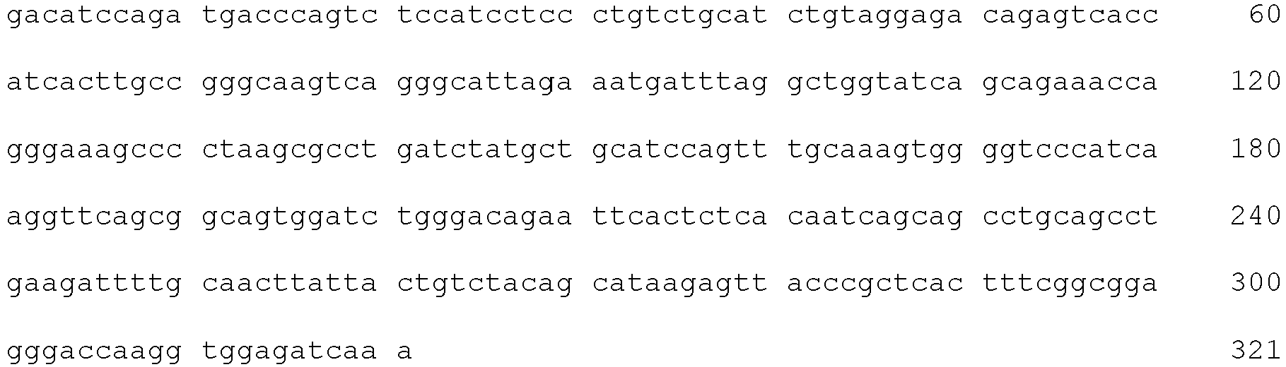
- <210> 88
<211> 321
<212> DNA
<213> Homo sapiens - <400> 88


- <210> 89
<211> 321
<212> DNA
<213> Homo sapiens - <400> 89

- <210> 90
<211> 324
<212> DNA
<213> Homo sapiens - <400> 90

- <210> 91
<211> 321
<212> DNA
<213> Homo sapiens - <400> 91


- <210> 92
<211> 336
<212> DNA
<213> Homo sapiens - <400> 92
- <210> 93
<211> 321
<212> DNA
<213> Homo sapiens - <400> 93

- <210> 94
<211> 321
<212> DNA
<213> Homo sapiens - <400> 94

- <210> 95
<211> 324
<212> DNA
<213> Homo sapiens - <400> 95

- <210> 96
<211> 321
<212> DNA
<213> Homo sapiens - <400> 96

- <210> 97
<211> 339
<212> DNA
<213> Homo sapiens - <400> 97

- <210> 98
<211> 321
<212> DNA
<213> Homo sapiens - <400> 98


- <210> 99
<211> 321
<212> DNA
<213> Homo sapiens - <400> 99

- <210> 100
<211> 321
<212> DNA
<213> Homo sapiens - <400> 100

- <210> 101
<211> 321
<212> DNA
<213> Homo sapiens - <400> 101


- <210> 102
<211> 321
<212> DNA
<213> Homo sapiens - <400> 102

- <210> 103
<211> 339
<212> DNA
<213> Homo sapiens - <400> 103

- <210> 104
<211> 339
<212> DNA
<213> Homo sapiens - <400> 104


- <210> 105
<211> 321
<212> DNA
<213> Homo sapiens - <400> 105

- <210> 106
<211> 321
<212> DNA
<213> Homo sapiens - <400> 106

- <210> 107
<211> 5
<212> PRT
<213> Homo sapiens - <400> 107

- <210> 108
<211> 16
<212> PRT
<213> Homo sapiens - <400> 108

- <210> 109
<211> 23
<212> PRT
<213> Homo sapiens - <400> 109

- <210> 110
<211> 5
<212> PRT
<213> Homo sapiens - <400> 110

- <210> 111
<211> 16
<212> PRT
<213> Homo sapiens - <400> 111

- <210> 112
<211> 19
<212> PRT
<213> Homo sapiens - <400> 112

- <210> 113
<211> 5
<212> PRT
<213> Homo sapiens - <400> 113

- <210> 114
<211> 17
<212> PRT
<213> Homo sapiens - <400> 114

- <210> 115
<211> 5
<212> PRT
<213> Homo sapiens - <400> 115

- <210> 116
<211> 5
<212> PRT
<213> Homo sapiens - <400> 116

- <210> 117
<211> 17
<212> PRT
<213> Homo sapiens - <400> 117

- <210> 118
<211> 5
<212> PRT
<213> Homo sapiens - <400> 118

- <210> 119
<211> 5
<212> PRT
<213> Homo sapiens - <400> 119

- <210> 120
<211> 16
<212> PRT
<213> Homo sapiens - <400> 120

- <210> 121
<211> 17
<212> PRT
<213> Homo sapiens - <400> 121

- <210> 122
<211> 5
<212> PRT
<213> Homo sapiens - <400> 122

- <210> 123
<211> 17
<212> PRT
<213> Homo sapiens - <400> 123

- <210> 124
<211> 15
<212> PRT
<213> Homo sapiens - <400> 124

- <210> 125
<211> 5
<212> PRT
<213> Homo sapiens - <400> 125

- <210> 126
<211> 17
<212> PRT
<213> Homo sapiens - <400> 126

- <210> 127
<211> 15
<212> PRT
<213> Homo sapiens - <400> 127

- <210> 128
<211> 5
<212> PRT
<213> Homo sapiens - <400> 128

- <210> 129
<211> 17
<212> PRT
<213> Homo sapiens - <400> 129

- <210> 130
<211> 15
<212> PRT
<213> Homo sapiens - <400> 130

- <210> 131
<211> 5
<212> PRT
<213> Homo sapiens - <400> 131

- <210> 132
<211> 17
<212> PRT
<213> Homo sapiens - <400> 132

- <210> 133
<211> 15
<212> PRT
<213> Homo sapiens - <400> 133

- <210> 134
<211> 7
<212> PRT
<213> Homo sapiens - <400> 134

- <210> 135
<211> 16
<212> PRT
<213> Homo sapiens - <400> 135

- <210> 136
<211> 14
<212> PRT
<213> Homo sapiens - <400> 136

- <210> 137
<211> 5
<212> PRT
<213> Homo sapiens - <400> 137

- <210> 138
<211> 17
<212> PRT
<213> Homo sapiens - <400> 138

- <210> 139
<211> 5
<212> PRT
<213> Homo sapiens - <400> 139

- <210> 140
<211> 5
<212> PRT
<213> Homo sapiens - <400> 140

- <210> 141
<211> 17
<212> PRT
<213> Homo sapiens - <400> 141

- <210> 142
<211> 7
<212> PRT
<213> Homo sapiens - <400> 142

- <210> 143
<211> 5
<212> PRT
<213> Homo sapiens - <400> 143

- <210> 144
<211> 17
<212> PRT
<213> Homo sapiens - <400> 144

- <210> 145
<211> 12
<212> PRT
<213> Homo sapiens - <400> 145

- <210> 146
<211> 5
<212> PRT
<213> Homo sapiens - <400> 146

- <210> 147
<211> 17
<212> PRT
<213> Homo sapiens - <400> 147

- <210> 148
<211> 7
<212> PRT
<213> Homo sapiens - <400> 148

- <210> 149
<211> 5
<212> PRT
<213> Homo sapiens - <400> 149

- <210> 150
<211> 17
<212> PRT
<213> Homo sapiens - <400> 150

- <210> 151
<211> 12
<212> PRT
<213> Homo sapiens - <400> 151

- <210> 152
<211> 5
<212> PRT
<213> Homo sapiens - <400> 152

- <210> 153
<211> 17
<212> PRT
<213> Homo sapiens - <400> 153

- <210> 154
<211> 7
<212> PRT
<213> Homo sapiens - <400> 154

- <210> 155
<211> 5
<212> PRT
<213> Homo sapiens - <400> 155

- <210> 156
<211> 17
<212> PRT
<213> Homo sapiens - <400> 156

- <210> 157
<211> 7
<212> PRT
<213> Homo sapiens - <400> 157

- <210> 158
<211> 5
<212> PRT
<213> Homo sapiens - <400> 158

- <210> 159
<211> 17
<212> PRT
<213> Homo sapiens - <400> 159


- <210> 160
<211> 17
<212> PRT
<213> Homo sapiens - <400> 160

- <210> 161
<211> 5
<212> PRT
<213> Homo sapiens - <400> 161

- <210> 162
<211> 17
<212> PRT
<213> Homo sapiens - <400> 162

- <210> 163
<211> 7
<212> PRT
<213> Homo sapiens - <400> 163

- <210> 164
<211> 5
<212> PRT
<213> Homo sapiens - <400> 164

- <210> 165
<211> 17
<212> PRT
<213> Homo sapiens - <400> 165

- <210> 166
<211> 9
<212> PRT
<213> Homo sapiens - <400> 166

- <210> 167
<211> 5
<212> PRT
<213> Homo sapiens - <400> 167

- <210> 168
<211> 17
<212> PRT
<213> Homo sapiens - <400> 168

- <210> 169
<211> 9
<212> PRT
<213> Homo sapiens - <400> 169

- <210> 170
<211> 5
<212> PRT
<213> Homo sapiens - <400> 170


- <210> 171
<211> 17
<212> PRT
<213> Homo sapiens - <400> 171

- <210> 172
<211> 7
<212> PRT
<213> Homo sapiens - <400> 172

- <210> 173
<211> 5
<212> PRT
<213> Homo sapiens - <400> 173

- <210> 174
<211> 16
<212> PRT
<213> Homo sapiens - <400> 174

- <210> 175
<211> 17
<212> PRT
<213> Homo sapiens - <400> 175

- <210> 176
<211> 5
<212> PRT
<213> Homo sapiens - <400> 176

- <210> 177
<211> 16
<212> PRT
<213> Homo sapiens - <400> 177

- <210> 178
<211> 17
<212> PRT
<213> Homo sapiens - <400> 178

- <210> 179
<211> 7
<212> PRT
<213> Homo sapiens - <400> 179

- <210> 180
<211> 13
<212> PRT
<213> Homo sapiens - <400> 180

- <210> 181
<211> 14
<212> PRT
<213> Homo sapiens - <400> 181

- <210> 182
<211> 5
<212> PRT
<213> Homo sapiens - <400> 182

- <210> 183
<211> 17
<212> PRT
<213> Homo sapiens - <400> 183

- <210> 184
<211> 16
<212> PRT
<213> Homo sapiens - <400> 184

- <210> 185
<211> 11
<212> PRT
<213> Homo sapiens - <400> 185

- <210> 186
<211> 7
<212> PRT
<213> Homo sapiens - <400> 186

- <210> 187
<211> 9
<212> PRT
<213> Homo sapiens - <400> 187

- <210> 188
<211> 11
<212> PRT
<213> Homo sapiens - <400> 188

- <210> 189
<211> 7
<212> PRT
<213> Homo sapiens - <400> 189

- <210> 190
<211> 8
<212> PRT
<213> Homo sapiens - <400> 190

- <210> 191
<211> 11
<212> PRT
<213> Homo sapiens - <400> 191

- <210> 192
<211> 7
<212> PRT
<213> Homo sapiens - <400> 192

- <210> 193
<211> 9
<212> PRT
<213> Homo sapiens - <400> 193

- <210> 194
<211> 11
<212> PRT
<213> Homo sapiens - <400> 194

- <210> 195
<211> 7
<212> PRT
<213> Homo sapiens - <400> 195

- <210> 196
<211> 10
<212> PRT
<213> Homo sapiens - <400> 196

- <210> 197
<211> 11
<212> PRT
<213> Homo sapiens - <400> 197

- <210> 198
<211> 7
<212> PRT
<213> Homo sapiens - <400> 198

- <210> 199
<211> 9
<212> PRT
<213> Homo sapiens - <400> 199

- <210> 200
<211> 11
<212> PRT
<213> Homo sapiens - <400> 200

- <210> 201
<211> 7
<212> PRT
<213> Homo sapiens - <400> 201

- <210> 202
<211> 9
<212> PRT
<213> Homo sapiens - <400> 202

- <210> 203
<211> 11
<212> PRT
<213> Homo sapiens - <400> 203

- <210> 204
<211> 7
<212> PRT
<213> Homo sapiens - <400> 204

- <210> 205
<211> 9
<212> PRT
<213> Homo sapiens - <400> 205

- <210> 206
<211> 11
<212> PRT
<213> Homo sapiens - <400> 206

- <210> 207
<211> 7
<212> PRT
<213> Homo sapiens - <400> 207

- <210> 208
<211> 9
<212> PRT
<213> Homo sapiens - <400> 208

- <210> 209
<211> 11
<212> PRT
<213> Homo sapiens - <400> 209

- <210> 210
<211> 7
<212> PRT
<213> Homo sapiens - <400> 210

- <210> 211
<211> 9
<212> PRT
<213> Homo sapiens - <400> 211

- <210> 212
<211> 11
<212> PRT
<213> Homo sapiens - <400> 212


- <210> 213
<211> 7
<212> PRT
<213> Homo sapiens - <400> 213

- <210> 214
<211> 9
<212> PRT
<213> Homo sapiens - <400> 214

- <210> 215
<211> 11
<212> PRT
<213> Homo sapiens - <400> 215

- <210> 216
<211> 7
<212> PRT
<213> Homo sapiens - <400> 216

- <210> 217
<211> 10
<212> PRT
<213> Homo sapiens - <400> 217

- <210> 218
<211> 11
<212> PRT
<213> Homo sapiens - <400> 218

- <210> 219
<211> 7
<212> PRT
<213> Homo sapiens - <400> 219

- <210> 220
<211> 9
<212> PRT
<213> Homo sapiens - <400> 220
Gln Gln Tyr Asp Asn Trp Pro Leu Thr
1 5 - <210> 221
<211> 16
<212> PRT
<213> Homo sapiens - <400> 221

- <210> 222
<211> 7
<212> PRT
<213> Homo sapiens - <400> 222

- <210> 223
<211> 9
<212> PRT
<213> Homo sapiens - <400> 223

- <210> 224
<211> 11
<212> PRT
<213> Homo sapiens - <400> 224

- <210> 225
<211> 7
<212> PRT
<213> Homo sapiens - <400> 225

- <210> 226
<211> 9
<212> PRT
<213> Homo sapiens - <400> 226

- <210> 227
<211> 11
<212> PRT
<213> Homo sapiens - <400> 227

- <210> 228
<211> 7
<212> PRT
<213> Homo sapiens - <400> 228

- <210> 229
<211> 9
<212> PRT
<213> Homo sapiens - <400> 229

- <210> 230
<211> 11
<212> PRT
<213> Homo sapiens - <400> 230

- <210> 231
<211> 7
<212> PRT
<213> Homo sapiens - <400> 231

- <210> 232
<211> 9
<212> PRT
<213> Homo sapiens - <400> 232

- <210> 233
<211> 11
<212> PRT
<213> Homo sapiens - <400> 233

- <210> 234
<211> 7
<212> PRT
<213> Homo sapiens - <400> 234

- <210> 235
<211> 9
<212> PRT
<213> Homo sapiens - <400> 235

- <210> 236
<211> 17
<212> PRT
<213> Homo sapiens - <400> 236

- <210> 237
<211> 7
<212> PRT
<213> Homo sapiens - <400> 237

- <210> 238
<211> 9
<212> PRT
<213> Homo sapiens - <400> 238

- <210> 239
<211> 11
<212> PRT
<213> Homo sapiens - <400> 239

- <210> 240
<211> 7
<212> PRT
<213> Homo sapiens - <400> 240

- <210> 241
<211> 9
<212> PRT
<213> Homo sapiens - <400> 241

- <210> 242
<211> 11
<212> PRT
<213> Homo sapiens - <400> 242

- <210> 243
<211> 7
<212> PRT
<213> Homo sapiens - <400> 243

- <210> 244
<211> 9
<212> PRT
<213> Homo sapiens - <400> 244

- <210> 245
<211> 11
<212> PRT
<213> Homo sapiens - <400> 245

- <210> 246
<211> 7
<212> PRT
<213> Homo sapiens - <400> 246

- <210> 247
<211> 9
<212> PRT
<213> Homo sapiens - <400> 247

- <210> 248
<211> 11
<212> PRT
<213> Homo sapiens - <400> 248

- <210> 249
<211> 7
<212> PRT
<213> Homo sapiens - <400> 249

- <210> 250
<211> 9
<212> PRT
<213> Homo sapiens - <400> 250

- <210> 251
<211> 11
<212> PRT
<213> Homo sapiens - <400> 251

- <210> 252
<211> 7
<212> PRT
<213> Homo sapiens - <400> 252

- <210> 253
<211> 9
<212> PRT
<213> Homo sapiens - <400> 253

- <210> 254
<211> 16
<212> PRT
<213> Homo sapiens - <400> 254

- <210> 255
<211> 7
<212> PRT
<213> Homo sapiens - <400> 255

- <210> 256
<211> 10
<212> PRT
<213> Homo sapiens - <400> 256

- <210> 257
<211> 16
<212> PRT
<213> Homo sapiens - <400> 257

- <210> 258
<211> 7
<212> PRT
<213> Homo sapiens - <400> 258

- <210> 259
<211> 10
<212> PRT
<213> Homo sapiens - <400> 259

- <210> 260
<211> 11
<212> PRT
<213> Homo sapiens - <400> 260

- <210> 261
<211> 7
<212> PRT
<213> Homo sapiens - <400> 261

- <210> 262
<211> 9
<212> PRT
<213> Homo sapiens - <400> 262

- <210> 263
<211> 11
<212> PRT
<213> Homo sapiens - <400> 263

- <210> 264
<211> 7
<212> PRT
<213> Homo sapiens - <400> 264

- <210> 265
<211> 9
<212> PRT
<213> Homo sapiens - <400> 265

- <210> 266
<211> 15
<212> DNA
<213> Homo sapiens - <400> 266
aattactact ggaac 15 - <210> 267
<211> 75
<212> DNA
<213> Homo sapiens - <400> 267

- <210> 268
<211> 69
<212> DNA
<213> Homo sapiens - <400> 268

- <210> 269
<211> 15
<212> DNA
<213> Homo sapiens - <400> 269
ggttactact ggagc 15 - <210> 270
<211> 48
<212> DNA
<213> Homo sapiens - <400> 270
gaaatcaatc atagtggacg caccaattac aacccgtccc tcaagagt 48 - <210> 271
<211> 57
<212> DNA
<213> Homo sapiens - <400> 271
ggcccttatt actttgatag tagtggttac ctttactact actacggttt ggacgtc 57 - <210> 272
<211> 15
<212> DNA
<213> Homo sapiens - <400> 272
agctatggca tgcac 15 - <210> 273
<211> 51
<212> DNA
<213> Homo sapiens - <400> 273
gttatatggt atgatggaag taataaacac tatgcagact ccgtgaagggc 51 - <210> 274
<211> 15
<212> DNA
<213> Homo sapiens - <400> 274
gatactgggg tctac 15 - <210> 275
<211> 15
<212> DNA
<213> Homo sapiens - <400> 275
agctatggca tgcac 15 - <210> 276
<211> 51
<212> DNA
<213> Homo sapiens - <400> 276
gttatatggt atgatggaag taataaacac tatgcagact ccgtgaagggc 51 - <210> 277
<211> 15
<212> DNA
<213> Homo sapiens - <400> 277
gatactgggg tctac 15 - <210> 278
<211> 15
<212> DNA
<213> Homo sapiens - <400> 278
agttactact ggagc 15 - <210> 279
<211> 48
<212> DNA
<213> Homo sapiens - <400> 279
cgtatctatc gcagtgggaa caccatctac aacccctccc tcaagagt 48 - <210> 280
<211> 51
<212> DNA
<213> Homo sapiens - <400> 280
gagaattact ctgagagtag tggtctctac tactactacg gtatggacgtc 51 - <210> 281
<211> 15
<212> DNA
<213> Homo sapiens - <400> 281
agatatggta tcagc 15 - <210> 282
<211> 51
<212> DNA
<213> Homo sapiens - <400> 282
tggatcagcg cttacaatgg taacacaaac tatgcacaga agctccagggc 51 - <210> 283
<211> 45
<212> DNA
<213> Homo sapiens - <400> 283
agggattacg atattttgac tggttattat aacgggttcg acccc 45 - <210> 284
<211> 15
<212> DNA
<213> Homo sapiens - <400> 284
agatatggta tcagc 15 - <210> 285
<211> 51
<212> DNA
<213> Homo sapiens - <400> 285
tggatcagcg cttacaatgg taacacaaac tatgcacaga agctccagggc 51 - <210> 286
<211> 45
<212> DNA
<213> Homo sapiens - <400> 286
agggattacg atattttgac tggttattat aacgggttcgaccc 45 - <210> 287
<211> 15
<212> DNA
<213> Homo sapiens - <400> 287
ggctatggta tcagc 15 - <210> 288
<211> 51
<212> DNA
<213> Homo sapiens - <400> 288
tggatcagcg cttacaatgg taacacaaac tatgcacaga acctccagggc 51 - <210> 289
<211> 45
<212> DNA
<213> Homo sapiens - <400> 289
agggattacg atattttgac tggttattat aacgggttcg acccc 45 - <210> 290
<211> 15
<212> DNA
<213> Homo sapiens - <400> 290
agatatggta tcagc 15 - <210> 291
<211> 50
<212> DNA
<213> Homo sapiens - <400> 291
tggatcagcg cttacaatgg taacacaaac tatgcacaga agctccaggg 50 - <210> 292
<211> 45
<212> DNA
<213> Homo sapiens - <400> 292
agggattacg atattttgac tggttattat aacgggttcg acccc 45 - <210> 293
<211> 21
<212> DNA
<213> Homo sapiens - <400> 293
agtggtggtt actactggagc 21 - <210> 294
<211> 48
<212> DNA
<213> Homo sapiens - <400> 294
tacatctatt tcagtgggag cgcctactac aacccgtccc tcaagagt 48 - <210> 295
<211> 42
<212> DNA
<213> Homo sapiens - <400> 295
gaatactatg atagtagtgg ttaccccgat gcttttgatatc 42 - <210> 296
<211> 15
<212> DNA
<213> Homo sapiens - <400> 296
agctatggca tgcac 15 - <210> 297
<211> 51
<212> DNA
<213> Homo sapiens - <400> 297
gttatatggt atgatggaag taataaatat tatgcagact ccgtgaagggc 51 - <210> 298
<211> 15
<212> DNA
<213> Homo sapiens - <400> 298
gatacgaagg actac 15 - <210> 299
<211> 15
<212> DNA
<213> Homo sapiens - <400> 299
agctatggta tcagc 15 - <210> 300
<211> 51
<212> DNA
<213> Homo sapiens - <400> 300
tggatcagca cttacaaagg taacacaaac tatgcacaga agctccagggc 51 - <210> 301
<211> 21
<212> DNA
<213> Homo sapiens - <400> 301
aagcagctcg tctttgactac 21 - <210> 302
<211> 15
<212> DNA
<213> Homo sapiens - <400> 302
agctatggca tgcag 15 - <210> 303
<211> 51
<212> DNA
<213> Homo sapiens - <400> 303
gttatatggt atgatggaaa taagaaatac tatgcagact ccgtgaagggc 51 - <210> 304
<211> 36
<212> DNA
<213> Homo sapiens - <400> 304
ggacgtgtta gggactacta ctacggtatg gacgtc 36 - <210> 305
<211> 15
<212> DNA
<213> Homo sapiens - <400> 305
agatatggta tcagc 15 - <210> 306
<211> 51
<212> DNA
<213> Homo sapiens - <400> 306
tggatcagca cttacagtgg taacacaaac tatgcacaga agctccagggc 51 - <210> 307
<211> 21
<212> DNA
<213> Homo sapiens - <400> 307
cggcagcttt actttgactac 21 - <210> 308
<211> 15
<212> DNA
<213> Homo sapiens - <400> 308
agctatggca tgcag 15 - <210> 309
<211> 51
<212> DNA
<213> Homo sapiens - <400> 309
gttatatggt atgatggaaa taagaaatac tatgcagact ccgtgaagggc 51 - <210> 310
<211> 36
<212> DNA
<213> Homo sapiens - <400> 310
ggacgtgtta gggactacta ctacggtatg gacgtc 36 - <210> 311
<211> 15
<212> DNA
<213> Homo sapiens - <400> 311
agctatggta tcagc 15 - <210> 312
<211> 51
<212> DNA
<213> Homo sapiens - <400> 312
tggatcagcg cttacaatgg taacacaaag tatgcacaga agctccagggc 51 - <210> 313
<211> 21
<212> DNA
<213> Homo sapiens - <400> 313
aagcagctcg tctttgactac 21 - <210> 314
<211> 15
<212> DNA
<213> Homo sapiens - <400> 314
agctatggta tcagc 15 - <210> 315
<211> 51
<212> DNA
<213> Homo sapiens - <400> 315
tggatcagcg cttacagtgg taatacaaag tatgcacaga agctccagggc 51 - <210> 316
<211> 21
<212> DNA
<213> Homo sapiens - <400> 316
aagcagctcg tctttgactac 21 - <210> 317
<211> 15
<212> DNA
<213> Homo sapiens - <400> 317
gactactaca tgcac 15 - <210> 318
<211> 51
<212> DNA
<213> Homo sapiens - <400> 318
tggatgcacc ctaacagtgg tggcacagac ttagcacaga ggtttcagggc 51 - <210> 319
<211> 51
<212> DNA
<213> Homo sapiens - <400> 319
gggggatatt gtagtacttt gagctgctcc ttctactggt acttcgatctc 51 - <210> 320
<211> 15
<212> DNA
<213> Homo sapiens - <400> 320
agctatggaa tcagt 15 - <210> 321
<211> 51
<212> DNA
<213> Homo sapiens - <400> 321
tggatcagcg cttacagtgg taacacaaag tatgcacaga agttccagggc 51 - <210> 322
<211> 21
<212> DNA
<213> Homo sapiens - <400> 322
aggcagctcg cgttggactac 21 - <210> 323
<211> 15
<212> DNA
<213> Homo sapiens - <400> 323
agctatagca tgaac 15 - <210> 324
<211> 51
<212> DNA
<213> Homo sapiens - <400> 324
ttcattagtg ctagaagtag taccatatac tacgcagact ctgtgaagggc 51 - <210> 325
<211> 27
<212> DNA
<213> Homo sapiens - <400> 325
cctaaagtgg ggggcggtat ggacgtc 27 - <210> 326
<211> 15
<212> DNA
<213> Homo sapiens - <400> 326
agctatagca tgaac 15 - <210> 327
<211> 51
<212> DNA
<213> Homo sapiens - <400> 327
atcattagta gtagaagtag tatcatacac tacgcagact ctgtgaagggc 51 - <210> 328
<211> 27
<212> DNA
<213> Homo sapiens - <400> 328
cctaaagtgg ggggcggtat ggacgtc 27 - <210> 329
<211> 15
<212> DNA
<213> Homo sapiens - <400> 329
agatatggta tcagc 15 - <210> 330
<211> 51
<212> DNA
<213> Homo sapiens - <400> 330
tggatcagcg cttacagtgg taacacaaac tatgcacaga agctccagggc 51 - <210> 331
<211> 21
<212> DNA
<213> Homo sapiens - <400> 331
cggcagcttt actttgactac 21 - <210> 332
<211> 15
<212> DNA
<213> Homo sapiens - <400> 332
agttactact ggagc 15 - <210> 333
<211> 48
<212> DNA
<213> Homo sapiens - <400> 333
cgtatctatc ccagtgggag aaccaactac aacccctccc tcaagagt 48 - <210> 334
<211> 51
<212> DNA
<213> Homo sapiens - <400> 334
gaggcatatg agctgcaact gggcctctac tactactacg gtatggacgtc 51 - <210> 335
<211> 15
<212> DNA
<213> Homo sapiens - <400> 335
agttactact ggagc 15 - <210> 336
<211> 48
<212> DNA
<213> Homo sapiens - <400> 336
cgtatctatc ccagtgggag aaccaactac aacccctccc tcaagagt 48 - <210> 337
<211> 51
<212> DNA
<213> Homo sapiens - <400> 337
gaggcatatg agctgcaact gggcctctac tactactacg gtatggacgtc 51 - <210> 338
<211> 21
<212> DNA
<213> Homo sapiens - <400> 338
agtggtggtt actactggagc 21 - <210> 339
<211> 39
<212> DNA
<213> Homo sapiens - <400> 339
tacagtggga acacctacta caacccgtcc ctcaggagt 39 - <210> 340
<211> 42
<212> DNA
<213> Homo sapiens - <400> 340
gaggccggtg gtaactccgc ctactactac ggtatggacgtc 42 - <210> 341
<211> 15
<212> DNA
<213> Homo sapiens - <400> 341
gactactaca tgagc 15 - <210> 342
<211> 51
<212> DNA
<213> Homo sapiens - <400> 342
tacattagta gtagtcgtag taccatatac tacgcagact ctgtgaagggc 51 - <210> 343
<211> 48
<212> DNA
<213> Homo sapiens - <400> 343
gatcgcacgt attactttgg ttcggggagt tatgaaggga tggacgtc 48 - <210> 344
<211> 88
<212> PRT
<213> Homo sapiens - <400> 344
- <210> 345
<211> 33
<212> DNA
<213> Homo sapiens - <400> 345
cgggcaagtc agggcattag aaatgattta ggc 33 - <210> 346
<211> 21
<212> DNA
<213> Homo sapiens - <400> 346
gctgcatcca gtttgcaaag t 21 - <210> 347
<211> 27
<212> DNA
<213> Homo sapiens - <400> 347
ctacagcata atagtaaccc attcact 27 - <210> 348
<211> 33
<212> DNA
<213> Homo sapiens - <400> 348
agggccagtc agagtgttag cagaaactta gtc 33 - <210> 349
<211> 21
<212> DNA
<213> Homo sapiens - <400> 349
ggggcatcca ctagggccaat 21 - <210> 350
<211> 24
<212> DNA
<213> Homo sapiens - <400> 350
cagcagtata aaagctggcg gacg 24 - <210> 351
<211> 33
<212> DNA
<213> Homo sapiens - <400> 351
cgggcaagtc agagcattag cagctatttaaat 33 - <210> 352
<211> 21
<212> DNA
<213> Homo sapiens - <400> 352
gctgcatcca gtttgcaaag t 21 - <210> 353
<211> 27
<212> DNA
<213> Homo sapiens - <400> 353
caacagagtt acagtacccc attcact 27 - <210> 354
<211> 33
<212> DNA
<213> Homo sapiens - <400> 354
agggccagtc agagtgttag taggaatttagcc 33 - <210> 355
<211> 21
<212> DNA
<213> Homo sapiens - <400> 355
ggtgcatcca ccagggccact 21 - <210> 356
<211> 30
<212> DNA
<213> Homo sapiens - <400> 356
cagcagtata ataactggcc cacgtggacg 30 - <210> 357
<211> 33
<212> DNA
<213> Homo sapiens - <400> 357
cgggcaagtc agggcattag aaatgattta ggc 33 - <210> 358
<211> 21
<212> DNA
<213> Homo sapiens - <400> 358
gctgcatcca gtttccaaag t 21 - <210> 359
<211> 27
<212> DNA
<213> Homo sapiens - <400> 359
ctacagcata atagttaccc tccgacg 27 - <210> 360
<211> 33
<212> DNA
<213> Homo sapiens - <400> 360
cgggcaagtc agggcattag aaatgattta ggc 33 - <210> 361
<211> 21
<212> DNA
<213> Homo sapiens - <400> 361
gctgcatcca gtttgcaaag t 21 - <210> 362
<211> 27
<212> DNA
<213> Homo sapiens - <400> 362
ctacagcata aaagttaccc gctcact 27 - <210> 363
<211> 33
<212> DNA
<213> Homo sapiens - <400> 363
cgggcaagtc agggcattag aaatgattta ggc 33 - <210> 364
<211> 21
<212> DNA
<213> Homo sapiens - <400> 364
gctgcatcca gtttgcaaag t 21 - <210> 365
<211> 27
<212> DNA
<213> Homo sapiens - <400> 365
ctacagcata aaagttaccc gctcact 27 - <210> 366
<211> 33
<212> DNA
<213> Homo sapiens - <400> 366
cgggcaagtc agggcattag aaatgattta ggc 33 - <210> 367
<211> 21
<212> DNA
<213> Homo sapiens - <400> 367
gctgcatcca gtttgcaaag t 21 - <210> 368
<211> 27
<212> DNA
<213> Homo sapiens - <400> 368
ctacagcata agagttaccc gctcact 27 - <210> 369
<211> 30
<212> DNA
<213> Homo sapiens - <400> 369
cgggcaagtc agggcattag aaatgattta 30 - <210> 370
<211> 21
<212> DNA
<213> Homo sapiens - <400> 370
gctgcatcca gtttgcaaag t 21 - <210> 371
<211> 27
<212> DNA
<213> Homo sapiens - <400> 371
ctacagcata aaagttaccc gctcact 27 - <210> 372
<211> 33
<212> DNA
<213> Homo sapiens - <400> 372
cgggcgagtc agggtattag gagctggttagcc 33 - <210> 373
<211> 21
<212> DNA
<213> Homo sapiens - <400> 373
gctgcatcca gtttgcaaag t 21 - <210> 374
<211> 27
<212> DNA
<213> Homo sapiens - <400> 374
caacaggcta acaatttccc tcggacg 27 - <210> 375
<211> 33
<212> DNA
<213> Homo sapiens - <400> 375
agggccagtc agagtgttag cagcaacttagcc 33 - <210> 376
<211> 21
<212> DNA
<213> Homo sapiens - <400> 376
ggtgcatcca ccagggccgct 21 - <210> 377
<211> 30
<212> DNA
<213> Homo sapiens - <400> 377
cagcactata taaactggcc taagtggacg 30 - <210> 378
<211> 33
<212> DNA
<213> Homo sapiens - <400> 378
agggccagtc agagtattag cagcagcttagcc 33 - <210> 379
<211> 21
<212> DNA
<213> Homo sapiens - <400> 379
ggtgcatcca ccagggccact 21 - <210> 380
<211> 27
<212> DNA
<213> Homo sapiens - <400> 380
cagcaatatg ataactggcc gctcact 27 - <210> 381
<211> 48
<212> DNA
<213> Homo sapiens - <400> 381
aagtctagtc agagcctcct gcatagtgat ggaaagacct atttgtat 48 - <210> 382
<211> 21
<212> DNA
<213> Homo sapiens - <400> 382
gaagtttcca cccggttctct 21 - <210> 383
<211> 27
<212> DNA
<213> Homo sapiens - <400> 383
atgcaaagta tacagcttcc gctcact 27 - <210> 384
<211> 33
<212> DNA
<213> Homo sapiens - <400> 384
agggccagtc agagtgttag cagcaacttagcc 33 - <210> 385
<211> 21
<212> DNA
<213> Homo sapiens - <400> 385
gatgcatcca ccagggccact 21 - <210> 386
<211> 27
<212> DNA
<213> Homo sapiens - <400> 386
cagcagtatg ataactggcc gctcact 27 - <210> 387
<211> 33
<212> DNA
<213> Homo sapiens - <400> 387
agggccagtc agagtgttag cagcaacttagcc 33 - <210> 388
<211> 21
<212> DNA
<213> Homo sapiens - <400> 388
gatgcatcca ccagggccgct 21 - <210> 389
<211> 27
<212> DNA
<213> Homo sapiens - <400> 389
cagcagtatg ataactggcc gctcact 27 - <210> 390
<211> 33
<212> DNA
<213> Homo sapiens - <400> 390
agggccagtc agagtattag caccagcttagcc 33 - <210> 391
<211> 21
<212> DNA
<213> Homo sapiens - <400> 391
ggtacatcca ccagggccact 21 - <210> 392
<211> 27
<212> DNA
<213> Homo sapiens - <400> 392
caacagtatg atatctggcc gctcact 27 - <210> 393
<211> 33
<212> DNA
<213> Homo sapiens - <400> 393
agggccagtc agagtgttag cagcaacttagcc 33 - <210> 394
<211> 21
<212> DNA
<213> Homo sapiens - <400> 394
ggtgcatcca ccagggccact 21 - <210> 395
<211> 27
<212> DNA
<213> Homo sapiens - <400> 395
cagcagtatg ataactggcc gctcact 27 - <210> 396
<211> 51
<212> DNA
<213> Homo sapiens - <400> 396
aagaccagcc agagtgtttt atacagctcc aaaaacaaga acttcttagct 51 - <210> 397
<211> 21
<212> DNA
<213> Homo sapiens - <400> 397
tgggcatcta cccgggaatcc 21 - <210> 398
<211> 27
<212> DNA
<213> Homo sapiens - <400> 398
cagcaatatt atagtactcc attcact 27 - <210> 399
<211> 33
<212> DNA
<213> Homo sapiens - <400> 399
agggccagtc agagtattag cagcaacttagcc 33 - <210> 400
<211> 21
<212> DNA
<213> Homo sapiens - <400> 400
ggtgcatcca ccagggccact 21 - <210> 401
<211> 27
<212> DNA
<213> Homo sapiens - <400> 401
cagcagtatg atacctggcc tctcact 27 - <210> 402
<211> 33
<212> DNA
<213> Homo sapiens - <400> 402
cgggcgagtc agggcattag caattatttagcc 33 - <210> 403
<211> 21
<212> DNA
<213> Homo sapiens - <400> 403
gctgcatcca ctttacaatc a 21 - <210> 404
<211> 27
<212> DNA
<213> Homo sapiens - <400> 404
caaaagtata accgtgcccc attcact 27 - <210> 405
<211> 33
<212> DNA
<213> Homo sapiens - <400> 405
cgggcgagtc agggcattag caattatttagcc 33 - <210> 406
<211> 21
<212> DNA
<213> Homo sapiens - <400> 406
gctgcatcca ctttgcaatc a 21 - <210> 407
<211> 27
<212> DNA
<213> Homo sapiens - <400> 407
caaaagtata accgtgcccc attcact 27 - <210> 408
<211> 33
<212> DNA
<213> Homo sapiens - <400> 408
agggccagtc agagtgttag cagcaacttagcc 33 - <210> 409
<211> 21
<212> DNA
<213> Homo sapiens - <400> 409
gatgcatcca ccagggccgct 21 - <210> 410
<211> 27
<212> DNA
<213> Homo sapiens - <400> 410
cagcagtatg ataactggcc gctcact 27 - <210> 411
<211> 33
<212> DNA
<213> Homo sapiens - <400> 411
cgggcaagtc agggcattat aaatgattta ggc 33 - <210> 412
<211> 21
<212> DNA
<213> Homo sapiens - <400> 412
gctgcatcca gtttgcaaag t 21 - <210> 413
<211> 27
<212> DNA
<213> Homo sapiens - <400> 413
ctacagcata atagttaccc tccgacg 27 - <210> 414
<211> 48
<212> DNA
<213> Homo sapiens - <400> 414
aggtctagtc aaagcctcgt atatagtgat ggacacacct gcttgaat 48 - <210> 415
<211> 21
<212> DNA
<213> Homo sapiens - <400> 415
aaggtttcta actgggactct 21 - <210> 416
<211> 30
<212> DNA
<213> Homo sapiens - <400> 416
atgcaaggta cacactggcc tctgtgcagt 30 - <210> 417
<211> 48
<212> DNA
<213> Homo sapiens - <400> 417
aggtctagtc aaagcctcgt atatagtgat ggacacacct gcttgaat 48 - <210> 418
<211> 21
<212> DNA
<213> Homo sapiens - <400> 418
aaggtttcta actgggactct 21 - <210> 419
<211> 30
<212> DNA
<213> Homo sapiens - <400> 419
atgcaaggta cacactggcc tctgtgcagt 30 - <210> 420
<211> 33
<212> DNA
<213> Homo sapiens - <400> 420
cgggcgagtc aggccattag catttatttagcc 33 - <210> 421
<211> 21
<212> DNA
<213> Homo sapiens - <400> 421
gctgcatcca gtttgcaaag t 21 - <210> 422
<211> 27
<212> DNA
<213> Homo sapiens - <400> 422
caacagtata gtagttaccc tcggacg 27 - <210> 423
<211> 33
<212> DNA
<213> Homo sapiens - <400> 423
agggccagtc agagtgttta cagcaacttagcc 33 - <210> 424
<211> 21
<212> DNA
<213> Homo sapiens - <400> 424
ggtgcttcca ccagggccact 21 - <210> 425
<211> 27
<212> DNA
<213> Homo sapiens - <400> 425
cagcagtatt ataactggcc gtggacg 27 - <210> 426
<211> 1409
<212> DNA
<213> Homo sapiens - <220>
<221> CDS
<222> (16)..(1398) - <400> 426


- <210> 427
<211> 461
<212> PRT
<213> Homo sapiens - <400> 427


- <210> 428
<211> 741
<212> DNA
<213> Homo sapiens - <220>
<221> CDS
<222> (24)..(725) - <400> 428


- <210> 429
<211> 234
<212> PRT
<213> Homo sapiens - <400> 429


- <210> 430
<211> 866
<212> PRT
<213> Homo sapiens - <400> 430


- <210> 431
<211> 344
<212> PRT
<213> Artificial Sequence - <220>
<223> Description of Artificial Sequence: Synthetic construct - <400> 431


- <210> 432
<211> 322
<212> PRT
<213> Mus musculus - <400> 432


- <210> 433
<211> 344
<212> PRT
<213> Artificial Sequence - <220>
<223> Description of Artificial Sequence: Synthetic construct - <400> 433


- <210> 434
<211> 344
<212> PRT
<213> Artificial Sequence - <220>
<223> Description of Artificial Sequence: Synthetic construct - <400> 434
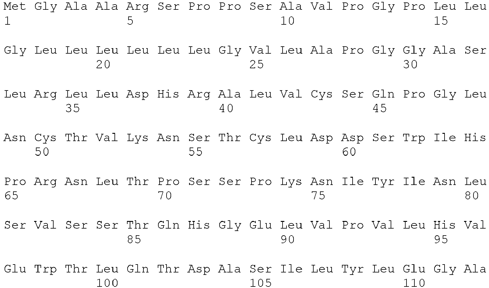

- <210> 435
<211> 344
<212> PRT
<213> Artificial Sequence - <220>
<223> Description of Artificial Sequence: Synthetic construct - <400> 435

- <210> 436
<211> 344
<212> PRT
<213> Artificial Sequence - <220>
<223> Description of Artificial Sequence: Synthetic construct - <400> 436


- <210> 437
<211> 344
<212> PRT
<213> Artificial Sequence - <220>
<223> Description of Artificial Sequence: Synthetic construct - <400> 437


- <210> 438
<211> 346
<212> PRT
<213> Artificial Sequence - <220>
<223> Description of Artificial Sequence: Synthetic construct - <400> 438


- <210> 439
<211> 344
<212> PRT
<213> Artificial Sequence - <220>
<223> Description of Artificial Sequence: Synthetic construct - <400> 439


- <210> 440
<211> 344
<212> PRT
<213> Artificial Sequence - <220>
<223> Description of Artificial Sequence: Synthetic construct - <400> 440


- <210> 441
<211> 344
<212> PRT
<213> Artificial Sequence - <220>
<223> Description of Artificial Sequence: Synthetic construct - <400> 441


- <210> 442
<211> 344
<212> PRT
<213> Artificial Sequence - <220>
<223> Description of Artificial Sequence: Synthetic construct - <400> 442


- <210> 443
<211> 346
<212> PRT
<213> Artificial Sequence - <220>
<223> Description of Artificial Sequence: Synthetic construct - <400> 443


- <210> 444
<211> 346
<212> PRT
<213> Artificial Sequence - <220>
<223> Description of Artificial Sequence: Synthetic construct - <400> 444


- <210> 445
<211> 346
<212> PRT
<213> Artificial Sequence - <220>
<223> Description of Artificial Sequence: Synthetic construct - <400> 445


- <210> 446
<211> 346
<212> PRT
<213> Artificial Sequence - <220>
<223> Description of Artificial Sequence: Synthetic construct - <400> 446


- <210> 447
<211> 8
<212> PRT
<213> Artificial Sequence - <220>
<223> Description of Artificial Sequence: Synthetic peptide - <400> 447

- <210> 448
<211> 12
<212> PRT
<213> Artificial Sequence - <220>
<223> Description of Artificial Sequence: Synthetic peptide - <400> 448

- <210> 449
<211> 88
<212> PRT
<213> Artificial Sequence - <220>
<223> Description of Artificial Sequence: Synthetic construct - <400> 449

- <210> 450
<211> 88
<212> PRT
<213> Artificial Sequence - <220>
<223> Description of Artificial Sequence: Synthetic construct - <400> 450

- <210> 451
<211> 88
<212> PRT
<213> Artificial Sequence - <220>
<223> Description of Artificial Sequence: Synthetic construct - <400> 451

- <210> 452
<211> 88
<212> PRT
<213> Artificial Sequence - <220>
<223> Description of Artificial Sequence: Synthetic construct - <400> 452

- <210> 453
<211> 5
<212> PRT
<213> Artificial Sequence - <220>
<223> Description of Artificial Sequence: Synthetic peptide - <220>
<221> MOD_RES
<222> (1) .. (1)
<223> Variable amino acid - <400> 453

- <210> 454
<211> 5
<212> PRT
<213> Artificial Sequence - <220>
<223> Description of Artificial Sequence: Synthetic peptide - <220>
<221> MOD_RES
<222> (1) .. (1)
<223> Variable amino acid - <220>
<221> MOD_RES
<222> (3) .. (3)
<223> Variable amino acid - <220>
<221> MOD_RES
<222> (5) .. (5)
<223> Variable amino acid - <400> 454

- <210> 455
<211> 5
<212> PRT
<213> Artificial Sequence - <220>
<223> Description of Artificial Sequence: Synthetic peptide - <220>
<221> MOD_RES
<222> (5) .. (5)
<223> Variable amino acid - <400> 455

- <210> 456
<211> 17
<212> PRT
<213> Artificial Sequence - <220>
<223> Description of Artificial Sequence: Synthetic peptide - <220>
<221> MOD_RES
<222> (4) .. (4)
<223> Variable amino acid - <220>
<221> MOD_RES
<222> (6)..(6)
<223> Variable amino acid - <220>
<221> MOD_RES
<222> (10)..(10)
<223> Variable amino acid - <220>
<221> MOD_RES
<222> (14)..(15)
<223> Variable amino acid - <400> 456

- <210> 457
<211> 17
<212> PRT
<213> Artificial Sequence - <220>
<223> Description of Artificial Sequence: Synthetic peptide - <220>
<221> MOD_RES
<222> (1)..(2)
<223> Variable amino acid - <220>
<221> MOD_RES
<222> (4)..(6)
<223> Variable amino acid - <220>
<221> MOD_RES
<222> (8) .. (8)
<223> Variable amino acid - <220>
<221> MOD_RES
<222> (10)..(10)
<223> Variable amino acid - <400> 457

- <210> 458
<211> 17
<212> PRT
<213> Artificial Sequence - <220>
<223> Description of Artificial Sequence: Synthetic peptide - <220>
<221> MOD_RES
<222> (7) .. (8)
<223> Variable amino acid - <220>
<221> MOD_RES
<222> (10)..(10)
<223> Variable amino acid - <400> 458

- <210> 459
<211> 7
<212> PRT
<213> Artificial Sequence - <220>
<223> Description of Artificial Sequence: Synthetic peptide - <220>
<221> MOD_RES
<222> (1) .. (1)
<223> Variable amino acid - <220>
<221> MOD_RES
<222> (4) .. (5)
<223> Variable amino acid - <400> 459

- <210> 460
<211> 7
<212> PRT
<213> Artificial Sequence - <220>
<223> Description of Artificial Sequence: Synthetic peptide - <220>
<221> MOD_RES
<222> (1) .. (1)
<223> Variable amino acid - <220>
<221> MOD_RES
<222> (4) .. (4)
<223> Variable amino acid - <400> 460

- <210> 461
<211> 11
<212> PRT
<213> Artificial Sequence - <220>
<223> Description of Artificial Sequence: Synthetic peptide - <220>
<221> MOD_RES
<222> (5) .. (5)
<223> Variable amino acid - <220>
<221> MOD_RES
<222> (7) .. (9)
<223> Variable amino acid - <220>
<221> MOD_RES
<222> (11)..(11)
<223> Variable amino acid - <400> 461

- <210> 462
<211> 11
<212> PRT
<213> Artificial Sequence - <220>
<223> Description of Artificial Sequence: Synthetic peptide - <220>
<221> MOD_RES
<222> (6)..(9)
<223> Variable amino acid - <400> 462

- <210> 463
<211> 11
<212> PRT
<213> Artificial Sequence - <220>
<223> Description of Artificial Sequence: Synthetic peptide - <220>
<221> MOD_RES
<222> (7) .. (8)
<223> Variable amino acid - <220>
<221> MOD_RES
<222> (11)..(11)
<223> Variable amino acid - <400> 463

- <210> 464
<211> 7
<212> PRT
<213> Artificial Sequence - <220>
<223> Description of Artificial Sequence: Synthetic peptide - <220>
<221> MOD_RES
<222> (5) .. (5)
<223> Variable amino acid - <400> 464

- <210> 465
<211> 7
<212> PRT
<213> Artificial Sequence - <220>
<223> Description of Artificial Sequence: Synthetic peptide - <220>
<221> MOD_RES
<222> (4) .. (4)
<223> Variable amino acid - <400> 465

- <210> 466
<211> 7
<212> PRT
<213> Artificial Sequence - <220>
<223> Description of Artificial Sequence: Synthetic peptide - <220>
<221> MOD_RES
<222> (1)..(2)
<223> Variable amino acid - <220>
<221> MOD_RES
<222> (7) .. (7)
<223> Variable amino acid - <400> 466

- <210> 467
<211> 9
<212> PRT
<213> Artificial Sequence - <220>
<223> Description of Artificial Sequence: Synthetic peptide - <220>
<221> MOD_RES
<222> (4) .. (4)
<223> Variable amino acid - <220>
<221> MOD_RES
<222> (7) .. (8)
<223> Variable amino acid - <400> 467

- <210> 468
<211> 9
<212> PRT
<213> Artificial Sequence - <220>
<223> Description of Artificial Sequence: Synthetic peptide - <220>
<221> MOD_RES
<222> (2)..(6)
<223> Variable amino acid - <220>
<221> MOD_RES
<222> (8) .. (8)
<223> Variable amino acid - <400> 468

- <210> 469
<211> 9
<212> PRT
<213> Artificial Sequence - <220>
<223> Description of Artificial Sequence: Synthetic peptide - <220>
<221> MOD_RES
<222> (5) .. (5)
<223> Variable amino acid - <400> 469

- <210> 470
<211> 10
<212> PRT
<213> Artificial Sequence - <220>
<223> Description of Artificial Sequence: Synthetic peptide - <220>
<221> MOD_RES
<222> (2)..(2)
<223> Variable amino acid - <220>
<221> MOD_RES
<222> (4) .. (5)
<223> Variable amino acid - <220>
<221> MOD_RES
<222> (7) .. (9)
<223> Variable amino acid - <400> 470

Claims (17)
- An isolated monoclonal antibody or fragment thereof, comprising a variable heavy chain that is at least 90% identical to SEQ ID NO:14 and a variable light chain that is at least 90% identical to SEQ ID NO:40, that specifically binds to IL-17RA of SEQ ID NO:431, with a binding affinity of 10-8 to 10-10 M and does not bind said polypeptide wherein the aspartic acid residue at position 152 of SEQ ID NO:431 substituted with an arginine, does not specifically bind said polypeptide wherein the aspartic acid residue at position 154 of SEQ ID NO:431 substituted with an arginine, does not specifically bind said polypeptide wherein the glutamic acid residue at position 156 of SEQ ID NO:431 substituted with an arginine does not specifically bind said polypeptide substituted with arginine at position 184 of SEQ ID NO:431, does not specifically bind said polypeptide substituted with arginine at position 186 of SEQ ID NO:431, does not specifically bind said polypeptide substituted with arginine at position 297 of SEQ ID NO:431 and does not specifically bind to a polypeptide consisting of SEQ ID NO: 436.
- The antibody of claim 1, wherein said monoclonal antibody is a human IgG antibody.
- The antibody of claim 1 or claim 2, wherein said monoclonal antibody is a human IgG2 antibody.
- A pharmaceutical composition, comprising the antibody of any of claims 1 to 3.
- An isolated polynucleotide, wherein said polynucleotide encodes the antibody of any of claims 1 to 3.
- A plasmid, comprising said polynucleotide of claim 5.
- The plasmid of claim 6, wherein said plasmid is an expression vector.
- An isolated cell, comprising said plasmid of claim 6.
- The isolated cell of claim 8, wherein a chromosome of said cell comprises said polynucleotide.
- The isolated cell of claim 8, wherein said cell is a hybridoma.
- The isolated cell of claim 8, wherein said cell comprises the expression vector of claim 7.
- The isolated cell of claim 11, wherein said cell is a selected from the group consisting of:a. a prokaryotic cell;b. a eukaryotic cell;c. a mammalian cell;d. an insect cell; ande. a CHO cell.
- A method of making the antibody of claims 1 to 3, comprising incubating said isolated cell of claim 11 under conditions that allow it to express said polypeptide.
- The antibody made by the method of claim 13.
- The antibody of any of claims 1 to 3 or the pharmaceutical composition of claim 4 for use in the treatment psoriatic arthritis, juvenile psoriatic arthritis, psoriasis, plaque psoriasis, atopic dermatitis, asthma or chronic obstructive pulmonary disease.
- The antibody or pharmaceutical composition for use according to claim 15 further comprising the use of a second pharmaceutical composition.
- The antibody or pharmaceutical composition for use according to claim 16, wherein said second pharmaceutical composition is selected from the group consisting of: TNF inhibitors, soluble TNF receptors, Etanercept, soluble TNF receptor type-I and soluble TNF receptor type-II, monomeric or multimeric p75 and/or p55 TNF receptor molecules and fragments thereof, anti-TNF antibodies, Infliximab, adalimumab, IL-1 inhibitors, IL-1 receptor inhibitors, CD28 inhibitors, non-steroidal antiinflammatory drugs (NSAID), a slow acting antirheumatic drugs (SAARD), and disease modifying antirheumatic drugs (DMARD).
Priority Applications (6)
| Application Number | Priority Date | Filing Date | Title |
|---|---|---|---|
| DK16190586.4T DK3165539T3 (en) | 2006-10-02 | 2007-10-01 | Il-17-receptor a antigenbindende proteiner |
| EP16190586.4A EP3165539B1 (en) | 2006-10-02 | 2007-10-01 | Il-17 receptor a antigen binding proteins |
| PL12159118T PL2518085T3 (en) | 2006-10-02 | 2007-10-01 | IL-17 receptor A antigen binding proteins |
| PL16190586T PL3165539T3 (en) | 2006-10-02 | 2007-10-01 | Il-17 receptor a antigen binding proteins |
| SI200731902A SI2518085T1 (en) | 2006-10-02 | 2007-10-01 | IL-17 receptor A antigen binding proteins |
| CY20221100181T CY1125133T1 (en) | 2006-10-02 | 2022-03-02 | IL-17 RECEPTOR A BINDING PROTEINS |
Applications Claiming Priority (4)
| Application Number | Priority Date | Filing Date | Title |
|---|---|---|---|
| US82788206P | 2006-10-02 | 2006-10-02 | |
| US87307206P | 2006-12-05 | 2006-12-05 | |
| US96989507P | 2007-09-04 | 2007-09-04 | |
| EP07867194.8A EP2076541B1 (en) | 2006-10-02 | 2007-10-01 | Il-17 receptor a antigen binding proteins |
Related Parent Applications (3)
| Application Number | Title | Priority Date | Filing Date |
|---|---|---|---|
| EP07867194.8A Division EP2076541B1 (en) | 2006-10-02 | 2007-10-01 | Il-17 receptor a antigen binding proteins |
| EP07867194.8A Division-Into EP2076541B1 (en) | 2006-10-02 | 2007-10-01 | Il-17 receptor a antigen binding proteins |
| EP07867194.8 Division | 2007-10-01 |
Related Child Applications (2)
| Application Number | Title | Priority Date | Filing Date |
|---|---|---|---|
| EP16190586.4A Division EP3165539B1 (en) | 2006-10-02 | 2007-10-01 | Il-17 receptor a antigen binding proteins |
| EP16190586.4A Division-Into EP3165539B1 (en) | 2006-10-02 | 2007-10-01 | Il-17 receptor a antigen binding proteins |
Publications (2)
| Publication Number | Publication Date |
|---|---|
| EP2518085A1 EP2518085A1 (en) | 2012-10-31 |
| EP2518085B1 true EP2518085B1 (en) | 2016-12-07 |
Family
ID=39344834
Family Applications (4)
| Application Number | Title | Priority Date | Filing Date |
|---|---|---|---|
| EP12159091.3A Active EP2487188B1 (en) | 2006-10-02 | 2007-10-01 | IL-17 receptor A antigen binding proteins |
| EP16190586.4A Active EP3165539B1 (en) | 2006-10-02 | 2007-10-01 | Il-17 receptor a antigen binding proteins |
| EP07867194.8A Active EP2076541B1 (en) | 2006-10-02 | 2007-10-01 | Il-17 receptor a antigen binding proteins |
| EP12159118.4A Active EP2518085B1 (en) | 2006-10-02 | 2007-10-01 | IL-17 receptor A antigen binding proteins |
Family Applications Before (3)
| Application Number | Title | Priority Date | Filing Date |
|---|---|---|---|
| EP12159091.3A Active EP2487188B1 (en) | 2006-10-02 | 2007-10-01 | IL-17 receptor A antigen binding proteins |
| EP16190586.4A Active EP3165539B1 (en) | 2006-10-02 | 2007-10-01 | Il-17 receptor a antigen binding proteins |
| EP07867194.8A Active EP2076541B1 (en) | 2006-10-02 | 2007-10-01 | Il-17 receptor a antigen binding proteins |
Country Status (30)
| Country | Link |
|---|---|
| US (14) | US7767206B2 (en) |
| EP (4) | EP2487188B1 (en) |
| JP (1) | JP5013383B2 (en) |
| KR (2) | KR20120098953A (en) |
| CN (1) | CN103214576B (en) |
| AR (1) | AR063090A1 (en) |
| AU (2) | AU2007314519C1 (en) |
| BR (1) | BRPI0719762B8 (en) |
| CA (1) | CA2663537C (en) |
| CL (1) | CL2007002800A1 (en) |
| CR (1) | CR10760A (en) |
| CY (5) | CY1116291T1 (en) |
| DK (4) | DK3165539T3 (en) |
| EA (1) | EA019397B1 (en) |
| ES (4) | ES2616210T3 (en) |
| HU (4) | HUE033216T2 (en) |
| IL (3) | IL197636B (en) |
| LT (4) | LT3165539T (en) |
| LU (1) | LUC00056I2 (en) |
| MX (1) | MX2009003511A (en) |
| MY (1) | MY187263A (en) |
| NL (1) | NL300919I2 (en) |
| NO (2) | NO344867B1 (en) |
| NZ (1) | NZ575631A (en) |
| PE (2) | PE20120832A1 (en) |
| PL (4) | PL2076541T3 (en) |
| PT (4) | PT3165539T (en) |
| SI (4) | SI3165539T1 (en) |
| TW (1) | TWI396694B (en) |
| WO (1) | WO2008054603A2 (en) |
Families Citing this family (91)
| Publication number | Priority date | Publication date | Assignee | Title |
|---|---|---|---|---|
| AU2002246746B2 (en) * | 2000-10-18 | 2007-05-31 | Kirin-Amgen, Inc. | Methods for treating rheumatoid arthritis using il-17 antagonists |
| US7767206B2 (en) | 2006-10-02 | 2010-08-03 | Amgen Inc. | Neutralizing determinants of IL-17 Receptor A and antibodies that bind thereto |
| JP5337055B2 (en) * | 2007-02-28 | 2013-11-06 | メルク・シャープ・アンド・ドーム・コーポレーション | Combination therapy for the treatment of immune disorders |
| AU2008266948B2 (en) * | 2007-06-13 | 2012-08-23 | Kirin-Amgen, Inc. | IL-17 heteromeric receptor complex |
| US20100256600A1 (en) * | 2009-04-04 | 2010-10-07 | Ferrera David A | Neurovascular otw pta balloon catheter and delivery system |
| KR101240904B1 (en) * | 2008-02-21 | 2013-03-11 | 키린-암젠 인코포레이티드 | Il-17ra-il-17rb antagonists and uses thereof |
| EP2356140A2 (en) * | 2008-10-07 | 2011-08-17 | Novimmune Sa | Il-17-mediated transfection methods |
| WO2010062858A1 (en) * | 2008-11-26 | 2010-06-03 | Allergan, Inc. | Il-17 antibody inhibitor for treating dry eye |
| WO2010062857A1 (en) | 2008-11-26 | 2010-06-03 | Allergan, Inc. | Klk-13 antibody inhibitor for treating dry eye |
| EP2810652A3 (en) | 2009-03-05 | 2015-03-11 | AbbVie Inc. | IL-17 binding proteins |
| MX347291B (en) | 2009-03-20 | 2017-04-17 | Amgen Inc | Carrier immunoglobulins and uses thereof. |
| RU2605318C2 (en) | 2009-05-05 | 2016-12-20 | Новиммун С.А. | Anti-il-17f antibodies and methods for use thereof |
| WO2010142551A2 (en) * | 2009-06-12 | 2010-12-16 | Ablynx N.V. | Single variable domain (vhh) antibodies to cytokines of the il-17 receptor family |
| CA2777222A1 (en) * | 2009-10-10 | 2011-04-14 | Eleven Biotherapeutics, Inc. | Il-17 family cytokine compositions and uses |
| TW201117824A (en) * | 2009-10-12 | 2011-06-01 | Amgen Inc | Use of IL-17 receptor a antigen binding proteins |
| EA201792376A3 (en) | 2009-10-23 | 2018-08-31 | Милленниум Фармасьютикалз, Инк. | ANTI-GCC MOLECULES ANTIBODIES AND RELATED COMPOSITIONS AND METHODS |
| SG182468A1 (en) | 2010-01-15 | 2012-08-30 | Kirin Amgen Inc | Antibody formulation and therapeutic regimens |
| TWI596114B (en) * | 2010-02-24 | 2017-08-21 | 雷那特神經科學股份有限公司 | Antagonist anti-il-7 receptor antibodies and methods |
| RU2012149227A (en) | 2010-05-20 | 2014-06-27 | Аблинкс Нв | BIOLOGICAL MATERIALS RELATING TO HER3 |
| EA201370076A1 (en) * | 2010-09-22 | 2013-08-30 | Амген Инк. | IMMUNE GLOBULIN TRANSFER AND THEIR APPLICATION |
| WO2012061129A1 (en) * | 2010-10-25 | 2012-05-10 | Genentech, Inc | Treatment of gastrointestinal inflammation and psoriasis a |
| EP4116325A1 (en) | 2010-11-05 | 2023-01-11 | Novartis AG | Methods of treating rheumatoid arthritis using il-17 antagonists |
| US10208349B2 (en) * | 2011-01-07 | 2019-02-19 | Ucb Biopharma Sprl | Lipocalin 2 as a biomarker for IL-17 inhibitor therapy efficacy |
| PT2699293T (en) | 2011-04-20 | 2019-05-21 | Amgen Inc | Autoinjector apparatus |
| UA117218C2 (en) | 2011-05-05 | 2018-07-10 | Мерк Патент Гмбх | Amino acid sequences directed against il-17a, il-17f and/or il17-a/f and polypeptides comprising the same |
| WO2013011368A2 (en) * | 2011-07-19 | 2013-01-24 | Teva Pharmaceutical Industries Ltd. | Novel il-17r-ecd mutants |
| WO2013016220A1 (en) * | 2011-07-22 | 2013-01-31 | Amgen Inc. | Il-17 receptor a is required for il-17c biology |
| UY35148A (en) | 2012-11-21 | 2014-05-30 | Amgen Inc | HETERODIMERIC IMMUNOGLOBULINS |
| US9757529B2 (en) | 2012-12-20 | 2017-09-12 | Otitopic Inc. | Dry powder inhaler and methods of use |
| US9757395B2 (en) | 2012-12-20 | 2017-09-12 | Otitopic Inc. | Dry powder inhaler and methods of use |
| JP6134392B2 (en) | 2013-01-25 | 2017-05-24 | サイモン・エルエルシー | Compositions for selective reduction of circulating biologically active soluble TNF and methods for treating TNF-mediated diseases |
| EP2968760B1 (en) | 2013-03-15 | 2024-01-03 | Amgen Inc. | Drug cassette, autoinjector, and autoinjector system |
| TWI639449B (en) | 2013-03-15 | 2018-11-01 | 美商安美基公司 | Cassette for an injector |
| WO2014144817A2 (en) | 2013-03-15 | 2014-09-18 | Amgen Inc. | Inhibitory polypeptides specific to wnt inhibitors |
| KR102074421B1 (en) * | 2013-03-29 | 2020-02-10 | 삼성전자주식회사 | Bispecific anti-cMet/anti-EGFR antibodies |
| EP2991634A1 (en) | 2013-04-30 | 2016-03-09 | Otitopic Inc. | Dry powder formulations and methods of use |
| EP4223312A1 (en) | 2013-08-15 | 2023-08-09 | Novartis AG | Methods of treating plaque psoriasis using il-17 antagonists |
| AU2014318017B2 (en) | 2013-09-05 | 2020-02-06 | Amgen Inc. | Fc-containing molecules exhibiting predictable, consistent, and reproducible glycoform profiles |
| WO2015057906A1 (en) * | 2013-10-16 | 2015-04-23 | Janssen Biotech, Inc. | Cd200 receptor 1 agonists |
| KR102127408B1 (en) * | 2014-01-29 | 2020-06-29 | 삼성전자주식회사 | Anti-Her3 scFv fragment and Bispecific anti-c-Met/anti-Her3 antibodies comprising the same |
| BR112016018980A2 (en) | 2014-02-21 | 2017-10-10 | Genentech Inc | method of treating a disorder, multispecific antibody, isolated nucleic acid, host cell, methods of producing an antibody, producing an antibody half or multispecific antibody, and producing a multispecific, immunoconjugate antibody and pharmaceutical formulation |
| JP6596014B2 (en) | 2014-03-31 | 2019-10-23 | アムジェン ケー・エー,インコーポレイテッド | How to treat nail and scalp psoriasis |
| EP3143404B1 (en) | 2014-05-16 | 2018-08-29 | Amgen Inc. | Assay for detecting th1 and th2 cell populations |
| AU2015307868B2 (en) * | 2014-08-26 | 2020-08-27 | Amgen K-A, Inc. | Method for treating psoriasis patient which received anti-TNF-alpha antibody therapy |
| JP2017528465A (en) | 2014-09-10 | 2017-09-28 | ノバルティス アーゲー | Use of an IL-17 antagonist to inhibit the progression of structural damage in patients with psoriatic arthritis |
| MA41044A (en) | 2014-10-08 | 2017-08-15 | Novartis Ag | COMPOSITIONS AND METHODS OF USE FOR INCREASED IMMUNE RESPONSE AND CANCER TREATMENT |
| CN114920840A (en) | 2014-10-14 | 2022-08-19 | 诺华股份有限公司 | Antibody molecules against PD-L1 and uses thereof |
| GB201500461D0 (en) * | 2015-01-12 | 2015-02-25 | Cresendo Biolog Ltd | Therapeutic molecules |
| WO2016123329A2 (en) | 2015-01-28 | 2016-08-04 | Genentech, Inc. | Gene expression markers and treatment of multiple sclerosis |
| US20180201673A1 (en) | 2015-07-16 | 2018-07-19 | Eli Lilly And Company | Treatment of pruritus |
| EP3964528A1 (en) | 2015-07-29 | 2022-03-09 | Novartis AG | Combination therapies comprising antibody molecules to lag-3 |
| EP3878465A1 (en) | 2015-07-29 | 2021-09-15 | Novartis AG | Combination therapies comprising antibody molecules to tim-3 |
| MA45488A (en) | 2015-10-22 | 2018-08-29 | Juno Therapeutics Gmbh | CELL CULTURE PROCESSES, KITS AND APPARATUS |
| AU2016369537B2 (en) | 2015-12-17 | 2024-03-14 | Novartis Ag | Antibody molecules to PD-1 and uses thereof |
| RU2757314C2 (en) | 2016-01-22 | 2021-10-13 | Мерк Шарп И Доум Корп. | Antibodies against xi clotting factor |
| BR112018071307A2 (en) | 2016-04-18 | 2019-02-26 | Celldex Therapeutics, Inc. | human cd40 binding agonist antibodies and uses thereof |
| TWI752964B (en) | 2016-06-14 | 2022-01-21 | 美商默沙東藥廠 | Anti-coagulation factor xi antibodies |
| WO2017221174A1 (en) | 2016-06-22 | 2017-12-28 | Novartis Ag | Methods of treating vitiligo using interleukin-17 (il-17) antibodies |
| JP7128801B2 (en) * | 2016-07-14 | 2022-08-31 | スカラー ロック インコーポレイテッド | TGFB antibodies, methods and uses |
| WO2018067198A1 (en) * | 2016-10-03 | 2018-04-12 | The Regents Of The University Of California | Inhibitory antibodies and methods of use thereof |
| JP2019505516A (en) | 2016-11-28 | 2019-02-28 | ノバルティス アーゲー | Method of treating acne using an interleukin-17 (IL-17) antagonist |
| SG11201908328XA (en) | 2017-03-14 | 2019-10-30 | Amgen Inc | Control of total afucosylated glycoforms of antibodies produced in cell culture |
| US10676522B2 (en) | 2017-05-05 | 2020-06-09 | Novartis Ag | Methods of selectively treating asthma using IL-17 antagonists |
| US11248054B2 (en) | 2017-06-12 | 2022-02-15 | Bluefin Biomedicine, Inc. | Anti-IL1RAP antibodies and antibody drug conjugates |
| US10786456B2 (en) | 2017-09-22 | 2020-09-29 | Otitopic Inc. | Inhaled aspirin and magnesium to treat inflammation |
| EP3684338A4 (en) | 2017-09-22 | 2021-06-23 | Otitopic Inc. | Dry powder compositions with magnesium stearate |
| JP2021501752A (en) | 2017-11-02 | 2021-01-21 | ノバルティス アーゲー | How to treat tendon disorders with interleukin-17 (IL-17) antagonists |
| MX2020005066A (en) | 2017-11-16 | 2020-08-20 | Amgen Inc | Autoinjector with stall and end point detection. |
| SG11202005005YA (en) | 2017-11-30 | 2020-06-29 | Novartis Ag | Bcma-targeting chimeric antigen receptor, and uses thereof |
| JPWO2019163945A1 (en) | 2018-02-22 | 2021-02-18 | 学校法人東海大学 | IL-17A activity inhibitor and its uses |
| KR20200135781A (en) | 2018-03-26 | 2020-12-03 | 암젠 인크 | Total non-fucosylated glycoforms of antibodies produced in cell culture |
| MX2020010913A (en) | 2018-04-17 | 2021-01-08 | Celldex Therapeutics Inc | Anti-cd27 and anti-pd-l1 antibodies and bispecific constructs. |
| BR112020024351A2 (en) | 2018-06-01 | 2021-02-23 | Novartis Ag | binding molecules against bcma and uses thereof |
| CN112513078B (en) * | 2018-07-31 | 2022-11-18 | 沈为群 | anti-IL-17A antibodies and uses thereof |
| EP3856772A1 (en) | 2018-09-25 | 2021-08-04 | Institut National de la Santé et de la Recherche Médicale (INSERM) | Use of antagonists of th17 cytokines for the treatment of bronchial remodeling in patients suffering from allergic asthma |
| AU2020215795A1 (en) | 2019-01-31 | 2021-07-29 | Numab Therapeutics AG | Multispecific antibodies having specificity for TNFA and IL-17A, antibodies targeting IL-17A, and methods of use thereof |
| EP3689907A1 (en) | 2019-01-31 | 2020-08-05 | Numab Therapeutics AG | Antibodies targeting il-17a and methods of use thereof |
| US11332546B2 (en) | 2019-05-21 | 2022-05-17 | The Regents Of The University Of California | Protease inhibitory antibodies and methods of use thereof |
| WO2021050563A1 (en) * | 2019-09-09 | 2021-03-18 | The Rockefeller University | Antibody treatment for lesional tissue of hidradenitis suppurativa |
| EP4028053A1 (en) * | 2019-09-11 | 2022-07-20 | Bausch Health Ireland Limited | Methods of treating nonalcoholic fatty liver disease (nafld) using il-17ra antibody |
| CN114423455A (en) | 2019-09-25 | 2022-04-29 | 国立大学法人东京大学 | Pharmaceutical composition for treating systemic sclerosis |
| CA3152547A1 (en) | 2019-09-26 | 2021-04-01 | Amgen Inc. | Methods of producing antibody compositions |
| WO2021247892A1 (en) | 2020-06-04 | 2021-12-09 | Amgen Inc. | Assessment of cleaning procedures of a biotherapeutic manufacturing process |
| WO2022081824A1 (en) | 2020-10-15 | 2022-04-21 | Amgen Inc. | Relative unpaired glycans in antibody production methods |
| IL302569A (en) | 2020-11-06 | 2023-07-01 | Novartis Ag | Cd19 binding molecules and uses thereof |
| AU2022289365A1 (en) | 2021-06-07 | 2023-12-14 | Amgen Inc. | Using fucosidase to control afucosylation level of glycosylated proteins |
| WO2023049909A1 (en) * | 2021-09-24 | 2023-03-30 | Synergy Imt, Inc. | Composition of recombinant antigen binding molecules and method of making and using thereof |
| WO2023059607A1 (en) | 2021-10-05 | 2023-04-13 | Amgen Inc. | Fc-gamma receptor ii binding and glycan content |
| WO2023215725A1 (en) | 2022-05-02 | 2023-11-09 | Fred Hutchinson Cancer Center | Compositions and methods for cellular immunotherapy |
| WO2023223211A1 (en) | 2022-05-16 | 2023-11-23 | Novartis Ag | Methods of treating giant cell arteritis using interleukin-17 (il-17) antagonists |
| WO2023223263A1 (en) | 2022-05-18 | 2023-11-23 | Novartis Ag | Methods of selectively treating tendinopathy using interleukin-17 (il-17) antagonists |
Family Cites Families (170)
| Publication number | Priority date | Publication date | Assignee | Title |
|---|---|---|---|---|
| US3197525A (en) * | 1961-08-07 | 1965-07-27 | Union Oil Co | Clathration process |
| US3773919A (en) | 1969-10-23 | 1973-11-20 | Du Pont | Polylactide-drug mixtures |
| US3691016A (en) | 1970-04-17 | 1972-09-12 | Monsanto Co | Process for the preparation of insoluble enzymes |
| CA1023287A (en) | 1972-12-08 | 1977-12-27 | Boehringer Mannheim G.M.B.H. | Process for the preparation of carrier-bound proteins |
| US4179337A (en) | 1973-07-20 | 1979-12-18 | Davis Frank F | Non-immunogenic polypeptides |
| US4195128A (en) | 1976-05-03 | 1980-03-25 | Bayer Aktiengesellschaft | Polymeric carrier bound ligands |
| US4330440A (en) | 1977-02-08 | 1982-05-18 | Development Finance Corporation Of New Zealand | Activated matrix and method of activation |
| CA1093991A (en) | 1977-02-17 | 1981-01-20 | Hideo Hirohara | Enzyme immobilization with pullulan gel |
| US4229537A (en) | 1978-02-09 | 1980-10-21 | New York University | Preparation of trichloro-s-triazine activated supports for coupling ligands |
| US4263428A (en) | 1978-03-24 | 1981-04-21 | The Regents Of The University Of California | Bis-anthracycline nucleic acid function inhibitors and improved method for administering the same |
| JPS6023084B2 (en) | 1979-07-11 | 1985-06-05 | 味の素株式会社 | blood substitute |
| IE52535B1 (en) | 1981-02-16 | 1987-12-09 | Ici Plc | Continuous release pharmaceutical compositions |
| US4640835A (en) | 1981-10-30 | 1987-02-03 | Nippon Chemiphar Company, Ltd. | Plasminogen activator derivatives |
| EP0088046B1 (en) | 1982-02-17 | 1987-12-09 | Ciba-Geigy Ag | Lipids in the aqueous phase |
| HUT35524A (en) | 1983-08-02 | 1985-07-29 | Hoechst Ag | Process for preparing pharmaceutical compositions containing regulatory /regulative/ peptides providing for the retarded release of the active substance |
| EP0143949B1 (en) | 1983-11-01 | 1988-10-12 | TERUMO KABUSHIKI KAISHA trading as TERUMO CORPORATION | Pharmaceutical composition containing urokinase |
| US4496689A (en) | 1983-12-27 | 1985-01-29 | Miles Laboratories, Inc. | Covalently attached complex of alpha-1-proteinase inhibitor with a water soluble polymer |
| US4751180A (en) | 1985-03-28 | 1988-06-14 | Chiron Corporation | Expression using fused genes providing for protein product |
| DE3675588D1 (en) | 1985-06-19 | 1990-12-20 | Ajinomoto Kk | HAEMOGLOBIN TIED TO A POLY (ALKENYLENE OXIDE). |
| US4935233A (en) | 1985-12-02 | 1990-06-19 | G. D. Searle And Company | Covalently linked polypeptide cell modulators |
| JPS63502716A (en) | 1986-03-07 | 1988-10-13 | マサチューセッツ・インステチュート・オブ・テクノロジー | How to enhance glycoprotein stability |
| US4791192A (en) | 1986-06-26 | 1988-12-13 | Takeda Chemical Industries, Ltd. | Chemically modified protein with polyethyleneglycol |
| US4946778A (en) | 1987-09-21 | 1990-08-07 | Genex Corporation | Single polypeptide chain binding molecules |
| US5011912A (en) | 1986-12-19 | 1991-04-30 | Immunex Corporation | Hybridoma and monoclonal antibody for use in an immunoaffinity purification system |
| IL98078A0 (en) | 1991-05-07 | 1992-06-21 | Yeda Res & Dev | Pharmaceutical compositions comprising an anticytokyne |
| IL83878A (en) | 1987-09-13 | 1995-07-31 | Yeda Res & Dev | Soluble protein corresponding to tnf inhibitory protein its preparation and pharmaceutical compositions containing it |
| US4965195A (en) | 1987-10-26 | 1990-10-23 | Immunex Corp. | Interleukin-7 |
| US5204244A (en) | 1987-10-27 | 1993-04-20 | Oncogen | Production of chimeric antibodies by homologous recombination |
| US4968607A (en) | 1987-11-25 | 1990-11-06 | Immunex Corporation | Interleukin-1 receptors |
| GB8807803D0 (en) | 1988-03-31 | 1988-05-05 | Glaxo Group Ltd | Biochemical product |
| EP0362371A4 (en) | 1988-04-15 | 1990-10-24 | Protein Design Labs, Inc. | Il-2 receptor-specific chimeric antibodies |
| US5075222A (en) | 1988-05-27 | 1991-12-24 | Synergen, Inc. | Interleukin-1 inhibitors |
| IL94039A (en) | 1989-08-06 | 2006-09-05 | Yeda Res & Dev | Antibodies to tbp - 1 and their use |
| AU643427B2 (en) | 1988-10-31 | 1993-11-18 | Immunex Corporation | Interleukin-4 receptors |
| AU634186B2 (en) | 1988-11-11 | 1993-02-18 | Medical Research Council | Single domain ligands, receptors comprising said ligands, methods for their production, and use of said ligands and receptors |
| US5530101A (en) * | 1988-12-28 | 1996-06-25 | Protein Design Labs, Inc. | Humanized immunoglobulins |
| US5116964A (en) | 1989-02-23 | 1992-05-26 | Genentech, Inc. | Hybrid immunoglobulins |
| EP0393438B1 (en) | 1989-04-21 | 2005-02-16 | Amgen Inc. | TNF-receptor, TNF-binding protein and DNA coding therefor |
| DE3922089A1 (en) | 1989-05-09 | 1990-12-13 | Basf Ag | NEW PROTEINS AND THEIR PRODUCTION |
| US6291158B1 (en) | 1989-05-16 | 2001-09-18 | Scripps Research Institute | Method for tapping the immunological repertoire |
| ATE119942T1 (en) | 1989-05-18 | 1995-04-15 | Yeda Res & Dev | TUMOR NECROSE FACTOR BINDING PROTEIN II, ITS PURIFICATION AND SPECIFIC ANTIBODIES. |
| IL95031A (en) | 1989-07-18 | 2007-03-08 | Amgen Inc | Method for the production of a human recombinant tumor necrosis factor inhibitor |
| US5683888A (en) | 1989-07-22 | 1997-11-04 | University Of Wales College Of Medicine | Modified bioluminescent proteins and their use |
| US5703088A (en) | 1989-08-21 | 1997-12-30 | Beth Israel Deaconess Medical Center, Inc. | Topical application of spiperone or derivatives thereof for treatment of pathological conditions associated with immune responses |
| US5395760A (en) | 1989-09-05 | 1995-03-07 | Immunex Corporation | DNA encoding tumor necrosis factor-α and -β receptors |
| WO1991003553A1 (en) | 1989-09-05 | 1991-03-21 | Immunex Corporation | TUMOR NECROSIS FACTOR-α AND -β RECEPTORS |
| EP1132471A3 (en) | 1989-09-12 | 2001-11-28 | F. Hoffmann-La Roche Ag | TNF-binding proteins |
| JPH03127800A (en) | 1989-10-13 | 1991-05-30 | Teijin Ltd | Tumor necrosis factor activity inhibitor |
| DK0502956T3 (en) | 1989-11-29 | 1997-10-20 | Amgen Boulder Inc | Preparation of a Recombinant Human Interleukin-1 Inhibitor. |
| ATE128184T1 (en) | 1989-12-13 | 1995-10-15 | Yeda Res & Dev | EXPRESSION OF RECOMBINANT TUMOR NECROSIS FACTOR BINDING PROTEIN I (TBP-I). |
| US5292658A (en) | 1989-12-29 | 1994-03-08 | University Of Georgia Research Foundation, Inc. Boyd Graduate Studies Research Center | Cloning and expressions of Renilla luciferase |
| US6673986B1 (en) | 1990-01-12 | 2004-01-06 | Abgenix, Inc. | Generation of xenogeneic antibodies |
| SG48759A1 (en) | 1990-01-12 | 2002-07-23 | Abgenix Inc | Generation of xenogenic antibodies |
| US5136021A (en) | 1990-02-27 | 1992-08-04 | Health Research, Inc. | TNF-inhibitory protein and a method of production |
| US6552170B1 (en) | 1990-04-06 | 2003-04-22 | Amgen Inc. | PEGylation reagents and compounds formed therewith |
| US6458360B1 (en) | 1990-04-25 | 2002-10-01 | The Johns Hopkins University | Soluble complement regulatory molecules |
| DE4013159A1 (en) | 1990-04-25 | 1991-10-31 | Bosch Gmbh Robert | HOUSING BLOCK FOR A HYDRAULIC BRAKE SYSTEM |
| WO1991017184A1 (en) | 1990-04-27 | 1991-11-14 | The Upjohn Company | Modified interleukin-1 inhibitors |
| GB2243569A (en) | 1990-05-02 | 1991-11-06 | Andrew Langdon | Brush with timer |
| AU651596B2 (en) | 1990-06-05 | 1994-07-28 | Immunex Corporation | Type II interleukin-1 receptors |
| GB2246569A (en) | 1990-06-15 | 1992-02-05 | Charing Cross Sunley Research | Tumour necrosis factor - alpha binding protein |
| ES2120949T4 (en) | 1990-06-28 | 2011-12-29 | Sanofi-Aventis Deutschland Gmbh 50% | FUSION PROTEINS WITH LOTS OF IMMUNOGLOBULINS, THEIR PREPARATION AND EMPLOYMENT. |
| US6172197B1 (en) | 1991-07-10 | 2001-01-09 | Medical Research Council | Methods for producing members of specific binding pairs |
| WO1992001002A1 (en) | 1990-07-11 | 1992-01-23 | Teijin Limited | Tumor necrosis factor activity inhibitor and production thereof |
| US5858355A (en) | 1990-12-20 | 1999-01-12 | University Of Pittsburgh Of The Commonwealth System Of Higher Education | IRAP gene as treatment for arthritis |
| JP2864434B2 (en) | 1991-01-18 | 1999-03-03 | サイナーゲン,インコーポレーテッド | Methods of treating tumor necrosis factor-mediated diseases |
| WO1992015673A1 (en) | 1991-03-11 | 1992-09-17 | The University Of Georgia Research Foundation, Inc. | Cloning and expression of renilla luciferase |
| US6565841B1 (en) | 1991-03-15 | 2003-05-20 | Amgen, Inc. | Pulmonary administration of granulocyte colony stimulating factor |
| EP0575545B1 (en) | 1991-03-15 | 2003-05-21 | Amgen Inc. | Pegylation of polypeptides |
| IL99120A0 (en) | 1991-08-07 | 1992-07-15 | Yeda Res & Dev | Multimers of the soluble forms of tnf receptors,their preparation and pharmaceutical compositions containing them |
| CA2121473C (en) | 1991-10-15 | 2001-05-01 | Michael Mullarkey | Methods and compositions for treating allergic reactions |
| US5716805A (en) | 1991-10-25 | 1998-02-10 | Immunex Corporation | Methods of preparing soluble, oligomeric proteins |
| US5262522A (en) | 1991-11-22 | 1993-11-16 | Immunex Corporation | Receptor for oncostatin M and leukemia inhibitory factor |
| AU3178993A (en) | 1991-11-25 | 1993-06-28 | Enzon, Inc. | Multivalent antigen-binding proteins |
| DE69233782D1 (en) | 1991-12-02 | 2010-05-20 | Medical Res Council | Preparation of Autoantibodies on Phage Surfaces Starting from Antibody Segment Libraries |
| WO1993015722A1 (en) | 1992-02-07 | 1993-08-19 | Syntex (Usa) Inc. | Controlled delivery of pharmaceuticals from preformed porous microparticles |
| EP0670730B1 (en) | 1992-03-30 | 2003-06-04 | Immunex Corporation | Fusion protein comprising two tumor necrosis factor receptors |
| PT639079E (en) | 1992-04-30 | 2000-04-28 | Amgen Inc | COMPOSITION FOR TREATMENT OF DISEASES MEDIATED BY INTERLEUQUIN-1 AND TUMOR NECROSIS FACTOR |
| JPH05312248A (en) | 1992-05-08 | 1993-11-22 | Nissan Motor Co Ltd | Shift level device |
| DE4219626A1 (en) | 1992-06-16 | 1993-12-23 | Wehling Peter Priv Doz Dr Med | Incorporating therapeutic gene via vector into body cells - in=vivo or in vitro, for subsequent expression and secretion of active protein, partic. for treating degenerative diseases of spine and nerves |
| AU670125B2 (en) | 1992-09-15 | 1996-07-04 | Immunex Corporation | Method of treating tnf-dependent inflammation using tumor necrosis factor antagonists |
| ES2213744T3 (en) | 1992-09-17 | 2004-09-01 | Amgen Inc. | PHARMACEUTICAL COMPOSITIONS BASED ON INHIBITORS OF INTERLEUCINE-1. |
| PT672141E (en) | 1992-10-23 | 2003-09-30 | Immunex Corp | METHODS OF PREPARATION OF SOLUVEAL OLIGOMERIC PROTEINS |
| ATE199392T1 (en) | 1992-12-04 | 2001-03-15 | Medical Res Council | MULTIVALENT AND MULTI-SPECIFIC BINDING PROTEINS, THEIR PRODUCTION AND USE |
| JPH08511507A (en) | 1993-03-08 | 1996-12-03 | ユニバーシティー オブ ピッツバーグ オブ ザ コモンウェルス システム オブ ハイヤー エデュケーション | Gene transfer for treating connective tissue in a mammalian host |
| US5932208A (en) | 1993-03-19 | 1999-08-03 | Vacsyn S.A. | Compositions and methods for the use of such compositions in human therapeutics, characterized by the association of a muramyl peptide with a cytokine |
| US6326353B1 (en) | 1993-03-23 | 2001-12-04 | Sequus Pharmaceuticals, Inc. | Enhanced circulation effector composition and method |
| US6562333B1 (en) | 1993-06-14 | 2003-05-13 | Schering Corporation | Purified mammalian CTLA-8 antigens and related reagents |
| FR2706772A1 (en) | 1993-06-22 | 1994-12-30 | Vacsyn Sa | Prevention and treatment of septic syndrome with an immunosuppressant, in particular cyclosporin. |
| US5457035A (en) | 1993-07-23 | 1995-10-10 | Immunex Corporation | Cytokine which is a ligand for OX40 |
| ATE400651T1 (en) | 1993-09-10 | 2008-07-15 | Univ Columbia | USE OF GREEN FLUORESCENT PROTEIN |
| WO1995021191A1 (en) | 1994-02-04 | 1995-08-10 | William Ward | Bioluminescent indicator based upon the expression of a gene for a modified green-fluorescent protein |
| US5695941A (en) * | 1994-06-22 | 1997-12-09 | The General Hospital Corporation | Interaction trap systems for analysis of protein networks |
| US5777079A (en) | 1994-11-10 | 1998-07-07 | The Regents Of The University Of California | Modified green fluorescent proteins |
| US6680057B1 (en) | 1995-03-23 | 2004-01-20 | Immunex Corporation | Methods of treating autoimmune disease by administering interleukin-17 receptor |
| US5869286A (en) | 1995-03-23 | 1999-02-09 | Immunex Corporation | Receptor that binds IL-17 |
| EP0839196B1 (en) | 1995-07-19 | 2005-05-11 | Genetics Institute, LLC | Human ctla-8 and uses of ctla-8-related proteins |
| US6074849A (en) | 1995-07-19 | 2000-06-13 | Genetics Institute, Inc. | Polynucleotides encoding human CTLA-8 related proteins |
| US6267958B1 (en) | 1995-07-27 | 2001-07-31 | Genentech, Inc. | Protein formulation |
| US5874304A (en) | 1996-01-18 | 1999-02-23 | University Of Florida Research Foundation, Inc. | Humanized green fluorescent protein genes and methods |
| US5804387A (en) | 1996-02-01 | 1998-09-08 | The Board Of Trustees Of The Leland Stanford Junior University | FACS-optimized mutants of the green fluorescent protein (GFP) |
| US5876995A (en) | 1996-02-06 | 1999-03-02 | Bryan; Bruce | Bioluminescent novelty items |
| IL125552A0 (en) | 1996-02-09 | 1999-03-12 | Amgen Inc | Composition comprising interleukin-1 inhibitor and controlled release polymer |
| TW555765B (en) | 1996-07-09 | 2003-10-01 | Amgen Inc | Low molecular weight soluble tumor necrosis factor type-I and type-II proteins |
| US5925558A (en) | 1996-07-16 | 1999-07-20 | The Regents Of The University Of California | Assays for protein kinases using fluorescent protein substrates |
| US6406867B1 (en) * | 1996-08-16 | 2002-06-18 | Human Genome Sciences, Inc. | Antibody to human endokine alpha and methods of use |
| US5976796A (en) | 1996-10-04 | 1999-11-02 | Loma Linda University | Construction and expression of renilla luciferase and green fluorescent protein fusion genes |
| US5891885A (en) * | 1996-10-09 | 1999-04-06 | Algos Pharmaceutical Corporation | Method for treating migraine |
| EP0959897B1 (en) * | 1996-11-27 | 2009-04-22 | Immunex Corporation | Method of regulating nitric oxide production |
| ES2301183T3 (en) | 1996-12-03 | 2008-06-16 | Amgen Fremont Inc. | COMPLETELY HUMAN ANTIBODY THAT JOINS THE EGFR RECEIVER. |
| CA2271717A1 (en) | 1996-12-12 | 1998-06-18 | Prolume, Ltd. | Apparatus and method for detecting and identifying infectious agents |
| US6171586B1 (en) | 1997-06-13 | 2001-01-09 | Genentech, Inc. | Antibody formulation |
| WO1999014240A1 (en) | 1997-09-17 | 1999-03-25 | Human Genome Sciences, Inc. | Interleukin-17 receptor-like protein |
| US6482923B1 (en) | 1997-09-17 | 2002-11-19 | Human Genome Sciences, Inc. | Interleukin 17-like receptor protein |
| GB9722131D0 (en) | 1997-10-20 | 1997-12-17 | Medical Res Council | Method |
| DE69938293T2 (en) | 1998-03-27 | 2009-03-12 | Bruce J. Beverly Hills Bryan | LUCIFERASE, GFP FLUORESCENCE PROTEINS, CODING NUCLEIC CAUSE, AND ITS USE IN DIAGNOSIS |
| CA2328496C (en) | 1998-05-15 | 2016-01-05 | Genentech, Inc. | Il-17 homologous polypeptides and therapeutic uses thereof |
| US6833268B1 (en) | 1999-06-10 | 2004-12-21 | Abgenix, Inc. | Transgenic animals for producing specific isotypes of human antibodies via non-cognate switch regions |
| US6144598A (en) * | 1999-07-06 | 2000-11-07 | Micron Technology, Inc. | Method and apparatus for efficiently testing rambus memory devices |
| IL149008A0 (en) | 1999-10-04 | 2002-11-10 | Chiron Corp | Stabilized liquid polypeptide-containing pharmaceutical compositions |
| AR030554A1 (en) | 2000-03-16 | 2003-08-27 | Amgen Inc | SIMILAR MOLECULES TO IL-17 RECEPTORS AND USES OF THE SAME |
| AR028256A1 (en) | 2000-03-16 | 2003-04-30 | Amgen Inc | SIMILAR MOLECULES TO IL-17 RECEPTORS AND USES OF THE SAME |
| US20030180255A1 (en) | 2000-08-24 | 2003-09-25 | Genentech, Inc. | IL-17 homologous polypeptides and therapeutic uses thereof |
| AU2002246746B2 (en) | 2000-10-18 | 2007-05-31 | Kirin-Amgen, Inc. | Methods for treating rheumatoid arthritis using il-17 antagonists |
| WO2004058821A2 (en) | 2002-12-27 | 2004-07-15 | Domantis Limited | Dual specific single domain antibodies specific for a ligand and for the receptor of the ligand |
| DK1399484T3 (en) | 2001-06-28 | 2010-11-08 | Domantis Ltd | Double-specific ligand and its use |
| ES2618832T3 (en) | 2002-02-27 | 2017-06-22 | Immunex Corporation | Stabilized TNFR-FC composition comprising arginine |
| DE10228103A1 (en) | 2002-06-24 | 2004-01-15 | Bayer Cropscience Ag | Fungicidal active ingredient combinations |
| WO2004002519A1 (en) | 2002-06-27 | 2004-01-08 | Smithkline Beecham Corporation | Methods of treating or preventing ibd with il-18 |
| ATE328906T1 (en) | 2002-06-28 | 2006-06-15 | Domantis Ltd | DUAL-SPECIFIC LIGANDS WITH INCREASED HALF-LIFE |
| WO2004014385A1 (en) * | 2002-08-09 | 2004-02-19 | Zehra Rowjee | Pharmacological treatment of psoriasis |
| US7563748B2 (en) | 2003-06-23 | 2009-07-21 | Cognis Ip Management Gmbh | Alcohol alkoxylate carriers for pesticide active ingredients |
| DE10333317A1 (en) | 2003-07-22 | 2005-02-17 | Biotecon Therapeutics Gmbh | Formulation for protein medicines without the addition of human serum albumin (HSA) |
| US20050070460A1 (en) | 2003-08-05 | 2005-03-31 | 3M Innovative Properties Company | Infection prophylaxis using immune response modifier compounds |
| AU2004279441B2 (en) * | 2003-10-08 | 2010-07-01 | The Feinstein Institute For Medical Research | Methods and compositions for diagnosis and treatment of B cell chronic lymphocytic leukemia |
| JPWO2005063291A1 (en) | 2003-12-25 | 2007-07-19 | 麒麟麦酒株式会社 | Stable aqueous pharmaceutical formulations containing antibodies |
| IL159670A0 (en) | 2003-12-31 | 2004-06-01 | Yeda Res & Dev | Use of il-18 binding protein in inflammations |
| CA2552999A1 (en) * | 2004-02-02 | 2005-08-18 | Tanox, Inc. | Identification of novel ige epitopes |
| RU2346996C2 (en) | 2004-06-29 | 2009-02-20 | ЮРОПИЭН НИКЕЛЬ ПиЭлСи | Improved leaching of base metals |
| GB0425569D0 (en) | 2004-11-19 | 2004-12-22 | Celltech R&D Ltd | Biological products |
| US20090007475A1 (en) | 2004-12-16 | 2009-01-08 | Pollock Linda R | Tool and Method for Organizing a Vertically Stacked Pile of Items |
| JP2008532493A (en) * | 2005-02-14 | 2008-08-21 | ワイス | Characterization of the interaction between IL-17F and IL-17R |
| BRPI0611901A2 (en) | 2005-06-14 | 2012-08-28 | Amgen, Inc | composition, lyophilized kit and process for preparing a composition |
| JP2009503105A (en) | 2005-08-03 | 2009-01-29 | イミュノジェン・インコーポレーテッド | Immune complex preparation |
| CA2915270C (en) | 2005-08-05 | 2017-07-11 | Amgen Inc. | Stable aqueous protein or antibody pharmaceutical formulations and their preparation |
| PT1933869E (en) | 2005-09-01 | 2009-12-24 | Schering Corp | Use of il-23 and il-17 antagonists to treat autoimmune ocular inflammatory disease |
| EP1931708A1 (en) | 2005-10-18 | 2008-06-18 | Zymogenetics, Inc. | Il-17c antagonists and methods of using the same |
| KR20110128957A (en) | 2005-11-01 | 2011-11-30 | 와이어쓰 엘엘씨 | Sodium chloride solution for drug reconstitution or dilution |
| JP5231810B2 (en) | 2005-12-28 | 2013-07-10 | 中外製薬株式会社 | Antibody-containing stabilized preparation |
| US20080071063A1 (en) | 2006-02-03 | 2008-03-20 | Medimmune, Inc. | Protein Formulations |
| US7767206B2 (en) * | 2006-10-02 | 2010-08-03 | Amgen Inc. | Neutralizing determinants of IL-17 Receptor A and antibodies that bind thereto |
| WO2008049070A2 (en) | 2006-10-18 | 2008-04-24 | Zymogenetics, Inc. | Il-17c antagonists and methods of using the same |
| FR2910324B1 (en) | 2006-12-21 | 2009-03-06 | Biomerieux Sa | NEW MEDICINE FOR THE TREATMENT OF GASTRIC CANCER |
| WO2008079290A2 (en) | 2006-12-21 | 2008-07-03 | Amgen Inc | Stable buffered formulations containing polypeptides |
| GB0700523D0 (en) | 2007-01-11 | 2007-02-21 | Insense Ltd | The Stabilisation Of Proteins |
| CA2679588A1 (en) | 2007-03-26 | 2008-10-02 | Zymogenetics, Inc. | Soluble il-17ra/rc fusion proteins and related methods |
| AU2008266948B2 (en) | 2007-06-13 | 2012-08-23 | Kirin-Amgen, Inc. | IL-17 heteromeric receptor complex |
| WO2009006301A2 (en) | 2007-06-29 | 2009-01-08 | Battelle Memorial Institute | Protein stabilization |
| AU2008289225A1 (en) | 2007-08-17 | 2009-02-26 | Amgen Inc. | Formulations of antibodies and Fc-fusion molecules using polycations |
| KR101240904B1 (en) | 2008-02-21 | 2013-03-11 | 키린-암젠 인코포레이티드 | Il-17ra-il-17rb antagonists and uses thereof |
| CA2777222A1 (en) | 2009-10-10 | 2011-04-14 | Eleven Biotherapeutics, Inc. | Il-17 family cytokine compositions and uses |
| TW201117824A (en) * | 2009-10-12 | 2011-06-01 | Amgen Inc | Use of IL-17 receptor a antigen binding proteins |
| SG182468A1 (en) | 2010-01-15 | 2012-08-30 | Kirin Amgen Inc | Antibody formulation and therapeutic regimens |
| AU2011311482B2 (en) | 2010-10-08 | 2014-05-29 | Novartis Ag | Methods of treating psoriasis using IL-17 antagonists |
| WO2012061129A1 (en) | 2010-10-25 | 2012-05-10 | Genentech, Inc | Treatment of gastrointestinal inflammation and psoriasis a |
| WO2013016220A1 (en) | 2011-07-22 | 2013-01-31 | Amgen Inc. | Il-17 receptor a is required for il-17c biology |
| US9413463B2 (en) | 2013-08-30 | 2016-08-09 | Google Inc. | Apparatus and method for efficient two-way optical communication where transmitter may interfere with receiver |
| US9209965B2 (en) | 2014-01-14 | 2015-12-08 | Microsemi Semiconductor Ulc | Network interface with clock recovery module on line card |
| JP6596014B2 (en) | 2014-03-31 | 2019-10-23 | アムジェン ケー・エー,インコーポレイテッド | How to treat nail and scalp psoriasis |
| AU2015307868B2 (en) | 2014-08-26 | 2020-08-27 | Amgen K-A, Inc. | Method for treating psoriasis patient which received anti-TNF-alpha antibody therapy |
-
2007
- 2007-09-27 US US11/906,079 patent/US7767206B2/en active Active
- 2007-09-27 US US11/906,078 patent/US7833527B2/en active Active
- 2007-09-28 US US11/906,094 patent/US7939070B2/en active Active
- 2007-09-28 US US11/906,051 patent/US7786284B2/en active Active
- 2007-09-28 CL CL200702800A patent/CL2007002800A1/en unknown
- 2007-10-01 EA EA200900514A patent/EA019397B1/en not_active IP Right Cessation
- 2007-10-01 PT PT161905864T patent/PT3165539T/en unknown
- 2007-10-01 EP EP12159091.3A patent/EP2487188B1/en active Active
- 2007-10-01 PT PT78671948T patent/PT2076541E/en unknown
- 2007-10-01 DK DK16190586.4T patent/DK3165539T3/en active
- 2007-10-01 SI SI200732184T patent/SI3165539T1/en unknown
- 2007-10-01 PL PL07867194T patent/PL2076541T3/en unknown
- 2007-10-01 DK DK07867194.8T patent/DK2076541T3/en active
- 2007-10-01 SI SI200731931A patent/SI2487188T1/en unknown
- 2007-10-01 HU HUE12159091A patent/HUE033216T2/en unknown
- 2007-10-01 EP EP16190586.4A patent/EP3165539B1/en active Active
- 2007-10-01 US US12/443,962 patent/US20100028345A1/en not_active Abandoned
- 2007-10-01 PT PT121591184T patent/PT2518085T/en unknown
- 2007-10-01 KR KR1020127020203A patent/KR20120098953A/en not_active Application Discontinuation
- 2007-10-01 HU HUE16190586A patent/HUE057611T2/en unknown
- 2007-10-01 LT LTEP16190586.4T patent/LT3165539T/en unknown
- 2007-10-01 EP EP07867194.8A patent/EP2076541B1/en active Active
- 2007-10-01 BR BRPI0719762A patent/BRPI0719762B8/en active IP Right Grant
- 2007-10-01 ES ES12159118.4T patent/ES2616210T3/en active Active
- 2007-10-01 ES ES12159091.3T patent/ES2624921T3/en active Active
- 2007-10-01 SI SI200731633T patent/SI2076541T1/en unknown
- 2007-10-01 PL PL12159118T patent/PL2518085T3/en unknown
- 2007-10-01 CA CA2663537A patent/CA2663537C/en active Active
- 2007-10-01 MY MYPI20091146A patent/MY187263A/en unknown
- 2007-10-01 HU HUE12159118A patent/HUE032421T2/en unknown
- 2007-10-01 JP JP2009531421A patent/JP5013383B2/en active Active
- 2007-10-01 PL PL12159091T patent/PL2487188T3/en unknown
- 2007-10-01 WO PCT/US2007/021174 patent/WO2008054603A2/en active Application Filing
- 2007-10-01 LT LTEP12159118.4T patent/LT2518085T/en unknown
- 2007-10-01 ES ES16190586T patent/ES2906774T3/en active Active
- 2007-10-01 NZ NZ575631A patent/NZ575631A/en unknown
- 2007-10-01 PT PT121590913T patent/PT2487188T/en unknown
- 2007-10-01 PL PL16190586T patent/PL3165539T3/en unknown
- 2007-10-01 DK DK12159091.3T patent/DK2487188T3/en active
- 2007-10-01 MX MX2009003511A patent/MX2009003511A/en active IP Right Grant
- 2007-10-01 DK DK12159118.4T patent/DK2518085T3/en active
- 2007-10-01 TW TW096136738A patent/TWI396694B/en active
- 2007-10-01 SI SI200731902A patent/SI2518085T1/en unknown
- 2007-10-01 ES ES07867194.8T patent/ES2533352T3/en active Active
- 2007-10-01 KR KR1020097009096A patent/KR101240903B1/en active IP Right Grant
- 2007-10-01 LT LTEP12159091.3T patent/LT2487188T/en unknown
- 2007-10-01 AU AU2007314519A patent/AU2007314519C1/en active Active
- 2007-10-01 CN CN201210507638.5A patent/CN103214576B/en active Active
- 2007-10-01 EP EP12159118.4A patent/EP2518085B1/en active Active
- 2007-10-02 PE PE2012000140A patent/PE20120832A1/en not_active Application Discontinuation
- 2007-10-02 AR ARP070104352A patent/AR063090A1/en active IP Right Grant
- 2007-10-02 PE PE2007001329A patent/PE20081625A1/en active IP Right Grant
-
2009
- 2009-03-17 IL IL197636A patent/IL197636B/en active IP Right Grant
- 2009-04-30 NO NO20091736A patent/NO344867B1/en active Protection Beyond IP Right Term
- 2009-04-30 CR CR10760A patent/CR10760A/en unknown
-
2010
- 2010-05-21 US US12/785,200 patent/US9073999B2/en active Active
- 2010-06-25 US US12/823,827 patent/US8545842B2/en active Active
- 2010-09-09 AU AU2010219370A patent/AU2010219370C1/en active Active
- 2010-10-06 US US12/899,290 patent/US8435518B2/en active Active
-
2011
- 2011-03-10 US US13/045,308 patent/US9096673B2/en active Active
-
2012
- 2012-05-15 US US13/472,074 patent/US8790648B2/en active Active
-
2013
- 2013-10-02 IL IL228672A patent/IL228672A/en active IP Right Grant
-
2014
- 2014-06-13 US US14/304,472 patent/US20140356355A1/en not_active Abandoned
-
2015
- 2015-05-18 CY CY20151100442T patent/CY1116291T1/en unknown
-
2016
- 2016-03-02 US US15/058,738 patent/US10208122B2/en active Active
-
2017
- 2017-03-07 CY CY20171100296T patent/CY1118845T1/en unknown
- 2017-04-25 IL IL251908A patent/IL251908A0/en unknown
- 2017-05-25 CY CY20171100544T patent/CY1118994T1/en unknown
- 2017-12-15 LU LU00056C patent/LUC00056I2/en unknown
- 2017-12-15 NL NL300919C patent/NL300919I2/en unknown
-
2018
- 2018-01-12 LT LTPA2018003C patent/LTC2076541I2/en unknown
- 2018-01-15 CY CY2018001C patent/CY2018001I2/en unknown
- 2018-01-16 HU HUS1800005C patent/HUS1800005I1/en unknown
- 2018-12-21 US US16/229,716 patent/US11180564B2/en active Active
-
2020
- 2020-08-05 US US16/985,868 patent/US11858999B2/en active Active
- 2020-10-28 NO NO2020036C patent/NO2020036I1/en unknown
-
2022
- 2022-03-02 CY CY20221100181T patent/CY1125133T1/en unknown
Non-Patent Citations (1)
| Title |
|---|
| None * |
Also Published As
Similar Documents
| Publication | Publication Date | Title |
|---|---|---|
| US11858999B2 (en) | IL-17 receptor A antigen binding proteins | |
| AU2016201892A1 (en) | IL-17 receptor A antigen binding proteins |
Legal Events
| Date | Code | Title | Description |
|---|---|---|---|
| PUAI | Public reference made under article 153(3) epc to a published international application that has entered the european phase |
Free format text: ORIGINAL CODE: 0009012 |
|
| AC | Divisional application: reference to earlier application |
Ref document number: 2076541 Country of ref document: EP Kind code of ref document: P |
|
| AK | Designated contracting states |
Kind code of ref document: A1 Designated state(s): AT BE BG CH CY CZ DE DK EE ES FI FR GB GR HU IE IS IT LI LT LU LV MC MT NL PL PT RO SE SI SK TR |
|
| 17P | Request for examination filed |
Effective date: 20130430 |
|
| 17Q | First examination report despatched |
Effective date: 20131112 |
|
| GRAP | Despatch of communication of intention to grant a patent |
Free format text: ORIGINAL CODE: EPIDOSNIGR1 |
|
| INTG | Intention to grant announced |
Effective date: 20160512 |
|
| GRAJ | Information related to disapproval of communication of intention to grant by the applicant or resumption of examination proceedings by the epo deleted |
Free format text: ORIGINAL CODE: EPIDOSDIGR1 |
|
| RIN1 | Information on inventor provided before grant (corrected) |
Inventor name: TOCKER, JOEL Inventor name: PESCHON, JACQUES J. Inventor name: FITZPATRICK, DAVID Inventor name: MEHLIN, CHRISTOPHER Inventor name: SMOTHERS, JAMES F. Inventor name: LIM AI, CHING |
|
| GRAR | Information related to intention to grant a patent recorded |
Free format text: ORIGINAL CODE: EPIDOSNIGR71 |
|
| GRAS | Grant fee paid |
Free format text: ORIGINAL CODE: EPIDOSNIGR3 |
|
| INTC | Intention to grant announced (deleted) | ||
| GRAA | (expected) grant |
Free format text: ORIGINAL CODE: 0009210 |
|
| STAA | Information on the status of an ep patent application or granted ep patent |
Free format text: STATUS: THE PATENT HAS BEEN GRANTED |
|
| INTG | Intention to grant announced |
Effective date: 20161027 |
|
| AC | Divisional application: reference to earlier application |
Ref document number: 2076541 Country of ref document: EP Kind code of ref document: P |
|
| AK | Designated contracting states |
Kind code of ref document: B1 Designated state(s): AT BE BG CH CY CZ DE DK EE ES FI FR GB GR HU IE IS IT LI LT LU LV MC MT NL PL PT RO SE SI SK TR |
|
| REG | Reference to a national code |
Ref country code: GB Ref legal event code: FG4D |
|
| REG | Reference to a national code |
Ref country code: CH Ref legal event code: EP Ref country code: AT Ref legal event code: REF Ref document number: 851629 Country of ref document: AT Kind code of ref document: T Effective date: 20161215 |
|
| REG | Reference to a national code |
Ref country code: IE Ref legal event code: FG4D |
|
| REG | Reference to a national code |
Ref country code: DE Ref legal event code: R096 Ref document number: 602007049095 Country of ref document: DE |
|
| REG | Reference to a national code |
Ref country code: DK Ref legal event code: T3 Effective date: 20170207 |
|
| REG | Reference to a national code |
Ref country code: SE Ref legal event code: TRGR |
|
| REG | Reference to a national code |
Ref country code: RO Ref legal event code: EPE |
|
| REG | Reference to a national code |
Ref country code: NL Ref legal event code: FP |
|
| REG | Reference to a national code |
Ref country code: PT Ref legal event code: SC4A Ref document number: 2518085 Country of ref document: PT Date of ref document: 20170313 Kind code of ref document: T Free format text: AVAILABILITY OF NATIONAL TRANSLATION Effective date: 20170303 |
|
| REG | Reference to a national code |
Ref country code: CH Ref legal event code: NV Representative=s name: E. BLUM AND CO. AG PATENT- UND MARKENANWAELTE , CH |
|
| REG | Reference to a national code |
Ref country code: ES Ref legal event code: FG2A Ref document number: 2616210 Country of ref document: ES Kind code of ref document: T3 Effective date: 20170609 |
|
| REG | Reference to a national code |
Ref country code: EE Ref legal event code: FG4A Ref document number: E013775 Country of ref document: EE Effective date: 20170307 |
|
| REG | Reference to a national code |
Ref country code: GR Ref legal event code: EP Ref document number: 20170400718 Country of ref document: GR Effective date: 20170804 |
|
| REG | Reference to a national code |
Ref country code: DE Ref legal event code: R097 Ref document number: 602007049095 Country of ref document: DE |
|
| REG | Reference to a national code |
Ref country code: HU Ref legal event code: AG4A Ref document number: E032421 Country of ref document: HU |
|
| REG | Reference to a national code |
Ref country code: SK Ref legal event code: T3 Ref document number: E 24182 Country of ref document: SK |
|
| PLBE | No opposition filed within time limit |
Free format text: ORIGINAL CODE: 0009261 |
|
| STAA | Information on the status of an ep patent application or granted ep patent |
Free format text: STATUS: NO OPPOSITION FILED WITHIN TIME LIMIT |
|
| REG | Reference to a national code |
Ref country code: FR Ref legal event code: PLFP Year of fee payment: 11 |
|
| 26N | No opposition filed |
Effective date: 20170908 |
|
| PG25 | Lapsed in a contracting state [announced via postgrant information from national office to epo] |
Ref country code: MC Free format text: LAPSE BECAUSE OF FAILURE TO SUBMIT A TRANSLATION OF THE DESCRIPTION OR TO PAY THE FEE WITHIN THE PRESCRIBED TIME-LIMIT Effective date: 20161207 |
|
| PG25 | Lapsed in a contracting state [announced via postgrant information from national office to epo] |
Ref country code: LU Free format text: LAPSE BECAUSE OF NON-PAYMENT OF DUE FEES Effective date: 20171001 |
|
| REG | Reference to a national code |
Ref country code: FR Ref legal event code: PLFP Year of fee payment: 12 |
|
| REG | Reference to a national code |
Ref country code: AT Ref legal event code: UEP Ref document number: 851629 Country of ref document: AT Kind code of ref document: T Effective date: 20161207 |
|
| P01 | Opt-out of the competence of the unified patent court (upc) registered |
Effective date: 20230518 |
|
| P02 | Opt-out of the competence of the unified patent court (upc) changed |
Effective date: 20230527 |
|
| PGFP | Annual fee paid to national office [announced via postgrant information from national office to epo] |
Ref country code: RO Payment date: 20230811 Year of fee payment: 17 Ref country code: NL Payment date: 20230905 Year of fee payment: 17 Ref country code: IT Payment date: 20230904 Year of fee payment: 17 Ref country code: IE Payment date: 20230905 Year of fee payment: 17 Ref country code: GB Payment date: 20230905 Year of fee payment: 17 Ref country code: FI Payment date: 20230905 Year of fee payment: 17 Ref country code: EE Payment date: 20230905 Year of fee payment: 17 Ref country code: CZ Payment date: 20230807 Year of fee payment: 17 Ref country code: BG Payment date: 20230913 Year of fee payment: 17 |
|
| PGFP | Annual fee paid to national office [announced via postgrant information from national office to epo] |
Ref country code: SK Payment date: 20230811 Year of fee payment: 17 Ref country code: SE Payment date: 20230922 Year of fee payment: 17 Ref country code: PT Payment date: 20230915 Year of fee payment: 17 Ref country code: PL Payment date: 20230913 Year of fee payment: 17 Ref country code: IS Payment date: 20230907 Year of fee payment: 17 Ref country code: GR Payment date: 20230905 Year of fee payment: 17 Ref country code: FR Payment date: 20230908 Year of fee payment: 17 Ref country code: DK Payment date: 20230905 Year of fee payment: 17 Ref country code: BE Payment date: 20230905 Year of fee payment: 17 |
|
| PGFP | Annual fee paid to national office [announced via postgrant information from national office to epo] |
Ref country code: MT Payment date: 20230911 Year of fee payment: 17 Ref country code: LV Payment date: 20230920 Year of fee payment: 17 Ref country code: LT Payment date: 20230804 Year of fee payment: 17 |
|
| PGFP | Annual fee paid to national office [announced via postgrant information from national office to epo] |
Ref country code: ES Payment date: 20231128 Year of fee payment: 17 |
|
| PGFP | Annual fee paid to national office [announced via postgrant information from national office to epo] |
Ref country code: TR Payment date: 20231002 Year of fee payment: 17 Ref country code: SI Payment date: 20230817 Year of fee payment: 17 Ref country code: HU Payment date: 20230804 Year of fee payment: 17 Ref country code: DE Payment date: 20230905 Year of fee payment: 17 Ref country code: CY Payment date: 20230802 Year of fee payment: 17 Ref country code: CH Payment date: 20231102 Year of fee payment: 17 Ref country code: AT Payment date: 20230905 Year of fee payment: 17 |