EP2379087B1 - Cellules extraites du tissu du cordon ombilical pour le traitement de douleur neuropathique et de la spasticité - Google Patents
Cellules extraites du tissu du cordon ombilical pour le traitement de douleur neuropathique et de la spasticité Download PDFInfo
- Publication number
- EP2379087B1 EP2379087B1 EP09795654.4A EP09795654A EP2379087B1 EP 2379087 B1 EP2379087 B1 EP 2379087B1 EP 09795654 A EP09795654 A EP 09795654A EP 2379087 B1 EP2379087 B1 EP 2379087B1
- Authority
- EP
- European Patent Office
- Prior art keywords
- cells
- cell
- tissue
- umbilical cord
- derived
- Prior art date
- Legal status (The legal status is an assumption and is not a legal conclusion. Google has not performed a legal analysis and makes no representation as to the accuracy of the status listed.)
- Not-in-force
Links
- 210000003954 umbilical cord Anatomy 0.000 title claims description 152
- 208000004296 neuralgia Diseases 0.000 title claims description 46
- 208000021722 neuropathic pain Diseases 0.000 title claims description 46
- 208000008238 Muscle Spasticity Diseases 0.000 title claims description 5
- 208000018198 spasticity Diseases 0.000 title claims description 5
- 210000004027 cell Anatomy 0.000 claims description 903
- 210000001519 tissue Anatomy 0.000 claims description 178
- 108090000623 proteins and genes Proteins 0.000 claims description 102
- 208000002193 Pain Diseases 0.000 claims description 92
- 241000282414 Homo sapiens Species 0.000 claims description 62
- 230000014509 gene expression Effects 0.000 claims description 62
- 239000003795 chemical substances by application Substances 0.000 claims description 49
- 239000000203 mixture Substances 0.000 claims description 42
- 210000002950 fibroblast Anatomy 0.000 claims description 37
- 102000004890 Interleukin-8 Human genes 0.000 claims description 35
- 108090001007 Interleukin-8 Proteins 0.000 claims description 35
- 229940096397 interleukin-8 Drugs 0.000 claims description 32
- XKTZWUACRZHVAN-VADRZIEHSA-N interleukin-8 Chemical compound C([C@H](NC(=O)[C@H](CC(O)=O)NC(=O)[C@H](CC=1C2=CC=CC=C2NC=1)NC(=O)[C@@H](NC(C)=O)CCSC)C(=O)N[C@@H](CC(O)=O)C(=O)N[C@@H](CC(O)=O)C(=O)N[C@@H](CC(C)C)C(=O)N[C@@H](CC(N)=O)C(=O)N[C@@H](CC=1C=CC=CC=1)C(=O)N[C@@H]([C@@H](C)O)C(=O)NCC(=O)N[C@@H](CCSC)C(=O)N1[C@H](CCC1)C(=O)N1[C@H](CCC1)C(=O)N[C@@H](C)C(=O)N[C@H](CC(O)=O)C(=O)N[C@H](CCC(O)=O)C(=O)N[C@H](CC(O)=O)C(=O)N[C@H](CC=1C=CC(O)=CC=1)C(=O)N[C@H](CO)C(=O)N1[C@H](CCC1)C(N)=O)C1=CC=CC=C1 XKTZWUACRZHVAN-VADRZIEHSA-N 0.000 claims description 32
- 238000004519 manufacturing process Methods 0.000 claims description 29
- 208000000094 Chronic Pain Diseases 0.000 claims description 25
- 239000000243 solution Substances 0.000 claims description 25
- 230000001965 increasing effect Effects 0.000 claims description 24
- 230000001537 neural effect Effects 0.000 claims description 23
- 210000003205 muscle Anatomy 0.000 claims description 20
- 210000004369 blood Anatomy 0.000 claims description 18
- 239000008280 blood Substances 0.000 claims description 18
- 210000002901 mesenchymal stem cell Anatomy 0.000 claims description 18
- 108010035532 Collagen Proteins 0.000 claims description 12
- 102000008186 Collagen Human genes 0.000 claims description 12
- 108010017842 Telomerase Proteins 0.000 claims description 12
- 229920001436 collagen Polymers 0.000 claims description 12
- 230000002500 effect on skin Effects 0.000 claims description 12
- 102100022464 5'-nucleotidase Human genes 0.000 claims description 9
- 102100032912 CD44 antigen Human genes 0.000 claims description 9
- 101000678236 Homo sapiens 5'-nucleotidase Proteins 0.000 claims description 9
- 101000868273 Homo sapiens CD44 antigen Proteins 0.000 claims description 9
- 101001008874 Homo sapiens Mast/stem cell growth factor receptor Kit Proteins 0.000 claims description 8
- 101000738771 Homo sapiens Receptor-type tyrosine-protein phosphatase C Proteins 0.000 claims description 8
- 102100027754 Mast/stem cell growth factor receptor Kit Human genes 0.000 claims description 8
- 102100037422 Receptor-type tyrosine-protein phosphatase C Human genes 0.000 claims description 8
- 239000003937 drug carrier Substances 0.000 claims description 8
- 210000005260 human cell Anatomy 0.000 claims description 7
- 101000800116 Homo sapiens Thy-1 membrane glycoprotein Proteins 0.000 claims description 6
- 102100033523 Thy-1 membrane glycoprotein Human genes 0.000 claims description 6
- 210000002798 bone marrow cell Anatomy 0.000 claims description 6
- 102100022647 Reticulon-1 Human genes 0.000 claims description 5
- 101710122684 Reticulon-1 Proteins 0.000 claims description 5
- 239000003085 diluting agent Substances 0.000 claims description 5
- 239000000017 hydrogel Substances 0.000 claims description 5
- 230000002792 vascular Effects 0.000 claims description 5
- 210000000577 adipose tissue Anatomy 0.000 claims description 4
- 210000000845 cartilage Anatomy 0.000 claims description 4
- 210000002808 connective tissue Anatomy 0.000 claims description 4
- 229950003499 fibrin Drugs 0.000 claims description 4
- 210000003041 ligament Anatomy 0.000 claims description 4
- 210000002435 tendon Anatomy 0.000 claims description 4
- 230000008467 tissue growth Effects 0.000 claims description 4
- FAPWRFPIFSIZLT-UHFFFAOYSA-M Sodium chloride Chemical compound [Na+].[Cl-] FAPWRFPIFSIZLT-UHFFFAOYSA-M 0.000 claims description 3
- 238000001361 intraarterial administration Methods 0.000 claims description 3
- 108010073385 Fibrin Proteins 0.000 claims description 2
- 102000009123 Fibrin Human genes 0.000 claims description 2
- BWGVNKXGVNDBDI-UHFFFAOYSA-N Fibrin monomer Chemical compound CNC(=O)CNC(=O)CN BWGVNKXGVNDBDI-UHFFFAOYSA-N 0.000 claims description 2
- 239000012062 aqueous buffer Substances 0.000 claims description 2
- 108010045569 atelocollagen Proteins 0.000 claims description 2
- 239000002612 dispersion medium Substances 0.000 claims description 2
- 239000011780 sodium chloride Substances 0.000 claims description 2
- 239000002904 solvent Substances 0.000 claims description 2
- 230000009772 tissue formation Effects 0.000 claims description 2
- 238000011282 treatment Methods 0.000 description 76
- 238000000034 method Methods 0.000 description 71
- 230000036407 pain Effects 0.000 description 67
- 239000001963 growth medium Substances 0.000 description 63
- 241001465754 Metazoa Species 0.000 description 59
- 239000011575 calcium Substances 0.000 description 54
- LOKCTEFSRHRXRJ-UHFFFAOYSA-I dipotassium trisodium dihydrogen phosphate hydrogen phosphate dichloride Chemical compound P(=O)(O)(O)[O-].[K+].P(=O)(O)([O-])[O-].[Na+].[Na+].[Cl-].[K+].[Cl-].[Na+] LOKCTEFSRHRXRJ-UHFFFAOYSA-I 0.000 description 52
- 239000002953 phosphate buffered saline Substances 0.000 description 52
- 239000002609 medium Substances 0.000 description 49
- NWIBSHFKIJFRCO-WUDYKRTCSA-N Mytomycin Chemical compound C1N2C(C(C(C)=C(N)C3=O)=O)=C3[C@@H](COC(N)=O)[C@@]2(OC)[C@@H]2[C@H]1N2 NWIBSHFKIJFRCO-WUDYKRTCSA-N 0.000 description 42
- 230000000694 effects Effects 0.000 description 42
- UGJMXCAKCUNAIE-UHFFFAOYSA-N Gabapentin Chemical compound OC(=O)CC1(CN)CCCCC1 UGJMXCAKCUNAIE-UHFFFAOYSA-N 0.000 description 40
- 230000012010 growth Effects 0.000 description 40
- 230000006378 damage Effects 0.000 description 34
- 210000001113 umbilicus Anatomy 0.000 description 33
- 239000003981 vehicle Substances 0.000 description 33
- 108091003079 Bovine Serum Albumin Proteins 0.000 description 32
- 102000004190 Enzymes Human genes 0.000 description 32
- 108090000790 Enzymes Proteins 0.000 description 32
- 108010010803 Gelatin Proteins 0.000 description 32
- 229940088598 enzyme Drugs 0.000 description 32
- 239000008273 gelatin Substances 0.000 description 32
- 229920000159 gelatin Polymers 0.000 description 32
- 235000019322 gelatine Nutrition 0.000 description 32
- 235000011852 gelatine desserts Nutrition 0.000 description 32
- 238000012360 testing method Methods 0.000 description 32
- -1 antepileptics Substances 0.000 description 31
- 102000004169 proteins and genes Human genes 0.000 description 31
- 239000012091 fetal bovine serum Substances 0.000 description 30
- 210000000130 stem cell Anatomy 0.000 description 30
- 102000010834 Extracellular Matrix Proteins Human genes 0.000 description 27
- 108010037362 Extracellular Matrix Proteins Proteins 0.000 description 27
- 210000002744 extracellular matrix Anatomy 0.000 description 26
- 235000018102 proteins Nutrition 0.000 description 26
- UCSJYZPVAKXKNQ-HZYVHMACSA-N streptomycin Chemical compound CN[C@H]1[C@H](O)[C@@H](O)[C@H](CO)O[C@H]1O[C@@H]1[C@](C=O)(O)[C@H](C)O[C@H]1O[C@@H]1[C@@H](NC(N)=N)[C@H](O)[C@@H](NC(N)=N)[C@H](O)[C@H]1O UCSJYZPVAKXKNQ-HZYVHMACSA-N 0.000 description 26
- 108060005980 Collagenase Proteins 0.000 description 24
- 102000029816 Collagenase Human genes 0.000 description 24
- 238000002054 transplantation Methods 0.000 description 24
- CURLTUGMZLYLDI-UHFFFAOYSA-N Carbon dioxide Chemical compound O=C=O CURLTUGMZLYLDI-UHFFFAOYSA-N 0.000 description 23
- 229960002424 collagenase Drugs 0.000 description 23
- 239000002299 complementary DNA Substances 0.000 description 23
- 238000000684 flow cytometry Methods 0.000 description 23
- 238000002955 isolation Methods 0.000 description 23
- 108010025020 Nerve Growth Factor Proteins 0.000 description 22
- 238000004458 analytical method Methods 0.000 description 22
- 230000010261 cell growth Effects 0.000 description 22
- 229960004857 mitomycin Drugs 0.000 description 22
- 238000007799 mixed lymphocyte reaction assay Methods 0.000 description 22
- 238000003752 polymerase chain reaction Methods 0.000 description 22
- 102100036153 C-X-C motif chemokine 6 Human genes 0.000 description 21
- 239000000047 product Substances 0.000 description 21
- 108010003272 Hyaluronate lyase Proteins 0.000 description 20
- 102000001974 Hyaluronidases Human genes 0.000 description 20
- 229960002870 gabapentin Drugs 0.000 description 20
- 229960002773 hyaluronidase Drugs 0.000 description 20
- 208000014674 injury Diseases 0.000 description 20
- 210000002826 placenta Anatomy 0.000 description 20
- 108010014423 Chemokine CXCL6 Proteins 0.000 description 19
- 208000027418 Wounds and injury Diseases 0.000 description 19
- 230000000735 allogeneic effect Effects 0.000 description 19
- 150000001875 compounds Chemical class 0.000 description 19
- 108010007093 dispase Proteins 0.000 description 19
- 238000000338 in vitro Methods 0.000 description 19
- 210000002966 serum Anatomy 0.000 description 19
- 230000004083 survival effect Effects 0.000 description 19
- 108020004414 DNA Proteins 0.000 description 18
- 230000037396 body weight Effects 0.000 description 18
- 239000000463 material Substances 0.000 description 18
- 238000001356 surgical procedure Methods 0.000 description 18
- 102000007072 Nerve Growth Factors Human genes 0.000 description 17
- 108020004999 messenger RNA Proteins 0.000 description 17
- 238000007910 systemic administration Methods 0.000 description 17
- 102100031573 Hematopoietic progenitor cell antigen CD34 Human genes 0.000 description 16
- 101000777663 Homo sapiens Hematopoietic progenitor cell antigen CD34 Proteins 0.000 description 16
- 230000001413 cellular effect Effects 0.000 description 16
- 239000003900 neurotrophic factor Substances 0.000 description 16
- DGVVWUTYPXICAM-UHFFFAOYSA-N β‐Mercaptoethanol Chemical compound OCCS DGVVWUTYPXICAM-UHFFFAOYSA-N 0.000 description 16
- JGSARLDLIJGVTE-MBNYWOFBSA-N Penicillin G Chemical compound N([C@H]1[C@H]2SC([C@@H](N2C1=O)C(O)=O)(C)C)C(=O)CC1=CC=CC=C1 JGSARLDLIJGVTE-MBNYWOFBSA-N 0.000 description 15
- 239000003102 growth factor Substances 0.000 description 15
- 230000008569 process Effects 0.000 description 15
- 102100035071 Vimentin Human genes 0.000 description 14
- 108010065472 Vimentin Proteins 0.000 description 14
- QVGXLLKOCUKJST-UHFFFAOYSA-N atomic oxygen Chemical compound [O] QVGXLLKOCUKJST-UHFFFAOYSA-N 0.000 description 14
- 210000003061 neural cell Anatomy 0.000 description 14
- 239000001301 oxygen Substances 0.000 description 14
- 229910052760 oxygen Inorganic materials 0.000 description 14
- 239000004033 plastic Substances 0.000 description 14
- 229920003023 plastic Polymers 0.000 description 14
- 230000035755 proliferation Effects 0.000 description 14
- 230000009885 systemic effect Effects 0.000 description 14
- 210000005048 vimentin Anatomy 0.000 description 14
- 108010085238 Actins Proteins 0.000 description 13
- 102000007469 Actins Human genes 0.000 description 13
- 229930182555 Penicillin Natural products 0.000 description 13
- 210000001744 T-lymphocyte Anatomy 0.000 description 13
- 229910002092 carbon dioxide Inorganic materials 0.000 description 13
- 239000006285 cell suspension Substances 0.000 description 13
- 230000004069 differentiation Effects 0.000 description 13
- 229940049954 penicillin Drugs 0.000 description 13
- 239000013641 positive control Substances 0.000 description 13
- 238000002360 preparation method Methods 0.000 description 13
- 229960005322 streptomycin Drugs 0.000 description 13
- 239000006228 supernatant Substances 0.000 description 13
- 108090000715 Brain-derived neurotrophic factor Proteins 0.000 description 12
- 102000004219 Brain-derived neurotrophic factor Human genes 0.000 description 12
- 102000018233 Fibroblast Growth Factor Human genes 0.000 description 12
- 108050007372 Fibroblast Growth Factor Proteins 0.000 description 12
- 241000700159 Rattus Species 0.000 description 12
- 108010073929 Vascular Endothelial Growth Factor A Proteins 0.000 description 12
- 102000005789 Vascular Endothelial Growth Factors Human genes 0.000 description 12
- 108010019530 Vascular Endothelial Growth Factors Proteins 0.000 description 12
- 229940077737 brain-derived neurotrophic factor Drugs 0.000 description 12
- 238000004113 cell culture Methods 0.000 description 12
- 230000029087 digestion Effects 0.000 description 12
- 229940126864 fibroblast growth factor Drugs 0.000 description 12
- 238000002347 injection Methods 0.000 description 12
- 239000007924 injection Substances 0.000 description 12
- 230000001228 trophic effect Effects 0.000 description 12
- 102100021943 C-C motif chemokine 2 Human genes 0.000 description 11
- 241000283707 Capra Species 0.000 description 11
- 102100037362 Fibronectin Human genes 0.000 description 11
- KZSNJWFQEVHDMF-BYPYZUCNSA-N L-valine Chemical compound CC(C)[C@H](N)C(O)=O KZSNJWFQEVHDMF-BYPYZUCNSA-N 0.000 description 11
- 102000016610 Oxidized LDL Receptors Human genes 0.000 description 11
- 108010028191 Oxidized LDL Receptors Proteins 0.000 description 11
- 230000008859 change Effects 0.000 description 11
- 239000003636 conditioned culture medium Substances 0.000 description 11
- 238000012258 culturing Methods 0.000 description 11
- 238000002474 experimental method Methods 0.000 description 11
- 239000008188 pellet Substances 0.000 description 11
- 210000003819 peripheral blood mononuclear cell Anatomy 0.000 description 11
- 230000002829 reductive effect Effects 0.000 description 11
- 230000009758 senescence Effects 0.000 description 11
- 238000010186 staining Methods 0.000 description 11
- 108091032973 (ribonucleotides)n+m Proteins 0.000 description 10
- LFQSCWFLJHTTHZ-UHFFFAOYSA-N Ethanol Chemical compound CCO LFQSCWFLJHTTHZ-UHFFFAOYSA-N 0.000 description 10
- WQZGKKKJIJFFOK-GASJEMHNSA-N Glucose Natural products OC[C@H]1OC(O)[C@H](O)[C@@H](O)[C@@H]1O WQZGKKKJIJFFOK-GASJEMHNSA-N 0.000 description 10
- 108010058597 HLA-DR Antigens Proteins 0.000 description 10
- 102000006354 HLA-DR Antigens Human genes 0.000 description 10
- 208000004454 Hyperalgesia Diseases 0.000 description 10
- 102100033421 Keratin, type I cytoskeletal 18 Human genes 0.000 description 10
- 108010066327 Keratin-18 Proteins 0.000 description 10
- 230000006399 behavior Effects 0.000 description 10
- 230000003247 decreasing effect Effects 0.000 description 10
- 229940079593 drug Drugs 0.000 description 10
- 239000003814 drug Substances 0.000 description 10
- 239000008103 glucose Substances 0.000 description 10
- 238000001727 in vivo Methods 0.000 description 10
- 239000003446 ligand Substances 0.000 description 10
- 238000003753 real-time PCR Methods 0.000 description 10
- 239000000523 sample Substances 0.000 description 10
- 230000000638 stimulation Effects 0.000 description 10
- 108010047303 von Willebrand Factor Proteins 0.000 description 10
- 102100036537 von Willebrand factor Human genes 0.000 description 10
- 229960001134 von willebrand factor Drugs 0.000 description 10
- 101710155857 C-C motif chemokine 2 Proteins 0.000 description 9
- 102100036912 Desmin Human genes 0.000 description 9
- 108010044052 Desmin Proteins 0.000 description 9
- 108010067306 Fibronectins Proteins 0.000 description 9
- 108010085895 Laminin Proteins 0.000 description 9
- GLNADSQYFUSGOU-GPTZEZBUSA-J Trypan blue Chemical compound [Na+].[Na+].[Na+].[Na+].C1=C(S([O-])(=O)=O)C=C2C=C(S([O-])(=O)=O)C(/N=N/C3=CC=C(C=C3C)C=3C=C(C(=CC=3)\N=N\C=3C(=CC4=CC(=CC(N)=C4C=3O)S([O-])(=O)=O)S([O-])(=O)=O)C)=C(O)C2=C1N GLNADSQYFUSGOU-GPTZEZBUSA-J 0.000 description 9
- 108090000631 Trypsin Proteins 0.000 description 9
- 102000004142 Trypsin Human genes 0.000 description 9
- 210000005045 desmin Anatomy 0.000 description 9
- 208000037265 diseases, disorders, signs and symptoms Diseases 0.000 description 9
- 102000005962 receptors Human genes 0.000 description 9
- 108020003175 receptors Proteins 0.000 description 9
- 239000012588 trypsin Substances 0.000 description 9
- VBEQCZHXXJYVRD-GACYYNSASA-N uroanthelone Chemical compound C([C@@H](C(=O)N[C@H](C(=O)N[C@@H](CS)C(=O)N[C@@H](CC(N)=O)C(=O)N[C@@H](CS)C(=O)N[C@H](C(=O)N[C@@H]([C@@H](C)CC)C(=O)NCC(=O)N[C@@H](CC=1C=CC(O)=CC=1)C(=O)N[C@@H](CO)C(=O)NCC(=O)N[C@@H](CC(O)=O)C(=O)N[C@@H](CCCNC(N)=N)C(=O)N[C@@H](CS)C(=O)N[C@@H](CCC(N)=O)C(=O)N[C@@H]([C@@H](C)O)C(=O)N[C@@H](CCCNC(N)=N)C(=O)N[C@@H](CC(O)=O)C(=O)N[C@@H](CC(C)C)C(=O)N[C@@H](CCCNC(N)=N)C(=O)N[C@@H](CC=1C2=CC=CC=C2NC=1)C(=O)N[C@@H](CC=1C2=CC=CC=C2NC=1)C(=O)N[C@@H](CCC(O)=O)C(=O)N[C@@H](CC(C)C)C(=O)N[C@@H](CCCNC(N)=N)C(O)=O)C(C)C)[C@@H](C)O)NC(=O)[C@H](CO)NC(=O)[C@H](CC(O)=O)NC(=O)[C@H](CC(C)C)NC(=O)[C@H](CO)NC(=O)[C@H](CCC(O)=O)NC(=O)[C@@H](NC(=O)[C@H](CC=1NC=NC=1)NC(=O)[C@H](CCSC)NC(=O)[C@H](CS)NC(=O)[C@@H](NC(=O)CNC(=O)CNC(=O)[C@H](CC(N)=O)NC(=O)[C@H](CC(C)C)NC(=O)[C@H](CS)NC(=O)[C@H](CC=1C=CC(O)=CC=1)NC(=O)CNC(=O)[C@H](CC(O)=O)NC(=O)[C@H](CC=1C=CC(O)=CC=1)NC(=O)[C@H](CO)NC(=O)[C@H](CO)NC(=O)[C@H]1N(CCC1)C(=O)[C@H](CS)NC(=O)CNC(=O)[C@H]1N(CCC1)C(=O)[C@H](CC=1C=CC(O)=CC=1)NC(=O)[C@H](CO)NC(=O)[C@@H](N)CC(N)=O)C(C)C)[C@@H](C)CC)C1=CC=C(O)C=C1 VBEQCZHXXJYVRD-GACYYNSASA-N 0.000 description 9
- XLYOFNOQVPJJNP-UHFFFAOYSA-N water Chemical compound O XLYOFNOQVPJJNP-UHFFFAOYSA-N 0.000 description 9
- IJGRMHOSHXDMSA-UHFFFAOYSA-N Atomic nitrogen Chemical compound N#N IJGRMHOSHXDMSA-UHFFFAOYSA-N 0.000 description 8
- KCXVZYZYPLLWCC-UHFFFAOYSA-N EDTA Chemical compound OC(=O)CN(CC(O)=O)CCN(CC(O)=O)CC(O)=O KCXVZYZYPLLWCC-UHFFFAOYSA-N 0.000 description 8
- 238000002965 ELISA Methods 0.000 description 8
- 102000004889 Interleukin-6 Human genes 0.000 description 8
- 108090001005 Interleukin-6 Proteins 0.000 description 8
- 108010038512 Platelet-Derived Growth Factor Proteins 0.000 description 8
- 102000010780 Platelet-Derived Growth Factor Human genes 0.000 description 8
- 230000002491 angiogenic effect Effects 0.000 description 8
- 238000011534 incubation Methods 0.000 description 8
- 229940100601 interleukin-6 Drugs 0.000 description 8
- 239000011159 matrix material Substances 0.000 description 8
- 238000005259 measurement Methods 0.000 description 8
- 239000012528 membrane Substances 0.000 description 8
- 239000003642 reactive oxygen metabolite Substances 0.000 description 8
- 230000008929 regeneration Effects 0.000 description 8
- 238000011069 regeneration method Methods 0.000 description 8
- 230000008439 repair process Effects 0.000 description 8
- 229960004072 thrombin Drugs 0.000 description 8
- 102100022749 Aminopeptidase N Human genes 0.000 description 7
- APKFDSVGJQXUKY-KKGHZKTASA-N Amphotericin-B Natural products O[C@H]1[C@@H](N)[C@H](O)[C@@H](C)O[C@H]1O[C@H]1C=CC=CC=CC=CC=CC=CC=C[C@H](C)[C@@H](O)[C@@H](C)[C@H](C)OC(=O)C[C@H](O)C[C@H](O)CC[C@@H](O)[C@H](O)C[C@H](O)C[C@](O)(C[C@H](O)[C@H]2C(O)=O)O[C@H]2C1 APKFDSVGJQXUKY-KKGHZKTASA-N 0.000 description 7
- 108010008951 Chemokine CXCL12 Proteins 0.000 description 7
- 102400001368 Epidermal growth factor Human genes 0.000 description 7
- 101800003838 Epidermal growth factor Proteins 0.000 description 7
- 102000003974 Fibroblast growth factor 2 Human genes 0.000 description 7
- 108090000379 Fibroblast growth factor 2 Proteins 0.000 description 7
- 102100028972 HLA class I histocompatibility antigen, A alpha chain Human genes 0.000 description 7
- 108010075704 HLA-A Antigens Proteins 0.000 description 7
- 108090000100 Hepatocyte Growth Factor Proteins 0.000 description 7
- 102000003745 Hepatocyte Growth Factor Human genes 0.000 description 7
- 101000757160 Homo sapiens Aminopeptidase N Proteins 0.000 description 7
- 102100024616 Platelet endothelial cell adhesion molecule Human genes 0.000 description 7
- 108010041111 Thrombopoietin Proteins 0.000 description 7
- 102000036693 Thrombopoietin Human genes 0.000 description 7
- APKFDSVGJQXUKY-INPOYWNPSA-N amphotericin B Chemical compound O[C@H]1[C@@H](N)[C@H](O)[C@@H](C)O[C@H]1O[C@H]1/C=C/C=C/C=C/C=C/C=C/C=C/C=C/[C@H](C)[C@@H](O)[C@@H](C)[C@H](C)OC(=O)C[C@H](O)C[C@H](O)CC[C@@H](O)[C@H](O)C[C@H](O)C[C@](O)(C[C@H](O)[C@H]2C(O)=O)O[C@H]2C1 APKFDSVGJQXUKY-INPOYWNPSA-N 0.000 description 7
- 229960003942 amphotericin b Drugs 0.000 description 7
- 239000003963 antioxidant agent Substances 0.000 description 7
- 235000011089 carbon dioxide Nutrition 0.000 description 7
- 238000005119 centrifugation Methods 0.000 description 7
- 238000012512 characterization method Methods 0.000 description 7
- 210000000349 chromosome Anatomy 0.000 description 7
- 230000001684 chronic effect Effects 0.000 description 7
- 229940116977 epidermal growth factor Drugs 0.000 description 7
- 230000028993 immune response Effects 0.000 description 7
- 210000004379 membrane Anatomy 0.000 description 7
- 238000002493 microarray Methods 0.000 description 7
- 210000002569 neuron Anatomy 0.000 description 7
- 102000039446 nucleic acids Human genes 0.000 description 7
- 108020004707 nucleic acids Proteins 0.000 description 7
- 150000007523 nucleic acids Chemical class 0.000 description 7
- 108010071584 oxidized low density lipoprotein Proteins 0.000 description 7
- 230000004044 response Effects 0.000 description 7
- 230000035945 sensitivity Effects 0.000 description 7
- 238000007619 statistical method Methods 0.000 description 7
- 238000012546 transfer Methods 0.000 description 7
- 239000013598 vector Substances 0.000 description 7
- 102100034608 Angiopoietin-2 Human genes 0.000 description 6
- CIWBSHSKHKDKBQ-JLAZNSOCSA-N Ascorbic acid Chemical compound OC[C@H](O)[C@H]1OC(=O)C(O)=C1O CIWBSHSKHKDKBQ-JLAZNSOCSA-N 0.000 description 6
- 102100036845 C-C motif chemokine 22 Human genes 0.000 description 6
- 108010083701 Chemokine CCL22 Proteins 0.000 description 6
- 102000003972 Fibroblast growth factor 7 Human genes 0.000 description 6
- 108090000385 Fibroblast growth factor 7 Proteins 0.000 description 6
- 101000914484 Homo sapiens T-lymphocyte activation antigen CD80 Proteins 0.000 description 6
- 101000763314 Homo sapiens Thrombomodulin Proteins 0.000 description 6
- 206010020751 Hypersensitivity Diseases 0.000 description 6
- 102100034980 ICOS ligand Human genes 0.000 description 6
- 101100407308 Mus musculus Pdcd1lg2 gene Proteins 0.000 description 6
- 101000931422 Photobacterium phosphoreum Methenyltetrahydrofolate cyclohydrolase Proteins 0.000 description 6
- 108700030875 Programmed Cell Death 1 Ligand 2 Proteins 0.000 description 6
- 102100024213 Programmed cell death 1 ligand 2 Human genes 0.000 description 6
- 102100033762 Proheparin-binding EGF-like growth factor Human genes 0.000 description 6
- 101000752546 Saccharomyces cerevisiae (strain ATCC 204508 / S288c) Transaminated amino acid decarboxylase Proteins 0.000 description 6
- 102100021669 Stromal cell-derived factor 1 Human genes 0.000 description 6
- 102000019197 Superoxide Dismutase Human genes 0.000 description 6
- 108010012715 Superoxide dismutase Proteins 0.000 description 6
- 102100027222 T-lymphocyte activation antigen CD80 Human genes 0.000 description 6
- 108090000190 Thrombin Proteins 0.000 description 6
- 102100026966 Thrombomodulin Human genes 0.000 description 6
- 102000011117 Transforming Growth Factor beta2 Human genes 0.000 description 6
- 101800000304 Transforming growth factor beta-2 Proteins 0.000 description 6
- 230000002411 adverse Effects 0.000 description 6
- 235000006708 antioxidants Nutrition 0.000 description 6
- 210000004204 blood vessel Anatomy 0.000 description 6
- 230000007423 decrease Effects 0.000 description 6
- 238000001514 detection method Methods 0.000 description 6
- 239000003599 detergent Substances 0.000 description 6
- 229940012952 fibrinogen Drugs 0.000 description 6
- 210000002683 foot Anatomy 0.000 description 6
- 230000006870 function Effects 0.000 description 6
- 239000007788 liquid Substances 0.000 description 6
- 230000008774 maternal effect Effects 0.000 description 6
- 239000003068 molecular probe Substances 0.000 description 6
- 239000002858 neurotransmitter agent Substances 0.000 description 6
- 238000010899 nucleation Methods 0.000 description 6
- 239000008194 pharmaceutical composition Substances 0.000 description 6
- 239000002243 precursor Substances 0.000 description 6
- 230000001105 regulatory effect Effects 0.000 description 6
- 238000000926 separation method Methods 0.000 description 6
- 241000894007 species Species 0.000 description 6
- 239000000725 suspension Substances 0.000 description 6
- 238000012384 transportation and delivery Methods 0.000 description 6
- 206010002091 Anaesthesia Diseases 0.000 description 5
- 108010048036 Angiopoietin-2 Proteins 0.000 description 5
- 102100023698 C-C motif chemokine 17 Human genes 0.000 description 5
- 108010082169 Chemokine CCL17 Proteins 0.000 description 5
- 108010055166 Chemokine CCL5 Proteins 0.000 description 5
- 108010014419 Chemokine CXCL1 Proteins 0.000 description 5
- 102000016950 Chemokine CXCL1 Human genes 0.000 description 5
- KZSNJWFQEVHDMF-SCSAIBSYSA-N D-valine Chemical compound CC(C)[C@@H](N)C(O)=O KZSNJWFQEVHDMF-SCSAIBSYSA-N 0.000 description 5
- 229930182831 D-valine Natural products 0.000 description 5
- 238000012286 ELISA Assay Methods 0.000 description 5
- 241000283074 Equus asinus Species 0.000 description 5
- 102000003951 Erythropoietin Human genes 0.000 description 5
- 108090000394 Erythropoietin Proteins 0.000 description 5
- 108010049003 Fibrinogen Proteins 0.000 description 5
- 102000008946 Fibrinogen Human genes 0.000 description 5
- 102100031181 Glyceraldehyde-3-phosphate dehydrogenase Human genes 0.000 description 5
- 102100028967 HLA class I histocompatibility antigen, alpha chain G Human genes 0.000 description 5
- 108010024164 HLA-G Antigens Proteins 0.000 description 5
- 108090000723 Insulin-Like Growth Factor I Proteins 0.000 description 5
- 102000003729 Neprilysin Human genes 0.000 description 5
- 108090000028 Neprilysin Proteins 0.000 description 5
- 102000015336 Nerve Growth Factor Human genes 0.000 description 5
- 108090000742 Neurotrophin 3 Proteins 0.000 description 5
- 102000004230 Neurotrophin 3 Human genes 0.000 description 5
- 102100033857 Neurotrophin-4 Human genes 0.000 description 5
- 102000035092 Neutral proteases Human genes 0.000 description 5
- 108091005507 Neutral proteases Proteins 0.000 description 5
- 239000004698 Polyethylene Substances 0.000 description 5
- 108090000783 Renin Proteins 0.000 description 5
- 102100028255 Renin Human genes 0.000 description 5
- 102100021947 Survival motor neuron protein Human genes 0.000 description 5
- 230000001464 adherent effect Effects 0.000 description 5
- 230000037005 anaesthesia Effects 0.000 description 5
- 238000003556 assay Methods 0.000 description 5
- 238000004364 calculation method Methods 0.000 description 5
- 238000006243 chemical reaction Methods 0.000 description 5
- 201000010099 disease Diseases 0.000 description 5
- 238000009826 distribution Methods 0.000 description 5
- 229940105423 erythropoietin Drugs 0.000 description 5
- 210000004700 fetal blood Anatomy 0.000 description 5
- 239000000835 fiber Substances 0.000 description 5
- 239000012737 fresh medium Substances 0.000 description 5
- 108020004445 glyceraldehyde-3-phosphate dehydrogenase Proteins 0.000 description 5
- 230000035876 healing Effects 0.000 description 5
- 230000001939 inductive effect Effects 0.000 description 5
- 239000003112 inhibitor Substances 0.000 description 5
- 230000003834 intracellular effect Effects 0.000 description 5
- 239000011777 magnesium Substances 0.000 description 5
- 239000003550 marker Substances 0.000 description 5
- 230000031864 metaphase Effects 0.000 description 5
- 229940053128 nerve growth factor Drugs 0.000 description 5
- 229940032018 neurotrophin 3 Drugs 0.000 description 5
- OXCMYAYHXIHQOA-UHFFFAOYSA-N potassium;[2-butyl-5-chloro-3-[[4-[2-(1,2,4-triaza-3-azanidacyclopenta-1,4-dien-5-yl)phenyl]phenyl]methyl]imidazol-4-yl]methanol Chemical compound [K+].CCCCC1=NC(Cl)=C(CO)N1CC1=CC=C(C=2C(=CC=CC=2)C2=N[N-]N=N2)C=C1 OXCMYAYHXIHQOA-UHFFFAOYSA-N 0.000 description 5
- 230000028327 secretion Effects 0.000 description 5
- 229960004295 valine Drugs 0.000 description 5
- 210000003462 vein Anatomy 0.000 description 5
- YBJHBAHKTGYVGT-ZKWXMUAHSA-N (+)-Biotin Chemical compound N1C(=O)N[C@@H]2[C@H](CCCCC(=O)O)SC[C@@H]21 YBJHBAHKTGYVGT-ZKWXMUAHSA-N 0.000 description 4
- GVJHHUAWPYXKBD-UHFFFAOYSA-N (±)-α-Tocopherol Chemical compound OC1=C(C)C(C)=C2OC(CCCC(C)CCCC(C)CCCC(C)C)(C)CCC2=C1C GVJHHUAWPYXKBD-UHFFFAOYSA-N 0.000 description 4
- 102100034605 Atrial natriuretic peptide receptor 3 Human genes 0.000 description 4
- 201000006390 Brachial Plexus Neuritis Diseases 0.000 description 4
- 102100021984 C-C motif chemokine 4-like Human genes 0.000 description 4
- OYPRJOBELJOOCE-UHFFFAOYSA-N Calcium Chemical compound [Ca] OYPRJOBELJOOCE-UHFFFAOYSA-N 0.000 description 4
- 108010055165 Chemokine CCL4 Proteins 0.000 description 4
- 102000016911 Deoxyribonucleases Human genes 0.000 description 4
- 108010053770 Deoxyribonucleases Proteins 0.000 description 4
- 208000032131 Diabetic Neuropathies Diseases 0.000 description 4
- 101000924488 Homo sapiens Atrial natriuretic peptide receptor 3 Proteins 0.000 description 4
- 101000871708 Homo sapiens Proheparin-binding EGF-like growth factor Proteins 0.000 description 4
- FYYHWMGAXLPEAU-UHFFFAOYSA-N Magnesium Chemical compound [Mg] FYYHWMGAXLPEAU-UHFFFAOYSA-N 0.000 description 4
- HOKKHZGPKSLGJE-GSVOUGTGSA-N N-Methyl-D-aspartic acid Chemical compound CN[C@@H](C(O)=O)CC(O)=O HOKKHZGPKSLGJE-GSVOUGTGSA-N 0.000 description 4
- 206010029229 Neuralgic amyotrophy Diseases 0.000 description 4
- 108090000099 Neurotrophin-4 Proteins 0.000 description 4
- MWUXSHHQAYIFBG-UHFFFAOYSA-N Nitric oxide Chemical compound O=[N] MWUXSHHQAYIFBG-UHFFFAOYSA-N 0.000 description 4
- 241000283984 Rodentia Species 0.000 description 4
- 102000007000 Tenascin Human genes 0.000 description 4
- 108010008125 Tenascin Proteins 0.000 description 4
- 108091009550 Vesicle-associated membrane protein 5 Proteins 0.000 description 4
- 239000012190 activator Substances 0.000 description 4
- 210000000612 antigen-presenting cell Anatomy 0.000 description 4
- 238000003491 array Methods 0.000 description 4
- 230000008901 benefit Effects 0.000 description 4
- 230000003115 biocidal effect Effects 0.000 description 4
- 230000000903 blocking effect Effects 0.000 description 4
- 210000001185 bone marrow Anatomy 0.000 description 4
- 229910052791 calcium Inorganic materials 0.000 description 4
- 239000013592 cell lysate Substances 0.000 description 4
- 238000002659 cell therapy Methods 0.000 description 4
- 239000003153 chemical reaction reagent Substances 0.000 description 4
- 208000035475 disorder Diseases 0.000 description 4
- 230000007717 exclusion Effects 0.000 description 4
- 238000001943 fluorescence-activated cell sorting Methods 0.000 description 4
- 239000012634 fragment Substances 0.000 description 4
- 239000000499 gel Substances 0.000 description 4
- 239000006481 glucose medium Substances 0.000 description 4
- RWSXRVCMGQZWBV-WDSKDSINSA-N glutathione Chemical compound OC(=O)[C@@H](N)CCC(=O)N[C@@H](CS)C(=O)NCC(O)=O RWSXRVCMGQZWBV-WDSKDSINSA-N 0.000 description 4
- 235000003642 hunger Nutrition 0.000 description 4
- 230000002519 immonomodulatory effect Effects 0.000 description 4
- 238000003364 immunohistochemistry Methods 0.000 description 4
- 230000003993 interaction Effects 0.000 description 4
- 239000006166 lysate Substances 0.000 description 4
- 239000012139 lysis buffer Substances 0.000 description 4
- 229910052749 magnesium Inorganic materials 0.000 description 4
- 210000005036 nerve Anatomy 0.000 description 4
- 230000000508 neurotrophic effect Effects 0.000 description 4
- 229910052757 nitrogen Inorganic materials 0.000 description 4
- 229940021182 non-steroidal anti-inflammatory drug Drugs 0.000 description 4
- 238000002966 oligonucleotide array Methods 0.000 description 4
- 229940127240 opiate Drugs 0.000 description 4
- 230000036542 oxidative stress Effects 0.000 description 4
- 230000036961 partial effect Effects 0.000 description 4
- 150000003254 radicals Chemical class 0.000 description 4
- 230000009467 reduction Effects 0.000 description 4
- 230000026416 response to pain Effects 0.000 description 4
- 210000003497 sciatic nerve Anatomy 0.000 description 4
- 210000001626 skin fibroblast Anatomy 0.000 description 4
- 239000007787 solid Substances 0.000 description 4
- 230000037351 starvation Effects 0.000 description 4
- 239000000126 substance Substances 0.000 description 4
- 239000000758 substrate Substances 0.000 description 4
- 230000002103 transcriptional effect Effects 0.000 description 4
- GPRLSGONYQIRFK-MNYXATJNSA-N triton Chemical compound [3H+] GPRLSGONYQIRFK-MNYXATJNSA-N 0.000 description 4
- NLXLAEXVIDQMFP-UHFFFAOYSA-N Ammonia chloride Chemical compound [NH4+].[Cl-] NLXLAEXVIDQMFP-UHFFFAOYSA-N 0.000 description 3
- 108090000145 Bacillolysin Proteins 0.000 description 3
- 102000004954 Biglycan Human genes 0.000 description 3
- 108090001138 Biglycan Proteins 0.000 description 3
- 102100032367 C-C motif chemokine 5 Human genes 0.000 description 3
- 102000000013 Chemokine CCL3 Human genes 0.000 description 3
- 102000019034 Chemokines Human genes 0.000 description 3
- 108010012236 Chemokines Proteins 0.000 description 3
- 108010005939 Ciliary Neurotrophic Factor Proteins 0.000 description 3
- 102100031614 Ciliary neurotrophic factor Human genes 0.000 description 3
- 229930105110 Cyclosporin A Natural products 0.000 description 3
- PMATZTZNYRCHOR-CGLBZJNRSA-N Cyclosporin A Chemical compound CC[C@@H]1NC(=O)[C@H]([C@H](O)[C@H](C)C\C=C\C)N(C)C(=O)[C@H](C(C)C)N(C)C(=O)[C@H](CC(C)C)N(C)C(=O)[C@H](CC(C)C)N(C)C(=O)[C@@H](C)NC(=O)[C@H](C)NC(=O)[C@H](CC(C)C)N(C)C(=O)[C@H](C(C)C)NC(=O)[C@H](CC(C)C)N(C)C(=O)CN(C)C1=O PMATZTZNYRCHOR-CGLBZJNRSA-N 0.000 description 3
- 108010036949 Cyclosporine Proteins 0.000 description 3
- 208000034423 Delivery Diseases 0.000 description 3
- 108010014258 Elastin Proteins 0.000 description 3
- 101710139422 Eotaxin Proteins 0.000 description 3
- 102100023688 Eotaxin Human genes 0.000 description 3
- CEAZRRDELHUEMR-URQXQFDESA-N Gentamicin Chemical compound O1[C@H](C(C)NC)CC[C@@H](N)[C@H]1O[C@H]1[C@H](O)[C@@H](O[C@@H]2[C@@H]([C@@H](NC)[C@@](C)(O)CO2)O)[C@H](N)C[C@@H]1N CEAZRRDELHUEMR-URQXQFDESA-N 0.000 description 3
- 229930182566 Gentamicin Natural products 0.000 description 3
- 108010010378 HLA-DP Antigens Proteins 0.000 description 3
- 102000015789 HLA-DP Antigens Human genes 0.000 description 3
- 101000996663 Homo sapiens Neurotrophin-4 Proteins 0.000 description 3
- 102100037852 Insulin-like growth factor I Human genes 0.000 description 3
- YQEZLKZALYSWHR-UHFFFAOYSA-N Ketamine Chemical compound C=1C=CC=C(Cl)C=1C1(NC)CCCCC1=O YQEZLKZALYSWHR-UHFFFAOYSA-N 0.000 description 3
- PWKSKIMOESPYIA-BYPYZUCNSA-N L-N-acetyl-Cysteine Chemical compound CC(=O)N[C@@H](CS)C(O)=O PWKSKIMOESPYIA-BYPYZUCNSA-N 0.000 description 3
- 102000005741 Metalloproteases Human genes 0.000 description 3
- 108010006035 Metalloproteases Proteins 0.000 description 3
- 229930040373 Paraformaldehyde Natural products 0.000 description 3
- 239000004365 Protease Substances 0.000 description 3
- 102000013275 Somatomedins Human genes 0.000 description 3
- QJJXYPPXXYFBGM-LFZNUXCKSA-N Tacrolimus Chemical compound C1C[C@@H](O)[C@H](OC)C[C@@H]1\C=C(/C)[C@@H]1[C@H](C)[C@@H](O)CC(=O)[C@H](CC=C)/C=C(C)/C[C@H](C)C[C@H](OC)[C@H]([C@H](C[C@H]2C)OC)O[C@@]2(O)C(=O)C(=O)N2CCCC[C@H]2C(=O)O1 QJJXYPPXXYFBGM-LFZNUXCKSA-N 0.000 description 3
- 108010000499 Thromboplastin Proteins 0.000 description 3
- 102100030859 Tissue factor Human genes 0.000 description 3
- 108010009583 Transforming Growth Factors Proteins 0.000 description 3
- 102000009618 Transforming Growth Factors Human genes 0.000 description 3
- 239000013504 Triton X-100 Substances 0.000 description 3
- 229920004890 Triton X-100 Polymers 0.000 description 3
- 229960004308 acetylcysteine Drugs 0.000 description 3
- 208000026935 allergic disease Diseases 0.000 description 3
- 230000000202 analgesic effect Effects 0.000 description 3
- 239000002870 angiogenesis inducing agent Substances 0.000 description 3
- 230000002424 anti-apoptotic effect Effects 0.000 description 3
- 230000003110 anti-inflammatory effect Effects 0.000 description 3
- 239000000427 antigen Substances 0.000 description 3
- 108091007433 antigens Proteins 0.000 description 3
- 102000036639 antigens Human genes 0.000 description 3
- 230000015572 biosynthetic process Effects 0.000 description 3
- 239000000872 buffer Substances 0.000 description 3
- 239000001569 carbon dioxide Substances 0.000 description 3
- 210000004748 cultured cell Anatomy 0.000 description 3
- 230000002559 cytogenic effect Effects 0.000 description 3
- 230000007850 degeneration Effects 0.000 description 3
- 229940119679 deoxyribonucleases Drugs 0.000 description 3
- 230000002255 enzymatic effect Effects 0.000 description 3
- 238000001976 enzyme digestion Methods 0.000 description 3
- 239000012530 fluid Substances 0.000 description 3
- BTCSSZJGUNDROE-UHFFFAOYSA-N gamma-aminobutyric acid Chemical compound NCCCC(O)=O BTCSSZJGUNDROE-UHFFFAOYSA-N 0.000 description 3
- 229960002518 gentamicin Drugs 0.000 description 3
- 229930195712 glutamate Natural products 0.000 description 3
- 229940049906 glutamate Drugs 0.000 description 3
- ZDXPYRJPNDTMRX-UHFFFAOYSA-N glutamine Natural products OC(=O)C(N)CCC(N)=O ZDXPYRJPNDTMRX-UHFFFAOYSA-N 0.000 description 3
- 210000003958 hematopoietic stem cell Anatomy 0.000 description 3
- 238000003365 immunocytochemistry Methods 0.000 description 3
- 230000001506 immunosuppresive effect Effects 0.000 description 3
- 239000007943 implant Substances 0.000 description 3
- 238000002513 implantation Methods 0.000 description 3
- 230000006872 improvement Effects 0.000 description 3
- 230000002757 inflammatory effect Effects 0.000 description 3
- 230000000977 initiatory effect Effects 0.000 description 3
- 238000001990 intravenous administration Methods 0.000 description 3
- 229960003299 ketamine Drugs 0.000 description 3
- 108010046018 leukocyte inhibitory factor Proteins 0.000 description 3
- 230000003859 lipid peroxidation Effects 0.000 description 3
- 230000035800 maturation Effects 0.000 description 3
- 210000000274 microglia Anatomy 0.000 description 3
- 239000007758 minimum essential medium Substances 0.000 description 3
- 210000005087 mononuclear cell Anatomy 0.000 description 3
- 230000000510 mucolytic effect Effects 0.000 description 3
- 201000001119 neuropathy Diseases 0.000 description 3
- 230000007823 neuropathy Effects 0.000 description 3
- 229960000988 nystatin Drugs 0.000 description 3
- VQOXZBDYSJBXMA-NQTDYLQESA-N nystatin A1 Chemical compound O[C@H]1[C@@H](N)[C@H](O)[C@@H](C)O[C@H]1O[C@H]1/C=C/C=C/C=C/C=C/CC/C=C/C=C/[C@H](C)[C@@H](O)[C@@H](C)[C@H](C)OC(=O)C[C@H](O)C[C@H](O)C[C@H](O)CC[C@@H](O)[C@H](O)C[C@](O)(C[C@H](O)[C@H]2C(O)=O)O[C@H]2C1 VQOXZBDYSJBXMA-NQTDYLQESA-N 0.000 description 3
- 210000000056 organ Anatomy 0.000 description 3
- 229920002866 paraformaldehyde Polymers 0.000 description 3
- 230000007170 pathology Effects 0.000 description 3
- 230000037361 pathway Effects 0.000 description 3
- 210000003668 pericyte Anatomy 0.000 description 3
- 208000033808 peripheral neuropathy Diseases 0.000 description 3
- 238000002203 pretreatment Methods 0.000 description 3
- 238000011160 research Methods 0.000 description 3
- 210000004116 schwann cell Anatomy 0.000 description 3
- 239000012679 serum free medium Substances 0.000 description 3
- 210000000278 spinal cord Anatomy 0.000 description 3
- 208000020431 spinal cord injury Diseases 0.000 description 3
- 238000010561 standard procedure Methods 0.000 description 3
- 230000004936 stimulating effect Effects 0.000 description 3
- 238000003860 storage Methods 0.000 description 3
- 208000024891 symptom Diseases 0.000 description 3
- 230000001225 therapeutic effect Effects 0.000 description 3
- 231100000331 toxic Toxicity 0.000 description 3
- 230000002588 toxic effect Effects 0.000 description 3
- 230000035899 viability Effects 0.000 description 3
- 239000013603 viral vector Substances 0.000 description 3
- 229960001600 xylazine Drugs 0.000 description 3
- BPICBUSOMSTKRF-UHFFFAOYSA-N xylazine Chemical compound CC1=CC=CC(C)=C1NC1=NCCCS1 BPICBUSOMSTKRF-UHFFFAOYSA-N 0.000 description 3
- 108020004463 18S ribosomal RNA Proteins 0.000 description 2
- FWBHETKCLVMNFS-UHFFFAOYSA-N 4',6-Diamino-2-phenylindol Chemical compound C1=CC(C(=N)N)=CC=C1C1=CC2=CC=C(C(N)=N)C=C2N1 FWBHETKCLVMNFS-UHFFFAOYSA-N 0.000 description 2
- 102100024394 Adipocyte enhancer-binding protein 1 Human genes 0.000 description 2
- 101710105604 Adipocyte enhancer-binding protein 1 Proteins 0.000 description 2
- 208000003130 Alcoholic Neuropathy Diseases 0.000 description 2
- 102100024090 Aldo-keto reductase family 1 member C3 Human genes 0.000 description 2
- 102100040743 Alpha-crystallin B chain Human genes 0.000 description 2
- 102100021569 Apoptosis regulator Bcl-2 Human genes 0.000 description 2
- 102100021631 B-cell lymphoma 6 protein Human genes 0.000 description 2
- 102100037140 BCL2/adenovirus E1B 19 kDa protein-interacting protein 3-like Human genes 0.000 description 2
- 102100036189 C-X-C motif chemokine 3 Human genes 0.000 description 2
- 108700012434 CCL3 Proteins 0.000 description 2
- 208000001387 Causalgia Diseases 0.000 description 2
- 206010064012 Central pain syndrome Diseases 0.000 description 2
- 206010050217 Cervical radiculopathy Diseases 0.000 description 2
- 102000001327 Chemokine CCL5 Human genes 0.000 description 2
- 108010010786 Cholesterol 25-hydroxylase Proteins 0.000 description 2
- 102100032765 Chordin-like protein 1 Human genes 0.000 description 2
- 101710173231 Chordin-like protein 1 Proteins 0.000 description 2
- 102100024337 Collagen alpha-1(VIII) chain Human genes 0.000 description 2
- 208000023890 Complex Regional Pain Syndromes Diseases 0.000 description 2
- 108010079362 Core Binding Factor Alpha 3 Subunit Proteins 0.000 description 2
- 102100026533 Cytochrome P450 1A2 Human genes 0.000 description 2
- 101710088434 Cytochrome c oxidase subunit 7A1, mitochondrial Proteins 0.000 description 2
- 102100025629 Cytochrome c oxidase subunit 7A1, mitochondrial Human genes 0.000 description 2
- 102100038493 Cytokine receptor-like factor 1 Human genes 0.000 description 2
- 101710194728 Cytokine receptor-like factor 1 Proteins 0.000 description 2
- 102100038497 Cytokine receptor-like factor 2 Human genes 0.000 description 2
- 102000004127 Cytokines Human genes 0.000 description 2
- 108090000695 Cytokines Proteins 0.000 description 2
- ZZZCUOFIHGPKAK-UHFFFAOYSA-N D-erythro-ascorbic acid Natural products OCC1OC(=O)C(O)=C1O ZZZCUOFIHGPKAK-UHFFFAOYSA-N 0.000 description 2
- 102100032883 DNA-binding protein SATB2 Human genes 0.000 description 2
- 102100028556 Disheveled-associated activator of morphogenesis 2 Human genes 0.000 description 2
- 102100031814 EGF-containing fibulin-like extracellular matrix protein 1 Human genes 0.000 description 2
- 101710176517 EGF-containing fibulin-like extracellular matrix protein 1 Proteins 0.000 description 2
- 102100021717 Early growth response protein 3 Human genes 0.000 description 2
- 102000016942 Elastin Human genes 0.000 description 2
- 102100033167 Elastin Human genes 0.000 description 2
- 108010055191 EphA3 Receptor Proteins 0.000 description 2
- 102100030324 Ephrin type-A receptor 3 Human genes 0.000 description 2
- 102000003971 Fibroblast Growth Factor 1 Human genes 0.000 description 2
- 108090000386 Fibroblast Growth Factor 1 Proteins 0.000 description 2
- WSFSSNUMVMOOMR-UHFFFAOYSA-N Formaldehyde Chemical compound O=C WSFSSNUMVMOOMR-UHFFFAOYSA-N 0.000 description 2
- 102100039676 Frizzled-7 Human genes 0.000 description 2
- 102000034615 Glial cell line-derived neurotrophic factor Human genes 0.000 description 2
- 108091010837 Glial cell line-derived neurotrophic factor Proteins 0.000 description 2
- 108010024636 Glutathione Proteins 0.000 description 2
- DHMQDGOQFOQNFH-UHFFFAOYSA-N Glycine Chemical compound NCC(O)=O DHMQDGOQFOQNFH-UHFFFAOYSA-N 0.000 description 2
- 108010062347 HLA-DQ Antigens Proteins 0.000 description 2
- 102100039165 Heat shock protein beta-1 Human genes 0.000 description 2
- 101710100504 Heat shock protein beta-1 Proteins 0.000 description 2
- 101800001649 Heparin-binding EGF-like growth factor Proteins 0.000 description 2
- 102000052396 Hephaestin Human genes 0.000 description 2
- 108700038053 Hephaestin Proteins 0.000 description 2
- NTYJJOPFIAHURM-UHFFFAOYSA-N Histamine Chemical compound NCCC1=CN=CN1 NTYJJOPFIAHURM-UHFFFAOYSA-N 0.000 description 2
- 102100022373 Homeobox protein DLX-5 Human genes 0.000 description 2
- 102100037102 Homeobox protein MOX-2 Human genes 0.000 description 2
- 102100029279 Homeobox protein SIX1 Human genes 0.000 description 2
- 102100027332 Homeobox protein SIX2 Human genes 0.000 description 2
- 101000891982 Homo sapiens Alpha-crystallin B chain Proteins 0.000 description 2
- 101000971171 Homo sapiens Apoptosis regulator Bcl-2 Proteins 0.000 description 2
- 101000740545 Homo sapiens BCL2/adenovirus E1B 19 kDa protein-interacting protein 3-like Proteins 0.000 description 2
- 101000947193 Homo sapiens C-X-C motif chemokine 3 Proteins 0.000 description 2
- 101000947177 Homo sapiens C-X-C motif chemokine 6 Proteins 0.000 description 2
- 101100220044 Homo sapiens CD34 gene Proteins 0.000 description 2
- 101000909492 Homo sapiens Collagen alpha-1(VIII) chain Proteins 0.000 description 2
- 101000655236 Homo sapiens DNA-binding protein SATB2 Proteins 0.000 description 2
- 101000896450 Homo sapiens Early growth response protein 3 Proteins 0.000 description 2
- 101001027128 Homo sapiens Fibronectin Proteins 0.000 description 2
- 101000885797 Homo sapiens Frizzled-7 Proteins 0.000 description 2
- 101000901627 Homo sapiens Homeobox protein DLX-5 Proteins 0.000 description 2
- 101000955037 Homo sapiens Homeobox protein MOX-2 Proteins 0.000 description 2
- 101000634171 Homo sapiens Homeobox protein SIX1 Proteins 0.000 description 2
- 101000651912 Homo sapiens Homeobox protein SIX2 Proteins 0.000 description 2
- 101000599951 Homo sapiens Insulin-like growth factor I Proteins 0.000 description 2
- 101001044940 Homo sapiens Insulin-like growth factor-binding protein 2 Proteins 0.000 description 2
- 101000994363 Homo sapiens Integrin alpha-7 Proteins 0.000 description 2
- 101000997670 Homo sapiens Integrin beta-8 Proteins 0.000 description 2
- 101000976713 Homo sapiens Integrin beta-like protein 1 Proteins 0.000 description 2
- 101001003147 Homo sapiens Interleukin-11 receptor subunit alpha Proteins 0.000 description 2
- 101001026236 Homo sapiens Intermediate conductance calcium-activated potassium channel protein 4 Proteins 0.000 description 2
- 101000602237 Homo sapiens Neuroblastoma suppressor of tumorigenicity 1 Proteins 0.000 description 2
- 101000904196 Homo sapiens Pancreatic secretory granule membrane major glycoprotein GP2 Proteins 0.000 description 2
- 101000612134 Homo sapiens Procollagen C-endopeptidase enhancer 1 Proteins 0.000 description 2
- 101000736906 Homo sapiens Protein prune homolog 2 Proteins 0.000 description 2
- 101000880310 Homo sapiens SH3 and cysteine-rich domain-containing protein Proteins 0.000 description 2
- 101000703741 Homo sapiens Short stature homeobox protein 2 Proteins 0.000 description 2
- 101000626125 Homo sapiens Tetranectin Proteins 0.000 description 2
- 101000711846 Homo sapiens Transcription factor SOX-9 Proteins 0.000 description 2
- 101000830570 Homo sapiens Tumor necrosis factor alpha-induced protein 3 Proteins 0.000 description 2
- 101000964749 Homo sapiens Zinc finger protein 710 Proteins 0.000 description 2
- MHAJPDPJQMAIIY-UHFFFAOYSA-N Hydrogen peroxide Chemical compound OO MHAJPDPJQMAIIY-UHFFFAOYSA-N 0.000 description 2
- HEFNNWSXXWATRW-UHFFFAOYSA-N Ibuprofen Chemical compound CC(C)CC1=CC=C(C(C)C(O)=O)C=C1 HEFNNWSXXWATRW-UHFFFAOYSA-N 0.000 description 2
- 206010061218 Inflammation Diseases 0.000 description 2
- 108090001117 Insulin-Like Growth Factor II Proteins 0.000 description 2
- 102000048143 Insulin-Like Growth Factor II Human genes 0.000 description 2
- 102100022710 Insulin-like growth factor-binding protein 2 Human genes 0.000 description 2
- 102100032832 Integrin alpha-7 Human genes 0.000 description 2
- 102100033336 Integrin beta-8 Human genes 0.000 description 2
- 102100023481 Integrin beta-like protein 1 Human genes 0.000 description 2
- 102100020787 Interleukin-11 receptor subunit alpha Human genes 0.000 description 2
- 102100037441 Intermediate conductance calcium-activated potassium channel protein 4 Human genes 0.000 description 2
- 102100023529 Iroquois-class homeodomain protein IRX-5 Human genes 0.000 description 2
- 101710136127 Iroquois-class homeodomain protein IRX-5 Proteins 0.000 description 2
- WHUUTDBJXJRKMK-VKHMYHEASA-N L-glutamic acid Chemical compound OC(=O)[C@@H](N)CCC(O)=O WHUUTDBJXJRKMK-VKHMYHEASA-N 0.000 description 2
- 101710091439 Major capsid protein 1 Proteins 0.000 description 2
- 102100024295 Maltase-glucoamylase Human genes 0.000 description 2
- 108010093175 Member 2 Group A Nuclear Receptor Subfamily 4 Proteins 0.000 description 2
- 102000018697 Membrane Proteins Human genes 0.000 description 2
- 108010052285 Membrane Proteins Proteins 0.000 description 2
- 229930192392 Mitomycin Natural products 0.000 description 2
- 102000002452 NPR3 Human genes 0.000 description 2
- 101150066297 NPR3 gene Proteins 0.000 description 2
- 102100037142 Neuroblastoma suppressor of tumorigenicity 1 Human genes 0.000 description 2
- 102000003683 Neurotrophin-4 Human genes 0.000 description 2
- 102100022676 Nuclear receptor subfamily 4 group A member 2 Human genes 0.000 description 2
- 239000004677 Nylon Substances 0.000 description 2
- 241000283973 Oryctolagus cuniculus Species 0.000 description 2
- 102100024019 Pancreatic secretory granule membrane major glycoprotein GP2 Human genes 0.000 description 2
- 241001631646 Papillomaviridae Species 0.000 description 2
- 102000035195 Peptidases Human genes 0.000 description 2
- 108091005804 Peptidases Proteins 0.000 description 2
- 208000004983 Phantom Limb Diseases 0.000 description 2
- 206010056238 Phantom pain Diseases 0.000 description 2
- ISWSIDIOOBJBQZ-UHFFFAOYSA-N Phenol Chemical compound OC1=CC=CC=C1 ISWSIDIOOBJBQZ-UHFFFAOYSA-N 0.000 description 2
- 229920001213 Polysorbate 20 Polymers 0.000 description 2
- 206010036376 Postherpetic Neuralgia Diseases 0.000 description 2
- 102100041026 Procollagen C-endopeptidase enhancer 1 Human genes 0.000 description 2
- 102100038931 Proenkephalin-A Human genes 0.000 description 2
- 108010065942 Prostaglandin-F synthase Proteins 0.000 description 2
- 102100036040 Protein prune homolog 2 Human genes 0.000 description 2
- 108010026552 Proteome Proteins 0.000 description 2
- 238000002123 RNA extraction Methods 0.000 description 2
- 238000011530 RNeasy Mini Kit Methods 0.000 description 2
- 206010037779 Radiculopathy Diseases 0.000 description 2
- 208000004756 Respiratory Insufficiency Diseases 0.000 description 2
- 206010038678 Respiratory depression Diseases 0.000 description 2
- 102100025369 Runt-related transcription factor 3 Human genes 0.000 description 2
- 102100037646 SH3 and cysteine-rich domain-containing protein Human genes 0.000 description 2
- 102100031976 Short stature homeobox protein 2 Human genes 0.000 description 2
- 108020004459 Small interfering RNA Proteins 0.000 description 2
- OUUQCZGPVNCOIJ-UHFFFAOYSA-M Superoxide Chemical compound [O-][O] OUUQCZGPVNCOIJ-UHFFFAOYSA-M 0.000 description 2
- 208000002220 Supravalvular aortic stenosis Diseases 0.000 description 2
- 206010043276 Teratoma Diseases 0.000 description 2
- 102100024554 Tetranectin Human genes 0.000 description 2
- 208000007536 Thrombosis Diseases 0.000 description 2
- IQFYYKKMVGJFEH-XLPZGREQSA-N Thymidine Chemical compound O=C1NC(=O)C(C)=CN1[C@@H]1O[C@H](CO)[C@@H](O)C1 IQFYYKKMVGJFEH-XLPZGREQSA-N 0.000 description 2
- 102100034204 Transcription factor SOX-9 Human genes 0.000 description 2
- 102000004887 Transforming Growth Factor beta Human genes 0.000 description 2
- 108090001012 Transforming Growth Factor beta Proteins 0.000 description 2
- 102100024596 Tumor necrosis factor alpha-induced protein 3 Human genes 0.000 description 2
- 102100031988 Tumor necrosis factor ligand superfamily member 6 Human genes 0.000 description 2
- 102000033162 Vesicle-associated membrane protein 5 Human genes 0.000 description 2
- 102100031484 Vesicle-associated membrane protein 5 Human genes 0.000 description 2
- 229930003268 Vitamin C Natural products 0.000 description 2
- 229930003427 Vitamin E Natural products 0.000 description 2
- 108010031318 Vitronectin Proteins 0.000 description 2
- 102100035140 Vitronectin Human genes 0.000 description 2
- 206010049644 Williams syndrome Diseases 0.000 description 2
- 201000001305 Williams-Beuren syndrome Diseases 0.000 description 2
- 102100040663 Zinc finger protein 710 Human genes 0.000 description 2
- 230000002378 acidificating effect Effects 0.000 description 2
- 239000013543 active substance Substances 0.000 description 2
- OIRDTQYFTABQOQ-KQYNXXCUSA-N adenosine Chemical compound C1=NC=2C(N)=NC=NC=2N1[C@@H]1O[C@H](CO)[C@@H](O)[C@H]1O OIRDTQYFTABQOQ-KQYNXXCUSA-N 0.000 description 2
- 210000004504 adult stem cell Anatomy 0.000 description 2
- 108010028144 alpha-Glucosidases Proteins 0.000 description 2
- 230000003321 amplification Effects 0.000 description 2
- 238000005340 analytical number Methods 0.000 description 2
- 230000001857 anti-mycotic effect Effects 0.000 description 2
- 239000000935 antidepressant agent Substances 0.000 description 2
- 229940005513 antidepressants Drugs 0.000 description 2
- 239000003429 antifungal agent Substances 0.000 description 2
- 239000002543 antimycotic Substances 0.000 description 2
- 238000013459 approach Methods 0.000 description 2
- 210000001130 astrocyte Anatomy 0.000 description 2
- 239000012298 atmosphere Substances 0.000 description 2
- 230000003376 axonal effect Effects 0.000 description 2
- 230000003542 behavioural effect Effects 0.000 description 2
- 230000009286 beneficial effect Effects 0.000 description 2
- 229960000074 biopharmaceutical Drugs 0.000 description 2
- 229960002685 biotin Drugs 0.000 description 2
- 235000020958 biotin Nutrition 0.000 description 2
- 239000011616 biotin Substances 0.000 description 2
- 210000001772 blood platelet Anatomy 0.000 description 2
- 229940098773 bovine serum albumin Drugs 0.000 description 2
- 230000015556 catabolic process Effects 0.000 description 2
- 230000005779 cell damage Effects 0.000 description 2
- 230000003915 cell function Effects 0.000 description 2
- 208000037887 cell injury Diseases 0.000 description 2
- 210000000170 cell membrane Anatomy 0.000 description 2
- 210000003855 cell nucleus Anatomy 0.000 description 2
- 230000003833 cell viability Effects 0.000 description 2
- 210000003169 central nervous system Anatomy 0.000 description 2
- OSASVXMJTNOKOY-UHFFFAOYSA-N chlorobutanol Chemical compound CC(C)(O)C(Cl)(Cl)Cl OSASVXMJTNOKOY-UHFFFAOYSA-N 0.000 description 2
- 239000000769 chromic catgut Substances 0.000 description 2
- 229960001265 ciclosporin Drugs 0.000 description 2
- 238000010367 cloning Methods 0.000 description 2
- 208000014439 complex regional pain syndrome type 2 Diseases 0.000 description 2
- 239000012141 concentrate Substances 0.000 description 2
- 238000013480 data collection Methods 0.000 description 2
- 230000003412 degenerative effect Effects 0.000 description 2
- 235000005911 diet Nutrition 0.000 description 2
- 230000037213 diet Effects 0.000 description 2
- 230000001079 digestive effect Effects 0.000 description 2
- 238000002224 dissection Methods 0.000 description 2
- 238000010494 dissociation reaction Methods 0.000 description 2
- 230000005593 dissociations Effects 0.000 description 2
- VYFYYTLLBUKUHU-UHFFFAOYSA-N dopamine Chemical compound NCCC1=CC=C(O)C(O)=C1 VYFYYTLLBUKUHU-UHFFFAOYSA-N 0.000 description 2
- 239000002552 dosage form Substances 0.000 description 2
- 230000004064 dysfunction Effects 0.000 description 2
- 230000000773 effect on pain Effects 0.000 description 2
- 229920002549 elastin Polymers 0.000 description 2
- 238000001962 electrophoresis Methods 0.000 description 2
- 210000002308 embryonic cell Anatomy 0.000 description 2
- 230000007613 environmental effect Effects 0.000 description 2
- 238000011156 evaluation Methods 0.000 description 2
- 238000000605 extraction Methods 0.000 description 2
- 238000001914 filtration Methods 0.000 description 2
- 210000003953 foreskin Anatomy 0.000 description 2
- 238000009472 formulation Methods 0.000 description 2
- 238000004108 freeze drying Methods 0.000 description 2
- 229960003692 gamma aminobutyric acid Drugs 0.000 description 2
- WIGCFUFOHFEKBI-UHFFFAOYSA-N gamma-tocopherol Natural products CC(C)CCCC(C)CCCC(C)CCCC1CCC2C(C)C(O)C(C)C(C)C2O1 WIGCFUFOHFEKBI-UHFFFAOYSA-N 0.000 description 2
- 210000001654 germ layer Anatomy 0.000 description 2
- 229960003180 glutathione Drugs 0.000 description 2
- 238000003306 harvesting Methods 0.000 description 2
- 210000000548 hind-foot Anatomy 0.000 description 2
- 238000009396 hybridization Methods 0.000 description 2
- 230000009610 hypersensitivity Effects 0.000 description 2
- 230000001900 immune effect Effects 0.000 description 2
- 238000012744 immunostaining Methods 0.000 description 2
- 238000010348 incorporation Methods 0.000 description 2
- 230000006698 induction Effects 0.000 description 2
- 230000004054 inflammatory process Effects 0.000 description 2
- NOESYZHRGYRDHS-UHFFFAOYSA-N insulin Chemical compound N1C(=O)C(NC(=O)C(CCC(N)=O)NC(=O)C(CCC(O)=O)NC(=O)C(C(C)C)NC(=O)C(NC(=O)CN)C(C)CC)CSSCC(C(NC(CO)C(=O)NC(CC(C)C)C(=O)NC(CC=2C=CC(O)=CC=2)C(=O)NC(CCC(N)=O)C(=O)NC(CC(C)C)C(=O)NC(CCC(O)=O)C(=O)NC(CC(N)=O)C(=O)NC(CC=2C=CC(O)=CC=2)C(=O)NC(CSSCC(NC(=O)C(C(C)C)NC(=O)C(CC(C)C)NC(=O)C(CC=2C=CC(O)=CC=2)NC(=O)C(CC(C)C)NC(=O)C(C)NC(=O)C(CCC(O)=O)NC(=O)C(C(C)C)NC(=O)C(CC(C)C)NC(=O)C(CC=2NC=NC=2)NC(=O)C(CO)NC(=O)CNC2=O)C(=O)NCC(=O)NC(CCC(O)=O)C(=O)NC(CCCNC(N)=N)C(=O)NCC(=O)NC(CC=3C=CC=CC=3)C(=O)NC(CC=3C=CC=CC=3)C(=O)NC(CC=3C=CC(O)=CC=3)C(=O)NC(C(C)O)C(=O)N3C(CCC3)C(=O)NC(CCCCN)C(=O)NC(C)C(O)=O)C(=O)NC(CC(N)=O)C(O)=O)=O)NC(=O)C(C(C)CC)NC(=O)C(CO)NC(=O)C(C(C)O)NC(=O)C1CSSCC2NC(=O)C(CC(C)C)NC(=O)C(NC(=O)C(CCC(N)=O)NC(=O)C(CC(N)=O)NC(=O)C(NC(=O)C(N)CC=1C=CC=CC=1)C(C)C)CC1=CN=CN1 NOESYZHRGYRDHS-UHFFFAOYSA-N 0.000 description 2
- 239000003589 local anesthetic agent Substances 0.000 description 2
- 229960005015 local anesthetics Drugs 0.000 description 2
- 230000033001 locomotion Effects 0.000 description 2
- 238000002595 magnetic resonance imaging Methods 0.000 description 2
- 238000012423 maintenance Methods 0.000 description 2
- 229910052751 metal Inorganic materials 0.000 description 2
- 239000002184 metal Substances 0.000 description 2
- 239000011859 microparticle Substances 0.000 description 2
- 230000000877 morphologic effect Effects 0.000 description 2
- 210000002894 multi-fate stem cell Anatomy 0.000 description 2
- 201000006938 muscular dystrophy Diseases 0.000 description 2
- 210000003098 myoblast Anatomy 0.000 description 2
- 210000004126 nerve fiber Anatomy 0.000 description 2
- 210000000653 nervous system Anatomy 0.000 description 2
- 210000004498 neuroglial cell Anatomy 0.000 description 2
- 230000000324 neuroprotective effect Effects 0.000 description 2
- 229940097998 neurotrophin 4 Drugs 0.000 description 2
- 238000003199 nucleic acid amplification method Methods 0.000 description 2
- 235000015097 nutrients Nutrition 0.000 description 2
- 229920001778 nylon Polymers 0.000 description 2
- 229940005483 opioid analgesics Drugs 0.000 description 2
- 230000002093 peripheral effect Effects 0.000 description 2
- 230000002085 persistent effect Effects 0.000 description 2
- 210000005059 placental tissue Anatomy 0.000 description 2
- 210000002381 plasma Anatomy 0.000 description 2
- 229920000642 polymer Polymers 0.000 description 2
- 108010055896 polyornithine Proteins 0.000 description 2
- 229920002714 polyornithine Polymers 0.000 description 2
- 235000010486 polyoxyethylene sorbitan monolaurate Nutrition 0.000 description 2
- 239000000256 polyoxyethylene sorbitan monolaurate Substances 0.000 description 2
- 229910000028 potassium bicarbonate Inorganic materials 0.000 description 2
- 239000011736 potassium bicarbonate Substances 0.000 description 2
- TYJJADVDDVDEDZ-UHFFFAOYSA-M potassium hydrogencarbonate Chemical compound [K+].OC([O-])=O TYJJADVDDVDEDZ-UHFFFAOYSA-M 0.000 description 2
- 108010041071 proenkephalin Proteins 0.000 description 2
- 230000002035 prolonged effect Effects 0.000 description 2
- 230000011514 reflex Effects 0.000 description 2
- 230000003362 replicative effect Effects 0.000 description 2
- 238000010839 reverse transcription Methods 0.000 description 2
- 238000003118 sandwich ELISA Methods 0.000 description 2
- 230000035807 sensation Effects 0.000 description 2
- 230000001953 sensory effect Effects 0.000 description 2
- 238000012163 sequencing technique Methods 0.000 description 2
- QZAYGJVTTNCVMB-UHFFFAOYSA-N serotonin Chemical compound C1=C(O)C=C2C(CCN)=CNC2=C1 QZAYGJVTTNCVMB-UHFFFAOYSA-N 0.000 description 2
- 210000002363 skeletal muscle cell Anatomy 0.000 description 2
- 239000004055 small Interfering RNA Substances 0.000 description 2
- 239000011734 sodium Substances 0.000 description 2
- 239000011343 solid material Substances 0.000 description 2
- 238000000527 sonication Methods 0.000 description 2
- 210000001032 spinal nerve Anatomy 0.000 description 2
- 238000012453 sprague-dawley rat model Methods 0.000 description 2
- 230000009469 supplementation Effects 0.000 description 2
- 210000002504 synaptic vesicle Anatomy 0.000 description 2
- 238000010998 test method Methods 0.000 description 2
- ZRKFYGHZFMAOKI-QMGMOQQFSA-N tgfbeta Chemical compound C([C@H](NC(=O)[C@H](C(C)C)NC(=O)CNC(=O)[C@H](CCC(O)=O)NC(=O)[C@H](CCCNC(N)=N)NC(=O)[C@H](CC(N)=O)NC(=O)[C@H](CC(C)C)NC(=O)[C@H]([C@@H](C)O)NC(=O)[C@H](CCC(O)=O)NC(=O)[C@H]([C@@H](C)O)NC(=O)[C@H](CC(C)C)NC(=O)CNC(=O)[C@H](C)NC(=O)[C@H](CO)NC(=O)[C@H](CCC(N)=O)NC(=O)[C@@H](NC(=O)[C@H](C)NC(=O)[C@H](C)NC(=O)[C@@H](NC(=O)[C@H](CC(C)C)NC(=O)[C@@H](N)CCSC)C(C)C)[C@@H](C)CC)C(=O)N[C@@H]([C@@H](C)O)C(=O)N[C@@H](C(C)C)C(=O)N[C@@H](CC=1C=CC=CC=1)C(=O)N[C@@H](C)C(=O)N1[C@@H](CCC1)C(=O)N[C@@H]([C@@H](C)O)C(=O)N[C@@H](CC(N)=O)C(=O)N[C@@H](CCC(O)=O)C(=O)N[C@@H](C)C(=O)N[C@@H](CC=1C=CC=CC=1)C(=O)N[C@@H](CCCNC(N)=N)C(=O)N[C@@H](C)C(=O)N[C@@H](CC(C)C)C(=O)N1[C@@H](CCC1)C(=O)N1[C@@H](CCC1)C(=O)N[C@@H](CCCNC(N)=N)C(=O)N[C@@H](CCC(O)=O)C(=O)N[C@@H](CCCNC(N)=N)C(=O)N[C@@H](CO)C(=O)N[C@@H](CCCNC(N)=N)C(=O)N[C@@H](CC(C)C)C(=O)N[C@@H](CC(C)C)C(O)=O)C1=CC=C(O)C=C1 ZRKFYGHZFMAOKI-QMGMOQQFSA-N 0.000 description 2
- 238000010257 thawing Methods 0.000 description 2
- 238000002560 therapeutic procedure Methods 0.000 description 2
- 238000013518 transcription Methods 0.000 description 2
- 230000035897 transcription Effects 0.000 description 2
- 230000001131 transforming effect Effects 0.000 description 2
- 229940072041 transforming growth factor beta 2 Drugs 0.000 description 2
- 230000032258 transport Effects 0.000 description 2
- 230000008733 trauma Effects 0.000 description 2
- 206010044652 trigeminal neuralgia Diseases 0.000 description 2
- 238000000108 ultra-filtration Methods 0.000 description 2
- 241001430294 unidentified retrovirus Species 0.000 description 2
- 235000019154 vitamin C Nutrition 0.000 description 2
- 239000011718 vitamin C Substances 0.000 description 2
- 229940046009 vitamin E Drugs 0.000 description 2
- 235000019165 vitamin E Nutrition 0.000 description 2
- 239000011709 vitamin E Substances 0.000 description 2
- 239000011534 wash buffer Substances 0.000 description 2
- 238000005303 weighing Methods 0.000 description 2
- 235000019786 weight gain Nutrition 0.000 description 2
- 239000012130 whole-cell lysate Substances 0.000 description 2
- OGNSCSPNOLGXSM-UHFFFAOYSA-N (+/-)-DABA Natural products NCCC(N)C(O)=O OGNSCSPNOLGXSM-UHFFFAOYSA-N 0.000 description 1
- DIGQNXIGRZPYDK-WKSCXVIASA-N (2R)-6-amino-2-[[2-[[(2S)-2-[[2-[[(2R)-2-[[(2S)-2-[[(2R,3S)-2-[[2-[[(2S)-2-[[2-[[(2S)-2-[[(2S)-2-[[(2R)-2-[[(2S,3S)-2-[[(2R)-2-[[(2S)-2-[[(2S)-2-[[(2S)-2-[[2-[[(2S)-2-[[(2R)-2-[[2-[[2-[[2-[(2-amino-1-hydroxyethylidene)amino]-3-carboxy-1-hydroxypropylidene]amino]-1-hydroxy-3-sulfanylpropylidene]amino]-1-hydroxyethylidene]amino]-1-hydroxy-3-sulfanylpropylidene]amino]-1,3-dihydroxypropylidene]amino]-1-hydroxyethylidene]amino]-1-hydroxypropylidene]amino]-1,3-dihydroxypropylidene]amino]-1,3-dihydroxypropylidene]amino]-1-hydroxy-3-sulfanylpropylidene]amino]-1,3-dihydroxybutylidene]amino]-1-hydroxy-3-sulfanylpropylidene]amino]-1-hydroxypropylidene]amino]-1,3-dihydroxypropylidene]amino]-1-hydroxyethylidene]amino]-1,5-dihydroxy-5-iminopentylidene]amino]-1-hydroxy-3-sulfanylpropylidene]amino]-1,3-dihydroxybutylidene]amino]-1-hydroxy-3-sulfanylpropylidene]amino]-1,3-dihydroxypropylidene]amino]-1-hydroxyethylidene]amino]-1-hydroxy-3-sulfanylpropylidene]amino]-1-hydroxyethylidene]amino]hexanoic acid Chemical compound C[C@@H]([C@@H](C(=N[C@@H](CS)C(=N[C@@H](C)C(=N[C@@H](CO)C(=NCC(=N[C@@H](CCC(=N)O)C(=NC(CS)C(=N[C@H]([C@H](C)O)C(=N[C@H](CS)C(=N[C@H](CO)C(=NCC(=N[C@H](CS)C(=NCC(=N[C@H](CCCCN)C(=O)O)O)O)O)O)O)O)O)O)O)O)O)O)O)N=C([C@H](CS)N=C([C@H](CO)N=C([C@H](CO)N=C([C@H](C)N=C(CN=C([C@H](CO)N=C([C@H](CS)N=C(CN=C(C(CS)N=C(C(CC(=O)O)N=C(CN)O)O)O)O)O)O)O)O)O)O)O)O DIGQNXIGRZPYDK-WKSCXVIASA-N 0.000 description 1
- KIUKXJAPPMFGSW-DNGZLQJQSA-N (2S,3S,4S,5R,6R)-6-[(2S,3R,4R,5S,6R)-3-Acetamido-2-[(2S,3S,4R,5R,6R)-6-[(2R,3R,4R,5S,6R)-3-acetamido-2,5-dihydroxy-6-(hydroxymethyl)oxan-4-yl]oxy-2-carboxy-4,5-dihydroxyoxan-3-yl]oxy-5-hydroxy-6-(hydroxymethyl)oxan-4-yl]oxy-3,4,5-trihydroxyoxane-2-carboxylic acid Chemical compound CC(=O)N[C@H]1[C@H](O)O[C@H](CO)[C@@H](O)[C@@H]1O[C@H]1[C@H](O)[C@@H](O)[C@H](O[C@H]2[C@@H]([C@@H](O[C@H]3[C@@H]([C@@H](O)[C@H](O)[C@H](O3)C(O)=O)O)[C@H](O)[C@@H](CO)O2)NC(C)=O)[C@@H](C(O)=O)O1 KIUKXJAPPMFGSW-DNGZLQJQSA-N 0.000 description 1
- UCTWMZQNUQWSLP-VIFPVBQESA-N (R)-adrenaline Chemical compound CNC[C@H](O)C1=CC=C(O)C(O)=C1 UCTWMZQNUQWSLP-VIFPVBQESA-N 0.000 description 1
- 229930182837 (R)-adrenaline Natural products 0.000 description 1
- QHGUCRYDKWKLMG-QMMMGPOBSA-N (R)-octopamine Chemical compound NC[C@H](O)C1=CC=C(O)C=C1 QHGUCRYDKWKLMG-QMMMGPOBSA-N 0.000 description 1
- CMCBDXRRFKYBDG-UHFFFAOYSA-N 1-dodecoxydodecane Chemical compound CCCCCCCCCCCCOCCCCCCCCCCCC CMCBDXRRFKYBDG-UHFFFAOYSA-N 0.000 description 1
- WEEMDRWIKYCTQM-UHFFFAOYSA-N 2,6-dimethoxybenzenecarbothioamide Chemical compound COC1=CC=CC(OC)=C1C(N)=S WEEMDRWIKYCTQM-UHFFFAOYSA-N 0.000 description 1
- IEQAICDLOKRSRL-UHFFFAOYSA-N 2-[2-[2-[2-[2-[2-[2-[2-[2-[2-[2-[2-[2-[2-[2-[2-[2-[2-[2-[2-[2-[2-(2-dodecoxyethoxy)ethoxy]ethoxy]ethoxy]ethoxy]ethoxy]ethoxy]ethoxy]ethoxy]ethoxy]ethoxy]ethoxy]ethoxy]ethoxy]ethoxy]ethoxy]ethoxy]ethoxy]ethoxy]ethoxy]ethoxy]ethoxy]ethanol Chemical compound CCCCCCCCCCCCOCCOCCOCCOCCOCCOCCOCCOCCOCCOCCOCCOCCOCCOCCOCCOCCOCCOCCOCCOCCOCCOCCOCCO IEQAICDLOKRSRL-UHFFFAOYSA-N 0.000 description 1
- UMCMPZBLKLEWAF-BCTGSCMUSA-N 3-[(3-cholamidopropyl)dimethylammonio]propane-1-sulfonate Chemical compound C([C@H]1C[C@H]2O)[C@H](O)CC[C@]1(C)[C@@H]1[C@@H]2[C@@H]2CC[C@H]([C@@H](CCC(=O)NCCC[N+](C)(C)CCCS([O-])(=O)=O)C)[C@@]2(C)[C@@H](O)C1 UMCMPZBLKLEWAF-BCTGSCMUSA-N 0.000 description 1
- LHYQAEFVHIZFLR-UHFFFAOYSA-L 4-(4-diazonio-3-methoxyphenyl)-2-methoxybenzenediazonium;dichloride Chemical compound [Cl-].[Cl-].C1=C([N+]#N)C(OC)=CC(C=2C=C(OC)C([N+]#N)=CC=2)=C1 LHYQAEFVHIZFLR-UHFFFAOYSA-L 0.000 description 1
- LVRVABPNVHYXRT-BQWXUCBYSA-N 52906-92-0 Chemical compound C([C@H](N)C(=O)N[C@H](C(=O)N1CCC[C@H]1C(=O)N[C@@H]([C@@H](C)CC)C(=O)N[C@@H](CC=1C=CC=CC=1)C(=O)N[C@@H]([C@@H](C)O)C(=O)N[C@@H](CC=1C=CC(O)=CC=1)C(=O)NCC(=O)N[C@@H](CCC(O)=O)C(=O)N[C@@H](CC(C)C)C(=O)N[C@@H](CCC(N)=O)C(=O)N[C@@H](CCCNC(N)=N)C(=O)N[C@@H](CCSC)C(=O)N[C@@H](CCC(N)=O)C(=O)N[C@@H](CCC(O)=O)C(=O)N[C@@H](CCCCN)C(=O)N[C@@H](CCC(O)=O)C(=O)N[C@@H](CCCNC(N)=N)C(=O)N[C@@H](CC(N)=O)C(=O)N[C@@H](CCCCN)C(=O)NCC(=O)N[C@@H](CCC(N)=O)C(O)=O)C(C)C)C1=CC=CC=C1 LVRVABPNVHYXRT-BQWXUCBYSA-N 0.000 description 1
- HFDKKNHCYWNNNQ-YOGANYHLSA-N 75976-10-2 Chemical compound C([C@@H](C(=O)N[C@@H]([C@@H](C)CC)C(=O)N[C@@H](CC(N)=O)C(=O)N[C@@H](CCSC)C(=O)N[C@@H](CC(C)C)C(=O)N[C@@H]([C@@H](C)O)C(=O)N[C@@H](CCCNC(N)=N)C(=O)N1[C@@H](CCC1)C(=O)N[C@@H](CCCNC(N)=N)C(=O)N[C@@H](CC=1C=CC(O)=CC=1)C(N)=O)NC(=O)[C@H](CCCNC(N)=N)NC(=O)[C@H](CCCNC(N)=N)NC(=O)[C@H](CC(C)C)NC(=O)[C@H](CC(O)=O)NC(=O)[C@H](C)NC(=O)[C@H](C)NC(=O)[C@H](CC=1C=CC(O)=CC=1)NC(=O)[C@H](CCC(N)=O)NC(=O)[C@H](C)NC(=O)[C@H](CCSC)NC(=O)[C@H](CCC(N)=O)NC(=O)[C@H](CCC(O)=O)NC(=O)[C@H]1N(CCC1)C(=O)[C@@H](NC(=O)[C@H](C)NC(=O)[C@H](CC(N)=O)NC(=O)[C@H](CC(O)=O)NC(=O)CNC(=O)[C@H]1N(CCC1)C(=O)[C@H](CC=1C=CC(O)=CC=1)NC(=O)[C@@H](NC(=O)[C@H]1N(CCC1)C(=O)[C@H](CCC(O)=O)NC(=O)[C@H](CC(C)C)NC(=O)[C@H]1N(CCC1)C(=O)[C@H](C)N)C(C)C)[C@@H](C)O)C1=CC=C(O)C=C1 HFDKKNHCYWNNNQ-YOGANYHLSA-N 0.000 description 1
- 102100033792 ALX homeobox protein 1 Human genes 0.000 description 1
- 108091006112 ATPases Proteins 0.000 description 1
- OPVPGKGADVGKTG-BQBZGAKWSA-N Ac-Asp-Glu Chemical compound CC(=O)N[C@@H](CC(O)=O)C(=O)N[C@H](C(O)=O)CCC(O)=O OPVPGKGADVGKTG-BQBZGAKWSA-N 0.000 description 1
- 102000057290 Adenosine Triphosphatases Human genes 0.000 description 1
- ZKHQWZAMYRWXGA-KQYNXXCUSA-N Adenosine triphosphate Chemical compound C1=NC=2C(N)=NC=NC=2N1[C@@H]1O[C@H](COP(O)(=O)OP(O)(=O)OP(O)(O)=O)[C@@H](O)[C@H]1O ZKHQWZAMYRWXGA-KQYNXXCUSA-N 0.000 description 1
- 239000000275 Adrenocorticotropic Hormone Substances 0.000 description 1
- 206010001497 Agitation Diseases 0.000 description 1
- 201000011374 Alagille syndrome Diseases 0.000 description 1
- 108010033760 Amphiregulin Proteins 0.000 description 1
- 102100038778 Amphiregulin Human genes 0.000 description 1
- 241001535291 Analges Species 0.000 description 1
- 102100039181 Ankyrin repeat domain-containing protein 1 Human genes 0.000 description 1
- 101710122305 Ankyrin repeat domain-containing protein 1 Proteins 0.000 description 1
- 102100036817 Ankyrin-3 Human genes 0.000 description 1
- 108020005544 Antisense RNA Proteins 0.000 description 1
- 206010003497 Asphyxia Diseases 0.000 description 1
- 206010003694 Atrophy Diseases 0.000 description 1
- 101710111825 B-cell lymphoma 6 protein Proteins 0.000 description 1
- KPYSYYIEGFHWSV-UHFFFAOYSA-N Baclofen Chemical compound OC(=O)CC(CN)C1=CC=C(Cl)C=C1 KPYSYYIEGFHWSV-UHFFFAOYSA-N 0.000 description 1
- 241000894006 Bacteria Species 0.000 description 1
- 102100026189 Beta-galactosidase Human genes 0.000 description 1
- 102100021961 Bis(5'-adenosyl)-triphosphatase ENPP4 Human genes 0.000 description 1
- 108010051479 Bombesin Proteins 0.000 description 1
- 102000013585 Bombesin Human genes 0.000 description 1
- 108030001720 Bontoxilysin Proteins 0.000 description 1
- 210000003771 C cell Anatomy 0.000 description 1
- YNXLOPYTAAFMTN-SBUIBGKBSA-N C([C@H](N)C(=O)N1CCC[C@H]1C(=O)N[C@@H]([C@@H](C)CC)C(=O)N[C@@H](CCCCN)C(=O)N1[C@@H](CCC1)C(=O)N[C@@H](CCC(O)=O)C(=O)N[C@@H](C)C(=O)N1[C@@H](CCC1)C(=O)NCC(=O)N[C@@H](CCC(O)=O)C(=O)N[C@@H](CC(O)=O)C(=O)N[C@@H](C)C(=O)N[C@@H](CO)C(=O)N1[C@@H](CCC1)C(=O)N[C@@H](CCC(O)=O)C(=O)N[C@@H](CCC(O)=O)C(=O)N[C@@H](CC(C)C)C(=O)N[C@@H](CC(N)=O)C(=O)N[C@@H](CCCNC(N)=N)C(=O)N[C@@H](CC=1C=CC(O)=CC=1)C(=O)N[C@@H](CC=1C=CC(O)=CC=1)C(=O)N[C@@H](C)C(=O)N[C@@H](CO)C(=O)N[C@@H](CC(C)C)C(=O)N[C@@H](CCCNC(N)=N)C(=O)N[C@@H](CC=1NC=NC=1)C(=O)N[C@@H](CC=1C=CC(O)=CC=1)C(=O)N[C@@H](CC(C)C)C(=O)N[C@@H](CC(N)=O)C(=O)N[C@@H](CC(C)C)C(=O)N[C@@H](C(C)C)C(=O)N[C@@H]([C@@H](C)O)C(=O)N[C@@H](CCCNC(N)=N)C(=O)N[C@@H](CCC(N)=O)C(=O)N[C@@H](CCCNC(N)=N)C(=O)N[C@@H](CC=1C=CC(O)=CC=1)C(N)=O)C1=CC=C(O)C=C1 Chemical compound C([C@H](N)C(=O)N1CCC[C@H]1C(=O)N[C@@H]([C@@H](C)CC)C(=O)N[C@@H](CCCCN)C(=O)N1[C@@H](CCC1)C(=O)N[C@@H](CCC(O)=O)C(=O)N[C@@H](C)C(=O)N1[C@@H](CCC1)C(=O)NCC(=O)N[C@@H](CCC(O)=O)C(=O)N[C@@H](CC(O)=O)C(=O)N[C@@H](C)C(=O)N[C@@H](CO)C(=O)N1[C@@H](CCC1)C(=O)N[C@@H](CCC(O)=O)C(=O)N[C@@H](CCC(O)=O)C(=O)N[C@@H](CC(C)C)C(=O)N[C@@H](CC(N)=O)C(=O)N[C@@H](CCCNC(N)=N)C(=O)N[C@@H](CC=1C=CC(O)=CC=1)C(=O)N[C@@H](CC=1C=CC(O)=CC=1)C(=O)N[C@@H](C)C(=O)N[C@@H](CO)C(=O)N[C@@H](CC(C)C)C(=O)N[C@@H](CCCNC(N)=N)C(=O)N[C@@H](CC=1NC=NC=1)C(=O)N[C@@H](CC=1C=CC(O)=CC=1)C(=O)N[C@@H](CC(C)C)C(=O)N[C@@H](CC(N)=O)C(=O)N[C@@H](CC(C)C)C(=O)N[C@@H](C(C)C)C(=O)N[C@@H]([C@@H](C)O)C(=O)N[C@@H](CCCNC(N)=N)C(=O)N[C@@H](CCC(N)=O)C(=O)N[C@@H](CCCNC(N)=N)C(=O)N[C@@H](CC=1C=CC(O)=CC=1)C(N)=O)C1=CC=C(O)C=C1 YNXLOPYTAAFMTN-SBUIBGKBSA-N 0.000 description 1
- 101710155856 C-C motif chemokine 3 Proteins 0.000 description 1
- 102100026194 C-type lectin domain family 2 member B Human genes 0.000 description 1
- 239000002126 C01EB10 - Adenosine Substances 0.000 description 1
- 102100031173 CCN family member 4 Human genes 0.000 description 1
- 101710137353 CCN family member 4 Proteins 0.000 description 1
- UGFAIRIUMAVXCW-UHFFFAOYSA-N Carbon monoxide Chemical compound [O+]#[C-] UGFAIRIUMAVXCW-UHFFFAOYSA-N 0.000 description 1
- 102000014914 Carrier Proteins Human genes 0.000 description 1
- 108010076667 Caspases Proteins 0.000 description 1
- 102000011727 Caspases Human genes 0.000 description 1
- 102100035882 Catalase Human genes 0.000 description 1
- 108010053835 Catalase Proteins 0.000 description 1
- 108090000994 Catalytic RNA Proteins 0.000 description 1
- 102000053642 Catalytic RNA Human genes 0.000 description 1
- 229920000298 Cellophane Polymers 0.000 description 1
- 241000282693 Cercopithecidae Species 0.000 description 1
- 102000006573 Chemokine CXCL12 Human genes 0.000 description 1
- 102000009410 Chemokine receptor Human genes 0.000 description 1
- 108050000299 Chemokine receptor Proteins 0.000 description 1
- 241000819038 Chichester Species 0.000 description 1
- 208000017667 Chronic Disease Diseases 0.000 description 1
- 108090000317 Chymotrypsin Proteins 0.000 description 1
- 206010053567 Coagulopathies Diseases 0.000 description 1
- 208000028698 Cognitive impairment Diseases 0.000 description 1
- 208000032170 Congenital Abnormalities Diseases 0.000 description 1
- 206010010774 Constipation Diseases 0.000 description 1
- 101800000414 Corticotropin Proteins 0.000 description 1
- 101710194733 Cytokine receptor-like factor 2 Proteins 0.000 description 1
- 102100028530 Cytoplasmic dynein 1 intermediate chain 1 Human genes 0.000 description 1
- 102000053602 DNA Human genes 0.000 description 1
- 108091027757 Deoxyribozyme Proteins 0.000 description 1
- 206010012335 Dependence Diseases 0.000 description 1
- 206010012373 Depressed level of consciousness Diseases 0.000 description 1
- 101710093421 Disheveled-associated activator of morphogenesis 2 Proteins 0.000 description 1
- 102000010777 Dual Specificity Phosphatase 2 Human genes 0.000 description 1
- 108010038535 Dual Specificity Phosphatase 2 Proteins 0.000 description 1
- 239000012983 Dulbecco’s minimal essential medium Substances 0.000 description 1
- 239000006144 Dulbecco’s modified Eagle's medium Substances 0.000 description 1
- 108010065372 Dynorphins Proteins 0.000 description 1
- 102100039246 Elongator complex protein 1 Human genes 0.000 description 1
- 101710167754 Elongator complex protein 1 Proteins 0.000 description 1
- 108010049140 Endorphins Proteins 0.000 description 1
- 102000009025 Endorphins Human genes 0.000 description 1
- 241000283073 Equus caballus Species 0.000 description 1
- 206010015548 Euthanasia Diseases 0.000 description 1
- 108091060211 Expressed sequence tag Proteins 0.000 description 1
- 102100026353 F-box-like/WD repeat-containing protein TBL1XR1 Human genes 0.000 description 1
- 102100028072 Fibroblast growth factor 4 Human genes 0.000 description 1
- 208000001640 Fibromyalgia Diseases 0.000 description 1
- 102100026536 Fibronectin type III domain-containing protein 4 Human genes 0.000 description 1
- 102100036963 Filamin A-interacting protein 1-like Human genes 0.000 description 1
- 238000012413 Fluorescence activated cell sorting analysis Methods 0.000 description 1
- 241000233866 Fungi Species 0.000 description 1
- 102400000921 Gastrin Human genes 0.000 description 1
- 102100036519 Gastrin-releasing peptide Human genes 0.000 description 1
- 108010052343 Gastrins Proteins 0.000 description 1
- 208000018522 Gastrointestinal disease Diseases 0.000 description 1
- 108700028146 Genetic Enhancer Elements Proteins 0.000 description 1
- 102000053187 Glucuronidase Human genes 0.000 description 1
- 108010060309 Glucuronidase Proteins 0.000 description 1
- SXRSQZLOMIGNAQ-UHFFFAOYSA-N Glutaraldehyde Chemical compound O=CCCCC=O SXRSQZLOMIGNAQ-UHFFFAOYSA-N 0.000 description 1
- 102000006587 Glutathione peroxidase Human genes 0.000 description 1
- 108700016172 Glutathione peroxidases Proteins 0.000 description 1
- VXKCPBPQEKKERH-IUCAKERBSA-N Gly-Arg-Pro Chemical compound NC(N)=NCCC[C@H](NC(=O)CN)C(=O)N1CCC[C@H]1C(O)=O VXKCPBPQEKKERH-IUCAKERBSA-N 0.000 description 1
- 239000004471 Glycine Substances 0.000 description 1
- 239000000095 Growth Hormone-Releasing Hormone Substances 0.000 description 1
- 102100034221 Growth-regulated alpha protein Human genes 0.000 description 1
- 229940121710 HMGCoA reductase inhibitor Drugs 0.000 description 1
- 102000002812 Heat-Shock Proteins Human genes 0.000 description 1
- 108010004889 Heat-Shock Proteins Proteins 0.000 description 1
- HTTJABKRGRZYRN-UHFFFAOYSA-N Heparin Chemical compound OC1C(NC(=O)C)C(O)OC(COS(O)(=O)=O)C1OC1C(OS(O)(=O)=O)C(O)C(OC2C(C(OS(O)(=O)=O)C(OC3C(C(O)C(O)C(O3)C(O)=O)OS(O)(=O)=O)C(CO)O2)NS(O)(=O)=O)C(C(O)=O)O1 HTTJABKRGRZYRN-UHFFFAOYSA-N 0.000 description 1
- 208000009889 Herpes Simplex Diseases 0.000 description 1
- 102100028993 Hippocalcin-like protein 1 Human genes 0.000 description 1
- 241000282412 Homo Species 0.000 description 1
- 101000779615 Homo sapiens ALX homeobox protein 1 Proteins 0.000 description 1
- 101000924533 Homo sapiens Angiopoietin-2 Proteins 0.000 description 1
- 101000928342 Homo sapiens Ankyrin-3 Proteins 0.000 description 1
- 101000897056 Homo sapiens Bis(5'-adenosyl)-triphosphatase ENPP4 Proteins 0.000 description 1
- 101000912618 Homo sapiens C-type lectin domain family 2 member B Proteins 0.000 description 1
- 101000915295 Homo sapiens Cytoplasmic dynein 1 intermediate chain 1 Proteins 0.000 description 1
- 101000915408 Homo sapiens Disheveled-associated activator of morphogenesis 2 Proteins 0.000 description 1
- 101000835675 Homo sapiens F-box-like/WD repeat-containing protein TBL1XR1 Proteins 0.000 description 1
- 101001060274 Homo sapiens Fibroblast growth factor 4 Proteins 0.000 description 1
- 101000913658 Homo sapiens Fibronectin type III domain-containing protein 4 Proteins 0.000 description 1
- 101000878301 Homo sapiens Filamin A-interacting protein 1-like Proteins 0.000 description 1
- 101001066129 Homo sapiens Glyceraldehyde-3-phosphate dehydrogenase Proteins 0.000 description 1
- 101001069921 Homo sapiens Growth-regulated alpha protein Proteins 0.000 description 1
- 101000838883 Homo sapiens Hippocalcin-like protein 1 Proteins 0.000 description 1
- 101001054732 Homo sapiens Inhibin beta A chain Proteins 0.000 description 1
- 101001078149 Homo sapiens Integrin alpha-10 Proteins 0.000 description 1
- 101001015037 Homo sapiens Integrin beta-7 Proteins 0.000 description 1
- 101001098352 Homo sapiens OX-2 membrane glycoprotein Proteins 0.000 description 1
- 101000722006 Homo sapiens Olfactomedin-like protein 2B Proteins 0.000 description 1
- 101000886179 Homo sapiens Polypeptide N-acetylgalactosaminyltransferase 3 Proteins 0.000 description 1
- 101000974737 Homo sapiens Potassium channel subfamily K member 15 Proteins 0.000 description 1
- 101000933601 Homo sapiens Protein BTG1 Proteins 0.000 description 1
- 101000931462 Homo sapiens Protein FosB Proteins 0.000 description 1
- 101000979760 Homo sapiens Protein NDNF Proteins 0.000 description 1
- 101000652172 Homo sapiens Protein Smaug homolog 1 Proteins 0.000 description 1
- 101000861454 Homo sapiens Protein c-Fos Proteins 0.000 description 1
- 101000971468 Homo sapiens Protein kinase C zeta type Proteins 0.000 description 1
- 101000615238 Homo sapiens Proto-oncogene DBL Proteins 0.000 description 1
- 101000890554 Homo sapiens Retinal dehydrogenase 2 Proteins 0.000 description 1
- 101000879840 Homo sapiens Serglycin Proteins 0.000 description 1
- 101000727795 Homo sapiens Solute carrier family 35 member F5 Proteins 0.000 description 1
- 101000617738 Homo sapiens Survival motor neuron protein Proteins 0.000 description 1
- 101000635958 Homo sapiens Transforming growth factor beta-2 proprotein Proteins 0.000 description 1
- 101000795753 Homo sapiens mRNA decay activator protein ZFP36 Proteins 0.000 description 1
- 241000701044 Human gammaherpesvirus 4 Species 0.000 description 1
- 206010021143 Hypoxia Diseases 0.000 description 1
- 108091058560 IL8 Proteins 0.000 description 1
- 206010062016 Immunosuppression Diseases 0.000 description 1
- 102100027004 Inhibin beta A chain Human genes 0.000 description 1
- 102000004877 Insulin Human genes 0.000 description 1
- 108090001061 Insulin Proteins 0.000 description 1
- 102000004218 Insulin-Like Growth Factor I Human genes 0.000 description 1
- 102100025310 Integrin alpha-10 Human genes 0.000 description 1
- 102100033016 Integrin beta-7 Human genes 0.000 description 1
- 102100029607 Interferon-induced protein 44 Human genes 0.000 description 1
- 101710197212 Interferon-induced protein 44 Proteins 0.000 description 1
- 108010002352 Interleukin-1 Proteins 0.000 description 1
- 102100036679 Interleukin-26 Human genes 0.000 description 1
- 101710181612 Interleukin-26 Proteins 0.000 description 1
- 108090000862 Ion Channels Proteins 0.000 description 1
- 102000004310 Ion Channels Human genes 0.000 description 1
- 239000007760 Iscove's Modified Dulbecco's Medium Substances 0.000 description 1
- 108700003486 Jagged-1 Proteins 0.000 description 1
- 102100033529 Keratin-associated protein 1-1 Human genes 0.000 description 1
- 101710183636 Keratin-associated protein 1-1 Proteins 0.000 description 1
- CKLJMWTZIZZHCS-REOHCLBHSA-N L-aspartic acid Chemical compound OC(=O)[C@@H](N)CC(O)=O CKLJMWTZIZZHCS-REOHCLBHSA-N 0.000 description 1
- XNSAINXGIQZQOO-UHFFFAOYSA-N L-pyroglutamyl-L-histidyl-L-proline amide Natural products NC(=O)C1CCCN1C(=O)C(NC(=O)C1NC(=O)CC1)CC1=CN=CN1 XNSAINXGIQZQOO-UHFFFAOYSA-N 0.000 description 1
- 108010052014 Liberase Proteins 0.000 description 1
- 241000124008 Mammalia Species 0.000 description 1
- 108010016113 Matrix Metalloproteinase 1 Proteins 0.000 description 1
- 102000000380 Matrix Metalloproteinase 1 Human genes 0.000 description 1
- 101710151321 Melanostatin Proteins 0.000 description 1
- YJPIGAIKUZMOQA-UHFFFAOYSA-N Melatonin Natural products COC1=CC=C2N(C(C)=O)C=C(CCN)C2=C1 YJPIGAIKUZMOQA-UHFFFAOYSA-N 0.000 description 1
- 102000003792 Metallothionein Human genes 0.000 description 1
- 108090000157 Metallothionein Proteins 0.000 description 1
- 102100030335 Midkine Human genes 0.000 description 1
- 108010092801 Midkine Proteins 0.000 description 1
- 102400001357 Motilin Human genes 0.000 description 1
- 101800002372 Motilin Proteins 0.000 description 1
- 208000002033 Myoclonus Diseases 0.000 description 1
- 206010028813 Nausea Diseases 0.000 description 1
- 206010028980 Neoplasm Diseases 0.000 description 1
- 208000028389 Nerve injury Diseases 0.000 description 1
- 102400000097 Neurokinin A Human genes 0.000 description 1
- HEAUFJZALFKPBA-YRVBCFNBSA-N Neurokinin A Chemical compound C([C@@H](C(=O)N[C@H](C(=O)NCC(=O)N[C@@H](CC(C)C)C(=O)N[C@@H](CCSC)C(N)=O)C(C)C)NC(=O)[C@H](CO)NC(=O)[C@H](CC(O)=O)NC(=O)[C@@H](NC(=O)[C@H](CCCCN)NC(=O)[C@@H](N)CC=1NC=NC=1)C(C)O)C1=CC=CC=C1 HEAUFJZALFKPBA-YRVBCFNBSA-N 0.000 description 1
- 101800000399 Neurokinin A Proteins 0.000 description 1
- 102000046798 Neurokinin B Human genes 0.000 description 1
- NHXYSAFTNPANFK-HDMCBQFHSA-N Neurokinin B Chemical compound C([C@@H](C(=O)N[C@H](C(=O)NCC(=O)N[C@@H](CC(C)C)C(=O)N[C@@H](CCSC)C(N)=O)C(C)C)NC(=O)[C@H](CC=1C=CC=CC=1)NC(=O)[C@H](CC(O)=O)NC(=O)[C@H](CC=1N=CNC=1)NC(=O)[C@H](CCSC)NC(=O)[C@@H](N)CC(O)=O)C1=CC=CC=C1 NHXYSAFTNPANFK-HDMCBQFHSA-N 0.000 description 1
- 101800002813 Neurokinin-B Proteins 0.000 description 1
- 102400000064 Neuropeptide Y Human genes 0.000 description 1
- 206010029350 Neurotoxicity Diseases 0.000 description 1
- 101710163270 Nuclease Proteins 0.000 description 1
- 102100037589 OX-2 membrane glycoprotein Human genes 0.000 description 1
- QHGUCRYDKWKLMG-MRVPVSSYSA-N Octopamine Natural products NC[C@@H](O)C1=CC=C(O)C=C1 QHGUCRYDKWKLMG-MRVPVSSYSA-N 0.000 description 1
- 102100025388 Olfactomedin-like protein 2B Human genes 0.000 description 1
- 108020005187 Oligonucleotide Probes Proteins 0.000 description 1
- 102400000050 Oxytocin Human genes 0.000 description 1
- 101800000989 Oxytocin Proteins 0.000 description 1
- XNOPRXBHLZRZKH-UHFFFAOYSA-N Oxytocin Natural products N1C(=O)C(N)CSSCC(C(=O)N2C(CCC2)C(=O)NC(CC(C)C)C(=O)NCC(N)=O)NC(=O)C(CC(N)=O)NC(=O)C(CCC(N)=O)NC(=O)C(C(C)CC)NC(=O)C1CC1=CC=C(O)C=C1 XNOPRXBHLZRZKH-UHFFFAOYSA-N 0.000 description 1
- 102100029181 PDZ and LIM domain protein 5 Human genes 0.000 description 1
- 102000018886 Pancreatic Polypeptide Human genes 0.000 description 1
- 108010067372 Pancreatic elastase Proteins 0.000 description 1
- 102000016387 Pancreatic elastase Human genes 0.000 description 1
- 108090000526 Papain Proteins 0.000 description 1
- 108010088847 Peptide YY Proteins 0.000 description 1
- 102100029909 Peptide YY Human genes 0.000 description 1
- 102100039685 Polypeptide N-acetylgalactosaminyltransferase 3 Human genes 0.000 description 1
- 239000004721 Polyphenylene oxide Substances 0.000 description 1
- 239000004743 Polypropylene Substances 0.000 description 1
- 102100022797 Potassium channel subfamily K member 15 Human genes 0.000 description 1
- 102100024622 Proenkephalin-B Human genes 0.000 description 1
- 102100026036 Protein BTG1 Human genes 0.000 description 1
- 102100020847 Protein FosB Human genes 0.000 description 1
- 108010029485 Protein Isoforms Proteins 0.000 description 1
- 102000001708 Protein Isoforms Human genes 0.000 description 1
- 102000003923 Protein Kinase C Human genes 0.000 description 1
- 102100024983 Protein NDNF Human genes 0.000 description 1
- 102100030591 Protein Smaug homolog 1 Human genes 0.000 description 1
- 102100027584 Protein c-Fos Human genes 0.000 description 1
- 102100032702 Protein jagged-1 Human genes 0.000 description 1
- 108700037966 Protein jagged-1 Proteins 0.000 description 1
- 102100021538 Protein kinase C zeta type Human genes 0.000 description 1
- 108700020978 Proto-Oncogene Proteins 0.000 description 1
- 102000052575 Proto-Oncogene Human genes 0.000 description 1
- 108010090920 Proto-Oncogene Proteins c-bcl-6 Proteins 0.000 description 1
- 102100021384 Proto-oncogene DBL Human genes 0.000 description 1
- 239000012980 RPMI-1640 medium Substances 0.000 description 1
- 108090000244 Rat Proteins Proteins 0.000 description 1
- 108700008625 Reporter Genes Proteins 0.000 description 1
- 102100040070 Retinal dehydrogenase 2 Human genes 0.000 description 1
- 108091007110 SCF2 complex Proteins 0.000 description 1
- 108010086019 Secretin Proteins 0.000 description 1
- 102100037505 Secretin Human genes 0.000 description 1
- 206010039897 Sedation Diseases 0.000 description 1
- 102100037344 Serglycin Human genes 0.000 description 1
- 102000012479 Serine Proteases Human genes 0.000 description 1
- 108010022999 Serine Proteases Proteins 0.000 description 1
- 108020004682 Single-Stranded DNA Proteins 0.000 description 1
- DBMJMQXJHONAFJ-UHFFFAOYSA-M Sodium laurylsulphate Chemical compound [Na+].CCCCCCCCCCCCOS([O-])(=O)=O DBMJMQXJHONAFJ-UHFFFAOYSA-M 0.000 description 1
- 102100030112 Solute carrier family 35 member F5 Human genes 0.000 description 1
- 101710142969 Somatoliberin Proteins 0.000 description 1
- 102100022831 Somatoliberin Human genes 0.000 description 1
- 108010056088 Somatostatin Proteins 0.000 description 1
- 102000005157 Somatostatin Human genes 0.000 description 1
- 101000983124 Sus scrofa Pancreatic prohormone precursor Proteins 0.000 description 1
- 108010006785 Taq Polymerase Proteins 0.000 description 1
- 102100032938 Telomerase reverse transcriptase Human genes 0.000 description 1
- 108090001109 Thermolysin Proteins 0.000 description 1
- 108020004440 Thymidine kinase Proteins 0.000 description 1
- 239000000627 Thyrotropin-Releasing Hormone Substances 0.000 description 1
- 101800004623 Thyrotropin-releasing hormone Proteins 0.000 description 1
- 102400000336 Thyrotropin-releasing hormone Human genes 0.000 description 1
- 206010044221 Toxic encephalopathy Diseases 0.000 description 1
- 102100030737 Transforming growth factor beta-2 proprotein Human genes 0.000 description 1
- 108700019146 Transgenes Proteins 0.000 description 1
- DZGWFCGJZKJUFP-UHFFFAOYSA-N Tyramine Natural products NCCC1=CC=C(O)C=C1 DZGWFCGJZKJUFP-UHFFFAOYSA-N 0.000 description 1
- 208000025865 Ulcer Diseases 0.000 description 1
- 108010003205 Vasoactive Intestinal Peptide Proteins 0.000 description 1
- 102400000015 Vasoactive intestinal peptide Human genes 0.000 description 1
- GXBMIBRIOWHPDT-UHFFFAOYSA-N Vasopressin Natural products N1C(=O)C(CC=2C=C(O)C=CC=2)NC(=O)C(N)CSSCC(C(=O)N2C(CCC2)C(=O)NC(CCCN=C(N)N)C(=O)NCC(N)=O)NC(=O)C(CC(N)=O)NC(=O)C(CCC(N)=O)NC(=O)C1CC1=CC=CC=C1 GXBMIBRIOWHPDT-UHFFFAOYSA-N 0.000 description 1
- 108010004977 Vasopressins Proteins 0.000 description 1
- 102000002852 Vasopressins Human genes 0.000 description 1
- 208000008383 Wilms tumor Diseases 0.000 description 1
- 208000026448 Wilms tumor 1 Diseases 0.000 description 1
- 102100022748 Wilms tumor protein Human genes 0.000 description 1
- 101710127857 Wilms tumor protein Proteins 0.000 description 1
- 230000002159 abnormal effect Effects 0.000 description 1
- 208000028752 abnormal posture Diseases 0.000 description 1
- 230000005856 abnormality Effects 0.000 description 1
- OIPILFWXSMYKGL-UHFFFAOYSA-N acetylcholine Chemical compound CC(=O)OCC[N+](C)(C)C OIPILFWXSMYKGL-UHFFFAOYSA-N 0.000 description 1
- 229960004373 acetylcholine Drugs 0.000 description 1
- 230000009471 action Effects 0.000 description 1
- 230000004913 activation Effects 0.000 description 1
- 229960005305 adenosine Drugs 0.000 description 1
- 230000001919 adrenal effect Effects 0.000 description 1
- 239000012574 advanced DMEM Substances 0.000 description 1
- 239000011543 agarose gel Substances 0.000 description 1
- 150000001335 aliphatic alkanes Chemical class 0.000 description 1
- 125000002877 alkyl aryl group Chemical group 0.000 description 1
- OENHQHLEOONYIE-UKMVMLAPSA-N all-trans beta-carotene Natural products CC=1CCCC(C)(C)C=1/C=C/C(/C)=C/C=C/C(/C)=C/C=C/C=C(C)C=CC=C(C)C=CC1=C(C)CCCC1(C)C OENHQHLEOONYIE-UKMVMLAPSA-N 0.000 description 1
- 238000011316 allogeneic transplantation Methods 0.000 description 1
- VREFGVBLTWBCJP-UHFFFAOYSA-N alprazolam Chemical compound C12=CC(Cl)=CC=C2N2C(C)=NN=C2CN=C1C1=CC=CC=C1 VREFGVBLTWBCJP-UHFFFAOYSA-N 0.000 description 1
- 229940024606 amino acid Drugs 0.000 description 1
- 235000001014 amino acid Nutrition 0.000 description 1
- 150000001413 amino acids Chemical class 0.000 description 1
- 235000019270 ammonium chloride Nutrition 0.000 description 1
- 238000000540 analysis of variance Methods 0.000 description 1
- LGEQQWMQCRIYKG-DOFZRALJSA-N anandamide Chemical compound CCCCC\C=C/C\C=C/C\C=C/C\C=C/CCCC(=O)NCCO LGEQQWMQCRIYKG-DOFZRALJSA-N 0.000 description 1
- 230000033115 angiogenesis Effects 0.000 description 1
- 210000004102 animal cell Anatomy 0.000 description 1
- 238000010171 animal model Methods 0.000 description 1
- 239000005557 antagonist Substances 0.000 description 1
- 239000000730 antalgic agent Substances 0.000 description 1
- 239000003242 anti bacterial agent Substances 0.000 description 1
- 230000001760 anti-analgesic effect Effects 0.000 description 1
- 230000001430 anti-depressive effect Effects 0.000 description 1
- 230000003502 anti-nociceptive effect Effects 0.000 description 1
- 230000001028 anti-proliverative effect Effects 0.000 description 1
- 230000000692 anti-sense effect Effects 0.000 description 1
- 229940088710 antibiotic agent Drugs 0.000 description 1
- 239000003146 anticoagulant agent Substances 0.000 description 1
- 229940127219 anticoagulant drug Drugs 0.000 description 1
- 229940125681 anticonvulsant agent Drugs 0.000 description 1
- 239000001961 anticonvulsive agent Substances 0.000 description 1
- 229940121375 antifungal agent Drugs 0.000 description 1
- 239000004599 antimicrobial Substances 0.000 description 1
- 230000003078 antioxidant effect Effects 0.000 description 1
- LGEQQWMQCRIYKG-UHFFFAOYSA-N arachidonic acid ethanolamide Natural products CCCCCC=CCC=CCC=CCC=CCCCC(=O)NCCO LGEQQWMQCRIYKG-UHFFFAOYSA-N 0.000 description 1
- KBZOIRJILGZLEJ-LGYYRGKSSA-N argipressin Chemical compound C([C@H]1C(=O)N[C@@H](CCC(N)=O)C(=O)N[C@@H](CC(N)=O)C(=O)N[C@@H](CSSC[C@@H](C(N[C@@H](CC=2C=CC(O)=CC=2)C(=O)N1)=O)N)C(=O)N1[C@@H](CCC1)C(=O)N[C@@H](CCCN=C(N)N)C(=O)NCC(N)=O)C1=CC=CC=C1 KBZOIRJILGZLEJ-LGYYRGKSSA-N 0.000 description 1
- 210000001367 artery Anatomy 0.000 description 1
- 229960005070 ascorbic acid Drugs 0.000 description 1
- 235000010323 ascorbic acid Nutrition 0.000 description 1
- 239000011668 ascorbic acid Substances 0.000 description 1
- 229940009098 aspartate Drugs 0.000 description 1
- 230000037444 atrophy Effects 0.000 description 1
- 230000003190 augmentative effect Effects 0.000 description 1
- 229960000794 baclofen Drugs 0.000 description 1
- 239000007640 basal medium Substances 0.000 description 1
- 108010005774 beta-Galactosidase Proteins 0.000 description 1
- 239000011648 beta-carotene Substances 0.000 description 1
- 235000013734 beta-carotene Nutrition 0.000 description 1
- TUPZEYHYWIEDIH-WAIFQNFQSA-N beta-carotene Natural products CC(=C/C=C/C=C(C)/C=C/C=C(C)/C=C/C1=C(C)CCCC1(C)C)C=CC=C(/C)C=CC2=CCCCC2(C)C TUPZEYHYWIEDIH-WAIFQNFQSA-N 0.000 description 1
- 229960002747 betacarotene Drugs 0.000 description 1
- 108091008324 binding proteins Proteins 0.000 description 1
- 238000006065 biodegradation reaction Methods 0.000 description 1
- 230000033228 biological regulation Effects 0.000 description 1
- 210000002459 blastocyst Anatomy 0.000 description 1
- 230000017531 blood circulation Effects 0.000 description 1
- 210000001124 body fluid Anatomy 0.000 description 1
- DNDCVAGJPBKION-DOPDSADYSA-N bombesin Chemical compound C([C@@H](C(=O)N[C@@H](CC(C)C)C(=O)N[C@@H](CCSC)C(N)=O)NC(=O)CNC(=O)[C@@H](NC(=O)[C@H](C)NC(=O)[C@H](CC=1NC2=CC=CC=C2C=1)NC(=O)[C@H](CCC(N)=O)NC(=O)[C@H](CC(N)=O)NC(=O)CNC(=O)[C@H](CC(C)C)NC(=O)[C@H](CCCNC(N)=N)NC(=O)[C@H](CCC(N)=O)NC(=O)[C@H]1NC(=O)CC1)C(C)C)C1=CN=CN1 DNDCVAGJPBKION-DOPDSADYSA-N 0.000 description 1
- 229940053031 botulinum toxin Drugs 0.000 description 1
- 239000007853 buffer solution Substances 0.000 description 1
- 239000007975 buffered saline Substances 0.000 description 1
- 229940046731 calcineurin inhibitors Drugs 0.000 description 1
- 201000011510 cancer Diseases 0.000 description 1
- 229930003827 cannabinoid Natural products 0.000 description 1
- 239000003557 cannabinoid Substances 0.000 description 1
- 229940065144 cannabinoids Drugs 0.000 description 1
- 229910052799 carbon Inorganic materials 0.000 description 1
- 125000004432 carbon atom Chemical group C* 0.000 description 1
- 229910002091 carbon monoxide Inorganic materials 0.000 description 1
- 239000000969 carrier Substances 0.000 description 1
- 239000005018 casein Substances 0.000 description 1
- BECPQYXYKAMYBN-UHFFFAOYSA-N casein, tech. Chemical group NCCCCC(C(O)=O)N=C(O)C(CC(O)=O)N=C(O)C(CCC(O)=N)N=C(O)C(CC(C)C)N=C(O)C(CCC(O)=O)N=C(O)C(CC(O)=O)N=C(O)C(CCC(O)=O)N=C(O)C(C(C)O)N=C(O)C(CCC(O)=N)N=C(O)C(CCC(O)=N)N=C(O)C(CCC(O)=N)N=C(O)C(CCC(O)=O)N=C(O)C(CCC(O)=O)N=C(O)C(COP(O)(O)=O)N=C(O)C(CCC(O)=N)N=C(O)C(N)CC1=CC=CC=C1 BECPQYXYKAMYBN-UHFFFAOYSA-N 0.000 description 1
- 235000021240 caseins Nutrition 0.000 description 1
- 229940105657 catalase Drugs 0.000 description 1
- 230000003197 catalytic effect Effects 0.000 description 1
- 238000012832 cell culture technique Methods 0.000 description 1
- 230000032823 cell division Effects 0.000 description 1
- 230000008809 cell oxidative stress Effects 0.000 description 1
- 230000004663 cell proliferation Effects 0.000 description 1
- 238000001516 cell proliferation assay Methods 0.000 description 1
- 239000002458 cell surface marker Substances 0.000 description 1
- AOXOCDRNSPFDPE-UKEONUMOSA-N chembl413654 Chemical compound C([C@H](C(=O)NCC(=O)N[C@H](CC=1C2=CC=CC=C2NC=1)C(=O)N[C@H](CCSC)C(=O)N[C@H](CC(O)=O)C(=O)N[C@H](CC=1C=CC=CC=1)C(N)=O)NC(=O)[C@@H](C)NC(=O)[C@@H](CCC(O)=O)NC(=O)[C@@H](CCC(O)=O)NC(=O)[C@@H](CCC(O)=O)NC(=O)[C@H](CCC(O)=O)NC(=O)[C@H](CCC(O)=O)NC(=O)[C@H](CC(C)C)NC(=O)[C@H](CC=1C2=CC=CC=C2NC=1)NC(=O)[C@H]1N(CCC1)C(=O)CNC(=O)[C@@H](N)CCC(O)=O)C1=CC=C(O)C=C1 AOXOCDRNSPFDPE-UKEONUMOSA-N 0.000 description 1
- 238000010382 chemical cross-linking Methods 0.000 description 1
- 229960004926 chlorobutanol Drugs 0.000 description 1
- 210000002932 cholinergic neuron Anatomy 0.000 description 1
- 210000003737 chromaffin cell Anatomy 0.000 description 1
- 229960002376 chymotrypsin Drugs 0.000 description 1
- 230000035602 clotting Effects 0.000 description 1
- 239000011248 coating agent Substances 0.000 description 1
- 238000000576 coating method Methods 0.000 description 1
- 208000010877 cognitive disease Diseases 0.000 description 1
- 229940072738 combination collagenase Drugs 0.000 description 1
- 238000002648 combination therapy Methods 0.000 description 1
- 238000004891 communication Methods 0.000 description 1
- 239000003184 complementary RNA Substances 0.000 description 1
- 238000002591 computed tomography Methods 0.000 description 1
- 230000001143 conditioned effect Effects 0.000 description 1
- 238000011109 contamination Methods 0.000 description 1
- 230000001054 cortical effect Effects 0.000 description 1
- 239000003246 corticosteroid Substances 0.000 description 1
- 229960001334 corticosteroids Drugs 0.000 description 1
- IDLFZVILOHSSID-OVLDLUHVSA-N corticotropin Chemical compound C([C@@H](C(=O)N[C@@H](CO)C(=O)N[C@@H](CCSC)C(=O)N[C@@H](CCC(O)=O)C(=O)N[C@@H](CC=1NC=NC=1)C(=O)N[C@@H](CC=1C=CC=CC=1)C(=O)N[C@@H](CCCNC(N)=N)C(=O)N[C@@H](CC=1C2=CC=CC=C2NC=1)C(=O)NCC(=O)N[C@@H](CCCCN)C(=O)N1[C@@H](CCC1)C(=O)N[C@@H](C(C)C)C(=O)NCC(=O)N[C@@H](CCCCN)C(=O)N[C@@H](CCCCN)C(=O)N[C@@H](CCCNC(N)=N)C(=O)N[C@@H](CCCNC(N)=N)C(=O)N1[C@@H](CCC1)C(=O)N[C@@H](C(C)C)C(=O)N[C@@H](CCCCN)C(=O)N[C@@H](C(C)C)C(=O)N[C@@H](CC=1C=CC(O)=CC=1)C(=O)N1[C@@H](CCC1)C(=O)N[C@@H](CC(N)=O)C(=O)NCC(=O)N[C@@H](C)C(=O)N[C@@H](CCC(O)=O)C(=O)N[C@@H](CC(O)=O)C(=O)N[C@@H](CCC(O)=O)C(=O)N[C@@H](CO)C(=O)N[C@@H](C)C(=O)N[C@@H](CCC(O)=O)C(=O)N[C@@H](C)C(=O)N[C@@H](CC=1C=CC=CC=1)C(=O)N1[C@@H](CCC1)C(=O)N[C@@H](CC(C)C)C(=O)N[C@@H](CCC(O)=O)C(=O)N[C@@H](CC=1C=CC=CC=1)C(O)=O)NC(=O)[C@@H](N)CO)C1=CC=C(O)C=C1 IDLFZVILOHSSID-OVLDLUHVSA-N 0.000 description 1
- 229960000258 corticotropin Drugs 0.000 description 1
- 101150067246 crmA gene Proteins 0.000 description 1
- 230000002338 cryopreservative effect Effects 0.000 description 1
- 239000002577 cryoprotective agent Substances 0.000 description 1
- 230000016396 cytokine production Effects 0.000 description 1
- 108010057085 cytokine receptors Proteins 0.000 description 1
- 102000003675 cytokine receptors Human genes 0.000 description 1
- 230000001086 cytosolic effect Effects 0.000 description 1
- 231100000135 cytotoxicity Toxicity 0.000 description 1
- 230000003013 cytotoxicity Effects 0.000 description 1
- 238000007405 data analysis Methods 0.000 description 1
- 230000007547 defect Effects 0.000 description 1
- 230000002950 deficient Effects 0.000 description 1
- 238000006731 degradation reaction Methods 0.000 description 1
- 238000012217 deletion Methods 0.000 description 1
- 230000037430 deletion Effects 0.000 description 1
- 238000004925 denaturation Methods 0.000 description 1
- 230000036425 denaturation Effects 0.000 description 1
- 238000001784 detoxification Methods 0.000 description 1
- 230000001627 detrimental effect Effects 0.000 description 1
- 238000011161 development Methods 0.000 description 1
- 230000018109 developmental process Effects 0.000 description 1
- 238000002405 diagnostic procedure Methods 0.000 description 1
- 235000015872 dietary supplement Nutrition 0.000 description 1
- 238000010790 dilution Methods 0.000 description 1
- 239000012895 dilution Substances 0.000 description 1
- 230000003467 diminishing effect Effects 0.000 description 1
- SDIXRDNYIMOKSG-UHFFFAOYSA-L disodium methyl arsenate Chemical group [Na+].[Na+].C[As]([O-])([O-])=O SDIXRDNYIMOKSG-UHFFFAOYSA-L 0.000 description 1
- 239000012153 distilled water Substances 0.000 description 1
- 229960003638 dopamine Drugs 0.000 description 1
- 230000028436 dopamine uptake Effects 0.000 description 1
- 239000003651 drinking water Substances 0.000 description 1
- 235000020188 drinking water Nutrition 0.000 description 1
- 239000000975 dye Substances 0.000 description 1
- 210000003981 ectoderm Anatomy 0.000 description 1
- 230000002526 effect on cardiovascular system Effects 0.000 description 1
- 230000002900 effect on cell Effects 0.000 description 1
- 239000012636 effector Substances 0.000 description 1
- 238000004520 electroporation Methods 0.000 description 1
- 230000008030 elimination Effects 0.000 description 1
- 238000003379 elimination reaction Methods 0.000 description 1
- 201000011025 embryonal testis carcinoma Diseases 0.000 description 1
- 210000001671 embryonic stem cell Anatomy 0.000 description 1
- 210000001900 endoderm Anatomy 0.000 description 1
- 210000002889 endothelial cell Anatomy 0.000 description 1
- 230000003511 endothelial effect Effects 0.000 description 1
- 239000003623 enhancer Substances 0.000 description 1
- 230000006862 enzymatic digestion Effects 0.000 description 1
- 229960005139 epinephrine Drugs 0.000 description 1
- 210000003743 erythrocyte Anatomy 0.000 description 1
- ZMMJGEGLRURXTF-UHFFFAOYSA-N ethidium bromide Chemical compound [Br-].C12=CC(N)=CC=C2C2=CC=C(N)C=C2[N+](CC)=C1C1=CC=CC=C1 ZMMJGEGLRURXTF-UHFFFAOYSA-N 0.000 description 1
- 229960005542 ethidium bromide Drugs 0.000 description 1
- 230000002964 excitative effect Effects 0.000 description 1
- 230000001747 exhibiting effect Effects 0.000 description 1
- 230000021824 exploration behavior Effects 0.000 description 1
- 210000000646 extraembryonic cell Anatomy 0.000 description 1
- 210000002219 extraembryonic membrane Anatomy 0.000 description 1
- 210000000604 fetal stem cell Anatomy 0.000 description 1
- 210000003754 fetus Anatomy 0.000 description 1
- 239000000706 filtrate Substances 0.000 description 1
- 239000010419 fine particle Substances 0.000 description 1
- 238000007667 floating Methods 0.000 description 1
- GNBHRKFJIUUOQI-UHFFFAOYSA-N fluorescein Chemical compound O1C(=O)C2=CC=CC=C2C21C1=CC=C(O)C=C1OC1=CC(O)=CC=C21 GNBHRKFJIUUOQI-UHFFFAOYSA-N 0.000 description 1
- MKXKFYHWDHIYRV-UHFFFAOYSA-N flutamide Chemical compound CC(C)C(=O)NC1=CC=C([N+]([O-])=O)C(C(F)(F)F)=C1 MKXKFYHWDHIYRV-UHFFFAOYSA-N 0.000 description 1
- 235000013305 food Nutrition 0.000 description 1
- 238000005194 fractionation Methods 0.000 description 1
- IRSCQMHQWWYFCW-UHFFFAOYSA-N ganciclovir Chemical compound O=C1NC(N)=NC2=C1N=CN2COC(CO)CO IRSCQMHQWWYFCW-UHFFFAOYSA-N 0.000 description 1
- 229960002963 ganciclovir Drugs 0.000 description 1
- 150000002270 gangliosides Chemical class 0.000 description 1
- 230000002496 gastric effect Effects 0.000 description 1
- 238000001641 gel filtration chromatography Methods 0.000 description 1
- 238000003633 gene expression assay Methods 0.000 description 1
- 238000011223 gene expression profiling Methods 0.000 description 1
- 238000003197 gene knockdown Methods 0.000 description 1
- 238000012239 gene modification Methods 0.000 description 1
- 230000005017 genetic modification Effects 0.000 description 1
- 235000013617 genetically modified food Nutrition 0.000 description 1
- 239000011521 glass Substances 0.000 description 1
- WHUUTDBJXJRKMK-VKHMYHEASA-L glutamate group Chemical group N[C@@H](CCC(=O)[O-])C(=O)[O-] WHUUTDBJXJRKMK-VKHMYHEASA-L 0.000 description 1
- 229960002449 glycine Drugs 0.000 description 1
- PCHJSUWPFVWCPO-UHFFFAOYSA-N gold Chemical compound [Au] PCHJSUWPFVWCPO-UHFFFAOYSA-N 0.000 description 1
- 239000010931 gold Substances 0.000 description 1
- 229910052737 gold Inorganic materials 0.000 description 1
- 230000036449 good health Effects 0.000 description 1
- 239000008187 granular material Substances 0.000 description 1
- 230000005484 gravity Effects 0.000 description 1
- 230000036541 health Effects 0.000 description 1
- 230000003862 health status Effects 0.000 description 1
- 238000010438 heat treatment Methods 0.000 description 1
- 210000003566 hemangioblast Anatomy 0.000 description 1
- 239000002874 hemostatic agent Substances 0.000 description 1
- 229960002897 heparin Drugs 0.000 description 1
- 229920000669 heparin Polymers 0.000 description 1
- 238000004128 high performance liquid chromatography Methods 0.000 description 1
- 229960001340 histamine Drugs 0.000 description 1
- 230000006801 homologous recombination Effects 0.000 description 1
- 238000002744 homologous recombination Methods 0.000 description 1
- 229940088597 hormone Drugs 0.000 description 1
- 239000005556 hormone Substances 0.000 description 1
- 102000047486 human GAPDH Human genes 0.000 description 1
- 229920002674 hyaluronan Polymers 0.000 description 1
- 229960003160 hyaluronic acid Drugs 0.000 description 1
- 239000002471 hydroxymethylglutaryl coenzyme A reductase inhibitor Substances 0.000 description 1
- 230000007954 hypoxia Effects 0.000 description 1
- 210000002861 immature t-cell Anatomy 0.000 description 1
- 238000007654 immersion Methods 0.000 description 1
- 230000005847 immunogenicity Effects 0.000 description 1
- 230000002055 immunohistochemical effect Effects 0.000 description 1
- 230000001861 immunosuppressant effect Effects 0.000 description 1
- 239000003018 immunosuppressive agent Substances 0.000 description 1
- 229940125721 immunosuppressive agent Drugs 0.000 description 1
- 230000001976 improved effect Effects 0.000 description 1
- 230000000415 inactivating effect Effects 0.000 description 1
- 230000002779 inactivation Effects 0.000 description 1
- 238000001802 infusion Methods 0.000 description 1
- 239000004615 ingredient Substances 0.000 description 1
- 230000002401 inhibitory effect Effects 0.000 description 1
- 230000005764 inhibitory process Effects 0.000 description 1
- 229940125396 insulin Drugs 0.000 description 1
- 230000010354 integration Effects 0.000 description 1
- 210000001153 interneuron Anatomy 0.000 description 1
- 238000007918 intramuscular administration Methods 0.000 description 1
- 238000007912 intraperitoneal administration Methods 0.000 description 1
- VBUWHHLIZKOSMS-RIWXPGAOSA-N invicorp Chemical compound C([C@@H](C(=O)N[C@@H](CC(C)C)C(=O)N[C@@H](CC(N)=O)C(=O)N[C@@H](CO)C(=O)N[C@@H]([C@@H](C)CC)C(=O)N[C@@H](CC(C)C)C(=O)N[C@@H](CC(N)=O)C(O)=O)NC(=O)[C@H](CCCCN)NC(=O)[C@H](CCCCN)NC(=O)[C@@H](NC(=O)[C@H](C)NC(=O)[C@H](CCSC)NC(=O)[C@H](CCC(N)=O)NC(=O)[C@H](CCCCN)NC(=O)[C@H](CCCNC(N)=N)NC(=O)[C@H](CC(C)C)NC(=O)[C@H](CCCNC(N)=N)NC(=O)[C@@H](NC(=O)[C@H](CC=1C=CC(O)=CC=1)NC(=O)[C@H](CC(N)=O)NC(=O)[C@H](CC(O)=O)NC(=O)[C@@H](NC(=O)[C@H](CC=1C=CC=CC=1)NC(=O)[C@@H](NC(=O)[C@H](C)NC(=O)[C@H](CC(O)=O)NC(=O)[C@H](CO)NC(=O)[C@@H](N)CC=1NC=NC=1)C(C)C)[C@@H](C)O)[C@@H](C)O)C(C)C)C1=CC=C(O)C=C1 VBUWHHLIZKOSMS-RIWXPGAOSA-N 0.000 description 1
- 238000004255 ion exchange chromatography Methods 0.000 description 1
- 230000007794 irritation Effects 0.000 description 1
- 108010076560 isospaglumic acid Proteins 0.000 description 1
- 230000000503 lectinlike effect Effects 0.000 description 1
- 230000000670 limiting effect Effects 0.000 description 1
- 150000002632 lipids Chemical class 0.000 description 1
- 239000002502 liposome Substances 0.000 description 1
- 230000007774 longterm Effects 0.000 description 1
- 210000002751 lymph Anatomy 0.000 description 1
- 230000002934 lysing effect Effects 0.000 description 1
- 102100031622 mRNA decay activator protein ZFP36 Human genes 0.000 description 1
- 229940124302 mTOR inhibitor Drugs 0.000 description 1
- 210000001161 mammalian embryo Anatomy 0.000 description 1
- 239000003628 mammalian target of rapamycin inhibitor Substances 0.000 description 1
- 108010082117 matrigel Proteins 0.000 description 1
- 230000010534 mechanism of action Effects 0.000 description 1
- 238000011177 media preparation Methods 0.000 description 1
- 201000001441 melanoma Diseases 0.000 description 1
- 229960003987 melatonin Drugs 0.000 description 1
- DRLFMBDRBRZALE-UHFFFAOYSA-N melatonin Chemical compound COC1=CC=C2NC=C(CCNC(C)=O)C2=C1 DRLFMBDRBRZALE-UHFFFAOYSA-N 0.000 description 1
- 230000003340 mental effect Effects 0.000 description 1
- 210000003716 mesoderm Anatomy 0.000 description 1
- 150000002739 metals Chemical class 0.000 description 1
- CWWARWOPSKGELM-SARDKLJWSA-N methyl (2s)-2-[[(2s)-2-[[2-[[(2s)-2-[[(2s)-2-[[(2s)-5-amino-2-[[(2s)-5-amino-2-[[(2s)-1-[(2s)-6-amino-2-[[(2s)-1-[(2s)-2-amino-5-(diaminomethylideneamino)pentanoyl]pyrrolidine-2-carbonyl]amino]hexanoyl]pyrrolidine-2-carbonyl]amino]-5-oxopentanoyl]amino]-5 Chemical compound C([C@@H](C(=O)NCC(=O)N[C@@H](CC(C)C)C(=O)N[C@@H](CCSC)C(=O)OC)NC(=O)[C@H](CC=1C=CC=CC=1)NC(=O)[C@H](CCC(N)=O)NC(=O)[C@H](CCC(N)=O)NC(=O)[C@H]1N(CCC1)C(=O)[C@H](CCCCN)NC(=O)[C@H]1N(CCC1)C(=O)[C@@H](N)CCCN=C(N)N)C1=CC=CC=C1 CWWARWOPSKGELM-SARDKLJWSA-N 0.000 description 1
- 230000000813 microbial effect Effects 0.000 description 1
- 244000005700 microbiome Species 0.000 description 1
- 230000002025 microglial effect Effects 0.000 description 1
- 238000000386 microscopy Methods 0.000 description 1
- 239000004005 microsphere Substances 0.000 description 1
- 230000005012 migration Effects 0.000 description 1
- 238000013508 migration Methods 0.000 description 1
- 239000002829 mitogen activated protein kinase inhibitor Substances 0.000 description 1
- 238000002156 mixing Methods 0.000 description 1
- 210000001616 monocyte Anatomy 0.000 description 1
- 201000006417 multiple sclerosis Diseases 0.000 description 1
- 210000003078 multipolar neuron Anatomy 0.000 description 1
- 210000000663 muscle cell Anatomy 0.000 description 1
- 210000002464 muscle smooth vascular Anatomy 0.000 description 1
- 229920005615 natural polymer Polymers 0.000 description 1
- 230000008693 nausea Effects 0.000 description 1
- 230000008764 nerve damage Effects 0.000 description 1
- 230000003988 neural development Effects 0.000 description 1
- 210000000118 neural pathway Anatomy 0.000 description 1
- 230000010004 neural pathway Effects 0.000 description 1
- 210000005155 neural progenitor cell Anatomy 0.000 description 1
- 210000002241 neurite Anatomy 0.000 description 1
- 230000004770 neurodegeneration Effects 0.000 description 1
- 208000015122 neurodegenerative disease Diseases 0.000 description 1
- 238000010984 neurological examination Methods 0.000 description 1
- 230000008062 neuronal firing Effects 0.000 description 1
- 230000002981 neuropathic effect Effects 0.000 description 1
- 239000004090 neuroprotective agent Substances 0.000 description 1
- 230000003018 neuroregenerative effect Effects 0.000 description 1
- 230000002887 neurotoxic effect Effects 0.000 description 1
- 230000007135 neurotoxicity Effects 0.000 description 1
- 231100000228 neurotoxicity Toxicity 0.000 description 1
- 229940121367 non-opioid analgesics Drugs 0.000 description 1
- URPYMXQQVHTUDU-OFGSCBOVSA-N nucleopeptide y Chemical compound C([C@@H](C(=O)N[C@@H]([C@@H](C)CC)C(=O)N[C@@H](CC(N)=O)C(=O)N[C@@H](CC(C)C)C(=O)N[C@@H]([C@@H](C)CC)C(=O)N[C@@H]([C@@H](C)O)C(=O)N[C@@H](CCCNC(N)=N)C(=O)N[C@@H](CCC(N)=O)C(=O)N[C@@H](CCCNC(N)=N)C(=O)N[C@@H](CC=1C=CC(O)=CC=1)C(N)=O)NC(=O)[C@H](CC=1NC=NC=1)NC(=O)[C@H](CCCNC(N)=N)NC(=O)[C@H](CC(C)C)NC(=O)[C@H](C)NC(=O)[C@H](CO)NC(=O)[C@H](CC=1C=CC(O)=CC=1)NC(=O)[C@H](CC=1C=CC(O)=CC=1)NC(=O)[C@H](CCCNC(N)=N)NC(=O)[C@H](C)NC(=O)[C@H](CC(C)C)NC(=O)[C@H](CC(O)=O)NC(=O)[C@H](CCC(O)=O)NC(=O)[C@H](C)NC(=O)[C@H]1N(CCC1)C(=O)[C@H](C)NC(=O)[C@H](CC(O)=O)NC(=O)[C@H](CCC(O)=O)NC(=O)CNC(=O)[C@H]1N(CCC1)C(=O)[C@H](CC(N)=O)NC(=O)[C@H](CC(O)=O)NC(=O)[C@H]1N(CCC1)C(=O)[C@H](CCCCN)NC(=O)[C@H](CO)NC(=O)[C@H]1N(CCC1)C(=O)[C@@H](N)CC=1C=CC(O)=CC=1)C1=CC=C(O)C=C1 URPYMXQQVHTUDU-OFGSCBOVSA-N 0.000 description 1
- 229960001576 octopamine Drugs 0.000 description 1
- UYDLBVPAAFVANX-UHFFFAOYSA-N octylphenoxy polyethoxyethanol Chemical compound CC(C)(C)CC(C)(C)C1=CC=C(OCCOCCOCCOCCO)C=C1 UYDLBVPAAFVANX-UHFFFAOYSA-N 0.000 description 1
- 210000004248 oligodendroglia Anatomy 0.000 description 1
- 239000002751 oligonucleotide probe Substances 0.000 description 1
- 230000003204 osmotic effect Effects 0.000 description 1
- 238000012261 overproduction Methods 0.000 description 1
- 230000001590 oxidative effect Effects 0.000 description 1
- XNOPRXBHLZRZKH-DSZYJQQASA-N oxytocin Chemical compound C([C@H]1C(=O)N[C@H](C(N[C@@H](CCC(N)=O)C(=O)N[C@@H](CC(N)=O)C(=O)N[C@@H](CSSC[C@H](N)C(=O)N1)C(=O)N1[C@@H](CCC1)C(=O)N[C@@H](CC(C)C)C(=O)NCC(N)=O)=O)[C@@H](C)CC)C1=CC=C(O)C=C1 XNOPRXBHLZRZKH-DSZYJQQASA-N 0.000 description 1
- 229960001723 oxytocin Drugs 0.000 description 1
- 102000002574 p38 Mitogen-Activated Protein Kinases Human genes 0.000 description 1
- 108010068338 p38 Mitogen-Activated Protein Kinases Proteins 0.000 description 1
- 229940124583 pain medication Drugs 0.000 description 1
- 229940055729 papain Drugs 0.000 description 1
- 235000019834 papain Nutrition 0.000 description 1
- 239000002245 particle Substances 0.000 description 1
- 230000008807 pathological lesion Effects 0.000 description 1
- 229960004439 pemirolast Drugs 0.000 description 1
- HIANJWSAHKJQTH-UHFFFAOYSA-N pemirolast Chemical compound CC1=CC=CN(C2=O)C1=NC=C2C=1N=NNN=1 HIANJWSAHKJQTH-UHFFFAOYSA-N 0.000 description 1
- 229940056360 penicillin g Drugs 0.000 description 1
- 210000005259 peripheral blood Anatomy 0.000 description 1
- 239000011886 peripheral blood Substances 0.000 description 1
- 210000000578 peripheral nerve Anatomy 0.000 description 1
- 210000001428 peripheral nervous system Anatomy 0.000 description 1
- 238000005502 peroxidation Methods 0.000 description 1
- CMFNMSMUKZHDEY-UHFFFAOYSA-M peroxynitrite Chemical compound [O-]ON=O CMFNMSMUKZHDEY-UHFFFAOYSA-M 0.000 description 1
- 230000000144 pharmacologic effect Effects 0.000 description 1
- 229960003742 phenol Drugs 0.000 description 1
- 230000003169 placental effect Effects 0.000 description 1
- 239000013612 plasmid Substances 0.000 description 1
- 210000004623 platelet-rich plasma Anatomy 0.000 description 1
- 238000007747 plating Methods 0.000 description 1
- 210000001778 pluripotent stem cell Anatomy 0.000 description 1
- 230000008488 polyadenylation Effects 0.000 description 1
- 229920000570 polyether Polymers 0.000 description 1
- 229920000573 polyethylene Polymers 0.000 description 1
- 229920001155 polypropylene Polymers 0.000 description 1
- 229940068977 polysorbate 20 Drugs 0.000 description 1
- 238000002600 positron emission tomography Methods 0.000 description 1
- 235000015497 potassium bicarbonate Nutrition 0.000 description 1
- 230000003389 potentiating effect Effects 0.000 description 1
- 230000035935 pregnancy Effects 0.000 description 1
- 108090000765 processed proteins & peptides Proteins 0.000 description 1
- 238000012545 processing Methods 0.000 description 1
- 230000002062 proliferating effect Effects 0.000 description 1
- 210000004129 prosencephalon Anatomy 0.000 description 1
- 235000019419 proteases Nutrition 0.000 description 1
- 238000003498 protein array Methods 0.000 description 1
- 230000006920 protein precipitation Effects 0.000 description 1
- XNSAINXGIQZQOO-SRVKXCTJSA-N protirelin Chemical compound NC(=O)[C@@H]1CCCN1C(=O)[C@@H](NC(=O)[C@H]1NC(=O)CC1)CC1=CN=CN1 XNSAINXGIQZQOO-SRVKXCTJSA-N 0.000 description 1
- 230000005180 public health Effects 0.000 description 1
- 239000000700 radioactive tracer Substances 0.000 description 1
- ZAHRKKWIAAJSAO-UHFFFAOYSA-N rapamycin Natural products COCC(O)C(=C/C(C)C(=O)CC(OC(=O)C1CCCCN1C(=O)C(=O)C2(O)OC(CC(OC)C(=CC=CC=CC(C)CC(C)C(=O)C)C)CCC2C)C(C)CC3CCC(O)C(C3)OC)C ZAHRKKWIAAJSAO-UHFFFAOYSA-N 0.000 description 1
- 239000011535 reaction buffer Substances 0.000 description 1
- 238000011084 recovery Methods 0.000 description 1
- 229940116176 remicade Drugs 0.000 description 1
- 238000009256 replacement therapy Methods 0.000 description 1
- 238000012340 reverse transcriptase PCR Methods 0.000 description 1
- 230000002441 reversible effect Effects 0.000 description 1
- 108091092562 ribozyme Proteins 0.000 description 1
- 238000005096 rolling process Methods 0.000 description 1
- 239000012266 salt solution Substances 0.000 description 1
- 230000002000 scavenging effect Effects 0.000 description 1
- 238000007790 scraping Methods 0.000 description 1
- 229960002101 secretin Drugs 0.000 description 1
- OWMZNFCDEHGFEP-NFBCVYDUSA-N secretin human Chemical compound C([C@@H](C(=O)N[C@H](C(=O)N[C@@H](CO)C(=O)N[C@@H](CCC(O)=O)C(=O)N[C@@H](CC(C)C)C(=O)N[C@@H](CO)C(=O)N[C@@H](CCCNC(N)=N)C(=O)N[C@@H](CC(C)C)C(=O)N[C@@H](CCCNC(N)=N)C(=O)N[C@@H](CCC(O)=O)C(=O)NCC(=O)N[C@@H](C)C(=O)N[C@@H](CCCNC(N)=N)C(=O)N[C@@H](CC(C)C)C(=O)N[C@@H](CCC(N)=O)C(=O)N[C@@H](CCCNC(N)=N)C(=O)N[C@@H](CC(C)C)C(=O)N[C@@H](CC(C)C)C(=O)N[C@@H](CCC(N)=O)C(=O)NCC(=O)N[C@@H](CC(C)C)C(=O)N[C@@H](C(C)C)C(N)=O)[C@@H](C)O)NC(=O)[C@@H](NC(=O)CNC(=O)[C@H](CC(O)=O)NC(=O)[C@H](CO)NC(=O)[C@@H](N)CC=1NC=NC=1)[C@@H](C)O)C1=CC=CC=C1 OWMZNFCDEHGFEP-NFBCVYDUSA-N 0.000 description 1
- 230000003248 secreting effect Effects 0.000 description 1
- 230000036280 sedation Effects 0.000 description 1
- 239000006152 selective media Substances 0.000 description 1
- 239000012056 semi-solid material Substances 0.000 description 1
- 229940076279 serotonin Drugs 0.000 description 1
- 239000004017 serum-free culture medium Substances 0.000 description 1
- 229960002930 sirolimus Drugs 0.000 description 1
- QFJCIRLUMZQUOT-HPLJOQBZSA-N sirolimus Chemical compound C1C[C@@H](O)[C@H](OC)C[C@@H]1C[C@@H](C)[C@H]1OC(=O)[C@@H]2CCCCN2C(=O)C(=O)[C@](O)(O2)[C@H](C)CC[C@H]2C[C@H](OC)/C(C)=C/C=C/C=C/[C@@H](C)C[C@@H](C)C(=O)[C@H](OC)[C@H](O)/C(C)=C/[C@@H](C)C(=O)C1 QFJCIRLUMZQUOT-HPLJOQBZSA-N 0.000 description 1
- 210000004683 skeletal myoblast Anatomy 0.000 description 1
- 210000003491 skin Anatomy 0.000 description 1
- 239000002002 slurry Substances 0.000 description 1
- 150000003384 small molecules Chemical class 0.000 description 1
- 210000000329 smooth muscle myocyte Anatomy 0.000 description 1
- NHXLMOGPVYXJNR-ATOGVRKGSA-N somatostatin Chemical compound C([C@H]1C(=O)N[C@H](C(N[C@@H](CO)C(=O)N[C@@H](CSSC[C@@H](C(=O)N[C@@H](CCCCN)C(=O)N[C@@H](CC(N)=O)C(=O)N[C@@H](CC=2C=CC=CC=2)C(=O)N[C@@H](CC=2C=CC=CC=2)C(=O)N[C@@H](CC=2C3=CC=CC=C3NC=2)C(=O)N[C@@H](CCCCN)C(=O)N[C@H](C(=O)N1)[C@@H](C)O)NC(=O)CNC(=O)[C@H](C)N)C(O)=O)=O)[C@H](O)C)C1=CC=CC=C1 NHXLMOGPVYXJNR-ATOGVRKGSA-N 0.000 description 1
- 229960000553 somatostatin Drugs 0.000 description 1
- 235000011067 sorbitan monolaureate Nutrition 0.000 description 1
- 239000001590 sorbitan monolaureate Substances 0.000 description 1
- 108010048090 soybean lectin Proteins 0.000 description 1
- 230000000920 spermatogeneic effect Effects 0.000 description 1
- 229910001220 stainless steel Inorganic materials 0.000 description 1
- 239000010935 stainless steel Substances 0.000 description 1
- 230000003068 static effect Effects 0.000 description 1
- 230000001954 sterilising effect Effects 0.000 description 1
- 238000004659 sterilization and disinfection Methods 0.000 description 1
- 229960002385 streptomycin sulfate Drugs 0.000 description 1
- 238000013337 sub-cultivation Methods 0.000 description 1
- 230000002459 sustained effect Effects 0.000 description 1
- 229920001059 synthetic polymer Polymers 0.000 description 1
- 108010057210 telomerase RNA Proteins 0.000 description 1
- 102000055501 telomere Human genes 0.000 description 1
- 108091035539 telomere Proteins 0.000 description 1
- 210000003411 telomere Anatomy 0.000 description 1
- XYKWNRUXCOIMFZ-UHFFFAOYSA-N tepoxalin Chemical compound C1=CC(OC)=CC=C1N1C(C=2C=CC(Cl)=CC=2)=CC(CCC(=O)N(C)O)=N1 XYKWNRUXCOIMFZ-UHFFFAOYSA-N 0.000 description 1
- 229950009638 tepoxalin Drugs 0.000 description 1
- 206010062123 testicular embryonal carcinoma Diseases 0.000 description 1
- 229940124597 therapeutic agent Drugs 0.000 description 1
- 229940126585 therapeutic drug Drugs 0.000 description 1
- RTKIYNMVFMVABJ-UHFFFAOYSA-L thimerosal Chemical compound [Na+].CC[Hg]SC1=CC=CC=C1C([O-])=O RTKIYNMVFMVABJ-UHFFFAOYSA-L 0.000 description 1
- 229940034199 thyrotropin-releasing hormone Drugs 0.000 description 1
- 230000000451 tissue damage Effects 0.000 description 1
- 231100000827 tissue damage Toxicity 0.000 description 1
- 230000017423 tissue regeneration Effects 0.000 description 1
- UPSPUYADGBWSHF-UHFFFAOYSA-N tolmetin Chemical compound C1=CC(C)=CC=C1C(=O)C1=CC=C(CC(O)=O)N1C UPSPUYADGBWSHF-UHFFFAOYSA-N 0.000 description 1
- 229960001017 tolmetin Drugs 0.000 description 1
- 238000003325 tomography Methods 0.000 description 1
- 239000003860 topical agent Substances 0.000 description 1
- 230000001988 toxicity Effects 0.000 description 1
- 231100000419 toxicity Toxicity 0.000 description 1
- 229960005342 tranilast Drugs 0.000 description 1
- NZHGWWWHIYHZNX-CSKARUKUSA-N tranilast Chemical compound C1=C(OC)C(OC)=CC=C1\C=C\C(=O)NC1=CC=CC=C1C(O)=O NZHGWWWHIYHZNX-CSKARUKUSA-N 0.000 description 1
- 230000010474 transient expression Effects 0.000 description 1
- 230000007723 transport mechanism Effects 0.000 description 1
- 238000001665 trituration Methods 0.000 description 1
- 229960003732 tyramine Drugs 0.000 description 1
- DZGWFCGJZKJUFP-UHFFFAOYSA-O tyraminium Chemical compound [NH3+]CCC1=CC=C(O)C=C1 DZGWFCGJZKJUFP-UHFFFAOYSA-O 0.000 description 1
- 230000036269 ulceration Effects 0.000 description 1
- 210000004291 uterus Anatomy 0.000 description 1
- 210000003556 vascular endothelial cell Anatomy 0.000 description 1
- 210000004509 vascular smooth muscle cell Anatomy 0.000 description 1
- 230000000982 vasogenic effect Effects 0.000 description 1
- 229960003726 vasopressin Drugs 0.000 description 1
- 230000003612 virological effect Effects 0.000 description 1
- 230000004584 weight gain Effects 0.000 description 1
- 230000036642 wellbeing Effects 0.000 description 1
- 239000002023 wood Substances 0.000 description 1
- 229910052727 yttrium Inorganic materials 0.000 description 1
- 239000011701 zinc Substances 0.000 description 1
- OENHQHLEOONYIE-JLTXGRSLSA-N β-Carotene Chemical compound CC=1CCCC(C)(C)C=1\C=C\C(\C)=C\C=C\C(\C)=C\C=C\C=C(/C)\C=C\C=C(/C)\C=C\C1=C(C)CCCC1(C)C OENHQHLEOONYIE-JLTXGRSLSA-N 0.000 description 1
Images


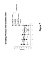

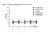
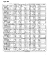
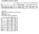
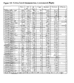


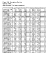

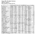
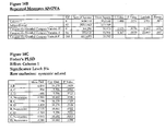

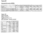
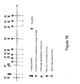
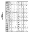

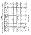
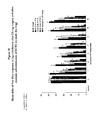


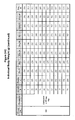
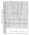

Classifications
-
- A—HUMAN NECESSITIES
- A61—MEDICAL OR VETERINARY SCIENCE; HYGIENE
- A61K—PREPARATIONS FOR MEDICAL, DENTAL OR TOILETRY PURPOSES
- A61K35/00—Medicinal preparations containing materials or reaction products thereof with undetermined constitution
- A61K35/12—Materials from mammals; Compositions comprising non-specified tissues or cells; Compositions comprising non-embryonic stem cells; Genetically modified cells
- A61K35/44—Vessels; Vascular smooth muscle cells; Endothelial cells; Endothelial progenitor cells
-
- A—HUMAN NECESSITIES
- A61—MEDICAL OR VETERINARY SCIENCE; HYGIENE
- A61P—SPECIFIC THERAPEUTIC ACTIVITY OF CHEMICAL COMPOUNDS OR MEDICINAL PREPARATIONS
- A61P21/00—Drugs for disorders of the muscular or neuromuscular system
- A61P21/02—Muscle relaxants, e.g. for tetanus or cramps
-
- A—HUMAN NECESSITIES
- A61—MEDICAL OR VETERINARY SCIENCE; HYGIENE
- A61P—SPECIFIC THERAPEUTIC ACTIVITY OF CHEMICAL COMPOUNDS OR MEDICINAL PREPARATIONS
- A61P25/00—Drugs for disorders of the nervous system
-
- A—HUMAN NECESSITIES
- A61—MEDICAL OR VETERINARY SCIENCE; HYGIENE
- A61P—SPECIFIC THERAPEUTIC ACTIVITY OF CHEMICAL COMPOUNDS OR MEDICINAL PREPARATIONS
- A61P25/00—Drugs for disorders of the nervous system
- A61P25/04—Centrally acting analgesics, e.g. opioids
-
- A—HUMAN NECESSITIES
- A61—MEDICAL OR VETERINARY SCIENCE; HYGIENE
- A61P—SPECIFIC THERAPEUTIC ACTIVITY OF CHEMICAL COMPOUNDS OR MEDICINAL PREPARATIONS
- A61P25/00—Drugs for disorders of the nervous system
- A61P25/08—Antiepileptics; Anticonvulsants
-
- A—HUMAN NECESSITIES
- A61—MEDICAL OR VETERINARY SCIENCE; HYGIENE
- A61P—SPECIFIC THERAPEUTIC ACTIVITY OF CHEMICAL COMPOUNDS OR MEDICINAL PREPARATIONS
- A61P25/00—Drugs for disorders of the nervous system
- A61P25/14—Drugs for disorders of the nervous system for treating abnormal movements, e.g. chorea, dyskinesia
-
- A—HUMAN NECESSITIES
- A61—MEDICAL OR VETERINARY SCIENCE; HYGIENE
- A61P—SPECIFIC THERAPEUTIC ACTIVITY OF CHEMICAL COMPOUNDS OR MEDICINAL PREPARATIONS
- A61P29/00—Non-central analgesic, antipyretic or antiinflammatory agents, e.g. antirheumatic agents; Non-steroidal antiinflammatory drugs [NSAID]
-
- C—CHEMISTRY; METALLURGY
- C12—BIOCHEMISTRY; BEER; SPIRITS; WINE; VINEGAR; MICROBIOLOGY; ENZYMOLOGY; MUTATION OR GENETIC ENGINEERING
- C12N—MICROORGANISMS OR ENZYMES; COMPOSITIONS THEREOF; PROPAGATING, PRESERVING, OR MAINTAINING MICROORGANISMS; MUTATION OR GENETIC ENGINEERING; CULTURE MEDIA
- C12N5/00—Undifferentiated human, animal or plant cells, e.g. cell lines; Tissues; Cultivation or maintenance thereof; Culture media therefor
- C12N5/06—Animal cells or tissues; Human cells or tissues
- C12N5/0602—Vertebrate cells
- C12N5/0634—Cells from the blood or the immune system
Definitions
- the present invention relates generally to compositions, for treating neuropathic pain by administration of cells.
- the invention provides administering cells systemically to a patient to normalize inflammatory or degenerative or a state of pathology of the nervous system.
- a pathology could be at the subcellular, cellular and tissue milieu, thereby modifying neuronal interactions and decreasing pain.
- Chronic pain in general is a public health issue affecting 30-60% of all Americans. In most cases, there is little to no correlation between objective disease findings (X-ray, MRI and CT scan of the region in pain) and subjective pain reports.
- Chronic pain includes pain which persists beyond the normal healing time for a disease or injury, pain related to chronic degenerative disease or a persistent neurologic condition, pain that emerges or persists, even recurnng for months to years without an identifiable cause, or as pain associated with cancer.
- Neuropathic pain is often caused by disorders of the nervous system, also known as neuropathy or neuropathic pain.
- Neuropathic pain is typically accompanied by tissue damage, including nerve fibers that are damaged, dysfunction or injured.
- Neuropathic pain may be caused by a variety of problems, including pathologic lesions, neurodegeneration processes, or prolonged dysfunction of parts of the peripheral or central nervous system. Neuropathic pain can also be present when no detectable damage can be assessed or defined.
- Central plasticity can be the result of changes in receptor population or receptor sensitivity at any level of the CNS.
- central sensitization is known to play a major role in mediating chronic inflammatory as well as neuropathic pain.
- changes in Schwann cell - axonal interaction are known to play a role in the induction and maintenance of neuropathic pain.
- the standard course of treatment for chronic pain involves a step ladder approach which begins with non-opioid analgesics and progresses from moderate opiates to potent opiates.
- Opiates are often used in combination with other agents.
- a physician is able to monitor and adjust the dose of the agent to limit the undesired side effects of opioids, which includes sedation, cognitive impairment, myoclonus, addiction, tolerance, and respiratory depression.
- opioids can also induce nausea, constipation, confusion, respiratory depression, and dependence.
- opiate tolerance is a well documented side effect observed in chronic pain patients.
- NSAIDs nonsteroidal anti-inflammatory drugs
- NSAIDs nonsteroidal anti-inflammatory drugs
- analgesic analgesic
- antidepressants anticonvulsants
- topical agents cannabinoids
- botulinum toxin NMDA antagonists
- antepileptics anti-depressants and dietary supplements.
- side effects which can be debilitating, including CNS depression, cardiovascular effects, gastrointestinal disturbances, ulceration, renal damage, decreased libido and hypersensitivity reactions.
- these compounds must be taken repeatedly, typically more than once a day, and some compounds become ineffective with time, resulting in tolerance to the drug.
- transplanted cells at the site of neural damage to assist in the repair or reversal of neural cell damage.
- some researchers have transplanted neural cells to the site of injury of a patient with a sensory neural pathway disorder or injury. See, U.S. Patent Application No. 09/163,684 which issued as US Patent No. 6,444,205 .
- Other researchers focus on transplantation of stem cells to reconstitute a target tissue, thereby restoring physiologic and anatomic functionality.
- Klass et al. Anesth Analg., 2007; 104:944 - 49 A viable, reliable method of administering cells to decrease neuropathic pain does not presently exist.
- compositions of the illustrative embodiments described herein provide methods for treating chronic and neuropathic pain by administering a population of cells. While not wishing to be bound by any mechanism of action, the inventors believe that the cells administered have the capacity of normalizing inflammatory or degenerative cellular and tissue milieu at the Schwann cell - axonal interaction and/or the microglia - neuronal interaction to decrease pain. Further, the cell administration may block ectopic neuronal firing, thus decreasing pain.
- the present invention is based, at least in part, on the discovery that cells, including cells derived from human umbilical cord tissue, can be administered systemically to a patient in need of chronic pain treatment.
- Specific options provided by the invention are directed to the direct repair, regeneration, replacement of, or the support of the repair, regeneration, or replacement of neural cells for the treatment of neural damage, injury and/or pain.
- the disclosure also pertains to a method of treating a subject having neural damage, injury and/or pain by administering a population of cells in an amount effective, such that the damage, injury and/or pain is treated.
- the cells administered are umbilical cord tissue-derived cells.
- the cells are administered systemically.
- the cells may be administered by any means which allows systemic distribution of the cells, including but not limited to, intramuscular, intravenous, or intra-arterial delivery via syringes with needles and/or catheters with or without pump devices.
- the population of cells is administered with a hydrogel.
- Further embodiments are to specific hydrogels, such as collagen, atelocollagen, fibrin constructs, thrombin-fibrin constructs.
- some embodiments include use of one or more growth factors injected in parallel, sequentially, or formulated directly into a hydrogel.
- the population of cells is induced in vitro to differentiate into a specific type of cell.
- the cells are genetically engineered to produce a gene product that promotes treatment of chronic pain.
- the populations of cells are administered with at least one other agent, including but not limited to, selected extracellular matrix components, anti-apoptotic agents, anti-inflammatory compounds, immunosuppressive or immunomodulatory agents, local anesthetics, and other angiogenic factors.
- the other agent can be administered simultaneously with, before, or after the population of cells.
- the composition further comprises at least one of the agents or factors selected from the group consisting of neurotrophic factors.
- the population of cells is administered with growth factors and/or other agents which promote differentiation of the cells into a predetermined, desired neural cell.
- the desired neural cells are capable of secreting one or more neurotransmitters.
- compositions and kits for treating a patient with chronic pain comprising at least a population of cells and a pharmaceutically acceptable carrier.
- Other composition and kit forms may include other agents, growth factors, and compounds to promote growth and/or healing of the tissue, such that the chronic pain decreases in severity or length.
- the pharmaceutical compositions and kits are designed and/or formulated for practicing the methods of the disclosure as outlined above and below.
- chronic pain and neuroopathic pain as used interchangeably herein generally refer to conditions in which pain persists and fails to respond to conventional treatment. These terms include pain of long duration and pain that can be medically refractory. These terms also include pain characterized by a persistent increase in the level of neuron excitability or the presence of abnormal sensations in the affected area.
- exemplary chronic pain conditions include diabetic neuropathy, cervical radiculopathy, neuralgic amyotrophy, HIV neuropathy, neuralgic amyotrophy, post herpetic neuralgia, post spinal cord injury, muscular dystrophy, trigeminal neuralgia, phantom limb pain, causalgia, spasticity of spinal cord origin, and diabetic and alcoholic polyneuropathies.
- tissue site tissue site
- nerve damage tissue injury
- site of neural damage tissue site
- tissue site tissue site
- connective tissue tissue that connective tissue
- cartilage cartilage
- adipose tissue muscle tissue
- tendons or ligaments tissue site
- the terms may further refer to areas of any tissue that are not necessarily wounded or defective, but are instead areas in which it is desired to add or promote growth of additional tissue.
- the term may also refer to nervous tissue that while having no apparent damage, clinical observations, tests and patients' report indicate the presence of an abnormality.
- xenogeneic refers to transplantation of cells from a donor of one species into a subject of a different species.
- treat generally refer to amelioration or reduction in pain or injury for a period of time following administration of a population of cells into a subject suffering from chronic pain.
- the amelioration or reduction also includes any objective or subjective parameter such as abatement, remission, diminishing of symptoms or making the injury, pathology, or condition more tolerable to the patient, slowing in the rate of degeneration or decline, making the final point of degeneration less debilitating, improving a subject's physical or mental well-being, or prolonging the length of survival.
- the treatment or amelioration of symptoms can be based on objective or subjective parameters; including the results of a physical examination or neurological examination.
- an effective amount generally refers to a concentration or amount of a compound, material, or composition, as described herein, that is effective to achieve a particular biological result. Such results include, but are not limited to, the regeneration, repair, or improvement of neural tissue, the improvement of blood flow, and/or the decrease of inflammation in patients with chronic pain. Such effective activity may be achieved, for example, by administering the cells and/or compositions of the present invention to patients with chronic pain. With respect to the population of cells as administered to a patient, an effective amount is generally more about 10 4 cells/kg body weight, and may range from about 10 4 cells/kg body weight to about 10 6 cells/kg body weight when delivered locally, or from about 10 5 to about 10 7 cells/kg body weight when delivered systemically.
- an effective amount may range from about 10 5 to about 10 8 cells/kg body weight. It will be appreciated that the number of cells to be administered will vary depending on the specifics of the disorder to be treated, including but not limited to size or total volume/surface area to be treated, and proximity of the site of administration to the location of the region to be treated, among other factors familiar to the medicinal biologist.
- Stem cells are undifferentiated cells defined by the ability of a single cell both to self-renew, and to differentiate to produce progeny cells, including self-renewing progenitors, non-renewing progenitors, and terminally differentiated cells. Stem cells are also characterized by their ability to differentiate in vitro into functional cells of various cell lineages from multiple germ layers (endoderm, mesoderm and ectoderm), as well as to give rise to tissues of multiple germ layers following transplantation, and to contribute substantially to most, if not all, tissues following injection into blastocysts.
- Stem cells are classified according to their developmental potential as: (1) totipotent; (2) pluripotent; (3) multipotent; (4) oligopotent; and (5) unipotent.
- Totipotent cells are able to give rise to all embryonic and extraembryonic cell types.
- Pluripotent cells are able to give rise to all embryonic cell types.
- Multipotent cells include those able to give rise to a subset of cell lineages, but all within a particular tissue, organ, or physiological system.
- hematopoietic stem cells can produce progeny that include HSC (self-renewal), blood cell-restricted oligopotent progenitors, and all cell types and elements (e.g., platelets) that are normal components of the blood.
- HSC self-renewal
- oligopotent progenitors can give rise to a more restricted subset of cell lineages than multipotent stem cells.
- Cells that are unipotent are able to give rise to a single cell lineage (e.g., spermatogenic stem cells).
- Stem cells are also categorized on the basis of the source from which they are obtained.
- An “adult stem cell” is generally a multipotent undifferentiated cell found in tissue comprising multiple differentiated cell types. The adult stem cell can renew itself. Under normal circumstances, it can also differentiate to yield the specialized cell types of the tissue from which it originated, and possibly other tissue types.
- An “embryonic stem cell” is a pluripotent cell from the inner cell mass of a blastocyst-stage embryo. A fetal stem cell is one that originates from fetal tissues or membranes.
- a “postpartum stem cell” is a multipotent or pluripotent cell that originates substantially from extraembryonic tissue available after birth, namely, the placenta and the umbilical cord.
- Postpartum stem cells may be blood-derived (e.g., as are those obtained from umbilical cord blood) or non-blood-derived (e.g., as obtained from the non-blood tissues of the umbilical cord and placenta).
- Cell culture refers generally to cells taken from a living organism and grown under controlled condition ("in culture” or “cultured”).
- a “primary cell culture” is a culture of cells, tissues, or organs taken directly from an organism(s) before the first subculture. Cells are “expanded” in culture when they are placed in a growth medium under conditions that facilitate cell growth and/or division, resulting in a larger population of the cells. When cells are expanded in culture, the rate of cell proliferation is sometimes measured by the amount of time needed for the cells to double in number. This is referred to as "doubling time".
- standard growth conditions refers to culturing of cells at 37°C, in a standard atmosphere comprising 5% CO 2 and relative humidity maintained at about 100%. While the foregoing conditions are useful for culturing, it is to be understood that such conditions are capable of being varied by the skilled artisan who will appreciate the options available in the art for culturing cells.
- a “conditioned medium” is a medium in which a specific cell or population of cells has been cultured, and then removed. When cells are cultured in a medium, they may secrete cellular factors that can provide trophic support to other cells. Such trophic factors include, but are not limited to hormones, cytokines, extracellular matrix (ECM), proteins, vesicles, antibodies, and granules.
- the medium containing the cellular factors is the conditioned medium.
- “Differentiation” is the process by which an unspecialized ("uncommitted") or less specialized cell acquires the features of a specialized cell, such as a nerve cell or a muscle cell, for example.
- a “differentiated” cell is one that has taken on a more specialized ("committed") position within the lineage of a cell.
- the term “committed,” when applied to the process of differentiation, refers to a cell that has proceeded in the differentiation pathway to a point where, under normal circumstances, it will continue to differentiate into a specific cell type or subset of cell types, and cannot, under normal circumstances, differentiate into a different cell type or revert to a less differentiated cell type.
- De-differentiation refers to the process by which a cell reverts to a less specialized (or committed) position within the lineage of a cell.
- the "lineage" of a cell defines the heredity of the cell, i.e. which cells it came from and what cells it can give rise to. The lineage of a cell places the cell within a hereditary scheme of development and differentiation.
- a progenitor cell is a cell that has the capacity to create progeny that are more differentiated than itself, and yet retains the capacity to replenish the pool of progenitors.
- stem cells themselves are also progenitor cells, as arc the more immediate precursors to terminally differentiated cells.
- this broad definition of progenitor cell may be used.
- a progenitor cell is often defined as a cell that is intermediate in the differentiation pathway, i.e., it arises from a stem cell and is intermediate in the production of a mature cell type or subset of cell types.
- This type of progenitor cell is generally not able to self-renew. Accordingly, if this type of cell is referred to herein, it will be referred to as a “non-renewing progenitor cell” or as an “intermediate progenitor or precursor cell".
- autologous transfer refers to treatments wherein the cell or transplant donor is also the cell or transplant recipient.
- allogeneic transfer refers to treatments wherein the cell or transplant donor is of the same species as the recipient, but is not the same individual.
- a cell transfer in which the donor's cells have been histocompatibly matched with a recipient is sometimes referred to as a "syngeneic transfer”.
- xenogeneic transfer refers to treatments wherein the cell or transplant donor is of a different species than the recipient.
- nerve cell includes both nerve cells (i.e., neurons, e.g., uni-, bi-, or multipolar neurons) and their precursors and glial cells (e.g., macroglia such as oligodendrocytes, Schwann cells, and astrocytes, or microglia) and their precursors.
- nerve cells i.e., neurons, e.g., uni-, bi-, or multipolar neurons
- glial cells e.g., macroglia such as oligodendrocytes, Schwann cells, and astrocytes, or microglia
- the cells used in the present invention are generally referred to as "postpartum cells” or “postpartum-derived cells” (PPDC(s)).”
- the cells are more specifically “umbilicus-derived cells” or “umbilical cord-derived cells” (UDC(s)), or “umbilical cord tissue-derived cells” (UTC(s)).
- the cells may be described as being stem or progenitor cells, the latter term being used in the broad sense.
- the term "derived” is used to indicate that the cells have been obtained from their biological source and grown or otherwise manipulated in vitro (e.g., cultured in a growth medium to expand the population and/or to produce a cell line). The in vitro manipulations of umbilical stem cells and the unique features of the umbilicus-derived cells of the present invention are described in detail below.
- isolated generally refers to a cell which has been separated from its natural environment. This term includes gross physical separation from its natural environment, e.g ., removal from the donor animal.
- an isolated cell is not present in a tissue, i.e., the cell is separated or dissociated from the neighboring cells with which it is normally in contact.
- cells are administered as a cell suspension.
- the phrase "cell suspension” includes cells which are in contact with a medium and which have been dissociated, e.g ., by subjecting a piece of tissue to gentle trituration.
- the term "growth medium” generally refers to a medium sufficient for the culturing of umbilical cord tissue-derived cells.
- one medium for the culturing of the cells of the invention comprises Dulbecco's Modified Essential Media (DMEM).
- DMEM Dulbecco's Modified Essential Media
- Particularly preferred is DMEM-Low glucose (DMEM-LG) (Invitrogen, Carlsbad, Ca.).
- the DMEM-LG is preferably supplemented with serum, most preferably fetal bovine serum or human serum. Typically, 15% (v/v) fetal bovine serum ( e.g .
- fetal bovine serum Hyclone, Logan Ut.
- antibiotics/antimycotics preferably 100 Unit/milliliter penicillin, 100 milligrams/milliliter streptomycin, and 0.25 microgram/milliliter amphotericin B; Invitrogen, Carlsbad, Ca.
- 0.001% (v/v) 2-mercaptoethanol Sigma, St. Louis Mo.
- different growth media are used or different supplementations are provided, and these are normally indicated in the text as supplementations to growth medium.
- the cells may be grown without serum present at all. In such cases, the cells may require certain growth factors, which can be added to the medium to support and sustain the cells.
- Presently preferred factors to be added for growth in serum-free media include one or more of bFGF, EGF, IGF-I, and PDGF. In more preferred embodiments, two, three or all four of the factors are added to serum free or chemically defined media. In other embodiments, LIF is added to serum-free medium to support or improve growth of the cells.
- cell line generally refers to a population of cells formed by one or more subcultivations of a primary cell culture. Each round of subculturing is referred to as a passage. When cells are subcultured, they are referred to as having been "passaged.” A specific population of cells, or a cell line, is sometimes referred to or characterized by the number of times it has been passaged. For example, a cultured cell population that has been passaged ten times may be referred to as a P10 culture.
- the primary culture i.e., the first culture following the isolation of cells from tissue, is designated P0. Following the first subculture, the cells are described as a secondary culture (P1 or passage 1).
- the cells After the second subculture, the cells become a tertiary culture (P2 or passage 2), and so on. It will be understood by those of skill in the art that there may be many population doublings during the period of passaging; therefore, the number of population doublings of a culture is greater than the passage number.
- the expansion of cells (i.e., the number of population doublings) during the period between passaging depends on many factors, including, but not limited to, the seeding density, substrate, medium, growth conditions, and time between passaging.
- pharmaceutically acceptable carrier or “pharmaceutically acceptable medium,” which may be used interachangeably with the terms “biologically compatible carrier” or “biologically compatible medium,” generally refer to reagents, cells, compounds, materials, compositions, and/or dosage forms that are not only compatible with the cells and other agents to be administered therapeutically, but also are suitable for use in contact with the tissues of human beings and animals without excessive toxicity, irritation, allergic response, or other complication commensurate with a reasonable benefit/risk ratio.
- pharmaceutically acceptable carriers suitable for use in the present invention include liquids, semi-solid (e.g., gels) and solid materials (e.g., cell scaffolds and matrices, tubes, sheets and other such materials known in the art and described in greater detail herein).
- semi-solid and solid materials may be designed to resist degradation within the body (non-biodegradable) or they may be designed to degrade within the body (biodegradable, bioerodable).
- a biodegradable material may further be bioresorbable or bioabsorbable, i.e., it may be dissolved and absorbed into bodily fluids (water-soluble implants are one example), or degraded and ultimately eliminated from the body, either by conversion into other materials or breakdown and elimination through natural pathways.
- the biodegradation rate can vary according to the desired release rate once implanted in the body.
- matrix generally refers to biodegradable and/or bioresorbable materials that are administrated with the cells to a patient.
- the matrix may act as a temporary scaffold until replaced by newly grown cells.
- the matrix may provide for sustained the release of neurotrophic factors or other agents used in conjunction with the cells and may provide a structure for developing tissue growth in the patient.
- the matrix simply provides a temporary scaffold for the developing tissue.
- the matrix can be in particulate form (macroparticles greater than 10 microns in diameter or microparticles less than 10 microns in diameter), or it can be in the form of a structurally stable, three-dimensional implant (e.g., a scaffold).
- the matrix can be a slurry, hydrogel or alternatively a three dimensional structure such as a cube, cylinder, tube, block, film, sheet or an appropriate anatomical form.
- scaffold generally refers to a three dimensional porous structure that provides a template for cell growth.
- a scaffold is made of biodegradable and/or bioresorbable materials that degrade over time within the body. The length of time taken for the scaffold to degrade may depend upon the molecular weight of the materials. Thus, higher molecular weight material may result in polymer scaffolds which retain their structural integrity for longer periods of time; while lower molecular weights result in both slower release and shorter scaffold lives.
- the scaffold may be made by any means known in the art. Examples of polymers which can be used to form the scaffold include natural and synthetic polymers.
- the scaffold may be infused with, coated with, or comprised of a population of cells, growth factors, or other nutrients to promote cell growth.
- the scaffold contains growth inducing agents including neurotrophins.
- the growth inducing agents may be synthetic or naturally produced, and may be a fragment, derivative or analog of a growth inducing agent.
- Neurotrophic factor or “trophic factor” is defined as a substance that promotes survival, growth, proliferation and/or maturation of a cell, or stimulates increased activity of a cell.
- the present invention features pharmaceutical compositions for treatment of chronic and neuropathic pain.
- These pharmaceutical compositions are designed to stimulate and support neural tissue growth or healing, and to improve the regeneration and repair of tissues surrounding the neural tissue, including but not limited to, dermal tissue, vascular tissue, connective tissue, cartilage, adipose tissue, muscle tissue, tendons or ligaments.
- the cells of the invention include, progenitor cells and cell populations derived from, umbilicus tissue. A more detailed explanation of preferred cells may be found below.
- the cells are stem cells.
- Stem cells are undifferentiated cells defined by the ability of a single cell both to self-renew and to differentiate to produce progeny cells, including self-renewing progenitors, non-renewing progenitors and terminally differentiated cells.
- the stem cells are umbilical cord tissue-derived cells.
- an umbilical cord is recovered upon or shortly after termination of either a full-term or pre-term pregnancy, for example, after expulsion of after birth.
- the umbilical cord tissue may be transported from the birth site to a laboratory in a sterile container such as a flask, beaker, culture dish, or bag.
- the container may have a solution or medium, including but not limited to a salt solution, such as Dulbecco's Modified Eagle's Medium (DMEM) (also known as Dulbecco's Minimal Essential Medium) or phosphate buffered saline (PBS), or any solution used for the transportation of organs used for transplantation, such as University of Wisconsin solution or perfluorochemical solution.
- DMEM Dulbecco's Modified Eagle's Medium
- PBS phosphate buffered saline
- antibiotic and/or antimycotic agents such as, but not limited to, penicillin, streptomycin, amphotericin B, gentamicin, and nystatin, may be added to the medium or buffer.
- the umbilical cord tissue may be rinsed with an anticoagulant solution such as heparin-containing solution. It is preferable to keep the tissue at about 4 to about 10°C prior to extraction of the cells. It is even more preferable that the tissue not be frozen prior to extraction of
- the umbilical cord tissue-derived cells are preferably isolated in an aseptic environment.
- the umbilical cord may be separated from the placenta by means known in the art.
- Blood and debris are preferably removed from the postpartum tissue prior to isolation of umbilical cord tissue-derived cells.
- the postpartum tissue may be washed with buffer solution, including but not limited to phosphate buffered saline.
- the wash buffer also may comprise one or more antimycotic and/or antibiotic agents, including but not limited to penicillin, streptomycin, amphotericin B, gentamicin, and nystatin.
- Postpartum tissue comprising a whole umbilicus or a fragment or section thereof is preferably disaggregated by mechanical force (mincing or shear forces).
- the isolation procedure may also utilize an enzymatic digestion process.
- Many enzymes are known in the art to be useful for the isolation of individual cells from complex tissue matrices to facilitate growth in culture. Digestion enzymes range from weakly digestive (e.g. deoxyribonucleases and the neutral protease, dispase) to strongly digestive (e.g. papain and trypsin), and are available commercially.
- a nonexhaustive list of such enzymes includes mucolytic enzyme activities, metalloproteases, neutral proteases, serine proteases (such as trypsin, chymotrypsin, or elastase), and deoxyribonucleases.
- enzyme activities selected from metalloproteases, neutral proteases and mucolytic activities.
- collagenases are known to be useful for isolating various cells from tissues.
- Deoxyribonucleases can digest single-stranded DNA and can minimize cell-clumping during isolation.
- Preferred methods involve enzymatic treatment with collagenase and dispase, or collagenase, dispase, and hyaluronidase.
- enzyme treatments are known in the art for isolating cells from various tissue sources, and is well-equipped to assess new or additional enzymes or enzyme combinations for their utility in isolating the cells of the invention.
- Preferred enzyme treatments can be from about 0.5 to 2 hours long or longer.
- the tissue is incubated at 37°C during the enzyme treatment of the dissociation step.
- the cells are passaged or removed to a separate culture vessel containing fresh medium of the same or a different type as that used initially, where the population of cells can be mitotically expanded.
- the cells of the invention may be used at any point between passage 0 and senescence.
- the cells preferably are passaged between about 3 and about 25 times, more preferably are passaged about 4 to about 12 times and preferably are passaged 10 or 11 times. Cloning and/or subcloning may be performed to confirm that a clonal population of cells has been isolated.
- the different cell types present in postpartum tissue are fractionated into subpopulations from which the umbilical cord tissue-derived cells can be isolated. Fractionation or selection may be accomplished using standard techniques for cell separation. Such techniques include, but are not limited to, enzymatic treatment to dissociate postpartum tissue into its component cells, followed by cloning and selection of specific cell types, including, but not limited to, selection based on morphological and/or biochemical markers; selective growth of desired cells (positive selection), selective destruction of unwanted cells (negative selection); separation based upon differential cell agglutinability in the mixed population as, for example, with soybean agglutinin; freeze-thaw procedures; differential adherence properties of the cells in the mixed population; filtration; conventional and zonal centrifugation; centrifugal elutriation (counter-streaming centrifugation); unit gravity separation; countercurrent distribution; electrophoresis; and fluorescence activated cell sorting (FACS).
- FACS fluorescence activated cell sorting
- the culture medium is changed as necessary. For example, by carefully aspirating the medium from the dish with a pipette and replenishing with fresh medium. Incubation is continued until a sufficient number or density of cells accumulates in the dish. Thereafter, any original explanted tissue sections that exist may be removed and the remaining cells separated from the dish by trypsinization using standard techniques or by using a cell scraper. After trypsinization the cells are collected, removed to fresh medium and incubated as above. In some embodiments, the medium is changed at least once at approximately 24 hours post-trypsinization to remove any floating cells. The cells remaining in culture are considered to be umbilical cord tissue-derived cells.
- Umbilical cord tissue-derived cells may be cryopreserved. Accordingly, in a preferred embodiment described in greater detail below, umbilical cord tissue-derived cells for autologous transfer (for either the mother or child) may be derived from appropriate postpartum tissues following the birth of a child, then cryopreserved so as to be available in the event they are later needed for transplantation.
- Umbilical cord tissue-derived cells may be characterized, for example, by growth characteristics (e.g ., population doubling capability, doubling time, passages to senescence), karyotype analysis (e.g ., normal karyotype; maternal or neonatal lineage), flow cytometry (e.g., FACS analysis), immunohistochemistry and/or immunocytochemistry (e.g., for detection of epitopes), gene expression profiling (e.g ., gene chip arrays; polymerase chain reaction (for example, reverse transcriptase PCR, real time PCR, and conventional PCR)), protein arrays, protein secretion (e.g ., by plasma clotting assay or analysis of PDC-conditioned medium, for example, by enzyme linked immunosorbent assay (ELISA), mixed lymphocyte reaction (e.g ., as measure of stimulation of PBMCs), and/or other methods known in the art.
- growth characteristics e.g ., population doubling capability, doubling time, passages to
- Examples of umbilical cord tissue-derived cells were deposited with the American Type Culture Collection on June 10, 2004, and assigned ATCC Accession Numbers as follows: (1) strain designation UMB 022803 (P7) was assigned Accession No. PTA-6067; and (2) strain designation UMB 022803 (P17) was assigned Accession No. PTA-6068.
- the umbilical cord tissue-derived cells possess one or more of the following growth features: (1) they require L-valine for growth in culture; (2) they are capable of growth in atmospheres containing oxygen from about 5% to about 20%; (3) they have the potential for at least about 40 doublings in culture before reaching senescence; and (4) they attach and expand tissue culture vessels that are uncoated or that are coated with gelatin, laminin, collagen, polyornithine, vitronectin or fibronectin.
- the umbilical cord tissue-derived cells possess a normal karyotype, which is maintained as the cells are passaged. Methods for karyotyping are available and known to those of skill in the art.
- the umbilical cord tissue-derived cells may be characterized by production of certain proteins, including: (1) production of at least one of tissue factor, vimentin, and alpha-smooth muscle actin; and (2) production of at least one of: CD 10, CD13, CD44, CD73, CD90, PDGFr-alpha, PD-L2 and HLA-A,B,C cell surface markers, as detected by flow cytometry.
- the umbilical cord tissue-derived cells are/may be characterized by lack of production of at least one of: CD31, CD34, CD45, CD80, CD86, CD117, CD141, CD178, B7-H2, HLA-G, and HLA-DR,DP,DQ cell surface markers, as detected by flow cytometry.
- Particularly preferred in some applications are cells that produce at least two of: tissue factor; vimentin; and alpha-smooth muscle actin. More preferred are those cells producing all three of the proteins: tissue factor; vimentin; and alpha-smooth muscle actin.
- the umbilical cord tissue-derived cells may be characterized by gene expression relative to a human cell that is a fibroblast, a mesenchymal stem cell, or an iliac crest bone marrow cell, which is increased for a gene encoding at least one of: interleukin 8; reticulon 1; chemokine (C-X-C motif) ligand 1 (melonoma growth stimulating activity, alpha); chemokine (C-X-C motif) ligand 6 (granulocyte chemotactic protein 2); chemokine (C-X-C motif) ligand 3; and tumor necrosis factor, alpha-induced protein 3.
- interleukin 8 reticulon 1
- chemokine (C-X-C motif) ligand 1 melonoma growth stimulating activity, alpha
- chemokine (C-X-C motif) ligand 6 granulocyte chemotactic protein 2
- chemokine (C-X-C motif) ligand 3
- the umbilical cord tissue-derived cells may be characterized by gene expression relative to a human cell that is a fibroblast, a mesenchymal stem cell, or an iliac crest bone marrow cell, which is reduced for a gene encoding at least one of: short stature homeobox 2; heat shock 27 kDa protein 2; chemokine (C-X-C motif) ligand 12 (stromal cell-derived factor 1); elastin (supravalvular aortic stenosis, Williams-Beuren syndrome); Homo sapiens mRNA; cDNA DKFZp586M2022 (from clone DKFZp586M2022); mesenchyme homeo box 2 (growth arrest-specific homeo box); sine oculis homeobox homolog 1 ( Drosophila ); crystallin, alpha B; disheveled associated activator of morphogenesis 2; DKFZP586B2420 protein; similar
- the umbilical cord tissue-derived cells may be characterized by secretion of at least one of: MCP-1; IL-6; IL-8; GCP-2; HGF; KGF; FGF; HB-EGF; BDNF; TPO; RANTES; MIP1b; 1309; MDC; and TIMP1.
- the umbilical cord tissue-derived cells may be characterized by a lack of secretion of at least one of: TGF-beta2; ANG2; MIP1a; PDGFbb; and VEGF, as detected by ELISA.
- the umbilical cord tissue-derived cells are derived from umbilical cord tissue substantially free of blood and are capable of self-renewal and expansion in culture. They may require L-valine for growth, can grow in at least about 5% oxygen, and comprise at least one of the following characteristics: (1) the potential for at least about 40 doublings in culture; (2) the ability to attach and expand on an uncoated tissue culture vessel or one coated with gelatin, laminin, collagen, polyornithine, vitronectin, or fibronectin; (3) production ofvimentin and alpha-smooth muscle actin;(4) production of CD10, CD13, CD44, CD73, and CD90; and (5) expression of a gene, which relative to a human cell that is a fibroblast, a mesenchymal stem cell, or an iliac crest bone marrow cell, is increased for a gene encoding interleukin 8 and reticulon 1. Such umbilical cord tissue-derived cells may optionally not produce CD45 and are negative for CD117.
- the cell comprises two or more of the above-listed growth, protein/surface marker production, gene expression or substance-secretion characteristics. More preferred are those cells comprising three, four, five or more of the characteristics. Still more preferred are umbilical cord tissue-derived cells comprising six, seven, eight or more of the characteristics. Still more preferred presently are those cells comprising all of above characteristics.
- umbilical cord tissue-derived cells having the characteristics described above and more particularly those wherein the cells have normal karyotypes and maintain normal karyotypes with passaging, and further wherein the cells express each of the markers CD 10, CD 13, CD44, CD73, CD90, PDGFr-alpha, and HLA-A,B,C, and wherein the cells produce the immunologically-detectable proteins which correspond to the listed markers. Still more preferred are those cells which, in addition to the foregoing, do not produce proteins corresponding to any of the markers CD31, CD34, CD45, CD141, or HLA-DR,DP,DQ, as detected by flow cytometry.
- Certain cells having the potential to differentiate along lines leading to various phenotypes are unstable and thus can spontaneously differentiate.
- Presently preferred for use with the invention are cells that do not spontaneously differentiate, for example, along myoblast, skeletal muscle, vascular smooth muscle, pericyte, hemangiogenic, angiogenic, vasculogenic, or vascular endothelial lines.
- Preferred cells, when grown in growth medium are substantially stable with respect to the cell markers produced on their surface, and with respect to the expression pattern of various genes, for example, as determined using a medical diagnostic test sold under the trade name GENECHIP (Affymetrix, Inc., Santa Clara, CA). The cells remain substantially constant, for example, in their surface marker characteristics over passaging and through multiple population doublings.
- the cell population is heterogeneous.
- a heterogeneous cell population of the invention may comprise at least about 5%, 10%, 20%, 30%, 40%, 50%, 60%, 70%, 80%, 90%, or 95% umbilical cord tissue-derived cells of the invention.
- the heterogeneous cell populations of the invention may further comprise stem cells or other progenitor cells, such as myoblasts or other muscle progenitor cells, hemangioblasts, or blood vessel precursor cells; or it may further comprise fully differentiated skeletal muscle cells, smooth muscle cells, pericytes, or blood vessel endothelial cells.
- the population is substantially homogeneous, i.e., comprises substantially only umbilical cord tissue-derived cells (preferably at least about 96%, 97%, 98%, 99% or more umbilical cord tissue-derived cells).
- the homogeneous cell population of the invention may comprise umbilicus-derived cells. Homogeneous populations of umbilicus-derived cells are preferably free of cells of maternal lineage. Homogeneity of a cell population may be achieved by any method known in the art, for example, by cell sorting (e.g., flow cytometry) or by clonal expansion in accordance with known methods. Thus, preferred homogeneous umbilical cord tissue-derived cell populations may comprise a clonal cell line of umbilical cord tissue-derived cells. Such populations are particularly useful when a cell clone with highly desirable functionality has been isolated.
- the cells are umbilical cord tissue-derived cells that are administered as undifferentiated cells, i.e., as cultured in growth medium.
- the umbilical cord tissue-derived cells may be administered following exposure in culture to conditions that stimulate differentiation toward a desired neural tissue.
- the cells are UTC.
- the cells are hUTC.
- the population of cells may include more than one type of cell. Indeed, some embodiments include administration of cells which surround and support the neural cell. Such cells may include, but are not limited to, dermal tissue, vascular tissue, connective tissue, cartilage, adipose tissue, muscle tissue, tendons or ligaments.
- Cells used in the invention may also be genetically modified to produce therapeutically useful gene products, to produce agents to facilitate or support neural tissue formation, healing and/or growth, or to produce factors to recruit progenitor cells to the area of neural damage.
- Genetic modification may be accomplished using any of a variety of vectors including, but not limited to, integrating viral vectors, e.g., retrovirus vector or adeno-associated viral vectors; non-integrating replicating vectors, e.g. , papilloma virus vectors, SV40 vectors, adenoviral vectors; or replication-defective viral vectors.
- integrating viral vectors e.g., retrovirus vector or adeno-associated viral vectors
- non-integrating replicating vectors e.g. , papilloma virus vectors, SV40 vectors, adenoviral vectors
- replication-defective viral vectors e.g., papilloma virus vectors, SV40 vectors, adenoviral vectors
- Other methods of introducing DNA into cells include the use of liposomes, electroporation, a particle gun, or by direct DNA injection.
- the cells may be genetically engineered to express and/or secrete a foreign molecule (e.g ., a heterologous molecule not normally made by the cell) or to modify the production of a molecule to treat chronic pain.
- a foreign molecule e.g ., a heterologous molecule not normally made by the cell
- Such molecules can be produced by the cells upon introduction of heterologous nucleic acid molecules using techniques which are well known in the art.
- the cells of the invention can be modified to express a receptor to a neurotransmitter.
- the cells of the invention are modified to produce a neurotransmitter, including but not limited to, serotonin, histamine, gamma aminobutyric acid, glutamate, aspartate, glycine, neuropeptide Y, ATP, GRP, adenosine, epinephrine, neuroepinephrine, dopamine, acetylcholine, melatonin, n-acetylaspartylglutamate, octopamine, tyramine, gastrin, cholecytokinin, vasopressin, oxytocin, neurphysin I and II, pancreatic polypeptide, peptide YY, corticotrophin, dynorphin, endorphin, enkephaline, secretin, motilin, glucagons, vasoactive intestinal peptide, growth
- a foreign molecule enhances the neuroregenerative capacity of the transplanted cells, aids in reestablishing sensorineural communication of GABA interneurons, and/or aids in reestablishment of the excitatory/inhibitory neurotransmitter balance in the subject
- the cells are genetically engineered to express and/or secret foreign molecules that directly reduce pain in the subject, promote success of transplantation (e.g. , by downmodulation of an immune response in the subject), and/or promote survival or function of the transplanted cells.
- exemplary molecules include, e.g., a neurotrophic factor, or a neuroprotective agent.
- unmodified or modified cells can be introduced together with other types of cells genetically modified to perform a useful function.
- the cells can be administered together with other cells which secrete or have been modified to secrete, for example, a neurotrophic factor.
- Examples of cells that act as carriers of transgenes to a subject include fibroblasts, adrenal chromaffin cells, astrocytes, and myoblasts.
- Such cells for example fibroblasts and glial cells, can also be used to deliver retroviruses containing genes such as the herpes simplex thymidine kinase gene, the gene products of which are targets for other therapeutic drugs or agents such as ganciclovir to target cells.
- Hosts cells may be transformed or transfected with DNA controlled by, or in operative association with, one or more appropriate expression control elements such as promoter or enhancer sequences, transcription terminators, polyadenylation sites, among others, and a selectable marker.
- Any promoter may be used to drive the expression of the inserted gene.
- viral promoters include, but are not limited to, the CMV promoter/enhancer, SV 40, papillomavirus, Epstein-Barr virus or elastin gene promoter.
- the control elements used to control expression of the gene of interest can allow for the regulated expression of the gene so that the product is synthesized only when needed in vivo.
- constitutive promoters are preferably used in a non-integrating and/or replication-defective vector.
- inducible promoters could be used to drive the expression of the inserted gene when necessary.
- Inducible promoters include, but are not limited to, those associated with metallothionein and heat shock proteins.
- engineered cells may be allowed to grow in enriched media and then switched to selective media.
- the selectable marker in the foreign DNA confers resistance to the selection and allows cells to stably integrate the foreign DNA as, for example, on a plasmid, into their chromosomes and grow to form foci which, in turn, can be cloned and expanded into cell lines. This method can be advantageously used to engineer cell lines that express the gene product.
- the cells of the invention may be genetically engineered to "knock out” or “knock down” expression of factors that promote inflammation or rejection at the implant site. Negative modulatory techniques for the reduction of target gene expression levels or target gene product activity levels are discussed below. "Negative modulation,” as used herein, refers to a reduction in the level and/or activity of target gene product relative to the level and/or activity of the target gene product in the absence of the modulatory treatment.
- a gene native to a skeletal muscle cell, vascular smooth muscle cell, pericyte, vascular endothelial cell, neural cell, or progenitor cells thereof can be reduced or knocked out using a number of techniques including, for example, inhibition of expression by inactivating the gene using the homologous recombination technique.
- an exon encoding an important region of the protein (or an exon 5' to that region) is interrupted by a positive selectable marker, e.g., neo, preventing the production of normal mRNA from the target gene and resulting in inactivation of the gene.
- a gene may also be inactivated by creating a deletion in part of a gene, or by deleting the entire gene.
- the disclosure involves the use of cell lysates and cell soluble fractions prepared from a umbilical cord tissue-derived cells (not claimed).
- lysates and fractions thereof have many utilities.
- Methods of lysing cells are well-known in the art and include various means of mechanical disruption, enzymatic disruption, or chemical disruption, or combinations thereof.
- Such cell lysates may be prepared from cells directly in their growth medium, and thus contain secreted growth factors and the like, or they may be prepared from cells washed free of medium in, for example, PBS or other solution. Washed cells may be resuspended at concentrations greater than the original population density if preferred.
- whole cell lysates are prepared, e.g., by disrupting cells without subsequent separation of cell fractions.
- a cell membrane fraction is separated from a soluble fraction of the cells by routine methods known in the art, e.g., centrifugation, filtration, or similar methods.
- Cell lysates or cell soluble fractions prepared from populations of postpartum-derived cells may be used as is, further concentrated by, for example, ultrafiltration or lyophilization, or even dried, partially purified, combined with pharmaceutically-acceptable carriers or diluents as are known in the art, or combined with other compounds such as biologicals, for example, pharmaceutically useful protein compositions.
- Cell lysates or fractions thereof may be used in vitro or in vivo, alone or, for example, with autologous or syngeneic live cells.
- the lysates, if introduced in vivo may be introduced locally at a site of treatment, or remotely to provide, for example, needed cellular growth factors to a patient.
- the UTC can be cultured in vitro to produce biological products in high yield.
- An umbilical cord tissue-derived cell that either naturally produces a particular biological product of interest (e.g ., a trophic factor), or that has been genetically engineered to produce such a biological product, can be clonally expanded using the culture techniques described herein.
- cells may be expanded in a medium that induces differentiation to a neural cell.
- biological products produced by the cell and secreted into the medium can be readily isolated from the conditioned medium using standard separation techniques, e.g ., such as differential protein precipitation, ion-exchange chromatography, gel filtration chromatography, electrophoresis, and HPLC, to name a few.
- a “bioreactor” may be used to take advantage of the flow method for feeding, for example, a three-dimensional culture in vitro . Essentially, as fresh media is passed through the three-dimensional culture, the biological product is washed out of the culture and may then be isolated from the outflow, as above.
- a biological product of interest may remain within the cell and, thus, its collection may require that the cells be lysed, as described above.
- the biological product may then be purified using any one or more of the above-listed techniques.
- the disclosure involves the use of conditioned medium from cultured umbilical cord tissue-derived cells for use in vitro and in vivo as described below.
- Use of the umbilical cord tissue-derived cells conditioned medium allows the beneficial trophic factors secreted by the umbilical cord tissue-derived cells to be used allogeneically in a patient without introducing intact cells that could trigger rejection, or other adverse immunological responses.
- Conditioned medium is prepared by culturing cells in a culture medium, then removing the cells from the medium.
- Conditioned medium prepared from populations of umbilical cord-derived cells may be used as is, further concentrated, for example, by ultrafiltration or lyophilization, or even dried, partially purified, combined with pharmaceutically acceptable carriers or diluents as are known in the art, or combined with other compounds such as biologicals, for example, pharmaceutically useful protein compositions.
- Conditioned medium may be used in vitro or in vivo, alone or combined with autologous or syngeneic live cells, for example.
- the conditioned medium, if introduced in vivo may be introduced locally at a site of treatment, or remotely to provide needed cellular growth or trophic factors to a patient.
- an extracellular matrix (ECM) produced by culturing the umbilical cord tissue-derived cells on liquid, solid or semi-solid substrates is prepared, collected and utilized as an alternative to implanting live cells into a subject in need the repair , replacement, or regeneration of neural cells.
- the umbilical cord tissue-derived cells are cultured in vitro, on a three dimensional framework as described elsewhere herein, under conditions such that a desired amount of ECM is secreted onto the framework.
- the cells comprising the new tissue are removed, and the ECM processed for further use, for example, as an injectable preparation.
- cells on the framework are killed and any cellular debris is removed from the framework.
- This process may be carried out in a number of different ways.
- the living tissue can be flash-frozen in liquid nitrogen without a cryopreservative, or the tissue can be immersed in sterile distilled water so that the cells burst in response to osmotic pressure.
- the cellular membranes may be disrupted and cellular debris removed by treatment with a mild detergent rinse, such as EDTA, CHAPS or a zwitterionic detergent.
- a mild detergent rinse such as EDTA, CHAPS or a zwitterionic detergent.
- the tissue can be enzymatically digested and/or extracted with reagents that break down cellular membranes and allow removal of cell contents.
- enzymes include, but are not limited to, hyaluronidase, dispase, proteases, and nucleases.
- detergents include non-ionic detergents such as, for example, alkylaryl polyether alcohol (TRITON X-100), octylphenoxy polyethoxy-ethanol (Rohm and Haas, Philadelphia, Pa.), BRIJ-35, a polyethoxyethanol lauryl ether (Atlas Chemical Co., San Diego, Ca.), polysorbate 20 (TWEEN 20), a polyethoxyethanol sorbitan monolaureate (Rohm and Haas, Philadelphia, Pa.), polyethylene lauryl ether (Rohm and Haas, Philadelphia, Pa.); and ionic detergents such as sodium dodecyl sulfate, sulfated higher aliphatic alcohols, sulfonated alkanes and sulfonated alkylarenes containing 7 to 22 carbon atoms in a branched or unbranched chain.
- non-ionic detergents such as, for example, alkylaryl polyether alcohol (TRITON
- the collection of the ECM can be accomplished in a variety of ways, depending at least in part on whether the new tissue has been formed on a three-dimensional framework that is biodegradable or non-biodegradable, as in the case of metals.
- the framework is non-biodegradable
- the ECM can be removed by subjecting the framework to sonication, high pressure water jets, mechanical scraping, or mild treatment with detergents or enzymes, or any combination of the above.
- the ECM can be collected, for example, by allowing the framework to degrade or dissolve in solution.
- the biodegradable framework is composed of a material that can itself be injected along with the ECM, the framework and the ECM can be processed in toto for subsequent injection.
- the ECM can be removed from the biodegradable framework by any of the methods described above for collection of ECM from a non-biodegradable framework. All collection processes are preferably designed so as not to denature the ECM.
- the ECM may be processed further.
- the ECM can be homogenized to fine particles using techniques well known in the art such as by sonication, so that it can pass through a surgical needle.
- the components of the ECM can also be crosslinked, if desired, by gamma irradiation.
- the ECM can be irradiated between 0.25 to 2 mega rads to sterilize and crosslink the ECM.
- the amounts and/or ratios of proteins may be adjusted by mixing the ECM produced by the cells of the invention with ECM of one or more other cell types.
- biologically active substances such as proteins, growth factors and/or drugs, can be incorporated into the ECM.
- tissue growth factors such as TGF-beta, and the like, which promote healing and tissue repair at the site of the injection.
- additional agents may be utilized in any of the embodiments described herein above, e.g., with whole cell lysates, soluble cell fractions, or further purified components and products produced by the umbilical cord tissue-derived cells.
- the isolated cells may be used to initiate, or seed, cell cultures. Isolated cells are transferred to sterile tissue culture vessels either uncoated or coated with extracellular matrix or ligands such as laminin, collagen (native, denatured or crosslinked), gelatin, fibronectin, and other extracellular matrix proteins.
- extracellular matrix or ligands such as laminin, collagen (native, denatured or crosslinked), gelatin, fibronectin, and other extracellular matrix proteins.
- the cells are cultured in any culture medium capable of sustaining growth of the cells such as, but not limited to, DMEM (high or low glucose), advanced DMEM, DMEM/MCDB 201, Eagle's basal medium, Ham's F10 medium (F10), Ham's F-12 medium (F12), Iscove's modified Dulbecco's medium, mesenchymal stem cell growth medium (MSCGM), DMEM/F12, RPMI 1640, and serum/media free medium sold under the trade name CELL-GRO-FREE (Mediatech, Inc., Herndon, Va.).
- the culture medium may be supplemented with one or more components including, for example, fetal bovine serum (FBS), preferably about 2-15% (v/v); equine serum (ES); human serum (HS); beta-mercaptoethanol (BME or 2-ME), preferably about 0.001% (v/v); one or more growth factors, for example, platelet-derived growth factor (PDGF), epidermal growth factor (EGF), fibroblast growth factor (FGF), vascular endothelial growth factor (VEGF), insulin-like growth factor-1 (IGF-1), leukocyte inhibitory factor (LIF) and erythropoietin (EPO); amino acids, including L-valine; and one or more antibiotic and/or antimycotic agents to control microbial contamination, such as penicillin G, streptomycin sulfate, amphotericin B, gentamicin, and nystatin, either alone or in combination.
- the culture medium preferably comprises growth medium. (e.g. DMEM-Low
- the cells are seeded in culture vessels at a density to allow cell growth.
- the cells are cultured at about 0 to about 5 percent by volume CO 2 in air.
- the cells are cultured at about 2 to about 25 percent O 2 in air, preferably about 5 to about 20 percent O 2 in air.
- the cells preferably are cultured at a temperature of about 25 to about 40°C and more preferably are cultured at 37°C.
- the cells are preferably cultured in an incubator.
- the medium in the culture vessel can be static or agitated, for example, using a bioreactor.
- the cells are grown under low oxidative stress (e.g., with addition of glutathione, vitamin C, catalase, vitamin E, N-acetylcysteine).
- low oxidative stress refers to conditions of no or minimal free radical damage to the cultured cells.
- the population of cells is administered systemically to treat pain and/or site of neural damage.
- the administration site may be any determined by the medical professional to be best, and thus may be intravenous, intramusculature, intraperitoneal, and the like.
- the cells may be administered by any means including, but not limited to, injection and infusion.
- Specific embodiments of the invention are directed to systemic administration of a population of cells for the direct repair, regeneration, replacement of, or the support of the repair, regeneration, or replacement of neural cells for the treatment of neural damage, injury and/or pain.
- Routes of systemic administration of the cells of the invention or compositions thereof include, but are not limited to, intravenous, interperitoneally, intraarterial, or via syringes with needles or catheters with or without pump devices.
- the migration of the population of cells can be guided by movement of fluids within the individual's body, such as blood or lymph movement, as well as chemical signals, growth factors, and the like.
- a delivery catheter may be used to deliver the population of cells into a delivery device which facilitates introduction by e.g., injection, of the cells into the subjects.
- delivery devices include tubes, e.g., catheters, for injecting cells and fluids into the body of a recipient subject.
- the population of cells can be administered in any physiologically compatible carrier, such as a buffered saline solution.
- Pharmaceutically acceptable carriers and diluents include saline, aqueous buffer solutions, solvents and/or dispersion media.
- the solution is stable under the conditions of manufacture and storage and preserved against the contaminating action of microorganisms such as bacteria and fungi through the use of antimicrobials and antifungals including, for example, parabens, chlorobutanol, phenol, ascorbic acid, thimerosol, and the like.
- Solutions of the invention can be prepared by using a pharmaceutically acceptable carrier or diluent and, as required, other ingredients enumerated above, followed by filtered sterilization, and then incorporating the population of cells as described herein.
- the dosage forms and regimes for administering the population of cells or any of the other therapeutic or pharmaceutical compositions described herein are developed in accordance with good medical practice, taking into account the condition of the individual patient, e.g., nature and extent of the neural injury or damage, age, sex, body weight and general medical condition, and other factors known to medical practitioners.
- the effective amount of a pharmaceutical composition to be administered to a patient is determined by these considerations as known in the art.
- the population of cells used by the medical practitioner is umbilical cord tissue-derived cells
- transplantation with allogeneic, or even xenogeneic, cells may be tolerated in some instances as these cells have been shown not to stimulate allogeneic PBMCs in a mixed lymphocyte reaction. Accordingly, it is recognized that the cells themselves provide an immunosuppressant effect, thereby preventing host rejection of the transplanted population of cells. In such instances, pharmacological immunosuppression during cell therapy may not be necessary.
- survival of a transplanted population of cells in a living patient can be determined through the use of a variety of scanning techniques, e.g., computerized axial tomography (CAT or CT) scan, magnetic resonance imaging (MRI) or positron emission tomography (PET) scans. Determination of transplant survival can also be done post mortem by removing the neural tissue and surrounding tissues, and examining it visually or through a microscope. Alternatively, cells can be treated with stains that are specific for neural tissue, or its surrounding tissues.
- CAT or CT computerized axial tomography
- MRI magnetic resonance imaging
- PET positron emission tomography
- Transplanted cells can also be identified by prior incorporation of tracer dyes such as rhodamine- or fluorescein-labeled microspheres, fast blue, ferric microparticles, bisbenzamide or genetically introduced reporter gene products, such as beta-galactosidase or beta-glucuronidase.
- tracer dyes such as rhodamine- or fluorescein-labeled microspheres, fast blue, ferric microparticles, bisbenzamide or genetically introduced reporter gene products, such as beta-galactosidase or beta-glucuronidase.
- the cells of the present invention can be incubated and/or treated at any stage in their preparation for transplantation, e.g., during dissection, limited digestion, dissociation, plating, and/or production of cell suspensions for transplantation, with a number of agents or factors which promote the survival, growth, differentiation, and/or integration of the cells in vitro and/or in the recipient subject, or which further aid in the treatment of chronic pain.
- the administration of additional agents can begin prior to transplantation of cells, can begin at the time of transplantation, or can begin after transplantation.
- the administration of additional agents can be limited in duration (e.g., can consist of a single administration of the agent) or can be of prolonged duration (e.g., can be given to the subject repeatedly over a long period of time).
- one or more compounds or components are administered in parallel, sequentially or formulated directly with the population of cells.
- other components that may be added to the administered cells include, but are not limited to: (1) other neurotrophic factors such as brain derived neurotrophic factor, ciliary neurotrophic factor, neurotrophin-3, neurotrophin 4/5, nerve growth factor, acidic fibroblast growth factor, basic fibroblast growth factor, platelet-derived growth factor, thyrotropin releasing hormone, epidermal growth factor, amphiregulin, transforming growth factor, transforming growth factor, insulin-like growth factor; (2) selected extracellular matrix components, such as one or more types of collagen known in the art, and/or growth factors, platelet-rich plasma, and drugs (alternatively, umbilical cord tissue-derived cells may be genetically engineered to express and produce growth factors); (3) anti-apoptotic agents (e.g., erythropoietin (EPO), EPO mimetibody, thrombopoietin, insulin-
- such agents or factors can be added at the site of transplantation in the recipient subject after the cells of the invention have been transplanted therein.
- these agents can minimize or counteract detrimental effects on the cells resulting from the procedures used to prepare the cells for transplantation.
- cells prepared for transplantation may experience cellular trauma and/or hypoxia which leads to the production of reactive oxygen species (ROS) such as superoxide radical anion, hydrogen peroxide, and the hydroxyl free radical.
- ROS reactive oxygen species
- ROS ROS are known to adversely affect cell function, most likely by affecting a variety of membrane and intracellular components including ion channels, membrane lipids, transport mechanisms such as the Na/K ATPase and Na/glutamate exchange transport and cytosolic enzymes such as glutamine synthase.
- reactive oxygen species provoke membrane lipid peroxidation, and consequently may reduce the survival of the cells in the transplants.
- the cells of the present invention can be incubated and/or treated with antioxidants at any stage during the preparation.
- antioxidants include the enzyme antioxidants superoxide dismutase (SOD) and glutathione peroxidase and agents which promote glutathione formation, e.g. N-acetyl cysteine (NAC).
- SOD superoxide dismutase
- NAC N-acetyl cysteine
- Other antioxidants includes lazaroids, e.g., U-74389G and U-83836E, which are aminosteroids that are designed to localize in the cell membrane and inhibit lipid peroxidation while scavenging free radicals.
- Other examples of antioxidants which can be added to the cell cultures and cell suspensions include TGF, vitamin E, vitamin C, beta carotene, and other compounds which scavenge ROS, inhibit the production of ROS, and/or inhibit lipid peroxidation.
- Antioxidant enzymes such as SOD, scavenge ROS and prevent the reaction of superoxide with nitric oxide to form peroxynitrite anion, which has been shown to be toxic to cultured cells.
- These enzymes can be incubated with the cells of the invention as described above.
- Another method, of introducing these enzymes into the cellular preparations of the present invention is to genetically modify the cells to contain the nucleic acid encoding such enzymes. The genetically modified cells can then produce agents which enhance the survival, growth, and differentiation of the grafted cells in the recipient subject.
- cells of the invention can be transfected with the human gene for Cu/Zn superoxide dismutase, a pivotal enzyme in the detoxification of oxygen free radicals, which results in the transfected cells expressing SOD and, consequently, efficiently detoxifying ROS generated during tissue preparation and implantation to thereby increase transplanted cell survival.
- the oxidative environment of the cells in vitro can be modified to inhibit cellular oxidative stress.
- the partial pressure of oxygen in the cells environment can be decreased from the normal oxygen partial pressure, i.e., approximately 150 torr 02, to a decreased oxygen partial pressure, i.e., 38 torr 02 (about 5% 02).
- This method of decreasing oxidative stress can be combined with treatment of the cells with one or more of the above-described antioxidants.
- Inhibitors of NOS such as gangliosides, FK506, and cyclosporine A, can be added to the cell preparations to inhibit the production of NO, thereby decreasing the production of peroxynitrite and its derivatives.
- Superoxide dismutase is another agent which can decrease the adverse effects of overproduction of NO and the toxic effects it mediates.
- the cells of the invention can be transfected with nucleic acids encoding antiapoptotic gene products such as the bcl-2 and/or the crmA gene product. Further, the transfected cells of the invention can be treated with agents which unregulate the expression or function of these gene products, e.g., TGF1 and TGF3 which upregulate the expression of bcl-2, nerve growth factor (NGF) and platelet-derived growth factor (PDGF). Further, the cells of the invention can also be transfected with nucleic acid encoding these factors.
- TGF1 and TGF3 which upregulate the expression of bcl-2, nerve growth factor (NGF) and platelet-derived growth factor (PDGF).
- NGF nerve growth factor
- PDGF platelet-derived growth factor
- the cells can be transplanted in conjunction with an angiogenic agent or transfected with nucleic acid encoding an angiogenic agent.
- the angiogenic agent promotes the ingrowth of blood vessels into the population of cells. As a result of this vessel ingrowth, the transplanted cells obtain sufficient nutrients to proliferate and survive within the recipient subject. Many growth factors exhibit angiogenic activity.
- VEGF vascular endothelial growth factor
- PDGF vascular endothelial growth factor
- FGF acidic and basic fibroblast growth factor
- EGF epidermal growth factor
- K-FGF possess angiogenic activity and can be used in the methods of the invention to encourage blood vessel ingrowth into the transplanted cells of the invention.
- neurotrophic factors which contribute to neural development, nerve fiber formation, and maintenance of neurons
- the cells of the invention can also be genetically modified to produce such neurotrophic factors as described herein.
- the neurotrophic factor which is added to the cells of the present invention can be selected based on the presence of its receptors on the cells which are to be transplanted.
- mesencephalic cells possess receptors for the following neurotrophic factors: glial cell line-derived neurotrophic factor (GDNF), which promotes the survival of, morphological differentiation of, and high affinity dopamine uptake in mesencephalic cells; brain-derived neurotrophic factor (BDNF); ciliary neurotrophic factor (CNTF), which prevents axotomy induced degeneration of mesencephalic cells; midkine, which promotes the survival and differentiation of mesencephalic cells; EGF, which increases survival and maturation of mesencephalic cells; insulin-like growth factor I and II and insulin; acidic FGF; basic FGF, which induce a significant increase in the number of neurite-bearing cells as well as in the degree of their fiber network; neurotrophin-3 (NT-3) and neurotrophin 4/5 (NT-4/5); and transforming growth factor-2 (TGF2) and transforming growth factor-3 (TGF3).
- GDNF glial cell line-derived neurotrophic factor
- BDNF brain-derived neurotroph
- Neurotrophic factors which promote the survival of neural cells can be selected based on the presence of receptors on the cells. Receptors for basic FGF, BDNF, NT-3 and NT-4/5 can be found on certain neural cells. Thus, in one embodiment, the cells of the invention can be transfected with the nucleic acids encoding one or more of these factors. In another embodiment, one or more of these factors can be added to the preparation of neural cells prior to transplantation. These neurotrophic factors enhance the survival of the cells of the invention in the recipient subject.
- neurotrophic factors which exhibit specificity for cortical cells, and consequently, which can be used to promote the survival of such cells upon engraftment into a recipient subject, include nerve growth factor (NGF), which prevents, for example, atrophy of axotomized forebrain cholinergic neurons; BDNF, and NT-3 and NT-4/5.
- NGF nerve growth factor
- the neurotrophic factors described herein can be used together or in combination with other compounds, such as neurotransmitters, to augment their neurotrophic effects.
- various combinations of neurotrophic factors described herein can act synergistically and, therefore, can be used together to promote survival of the transplanted cells of the invention.
- Certain drugs also possess neurotrophic activity.
- drugs include FK506 and cyclosporin A which block the neurotoxicity elicited by glutamate acting at N-methyl-D-aspartate (NMDA) receptors by, for example, augmenting phosphorylated levels of NOS. As phosphorylated NOS inhibits its catalytic activity, these drugs effectively reduce NO formation and prevent the neurotoxic effects of NMDA on these cells.
- Other drugs which possess neurotrophic activity and can be used in the present invention are those small molecules which bind to the same binding proteins as FK506 and/or cyclosporin A and, therefore, mediate similar neuroprotective effects. In one embodiment, these drugs are administered to the subject in addition to the population of cells to treat chronic pain and/or spasticity.
- combinations of one or more of the above-described agents and factors can be used to promote survival of the cells of the invention prior to or after the cells are transplanted into recipient subjects.
- cells of the present invention can be contacted with one or more of the agents or factors described herein to promote survival of the cells in vitro and/or in vivo.
- the cells of the invention can be transfected with the nucleic acid of one or more of the agents or factors described herein and also contacted with one or more of the agents or factors described herein.
- the neurotrophic factors described herein are specific for a particular cell type, the association of these factors with such a cell type does not exclude the use of that factor with a different cell type. Treatment of the cells of the invention with the agents or factors described herein can occur simultaneously or sequentially.
- the administration of the population of cells to treat chronic pain can be coupled with administration of traditional therapies for these conditions (e.g ., with opiods or baclofen).
- traditional therapies for these conditions e.g ., with opiods or baclofen.
- combination therapies may result in optimal amelioration of symptoms.
- agents which inhibit T cell activity in the subject can be administered in addition to the subject cells.
- an agent which inhibits T cell activity is defined as an agent which results in removal or destruction of T cells within a subject or inhibits T cell functions within the subject, thus the T cells may still be present in the subject but are in a non-functional state, such that they are unable to proliferate or elicit or perform effector functions, such as cytokine production, cytotoxicity etc.
- T cell encompasses mature peripheral blood T lymphocytes.
- the agent which inhibits T cell activity may also inhibit the activity or maturation of immature T cells.
- FIG. 1 A photograph showing cell viability is illustrated in Figure 1 .
- CCI choronic constriction injury
- the pre-treatment baseline scores were not significantly different from the predicted normal values (difference scores of 'zero'). Moreover, there were no significant between-group differences for the baseline scores.
- CCI group Five days following the surgery all three groups developed significant mechanical allodynia (CCI group: -0.36 ⁇ 0.08 log10gm, vehicle group: -0.31 ⁇ 0.07 log10gm, construct group: -0.34 ⁇ 0.08 log10gm) compared to the baseline levels (CCI group: -0.02 ⁇ 0.01 log10gm, vehicle group: -0.05 ⁇ 0.02 log10gm, construct group: -0.06 ⁇ 0.04 log10gm). Eleven and 20 days following the administration, the CCI group (no treatment) remained significantly hypersensitive compared to the baseline levels (-0.54 ⁇ 0.12 log10gm and -0.78 ⁇ 0.14 log10gm respectively).
- the vehicle group remained significantly hypersensitive compared to the baseline on the 11 th and 20 th days following administration, however significantly less sensitive than the CCI group (-0.18 ⁇ 0.01 log10gm and -0.27 ⁇ 0.12 log10gm respectively).
- the construct group developed significant hyposensitivity on day 11 following the administration (0.21 ⁇ 0.19 log10gm) and significantly reduced hypersensitivity on day 20 (-0.08 ⁇ 0.03 log10gm).
- the pre-treatment baseline scores were not significantly different from the predicted normal values (difference scores of 'zero'). Moreover, there were no significant between-group differences for the baseline scores.
- CCI group -0.35 ⁇ 0.08 log10gm, vehicle group: -0.40 ⁇ 0.05 log10gm, construct group: -0.55 ⁇ 0.09 log10gm
- the CCI (no treatment) and the vehicle groups remained significantly hypersensitive compared to the baseline levels (CCI: -0.53 ⁇ 0.11 log10gm and - 0.76 ⁇ 0.10 log10gm respectively, vehicle: -0.61 ⁇ 0.14 log10gm and -0.83 ⁇ 0.11 log10gm respectively).
- the hypersensitivity of the construct group was significantly reduced on day 10 and day 20 compared to the two other groups (-0.33 ⁇ 0.10 log10gm and -0.41 ⁇ 0.11 log10gm).
- Table 3-1 Systemic Administration Group Treatment Regime Pain Assessment A 1e6 hUTC Cells Once on Study Day 6 Prior to Injury. 5-6 days post injury. 7, 10, 14, 17, 21 days post treatment B 1e7 hUTC Cells C pbs D 3e6 hUTC Cells GP Gabapentin Prior tests
- Table 3-2 Local Administration Group Treatment Regime Pain Assessment 1 Cells 1.00E+06 Once on Study Day 6 Prior to Injury. 5-6 days post injury.
- the pre-treatment baseline scores were not significantly different from the predicted normal values (difference scores of'zero' between two paws). Moreover, there were no significant between-group differences for the baseline scores. Five days following the surgery all three groups developed significant mechanical allodynia compared to the baseline levels (see for row data).
- Affected Side Systemic Administration Affected Side (Left), Change from Neuropathic Pain
- Contralateral Side Systemic Administration Contralateral Side (Right), Change From Neuropathic Pain
- Von Frey results calculated by simple subtractions. Another way of calculating the Von Frey values was by simple subtraction of the force needed to withdraw the right healthy leg minus the force needed to withdraw the left painful leg.
- results showed a significant pain relief in all treated groups versus the Vehicle group at several time points.
- the positive control, Gabapentin (Group 5M) had significant pain relief points during all testing days compared to the Vehicle treated group (Group 4M; p ⁇ 0.01).
- Von Frey results calculated by simple subtractions versus pretreatment. Comparison of Von Frey values of each treatment group at each measurement time point to the pretreatment Von Frey values measured on study day 5 showed a significant pain relief in all treated groups versus the Vehicle group at several time points.
- the positive control, Gabapentin (Group 5M) had a significant pain relief effect on all tested days compared to the same group on study day 5 before treatment (p ⁇ 0.01).
- the objective of the present study was to evaluate the therapeutic activity of hUTC cells given by systemic administration via a tail vein on day 6 after surgery in the Chung neuropathic pain model in rats.
- the cryovial of hUTC was thawed by gently swirling the vial in a 37°C water bath for approximately 1-2 minutes until just thawed.
- the vial was sprayed down with ethanol, dried off with a Kimwipe and placed in the biosafety cabinet.
- the cryovial cap was removed and the contents were gently mixed by pipetting up and down two times using a 1 ml pipette.
- 1 ml of thawed hUTC suspension was then transferred using the pipette to a 15 ml tube.
- a fresh 1 ml pipette was used to add 1 ml of sterile PBS to the cryovial.
- the PBS was pipetted up and down two times.
- One ml of the residual hUTC suspension was pipetted into the 15 ml tube containing the thawed hUTC suspension.
- the hUTC suspension was centrifuged at 250 g (or 1200 rpm) for 5 minutes at room temperature. The tube was removed from the centrifuge and while taking care not to disturb the pelleted hUTC, 12.7 ml of supernatant was pipetted up and discarded using one 10 ml pipette.
- hUTC microfuge count tube Fifty ⁇ l of trypan blue solution was added to the hUTC microfuge count tube. Using a P200 Pipetman, cells were diluted 1: 10 (50 ⁇ l in 500 ⁇ l) in PBS. Fifty ⁇ l of diluted hUTC suspension was added to the trypan blue solution in the microfuge tube using a P200 Pipetman. The contents of the tube were gently, but thoroughly, mixed by pipette and 10 ⁇ l of trypan blue stained hUTC were loaded into a hemacytometer. The hUTC were counted using the outermost four large hemacytometer girds. The number and the sum of both colorless (viable) and blue-stained (non-viable) hUTC were recorded. Viable hUTC concentration, total viable hUTC content of the cell suspension, and % viable hUTC, were calculated as follows:
- the volume to re-suspend hUTC so as to achieve the appropriate concentration of hUTC / ⁇ l was calculated.
- the cells were pulled up by syringe in advance, stored horizontally on ice and mixed by gentle rolling immediately before injection.
- Species/Strain Rat Sprague Dawley. Source: Harlan Laboratories Israel, Ltd. (ISO 9001 :2000 Certificate No.: US2002/3081 ).
- Body Weight Weight variation of animals at the time of treatment initiation did not exceed ⁇ 20% of the mean weight.
- Animal Health The health status of the animals used in this study was examined upon their arrival. Only animals in good health were acclimatized to laboratory conditions and were used in the study. Acclimation: 5 days.
- Study Schedule Study Day Task -1 Body weight measurements (baseline); Pain assessment (baseline) 0 Chung operation 5 Body weight measurements; Pain assessment; Grouping 6 Systemic treatment with cells 7,10,14,17,21,26 Body weight measurements; Pain assessment 30 Body weight measurements; Pain assessment; Study termination
- mice On study day 6 after the Chung surgery, the animals (Groups 1M, 2M, 3M and 4M) were given their respective treatments via systemic injection to the tail vein. Animals in Group 5M received treatment with gabapentin, at a dose of 150 mg/kg via IP, 120 minutes before each behavioral assessment starting on study day 7. In all instances, all dosing was applied as a single administration. At the end of the study, the animals were euthanized with exposure to CO2.
- the operating principle When the tip of a fiber of given length and diameter is pressed against the skin at right angles, the force of application increases as long as the researcher continues to advance the probe until the fiber bends. After the fiber bends, the probe can continue to advance which causes the fiber to bend more, but without applying additional force. This principle makes it possible for the researcher to use a hand held probe to apply a reproducible force within a wide tolerance to the paw.
- Rodents exhibit a paw withdrawal reflex when the paw is unexpectedly touched.
- the Touch TestTM Sensory Evaluator can be used on the plantar surfaces of the rat's foot and the animal will indicate sensation by pulling back its paw. The minimal force needed to elevate the withdrawal reflex is considered as the value of reference.
- Body Weights Determination of the individual body weights of the animals was made on each testing day.
- Von Frey Examination Response to pain was assessed using Von Frey apparatus (Touch Test®) for mechanical allodynia.
- the withdrawal force in grams was converted to log force according to the values given by Touch Test® (Reference Section 7.1).
- the log force needed to withdraw the healthy leg (right) was divided by the log force needed to withdraw the operated leg (left).
- the calculation presents the values of legs in the healthy state near 1 when the ratio between the force of each leg is similar. The increase in values presents a more painful state. ( Figures 19 , 20 and 21 .)
- Animals treated with cells at a medium concentration showed significant pain relief on study days 7, 10 and 26 with p ⁇ 0.05 and on study days 17 and 30 with p ⁇ 0.01 compared to the Vehicle treated group (Group 4M): 1.1 0 ⁇ 0.02 vs. 1.17 ⁇ 0.02 in Group 4M on study day 7 (p ⁇ 0.05).
- Von Frey results calculated by simple subtractions: Von Frey results were also calculated using simple subtraction: The force needed to withdraw the right healthy leg minus the force needed to withdraw the left painful leg.
- Animals treated with cells at a low concentration showed significant pain relief on study day 7 (p ⁇ 0.05) and on study days 10 and 26 (p ⁇ 0.01) compared to the Vehicle treated group (Group 4M): 15.67 ⁇ 6.18 g vs. 40.25 ⁇ 5.82 g in Group 4M on study day 10 (p ⁇ 0.01).
- Animals treated with cells at a medium concentration showed significant pain relief on study days 7, 10 and 17 (p ⁇ 0.05) and on study day 30 (p ⁇ 0.01) compared to the Vehicle treated group (Group 4M): 17.33 ⁇ 6.73 g vs. 40.25 ⁇ 5.82 g in Group 4M on study day 10 (p ⁇ 0.05).
- mice treated with cells at a high concentration showed significant pain relief on study days 7, 21, 26 and 30 (p ⁇ 0.01) compared to the Vehicle treated group (Group 4M): 18.83 ⁇ 8.25 g vs. 46.67 ⁇ 2.68 g in Group 4M on study day 26 (p ⁇ 0.01).
- Treatment with the positive control item, gabapentin (Group 5M), was active as a pain relief compound during all testing days compared to the Vehicle treated group (Group 4M): 7.92 ⁇ 5.35 g vs. 40.25 ⁇ 5.82 g in Group 4M on study day 10 (p ⁇ 0.01).
- Von Frey results calculated by simple subtractions versus pretreatment Comparison of Von Frey values for each treatment group at each time point to the pretreatment Von Frey values measured on study day 5 showed significant pain relief as follows. Animals treated with cells at a low concentration (Group 1 M) showed significant pain relief on study days 7, 21 and 26 (p ⁇ 0.05) and on study days 10, 14 and 17 (p ⁇ 0.01) compared to the same group on study day 5 before treatment: 28.08 ⁇ 7.30 g on study day 7 vs. 46.67 ⁇ 2.79 g on study day 5 (p ⁇ 0.05).
- the hUTC at low, medium and high doses were effective as pain analgesic items as reflected in the parameters of the Von Frey test.
- Umbilical cell isolation Umbilical cords were obtained from National Disease Research Interchange (NDRI, Philadelphia, Pa.). The tissues were obtained following normal deliveries. The cell isolation protocols were performed aseptically in a laminar flow hood. To remove blood and debris, the cord was washed in phosphate buffered saline (PBS; Invitrogen, Carlsbad, Ca.) in the presence of penicillin at 100 Units/milliliter, streptomycin at 100 milligrams/milliliter and amphotericin B at .25 micrograms/milliliter (Invitrogen, Carlsbad, Ca.).
- PBS phosphate buffered saline
- the tissues were then mechanically dissociated in 150 cm2 tissue culture plates in the presence to 50 milliliters of medium (DMEM-low glucose and DMEM-high glucose; Invitrogen) until the tissue was minced into a fine pulp.
- the chopped tissues were transferred to 50 milliliter conical tubes (approximately 5 grams of tissue per tube).
- the tissue was then digested in either DMEM-low glucose medium or DMEM-high glucose medium, each containing penicillin at 100 Units/milliliter, streptomycin at 100 milligrams/milliliter, amphotericin B at 0.25 micrograms/milliliter and the digestion enzymes.
- C:D collagenase and dispase
- collagenase Sigma, St Louis, Mo.
- dispase Invitrogen
- C:D:H collagenase, 500 Units/milliliter; dispase, 50 Units/milliliter; and hyaluronidase (Sigma), 5 Units/milliliter, in DMEM-Low glucose).
- the conical tubes containing the tissue, medium and digestion enzymes were incubated at 37°C in an orbital shaker (Environ, Brooklyn, N.Y.) at 225 rpm for 2 hrs.
- the cell suspension was filtered through a 70-micron nylon BD FALCON Cell Strainer (BD Biosciences, San Jose, Ca.). An additional 5 milliliters rinse comprising growth medium was passed through the strainer. The cell suspension was then passed through a 40-micrometer nylon cell strainer (BD Biosciences, San Jose, CA) and chased with a rinse of an additional 5 milliliters of growth medium.
- BD FALCON Cell Strainer BD Biosciences, San Jose, Ca.
- the filtrate was resuspended in growth medium (total volume 50 milliliters) and centrifuged at 150 x g for 5 minutes. The supernatant was aspirated and the cells were resuspended in 50 milliliters of fresh growth medium. This process was repeated twice more.
- the cells isolated from umbilical cord tissues were seeded at 5,000 cells/cm 2 onto gelatin-coated T-75 flasks (Corning Inc., Corning, N.Y.) in growth medium. After two days, spent medium and unadhered cells were aspirated from the flasks. Adherent cells were washed with PBS three times to remove debris and blood-derived cells. Cells were then replenished with growth medium and allowed to grow to confluence (about 10 days from passage 0 (to passage 1). On subsequent passages (from passage 1 to 2 etc), cells reached sub-confluence (75-85 percent confluence) in 4-5 days. For these subsequent passages, cells were seeded at 5,000 cells/cm 2 . Cells were grown in a humidified incubator with 5 percent carbon dioxide at 37°C.
- cells were isolated from umbilical cord tissues in DMEM-low glucose medium after digested with LIBERASETM (2.5 milligrams per milliliter, Blendzyme 3; Roche Applied Sciences, Indianapolis, IN) and hyaluronidase (5 Units/milliliter, Sigma). Digestion of the tissue and isolation of the cells was as described for other protease digestions above, however, the LIBERASETM /hyaluronidase mixture was used instead of the C:D or C:D:H enzyme mixture. Tissue digestion with LIBERASETM resulted in the isolation of cell populations from postpartum tissues that expanded readily.
- LIBERASETM 2.5 milligrams per milliliter, Blendzyme 3; Roche Applied Sciences, Indianapolis, IN
- hyaluronidase 5 Units/milliliter, Sigma.
- Enzymes compared for digestion included: i) collagenase; ii) dispase; iii) hyaluronidase; iv) collagenase:dispase mixture (C:D); v) collagenase:hyaluronidase mixture (C:H); vi) dispase:hyaluronidase mixture (D:H); and vii) collagenase:dispase:hyaluronidase mixture (C:D:H). Differences in cell isolation utilizing these different enzyme digestion conditions were observed (Table 4-1).
- umbilical cord was sliced and washed with growth medium to dislodge the blood clots and gelatinous material.
- the mixture of blood, gelatinous material and growth medium was collected and centrifuged at 150 x g. The pellet was resuspended and seeded onto gelatin coated flasks in growth medium. From these experiments a cell population was isolated that readily expanded.
- NDRI cord blood samples obtained from NDRI.
- the isolation protocol used was that of International Patent Application PCT/US2002/029971 by Ho et al.
- Samples (50 milliliter and 10.5 milliliters, respectively) of umbilical cord blood (NDRI, Philadelphia Pa.) were mixed with lysis buffer (filter-sterilized 155 millimolar ammonium chloride, 10 millimolar potassium bicarbonate, 0.1 millimolar EDTA buffered to pH 7.2 (all components from Sigma, St. Louis, Mo.).
- lysis buffer filter-sterilized 155 millimolar ammonium chloride, 10 millimolar potassium bicarbonate, 0.1 millimolar EDTA buffered to pH 7.2 (all components from Sigma, St. Louis, Mo.).
- Cells were lysed at a ratio of 1:20 cord blood to lysis buffer.
- the resulting cell suspension was vortexed for 5 seconds, and incubated for 2 minutes at ambient temperature.
- the lysate was centrifuged (10 minutes at 200 x g).
- the cell pellet was resuspended in Complete Minimal Essential Medium (Gibco, Carlsbad Ca.) containing 10 percent fetal bovine serum (Hyclone, Logan Ut.), 4 millimolar glutamine (Mediatech Herndon, Va.), penicillin at 100 Units per milliliter and streptomycin at 100 micrograms per milliliter (Gibco, Carlsbad, Ca.).
- the resuspended cells were centrifuged (10 minutes at 200 x g), the supernatant was aspirated, and the cell pellet was washed in complete medium.
- T75 flasks Corning, N.Y.
- T75 laminin-coated flasks T75 laminin-coated flasks
- T175 fibronectin-coated flasks both Becton Dickinson, Bedford, Ma.
- cells were digested in growth medium with or without 0.001 percent (v/v) 2-mercaptoethanol (Sigma, St. Louis, Mo.), using the enzyme combination of C:D:H, according to the procedures provided above. All cells were grown in the presence of penicillin at 100 Units per milliliter and streptomycin at 100 micrograms per milliliter. Under all tested conditions cells attached and expanded well between passage 0 and 1 (Table 4-2). Cells in conditions 5-8 and 13-16 were demonstrated to proliferate well up to 4 passages after seeding, at which point they were cryopreserved.
- Table 4-2 Isolation and culture expansion of postpartum cells under varying conditions: Condition Medium 15% FBS BME Gelatin 20% O 2 Growth Factors 1 DMEM-Lg Y Y Y Y N 2 DMEM-Lg Y Y Y N (5%) N 3 DMEM-Lg Y Y N Y N 4 DMEM-Lg Y Y N N (5%) N 5 DMEM-Lg N (2%) Y N (Laminin) Y EGF/FGF (20 ng/ml) 6 DMEM-Lg N (2%) Y N (Laminin) N (5%) EGF/FGF (20 ng/ml) 7 DMEM-Lg N (2%) Y N (Fibronectin) Y PDGFNEGF 8 DMEM-Lg N (2%) Y N (Fibronectin) N (5%) PDGFNEGF 9 DMEM-Lg Y N Y Y N 10 DMEM-Lg Y N Y N (5%) N 11 DMEM-L
- the preparations contained red blood cells and platelets. No nucleated cells attached and divided during the first 3 weeks. The medium was changed 3 weeks after seeding and no cells were observed to attach and grow.
- Cells were also isolated from residual blood in the cords, but not cord blood. The presence of cells in blood clots washed from the tissue, which adhere and grow under the conditions used, may be due to cells being released during the dissection process.
- the cell expansion potential of umbilical cord tissue-derived cells was compared to other populations of isolated stem cells.
- the process of cell expansion to senescence is referred to as Hayflick's limit ( Hayflick L., J. Am. Geriatr. Soc. 1974; 22(1):1-12 ; Hayflick L., Gerontologist, 1974; 14(1):37-45 .
- Tissue culture plastic flasks were coated by adding 20 milliliters 2% (w/v) gelatin (Type B: 225 Bloom; Sigma, St Louis, Mo.) to a T75 flask (Corning Inc., Corning, N.Y.) for 20 minutes at room temperature. After removing the gelatin solution, 10 milliliters of phosphate-buffered saline (PBS) (Invitrogen, Carlsbad, Ca.) was added and then aspirated.
- PBS phosphate-buffered saline
- mesenchymal stem cells MSC; Cambrex, Walkersville, Md.
- adipose-derived cells US Patent No. 6,555,374 B1 ; US Patent Application US20040058412
- iii normal dermal skin fibroblasts (cc-2509 lot # 9F0844; Cambrex, Walkersville, Md.)
- umbilicus-derived cells Cells were initially seeded at 5,000 cells/cm 2 on gelatin-coated T75 flasks in growth medium. For subsequent passages, cell cultures were treated as follows. After trypsinization, viable cells were counted after trypan blue staining. Cell suspension (50 microliters) was combined with trypan blue (50 microliters, Sigma, St. Louis Mo.). Viable cell numbers were estimated using a hemocytometer.
- cells were seeded at 5,000 cells/cm 2 onto gelatin-coated T 75 flasks in 25 milliliters of fresh growth medium. Cells were grown in a standard atmosphere (5 percent carbon dioxide (v/v)) at 37 °C. The growth medium was changed twice per week. When cells reached about 85 percent confluence they were passaged; this process was repeated until the cells reached senescence.
- v/v percent carbon dioxide
- the expansion potential of cells banked at passage 10 was also tested. A different set of conditions was used. Normal dermal skin fibroblasts (cc-2509 lot # 9F0844; Cambrex, Walkersville, Md.), umbilicus-derived cells were tested. These cell populations had been banked at passage 10 previously, having been cultured at 5,000 cells/cm 2 at each passage to that point. The effect of cell density on the cell populations following cell thaw at passage 10 was determined. Cells were thawed under standard conditions and counted using trypan blue staining. Thawed cells were then seeded at 1,000 cells/cm 2 in growth medium. Cells were grown under standard atmospheric conditions at 37 °C. growth medium was changed twice a week.
- Umbilicus-derived cells expanded for more than 40 passages generating cell yields of > 1 E17 cells in 60 days.
- MSCs and fibroblasts senesced after ⁇ 25 days and ⁇ 60 days, respectively.
- both adipose-derived and omental cells expanded for almost 60 days, they generated total cell yields of 4.5x10 12 and 4.24x10 13 respectively.
- umbilicus-derived cells expanded much better than the other cell types grown under the same conditions (Table 6-1).
- Table 6-1 Growth characteristics for different cell populations grown to senescence Cell Type Senescence Total Population Doublings Yield (Total Cells) MSC 24 d 8 4.72 E7 Adipose-derived cell 57 d 24 4.5 E12 Fibroblasts 53 d 26 2.82 E13 Umbilical 65 d 42 6.15 E17
- Umbilicus-derived cells and fibroblast cells expanded for greater than 10 passages generating cell yields of > 1E11 cells in 60 days (6-2). After 60 days under these conditions, the fibroblasts became senesced; whereas the umbilicus-derived cells senesced after 80 days, completing >50 population doublings.
- Table 6-2 Growth characteristics for different cell populations using low density growth expansion from passage 10 through senescence Cell Type (Passage No.) Senescence Total Population Doublings Yield (Total Cells) Fibroblast (P10) 80 days 43.68 2.59 E11 Umbilical (P10) 80 days 53.6 1.25 E14
- the current cell expansion conditions of growing isolated umbilical cord tissue-derived cells at densities of about 5,000 cells/cm 2 , in growth medium on gelatin-coated or uncoated flasks, under standard atmospheric oxygen, are sufficient to generate large numbers of cells at passage 11.
- the data suggests that the cells can be readily expanded using lower density culture conditions (e.g., 1,000 cells/cm 2 ).
- Umbilical cord tissue -derived cell expansion in low oxygen conditions also facilitates cell expansion, although no incremental improvement in cell expansion potential has yet been observed when utilizing these conditions for growth.
- culturing umbilical cord tissue-derived cells under standard atmospheric conditions is preferred for generating large pools of cells.
- umbilical cord tissue-derived cell expansion can likewise be altered. This strategy may be used to enhance the proliferative and differentiative capacity of these cell populations.
- medium containing D-valine instead of the normal L-valine isoform can be used to selectively inhibit the growth of fibroblast-like cells in culture ( Hongpaisan, Cell Biol Int., 2000; 24:1-7 ; Sordillo et al., Cell Biol Int Rep., 1988; 12:355-64 .).
- Experiments were performed to determine whether umbilical cord tissue-derived cells could grow in medium containing D-valine.
- Umbilicus-derived cells (P5) and fibroblasts (P9) were seeded at 5,000 cells/cm 2 in gelatin-coated T75 flasks (Corning, Corning, N.Y.). After 24 hours the medium was removed and the cells were washed with phosphate buffered saline (PBS) (Gibco, Carlsbad, Ca.) to remove residual medium.
- PBS phosphate buffered saline
- the medium was replaced with a modified growth medium (DMEM with D-valine (special order Gibco), 15% (v/v) dialyzed fetal bovine serum (Hyclone, Logan, Ut.), 0.001% (v/v) betamercaptoethanol (Sigma), penicillin at 50 Units/milliliter and streptomycin at 50 milligrams/milliliter (Gibco)).
- DMEM with D-valine special order Gibco
- 15% v/v dialyzed fetal bovine serum
- betamercaptoethanol Sigma
- penicillin 50 Units/milliliter
- streptomycin at 50 milligrams/milliliter
- Umbilicus-derived cells and fibroblast cells seeded in the D-valine-containing medium did not proliferate, unlike cells seeded in growth medium containing dialyzed serum. Fibroblasts cells changed morphologically, increasing in size and changing shape. All of the cells died and eventually detached from the flask surface after four weeks. Thus, it may be concluded that umbilical cord tissue-derived cells require L-valine for cell growth and to maintain long-term viability. L-valine is preferably not removed from the growth medium for umbilical cord tissue-derived cells.
- Cell lines used in cell therapy are preferably homogeneous and free from any contaminating cell type. Human cells used in cell therapy should have a normal number (46) of chromosomes with normal structure. To identify umbilical cord tissue-derived cell lines that are homogeneous and free from cells of non-postpartum tissue origin, karyotypes of cell samples were analyzed.
- Umbilical cord tissue-derived cells from postpartum tissue of a male neonate were cultured in growth media.
- Umbilical cord tissue from a male neonate (X,Y) was selected to allow distinction between neonatal-derived cells and maternal derived cells (X,X).
- Cells were seeded at 5,000 cells per square centimeter in growth medium in a T25 flask (Corning, Corning, N.Y.) and expanded to 80% confluence. A T25 flask containing cells was filled to the neck with growth media. Samples were delivered to a clinical cytogenetics lab by courier (estimated lab to lab transport time is one hour). Chromosome analysis was performed by the Center for Human & Molecular Genetics at the New Jersey Medical School, Newark, N.J.
- Cells were analyzed during metaphase when the chromosomes are best visualized. Of twenty cells in metaphase counted, five were analyzed for normal homogeneous karyotype number (two). A cell sample was characterized as homogeneous if two karyotypes were observed. A cell sample was characterized as heterogeneous if more than two karyotypes were observed. Additional metaphase cells were counted and analyzed when a heterogeneous karyotype number (four) was identified.
- Chromosome analysis identified umbilicus-derived cells whose karyotypes appear normal as interpreted by a clinical cytogenetic laboratory.
- Karyotype analysis also identified cell lines free from maternal cells, as determined by homogeneous karyotype.
- Characterization of cell surface proteins or "markers" by flow cytometry can be used to determine a cell line's identity. The consistency of expression can be determined from multiple donors, and in cells exposed to different processing and culturing conditions. Cell lines isolated from the umbilicus were characterized by flow cytometry, providing a profile for the identification of these cell lines.
- T75, T150, and T225 tissue culture flasks were cultured in growth medium, in plasma-treated T75, T150, and T225 tissue culture flasks (Corning, Corning, N.Y.) until confluent.
- the growth surfaces of the flasks were coated with gelatin by incubating 2% (w/v) gelatin (Sigma, St. Louis, Mo.) for 20 minutes at room temperature.
- Adherent cells in flasks were washed in phosphate buffered saline (PBS); (Gibco, Carlsbad, Mo.) and detached with trypsin/EDTA (Gibco). Cells were harvested, centrifuged, and resuspended in 3% (v/v) FBS in PBS at a cell concentration of 1x10 7 per milliliter.
- antibody to the cell surface marker of interest was added to 100 microliters of cell suspension and the mixture was incubated in the dark for 30 minutes at 4 °C. After incubation, cells were washed with PBS and centrifuged to remove unbound antibody. Cells were resuspended in 500 microliters PBS and analyzed by flow cytometry. Flow cytometry analysis was performed with a FACS calibur instrument (Becton Dickinson, San Jose, Ca.).
- Umbilical cord cells were analyzed at passages 8, 15, and 20.
- umbilical cord-derived cells from different donors were compared to each other.
- Umbilical cord-derived cells cultured on gelatin-coated flasks were compared to umbilical cord-derived cells cultured on uncoated flasks.
- Umbilical cord-derived cells at passage 8, 15, and 20 analyzed by flow cytometry all expressed CD 10, CD13, CD44, CD73, CD 90, PDGFr-alpha and HLA-A, B, C, indicated by increased fluorescence relative to the IgG control. These cells were negative for CD31, CD34, CD45, CD117, CD141, and HLA-DR, DP, DQ, indicated by fluorescence values consistent with the IgG control.
- Umbilical cord-derived cells isolated from separate donors analyzed by flow cytometry each showed positive for production of CD10, CD13, CD44, CD73, CD 90, PDGFr-alpha and HLA-A, B, C, reflected in the increased values of fluorescence relative to the IgG control. These cells were negative for production of CD31, CD34, CD45, CD117, CD141, and HLA-DR, DP, DQ with fluorescence values consistent with the IgG control.
- umbilical cord-derived cells are positive for CD10, CD13, CD44, CD73, CD90, PDGFr-alpha, and HLA-A,B,C; and negative for CD31, CD34, CD45, CD117, CD141 and HLA-DR, DP, DQ.
- This identity was consistent between variations in variables including the donor, passage, culture vessel surface coating, digestion enzymes, and placental layer.
- Oligonucleotide arrays were used to compare gene expression profiles of umbilicus-derived and placenta-derived cells with fibroblasts, human mesenchymal stem cells, and another cell line derived from human bone marrow. This analysis provided a characterization of the postpartum-derived cells and identified unique molecular markers for these cells.
- Postpartum tissue-derived cells Human umbilical cords and placenta were obtained from National Disease Research Interchange (NDRI, Philadelphia, Pa.) from normal full term deliveries with patient consent. The tissues were received and cells were isolated as described in Example 4 after digestion with a C:D:H mixture. Cells were cultured in growth medium on gelatin-coated plastic tissue culture flasks. The cultures were incubated at 37° C with 5% CO 2 .
- Fibroblasts Human dermal fibroblasts were purchased from Cambrex Incorporated (Walkersville, Md.; Lot number 9F0844) and ATCC CRL-1501 (CCD39SK). Both lines were cultured in DMEM/F12 medium (Invitrogen, Carlsbad, Ca.) with 10% (v/v) fetal bovine serum (Hyclone) and penicillin/streptomycin (Invitrogen)). The cells were grown on standard tissue-treated plastic.
- hMSC Human Mesenchymal Stem Cells
- Human Iliac Crest Bone Marrow Cells (ICBM). Human iliac crest bone marrow was received from NDRI with patient consent. The marrow was processed according to the method outlined by Ho, et al. (WO03/025149 ). The marrow was mixed with lysis buffer (155 mM NH 4 Cl, 10 mM KHCO 3 , and 0.1 mM EDTA, pH 7.2) at a ratio of 1 part bone marrow to 20 parts lysis buffer. The cell suspension was vortexed, incubated for 2 minutes at ambient temperature, and centrifuged for 10 minutes at 500 x g.
- lysis buffer 155 mM NH 4 Cl, 10 mM KHCO 3 , and 0.1 mM EDTA, pH 7.2
- the supernatant was discarded and the cell pellet was resuspended in Minimal Essential Medium-alpha (Invitrogen) supplemented with 10% (v/v) fetal bovine serum and 4 mM glutamine.
- the cells were centrifuged again and the cell pellet was resuspended in fresh medium.
- the viable mononuclear cells were counted using trypan blue exclusion (Sigma, St. Louis, Mo.).
- the mononuclear cells were seeded in plastic tissue culture flasks at 5 x 10 4 cells/cm 2 .
- the cells were incubated at 37 °C with 5% CO 2 at either standard atmospheric O 2 or at 5% O 2 .
- Cells were cultured for 5 days without a media change. Media and non-adherent cells were removed after 5 days of culturing. The adherent cells were maintained in culture.
- the hybridizations and data collection were performed according to the manufacturer's specifications.
- the hybridization and data collection was performed according to the manufacturer's specifications.
- Data analyses were performed using "Significance Analysis of Microarrays” (SAM) version 1.21 computer software ( Tusher, V.G. et al., Proc. Natl. Acad. Sci. USA, 2001; 98:5116-5121 ).
- Table 10-2 shows the Euclidean distances that were calculated for the comparison of the cell pairs.
- the Euclidean distances were based on the comparison of the cells based on the 290 genes that were differentially expressed among the cell types.
- the Euclidean distance is inversely proportional to similarity between the expression of the 290 genes.
- Table 10-2. The Euclidean Distances for the Cell Pairs.
- the Euclidean distance was calculated for the cell types using the 290 genes that were-expressed differentially between the cell types. Similarity between the cells is inversely proportional to the Euclidean distance.
- Tables 10-3, 10-4, and 10-5 show the expression of genes increased in umbilical tissue-derived cells (Table 10-3) and placenta-derived cells (Table 10-4), increased in, and reduced in umbilical cord and placenta-derived cells (Table 10-5).
- Table 10-3 Genes that are specifically increased in expression in umbilical cord-derived cells as compared to the other cell lines assayed.
- Genes Increased in Placenta-Derived Cells Probe Set ID Gene Name NCBI Accession Number 209732_at C-type (calcium dependent, carbohydrate-recognition domain) lectin, superfamily member 2 (activation-induced) AF070642 206067_s_at Wilms tumor 1 NM_024426 207016_s_at aldehyde dehydrogenase 1 family, member A2 AB015228 206367_at Renin NM_000537 210004_at oxidized low density lipoprotein (lectin-like) receptor 1 AF035776 214993_at Homo sapiens, clone IMAGE:4179671, mRNA, partial cds AF070642 202178_at protein kinase C, zeta NM_002744 209780_at hypothetical protein DK
- Tables 10-6, 10-7, and 10-8 show the expression of genes increased in human fibroblasts (Table 10-6), ICBM cells (Table 10-7), and MSCs (Table 10-8). Table 10-6. Genes that were increased in expression in fibroblasts as compared to the other cell lines assayed.
- the present example was performed to provide a molecular characterization of the cells derived from umbilical cord and placenta. This analysis included cells derived from three different umbilical cords and three different placentas. The study also included two different lines of dermal fibroblasts, three lines of mesenchymal stem cells, and three lines of iliac crest bone marrow cells. The mRNA that was expressed by these cells was analyzed on a GENECHIP oligonucleotide array that contained oligonucleotide probes for 22,000 genes.
- transcripts for 290 genes were present in different amounts in these five different cell types. These genes include seven genes specifically increased in the umbilical tissue-derived cells and ten genes that are specifically increased in the placenta-derived cells. Fifty-four genes were found to have specifically lower expression levels in placenta-derived and umbilical cord-derived cells.
- Postpartum-derived cells generally, and umbilical derived cells, in particular, have distinct gene expression profiles, for example, as compared to other human cells, such as the bone marrow-derived cells and fibroblasts tested here.
- Gene expression profiles of cells derived from umbilical cord were compared with those of cells derived from other sources using an Affymetrix GENECHIP.
- Six "signature” genes were identified: oxidized LDL receptor 1, interleukin-8 (IL-8), renin, reticulon, chemokine receptor ligand 3 (CXC ligand 3), and granulocyte chemotactic protein 2 (GCP-2). These "signature” genes were expressed at relatively high levels in umbilicus-derived cells.
- Umbilicus-derived cells (four isolates), and normal human dermal fibroblasts (NHDF; neonatal and adult) were grown in growth medium in gelatin-coated T75 flasks.
- mesenchymal stem cells (MSCs) were grown in mesenchymal stem cell growth medium bullet kit (MSCGM; Cambrex, Walkerville, Md.).
- trypsin/EDTA Gibco, Carlsbad, Ca.
- trypsin activity was neutralized with 8 milliliters of growth medium.
- Cells were transferred to a 15 milliliter centrifuge tube and centrifuged at 150 x g for 5 minutes. Supernatant was removed and 1 milliliter growth medium was added to each tube to resuspend the cells. Cell number was determined with a hemocytometer.
- the amount of IL-8 secreted by the cells into serum starvation medium was analyzed using ELISA assays (R&D Systems, Minneapolis, Mn.). All assays were conducted according to the instructions provided by the manufacturer.
- Cells were lysed with 350 microliters buffer RLT containing beta-mercaptoethanol (Sigma, St. Louis, Mo.) according to the manufacturer's instructions (RNeasy Mini Kit; Qiagen, Valencia, Ca.).
- RNA was extracted according to the manufacturer's instructions (RNeasy Mini Kit; Qiagen, Valencia, Ca.) and subjected to DNase treatment (2.7 Units/sample) (Sigma St. Louis, Mo.).
- RNA was eluted with 50 microliters DEPC-treated water and stored at -80°C.
- RNA was also extracted from human umbilical cord.
- Tissue (30 milligrams) was suspended in 700 microliters of buffer RLT containing beta-mercaptoethanol. Samples were mechanically homogenized and the RNA extraction proceeded according to manufacturer's specification. RNA was extracted with 50 microliters of DEPC-treated water and stored at -80°C.
- Genes identified by cDNA microarray as uniquely regulated in umbilical cord cells were further investigated using real-time and conventional PCR.
- PCR was performed on cDNA samples using gene expression products sold under the tradename Assays-On-Demand (Applied Biosystems) gene expression products.
- Oxidized LDL receptor (Hs00234028); renin (Hs00166915); reticulon (Hs00382515); CXC ligand 3 (Hs00171061); GCP-2 (Hs00605742); IL-8 (Hs00174103); and GAPDH were mixed with cDNA and TaqMan Universal PCR master mix according to the manufacturer's instructions (Applied Biosystems) using a 7000 sequence detection system with ABI Prism 7000 SDS software (Applied Biosystems).
- Thermal cycle conditions were initially 50°C for 2 minutes and 95°C for 10 minutes, followed by 40 cycles of 95°C for 15 seconds and 60°C for 1 minute.
- PCR data were analyzed according to manufacturer's specifications (User Bulletin #2 from Applied Biosystems for ABI Prism 7700 Sequence Detection System).
- PCR was performed using an ABI PRISM 7700 (Perkin Elmer Applied Biosystems, Boston, Ma.) to confirm the results from real-time PCR.
- PCR was performed using 2 microliters of cDNA solution (1 ⁇ Taq polymerase (tradename AMPLITAQ GOLD) universal mix PCR reaction buffer (Applied Biosystems) and initial denaturation at 94°C for 5 minutes. Amplification was optimized for each primer set.
- IL-8 For IL-8, CXC ligand 3, and reticulon (94°C for 15 seconds, 55°C for 15 seconds and 72°C for 30 seconds for 30 cycles); for renin (94°C for 15 seconds, 53°C for 15 seconds and 72°C for 30 seconds for 38 cycles); for oxidized LDL receptor and GAPDH (94°C for 15 seconds, 55°C for 15 seconds and 72°C for 30 seconds for 33 cycles).
- Primers used for amplification are listed in Table 11-1. Primer concentration in the final PCR reaction was 1 micromolar except for GAPDH which was 0.5 micromolar. GAPDH primers were the same as for real-time PCR, except that the manufacturer's TaqMan probe was not added to the final PCR reaction.
- Umbilical cord-derived cells were fixed with cold 4% (w/v) paraformaldehyde (Sigma-Aldrich, St. Louis, Mo.) for 10 minutes at room temperature.
- One isolate each of umbilical cord-derived cells at passage 0 (P0) (directly after isolation) and passage 11 (P11) (two isolates of Umbilical cord-derived cells) and fibroblasts (P11) were used.
- Immunocytochemistry was performed using antibodies directed against the following epitopes: vimentin (1:500, Sigma, St. Louis, Mo.), desmin (1:150; Sigma - raised against rabbit; or 1:300; Chemicon, Temecula, Ca.
- fluorescence was visualized using an appropriate fluorescence filter on an Olympus inverted epi-fluorescent microscope (Olympus, Melville, N.Y.). In all cases, positive staining represented fluorescence signal above control staining where the entire procedure outlined above was followed with the exception of application of a primary antibody solution (no 1° control). Representative images were captured using a digital color videocamera and ImagePro software (Media Cybernetics, Carlsbad, Ca.). For triple-stained samples, each image was taken using only one emission filter at a time. Layered montages were then prepared using Adobe Photoshop software (Adobe, San Jose, Ca.).
- Adherent cells in flasks were washed in phosphate buffered saline (PBS) (Gibco, Carlsbad, Ca.) and detached with Trypsin/EDTA (Gibco, Carlsbad, Ca.). Cells were harvested, centrifuged, and re-suspended 3% (v/v) FBS in PBS at a cell concentration of 1x10 7 /milliliter. One hundred microliter aliquots were delivered to conical tubes. Cells stained for intracellular antigens were permeabilized with Perm/Wash buffer (BD Pharmingen, San Diego, Ca.).
- Antibody was added to aliquots as per manufacturer's specifications, and the cells were incubated for in the dark for 30 minutes at 4°C. After incubation, cells were washed with PBS and centrifuged to remove excess antibody. Cells requiring a secondary antibody were resuspended in 100 microliter of 3% FBS. Secondary antibody was added as per manufacturer's specification, and the cells were incubated in the dark for 30 minutes at 4°C. After incubation, cells were washed with PBS and centrifuged to remove excess secondary antibody. Washed cells were resuspended in 0.5 milliliter PBS and analyzed by flow cytometry.
- oxidized LDL receptor 1 (sc-5813; Santa Cruz, Biotech), GROa (555042; BD Pharmingen, Bedford, Ma.), Mouse IgG1 kappa, (P-4685 and M-5284; Sigma), and Donkey against Goat IgG (sc-3743; Santa Cruz, Biotech.).
- FACScalibur Becton Dickinson San Jose, Ca.
- results of real-time PCR for selected "signature" genes performed on cDNA from cells derived from human umbilical cord, adult and neonatal fibroblasts, and Mesenchymal Stem Cells (MSCs) indicate that both reticulon and oxidized LDL receptor expression were higher in umbilicus-derived cells as compared to other cells.
- the data obtained from real-time PCR were analyzed by the ⁇ CT method and expressed on a logarithmic scale. No significant differences in the expression levels of CXC ligand 3 and GCP-2 were found between postpartum cells and controls.
- the results of real-time PCR were confirmed by conventional PCR. Sequencing of PCR products further validated these observations. No significant difference in the expression level of CXC ligand 3 was found between postpartum cells and controls using conventional PCR CXC ligand 3 primers listed in Table 11-1.
- the expression of the cytokine, IL-8 in umbilical cord-derived cells was elevated in both growth medium-cultured and serum-starved umbilical cord-derived cells. All real-time PCR data were validated with conventional PCR and by sequencing PCR products.
- IL-8 After growth in scrum-free media, the conditioned media were examined for the presence of IL-8. The greatest amounts of IL-8 were detected in media in which umbilical cells had been grown (Table 11-2). No IL-8 was detected in medium in which human dermal fibroblasts had been grown.
- GROalpha, GCP-2, oxidized LDL receptor 1 and reticulon (NOGO-A) in umbilical cord-derived cells at passage 11 was investigated by immunocytochemistry.
- Umbilical cord-derived cells were GCP-2 positive, but GRO alpha production was not detected by this method. Furthermore, cells were NOGO-A positive.
- the complete mRNA data at least partially verifies the data obtained from the microarray experiments.
- the phenotypes of cells found within human umbilical cord were analyzed by immunohistochemistry.
- anti-human GROalpha-PE (1:100; Becton Dickinson, Franklin Lakes, N.J.
- anti-human GCP-2 (1:100; Santa Cruz Biotech, Santa Cruz, Ca.
- anti-human oxidized LDL receptor 1 ox-LDL R1; 1:100; Santa Cruz Biotech
- anti-human NOGO-A (1:100; Santa Cruz Biotech).
- Fixed specimens were trimmed with a scalpel and placed within OCT embedding compound (Tissue-Tek OCT; Sakura, Torrance, Ca.) on a dry ice bath containing ethanol. Frozen blocks were then sectioned (10 microns thick) using a standard cryostat (Leica Microsystems) and mounted onto glass slides for staining.
- Vimentin, desmin, SMA, CK18, vWF, and CD34 markers were expressed in a subset of the cells found within umbilical cord (data not shown).
- vWF and CD34 expression were restricted to blood vessels contained within the cord.
- CD34+ cells were on the innermost layer (lumen side).
- Vimentin expression was found throughout the matrix and blood vessels of the cord.
- SMA was limited to the matrix and outer walls of the artery and vein, but not contained within the vessels themselves.
- CK18 and desmin were observed within the vessels only, desmin being restricted to the middle and outer layers.
- Vimentin, desmin, alpha-smooth muscle actin, cytokeratin 18, von Willebrand Factor, and CD 34 are expressed in cells within human umbilical cord. Based on in vitro characterization studies showing that only vimentin and alpha-smooth muscle actin are expressed, the data suggests that the current process of umbilical cord-derived cell isolation harvests a subpopulation of cells or that the cells isolated change expression of markers to express vimentin and alpha-smooth muscle actin.
- HGF hepatocyte growth factor
- MCP-1 monocyte chemotactic protein 1
- IL-8 interleukin-8
- KGF keratinocyte growth factor
- bFGF basic fibroblast growth factor
- VEGF vascular endothelial growth factor
- tissue inhibitor of matrix metalloproteinase 1 TGF1, angiopoietin 2 (ANG2), platelet derived growth factor (PDGFbb), thrombopoietin (TPO), heparin-binding epidermal growth factor (HB-EGF), stromal-derived factor 1alpha (SDF-1alpha)), neurotrophic/neuroprotective activity (brain-derived neurotrophic factor (BDNF) ( Cheng et al., Dev.
- interleukin-6 interleukin-6
- GCP-2 granulocyte chemotactic protein-2
- TGFbeta2 transforming growth factor beta2
- chemokine activity microphage inflammatory protein 1 alpha (MIP1alpha), macrophage inflammatory protein 1 beta (MIP1beta), monocyte chemoattractant-1 (MCP-1), Rantes (regulated on activation, normal T cell expressed and secreted), 1309, thymus and activation-regulated chemokine (TARC), Eotaxin, macrophage-derived chemokine (MDC), and IL-8.
- MIP1alpha macrophage inflammatory protein 1 alpha
- MIP1beta macrophage inflammatory protein 1 beta
- MCP-1 monocyte chemoattractant-1
- Rantes regulated on activation, normal T cell expressed and secreted
- TARC thymus and activation-regulated chemokine
- Eotaxin Eotaxin
- MDC macrophage-derived
- Chemokines (MIP1alpha, MIP1beta, MCP-1, Rantes, 1309, TARC, Eotaxin, MDC, IL8), BDNF, and angiogenic factors (HGF, KGF, bFGF, VEGF, TIMP1, ANG2, PDGFbb, TPO, HB-EGF were measured using SearchLight Proteome Arrays (Pierce Biotechnology Inc.).
- the Proteome Arrays are multiplexed sandwich ELISAs for the quantitative measurement of two to sixteen proteins per well. The arrays are produced by spotting a 2 x 2, 3 x 3, or 4 x 4 pattern of four to sixteen different capture antibodies into each well of a 96-well plate. Following a sandwich ELISA procedure, the entire plate is imaged to capture the chemiluminescent signal generated at each spot within each well of the plate. The signal generated at each spot is proportional to the amount of target protein in the original standard or sample.
- MCP-1 and IL-6 were secreted by umbilicus-derived cells and dermal fibroblasts (Table 13-1). SDF-1alpha and GCP-2 were secreted by fibroblasts. GCP-2 and IL-8 were secreted by umbilicus-derived cells. TGF-beta2 was not detected from either cell type by ELISA. Table 13-1.
- TIMP1, TPO, KGF, HGF, FGF, HBEGF, BDNF, MIP1beta, MCP1, RANTES, 1309, TARC, MDC, and IL-8 were secreted from umbilicus-derived PPDCs (Tables 13-2 and 13-3). No Ang2, VEGF, or PDGFbb were detected. Table 13-2.
- Umbilicus-derived cells secreted a number of trophic factors. Some of these trophic factors, such as HGF, bFGF, MCP-1 and IL-8, play important roles in angiogenesis. Other trophic factors, such as BDNF and IL-6, have important roles in neural regeneration or protection.
- Umbilical cord cell lines were evaluated in vitro for their immunological characteristics in an effort to predict the immunological response, if any, these cells would elicit upon in vivo transplantation.
- Umbilical cord cell lines were assayed by flow cytometry for the expression of HLA-DR, HLA-DP, HLA-DQ, CD80, CD86, and B7-H2. These proteins are expressed by antigen-presenting cells (APC) and are required for the direct stimulation of na ⁇ ve CD4 + T cells ( Abbas & Lichtman, Cellular and Molecular Immunology, 5th Ed. (2003) Saunders, Philadelphia, p. 171 ).
- APC antigen-presenting cells
- the cell lines were also analyzed by flow cytometry for the expression of HLA-G ( Abbas & Lichtman, Cellular and Molecular Immunology, 5th Ed. (2003) Saunders, Philadelphia, p. 171 ), CD178 ( Coumans, et.al., Journal of Immunological Methods, 1999; 224, 185-196 ), and PD-L2 ( Abbas & Lichtman, Cellular and Molecular Immunology, 5th Ed. (2003) Saunders, Philadelphia, p. 171 ; Brown, et. al. The Journal of Immunology, 2003; 170, 1257-1266 ).
- the expression of these proteins by cells residing in placental tissues is thought to mediate the immuno-privileged status of placental tissues in utero.
- MLR mixed lymphocyte reaction
- DMEM-low glucose Gibco, Carlsbad, Ca.
- FBS fetal bovine serum
- betamercaptoethanol Sigma, St. Louis, Mo.
- 50 Units/milliliter penicillin 50 micrograms/milliliter streptomycin (Gibco, Carlsbad, Ca.) until confluent in T75 flasks (Corning, Corning, N.Y.) coated with 2% gelatin (Sigma, St. Louis, Mo.).
- PBS phosphate buffered saline
- Trypsin/EDTA Trypsin/EDTA
- Cells were harvested, centrifuged, and re-suspended in 3% (v/v) FBS in PBS at a cell concentration of 1x10 7 per milliliter.
- Antibody (Table 14-1) was added to one hundred microliters of cell suspension as per manufacture's specifications and incubated in the dark for 30 minutes at 4°C. After incubation, cells were washed with PBS and centrifuged to remove unbound antibody.
- PBMCs Peripheral blood mononuclear cells
- Stimulator (donor) allogeneic PBMC, autologous PBMC, and umbilical cord tissue-derived cell lines were treated with mitomycin C.
- Autologous and mitomycin C-treated stimulator cells were added to responder (recipient) PBMCs and cultured for 4 days. After incubation, [ 3 H]thymidine was added to each sample and cultured for 18 hours. Following harvest of the cells, radiolabeled DNA was extracted, and [ 3 H]-thymidine incorporation was measured using a scintillation counter.
- the stimulation index for the allogeneic donor was calculated as the mean proliferation of the receiver plus mitomycin C-treated allogeneic donor divided by the baseline proliferation of the receiver.
- the stimulation index of the umbilical cord-derived cells was calculated as the mean proliferation of the receiver plus mitomycin C-treated umbilical cord tissue-derived cell line divided by the baseline proliferation of the receiver.
- Histograms of umbilical cord-derived cells analyzed by flow cytometry show negative expression of HLA-DR, DP, DQ, CD80, CD86, and B7-H2, as noted by fluorescence value consistent with the IgG control, indicating that umbilical cord-derived cell lines lack the cell surface molecules required to directly stimulate allogeneic PBMCs (e.g., CD4 + T cells).
- Histograms of umbilical cord-derived cells analyzed by flow cytometry show positive expression of PD-L2, as noted by the increased value of fluorescence relative to the IgG control, and negative expression of CD 178 and HLA-G, as noted by fluorescence value consistent with the IgG control.
- the average stimulation index ranged from 6.5 to 9, and that of the allogeneic positive controls ranged from 42.75 to 70.
- Umbilical cord-derived cell lines were negative for the expression of the stimulating proteins HLA-DR, HLA-DP, HLA-DQ, CD80, CD86, and B7-H2, as measured by flow cytometry.
- Umbilical cord-derived cell lines were negative for the expression of immuno-modulating proteins HLA-G and CD 178 and positive for the expression of PD-L2, as measured by flow cytometry.
- Allogeneic donor PBMCs contained antigen-presenting cells expressing HLA-DP, DR, DQ, CD80, CD86, and B7-H2, thereby allowing for the stimulation of allogeneic PBMCs (e.g., na ⁇ ve CD4 + T cells).
- allogeneic PBMCs e.g., na ⁇ ve CD4 + T cells.
- the absence of antigen-presenting cell surface molecules on umbilical cord-derived cells required for the direct stimulation of allogeneic PBMCs (e.g., na ⁇ ve CD4 + T cells) and the presence of PD-L2, an immuno-modulating protein, may account for the low stimulation index exhibited by these cells in a MLR as compared to allogeneic controls.
- Telomerase functions to synthesize telomere repeats that serve to protect the integrity of chromosomes and to prolong the replicative life span of cells ( Liu, K, et al., PNAS, 1999: 96:5147-5152 ). Telomerase consists of two components, telomerase RNA template
- telomere reverse transcriptase telomerase reverse transcriptase
- PCR Real-time polymerase chain reaction
- telomerase production of human umbilical cord tissue-derived cells Human umbilical cord tissue-derived cells were prepared in accordance with Examples 4 - 14 and the examples set forth in U.S. Application Serial No. 10/877,012 (the '012 application), which issued as US Patent No. 7, 510, 873 .
- umbilical cords obtained from National Disease Research Interchange (Philadelphia, Pa.) following a normal delivery were washed to remove blood and debris and mechanically dissociated. The tissue was then incubated with digestion enzymes including collagenase, dispase and hyaluronidase in culture medium at 37°C.
- Human umbilical cord tissue-derived cells were cultured according to the methods set forth in the examples of the '012 application.
- Mesenchymal stem cells and normal dermal skin fibroblasts (cc-2509 lot # 9F0844) were obtained from Cambrex, Walkersville, Md.
- a pluripotent human testicular embryonal carcinoma (teratoma) cell line nTera-2 cells (NTERA-2 cl.D1), ( see, Plaia et al., Stem Cells, 2006; 24(3):531-546 ) was purchased from ATCC (Manassas, Va.) and was cultured according to the methods set forth in the '012 application.
- PCR was performed on cDNA samples using the Applied Biosystems Assays-On-DemandTM (also known as Tasman® Gene Expression Assays) according to the manufacturer's specifications (Applied Biosystems).
- This commercial kit is widely used to assay for telomerase in human cells. Briefly, hTERT (human telomerase gene) (Hs00162669) and human GAPDH (an internal control) were mixed with cDNA and TaqMan® Universal PCR master mix using a 7000 sequence detection system with ABI prism 7000 SDS software (Applied Biosystems). Thermal cycle conditions were initially 50°C for 2 min and 95°C for 10 min followed by 40 cycles of 95°C for 15 sec and 60°C for 1 min. PCR data was analyzed according to the manufacturer's specifications.
- hTERT Human umbilical cord tissue-derived cells (ATCC Accession No. PTA-6067), fibroblasts, and mesenchymal stem cells were assayed for hTERT and 18S RNA. As shown in Table 15-1, hTERT, and hence telomerase, was not detected in human umbilical cord tissue-derived cells. Table 15-1 hTERT 18S RNA Umbilical cells (022803) ND + Fibroblasts ND + ND- not detected; + signal detected
Landscapes
- Health & Medical Sciences (AREA)
- Life Sciences & Earth Sciences (AREA)
- Engineering & Computer Science (AREA)
- Biomedical Technology (AREA)
- Chemical & Material Sciences (AREA)
- Organic Chemistry (AREA)
- General Health & Medical Sciences (AREA)
- Bioinformatics & Cheminformatics (AREA)
- Medicinal Chemistry (AREA)
- Pharmacology & Pharmacy (AREA)
- Veterinary Medicine (AREA)
- Animal Behavior & Ethology (AREA)
- Public Health (AREA)
- Zoology (AREA)
- Cell Biology (AREA)
- Biotechnology (AREA)
- Nuclear Medicine, Radiotherapy & Molecular Imaging (AREA)
- General Chemical & Material Sciences (AREA)
- Chemical Kinetics & Catalysis (AREA)
- Neurology (AREA)
- Genetics & Genomics (AREA)
- Wood Science & Technology (AREA)
- Immunology (AREA)
- Neurosurgery (AREA)
- Pain & Pain Management (AREA)
- General Engineering & Computer Science (AREA)
- Biochemistry (AREA)
- Vascular Medicine (AREA)
- Virology (AREA)
- Hematology (AREA)
- Epidemiology (AREA)
- Developmental Biology & Embryology (AREA)
- Microbiology (AREA)
- Psychology (AREA)
- Physical Education & Sports Medicine (AREA)
- Rheumatology (AREA)
- Orthopedic Medicine & Surgery (AREA)
- Medicines Containing Material From Animals Or Micro-Organisms (AREA)
- Micro-Organisms Or Cultivation Processes Thereof (AREA)
- Medicinal Preparation (AREA)
Claims (7)
- Composition comprenant une population de cellules dérivées de tissu de cordon ombilical isolées pour utilisation dans le traitement d'un sujet ayant une douleur neuropathique, une spasticité neurale ou une douleur chronique, où lesdites cellules sont dérivées de tissu de cordon ombilical humain sensiblement exempt de sang, sont capables d'autorenouvellement et d'expansion en culture, sont négatives pour CD117 et n'expriment pas hTERT ou la télomérase, où le traitement comprend l'administration de la population de cellules dérivées de tissu de cordon ombilical isolées de façon systémique.
- Composition pour utilisation selon la revendication 1, le traitement comprenant l'administration de la population de cellules dérivées de tissu de cordon ombilical isolées par voie intraveineuse, interpéritonéale, intra-artérielle, ou par l'intermédiaire de seringues avec des aiguilles ou des cathéters avec ou sans dispositifs à pompe.
- Composition pour utilisation selon la revendication 1 ou la revendication 2, la composition comprenant des véhicules et/ou diluants pharmaceutiquement acceptables choisis dans le groupe constitué de : soluté, solutions tampons aqueuses, solvants, composition de milieux de dispersion et un mélange de ceux-ci.
- Composition pour utilisation selon l'une quelconque des revendications 1 à 3, la composition comprenant en outre un ou plusieurs hydrogels qui sont choisis dans le groupe constitué de : collagène, atélocollagène, fibrine, thrombine-fibrine, et un mélange de ceux-ci.
- Composition pour utilisation selon l'une quelconque des revendications 1 à 4, la population de cellules dérivées de tissu de cordon ombilical isolées comprenant des cellules qui sont génétiquement modifiées pour produire des produits géniques thérapeutiquement utiles ou pour produire des agents pour faciliter la formation ou la croissance de tissu de support neural.
- Composition pour utilisation selon l'une quelconque des revendications 1 à 5, ladite composition comprenant en outre une population de cellules dérivées d'un ou plusieurs des tissus suivants : tissu dermique, tissu vasculaire, tissu conjonctif, cartilage, tissu adipeux, tissu musculaire, tendons ou ligaments.
- Composition pour utilisation selon l'une quelconque des revendications 1 à 6, les cellules dérivées de tissu de cordon ombilical isolées comprenant en outre les caractéristiques suivantes :le potentiel pour au moins 40 doublements en culture ;la production de CD10, CD13, CD44, CD73, et CD90 ;l'expression d'un gène, qui par rapport à une cellule humaine qui est un fibroblaste, une cellule souche mésenchymateuse, ou une cellule de moelle osseuse de crête iliaque, est augmentée pour un gène codant pour l'interleukine 8 et réticulon 1 ; etl'absence de production de CD45.
Priority Applications (2)
| Application Number | Priority Date | Filing Date | Title |
|---|---|---|---|
| SI200931065T SI2379087T1 (sl) | 2008-12-19 | 2009-12-19 | Celice, izpeljane iz popkovniäśnega tkiva, za zdravljenje nevropatske boleäśine in spastiäśnosti |
| CY20141100961T CY1116344T1 (el) | 2008-12-19 | 2014-11-18 | Κυτταρα προερχομενα απο ιστο ομφαλιου λωρου για την θεραπεια νευροπαθητικου πονου και σπαστικοτητας |
Applications Claiming Priority (2)
| Application Number | Priority Date | Filing Date | Title |
|---|---|---|---|
| US13916908P | 2008-12-19 | 2008-12-19 | |
| PCT/US2009/068879 WO2010071862A1 (fr) | 2008-12-19 | 2009-12-19 | Cellules extraites du tissu du cordon ombilical pour le traitement de douleur neuropathique et de la spasticité |
Publications (2)
| Publication Number | Publication Date |
|---|---|
| EP2379087A1 EP2379087A1 (fr) | 2011-10-26 |
| EP2379087B1 true EP2379087B1 (fr) | 2014-08-20 |
Family
ID=41682583
Family Applications (1)
| Application Number | Title | Priority Date | Filing Date |
|---|---|---|---|
| EP09795654.4A Not-in-force EP2379087B1 (fr) | 2008-12-19 | 2009-12-19 | Cellules extraites du tissu du cordon ombilical pour le traitement de douleur neuropathique et de la spasticité |
Country Status (13)
| Country | Link |
|---|---|
| US (1) | US20190105351A9 (fr) |
| EP (1) | EP2379087B1 (fr) |
| JP (1) | JP5646502B2 (fr) |
| CN (1) | CN102481321B (fr) |
| AU (1) | AU2009327382B2 (fr) |
| BR (1) | BRPI0923177A2 (fr) |
| CA (1) | CA2747757C (fr) |
| CY (1) | CY1116344T1 (fr) |
| DK (1) | DK2379087T3 (fr) |
| ES (1) | ES2520393T3 (fr) |
| PT (1) | PT2379087E (fr) |
| SI (1) | SI2379087T1 (fr) |
| WO (1) | WO2010071862A1 (fr) |
Families Citing this family (29)
| Publication number | Priority date | Publication date | Assignee | Title |
|---|---|---|---|---|
| US8790637B2 (en) | 2003-06-27 | 2014-07-29 | DePuy Synthes Products, LLC | Repair and regeneration of ocular tissue using postpartum-derived cells |
| US8518390B2 (en) | 2003-06-27 | 2013-08-27 | Advanced Technologies And Regenerative Medicine, Llc | Treatment of stroke and other acute neural degenerative disorders via intranasal administration of umbilical cord-derived cells |
| US8491883B2 (en) * | 2003-06-27 | 2013-07-23 | Advanced Technologies And Regenerative Medicine, Llc | Treatment of amyotrophic lateral sclerosis using umbilical derived cells |
| PL1641913T3 (pl) | 2003-06-27 | 2016-06-30 | Depuy Synthes Products Inc | Komórki poporodowe pochodzące z tkanek pępowinowych i sposoby ich wytwarzania i zastosowania |
| US9592258B2 (en) | 2003-06-27 | 2017-03-14 | DePuy Synthes Products, Inc. | Treatment of neurological injury by administration of human umbilical cord tissue-derived cells |
| US7875272B2 (en) * | 2003-06-27 | 2011-01-25 | Ethicon, Incorporated | Treatment of stroke and other acute neuraldegenerative disorders using postpartum derived cells |
| EP1933852B1 (fr) | 2005-09-27 | 2018-12-19 | TissueTech, Inc. | Preparations de membrane amniotique et compositions purifiees, et procedes d'utilisation |
| US9125906B2 (en) | 2005-12-28 | 2015-09-08 | DePuy Synthes Products, Inc. | Treatment of peripheral vascular disease using umbilical cord tissue-derived cells |
| EP2089511B1 (fr) | 2006-11-13 | 2014-09-17 | DePuy Synthes Products, LLC | Expansion in vitro de cellules post-natales au moyen de microporteurs |
| ES2525718T3 (es) * | 2007-10-05 | 2014-12-29 | DePuy Synthes Products, LLC | Reparación y regeneración de tejido renal mediante células derivadas de tejido de cordón umbilical humano |
| US8236538B2 (en) | 2007-12-20 | 2012-08-07 | Advanced Technologies And Regenerative Medicine, Llc | Methods for sterilizing materials containing biologically active agents |
| US10179900B2 (en) | 2008-12-19 | 2019-01-15 | DePuy Synthes Products, Inc. | Conditioned media and methods of making a conditioned media |
| AU2009327384B2 (en) | 2008-12-19 | 2014-07-10 | DePuy Synthes Products, LLC | Treatment of lung and pulmonary diseases and disorders |
| BRPI1013409A2 (pt) * | 2009-03-26 | 2018-01-16 | Advanced Tech And Regenerative Medicine Llc | células de tecido de cordão umbilical humano como terapia para doença de alzheimer |
| ES2707579T3 (es) * | 2011-06-01 | 2019-04-04 | Celularity Inc | Tratamiento del dolor usando citoblastos placentarios |
| CA2844619C (fr) * | 2011-08-10 | 2020-01-14 | DePuy Synthes Products, LLC | Traitement d'une maladie vasculaire peripherique a l'aide de cellules derivees d'un tissu du cordon ombilical |
| AU2013284338B2 (en) | 2012-06-26 | 2018-03-01 | Cell-Innovations Ip Pty Ltd | Compositions and methods for reducing frequency and/or severity of headache |
| EP2756754B1 (fr) | 2013-01-17 | 2017-01-04 | Vita 34 Ag | Procédé de traitement du tissu du cordon ombilical, notamment en rapport avec la conservation du tissu |
| US10039792B1 (en) * | 2013-03-16 | 2018-08-07 | Brahm Holdings, Llc | Methods for the treatment of inflammation and pain using human birth tissue material composition |
| EP3174547A4 (fr) * | 2014-07-29 | 2018-07-11 | InGeneron Inc. | Procédé et appareil pour la récupération de cellules régénératives dérivées de tissu du cordon ombilical et utilisation de celles-ci |
| WO2016138025A2 (fr) | 2015-02-23 | 2016-09-01 | Tissuetech, Inc. | Appareils et méthodes de traitement de maladies et de troubles ophtalmiques |
| CN106906180A (zh) * | 2016-12-28 | 2017-06-30 | 里程 | 一种具有生物活化作用的复合添加剂及其制备方法和用途 |
| CN110772535A (zh) * | 2018-07-12 | 2020-02-11 | 南通大学 | 多系分化持续应激细胞在制备镇痛药物中的应用 |
| WO2021092199A1 (fr) * | 2019-11-08 | 2021-05-14 | Kansas State University Research Foundation | Isolement, conservation et multiplication de cellules stromales mésenchymateuses de cordon ombilical canines |
| CN112190694B (zh) * | 2020-09-17 | 2023-11-10 | 南通大学 | 细胞因子活化素c在治疗神经病理性疼痛方面的应用 |
| US20220280573A1 (en) * | 2021-03-03 | 2022-09-08 | Axogen Corporation | Human umbilical cord-derived compositions and uses thereof for treating neuropathy |
| WO2022186946A1 (fr) * | 2021-03-03 | 2022-09-09 | Axogen Corporation | Compositions dérivées de cordon ombilical humain et leurs utilisations pour le traitement de la neuropathie |
| CN114561355B (zh) * | 2022-01-23 | 2023-04-11 | 四川大学华西医院 | 一种脊髓瘢痕组织细胞急性快速分离方法 |
| WO2024025912A2 (fr) * | 2022-07-26 | 2024-02-01 | Biostem Technologies, Inc. | Compositions à base de cordon ombilical humain et méthodes de thérapie intra-articulaire |
Family Cites Families (98)
| Publication number | Priority date | Publication date | Assignee | Title |
|---|---|---|---|---|
| US3665061A (en) * | 1969-07-16 | 1972-05-23 | United States Banknote Corp | Process for producing collagen sponges |
| JPS5651747B2 (fr) * | 1973-05-31 | 1981-12-08 | ||
| US5902741A (en) * | 1986-04-18 | 1999-05-11 | Advanced Tissue Sciences, Inc. | Three-dimensional cartilage cultures |
| US5863531A (en) * | 1986-04-18 | 1999-01-26 | Advanced Tissue Sciences, Inc. | In vitro preparation of tubular tissue structures by stromal cell culture on a three-dimensional framework |
| US5192553A (en) * | 1987-11-12 | 1993-03-09 | Biocyte Corporation | Isolation and preservation of fetal and neonatal hematopoietic stem and progenitor cells of the blood and methods of therapeutic use |
| US5004681B1 (en) * | 1987-11-12 | 2000-04-11 | Biocyte Corp | Preservation of fetal and neonatal hematopoietic stem and progenitor cells of the blood |
| US20030032178A1 (en) * | 1988-08-04 | 2003-02-13 | Williams Robert Lindsay | In vitro propagation of embryonic stem cells |
| US4925677A (en) * | 1988-08-31 | 1990-05-15 | Theratech, Inc. | Biodegradable hydrogel matrices for the controlled release of pharmacologically active agents |
| US5486359A (en) * | 1990-11-16 | 1996-01-23 | Osiris Therapeutics, Inc. | Human mesenchymal stem cells |
| US5286632A (en) * | 1991-01-09 | 1994-02-15 | Jones Douglas H | Method for in vivo recombination and mutagenesis |
| US6399369B1 (en) * | 1991-07-08 | 2002-06-04 | Neurospheres Holdings Ltd. | Multipotent neural stem cell cDNA libraries |
| US5320962A (en) * | 1992-07-22 | 1994-06-14 | Duke University | DNA encoding the human A1 adenosine receptor |
| US5670483A (en) | 1992-12-28 | 1997-09-23 | Massachusetts Insititute Of Technology | Stable macroscopic membranes formed by self-assembly of amphiphilic peptides and uses therefor |
| US5955343A (en) | 1992-12-28 | 1999-09-21 | Massachusetts Institute Of Technology | Stable macroscopic membranes formed by self-assembly of amphiphilic peptides and uses therefor |
| US5707643A (en) * | 1993-02-26 | 1998-01-13 | Santen Pharmaceutical Co., Ltd. | Biodegradable scleral plug |
| JP3680114B2 (ja) * | 1993-09-17 | 2005-08-10 | 敏一 中村 | 脳神経障害治療剤 |
| US6686198B1 (en) * | 1993-10-14 | 2004-02-03 | President And Fellows Of Harvard College | Method of inducing and maintaining neuronal cells |
| US6703017B1 (en) * | 1994-04-28 | 2004-03-09 | Ixion Biotechnology, Inc. | Reversal of insulin-dependent diabetes by islet-producing stem cells, islet progenitor cells and islet-like structures |
| DK0952792T3 (da) * | 1994-06-06 | 2003-12-08 | Osiris Therapeutics Inc | Biomatrix til vævsregeneration |
| US6309853B1 (en) | 1994-08-17 | 2001-10-30 | The Rockfeller University | Modulators of body weight, corresponding nucleic acids and proteins, and diagnostic and therapeutic uses thereof |
| US5725493A (en) * | 1994-12-12 | 1998-03-10 | Avery; Robert Logan | Intravitreal medicine delivery |
| US5843780A (en) * | 1995-01-20 | 1998-12-01 | Wisconsin Alumni Research Foundation | Primate embryonic stem cells |
| US5906934A (en) * | 1995-03-14 | 1999-05-25 | Morphogen Pharmaceuticals, Inc. | Mesenchymal stem cells for cartilage repair |
| US5869079A (en) * | 1995-06-02 | 1999-02-09 | Oculex Pharmaceuticals, Inc. | Formulation for controlled release of drugs by combining hydrophilic and hydrophobic agents |
| US5641750A (en) * | 1995-11-29 | 1997-06-24 | Amgen Inc. | Methods for treating photoreceptors using glial cell line-derived neurotrophic factor (GDNF) protein product |
| US6200606B1 (en) * | 1996-01-16 | 2001-03-13 | Depuy Orthopaedics, Inc. | Isolation of precursor cells from hematopoietic and nonhematopoietic tissues and their use in vivo bone and cartilage regeneration |
| US6358737B1 (en) * | 1996-07-31 | 2002-03-19 | Board Of Regents, The University Of Texas System | Osteocyte cell lines |
| US6787355B1 (en) * | 1996-08-26 | 2004-09-07 | Mcgill University | Multipotent neural stem cells from peripheral tissues and uses thereof |
| CA2290381A1 (fr) * | 1997-05-21 | 1998-11-26 | Sloan-Kettering Institute For Cancer Research | Procede permettant d'augmenter la concentration d'acide ascorbique dans les tissus cerebraux d'un individu |
| DE69840171D1 (de) * | 1997-05-30 | 2008-12-11 | Osteobiologics Inc | Faserverstärkte,poröse,biologisch abbaubare implantatvorrichtung |
| US5902598A (en) * | 1997-08-28 | 1999-05-11 | Control Delivery Systems, Inc. | Sustained release drug delivery devices |
| DE69841850D1 (de) * | 1997-12-02 | 2010-09-30 | Artecel Sciences Inc | Differenzierung in osteoblasten der fettgewebe-stromalen zellen und ihre verwendungen |
| US6059968A (en) * | 1998-01-20 | 2000-05-09 | Baxter International Inc. | Systems for processing and storing placenta/umbilical cord blood |
| AU749675B2 (en) * | 1998-03-13 | 2002-07-04 | Mesoblast International Sarl | Uses for human non-autologous mesenchymal stem cells |
| US6171610B1 (en) * | 1998-04-24 | 2001-01-09 | University Of Massachusetts | Guided development and support of hydrogel-cell compositions |
| WO1999056759A1 (fr) * | 1998-05-07 | 1999-11-11 | University Of South Florida | Cellules de moelle osseuse comme source de neurones utile pour reparer la moelle epiniere et le cerveau |
| AU4221399A (en) * | 1998-05-29 | 1999-12-13 | Osiris Therapeutics, Inc. | Human cd45+ and/or fibroblast + mesenchymal stem cells |
| US20040037818A1 (en) * | 1998-07-30 | 2004-02-26 | Brand Stephen J. | Treatment for diabetes |
| US5958767A (en) * | 1998-08-14 | 1999-09-28 | The Children's Medical Center Corp. | Engraftable human neural stem cells |
| US6610540B1 (en) | 1998-11-18 | 2003-08-26 | California Institute Of Technology | Low oxygen culturing of central nervous system progenitor cells |
| US20030007954A1 (en) * | 1999-04-12 | 2003-01-09 | Gail K. Naughton | Methods for using a three-dimensional stromal tissue to promote angiogenesis |
| EP1171006B1 (fr) | 1999-04-16 | 2006-03-29 | Wm. MARSH RICE UNIVERSITY | Poly(propylene fumarate) fonctionnalise et poly(propylene fumarate-co-ethylene glycol) |
| US6287340B1 (en) * | 1999-05-14 | 2001-09-11 | Trustees Of Tufts College | Bioengineered anterior cruciate ligament |
| US6372494B1 (en) * | 1999-05-14 | 2002-04-16 | Advanced Tissue Sciences, Inc. | Methods of making conditioned cell culture medium compositions |
| US6355699B1 (en) | 1999-06-30 | 2002-03-12 | Ethicon, Inc. | Process for manufacturing biomedical foams |
| US6333029B1 (en) * | 1999-06-30 | 2001-12-25 | Ethicon, Inc. | Porous tissue scaffoldings for the repair of regeneration of tissue |
| US6555374B1 (en) * | 1999-08-19 | 2003-04-29 | Artecel Sciences, Inc. | Multiple mesodermal lineage differentiation potentials for adipose tissue-derived stromal cells and uses thereof |
| US20030129745A1 (en) * | 1999-10-28 | 2003-07-10 | Robl James M. | Gynogenetic or androgenetic production of pluripotent cells and cell lines, and use thereof to produce differentiated cells and tissues |
| WO2001039784A1 (fr) * | 1999-12-06 | 2001-06-07 | The General Hospital Corporation | Cellules souches pancreatiques et leur utilisation en transplantation |
| US20030082155A1 (en) * | 1999-12-06 | 2003-05-01 | Habener Joel F. | Stem cells of the islets of langerhans and their use in treating diabetes mellitus |
| US7544509B2 (en) * | 2000-01-24 | 2009-06-09 | Mcgill University | Method for preparing stem cell preparations |
| AU4346401A (en) * | 2000-03-09 | 2001-09-17 | Cryo Cell Int | Human cord blood as a source of neural tissue for repair of the brain and spinalcord |
| US6673606B1 (en) * | 2000-04-12 | 2004-01-06 | The Children's Hospital Of Philadelphia | Therapeutic uses for mesenchymal stromal cells |
| US6375972B1 (en) * | 2000-04-26 | 2002-04-23 | Control Delivery Systems, Inc. | Sustained release drug delivery devices, methods of use, and methods of manufacturing thereof |
| US6759039B2 (en) * | 2000-06-30 | 2004-07-06 | Amcyte, Inc. | Culturing pancreatic stem cells having a specified, intermediate stage of development |
| JP5057629B2 (ja) | 2001-02-06 | 2012-10-24 | マサチューセッツ インスティテュート オブ テクノロジー | ペプチド足場での組織細胞のカプセル化およびその使用 |
| US7449180B2 (en) | 2001-02-06 | 2008-11-11 | John Kisiday | Macroscopic scaffold containing amphiphilic peptides encapsulating cells |
| CA2438153C (fr) * | 2001-02-14 | 2015-06-02 | Anthrogenesis Corporation | Placenta post-gravidique de mammifere, son utilisation et cellules souches placentaires correspondantes |
| SG125066A1 (en) * | 2001-04-19 | 2006-09-29 | Lintec Corp | Adhesive sheet for precision electronic member |
| US20030022369A1 (en) * | 2001-05-18 | 2003-01-30 | Helen Fillmore | Differentiation of specialized dermal and epidermal cells into neuronal cells |
| US20030032183A1 (en) * | 2001-05-25 | 2003-02-13 | Sheridan Steven D. | Stem cell differentiation |
| US6835711B2 (en) * | 2001-06-28 | 2004-12-28 | Yeda Research And Development Co. Ltd. | Use of poly-Glu,Tyr for neuroprotective therapy |
| AU2002313817A1 (en) * | 2001-08-27 | 2003-03-10 | Advanced Cell Technology, Inc. | Trans-differentiation and re-differentiation of somatic cells and production of cells for cell therapies |
| US20030104997A1 (en) * | 2001-09-05 | 2003-06-05 | Black Ira B. | Multi-lineage directed induction of bone marrow stromal cell differentiation |
| US9969980B2 (en) | 2001-09-21 | 2018-05-15 | Garnet Biotherapeutics | Cell populations which co-express CD49c and CD90 |
| US7129034B2 (en) * | 2001-10-25 | 2006-10-31 | Cedars-Sinai Medical Center | Differentiation of whole bone marrow |
| JP3728750B2 (ja) * | 2001-11-22 | 2005-12-21 | ニプロ株式会社 | 培養皮膚及びその製造方法 |
| CA2470707C (fr) * | 2001-12-21 | 2014-07-08 | Mount Sinai Hospital | Compositions cellulaires et procedes de preparation et d'utilisation associes |
| US20030162290A1 (en) * | 2002-01-25 | 2003-08-28 | Kazutomo Inoue | Method for inducing differentiation of embryonic stem cells into functioning cells |
| US20040029269A1 (en) * | 2002-05-07 | 2004-02-12 | Goldman Steven A | Promoter-based isolation, purification, expansion, and transplantation of neuronal progenitor cells, oligodendrocyte progenitor cells, or neural stem cells from a population of embryonic stem cells |
| US20040014662A1 (en) * | 2002-05-08 | 2004-01-22 | Per Lindquist | Modulation of neural stem cells and neural progenitor cells |
| CA2488013A1 (fr) * | 2002-05-30 | 2003-12-11 | Celgene Corporation | Procedes d'utilisation d'inhibiteurs de jnk ou de mkk en vue de moduler la differenciation cellulaire et de traiter des troubles myeloproliferatifs et des syndromes myelodysplasiques |
| AU2003247514A1 (en) * | 2002-06-11 | 2003-12-22 | Roy Ogle | Meningeal-derived stem cells |
| US7285415B2 (en) * | 2002-07-11 | 2007-10-23 | The Regents Of The University Of California | Oligodendrocytes derived from human embryonic stem cells for remyelination and treatment of spinal cord injury |
| US7390659B2 (en) * | 2002-07-16 | 2008-06-24 | The Trustees Of Columbia University In The City Of New York | Methods for inducing differentiation of embryonic stem cells and uses thereof |
| WO2004020601A2 (fr) * | 2002-08-28 | 2004-03-11 | University Of Florida | Neurogenèse à partir de cellules souches hépatiques |
| US9969977B2 (en) | 2002-09-20 | 2018-05-15 | Garnet Biotherapeutics | Cell populations which co-express CD49c and CD90 |
| EP1601248A4 (fr) * | 2003-02-13 | 2010-01-27 | Anthrogenesis Corp | Utilisation de sang de cordon ombilical pour traiter des individus presentant une maladie, un trouble ou une pathologie |
| PL1641913T3 (pl) * | 2003-06-27 | 2016-06-30 | Depuy Synthes Products Inc | Komórki poporodowe pochodzące z tkanek pępowinowych i sposoby ich wytwarzania i zastosowania |
| US7875272B2 (en) * | 2003-06-27 | 2011-01-25 | Ethicon, Incorporated | Treatment of stroke and other acute neuraldegenerative disorders using postpartum derived cells |
| US8518390B2 (en) * | 2003-06-27 | 2013-08-27 | Advanced Technologies And Regenerative Medicine, Llc | Treatment of stroke and other acute neural degenerative disorders via intranasal administration of umbilical cord-derived cells |
| US20050089513A1 (en) * | 2003-10-28 | 2005-04-28 | Norio Sakuragawa | Side population cells originated from human amnion and their uses |
| IN2014CN03629A (fr) * | 2004-11-17 | 2015-09-04 | Neuralstem Inc | |
| CA2588908C (fr) * | 2004-11-29 | 2014-09-23 | Yeda Research And Development Co. Ltd | Induction de neurogenese et therapie par cellules souches en combinaison avec le copolymere 1 |
| US7833513B2 (en) * | 2004-12-03 | 2010-11-16 | Rhode Island Hospital | Treatment of Alzheimer's Disease |
| SI1835924T1 (sl) * | 2004-12-23 | 2013-12-31 | Ethicon, Incorporated | Zdravljenje Parkinsonove bolezni in sorodnih obolenj z uporabo celic, pridobljenih post partum |
| US20110052545A1 (en) * | 2005-05-05 | 2011-03-03 | Katalin Vehmeyer | Regeneration system, its production and use |
| US20070025973A1 (en) * | 2005-07-29 | 2007-02-01 | Fitzsimmons Frederick J | Reprogramming of adult or neonic stem cells and methods of use |
| AU2006236006B2 (en) * | 2006-04-13 | 2012-09-06 | The University Of Queensland | Cyclised a-conotoxin peptides |
| CA2893679C (fr) * | 2006-06-26 | 2018-10-30 | Lifescan, Inc. | Milieu conditionne pour la culture de cellules souches pluripotentes |
| US8372399B2 (en) * | 2006-08-31 | 2013-02-12 | Cardiac Pacemakers, Inc. | Bispecific antibodies and agents to enhance stem cell homing |
| CN101595212B (zh) * | 2006-10-12 | 2014-04-30 | 伊西康公司 | 肾源细胞及在组织修复和再生中的使用方法 |
| US20080092653A1 (en) * | 2006-10-18 | 2008-04-24 | Scintrex Limited | Method of reducing the drift rate of accelerometer and accelerometer with reduced drift rate |
| WO2008085221A2 (fr) * | 2006-10-27 | 2008-07-17 | Caritas St. Elizabeth Medical Center Of Boston, Inc. | Utilisation thérapeutique de cellules exprimant cd31 |
| EP2630959A1 (fr) * | 2007-02-12 | 2013-08-28 | Anthrogenesis Corporation | Traitement de maladies inflammatoires au moyen de cellules souches placentaires |
| US20080305148A1 (en) * | 2007-03-19 | 2008-12-11 | National Yang Ming University | Treatment of spinal injuries using human umbilical mesenchymal stem cells |
| AU2008343203B2 (en) * | 2007-12-27 | 2014-07-10 | DePuy Synthes Products, LLC | Treatment of intervertebral disc degeneration using human umbilical cord tissue-derived cells |
| BRPI1013409A2 (pt) * | 2009-03-26 | 2018-01-16 | Advanced Tech And Regenerative Medicine Llc | células de tecido de cordão umbilical humano como terapia para doença de alzheimer |
-
2009
- 2009-12-19 DK DK09795654.4T patent/DK2379087T3/da active
- 2009-12-19 ES ES09795654.4T patent/ES2520393T3/es active Active
- 2009-12-19 EP EP09795654.4A patent/EP2379087B1/fr not_active Not-in-force
- 2009-12-19 WO PCT/US2009/068879 patent/WO2010071862A1/fr active Application Filing
- 2009-12-19 BR BRPI0923177-3A patent/BRPI0923177A2/pt not_active Application Discontinuation
- 2009-12-19 CN CN200980156986.9A patent/CN102481321B/zh not_active Expired - Fee Related
- 2009-12-19 JP JP2011542518A patent/JP5646502B2/ja not_active Expired - Fee Related
- 2009-12-19 AU AU2009327382A patent/AU2009327382B2/en not_active Ceased
- 2009-12-19 US US12/642,775 patent/US20190105351A9/en not_active Abandoned
- 2009-12-19 SI SI200931065T patent/SI2379087T1/sl unknown
- 2009-12-19 CA CA2747757A patent/CA2747757C/fr active Active
- 2009-12-19 PT PT97956544T patent/PT2379087E/pt unknown
-
2014
- 2014-11-18 CY CY20141100961T patent/CY1116344T1/el unknown
Also Published As
| Publication number | Publication date |
|---|---|
| SI2379087T1 (sl) | 2015-01-30 |
| JP2012512909A (ja) | 2012-06-07 |
| AU2009327382B2 (en) | 2014-07-17 |
| BRPI0923177A2 (pt) | 2020-08-25 |
| US20190105351A9 (en) | 2019-04-11 |
| DK2379087T3 (da) | 2014-11-10 |
| PT2379087E (pt) | 2014-12-02 |
| EP2379087A1 (fr) | 2011-10-26 |
| WO2010071862A1 (fr) | 2010-06-24 |
| ES2520393T3 (es) | 2014-11-11 |
| CA2747757A1 (fr) | 2010-06-24 |
| US20100159025A1 (en) | 2010-06-24 |
| JP5646502B2 (ja) | 2014-12-24 |
| AU2009327382A1 (en) | 2011-08-11 |
| CN102481321B (zh) | 2017-12-19 |
| CY1116344T1 (el) | 2017-02-08 |
| CA2747757C (fr) | 2020-11-03 |
| CN102481321A (zh) | 2012-05-30 |
Similar Documents
| Publication | Publication Date | Title |
|---|---|---|
| EP2379087B1 (fr) | Cellules extraites du tissu du cordon ombilical pour le traitement de douleur neuropathique et de la spasticité | |
| EP2411504B1 (fr) | Cellules provenant de tissus de cordon ombilical humain pour la thérapie de la maladie d'alzheimer | |
| EP1641914B1 (fr) | Cellules post-partum derivees de tissu placentaire et procedes de production et d'utilisation de ces dernieres | |
| EP2379089B1 (fr) | Regeneration et reparation du tissu nerveux suite à une blessure | |
| EP1831356B1 (fr) | Cellules postnatales derivees de tissus du cordon ombilical, et procedes de production et d'utilisation de celles-ci | |
| EP1835924B1 (fr) | Traitement de la maladie de parkinson et de troubles associes au moyen de cellules derivees postnatales | |
| EP2379088B1 (fr) | Traitement des maladies et des troubles pulmonaires | |
| EP2675464B1 (fr) | Utilisation de cellules dérivées du cordon umbilical pour le traitement de la sclérose latérale amyotrophique | |
| AU2009335962A1 (en) | Conditioned medium and methods of making the same |
Legal Events
| Date | Code | Title | Description |
|---|---|---|---|
| PUAI | Public reference made under article 153(3) epc to a published international application that has entered the european phase |
Free format text: ORIGINAL CODE: 0009012 |
|
| 17P | Request for examination filed |
Effective date: 20110719 |
|
| AK | Designated contracting states |
Kind code of ref document: A1 Designated state(s): AT BE BG CH CY CZ DE DK EE ES FI FR GB GR HR HU IE IS IT LI LT LU LV MC MK MT NL NO PL PT RO SE SI SK SM TR |
|
| DAX | Request for extension of the european patent (deleted) | ||
| 17Q | First examination report despatched |
Effective date: 20130109 |
|
| GRAP | Despatch of communication of intention to grant a patent |
Free format text: ORIGINAL CODE: EPIDOSNIGR1 |
|
| RIC1 | Information provided on ipc code assigned before grant |
Ipc: C12N 5/078 20100101ALI20130918BHEP Ipc: A61K 35/44 20060101AFI20130918BHEP |
|
| GRAJ | Information related to disapproval of communication of intention to grant by the applicant or resumption of examination proceedings by the epo deleted |
Free format text: ORIGINAL CODE: EPIDOSDIGR1 |
|
| GRAP | Despatch of communication of intention to grant a patent |
Free format text: ORIGINAL CODE: EPIDOSNIGR1 |
|
| INTG | Intention to grant announced |
Effective date: 20131022 |
|
| INTG | Intention to grant announced |
Effective date: 20131104 |
|
| INTG | Intention to grant announced |
Effective date: 20131111 |
|
| GRAS | Grant fee paid |
Free format text: ORIGINAL CODE: EPIDOSNIGR3 |
|
| GRAA | (expected) grant |
Free format text: ORIGINAL CODE: 0009210 |
|
| RAP1 | Party data changed (applicant data changed or rights of an application transferred) |
Owner name: DEPUY SYNTHES PRODUCTS, LLC |
|
| AK | Designated contracting states |
Kind code of ref document: B1 Designated state(s): AT BE BG CH CY CZ DE DK EE ES FI FR GB GR HR HU IE IS IT LI LT LU LV MC MK MT NL NO PL PT RO SE SI SK SM TR |
|
| REG | Reference to a national code |
Ref country code: GB Ref legal event code: FG4D |
|
| REG | Reference to a national code |
Ref country code: CH Ref legal event code: NV Representative=s name: E. BLUM AND CO. AG PATENT- UND MARKENANWAELTE , CH Ref country code: CH Ref legal event code: EP |
|
| REG | Reference to a national code |
Ref country code: AT Ref legal event code: REF Ref document number: 683045 Country of ref document: AT Kind code of ref document: T Effective date: 20140915 |
|
| REG | Reference to a national code |
Ref country code: IE Ref legal event code: FG4D |
|
| REG | Reference to a national code |
Ref country code: DE Ref legal event code: R096 Ref document number: 602009026182 Country of ref document: DE Effective date: 20140925 |
|
| REG | Reference to a national code |
Ref country code: SE Ref legal event code: TRGR |
|
| REG | Reference to a national code |
Ref country code: DK Ref legal event code: T3 Effective date: 20141105 |
|
| REG | Reference to a national code |
Ref country code: ES Ref legal event code: FG2A Ref document number: 2520393 Country of ref document: ES Kind code of ref document: T3 Effective date: 20141111 |
|
| REG | Reference to a national code |
Ref country code: NL Ref legal event code: T3 |
|
| REG | Reference to a national code |
Ref country code: PT Ref legal event code: SC4A Free format text: AVAILABILITY OF NATIONAL TRANSLATION Effective date: 20141022 |
|
| REG | Reference to a national code |
Ref country code: EE Ref legal event code: FG4A Ref document number: E009888 Country of ref document: EE Effective date: 20141112 |
|
| PG25 | Lapsed in a contracting state [announced via postgrant information from national office to epo] |
Ref country code: NO Free format text: LAPSE BECAUSE OF FAILURE TO SUBMIT A TRANSLATION OF THE DESCRIPTION OR TO PAY THE FEE WITHIN THE PRESCRIBED TIME-LIMIT Effective date: 20141120 |
|
| REG | Reference to a national code |
Ref country code: GR Ref legal event code: EP Ref document number: 20140402341 Country of ref document: GR Effective date: 20141223 |
|
| PG25 | Lapsed in a contracting state [announced via postgrant information from national office to epo] |
Ref country code: HR Free format text: LAPSE BECAUSE OF FAILURE TO SUBMIT A TRANSLATION OF THE DESCRIPTION OR TO PAY THE FEE WITHIN THE PRESCRIBED TIME-LIMIT Effective date: 20140820 Ref country code: IS Free format text: LAPSE BECAUSE OF FAILURE TO SUBMIT A TRANSLATION OF THE DESCRIPTION OR TO PAY THE FEE WITHIN THE PRESCRIBED TIME-LIMIT Effective date: 20141220 |
|
| REG | Reference to a national code |
Ref country code: SK Ref legal event code: T3 Ref document number: E 17775 Country of ref document: SK |
|
| REG | Reference to a national code |
Ref country code: DE Ref legal event code: R097 Ref document number: 602009026182 Country of ref document: DE |
|
| PG25 | Lapsed in a contracting state [announced via postgrant information from national office to epo] |
Ref country code: PL Free format text: LAPSE BECAUSE OF FAILURE TO SUBMIT A TRANSLATION OF THE DESCRIPTION OR TO PAY THE FEE WITHIN THE PRESCRIBED TIME-LIMIT Effective date: 20140820 |
|
| PLBE | No opposition filed within time limit |
Free format text: ORIGINAL CODE: 0009261 |
|
| STAA | Information on the status of an ep patent application or granted ep patent |
Free format text: STATUS: NO OPPOSITION FILED WITHIN TIME LIMIT |
|
| REG | Reference to a national code |
Ref country code: HU Ref legal event code: AG4A Ref document number: E023811 Country of ref document: HU |
|
| 26N | No opposition filed |
Effective date: 20150521 |
|
| REG | Reference to a national code |
Ref country code: RO Ref legal event code: EPE |
|
| PG25 | Lapsed in a contracting state [announced via postgrant information from national office to epo] |
Ref country code: SM Free format text: LAPSE BECAUSE OF FAILURE TO SUBMIT A TRANSLATION OF THE DESCRIPTION OR TO PAY THE FEE WITHIN THE PRESCRIBED TIME-LIMIT Effective date: 20140820 |
|
| PG25 | Lapsed in a contracting state [announced via postgrant information from national office to epo] |
Ref country code: MT Free format text: LAPSE BECAUSE OF FAILURE TO SUBMIT A TRANSLATION OF THE DESCRIPTION OR TO PAY THE FEE WITHIN THE PRESCRIBED TIME-LIMIT Effective date: 20140820 |
|
| REG | Reference to a national code |
Ref country code: FR Ref legal event code: PLFP Year of fee payment: 8 |
|
| REG | Reference to a national code |
Ref country code: FR Ref legal event code: PLFP Year of fee payment: 9 |
|
| PG25 | Lapsed in a contracting state [announced via postgrant information from national office to epo] |
Ref country code: MK Free format text: LAPSE BECAUSE OF FAILURE TO SUBMIT A TRANSLATION OF THE DESCRIPTION OR TO PAY THE FEE WITHIN THE PRESCRIBED TIME-LIMIT Effective date: 20140820 |
|
| PGFP | Annual fee paid to national office [announced via postgrant information from national office to epo] |
Ref country code: AT Payment date: 20201125 Year of fee payment: 12 Ref country code: DK Payment date: 20201210 Year of fee payment: 12 Ref country code: MC Payment date: 20201127 Year of fee payment: 12 Ref country code: FI Payment date: 20201209 Year of fee payment: 12 Ref country code: CZ Payment date: 20201126 Year of fee payment: 12 |
|
| PGFP | Annual fee paid to national office [announced via postgrant information from national office to epo] |
Ref country code: LU Payment date: 20201210 Year of fee payment: 12 |
|
| PGFP | Annual fee paid to national office [announced via postgrant information from national office to epo] |
Ref country code: ES Payment date: 20210111 Year of fee payment: 12 |
|
| PGFP | Annual fee paid to national office [announced via postgrant information from national office to epo] |
Ref country code: RO Payment date: 20211203 Year of fee payment: 13 Ref country code: SE Payment date: 20211110 Year of fee payment: 13 Ref country code: PT Payment date: 20211220 Year of fee payment: 13 Ref country code: SK Payment date: 20211115 Year of fee payment: 13 Ref country code: DE Payment date: 20211102 Year of fee payment: 13 Ref country code: BG Payment date: 20211119 Year of fee payment: 13 Ref country code: EE Payment date: 20211124 Year of fee payment: 13 Ref country code: FR Payment date: 20211115 Year of fee payment: 13 Ref country code: IE Payment date: 20211109 Year of fee payment: 13 Ref country code: NL Payment date: 20211116 Year of fee payment: 13 Ref country code: LT Payment date: 20211124 Year of fee payment: 13 Ref country code: GB Payment date: 20211028 Year of fee payment: 13 |
|
| PGFP | Annual fee paid to national office [announced via postgrant information from national office to epo] |
Ref country code: SI Payment date: 20211118 Year of fee payment: 13 Ref country code: LV Payment date: 20211129 Year of fee payment: 13 Ref country code: IT Payment date: 20211110 Year of fee payment: 13 Ref country code: HU Payment date: 20211110 Year of fee payment: 13 Ref country code: GR Payment date: 20211112 Year of fee payment: 13 Ref country code: CH Payment date: 20211116 Year of fee payment: 13 Ref country code: BE Payment date: 20211119 Year of fee payment: 13 |
|
| PGFP | Annual fee paid to national office [announced via postgrant information from national office to epo] |
Ref country code: TR Payment date: 20211215 Year of fee payment: 13 Ref country code: CY Payment date: 20211214 Year of fee payment: 13 |
|
| REG | Reference to a national code |
Ref country code: FI Ref legal event code: MAE |
|
| REG | Reference to a national code |
Ref country code: DK Ref legal event code: EBP Effective date: 20211231 |
|
| PG25 | Lapsed in a contracting state [announced via postgrant information from national office to epo] |
Ref country code: MC Free format text: LAPSE BECAUSE OF NON-PAYMENT OF DUE FEES Effective date: 20211231 Ref country code: CZ Free format text: LAPSE BECAUSE OF NON-PAYMENT OF DUE FEES Effective date: 20211219 |
|
| REG | Reference to a national code |
Ref country code: AT Ref legal event code: MM01 Ref document number: 683045 Country of ref document: AT Kind code of ref document: T Effective date: 20211219 |
|
| PG25 | Lapsed in a contracting state [announced via postgrant information from national office to epo] |
Ref country code: FI Free format text: LAPSE BECAUSE OF NON-PAYMENT OF DUE FEES Effective date: 20211219 |
|
| PG25 | Lapsed in a contracting state [announced via postgrant information from national office to epo] |
Ref country code: LU Free format text: LAPSE BECAUSE OF NON-PAYMENT OF DUE FEES Effective date: 20211219 Ref country code: AT Free format text: LAPSE BECAUSE OF NON-PAYMENT OF DUE FEES Effective date: 20211219 |
|
| PG25 | Lapsed in a contracting state [announced via postgrant information from national office to epo] |
Ref country code: DK Free format text: LAPSE BECAUSE OF NON-PAYMENT OF DUE FEES Effective date: 20211231 |
|
| REG | Reference to a national code |
Ref country code: ES Ref legal event code: FD2A Effective date: 20230329 |
|
| PG25 | Lapsed in a contracting state [announced via postgrant information from national office to epo] |
Ref country code: ES Free format text: LAPSE BECAUSE OF NON-PAYMENT OF DUE FEES Effective date: 20211220 |
|
| REG | Reference to a national code |
Ref country code: DE Ref legal event code: R119 Ref document number: 602009026182 Country of ref document: DE |
|
| REG | Reference to a national code |
Ref country code: LT Ref legal event code: MM4D Effective date: 20221219 |
|
| REG | Reference to a national code |
Ref country code: EE Ref legal event code: MM4A Ref document number: E009888 Country of ref document: EE Effective date: 20221231 |
|
| PG25 | Lapsed in a contracting state [announced via postgrant information from national office to epo] |
Ref country code: RO Free format text: LAPSE BECAUSE OF NON-PAYMENT OF DUE FEES Effective date: 20221219 Ref country code: PT Free format text: LAPSE BECAUSE OF NON-PAYMENT OF DUE FEES Effective date: 20230619 Ref country code: LV Free format text: LAPSE BECAUSE OF NON-PAYMENT OF DUE FEES Effective date: 20221219 Ref country code: CY Free format text: LAPSE BECAUSE OF NON-PAYMENT OF DUE FEES Effective date: 20221219 |
|
| REG | Reference to a national code |
Ref country code: CH Ref legal event code: PL |
|
| REG | Reference to a national code |
Ref country code: SE Ref legal event code: EUG |
|
| REG | Reference to a national code |
Ref country code: NL Ref legal event code: MM Effective date: 20230101 |
|
| REG | Reference to a national code |
Ref country code: SK Ref legal event code: MM4A Ref document number: E 17775 Country of ref document: SK Effective date: 20221219 |
|
| GBPC | Gb: european patent ceased through non-payment of renewal fee |
Effective date: 20221219 |
|
| REG | Reference to a national code |
Ref country code: BE Ref legal event code: MM Effective date: 20221231 |
|
| PG25 | Lapsed in a contracting state [announced via postgrant information from national office to epo] |
Ref country code: NL Free format text: LAPSE BECAUSE OF NON-PAYMENT OF DUE FEES Effective date: 20230101 |
|
| REG | Reference to a national code |
Ref country code: SI Ref legal event code: KO00 Effective date: 20230810 |
|
| PG25 | Lapsed in a contracting state [announced via postgrant information from national office to epo] |
Ref country code: SE Free format text: LAPSE BECAUSE OF NON-PAYMENT OF DUE FEES Effective date: 20221220 Ref country code: LI Free format text: LAPSE BECAUSE OF NON-PAYMENT OF DUE FEES Effective date: 20221231 Ref country code: IE Free format text: LAPSE BECAUSE OF NON-PAYMENT OF DUE FEES Effective date: 20221219 Ref country code: GB Free format text: LAPSE BECAUSE OF NON-PAYMENT OF DUE FEES Effective date: 20221219 Ref country code: EE Free format text: LAPSE BECAUSE OF NON-PAYMENT OF DUE FEES Effective date: 20221231 Ref country code: DE Free format text: LAPSE BECAUSE OF NON-PAYMENT OF DUE FEES Effective date: 20230701 Ref country code: CH Free format text: LAPSE BECAUSE OF NON-PAYMENT OF DUE FEES Effective date: 20221231 |
|
| PG25 | Lapsed in a contracting state [announced via postgrant information from national office to epo] |
Ref country code: SK Free format text: LAPSE BECAUSE OF NON-PAYMENT OF DUE FEES Effective date: 20221219 Ref country code: SI Free format text: LAPSE BECAUSE OF NON-PAYMENT OF DUE FEES Effective date: 20221220 Ref country code: LT Free format text: LAPSE BECAUSE OF NON-PAYMENT OF DUE FEES Effective date: 20221219 Ref country code: HU Free format text: LAPSE BECAUSE OF NON-PAYMENT OF DUE FEES Effective date: 20221220 Ref country code: GR Free format text: LAPSE BECAUSE OF NON-PAYMENT OF DUE FEES Effective date: 20230705 Ref country code: FR Free format text: LAPSE BECAUSE OF NON-PAYMENT OF DUE FEES Effective date: 20221231 Ref country code: BE Free format text: LAPSE BECAUSE OF NON-PAYMENT OF DUE FEES Effective date: 20221231 |
|
| PG25 | Lapsed in a contracting state [announced via postgrant information from national office to epo] |
Ref country code: IT Free format text: LAPSE BECAUSE OF NON-PAYMENT OF DUE FEES Effective date: 20221219 |
|
| PG25 | Lapsed in a contracting state [announced via postgrant information from national office to epo] |
Ref country code: BG Free format text: LAPSE BECAUSE OF NON-PAYMENT OF DUE FEES Effective date: 20221219 |
|
| PG25 | Lapsed in a contracting state [announced via postgrant information from national office to epo] |
Ref country code: TR Free format text: LAPSE BECAUSE OF NON-PAYMENT OF DUE FEES Effective date: 20221219 |