EP2332575B1 - Treatment of cancer with novel anti-IL 13 monoclonal antibodies - Google Patents
Treatment of cancer with novel anti-IL 13 monoclonal antibodies Download PDFInfo
- Publication number
- EP2332575B1 EP2332575B1 EP10179775.1A EP10179775A EP2332575B1 EP 2332575 B1 EP2332575 B1 EP 2332575B1 EP 10179775 A EP10179775 A EP 10179775A EP 2332575 B1 EP2332575 B1 EP 2332575B1
- Authority
- EP
- European Patent Office
- Prior art keywords
- antibody
- antibodies
- human
- cancer
- prt
- Prior art date
- Legal status (The legal status is an assumption and is not a legal conclusion. Google has not performed a legal analysis and makes no representation as to the accuracy of the status listed.)
- Expired - Lifetime
Links
Images


Classifications
-
- C—CHEMISTRY; METALLURGY
- C07—ORGANIC CHEMISTRY
- C07K—PEPTIDES
- C07K16/00—Immunoglobulins [IG], e.g. monoclonal or polyclonal antibodies
- C07K16/18—Immunoglobulins [IG], e.g. monoclonal or polyclonal antibodies against material from animals or humans
- C07K16/24—Immunoglobulins [IG], e.g. monoclonal or polyclonal antibodies against material from animals or humans against cytokines, lymphokines or interferons
- C07K16/244—Interleukins [IL]
-
- C—CHEMISTRY; METALLURGY
- C07—ORGANIC CHEMISTRY
- C07K—PEPTIDES
- C07K16/00—Immunoglobulins [IG], e.g. monoclonal or polyclonal antibodies
-
- A—HUMAN NECESSITIES
- A61—MEDICAL OR VETERINARY SCIENCE; HYGIENE
- A61K—PREPARATIONS FOR MEDICAL, DENTAL OR TOILETRY PURPOSES
- A61K39/00—Medicinal preparations containing antigens or antibodies
- A61K39/395—Antibodies; Immunoglobulins; Immune serum, e.g. antilymphocytic serum
-
- A—HUMAN NECESSITIES
- A61—MEDICAL OR VETERINARY SCIENCE; HYGIENE
- A61P—SPECIFIC THERAPEUTIC ACTIVITY OF CHEMICAL COMPOUNDS OR MEDICINAL PREPARATIONS
- A61P1/00—Drugs for disorders of the alimentary tract or the digestive system
- A61P1/04—Drugs for disorders of the alimentary tract or the digestive system for ulcers, gastritis or reflux esophagitis, e.g. antacids, inhibitors of acid secretion, mucosal protectants
-
- A—HUMAN NECESSITIES
- A61—MEDICAL OR VETERINARY SCIENCE; HYGIENE
- A61P—SPECIFIC THERAPEUTIC ACTIVITY OF CHEMICAL COMPOUNDS OR MEDICINAL PREPARATIONS
- A61P11/00—Drugs for disorders of the respiratory system
-
- A—HUMAN NECESSITIES
- A61—MEDICAL OR VETERINARY SCIENCE; HYGIENE
- A61P—SPECIFIC THERAPEUTIC ACTIVITY OF CHEMICAL COMPOUNDS OR MEDICINAL PREPARATIONS
- A61P11/00—Drugs for disorders of the respiratory system
- A61P11/02—Nasal agents, e.g. decongestants
-
- A—HUMAN NECESSITIES
- A61—MEDICAL OR VETERINARY SCIENCE; HYGIENE
- A61P—SPECIFIC THERAPEUTIC ACTIVITY OF CHEMICAL COMPOUNDS OR MEDICINAL PREPARATIONS
- A61P11/00—Drugs for disorders of the respiratory system
- A61P11/06—Antiasthmatics
-
- A—HUMAN NECESSITIES
- A61—MEDICAL OR VETERINARY SCIENCE; HYGIENE
- A61P—SPECIFIC THERAPEUTIC ACTIVITY OF CHEMICAL COMPOUNDS OR MEDICINAL PREPARATIONS
- A61P17/00—Drugs for dermatological disorders
-
- A—HUMAN NECESSITIES
- A61—MEDICAL OR VETERINARY SCIENCE; HYGIENE
- A61P—SPECIFIC THERAPEUTIC ACTIVITY OF CHEMICAL COMPOUNDS OR MEDICINAL PREPARATIONS
- A61P17/00—Drugs for dermatological disorders
- A61P17/02—Drugs for dermatological disorders for treating wounds, ulcers, burns, scars, keloids, or the like
-
- A—HUMAN NECESSITIES
- A61—MEDICAL OR VETERINARY SCIENCE; HYGIENE
- A61P—SPECIFIC THERAPEUTIC ACTIVITY OF CHEMICAL COMPOUNDS OR MEDICINAL PREPARATIONS
- A61P17/00—Drugs for dermatological disorders
- A61P17/04—Antipruritics
-
- A—HUMAN NECESSITIES
- A61—MEDICAL OR VETERINARY SCIENCE; HYGIENE
- A61P—SPECIFIC THERAPEUTIC ACTIVITY OF CHEMICAL COMPOUNDS OR MEDICINAL PREPARATIONS
- A61P19/00—Drugs for skeletal disorders
- A61P19/08—Drugs for skeletal disorders for bone diseases, e.g. rachitism, Paget's disease
- A61P19/10—Drugs for skeletal disorders for bone diseases, e.g. rachitism, Paget's disease for osteoporosis
-
- A—HUMAN NECESSITIES
- A61—MEDICAL OR VETERINARY SCIENCE; HYGIENE
- A61P—SPECIFIC THERAPEUTIC ACTIVITY OF CHEMICAL COMPOUNDS OR MEDICINAL PREPARATIONS
- A61P27/00—Drugs for disorders of the senses
- A61P27/02—Ophthalmic agents
-
- A—HUMAN NECESSITIES
- A61—MEDICAL OR VETERINARY SCIENCE; HYGIENE
- A61P—SPECIFIC THERAPEUTIC ACTIVITY OF CHEMICAL COMPOUNDS OR MEDICINAL PREPARATIONS
- A61P29/00—Non-central analgesic, antipyretic or antiinflammatory agents, e.g. antirheumatic agents; Non-steroidal antiinflammatory drugs [NSAID]
-
- A—HUMAN NECESSITIES
- A61—MEDICAL OR VETERINARY SCIENCE; HYGIENE
- A61P—SPECIFIC THERAPEUTIC ACTIVITY OF CHEMICAL COMPOUNDS OR MEDICINAL PREPARATIONS
- A61P31/00—Antiinfectives, i.e. antibiotics, antiseptics, chemotherapeutics
- A61P31/12—Antivirals
-
- A—HUMAN NECESSITIES
- A61—MEDICAL OR VETERINARY SCIENCE; HYGIENE
- A61P—SPECIFIC THERAPEUTIC ACTIVITY OF CHEMICAL COMPOUNDS OR MEDICINAL PREPARATIONS
- A61P35/00—Antineoplastic agents
-
- A—HUMAN NECESSITIES
- A61—MEDICAL OR VETERINARY SCIENCE; HYGIENE
- A61P—SPECIFIC THERAPEUTIC ACTIVITY OF CHEMICAL COMPOUNDS OR MEDICINAL PREPARATIONS
- A61P35/00—Antineoplastic agents
- A61P35/04—Antineoplastic agents specific for metastasis
-
- A—HUMAN NECESSITIES
- A61—MEDICAL OR VETERINARY SCIENCE; HYGIENE
- A61P—SPECIFIC THERAPEUTIC ACTIVITY OF CHEMICAL COMPOUNDS OR MEDICINAL PREPARATIONS
- A61P37/00—Drugs for immunological or allergic disorders
-
- A—HUMAN NECESSITIES
- A61—MEDICAL OR VETERINARY SCIENCE; HYGIENE
- A61P—SPECIFIC THERAPEUTIC ACTIVITY OF CHEMICAL COMPOUNDS OR MEDICINAL PREPARATIONS
- A61P37/00—Drugs for immunological or allergic disorders
- A61P37/08—Antiallergic agents
-
- A—HUMAN NECESSITIES
- A61—MEDICAL OR VETERINARY SCIENCE; HYGIENE
- A61P—SPECIFIC THERAPEUTIC ACTIVITY OF CHEMICAL COMPOUNDS OR MEDICINAL PREPARATIONS
- A61P43/00—Drugs for specific purposes, not provided for in groups A61P1/00-A61P41/00
-
- C—CHEMISTRY; METALLURGY
- C07—ORGANIC CHEMISTRY
- C07K—PEPTIDES
- C07K7/00—Peptides having 5 to 20 amino acids in a fully defined sequence; Derivatives thereof
- C07K7/04—Linear peptides containing only normal peptide links
- C07K7/06—Linear peptides containing only normal peptide links having 5 to 11 amino acids
-
- C—CHEMISTRY; METALLURGY
- C07—ORGANIC CHEMISTRY
- C07K—PEPTIDES
- C07K7/00—Peptides having 5 to 20 amino acids in a fully defined sequence; Derivatives thereof
- C07K7/04—Linear peptides containing only normal peptide links
- C07K7/08—Linear peptides containing only normal peptide links having 12 to 20 amino acids
-
- A—HUMAN NECESSITIES
- A61—MEDICAL OR VETERINARY SCIENCE; HYGIENE
- A61K—PREPARATIONS FOR MEDICAL, DENTAL OR TOILETRY PURPOSES
- A61K39/00—Medicinal preparations containing antigens or antibodies
- A61K2039/505—Medicinal preparations containing antigens or antibodies comprising antibodies
-
- A—HUMAN NECESSITIES
- A61—MEDICAL OR VETERINARY SCIENCE; HYGIENE
- A61K—PREPARATIONS FOR MEDICAL, DENTAL OR TOILETRY PURPOSES
- A61K39/00—Medicinal preparations containing antigens or antibodies
- A61K2039/555—Medicinal preparations containing antigens or antibodies characterised by a specific combination antigen/adjuvant
- A61K2039/55511—Organic adjuvants
- A61K2039/55522—Cytokines; Lymphokines; Interferons
-
- C—CHEMISTRY; METALLURGY
- C07—ORGANIC CHEMISTRY
- C07K—PEPTIDES
- C07K2317/00—Immunoglobulins specific features
- C07K2317/20—Immunoglobulins specific features characterized by taxonomic origin
- C07K2317/24—Immunoglobulins specific features characterized by taxonomic origin containing regions, domains or residues from different species, e.g. chimeric, humanized or veneered
-
- C—CHEMISTRY; METALLURGY
- C07—ORGANIC CHEMISTRY
- C07K—PEPTIDES
- C07K2317/00—Immunoglobulins specific features
- C07K2317/30—Immunoglobulins specific features characterized by aspects of specificity or valency
- C07K2317/34—Identification of a linear epitope shorter than 20 amino acid residues or of a conformational epitope defined by amino acid residues
-
- C—CHEMISTRY; METALLURGY
- C07—ORGANIC CHEMISTRY
- C07K—PEPTIDES
- C07K2317/00—Immunoglobulins specific features
- C07K2317/50—Immunoglobulins specific features characterized by immunoglobulin fragments
- C07K2317/56—Immunoglobulins specific features characterized by immunoglobulin fragments variable (Fv) region, i.e. VH and/or VL
- C07K2317/565—Complementarity determining region [CDR]
-
- C—CHEMISTRY; METALLURGY
- C07—ORGANIC CHEMISTRY
- C07K—PEPTIDES
- C07K2317/00—Immunoglobulins specific features
- C07K2317/50—Immunoglobulins specific features characterized by immunoglobulin fragments
- C07K2317/56—Immunoglobulins specific features characterized by immunoglobulin fragments variable (Fv) region, i.e. VH and/or VL
- C07K2317/567—Framework region [FR]
-
- C—CHEMISTRY; METALLURGY
- C07—ORGANIC CHEMISTRY
- C07K—PEPTIDES
- C07K2317/00—Immunoglobulins specific features
- C07K2317/70—Immunoglobulins specific features characterized by effect upon binding to a cell or to an antigen
- C07K2317/73—Inducing cell death, e.g. apoptosis, necrosis or inhibition of cell proliferation
-
- C—CHEMISTRY; METALLURGY
- C07—ORGANIC CHEMISTRY
- C07K—PEPTIDES
- C07K2317/00—Immunoglobulins specific features
- C07K2317/70—Immunoglobulins specific features characterized by effect upon binding to a cell or to an antigen
- C07K2317/76—Antagonist effect on antigen, e.g. neutralization or inhibition of binding
-
- C—CHEMISTRY; METALLURGY
- C07—ORGANIC CHEMISTRY
- C07K—PEPTIDES
- C07K2317/00—Immunoglobulins specific features
- C07K2317/90—Immunoglobulins specific features characterized by (pharmaco)kinetic aspects or by stability of the immunoglobulin
- C07K2317/92—Affinity (KD), association rate (Ka), dissociation rate (Kd) or EC50 value
Definitions
- IL13 is a pleiotropic Th2 cytokine produced predominantly by CD4 + T-helper type 2 cells, as well as NKT cells, basophils, and mast cells ( Hershey, GKK, J Allergy Clin Immunol. (2003) 111: 677-90 ).
- CD4 + T-helper type 2 cells as well as NKT cells, basophils, and mast cells.
- NKT cells basophils, and mast cells
- IL13 is also known to play important roles in tumor growth ( Kapp U et al., J Exp Med. (1999) 189: 1939-4 ; Trieu Y et al., Cancer Res. 2004; 64: 3271-5 ) and modulation of tumor immunity ( Terabe M et al., Cancer Immunol Immunother. 2004; 53: 79-85 ; Terabe M et al., Nat Immunol. 2000; 1: 515-20 ). Therefore, IL13 and its receptors are potential therapeutic targets for cancer.
- Hodgkin's lymphoma is a malignant disorder of the lymph nodes characterized by the abnormal production of multiple cytokines from the malignant cell population of HL, the Reed-Stemberg (RS) cells (See Kapp et al. and Trieu et al., supra).
- IL13 was shown to promote HL proliferation by an autocrine mechanism.
- Anti-IL13 neutralizing monoclonal antibodies (MAbs) were shown to inhibit the proliferation of HL cells in vitro (Trieu et al. supra; Skinnider et al., Leukemia and Lymphoma (2002) 43:1203-1210 ).
- IL13 receptors are highly expressed on a variety of, human malignant tumor cell lines (e.g., glioblastoma, head-and-neck tumors, squamous cell carcinoma, renal cell carcinoma, AIDS-associated Kaposi's carcinoma, prostate carcinoma, pancreatic carcinoma, and epithelial carcinomas such as adenocarcinoma of stomach, colon, and skin)
- human malignant tumor cell lines e.g., glioblastoma, head-and-neck tumors, squamous cell carcinoma, renal cell carcinoma, AIDS-associated Kaposi's carcinoma, prostate carcinoma, pancreatic carcinoma, and epithelial carcinomas such as adenocarcinoma of stomach, colon, and skin
- glioblastoma e.g., head-and-neck tumors, squamous cell carcinoma, renal cell carcinoma, AIDS-associated Kaposi's carcinoma, prostate carcinoma, pancreatic carcinoma, and epithelial carcinomas such as adenocarcinoma of stomach,
- Antibody-based therapy has proved very effective in the treatment of various cancers.
- HERCEPTIN®. and RITUXAN® have been used successfully to treat breast cancer and non-Hodgkin's lymphoma, respectively.
- the present invention provides alternative methods of treating cancer that overcome the limitations of conventional therapeutic methods as well as offer additional advantages that will be apparent from the detailed description below.
- the present invention relates to the embodiments as disclosed in the claims.
- the present invention provides a pharmaceutical composition for use in inhibiting cancer cell proliferation comprising an antagonistic anti-human IL-13 antibody that binds specifically to human IL-13, wherein said antibody competitively inhibits binding of and/or binds to the same epitope as an antibody obtainable from hybridoma 228B/C-1 (PTA-5657), wherein said pharmaceutical composition is to be administered in combination with another form of cancer treatment.
- the present application relates to the treatment of cancers and/or tumors expressing IL13 with novel antagonistic anti-IL13 monoclonal antibodies.
- the antibodies useful In the present invention comprise novel antagonistic anti-IL13 antibodies that bind specifically and with high affinity to both glycosylated and non-glycosylated human IL13; wherein said antibody competitively inhibits binding of and/or binds to the same epitope as an antibody obtainable from hybridoma 228B/C-1 (PTA-5657); do not bind mouse IL13, and neutralize human IL13 activity at an approximate molar ratio of 1:2 (MAb:IL13).
- the antibodies of the invention may be monoclonal, and a monoclonal antibody may be a human antibody, a chimeric antibody, or a humanized antibody.
- Examples of these antibodies are 2288/C-1, 228A-4, 227-26, and 227-43.
- the hybridomas that produce these antibodies were deposited on November 20, 2003, with the American Type Culture Collection, 10801 University Boulevard., Manassas, VA 20110-2209, under Accession Numbers PTA-5657, PTA-5656, PTA-5654, and PTA-5655, respectively.
- These antibodies can target an IL13-expressing tumor cell in vivo.
- These antibodies are described in a co-pending application WO 2005/062967, filed 23 Dec 2004 ).
- Antibodies described herein also include antibodies having a VL sequence at least 95% homologous to that set forth in SEQ ID NO: 3, and a VH sequence at least 95% homologous to that set forth in SEQ ID NO: 4; antibodies which have a VL sequence at least 95% homologous to that set forth in SEQ ID NO: 5, and a VH sequence at least 95% homologous to that set forth in SEQ ID NO: 6; and antibodies which have a VL sequence at least 95% homologous to that set forth in SEQ ID NO: 7, and a VH sequence at least 95% homologous to that set forth in SEQ ID NO: 8.
- a recombinant antibody molecule or an IL13-binding fragment thereof, comprising at least one antibody heavy chain, or an IL13-binding fragment thereof, comprising non-human CDRs at positions 31-35 (CDR1), 50-65 (CDR2) and 95-102 (CDR3) (Kabat numbering) from a mouse anti-IL13 antibody, wherein positions 27-30 have the amino acid Gly 26, Phe 27, Ser 28, Leu 29, Asn 30, (SEQ ID NO: 18); and at least one antibody light chain, or an IL13-binding fragment thereof, comprising non-human CDRs at positions 24-34 (CDR1), 50-56 (CDR2) and 89-97 (CDR3) from a mouse anti-IL13 antibody, and framework regions from a human monoclonal antibody.
- antibodies also include human antigen-binding antibody fragments of the antibodies of the present disclosure including, but are not limited to, Fab, Fab' and F(ab') 2 , Fd, single-chain Fvs (scFv), single-chain antibodies, disulfide-linked Fvs (sdFv).
- Single-domain antibodies comprising either a VL or VH domain.
- an scFv having the sequence as set forth in SEQ ID NO 152.
- Antibodies also useful in the present invention include humanized sequences of monoclonal antibody 228B/C-1.
- Humanized recombinant antibody molecules may comprise a variable light chain region comprising an amino acid sequence having the formula: FRL1-CDRL1-FRL2-CDRL2-FRL3-CDRL3-FRL4, wherein FRL1 consists of any one of SEQ ID Nos: 20-25; CDRL1 consists of any one of SEQ ID NOs: 99-103; FRL2 consists of SEQ ID NO: 29; CDRL2 consists of any one of SEQ ID NOs: 104-114; FRL3 consists of any one of SEQ ID NOs: 30-56; CDRL3 consists of any of SEQ ID NOs: 115-116; and FRL4 consists of SEQ ID NO: 57-59; and comprising a variable heavy chain region comprising an amino acid sequence having the formula: FRH1-CDRH1-FRH2-CDRH2-FRH3-CDRH3-FRH
- variable heavy chain region may further comprise at least the CH1 domain of a constant region or the CH1, CH2 and CH3 domains of a constant region.
- the heavy chain constant region may comprise an IgG antibody, wherein the IgG antibody is an IgG1 antibody, an IgG2 antibody, an IgG3 antibody, or an IgG4 antibody.
- antibodies included comprise recombinant antibody molecules wherein the variable light chain is chosen from any one of SEQ ID Nos: 3, 5, 7, 93, 95, 97, 142, 144, and 150, and a variable heavy chain chosen from any one of SEQ ID Nos: 4, 6, 8, 94, 96, 98, 143, 145, 146, 147, 148, and 149.
- One particular antibody comprises the variable light chain having the sequence set forth in SEQ ID NO:142, and a variable heavy chain having the sequence set forth in SEQ ID NO:143.
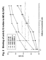
- the binding epitope of MAb 228B/C-1 was mapped to a unique site on IL13 responsible for the interaction with IL4R ⁇ , which constitutes part of the multimeric IL13R complex.
- This binding site on IL13 is distant from the site responsible for IL13R interaction, and therefore, 228B/C-1 can bind to IL13 bound on tumor cells overexpressing IL13R.
- antibodies that compete for binding to the same epitope recognized by any of the aforementioned monoclonal antibodies There are also described antibodies that bind the same epitope as 228B/C-1.
- Epitope peptides include a peptide comprising essentially or consisting of ESLINVSG (SEQ ID NO: 18) or YCAALESUNVS (SEQ ID NO:19).
- an isolated anti-IL13 monoclonal antibody for use that inhibits the growth of IL13-expressing cancer cells in vivo, or is cytotoxic in vivo, to such cells and tumors containing such cells is provided.
- the disclosure provides anti-IL13 antibodies that are conjugated to a cytotoxic agent or to a growth inhibitory agent
- the cytotoxic agent can be a toxin, cytotoxic small-moleculte drug, high-energy radioactive isotope, photoactivable drug, pro-apoptotic protein or drug, cytolytic or nucleolytic enzyme.
- Antibodies described herein may comprise a constant region of human IgG1, which can mediate tumor cell killing by complement-mediated cytolysis (CMC) and antibody-dependent cell-mediated cytotoxicity (ADCC). Such an antibody can also suppress the growth of tumors which is IL13-dependent.
- CMC complement-mediated cytolysis
- ADCC antibody-dependent cell-mediated cytotoxicity
- the anti-IL13 antibodies include intact (full length) antibodies as well as antibody fragments.
- the anti-IL13 antibody may be a chimeric, humanized or human antibody.
- Human antigen-binding antibody fragments of the antibodies of the present disclosure include, but are not limited to, Fab, Fab' and F(ab') 2 , Fd, single-chain Fvs (scFv), single-chain antibodies, disulfide-linked Fvs (sdFv).
- the invention also includes single-domain antibodies for use according to the present invention comprising either a VL or VH domain. On example is an scFv having the sequence of SEQ ID NO 152.
- the invention also encompasses the use of a composition comprising any one of the anti-IL13 antibodies of the above embodiments, and there is disclosed a carrier, in the methods of the present disclosure.
- the carrier is a pharmaceutically-acceptable carrier.
- These compositions can be provided in an article of manufacture or a kit for the treatment of cancer.
- a separate aspect of the disclosure is a method of killing an IL13-expressing cancer cell, comprising contacting the cancer cell with an anti-IL13 antibody of any of the above embodiments, thereby killing the cancer cell.
- Another aspect is a method of alleviating or treating an IL13-expressing cancer in a mammal, comprising administering a therapeutically effective amount of the anti-IL13 antibodies described herein to the mammal.
- the cancer is renal cell carcinoma, glioma, brain tumors, Hodgkins lymphoma, or other tumors or cancers that express IL13 receptor on their surface.
- the anti-IL13 antibody is a human or a humanized antibody.
- the antibody described herein is conjugated to a cytotoxic agent such as a toxin or a radioactive isotope and a cytostatic agent such as inhibitors to cyclin-dependent kinases.
- the method of alleviating the IL13-expressing cancer anticipates administration of the anti-IL13 antibody in combination with other forms of cancer treatment, such as radiotherapy and chemotherapy.
- the mammal is also receiving at least one chemotherapeutic agent.
- the chemotherapeutic agent is wherein the chemotherapy is selected from the group of drugs such as but not limited to Doxorubicin, 5-Fluorouracil, Cytosine arabinoside, Cyclophosphamide, Thiotepa, Busulfan, Cytoxin, Taxol, Methotrexate, Cisplatin, Melphalan, Vinblastine, bleomycin and Carboplatin.
- the anti-IL13 antibody can be used in conjunction with other anti-tumor antibodies such as, but not limited to, anti-VEGF MAb, anti-Her2 MAb, anti-EGFR MAb, anti-EpCam MAb, anti-ganglioside MAb, anti-tissue factor MAb and anti-integrin MAb.
- an article of manufacture comprising a container and a composition contained therein, wherein the composition comprises an anti-IL13 antibody of the above embodiments, and further comprising a package insert indicating that the composition can be used to alleviate or treat a IL13-expressing cancer.
- Another aspect of the disclosure comprises diagnosing a cancer or tumor overexpressing IL13 comprising the use of the anti-IL13 antibodies for use according to the present invention to detect overexpression of IL13 in the biological sample taken from a patient suspected of having said cancer or tumor.
- antibody refers to immunoglobulin molecules and immunologically active portions of immunoglobulin molecules, i.e., molecules that contain an antigen binding site that immunospecifically binds an antigen.
- the immunoglobulin molecules for use of the invention can be of any type (e.g ., IgG, IgE, IgM, IgD, IgA and IgY), class (e.g ., IgG1, IgG2, IgG3, IgG4, IgA1 and IgA2) or subclass of immunoglobulin molecule.
- antibody or “monoclonal antibody” (mAb) is meant to include intact molecules, as well as, antibody fragments (such as, for example, Fab and F(ab') 2 fragments) which are capable of specifically binding to protein.
- Fab and F(ab') 2 fragments lack the Fc fragment of intact antibody, clear more rapidly from the circulation of the animal or plant, and may have less non-specific tissue binding than an intact antibody ( Wahl et al., J. Nucl. Med. 24:316-325 (1983 )).
- human antibodies include antibodies having the amino acid sequence of a human immunoglobulin and include antibodies isolated from human immunoglobulin libraries or from animals transgenic for one or more human immunoglobulin and that do not express endogenous immunoglobulins, as described infra and, for example in, U.S. Pat. No. 5,939,598 by Kucherlapati et al.
- Antibody effector functions refer to those biological activities attributable to the Fc region (a native sequence Fc region or amino acid sequence variant Fc region) of an antibody, and vary with the antibody isotype. Examples of antibody effector functions include: C1q binding and complement dependent cytotoxicity; Fc receptor binding; antibody-dependent cell-mediated cytotoxicity (ADCC); phagocytosis; down regulation of cell surface receptors (e.g. B cell receptor); and B cell activation.
- ADCC antibody-dependent cell-mediated cytotoxicity
- Fc ⁇ Rs Fc gamma receptors
- cytotoxic cells e.g. Natural Killer (NK) cells, neutrophils, and macrophages
- NK cells Natural Killer cells
- neutrophils neutrophils
- macrophages e.g., neutrophils, and macrophages
- the antibodies “arm” the cytotoxic cells and are required for such killing.
- the primary cells for mediating ADCC, NK cells express Fc ⁇ RIII only, whereas monocytes express Fc ⁇ RI, Fc ⁇ RII and Fc ⁇ RIII.
- ADCC activity of a molecule of interest may be assessed in vitro, such as that described below.
- useful effector cells for such assays include peripheral blood mononuclear cells (PBMC) and Natural Killer (NK) cells.
- PBMC peripheral blood mononuclear cells
- NK Natural Killer
- ADCC activity of the molecule of interest may be assessed in vivo, e.g., in a animal model such as that disclosed in Clynes et al. PNAS (USA) 95:652-656 (1998 ).
- Recombinant IL13 was used to immunize mice to generate the hybridomas that produce the monoclonal antibodies for use of the present Invention.
- Recombinant IL13 is commercially available from a number of sources (see, e.g. R & D Systems, Minneapolis, MN, PeproTech, Inc., NJ, and Sanofi Bio-industries, Inc., Tervose, PA.).
- a gene or a cDNA encoding IL13 may be cloned into a plasmid or other expression vector and expressed in any of a number of expression systems according to methods well known to those of skill in the art.
- IL13 and the nucleic acid sequence for IL13 are well known (see, for example, U.S. Patent No. 5,652,123 ). Because of the degeneracy of the genetic code, a multitude of nucleotide sequences encoding IL13 polypeptides may be produced. One may vary the nucleotide sequence by selecting combinations based on possible codon choices. These combinations are made In accordance with the standard triplet genetic code as applied to the nucleotide sequence that codes for naturally occurring IL13 polypeptide and all such variations are to be considered. Any one of these polypeptides may be used in the immunization of an animal to generate antibodies that bind to IL13.
- the immunogen IL13 polypeptide may, when beneficial, be expressed as a fusion protein that has the IL13 polypeptide attached to a fusion segment.
- the fusion segment often aids in protein purification, e.g., by permitting the fusion protein to be isolated and purified by affinity chromatography.
- Fusion proteins can be produced by culturing a recombinant cell transformed with a fusion nucleic acid sequence that encodes a protein including the fusion segment attached to either the carboxyl and/or amino terminal end of the protein.
- Fusion segments may include, but are not limited to, immunoglobulin Fc regions, glutathione-S-transferase, ⁇ -galactosidase, a poly-histidine segment capable of binding to a divalent metal ion, and maltose binding protein.
- a fusion protein comprising a mutant form of human IL13 was used to generate the antibodies for use of the present invention.
- This mutant form of IL13 contained a single mutation resulting in an inactive form of the protein ( Thompson et al., J. Biol. Chem. 274: 2994 (1999 )).
- the fusion protein comprised the mutant IL13 protein fused to an Immunoglobulin Fc, specifically IgG1, and was expressed in a mammalian cell line such that the recombinant protein was naturally glycosylated.
- the Fc portion of the fusion protein may have provided a conformational structure that exposed a key epitope. The glycosylation may have increased the immunogenicity of the epitope, allowing the generation of antibodies to this particular epitope.
- IL13 polypeptides expressed in E. coli lack glycosylation and the commercially available antibodies tested were generated using this protein. We tested these antibodies, e.g., R&D Systems and Pharmingen, and found that they do not cross react with the epitope bound by the antibodies for use of the present invention.
- the antibodies for use of the present invention may be generated by any suitable method known in the art.
- the antibodies for use of the present invention may comprise polyclonal antibodies. Methods of preparing polyclonal antibodies are known to the skilled artisan ( Harlow, et al., Antibodies: a Laboratory Manual, (Cold spring Harbor Laboratory Press, 2nd ed. (1988 )).
- an immunogen as described above may be administered to various host animals including, but not limited to, rabbits, mice, rats, etc., to induce the production of sera containing polyclonal antibodies specific for the antigen.
- the administration of the immunogen may entail one or more injections of an immunizing agent and, if desired, an adjuvant.
- adjuvants may be used to increase the immunological response, depending on the host species, and include but are not limited to, Freund's (complete and incomplete), mineral gels such as aluminum hydroxide, surface active substances such as lysolecithin, pluronic polyols, polyanions, peptides, oil emulsions, keyhole limpet hemocyanins, dinitrophenol, and potentially useful human adjuvants such as BCG (bacille Calmette-Guerin) and Corynebacterium parvum. Additional examples of adjuvants which may be employed include the MPL-TDM adjuvant (monophosphoryl lipid A, synthetic trehalose dicorynomycolate). Immunization protocols are well known in the art in the art and may be performed by any method that elicits an immune response in the animal host chosen. Adjuvants are also well known in the art.
- the immunogen (with or without adjuvant) is injected into the mammal by multiple subcutaneous or intraperitoneal injections, or intramuscularly or through IV.
- the immunogen may include an IL13 polypeptide, a fusion protein or variants thereof.
- percent hydrophobicity, percent hydrophilicity, stability, net charge, isoelectric point etc. it may be useful to conjugate the immunogen to a protein known to be immunogenic in the mammal being immunized.
- Such conjugation includes either chemical conjugation by derivatizing active chemical functional groups to both the immunogen and the immunogenic protein to be conjugated such that a covalent bond is formed, or through fusion-protein based methodology, or other methods known to the skilled artisan.
- immunogenic proteins include, but are not limited to, keyhole limpet hemocyanin, ovalbumin, serum albumin, bovine thyroglobulin, soybean trypsin inhibitor, and promiscuous T helper peptides.
- Various adjuvants may be used to increase the immunological response as described above.
- the antibodies for use of the present invention comprise monoclonal antibodies.
- Monoclonal antibodies may be prepared using hybridoma technology, such as those described by Kohler and Milstein, Nature, 256:495 (1975 ) and U.S. Pat. No. 4,376,110 , by Harlow, et al., Antibodies: A Laboratory Manual, (Cold spring Harbor Laboratory Press, 2.sup.nd ed. (1988 ), by Hammerling, et al., Monoclonal Antibodies and T-Cell Hybridomas (Elsevier, N.Y., (1981 )), or other methods known to the artisan.
- Suitable methods which may be employed for producing monoclonal antibodies include, but are not limited to, the human B-cell hybridoma technique ( Kosbor et al., 1983, Immunology Today 4:72 ; Cole et al., 1983, Proc. Natl. Acad. Sci. USA 80:2026-2030 ), and the EBV-hybridoma technique ( Cole et al., 1985, Monoclonal Antibodies And Cancer Therapy, Alan R. Liss, Inc., pp. 77-96 ).
- Such antibodies may be of any immunoglobulin class including IgG, IgM, IgE, IgA, IgD and any subclass thereof.
- the hybridoma producing the mAb for use of this invention may be cultivated in vitro or in vivo
- a host such as a mouse, a humanized mouse, a mouse with a human immune system, hamster, rabbit, camel or any other appropriate host animal, is typically immunized with an immunogen to elicit lymphocytes that produce or are capable of producing antibodies that will specifically bind to IL13.
- lymphocytes may be immunized In vitro with the antigen.
- peripheral blood lymphocytes PBLs
- spleen cells or lymph node cells are used if non-human mammalian sources are desired.
- the lymphocytes are then fused with an immortalized cell line using a suitable fusing agent, such as polyethylene glycol, to form a hybridoma cell ( Goding, Monoclonal Antibodies: Principles and Practice, Academic Press, (1986), pp. 59-103 ).
- Immortalized cell lines are usually transformed mammalian cells, particularly myeloma cells of rodent, bovine or human origin. Typically, a rat or mouse myeloma cell line is employed.
- the hybridoma cells may be cultured in a suitable culture medium that preferably contains one or more substances that inhibit the growth or survival of the unfused, immortalized cells.
- a suitable culture medium that preferably contains one or more substances that inhibit the growth or survival of the unfused, immortalized cells.
- the culture medium for the hybridomas typically will include hypoxanthine, aminopterin, and thymidine ("HAT medium"), substances that prevent the growth of HGPRT-deficient cells.
- Preferred immortalized cell lines are those that fuse efficiently, support stable high level expression of antibody by the selected antibody-producing cells, and are sensitive to a medium such as HAT medium. More preferred immortalized cell lines are murine myeloma lines, which can be obtained, for instance, from the Salk Institute Cell Distribution Center, San Diego, Calif. and the American Type Culture Collection, Manassas, Va. Human myeloma and mouse-human heteromyeloma cell lines may also be used for the production of human monoclonal antibodies ( Kozbor, J. Immunol., 133:3001 (1984 ); Brodeur et al., Monoclonal Antibody Production Techniques and Applications, Marcel Dekker, Inc., New York, (1987) pp. 51-63 ).
- the culture medium in which the hybridoma cells are cultured can then be assayed for the presence of monoclonal antibodies directed against the IL13.
- the binding specificity of monoclonal antibodies produced by the hybridoma cells is determined by, e.g., immunoprecipitation or by an in vitro binding assay, such as radioimmunoassay (RIA) or enzyme-linked immunoadsorbant assay (ELISA). Such techniques are known in the art and within the skill of the artisan.
- the binding affinity of the monoclonal antibody to IL13 can, for example, be determined by a Scatchard analysis ( Munson et al., Anal. Biochem., 107:220 (1980 )).
- the clones may be subcloned by limiting dilution procedures and grown by standard methods (Goding, supra). Suitable culture media for this purpose include, for example, Dulbecco's Modified Eagle's Medium and RPMI-1640.
- the monoclonal antibodies secreted by the subclones may be isolated or purified from the culture medium by conventional immunoglobulin purification procedures such as, e.g., protein A-sepharose, hydroxyapatite chromatography, gel exclusion chromatography, gel electrophoresis, dialysis, or affinity chromatography.
- the invention is not limited to their sole production in hydridomas.
- the monoclonal antibodies may be made by recombinant DNA methods, such as those described in U.S. Pat. No. 4,816,567 .
- the term "monoclonal antibody” refers to an antibody derived from a single eukaryotic, phage, or prokaryotic clone.
- the DNA encoding the monoclonal antibodies for use of the invention can be readily isolated and sequenced using conventional procedures (e.g., by using oligonucleotide probes that are capable of binding specifically to genes encoding the heavy and light chains of murine antibodies, or such chains from human, humanized, or other sources).
- the hydridoma cells of the disclosure serve as a preferred source of such DNA.
- the DNA may be placed into expression vectors, which are then transformed into host cells such as NS0 cells, Simian COS cells, Chinese hamster ovary (CHO) cells, or myeloma cells that do not otherwise produce immunoglobulin protein, to obtain the synthesis of monoclonal antibodies in the recombinant host cells.
- the DNA also may be modified, for example, by substituting the coding sequence for human heavy and light chain constant domains in place of the homologous murine sequences ( U.S. Pat. No. 4,816,567 ; Monison et al, supra) or by covalently joining to the immunoglobulin coding sequence all or part of the coding sequence for a non-immunoglobulin polypeptide.
- a non-immunoglobulin polypeptide can be substituted for the constant domains of an antibody for use of the invention, or can be substituted for the variable domains of one antigen-combining site of an antibody for use of the invention to create a chimeric bivalent antibody.
- the antibodies may be monovalent antibodies.
- Methods for preparing monovalent antibodies are well known in the art For example, one method involves recombinant expression of immunoglobulin light chain and modified heavy chain.
- the heavy chain is truncated generally at any point in the Fc region so as to prevent heavy chain cross-linking.
- the relevant cysteine residues are substituted with another amino acid residue or are deleted so as to prevent cross-linking.
- Antibody fragments which recognize specific epitopes may be generated by known techniques.
- Fab and F(ab') 2 fragments of the disclosure may be produced by proteolytic cleavage of immunoglobulin molecules, using enzymes such as papain (to produce Fab fragments) or pepsin (to produce F(ab') 2 fragments).
- F(ab') 2 fragments contain the variable region, the light chain constant region and the CH1 domain of the heavy chain.
- a chimeric antibody is a molecule in which different portions of the antibody are derived from different animal species, such as antibodies having a variable region derived from a murine monoclonal antibody and a human immunoglobulin constant region.
- Methods for producing chimeric antibodies are known in the art. See e.g., Morrison, Science 229:1202 (1985 ); Ol et al., BioTechniques 4:214 (1986 ); Gillies et al., (1989) J. Immunol. Methods 125:191-202 ; U.S. Pat. Nos. 5,807,715 ; 4,816,567 ; and 4,816397 .
- Humanized antibodies are antibody molecules generated in a non-human species that bind the desired antigen having one or more complementarity determining regions (CDRs) from the non-human species and framework (FR) regions from a human immunoglobulin molecule.
- CDRs complementarity determining regions
- FR framework
- framework residues in the human framework regions will be substituted with the corresponding residue from the CDR donor antibody to alter, preferably improve, antigen binding.
- framework substitutions are identified by methods well known in the art, e.g ., by modeling of the interactions of the CDR and framework residues to identify framework residues important for antigen binding and sequence comparison to identify unusual framework residues at particular positions. (See, e.g., Queen et al., U.S. Pat. No. 5,685,089 ; Riechmann et al., Nature 332:323 (1988 )),
- Antibodies can be humanized using a variety of techniques known in the art including, for example, CDR-grafting ( EP 239,400 ; PCT publication WO 91/09967 ; U.S. Pat. Nos. 5,225,539 ; 5,530,101 ; and 5,585,089 ), veneering or resurfacing ( EP 592,106 ; EP 519,596 ; Padlan, Molecular Immunology 28(4/5):489-498 (1991 ); Studnicka et al., Protein Engineering 7(6):805-814 (1994 ); Roguska. et al., PNAS 91:969-973 (1994 )), and chain shuffling ( U.S. Pat. No. 5,565,332 ).
- a humanized antibody has one or more amino acid residues Introduced into it from a source that is non-human. These non-human amino acid residues are often referred to as "import" residues, which are typically taken from an "import” variable domain. Humanization can be essentially performed following the methods of Winter and co-workers ( Jones et al., Nature, 321:522-525 (1986 ); Reichmann et al., Nature, 332:323-327 (1988 ); Verhoeyen et al., Science, 239:1534-1536 (1988 ), by substituting rodent CDRs or CDR sequences for the corresponding sequences of a human antibody. Accordingly, such "humanized” antibodies are chimeric antibodies ( U.S.
- humanized antibodies are typically human antibodies in which some CDR residues and possible some FR residues are substituted from analogous sites in rodent antibodies.
- Human antibodies are particularly desirable for therapeutic treatment of human patients.
- Human antibodies can be made by a variety of methods known in the art including phage display methods described above using antibody libraries derived from human immunoglobulin sequences. See also, U.S. Pat. Nos. 4,444,887 and 4,716,111 ; and PCT publications WO 98/46645 , WO 98/50433 , WO 98/24893 , WO 98/16654 , WO 96/34096 , WO 96/33735 , and WO 91/10741 .
- Human antibodies can also be produced using transgenic mice which are incapable of expressing functional endogenous immunoglobulins, but which can express human immunoglobulin genes.
- the human heavy and light chain immunoglobulin gene complexes may be introduced randomly or by homologous recombination into mouse embryonic stem cells.
- the human variable region, constant region, and diversity region may be introduced into mouse embryonic stem cells in addition to the human heavy and light chain genes.
- the mouse heavy and light chain immunoglobulin genes may be rendered non-functional separately or simultaneously with the introduction of human immunoglobulin loci by homologous recombination. In particular, homozygous deletion of the JH region prevents endogenous antibody production.
- the modified embryonic stem cells are expanded and microinjected into blastocysts to produce chimeric mice.
- the chimeric mice are then bred to produce homozygous offspring which express human antibodies.
- the transgenic mice are immunized in the normal fashion with a selected antigen, e.g., all or a portion of a polypeptide disclosed herein.
- Monoclonal antibodies directed against the antigen can be obtained from the immunized, transgenic mice using conventional hybridoma technology.
- the human immunoglobulin transgenes harbored by the transgenic mice rearrange during B cell differentiation, and subsequently undergo class switching and somatic mutation.
- human mAbs could be made by immunizing mice transplanted with human peripheral blood leukocytes, splenocytes or bone marrows ( e.g., Trioma techniques of XTL).
- Completely human antibodies which recognize a selected epltope can be generated using a technique referred to as "guided selection.”
- a selected non-human monoclonal antibody e.g., a mouse antibody, is used to guide the selection of a completely human antibody recognizing the same epitope. ( Jespers et at., Bio/technology 12:899-903 (1988 )).
- antibodies to the polypeptides of the disclosure can, in turn, be utilized to generate anti-idiotype antibodies that "mimic" polypeptides of the disclosure using techniques well known to those skilled in the art. (See, e.g., Greenspan & Bona, FASEB J. 7(5):437-444; (1989 ) and Nissinoff, J. Immunol. 147(8):2429-2438 (1991 )).
- antibodies which bind to and competitively inhibit polypeptide multimerization and/or binding of a polypeptide of the disclosure to a ligand can be used to generate anti-idiotypes that "mimic" the polypeptide multimerization and/or binding domain and, as a consequence, bind to and neutralize polypeptide and/or its ligand.
- anti-idiotypes or Fab fragments of such anti-idiotypes can be used in therapeutic regimens to neutralize polypeptide ligand.
- anti-idiotypic antibodies can be used to bind a polypeptide of the disclosure and/or to bind its ligands/receptors, and thereby block its biological activity.
- the antibodies for use of the present invention may be bispecific antibodies.
- Bispecific antibodies are monoclonal, preferably human or humanized, antibodies that have binding specificities for at least two different antigens.
- one of the binding specificities may be directed towards IL13, the other may be for any other antigen, and preferably for a cell-surface protein, receptor, receptor subunit, tissue-specific antigen, virally derived protein, virally encoded envelope protein, bacterially derived protein, or bacterial surface protein, etc.
- bispecific antibodies are well known. Traditionally, the recombinant production of bispecific antibodies is based on the co-expression of two immunoglobulin heavy-chain/light-chain pairs, where the two heavy chains have different specificities ( Milstein and Cuello, Nature, 305:537-539 (1983 ). Because of the random assortment of immunoglobulin heavy and light chains, these hybridomas (quadromas) produce a potential mixture of ten different antibody molecules, of which only one has the correct bispecific structure. The purification of the correct molecule is usually accomplished by affinity chromatography steps. Similar procedures are disclosed in WO 93/08829, published May 13, 1993 , and In Traunecker et al., EMBO J., 10:3655-3659 (1991 ).
- Antibody variable domains with the desired binding specificities can be fused to immunoglobulin constant domain sequences.
- the fusion preferably is with an immunoglobulin heavy-chain constant domain, comprising at least part of the hinge, CH2, and CH3 regions. It may have the first heavy-chain constant region (CH1) containing the site necessary for light-chain binding present in at least one of the fusions.
- DNAs encoding the immunoglobulin heavy-chain fusions and, if desired, the immunoglobulin light chain are inserted into separate expression vectors, and are co-transformed into a suitable host organism.
- DNAs encoding the immunoglobulin heavy-chain fusions and, if desired, the immunoglobulin light chain are inserted into separate expression vectors, and are co-transformed into a suitable host organism.
- Heteroconjugate antibodies are also described herein.
- Heteroconjugate antibodies are composed of two covalently joined antibodies. Such antibodies have, for example, been proposed to target immune system cells to unwanted cells ( U.S. Pat. No. 4,676,980 ).
- the antibodies may be prepared in vitro using known methods in synthetic protein chemistry, including those involving cross-linking agents.
- immunotoxins may be constructed using a disulfide exchange reaction or by forming a thioester bond. Examples of suitable reagents for this purpose include iminothiolate and methyl-4-mercaptobutyrimidate and those disclosed, for example, in U.S. Pat. No. 4,676,980 .
- the present invention provides antagonist monoclonal antibodies for use that inhibit and neutralize the action of IL13.
- the antibodies for use of the present invention bind to IL13 and inhibit the activation of the IL13 receptor complex.
- the antibodies described herein include the antibodies designated 228B/C-1, 228A-4, 227-26, and 227-43, and humanized clones of 228B/C-1 are disclosed.
- the present invention also includes antibodies for use that bind to the same epitope as monoclonal antibody 228B/C-1.
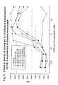
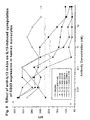
- Candidate anti-IL-13 antibodies were tested by enzyme linked immunosorbent assay (ELISA), Western immunoblotting, or other immunochemical techniques. Assays performed to characterize the individual antibodies included: (1) Inhibition of IL13-autocrine proliferation of Hodgkin's lymphoma cell lines HDLM-2 and L-1236; (2) inhibition of IL13-induced STAT6 phosphorylation in THP-1 cells; and (3) inhibition of IL13-induced suppression of CD14 expression in primary human monocytes; and (4) Inhibition of IL13-induced up-regulation of CD23 expression on primary human monocytes. Experimental details are described in the Examples.
- Antibodies disclosed herein include, but are not limited to, polyclonal, monoclonal, monovalent, bispecific, heteroconjugate, multispecific, human, humanized or chimeric antibodies, single chain antibodies, single-domain antibodies, Fab fragments, F(ab') fragments, fragments produced by a Fab expression library, anti-idiotypic (anti-Id) antibodies (including, e.g., anti-Id antibodies to antibodies for use of the invention), and epitope-binding fragments of any of the above.
- the antibodies may be human antigen-binding antibody fragments of the present disclosure and include, but are not limited to, Fab, Fab' and F(ab') 2 , Fd, single-chain Fvs (scFv), single-chain antibodies, disulfide-linked Fvs (sdFv) and single-domain antibodies comprising either a VL or VH domain.
- Antigen-binding antibody fragments, including single-chain antibodies may comprise the variable region(s) alone or in combination with the entrety or a portion of the following: hinge region, CH1, CH2, and CH3 domains. Also included in the disclosure are antigen-binding fragments comprising any combination of variable region(s) with a hinge region, CH1, CH2, and CH3 domains.
- the antibodies for use of the invention may be from any animal origin including birds and mammals.
- the antibodies may be human, non-human primates, rodents ( e.g., mouse and rat), donkey, sheep, rabbit, goat, guinea pig, camel, horse, or chicken.
- the antibodies of the present disclosure may be monospecific, bispecific, trispecific or of greater multispecificity.
- Multispecific antibodies may be specific for different epitopes of IL13 or may be specific for both IL13 as well as for a heterologous epitope, such as a heterologous polypeptide or solid support material.
- a heterologous epitope such as a heterologous polypeptide or solid support material.
- Antibodies for use of the present invention may be described or specified in terms of the epitope(s) or portion(s), of IL13 which they recognize or specifically bind.
- the epitope(s) or polypeptide portion(s) may be specified as described herein, e.g., by N-terminal and C-terminal positions, by size in contiguous amino acid residues, or listed in the Tables and Figures.
- Antibodies for use of the present invention may also be described or specified in terms of their cross-reactivity.
- Antibodies that bind IL13 polypeptides, which have at least 95%, at least 90%, at least 85%, at least 80%, at least 75%, at least 70%, at least 65%, at least 60%, at least 55%, and at least 50% identity (as calculated using methods known in the art and described herein) to IL-13 are also included in the present invention.
- Antibodies of the present disclosure cross-react with monkey homologues of human IL13 and the corresponding epitopes thereof.
- antibodies which bind polypeptides encoded by polynucleotides which hybridize to a polynucleotide encoding IL13 under stringent hybridization conditions may also be described or specified in terms of their binding affinity to a polypeptide of the disclosure. Binding affinities include those with an equilibrium dissociation constant or K D from 10 -8 to 10 -15 M.
- the invention also provides antibodies that competitively inhibit binding of an antibody to an epitope of the invention as determined by any method known in the art for determining competitive binding, for example, the immunoassays described herein.
- the antibody may competitively inhibit binding to the epitope by at least 95%, at least 90%, at least 85%, at least 80%, at least 75%, at least 70%, at least 60%, or at least 50%.
- antibodies that bind to the same epitope as the anti-IL13 antibodies for use of the present invention are antibodies that bind to the same epitope as the anti-IL13 antibodies for use of the present invention.
- a cross-blocking assay e.g., a competitive ELISA assay
- IL13 coated on the wells of a microtiter plate is preincubated with or without candidate competing antibody and then the biotin-labeled anti-IL13 antibody for use of the invention is added.
- the amount of labeled anti-IL13 antibody bound to the IL13 antigen in the wells is measured using avidin-peroxidase conjugate and appropriate substrate.
- the antibody can be labeled with a radioactive or fluorescent label or some other detectable and measurable label.
- the amount of labeled anti-IL13 antibody that bound to the antigen will have an indirect correlation to the ability of the candidate competing antibody (test antibody) to compete for binding to the same epitope, i.e., the greater the affinity of the test antibody for the same epitope, the less labeled antibody will be bound to the antigen-coated wells.
- a candidate competing antibody is considered an antibody that binds substantially to the same epitope or that competes for binding to the same epitope as an anti-IL13 antibody for use of the invention if the candidate antibody can block binding of the IL13 antibody by at least 20%, by at least 20-50%, or by at least 50% as compared to the control performed in parallel in the absence of the candidate competing antibody. It will be understood that variations of this assay can be performed to arrive at the same quantitative value.
- the present disclosure provides vector constructs comprising a nucleotide sequence encoding the antibodies for use of the present invention and a host cell comprising such a vector.
- Standard techniques for cloning and transformation may be used in the preparation of cell lines expressing the antibodies for use of the present invention.
- Recombinant expression vectors containing a nucleotide sequence encoding the antibodies for use of the present invention can be prepared using well known techniques.
- the expression vectors include a nucleotide sequence operably linked to suitable transcriptional or translational regulatory nucleotide sequences such as those derived from mammalian, microbial, viral, or insect genes.
- suitable transcriptional or translational regulatory nucleotide sequences such as those derived from mammalian, microbial, viral, or insect genes.
- regulatory sequences include transcriptional promoters, operators, enhancers, mRNA ribosomal binding sites, and/or other appropriate sequences which control transcription and translation initiation and termination.
- Nucleotide sequences are "operably linked" when the regulatory sequence functionally relates to the nucleotide sequence for the appropriate polypeptide.
- a promoter nucleotide sequence is operably linked to, e.g., the antibody heavy chain sequence if the promoter nucleotide sequence controls the
- sequences encoding appropriate signal peptides that are not naturally associated with antibody heavy and/or light chain sequences can be incorporated into expression vectors.
- a nucleotide sequence for a signal peptide may be fused in-frame to the polypeptide sequence so that the antibody is secreted to the periplasmic space or into the medium.
- a signal peptide that is functional in the intended host cells enhances extracellular secretion of the appropriate antibody.
- the signal peptide may be cleaved from the polypeptide upon secretion of antibody from the cell. Examples of such secretory signals are well known and include, e.g., those described in US5698435 , US5698417 , and US6204023 .
- Host cells described include but are not limited to microorganisms such as bacteria (e.g ., E. coli , B. subtilis ) transformed with recombinant bacteriophage DNA, plasmid DNA or cosmid DNA expression vectors containing antibody coding sequences; yeast (e.g., Saccharomyces, Pichia) transformed with recombinant yeast expression vectors containing antibody coding sequences; insect cell systems infected with recombinant virus expression vectors (e.g., Baculovirus) containing antibody coding sequences; plant cell systems infected with recombinant virus expression vectors ( e.g., cauliflower mosaic virus, CaMV; tobacco mosaic virus, TMV) or transformed with recombinant plasmid expression vectors (e.g., Ti plasmid) containing antibody coding sequences; or mammalian cell systems (e.g., COS, CHO, BHK, 293, 3T3 cells) harboring recombinant bacter
- the vector may be a plasmid vector, a single or double-stranded phage vector, or a single or double-stranded RNA or DNA viral vector.
- Such vectors may be introduced into cells as polynucleotides by well known techniques for introducing DNA and RNA into cells.
- the vectors, in the case of phage and viral vectors also may be introduced into cells as packaged or encapsulated virus by well known techniques for infection and transduction.
- Viral vectors may be replication competent or replication defective. In the latter case, viral propagation generally will occur only in complementing host cells. Cell-free translation systems may also be employed to produce the protein using RNAs derived from the present DNA constructs.
- Such vectors may include the nucleotide sequence encoding the constant region of the antibody molecule (see, e.g., PCT Publication WO 86/05807 ; PCT Publication WO 89/01036 ; and U.S. Pat. No. 5,122,464 ) and the variable domain of the antibody may be cloned into such a vector for expression of the entire heavy or light chain.
- Prokaryotes useful as host cells include gram negative or gram positive organisms such as E . coli and B. subtilis.
- Expression vectors for use in prokaryotic host cells generally comprise one or more phenotypic selectable marker genes.
- a phenotypic selectable marker gene is, for example, a gene encoding a protein that confers antibiotic resistance or that supplies an autotrophic requirement.
- useful expression vectors for prokaryotic host cells include those derived from commercially available plasmids such as the pKK223-3 (Pharmacia Fine Chemicals, Uppsala, Sweden), pGEM1 (Promega Biotec, Madison, Wisconsin., USA), and the pET (Novagen, Madison, Wisconsin, USA) and pRSET (Invitrogen Corporation, Carlsbad, California, USA) series of vectors ( Studier, F.W., J. Mol. Biol. 219: 37 (1991 ); Schoepfer, R. Gene 124: 83 (1993 )).
- Promoter sequences commonly used for recombinant prokaryotic host cell expression vectors include T7, ( Rosenberg, et al.
- Yeasts include those from the genus Saccharomyces, Pichia, Actinomycetes and Kluyveromyces. Yeast vectors will often contain an origin of replication sequence from a 2 ⁇ yeast plasmid, an autonomously replicating sequence (ARS), a promoter region, sequences for polyadenylation, sequences for transcription termination, and a selectable marker gene. Suitable promoter sequences for yeast vectors include, among others, promoters for metallothionein, 3-phosphoglycerate kinase ( Hitzeman et al., J. Biol. Chem. 255:2073, (1980 )) or other glycolytic enzymes ( Holland et al., Biochem.
- yeast transformation protocols are well known. One such protocol is described by Hinnen et al., Proc. Natl. Acad. Sci., 75:1929 (1978 ). The Hinnen protocol selects for Trp + transformants in a selective medium.
- Mammalian or insect host cell culture systems may also be employed to express recombinant antibodies, e.g., Baculovirus systems for production of heterologous proteins.
- Baculovirus systems for production of heterologous proteins.
- Autographa californica nuclear polyhedrosis virus (AcNPV) may be used as a vector to express foreign genes.
- the virus grows in Spodoptera frugiperda cells.
- the antibody coding sequence may be cloned individually into non-essential regions (for example the polyhedrin gene) of the virus and placed under control of an AcNPV promoter (for example the polyhedrin promoter).
- NS0 or Chinese hamster ovary (CHO) cells for mammalian expression of the antibodies for use of the present invention may be used.
- Transcriptional and translational control sequences for mammalian host cell expression vectors may be excised from viral genomes. Commonly used promoter sequences and enhancer sequences are derived from Polyoma virus, Adenovirus 2, Simian Virus 40 (SV40), and human cytomegalovirus (CMV). DNA sequences derived from the SV40 viral genome may be used to provide other genetic elements for expression of a structural gene sequence in a mammalian host cell, e.g., SV40 origin, early and late promoter, enhancer, splice, and polyadenylation sites. Viral early and late promoters are particularly useful because both are easily obtained from a viral genome as a fragment which may also contain a viral origin of replication. Exemplary expression vectors for use in mammalian host cells are commercially available.
- the disclosure further provides polynucleotides comprising a nucleotide sequence encoding an antibody for use of the invention and fragments thereof.
- the disclosure also encompasses polynucleotides that hybridize under stringent or lower stringency hybridization conditions to polynucleotides that encode an antibody for use of the present invention,
- the polynucleotides may be obtained, and the nucleotide sequence of the polynucleaudes determined, by any method known in the art.
- a polynucleotide encoding the antibody may be assembled from chemically synthesized oligonucleotides (e.g., as described in Kutmeier et al., BioTechniques 17:242 (1994 )), which, briefly, involves the synthesis of overlapping oligonucleotides containing portions of the sequence encoding the antibody, annealing and ligating of those oligonucleotides, and then amplification of the ligated oligonucleotides by PCR.
- a polynucleotide encoding an antibody may be generated from nucleic acid from a suitable source. If a clone containing a nucleic acid encoding a particular antibody is not available, but the sequence of the antibody molecule is known, a nucleic acid encoding the immunoglobulin may be chemically synthesized or obtained from a suitable source (e.g., an antibody cDNA library, or a cDNA library generated from, or nucleic acid, preferably poly A + RNA, isolated from, any tissue or cells expressing the antibody, such as hybridoma cells selected to express an antibody for use of the invention) by PCR amplification using synthetic primers hybridizable to the 3' and 5' ends of the sequence or by cloning using an oligonucleotide probe specific for the particular gene sequence to identify, e.g., a cDNA clone from a cDNA library that encodes the antibody. Amplified nucleic acids generated by PCR may be chemical
- nucleotide sequence and corresponding amino acid sequence of the antibody may be manipulated using methods well known in the art for the manipulation of nucleotide sequences, e.g., recombinant DNA techniques, site directed mutagenesis, PCR, etc. (see, for example, the techniques described in Sambrook et al., 1990, Molecular Cloning, A Laboratory Manual, 2d Ed., Cold Spring Harbor Laboratory, Cold Spring Harbor, N.Y .
- the amino acid sequence of the heavy and/or light chain variable domains may be inspected to identify the sequences of the CDRs by well known methods, e.g., by comparison to known amino acid sequences of other heavy and light chain variable regions to determine the regions of sequence hypervariability.
- one or more of the CDRs may be inserted within framework regions, e.g., into human framework regions to humanize a non-human antibody, as described supra.
- the framework regions may be naturally occurring or consensus framework regions, and preferably human framework regions (see, e.g., Chothia et al., J. Mol. Biol. 278: 457-479 (1998 ) for a listing of human framework regions).
- the polynucleotide generated by the combination of the framework regions and CDRs encodes an antibody that specifically binds a polypeptide of the disclosure.
- one or more amino acid substitutions may be made within the framework regions, and, the amino acid substitutions improve binding of the antibody to its antigen.
- such methods may be used to make amino acid substitutions or deletions of one or more variable region cysteine residues participating in an intrachain disulfide bond to generate antibody molecules lacking one or more intrachain disulfide bonds.
- Other alterations to the polynucleotide are encompassed by the present disclosure and within the skill of the art.
- a chimeric antibody is a molecule in which different portions are derived from different animal species, such as those having a variable region derived from a murine mAb and a human immunoglobulin constant region, e.g., humanized antibodies.
- Single chain antibodies are formed by linking the heavy and light chain fragments of the Fv region via an amino acid bridge, resulting in a single chain polypeptide.
- Techniques for the assembly of functional Fv fragments in E coli may also be used ( Skerra et al., Science 242:1038-1041 (1988 )).
- the antibodies for use of the invention can be produced by any method known in the art for the synthesis of antibodies, in particular, by chemical synthesis or preferably, by recombinant expression techniques.
- Recombinant expression of an antibody for use of the invention, or fragment, derivative or analog thereof, requires construction of an expression vector containing a polynucleotide that encodes the antibody or a fragment of the antibody.
- an expression vector containing a polynucleotide that encodes the antibody or a fragment of the antibody.
- the vector for the production of the antibody may be produced by recombinant DNA technology.
- An expression vector is constructed containing antibody coding sequences and appropriate transcriptional and translational control signals. These methods include, for example, in vitro recombinant DNA techniques, synthetic techniques, and in vivo genetic recombination.
- the expression vector is transferred to a host cell by conventional techniques and the transfected cells are then cultured by conventional techniques to produce an antibody of the invention.
- Vectors encoding both the heavy and light chains may be co-expressed in the host cell for expression of the entire immunoglobulin molecule, as detailed below.
- host-expression vector systems may be utilized to express the antibody molecules for use of the invention as described above.
- Such host-expression systems represent vehicles by which the coding sequences of interest may be produced and subsequently purified, but also represent cells which may, when transformed or transfected with the appropriate nucleotide coding sequences, express an antibody molecule for use of the invention in situ.
- Bacterial cells such as E. coli, and eukaryotic cells are commonly used for the expression of a recombinant antibody molecule, especially for the expression of whole recombinant antibody molecule.
- mammalian cells such as Chinese hamster ovary cells (CHO), in conjunction with a vector such as the major intermediate early gene promoter element from human cytomegalovirus is an effective expression system for antibodies ( Foecking et al., Gene 45:101 (1986 ); Cockett et al., Bio/Technology 8:2 (1990 )).
- CHO Chinese hamster ovary cells
- a vector such as the major intermediate early gene promoter element from human cytomegalovirus
- a host cell strain may be chosen which modulates the expression of the inserted sequences, or modifies and processes the gene product in the specific fashion desired. Such modifications (e.g., glycosylation) and processing (e.g., cleavage) of protein products may be important for the function of the protein.
- Different host cells have characteristic and specific mechanisms for the post-translational processing and modification of proteins and gene products. Appropriate cell lines or host systems can be chosen to ensure the correct modification and processing of the foreign protein expressed.
- eukaryotic host cells which possess the cellular machinery for proper processing of the primary transcript, glycosylation, and phosphorylation of the gene product may be used.
- mammalian host cells include, but are not limited to, CHO, COS, 293, 3T3, or myeloma cells.
- cell lines which stably express the antibody molecule may be engineered.
- host cells can be transformed with DNA controlled by appropriate expression control elements (e.g., promoter, enhancer, sequences, transcription terminators, polyadenylation sites, etc.), and a selectable marker.
- appropriate expression control elements e.g., promoter, enhancer, sequences, transcription terminators, polyadenylation sites, etc.
- engineered cells may be allowed to grow for 1-2 days in an enriched media, and then are switched to a selective media.
- the selectable marker in the recombinant plasmid confers resistance to the selection and allows cells to stably integrate the plasmid into their chromosomes and grow to form foci - which in turn can be cloned and expanded into cell lines.
- This method may advantageously be used to engineer cell lines which express the antibody molecule.
- Such engineered cell lines may be particularly useful in screening and evaluation of compounds that interact directly or indirectly with the antibody molecule.
- a number of selection systems may be used, including but not limited to the herpes simplex virus thymidine kinase ( Wigler et al., Cell 11:223 (1977 )), hypoxanthine-guanine phosphoribosyltransferase ( Szybalska. & Szybalski, Proc. Natl. Acad. Sci. USA 48:202 (1992 )), and adenine phosphoribosyltransferase ( Lowy et al., Cell 22:817 (1980 )) genes can be employed in tk, hgprt or aprt-cells, respectively.
- antimetabolite resistance can be used as the basis of selection for the following genes: dhfr, which confers resistance to methotrexate ( Wigler et at., Proc. Natl. Acad. Sci., USA 77:357 (1980 ); O'Hare et al., Proc. Natl. Acad. Sci. USA 78:1527 (1981 )); gpt, which confers resistance to mycophenotic acid ( Mulligan & Berg, Proc. Natl. Acad. Sci.
- the expression levels of an antibody molecule can be increased by vector amplification (for a review, see Bebbington and Hentschel, "The use of vectors based on gene amplification for the expression of cloned genes in mammalian cells” (DNA Cloning, Vol.3. Academic Press, New York, 1987 )).
- vector amplification for a review, see Bebbington and Hentschel, "The use of vectors based on gene amplification for the expression of cloned genes in mammalian cells" (DNA Cloning, Vol.3. Academic Press, New York, 1987 )).
- a marker In the vector system expressing antibody is amplifiable
- increase in the level of inhibitor present in culture of host cell will increase the number of copies of the marker gene. Since the amplified region is associated with the antibody gene, production of the antibody will also increase ( Crouse et al., Mol. Cell. Biol. 3:257 (1983 )).
- the host cell may be co-transfected with two expression vectors of the disclosure, the first vector encoding a heavy chain derived polypeptide and the second vector encoding a light chain derived polypeptide.
- the two vectors may contain identical selectable markers which enable equal expression of heavy and light chain polypeptides.
- a single vector may be used which encodes, and is capable of expressing, both heavy and light chain polypeptides. In such situations, the light chain should be placed before the heavy chain to avoid an excess of toxic free heavy chain ( Proudfoot, Nature 322:52 (1986 ); Kohler, Proc. Natl. Acad. Sci. USA 77:2197 (1980 )).
- the coding sequences for the heavy and light chains may comprise cDNA or genomic DNA.
- an antibody molecule for use of the invention may be purified by any method known in the art for purification of an immunoglobulin molecule, for example, by chromatography (e.g., ion exchange, affinity, particularly by affinity for the specific antigen after Protein A, and size-exclusion chromatography), centrifugation, differential solubility, or by any other standard technique for the purification of proteins.
- chromatography e.g., ion exchange, affinity, particularly by affinity for the specific antigen after Protein A, and size-exclusion chromatography
- centrifugation e.g., ion exchange, affinity, particularly by affinity for the specific antigen after Protein A, and size-exclusion chromatography
- differential solubility e.g., differential solubility, or by any other standard technique for the purification of proteins.
- the antibodies of the present invention or fragments thereof can be fused to heterologous polypeptide sequences described herein or otherwise known in the art, to facilitate purification.
- the present disclosure encompasses antibodies recombinantly fused or chemically conjugated (including both covalently and non-covalently conjugations) to a polypeptide.
- Fused or conjugated antibodies of the present invention may be used for ease in purification. See e.g., Harbor et al., supra, and PCT publication WO 33/21232 ; EP 439,095 ; Naramura et al., Immunol. Lett. 39:91-99 (1994 ); U.S. Pat. No. 5,474,981 ; Gillies et al., Proc. Natl. Acad. Sci. 89:1428-1432 (1992 ); Fell et al., J. Immunol. 146:2446-2452(1991 ).
- the antibodies or fragments thereof described herein can be fused to marker sequences, such as a peptide to facilitate purification.
- the marker amino acid sequence is a hexa-histidine peptide, such as the tag provided In a pQE vector (QIAGEN, Inc., 9259 Eton Avenue, Chatsworth, Calif., 91311), among others, many of which are commercially available.
- hexa-histidine provides for convenient purification of the fusion protein.
- peptide tags useful for purification include, but are not limited to, the "HA” tag, which corresponds to an epitope derived from the influenza hemagglutinin protein ( Wilson et al., Cell 37:767 (1984 )) and the "flag" tag.
- the antibodies disclosed include derivatives that are modified, i.e., by the covalent attachment of any type of molecule to the antibody, such that covalent attachment does not interfere with binding to IL13.
- the antibody derivatives include antibodies that have been modified, e.g., by biotinylation, HRP, or any other detectable moiety.
- Antibodies of the present invention may be used, for example, but not limited to, to detect the level of IL13 from a cancer patient, including both in vitro and in vivo diagnostic methods.
- the antibodies may be used in Immunoassays for qualitatively and quantitatively measuring levels of IL13 in biological samples. See, e.g ., Harlow et al., Antibodies: A Laboratory Manual, (Cold Spring Harbor Laboratory Press, 2nd ed. 1988 ).
- the antibodies for use of the present invention may be used either alone or in combination with other compositions.
- the antibodies may further be recombinantly fused to a heterologous polypeptide at the N- or C-terminus or chemically conjugated (including covalently and non-covalently conjugations) to polypeptides or other compositions.
- antibodies for use of the present invention may be recombinantly fused or conjugated to molecules useful as labels in detection assays.
- the present disclosure further encompasses antibodies or fragments thereof conjugated to a diagnostic agent.
- the antibodies can be used diagnostically to, for example, monitor the development or progression of cancer as part of a clinical testing procedure to, e.g., determine the efficacy of a given treatment regimen. Detection can be facilitated by coupling the antibody to a detectable substance. Examples of detectable substances include various enzymes, prosthetic groups, fluorescent materials, luminescent materials, bioluminescent materials, radioactive materials, positron emitting metals using various positron emission tomographies, and nonradioactive paramagnetic metal ions.
- the detectable substance may be coupled or conjugated either directly to the antibody (or fragment thereof) or indirectly, through an intermediate (such as, for example, a linker known in the art) using techniques known in the art. See, for example, U.S. Pat. No. 4,741,900 for metal ions which can be conjugated to antibodies for use as diagnostics according to the present disclosure.
- suitable enzymes include horseradish peroxidase, alkaline phosphatase, beta-galactosidase, or acetylcholinesterase;
- suitable prosthetic group complexes include streptavidin/biotin and avidin/biotin;
- suitable fluorescent materials include umbelliferone, fluorescein, fluorescein isothiocyanate, rhodamine, dichlorotriazinylamine fluorescein, dansyl chloride or phycoerythrin;
- an example of a luminescent material includes luminol;
- examples of bioluminescent materials include luciferase, luciferin, and aequorin;
- suitable radioactive material include 125 I, 131 I, 111 In or 99 Tc.
- Antibodies may also be attached to solid supports, which are particularly useful for immunoassays or purification of the target antigen.
- solid supports include, but are not limited to, glass, cellulose, polyacrylamide, nylon, polystyrene, polyvinyl chloride or polypropylene.
- Labeled antibodies, and derivatives and analogs thereof, which specifically bind to IL13 can be used for diagnostic purposes to detect, diagnose, or monitor diseases, disorders, and/or conditions associated with the aberrant expression and/or activity of IL13.
- the disclosure provides for the detection of aberrant expression of IL13, comprising (a) assaying the expression of IL13 in cells or body fluid of an individual using one or more antibodies for use of the present invention specific to IL13 and (b) comparing the level of gene expression with a standard gene expression level, whereby an increase or decrease in the assayed IL13 expression level compared to the standard expression level is indicative of aberrant expression.
- the disclosure provides a diagnostic assay for diagnosing a disorder, comprising (a) assaying the expression of IL13 in cells or body fluid of an individual using one or more antibodies for use of the present invention and (b) comparing the level of gene expression with a standard gene expression level, whereby an increase or decrease in the assayed gene expression level compared to the standard expression level is indicative of a particular disorder.
- Antibodies of the invention can be used to assay protein levels in a biological sample using classical immunohistological methods known to those of skill in the art (e.g., see Jalkanen, et al., J. Cell, Biol. 101:976-985 (1985 ); Jalkanten, et al., J. Cell. Biol. 105:3087-3096 (1987 )).
- Other antibody-based methods useful for detecting protein gene expression include immunoassays, such as the enzyme linked immunosorbent assay (ELISA) and the radioimmunoassay (RIA).
- ELISA enzyme linked immunosorbent assay
- RIA radioimmunoassay
- Suitable antibody assay labels include enzyme labels, such as, glucose oxidase; radioisotopes, such as iodine ( 125 I, 121 I), carbon ( 14 -C), sulfur ( 35 S), tritium ( 3 H), indium ( 112 In), and technetium ( 99 Tc); luminescent labels, such as luminol; and fluorescent labels, such as fluorescein and rhodamine, and biotin.
- enzyme labels such as, glucose oxidase
- radioisotopes such as iodine ( 125 I, 121 I), carbon ( 14 -C), sulfur ( 35 S), tritium ( 3 H), indium ( 112 In), and technetium ( 99 Tc)
- luminescent labels such as luminol
- fluorescent labels such as fluorescein and rhodamine, and biotin.
- diagnosis comprises: a) administering (for example, parenterally, subcutaneously, or intraperitoneally) to a subject an effective amount of a labeled molecule which specifically binds to IL13; b) waiting for a time interval following the administration permitting the labeled molecule to preferentially concentrate at sites in the subject where the polypeptide is expressed (and for unbound labeled molecule to be cleared to background level); c) determining background level; and d) detecting the labeled molecule in the subject, such that detection of labeled molecule above the background level indicates that the subject has a particular disease or disorder associated with aberrant expression of IL13.
- Background level can be determined by various methods including, comparing the amount of labeled molecule detected to a standard value previously determined for a particular system.
- the size of the subject and the imaging system used will determine the quantity of imaging moiety needed to produce diagnostic images.
- the quantity of radioactivity injected will normally range from about 5 to 20 millicuries of 99 Tc.
- the labeled antibody or antibody fragment will then preferentially accumulate at the location of cells which contain the specific protein.
- In vivo imaging is described in S. W. Burchlel et al., "Immunopharmacokinetics of Radiolabeted Antibodies and Their Fragments.” (Chapter 13 in Tumor Imaging: The Radiochemical Detection of Cancer, S. W. Burchiel and B. A. Rhodes, eds., Masson Publishing Inc. (1982 ).
- the time interval following the administration for permitting the labeled molecule to preferentially concentrate at sites in the subject and for unbound labeled molecule to be cleared to background level is 6 to 48 hours or 6 to 24 hours or 6 to 12 hours.
- the time interval following administration may be 5 to 20 days or 5 to 10 days.
- Monitoring of the disease or disorder can be carried out by repeating the method for diagnosing the disease or disease, for example, one month after initial diagnosis, six months after initial diagnosis, one year after initial diagnosis, etc.
- Presence of the labeled molecule can be detected in the patient using methods known in the art for in vivo scanning. These methods depend upon the type of label used. Skilled artisans will be able to determine the appropriate method for detecting a particular label. Methods and devices that may be used in the diagnostic methods of the disclosure include, but are not limited to, computed tomography (CT), whole body scan such as position emission tomography (PET), magnetic resonance imaging (MRI), and sonography.
- CT computed tomography
- PET position emission tomography
- MRI magnetic resonance imaging
- sonography sonography
- the molecule can be labeled with a radioisotope and is detected in the patient using a radiation responsive surgical instrument ( Thurston et al., U.S. Pat. No. 5,441,050 ).
- the molecule can be labeled with a fluorescent compound and is detected in the patient using a fluorescence responsive scanning instrument
- the molecule can be labeled with a positron emitting metal and is detected in the patent using positron emission-tomography.
- the molecule can be labeled with a paramagnetic label and is detected in a patient using magnetic resonance imaging (MRI).
- MRI magnetic resonance imaging
- the present disclosure provides a method for diagnosing the predisposition of a patient to develop diseases caused by the unregulated expression of cytokines. Increased amounts of IL13 in certain patient cells, tissues, or body fluids may indicate that the patient is predisposed to certain diseases.
- the method comprises collecting a cell, tissue, or body fluid sample a subject known to have low or normal levels of IL13, analyzing the tissue or body fluid for the presence of IL13 in the tissue, and predicting the predisposition of the patient to certain immune diseases based upon the level of expression of IL13 in the tissue or body fluid..
- the method comprises collecting a cell, tissue, or body fluid sample known to contain a defined level of IL13 from a patient, analyzing the tissue or body fluid for the amount of IL13, and predicting the predisposition of the patient to certain diseases based upon the change in the amount of IL13 compared to a defined or tested level established for normal cell, tissue, or bodily fluid.
- the defined level of IL13 may be a known amount based upon literature values or may be determined in advance by measuring the amount in normal cell, tissue, or body fluids. Specifically, determination of IL13 levels in certain tissues or body fluids permits specific and early, preferably before disease occurs, detection of immune diseases in the patient. Immune diseases that can be diagnosed using the present method include, but are not limited to, the immune diseases described herein.
- the tissue or body fluid can be peripheral blood, peripheral blood leukocytes, biopsy tissues such as lung or skin biopsies, and tissue.
- the - present invention is directed to antibody-based therapies which involve antibodies for use of the invention which are to be administered to an animal, a mammal, or a human, for treating an IL13-mediated disease, disorder, or condition.
- Antibodies directed against IL13 are useful for inhibiting tumors or cancer cell proliferation in animals, including but not limited to cows, pigs, horses, chickens, cats, dogs, non-human primates etc., as well as humans.
- an antibody, or antibodies, for use of the present invention, or a cocktail of the present antibodies, or in combination with other antibodies of varying sources, which is/are to be administered cancers or tumors may be reduced or eliminated in the treated mammal.
- Therapeutic compounds for use of the invention include, but are not limited to, antibodies for use of the invention (including fragments, analogs and derivatives thereof as described herein) and nucleic acids encoding antibodies for use of the invention as described below (including fragments, analogs and derivatives thereof and anti-idiotypic antibodies as described herein).
- the antibodies for use of the invention can be used to treat, inhibit or prevent diseases, disorders or conditions associated with aberrant expression and/or activity of IL13, including, but not limited to, any one or more of the diseases, disorders, or conditions described herein.
- the treatment and/or prevention of diseases, disorders, or conditions associated with aberrant expression and/or activity of IL13 includes, but is not limited to, alleviating symptoms associated with those diseases, disorders or conditions.
- Antibodies for use of the invention may be provided in pharmaceutically acceptable compositions as known in the art or as described herein.
- Anti-IL13 antibodies for use of the present Invention may be used therapeutically in a variety of diseases.
- the method comprises administering a disease preventing or treating amount of anti-IL13 antibody to the mammal.
- the anti-IL13 antibody binds to IL13 and regulates cytokine and cellular receptor expression resulting in cytokine levels characteristic of non-disease states.
- the amount of the antibody which will be effective in the treatment, inhibition and prevention of a disease or disorder associated with aberrant expression and/or activity of IL13 can be determined by standard clinical techniques.
- the antibody can be administered in treatment regimes consistent with the disease, e.g., a single or a few doses over one to several days to ameliorate a disease state or periodic doses over an extended time to prevent allergy or asthma.
- In vitro assays may optionally be employed to help identify optimal dosage ranges.
- the precise dose to be employed in the formulation will also depend on the route of administration, and the seriousness of the disease or disorder, and should be decided according to the judgment of the practitioner and each patient's circumstances. Effective doses may be extrapolated from dose-response curves derived from in vitro or animal model test systems.
- the dosage to be administered to a patient is typically 0.1 mg/kg to 100 mg/kg of the patient's body weight.
- the dosage to be administered to a patient is - between 0.1 mg/kg and 20 mg/kg of the patient's body weight, more preferably 1 mg/kg to 10 mg/kg of the patient's body weight.
- human antibodies have a longer half-life within the human body than antibodies from other species due to the immune response to the foreign polypeptides. Thus, lower dosages of human antibodies and less frequent administration is often possible.
- the dosage and frequency of administration of antibodies for use of the invention may be reduced by enhancing uptake and tissue penetration (e.g., into the brain) of the antibodies by modifcafions such as, for example, lipidation.
- the antibodies for use of this invention may be advantageously utilized in combination with other monoclonal or chimeric antibodies, or with lymphokines or hematopoietic growth factors (such as, e.g., IL-2, IL-3, IL-7, IFN, GCSF, GMCSF, Flt3, IL21) and unmethylated CpG containing oligonucleotides, for example, which serve to increase the number or activity of effector cells which interact with the antibodies.
- lymphokines or hematopoietic growth factors such as, e.g., IL-2, IL-3, IL-7, IFN, GCSF, GMCSF, Flt3, IL21
- unmethylated CpG containing oligonucleotides for example, which serve to increase the number or activity of effector cells which interact with the antibodies.
- the antibodies for use of the invention may be administered alone or in combination with other types of treatments, such as chemotherapy and radiotherapy.
- the antibody is substantially purified (e.g., substantially free from substances that limit its effect or produce undesired side-effects).
- the anti-IL13 antibody can be to be administered to the mammal in any acceptable manner.
- Methods of introduction include but are not limited to intradermal, intramuscular, intraperitoneal, intravenous, subcutaneous, intranasal, epidural, inhalation and oral routes.
- the antibodies or compositions may be to be administered by any convenient route, for example by infusion or bolus injection, by absorption through epithelial or mucocutaneous linings (e.g., oral mucosa, rectal and intestinal mucosa, etc.) and may be administered together with other biologically active agents. Administration can be systemic or local.
- the therapeutic antibodies or compositions for use of the invention is to be introduced into the central nervous system by any suitable route, including intraventricular and intrathecal injection; intraventricular injection may be facilitated by an intraventricular catheter, for example, attached to a reservoir, such as an Ommaya reservoir.
- Pulmonary administration can also be employed, e.g., by use of an inhaler or nebulizer, and formulation with an aerosolizing agent.
- the antibody may also be administered into the lungs of a patient in the form of a dry powder composition (See e.g., U.S. Pat. No. 6,514,496 ).
- the therapeutic antibodies or compositions for use of the invention are to be administered locally to the area in need of treatment; this may be achieved by, for example, and not by way of limitation, local infusion, topical application, by injection, by means of a catheter, by means of a suppository, or by means of an implant, said implant being of a porous, non-porous, or gelatinous material, including membranes, such as sialastic membranes, or fibers.
- an antibody for use of the invention is to be administered,care must be taken to use materials to which the protein does not absorb.
- the antibody in another embodiment, can be delivered in a vesicle, in particular a liposome (see Langer, Science 249:1527-1533 (1990 ); Treat et al., in Liposomes in the Therapy of Infectious Disease and Cancer, Lopez-Berestein and Fidler (eds.), Liss, New York, pp. 353-365 (1989 ); Lopez-Berestein, ibid., pp. 317-327 ; see generally ibid.).
- a liposome see Langer, Science 249:1527-1533 (1990 ); Treat et al., in Liposomes in the Therapy of Infectious Disease and Cancer, Lopez-Berestein and Fidler (eds.), Liss, New York, pp. 353-365 (1989 ); Lopez-Berestein, ibid., pp. 317-327 ; see generally ibid.).
- the antibody can be delivered in a controlled release system.
- a pump may be used (see Langer, supra; Sefton, CRC Crit. Ref. Biomed. Eng. 14:201 (1987 ); Buchwald et al., Surgery 88:507 (1980 ); Saudek et al., N. Engl. J. Med. 321:574 (1989 )).
- polymeric materials can be used (see Medical Applications of Controlled Release, Langer and Wise (eds.), CRC Pres., Boca Raton, Fla.
- a controlled release system can be placed in proximity of the therapeutic target
- compositions for use comprise a therapeutically effective amount of the antibody, and a physiologicalty acceptable carrier.
- physiologically acceptable means approved by a regulatory agency of the Federal or a state government or listed in the U.S. Pharmacopeia or other generally recognized pharmacopela for use in animals, and more particularly in humans.
- carrier refers to a diluent, adjuvant, excipient, or vehicle with which the therapeutic is to be administered.
- physiological carriers can be sterile liquids, such as water and oils, including those of petroleum, animal, vegetable or synthetic origin, such as peanut oil, soybean oil, mineral oil, sesame oil and the like.
- Water is a preferred carrier when the pharmaceutical composition is to be administered intravenously.
- Saline solutions and aqueous dextrose and glycerol solutions can also be employed as liquid carriers, particularly for injectable solutions.
- Suitable pharmaceutical excipients include starch, glucose, lactose, sucrose, gelatin, malt, rice, flour, chalk, silica gel, sodium stearate, glycerol monostearate, talc, sodium chloride, dried skim milk, glycerol, propylene, glycol, water, ethanol and the like.
- the composition if desired, can also contain minor amounts of wetting or emulsifying agents, or pH buffering agents.
- compositions can take the form of solutions, suspensions, emulsion, tablets, pills, capsules, powders, sustained-release formulations and the like.
- the composition can be formulated as a suppository, with traditional binders and carriers such as triglycerides.
- Oral formulation can include standard carriers such as pharmaceutical grades of mannitol, lactose, starch, magnesium stearate, sodium saccharine, cellulose, magnesium carbonate, etc. Examples of suitable carriers are described in "Remington's Pharmaceutical Sciences" by E. W. Martin.
- Such compositions will contain an effective amount of the antibody, preferably in purified form, together with a suitable amount of carrier so as to provide the form for proper administration to the patient.
- the formulation should suit the mode of administration.
- the composition is formulated in accordance with routine procedures as a pharmaceutical composition adapted for intravenous administration to human beings.
- compositions for intravenous administration are solutions in sterile isotonic aqueous buffer.
- the composition may also include a solubilizing agent and a local anesthetic such as lignocaine to ease pain at the site of the injection.
- the ingredients are supplied either separately or mixed together in unit dosage form, for example, as a dry lyophilized powder or water free concentrate in a hermetically sealed container such as an ampoule or sachette indicating the quantity of active agent
- a hermetically sealed container such as an ampoule or sachette indicating the quantity of active agent
- the composition is to be administered by infusion, it can be dispensed with an infusion bottle containing sterile pharmaceutical grade water or saline.
- an ampoule of sterile water for injection or saline can be provided so that the ingredients may be mixed prior to administration.
- the disclosure also provides a pharmaceutical pack or kit comprising one or more containers filled with one or more of the ingredients of the pharmaceutical compositions for use of the invention.
- Optionally associated with such container(s) can be a notice in the form prescribed by a governmental agency regulating the manufacture, use or sale of pharmaceuticals or biological products, which notice reflects approval by the agency of manufacture, use or sale for human administration.
- the antibodies disclosed herein may be conjugated to various effector molecules such as heterologous polypeptides, drugs, radionucleotides, or toxins. See, e.g. , PCT publications WO 92/08495 ; WO 91/14438 ; WO 89/12624 ; U.S. Pat. No. 5,314,995 ; and EP 396,387 .
- An antibody or fragment thereof may be conjugated to a therapeutic moiety such as a cytotoxin, e . g ., a cytostatic or cytocidal agent, a therapeutic agent or a radioactive metal ion, e.g., alpha-emitters such as, for example, 213BI.
- a cytotoxin or cytotoxic agent includes any agent that is detrimental to cells. Examples include paclitaxol, cytochalasin B, gramicidin D, ethidium bromide, emetine, mitomycin, etoposide, tenoposide, vincristine, vinblastine, colchicin, doxorubicin, daunorubicin, dihydroxy anthracin dione, mitoxantrone, mithramycin, actinomycin D, 1-dehydrotestosterone, glucocorticoids, procaine, tetracaine, lidocaine, propranolol, and puromycin and analogs or homologues thereof.
- Therapeutic agents include, but are not limited to, antimetabolites (e.g., methotrexate, 6-mercaptopurine, 6-thioguanine, cytarabine, 5-fluorouracil decarbazine), alkylating agents (e.g., mechlorethamine, thioepa chlorambucil, melphalan, carmustine (BSNU) and lomustine (CCNU), cyclothosphamide, busulfan, dibromomannitol, streptozotocin, mitomycin C, and cis-dichlorodiamine platinum (II) (DDP) cisplatin), anthracyclines (e.g., daunorubicin (formerly daunomycin) and doxorubicin), antibiotics ( e.g., dactinomycin (formerly actinomycin), bleomycin, mithramycin, and anthramycin (AMC)), and anti-mitotic agents ( e
- an antibody can be conjugated to a second antibody to form an antibody heteroconjugate. (See, e.g., Segal in U.S. Pat. No. 4,676,980 .)
- the conjugates of the disclosure can be used for modifying a given biological response, the therapeutic agent or drug moiety is not to be construed as limited to classical chemical therapeutic agents.
- the drug moiety may be a protein or polypeptide possessing a desired biological activity.
- proteins may include, for example, a toxin such as abrin, ricin A, pseudomonas exotoxin, or diphtheria toxin; a protein such as tumor necrosis factor, ⁇ -interferon, ⁇ -interferon, nerve growth factor, platelet derived growth factor, tissue plasminogen activator, an apoptotic agent, e.g., TNF- ⁇ , TNF- ⁇ , AIM 1 (See, International Publication No.
- a thrombotic agent or an anti-angiogenic agent e.g., angiostatin or endostatin
- biological response modifiers such as, for example, lymphokines, interleukin-1 ("IL-1"), interleukin-2 (“IL-2”), interleukin-6 (“IL-6”), granulocyte macrophage colony stimulating factor (“GM-CSF”), granulocyte colony stimulating factor (“G-CSF”), or other growth factors.
- IL-1 interleukin-1
- IL-2 interleukin-2
- IL-6 interleukin-6
- GM-CSF granulocyte macrophage colony stimulating factor
- G-CSF granulocyte colony stimulating factor
- oligonucleotide primers 5' AAGCTTTCCCCAGGCCCTGTGCCTCCCTACAGCCCTCAGGAAGCTCAT3' (SEQ ID NO 9) 5' CTCGAGGTTGAACCGTCCCTCGCGAAAAAG 3' (SEQ ID NO 10) corresponding to the oligonucleotide sequence of MT-IL13 gene were synthesized and used as templates in polymerase chain reactions (PCR) to clone the IL13 gene from human testis cDNA library (BD Biosciences Clontech, Palo Alto, CA).
- PCR polymerase chain reactions
- the PCR fragment (342 base pairs) which lacked the predicted signal peptide sequence of IL13 was ligated into the pSecTag/FRT vector (Invitrogen, Carlsbad, CA) that contained a secretion signal peptide sequence at the 5' end and a human Fc ⁇ 1 (hinge and constant regions CH2 and CH3) sequence at the 3' end.
- the construct's composition was confirmed by sequencing.
- MT-IL13/Fc transient expression of MT-IL13/Fc
- purified plasmid DNA was transfected into 293T cells by Lipofectamine 2000 (Invitrogen), according to the manufactures protocol. At 72 hours post-transfection, culture supernatants from transfected cells were collected for purification.
- cell lines were established using a Flp-ln 293T cell line (Invitrogen). To confirm expression, culture supernatants were analyzed by sodium dodecyl sulfate polyacrylamide gel electrophoresis (SDS-PAGE).
- the separated proteins were transferred to nitrocellulose membrane and detected by reaction with horseradish peroxidase (HRP) conjugated mouse anti-human IgG (Fc) monoclonal antibody (Sigma, St. Louis, MO) or polyclonal goat anti-IL13 antibodies (R&D Systems, Minneapolis, MN), which were then detected with HRP-donkey anti-goat IgG (Jackson ImmunoResearch Laboratories, West Grove, PA).
- HRP-donkey anti-goat IgG Jackson ImmunoResearch Laboratories, West Grove, PA.
- the immunoreactive proteins were identified on film, using enhanced chemi-luminescence detection (Supersignal West Pico Chemiluminescent Substrate, Pierce, Rockford, IL).
- MT-IL13/Fc was purified with a hyper-D protein A affinity column (Invitrogen) equilibrated with phosphate-buffered saline (PBS). After applying the cell culture supernatant to the column, the resin was washed with more than 20 column volumes of PBS. Then, the resin was washed with SCC buffer (0.05 M sodium citrate, 0.5 M sodium chloride, pH 6.0) to remove unbound proteins. The IL13 fusion proteins were then eluted (0.05 M sodium citrate, 0.15 M sodium chloride, pH 3.0) and dialyzed in PBS.
- SCC buffer 0.05 M sodium citrate, 0.5 M sodium chloride, pH 6.0
- Fractions from the affinity column containing MT-IL13/Fc were analyzed by SDS-PAGE. The purity of the proteins were analyzed by Coomassie Blue staining and the identity of the proteins by Western immunoblotting using goat anti-human IgG (Fc) antibody (Sigma) and goat anti-human IL13 antibody (R&D Systems) as described above.
- Fc goat anti-human IgG
- R&D Systems goat anti-human IL13 antibody
- mice Male A/J mice (Harlan, Indianapolis, IN), 8-12 weeks old, were injected subcutaneously with 20 ⁇ g MT-IL13/Fc in complete Freund's adjuvant (Difco Laboratories, Detroit, MI) in 200 ⁇ L of PBS pH 7.4. At two-week intervals the mice were twice injected subcutaneously with 20 ⁇ g MT-IL13/Fc in incomplete Freund's adjuvant. Then, two weeks later and three days prior to sacrifice, the mice were again injected intraperitoneally with 20 ⁇ g of the same immunogen in PBS. Spleen cells isolated from one or more antigen-immunized mouse were used for fusion. Similar procedures of immunization and fusion were also used with E . coli expressed human IL13 (R&D Systems) as immunogen.
- the cells were then adjusted to a concentration of 1.5 x 10 6 spleen cells per 250 ⁇ L of the suspension in DMEM medium (invitrogen, CA), supplemented with 10% fetal bovine serum, 100 units/mL of penicillin, 100 ⁇ g/mL of streptomycin, 0.1 mM hypoxanthine, 0.4 ⁇ M aminopterin, and 16 ⁇ M thymidine. Two hundred and fifty microliters of the cell suspension were added to each well of about fifty 96-well microculture plates. After about ten days culture supernatants were withdrawn for screening for reactivity with MT-IL13/Fc in ELISA.
- the culture supernatants from the positive wells of MT-IL13/Fc screening were then tested for negative binding to an irrelevant F ⁇ 1 fusion protein. Final positive wells were then selected for single-cell cloning by limiting dilution. Culture supernatants from monoclonal antibodies were re-tested to confirm their reactivity by ELISA. Selected hybridomas were grown in spinner flasks and the spent culture supernatant collected for antibody purification by protein A affinity chromatography.
- the purified antibodies were tested by four assays: f) Cross-reactivity with 293T cell expressed MT-IL13/Fc and E. coli expressed mouse IL13; ii) Inhibition of IL13-autocrine proliferation of HDLM-2 and L-1236 cells; iii) Inhibition of IL13-induced STAT6 phosphorylation in THP-1 cells; and iv) Inhibition of IL13-regulated CD14 and CD23 expression on human monocytes.
- One hundred microliters of two-fold serially diluted anti-IL13 mAbs (0.5 ⁇ g/mL (3.33 nM) to 0.05 ng/mL (0.00033 nM)) were added to the wells for 1 hour at room temperature.
- An isotype-matched mouse anti-HIV-1 gp120 mAb was used as irrelevant negative control. The wells were then washed with PBST.
- Bound antibody was detected by incubation with diluted HRP-goat anti-mouse IgG (Fc) (Jackson ImmunoResearch) for 1 hour at room temperature. Peroxidase substrate solution was then added for color development as described above. The OD 450 was measured using an ELISA reader.
- Fig. 1 shows the dose-dependent binding of anti-IL13 mAbs 228B/C-1, 228A-4, 227-26, 227-43, and the negative control in ELISA. Among these mAbs, 228B/C-1 showed the strongest reactivity.
- Fig. 2 shows the dose-dependent binding of the anti-IL13 mAbs to MT-IL13/Fc in ELISA. 228B/C-1 and 228A-4 showed the strongest reactivity with MT-IL13/Fc, whereas 227-26 and 227-43 showed moderate reactivity.
- Figs 1 and 2 show that 228B/C-1 has highest affinity for both glycosylated and non-glycosylated human IL13 among all the anti-IL13 mAbs tested. All these anti-IL13 mAbs did not cross-react with mouse IL13 in ELISA (data no shown).
- Fig. 3 demonstrates that JES10-5A2 does not compete with the binding of 228B/C-1-HRP to human IL13, indicating that 228B/C-1 and JES10-5A2 bind to different sites on human IL13.
- L-1236 and HDLM-2 are Hodgkin lymphoma cell lines obtained from the German Collection of Microorganisms and Cell Cultures (DSMZ, Braunschweig, Germany). These cell lines produce IL13 which in turn activates their cell proliferation In an autocrine fashion ( Kapp U et.al., J. Exp. Med. 189:1939 (1999 )).Cells were cultured (25,000 cells/well) in the presence or absence of different anti-IL13 MAb (0.2, 0.02 and 0.002 ⁇ g/mL) in 5% CO 2 at 37°C for 3-5 days.
- Cell proliferation was then measured either by an assay using the tetrazolium compound MTS (Promega, Madison, WI) (readouts at OD 490 ) or by the incorporation of 3 H-thymidine (Amersham Biosciences, Piscataway, NJ).
- IL13 induces suppression of CD14 expression and the up-regulation of CD23 expression in the human monocytes (de Waal Malefyt et al., J. Immunol., 151: 6370 (1993 ), Chomarat et al., Int. Rev. Immunol., 17: 1 (1998 )).
- Peripheral blood leukocytes PBLs were isolated from freshly collected, heparinized whole blood of healthy human donors by density-gradient centrifugation in Histopaque-1077 (Sigma).
- CD14 expression or CD23 expression on monocytes was suppressed or up-regulated, respectively, by the addition of 0.813 nM human IL13 to the incubating medium.
- the medium control contained RPMI-1640/FBS medium without recombinant IL13.
- the cells were incubated in 5% CO 2 at 37°C for 2 days.
- the cells were harvested for staining with anti-CD14-FITC or anti-CD23-PE (BD Biosciences-Pharmingen).
- the expression levels of CD14 and CD23 in the monocyte population were measured by flow cytometry and represented by Median Fluorescence Intensity (MFI).
- Fig. 5 The effects of anti-IL13 MAbs on IL13-suppressed CD14 expression on human monocytes are depicted in Fig. 5 .
- 228B/C-1 had the highest potency in inhibiting the effect of IL13 on CD14 expression.
- Complete inhibition of the effect of IL13 was achieved at 0.33 nM.
- the inhibitory activities of MAbs 227-26 and 228A-4 were moderate, whereas that of JES10-5A2 was weak.
- the effect of IL13 could not be completely inhibited by JES10-5A2 even at 80 nM.
- Fig. 6 The effects of anti-IL13 MAbs on IL13-induced CD23 up-regulation on human monocytes are depicted in Fig. 6 . Similar to the results on CD14 expression ( Fig. 5 ), 228B/C-1 was most potent in inhibiting the effect of IL13 on CD23 expression among the anti-IL13 MAbs tested. Complete inhibition by 228B/C-1 was achieved at 0.33 nM. The inhibitory potency of JES 10-5A2 was weak.
- IL13 can activate the myeloid cell line THP-1(ATCC, Manassas, VA) to induce phosphorylation of STAT6 which is a critical step in the signal transduction pathway of IL13 ( Murata T et al., Int. Immunol. 10: 1103-1990 (1998 ).
- the anti-IL13 MAbs were tested for inhibition of IL13 in this assay.
- DMEM Dulbecco's Modified Eagle Medium
- the mixtures were incubated at 37°C under 5% CO 2 for 10 min.
- the plates were then centrifuged at 300 x g for 3 minutes at 4°C.
- the cell pellets were resuspended in 100 ⁇ L of Laemmli non-reducing sample buffer (SDS-PAGE loading buffer, BioRad, CA) and then transferred to microcentrifuge tubes.
- the tubes were heated at 95 °C for 5 minutes and then centrifuged at 10,000 x g for 10 minutes at room temperature.
- the supernatants were collected and analyzed by 4-20% gradient SDS-PAGE.
- the separated proteins were transferred to PVDF membrane which was then incubated with diluted mouse anti-human Stat6 (Y641, phospho-specific) mAb (BD Bioscienses Pharmingen).
- Fig. 7 depicts the results of the effect of anti-IL13 MAbs on IL13-induced phosphorylation of Stat6 in THP-1 cells.
- Stat6 is phosphorylated in THP-1 cells treated with 0.813 nM human IL13. Dose-dependent inhibition of Stat6 phosphorylation was found when the cells were treated with MAbs 228B/C-1, 228A-4, 227-26, 227-43 and JES10-5A2.
- MAb 228B/C-1 is the most potent neutralizing antibodies among the anti-IL13 mAbs tested. Complete inhibition by 228B/C-1 was achieved at a concentration between 0.667 nM and 0.133 nM. The approximate molar stoichiometric ratio between 228B/C-1 and IL13 for complete inhibition was 1:2. It is consistent with the data shown in Figs. 5 and 6 .
- the reaction mixture was incubated at 65°C for 5 minutes and placed on ice immediately for 1 minute. After a brief centrifugation, the following reagents were added: 4 mL of 5X first strand buffer (250 mM Tris-HCl, pH 8.3, 375 mM KCl, 15 mM MgCl 2 ), 2 mL of 0.1 mM DTT, and 1 mL of RNaseOUT RNase inhibitor (40 U/mL). After mixing, the reaction was incubated at room temperature for 2 minutes. One milliliter of Superscript II RT (50 U/ml) was then added to the mixture for incubation at 25°C for 10 minutes followed by 50 minutes at 42°C.
- 5X first strand buffer 250 mM Tris-HCl, pH 8.3, 375 mM KCl, 15 mM MgCl 2
- RNaseOUT RNase inhibitor 40 U/mL
- the reaction was incubated for 15 minutes at 70°C to inactivate the reverse transcriptase.
- One microliter of RNase H (2 U/ml) was then added and the reaction was incubated for 20 minutes at 37°C to destroy RNA.
- 5' and 3' primers (1.5 mL of 10 mM) were mixed with 5 mL of 10X PCR buffer (250 mM Tris-HCl, pH 8.8, 20 mM MgSO 4 , 100 mM KCl, 100 mM (NH 4 ) 2 SO 4 , 1% Triton X-100, 1 mg/mL nuclease free BSA), 1 mL cDNA as prepared previously, 1 mL of Turbo pfu (Stratagene) and water to adjust the total volume of the reaction to 50 mL.
- 10X PCR buffer 250 mM Tris-HCl, pH 8.8, 20 mM MgSO 4 , 100 mM KCl, 100 mM (NH 4 ) 2 SO 4 , 1% Triton X-100, 1 mg/mL nuclease free BSA
- PCR was performed as follows: 1 cycle at 94°C for 4 minutes; 25 cycles at 94°C for 30 seconds, at 53°C for 30 seconds, and at 72°C for 45 seconds; and 1 cycle at 72°C for 7 minutes. Reaction mixtures were resolved by electrophoresis in a 1% agarose gel.
- Anti-IL13 MAb 228B/C-1 binds to a conformational epitope and binds to cynomologous monkey IL13 with the same high affinity as it does to human IL13. However, 228B/C does not bind to murine IL13. So, the strategy devised for epitope mapping was to exchange small portions of the monkey IL13 with the corresponding mouse IL13 sequence. Overlapping oligonucleotides were synthesized. Two rounds of PCR were performed to assemble the IL13 hybrid constructs so that part of monkey IL13 was replaced by the corresponding sequence from mouse IL13.
- the final PCR amplified IL13 coding regions were cloned into pcDNA3.1 vector in frame with a V5 tag using TOPO cloning kit (Invitrogen). All PCR amplified region were confirmed by sequencing to contain only the desired domain swapping mutations and not additional unwanted mutation in the expression vectors.
- the anti-IL13 MAb binding epitope was identified as a 8-mer peptide from amino acid #49 to 56, ESLINVSG (SEQ ID NO 18). This epitope is located in Helix-B and loop-BC in human IL13.
- the epitope peptide derived from cyno-IL13 was used to swap the corresponding sequence in murine IL13, the resulting hybrid IL13 molecule can bind to 228B/C with affinity similar to that of the original cynoIL13, further validated that 228B/C MAb binding to cyno or human IL13 at this peptide between residual #49-56.
- PBMCs are isolated from fresh heparinized blood samples by standard centrifugation techniques using Ficoll-paque (60ml buffy coat gives ⁇ 300 x10 6 PBMCs).
- the PBMCs are stimulated (20 x10 6 PBMC) with IL-2 (10 U/ml) in RPMI 1640/10% FCS for 24 hrs at 37°C, 5% CO 2 .
- HDLM-2 or L-1236(Hodgkin's lymphoma cell lines) target cells (2x10 6 cells) are labelled by incubating with 400 uCi of 51 Cr (sodium chromate) overnight at 37°C.
- 51 Cr -labeled cells are washed 4x and resuspended in RPMI1640/5%FCS.
- the labeled cells are then aliquoted into 96-well U-bottom plates (duplicate well).
- the IL-2 stimulated PBMCs are then added at different E:T ratio (e.g. 80:1, 20:1).
- CMC complement-mediated cytotoxicity
- Tumor cells (5 x 10 4 in 50 ⁇ L of DMEM culture medium), normal human serum (1:1 dilution in 50 ⁇ L of medium), and various concentrations of humanized anti-IL13 MAb (IgG1) (in 100 ⁇ L of medium) may be incubated in 96-well flat-bottomed plates for 2 hours at 37°C in 5% CO 2 .
- An irrelevant isotype-matched antibody may be used as negative control.
- a cell-proliferation reagent WST-1 (15 ⁇ L; Roche Diagnostics, Basel, Switzerland) is added and incubated for 5 hours at 37°C.
- Optical density of the color reaction is read at 450 nm with an ELISA plate reader.
Landscapes
- Health & Medical Sciences (AREA)
- Chemical & Material Sciences (AREA)
- Organic Chemistry (AREA)
- Life Sciences & Earth Sciences (AREA)
- Medicinal Chemistry (AREA)
- General Health & Medical Sciences (AREA)
- Animal Behavior & Ethology (AREA)
- Pharmacology & Pharmacy (AREA)
- Public Health (AREA)
- Veterinary Medicine (AREA)
- General Chemical & Material Sciences (AREA)
- Nuclear Medicine, Radiotherapy & Molecular Imaging (AREA)
- Chemical Kinetics & Catalysis (AREA)
- Bioinformatics & Cheminformatics (AREA)
- Engineering & Computer Science (AREA)
- Immunology (AREA)
- Molecular Biology (AREA)
- Biochemistry (AREA)
- Proteomics, Peptides & Aminoacids (AREA)
- Genetics & Genomics (AREA)
- Biophysics (AREA)
- Pulmonology (AREA)
- Dermatology (AREA)
- Physical Education & Sports Medicine (AREA)
- Rheumatology (AREA)
- Epidemiology (AREA)
- Mycology (AREA)
- Microbiology (AREA)
- Oncology (AREA)
- Orthopedic Medicine & Surgery (AREA)
- Ophthalmology & Optometry (AREA)
- Virology (AREA)
- Pain & Pain Management (AREA)
- Communicable Diseases (AREA)
- Otolaryngology (AREA)
- Peptides Or Proteins (AREA)
- Medicines Containing Antibodies Or Antigens For Use As Internal Diagnostic Agents (AREA)
- Organic Low-Molecular-Weight Compounds And Preparation Thereof (AREA)
- Medicines That Contain Protein Lipid Enzymes And Other Medicines (AREA)
- Preparation Of Compounds By Using Micro-Organisms (AREA)
Abstract
Description
- IL13 is a pleiotropic Th2 cytokine produced predominantly by CD4+ T-helper type 2 cells, as well as NKT cells, basophils, and mast cells (Hershey, GKK, J Allergy Clin Immunol. (2003) 111: 677-90). In addition to its etiologic roles in asthma, fibrosis, chronic pulmonary obstructive disease and ulcerative coilitis Wynn, TA, Annu Rev Immunol. (2003) 21: 425-56; Wynn TA., Nat Rev Immunol. (2004) 4: 583-94; Heller F et al. , Immunity (2002) 17: 629-38), IL13 is also known to play important roles in tumor growth (Kapp U et al., J Exp Med. (1999) 189: 1939-4; Trieu Y et al., Cancer Res. 2004; 64: 3271-5) and modulation of tumor immunity (Terabe M et al., Cancer Immunol Immunother. 2004; 53: 79-85; Terabe M et al., Nat Immunol. 2000; 1: 515-20). Therefore, IL13 and its receptors are potential therapeutic targets for cancer.
- Hodgkin's lymphoma (HL) is a malignant disorder of the lymph nodes characterized by the abnormal production of multiple cytokines from the malignant cell population of HL, the Reed-Stemberg (RS) cells (See Kapp et al. and Trieu et al., supra). IL13 was shown to promote HL proliferation by an autocrine mechanism. Anti-IL13 neutralizing monoclonal antibodies (MAbs) were shown to inhibit the proliferation of HL cells in vitro (Trieu et al. supra; Skinnider et al., Leukemia and Lymphoma (2002) 43:1203-1210).
- Accumulating evidence indicates that IL13 receptors are highly expressed on a variety of, human malignant tumor cell lines (e.g., glioblastoma, head-and-neck tumors, squamous cell carcinoma, renal cell carcinoma, AIDS-associated Kaposi's carcinoma, prostate carcinoma, pancreatic carcinoma, and epithelial carcinomas such as adenocarcinoma of stomach, colon, and skin) (See e.g., Debinski W et al. J Biol Chem. (1995) 270: 16775-80; Puri RK et al. Blood (1996) 87: 4333-9; Mainl A et al. J Urol. (1997) 158: 948-53; Debinski Wet al. Clin Cancer Res. (1995) 1: 1253-8; Kommann M et al. Anticancer Res. (1999) 19: 125-31; Husain SR et al. Blood (2000) 95: 3506-13; Kawakami K et al. Cancer Res. (2001) 61: 6194-6200). A recombinant fusion protein comprising IL13 coupled to a mutated form of Pseudomonas exotoxin was shown to specifically kill these tumor cells in vitro. Therefore, these data suggest that the IL13 receptor is an attractive target for directing selective tumor killing.
- It is now known that the major mediators of anti-tumor immunity are CD4+ Th1 cells and CD8+ cytotoxic T lymphocytes (CTL). Since immune deviation toward Th2 suppresses Th1 development, it has been suggested that induction of a Th2 response in cancer patients is one of the major mechanisms repressing tumor Immunosurveillance. Terabe et al. showed that an IL13 inhibitor (sIL13R□2-Fc) inhibited tumor recurrence in a mouse model. Similar observations were also found with STAT6 or IL4R knockout mice, but not with IL4 knockout mice. Together, these results indicate that IL13 plays an important role in suppressing anti-tumor Immunity in vivo. Therefore, inhibiting IL13 could promote anti-tumor immunity in cancer patients.
- Antibody-based therapy has proved very effective in the treatment of various cancers. For example, HERCEPTIN®. and RITUXAN® have been used successfully to treat breast cancer and non-Hodgkin's lymphoma, respectively. The present invention provides alternative methods of treating cancer that overcome the limitations of conventional therapeutic methods as well as offer additional advantages that will be apparent from the detailed description below.
- The present invention relates to the embodiments as disclosed in the claims. Specifically, the present invention provides a pharmaceutical composition for use in inhibiting cancer cell proliferation comprising an antagonistic anti-human IL-13 antibody that binds specifically to human IL-13, wherein said antibody competitively inhibits binding of and/or binds to the same epitope as an antibody obtainable from
hybridoma 228B/C-1 (PTA-5657), wherein said pharmaceutical composition is to be administered in combination with another form of cancer treatment. - Further, the present application relates to the treatment of cancers and/or tumors expressing IL13 with novel antagonistic anti-IL13 monoclonal antibodies. The antibodies useful In the present invention comprise novel antagonistic anti-IL13 antibodies that bind specifically and with high affinity to both glycosylated and non-glycosylated human IL13;
wherein said antibody competitively inhibits binding of and/or binds to the same epitope as an antibody obtainable fromhybridoma 228B/C-1 (PTA-5657); do not bind mouse IL13, and neutralize human IL13 activity at an approximate molar ratio of 1:2 (MAb:IL13). Also included in the present invention are antibodies comprising the antigen binding regions derived from the light and/or heavy chain variable regions of said antibodies. The antibodies of the invention may be monoclonal, and a monoclonal antibody may be a human antibody, a chimeric antibody, or a humanized antibody. - Examples of these antibodies are 2288/C-1, 228A-4, 227-26, and 227-43. The hybridomas that produce these antibodies were deposited on November 20, 2003, with the American Type Culture Collection, 10801 University Blvd., Manassas, VA 20110-2209, under Accession Numbers PTA-5657, PTA-5656, PTA-5654, and PTA-5655, respectively. These antibodies can target an IL13-expressing tumor cell in vivo. These antibodies are described in a co-pending application
WO 2005/062967, filed 23 Dec 2004 ). - Antibodies described herein also include antibodies having a VL sequence at least 95% homologous to that set forth in SEQ ID NO: 3, and a VH sequence at least 95% homologous to that set forth in SEQ ID NO: 4; antibodies which have a VL sequence at least 95% homologous to that set forth in SEQ ID NO: 5, and a VH sequence at least 95% homologous to that set forth in SEQ ID NO: 6; and antibodies which have a VL sequence at least 95% homologous to that set forth in SEQ ID NO: 7, and a VH sequence at least 95% homologous to that set forth in SEQ ID NO: 8. Also disclosed is a recombinant antibody molecule, or an IL13-binding fragment thereof, comprising at least one antibody heavy chain, or an IL13-binding fragment thereof, comprising non-human CDRs at positions 31-35 (CDR1), 50-65 (CDR2) and 95-102 (CDR3) (Kabat numbering) from a mouse anti-IL13 antibody, wherein positions 27-30 have the amino acid Gly 26, Phe 27, Ser 28, Leu 29, Asn 30, (SEQ ID NO: 18); and at least one antibody light chain, or an IL13-binding fragment thereof, comprising non-human CDRs at positions 24-34 (CDR1), 50-56 (CDR2) and 89-97 (CDR3) from a mouse anti-IL13 antibody, and framework regions from a human monoclonal antibody.
- As described herein, antibodies also include human antigen-binding antibody fragments of the antibodies of the present disclosure including, but are not limited to, Fab, Fab' and F(ab')2, Fd, single-chain Fvs (scFv), single-chain antibodies, disulfide-linked Fvs (sdFv).
- Disclosed are also single-domain antibodies comprising either a VL or VH domain. On example is an scFv having the sequence as set forth in SEQ ID NO 152.
- Antibodies also useful in the present invention include humanized sequences of
monoclonal antibody 228B/C-1. Humanized recombinant antibody molecules may comprise a variable light chain region comprising an amino acid sequence having the formula: FRL1-CDRL1-FRL2-CDRL2-FRL3-CDRL3-FRL4, wherein FRL1 consists of any one of SEQ ID Nos: 20-25; CDRL1 consists of any one of SEQ ID NOs: 99-103; FRL2 consists of SEQ ID NO: 29; CDRL2 consists of any one of SEQ ID NOs: 104-114; FRL3 consists of any one of SEQ ID NOs: 30-56; CDRL3 consists of any of SEQ ID NOs: 115-116; and FRL4 consists of SEQ ID NO: 57-59; and comprising a variable heavy chain region comprising an amino acid sequence having the formula: FRH1-CDRH1-FRH2-CDRH2-FRH3-CDRH3-FRH4, wherein FRH1 consists of any one of SEQ ID NOs: 60-66; CDRH1 consists of any one of SEQ ID NOs: 117-122; FRH2 consists of any one of SEQ ID NOs: 67-75 ; CDRH2 consists of any one of SEQ ID NOs: 123-134; FRH3 consists of any one of SEQ ID NOs: 76-90; CDRH3 consists of any of SEQ ID NOs: 135-141; and FRH4 consists of SEQ ID NO: 91-92. The variable heavy chain region may further comprise at least the CH1 domain of a constant region or the CH1, CH2 and CH3 domains of a constant region. The heavy chain constant region may comprise an IgG antibody, wherein the IgG antibody is an IgG1 antibody, an IgG2 antibody, an IgG3 antibody, or an IgG4 antibody. - Additionally, antibodies included comprise recombinant antibody molecules wherein the variable light chain is chosen from any one of SEQ ID Nos: 3, 5, 7, 93, 95, 97, 142, 144, and 150, and a variable heavy chain chosen from any one of SEQ ID Nos: 4, 6, 8, 94, 96, 98, 143, 145, 146, 147, 148, and 149. One particular antibody comprises the variable light chain having the sequence set forth in SEQ ID NO:142, and a variable heavy chain having the sequence set forth in SEQ ID NO:143.
- The binding epitope of
MAb 228B/C-1 was mapped to a unique site on IL13 responsible for the interaction with IL4Rα, which constitutes part of the multimeric IL13R complex. This binding site on IL13 is distant from the site responsible for IL13R interaction, and therefore, 228B/C-1 can bind to IL13 bound on tumor cells overexpressing IL13R. Also provided are antibodies that compete for binding to the same epitope recognized by any of the aforementioned monoclonal antibodies. There are also described antibodies that bind the same epitope as 228B/C-1. Epitope peptides include a peptide comprising essentially or consisting of ESLINVSG (SEQ ID NO: 18) or YCAALESUNVS (SEQ ID NO:19). - In another embodiment, an isolated anti-IL13 monoclonal antibody for use that inhibits the growth of IL13-expressing cancer cells in vivo, or is cytotoxic in vivo, to such cells and tumors containing such cells, is provided. The disclosure provides anti-IL13 antibodies that are conjugated to a cytotoxic agent or to a growth inhibitory agent The cytotoxic agent can be a toxin, cytotoxic small-moleculte drug, high-energy radioactive isotope, photoactivable drug, pro-apoptotic protein or drug, cytolytic or nucleolytic enzyme.
- Antibodies described herein may comprise a constant region of human IgG1, which can mediate tumor cell killing by complement-mediated cytolysis (CMC) and antibody-dependent cell-mediated cytotoxicity (ADCC). Such an antibody can also suppress the growth of tumors which is IL13-dependent.
- The anti-IL13 antibodies include intact (full length) antibodies as well as antibody fragments. The anti-IL13 antibody may be a chimeric, humanized or human antibody. Human antigen-binding antibody fragments of the antibodies of the present disclosure include, but are not limited to, Fab, Fab' and F(ab')2, Fd, single-chain Fvs (scFv), single-chain antibodies, disulfide-linked Fvs (sdFv). The invention also includes single-domain antibodies for use according to the present invention comprising either a VL or VH domain. On example is an scFv having the sequence of SEQ ID NO 152.
- The invention also encompasses the use of a composition comprising any one of the anti-IL13 antibodies of the above embodiments, and there is disclosed a carrier, in the methods of the present disclosure.
- The carrier is a pharmaceutically-acceptable carrier. These compositions can be provided in an article of manufacture or a kit for the treatment of cancer.
- Yet a separate aspect of the disclosure is a method of killing an IL13-expressing cancer cell, comprising contacting the cancer cell with an anti-IL13 antibody of any of the above embodiments, thereby killing the cancer cell. Another aspect is a method of alleviating or treating an IL13-expressing cancer in a mammal, comprising administering a therapeutically effective amount of the anti-IL13 antibodies described herein to the mammal. In the preceding methods, the cancer is renal cell carcinoma, glioma, brain tumors, Hodgkins lymphoma, or other tumors or cancers that express IL13 receptor on their surface. In the methods disclosed herein, the anti-IL13 antibody is a human or a humanized antibody. The antibody described herein is conjugated to a cytotoxic agent such as a toxin or a radioactive isotope and a cytostatic agent such as inhibitors to cyclin-dependent kinases.
- The method of alleviating the IL13-expressing cancer anticipates administration of the anti-IL13 antibody in combination with other forms of cancer treatment, such as radiotherapy and chemotherapy. For the latter, the mammal is also receiving at least one chemotherapeutic agent. The chemotherapeutic agent is wherein the chemotherapy is selected from the group of drugs such as but not limited to Doxorubicin, 5-Fluorouracil, Cytosine arabinoside, Cyclophosphamide, Thiotepa, Busulfan, Cytoxin, Taxol, Methotrexate, Cisplatin, Melphalan, Vinblastine, bleomycin and Carboplatin.
- The anti-IL13 antibody can be used in conjunction with other anti-tumor antibodies such as, but not limited to, anti-VEGF MAb, anti-Her2 MAb, anti-EGFR MAb, anti-EpCam MAb, anti-ganglioside MAb, anti-tissue factor MAb and anti-integrin MAb.
- In a further aspect, an article of manufacture is described comprising a container and a composition contained therein, wherein the composition comprises an anti-IL13 antibody of the above embodiments, and further comprising a package insert indicating that the composition can be used to alleviate or treat a IL13-expressing cancer.
- Another aspect of the disclosure comprises diagnosing a cancer or tumor overexpressing IL13 comprising the use of the anti-IL13 antibodies for use according to the present invention to detect overexpression of IL13 in the biological sample taken from a patient suspected of having said cancer or tumor.
-
-
FIGURE 1 depicts the binding of anti-IL13 monoclonal antibodies to human IL13. -
FIGURE 2 depicts the binding of anti-IL13 monoclonal antibodies mutant IL13-Fc. -
FIGURE 3 illustrates that there is no inhibition ofmAb 228B/C-1 binding to human IL13 by mAb JES10-5A2 (Pharmingen). -
FIGURE 4 illustrates the effect of anti-IL13 monoclonal antibodies on the proliferation of Hodgkin Lymphoma L-1236 cells. -
FIGURE 5 illustrates the effect of anti-IL13 monoclonal antibodies on IL13-induced suppression of CD14 expression in human monocytes. -
FIGURE 6 illustrates the effect of anti-IL13 monoclonal antibodies on IL13-induced up-regufation of CD23 expression in human monocytes. -
FIGURE 7 illustrates the effect of anti-IL13 monoclonal antibodies on IL13-induced STAT6 phosphorylation in THP-1 cells. -
FIGURE 8 depicts the amino acid sequences of the VH and VL regions ofmonoclonal antibody 228B/C-1. -
FIGURE 9 depicts the amino acid sequences of the VH and VL regions of several humanized antibodies derived frommonoclonal antibody 228 B/C-1 - This invention is not limited to the particular methodology, protocols, cell lines, vectors, or reagents described herein because they may vary. Further, the terminology used herein is for the purpose of describing particular embodiments only and is not intended to limit the scope of the present invention. As used herein and in the appended claims, the singular forms "a", "an", and "the" include plural reference unless the context clearly dictates otherwise, e.g., reference to "a host cell" includes a plurality of such host cells.
- Unless defined otherwise, all technical and scientific terms and any acronyms used herein have the same meanings as commonly understood by one of ordinary skill in the art in the field of the invention. Although any methods and materials similar or equivalent to those described herein can be used in the practice of the present invention, the methods, devices, and materials are described herein.
- The term "antibody," as used herein, refers to immunoglobulin molecules and immunologically active portions of immunoglobulin molecules, i.e., molecules that contain an antigen binding site that immunospecifically binds an antigen. The immunoglobulin molecules for use of the invention can be of any type (e.g., IgG, IgE, IgM, IgD, IgA and IgY), class (e.g., IgG1, IgG2, IgG3, IgG4, IgA1 and IgA2) or subclass of immunoglobulin molecule. Moreover, the term "antibody" (Ab) or "monoclonal antibody" (mAb) is meant to include intact molecules, as well as, antibody fragments (such as, for example, Fab and F(ab')2 fragments) which are capable of specifically binding to protein. Fab and F(ab')2 fragments lack the Fc fragment of intact antibody, clear more rapidly from the circulation of the animal or plant, and may have less non-specific tissue binding than an intact antibody (Wahl et al., J. Nucl. Med. 24:316-325 (1983)).
- As used herein, "human" antibodies include antibodies having the amino acid sequence of a human immunoglobulin and include antibodies isolated from human immunoglobulin libraries or from animals transgenic for one or more human immunoglobulin and that do not express endogenous immunoglobulins, as described infra and, for example in,
U.S. Pat. No. 5,939,598 by Kucherlapati et al. - Antibody "effector functions" refer to those biological activities attributable to the Fc region (a native sequence Fc region or amino acid sequence variant Fc region) of an antibody, and vary with the antibody isotype. Examples of antibody effector functions include: C1q binding and complement dependent cytotoxicity; Fc receptor binding; antibody-dependent cell-mediated cytotoxicity (ADCC); phagocytosis; down regulation of cell surface receptors (e.g. B cell receptor); and B cell activation.
- "Antibody-dependent cell-mediated cytotoxicity" or "ADCC" refers to a form of cytotoxicity in which secreted Ig bound to Fc gamma receptors (Fc□Rs) present on certain cytotoxic cells (e.g. Natural Killer (NK) cells, neutrophils, and macrophages) enable these cytotoxic effector cells to bind specifically to an antigen-bearing target cell and subsequently kill the target cell with cellular enzymes or oxidative free radicals. The antibodies "arm" the cytotoxic cells and are required for such killing. The primary cells for mediating ADCC, NK cells, express FcγRIII only, whereas monocytes express FcγRI, FcγRII and FcγRIII. To assess ADCC activity of a molecule of interest, an in vitro ADCC assay, such as that described below, may be performed. Useful effector cells for such assays include peripheral blood mononuclear cells (PBMC) and Natural Killer (NK) cells. Alternatively, or additionally, ADCC activity of the molecule of interest may be assessed in vivo, e.g., in a animal model such as that disclosed in Clynes et al. PNAS (USA) 95:652-656 (1998).
- Recombinant IL13 was used to immunize mice to generate the hybridomas that produce the monoclonal antibodies for use of the present Invention. Recombinant IL13 is commercially available from a number of sources (see, e.g. R & D Systems, Minneapolis, MN, PeproTech, Inc., NJ, and Sanofi Bio-industries, Inc., Tervose, PA.). Alternatively, a gene or a cDNA encoding IL13 may be cloned into a plasmid or other expression vector and expressed in any of a number of expression systems according to methods well known to those of skill in the art. Methods of cloning and expressing IL13 and the nucleic acid sequence for IL13 are well known (see, for example,
U.S. Patent No. 5,652,123 ). Because of the degeneracy of the genetic code, a multitude of nucleotide sequences encoding IL13 polypeptides may be produced. One may vary the nucleotide sequence by selecting combinations based on possible codon choices. These combinations are made In accordance with the standard triplet genetic code as applied to the nucleotide sequence that codes for naturally occurring IL13 polypeptide and all such variations are to be considered. Any one of these polypeptides may be used in the immunization of an animal to generate antibodies that bind to IL13. - The immunogen IL13 polypeptide may, when beneficial, be expressed as a fusion protein that has the IL13 polypeptide attached to a fusion segment. The fusion segment often aids in protein purification, e.g., by permitting the fusion protein to be isolated and purified by affinity chromatography. Fusion proteins can be produced by culturing a recombinant cell transformed with a fusion nucleic acid sequence that encodes a protein including the fusion segment attached to either the carboxyl and/or amino terminal end of the protein. Fusion segments may include, but are not limited to, immunoglobulin Fc regions, glutathione-S-transferase, β-galactosidase, a poly-histidine segment capable of binding to a divalent metal ion, and maltose binding protein.
- A fusion protein comprising a mutant form of human IL13 was used to generate the antibodies for use of the present invention. This mutant form of IL13 contained a single mutation resulting in an inactive form of the protein (Thompson et al., J. Biol. Chem. 274: 2994 (1999)). In order to generate neutralizing antibodies with high affinity, the fusion protein comprised the mutant IL13 protein fused to an Immunoglobulin Fc, specifically IgG1, and was expressed in a mammalian cell line such that the recombinant protein was naturally glycosylated. The Fc portion of the fusion protein may have provided a conformational structure that exposed a key epitope. The glycosylation may have increased the immunogenicity of the epitope, allowing the generation of antibodies to this particular epitope.
- IL13 polypeptides expressed in E. coli lack glycosylation and the commercially available antibodies tested were generated using this protein. We tested these antibodies, e.g., R&D Systems and Pharmingen, and found that they do not cross react with the epitope bound by the antibodies for use of the present invention.
- The antibodies for use of the present invention may be generated by any suitable method known in the art. The antibodies for use of the present invention may comprise polyclonal antibodies. Methods of preparing polyclonal antibodies are known to the skilled artisan (Harlow, et al., Antibodies: a Laboratory Manual, (Cold spring Harbor Laboratory Press, 2nd ed. (1988)).
- For example, an immunogen as described above may be administered to various host animals including, but not limited to, rabbits, mice, rats, etc., to induce the production of sera containing polyclonal antibodies specific for the antigen. The administration of the immunogen may entail one or more injections of an immunizing agent and, if desired, an adjuvant. Various adjuvants may be used to increase the immunological response, depending on the host species, and include but are not limited to, Freund's (complete and incomplete), mineral gels such as aluminum hydroxide, surface active substances such as lysolecithin, pluronic polyols, polyanions, peptides, oil emulsions, keyhole limpet hemocyanins, dinitrophenol, and potentially useful human adjuvants such as BCG (bacille Calmette-Guerin) and Corynebacterium parvum. Additional examples of adjuvants which may be employed include the MPL-TDM adjuvant (monophosphoryl lipid A, synthetic trehalose dicorynomycolate). Immunization protocols are well known in the art in the art and may be performed by any method that elicits an immune response in the animal host chosen. Adjuvants are also well known in the art.
- Typically, the immunogen (with or without adjuvant) is injected into the mammal by multiple subcutaneous or intraperitoneal injections, or intramuscularly or through IV. The immunogen may include an IL13 polypeptide, a fusion protein or variants thereof. Depending upon the nature of the polypeptides (i.e., percent hydrophobicity, percent hydrophilicity, stability, net charge, isoelectric point etc.), it may be useful to conjugate the immunogen to a protein known to be immunogenic in the mammal being immunized. Such conjugation includes either chemical conjugation by derivatizing active chemical functional groups to both the immunogen and the immunogenic protein to be conjugated such that a covalent bond is formed, or through fusion-protein based methodology, or other methods known to the skilled artisan. Examples of such immunogenic proteins include, but are not limited to, keyhole limpet hemocyanin, ovalbumin, serum albumin, bovine thyroglobulin, soybean trypsin inhibitor, and promiscuous T helper peptides. Various adjuvants may be used to increase the immunological response as described above.
- The antibodies for use of the present invention comprise monoclonal antibodies. Monoclonal antibodies may be prepared using hybridoma technology, such as those described by Kohler and Milstein, Nature, 256:495 (1975) and
U.S. Pat. No. 4,376,110 , by Harlow, et al., Antibodies: A Laboratory Manual, (Cold spring Harbor Laboratory Press, 2.sup.nd ed. (1988), by Hammerling, et al., Monoclonal Antibodies and T-Cell Hybridomas (Elsevier, N.Y., (1981)), or other methods known to the artisan. Other examples of methods which may be employed for producing monoclonal antibodies include, but are not limited to, the human B-cell hybridoma technique (Kosbor et al., 1983, Immunology Today 4:72; Cole et al., 1983, Proc. Natl. Acad. Sci. USA 80:2026-2030), and the EBV-hybridoma technique (Cole et al., 1985, Monoclonal Antibodies And Cancer Therapy, Alan R. Liss, Inc., pp. 77-96). Such antibodies may be of any immunoglobulin class including IgG, IgM, IgE, IgA, IgD and any subclass thereof. The hybridoma producing the mAb for use of this invention may be cultivated in vitro or in vivo - Using typical hybridoma techniques, a host such as a mouse, a humanized mouse, a mouse with a human immune system, hamster, rabbit, camel or any other appropriate host animal, is typically immunized with an immunogen to elicit lymphocytes that produce or are capable of producing antibodies that will specifically bind to IL13. Alternatively, lymphocytes may be immunized In vitro with the antigen.
- Generally, in making antibody-producing hybridomas, either peripheral blood lymphocytes ("PBLs") are used if cells of human origin are desired, or spleen cells or lymph node cells are used if non-human mammalian sources are desired. The lymphocytes are then fused with an immortalized cell line using a suitable fusing agent, such as polyethylene glycol, to form a hybridoma cell (Goding, Monoclonal Antibodies: Principles and Practice, Academic Press, (1986), pp. 59-103). Immortalized cell lines are usually transformed mammalian cells, particularly myeloma cells of rodent, bovine or human origin. Typically, a rat or mouse myeloma cell line is employed. The hybridoma cells may be cultured in a suitable culture medium that preferably contains one or more substances that inhibit the growth or survival of the unfused, immortalized cells. For example, if the parental cells lack the enzyme hypoxanthine guanine phosphoribosyl transferase (HGPRT or HPRT), the culture medium for the hybridomas typically will include hypoxanthine, aminopterin, and thymidine ("HAT medium"), substances that prevent the growth of HGPRT-deficient cells.
- Preferred immortalized cell lines are those that fuse efficiently, support stable high level expression of antibody by the selected antibody-producing cells, and are sensitive to a medium such as HAT medium. More preferred immortalized cell lines are murine myeloma lines, which can be obtained, for instance, from the Salk Institute Cell Distribution Center, San Diego, Calif. and the American Type Culture Collection, Manassas, Va. Human myeloma and mouse-human heteromyeloma cell lines may also be used for the production of human monoclonal antibodies (Kozbor, J. Immunol., 133:3001 (1984); Brodeur et al., Monoclonal Antibody Production Techniques and Applications, Marcel Dekker, Inc., New York, (1987) pp. 51-63).
- The culture medium in which the hybridoma cells are cultured can then be assayed for the presence of monoclonal antibodies directed against the IL13. The binding specificity of monoclonal antibodies produced by the hybridoma cells is determined by, e.g., immunoprecipitation or by an in vitro binding assay, such as radioimmunoassay (RIA) or enzyme-linked immunoadsorbant assay (ELISA). Such techniques are known in the art and within the skill of the artisan. The binding affinity of the monoclonal antibody to IL13 can, for example, be determined by a Scatchard analysis (Munson et al., Anal. Biochem., 107:220 (1980)).
- After the desired hybridoma cells are identified, the clones may be subcloned by limiting dilution procedures and grown by standard methods (Goding, supra). Suitable culture media for this purpose include, for example, Dulbecco's Modified Eagle's Medium and RPMI-1640. The monoclonal antibodies secreted by the subclones may be isolated or purified from the culture medium by conventional immunoglobulin purification procedures such as, e.g., protein A-sepharose, hydroxyapatite chromatography, gel exclusion chromatography, gel electrophoresis, dialysis, or affinity chromatography.
- A variety of methods exist in the art for the production of monoclonal antibodies and thus, the invention is not limited to their sole production in hydridomas. For example, the monoclonal antibodies may be made by recombinant DNA methods, such as those described in
U.S. Pat. No. 4,816,567 . In this context, the term "monoclonal antibody" refers to an antibody derived from a single eukaryotic, phage, or prokaryotic clone. The DNA encoding the monoclonal antibodies for use of the invention can be readily isolated and sequenced using conventional procedures (e.g., by using oligonucleotide probes that are capable of binding specifically to genes encoding the heavy and light chains of murine antibodies, or such chains from human, humanized, or other sources). The hydridoma cells of the disclosure serve as a preferred source of such DNA. Once isolated, the DNA may be placed into expression vectors, which are then transformed into host cells such as NS0 cells, Simian COS cells, Chinese hamster ovary (CHO) cells, or myeloma cells that do not otherwise produce immunoglobulin protein, to obtain the synthesis of monoclonal antibodies in the recombinant host cells. The DNA also may be modified, for example, by substituting the coding sequence for human heavy and light chain constant domains in place of the homologous murine sequences (U.S. Pat. No. 4,816,567 ; Monison et al, supra) or by covalently joining to the immunoglobulin coding sequence all or part of the coding sequence for a non-immunoglobulin polypeptide. Such a non-immunoglobulin polypeptide can be substituted for the constant domains of an antibody for use of the invention, or can be substituted for the variable domains of one antigen-combining site of an antibody for use of the invention to create a chimeric bivalent antibody. - The antibodies may be monovalent antibodies. Methods for preparing monovalent antibodies are well known in the art For example, one method involves recombinant expression of immunoglobulin light chain and modified heavy chain. The heavy chain is truncated generally at any point in the Fc region so as to prevent heavy chain cross-linking. Alternatively, the relevant cysteine residues are substituted with another amino acid residue or are deleted so as to prevent cross-linking.
- Antibody fragments which recognize specific epitopes may be generated by known techniques. For example, Fab and F(ab')2 fragments of the disclosure may be produced by proteolytic cleavage of immunoglobulin molecules, using enzymes such as papain (to produce Fab fragments) or pepsin (to produce F(ab')2 fragments). F(ab')2 fragments contain the variable region, the light chain constant region and the CH1 domain of the heavy chain.
- For some uses, including in vivo use of antibodies in humans and in vitro detection assays, it may be preferable to use chimeric, humanized, or human antibodies. A chimeric antibody is a molecule in which different portions of the antibody are derived from different animal species, such as antibodies having a variable region derived from a murine monoclonal antibody and a human immunoglobulin constant region. Methods for producing chimeric antibodies are known in the art. See e.g., Morrison, Science 229:1202 (1985); Ol et al., BioTechniques 4:214 (1986); Gillies et al., (1989) J. Immunol. Methods 125:191-202;
U.S. Pat. Nos. 5,807,715 ;4,816,567 ; and4,816397 . - Humanized antibodies are antibody molecules generated in a non-human species that bind the desired antigen having one or more complementarity determining regions (CDRs) from the non-human species and framework (FR) regions from a human immunoglobulin molecule. Often, framework residues in the human framework regions will be substituted with the corresponding residue from the CDR donor antibody to alter, preferably improve, antigen binding. These framework substitutions are identified by methods well known in the art, e.g., by modeling of the interactions of the CDR and framework residues to identify framework residues important for antigen binding and sequence comparison to identify unusual framework residues at particular positions. (See, e.g.,
Queen et al., U.S. Pat. No. 5,685,089 ; Riechmann et al., Nature 332:323 (1988)), - Antibodies can be humanized using a variety of techniques known in the art including, for example, CDR-grafting (
EP 239,400 PCT publication WO 91/09967 U.S. Pat. Nos. 5,225,539 ;5,530,101 ; and5,585,089 ), veneering or resurfacing (EP 592,106 EP 519,596 U.S. Pat. No. 5,565,332 ). - Generally, a humanized antibody has one or more amino acid residues Introduced into it from a source that is non-human. These non-human amino acid residues are often referred to as "import" residues, which are typically taken from an "import" variable domain. Humanization can be essentially performed following the methods of Winter and co-workers (Jones et al., Nature, 321:522-525 (1986); Reichmann et al., Nature, 332:323-327 (1988); Verhoeyen et al., Science, 239:1534-1536 (1988), by substituting rodent CDRs or CDR sequences for the corresponding sequences of a human antibody. Accordingly, such "humanized" antibodies are chimeric antibodies (
U.S. Pat. No. 4,816,567 ), wherein subsfantially less than an intact human variable domain has been substituted by the corresponding sequence from a non-human species. In practice, humanized antibodies are typically human antibodies in which some CDR residues and possible some FR residues are substituted from analogous sites in rodent antibodies. - Completely human antibodies are particularly desirable for therapeutic treatment of human patients. Human antibodies can be made by a variety of methods known in the art including phage display methods described above using antibody libraries derived from human immunoglobulin sequences. See also,
U.S. Pat. Nos. 4,444,887 and4,716,111 ; andPCT publications WO 98/46645 WO 98/50433 WO 98/24893 WO 98/16654 WO 96/34096 WO 96/33735 WO 91/10741 - The techniques of Cole et al., and Boerder et al., are also available for the preparation of human monoclonal antibodies (Cole et al., Monoclonal Antibodies and Cancer Therapy, Alan R. Riss, (1985); and Boerner et al., J. Immunol., 147(1):86-95, (1991)).
- Human antibodies can also be produced using transgenic mice which are incapable of expressing functional endogenous immunoglobulins, but which can express human immunoglobulin genes. For example, the human heavy and light chain immunoglobulin gene complexes may be introduced randomly or by homologous recombination into mouse embryonic stem cells. Alternatively, the human variable region, constant region, and diversity region may be introduced into mouse embryonic stem cells in addition to the human heavy and light chain genes. The mouse heavy and light chain immunoglobulin genes may be rendered non-functional separately or simultaneously with the introduction of human immunoglobulin loci by homologous recombination. In particular, homozygous deletion of the JH region prevents endogenous antibody production. The modified embryonic stem cells are expanded and microinjected into blastocysts to produce chimeric mice. The chimeric mice are then bred to produce homozygous offspring which express human antibodies. The transgenic mice are immunized in the normal fashion with a selected antigen, e.g., all or a portion of a polypeptide disclosed herein. Monoclonal antibodies directed against the antigen can be obtained from the immunized, transgenic mice using conventional hybridoma technology. The human immunoglobulin transgenes harbored by the transgenic mice rearrange during B cell differentiation, and subsequently undergo class switching and somatic mutation. Thus, using such a technique, it is possible to produce therapeutically useful IgG, IgA, IgM and IgE antibodies. For an overview of this technology for producing human antibodies, see Lonberg and Huszar, Int. Rev. Immunol. 13:65-93 (1995). For a detailed discussion of this technology for producing human antibodies and human monoclonal antibodies and protocols for producing such antibodies, see, e.g.,
PCT publications WO 96/24893 WO 92/01047 WO 96/34096 WO 96/33735 0 598 877 U.S. Pat. Nos. 5,413,923 ;5,625,126 ;5,633,426 ;5,569,825 ;5,661,016 ;5,545,808 ;5,814,318 ;5,885,793 ;5,916,771 ; and5,939,598 . In addition, companies such as Abgenix, Inc. (Freemont, Calif.), Genpharm (San Jose, Calif.), and Medarex, Inc. (Princeton, N.J.) can be engaged to provide human antibodies directed against a selected antigen using technology similar to that described above. - Also human mAbs could be made by immunizing mice transplanted with human peripheral blood leukocytes, splenocytes or bone marrows (e.g., Trioma techniques of XTL). Completely human antibodies which recognize a selected epltope can be generated using a technique referred to as "guided selection." In this approach a selected non-human monoclonal antibody, e.g., a mouse antibody, is used to guide the selection of a completely human antibody recognizing the same epitope. (Jespers et at., Bio/technology 12:899-903 (1988)).
- Further, antibodies to the polypeptides of the disclosure can, in turn, be utilized to generate anti-idiotype antibodies that "mimic" polypeptides of the disclosure using techniques well known to those skilled in the art. (See, e.g., Greenspan & Bona, FASEB J. 7(5):437-444; (1989) and Nissinoff, J. Immunol. 147(8):2429-2438 (1991)). For example, antibodies which bind to and competitively inhibit polypeptide multimerization and/or binding of a polypeptide of the disclosure to a ligand can be used to generate anti-idiotypes that "mimic" the polypeptide multimerization and/or binding domain and, as a consequence, bind to and neutralize polypeptide and/or its ligand. Such neutralizing anti-idiotypes or Fab fragments of such anti-idiotypes can be used in therapeutic regimens to neutralize polypeptide ligand. For example, such anti-idiotypic antibodies can be used to bind a polypeptide of the disclosure and/or to bind its ligands/receptors, and thereby block its biological activity.
- The antibodies for use of the present invention may be bispecific antibodies. Bispecific antibodies are monoclonal, preferably human or humanized, antibodies that have binding specificities for at least two different antigens. According to the present disclosure, one of the binding specificities may be directed towards IL13, the other may be for any other antigen, and preferably for a cell-surface protein, receptor, receptor subunit, tissue-specific antigen, virally derived protein, virally encoded envelope protein, bacterially derived protein, or bacterial surface protein, etc.
- Methods for making bispecific antibodies are well known. Traditionally, the recombinant production of bispecific antibodies is based on the co-expression of two immunoglobulin heavy-chain/light-chain pairs, where the two heavy chains have different specificities (Milstein and Cuello, Nature, 305:537-539 (1983). Because of the random assortment of immunoglobulin heavy and light chains, these hybridomas (quadromas) produce a potential mixture of ten different antibody molecules, of which only one has the correct bispecific structure. The purification of the correct molecule is usually accomplished by affinity chromatography steps. Similar procedures are disclosed in
WO 93/08829, published May 13, 1993 - Antibody variable domains with the desired binding specificities (antibody-antigen combining sites) can be fused to immunoglobulin constant domain sequences. The fusion preferably is with an immunoglobulin heavy-chain constant domain, comprising at least part of the hinge, CH2, and CH3 regions. It may have the first heavy-chain constant region (CH1) containing the site necessary for light-chain binding present in at least one of the fusions. DNAs encoding the immunoglobulin heavy-chain fusions and, if desired, the immunoglobulin light chain, are inserted into separate expression vectors, and are co-transformed into a suitable host organism. For further details of generating bispecific antibodies see, for example Suresh et al., Meth. In Enzym., 121:210(1986).
- Heteroconjugate antibodies are also described herein.
Heteroconjugate antibodies are composed of two covalently joined antibodies. Such antibodies have, for example, been proposed to target immune system cells to unwanted cells (U.S. Pat. No. 4,676,980 ). It is contemplated that the antibodies may be prepared in vitro using known methods in synthetic protein chemistry, including those involving cross-linking agents. For example, immunotoxins may be constructed using a disulfide exchange reaction or by forming a thioester bond. Examples of suitable reagents for this purpose include iminothiolate and methyl-4-mercaptobutyrimidate and those disclosed, for example, inU.S. Pat. No. 4,676,980 . - In addition, one can generate single-domain antibodies to IL-13. Examples of this technology have been described in
WO9425591 US20030130496 describing the isolation of single domain fully human antibodies from phage libraries. - The present invention provides antagonist monoclonal antibodies for use that inhibit and neutralize the action of IL13. In particular, the antibodies for use of the present invention bind to IL13 and inhibit the activation of the IL13 receptor complex. The antibodies described herein include the antibodies designated 228B/C-1, 228A-4, 227-26, and 227-43, and humanized clones of 228B/C-1 are disclosed. The present invention also includes antibodies for use that bind to the same epitope as
monoclonal antibody 228B/C-1. - Candidate anti-IL-13 antibodies were tested by enzyme linked immunosorbent assay (ELISA), Western immunoblotting, or other immunochemical techniques. Assays performed to characterize the individual antibodies included: (1) Inhibition of IL13-autocrine proliferation of Hodgkin's lymphoma cell lines HDLM-2 and L-1236; (2) inhibition of IL13-induced STAT6 phosphorylation in THP-1 cells; and (3) inhibition of IL13-induced suppression of CD14 expression in primary human monocytes; and (4) Inhibition of IL13-induced up-regulation of CD23 expression on primary human monocytes. Experimental details are described in the Examples.
- Antibodies disclosed herein include, but are not limited to, polyclonal, monoclonal, monovalent, bispecific, heteroconjugate, multispecific, human, humanized or chimeric antibodies, single chain antibodies, single-domain antibodies, Fab fragments, F(ab') fragments, fragments produced by a Fab expression library, anti-idiotypic (anti-Id) antibodies (including, e.g., anti-Id antibodies to antibodies for use of the invention), and epitope-binding fragments of any of the above.
- The antibodies may be human antigen-binding antibody fragments of the present disclosure and include, but are not limited to, Fab, Fab' and F(ab')2, Fd, single-chain Fvs (scFv), single-chain antibodies, disulfide-linked Fvs (sdFv) and single-domain antibodies comprising either a VL or VH domain. Antigen-binding antibody fragments, including single-chain antibodies, may comprise the variable region(s) alone or in combination with the entrety or a portion of the following: hinge region, CH1, CH2, and CH3 domains. Also included in the disclosure are antigen-binding fragments comprising any combination of variable region(s) with a hinge region, CH1, CH2, and CH3 domains. The antibodies for use of the invention may be from any animal origin including birds and mammals. The antibodies may be human, non-human primates, rodents (e.g., mouse and rat), donkey, sheep, rabbit, goat, guinea pig, camel, horse, or chicken.
- The antibodies of the present disclosure may be monospecific, bispecific, trispecific or of greater multispecificity. Multispecific antibodies may be specific for different epitopes of IL13 or may be specific for both IL13 as well as for a heterologous epitope, such as a heterologous polypeptide or solid support material. See, e.g.,
PCT publications WO 93/17715 WO 92/08802 WO 91/00360 WO 92/05793 U.S. Pat. Nos. 4,474,893 ;4,714,681 ;4,925,648 ;5,573,920 ;5,601,819 ; Kosteiny et al., J. Immunol. 148:1547-1553 (1992). - Antibodies for use of the present invention may be described or specified in terms of the epitope(s) or portion(s), of IL13 which they recognize or specifically bind. The epitope(s) or polypeptide portion(s) may be specified as described herein, e.g., by N-terminal and C-terminal positions, by size in contiguous amino acid residues, or listed in the Tables and Figures.
- Antibodies for use of the present invention may also be described or specified in terms of their cross-reactivity. Antibodies that bind IL13 polypeptides, which have at least 95%, at least 90%, at least 85%, at least 80%, at least 75%, at least 70%, at least 65%, at least 60%, at least 55%, and at least 50% identity (as calculated using methods known in the art and described herein) to IL-13 are also included in the present invention.
- Antibodies of the present disclosure cross-react with monkey homologues of human IL13 and the corresponding epitopes thereof.
- The above-described cross-reactivity is with respect to any single specific antigenic or immunogenic polypeptide, or combination(s) of the specific antigenic and/or immunogenic polypeptides disclosed herein.
- Further included in the present disclosure are antibodies which bind polypeptides encoded by polynucleotides which hybridize to a polynucleotide encoding IL13 under stringent hybridization conditions. Antibodies for use of the present invention may also be described or specified in terms of their binding affinity to a polypeptide of the disclosure. Binding affinities include those with an equilibrium dissociation constant or KD from 10-8 to 10-15 M. The invention also provides antibodies that competitively inhibit binding of an antibody to an epitope of the invention as determined by any method known in the art for determining competitive binding, for example, the immunoassays described herein.
- The antibody may competitively inhibit binding to the epitope by at least 95%, at least 90%, at least 85%, at least 80%, at least 75%, at least 70%, at least 60%, or at least 50%.
- Further described are antibodies that bind to the same epitope as the anti-IL13 antibodies for use of the present invention. To determine if an antibody can compete for binding to the same epitope as the epitope bound by the anti-IL13 antibodies for use of the present invention including the antibodies produced by the hybridomas deposited with the ATCC, a cross-blocking assay, e.g., a competitive ELISA assay, can be performed. In an exemplary competitive ELISA assay, IL13 coated on the wells of a microtiter plate is preincubated with or without candidate competing antibody and then the biotin-labeled anti-IL13 antibody for use of the invention is added. The amount of labeled anti-IL13 antibody bound to the IL13 antigen in the wells is measured using avidin-peroxidase conjugate and appropriate substrate. The antibody can be labeled with a radioactive or fluorescent label or some other detectable and measurable label. The amount of labeled anti-IL13 antibody that bound to the antigen will have an indirect correlation to the ability of the candidate competing antibody (test antibody) to compete for binding to the same epitope, i.e., the greater the affinity of the test antibody for the same epitope, the less labeled antibody will be bound to the antigen-coated wells. A candidate competing antibody is considered an antibody that binds substantially to the same epitope or that competes for binding to the same epitope as an anti-IL13 antibody for use of the invention if the candidate antibody can block binding of the IL13 antibody by at least 20%, by at least 20-50%, or by at least 50% as compared to the control performed in parallel in the absence of the candidate competing antibody. It will be understood that variations of this assay can be performed to arrive at the same quantitative value.
- In another aspect, the present disclosure provides vector constructs comprising a nucleotide sequence encoding the antibodies for use of the present invention and a host cell comprising such a vector. Standard techniques for cloning and transformation may be used in the preparation of cell lines expressing the antibodies for use of the present invention.
- Recombinant expression vectors containing a nucleotide sequence encoding the antibodies for use of the present invention can be prepared using well known techniques. The expression vectors include a nucleotide sequence operably linked to suitable transcriptional or translational regulatory nucleotide sequences such as those derived from mammalian, microbial, viral, or insect genes. Examples of regulatory sequences include transcriptional promoters, operators, enhancers, mRNA ribosomal binding sites, and/or other appropriate sequences which control transcription and translation initiation and termination. Nucleotide sequences are "operably linked" when the regulatory sequence functionally relates to the nucleotide sequence for the appropriate polypeptide. Thus, a promoter nucleotide sequence is operably linked to, e.g., the antibody heavy chain sequence if the promoter nucleotide sequence controls the transcription of the appropriate nucleotide sequence.
- In addition, sequences encoding appropriate signal peptides that are not naturally associated with antibody heavy and/or light chain sequences can be incorporated into expression vectors. For example, a nucleotide sequence for a signal peptide (secretory leader) may be fused in-frame to the polypeptide sequence so that the antibody is secreted to the periplasmic space or into the medium. A signal peptide that is functional in the intended host cells enhances extracellular secretion of the appropriate antibody. The signal peptide may be cleaved from the polypeptide upon secretion of antibody from the cell. Examples of such secretory signals are well known and include, e.g., those described in
US5698435 ,US5698417 , andUS6204023 . - Host cells described include but are not limited to microorganisms such as bacteria (e.g., E. coli, B. subtilis) transformed with recombinant bacteriophage DNA, plasmid DNA or cosmid DNA expression vectors containing antibody coding sequences; yeast (e.g., Saccharomyces, Pichia) transformed with recombinant yeast expression vectors containing antibody coding sequences; insect cell systems infected with recombinant virus expression vectors (e.g., Baculovirus) containing antibody coding sequences; plant cell systems infected with recombinant virus expression vectors (e.g., cauliflower mosaic virus, CaMV; tobacco mosaic virus, TMV) or transformed with recombinant plasmid expression vectors (e.g., Ti plasmid) containing antibody coding sequences; or mammalian cell systems (e.g., COS, CHO, BHK, 293, 3T3 cells) harboring recombinant expression constructs containing promoters derived from the genome of mammalian cells (e.g., metallothionein promoter) or from mammalian viruses (e.g., the adenovirus late promoter; the vaccinia virus 7.5K promoter).
- The vector may be a plasmid vector, a single or double-stranded phage vector, or a single or double-stranded RNA or DNA viral vector. Such vectors may be introduced into cells as polynucleotides by well known techniques for introducing DNA and RNA into cells. The vectors, in the case of phage and viral vectors also may be introduced into cells as packaged or encapsulated virus by well known techniques for infection and transduction. Viral vectors may be replication competent or replication defective. In the latter case, viral propagation generally will occur only in complementing host cells. Cell-free translation systems may also be employed to produce the protein using RNAs derived from the present DNA constructs. Such vectors may include the nucleotide sequence encoding the constant region of the antibody molecule (see, e.g.,
PCT Publication WO 86/05807 PCT Publication WO 89/01036 U.S. Pat. No. 5,122,464 ) and the variable domain of the antibody may be cloned into such a vector for expression of the entire heavy or light chain. - Prokaryotes useful as host cells include gram negative or gram positive organisms such as E. coli and B. subtilis. Expression vectors for use in prokaryotic host cells generally comprise one or more phenotypic selectable marker genes. A phenotypic selectable marker gene is, for example, a gene encoding a protein that confers antibiotic resistance or that supplies an autotrophic requirement. Examples of useful expression vectors for prokaryotic host cells include those derived from commercially available plasmids such as the pKK223-3 (Pharmacia Fine Chemicals, Uppsala, Sweden), pGEM1 (Promega Biotec, Madison, Wisconsin., USA), and the pET (Novagen, Madison, Wisconsin, USA) and pRSET (Invitrogen Corporation, Carlsbad, California, USA) series of vectors (Studier, F.W., J. Mol. Biol. 219: 37 (1991); Schoepfer, R. Gene 124: 83 (1993)). Promoter sequences commonly used for recombinant prokaryotic host cell expression vectors include T7, (Rosenberg, et al. Gene 56, 125-135 (1987)), β-lactamase (penicillinase), lactose promoter system (Chang et al., Nature 275:615, (1978); and Goeddel et al., Nature 281:544, (1979)), tryptophan (trp) promoter system (Goeddel et al., Nucl. Acids Res. 8:4057, (1980)), and tac promoter (Sambrook et al., 1990, Molecular Cloning, A Laboratory Manual, 2d Ed., Cold Spring Harbor Laboratory, Cold Spring Harbor, N.Y.)
- Yeasts include those from the genus Saccharomyces, Pichia, Actinomycetes and Kluyveromyces. Yeast vectors will often contain an origin of replication sequence from a 2µ yeast plasmid, an autonomously replicating sequence (ARS), a promoter region, sequences for polyadenylation, sequences for transcription termination, and a selectable marker gene. Suitable promoter sequences for yeast vectors include, among others, promoters for metallothionein, 3-phosphoglycerate kinase (Hitzeman et al., J. Biol. Chem. 255:2073, (1980)) or other glycolytic enzymes (Holland et al., Biochem. 17:4900, (1978)) such as enolase, glyceraldehyde-3-phosphate dehydrogenase, hexokinase, pyruvate decarboxylase, phosphofructokinase, glucose-6-phosphate isomerase, 3-phosphoglycerate mutase, pyruvate kinase, triosephosphate isomerase, phosphoglucose isomerase, and glucokinase. Other suitable vectors and promoters for use in yeast expression are further described in Fleer et al., Gene, 107:285-195 (1991). Other suitable promoters and vectors for yeast and yeast transformation protocols are well known in the art. Yeast transformation protocols are well known. One such protocol is described by Hinnen et al., Proc. Natl. Acad. Sci., 75:1929 (1978). The Hinnen protocol selects for Trp+ transformants in a selective medium.
- Mammalian or insect host cell culture systems may also be employed to express recombinant antibodies, e.g., Baculovirus systems for production of heterologous proteins. In an insect system, Autographa californica nuclear polyhedrosis virus (AcNPV) may be used as a vector to express foreign genes. The virus grows in Spodoptera frugiperda cells. The antibody coding sequence may be cloned individually into non-essential regions (for example the polyhedrin gene) of the virus and placed under control of an AcNPV promoter (for example the polyhedrin promoter).
- NS0 or Chinese hamster ovary (CHO) cells for mammalian expression of the antibodies for use of the present invention may be used. Transcriptional and translational control sequences for mammalian host cell expression vectors may be excised from viral genomes. Commonly used promoter sequences and enhancer sequences are derived from Polyoma virus, Adenovirus 2, Simian Virus 40 (SV40), and human cytomegalovirus (CMV). DNA sequences derived from the SV40 viral genome may be used to provide other genetic elements for expression of a structural gene sequence in a mammalian host cell, e.g., SV40 origin, early and late promoter, enhancer, splice, and polyadenylation sites. Viral early and late promoters are particularly useful because both are easily obtained from a viral genome as a fragment which may also contain a viral origin of replication. Exemplary expression vectors for use in mammalian host cells are commercially available.
- The disclosure further provides polynucleotides comprising a nucleotide sequence encoding an antibody for use of the invention and fragments thereof. The disclosure also encompasses polynucleotides that hybridize under stringent or lower stringency hybridization conditions to polynucleotides that encode an antibody for use of the present invention,
- The polynucleotides may be obtained, and the nucleotide sequence of the polynucleaudes determined, by any method known in the art. For example, if the nucleotide sequence of the antibody is known, a polynucleotide encoding the antibody may be assembled from chemically synthesized oligonucleotides (e.g., as described in Kutmeier et al., BioTechniques 17:242 (1994)), which, briefly, involves the synthesis of overlapping oligonucleotides containing portions of the sequence encoding the antibody, annealing and ligating of those oligonucleotides, and then amplification of the ligated oligonucleotides by PCR.
- Alternatively, a polynucleotide encoding an antibody may be generated from nucleic acid from a suitable source. If a clone containing a nucleic acid encoding a particular antibody is not available, but the sequence of the antibody molecule is known, a nucleic acid encoding the immunoglobulin may be chemically synthesized or obtained from a suitable source (e.g., an antibody cDNA library, or a cDNA library generated from, or nucleic acid, preferably poly A+ RNA, isolated from, any tissue or cells expressing the antibody, such as hybridoma cells selected to express an antibody for use of the invention) by PCR amplification using synthetic primers hybridizable to the 3' and 5' ends of the sequence or by cloning using an oligonucleotide probe specific for the particular gene sequence to identify, e.g., a cDNA clone from a cDNA library that encodes the antibody. Amplified nucleic acids generated by PCR may then be cloned into replicable cloning vectors using any method well known in the art.
- Once the nucleotide sequence and corresponding amino acid sequence of the antibody is determined, the nucleotide sequence of the antibody may be manipulated using methods well known in the art for the manipulation of nucleotide sequences, e.g., recombinant DNA techniques, site directed mutagenesis, PCR, etc. (see, for example, the techniques described in Sambrook et al., 1990, Molecular Cloning, A Laboratory Manual, 2d Ed., Cold Spring Harbor Laboratory, Cold Spring Harbor, N.Y. and Ausubel et al., eds., 1998, Current Protocols in Molecular Biology, John Wiley & Sons, NY), to generate antibodies having a different amino acid sequence, for example to create amino acid substitutions, deletions, and/or insertions.
- The amino acid sequence of the heavy and/or light chain variable domains may be inspected to identify the sequences of the CDRs by well known methods, e.g., by comparison to known amino acid sequences of other heavy and light chain variable regions to determine the regions of sequence hypervariability. Using routine recombinant DNA techniques, one or more of the CDRs may be inserted within framework regions, e.g., into human framework regions to humanize a non-human antibody, as described supra. The framework regions may be naturally occurring or consensus framework regions, and preferably human framework regions (see, e.g., Chothia et al., J. Mol. Biol. 278: 457-479 (1998) for a listing of human framework regions). The polynucleotide generated by the combination of the framework regions and CDRs encodes an antibody that specifically binds a polypeptide of the disclosure. As discussed supra, one or more amino acid substitutions may be made within the framework regions, and, the amino acid substitutions improve binding of the antibody to its antigen. Additionally, such methods may be used to make amino acid substitutions or deletions of one or more variable region cysteine residues participating in an intrachain disulfide bond to generate antibody molecules lacking one or more intrachain disulfide bonds. Other alterations to the polynucleotide are encompassed by the present disclosure and within the skill of the art.
- In addition, techniques developed for the production of "chimeric antibodies" (Morrison et al., Proc. Natl. Acad. Sci. 81:851-855 (1984); Neuberger et al., Nature 312:604-608 (1984); Takeda et al., Nature 314:452-454 (1985)) by splicing genes from a mouse antibody molecule of appropriate antigen specificity together with genes from a human antibody molecule of appropriate biological activity can be used. As described supra, a chimeric antibody is a molecule in which different portions are derived from different animal species, such as those having a variable region derived from a murine mAb and a human immunoglobulin constant region, e.g., humanized antibodies.
- Alternatively, techniques described for the production of single chain antibodies (
U.S. Pat. No. 4,946,778 ; Bird, Science 242:423-42 (1988); Huston et al., Proc. Natl. Acad. Sci. USA 85:5879-5883 (1988); and Ward et al., Nature 334:544-54 (1989)) can be adapted to produce single chain antibodies. Single chain antibodies are formed by linking the heavy and light chain fragments of the Fv region via an amino acid bridge, resulting in a single chain polypeptide. Techniques for the assembly of functional Fv fragments in E coli may also be used (Skerra et al., Science 242:1038-1041 (1988)). - The antibodies for use of the invention can be produced by any method known in the art for the synthesis of antibodies, in particular, by chemical synthesis or preferably, by recombinant expression techniques.
- Recombinant expression of an antibody for use of the invention, or fragment, derivative or analog thereof, (e.g., a heavy or light chain of an antibody for use of the invention or a single chain antibody for use of the invention), requires construction of an expression vector containing a polynucleotide that encodes the antibody or a fragment of the antibody. Once a polynucleotide encoding an antibody molecule has been obtained, the vector for the production of the antibody may be produced by recombinant DNA technology. An expression vector is constructed containing antibody coding sequences and appropriate transcriptional and translational control signals. These methods include, for example, in vitro recombinant DNA techniques, synthetic techniques, and in vivo genetic recombination.
- The expression vector is transferred to a host cell by conventional techniques and the transfected cells are then cultured by conventional techniques to produce an antibody of the invention. Vectors encoding both the heavy and light chains may be co-expressed in the host cell for expression of the entire immunoglobulin molecule, as detailed below.
- A variety of host-expression vector systems may be utilized to express the antibody molecules for use of the invention as described above. Such host-expression systems represent vehicles by which the coding sequences of interest may be produced and subsequently purified, but also represent cells which may, when transformed or transfected with the appropriate nucleotide coding sequences, express an antibody molecule for use of the invention in situ. Bacterial cells such as E. coli, and eukaryotic cells are commonly used for the expression of a recombinant antibody molecule, especially for the expression of whole recombinant antibody molecule. For example, mammalian cells such as Chinese hamster ovary cells (CHO), in conjunction with a vector such as the major intermediate early gene promoter element from human cytomegalovirus is an effective expression system for antibodies (Foecking et al., Gene 45:101 (1986); Cockett et al., Bio/Technology 8:2 (1990)).
- In addition, a host cell strain may be chosen which modulates the expression of the inserted sequences, or modifies and processes the gene product in the specific fashion desired. Such modifications (e.g., glycosylation) and processing (e.g., cleavage) of protein products may be important for the function of the protein. Different host cells have characteristic and specific mechanisms for the post-translational processing and modification of proteins and gene products. Appropriate cell lines or host systems can be chosen to ensure the correct modification and processing of the foreign protein expressed. To this end, eukaryotic host cells which possess the cellular machinery for proper processing of the primary transcript, glycosylation, and phosphorylation of the gene product may be used. Such mammalian host cells Include, but are not limited to, CHO, COS, 293, 3T3, or myeloma cells.
- For long-term, high-yield production of recombinant proteins, stable expression is preferred. For example, cell lines which stably express the antibody molecule may be engineered. Rather than using expression vectors which contain viral origins of replication, host cells can be transformed with DNA controlled by appropriate expression control elements (e.g., promoter, enhancer, sequences, transcription terminators, polyadenylation sites, etc.), and a selectable marker. Following the introduction of the foreign DNA, engineered cells may be allowed to grow for 1-2 days in an enriched media, and then are switched to a selective media. The selectable marker in the recombinant plasmid confers resistance to the selection and allows cells to stably integrate the plasmid into their chromosomes and grow to form foci - which in turn can be cloned and expanded into cell lines. This method may advantageously be used to engineer cell lines which express the antibody molecule. Such engineered cell lines may be particularly useful in screening and evaluation of compounds that interact directly or indirectly with the antibody molecule.
- A number of selection systems may be used, including but not limited to the herpes simplex virus thymidine kinase (Wigler et al., Cell 11:223 (1977)), hypoxanthine-guanine phosphoribosyltransferase (Szybalska. & Szybalski, Proc. Natl. Acad. Sci. USA 48:202 (1992)), and adenine phosphoribosyltransferase (Lowy et al., Cell 22:817 (1980)) genes can be employed in tk, hgprt or aprt-cells, respectively. Also, antimetabolite resistance can be used as the basis of selection for the following genes: dhfr, which confers resistance to methotrexate (Wigler et at., Proc. Natl. Acad. Sci., USA 77:357 (1980); O'Hare et al., Proc. Natl. Acad. Sci. USA 78:1527 (1981)); gpt, which confers resistance to mycophenotic acid (Mulligan & Berg, Proc. Natl. Acad. Sci. USA 78:2072 (1981)); neo, which confers resistance to the aminoglycoside G-418 (Wu and Wu, Biotherapy 3:87-95 (1991)); and hygro, which confers resistance to hygromycin (Santerre et al., Gene 30:147 (1984)). Methods commonly known in the art of recombinant DNA technology may be routinely applied to select the desired recombinant clone, and such methods are described, for example, in Ausubel et al. (eds.), Current Protocols in Molecular Biology, John Wiley & Sons, NY (1993); Kriegler, Gene Transfer and Expression, A Laboratory Manual, Stockton Press, NY (1990); and in
Chapters 12 and 13, Dracopoll et al. (eds), Current Protocols in Human Genetics, John Wiley & Sons, NY (1994 - The expression levels of an antibody molecule can be increased by vector amplification (for a review, see Bebbington and Hentschel, "The use of vectors based on gene amplification for the expression of cloned genes in mammalian cells" (DNA Cloning, Vol.3. Academic Press, New York, 1987)). When a marker In the vector system expressing antibody is amplifiable, increase in the level of inhibitor present in culture of host cell will increase the number of copies of the marker gene. Since the amplified region is associated with the antibody gene, production of the antibody will also increase (Crouse et al., Mol. Cell. Biol. 3:257 (1983)).
- The host cell may be co-transfected with two expression vectors of the disclosure, the first vector encoding a heavy chain derived polypeptide and the second vector encoding a light chain derived polypeptide. The two vectors may contain identical selectable markers which enable equal expression of heavy and light chain polypeptides. Alternatively, a single vector may be used which encodes, and is capable of expressing, both heavy and light chain polypeptides. In such situations, the light chain should be placed before the heavy chain to avoid an excess of toxic free heavy chain (Proudfoot, Nature 322:52 (1986); Kohler, Proc. Natl. Acad. Sci. USA 77:2197 (1980)). The coding sequences for the heavy and light chains may comprise cDNA or genomic DNA.
- Once an antibody molecule for use of the invention has been produced by an animal, chemically synthesized, or recombinantly expressed, it may be purified by any method known in the art for purification of an immunoglobulin molecule, for example, by chromatography (e.g., ion exchange, affinity, particularly by affinity for the specific antigen after Protein A, and size-exclusion chromatography), centrifugation, differential solubility, or by any other standard technique for the purification of proteins. In addition, the antibodies of the present invention or fragments thereof can be fused to heterologous polypeptide sequences described herein or otherwise known in the art, to facilitate purification.
- The present disclosure encompasses antibodies recombinantly fused or chemically conjugated (including both covalently and non-covalently conjugations) to a polypeptide. Fused or conjugated antibodies of the present invention may be used for ease in purification. See e.g., Harbor et al., supra, and
PCT publication WO 33/21232 EP 439,095 U.S. Pat. No. 5,474,981 ; Gillies et al., Proc. Natl. Acad. Sci. 89:1428-1432 (1992); Fell et al., J. Immunol. 146:2446-2452(1991). - Moreover, the antibodies or fragments thereof described herein can be fused to marker sequences, such as a peptide to facilitate purification. In preferred embodiments, the marker amino acid sequence is a hexa-histidine peptide, such as the tag provided In a pQE vector (QIAGEN, Inc., 9259 Eton Avenue, Chatsworth, Calif., 91311), among others, many of which are commercially available. As described in Gentz et al., Proc. Nan. Acad. Sci. USA 86:821-824 (1989), for instance, hexa-histidine provides for convenient purification of the fusion protein. Other peptide tags useful for purification include, but are not limited to, the "HA" tag, which corresponds to an epitope derived from the influenza hemagglutinin protein (Wilson et al., Cell 37:767 (1984)) and the "flag" tag.
- The antibodies disclosed include derivatives that are modified, i.e., by the covalent attachment of any type of molecule to the antibody, such that covalent attachment does not interfere with binding to IL13. For example, but not by way of limitation, the antibody derivatives include antibodies that have been modified, e.g., by biotinylation, HRP, or any other detectable moiety.
- Antibodies of the present invention may be used, for example, but not limited to, to detect the level of IL13 from a cancer patient, including both in vitro and in vivo diagnostic methods. For example, the antibodies may be used in Immunoassays for qualitatively and quantitatively measuring levels of IL13 in biological samples. See, e.g., Harlow et al., Antibodies: A Laboratory Manual, (Cold Spring Harbor Laboratory Press, 2nd ed. 1988).
- As discussed in more detail below, the antibodies for use of the present invention may be used either alone or in combination with other compositions. The antibodies may further be recombinantly fused to a heterologous polypeptide at the N- or C-terminus or chemically conjugated (including covalently and non-covalently conjugations) to polypeptides or other compositions. For example, antibodies for use of the present invention may be recombinantly fused or conjugated to molecules useful as labels in detection assays.
- The present disclosure further encompasses antibodies or fragments thereof conjugated to a diagnostic agent. The antibodies can be used diagnostically to, for example, monitor the development or progression of cancer as part of a clinical testing procedure to, e.g., determine the efficacy of a given treatment regimen. Detection can be facilitated by coupling the antibody to a detectable substance. Examples of detectable substances include various enzymes, prosthetic groups, fluorescent materials, luminescent materials, bioluminescent materials, radioactive materials, positron emitting metals using various positron emission tomographies, and nonradioactive paramagnetic metal ions. The detectable substance may be coupled or conjugated either directly to the antibody (or fragment thereof) or indirectly, through an intermediate (such as, for example, a linker known in the art) using techniques known in the art. See, for example,
U.S. Pat. No. 4,741,900 for metal ions which can be conjugated to antibodies for use as diagnostics according to the present disclosure. Examples of suitable enzymes include horseradish peroxidase, alkaline phosphatase, beta-galactosidase, or acetylcholinesterase; examples of suitable prosthetic group complexes include streptavidin/biotin and avidin/biotin; examples of suitable fluorescent materials include umbelliferone, fluorescein, fluorescein isothiocyanate, rhodamine, dichlorotriazinylamine fluorescein, dansyl chloride or phycoerythrin; an example of a luminescent material includes luminol; examples of bioluminescent materials include luciferase, luciferin, and aequorin; and examples of suitable radioactive material include 125I, 131I, 111In or 99Tc. - Antibodies may also be attached to solid supports, which are particularly useful for immunoassays or purification of the target antigen. Such solid supports include, but are not limited to, glass, cellulose, polyacrylamide, nylon, polystyrene, polyvinyl chloride or polypropylene.
- Labeled antibodies, and derivatives and analogs thereof, which specifically bind to IL13 can be used for diagnostic purposes to detect, diagnose, or monitor diseases, disorders, and/or conditions associated with the aberrant expression and/or activity of IL13. The disclosure provides for the detection of aberrant expression of IL13, comprising (a) assaying the expression of IL13 in cells or body fluid of an individual using one or more antibodies for use of the present invention specific to IL13 and (b) comparing the level of gene expression with a standard gene expression level, whereby an increase or decrease in the assayed IL13 expression level compared to the standard expression level is indicative of aberrant expression.
- The disclosure provides a diagnostic assay for diagnosing a disorder, comprising (a) assaying the expression of IL13 in cells or body fluid of an individual using one or more antibodies for use of the present invention and (b) comparing the level of gene expression with a standard gene expression level, whereby an increase or decrease in the assayed gene expression level compared to the standard expression level is indicative of a particular disorder.
- Antibodies of the invention can be used to assay protein levels in a biological sample using classical immunohistological methods known to those of skill in the art (e.g., see Jalkanen, et al., J. Cell, Biol. 101:976-985 (1985); Jalkanten, et al., J. Cell. Biol. 105:3087-3096 (1987)). Other antibody-based methods useful for detecting protein gene expression include immunoassays, such as the enzyme linked immunosorbent assay (ELISA) and the radioimmunoassay (RIA). Suitable antibody assay labels are known in the art and include enzyme labels, such as, glucose oxidase; radioisotopes, such as iodine (125I, 121I), carbon (14-C), sulfur (35S), tritium (3H), indium (112In), and technetium (99Tc); luminescent labels, such as luminol; and fluorescent labels, such as fluorescein and rhodamine, and biotin.
- According to the present disclosure, there is described the detection and diagnosis of a disease or disorder associated with aberrant expression of IL13 in an animal, preferably a mammal and most preferably a human. In one embodiment, diagnosis comprises: a) administering (for example, parenterally, subcutaneously, or intraperitoneally) to a subject an effective amount of a labeled molecule which specifically binds to IL13; b) waiting for a time interval following the administration permitting the labeled molecule to preferentially concentrate at sites in the subject where the polypeptide is expressed (and for unbound labeled molecule to be cleared to background level); c) determining background level; and d) detecting the labeled molecule in the subject, such that detection of labeled molecule above the background level indicates that the subject has a particular disease or disorder associated with aberrant expression of IL13. Background level can be determined by various methods including, comparing the amount of labeled molecule detected to a standard value previously determined for a particular system.
- It will be understood in the art that the size of the subject and the imaging system used will determine the quantity of imaging moiety needed to produce diagnostic images. In the case of a radioisotope moiety, for a human subject, the quantity of radioactivity injected will normally range from about 5 to 20 millicuries of 99 Tc. The labeled antibody or antibody fragment will then preferentially accumulate at the location of cells which contain the specific protein. In vivo imaging is described in S. W. Burchlel et al., "Immunopharmacokinetics of Radiolabeted Antibodies and Their Fragments." ().
- Depending on several variables, including the type of label used and the mode of administration, the time interval following the administration for permitting the labeled molecule to preferentially concentrate at sites in the subject and for unbound labeled molecule to be cleared to background level is 6 to 48 hours or 6 to 24 hours or 6 to 12 hours.
- The time interval following administration may be 5 to 20 days or 5 to 10 days.
- Monitoring of the disease or disorder can be carried out by repeating the method for diagnosing the disease or disease, for example, one month after initial diagnosis, six months after initial diagnosis, one year after initial diagnosis, etc.
- Presence of the labeled molecule can be detected in the patient using methods known in the art for in vivo scanning. These methods depend upon the type of label used. Skilled artisans will be able to determine the appropriate method for detecting a particular label. Methods and devices that may be used in the diagnostic methods of the disclosure include, but are not limited to, computed tomography (CT), whole body scan such as position emission tomography (PET), magnetic resonance imaging (MRI), and sonography.
- The molecule can be labeled with a radioisotope and is detected in the patient using a radiation responsive surgical instrument (
Thurston et al., U.S. Pat. No. 5,441,050 ). The molecule can be labeled with a fluorescent compound and is detected in the patient using a fluorescence responsive scanning instrument - The molecule can be labeled with a positron emitting metal and is detected in the patent using positron emission-tomography. The molecule can be labeled with a paramagnetic label and is detected in a patient using magnetic resonance imaging (MRI).
- The present disclosure provides a method for diagnosing the predisposition of a patient to develop diseases caused by the unregulated expression of cytokines. Increased amounts of IL13 in certain patient cells, tissues, or body fluids may indicate that the patient is predisposed to certain diseases. The method comprises collecting a cell, tissue, or body fluid sample a subject known to have low or normal levels of IL13, analyzing the tissue or body fluid for the presence of IL13 in the tissue, and predicting the predisposition of the patient to certain immune diseases based upon the level of expression of IL13 in the tissue or body fluid.. The method comprises collecting a cell, tissue, or body fluid sample known to contain a defined level of IL13 from a patient, analyzing the tissue or body fluid for the amount of IL13, and predicting the predisposition of the patient to certain diseases based upon the change in the amount of IL13 compared to a defined or tested level established for normal cell, tissue, or bodily fluid. The defined level of IL13 may be a known amount based upon literature values or may be determined in advance by measuring the amount in normal cell, tissue, or body fluids. Specifically, determination of IL13 levels in certain tissues or body fluids permits specific and early, preferably before disease occurs, detection of immune diseases in the patient. Immune diseases that can be diagnosed using the present method include, but are not limited to, the immune diseases described herein. The tissue or body fluid can be peripheral blood, peripheral blood leukocytes, biopsy tissues such as lung or skin biopsies, and tissue.
- An antibody, with or without a therapeutic moiety conjugated to it, administered alone or in combination with cytotoxic or cytostatic factor(s) can be used as a therapeutic. The - present invention is directed to antibody-based therapies which involve antibodies for use of the invention which are to be administered to an animal, a mammal, or a human, for treating an IL13-mediated disease, disorder, or condition. Antibodies directed against IL13 are useful for inhibiting tumors or cancer cell proliferation in animals, including but not limited to cows, pigs, horses, chickens, cats, dogs, non-human primates etc., as well as humans. For example, with a therapeutically acceptable dose of an antibody, or antibodies, for use of the present invention, or a cocktail of the present antibodies, or in combination with other antibodies of varying sources, which is/are to be administered cancers or tumors may be reduced or eliminated in the treated mammal.
- Therapeutic compounds for use of the invention include, but are not limited to, antibodies for use of the invention (including fragments, analogs and derivatives thereof as described herein) and nucleic acids encoding antibodies for use of the invention as described below (including fragments, analogs and derivatives thereof and anti-idiotypic antibodies as described herein). The antibodies for use of the invention can be used to treat, inhibit or prevent diseases, disorders or conditions associated with aberrant expression and/or activity of IL13, including, but not limited to, any one or more of the diseases, disorders, or conditions described herein. The treatment and/or prevention of diseases, disorders, or conditions associated with aberrant expression and/or activity of IL13 includes, but is not limited to, alleviating symptoms associated with those diseases, disorders or conditions. Antibodies for use of the invention may be provided in pharmaceutically acceptable compositions as known in the art or as described herein.
- Anti-IL13 antibodies for use of the present Invention may be used therapeutically in a variety of diseases. There is disclosed a method for preventing or treating IL13-mediated diseases in a mammal. The method comprises administering a disease preventing or treating amount of anti-IL13 antibody to the mammal. The anti-IL13 antibody binds to IL13 and regulates cytokine and cellular receptor expression resulting in cytokine levels characteristic of non-disease states.
- The amount of the antibody which will be effective in the treatment, inhibition and prevention of a disease or disorder associated with aberrant expression and/or activity of IL13 can be determined by standard clinical techniques. The antibody can be administered in treatment regimes consistent with the disease, e.g., a single or a few doses over one to several days to ameliorate a disease state or periodic doses over an extended time to prevent allergy or asthma. In addition, In vitro assays may optionally be employed to help identify optimal dosage ranges. The precise dose to be employed in the formulation will also depend on the route of administration, and the seriousness of the disease or disorder, and should be decided according to the judgment of the practitioner and each patient's circumstances. Effective doses may be extrapolated from dose-response curves derived from in vitro or animal model test systems.
- For antibodies, the dosage to be administered to a patient is typically 0.1 mg/kg to 100 mg/kg of the patient's body weight. Preferably, the dosage to be administered to a patient is - between 0.1 mg/kg and 20 mg/kg of the patient's body weight, more preferably 1 mg/kg to 10 mg/kg of the patient's body weight. Generally, human antibodies have a longer half-life within the human body than antibodies from other species due to the immune response to the foreign polypeptides. Thus, lower dosages of human antibodies and less frequent administration is often possible. Further, the dosage and frequency of administration of antibodies for use of the invention may be reduced by enhancing uptake and tissue penetration (e.g., into the brain) of the antibodies by modifcafions such as, for example, lipidation.
- The antibodies for use of this invention may be advantageously utilized in combination with other monoclonal or chimeric antibodies, or with lymphokines or hematopoietic growth factors (such as, e.g., IL-2, IL-3, IL-7, IFN, GCSF, GMCSF, Flt3, IL21) and unmethylated CpG containing oligonucleotides, for example, which serve to increase the number or activity of effector cells which interact with the antibodies.
- The antibodies for use of the invention may be administered alone or in combination with other types of treatments, such as chemotherapy and radiotherapy.
- In a preferred aspect, the antibody is substantially purified (e.g., substantially free from substances that limit its effect or produce undesired side-effects).
- The anti-IL13 antibody can be to be administered to the mammal in any acceptable manner. Methods of introduction include but are not limited to intradermal, intramuscular, intraperitoneal, intravenous, subcutaneous, intranasal, epidural, inhalation and oral routes. The antibodies or compositions may be to be administered by any convenient route, for example by infusion or bolus injection, by absorption through epithelial or mucocutaneous linings (e.g., oral mucosa, rectal and intestinal mucosa, etc.) and may be administered together with other biologically active agents. Administration can be systemic or local. In addition, it may be desirable that the therapeutic antibodies or compositions for use of the invention is to be introduced into the central nervous system by any suitable route, including intraventricular and intrathecal injection; intraventricular injection may be facilitated by an intraventricular catheter, for example, attached to a reservoir, such as an Ommaya reservoir.
- Pulmonary administration can also be employed, e.g., by use of an inhaler or nebulizer, and formulation with an aerosolizing agent. The antibody may also be administered into the lungs of a patient in the form of a dry powder composition (See e.g.,
U.S. Pat. No. 6,514,496 ). - In a specific embodiment, it may be desirable to that the therapeutic antibodies or compositions for use of the invention are to be administered locally to the area in need of treatment; this may be achieved by, for example, and not by way of limitation, local infusion, topical application, by injection, by means of a catheter, by means of a suppository, or by means of an implant, said implant being of a porous, non-porous, or gelatinous material, including membranes, such as sialastic membranes, or fibers. Preferably, when an antibody for use of the invention, is to be administered,care must be taken to use materials to which the protein does not absorb.
- In another embodiment, the antibody can be delivered in a vesicle, in particular a liposome (see Langer, Science 249:1527-1533 (1990); Treat et al., in Liposomes in the Therapy of Infectious Disease and Cancer, Lopez-Berestein and Fidler (eds.), Liss, New York, pp. 353-365 (1989); Lopez-Berestein, ibid., pp. 317-327; see generally ibid.).
- In yet another embodiment, the antibody can be delivered in a controlled release system. In one embodiment, a pump may be used (see Langer, supra; Sefton, CRC Crit. Ref. Biomed. Eng. 14:201 (1987); Buchwald et al., Surgery 88:507 (1980); Saudek et al., N. Engl. J. Med. 321:574 (1989)). In another embodiment, polymeric materials can be used (see Medical Applications of Controlled Release, Langer and Wise (eds.), CRC Pres., Boca Raton, Fla. (1974); Controlled Drug Bioavailability, Drug Product Design and Performance, Smolen and Ball (eds.), Wiley, New York (1984); Ranger and Peppas, J., Macromol. Sci. Rev. Macromol. Chem. 23:61 (1983); see also Levy et al., Science 228:190 (1985); During et al., Ann, Neurol. 25:351 (1989); Howard et al., J. Neurosurg. 71:105 (1989)). In yet another embodiment, a controlled release system can be placed in proximity of the therapeutic target
- The present invention also provides pharmaceutical compositions for use. Such compositions comprise a therapeutically effective amount of the antibody, and a physiologicalty acceptable carrier. In a specific embodiment, the term "physiologically acceptable" means approved by a regulatory agency of the Federal or a state government or listed in the U.S. Pharmacopeia or other generally recognized pharmacopela for use in animals, and more particularly in humans. The term "carrier" refers to a diluent, adjuvant, excipient, or vehicle with which the therapeutic is to be administered. Such physiological carriers can be sterile liquids, such as water and oils, including those of petroleum, animal, vegetable or synthetic origin, such as peanut oil, soybean oil, mineral oil, sesame oil and the like. Water is a preferred carrier when the pharmaceutical composition is to be administered intravenously. Saline solutions and aqueous dextrose and glycerol solutions can also be employed as liquid carriers, particularly for injectable solutions. Suitable pharmaceutical excipients include starch, glucose, lactose, sucrose, gelatin, malt, rice, flour, chalk, silica gel, sodium stearate, glycerol monostearate, talc, sodium chloride, dried skim milk, glycerol, propylene, glycol, water, ethanol and the like. The composition, if desired, can also contain minor amounts of wetting or emulsifying agents, or pH buffering agents. These compositions can take the form of solutions, suspensions, emulsion, tablets, pills, capsules, powders, sustained-release formulations and the like. The composition can be formulated as a suppository, with traditional binders and carriers such as triglycerides. Oral formulation can include standard carriers such as pharmaceutical grades of mannitol, lactose, starch, magnesium stearate, sodium saccharine, cellulose, magnesium carbonate, etc. Examples of suitable carriers are described in "Remington's Pharmaceutical Sciences" by E. W. Martin. Such compositions will contain an effective amount of the antibody, preferably in purified form, together with a suitable amount of carrier so as to provide the form for proper administration to the patient. The formulation should suit the mode of administration.
- In one embodiment, the composition is formulated in accordance with routine procedures as a pharmaceutical composition adapted for intravenous administration to human beings. Typically, compositions for intravenous administration are solutions in sterile isotonic aqueous buffer. Where necessary, the composition may also include a solubilizing agent and a local anesthetic such as lignocaine to ease pain at the site of the injection. Generally, the ingredients are supplied either separately or mixed together in unit dosage form, for example, as a dry lyophilized powder or water free concentrate in a hermetically sealed container such as an ampoule or sachette indicating the quantity of active agent Where the composition is to be administered by infusion, it can be dispensed with an infusion bottle containing sterile pharmaceutical grade water or saline. Where the composition is to be administered by injection, an ampoule of sterile water for injection or saline can be provided so that the ingredients may be mixed prior to administration.
- The disclosure also provides a pharmaceutical pack or kit comprising one or more containers filled with one or more of the ingredients of the pharmaceutical compositions for use of the invention. Optionally associated with such container(s) can be a notice in the form prescribed by a governmental agency regulating the manufacture, use or sale of pharmaceuticals or biological products, which notice reflects approval by the agency of manufacture, use or sale for human administration.
- In addition, the antibodies disclosed herein may be conjugated to various effector molecules such as heterologous polypeptides, drugs, radionucleotides, or toxins. See, e.g.,
PCT publications WO 92/08495 WO 91/14438 WO 89/12624 U.S. Pat. No. 5,314,995 ; andEP 396,387 - Techniques for conjugating such therapeutic moiety to antibodies are well known, see, e.g., Amon et al., "Monoclonal Antibodies For Immunotargeting Of Drugs In Cancer Therapy", in Monoclonal Antibodies And Cancer Therapy, Reisfeld et al. (eds.), pp. 243-56 (Alan R. Liss, Inc. 1985); Hellstrom et al., "Antibodies For Drug Delivery", in Controlled Drug Delivery (2nd Ed.), Robinson et al. (eds.), pp. 623-53 (Marcel Dekker, Inc. 1987); Thorpe, "Antibody Carriers Of Cytotoxic Agents In Cancer Therapy: A Review', in Monoclonal Antibodies '84: Biological And Clinical Applications, Pinchera et al. (eds.), pp. 475-506 (1985); "Analysis, Results, And Future Prospective Of The Therapeutic Use Of Radiolabeled Antibody In Cancer Therapy", in Monoclonal Antibodies For Cancer Detection And Therapy, Baldwin et al. (eds.), pp. 303-16 (Academic Press 1985), and Thorpe et al., "The Preparation And Cytotoxic Properties Of Antibody-Toxin Conjugates", Immunol. Rev. 62:119-58 (1982). Alternatively, an antibody can be conjugated to a second antibody to form an antibody heteroconjugate. (See, e.g., Segal in
U.S. Pat. No. 4,676,980 .) - The conjugates of the disclosure can be used for modifying a given biological response, the therapeutic agent or drug moiety is not to be construed as limited to classical chemical therapeutic agents. For example, the drug moiety may be a protein or polypeptide possessing a desired biological activity. Such proteins may include, for example, a toxin such as abrin, ricin A, pseudomonas exotoxin, or diphtheria toxin; a protein such as tumor necrosis factor, α-interferon, β-interferon, nerve growth factor, platelet derived growth factor, tissue plasminogen activator, an apoptotic agent, e.g., TNF-α, TNF-β, AIM 1 (See, International Publication No.
WO 97/33899 WO 97/34911 WO 99/23105 - It was reported that human IL13 with a mutation (glutamic acid to lysine) at amino
acid residue # 13 bound IL13Rα1 with equal or higher affinity but had lost the ability to activate IL13Rα1-bearing cells (Thompson et al., J. Biot. Chem., 274: 29944 (1999)). This mutated, inactive IL13, designated MT-IL13, was expressed in human embryonic kidney cells 293-T. The purified recombinant protein was used as the immunogen in the present invention to generate anti-IL13 monoclonal antibodies. Two oligonucleotide primers: 5' AAGCTTTCCCCAGGCCCTGTGCCTCCCTCTACAGCCCTCAGGAAGCTCAT3' (SEQ ID NO 9) 5' CTCGAGGTTGAACCGTCCCTCGCGAAAAAG 3' (SEQ ID NO 10) corresponding to the oligonucleotide sequence of MT-IL13 gene were synthesized and used as templates in polymerase chain reactions (PCR) to clone the IL13 gene from human testis cDNA library (BD Biosciences Clontech, Palo Alto, CA). The PCR fragment (342 base pairs) which lacked the predicted signal peptide sequence of IL13 was ligated into the pSecTag/FRT vector (Invitrogen, Carlsbad, CA) that contained a secretion signal peptide sequence at the 5' end and a human Fcγ1 (hinge and constant regions CH2 and CH3) sequence at the 3' end. The construct's composition was confirmed by sequencing. - For transient expression of MT-IL13/Fc, purified plasmid DNA was transfected into 293T cells by Lipofectamine 2000 (Invitrogen), according to the manufactures protocol. At 72 hours post-transfection, culture supernatants from transfected cells were collected for purification. For stable expression of MT-IL13/Fc, cell lines were established using a Flp-ln 293T cell line (Invitrogen). To confirm expression, culture supernatants were analyzed by sodium dodecyl sulfate polyacrylamide gel electrophoresis (SDS-PAGE). The separated proteins were transferred to nitrocellulose membrane and detected by reaction with horseradish peroxidase (HRP) conjugated mouse anti-human IgG (Fc) monoclonal antibody (Sigma, St. Louis, MO) or polyclonal goat anti-IL13 antibodies (R&D Systems, Minneapolis, MN), which were then detected with HRP-donkey anti-goat IgG (Jackson ImmunoResearch Laboratories, West Grove, PA). The immunoreactive proteins were identified on film, using enhanced chemi-luminescence detection (Supersignal West Pico Chemiluminescent Substrate, Pierce, Rockford, IL).
- MT-IL13/Fc was purified with a hyper-D protein A affinity column (Invitrogen) equilibrated with phosphate-buffered saline (PBS). After applying the cell culture supernatant to the column, the resin was washed with more than 20 column volumes of PBS. Then, the resin was washed with SCC buffer (0.05 M sodium citrate, 0.5 M sodium chloride, pH 6.0) to remove unbound proteins. The IL13 fusion proteins were then eluted (0.05 M sodium citrate, 0.15 M sodium chloride, pH 3.0) and dialyzed in PBS.
- Fractions from the affinity column containing MT-IL13/Fc were analyzed by SDS-PAGE. The purity of the proteins were analyzed by Coomassie Blue staining and the identity of the proteins by Western immunoblotting using goat anti-human IgG (Fc) antibody (Sigma) and goat anti-human IL13 antibody (R&D Systems) as described above.
- Male A/J mice (Harlan, Indianapolis, IN), 8-12 weeks old, were injected subcutaneously with 20 µg MT-IL13/Fc in complete Freund's adjuvant (Difco Laboratories, Detroit, MI) in 200 µL of PBS pH 7.4. At two-week intervals the mice were twice injected subcutaneously with 20 µg MT-IL13/Fc in incomplete Freund's adjuvant. Then, two weeks later and three days prior to sacrifice, the mice were again injected intraperitoneally with 20 µg of the same immunogen in PBS. Spleen cells isolated from one or more antigen-immunized mouse were used for fusion. Similar procedures of immunization and fusion were also used with E. coli expressed human IL13 (R&D Systems) as immunogen.
- In the fusion leading to the generation of the
anti-IL13 mAb 228B/C-1, 26.4x10⁶ spleen cells and 58.8x10⁶ spleen cells from two immunized mice were combined. For each fusion, single cell suspensions were prepared from the spleen of immunized mice and used for fusion with Sp2/0 myeloma cells. Sp2/0 and spleen cells at a ratio of 1:1 were fused in a medium containing 50% polyethylene glycol (M.W. 1450) (Kodak, Rochester, NY) and 5% dimethylsulfoxide (Sigma). The cells were then adjusted to a concentration of 1.5 x 10⁶ spleen cells per 250 µL of the suspension in DMEM medium (invitrogen, CA), supplemented with 10% fetal bovine serum, 100 units/mL of penicillin, 100 µg/mL of streptomycin, 0.1 mM hypoxanthine, 0.4 µM aminopterin, and 16 µM thymidine. Two hundred and fifty microliters of the cell suspension were added to each well of about fifty 96-well microculture plates. After about ten days culture supernatants were withdrawn for screening for reactivity with MT-IL13/Fc in ELISA. - Wells of Immulon 2 (Dynatech Laboratories, Chantilly, VA) microtest plates were coated by adding purified MT-IL13/Fc (0.1 µg/mL) overnight at room temperature. After the coating solution was removed by flicking of the plate, 200 µL of a blocking/diluting buffer (PBS containing 2% bovine serum albumin and 0.05% TWEEN® 20) was added to each well for one hour to block the non-specific sites. One hour later, the wells were then washed with PBST buffer (PBS containing 0.05% TWEEN® 20). Fifty microliters of culture supernatant was collected from each fusion well, mixed with 50 µL of the blocking/diluting buffer and then added to the individual wells of the microtest plates. After one hour of incubation, the wells were washed with PBST. The bound murine antibodies were then detected by reaction with HRP-conjugated goat anti-mouse IgG (Fc specific) (Jackson ImmunoResearch Lab, West Grove, PA) and diluted at 1:2,000 with the blocking/diluting buffer. Peroxidase substrate solution containing 0.1% 3,3,5,5 tetramethyl benzidine (Sigma, St. Louis, MO) and 0.003% hydrogen peroxide (Sigma) was added to the wells for color development for 30 minutes. The reaction was terminated by the addition of 50 µL of 2 M H2SO4 per well. The OD450 of the reaction mixture was measured with a BioTek ELISA Reader (BioTek Instruments, Winooski, VM).
- The culture supernatants from the positive wells of MT-IL13/Fc screening were then tested for negative binding to an irrelevant Fγ1 fusion protein. Final positive wells were then selected for single-cell cloning by limiting dilution. Culture supernatants from monoclonal antibodies were re-tested to confirm their reactivity by ELISA. Selected hybridomas were grown in spinner flasks and the spent culture supernatant collected for antibody purification by protein A affinity chromatography.
- The purified antibodies were tested by four assays: f) Cross-reactivity with 293T cell expressed MT-IL13/Fc and E. coli expressed mouse IL13; ii) Inhibition of IL13-autocrine proliferation of HDLM-2 and L-1236 cells; iii) Inhibition of IL13-induced STAT6 phosphorylation in THP-1 cells; and iv) Inhibition of IL13-regulated CD14 and CD23 expression on human monocytes.
- Seventy-three anti-IL13 mAbs were obtained from the fusions performed on MT-IL13/Fc and IL13 immunized mice. Thirty-nine of these mAbs were purified for characterization by ELISA and cell-based assays. Thirteen of these 39 mAbs inhibited autocrine IL13-induced proliferation of HDLM-2 and L-1236 cells (see assay description and results in Example 5). Four of the mAbs were found to be very strongly reactive with human IL13 in ELISA and were neutralizing against human IL13 in functional cell-based assays. These mAbs were designated 228B/C-1, 228A-4, 227-26, and 227-43. These antibodies were all generated using the glycosylated MT-IL13/Fc as immunogen.
- The reactivity of various anti-IL13 monoclonal antibodies was tested by ELISA. Different wells of 96-well microtest plates were coated with either E. coli expressed non-glycosylated human IL13 (R&D Systems), 293T cell expressed glycosylated MT-IL13/Fc, or E. coli expressed mouse IL13 (R&D Systems) by the addition of 100 µL of IL13 protein at 0.1 µg/mL in PBS. After overnight incubation at room temperature, the wells were treated with PBSTB (PBST containing 2% BSA) to saturate the remaining binding sites. The wells were then washed with PBST.
- One hundred microliters of two-fold serially diluted anti-IL13 mAbs (0.5 µg/mL (3.33 nM) to 0.05 ng/mL (0.00033 nM)) were added to the wells for 1 hour at room temperature. An anti-IL13 mAb JES-5A2 from (BD Biosciences-Pharmingen, San Diego, CA) was also tested as a positive control. This antibody was generated by using E. coli expressed human IL13 as immunogen. An isotype-matched mouse anti-HIV-1 gp120 mAb was used as irrelevant negative control. The wells were then washed with PBST. Bound antibody was detected by incubation with diluted HRP-goat anti-mouse IgG (Fc) (Jackson ImmunoResearch) for 1 hour at room temperature. Peroxidase substrate solution was then added for color development as described above. The OD450 was measured using an ELISA reader.
-
Fig. 1 shows the dose-dependent binding ofanti-IL13 mAbs 228B/C-1, 228A-4, 227-26, 227-43, and the negative control in ELISA. Among these mAbs, 228B/C-1 showed the strongest reactivity.Fig. 2 shows the dose-dependent binding of the anti-IL13 mAbs to MT-IL13/Fc in ELISA. 228B/C-1 and 228A-4 showed the strongest reactivity with MT-IL13/Fc, whereas 227-26 and 227-43 showed moderate reactivity. -
Figs 1 and2 show that 228B/C-1 has highest affinity for both glycosylated and non-glycosylated human IL13 among all the anti-IL13 mAbs tested. All these anti-IL13 mAbs did not cross-react with mouse IL13 in ELISA (data no shown). - To address whether JES10-5A2 and 228B/C-1 bind to the same epitope on human IL13, a competition ELISA was used to examine the effect of JES10-5A2 on 228B/C-1-HRP binding to E. coli expressed human IL13. Each well of 96-well microtest plates were incubated with 100 µL of IL13 protein at 0.1 µg/mL in PBS. After overnight incubation at room temperature, the wells were treated with PBSTB (PBST containing 2% BSA) to saturate the remaining binding sites. The wells were then washed with PBST. Fifty microliters of two fold serially diluted 228B/C-1 and JES10-5A2 (from a final concentration of 20 µg/mL to 9.76 ng/mL) were mixed with 50 µL of pre-titrated 228B/C-1-HRP (at 1:6,400 dilution). The mixtures were then added to the wells and incubated for 1 hour at room temperature. Peroxidase substrate solution was then added for color development as described above. The OD450 was measured using an ELISA reader.
-
Fig. 3 demonstrates that JES10-5A2 does not compete with the binding of 228B/C-1-HRP to human IL13, indicating that 228B/C-1 and JES10-5A2 bind to different sites on human IL13. - L-1236 and HDLM-2 are Hodgkin lymphoma cell lines obtained from the German Collection of Microorganisms and Cell Cultures (DSMZ, Braunschweig, Germany). These cell lines produce IL13 which in turn activates their cell proliferation In an autocrine fashion (Kapp U et.al., J. Exp. Med. 189:1939 (1999)).Cells were cultured (25,000 cells/well) in the presence or absence of different anti-IL13 MAb (0.2, 0.02 and 0.002 µg/mL) in 5% CO2 at 37°C for 3-5 days. Cell proliferation was then measured either by an assay using the tetrazolium compound MTS (Promega, Madison, WI) (readouts at OD490) or by the incorporation of 3H-thymidine (Amersham Biosciences, Piscataway, NJ).
- The addition of an anti-IL13 neutralizing MAb to the culture of these cell lines was expected to inhibit their proliferation by the binding and inactivation of the IL13 produced by these cells. The results illustrated in
Figure 4 shows the effect of anti-IL13 mAb of the present invention on the proliferation of L-1235 cells.MAb 228B/C-1 displays the highest potency of inhibition of L-1236 cell proliferation in a dose-dependent manner among the neutralizing antibodies tested. TA1-37 (an anti-IL13 MAb generated by using E. coli expressed human IL13 as immunogen) did not have any inhibitory activity even at a dose as high as 0.2 µg/mL. Similar results were obtained with HDLM-2 cells. - IL13 induces suppression of CD14 expression and the up-regulation of CD23 expression in the human monocytes (de Waal Malefyt et al., J. Immunol., 151: 6370 (1993), Chomarat et al., Int. Rev. Immunol., 17: 1 (1998)). Peripheral blood leukocytes (PBLs) were isolated from freshly collected, heparinized whole blood of healthy human donors by density-gradient centrifugation in Histopaque-1077 (Sigma). PBLs (1.5x10⁵) suspended in RPMI-1640 medium (Invitrogen) with 5% fetal bovine serum were added to each well of a 96-well tissue culture plate containing recombinant IL13 (final 10 ng/mL = 0.813 nM) and an anti-IL13 monoclonal antibody or an irrelevant antibody (three-fold serial dilutions, from a final 12 µg/mL = 80 nM). CD14 expression or CD23 expression on monocytes was suppressed or up-regulated, respectively, by the addition of 0.813 nM human IL13 to the incubating medium. The medium control contained RPMI-1640/FBS medium without recombinant IL13.
- The cells were incubated in 5% CO2 at 37°C for 2 days. The cells were harvested for staining with anti-CD14-FITC or anti-CD23-PE (BD Biosciences-Pharmingen). The expression levels of CD14 and CD23 in the monocyte population were measured by flow cytometry and represented by Median Fluorescence Intensity (MFI).
- The effects of anti-IL13 MAbs on IL13-suppressed CD14 expression on human monocytes are depicted in
Fig. 5 . Among all the ant-IL13 MAbs tested, 228B/C-1 had the highest potency in inhibiting the effect of IL13 on CD14 expression. Complete inhibition of the effect of IL13 was achieved at 0.33 nM. The inhibitory activities of MAbs 227-26 and 228A-4 were moderate, whereas that of JES10-5A2 was weak. The effect of IL13 could not be completely inhibited by JES10-5A2 even at 80 nM. - The effects of anti-IL13 MAbs on IL13-induced CD23 up-regulation on human monocytes are depicted in
Fig. 6 . Similar to the results on CD14 expression (Fig. 5 ), 228B/C-1 was most potent in inhibiting the effect of IL13 on CD23 expression among the anti-IL13 MAbs tested. Complete inhibition by 228B/C-1 was achieved at 0.33 nM. The inhibitory potency of JES 10-5A2 was weak. - Based on the results presented in
Figs. 5 and6 , complete inhibition of IL13 by 228B/C-1 can be achieved at a molar stoichiometric ratio of 1:2 (mAb:IL13), and, therefore, . 228B/C-1 is a very high affinity neutralizing MAb against human IL13. - IL13 can activate the myeloid cell line THP-1(ATCC, Manassas, VA) to induce phosphorylation of STAT6 which is a critical step in the signal transduction pathway of IL13 (Murata T et al., Int. Immunol. 10: 1103-1990 (1998). The anti-IL13 MAbs were tested for inhibition of IL13 in this assay.
- THP-1 cells were maintained in Dulbecco's Modified Eagle Medium (DMEM) (Invitrogen) supplemented with 5 % fetal bovine serum. On the day of experiments, the cells were washed and incubated in serum-free DMEM at 37°C in 5% CO2 for 2 hours. 0.3x10⁶ cells in 80 µL of the serum-free medium were then added to each well of a 96-well roundbottom plate. One hundred and twenty microliters of medium containing human IL13 (final concentration of 10 ng/mL = 0.813 nM) and anti-IL13 MAbs (5 fold serial dilutions, from final concentration of 0.5 µg/mL = 3.333 nM). Negative control wells containing either no IL13 or IL13 and an isotype-matched irrelevant mouse MAb.
- The mixtures were incubated at 37°C under 5% CO2 for 10 min. The plates were then centrifuged at 300 x g for 3 minutes at 4°C. After discarding the supernatant, the cell pellets were resuspended in 100 µL of Laemmli non-reducing sample buffer (SDS-PAGE loading buffer, BioRad, CA) and then transferred to microcentrifuge tubes. The tubes were heated at 95 °C for 5 minutes and then centrifuged at 10,000 x g for 10 minutes at room temperature. The supernatants were collected and analyzed by 4-20% gradient SDS-PAGE. The separated proteins were transferred to PVDF membrane which was then incubated with diluted mouse anti-human Stat6 (Y641, phospho-specific) mAb (BD Bioscienses Pharmingen).
- The bound antibody was detected by HRP conjugated goat anti-mouse IgG (Fc) antibodies (Jackson ImmunoResearch Laboratories). The immunoreactive proteins were identified on film, using enhanced chemiluminescence detection (Supersignal West Pico Chemiluminescent Substrate, Pierce)
Fig. 7 depicts the results of the effect of anti-IL13 MAbs on IL13-induced phosphorylation of Stat6 in THP-1 cells. Stat6 is phosphorylated in THP-1 cells treated with 0.813 nM human IL13. Dose-dependent inhibition of Stat6 phosphorylation was found when the cells were treated withMAbs 228B/C-1, 228A-4, 227-26, 227-43 and JES10-5A2.MAb 228B/C-1 is the most potent neutralizing antibodies among the anti-IL13 mAbs tested. Complete inhibition by 228B/C-1 was achieved at a concentration between 0.667 nM and 0.133 nM. The approximate molar stoichiometric ratio between 228B/C-1 and IL13 for complete inhibition was 1:2. It is consistent with the data shown inFigs. 5 and6 . - Total RNA was isolated from hybridoma cells using a QIAGEN kit (Valencia, CA). Reverse transcription (first strand cDNA) reaction was carried out as follows: 1-1.5 mg of total RNA was mixed with 1
ml 10 mM dNTPs, 50 ng random Hexamers, and RNase free water in a final volume of 12 mL - The reaction mixture was incubated at 65°C for 5 minutes and placed on ice immediately for 1 minute. After a brief centrifugation, the following reagents were added: 4 mL of 5X first strand buffer (250 mM Tris-HCl, pH 8.3, 375 mM KCl, 15 mM MgCl2), 2 mL of 0.1 mM DTT, and 1 mL of RNaseOUT RNase inhibitor (40 U/mL). After mixing, the reaction was incubated at room temperature for 2 minutes. One milliliter of Superscript II RT (50 U/ml) was then added to the mixture for incubation at 25°C for 10 minutes followed by 50 minutes at 42°C. After a brief centrifugation, the reaction was incubated for 15 minutes at 70°C to inactivate the reverse transcriptase. One microliter of RNase H (2 U/ml) was then added and the reaction was incubated for 20 minutes at 37°C to destroy RNA.
- To amplify the variable regions of the heavy and light chains, a method described by O'Brien and Jones (O'Brien S. and Jones T., "Humanizing antibodies by CDR grafting", Antibody Engineering, Springer Lab manual, Eds. Kontermann and Duble, S (2001)) was used. Briefly, 5' primers were selected from the signal peptide region (11 sets for light chain and 12 sets of degenerate primers for heavy chain) and 3' primers were selected from the constant region of either the light or heavy chain. 5' and 3' primers (1.5 mL of 10 mM) were mixed with 5 mL of 10X PCR buffer (250 mM Tris-HCl, pH 8.8, 20 mM MgSO4, 100 mM KCl, 100 mM (NH4)2 SO4, 1% Triton X-100, 1 mg/mL nuclease free BSA), 1 mL cDNA as prepared previously, 1 mL of Turbo pfu (Stratagene) and water to adjust the total volume of the reaction to 50 mL. PCR was performed as follows: 1 cycle at 94°C for 4 minutes; 25 cycles at 94°C for 30 seconds, at 53°C for 30 seconds, and at 72°C for 45 seconds; and 1 cycle at 72°C for 7 minutes. Reaction mixtures were resolved by electrophoresis in a 1% agarose gel.
- Amplified DNA fragment was purified and cloned into a pcDNA3.1 vector. Cloning was carried out using the invitrogen TOPO cloning kit following the manufacturer's suggested protocol (Invitrogen). Fifteen to twenty colonies of transformed E.coli were used for plasmid purification. Plasmids were sequenced using a T7 primer. The predominant sequences for the heavy and light chains were cloned into an M13 Fab expression vector by hybridization mutagenesis (Glaser S. et al. Antibody Engineering (Oxford University Press, New York(1995)), Near RI, BioTechniques 12: 88 (1992)). Binding properties of the expressed Fab were confirmed by ELISA.
Figure 8 depicts the VH and VL chain amino acid sequences for 228B/C. -
Anti-IL13 MAb 228B/C-1 binds to a conformational epitope and binds to cynomologous monkey IL13 with the same high affinity as it does to human IL13. However, 228B/C does not bind to murine IL13. So, the strategy devised for epitope mapping was to exchange small portions of the monkey IL13 with the corresponding mouse IL13 sequence. Overlapping oligonucleotides were synthesized. Two rounds of PCR were performed to assemble the IL13 hybrid constructs so that part of monkey IL13 was replaced by the corresponding sequence from mouse IL13. The final PCR amplified IL13 coding regions were cloned into pcDNA3.1 vector in frame with a V5 tag using TOPO cloning kit (Invitrogen). All PCR amplified region were confirmed by sequencing to contain only the desired domain swapping mutations and not additional unwanted mutation in the expression vectors. - The anti-IL13 MAb binding epitope was identified as a 8-mer peptide from amino acid #49 to 56, ESLINVSG (SEQ ID NO 18). This epitope is located in Helix-B and loop-BC in human IL13. When the epitope peptide derived from cyno-IL13 was used to swap the corresponding sequence in murine IL13, the resulting hybrid IL13 molecule can bind to 228B/C with affinity similar to that of the original cynoIL13, further validated that 228B/C MAb binding to cyno or human IL13 at this peptide between residual #49-56. Sequence comparison between human, cyno, and murine IL13 reveals only three residues Ile52, Val54, Gly56 in human IL13 are not conserved, suggesting the critical residues for IL13 and anti-IL93 MAb interaction through this 8-mer peptide is determined by one or combination of some of these three residues.
- This epitope was further confirmed by peptide spot analysis. The entire human IL13 peptide was scanned with a series of overlapping 12-mer peptides synthesized via SPOT on cellulose membrane. The only anti-IL13 MAb reactive peptide was identified as a 12-mer peptide of amino acid #44-56, YCAALESLINVS (SEQ ID NO 19), which is overlapping with the region identified through domain swapping experiments.
- PBMCs are isolated from fresh heparinized blood samples by standard centrifugation techniques using Ficoll-paque (60ml buffy coat gives ∼300 x106 PBMCs). The PBMCs are stimulated (20 x106 PBMC) with IL-2 (10 U/ml) in RPMI 1640/10% FCS for 24 hrs at 37°C, 5% CO2.
- After 24 hrs, HDLM-2 or L-1236(Hodgkin's lymphoma cell lines) target cells (2x10⁶ cells) are labelled by incubating with 400 uCi of 51Cr (sodium chromate) overnight at 37°C. 51Cr -labeled cells are washed 4x and resuspended in RPMI1640/5%FCS. The labeled cells are then aliquoted into 96-well U-bottom plates (duplicate well). The IL-2 stimulated PBMCs are then added at different E:T ratio (e.g. 80:1, 20:1).
- The anti-IL13 Mabs to be tested are serially diluted and aliquoted into wells so that the final MAb concentration is between, e.g., 0, 0.5, 5, 50 ug/ml. After incubation, the plates are centrifuged at 900 rpm for 3 minutes. The supernatant from each well is collected and the amount of radioactivity counted. The percent cell lysis is calculated according to the following equation:

- [For additional ADCC information, see, e.g., L. M. Weiner et al, Cancer Res., 48:2568-2573 (1988); P. Hersey et al, Cancer Res., 46:6083-6090 (1988); and C. J. Hansik et al, Proc. Natl. Acad. Sci., 83:7893-97 (1986).]
- Tumor cells (5 x 10⁴ in 50 µL of DMEM culture medium), normal human serum (1:1 dilution in 50 µL of medium), and various concentrations of humanized anti-IL13 MAb (IgG1) (in 100 µL of medium) may be incubated in 96-well flat-bottomed plates for 2 hours at 37°C in 5% CO2. An irrelevant isotype-matched antibody may be used as negative control. A cell-proliferation reagent WST-1 (15 µL; Roche Diagnostics, Basel, Switzerland) is added and incubated for 5 hours at 37°C. Optical density of the color reaction is read at 450 nm with an ELISA plate reader. Percent inhibition of CMC by the anti-IL13 MAb = 100 x (ODsa-ODs)/(ODns-ODs); ODs/a = OD for wells treated with serum and antibody; ODs = OD for wells treated with serum; ODns = OD for wells treated without serum and antibody.
- The following cultures have been deposited with the American Type Culture Collection, 10801 University Boulevard, Manassas Va. 20110-2209 USA (ATCC):
Hybridoma ATCC NO. Deposit Date Anti-IL13 228B/C-1 PTA-5657 November 20, 2003 Anti-IL13 228A-4PTA-5656 November 20, 2003 Anti-IL13 227-26 PTA-5654 November 20, 2003 Anti-IL13 227-43 PTA-5655 November 20, 2003 - This deposit was made under the provisions of the Budapest Treaty on the International Recognition of the Deposit of Microorganisms for the Purpose of Patent Procedure and the Regulations thereunder (Budapest Treaty). This assures maintenance of a viable culture for 30 years from the date of deposit. The organism will be made available by ATCC under the terms of the Budapest Treaty, which assures permanent and unrestricted availability of the progeny of the culture to the public upon issuance of the pertinent U.S. patent.
- The assignee of the present application has agreed that if the culture on deposit should die or be lost or destroyed when cultivated under suitable conditions, it will be promptly replaced on notification with a viable specimen of the same culture. Availability of the deposited strain is not to be construed as a license to practice the invention in contravention of the rights granted under the authority of any government in accordance with its patent laws.
- The foregoing written specification is considered to be sufficient to enable one skilled in the art to practice the invention. The present invention is illustrated by the deposited embodiments.
- Furthermore, the present disclosure describes the following items:
- 1. A method of treating a neoplasm that expresses andior bind IL13 comprising administering an effective amount of an anti-IL13 antibody or a binding fragment thereof, wherein said antibody or fragment binds specifically and with high affinity to both glycosylated and non-glycosylated human IL13, and neutralizes human IL13 activity at an approximate molar ratio of 1:2 (MAb:IL13).
- 2. The method of
item 1, wherein the antibody is 228B/C and produced by the hybridoma designated PTA-5657. - 3. The method of
item 1, wherein the antibody binds to the same epitope as the antibody of item 2. - 4. Tne method of
item 1, wherein the antibody is 228A-4 and produced by the hybridoma designated PTA-5656; 227-26 and produced by the hybridoma designated PTA-5654; or 227-43 and produced by the hybridoma designated PTA-5655. - 5. The method of
item 1, wherein the antibody is administered by inhalation, systemically, bolus injection, or continuous infusion. - 6. The method of
item 1, wherein the antibody is a human antibody, a chimeric antibody, a single domain antibody or a humanized antibody. - 7. The method of
item 1, wherein the antibody is a fragment, such as Fv, Fab, and F(ab')2 fragments, single-chain antibodies such as scFv, and various chain combinations. - 8. The method of
item 1, wherein the antibody is a single domain antibody. - 9. The method of
item 1, wherein the antibody further comprises a physiologically acceptable carrier, diluent, excipient, or stabilizer. - 10. The method of
item 1, wherein said neoplastic disorder is selected from the group consisting of Hodgkin's lymphoma, skin cancer, stomach cancer, colon cancer, breast cancer, pancreatic cancer, liver cancer, prostate cancer, lung cancer, head-and-neck cancer, renal cell cancer, squamous cell carcinoma. AIDS-associated Kaposi's carcinoma, and brain cancer. - 11. The method of
item 1, wherein the antibody mediates kiliing by antibody dependent cell-mediated cytotoxicity and/or complement mediated cytotoxicity. - 12. The method of
item 1, wherein the antibody comprises at least a variable light chain region comprising an amino acid sequence having the formula: FRL1-CDRL1-FRL2-CDRL2-FRL3-CDRL3-FRL4, wherein FRL1 consists of any one of SEQ ID Nos: 20-25; CDRL21 consists of any one of SEQ ID NOs: 99-103; FRL2 consists of SEQ ID NO: 29; CDRL2 consists of any one of SEQ ID Nos: 104-114; FRL3 consists of any one ofSEQ 10 NOs: 30-56; CDRL3 consists of any of SEQ ID NOs: 115-116; and FRL4 consists of SEQ ID NO: 57-59. - 13. The method of
item 1, wherein the antibody comprises at least a variable light chain region comprising an amino acid sequence of any one of SEQ ID NOs: 3, 5, 7, 93, 95, 97, 142, 144, and 150. - 14. The method of item 12, wherein the variable light chain region further comprises a constant region.
- 15. The method of
item 13, wherein the variable light chain region further comprises a constant region. - 16. The method of
item 1, wherein the antibody comprises at least a variable heavy chain region comprising an amino acid sequence having the formula: FRH1-CDRH1-FRH2-CDRH2-FRH3-CDRH3-FRH4, wherein FRH1 consists of any one of SEQ ID NOs: 60-66; CDR1 consists of any one of SEQ ID NOs: 117-122; FRH2 consists of any one of SEQ ID NOs: 67-75; CDRH2 consists of any one of SEQ ID NOs: 123-134; FRH3 consists of any one of SEQ ID NOs: 76-90; CDRH3 consists of any of SEQ ID NOs: 135-141; and FRH4 consists of SEQ ID NO: 91-92. - 17. The method of
item 1, wherein the antibody comprises at least a variable heavy chain region comprising any one of SEQ ID NOs: 4, 6, 8, 94, 96, 98, 143, 145, 146, 147,148, and 149. - 18. The method of item 16 or 17, wherein the variable heavy chain region further comprises a constant region.
- 19. The method of item 18, wherein the constant region is from an IgG antibody.
- 20. The method of item 19, wherein the IgG antibody is an IgG1 antibody, an IgG2 antibody, an IgG3 antibody, or an IgG4 antibody.
- 21. The method of item 12, wherein the antibody further comprises the heavy chain of item 16.
- 22. The method of
item 13, wherein the antibody further comprises the heavy chain of item 17. - 23. The method of item 22, wherein the antibody comprises a variable light chain region having the amino acid sequence set forth in SEQ ID NO: 142 and a variable heavy chain region having the amino acid sequence set forth in SEQ ID NO:143.
- 24. The antibody of item 22, wherein the antibody comprises a variable light chain region having the amino acid sequence set forth in SEQ ID NO: 150 and a variable heavy chain region having the amino acid sequence set forth in SEQ ID NO: 151.
- 25. The method of
item 1, wherein the antibody is a single chain antibody having the sequence set forth in SEQ ID NO:152. - 26. A method of treating Hodgkin's disease comprising administering an effective amount of the antibody or binding fragment thereof to a patient in need of such treatment, wherein the antibody binds specifically and with high affinity to both glycosylated and non-glycosytated human IL13, and neutralizes human IL13 activity at an approximate molar ratio of 1:2 (mAb:IL13).
- 27. The method of item 26, wherein the antibody is humanized or is a chimeric antibody.
- 28. The method of item 26, wherein the antibody is 228B/C and produced by the hybridoma designated PTA-5657.
- 29. The method of item 28, wherein the antibody is a chimeric or humanized antibody of 288B/C:
- 30. The method of
item 1, wherein the antibody is associated with a cytotoxic agent. - 31. The method of
item 13, wherein the cytotoxic agent is a radioisotope or a chemotherapeutic agent. - 32. A method for inhibiting IL-13 dependent proliferation of neoplastic cells in a mammal comprising the step of administering an effective amount of an antibody, or a binding fragment thereof, that inhibits the biological activity of IL-13, wherein the antibody antibody or fragment binds specifically and with high affinity to both glycosylated and non-glycosylated human IL13, and neutralizes human IL13 activity at an approximate molar ratio of 1:2 (MAb:IL13).
- 33. A method of diagnosing a cancer or tumor overexpressing IL13 comprising the use of an anti-IL13 antibody of any one of items 2-4 or 12-25 to detect overexpression of IL13 in the biological sample taken from a patient suspected of having said cancer or tumor.
-
- <110> TANOX, INC. Our Ref.: P2444 EP/1 S3 FUNG, sek Chung Moyle, Matthew
- <120> TREATMENT OF CANCER USING NOVEL ANTI-IL13 ANTIBODIES
- <130> TNX-1088
- <150>
US60/532,130
<151> 2003-12-23 - <160> 152
- <170> PatentIn version 3.2
- <210> 1
<211> 114
<212> PRT
<213> Homo sapiens - <400> 1

- <210> 2
<211> 114
<212> PRT
<213> Homo sapiens - <220>
<221> misc_feature
<222> (13)..(13)
<223> Xaa can be any naturally occurring amino acid - <400> 2

- <210> 3
<211> 113
<212> PRT
<213> Murinae gen. sp. - <220>
<221> CHAIN
<222> (1)..(113)
<223> VARIABLE REGION OF LIGHT CHAIN OFMONOCLONAL ANTIBODY 228B/C - <400> 3


- <210> 4
<211> 118
<212> PRT
<213> Murine gen. sp. - <220>
<221> CHAIN
<222> (1)..(118)
<223> VARIABLE REGION OF HEAVY CHAIN OFMONOCLONAL ANTIBODY 228B/C - <400> 4

- <210> 5
<211> 118
<212> PRT
<213> Murinae gen. sp. - <220>
<221> CHAIN
<222> (1)..(118)
<223> VARIABLE REGION OF LIGHT CHAIN OFMONOCLONAL ANTIBODY 228A-4 - <400> 5


- <210> 6
<211> 118
<212> PRT
<213> Murinae gen. sp. - <220>
<221> CHAIN
<222> (1)..(118)
<223> VARIABLE REGION OF HEAVY CHAIN OFMONOCLONAL ANTIBODY 228A-4 - <400> 6


- <210> 7
<211> 114
<212> PRT
<213> Murinae gen. sp. - <220>
<221> CHAIN
<222> (1) .. (1.14)
<223> VARIABLE REGION OF LIGHT CHAIN OF MONOCLONAL ANTIBODY 227-26 - <220>
<221> CHAIN
<222> (1)..(114)
<223> VARIABLE REGION OF LIGHT CHAIN OF MONOCLONAL ANTIBODY 227-26-1 - <400> 7

- <210> 8
<211> 120
<212> PRT
<213> Murinae gen. sp. - <220>
<222> CHAIN
<222> (1)..(120)
<223> VARIABLE REGION OF HEAVY CHAIN OF MONOCLONAL ANTIBODY 227-26-1 - <400> 8

- <210> 9
<211> 50
<212> DNA
<213> ARTIFICIAL - <220>
<223> Forward oligonucleotide primer for a mutant IL13 sequence - <400> 9
aagctttccc caggccctgt gcctccctct acagccctca ggaagctcat 50 - <210> 10
<211> 30
<212> DNA
<213> ARTIFICIAL - <220>
<223> Reverse oligo nucleotide primer of a mutant IL13 sequence - <400> 10
ctcgaggttg aaccgtccct cgcgaaaaag 30 - <210> 11
<211> 22
<212> DNA
<213> ARTIFICIAL - <220>
<223> Forward degenerate oligonucleotide primer for monkey IL13 - <400> 11
gyyctrggcy ycatggcgct yt 22 - <210> 12
<211> 25
<212> DNA
<213> ARTIFICIAL - <220>
<223> Reverse degenerate oligonucleotide primer for monkey IL13 - <400> 12
tttcagttga accgtccyty gcgaa 25 - <210> 13
<211> 399
<212> DNA
<213> Macaca fascicularis - <400> 13

- <210> 14
<211> 34
<212> DNA
<213> ARTIFICIAL - <220>
<223> Forward oligonucleotide primer for cynomologus monkey IL13 - <400> 14
aagcttcacc atggcgctct tgttgaccat ggtc 34 - <210> 15
<211> 40
<212> DNA
<213> ARTIFICIAL - <220>
<223> Reverse oligonucleotide primer for cynomologus monkey IL13 - <400> 15
tcacaagatc tgggctcctc gaggttgaac cgtccattgc 40 - <210> 16
<211> 23
<212> DNA
<213> ARTIFICIAL - <220>
<223> Forward oligonucleotide primer for Fc gamma1 - <400> 16
ctcgaggagc ccagatcttg tga 23 - <210> 17
<211> 35
<212> DNA
<213> ARTIFICIAL - <220>
<223> Reverse oligonucleotide primer forFc gamma 1 - <400> 17
gctctagagc ctcatttacc cggagacagg gagag 35 - <210> 18
<211> 8
<212> PRT
<213> ARTIFICIAL - <220>
<223> EPITOPE BINDING SITE - <400> 18

- <210> 19
<211> 12
<212> PRT
<213> ARTIFICIAL - <220>
<223> EPITOPE BINDING SITE - <400> 19

- <210> 20
<211> 23
<212> PRT
<213> ARTIFICIAL - <220>
<223>FRL1 228B/C-1 - <400> 20

- <210> 21
<211> 23
<212> PRT
<213> ARTIFICIAL - <220>
<223> FRL1 TEMPLATE HT2 - <400> 21


- <210> 22
<211> 23
<212> PRT
<213> ARTIFICIAL - <220>
<223> FRL1 VARIANT B - <400> 22

- <210> 23
<211> 23
<212> PRT
<213> ARTIFICIAL - <220>
<223> FRL1 VARIANT J - <400> 23

- <210> 24
<211> 23
<212> PRT
<213> ARTIFICIAL - <220>
<223> FRL1 VARIANT L - <400> 24

- <210> 25
<211> 23
<212> PRT
<213> ARTIFICIAL - <220>
<223> FRL1 VARIANT HT-NEW #300 - <400> 25

- <210> 26
<211> 23
<212> PRT
<213> ARTIFICIAL - <220>
<223> FRL1 VARIANT HT2-DP27 #29 - <400> 26

- <210> 27
<211> 23
<212> PRT
<213> ARTIFICIAL - <220>
<223> FRL1 VARIANT HT2-DP27 #53 - <400> 27

- <210> 28
<211> 23
<212> PRT
<213> ARTIFICIAL - <220>
<223> FRL1 VARIANT HT2-DP27 #66 - <400> 28

- <210> 29
<211> 15
<212> PRT
<213> ARTIFICIAL - <220>
<223>FRL2 228B/C - <400> 29

- <210> 30
<211> 32
<212> PRT
<213> ARTIFICIAL - <220>
<223> FRL3 288 B/C - <400> 30

- <210> 31
<211> 32
<212> PRT
<213> ARTIFICIAL - <220>
<223> FRL3 HT2 - <400> 31

- <210> 32
<211> 32
<212> PRT
<213> ARTIFICIAL - <220>
<223> FRL3 VARIANT B - <400> 32

- <210> 33
<211> 32
<212> PRT
<213> ARTIFICIAL - <220>
<223> FRL3 VARIANT J - <400> 33

- <210> 34
<211> 32
<212> PRT
<213> ARTIFICIAL - <220>
<223> FRL3 VARIANT L - <400> 34

- <210> 35
<211> 32
<212> PRT
<213> ARTIFICIAL - <220>
<223> FRL3 VARIANT N - <400> 35

- <210> 36
<211> 32
<212> PRT
<213> ARTIFICIAL - <220>
<223> FRL3 VARIANT P - <400> 36

- <210> 37
<211> 32
<212> PRT
<213> ARTIFICIAL - <220>
<223> FRL3 VARIANT R - <400> 37

- <210> 38
<211> 32
<212> PRT
<213> ARTIFICIAL - <220>
<223> FRL3 VARIANT HT2-NEW # 1 - <400> 38

- <210> 39
<211> 32
<212> PRT
<213> ARTIFICIAL - <220>
<223> FRL3 VARIANT HT2-NEW #9 - <400> 39

- <210> 40
<211> 32
<212> PRT
<213> ARTIFICIAL - <220>
<223> FRL3 VARIANT HT2-NEW #14 - <400> 40

- <210> 41
<211> 32
<212> PRT
<213> ARTIFICIAL - <220>
<223> FRL3 HT2-NEW #21 - <400> 41

- <210> 42
<211> 32
<212> PRT
<213> ARTIFICIAL - <220>
<223> FRL3 VARIANT HT2-NEW # 67 - <400> 42

- <210> 43
<211> 32
<212> PRT
<213> ARTIFICIAL - <220>
<223> FRL3 VARIANT HT2-NEW #74 - <400> 43

- <210> 44
<211> 32
<212> PRT
<213> ARTIFICIAL - <220>
<223> FRL3 VARIANT HT2-NEW #78 - <400> 44

- <210> 45
<211> 32
<212> PRT
<213> ARTIFICIAL - <220>
<223> FRL3 VARIANT HT2-NEW #322 - <400> 45

- <210> 46
<211> 32
<212> PRT
<213> ARTIFICIAL - <220>
<223> FRL3 VARIANT HT2-NEW #162 - <400> 46

- <210> 47
<211> 32
<212> PRT
<213> ARTIFICIAL - <220>
<223> FRL3 VARIANT HT2-DP27 # 7 - <400> 47

- <210> 48
<211> 32
<212> PRT
<213> ARTIFICIAL - <220>
<223> FRL3 VARIANT HT2-DP27 #57 - <400> 48


- <210> 49
<211> 32
<212> PRT
<213> ARTIFICIAL - <220>
<223> FRL3 VARIANT HT2-DP27 #73 - <400> 49

- <210> 50
<211> 32
<212> PRT
<213> ARTIFICIAL - <220>
<223> FRL3 VARIANT HT2-DP27 #92 - <400> 50

- <210> 51
<211> 32
<212> PRT
<213> ARTIFICIAL - <220>
<223> FRL3 VARIANT HT2-DP27 #118 - <400> 51

- <210> 52
<211> 32
<212> PRT
<213> ARTIFICIAL - <220>
<223> FRL3 VARIANT HT2-DP27 #123 - <400> 52

- <210> 53
<211> 32
<212> PRT
<213> ARTIFICIAL - <220>
<223> FRL3 VARIANT HT2-DP27 #83 - <400> 53

- <210> 54
<211> 32
<212> PRT
<213> ARTIFICIAL - <220>
<223> FRL3 VARIANT HT2-DP27 #135 - <400> 54

- <210> 55
<211> 32
<212> PRT
<213> ARTIFICIAL - <220>
<223> FRL3 VARIANT HT2-DP27 #273 - <400> 55

- <210> 56
<211> 32
<212> PRT
<213> ARTIFICIAL - <220>
<223> FRL3 VARIANT HT2-DP27 #301 - <400> 56

- <210> 57
<211> 12
<212> PRT
<213> ARTIFICIAL - <220>
<223>FRL4 228 B/C - <400> 57

- <210> 58
<211> 11
<212> PRT
<213> ARTIFICIAL - <220>
<223> FRL4 HT2 - <400> 58

- <210> 59
<211> 11
<212> PRT
<213> ARTIFICIAL - <220>
<223> FRL4 VARIANT B - <400> 59

- <210> 60
<211> 30
<212> PRT
<213> ARTIFICIAL - <220>
<223>FRH1 228 B/C - <400> 60

- <210> 61
<211> 30
<212> PRT
<213> ARTIFICIAL - <220>
<223> FRH1 DP27 - <400> 61

- <210> 62
<211> 30
<212> PRT
<213> ARTIFICIAL - <220>
<223> FRH1 NEW - <400> 62

- <210> 63
<211> 30
<212> PRT
<213> ARTIFICIAL - <220>
<223> FRH1 VARIANT HT2-NEW #73 - <400> 63

- <210> 64
<211> 30
<212> PRT
<213> ARTIFICIAL - <220>
<223> FRH1 HT2-DP27 #7 - <400> 64


- <210> 65
<211> 30
<212> PRT
<213> ARTIFICIAL - <220>
<223> FRH1 VARIANT HT2-DP27 #40 - <400> 65

- <210> 66
<211> 30
<212> PRT
<213> ARTIFICIAL - <220>
<223> FRH1 VARIANT HT2-DP27 #268 - <400> 66

- <210> 67
<211> 14
<212> PRT
<213> ARTIFICIAL - <220>
<223>FRH2 228 B/C - <400> 67

- <210> 68
<211> 14
<212> PRT
<213> ARTIFICIAL - <220>
<223> FRH2 DP27 - <400> 68

- <210> 69
<211> 14
<212> PRT .
<213> ARTIFICIAL - <220>
<223> FRH2 NEW - <400> 69

- <210> 70
<217> 14
<212> PRT
<213> ARTIFICIAL - <220>
<223>FRH2 VARIANT 1 - <400> 70

- <210> 71
<211> 14
<212> PRT
<213> ARTIFICIAL - <220>
<223>FRH2 VARIANT 3 - <400> 71

- <210> 72
<211> 14
<212> PRT
<213> ARTIFICIAL - <220>
<223> FRH2 VARIANT HT2-DP27 #7 - <400> 72

- <210> 73
<211> 14
<212> PRT
<213> ARTIFICIAL - <220>
<223> FRH2 VARIANT HT2-DP27 # 43 - <400> 73

- <210> 74
<211> 14
<212> PRT
<213> ARTIFICIAL - <220>
<223> FRH2 VARIANT HT2-DP27 #50 - <400> 74

- <210> 75
<211> 14
<212> PRT
<213> ARTIFICIAL - <220>
<223> FRH2 VARIANT HT2-DP27 # 100 - <400> 75

- <210> 76
<211> 32
<212> PRT
<213> ARTIFICIAL - <220>
<223>FRH3 228 B/C - <400> 76

- <210> 77
<211> 32
<212> PRT
<213> ARTIFICIAL - <220>
<223> FRH3 DP27 - <400> 77

- <210> 78
<211> 32
<212> PRT
<213> ARTIFICIAL - <220>
<223> FRH3 NEW - <400> 78

- <210> 79
<211> 32
<212> PRT
<213> ARTIFICIAL - <220>
<223>FRH3 VARIANT 1 - <400> 79

- <210> 80
<211> 32
<212> PRT
<213> ARTIFICIAL - <220>
<223>FRH3 VARIANT 3 - <400> 80

- <210> 81
<211> 32
<212> PRT
<213> ARTIFICIAL - <220>
<223>FRH3 VARIANT 4 - <400> 81

- <210> 82
<211> 32
<212> PRT
<213> ARTIFICIAL - <220>
<223> FRH3 HT2-NEW # 1 - <400> 82

- <210> 83
<211> 32
<212> PRT
<213> ARTIFICIAL - <220>
<223> FRH3 VARIANT HT2-NEW #9 - <400> 83

- <210> 84
<211> 32
<212> PRT
<213> ARTIFICIAL - <220>
<223> FRH3 VARIANT HT2-NEW #14 - <400> 84

- <210> 85
<211> 32
<212> PRT
<213> ARTIFICIAL - <220>
<223> FRH3 VARIANT HT2-DP27 #26 - <400> 85

- <210> 86
<211> 32
<212> PRT
<213> ARTIFICIAL - <220>
<223> FRH3 VARIANT HT2-DP27 #275 - <400> 86

- <210> 87
<211> 32
<212> PRT
<213> ARTIFICIAL - <220>
<223> FRH3 VARIANT HT2-DP27 #301 - <400> 87

- <210> 88
<211> 32
<212> PRT
<213> ARTIFICIAL - <220>
<223> FRH3 VARIANT HT2-DP27 #580 - <400> 88

- <210> 89
<211> 32
<212> PRT
<213> ARTIFICIAL - <220>
<223> FRH3 VARIANT HT2-DP27 #345 - <400> 89


- <210> 90
<211> 32
<212> PRT
<213> ARTIFICIAL - <220>
<223> FRH3 VARIANT HT2-DP27 #634 - <400> 90

- <210> 91
<211> 11
<212> PRT
<213> ARTIFICIAL - <220>
<223>FRH4 228B/C - <400> 91

- <210> 92
<211> 11
<212> PRT
<213> ARTIFICIAL - <220>
<223> FRH4 DP27 - <400> 92

- <210> 93
<211> 112
<212> PRT
<213> ARTIFICIAL - <220>
<223> VARIABLE LIGHT CHAIN OF CL5 - <400> 93


- <210> 94
<211> 118
<212> PRT
<213> ARTIFICIAL - <220>
<223> VARIABLE HEAVY CHAIN OF CL5 - <400> 94

- <210> 95
<211> 112
<212> PRT
<213> ARTIFICIAL - <220>
<223> VARIABLE LIGHT CHAIN OF CL-13 - <400> 95

- <210> 96
<211> 118
<212> PRT
<213> ARTIFICIAL - <220>
<223> VARIABLE HEAVY CHAIN OF CL-13 - <400> 96


- <210> 97
<211> 112
<212> PRT
<213> ARTIFICIAL - <220>
<223> VARIABLE LIGHT CHAIN OF CL-50 - <400> 97

- <210> 98
<211> 118
<212> PRT
<213> ARTIFICIAL - <220>
<223> VARIABLE HEAVY CHAIN OF CL-50 - <400> 98


- <210> 99
<211> 15
<212> PRT
<213> ARTIFICIAL - <220>
<223> CDR-L1 228B/C - <400> 99

- <210> 100
<211> 15
<212> PRT
<213> ARTIFICIAL - <220>
<223> CDR-L1 VARIANT 1 - <400> 100

- <210> 101
<211> 15
<212> PRT
<213> ARTIFICIAL - <220>
<223> CDR-L1 VARIANT 2 - <400> 101

- <210> 102
<211> 15
<212> PRT
<213> ARTIFICIAL - <220>
<223> CDR-L1 VARIANT 3 - <400> 102

- <210> 103
<211> 15
<212> PRT
<213> ARTIFICIAL - <220>
<223> CDR-L1 VARIANT 4 - <400> 103

- <210> 104
<211> 7
<212> PRT
<213> ARTIFICIAL - <220>
<223> CDR-L2 228B/C - <400> 104

- <210> 105
<211> 7
<212> PRT
<213> ARTIFICIAL - <220>
<223> CDR-L2 VARIANT 1 - <400> 105

- <210> 106
<211> 7
<212> PRT
<213> ARTIFICIAL - <220>
<223> CDR-L2 VARIANT 2 - <400> 106

- <210> 107
<211> 7
<212> PRT
<213> ARTIFICIAL - <220>
<223> CDR-L2 VARIANT 3 - <400> 107


- <210> 108
<211> 7
<212> PRT
<213> ARTIFICIAL - <220>
<223> CDR-L2 VARIANT 4 - <400> 108

- <210> 109
<211> 7
<212> PRT
<213> ARTIFICIAL - <220>
<223> CDR-L2 VARIANT 5 - <400> 109

- <210> 110
<211> 7
<212> PRT
<213> ARTIFICIAL - <220>
<223> CDR-L2 VARIANT 6 - <400> 110

- <210> 111
<211> 7
<212> PRT
<213> ARTIFICIAL - <220>
<223> CDR-L2 VARIANT 7 - <400> 111

- <210> 7
<211> 7
<212> PRT
<213> ARTIFICIAL - <220>
<223> CDR-L2 VARIANT 8 - <400> 112

- <210> 113
<211> 7
<212> PRT
<213> ARTIFICIAL - <220>
<223> CDR-L2 VARIANT 9 - <400> 113

- <210> 114
<211> 7
<212> PRT
<213> ARTIFICIAL - <220>
<223> CDR-L2 VARIANT 10 - <400> 114

- <210> 115
<211> 9
<212> PRT
<213> ARTIFICIAL - <220>
<223> CDR-L3 228B/C - <400> 115

- <210> 116
<211> 9
<212> PRT
<213> ARTIFICIAL - <220>
<223> CDR-L3 VARIANT 1 - <400> 116

- <210> 117
<211> 5
<212> PRT
<213> ARTIFICIAL - <220>
<223> CDR-H1 228B/C - <400> 117

- <210> 118
<211> 5
<212> PRT
<213> ARTIFICIAL - <220>
<223> CDR-H1 VARIANT 1 - <400> 118

- <210> 119
<211> 5
<212> PRT
<213> ARTIFICIAL - <220>
<223> CDR-H1 VARIANT 2 - <400> 119

- <210> 120
<211> 5
<212> PRT
<213> ARTIFICIAL - <220>
<223> CDR-H1 VARIANT 3 - <400> 120

- <210> 121
<211> 5
<212> PRT
<213> ARTIFICIAL - <220>
<223> CDR-H1 VARIANT 4 - <400> 121

- <210> 122
<211> 5
<212> PRT
<213> ARTIFICIAL - <220>
<223> CDR-H1 VARIANT 5 - <400> 122

- <210> 123
<211> 16
<212> PRT
<213> ARTIFICIAL - <220>
<223> CDR-H2 228B/C - <400> 123

- <210> 124
<211> 16
<212> PRT
<213> ARTIFICIAL - <220>
<223> CDR-H2 VARIANT 1 - <400> 124

- <210> 125
<211> 16
<212> PRT
<213> ARTIFICIAL - <220>
<223> CDR-H2 VARIANT 2 - <400> 125

- <210> 126
<211> 16
<212> PRT
<213> ARTIFICIAL - <220>
<223> CDR-H2 VARIANT 3 - <400> 126

- <210> 127
<211> 16
<212> PRT
<213> ARTIFICIAL - <220>
<223> CDR-H2 VARIANT 4 - <400> 127

- <210> 128
<211> 16
<212> PRT
<213> ARTIFICIAL - <220>
<223> CDR-H2 VARIANT 5 - <400> 128

- <210> 129
<211> 16
<212> PRT
<213> ARTIFICIAL - <220>
<223> CDR-H2 VARIANT 6 - <400> 129

- <210> 130
<211> 16
<212> PRT
<213> ARTIFICIAL - <220>
<223> CDR-H2 VARIANT 7 - <400> 130

- <210> 131
<211> 16
<212> PRT
<213> ARTIFICIAL - <220>
<223> CDR-H2 VARIANT 8 - <400> 131

- <210> 132
<211> 16
<212> PRT
<213> ARTIFICIAL - <220>
<223> CDR-H2 VARIANT 9 - <400> 132

- <210> 133
<212> 16
<212> PRT
<213> ARTIFICIAL - <220>
<223> CDR-H2 VARIANT 10 - <400> 133

- <210> 134
<211> 16
<212> PRT
<213> ARTIFICIAL - <220>
<223> CDR-H2 VARIANT 11 - <400> 134

- <210> 135
<211> 10
<212> PRT
<213> ARTIFICIAL - <220>
<223> CDR-H3 228B/C - <400> 135

- <210> 136
<211> 10
<212> PRT
<213> ARTIFICIAL - <220>
<223> CDR-H3 VARIANT 1 - <400> 136

- <210> 137
<211> 10
<212> PRT
<213> ARTIFICIAL - <220>
<223> CDR-H3 VARIANT 2 - <400> 137

- <210> 138
<211> 10
<212> PRT
<213> ARTIFICIAL - <220>
<223> CDR-H3 VARIANT 3 - <400> 138

- <210> 139
<211> 10
<212> PRT
<213> ARTIFICIAL - <220>
<223> CDR-H3 VARIANT 4 - <400> 139

- <210> 140
<211> 10
<212> PRT
<213> ARTIFICIAL - <220>
<223> CDR-H3 VARIANT 5 - <400> 140

- <210> 141
<211> 10
<212> PRT
<213> ARTIFICIAL - <220>
<223> CDR-H3 VARIANT 6 - <400> 141

- <210> 142
<211> 112
<212> PRT
<213> ARTIFICIAL - <220>
<223> VARIABLE LIGHT CHAIN OF CL-89 - <400> 142

- <210> 143
<211> 118
<212> PRT
<213> ARTIFICIAL - <220>
<223> VARIABLE HEAVY CHAIN CL-276G - <400> 143


- <210> 144
<211> 112
<212> PRT
<213> ARTIFICIAL - <220>
<223> VARIABLE LIGHT CHAIN OF RL-36 - <400> 144

- <210> 145
<211> 118
<212> PRT
<213> ARTIFICIAL - <220>
<223> VARIABLE HEAVY CHAIN RL-36 - <400> 145


- <210> 146
<211> 118
<212> PRT
<213> ARTIFICIAL - <220>
<223> VARIABLE HEAVY CHAIN RL-19 - <400> 146

- <210> 147
<211> 118
<212> PRT
<213> ARTIFICIAL - <220>
<223> VARIABLE HEAVY CHAIN RL-11 - <400> 147

- <210> 148
<211> 118
<212> PRT
<213> ARTIFICIAL - <220>
<223> VARIABLE HEAVY CHAIN RL-8 - <400> 148


- <210> 149
<211> 118
<212> PRT
<213> ARTIFICIAL - <220>
<223> VARIABLE HEAVY CHAIN RL-45 - <400> 149

- <210> 150
<211> 112
<212> PRT
<213> ARTIFICIAL - <220>
<223> VARIABLE LIGHT CHAIN RL-36-L1,59 - <400> 150


- <210> 151
<211> 118
<212> PRT
<213> ARTIFICIAL - <220>
<223> VARIABLE HEAVY CHAIN RL36-L1,59 - <400> 151

- <210> 152
<211> 248
<212> PRT
<213> ARTIFICIAL - <220>
<223> SINGLE CHAIN FV - <400> 152

Claims (14)
- A pharmaceutical composition for use in inhibiting cancer cell proliferation comprising an antagonistic anti-human IL-13 antibody that binds specifically to human IL-13, wherein said antibody competitively inhibits binding of and/or binds to the same epitope as an antibody obtainable from hybridoma 228B/C-1 (PTA-5657), wherein said pharmaceutical composition is to be administered in combination with another form of cancer treatment.
- The pharmaceutical composition for use as defined in claim 1, wherein said antagonistic anti-human IL-13 antibody:(i) is a humanized antibody of an antibody obtainable from hybridoma 228B/C-1 (PTA-5657); or(ii) comprises the complementarity determining regions (CDRs) of an antibody obtainable from hybridoma 2288/C-1 (PTA-5657); or(iii) comprises antigen-binding regions derived from the light chain and/or heavy chain variable regions of an antibody obtainable from hybridoma 228B/C-1 (PTA-5657).
- The pharmaceutical composition for use as defined in claim 1 or 2, wherein said antagonistic anti-human IL-13 antibody is a humanized monoclonal antibody of an antibody obtainable from by hybridoma 228B/C-1 (PTA-5657).
- The pharmaceutical composition for use as defined in any one of claims 1 to 3, wherein said antagonistic anti-human IL-13 antibody comprises a variable heavy chain region comprising complementarity determining regions CDRH1, CDRH2 and CDRH3 having the sequences of SEQ ID NO: 117, SEQ ID NO: 123, and SEQ ID NO: 135, respectively; and a variable light chain region comprising complementarity determining regions CDRL1, CDRL2 and CDRL3 having the sequences of SEQ ID NO: 99, SEQ ID NO: 104, and SEQ ID NO: 115, respectively.
- The pharmaceutical composition for use as defined in any one of claims 1 to 3, wherein said antagonistic anti-human IL-13 antibody is a humanized antibody of an antibody produced by hybridoma 228B/C-1 (PTA-5657) and comprises a variable heavy chain region comprising the amino acid sequence of SEQ ID NO: 143 and a variable heavy chain region comprising the amino acid sequence of SEQ ID NO: 142.
- The pharmaceutical composition for use as defined in any one of claims 1 to 3, wherein said antibody obtainable from hybridoma 228B/C-1 (PTA-5657) comprises a variable heavy chain region comprising the amino acid sequence of SEQ ID NO: 4 and a variable heavy chain region comprising the amino acid sequence of SEQ ID NO: 3.
- The pharmaceutical composition for use as defined in any one of claims 1 to 6, wherein the cancer is Hodgkin's lymphoma, skin cancer, stomach cancer, colon cancer, breast cancer, pancreatic cancer, liver cancer, prostate cancer, lung cancer, head-and-neck cancer, renal cell cancer, squamous cell carcinoma, AIDS-associated Kaposi's carcinoma or brain cancer.
- The pharmaceutical composition for use as defined in any one of claims 1 to 7, wherein said antagonistic anti-human IL-13 antibody is a monoclonal antibody, a monovalent antibody, a multispecific antibody, a humanized antibody, a chimeric antibody, a single chain antibody, a Fab fragment, or a F(ab') fragment.
- The pharmaceutical composition for use as defined in claim 8, wherein said antagonistic anti-human IL-13 antibody is a multispecific antibody that is a bispecific antibody.
- The pharmaceutical composition for use as defined in any one of claims 1 to 9, wherein said antagonistic anti-human IL-13 antibody is an IgG antibody.
- The pharmaceutical composition for use as defined in any one of claims 1 to 10, wherein said antagonistic anti-human IL-13 antibody binds to the same epitope as an antibody obtainable from hybridoma 228B/C-1 (PTA-5657).
- The pharmaceutical composition for use as defined in any one of claims 1 to 11, wherein said antagonistic anti-human IL-13 antibody is conjugated to a cytotoxic agent or a radioactive isotope.
- The pharmaceutical composition for use as defined in any one of claims 1 to 12, wherein said pharmaceutical composition is to be administered by inhalation, bolus injection, or infusion.
- The pharmaceutical composition for use as defined in any one of claims 1 to 13, wherein the another form of cancer treatment is radiation therapy, chemotherapy, or an anti-tumor antibody.
Applications Claiming Priority (2)
| Application Number | Priority Date | Filing Date | Title |
|---|---|---|---|
| US53213003P | 2003-12-23 | 2003-12-23 | |
| EP04815598A EP1711528B1 (en) | 2003-12-23 | 2004-12-23 | Treatment of cancer with novel anti-il 13 monoclonal antibodies |
Related Parent Applications (2)
| Application Number | Title | Priority Date | Filing Date |
|---|---|---|---|
| EP04815598.0 Division | 2004-12-23 | ||
| EP04815598A Division EP1711528B1 (en) | 2003-12-23 | 2004-12-23 | Treatment of cancer with novel anti-il 13 monoclonal antibodies |
Publications (2)
| Publication Number | Publication Date |
|---|---|
| EP2332575A1 EP2332575A1 (en) | 2011-06-15 |
| EP2332575B1 true EP2332575B1 (en) | 2014-11-12 |
Family
ID=34738751
Family Applications (6)
| Application Number | Title | Priority Date | Filing Date |
|---|---|---|---|
| EP20155148.8A Expired - Lifetime EP3718564B1 (en) | 2003-12-23 | 2004-12-23 | Novel anti-il 13 antibodies and uses thereof |
| EP14164624.0A Expired - Lifetime EP2805728B1 (en) | 2003-12-23 | 2004-12-23 | Novel anti-IL 13 antibodies and uses thereof |
| EP10178487A Withdrawn EP2351584A1 (en) | 2003-12-23 | 2004-12-23 | Novel anti-IL 13 antibodies and uses thereof |
| EP04815562A Expired - Lifetime EP1703893B1 (en) | 2003-12-23 | 2004-12-23 | Novel anti-il 13 antibodies and uses thereof |
| EP04815598A Expired - Lifetime EP1711528B1 (en) | 2003-12-23 | 2004-12-23 | Treatment of cancer with novel anti-il 13 monoclonal antibodies |
| EP10179775.1A Expired - Lifetime EP2332575B1 (en) | 2003-12-23 | 2004-12-23 | Treatment of cancer with novel anti-IL 13 monoclonal antibodies |
Family Applications Before (5)
| Application Number | Title | Priority Date | Filing Date |
|---|---|---|---|
| EP20155148.8A Expired - Lifetime EP3718564B1 (en) | 2003-12-23 | 2004-12-23 | Novel anti-il 13 antibodies and uses thereof |
| EP14164624.0A Expired - Lifetime EP2805728B1 (en) | 2003-12-23 | 2004-12-23 | Novel anti-IL 13 antibodies and uses thereof |
| EP10178487A Withdrawn EP2351584A1 (en) | 2003-12-23 | 2004-12-23 | Novel anti-IL 13 antibodies and uses thereof |
| EP04815562A Expired - Lifetime EP1703893B1 (en) | 2003-12-23 | 2004-12-23 | Novel anti-il 13 antibodies and uses thereof |
| EP04815598A Expired - Lifetime EP1711528B1 (en) | 2003-12-23 | 2004-12-23 | Treatment of cancer with novel anti-il 13 monoclonal antibodies |
Country Status (23)
| Country | Link |
|---|---|
| US (10) | US8067199B2 (en) |
| EP (6) | EP3718564B1 (en) |
| JP (5) | JP4851944B2 (en) |
| KR (5) | KR20120020203A (en) |
| CN (4) | CN104013957B (en) |
| AT (1) | ATE552850T1 (en) |
| AU (1) | AU2004308494B2 (en) |
| CA (2) | CA2551110C (en) |
| CY (2) | CY1113009T1 (en) |
| DK (4) | DK2805728T3 (en) |
| ES (5) | ES2390344T3 (en) |
| FI (1) | FI3718564T3 (en) |
| HR (3) | HRP20231569T1 (en) |
| HU (2) | HUE064610T2 (en) |
| IL (2) | IL176042A (en) |
| LT (2) | LT2805728T (en) |
| MX (1) | MX361174B (en) |
| PL (4) | PL3718564T3 (en) |
| PT (3) | PT1703893E (en) |
| RU (3) | RU2487887C2 (en) |
| SG (4) | SG151296A1 (en) |
| SI (4) | SI3718564T1 (en) |
| WO (2) | WO2005062972A2 (en) |
Cited By (1)
| Publication number | Priority date | Publication date | Assignee | Title |
|---|---|---|---|---|
| US12358979B2 (en) | 2022-06-17 | 2025-07-15 | Apogee Therapeutics, Inc. | Antibodies that bind interleukin 13 and methods of use |
Families Citing this family (120)
| Publication number | Priority date | Publication date | Assignee | Title |
|---|---|---|---|---|
| US6914128B1 (en) | 1999-03-25 | 2005-07-05 | Abbott Gmbh & Co. Kg | Human antibodies that bind human IL-12 and methods for producing |
| US7883704B2 (en) * | 1999-03-25 | 2011-02-08 | Abbott Gmbh & Co. Kg | Methods for inhibiting the activity of the P40 subunit of human IL-12 |
| GB0407315D0 (en) | 2003-07-15 | 2004-05-05 | Cambridge Antibody Tech | Human antibody molecules |
| DK2805728T3 (en) | 2003-12-23 | 2020-04-20 | Genentech Inc | New anti-IL13 antibodies and their applications |
| CA2555841A1 (en) * | 2004-02-12 | 2005-09-01 | Nektar Therapeutics | Interleukin-13 antagonist powders, spray-dried particles, and methods |
| WO2005080432A2 (en) | 2004-02-19 | 2005-09-01 | Genentech, Inc. | Cdr-repaired antibodies |
| US7501121B2 (en) | 2004-06-17 | 2009-03-10 | Wyeth | IL-13 binding agents |
| AR049390A1 (en) | 2004-06-09 | 2006-07-26 | Wyeth Corp | ANTIBODIES AGAINST HUMAN INTERLEUQUINE-13 AND USES OF THE SAME |
| US20090098142A1 (en) * | 2004-06-09 | 2009-04-16 | Kasaian Marion T | Methods and compositions for treating and monitoring treatment of IL-13-associated disorders |
| WO2006055638A2 (en) | 2004-11-17 | 2006-05-26 | Abgenix, Inc. | Fully human monoclonal antibodies to il-13 |
| WO2007036745A2 (en) | 2005-09-30 | 2007-04-05 | Medimmune Limited | Interleukin-13 antibody composition |
| US20070202106A1 (en) * | 2005-10-06 | 2007-08-30 | Baylor Research Institute | Compositions and methods for the treatment of cancer |
| US20070237763A1 (en) * | 2005-10-06 | 2007-10-11 | Baylor Research Institute | Compositions and methods for the treatment of cancer |
| MY144906A (en) | 2005-10-21 | 2011-11-30 | Novartis Ag | Human antibodies against il13 and therapeutic uses |
| KR20180058863A (en) | 2005-11-30 | 2018-06-01 | 애브비 인코포레이티드 | Monoclonal antibodies against amyloid beta protein and uses thereof |
| GB0600488D0 (en) * | 2006-01-11 | 2006-02-22 | Glaxo Group Ltd | Immunoglobulins |
| JP2009523460A (en) * | 2006-01-24 | 2009-06-25 | ドマンティス リミテッド | Ligand binding to IL-4 and / or IL-13 |
| EP2059533B1 (en) | 2006-08-30 | 2012-11-14 | Genentech, Inc. | Multispecific antibodies |
| KR101544108B1 (en) * | 2006-09-08 | 2015-08-13 | 애브비 바하마스 리미티드 | Interleukin-13 binding protein |
| AU2008205512B2 (en) | 2007-01-16 | 2014-06-12 | Abbvie Inc. | Methods for treating psoriasis |
| JP2010527916A (en) * | 2007-04-23 | 2010-08-19 | ワイス・エルエルシー | Methods and compositions for treating IL-13 related disorders and methods for monitoring the treatment |
| WO2008140455A1 (en) * | 2007-05-15 | 2008-11-20 | Tanox, Inc. | Treatment of radiation and chemo-therapy induced fibrosis using novel anti-il 13 monoclonal antibodies |
| US8183982B2 (en) | 2007-08-14 | 2012-05-22 | Infineon Technologies Ag | System including reply signal that at least partially overlaps request |
| WO2009028411A1 (en) * | 2007-08-24 | 2009-03-05 | Keio University | Immunosuppression weaning agent comprising tumor cell and antitumor agent using the same |
| EP2050764A1 (en) | 2007-10-15 | 2009-04-22 | sanofi-aventis | Novel polyvalent bispecific antibody format and uses thereof |
| KR20100126515A (en) | 2008-03-18 | 2010-12-01 | 아보트 러보러터리즈 | How to treat psoriasis |
| RU2607569C2 (en) * | 2008-03-31 | 2017-01-10 | Дженентек, Инк. | Compositions and methods for treating and diagnosing asthma |
| AU2009283008A1 (en) | 2008-08-20 | 2010-02-25 | Centocor Ortho Biotech Inc. | Engineered anti-IL-13 antibodies, compositions, methods and uses |
| US8193321B2 (en) | 2008-09-03 | 2012-06-05 | Genentech, Inc. | Multispecific antibodies |
| EA201100653A1 (en) * | 2008-11-26 | 2011-12-30 | Глаксо Груп Лимитед | LIGANDS CONNECTING INTERLAYKIN-13 (IL-13) |
| GB0904214D0 (en) | 2009-03-11 | 2009-04-22 | Ucb Pharma Sa | Biological products |
| US9637557B2 (en) | 2010-04-23 | 2017-05-02 | Genentech, Inc. | Production of heteromultimeric proteins |
| US8822663B2 (en) | 2010-08-06 | 2014-09-02 | Moderna Therapeutics, Inc. | Engineered nucleic acids and methods of use thereof |
| DK4108671T3 (en) | 2010-10-01 | 2025-01-06 | Modernatx Inc | MODIFIED NUCLEOSIDES, NUCLEOTIDES AND NUCLEIC ACIDS AND THEIR USES |
| JP6005657B2 (en) * | 2010-12-16 | 2016-10-12 | ジェネンテック, インコーポレイテッド | Diagnosis and treatment related to TH2 inhibition |
| EP3252076B1 (en) | 2011-01-14 | 2019-09-04 | The Regents Of The University Of California | Diagnostic use of antibodies against ror-1 protein |
| CN103649117B (en) | 2011-02-04 | 2016-09-14 | 霍夫曼-拉罗奇有限公司 | Fc variant and the method for generation thereof |
| US10689447B2 (en) | 2011-02-04 | 2020-06-23 | Genentech, Inc. | Fc variants and methods for their production |
| TWI719112B (en) | 2011-03-16 | 2021-02-21 | 賽諾菲公司 | Uses of a dual v region antibody-like protein |
| JP2014511687A (en) | 2011-03-31 | 2014-05-19 | モデルナ セラピューティクス インコーポレイテッド | Engineered nucleic acid delivery and formulation |
| EP2702077A2 (en) | 2011-04-27 | 2014-03-05 | AbbVie Inc. | Methods for controlling the galactosylation profile of recombinantly-expressed proteins |
| US9464124B2 (en) | 2011-09-12 | 2016-10-11 | Moderna Therapeutics, Inc. | Engineered nucleic acids and methods of use thereof |
| JP6113737B2 (en) | 2011-10-03 | 2017-04-12 | モデルナティエックス インコーポレイテッドModernaTX,Inc. | Modified nucleosides, nucleotides and nucleic acids and methods for their use |
| LT3091029T (en) | 2011-10-31 | 2023-02-27 | F. Hoffmann-La Roche Ag | ANTI-IL13 ANTIBODY COMPOSITIONS |
| KR101483335B1 (en) | 2011-11-01 | 2015-01-15 | 울산대학교 산학협력단 | Use of fibrinogen as a bone loss diagnostic marker |
| KR20140102759A (en) | 2011-12-16 | 2014-08-22 | 모더나 세라퓨틱스, 인코포레이티드 | Modified nucleoside, nucleotide, and nucleic acid compositions |
| CN104334744A (en) * | 2012-03-27 | 2015-02-04 | 弗·哈夫曼-拉罗切有限公司 | Methods of prognosing, diagnosing and treating idiopathic pulmonary fibrosis |
| US9572897B2 (en) | 2012-04-02 | 2017-02-21 | Modernatx, Inc. | Modified polynucleotides for the production of cytoplasmic and cytoskeletal proteins |
| US9254311B2 (en) | 2012-04-02 | 2016-02-09 | Moderna Therapeutics, Inc. | Modified polynucleotides for the production of proteins |
| US9283287B2 (en) | 2012-04-02 | 2016-03-15 | Moderna Therapeutics, Inc. | Modified polynucleotides for the production of nuclear proteins |
| EP2834259A4 (en) | 2012-04-02 | 2016-08-24 | Moderna Therapeutics Inc | Modified polynucleotides |
| US9067990B2 (en) | 2013-03-14 | 2015-06-30 | Abbvie, Inc. | Protein purification using displacement chromatography |
| US9505833B2 (en) | 2012-04-20 | 2016-11-29 | Abbvie Inc. | Human antibodies that bind human TNF-alpha and methods of preparing the same |
| US9334319B2 (en) | 2012-04-20 | 2016-05-10 | Abbvie Inc. | Low acidic species compositions |
| US9181572B2 (en) | 2012-04-20 | 2015-11-10 | Abbvie, Inc. | Methods to modulate lysine variant distribution |
| WO2013176754A1 (en) | 2012-05-24 | 2013-11-28 | Abbvie Inc. | Novel purification of antibodies using hydrophobic interaction chromatography |
| AU2013289970A1 (en) * | 2012-07-13 | 2015-01-22 | The Trustees Of The University Of Pennsylvania | Compositions and methods for regulating CAR T cells |
| US9512214B2 (en) | 2012-09-02 | 2016-12-06 | Abbvie, Inc. | Methods to control protein heterogeneity |
| US9597380B2 (en) | 2012-11-26 | 2017-03-21 | Modernatx, Inc. | Terminally modified RNA |
| US9017687B1 (en) | 2013-10-18 | 2015-04-28 | Abbvie, Inc. | Low acidic species compositions and methods for producing and using the same using displacement chromatography |
| WO2014151878A2 (en) | 2013-03-14 | 2014-09-25 | Abbvie Inc. | Methods for modulating protein glycosylation profiles of recombinant protein therapeutics using monosaccharides and oligosacharides |
| US8980864B2 (en) | 2013-03-15 | 2015-03-17 | Moderna Therapeutics, Inc. | Compositions and methods of altering cholesterol levels |
| WO2014165771A2 (en) * | 2013-04-05 | 2014-10-09 | Genentech, Inc. | Anti-il-4 antibodies and bispecific antibodies and uses thereof |
| SG10201802525QA (en) | 2013-09-13 | 2018-04-27 | Genentech Inc | Methods and compositions comprising purified recombinant polypeptides |
| KR102332302B1 (en) | 2013-09-13 | 2021-12-01 | 제넨테크, 인크. | Compositions and methods for detecting and quantifying host cell protein in cell lines and recombinant polypeptide products |
| US10023626B2 (en) | 2013-09-30 | 2018-07-17 | Modernatx, Inc. | Polynucleotides encoding immune modulating polypeptides |
| EP3052521A1 (en) | 2013-10-03 | 2016-08-10 | Moderna Therapeutics, Inc. | Polynucleotides encoding low density lipoprotein receptor |
| WO2015051293A2 (en) | 2013-10-04 | 2015-04-09 | Abbvie, Inc. | Use of metal ions for modulation of protein glycosylation profiles of recombinant proteins |
| US9181337B2 (en) | 2013-10-18 | 2015-11-10 | Abbvie, Inc. | Modulated lysine variant species compositions and methods for producing and using the same |
| US9085618B2 (en) * | 2013-10-18 | 2015-07-21 | Abbvie, Inc. | Low acidic species compositions and methods for producing and using the same |
| US20150139988A1 (en) | 2013-11-15 | 2015-05-21 | Abbvie, Inc. | Glycoengineered binding protein compositions |
| CN106029693A (en) | 2014-02-21 | 2016-10-12 | 豪夫迈·罗氏有限公司 | Anti-IL-13/IL-17 bispecific antibodies and uses thereof |
| WO2015155710A1 (en) * | 2014-04-11 | 2015-10-15 | Novartis Ag | Methods of selectively treating asthma using il-13 antagonists |
| HUE063273T2 (en) | 2014-05-06 | 2024-01-28 | Hoffmann La Roche | Production of heteromultimeric proteins using mammalian cells |
| SG11201609980TA (en) * | 2014-06-18 | 2016-12-29 | Medimmune Llc | Cell culture methods and media comprising n-acetylcysteine |
| CA2966558C (en) * | 2014-11-05 | 2024-03-12 | Genentech, Inc. | Methods of producing two chain proteins in bacteria |
| AU2015342964B2 (en) | 2014-11-05 | 2021-06-24 | Genentech, Inc. | Methods of producing two chain proteins in bacteria |
| CA2960297A1 (en) | 2014-11-10 | 2016-05-19 | Genentech, Inc. | Anti-interleukin-33 antibodies and uses thereof |
| EP3218515B1 (en) | 2014-11-10 | 2023-04-26 | F. Hoffmann-La Roche AG | Therapeutic and diagnostic methods for il-33-mediated disorders |
| CA2977285A1 (en) * | 2015-03-16 | 2016-09-22 | F. Hoffmann-La Roche Ag | Methods of detecting and quantifying il-13 and uses in diagnosing and treating th2-associated diseases |
| KR102668727B1 (en) | 2015-04-24 | 2024-05-28 | 제넨테크, 인크. | Multispecific antigen-binding protein |
| CN107810197B (en) | 2015-04-24 | 2022-10-25 | 豪夫迈·罗氏有限公司 | Methods of identifying bacteria comprising binding polypeptides |
| EA037960B1 (en) | 2016-04-27 | 2021-06-15 | Эббви Инк. | Method of treatment of eosinophilic esophagitis using an anti-il-13 antibody |
| CA3031589C (en) * | 2016-09-23 | 2025-05-06 | Genentech, Inc. | Uses of il-13 antagonists for treating atopic dermatitis |
| CA3046489A1 (en) | 2016-12-07 | 2018-06-14 | Progenity Inc. | Gastrointestinal tract detection methods, devices and systems |
| CN110494453B (en) | 2017-02-10 | 2023-05-26 | 豪夫迈·罗氏有限公司 | Anti-tryptase antibodies, compositions thereof and uses thereof |
| WO2018183932A1 (en) | 2017-03-30 | 2018-10-04 | Progenity Inc. | Treatment of a disease of the gastrointestinal tract with a il-13 inhibitor |
| BR112019021812A2 (en) * | 2017-04-21 | 2020-05-26 | Kindred Biosciences, Inc. | IL4 / IL13 RECEPTOR MOLECULE FOR VETERINARY USE |
| WO2019028367A1 (en) | 2017-08-04 | 2019-02-07 | Regeneron Pharmaceuticals, Inc. | Methods for treating active eosinophilic esophagitis |
| CN109776677B (en) * | 2017-11-15 | 2023-11-03 | 尚华科创投资管理(江苏)有限公司 | Humanized anti-IL-13 antibody and preparation method and application thereof |
| CN113101364B (en) | 2018-05-29 | 2023-12-01 | 康诺亚生物医药科技(成都)有限公司 | Development and application of an autoimmune inhibitor |
| US20210261649A1 (en) * | 2018-06-29 | 2021-08-26 | Krystal Biotech, Inc, | Compositions and methods for antibody delivery |
| US20210220471A1 (en) * | 2018-07-13 | 2021-07-22 | Memorial Sloan Kettering Cancer Center | Methods of using pharmacologic inhibitors of type 2 cytokine signaling to treat or prevent pancreatic cancer |
| WO2020086886A1 (en) | 2018-10-25 | 2020-04-30 | Kindred Biosciences, Inc. | Il4/il13 receptor molecule for veterinary use |
| CN116726361A (en) | 2018-11-19 | 2023-09-12 | 比奥拉治疗股份有限公司 | Methods and devices for treating diseases with biologic therapeutic agents |
| SG11202109003QA (en) | 2019-03-06 | 2021-09-29 | Regeneron Pharma | Il-4/il-13 pathway inhibitors for enhanced efficacy in treating cancer |
| MA55372A (en) | 2019-03-21 | 2022-01-26 | Regeneron Pharma | COMBINATION OF IL-4/IL-13 PATHWAY INHIBITORS AND PLASMOCYTE ABLATION TO TREAT ALLERGY |
| BR112021021673A2 (en) * | 2019-05-09 | 2021-12-21 | Genentech Inc | Method for improving the preferential pairing of a heavy chain and a light chain of an antibody and antibody |
| US12329760B2 (en) * | 2019-05-10 | 2025-06-17 | The Trustees Of The University Of Pennsylvania | Methods and compositions for targeting retinoic acid for solid tumor immunotherapy |
| CN114555792A (en) | 2019-10-15 | 2022-05-27 | 伊莱利利公司 | Recombinant engineered, lipase/esterase deficient mammalian cell lines |
| WO2021119482A1 (en) | 2019-12-13 | 2021-06-17 | Progenity, Inc. | Ingestible device for delivery of therapeutic agent to the gastrointestinal tract |
| CA3174680A1 (en) | 2020-03-13 | 2021-09-16 | Genentech, Inc. | Anti-interleukin-33 antibodies and uses thereof |
| US20240270819A1 (en) * | 2020-04-22 | 2024-08-15 | Elanco Us Inc. | Il4/il13 receptor molecules for veterinary use |
| TWI847170B (en) | 2021-07-16 | 2024-07-01 | 美商德米拉股份有限公司 | Il-13 antibodies for the treatment of atopic dermatitis |
| TWI859566B (en) | 2021-08-13 | 2024-10-21 | 美商德米拉股份有限公司 | Il-13 antibodies for the treatment of atopic dermatitis |
| IL311027A (en) | 2021-09-15 | 2024-04-01 | Dermira Inc | IL-13 inhibitors for the treatment of PRURIGO NODULARIS |
| AU2022425608A1 (en) | 2021-12-30 | 2024-08-15 | Regeneron Pharmaceuticals, Inc. | Methods for attenuating atopic march by administering an il-4/il-13 antagonist |
| EP4514846A1 (en) * | 2022-04-26 | 2025-03-05 | Novartis AG | Multispecific antibodies targeting il-13 and il-18 |
| CN118005784A (en) * | 2022-11-08 | 2024-05-10 | 上海洛启生物医药技术有限公司 | Anti-IL-13 long-acting nanoantibody sequence and its application |
| EP4676532A1 (en) | 2023-03-08 | 2026-01-14 | Dermira, Inc. | Il-13 antibodies for the treatment of atopic dermatitis |
| AU2024313862A1 (en) * | 2023-06-22 | 2026-01-22 | Paragon Therapeutics, Inc. | Antibodies that bind interleukin 13 and tslp or tslpr and methods of use |
| WO2025049345A1 (en) | 2023-08-25 | 2025-03-06 | Proteologix Us Inc. | Anti-il-13 multispecific antibody constructs and uses thereof |
| TW202544035A (en) | 2023-12-14 | 2025-11-16 | 美商德米拉股份有限公司 | Il-13 antibodies for the treatment of perennial allergic rhinitis |
| WO2025128984A1 (en) | 2023-12-14 | 2025-06-19 | Dermira, Inc. | Il-13 antibodies for the treatment of chronic rhinosinusitis with nasal polyps |
| WO2025137523A2 (en) | 2023-12-20 | 2025-06-26 | Apogee Therapeutics, Inc. | Pharmaceutical formulations of antibodies that bind interleukin 13 |
| US20250230251A1 (en) | 2023-12-20 | 2025-07-17 | Bristol-Myers Squibb Company | Antibodies targeting il-18 receptor beta (il-18rb) and related methods |
| TW202535935A (en) | 2023-12-20 | 2025-09-16 | 美商艾普吉醫療股份有限公司 | Pharmaceutical compositions of antibodies that bind interleukin 13 |
| WO2025184453A1 (en) | 2024-03-01 | 2025-09-04 | Dermira, Inc. | Il-13 antibodies for the treatment of post-inflammatory hyperpigmentation or hypopigmentation of skin |
| CN121135878A (en) | 2024-06-14 | 2025-12-16 | 康诺亚生物医药科技(成都)有限公司 | Bispecific antibodies against OX40L and IL-13 and their applications |
| WO2025264960A1 (en) | 2024-06-21 | 2025-12-26 | Apogee Therapeutics, Inc. | Antibodies that bind il-13 and antibodies that bind ox40l |
Family Cites Families (152)
| Publication number | Priority date | Publication date | Assignee | Title |
|---|---|---|---|---|
| US4444887A (en) | 1979-12-10 | 1984-04-24 | Sloan-Kettering Institute | Process for making human antibody producing B-lymphocytes |
| US4376110A (en) | 1980-08-04 | 1983-03-08 | Hybritech, Incorporated | Immunometric assays using monoclonal antibodies |
| US4714681A (en) | 1981-07-01 | 1987-12-22 | The Board Of Reagents, The University Of Texas System Cancer Center | Quadroma cells and trioma cells and methods for the production of same |
| US4474893A (en) | 1981-07-01 | 1984-10-02 | The University of Texas System Cancer Center | Recombinant monoclonal antibodies |
| DE3127674A1 (en) | 1981-07-14 | 1983-02-24 | Rheinmetall GmbH, 4000 Düsseldorf | METHOD AND DEVICE FOR COVERING A TARGET SURFACE WITH AMMUNITION |
| US4716111A (en) | 1982-08-11 | 1987-12-29 | Trustees Of Boston University | Process for producing human antibodies |
| US4741900A (en) | 1982-11-16 | 1988-05-03 | Cytogen Corporation | Antibody-metal ion complexes |
| GB8308235D0 (en) | 1983-03-25 | 1983-05-05 | Celltech Ltd | Polypeptides |
| US4816567A (en) | 1983-04-08 | 1989-03-28 | Genentech, Inc. | Recombinant immunoglobin preparations |
| US5807715A (en) | 1984-08-27 | 1998-09-15 | The Board Of Trustees Of The Leland Stanford Junior University | Methods and transformed mammalian lymphocyte cells for producing functional antigen-binding protein including chimeric immunoglobulin |
| DE3668186D1 (en) | 1985-04-01 | 1990-02-15 | Celltech Ltd | TRANSFORMED MYELOMA CELL LINE AND METHOD USING THE SAME FOR EXPRESSING A GENE ENCODING AN EUKARYONTIC POLYPEPTIDE. |
| US4980286A (en) | 1985-07-05 | 1990-12-25 | Whitehead Institute For Biomedical Research | In vivo introduction and expression of foreign genetic material in epithelial cells |
| US4676980A (en) | 1985-09-23 | 1987-06-30 | The United States Of America As Represented By The Secretary Of The Department Of Health And Human Services | Target specific cross-linked heteroantibodies |
| US5618920A (en) | 1985-11-01 | 1997-04-08 | Xoma Corporation | Modular assembly of antibody genes, antibodies prepared thereby and use |
| GB8601597D0 (en) | 1986-01-23 | 1986-02-26 | Wilson R H | Nucleotide sequences |
| US5225539A (en) | 1986-03-27 | 1993-07-06 | Medical Research Council | Recombinant altered antibodies and methods of making altered antibodies |
| GB8607679D0 (en) | 1986-03-27 | 1986-04-30 | Winter G P | Recombinant dna product |
| US4946778A (en) | 1987-09-21 | 1990-08-07 | Genex Corporation | Single polypeptide chain binding molecules |
| GB8717430D0 (en) | 1987-07-23 | 1987-08-26 | Celltech Ltd | Recombinant dna product |
| WO1989004838A1 (en) | 1987-11-25 | 1989-06-01 | Immunex Corporation | Interleukin-1 receptors |
| EP0428534B1 (en) | 1988-06-14 | 1995-03-29 | Cetus Oncology Corporation | Coupling agents and sterically hindered disulfide linked conjugates prepared therefrom |
| US4925648A (en) | 1988-07-29 | 1990-05-15 | Immunomedics, Inc. | Detection and treatment of infectious and inflammatory lesions |
| US5601819A (en) | 1988-08-11 | 1997-02-11 | The General Hospital Corporation | Bispecific antibodies for selective immune regulation and for selective immune cell binding |
| US5359037A (en) * | 1988-09-12 | 1994-10-25 | Yeda Research And Development Co. Ltd. | Antibodies to TNF binding protein I |
| AU643427B2 (en) | 1988-10-31 | 1993-11-18 | Immunex Corporation | Interleukin-4 receptors |
| FI903489A0 (en) | 1988-11-11 | 1990-07-10 | Medical Res Council | LIGANDER MED EN ENDA SEKTION, RECEPTORER INNEHAOLLANDE NAEMNDA LIGANDER, FOERFARANDEN FOER DERAS FRAMSTAELLNING SAMT ANVAENDNING AV LIGANDERNA OCH RECEPTORERNA. |
| US5530101A (en) | 1988-12-28 | 1996-06-25 | Protein Design Labs, Inc. | Humanized immunoglobulins |
| ZA902949B (en) | 1989-05-05 | 1992-02-26 | Res Dev Foundation | A novel antibody delivery system for biological response modifiers |
| US5011778A (en) | 1989-05-23 | 1991-04-30 | Otsuka Pharmaceutical Co., Ltd. | Monoclonal antibodies directed to IL-1 activated endothelial cells and medicaments employing the monoclonal antibodies |
| ES2096590T3 (en) | 1989-06-29 | 1997-03-16 | Medarex Inc | BI-SPECIFIC REAGENTS FOR AIDS THERAPY. |
| US5413923A (en) | 1989-07-25 | 1995-05-09 | Cell Genesys, Inc. | Homologous recombination for universal donor cells and chimeric mammalian hosts |
| US5436146A (en) | 1989-09-07 | 1995-07-25 | The Trustees Of Princeton University | Helper-free stocks of recombinant adeno-associated virus vectors |
| PT96230B (en) | 1989-12-20 | 1998-06-30 | Schering Corp | PROCESS FOR THE PRODUCTION OF A POLIPEPTIDEO AND ANTAGONIST ANTIGONISTS OF HUMAN INTERLEUQUIN-4 |
| GB8928874D0 (en) | 1989-12-21 | 1990-02-28 | Celltech Ltd | Humanised antibodies |
| ES2087997T3 (en) | 1990-01-12 | 1996-08-01 | Cell Genesys Inc | GENERATION OF XENOGENIC ANTIBODIES. |
| US5314995A (en) | 1990-01-22 | 1994-05-24 | Oncogen | Therapeutic interleukin-2-antibody based fusion proteins |
| EP0521985B1 (en) | 1990-03-20 | 1997-09-24 | The Trustees Of Columbia University In The City Of New York | Chimeric antibodies with receptor binding ligands in place of their constant region |
| GB9015198D0 (en) | 1990-07-10 | 1990-08-29 | Brien Caroline J O | Binding substance |
| ES2108048T3 (en) | 1990-08-29 | 1997-12-16 | Genpharm Int | PRODUCTION AND USE OF LOWER TRANSGENIC ANIMALS CAPABLE OF PRODUCING HETEROLOGICAL ANTIBODIES. |
| US5625126A (en) | 1990-08-29 | 1997-04-29 | Genpharm International, Inc. | Transgenic non-human animals for producing heterologous antibodies |
| US5545806A (en) | 1990-08-29 | 1996-08-13 | Genpharm International, Inc. | Ransgenic non-human animals for producing heterologous antibodies |
| US5633425A (en) | 1990-08-29 | 1997-05-27 | Genpharm International, Inc. | Transgenic non-human animals capable of producing heterologous antibodies |
| US5814318A (en) | 1990-08-29 | 1998-09-29 | Genpharm International Inc. | Transgenic non-human animals for producing heterologous antibodies |
| US5661016A (en) | 1990-08-29 | 1997-08-26 | Genpharm International Inc. | Transgenic non-human animals capable of producing heterologous antibodies of various isotypes |
| WO1992006180A1 (en) | 1990-10-01 | 1992-04-16 | University Of Connecticut | Targeting viruses and cells for selective internalization by cells |
| DE69130709T3 (en) | 1990-10-05 | 2005-12-22 | Celldex Therapeutics, Inc. | TARGETED IMMUNOSTIMULATION WITH SPECIFIC SUBSTANCES |
| EP0557300B1 (en) | 1990-10-29 | 1997-11-19 | Chiron Corporation | Bispecific antibodies, method of production, and uses thereof |
| ES2178635T3 (en) | 1990-11-09 | 2003-01-01 | Stephen D Gillies | IMMUNOCONJUGADOS DE CITOQUINAS. |
| HU217099B (en) | 1991-03-29 | 1999-11-29 | Elf Sanofi Sa. | Process for producing a protein having cytokine-type activity, recombinant dna coding for this protein, transformed cells, and micro-organisms |
| DE69214709T2 (en) | 1991-04-26 | 1997-02-20 | Surface Active Ltd | New antibodies and methods of using them |
| CA2103064A1 (en) | 1991-05-14 | 1992-11-15 | George Y. Wu | Targeted delivery of genes encoding immunogenic proteins |
| DE69233482T2 (en) | 1991-05-17 | 2006-01-12 | Merck & Co., Inc. | Method for reducing the immunogenicity of antibody variable domains |
| WO1992022635A1 (en) | 1991-06-05 | 1992-12-23 | University Of Connecticut | Targeted delivery of genes encoding secretory proteins |
| US5994514A (en) | 1991-08-14 | 1999-11-30 | Genentech, Inc. | Immunoglobulin variants |
| US6329509B1 (en) | 1991-08-14 | 2001-12-11 | Genentech, Inc. | Anti-IgE antibodies |
| US5965709A (en) | 1991-08-14 | 1999-10-12 | Genentech, Inc. | IgE antagonists |
| ES2136092T3 (en) | 1991-09-23 | 1999-11-16 | Medical Res Council | PROCEDURES FOR THE PRODUCTION OF HUMANIZED ANTIBODIES. |
| WO1993008829A1 (en) | 1991-11-04 | 1993-05-13 | The Regents Of The University Of California | Compositions that mediate killing of hiv-infected cells |
| ATE463573T1 (en) | 1991-12-02 | 2010-04-15 | Medimmune Ltd | PRODUCTION OF AUTOANTIBODIES ON PHAGE SURFACES BASED ON ANTIBODIES SEGMENT LIBRARIES |
| WO1993014188A1 (en) | 1992-01-17 | 1993-07-22 | The Regents Of The University Of Michigan | Targeted virus |
| AU3658093A (en) | 1992-02-10 | 1993-09-03 | Seragen, Inc. | Desensitization to specific allergens |
| EP0627940B1 (en) | 1992-03-05 | 2003-05-07 | Board of Regents, The University of Texas System | Use of immunoconjugates for the diagnosis and/or therapy of vascularized tumors |
| WO1993020221A1 (en) | 1992-04-03 | 1993-10-14 | Young Alexander T | Gene therapy using targeted viral vectors |
| ZA932522B (en) | 1992-04-10 | 1993-12-20 | Res Dev Foundation | Immunotoxins directed against c-erbB-2(HER/neu) related surface antigens |
| US5318564A (en) | 1992-05-01 | 1994-06-07 | Hemostatic Surgery Corporation | Bipolar surgical snare and methods of use |
| ES2136664T3 (en) | 1992-06-09 | 1999-12-01 | Hoppe Ag | CLOSING SYSTEM AND LOCKING ASSEMBLY. |
| US5397703A (en) | 1992-07-09 | 1995-03-14 | Cetus Oncology Corporation | Method for generation of antibodies to cell surface molecules |
| US5596072A (en) | 1992-08-21 | 1997-01-21 | Schering Corporation | Method of refolding human IL-13 |
| CN1085953A (en) * | 1992-08-21 | 1994-04-27 | 先灵公司 | Human interleukin-13 |
| JPH07508179A (en) | 1992-08-21 | 1995-09-14 | シェリング・コーポレーション | human interleukin-13 |
| WO1994004670A1 (en) | 1992-08-26 | 1994-03-03 | President And Fellows Of Harvard College | Use of the cytokine ip-10 as an anti-tumor agent |
| US5639641A (en) | 1992-09-09 | 1997-06-17 | Immunogen Inc. | Resurfacing of rodent antibodies |
| WO1994008598A1 (en) | 1992-10-09 | 1994-04-28 | Advanced Tissue Sciences, Inc. | Liver reserve cells |
| JPH08503855A (en) | 1992-12-03 | 1996-04-30 | ジェンザイム・コーポレイション | Gene therapy for cystic fibrosis |
| US5441050A (en) | 1992-12-18 | 1995-08-15 | Neoprobe Corporation | Radiation responsive surgical instrument |
| EP0604693A1 (en) | 1992-12-29 | 1994-07-06 | Schering-Plough | Monoclonal antibodies against the human interleukin-4 receptor and hybridomas producing the same |
| DK0698097T3 (en) | 1993-04-29 | 2001-10-08 | Unilever Nv | Production of antibodies or (functionalized) fragments thereof derived from Camelidae heavy chain immunoglobulins |
| WO1995006481A1 (en) * | 1993-09-02 | 1995-03-09 | Trustees Of Dartmouth College | Methods for inducing antigen-specific t cell tolerance |
| RU2162711C2 (en) * | 1993-09-07 | 2001-02-10 | Смитклайн Бичам Корпорейшн | Recombinant antibody il4 used for treatment of disorders associated with function of il4 |
| EP0659766A1 (en) | 1993-11-23 | 1995-06-28 | Schering-Plough | Human monoclonal antibodies against human cytokines and methods of making and using such antibodies |
| GB9415379D0 (en) * | 1994-07-29 | 1994-09-21 | Smithkline Beecham Plc | Novel compounds |
| IT1270662B (en) * | 1994-10-13 | 1997-05-07 | Applied Research Systems | INTERLEUCHIN-1 ANTAGONIST |
| US5705154A (en) * | 1995-03-08 | 1998-01-06 | Schering Corporation | Humanized monoclonal antibodies against human interleukin-4 |
| US6518061B1 (en) * | 1995-03-15 | 2003-02-11 | The United States Of America As Represented By The Department Of Health And Human Services | IL-13 receptor specific chimeric proteins and uses thereof |
| US6037453A (en) | 1995-03-15 | 2000-03-14 | Genentech, Inc. | Immunoglobulin variants |
| US6165463A (en) | 1997-10-16 | 2000-12-26 | Inhale Therapeutic Systems, Inc. | Dispersible antibody compositions and methods for their preparation and use |
| EP0822830B1 (en) | 1995-04-27 | 2008-04-02 | Amgen Fremont Inc. | Human anti-IL-8 antibodies, derived from immunized xenomice |
| CA2219486A1 (en) | 1995-04-28 | 1996-10-31 | Abgenix, Inc. | Human antibodies derived from immunized xenomice |
| CA2230758A1 (en) | 1995-09-08 | 1997-03-13 | Genzyme Corporation | Improved aav vectors for gene therapy |
| US6911530B1 (en) | 1995-10-23 | 2005-06-28 | Amrad Operations, Pty., Ltd. | Haemopoietin receptor and genetic sequences encoding same |
| EP0907730B1 (en) | 1995-10-23 | 2009-10-07 | Zenyth Operations Pty Ltd | Haemopoietin receptor and genetic sequences encoding same |
| FR2742156A1 (en) | 1995-12-06 | 1997-06-13 | Sanofi Sa | IL-13 RECEPTOR POLYPEPTIDE |
| DE122004000004I1 (en) | 1996-02-09 | 2004-08-12 | Abott Biotechnology Ltd | Human antibodies that bind to human TNFalpha. |
| US5710023A (en) | 1996-03-01 | 1998-01-20 | Genetics Institute, Inc. | IL-13 cytokine receptor chain |
| US6664227B1 (en) | 1996-03-01 | 2003-12-16 | Genetics Institute, Llc | Treatment of fibrosis by antagonism of IL-13 and IL-13 receptor chains |
| US7078494B1 (en) * | 1996-03-01 | 2006-07-18 | Genetics Institute, Llc | Antibodies to human IL-13bc and methods of their use in inhibiting IL-13 binding |
| AU5711196A (en) | 1996-03-14 | 1997-10-01 | Human Genome Sciences, Inc. | Apoptosis inducing molecule i |
| EP0904278A4 (en) | 1996-03-22 | 1999-09-15 | Human Genome Sciences Inc | Apoptosis inducing molecule ii |
| WO1997047742A1 (en) | 1996-06-12 | 1997-12-18 | Smithkline Beecham Corporation | Hr-1 receptor |
| WO1998010638A1 (en) | 1996-09-10 | 1998-03-19 | Amrad Operations Pty. Ltd. | Therapeutic molecules |
| US5916771A (en) | 1996-10-11 | 1999-06-29 | Abgenix, Inc. | Production of a multimeric protein by cell fusion method |
| EP2305027B1 (en) | 1996-12-03 | 2014-07-02 | Amgen Fremont Inc. | Transgenic mammals having human Ig loci including plural VH and Vkappa regions and antibodies produced therefrom |
| US6743604B1 (en) | 1996-12-13 | 2004-06-01 | Smithkline Beecham Corporation | Substances and their uses |
| GB9625899D0 (en) | 1996-12-13 | 1997-01-29 | Glaxo Group Ltd | Substances and their uses |
| NZ337072A (en) | 1997-01-10 | 2000-12-22 | Biogen Inc | use of an anti-CD40L compounds to inhibit the progression of nephritis, stabilize nephritis or reverse nephritis in a patient with an immune complex disease |
| IL130908A0 (en) | 1997-01-22 | 2001-01-28 | Univ Texas | Tissue-factor (tf) compositions for coagulation and tumor treatment |
| SI0970126T1 (en) | 1997-04-14 | 2001-08-31 | Micromet Ag | Novel method for the production of antihuman antigen receptors and uses thereof |
| US6235883B1 (en) | 1997-05-05 | 2001-05-22 | Abgenix, Inc. | Human monoclonal antibodies to epidermal growth factor receptor |
| US6642051B1 (en) | 1997-10-21 | 2003-11-04 | Targeted Genetics Corporation | Amplifiable adeno-associated virus(AAV) packaging cassettes for the production of recombinant AAV vectors |
| DE69839060T2 (en) | 1997-11-03 | 2009-01-15 | Human Genome Sciences, Inc. | VEGI, an inhibitor of angiogenesis and tumor growth |
| US6576232B1 (en) | 1998-04-03 | 2003-06-10 | The Penn State Research Foundation | IL13 mutants |
| US6482403B1 (en) | 1998-05-29 | 2002-11-19 | Heska Corporation | Caniney IL-13 immunoregulatory proteins and uses thereof |
| US6299875B1 (en) | 1998-06-04 | 2001-10-09 | Panacea Pharmaceuticals, Llc | Methods to block IGE binding to cell surface receptors of mast cells |
| US6222971B1 (en) | 1998-07-17 | 2001-04-24 | David Slobodin | Small inlet optical panel and a method of making a small inlet optical panel |
| GB9820014D0 (en) | 1998-09-14 | 1998-11-04 | Cancer Res Campaign Tech | Receptor antagonists and uses thereof |
| DE19848026A1 (en) | 1998-10-17 | 2000-04-20 | Bayer Ag | Production of bis(4-hydroxyaryl)alkane, useful in production of polymer, e.g. polycarbonate or epoxide, involves using circulating inert gas for stripping aromatic hydroxyl compound from adduct |
| US7553487B2 (en) * | 1998-12-14 | 2009-06-30 | Genetics Institute, Llc | Method and compositions for treating asthma |
| ES2277460T3 (en) | 1998-12-14 | 2007-07-01 | Genetics Institute, Llc | CHAIN OF THE CYTOKIN RECEIVER. |
| AU3855300A (en) | 1999-01-26 | 2000-08-07 | California Institute Of Technology | Opto-electronic oscillators having optical resonators |
| AU2750800A (en) * | 1999-02-01 | 2000-08-18 | Amgen Canada Inc. | Materials and methods to inhibit hodgkin and reed sternberg cell growth |
| WO2001025282A1 (en) * | 1999-10-06 | 2001-04-12 | The Penn State Research Foundation | Il13 mutants |
| AU1599301A (en) | 1999-11-11 | 2001-06-06 | Government Of The United States Of America, As Represented By The Secretary Of The Department Of Health And Human Services, The | Modulating IL-13 activity using mutated IL-13 molecules that are antagonists or agonists of IL-13 |
| US7422376B2 (en) | 1999-12-07 | 2008-09-09 | Molex Incorporated | Self-contained fiber optic connector module |
| CA2401491C (en) | 2000-03-03 | 2011-07-05 | Kyowa Kirin Co., Ltd. | Ccr4-binding antibody and diagnostic and therapeutic uses thereof |
| CN1323812A (en) | 2000-05-16 | 2001-11-28 | 上海博德基因开发有限公司 | New polypeptide-human interleukin-13 (IL-13) receptor 11 and polynucleotides for coding same |
| EP1327681A4 (en) | 2000-10-20 | 2004-09-01 | Chugai Pharmaceutical Co Ltd | DEGREE AGONIST ANTIBODIES |
| DK1333850T3 (en) * | 2000-10-20 | 2008-12-01 | Genetics Inst | Use of IL-13 Inhibitors to Treat Tumors |
| CA2458627C (en) * | 2001-08-31 | 2013-09-10 | Kyowa Kirin Co., Ltd. | Human cdr-grafted antibody and antibody fragment thereof |
| US6908963B2 (en) | 2001-10-09 | 2005-06-21 | Nektar Therapeutics Al, Corporation | Thioester polymer derivatives and method of modifying the N-terminus of a polypeptide therewith |
| EP1578912A4 (en) | 2001-10-26 | 2007-12-26 | Centocor Inc | Il-13 mutein proteins, antibodies, compositions, methods and uses |
| ES2320638T3 (en) | 2001-11-07 | 2009-05-27 | Cytos Biotechnology Ag | ORDERED PROVISION OF ANTIGENS IL-5, IL-13 OR EOTASINE, FOR THE TREATMENT OF ALLERGIC DISEASES WITH AN EOSYPHILIC COMPONENT. |
| AU2002258011B2 (en) * | 2001-12-04 | 2009-01-08 | The Government Of The United States, As Represented By The Secretary Of The Department Of Health And Human Services National Institutes Of Health | Chimeric molecule for the treatment of TH2-like cytokine mediated disorders |
| WO2003075859A2 (en) * | 2002-03-08 | 2003-09-18 | Rigel Pharmaceuticals, Inc. | COMPOUNDS THAT MODULATE PROCESSES ASSOCIATED WITH IgE PRODUCTION AND METHODS AND KITS FOR IDENTIFYING AND USING THE SAME |
| US20030170724A1 (en) * | 2002-03-08 | 2003-09-11 | Anderson David C. | Methods of identifying target polypeptides |
| AU2003223497A1 (en) * | 2002-04-05 | 2003-10-27 | Centocor, Inc. | Asthma-related anti-il-13 immunoglobulin derived proteins, compositions, methods and uses |
| ES2384439T3 (en) | 2002-06-14 | 2012-07-04 | Government Of The United States Of America, As Represented By The Secretary, Department Of Health And Human Services | Treatment and prevention procedures for colitis involving IL-13 and NK-T cells |
| US7374894B2 (en) * | 2002-07-16 | 2008-05-20 | Rigel Pharmaceuticals, Inc. | Methods of identifying compounds that modulate IL-4 receptor-mediated IgE synthesis utilizing a chloride intracellular channel 1 protein |
| US20060104943A1 (en) | 2002-08-30 | 2006-05-18 | Claire Ashman | Vaccine comprising il-13 and an adjuvant |
| JP2006503017A (en) | 2002-08-30 | 2006-01-26 | グラクソ グループ リミテッド | Immunogenic composition comprising IL-13 element and T cell epitope and therapeutic use thereof |
| ME00204B (en) * | 2002-09-06 | 2011-02-10 | Medarex Llc | Therapeutic human anti-il-1r1 monoclonal antibody |
| ES2575547T3 (en) | 2003-02-01 | 2016-06-29 | Tanox, Inc. | High affinity anti-human IgE antibodies |
| ATE316077T1 (en) * | 2003-04-25 | 2006-02-15 | Actimis Pharmaceuticals Inc | PYRIMIDINE ACID DERIVATIVES SUITABLE FOR THE TREATMENT OF CRTH2-RELATED DISEASES |
| GB0407315D0 (en) | 2003-07-15 | 2004-05-05 | Cambridge Antibody Tech | Human antibody molecules |
| DK2805728T3 (en) | 2003-12-23 | 2020-04-20 | Genentech Inc | New anti-IL13 antibodies and their applications |
| US7501121B2 (en) | 2004-06-17 | 2009-03-10 | Wyeth | IL-13 binding agents |
| AR049390A1 (en) | 2004-06-09 | 2006-07-26 | Wyeth Corp | ANTIBODIES AGAINST HUMAN INTERLEUQUINE-13 AND USES OF THE SAME |
| TWI307630B (en) | 2004-07-01 | 2009-03-21 | Glaxo Group Ltd | Immunoglobulins |
| WO2007036745A2 (en) | 2005-09-30 | 2007-04-05 | Medimmune Limited | Interleukin-13 antibody composition |
| MY144906A (en) | 2005-10-21 | 2011-11-30 | Novartis Ag | Human antibodies against il13 and therapeutic uses |
| WO2008140455A1 (en) | 2007-05-15 | 2008-11-20 | Tanox, Inc. | Treatment of radiation and chemo-therapy induced fibrosis using novel anti-il 13 monoclonal antibodies |
| RU2607569C2 (en) | 2008-03-31 | 2017-01-10 | Дженентек, Инк. | Compositions and methods for treating and diagnosing asthma |
| JP6005657B2 (en) | 2010-12-16 | 2016-10-12 | ジェネンテック, インコーポレイテッド | Diagnosis and treatment related to TH2 inhibition |
-
2004
- 2004-12-23 DK DK14164624.0T patent/DK2805728T3/en active
- 2004-12-23 PL PL20155148.8T patent/PL3718564T3/en unknown
- 2004-12-23 DK DK04815598.0T patent/DK1711528T3/en active
- 2004-12-23 PL PL04815598T patent/PL1711528T3/en unknown
- 2004-12-23 WO PCT/US2004/043541 patent/WO2005062972A2/en not_active Ceased
- 2004-12-23 ES ES04815598T patent/ES2390344T3/en not_active Expired - Lifetime
- 2004-12-23 KR KR1020127001379A patent/KR20120020203A/en not_active Ceased
- 2004-12-23 LT LTEP14164624.0T patent/LT2805728T/en unknown
- 2004-12-23 ES ES04815562T patent/ES2385038T3/en not_active Expired - Lifetime
- 2004-12-23 JP JP2006547431A patent/JP4851944B2/en not_active Expired - Lifetime
- 2004-12-23 ES ES10179775.1T patent/ES2533084T3/en not_active Expired - Lifetime
- 2004-12-23 KR KR1020147003706A patent/KR101627588B1/en not_active Expired - Lifetime
- 2004-12-23 EP EP20155148.8A patent/EP3718564B1/en not_active Expired - Lifetime
- 2004-12-23 KR KR1020067014859A patent/KR101280338B1/en not_active Expired - Lifetime
- 2004-12-23 WO PCT/US2004/043501 patent/WO2005062967A2/en not_active Ceased
- 2004-12-23 ES ES14164624T patent/ES2775204T3/en not_active Expired - Lifetime
- 2004-12-23 EP EP14164624.0A patent/EP2805728B1/en not_active Expired - Lifetime
- 2004-12-23 CA CA2551110A patent/CA2551110C/en not_active Expired - Lifetime
- 2004-12-23 EP EP10178487A patent/EP2351584A1/en not_active Withdrawn
- 2004-12-23 US US10/583,927 patent/US8067199B2/en not_active Expired - Lifetime
- 2004-12-23 SG SG200901976-1A patent/SG151296A1/en unknown
- 2004-12-23 SG SG2012070413A patent/SG184774A1/en unknown
- 2004-12-23 SI SI200432525T patent/SI3718564T1/en unknown
- 2004-12-23 SG SG10201900535UA patent/SG10201900535UA/en unknown
- 2004-12-23 MX MX2015002420A patent/MX361174B/en unknown
- 2004-12-23 HU HUE20155148A patent/HUE064610T2/en unknown
- 2004-12-23 HR HRP20231569TT patent/HRP20231569T1/en unknown
- 2004-12-23 CN CN201410077001.6A patent/CN104013957B/en not_active Expired - Lifetime
- 2004-12-23 DK DK04815562.6T patent/DK1703893T3/en active
- 2004-12-23 AU AU2004308494A patent/AU2004308494B2/en active Active
- 2004-12-23 PT PT04815562T patent/PT1703893E/en unknown
- 2004-12-23 SG SG10201404273QA patent/SG10201404273QA/en unknown
- 2004-12-23 EP EP04815562A patent/EP1703893B1/en not_active Expired - Lifetime
- 2004-12-23 CN CN2004800386571A patent/CN1973053B/en not_active Expired - Lifetime
- 2004-12-23 KR KR1020157014126A patent/KR20150064250A/en not_active Ceased
- 2004-12-23 SI SI200431888T patent/SI1703893T1/en unknown
- 2004-12-23 LT LTEP20155148.8T patent/LT3718564T/en unknown
- 2004-12-23 AT AT04815562T patent/ATE552850T1/en active
- 2004-12-23 ES ES20155148T patent/ES2967485T3/en not_active Expired - Lifetime
- 2004-12-23 SI SI200432492T patent/SI2805728T1/en unknown
- 2004-12-23 SI SI200431935T patent/SI1711528T1/en unknown
- 2004-12-23 KR KR1020137007978A patent/KR20130041373A/en not_active Ceased
- 2004-12-23 HU HUE14164624A patent/HUE049161T2/en unknown
- 2004-12-23 EP EP04815598A patent/EP1711528B1/en not_active Expired - Lifetime
- 2004-12-23 PT PT201551488T patent/PT3718564T/en unknown
- 2004-12-23 JP JP2006547441A patent/JP4943161B2/en not_active Expired - Lifetime
- 2004-12-23 FI FIEP20155148.8T patent/FI3718564T3/en active
- 2004-12-23 HR HRP20120376TT patent/HRP20120376T1/en unknown
- 2004-12-23 PL PL04815562T patent/PL1703893T3/en unknown
- 2004-12-23 EP EP10179775.1A patent/EP2332575B1/en not_active Expired - Lifetime
- 2004-12-23 CN CNA2004800388204A patent/CN1898264A/en active Pending
- 2004-12-23 CA CA2550651A patent/CA2550651C/en not_active Expired - Lifetime
- 2004-12-23 US US10/583,926 patent/US7674459B2/en not_active Expired - Lifetime
- 2004-12-23 PT PT141646240T patent/PT2805728T/en unknown
- 2004-12-23 CN CN201210299210.6A patent/CN102993302B/en not_active Expired - Lifetime
- 2004-12-23 PL PL14164624T patent/PL2805728T3/en unknown
- 2004-12-23 DK DK20155148.8T patent/DK3718564T3/en active
- 2004-12-23 RU RU2006122037/10A patent/RU2487887C2/en active
-
2006
- 2006-05-31 IL IL176042A patent/IL176042A/en active IP Right Grant
-
2007
- 2007-01-25 JP JP2007015642A patent/JP2007161724A/en active Pending
-
2008
- 2008-09-29 US US12/240,604 patent/US8088618B2/en not_active Expired - Lifetime
-
2010
- 2010-01-14 US US12/687,637 patent/US8318160B2/en not_active Expired - Lifetime
- 2010-10-15 JP JP2010233095A patent/JP2011042669A/en active Pending
-
2011
- 2011-05-18 JP JP2011111913A patent/JP2011200242A/en not_active Withdrawn
- 2011-10-18 US US13/275,684 patent/US8734801B2/en not_active Expired - Lifetime
- 2011-10-18 US US13/275,658 patent/US8734797B2/en not_active Expired - Lifetime
- 2011-11-30 US US13/308,186 patent/US9067994B2/en not_active Expired - Lifetime
-
2012
- 2012-05-31 CY CY20121100496T patent/CY1113009T1/en unknown
- 2012-06-05 RU RU2012123148/10A patent/RU2012123148A/en unknown
-
2013
- 2013-08-20 US US13/971,394 patent/US9605065B2/en active Active
-
2014
- 2014-04-23 IL IL232210A patent/IL232210A0/en unknown
-
2017
- 2017-09-29 US US15/721,570 patent/US20180251538A1/en not_active Abandoned
-
2019
- 2019-11-13 US US16/682,773 patent/US11434287B2/en not_active Expired - Lifetime
-
2020
- 2020-02-13 RU RU2020106818A patent/RU2752833C1/en active
- 2020-04-14 HR HRP20200592TT patent/HRP20200592T1/en unknown
- 2020-04-28 CY CY20201100385T patent/CY1122817T1/en unknown
Cited By (1)
| Publication number | Priority date | Publication date | Assignee | Title |
|---|---|---|---|---|
| US12358979B2 (en) | 2022-06-17 | 2025-07-15 | Apogee Therapeutics, Inc. | Antibodies that bind interleukin 13 and methods of use |
Also Published As
Similar Documents
| Publication | Publication Date | Title |
|---|---|---|
| EP2332575B1 (en) | Treatment of cancer with novel anti-IL 13 monoclonal antibodies | |
| HK1201457B (en) | Treatment of cancer with novel anti-il 13 monoclonal antibodies | |
| HK1204288B (en) | Novel anti-il 13 antibodies and uses thereof | |
| HK1160400A (en) | Novel anti-il 13 antibodies and uses thereof | |
| MXPA06007181A (en) | Novel anti-il 13 antibodies and uses thereof |
Legal Events
| Date | Code | Title | Description |
|---|---|---|---|
| PUAI | Public reference made under article 153(3) epc to a published international application that has entered the european phase |
Free format text: ORIGINAL CODE: 0009012 |
|
| AC | Divisional application: reference to earlier application |
Ref document number: 1711528 Country of ref document: EP Kind code of ref document: P |
|
| AK | Designated contracting states |
Kind code of ref document: A1 Designated state(s): AT BE BG CH CY CZ DE DK EE ES FI FR GB GR HU IE IS IT LI LT LU MC NL PL PT RO SE SI SK TR |
|
| 17P | Request for examination filed |
Effective date: 20111215 |
|
| 17Q | First examination report despatched |
Effective date: 20120213 |
|
| RIC1 | Information provided on ipc code assigned before grant |
Ipc: G01N 33/53 20060101ALI20140114BHEP Ipc: A61P 35/00 20060101ALI20140114BHEP Ipc: C07K 16/24 20060101ALN20140114BHEP Ipc: A61K 39/395 20060101AFI20140114BHEP |
|
| GRAP | Despatch of communication of intention to grant a patent |
Free format text: ORIGINAL CODE: EPIDOSNIGR1 |
|
| RIC1 | Information provided on ipc code assigned before grant |
Ipc: G01N 33/53 20060101ALI20140508BHEP Ipc: A61P 35/00 20060101ALI20140508BHEP Ipc: A61K 39/395 20060101AFI20140508BHEP Ipc: C07K 16/24 20060101ALI20140508BHEP |
|
| INTG | Intention to grant announced |
Effective date: 20140520 |
|
| GRAS | Grant fee paid |
Free format text: ORIGINAL CODE: EPIDOSNIGR3 |
|
| GRAA | (expected) grant |
Free format text: ORIGINAL CODE: 0009210 |
|
| AC | Divisional application: reference to earlier application |
Ref document number: 1711528 Country of ref document: EP Kind code of ref document: P |
|
| AK | Designated contracting states |
Kind code of ref document: B1 Designated state(s): AT BE BG CH CY CZ DE DK EE ES FI FR GB GR HU IE IS IT LI LT LU MC NL PL PT RO SE SI SK TR |
|
| REG | Reference to a national code |
Ref country code: GB Ref legal event code: FG4D |
|
| REG | Reference to a national code |
Ref country code: CH Ref legal event code: EP |
|
| REG | Reference to a national code |
Ref country code: AT Ref legal event code: REF Ref document number: 695351 Country of ref document: AT Kind code of ref document: T Effective date: 20141115 |
|
| REG | Reference to a national code |
Ref country code: IE Ref legal event code: FG4D |
|
| REG | Reference to a national code |
Ref country code: DE Ref legal event code: R096 Ref document number: 602004046153 Country of ref document: DE Effective date: 20141224 |
|
| REG | Reference to a national code |
Ref country code: CH Ref legal event code: NV Representative=s name: VOSSIUS AND PARTNER, CH |
|
| REG | Reference to a national code |
Ref country code: NL Ref legal event code: VDEP Effective date: 20141112 |
|
| REG | Reference to a national code |
Ref country code: ES Ref legal event code: FG2A Ref document number: 2533084 Country of ref document: ES Kind code of ref document: T3 Effective date: 20150407 |
|
| REG | Reference to a national code |
Ref country code: AT Ref legal event code: MK05 Ref document number: 695351 Country of ref document: AT Kind code of ref document: T Effective date: 20141112 |
|
| PG25 | Lapsed in a contracting state [announced via postgrant information from national office to epo] |
Ref country code: PT Free format text: LAPSE BECAUSE OF FAILURE TO SUBMIT A TRANSLATION OF THE DESCRIPTION OR TO PAY THE FEE WITHIN THE PRESCRIBED TIME-LIMIT Effective date: 20150312 Ref country code: LT Free format text: LAPSE BECAUSE OF FAILURE TO SUBMIT A TRANSLATION OF THE DESCRIPTION OR TO PAY THE FEE WITHIN THE PRESCRIBED TIME-LIMIT Effective date: 20141112 Ref country code: FI Free format text: LAPSE BECAUSE OF FAILURE TO SUBMIT A TRANSLATION OF THE DESCRIPTION OR TO PAY THE FEE WITHIN THE PRESCRIBED TIME-LIMIT Effective date: 20141112 Ref country code: NL Free format text: LAPSE BECAUSE OF FAILURE TO SUBMIT A TRANSLATION OF THE DESCRIPTION OR TO PAY THE FEE WITHIN THE PRESCRIBED TIME-LIMIT Effective date: 20141112 Ref country code: IS Free format text: LAPSE BECAUSE OF FAILURE TO SUBMIT A TRANSLATION OF THE DESCRIPTION OR TO PAY THE FEE WITHIN THE PRESCRIBED TIME-LIMIT Effective date: 20150312 |
|
| PG25 | Lapsed in a contracting state [announced via postgrant information from national office to epo] |
Ref country code: GR Free format text: LAPSE BECAUSE OF FAILURE TO SUBMIT A TRANSLATION OF THE DESCRIPTION OR TO PAY THE FEE WITHIN THE PRESCRIBED TIME-LIMIT Effective date: 20150213 Ref country code: AT Free format text: LAPSE BECAUSE OF FAILURE TO SUBMIT A TRANSLATION OF THE DESCRIPTION OR TO PAY THE FEE WITHIN THE PRESCRIBED TIME-LIMIT Effective date: 20141112 Ref country code: SE Free format text: LAPSE BECAUSE OF FAILURE TO SUBMIT A TRANSLATION OF THE DESCRIPTION OR TO PAY THE FEE WITHIN THE PRESCRIBED TIME-LIMIT Effective date: 20141112 Ref country code: CY Free format text: LAPSE BECAUSE OF FAILURE TO SUBMIT A TRANSLATION OF THE DESCRIPTION OR TO PAY THE FEE WITHIN THE PRESCRIBED TIME-LIMIT Effective date: 20141112 Ref country code: PL Free format text: LAPSE BECAUSE OF FAILURE TO SUBMIT A TRANSLATION OF THE DESCRIPTION OR TO PAY THE FEE WITHIN THE PRESCRIBED TIME-LIMIT Effective date: 20141112 |
|
| PG25 | Lapsed in a contracting state [announced via postgrant information from national office to epo] |
Ref country code: BE Free format text: LAPSE BECAUSE OF NON-PAYMENT OF DUE FEES Effective date: 20141231 |
|
| PG25 | Lapsed in a contracting state [announced via postgrant information from national office to epo] |
Ref country code: SK Free format text: LAPSE BECAUSE OF FAILURE TO SUBMIT A TRANSLATION OF THE DESCRIPTION OR TO PAY THE FEE WITHIN THE PRESCRIBED TIME-LIMIT Effective date: 20141112 Ref country code: CZ Free format text: LAPSE BECAUSE OF FAILURE TO SUBMIT A TRANSLATION OF THE DESCRIPTION OR TO PAY THE FEE WITHIN THE PRESCRIBED TIME-LIMIT Effective date: 20141112 Ref country code: RO Free format text: LAPSE BECAUSE OF FAILURE TO SUBMIT A TRANSLATION OF THE DESCRIPTION OR TO PAY THE FEE WITHIN THE PRESCRIBED TIME-LIMIT Effective date: 20141112 Ref country code: EE Free format text: LAPSE BECAUSE OF FAILURE TO SUBMIT A TRANSLATION OF THE DESCRIPTION OR TO PAY THE FEE WITHIN THE PRESCRIBED TIME-LIMIT Effective date: 20141112 Ref country code: DK Free format text: LAPSE BECAUSE OF FAILURE TO SUBMIT A TRANSLATION OF THE DESCRIPTION OR TO PAY THE FEE WITHIN THE PRESCRIBED TIME-LIMIT Effective date: 20141112 |
|
| REG | Reference to a national code |
Ref country code: DE Ref legal event code: R097 Ref document number: 602004046153 Country of ref document: DE |
|
| PG25 | Lapsed in a contracting state [announced via postgrant information from national office to epo] |
Ref country code: MC Free format text: LAPSE BECAUSE OF FAILURE TO SUBMIT A TRANSLATION OF THE DESCRIPTION OR TO PAY THE FEE WITHIN THE PRESCRIBED TIME-LIMIT Effective date: 20141112 |
|
| PLBE | No opposition filed within time limit |
Free format text: ORIGINAL CODE: 0009261 |
|
| STAA | Information on the status of an ep patent application or granted ep patent |
Free format text: STATUS: NO OPPOSITION FILED WITHIN TIME LIMIT |
|
| REG | Reference to a national code |
Ref country code: IE Ref legal event code: MM4A |
|
| 26N | No opposition filed |
Effective date: 20150813 |
|
| PG25 | Lapsed in a contracting state [announced via postgrant information from national office to epo] |
Ref country code: IE Free format text: LAPSE BECAUSE OF NON-PAYMENT OF DUE FEES Effective date: 20141223 |
|
| REG | Reference to a national code |
Ref country code: FR Ref legal event code: PLFP Year of fee payment: 12 |
|
| PG25 | Lapsed in a contracting state [announced via postgrant information from national office to epo] |
Ref country code: SI Free format text: LAPSE BECAUSE OF FAILURE TO SUBMIT A TRANSLATION OF THE DESCRIPTION OR TO PAY THE FEE WITHIN THE PRESCRIBED TIME-LIMIT Effective date: 20141112 |
|
| PG25 | Lapsed in a contracting state [announced via postgrant information from national office to epo] |
Ref country code: BG Free format text: LAPSE BECAUSE OF FAILURE TO SUBMIT A TRANSLATION OF THE DESCRIPTION OR TO PAY THE FEE WITHIN THE PRESCRIBED TIME-LIMIT Effective date: 20141112 |
|
| REG | Reference to a national code |
Ref country code: CH Ref legal event code: PFA Owner name: GENENTECH, INC., US Free format text: FORMER OWNER: GENENTECH, INC., US |
|
| PG25 | Lapsed in a contracting state [announced via postgrant information from national office to epo] |
Ref country code: LU Free format text: LAPSE BECAUSE OF NON-PAYMENT OF DUE FEES Effective date: 20141223 Ref country code: HU Free format text: LAPSE BECAUSE OF FAILURE TO SUBMIT A TRANSLATION OF THE DESCRIPTION OR TO PAY THE FEE WITHIN THE PRESCRIBED TIME-LIMIT; INVALID AB INITIO Effective date: 20041223 |
|
| REG | Reference to a national code |
Ref country code: FR Ref legal event code: PLFP Year of fee payment: 13 |
|
| REG | Reference to a national code |
Ref country code: FR Ref legal event code: PLFP Year of fee payment: 14 |
|
| REG | Reference to a national code |
Ref country code: CH Ref legal event code: NV Representative=s name: E. BLUM AND CO. AG PATENT- UND MARKENANWAELTE , CH |
|
| REG | Reference to a national code |
Ref country code: DE Ref legal event code: R082 Ref document number: 602004046153 Country of ref document: DE Representative=s name: OFFICE FREYLINGER S.A., LU |
|
| P01 | Opt-out of the competence of the unified patent court (upc) registered |
Effective date: 20230420 |
|
| PGFP | Annual fee paid to national office [announced via postgrant information from national office to epo] |
Ref country code: GB Payment date: 20231121 Year of fee payment: 20 |
|
| PGFP | Annual fee paid to national office [announced via postgrant information from national office to epo] |
Ref country code: TR Payment date: 20231128 Year of fee payment: 20 Ref country code: FR Payment date: 20231122 Year of fee payment: 20 Ref country code: DE Payment date: 20231121 Year of fee payment: 20 |
|
| PGFP | Annual fee paid to national office [announced via postgrant information from national office to epo] |
Ref country code: ES Payment date: 20240102 Year of fee payment: 20 |
|
| PGFP | Annual fee paid to national office [announced via postgrant information from national office to epo] |
Ref country code: CH Payment date: 20240101 Year of fee payment: 20 |
|
| REG | Reference to a national code |
Ref country code: DE Ref legal event code: R071 Ref document number: 602004046153 Country of ref document: DE |
|
| REG | Reference to a national code |
Ref country code: CH Ref legal event code: PL |
|
| REG | Reference to a national code |
Ref country code: GB Ref legal event code: PE20 Expiry date: 20241222 |
|
| PG25 | Lapsed in a contracting state [announced via postgrant information from national office to epo] |
Ref country code: GB Free format text: LAPSE BECAUSE OF EXPIRATION OF PROTECTION Effective date: 20241222 |
|
| PG25 | Lapsed in a contracting state [announced via postgrant information from national office to epo] |
Ref country code: GB Free format text: LAPSE BECAUSE OF EXPIRATION OF PROTECTION Effective date: 20241222 |
|
| REG | Reference to a national code |
Ref country code: ES Ref legal event code: FD2A Effective date: 20250203 |
|
| PG25 | Lapsed in a contracting state [announced via postgrant information from national office to epo] |
Ref country code: ES Free format text: LAPSE BECAUSE OF EXPIRATION OF PROTECTION Effective date: 20241224 |
|
| PGFP | Annual fee paid to national office [announced via postgrant information from national office to epo] |
Ref country code: IT Payment date: 20231121 Year of fee payment: 20 |