EP2327721B1 - Produits géniques exprimés de manière différentielle dans des tumeurs et leur utilisation - Google Patents
Produits géniques exprimés de manière différentielle dans des tumeurs et leur utilisation Download PDFInfo
- Publication number
- EP2327721B1 EP2327721B1 EP10011193.9A EP10011193A EP2327721B1 EP 2327721 B1 EP2327721 B1 EP 2327721B1 EP 10011193 A EP10011193 A EP 10011193A EP 2327721 B1 EP2327721 B1 EP 2327721B1
- Authority
- EP
- European Patent Office
- Prior art keywords
- tumor
- nucleic acid
- antibody
- cells
- protein
- Prior art date
- Legal status (The legal status is an assumption and is not a legal conclusion. Google has not performed a legal analysis and makes no representation as to the accuracy of the status listed.)
- Active
Links
- 206010028980 Neoplasm Diseases 0.000 title claims description 305
- 230000002068 genetic effect Effects 0.000 title description 2
- 239000000427 antigen Substances 0.000 claims description 251
- 102000036639 antigens Human genes 0.000 claims description 251
- 108091007433 antigens Proteins 0.000 claims description 251
- 150000007523 nucleic acids Chemical class 0.000 claims description 230
- 102000039446 nucleic acids Human genes 0.000 claims description 216
- 108020004707 nucleic acids Proteins 0.000 claims description 216
- 108090000623 proteins and genes Proteins 0.000 claims description 180
- 230000014509 gene expression Effects 0.000 claims description 120
- 101000679365 Homo sapiens Putative tyrosine-protein phosphatase TPTE Proteins 0.000 claims description 113
- 102100022578 Putative tyrosine-protein phosphatase TPTE Human genes 0.000 claims description 113
- 102000004169 proteins and genes Human genes 0.000 claims description 106
- 238000000034 method Methods 0.000 claims description 76
- 238000001514 detection method Methods 0.000 claims description 46
- 208000037265 diseases, disorders, signs and symptoms Diseases 0.000 claims description 39
- 239000003795 chemical substances by application Substances 0.000 claims description 36
- 239000008194 pharmaceutical composition Substances 0.000 claims description 32
- 201000011510 cancer Diseases 0.000 claims description 31
- 239000012634 fragment Substances 0.000 claims description 31
- 239000000523 sample Substances 0.000 claims description 26
- 210000004881 tumor cell Anatomy 0.000 claims description 25
- 230000000692 anti-sense effect Effects 0.000 claims description 24
- 230000027455 binding Effects 0.000 claims description 23
- 150000001413 amino acids Chemical class 0.000 claims description 21
- 238000013508 migration Methods 0.000 claims description 20
- 230000005012 migration Effects 0.000 claims description 19
- 108091028043 Nucleic acid sequence Proteins 0.000 claims description 15
- 125000003275 alpha amino acid group Chemical group 0.000 claims description 15
- 230000015572 biosynthetic process Effects 0.000 claims description 13
- 238000000338 in vitro Methods 0.000 claims description 13
- 239000012472 biological sample Substances 0.000 claims description 12
- 239000003814 drug Substances 0.000 claims description 12
- 230000003321 amplification Effects 0.000 claims description 10
- 238000003199 nucleic acid amplification method Methods 0.000 claims description 10
- 206010027476 Metastases Diseases 0.000 claims description 9
- 230000009368 gene silencing by RNA Effects 0.000 claims description 9
- 230000002401 inhibitory effect Effects 0.000 claims description 7
- 230000009401 metastasis Effects 0.000 claims description 7
- 102000040430 polynucleotide Human genes 0.000 claims description 7
- 108091033319 polynucleotide Proteins 0.000 claims description 7
- 239000002157 polynucleotide Substances 0.000 claims description 7
- 229940124597 therapeutic agent Drugs 0.000 claims description 7
- 208000026310 Breast neoplasm Diseases 0.000 claims description 6
- 201000001441 melanoma Diseases 0.000 claims description 5
- 206010006187 Breast cancer Diseases 0.000 claims description 4
- 206010060862 Prostate cancer Diseases 0.000 claims description 2
- 208000006265 Renal cell carcinoma Diseases 0.000 claims description 2
- 201000008275 breast carcinoma Diseases 0.000 claims description 2
- 208000020816 lung neoplasm Diseases 0.000 claims description 2
- 208000037841 lung tumor Diseases 0.000 claims description 2
- 201000001514 prostate carcinoma Diseases 0.000 claims description 2
- 208000023958 prostate neoplasm Diseases 0.000 claims description 2
- 108091030071 RNAI Proteins 0.000 claims 2
- 208000029742 colonic neoplasm Diseases 0.000 claims 2
- 206010009944 Colon cancer Diseases 0.000 claims 1
- 108091027967 Small hairpin RNA Proteins 0.000 claims 1
- 208000019065 cervical carcinoma Diseases 0.000 claims 1
- 239000004055 small Interfering RNA Substances 0.000 claims 1
- 210000004027 cell Anatomy 0.000 description 228
- 108090000765 processed proteins & peptides Proteins 0.000 description 79
- 102000004196 processed proteins & peptides Human genes 0.000 description 55
- 210000001519 tissue Anatomy 0.000 description 55
- 210000001151 cytotoxic T lymphocyte Anatomy 0.000 description 47
- 102000053602 DNA Human genes 0.000 description 44
- 108020004414 DNA Proteins 0.000 description 43
- 241000282414 Homo sapiens Species 0.000 description 41
- 229920001184 polypeptide Polymers 0.000 description 37
- 239000012528 membrane Substances 0.000 description 35
- 201000010099 disease Diseases 0.000 description 33
- 108091034117 Oligonucleotide Proteins 0.000 description 29
- 230000000694 effects Effects 0.000 description 28
- 210000001744 T-lymphocyte Anatomy 0.000 description 27
- 230000003053 immunization Effects 0.000 description 24
- 230000002159 abnormal effect Effects 0.000 description 23
- 210000001550 testis Anatomy 0.000 description 23
- 108700018351 Major Histocompatibility Complex Proteins 0.000 description 22
- 238000002649 immunization Methods 0.000 description 22
- 239000013615 primer Substances 0.000 description 22
- 230000020382 suppression by virus of host antigen processing and presentation of peptide antigen via MHC class I Effects 0.000 description 22
- 230000028993 immune response Effects 0.000 description 19
- 238000003752 polymerase chain reaction Methods 0.000 description 19
- 230000001225 therapeutic effect Effects 0.000 description 18
- 239000000047 product Substances 0.000 description 17
- 239000002299 complementary DNA Substances 0.000 description 16
- 210000004443 dendritic cell Anatomy 0.000 description 16
- 210000002443 helper t lymphocyte Anatomy 0.000 description 16
- 125000003729 nucleotide group Chemical group 0.000 description 16
- 239000002773 nucleotide Substances 0.000 description 15
- 229920002477 rna polymer Polymers 0.000 description 15
- 239000013604 expression vector Substances 0.000 description 14
- 238000009396 hybridization Methods 0.000 description 14
- 238000004519 manufacturing process Methods 0.000 description 14
- 239000000203 mixture Substances 0.000 description 14
- 239000000126 substance Substances 0.000 description 14
- 239000013598 vector Substances 0.000 description 14
- 230000004044 response Effects 0.000 description 13
- 230000001105 regulatory effect Effects 0.000 description 12
- 238000001890 transfection Methods 0.000 description 12
- 108020004459 Small interfering RNA Proteins 0.000 description 11
- 108020004999 messenger RNA Proteins 0.000 description 11
- 150000003839 salts Chemical class 0.000 description 11
- 238000002560 therapeutic procedure Methods 0.000 description 11
- 239000002671 adjuvant Substances 0.000 description 10
- 210000000612 antigen-presenting cell Anatomy 0.000 description 10
- 230000000295 complement effect Effects 0.000 description 10
- 239000012636 effector Substances 0.000 description 10
- 238000001727 in vivo Methods 0.000 description 10
- 230000004807 localization Effects 0.000 description 10
- 239000002609 medium Substances 0.000 description 10
- OKKJLVBELUTLKV-UHFFFAOYSA-N Methanol Chemical compound OC OKKJLVBELUTLKV-UHFFFAOYSA-N 0.000 description 9
- 210000000170 cell membrane Anatomy 0.000 description 9
- 230000012292 cell migration Effects 0.000 description 9
- 230000001413 cellular effect Effects 0.000 description 9
- 230000003399 chemotactic effect Effects 0.000 description 9
- 230000006870 function Effects 0.000 description 9
- 230000003993 interaction Effects 0.000 description 9
- 239000003550 marker Substances 0.000 description 9
- 230000007246 mechanism Effects 0.000 description 9
- 230000011987 methylation Effects 0.000 description 9
- 238000007069 methylation reaction Methods 0.000 description 9
- 238000012544 monitoring process Methods 0.000 description 9
- 238000011282 treatment Methods 0.000 description 9
- 108091026890 Coding region Proteins 0.000 description 8
- 108700024394 Exon Proteins 0.000 description 8
- 241001465754 Metazoa Species 0.000 description 8
- JLCPHMBAVCMARE-UHFFFAOYSA-N [3-[[3-[[3-[[3-[[3-[[3-[[3-[[3-[[3-[[3-[[3-[[5-(2-amino-6-oxo-1H-purin-9-yl)-3-[[3-[[3-[[3-[[3-[[3-[[5-(2-amino-6-oxo-1H-purin-9-yl)-3-[[5-(2-amino-6-oxo-1H-purin-9-yl)-3-hydroxyoxolan-2-yl]methoxy-hydroxyphosphoryl]oxyoxolan-2-yl]methoxy-hydroxyphosphoryl]oxy-5-(5-methyl-2,4-dioxopyrimidin-1-yl)oxolan-2-yl]methoxy-hydroxyphosphoryl]oxy-5-(6-aminopurin-9-yl)oxolan-2-yl]methoxy-hydroxyphosphoryl]oxy-5-(6-aminopurin-9-yl)oxolan-2-yl]methoxy-hydroxyphosphoryl]oxy-5-(6-aminopurin-9-yl)oxolan-2-yl]methoxy-hydroxyphosphoryl]oxy-5-(6-aminopurin-9-yl)oxolan-2-yl]methoxy-hydroxyphosphoryl]oxyoxolan-2-yl]methoxy-hydroxyphosphoryl]oxy-5-(5-methyl-2,4-dioxopyrimidin-1-yl)oxolan-2-yl]methoxy-hydroxyphosphoryl]oxy-5-(4-amino-2-oxopyrimidin-1-yl)oxolan-2-yl]methoxy-hydroxyphosphoryl]oxy-5-(5-methyl-2,4-dioxopyrimidin-1-yl)oxolan-2-yl]methoxy-hydroxyphosphoryl]oxy-5-(5-methyl-2,4-dioxopyrimidin-1-yl)oxolan-2-yl]methoxy-hydroxyphosphoryl]oxy-5-(6-aminopurin-9-yl)oxolan-2-yl]methoxy-hydroxyphosphoryl]oxy-5-(6-aminopurin-9-yl)oxolan-2-yl]methoxy-hydroxyphosphoryl]oxy-5-(4-amino-2-oxopyrimidin-1-yl)oxolan-2-yl]methoxy-hydroxyphosphoryl]oxy-5-(4-amino-2-oxopyrimidin-1-yl)oxolan-2-yl]methoxy-hydroxyphosphoryl]oxy-5-(4-amino-2-oxopyrimidin-1-yl)oxolan-2-yl]methoxy-hydroxyphosphoryl]oxy-5-(6-aminopurin-9-yl)oxolan-2-yl]methoxy-hydroxyphosphoryl]oxy-5-(4-amino-2-oxopyrimidin-1-yl)oxolan-2-yl]methyl [5-(6-aminopurin-9-yl)-2-(hydroxymethyl)oxolan-3-yl] hydrogen phosphate Polymers Cc1cn(C2CC(OP(O)(=O)OCC3OC(CC3OP(O)(=O)OCC3OC(CC3O)n3cnc4c3nc(N)[nH]c4=O)n3cnc4c3nc(N)[nH]c4=O)C(COP(O)(=O)OC3CC(OC3COP(O)(=O)OC3CC(OC3COP(O)(=O)OC3CC(OC3COP(O)(=O)OC3CC(OC3COP(O)(=O)OC3CC(OC3COP(O)(=O)OC3CC(OC3COP(O)(=O)OC3CC(OC3COP(O)(=O)OC3CC(OC3COP(O)(=O)OC3CC(OC3COP(O)(=O)OC3CC(OC3COP(O)(=O)OC3CC(OC3COP(O)(=O)OC3CC(OC3COP(O)(=O)OC3CC(OC3COP(O)(=O)OC3CC(OC3COP(O)(=O)OC3CC(OC3COP(O)(=O)OC3CC(OC3COP(O)(=O)OC3CC(OC3CO)n3cnc4c(N)ncnc34)n3ccc(N)nc3=O)n3cnc4c(N)ncnc34)n3ccc(N)nc3=O)n3ccc(N)nc3=O)n3ccc(N)nc3=O)n3cnc4c(N)ncnc34)n3cnc4c(N)ncnc34)n3cc(C)c(=O)[nH]c3=O)n3cc(C)c(=O)[nH]c3=O)n3ccc(N)nc3=O)n3cc(C)c(=O)[nH]c3=O)n3cnc4c3nc(N)[nH]c4=O)n3cnc4c(N)ncnc34)n3cnc4c(N)ncnc34)n3cnc4c(N)ncnc34)n3cnc4c(N)ncnc34)O2)c(=O)[nH]c1=O JLCPHMBAVCMARE-UHFFFAOYSA-N 0.000 description 8
- 238000013459 approach Methods 0.000 description 8
- 238000012217 deletion Methods 0.000 description 8
- 230000037430 deletion Effects 0.000 description 8
- 238000003780 insertion Methods 0.000 description 8
- 230000037431 insertion Effects 0.000 description 8
- 238000006467 substitution reaction Methods 0.000 description 8
- 238000013518 transcription Methods 0.000 description 8
- 230000035897 transcription Effects 0.000 description 8
- 238000012546 transfer Methods 0.000 description 8
- 102000004127 Cytokines Human genes 0.000 description 7
- 108090000695 Cytokines Proteins 0.000 description 7
- 102100028972 HLA class I histocompatibility antigen, A alpha chain Human genes 0.000 description 7
- 108010075704 HLA-A Antigens Proteins 0.000 description 7
- 238000012228 RNA interference-mediated gene silencing Methods 0.000 description 7
- 102100030086 Receptor tyrosine-protein kinase erbB-2 Human genes 0.000 description 7
- 238000006243 chemical reaction Methods 0.000 description 7
- 230000008045 co-localization Effects 0.000 description 7
- 230000001472 cytotoxic effect Effects 0.000 description 7
- 238000007418 data mining Methods 0.000 description 7
- 238000003745 diagnosis Methods 0.000 description 7
- 230000002163 immunogen Effects 0.000 description 7
- 230000006698 induction Effects 0.000 description 7
- 238000002360 preparation method Methods 0.000 description 7
- 238000013519 translation Methods 0.000 description 7
- 102000007469 Actins Human genes 0.000 description 6
- 108010085238 Actins Proteins 0.000 description 6
- 101710100968 Receptor tyrosine-protein kinase erbB-2 Proteins 0.000 description 6
- 238000004458 analytical method Methods 0.000 description 6
- 239000000872 buffer Substances 0.000 description 6
- 231100000433 cytotoxic Toxicity 0.000 description 6
- 230000017858 demethylation Effects 0.000 description 6
- 238000010520 demethylation reaction Methods 0.000 description 6
- 239000000975 dye Substances 0.000 description 6
- 210000000987 immune system Anatomy 0.000 description 6
- 238000002347 injection Methods 0.000 description 6
- 239000007924 injection Substances 0.000 description 6
- 239000002502 liposome Substances 0.000 description 6
- 210000002540 macrophage Anatomy 0.000 description 6
- 239000013612 plasmid Substances 0.000 description 6
- 230000002062 proliferating effect Effects 0.000 description 6
- 238000000746 purification Methods 0.000 description 6
- 238000011002 quantification Methods 0.000 description 6
- 238000003753 real-time PCR Methods 0.000 description 6
- 238000003757 reverse transcription PCR Methods 0.000 description 6
- 238000003786 synthesis reaction Methods 0.000 description 6
- 230000002381 testicular Effects 0.000 description 6
- 238000012360 testing method Methods 0.000 description 6
- 241001430294 unidentified retrovirus Species 0.000 description 6
- 108020004463 18S ribosomal RNA Proteins 0.000 description 5
- 241000701022 Cytomegalovirus Species 0.000 description 5
- 102000004190 Enzymes Human genes 0.000 description 5
- 108090000790 Enzymes Proteins 0.000 description 5
- 108091060211 Expressed sequence tag Proteins 0.000 description 5
- DHMQDGOQFOQNFH-UHFFFAOYSA-N Glycine Chemical compound NCC(O)=O DHMQDGOQFOQNFH-UHFFFAOYSA-N 0.000 description 5
- 101001130171 Homo sapiens L-lactate dehydrogenase C chain Proteins 0.000 description 5
- 102100031357 L-lactate dehydrogenase C chain Human genes 0.000 description 5
- 230000004913 activation Effects 0.000 description 5
- 238000003556 assay Methods 0.000 description 5
- 238000010367 cloning Methods 0.000 description 5
- 230000002950 deficient Effects 0.000 description 5
- 239000000032 diagnostic agent Substances 0.000 description 5
- 229940039227 diagnostic agent Drugs 0.000 description 5
- 238000009826 distribution Methods 0.000 description 5
- 238000004520 electroporation Methods 0.000 description 5
- 239000003446 ligand Substances 0.000 description 5
- 210000001165 lymph node Anatomy 0.000 description 5
- 230000001404 mediated effect Effects 0.000 description 5
- -1 phosphate triesters Chemical class 0.000 description 5
- 230000035755 proliferation Effects 0.000 description 5
- 102000027426 receptor tyrosine kinases Human genes 0.000 description 5
- 108091008598 receptor tyrosine kinases Proteins 0.000 description 5
- 230000009467 reduction Effects 0.000 description 5
- 210000002966 serum Anatomy 0.000 description 5
- 239000000243 solution Substances 0.000 description 5
- 238000010186 staining Methods 0.000 description 5
- 230000000638 stimulation Effects 0.000 description 5
- 229960005486 vaccine Drugs 0.000 description 5
- 238000005406 washing Methods 0.000 description 5
- 238000001262 western blot Methods 0.000 description 5
- 108010047041 Complementarity Determining Regions Proteins 0.000 description 4
- 241000588724 Escherichia coli Species 0.000 description 4
- 108010043121 Green Fluorescent Proteins Proteins 0.000 description 4
- 102000004144 Green Fluorescent Proteins Human genes 0.000 description 4
- 108010054477 Immunoglobulin Fab Fragments Proteins 0.000 description 4
- OUYCCCASQSFEME-QMMMGPOBSA-N L-tyrosine Chemical compound OC(=O)[C@@H](N)CC1=CC=C(O)C=C1 OUYCCCASQSFEME-QMMMGPOBSA-N 0.000 description 4
- 108090000608 Phosphoric Monoester Hydrolases Proteins 0.000 description 4
- 102000004160 Phosphoric Monoester Hydrolases Human genes 0.000 description 4
- 238000011529 RT qPCR Methods 0.000 description 4
- 108020004511 Recombinant DNA Proteins 0.000 description 4
- 108091008874 T cell receptors Proteins 0.000 description 4
- 102000016266 T-Cell Antigen Receptors Human genes 0.000 description 4
- 238000007792 addition Methods 0.000 description 4
- 210000003719 b-lymphocyte Anatomy 0.000 description 4
- 210000000481 breast Anatomy 0.000 description 4
- 239000011575 calcium Substances 0.000 description 4
- 230000001461 cytolytic effect Effects 0.000 description 4
- 238000004925 denaturation Methods 0.000 description 4
- 230000036425 denaturation Effects 0.000 description 4
- 230000008030 elimination Effects 0.000 description 4
- 238000003379 elimination reaction Methods 0.000 description 4
- 210000004602 germ cell Anatomy 0.000 description 4
- 239000005090 green fluorescent protein Substances 0.000 description 4
- 238000010166 immunofluorescence Methods 0.000 description 4
- 238000002955 isolation Methods 0.000 description 4
- 108010045069 keyhole-limpet hemocyanin Proteins 0.000 description 4
- 239000006166 lysate Substances 0.000 description 4
- 238000010232 migration assay Methods 0.000 description 4
- 210000001616 monocyte Anatomy 0.000 description 4
- 210000002307 prostate Anatomy 0.000 description 4
- 238000012216 screening Methods 0.000 description 4
- 230000019491 signal transduction Effects 0.000 description 4
- 230000002269 spontaneous effect Effects 0.000 description 4
- 238000002287 time-lapse microscopy Methods 0.000 description 4
- 238000002255 vaccination Methods 0.000 description 4
- GVJHHUAWPYXKBD-UHFFFAOYSA-N (±)-α-Tocopherol Chemical compound OC1=C(C)C(C)=C2OC(CCCC(C)CCCC(C)CCCC(C)C)(C)CCC2=C1C GVJHHUAWPYXKBD-UHFFFAOYSA-N 0.000 description 3
- XAUDJQYHKZQPEU-KVQBGUIXSA-N 5-aza-2'-deoxycytidine Chemical compound O=C1N=C(N)N=CN1[C@@H]1O[C@H](CO)[C@@H](O)C1 XAUDJQYHKZQPEU-KVQBGUIXSA-N 0.000 description 3
- QTBSBXVTEAMEQO-UHFFFAOYSA-N Acetic acid Chemical compound CC(O)=O QTBSBXVTEAMEQO-UHFFFAOYSA-N 0.000 description 3
- 102000002260 Alkaline Phosphatase Human genes 0.000 description 3
- 108020004774 Alkaline Phosphatase Proteins 0.000 description 3
- 102100039498 Cytotoxic T-lymphocyte protein 4 Human genes 0.000 description 3
- 108010009540 DNA (Cytosine-5-)-Methyltransferase 1 Proteins 0.000 description 3
- 102100036279 DNA (cytosine-5)-methyltransferase 1 Human genes 0.000 description 3
- 108010014303 DNA-directed DNA polymerase Proteins 0.000 description 3
- 102000016928 DNA-directed DNA polymerase Human genes 0.000 description 3
- 239000006144 Dulbecco’s modified Eagle's medium Substances 0.000 description 3
- 108010008177 Fd immunoglobulins Proteins 0.000 description 3
- 108700039691 Genetic Promoter Regions Proteins 0.000 description 3
- PEDCQBHIVMGVHV-UHFFFAOYSA-N Glycerine Chemical compound OCC(O)CO PEDCQBHIVMGVHV-UHFFFAOYSA-N 0.000 description 3
- 244000286779 Hansenula anomala Species 0.000 description 3
- 102000008949 Histocompatibility Antigens Class I Human genes 0.000 description 3
- 108010088652 Histocompatibility Antigens Class I Proteins 0.000 description 3
- 101000889276 Homo sapiens Cytotoxic T-lymphocyte protein 4 Proteins 0.000 description 3
- 102000001706 Immunoglobulin Fab Fragments Human genes 0.000 description 3
- 108010065805 Interleukin-12 Proteins 0.000 description 3
- 102000013462 Interleukin-12 Human genes 0.000 description 3
- 108010038807 Oligopeptides Proteins 0.000 description 3
- 102000015636 Oligopeptides Human genes 0.000 description 3
- 241000283973 Oryctolagus cuniculus Species 0.000 description 3
- 102000003993 Phosphatidylinositol 3-kinases Human genes 0.000 description 3
- 108090000430 Phosphatidylinositol 3-kinases Proteins 0.000 description 3
- 108010008281 Recombinant Fusion Proteins Proteins 0.000 description 3
- 102000007056 Recombinant Fusion Proteins Human genes 0.000 description 3
- FAPWRFPIFSIZLT-UHFFFAOYSA-M Sodium chloride Chemical compound [Na+].[Cl-] FAPWRFPIFSIZLT-UHFFFAOYSA-M 0.000 description 3
- 230000005867 T cell response Effects 0.000 description 3
- 238000009825 accumulation Methods 0.000 description 3
- 239000004480 active ingredient Substances 0.000 description 3
- 239000002246 antineoplastic agent Substances 0.000 description 3
- 239000000074 antisense oligonucleotide Substances 0.000 description 3
- 238000012230 antisense oligonucleotides Methods 0.000 description 3
- 239000011230 binding agent Substances 0.000 description 3
- 210000004369 blood Anatomy 0.000 description 3
- 239000008280 blood Substances 0.000 description 3
- 208000003362 bronchogenic carcinoma Diseases 0.000 description 3
- 239000000969 carrier Substances 0.000 description 3
- 210000000349 chromosome Anatomy 0.000 description 3
- KRKNYBCHXYNGOX-UHFFFAOYSA-N citric acid Chemical compound OC(=O)CC(O)(C(O)=O)CC(O)=O KRKNYBCHXYNGOX-UHFFFAOYSA-N 0.000 description 3
- 230000001419 dependent effect Effects 0.000 description 3
- 230000018732 detection of tumor cell Effects 0.000 description 3
- 230000004069 differentiation Effects 0.000 description 3
- 208000035475 disorder Diseases 0.000 description 3
- 239000003937 drug carrier Substances 0.000 description 3
- 238000002474 experimental method Methods 0.000 description 3
- 239000013613 expression plasmid Substances 0.000 description 3
- 238000000605 extraction Methods 0.000 description 3
- 238000009472 formulation Methods 0.000 description 3
- 238000003384 imaging method Methods 0.000 description 3
- 239000012642 immune effector Substances 0.000 description 3
- 238000003364 immunohistochemistry Methods 0.000 description 3
- 229940121354 immunomodulator Drugs 0.000 description 3
- 238000009169 immunotherapy Methods 0.000 description 3
- 238000000126 in silico method Methods 0.000 description 3
- 238000011534 incubation Methods 0.000 description 3
- 230000005764 inhibitory process Effects 0.000 description 3
- 230000000977 initiatory effect Effects 0.000 description 3
- 229940117681 interleukin-12 Drugs 0.000 description 3
- 238000011835 investigation Methods 0.000 description 3
- 210000004698 lymphocyte Anatomy 0.000 description 3
- 210000004962 mammalian cell Anatomy 0.000 description 3
- 239000000463 material Substances 0.000 description 3
- 230000036961 partial effect Effects 0.000 description 3
- 238000002823 phage display Methods 0.000 description 3
- 239000002953 phosphate buffered saline Substances 0.000 description 3
- 239000002243 precursor Substances 0.000 description 3
- 239000003755 preservative agent Substances 0.000 description 3
- 239000011541 reaction mixture Substances 0.000 description 3
- 102000005962 receptors Human genes 0.000 description 3
- 108020003175 receptors Proteins 0.000 description 3
- 229930182490 saponin Natural products 0.000 description 3
- 150000007949 saponins Chemical class 0.000 description 3
- 235000017709 saponins Nutrition 0.000 description 3
- 230000028327 secretion Effects 0.000 description 3
- 238000011895 specific detection Methods 0.000 description 3
- 239000000758 substrate Substances 0.000 description 3
- 239000003053 toxin Substances 0.000 description 3
- 231100000765 toxin Toxicity 0.000 description 3
- 108700012359 toxins Proteins 0.000 description 3
- 241000701161 unidentified adenovirus Species 0.000 description 3
- 230000003612 virological effect Effects 0.000 description 3
- YBJHBAHKTGYVGT-ZKWXMUAHSA-N (+)-Biotin Chemical compound N1C(=O)N[C@@H]2[C@H](CCCCC(=O)O)SC[C@@H]21 YBJHBAHKTGYVGT-ZKWXMUAHSA-N 0.000 description 2
- 108091027075 5S-rRNA precursor Proteins 0.000 description 2
- 108700028369 Alleles Proteins 0.000 description 2
- 108010032595 Antibody Binding Sites Proteins 0.000 description 2
- 108020005544 Antisense RNA Proteins 0.000 description 2
- 108091003079 Bovine Serum Albumin Proteins 0.000 description 2
- 238000007809 Boyden Chamber assay Methods 0.000 description 2
- 241000283707 Capra Species 0.000 description 2
- 208000001333 Colorectal Neoplasms Diseases 0.000 description 2
- 108020004635 Complementary DNA Proteins 0.000 description 2
- 230000007067 DNA methylation Effects 0.000 description 2
- AOJJSUZBOXZQNB-TZSSRYMLSA-N Doxorubicin Chemical compound O([C@H]1C[C@@](O)(CC=2C(O)=C3C(=O)C=4C=CC=C(C=4C(=O)C3=C(O)C=21)OC)C(=O)CO)[C@H]1C[C@H](N)[C@H](O)[C@H](C)O1 AOJJSUZBOXZQNB-TZSSRYMLSA-N 0.000 description 2
- 108700039887 Essential Genes Proteins 0.000 description 2
- 108060002716 Exonuclease Proteins 0.000 description 2
- 108700028146 Genetic Enhancer Elements Proteins 0.000 description 2
- 108010017213 Granulocyte-Macrophage Colony-Stimulating Factor Proteins 0.000 description 2
- 102100039620 Granulocyte-macrophage colony-stimulating factor Human genes 0.000 description 2
- 241000238631 Hexapoda Species 0.000 description 2
- DGAQECJNVWCQMB-PUAWFVPOSA-M Ilexoside XXIX Chemical compound C[C@@H]1CC[C@@]2(CC[C@@]3(C(=CC[C@H]4[C@]3(CC[C@@H]5[C@@]4(CC[C@@H](C5(C)C)OS(=O)(=O)[O-])C)C)[C@@H]2[C@]1(C)O)C)C(=O)O[C@H]6[C@@H]([C@H]([C@@H]([C@H](O6)CO)O)O)O.[Na+] DGAQECJNVWCQMB-PUAWFVPOSA-M 0.000 description 2
- 108090001005 Interleukin-6 Proteins 0.000 description 2
- KFZMGEQAYNKOFK-UHFFFAOYSA-N Isopropanol Chemical compound CC(C)O KFZMGEQAYNKOFK-UHFFFAOYSA-N 0.000 description 2
- 102000043129 MHC class I family Human genes 0.000 description 2
- 108091054437 MHC class I family Proteins 0.000 description 2
- 102000018697 Membrane Proteins Human genes 0.000 description 2
- 108010052285 Membrane Proteins Proteins 0.000 description 2
- 108060004795 Methyltransferase Proteins 0.000 description 2
- 102000016397 Methyltransferase Human genes 0.000 description 2
- 206010061535 Ovarian neoplasm Diseases 0.000 description 2
- 238000012408 PCR amplification Methods 0.000 description 2
- 229910019142 PO4 Inorganic materials 0.000 description 2
- 108010011536 PTEN Phosphohydrolase Proteins 0.000 description 2
- 102000014160 PTEN Phosphohydrolase Human genes 0.000 description 2
- 108010067902 Peptide Library Proteins 0.000 description 2
- ISWSIDIOOBJBQZ-UHFFFAOYSA-N Phenol Chemical compound OC1=CC=CC=C1 ISWSIDIOOBJBQZ-UHFFFAOYSA-N 0.000 description 2
- NBIIXXVUZAFLBC-UHFFFAOYSA-N Phosphoric acid Chemical compound OP(O)(O)=O NBIIXXVUZAFLBC-UHFFFAOYSA-N 0.000 description 2
- 208000000236 Prostatic Neoplasms Diseases 0.000 description 2
- 241000282898 Sus scrofa Species 0.000 description 2
- 108010006785 Taq Polymerase Proteins 0.000 description 2
- 108091023040 Transcription factor Proteins 0.000 description 2
- 102000040945 Transcription factor Human genes 0.000 description 2
- 241000700618 Vaccinia virus Species 0.000 description 2
- 241000700605 Viruses Species 0.000 description 2
- 230000001594 aberrant effect Effects 0.000 description 2
- 230000005856 abnormality Effects 0.000 description 2
- RJURFGZVJUQBHK-UHFFFAOYSA-N actinomycin D Natural products CC1OC(=O)C(C(C)C)N(C)C(=O)CN(C)C(=O)C2CCCN2C(=O)C(C(C)C)NC(=O)C1NC(=O)C1=C(N)C(=O)C(C)=C2OC(C(C)=CC=C3C(=O)NC4C(=O)NC(C(N5CCCC5C(=O)N(C)CC(=O)N(C)C(C(C)C)C(=O)OC4C)=O)C(C)C)=C3N=C21 RJURFGZVJUQBHK-UHFFFAOYSA-N 0.000 description 2
- 239000013543 active substance Substances 0.000 description 2
- OIRDTQYFTABQOQ-KQYNXXCUSA-N adenosine Chemical compound C1=NC=2C(N)=NC=NC=2N1[C@@H]1O[C@H](CO)[C@@H](O)[C@H]1O OIRDTQYFTABQOQ-KQYNXXCUSA-N 0.000 description 2
- 125000001931 aliphatic group Chemical group 0.000 description 2
- 230000004075 alteration Effects 0.000 description 2
- 150000001408 amides Chemical class 0.000 description 2
- 125000000539 amino acid group Chemical group 0.000 description 2
- TZCXTZWJZNENPQ-UHFFFAOYSA-L barium sulfate Chemical compound [Ba+2].[O-]S([O-])(=O)=O TZCXTZWJZNENPQ-UHFFFAOYSA-L 0.000 description 2
- 239000002585 base Substances 0.000 description 2
- 230000033228 biological regulation Effects 0.000 description 2
- 238000001574 biopsy Methods 0.000 description 2
- 230000000903 blocking effect Effects 0.000 description 2
- 210000001185 bone marrow Anatomy 0.000 description 2
- 238000004422 calculation algorithm Methods 0.000 description 2
- 239000002775 capsule Substances 0.000 description 2
- 230000009087 cell motility Effects 0.000 description 2
- 239000003153 chemical reaction reagent Substances 0.000 description 2
- OSASVXMJTNOKOY-UHFFFAOYSA-N chlorobutanol Chemical compound CC(C)(O)C(Cl)(Cl)Cl OSASVXMJTNOKOY-UHFFFAOYSA-N 0.000 description 2
- 150000001875 compounds Chemical class 0.000 description 2
- 230000009089 cytolysis Effects 0.000 description 2
- 210000004292 cytoskeleton Anatomy 0.000 description 2
- 230000006378 damage Effects 0.000 description 2
- 238000011161 development Methods 0.000 description 2
- 230000018109 developmental process Effects 0.000 description 2
- 238000002405 diagnostic procedure Methods 0.000 description 2
- 238000010790 dilution Methods 0.000 description 2
- 239000012895 dilution Substances 0.000 description 2
- 230000005014 ectopic expression Effects 0.000 description 2
- 239000000839 emulsion Substances 0.000 description 2
- 230000002708 enhancing effect Effects 0.000 description 2
- 102000013165 exonuclease Human genes 0.000 description 2
- 210000003754 fetus Anatomy 0.000 description 2
- 210000002950 fibroblast Anatomy 0.000 description 2
- 108010021843 fluorescent protein 583 Proteins 0.000 description 2
- 230000037433 frameshift Effects 0.000 description 2
- 230000004927 fusion Effects 0.000 description 2
- 108020001507 fusion proteins Proteins 0.000 description 2
- 102000037865 fusion proteins Human genes 0.000 description 2
- 238000010820 immunofluorescence microscopy Methods 0.000 description 2
- 238000010348 incorporation Methods 0.000 description 2
- 208000015181 infectious disease Diseases 0.000 description 2
- 239000004615 ingredient Substances 0.000 description 2
- 230000003834 intracellular effect Effects 0.000 description 2
- 238000001990 intravenous administration Methods 0.000 description 2
- 150000002632 lipids Chemical class 0.000 description 2
- 210000004072 lung Anatomy 0.000 description 2
- 230000001394 metastastic effect Effects 0.000 description 2
- 206010061289 metastatic neoplasm Diseases 0.000 description 2
- 238000000386 microscopy Methods 0.000 description 2
- 238000010369 molecular cloning Methods 0.000 description 2
- 238000002703 mutagenesis Methods 0.000 description 2
- 231100000350 mutagenesis Toxicity 0.000 description 2
- 230000035772 mutation Effects 0.000 description 2
- 238000010606 normalization Methods 0.000 description 2
- 239000003921 oil Substances 0.000 description 2
- 229940124276 oligodeoxyribonucleotide Drugs 0.000 description 2
- 239000002245 particle Substances 0.000 description 2
- 230000008447 perception Effects 0.000 description 2
- 210000005259 peripheral blood Anatomy 0.000 description 2
- 239000011886 peripheral blood Substances 0.000 description 2
- 102000013415 peroxidase activity proteins Human genes 0.000 description 2
- 108040007629 peroxidase activity proteins Proteins 0.000 description 2
- 239000010452 phosphate Substances 0.000 description 2
- 150000003906 phosphoinositides Chemical class 0.000 description 2
- 230000004962 physiological condition Effects 0.000 description 2
- 238000006116 polymerization reaction Methods 0.000 description 2
- 238000001556 precipitation Methods 0.000 description 2
- 230000002046 pro-migratory effect Effects 0.000 description 2
- 230000008569 process Effects 0.000 description 2
- 210000001243 pseudopodia Anatomy 0.000 description 2
- 239000001397 quillaja saponaria molina bark Substances 0.000 description 2
- 230000002285 radioactive effect Effects 0.000 description 2
- 230000009257 reactivity Effects 0.000 description 2
- 238000011160 research Methods 0.000 description 2
- 230000001177 retroviral effect Effects 0.000 description 2
- 230000002441 reversible effect Effects 0.000 description 2
- 238000007423 screening assay Methods 0.000 description 2
- 229910052708 sodium Inorganic materials 0.000 description 2
- 239000011734 sodium Substances 0.000 description 2
- 229940083542 sodium Drugs 0.000 description 2
- 238000002415 sodium dodecyl sulfate polyacrylamide gel electrophoresis Methods 0.000 description 2
- 241000894007 species Species 0.000 description 2
- 230000004936 stimulating effect Effects 0.000 description 2
- 230000001629 suppression Effects 0.000 description 2
- 239000000725 suspension Substances 0.000 description 2
- 230000008685 targeting Effects 0.000 description 2
- OUYCCCASQSFEME-UHFFFAOYSA-N tyrosine Natural products OC(=O)C(N)CC1=CC=C(O)C=C1 OUYCCCASQSFEME-UHFFFAOYSA-N 0.000 description 2
- 238000011144 upstream manufacturing Methods 0.000 description 2
- 239000003981 vehicle Substances 0.000 description 2
- GSVQIUGOUKJHRC-YFKPBYRVSA-N (2s)-3-(n-acetyl-3-amino-2,4,6-triiodoanilino)-2-methylpropanoic acid Chemical compound OC(=O)[C@@H](C)CN(C(C)=O)C1=C(I)C=C(I)C(N)=C1I GSVQIUGOUKJHRC-YFKPBYRVSA-N 0.000 description 1
- YYGNTYWPHWGJRM-UHFFFAOYSA-N (6E,10E,14E,18E)-2,6,10,15,19,23-hexamethyltetracosa-2,6,10,14,18,22-hexaene Chemical compound CC(C)=CCCC(C)=CCCC(C)=CCCC=C(C)CCC=C(C)CCC=C(C)C YYGNTYWPHWGJRM-UHFFFAOYSA-N 0.000 description 1
- FDKXTQMXEQVLRF-ZHACJKMWSA-N (E)-dacarbazine Chemical compound CN(C)\N=N\c1[nH]cnc1C(N)=O FDKXTQMXEQVLRF-ZHACJKMWSA-N 0.000 description 1
- 108091032973 (ribonucleotides)n+m Proteins 0.000 description 1
- 102000040650 (ribonucleotides)n+m Human genes 0.000 description 1
- QAPSNMNOIOSXSQ-YNEHKIRRSA-N 1-[(2r,4s,5r)-4-[tert-butyl(dimethyl)silyl]oxy-5-(hydroxymethyl)oxolan-2-yl]-5-methylpyrimidine-2,4-dione Chemical compound O=C1NC(=O)C(C)=CN1[C@@H]1O[C@H](CO)[C@@H](O[Si](C)(C)C(C)(C)C)C1 QAPSNMNOIOSXSQ-YNEHKIRRSA-N 0.000 description 1
- PNDPGZBMCMUPRI-HVTJNCQCSA-N 10043-66-0 Chemical compound [131I][131I] PNDPGZBMCMUPRI-HVTJNCQCSA-N 0.000 description 1
- JKMHFZQWWAIEOD-UHFFFAOYSA-N 2-[4-(2-hydroxyethyl)piperazin-1-yl]ethanesulfonic acid Chemical compound OCC[NH+]1CCN(CCS([O-])(=O)=O)CC1 JKMHFZQWWAIEOD-UHFFFAOYSA-N 0.000 description 1
- OTLLEIBWKHEHGU-UHFFFAOYSA-N 2-[5-[[5-(6-aminopurin-9-yl)-3,4-dihydroxyoxolan-2-yl]methoxy]-3,4-dihydroxy-6-(hydroxymethyl)oxan-2-yl]oxy-3,5-dihydroxy-4-phosphonooxyhexanedioic acid Chemical compound C1=NC=2C(N)=NC=NC=2N1C(C(C1O)O)OC1COC1C(CO)OC(OC(C(O)C(OP(O)(O)=O)C(O)C(O)=O)C(O)=O)C(O)C1O OTLLEIBWKHEHGU-UHFFFAOYSA-N 0.000 description 1
- BFSVOASYOCHEOV-UHFFFAOYSA-N 2-diethylaminoethanol Chemical compound CCN(CC)CCO BFSVOASYOCHEOV-UHFFFAOYSA-N 0.000 description 1
- WUIABRMSWOKTOF-OYALTWQYSA-N 3-[[2-[2-[2-[[(2s,3r)-2-[[(2s,3s,4r)-4-[[(2s,3r)-2-[[6-amino-2-[(1s)-3-amino-1-[[(2s)-2,3-diamino-3-oxopropyl]amino]-3-oxopropyl]-5-methylpyrimidine-4-carbonyl]amino]-3-[(2r,3s,4s,5s,6s)-3-[(2r,3s,4s,5r,6r)-4-carbamoyloxy-3,5-dihydroxy-6-(hydroxymethyl)ox Chemical compound OS([O-])(=O)=O.N([C@H](C(=O)N[C@H](C)[C@@H](O)[C@H](C)C(=O)N[C@@H]([C@H](O)C)C(=O)NCCC=1SC=C(N=1)C=1SC=C(N=1)C(=O)NCCC[S+](C)C)[C@@H](O[C@H]1[C@H]([C@@H](O)[C@H](O)[C@H](CO)O1)O[C@@H]1[C@H]([C@@H](OC(N)=O)[C@H](O)[C@@H](CO)O1)O)C=1NC=NC=1)C(=O)C1=NC([C@H](CC(N)=O)NC[C@H](N)C(N)=O)=NC(N)=C1C WUIABRMSWOKTOF-OYALTWQYSA-N 0.000 description 1
- WYWHKKSPHMUBEB-UHFFFAOYSA-N 6-Mercaptoguanine Natural products N1C(N)=NC(=S)C2=C1N=CN2 WYWHKKSPHMUBEB-UHFFFAOYSA-N 0.000 description 1
- FVFVNNKYKYZTJU-UHFFFAOYSA-N 6-chloro-1,3,5-triazine-2,4-diamine Chemical group NC1=NC(N)=NC(Cl)=N1 FVFVNNKYKYZTJU-UHFFFAOYSA-N 0.000 description 1
- ZCYVEMRRCGMTRW-UHFFFAOYSA-N 7553-56-2 Chemical compound [I] ZCYVEMRRCGMTRW-UHFFFAOYSA-N 0.000 description 1
- 108010066676 Abrin Proteins 0.000 description 1
- DLFVBJFMPXGRIB-UHFFFAOYSA-N Acetamide Chemical class CC(N)=O DLFVBJFMPXGRIB-UHFFFAOYSA-N 0.000 description 1
- 241000589158 Agrobacterium Species 0.000 description 1
- 101710117290 Aldo-keto reductase family 1 member C4 Proteins 0.000 description 1
- 108091093088 Amplicon Proteins 0.000 description 1
- 108020000948 Antisense Oligonucleotides Proteins 0.000 description 1
- 108090001008 Avidin Proteins 0.000 description 1
- 241000894006 Bacteria Species 0.000 description 1
- 108010081589 Becaplermin Proteins 0.000 description 1
- 108010006654 Bleomycin Proteins 0.000 description 1
- 241000283690 Bos taurus Species 0.000 description 1
- COVZYZSDYWQREU-UHFFFAOYSA-N Busulfan Chemical compound CS(=O)(=O)OCCCCOS(C)(=O)=O COVZYZSDYWQREU-UHFFFAOYSA-N 0.000 description 1
- 239000002126 C01EB10 - Adenosine Substances 0.000 description 1
- 108010029697 CD40 Ligand Proteins 0.000 description 1
- 102100032937 CD40 ligand Human genes 0.000 description 1
- OYPRJOBELJOOCE-UHFFFAOYSA-N Calcium Chemical compound [Ca] OYPRJOBELJOOCE-UHFFFAOYSA-N 0.000 description 1
- 108090000565 Capsid Proteins Proteins 0.000 description 1
- 208000005623 Carcinogenesis Diseases 0.000 description 1
- 208000010667 Carcinoma of liver and intrahepatic biliary tract Diseases 0.000 description 1
- DLGOEMSEDOSKAD-UHFFFAOYSA-N Carmustine Chemical compound ClCCNC(=O)N(N=O)CCCl DLGOEMSEDOSKAD-UHFFFAOYSA-N 0.000 description 1
- 108090000994 Catalytic RNA Proteins 0.000 description 1
- 102000053642 Catalytic RNA Human genes 0.000 description 1
- 102100023321 Ceruloplasmin Human genes 0.000 description 1
- 102000019034 Chemokines Human genes 0.000 description 1
- 108010012236 Chemokines Proteins 0.000 description 1
- JWBOIMRXGHLCPP-UHFFFAOYSA-N Chloditan Chemical compound C=1C=CC=C(Cl)C=1C(C(Cl)Cl)C1=CC=C(Cl)C=C1 JWBOIMRXGHLCPP-UHFFFAOYSA-N 0.000 description 1
- 108010049048 Cholera Toxin Proteins 0.000 description 1
- 102000009016 Cholera Toxin Human genes 0.000 description 1
- GUTLYIVDDKVIGB-OUBTZVSYSA-N Cobalt-60 Chemical compound [60Co] GUTLYIVDDKVIGB-OUBTZVSYSA-N 0.000 description 1
- 241000699800 Cricetinae Species 0.000 description 1
- CMSMOCZEIVJLDB-UHFFFAOYSA-N Cyclophosphamide Chemical compound ClCCN(CCCl)P1(=O)NCCCO1 CMSMOCZEIVJLDB-UHFFFAOYSA-N 0.000 description 1
- PMATZTZNYRCHOR-CGLBZJNRSA-N Cyclosporin A Chemical compound CC[C@@H]1NC(=O)[C@H]([C@H](O)[C@H](C)C\C=C\C)N(C)C(=O)[C@H](C(C)C)N(C)C(=O)[C@H](CC(C)C)N(C)C(=O)[C@H](CC(C)C)N(C)C(=O)[C@@H](C)NC(=O)[C@H](C)NC(=O)[C@H](CC(C)C)N(C)C(=O)[C@H](C(C)C)NC(=O)[C@H](CC(C)C)N(C)C(=O)CN(C)C1=O PMATZTZNYRCHOR-CGLBZJNRSA-N 0.000 description 1
- 108010036949 Cyclosporine Proteins 0.000 description 1
- HMFHBZSHGGEWLO-SOOFDHNKSA-N D-ribofuranose Chemical compound OC[C@H]1OC(O)[C@H](O)[C@@H]1O HMFHBZSHGGEWLO-SOOFDHNKSA-N 0.000 description 1
- 230000030933 DNA methylation on cytosine Effects 0.000 description 1
- 239000003155 DNA primer Substances 0.000 description 1
- 108010092160 Dactinomycin Proteins 0.000 description 1
- 241000702421 Dependoparvovirus Species 0.000 description 1
- 206010061818 Disease progression Diseases 0.000 description 1
- 206010052804 Drug tolerance Diseases 0.000 description 1
- KCXVZYZYPLLWCC-UHFFFAOYSA-N EDTA Chemical compound OC(=O)CN(CC(O)=O)CCN(CC(O)=O)CC(O)=O KCXVZYZYPLLWCC-UHFFFAOYSA-N 0.000 description 1
- 241000196324 Embryophyta Species 0.000 description 1
- 241000283073 Equus caballus Species 0.000 description 1
- 241000701959 Escherichia virus Lambda Species 0.000 description 1
- 241000282326 Felis catus Species 0.000 description 1
- 229920001917 Ficoll Polymers 0.000 description 1
- PXGOKWXKJXAPGV-UHFFFAOYSA-N Fluorine Chemical compound FF PXGOKWXKJXAPGV-UHFFFAOYSA-N 0.000 description 1
- GHASVSINZRGABV-UHFFFAOYSA-N Fluorouracil Chemical compound FC1=CNC(=O)NC1=O GHASVSINZRGABV-UHFFFAOYSA-N 0.000 description 1
- 229910052688 Gadolinium Inorganic materials 0.000 description 1
- 241000126130 Ganymedes Species 0.000 description 1
- 241001295925 Gegenes Species 0.000 description 1
- 108700004714 Gelonium multiflorum GEL Proteins 0.000 description 1
- 206010018338 Glioma Diseases 0.000 description 1
- 239000004471 Glycine Substances 0.000 description 1
- 102100021181 Golgi phosphoprotein 3 Human genes 0.000 description 1
- 239000007995 HEPES buffer Substances 0.000 description 1
- 102100028976 HLA class I histocompatibility antigen, B alpha chain Human genes 0.000 description 1
- 108010088729 HLA-A*02:01 antigen Proteins 0.000 description 1
- 108010058607 HLA-B Antigens Proteins 0.000 description 1
- 102210024302 HLA-B*0702 Human genes 0.000 description 1
- 108010078301 HLA-B*07:02 antigen Proteins 0.000 description 1
- 102100031573 Hematopoietic progenitor cell antigen CD34 Human genes 0.000 description 1
- 206010073069 Hepatic cancer Diseases 0.000 description 1
- 101000690301 Homo sapiens Aldo-keto reductase family 1 member C4 Proteins 0.000 description 1
- 101000931098 Homo sapiens DNA (cytosine-5)-methyltransferase 1 Proteins 0.000 description 1
- 101000935587 Homo sapiens Flavin reductase (NADPH) Proteins 0.000 description 1
- 101001040734 Homo sapiens Golgi phosphoprotein 3 Proteins 0.000 description 1
- 101000777663 Homo sapiens Hematopoietic progenitor cell antigen CD34 Proteins 0.000 description 1
- 101001116548 Homo sapiens Protein CBFA2T1 Proteins 0.000 description 1
- 101001012157 Homo sapiens Receptor tyrosine-protein kinase erbB-2 Proteins 0.000 description 1
- 101000914514 Homo sapiens T-cell-specific surface glycoprotein CD28 Proteins 0.000 description 1
- 101000914484 Homo sapiens T-lymphocyte activation antigen CD80 Proteins 0.000 description 1
- 206010020880 Hypertrophy Diseases 0.000 description 1
- 108010021625 Immunoglobulin Fragments Proteins 0.000 description 1
- 102000008394 Immunoglobulin Fragments Human genes 0.000 description 1
- 102000013463 Immunoglobulin Light Chains Human genes 0.000 description 1
- 108010065825 Immunoglobulin Light Chains Proteins 0.000 description 1
- 108010067060 Immunoglobulin Variable Region Proteins 0.000 description 1
- 102000006992 Interferon-alpha Human genes 0.000 description 1
- 108010047761 Interferon-alpha Proteins 0.000 description 1
- ZCYVEMRRCGMTRW-AHCXROLUSA-N Iodine-123 Chemical compound [123I] ZCYVEMRRCGMTRW-AHCXROLUSA-N 0.000 description 1
- OIRFJRBSRORBCM-UHFFFAOYSA-N Iopanoic acid Chemical compound CCC(C(O)=O)CC1=C(I)C=C(I)C(N)=C1I OIRFJRBSRORBCM-UHFFFAOYSA-N 0.000 description 1
- FBOZXECLQNJBKD-ZDUSSCGKSA-N L-methotrexate Chemical compound C=1N=C2N=C(N)N=C(N)C2=NC=1CN(C)C1=CC=C(C(=O)N[C@@H](CCC(O)=O)C(O)=O)C=C1 FBOZXECLQNJBKD-ZDUSSCGKSA-N 0.000 description 1
- GQYIWUVLTXOXAJ-UHFFFAOYSA-N Lomustine Chemical compound ClCCN(N=O)C(=O)NC1CCCCC1 GQYIWUVLTXOXAJ-UHFFFAOYSA-N 0.000 description 1
- 206010058467 Lung neoplasm malignant Diseases 0.000 description 1
- 241000124008 Mammalia Species 0.000 description 1
- BAQCROVBDNBEEB-UBYUBLNFSA-N Metrizamide Chemical compound CC(=O)N(C)C1=C(I)C(NC(C)=O)=C(I)C(C(=O)N[C@@H]2[C@H]([C@H](O)[C@@H](CO)OC2O)O)=C1I BAQCROVBDNBEEB-UBYUBLNFSA-N 0.000 description 1
- 241000699666 Mus <mouse, genus> Species 0.000 description 1
- 101000935589 Mus musculus Flavin reductase (NADPH) Proteins 0.000 description 1
- 238000005481 NMR spectroscopy Methods 0.000 description 1
- 239000000020 Nitrocellulose Substances 0.000 description 1
- 238000000636 Northern blotting Methods 0.000 description 1
- 108700026244 Open Reading Frames Proteins 0.000 description 1
- 206010033128 Ovarian cancer Diseases 0.000 description 1
- 229930012538 Paclitaxel Natural products 0.000 description 1
- 229930040373 Paraformaldehyde Natural products 0.000 description 1
- 241001494479 Pecora Species 0.000 description 1
- 108010043958 Peptoids Proteins 0.000 description 1
- 241000009328 Perro Species 0.000 description 1
- 108010081690 Pertussis Toxin Proteins 0.000 description 1
- 102000018890 Phospholipase C delta Human genes 0.000 description 1
- 108010013144 Phospholipase C delta Proteins 0.000 description 1
- 108010004729 Phycoerythrin Proteins 0.000 description 1
- 108010038512 Platelet-Derived Growth Factor Proteins 0.000 description 1
- 102000010780 Platelet-Derived Growth Factor Human genes 0.000 description 1
- ZLMJMSJWJFRBEC-UHFFFAOYSA-N Potassium Chemical compound [K] ZLMJMSJWJFRBEC-UHFFFAOYSA-N 0.000 description 1
- 241000288906 Primates Species 0.000 description 1
- 206010036790 Productive cough Diseases 0.000 description 1
- 108010029485 Protein Isoforms Proteins 0.000 description 1
- 102000001708 Protein Isoforms Human genes 0.000 description 1
- 102000001253 Protein Kinase Human genes 0.000 description 1
- 101000762949 Pseudomonas aeruginosa (strain ATCC 15692 / DSM 22644 / CIP 104116 / JCM 14847 / LMG 12228 / 1C / PRS 101 / PAO1) Exotoxin A Proteins 0.000 description 1
- 238000011531 Quantitect SYBR Green PCR kit Methods 0.000 description 1
- 238000002123 RNA extraction Methods 0.000 description 1
- 239000013614 RNA sample Substances 0.000 description 1
- 238000010240 RT-PCR analysis Methods 0.000 description 1
- 108091028664 Ribonucleotide Proteins 0.000 description 1
- PYMYPHUHKUWMLA-LMVFSUKVSA-N Ribose Natural products OC[C@@H](O)[C@@H](O)[C@@H](O)C=O PYMYPHUHKUWMLA-LMVFSUKVSA-N 0.000 description 1
- 108010039491 Ricin Proteins 0.000 description 1
- 241000283984 Rodentia Species 0.000 description 1
- 201000010208 Seminoma Diseases 0.000 description 1
- 241000710961 Semliki Forest virus Species 0.000 description 1
- 238000012300 Sequence Analysis Methods 0.000 description 1
- 241000700584 Simplexvirus Species 0.000 description 1
- 241000710960 Sindbis virus Species 0.000 description 1
- VMHLLURERBWHNL-UHFFFAOYSA-M Sodium acetate Chemical compound [Na+].CC([O-])=O VMHLLURERBWHNL-UHFFFAOYSA-M 0.000 description 1
- 230000006052 T cell proliferation Effects 0.000 description 1
- 102100027213 T-cell-specific surface glycoprotein CD28 Human genes 0.000 description 1
- 102100027222 T-lymphocyte activation antigen CD80 Human genes 0.000 description 1
- 108700026226 TATA Box Proteins 0.000 description 1
- 206010043276 Teratoma Diseases 0.000 description 1
- BHEOSNUKNHRBNM-UHFFFAOYSA-N Tetramethylsqualene Natural products CC(=C)C(C)CCC(=C)C(C)CCC(C)=CCCC=C(C)CCC(C)C(=C)CCC(C)C(C)=C BHEOSNUKNHRBNM-UHFFFAOYSA-N 0.000 description 1
- 238000012338 Therapeutic targeting Methods 0.000 description 1
- RYYWUUFWQRZTIU-UHFFFAOYSA-N Thiophosphoric acid Chemical class OP(O)(S)=O RYYWUUFWQRZTIU-UHFFFAOYSA-N 0.000 description 1
- 108091036066 Three prime untranslated region Proteins 0.000 description 1
- 208000024770 Thyroid neoplasm Diseases 0.000 description 1
- 108700009124 Transcription Initiation Site Proteins 0.000 description 1
- 239000007983 Tris buffer Substances 0.000 description 1
- 229920004890 Triton X-100 Polymers 0.000 description 1
- 239000013504 Triton X-100 Substances 0.000 description 1
- 102100040245 Tumor necrosis factor receptor superfamily member 5 Human genes 0.000 description 1
- 208000002495 Uterine Neoplasms Diseases 0.000 description 1
- JXLYSJRDGCGARV-WWYNWVTFSA-N Vinblastine Natural products O=C(O[C@H]1[C@](O)(C(=O)OC)[C@@H]2N(C)c3c(cc(c(OC)c3)[C@]3(C(=O)OC)c4[nH]c5c(c4CCN4C[C@](O)(CC)C[C@H](C3)C4)cccc5)[C@@]32[C@H]2[C@@]1(CC)C=CCN2CC3)C JXLYSJRDGCGARV-WWYNWVTFSA-N 0.000 description 1
- 229930003427 Vitamin E Natural products 0.000 description 1
- 239000002253 acid Substances 0.000 description 1
- 230000002378 acidificating effect Effects 0.000 description 1
- 150000007513 acids Chemical class 0.000 description 1
- RJURFGZVJUQBHK-IIXSONLDSA-N actinomycin D Chemical compound C[C@H]1OC(=O)[C@H](C(C)C)N(C)C(=O)CN(C)C(=O)[C@@H]2CCCN2C(=O)[C@@H](C(C)C)NC(=O)[C@H]1NC(=O)C1=C(N)C(=O)C(C)=C2OC(C(C)=CC=C3C(=O)N[C@@H]4C(=O)N[C@@H](C(N5CCC[C@H]5C(=O)N(C)CC(=O)N(C)[C@@H](C(C)C)C(=O)O[C@@H]4C)=O)C(C)C)=C3N=C21 RJURFGZVJUQBHK-IIXSONLDSA-N 0.000 description 1
- 230000009471 action Effects 0.000 description 1
- 230000003213 activating effect Effects 0.000 description 1
- 239000012190 activator Substances 0.000 description 1
- 229960005305 adenosine Drugs 0.000 description 1
- 239000000443 aerosol Substances 0.000 description 1
- 238000001261 affinity purification Methods 0.000 description 1
- 239000011543 agarose gel Substances 0.000 description 1
- 238000011366 aggressive therapy Methods 0.000 description 1
- 229910052783 alkali metal Inorganic materials 0.000 description 1
- 150000001340 alkali metals Chemical class 0.000 description 1
- 229910052784 alkaline earth metal Inorganic materials 0.000 description 1
- 125000005600 alkyl phosphonate group Chemical group 0.000 description 1
- 230000000735 allogeneic effect Effects 0.000 description 1
- HMFHBZSHGGEWLO-UHFFFAOYSA-N alpha-D-Furanose-Ribose Natural products OCC1OC(O)C(O)C1O HMFHBZSHGGEWLO-UHFFFAOYSA-N 0.000 description 1
- 229940037003 alum Drugs 0.000 description 1
- 229910000147 aluminium phosphate Inorganic materials 0.000 description 1
- ROBVIMPUHSLWNV-UHFFFAOYSA-N aminoglutethimide Chemical compound C=1C=C(N)C=CC=1C1(CC)CCC(=O)NC1=O ROBVIMPUHSLWNV-UHFFFAOYSA-N 0.000 description 1
- 229960003437 aminoglutethimide Drugs 0.000 description 1
- 210000003484 anatomy Anatomy 0.000 description 1
- 238000004873 anchoring Methods 0.000 description 1
- 238000010171 animal model Methods 0.000 description 1
- 238000000137 annealing Methods 0.000 description 1
- 230000003042 antagnostic effect Effects 0.000 description 1
- 239000005557 antagonist Substances 0.000 description 1
- 230000000259 anti-tumor effect Effects 0.000 description 1
- 230000005809 anti-tumor immunity Effects 0.000 description 1
- 238000009175 antibody therapy Methods 0.000 description 1
- 230000000890 antigenic effect Effects 0.000 description 1
- 229940034982 antineoplastic agent Drugs 0.000 description 1
- PYMYPHUHKUWMLA-WDCZJNDASA-N arabinose Chemical compound OC[C@@H](O)[C@@H](O)[C@H](O)C=O PYMYPHUHKUWMLA-WDCZJNDASA-N 0.000 description 1
- PYMYPHUHKUWMLA-UHFFFAOYSA-N arabinose Natural products OCC(O)C(O)C(O)C=O PYMYPHUHKUWMLA-UHFFFAOYSA-N 0.000 description 1
- 125000003118 aryl group Chemical group 0.000 description 1
- 230000001174 ascending effect Effects 0.000 description 1
- 230000002238 attenuated effect Effects 0.000 description 1
- LMEKQMALGUDUQG-UHFFFAOYSA-N azathioprine Chemical compound CN1C=NC([N+]([O-])=O)=C1SC1=NC=NC2=C1NC=N2 LMEKQMALGUDUQG-UHFFFAOYSA-N 0.000 description 1
- 229960002170 azathioprine Drugs 0.000 description 1
- 230000001580 bacterial effect Effects 0.000 description 1
- 229940092690 barium sulfate Drugs 0.000 description 1
- 229960000686 benzalkonium chloride Drugs 0.000 description 1
- CADWTSSKOVRVJC-UHFFFAOYSA-N benzyl(dimethyl)azanium;chloride Chemical compound [Cl-].C[NH+](C)CC1=CC=CC=C1 CADWTSSKOVRVJC-UHFFFAOYSA-N 0.000 description 1
- SRBFZHDQGSBBOR-UHFFFAOYSA-N beta-D-Pyranose-Lyxose Natural products OC1COC(O)C(O)C1O SRBFZHDQGSBBOR-UHFFFAOYSA-N 0.000 description 1
- 230000004071 biological effect Effects 0.000 description 1
- 229960002685 biotin Drugs 0.000 description 1
- 235000020958 biotin Nutrition 0.000 description 1
- 239000011616 biotin Substances 0.000 description 1
- 230000000740 bleeding effect Effects 0.000 description 1
- 229960004395 bleomycin sulfate Drugs 0.000 description 1
- 210000000601 blood cell Anatomy 0.000 description 1
- 210000001124 body fluid Anatomy 0.000 description 1
- 239000010839 body fluid Substances 0.000 description 1
- KGBXLFKZBHKPEV-UHFFFAOYSA-N boric acid Chemical compound OB(O)O KGBXLFKZBHKPEV-UHFFFAOYSA-N 0.000 description 1
- 239000004327 boric acid Substances 0.000 description 1
- 229940098773 bovine serum albumin Drugs 0.000 description 1
- 229960002092 busulfan Drugs 0.000 description 1
- 238000010804 cDNA synthesis Methods 0.000 description 1
- 229910052791 calcium Inorganic materials 0.000 description 1
- 229960005069 calcium Drugs 0.000 description 1
- 229940095658 calcium ipodate Drugs 0.000 description 1
- 239000001506 calcium phosphate Substances 0.000 description 1
- 229910000389 calcium phosphate Inorganic materials 0.000 description 1
- 235000011010 calcium phosphates Nutrition 0.000 description 1
- 159000000007 calcium salts Chemical class 0.000 description 1
- 230000036952 cancer formation Effects 0.000 description 1
- 150000004657 carbamic acid derivatives Chemical class 0.000 description 1
- 150000001720 carbohydrates Chemical class 0.000 description 1
- 235000014633 carbohydrates Nutrition 0.000 description 1
- OKTJSMMVPCPJKN-BJUDXGSMSA-N carbon-11 Chemical compound [11C] OKTJSMMVPCPJKN-BJUDXGSMSA-N 0.000 description 1
- 150000004649 carbonic acid derivatives Chemical class 0.000 description 1
- 125000002057 carboxymethyl group Chemical group [H]OC(=O)C([H])([H])[*] 0.000 description 1
- 231100000504 carcinogenesis Toxicity 0.000 description 1
- 229960005243 carmustine Drugs 0.000 description 1
- 230000003197 catalytic effect Effects 0.000 description 1
- 230000001364 causal effect Effects 0.000 description 1
- 238000004113 cell culture Methods 0.000 description 1
- 230000030833 cell death Effects 0.000 description 1
- 230000010261 cell growth Effects 0.000 description 1
- 230000008859 change Effects 0.000 description 1
- HVZGHKKROPCBDE-HZIJXFFPSA-L chembl2068725 Chemical compound [Ca+2].CN(C)\C=N\C1=C(I)C=C(I)C(CCC([O-])=O)=C1I.CN(C)\C=N\C1=C(I)C=C(I)C(CCC([O-])=O)=C1I HVZGHKKROPCBDE-HZIJXFFPSA-L 0.000 description 1
- 239000002975 chemoattractant Substances 0.000 description 1
- 230000035605 chemotaxis Effects 0.000 description 1
- 210000004978 chinese hamster ovary cell Anatomy 0.000 description 1
- JCKYGMPEJWAADB-UHFFFAOYSA-N chlorambucil Chemical compound OC(=O)CCCC1=CC=C(N(CCCl)CCCl)C=C1 JCKYGMPEJWAADB-UHFFFAOYSA-N 0.000 description 1
- 229960004630 chlorambucil Drugs 0.000 description 1
- 229960004926 chlorobutanol Drugs 0.000 description 1
- 238000004587 chromatography analysis Methods 0.000 description 1
- 229960001265 ciclosporin Drugs 0.000 description 1
- DQLATGHUWYMOKM-UHFFFAOYSA-L cisplatin Chemical compound N[Pt](N)(Cl)Cl DQLATGHUWYMOKM-UHFFFAOYSA-L 0.000 description 1
- 229960004316 cisplatin Drugs 0.000 description 1
- 238000003776 cleavage reaction Methods 0.000 description 1
- 238000012411 cloning technique Methods 0.000 description 1
- 210000001072 colon Anatomy 0.000 description 1
- 230000000112 colonic effect Effects 0.000 description 1
- 238000004040 coloring Methods 0.000 description 1
- 230000000052 comparative effect Effects 0.000 description 1
- 230000024203 complement activation Effects 0.000 description 1
- 239000003184 complementary RNA Substances 0.000 description 1
- 230000009918 complex formation Effects 0.000 description 1
- 238000011970 concomitant therapy Methods 0.000 description 1
- 238000012790 confirmation Methods 0.000 description 1
- 239000000470 constituent Substances 0.000 description 1
- 230000001054 cortical effect Effects 0.000 description 1
- 230000008878 coupling Effects 0.000 description 1
- 238000010168 coupling process Methods 0.000 description 1
- 238000005859 coupling reaction Methods 0.000 description 1
- 101150070926 ct gene Proteins 0.000 description 1
- 238000012258 culturing Methods 0.000 description 1
- 229960004397 cyclophosphamide Drugs 0.000 description 1
- 229930182912 cyclosporin Natural products 0.000 description 1
- 230000016396 cytokine production Effects 0.000 description 1
- 210000000805 cytoplasm Anatomy 0.000 description 1
- 230000001086 cytosolic effect Effects 0.000 description 1
- 239000000824 cytostatic agent Substances 0.000 description 1
- 230000001085 cytostatic effect Effects 0.000 description 1
- 229960003901 dacarbazine Drugs 0.000 description 1
- 229960000640 dactinomycin Drugs 0.000 description 1
- 230000034994 death Effects 0.000 description 1
- 239000005547 deoxyribonucleotide Substances 0.000 description 1
- 125000002637 deoxyribonucleotide group Chemical group 0.000 description 1
- 230000030609 dephosphorylation Effects 0.000 description 1
- 238000006209 dephosphorylation reaction Methods 0.000 description 1
- 238000001212 derivatisation Methods 0.000 description 1
- 239000003599 detergent Substances 0.000 description 1
- 239000003085 diluting agent Substances 0.000 description 1
- 239000000539 dimer Substances 0.000 description 1
- 206010013023 diphtheria Diseases 0.000 description 1
- LOKCTEFSRHRXRJ-UHFFFAOYSA-I dipotassium trisodium dihydrogen phosphate hydrogen phosphate dichloride Chemical compound P(=O)(O)(O)[O-].[K+].P(=O)(O)([O-])[O-].[Na+].[Na+].[Cl-].[K+].[Cl-].[Na+] LOKCTEFSRHRXRJ-UHFFFAOYSA-I 0.000 description 1
- 230000005750 disease progression Effects 0.000 description 1
- NAGJZTKCGNOGPW-UHFFFAOYSA-N dithiophosphoric acid Chemical class OP(O)(S)=S NAGJZTKCGNOGPW-UHFFFAOYSA-N 0.000 description 1
- PRAKJMSDJKAYCZ-UHFFFAOYSA-N dodecahydrosqualene Natural products CC(C)CCCC(C)CCCC(C)CCCCC(C)CCCC(C)CCCC(C)C PRAKJMSDJKAYCZ-UHFFFAOYSA-N 0.000 description 1
- 239000002552 dosage form Substances 0.000 description 1
- 238000012137 double-staining Methods 0.000 description 1
- 229960004679 doxorubicin Drugs 0.000 description 1
- 229940079593 drug Drugs 0.000 description 1
- 241001493065 dsRNA viruses Species 0.000 description 1
- 101150097231 eg gene Proteins 0.000 description 1
- 238000001493 electron microscopy Methods 0.000 description 1
- 210000001671 embryonic stem cell Anatomy 0.000 description 1
- 230000012202 endocytosis Effects 0.000 description 1
- 238000005516 engineering process Methods 0.000 description 1
- 108010048367 enhanced green fluorescent protein Proteins 0.000 description 1
- 239000003623 enhancer Substances 0.000 description 1
- 230000002255 enzymatic effect Effects 0.000 description 1
- 238000006911 enzymatic reaction Methods 0.000 description 1
- 210000000981 epithelium Anatomy 0.000 description 1
- ZMMJGEGLRURXTF-UHFFFAOYSA-N ethidium bromide Chemical compound [Br-].C12=CC(N)=CC=C2C2=CC=C(N)C=C2[N+](CC)=C1C1=CC=CC=C1 ZMMJGEGLRURXTF-UHFFFAOYSA-N 0.000 description 1
- 229960005542 ethidium bromide Drugs 0.000 description 1
- VJJPUSNTGOMMGY-MRVIYFEKSA-N etoposide Chemical compound COC1=C(O)C(OC)=CC([C@@H]2C3=CC=4OCOC=4C=C3[C@@H](O[C@H]3[C@@H]([C@@H](O)[C@@H]4O[C@H](C)OC[C@H]4O3)O)[C@@H]3[C@@H]2C(OC3)=O)=C1 VJJPUSNTGOMMGY-MRVIYFEKSA-N 0.000 description 1
- 229960005420 etoposide Drugs 0.000 description 1
- 210000003527 eukaryotic cell Anatomy 0.000 description 1
- 238000011156 evaluation Methods 0.000 description 1
- 230000000763 evoking effect Effects 0.000 description 1
- 230000005284 excitation Effects 0.000 description 1
- 231100000776 exotoxin Toxicity 0.000 description 1
- 239000002095 exotoxin Substances 0.000 description 1
- 238000010195 expression analysis Methods 0.000 description 1
- 210000003608 fece Anatomy 0.000 description 1
- 239000012894 fetal calf serum Substances 0.000 description 1
- 239000000945 filler Substances 0.000 description 1
- GNBHRKFJIUUOQI-UHFFFAOYSA-N fluorescein Chemical compound O1C(=O)C2=CC=CC=C2C21C1=CC=C(O)C=C1OC1=CC(O)=CC=C21 GNBHRKFJIUUOQI-UHFFFAOYSA-N 0.000 description 1
- 238000000799 fluorescence microscopy Methods 0.000 description 1
- 108091006047 fluorescent proteins Proteins 0.000 description 1
- 102000034287 fluorescent proteins Human genes 0.000 description 1
- 229910052731 fluorine Inorganic materials 0.000 description 1
- 239000011737 fluorine Substances 0.000 description 1
- 229960002949 fluorouracil Drugs 0.000 description 1
- UIWYJDYFSGRHKR-UHFFFAOYSA-N gadolinium atom Chemical compound [Gd] UIWYJDYFSGRHKR-UHFFFAOYSA-N 0.000 description 1
- WIGCFUFOHFEKBI-UHFFFAOYSA-N gamma-tocopherol Natural products CC(C)CCCC(C)CCCC(C)CCCC1CCC2C(C)C(O)C(C)C(C)C2O1 WIGCFUFOHFEKBI-UHFFFAOYSA-N 0.000 description 1
- 239000000499 gel Substances 0.000 description 1
- 238000001502 gel electrophoresis Methods 0.000 description 1
- 238000003197 gene knockdown Methods 0.000 description 1
- 230000004077 genetic alteration Effects 0.000 description 1
- 231100000118 genetic alteration Toxicity 0.000 description 1
- 239000011521 glass Substances 0.000 description 1
- 239000003102 growth factor Substances 0.000 description 1
- 208000013210 hematogenous Diseases 0.000 description 1
- 210000003958 hematopoietic stem cell Anatomy 0.000 description 1
- 206010073071 hepatocellular carcinoma Diseases 0.000 description 1
- 102000054751 human RUNX1T1 Human genes 0.000 description 1
- 210000004408 hybridoma Anatomy 0.000 description 1
- 125000002887 hydroxy group Chemical group [H]O* 0.000 description 1
- 206010020718 hyperplasia Diseases 0.000 description 1
- 210000003297 immature b lymphocyte Anatomy 0.000 description 1
- 210000002861 immature t-cell Anatomy 0.000 description 1
- 210000002865 immune cell Anatomy 0.000 description 1
- 230000001900 immune effect Effects 0.000 description 1
- 230000008073 immune recognition Effects 0.000 description 1
- 230000036039 immunity Effects 0.000 description 1
- 238000013115 immunohistochemical detection Methods 0.000 description 1
- 238000011532 immunohistochemical staining Methods 0.000 description 1
- 230000001024 immunotherapeutic effect Effects 0.000 description 1
- 230000001771 impaired effect Effects 0.000 description 1
- 230000000415 inactivating effect Effects 0.000 description 1
- 229940055742 indium-111 Drugs 0.000 description 1
- APFVFJFRJDLVQX-AHCXROLUSA-N indium-111 Chemical compound [111In] APFVFJFRJDLVQX-AHCXROLUSA-N 0.000 description 1
- 230000002458 infectious effect Effects 0.000 description 1
- 230000028709 inflammatory response Effects 0.000 description 1
- 238000001802 infusion Methods 0.000 description 1
- 230000010354 integration Effects 0.000 description 1
- 230000019734 interleukin-12 production Effects 0.000 description 1
- 230000017306 interleukin-6 production Effects 0.000 description 1
- 238000007918 intramuscular administration Methods 0.000 description 1
- 238000007912 intraperitoneal administration Methods 0.000 description 1
- 229960001943 iocetamic acid Drugs 0.000 description 1
- 229910052740 iodine Inorganic materials 0.000 description 1
- 239000011630 iodine Substances 0.000 description 1
- 229960002979 iopanoic acid Drugs 0.000 description 1
- 150000002540 isothiocyanates Chemical class 0.000 description 1
- 210000002510 keratinocyte Anatomy 0.000 description 1
- 210000000265 leukocyte Anatomy 0.000 description 1
- 230000000670 limiting effect Effects 0.000 description 1
- 239000007788 liquid Substances 0.000 description 1
- 238000013332 literature search Methods 0.000 description 1
- 201000002250 liver carcinoma Diseases 0.000 description 1
- 229960002247 lomustine Drugs 0.000 description 1
- 239000007937 lozenge Substances 0.000 description 1
- 201000005296 lung carcinoma Diseases 0.000 description 1
- 230000002934 lysing effect Effects 0.000 description 1
- UEGPKNKPLBYCNK-UHFFFAOYSA-L magnesium acetate Chemical compound [Mg+2].CC([O-])=O.CC([O-])=O UEGPKNKPLBYCNK-UHFFFAOYSA-L 0.000 description 1
- 239000011654 magnesium acetate Substances 0.000 description 1
- 235000011285 magnesium acetate Nutrition 0.000 description 1
- 229940069446 magnesium acetate Drugs 0.000 description 1
- 230000036210 malignancy Effects 0.000 description 1
- 230000003211 malignant effect Effects 0.000 description 1
- 239000011159 matrix material Substances 0.000 description 1
- 210000003519 mature b lymphocyte Anatomy 0.000 description 1
- MIKKOBKEXMRYFQ-WZTVWXICSA-N meglumine amidotrizoate Chemical compound C[NH2+]C[C@H](O)[C@@H](O)[C@H](O)[C@H](O)CO.CC(=O)NC1=C(I)C(NC(C)=O)=C(I)C(C([O-])=O)=C1I MIKKOBKEXMRYFQ-WZTVWXICSA-N 0.000 description 1
- GLVAUDGFNGKCSF-UHFFFAOYSA-N mercaptopurine Chemical compound S=C1NC=NC2=C1NC=N2 GLVAUDGFNGKCSF-UHFFFAOYSA-N 0.000 description 1
- 229960001428 mercaptopurine Drugs 0.000 description 1
- 229960000485 methotrexate Drugs 0.000 description 1
- 229960000554 metrizamide Drugs 0.000 description 1
- 238000002493 microarray Methods 0.000 description 1
- 244000005700 microbiome Species 0.000 description 1
- 230000001617 migratory effect Effects 0.000 description 1
- 235000013336 milk Nutrition 0.000 description 1
- 239000008267 milk Substances 0.000 description 1
- 210000004080 milk Anatomy 0.000 description 1
- 229960000350 mitotane Drugs 0.000 description 1
- 241000264288 mixed libraries Species 0.000 description 1
- 238000002156 mixing Methods 0.000 description 1
- 230000004048 modification Effects 0.000 description 1
- 238000012986 modification Methods 0.000 description 1
- 102000035118 modified proteins Human genes 0.000 description 1
- 108091005573 modified proteins Proteins 0.000 description 1
- 229940035032 monophosphoryl lipid a Drugs 0.000 description 1
- 230000004899 motility Effects 0.000 description 1
- 210000005170 neoplastic cell Anatomy 0.000 description 1
- 230000009826 neoplastic cell growth Effects 0.000 description 1
- 229920001220 nitrocellulos Polymers 0.000 description 1
- 231100000252 nontoxic Toxicity 0.000 description 1
- 230000003000 nontoxic effect Effects 0.000 description 1
- 239000000346 nonvolatile oil Substances 0.000 description 1
- 210000003463 organelle Anatomy 0.000 description 1
- 125000000962 organic group Chemical group 0.000 description 1
- 230000002611 ovarian Effects 0.000 description 1
- 210000001672 ovary Anatomy 0.000 description 1
- 238000012261 overproduction Methods 0.000 description 1
- 229960001592 paclitaxel Drugs 0.000 description 1
- 238000004091 panning Methods 0.000 description 1
- 229920002866 paraformaldehyde Polymers 0.000 description 1
- 238000007911 parenteral administration Methods 0.000 description 1
- 230000001717 pathogenic effect Effects 0.000 description 1
- 230000001575 pathological effect Effects 0.000 description 1
- 238000010647 peptide synthesis reaction Methods 0.000 description 1
- 230000008823 permeabilization Effects 0.000 description 1
- 230000000144 pharmacologic effect Effects 0.000 description 1
- 239000012660 pharmacological inhibitor Substances 0.000 description 1
- NBIIXXVUZAFLBC-UHFFFAOYSA-K phosphate Chemical compound [O-]P([O-])([O-])=O NBIIXXVUZAFLBC-UHFFFAOYSA-K 0.000 description 1
- 125000002467 phosphate group Chemical group [H]OP(=O)(O[H])O[*] 0.000 description 1
- 150000008298 phosphoramidates Chemical class 0.000 description 1
- 150000003014 phosphoric acid esters Chemical class 0.000 description 1
- 230000026731 phosphorylation Effects 0.000 description 1
- 238000006366 phosphorylation reaction Methods 0.000 description 1
- 210000005059 placental tissue Anatomy 0.000 description 1
- 108700028325 pokeweed antiviral Proteins 0.000 description 1
- 230000010287 polarization Effects 0.000 description 1
- 229920002401 polyacrylamide Polymers 0.000 description 1
- 230000008488 polyadenylation Effects 0.000 description 1
- 229920000036 polyvinylpyrrolidone Polymers 0.000 description 1
- 239000001267 polyvinylpyrrolidone Substances 0.000 description 1
- 235000013855 polyvinylpyrrolidone Nutrition 0.000 description 1
- 239000011148 porous material Substances 0.000 description 1
- 239000013641 positive control Substances 0.000 description 1
- 229910052700 potassium Inorganic materials 0.000 description 1
- 239000011591 potassium Substances 0.000 description 1
- 229960003975 potassium Drugs 0.000 description 1
- 239000000843 powder Substances 0.000 description 1
- 239000002244 precipitate Substances 0.000 description 1
- 230000002265 prevention Effects 0.000 description 1
- 229960000624 procarbazine Drugs 0.000 description 1
- CPTBDICYNRMXFX-UHFFFAOYSA-N procarbazine Chemical compound CNNCC1=CC=C(C(=O)NC(C)C)C=C1 CPTBDICYNRMXFX-UHFFFAOYSA-N 0.000 description 1
- 238000012545 processing Methods 0.000 description 1
- 210000001236 prokaryotic cell Anatomy 0.000 description 1
- 230000001915 proofreading effect Effects 0.000 description 1
- 230000000644 propagated effect Effects 0.000 description 1
- 230000001681 protective effect Effects 0.000 description 1
- 108060006633 protein kinase Proteins 0.000 description 1
- 238000007388 punch biopsy Methods 0.000 description 1
- 239000011535 reaction buffer Substances 0.000 description 1
- 238000012755 real-time RT-PCR analysis Methods 0.000 description 1
- 230000007115 recruitment Effects 0.000 description 1
- 230000002829 reductive effect Effects 0.000 description 1
- 230000022532 regulation of transcription, DNA-dependent Effects 0.000 description 1
- 230000009711 regulatory function Effects 0.000 description 1
- 230000010076 replication Effects 0.000 description 1
- 108700038288 rhodamine-phalloidin Proteins 0.000 description 1
- 239000002336 ribonucleotide Substances 0.000 description 1
- 125000002652 ribonucleotide group Chemical group 0.000 description 1
- 125000000548 ribosyl group Chemical group C1([C@H](O)[C@H](O)[C@H](O1)CO)* 0.000 description 1
- 108091092562 ribozyme Proteins 0.000 description 1
- 230000007017 scission Effects 0.000 description 1
- 238000013515 script Methods 0.000 description 1
- 230000009991 second messenger activation Effects 0.000 description 1
- 238000012163 sequencing technique Methods 0.000 description 1
- 230000011664 signaling Effects 0.000 description 1
- 150000003384 small molecules Chemical class 0.000 description 1
- 239000001632 sodium acetate Substances 0.000 description 1
- 235000017281 sodium acetate Nutrition 0.000 description 1
- ZEYOIOAKZLALAP-UHFFFAOYSA-M sodium amidotrizoate Chemical compound [Na+].CC(=O)NC1=C(I)C(NC(C)=O)=C(I)C(C([O-])=O)=C1I ZEYOIOAKZLALAP-UHFFFAOYSA-M 0.000 description 1
- 239000011780 sodium chloride Substances 0.000 description 1
- 239000001509 sodium citrate Substances 0.000 description 1
- NLJMYIDDQXHKNR-UHFFFAOYSA-K sodium citrate Chemical compound O.O.[Na+].[Na+].[Na+].[O-]C(=O)CC(O)(CC([O-])=O)C([O-])=O NLJMYIDDQXHKNR-UHFFFAOYSA-K 0.000 description 1
- 239000007787 solid Substances 0.000 description 1
- 238000010532 solid phase synthesis reaction Methods 0.000 description 1
- 239000002904 solvent Substances 0.000 description 1
- 230000009870 specific binding Effects 0.000 description 1
- 210000003802 sputum Anatomy 0.000 description 1
- 208000024794 sputum Diseases 0.000 description 1
- 229940031439 squalene Drugs 0.000 description 1
- TUHBEKDERLKLEC-UHFFFAOYSA-N squalene Natural products CC(=CCCC(=CCCC(=CCCC=C(/C)CCC=C(/C)CC=C(C)C)C)C)C TUHBEKDERLKLEC-UHFFFAOYSA-N 0.000 description 1
- 230000000087 stabilizing effect Effects 0.000 description 1
- 238000007619 statistical method Methods 0.000 description 1
- 210000000130 stem cell Anatomy 0.000 description 1
- 230000004960 subcellular localization Effects 0.000 description 1
- 238000007920 subcutaneous administration Methods 0.000 description 1
- 230000000153 supplemental effect Effects 0.000 description 1
- 239000006188 syrup Substances 0.000 description 1
- 235000020357 syrup Nutrition 0.000 description 1
- 239000003826 tablet Substances 0.000 description 1
- RCINICONZNJXQF-MZXODVADSA-N taxol Chemical compound O([C@@H]1[C@@]2(C[C@@H](C(C)=C(C2(C)C)[C@H](C([C@]2(C)[C@@H](O)C[C@H]3OC[C@]3([C@H]21)OC(C)=O)=O)OC(=O)C)OC(=O)[C@H](O)[C@@H](NC(=O)C=1C=CC=CC=1)C=1C=CC=CC=1)O)C(=O)C1=CC=CC=C1 RCINICONZNJXQF-MZXODVADSA-N 0.000 description 1
- ABZLKHKQJHEPAX-UHFFFAOYSA-N tetramethylrhodamine Chemical compound C=12C=CC(N(C)C)=CC2=[O+]C2=CC(N(C)C)=CC=C2C=1C1=CC=CC=C1C([O-])=O ABZLKHKQJHEPAX-UHFFFAOYSA-N 0.000 description 1
- MPLHNVLQVRSVEE-UHFFFAOYSA-N texas red Chemical compound [O-]S(=O)(=O)C1=CC(S(Cl)(=O)=O)=CC=C1C(C1=CC=2CCCN3CCCC(C=23)=C1O1)=C2C1=C(CCC1)C3=[N+]1CCCC3=C2 MPLHNVLQVRSVEE-UHFFFAOYSA-N 0.000 description 1
- 229940126622 therapeutic monoclonal antibody Drugs 0.000 description 1
- RTKIYNMVFMVABJ-UHFFFAOYSA-L thimerosal Chemical compound [Na+].CC[Hg]SC1=CC=CC=C1C([O-])=O RTKIYNMVFMVABJ-UHFFFAOYSA-L 0.000 description 1
- 229940033663 thimerosal Drugs 0.000 description 1
- MNRILEROXIRVNJ-UHFFFAOYSA-N tioguanine Chemical compound N1C(N)=NC(=S)C2=NC=N[C]21 MNRILEROXIRVNJ-UHFFFAOYSA-N 0.000 description 1
- 229960003087 tioguanine Drugs 0.000 description 1
- 229930003799 tocopherol Natural products 0.000 description 1
- 229960001295 tocopherol Drugs 0.000 description 1
- 239000011732 tocopherol Substances 0.000 description 1
- 235000010384 tocopherol Nutrition 0.000 description 1
- 230000005026 transcription initiation Effects 0.000 description 1
- 238000010361 transduction Methods 0.000 description 1
- 230000026683 transduction Effects 0.000 description 1
- 239000012096 transfection reagent Substances 0.000 description 1
- 230000009466 transformation Effects 0.000 description 1
- 230000001052 transient effect Effects 0.000 description 1
- 230000014621 translational initiation Effects 0.000 description 1
- 230000032258 transport Effects 0.000 description 1
- QORWJWZARLRLPR-UHFFFAOYSA-H tricalcium bis(phosphate) Chemical compound [Ca+2].[Ca+2].[Ca+2].[O-]P([O-])([O-])=O.[O-]P([O-])([O-])=O QORWJWZARLRLPR-UHFFFAOYSA-H 0.000 description 1
- 230000005748 tumor development Effects 0.000 description 1
- 238000010396 two-hybrid screening Methods 0.000 description 1
- 210000002700 urine Anatomy 0.000 description 1
- KDQAABAKXDWYSZ-PNYVAJAMSA-N vinblastine sulfate Chemical compound OS(O)(=O)=O.C([C@H](C[C@]1(C(=O)OC)C=2C(=CC3=C([C@]45[C@H]([C@@]([C@H](OC(C)=O)[C@]6(CC)C=CCN([C@H]56)CC4)(O)C(=O)OC)N3C)C=2)OC)C[C@@](C2)(O)CC)N2CCC2=C1NC1=CC=CC=C21 KDQAABAKXDWYSZ-PNYVAJAMSA-N 0.000 description 1
- 229960004982 vinblastine sulfate Drugs 0.000 description 1
- AQTQHPDCURKLKT-JKDPCDLQSA-N vincristine sulfate Chemical compound OS(O)(=O)=O.C([C@@H](C[C@]1(C(=O)OC)C=2C(=CC3=C([C@]45[C@H]([C@@]([C@H](OC(C)=O)[C@]6(CC)C=CCN([C@H]56)CC4)(O)C(=O)OC)N3C=O)C=2)OC)C[C@@](C2)(O)CC)N2CCC2=C1NC1=CC=CC=C21 AQTQHPDCURKLKT-JKDPCDLQSA-N 0.000 description 1
- 229960002110 vincristine sulfate Drugs 0.000 description 1
- 239000013603 viral vector Substances 0.000 description 1
- 230000000007 visual effect Effects 0.000 description 1
- 239000011709 vitamin E Substances 0.000 description 1
- 229940046009 vitamin E Drugs 0.000 description 1
- 235000019165 vitamin E Nutrition 0.000 description 1
- XLYOFNOQVPJJNP-UHFFFAOYSA-N water Substances O XLYOFNOQVPJJNP-UHFFFAOYSA-N 0.000 description 1
- 239000008096 xylene Substances 0.000 description 1
- 150000003738 xylenes Chemical class 0.000 description 1
- 210000005253 yeast cell Anatomy 0.000 description 1
- GVJHHUAWPYXKBD-IEOSBIPESA-N α-tocopherol Chemical compound OC1=C(C)C(C)=C2O[C@@](CCC[C@H](C)CCC[C@H](C)CCCC(C)C)(C)CCC2=C1C GVJHHUAWPYXKBD-IEOSBIPESA-N 0.000 description 1
Images


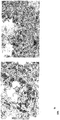


Classifications
-
- C—CHEMISTRY; METALLURGY
- C07—ORGANIC CHEMISTRY
- C07K—PEPTIDES
- C07K14/00—Peptides having more than 20 amino acids; Gastrins; Somatostatins; Melanotropins; Derivatives thereof
- C07K14/435—Peptides having more than 20 amino acids; Gastrins; Somatostatins; Melanotropins; Derivatives thereof from animals; from humans
- C07K14/46—Peptides having more than 20 amino acids; Gastrins; Somatostatins; Melanotropins; Derivatives thereof from animals; from humans from vertebrates
- C07K14/47—Peptides having more than 20 amino acids; Gastrins; Somatostatins; Melanotropins; Derivatives thereof from animals; from humans from vertebrates from mammals
- C07K14/4701—Peptides having more than 20 amino acids; Gastrins; Somatostatins; Melanotropins; Derivatives thereof from animals; from humans from vertebrates from mammals not used
- C07K14/4748—Tumour specific antigens; Tumour rejection antigen precursors [TRAP], e.g. MAGE
-
- A—HUMAN NECESSITIES
- A61—MEDICAL OR VETERINARY SCIENCE; HYGIENE
- A61P—SPECIFIC THERAPEUTIC ACTIVITY OF CHEMICAL COMPOUNDS OR MEDICINAL PREPARATIONS
- A61P35/00—Antineoplastic agents
-
- A—HUMAN NECESSITIES
- A61—MEDICAL OR VETERINARY SCIENCE; HYGIENE
- A61P—SPECIFIC THERAPEUTIC ACTIVITY OF CHEMICAL COMPOUNDS OR MEDICINAL PREPARATIONS
- A61P35/00—Antineoplastic agents
- A61P35/04—Antineoplastic agents specific for metastasis
-
- A—HUMAN NECESSITIES
- A61—MEDICAL OR VETERINARY SCIENCE; HYGIENE
- A61P—SPECIFIC THERAPEUTIC ACTIVITY OF CHEMICAL COMPOUNDS OR MEDICINAL PREPARATIONS
- A61P43/00—Drugs for specific purposes, not provided for in groups A61P1/00-A61P41/00
-
- C—CHEMISTRY; METALLURGY
- C07—ORGANIC CHEMISTRY
- C07K—PEPTIDES
- C07K16/00—Immunoglobulins [IGs], e.g. monoclonal or polyclonal antibodies
- C07K16/18—Immunoglobulins [IGs], e.g. monoclonal or polyclonal antibodies against material from animals or humans
- C07K16/28—Immunoglobulins [IGs], e.g. monoclonal or polyclonal antibodies against material from animals or humans against receptors, cell surface antigens or cell surface determinants
- C07K16/30—Immunoglobulins [IGs], e.g. monoclonal or polyclonal antibodies against material from animals or humans against receptors, cell surface antigens or cell surface determinants from tumour cells
-
- C—CHEMISTRY; METALLURGY
- C12—BIOCHEMISTRY; BEER; SPIRITS; WINE; VINEGAR; MICROBIOLOGY; ENZYMOLOGY; MUTATION OR GENETIC ENGINEERING
- C12Q—MEASURING OR TESTING PROCESSES INVOLVING ENZYMES, NUCLEIC ACIDS OR MICROORGANISMS; COMPOSITIONS OR TEST PAPERS THEREFOR; PROCESSES OF PREPARING SUCH COMPOSITIONS; CONDITION-RESPONSIVE CONTROL IN MICROBIOLOGICAL OR ENZYMOLOGICAL PROCESSES
- C12Q1/00—Measuring or testing processes involving enzymes, nucleic acids or microorganisms; Compositions therefor; Processes of preparing such compositions
- C12Q1/68—Measuring or testing processes involving enzymes, nucleic acids or microorganisms; Compositions therefor; Processes of preparing such compositions involving nucleic acids
- C12Q1/6876—Nucleic acid products used in the analysis of nucleic acids, e.g. primers or probes
- C12Q1/6883—Nucleic acid products used in the analysis of nucleic acids, e.g. primers or probes for diseases caused by alterations of genetic material
- C12Q1/6886—Nucleic acid products used in the analysis of nucleic acids, e.g. primers or probes for diseases caused by alterations of genetic material for cancer
-
- C—CHEMISTRY; METALLURGY
- C07—ORGANIC CHEMISTRY
- C07K—PEPTIDES
- C07K2317/00—Immunoglobulins specific features
- C07K2317/30—Immunoglobulins specific features characterized by aspects of specificity or valency
- C07K2317/34—Identification of a linear epitope shorter than 20 amino acid residues or of a conformational epitope defined by amino acid residues
-
- C—CHEMISTRY; METALLURGY
- C12—BIOCHEMISTRY; BEER; SPIRITS; WINE; VINEGAR; MICROBIOLOGY; ENZYMOLOGY; MUTATION OR GENETIC ENGINEERING
- C12Q—MEASURING OR TESTING PROCESSES INVOLVING ENZYMES, NUCLEIC ACIDS OR MICROORGANISMS; COMPOSITIONS OR TEST PAPERS THEREFOR; PROCESSES OF PREPARING SUCH COMPOSITIONS; CONDITION-RESPONSIVE CONTROL IN MICROBIOLOGICAL OR ENZYMOLOGICAL PROCESSES
- C12Q2600/00—Oligonucleotides characterized by their use
- C12Q2600/106—Pharmacogenomics, i.e. genetic variability in individual responses to drugs and drug metabolism
-
- C—CHEMISTRY; METALLURGY
- C12—BIOCHEMISTRY; BEER; SPIRITS; WINE; VINEGAR; MICROBIOLOGY; ENZYMOLOGY; MUTATION OR GENETIC ENGINEERING
- C12Q—MEASURING OR TESTING PROCESSES INVOLVING ENZYMES, NUCLEIC ACIDS OR MICROORGANISMS; COMPOSITIONS OR TEST PAPERS THEREFOR; PROCESSES OF PREPARING SUCH COMPOSITIONS; CONDITION-RESPONSIVE CONTROL IN MICROBIOLOGICAL OR ENZYMOLOGICAL PROCESSES
- C12Q2600/00—Oligonucleotides characterized by their use
- C12Q2600/112—Disease subtyping, staging or classification
-
- C—CHEMISTRY; METALLURGY
- C12—BIOCHEMISTRY; BEER; SPIRITS; WINE; VINEGAR; MICROBIOLOGY; ENZYMOLOGY; MUTATION OR GENETIC ENGINEERING
- C12Q—MEASURING OR TESTING PROCESSES INVOLVING ENZYMES, NUCLEIC ACIDS OR MICROORGANISMS; COMPOSITIONS OR TEST PAPERS THEREFOR; PROCESSES OF PREPARING SUCH COMPOSITIONS; CONDITION-RESPONSIVE CONTROL IN MICROBIOLOGICAL OR ENZYMOLOGICAL PROCESSES
- C12Q2600/00—Oligonucleotides characterized by their use
- C12Q2600/154—Methylation markers
Definitions
- cancer remains one of the leading causes of death.
- More recent therapeutic concepts aim to integrate the patient's own immune system into the overall therapeutic concept by using recombinant tumor vaccines and other specific measures such as antibody therapy.
- the prerequisite for the success of such a strategy is the recognition by the immune system of the patient of tumor-specific or tumor-associated antigens or epitopes whose effector functions are to be intensified interventionally.
- Tumor cells differ biologically significantly from their non-malignant cells of origin. These differences are due to genetic alterations acquired during tumor development and also lead, inter alia, to the formation of qualitatively or quantitatively altered molecular structures in the cancer cells.
- tumor-associated antigens are recognized by the specific immune system of the tumor-bearing host, one speaks of tumor-associated antigens.
- the specific recognition of tumor-associated antigens involves cellular and humoral mechanisms that represent two units that are functionally linked to one another: CD4 + and CD8 + T lymphocytes recognize processed antigens that are located on the molecules of the MHC (major histocompatibility complex) antigens. Classes II and I, respectively, while B lymphocytes produce circulating antibody molecules that bind directly to unprocessed antigens.
- tumor-associated antigens arises from the fact that the recognition of antigens on neoplastic cells by the immune system leads to the initiation of cytotoxic effector mechanisms and in the presence of T helper cells can cause the elimination of cancer cells ( Pardoll, Nat. Med. 4: 525-31, 1998 ). Accordingly, a central goal of tumor immunology is to define these structures molecularly. The molecular nature of these antigens has long remained enigmatic.
- CTL cytotoxic T lymphocyte
- circulating autoantibodies Sahin et al., Curr. Opin. Immunol. 9: 709-16, 1997 .
- cDNA expression libraries were prepared from fresh tumor tissue and recombinantly expressed in suitable systems as proteins. Immune effectors isolated from patients, namely CTL clones with tumor specificity Lysis patterns or circulating autoantibodies were used to clone the respective antigens.
- CTA Cancer / Testis antigens
- cancer / Testis antigens CTA and their coding genes (cancer / testis genes or CTG) are defined by their characteristic expression pattern [ Tureci et al, Mol Med Today. 3: 342-9, 1997 ]. They are not found in normal tissues except for testis or germ cells, but are expressed in a number of human malignancies and not tumor specific, but with varying frequency in tumor entities of very different origin ( Chen & Old, Cancer J. Sci. At the. 5: 16-7, 1999 ). Also serum reactivities against CTA are not found in healthy controls, but only in tumor patients.
- this class of antigen is of particular value for immunotherapeutic purposes and is being tested in current clinical patient studies ( Marchand et al., Int. J. Cancer 80: 219-30, 1999 ; Knuth et al., Cancer Chemother. Pharmacol. 46: S46-51, 2000 ).
- a strategy for identifying and providing tumor-associated expressed antigens and the nucleic acids coding therefor was pursued.
- This strategy is based on the fact that actually testis and thus germ cell-specific genes, which are normally silent in adult tissues, are reactivated ectopically and illegally in tumor cells.
- data mining a complete list of all known testis-specific genes is first set up and then evaluated by expression analyzes by means of specific RT-PCR for their aberrant activation in tumors.
- Data mining is a well-known method for identifying tumor-associated genes.
- transcriptomes from normal tissue banks are usually electronically subtracted from tumor tissue banks, assuming that the remaining genes are tumor specific ( Schmitt et al., Nucleic Acids Res. 27: 4251-60, 1999 ; Vasmatzis et al., Proc. Natl. Acad. Sci. USA. 95: 300-4, 1998 , Scheurle et al., Cancer Res. 60: 4037-43, 2000 ).
- the inventive concept which has proven to be much more successful, relies on utilizing data mining for the electronic extraction of all testis-specific genes and then evaluating them for ectopic expression in tumors.
- the present teachings relate to a strategy for identifying genes that are differentially expressed in tumors. This combines data mining of public sequence banks (" in silico ”) with subsequent evaluating laboratory-experimental (“wet bench”) investigations.
- CT cancer / testis
- the identified tumor-associated antigens have an amino acid sequence encoded by a nucleic acid selected from the group consisting of (a) a nucleic acid comprising a nucleic acid sequence selected from the group consisting of SEQ (B) a nucleic acid which hybridizes under stringent conditions with the nucleic acid under (a), (c) a nucleic acid which is stable with respect to the nucleic acid under (a) (a) or (b) is degenerate; and (d) a nucleic acid complementary to the nucleic acid of (a), (b) or (c).
- a nucleic acid selected from the group consisting of (a) a nucleic acid comprising a nucleic acid sequence selected from the group consisting of SEQ (B) a nucleic acid which hybridizes under stringent conditions with the nucleic acid under (a), (c) a nucleic acid which is stable with respect to the nucleic acid under (a) (a) or (b) is degenerate;
- a tumor-associated antigen identified according to the invention has an amino acid sequence encoded by a nucleic acid selected from the group consisting of SEQ ID NOs: 19-21 and 54-57.
- a tumor-associated antigen identified according to the invention comprises an amino acid sequence selected from the group consisting of SEQ ID NOs: 22-24, 58-61, 81, 82 and 103-105 a part or derivative thereof.
- the present technical teaching generally relates to the use of tumor-associated antigens identified in accordance with the invention or of parts thereof, of nucleic acids coding therefor or of nucleic acids directed against the coding nucleic acids or of antibodies directed against the tumor-associated antigens or parts identified according to the invention are directed for therapy and diagnosis.
- This use may involve single but also combinations of several of these antigens, functional fragments, nucleic acids, antibodies, etc., in one embodiment also in combination with other tumor-associated genes and antigens for diagnosis, therapy and follow-up.
- Preferred disorders for therapy and / or diagnosis are those in which there is selective expression or abnormal expression of one or more of the tumor-associated antigens identified according to the invention.
- ectopic activation of genes in tumors may also be attributable to an altered gene methylation pattern of their nucleotide sequence. For example, e.g. It has been reported that alterations in cytosine methylation contribute to this (De Smet et al, 1996 and 1999).
- the present disclosure also relates to nucleic acids and gene products that are tumor cell-associated expressed and that result from altered splicing (splice variants) of known genes, or from altered translation using alternative open reading frames.
- These nucleic acids comprise the sequences according to SEQ ID NOs: 20, 21 and 54-57 of the Sequence Listing.
- the gene products include sequences according to SEQ ID NOs: 23, 24 and 58-61 of the Sequence Listing.
- the splice variants are useful according to the technical teaching as targets for the diagnosis and therapy of tumor diseases.
- splice variant The altered splicing of a gene leads to an altered transcript sequence (splice variant). If a splice variant is translated in the region of its altered sequence, this results in a modified protein, which may differ significantly from the original structure and function.
- tumor-associated splice variants tumor-associated transcripts and tumor-associated proteins / antigens can arise. These can be used as molecular markers both for the detection of tumor cells and for the therapeutic targeting of tumors.
- the detection of tumor cells can be carried out according to the invention, for example, after extraction of nucleic acids by PCR amplification with splice variant specific oligonucleotides.
- all sequence-dependent detection systems are suitable according to the invention. In addition to PCR, these are eg gene chip / microarray systems, Northern blot, RNAse protection assays (RDA) and others. All detection systems have in common that the detection is based on a specific hybridization with at least one splice variant-specific nucleic acid sequence.
- the detection of tumor cells can also be carried out according to the invention by antibodies that recognize a coded by the splice variant specific epitope.
- the antibodies peptides for immunization can be used that are specific for this splice variant.
- Particularly suitable for the immunization are the amino acids which have distinct epitope differences to the variant (s) of the gene product which is preferably formed in healthy cells.
- the proof of Tumor cells with antibodies can be carried out on a sample isolated from the patient or as imaging with intravenously applied antibodies.
- splice variants that exhibit new or altered epitopes are attractive targets for immunotherapy.
- the epitopes can be used to target therapeutically effective monoclonal antibodies or T lymphocytes.
- antibodies or T lymphocytes that recognize splice variant-specific epitopes are adoptively transferred.
- the generation of antibodies, as with other antigens, can also be done using standard technologies (immunization of animals, panning strategies for the isolation of recombinant antibodies) using polypeptides containing these epitopes.
- nucleic acids encoding oligo- or polypeptides containing these epitopes may be used.
- the technical teaching relates to a pharmaceutical composition
- a pharmaceutical composition comprising an agent which recognizes the tumor-associated antigen identified according to the invention and is preferably selective for cells which exhibit expression or abnormal expression of a tumor-associated antigen identified according to the invention.
- the agent may cause induction of cell death, reduction of cell growth, damage to the cell membrane, or secretion of cytokines, and preferably has antitumor activity.
- the agent is an antisense nucleic acid that selectively hybridizes to the nucleic acid encoding the tumor-associated antigen.
- the agent is an antibody that selectively binds to the tumor-associated antigen, in particular a complement-activated antibody that selectively binds to the tumor-associated antigen.
- the agent comprises a plurality of agents, each of which selectively recognizes various tumor-associated antigens, wherein at least one of the tumor-associated antigens is one identified according to the invention Tumor-associated antigen.
- the recognition need not be directly associated with an inhibition of the activity or expression of the antigen.
- the antigen selectively restricted to tumors preferably serves as a label for recruitment of effector mechanisms to that specific site.
- the agent is a cytotoxic T lymphocyte which recognizes the antigen on an HLA molecule and lyses the cell so labeled.
- the agent is an antibody that selectively binds to the tumor-associated antigen, thus recruiting natural or artificial effector mechanisms to that cell.
- the agent is a T helper lymphocyte that enhances effector functions of other cells that specifically recognize that antigen.
- the present technical teaching relates to a pharmaceutical composition
- a pharmaceutical composition comprising an agent which inhibits the expression or activity of a tumor-associated antigen identified according to the invention.
- the agent is an antisense nucleic acid that selectively hybridizes to the nucleic acid encoding the tumor-associated antigen.
- the agent is an antibody that selectively binds to the tumor-associated antigen.
- the agent comprises a plurality of agents each selectively inhibiting the expression or activity of various tumor-associated antigens, wherein at least one of the tumor-associated antigens is a tumor-associated antigen identified according to the invention.
- the activity of a tumor-associated antigen identified according to the invention may be any activity of a protein or peptide.
- this activity relates to involvement in cellular migration and / or metastatization as in the case of TPTE-related sequences (see Example 1).
- the therapy and diagnostic methods of the invention may also be aimed at inhibiting or reducing this activity or testing this activity.
- the technical teaching relates to a pharmaceutical composition which comprises an agent which, when administered, selectively increases the amount of complexes between an HLA molecule and a peptide epitope from the tumor-associated antigen identified according to the invention.
- the agent in one embodiment comprises one or more components selected from the group consisting of (i) the tumor-associated Antigen or a part thereof, (ii) a nucleic acid encoding the tumor-associated antigen or a part thereof, (iii) a host cell expressing the tumor-associated antigen or part thereof, and (iv) isolated complexes between Peptide epitopes from the tumor-associated antigen and an MHC molecule.
- the agent comprises a plurality of agents, each of which selectively increases the amount of complexes between MHC molecules and peptide epitopes of various tumor-associated antigens, wherein at least one of the tumor-associated antigens is a tumor-associated antigen identified according to the invention.
- the technical teaching relates to a pharmaceutical composition which comprises one or more constituents selected from the group consisting of (i) a tumor-associated antigen identified in accordance with the invention or a part thereof, (ii) a nucleic acid which is suitable for a (iii) an antibody which binds to a tumor-associated antigen or a part thereof identified according to the invention, (iv) an antisense nucleic acid which is bound under stringent conditions specifically with a nucleic acid, (v) a host cell expressing a tumor-associated antigen or part thereof identified according to the invention, and (vi) isolated complexes between a tumor-associated antigen identified according to the invention or a part of it and a HLA-Mo lekül.
- a pharmaceutical composition which comprises one or more constituents selected from the group consisting of (i) a tumor-associated antigen identified in accordance with the invention or a part thereof, (ii) a nucleic acid which is suitable for a (iii) an antibody which bind
- a nucleic acid encoding a tumor-associated antigen or part thereof identified according to the invention may be present in the pharmaceutical composition in an expression vector and operably linked to a promoter.
- a host cell contained in a pharmaceutical composition may secrete the tumor-associated antigen or portion thereof, express it on the surface, or may additionally express an HLA molecule that binds to the tumor-associated antigen or portion thereof.
- the host cell expresses the HLA molecule endogenously.
- the host cell recombinantly expresses the HLA molecule and / or the tumor-associated antigen or part thereof.
- the host cell is non-proliferative.
- the host cell is an antigen-presenting cell, in particular a dendritic cell, a monocyte or a macrophage.
- An antibody contained in a pharmaceutical composition may be a monoclonal antibody.
- the antibody is a chimeric or humanized antibody, a fragment of a natural antibody, or a synthetic antibody that can be produced by combinatorial techniques.
- the antibody may be coupled to a therapeutically or diagnostically useful agent.
- An antisense nucleic acid contained in a pharmaceutical composition may comprise a sequence of 6-50, especially 10-30, 15-30 and 20-30 contiguous nucleotides from the nucleic acid encoding the tumor-associated antigen identified according to the invention.
- a tumor-associated antigen or portion thereof provided by a pharmaceutical composition binds to MHC molecules on the surface of cells, which binding preferably induces a cytolytic response and / or induces cytokine secretion ,
- a pharmaceutical composition may comprise a pharmaceutically acceptable carrier and / or adjuvant.
- the adjuvant may be selected from saponin, GM-CSF, CpG nucleotides, RNA, a cytokine or a chemokine.
- a pharmaceutical composition according to the invention is preferably used for the treatment of a disease characterized by the selective expression or abnormal expression of a tumor-associated antigen. In a preferred embodiment, the disease is cancer.
- the present teaching relates to methods for the treatment or diagnosis of a disease characterized by the expression or abnormal expression of one or more tumor-associated antigens.
- the treatment comprises administration of a pharmaceutical composition according to the present invention.
- the teaching is directed to a method of diagnosing a disease that is identified by the expression or abnormal expression of a subject invention Tumor-associated antigen distinguishes.
- the method comprises detecting (i) a nucleic acid encoding the tumor-associated antigen, or a portion thereof, and / or (ii) detecting the tumor-associated antigen or a portion thereof, and / or (iii) detecting an antibody against the tumor-associated antigen or a part thereof and / or (iv) the detection of cytotoxic or helper T lymphocytes specific for the tumor-associated antigen or a part thereof in a biological sample isolated from a patient ,
- detection comprises (i) contacting the biological sample with an agent that specifically binds to the nucleic acid encoding the tumor-associated antigen, or the portion thereof, to the tumor-associated antigen or portion thereof the antibody or to cytotoxic or helper T lymphocytes specific for the tumor-associated antigen or parts thereof, and (ii) the detection of complex formation
- the disease is characterized by the expression or abnormal expression of a plurality of different tumor-associated antigens and the detection comprises detection of multiple nucleic acids encoding the several different tumor-associated antigens, or portions thereof, the detection of the several different ones Tumor-associated antigens or portions thereof, the detection of multiple antibodies that bind to the several different tumor-associated antigens or portions thereof, or the detection of multiple cytotoxic or helper T lymphocytes specific for the several different tumor-associated antigens are.
- the isolated biological sample from the patient is compared to a comparable normal biological sample.
- the disclosed diagnostic methods may also relate to use of the tumor-associated antigens identified according to the invention as prognostic markers in order to predict metastatization, for example by testing the migration behavior of cells and therefore a worsened course of disease, thereby enabling, inter alia, the planning of a more aggressive therapy.
- the TPTE-related sequences are suitable for this purpose. They are also suitable for delineating benign changes such as hyperplasia from already unfavorable tumor precursors and thus foreseeing a tendency to cancer even before an invasive tumor has formed.
- the disclosed teaching relates to a method of diagnosing a disease characterized by the expression or abnormal expression of a tumor-associated antigen identified according to the invention, the method comprising evaluating the migration behavior of cells from a patient.
- the method is for determining the extent or prediction of metastatization and / or lymph node and / or distant metastases, wherein increased migration behavior of cells in certain embodiments indicates that the patient being tested is suffering from the disease the expression or abnormal expression of a tumor-associated antigen identified according to the invention is characterized, diseased, becomes ill and / or has a potential for such a disease.
- increased migration behavior indicates metastatic and / or lymph node and / or distant metastasis formation or potential.
- the tumor-associated antigen identified according to the invention preferably has a sequence encoded by a nucleic acid selected from the group consisting of: (a) a nucleic acid comprising a nucleic acid sequence selected from the group consisting of (B) a nucleic acid which hybridizes under stringent conditions with the nucleic acid under (a), (c) a nucleic acid which is linked with respect to the nucleic acid of (a), (SEQ ID NOs: 19-21 and 54-57) Nucleic acid is degenerate under (a) or (b) and (d) a nucleic acid which is complementary to the nucleic acid under (a), (b) or (c).
- the tumor-associated antigen preferably comprises an amino acid sequence selected from the group consisting of SEQ ID NOs: 22-24, 58-61, 81, 82 and 103-105, a part or derivative thereof.
- the disclosed methods of treatment in this aspect are preferably directed to the administration of pharmaceutical compositions which normalize, preferably inhibit, migration behavior.
- the present disclosure relates to a method of diagnosing a disorder characterized by the expression or abnormal expression of a tumor-associated antigen identified according to the invention, the method comprising determining the methylation pattern and / or the degree of methylation within a nucleic acid, which comprises a nucleic acid sequence associated with the tumor Antigen, especially within the non-coding regions thereof, preferably within the promoter region thereof.
- the tumor-associated antigen identified according to the invention has a sequence encoded by a nucleic acid selected from the group consisting of: (a) a nucleic acid comprising a nucleic acid sequence selected from the group consisting of SEQ ID NOs : 19-21, and 54-57, a part or derivative thereof, (b) a nucleic acid which hybridizes under stringent conditions with the nucleic acid under (a), (c) a nucleic acid which, relative to the nucleic acid under (a) or (b) is degenerate; and (d) a nucleic acid complementary to the nucleic acid of (a), (b) or (c), and the determination is made with respect to that nucleic acid, preferably within the promoter region thereof, and in particular within the sequence shown in SEQ ID NO: 822, in particular within the nucleotides from position 121 to 540 of the sequence shown in SEQ ID NO: 822 (TPTE promoter sequence on chromosome 21, position -368 /
- the tumor-associated antigen preferably comprises an amino acid sequence selected from the group consisting of SEQ ID NOs: 22-24, 58-61, 81, 82 and 103-105, a part or derivative thereof.
- a lower methylation or lack of methylation, as compared to a control, preferably indicates that the tested patient is ill with the disease characterized by the expression or abnormal expression of a tumor-associated antigen identified according to the invention and / or or has an increased potential for such a disease.
- Methods of treatment in this aspect are preferably directed to the administration of pharmaceutical compositions that normalize, preferably increase, the methylation pattern and / or the degree of methylation.
- the present teaching relates to a method for determining the regression, the course or the onset of a disease characterized by the expression or abnormal expression of a tumor-associated antigen identified according to the invention, comprising monitoring a sample from a patient the disease has or is suspected of contracting the disease with respect to one or more parameters selected from the group consisting of (i) the amount of the nucleic acid encoding the tumor-associated antigen, or a part thereof thereof, (ii) the amount of the tumor-associated antigen or a part thereof, (iii) the The amount of antibody binding to or associated with the tumor-associated antigen, and (iv) the amount of cytolytic T-cell or helper T-cell responsible for a complex between the tumor-associated antigen or a portion thereof and specific to an MHC molecule.
- the method preferably comprises the determination of the parameter (s) at a first time in a first sample and at a second time in another sample, wherein the course of the disease is determined by comparing the two samples.
- the disease is characterized by the expression or abnormal expression of a plurality of different tumor-associated antigens and monitoring includes monitoring (i) the amount of multiple nucleic acids encoding the several different tumor-associated antigens, or portions thereof and / or (ii) the amount of the several different tumor-associated antigens or parts thereof and / or (iii) the amount of multiple antibodies that bind to the several different tumor-associated antigens or parts thereof, and / or (iv) the amount of multiple cytolytic T cells or helper T cells specific for complexes between the several different tumor-associated antigens or portions thereof and MHC molecules.
- Detection of a nucleic acid or portion thereof or monitoring the amount of a nucleic acid or portion thereof may be with a polynucleotide probe that specifically hybridizes to the nucleic acid or portion thereof or may be by selective amplification of the nucleic acid or portion thereof.
- the polynucleotide probe comprises a sequence of 6-50, in particular 10-30, 15-30 and 20-30 contiguous nucleotides from the nucleic acid.
- the tumor-associated antigen or portion thereof to be detected is present intracellularly or on the cell surface. Detection of a tumor-associated antigen or portion thereof or monitoring the amount of a tumor-associated antigen or portion thereof may be with an antibody that binds specifically to the tumor-associated antigen or portion thereof.
- the tumor-associated antigen or portion thereof to be detected is in complex with an MHC molecule, particularly an HLA molecule.
- Detection of an antibody or monitoring of the amount of antibody can be done with a protein or peptide that specifically binds to the antibody.
- Detection of cytolytic T cells or helper T cells, or the monitoring of the amount of cytolytic T cells or helper T cells specific for complexes between an antigen or a portion thereof and MHC molecules can be performed with a Cell which presents the complex between the antigen or part thereof and an MHC molecule.
- the polynucleotide probe used for detection or monitoring, the antibody, protein or peptide or cell are preferably detectably labeled.
- the detectable marker is a radioactive marker or an enzyme label.
- Detection of T lymphocytes may additionally be performed by detecting their proliferation, their cytokine production, as well as their cytotoxic activity elicited by specific stimulation with the MHC-tumor associated antigen complex or parts thereof.
- the detection of T-lymphocytes can also be carried out by a recombinant MHC molecule or even a complex of several MHC molecules which are loaded with the respective immunogenic fragment from one or more of the tumor-associated antigens and by contacting the specific T-cell receptor identify the specific T lymphocytes.
- the present teachings relate to a method of treating, diagnosing, or monitoring a disease characterized by the expression or abnormal expression of a tumor-associated antigen identified according to the invention, comprising administering to the tumor-associated antigen an antibody or a part thereof and is coupled with a therapeutic or diagnostic agent.
- the antibody may be a monoclonal antibody.
- the antibody is a chimeric or humanized antibody or a fragment of a natural antibody.
- the disclosure also relates to a method of treating a patient with a disease characterized by the expression or abnormal expression of a tumor-associated antigen identified according to the invention, comprising (i) removing a sample from the patient with immunoreactive cells, (ii) the Contacting the sample with a host cell expressing the tumor-associated antigen or a portion thereof under conditions that favor production of cytolytic T cells against the tumor-associated antigen or a portion thereof, and (iii) introducing the cytolytic T cells into the cell Patients in an amount capable of lysing cells expressing the tumor-associated antigen or a portion thereof.
- the disclosure also relates to the cloning of the T-cell receptor of cytolytic T-cells against the tumor-associated antigen. This can be transferred to other T cells, which thus obtain the desired specificity and as in (iii) can be introduced into the patient.
- the host cell endogenously expresses an HLA molecule.
- the host cell expresses an HLA molecule and / or the tumor-associated antigen or part thereof recombinantly.
- the host cell is non-proliferative.
- the host cell is an antigen-presenting cell, in particular a dendritic cell, a monocyte or a macrophage.
- the disclosed technical teaching relates to a method of treating a patient with a disease characterized by the expression or abnormal expression of a tumor-associated antigen, comprising (i) identifying a tumor-associated antigen identified according to the invention Nucleic acid expressed by cells associated with the disease, (ii) the transfection of a host cell with the nucleic acid or a portion thereof, (iii) the cultivation of the transfected host cell for expression of the nucleic acid Transfection rate not obligatory), and (iv) introducing the host cells or an extract thereof into the patient in an amount effective to increase the immune response to the patient's cells associated with the disease.
- the method may further comprise identifying an MHC molecule presenting the tumor-associated antigen or a portion thereof, wherein the host cell expresses the identified MHC molecule and presents the tumor-associated antigen or portion thereof.
- the immune response may include a B-cell response or a T-cell response.
- a T cell response may involve the production of cytolytic T cells and / or helper T cells specific for the host cells presenting or specific to the tumor associated antigen or portion thereof for cells of the patient expressing the tumor-associated antigen or a part thereof.
- the disclosed teaching also relates to a method of treating a disorder characterized by the expression or abnormal expression of a tumor-associated antigen identified according to the invention comprising (i) the identification of cells from the patient which express abnormal amounts of the tumor-associated antigen (ii) isolating a sample of the cells, (iii) culturing the cells, and (iv) introducing the cells into the patient in an amount effective to induce an immune response to the cells.
- the host cells used are non-proliferative or rendered non-proliferative.
- a disease characterized by the expression or abnormal expression of a tumor-associated antigen is, in particular, cancer.
- nucleic acid selected from the group consisting of (a) a nucleic acid comprising a nucleic acid sequence selected from the group consisting of SEQ ID NOs: 20-21 and 54-57 a part or A derivative thereof, (b) a nucleic acid which hybridizes under stringent conditions with the nucleic acid under (a), (c) a nucleic acid that is degenerate with respect to the nucleic acid under (a) or (b) and (d) a nucleic acid complementary to the nucleic acid of (a), (b) or (c).
- the teaching further relates to a nucleic acid encoding a protein or polypeptide comprising an amino acid sequence selected from the group consisting of SEQ ID NOs: 22-24, 58-61, 81, 82 and 103-105, a part or derivative from that.
- the present teaching relates to promoter sequences of nucleic acids according to the invention. These may be operably linked to another gene, preferably in an expression vector, thus ensuring selective expression of that gene in corresponding cells.
- the present teaching relates to a recombinant nucleic acid molecule, in particular a DNA or RNA molecule, which comprises a nucleic acid according to the invention.
- the disclosure also relates to host cells which contain a nucleic acid according to the invention or a recombinant nucleic acid molecule which comprises a nucleic acid according to the invention.
- the host cell may further comprise a nucleic acid encoding an HLA molecule.
- the host cell expresses the HLA molecule endogenously.
- the host cell recombinantly expresses the HLA molecule and / or the nucleic acid according to the invention or a part thereof.
- the host cell is non-proliferative.
- the host cell is an antigen-presenting cell, in particular a dendritic cell, a monocyte or a macrophage.
- the present teaching also relates to oligonucleotides which hybridize with a nucleic acid identified according to the invention and can be used as genetic probes or as "antisense" molecules.
- Nucleic acid molecules in the form of oligonucleotide primers or competent probes which hybridize to a nucleic acid or parts thereof identified according to the invention can be used to find nucleic acids which are homologous to the nucleic acid identified according to the invention.
- PCR amplification, Southern and Northern hybridization can be used to detect homologous nucleic acids. Hybridization can occur under low, better under medium, and most preferably under high stringency conditions.
- stringent conditions according to the invention relates to conditions that allow specific hybridization between polynucleotides.
- the present disclosure relates to a protein or polypeptide encoded by a nucleic acid selected from the group consisting of (a) a nucleic acid comprising a nucleic acid sequence selected from the group consisting of SEQ ID NOs: (B) a nucleic acid which hybridizes under stringent conditions with the nucleic acid under (a), (c) a nucleic acid which, in relation to the nucleic acid, is selected from (a) 20-21 and 54-57 or (b) is degenerate; and (d) a nucleic acid complementary to the nucleic acid of (a), (b) or (c).
- the disclosure relates to a protein or polypeptide comprising an amino acid sequence selected from the group consisting of SEQ ID NOs: 23-24 and 58-61, 103-105, a part or derivative thereof.
- the present teaching relates to an immunogenic fragment of a tumor-associated antigen identified according to the invention.
- the fragment preferably binds to a human HLA receptor or human antibody.
- a fragment according to the invention comprises a sequence of at least 6, in particular at least 8, at least 10, at least 12, at least 15, at least 20, at least 30 or at least 50 amino acids.
- the disclosed teaching relates to an agent which binds to a tumor-associated antigen identified in accordance with the invention or to a part thereof.
- the agent is an antibody.
- the antibody is a chimeric antibody, a humanized or combinatorial antibody, or a fragment of an antibody.
- the disclosure relates to an antibody which selectively binds to a complex of (i) a tumor-associated antigen identified in accordance with the invention or a portion thereof and (ii) an MHC molecule to which the tumor-associated antigen identified according to the invention or the portion binds, wherein the antibody does not bind alone to (i) or (ii).
- An antibody of the invention may be a monoclonal antibody.
- the antibody is a chimeric or humanized antibody or a fragment of a natural antibody.
- the disclosed technical teaching relates to a conjugate between a composition according to the invention which binds to a tumor-associated antigen identified according to the invention or to a part thereof, or an antibody according to the invention and a therapeutic or diagnostic agent.
- the therapeutic or diagnostic agent is a toxin.
- the disclosure relates to a kit for detecting the expression or abnormal expression of a tumor-associated antigen identified according to the invention comprising means for detecting (i) the nucleic acid encoding the tumor-associated antigen or a part thereof, (ii ) of the tumor-associated antigen or a part thereof, (iii) antibodies which bind to the tumor-associated antigen or a part thereof, and / or (iv) T cells which are responsible for a complex between the tumor-associated Antigen or part thereof and an MHC molecule.
- the means for detecting the nucleic acid or the part thereof are nucleic acid molecules for the selective amplification of the nucleic acid, which comprise in particular a sequence of 6-50, in particular 10-30, 15-30 and 20-30 contiguous nucleotides from the nucleic acid.
- genes are described which are selectively expressed or aberrantly expressed in tumor cells and represent tumor-associated antigens.
- these genes or their derivatives are preferred target structures for therapeutic approaches.
- the therapeutic approaches may target an inhibition of the activity of the selectively expressed tumor-associated gene product. This is useful if the aberrant or selective expression is functionally of tumor pathogenetic importance and their suppression is accompanied by a selective damage to the corresponding cells.
- Other therapeutic concepts regard tumor-associated antigens as labels that selectively recruit effector mechanisms with cell-damaging potential to tumor cells.
- the function of the target molecule itself and its role in tumorigenesis is completely irrelevant.
- derivatives of a nucleic acid is meant that single or multiple nucleotide substitution, deletion and / or addition are present in the nucleic acid.
- derivative also includes a chemical derivatization of a nucleic acid on a nucleotide base, on the sugar or on the phosphate.
- derivative also includes nucleic acids containing non-naturally occurring nucleotides and nucleotide analogs.
- a nucleic acid is preferably deoxyribonucleic acid (DNA) or ribonucleic acid (RNA).
- Nucleic acids according to the invention comprise genomic DNA, cDNA, mRNA, recombinantly produced and chemically synthesized molecules.
- a nucleic acid may be present as a single-stranded or double-stranded and linear or covalently circular closed molecule.
- isolated nucleic acid means that the nucleic acid (i) amplifies in vitro was, for example, by polymerase chain reaction (PCR), (ii) was produced recombinantly by cloning, (iii) was purified, for example by cleavage and gel electrophoretic separation or (iv) was synthesized, for example by chemical synthesis.
- An isolated nucleic acid is a nucleic acid available for manipulation by recombinant DNA techniques.
- One nucleic acid is "complementary" to another nucleic acid if the two sequences hybridize with each other and can form a stable duplex, the hybridization preferably taking place under conditions allowing specific hybridization between polynucleotides (stringent conditions).
- Stringent conditions are for example in Molecular Cloning: A Laboratory Manual, J. Sambrook et al., Eds., 2nd Ed., Cold Spring Harbor Laboratory Press, Cold Spring Harbor, New York, 1989 or Current Protocols in Molecular Biology, FM Ausubel et al., Eds., John Wiley & Sons, Inc., New York pertaining, for example, to hybridization at 65 ° C.
- hybridization buffer 3.5 ⁇ SSC, 0.02% Ficoll, 0.02% polyvinylpyrrolidone, 0.02% bovine serum albumin, 2.5 mM NaH 2 PO 4 (pH 7), 0, 5% SDS, 2 mM EDTA.
- SSC is 0.15 M sodium chloride / 0.15 M sodium citrate, pH 7.
- the membrane to which the DNA has been transferred becomes, for example, 2 ⁇ SSC at room temperature and then 0.1-0.5 ⁇ SSC / 0.1 x SDS at temperatures up to 68 ° C washed.
- Complementary nucleic acids according to the invention have at least 40%, in particular at least 50%, at least 60%, at least 70%, at least 80%, at least 90% and preferably at least 95%, at least 98 or at least 99% identity of the nucleotides.
- Nucleic acids encoding tumor-associated antigens may be present alone or in combination with other nucleic acids, particularly heterologous nucleic acids.
- a nucleic acid is functionally in association with expression control sequences or regulatory sequences which may be homologous or heterologous relative to the nucleic acid.
- a coding sequence and a regulatory sequence are then "operably linked" if covalently linked such that the expression or transcription of the coding sequence is under the control or under the influence of the regulatory sequence.
- the coding sequence is to be translated into a functional protein, results in a functional association of a regulatory sequence with the coding sequence, induction of the regulatory sequence to transcription of the coding sequence, without resulting in frame shift in the coding sequence or inability of the coding sequence to be translated into the desired protein or peptide.
- expression control sequence includes promoters, enhancers and other control elements that direct the expression of a gene.
- the expression control sequences are regulatable.
- the precise structure of regulatory sequences may vary species-dependent or cell-type dependent, but generally includes 5'-untranscribed and 5'-untranslated sequences involved in the initiation of transcription, such as TATA box, capping sequence , CAAT sequence and the like.
- 5 'untranscribed regulatory sequences include a promoter region that includes a promoter sequence for transcriptional control of the functionally linked gene. Regulatory sequences may also include enhancer sequences or upstream activator sequences.
- the tumor-associated antigens shown here can be combined with any expression control sequences and promoters.
- the promoters of the tumor-associated gene products shown here can be combined with any other genes. This allows to exploit the selective activity of these promoters.
- nucleic acid may be present in association with another nucleic acid encoding a polypeptide that directs secretion of the nucleic acid-encoded protein or polypeptide from a host cell.
- a nucleic acid may be present in conjunction with another nucleic acid encoding a polypeptide that causes anchoring of the encoded protein or polypeptide on the cell membrane of the host cell or its compartmentalization into particular organelles of that cell.
- a recombinant DNA molecule is a vector, optionally with a promoter, which controls the expression of a nucleic acid, eg a nucleic acid, which codes for a tumor-associated antigen according to the invention.
- a nucleic acid eg a nucleic acid, which codes for a tumor-associated antigen according to the invention.
- vector is used in its most general meaning and encompasses any intermediary vehicle for a nucleic acid which, for example, make it possible to introduce the nucleic acid into prokaryotic and / or eukaryotic cells and, if appropriate, to integrate it into a genome.
- Such vectors are preferably replicated and / or expressed in the cell.
- an intermediary vehicle may be adapted for use in electroporation, microprojectile bombardment, liposomal delivery, Agrobacterium transfer, or DNA or RNA virus insertion.
- Vectors include plasmids, phagemids or viral genomes.
- nucleic acids encoding a tumor-associated antigen identified according to the invention can be used for transfection of host cells.
- nucleic acids is meant both recombinant DNA and RNA.
- Recombinant RNA can be produced by in vitro transcription from a DNA template. It can also be modified before application by stabilizing sequences, capping and polyadenylation.
- the term "host cell” according to the invention relates to any cell that is transformable or transfectable with an exogenous nucleic acid.
- host cells according to the invention comprises prokaryotic (eg E. coli ) or eukaryotic (eg dendritic cells, B cells, CHO cells, COS cells, K562 cells, yeast cells and insect cells).
- mammalian cells such as human, mouse, hamster, pig, goat, primate cells.
- the cells can be derived from a variety of tissue types and include primary cells and cell lines. Specific examples include keratinocytes, peripheral blood leukocytes, bone marrow stem cells and embryonic stem cells.
- the host cell is an antigen-presenting cell, in particular a dendritic cell, monocyte or macrophage.
- a nucleic acid may be present in the host cell in single or multiple copies and in one embodiment is expressed in the host cell.
- RNA or of RNA and protein are preferred expression systems in mammalian cells.
- pcDNA3.1 and pRc / CMV include pcDNA3.1 and pRc / CMV (Invitrogen, Carlsbad, CA) containing a selectable marker such as a gene conferring resistance to G418 (thus allowing selection of stably transfected cell lines) and the enhancer-promoter Sequences of cytomegalovirus (CMV).
- CMV cytomegalovirus
- an expression vector may also comprise a nucleic acid sequence encoding the HLA molecule.
- the nucleic acid sequence encoding the HLA molecule may be on the same expression vector as the nucleic acid encoding the tumor-associated antigen or portion thereof or both nucleic acids may be on different expression vectors.
- the two expression vectors can be cotransfected into a cell. If a host cell expresses neither the tumor-associated antigen or the part thereof nor the HLA molecule, both nucleic acids coding for it are transfected into the cell either on the same expression vector or on different expression vectors. If the cell already expresses the HLA molecule, only the nucleic acid sequence encoding the tumor-associated antigen or portion thereof can be transfected into the cell.
- kits for amplifying a nucleic acid encoding a tumor-associated antigen include, for example, a pair of amplification primers that hybridize to the nucleic acid encoding the tumor-associated antigen.
- the primers preferably comprise a sequence of 6-50, more preferably 10-30, 15-30 and 20-30 contiguous nucleotides from the nucleic acid and are non-overlapping to avoid the formation of primer dimers.
- One of the primers will hybridize to one strand of the nucleic acid encoding the tumor-associated antigen, and the other primer will hybridize to the complementary strand in an arrangement that permits amplification of the nucleic acid encoding the tumor-associated antigen ,
- Antisense molecules or “antisense” nucleic acids can be used to regulate, in particular reduce, the expression of a nucleic acid.
- the term "antisense molecule” or “antisense nucleic acid” according to the disclosed technical teaching relates to an oligonucleotide which is an oligoribonucleotide, oligodeoxyribonucleotide, modified oligoribonucleotide or modified oligodeoxyribonucleotide and which under physiological conditions comprises DNA comprising a particular gene or mRNA this gene hybridizes, thereby inhibiting the transcription of this gene and / or the translation of this mRNA.
- an "antisense molecule” also includes a construct that has a nucleic acid or portion thereof in reverse orientation relative to its natural Contains promoter.
- An antisense transcript of a nucleic acid or portion thereof may enter into a duplex with the naturally occurring mRNA that specifies the enzyme, thus preventing accumulation of or translation of the mRNA into the active enzyme.
- Another possibility is the use of ribozymes for inactivating a nucleic acid.
- preferred antisense oligonucleotides have a sequence of 6-50, in particular 10-30, 15-30 and 20-30 contiguous nucleotides from the target nucleic acid and are preferably completely complementary to the target nucleic acid or a part thereof ,
- the antisense oligonucleotide hybridizes to an N-terminal or 5'-upstream site such as a translation initiation, transcription initiation or promoter site. In further embodiments, the antisense oligonucleotide hybridizes to a 3 'untranslated region or mRNA splicing site.
- an oligonucleotide consists of ribonucleotides, deoxyribonucleotides, or a combination thereof.
- the 5'-end of a nucleotide and the 3'-end of another nucleotide are linked together by a phosphodiester bond.
- These oligonucleotides can be synthesized in a conventional manner or produced recombinantly.
- an oligonucleotide is a "modified" oligonucleotide.
- the oligonucleotide for example, to increase its stability or therapeutic efficacy, be modified in a variety of ways without its ability to bind to its target is impaired.
- modified oligonucleotide means according to the invention an oligonucleotide in which (i) at least two of its nucleotides are linked together by a synthetic internucleoside linkage (ie, an internucleoside linkage that is not a phosphodiester bond) and / or (ii) a chemical moiety is covalently linked to the oligonucleotide is that does not normally occur with nucleic acids.
- a synthetic internucleoside linkage ie, an internucleoside linkage that is not a phosphodiester bond
- a chemical moiety is covalently linked to the oligonucleotide is that does not normally occur with nucleic acids.
- Preferred synthetic internucleoside linkages are phosphorothioates, alkylphosphonates, phosphorodithioates, phosphate esters, alkylphosphonothioates, phosphoramidates, carbamates, carbonates, phosphate triesters, acetamidates, carboxymethyl esters and peptides.
- modified oligonucleotide also includes oligonucleotides with a covalently modified base and / or sugar.
- modified oligonucleotides include oligonucleotides having sugar moieties covalently bonded to low molecular weight organic groups that are not a hydroxyl group at the 3'-position and no phosphate group at the 5'-position.
- Modified oligonucleotides may include, for example, a 2'-O-alkylated ribose residue or another sugar instead of ribose, such as arabinose.
- isolated protein or isolated polypeptide
- isolated polypeptide mean that the protein or polypeptide is separated from its natural environment.
- An isolated protein or polypeptide may be in a substantially purified state.
- substantially purified means that the protein or polypeptide is substantially free of other substances present in nature or in vivo .
- proteins and polypeptides serve, for example, for the production of antibodies and can be used in an immunological or diagnostic assay or as therapeutics.
- Proteins and polypeptides described according to the invention can be isolated from biological samples such as tissue or cell homogenates and can also be expressed recombinantly in a multiplicity of pro- or eukaryotic expression systems.
- “Derivatives" of a protein or polypeptide or an amino acid sequence for the purposes of this invention include amino acid insertion variants, amino acid deletion variants and / or amino acid substitution variants.
- amino acid variants described above can be readily synthesized by known peptide synthesis techniques such as " Solid Phase Synthesis "(Merrifield, 1964 ) and similar methods or by recombinant DNA manipulation. Techniques for introducing substitution mutations at predetermined locations in DNA having a known or partially known sequence are well known and include, for example, M13 mutagenesis. The manipulation of DNA sequences for the production of proteins with substitutions, insertions or deletions is described, for example, in Sambrook et. al. (1989).
- Derivatives of proteins or polypeptides also include single or multiple substitutions, deletions and / or additions of any molecules associated with the enzyme, such as carbohydrates, lipids and / or proteins or polypeptides. Furthermore, the term “derivative” also extends to all functional chemical equivalents of the proteins or polypeptides.
- a portion or fragment of a tumor-associated antigen is claimed to have a functional property of the polypeptide from which it is derived. Such functional properties include interaction with antibodies, interaction with other polypeptides or proteins, selective binding of nucleic acids, and enzymatic activity.
- An important feature is the ability to complex with HLA and, where appropriate, to produce an immune response. This immune response may be due to stimulation of cytotoxic or helper T cells.
- a disclosed portion or fragment of a tumor-associated antigen comprises a sequence of at least 6, more preferably at least 8, at least 10, at least 12, at least 15, at least 20, at least 30 or at least 50 consecutive amino acids from the tumor-associated antigen.
- a portion or fragment of a nucleic acid encoding a tumor-associated antigen is disclosed to include the portion of the nucleic acid that encodes at least the tumor-associated antigen and / or a portion or fragment of the tumor-associated antigen as defined above coded.
- the isolation and identification of genes that encode tumor-associated antigens also allows for the diagnosis of a disease characterized by the expression of one or more tumor-associated antigens.
- These methods include the determination of one or more nucleic acids encoding a tumor-associated antigen and / or the determination of the encoded tumor-associated antigens and / or peptides derived therefrom.
- the present teaching also enables the isolation of proteins that bind to tumor-associated antigens described herein, including antibodies and cellular binding partners of the tumor-associated antigens.
- a dominant negative polypeptide is an inactive variant of a protein that, by interacting with the cellular machinery, displaces an active protein from its interaction with the cellular machinery or competes with the active protein, thereby reducing the effect of the active protein.
- a dominant negative receptor that binds a ligand but does not generate a signal in response to binding of the ligand can reduce the biological activity of the ligand.
- a dominant negative catalytically-inactive kinase which normally interacts with target proteins but does not phosphorylate the target proteins, can reduce phosphorylation of the target proteins in response to a cellular signal.
- a dominant negative transcription factor that binds to a promoter site in the control region of a gene does not increase the transcription of the gene, reduce the effect of a normal transcription factor by occupying promoter binding sites without an increase in transcription.
- the result of expression of a dominant negative polypeptide in a cell is a reduction in the function of active proteins.
- One skilled in the art can produce dominant negative variants of a protein, for example, by conventional mutagenesis methods and evaluating the dominant negative effect of the variant polypeptide.
- Such binding agents may e.g. in screening assays for detection of tumor-associated antigens and complexes of tumor-associated antigens with their binding partners and in a purification of the tumor-associated antigens and complexes thereof with their binding partners use.
- Such substances may also find use for inhibiting the activity of tumor-associated antigens, for example by binding to such antigens.
- binders such as e.g. Antibodies or antibody fragments having the ability to bind selectively to tumor-associated antigens.
- Antibodies include polyclonal and monoclonal antibodies prepared in a conventional manner.
- the pFc 'and Fc regions are, for example, effectors of the complement cascade but are not involved in antigen binding.
- an antibody from which the Fc region has been enzymatically cleaved or which has been produced without the Fc region termed a Fab fragment
- Fab fragments carries an antigen binding site of an intact antibody molecule.
- Fab fragments consist of a covalently linked light chain of an antibody and a part of the heavy chain of the Antibody, termed Fd.
- the Fd fragments are the major determinants of antibody specificity (a single Fd fragment can be associated with up to ten different light chains without altering the specificity of the antibody) and Fd fragments remain an isolation ability to bind to an epitope.
- CDRs complementarity determining regions
- FRs framework regions
- Both the Fd fragment of the heavy chain and the IgG immunoglobulin light chain contain four framework regions (FR1 to FR4), each separated by three complementarity determining regions (CDR1 to CDR3).
- the CDRs and in particular the CDR3 regions, and more particularly the CDR3 heavy chain region, are largely responsible for antibody specificity.
- non-CDR regions of a mammalian antibody can be replaced by similar regions of antibodies of the same or different specificity, while retaining the specificity for the epitope of the original antibody.
- mice humanized RSV antibodies in which at least a portion of mouse FR regions have been replaced by FR regions of a human origin.
- Such antibodies including fragments of intact antibodies having antigen-binding ability, are often referred to as "chimeric" antibodies.
- F (ab ') 2 Fab, Fv and Fd fragments of antibodies
- chimeric antibodies in which the Fc and / or FR and / or CDR1 and / or CDR2 and / or light Chain CDR3 regions have been replaced by homologous human or non-human sequences
- chimeric F (ab ') 2 fragment antibodies in which the FR and / or CDR1 and / or CDR2 and / or light chain CDR3 Regions were replaced by homologous human or non-human sequences
- chimeric Fab fragment antibodies in which the FR and / or CDR1 and / or CDR2 and / or light chain CDR3 regions were replaced by homologous human or non-human sequences
- chimeric Fd fragment antibodies in which the FR and / or CDR1 and / or CDR2 regions have been replaced by homologous human or non-human sequences.
- so-called single-chain antibodies are so-called single-chain antibodies.
- an antibody used in accordance with the present invention is directed against one of the sequences shown in SEQ ID NOs: 81, 82 and 103-105 of the Sequence Listing and / or can be obtained by immunization with these peptides.
- polypeptides that specifically bind to tumor-associated antigens.
- polypeptide binding agents can be provided by degenerate peptide libraries that can be readily prepared in solution in immobilized form or as phage display libraries.
- Combinatorial libraries of peptides with one or more amino acids can also be prepared.
- libraries of peptoids and non-peptidic synthetic residues can be prepared.
- Phage display may be particularly effective in identifying disclosed binding peptides.
- a phage library (using, for example, the m13, fd, or lambda phage) is prepared which presents inserts of 4 to about 80 amino acid residues in length. Phages are then selected that carry inserts that bind to the tumor-associated antigen. This process can be repeated over several cycles of reselection of phages that bind to the tumor-associated antigen. Repeated rounds lead to an accumulation of phages that carry certain sequences.
- An analysis of DNA sequences may be made to identify the sequences of the expressed polypeptides. The smallest linear portion of the sequence that binds to the tumor-associated antigen can be determined.
- the "two-hybrid system" of yeast can also be used to identify polypeptides that bind to a tumor-associated antigen.
- Tumor-associated antigens or fragments thereof described in accordance with the invention can be used for screening peptide libraries, including phage display libraries, to identify and select peptide binding partners of the tumor-associated antigens.
- Such molecules can be used, for example, for screening assays, purification protocols, for a Interference with the function of the tumor-associated antigen and for other purposes known to those skilled in the art.
- the antibodies and other binding molecules described above can be used to identify a tissue that expresses a tumor-associated antigen.
- Antibodies may also be coupled to specific diagnostic agents for imaging cells and tissues that express tumor-associated antigens. They can also be coupled to therapeutically useful substances.
- Diagnostic agents include, but are not limited to, barium sulfate, iocetamic acid, iopanoic acid, calcium ipodate, sodium diatrizoate, meglumine diatrizoate, metrizamide, sodium tylopanoate, and radiodiagnostics, including positron emitters such as fluorin-18 and carbon-11, gamma emitters such as Iodine-123, technitium-99m, iodine-131 and indium-111, nuclides for nuclear magnetic resonance such as fluorine and gadolinium.
- positron emitters such as fluorin-18 and carbon-11
- gamma emitters such as Iodine-123, technitium-99m, iodine-131 and indium-111
- nuclides for nuclear magnetic resonance such as fluorine and gadolinium.
- therapeutically useful substance means according to the invention any therapeutic molecule desirably selectively delivered to a cell expressing one or more tumor-associated antigens, including anti-cancer agents, radioactive iodine-containing compounds, toxins, cytostatic or cytolytic drugs, etc.
- Anticancer agents include, for example, aminoglutethimide, azathioprine, bleomycin sulfate, busulfan, carmustine, chlorambucil, cisplatin, cyclophosphamide, cyclosporine, cytarabidine, dacarbazine, dactinomycin, daunorubin, doxorubicin, taxol, etoposide, fluorouracil, interferon- ⁇ , lomustine, mercaptopurine, methotrexate, mitotane, procarbazine -HCl, thioguanine, vinblastine sulfate and vincristine sulfate Goodman and Gilman, "The Pharmacological Basis of Therapeutics," 8th Edition, 1990, McGraw-Hill, Inc.
- Toxins may be proteins such as pokeweed antiviral protein, cholera toxin, pertussis toxin, ricin, gelonin, abrin, diphtheria exotoxin or Pseudomonas exotoxin be emitting radionuclides such as cobalt-60.
- the tumor-associated antigens according to the invention identified in accordance with the invention can transport themselves from substances binding thereto, in particular the above-described therapeutic antibodies, from the membrane to the cytoplasm and thus serve an internalization, where these substances can preferably exert their effect as a cell-destroying effect.
- patient means according to the invention human, non-human primate or another animal, in particular mammal such as cow, horse, pig, sheep, goat, dog, cat or rodent such as mouse and rat.
- patient is a human.
- abnormal expression means that the expression is changed, preferably increased, in relation to the condition in a healthy individual.
- An increase in expression relates to an increase of at least 10%, in particular at least 20%, at least 50% or at least 100%.
- the tumor-associated antigen is expressed only in tissue of a diseased individual, while expression is repressed in a healthy individual.
- An example of such a disease is cancer, especially seminomas, melanomas, teratomas, gliomas, colorectal, breast, prostate, uterine, ovarian, and lung cancers.
- a biological sample may be a tissue and / or cellular sample according to the invention and may be recovered for use in the various methods described herein in a conventional manner, such as tissue biopsy, including punch biopsy, and blood, bronchial aspirate, urine, faeces or other body fluids.
- immunoreactive cell means according to the invention a cell which can mature into an immune cell (such as B cell, T helper cell or cytolytic T cell) with appropriate stimulation.
- Immunoreactive cells include CD34 + hematopoietic stem cells, immature and mature T cells, and immature and mature B cells. If the production of cytolytic or helper T cells recognizing a tumor-associated antigen is desired, the immunoreactive cell is contacted with a cell expressing a tumor-associated antigen under conditions involving production, differentiation, and / or or favoring selection of cytolytic as well as helper T cells.
- the Differentiation of T cell progenitors into a cytolytic T cell upon exposure to an antigen is similar to clonal selection of the immune system.
- Some therapeutic methods rely on a response of a patient's immune system that results in lysis of antigen-presenting cells, such as cancer cells, that present one or more tumor-associated antigens.
- antigen-presenting cells such as cancer cells
- cytotoxic T lymphocytes specific for a complex of a tumor-associated antigen and an MHC molecule are administered to a patient having a cellular abnormality.
- the production of such cytotoxic T lymphocytes in vitro is known.
- An example of a method for the differentiation of T cells can be found in the WO-A-9633265 .
- a sample of cells, such as blood cells is removed from the patient and the cells are contacted with a cell that presents the complex and can induce proliferation of cytotoxic T lymphocytes (eg, dendritic cells).
- the target cell may be a transfected cell, such as a COS cell. These transfected cells present the desired complex on their surface and stimulate their proliferation when contacted with cytotoxic T lymphocytes. The clonally expanded autologous cytotoxic T lymphocytes are then administered to the patient.
- fluorogenic tetramers of MHC class 1 molecule / peptide complexes are used for detection of specific clones of cytotoxic T lymphocytes ( Altman et al., Science 274: 94-96, 1996 ; Dunbar et al., Curr. Biol. 8: 413-416, 1998 ).
- Soluble MHC class 1 molecules are folded in vitro in the presence of ⁇ 2 -microglobulin and a peptide antigen that binds to the class 1 molecule. After purification of the MHC / peptide complexes, they are labeled with biotin.
- Tetramers are formed by mixing the biotinylated peptide-MHC complexes with labeled avidin (eg, phycoerythrin) at a 4: 1 molar ratio. Tetramers are then contacted with cytotoxic T lymphocytes such as peripheral blood or lymph nodes. The tetramers bind to cytotoxic T lymphocytes that recognize the peptide antigen / MHC class 1 complex. Cells that bind to the tetramers can be sorted by fluorescence-directed cell sorting to isolate reactive cytotoxic T lymphocytes. The isolated cytotoxic T lymphocytes can then be propagated in vitro .
- avidin eg, phycoerythrin
- T cell receptor itself.
- cells which present the desired complex eg dendritic cells
- healthy cytotoxic T lymphocytes This results in proliferation of high affinity specific cytotoxic T lymphocytes if the donor has so far had no contact with the specific complex.
- the high-affinity T cell receptor from these increased specific T lymphocytes is cloned and can be transduced by gene transfer, for example, with retroviral vectors as desired in T cells from other patients.
- the foregoing therapeutic aspects assume that at least some of the patient's abnormal cells present a complex of a tumor-associated antigen and an HLA molecule. An identification of such cells can take place in a manner known per se. Once cells presenting the complex have been identified, they can be combined with a sample from the patient containing cytotoxic T lymphocytes. If the cells presenting the complex are lysed by the cytotoxic T lymphocytes, it can be assumed that a tumor-associated antigen is presented.
- Cytotoxic T lymphocytes can also be generated in vivo in a manner known per se.
- One method uses non-proliferative cells expressing the complex.
- the cells used will be those that normally express the complex, such as irradiated tumor cells or cells transfected with one or both genes necessary for presentation of the complex (ie, the antigenic peptide and the presenting HLA -Molecule). Different cell types can be used.
- vectors carrying one or both of the genes of interest may be used. Viral or bacterial vectors are particularly preferred.
- nucleic acids encoding a tumor-associated antigen or portion thereof can be operably linked to promoter and enhancer sequences that direct expression of the tumor-associated antigen or fragment thereof in particular tissues or cell types.
- the nucleic acid can be incorporated into an expression vector.
- Expression vectors may be unmodified extrachromosomal nucleic acids, plasmids or viral genomes capable of inserting exogenous nucleic acids.
- Nucleic acids encoding a tumor-associated antigen can also be inserted into a retroviral genome, thereby enabling integration of the nucleic acid into the genome of the target tissue or cell.
- a microorganism such as vaccinia virus, poxvirus, herpes simplex virus, retrovirus or adenovirus carries the gene of interest and "inactivates" de facto host cells.
- Another preferred form is the introduction of the tumor-associated antigen in the form of recombinant RNA. This can be introduced, for example, by liposomal transfer or by electroporation into cells. The resulting cells present the complex of interest and are recognized by autologous cytotoxic T lymphocytes, which then multiply.
- a similar effect can be achieved by combining the tumor-associated antigen or a fragment thereof with an adjuvant to allow incorporation into antigen-presenting cells in vivo .
- the tumor-associated antigen or a fragment thereof may be represented as protein, as DNA (eg within a vector) or as RNA.
- the tumor-associated antigen is processed to yield a peptide partner for the HLA molecule, while a fragment thereof can be presented without the need for further processing. The latter is especially the case when they can bind and HLA molecules.
- Administration forms in which the Total antigen is processed in vivo by a dendritic cell are preferred because it can also help T cell responses arise. An effective immune response requires this ( Ossendorp et al, Immunol Lett.
- an effective amount of the tumor-associated antigen can be administered to a patient, for example, by intradermal injection.
- the injection can also be done intranodally in a lymph node ( Maloy et al, Proc Natl Acad Sci USA 98: 3299-303, 2001 ). It can also be used in combination with reagents that facilitate uptake into dendritic cells.
- preferred tumor-associated antigens include those that react with allogeneic cancer antisera or with T cells from many cancer patients. Of particular interest, however, are also those against which no spontaneous immune responses exist.
- the pharmaceutical compositions described above can also be used as vaccines for immunization.
- the terms "immunization” or “vaccination” as used herein teach an increase or activation of an immune response to an antigen.
- Animal models can be used to test an immunizing effect against cancer by using a tumor-associated antigen or a nucleic acid encoding it.
- human cancer cells may be introduced into a mouse for the creation of a tumor, and one or more nucleic acids encoding tumor-associated antigens may be administered.
- the effect on the cancer cells e.g., reduction in tumor size
- one or more tumor-associated antigens or stimulating fragments thereof are administered with one or more adjuvants for induction of an immune response or enhancement of an immune response.
- An adjuvant is a substance that is incorporated into or coadministered with the antigen and enhances the immune response.
- Adjuvants may enhance the immune response by providing an antigen reservoir (extracellular or in macrophages), activating macrophages and stimulating certain lymphocytes strengthen.
- Adjuvants include, but are not limited to, monophosphoryl lipid A (MPL, SmithKline Beecham), saponins such as QS21 (SmithKline Beecham), DQS21 (SmithKline Beecham; WO 96/33739 ), QS7, QS17, QS18 and QS-L1 ( So et al., Mol. Cells 7: 178-186, 1997 ), incomplete Freund's adjuvant, complete Freund's adjuvant, vitamin E, montanide, alum, CpG oligonucleotides (cf.
- MPL monophosphoryl lipid A
- saponins such as QS21 (SmithKline Beecham), DQS21 (SmithKline Beecham; WO 96/33739 ), QS7, QS17, QS18 and QS-L1 ( So et al., Mol. Cells 7: 178-186, 1997 ), incomplete Freund's adjuvant, complete Freund's adjuvant, vitamin E, montanide
- the peptides are administered in a mixture with DQS21 / MPL.
- the ratio of DQS21 to MPL is typically about 1:10 to 10: 1, preferably about 1: 5 to 5: 1 and most preferably about 1: 1.
- DQS21 and MPL are typically present in a vaccine formulation ranging from about 1 ⁇ g to about 100 ⁇ g.
- cytokines are useful in vaccination because of their regulatory properties on lymphocytes.
- cytokines include, for example, interleukin-12 (IL-12), which has been shown to enhance the protective effects of vaccines (cf. Science 268: 1432-1434, 1995 ), GM-CSF and IL-18.
- co-stimulatory molecules provided in the form of proteins or nucleic acids.
- co-stimulatory molecules are, for example, B7-1 and B7-2 (CD80 and CD86, respectively), which are expressed on dendritic cells (DC) and interact with the CD28 molecule expressed on the T cells. This interaction provides co-stimulation (signal 2) for an antigen / MHC / TCR-stimulated (signal 1) T cell, thereby enhancing T cell proliferation and effector function.
- B7 also interacts with CTLA4 (CD152) on T cells, and studies involving CTLA4 and B7 ligands show that B7-CTLA4 interaction can enhance anti-tumor immunity and CTL proliferation ( Zheng, P. et al., Proc. Natl. Acad. Sci. USA 95 (11): 6284-6289 (1998 )).
- B7 is typically not expressed on tumor cells, so they are not effective antigen presenting cells (APCs) for T cells. Induction of B7 expression would allow tumor cells to more efficiently proliferate cytotoxic T lymphocytes and stimulate an effector function. Co-stimulation by a combination of B7 / IL-6 / IL-12 showed induction of the IFN gamma and Th1 cytokine profile in a T cell population, resulting in further enhanced T cell activity ( Gajewski et al., J. Immunol. 154: 5637-5648 (1995 )).
- T helper cells Complete activation of cytotoxic T lymphocytes and complete effector function requires the involvement of T helper cells through the interaction between the CD40 ligand on the T helper cells and the CD40 molecule expressed by dendritic cells ( Ridge et al., Nature 393: 474 (1998 ) Bennett et al., Nature 393: 478 (1998 ) Schonberger et al., Nature 393: 480 (1998 )).
- the mechanism of this co-stimulatory signal is probably related to the enhancement of B7 and associated IL-6 / IL-12 production by the dendritic cells (antigen-presenting cells).
- the CD40-CD40L interaction thus complements the interactions of signal 1 (antigen / MHC-TCR) and signal 2 (B7-CD28).
- anti-CD40 antibodies for stimulation of dendritic cells would be expected to directly enhance a response to tumor antigens that are normally outside the range of an inflammatory response or presented by non-professional antigen presenting cells (tumor cells). In these situations, T helper and B7 co-stimulating signals are not provided. This mechanism could be used in the context of therapies based on antigen-pulsed dendritic cells, or in situations where T helper epitopes have not been defined in known TRA precursors.
- the disclosure also provides for the administration of nucleic acids, polypeptides or peptides.
- Administration of polypeptides and peptides may be accomplished in a manner known per se.
- administration of nucleic acids is by ex vivo methods, ie, by removing cells from a patient, genetically altering the cells to incorporate a tumor-associated antigen, and reintroducing the altered cells into the patient. This generally involves introducing a functional copy of a gene into the cells of a patient in vitro and returning the genetically altered cells to the patient. The functional copy of the gene is under the functional control of regulatory elements that allow expression of the gene in the genetically engineered cells.
- Transfection and transduction methods are known in the art. Also contemplated is administration of nucleic acids in vivo through the use of vectors such as viruses and targeted liposomes.
- a viral vector for the administration of a nucleic acid encoding a tumor-associated antigen is selected from the group consisting of adenoviruses, adeno-associated viruses, poxviruses, including vaccinia virus and attenuated poxviruses, Semliki forest virus, Retroviruses, Sindbis virus and Ty virus-like particles. Particularly preferred are adenoviruses and retroviruses.
- the retroviruses are usually replication-deficient (i.e., they are unable to produce infectious particles).
- nucleic acids can be used to introduce nucleic acids into cells in vitro or in vivo .
- Such methods include transfection of nucleic acid CaPO 4 precipitates, transfection of nucleic acids associated with DEAE, transfection or infection with the above viruses carrying the nucleic acids of interest, liposome-mediated transfection, and the like.
- control of the nucleic acid to particular cells is preferred.
- a carrier used to deliver a nucleic acid to a cell eg, a retrovirus or a liposome
- a molecule such as an antibody specific for a surface membrane protein on the target cell or a ligand for a receptor on the target cell can be incorporated into or bound to the nucleic acid carrier.
- Preferred antibodies include antibodies that selectively bind a tumor-associated antigen.
- proteins that bind to a surface membrane protein associated with endocytosis may be incorporated into the liposome formulation to facilitate targeting and / or uptake.
- proteins include capsid proteins or fragments thereof that are specific for a particular cell type, antibodies to proteins that are internalized, proteins that target an intracellular site, and the like.
- compositions of the invention may be administered in pharmaceutically acceptable formulations.
- Such preparations may usually contain pharmaceutically acceptable concentrations of salts, buffers, Preservatives, carriers, supplemental immunity enhancing substances such as adjuvants, CpG and cytokines and optionally other therapeutic agents.
- the therapeutic agents of the present invention can be administered by any conventional route, including injection or infusion.
- the administration may be, for example, oral, intravenous, intraperitoneal, intramuscular, subcutaneous or transdermal.
- Therapeutic administration of antibodies is preferably by a lung aerosol.
- the administration of antisense nucleic acids is preferably by slow intravenous administration.
- compositions according to the invention are administered in effective amounts.
- An "effective amount” refers to the amount that achieves a desired response or effect alone or with other doses.
- the desired response relates to the inhibition of disease progression. This includes slowing the progression of the disease and, in particular, interrupting the progression of the disease.
- the desired response in treating a disease or condition may also be the delay in the onset or prevention of the onset of the disease or condition.
- An effective amount of a composition will depend on the condition being treated, the severity of the disease, the individual parameters of the patient, including age, physiological condition, height and weight, duration of treatment, type of concomitant therapy (if any), specific Route of administration and similar factors.
- compositions are preferably sterile and contain an effective amount of the therapeutically active substance to produce the desired reaction or effect.
- the doses of the compositions to be administered may be of various parameters such as the mode of administration, the condition of the patient, the desired Administration period, etc. In the event that a patient's response is insufficient at an initial dose, higher doses (or effectively higher doses achieved by a different, more localized route of administration) may be employed.
- doses of the tumor-associated antigen are formulated and administered from 1 ng to 1 mg, preferably from 10 ng to 100 ⁇ g. If the administration of nucleic acids (DNA as well as RNA) encoding tumor-associated antigens is desired, doses of 1 ng to 0.1 mg are formulated and administered.
- compositions are generally administered in pharmaceutically acceptable amounts and in pharmaceutically acceptable compositions.
- pharmaceutically acceptable refers to a non-toxic material that does not interfere with the action of the active ingredient of the pharmaceutical composition.
- Such preparations may usually contain salts, buffers, preservatives, carriers and optionally other therapeutic agents.
- the salts should be pharmaceutically acceptable.
- non-pharmaceutically acceptable salts can be used for the preparation of pharmaceutically acceptable salts thereof and are included in the invention.
- Such pharmacologically and pharmaceutically acceptable salts include, but are not limited to, those made from the following acids: hydrochloric, hydrobromic, sulfuric, nitric, phosphoric, maleic, acetic, salicylic, citric, formic , Malonic, succinic and the like.
- Pharmaceutically acceptable salts may also be prepared as alkali metal or alkaline earth metal salts such as sodium, potassium or calcium salts.
- a pharmaceutical composition may comprise a pharmaceutically acceptable carrier.
- pharmaceutically acceptable carrier is contemplated to include one or more compatible solid or liquid fillers, diluents or capsule substances suitable for administration to a human.
- carrier refers to an organic or inorganic ingredient, natural or synthetic, in which the active ingredient is combined to facilitate application.
- the ingredients of the pharmaceutical composition are ordinary such that no interaction occurs which substantially affects the desired pharmaceutical efficacy.
- the pharmaceutical compositions may contain suitable buffers such as acetic acid in a salt, citric acid in a salt, boric acid in a salt and phosphoric acid in a salt.
- compositions may also optionally contain suitable preservatives such as benzalkonium chloride, chlorobutanol, parabens and thimerosal.
- suitable preservatives such as benzalkonium chloride, chlorobutanol, parabens and thimerosal.
- compositions are usually presented in a unit dosage form and can be prepared in a manner known per se.
- pharmaceutical compositions of the present invention may be in the form of capsules, tablets, lozenges, suspensions, syrups, elixirs or as an emulsion.
- compositions suitable for parenteral administration usually comprise a sterile aqueous or non-aqueous preparation of the active ingredient, which is preferably isotonic with the blood of the recipient.
- Compatible carriers and solvents include Ringer's solution and isotonic sodium chloride solution.
- sterile, fixed oils are usually used as the solution or suspension medium.
- GenBank keyword search was combined ( Fig. 1 ).
- cDNAxProfiler Two in silico strategies namely GenBank keyword search and the cDNAxProfiler were combined ( Fig. 1 ).
- ENTREZ Search and Retrieval System of the NCBI http://www.ncbi.nlm.nih.gov/Entrez
- a search was made for candidate genes in GenBank that are annotated as specifically expressed in testicular tissue Wheeler et al., Nucleic Acids Research 28: 10-14, 2000 ).
- testis-specific genes “sperm-specific genes”, “spermatogonia-specific genes” candidate genes (GOI, genes of interest) were extracted from the databases.
- the search was restricted to a part of the total information of these databases by using “homo sapiens” as limit for the organism and "mRNA” for the molecular species.
- the list of found GOIs was curated by identifying different names for the same sequence and correcting such redundancies.
- eNorthern is based on the sequence of a GOI compared to an EST (expressed sequence tag) database ( Adams et al., Science 252: 1651, 1991 ) (http://www.ncbi.nlm.nih.gov/BLAST). For each EST that is homologous to the inputted GOI, the origin of the tissue can be determined and the sum of all ESTs thus achieve a preliminary assessment of the tissue distribution of the GOI.
- RNA from native tissue material was extracted using guanidium isothiocyanates as a chaotrophic agent ( Chomczynski & Sacchi, Anal. Biochem. 162: 156-9, 1987 ). After extraction with acidic phenol and precipitation with isopropanol, the RNA was dissolved in DEPC-treated water.
- a first-strand cDNA synthesis was carried out in a 20 ⁇ l reaction mixture using Superscript II (Invitrogen) according to the manufacturer's instructions.
- the primer used was a dT (18) oligonucleotide.
- the integrity and quality of the cDNA were checked by amplification of p53 in a 30-cycle PCR (sense CGTGAGCGCTTCGAGATGTTCCG (SEQ ID NO: 52), antisense CCTAACCAGCTGCCCAACTGTAG (SEQ ID NO: 53), hybridization temperature 67 ° C).
- the primers were chosen to be in 2 different exons, and elimination of interference from contaminating genomic DNA as the cause of false positives was confirmed by testing non-reverse transcribed DNA as a template. After 15 minutes at 95 ° C to activate the HotStarTaq DNA polymerase 35 cycles of PCR were performed (1 min 94 ° C, 1 min each hybridization temperature, 2 min 72 ° C and final elongation at 72 ° C for 6 min). 20 ⁇ l of this reaction was separated on an ethidium bromide stained agarose gel and analyzed.
- the following primers were used for the expression analysis of the corresponding antigens at the indicated hybridization temperature.
- the expression of LDHC was quantified by real-time PCR.
- the principle of quantitative real-time PCR with the ABI PRISM Sequence Detection System utilizes the 5'-3 'exonuclease activity of Taq DNA polymerase for direct and specific detection of PCR products by release of fluorescence reporter dyes.
- a double fluorescence-labeled probe (TaqMan probe) which hybridizes to a sequence of the PCR product.
- the probe is labeled 5 'with a reporter dye (eg FAM) and 3' with a quencher dye (eg TAMRA). If the probe is intact, the close proximity of reporter to quencher suppresses the emission of reporter fluorescence.
- the probe When the probe hybridizes to the PCR product during PCR, it is cleaved by the 5'-3 'exonuclease activity of the Taq DNA polymerase and the suppression of reporter fluorescence is abolished.
- the increase in reporter fluorescence as a result of target amplification is measured after each PCR cycle and used for quantification.
- the expression quantification of the target gene is absolute or relative to the expression of a control gene with constant expression in the tissues to be examined.
- the expression of LDHC was calculated after normalization of the samples against 18s RNA as a so-called housekeeping gene using the ⁇ -C t method (PE Biosystems, USA). The reactions were performed in duplex approaches and determined in duplicates.
- the synthesis of the cDNA was carried out using the High Capacity cDNA Archive Kit (PE Biosystems, USA) using hexamer primers according to the manufacturer. In each case 5 ⁇ l of the diluted cDNA were used in 25 ⁇ l total volume for the PCR: sense primer (GGTGTCACTTCTGTGCCTTCCT) 300 nM; antisense primer (CGGCACCAGTTCCAACAATAG) 300 nM; TaqMan sample (CAAAGGTTCTCCAAATGT) 250 nM; sense primer 18s RNA 50 nM; antisense primer 18s RNA 50 nM; 18 s RNA sample 250 nM; 12.5 ⁇ l TaqMan Universal PCR Master Mix; initial denaturation 95 ° C (10 min); 95 ° C (15 sec); 60 ° C (1 min); 40 cycles. By amplification of a 128 bp product across the border of exon 1 and exon 2, all described splice variants of LDHC were included in the quantification.
- TPTE The expression of TPTE was also quantified by real-time PCR, but the PCR products were detected with SYBR-Green as reporter dye.
- the reporter fluorescence of SYBR-Green is suppressed in solution and only after binding to double-stranded DNA fragments is the dye active.
- the increase in SYBR-Green fluorescence as a result of specific amplification by means of GOI-specific primers after each PCR cycle is used for quantification.
- the expression quantification of the target gene is absolute or relative to the expression of a control gene with constant expression in the tissues to be examined.
- the expression of LDHC was calculated after normalization of the samples against 18s RNA as a so-called housekeeping gene using the ⁇ -C t method (PE Biosystems, USA).
- the reactions were in duplex approaches carried out and determined in triplicates.
- the QuantiTect SYBR-Green PCR Kit (Qiagen, Hilden) was used according to the manufacturer's instructions.
- the synthesis of the cDNA was carried out using the High Capacity cDNA Archive Kit (PE Biosystems, USA) using hexamer primers according to the manufacturer.
- 5 ⁇ l of the diluted cDNA were used in 25 ⁇ l total volume for the PCR: sense primer 300 nM, antisense primer 300 nM; initial denaturation 95 ° 15 min; 95 ° 30 sec; Annealing 30 sec; 72 ° 30 sec .; 40 cycles.
- Cloning of full-length or gene fragments was carried out according to common methods. To determine the sequence, corresponding antigens were amplified by means of the proofreading polymerase pfu (Stratagene). After completion of the PCR, adenosine was ligated to the ends of the amplicon using HotStarTaq DNA polymerase to clone the fragments into the TOPO-TA vector according to the manufacturer's instructions. The sequencing was performed by a commercial service. The sequences were analyzed using common prediction programs and algorithms.
- Cells from cell culture (endogenous expression of the target gene or synthesis of the target protein after transfection of an expression vector encoding the target protein) or tissue samples that might contain the target protein are lysed in a 1% SDS solution.
- the SDS denatures the proteins contained in the lysate.
- the lysates of an experimental approach are electrophoretically separated by size (SDS-polyacrylamide gel electrophoresis, SDS-PAGE) on 8-15% denaturing polyacrylamide gels (containing 1% SDS) depending on the expected protein size.
- the proteins are transferred by the semi-dry electroblotting method (Biorad) to nitrocellulose membrane (Schleicher & Schull), on which the desired protein can be detected.
- the membrane is first blocked (eg with milk powder) and then incubated with the specific antibody in a dilution of 1: 20-1: 200 (depending on the specificity of the antibody) for 60 minutes.
- a marker eg enzymes such as peroxidase or alkaline phosphatase
- the target protein is then visualized in a color or chemiluminescent reaction on the membrane by means of an enzyme reaction (eg ECL, Amersham Bioscience). The result is documented by recording with a suitable camera.
- Cells of established cell lines are used which either endogenously synthesize the target protein (detection of the RNA in the RT-PCR or of the protein in the Western blot) or have been transfected with plasmid DNA before the IF.
- endogenously synthesize the target protein detection of the RNA in the RT-PCR or of the protein in the Western blot
- plasmid DNA DNA before the IF.
- electroporation, liposome-based transfection, calcium phosphate precipitation are well established (eg Lemoine et al. Methods Mol Biol. 1997; 75: 441-7 ).
- the transfected plasmid can encode the unmodified protein in immunofluorescence or else couple different amino acid markers to the target protein.
- the most important markers are, for example, the fluorescent green fluorescent protein (GFP) in its various differentially fluorescent forms, short peptide sequences of 6-12 amino acids, for which high-affinity and specific antibodies are available.
- GFP fluorescent green fluorescent protein
- Cells that synthesize the target protein are treated with paraformaldehyde; Saponin or methanol fixed. Subsequently, if required, the cells can be permeabilized by incubation with detergents (eg 0.2% Triton X-100). After fixation / permeabilization, the cells are incubated with a primary antibody directed against the target protein or against one of the coupled markers.
- detergents eg 0.2% Triton X-100
- the batch is incubated with a second antibody coupled with a fluorescent marker (eg, fluorescein, Texas Red, Dako) which binds to the first antibody.
- a fluorescent marker eg, fluorescein, Texas Red, Dako
- the cells marked in this way are covered with glycerol and analyzed with the aid of a fluorescence microscope according to the manufacturer's instructions. Specific fluorescence emissions are achieved by specific excitation, depending on the substances used.
- the analysis usually allows the safe localization of the target protein, which is stained in addition to the target protein and the coupled amino acid marker or other marker proteins for confirmation of antibody quality and the target protein in double staining, the localization is already described in the literature.
- a special case is the GFP and its derivatives, which can be excited directly and fluoresce itself, so that no antibodies are needed for detection.
- the IHC serves to (1) estimate the amount of target protein in tumor and normal tissues, (2) to analyze how many cells in tumor and healthy tissue synthesize the target gene, (3) the cell type in a tissue ( Tumor, healthy cells) in which the target protein is detectable.
- Formalin-fixed (other fixations: eg methanol) and paraffin-embedded tissue pieces with a thickness of about 4 .mu.m are applied to a glass substrate and, for example, deparaffinized with xylenes.
- the samples are washed with TBS-T washed in serum. This is followed by incubation with the first antibody (dilution: 1: 2 to 1: 2000) for 1-18 hours, affinity-purified antibodies being generally used. After a washing step, incubation is carried out for about 30-60 minutes with a second antibody coupled with alkaline phosphatase (alternatively: eg peroxidase), which is directed against the first antibody.
- animals eg rabbits
- animals are immunized by a first injection of the desired target protein.
- the immune response of the animal against the immunogen can be increased.
- time intervals (1st bleeding after 4 weeks, then approx. Every 2 weeks with a total of up to 5 withdrawals) the animals are bled and from this an immune serum is obtained.
- the purification of the polyclonal sera was carried out in the case of peptide antibodies wholly or in the case of antibodies against recombinant proteins partly as a service by the contracted companies.
- the corresponding peptide or recombinant protein was covalently bound to a matrix in both cases, this equilibrated after coupling with a native buffer (PBS: phosphate buffered saline) and subsequently incubated with the crude serum.
- PBS phosphate buffered saline
- the antibody was eluted with 100 mM glycine pH 2.7 and the eluate was immediately neutralized in 2 M TRIS pH 8.
- the thus purified antibodies could then be used for the specific detection of the target proteins both by Western blotting and by immunofluorescence.
- the Boyden chamber quantifies cell migration in response to chemotactic stimuli.
- the chamber consists of two compartments separated by a micropore membrane. The cells to be examined are placed in the upper compartment in minimal medium, while the lower compartment is filled with medium containing the corresponding chemotactic. The cells migrate through the membrane according to the gradient and adhere to the underside of the membrane. After fixation, the transmigrated cells can be counted under the microscope.
- the migration assay was used to determine the promigrational potential of TPTE. Used with TPTE-eGFP transfected, c-erbB2 transformed NIH3T3 fibroblasts. As control cells, non-transfected cells and cells transfected with eGFP-N3 empty vector were used. For the assay, Transwell chambers (Becton Dickinson) with a pore size of 8.0 ⁇ M were used in the membrane. 4x10 4 cells each in 400 ⁇ l serum-free DMEM medium were added to the upper compartment. The lower compartment was filled with 800 ⁇ l of DMEM medium to which 10% FCS or PDGF-BB was added in ascending concentrations (10-300 ng / ⁇ l).
- the chambers were incubated for 40h at 37 °. Subsequently, the transmigrated cells were fixed in ice-cold methanol, the membranes were excised and placed on microscope slides with Hoechst nuclear dye (DAKO) for fluorescence microscopy. Cells in five visual fields (magnification 20-fold) per membrane were counted. All experiments were performed in triplicates.
- DAKO Hoechst nuclear dye
- RNA interference RNA interference
- TPTE siRNA oligos were selected according to the Tuschl criteria ( Elbashir et al., Nature 411 (6836): 428-9, 2001 ).
- TPTE siRNA oligos (sense 5'-CCCUGCCACAUGUUCAUAUdTdT-3 '; antisense 5'AUAUGAACAUGUGGCAGGGdTdT-3') targeted nucleotides 2043-2061 of the TPTE mRNA sequence (NM_013315).
- siRNA oligos were used specifically for the irrelevant DsRed fluorescent protein (AF506025) (sense 5'-AGUUCCAGUACGGCUCCAAdTdT-5 '; antisense 5'-UUGGAGCCGUACUGGAACUdTdT-3'). 200 ⁇ M of the respective sense and antisense oligos in hybridization buffer (30 mM HEPES (pH 7.4), 100 mM sodium acetate, 2 mM magnesium acetate) after initial denaturation (1 min at 90 ° C.) were used to generate the siRNA duplexes ) for 1 h at 37 ° C.
- hybridization buffer (30 mM HEPES (pH 7.4), 100 mM sodium acetate, 2 mM magnesium acetate) after initial denaturation (1 min at 90 ° C.
- 5x10 6 cells were taken up in 250 ⁇ l serum-free X-VIVO 15 medium and electroporated with 1 ⁇ M of the corresponding siRNA duplexes (200V, 250 ⁇ F).
- TPTE mRNA expression was quantified 24 hours later by real-time RT-PCR.
- TPTE negative cell lines BT549 (mamma Ca) and HCT116 (colon ca) were cultured for 72 h with 2 ⁇ M and 10 ⁇ M 5-aza-2'-deoxycytidine, respectively. Subsequently, the TPTE expression was quantified by means of real-time RT-PCR. Furthermore, TPTE expression was quantified in DNA methyltransferase (DNMT) deficient HCT116 cells.
- DNMT DNA methyltransferase
- DNMT1 HCT116 DNMT1 - / -
- DNMT3b HCT116 DNMT3b - / -
- TPTE-eGFP transfected cells were incubated for 12 h in serum-free DMEM medium.
- serum-free DMEM medium To induce spontaneous cell membrane dynamics in the form of membrane protrusions, pseudopodia and filopodia, the cells were stimulated prior to analysis by addition of FCS.
- Vital cell images were taken at 30sec intervals using an Invert Olympus Microscope (IX70) with a TILL IMAGO-VGA CCD camera.
- TPTE immunofluorescence microscopy of heterologously expressed TPTE
- the complete ORF of TPTE was cloned into pEGFP-C1 and pEGFP-N3 vectors (Clontech).
- CHO and NIH3T3 cells cultured on slides were transfected with the appropriate plasmid constructs using Fugene transfection reagent (Roche) according to the manufacturer's instructions and analyzed by means of immunofluorescence microscopy after 12-24 h.
- Fugene transfection reagent Fugene transfection reagent
- Peptide epitopes from each of the protein sequences having binding properties for the polymorphic HLA alleles A * 0201, A * 2402, A * 0101, A * 0301, B * 0702 were obtained using the assay located at http://syfpeithi.bmi-heidelberg.com/ Prediction algorithm determined.
- the peptide length was set at 8, 9 and 10mers and a cutoff was set at a score of 15. If the search resulted in fewer than 10 peptides for each length, alternatively the 10 best peptides were selected.
- Example 1 Identification of TPTE as a new tumor antigen
- sequences of the transcript of TPTE (SEQ ID NO: 19) and its translation product (SEQ ID NO: 22) are published in GenBank under accession number NM_013315 ( Walker, SM et al., Biochem. J. 360 (Pt 2): 277-83, 2001 ; Guipponi M. et al., Hum. Genet. 107 (2): 127-31, 2000 ; Chen H. et al., Hum. Genet. 105 (5): 399-409, 1999 ).
- TPTE has been described as a gene encoding a possible transmembrane tyrosine phosphatase with a testis-specific expression located in the pericentromeric region of chromosomes 21, 13, 15, 22, and Y ( Chen, H. et al., Hum. Genet. 105: 399-409, 1999 ). Inventive alignment studies additionally show homologous genomic sequences on chromosomes 3 and 7. In addition, Dong et al. 2003 that TPTE expression is detectable among other things in certain liver carcinomas ( Br J Cancer, 89: 291-297 ).
- Membrane localization e.g. for the accessibility of therapeutic antibodies was proved beyond doubt by transfection of TPTE-negative cells with a fusion construct of TPTE and Green-Fluorescent-Protein (Figure 18, right). This accumulates on the membrane surface and can be detected as colocalizing with other known membrane markers such as HLA molecules.
- PCR primers were generated according to the invention (5'-TGGATGTCACTCTCATCCTTG-3 '(SEQ ID NO: 27) and 5'-CCATAGTTCCTGTTCTATCTG-3' (SEQ ID NO: 28)) and for RT-PCR analyzes (95 ° 15 min, 94 ° 1 min, 63 ° 1 min, 72 ° 1 min, 35 cycles) in a variety of human tissues. It was found that expression in normal tissues is restricted to testis. As described for the other eCTs, according to the invention it has been shown that TPTE variants are activated ectopically in a number of tumor tissues; see. Table 1. ⁇ b> Table 1.
- TPTE in tumors ⁇ / b> tissue tested in total positive % melanoma 18 9 50 breast cancers 36 17 47 Colorectal tumors 20 0 0 prostate cancers 8th 3 38 lung 45 25 55 Renal cell carcinoma 7 2 29 ovarian cancer 7 2 29 thyroid carcinomas 4 0 0 Zervixkarzimome 6 1 17 Melanoma cell lines 8th 5 62 Bronchogenic carcinoma cell lines 6 2 33 Breast carcinoma cell lines 5 4 80
- the data are to be displayed for antibodies directed against SEQ ID NO: 105.
- the specific antibody can be used under different fixation conditions for immunofluorescence studies.
- the corresponding protein is detectable in a well-detectable amount specifically in the positively typed cell lines ( Fig. 5 , right).
- the endogenous protein is membrane-localized.
- This antibody was also used for immunohistochemical staining of tissue sections. As expected, the antibody stains testis tissue specifically. In particular, it is germ cells that are stained ( Fig. 7 ). Tissue sections of lung carcinomas can also be stained.
- TPTE protein can be detected homogeneously and in large quantities in all cells of a tumor.
- Fig. 8 Again, the localization of the protein on the membrane of the cells is confirmed ( Fig. 8 ).
- the specificity of the staining was confirmed by competition experiments.
- the specific one Antibody was first pre-incubated with recombinant TPTE protein and then applied to the tissue sections (eg, Testis as a positive control, Fig. 7 ) spent. This can successfully block the reactivity.
- tissue sections eg, Testis as a positive control, Fig. 7
- This antibody many different primary tumor types (including breast tumors, melanomas, etc.) but not the associated normal tissue can be successfully stained.
- a coloring of prostate tissue is also shown by way of example. Normal prostate tissue is negative, while both invasive and non-invaded prostate tumors are positive for this molecule.
- TPTE TPTE
- SEQ ID NO: 20 SEQ ID NO: 21, SEQ ID NO: 54, SEQ ID NO: 55, SEQ ID NO: 56, SEQ ID NO: 57
- SEQ ID NO: 56 SEQ ID NO: 55
- SEQ ID NO: 56 SEQ ID NO: 56
- the genomic sequence of TPTE consists of 24 exons (accession number NT_029430).
- the transcript shown in SEQ ID NO: 19 contains all of these exons.
- the splice variant shown in SEQ ID NO: 20 is formed by splicing out exon 7.
- the splice variant shown in SEQ ID NO: 21 shows a partial incorporation of an intron following exon 15.
- exons 18, 19, 20 and 21 can also be spliced out.
- the translation product encoded by the sequence shown in SEQ ID NO: 20 (SEQ ID NO: 23) has a deletion of 13 amino acids compared to the sequence shown in SEQ ID NO: 22.
- the translation product encoded by the sequence shown in SEQ ID NO: 21 (SEQ ID NO: 24) carries an additional insertion in the central region of the molecule and thereby differs by 14 amino acids from the other variants.
- TPTE antigen expression with specific antibodies confirmed selective expression in testis as well as in a number of different tumors. Colocalization studies also showed that TPTE is localized with class I histocompatibility antigens on the cell surface of tumor cells. So far, TPTE has only been described as a Golgi-associated protein. Due to the expression of TPTE on the cell surface of tumor cells, this tumor antigen is suitable according to the invention as an excellent target for the development of diagnostic and therapeutic monoclonal antibodies. Due to the predicted membrane topology of TPTE, the extracellularly exposed areas are suitable for this purpose according to the invention in particular.
- this includes the peptides FTDSKLYIPLEYRS (SEQ ID NO: 81) and FDIKLLRNIPRWT (SEQ ID NO: 82).
- FTDSKLYIPLEYRS SEQ ID NO: 81
- FDIKLLRNIPRWT SEQ ID NO: 82
- TPTE promotes the migration of tumor cells.
- TPTE-eGFP heterologous expressing cells
- AK staining spontaneously expressing cell lines
- rhodamine-phalloidin revealed a clear co-localization of TPTE with F-actin in these regions.
- Actin polymerisation mediated by second messengers such as phosphoinositides and calcium is a critical factor in the initiation of cell migration in response to extracellular gradients of chemotactic agents. Signal transduction via this second messenger is primarily initiated by receptor tyrosine kinases (RTK).
- RTK receptor tyrosine kinases
- TPTE RTK c-erbB-2
- TPTE-eGFP was expressed in NIH3T3 fibroblasts, which by transformation with c-erbB-2 have a constitutively activated PI-3 kinase signaling and thus an overproduction of the second messenger PIP 3,4,5 .
- Time-lapse microscopy of these cells after several hours of culture in minimal medium showed that the localization of TPTE on the membrane of pseudopodia and protrusions immediately leads to retraction of the corresponding memrane region.
- PI-3 kinase antagonistic effect of TPTE which terminates the actin polymerisation under the affected membrane area by the termination of RTK-mediated signal transduction.
- PIP 4,5 regulates cortical actin polymerization and mediates the adhesion of the cytoskeleton to the plasma membrane.
- PLC- ⁇ 1 phospholipase C- ⁇ 1
- TPTE and PIP 4,5 have been shown to positively regulate the migratory potential of cells.
- Cell migration is a directional process for which the perception of extracellular gradients of chemotactic agents is essential. Directed cell migration causes the cell to polarize towards the gradient upon detection of the chemotactic.
- PTEN mediates the polarization of the cell by formation of an intracellular gradient of second messenger PIP 3,4,5 in the direction of the extracellular gradient and thus a directed actin polymerization and migration.
- TPTE-transfected NIH3T3 / c-erbB-2 cells and corresponding control cells were tested with various amounts of chemical attractants .
- Cells expressing TPTE showed a significantly increased migration rate of more than fourfold compared to the control cells in response to 10% FCS ( Fig. 10 ).
- RNA interference has been used to demonstrate that the spontaneous expression of TPTE in tumor cell lines enhances the chemotaxis of these cells.
- TPTE-positive PC-3 prostate carcinoma cells were electroporated with TPTE-specific double-stranded RNA oligos (sense 5'-CCCUGCCACAUGUUCAUAUdTdT-3 'and antisense 5'-AUAUGAACAUGUGGCAGGGdTdT-3'). After 24 hours a decrease in TPTE expression by 70% was detected by quantitative real-time RT-PCR ( Fig. 15 ).
- the transwell migration assay showed a significant reduction in the rate of migration (70%) compared to the corresponding controls (untreated PC-3 cells and PC-3 cells electroporated with irrelevant siRNA) ( Fig. 16 ).
- PTEN is a cytoplasmic protein that can be targeted to the plasma membrane by means of PH domain-recruiting second messengers such as PIP 3,4,5
- TPTE might detect chemotactic gradients directly on the membrane by blocking the signal transduction of modulate activated RTKs, which are often overexpressed in cancer cells, thereby mediating a perception of a spatial gradient.
- TPTE Phosphatases are often involved in cell motility and migration.
- TPTE was introduced into a TPTE-negative cell line as a fusion protein with Green-Fluorescence Protein, and the distribution of this protein in vivo was observed by real-time microscopy.
- Cells form membrane protrusions (protrusions) as a precursor to migration.
- TPTE accumulates at this membrane area ( Fig. 9 ).
- TPTE is withdrawn from the membrane and internalized, and is apparently involved in the course of motility events of the cell.
- TPTE apparently mediates promigratory properties to a cell
- breast and lung tumors from a total of 58 patients were examined for expression of TPTE and statistically compared with the disease course of the patients.
- Those tumors that Express TPTE are significantly more likely to cause lymph node but also distant metastases than the TPTE-negative ( Fig. 11 ).
- TPTE plays a key role in metastasis of tumors. Accordingly, TPTE can be used as a marker for prognostically poorer course and metastatic tilt. Furthermore, methods which according to the invention inhibit the endogenous activity of TPTE in tumor cells, for example by the use of antisense RNA, different methods of RNA interference (RNAi) by means of expression vectors or retroviruses, as well as by the use of small molecules, lead to a reduced Meatstas réelle and thus be therapeutically very significant.
- RNAi RNA interference
Landscapes
- Health & Medical Sciences (AREA)
- Chemical & Material Sciences (AREA)
- Life Sciences & Earth Sciences (AREA)
- Organic Chemistry (AREA)
- Proteomics, Peptides & Aminoacids (AREA)
- Immunology (AREA)
- General Health & Medical Sciences (AREA)
- Genetics & Genomics (AREA)
- Medicinal Chemistry (AREA)
- Molecular Biology (AREA)
- Zoology (AREA)
- Biophysics (AREA)
- Biochemistry (AREA)
- Engineering & Computer Science (AREA)
- Pathology (AREA)
- Analytical Chemistry (AREA)
- Wood Science & Technology (AREA)
- Pharmacology & Pharmacy (AREA)
- Chemical Kinetics & Catalysis (AREA)
- Veterinary Medicine (AREA)
- Bioinformatics & Cheminformatics (AREA)
- Public Health (AREA)
- Animal Behavior & Ethology (AREA)
- Nuclear Medicine, Radiotherapy & Molecular Imaging (AREA)
- Oncology (AREA)
- General Chemical & Material Sciences (AREA)
- Hospice & Palliative Care (AREA)
- Biotechnology (AREA)
- Toxicology (AREA)
- Cell Biology (AREA)
- Microbiology (AREA)
- Physics & Mathematics (AREA)
- General Engineering & Computer Science (AREA)
- Gastroenterology & Hepatology (AREA)
- Medicines That Contain Protein Lipid Enzymes And Other Medicines (AREA)
- Peptides Or Proteins (AREA)
- Measuring Or Testing Involving Enzymes Or Micro-Organisms (AREA)
- Pharmaceuticals Containing Other Organic And Inorganic Compounds (AREA)
- Micro-Organisms Or Cultivation Processes Thereof (AREA)
- Medicines Containing Antibodies Or Antigens For Use As Internal Diagnostic Agents (AREA)
Claims (22)
- Composition pharmaceutique pour utilisation dans un procédé pour le traitement d'une affection cancéreuse, où le procédé comprend la réduction ou l'empêchement de la migration ou la métastase des cellules tumorales, qui se manifestent par l'expression d'un antigène associé à une tumeur, et où la composition pharmaceutique comprend un ou plusieurs composants qui sont choisis dans le groupe consistant en:(i) un antigène associé à une tumeur ou une partie de celui-ci,(ii) un acide nucléique qui code pour un antigène associé à une tumeur ou une partie de celui-ci,(iii) un anticorps qui se lie à un antigène associé à une tumeur ou une partie de celui-ci, et(iv) un acide nucléique antisens, qui hybride dans des conditions stringentes spécifiquement avec un acide nucléique qui code pour un antigène associé à une tumeur,tandis que la partie comporte au moins 6 acides aminés consécutifs de l'antigène associé à une tumeur,
tandis que l'antigène associé à une tumeur présente une séquence qui est codée par un acide nucléique qui est choisi dans le groupe consistant en:(a) un acide nucléique qui comprend une séquence d'acide nucléique qui est choisie dans le groupe consistant en SEQ ID NOs: 19-21 et 54-57,
et(b) un acide nucléique qui présente au moins 90% d'identité de séquences par rapport à l'acide nucléique sous (a). - Anticorps pour utilisation dans un procédé pour le traitement d'une affection cancéreuse, où le procédé comprend la réduction ou l'inhibition de la migration ou la métastase de cellules tumorales, qui se manifestent par l'expression d'un antigène associé à une tumeur, tandis que l'anticorps se lie spécifiquement à l'antigène associé à la tumeur, qui est codé par un acide nucléique qui est choisi dans le groupe consistant en:(a) un acide nucléique qui comprend une séquence d'acides nucléiques qui est choisie dans le groupe consistant en SEQ ID NOs: 19-21 et 54-57,
et(b) un acide nucléique qui présente au moins 90% d'identité de séquences par rapport à l'acide nucléique sous (a). - Composition pharmaceutique pour utilisation selon la revendication 1 ou anticorps pour utilisation selon la revendication 2, où l'anticorps est couplé avec un agent thérapeutique.
- Anticorps pour utilisation selon la revendication 2 ou 3, où le procédé comprend le traitement d'une tumeur métastasant, qui se manifeste par l'expression de TPTE, et tandis que le procédé comprend l'administration de l'anticorps, l'anticorps se lie au TPTE et le TPTE présente une séquence qui est codée par un acide nucléique qui est choisi dans le groupe consistant en:(a) un acide nucléique qui comprend une séquence d'acide nucléique qui est choisie dans le groupe consistant en SEQ ID NO: 19,
et(b) un acide nucléique qui présente au moins 90% d'identité de séquences par rapport à l'acide nucléique sous (a). - Composition pharmaceutique pour utilisation selon la revendication 1 ou 3 ou anticorps pour utilisation selon la revendication 4, où l'anticorps est un anticorps monoclonal, chimérique ou humanisé ou un fragment d'un anticorps.
- Composition pharmaceutique pour utilisation selon la revendication 1, 3 ou 5 ou anticorps pour utilisation selon l'une des revendications 2 à 5, où la tumeur est une tumeur pulmonaire, une tumeur du sein, une tumeur de la prostate, un mélanome, une tumeur du côlon, une métastase d'une tumeur du côlon, un carcinome des cellules rénales, un carcinome du col de l'utérus, un carcinome du côlon ou un carcinome mammaire.
- Composition pharmaceutique pour utilisation selon la revendication 1, 3, 5 ou 6 ou anticorps pour utilisation selon l'une des revendications 2 à 6, où l'antigène associé à une tumeur comprend une séquence d'acides aminés qui est choisie dans le groupe consistant en SEQ ID NOs: 22-24, 58-61, 81, 82, 103-105.
- Composition pharmaceutique qui comprend une molécule d'ARNi, qui inhibe l'expression de l'antigène tumoral TPTE, caractérisée par les SEQ ID NOs: 19, 22, pour utilisation dans un procédé pour le traitement d'une affection cancéreuse, tandis que le procédé comprend la réduction ou l'inhibition de la migration ou la métastase de cellules tumorales qui se caractérisent par l'expression d'un antigène associé à une tumeur.
- Composition pharmaceutique pour utilisation selon la revendication 8, où la molécule d'ARNi contient une structure en épingle à cheveux courte (shARN).
- Procédé pour la détermination de la tendance à la métastase d'une tumeur chez un patient, comprenant(i) la détection d'un acide nucléique ayant l'une des séquences décrites dans les SEQ ID NOs: 19-21 et 54-57 et/ou(ii) la détection d'une protéine ayant l'une des séquences décrites dans les SEQ ID NOs: 22-24, 58-61, 81, 82, 103-105dans des cellules tumorales d'un échantillon biologique isolé à partir d'un patient,
tandis qu'une présence de l'acide nucléique et/ou de la protéine est l'indice de la tendance de la tumeur à former des métastases. - Procédé selon la revendication 10, où la détection de l'acide nucléique comprend(i) la mise en contact de l'échantillon biologique avec un agent qui se lie spécifiquement à l'acide nucléique, et(ii) la détection de la formation d'un complexe entre l'agent et l'acide nucléique.
- Procédé selon la revendication 10 ou 11, où la détection de l'acide nucléique s'effectue avec une sonde polynucléotidique qui hybride spécifiquement avec l'acide nucléique.
- Procédé selon la revendication 10 ou 11, où la détection de l'acide nucléique s'effectue par amplification sélective de l'acide nucléique.
- Procédé selon la revendication 10, dans lequel la détection de la protéine comprend(i) la mise en contact de l'échantillon biologique avec un agent qui se lie spécifiquement à la protéine, et(ii) la détection de la formation d'un complexe entre l'agent et la protéine.
- Procédé selon la revendication 10 ou 14, dans lequel la détection de la protéine s'effectue avec un anticorps qui se lie spécifiquement à la protéine.
- Procédé pour la détermination d'une tendance cancéreuse avant la formation d'une tumeur invasive chez un patient, comprenant(i) la détection d'un acide nucléique ayant l'une des séquences décrites dans SEQ ID NO: 20, 21 et 54-57
et/ou(ii) la détection d'une protéine ayant l'une des séquences décrites dans SEQ ID NO: 23, 24, 58-61, 81, 103-105 dans un échantillon biologique isolé à partir d'un patient,
tandis qu'une présence de l'acide nucléique et/ou de la protéine est l'indice d'une tendance cancéreuse. - Procédé selon la revendication 16, dans lequel la détection de l'acide nucléique comprend(i) la mise en contact de l'échantillon biologique avec un agent qui se lie spécifiquement à l'acide nucléique, et(ii) la détection de la formation d'un complexe entre l'agent et l'acide nucléique.
- Procédé selon la revendication 16 ou 17, dans lequel la détection de l'acide nucléique s'effectue avec une sonde polynucléotidique qui hybride spécifiquement avec l'acide nucléique.
- Procédé selon la revendication 16, dans lequel la détection de l'acide nucléique s'effectue par amplification sélective de l'acide nucléique.
- Procédé selon la revendication 16, dans lequel la détection de la protéine comprend(i) la mise en contact de l'échantillon biologique avec un agent qui se lie spécifiquement à la protéine, et(ii) la détection de la formation d'un complexe entre l'agent et la protéine.
- Procédé selon la revendication 16 ou 20, dans lequel la détection de la protéine s'effectue avec un anticorps qui se lie spécifiquement à la protéine.
- Utilisation d'un anticorps contre une protéine ayant l'une des séquences décrites dans les SEQ ID NOs: 22-24, 58-61, 81, 82, 103-105 in vitro pour la reconnaissance diagnostique précoce d'une apparition de carcinomes prostatiques.
Applications Claiming Priority (2)
| Application Number | Priority Date | Filing Date | Title |
|---|---|---|---|
| DE10341812A DE10341812A1 (de) | 2003-09-10 | 2003-09-10 | Differentiell in Tumoren exprimierte Genprodukte und deren Verwendung |
| EP04765088A EP1664103A2 (fr) | 2003-09-10 | 2004-09-10 | Produits geniques exprimes de maniere differentielle dans des tumeurs et leur utilisation |
Related Parent Applications (2)
| Application Number | Title | Priority Date | Filing Date |
|---|---|---|---|
| EP04765088.2 Division | 2004-09-10 | ||
| EP04765088A Division EP1664103A2 (fr) | 2003-09-10 | 2004-09-10 | Produits geniques exprimes de maniere differentielle dans des tumeurs et leur utilisation |
Publications (3)
| Publication Number | Publication Date |
|---|---|
| EP2327721A2 EP2327721A2 (fr) | 2011-06-01 |
| EP2327721A3 EP2327721A3 (fr) | 2011-09-21 |
| EP2327721B1 true EP2327721B1 (fr) | 2017-12-27 |
Family
ID=34258548
Family Applications (7)
| Application Number | Title | Priority Date | Filing Date |
|---|---|---|---|
| EP04765088A Withdrawn EP1664103A2 (fr) | 2003-09-10 | 2004-09-10 | Produits geniques exprimes de maniere differentielle dans des tumeurs et leur utilisation |
| EP10011193.9A Active EP2327721B1 (fr) | 2003-09-10 | 2004-09-10 | Produits géniques exprimés de manière différentielle dans des tumeurs et leur utilisation |
| EP10011191A Withdrawn EP2336157A3 (fr) | 2003-09-10 | 2004-09-10 | Produits génique exprimés de manière differentielle dans des tumeurs et leurs utilisation |
| EP10011192A Withdrawn EP2314613A3 (fr) | 2003-09-10 | 2004-09-10 | Produits géniques exprimes de maniere différentielle dans des tumeurs et leur utilisation |
| EP10011194A Withdrawn EP2322544A3 (fr) | 2003-09-10 | 2004-09-10 | Produits géniques d'expression différentielle dans les tumeurs et leur utilisation |
| EP16176546.6A Active EP3095791B1 (fr) | 2003-09-10 | 2004-09-10 | Produits geniques exprimes de maniere differentielle dans des tumeurs et leur utilisation |
| EP10011190A Withdrawn EP2336156A3 (fr) | 2003-09-10 | 2004-09-10 | Produits génique exprimés de manière differentielle dans des tumeurs et leurs utilisation |
Family Applications Before (1)
| Application Number | Title | Priority Date | Filing Date |
|---|---|---|---|
| EP04765088A Withdrawn EP1664103A2 (fr) | 2003-09-10 | 2004-09-10 | Produits geniques exprimes de maniere differentielle dans des tumeurs et leur utilisation |
Family Applications After (5)
| Application Number | Title | Priority Date | Filing Date |
|---|---|---|---|
| EP10011191A Withdrawn EP2336157A3 (fr) | 2003-09-10 | 2004-09-10 | Produits génique exprimés de manière differentielle dans des tumeurs et leurs utilisation |
| EP10011192A Withdrawn EP2314613A3 (fr) | 2003-09-10 | 2004-09-10 | Produits géniques exprimes de maniere différentielle dans des tumeurs et leur utilisation |
| EP10011194A Withdrawn EP2322544A3 (fr) | 2003-09-10 | 2004-09-10 | Produits géniques d'expression différentielle dans les tumeurs et leur utilisation |
| EP16176546.6A Active EP3095791B1 (fr) | 2003-09-10 | 2004-09-10 | Produits geniques exprimes de maniere differentielle dans des tumeurs et leur utilisation |
| EP10011190A Withdrawn EP2336156A3 (fr) | 2003-09-10 | 2004-09-10 | Produits génique exprimés de manière differentielle dans des tumeurs et leurs utilisation |
Country Status (8)
| Country | Link |
|---|---|
| US (1) | US8765389B2 (fr) |
| EP (7) | EP1664103A2 (fr) |
| JP (3) | JP5007123B2 (fr) |
| AU (1) | AU2004272270B2 (fr) |
| CA (1) | CA2538528C (fr) |
| DE (1) | DE10341812A1 (fr) |
| ES (1) | ES2663242T3 (fr) |
| WO (1) | WO2005026205A2 (fr) |
Families Citing this family (15)
| Publication number | Priority date | Publication date | Assignee | Title |
|---|---|---|---|---|
| EP1795596A1 (fr) * | 2005-12-08 | 2007-06-13 | Ganymed Pharmaceuticals AG | Compositions et méthodes pour le diagnostic et la thérapie des cancers |
| EP1911851A1 (fr) * | 2006-10-12 | 2008-04-16 | Ganymed Pharmaceuticals AG | Compositions et méthodes pour la thérapie et le diagnostic du cancer et ses métastases |
| EP2060583A1 (fr) * | 2007-10-23 | 2009-05-20 | Ganymed Pharmaceuticals AG | Identification des marqueurs associés aux tumeurs pour diagnostic et thérapie |
| EP2108701A1 (fr) * | 2008-04-10 | 2009-10-14 | Ganymed Pharmaceuticals AG | Procédés comprenant MS4A12 et agents cibles MS4A12 pour thérapie, diagnostic et test |
| JP5565723B2 (ja) * | 2009-04-07 | 2014-08-06 | 学校法人北里研究所 | 抗dcdモノクローナル抗体 |
| BR112013006718B1 (pt) | 2010-09-20 | 2021-12-07 | Tron - Translationale Onkologie An Der Universitätsmedizin Der Johannes Gutenberguniversität Mainz Gemeinnutzige Gmbh | Receptor de células t com especificidade para ny-eso-1, composição farmacêutica compreendendo o mesmo e uso do mesmo |
| WO2013164787A1 (fr) | 2012-05-02 | 2013-11-07 | Helse Stavanger As | Méthodes de dosage pour diagnostiquer et surveiller un cancer |
| EP2777714A1 (fr) | 2013-03-15 | 2014-09-17 | NBE-Therapeutics LLC | Procédé de production d'un conjugué de ligand immunologique/charge utile à l'aide d'une enzyme transpeptidase spécifique pour une séquence |
| WO2015140567A1 (fr) * | 2014-03-21 | 2015-09-24 | Immunocore Limited | Antigènes associés aux tumeur dérivés de la protéine traip |
| EP4299136A3 (fr) | 2015-12-16 | 2024-02-14 | Gritstone bio, Inc. | Identification de néogènes, fabrication et utilisation |
| AU2018348165A1 (en) | 2017-10-10 | 2020-05-21 | Gritstone Bio, Inc. | Neoantigen identification using hotspots |
| EP3714275A4 (fr) | 2017-11-22 | 2021-10-27 | Gritstone bio, Inc. | Réduction de la présentation d'épitope de jonction pour des néo-antigènes |
| US11308700B2 (en) | 2020-04-06 | 2022-04-19 | Saudi Arabian Oil Company | Augmented reality visualization of underground pipelines using geospatial databases and KM markers |
| WO2021251842A1 (fr) * | 2020-06-08 | 2021-12-16 | Qatar Foundation For Education, Science And Community Development | Ciblage de lactate déshydrogénase c, ses procédés de préparation et ses procédés d'utilisation en combinaison avec des traitements anticancéreux |
| IL309952A (en) | 2021-07-29 | 2024-03-01 | BioNTech SE | Compositions and methods for treating melanoma |
Citations (2)
| Publication number | Priority date | Publication date | Assignee | Title |
|---|---|---|---|---|
| WO2002010363A2 (fr) * | 2000-07-28 | 2002-02-07 | Incyte Genomics, Inc. | Proteines phosphatases |
| WO2002068579A2 (fr) * | 2001-01-10 | 2002-09-06 | Pe Corporation (Ny) | Kits tels que des dosages d'acides nucleiques comprenant une majorite d'exons ou de transcrits humains, destines a detecter l'expression et pouvant avoir d'autres applications |
Family Cites Families (22)
| Publication number | Priority date | Publication date | Assignee | Title |
|---|---|---|---|---|
| AU8192987A (en) * | 1986-12-01 | 1988-06-16 | Northwestern University | Human ldh-c4 cdna sequences encoding antigenic regions |
| GB9019812D0 (en) | 1990-09-11 | 1990-10-24 | Scotgen Ltd | Novel antibodies for treatment and prevention of infection in animals and man |
| US5677139A (en) | 1995-04-21 | 1997-10-14 | President And Fellows Of Harvard College | In vitro differentiation of CD34+ progenitor cells into T lymphocytes |
| UA56132C2 (uk) | 1995-04-25 | 2003-05-15 | Смітклайн Бічем Байолоджікалс С.А. | Композиція вакцини (варіанти), спосіб стабілізації qs21 відносно гідролізу (варіанти), спосіб приготування композиції вакцини |
| WO1999033963A1 (fr) * | 1997-12-31 | 1999-07-08 | Chiron Corporation | Gene regule par cancer metastatique |
| AU2659799A (en) | 1998-02-12 | 1999-08-30 | Incyte Pharmaceuticals, Inc. | Human receptor proteins |
| US6262333B1 (en) * | 1998-06-10 | 2001-07-17 | Bayer Corporation | Human genes and gene expression products |
| EP1108021A2 (fr) * | 1998-08-31 | 2001-06-20 | Bayer Corporation | Genes humains exprimes de maniere differenciee dans un cancer colorectal |
| CA2356987A1 (fr) * | 1998-12-23 | 2000-06-29 | Corixa Corporation | Composes destines a l'immunotherapie et au diagnostic du cancer du colon et methodes d'utilisation |
| AU3395900A (en) * | 1999-03-12 | 2000-10-04 | Human Genome Sciences, Inc. | Human lung cancer associated gene sequences and polypeptides |
| DE19936563A1 (de) | 1999-08-04 | 2001-02-08 | Boehringer Ingelheim Int | Tumorassoziiertes Antigen |
| EP1208191A4 (fr) * | 1999-08-13 | 2003-12-10 | Human Genome Sciences Inc | 13 proteines associees au cancer du colon et au colon humain |
| CA2384713A1 (fr) * | 1999-09-29 | 2001-04-05 | Human Genome Sciences, Inc. | Polynucleotides et polypeptides associes au colon et au cancer du colon |
| US6436703B1 (en) | 2000-03-31 | 2002-08-20 | Hyseq, Inc. | Nucleic acids and polypeptides |
| US20020160382A1 (en) * | 2000-10-11 | 2002-10-31 | Lasek Amy W. | Genes expressed in colon cancer |
| AU2002251692A1 (en) * | 2000-12-08 | 2002-08-19 | Duke University | Identification of novel ms4a gene family members expressed by hematopoietic cells |
| EP1222928A3 (fr) * | 2001-01-16 | 2003-11-05 | Universität Zürich Institut für Medizinische Virologie | Compositions pharmaceutiques pour prévenir ou traiter le cancer, en particulier le mélanome |
| AU2002307154A1 (en) * | 2001-04-06 | 2002-10-21 | Origene Technologies, Inc | Prostate cancer expression profiles |
| US7705120B2 (en) * | 2001-06-21 | 2010-04-27 | Millennium Pharmaceuticals, Inc. | Compositions, kits, and methods for identification, assessment, prevention, and therapy of breast cancer |
| DE10211088A1 (de) | 2002-03-13 | 2003-09-25 | Ugur Sahin | Differentiell in Tumoren exprimierte Genprodukte und deren Verwendung |
| JP2006516089A (ja) * | 2002-10-02 | 2006-06-22 | ジェネンテック・インコーポレーテッド | 腫瘍の診断と治療のための組成物と方法 |
| WO2005052182A2 (fr) | 2003-11-26 | 2005-06-09 | Yissum Research Development Company Of The Hebrew University Of Jerusalem | Procede d'analyse de contenu de proteine a membrane plasmique de cellules |
-
2003
- 2003-09-10 DE DE10341812A patent/DE10341812A1/de not_active Withdrawn
-
2004
- 2004-09-10 WO PCT/EP2004/010164 patent/WO2005026205A2/fr active Application Filing
- 2004-09-10 CA CA2538528A patent/CA2538528C/fr active Active
- 2004-09-10 ES ES10011193T patent/ES2663242T3/es active Active
- 2004-09-10 JP JP2006525779A patent/JP5007123B2/ja active Active
- 2004-09-10 EP EP04765088A patent/EP1664103A2/fr not_active Withdrawn
- 2004-09-10 EP EP10011193.9A patent/EP2327721B1/fr active Active
- 2004-09-10 AU AU2004272270A patent/AU2004272270B2/en active Active
- 2004-09-10 US US10/570,957 patent/US8765389B2/en active Active
- 2004-09-10 EP EP10011191A patent/EP2336157A3/fr not_active Withdrawn
- 2004-09-10 EP EP10011192A patent/EP2314613A3/fr not_active Withdrawn
- 2004-09-10 EP EP10011194A patent/EP2322544A3/fr not_active Withdrawn
- 2004-09-10 EP EP16176546.6A patent/EP3095791B1/fr active Active
- 2004-09-10 EP EP10011190A patent/EP2336156A3/fr not_active Withdrawn
-
2011
- 2011-09-20 JP JP2011205112A patent/JP2012050441A/ja active Pending
-
2012
- 2012-07-10 JP JP2012154564A patent/JP5711698B2/ja active Active
Patent Citations (2)
| Publication number | Priority date | Publication date | Assignee | Title |
|---|---|---|---|---|
| WO2002010363A2 (fr) * | 2000-07-28 | 2002-02-07 | Incyte Genomics, Inc. | Proteines phosphatases |
| WO2002068579A2 (fr) * | 2001-01-10 | 2002-09-06 | Pe Corporation (Ny) | Kits tels que des dosages d'acides nucleiques comprenant une majorite d'exons ou de transcrits humains, destines a detecter l'expression et pouvant avoir d'autres applications |
Also Published As
| Publication number | Publication date |
|---|---|
| AU2004272270A1 (en) | 2005-03-24 |
| US20100203040A1 (en) | 2010-08-12 |
| EP2327721A3 (fr) | 2011-09-21 |
| EP2322544A2 (fr) | 2011-05-18 |
| EP3095791B1 (fr) | 2020-06-10 |
| JP5711698B2 (ja) | 2015-05-07 |
| EP2327721A2 (fr) | 2011-06-01 |
| EP2314613A2 (fr) | 2011-04-27 |
| JP2013009673A (ja) | 2013-01-17 |
| EP2336156A2 (fr) | 2011-06-22 |
| AU2004272270B2 (en) | 2011-07-21 |
| JP5007123B2 (ja) | 2012-08-22 |
| EP3095791A1 (fr) | 2016-11-23 |
| ES2663242T3 (es) | 2018-04-11 |
| JP2007526759A (ja) | 2007-09-20 |
| WO2005026205A3 (fr) | 2005-07-07 |
| EP2336156A3 (fr) | 2011-11-16 |
| EP2336157A3 (fr) | 2011-11-16 |
| CA2538528C (fr) | 2020-03-10 |
| JP2012050441A (ja) | 2012-03-15 |
| US8765389B2 (en) | 2014-07-01 |
| EP2336157A2 (fr) | 2011-06-22 |
| EP2322544A3 (fr) | 2011-11-16 |
| EP2314613A3 (fr) | 2011-11-16 |
| DE10341812A1 (de) | 2005-04-07 |
| EP1664103A2 (fr) | 2006-06-07 |
| WO2005026205A2 (fr) | 2005-03-24 |
| CA2538528A1 (fr) | 2005-03-24 |
Similar Documents
| Publication | Publication Date | Title |
|---|---|---|
| EP2400001B1 (fr) | Produits géniques d'expression différentielle dans les tumeurs et leur utilisation | |
| EP2280082B1 (fr) | Identification d'antigènes de surface pour le diagnostique et la thérapie de tumeurs | |
| EP2327417B1 (fr) | Identification d'antigènes de surfaces pour le diagnostic et la thérapie de tumeurs | |
| EP2392593B1 (fr) | Produits géniques d'expression différentielle dans les tumeurs et leur utilisation | |
| EP1664113B1 (fr) | Identification d'antigenes de surface cellulaire associes a une tumeur dans le cadre d'un diagnostic et d'une therapie | |
| EP2287182B1 (fr) | Produits géniques d'expression différentielle dans les tumeurs et leur utilisation | |
| EP2327721B1 (fr) | Produits géniques exprimés de manière différentielle dans des tumeurs et leur utilisation |
Legal Events
| Date | Code | Title | Description |
|---|---|---|---|
| PUAI | Public reference made under article 153(3) epc to a published international application that has entered the european phase |
Free format text: ORIGINAL CODE: 0009012 |
|
| AC | Divisional application: reference to earlier application |
Ref document number: 1664103 Country of ref document: EP Kind code of ref document: P |
|
| AK | Designated contracting states |
Kind code of ref document: A2 Designated state(s): AT BE BG CH CY CZ DE DK EE ES FI FR GB GR HU IE IT LI LU MC NL PL PT RO SE SI SK TR |
|
| AX | Request for extension of the european patent |
Extension state: AL HR LT LV MK |
|
| PUAL | Search report despatched |
Free format text: ORIGINAL CODE: 0009013 |
|
| AK | Designated contracting states |
Kind code of ref document: A3 Designated state(s): AT BE BG CH CY CZ DE DK EE ES FI FR GB GR HU IE IT LI LU MC NL PL PT RO SE SI SK TR |
|
| AX | Request for extension of the european patent |
Extension state: AL HR LT LV MK |
|
| RIC1 | Information provided on ipc code assigned before grant |
Ipc: C07K 14/47 20060101AFI20110815BHEP Ipc: C12Q 1/68 20060101ALI20110815BHEP Ipc: C07K 16/30 20060101ALI20110815BHEP |
|
| RTI1 | Title (correction) |
Free format text: GENETIC PRODUCTS WHICH ARE DIFFERENTIALLY EXPRESSED IN TUMOURS AND USE THEREOF |
|
| 17P | Request for examination filed |
Effective date: 20120321 |
|
| RTI1 | Title (correction) |
Free format text: GENETIC PRODUCTS WHICH ARE DIFFERENTIALLY EXPRESSED IN TUMOURS AND USE THEREOF |
|
| 17Q | First examination report despatched |
Effective date: 20130328 |
|
| RAP1 | Party data changed (applicant data changed or rights of an application transferred) |
Owner name: GANYMED PHARMACEUTICALS AG |
|
| GRAP | Despatch of communication of intention to grant a patent |
Free format text: ORIGINAL CODE: EPIDOSNIGR1 |
|
| RIC1 | Information provided on ipc code assigned before grant |
Ipc: C12Q 1/68 20060101ALI20170309BHEP Ipc: G01N 33/50 20060101ALI20170309BHEP Ipc: C07K 16/30 20060101ALI20170309BHEP Ipc: C07K 14/47 20060101AFI20170309BHEP Ipc: A61P 35/04 20060101ALI20170309BHEP |
|
| RIN1 | Information on inventor provided before grant (corrected) |
Inventor name: KOSLOWSKI, MICHAEL Inventor name: TUERECI, OEZLEM Inventor name: SAHIN, UGUR |
|
| INTG | Intention to grant announced |
Effective date: 20170404 |
|
| GRAA | (expected) grant |
Free format text: ORIGINAL CODE: 0009210 |
|
| GRAS | Grant fee paid |
Free format text: ORIGINAL CODE: EPIDOSNIGR3 |
|
| RAP1 | Party data changed (applicant data changed or rights of an application transferred) |
Owner name: GANYMED PHARMACEUTICALS GMBH |
|
| AC | Divisional application: reference to earlier application |
Ref document number: 1664103 Country of ref document: EP Kind code of ref document: P |
|
| AK | Designated contracting states |
Kind code of ref document: B1 Designated state(s): AT BE BG CH CY CZ DE DK EE ES FI FR GB GR HU IE IT LI LU MC NL PL PT RO SE SI SK TR |
|
| AX | Request for extension of the european patent |
Extension state: AL HR LT LV MK |
|
| REG | Reference to a national code |
Ref country code: GB Ref legal event code: FG4D Free format text: NOT ENGLISH |
|
| REG | Reference to a national code |
Ref country code: CH Ref legal event code: EP |
|
| REG | Reference to a national code |
Ref country code: AT Ref legal event code: REF Ref document number: 958198 Country of ref document: AT Kind code of ref document: T Effective date: 20180115 |
|
| REG | Reference to a national code |
Ref country code: IE Ref legal event code: FG4D Free format text: LANGUAGE OF EP DOCUMENT: GERMAN |
|
| REG | Reference to a national code |
Ref country code: DE Ref legal event code: R096 Ref document number: 502004015626 Country of ref document: DE |
|
| REG | Reference to a national code |
Ref country code: CH Ref legal event code: NV Representative=s name: SERVOPATENT GMBH, CH |
|
| REG | Reference to a national code |
Ref country code: NL Ref legal event code: FP |
|
| REG | Reference to a national code |
Ref country code: ES Ref legal event code: FG2A Ref document number: 2663242 Country of ref document: ES Kind code of ref document: T3 Effective date: 20180411 |
|
| PG25 | Lapsed in a contracting state [announced via postgrant information from national office to epo] |
Ref country code: FI Free format text: LAPSE BECAUSE OF FAILURE TO SUBMIT A TRANSLATION OF THE DESCRIPTION OR TO PAY THE FEE WITHIN THE PRESCRIBED TIME-LIMIT Effective date: 20171227 |
|
| REG | Reference to a national code |
Ref country code: LT Ref legal event code: MG9D |
|
| PG25 | Lapsed in a contracting state [announced via postgrant information from national office to epo] |
Ref country code: BG Free format text: LAPSE BECAUSE OF FAILURE TO SUBMIT A TRANSLATION OF THE DESCRIPTION OR TO PAY THE FEE WITHIN THE PRESCRIBED TIME-LIMIT Effective date: 20180327 Ref country code: GR Free format text: LAPSE BECAUSE OF FAILURE TO SUBMIT A TRANSLATION OF THE DESCRIPTION OR TO PAY THE FEE WITHIN THE PRESCRIBED TIME-LIMIT Effective date: 20180328 |
|
| PG25 | Lapsed in a contracting state [announced via postgrant information from national office to epo] |
Ref country code: CY Free format text: LAPSE BECAUSE OF FAILURE TO SUBMIT A TRANSLATION OF THE DESCRIPTION OR TO PAY THE FEE WITHIN THE PRESCRIBED TIME-LIMIT Effective date: 20171227 Ref country code: EE Free format text: LAPSE BECAUSE OF FAILURE TO SUBMIT A TRANSLATION OF THE DESCRIPTION OR TO PAY THE FEE WITHIN THE PRESCRIBED TIME-LIMIT Effective date: 20171227 Ref country code: SK Free format text: LAPSE BECAUSE OF FAILURE TO SUBMIT A TRANSLATION OF THE DESCRIPTION OR TO PAY THE FEE WITHIN THE PRESCRIBED TIME-LIMIT Effective date: 20171227 Ref country code: CZ Free format text: LAPSE BECAUSE OF FAILURE TO SUBMIT A TRANSLATION OF THE DESCRIPTION OR TO PAY THE FEE WITHIN THE PRESCRIBED TIME-LIMIT Effective date: 20171227 |
|
| PG25 | Lapsed in a contracting state [announced via postgrant information from national office to epo] |
Ref country code: RO Free format text: LAPSE BECAUSE OF FAILURE TO SUBMIT A TRANSLATION OF THE DESCRIPTION OR TO PAY THE FEE WITHIN THE PRESCRIBED TIME-LIMIT Effective date: 20171227 Ref country code: PL Free format text: LAPSE BECAUSE OF FAILURE TO SUBMIT A TRANSLATION OF THE DESCRIPTION OR TO PAY THE FEE WITHIN THE PRESCRIBED TIME-LIMIT Effective date: 20171227 |
|
| REG | Reference to a national code |
Ref country code: FR Ref legal event code: PLFP Year of fee payment: 15 |
|
| REG | Reference to a national code |
Ref country code: DE Ref legal event code: R097 Ref document number: 502004015626 Country of ref document: DE |
|
| PLBE | No opposition filed within time limit |
Free format text: ORIGINAL CODE: 0009261 |
|
| STAA | Information on the status of an ep patent application or granted ep patent |
Free format text: STATUS: NO OPPOSITION FILED WITHIN TIME LIMIT |
|
| PG25 | Lapsed in a contracting state [announced via postgrant information from national office to epo] |
Ref country code: DK Free format text: LAPSE BECAUSE OF FAILURE TO SUBMIT A TRANSLATION OF THE DESCRIPTION OR TO PAY THE FEE WITHIN THE PRESCRIBED TIME-LIMIT Effective date: 20171227 |
|
| 26N | No opposition filed |
Effective date: 20180928 |
|
| PG25 | Lapsed in a contracting state [announced via postgrant information from national office to epo] |
Ref country code: SI Free format text: LAPSE BECAUSE OF FAILURE TO SUBMIT A TRANSLATION OF THE DESCRIPTION OR TO PAY THE FEE WITHIN THE PRESCRIBED TIME-LIMIT Effective date: 20171227 |
|
| PG25 | Lapsed in a contracting state [announced via postgrant information from national office to epo] |
Ref country code: MC Free format text: LAPSE BECAUSE OF FAILURE TO SUBMIT A TRANSLATION OF THE DESCRIPTION OR TO PAY THE FEE WITHIN THE PRESCRIBED TIME-LIMIT Effective date: 20171227 |
|
| REG | Reference to a national code |
Ref country code: BE Ref legal event code: MM Effective date: 20180930 |
|
| REG | Reference to a national code |
Ref country code: IE Ref legal event code: MM4A |
|
| PG25 | Lapsed in a contracting state [announced via postgrant information from national office to epo] |
Ref country code: LU Free format text: LAPSE BECAUSE OF NON-PAYMENT OF DUE FEES Effective date: 20180910 |
|
| REG | Reference to a national code |
Ref country code: CH Ref legal event code: PUE Owner name: BIONTECH SE, DE Free format text: FORMER OWNER: GANYMED PHARMACEUTICALS GMBH, DE |
|
| PG25 | Lapsed in a contracting state [announced via postgrant information from national office to epo] |
Ref country code: IE Free format text: LAPSE BECAUSE OF NON-PAYMENT OF DUE FEES Effective date: 20180910 |
|
| REG | Reference to a national code |
Ref country code: NL Ref legal event code: PD Owner name: BIONTECH SE; DE Free format text: DETAILS ASSIGNMENT: CHANGE OF OWNER(S), ASSIGNMENT; FORMER OWNER NAME: GANYMED PHARMACEUTICALS GMBH Effective date: 20190719 |
|
| PG25 | Lapsed in a contracting state [announced via postgrant information from national office to epo] |
Ref country code: BE Free format text: LAPSE BECAUSE OF NON-PAYMENT OF DUE FEES Effective date: 20180930 |
|
| REG | Reference to a national code |
Ref country code: AT Ref legal event code: PC Ref document number: 958198 Country of ref document: AT Kind code of ref document: T Owner name: BIONTECH SE, DE Effective date: 20190729 |
|
| REG | Reference to a national code |
Ref country code: DE Ref legal event code: R082 Ref document number: 502004015626 Country of ref document: DE Representative=s name: ZWICKER SCHNAPPAUF & PARTNER PATENTANWAELTE PA, DE |
|
| REG | Reference to a national code |
Ref country code: DE Ref legal event code: R082 Ref document number: 502004015626 Country of ref document: DE Representative=s name: ZWICKER SCHNAPPAUF & PARTNER PATENTANWAELTE PA, DE Ref country code: DE Ref legal event code: R081 Ref document number: 502004015626 Country of ref document: DE Owner name: BIONTECH SE, DE Free format text: FORMER OWNER: GANYMED PHARMACEUTICALS GMBH, 55131 MAINZ, DE |
|
| PG25 | Lapsed in a contracting state [announced via postgrant information from national office to epo] |
Ref country code: HU Free format text: LAPSE BECAUSE OF FAILURE TO SUBMIT A TRANSLATION OF THE DESCRIPTION OR TO PAY THE FEE WITHIN THE PRESCRIBED TIME-LIMIT; INVALID AB INITIO Effective date: 20040910 Ref country code: PT Free format text: LAPSE BECAUSE OF FAILURE TO SUBMIT A TRANSLATION OF THE DESCRIPTION OR TO PAY THE FEE WITHIN THE PRESCRIBED TIME-LIMIT Effective date: 20171227 |
|
| REG | Reference to a national code |
Ref country code: CH Ref legal event code: PCAR Free format text: NEW ADDRESS: WANNERSTRASSE 9/1, 8045 ZUERICH (CH) |
|
| REG | Reference to a national code |
Ref country code: ES Ref legal event code: PC2A Owner name: BIONTECH SE Effective date: 20200616 |
|
| PG25 | Lapsed in a contracting state [announced via postgrant information from national office to epo] |
Ref country code: SE Free format text: LAPSE BECAUSE OF FAILURE TO SUBMIT A TRANSLATION OF THE DESCRIPTION OR TO PAY THE FEE WITHIN THE PRESCRIBED TIME-LIMIT Effective date: 20171227 |
|
| REG | Reference to a national code |
Ref country code: GB Ref legal event code: 732E Free format text: REGISTERED BETWEEN 20221110 AND 20221116 |
|
| P01 | Opt-out of the competence of the unified patent court (upc) registered |
Effective date: 20230525 |
|
| PGFP | Annual fee paid to national office [announced via postgrant information from national office to epo] |
Ref country code: NL Payment date: 20230822 Year of fee payment: 20 |
|
| PGFP | Annual fee paid to national office [announced via postgrant information from national office to epo] |
Ref country code: TR Payment date: 20230824 Year of fee payment: 20 Ref country code: IT Payment date: 20230822 Year of fee payment: 20 Ref country code: GB Payment date: 20230921 Year of fee payment: 20 Ref country code: AT Payment date: 20230823 Year of fee payment: 20 |
|
| PGFP | Annual fee paid to national office [announced via postgrant information from national office to epo] |
Ref country code: FR Payment date: 20230918 Year of fee payment: 20 Ref country code: DE Payment date: 20230829 Year of fee payment: 20 |
|
| PGFP | Annual fee paid to national office [announced via postgrant information from national office to epo] |
Ref country code: ES Payment date: 20231002 Year of fee payment: 20 |
|
| PGFP | Annual fee paid to national office [announced via postgrant information from national office to epo] |
Ref country code: CH Payment date: 20231001 Year of fee payment: 20 |