EP2250502B1 - Verfahren zur untersuchung des transports eines agens durch eine doppelschichtige membran bei bioanalysesensor-anwendungen - Google Patents
Verfahren zur untersuchung des transports eines agens durch eine doppelschichtige membran bei bioanalysesensor-anwendungen Download PDFInfo
- Publication number
- EP2250502B1 EP2250502B1 EP09709128.4A EP09709128A EP2250502B1 EP 2250502 B1 EP2250502 B1 EP 2250502B1 EP 09709128 A EP09709128 A EP 09709128A EP 2250502 B1 EP2250502 B1 EP 2250502B1
- Authority
- EP
- European Patent Office
- Prior art keywords
- liposomes
- membrane
- liposome
- bilayer
- volume
- Prior art date
- Legal status (The legal status is an assumption and is not a legal conclusion. Google has not performed a legal analysis and makes no representation as to the accuracy of the status listed.)
- Not-in-force
Links
- 238000000034 method Methods 0.000 title claims description 59
- 239000003795 chemical substances by application Substances 0.000 title claims description 9
- 239000003012 bilayer membrane Substances 0.000 title description 8
- 239000002502 liposome Substances 0.000 claims description 138
- 239000012528 membrane Substances 0.000 claims description 68
- 238000001514 detection method Methods 0.000 claims description 25
- 108010052285 Membrane Proteins Proteins 0.000 claims description 23
- 230000008859 change Effects 0.000 claims description 19
- 238000002198 surface plasmon resonance spectroscopy Methods 0.000 claims description 19
- 102000018697 Membrane Proteins Human genes 0.000 claims description 18
- 125000006850 spacer group Chemical group 0.000 claims description 15
- 239000002555 ionophore Substances 0.000 claims description 5
- 230000000236 ionophoric effect Effects 0.000 claims description 5
- 238000000572 ellipsometry Methods 0.000 claims description 4
- 238000001307 laser spectroscopy Methods 0.000 claims description 3
- 230000003287 optical effect Effects 0.000 claims description 3
- 108010036176 Melitten Proteins 0.000 description 44
- VDXZNPDIRNWWCW-JFTDCZMZSA-N melittin Chemical compound NCC(=O)N[C@@H]([C@@H](C)CC)C(=O)NCC(=O)N[C@@H](C)C(=O)N[C@@H](C(C)C)C(=O)N[C@@H](CC(C)C)C(=O)N[C@@H](CCCCN)C(=O)N[C@@H](C(C)C)C(=O)N[C@@H](CC(C)C)C(=O)N[C@@H]([C@@H](C)O)C(=O)N[C@@H]([C@@H](C)O)C(=O)NCC(=O)N[C@@H](CC(C)C)C(=O)N1CCC[C@H]1C(=O)N[C@@H](C)C(=O)N[C@@H](CC(C)C)C(=O)N[C@@H]([C@@H](C)CC)C(=O)N[C@@H](CO)C(=O)N[C@H](C(=O)N[C@@H]([C@@H](C)CC)C(=O)N[C@@H](CCCCN)C(=O)N[C@@H](CCCNC(N)=N)C(=O)N[C@@H](CCCCN)C(=O)N[C@@H](CCCNC(N)=N)C(=O)N[C@@H](CCC(N)=O)C(=O)N[C@@H](CCC(N)=O)C(N)=O)CC1=CNC2=CC=CC=C12 VDXZNPDIRNWWCW-JFTDCZMZSA-N 0.000 description 44
- PEDCQBHIVMGVHV-UHFFFAOYSA-N Glycerine Chemical compound OCC(O)CO PEDCQBHIVMGVHV-UHFFFAOYSA-N 0.000 description 39
- HVYWMOMLDIMFJA-DPAQBDIFSA-N cholesterol Chemical compound C1C=C2C[C@@H](O)CC[C@]2(C)[C@@H]2[C@@H]1[C@@H]1CC[C@H]([C@H](C)CCCC(C)C)[C@@]1(C)CC2 HVYWMOMLDIMFJA-DPAQBDIFSA-N 0.000 description 36
- 229930006000 Sucrose Natural products 0.000 description 35
- CZMRCDWAGMRECN-UGDNZRGBSA-N Sucrose Chemical compound O[C@H]1[C@H](O)[C@@H](CO)O[C@@]1(CO)O[C@@H]1[C@H](O)[C@@H](O)[C@H](O)[C@@H](CO)O1 CZMRCDWAGMRECN-UGDNZRGBSA-N 0.000 description 35
- 239000005720 sucrose Substances 0.000 description 35
- 230000032258 transport Effects 0.000 description 32
- 238000005259 measurement Methods 0.000 description 20
- 239000011148 porous material Substances 0.000 description 20
- 150000002632 lipids Chemical class 0.000 description 19
- 235000012000 cholesterol Nutrition 0.000 description 18
- 239000012491 analyte Substances 0.000 description 16
- 238000007792 addition Methods 0.000 description 14
- 230000004044 response Effects 0.000 description 14
- 230000035699 permeability Effects 0.000 description 13
- 238000012546 transfer Methods 0.000 description 13
- 238000002347 injection Methods 0.000 description 12
- 239000007924 injection Substances 0.000 description 12
- 239000000232 Lipid Bilayer Substances 0.000 description 11
- 229940079593 drug Drugs 0.000 description 10
- 239000003814 drug Substances 0.000 description 10
- 230000035945 sensitivity Effects 0.000 description 10
- 102000004169 proteins and genes Human genes 0.000 description 9
- 108090000623 proteins and genes Proteins 0.000 description 9
- 239000000523 sample Substances 0.000 description 9
- 230000007423 decrease Effects 0.000 description 8
- 150000002500 ions Chemical class 0.000 description 8
- 230000001404 mediated effect Effects 0.000 description 8
- 238000012216 screening Methods 0.000 description 8
- XSQUKJJJFZCRTK-UHFFFAOYSA-N Urea Chemical compound NC(N)=O XSQUKJJJFZCRTK-UHFFFAOYSA-N 0.000 description 7
- 230000008901 benefit Effects 0.000 description 7
- 239000004202 carbamide Substances 0.000 description 7
- 239000010410 layer Substances 0.000 description 7
- 239000000243 solution Substances 0.000 description 7
- 229960002685 biotin Drugs 0.000 description 6
- 239000011616 biotin Substances 0.000 description 6
- 238000002296 dynamic light scattering Methods 0.000 description 6
- 239000003792 electrolyte Substances 0.000 description 6
- 239000000463 material Substances 0.000 description 6
- 239000002904 solvent Substances 0.000 description 6
- XLYOFNOQVPJJNP-UHFFFAOYSA-N water Substances O XLYOFNOQVPJJNP-UHFFFAOYSA-N 0.000 description 6
- VSNHCAURESNICA-UHFFFAOYSA-N Hydroxyurea Chemical compound NC(=O)NO VSNHCAURESNICA-UHFFFAOYSA-N 0.000 description 5
- 108090000301 Membrane transport proteins Proteins 0.000 description 5
- 102000003939 Membrane transport proteins Human genes 0.000 description 5
- 230000001419 dependent effect Effects 0.000 description 5
- 229960001330 hydroxycarbamide Drugs 0.000 description 5
- 238000011065 in-situ storage Methods 0.000 description 5
- 239000003446 ligand Substances 0.000 description 5
- 108010087904 neutravidin Proteins 0.000 description 5
- YBJHBAHKTGYVGT-ZKWXMUAHSA-N (+)-Biotin Chemical compound N1C(=O)N[C@@H]2[C@H](CCCCC(=O)O)SC[C@@H]21 YBJHBAHKTGYVGT-ZKWXMUAHSA-N 0.000 description 4
- 229920000858 Cyclodextrin Polymers 0.000 description 4
- 238000003556 assay Methods 0.000 description 4
- 239000003153 chemical reaction reagent Substances 0.000 description 4
- 238000009792 diffusion process Methods 0.000 description 4
- 239000010931 gold Substances 0.000 description 4
- 230000002209 hydrophobic effect Effects 0.000 description 4
- 229920000642 polymer Polymers 0.000 description 4
- HFHDHCJBZVLPGP-UHFFFAOYSA-N schardinger α-dextrin Chemical compound O1C(C(C2O)O)C(CO)OC2OC(C(C2O)O)C(CO)OC2OC(C(C2O)O)C(CO)OC2OC(C(O)C2O)C(CO)OC2OC(C(C2O)O)C(CO)OC2OC2C(O)C(O)C1OC2CO HFHDHCJBZVLPGP-UHFFFAOYSA-N 0.000 description 4
- IJGRMHOSHXDMSA-UHFFFAOYSA-N Atomic nitrogen Chemical compound N#N IJGRMHOSHXDMSA-UHFFFAOYSA-N 0.000 description 3
- 239000008364 bulk solution Substances 0.000 description 3
- 210000004027 cell Anatomy 0.000 description 3
- 229910001873 dinitrogen Inorganic materials 0.000 description 3
- 230000005284 excitation Effects 0.000 description 3
- PCHJSUWPFVWCPO-UHFFFAOYSA-N gold Chemical compound [Au] PCHJSUWPFVWCPO-UHFFFAOYSA-N 0.000 description 3
- 229910052737 gold Inorganic materials 0.000 description 3
- 230000003993 interaction Effects 0.000 description 3
- 230000009061 membrane transport Effects 0.000 description 3
- 239000000203 mixture Substances 0.000 description 3
- 238000012544 monitoring process Methods 0.000 description 3
- 238000010791 quenching Methods 0.000 description 3
- 108020003175 receptors Proteins 0.000 description 3
- 102000005962 receptors Human genes 0.000 description 3
- 230000002829 reductive effect Effects 0.000 description 3
- 238000012360 testing method Methods 0.000 description 3
- VSNHCAURESNICA-NJFSPNSNSA-N 1-oxidanylurea Chemical compound N[14C](=O)NO VSNHCAURESNICA-NJFSPNSNSA-N 0.000 description 2
- VEXZGXHMUGYJMC-UHFFFAOYSA-M Chloride anion Chemical compound [Cl-] VEXZGXHMUGYJMC-UHFFFAOYSA-M 0.000 description 2
- HEDRZPFGACZZDS-UHFFFAOYSA-N Chloroform Chemical compound ClC(Cl)Cl HEDRZPFGACZZDS-UHFFFAOYSA-N 0.000 description 2
- IAZDPXIOMUYVGZ-UHFFFAOYSA-N Dimethylsulphoxide Chemical compound CS(C)=O IAZDPXIOMUYVGZ-UHFFFAOYSA-N 0.000 description 2
- 108090000862 Ion Channels Proteins 0.000 description 2
- 102000004310 Ion Channels Human genes 0.000 description 2
- FAPWRFPIFSIZLT-UHFFFAOYSA-M Sodium chloride Chemical compound [Na+].[Cl-] FAPWRFPIFSIZLT-UHFFFAOYSA-M 0.000 description 2
- 230000004075 alteration Effects 0.000 description 2
- 238000004458 analytical method Methods 0.000 description 2
- 238000013459 approach Methods 0.000 description 2
- 230000015572 biosynthetic process Effects 0.000 description 2
- 235000020958 biotin Nutrition 0.000 description 2
- 210000000170 cell membrane Anatomy 0.000 description 2
- 238000006243 chemical reaction Methods 0.000 description 2
- 230000002596 correlated effect Effects 0.000 description 2
- 230000003247 decreasing effect Effects 0.000 description 2
- 238000011161 development Methods 0.000 description 2
- 238000010494 dissociation reaction Methods 0.000 description 2
- 230000005593 dissociations Effects 0.000 description 2
- 239000012636 effector Substances 0.000 description 2
- 238000005516 engineering process Methods 0.000 description 2
- 238000002474 experimental method Methods 0.000 description 2
- 239000003925 fat Substances 0.000 description 2
- XMBWDFGMSWQBCA-UHFFFAOYSA-N hydrogen iodide Chemical compound I XMBWDFGMSWQBCA-UHFFFAOYSA-N 0.000 description 2
- 230000003100 immobilizing effect Effects 0.000 description 2
- 230000006872 improvement Effects 0.000 description 2
- 230000033001 locomotion Effects 0.000 description 2
- 230000009057 passive transport Effects 0.000 description 2
- 150000003904 phospholipids Chemical class 0.000 description 2
- 230000000171 quenching effect Effects 0.000 description 2
- 239000000758 substrate Substances 0.000 description 2
- 230000002123 temporal effect Effects 0.000 description 2
- 230000002463 transducing effect Effects 0.000 description 2
- 230000005945 translocation Effects 0.000 description 2
- 108091032973 (ribonucleotides)n+m Proteins 0.000 description 1
- JKMHFZQWWAIEOD-UHFFFAOYSA-N 2-[4-(2-hydroxyethyl)piperazin-1-yl]ethanesulfonic acid Chemical compound OCC[NH+]1CCN(CCS([O-])(=O)=O)CC1 JKMHFZQWWAIEOD-UHFFFAOYSA-N 0.000 description 1
- 102000016560 Aquaglyceroporins Human genes 0.000 description 1
- 108010092667 Aquaglyceroporins Proteins 0.000 description 1
- 238000012935 Averaging Methods 0.000 description 1
- 108090001008 Avidin Proteins 0.000 description 1
- 108091006146 Channels Proteins 0.000 description 1
- 108020004414 DNA Proteins 0.000 description 1
- 229930186217 Glycolipid Natural products 0.000 description 1
- 108010026389 Gramicidin Proteins 0.000 description 1
- 239000007995 HEPES buffer Substances 0.000 description 1
- 241000725303 Human immunodeficiency virus Species 0.000 description 1
- 206010028980 Neoplasm Diseases 0.000 description 1
- 208000008589 Obesity Diseases 0.000 description 1
- 241000223960 Plasmodium falciparum Species 0.000 description 1
- 229930182558 Sterol Natural products 0.000 description 1
- 230000004913 activation Effects 0.000 description 1
- 230000009056 active transport Effects 0.000 description 1
- 210000001789 adipocyte Anatomy 0.000 description 1
- 230000002776 aggregation Effects 0.000 description 1
- 238000004220 aggregation Methods 0.000 description 1
- 150000001335 aliphatic alkanes Chemical class 0.000 description 1
- 125000000217 alkyl group Chemical group 0.000 description 1
- 150000001408 amides Chemical class 0.000 description 1
- 150000001450 anions Chemical class 0.000 description 1
- 239000007864 aqueous solution Substances 0.000 description 1
- 230000004888 barrier function Effects 0.000 description 1
- OGBUMNBNEWYMNJ-UHFFFAOYSA-N batilol Chemical class CCCCCCCCCCCCCCCCCCOCC(O)CO OGBUMNBNEWYMNJ-UHFFFAOYSA-N 0.000 description 1
- 230000002051 biphasic effect Effects 0.000 description 1
- 239000000872 buffer Substances 0.000 description 1
- 201000011510 cancer Diseases 0.000 description 1
- 230000001413 cellular effect Effects 0.000 description 1
- 238000012512 characterization method Methods 0.000 description 1
- 238000002512 chemotherapy Methods 0.000 description 1
- 230000000295 complement effect Effects 0.000 description 1
- 150000001875 compounds Chemical class 0.000 description 1
- 230000000875 corresponding effect Effects 0.000 description 1
- 238000010790 dilution Methods 0.000 description 1
- 239000012895 dilution Substances 0.000 description 1
- 238000009826 distribution Methods 0.000 description 1
- 238000007877 drug screening Methods 0.000 description 1
- 230000001614 effect on membrane Effects 0.000 description 1
- 230000000694 effects Effects 0.000 description 1
- 239000003623 enhancer Substances 0.000 description 1
- 238000001704 evaporation Methods 0.000 description 1
- 238000007306 functionalization reaction Methods 0.000 description 1
- 229960004905 gramicidin Drugs 0.000 description 1
- ZWCXYZRRTRDGQE-SORVKSEFSA-N gramicidina Chemical compound C1=CC=C2C(C[C@H](NC(=O)[C@@H](CC(C)C)NC(=O)[C@H](CC=3C4=CC=CC=C4NC=3)NC(=O)[C@@H](CC(C)C)NC(=O)[C@H](CC=3C4=CC=CC=C4NC=3)NC(=O)[C@@H](CC(C)C)NC(=O)[C@H](CC=3C4=CC=CC=C4NC=3)NC(=O)[C@H](C(C)C)NC(=O)[C@H](C(C)C)NC(=O)[C@@H](C(C)C)NC(=O)[C@H](C)NC(=O)[C@H](NC(=O)[C@H](C)NC(=O)CNC(=O)[C@@H](NC=O)C(C)C)CC(C)C)C(=O)NCCO)=CNC2=C1 ZWCXYZRRTRDGQE-SORVKSEFSA-N 0.000 description 1
- 238000010348 incorporation Methods 0.000 description 1
- 208000015181 infectious disease Diseases 0.000 description 1
- 238000002372 labelling Methods 0.000 description 1
- 230000000670 limiting effect Effects 0.000 description 1
- 210000005229 liver cell Anatomy 0.000 description 1
- 201000004792 malaria Diseases 0.000 description 1
- 102000006240 membrane receptors Human genes 0.000 description 1
- 108020004084 membrane receptors Proteins 0.000 description 1
- 230000004060 metabolic process Effects 0.000 description 1
- 239000002184 metal Substances 0.000 description 1
- 229910052751 metal Inorganic materials 0.000 description 1
- 230000004001 molecular interaction Effects 0.000 description 1
- 239000002547 new drug Substances 0.000 description 1
- 102000039446 nucleic acids Human genes 0.000 description 1
- 108020004707 nucleic acids Proteins 0.000 description 1
- 150000007523 nucleic acids Chemical class 0.000 description 1
- 235000020824 obesity Nutrition 0.000 description 1
- 239000003921 oil Substances 0.000 description 1
- 230000003204 osmotic effect Effects 0.000 description 1
- 244000045947 parasite Species 0.000 description 1
- 230000036961 partial effect Effects 0.000 description 1
- 238000005192 partition Methods 0.000 description 1
- 239000003961 penetration enhancing agent Substances 0.000 description 1
- 239000012466 permeate Substances 0.000 description 1
- 229920000515 polycarbonate Polymers 0.000 description 1
- 239000004417 polycarbonate Substances 0.000 description 1
- 238000002360 preparation method Methods 0.000 description 1
- 230000008569 process Effects 0.000 description 1
- 108090000765 processed proteins & peptides Proteins 0.000 description 1
- 102000004196 processed proteins & peptides Human genes 0.000 description 1
- 239000000047 product Substances 0.000 description 1
- 230000001902 propagating effect Effects 0.000 description 1
- 238000011002 quantification Methods 0.000 description 1
- 238000009790 rate-determining step (RDS) Methods 0.000 description 1
- 239000011541 reaction mixture Substances 0.000 description 1
- 230000009467 reduction Effects 0.000 description 1
- 230000002441 reversible effect Effects 0.000 description 1
- 239000012146 running buffer Substances 0.000 description 1
- 239000002356 single layer Substances 0.000 description 1
- 239000011780 sodium chloride Substances 0.000 description 1
- 150000003432 sterols Chemical class 0.000 description 1
- 235000003702 sterols Nutrition 0.000 description 1
- 239000000126 substance Substances 0.000 description 1
- 208000001072 type 2 diabetes mellitus Diseases 0.000 description 1
- 210000002700 urine Anatomy 0.000 description 1
- 239000002699 waste material Substances 0.000 description 1
- 239000001993 wax Substances 0.000 description 1
Images



Classifications
-
- G—PHYSICS
- G01—MEASURING; TESTING
- G01N—INVESTIGATING OR ANALYSING MATERIALS BY DETERMINING THEIR CHEMICAL OR PHYSICAL PROPERTIES
- G01N33/00—Investigating or analysing materials by specific methods not covered by groups G01N1/00 - G01N31/00
- G01N33/48—Biological material, e.g. blood, urine; Haemocytometers
- G01N33/50—Chemical analysis of biological material, e.g. blood, urine; Testing involving biospecific ligand binding methods; Immunological testing
- G01N33/68—Chemical analysis of biological material, e.g. blood, urine; Testing involving biospecific ligand binding methods; Immunological testing involving proteins, peptides or amino acids
-
- G—PHYSICS
- G01—MEASURING; TESTING
- G01N—INVESTIGATING OR ANALYSING MATERIALS BY DETERMINING THEIR CHEMICAL OR PHYSICAL PROPERTIES
- G01N21/00—Investigating or analysing materials by the use of optical means, i.e. using sub-millimetre waves, infrared, visible or ultraviolet light
- G01N21/17—Systems in which incident light is modified in accordance with the properties of the material investigated
- G01N21/41—Refractivity; Phase-affecting properties, e.g. optical path length
-
- G—PHYSICS
- G01—MEASURING; TESTING
- G01N—INVESTIGATING OR ANALYSING MATERIALS BY DETERMINING THEIR CHEMICAL OR PHYSICAL PROPERTIES
- G01N21/00—Investigating or analysing materials by the use of optical means, i.e. using sub-millimetre waves, infrared, visible or ultraviolet light
- G01N21/17—Systems in which incident light is modified in accordance with the properties of the material investigated
- G01N21/55—Specular reflectivity
- G01N21/552—Attenuated total reflection
- G01N21/553—Attenuated total reflection and using surface plasmons
-
- G—PHYSICS
- G01—MEASURING; TESTING
- G01N—INVESTIGATING OR ANALYSING MATERIALS BY DETERMINING THEIR CHEMICAL OR PHYSICAL PROPERTIES
- G01N33/00—Investigating or analysing materials by specific methods not covered by groups G01N1/00 - G01N31/00
- G01N33/48—Biological material, e.g. blood, urine; Haemocytometers
- G01N33/50—Chemical analysis of biological material, e.g. blood, urine; Testing involving biospecific ligand binding methods; Immunological testing
-
- G—PHYSICS
- G01—MEASURING; TESTING
- G01N—INVESTIGATING OR ANALYSING MATERIALS BY DETERMINING THEIR CHEMICAL OR PHYSICAL PROPERTIES
- G01N33/00—Investigating or analysing materials by specific methods not covered by groups G01N1/00 - G01N31/00
- G01N33/48—Biological material, e.g. blood, urine; Haemocytometers
- G01N33/50—Chemical analysis of biological material, e.g. blood, urine; Testing involving biospecific ligand binding methods; Immunological testing
- G01N33/53—Immunoassay; Biospecific binding assay; Materials therefor
- G01N33/543—Immunoassay; Biospecific binding assay; Materials therefor with an insoluble carrier for immobilising immunochemicals
- G01N33/54313—Immunoassay; Biospecific binding assay; Materials therefor with an insoluble carrier for immobilising immunochemicals the carrier being characterised by its particulate form
- G01N33/5432—Liposomes or microcapsules
-
- G—PHYSICS
- G01—MEASURING; TESTING
- G01N—INVESTIGATING OR ANALYSING MATERIALS BY DETERMINING THEIR CHEMICAL OR PHYSICAL PROPERTIES
- G01N33/00—Investigating or analysing materials by specific methods not covered by groups G01N1/00 - G01N31/00
- G01N33/48—Biological material, e.g. blood, urine; Haemocytometers
- G01N33/50—Chemical analysis of biological material, e.g. blood, urine; Testing involving biospecific ligand binding methods; Immunological testing
- G01N33/53—Immunoassay; Biospecific binding assay; Materials therefor
- G01N33/543—Immunoassay; Biospecific binding assay; Materials therefor with an insoluble carrier for immobilising immunochemicals
- G01N33/54353—Immunoassay; Biospecific binding assay; Materials therefor with an insoluble carrier for immobilising immunochemicals with ligand attached to the carrier via a chemical coupling agent
-
- G—PHYSICS
- G01—MEASURING; TESTING
- G01N—INVESTIGATING OR ANALYSING MATERIALS BY DETERMINING THEIR CHEMICAL OR PHYSICAL PROPERTIES
- G01N33/00—Investigating or analysing materials by specific methods not covered by groups G01N1/00 - G01N31/00
- G01N33/48—Biological material, e.g. blood, urine; Haemocytometers
- G01N33/50—Chemical analysis of biological material, e.g. blood, urine; Testing involving biospecific ligand binding methods; Immunological testing
- G01N33/53—Immunoassay; Biospecific binding assay; Materials therefor
- G01N33/543—Immunoassay; Biospecific binding assay; Materials therefor with an insoluble carrier for immobilising immunochemicals
- G01N33/54366—Apparatus specially adapted for solid-phase testing
- G01N33/54373—Apparatus specially adapted for solid-phase testing involving physiochemical end-point determination, e.g. wave-guides, FETS, gratings
-
- G—PHYSICS
- G01—MEASURING; TESTING
- G01N—INVESTIGATING OR ANALYSING MATERIALS BY DETERMINING THEIR CHEMICAL OR PHYSICAL PROPERTIES
- G01N33/00—Investigating or analysing materials by specific methods not covered by groups G01N1/00 - G01N31/00
- G01N33/48—Biological material, e.g. blood, urine; Haemocytometers
- G01N33/50—Chemical analysis of biological material, e.g. blood, urine; Testing involving biospecific ligand binding methods; Immunological testing
- G01N33/68—Chemical analysis of biological material, e.g. blood, urine; Testing involving biospecific ligand binding methods; Immunological testing involving proteins, peptides or amino acids
- G01N33/6872—Intracellular protein regulatory factors and their receptors, e.g. including ion channels
-
- G—PHYSICS
- G01—MEASURING; TESTING
- G01N—INVESTIGATING OR ANALYSING MATERIALS BY DETERMINING THEIR CHEMICAL OR PHYSICAL PROPERTIES
- G01N33/00—Investigating or analysing materials by specific methods not covered by groups G01N1/00 - G01N31/00
- G01N33/48—Biological material, e.g. blood, urine; Haemocytometers
- G01N33/50—Chemical analysis of biological material, e.g. blood, urine; Testing involving biospecific ligand binding methods; Immunological testing
- G01N33/92—Chemical analysis of biological material, e.g. blood, urine; Testing involving biospecific ligand binding methods; Immunological testing involving lipids, e.g. cholesterol, lipoproteins, or their receptors
-
- G—PHYSICS
- G01—MEASURING; TESTING
- G01N—INVESTIGATING OR ANALYSING MATERIALS BY DETERMINING THEIR CHEMICAL OR PHYSICAL PROPERTIES
- G01N21/00—Investigating or analysing materials by the use of optical means, i.e. using sub-millimetre waves, infrared, visible or ultraviolet light
- G01N21/17—Systems in which incident light is modified in accordance with the properties of the material investigated
- G01N21/25—Colour; Spectral properties, i.e. comparison of effect of material on the light at two or more different wavelengths or wavelength bands
- G01N2021/258—Surface plasmon spectroscopy, e.g. micro- or nanoparticles in suspension
-
- G—PHYSICS
- G01—MEASURING; TESTING
- G01N—INVESTIGATING OR ANALYSING MATERIALS BY DETERMINING THEIR CHEMICAL OR PHYSICAL PROPERTIES
- G01N21/00—Investigating or analysing materials by the use of optical means, i.e. using sub-millimetre waves, infrared, visible or ultraviolet light
- G01N21/17—Systems in which incident light is modified in accordance with the properties of the material investigated
- G01N21/21—Polarisation-affecting properties
- G01N21/211—Ellipsometry
-
- G—PHYSICS
- G01—MEASURING; TESTING
- G01N—INVESTIGATING OR ANALYSING MATERIALS BY DETERMINING THEIR CHEMICAL OR PHYSICAL PROPERTIES
- G01N2333/00—Assays involving biological materials from specific organisms or of a specific nature
- G01N2333/435—Assays involving biological materials from specific organisms or of a specific nature from animals; from humans
- G01N2333/43504—Assays involving biological materials from specific organisms or of a specific nature from animals; from humans from invertebrates
- G01N2333/43552—Assays involving biological materials from specific organisms or of a specific nature from animals; from humans from invertebrates from insects
- G01N2333/43565—Assays involving biological materials from specific organisms or of a specific nature from animals; from humans from invertebrates from insects from bees
-
- G—PHYSICS
- G01—MEASURING; TESTING
- G01N—INVESTIGATING OR ANALYSING MATERIALS BY DETERMINING THEIR CHEMICAL OR PHYSICAL PROPERTIES
- G01N2500/00—Screening for compounds of potential therapeutic value
Definitions
- the present invention relates to a method for the measurement of transport of an agent across a bilayer membrane.
- SPR Surface plasmon resonance
- liposomes in a solution.
- a membrane protein of interest To the liposome membrane there are associated for instance a membrane protein of interest and the membrane protein mediated transport of an agent across the membrane is studied using methods involving for instance fluorescence and/or radioactivity measurements.
- US 2004/0033624 disclose a membrane receptor reagent and assay device.
- Liposomes are tethered to a surface with anchor groups.
- the surface comprises reagent ligands that are tethered to the surface.
- the ligands are able to bind reversibly to a receptor in the liposome membrane.
- the membrane protein in the liposome is associated with a molecule with the capability to be excited by emitted energy from the surface and thereby produces a detectable signal.
- the binding of the membrane protein in the liposome membrane to the reagent ligands on the surface tend to pull the liposomes towards the surface.
- a test molecule with affinity to the membrane protein will bind competitively to the membrane protein and to some extent replace the reagent ligand on the surface. This will result in that membrane proteins come off the surface and are redistributed in the liposome membrane, while the liposome still is anchored to the surface. The membrane proteins will have a larger average distance from the surface and thereby they receive less energy from the surface, since the energy transfer from the surface is distance dependent. This can be detected.
- the method in US 2004/0033624 requires some kind of label such as a fluorophore in order to function.
- US 20070116733 discloses a surface-immobilised multilayer structure of a plurality of vesicles, said structure comprising at least one linker immobilised onto said surface, said at least one linker being bound to at least one other linker, which is attached to a vesicle, which optionally may have another linker bound to another linker attached to another vesicle, wherein the structure either comprises at least two vesicles bound via linkers to each other or at least two vesicles bound via linkers attached to the vesicles to one linker immobilised onto said surface.
- DANELIAN E. ET AL "SPR biosensor studies of the direct interaction between 27 drugs and a liposome surface: correlation with fraction absorbed in humans", inJOURNAL OF MED. CHEM., vol. 43, no. 11, 1 June 2000 (2000-06-01), pages 2083-2086 , disclose a method to estimate the fraction of a drug absorbed in the human. Liposomes are attached to a sensor surface, and the interactions between drugs and liposomes are monitored directly using surface Plasmon resonance (SPR) technology.
- SPR surface Plasmon resonance
- BAIRD, CHERYL ET AL. "Surface plasmon resonance characterization of drug/liposome interactions", in ANALYTICAL BIOCHEMISTRY, vol. 310, 2002, pages 93-99 , disclose experimental protocols and probed test conditions to study drugs interacting with liposome surfaces. Liposome capture on hydrophobic alkane surfaces was reproducible and stable under variable conditions of pH, temperature, lipid content, cholesterol content, and buffer dimethylsulfoxide concentration. The results illustrate how surface plasmon resonance can be used to establish passive transport properties of drugs.
- bilayer structure denotes a double layer structure of atoms or molecules and especially lipids.
- the term encompasses bilayers of all geometries including but not limited to curved bilayers. Examples of bilayer structures include but are not limited to liposomes.
- detection volume denotes a volume in which the refractive index is measured.
- ellipsometry sensor as used throughout the description and the claims denotes a sensor comprising an ellipsometer.
- ionophore as used throughout the description and the claims denotes a lipid-soluble molecule with the capability to transport ions across a lipid bilayer.
- lipid as used throughout the description and the claims denotes any fat-soluble molecules.
- examples of lipids include but are not limited to; fats, oils, waxes, cholesterol, sterols, monoglycerides, diglycerides, and phospholipids.
- liposome as used throughout the description and the claims denotes an essentially spherical vesicle comprising a lipid bilayer membrane. Liposomes may comprise a core of an aqueous solution.
- the lipid membrane of the liposome may comprise components such as but not limited to proteins, glycolipids, steriods and other membrane-associated components.
- membrane as used throughout the description and the claims comprises all types of membrane such as but not limited to a bilayer membrane.
- a membrane may comprise molecules such as but not limited to proteins and lipids.
- membrane protein as used throughout the description and the claims denotes a protein which is associated with a membrane.
- sensor denotes a transducer which uses a type of energy, a signal of some sort, and converts it into a reading for the purpose of information transfer.
- spacer denotes a molecule that is used to link together other molecules so that there is a space between the linked molecules.
- a surface can in the present invention be used to support means on which structures can be tethered.
- surface plasmon resonance sensor as used throughout the description and the claims denotes a sensor utilising the excitation of surface plasmons by light.
- tether as used throughout the description and the claims denotes the attachment or entrapment of a material to a surface in a manner that confines, but not necessarily restricts the movement of the material.
- the present invention provides a method for studying transport of an agent across a membrane comprising the steps
- the bilayer structure is in contact with a solvent.
- the solvent is water.
- the bilayer structure comprises at least one membrane.
- At least one membrane protein is associated with the bilayer structure. If it is desired to study transport across a membrane which is mediated by a membrane protein, the membrane protein is in one embodiment inserted in the bilayer membrane. Also transport of other molecules on the transport across a membrane can be studied. Examples include but are not limited to passive diffusion of molecules across membranes and transport which is facilitated by for example ionophores and permeation enhancers.
- a permeation enhancer is a molecule which is added to the solution comprising the analyte and which influences the transport across the membrane.
- At least one entity from the group consisting of a membrane protein, and an ionophore is associated with the bilayer structure.
- the surface comprises a detection volume in which the refractive index is measured.
- the bilayer structure tethered to the surface is entirely or partly within the detection volume.
- the detection volume is a volume limited by the surface and a plane 250nm from the surface and parallel to the surface.
- the sensitivity of the detection volume decreases exponentially with the distance from the surface.
- the detection volume is limited to a volume where the detection sensitivity is more than 1% of the maximum sensitivity at the surface.
- An example of an embodiment where the sensitivity of the detection volume decreases exponentially with the distance from the surface includes but is not limited to an embodiment comprising a surface plasmon resonance sensor.
- the bilayer structure is such that an analyte can not diffuse or move freely across the bilayer structure because of the bilayer. However, the analyte can still pass through the membrane.
- the bilayer structure comprises a volume which is at least partly enclosed by the bilayer.
- the analyte to be investigated is in one embodiment added to the solvent in contact with the bilayer structure. Depending on the properties of the analyte, it may be transferred very slowly or hardly at all across the bilayer membrane. For some analytes, the analyte may be transferred across the bilayer membrane at a higher rate. The transport of an analyte across the membrane depends on the properties of the analyte and on the molecules in the bilayer.
- At least one molecule to be studied there is in one embodiment associated at least one molecule to be studied.
- molecules include but are not limited to; ionophores, integral membrane spanning proteins, proteins binding to one side of the bilayer through a hydrophobic patch, mainly hydrophilic proteins attaching hydrophobically through a covalently bound alkyl chain, mainly hydrophilic proteins with a hydrophobic pocket that can attract a lipid chain, and hydrophilic proteins binding electrostatically.
- the present invention provides a possibility to study transport across a bilayer membrane facilitated by a molecule.
- the transport of the analyte across the bilayer membrane is measured by measuring the refractive index.
- analytes include but are not limited to positive ions, negative ions, drug molecules, organic molecules, inorganic molecules, receptor ligands, sucrose, DNA, RNA, peptides, and proteins.
- an analyte is added to the solvent in contact with the bilayer structure and is spread over a volume which is on one side of the bilayer in the bilayer structure.
- the analyte dissolved in the solvent at a certain concentration has one refractive index.
- concentration of the analyte may be different, which will give a different refractive index for that volume.
- the senor utilises a technique that measures refractive index changes in close proximity to the surface. In one embodiment the sensor has the capability to measure changes in refractive index in the interval 0-250nm from the surface.
- the method comprises detecting a change in refractive index with a sensor selected from the group consisting of a surface plasmon resonance sensor, an ellipsometry sensor, and an optical waveguide laser spectroscopy sensor.
- the bilayer structure comprises at least one liposome. In a further embodiment the bilayer structure comprises at least one layer of liposomes. In still a further embodiment the bilayer structure is a layer of tethered liposomes. In another embodiment the bilayer structure comprises at least two layers of liposomes. In yet another embodiment the bilayer structure is a multitude of liposomes tethered to the surface.
- the liposomes are tethered to the surface with a spacer molecule. In one embodiment where there are several layers of liposomes there is at least one spacer that tethers the liposomes in one layer to the liposomes in an adjacent layer.
- the method comprises measuring the refractive index in a volume enclosed by the bilayer structure.
- the method comprises measuring the refractive index in a volume enclosed by liposomes tethered to the surface.
- the method comprises mesuring the refractive index in a volume enclosed by at least one liposome tethered to the surface.
- the bilayer structure encloses a first volume of the detection volume and wherein the volume not enclosed by the bilayer structure but within the detection volume is a second volume and wherein the ratio between the first and second volumes is optimised for measurement of refractive index of the first volume.
- the first volume should be as large as possible compared to the second volume.
- a large first volume will give higher sensitivity.
- suitable ratios between the first volume and the second volume include but are not limited to 0.001, 0.01, 0.1, 1, and 10. Also higher values of the ratio are encompassed by the present invention such as 20, 50, 100, 500, and 1000.
- the presently invented method to study cell-membrane permeation advantageously admits direct measurements of the transfer rate of both uncharged and charged solutes across biological membranes.
- the method is based on resolving the temporal change in refractive index upon a permeation-dependent change in the solute concentration inside liposomes confined to the evanescent field associated with, for example, a surface plasmon resonance active sensor surface.
- the ratio between the first volume and second volume is above about 0.001, preferably above about 0.01, more preferably above about 0.1, even more preferably above about 1, and most preferably above about 10.
- a high ratio is desired because it leads to a better sensitivity for the refractive index measurement in the first volume.
- One advantage of the present invention is that the sensitivity and accuracy can be increased by providing a high ratio.
- the bilayer structure comprises at least one liposome tethered to the surface. In yet another embodiment the bilayer structure comprises a multitude of liposomes tethered to the surface.
- the at least one liposome is tethered to the surface with at least one spacer.
- spacers include but are not limited to a polymer, a nucleic acid, DNA, a His-tag, a biotinylated lipid attached to avidin covalently bound to the surface, and hydrophobically modified dextran. Also any combination of the above mentioned spacers can be used.
- the at least one spacer is a polymer. In another embodiment the at least one spacer is a His-tag. In a further embodiment the there are both polymer spacers and His-tag spacers.
- the distance between the bilayer structure and the surface is less than about 250 nm.
- the sensor which measures the refractive index in the detection volume is selected from the group consisting of a surface plasmon resonance sensor, an ellipsometry sensor, and an optical waveguide laser spectroscopy sensor.
- the senor utilizes the phenomenon surface plasmon resonance.
- 1-Palmitoyl-2-Oleoyl- sn -Glycero-3-Phophocholine POPC was purchased from Avanti Polar Lipids®.
- PEG-polymers (3 kDa), HS-PEG-NHCO-CH 2 CH 2 -OH (HS-PEG) HS-PEG-NHCO-CH 2 CH 2 -Biotin (HS-PEG-Biotin) were purchased from Rapp Polymere® GmbH.
- the Sensor chips were purchased from GE Healthcare®.
- the membrane protein analysis kit came from LayerLab®.
- HEPES-2 is 10 mM HEPES, 150 mM NaCl, pH 7.4.
- Liposomes were made by first evaporating the solvent, chloroform, from the POPC lipids using a flow of nitrogen gas. The dried lipids were then kept under the flow of nitrogen gas for at least one hour after which HEPES-2 was added to yield a 4 mg/ml concentration of POPC. The dissolved POPC lipids were vortexed for at least 30 min to yield multi-laminar liposomes. Uni-laminar liposomes were produced using a mini extruder and polycarbonate membranes with pore size of 50 nm from Avanti Polar Lipids® with a 50 nm pore size. The radiuses of the liposomes had a Gaussian size distribution with a mean radius of 36 nm. The liposomes were stored under nitrogen gas at 4 °C until use (within 2 days).
- POPC liposomes (2 mg/ml, in HEPES-2) containing cholesterol-DNA tags (3 DNA tags per liposome) were injected at a flow rate of 5 ⁇ l/min.
- the end of the DNA-part of the cholesterol-DNA tag is single-stranded and complementary in sequence to the Biotin-DNA at the surface enabling tethering of the liposomes to the sensor surface.
- the bilayer structure enclosed a first volume of the detection volume and the volume not enclosed by the bilayer structure but within the detection volume is a second volume.
- the ratio between the first and second volumes was in this particular case about 0.1.
- the response (RU 2 ) will not only depend on [Mel] B but also on the fraction of liposomes that do not have any pores.
- Fig 1 shows the successive addition of Neutravidin/Biotin DNA (N/B), 0.1 M sucrose (S1), 2 mg/ml cholesterol DNA tagged POPC-liposomes (POPC), 0.1 M sucrose (S2), 40 ⁇ M melittin, 0.1 M sucrose (S3), 0.1 M sucrose (S4), 0.1 M sucrose (S5) to a PEG/PEG-biotin functionalized surface.
- Fig 2 shows the response (RU) from the five sucrose additions (S1-S5).
- sucrose changes the refractive index of the solution in the detection volume, i.e. the volume corresponding to the evanescent field above the sensor surface, which is manifested as a shift in RU.
- the smaller increase in RU upon adding S2 compared to adding S1 corresponds to the reduced accessible volume of the evanescent field caused by the surface-bound liposomes.
- Addition of melittin produces pores in the liposomes membrane and hence makes the interior volume of the liposomes accessible to the added sucrose molecules.
- Fig 2 shows the change in RU upon addition of sucrose as the number of pores/liposome is reduced (S3-S5).
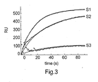
- Fig 3 shows the uptake of sucrose into the liposomes as a function of time, which is obtained by subtracting the change in RU upon addition of sucrose to closed liposomes (S2) from the change in RU upon addition of S3-S5.
- the amplitude of the uptake decrease from S3 to S5, which is explained by a decreasing number of liposomes having pores.
- the rate also decreases from S3 to S5, which is explained by decreasing number of pores per liposome. From the graph in Fig 3 the decay time for uptake of sucrose can be estimated and ⁇ 3 ⁇ 20 sec.
- the Mw of the liposome is 35800 kDa, which gives 0.19 RU/kDa.
- the efficiency of the SPR-based method and its compatibility with screening of multiple permeation events was evaluated by investigating the permeability of the non-electrolytes glycerol, urea and hydroxyurea, which are all biologically relevant molecules.
- glycerol is transported across the membranes of the adipocytes, where fat molecules are stored, has been suggested to be an important factor in the development of obesity and type II diabetes [17].
- Urea is a waste product in the metabolic process and is transported out from liver cells and released from the body via urine, while hydroxyurea is a drug that has been widely used in cancer chemotherapy [18] as well as in treatment of HIV-virus infections [19].
- the applicability of the method is demonstrated for in situ alteration and simultaneous monitoring of liposome permeability by monitoring the increase in glycerol permeation upon a gradual cyclodextrin-induced reduction in the liposomal cholesterol content. Also shown is the compatibility of the method to probe transport of ions.
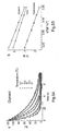
- FIG. 4a shows binding of 70 nm liposomes followed by repeated injection pulses of glycerol, urea, hydroxyurea at different concentrations (see inset).
- Figure 4b shows a magnification of one of the rinsing steps, upon which the solute (glycerol) is released from the liposome interior.
- Figure 4b dashed curve
- Figure 4b dashed curve
- the rate of glycerol release was examined in a temperature interval from 8 to 22 °C.
- Example 7 70 nm liposomes containing 40% cholesterol were provided in accordance with Example 1. Binding of the liposomes followed by repeated injection cyclodextrin, which removes cholesterol from the lipid bilyaer of the liposomes is shown in Figure 6a .
- the SPR response is proportional to the interfacial refractive index, which makes it possible to quantify the removal of cholesterol. After seven injections of cyclodextrin, the cholesterol content was reduced to 14%.
- Figure 6c shows the variations in permeability (estimated from the rate of solute transfer and the vesicle area) and the amplitude of the response (which is proportional to the internal volume).
- the permeability decreases linearly versus cholesterol content, while the internalized volume increases non-linearly.
- the latter observation reflects a structural change of the membrane at a cholesterol content of around 25%.
- the method according to the invention enables screening of multiple permeation events in a single measurement, and allows for in situ alterations in permeability to be quantified, which is exemplified by successive removal of cholesterol from the lipid bilayer.
- the method improves the sensitivity and reduces the required amount of sample by orders of magnitude.
- the present invention admits the possibility of using this sensing principle for direct measurements of molecular transport of non-electrolyte solutes across membranes with a time resolution of 100 ms.
- the method is unique in the sense that it enables direct, rather than indirect (c.f. the DLS and fluorescence quenching methods), measurements of solute transfer across lipid bilayers, which was so far possible only for charged molecules using patch-clamp [14] or chip-based alternatives [15].
- it also offers all other advantages of surface-based methods, such as rapid sequential (or parallel) screening of multiple solutes as well as in situ perturbation of liposome permeability by addition of effector molecules.
Landscapes
- Health & Medical Sciences (AREA)
- Life Sciences & Earth Sciences (AREA)
- Immunology (AREA)
- Engineering & Computer Science (AREA)
- Molecular Biology (AREA)
- Chemical & Material Sciences (AREA)
- Urology & Nephrology (AREA)
- Hematology (AREA)
- Biomedical Technology (AREA)
- Physics & Mathematics (AREA)
- Pathology (AREA)
- General Physics & Mathematics (AREA)
- General Health & Medical Sciences (AREA)
- Biochemistry (AREA)
- Analytical Chemistry (AREA)
- Cell Biology (AREA)
- Microbiology (AREA)
- Medicinal Chemistry (AREA)
- Food Science & Technology (AREA)
- Biotechnology (AREA)
- Proteomics, Peptides & Aminoacids (AREA)
- Biophysics (AREA)
- Endocrinology (AREA)
- Investigating Or Analysing Materials By Optical Means (AREA)
- Medicinal Preparation (AREA)
- Investigating Or Analysing Biological Materials (AREA)
Claims (5)
- Verfahren zum Studieren des Transports eines Wirkstoffes durch eine Membran, das die folgenden Schritte umfasst:a. Bereitstellen mindestens einer Oberfläche mit einer doppelschichtigen Struktur, die an die Oberfläche gebunden ist, wobei die doppelschichtige Struktur ein Nachweisvolumen umgibt,b. in Kontakt bringen der Doppelschicht mit mindestens einem zu analysierenden Wirkstoff undc. Nachweisen einer Veränderung des Brechungsindex des von der doppelschichtigen Struktur umgebenen Nachweisvolumens, die aus dem Transport des Wirkstoffes durch die Membran resultiert, welches das Nachweisen einer Veränderung des Brechungsindex mit mindestens einem Sensor umfasst, der ausgewählt wird aus der Gruppe bestehend aus einem Oberflächen-Plasmon-Resonanz Sensor, einem Ellipsometrie Sensor und einem optischen Waveguide-Laserspektroskopie Sensor.
- Verfahren nach Anspruch 1, wobei mindestens eine Einheit ausgewählt aus der Gruppe bestehend aus einem Membranprotein und einem Ionophor mit der doppelschichtigen Struktur verbunden ist.
- Verfahren nach einem der Ansprüche 1-3, wobei die doppelschichtige Struktur mindestens ein Liposom umfasst.
- Verfahren nach Anspruch 3, wobei mindestens ein Liposom an die Oberfläche durch ein Spacer-Molekül gebunden ist.
- Verfahren nach einem der Ansprüche 1-4, wobei die doppelschichtige Struktur mindestens zwei Schichten von Liposomen umfasst.
Applications Claiming Priority (2)
| Application Number | Priority Date | Filing Date | Title |
|---|---|---|---|
| US2735208P | 2008-02-08 | 2008-02-08 | |
| PCT/SE2009/050123 WO2009099391A1 (en) | 2008-02-08 | 2009-02-06 | Method and device for studying transport of an agent across a bilayer membrane in bioanalytical sensor applications |
Publications (3)
| Publication Number | Publication Date |
|---|---|
| EP2250502A1 EP2250502A1 (de) | 2010-11-17 |
| EP2250502A4 EP2250502A4 (de) | 2011-04-27 |
| EP2250502B1 true EP2250502B1 (de) | 2014-05-07 |
Family
ID=40952359
Family Applications (1)
| Application Number | Title | Priority Date | Filing Date |
|---|---|---|---|
| EP09709128.4A Not-in-force EP2250502B1 (de) | 2008-02-08 | 2009-02-06 | Verfahren zur untersuchung des transports eines agens durch eine doppelschichtige membran bei bioanalysesensor-anwendungen |
Country Status (5)
| Country | Link |
|---|---|
| US (3) | US20110008902A1 (de) |
| EP (1) | EP2250502B1 (de) |
| JP (1) | JP5302341B2 (de) |
| CN (1) | CN101939647B (de) |
| WO (1) | WO2009099391A1 (de) |
Families Citing this family (2)
| Publication number | Priority date | Publication date | Assignee | Title |
|---|---|---|---|---|
| US20110008902A1 (en) | 2008-02-08 | 2011-01-13 | Layeriab Aktiebolag | Method and device for studying transport of an agent across a bilayer membrane in bioanalytical sensor applications |
| JP7179330B2 (ja) * | 2019-01-28 | 2022-11-29 | 国立大学法人福井大学 | 脂質二重膜の水透過性の評価システム、脂質二重膜の水透過性の評価方法、および、脂質二重膜の水透過性を制御する薬剤のスクリーニング方法 |
Family Cites Families (3)
| Publication number | Priority date | Publication date | Assignee | Title |
|---|---|---|---|---|
| SE0301038D0 (sv) * | 2003-04-07 | 2003-04-07 | Pro Forma Xi Ab | Surface immobilised multilayer structure of vesicles |
| WO2004090165A1 (en) * | 2003-04-07 | 2004-10-21 | Layerlab Aktiebolag | Surface immobilised multilayer structure of vesicles |
| US20110008902A1 (en) | 2008-02-08 | 2011-01-13 | Layeriab Aktiebolag | Method and device for studying transport of an agent across a bilayer membrane in bioanalytical sensor applications |
-
2009
- 2009-02-06 US US12/863,499 patent/US20110008902A1/en not_active Abandoned
- 2009-02-06 CN CN200980104408.0A patent/CN101939647B/zh not_active Expired - Fee Related
- 2009-02-06 JP JP2010545831A patent/JP5302341B2/ja not_active Expired - Fee Related
- 2009-02-06 WO PCT/SE2009/050123 patent/WO2009099391A1/en not_active Ceased
- 2009-02-06 EP EP09709128.4A patent/EP2250502B1/de not_active Not-in-force
-
2014
- 2014-04-22 US US14/258,932 patent/US9322830B2/en active Active
-
2016
- 2016-04-18 US US15/131,956 patent/US9784746B2/en not_active Expired - Fee Related
Also Published As
| Publication number | Publication date |
|---|---|
| US20110008902A1 (en) | 2011-01-13 |
| WO2009099391A1 (en) | 2009-08-13 |
| EP2250502A4 (de) | 2011-04-27 |
| US9322830B2 (en) | 2016-04-26 |
| EP2250502A1 (de) | 2010-11-17 |
| US20160231326A1 (en) | 2016-08-11 |
| JP5302341B2 (ja) | 2013-10-02 |
| US20150031139A1 (en) | 2015-01-29 |
| JP2011512526A (ja) | 2011-04-21 |
| CN101939647B (zh) | 2015-04-15 |
| CN101939647A (zh) | 2011-01-05 |
| US9784746B2 (en) | 2017-10-10 |
Similar Documents
| Publication | Publication Date | Title |
|---|---|---|
| US6297059B1 (en) | Triggered optical biosensor | |
| Stahelin | Surface plasmon resonance: a useful technique for cell biologists to characterize biomolecular interactions | |
| CN1902497B (zh) | 分析芯片以及所述分析芯片用于测定分子结构和功能的用途 | |
| Noureen et al. | An electrochemical PAH-modified aptasensor for the label-free and highly-sensitive detection of saxitoxin | |
| Jelinek et al. | Biomolecular sensing with colorimetric vesicles | |
| US6790632B2 (en) | Membrane receptor reagent and assay | |
| Fisher et al. | Structure and activity of lipid membrane biosensor surfaces studied with atomic force microscopy and a resonant mirror | |
| Mao et al. | Simultaneous detection of nucleic acid and protein using gold nanoparticles and lateral flow device | |
| Brändén et al. | Refractive-index-based screening of membrane-protein-mediated transfer across biological membranes | |
| JP4925820B2 (ja) | 表面に固定された小胞の多層構造 | |
| US9784746B2 (en) | Method for studying transport of an agent across a bilayer membrane in bioanalytical sensor applications | |
| Lemmer et al. | Detection of antimycolic acid antibodies by liposomal biosensors | |
| Benítez et al. | Nanovesicle trapping for studying weak protein interactions by single-molecule FRET | |
| Hinman et al. | Bioinspired assemblies and plasmonic interfaces for electrochemical biosensing | |
| Cannon et al. | Cholesterol modulated antibody binding in supported lipid membranes as determined by total internal reflectance microscopy on a microfabricated high-throughput glass chip | |
| Kröger et al. | Surface investigations on the development of a direct optical immunosensor | |
| Liu et al. | Vesicle adhesion visualized with total internal reflection imaging ellipsometry biosensor | |
| Szekacs et al. | Optical waveguide lightmode spectroscopic techniques for investigating membrane-bound ion channel activities | |
| Meyer et al. | Macromolecular High‐Affinity Binding Probed by Advanced Fluorescence Techniques | |
| Kelly et al. | Nanofabrication for the Analysis and Manipulation of Membranes | |
| JP4583356B2 (ja) | 結合素子、バイオセンサ及び生体物質間の相互作用測定方法 | |
| Minunni et al. | SPR in drug discovery: searching bioactive compounds in plant extracts | |
| Goli-Malekabadi et al. | Biosensors for drug detection | |
| ES3042384T3 (en) | Method for immunosensing on a lipid layer using magnetic tunnel junctions | |
| EP4722695A1 (de) | Plasmonisches wellplattensubstrat für spektroskopische analyse und herstellungsverfahren dafür |
Legal Events
| Date | Code | Title | Description |
|---|---|---|---|
| PUAI | Public reference made under article 153(3) epc to a published international application that has entered the european phase |
Free format text: ORIGINAL CODE: 0009012 |
|
| 17P | Request for examination filed |
Effective date: 20100907 |
|
| AK | Designated contracting states |
Kind code of ref document: A1 Designated state(s): AT BE BG CH CY CZ DE DK EE ES FI FR GB GR HR HU IE IS IT LI LT LU LV MC MK MT NL NO PL PT RO SE SI SK TR |
|
| AX | Request for extension of the european patent |
Extension state: AL BA RS |
|
| A4 | Supplementary search report drawn up and despatched |
Effective date: 20110329 |
|
| DAX | Request for extension of the european patent (deleted) | ||
| 17Q | First examination report despatched |
Effective date: 20120214 |
|
| 19U | Interruption of proceedings before grant |
Effective date: 20120405 |
|
| 19W | Proceedings resumed before grant after interruption of proceedings |
Effective date: 20130201 |
|
| RAP1 | Party data changed (applicant data changed or rights of an application transferred) |
Owner name: BIO-RAD LABORATORIES, INC. |
|
| REG | Reference to a national code |
Ref country code: DE Ref legal event code: R079 Ref document number: 602009023855 Country of ref document: DE Free format text: PREVIOUS MAIN CLASS: G01N0033543000 Ipc: G01N0033500000 |
|
| RIC1 | Information provided on ipc code assigned before grant |
Ipc: G01N 33/50 20060101AFI20130916BHEP Ipc: G01N 33/68 20060101ALI20130916BHEP |
|
| GRAP | Despatch of communication of intention to grant a patent |
Free format text: ORIGINAL CODE: EPIDOSNIGR1 |
|
| INTG | Intention to grant announced |
Effective date: 20131128 |
|
| GRAS | Grant fee paid |
Free format text: ORIGINAL CODE: EPIDOSNIGR3 |
|
| GRAA | (expected) grant |
Free format text: ORIGINAL CODE: 0009210 |
|
| AK | Designated contracting states |
Kind code of ref document: B1 Designated state(s): AT BE BG CH CY CZ DE DK EE ES FI FR GB GR HR HU IE IS IT LI LT LU LV MC MK MT NL NO PL PT RO SE SI SK TR |
|
| REG | Reference to a national code |
Ref country code: GB Ref legal event code: FG4D |
|
| REG | Reference to a national code |
Ref country code: AT Ref legal event code: REF Ref document number: 667064 Country of ref document: AT Kind code of ref document: T Effective date: 20140515 |
|
| REG | Reference to a national code |
Ref country code: IE Ref legal event code: FG4D |
|
| REG | Reference to a national code |
Ref country code: DE Ref legal event code: R096 Ref document number: 602009023855 Country of ref document: DE Effective date: 20140618 |
|
| REG | Reference to a national code |
Ref country code: AT Ref legal event code: MK05 Ref document number: 667064 Country of ref document: AT Kind code of ref document: T Effective date: 20140507 |
|
| REG | Reference to a national code |
Ref country code: NL Ref legal event code: VDEP Effective date: 20140507 |
|
| REG | Reference to a national code |
Ref country code: LT Ref legal event code: MG4D |
|
| PG25 | Lapsed in a contracting state [announced via postgrant information from national office to epo] |
Ref country code: GR Free format text: LAPSE BECAUSE OF FAILURE TO SUBMIT A TRANSLATION OF THE DESCRIPTION OR TO PAY THE FEE WITHIN THE PRESCRIBED TIME-LIMIT Effective date: 20140808 Ref country code: IS Free format text: LAPSE BECAUSE OF FAILURE TO SUBMIT A TRANSLATION OF THE DESCRIPTION OR TO PAY THE FEE WITHIN THE PRESCRIBED TIME-LIMIT Effective date: 20140907 Ref country code: FI Free format text: LAPSE BECAUSE OF FAILURE TO SUBMIT A TRANSLATION OF THE DESCRIPTION OR TO PAY THE FEE WITHIN THE PRESCRIBED TIME-LIMIT Effective date: 20140507 Ref country code: NO Free format text: LAPSE BECAUSE OF FAILURE TO SUBMIT A TRANSLATION OF THE DESCRIPTION OR TO PAY THE FEE WITHIN THE PRESCRIBED TIME-LIMIT Effective date: 20140807 Ref country code: LT Free format text: LAPSE BECAUSE OF FAILURE TO SUBMIT A TRANSLATION OF THE DESCRIPTION OR TO PAY THE FEE WITHIN THE PRESCRIBED TIME-LIMIT Effective date: 20140507 Ref country code: CY Free format text: LAPSE BECAUSE OF FAILURE TO SUBMIT A TRANSLATION OF THE DESCRIPTION OR TO PAY THE FEE WITHIN THE PRESCRIBED TIME-LIMIT Effective date: 20140507 |
|
| PG25 | Lapsed in a contracting state [announced via postgrant information from national office to epo] |
Ref country code: AT Free format text: LAPSE BECAUSE OF FAILURE TO SUBMIT A TRANSLATION OF THE DESCRIPTION OR TO PAY THE FEE WITHIN THE PRESCRIBED TIME-LIMIT Effective date: 20140507 Ref country code: LV Free format text: LAPSE BECAUSE OF FAILURE TO SUBMIT A TRANSLATION OF THE DESCRIPTION OR TO PAY THE FEE WITHIN THE PRESCRIBED TIME-LIMIT Effective date: 20140507 Ref country code: SE Free format text: LAPSE BECAUSE OF FAILURE TO SUBMIT A TRANSLATION OF THE DESCRIPTION OR TO PAY THE FEE WITHIN THE PRESCRIBED TIME-LIMIT Effective date: 20140507 Ref country code: ES Free format text: LAPSE BECAUSE OF FAILURE TO SUBMIT A TRANSLATION OF THE DESCRIPTION OR TO PAY THE FEE WITHIN THE PRESCRIBED TIME-LIMIT Effective date: 20140507 Ref country code: HR Free format text: LAPSE BECAUSE OF FAILURE TO SUBMIT A TRANSLATION OF THE DESCRIPTION OR TO PAY THE FEE WITHIN THE PRESCRIBED TIME-LIMIT Effective date: 20140507 Ref country code: PL Free format text: LAPSE BECAUSE OF FAILURE TO SUBMIT A TRANSLATION OF THE DESCRIPTION OR TO PAY THE FEE WITHIN THE PRESCRIBED TIME-LIMIT Effective date: 20140507 |
|
| PG25 | Lapsed in a contracting state [announced via postgrant information from national office to epo] |
Ref country code: PT Free format text: LAPSE BECAUSE OF FAILURE TO SUBMIT A TRANSLATION OF THE DESCRIPTION OR TO PAY THE FEE WITHIN THE PRESCRIBED TIME-LIMIT Effective date: 20140908 |
|
| PG25 | Lapsed in a contracting state [announced via postgrant information from national office to epo] |
Ref country code: CZ Free format text: LAPSE BECAUSE OF FAILURE TO SUBMIT A TRANSLATION OF THE DESCRIPTION OR TO PAY THE FEE WITHIN THE PRESCRIBED TIME-LIMIT Effective date: 20140507 Ref country code: BE Free format text: LAPSE BECAUSE OF FAILURE TO SUBMIT A TRANSLATION OF THE DESCRIPTION OR TO PAY THE FEE WITHIN THE PRESCRIBED TIME-LIMIT Effective date: 20140507 Ref country code: DK Free format text: LAPSE BECAUSE OF FAILURE TO SUBMIT A TRANSLATION OF THE DESCRIPTION OR TO PAY THE FEE WITHIN THE PRESCRIBED TIME-LIMIT Effective date: 20140507 Ref country code: SK Free format text: LAPSE BECAUSE OF FAILURE TO SUBMIT A TRANSLATION OF THE DESCRIPTION OR TO PAY THE FEE WITHIN THE PRESCRIBED TIME-LIMIT Effective date: 20140507 Ref country code: RO Free format text: LAPSE BECAUSE OF FAILURE TO SUBMIT A TRANSLATION OF THE DESCRIPTION OR TO PAY THE FEE WITHIN THE PRESCRIBED TIME-LIMIT Effective date: 20140507 Ref country code: EE Free format text: LAPSE BECAUSE OF FAILURE TO SUBMIT A TRANSLATION OF THE DESCRIPTION OR TO PAY THE FEE WITHIN THE PRESCRIBED TIME-LIMIT Effective date: 20140507 |
|
| REG | Reference to a national code |
Ref country code: DE Ref legal event code: R097 Ref document number: 602009023855 Country of ref document: DE |
|
| PG25 | Lapsed in a contracting state [announced via postgrant information from national office to epo] |
Ref country code: NL Free format text: LAPSE BECAUSE OF FAILURE TO SUBMIT A TRANSLATION OF THE DESCRIPTION OR TO PAY THE FEE WITHIN THE PRESCRIBED TIME-LIMIT Effective date: 20140507 |
|
| PLBE | No opposition filed within time limit |
Free format text: ORIGINAL CODE: 0009261 |
|
| STAA | Information on the status of an ep patent application or granted ep patent |
Free format text: STATUS: NO OPPOSITION FILED WITHIN TIME LIMIT |
|
| 26N | No opposition filed |
Effective date: 20150210 |
|
| PG25 | Lapsed in a contracting state [announced via postgrant information from national office to epo] |
Ref country code: IT Free format text: LAPSE BECAUSE OF FAILURE TO SUBMIT A TRANSLATION OF THE DESCRIPTION OR TO PAY THE FEE WITHIN THE PRESCRIBED TIME-LIMIT Effective date: 20140507 |
|
| REG | Reference to a national code |
Ref country code: DE Ref legal event code: R097 Ref document number: 602009023855 Country of ref document: DE Effective date: 20150210 |
|
| PG25 | Lapsed in a contracting state [announced via postgrant information from national office to epo] |
Ref country code: SI Free format text: LAPSE BECAUSE OF FAILURE TO SUBMIT A TRANSLATION OF THE DESCRIPTION OR TO PAY THE FEE WITHIN THE PRESCRIBED TIME-LIMIT Effective date: 20140507 |
|
| PG25 | Lapsed in a contracting state [announced via postgrant information from national office to epo] |
Ref country code: LU Free format text: LAPSE BECAUSE OF FAILURE TO SUBMIT A TRANSLATION OF THE DESCRIPTION OR TO PAY THE FEE WITHIN THE PRESCRIBED TIME-LIMIT Effective date: 20150206 |
|
| REG | Reference to a national code |
Ref country code: CH Ref legal event code: PL |
|
| PG25 | Lapsed in a contracting state [announced via postgrant information from national office to epo] |
Ref country code: LI Free format text: LAPSE BECAUSE OF NON-PAYMENT OF DUE FEES Effective date: 20150228 Ref country code: CH Free format text: LAPSE BECAUSE OF NON-PAYMENT OF DUE FEES Effective date: 20150228 Ref country code: MC Free format text: LAPSE BECAUSE OF FAILURE TO SUBMIT A TRANSLATION OF THE DESCRIPTION OR TO PAY THE FEE WITHIN THE PRESCRIBED TIME-LIMIT Effective date: 20140507 |
|
| REG | Reference to a national code |
Ref country code: IE Ref legal event code: MM4A |
|
| PG25 | Lapsed in a contracting state [announced via postgrant information from national office to epo] |
Ref country code: IE Free format text: LAPSE BECAUSE OF NON-PAYMENT OF DUE FEES Effective date: 20150206 |
|
| REG | Reference to a national code |
Ref country code: FR Ref legal event code: PLFP Year of fee payment: 8 |
|
| PG25 | Lapsed in a contracting state [announced via postgrant information from national office to epo] |
Ref country code: MT Free format text: LAPSE BECAUSE OF FAILURE TO SUBMIT A TRANSLATION OF THE DESCRIPTION OR TO PAY THE FEE WITHIN THE PRESCRIBED TIME-LIMIT Effective date: 20140507 |
|
| REG | Reference to a national code |
Ref country code: FR Ref legal event code: PLFP Year of fee payment: 9 |
|
| PG25 | Lapsed in a contracting state [announced via postgrant information from national office to epo] |
Ref country code: BG Free format text: LAPSE BECAUSE OF FAILURE TO SUBMIT A TRANSLATION OF THE DESCRIPTION OR TO PAY THE FEE WITHIN THE PRESCRIBED TIME-LIMIT Effective date: 20140507 Ref country code: HU Free format text: LAPSE BECAUSE OF FAILURE TO SUBMIT A TRANSLATION OF THE DESCRIPTION OR TO PAY THE FEE WITHIN THE PRESCRIBED TIME-LIMIT; INVALID AB INITIO Effective date: 20090206 |
|
| PG25 | Lapsed in a contracting state [announced via postgrant information from national office to epo] |
Ref country code: TR Free format text: LAPSE BECAUSE OF FAILURE TO SUBMIT A TRANSLATION OF THE DESCRIPTION OR TO PAY THE FEE WITHIN THE PRESCRIBED TIME-LIMIT Effective date: 20140507 |
|
| REG | Reference to a national code |
Ref country code: FR Ref legal event code: PLFP Year of fee payment: 10 |
|
| PG25 | Lapsed in a contracting state [announced via postgrant information from national office to epo] |
Ref country code: MK Free format text: LAPSE BECAUSE OF FAILURE TO SUBMIT A TRANSLATION OF THE DESCRIPTION OR TO PAY THE FEE WITHIN THE PRESCRIBED TIME-LIMIT Effective date: 20140507 |
|
| PGFP | Annual fee paid to national office [announced via postgrant information from national office to epo] |
Ref country code: FR Payment date: 20230223 Year of fee payment: 15 |
|
| PGFP | Annual fee paid to national office [announced via postgrant information from national office to epo] |
Ref country code: GB Payment date: 20230214 Year of fee payment: 15 Ref country code: DE Payment date: 20230227 Year of fee payment: 15 |
|
| REG | Reference to a national code |
Ref country code: DE Ref legal event code: R119 Ref document number: 602009023855 Country of ref document: DE |
|
| GBPC | Gb: european patent ceased through non-payment of renewal fee |
Effective date: 20240206 |
|
| PG25 | Lapsed in a contracting state [announced via postgrant information from national office to epo] |
Ref country code: DE Free format text: LAPSE BECAUSE OF NON-PAYMENT OF DUE FEES Effective date: 20240903 |
|
| PG25 | Lapsed in a contracting state [announced via postgrant information from national office to epo] |
Ref country code: GB Free format text: LAPSE BECAUSE OF NON-PAYMENT OF DUE FEES Effective date: 20240206 |
|
| PG25 | Lapsed in a contracting state [announced via postgrant information from national office to epo] |
Ref country code: FR Free format text: LAPSE BECAUSE OF NON-PAYMENT OF DUE FEES Effective date: 20240229 |
|
| PG25 | Lapsed in a contracting state [announced via postgrant information from national office to epo] |
Ref country code: GB Free format text: LAPSE BECAUSE OF NON-PAYMENT OF DUE FEES Effective date: 20240206 Ref country code: FR Free format text: LAPSE BECAUSE OF NON-PAYMENT OF DUE FEES Effective date: 20240229 Ref country code: DE Free format text: LAPSE BECAUSE OF NON-PAYMENT OF DUE FEES Effective date: 20240903 |