EP2228654A2 - Diagnostic et traitement de l'invasion des cellules cancéreuses - Google Patents
Diagnostic et traitement de l'invasion des cellules cancéreuses Download PDFInfo
- Publication number
- EP2228654A2 EP2228654A2 EP10157827A EP10157827A EP2228654A2 EP 2228654 A2 EP2228654 A2 EP 2228654A2 EP 10157827 A EP10157827 A EP 10157827A EP 10157827 A EP10157827 A EP 10157827A EP 2228654 A2 EP2228654 A2 EP 2228654A2
- Authority
- EP
- European Patent Office
- Prior art keywords
- axl
- gene
- expression
- protein
- cells
- Prior art date
- Legal status (The legal status is an assumption and is not a legal conclusion. Google has not performed a legal analysis and makes no representation as to the accuracy of the status listed.)
- Withdrawn
Links
Images


Classifications
-
- C—CHEMISTRY; METALLURGY
- C07—ORGANIC CHEMISTRY
- C07K—PEPTIDES
- C07K16/00—Immunoglobulins [IGs], e.g. monoclonal or polyclonal antibodies
- C07K16/18—Immunoglobulins [IGs], e.g. monoclonal or polyclonal antibodies against material from animals or humans
- C07K16/28—Immunoglobulins [IGs], e.g. monoclonal or polyclonal antibodies against material from animals or humans against receptors, cell surface antigens or cell surface determinants
- C07K16/2863—Immunoglobulins [IGs], e.g. monoclonal or polyclonal antibodies against material from animals or humans against receptors, cell surface antigens or cell surface determinants against receptors for growth factors, growth regulators
-
- A—HUMAN NECESSITIES
- A61—MEDICAL OR VETERINARY SCIENCE; HYGIENE
- A61K—PREPARATIONS FOR MEDICAL, DENTAL OR TOILETRY PURPOSES
- A61K39/00—Medicinal preparations containing antigens or antibodies
- A61K39/395—Antibodies; Immunoglobulins; Immune serum, e.g. antilymphocytic serum
- A61K39/39533—Antibodies; Immunoglobulins; Immune serum, e.g. antilymphocytic serum against materials from animals
- A61K39/3955—Antibodies; Immunoglobulins; Immune serum, e.g. antilymphocytic serum against materials from animals against proteinaceous materials, e.g. enzymes, hormones, lymphokines
-
- A—HUMAN NECESSITIES
- A61—MEDICAL OR VETERINARY SCIENCE; HYGIENE
- A61P—SPECIFIC THERAPEUTIC ACTIVITY OF CHEMICAL COMPOUNDS OR MEDICINAL PREPARATIONS
- A61P35/00—Antineoplastic agents
-
- A—HUMAN NECESSITIES
- A61—MEDICAL OR VETERINARY SCIENCE; HYGIENE
- A61P—SPECIFIC THERAPEUTIC ACTIVITY OF CHEMICAL COMPOUNDS OR MEDICINAL PREPARATIONS
- A61P35/00—Antineoplastic agents
- A61P35/04—Antineoplastic agents specific for metastasis
-
- A—HUMAN NECESSITIES
- A61—MEDICAL OR VETERINARY SCIENCE; HYGIENE
- A61P—SPECIFIC THERAPEUTIC ACTIVITY OF CHEMICAL COMPOUNDS OR MEDICINAL PREPARATIONS
- A61P43/00—Drugs for specific purposes, not provided for in groups A61P1/00-A61P41/00
-
- C—CHEMISTRY; METALLURGY
- C12—BIOCHEMISTRY; BEER; SPIRITS; WINE; VINEGAR; MICROBIOLOGY; ENZYMOLOGY; MUTATION OR GENETIC ENGINEERING
- C12Q—MEASURING OR TESTING PROCESSES INVOLVING ENZYMES, NUCLEIC ACIDS OR MICROORGANISMS; COMPOSITIONS OR TEST PAPERS THEREFOR; PROCESSES OF PREPARING SUCH COMPOSITIONS; CONDITION-RESPONSIVE CONTROL IN MICROBIOLOGICAL OR ENZYMOLOGICAL PROCESSES
- C12Q1/00—Measuring or testing processes involving enzymes, nucleic acids or microorganisms; Compositions therefor; Processes of preparing such compositions
- C12Q1/68—Measuring or testing processes involving enzymes, nucleic acids or microorganisms; Compositions therefor; Processes of preparing such compositions involving nucleic acids
- C12Q1/6876—Nucleic acid products used in the analysis of nucleic acids, e.g. primers or probes
- C12Q1/6883—Nucleic acid products used in the analysis of nucleic acids, e.g. primers or probes for diseases caused by alterations of genetic material
- C12Q1/6886—Nucleic acid products used in the analysis of nucleic acids, e.g. primers or probes for diseases caused by alterations of genetic material for cancer
-
- A—HUMAN NECESSITIES
- A61—MEDICAL OR VETERINARY SCIENCE; HYGIENE
- A61K—PREPARATIONS FOR MEDICAL, DENTAL OR TOILETRY PURPOSES
- A61K39/00—Medicinal preparations containing antigens or antibodies
- A61K2039/505—Medicinal preparations containing antigens or antibodies comprising antibodies
-
- C—CHEMISTRY; METALLURGY
- C12—BIOCHEMISTRY; BEER; SPIRITS; WINE; VINEGAR; MICROBIOLOGY; ENZYMOLOGY; MUTATION OR GENETIC ENGINEERING
- C12Q—MEASURING OR TESTING PROCESSES INVOLVING ENZYMES, NUCLEIC ACIDS OR MICROORGANISMS; COMPOSITIONS OR TEST PAPERS THEREFOR; PROCESSES OF PREPARING SUCH COMPOSITIONS; CONDITION-RESPONSIVE CONTROL IN MICROBIOLOGICAL OR ENZYMOLOGICAL PROCESSES
- C12Q2600/00—Oligonucleotides characterized by their use
- C12Q2600/136—Screening for pharmacological compounds
Definitions
- the present invention relates to diagnostic and therapeutic methods in the field of malignant disorders. More particularly, the invention provides methods of determining the invasivity of malignant disorders and methods for reducing the invasivity of malignant disorders including the prevention or treatment of cancer cell invasion.
- RTK receptor tyrosine kinases
- One purpose of the present study was to establish expression profiles of genes particularly selected from protein kinases, phosphatases and other signalling genes in malignant disorders, particularly breast cancer and brain cancer in order to identify novel markers for invasivity and/or aggressiveness.
- a cDNA hybridization array was used to analyze gene expression profiles of seven highly invasive, fourteen weakly invasive breast cancer cell lines and three normal breast epithelial cell lines. Differences in gene expression between weakly and highly invasive breast cancer cell lines were identified, which enable the definition of a gene cluster correlating with the invasivity of a breast cancer cell line. By using this cluster or combinations of genes therefrom, a discrimination of highly invasive breast cancer cell lines from weakly invasive breast cancer cell lines and normal breast epithelial cell lines is possible.
- RTK receptor tyrosine kinases
- UFO/AXL is the first RTK to be implemented in mediating the diffuse-infiltrative, local metastatic growth of malignant brain tumors.
- a first aspect of the present invention relates to a method of determining the invasivity of malignant disorders comprising determining the expression of at least one gene selected from the group consisting of AXL (Genbank M 76125), GAS 6 (Genbank L 13720), MMP14 (Genbank NM 004995), ADAM12 (Genbank AF 023476), ADAM17 (Genbank U 69611), MT3MMP (Genbank NM 005961), FGF2 (Genbank NM 002006), FGF5 (Genbank NM 004464), FYN (Genbank M 14333), LYN (Genbank M 16038), DDR2 (Genbank X 74764), TIMP1 (Genbank NM 003254), HB-EGF (Genbank NM 001945), SGK (Genbank Y 10032), RPS6RB1 (Genbank M 60724), MAP4K4 (Genbank XM 038748), SIRP ⁇ (Genbank Y 10375) and Annexin
- the expression of the genes Stat 5b (Acc. NM_012448) or EDG2 (Acc. NM_057159) may be determined as indicator for the invasivity of malignant disorders, optionally in addition to determining the expression of one or more of the above genes. It was found that a high expression of at least one of the above genes correlates with a high invasivity.
- AXL AXL-AXL-AXL-AXL-AXL-AXL-AXL-AXL-AXL-AXL-AXL-AXL-AXL-AXL-AXL-AXL-AXL-AXL-AXL-AXL-AXL-AXL-AXL-AXL-AXL-AXL-AXL-AXL and one or more further genes.
- the one or more further genes can be selected from the genes listed above or from a gene which is already known as a marker for invasiveness.
- the method preferably comprises determining the expression of several of the above genes, e.g. determining the expression of at least two, three, four, five, six, seven or eight genes. More preferably, the method comprises determining the expression of at least the AXL/UFO gene (Genbank M 76125).
- the method may comprise determining the expression of at least one further gene which is already known as a marker of invasiveness, such as CD44 (Genbank X 66733), vimentin (Genbank X 56134), CAV1 (Genbank Z 18951), CAV2 (Genbank AF 03572), MMP 1 (Genbank M 13509), MMP 2 (Genbank NM 004530), MMP9 (Genbank NM 004994), M-CSF (Genbank M 37435) and EPHA2 (Genbank M 59371).
- a marker of invasiveness such as CD44 (Genbank X 66733), vimentin (Genbank X 56134), CAV1 (Genbank Z 18951), CAV2 (Genbank AF 03572), MMP 1 (Genbank M 13509), MMP 2 (Genbank NM 004530), MMP9 (Genbank NM 004994), M-CSF (Genbank M 37435) and EPHA2 (Genbank M 59371).
- a correlation between expression of the above gene cluster and particularly the AXL gene and invasivity was found in several types of malignant disorders, e.g. breast cancer, particularly primary breast cancer, prostate cancer, kidney cancer and glioblastomas or other cancers of epithelial origin.
- breast cancer particularly primary breast cancer, prostate cancer, kidney cancer and glioblastomas or other cancers of epithelial origin.
- glioblastomas or other cancers of epithelial origin.
- a dominant negative mutant of the AXL gene is capable of strongly suppressing cell invasiveness and migration indicating that inhibition of AXL function may block and loss of metastasis formation in highly invasive malignant disorders, such as breast cancer or brain cancer, e.g. glioblastoma.
- a polyclonal antibody directed against the extracellular portion of AXL has a very strong inhibitory activity on the migration and invasivity of cancer cells, e.g. breast or prostate cancer cell lines.
- overexpression of wildtype AXL in weakly invasive breast cancer, prostate cancer cell lines and glioma cells significantly increased their invasivity.
- a further aspect of the present invention relates to a method of reducing the invasivity of malignant disorders comprising inhibiting the AXL gene, AXL ligand gene or protein, or ligand thereof.
- the method may comprise (i) inhibiting the receptor tyrosine kinase activity of the AXL protein, (ii) inhibiting the expression of the AXL gene, (iii) inhibiting the interaction between the AXL protein and its ligands, particulary GAS6 and/or (iv) inhibiting the interaction of AXL with downstream signal transducing factors.
- laminin G-like domains of GAS6 (GAS6-LG) in particular have been found to be involved in the interaction with the AXL protein, such as AXL binding and activation (Reference 36).
- residues of the GAS-LG2 domain for example Leu 620 , Tyr 660 and Phe 487 , affect AXL binding and/or activation.
- the method of reducing invasivity of malignant disorders comprises the inhibition of one or more residues of the GAS6-LG, in particular Leu 620 , Tyr 660 and/or Phe 487 .
- the present invention relates to the diagnosis or the prevention and/or treatment of malignant disorders, particulary the tumor invasivity and/or metastasis formation in malignant disorders.
- malignant disorders are cancers of the breast, prostate, kidney, colon, lung and glioblastomas. More preferably, the malignant disorder is breast cancer or glioblastomas.
- the expression of invasivity-associated genes is determined qualitatively and/or quantitatively.
- the expression is determined in a sample comprising malignant cells, e.g. from a human tumour patient.
- the sample may be derived from tissue sections, biopsy samples etc. or from body fluids.
- Gene expression in the sample to be tested may be compared with gene expression in control samples, e.g. negative control samples from "normal" cells or weakly invasive malignant cells, and/or from positive controls, e.g. from highly invasive malignant cells.
- Gene expression may be determined according to methods known in the art, e.g. on the mRNA or transcript level and/or on the protein level. Measurement of gene expression on the mRNA level may comprise reverse transcription and/or amplification reactions such as PCR. Preferably, gene expression is measured on a nucleic acid array, wherein nucleic acids from the sample to be tested, e.g. RNA or cDNA, is hybridized to an array of immobilized probes specific for the nucleic acids to be tested. A preferred example of a suitable nucleic acid array is described in PCT/EP 02/01073 . Alternatively, gene expression may be determined by other methods, e.g. Northern blot hybridization.
- Gene expression on the protein level may be determined by immunological methods using antibodies directed against the proteins encoded by invasivity-associated genes.
- the antibodies may be labeled directly or indirectly by known labeling groups such as radioactive, fluorescence, chemiluminescence or enzymatic groups such as known in the art.
- the therapeutic embodiment of the present invention particulary relates to a method comprising the administration of an inhibitor of the AXL gene, AXL ligand gene, AXL protein or ligand thereof in an amount which is effective of reducing the invasivity of malignant disorders to a subject in need thereof.
- the subject is preferably a mammal, more preferably a human being.
- the ligand of the AXL protein is preferably GAS6, in particular residues of GASG-LG, as defined above.
- the inhibitor of the AXL gene , AXL ligand gene, AXL protein or ligand thereof, e.g. GAS6, may be an antibody, a biologically active nucleic acid or a low molecular weight compound, e.g. a peptide or a non-peptidic organic compound.
- the inhibitor is an antibody directed against the AXL protein or a ligand thereof, e.g. GAS6.
- antibody relates to polyclonal antibodies and monoclonal antibodies, particularly to chimeric or humanized monoclonal antibodies or to human antibodies. Further, the term comprises antibody fragments, e.g. proteolytic fragments such as Fab, Fab' or F(ab) 2 fragments or recombinant fragments such as single chain antibody fragments, e.g. scFv fragments. Methods of manufacturing antibodies or antibody fragments as described above are known in the art.
- the inhibitor is a biologically active nucleic acid, e.g. a DNA, an RNA or a synthetic nucleic acid analog.
- biologically active nucleic acids are antisense nucleic acids, ribozymes or RNA interference molecules directed against the AXL gene or an AXL ligand gene or a transcript thereof.
- a further preferred example of a biologically active nucleic acid is a dominant-negative mutant of the AXL gene.
- Biologically active nucleic acids may be delivered by known procedures, e.g. by using viral or non-viral gene transfer vectors.
- the inhibitor is a peptidic compound, e.g. a peptide having a length of from 4 to 25 amino acids, a cyclic peptide, a peptide derivative or a peptide mimetic derived from such a peptide.
- the low-molecular weight inhibitor may be a non-peptidic organic compound, e.g. an inhibitor of AXL kinase activity. Low-molecular weight inhibitors may be obtained by screening suitable compound libraries in a method as described in more detail below.
- Still a further aspect of the present invention relates to a pharmaceutical composition
- a pharmaceutical composition comprising as an active agent an inhibitor of the AXL gene, AXL ligand gene, AXL protein or ligand thereof (e.g. GAS6, in particular residues from GAS6-LG, as defined above) together with pharmacologically active diluents, carriers and/or adjuvants.
- This composition is particularly suitable for reducing the invasivity of malignant disorders and/or reducing the metastasis formation in malignant disorders.
- the pharmaceutical composition may be a liquid, a solid, e.g. a powder, tablet etc., an emulsion or a suspension.
- composition may be administered by injection, orally, topically, rectally, intranasally or by any other suitable means.
- effective amount of the active agent in the composition may be determined by the skilled person without any undue burden depending on the type of compound and the disease to be treated.
- the composition may comprise at least one further active agent.
- This at least one further active agent may be formulated together with the AXL inhibitor in a single composition or in a separate composition which is coadministered with the AXL inhibitor composition.
- the further active agent may be a cytotoxic or cytostatic agent such as doxorubicin, cis-platin, carboplatin, an anti-tumor antibody or any combination thereof.
- Still a further aspect of the invention relates to a method of identifying and/or characterizing an inhibitor of the invasivity of malignant disorders comprising determining, if at least a test compound is capable of inhibiting the AXL gene, AXL ligand gene, AXL protein or ligand thereof (e.g. GAS6 as defined above) or protein. More particularly, the method comprises determining, if a test compound is capable of binding to the AXL protein and/or reducing the AXL gene expression.
- the test compound may be derived from compound libraries, e.g. peptide or non-peptidic libraries which are subjected to a screening for AXL inhibitory activity.
- the screening method may comprise the use of a cell-based assay system, e.g.
- the method may comprise the use of a cell-free assay system, wherein the test compound is contacted with substantially purified AXL protein or a fragment thereof in order to determine binding of the test compound to the protein or fragment thereof.
- RNAs to be tested were prepared from unselected samples. Samples of primary invasive breast carcinomas were collected from 72 patients undergoing surgery. After surgical resection, the tumors were macrodissected: a section was taken for the pathologist's diagnosis and an adjacent piece was quickly frozen in liquid nitrogen for mRNA extractions. The median age of patients at the time diagnosis was 55 years (range 29-81) and most of them were postmenopausal. Tumors were classified according to the WHO histological typing of breast tumors: ductal carcinomas, lobular carcinomas, mixed ductal-lobular carcinomas and medullary carcinomas.
- the sources of the breast cancer cell lines were as follows: BT-20, BT-474, BT-483, BT-459, Du-4475, MDA-MB-134, -157, -175, -361, -436, -453, -468, SK-BR-3, and ZR-75-1, T-47D, MDA-MB-231, ZR-75-30 were obtained from the American Type Culture Collection (ATCC, Rockville, MD). MCF-7, clone and BC cell line DAL were supplied by SUGEN (Redwood City, CA). The HBL-100 cell line was from ATCC. This cell line was derived from normal tissue but contains tandemly integrated SV-40 sequences (9).
- RNA and genomic DNA were isolated from the same cell pellet by lysis in guanidinium isothiocyanate solution (GTS buffer: 4 M guanidinium isothiocyanate, 25 mM sodium citrate pH 7.0, 0.5% Sarkosyl, and 0.1 M ⁇ -mercaptoethanol) followed by phenol-chloroform extractions.
- GTS buffer 4 M guanidinium isothiocyanate, 25 mM sodium citrate pH 7.0, 0.5% Sarkosyl, and 0.1 M ⁇ -mercaptoethanol
- Total RNA was isolated using standard methods (Sambrook et al., [1989]) with modifications. DNA was collected and extracted twice with an equal volume of phenol:chloroform:isoamylalcohol (25:24:1). RNA and DNA were isolated from each cell line on a minimum of 3 independent occasions.
- Total and mRNA integrity and cDNA complexity was controlled by agarose gel electrophoresis and Northern blots using specific probes. Some mRNA extraction was performed using the OligoTex mRNA isolation Kit (Quagen, Biotech, Germany). Cell pellets were resuspended in lysis/binding buffer, vortex-mixed briefly, passed three times through a 21 G needle and applied to a spin lysate column and centrifuged at 13,000g for 3 min. The lysate was then mixed gently with Oligo-dT cellulose (Stratagene Inc.) and applied to a pre-wetted Oligotex molecular biology column (Quagen Biotech). The column was washed three times with lysis/binding buffer and four times with wash buffer before eluting the mRNA with pre-warmed (65C) elution buffer. The quantity of mRNA was measured using the OD260.
- PK and PP gene expression was analyzed by hybridization on nylon filters arrays with radioactive targets (cDNA).
- the arrays contained 645 genes encoding kinases, phosphatases and others signalling proteins: ligands, adaptors, transcription factors, metalloproteinases/ADAMs, apoptosis related genes and 11 house keeping genes (the list is available at http://www.biochem.mpg.de or ullrich@biochem.mpg.de). Their identity was verified by sequencing of plasmid DNA and compared with GenBank sequence information. Identity of PK and PP was conformed for all clones spotted on nylon filters ones, or in duplicate. For normalisation purpose, the GFP gene was spotted two times as well as genomic and vector DNA. Purification of plasmids was done using a plasmid purification kit (Qiagen, Germany).
- Filters were initially pre-washed in 0.5% SDS for 5 min, with agitation.
- 10 ml of the pre-hybridisation solution was included Yeast tRNA.
- Human Cot-1 DNA (BRL/Life technologies) was used in the hybridization step which was performed in a Roller bottle (Hybaid Inc.) for 16 h in a roller oven at 65°C. Labelled probe was denatured for 10 min at 100°C and then placed immediately into the hybridisation mixture which was incubated for a further 18 h at 65°C. After 18 h, the hybridisation mixture was discarded and the array was washed twice in 2 sodium chloride: sodium citrate (SSC) buffer, 0.2% SDS for 20 min at 42°C with continued rotation in the incubator.
- SSC sodium citrate
- a third wash was performed in 0.2xSSC, 0.1 % SDS for 15-60 min at 65°C in a plastic box with horizontal shaking.
- the filter was placed on a piece of moistened Whatman paper and covered with Saran wrap.
- the array was then placed into an imager cassette with a Phosphorimager storage screen (Fuji, Japan) and exposed for 2 days.
- Exposed phospho-imager storage screens were scanned once on a Phosphoimager Scanner (Fuji) at a resolution of 50 microns and were visualised using MacBAS 2000 (Fuji). Images were imported into ArrayVision V(Canada) for analysis by a software protocol. Mapping of individual elements to an internal reference database was achieved by aligning the images onto a software-based matrix using a total of 4 control elements representing total genomic control DNA, GFP, and vector. Normalisation was performed by multiplying the raw intensity for each data element by a normalisation factor equal to the average raw intensity for all the vector elements divided by 100 (this value is the average raw intensity for all elements, derived from a large number of different hybridizations performed by us the development of the arrays).
- the same algorithm is applied to cluster the experimental samples (i.e., cell lines and tumors) according to the similarities in their overall patterns of gene expression.
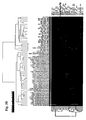
- the data tables, thus ordered, are presented graphically as colored images.
- the genes analyzed are arranged as ordered by the clustering algorithm, so that the genes with the most similar patterns of expression are placed adjacent to each other.
- experimental samples are similarly arranged such that those with the most similar patterns of expression across all genes are placed adjacent to each other.
- the colour of each cell/square in this tabular image represents the measured expression ratio of each gene in question.
- the colour saturation is also directly proportional to the magnitude of the measured gene expression ratio with the brightest red squares having the highest T/N ratio (i.e., >8-fold difference), the brightest green squares having the lowest T/N ratio, black squares indicating a ratio of approximately 1, and grey squares indicating insufficient data quality.
- the chemoinvasion assay was carried out using a modification of the method of Albini et al. (10). After trypsinization, cells (20.000) were plated on Matrigel-coated (150 ⁇ l of 4.0 mg/ml) 8- ⁇ m polypropylene filter inserts in Boyden chambers (Biocoat Matrigel Invasion Chamber, Becton Dickinson, Bedford, MA or Nunc 10mm tissue culture inserts, Naperville, IL). The bottom chamber contained 0.55 ml of NIH3T3-conditioned media, produced as described by Albini et al. or normal growth media for some cell lines.
- BC cell lines obtained from the ATCC were trypsinized, centrifuged, and resuspended at 4 x 10 5 cells/ml in RPMI medium containing 10% FBS. The remaining cell lines were resuspended in their regular growth medium.
- the cells remaining in the insert were removed with a cotton swab, and the cells on the bottom of the filter were counted using different protocols: fixed in Diff-quick (American Scientific Products, McGraw Park, IL) and treated with RNase A (at 50 ⁇ g/ml for 20 min at 37°C) before staining with propidium iodide (10 ⁇ g/ml in PBS) for 1 min at room temperature (RT).
- the dried filters were removed and mounted on slides with Cytoseal 60 mounting media (Stephens Scientific, Kalamazoo, MI). Individual propidium iodide-stained nuclei on the filters were counted. Most results were obtained using trypsinization and counting of the cells. Triplicate samples were counted in each experiment. Outlying values were eliminated from calculations of average invasive activity.
- wounds were made on confluent cell monolayers with a plastic tip.
- MDA-MB-345S-mock and MDA-MB-435-dnAXL clone 2 cells were treated with culture medium (10% FCS) and culture medium containing GAS6 (200 ng/ml) for 12, 24 and 48 h, before taking pictures (phase contrast).
- culture medium 10% FCS
- GAS6 200 ng/ml
- AXLwt and dn-AXL mutant forms of the viruses were obtained according to a standard protocol (31) with modifications. Briefly, pLXSN-AXLwt and pLXSN-dnAXL were cloned via EcoRI/BamHI and Notl/Xbal sites, subsequently.
- the packaging cell line Phoenix A was transfected with these vectors using calcium phosphate.
- the supernatant of transfected Phoenix A cells was collected and filtered trough a 0.45- ⁇ m filter, for the infection of the human cancer cell line, cells were incubated with viral supernatant for 24 h. After 48 h, medium was replaced with medium containing 400 ⁇ g/ml G 148. For further selection, cells were incubated with G418 for 14 days.
- Polyclonal and monoclonal cell lines were generated by limited dilution. AXL expression was monitored by Western blot and array analysis. Polyclonal and three monoclonal cell lines with similar expression levels of AXL wt and dn-AXL were chosen for further experiments.
- AXL/UFO-specific antibodies were generated by immunization of rabbits with recombinant GST-AXL extra-cellular domain fusion protein containing amino acid residues 1-410 (AXL-Ex).
- the recombinant GST-AXL-Ex protein was stably secreted by transfected HEK293 cells (vector pcDNA3-GST). Culture medium was collected and GST-AXL-Ex protein purified using standard GST-tag protocol (Pharmacia, Sweden).
- AXL-Ex polyclonal antibodies were partially purified on GST-Sepharose affinity columns.
- cDNA hybridization arrays were used to analyze the gene expression profiles of 14 weakly, 7 highly invasive breast cancer cell lines and 3 normal breast epithelial cell lines (Table 1, Fig. 1 , 3D growth of invasive BC cell lines and control).
- cDNA microarray membranes containing 650 genes were used in these studies. Differences in gene expression between weakly and highly invasive BC cells were identified that enabled the definition of "consensus of invasiveness" for each invasive phenotype ( Fig. 2 , Cluster AXL, correlation >0.71).
- Highly invasive BC cell lines (BT549, MDA-MB-231, MDA-MB-436, MDA-MB-415, Hs578T, MDA-MB-157 and MDA-MB-435S) over-expressed AXL and show a defined gene expression profile that discriminate them from weakly invasive BC cell lines and "normal" breast epithelial cells.
- genes already known as markers of invasiveness CD44, VIM, CAV1, 2 and MMPs (Ref. 20-27)). Some of these genes have only been considered for association with cancer cell invasiveness (M-CSF and EPHA2 (Ref. 28-30) and Table 2). Other genes of the cluster were identified for the first time as genes associated with cancer cells aggressiveness: AXL, GAS, MMP14, Adam12, Adam17, MT3MMP, FGF2 and 5, Fyn, Lyn, DDR2, TIMP1, HB-EGF, SGK, S6KII, MAP4K4, SIRP ⁇ and Annexin 2.
- the dominant negative mutant of the AXL gene (dnAXL) which was stable over-expressed in highly invasive BC cell lines strongly suppressed invasiveness, migration and survival of the several BC cell lines: MDA-MB-435S, BT549 and partially MDA-MB-231 ( Fig. 5A and B ). All clones having stable dn-AXL expression had 3D-growth on the Matrigel matrix like non-invasive or weakly invasive breast cancer cell lines, for example, MCF7. The dn-AXL expression significantly inhibits GAS6 signalling and results in reduced or lacking AXL phosphorylation upon GAS treatment. ERK2 signalling in these cells was also blocked.
- a polyclonal antibody directed against extracellular portion of AXL alters the cell morphology ( FIG. 6A ) and has very strong inhibitory activity on the migration and invasion of the MDA-MB-435S and BT549 BC cell lines ( Fig. 6B and C ). Similar results were obtained with the prostate cancer cell line PPC1. Moreover, over-expression of wild-type (wt) AXL in the weakly invasive BC cell line MCF7 and prostate cancer cell line LNCaP resulted in a transformation in to a highly invasive phenotype.
- glioma cell lines were used in this study: U-1 18, U-1242, SF126, A-172, U-373, U-1240, T-98G, SF763, and SF767. All cells were grown in 10% fetal bovine serum (PAA GmbH, Linz, Austria) at 37°C in 5% CO 2 humidified incubators and tested routinely for Mycoplasma contamination with Hoechst 33258 stain.
- DMEM for U-118, T-98G, and SF763
- MEM nonessential amino acids (1:100 dilution of stock; Gibco), 1mM Na-Pyruvate for U-1242
- DMEM with 4,5g/L Glucose for SF-126, A-172, and U373 and MEM for U-1240 and SF767.
- Dil Prior to tumor implantation into the dorsal skinfold chamber preparation, cells were stained with Dil as previously described (Reference 35).
- the content of the cDNA array as well as its hybridization technique have been previously described in detail (Reference 31).
- the array comprised 125 cDNA fragments, corresponding to 84 RTKs and 30 protein tyrosine phophatases, plus control cDNAs.
- Total RNA, Poly(A) + RNA, and cDNA probes were generated as described elsewhere (Reference 31). Labeling of 3-5 ⁇ l of cDNA was performed with the Megaprime kit (Amersham) in the presence of 50 ⁇ Ci of [ 32 -P]dATP.
- the 2.7 kbp cDNA sequences coding for AXL were cloned into the EcoRI/BamHI restriction sites of the retroviral vector pLXSN.
- the dominant-negative variant was generated by subcloning the 1.5 kbp EcoRI/FspI fragment into the same vector.
- Expression plasmids and empty vector were transfected into Phoenix-Ampho cells using a calcium phosphate coprecipitation method.
- Supernatants containing recombinant retroviruses were harvested 28 h after transfection, mixed with polybrene at a final concentration of 8 ⁇ g/ml, and applied for 3 h to subconfluent SF126 cells. Infection was repeated twice with fresh supernatant of the same producer cells. Infected cells were passaged after one day and selected with 1 mg/ml G418 for two weeks. Monoclonal cell lines were selected for high expression of AXL as monitored by western blot analysis.
- Cells were lysed in 50 mM Hepes pH 7.5, 150 mM NaCl, 1 mM EDTA, 10% glycerol, 1% Triton X-100, 10 mM Na 4 P 2 O 7 supplemented with 10 ⁇ g/ml Aprotinin, 1 mM PMSF, 2 mM Na 3 VO 4 , 10 mM NaF. Protein concentrations were determined by the micro BCA protein assay (PIERCE, Rockford, Illinois).
- AXL was precipitated from 1.8 mg of total cellular proteins using 30 ⁇ l of protein A sepharose suspension (CL-4B, Amersham Biosciences, Freiburg, Germany) and 3 ⁇ l anti-AXL polyclonal rabbit serum (Reference 36) overnight at 4°C. Precipitates were washed three times with HNTG buffer (20 mM Hepes pH 7.5, 150 mM NaCl, 10% glycerol, 0.1% Triton X-100). Immunoprecipitates or 200 ⁇ g of total cellular proteins per lane were mixed with reducing sample buffer, separated by 7.5% SDS-PAGE, and transferred to nitrocellulose membranes (Protran; Schleicher&Schuell, Dassel, Germany).
- Membranes were blocked with 0.25% gelatine in 150 mM NaCl, 50 mM Tris-HCl pH 7.5, 5 mM EDTA, 0.05% Triton X-100 and incubated over night at 4°C with anti-phosphotyrosin monoclonal antibody 4G10 diluted 1:5000 in the same buffer. Secondary antibody goat anti-mouse HRP (1:10000, BioRad) was applied for 60 min at room temperature. Membranes were stripped for 90 min at 55°C before reprobing with anti-AXL (polyclonal rabbit serum, 1:1000) and protein A-HRP (BioRad, 1:40000). Detection was performed with Western Lightning reagents (Perkin Elmer Life Sciences, Boston).
- mice Athymic nude mice (nu/nu; male/female) were bred and maintained within a specific pathogen germ-free environment and were used at 6-10 weeks of age. Experiments were performed in accordance with the approved institutional protocol and the guidelines of the Institutional Animal Care and Use Committee. For surgical procedures mice were anaesthetised by s.c. injection of ketamin/xylazine.
- Glioma xenografts were grown subcutaneously following injection of 1x10 6 C6 cells (Reference 44) into the left flank regions of nude mice. Tumor growth was assessed using vernier calipers until day 14 after implantation. Tumor volume was calculated as (length x width x height)/2.
- Tumor volume was calculated as (length x width x height)/2.
- implantation was performed by injecting 5x10 5 cells stereotactically in the right striatum. All animals were sacrificed as soon as animals in one experimental group developed neurological deficits or lost >30% of their body weight in order to compare tumor growth.
- the glioma containing dorsal skinfold chamber preparations and brains were dissected free, and frozen in liquid nitrogen for histomorphological analysis.
- the sections were mounted on stubs, embedded in Tissue-Tek ( Miles Laboratories Inc., Naperville, IL) and frozen in 2-Methylbutane (E.Merck, Darmstadt, Germany) cooled with liquid N 2 .
- Serial axial sections (5 ⁇ m) were cut and mounted on slides precoated with gelatine (Sigma). The sections were stained with Harris Haematoxylin and Eosin G (Merck) according to standard procedures.
- glioma cell lines Proliferation of glioma cell lines was assessed in a 3-(4,5-dimethylthiazol-2-yl)-2,5-diphenyltetrazolium bromide (MTT) assay (Boehringer Mannheim, Mannheim, Germany).
- MTT 3-(4,5-dimethylthiazol-2-yl)-2,5-diphenyltetrazolium bromide
- Cells were seeded in 96-well tissue culture plates at a concentration of 3000 cells/well and were cultured for 48 hours either in the absence or in the presence of Gas6 (200 ⁇ g/ml). Cells were then assayed for their abilities to reduce MTT dye to a colored formazan product, as an index of cell proliferation.
- Glioma cell spheroids were produced by seeding 5 x 10 6 cells in culture medium into a 75-cm 2 flask previously base coated with 1.0% Noble agar (DIFCO, Detroit, MI.). After 7-10 days in culture, spheroids with a diameter less than 300- ⁇ m were chosen for the migration and invasion studies. Glioma spheroids were placed in the middle of 24-well plates The area covered by the tumor cells migrating out from the spheroid explant was used as an index of cell migration. Two orthogonal diameters of each explant area were measured daily using a phase contrast microscope over a 7day period and the mean area covered by tumor cells was calculated. Migration assays were performed in quadruplicate.
- Fetal rat brain cell aggregates were generated according to a standardized procedure, which was described previously (Reference 40). Briefly, 18-day-old BD IX rat fetuses were removed by cesarean section. The brains were carefully dissected, minced, and serially trypsinized. After centrifugation, 1 x 10 7 cells (resuspended in medium) were seeded in agar-coated wells of a 24-well plate. After 2 days of reaggregation, spheroids were transferred to fresh wells (five to seven aggregates/well), where they matured for 18 to 21 days. By that time, mature brain aggregates had formed.
- Fetal rat brain aggregates and glioma spheroids represent standardized, primary, avascular brain and tumor masses that resemble brain and glioma tissues in situ, thus providing a suitable model to investigate glioma cell migration and vascular-independent invasion in vitro.
- SF126-Ufo-WT cells and SF126-Ufo-DN cells were implanted into the dorsal skinfold transparent chamber model of adult nude mice. Following fluorescent labeling of tumor cells and systemic administration of fluorescent plasma markers, this model allows for a repeatable and non-invasive assessment of tumor growth, tumor cell behavior, tumor angiogenesis and tumor perfusion by intravital multi-fluorescence videomicroscopy. Using this approach the significance of UFO/AXL signalling for tumor growth could be confirmed.
- SF126-Ufo-WT tumors were characterized by a large solid tumor mass as well as massive invasion and subsequent destruction of the adjacent host tissue (i.e. muscle and subcutaneous tissue) by individual tumor cells.
- SF126-Ufo-DN tumors were much smaller and failed to invade into the surrounding host tissues ( Figs. 9 D and F ).
- SF126-mock and SF126-Ufo-WT cells readily formed spheroids ( FIG. 10B ). Their ability to aggregate was not attenuated in SF126-Ufo-DN cells ( Fig. 10C ) which confirms that cell aggregation is mediated solely by the extracellular domain of UFO/AXL, independent of the tyrosine kinase domain.
- glioma cell migration was addressed by plating the tumor spheroids and measuring the distance of migrating tumor cells from the originating spheroid over time.
- SF126-mock and SF126-Ufo-WT cells migrated comparable distances, tumor cell migration was severely impaired in SF126-Ufo-DN cells ( Fig. 10D ). Since cell migration is a prerequisite for tumor invasion the invasiveness of the SF126 cell clones was finally addressed by confronting tumor spheroids with fetal rat brain cell aggregates. Following 48 hours of co-culture, both SF126-mock and SF126-Ufo-WT cells had diffusely invaded the brain aggregate ( Fig. 10E ). In contrast, after the same time period a clear border between the SF126-Ufo-DN tumor spheroid and the brain cell aggregate could be observed, indicating that these cells were unable to invade into normal brain tissue ( Fig. 10F ).
- SF126-Ufo-WT tumors were characterized by an aggressive clinical course in that all animals had to be sacrificed within 8 days due to a rapid clinical deterioration ( Fig. 11A ).
- the histomorphological analysis revealed that SF126-Ufo-WT cells had diffusely infiltrated the brain parenchyma while the space occupying effect of the solid tumor mass was only moderate ( Fig. 11B ).
- SF126-Ufo-DN tumor formation could not be identified in any of the animals.
- the results of the present analyses suggest a novel fundamental role for the RTK UFO/AXL in the biology of malignant brain tumors.
- the findings indicate that UFO/AXL is overexpressed by a significant number of human glioma cell lines, to an extent that is comparable to EGFR or PDGFR- ⁇ , and that it mediates glioma growth as well as glioma invasion. So far, UFO/AXL is the first RTK reported to be involved in glioma invasion and, therefore, represents a novel therapeutic target for interfering with these highly aggressive, as yet therapy-refractory, tumors. This is supported by the present results which demonstrate that inhibition of UFO/AXL signalling suppresses glioma growth and prolongs survival following orthotopic implantation.
- the central nervous system is characterized by a prominent expression of UFO/AXL, its ligand Gas6, and related RTKs, such as Tyro 3 or Mer.
- UFO/AXL UFO/AXL
- ligand Gas6 ligand Gas6
- RTKs Tyro 3 or Mer.
- the findings of the present study now indicate that Gas6/UFO/AXL signalling may be part of the molecular system orchestrating migration and guidance of neurons and glial cells.
- the prominent expression of UFO/AXL and its ligand Gas6 by both the host and tumor tissue may provide a clue to a better understanding of the unique invasive capacity of tumors originating within the brain.
- RTK AXL as a single gene is sufficient to induce tumor metastasis in experimental systems is surprising, because it stands in contrast to the current view that the acquisition of a metastatic phenotype is a multistep process involving several genetic and epigenetic events.
- AXL/GAS play a key role in human cancers by influencing tumor cell invasion.
- AXL protein is a new target for cancer diagnosis and treatment (anti-invasiveness).
- expression of dnAXL in cancer cells can prevent them from invasion and development of metastases.
- genes of AXL-cluster listed in Tab. 2 can be used as diagnostic tool for the detection of the pre-invasive stage development in primary tumours, particularly in primary tumours of breast, prostate, kidney and glioblastomas.
Landscapes
- Health & Medical Sciences (AREA)
- Chemical & Material Sciences (AREA)
- Life Sciences & Earth Sciences (AREA)
- Organic Chemistry (AREA)
- Immunology (AREA)
- General Health & Medical Sciences (AREA)
- Proteomics, Peptides & Aminoacids (AREA)
- Medicinal Chemistry (AREA)
- Engineering & Computer Science (AREA)
- Genetics & Genomics (AREA)
- Pharmacology & Pharmacy (AREA)
- Bioinformatics & Cheminformatics (AREA)
- Animal Behavior & Ethology (AREA)
- Public Health (AREA)
- Veterinary Medicine (AREA)
- Analytical Chemistry (AREA)
- Wood Science & Technology (AREA)
- Pathology (AREA)
- Biochemistry (AREA)
- Biophysics (AREA)
- Zoology (AREA)
- Molecular Biology (AREA)
- Microbiology (AREA)
- Nuclear Medicine, Radiotherapy & Molecular Imaging (AREA)
- General Chemical & Material Sciences (AREA)
- Chemical Kinetics & Catalysis (AREA)
- Oncology (AREA)
- Physics & Mathematics (AREA)
- Biotechnology (AREA)
- Hospice & Palliative Care (AREA)
- General Engineering & Computer Science (AREA)
- Endocrinology (AREA)
- Mycology (AREA)
- Epidemiology (AREA)
- Measuring Or Testing Involving Enzymes Or Micro-Organisms (AREA)
- Medicines That Contain Protein Lipid Enzymes And Other Medicines (AREA)
- Medicines Containing Antibodies Or Antigens For Use As Internal Diagnostic Agents (AREA)
- Acyclic And Carbocyclic Compounds In Medicinal Compositions (AREA)
- Pharmaceuticals Containing Other Organic And Inorganic Compounds (AREA)
- Investigating Or Analysing Biological Materials (AREA)
Priority Applications (2)
| Application Number | Priority Date | Filing Date | Title |
|---|---|---|---|
| EP10185315A EP2267454A3 (fr) | 2002-07-17 | 2003-07-17 | Diagnostic et traitement de l'invasion des cellules cancéreuses |
| EP10157827A EP2228654A3 (fr) | 2002-07-17 | 2003-07-17 | Diagnostic et traitement de l'invasion des cellules cancéreuses |
Applications Claiming Priority (3)
| Application Number | Priority Date | Filing Date | Title |
|---|---|---|---|
| EP02015944A EP1382969A1 (fr) | 2002-07-17 | 2002-07-17 | Diagnostic et traitement de l'invasion des cellules cancéreuses |
| EP03763885A EP1530724B1 (fr) | 2002-07-17 | 2003-07-17 | Diagnostic et prevention de l'invasion des cellules cancereuses |
| EP10157827A EP2228654A3 (fr) | 2002-07-17 | 2003-07-17 | Diagnostic et traitement de l'invasion des cellules cancéreuses |
Related Parent Applications (1)
| Application Number | Title | Priority Date | Filing Date |
|---|---|---|---|
| EP03763885.5 Division | 2003-07-17 |
Related Child Applications (1)
| Application Number | Title | Priority Date | Filing Date |
|---|---|---|---|
| EP10185315.8 Division-Into | 2010-10-01 |
Publications (2)
| Publication Number | Publication Date |
|---|---|
| EP2228654A2 true EP2228654A2 (fr) | 2010-09-15 |
| EP2228654A3 EP2228654A3 (fr) | 2011-01-05 |
Family
ID=29762643
Family Applications (4)
| Application Number | Title | Priority Date | Filing Date |
|---|---|---|---|
| EP02015944A Withdrawn EP1382969A1 (fr) | 2002-07-17 | 2002-07-17 | Diagnostic et traitement de l'invasion des cellules cancéreuses |
| EP03763885A Expired - Lifetime EP1530724B1 (fr) | 2002-07-17 | 2003-07-17 | Diagnostic et prevention de l'invasion des cellules cancereuses |
| EP10185315A Ceased EP2267454A3 (fr) | 2002-07-17 | 2003-07-17 | Diagnostic et traitement de l'invasion des cellules cancéreuses |
| EP10157827A Withdrawn EP2228654A3 (fr) | 2002-07-17 | 2003-07-17 | Diagnostic et traitement de l'invasion des cellules cancéreuses |
Family Applications Before (3)
| Application Number | Title | Priority Date | Filing Date |
|---|---|---|---|
| EP02015944A Withdrawn EP1382969A1 (fr) | 2002-07-17 | 2002-07-17 | Diagnostic et traitement de l'invasion des cellules cancéreuses |
| EP03763885A Expired - Lifetime EP1530724B1 (fr) | 2002-07-17 | 2003-07-17 | Diagnostic et prevention de l'invasion des cellules cancereuses |
| EP10185315A Ceased EP2267454A3 (fr) | 2002-07-17 | 2003-07-17 | Diagnostic et traitement de l'invasion des cellules cancéreuses |
Country Status (11)
| Country | Link |
|---|---|
| US (2) | US8277802B2 (fr) |
| EP (4) | EP1382969A1 (fr) |
| JP (2) | JP5419316B2 (fr) |
| CN (2) | CN102349997A (fr) |
| AT (1) | ATE462973T1 (fr) |
| AU (2) | AU2003250984B2 (fr) |
| CA (1) | CA2493111C (fr) |
| DE (1) | DE60331921D1 (fr) |
| DK (1) | DK1530724T3 (fr) |
| ES (1) | ES2343950T3 (fr) |
| WO (1) | WO2004008147A2 (fr) |
Families Citing this family (58)
| Publication number | Priority date | Publication date | Assignee | Title |
|---|---|---|---|---|
| US7374886B2 (en) | 1999-04-09 | 2008-05-20 | Rigshospitalet | Tissue inhibitor of matrix metalloproteinases type-1 (TIMP-1) as a cancer marker and postoperative marker for minimal residual disease or recurrent disease in patients with a prior history of cancer |
| EP1171771B1 (fr) | 1999-04-09 | 2005-06-29 | Rigshospitalet | Inhibiteur tissulaire de metalloproteinases matricielles type-1 (timp-1) comme marqueur de cancer |
| EP1563094A4 (fr) * | 2002-10-29 | 2007-04-25 | Rigel Pharmaceuticals Inc | Modulateurs de l'angiogenese et de la tumorigenese |
| JP4942219B2 (ja) * | 2005-04-07 | 2012-05-30 | ノバルティス ヴァクシンズ アンド ダイアグノスティクス インコーポレイテッド | 癌の診断、検出および処置におけるddr2 |
| US11046784B2 (en) | 2006-03-31 | 2021-06-29 | Chugai Seiyaku Kabushiki Kaisha | Methods for controlling blood pharmacokinetics of antibodies |
| WO2008098139A2 (fr) | 2007-02-07 | 2008-08-14 | The Regents Of The University Of Colorado | Inhibiteurs des récepteurs axl à activité tyrosine kinase et procédés de fabrication et d'utilisation de ceux-ci |
| CA2683804A1 (fr) * | 2007-04-13 | 2008-10-23 | Dana Farber Cancer Institute, Inc. | Etablissement du profil de recepteurs tyrosine kinases |
| WO2008127710A2 (fr) | 2007-04-13 | 2008-10-23 | Dana Farber Cancer Institute | Méthodes de traitement d'un cancer résistant à des agents thérapeutiques anti-erbb |
| US20090087431A1 (en) * | 2007-07-02 | 2009-04-02 | Wyeth | Methods of treating bone disorders with modulators of axl |
| MY163473A (en) | 2007-09-26 | 2017-09-15 | Chugai Pharmaceutical Co Ltd | Modified antibody constant region |
| SI2202245T1 (sl) | 2007-09-26 | 2016-10-28 | Chugai Seiyaku Kabushiki Kaisha | Postopek modificiranja izoelektrične točke protitelesa preko aminokislinske substitucije v CDR |
| EP2225364A4 (fr) * | 2007-10-19 | 2011-02-16 | Cell Signaling Technology Inc | Classification d'un cancer et procédés d'utilisation |
| CA2705164A1 (fr) * | 2007-11-12 | 2009-05-22 | U3 Pharma Gmbh | Anticorps anti-axl |
| RU2559530C2 (ru) * | 2007-11-15 | 2015-08-10 | Чугаи Сейяку Кабусики Кайся | Моноклональные антитела, способные связываться с белком axl, и их применение |
| CN107488228A (zh) | 2008-04-11 | 2017-12-19 | 中外制药株式会社 | 与多个分子的抗原反复结合的抗原结合分子 |
| US20120230991A1 (en) * | 2008-07-29 | 2012-09-13 | Douglas Kim Graham | Methods and compounds for enhancing anti-cancer therapy |
| US20120159655A1 (en) * | 2009-03-13 | 2012-06-21 | Bergen Teknologioverforing As | Methods using axl as a biomarker of epithelial-to-mesenchymal transition |
| WO2010107110A1 (fr) | 2009-03-19 | 2010-09-23 | 中外製薬株式会社 | Variant d'une région constante d'anticorps |
| JP5787446B2 (ja) | 2009-03-19 | 2015-09-30 | 中外製薬株式会社 | 抗体定常領域改変体 |
| TWI526223B (zh) * | 2009-05-11 | 2016-03-21 | U3製藥有限責任公司 | 人化axl抗體類 |
| EP2270053A1 (fr) * | 2009-05-11 | 2011-01-05 | U3 Pharma GmbH | Anticorps AXL humanisés |
| CN102459344A (zh) * | 2009-05-15 | 2012-05-16 | 中外制药株式会社 | 抗axl抗体 |
| TW201106972A (en) | 2009-07-27 | 2011-03-01 | Genentech Inc | Combination treatments |
| WO2011014872A2 (fr) * | 2009-07-31 | 2011-02-03 | The Johns Hopkins University | Compositions et procédés de diagnostic, traitement ou prévention de néoplasies |
| CA2786149C (fr) * | 2010-01-22 | 2019-11-12 | The Board Of Trustees Of The Leland Stanford Junior University | Inhibition de la signalisation axl dans une therapie antimetastasique |
| US9074192B2 (en) | 2010-01-22 | 2015-07-07 | The Board Of Trustees Of The Leland Stanford Junior University | Inhibition of AXL signaling in anti-metastatic therapy |
| WO2011159980A1 (fr) | 2010-06-18 | 2011-12-22 | Genentech, Inc. | Anticorps anti-axl, et procédés d'utilisation. |
| EP2606364A4 (fr) * | 2010-08-19 | 2014-04-16 | Howard Florey Inst | Récepteurs tam et ligands de récepteur tam pour détecter et moduler une maladie neuropathologique |
| RU2658504C9 (ru) | 2010-11-30 | 2018-08-21 | Чугаи Сейяку Кабусики Кайся | Антигенсвязывающая молекула, способная многократно связываться с множеством антигенных молекул |
| US20130295578A1 (en) * | 2011-01-10 | 2013-11-07 | Trustees Of Dartmouth College | Methods for screening for drug resistance in cancer treatment |
| US9409988B2 (en) | 2011-06-22 | 2016-08-09 | Inserm (Institut National De La Sante Et De La Recherche Medicale) | Anti-Axl antibodies and uses thereof |
| US9249228B2 (en) | 2011-06-22 | 2016-02-02 | Oribase Pharma | Anti-Axl antibodies and uses thereof |
| US9879061B2 (en) | 2011-12-15 | 2018-01-30 | The Board Of Trustees Of The Leland Stanford Junior University | Inhibition of AXL/GAS6 signaling in the treatment of liver fibrosis |
| US9499856B2 (en) | 2012-04-02 | 2016-11-22 | The Board Institute, Inc. | DDR2 mutations in squamous cell lung cancer |
| JP6345690B2 (ja) | 2012-12-14 | 2018-06-20 | ザ ボード オブ トラスティーズ オブ ザ レランド スタンフォード ジュニア ユニバーシティー | 改変axlペプチド及び抗転移療法のaxlシグナル伝達阻害におけるその使用 |
| JP5875054B2 (ja) * | 2013-02-13 | 2016-03-02 | 国立大学法人 東京大学 | がんの検査方法及び検査用キット |
| JP6195716B2 (ja) * | 2013-02-15 | 2017-09-13 | 富士レビオ株式会社 | 抗癌剤耐性診断マーカー |
| WO2015080047A1 (fr) | 2013-11-27 | 2015-06-04 | 株式会社カネカ | Milieu de culture cellulaire et procédé de culture utilisant celui-ci |
| GB201322034D0 (en) | 2013-12-12 | 2014-01-29 | Almac Diagnostics Ltd | Prostate cancer classification |
| CA2935266C (fr) * | 2014-01-03 | 2022-02-01 | Blanchette Rockefeller Neurosciences Institute | Convergence du taux d'agregation associes a des diagnostics peripheriques valides de la maladie d'alzheimer |
| EP3186284B1 (fr) | 2014-08-28 | 2022-04-06 | BioAtla, Inc. | Récepteurs d'antigènes chimères conditionnellement actifs pour cellules t modifiées |
| JP2017205021A (ja) * | 2014-09-26 | 2017-11-24 | Jsr株式会社 | 初代癌細胞のスフェロイド作製方法、スフェロイド、スクリーニング方法、及び、診断方法 |
| US10137173B2 (en) | 2014-10-20 | 2018-11-27 | Aravive Biologics, Inc. | Antiviral activity of Gas6 inhibitor |
| EP3233119A2 (fr) | 2014-12-18 | 2017-10-25 | Bergen Teknologioverforing AS | Anticorps antagonistes anti-axl |
| GB201506411D0 (en) | 2015-04-15 | 2015-05-27 | Bergenbio As | Humanized anti-axl antibodies |
| GB201509338D0 (en) | 2015-05-29 | 2015-07-15 | Bergenbio As | Combination therapy |
| KR20230162730A (ko) | 2016-04-15 | 2023-11-28 | 바이오아트라, 인코퍼레이티드 | 항 Axl항체 및 이의 면역접합체와 이것들의 용도 |
| ES2930255T3 (es) | 2016-05-13 | 2022-12-09 | Bioatla Inc | Anticuerpos anti-Ror2, fragmentos de anticuerpos, sus inmunoconjugados y usos de los mismos |
| GB201610902D0 (en) | 2016-06-22 | 2016-08-03 | Bergen Teknologioverforing As And Bergenbio As | Anti-Axl Antagonistic Antibodies |
| EP3571229A1 (fr) | 2017-01-18 | 2019-11-27 | F1 Oncology, Inc. | Récepteurs antigéniques chimériques contre axl ou ror2 et procédés d'utilisation associés |
| WO2018183703A1 (fr) | 2017-03-31 | 2018-10-04 | NeuroDiagnostics LLC | Test morphométrique basé sur les lymphocytes dédié à la maladie d'alzheimer |
| KR20190141666A (ko) | 2017-04-20 | 2019-12-24 | 에이디씨 테라퓨틱스 에스에이 | 항-axl 항체-약물 접합체로의 병용 요법 |
| JP7029745B2 (ja) * | 2017-12-05 | 2022-03-04 | 国立大学法人金沢大学 | 膠芽腫マーカー及びその使用 |
| GB201912059D0 (en) | 2019-08-22 | 2019-10-09 | Bergenbio As | Combaination therapy of a patient subgroup |
| GB202004189D0 (en) | 2020-03-23 | 2020-05-06 | Bergenbio As | Combination therapy |
| AU2021252094A1 (en) | 2020-04-08 | 2022-11-10 | Bergenbio Asa | AXL inhibitors for antiviral therapy |
| GB202006072D0 (en) | 2020-04-24 | 2020-06-10 | Bergenbio Asa | Method of selecting patients for treatment with cmbination therapy |
| GB202104037D0 (en) | 2021-03-23 | 2021-05-05 | Bergenbio Asa | Combination therapy |
Family Cites Families (12)
| Publication number | Priority date | Publication date | Assignee | Title |
|---|---|---|---|---|
| JP3040121B2 (ja) * | 1988-01-12 | 2000-05-08 | ジェネンテク,インコーポレイテッド | 増殖因子レセプターの機能を阻害することにより腫瘍細胞を処置する方法 |
| DE4110405A1 (de) * | 1991-03-28 | 1992-10-01 | Birchmeier Walter Prof Dr | Verfahren zum nachweis der differenzierung und der invasivitaet von karzinomzellen |
| US5468634A (en) * | 1991-06-24 | 1995-11-21 | The University Of North Carolina At Chapel Hill | Axl oncogene |
| US5538861A (en) * | 1994-07-29 | 1996-07-23 | Amgen Inc. | DNA encoding a stimulating factor for the axl receptor |
| EP1361210B1 (fr) * | 1996-08-12 | 2008-12-24 | Celgene Corporation | Nouvel agent immunothérapeutique et son emploi dans la réduction de la concentration en cytokine |
| EP1185635A2 (fr) * | 1999-06-10 | 2002-03-13 | D. Collen Research Foundation vzw | Animaux transgeniques non-humains deficients en fonction "gas6" (growth arrest-specific gene 6) et leur utilisation |
| US7442776B2 (en) * | 1999-10-08 | 2008-10-28 | Young David S F | Cancerous disease modifying antibodies |
| WO2001030964A2 (fr) * | 1999-10-22 | 2001-05-03 | Lifespan Biosciences, Inc. | Acides nucleiques anticancereux et cibles proteiniques |
| CA2395320A1 (fr) * | 1999-12-23 | 2001-07-05 | Exiqon A/S | Utilisations therapeutiques d'oligonucleotides a lna modifie |
| US20030144237A1 (en) * | 2000-04-13 | 2003-07-31 | Peter Carmeliet | Use of inhibition of a gas6 function or of a gas6 receptor for preventing and treating a cardiovascular disease |
| EP1353947A2 (fr) | 2000-12-08 | 2003-10-22 | Ipsogen | Caracterisation de l'expression genique des carcinomes primaires du sein a l'aide de reseaux de genes d'interet |
| KR20060031809A (ko) * | 2003-06-09 | 2006-04-13 | 더 리젠츠 오브 더 유니버시티 오브 미시간 | 암 치료 및 진단용 조성물 및 방법 |
-
2002
- 2002-07-17 EP EP02015944A patent/EP1382969A1/fr not_active Withdrawn
-
2003
- 2003-07-17 WO PCT/EP2003/007786 patent/WO2004008147A2/fr active Application Filing
- 2003-07-17 ES ES03763885T patent/ES2343950T3/es not_active Expired - Lifetime
- 2003-07-17 DE DE60331921T patent/DE60331921D1/de not_active Expired - Lifetime
- 2003-07-17 CA CA2493111A patent/CA2493111C/fr not_active Expired - Fee Related
- 2003-07-17 EP EP03763885A patent/EP1530724B1/fr not_active Expired - Lifetime
- 2003-07-17 AT AT03763885T patent/ATE462973T1/de not_active IP Right Cessation
- 2003-07-17 CN CN2011103284459A patent/CN102349997A/zh active Pending
- 2003-07-17 CN CNA03816955XA patent/CN1739030A/zh active Pending
- 2003-07-17 DK DK03763885.5T patent/DK1530724T3/da active
- 2003-07-17 US US10/521,410 patent/US8277802B2/en not_active Expired - Fee Related
- 2003-07-17 EP EP10185315A patent/EP2267454A3/fr not_active Ceased
- 2003-07-17 JP JP2004520662A patent/JP5419316B2/ja not_active Expired - Fee Related
- 2003-07-17 EP EP10157827A patent/EP2228654A3/fr not_active Withdrawn
- 2003-07-17 AU AU2003250984A patent/AU2003250984B2/en not_active Ceased
-
2009
- 2009-12-22 AU AU2009251103A patent/AU2009251103B2/en not_active Ceased
-
2010
- 2010-08-02 JP JP2010173646A patent/JP2011024580A/ja active Pending
-
2012
- 2012-08-31 US US13/600,505 patent/US20130084300A1/en not_active Abandoned
Non-Patent Citations (45)
| Title |
|---|
| A. NEUBAUER; A. FIEBELER; D.K. GRAHAM ET AL.: "Expression of axl, a transforming receptor tyrosine kinase, in normal and malignant hematopoiesis", BLOOD, vol. 84, 1994, pages 1931 - 1941 |
| A. WIMMEL; M. SCHILLI; U. KAISER ET AL.: "Preferential histiotypic expression of CD44-isoforms in human lung cancer", LUNG CANCER, vol. 16, 1997, pages 151 - 172 |
| ALBINI A.; IWAMOTO Y.; KLEINMAN H. K.; MARTIN G. R.; AARONSON S. A.; KOZLOWSKI J. M.; MCEWAN R. N.: "A rapid in vitro assay for quantitating the invasive potential of tumor cells", CANCER RES., vol. 47, 1987, pages 3239 - 3245 |
| ATTAR E.C; FRIDELL Y.C; XU L.; JIN Y.; MAIA D.M.; SCHELL M.J.; LIU E.T.: "AXL receptor tyrosine kinase expression in human breast cancer", BREAST CANCER RESEARCH AND TREATMENT, vol. 46, no. 1, October 1997 (1997-10-01), pages 91 |
| BACHMEIER BE; NERLICH AG; LICHTINGHAGEN R; SOMMERHOFF CP.: "Matrix metalloproteinases (MMPs) in breast cancer cell lines of different tumorigenicity", ANTICANCER RES, vol. 21, no. 6A, November 2001 (2001-11-01), pages 3821 - 3828 |
| BANGE J.; PRECHTL D.; CHEBURKIN Y.; SPECHT K.; HARBECK N.; SCHMITT M.; KNYAZEVA T.; MULLER S.; GARTNER S.; SURES I.: "Cancer progression and tumor cell motility are associated with the FGFR4 Arg(388) allele", CANCER RES., vol. 62, no. 3, 1 February 2002 (2002-02-01), pages 840 - 847 |
| BELLOSTA, P.; COSTA, M.; LIN, D. A.; BASILICO, C.: "The receptor tyrosine kinase ARK mediates cell aggregation by homophilic binding", MOL CELL BIOL., vol. 15, 1995, pages 614 - 625 |
| BJERKVIG, R.; LAERUM, O. D.; MELLA, O.: "Glioma cell interactions with fetal rat brain aggre-gates in vitro and with brain tissue in vivo", CANCER RES., vol. 46, 1986, pages 4071 - 4079 |
| BREAST CANCER RESEARCH AND TREATMENT, vol. 46, no. 1, October 1997 (1997-10-01), pages 91 |
| CARON DE FROMENTEL C.; NARDEUX P. C.; SOUSSI T.; LAVIALLE C.; ESTRADE S.; CARLONI G.; CHANDRASEKARAN K.; CASSINGENA R.: "Epithelial HBL-100 cell line derived from milk of an apparently healthy woman harbors SV40 genetic information", EXP. CELL RES., vol. 160, 1985, pages 83 - 94 |
| DEBORAH A. ZAJCHOWSKI; MARTY F. BARTHOLDI; YAN GONG, LYNN WEBSTER; HSIAO-LAI LIU; ALEXANDER MUNISHKIN; CATHERINE BEAUHEIM, SUSAN H: "Identification of Gene Expression Profiles That Predict the Aggressive Behavior of Breast Cancer Cells", CANCER RESEARCH, vol. 61, 1 July 2001 (2001-07-01), pages 5168 - 5178 |
| DODGE ZANTEK N.; WALKER-DANIELS J.; STEWART J.; HANSEN R. , ROBINSON D.; MIAO H.; WANG B.; KUNG H-J.; BISSELL M. J.; KINCH M.: "MCF-10A-neoSt: A New Cell System for Studying Cell-ECM and Cell-Cell Interactions in Breast Cancer", CLINICAL RESEARCH, vol. 7, November 2001 (2001-11-01), pages 3640 - 3548 |
| DOMAGALA W.; LASOTA J.; BARTOWIAK J.; WEBER K.; OSBORN M.: "Vimentin is preferentially expressed in human breast carcinomas with low estrogen receptor and high Ki-67 growth fraction", AM. J. PATHOL., vol. 136, 1990, pages 219 - 227 |
| DOMAGALA W.; WOZNIAK L.; LASOTA J.; WEBER K.; OSBORN M.: "Vimentin is preferentially expressed in high-grade ductal and medullary but not in lobular breast carcinomas", AM. J. PATHOL., vol. 137, 1990, pages 1059 - 1064 |
| DUGGAN D. J.; BITTNER M.; CHEN Y.; MELTZER P.; TRENT J. M.: "Expression profiling using cDNA microarrays", NAT. GENET., vol. 21, 1999, pages 10 - 14 |
| FRIDELL, Y. W.; VILLA, J., JR.; ATTAR, E. C.; LIU, E. T.: "GAS6 induces Axl-mediated chemotaxis of vascular smooth muscle cells", J BIOL CHEM., vol. 273, 1998, pages 7123 - 7126 |
| HAYASHI K; MATSUDA S; MACHIDA I<; YAMAMOTO T; FUKUDA Y; NIMURA Y; HAYAKAWA T; HAMAGUCHI M.: "Invasion activating caveolin-1 mutation in human scirrhous breast cancers", CANCER RES, vol. 61, no. 6, 15 March 2001 (2001-03-15), pages 2361 - 2364 |
| HEALY, A. M.; SCHWARTZ, J. J.; ZHU, X.; HERRICK, B. E.; VARNUM, B.; FARBER, H. W.: "Gas 6 promotes AXL-mediated survival in pulmonary endothelial cells", AM. J. PHYSIOL., vol. 280, 2001, pages 1273L - 1281 |
| J. O'BRYAN; R.A. FRYE; P.C. COGSWELL ET AL.: "AXL, a transforming gene isolated from primary human myeloid leukemia cells, encodes a novel receptor tyrosine kinase", MOL. CELL. BIOL., vol. 11, 1991, pages 5016 - 5031 |
| J.W. JANSSEN; A.S. SCHULZ; A.C. STEENVOORDEN ET AL.: "A novel putative tyrosine kinase receptor with oncogenic potential", ONCOGENE, vol. 6, 1991, pages 2113 - 2120 |
| JOHNSTON M.: "Gene chips: array of hope for understanding gene regulation", CURR. BIOL., vol. 8, 1998, pages R171 - 174 |
| KELLY CARLES-KINCH; KATHERINE E. KILPATRICK; JANE C. STEWART; MICHAEL S. KINCH: "Antibody Targeting of the EphA2 Tyrosine Kinase Inhibits Malignant Cell Behavior", CANCER RESEARCH, vol. 62, 15 May 2002 (2002-05-15), pages 2840 - 2847 |
| LIN EY; NGUYEN AV; RUSSELL RG; POLLARD JW.: "Colony-stimulating factor 1 promotes progression of mammary tumors to malignancy", J EXP MED, vol. 193, no. 6, 19 March 2001 (2001-03-19), pages 727 - 740 |
| MILLAUER B; SHAWVER LK; PLATE KH; RISAU W; ULLRICH A: "Glioblastoma growth inhibited in vivo by a dominant-negative Flk-1 mutant", NATURE, vol. 367, 1994, pages 576 - 579 |
| P. BELLOSTA; Q. ZHANG; S.P. GOFF; C. BASILICO: "Signalling through the ARK tyrosine kinase receptor protects from apoptosis in the absence of growth stimulation", ONCOGENE, vol. 15, 1997, pages 2387 - 2397 |
| P. MCCLOSKEY; J. PIERCE; R.A. KOSKI; B. VARNUM; E.T. LIU: "Activation of the AXL receptor tyrosine kinase induces mitogenesis and transformation in 32D cells", CELL GROWTH DIFFER., vol. 5, 1994, pages 1105 - 1117 |
| P. MCCLOSKEY; Y.W. FRIDELL; E. ATTAR ET AL.: "GAS6 mediates adhesion of cells expressing the receptor tyrosine kinase AXL", J. BIOL. CHEM., vol. 272, 1997, pages 23285 - 3291 |
| PEDERSON L; WINDING B; FOGED NT; SPELSBERG TC; OURSLER MJ.: "Identification of breast cancer cell line-derived paracrine factors that stimulate osteoclast activity", CANCER RES, vol. 59, no. 22, 15 November 1999 (1999-11-15), pages 5849 - 5855 |
| PEROU C. M.; JEFFREY S. S.; VAN DE RIJN M.; REES C. A.; EISEN M. B.; ROSS D. T.; PERGAMENSCHIKOV A; WILLIAMS C. F.; ZHU S. X.; LEE: "Distinctive gene expression patterns in human mammary epithelial cells and breast cancers", PROC. NATL. ACAD. SCI. USA, vol. 96, 1999, pages 9212 - 9217 |
| PEROU C. M.; SORLIE T.; EISEN M. B.; VAN DE RIJN M.; JEFFREY S. S.; REES C. A.; POLLACK J. R.; ROSS D. T.; JOHNSEN H.; AKSLEN L. A: "Molecular portraits of human breast tumours", NATURE, vol. 406, 2000, pages 747 - 752 |
| PRICE J. E.; POLYZOS A.; ZHANG R. D.; DANIELS L. M.: "Tumorigenicity and metastasis of human breast carcinoma cell lines in nude mice", CANCER RES., vol. 50, 1990, pages 717 - 721 |
| READ, T. A.; FARHADI, M.; BJERKVIG, R.; OLSEN, B. R.; ROKSTAD, A. M.; HUSZTHY, P. C.; VAJKO-CZY, P.: "Intravital microscopy reveals novel antivascular and antitumor effects of endostatin deliv-ered locally by alginate-encapsulated cells", CANCER RES., vol. 61, 2001, pages 6830 - 6837 |
| S. GORUPPI; E. RUARO; C. SCHNEIDER: "Gas6, the ligand of AXL tyrosine kinase receptor, has mitogenic and survival activities for serum starved NIH3T3 fibroblasts", ONCOGENE, vol. 12, 1996, pages 471 - 480 |
| SASAKI, T.; KNYAZEV, P. G.; CHEBURKIN, Y.; GOHRING, W.; TISI, D.; ULLRICH, A.; TIMPL, R.; HO-HENESTER, E.: "Crystal structure of a C-terminal fragment of growth arrest-specific protein Gas6. Receptor tyrosine kinase activation by laminin G-like domains", J BIOL CHEM., vol. 277, 2002, pages 44164 - 44170 |
| SOMMERS C. L.; BYERS S. W.; THOMPSON E. W.; TORRI J. A.; GELMANN E. P.: "Differentiation state and invasiveness of human breast cancer cell lines", BREAST CANCER RES. TREAT, vol. 31, 1994, pages 325 - 335 |
| SOUNNI NE; DEVY L; HAJITOU A; FRANKENNE F; MUNAUT C; GILLES C; DEROANNE C; THOMPSON EW; FOIDART JM; NOEL A: "MT1-MMP expression promotes tumor growth and angiogenesis through an up-regulation of vascular endothelial growth factor expression", FASEB J, vol. 16, no. 6, April 2002 (2002-04-01), pages 555 - 564 |
| STITT, T. N. ET AL.: "The anticoagulation factor protein S and its relative, Gas6, are ligands for the Tyro 3/Axl family of receptor tyrosine kinases", CELL, vol. 80, 1995, pages 661 - 670 |
| SUBBURAJ ILANGUMARAN; ANNE BRIOL; DANIEL C. HOESSLI.: "CD44 Selectively Associates With Active Src Family Protein Tyrosine Kinases Lck and Fyn in Glycosphingolipid-Rich Plasma Membrane Domains of Human Peripheral Blood Lymphocytes", BLOOD, vol. 91, no. 10, 15 May 1998 (1998-05-15), pages 3901 - 3908 |
| TERRANOVA V. P.; HUJANEN E. S.; MARTIN G. R.: "Basement membrane and the invasive activity of metastatic tumor cells", J. NATL. CANCER INST., vol. 77, 1986, pages 311 - 316 |
| THOMPSON E. W.; PAIK S.; BRUNNER N.; SOMMERS C. L.; ZUGMAIER G.; CLARKE R.; SHIMA T. B.; TORRI J.; DONAHUE S.; LIPPMAN M. E. ET AL: "Association of increased basement membrane invasiveness with absence of estrogen receptor and expression of vimentin in human breast cancer cell lines", J. CELL. PHYSIOL., vol. 150, 1992, pages 534 - 544 |
| VAJKOCZY, P. ET AL.: "Glioma cell migration is associated with glioma-induced angiogenesis in vivo", INT J DEV NEUROSCI., vol. 17, 1999, pages 557 - 563 |
| VAJKOCZY, P. ET AL.: "Microtumor growth initiates angiogenic sprouting with simultane-ous expression of VEGF, VEGF receptor-2, and angiopoietin-2", J CLIN INVEST., vol. 109, 2002, pages 777 - 785 |
| VAJKOCZY, P.; SCHILLING, L.; ULLRICH, A.; SCHMIEDEK, P.; MENGER, M. D.: "Characterization of angiogenesis and microcirculation of high-grade glioma: an intravital multifluorescence micro-scopic approach in the athymic nude mouse", J CEREB BLOOD FLOW METAB., vol. 18, 1998, pages 510 - 520 |
| YANAGITA M. ET AL.: "Gas6 regulates mesangial cell proliferation through AXL in experimental glomerulonephritis", AM. J. PATHOL, vol. 8, no. 4, 15 April 2001 (2001-04-15), pages 1423 - 1432 |
| YANG G; TRUONG LD; TIMME TL; REN C; WHEELER TM; PARK SH; NASU Y; BANGMA CH; KATTAN MW; SCARDINO PT: "Elevated expression of caveolin is associated with prostate and breast cancer", CLIN CANCER RES, vol. 4, no. 8, August 1998 (1998-08-01), pages 1873 - 1880 |
Also Published As
| Publication number | Publication date |
|---|---|
| EP2267454A3 (fr) | 2011-03-30 |
| CA2493111A1 (fr) | 2004-01-22 |
| EP2267454A2 (fr) | 2010-12-29 |
| ATE462973T1 (de) | 2010-04-15 |
| JP2011024580A (ja) | 2011-02-10 |
| JP2005532805A (ja) | 2005-11-04 |
| DK1530724T3 (da) | 2010-08-02 |
| CN1739030A (zh) | 2006-02-22 |
| AU2009251103A1 (en) | 2010-01-21 |
| DE60331921D1 (de) | 2010-05-12 |
| CA2493111C (fr) | 2016-07-05 |
| WO2004008147A2 (fr) | 2004-01-22 |
| EP1530724A2 (fr) | 2005-05-18 |
| CN102349997A (zh) | 2012-02-15 |
| ES2343950T3 (es) | 2010-08-13 |
| EP1382969A1 (fr) | 2004-01-21 |
| US20050186571A1 (en) | 2005-08-25 |
| EP2228654A3 (fr) | 2011-01-05 |
| WO2004008147A3 (fr) | 2004-05-27 |
| EP1530724B1 (fr) | 2010-03-31 |
| AU2009251103B2 (en) | 2013-09-05 |
| AU2003250984B2 (en) | 2009-10-08 |
| US20130084300A1 (en) | 2013-04-04 |
| AU2003250984A1 (en) | 2004-02-02 |
| JP5419316B2 (ja) | 2014-02-19 |
| US8277802B2 (en) | 2012-10-02 |
Similar Documents
| Publication | Publication Date | Title |
|---|---|---|
| EP1530724B1 (fr) | Diagnostic et prevention de l'invasion des cellules cancereuses | |
| Ruzinova et al. | Effect of angiogenesis inhibition by Id loss and the contribution of bone-marrow-derived endothelial cells in spontaneous murine tumors | |
| Ma et al. | Extracellular matrix protein βig-h3/TGFBI promotes metastasis of colon cancer by enhancing cell extravasation | |
| US8846629B2 (en) | Id-1 and Id-2 genes and products as therapeutic targets for treatment of breast cancer and other types of carcinoma | |
| US8029981B2 (en) | Hypoxia-inducible protein 2 (HIG2), a diagnostic marker for clear cell renal cell carcinoma | |
| WO2012045905A2 (fr) | Méthode de diagnostic, pronostic et traitement de la métastase du cancer du sein | |
| US20240076749A1 (en) | Role of PVT1 in the Diagnosis and Treatment of MYC-Driven Cancer | |
| KR20030076695A (ko) | 진단에서의, 그리고 암에서 치료 표적으로서의 ttk | |
| CN105189786B (zh) | 用作治疗癌症的疗法的靶标的falz | |
| AU2009291203B2 (en) | Peripheral zone tumor cells, methods for their preparation and use | |
| EP1365033A1 (fr) | Procede de diagnostic moleculaire de leucemie myeloide chronique | |
| CN114689856A (zh) | Vgf基因在转移性前列腺癌治疗中的应用 | |
| CN108359731A (zh) | AACS及其调控lncRNA在骨肉瘤转移诊疗中的应用 | |
| CN108179181A (zh) | Rdx基因在临床用药中的应用 | |
| CN108060223A (zh) | Atf2基因的新应用 | |
| Wong | Genetic and mechanistic determinants of prostate cancer progression and metastasis |
Legal Events
| Date | Code | Title | Description |
|---|---|---|---|
| PUAI | Public reference made under article 153(3) epc to a published international application that has entered the european phase |
Free format text: ORIGINAL CODE: 0009012 |
|
| 17P | Request for examination filed |
Effective date: 20100325 |
|
| AC | Divisional application: reference to earlier application |
Ref document number: 1530724 Country of ref document: EP Kind code of ref document: P |
|
| AK | Designated contracting states |
Kind code of ref document: A2 Designated state(s): AT BE BG CH CY CZ DE DK EE ES FI FR GB GR HU IE IT LI LU MC NL PT RO SE SI SK TR |
|
| AX | Request for extension of the european patent |
Extension state: AL LT LV MK |
|
| RIN1 | Information on inventor provided before grant (corrected) |
Inventor name: VAJKOCZY, PETER Inventor name: CHEBURKIN, YURI Inventor name: KNYAZEVA ,TATJANA, DR. Inventor name: KNYAZEV, PJOTR, DR. Inventor name: ULLRICH, AXEL, PROF. DR |
|
| PUAL | Search report despatched |
Free format text: ORIGINAL CODE: 0009013 |
|
| AK | Designated contracting states |
Kind code of ref document: A3 Designated state(s): AT BE BG CH CY CZ DE DK EE ES FI FR GB GR HU IE IT LI LU MC NL PT RO SE SI SK TR |
|
| AX | Request for extension of the european patent |
Extension state: AL LT LV MK |
|
| STAA | Information on the status of an ep patent application or granted ep patent |
Free format text: STATUS: THE APPLICATION IS DEEMED TO BE WITHDRAWN |
|
| 18D | Application deemed to be withdrawn |
Effective date: 20110706 |