EP2209888B1 - Improved methods of producing rpe cells and compositions of rpe cells - Google Patents
Improved methods of producing rpe cells and compositions of rpe cells Download PDFInfo
- Publication number
- EP2209888B1 EP2209888B1 EP08839134.7A EP08839134A EP2209888B1 EP 2209888 B1 EP2209888 B1 EP 2209888B1 EP 08839134 A EP08839134 A EP 08839134A EP 2209888 B1 EP2209888 B1 EP 2209888B1
- Authority
- EP
- European Patent Office
- Prior art keywords
- cells
- rpe
- rpe cells
- cell
- human
- Prior art date
- Legal status (The legal status is an assumption and is not a legal conclusion. Google has not performed a legal analysis and makes no representation as to the accuracy of the status listed.)
- Active
Links
- 238000000034 method Methods 0.000 title claims description 113
- 239000000203 mixture Substances 0.000 title description 31
- 230000001976 improved effect Effects 0.000 title description 5
- 210000004027 cell Anatomy 0.000 claims description 1020
- 108090000623 proteins and genes Proteins 0.000 claims description 131
- 210000001671 embryonic stem cell Anatomy 0.000 claims description 107
- 102000004169 proteins and genes Human genes 0.000 claims description 62
- 210000001778 pluripotent stem cell Anatomy 0.000 claims description 57
- 210000002242 embryoid body Anatomy 0.000 claims description 37
- 239000012583 B-27 Supplement Substances 0.000 claims description 17
- 238000004115 adherent culture Methods 0.000 claims description 17
- 238000012258 culturing Methods 0.000 claims description 17
- 241001465754 Metazoa Species 0.000 claims description 15
- 210000002966 serum Anatomy 0.000 claims description 11
- CIWBSHSKHKDKBQ-JLAZNSOCSA-N Ascorbic acid Chemical compound OC[C@H](O)[C@H]1OC(=O)C(O)=C1O CIWBSHSKHKDKBQ-JLAZNSOCSA-N 0.000 claims description 10
- JYGXADMDTFJGBT-VWUMJDOOSA-N hydrocortisone Chemical compound O=C1CC[C@]2(C)[C@H]3[C@@H](O)C[C@](C)([C@@](CC4)(O)C(=O)CO)[C@@H]4[C@@H]3CCC2=C1 JYGXADMDTFJGBT-VWUMJDOOSA-N 0.000 claims description 10
- 230000035800 maturation Effects 0.000 claims description 10
- 102000004190 Enzymes Human genes 0.000 claims description 7
- 108090000790 Enzymes Proteins 0.000 claims description 7
- 229940088598 enzyme Drugs 0.000 claims description 7
- 102000029816 Collagenase Human genes 0.000 claims description 6
- 108060005980 Collagenase Proteins 0.000 claims description 6
- 108090000723 Insulin-Like Growth Factor I Proteins 0.000 claims description 6
- 102000005789 Vascular Endothelial Growth Factors Human genes 0.000 claims description 6
- 108010019530 Vascular Endothelial Growth Factors Proteins 0.000 claims description 6
- HTTJABKRGRZYRN-UHFFFAOYSA-N Heparin Chemical compound OC1C(NC(=O)C)C(O)OC(COS(O)(=O)=O)C1OC1C(OS(O)(=O)=O)C(O)C(OC2C(C(OS(O)(=O)=O)C(OC3C(C(O)C(O)C(O3)C(O)=O)OS(O)(=O)=O)C(CO)O2)NS(O)(=O)=O)C(C(O)=O)O1 HTTJABKRGRZYRN-UHFFFAOYSA-N 0.000 claims description 5
- 102000013275 Somatomedins Human genes 0.000 claims description 5
- 229960002424 collagenase Drugs 0.000 claims description 5
- 229960002897 heparin Drugs 0.000 claims description 5
- 229920000669 heparin Polymers 0.000 claims description 5
- 229960000890 hydrocortisone Drugs 0.000 claims description 5
- 239000000790 retinal pigment Substances 0.000 claims description 5
- 239000011668 ascorbic acid Substances 0.000 claims description 4
- 229960005070 ascorbic acid Drugs 0.000 claims description 4
- 235000010323 ascorbic acid Nutrition 0.000 claims description 4
- 238000004114 suspension culture Methods 0.000 claims description 4
- 108090000379 Fibroblast growth factor 2 Proteins 0.000 claims description 3
- 102100024785 Fibroblast growth factor 2 Human genes 0.000 claims description 3
- 101710098940 Pro-epidermal growth factor Proteins 0.000 claims description 3
- 239000006285 cell suspension Substances 0.000 claims description 3
- 108010007093 dispase Proteins 0.000 claims description 3
- 102000009024 Epidermal Growth Factor Human genes 0.000 claims 1
- 210000003583 retinal pigment epithelium Anatomy 0.000 description 707
- 238000002360 preparation method Methods 0.000 description 105
- 230000014509 gene expression Effects 0.000 description 85
- 239000002609 medium Substances 0.000 description 66
- 239000011604 retinal Substances 0.000 description 43
- NCYCYZXNIZJOKI-UHFFFAOYSA-N vitamin A aldehyde Natural products O=CC=C(C)C=CC=C(C)C=CC1=C(C)CCCC1(C)C NCYCYZXNIZJOKI-UHFFFAOYSA-N 0.000 description 42
- 230000004069 differentiation Effects 0.000 description 41
- 230000008672 reprogramming Effects 0.000 description 41
- 235000020945 retinal Nutrition 0.000 description 41
- 210000001508 eye Anatomy 0.000 description 40
- 230000002207 retinal effect Effects 0.000 description 39
- 238000011282 treatment Methods 0.000 description 38
- 208000012641 Pigmentation disease Diseases 0.000 description 35
- 230000019612 pigmentation Effects 0.000 description 35
- 239000003795 chemical substances by application Substances 0.000 description 34
- 230000018109 developmental process Effects 0.000 description 34
- 210000001082 somatic cell Anatomy 0.000 description 34
- 238000011161 development Methods 0.000 description 33
- 208000037265 diseases, disorders, signs and symptoms Diseases 0.000 description 29
- 230000007547 defect Effects 0.000 description 27
- 102100035846 Pigment epithelium-derived factor Human genes 0.000 description 26
- 238000011194 good manufacturing practice Methods 0.000 description 26
- 101000670189 Homo sapiens Ribulose-phosphate 3-epimerase Proteins 0.000 description 25
- 208000002780 macular degeneration Diseases 0.000 description 25
- -1 nanog Proteins 0.000 description 25
- 210000001525 retina Anatomy 0.000 description 24
- 108090000102 pigment epithelium-derived factor Proteins 0.000 description 23
- 102100028001 Retinaldehyde-binding protein 1 Human genes 0.000 description 22
- 210000000695 crystalline len Anatomy 0.000 description 21
- 238000004519 manufacturing process Methods 0.000 description 21
- 102100039094 Tyrosinase Human genes 0.000 description 20
- 102000012304 Bestrophin Human genes 0.000 description 19
- 108050002823 Bestrophin Proteins 0.000 description 19
- 108700028369 Alleles Proteins 0.000 description 18
- 101710101931 Retinaldehyde-binding protein 1 Proteins 0.000 description 18
- 108060008724 Tyrosinase Proteins 0.000 description 18
- 238000004113 cell culture Methods 0.000 description 18
- 230000004373 eye development Effects 0.000 description 18
- 230000001605 fetal effect Effects 0.000 description 18
- 210000001161 mammalian embryo Anatomy 0.000 description 18
- 102000005962 receptors Human genes 0.000 description 18
- 108020003175 receptors Proteins 0.000 description 18
- 201000010099 disease Diseases 0.000 description 17
- 108020004999 messenger RNA Proteins 0.000 description 17
- 238000002560 therapeutic procedure Methods 0.000 description 17
- 239000000243 solution Substances 0.000 description 16
- 210000000130 stem cell Anatomy 0.000 description 16
- 238000002054 transplantation Methods 0.000 description 16
- 102100035423 POU domain, class 5, transcription factor 1 Human genes 0.000 description 15
- 201000007737 Retinal degeneration Diseases 0.000 description 15
- 208000007014 Retinitis pigmentosa Diseases 0.000 description 15
- 238000000338 in vitro Methods 0.000 description 15
- 230000004258 retinal degeneration Effects 0.000 description 15
- 238000003860 storage Methods 0.000 description 15
- 101710126211 POU domain, class 5, transcription factor 1 Proteins 0.000 description 14
- 210000000981 epithelium Anatomy 0.000 description 14
- 239000003102 growth factor Substances 0.000 description 14
- 239000000825 pharmaceutical preparation Substances 0.000 description 14
- 201000007790 vitelliform macular dystrophy Diseases 0.000 description 14
- 108010064003 Crystallins Proteins 0.000 description 13
- 102000014824 Crystallins Human genes 0.000 description 13
- 102000007354 PAX6 Transcription Factor Human genes 0.000 description 13
- 230000006378 damage Effects 0.000 description 13
- 235000015097 nutrients Nutrition 0.000 description 13
- 108091008695 photoreceptors Proteins 0.000 description 13
- 208000002177 Cataract Diseases 0.000 description 12
- 101000729271 Homo sapiens Retinoid isomerohydrolase Proteins 0.000 description 12
- 102100031176 Retinoid isomerohydrolase Human genes 0.000 description 12
- 239000003814 drug Substances 0.000 description 12
- 230000012010 growth Effects 0.000 description 12
- 238000002347 injection Methods 0.000 description 12
- 239000007924 injection Substances 0.000 description 12
- 210000001127 pigmented epithelial cell Anatomy 0.000 description 12
- 208000037663 Best vitelliform macular dystrophy Diseases 0.000 description 11
- 201000004569 Blindness Diseases 0.000 description 11
- 210000001109 blastomere Anatomy 0.000 description 11
- 210000003169 central nervous system Anatomy 0.000 description 11
- 208000035475 disorder Diseases 0.000 description 11
- 230000004438 eyesight Effects 0.000 description 11
- NOESYZHRGYRDHS-UHFFFAOYSA-N insulin Chemical compound N1C(=O)C(NC(=O)C(CCC(N)=O)NC(=O)C(CCC(O)=O)NC(=O)C(C(C)C)NC(=O)C(NC(=O)CN)C(C)CC)CSSCC(C(NC(CO)C(=O)NC(CC(C)C)C(=O)NC(CC=2C=CC(O)=CC=2)C(=O)NC(CCC(N)=O)C(=O)NC(CC(C)C)C(=O)NC(CCC(O)=O)C(=O)NC(CC(N)=O)C(=O)NC(CC=2C=CC(O)=CC=2)C(=O)NC(CSSCC(NC(=O)C(C(C)C)NC(=O)C(CC(C)C)NC(=O)C(CC=2C=CC(O)=CC=2)NC(=O)C(CC(C)C)NC(=O)C(C)NC(=O)C(CCC(O)=O)NC(=O)C(C(C)C)NC(=O)C(CC(C)C)NC(=O)C(CC=2NC=NC=2)NC(=O)C(CO)NC(=O)CNC2=O)C(=O)NCC(=O)NC(CCC(O)=O)C(=O)NC(CCCNC(N)=N)C(=O)NCC(=O)NC(CC=3C=CC=CC=3)C(=O)NC(CC=3C=CC=CC=3)C(=O)NC(CC=3C=CC(O)=CC=3)C(=O)NC(C(C)O)C(=O)N3C(CCC3)C(=O)NC(CCCCN)C(=O)NC(C)C(O)=O)C(=O)NC(CC(N)=O)C(O)=O)=O)NC(=O)C(C(C)CC)NC(=O)C(CO)NC(=O)C(C(C)O)NC(=O)C1CSSCC2NC(=O)C(CC(C)C)NC(=O)C(NC(=O)C(CCC(N)=O)NC(=O)C(CC(N)=O)NC(=O)C(NC(=O)C(N)CC=1C=CC=CC=1)C(C)C)CC1=CN=CN1 NOESYZHRGYRDHS-UHFFFAOYSA-N 0.000 description 11
- 239000010410 layer Substances 0.000 description 11
- 239000000049 pigment Substances 0.000 description 11
- 101000601647 Homo sapiens Paired box protein Pax-6 Proteins 0.000 description 10
- OUYCCCASQSFEME-QMMMGPOBSA-N L-tyrosine Chemical compound OC(=O)[C@@H](N)CC1=CC=C(O)C=C1 OUYCCCASQSFEME-QMMMGPOBSA-N 0.000 description 10
- 101100518992 Mus musculus Pax2 gene Proteins 0.000 description 10
- 102100037506 Paired box protein Pax-6 Human genes 0.000 description 10
- 101710150336 Protein Rex Proteins 0.000 description 10
- 102000040945 Transcription factor Human genes 0.000 description 10
- 108091023040 Transcription factor Proteins 0.000 description 10
- 206010064930 age-related macular degeneration Diseases 0.000 description 10
- 230000015572 biosynthetic process Effects 0.000 description 10
- 210000004556 brain Anatomy 0.000 description 10
- 230000001965 increasing effect Effects 0.000 description 10
- 101150087532 mitF gene Proteins 0.000 description 10
- 210000002569 neuron Anatomy 0.000 description 10
- 230000009201 retinal pigment epithelium development Effects 0.000 description 10
- 239000007858 starting material Substances 0.000 description 10
- 230000004083 survival effect Effects 0.000 description 10
- 210000001519 tissue Anatomy 0.000 description 10
- 230000000007 visual effect Effects 0.000 description 10
- 208000020938 vitelliform macular dystrophy 2 Diseases 0.000 description 10
- 208000010412 Glaucoma Diseases 0.000 description 9
- 210000003161 choroid Anatomy 0.000 description 9
- 230000007850 degeneration Effects 0.000 description 9
- 230000006870 function Effects 0.000 description 9
- 201000000461 oculocutaneous albinism type II Diseases 0.000 description 9
- 239000000725 suspension Substances 0.000 description 9
- NCYCYZXNIZJOKI-IOUUIBBYSA-N 11-cis-retinal Chemical compound O=C/C=C(\C)/C=C\C=C(/C)\C=C\C1=C(C)CCCC1(C)C NCYCYZXNIZJOKI-IOUUIBBYSA-N 0.000 description 8
- 241000282472 Canis lupus familiaris Species 0.000 description 8
- 206010007747 Cataract congenital Diseases 0.000 description 8
- 206010012689 Diabetic retinopathy Diseases 0.000 description 8
- XUMBMVFBXHLACL-UHFFFAOYSA-N Melanin Chemical compound O=C1C(=O)C(C2=CNC3=C(C(C(=O)C4=C32)=O)C)=C2C4=CNC2=C1C XUMBMVFBXHLACL-UHFFFAOYSA-N 0.000 description 8
- 101150092239 OTX2 gene Proteins 0.000 description 8
- 108010032788 PAX6 Transcription Factor Proteins 0.000 description 8
- 208000018737 Parkinson disease Diseases 0.000 description 8
- 241000700159 Rattus Species 0.000 description 8
- 241000251539 Vertebrata <Metazoa> Species 0.000 description 8
- 230000001580 bacterial effect Effects 0.000 description 8
- 230000008859 change Effects 0.000 description 8
- VYFYYTLLBUKUHU-UHFFFAOYSA-N dopamine Chemical compound NCCC1=CC=C(O)C(O)=C1 VYFYYTLLBUKUHU-UHFFFAOYSA-N 0.000 description 8
- 230000013020 embryo development Effects 0.000 description 8
- 239000007943 implant Substances 0.000 description 8
- 238000012544 monitoring process Methods 0.000 description 8
- 230000001537 neural effect Effects 0.000 description 8
- 201000000458 oculocutaneous albinism type III Diseases 0.000 description 8
- 230000002829 reductive effect Effects 0.000 description 8
- 238000003757 reverse transcription PCR Methods 0.000 description 8
- 230000003612 virological effect Effects 0.000 description 8
- 101000613577 Homo sapiens Paired box protein Pax-2 Proteins 0.000 description 7
- 208000035719 Maculopathy Diseases 0.000 description 7
- 241000699666 Mus <mouse, genus> Species 0.000 description 7
- 102100040852 Paired box protein Pax-2 Human genes 0.000 description 7
- 101100247004 Rattus norvegicus Qsox1 gene Proteins 0.000 description 7
- 238000010171 animal model Methods 0.000 description 7
- 230000000694 effects Effects 0.000 description 7
- 208000030533 eye disease Diseases 0.000 description 7
- 210000005260 human cell Anatomy 0.000 description 7
- 210000004263 induced pluripotent stem cell Anatomy 0.000 description 7
- 230000004060 metabolic process Effects 0.000 description 7
- 230000004048 modification Effects 0.000 description 7
- 238000012986 modification Methods 0.000 description 7
- 230000003647 oxidation Effects 0.000 description 7
- 238000007254 oxidation reaction Methods 0.000 description 7
- 208000024891 symptom Diseases 0.000 description 7
- 239000013598 vector Substances 0.000 description 7
- 108010010803 Gelatin Proteins 0.000 description 6
- 206010025421 Macule Diseases 0.000 description 6
- 201000000582 Retinoblastoma Diseases 0.000 description 6
- 102000004243 Tubulin Human genes 0.000 description 6
- 108090000704 Tubulin Proteins 0.000 description 6
- 108091007433 antigens Proteins 0.000 description 6
- 239000002771 cell marker Substances 0.000 description 6
- 238000005138 cryopreservation Methods 0.000 description 6
- 230000003247 decreasing effect Effects 0.000 description 6
- 239000003937 drug carrier Substances 0.000 description 6
- 210000002950 fibroblast Anatomy 0.000 description 6
- 239000008273 gelatin Substances 0.000 description 6
- 229920000159 gelatin Polymers 0.000 description 6
- 235000019322 gelatine Nutrition 0.000 description 6
- 235000011852 gelatine desserts Nutrition 0.000 description 6
- 238000001727 in vivo Methods 0.000 description 6
- 230000001939 inductive effect Effects 0.000 description 6
- 229940125396 insulin Drugs 0.000 description 6
- 239000000463 material Substances 0.000 description 6
- 208000015122 neurodegenerative disease Diseases 0.000 description 6
- 230000037361 pathway Effects 0.000 description 6
- 230000004481 post-translational protein modification Effects 0.000 description 6
- 239000002243 precursor Substances 0.000 description 6
- 230000004044 response Effects 0.000 description 6
- 210000003491 skin Anatomy 0.000 description 6
- 239000013589 supplement Substances 0.000 description 6
- 230000007306 turnover Effects 0.000 description 6
- FPIPGXGPPPQFEQ-UHFFFAOYSA-N 13-cis retinol Natural products OCC=C(C)C=CC=C(C)C=CC1=C(C)CCCC1(C)C FPIPGXGPPPQFEQ-UHFFFAOYSA-N 0.000 description 5
- 102100030310 5,6-dihydroxyindole-2-carboxylic acid oxidase Human genes 0.000 description 5
- 108010077544 Chromatin Proteins 0.000 description 5
- 102000009465 Growth Factor Receptors Human genes 0.000 description 5
- 108010009202 Growth Factor Receptors Proteins 0.000 description 5
- 102000004877 Insulin Human genes 0.000 description 5
- 108090001061 Insulin Proteins 0.000 description 5
- 101150081664 PAX6 gene Proteins 0.000 description 5
- 102100033237 Pro-epidermal growth factor Human genes 0.000 description 5
- 208000036903 RLBP1-related retinopathy Diseases 0.000 description 5
- 238000010521 absorption reaction Methods 0.000 description 5
- 206010002022 amyloidosis Diseases 0.000 description 5
- 238000004458 analytical method Methods 0.000 description 5
- 239000000427 antigen Substances 0.000 description 5
- 102000036639 antigens Human genes 0.000 description 5
- 238000013459 approach Methods 0.000 description 5
- 238000003556 assay Methods 0.000 description 5
- 239000000872 buffer Substances 0.000 description 5
- 230000024245 cell differentiation Effects 0.000 description 5
- 210000003483 chromatin Anatomy 0.000 description 5
- 238000011109 contamination Methods 0.000 description 5
- 229940116977 epidermal growth factor Drugs 0.000 description 5
- 230000002068 genetic effect Effects 0.000 description 5
- 239000001963 growth medium Substances 0.000 description 5
- 239000011159 matrix material Substances 0.000 description 5
- 238000002493 microarray Methods 0.000 description 5
- 210000003061 neural cell Anatomy 0.000 description 5
- 230000002265 prevention Effects 0.000 description 5
- 230000001105 regulatory effect Effects 0.000 description 5
- 238000007634 remodeling Methods 0.000 description 5
- 238000012216 screening Methods 0.000 description 5
- 201000008525 senile cataract Diseases 0.000 description 5
- 238000010257 thawing Methods 0.000 description 5
- 238000012546 transfer Methods 0.000 description 5
- 238000011269 treatment regimen Methods 0.000 description 5
- 229960001727 tretinoin Drugs 0.000 description 5
- 239000013603 viral vector Substances 0.000 description 5
- 230000004393 visual impairment Effects 0.000 description 5
- GVJHHUAWPYXKBD-UHFFFAOYSA-N (±)-α-Tocopherol Chemical compound OC1=C(C)C(C)=C2OC(CCCC(C)CCCC(C)CCCC(C)C)(C)CCC2=C1C GVJHHUAWPYXKBD-UHFFFAOYSA-N 0.000 description 4
- HZAXFHJVJLSVMW-UHFFFAOYSA-N 2-Aminoethan-1-ol Chemical compound NCCO HZAXFHJVJLSVMW-UHFFFAOYSA-N 0.000 description 4
- SHGAZHPCJJPHSC-ZVCIMWCZSA-N 9-cis-retinoic acid Chemical compound OC(=O)/C=C(\C)/C=C/C=C(/C)\C=C\C1=C(C)CCCC1(C)C SHGAZHPCJJPHSC-ZVCIMWCZSA-N 0.000 description 4
- 206010001557 Albinism Diseases 0.000 description 4
- 102000002260 Alkaline Phosphatase Human genes 0.000 description 4
- 108020004774 Alkaline Phosphatase Proteins 0.000 description 4
- 102100022794 Bestrophin-1 Human genes 0.000 description 4
- 102100029388 Beta-crystallin B2 Human genes 0.000 description 4
- KCXVZYZYPLLWCC-UHFFFAOYSA-N EDTA Chemical compound OC(=O)CN(CC(O)=O)CCN(CC(O)=O)CC(O)=O KCXVZYZYPLLWCC-UHFFFAOYSA-N 0.000 description 4
- 108010037362 Extracellular Matrix Proteins Proteins 0.000 description 4
- 102000010834 Extracellular Matrix Proteins Human genes 0.000 description 4
- 208000003492 Fundus albipunctatus Diseases 0.000 description 4
- 101000903449 Homo sapiens Bestrophin-1 Proteins 0.000 description 4
- 101000919250 Homo sapiens Beta-crystallin B2 Proteins 0.000 description 4
- 101001078886 Homo sapiens Retinaldehyde-binding protein 1 Proteins 0.000 description 4
- 101000788517 Homo sapiens Tubulin beta-2A chain Proteins 0.000 description 4
- 101000835646 Homo sapiens Tubulin beta-2B chain Proteins 0.000 description 4
- 108700021430 Kruppel-Like Factor 4 Proteins 0.000 description 4
- 102100031413 L-dopachrome tautomerase Human genes 0.000 description 4
- 102100038895 Myc proto-oncogene protein Human genes 0.000 description 4
- 101710135898 Myc proto-oncogene protein Proteins 0.000 description 4
- 108010088225 Nestin Proteins 0.000 description 4
- 102000008730 Nestin Human genes 0.000 description 4
- 102100027347 Neural cell adhesion molecule 1 Human genes 0.000 description 4
- RJKFOVLPORLFTN-LEKSSAKUSA-N Progesterone Chemical compound C1CC2=CC(=O)CC[C@]2(C)[C@@H]2[C@@H]1[C@@H]1CC[C@H](C(=O)C)[C@@]1(C)CC2 RJKFOVLPORLFTN-LEKSSAKUSA-N 0.000 description 4
- 102100040756 Rhodopsin Human genes 0.000 description 4
- 108090000820 Rhodopsin Proteins 0.000 description 4
- 208000022758 Sorsby fundus dystrophy Diseases 0.000 description 4
- 206010043276 Teratoma Diseases 0.000 description 4
- 101710150448 Transcriptional regulator Myc Proteins 0.000 description 4
- 102000004142 Trypsin Human genes 0.000 description 4
- 108090000631 Trypsin Proteins 0.000 description 4
- 102100025225 Tubulin beta-2A chain Human genes 0.000 description 4
- 102100026248 Tubulin beta-2B chain Human genes 0.000 description 4
- 102100036790 Tubulin beta-3 chain Human genes 0.000 description 4
- FPIPGXGPPPQFEQ-BOOMUCAASA-N Vitamin A Natural products OC/C=C(/C)\C=C\C=C(\C)/C=C/C1=C(C)CCCC1(C)C FPIPGXGPPPQFEQ-BOOMUCAASA-N 0.000 description 4
- 102100023550 Zinc finger protein 42 homolog Human genes 0.000 description 4
- 230000001154 acute effect Effects 0.000 description 4
- 229960001445 alitretinoin Drugs 0.000 description 4
- SHGAZHPCJJPHSC-YCNIQYBTSA-N all-trans-retinoic acid Chemical compound OC(=O)\C=C(/C)\C=C\C=C(/C)\C=C\C1=C(C)CCCC1(C)C SHGAZHPCJJPHSC-YCNIQYBTSA-N 0.000 description 4
- FPIPGXGPPPQFEQ-OVSJKPMPSA-N all-trans-retinol Chemical compound OC\C=C(/C)\C=C\C=C(/C)\C=C\C1=C(C)CCCC1(C)C FPIPGXGPPPQFEQ-OVSJKPMPSA-N 0.000 description 4
- 210000001742 aqueous humor Anatomy 0.000 description 4
- 230000011712 cell development Effects 0.000 description 4
- 230000010261 cell growth Effects 0.000 description 4
- 230000001413 cellular effect Effects 0.000 description 4
- 210000000349 chromosome Anatomy 0.000 description 4
- 230000015271 coagulation Effects 0.000 description 4
- 238000005345 coagulation Methods 0.000 description 4
- 210000004087 cornea Anatomy 0.000 description 4
- 230000001086 cytosolic effect Effects 0.000 description 4
- 229960003638 dopamine Drugs 0.000 description 4
- 210000002308 embryonic cell Anatomy 0.000 description 4
- 210000002889 endothelial cell Anatomy 0.000 description 4
- 210000002744 extracellular matrix Anatomy 0.000 description 4
- 238000010363 gene targeting Methods 0.000 description 4
- 210000001654 germ layer Anatomy 0.000 description 4
- 210000004209 hair Anatomy 0.000 description 4
- 238000002650 immunosuppressive therapy Methods 0.000 description 4
- 208000015181 infectious disease Diseases 0.000 description 4
- 230000004410 intraocular pressure Effects 0.000 description 4
- 230000000670 limiting effect Effects 0.000 description 4
- 101150111214 lin-28 gene Proteins 0.000 description 4
- 239000007788 liquid Substances 0.000 description 4
- 238000012423 maintenance Methods 0.000 description 4
- 210000005055 nestin Anatomy 0.000 description 4
- 210000001020 neural plate Anatomy 0.000 description 4
- 210000005157 neural retina Anatomy 0.000 description 4
- 208000008633 oculocutaneous albinism type 3 Diseases 0.000 description 4
- 230000005868 ontogenesis Effects 0.000 description 4
- 239000008194 pharmaceutical composition Substances 0.000 description 4
- 239000000546 pharmaceutical excipient Substances 0.000 description 4
- 230000035755 proliferation Effects 0.000 description 4
- 230000008929 regeneration Effects 0.000 description 4
- 238000011069 regeneration method Methods 0.000 description 4
- 238000011160 research Methods 0.000 description 4
- 230000004491 retinal development Effects 0.000 description 4
- 229930002330 retinoic acid Natural products 0.000 description 4
- 238000007423 screening assay Methods 0.000 description 4
- UCSJYZPVAKXKNQ-HZYVHMACSA-N streptomycin Chemical compound CN[C@H]1[C@H](O)[C@@H](O)[C@H](CO)O[C@H]1O[C@@H]1[C@](C=O)(O)[C@H](C)O[C@H]1O[C@@H]1[C@@H](NC(N)=N)[C@H](O)[C@@H](NC(N)=N)[C@H](O)[C@H]1O UCSJYZPVAKXKNQ-HZYVHMACSA-N 0.000 description 4
- 230000008093 supporting effect Effects 0.000 description 4
- 238000013518 transcription Methods 0.000 description 4
- 230000035897 transcription Effects 0.000 description 4
- 230000001131 transforming effect Effects 0.000 description 4
- 239000012588 trypsin Substances 0.000 description 4
- 230000002792 vascular Effects 0.000 description 4
- 230000035899 viability Effects 0.000 description 4
- 230000004304 visual acuity Effects 0.000 description 4
- 235000019155 vitamin A Nutrition 0.000 description 4
- 239000011719 vitamin A Substances 0.000 description 4
- 229940045997 vitamin a Drugs 0.000 description 4
- 210000004127 vitreous body Anatomy 0.000 description 4
- 238000001262 western blot Methods 0.000 description 4
- 102100022014 Angiopoietin-1 receptor Human genes 0.000 description 3
- 206010003694 Atrophy Diseases 0.000 description 3
- 102100030504 Beta-crystallin A4 Human genes 0.000 description 3
- 108091003079 Bovine Serum Albumin Proteins 0.000 description 3
- 208000005590 Choroidal Neovascularization Diseases 0.000 description 3
- PMATZTZNYRCHOR-CGLBZJNRSA-N Cyclosporin A Chemical compound CC[C@@H]1NC(=O)[C@H]([C@H](O)[C@H](C)C\C=C\C)N(C)C(=O)[C@H](C(C)C)N(C)C(=O)[C@H](CC(C)C)N(C)C(=O)[C@H](CC(C)C)N(C)C(=O)[C@@H](C)NC(=O)[C@H](C)NC(=O)[C@H](CC(C)C)N(C)C(=O)[C@H](C(C)C)NC(=O)[C@H](CC(C)C)N(C)C(=O)CN(C)C1=O PMATZTZNYRCHOR-CGLBZJNRSA-N 0.000 description 3
- 229930105110 Cyclosporin A Natural products 0.000 description 3
- 108010036949 Cyclosporine Proteins 0.000 description 3
- LFQSCWFLJHTTHZ-UHFFFAOYSA-N Ethanol Chemical compound CCO LFQSCWFLJHTTHZ-UHFFFAOYSA-N 0.000 description 3
- 206010017533 Fungal infection Diseases 0.000 description 3
- 102000034615 Glial cell line-derived neurotrophic factor Human genes 0.000 description 3
- 108091010837 Glial cell line-derived neurotrophic factor Proteins 0.000 description 3
- PEDCQBHIVMGVHV-UHFFFAOYSA-N Glycerine Chemical compound OCC(O)CO PEDCQBHIVMGVHV-UHFFFAOYSA-N 0.000 description 3
- 241000282412 Homo Species 0.000 description 3
- 101000773083 Homo sapiens 5,6-dihydroxyindole-2-carboxylic acid oxidase Proteins 0.000 description 3
- 101000753291 Homo sapiens Angiopoietin-1 receptor Proteins 0.000 description 3
- 101000919530 Homo sapiens Beta-crystallin A4 Proteins 0.000 description 3
- 101001094700 Homo sapiens POU domain, class 5, transcription factor 1 Proteins 0.000 description 3
- 101000742950 Homo sapiens Retinol dehydrogenase 5 Proteins 0.000 description 3
- 101000687905 Homo sapiens Transcription factor SOX-2 Proteins 0.000 description 3
- 101000713575 Homo sapiens Tubulin beta-3 chain Proteins 0.000 description 3
- 101000854931 Homo sapiens Visual system homeobox 2 Proteins 0.000 description 3
- 101000976622 Homo sapiens Zinc finger protein 42 homolog Proteins 0.000 description 3
- WTDRDQBEARUVNC-LURJTMIESA-N L-DOPA Chemical compound OC(=O)[C@@H](N)CC1=CC=C(O)C(O)=C1 WTDRDQBEARUVNC-LURJTMIESA-N 0.000 description 3
- WTDRDQBEARUVNC-UHFFFAOYSA-N L-Dopa Natural products OC(=O)C(N)CC1=CC=C(O)C(O)=C1 WTDRDQBEARUVNC-UHFFFAOYSA-N 0.000 description 3
- 102000029749 Microtubule Human genes 0.000 description 3
- 108091022875 Microtubule Proteins 0.000 description 3
- 101100350582 Mus musculus Otx2 gene Proteins 0.000 description 3
- 208000031888 Mycoses Diseases 0.000 description 3
- 206010029113 Neovascularisation Diseases 0.000 description 3
- 108010025020 Nerve Growth Factor Proteins 0.000 description 3
- 108010069196 Neural Cell Adhesion Molecules Proteins 0.000 description 3
- 102000007399 Nuclear hormone receptor Human genes 0.000 description 3
- 108020005497 Nuclear hormone receptor Proteins 0.000 description 3
- DNIAPMSPPWPWGF-UHFFFAOYSA-N Propylene glycol Chemical compound CC(O)CO DNIAPMSPPWPWGF-UHFFFAOYSA-N 0.000 description 3
- NCYCYZXNIZJOKI-OVSJKPMPSA-N Retinaldehyde Chemical compound O=C\C=C(/C)\C=C\C=C(/C)\C=C\C1=C(C)CCCC1(C)C NCYCYZXNIZJOKI-OVSJKPMPSA-N 0.000 description 3
- 102100038053 Retinol dehydrogenase 5 Human genes 0.000 description 3
- FAPWRFPIFSIZLT-UHFFFAOYSA-M Sodium chloride Chemical compound [Na+].[Cl-] FAPWRFPIFSIZLT-UHFFFAOYSA-M 0.000 description 3
- 102100024270 Transcription factor SOX-2 Human genes 0.000 description 3
- 102100020676 Visual system homeobox 2 Human genes 0.000 description 3
- 208000027418 Wounds and injury Diseases 0.000 description 3
- 238000009825 accumulation Methods 0.000 description 3
- 239000013543 active substance Substances 0.000 description 3
- 208000025531 adult-onset foveomacular vitelliform dystrophy Diseases 0.000 description 3
- 210000002159 anterior chamber Anatomy 0.000 description 3
- 229940046836 anti-estrogen Drugs 0.000 description 3
- 230000001833 anti-estrogenic effect Effects 0.000 description 3
- 230000000702 anti-platelet effect Effects 0.000 description 3
- 239000003146 anticoagulant agent Substances 0.000 description 3
- 239000007864 aqueous solution Substances 0.000 description 3
- 230000037444 atrophy Effects 0.000 description 3
- 210000004227 basal ganglia Anatomy 0.000 description 3
- 230000008901 benefit Effects 0.000 description 3
- 208000036815 beta tubulin Diseases 0.000 description 3
- 230000033228 biological regulation Effects 0.000 description 3
- 210000004899 c-terminal region Anatomy 0.000 description 3
- 230000022159 cartilage development Effects 0.000 description 3
- 230000015556 catabolic process Effects 0.000 description 3
- 230000005779 cell damage Effects 0.000 description 3
- 230000003915 cell function Effects 0.000 description 3
- 208000037887 cell injury Diseases 0.000 description 3
- 230000004663 cell proliferation Effects 0.000 description 3
- 230000001684 chronic effect Effects 0.000 description 3
- 239000000470 constituent Substances 0.000 description 3
- 229920001577 copolymer Polymers 0.000 description 3
- 238000012136 culture method Methods 0.000 description 3
- 230000007423 decrease Effects 0.000 description 3
- 206010012601 diabetes mellitus Diseases 0.000 description 3
- 239000006185 dispersion Substances 0.000 description 3
- 238000009826 distribution Methods 0.000 description 3
- 210000005064 dopaminergic neuron Anatomy 0.000 description 3
- 239000000328 estrogen antagonist Substances 0.000 description 3
- 210000000416 exudates and transudate Anatomy 0.000 description 3
- 210000003754 fetus Anatomy 0.000 description 3
- 229940126864 fibroblast growth factor Drugs 0.000 description 3
- 238000009472 formulation Methods 0.000 description 3
- 229940044627 gamma-interferon Drugs 0.000 description 3
- 210000003976 gap junction Anatomy 0.000 description 3
- 239000003018 immunosuppressive agent Substances 0.000 description 3
- 238000002513 implantation Methods 0.000 description 3
- 239000003112 inhibitor Substances 0.000 description 3
- 208000014674 injury Diseases 0.000 description 3
- 210000000554 iris Anatomy 0.000 description 3
- 230000000366 juvenile effect Effects 0.000 description 3
- 229940039781 leptin Drugs 0.000 description 3
- 239000003550 marker Substances 0.000 description 3
- 230000008099 melanin synthesis Effects 0.000 description 3
- 210000004379 membrane Anatomy 0.000 description 3
- 239000012528 membrane Substances 0.000 description 3
- 210000004688 microtubule Anatomy 0.000 description 3
- 210000000110 microvilli Anatomy 0.000 description 3
- 229940053128 nerve growth factor Drugs 0.000 description 3
- 230000004031 neuronal differentiation Effects 0.000 description 3
- 201000007909 oculocutaneous albinism Diseases 0.000 description 3
- 201000000414 oculocutaneous albinism type IA Diseases 0.000 description 3
- 201000000413 oculocutaneous albinism type IB Diseases 0.000 description 3
- 210000000496 pancreas Anatomy 0.000 description 3
- 239000013612 plasmid Substances 0.000 description 3
- 229920000642 polymer Polymers 0.000 description 3
- 239000000047 product Substances 0.000 description 3
- 238000013268 sustained release Methods 0.000 description 3
- 239000012730 sustained-release form Substances 0.000 description 3
- 208000015047 syndromic microphthalmia 5 Diseases 0.000 description 3
- 230000001225 therapeutic effect Effects 0.000 description 3
- 239000012581 transferrin Substances 0.000 description 3
- 239000003981 vehicle Substances 0.000 description 3
- 230000004382 visual function Effects 0.000 description 3
- 230000003442 weekly effect Effects 0.000 description 3
- YBJHBAHKTGYVGT-ZKWXMUAHSA-N (+)-Biotin Chemical compound N1C(=O)N[C@@H]2[C@H](CCCCC(=O)O)SC[C@@H]21 YBJHBAHKTGYVGT-ZKWXMUAHSA-N 0.000 description 2
- NCYCYZXNIZJOKI-HPNHMNAASA-N 11Z-retinal Natural products CC(=C/C=O)C=C/C=C(C)/C=C/C1=C(C)CCCC1(C)C NCYCYZXNIZJOKI-HPNHMNAASA-N 0.000 description 2
- 102100038794 17-beta-hydroxysteroid dehydrogenase type 6 Human genes 0.000 description 2
- YFTGOBNOJKXZJC-UHFFFAOYSA-N 5,6-dihydroxyindole-2-carboxylic acid Chemical compound OC1=C(O)C=C2NC(C(=O)O)=CC2=C1 YFTGOBNOJKXZJC-UHFFFAOYSA-N 0.000 description 2
- 102000007469 Actins Human genes 0.000 description 2
- 108010085238 Actins Proteins 0.000 description 2
- 102100032197 Alpha-crystallin A chain Human genes 0.000 description 2
- 206010002329 Aneurysm Diseases 0.000 description 2
- 208000035143 Bacterial infection Diseases 0.000 description 2
- 102100027984 Beta-crystallin B3 Human genes 0.000 description 2
- 241000283690 Bos taurus Species 0.000 description 2
- 201000007693 Bothnia retinal dystrophy Diseases 0.000 description 2
- 101100257359 Caenorhabditis elegans sox-2 gene Proteins 0.000 description 2
- OYPRJOBELJOOCE-UHFFFAOYSA-N Calcium Chemical compound [Ca] OYPRJOBELJOOCE-UHFFFAOYSA-N 0.000 description 2
- 102000014914 Carrier Proteins Human genes 0.000 description 2
- 102100035370 Cat eye syndrome critical region protein 2 Human genes 0.000 description 2
- 208000028430 Cerulean cataract Diseases 0.000 description 2
- 102100033577 Clusterin-like protein 1 Human genes 0.000 description 2
- 206010010356 Congenital anomaly Diseases 0.000 description 2
- OMFXVFTZEKFJBZ-UHFFFAOYSA-N Corticosterone Natural products O=C1CCC2(C)C3C(O)CC(C)(C(CC4)C(=O)CO)C4C3CCC2=C1 OMFXVFTZEKFJBZ-UHFFFAOYSA-N 0.000 description 2
- 206010012559 Developmental delay Diseases 0.000 description 2
- 102100037124 Developmental pluripotency-associated 5 protein Human genes 0.000 description 2
- 101100351026 Drosophila melanogaster ey gene Proteins 0.000 description 2
- 206010058314 Dysplasia Diseases 0.000 description 2
- 101150021185 FGF gene Proteins 0.000 description 2
- 241000282326 Felis catus Species 0.000 description 2
- 102100037362 Fibronectin Human genes 0.000 description 2
- 108010067306 Fibronectins Proteins 0.000 description 2
- 102400000321 Glucagon Human genes 0.000 description 2
- 108060003199 Glucagon Proteins 0.000 description 2
- AEMRFAOFKBGASW-UHFFFAOYSA-N Glycolic acid Chemical compound OCC(O)=O AEMRFAOFKBGASW-UHFFFAOYSA-N 0.000 description 2
- 102100039620 Granulocyte-macrophage colony-stimulating factor Human genes 0.000 description 2
- 102100030634 Homeobox protein OTX2 Human genes 0.000 description 2
- 101000920937 Homo sapiens Alpha-crystallin A chain Proteins 0.000 description 2
- 101000859442 Homo sapiens Beta-crystallin B3 Proteins 0.000 description 2
- 101000737671 Homo sapiens Cat eye syndrome critical region protein 2 Proteins 0.000 description 2
- 101000945106 Homo sapiens Clusterin-like protein 1 Proteins 0.000 description 2
- 101000881848 Homo sapiens Developmental pluripotency-associated 5 protein Proteins 0.000 description 2
- 101000584400 Homo sapiens Homeobox protein OTX2 Proteins 0.000 description 2
- 101000796203 Homo sapiens L-dopachrome tautomerase Proteins 0.000 description 2
- 101001109685 Homo sapiens Nuclear receptor subfamily 5 group A member 2 Proteins 0.000 description 2
- 101001073422 Homo sapiens Pigment epithelium-derived factor Proteins 0.000 description 2
- 101000742859 Homo sapiens Retinoblastoma-associated protein Proteins 0.000 description 2
- 101000835745 Homo sapiens Teratocarcinoma-derived growth factor 1 Proteins 0.000 description 2
- 101000711846 Homo sapiens Transcription factor SOX-9 Proteins 0.000 description 2
- 101000713585 Homo sapiens Tubulin beta-4A chain Proteins 0.000 description 2
- 101000713613 Homo sapiens Tubulin beta-4B chain Proteins 0.000 description 2
- 101000606090 Homo sapiens Tyrosinase Proteins 0.000 description 2
- 208000032578 Inherited retinal disease Diseases 0.000 description 2
- 108010052370 Inhibitor of Differentiation Proteins Proteins 0.000 description 2
- 102000018728 Inhibitor of Differentiation Proteins Human genes 0.000 description 2
- 102100040018 Interferon alpha-2 Human genes 0.000 description 2
- 108010078049 Interferon alpha-2 Proteins 0.000 description 2
- 102000008070 Interferon-gamma Human genes 0.000 description 2
- 108010074328 Interferon-gamma Proteins 0.000 description 2
- 102000000589 Interleukin-1 Human genes 0.000 description 2
- 108010002352 Interleukin-1 Proteins 0.000 description 2
- ZDXPYRJPNDTMRX-VKHMYHEASA-N L-glutamine Chemical compound OC(=O)[C@@H](N)CCC(N)=O ZDXPYRJPNDTMRX-VKHMYHEASA-N 0.000 description 2
- 102000016267 Leptin Human genes 0.000 description 2
- 108010092277 Leptin Proteins 0.000 description 2
- 108091054455 MAP kinase family Proteins 0.000 description 2
- 102000043136 MAP kinase family Human genes 0.000 description 2
- 102000009571 Macrophage Inflammatory Proteins Human genes 0.000 description 2
- 108010009474 Macrophage Inflammatory Proteins Proteins 0.000 description 2
- 102100026261 Metalloproteinase inhibitor 3 Human genes 0.000 description 2
- 208000009795 Microphthalmos Diseases 0.000 description 2
- 101100257363 Mus musculus Sox2 gene Proteins 0.000 description 2
- 241000699670 Mus sp. Species 0.000 description 2
- 102000015336 Nerve Growth Factor Human genes 0.000 description 2
- 102100022669 Nuclear receptor subfamily 5 group A member 2 Human genes 0.000 description 2
- 206010030113 Oedema Diseases 0.000 description 2
- 229930182555 Penicillin Natural products 0.000 description 2
- JGSARLDLIJGVTE-MBNYWOFBSA-N Penicillin G Chemical compound N([C@H]1[C@H]2SC([C@@H](N2C1=O)C(O)=O)(C)C)C(=O)CC1=CC=CC=C1 JGSARLDLIJGVTE-MBNYWOFBSA-N 0.000 description 2
- 206010057249 Phagocytosis Diseases 0.000 description 2
- ISWSIDIOOBJBQZ-UHFFFAOYSA-N Phenol Chemical compound OC1=CC=CC=C1 ISWSIDIOOBJBQZ-UHFFFAOYSA-N 0.000 description 2
- 108091000080 Phosphotransferase Proteins 0.000 description 2
- 102000002273 Polycomb Repressive Complex 1 Human genes 0.000 description 2
- 108010000598 Polycomb Repressive Complex 1 Proteins 0.000 description 2
- 108010022429 Polycomb-Group Proteins Proteins 0.000 description 2
- 102000012425 Polycomb-Group Proteins Human genes 0.000 description 2
- 108010029485 Protein Isoforms Proteins 0.000 description 2
- 102000001708 Protein Isoforms Human genes 0.000 description 2
- 102100020718 Receptor-type tyrosine-protein kinase FLT3 Human genes 0.000 description 2
- 101710151245 Receptor-type tyrosine-protein kinase FLT3 Proteins 0.000 description 2
- 208000017442 Retinal disease Diseases 0.000 description 2
- 208000032430 Retinal dystrophy Diseases 0.000 description 2
- 102100038042 Retinoblastoma-associated protein Human genes 0.000 description 2
- 101150086694 SLC22A3 gene Proteins 0.000 description 2
- 102100026404 Teratocarcinoma-derived growth factor 1 Human genes 0.000 description 2
- 108010031429 Tissue Inhibitor of Metalloproteinase-3 Proteins 0.000 description 2
- 102100034204 Transcription factor SOX-9 Human genes 0.000 description 2
- 102000004338 Transferrin Human genes 0.000 description 2
- 108090000901 Transferrin Proteins 0.000 description 2
- 206010052779 Transplant rejections Diseases 0.000 description 2
- 102100029290 Transthyretin Human genes 0.000 description 2
- 102100036821 Tubulin beta-4B chain Human genes 0.000 description 2
- 206010067584 Type 1 diabetes mellitus Diseases 0.000 description 2
- 108010073929 Vascular Endothelial Growth Factor A Proteins 0.000 description 2
- 208000036142 Viral infection Diseases 0.000 description 2
- 206010047571 Visual impairment Diseases 0.000 description 2
- 102000013814 Wnt Human genes 0.000 description 2
- 108050003627 Wnt Proteins 0.000 description 2
- 230000002159 abnormal effect Effects 0.000 description 2
- 230000005856 abnormality Effects 0.000 description 2
- 239000002253 acid Substances 0.000 description 2
- 230000000964 angiostatic effect Effects 0.000 description 2
- 210000004102 animal cell Anatomy 0.000 description 2
- 230000003208 anti-thyroid effect Effects 0.000 description 2
- 230000000417 anti-transforming effect Effects 0.000 description 2
- 239000003418 antiprogestin Substances 0.000 description 2
- 229940043671 antithyroid preparations Drugs 0.000 description 2
- 208000022362 bacterial infectious disease Diseases 0.000 description 2
- 230000004888 barrier function Effects 0.000 description 2
- 230000002146 bilateral effect Effects 0.000 description 2
- 230000027455 binding Effects 0.000 description 2
- 108091008324 binding proteins Proteins 0.000 description 2
- 229920002988 biodegradable polymer Polymers 0.000 description 2
- 239000004621 biodegradable polymer Substances 0.000 description 2
- 210000004369 blood Anatomy 0.000 description 2
- 239000008280 blood Substances 0.000 description 2
- 239000011575 calcium Substances 0.000 description 2
- 229910052791 calcium Inorganic materials 0.000 description 2
- 201000009584 cataract 22 multiple types Diseases 0.000 description 2
- 201000009842 cataract 5 multiple types Diseases 0.000 description 2
- 230000011748 cell maturation Effects 0.000 description 2
- 230000005754 cellular signaling Effects 0.000 description 2
- 238000012512 characterization method Methods 0.000 description 2
- OSASVXMJTNOKOY-UHFFFAOYSA-N chlorobutanol Chemical compound CC(C)(O)C(Cl)(Cl)Cl OSASVXMJTNOKOY-UHFFFAOYSA-N 0.000 description 2
- 229960001265 ciclosporin Drugs 0.000 description 2
- 238000003776 cleavage reaction Methods 0.000 description 2
- 150000001875 compounds Chemical class 0.000 description 2
- 210000000795 conjunctiva Anatomy 0.000 description 2
- OMFXVFTZEKFJBZ-HJTSIMOOSA-N corticosterone Chemical compound O=C1CC[C@]2(C)[C@H]3[C@@H](O)C[C@](C)([C@H](CC4)C(=O)CO)[C@@H]4[C@@H]3CCC2=C1 OMFXVFTZEKFJBZ-HJTSIMOOSA-N 0.000 description 2
- 230000008226 craniofacial development Effects 0.000 description 2
- 238000005520 cutting process Methods 0.000 description 2
- 229930182912 cyclosporin Natural products 0.000 description 2
- 230000003412 degenerative effect Effects 0.000 description 2
- 230000001419 dependent effect Effects 0.000 description 2
- 238000009795 derivation Methods 0.000 description 2
- 230000006866 deterioration Effects 0.000 description 2
- 238000010494 dissociation reaction Methods 0.000 description 2
- 230000005593 dissociations Effects 0.000 description 2
- 231100000673 dose–response relationship Toxicity 0.000 description 2
- 210000003981 ectoderm Anatomy 0.000 description 2
- 239000003792 electrolyte Substances 0.000 description 2
- 210000002257 embryonic structure Anatomy 0.000 description 2
- 238000005516 engineering process Methods 0.000 description 2
- 230000002255 enzymatic effect Effects 0.000 description 2
- 238000006911 enzymatic reaction Methods 0.000 description 2
- 210000003560 epithelium corneal Anatomy 0.000 description 2
- 238000011156 evaluation Methods 0.000 description 2
- 238000002474 experimental method Methods 0.000 description 2
- 239000012091 fetal bovine serum Substances 0.000 description 2
- 239000000835 fiber Substances 0.000 description 2
- 201000006321 fundus dystrophy Diseases 0.000 description 2
- 230000002538 fungal effect Effects 0.000 description 2
- MASNOZXLGMXCHN-ZLPAWPGGSA-N glucagon Chemical compound C([C@@H](C(=O)N[C@H](C(=O)N[C@@H](CCC(N)=O)C(=O)N[C@@H](CC=1C2=CC=CC=C2NC=1)C(=O)N[C@@H](CC(C)C)C(=O)N[C@@H](CCSC)C(=O)N[C@@H](CC(N)=O)C(=O)N[C@@H]([C@@H](C)O)C(O)=O)C(C)C)NC(=O)[C@H](CC(O)=O)NC(=O)[C@H](CCC(N)=O)NC(=O)[C@H](C)NC(=O)[C@H](CCCNC(N)=N)NC(=O)[C@H](CCCNC(N)=N)NC(=O)[C@H](CO)NC(=O)[C@H](CC(O)=O)NC(=O)[C@H](CC(C)C)NC(=O)[C@H](CC=1C=CC(O)=CC=1)NC(=O)[C@H](CCCCN)NC(=O)[C@H](CO)NC(=O)[C@H](CC=1C=CC(O)=CC=1)NC(=O)[C@H](CC(O)=O)NC(=O)[C@H](CO)NC(=O)[C@@H](NC(=O)[C@H](CC=1C=CC=CC=1)NC(=O)[C@@H](NC(=O)CNC(=O)[C@H](CCC(N)=O)NC(=O)[C@H](CO)NC(=O)[C@@H](N)CC=1NC=NC=1)[C@@H](C)O)[C@@H](C)O)C1=CC=CC=C1 MASNOZXLGMXCHN-ZLPAWPGGSA-N 0.000 description 2
- 229960004666 glucagon Drugs 0.000 description 2
- 230000036541 health Effects 0.000 description 2
- 229940088597 hormone Drugs 0.000 description 2
- 239000005556 hormone Substances 0.000 description 2
- 230000005847 immunogenicity Effects 0.000 description 2
- 229960003444 immunosuppressant agent Drugs 0.000 description 2
- 208000017532 inherited retinal dystrophy Diseases 0.000 description 2
- 229940079322 interferon Drugs 0.000 description 2
- 229940100601 interleukin-6 Drugs 0.000 description 2
- 210000004153 islets of langerhan Anatomy 0.000 description 2
- NRYBAZVQPHGZNS-ZSOCWYAHSA-N leptin Chemical compound O=C([C@H](CO)NC(=O)[C@H](CC(C)C)NC(=O)[C@H](CC(O)=O)NC(=O)[C@H](CC(C)C)NC(=O)[C@H](CCC(N)=O)NC(=O)[C@H](CC=1C2=CC=CC=C2NC=1)NC(=O)[C@H](CC(C)C)NC(=O)[C@@H](NC(=O)[C@H](CC(O)=O)NC(=O)[C@H](CCC(N)=O)NC(=O)[C@H](CC(C)C)NC(=O)[C@H](CO)NC(=O)CNC(=O)[C@H](CCC(N)=O)NC(=O)[C@@H](N)CC(C)C)CCSC)N1CCC[C@H]1C(=O)NCC(=O)N[C@@H](CS)C(O)=O NRYBAZVQPHGZNS-ZSOCWYAHSA-N 0.000 description 2
- 230000003902 lesion Effects 0.000 description 2
- 238000011068 loading method Methods 0.000 description 2
- 108010082117 matrigel Proteins 0.000 description 2
- 238000005259 measurement Methods 0.000 description 2
- 238000010297 mechanical methods and process Methods 0.000 description 2
- 210000002752 melanocyte Anatomy 0.000 description 2
- 210000002780 melanosome Anatomy 0.000 description 2
- 210000003716 mesoderm Anatomy 0.000 description 2
- 239000002207 metabolite Substances 0.000 description 2
- 201000010478 microphthalmia Diseases 0.000 description 2
- 230000005012 migration Effects 0.000 description 2
- 238000013508 migration Methods 0.000 description 2
- 208000036450 multiple types cataract 2 Diseases 0.000 description 2
- 210000002241 neurite Anatomy 0.000 description 2
- 230000000324 neuroprotective effect Effects 0.000 description 2
- 239000002858 neurotransmitter agent Substances 0.000 description 2
- 208000000736 oculocutaneous albinism type 1 Diseases 0.000 description 2
- 229940046781 other immunosuppressants in atc Drugs 0.000 description 2
- XNOPRXBHLZRZKH-DSZYJQQASA-N oxytocin Chemical compound C([C@H]1C(=O)N[C@H](C(N[C@@H](CCC(N)=O)C(=O)N[C@@H](CC(N)=O)C(=O)N[C@@H](CSSC[C@H](N)C(=O)N1)C(=O)N1[C@@H](CCC1)C(=O)N[C@@H](CC(C)C)C(=O)NCC(N)=O)=O)[C@@H](C)CC)C1=CC=C(O)C=C1 XNOPRXBHLZRZKH-DSZYJQQASA-N 0.000 description 2
- 239000002245 particle Substances 0.000 description 2
- 238000000059 patterning Methods 0.000 description 2
- 229940049954 penicillin Drugs 0.000 description 2
- 230000008782 phagocytosis Effects 0.000 description 2
- 102000020233 phosphotransferase Human genes 0.000 description 2
- 230000008569 process Effects 0.000 description 2
- 229960003387 progesterone Drugs 0.000 description 2
- 239000000186 progesterone Substances 0.000 description 2
- 230000003623 progesteronic effect Effects 0.000 description 2
- 230000001902 propagating effect Effects 0.000 description 2
- 208000000858 pulverulent cataract Diseases 0.000 description 2
- KIDHWZJUCRJVML-UHFFFAOYSA-N putrescine Chemical compound NCCCCN KIDHWZJUCRJVML-UHFFFAOYSA-N 0.000 description 2
- 230000009467 reduction Effects 0.000 description 2
- 150000004492 retinoid derivatives Chemical class 0.000 description 2
- 108010035291 retinol dehydrogenase Proteins 0.000 description 2
- QGNJRVVDBSJHIZ-QHLGVNSISA-N retinyl acetate Chemical compound CC(=O)OC\C=C(/C)\C=C\C=C(/C)\C=C\C1=C(C)CCCC1(C)C QGNJRVVDBSJHIZ-QHLGVNSISA-N 0.000 description 2
- 210000004116 schwann cell Anatomy 0.000 description 2
- 230000007017 scission Effects 0.000 description 2
- 210000003786 sclera Anatomy 0.000 description 2
- 239000002356 single layer Substances 0.000 description 2
- 230000012488 skeletal system development Effects 0.000 description 2
- 238000011301 standard therapy Methods 0.000 description 2
- 150000003431 steroids Chemical class 0.000 description 2
- 229960005322 streptomycin Drugs 0.000 description 2
- 239000000126 substance Substances 0.000 description 2
- 239000000758 substrate Substances 0.000 description 2
- 210000003863 superior colliculi Anatomy 0.000 description 2
- 239000006228 supernatant Substances 0.000 description 2
- 208000020800 syndromic microphthalmia type 5 Diseases 0.000 description 2
- 230000009885 systemic effect Effects 0.000 description 2
- 238000012360 testing method Methods 0.000 description 2
- 210000001550 testis Anatomy 0.000 description 2
- 210000001685 thyroid gland Anatomy 0.000 description 2
- 239000005495 thyroid hormone Substances 0.000 description 2
- XUIIKFGFIJCVMT-UHFFFAOYSA-N thyroxine-binding globulin Natural products IC1=CC(CC([NH3+])C([O-])=O)=CC(I)=C1OC1=CC(I)=C(O)C(I)=C1 XUIIKFGFIJCVMT-UHFFFAOYSA-N 0.000 description 2
- 230000017423 tissue regeneration Effects 0.000 description 2
- 230000002103 transcriptional effect Effects 0.000 description 2
- 230000032258 transport Effects 0.000 description 2
- OUYCCCASQSFEME-UHFFFAOYSA-N tyrosine Natural products OC(=O)C(N)CC1=CC=C(O)C=C1 OUYCCCASQSFEME-UHFFFAOYSA-N 0.000 description 2
- 230000008189 vertebrate development Effects 0.000 description 2
- 230000007998 vessel formation Effects 0.000 description 2
- 208000029257 vision disease Diseases 0.000 description 2
- PROQIPRRNZUXQM-UHFFFAOYSA-N (16alpha,17betaOH)-Estra-1,3,5(10)-triene-3,16,17-triol Natural products OC1=CC=C2C3CCC(C)(C(C(O)C4)O)C4C3CCC2=C1 PROQIPRRNZUXQM-UHFFFAOYSA-N 0.000 description 1
- BNIFSVVAHBLNTN-XKKUQSFHSA-N (2s)-4-amino-2-[[(2s)-2-[[(2s)-2-[[(2s)-2-[[(2s)-1-[(2s)-4-amino-2-[[2-[[(2s)-2-[[(2s)-2-[[(2s)-1-[(2s)-6-amino-2-[[(2s)-2-[[(2s)-2-[[(2s,3r)-2-amino-3-hydroxybutanoyl]amino]-4-methylsulfanylbutanoyl]amino]-5-(diaminomethylideneamino)pentanoyl]amino]hexan Chemical compound C[C@@H](O)[C@H](N)C(=O)N[C@@H](CCSC)C(=O)N[C@@H](CCCN=C(N)N)C(=O)N[C@@H](CCCCN)C(=O)N1CCC[C@H]1C(=O)N[C@@H](CCCN=C(N)N)C(=O)N[C@@H](CO)C(=O)NCC(=O)N[C@@H](CC(N)=O)C(=O)N1[C@H](C(=O)N[C@@H](CC(O)=O)C(=O)N[C@@H](C(C)C)C(=O)N[C@@H](C)C(=O)N[C@@H](CC(N)=O)C(O)=O)CCC1 BNIFSVVAHBLNTN-XKKUQSFHSA-N 0.000 description 1
- NMWKYTGJWUAZPZ-WWHBDHEGSA-N (4S)-4-[[(4R,7S,10S,16S,19S,25S,28S,31R)-31-[[(2S)-2-[[(1R,6R,9S,12S,18S,21S,24S,27S,30S,33S,36S,39S,42R,47R,53S,56S,59S,62S,65S,68S,71S,76S,79S,85S)-47-[[(2S)-2-[[(2S)-4-amino-2-[[(2S)-2-[[(2S)-2-[[(2S)-2-[[(2S)-2-[[(2S)-2-amino-3-methylbutanoyl]amino]-3-methylbutanoyl]amino]-3-hydroxypropanoyl]amino]-3-(1H-imidazol-4-yl)propanoyl]amino]-3-phenylpropanoyl]amino]-4-oxobutanoyl]amino]-3-carboxypropanoyl]amino]-18-(4-aminobutyl)-27,68-bis(3-amino-3-oxopropyl)-36,71,76-tribenzyl-39-(3-carbamimidamidopropyl)-24-(2-carboxyethyl)-21,56-bis(carboxymethyl)-65,85-bis[(1R)-1-hydroxyethyl]-59-(hydroxymethyl)-62,79-bis(1H-imidazol-4-ylmethyl)-9-methyl-33-(2-methylpropyl)-8,11,17,20,23,26,29,32,35,38,41,48,54,57,60,63,66,69,72,74,77,80,83,86-tetracosaoxo-30-propan-2-yl-3,4,44,45-tetrathia-7,10,16,19,22,25,28,31,34,37,40,49,55,58,61,64,67,70,73,75,78,81,84,87-tetracosazatetracyclo[40.31.14.012,16.049,53]heptaoctacontane-6-carbonyl]amino]-3-methylbutanoyl]amino]-7-(3-carbamimidamidopropyl)-25-(hydroxymethyl)-19-[(4-hydroxyphenyl)methyl]-28-(1H-imidazol-4-ylmethyl)-10-methyl-6,9,12,15,18,21,24,27,30-nonaoxo-16-propan-2-yl-1,2-dithia-5,8,11,14,17,20,23,26,29-nonazacyclodotriacontane-4-carbonyl]amino]-5-[[(2S)-1-[[(2S)-1-[[(2S)-3-carboxy-1-[[(2S)-1-[[(2S)-1-[[(1S)-1-carboxyethyl]amino]-4-methyl-1-oxopentan-2-yl]amino]-4-methyl-1-oxopentan-2-yl]amino]-1-oxopropan-2-yl]amino]-1-oxopropan-2-yl]amino]-3-(1H-imidazol-4-yl)-1-oxopropan-2-yl]amino]-5-oxopentanoic acid Chemical compound CC(C)C[C@H](NC(=O)[C@H](CC(C)C)NC(=O)[C@H](CC(O)=O)NC(=O)[C@H](C)NC(=O)[C@H](Cc1c[nH]cn1)NC(=O)[C@H](CCC(O)=O)NC(=O)[C@@H]1CSSC[C@H](NC(=O)[C@@H](NC(=O)[C@@H]2CSSC[C@@H]3NC(=O)[C@H](Cc4ccccc4)NC(=O)[C@H](CCC(N)=O)NC(=O)[C@@H](NC(=O)[C@H](Cc4c[nH]cn4)NC(=O)[C@H](CO)NC(=O)[C@H](CC(O)=O)NC(=O)[C@@H]4CCCN4C(=O)[C@H](CSSC[C@H](NC(=O)[C@@H](NC(=O)CNC(=O)[C@H](Cc4c[nH]cn4)NC(=O)[C@H](Cc4ccccc4)NC3=O)[C@@H](C)O)C(=O)N[C@@H](CCCNC(N)=N)C(=O)N[C@@H](Cc3ccccc3)C(=O)N[C@@H](CC(C)C)C(=O)N[C@@H](C(C)C)C(=O)N[C@@H](CCC(N)=O)C(=O)N[C@@H](CCC(O)=O)C(=O)N[C@@H](CC(O)=O)C(=O)N[C@@H](CCCCN)C(=O)N3CCC[C@H]3C(=O)N[C@@H](C)C(=O)N2)NC(=O)[C@H](CC(O)=O)NC(=O)[C@H](CC(N)=O)NC(=O)[C@H](Cc2ccccc2)NC(=O)[C@H](Cc2c[nH]cn2)NC(=O)[C@H](CO)NC(=O)[C@@H](NC(=O)[C@@H](N)C(C)C)C(C)C)[C@@H](C)O)C(C)C)C(=O)N[C@@H](Cc2c[nH]cn2)C(=O)N[C@@H](CO)C(=O)NCC(=O)N[C@@H](Cc2ccc(O)cc2)C(=O)N[C@@H](C(C)C)C(=O)NCC(=O)N[C@@H](C)C(=O)N[C@@H](CCCNC(N)=N)C(=O)N1)C(=O)N[C@@H](C)C(O)=O NMWKYTGJWUAZPZ-WWHBDHEGSA-N 0.000 description 1
- PHIQHXFUZVPYII-ZCFIWIBFSA-N (R)-carnitine Chemical compound C[N+](C)(C)C[C@H](O)CC([O-])=O PHIQHXFUZVPYII-ZCFIWIBFSA-N 0.000 description 1
- IIZPXYDJLKNOIY-JXPKJXOSSA-N 1-palmitoyl-2-arachidonoyl-sn-glycero-3-phosphocholine Chemical compound CCCCCCCCCCCCCCCC(=O)OC[C@H](COP([O-])(=O)OCC[N+](C)(C)C)OC(=O)CCC\C=C/C\C=C/C\C=C/C\C=C/CCCCC IIZPXYDJLKNOIY-JXPKJXOSSA-N 0.000 description 1
- FPIPGXGPPPQFEQ-HPNHMNAASA-N 11-cis-retinol Natural products OCC=C(C)C=C\C=C(/C)\C=C\C1=C(C)CCCC1(C)C FPIPGXGPPPQFEQ-HPNHMNAASA-N 0.000 description 1
- VOXZDWNPVJITMN-ZBRFXRBCSA-N 17β-estradiol Chemical compound OC1=CC=C2[C@H]3CC[C@](C)([C@H](CC4)O)[C@@H]4[C@@H]3CCC2=C1 VOXZDWNPVJITMN-ZBRFXRBCSA-N 0.000 description 1
- FSPQCTGGIANIJZ-UHFFFAOYSA-N 2-[[(3,4-dimethoxyphenyl)-oxomethyl]amino]-4,5,6,7-tetrahydro-1-benzothiophene-3-carboxamide Chemical compound C1=C(OC)C(OC)=CC=C1C(=O)NC1=C(C(N)=O)C(CCCC2)=C2S1 FSPQCTGGIANIJZ-UHFFFAOYSA-N 0.000 description 1
- 108010023155 2.5S nerve growth factor Proteins 0.000 description 1
- HZCBWYNLGPIQRK-LBPRGKRZSA-N 3,3',5'-triiodo-L-thyronine Chemical compound IC1=CC(C[C@H]([NH3+])C([O-])=O)=CC=C1OC1=CC(I)=C(O)C(I)=C1 HZCBWYNLGPIQRK-LBPRGKRZSA-N 0.000 description 1
- FJKROLUGYXJWQN-UHFFFAOYSA-N 4-hydroxybenzoic acid Chemical compound OC(=O)C1=CC=C(O)C=C1 FJKROLUGYXJWQN-UHFFFAOYSA-N 0.000 description 1
- ZKRFOXLVOKTUTA-KQYNXXCUSA-N 9-(5-phosphoribofuranosyl)-6-mercaptopurine Chemical compound O[C@@H]1[C@H](O)[C@@H](COP(O)(O)=O)O[C@H]1N1C(NC=NC2=S)=C2N=C1 ZKRFOXLVOKTUTA-KQYNXXCUSA-N 0.000 description 1
- 208000024813 Abnormality of the eye Diseases 0.000 description 1
- 239000000275 Adrenocorticotropic Hormone Substances 0.000 description 1
- 102400001318 Adrenomedullin Human genes 0.000 description 1
- 101800004616 Adrenomedullin Proteins 0.000 description 1
- 102100036601 Aggrecan core protein Human genes 0.000 description 1
- 108010067219 Aggrecans Proteins 0.000 description 1
- 108010088751 Albumins Proteins 0.000 description 1
- 102000009027 Albumins Human genes 0.000 description 1
- 102100025683 Alkaline phosphatase, tissue-nonspecific isozyme Human genes 0.000 description 1
- 102100038778 Amphiregulin Human genes 0.000 description 1
- 108010033760 Amphiregulin Proteins 0.000 description 1
- 102100022987 Angiogenin Human genes 0.000 description 1
- 102000009088 Angiopoietin-1 Human genes 0.000 description 1
- 108010048154 Angiopoietin-1 Proteins 0.000 description 1
- 201000002862 Angle-Closure Glaucoma Diseases 0.000 description 1
- 208000009786 Anophthalmos Diseases 0.000 description 1
- 241001167018 Aroa Species 0.000 description 1
- 102000003916 Arrestin Human genes 0.000 description 1
- 108090000328 Arrestin Proteins 0.000 description 1
- 102100029335 Beta-crystallin A2 Human genes 0.000 description 1
- 102100029334 Beta-crystallin A3 Human genes 0.000 description 1
- 102100030516 Beta-crystallin B1 Human genes 0.000 description 1
- 101800001382 Betacellulin Proteins 0.000 description 1
- BVKZGUZCCUSVTD-UHFFFAOYSA-M Bicarbonate Chemical compound OC([O-])=O BVKZGUZCCUSVTD-UHFFFAOYSA-M 0.000 description 1
- 101001027327 Bos taurus Growth-regulated protein homolog alpha Proteins 0.000 description 1
- 101001069912 Bos taurus Growth-regulated protein homolog gamma Proteins 0.000 description 1
- 102000004219 Brain-derived neurotrophic factor Human genes 0.000 description 1
- 108090000715 Brain-derived neurotrophic factor Proteins 0.000 description 1
- 102100023705 C-C motif chemokine 14 Human genes 0.000 description 1
- 102100021943 C-C motif chemokine 2 Human genes 0.000 description 1
- 101710155857 C-C motif chemokine 2 Proteins 0.000 description 1
- 102100036848 C-C motif chemokine 20 Human genes 0.000 description 1
- 102100036846 C-C motif chemokine 21 Human genes 0.000 description 1
- 102100031092 C-C motif chemokine 3 Human genes 0.000 description 1
- 101710155856 C-C motif chemokine 3 Proteins 0.000 description 1
- 102100032366 C-C motif chemokine 7 Human genes 0.000 description 1
- 102100036189 C-X-C motif chemokine 3 Human genes 0.000 description 1
- 102100032982 CCR4-NOT transcription complex subunit 9 Human genes 0.000 description 1
- 101150032562 CNOT9 gene Proteins 0.000 description 1
- 102000000584 Calmodulin Human genes 0.000 description 1
- 108010041952 Calmodulin Proteins 0.000 description 1
- 102100028892 Cardiotrophin-1 Human genes 0.000 description 1
- 102100035882 Catalase Human genes 0.000 description 1
- 108010053835 Catalase Proteins 0.000 description 1
- 206010007759 Cataract nuclear Diseases 0.000 description 1
- 108010067225 Cell Adhesion Molecules Proteins 0.000 description 1
- 102000016289 Cell Adhesion Molecules Human genes 0.000 description 1
- 108091005944 Cerulean Proteins 0.000 description 1
- 108010083700 Chemokine CCL20 Proteins 0.000 description 1
- 108010083702 Chemokine CCL21 Proteins 0.000 description 1
- 108010055166 Chemokine CCL5 Proteins 0.000 description 1
- 102000001327 Chemokine CCL5 Human genes 0.000 description 1
- 108010055124 Chemokine CCL7 Proteins 0.000 description 1
- 102000000012 Chemokine CCL8 Human genes 0.000 description 1
- 108010055204 Chemokine CCL8 Proteins 0.000 description 1
- 102000016950 Chemokine CXCL1 Human genes 0.000 description 1
- 108010008951 Chemokine CXCL12 Proteins 0.000 description 1
- 102000016951 Chemokine CXCL2 Human genes 0.000 description 1
- 108010014414 Chemokine CXCL2 Proteins 0.000 description 1
- 102000009410 Chemokine receptor Human genes 0.000 description 1
- 108050000299 Chemokine receptor Proteins 0.000 description 1
- 101000709520 Chlamydia trachomatis serovar L2 (strain 434/Bu / ATCC VR-902B) Atypical response regulator protein ChxR Proteins 0.000 description 1
- 102000011045 Chloride Channels Human genes 0.000 description 1
- 108010062745 Chloride Channels Proteins 0.000 description 1
- 102000011022 Chorionic Gonadotropin Human genes 0.000 description 1
- 108010062540 Chorionic Gonadotropin Proteins 0.000 description 1
- 208000024304 Choroidal Effusions Diseases 0.000 description 1
- 108010005939 Ciliary Neurotrophic Factor Proteins 0.000 description 1
- 102100031614 Ciliary neurotrophic factor Human genes 0.000 description 1
- 201000003101 Coloboma Diseases 0.000 description 1
- 102000004626 Colony-Stimulating Factor Receptors Human genes 0.000 description 1
- 108010003384 Colony-Stimulating Factor Receptors Proteins 0.000 description 1
- 108010071942 Colony-Stimulating Factors Proteins 0.000 description 1
- 102100029362 Cone-rod homeobox protein Human genes 0.000 description 1
- 206010010904 Convulsion Diseases 0.000 description 1
- RYGMFSIKBFXOCR-UHFFFAOYSA-N Copper Chemical compound [Cu] RYGMFSIKBFXOCR-UHFFFAOYSA-N 0.000 description 1
- 102400000739 Corticotropin Human genes 0.000 description 1
- 101800000414 Corticotropin Proteins 0.000 description 1
- 102000004127 Cytokines Human genes 0.000 description 1
- 108090000695 Cytokines Proteins 0.000 description 1
- ZZZCUOFIHGPKAK-UHFFFAOYSA-N D-erythro-ascorbic acid Natural products OCC1OC(=O)C(O)=C1O ZZZCUOFIHGPKAK-UHFFFAOYSA-N 0.000 description 1
- XUIIKFGFIJCVMT-GFCCVEGCSA-N D-thyroxine Chemical compound IC1=CC(C[C@@H](N)C(O)=O)=CC(I)=C1OC1=CC(I)=C(O)C(I)=C1 XUIIKFGFIJCVMT-GFCCVEGCSA-N 0.000 description 1
- 239000011627 DL-alpha-tocopherol Substances 0.000 description 1
- 235000001815 DL-alpha-tocopherol Nutrition 0.000 description 1
- 230000004568 DNA-binding Effects 0.000 description 1
- 101100317380 Danio rerio wnt4a gene Proteins 0.000 description 1
- 208000012239 Developmental disease Diseases 0.000 description 1
- 102100036949 Developmental pluripotency-associated protein 2 Human genes 0.000 description 1
- 102100037127 Developmental pluripotency-associated protein 3 Human genes 0.000 description 1
- 102100037126 Developmental pluripotency-associated protein 4 Human genes 0.000 description 1
- 108700016260 Drosophila bi Proteins 0.000 description 1
- 108700019764 Drosophila oc Proteins 0.000 description 1
- 102000002322 Egg Proteins Human genes 0.000 description 1
- 108010000912 Egg Proteins Proteins 0.000 description 1
- LVGKNOAMLMIIKO-UHFFFAOYSA-N Elaidinsaeure-aethylester Natural products CCCCCCCCC=CCCCCCCCC(=O)OCC LVGKNOAMLMIIKO-UHFFFAOYSA-N 0.000 description 1
- 108010041308 Endothelial Growth Factors Proteins 0.000 description 1
- 102100023688 Eotaxin Human genes 0.000 description 1
- 101710139422 Eotaxin Proteins 0.000 description 1
- 101800003838 Epidermal growth factor Proteins 0.000 description 1
- 108010007005 Estrogen Receptor alpha Proteins 0.000 description 1
- 102100038595 Estrogen receptor Human genes 0.000 description 1
- 208000013668 Facial cleft Diseases 0.000 description 1
- 208000001308 Fasciculation Diseases 0.000 description 1
- 102000018233 Fibroblast Growth Factor Human genes 0.000 description 1
- 108050007372 Fibroblast Growth Factor Proteins 0.000 description 1
- 108090000385 Fibroblast growth factor 7 Proteins 0.000 description 1
- 102000003972 Fibroblast growth factor 7 Human genes 0.000 description 1
- 108090000368 Fibroblast growth factor 8 Proteins 0.000 description 1
- 102000012673 Follicle Stimulating Hormone Human genes 0.000 description 1
- 108010079345 Follicle Stimulating Hormone Proteins 0.000 description 1
- 102100037060 Forkhead box protein D3 Human genes 0.000 description 1
- 102000003688 G-Protein-Coupled Receptors Human genes 0.000 description 1
- 108090000045 G-Protein-Coupled Receptors Proteins 0.000 description 1
- 241000287828 Gallus gallus Species 0.000 description 1
- 208000003098 Ganglion Cysts Diseases 0.000 description 1
- 208000008069 Geographic Atrophy Diseases 0.000 description 1
- 102100033295 Glial cell line-derived neurotrophic factor Human genes 0.000 description 1
- 108010024636 Glutathione Proteins 0.000 description 1
- MVORZMQFXBLMHM-QWRGUYRKSA-N Gly-His-Lys Chemical compound NCCCC[C@@H](C(O)=O)NC(=O)[C@@H](NC(=O)CN)CC1=CN=CN1 MVORZMQFXBLMHM-QWRGUYRKSA-N 0.000 description 1
- 108010086677 Gonadotropins Proteins 0.000 description 1
- 102000006771 Gonadotropins Human genes 0.000 description 1
- 102000004269 Granulocyte Colony-Stimulating Factor Human genes 0.000 description 1
- 108010017080 Granulocyte Colony-Stimulating Factor Proteins 0.000 description 1
- 108010017213 Granulocyte-Macrophage Colony-Stimulating Factor Proteins 0.000 description 1
- 108010051696 Growth Hormone Proteins 0.000 description 1
- 102100034221 Growth-regulated alpha protein Human genes 0.000 description 1
- 208000032843 Hemorrhage Diseases 0.000 description 1
- 102000003745 Hepatocyte Growth Factor Human genes 0.000 description 1
- 108090000100 Hepatocyte Growth Factor Proteins 0.000 description 1
- 108010020382 Hepatocyte Nuclear Factor 1-alpha Proteins 0.000 description 1
- 102100022057 Hepatocyte nuclear factor 1-alpha Human genes 0.000 description 1
- 208000016605 Hereditary Eye disease Diseases 0.000 description 1
- 241000238631 Hexapoda Species 0.000 description 1
- 102000017286 Histone H2A Human genes 0.000 description 1
- 108050005231 Histone H2A Proteins 0.000 description 1
- 108010033040 Histones Proteins 0.000 description 1
- 102000006947 Histones Human genes 0.000 description 1
- 108700005087 Homeobox Genes Proteins 0.000 description 1
- 102100029330 Homeobox protein PKNOX2 Human genes 0.000 description 1
- 102100027345 Homeobox protein SIX3 Human genes 0.000 description 1
- 108010048671 Homeodomain Proteins Proteins 0.000 description 1
- 102000009331 Homeodomain Proteins Human genes 0.000 description 1
- 101000574445 Homo sapiens Alkaline phosphatase, tissue-nonspecific isozyme Proteins 0.000 description 1
- 101000919133 Homo sapiens Beta-crystallin A2 Proteins 0.000 description 1
- 101000919139 Homo sapiens Beta-crystallin A3 Proteins 0.000 description 1
- 101000919505 Homo sapiens Beta-crystallin B1 Proteins 0.000 description 1
- 101000978381 Homo sapiens C-C motif chemokine 14 Proteins 0.000 description 1
- 101000947193 Homo sapiens C-X-C motif chemokine 3 Proteins 0.000 description 1
- 101000919370 Homo sapiens Cone-rod homeobox protein Proteins 0.000 description 1
- 101000974934 Homo sapiens Cyclic AMP-dependent transcription factor ATF-2 Proteins 0.000 description 1
- 101000804948 Homo sapiens Developmental pluripotency-associated protein 2 Proteins 0.000 description 1
- 101000881866 Homo sapiens Developmental pluripotency-associated protein 3 Proteins 0.000 description 1
- 101000881868 Homo sapiens Developmental pluripotency-associated protein 4 Proteins 0.000 description 1
- 101500025419 Homo sapiens Epidermal growth factor Proteins 0.000 description 1
- 101001029308 Homo sapiens Forkhead box protein D3 Proteins 0.000 description 1
- 101000997829 Homo sapiens Glial cell line-derived neurotrophic factor Proteins 0.000 description 1
- 101001069921 Homo sapiens Growth-regulated alpha protein Proteins 0.000 description 1
- 101001128090 Homo sapiens Homeobox protein NANOG Proteins 0.000 description 1
- 101001125949 Homo sapiens Homeobox protein PKNOX2 Proteins 0.000 description 1
- 101000651928 Homo sapiens Homeobox protein SIX3 Proteins 0.000 description 1
- 101001020544 Homo sapiens LIM/homeobox protein Lhx2 Proteins 0.000 description 1
- 101001044098 Homo sapiens LINE-1 type transposase domain-containing protein 1 Proteins 0.000 description 1
- 101001005166 Homo sapiens Lens fiber membrane intrinsic protein Proteins 0.000 description 1
- 101000966742 Homo sapiens Leucine-rich PPR motif-containing protein, mitochondrial Proteins 0.000 description 1
- 101000669513 Homo sapiens Metalloproteinase inhibitor 1 Proteins 0.000 description 1
- 101000581981 Homo sapiens Neural cell adhesion molecule 1 Proteins 0.000 description 1
- 101000720966 Homo sapiens Opsin-3 Proteins 0.000 description 1
- 101001086282 Homo sapiens Opsin-5 Proteins 0.000 description 1
- 101001131829 Homo sapiens P protein Proteins 0.000 description 1
- 101000613495 Homo sapiens Paired box protein Pax-4 Proteins 0.000 description 1
- 101001069727 Homo sapiens Paired mesoderm homeobox protein 1 Proteins 0.000 description 1
- 101000583616 Homo sapiens Polyhomeotic-like protein 2 Proteins 0.000 description 1
- 101000871708 Homo sapiens Proheparin-binding EGF-like growth factor Proteins 0.000 description 1
- 101001069749 Homo sapiens Prospero homeobox protein 1 Proteins 0.000 description 1
- 101001051777 Homo sapiens Protein kinase C alpha type Proteins 0.000 description 1
- 101001092166 Homo sapiens RPE-retinal G protein-coupled receptor Proteins 0.000 description 1
- 101000823237 Homo sapiens Reticulon-1 Proteins 0.000 description 1
- 101000640876 Homo sapiens Retinoic acid receptor RXR-beta Proteins 0.000 description 1
- 101000640882 Homo sapiens Retinoic acid receptor RXR-gamma Proteins 0.000 description 1
- 101001112293 Homo sapiens Retinoic acid receptor alpha Proteins 0.000 description 1
- 101001132698 Homo sapiens Retinoic acid receptor beta Proteins 0.000 description 1
- 101000651309 Homo sapiens Retinoic acid receptor responder protein 1 Proteins 0.000 description 1
- 101001099922 Homo sapiens Retinoic acid-induced protein 1 Proteins 0.000 description 1
- 101000885321 Homo sapiens Serine/threonine-protein kinase DCLK1 Proteins 0.000 description 1
- 101000666775 Homo sapiens T-box transcription factor TBX3 Proteins 0.000 description 1
- 101000664703 Homo sapiens Transcription factor SOX-10 Proteins 0.000 description 1
- 101000652324 Homo sapiens Transcription factor SOX-17 Proteins 0.000 description 1
- 101000642512 Homo sapiens Transcription factor SOX-5 Proteins 0.000 description 1
- 101000642517 Homo sapiens Transcription factor SOX-6 Proteins 0.000 description 1
- 101000642528 Homo sapiens Transcription factor SOX-8 Proteins 0.000 description 1
- 101000835093 Homo sapiens Transferrin receptor protein 1 Proteins 0.000 description 1
- 101000772194 Homo sapiens Transthyretin Proteins 0.000 description 1
- 101000777245 Homo sapiens Undifferentiated embryonic cell transcription factor 1 Proteins 0.000 description 1
- 101000666874 Homo sapiens Visinin-like protein 1 Proteins 0.000 description 1
- 101001137064 Homo sapiens Visual pigment-like receptor peropsin Proteins 0.000 description 1
- 208000023105 Huntington disease Diseases 0.000 description 1
- 206010058558 Hypoperfusion Diseases 0.000 description 1
- 206010021118 Hypotonia Diseases 0.000 description 1
- 206010061598 Immunodeficiency Diseases 0.000 description 1
- 206010062016 Immunosuppression Diseases 0.000 description 1
- 102000004218 Insulin-Like Growth Factor I Human genes 0.000 description 1
- 108090001117 Insulin-Like Growth Factor II Proteins 0.000 description 1
- 102100025947 Insulin-like growth factor II Human genes 0.000 description 1
- 102100027636 Insulin-like growth factor-binding protein 1 Human genes 0.000 description 1
- 108090000957 Insulin-like growth factor-binding protein 1 Proteins 0.000 description 1
- 102000001617 Interferon Receptors Human genes 0.000 description 1
- 108010054267 Interferon Receptors Proteins 0.000 description 1
- 102100040019 Interferon alpha-1/13 Human genes 0.000 description 1
- 101710106107 Interferon alpha-D Proteins 0.000 description 1
- 108010047761 Interferon-alpha Proteins 0.000 description 1
- 102000006992 Interferon-alpha Human genes 0.000 description 1
- 102000003814 Interleukin-10 Human genes 0.000 description 1
- 108090000174 Interleukin-10 Proteins 0.000 description 1
- 102000013462 Interleukin-12 Human genes 0.000 description 1
- 108010065805 Interleukin-12 Proteins 0.000 description 1
- 102000003816 Interleukin-13 Human genes 0.000 description 1
- 108090000176 Interleukin-13 Proteins 0.000 description 1
- 102000003812 Interleukin-15 Human genes 0.000 description 1
- 108090000172 Interleukin-15 Proteins 0.000 description 1
- 102000013691 Interleukin-17 Human genes 0.000 description 1
- 108050003558 Interleukin-17 Proteins 0.000 description 1
- 102100020873 Interleukin-2 Human genes 0.000 description 1
- 108010002350 Interleukin-2 Proteins 0.000 description 1
- 102100039064 Interleukin-3 Human genes 0.000 description 1
- 108010002386 Interleukin-3 Proteins 0.000 description 1
- 102000004388 Interleukin-4 Human genes 0.000 description 1
- 108090000978 Interleukin-4 Proteins 0.000 description 1
- 102100039897 Interleukin-5 Human genes 0.000 description 1
- 108010002616 Interleukin-5 Proteins 0.000 description 1
- 102000004889 Interleukin-6 Human genes 0.000 description 1
- 108090001005 Interleukin-6 Proteins 0.000 description 1
- 102100021592 Interleukin-7 Human genes 0.000 description 1
- 108010002586 Interleukin-7 Proteins 0.000 description 1
- 102000004890 Interleukin-8 Human genes 0.000 description 1
- 108090001007 Interleukin-8 Proteins 0.000 description 1
- 102100026871 Interleukin-9 Human genes 0.000 description 1
- 108010002335 Interleukin-9 Proteins 0.000 description 1
- 208000005137 Joint instability Diseases 0.000 description 1
- 206010070874 Joint laxity Diseases 0.000 description 1
- 208000008839 Kidney Neoplasms Diseases 0.000 description 1
- 101710093778 L-dopachrome tautomerase Proteins 0.000 description 1
- 229930182816 L-glutamine Natural products 0.000 description 1
- XUIIKFGFIJCVMT-LBPRGKRZSA-N L-thyroxine Chemical compound IC1=CC(C[C@H]([NH3+])C([O-])=O)=CC(I)=C1OC1=CC(I)=C(O)C(I)=C1 XUIIKFGFIJCVMT-LBPRGKRZSA-N 0.000 description 1
- 102100036132 LIM/homeobox protein Lhx2 Human genes 0.000 description 1
- 102100021610 LINE-1 type transposase domain-containing protein 1 Human genes 0.000 description 1
- 108010085895 Laminin Proteins 0.000 description 1
- 102000007547 Laminin Human genes 0.000 description 1
- 102100026038 Lens fiber membrane intrinsic protein Human genes 0.000 description 1
- 102100040589 Leucine-rich PPR motif-containing protein, mitochondrial Human genes 0.000 description 1
- 102000004058 Leukemia inhibitory factor Human genes 0.000 description 1
- 108090000581 Leukemia inhibitory factor Proteins 0.000 description 1
- 102000011965 Lipoprotein Receptors Human genes 0.000 description 1
- 108010061306 Lipoprotein Receptors Proteins 0.000 description 1
- 108090001030 Lipoproteins Proteins 0.000 description 1
- 102000004895 Lipoproteins Human genes 0.000 description 1
- 102000007651 Macrophage Colony-Stimulating Factor Human genes 0.000 description 1
- 108010046938 Macrophage Colony-Stimulating Factor Proteins 0.000 description 1
- 102000002274 Matrix Metalloproteinases Human genes 0.000 description 1
- 108010000684 Matrix Metalloproteinases Proteins 0.000 description 1
- 102100025169 Max-binding protein MNT Human genes 0.000 description 1
- 101800001751 Melanocyte-stimulating hormone alpha Proteins 0.000 description 1
- YJPIGAIKUZMOQA-UHFFFAOYSA-N Melatonin Natural products COC1=CC=C2N(C(C)=O)C=C(CCN)C2=C1 YJPIGAIKUZMOQA-UHFFFAOYSA-N 0.000 description 1
- 102000005741 Metalloproteases Human genes 0.000 description 1
- 108010006035 Metalloproteases Proteins 0.000 description 1
- 102100039364 Metalloproteinase inhibitor 1 Human genes 0.000 description 1
- 206010057414 Microcornea Diseases 0.000 description 1
- 108010050345 Microphthalmia-Associated Transcription Factor Proteins 0.000 description 1
- 102100030157 Microphthalmia-associated transcription factor Human genes 0.000 description 1
- 241001529936 Murinae Species 0.000 description 1
- 208000007379 Muscle Hypotonia Diseases 0.000 description 1
- 206010028293 Muscle contractions involuntary Diseases 0.000 description 1
- 102000055601 Nanog Homeobox Human genes 0.000 description 1
- 206010028851 Necrosis Diseases 0.000 description 1
- 206010028980 Neoplasm Diseases 0.000 description 1
- 102000007072 Nerve Growth Factors Human genes 0.000 description 1
- 102100024014 Nestin Human genes 0.000 description 1
- 102000004230 Neurotrophin 3 Human genes 0.000 description 1
- 108090000742 Neurotrophin 3 Proteins 0.000 description 1
- 102000003683 Neurotrophin-4 Human genes 0.000 description 1
- 108090000099 Neurotrophin-4 Proteins 0.000 description 1
- 208000001140 Night Blindness Diseases 0.000 description 1
- 108091028043 Nucleic acid sequence Proteins 0.000 description 1
- 108010068425 Octamer Transcription Factor-3 Proteins 0.000 description 1
- 208000019706 Oculocutaneous albinism type 1A Diseases 0.000 description 1
- 102000004140 Oncostatin M Human genes 0.000 description 1
- 108090000630 Oncostatin M Proteins 0.000 description 1
- 102000010175 Opsin Human genes 0.000 description 1
- 108050001704 Opsin Proteins 0.000 description 1
- 102100025909 Opsin-3 Human genes 0.000 description 1
- 102100032646 Opsin-5 Human genes 0.000 description 1
- 102000004316 Oxidoreductases Human genes 0.000 description 1
- 108090000854 Oxidoreductases Proteins 0.000 description 1
- 102400000050 Oxytocin Human genes 0.000 description 1
- 101800000989 Oxytocin Proteins 0.000 description 1
- XNOPRXBHLZRZKH-UHFFFAOYSA-N Oxytocin Natural products N1C(=O)C(N)CSSCC(C(=O)N2C(CCC2)C(=O)NC(CC(C)C)C(=O)NCC(N)=O)NC(=O)C(CC(N)=O)NC(=O)C(CCC(N)=O)NC(=O)C(C(C)CC)NC(=O)C1CC1=CC=C(O)C=C1 XNOPRXBHLZRZKH-UHFFFAOYSA-N 0.000 description 1
- 102100034574 P protein Human genes 0.000 description 1
- 108010070641 PEC-60 polypeptide Proteins 0.000 description 1
- 102100040909 Paired box protein Pax-4 Human genes 0.000 description 1
- 102100033786 Paired mesoderm homeobox protein 1 Human genes 0.000 description 1
- 102000003982 Parathyroid hormone Human genes 0.000 description 1
- 108090000445 Parathyroid hormone Proteins 0.000 description 1
- 208000027089 Parkinsonian disease Diseases 0.000 description 1
- 206010034010 Parkinsonism Diseases 0.000 description 1
- 208000033137 Pediatric-onset glaucoma of genetic origin Diseases 0.000 description 1
- 229940122344 Peptidase inhibitor Drugs 0.000 description 1
- 239000001888 Peptone Substances 0.000 description 1
- 108010080698 Peptones Proteins 0.000 description 1
- 239000004264 Petrolatum Substances 0.000 description 1
- 102100040826 Photoreceptor disk component PRCD Human genes 0.000 description 1
- 101710089989 Photoreceptor disk component PRCD Proteins 0.000 description 1
- 206010035021 Pigmentation changes Diseases 0.000 description 1
- 108010082093 Placenta Growth Factor Proteins 0.000 description 1
- 102100035194 Placenta growth factor Human genes 0.000 description 1
- 108010038512 Platelet-Derived Growth Factor Proteins 0.000 description 1
- 102000010780 Platelet-Derived Growth Factor Human genes 0.000 description 1
- 239000002202 Polyethylene glycol Substances 0.000 description 1
- 102100030903 Polyhomeotic-like protein 2 Human genes 0.000 description 1
- 108010071690 Prealbumin Proteins 0.000 description 1
- 241000288906 Primates Species 0.000 description 1
- 102100027467 Pro-opiomelanocortin Human genes 0.000 description 1
- 102100029837 Probetacellulin Human genes 0.000 description 1
- 102100033762 Proheparin-binding EGF-like growth factor Human genes 0.000 description 1
- 102000003946 Prolactin Human genes 0.000 description 1
- 108010057464 Prolactin Proteins 0.000 description 1
- 102100033880 Prospero homeobox protein 1 Human genes 0.000 description 1
- 101800001092 Protein 3B Proteins 0.000 description 1
- 102100024924 Protein kinase C alpha type Human genes 0.000 description 1
- 102000004022 Protein-Tyrosine Kinases Human genes 0.000 description 1
- 108090000412 Protein-Tyrosine Kinases Proteins 0.000 description 1
- 102100032350 Protransforming growth factor alpha Human genes 0.000 description 1
- 239000005700 Putrescine Substances 0.000 description 1
- 208000036891 RDH5-related retinopathy Diseases 0.000 description 1
- 102100035774 RPE-retinal G protein-coupled receptor Human genes 0.000 description 1
- 101000827729 Rattus norvegicus Fibroblast growth factor-binding protein 1 Proteins 0.000 description 1
- 101001069900 Rattus norvegicus Growth-regulated alpha protein Proteins 0.000 description 1
- 102100022647 Reticulon-1 Human genes 0.000 description 1
- 102100029757 Retinal homeobox protein Rx Human genes 0.000 description 1
- 206010038910 Retinitis Diseases 0.000 description 1
- 208000014633 Retinitis punctata albescens Diseases 0.000 description 1
- 102100034253 Retinoic acid receptor RXR-beta Human genes 0.000 description 1
- 102100034262 Retinoic acid receptor RXR-gamma Human genes 0.000 description 1
- 102100023606 Retinoic acid receptor alpha Human genes 0.000 description 1
- 102100033909 Retinoic acid receptor beta Human genes 0.000 description 1
- 102100027682 Retinoic acid receptor responder protein 1 Human genes 0.000 description 1
- 102100038470 Retinoic acid-induced protein 1 Human genes 0.000 description 1
- 206010038923 Retinopathy Diseases 0.000 description 1
- 108060007030 Ribulose-phosphate 3-epimerase Proteins 0.000 description 1
- 102100039270 Ribulose-phosphate 3-epimerase Human genes 0.000 description 1
- 241000283984 Rodentia Species 0.000 description 1
- 238000011579 SCID mouse model Methods 0.000 description 1
- 108010086019 Secretin Proteins 0.000 description 1
- 102100037505 Secretin Human genes 0.000 description 1
- BUGBHKTXTAQXES-UHFFFAOYSA-N Selenium Chemical compound [Se] BUGBHKTXTAQXES-UHFFFAOYSA-N 0.000 description 1
- 102100025416 Serine protease inhibitor Kazal-type 4 Human genes 0.000 description 1
- 102100039758 Serine/threonine-protein kinase DCLK1 Human genes 0.000 description 1
- 108010042291 Serum Response Factor Proteins 0.000 description 1
- 102100022056 Serum response factor Human genes 0.000 description 1
- 102000034755 Sex Hormone-Binding Globulin Human genes 0.000 description 1
- 108010089417 Sex Hormone-Binding Globulin Proteins 0.000 description 1
- 208000000453 Skin Neoplasms Diseases 0.000 description 1
- 102000005157 Somatostatin Human genes 0.000 description 1
- 108010056088 Somatostatin Proteins 0.000 description 1
- 102100038803 Somatotropin Human genes 0.000 description 1
- 102100021669 Stromal cell-derived factor 1 Human genes 0.000 description 1
- 102000019197 Superoxide Dismutase Human genes 0.000 description 1
- 108010012715 Superoxide dismutase Proteins 0.000 description 1
- 208000005400 Synovial Cyst Diseases 0.000 description 1
- 108010014480 T-box transcription factor 5 Proteins 0.000 description 1
- 102100038409 T-box transcription factor TBX3 Human genes 0.000 description 1
- 102100024755 T-box transcription factor TBX5 Human genes 0.000 description 1
- 101150111019 Tbx3 gene Proteins 0.000 description 1
- 108010017842 Telomerase Proteins 0.000 description 1
- 108060008245 Thrombospondin Proteins 0.000 description 1
- 102000002938 Thrombospondin Human genes 0.000 description 1
- 102100031372 Thymidine phosphorylase Human genes 0.000 description 1
- 108700023160 Thymidine phosphorylases Proteins 0.000 description 1
- AUYYCJSJGJYCDS-LBPRGKRZSA-N Thyrolar Chemical compound IC1=CC(C[C@H](N)C(O)=O)=CC(I)=C1OC1=CC=C(O)C(I)=C1 AUYYCJSJGJYCDS-LBPRGKRZSA-N 0.000 description 1
- 102000011923 Thyrotropin Human genes 0.000 description 1
- 108010061174 Thyrotropin Proteins 0.000 description 1
- 239000000627 Thyrotropin-Releasing Hormone Substances 0.000 description 1
- 102400000336 Thyrotropin-releasing hormone Human genes 0.000 description 1
- 101800004623 Thyrotropin-releasing hormone Proteins 0.000 description 1
- 102000002248 Thyroxine-Binding Globulin Human genes 0.000 description 1
- 108010000259 Thyroxine-Binding Globulin Proteins 0.000 description 1
- 102000014034 Transcortin Human genes 0.000 description 1
- 108010011095 Transcortin Proteins 0.000 description 1
- 102100038808 Transcription factor SOX-10 Human genes 0.000 description 1
- 102100030243 Transcription factor SOX-17 Human genes 0.000 description 1
- 102100036692 Transcription factor SOX-5 Human genes 0.000 description 1
- 102100036694 Transcription factor SOX-6 Human genes 0.000 description 1
- 102100036731 Transcription factor SOX-8 Human genes 0.000 description 1
- 102100026144 Transferrin receptor protein 1 Human genes 0.000 description 1
- 102000004887 Transforming Growth Factor beta Human genes 0.000 description 1
- 108090001012 Transforming Growth Factor beta Proteins 0.000 description 1
- 101800004564 Transforming growth factor alpha Proteins 0.000 description 1
- 102100036788 Tubulin beta-4A chain Human genes 0.000 description 1
- 108060008682 Tumor Necrosis Factor Proteins 0.000 description 1
- 108010083162 Twist-Related Protein 1 Proteins 0.000 description 1
- 102100030398 Twist-related protein 1 Human genes 0.000 description 1
- 201000000692 Ulnar-mammary syndrome Diseases 0.000 description 1
- 102100031278 Undifferentiated embryonic cell transcription factor 1 Human genes 0.000 description 1
- 101800003106 VPg Proteins 0.000 description 1
- GXBMIBRIOWHPDT-UHFFFAOYSA-N Vasopressin Natural products N1C(=O)C(CC=2C=C(O)C=CC=2)NC(=O)C(N)CSSCC(C(=O)N2C(CCC2)C(=O)NC(CCCN=C(N)N)C(=O)NCC(N)=O)NC(=O)C(CC(N)=O)NC(=O)C(CCC(N)=O)NC(=O)C1CC1=CC=CC=C1 GXBMIBRIOWHPDT-UHFFFAOYSA-N 0.000 description 1
- 108010004977 Vasopressins Proteins 0.000 description 1
- 102000002852 Vasopressins Human genes 0.000 description 1
- 101800001133 Viral protein genome-linked Proteins 0.000 description 1
- 102100035569 Visual pigment-like receptor peropsin Human genes 0.000 description 1
- 229930003268 Vitamin C Natural products 0.000 description 1
- 229930003427 Vitamin E Natural products 0.000 description 1
- 101150010310 WNT-4 gene Proteins 0.000 description 1
- 102000052548 Wnt-4 Human genes 0.000 description 1
- 108700020984 Wnt-4 Proteins 0.000 description 1
- 101710160552 Zinc finger protein 42 Proteins 0.000 description 1
- 230000002378 acidificating effect Effects 0.000 description 1
- 208000037919 acquired disease Diseases 0.000 description 1
- 230000009471 action Effects 0.000 description 1
- 230000003213 activating effect Effects 0.000 description 1
- 230000004913 activation Effects 0.000 description 1
- 108010023082 activin A Proteins 0.000 description 1
- 230000001464 adherent effect Effects 0.000 description 1
- 210000001789 adipocyte Anatomy 0.000 description 1
- 239000002671 adjuvant Substances 0.000 description 1
- 238000009098 adjuvant therapy Methods 0.000 description 1
- 210000004404 adrenal cortex Anatomy 0.000 description 1
- ULCUCJFASIJEOE-NPECTJMMSA-N adrenomedullin Chemical compound C([C@@H](C(=O)N[C@@H](CCC(N)=O)C(=O)NCC(=O)N[C@@H]([C@@H](C)CC)C(=O)N[C@@H](CCCNC(N)=N)C(=O)N[C@@H](CO)C(=O)N[C@@H](CC=1C=CC=CC=1)C(=O)NCC(=O)N[C@@H]1C(N[C@@H](CCCNC(N)=N)C(=O)N[C@@H](CC=2C=CC=CC=2)C(=O)NCC(=O)N[C@H](C(=O)N[C@@H](CSSC1)C(=O)N[C@@H]([C@@H](C)O)C(=O)N[C@@H](C(C)C)C(=O)N[C@@H](CCC(N)=O)C(=O)N[C@@H](CCCCN)C(=O)N[C@@H](CC(C)C)C(=O)N[C@@H](C)C(=O)N[C@@H](CC=1NC=NC=1)C(=O)N[C@@H](CCC(N)=O)C(=O)N[C@@H]([C@@H](C)CC)C(=O)N[C@@H](CC=1C=CC(O)=CC=1)C(=O)N[C@@H](CCC(N)=O)C(=O)N[C@@H](CC=1C=CC=CC=1)C(=O)N[C@@H]([C@@H](C)O)C(=O)N[C@@H](CC(O)=O)C(=O)N[C@@H](CCCCN)C(=O)N[C@@H](CC(O)=O)C(=O)N[C@@H](CCCCN)C(=O)N[C@@H](CC(O)=O)C(=O)N[C@@H](CC(N)=O)C(=O)N[C@@H](C(C)C)C(=O)N[C@@H](C)C(=O)N1[C@@H](CCC1)C(=O)N[C@@H](CCCNC(N)=N)C(=O)N[C@@H](CO)C(=O)N[C@@H](CCCCN)C(=O)N[C@@H]([C@@H](C)CC)C(=O)N[C@@H](CO)C(=O)N1[C@@H](CCC1)C(=O)N[C@@H](CCC(N)=O)C(=O)NCC(=O)N[C@@H](CC=1C=CC(O)=CC=1)C(N)=O)[C@@H](C)O)=O)NC(=O)[C@H](CC(N)=O)NC(=O)[C@H](CC(N)=O)NC(=O)[C@H](CCSC)NC(=O)[C@H](CO)NC(=O)[C@H](CCC(N)=O)NC(=O)[C@H](CCCNC(N)=N)NC(=O)[C@@H](N)CC=1C=CC(O)=CC=1)C1=CC=CC=C1 ULCUCJFASIJEOE-NPECTJMMSA-N 0.000 description 1
- 230000032683 aging Effects 0.000 description 1
- 108090000183 alpha-2-Antiplasmin Proteins 0.000 description 1
- DTOSIQBPPRVQHS-PDBXOOCHSA-N alpha-linolenic acid Chemical compound CC\C=C/C\C=C/C\C=C/CCCCCCCC(O)=O DTOSIQBPPRVQHS-PDBXOOCHSA-N 0.000 description 1
- 235000020661 alpha-linolenic acid Nutrition 0.000 description 1
- 210000000411 amacrine cell Anatomy 0.000 description 1
- 230000003941 amyloidogenesis Effects 0.000 description 1
- 238000000540 analysis of variance Methods 0.000 description 1
- 229940121369 angiogenesis inhibitor Drugs 0.000 description 1
- 239000004037 angiogenesis inhibitor Substances 0.000 description 1
- 108010072788 angiogenin Proteins 0.000 description 1
- 238000002583 angiography Methods 0.000 description 1
- 150000001450 anions Chemical class 0.000 description 1
- 210000004960 anterior grey column Anatomy 0.000 description 1
- 239000003242 anti bacterial agent Substances 0.000 description 1
- 230000000844 anti-bacterial effect Effects 0.000 description 1
- 230000002137 anti-vascular effect Effects 0.000 description 1
- 229940121375 antifungal agent Drugs 0.000 description 1
- 239000003429 antifungal agent Substances 0.000 description 1
- 239000003963 antioxidant agent Substances 0.000 description 1
- 235000006708 antioxidants Nutrition 0.000 description 1
- 210000000576 arachnoid Anatomy 0.000 description 1
- KBZOIRJILGZLEJ-LGYYRGKSSA-N argipressin Chemical compound C([C@H]1C(=O)N[C@@H](CCC(N)=O)C(=O)N[C@@H](CC(N)=O)C(=O)N[C@@H](CSSC[C@@H](C(N[C@@H](CC=2C=CC(O)=CC=2)C(=O)N1)=O)N)C(=O)N1[C@@H](CCC1)C(=O)N[C@@H](CCCN=C(N)N)C(=O)NCC(N)=O)C1=CC=CC=C1 KBZOIRJILGZLEJ-LGYYRGKSSA-N 0.000 description 1
- 238000003491 array Methods 0.000 description 1
- 208000011775 arteriosclerosis disease Diseases 0.000 description 1
- 210000001130 astrocyte Anatomy 0.000 description 1
- 208000003729 autosomal dominant cataract Diseases 0.000 description 1
- 208000026211 autosomal recessive ocular albinism Diseases 0.000 description 1
- 239000003855 balanced salt solution Substances 0.000 description 1
- 238000009227 behaviour therapy Methods 0.000 description 1
- 229960002685 biotin Drugs 0.000 description 1
- 235000020958 biotin Nutrition 0.000 description 1
- 239000011616 biotin Substances 0.000 description 1
- 210000004703 blastocyst inner cell mass Anatomy 0.000 description 1
- 210000004204 blood vessel Anatomy 0.000 description 1
- 210000000988 bone and bone Anatomy 0.000 description 1
- 229940098773 bovine serum albumin Drugs 0.000 description 1
- 229940077737 brain-derived neurotrophic factor Drugs 0.000 description 1
- 210000001775 bruch membrane Anatomy 0.000 description 1
- 239000007975 buffered saline Substances 0.000 description 1
- 210000005252 bulbus oculi Anatomy 0.000 description 1
- 230000028956 calcium-mediated signaling Effects 0.000 description 1
- 201000011510 cancer Diseases 0.000 description 1
- 230000000747 cardiac effect Effects 0.000 description 1
- 210000004413 cardiac myocyte Anatomy 0.000 description 1
- 108010041776 cardiotrophin 1 Proteins 0.000 description 1
- 210000001011 carotid body Anatomy 0.000 description 1
- 239000000969 carrier Substances 0.000 description 1
- 229940105657 catalase Drugs 0.000 description 1
- 201000009811 cataract 9 multiple types Diseases 0.000 description 1
- 230000003532 cataractogenesis Effects 0.000 description 1
- 239000006143 cell culture medium Substances 0.000 description 1
- 210000003986 cell retinal photoreceptor Anatomy 0.000 description 1
- 238000002659 cell therapy Methods 0.000 description 1
- 230000003833 cell viability Effects 0.000 description 1
- 210000003710 cerebral cortex Anatomy 0.000 description 1
- 230000002490 cerebral effect Effects 0.000 description 1
- 238000006243 chemical reaction Methods 0.000 description 1
- 208000011654 childhood malignant neoplasm Diseases 0.000 description 1
- 229960004926 chlorobutanol Drugs 0.000 description 1
- 229940015047 chorionic gonadotropin Drugs 0.000 description 1
- 230000001886 ciliary effect Effects 0.000 description 1
- 230000003081 coactivator Effects 0.000 description 1
- 239000011248 coating agent Substances 0.000 description 1
- 238000000576 coating method Methods 0.000 description 1
- 238000010205 computational analysis Methods 0.000 description 1
- 230000001276 controlling effect Effects 0.000 description 1
- 229910052802 copper Inorganic materials 0.000 description 1
- 239000010949 copper Substances 0.000 description 1
- 210000000877 corpus callosum Anatomy 0.000 description 1
- 230000002596 correlated effect Effects 0.000 description 1
- 230000000875 corresponding effect Effects 0.000 description 1
- 230000001054 cortical effect Effects 0.000 description 1
- IDLFZVILOHSSID-OVLDLUHVSA-N corticotropin Chemical compound C([C@@H](C(=O)N[C@@H](CO)C(=O)N[C@@H](CCSC)C(=O)N[C@@H](CCC(O)=O)C(=O)N[C@@H](CC=1NC=NC=1)C(=O)N[C@@H](CC=1C=CC=CC=1)C(=O)N[C@@H](CCCNC(N)=N)C(=O)N[C@@H](CC=1C2=CC=CC=C2NC=1)C(=O)NCC(=O)N[C@@H](CCCCN)C(=O)N1[C@@H](CCC1)C(=O)N[C@@H](C(C)C)C(=O)NCC(=O)N[C@@H](CCCCN)C(=O)N[C@@H](CCCCN)C(=O)N[C@@H](CCCNC(N)=N)C(=O)N[C@@H](CCCNC(N)=N)C(=O)N1[C@@H](CCC1)C(=O)N[C@@H](C(C)C)C(=O)N[C@@H](CCCCN)C(=O)N[C@@H](C(C)C)C(=O)N[C@@H](CC=1C=CC(O)=CC=1)C(=O)N1[C@@H](CCC1)C(=O)N[C@@H](CC(N)=O)C(=O)NCC(=O)N[C@@H](C)C(=O)N[C@@H](CCC(O)=O)C(=O)N[C@@H](CC(O)=O)C(=O)N[C@@H](CCC(O)=O)C(=O)N[C@@H](CO)C(=O)N[C@@H](C)C(=O)N[C@@H](CCC(O)=O)C(=O)N[C@@H](C)C(=O)N[C@@H](CC=1C=CC=CC=1)C(=O)N1[C@@H](CCC1)C(=O)N[C@@H](CC(C)C)C(=O)N[C@@H](CCC(O)=O)C(=O)N[C@@H](CC=1C=CC=CC=1)C(O)=O)NC(=O)[C@@H](N)CO)C1=CC=C(O)C=C1 IDLFZVILOHSSID-OVLDLUHVSA-N 0.000 description 1
- 229960000258 corticotropin Drugs 0.000 description 1
- 230000021953 cytokinesis Effects 0.000 description 1
- 230000003436 cytoskeletal effect Effects 0.000 description 1
- 230000034994 death Effects 0.000 description 1
- 230000032459 dedifferentiation Effects 0.000 description 1
- 230000002950 deficient Effects 0.000 description 1
- 238000000326 densiometry Methods 0.000 description 1
- 230000009025 developmental regulation Effects 0.000 description 1
- 229960003957 dexamethasone Drugs 0.000 description 1
- UREBDLICKHMUKA-CXSFZGCWSA-N dexamethasone Chemical compound C1CC2=CC(=O)C=C[C@]2(C)[C@]2(F)[C@@H]1[C@@H]1C[C@@H](C)[C@@](C(=O)CO)(O)[C@@]1(C)C[C@@H]2O UREBDLICKHMUKA-CXSFZGCWSA-N 0.000 description 1
- UGMCXQCYOVCMTB-UHFFFAOYSA-K dihydroxy(stearato)aluminium Chemical compound CCCCCCCCCCCCCCCCCC(=O)O[Al](O)O UGMCXQCYOVCMTB-UHFFFAOYSA-K 0.000 description 1
- 230000010339 dilation Effects 0.000 description 1
- 238000010790 dilution Methods 0.000 description 1
- 239000012895 dilution Substances 0.000 description 1
- BVTBRVFYZUCAKH-UHFFFAOYSA-L disodium selenite Chemical compound [Na+].[Na+].[O-][Se]([O-])=O BVTBRVFYZUCAKH-UHFFFAOYSA-L 0.000 description 1
- 239000002270 dispersing agent Substances 0.000 description 1
- 229940079593 drug Drugs 0.000 description 1
- 230000002500 effect on skin Effects 0.000 description 1
- 210000002969 egg yolk Anatomy 0.000 description 1
- 235000013345 egg yolk Nutrition 0.000 description 1
- 238000001493 electron microscopy Methods 0.000 description 1
- 238000004520 electroporation Methods 0.000 description 1
- 239000003995 emulsifying agent Substances 0.000 description 1
- 239000000839 emulsion Substances 0.000 description 1
- 210000001900 endoderm Anatomy 0.000 description 1
- 239000006274 endogenous ligand Substances 0.000 description 1
- 210000002472 endoplasmic reticulum Anatomy 0.000 description 1
- 230000003511 endothelial effect Effects 0.000 description 1
- 230000002708 enhancing effect Effects 0.000 description 1
- 210000002919 epithelial cell Anatomy 0.000 description 1
- 229940105423 erythropoietin Drugs 0.000 description 1
- 229960001348 estriol Drugs 0.000 description 1
- PROQIPRRNZUXQM-ZXXIGWHRSA-N estriol Chemical compound OC1=CC=C2[C@H]3CC[C@](C)([C@H]([C@H](O)C4)O)[C@@H]4[C@@H]3CCC2=C1 PROQIPRRNZUXQM-ZXXIGWHRSA-N 0.000 description 1
- 229940011871 estrogen Drugs 0.000 description 1
- 239000000262 estrogen Substances 0.000 description 1
- BEFDCLMNVWHSGT-UHFFFAOYSA-N ethenylcyclopentane Chemical compound C=CC1CCCC1 BEFDCLMNVWHSGT-UHFFFAOYSA-N 0.000 description 1
- LVGKNOAMLMIIKO-QXMHVHEDSA-N ethyl oleate Chemical compound CCCCCCCC\C=C/CCCCCCCC(=O)OCC LVGKNOAMLMIIKO-QXMHVHEDSA-N 0.000 description 1
- 229940093471 ethyl oleate Drugs 0.000 description 1
- 210000003527 eukaryotic cell Anatomy 0.000 description 1
- 230000007717 exclusion Effects 0.000 description 1
- 238000010195 expression analysis Methods 0.000 description 1
- 239000013604 expression vector Substances 0.000 description 1
- 238000000605 extraction Methods 0.000 description 1
- 230000001815 facial effect Effects 0.000 description 1
- 230000008175 fetal development Effects 0.000 description 1
- 108060002885 fetuin Proteins 0.000 description 1
- 102000013361 fetuin Human genes 0.000 description 1
- 230000003328 fibroblastic effect Effects 0.000 description 1
- 108700014844 flt3 ligand Proteins 0.000 description 1
- 229940028334 follicle stimulating hormone Drugs 0.000 description 1
- 239000012634 fragment Substances 0.000 description 1
- 239000012737 fresh medium Substances 0.000 description 1
- WIGCFUFOHFEKBI-UHFFFAOYSA-N gamma-tocopherol Natural products CC(C)CCCC(C)CCCC(C)CCCC1CCC2C(C)C(O)C(C)C(C)C2O1 WIGCFUFOHFEKBI-UHFFFAOYSA-N 0.000 description 1
- GKDWRERMBNGKCZ-RNXBIMIWSA-N gastrin-17 Chemical compound C([C@@H](C(=O)NCC(=O)N[C@@H](CC=1C2=CC=CC=C2NC=1)C(=O)N[C@@H](CCSC)C(=O)N[C@@H](CC(O)=O)C(=O)N[C@@H](CC=1C=CC=CC=1)C(N)=O)NC(=O)[C@H](C)NC(=O)[C@H](CCC(O)=O)NC(=O)[C@H](CCC(O)=O)NC(=O)[C@H](CCC(O)=O)NC(=O)[C@H](CCC(O)=O)NC(=O)[C@H](CCC(O)=O)NC(=O)[C@H](CC(C)C)NC(=O)[C@H](CC=1C2=CC=CC=C2NC=1)NC(=O)[C@H]1N(CCC1)C(=O)CNC(=O)[C@H]1NC(=O)CC1)C1=CC=C(O)C=C1 GKDWRERMBNGKCZ-RNXBIMIWSA-N 0.000 description 1
- 239000000499 gel Substances 0.000 description 1
- 238000011223 gene expression profiling Methods 0.000 description 1
- 238000012239 gene modification Methods 0.000 description 1
- 210000003322 glomus cell Anatomy 0.000 description 1
- ZDXPYRJPNDTMRX-UHFFFAOYSA-N glutamine Natural products OC(=O)C(N)CCC(N)=O ZDXPYRJPNDTMRX-UHFFFAOYSA-N 0.000 description 1
- RWSXRVCMGQZWBV-WDSKDSINSA-N glutathione Chemical compound OC(=O)[C@@H](N)CCC(=O)N[C@@H](CS)C(=O)NCC(O)=O RWSXRVCMGQZWBV-WDSKDSINSA-N 0.000 description 1
- 108010038983 glycyl-histidyl-lysine Proteins 0.000 description 1
- 210000002149 gonad Anatomy 0.000 description 1
- 239000002622 gonadotropin Substances 0.000 description 1
- 208000035474 group of disease Diseases 0.000 description 1
- 239000000122 growth hormone Substances 0.000 description 1
- 239000003630 growth substance Substances 0.000 description 1
- 230000010005 growth-factor like effect Effects 0.000 description 1
- 230000037308 hair color Effects 0.000 description 1
- 208000016047 hereditary glaucoma Diseases 0.000 description 1
- 108010034429 heregulin alpha Proteins 0.000 description 1
- 102000027410 heterodimeric nuclear receptors Human genes 0.000 description 1
- 108091008587 heterodimeric nuclear receptors Proteins 0.000 description 1
- 210000003630 histaminocyte Anatomy 0.000 description 1
- 238000010562 histological examination Methods 0.000 description 1
- 230000003054 hormonal effect Effects 0.000 description 1
- 239000003667 hormone antagonist Substances 0.000 description 1
- 229940116978 human epidermal growth factor Drugs 0.000 description 1
- 208000003906 hydrocephalus Diseases 0.000 description 1
- 230000006607 hypermethylation Effects 0.000 description 1
- 210000003016 hypothalamus Anatomy 0.000 description 1
- 230000002519 immonomodulatory effect Effects 0.000 description 1
- 230000001900 immune effect Effects 0.000 description 1
- 230000002163 immunogen Effects 0.000 description 1
- 230000001506 immunosuppresive effect Effects 0.000 description 1
- 229940124589 immunosuppressive drug Drugs 0.000 description 1
- 230000001771 impaired effect Effects 0.000 description 1
- 230000006872 improvement Effects 0.000 description 1
- 239000012535 impurity Substances 0.000 description 1
- FXURFKFOPCZEKG-UHFFFAOYSA-N indole-5,6-quinone-2-carboxylic acid Chemical compound O=C1C(=O)C=C2NC(C(=O)O)=CC2=C1 FXURFKFOPCZEKG-UHFFFAOYSA-N 0.000 description 1
- 230000006698 induction Effects 0.000 description 1
- 238000001802 infusion Methods 0.000 description 1
- 230000005764 inhibitory process Effects 0.000 description 1
- 238000003780 insertion Methods 0.000 description 1
- 230000037431 insertion Effects 0.000 description 1
- 238000009413 insulation Methods 0.000 description 1
- 102000028416 insulin-like growth factor binding Human genes 0.000 description 1
- 108091022911 insulin-like growth factor binding Proteins 0.000 description 1
- 230000010354 integration Effects 0.000 description 1
- 230000003993 interaction Effects 0.000 description 1
- 230000002452 interceptive effect Effects 0.000 description 1
- 229960003130 interferon gamma Drugs 0.000 description 1
- 229940076144 interleukin-10 Drugs 0.000 description 1
- 229940117681 interleukin-12 Drugs 0.000 description 1
- 229940076264 interleukin-3 Drugs 0.000 description 1
- 229940028885 interleukin-4 Drugs 0.000 description 1
- 229940100602 interleukin-5 Drugs 0.000 description 1
- 229940100994 interleukin-7 Drugs 0.000 description 1
- 229940096397 interleukin-8 Drugs 0.000 description 1
- XKTZWUACRZHVAN-VADRZIEHSA-N interleukin-8 Chemical compound C([C@H](NC(=O)[C@H](CC(O)=O)NC(=O)[C@H](CC=1C2=CC=CC=C2NC=1)NC(=O)[C@@H](NC(C)=O)CCSC)C(=O)N[C@@H](CC(O)=O)C(=O)N[C@@H](CC(O)=O)C(=O)N[C@@H](CC(C)C)C(=O)N[C@@H](CC(N)=O)C(=O)N[C@@H](CC=1C=CC=CC=1)C(=O)N[C@@H]([C@@H](C)O)C(=O)NCC(=O)N[C@@H](CCSC)C(=O)N1[C@H](CCC1)C(=O)N1[C@H](CCC1)C(=O)N[C@@H](C)C(=O)N[C@H](CC(O)=O)C(=O)N[C@H](CCC(O)=O)C(=O)N[C@H](CC(O)=O)C(=O)N[C@H](CC=1C=CC(O)=CC=1)C(=O)N[C@H](CO)C(=O)N1[C@H](CCC1)C(N)=O)C1=CC=CC=C1 XKTZWUACRZHVAN-VADRZIEHSA-N 0.000 description 1
- 229940118526 interleukin-9 Drugs 0.000 description 1
- 210000001153 interneuron Anatomy 0.000 description 1
- 238000007918 intramuscular administration Methods 0.000 description 1
- 238000007912 intraperitoneal administration Methods 0.000 description 1
- 238000001990 intravenous administration Methods 0.000 description 1
- 208000025328 isolated microphthalmia Diseases 0.000 description 1
- 238000002955 isolation Methods 0.000 description 1
- 238000006317 isomerization reaction Methods 0.000 description 1
- 239000007951 isotonicity adjuster Substances 0.000 description 1
- 238000011813 knockout mouse model Methods 0.000 description 1
- 238000002647 laser therapy Methods 0.000 description 1
- 239000000787 lecithin Substances 0.000 description 1
- 229940067606 lecithin Drugs 0.000 description 1
- 235000010445 lecithin Nutrition 0.000 description 1
- 230000008120 lens development in camera-type eye Effects 0.000 description 1
- 229950008325 levothyroxine Drugs 0.000 description 1
- 239000003446 ligand Substances 0.000 description 1
- 210000000982 limb bud Anatomy 0.000 description 1
- KQQKGWQCNNTQJW-UHFFFAOYSA-N linolenic acid Natural products CC=CCCC=CCC=CCCCCCCCC(O)=O KQQKGWQCNNTQJW-UHFFFAOYSA-N 0.000 description 1
- 229960004488 linolenic acid Drugs 0.000 description 1
- 150000002632 lipids Chemical class 0.000 description 1
- 239000002502 liposome Substances 0.000 description 1
- 210000004185 liver Anatomy 0.000 description 1
- 230000001050 lubricating effect Effects 0.000 description 1
- 210000004072 lung Anatomy 0.000 description 1
- 210000004698 lymphocyte Anatomy 0.000 description 1
- 230000036244 malformation Effects 0.000 description 1
- 230000007257 malfunction Effects 0.000 description 1
- 230000008774 maternal effect Effects 0.000 description 1
- 230000007246 mechanism Effects 0.000 description 1
- 238000013160 medical therapy Methods 0.000 description 1
- 238000012961 medicinal therapy Methods 0.000 description 1
- 239000012913 medium supplement Substances 0.000 description 1
- 229960003987 melatonin Drugs 0.000 description 1
- DRLFMBDRBRZALE-UHFFFAOYSA-N melatonin Chemical compound COC1=CC=C2NC=C(CCNC(C)=O)C2=C1 DRLFMBDRBRZALE-UHFFFAOYSA-N 0.000 description 1
- QSHDDOUJBYECFT-UHFFFAOYSA-N mercury Chemical compound [Hg] QSHDDOUJBYECFT-UHFFFAOYSA-N 0.000 description 1
- 229910052753 mercury Inorganic materials 0.000 description 1
- 210000001259 mesencephalon Anatomy 0.000 description 1
- 244000005700 microbiome Species 0.000 description 1
- 208000004141 microcephaly Diseases 0.000 description 1
- 230000003228 microsomal effect Effects 0.000 description 1
- 239000002480 mineral oil Substances 0.000 description 1
- 235000010446 mineral oil Nutrition 0.000 description 1
- 239000000178 monomer Substances 0.000 description 1
- 230000000877 morphologic effect Effects 0.000 description 1
- 210000000327 mueller cell Anatomy 0.000 description 1
- 230000017074 necrotic cell death Effects 0.000 description 1
- 210000001577 neostriatum Anatomy 0.000 description 1
- GVUGOAYIVIDWIO-UFWWTJHBSA-N nepidermin Chemical compound C([C@@H](C(=O)N[C@@H]([C@@H](C)CC)C(=O)NCC(=O)N[C@@H](CCC(O)=O)C(=O)N[C@@H](CCCNC(N)=N)C(=O)N[C@@H](CS)C(=O)N[C@@H](CCC(N)=O)C(=O)N[C@@H](CC=1C=CC(O)=CC=1)C(=O)N[C@@H](CCCNC(N)=N)C(=O)N[C@@H](CC(O)=O)C(=O)N[C@@H](CC(C)C)C(=O)N[C@@H](CCCCN)C(=O)N[C@@H](CC=1C2=CC=CC=C2NC=1)C(=O)N[C@@H](CC=1C2=CC=CC=C2NC=1)C(=O)N[C@@H](CCC(O)=O)C(=O)N[C@@H](CC(C)C)C(=O)N[C@@H](CCCNC(N)=N)C(O)=O)NC(=O)CNC(=O)[C@@H](NC(=O)[C@@H](NC(=O)[C@H](CS)NC(=O)[C@H](CC(N)=O)NC(=O)[C@H](CS)NC(=O)[C@H](C)NC(=O)[C@H](CC=1C=CC(O)=CC=1)NC(=O)[C@H](CCCCN)NC(=O)[C@H](CC(O)=O)NC(=O)[C@H](CC(C)C)NC(=O)[C@H](C)NC(=O)[C@H](CCC(O)=O)NC(=O)[C@@H](NC(=O)[C@H](CC=1C=CC(O)=CC=1)NC(=O)[C@H](CCSC)NC(=O)[C@H](CS)NC(=O)[C@@H](NC(=O)CNC(=O)[C@H](CC(O)=O)NC(=O)[C@H](CC=1NC=NC=1)NC(=O)[C@H](CC(C)C)NC(=O)[C@H](CS)NC(=O)[C@H](CC=1C=CC(O)=CC=1)NC(=O)CNC(=O)[C@H](CC(O)=O)NC(=O)[C@H](CC=1NC=NC=1)NC(=O)[C@H](CO)NC(=O)[C@H](CC(C)C)NC(=O)[C@H]1N(CCC1)C(=O)[C@H](CS)NC(=O)[C@H](CCC(O)=O)NC(=O)[C@H](CO)NC(=O)[C@H](CC(O)=O)NC(=O)[C@H](CO)NC(=O)[C@@H](N)CC(N)=O)C(C)C)[C@@H](C)CC)C(C)C)C(C)C)C1=CC=C(O)C=C1 GVUGOAYIVIDWIO-UFWWTJHBSA-N 0.000 description 1
- 210000005036 nerve Anatomy 0.000 description 1
- 210000000653 nervous system Anatomy 0.000 description 1
- 210000000933 neural crest Anatomy 0.000 description 1
- 210000005155 neural progenitor cell Anatomy 0.000 description 1
- 230000000955 neuroendocrine Effects 0.000 description 1
- 210000004498 neuroglial cell Anatomy 0.000 description 1
- 230000017511 neuron migration Effects 0.000 description 1
- 230000002981 neuropathic effect Effects 0.000 description 1
- 239000003900 neurotrophic factor Substances 0.000 description 1
- 229940032018 neurotrophin 3 Drugs 0.000 description 1
- 229940097998 neurotrophin 4 Drugs 0.000 description 1
- 210000000440 neutrophil Anatomy 0.000 description 1
- 230000003040 nociceptive effect Effects 0.000 description 1
- 239000012457 nonaqueous media Substances 0.000 description 1
- 231100000252 nontoxic Toxicity 0.000 description 1
- 230000003000 nontoxic effect Effects 0.000 description 1
- 210000001331 nose Anatomy 0.000 description 1
- 208000029552 nuclear cataract Diseases 0.000 description 1
- 108020004017 nuclear receptors Proteins 0.000 description 1
- 239000003921 oil Substances 0.000 description 1
- 235000019198 oils Nutrition 0.000 description 1
- 239000002674 ointment Substances 0.000 description 1
- 235000008390 olive oil Nutrition 0.000 description 1
- 239000004006 olive oil Substances 0.000 description 1
- 210000000287 oocyte Anatomy 0.000 description 1
- 210000001328 optic nerve Anatomy 0.000 description 1
- 230000003287 optical effect Effects 0.000 description 1
- 150000002895 organic esters Chemical class 0.000 description 1
- 230000008520 organization Effects 0.000 description 1
- 210000000963 osteoblast Anatomy 0.000 description 1
- 210000002997 osteoclast Anatomy 0.000 description 1
- 201000008968 osteosarcoma Diseases 0.000 description 1
- 230000036542 oxidative stress Effects 0.000 description 1
- 229960001723 oxytocin Drugs 0.000 description 1
- 239000000199 parathyroid hormone Substances 0.000 description 1
- 229960001319 parathyroid hormone Drugs 0.000 description 1
- 210000002963 paraventricular hypothalamic nucleus Anatomy 0.000 description 1
- 238000007911 parenteral administration Methods 0.000 description 1
- 230000001776 parthenogenetic effect Effects 0.000 description 1
- 108010012038 peptide 78 Proteins 0.000 description 1
- 229940125863 peptide 78 Drugs 0.000 description 1
- 235000019319 peptone Nutrition 0.000 description 1
- 230000010412 perfusion Effects 0.000 description 1
- 230000002093 peripheral effect Effects 0.000 description 1
- 235000019271 petrolatum Nutrition 0.000 description 1
- 229940066842 petrolatum Drugs 0.000 description 1
- 230000026731 phosphorylation Effects 0.000 description 1
- 238000006366 phosphorylation reaction Methods 0.000 description 1
- 210000000608 photoreceptor cell Anatomy 0.000 description 1
- 230000035479 physiological effects, processes and functions Effects 0.000 description 1
- 210000004694 pigment cell Anatomy 0.000 description 1
- 230000001817 pituitary effect Effects 0.000 description 1
- 210000002826 placenta Anatomy 0.000 description 1
- 210000002381 plasma Anatomy 0.000 description 1
- 102000005162 pleiotrophin Human genes 0.000 description 1
- 229920001223 polyethylene glycol Polymers 0.000 description 1
- 229920005862 polyol Polymers 0.000 description 1
- 150000003077 polyols Chemical class 0.000 description 1
- 150000008442 polyphenolic compounds Chemical class 0.000 description 1
- 230000029279 positive regulation of transcription, DNA-dependent Effects 0.000 description 1
- 230000007542 postnatal development Effects 0.000 description 1
- 239000000843 powder Substances 0.000 description 1
- 230000035935 pregnancy Effects 0.000 description 1
- 210000003814 preoptic area Anatomy 0.000 description 1
- 239000003755 preservative agent Substances 0.000 description 1
- 238000011809 primate model Methods 0.000 description 1
- 238000012545 processing Methods 0.000 description 1
- 229940097325 prolactin Drugs 0.000 description 1
- 230000002062 proliferating effect Effects 0.000 description 1
- 230000002035 prolonged effect Effects 0.000 description 1
- 210000004129 prosencephalon Anatomy 0.000 description 1
- 230000002797 proteolythic effect Effects 0.000 description 1
- XNSAINXGIQZQOO-SRVKXCTJSA-N protirelin Chemical compound NC(=O)[C@@H]1CCCN1C(=O)[C@@H](NC(=O)[C@H]1NC(=O)CC1)CC1=CN=CN1 XNSAINXGIQZQOO-SRVKXCTJSA-N 0.000 description 1
- 210000001747 pupil Anatomy 0.000 description 1
- 210000000449 purkinje cell Anatomy 0.000 description 1
- 230000005855 radiation Effects 0.000 description 1
- 238000011552 rat model Methods 0.000 description 1
- 230000003716 rejuvenation Effects 0.000 description 1
- 238000009877 rendering Methods 0.000 description 1
- 230000008439 repair process Effects 0.000 description 1
- 230000003362 replicative effect Effects 0.000 description 1
- 230000001718 repressive effect Effects 0.000 description 1
- 210000000964 retinal cone photoreceptor cell Anatomy 0.000 description 1
- 230000004262 retinal health Effects 0.000 description 1
- 210000000844 retinal pigment epithelial cell Anatomy 0.000 description 1
- 229960000342 retinol acetate Drugs 0.000 description 1
- 235000019173 retinyl acetate Nutrition 0.000 description 1
- 239000011770 retinyl acetate Substances 0.000 description 1
- 230000001177 retroviral effect Effects 0.000 description 1
- 238000012552 review Methods 0.000 description 1
- 229960002101 secretin Drugs 0.000 description 1
- OWMZNFCDEHGFEP-NFBCVYDUSA-N secretin human Chemical compound C([C@@H](C(=O)N[C@H](C(=O)N[C@@H](CO)C(=O)N[C@@H](CCC(O)=O)C(=O)N[C@@H](CC(C)C)C(=O)N[C@@H](CO)C(=O)N[C@@H](CCCNC(N)=N)C(=O)N[C@@H](CC(C)C)C(=O)N[C@@H](CCCNC(N)=N)C(=O)N[C@@H](CCC(O)=O)C(=O)NCC(=O)N[C@@H](C)C(=O)N[C@@H](CCCNC(N)=N)C(=O)N[C@@H](CC(C)C)C(=O)N[C@@H](CCC(N)=O)C(=O)N[C@@H](CCCNC(N)=N)C(=O)N[C@@H](CC(C)C)C(=O)N[C@@H](CC(C)C)C(=O)N[C@@H](CCC(N)=O)C(=O)NCC(=O)N[C@@H](CC(C)C)C(=O)N[C@@H](C(C)C)C(N)=O)[C@@H](C)O)NC(=O)[C@@H](NC(=O)CNC(=O)[C@H](CC(O)=O)NC(=O)[C@H](CO)NC(=O)[C@@H](N)CC=1NC=NC=1)[C@@H](C)O)C1=CC=CC=C1 OWMZNFCDEHGFEP-NFBCVYDUSA-N 0.000 description 1
- 230000003248 secreting effect Effects 0.000 description 1
- 229910052711 selenium Inorganic materials 0.000 description 1
- 239000011669 selenium Substances 0.000 description 1
- 229940091258 selenium supplement Drugs 0.000 description 1
- 230000035945 sensitivity Effects 0.000 description 1
- 210000000697 sensory organ Anatomy 0.000 description 1
- 210000000717 sertoli cell Anatomy 0.000 description 1
- 230000019491 signal transduction Effects 0.000 description 1
- 201000000849 skin cancer Diseases 0.000 description 1
- 239000011780 sodium chloride Substances 0.000 description 1
- 229960001471 sodium selenite Drugs 0.000 description 1
- 239000011781 sodium selenite Substances 0.000 description 1
- 235000015921 sodium selenite Nutrition 0.000 description 1
- 238000010374 somatic cell nuclear transfer Methods 0.000 description 1
- NHXLMOGPVYXJNR-ATOGVRKGSA-N somatostatin Chemical compound C([C@H]1C(=O)N[C@H](C(N[C@@H](CO)C(=O)N[C@@H](CSSC[C@@H](C(=O)N[C@@H](CCCCN)C(=O)N[C@@H](CC(N)=O)C(=O)N[C@@H](CC=2C=CC=CC=2)C(=O)N[C@@H](CC=2C=CC=CC=2)C(=O)N[C@@H](CC=2C3=CC=CC=C3NC=2)C(=O)N[C@@H](CCCCN)C(=O)N[C@H](C(=O)N1)[C@@H](C)O)NC(=O)CNC(=O)[C@H](C)N)C(O)=O)=O)[C@H](O)C)C1=CC=CC=C1 NHXLMOGPVYXJNR-ATOGVRKGSA-N 0.000 description 1
- 229960000553 somatostatin Drugs 0.000 description 1
- 235000010199 sorbic acid Nutrition 0.000 description 1
- 229940075582 sorbic acid Drugs 0.000 description 1
- 239000004334 sorbic acid Substances 0.000 description 1
- 210000000278 spinal cord Anatomy 0.000 description 1
- 210000003594 spinal ganglia Anatomy 0.000 description 1
- 230000002269 spontaneous effect Effects 0.000 description 1
- 238000010561 standard procedure Methods 0.000 description 1
- 230000000707 stereoselective effect Effects 0.000 description 1
- 230000004936 stimulating effect Effects 0.000 description 1
- 210000002536 stromal cell Anatomy 0.000 description 1
- 238000007920 subcutaneous administration Methods 0.000 description 1
- 210000003523 substantia nigra Anatomy 0.000 description 1
- 235000000346 sugar Nutrition 0.000 description 1
- 150000008163 sugars Chemical class 0.000 description 1
- 239000004094 surface-active agent Substances 0.000 description 1
- 238000001356 surgical procedure Methods 0.000 description 1
- 239000000375 suspending agent Substances 0.000 description 1
- 230000002459 sustained effect Effects 0.000 description 1
- 208000011580 syndromic disease Diseases 0.000 description 1
- 238000003786 synthesis reaction Methods 0.000 description 1
- 230000009897 systematic effect Effects 0.000 description 1
- 238000007910 systemic administration Methods 0.000 description 1
- ZRKFYGHZFMAOKI-QMGMOQQFSA-N tgfbeta Chemical compound C([C@H](NC(=O)[C@H](C(C)C)NC(=O)CNC(=O)[C@H](CCC(O)=O)NC(=O)[C@H](CCCNC(N)=N)NC(=O)[C@H](CC(N)=O)NC(=O)[C@H](CC(C)C)NC(=O)[C@H]([C@@H](C)O)NC(=O)[C@H](CCC(O)=O)NC(=O)[C@H]([C@@H](C)O)NC(=O)[C@H](CC(C)C)NC(=O)CNC(=O)[C@H](C)NC(=O)[C@H](CO)NC(=O)[C@H](CCC(N)=O)NC(=O)[C@@H](NC(=O)[C@H](C)NC(=O)[C@H](C)NC(=O)[C@@H](NC(=O)[C@H](CC(C)C)NC(=O)[C@@H](N)CCSC)C(C)C)[C@@H](C)CC)C(=O)N[C@@H]([C@@H](C)O)C(=O)N[C@@H](C(C)C)C(=O)N[C@@H](CC=1C=CC=CC=1)C(=O)N[C@@H](C)C(=O)N1[C@@H](CCC1)C(=O)N[C@@H]([C@@H](C)O)C(=O)N[C@@H](CC(N)=O)C(=O)N[C@@H](CCC(O)=O)C(=O)N[C@@H](C)C(=O)N[C@@H](CC=1C=CC=CC=1)C(=O)N[C@@H](CCCNC(N)=N)C(=O)N[C@@H](C)C(=O)N[C@@H](CC(C)C)C(=O)N1[C@@H](CCC1)C(=O)N1[C@@H](CCC1)C(=O)N[C@@H](CCCNC(N)=N)C(=O)N[C@@H](CCC(O)=O)C(=O)N[C@@H](CCCNC(N)=N)C(=O)N[C@@H](CO)C(=O)N[C@@H](CCCNC(N)=N)C(=O)N[C@@H](CC(C)C)C(=O)N[C@@H](CC(C)C)C(O)=O)C1=CC=C(O)C=C1 ZRKFYGHZFMAOKI-QMGMOQQFSA-N 0.000 description 1
- 210000004001 thalamic nuclei Anatomy 0.000 description 1
- 238000011285 therapeutic regimen Methods 0.000 description 1
- 230000008719 thickening Effects 0.000 description 1
- 239000002562 thickening agent Substances 0.000 description 1
- 108091023025 thyroid hormone binding Proteins 0.000 description 1
- 102000028501 thyroid hormone-binding Human genes 0.000 description 1
- 229940034208 thyroxine Drugs 0.000 description 1
- 210000001578 tight junction Anatomy 0.000 description 1
- 229960000984 tocofersolan Drugs 0.000 description 1
- AOBORMOPSGHCAX-DGHZZKTQSA-N tocofersolan Chemical compound OCCOC(=O)CCC(=O)OC1=C(C)C(C)=C2O[C@](CCC[C@H](C)CCC[C@H](C)CCCC(C)C)(C)CCC2=C1C AOBORMOPSGHCAX-DGHZZKTQSA-N 0.000 description 1
- 230000000699 topical effect Effects 0.000 description 1
- 108091006108 transcriptional coactivators Proteins 0.000 description 1
- 108091006107 transcriptional repressors Proteins 0.000 description 1
- 238000001890 transfection Methods 0.000 description 1
- 230000009466 transformation Effects 0.000 description 1
- 238000013519 translation Methods 0.000 description 1
- 102000027257 transmembrane receptors Human genes 0.000 description 1
- 108091008578 transmembrane receptors Proteins 0.000 description 1
- 201000007905 transthyretin amyloidosis Diseases 0.000 description 1
- 210000004881 tumor cell Anatomy 0.000 description 1
- 102000003390 tumor necrosis factor Human genes 0.000 description 1
- 208000001072 type 2 diabetes mellitus Diseases 0.000 description 1
- 108010014402 tyrosinase-related protein-1 Proteins 0.000 description 1
- VBEQCZHXXJYVRD-GACYYNSASA-N uroanthelone Chemical compound C([C@@H](C(=O)N[C@H](C(=O)N[C@@H](CS)C(=O)N[C@@H](CC(N)=O)C(=O)N[C@@H](CS)C(=O)N[C@H](C(=O)N[C@@H]([C@@H](C)CC)C(=O)NCC(=O)N[C@@H](CC=1C=CC(O)=CC=1)C(=O)N[C@@H](CO)C(=O)NCC(=O)N[C@@H](CC(O)=O)C(=O)N[C@@H](CCCNC(N)=N)C(=O)N[C@@H](CS)C(=O)N[C@@H](CCC(N)=O)C(=O)N[C@@H]([C@@H](C)O)C(=O)N[C@@H](CCCNC(N)=N)C(=O)N[C@@H](CC(O)=O)C(=O)N[C@@H](CC(C)C)C(=O)N[C@@H](CCCNC(N)=N)C(=O)N[C@@H](CC=1C2=CC=CC=C2NC=1)C(=O)N[C@@H](CC=1C2=CC=CC=C2NC=1)C(=O)N[C@@H](CCC(O)=O)C(=O)N[C@@H](CC(C)C)C(=O)N[C@@H](CCCNC(N)=N)C(O)=O)C(C)C)[C@@H](C)O)NC(=O)[C@H](CO)NC(=O)[C@H](CC(O)=O)NC(=O)[C@H](CC(C)C)NC(=O)[C@H](CO)NC(=O)[C@H](CCC(O)=O)NC(=O)[C@@H](NC(=O)[C@H](CC=1NC=NC=1)NC(=O)[C@H](CCSC)NC(=O)[C@H](CS)NC(=O)[C@@H](NC(=O)CNC(=O)CNC(=O)[C@H](CC(N)=O)NC(=O)[C@H](CC(C)C)NC(=O)[C@H](CS)NC(=O)[C@H](CC=1C=CC(O)=CC=1)NC(=O)CNC(=O)[C@H](CC(O)=O)NC(=O)[C@H](CC=1C=CC(O)=CC=1)NC(=O)[C@H](CO)NC(=O)[C@H](CO)NC(=O)[C@H]1N(CCC1)C(=O)[C@H](CS)NC(=O)CNC(=O)[C@H]1N(CCC1)C(=O)[C@H](CC=1C=CC(O)=CC=1)NC(=O)[C@H](CO)NC(=O)[C@@H](N)CC(N)=O)C(C)C)[C@@H](C)CC)C1=CC=C(O)C=C1 VBEQCZHXXJYVRD-GACYYNSASA-N 0.000 description 1
- 210000003556 vascular endothelial cell Anatomy 0.000 description 1
- 210000005166 vasculature Anatomy 0.000 description 1
- 229960003726 vasopressin Drugs 0.000 description 1
- 235000015112 vegetable and seed oil Nutrition 0.000 description 1
- 239000008158 vegetable oil Substances 0.000 description 1
- 230000028973 vesicle-mediated transport Effects 0.000 description 1
- 230000016776 visual perception Effects 0.000 description 1
- 230000004482 visual phototransduction Effects 0.000 description 1
- 235000019154 vitamin C Nutrition 0.000 description 1
- 239000011718 vitamin C Substances 0.000 description 1
- 235000019165 vitamin E Nutrition 0.000 description 1
- 239000011709 vitamin E Substances 0.000 description 1
- 229940046009 vitamin E Drugs 0.000 description 1
- XLYOFNOQVPJJNP-UHFFFAOYSA-N water Substances O XLYOFNOQVPJJNP-UHFFFAOYSA-N 0.000 description 1
- 239000000080 wetting agent Substances 0.000 description 1
- 230000029663 wound healing Effects 0.000 description 1
- WHNFPRLDDSXQCL-UAZQEYIDSA-N α-msh Chemical compound C([C@@H](C(=O)N[C@@H](CO)C(=O)N[C@@H](CCSC)C(=O)N[C@@H](CCC(O)=O)C(=O)N[C@@H](CC=1NC=NC=1)C(=O)N[C@@H](CC=1C=CC=CC=1)C(=O)N[C@@H](CCCNC(N)=N)C(=O)N[C@@H](CC=1C2=CC=CC=C2NC=1)C(=O)NCC(=O)N[C@@H](CCCCN)C(=O)N1[C@@H](CCC1)C(=O)N[C@@H](C(C)C)C(N)=O)NC(=O)[C@H](CO)NC(C)=O)C1=CC=C(O)C=C1 WHNFPRLDDSXQCL-UAZQEYIDSA-N 0.000 description 1
Images
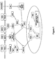
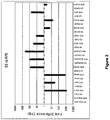
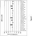
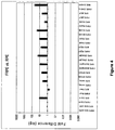

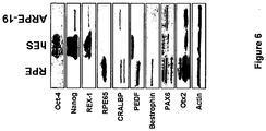

Classifications
-
- C—CHEMISTRY; METALLURGY
- C12—BIOCHEMISTRY; BEER; SPIRITS; WINE; VINEGAR; MICROBIOLOGY; ENZYMOLOGY; MUTATION OR GENETIC ENGINEERING
- C12N—MICROORGANISMS OR ENZYMES; COMPOSITIONS THEREOF; PROPAGATING, PRESERVING, OR MAINTAINING MICROORGANISMS; MUTATION OR GENETIC ENGINEERING; CULTURE MEDIA
- C12N5/00—Undifferentiated human, animal or plant cells, e.g. cell lines; Tissues; Cultivation or maintenance thereof; Culture media therefor
- C12N5/06—Animal cells or tissues; Human cells or tissues
- C12N5/0602—Vertebrate cells
- C12N5/0618—Cells of the nervous system
- C12N5/0621—Eye cells, e.g. cornea, iris pigmented cells
-
- A—HUMAN NECESSITIES
- A61—MEDICAL OR VETERINARY SCIENCE; HYGIENE
- A61K—PREPARATIONS FOR MEDICAL, DENTAL OR TOILETRY PURPOSES
- A61K35/00—Medicinal preparations containing materials or reaction products thereof with undetermined constitution
- A61K35/12—Materials from mammals; Compositions comprising non-specified tissues or cells; Compositions comprising non-embryonic stem cells; Genetically modified cells
- A61K35/30—Nerves; Brain; Eyes; Corneal cells; Cerebrospinal fluid; Neuronal stem cells; Neuronal precursor cells; Glial cells; Oligodendrocytes; Schwann cells; Astroglia; Astrocytes; Choroid plexus; Spinal cord tissue
-
- A—HUMAN NECESSITIES
- A61—MEDICAL OR VETERINARY SCIENCE; HYGIENE
- A61P—SPECIFIC THERAPEUTIC ACTIVITY OF CHEMICAL COMPOUNDS OR MEDICINAL PREPARATIONS
- A61P25/00—Drugs for disorders of the nervous system
- A61P25/14—Drugs for disorders of the nervous system for treating abnormal movements, e.g. chorea, dyskinesia
- A61P25/16—Anti-Parkinson drugs
-
- A—HUMAN NECESSITIES
- A61—MEDICAL OR VETERINARY SCIENCE; HYGIENE
- A61P—SPECIFIC THERAPEUTIC ACTIVITY OF CHEMICAL COMPOUNDS OR MEDICINAL PREPARATIONS
- A61P27/00—Drugs for disorders of the senses
- A61P27/02—Ophthalmic agents
-
- A—HUMAN NECESSITIES
- A61—MEDICAL OR VETERINARY SCIENCE; HYGIENE
- A61P—SPECIFIC THERAPEUTIC ACTIVITY OF CHEMICAL COMPOUNDS OR MEDICINAL PREPARATIONS
- A61P27/00—Drugs for disorders of the senses
- A61P27/02—Ophthalmic agents
- A61P27/06—Antiglaucoma agents or miotics
-
- A—HUMAN NECESSITIES
- A61—MEDICAL OR VETERINARY SCIENCE; HYGIENE
- A61P—SPECIFIC THERAPEUTIC ACTIVITY OF CHEMICAL COMPOUNDS OR MEDICINAL PREPARATIONS
- A61P43/00—Drugs for specific purposes, not provided for in groups A61P1/00-A61P41/00
-
- A—HUMAN NECESSITIES
- A61—MEDICAL OR VETERINARY SCIENCE; HYGIENE
- A61P—SPECIFIC THERAPEUTIC ACTIVITY OF CHEMICAL COMPOUNDS OR MEDICINAL PREPARATIONS
- A61P9/00—Drugs for disorders of the cardiovascular system
-
- A—HUMAN NECESSITIES
- A61—MEDICAL OR VETERINARY SCIENCE; HYGIENE
- A61P—SPECIFIC THERAPEUTIC ACTIVITY OF CHEMICAL COMPOUNDS OR MEDICINAL PREPARATIONS
- A61P9/00—Drugs for disorders of the cardiovascular system
- A61P9/10—Drugs for disorders of the cardiovascular system for treating ischaemic or atherosclerotic diseases, e.g. antianginal drugs, coronary vasodilators, drugs for myocardial infarction, retinopathy, cerebrovascula insufficiency, renal arteriosclerosis
-
- A—HUMAN NECESSITIES
- A61—MEDICAL OR VETERINARY SCIENCE; HYGIENE
- A61K—PREPARATIONS FOR MEDICAL, DENTAL OR TOILETRY PURPOSES
- A61K35/00—Medicinal preparations containing materials or reaction products thereof with undetermined constitution
- A61K35/12—Materials from mammals; Compositions comprising non-specified tissues or cells; Compositions comprising non-embryonic stem cells; Genetically modified cells
-
- C—CHEMISTRY; METALLURGY
- C12—BIOCHEMISTRY; BEER; SPIRITS; WINE; VINEGAR; MICROBIOLOGY; ENZYMOLOGY; MUTATION OR GENETIC ENGINEERING
- C12N—MICROORGANISMS OR ENZYMES; COMPOSITIONS THEREOF; PROPAGATING, PRESERVING, OR MAINTAINING MICROORGANISMS; MUTATION OR GENETIC ENGINEERING; CULTURE MEDIA
- C12N2501/00—Active agents used in cell culture processes, e.g. differentation
- C12N2501/10—Growth factors
-
- C—CHEMISTRY; METALLURGY
- C12—BIOCHEMISTRY; BEER; SPIRITS; WINE; VINEGAR; MICROBIOLOGY; ENZYMOLOGY; MUTATION OR GENETIC ENGINEERING
- C12N—MICROORGANISMS OR ENZYMES; COMPOSITIONS THEREOF; PROPAGATING, PRESERVING, OR MAINTAINING MICROORGANISMS; MUTATION OR GENETIC ENGINEERING; CULTURE MEDIA
- C12N2501/00—Active agents used in cell culture processes, e.g. differentation
- C12N2501/70—Enzymes
- C12N2501/73—Hydrolases (EC 3.)
- C12N2501/734—Proteases (EC 3.4.)
-
- C—CHEMISTRY; METALLURGY
- C12—BIOCHEMISTRY; BEER; SPIRITS; WINE; VINEGAR; MICROBIOLOGY; ENZYMOLOGY; MUTATION OR GENETIC ENGINEERING
- C12N—MICROORGANISMS OR ENZYMES; COMPOSITIONS THEREOF; PROPAGATING, PRESERVING, OR MAINTAINING MICROORGANISMS; MUTATION OR GENETIC ENGINEERING; CULTURE MEDIA
- C12N2506/00—Differentiation of animal cells from one lineage to another; Differentiation of pluripotent cells
- C12N2506/02—Differentiation of animal cells from one lineage to another; Differentiation of pluripotent cells from embryonic cells
-
- C—CHEMISTRY; METALLURGY
- C12—BIOCHEMISTRY; BEER; SPIRITS; WINE; VINEGAR; MICROBIOLOGY; ENZYMOLOGY; MUTATION OR GENETIC ENGINEERING
- C12N—MICROORGANISMS OR ENZYMES; COMPOSITIONS THEREOF; PROPAGATING, PRESERVING, OR MAINTAINING MICROORGANISMS; MUTATION OR GENETIC ENGINEERING; CULTURE MEDIA
- C12N2509/00—Methods for the dissociation of cells, e.g. specific use of enzymes
-
- C—CHEMISTRY; METALLURGY
- C12—BIOCHEMISTRY; BEER; SPIRITS; WINE; VINEGAR; MICROBIOLOGY; ENZYMOLOGY; MUTATION OR GENETIC ENGINEERING
- C12N—MICROORGANISMS OR ENZYMES; COMPOSITIONS THEREOF; PROPAGATING, PRESERVING, OR MAINTAINING MICROORGANISMS; MUTATION OR GENETIC ENGINEERING; CULTURE MEDIA
- C12N2533/00—Supports or coatings for cell culture, characterised by material
- C12N2533/50—Proteins
- C12N2533/54—Collagen; Gelatin
Definitions
- the retinal pigment epithelium is the pigmented cell layer just outside the neurosensory retina. This layer of cells nourishes retinal visual cells, and is attached to the underlying choroid (the layer of blood vessels behind the retina) and overlying retinal visual cells.
- the RPE acts as a filter to determine what nutrients reach the retina from the choroid. Additionally, the RPE provides insulation between the retina and the choroid. Breakdown of the RPE interferes with the metabolism of the retina, causing thinning of the retina. Thinning of the retina can have serious consequences. For example, thinning of the retina may cause "dry" macular degeneration and may also lead to the inappropriate blood vessel formation that can cause "wet" macular degeneration).
- RPE cells produced in vitro could be used to study the developments of the RPE, to identify factors that cause the RPE to breakdown, or to identify agents that can be used to stimulate repair of endogenous RPE cells. Additionally, RPE cells produced in vitro could themselves be used as a therapy for replacing or restoring all or a portion of a patient's damaged RPE cells. When used in this manner, RPE cells may provide an approach to treat macular degeneration, as well as other diseases and conditions caused, in whole or in part, by damage to the RPE.
- RPE cells produced in vitro for screening or as a therapeutic relies on methods that can be used to produce large numbers of RPE cells in a systematic, directed manner. Such systematized differentiation methods would provide significant advantages over previous schemes based on, for example, spontaneous differentiation of RPE cells from transformed cell lines or other sources.
- the present invention provides a method for differentiating RPE cells from human pluripotent stem cells, such as human embryonic stem cells and human induced pluripotent stem cells.
- the method is used to produce large numbers of differentiated RPE cells for use in screening assays, to study the basic biology of the RPE, and as therapeutics.
- RPE cells differentiated from pluripotent stem cells, such as human embryonic stem cells using this approach are molecularly distinct from human embryonic stem cells, as well as from adult and fetal-derived RPE cells.
- the present invention also provides preparations and pharmaceutical preparations of RPE cells derived from human pluripotent stem cells.
- RPE cell preparations are molecularly distinct from human embryonic stem cells, as well as from adult and fetal-derived RPE cells.
- the present invention provides, for the first time, a detailed molecular characterization of RPE cells differentiated from human embryonic stem cells.
- the detailed characterization includes comparisons to RPE cells derived from other sources (e.g., adult RPE cells and fetal RPE cells), as well as to human embryonic stem cells. This analysis not only provides a deeper understanding of RPE cells, but it also revealed that RPE cells differentiated from human embryonic stem cells have distinct molecular properties that distinguish these cells from previously described RPE cells.
- the present invention provides preparations of RPE cells, including substantially purified preparations of RPE cells.
- exemplary RPE cells are differentiated from human pluripotent stem cells, such as human embryonic stem cells or iPS cells.
- Human pluripotent stem cell-derived RPE cells can be formulated and used to treat retinal degenerative diseases.
- human pluripotent stem cell-derived RPE cells can be used in screening assays to identify agents that modulate RPE cell survival (in vitro and/or in vivo), to study RPE cell maturation, or to identify agents that modulate RPE cell maturation. Agents identified using such screening assays may be used in vitro or in vivo and may provide additional therapeutics that can be used alone or in combination with RPE cells to treat retinal degenerative diseases.
- the present invention provides improved methods for the production of RPE cells from embryonic stem cells or other pluripotent stem cells.
- the methods of the invention can be used to produce differentiated RPE cells.
- the level of maturation, as assessed by pigmentation levels, of the differentiated RPE cells can be modulated so that differentiated RPE cells, mature RPE cells, or mixtures thereof are produced.
- improved methods for the treatment of eye disorders involve the use of RPE cells derived from human embryonic stem cells to treat or ameliorate the symptoms of eye disorders, particularly eye disorders caused or exacerbated, in whole or in part, by damage to or breakdown of the endogenous RPE layer.
- the invention provides a method for producing a culture of retinal pigment epithelial (RPE) cells.
- the culture is a substantially purified culture containing at least 75%, 80%, 85%, 90%, 91%, 92%, 93%, 94%, 95%, 96%, 97%, 98%, 99%, or greater than 99% differentiated RPE cells (at least 75% of the culture is a differentiated RPE cell, regardless of level of maturity).
- the substantially purified culture contains at least 30%, 35%, 40% or 45% mature differentiated RPE cells.
- the substantially purified culture contains at least 50% mature differentiated RPE cells.
- the substantially purified culture contains at least 55%, 60%, 65%, 70%, 75%, 80%, 85%, 90%, 91%, 92%, 93%, 94%, 95%, 96%, 97%, 98%, 99%, or greater than 99% mature differentiated RPE cells.
- the differentiated RPE cells are derived from human embryonic stem cells, human iPS cells, or other pluripotent stem cells.
- the method comprising the steps of
- the invention provides a method of producing a mature retinal pigment epithelial (RPE) cell, said method comprising the steps of
- the substantially purified culture of RPE cells may contain both differentiated RPE cells and mature differentiated RPE cells.
- the level of pigment may vary.
- the mature RPE cells can be distinguished visually from the RPE cells based on the increased level of pigmentation and the more columnar shape.
- the percentage of mature differentiated RPE cells in the culture can be reduced by decreasing the density of the culture.
- the method further comprises subculturing a population of mature RPE cells to produce a culture containing a smaller percentage of mature RPE cells.
- the medium used when culturing the cells as embryoid bodies may be selected from any medium appropriate for culturing cells as embryoid bodies.
- any medium that is capable of supporting high-density cultures may be used, such as medium for viral, bacterial, or eukaryotic cell culture.
- the medium may be high nutrient, protein-free medium or high nutrient, low protein medium.
- the human embryonic stem cells may be cultured in MDBK-GM, OptiPro SFM, VP-SFM, EGM-2, or MDBK-MM.
- the medium may also contain B-27 supplement.
- the medium described herein may also be supplemented with one or more growth factors.
- Growth factors that may be used include, for example, EGF, bFGF, VEGF, and recombinant insulin-like growth factor.
- the medium may also contain supplements such as heparin, hydrocortisone, ascorbic acid, serum (such as, for example, fetal bovine serum), or a growth matrix (such as, for example, extracellular matrix from bovine corneal epithelium, matrigel (BD biosciences), or gelatin).
- mechanical or enzymatic methods are used to select RPE cells from amongst clusters of non-RPE cells in a culture of embryoid body, or to facilitate sub-culture of adherent cells.
- Exemplary mechanical methods include, but are not limited to, tituration with a pipette or cutting with a pulled needle.
- Exemplary enzymatic methods include, but are not limited to, any enzymes appropriate for disassociating cells (e.g., trypsin, collagenase, dispase).
- a non-enzymatic solution is used to disassociate the cells, such as a high EDTA-containing solution such as, for example, Hanks-based cell disassociation buffer.
- the cells are cultured for between about 3 days and 45 days, such as 7 days, 7-10 days, 7-14 days, or 14-21 days.
- the cells are cultured for about 1, 2, 3, 4, 5, 6, 7, 8, 9, 10, 11, 12, 13, 14, 15, 16, 17, 18, 19, 20, 21, 22, 23, 24, 25, 26, 27, 28, 29, 30, 31, 32, 33, 34, 35, 36, 37, 38, 39, 40, 41, 42, 43, 44, 45, or about 46 days.
- the cells are cultured for less than or equal to about: 45, 40, 35, 30, 25, 21, 20, 18, 15, 14, 13, 12, 11, 10, 9, 8, 7, 6, 5, 4, 3, 2, or 1 days.
- the cells may be cultured for the same period of time at each step or for differing periods of time at one or more of the steps.
- the RPE cells are further cultured to produce a culture of mature RPE cells.
- Both RPE cells and mature RPE cells are differentiated RPE cells.
- mature RPE cells are characterized by increased level of pigment in comparison to differentiated RPE cells. The level of maturity and pigmentation can be modulated by increasing or decreasing the density of the culture of differentiated RPE cells.
- a culture of RPE cells can be further cultured to produce mature RPE cells.
- the density of a culture containing mature RPE cells can be decreased to decrease the percentage of mature differentiated RPE cells and increase the percentage of differentiated RPE cells.
- the medium used to culture the RPE cells is any medium appropriate for cell culture, and can be selected by the skilled person.
- any medium that is capable of supporting high-density cultures may be used, such as medium for viral, bacterial, or animal cell culture.
- the cells described herein may be cultured in VP-SFM, EGM-2, and MDBK-MM.
- said substantially purified culture of RPE cells are frozen for storage.
- the cells may be stored by any appropriate method known in the art, e.g., cryogenically frozen and may be frozen at any temperature appropriate for storage of the cells.
- the cells may be frozen at approximately -20 °C, - 80 °C, - 120 °C, or at any other temperature appropriate for storage of cells.
- Cryogenically frozen cells are stored in appropriate containers and prepared for storage to reduce risk of cell damage and maximize the likelihood that the cells will survive thawing.
- RPE cells are maintained at room temperature, or refrigerated at, for example, approximately 4 °C.
- the method is performed in accordance with Good Manufacturing Practices (GMP).
- GMP Good Manufacturing Practices
- the human embryonic stem cells from which the RPE cells are differentiated were derived in accordance with Good Manufacturing Practices (GMP).
- the human embryonic stem cells from which the RPE cells are differentiated were derived from one or more blastomeres removed from an early stage embryo without destroying the remaining embryo.
- the method is used to produce a preparation comprising at least 1x10 5 RPE cells, at least 5x10 5 RPE cells, at least 1x10 6 RPE cells, at least 5x10 6 RPE cells, at least 1x10 7 RPE cells, at least 2x10 7 RPE cells, at least 3x10 7 RPE cells, at least 4x10 7 RPE cells, at least 5x10 7 RPE cells, at least 6x10 7 RPE cells, at least 7x10 7 RPE cells, at least 8x10 7 RPE cells, at least 9x10 7 RPE cells, at least 1x10 8 RPE cells, at least 2x10 8 RPE cells, at least 5x10 8 RPE cells, at least 7x10 8 RPE cells, or at least 1x10 9 RPE cells.
- the number of RPE cells in the preparation includes differentiated RPE cells, regardless of level of maturity and regardless of the relative percentages of differentiated RPE cells and mature RPE cells. In other embodiments, the number of RPE cells in the preparation refers to the number of either differentiated RPE cells or mature RPE cells.
- the method further comprises formulating the differentiated RPE cells and/or differentiated mature RPE cells to produce a preparation of RPE cells suitable for transplantation.
- the invention provides a method for treating or preventing a condition characterized by retinal degeneration, comprising administering to a subject in need thereof an effective amount of a preparation comprising RPE cells, which RPE cells are derived from human embryonic stem cells, iPS cells, or other pluripotent stem cells.
- RPE cells which RPE cells are derived from human embryonic stem cells, iPS cells, or other pluripotent stem cells.
- Conditions characterized by retinal degeneration include, for example, Stargardt's macular dystrophy, age related macular degeneration (dry or wet), diabetic retinopathy, and retinitis pigmentosa.
- the RPE cells are derived from human pluripotent stem cells using one or more of the methods described herein.
- the preparation was previously cryopreserved and was thawed before transplantation.
- the method of treating further comprises administration of cyclosporin or one or more other immunosuppressants.
- immunosuppressants When immunosuppressants are used, they may be administered systemically or locally, and they may be administered prior to, concomitantly with, or following administration of the RPE cells. In certain embodiments, immunosuppressive therapy continues for weeks, months, years, or indefinitely following administration of RPE cells.
- the method of treatment comprises administration of a single dose of RPE cells.
- the method of treatment comprises a course of therapy where RPE cells are administered multiple times over some period.
- Exemplary courses of treatment may comprise weekly, biweekly, monthly, quarterly, biannually, or yearly treatments.
- treatment may proceed in phases whereby multiple doses are required initially (e.g., daily doses for the first week), and subsequently fewer and less frequent doses are needed. Numerous treatment regimens are contemplated.
- the administered RPE cells comprise a mixed population of differentiated RPE cells and mature RPE cells.
- the administered RPE cells comprise a substantially purified population of either differentiated RPE cells or mature RPE cells.
- the administered RPE cells may contain less than 25%, 20%, 15%, 10%, 9%, 8%, 7%, 6%, 5%, 4%, 3%, 2%, or less than 1% of the other RPE cell-type.
- the RPE cells are formulated in a pharmaceutically acceptable carrier or excipient.
- the preparation comprising RPE cells is transplanted in a suspension, matrix or substrate.
- the preparation is administered by injection into the subretinal space of the eye.
- about 10 4 to about 10 6 cells are administered to the subject.
- the method further comprises the step of monitoring the efficacy of treatment or prevention by measuring electroretinogram responses, optomotor acuity threshold, or luminance threshold in the subject.
- the method may also comprise monitoring the efficacy of treatment or prevention by monitoring immunogenicity of the cells or migration of the cells in the eye.
- the invention provides a pharmaceutical preparation for treating or preventing a condition characterized by retinal degeneration, comprising an effective amount of RPE cells, which RPE cells are derived from human embryonic stem cells or other pluripotent stem cells.
- the pharmaceutical preparation may be formulated in a pharmaceutically acceptable carrier according to the route of administration.
- the preparation may be formulated for administration to the subretinal space of the eye.
- the composition may comprise at least 10 4 , 10 5 , 5x10 5 , 6x10 5 , 7x10 5 , 8x10 5 , 9x10 5 , 10 6 , 2x10 6 , 3x10 6 , 4x10 6 , 5x10 6 , 6x10 6 , 7x10 6 , 8x10 6 , 9x10 6 , or 10 7 RPE cells.
- the composition may comprise at least 2x10 7 , 5x10 7 , 6x10 7 , 7x10 7 , 8x10 7 , 9x10 7 , 1x10 8 RPE cells.
- the RPE cells may include mature RPE cells, and thus the cell number includes the total of both differentiated RPE cells and mature differentiated RPE cells.
- the invention provides a method for screening to identify agents that modulate the survival of RPE cells.
- RPE cells differentiated from human embryonic stem cells can be used to screen for agents that promote RPE survival.
- Identified agents can be used, alone or in combination with RPE cells, as part of a treatment regimen.
- identified agents can be used as part of a culture method to improve the survival of RPE cells differentiated in vitro.
- the invention provides a method for screening to identify agents that modulate RPE cell maturity.
- RPE cells differentiated from human ES cells can be used to screen for agents that promote RPE maturation.
- the method is performed in accordance with Good Manufacturing Practices (GMP).
- GMP Good Manufacturing Practices
- the human embryonic stem cells or other pluripotent stem cells from which the RPE cells are differentiated were derived in accordance with Good Manufacturing Practices (GMP).
- the human embryonic stem cells from which the RPE cells are differentiated were derived from one or more blastomere removed from an early stage embryo without destroying the remaining embryo.
- the invention contemplates that, instead of human embryonic stem cells, the starting material for producing RPE cells, or preparations thereof, can be other types of human pluripotent stem cells.
- the invention contemplates that induced pluripotent stem (iPS) cells are used as a starting material for differentiating RPE cells using the methods described herein.
- iPS cells can be obtained from a cell bank, or otherwise previously derived.
- iPS cells can be newly generated prior to commencing differentiation to RPE cells.
- RPE cells or preparations differentiated from pluripotent stem cells, including iPS cells are used in a therapeutic method.
- the present invention also provides functional human retinal pigmented epithelial cells (hRPEs) that are terminally differentiated from human embryonic stem cells (hESCs) or other human pluripotent stem cells.
- hRPEs human retinal pigmented epithelial cells
- hESCs human embryonic stem cells
- hRPEs may treat retinal degeneration in the diseased animal.
- the hRPEs of the invention are useful for treating patients afflicted by various retinal degenerative disorders.
- the present invention therefore provides a renewable source of hRPEs that can be produced and manufactured under GLP-like and GMP-compliant conditions for the treatment of visual degenerative diseases and disorders.
- the present invention provides a human retinal pigmented epithelial cell derived from a human embryonic stem cell, which cell is pigmented and expresses at least one gene that is not expressed in a cell that is not a human retinal pigmented epithelial cell.
- the human retinal pigmented epithelial cell is isolated from at least one protein, molecule, or other impurity that is found in its natural environment.
- the invention provides a cell culture comprising human RPE cells derived from human embryonic stem cells or other pluripotent stem cells, which are pigmented and express at least one gene that is not expressed in a cell that is not a human RPE.
- pigmented refers to any level of pigmentation, for example, the pigmentation that initial occurs when RPE cells differentiate from ES cells. Pigmentation may vary with cell density and the maturity of the differentiated RPE cells. However, when cells are referred to as pigmented - the term is understood to refer to any and all levels of pigmentation.
- the cell culture comprises a substantially purified population of human RPE cells.
- a substantially purified population of hRPE cells is one in which the hRPE cells comprise at least about 75% of the cells in the population.
- a substantially purified population of hRPE cells is one in which the hRPE cells comprise at least about 80%, 85%, 90%, 91%, 92%, 93%, 94%, 95%, 96%, 97%, 97.5%, 98%, 99%, or even greater than 99% of the cells in the population.
- the pigmentation of the hRPE cells in the cell culture is homogeneous.
- the pigmentation of the hRPE cells in the cell culture is heterogeneous, and the culture of RPE cells comprises both differentiated RPE cells and mature RPE cells.
- a cell culture of the invention may comprise at least about 10 1 , 10 2 , 5x10 2 , 10 3 , 5x10 3 , 10 4 , 10 5 , 10 6 , 10 7 , 10 8 , or at least about 10 9 hRPE cells.
- the present invention provides human retinal pigmented epithelial cells with varying degrees of pigmentation.
- the pigmentation of a human retinal pigmented epithelial cell is the same as an average human pigmented epithelial cell after terminal differentiation of the hRPE cell.
- the pigmentation of a human retinal pigmented epithelial cell is more pigmented than the average human retinal pigmented epithelial cell after terminal differentiation of the hRPE cell.
- the pigmentation of a human retinal pigmented epithelial cell is less pigmented than the average human retinal pigmented epithelial cell after terminal differentiation.
- the present invention provides human RPE cells differentiated from ES cells or other pluripotent stem cells and that express (at the mRNA and/or protein level) one or more (1, 2, 3, 4, 5, or 6) of the following: RPE-65, Bestrophin, PEDF, CRALBP, Otx2, and Mit-F.
- gene expression is measured by mRNA expression.
- gene expression is measured by protein expression.
- the RPE cells do not substantially express ES-specific genes, such as Oct-4, alkaline phosphatase, nanog, and/or Rex-1.
- the RPE cells express one or more (1, 2, or 3) of pax-2, pax-6, and/or tyrosinase. In certain embodiments, expression of pax-2, pax-6, and/or tyrosinase distinguishes differentiated RPE cells from mature differentiated RPE cells. In other embodiments, the RPE cells express one or more of the markers presented in Table 2, and the expression of the one or more markers is upregulated in RPE cells relative to expression (if any) in human ES cells. In other embodiments, the RPE cells express one or more of the markers presented in Table 3, and the expression of the one or more markers is downregulated in RPE cells relative to expression (if any) in human ES cells.
- the invention provides a pharmaceutical preparation comprising human RPE cells derived from human embryonic stem cells or other pluripotent stem cells.
- Pharmaceutical preparations may comprise at least about 10 1 , 10 2 , 5x10 2 , 10 3 , 5x10 3 , 10 4 , 10 5 , 10 6 , 10 7 , 10 8 or about 10 9 hRPE cells.
- the invention provides a cryopreserved preparation of the RPE cells described herein.
- the cryopreserved preparation may be frozen for storage for days or years.
- the cells may be stored by any appropriate method known in the art, e.g., cryogenically frozen and may be frozen at any temperature appropriate for storage of the cells.
- the cells may be frozen at approximately -20 °C, - 80 °C, - 120 °C, or at any other temperature appropriate for storage of cells.
- Cryogenically frozen cells are stored in appropriate containers and prepared for storage to reduce risk of cell damage and maximize the likelihood that the cells will survive thawing.
- RPE cells can be maintained at room temperature, or refrigerated at, for example, approximately 4 °C.
- Cryopreserved preparations of the compositions described herein may comprise at least about 10 1 , 10 2 , 5x10 2 , 10 3 , 5x10 3 , 10 4 , 10 5 , 10 6 , 10 7 , 10 8 or about 10 9 hRPE cells.
- the hRPE cells of the invention are recovered from storage following cryopreservation.
- greater than 65%, 70%, 75,%, or greater than 80% of the RPE cells retain viability following cryopreservation.
- greater than 85%, 90%, 91%, 92%, 93%, 94%, 95%, 96%, 97%, 98%, or greater than 99% of the RPE cells retain viability following cryopreservation.
- the invention provides substantially purified preparations of human RPE cells have any combination of the structural, molecular, and functional characteristics described herein.
- Such preparations may be formulated as pharmaceutical preparations for administration and/or may be formulated for cryopreservation.
- the invention provides use of the human RPE cells described herein in the manufacture of a medicament to treat a condition in a patient in need thereof.
- the invention provides use of a cell culture comprising the human RPE cells described herein in the manufacture of a medicament to treat a condition in a patient in need thereof.
- the invention provides the use of a pharmaceutical preparation comprising the human RPE cells described herein in the manufacture of a medicament to treat a condition in a patient in need thereof.
- Conditions that may be treated include, without limitation, degenerative diseases of the retina, such as Stargardt's macular dystrophy, retinitis pigmentosa, macular degeneration, glaucoma, and diabetic retinopathy.
- the invention provides methods for treating or preventing a condition characterized by retinal degeneration, comprising administering to a subject in need thereof an effective amount of a preparation comprising RPE cells, which RPE cells are derived from mammalian embryonic stem cells.
- Conditions characterized by retinal degeneration include, for example, Stargardt's macular dystrophy, age related macular degeneration, diabetic retinopathy, and retinitis pigmentosa.
- the invention provides a solution of human RPE cells derived from a human embryonic stem cell, or other pluripotent stem cell, which RPE cells have any combinations of the features described herein.
- a solutions may comprise at least about 10 1 , 10 2 , 5x10 2 , 10 3 , 5x10 3 , 10 4 , 10 5 , 10 6 , 10 7 , 10 8 or about 10 9 hRPE cells as described herein.
- Such solutions are suitable for injection to a subject.
- Such solutions are suitable for cryopreservation as described herein.
- This invention also provides a use of these solutions for the manufacture of a medicament to treat a disease that could be treated by the administration of RPE cells, such as, for example, retinal degenerative diseases of the eye.
- the RPE cells of the invention are derived from human embryonic stem cells, or other pluripotent stem cells, previously derived under GMP conditions.
- the human ES cells are derived from one or more blastomeres of an early cleavage stage embryo, optionally without destroying the embryo.
- the human ES cells are from a library of human embryonic stem cells.
- said library of human embryonic stem cells comprises stem cells, each of which is hemizygous, homozygous, or nullizygous for at least one MHC allele present in a human population, wherein each member of said library of stem cells is hemizygous, homozygous, or nullizygous for a different set of MHC alleles relative to the remaining members of the library.
- the library of human embryonic stem cells comprises stem cells that are hemizygous, homozygous, or nullizygous for all MHC alleles present in a human population.
- the invention provides a library of RPE cells, each of which is hemizygous, homozygous, or nullizygous for at least one MHC allele present in a human population, wherein each member of said library of RPE cells is hemizygous, homozygous, or nullizygous for a different set of MHC alleles relative to the remaining members of the library.
- invention provides a library of human RPE cells that are hemizygous, homozygous, or nullizygous for all MHC alleles present in a human population.
- said substantially purified culture of RPE cells are frozen for storage.
- the cells may be stored by any appropriate method known in the art, e.g., cryogenically frozen and may be frozen at any temperature appropriate for storage of the cells.
- the cells may be frozen at approximately -20 °C, - 80 °C, - 120 °C, or at any other temperature appropriate for storage of cells.
- Cryogenically frozen cells are stored in appropriate containers and prepared for storage to reduce risk of cell damage and maximize the likelihood that the cells will survive thawing.
- RPE cells can be maintained at room temperature, or refrigerated at, for example, approximately 4 °C.
- human RPE cells are produced in accordance with Good Manufacturing Practices (GMP).
- the human embryonic stem cells from which the RPE cells are differentiated were derived in accordance with Good Manufacturing Practices (GMP).
- the human embryonic stem cells from which the RPE cells are differentiated were derived from one or more blastomeres removed from an early stage embryo without destroying the remaining embryo.
- the invention provides GMP compliant preparations of RPE cells, including substantially purified preparations of RPE cells. Such preparations are substantially free of viral, bacterial, and/or fungal contamination or infection.
- compositions or preparations of RPE cells comprise at least 1x10 5 RPE cells, at least 5x10 5 RPE cells, at least 1x10 6 RPE cells, at least 5x10 6 RPE cells, at least 1x10 7 RPE cells, at least 2x10 7 RPE cells, at least 3x10 7 RPE cells, at least 4x10 7 RPE cells, at least 5x10 7 RPE cells, at least 6x10 7 RPE cells, at least 7x10 7 RPE cells, at least 8x10 7 RPE cells, at least 9x10 7 RPE cells, at least 1x10 8 RPE cells, at least 2x10 8 RPE cells, at least 5x10 8 RPE cells, at least 7x10 8 RPE cells, or at least 1x10 9 RPE cells.
- the number of RPE cells in the preparation includes differentiated RPE cells, regardless of level of maturity and regardless of the relative percentages of differentiated RPE cells and mature differentiated RPE cells. In other embodiments, the number of RPE cells in the preparation refers to the number of either differentiated RPE cells or mature RPE cells.
- the method further comprises formulating the differentiated RPE cells and/or differentiated mature RPE cells to produce a preparation of RPE cells suitable for transplantation.
- the invention provides a method for treating or preventing a condition characterized by retinal degeneration, comprising administering to a subject in need thereof an effective amount of a preparation comprising RPE cells, which RPE cells are derived from human pluripotent stem cells.
- RPE cells are derived using any of the methods described herein.
- Conditions characterized by retinal degeneration include, for example, Stargardt's macular dystrophy, age related macular degeneration (dry or wet), diabetic retinopathy, and retinitis pigmentosa.
- the preparation was previously cryopreserved and was thawed before transplantation.
- the method of treating further comprises administration of cyclosporin or one or more other immunosuppressants.
- immunosuppressants When immunosuppressants are used, they may be administered systemically or locally, and they may be administered prior to, concomitantly with, or following administration of the RPE cells. In certain embodiments, immunosuppressive therapy continues for weeks, months, years, or indefinitely following administration of RPE cells.
- the method of treatment comprises administration of a single dose of RPE cells.
- the method of treatment comprises a course of therapy where RPE cells are administered multiple times over some period.
- Exemplary courses of treatment may comprise weekly, biweekly, monthly, quarterly, biannually, or yearly treatments.
- treatment may proceed in phases whereby multiple doses are required initially (e.g., daily doses for the first week), and subsequently fewer and less frequent doses are needed. Numerous treatment regimens are contemplated.
- the administered RPE cells comprise a mixed population of differentiated RPE cells and mature RPE cells.
- the administered RPE cells comprise a substantially purified population of either differentiated RPE cells or mature RPE cells.
- the administered RPE cells may contain less than 25%, 20%, 15%, 10%, 9%, 8%, 7%, 6%, 5%, 4%, 3%, 2%, or less than 1% of the other RPE cell-type.
- the RPE cells are formulated in a pharmaceutically acceptable carrier or excipient.
- the preparation comprising RPE cells is transplanted in a suspension, matrix or substrate.
- the preparation is administered by injection into the subretinal space of the eye.
- the preparation is administered transcorneally.
- about 10 4 to about 10 6 cells are administered to the subject.
- the method further comprises the step of monitoring the efficacy of treatment or prevention by measuring electroretinogram responses, optomotor acuity threshold, or luminance threshold in the subject.
- the method may also comprise monitoring the efficacy of treatment or prevention by monitoring immunogenicity of the cells or migration of the cells in the eye.
- the invention provides a pharmaceutical preparation for treating or preventing a condition characterized by retinal degeneration, comprising an effective amount of RPE cells, which RPE cells are derived from human embryonic stem cells.
- the pharmaceutical preparation may be formulated in a pharmaceutically acceptable carrier according to the route of administration.
- the preparation may be formulated for administration to the subretinal space or cornea of the eye.
- the composition may comprise at least 10 4 , 10 5 , 5x10 5 , 6x10 5 , 7x10 5 , 8x10 5 , 9x10 5 , 10 6 , 2x10 6 3x10 6 4x10 6 , 5x10 6 , 6x10 6 , 7x10 6 , 8x10 6 , 9x10 6 , or 10 7 RPE cells.
- the composition may comprise at least 2x10 7 , 5x10 7 , 6x10 7 , 7x10 7 , 8x10 7 , 9x10 7 , 1x10 8 RPE cells.
- the RPE cells may include mature RPE cells, and thus the cell number includes the total of both differentiated RPE cells and mature differentiated RPE cells.
- the invention provides a method for screening to identify agents that modulate the survival of RPE cells.
- RPE cells differentiated from human embryonic stem cells can be used to screen for agents that promote RPE survival.
- Identified agents can be used, alone or in combination with RPE cells, as part of a treatment regimen.
- identified agents can be used as part of a culture method to improve the survival of RPE cells differentiated in vitro.
- the invention provides a method for screening to identify agents that modulate RPE cell maturity.
- RPE cells differentiated from human ES cells can be used to screen for agents that promote RPE maturation.
- the method is performed in accordance with Good Manufacturing Practices (GMP).
- GMP Good Manufacturing Practices
- the human embryonic stem cells from which the RPE cells are differentiated were derived in accordance with Good Manufacturing Practices (GMP).
- the human embryonic stem cells from which the RPE cells are differentiated were derived from one or more blastomere removed from an early stage embryo without destroying the remaining embryo.
- the invention contemplates that, instead of human embryonic stem cells, the starting material for producing RPE cells, or preparations thereof, can be other types of human pluripotent stem cells.
- the invention contemplates that induced pluripotent stem (iPS) cells, which have the characteristic of hES, can similarly be used as a starting material for differentiating RPE cells using the methods described herein.
- iPS cells can be obtained from a cell bank, or otherwise previously derived.
- iPS cells can be newly generated prior to commencing differentiation to RPE cells.
- RPE cells or preparations differentiated from pluripotent stem cells, including iPS cells are used in a therapeutic method.
- preparations of RPE cells comprising any combination of differentiated RPE cells and mature RPE cells can be used in the treatment of any of the diseases and conditions described herein.
- methods described herein for producing RPE cells using human embryonic stem cells as a starting material may be similarly performed using any human pluripotent stem cells as a starting material.
- embryo or “embryonic” is meant a developing cell mass that has not implanted into the uterine membrane of a maternal host.
- An “embryonic cell” is a cell isolated from or contained in an embryo. This also includes blastomeres, obtained as early as the two-cell stage, and aggregated blastomeres.
- embryonic stem cells refers to embryo-derived cells. More specifically it refers to non-human cells isolated from the inner cell mass of blastocysts or morulae and that have been serially passaged as cell lines. The term also includes cells isolated from one or more blastomeres of an embryo, preferably and in case of a human embryo without destroying the remainder of the embryo. The term also includes non-human embryonic stem cells produced by somatic cell nuclear transfer, even when non-embryonic cells are used in the process.
- human embryonic stem cells hES cells
- Human embryonic stem cells can be identified based on (i) the ability to differentiate into cells of all three germ layers, (ii) expression of at least Oct-4 and alkaline phosphatase, and (iii) ability to produce teratomas when transplanted into immunocompromised animals.
- pluripotent stem cells includes embryonic stem cells, and induced pluripotent stem cells.
- Pluripotent stem cells are defined functionally as stem cells that: (a) are capable of inducing teratomas when transplanted in immunodeficient (SCID) mice; (b) are capable of differentiating to cell types of all three germ layers (e.g., can differentiate to ectodermal, mesodermal and endodermal cell types); and (c) express one or more markers of embryonic stem cells (e.g., express Oct 4, alkaline phosphatase, SSEA-3 surface antigen, SSEA-4 surface antigen, nanog, TRA-1-60, TRA-1-81, SOX2, REX1, etc).
- SCID immunodeficient
- Exemplary pluripotent stem cells include induced pluripotent stem cells (iPS cells) generated by reprogramming a somatic cell by expressing or inducing expression of a combination of factors (herein referred to as reprogramming factors).
- iPS cells can be generated using fetal, postnatal, newborn, juvenile or adult somatic cells.
- factors that can be used to reprogram somatic cells to pluripotent stem cells include, for example, a combination of Oct4 (sometimes referred to as Oct 3/4), Sox2, c-Myc, and Klf4.
- factors that can be used to reprogram somatic cells to pluripotent stem cells include, for example, a combination of Oct 4, Sox2, Nanog, and Lin28.
- somatic cells are reprogrammed by expressing at least 2 reprogramming factors, at least three reprogramming factors, or four reprogramming factors.
- additional reprogramming factors are identified and used alone or in combination with one or more known reprogramming factors to reprogram a somatic cell to a pluripotent stem cell.
- RPE cell and “differentiated RPE cell” and “ES-derived RPE cell” and “human RPE cell” are used interchangeably throughout to refer to an RPE cell differentiated from a human embryonic stem cell using a method of the invention.
- the term is used generically to refer to differentiated RPE cells, regardless of the level of maturity of the cells, and thus may encompass RPE cells of various levels of maturity.
- Differentiated RPE cells can be visually recognized by their cobblestone morphology and the initial appearance of pigment. Differentiated RPE cells can also be identified molecularly based on substantial lack of expression of embryonic stem cell markers such as Oct-4 and nanog, as well as based on the expression of RPE markers such as RPE-65, PEDF, CRALBP, and bestrophin.
- RPE-like cells are generally referred to specifically as adult, fetal or APRE19 cells.
- RPE cells refers to RPE cells differentiated from human embryonic stem cells.
- mature RPE cell and “mature differentiated RPE cell” are used interchangeably throughout to refer to changes that occur following initial differentiating of RPE cells.
- RPE cells can be recognized, in part, based on initial appearance of pigment
- mature RPE cells can be recognized based on enhanced pigmentation.
- Pigmentation post-differentiation is not indicative of a change in the RPE state of the cells (e.g., the cells are still differentiated RPE cells). Rather, the changes in pigment post-differentiation correspond to the density at which the RPE cells are cultured and maintained.
- mature RPE cells have increased pigmentation that accumulates after initial differentiation. Mature RPE cells are more pigmented than RPE cells - although RPE cells do have some level of pigmentation.
- Mature RPE cells can be subcultured at a lower density, such that the pigmentation decreases.
- mature RPE cells can be cultured to produce RPE cells.
- Such RPE cells are still differentiated RPE cells that express markers of RPE differentiation.
- pigmentation changes post-differentiation are phenomenological and do not reflect dedifferentiation of the cells away from an RPE fate.
- changes in pigmentation post-differentiation also correlate with changes in pax-2 expression.
- RPE-like cells when other RPE-like cells are referred to, they are generally referred to specifically as adult, fetal or APRE19 cells.
- RPE cells refers to RPE cells differentiated from human embryonic stem cells.
- APRE-19 refers to cells of a previously derived, human RPE cell line. APRE-19 cells arose spontaneously and are not derived from human embryos or embryonic stem cells.
- This invention provides preparations and compositions comprising human retinal pigmented epithelium (RPE) cells derived from human embryonic stem cells or other human pluripotent stem cells.
- the RPE cells are pigmented, to at least some extent.
- the RPE cells do not express (at any appreciable level) the embryonic stem cell markers Oct-4, nanog, or Rex-1. Specifically, expression of these ES genes is approximately 100-1000 fold lower in RPE cells than in ES cells, when assessed by quantitative RT-PCR.
- the RPE cells do express, both at the mRNA and protein level, one or more of the following: RPE65, CRALBP, PEDF, Bestrophin, MitF and/or Otx2.
- the RPE cells express, both at the mRNA and protein level, one or more of Pax-2, Pax-6, MitF, and tyrosinase. In certain embodiments of any of the foregoing, the RPE cells are mature RPE cells with increased pigmentation in comparison to differentiated RPE cells.
- the invention provides for human RPE cells, cell cultures comprising a substantially purified population of human RPE cells, pharmaceutical preparations comprising human retinal pigmented epithelial cells and cryopreserved preparations of the human RPE cells.
- the invention provides for the use of the human RPE cells in the manufacture of a medicament to treat a condition in a patient in need thereof.
- the invention provides the use of the cell cultures in the manufacture of a medicament to treat a condition in a patient in need thereof.
- the invention also provides the use of the pharmaceutical preparations in the manufacture of a medicament to treat a condition in a patient in need thereof.
- preparations comprising RPE cells may include differentiated RPE cells of varying levels of maturity, or may be substantially pure with respect to differentiated RPE cells of a particular level of maturity.
- the preparations comprising RPE cells are prepared in accordance with Good Manufacturing Practices (GMP) (e.g., the preparations are GMP-compliant).
- GMP Good Manufacturing Practices
- the preparations comprising RPE cells are substantially free of bacterial, viral, or fungal contamination or infection.
- the human RPE cells can be identified and characterized based on their structural properties. Specifically, and in certain embodiments, these cells are unique in that they can be identified or characterized based on the expression or lack of expression (as assessed at the level of the gene or the level of the protein) of one or more markers.
- differentiated ES-derived RPE cells can be identified or characterized based on expression of one or more (e.g., the cells can be characterized based on expression of at least one, at least two, at least three, at least four, at least five, or at least six) of the following markers: RPE-65, Bestrophin, PEDF, CRALBP, Otx2, and Mit-F.
- ES-derived RPE cells can be identified or characterized based on expression of PAX2, tyrosinase, and/or PAX6.
- hRPE cells can be identified or characterized based on expression or lack of expression (as assessed at the level of the gene or the level of the protein) of one or more (1, 2, 3, 4, 5, 6, 7, 8, 9, 10, or more than 10) markers analyzed in any of Tables 1-3.
- ES-derived RPE cells can also be identified and characterized based on the degree of pigmentation of the cell. Differentiated hRPE cells that are rapidly dividing are lightly pigmented. However, when cell density reaches maximal capacity, or when hRPE cells are specifically matured, hRPE take on their characteristic phenotypic hexagonal shape and increase pigmentation level by accumulating melanin and lipofuscin. As such, initial accumulation of pigmentation serves as an indicator of RPE differentiation and increased pigmentation associated with cell density serves as an indicator of RPE maturity.
- Preparations comprising RPE cells include preparations that are substantially pure, with respect to non-RPE cell types, but which contain a mixture of differentiated RPE cells and mature differentiated RPE cells. Preparations comprising RPE cells also include preparations that are substantially pure both respect to non-RPE cell types and with respect to RPE cells of other levels of maturity.
- the invention contemplates that the RPE cells (characterized as described above) may be derived from human pluripotent stem cells, for example iPS cells and embryonic stem cells.
- the RPE cells are derived from human pluripotent stem cells using any of the methods described herein.
- Embryonic stem cells can be indefinitely maintained in vitro in an undifferentiated state and yet are capable of differentiating into virtually any cell type, providing a limitless supply of rejuvenated and histocompatible cells for transplantation therapy.
- the problem of immune rejection can be overcome with nuclear transfer and parthenogenetic technology.
- human embryonic stem (hES) cells are useful for studies on the differentiation of human cells and can be considered as a potential source for transplantation therapies.
- adipocytes (Bost et al., 2002, Aubert et al., 1999), hepatocyte-like cells (Rambhatla et al., 2003), hematopoetic cells (Chadwick et al., 2003).
- oocytes Hubner et all., 2003
- thymocyte-like cells Libner et al., 2003
- pancreatic islet cells Kahan, 2003
- osteoblasts Zur Nieden et al., 2003).
- the present invention provides for the differentiation of human ES cells into a specialized cell in the neuronal lineage, the retinal pigment epithelium (RPE).
- RPE is a densely pigmented epithelial monolayer between the choroid and neural retina. It serves as a part of a barrier between the bloodstream and retina. Its functions include phagocytosis of shed rod and cone outer segments, absorption of stray light, vitamin A metabolism, regeneration of retinoids, and tissue repair (Grierson et al., 1994, Fisher and Reh, 2001, Marmorstein et al., 1998).
- the RPE can be recognized by its cobblestone cellular morphology of black pigmented cells.
- RPE retinaldehyde-binding protein
- CRALBP retinaldehyde-binding protein
- RPE65 a cytoplasmic protein involved in retinoid metabolism (Ma et al., 2001, Redmond et al., 1998); bestrophin, the product of the Best vitelliform macular dystrophy gene (VMD2, Marmorstein et al., 2000), and pigment epithelium derived factor (PEDF), a 48kD secreted protein with angiostatic properties (Karakousis et al., 2001, Jablonski et al., 2000).
- CRALBP retinaldehyde-binding protein
- VMD2 Best vitelliform macular dystrophy gene
- PEDF pigment epithelium derived factor
- RPE plays an important role in photoreceptor maintenance, and various RPE malfunctions in vivo are associated with a number of vision-altering ailments, such as RPE detachment, displasia, atrophy, retinopathy, retinitis pigmentosa, macular dystrophy or degeneration, including age-related macular degeneration, which can result in photoreceptor damage and blindness. Because of its wound healing abilities, RPE has been extensively studied in application to transplantation therapy.
- hES cells As a source of immune compatible tissues, hES cells hold a promise for transplantation therapy, as the problem of immune rejection can be overcome with nuclear transfer technology.
- the use of the new differentiation derivatives of human ES cells, including retinal pigment epithelium-like cells and neuronal precursor cells, and the use of the differentiation system for producing the same offers an attractive potential supply of RPE and neuronal precursor cells for transplantation.
- one aspect of the present invention is to provide an improved method of generating RPE cells derived from human embryonic stem cells, which may be purified and/or isolated. Such cells are useful for therapy for retinal degeneration diseases such as, for example, retinitis pigmentosa, macular degeneration and other eye disorders.
- the cell types that can be produced using this invention include, but are not limited to, RPE cells and RPE progenitor cells.
- Cells that may also be produced include iris pigmented epithelial (IPE) cells and other vision associated neural cells, such as internuncial neurons (e.g. "relay" neurons of the inner nuclear layer (INL) and amacrine cells.
- IPE iris pigmented epithelial
- other vision associated neural cells such as internuncial neurons (e.g. "relay" neurons of the inner nuclear layer (INL) and amacrine cells.
- IPE iris pigmented epithelial
- INL inner nuclear layer
- the human embryonic stem cells are the starting material of this method.
- the embryonic stem cells may be cultured in any way known in the art, such as in the presence or absence of feeder cells.
- human embryonic stem cells are cultured as embryoid bodies.
- Embryonic stem cells may be pelleted, resuspended in culture medium, and plated on culture dishes (e.g., low attachment culture dishes).
- Cells may be cultured in any medium that is sufficient for growth of cells at high-density, such as, commercially available medium for viral, bacterial, insect, or animal cell culture.
- nutrient rich, low protein medium is used (e.g., MDBK-GM medium, containing about 150 mg/mL (0.015%) animal-derived protein).
- the medium When low protein medium is used, the medium contains, for example, less than or equal to about 5%, 4%, 3%, 2.5%, 2%, 1.5%, 1%, 0.75%, 0.5%, 0.25%, 0.2%, 0.1%, 0.05%, 0.02%, 0.016%, 0.015%, or even less than or equal to 0.010% animal-derived protein.
- reference to the percentage of protein present in low protein medium refers to the medium alone and does not account for protein present in, for example, B-27 supplement. Thus, it is understood that when cells are cultured in low protein medium and B-27 supplement, the percentage of protein present in the medium may be higher.
- nutrient rich, protein-free medium is used (e.g., MDBK-MM medium).
- culture media include, for example, OptiPro SFM, VP-SFM, and EGM-2.
- Such media may include nutrient components such as insulin, transferrin, sodium selenite, glutamine, albumin, ethanolamine, fetuin, peptone, purified lipoprotein material, vitamin A, vitamin C, and vitamin E.
- cell cultures in either low protein or protein free medium are supplemented with serum free B-27 supplement ( Brewer et al., Journal of Neuroscience Research 35:567-576 (1993 )).
- Nutrient components of B27 supplement may include biotin, L-carnitine, corticosterone, ethanolamine, D+-galactose, reduced glutathione, lineleic acid, linolenic acid, progesterone, putrescine, retinyl acetate, selenium, triodo-1-thyronine (T3), DL-alpha-tocopherol (vitamin E), DL-alpha-tocopherol acedate, bovine serum albumin, catalase, insulin, superoxide dismutase, and transferrin.
- protein free refers to the medium prior to addition of B-27.
- the medium may also contain supplements such as heparin, hydrocortisone, ascorbic acid, serum (such as, for example, fetal bovine serum), or a growth matrix (such as, for example, extracellular matrix from bovine corneal epithelium, matrigel (BD biosciences), or gelatin).
- supplements such as heparin, hydrocortisone, ascorbic acid, serum (such as, for example, fetal bovine serum), or a growth matrix (such as, for example, extracellular matrix from bovine corneal epithelium, matrigel (BD biosciences), or gelatin).
- RPE cells differentiate from the embryoid bodies. Isolating RPE cells from the EBs allows for the expansion of the RPE cells in an enriched culture in vitro.
- RPE cells may be obtained form EBs grown for less than 90 days.
- RPE cells arise in human EBs grown for 7-14 days.
- RPE cells arise in human EBs grown for 14-28 days.
- RPE cells are identified and may be isolated from human EBs grown for 28-45 days.
- RPE cells arise in human EBs grown for 45-90 days.
- the medium used to culture embryonic stem cells, embryoid bodies, and RPE cells may be removed and/or replaced with the same or different media at any interval.
- the medium may be removed and/or replaced after 0-7 days, 7-10 days, 10-14 days, 14-28 days, or 28-90 days.
- the medium is replaced at least daily, every other day, or at least every three days.
- the RPE cells that differentiate from the EBs are washed and dissociated (e.g., by Trypsin/EDTA, collegenase B, collegenase IV, or dispase).
- a non-enzymatic solution is used to disassociate the cells, such as a high EDTA-containing solution such as, for example, Hanks-based cell disassociation buffer.
- RPE cells are selected from the dissociated cells and cultured separately to produce a substantially purified culture of RPE cells.
- RPE cells are selected based on characteristics associated with RPE cells. For example, RPE cells can be recognized by cobblestone cellular morphology and pigmentation.
- RPE retinaldehyde-binding protein
- CRALBP retinaldehyde-binding protein
- RPE65 a cytoplasmic protein involved in retinoid metabolism (Ma et al., 2001, Redmond et al., 1998); bestrophin, the product of the Best vitelliform macular dystrophy gene (VMD2, Marmorstein et al., 2000), and pigment epithelium derived factor (PEDF), a 48kD secreted protein with angiostatic properties (Karakousis et al., 2001, Jablonski et al., 2000).
- CRALBP retinaldehyde-binding protein
- VMD2 Best vitelliform macular dystrophy gene
- PEDF pigment epithelium derived factor
- RPE cells can be selected based on cell function, such as by phagocytosis of shed rod and cone outer segments, absorption of stray light, vitamin A metabolism, regeneration of retinoids, and tissue repair (Grierson et al., 1994, Fisher and Reh, 2001, Marmorstein et al., 1998). Evaluation may also be performed using behavioral tests, fluorescent angiography, histology, tight junctions conductivity, or evaluation using electron microscopy.
- Another embodiment of the present invention is a method of identifying RPE cells by comparing the messenger RNA transcripts of such cells with cells derived in-vivo. In certain embodiments, an aliquot of cells is taken at various intervals during the differentiation of embryonic stem cells to RPE cells and assayed for the expression of any of the markers described above. These characteristic distinguish differentiated RPE cells.
- RPE cell culture media may be supplemented with one or more growth factors or agents.
- Growth factors that may be used include, for example, EGF, FGF, VEGF, and recombinant insulin-like growth factor.
- Other growth factors that may be used in the present invention include 6Ckine (recombinant), activin A, AlphaA-interferon, alpha-interferon, amphiregulin, angiogenin, B-endothelial cell growth factor, beta cellulin, B-interferon, brain derived neurotrophic factor, C10 (recombinant), cardiotrophin-1, ciliary neurotrophic factor, cytokine-induced neutrophil chemoattractant-1, endothelial cell growth supplement, eotaxin, epidermal growth factor, epithelial neutrophil activating peptide-78, erythropoiten, estrogen receptor-alpha, estrogen receptor-B, fibroblast growth factor (acidic/basic, heparin stabilized, recombinant), FL
- Agents that can be used according to the present invention include cytokines such as interferon-alpha A, interferon-alpha A/D, interferon-.beta., interferon-gamma, interferon-gamma-inducible protein-10, interleukin-1, interleukin-2, interleukin-3, interleukin-4, interleukin-5, interleukin-6, interleukin-7, interleukin-8, interleukin-9, interleukin-10, interleukin-1, interleukin-12, interleukin-13, interleukin-15, interleukin-17, keratinocyte growth factor, leptin, leukemia inhibitory factor, macrophage colony-stimulating factor, and macrophage inflammatory protein-1 alpha.
- cytokines such as interferon-alpha A, interferon-alpha A/D, interferon-.beta., interferon-gamma, interferon-gamma-induc
- Agents according to the invention also include hormones and hormone antagonists, such as 17B-estradiol, adrenocorticotropic hormone, adrenomedullin, alpha-melanocyte stimulating hormone, chorionic gonadotropin, corticosteroid-binding globulin, corticosterone, dexamethasone, estriol, follicle stimulating hormone, gastrin 1, glucagon, gonadotropin, hydrocortisone, insulin, insulin-like growth factor binding protein, L-3,3',5'-triiodothyronine, L-3,3',5-triiodothyronine, leptin, leutinizing hormone, L-thyroxine, melatonin, MZ-4, oxytocin, parathyroid hormone, PEC-60, pituitary growth hormone, progesterone, prolactin, secretin, sex hormone binding globulin, thyroid stimulating hormone, thyrotropin releasing
- agents according to the invention include extracellular matrix components such as fibronectin, proteolytic fragments of fibronectin, laminin, thrombospondin, aggrecan, and syndezan.
- Agents according to the invention also include antibodies to various factors, such as anti-low density lipoprotein receptor antibody, anti-progesterone receptor, internal antibody, anti-alpha interferon receptor chain 2 antibody, anti-c-c chemokine receptor 1 antibody, anti-CD 118 antibody, anti-CD 119 antibody, anti-colony stimulating factor-1 antibody, anti-CSF-1 receptor/c-fins antibody, anti-epidermal growth factor (AB-3) antibody, anti-epidermal growth factor receptor antibody, anti-epidermal growth factor receptor, phospho-specific antibody, anti-epidermal growth factor (AB-1) antibody, anti-erythropoietin receptor antibody, anti-estrogen receptor antibody, anti-estrogen receptor, C-terminal antibody, anti-estrogen receptor-B antibody, anti-fibroblast growth factor receptor antibody, anti-fibroblast growth factor, basic antibody, anti-gamma-interferon receptor chain antibody, anti-gamma-interferon human recombinant antibody, anti-GFR alpha-1
- Growth factors, agents, and other supplements described herein may be used alone or in combination with other factors, agents, or supplements. Factors, agents, and supplements may be added to the media immediately or any time after cell culture.
- the RPE cells are further cultured to produce a culture of mature RPE cells.
- the medium used to culture the RPE cells can be any medium appropriate for high-density cell culture growth, such as described herein.
- the cells described herein may be cultured in VP-SFM, EGM-2, and MDBK-MM.
- a previously derived culture of human embryonic stem cells is provided.
- the human ES cells are cultured as a suspension culture to produce embryoid bodies (EBs).
- the embryoid bodies are cultured in suspension for approximately 7-14 days.
- the EBs can be cultured in suspension for fewer than 7 days (less than 7, 6, 5, 4, 3, 2, or less than 1 day) or greater than 14 days.
- the EBs can be cultured in medium optionally supplemented with B-27 supplement.
- the EBs can be transferred to produce an adherent culture.
- the EBs can be plated in medium onto gelatin coated plates.
- the media is not supplemented with B-27 supplement when the cells are cultured as an adherent culture.
- the medium is supplemented with B-27 initially (e.g., for less than or equal to about 7 days), but then subsequently cultured in the absence of B-27 for the remainder of the period as an adherent culture.
- the EBs can be cultured as an adherent culture for approximately 14-28 days. However, in certain embodiments, the EBs can be cultured for fewer than 14 days (less than 14, 13, 12, 11, 10, 9, 8, 7, 6, 5, 4, 3, 2, or less than 1 day) or greater than 28 days.
- RPE cells begin to differentiate from amongst cells in the adherent culture of EBs. RPE cells can be visually recognized based on their cobblestone morphology and the initial appearance of pigmentation. As RPE cells continue to differentiate, clusters of RPE cells can be observed.
- RPE cells are dissociated from each other and from non-RPE cells using mechanical and/or chemical methods. A suspension of RPE cells can then be transferred to fresh medium and a fresh culture vessel to provide an enriched population of RPE cells.
- Enriched cultures of RPE cells can be cultured in appropriate medium, for example, EGM-2 medium.
- EGM-2 medium This, or a functionally equivalent or similar medium, may be supplemented with one or more growth factors or agents (e.g., bFGF, heparin, hydrocortisone, vascular endothelial growth factor, recombinant insulin-like growth factor, ascorbic acid, human epidermal growth factor).
- growth factors or agents e.g., bFGF, heparin, hydrocortisone, vascular endothelial growth factor, recombinant insulin-like growth factor, ascorbic acid, human epidermal growth factor.
- the RPE cells can be further cultured in, for example MDBK-MM medium until the desired level of maturation is obtained. This can be determined by monitoring the increase in pigmentation level during maturation.
- MDBK-MM medium a functionally equivalent or similar medium, may be used. Regardless of the particular medium used to mature the RPE cells, the medium may optionally be supplemented with one or more growth factors or agents.
- the culture of RPE cells can be substantially pure RPE cells containing less than 25%, 20%, 15%, 10%, 9%, 8%, 7%, 6%, 5%, 4%, 3%, 2%, or less than 1% non-RPE cells.
- the substantially purified (with respect to non-RPE cells) cultures contain RPE cells of varying levels of maturity. In other embodiments, the cultures are substantially pure both with respect to non-RPE cells and with respect to RPE cells of differing level of maturity.
- the invention contemplates that the RPE cells (characterized as described above) may be derived from human pluripotent stem cells, for example iPS cells and embryonic stem cells.
- the RPE cells are derived from human pluripotent stem cells using any of the methods described herein.
- the present invention provides preparations of human pluripotent stem cell-derived RPE cells.
- the preparation is a preparation of human embryonic stem cell-derived RPE cells.
- the preparation is a preparation of human iPS cell-derived RPE cells.
- the preparations are substantially purified (with respect to non-RPE cells) preparations comprising differentiated ES-derived RPE cells.
- substantially purified with respect to non-RPE cells, is meant that the preparation comprises at least 75%, 80%, 85%, 90%, 91%, 92%, 93%, 94%, 95%, 96%, 97%, 98%, 99%, or even greater than 99% RPE cells.
- the substantially purified preparation of RPE cells contains less than 25%, 20%, 15%, 10%, 9%, 8%, 7%, 6%, 5%, 4%, 3%, 2%, or less than 1% non-RPE cell type.
- the RPE cells in such a substantially purified preparation contain RPE cells of varying levels of maturity/pigmentation.
- the RPE cells are substantially pure, both with respect to non-RPE cells and with respect to RPE cells of other levels of maturity.
- the preparations are substantially purified, with respect to non-RPE cells, and enriched for mature RPE cells.
- enriched for mature RPE cells it is meant that at least 30%, 40%, 45%, 50%, 55%, 60%, 65%, 70%, 75%, 80%, 85%, 90%, 91%, 92%, 93%, 94%, 95%, 96%, 97%, 98%, 99%, or even greater than 99% of the RPE cells are mature RPE cells.
- the preparations are substantially purified, with respect to non-RPE cells, and enriched for differentiated RPE cells rather than mature RPE cells.
- RPE cells are differentiated RPE cells rather than mature RPE cells.
- mature RPE cells are distinguished from RPE cells by one or more of: the level of pigmentation, level of expression of Pax-2, Pax-6, and/or tyrosinase.
- the preparations include at least 1x10 3 RPE cells, 5x10 3 RPE cells, 1x10 4 RPE cells, 5x10 4 RPE cells, 1x10 5 RPE cells, 2x10 5 RPE cells, 3x10 5 RPE cells, 4x10 5 RPE cells, 5x10 5 RPE cells, 6x10 5 RPE cells, 7x10 5 RPE cells, 8x10 5 RPE cells, 9x10 5 RPE cells, 1x10 6 RPE cells, 5x10 6 RPE cells, 6x10 6 RPE cells, 7x10 6 RPE cells, 8x10 6 RPE cells, 9x10 6 RPE cells, 1x10 7 RPE cells, 5x10 7 RPE cells, 1x10 8 RPE cells, 1x10 9 RPE cells, or even more than 1x10 9 RPE cells.
- the ES-derived RPE cells do not express ES cell markers.
- expression of the ES cell genes Oct-4, nanog, and/or Rex-1 is approximately 100-1000 fold lower in RPE cells than in ES cells, as assessed by quantitative RT-PCR.
- RPE cells are substantially negative for Oct-4, nanog, and/or Rex-1 gene expression.
- the ES-derived RPE cells express, at the mRNA and protein level, one or more of the following: RPE65, bestrophin, PEDF, CRALBP, Otx2, and MitF. In certain embodiments, RPE cells express two or more, three or more, four or more, five or more, or six of these markers. In certain embodiments, the RPE cells additionally or alternatively express, at the mRNA and protein level, one or more (1, 2, or 3) of the following: pax-2, pax6, and tyrosinase. In other embodiments, the level of maturity of the RPE cells is assessed by expression of one or more (1, 2, or 3) of pax-2, pax6, and tyrosinase.
- the ES-derived RPE cells express, at the mRNA and/or protein level, one or more (1, 2, 3, 4, 5, 6, 7, 8, or 9) of the RPE-specific genes listed in Table 1 (pax-6, pax-2, RPE65, PEDF, CRALBP, bestrophin, mitF, Otx-2, and tyrosinase, as well as one or more (1, 2, 3, or 4) of the neuroretina genes listed in Table 1 (CHX10, NCAM, nestin, beta-tubulin).
- the RPE cells do not substantially express the ES cell specific genes Oct-4, nanog, and/or Rex-1 (e.g., expression of the ES cell specific genes is 100-1000 fold less in RPE cells, as determined by quantitative RT-PCR).
- the ES-derived RPE cells express, at the mRNA and/or protein level, one or more (1, 2, 3, 4, 5, 6, 7, 8, 9, 10, 11, 12, 13, 14, 15, 16, 17, 18, 19, 20, 21, 22, 23, 24, 25, 26, 27, 28, 29, 30, 31, 32, 33, 34, 35, 36, 37, 38, 39, 40, 41, 42, 43, 44, 45, 46, 47, 48, or more than 48) of the genes listed in Table 2, and the expression of the one or more genes is increased in RPE cells relative to the level of expression (if any) in human ES cells.
- the ES-derived RPE cells express, at the mRNA and/or protein level one or more (1, 2, 3, 4, 5, 6, 7, 8, 9, 10, 11, 12, 13, 14, 15, 16, 17, 18, 19, 20, 21, 22, 23, 24, or more than 25) of the genes listed in Table 3, but the expression of the one or more genes is decreased (including decreased to nearly undetectable levels) in RPE cells relative to the level of expression in human ES cells.
- the substantially purified preparation of RPE cells comprises RPE cells of differing levels of maturity (e.g., differentiated RPE cells and mature differentiated RPE cells).
- RPE cells of differing levels of maturity e.g., differentiated RPE cells and mature differentiated RPE cells.
- RPE cells may have substantially the same expression of RPE65, PEDF, CRALBP, and bestrophin.
- the RPE cells may vary, depending on level of maturity, with respect to expression of one or more of pax-2, pax-6, mitF, and/or tyrosinase.
- the ES-derived RPE cells are stable, terminally differentiated RPE cells that do not de-differentiate to a non-RPE cell type. In certain embodiments, the ES-derived RPE cells are functional RPE cells.
- the ES-derived RPE cells are characterized by the ability to integrate into the retina upon corneal, sub-retinal, or other transplantation or administration into an animal.
- the preparations are produced in compliance with GMP standards. As such, in certain embodiments, the preparations are GMP compliant preparations. In other embodiments, the preparations are substantially free of viral, bacterial, and/or fungal infection and contamination.
- the preparations are cryopreserved for storage and future use.
- the invention provides cryopreserved preparations comprising substantially purified RPE cells.
- Cryopreserved preparations are formulated in excipients suitable to maintain cell viability during and following cryopreservation.
- the cryopreserved preparation comprises at least 1x10 3 RPE cells, 5x10 3 RPE cells, 1x10 4 RPE cells, 5x10 4 RPE cells, 1x10 5 RPE cells, 2x10 5 RPE cells, 3x10 5 RPE cells, 4x10 5 RPE cells, 5x10 5 RPE cells, 6x10 5 RPE cells, 7x10 5 RPE cells, 8x10 5 RPE cells, 9x10 5 RPE cells, 1x10 6 RPE cells, 5x10 6 RPE cells, 6x10 6 RPE cells, 7x10 6 RPE cells, 8x10 6 RPE cells, 9x10 6 RPE cells, 1x10 7 RPE cells, 5x10 7 RPE cells, 1x10 8 RPE cells, 1x10 9 RPE cells, or even more than 1x10 9 RPE cells.
- Cryopreserved preparations may have the same levels of purity with respect to non-RPE cells and/or with respect to RPE cells of varying levels of maturity as detailed above.
- at least 65% of the RPE cells in a cryopreserved preparation of RPE cells retain viability following thawing.
- at least 70%, 75%, 80%, 85%, 90%, 81%, 92%, 93%, 94%, 95%, 96%, 97%, 98%, 99% or greater than 99% of the RPE cells in a cryopreserved preparation of RPE cells retain viability following thawing.
- the RPE cells provided herein are human cells. Note, however, that the human cells may be used in human patients, as well as in animal models or animal patients. For example, the human cells may be tested in rat, dog, or non-human primate models of retinal degeneration. Additionally, the human cells may be used therapeutically to treat animals in need thereof, such as in a veterinary medical setting.
- Preparations may be formulated as pharmaceutical preparations prepared in a pharmaceutically acceptable carrier or excipient.
- Preferred preparations are specifically formulated for administration to the eye (e.g., sub-retinal, corneal, ocular, etc.)
- the RPE cells are derived from human pluripotent stem cells, such as human embryonic stem cells or human iPS cells.
- human pluripotent stem cells such as human embryonic stem cells or human iPS cells.
- the invention contemplates that any of the preparations described herein may be derived from an appropriate human pluripotent stem cell.
- the invention contemplates that any of the foregoing preparations of RPE cells, including substantially purified preparations and preparations have a particular minimal number of RPE cells, may be used in the treatment of any of the indications described herein. Further, RPE cells differentiated using any of the methods described herein may be used in the treatment of any of the indications described herein.
- RPE cells and pharmaceutically preparations comprising RPE cells produced by the methods described herein and/or having the characteristics of RPE cell preparations described herein may be used for cell-based treatments in which RPE cells are needed or would improve treatment.
- the following section describes methods of using RPE cells provided by the present invention for treating various conditions that may benefit from RPE cell-based therapies.
- the particular treatment regimen, route of administration, and any adjuvant therapy will be tailored based on the particular condition, the severity of the condition, and the patient's overall health.
- administration of RPE cells may be effective to fully restore any vision loss or other symptoms.
- administration of RPE cells may be effective to reduce the severity of the symptoms and/or to prevent further degeneration in the patient's condition.
- the invention contemplates that administration of a preparation comprising RPE cells can be used to treat (including reducing the severity of the symptoms, in whole or in part) any of the foregoing or following conditions. Additionally, RPE cell administration may be used to help treat the symptoms of any injury to the endogenous RPE layer.
- RPE cells including preparations comprising RPE cells, derived using any of the methods described herein can be used in the treatment of any of the indications described herein. Further, the invention contemplates that any of the preparations comprising RPE cells described herein can be used in the treatment of any of the indications described herein.
- Retinitis pigmentosa is a hereditary condition in which the vision receptors are gradually destroyed through abnormal genetic programming. Some forms cause total blindness at relatively young ages, where other forms demonstrate characteristic "bone spicule" retinal changes with little vision destruction. This disease affects some 1.5 million people worldwide. Two gene defects that cause autosomal recessive retinitis pigmentosa have been found in genes expressed exclusively in RPE. One is due to an RPE protein involved in vitamin A metabolism (cis retinaldehyde binding protein). The second involves another protein unique to RPE, RPE65. This invention provides methods and compositions for treating both of these forms of retinitis pigmentosa by administration of RPE cells.
- the present invention provides methods and compositions for treating disorders associated with retinal degeneration, including macular degeneration.
- a further aspect of the present invention is the use of RPE cells for the therapy of eye diseases, including hereditary and acquired eye diseases.
- eye diseases including hereditary and acquired eye diseases.
- acquired or hereditary eye diseases are age-related macular degeneration, glaucoma and diabetic retinopathy.
- Age-related macular degeneration is the most common reason for legal blindness in western countries. Atrophy of the submacular retinal pigment epithelium and the development of choroidal neovascularizations (CNV) results secondarily in loss of central visual acuity. For the majority of patients with subfoveal CNV and geographic atrophy there is at present no treatment available to prevent loss of central visual acuity. Early signs of AMD are deposits (druses) between retinal pigment epithelium and Bruch's membrane. During the disease there is sprouting of choroid vessels into the subretinal space of the macula. This leads to loss of central vision and reading ability.
- AMD Age-related macular degeneration
- Glaucoma is the name given to a group of diseases in which the pressure in the eye increases abnormally. This leads to restrictions of the visual field and to the general diminution in the ability to see.
- the most common form is primary glaucoma; two forms of this are distinguished: chronic obtuse-angle glaucoma and acute angle closure. Secondary glaucoma may be caused by infections, tumors or injuries.
- a third type, hereditary glaucoma is usually derived from developmental disturbances during pregnancy.
- the aqueous humor in the eyeball is under a certain pressure which is necessary for the optical properties of the eye. This intraocular pressure is normally 15 to 20 millimeters of mercury and is controlled by the equilibrium between aqueous production and aqueous outflow.
- Glaucoma In glaucoma, the outflow of the aqueous humor in the angle of the anterior chamber is blocked so that the pressure inside the eye rises. Glaucoma usually develops in middle or advanced age, but hereditary forms and diseases are not uncommon in children and adolescents. Although the intraocular pressure is only slightly raised and there are moreover no evident symptoms, gradual damage occurs, especially restriction of the visual field. Acute angle closure by contrast causes pain, redness, dilation of the pupils and severe disturbances of vision. The cornea becomes cloudy, and the intraocular pressure is greatly increased. As the disease progresses, the visual field becomes increasingly narrower, which can easily be detected using a perimeter, an ophthalmologic instrument. Chronic glaucoma generally responds well to locally administered medicaments which enhance aqueous outflow.
- Diabetic retinopathy arises in cases of diabetes mellitus. It can lead to thickening of the basal membrane of the vascular endothelial cells as a result of glycosilation of proteins. It is the cause of early vascular sclerosis and the formation of capillary aneurysms. These vascular changes lead over the course of years to diabetic retinopathy. The vascular changes cause hypoperfusion of capillary regions. This leads to lipid deposits (hard exudates) and to vasoproliferation. The clinical course is variable in patients with diabetes mellitus. In age-related diabetes (type II diabetes), capillary aneurysms appear first.
- the fatty deposits are arranged like a corona around the macula (retinitis circinata). These changes are frequently accompanied by edema at the posterior pole of the eye. If the edema involves the macula there is an acute serious deterioration in vision.
- the main problem in type I diabetes is the vascular proliferation in the region of the fundus of the eye.
- the standard therapy is laser coagulation of the affected regions of the fundus of the eye. The laser coagulation is initially performed focally in the affected areas of the retina.
- the RPE cells of the invention may be used to treat disorders of the central nervous system.
- RPE cells may be transplanted into the CNS.
- CNS central nervous system
- fetal cells obtained from brains of human fetuses.
- Fetal cells from the ventral midbrain or dopaminergic neurons have already been transplanted in clinical studies on more than 300 patients with Parkinson's disease (for review, see Alexi T, Borlongan CV, Faull RL, Williams CE, Clark RG, Gluckman PD, Hughes PE (2000) (Neuroprotective strategies for basal ganglia degeneration: Parkinson's and Huntington's diseases.
- Prog Neurobiol 60: 409 470 A number of different cell types, including non-neuronal cells, e.g. cells from the adrenal cortex, Sertoli cells on the gonads or glomus cells from the carotid bodies, fibroblasts or astrocytes, have been used in patients with Parkinson's disease or in animal models with the aim of replacing dopamine spontaneously or after gene transfer (Alexi et al. 2000, supra).
- the survival rate of transplanted fetal dopaminergic neurons is 5 8%, which was enough to cause a slight improvement in the signs and symptoms (Alexi et al. 2000, supra).
- neuronal stem cells from brains of adult vertebrates have been isolated, expanded in vitro and reimplanted into the CNS, after which they differentiated into pure neurons. Their function in the CNS remains uncertain, however. Neuronal precursor cells have also been used for gene transfer ( Raymon HK, Thode S, Zhou J, Friedman GC, Pardinas JR, Barrere C, Johnson RM, Sah DW (1999) Immortalized human dorsal root ganglion cells differentiate into neurons with nociceptive properties. J Neurosci 19: 5420 5428 ).
- Schwann cells which overexpressed NGF and GDNF had neuroprotective effects in models of Parkinsonism Wilby MJ, Sinclair SR, Muir EM, Zietlow R, Adcock KH, Horellou P, Rogers JH, Dunnett SB, Fawcett JW (1999)
- Another aspect of the present invention is therefore the use of pigment epithelial cells for the therapy of nerve diseases, in particular a disease of the nervous system, preferably of the CNS, especially of Parkinson's disease.
- Parkinson's disease is a chronic degenerative disease of the brain.
- the disease is caused by degeneration of specialized neuronal cells in the region of the basal ganglia.
- the death of dopaminergic neurons results in reduced synthesis of dopamine, an important neurotransmitter, in patients with Parkinson's disease.
- the standard therapy is medical therapy with L-dopa.
- L-Dopa is metabolized in the basal ganglia to dopamine and there takes over the function of the missing endogenous neurotransmitter.
- L-dopa therapy loses its activity after some years.
- Animal models of retinitis pigmentosa that may be treated or used to test the efficacy of the RPE cells produced using the methods described herein include rodents (rd mouse, RPE-65 knockout mouse, tubby-like mouse, RCS rat), cats (Abyssinian cat), and dogs (cone degeneration "cd” dog, progressive rod-cone degeneration "prcd” dog, early retinal degeneration “erd” dog, rod-cone dysplasia 1, 2 & 3 "rcd1, rcd2 & rcd3" dogs, photoreceptor dysplasia "pd” dog, and Briard "RPE-65” (dog)).
- Another embodiment of the present invention is a method for the derivation of RPE lines or precursors to RPE cells that have an increased ability to prevent neovascularization.
- Such cells can be produced by aging a somatic cell from a patient such that telomerase is shortened where at least 10% of the normal replicative lifespan of the cell has been passed, then the use of said somatic cell as a nuclear transfer donor cell to create cells that overexpress angiogenesis inhibitors such as Pigment Epithelium Derived Factor (PEDF/EPC-1).
- PEDF/EPC-1 Pigment Epithelium Derived Factor
- Such cells may be genetically modified with exogenous genes that inhibit neovascularization.
- the invention contemplates that preparations of RPE cells differentiated from human pluripotent stem cells (e.g., human embryonic stem cells, iPS cells, or other pluripotent stem cells) can be used to treat any of the foregoing diseases or conditions, as well as injuries of the endogenous RPE layer. These diseases can be treated with preparations of RPE cells comprising a mixture of differentiated RPE cells of varying levels of maturity, as well as with preparations of differentiated RPE cells that are enriched for mature differentiated RPE cells or differentiated RPE cells.
- human pluripotent stem cells e.g., human embryonic stem cells, iPS cells, or other pluripotent stem cells
- RPE cells of the invention may be administered topically, systemically, or locally, such as by injection (e.g., intravitreal injection), or as part of a device or implant (e.g., a sustained release implant).
- the cells of the present invention may be transplanted into the subretinal space by using vitrectomy surgery.
- RPE cells can be added to buffered and electrolyte balanced aqueous solutions, buffered and electrolyte balanced aqueous solutions with a lubricating polymer, mineral oil or petrolatum-based ointment, other oils, liposomes, cylcodextrins, sustained release polymers or gels. These preparations can be administered topically to the eye 1 to 6 times per day for a period up to the lifetime of the patient.
- methods of treating a patient suffering from a condition associated with retinal degeneration comprise administering a composition of the invention locally (e.g., by intraocular injection or insertion of a sustained release device that releases a composition of the invention), by topical means or by systemic administration (e.g., by routes of administration that allow in vivo systemic absorption or accumulation of drugs in the blood stream followed by distribution throughout the entire body, including, without limitation, by intravenous, subcutaneous, intraperitoneal, inhalation, oral, intrapulmonary and intramuscular routes).
- a composition of the invention locally (e.g., by intraocular injection or insertion of a sustained release device that releases a composition of the invention), by topical means or by systemic administration (e.g., by routes of administration that allow in vivo systemic absorption or accumulation of drugs in the blood stream followed by distribution throughout the entire body, including, without limitation, by intravenous, subcutaneous, intraperitoneal, inhalation, oral, intrapulmonary and intramuscular routes).
- Intraocular administration of compositions of the invention includes, for example, delivery into the vitreous body, transcorneally, sub-conjunctival, juxtascleral, posterior scleral, and sub-tenon portions of the eye. See, for example, U.S. Patent Nos. 6,943,145 ; 6,943,153 ; and 6,945,971 .
- RPE cells of the invention may be delivered in a pharmaceutically acceptable ophthalmic formulation by intraocular injection.
- the solution should be concentrated so that minimized volumes may be delivered. Concentrations for injections may be at any amount that is effective and non-toxic, depending upon the factors described herein.
- RPE cells for treatment of a patient are formulated at doses of about 10 4 cells/mL. In other embodiments, RPE cells for treatment of a patient are formulated at doses of about 10 5 , 10 6 , 10 7 , 10 8 , 10 9 , or 10 10 cells/mL.
- RPE cells may be formulated for delivery in a pharmaceutically acceptable ophthalmic vehicle, such that the composition is maintained in contact with the ocular surface for a sufficient time period to allow the cells to penetrate the affected regions of the eye, as for example, the anterior chamber, posterior chamber, vitreous body, aqueous humor, vitreous humor, cornea, iris/ciliary, lens, choroid, retina, sclera, suprachoridal space, conjunctiva, subconjunctival space, episcleral space, intracorneal space, epicorneal space, pars plana, surgically-induced avascular regions, or the macula.
- Products and systems, such as delivery vehicles, comprising the agents of the invention, especially those formulated as pharmaceutical compositions - as well as kits comprising such delivery vehicles and/or systems - are also envisioned as being part of the present invention.
- a therapeutic method of the invention includes the step of administering RPE cells of the invention as an implant or device.
- the device is bioerodible implant for treating a medical condition of the eye comprising an active agent dispersed within a biodegradable polymer matrix, wherein at least about 75% of the particles of the active agent have a diameter of less than about 10 ⁇ m.
- the bioerodible implant is sized for implantation in an ocular region.
- the ocular region can be any one or more of the anterior chamber, the posterior chamber, the vitreous cavity, the choroid, the suprachoroidal space, the conjunctiva, the subconjunctival space, the episcleral space, the intracorneal space, the epicorneal space, the sclera, the pars plana, surgically-induced avascular regions, the macula, and the retina.
- the biodegradable polymer can be, for example, a poly(lactic-co-glycolic)acid (PLGA) copolymer.
- the ratio of lactic to glycolic acid monomers in the polymer is about 25/75, 40/60, 50/50, 60/40, 75/25 weight percentage, more preferably about 50/50.
- the PLGA copolymer can be about 20, 30, 40, 50, 60, 70, 80 to about 90 percent by weight of the bioerodible implant. In certain preferred embodiments, the PLGA copolymer can be from about 30 to about 50 percent by weight, preferably about 40 percent by weight of the bioerodible implant.
- the volume of composition administered according to the methods described herein is also dependent on factors such as the mode of administration, number of RPE cells, age and weight of the patient, and type and severity of the disease being treated.
- the liquid volume comprising a composition of the invention may be from about 0.5 milliliters to about 2.0 milliliters, from about 2.0 milliliters to about 5.0 milliliters, from about 5.0 milliliters to about 10.0 milliliters, or from about 10.0 milliliters to about 50.0 milliliters.
- the liquid volume comprising a composition of the invention may be from about 5.0 microliters to about 50 microliters, from about 50 microliters to about 250 microliters, from about 250 microliters to about 1 milliliter, from about 1 milliliter to about 5 milliliters, from about 5 milliliters to about 25 milliliters, from about 25 milliliters to about 100 milliliters, or from about 100 milliliters to about 1 liter.
- RPE cells can be delivered one or more times periodically throughout the life of a patient. For example RPE cells can be delivered once per year, once every 6-12 months, once every 3-6 months, once every 1-3 months, or once every 1-4 weeks. Alternatively, more frequent administration may be desirable for certain conditions or disorders. If administered by an implant or device, RPE cells can be administered one time, or one or more times periodically throughout the lifetime of the patient, as necessary for the particular patient and disorder or condition being treated. Similarly contemplated is a therapeutic regimen that changes over time. For example, more frequent treatment may be needed at the outset (e.g., daily or weekly treatment). Over time, as the patient's condition improves, less frequent treatment or even no further treatment may be needed.
- outset e.g., daily or weekly treatment
- patients are also administered immunosuppressive therapy, either before, concurrently with, or after administration of the RPE cells.
- Immunosuppressive therapy may be necessary throughout the life of the patient, or for a shorter period of time.
- RPE cells of the present invention are formulated with a pharmaceutically acceptable carrier.
- RPE cells may be administered alone or as a component of a pharmaceutical formulation.
- the subject compounds may be formulated for administration in any convenient way for use in human medicine.
- pharmaceutical compositions suitable for parenteral administration may comprise the RPE cells, in combination with one or more pharmaceutically acceptable sterile isotonic aqueous or nonaqueous solutions, dispersions, suspensions or emulsions, or sterile powders which may be reconstituted into sterile injectable solutions or dispersions just prior to use, which may contain antioxidants, buffers, bacteriostats, solutes which render the formulation isotonic with the blood of the intended recipient or suspending or thickening agents.
- aqueous and nonaqueous carriers examples include water, ethanol, polyols (such as glycerol, propylene glycol, polyethylene glycol, and the like), and suitable mixtures thereof, vegetable oils, such as olive oil, and injectable organic esters, such as ethyl oleate.
- polyols such as glycerol, propylene glycol, polyethylene glycol, and the like
- vegetable oils such as olive oil
- injectable organic esters such as ethyl oleate.
- Proper fluidity can be maintained, for example, by the use of coating materials, such as lecithin, by the maintenance of the required particle size in the case of dispersions, and by the use of surfactants.
- compositions of the invention may also contain adjuvants, such as preservatives, wetting agents, emulsifying agents and dispersing agents. Prevention of the action of microorganisms may be ensured by the inclusion of various antibacterial and antifungal agents, for example, paraben, chlorobutanol, phenol sorbic acid, and the like. It may also be desirable to include isotonic agents, such as sugars, sodium chloride, and the like in the compositions. In addition, prolonged absorption of the injectable pharmaceutical form may be brought about by the inclusion of one or more agents that delay absorption, such as, e.g., aluminum monostearate and gelatin.
- adjuvants such as preservatives, wetting agents, emulsifying agents and dispersing agents. Prevention of the action of microorganisms may be ensured by the inclusion of various antibacterial and antifungal agents, for example, paraben, chlorobutanol, phenol sorbic acid, and the like. It may also be desirable to include is
- the therapeutic composition for use in this invention is, of course, in a pyrogen-free, physiologically acceptable form.
- the composition may desirably be encapsulated or injected in a viscous form into the vitreous humor for delivery to the site of retinal or choroidal damage.
- the human embryonic stem cells used as the starting point for the method of producing RPE cells of this invention may also be derived from a library of human embryonic stem cells, each of which is hemizygous or homozygous for at least one MHC allele present in a human population.
- each member of said library of stem cells is hemizygous or homozygous for a different set of MHC alleles relative to the remaining members of the library.
- the library of stem cells is hemizygous or homozygous for all MHC alleles that are present in a human population.
- stem cells that are homozygous for one or more histocompatibility antigen genes include cells that are nullizygous for one or more (and in some embodiments, all) such genes.
- Nullizygous for a genetic locus means that the gene is null at that locus, i.e., both alleles of that gene are deleted or inactivated.
- Stem cells that are nullizygous for all MHC genes may be produced by standard methods known in the art, such as, for example, gene targeting and/or loss of heterozygosity (LOH). See, for example, United States patent publications US 20040091936 , US 20030217374 and US 20030232430 , and US provisional application number 60/729,1 73 .
- the present invention relates to methods of obtaining RPE cells, including a library of RPE cells, with reduced MHC complexity.
- RPE cells with reduced MHC complexity will increase the supply of available cells for therapeutic applications as it will eliminate the difficulties associated with patient matching.
- Such cells may be derived from stem cells that are engineered to be hemizygous or homozygous for genes of the MHC complex.
- a human ES cell may comprise modifications to one of the alleles of sister chromosomes in the cell's MHC complex.
- a variety of methods for generating gene modifications such as gene targeting, may be used to modify the genes in the MHC complex.
- the modified alleles of the MHC complex in the cells may be subsequently engineered to be homozygous so that identical alleles are present on sister chromosomes. Methods such as loss of heterozygosity (LOH) may be utilized to engineer cells to have homozygous alleles in the MHC complex.
- LHO loss of heterozygosity
- one or more genes in a set of MHC genes from a parental allele can be targeted to generate hemizygous cells.
- the other set of MHC genes can be removed by gene targeting or LOH to make a null line.
- This null line can be used further as the embryonic cell line in which to drop arrays of the HLA genes, or individual genes, to make a hemizygous or homozygous bank with an otherwise uniform genetic background.
- a library of ES cell lines wherein each member of the library is homozygous for at least one HLA gene, is used to derive RPE cells according to the methods of the present invention.
- the invention provides a library of RPE cells (and/or RPE lineage cells), wherein several lines of ES cells are selected and differentiated into RPE cells. These RPE cells and/or RPE lineage cells may be used for a patient in need of a cell-based therapy.
- certain embodiments of this invention pertain to a method of administering human RPE cells that have been derived from reduced-complexity embryonic stem cells to a patient in need thereof.
- this method comprises the steps of: (a) identifying a patient that needs treatment involving administering human RPE cells to him or her; (b) identifying MHC proteins expressed on the surface of the patient's cells; (c) providing a library of human RPE cells of reduced MHC complexity made by the method for producing RPE cells of the present invention; (d) selecting the RPE cells from the library that match this patient's MHC proteins on his or her cells; (e) administering any of the cells from step (d) to said patient.
- This method may be performed in a regional center, such as, for example, a hospital, a clinic, a physician's office, and other health care facilities. Further, the RPE cells selected as a match for the patient, if stored in small cell numbers, may be expanded prior to patient treatment.
- Certain aspects of the present invention pertain to the production of RPE cells to reach commercial quantities.
- RPE cells are produced on a large scale, stored if necessary, and supplied to hospitals, clinicians or other healthcare facilities. Once a patient presents with an indication such as, for example, Stargardt's macular dystrophy, age related macular degeneration, or retinitis pigmentosa, RPE cells can be ordered and provided in a timely manner.
- the present invention relates to methods of producing RPE cells to attain cells on a commercial scale, cell preparations comprising RPE cells derived from said methods, as well as methods of providing (i.e., producing, optionally storing, and selling) RPE cells to hospitals and clinicians.
- RPE cells may be harvested, purified and optionally stored prior to a patient's treatment.
- RPE cells may optionally be patient specific or specifically selected based on HLA or other immunologic profile.
- the present invention provides methods of supplying RPE cells to hospitals, healthcare centers, and clinicians, whereby RPE cells produced by the methods disclosed herein are stored, ordered on demand by a hospital, healthcare center, or clinician, and administered to a patient in need of RPE cell therapy.
- a hospital, healthcare center, or clinician orders RPE cells based on patient specific data, RPE cells are produced according to the patient's specifications and subsequently supplied to the hospital or clinician placing the order.
- the method of differentiating RPE cells from human embryonic stem cells is conducted in accordance with Good Manufacturing Practices (GMP).
- GMP Good Manufacturing Practices
- the initial derivation or production of human embryonic stem cells is also conducted in accordance with Good Manufacturing Practices (GMP).
- the cells may be tested at one or more points throughout the differentiation protocol to ensure, for example, that there is no viral, bacterial, or fungal infection or contamination in the cells or culture medium.
- the human embryonic stem cells used as starting material may be tested to ensure that there is no viral, bacterial, or fungal infection or contamination.
- the production of differentiated RPE cells or mature differentiated RPE cells is scaled up for commercial use.
- the method can be used to produce at least 1x10 5 , 5x10 5 , 1x10 6 , 5x10 6 , 1x10 7 , 5x10 7 , 1x10 8 , 5x10 8 , 1x10 9 , 5x10 9 , or 1x10 10 RPE cells.
- the invention provides a method of conducting a pharmaceutical business, comprising the step of providing RPE cell preparations that are homozygous for at least one histocompatibility antigen, wherein cells are chosen from a bank of such cells comprising a library of RPE cells that can be expanded by the methods disclosed herein, wherein each RPE cell preparation is hemizygous or homozygous for at least one MHC allele present in the human population, and wherein said bank of RPE cells comprises cells that are each hemizygous or homozygous for a different set of MHC alleles relative to the other members in the bank of cells.
- Methods of conducting a pharmaceutical business may also comprise establishing a distribution system for distributing the preparation for sale or may include establishing a sales group for marketing the pharmaceutical preparation.
- RPE cells of the present invention relate to the use of the RPE cells of the present invention as a research tool in settings such as a pharmaceutical, chemical, or biotechnology company, a hospital, or an academic or research institution.
- uses include the use of RPE cells differentiated from embryonic stem cells in screening assays to identify, for example, agents that can be used to promote RPE survival in vitro or in vivo, or that can be used to promote RPE maturation.
- Identified agents can be studied in vitro or in animal models to evaluate, for example, their potential use alone or in combination with RPE cells.
- the present invention also includes methods of obtaining human ES cells from a patient and then generating and expanding RPE cells derived from the ES cells. These RPE cells may be stored. In addition, these RPE cells may be used to treat the patient from which the ES were obtained or a relative of that patient.
- the present invention also relates to solutions of RPE cells that are suitable for such applications.
- the present invention accordingly relates to solutions of RPE cells that are suitable for injection into a patient.
- Such solutions may comprise cells formulated in a physiologically acceptable liquid (e.g., normal saline, buffered saline, or a balanced salt solution).
- the number of cells in the solution may be at least about 10 2 and less than about 10 9 cells.
- the number of cells in the solution may range from about 10 1 , 10 2 , 5x10 2 , 10 3 , 5x10 3 , 10 4 , 10 5 , 10 6 , 10 7 , or 10 8 to about 5x10 2 , 10 3 , 5x10 3 , 10 4 , 10 5 , 10 6 , 10 7 , 10 8 , or 10 9 , where the upper and lower limits are selected independently, except that the lower limit is always less than the upper limit.
- the cells may be administered in a single or in multiple administrations.
- the present invention provides a cryopreserved preparation of RPE cells, wherein said cryopreserved preparation comprises at least about 10 1 , 10 2 , 5x10 2 , 10 3 , 5x10 3 , 10 4 , 5x10 4 , 10 5 , 5x10 5 , or 10 6 .
- Cryopreserved preparations may further comprise at least about 5x10 6 , 10 7 , 5x10 7 , 10 8 , 15x0 8 , 10 9 , 5x10 9 , or 10 10 cells.
- methods of cryopreserving RPE cells RPE cells may be cryopreserved immediately following differentiation, following in vitro maturation, or after some period of time in culture.
- the RPE cells in the preparations may comprise a mixture of differentiated RPE cells and mature RPE cells.
- pluripotent stem cells includes embryonic stem cells, and induced pluripotent stem cells.
- Pluripotent stem cells are defined functionally as stem cells that: (a) are capable of inducing teratomas when transplanted in immunodeficient (SCID) mice; (b) are capable of differentiating to cell types of all three germ layers (e.g., can differentiate to ectodermal, mesodermal, and endodermal cell types); and (c) express one or more markers of embryonic stem cells (e.g., express Oct 4, alkaline phosphatase, SSEA-3 surface antigen, SSEA-4 surface antigen, nanog, TRA-1-60, TRA-1-81, SOX2, REX1, etc).
- SCID immunodeficient
- Exemplary pluripotent stem cells can be generated using, for example, methods known in the art.
- Exemplary pluripotent stem cells include induced pluripotent stem cells (iPS cells) generated by reprogramming a somatic cell by expressing or inducing the expression of a combination of factors (herein referred to as reprogramming factors).
- iPS cells can be generated using fetal, postnatal, newborn, juvenile, or adult somatic cells.
- factors that can be used to reprogram somatic cells to pluripotent stem cells include, for example, a combination of Oct4 (sometimes referred to as Oct 3/4), Sox2, c-Myc, and Klf4.
- factors that can be used to reprogram somatic cells to pluripotent stem cells include, for example, a combination of Oct 4, Sox2, Nanog, and Lin28.
- somatic cells are reprogrammed by expressing at least 2 reprogramming factors, at least three reprogramming factors, or four reprogramming factors.
- additional reprogramming factors are identified and used alone or in combination with one or more known reprogramming factors to reprogram a somatic cell to a pluripotent stem cell.
- Embryonic stem cells are one example of pluripotent stem cells.
- Another example are induced pluripotent stem (iPS) cells.
- the pluripotent stem cell is an embryonic stem cell or embryo-derived cell. In other embodiments, the pluripotent stem cell is an induced pluripotent stem cell. In certain embodiments, the pluripotent stem cell is an induced pluripotent stem cell produced by expressing or inducing the expression of one or more reprogramming factors in a somatic cell. In certain embodiments, the somatic cell is a fibroblast, such as a dermal fibroblast, synovial fibroblast, or lung fibroblast. In other embodiments, the somatic cell is not a fibroblast, but rather is a non-fibroblastic somatic cell.
- the somatic cell is reprogrammed by expressing at least two reprogramming factors, at least three reprogramming factors, or four reprogramming factors. In other embodiments, the somatic cell is reprogrammed by expressing at least four, at least five, or at least six reprogramming factors. In certain embodiments, the reprogramming factors are selected from Oct 3/4, Sox2, Nanog, Lin28, c-Myc, and Klf4. In other embodiments, the set of reprogramming factors expressed includes at least one, at least two, at least three, or at least four of the foregoing list of reprogramming factors, and optionally includes one or more other reprogramming factors.
- expression of at least one, at least two, at least three, or at least four of the foregoing or other reprogramming factors is induced by contacting the somatic cells with one or more agents, such as a small organic molecule agents, that induce expression of one or more reprogramming factors.
- the somatic cell is reprogramming using a combinatorial approach wherein one or more reprogramming factor is expressed (e.g., using a viral vector, plasmid, and the like) and the expression of one or more reprogramming factor is induced (e.g., using a small organic molecule.).
- reprogramming factors are expressed in the somatic cell by infection using a viral vector, such as a retroviral vector or a lentiviral vector.
- reprogramming factors are expressed in the somatic cell using a non-integrative vector, such as an episomal plasmid.
- the factors can be expressed in the cells using electroporation, transfection, or transformation of the somatic cells with the vectors.
- the pluripotent stem cells are generated from somatic cells, and the somatic cells are selected from embryonic, fetal, neonatal, juvenile, or adult cells.
- somatic cells are infected, transfected, or otherwise transduced with expression vectors expressing reprogramming factors.
- expression vectors expressing reprogramming factors.
- expression of four factors Oct3/4, Sox2, c-myc, and Klf4 using integrative viral vectors was sufficient to reprogram a somatic cell.
- expression of four factors Oct3/4, Sox2, Nanog, and Lin28 using integrative viral vectors was sufficient to reprogram a somatic cell.
- expression (or induction of expression) of fewer factors or other reprogramming factors may also be sufficient.
- the use of integrative vectors is only one mechanism for expressing reprogramming factors in the cells. Other methods including, for example, the use of non-integrative vectors can be used.
- expression of at least one, at least two, at least three, or at least four of the foregoing or other reprogramming factors is induced by contacting the somatic cells with one or more agents, such as a small organic molecule agents, that induce expression of one or more reprogramming factors.
- the somatic cell is reprogramming using a combinatorial approach wherein one or more reprogramming factor is expressed (e.g., using a viral vector, plasmid, and the like) and the expression of one or more reprogramming factor is induced (e.g., using a small organic molecule.).
- the cells are cultured. Over time, cells with ES characteristics appear in the culture dish. The cells can be picked and subcultured based on, for example, ES morphology, or based on expression of a selectable or detectable marker. The cells are cultured to produce a culture of cells that look like ES cells. These cells are putative iPS cells.
- the cells can be tested in one or more assays of pluripotency.
- the cells can be tested for expression of ES cell markers; the cells can be evaluated for ability to produce teratomas when transplanted into SCID mice; the cells can be evaluated for ability to differentiate to produce cell types of all three germ layers.
- pluripotent iPS cells Once pluripotent iPS cells are obtained (either freshly derived or from a bank or stock of previously derived cells), such cells can be used to make RPE cells.
- the making of iPS cells is an initial step in the production of RPE cells. In other embodiments, previously derived iPS cells are used. In certain embodiments, iPS cells are specifically generated using material from a particular patient or matched donor with the goal of generating tissue-matched RPE cells. In certain embodiments, the iPS cells are universal donor cells that are not substantially immunogenic.
- the retinal pigmented epithelium develops from the neuroectoderm and is located adjacent to the neural retina and choroid, providing a barrier between the vascular system and the retina.
- the data provided herein indicates that RPE cells are genetically and functionally distinguished from surrounding photoreceptors after terminal differentiation, although the cells may share a common progenitor.
- PAX6 Paired-box 6
- PAX6 acts synergistically with PAX2 to terminally differentiate mature RPE via the coordination of Mit-F and Oxt2 to transcribe RPE-specific genes such as Tyrosinase (Tyr), and downstream targets such as RPE-65, Bestrophin, CRALBP, and PEDF.
- RPE retinal pigmented epithelium
- PAX6 acts with PAX2 to terminally differentiate mature RPE cells via coordination of Mit-F and Oxt2 to transcribe RPE-specific genes such as Tyrosinase (Tyr), and downstream targets such as RPE-65, Bestrophin, CRALBP, and PEDF.
- the RPE-specific signature of mRNA and protein expression was not only unique from hES cells, but also from fetal RPE and ARPE-19 cells.
- the RPE cells described herein expressed multiple genes that were not expressed in hES cells, fetal RPE cells, or ARPE-19 cells ( Figures 3 , 4 , and 6 ).
- the unique expression of mRNA and proteins in the RPE cells of the invention constitutes a set of markers that make these RPE cells distinct from cells in the art, such as hES cells, ARPE-19 cells and fetal RPE cells.
- hES cells were thawed and placed into suspension culture on Lo-bind Nunclon Petri dishes in MDBK-Growth Medium (Sigma-SAFC Biosciences) or OptimPro SFM (Invitrogen) supplemented with L-Glutamine, Penicillin/Streptomycin, and B-27 supplement.
- the hES cells had been previously derived from single blastomeres biopsied from early cleavage stage human embryos. The remainder of the human embryo was not destroyed. Two hES cell line derived from single blastomeres were used - MA01 and MA09. The cells were cultured for 7-14 days as embryoid bodies (EBs).
- EBs embryoid bodies
- the EBs were plated onto tissue culture plates coated with gelatin from porcine skin.
- the EBs were grown as adherent cultures for an additional 14-28 days in Penicillin/Streptomycin, without B-27 supplement.
- RPE cells From amongst the cells in the adherent culture of EBs, RPE cells become visible and are recognised by their cobblestone cellular morphology and emergence of pigmentation.
- Pigmented clumps are transferred with a stem cell cutting tool (Swemed-Vitrolife) to a well of a 6-well plate containing 3 ml of MEF media. After all clumps have been picked up, the suspension of pigmented cells is transferred to a 15 ml conical tube containing 7 ml of MEF medium and centrifuged at 1000 rpm for five minutes. The supernatant is removed. 5 ml of a 1:1 mixture of 0.25% trypsin and cell dissociation buffer is added to the cells. The cells are incubated for 10 minutes at 37°C. The cells are dispersed by pipetting in a 5ml pipette until few clumps are remaining.
- a stem cell cutting tool Stemed-Vitrolife
- the culture of RPE cells was expanded by continued culture in EGM-2 medium.
- the cells were passaged, as necessary at a 1:3 to 1:6 ratio using a 1:1 mixture of 0.25% trypsin EDTA and Cell Dissociation Buffer.
- the cells were grown to near confluence in EGM-2. The medium was then changed to MDBK-MM (SAFC Biosciences) to help further promote maturation of the RPE cells.
- MDBK-MM SAFC Biosciences
- Reference Example 3 RPE-Specific mRNA Expression Measured by Quantitative, Real-Time, Reverse Transcription PCR (qPCR)
- qPCR was developed to provide a quantitative and relative measurement of the abundance of cell type-specific mRNA transcripts of interest in the RPE differentiation process. qPCR was used to determine genes that are uniquely expressed in human embryonic stem cells, in neuroretinal cells during eye development, and in RPE cells differentiated from human embryonic stem cells. The genes for each cell type are listed below in Table 1. Table 1.
- hES-specific genes included Oct-4 (POU5F1), Nanog, Rex-1, TDGF-1, SOX-2, and DPPA-2.
- Genes specific to neural ectoderm / neural retina include CHX10, NCAM, Nestin, and Beta-Tubulin.
- RPE cells differentiated from human embryonic stem cells were found to uniquely express PAX-6, PAX-2, RPE-65, PEDF, CRALBP, Bestrophin, MitF, Otx-2, and Tyr by qPCR measurement.
- hES-specific genes are grossly downregulated (near 1000-fold) in RPE cells derived from hES, whereas genes specific for RPE and neuroectoderm are vastly upregulated (approximately 100-fold) in RPE cells derived from hES.
- RPE cells derived from hES cells did not express the hES-specific proteins Oct-4, Nanog, and Rex-1, whereas they expressed RPE65, CRALBP, PEDF, Bestrophin, PAX6, and Otx2. These proteins are therefore prominent markers of RPE cells differentiated from hES cells. By contrast, APRE-19 cells showed an inconclusive pattern of proteomic marker expression.
- hES cell-differentiated hRPE in vitro undergo significant morphological events in culture during the expansion phase.
- Single-cell suspensions plated in thin cultures depigment and cells increase in surface area.
- hRPE cells maintain this morphology during expansion when the cells are rapidly dividing.
- RPE take on their characteristic phenotypic hexagonal shape and increase pigmentation level by accumulating melanin and lipofuscin.
- Figure 7 depicts a principle components analysis plot scattering of each sample based upon the minimal number of genes accounting for variability amongst each sample.
- Component 1 representing 69% of the variability represents the cell type, whereas Component 2, represents the cell line (i.e., genetic variability).
- Component 2 represents the cell line (i.e., genetic variability).
- a near-linear scatter of gene expression profiles characterizes the developmental ontogeny of hRPE derived from hES cells.
- Upregulated genes are shown in Table 2.
- Down regulated genes are shown in Table 3.
- Upregulated genes of interest reported on microarrays Gene Symbol Gene Name Associated with Description BEST1/VMD2 bestrophin (vitelliform macular dystrophy 2) RPE development Predominantly expressed in the basolateral membrane of the retinal pigment epithelium. Forms calcium-sensitive chloride channels. May conduct other physiologically significant anions such as bicarbonate.
- VMD2 vitelliform macular dystrophy type 2
- BMD Best macular dystrophy
- VMD2 is an autosomal dominant form of macular degeneration that usually begins in childhood or adolescence.
- VMD2 is characterized by typical "egg-yolk" macular lesions due to abnormal accumulation of lipofuscin within and beneath the retinal pigmented epithelium cells. Progression of the disease leads to destruction of the retinal pigmented epithelium and vision loss.
- Defects in BEST1 are a cause of adult-onset vitelliform macular dystrophy (AVMD).
- AVMD is a rare autosomal dominant disorder with incomplete penetrance and highly variable expression.
- CLUL1(retinal) clusterin-like 1 (retinal) retinal development Associated strongly with cone photoreceptors and appears in different tissues throughout retinal development.
- CRYAA crystallin, alpha A eye development Crystallins are the dominant structural components of the vertebrate eye lens. May contribute to the transparency and refractive index of the lens. Defects in CRYAA are the cause of zonular central nuclear cataract one of a considerable number of phenotypically and genotypically distinct forms of autosomal dominant cataract. This congenital cataract is a common major abnormality of the eye that frequently causes blindness in infants. Crystallins do not turn over as the lens ages, providing ample opportunity for post-translational modifications or oxidations. These modifications may change crystallin solubility properties and favor senile cataract.
- CRYBA1 crystallin, beta A1 eye development Crystallins are the dominant structural components of the vertebrate eye lens.
- Crystallins do not turn over as the lens ages, providing ample opportunity for post-translational modifications or oxidations. These modifications may change crystallin solubility properties and favor senile cataract.
- CRYBA2 crystallin, beta A2 eye development Crystallins are the dominant structural components of the vertebrate eye lens. Crystallins do not turn over as the lens ages, providing ample opportunity for post-translational modifications or oxidations. These modifications may change crystallin solubility properties and favor senile cataract.
- CRYBA4 crystallin, beta A4 eye development Crystallins are the dominant structural components of the vertebrate eye lens.Defects in CRYBA4 are the cause of lamellar cataract 2.
- Cataracts are a leading cause of blindness worldwide, affecting all societies. A significant proportion of cases are genetically determined. More than 15 genes for cataracts have been identified, of which the crystallin genes are the most commonly mutated. Lamellar cataract 2 is an autosomal dominant congenital cataract. Defects in CRYBA4 are a cause of isolated microphthalmia with cataract 4 (MCOPCT4). Microphtalmia consists of a development defect causing moderate or severe reduction in size of the eye. Opacities of the cornea and lens, scaring of the retina and choroid, and other abnormalities like cataract may also be present Crystallins do not turn over as the lens ages, providing ample opportunity for post-translational modifications or oxidations.
- CRYBB1 crystallin, beta B1 eye development Crystallins are the dominant structural components of the vertebrate eye lens.
- CRYBB2 crystallin, beta B2 eye development Crystallins are the dominant structural components of the vertebrate eye lens.
- Defects in CRYBB2 are the cause of congenital cerulean cataract 2 (CCA2); also known as congenital cataract blue dot type 2.
- CCA2 is a form of autosomal dominant congenital cataract (ADCC). Cerulean cataracts have peripheral bluish and white opacifications in concentric layers with occasional central lesions arranged radially.
- CRYBB2 is the cause of sutural cataract with punctate and cerulean opacities (CSPC). The phenotype associated with this form of autosomal dominant congenital cataract differed from all other forms of cataract reported. Defects in CRYBB2 are a cause of Coppock-like cataract (CCL). Crystallins do not turn over as the lens ages, providing ample opportunity for post-translational modifications or oxidations. CRYBB3 crystallin, beta B3 eye development Crystallins are the dominant structural components of the vertebrate eye lens.
- CRYBB3 Defects in CRYBB3 are the cause of autosomal recessive congenital nuclear cataract 2(CATCN2); a form of nonsyndromic congenital cataract.
- Non-syndromic congenital cataracts vary markedly in severity and morphology, affecting the nuclear, cortical, polar, or subcapsular parts of the lens or, in severe cases, the entire lens, with a variety of types of opacity. They are one of the major causes of vision loss in children worldwide and are responsible for approximately one third of blindness in infants. Congenital cataracts can lead to permanent blindness by interfering with the sharp focus of light on the retina during critical developmental intervals.
- Crystallins do not turn over as the lens ages, providing ample opportunity for post-translational modifications or oxidations. These modifications may change crystallin solubility properties and favor senile cataract.
- DCT/TYRP2 dopachrome tautomerase (dopachrome delta-isomerase, tyrosine-related protein 2) pigmented cells Tyrosine metabolism and Melanin biosynthesis.
- LIM2 lens intrinsic membrane protein 2, 19kDa eye development Present in the thicker 16-17 nm junctions of mammalian lens fiber cells, where it may contribute to cell junctional organization.
- OCA2 oculocutaneous albinism II pink-eye dilution homolog, mouse pigmented cells
- tyrosine the precursor to melanin synthesis
- melanocyte a melanocyte
- One of the components of the mammalian pigmentary system. Seems to regulate the postranslational processing of tyrosinase, which catalyzes the limiting reaction in melanin synthesis. May serve as a key control point at which ethnic skin color variation is determined. Major determinant of brown and/or blue eye color.
- Defects in OCA2 are the cause of oculocutaneous albinism type II (OCA2).
- OCA2 is an autosomal recessive form of albinism, a disorder of pigmentation in the skin, hair, and eyes.
- the phenotype of patients with OCA2 is typically somewhat less severe than in those with tyrosinase- deficient OCA1.
- OCA2 is the most prevalent type of albinism throughout the world.
- the gene OCA2 is localized to chromosome 15 at 15q11.2-q12 OPN3 opsin 3 eye development May play a role in encephalic photoreception. Strongly expressed in brain. Highly expressed in the preoptic area and paraventricular nucleus of the hypothalamus.
- OPN5 opsin 5 eye development Associated with visual perception and phototransduction.
- OTX2 orthodenticle homolog 2 (Drosophila) retinal development Probably plays a role in the development of the brain and the sense organs. Defects in OTX2 are the cause of syndromic microphthalmia 5 (MCOPS5).
- Microphthalmia is a clinically heterogeneous disorder of eye formation, ranging from small size of a single eye to complete bilateral absence of ocular tissues. Up to 80% of cases of microphthalia occur in association with syndromes that include non-ocular abnormalities such as cardiac defects, facial clefts, microcephaly and hydrocephaly. MCOPS5 patients manifest unilateral or bilateral microphthalmia/clinical anophthalmia and variable additional features including coloboma, microcornea, cataract, retinal dystrophy, hypoplasia or agenesis of the optic nerve, agenesis of the corpus callosum, developmental delay, joint laxity, hypotonia, and seizures.
- PAX6 paired box gene 6 (aniridia, keratitis) RPE development Transcription factor with important functions in the development of the eye, nose, central nervous system and pancreas. Required for the differentiation of pancreatic islet alpha cells (By similarity). Competes with PAX4 in binding to a common element in the glucagon, insulin and somatostatin promoters (By similarity). Isoform 5a appears to function as a molecular switch that specifies target genes. Defects in Pax6 results in a number of eye defects and malformations.
- PHC2 polyhomeotic-like 2 (Drosophila) development/differentiation Component of the Polycomb group (PcG) multiprotein PRC1 complex a complex required to maintain the transcriptionally repressive state of many genes, including Hox genes, throughout development.
- PcG PRC1 complex acts via chromatin remodeling and modification of histones; it mediates monoubiquitination of histone H2A 'Lys-119', rendering chromatin heritably changed in its expressibility.
- PKNOX2 PBX/knotted 1 homeobox 2 development/differentiation Known to be involved in development and may, along with MEIS, control Pax6.
- PRKCA protein kinase C alpha cellular signalling Very important for cellular signaling pathways such as the MAPK, Wnt, PI3, VEGF and Calcium pathways.
- PROX1 prospero-related homeobox 1 eye development May play a fundamental role in early development of CNS. May regulate gene expression and development of postmitotic undifferentiated young neurons. Highly expressed in lens, retina, and pancreas.
- PRRX1 paired related homeobox 1 development/differentiation Necessary for development. Transcription coactivator, enhancing the DNA-binding activity of serum response factor.
- RAI1 retinoic acid induced 1 development/differentiation May function as a transcriptional regulator.
- RARA retinoic acid receptor alpha development/differentiation This is a receptor for retinoic acid. This metabolite has profound effects on vertebrate development. This receptor controls cell function by directly regulating gene expression.
- RARB retinoic acid receptor beta development/differentiation This is a receptor for retinoic acid. This metabolite has profound effects on vertebrate development. This receptor controls cell function by directly regulating gene expression.
- RARRES1 retinoic acid receptor responder (tazarotene induced) 1 development/differentiation Associated with differentiation and control of cell proliferation. May be a growth regulator that mediates some of the growth suppressive effects of retinoids.
- RAX retina and anterior neural fold homeobox eye development Plays a critical role in eye formation by regulating the initial specification of retinal cells and/or their subsequent proliferation. Binds to the photoreceptor conserved element-I (PCE-1/Ret 1) in the photoreceptor cell-specific arrestin promoter.
- RB1 retinoblastoma 1 (including osteosarcoma) development/differentiation An important regulator of other genes and cell growth.
- RB1 Defects in RB1 are the cause of childhood cancer retinoblastoma (RB).
- RB is a congenital malignant tumor that arises from the nuclear layers of the retina.
- RDH5 retinol dehydrogenase 5 (11-cis/9-cis)
- Stereospecific 1 1-cis retinol dehydrogenase which catalyzes the final step in the biosynthesis of 11-cis retinaldehyde, the universal chromophore of visual pigments. Abundant in the retinal pigmented epithelium.
- RDH5 Defects in RDH5 are a cause of fundus albipunctatus (FA).
- FA is a rare form of stationary night blindness characterized by a delay in the regeneration of cone and rod photopigments.
- RGR retinal G protein coupled receptor RPE development Preferentially expressed at high levels in the retinal pigmented epithelium (RPE) and Mueller cells of the neural retina.
- RLBP1/CRALBP1 retinaldehyde binding protein 1 RPE development Carries 11-cis-retinol and 11-cis-retinaldehyde as endogenous ligands and may be a functional component of the visual cycle.
- Defects in RLBP1 are a cause of autosomal recessive retinitis pigmentosa (arRP).
- Retinitis pigmentosa (RP) leads to degeneration of retinal photoreceptor cells.
- Defects in RLBP1 are the cause of Bothnia retinal dystrophy, also known as Vasterbotten dystrophy. It is another form of autosomal recessive retinitis pigmentosa.
- NFRCD Newfoundland rod-cone dystrophy
- RRH retinal pigment epithelium-derived rhodopsin homolog RPE development Found only in the eye, where it is localized to the retinal pigment epithelium (RPE). In the RPE, it is localized to the microvilli that surround the photoreceptor outer segments. May play a role in rpe physiology either by detecting light directly or by monitoring the concentration of retinoids or other photoreceptor-derived compounds.
- RTN1 reticulon 1 development/differentiation Expressed in neural and neuroendocrine tissues and cell cultures derived therefrom. Expression of isoform RTN1-C is strongly correlated with neuronal differentiation.
- SERPINF1/PEDF serpin peptidase inhibitor clade F (alpha-2 antiplasmin, pigment epithelium derived factor), member 1 RPE development Specific expression in retinal pigment epithelial cells and blood plasma.
- Neurotrophic protein induces extensive neuronal differentiation in retinoblastoma cells.
- SIX3 sine oculis homeobox homolog 3 (Drosophila) eye development Expressed during eye development in midline forebrain and in anterior region of the neural plate especially inner retina and later in ganglion cells and in cells of the inner nuclear layer, involved in regulation of eye development.
- SOX10 SRY (sex determining region Y)-box 10 development/differentiation Transcription factor that seems to function synergistically with other development associated proteins.
- SOX5 SRY (sex determining region Y)-box 5 development/differentiation Expression is associated with craniofacial, skeletal and cartilage development and is highly expressed in brain, testis and various tissues.
- SOX6 SRY (sex determining region Y)-box 6 development/differentiation Expression is associated with craniofacial, skeletal and cartilage development and is highly expressed in brain, testis and various tissues.
- SOX8 SRY (sex determining region Y)-box 8 development/differentiation May play a role in central nervous system, limb and facial development.
- SOX9 SRY (sex determining region Y)-box 9 (campomelic dysplasia, autosomal sex-reversal) development/differentiation Plays an important role in the normal development. May regulate the expression of other genes involved for skeletal and cartilage formation by acting as a transcription factor for these genes.
- TIMP3 TIMP metallopeptida se inhibitor 3 (Sorsby fundus dystrophy, pseudoinflam matory) RPE development Matrix metalloproteinase,tissue inhibitor 3,expressed in retinal pigment epithelium,placenta,localized in extracellular matrix. Complexes with metalloproteinases (such as collagenases) and irreversibly inactivates them. May form part of a tissue- specific acute response to remodeling stimuli.
- TIMP3 Sorsby fundus dystrophy
- SFD Sorsby fundus dystrophy
- SFD is a rare autosomal dominant macular disorder with an age of onset in the fourth decade. It is characterized by loss of central vision from subretinal neovascularization and atrophy of the ocular tissues.
- TTR transthyretin prealbumin, amyloidosis type I
- Thyroid hormone-binding protein Probably transports thyroxine from the bloodstream to the brain.
- Defects in TTR are the cause of amyloidosis VII; also known as leptomeningeal amyloidosis or meningocerebrovascular amyloidosis.
- Leptomeningeal amyloidosis is distinct from other forms of transthyretin amyloidosis in that it exhibits primary involvement of the central nervous system. Neuropathologic examination shows amyloid in the walls of leptomeningeal vessels, in pia arachnoid, and subpial deposits. Some patients also develop vitreous amyloid deposition that leads to visual impairment (oculoleptomeningeal amyloidosis).
- TYR tyrosinase oculocutaneous albinism IA pigmented cells This is a copper-containing oxidase that functions in the formation of pigments such as melanins and other polyphenolic compounds.
- OCA-IA oculocutaneous albinism type IA
- OCA-I also known as tyrosinase negative oculocutaneous albinism
- OCA-I is an autosomal recessive disorder characterized by absence of pigment in hair, skin and eyes.
- OCA-I is divided into 2 types: type IA, characterized by complete lack of tyrosinase activity due to production of an inactive enzyme, and type IB characterized by reduced activity of tyrosinase.
- OCA-IA patients presents with the life-long absence of melanin pigment after birth and manifest increased sensitivity to ultraviolet radiation and to predisposition to skin cancer defects in TYR are the cause of oculocutaneous albinism type IB (OCA-IB); also known as albinism yellow mutant type.
- OCA-IB patients have white hair at birth that rapidly turns yellow or blond.
- TYRP1 tyrosinase-related protein 1 pigmented cells Specific expression in Pigment cells. Oxidation of 5,6-dihydroxyindole-2-carboxylic acid (DHICA) into indole-5,6-quinone-2-carboxylic acid. May regulate or influence the type of melanin synthesized.
- DHICA 5,6-dihydroxyindole-2-carboxylic acid
- Defects in TYRP1 are the cause of rufous oculocutaneous albinism (ROCA).
- ROCA occurs in blacks and is characterized by bright copper-red coloration of the skin and hair and dilution of the color of the iris.
- Defects in TYRP1 are the cause of oculocutaneous albinism type III (OCA-III); also known as OCA3.
- OCA-III is a form of albinism with only moderate reduction of pigment. Individuals with OCA-III are recognized by their reddish skin and hair color.
- CECR2 cat eye syndrome chromosome region candidate 2 Part of the CERF (CECR2-containing-remodeling factor) complex, which facilitates the perturbation of chromatin structure in an ATP-dependent manner.
- CERF CECR2-containing-remodeling factor
- cytoskeletal network with vesicular trafficking, nucleocytosolic shuttling, transcription, chromosome remodeling and cytokinesis.
- Developmental disorders are associated with the duplication of the gene.
- DCAMKL1 doublecortin and CaM kinase-like 1 Embryonic development Probable kinase that may be involved in a calcium-signaling pathway controlling neuronal migration in the developing brain.
- DPPA2 developmental pluripotency associated 2 ES cells May play a role in maintaining cell pluripotentiality.
- DPPA3 developmental pluripotency associated 3 ES cells May play a role in maintaining cell pluripotentiality.
- DPPA4 developmental pluripotency associated 4 ES cells May indicate cell pluripotentiality.
- L1TD1ECAT1 I LINE-1 type transposase domain containing 1/ES cell associated transcript 11 ES cells Embryonic stem cell marker.
- NANOG Nanog homeobox ES cells Embryonic stem cell marker. Transcription regulator involved in inner cell mass and embryonic stem (ES) cells proliferation and self-renewal. Imposes pluripotency on ES cells and prevents their differentiation towards extraembryonic endoderm and trophectoderm lineages.
- NCAM1 neural cell adhesion molecule 1 neuroprogenitors This protein is a cell adhesion molecule involved in neuron-neuron adhesion, neurite fasciculation, outgrowth of neurites, etc.
- NES/Nestin nestin ES cells Neuralprogenitor cells. NODAL nodal Embryonic development Essential for mesoderm formation and axial patterning during embryonic development.
- NR5A2/FTF nuclear receptor subfamily 5 group A, member 2 Embryonic development May contribute to the development and regulation of liver and pancreas-specific genes and play important roles in embryonic development.
- POU5F1/Oct-3/4 POU domain class 5, transcription factor 1 ES cells Embryonic stem cell marker. Indicator of "Sternness”. Transcription factor that binds to the octamer motif (5'-ATTTGCAT-3'). Prime candidate for an early developmental control gene.
- SOX17 SRY (sex determining region Y)-box 17 Inhibitor of differentiation Negative regulator of the Wnt signalling pathway.
- SOX2 SRY (sex determining region Y)-box 2 ES cells Indicator of "Sternness”.
- T-box 3 (ulnar mammary syndrome) Embryonic development Transcriptional repressor involved in developmental processes.
- Murine T-box gene Tbx3 (T,brachyury)homolog,putative transcription factor,pairing with TBX5,homolog to Drosophila optomotor-blind gene (omb),involved in optic lobe and wing development,involved in developmental regulation,expressed in anterior and posterior mouse limb buds,widely expressed in adults TDGF1/Cripto -1 teratocarcinom a-derived growth factor 1 ES cells Indicator of "Sternness".
- This protein is a protein tyrosine-kinase transmembrane receptor for angiopoietin 1. It may constitute the earliest mammalian endothelial cell lineage marker.
- Tubulin is the major constituent of microtubules. It binds two moles of GTP, one at an exchangeable site on the beta chain and one at a non-exchangeable site on the alpha-chain. Often associated with the formation of gap junctions in neural cells.
- Tubulin is the major constituent of microtubules. It binds two moles of GTP, one at an exchangeable site on the beta chain and one at a non-exchangeable site on the alpha-chain. Often associated with the formation of gap junctions in neural cells.
- TUBB3 tubulin, beta 3 neuroprogenitors Tubulin is the major constituent of microtubules.
- UTF1 undifferentiate d embryonic cell transcription factor 1 ES cells Embryonic stem cell marker. Acts as a transcriptional coactivator of ATF2.
- VSNL1 visinin-like 1 Inhibitor of rhodopsin Regulates the inhibition of rhodopsin phosphorylation.
- human RPE cells can be reliably differentiated and expanded from human ES cells under well-defined and reproducible conditions-representing an inexhaustible source of cells for patients with retinal degenerative disorders.
- concentration of these cells would not be limited by availability, but rather could be titrated to the precise clinical requirements of the individual. Repeated infusion or transplantation of the same cell population over the lifetime of the patient would also be possible if deemed necessary by the physician.
- the ability to create banks of matching or reduced-complexity HLA hES lines from which RPE cells could be produced could potentially reduce or eliminate the need for immunosuppressive drugs and/or immunomodulatory protocols altogether.
- RPE cells differentiated by the methods described herein express multiple genes that are not expressed by hES cells, fetal RPE cells, or ARPE-19 cells.
- the unique molecular fingerprint of mRNA and protein expression in the ES-cell derived RPE cells of the invention constitutes a set of markers, such as RPE-65, Bestrophin, PEDF, CRABLP, Otx2, Mit-F, PAX6 and PAX2, that make these RPE cells distinct from cells in the art, such as hES cells, ARPE-19 cells, and fetal RPE cells.
- Reference Example 6 Rescue of Visual Function Using RPE Cells from Embryonic Stem Cells
- Certain retinal diseases are characterized by degeneration of the retinal pigment epithelium (RPE) which in turn results in photoreceptor loss.
- RPE retinal pigment epithelium
- Examples include Stargardt's macular dystrophy in humans and the genetically-determined dystrophy in the Royal College of Surgeons (RCS) rat.
- Such a process may also play a role in macular degeneration, affecting more than 10 million people in the US alone.
- Superior colliculus recordings at P94 also showed much lower luminance threshold responses in RPE cell-injected eyes with some individual recordings within the normal range. Histological studies showed donor cells disposed as a semi-continuous, pigmented cell layer immediately internal to endogenous, host RPE. The donor RPE cells were positive for RPE65 and bestrophin, indicating that the transplanted cells were RPE cells and that the cell maintain their cell fate following transplantation.
- transplanted animals maintained photoreceptor thickness in comparison to control animals.
- the photoreceptors in RPE treatment animals were 4-5 cells thick in the rescued area compared with only a single layer in sham and untreated controls.
- RPE cells derived from embryonic stem cells and manufactured under GMP-compliant conditions survive after transplantation to the subretinal space of RCS rats, do not migrate into the retina and continue to express molecules characteristic of RPE. Most importantly, they achieve significant rescue of visual function in a dose dependent fashion in an animal model of photoreceptor degeneration.
- the data further suggest that these cells may be effective in limiting and/or reversing the deterioration of vision that accompanies RPE-driven photoreceptor degeneration in human disease.
Description
- This application claims the benefit of priority to United States provisional application numbers
60/998,766, filed October 12, 2007 60/998,668, filed October 12, 2007 61/009,908, filed January 2, 2008 61/009,911, filed January 2, 2008 - The retinal pigment epithelium (RPE) is the pigmented cell layer just outside the neurosensory retina. This layer of cells nourishes retinal visual cells, and is attached to the underlying choroid (the layer of blood vessels behind the retina) and overlying retinal visual cells. The RPE acts as a filter to determine what nutrients reach the retina from the choroid. Additionally, the RPE provides insulation between the retina and the choroid. Breakdown of the RPE interferes with the metabolism of the retina, causing thinning of the retina. Thinning of the retina can have serious consequences. For example, thinning of the retina may cause "dry" macular degeneration and may also lead to the inappropriate blood vessel formation that can cause "wet" macular degeneration).
- Given the importance of the RPE in maintaining visual and retinal health, there have been significant efforts in studying the RPE and in developing methodologies for producing RPE cells in vitro. RPE cells produced in vitro could be used to study the developments of the RPE, to identify factors that cause the RPE to breakdown, or to identify agents that can be used to stimulate repair of endogenous RPE cells. Additionally, RPE cells produced in vitro could themselves be used as a therapy for replacing or restoring all or a portion of a patient's damaged RPE cells. When used in this manner, RPE cells may provide an approach to treat macular degeneration, as well as other diseases and conditions caused, in whole or in part, by damage to the RPE.
- The use of RPE cells produced in vitro for screening or as a therapeutic relies on methods that can be used to produce large numbers of RPE cells in a systematic, directed manner. Such systematized differentiation methods would provide significant advantages over previous schemes based on, for example, spontaneous differentiation of RPE cells from transformed cell lines or other sources.
- The present invention provides a method for differentiating RPE cells from human pluripotent stem cells, such as human embryonic stem cells and human induced pluripotent stem cells. The method is used to produce large numbers of differentiated RPE cells for use in screening assays, to study the basic biology of the RPE, and as therapeutics. As described herein, RPE cells differentiated from pluripotent stem cells, such as human embryonic stem cells, using this approach are molecularly distinct from human embryonic stem cells, as well as from adult and fetal-derived RPE cells.
- The present invention also provides preparations and pharmaceutical preparations of RPE cells derived from human pluripotent stem cells. Such RPE cell preparations are molecularly distinct from human embryonic stem cells, as well as from adult and fetal-derived RPE cells.
- The present invention provides, for the first time, a detailed molecular characterization of RPE cells differentiated from human embryonic stem cells. The detailed characterization includes comparisons to RPE cells derived from other sources (e.g., adult RPE cells and fetal RPE cells), as well as to human embryonic stem cells. This analysis not only provides a deeper understanding of RPE cells, but it also revealed that RPE cells differentiated from human embryonic stem cells have distinct molecular properties that distinguish these cells from previously described RPE cells.
- The present invention provides preparations of RPE cells, including substantially purified preparations of RPE cells. Exemplary RPE cells are differentiated from human pluripotent stem cells, such as human embryonic stem cells or iPS cells. Human pluripotent stem cell-derived RPE cells can be formulated and used to treat retinal degenerative diseases. Additionally, human pluripotent stem cell-derived RPE cells can be used in screening assays to identify agents that modulate RPE cell survival (in vitro and/or in vivo), to study RPE cell maturation, or to identify agents that modulate RPE cell maturation. Agents identified using such screening assays may be used in vitro or in vivo and may provide additional therapeutics that can be used alone or in combination with RPE cells to treat retinal degenerative diseases.
- The present invention provides improved methods for the production of RPE cells from embryonic stem cells or other pluripotent stem cells. The methods of the invention can be used to produce differentiated RPE cells. Optionally, the level of maturation, as assessed by pigmentation levels, of the differentiated RPE cells can be modulated so that differentiated RPE cells, mature RPE cells, or mixtures thereof are produced. Also provided are improved methods for the treatment of eye disorders. In particular, these methods involve the use of RPE cells derived from human embryonic stem cells to treat or ameliorate the symptoms of eye disorders, particularly eye disorders caused or exacerbated, in whole or in part, by damage to or breakdown of the endogenous RPE layer.
- In certain aspects, the invention provides a method for producing a culture of retinal pigment epithelial (RPE) cells. In certain embodiments, the culture is a substantially purified culture containing at least 75%, 80%, 85%, 90%, 91%, 92%, 93%, 94%, 95%, 96%, 97%, 98%, 99%, or greater than 99% differentiated RPE cells (at least 75% of the culture is a differentiated RPE cell, regardless of level of maturity). In certain embodiments, the substantially purified culture contains at least 30%, 35%, 40% or 45% mature differentiated RPE cells. In certain embodiments, the substantially purified culture contains at least 50% mature differentiated RPE cells. In other embodiments, the substantially purified culture contains at least 55%, 60%, 65%, 70%, 75%, 80%, 85%, 90%, 91%, 92%, 93%, 94%, 95%, 96%, 97%, 98%, 99%, or greater than 99% mature differentiated RPE cells. In certain embodiments, the differentiated RPE cells are derived from human embryonic stem cells, human iPS cells, or other pluripotent stem cells.
- In certain embodiments, the method comprising the steps of
- a) providing human embryonic stem cells;
- b) culturing the human embryonic stem cells as embryoid bodies in nutrient rich, low protein medium, which medium optionally contains serum free B-27 supplement;
- c) culturing the embryoid bodies as an adherent culture in nutrient rich, low protein medium, which medium optionally contains serum free B-27 supplement;
- d) culturing the adherent culture of cells of (c) in nutrient rich, low protein medium, which medium does not contain serum free B-27 supplement;
- e) culturing the cells of (d) in medium capable of supporting growth of high-density somatic cell culture, whereby RPE cells appear in the culture of cells:
- f) contacting the culture of (e) with an enzyme;
- g) selecting the RPE cells from the culture and transferring the RPE cells to a separate culture containing medium supplemented with a growth factor to produce an enriched culture of RPE cells; and
- h) propagating the enriched culture of RPE cells to produce a substantially purified culture of RPE cells.
- In certain other aspects, the invention provides a method of producing a mature retinal pigment epithelial (RPE) cell, said method comprising the steps of
- a) providing human embryonic stem cells;
- b) culturing the human embryonic stem cells as embryoid bodies in nutrient rich, low protein medium, which medium optionally contains serum free B-27 supplement;
- c) culturing the embryoid bodies as an adherent culture in nutrient rich, low protein medium, which medium optionally contains serum free B-27 supplement;
- d) culturing the adherent culture of cells of step (c) in nutrient rich, low protein medium, which medium does not contain serum free B-27 supplement;
- e) culturing the cells of (d) in medium capable of supporting growth of high-density somatic cell culture, whereby RPE cells appear in the culture of cells
- f) contacting the culture of (e) with an enzyme;
- g) selecting the RPE cells from the culture and transferring the RPE cells to a separate culture containing medium supplemented with a growth factor to produce an enriched culture of RPE cells;
- h) propagating the enriched culture of RPE cells; and
- i) culturing the enriched culture of RPE cells to produce mature RPE cells.
- In certain embodiments of any of the foregoing, the substantially purified culture of RPE cells may contain both differentiated RPE cells and mature differentiated RPE cells. Amongst the mature RPE cells, the level of pigment may vary. However, the mature RPE cells can be distinguished visually from the RPE cells based on the increased level of pigmentation and the more columnar shape.
- In certain embodiments, the percentage of mature differentiated RPE cells in the culture can be reduced by decreasing the density of the culture. Thus, in certain embodiments, the method further comprises subculturing a population of mature RPE cells to produce a culture containing a smaller percentage of mature RPE cells.
- In certain embodiments, the medium used when culturing the cells as embryoid bodies may be selected from any medium appropriate for culturing cells as embryoid bodies. For example, one of skill in the art can select amongst commercially available or proprietary media. Any medium that is capable of supporting high-density cultures may be used, such as medium for viral, bacterial, or eukaryotic cell culture. For example, the medium may be high nutrient, protein-free medium or high nutrient, low protein medium. For example, the human embryonic stem cells may be cultured in MDBK-GM, OptiPro SFM, VP-SFM, EGM-2, or MDBK-MM. In certain embodiments the medium may also contain B-27 supplement.
- In certain embodiments, the medium described herein may also be supplemented with one or more growth factors. Growth factors that may be used include, for example, EGF, bFGF, VEGF, and recombinant insulin-like growth factor. The medium may also contain supplements such as heparin, hydrocortisone, ascorbic acid, serum (such as, for example, fetal bovine serum), or a growth matrix (such as, for example, extracellular matrix from bovine corneal epithelium, matrigel (BD biosciences), or gelatin).
- In certain embodiments, mechanical or enzymatic methods are used to select RPE cells from amongst clusters of non-RPE cells in a culture of embryoid body, or to facilitate sub-culture of adherent cells. Exemplary mechanical methods include, but are not limited to, tituration with a pipette or cutting with a pulled needle. Exemplary enzymatic methods include, but are not limited to, any enzymes appropriate for disassociating cells (e.g., trypsin, collagenase, dispase). In certain embodiments, a non-enzymatic solution is used to disassociate the cells, such as a high EDTA-containing solution such as, for example, Hanks-based cell disassociation buffer.
- In certain embodiments, for any of the above articulated steps, the cells are cultured for between about 3 days and 45 days, such as 7 days, 7-10 days, 7-14 days, or 14-21 days. In certain embodiments the cells are cultured for about 1, 2, 3, 4, 5, 6, 7, 8, 9, 10, 11, 12, 13, 14, 15, 16, 17, 18, 19, 20, 21, 22, 23, 24, 25, 26, 27, 28, 29, 30, 31, 32, 33, 34, 35, 36, 37, 38, 39, 40, 41, 42, 43, 44, 45, or about 46 days. In certain embodiments, the cells are cultured for less than or equal to about: 45, 40, 35, 30, 25, 21, 20, 18, 15, 14, 13, 12, 11, 10, 9, 8, 7, 6, 5, 4, 3, 2, or 1 days. Note that, for each of the above articulated method steps, the cells may be cultured for the same period of time at each step or for differing periods of time at one or more of the steps.
- In certain embodiments, the RPE cells are further cultured to produce a culture of mature RPE cells. Both RPE cells and mature RPE cells are differentiated RPE cells. However, mature RPE cells are characterized by increased level of pigment in comparison to differentiated RPE cells. The level of maturity and pigmentation can be modulated by increasing or decreasing the density of the culture of differentiated RPE cells. Thus, a culture of RPE cells can be further cultured to produce mature RPE cells. Alternatively, the density of a culture containing mature RPE cells can be decreased to decrease the percentage of mature differentiated RPE cells and increase the percentage of differentiated RPE cells.
- The medium used to culture the RPE cells is any medium appropriate for cell culture, and can be selected by the skilled person. For example, any medium that is capable of supporting high-density cultures may be used, such as medium for viral, bacterial, or animal cell culture. For example, the cells described herein may be cultured in VP-SFM, EGM-2, and MDBK-MM.
- In certain embodiments of any of the foregoing, said substantially purified culture of RPE cells (with or without mature RPE cells) are frozen for storage. The cells may be stored by any appropriate method known in the art, e.g., cryogenically frozen and may be frozen at any temperature appropriate for storage of the cells. For example, the cells may be frozen at approximately -20 °C, - 80 °C, - 120 °C, or at any other temperature appropriate for storage of cells. Cryogenically frozen cells are stored in appropriate containers and prepared for storage to reduce risk of cell damage and maximize the likelihood that the cells will survive thawing. In other embodiments, RPE cells are maintained at room temperature, or refrigerated at, for example, approximately 4 °C.
- In certain embodiments of any of the foregoing, the method is performed in accordance with Good Manufacturing Practices (GMP). In certain embodiments of any of the foregoing, the human embryonic stem cells from which the RPE cells are differentiated were derived in accordance with Good Manufacturing Practices (GMP). In certain embodiments of any of the foregoing, the human embryonic stem cells from which the RPE cells are differentiated were derived from one or more blastomeres removed from an early stage embryo without destroying the remaining embryo.
- In certain embodiments of any of the foregoing, the method is used to produce a preparation comprising at least 1x10⁵ RPE cells, at least 5x10⁵ RPE cells, at least 1x10⁶ RPE cells, at least 5x10⁶ RPE cells, at least 1x10⁷ RPE cells, at least 2x10⁷ RPE cells, at least 3x10⁷ RPE cells, at least 4x10⁷ RPE cells, at least 5x10⁷ RPE cells, at least 6x10⁷ RPE cells, at least 7x10⁷ RPE cells, at least 8x10⁷ RPE cells, at least 9x10⁷ RPE cells, at least 1x10⁸ RPE cells, at least 2x10⁸ RPE cells, at least 5x10⁸ RPE cells, at least 7x10⁸ RPE cells, or at least 1x10⁹ RPE cells. In certain embodiments, the number of RPE cells in the preparation includes differentiated RPE cells, regardless of level of maturity and regardless of the relative percentages of differentiated RPE cells and mature RPE cells. In other embodiments, the number of RPE cells in the preparation refers to the number of either differentiated RPE cells or mature RPE cells.
- In certain embodiments, the method further comprises formulating the differentiated RPE cells and/or differentiated mature RPE cells to produce a preparation of RPE cells suitable for transplantation.
- In another aspect, the invention provides a method for treating or preventing a condition characterized by retinal degeneration, comprising administering to a subject in need thereof an effective amount of a preparation comprising RPE cells, which RPE cells are derived from human embryonic stem cells, iPS cells, or other pluripotent stem cells. Conditions characterized by retinal degeneration include, for example, Stargardt's macular dystrophy, age related macular degeneration (dry or wet), diabetic retinopathy, and retinitis pigmentosa. In certain embodiments, the RPE cells are derived from human pluripotent stem cells using one or more of the methods described herein.
- In certain embodiments, the preparation was previously cryopreserved and was thawed before transplantation.
- In certain embodiments, the method of treating further comprises administration of cyclosporin or one or more other immunosuppressants. When immunosuppressants are used, they may be administered systemically or locally, and they may be administered prior to, concomitantly with, or following administration of the RPE cells. In certain embodiments, immunosuppressive therapy continues for weeks, months, years, or indefinitely following administration of RPE cells.
- In certain embodiments, the method of treatment comprises administration of a single dose of RPE cells. In other embodiments, the method of treatment comprises a course of therapy where RPE cells are administered multiple times over some period. Exemplary courses of treatment may comprise weekly, biweekly, monthly, quarterly, biannually, or yearly treatments. Alternatively, treatment may proceed in phases whereby multiple doses are required initially (e.g., daily doses for the first week), and subsequently fewer and less frequent doses are needed. Numerous treatment regimens are contemplated.
- In certain embodiments, the administered RPE cells comprise a mixed population of differentiated RPE cells and mature RPE cells. In other embodiments, the administered RPE cells comprise a substantially purified population of either differentiated RPE cells or mature RPE cells. For example, the administered RPE cells may contain less than 25%, 20%, 15%, 10%, 9%, 8%, 7%, 6%, 5%, 4%, 3%, 2%, or less than 1% of the other RPE cell-type.
- In certain embodiments, the RPE cells are formulated in a pharmaceutically acceptable carrier or excipient.
- In certain embodiments, the preparation comprising RPE cells is transplanted in a suspension, matrix or substrate. In certain embodiments, the preparation is administered by injection into the subretinal space of the eye. In certain embodiments, about 104 to about 106 cells are administered to the subject. In certain embodiments, the method further comprises the step of monitoring the efficacy of treatment or prevention by measuring electroretinogram responses, optomotor acuity threshold, or luminance threshold in the subject. The method may also comprise monitoring the efficacy of treatment or prevention by monitoring immunogenicity of the cells or migration of the cells in the eye.
- In certain aspects, the invention provides a pharmaceutical preparation for treating or preventing a condition characterized by retinal degeneration, comprising an effective amount of RPE cells, which RPE cells are derived from human embryonic stem cells or other pluripotent stem cells. The pharmaceutical preparation may be formulated in a pharmaceutically acceptable carrier according to the route of administration. For example, the preparation may be formulated for administration to the subretinal space of the eye. The composition may comprise at least 104, 105, 5x10⁵, 6x10⁵, 7x10⁵, 8x10⁵, 9x10⁵, 106, 2x10⁶, 3x10⁶, 4x10⁶, 5x10⁶, 6x10⁶, 7x10⁶, 8x10⁶, 9x10⁶, or 107 RPE cells. In certain embodiments, the composition may comprise at least 2x10⁷, 5x10⁷, 6x10⁷, 7x10⁷, 8x10⁷, 9x10⁷, 1x10⁸ RPE cells. In certain embodiments, the RPE cells may include mature RPE cells, and thus the cell number includes the total of both differentiated RPE cells and mature differentiated RPE cells.
- In another aspect, the invention provides a method for screening to identify agents that modulate the survival of RPE cells. For example, RPE cells differentiated from human embryonic stem cells can be used to screen for agents that promote RPE survival. Identified agents can be used, alone or in combination with RPE cells, as part of a treatment regimen. Alternatively, identified agents can be used as part of a culture method to improve the survival of RPE cells differentiated in vitro.
- In another aspect, the invention provides a method for screening to identify agents that modulate RPE cell maturity. For example, RPE cells differentiated from human ES cells can be used to screen for agents that promote RPE maturation.
- In certain embodiments of any of the foregoing, the method is performed in accordance with Good Manufacturing Practices (GMP). In certain embodiments of any of the foregoing, the human embryonic stem cells or other pluripotent stem cells from which the RPE cells are differentiated were derived in accordance with Good Manufacturing Practices (GMP). In certain embodiments of any of the foregoing, the human embryonic stem cells from which the RPE cells are differentiated were derived from one or more blastomere removed from an early stage embryo without destroying the remaining embryo.
- In another aspect, the invention contemplates that, instead of human embryonic stem cells, the starting material for producing RPE cells, or preparations thereof, can be other types of human pluripotent stem cells. By way of example, the invention contemplates that induced pluripotent stem (iPS) cells are used as a starting material for differentiating RPE cells using the methods described herein. Such iPS cells can be obtained from a cell bank, or otherwise previously derived. Alternatively, iPS cells can be newly generated prior to commencing differentiation to RPE cells.
- In one embodiment, RPE cells or preparations differentiated from pluripotent stem cells, including iPS cells, are used in a therapeutic method.
- The present invention also provides functional human retinal pigmented epithelial cells (hRPEs) that are terminally differentiated from human embryonic stem cells (hESCs) or other human pluripotent stem cells. In non-human, primate transplantation experiments, these hRPEs can be identified apart from other cells by means of their unique physical characteristics, such as by their unique mRNA and protein expression. Moreover, when implanted into a validated animal model of retinal degeneration, hRPEs may treat retinal degeneration in the diseased animal. Accordingly, the hRPEs of the invention are useful for treating patients afflicted by various retinal degenerative disorders. The present invention therefore provides a renewable source of hRPEs that can be produced and manufactured under GLP-like and GMP-compliant conditions for the treatment of visual degenerative diseases and disorders.
- In certain embodiments, the present invention provides a human retinal pigmented epithelial cell derived from a human embryonic stem cell, which cell is pigmented and expresses at least one gene that is not expressed in a cell that is not a human retinal pigmented epithelial cell. In certain embodiments, the human retinal pigmented epithelial cell is isolated from at least one protein, molecule, or other impurity that is found in its natural environment.
- In another embodiment, the invention provides a cell culture comprising human RPE cells derived from human embryonic stem cells or other pluripotent stem cells, which are pigmented and express at least one gene that is not expressed in a cell that is not a human RPE. When used in this manner, pigmented refers to any level of pigmentation, for example, the pigmentation that initial occurs when RPE cells differentiate from ES cells. Pigmentation may vary with cell density and the maturity of the differentiated RPE cells. However, when cells are referred to as pigmented - the term is understood to refer to any and all levels of pigmentation.
- In some embodiments, the cell culture comprises a substantially purified population of human RPE cells. A substantially purified population of hRPE cells is one in which the hRPE cells comprise at least about 75% of the cells in the population. In other embodiments, a substantially purified population of hRPE cells is one in which the hRPE cells comprise at least about 80%, 85%, 90%, 91%, 92%, 93%, 94%, 95%, 96%, 97%, 97.5%, 98%, 99%, or even greater than 99% of the cells in the population. In some embodiments, the pigmentation of the hRPE cells in the cell culture is homogeneous. In other embodiments, the pigmentation of the hRPE cells in the cell culture is heterogeneous, and the culture of RPE cells comprises both differentiated RPE cells and mature RPE cells. A cell culture of the invention may comprise at least about 101, 102, 5x10², 103, 5x10³, 104, 105, 106, 107, 108, or at least about 109 hRPE cells.
- The present invention provides human retinal pigmented epithelial cells with varying degrees of pigmentation. In certain embodiments, the pigmentation of a human retinal pigmented epithelial cell is the same as an average human pigmented epithelial cell after terminal differentiation of the hRPE cell. In certain embodiments, the pigmentation of a human retinal pigmented epithelial cell is more pigmented than the average human retinal pigmented epithelial cell after terminal differentiation of the hRPE cell. In certain other embodiments, the pigmentation of a human retinal pigmented epithelial cell is less pigmented than the average human retinal pigmented epithelial cell after terminal differentiation.
- In certain embodiments, the present invention provides human RPE cells differentiated from ES cells or other pluripotent stem cells and that express (at the mRNA and/or protein level) one or more (1, 2, 3, 4, 5, or 6) of the following: RPE-65, Bestrophin, PEDF, CRALBP, Otx2, and Mit-F. In certain embodiments, gene expression is measured by mRNA expression. In other embodiments, gene expression is measured by protein expression. In certain embodiments, the RPE cells do not substantially express ES-specific genes, such as Oct-4, alkaline phosphatase, nanog, and/or Rex-1. In other embodiments, the RPE cells express one or more (1, 2, or 3) of pax-2, pax-6, and/or tyrosinase. In certain embodiments, expression of pax-2, pax-6, and/or tyrosinase distinguishes differentiated RPE cells from mature differentiated RPE cells. In other embodiments, the RPE cells express one or more of the markers presented in Table 2, and the expression of the one or more markers is upregulated in RPE cells relative to expression (if any) in human ES cells. In other embodiments, the RPE cells express one or more of the markers presented in Table 3, and the expression of the one or more markers is downregulated in RPE cells relative to expression (if any) in human ES cells.
- In certain embodiments, the invention provides a pharmaceutical preparation comprising human RPE cells derived from human embryonic stem cells or other pluripotent stem cells. Pharmaceutical preparations may comprise at least about 101, 102, 5x10², 103, 5x10³, 104, 105, 106, 107, 108 or about 109 hRPE cells.
- In other embodiments, the invention provides a cryopreserved preparation of the RPE cells described herein. The cryopreserved preparation may be frozen for storage for days or years. The cells may be stored by any appropriate method known in the art, e.g., cryogenically frozen and may be frozen at any temperature appropriate for storage of the cells. For example, the cells may be frozen at approximately -20 °C, - 80 °C, - 120 °C, or at any other temperature appropriate for storage of cells. Cryogenically frozen cells are stored in appropriate containers and prepared for storage to reduce risk of cell damage and maximize the likelihood that the cells will survive thawing. In other embodiments, RPE cells can be maintained at room temperature, or refrigerated at, for example, approximately 4 °C. Cryopreserved preparations of the compositions described herein may comprise at least about 101, 102, 5x10², 103, 5x10³, 104, 105, 106, 107, 108 or about 109 hRPE cells. In certain embodiments, the hRPE cells of the invention are recovered from storage following cryopreservation. In certain embodiments, greater than 65%, 70%, 75,%, or greater than 80% of the RPE cells retain viability following cryopreservation. In other embodiments, greater than 85%, 90%, 91%, 92%, 93%, 94%, 95%, 96%, 97%, 98%, or greater than 99% of the RPE cells retain viability following cryopreservation.
- In another aspect, the invention provides substantially purified preparations of human RPE cells have any combination of the structural, molecular, and functional characteristics described herein. Such preparations may be formulated as pharmaceutical preparations for administration and/or may be formulated for cryopreservation.
- In another aspect, the invention provides use of the human RPE cells described herein in the manufacture of a medicament to treat a condition in a patient in need thereof. In another embodiment, the invention provides use of a cell culture comprising the human RPE cells described herein in the manufacture of a medicament to treat a condition in a patient in need thereof. In another embodiment, the invention provides the use of a pharmaceutical preparation comprising the human RPE cells described herein in the manufacture of a medicament to treat a condition in a patient in need thereof. Conditions that may be treated include, without limitation, degenerative diseases of the retina, such as Stargardt's macular dystrophy, retinitis pigmentosa, macular degeneration, glaucoma, and diabetic retinopathy. In certain embodiments, the invention provides methods for treating or preventing a condition characterized by retinal degeneration, comprising administering to a subject in need thereof an effective amount of a preparation comprising RPE cells, which RPE cells are derived from mammalian embryonic stem cells. Conditions characterized by retinal degeneration include, for example, Stargardt's macular dystrophy, age related macular degeneration, diabetic retinopathy, and retinitis pigmentosa.
- In other embodiments, the invention provides a solution of human RPE cells derived from a human embryonic stem cell, or other pluripotent stem cell, which RPE cells have any combinations of the features described herein. Such a solutions may comprise at least about 101, 102, 5x10², 103, 5x10³, 104, 105, 106, 107, 108 or about 109 hRPE cells as described herein. Such solutions are suitable for injection to a subject. Such solutions are suitable for cryopreservation as described herein. This invention also provides a use of these solutions for the manufacture of a medicament to treat a disease that could be treated by the administration of RPE cells, such as, for example, retinal degenerative diseases of the eye.
- In another aspect, the RPE cells of the invention are derived from human embryonic stem cells, or other pluripotent stem cells, previously derived under GMP conditions. In one embodiment, the human ES cells are derived from one or more blastomeres of an early cleavage stage embryo, optionally without destroying the embryo. In another embodiment, the human ES cells are from a library of human embryonic stem cells. In certain embodiments said library of human embryonic stem cells comprises stem cells, each of which is hemizygous, homozygous, or nullizygous for at least one MHC allele present in a human population, wherein each member of said library of stem cells is hemizygous, homozygous, or nullizygous for a different set of MHC alleles relative to the remaining members of the library. In further embodiments, the library of human embryonic stem cells comprises stem cells that are hemizygous, homozygous, or nullizygous for all MHC alleles present in a human population. In certain other embodiments, the invention provides a library of RPE cells, each of which is hemizygous, homozygous, or nullizygous for at least one MHC allele present in a human population, wherein each member of said library of RPE cells is hemizygous, homozygous, or nullizygous for a different set of MHC alleles relative to the remaining members of the library. In further embodiments, invention provides a library of human RPE cells that are hemizygous, homozygous, or nullizygous for all MHC alleles present in a human population.
- In certain embodiments of any of the foregoing, said substantially purified culture of RPE cells (with or without mature RPE cells) are frozen for storage. The cells may be stored by any appropriate method known in the art, e.g., cryogenically frozen and may be frozen at any temperature appropriate for storage of the cells. For example, the cells may be frozen at approximately -20 °C, - 80 °C, - 120 °C, or at any other temperature appropriate for storage of cells. Cryogenically frozen cells are stored in appropriate containers and prepared for storage to reduce risk of cell damage and maximize the likelihood that the cells will survive thawing. In other embodiments, RPE cells can be maintained at room temperature, or refrigerated at, for example, approximately 4 °C.
- In certain embodiments of any of the foregoing, human RPE cells are produced in accordance with Good Manufacturing Practices (GMP). In certain embodiments of any of the foregoing, the human embryonic stem cells from which the RPE cells are differentiated were derived in accordance with Good Manufacturing Practices (GMP). In certain embodiments of any of the foregoing, the human embryonic stem cells from which the RPE cells are differentiated were derived from one or more blastomeres removed from an early stage embryo without destroying the remaining embryo. As such, the invention provides GMP compliant preparations of RPE cells, including substantially purified preparations of RPE cells. Such preparations are substantially free of viral, bacterial, and/or fungal contamination or infection.
- In certain embodiments of any of the foregoing, compositions or preparations of RPE cells comprise at least 1x10⁵ RPE cells, at least 5x10⁵ RPE cells, at least 1x10⁶ RPE cells, at least 5x10⁶ RPE cells, at least 1x10⁷ RPE cells, at least 2x10⁷ RPE cells, at least 3x10⁷ RPE cells, at least 4x10⁷ RPE cells, at least 5x10⁷ RPE cells, at least 6x10⁷ RPE cells, at least 7x10⁷ RPE cells, at least 8x10⁷ RPE cells, at least 9x10⁷ RPE cells, at least 1x10⁸ RPE cells, at least 2x10⁸ RPE cells, at least 5x10⁸ RPE cells, at least 7x10⁸ RPE cells, or at least 1x10⁹ RPE cells. In certain embodiments, the number of RPE cells in the preparation includes differentiated RPE cells, regardless of level of maturity and regardless of the relative percentages of differentiated RPE cells and mature differentiated RPE cells. In other embodiments, the number of RPE cells in the preparation refers to the number of either differentiated RPE cells or mature RPE cells.
- In certain embodiments, the method further comprises formulating the differentiated RPE cells and/or differentiated mature RPE cells to produce a preparation of RPE cells suitable for transplantation.
- In another aspect, the invention provides a method for treating or preventing a condition characterized by retinal degeneration, comprising administering to a subject in need thereof an effective amount of a preparation comprising RPE cells, which RPE cells are derived from human pluripotent stem cells. In certain embodiments, the RPE cells are derived using any of the methods described herein. Conditions characterized by retinal degeneration include, for example, Stargardt's macular dystrophy, age related macular degeneration (dry or wet), diabetic retinopathy, and retinitis pigmentosa.
- In certain embodiments, the preparation was previously cryopreserved and was thawed before transplantation.
- In certain embodiments, the method of treating further comprises administration of cyclosporin or one or more other immunosuppressants. When immunosuppressants are used, they may be administered systemically or locally, and they may be administered prior to, concomitantly with, or following administration of the RPE cells. In certain embodiments, immunosuppressive therapy continues for weeks, months, years, or indefinitely following administration of RPE cells.
- In certain embodiments, the method of treatment comprises administration of a single dose of RPE cells. In other embodiments, the method of treatment comprises a course of therapy where RPE cells are administered multiple times over some period. Exemplary courses of treatment may comprise weekly, biweekly, monthly, quarterly, biannually, or yearly treatments. Alternatively, treatment may proceed in phases whereby multiple doses are required initially (e.g., daily doses for the first week), and subsequently fewer and less frequent doses are needed. Numerous treatment regimens are contemplated.
- In certain embodiments, the administered RPE cells comprise a mixed population of differentiated RPE cells and mature RPE cells. In other embodiments, the administered RPE cells comprise a substantially purified population of either differentiated RPE cells or mature RPE cells. For example, the administered RPE cells may contain less than 25%, 20%, 15%, 10%, 9%, 8%, 7%, 6%, 5%, 4%, 3%, 2%, or less than 1% of the other RPE cell-type.
- In certain embodiments, the RPE cells are formulated in a pharmaceutically acceptable carrier or excipient.
- In certain embodiments, the preparation comprising RPE cells is transplanted in a suspension, matrix or substrate. In certain embodiments, the preparation is administered by injection into the subretinal space of the eye. In certain embodiments, the preparation is administered transcorneally. In certain embodiments, about 104 to about 106 cells are administered to the subject. In certain embodiments, the method further comprises the step of monitoring the efficacy of treatment or prevention by measuring electroretinogram responses, optomotor acuity threshold, or luminance threshold in the subject. The method may also comprise monitoring the efficacy of treatment or prevention by monitoring immunogenicity of the cells or migration of the cells in the eye.
- In certain aspects, the invention provides a pharmaceutical preparation for treating or preventing a condition characterized by retinal degeneration, comprising an effective amount of RPE cells, which RPE cells are derived from human embryonic stem cells. The pharmaceutical preparation may be formulated in a pharmaceutically acceptable carrier according to the route of administration. For example, the preparation may be formulated for administration to the subretinal space or cornea of the eye. The composition may comprise at least 104, 105, 5x10⁵, 6x10⁵, 7x10⁵, 8x10⁵, 9x10⁵, 106, 2x10⁶ 3x10⁶ 4x10⁶, 5x10⁶, 6x10⁶, 7x10⁶, 8x10⁶, 9x10⁶, or 107 RPE cells. In certain embodiments, the composition may comprise at least 2x10⁷, 5x10⁷, 6x10⁷, 7x10⁷, 8x10⁷, 9x10⁷, 1x10⁸ RPE cells. In certain embodiments, the RPE cells may include mature RPE cells, and thus the cell number includes the total of both differentiated RPE cells and mature differentiated RPE cells.
- In another aspect, the invention provides a method for screening to identify agents that modulate the survival of RPE cells. For example, RPE cells differentiated from human embryonic stem cells can be used to screen for agents that promote RPE survival. Identified agents can be used, alone or in combination with RPE cells, as part of a treatment regimen. Alternatively, identified agents can be used as part of a culture method to improve the survival of RPE cells differentiated in vitro.
- In another aspect, the invention provides a method for screening to identify agents that modulate RPE cell maturity. For example, RPE cells differentiated from human ES cells can be used to screen for agents that promote RPE maturation.
- In certain embodiments of any of the foregoing, the method is performed in accordance with Good Manufacturing Practices (GMP). In certain embodiments of any of the foregoing, the human embryonic stem cells from which the RPE cells are differentiated were derived in accordance with Good Manufacturing Practices (GMP). In certain embodiments of any of the foregoing, the human embryonic stem cells from which the RPE cells are differentiated were derived from one or more blastomere removed from an early stage embryo without destroying the remaining embryo.
- In another aspect, the invention contemplates that, instead of human embryonic stem cells, the starting material for producing RPE cells, or preparations thereof, can be other types of human pluripotent stem cells. By way of example, the invention contemplates that induced pluripotent stem (iPS) cells, which have the characteristic of hES, can similarly be used as a starting material for differentiating RPE cells using the methods described herein. Such iPS cells can be obtained from a cell bank, or otherwise previously derived. Alternatively, iPS cells can be newly generated prior to commencing differentiation to RPE cells.
- In one embodiment, RPE cells or preparations differentiated from pluripotent stem cells, including iPS cells, are used in a therapeutic method.
- The invention contemplates any combination of the aspects and embodiments described above or below. For example, preparations of RPE cells comprising any combination of differentiated RPE cells and mature RPE cells can be used in the treatment of any of the diseases and conditions described herein. Similarly, methods described herein for producing RPE cells using human embryonic stem cells as a starting material may be similarly performed using any human pluripotent stem cells as a starting material.
-
-
Figure 1 is a schematic model showing the developmental ontogeny of human RPE cells derived from human embryonic stem cells. -
Figure 2 is a graph showing gene expression comparison of hES cells and human embryonic stem cell-derived RPE cells by quantitative, Real-Time, Reverse Transcription PCR (qPCR). -
Figure 3 is a graph showing gene expression comparison of ARPE-19 cells (a previously derived RPE cell line) and human embryonic stem cell-derived RPE cells by quantitative, Real-Time, Reverse Transcription PCR (qPCR). -
Figure 4 is a graph showing gene expression comparison of fetal RPE cells and human embryonic stem cell-derived RPE cells by quantitative, Real-Time, Reverse Transcription PCR (qPCR). -
Figure 5 is a graph showing gene expression comparison of mature RPE cells and hES cells by quantitative, Real-Time, Reverse Transcription PCR (qPCR). -
Figure 6 is a photomicrograph showing Western Blot analysis of hES-specific and RPE-specific markers. Embryonic stem cell-derived RPE cells (lane 1) derived from hES cells (lane 2) did not express the hES-specific proteins Oct-4, Nanog, and Rex-1. However, embryonic stem cell-derived RPE cells express RPE-specific proteins included RPE65, CRALBP, PEDF, Bestrophin, PAX6, and Otx2. Actin was used as protein loading control. -
Figure 7 is a graph showing the principal components analysis plot of microarray gene expressions.Component 1, representing 69% of the variability represents the cell type, whereasComponent 2, represents the cell line (i.e., genetic variability). A near-linear scatter of gene expression profiles characterizes the developmental ontogeny of hRPE derived from hES cells. - In order that the invention herein described may be fully understood, the following detailed description is set forth. Various embodiments of the invention are described in detail and may be further illustrated by the provided examples.
- Unless defined otherwise, all technical and scientific terms used herein have the same meaning as those commonly understood by one of ordinary skill in the art to which this invention belongs. Although methods and materials similar or equivalent to those described herein can be used in the invention or testing of the present invention, suitable methods and materials are described below. The materials, methods and examples are illustrative only, and are not intended to be limiting.
- In order to further define the invention, the following terms and definitions are provided herein.
- As used in the description herein and throughout the claims that follow, the meaning of "a," "an," and "the" includes plural reference unless the context clearly dictates otherwise. Also, as used in the description herein, the meaning of "in" includes "in" and "on" unless the context clearly dictates otherwise.
- Throughout this specification, the word "comprise" or variations such as "comprises" or "comprising" will be understood to imply the inclusion of a stated integer or groups of integers but not the exclusion of any other integer or group of integers.
- By "embryo" or "embryonic" is meant a developing cell mass that has not implanted into the uterine membrane of a maternal host. An "embryonic cell" is a cell isolated from or contained in an embryo. This also includes blastomeres, obtained as early as the two-cell stage, and aggregated blastomeres.
- The term "embryonic stem cells" refers to embryo-derived cells. More specifically it refers to non-human cells isolated from the inner cell mass of blastocysts or morulae and that have been serially passaged as cell lines. The term also includes cells isolated from one or more blastomeres of an embryo, preferably and in case of a human embryo without destroying the remainder of the embryo. The term also includes non-human embryonic stem cells produced by somatic cell nuclear transfer, even when non-embryonic cells are used in the process.
- The term "human embryonic stem cells" (hES cells) is used herein as it is used in the art. Human embryonic stem cells can be identified based on (i) the ability to differentiate into cells of all three germ layers, (ii) expression of at least Oct-4 and alkaline phosphatase, and (iii) ability to produce teratomas when transplanted into immunocompromised animals.
- As used herein, the term "pluripotent stem cells" includes embryonic stem cells, and induced pluripotent stem cells. Pluripotent stem cells are defined functionally as stem cells that: (a) are capable of inducing teratomas when transplanted in immunodeficient (SCID) mice; (b) are capable of differentiating to cell types of all three germ layers (e.g., can differentiate to ectodermal, mesodermal and endodermal cell types); and (c) express one or more markers of embryonic stem cells (e.g.,
express Oct 4, alkaline phosphatase, SSEA-3 surface antigen, SSEA-4 surface antigen, nanog, TRA-1-60, TRA-1-81, SOX2, REX1, etc). Exemplary pluripotent stem cells include induced pluripotent stem cells (iPS cells) generated by reprogramming a somatic cell by expressing or inducing expression of a combination of factors (herein referred to as reprogramming factors). iPS cells can be generated using fetal, postnatal, newborn, juvenile or adult somatic cells. In certain embodiments, factors that can be used to reprogram somatic cells to pluripotent stem cells include, for example, a combination of Oct4 (sometimes referred to as Oct 3/4), Sox2, c-Myc, and Klf4. In other embodiments, factors that can be used to reprogram somatic cells to pluripotent stem cells include, for example, a combination ofOct 4, Sox2, Nanog, and Lin28. In other embodiments, somatic cells are reprogrammed by expressing at least 2 reprogramming factors, at least three reprogramming factors, or four reprogramming factors. In other embodiments, additional reprogramming factors are identified and used alone or in combination with one or more known reprogramming factors to reprogram a somatic cell to a pluripotent stem cell. - The terms "RPE cell" and "differentiated RPE cell" and "ES-derived RPE cell" and "human RPE cell" are used interchangeably throughout to refer to an RPE cell differentiated from a human embryonic stem cell using a method of the invention. The term is used generically to refer to differentiated RPE cells, regardless of the level of maturity of the cells, and thus may encompass RPE cells of various levels of maturity. Differentiated RPE cells can be visually recognized by their cobblestone morphology and the initial appearance of pigment. Differentiated RPE cells can also be identified molecularly based on substantial lack of expression of embryonic stem cell markers such as Oct-4 and nanog, as well as based on the expression of RPE markers such as RPE-65, PEDF, CRALBP, and bestrophin. Note that when other RPE-like cells are referred to, they are generally referred to specifically as adult, fetal or APRE19 cells. Thus, unless otherwise specified, RPE cells, as used herein, refers to RPE cells differentiated from human embryonic stem cells.
- The terms "mature RPE cell" and "mature differentiated RPE cell" are used interchangeably throughout to refer to changes that occur following initial differentiating of RPE cells. Specifically, although RPE cells can be recognized, in part, based on initial appearance of pigment, after differentiation mature RPE cells can be recognized based on enhanced pigmentation. Pigmentation post-differentiation is not indicative of a change in the RPE state of the cells (e.g., the cells are still differentiated RPE cells). Rather, the changes in pigment post-differentiation correspond to the density at which the RPE cells are cultured and maintained. Thus, mature RPE cells have increased pigmentation that accumulates after initial differentiation. Mature RPE cells are more pigmented than RPE cells - although RPE cells do have some level of pigmentation. Mature RPE cells can be subcultured at a lower density, such that the pigmentation decreases. In this context, mature RPE cells can be cultured to produce RPE cells. Such RPE cells are still differentiated RPE cells that express markers of RPE differentiation. Thus, in contrast to the initial appearance of pigmentation that occurs when RPE cells begin to differentiate, pigmentation changes post-differentiation are phenomenological and do not reflect dedifferentiation of the cells away from an RPE fate. Note that changes in pigmentation post-differentiation also correlate with changes in pax-2 expression. Note that when other RPE-like cells are referred to, they are generally referred to specifically as adult, fetal or APRE19 cells. Thus, unless otherwise specified, RPE cells, as used herein, refers to RPE cells differentiated from human embryonic stem cells.
- "APRE-19" refers to cells of a previously derived, human RPE cell line. APRE-19 cells arose spontaneously and are not derived from human embryos or embryonic stem cells.
- This invention provides preparations and compositions comprising human retinal pigmented epithelium (RPE) cells derived from human embryonic stem cells or other human pluripotent stem cells. The RPE cells are pigmented, to at least some extent. The RPE cells do not express (at any appreciable level) the embryonic stem cell markers Oct-4, nanog, or Rex-1. Specifically, expression of these ES genes is approximately 100-1000 fold lower in RPE cells than in ES cells, when assessed by quantitative RT-PCR. The RPE cells do express, both at the mRNA and protein level, one or more of the following: RPE65, CRALBP, PEDF, Bestrophin, MitF and/or Otx2. In certain other embodiments, the RPE cells express, both at the mRNA and protein level, one or more of Pax-2, Pax-6, MitF, and tyrosinase. In certain embodiments of any of the foregoing, the RPE cells are mature RPE cells with increased pigmentation in comparison to differentiated RPE cells.
- The invention provides for human RPE cells, cell cultures comprising a substantially purified population of human RPE cells, pharmaceutical preparations comprising human retinal pigmented epithelial cells and cryopreserved preparations of the human RPE cells. In certain embodiments, the invention provides for the use of the human RPE cells in the manufacture of a medicament to treat a condition in a patient in need thereof. Alternatively, the invention provides the use of the cell cultures in the manufacture of a medicament to treat a condition in a patient in need thereof. The invention also provides the use of the pharmaceutical preparations in the manufacture of a medicament to treat a condition in a patient in need thereof. In any of the foregoing, preparations comprising RPE cells may include differentiated RPE cells of varying levels of maturity, or may be substantially pure with respect to differentiated RPE cells of a particular level of maturity. In certain embodiments of any of the foregoing, the preparations comprising RPE cells are prepared in accordance with Good Manufacturing Practices (GMP) (e.g., the preparations are GMP-compliant). In certain embodiments, the preparations comprising RPE cells are substantially free of bacterial, viral, or fungal contamination or infection.
- The human RPE cells (derived from pluripotent stem cells) can be identified and characterized based on their structural properties. Specifically, and in certain embodiments, these cells are unique in that they can be identified or characterized based on the expression or lack of expression (as assessed at the level of the gene or the level of the protein) of one or more markers. For example, in certain embodiments, differentiated ES-derived RPE cells can be identified or characterized based on expression of one or more (e.g., the cells can be characterized based on expression of at least one, at least two, at least three, at least four, at least five, or at least six) of the following markers: RPE-65, Bestrophin, PEDF, CRALBP, Otx2, and Mit-F. Additionally or alternatively, ES-derived RPE cells can be identified or characterized based on expression of PAX2, tyrosinase, and/or PAX6. Additionally or alternatively, hRPE cells can be identified or characterized based on expression or lack of expression (as assessed at the level of the gene or the level of the protein) of one or more (1, 2, 3, 4, 5, 6, 7, 8, 9, 10, or more than 10) markers analyzed in any of Tables 1-3.
- Additionally or alternatively, ES-derived RPE cells can also be identified and characterized based on the degree of pigmentation of the cell. Differentiated hRPE cells that are rapidly dividing are lightly pigmented. However, when cell density reaches maximal capacity, or when hRPE cells are specifically matured, hRPE take on their characteristic phenotypic hexagonal shape and increase pigmentation level by accumulating melanin and lipofuscin. As such, initial accumulation of pigmentation serves as an indicator of RPE differentiation and increased pigmentation associated with cell density serves as an indicator of RPE maturity.
- Preparations comprising RPE cells include preparations that are substantially pure, with respect to non-RPE cell types, but which contain a mixture of differentiated RPE cells and mature differentiated RPE cells. Preparations comprising RPE cells also include preparations that are substantially pure both respect to non-RPE cell types and with respect to RPE cells of other levels of maturity.
- For any of the foregoing embodiments, the invention contemplates that the RPE cells (characterized as described above) may be derived from human pluripotent stem cells, for example iPS cells and embryonic stem cells. In certain embodiments, the RPE cells are derived from human pluripotent stem cells using any of the methods described herein.
- Embryonic stem cells (ES) can be indefinitely maintained in vitro in an undifferentiated state and yet are capable of differentiating into virtually any cell type, providing a limitless supply of rejuvenated and histocompatible cells for transplantation therapy. The problem of immune rejection can be overcome with nuclear transfer and parthenogenetic technology. Thus, human embryonic stem (hES) cells are useful for studies on the differentiation of human cells and can be considered as a potential source for transplantation therapies. To date, the differentiation of human and mouse ES cells into numerous cell types have been reported (reviewed by Smith, 2001) including cardiomyocytes [Kehat et al. 2001, Mummery et al., 2003 Carpenter et al., 2002], neurons and neural precursors (Reubinoff et al. 2000, Carpenter et al. 2001, Schuldiner et al., 2001), adipocytes (Bost et al., 2002, Aubert et al., 1999), hepatocyte-like cells (Rambhatla et al., 2003), hematopoetic cells (Chadwick et al., 2003). oocytes (Hubner et all., 2003), thymocyte-like cells (Lin RY et al., 2003), pancreatic islet cells (Kahan, 2003), and osteoblasts (Zur Nieden et al., 2003).
- The present invention provides for the differentiation of human ES cells into a specialized cell in the neuronal lineage, the retinal pigment epithelium (RPE). RPE is a densely pigmented epithelial monolayer between the choroid and neural retina. It serves as a part of a barrier between the bloodstream and retina. Its functions include phagocytosis of shed rod and cone outer segments, absorption of stray light, vitamin A metabolism, regeneration of retinoids, and tissue repair (Grierson et al., 1994, Fisher and Reh, 2001, Marmorstein et al., 1998). The RPE can be recognized by its cobblestone cellular morphology of black pigmented cells. In addition, there are several known markers of the RPE, including cellular retinaldehyde-binding protein (CRALBP), a cytoplasmic protein that is also found in apical microvilli (Bunt-Milam and Saari, 1983); RPE65, a cytoplasmic protein involved in retinoid metabolism (Ma et al., 2001, Redmond et al., 1998); bestrophin, the product of the Best vitelliform macular dystrophy gene (VMD2, Marmorstein et al., 2000), and pigment epithelium derived factor (PEDF), a 48kD secreted protein with angiostatic properties (Karakousis et al., 2001, Jablonski et al., 2000).
- RPE plays an important role in photoreceptor maintenance, and various RPE malfunctions in vivo are associated with a number of vision-altering ailments, such as RPE detachment, displasia, atrophy, retinopathy, retinitis pigmentosa, macular dystrophy or degeneration, including age-related macular degeneration, which can result in photoreceptor damage and blindness. Because of its wound healing abilities, RPE has been extensively studied in application to transplantation therapy. It has been shown in several animal models and in humans (Gouras et al., 2002, Stanga et al., 2002, Binder et al., 2002, Schraermeyer et al., 2001, reviewed by Lund et al., 2001) that RPE transplantation has a good potential of vision restoration. Recently another prospective niche for RPE transplantation was proposed and even reached the phase of clinical trials: since these cells secrete dopamine, they could be used for treatment of Parkinson disease (Subramanian, 2001). However, even in an immune-privileged eye, there is a problem of graft rejection, hindering the progress of this approach if allogenic transplant is used. The other problem is the reliance on fetal tissue, as adult RPE has a very low proliferative potential. The present invention decreases the likelihood that graft rejection will occur and removes the reliance on the use of fetal tissue.
- As a source of immune compatible tissues, hES cells hold a promise for transplantation therapy, as the problem of immune rejection can be overcome with nuclear transfer technology. The use of the new differentiation derivatives of human ES cells, including retinal pigment epithelium-like cells and neuronal precursor cells, and the use of the differentiation system for producing the same offers an attractive potential supply of RPE and neuronal precursor cells for transplantation.
- Accordingly, one aspect of the present invention is to provide an improved method of generating RPE cells derived from human embryonic stem cells, which may be purified and/or isolated. Such cells are useful for therapy for retinal degeneration diseases such as, for example, retinitis pigmentosa, macular degeneration and other eye disorders. The cell types that can be produced using this invention include, but are not limited to, RPE cells and RPE progenitor cells. Cells that may also be produced include iris pigmented epithelial (IPE) cells and other vision associated neural cells, such as internuncial neurons (e.g. "relay" neurons of the inner nuclear layer (INL) and amacrine cells. Additionally, retinal cells, rods, cones and corneal cells can be produced. In another embodiment of the present invention, cells providing the vasculature of the eye can also be produced.
- The human embryonic stem cells are the starting material of this method. The embryonic stem cells may be cultured in any way known in the art, such as in the presence or absence of feeder cells.
- In the first step of this method for producing RPE cells, human embryonic stem cells are cultured as embryoid bodies. Embryonic stem cells may be pelleted, resuspended in culture medium, and plated on culture dishes (e.g., low attachment culture dishes). Cells may be cultured in any medium that is sufficient for growth of cells at high-density, such as, commercially available medium for viral, bacterial, insect, or animal cell culture. In certain embodiments, nutrient rich, low protein medium is used (e.g., MDBK-GM medium, containing about 150 mg/mL (0.015%) animal-derived protein). When low protein medium is used, the medium contains, for example, less than or equal to about 5%, 4%, 3%, 2.5%, 2%, 1.5%, 1%, 0.75%, 0.5%, 0.25%, 0.2%, 0.1%, 0.05%, 0.02%, 0.016%, 0.015%, or even less than or equal to 0.010% animal-derived protein. Note that reference to the percentage of protein present in low protein medium refers to the medium alone and does not account for protein present in, for example, B-27 supplement. Thus, it is understood that when cells are cultured in low protein medium and B-27 supplement, the percentage of protein present in the medium may be higher.
- In certain embodiments, nutrient rich, protein-free medium is used (e.g., MDBK-MM medium). Other examples of culture media include, for example, OptiPro SFM, VP-SFM, and EGM-2. Such media may include nutrient components such as insulin, transferrin, sodium selenite, glutamine, albumin, ethanolamine, fetuin, peptone, purified lipoprotein material, vitamin A, vitamin C, and vitamin E.
- In certain embodiments, cell cultures in either low protein or protein free medium are supplemented with serum free B-27 supplement (Brewer et al., Journal of Neuroscience Research 35:567-576 (1993)). Nutrient components of B27 supplement may include biotin, L-carnitine, corticosterone, ethanolamine, D+-galactose, reduced glutathione, lineleic acid, linolenic acid, progesterone, putrescine, retinyl acetate, selenium, triodo-1-thyronine (T3), DL-alpha-tocopherol (vitamin E), DL-alpha-tocopherol acedate, bovine serum albumin, catalase, insulin, superoxide dismutase, and transferrin. When cells are cultured in protein free medium supplemented with B-27, protein free refers to the medium prior to addition of B-27.
- The medium may also contain supplements such as heparin, hydrocortisone, ascorbic acid, serum (such as, for example, fetal bovine serum), or a growth matrix (such as, for example, extracellular matrix from bovine corneal epithelium, matrigel (BD biosciences), or gelatin).
- In this method of the present invention, RPE cells differentiate from the embryoid bodies. Isolating RPE cells from the EBs allows for the expansion of the RPE cells in an enriched culture in vitro. For human cells, RPE cells may be obtained form EBs grown for less than 90 days. In certain embodiments of the present invention, RPE cells arise in human EBs grown for 7-14 days. In other embodiments, RPE cells arise in human EBs grown for 14-28 days. In another embodiment, RPE cells are identified and may be isolated from human EBs grown for 28-45 days. In another embodiment, RPE cells arise in human EBs grown for 45-90 days. The medium used to culture embryonic stem cells, embryoid bodies, and RPE cells may be removed and/or replaced with the same or different media at any interval. For example, the medium may be removed and/or replaced after 0-7 days, 7-10 days, 10-14 days, 14-28 days, or 28-90 days. In certain embodiments, the medium is replaced at least daily, every other day, or at least every three days.
- In certain embodiments, the RPE cells that differentiate from the EBs are washed and dissociated (e.g., by Trypsin/EDTA, collegenase B, collegenase IV, or dispase). In certain embodiments, a non-enzymatic solution is used to disassociate the cells, such as a high EDTA-containing solution such as, for example, Hanks-based cell disassociation buffer.
- RPE cells are selected from the dissociated cells and cultured separately to produce a substantially purified culture of RPE cells. RPE cells are selected based on characteristics associated with RPE cells. For example, RPE cells can be recognized by cobblestone cellular morphology and pigmentation. In addition, there are several known markers of the RPE, including cellular retinaldehyde-binding protein (CRALBP), a cytoplasmic protein that is also found in apical microvilli (Bunt-Milam and Saari, 1983); RPE65, a cytoplasmic protein involved in retinoid metabolism (Ma et al., 2001, Redmond et al., 1998); bestrophin, the product of the Best vitelliform macular dystrophy gene (VMD2, Marmorstein et al., 2000), and pigment epithelium derived factor (PEDF), a 48kD secreted protein with angiostatic properties (Karakousis et al., 2001, Jablonski et al., 2000). Alternatively, RPE cells can be selected based on cell function, such as by phagocytosis of shed rod and cone outer segments, absorption of stray light, vitamin A metabolism, regeneration of retinoids, and tissue repair (Grierson et al., 1994, Fisher and Reh, 2001, Marmorstein et al., 1998). Evaluation may also be performed using behavioral tests, fluorescent angiography, histology, tight junctions conductivity, or evaluation using electron microscopy. Another embodiment of the present invention is a method of identifying RPE cells by comparing the messenger RNA transcripts of such cells with cells derived in-vivo. In certain embodiments, an aliquot of cells is taken at various intervals during the differentiation of embryonic stem cells to RPE cells and assayed for the expression of any of the markers described above. These characteristic distinguish differentiated RPE cells.
- RPE cell culture media may be supplemented with one or more growth factors or agents. Growth factors that may be used include, for example, EGF, FGF, VEGF, and recombinant insulin-like growth factor. Other growth factors that may be used in the present invention include 6Ckine (recombinant), activin A, AlphaA-interferon, alpha-interferon, amphiregulin, angiogenin, B-endothelial cell growth factor, beta cellulin, B-interferon, brain derived neurotrophic factor, C10 (recombinant), cardiotrophin-1, ciliary neurotrophic factor, cytokine-induced neutrophil chemoattractant-1, endothelial cell growth supplement, eotaxin, epidermal growth factor, epithelial neutrophil activating peptide-78, erythropoiten, estrogen receptor-alpha, estrogen receptor-B, fibroblast growth factor (acidic/basic, heparin stabilized, recombinant), FLT-3/FLK-2 ligand (FLT-3 ligand), gamma-interferon, glial cell line-derived neurotrophic factor, Gly-His-Lys, granulocyte colony-stimulating factor, granulocyte macrophage colony-stimulating factor, GRO-alpha/MGSA, GRO-B, GRO-gamma, HCC-1, heparin-binding epidermal growth factor like growth factor, hepatocyte growth factor, heregulin-alpha (EGF domain), insulin growth factor binding protein-1, insulin-like growth factor binding protein-1 /IGF-1 complex, insulin-like growth factor, insulin-like growth factor II, 2.5S nerve growth factor (NGF), 7S-NGF, macrophage inflammatory protein-1B, macrophage inflammatory protein-2, macrophage inflammatory protein-3 alpha, macrophage inflammatory protein-3B, monocyte chemotactic protein-1, monocyte chemotactic protein-2, monocyte chemotactic protein-3, neurotrophin-3, neurotrophin-4, NGF-B (human or rat recombinant), oncostatin M (human or mouse recombinant), pituitary extract, placenta growth factor, platelet-derived endothelial cell growth factor, platelet-derived growth factor, pleiotrophin, rantes, stem cell factor, stromal cell-derived factor 1B/pre-B cell growth stimulating factor, thrombopoetin, transforming growth factor alpha, transforming growth factor-B1, transforming growth factor-B2, transforming growth factor-B3, transforming growth-factor-B5, tumor necrosis factor (alpha and B), and vascular endothelial growth factor. Agents that can be used according to the present invention include cytokines such as interferon-alpha A, interferon-alpha A/D, interferon-.beta., interferon-gamma, interferon-gamma-inducible protein-10, interleukin-1, interleukin-2, interleukin-3, interleukin-4, interleukin-5, interleukin-6, interleukin-7, interleukin-8, interleukin-9, interleukin-10, interleukin-1, interleukin-12, interleukin-13, interleukin-15, interleukin-17, keratinocyte growth factor, leptin, leukemia inhibitory factor, macrophage colony-stimulating factor, and macrophage inflammatory protein-1 alpha.
- Agents according to the invention also include hormones and hormone antagonists, such as 17B-estradiol, adrenocorticotropic hormone, adrenomedullin, alpha-melanocyte stimulating hormone, chorionic gonadotropin, corticosteroid-binding globulin, corticosterone, dexamethasone, estriol, follicle stimulating hormone,
gastrin 1, glucagon, gonadotropin, hydrocortisone, insulin, insulin-like growth factor binding protein, L-3,3',5'-triiodothyronine, L-3,3',5-triiodothyronine, leptin, leutinizing hormone, L-thyroxine, melatonin, MZ-4, oxytocin, parathyroid hormone, PEC-60, pituitary growth hormone, progesterone, prolactin, secretin, sex hormone binding globulin, thyroid stimulating hormone, thyrotropin releasing factor, thyroxine-binding globulin, and vasopressin. - In addition, agents according to the invention include extracellular matrix components such as fibronectin, proteolytic fragments of fibronectin, laminin, thrombospondin, aggrecan, and syndezan.
- Agents according to the invention also include antibodies to various factors, such as anti-low density lipoprotein receptor antibody, anti-progesterone receptor, internal antibody, anti-alpha interferon receptor chain 2 antibody, anti-c-c chemokine receptor 1 antibody, anti-CD 118 antibody, anti-CD 119 antibody, anti-colony stimulating factor-1 antibody, anti-CSF-1 receptor/c-fins antibody, anti-epidermal growth factor (AB-3) antibody, anti-epidermal growth factor receptor antibody, anti-epidermal growth factor receptor, phospho-specific antibody, anti-epidermal growth factor (AB-1) antibody, anti-erythropoietin receptor antibody, anti-estrogen receptor antibody, anti-estrogen receptor, C-terminal antibody, anti-estrogen receptor-B antibody, anti-fibroblast growth factor receptor antibody, anti-fibroblast growth factor, basic antibody, anti-gamma-interferon receptor chain antibody, anti-gamma-interferon human recombinant antibody, anti-GFR alpha-1 C-terminal antibody, anti-GFR alpha-2 C-terminal antibody, anti-granulocyte colony-stimulating factor (AB-1) antibody, anti-granulocyte colony-stimulating factor receptor antibody, anti-insulin receptor antibody, anti-insulin-like growth factor-1 receptor antibody, anti-interleukin-6 human recombinant antibody, anti-interleukin-1 human recombinant antibody, anti-interleukin-2 human recombinant antibody, anti-leptin mouse recombinant antibody, anti-nerve growth factor receptor antibody, anti-p60, chicken antibody, anti-parathyroid hormone-like protein antibody, anti-platelet-derived growth factor receptor antibody, anti-platelet-derived growth factor receptor-B antibody, anti-platelet-derived growth factor-alpha antibody, anti-progesterone receptor antibody, anti-retinoic acid receptor-alpha antibody, anti-thyroid hormone nuclear receptor antibody, anti-thyroid hormone nuclear receptor-alpha 1/Bi antibody, anti-transferrin receptor/CD71 antibody, anti-transforming growth factor-alpha antibody, anti-transforming growth factor-B3 antibody, anti-rumor necrosis factor-alpha antibody, and anti-vascular endothelial growth factor antibody.
- Growth factors, agents, and other supplements described herein may be used alone or in combination with other factors, agents, or supplements. Factors, agents, and supplements may be added to the media immediately or any time after cell culture.
- In certain embodiments, the RPE cells are further cultured to produce a culture of mature RPE cells. The medium used to culture the RPE cells can be any medium appropriate for high-density cell culture growth, such as described herein. For example, the cells described herein may be cultured in VP-SFM, EGM-2, and MDBK-MM.
- A more detailed description of certain operative combinations of the above described features of the invention is provided below.
- In certain embodiments, a previously derived culture of human embryonic stem cells is provided. The human ES cells are cultured as a suspension culture to produce embryoid bodies (EBs). The embryoid bodies are cultured in suspension for approximately 7-14 days. However, in certain embodiments, the EBs can be cultured in suspension for fewer than 7 days (less than 7, 6, 5, 4, 3, 2, or less than 1 day) or greater than 14 days. The EBs can be cultured in medium optionally supplemented with B-27 supplement.
- After culturing the EBs in suspension culture, the EBs can be transferred to produce an adherent culture. For example, the EBs can be plated in medium onto gelatin coated plates. When cultured as an adherent culture, the EBs can be cultured in the same type of media as when grown in suspension. In certain embodiments, the media is not supplemented with B-27 supplement when the cells are cultured as an adherent culture. In other embodiments, the medium is supplemented with B-27 initially (e.g., for less than or equal to about 7 days), but then subsequently cultured in the absence of B-27 for the remainder of the period as an adherent culture. The EBs can be cultured as an adherent culture for approximately 14-28 days. However, in certain embodiments, the EBs can be cultured for fewer than 14 days (less than 14, 13, 12, 11, 10, 9, 8, 7, 6, 5, 4, 3, 2, or less than 1 day) or greater than 28 days.
- RPE cells begin to differentiate from amongst cells in the adherent culture of EBs. RPE cells can be visually recognized based on their cobblestone morphology and the initial appearance of pigmentation. As RPE cells continue to differentiate, clusters of RPE cells can be observed.
- To enrich for RPE cells and to establish substantially purified cultures of RPE cells, RPE cells are dissociated from each other and from non-RPE cells using mechanical and/or chemical methods. A suspension of RPE cells can then be transferred to fresh medium and a fresh culture vessel to provide an enriched population of RPE cells.
- Enriched cultures of RPE cells can be cultured in appropriate medium, for example, EGM-2 medium. This, or a functionally equivalent or similar medium, may be supplemented with one or more growth factors or agents (e.g., bFGF, heparin, hydrocortisone, vascular endothelial growth factor, recombinant insulin-like growth factor, ascorbic acid, human epidermal growth factor).
- For embodiments in which the RPE cells are matured, the RPE cells can be further cultured in, for example MDBK-MM medium until the desired level of maturation is obtained. This can be determined by monitoring the increase in pigmentation level during maturation. As an alternative to MDBK-MM medium, a functionally equivalent or similar medium, may be used. Regardless of the particular medium used to mature the RPE cells, the medium may optionally be supplemented with one or more growth factors or agents.
- The culture of RPE cells, and thus the preparations of RPE cells prepared from these cultures, can be substantially pure RPE cells containing less than 25%, 20%, 15%, 10%, 9%, 8%, 7%, 6%, 5%, 4%, 3%, 2%, or less than 1% non-RPE cells. In certain embodiments, the substantially purified (with respect to non-RPE cells) cultures contain RPE cells of varying levels of maturity. In other embodiments, the cultures are substantially pure both with respect to non-RPE cells and with respect to RPE cells of differing level of maturity.
- For any of the foregoing embodiments, the invention contemplates that the RPE cells (characterized as described above) may be derived from human pluripotent stem cells, for example iPS cells and embryonic stem cells. In certain embodiments, the RPE cells are derived from human pluripotent stem cells using any of the methods described herein.
- The present invention provides preparations of human pluripotent stem cell-derived RPE cells. In certain embodiments, the preparation is a preparation of human embryonic stem cell-derived RPE cells. In certain embodiments, the preparation is a preparation of human iPS cell-derived RPE cells. In certain embodiments, the preparations are substantially purified (with respect to non-RPE cells) preparations comprising differentiated ES-derived RPE cells. By substantially purified, with respect to non-RPE cells, is meant that the preparation comprises at least 75%, 80%, 85%, 90%, 91%, 92%, 93%, 94%, 95%, 96%, 97%, 98%, 99%, or even greater than 99% RPE cells. In other words, the substantially purified preparation of RPE cells contains less than 25%, 20%, 15%, 10%, 9%, 8%, 7%, 6%, 5%, 4%, 3%, 2%, or less than 1% non-RPE cell type. In certain embodiments, the RPE cells in such a substantially purified preparation contain RPE cells of varying levels of maturity/pigmentation. In other embodiments, the RPE cells are substantially pure, both with respect to non-RPE cells and with respect to RPE cells of other levels of maturity. In certain embodiments, the preparations are substantially purified, with respect to non-RPE cells, and enriched for mature RPE cells. By enriched for mature RPE cells, it is meant that at least 30%, 40%, 45%, 50%, 55%, 60%, 65%, 70%, 75%, 80%, 85%, 90%, 91%, 92%, 93%, 94%, 95%, 96%, 97%, 98%, 99%, or even greater than 99% of the RPE cells are mature RPE cells. In other embodiments, the preparations are substantially purified, with respect to non-RPE cells, and enriched for differentiated RPE cells rather than mature RPE cells. By enriched for, it is meant that at least 30%, 40%, 45%, 50%, 55%, 60%, 65%, 70%, 75%, 80%, 85%, 90%, 91%, 92%, 93%, 94%, 95%, 96%, 97%, 98%, 99%, or even greater than 99% of the RPE cells are differentiated RPE cells rather than mature RPE cells. In certain embodiments, mature RPE cells are distinguished from RPE cells by one or more of: the level of pigmentation, level of expression of Pax-2, Pax-6, and/or tyrosinase. In certain embodiments, the preparations include at least 1x10³ RPE cells, 5x10³ RPE cells, 1x10⁴ RPE cells, 5x10⁴ RPE cells, 1x10⁵ RPE cells, 2x10⁵ RPE cells, 3x10⁵ RPE cells, 4x10⁵ RPE cells, 5x10⁵ RPE cells, 6x10⁵ RPE cells, 7x10⁵ RPE cells, 8x10⁵ RPE cells, 9x10⁵ RPE cells, 1x10⁶ RPE cells, 5x10⁶ RPE cells, 6x10⁶ RPE cells, 7x10⁶ RPE cells, 8x10⁶ RPE cells, 9x10⁶ RPE cells, 1x10⁷ RPE cells, 5x10⁷ RPE cells, 1x10⁸ RPE cells, 1x10⁹ RPE cells, or even more than 1x10⁹ RPE cells.
- In certain embodiments, the ES-derived RPE cells do not express ES cell markers. For example, expression of the ES cell genes Oct-4, nanog, and/or Rex-1 is approximately 100-1000 fold lower in RPE cells than in ES cells, as assessed by quantitative RT-PCR. Thus, in comparison to ES cells, RPE cells are substantially negative for Oct-4, nanog, and/or Rex-1 gene expression.
- In certain embodiments, the ES-derived RPE cells express, at the mRNA and protein level, one or more of the following: RPE65, bestrophin, PEDF, CRALBP, Otx2, and MitF. In certain embodiments, RPE cells express two or more, three or more, four or more, five or more, or six of these markers. In certain embodiments, the RPE cells additionally or alternatively express, at the mRNA and protein level, one or more (1, 2, or 3) of the following: pax-2, pax6, and tyrosinase. In other embodiments, the level of maturity of the RPE cells is assessed by expression of one or more (1, 2, or 3) of pax-2, pax6, and tyrosinase.
- In certain embodiments, the ES-derived RPE cells express, at the mRNA and/or protein level, one or more (1, 2, 3, 4, 5, 6, 7, 8, or 9) of the RPE-specific genes listed in Table 1 (pax-6, pax-2, RPE65, PEDF, CRALBP, bestrophin, mitF, Otx-2, and tyrosinase, as well as one or more (1, 2, 3, or 4) of the neuroretina genes listed in Table 1 (CHX10, NCAM, nestin, beta-tubulin). However, the RPE cells do not substantially express the ES cell specific genes Oct-4, nanog, and/or Rex-1 (e.g., expression of the ES cell specific genes is 100-1000 fold less in RPE cells, as determined by quantitative RT-PCR).
- In certain embodiments, the ES-derived RPE cells express, at the mRNA and/or protein level, one or more (1, 2, 3, 4, 5, 6, 7, 8, 9, 10, 11, 12, 13, 14, 15, 16, 17, 18, 19, 20, 21, 22, 23, 24, 25, 26, 27, 28, 29, 30, 31, 32, 33, 34, 35, 36, 37, 38, 39, 40, 41, 42, 43, 44, 45, 46, 47, 48, or more than 48) of the genes listed in Table 2, and the expression of the one or more genes is increased in RPE cells relative to the level of expression (if any) in human ES cells. Alternatively or additionally, the ES-derived RPE cells express, at the mRNA and/or protein level one or more (1, 2, 3, 4, 5, 6, 7, 8, 9, 10, 11, 12, 13, 14, 15, 16, 17, 18, 19, 20, 21, 22, 23, 24, or more than 25) of the genes listed in Table 3, but the expression of the one or more genes is decreased (including decreased to nearly undetectable levels) in RPE cells relative to the level of expression in human ES cells.
- In certain embodiments, the substantially purified preparation of RPE cells comprises RPE cells of differing levels of maturity (e.g., differentiated RPE cells and mature differentiated RPE cells). In such instances, there may be variability across the preparation with respect to expression of markers indicative of pigmentation. For example, although such RPE cells may have substantially the same expression of RPE65, PEDF, CRALBP, and bestrophin. The RPE cells may vary, depending on level of maturity, with respect to expression of one or more of pax-2, pax-6, mitF, and/or tyrosinase.
- In certain embodiments, the ES-derived RPE cells are stable, terminally differentiated RPE cells that do not de-differentiate to a non-RPE cell type. In certain embodiments, the ES-derived RPE cells are functional RPE cells.
- In certain embodiments, the ES-derived RPE cells are characterized by the ability to integrate into the retina upon corneal, sub-retinal, or other transplantation or administration into an animal.
- The preparations are produced in compliance with GMP standards. As such, in certain embodiments, the preparations are GMP compliant preparations. In other embodiments, the preparations are substantially free of viral, bacterial, and/or fungal infection and contamination.
- In certain embodiments, the preparations are cryopreserved for storage and future use. Thus, the invention provides cryopreserved preparations comprising substantially purified RPE cells. Cryopreserved preparations are formulated in excipients suitable to maintain cell viability during and following cryopreservation. In certain embodiments, the cryopreserved preparation comprises at least 1x10³ RPE cells, 5x10³ RPE cells, 1x10⁴ RPE cells, 5x10⁴ RPE cells, 1x10⁵ RPE cells, 2x10⁵ RPE cells, 3x10⁵ RPE cells, 4x10⁵ RPE cells, 5x10⁵ RPE cells, 6x10⁵ RPE cells, 7x10⁵ RPE cells, 8x10⁵ RPE cells, 9x10⁵ RPE cells, 1x10⁶ RPE cells, 5x10⁶ RPE cells, 6x10⁶ RPE cells, 7x10⁶ RPE cells, 8x10⁶ RPE cells, 9x10⁶ RPE cells, 1x10⁷ RPE cells, 5x10⁷ RPE cells, 1x10⁸ RPE cells, 1x10⁹ RPE cells, or even more than 1x10⁹ RPE cells. Cryopreserved preparations may have the same levels of purity with respect to non-RPE cells and/or with respect to RPE cells of varying levels of maturity as detailed above. In certain embodiments, at least 65% of the RPE cells in a cryopreserved preparation of RPE cells retain viability following thawing. In other embodiments, at least 70%, 75%, 80%, 85%, 90%, 81%, 92%, 93%, 94%, 95%, 96%, 97%, 98%, 99% or greater than 99% of the RPE cells in a cryopreserved preparation of RPE cells retain viability following thawing.
- The RPE cells provided herein are human cells. Note, however, that the human cells may be used in human patients, as well as in animal models or animal patients. For example, the human cells may be tested in rat, dog, or non-human primate models of retinal degeneration. Additionally, the human cells may be used therapeutically to treat animals in need thereof, such as in a veterinary medical setting.
- Preparations may be formulated as pharmaceutical preparations prepared in a pharmaceutically acceptable carrier or excipient. Preferred preparations are specifically formulated for administration to the eye (e.g., sub-retinal, corneal, ocular, etc.)
- In certain embodiments of any of the foregoing, the RPE cells are derived from human pluripotent stem cells, such as human embryonic stem cells or human iPS cells. The invention contemplates that any of the preparations described herein may be derived from an appropriate human pluripotent stem cell.
- Preparations including one or more of any of the foregoing features are contemplated.
- The invention contemplates that any of the foregoing preparations of RPE cells, including substantially purified preparations and preparations have a particular minimal number of RPE cells, may be used in the treatment of any of the indications described herein. Further, RPE cells differentiated using any of the methods described herein may be used in the treatment of any of the indications described herein.
- RPE cells and pharmaceutically preparations comprising RPE cells produced by the methods described herein and/or having the characteristics of RPE cell preparations described herein may be used for cell-based treatments in which RPE cells are needed or would improve treatment. The following section describes methods of using RPE cells provided by the present invention for treating various conditions that may benefit from RPE cell-based therapies. The particular treatment regimen, route of administration, and any adjuvant therapy will be tailored based on the particular condition, the severity of the condition, and the patient's overall health. Additionally, in certain embodiments, administration of RPE cells may be effective to fully restore any vision loss or other symptoms. In other embodiments, administration of RPE cells may be effective to reduce the severity of the symptoms and/or to prevent further degeneration in the patient's condition. The invention contemplates that administration of a preparation comprising RPE cells can be used to treat (including reducing the severity of the symptoms, in whole or in part) any of the foregoing or following conditions. Additionally, RPE cell administration may be used to help treat the symptoms of any injury to the endogenous RPE layer.
- The invention contemplates that RPE cells, including preparations comprising RPE cells, derived using any of the methods described herein can be used in the treatment of any of the indications described herein. Further, the invention contemplates that any of the preparations comprising RPE cells described herein can be used in the treatment of any of the indications described herein.
- Retinitis pigmentosa is a hereditary condition in which the vision receptors are gradually destroyed through abnormal genetic programming. Some forms cause total blindness at relatively young ages, where other forms demonstrate characteristic "bone spicule" retinal changes with little vision destruction. This disease affects some 1.5 million people worldwide. Two gene defects that cause autosomal recessive retinitis pigmentosa have been found in genes expressed exclusively in RPE. One is due to an RPE protein involved in vitamin A metabolism (cis retinaldehyde binding protein). The second involves another protein unique to RPE, RPE65. This invention provides methods and compositions for treating both of these forms of retinitis pigmentosa by administration of RPE cells.
- In another embodiment, the present invention provides methods and compositions for treating disorders associated with retinal degeneration, including macular degeneration.
- A further aspect of the present invention is the use of RPE cells for the therapy of eye diseases, including hereditary and acquired eye diseases. Examples of acquired or hereditary eye diseases are age-related macular degeneration, glaucoma and diabetic retinopathy.
- Age-related macular degeneration (AMD) is the most common reason for legal blindness in western countries. Atrophy of the submacular retinal pigment epithelium and the development of choroidal neovascularizations (CNV) results secondarily in loss of central visual acuity. For the majority of patients with subfoveal CNV and geographic atrophy there is at present no treatment available to prevent loss of central visual acuity. Early signs of AMD are deposits (druses) between retinal pigment epithelium and Bruch's membrane. During the disease there is sprouting of choroid vessels into the subretinal space of the macula. This leads to loss of central vision and reading ability.
- Glaucoma is the name given to a group of diseases in which the pressure in the eye increases abnormally. This leads to restrictions of the visual field and to the general diminution in the ability to see. The most common form is primary glaucoma; two forms of this are distinguished: chronic obtuse-angle glaucoma and acute angle closure. Secondary glaucoma may be caused by infections, tumors or injuries. A third type, hereditary glaucoma, is usually derived from developmental disturbances during pregnancy. The aqueous humor in the eyeball is under a certain pressure which is necessary for the optical properties of the eye. This intraocular pressure is normally 15 to 20 millimeters of mercury and is controlled by the equilibrium between aqueous production and aqueous outflow. In glaucoma, the outflow of the aqueous humor in the angle of the anterior chamber is blocked so that the pressure inside the eye rises. Glaucoma usually develops in middle or advanced age, but hereditary forms and diseases are not uncommon in children and adolescents. Although the intraocular pressure is only slightly raised and there are moreover no evident symptoms, gradual damage occurs, especially restriction of the visual field. Acute angle closure by contrast causes pain, redness, dilation of the pupils and severe disturbances of vision. The cornea becomes cloudy, and the intraocular pressure is greatly increased. As the disease progresses, the visual field becomes increasingly narrower, which can easily be detected using a perimeter, an ophthalmologic instrument. Chronic glaucoma generally responds well to locally administered medicaments which enhance aqueous outflow. Systemic active substances are sometimes given to reduce aqueous production. However, medicinal treatment is not always successful. If medicinal therapy fails, laser therapy or conventional operations are used in order to create a new outflow for the aqueous humor. Acute glaucoma is a medical emergency. If the intraocular pressure is not reduced within 24 hours, permanent damage occurs.
- Diabetic retinopathy arises in cases of diabetes mellitus. It can lead to thickening of the basal membrane of the vascular endothelial cells as a result of glycosilation of proteins. It is the cause of early vascular sclerosis and the formation of capillary aneurysms. These vascular changes lead over the course of years to diabetic retinopathy. The vascular changes cause hypoperfusion of capillary regions. This leads to lipid deposits (hard exudates) and to vasoproliferation. The clinical course is variable in patients with diabetes mellitus. In age-related diabetes (type II diabetes), capillary aneurysms appear first. Thereafter, because of the impaired capillary perfusion, hard and soft exudates and dot-like hemorrhages in the retinal parenchyma appear. In later stages of diabetic retinopathy, the fatty deposits are arranged like a corona around the macula (retinitis circinata). These changes are frequently accompanied by edema at the posterior pole of the eye. If the edema involves the macula there is an acute serious deterioration in vision. The main problem in type I diabetes is the vascular proliferation in the region of the fundus of the eye. The standard therapy is laser coagulation of the affected regions of the fundus of the eye. The laser coagulation is initially performed focally in the affected areas of the retina. If the exudates persist, the area of laser coagulation is extended. The center of the retina with the site of sharpest vision, that is to say the macula and the papillomacular bundle, cannot be coagulated because the procedure would result in destruction of the parts of the retina which are most important for vision. If proliferation has already occurred, it is often necessary for the foci to be very densely pressed on the basis of the proliferation. This entails destruction of areas of the retina. The result is a corresponding loss of visual field. In type I diabetes, laser coagulation in good time is often the only chance of saving patients from blindness.
- In certain embodiments, the RPE cells of the invention may be used to treat disorders of the central nervous system. RPE cells may be transplanted into the CNS. To date, a number of different cell types have been employed in animal experiments or in patients with Parkinson's disease in clinical studies. Examples are fetal cells obtained from brains of human fetuses. Fetal cells from the ventral midbrain or dopaminergic neurons have already been transplanted in clinical studies on more than 300 patients with Parkinson's disease (for review, see Alexi T, Borlongan CV, Faull RL, Williams CE, Clark RG, Gluckman PD, Hughes PE (2000) (Neuroprotective strategies for basal ganglia degeneration: Parkinson's and Huntington's diseases. Prog Neurobiol 60: 409 470). A number of different cell types, including non-neuronal cells, e.g. cells from the adrenal cortex, Sertoli cells on the gonads or glomus cells from the carotid bodies, fibroblasts or astrocytes, have been used in patients with Parkinson's disease or in animal models with the aim of replacing dopamine spontaneously or after gene transfer (Alexi et al. 2000, supra). The survival rate of transplanted fetal dopaminergic neurons is 5 8%, which was enough to cause a slight improvement in the signs and symptoms (Alexi et al. 2000, supra).
- In recent years, neuronal stem cells from brains of adult vertebrates have been isolated, expanded in vitro and reimplanted into the CNS, after which they differentiated into pure neurons. Their function in the CNS remains uncertain, however. Neuronal precursor cells have also been used for gene transfer (Raymon HK, Thode S, Zhou J, Friedman GC, Pardinas JR, Barrere C, Johnson RM, Sah DW (1999) Immortalized human dorsal root ganglion cells differentiate into neurons with nociceptive properties. J Neurosci 19: 5420 5428). Schwann cells which overexpressed NGF and GDNF had neuroprotective effects in models of Parkinsonism (Wilby MJ, Sinclair SR, Muir EM, Zietlow R, Adcock KH, Horellou P, Rogers JH, Dunnett SB, Fawcett JW (1999) A glial cell line-derived neurotrophic factor-secreting clone of the Schwann cell line SCTM41 enhances survival and fiber outgrowth from embryonic nigral neurons grafted to the striatum and to the lesioned substantia nigra. J Neurosci 19: 2301 2312).
Another aspect of the present invention is therefore the use of pigment epithelial cells for the therapy of nerve diseases, in particular a disease of the nervous system, preferably of the CNS, especially of Parkinson's disease. - An example of a common disease of the CNS is Parkinson's disease which is a chronic degenerative disease of the brain. The disease is caused by degeneration of specialized neuronal cells in the region of the basal ganglia. The death of dopaminergic neurons results in reduced synthesis of dopamine, an important neurotransmitter, in patients with Parkinson's disease. The standard therapy is medical therapy with L-dopa. L-Dopa is metabolized in the basal ganglia to dopamine and there takes over the function of the missing endogenous neurotransmitter. However, L-dopa therapy loses its activity after some years.
- Animal models of retinitis pigmentosa that may be treated or used to test the efficacy of the RPE cells produced using the methods described herein include rodents (rd mouse, RPE-65 knockout mouse, tubby-like mouse, RCS rat), cats (Abyssinian cat), and dogs (cone degeneration "cd" dog, progressive rod-cone degeneration "prcd" dog, early retinal degeneration "erd" dog, rod-
cone dysplasia - Another embodiment of the present invention is a method for the derivation of RPE lines or precursors to RPE cells that have an increased ability to prevent neovascularization. Such cells can be produced by aging a somatic cell from a patient such that telomerase is shortened where at least 10% of the normal replicative lifespan of the cell has been passed, then the use of said somatic cell as a nuclear transfer donor cell to create cells that overexpress angiogenesis inhibitors such as Pigment Epithelium Derived Factor (PEDF/EPC-1). Alternatively such cells may be genetically modified with exogenous genes that inhibit neovascularization.
- The invention contemplates that preparations of RPE cells differentiated from human pluripotent stem cells (e.g., human embryonic stem cells, iPS cells, or other pluripotent stem cells) can be used to treat any of the foregoing diseases or conditions, as well as injuries of the endogenous RPE layer. These diseases can be treated with preparations of RPE cells comprising a mixture of differentiated RPE cells of varying levels of maturity, as well as with preparations of differentiated RPE cells that are enriched for mature differentiated RPE cells or differentiated RPE cells.
- RPE cells of the invention may be administered topically, systemically, or locally, such as by injection (e.g., intravitreal injection), or as part of a device or implant (e.g., a sustained release implant). For example, the cells of the present invention may be transplanted into the subretinal space by using vitrectomy surgery.
- Depending on the method of administration, RPE cells can be added to buffered and electrolyte balanced aqueous solutions, buffered and electrolyte balanced aqueous solutions with a lubricating polymer, mineral oil or petrolatum-based ointment, other oils, liposomes, cylcodextrins, sustained release polymers or gels. These preparations can be administered topically to the
eye 1 to 6 times per day for a period up to the lifetime of the patient. - In certain embodiments, methods of treating a patient suffering from a condition associated with retinal degeneration comprise administering a composition of the invention locally (e.g., by intraocular injection or insertion of a sustained release device that releases a composition of the invention), by topical means or by systemic administration (e.g., by routes of administration that allow in vivo systemic absorption or accumulation of drugs in the blood stream followed by distribution throughout the entire body, including, without limitation, by intravenous, subcutaneous, intraperitoneal, inhalation, oral, intrapulmonary and intramuscular routes). Intraocular administration of compositions of the invention includes, for example, delivery into the vitreous body, transcorneally, sub-conjunctival, juxtascleral, posterior scleral, and sub-tenon portions of the eye. See, for example,
U.S. Patent Nos. 6,943,145 ;6,943,153 ; and6,945,971 . - RPE cells of the invention may be delivered in a pharmaceutically acceptable ophthalmic formulation by intraocular injection. When administering the formulation by intravitreal injection, for example, the solution should be concentrated so that minimized volumes may be delivered. Concentrations for injections may be at any amount that is effective and non-toxic, depending upon the factors described herein. In some embodiments, RPE cells for treatment of a patient are formulated at doses of about 104 cells/mL. In other embodiments, RPE cells for treatment of a patient are formulated at doses of about 105, 106, 107, 108, 109, or 1010 cells/mL.
- RPE cells may be formulated for delivery in a pharmaceutically acceptable ophthalmic vehicle, such that the composition is maintained in contact with the ocular surface for a sufficient time period to allow the cells to penetrate the affected regions of the eye, as for example, the anterior chamber, posterior chamber, vitreous body, aqueous humor, vitreous humor, cornea, iris/ciliary, lens, choroid, retina, sclera, suprachoridal space, conjunctiva, subconjunctival space, episcleral space, intracorneal space, epicorneal space, pars plana, surgically-induced avascular regions, or the macula. Products and systems, such as delivery vehicles, comprising the agents of the invention, especially those formulated as pharmaceutical compositions - as well as kits comprising such delivery vehicles and/or systems - are also envisioned as being part of the present invention.
- In certain embodiments, a therapeutic method of the invention includes the step of administering RPE cells of the invention as an implant or device. In certain embodiments, the device is bioerodible implant for treating a medical condition of the eye comprising an active agent dispersed within a biodegradable polymer matrix, wherein at least about 75% of the particles of the active agent have a diameter of less than about 10 µm. The bioerodible implant is sized for implantation in an ocular region. The ocular region can be any one or more of the anterior chamber, the posterior chamber, the vitreous cavity, the choroid, the suprachoroidal space, the conjunctiva, the subconjunctival space, the episcleral space, the intracorneal space, the epicorneal space, the sclera, the pars plana, surgically-induced avascular regions, the macula, and the retina. The biodegradable polymer can be, for example, a poly(lactic-co-glycolic)acid (PLGA) copolymer. In certain embodiments, the ratio of lactic to glycolic acid monomers in the polymer is about 25/75, 40/60, 50/50, 60/40, 75/25 weight percentage, more preferably about 50/50. Additionally, the PLGA copolymer can be about 20, 30, 40, 50, 60, 70, 80 to about 90 percent by weight of the bioerodible implant. In certain preferred embodiments, the PLGA copolymer can be from about 30 to about 50 percent by weight, preferably about 40 percent by weight of the bioerodible implant.
- The volume of composition administered according to the methods described herein is also dependent on factors such as the mode of administration, number of RPE cells, age and weight of the patient, and type and severity of the disease being treated. For example, if administered orally as a liquid, the liquid volume comprising a composition of the invention may be from about 0.5 milliliters to about 2.0 milliliters, from about 2.0 milliliters to about 5.0 milliliters, from about 5.0 milliliters to about 10.0 milliliters, or from about 10.0 milliliters to about 50.0 milliliters. If administered by injection, the liquid volume comprising a composition of the invention may be from about 5.0 microliters to about 50 microliters, from about 50 microliters to about 250 microliters, from about 250 microliters to about 1 milliliter, from about 1 milliliter to about 5 milliliters, from about 5 milliliters to about 25 milliliters, from about 25 milliliters to about 100 milliliters, or from about 100 milliliters to about 1 liter.
- If administered by intraocular injection, RPE cells can be delivered one or more times periodically throughout the life of a patient. For example RPE cells can be delivered once per year, once every 6-12 months, once every 3-6 months, once every 1-3 months, or once every 1-4 weeks. Alternatively, more frequent administration may be desirable for certain conditions or disorders. If administered by an implant or device, RPE cells can be administered one time, or one or more times periodically throughout the lifetime of the patient, as necessary for the particular patient and disorder or condition being treated. Similarly contemplated is a therapeutic regimen that changes over time. For example, more frequent treatment may be needed at the outset (e.g., daily or weekly treatment). Over time, as the patient's condition improves, less frequent treatment or even no further treatment may be needed.
- In certain embodiments, patients are also administered immunosuppressive therapy, either before, concurrently with, or after administration of the RPE cells. Immunosuppressive therapy may be necessary throughout the life of the patient, or for a shorter period of time.
- In certain embodiments, RPE cells of the present invention are formulated with a pharmaceutically acceptable carrier. For example, RPE cells may be administered alone or as a component of a pharmaceutical formulation. The subject compounds may be formulated for administration in any convenient way for use in human medicine. In certain embodiments, pharmaceutical compositions suitable for parenteral administration may comprise the RPE cells, in combination with one or more pharmaceutically acceptable sterile isotonic aqueous or nonaqueous solutions, dispersions, suspensions or emulsions, or sterile powders which may be reconstituted into sterile injectable solutions or dispersions just prior to use, which may contain antioxidants, buffers, bacteriostats, solutes which render the formulation isotonic with the blood of the intended recipient or suspending or thickening agents. Examples of suitable aqueous and nonaqueous carriers which may be employed in the pharmaceutical compositions of the invention include water, ethanol, polyols (such as glycerol, propylene glycol, polyethylene glycol, and the like), and suitable mixtures thereof, vegetable oils, such as olive oil, and injectable organic esters, such as ethyl oleate. Proper fluidity can be maintained, for example, by the use of coating materials, such as lecithin, by the maintenance of the required particle size in the case of dispersions, and by the use of surfactants.
- The compositions of the invention may also contain adjuvants, such as preservatives, wetting agents, emulsifying agents and dispersing agents. Prevention of the action of microorganisms may be ensured by the inclusion of various antibacterial and antifungal agents, for example, paraben, chlorobutanol, phenol sorbic acid, and the like. It may also be desirable to include isotonic agents, such as sugars, sodium chloride, and the like in the compositions. In addition, prolonged absorption of the injectable pharmaceutical form may be brought about by the inclusion of one or more agents that delay absorption, such as, e.g., aluminum monostearate and gelatin.
- When administered, the therapeutic composition for use in this invention is, of course, in a pyrogen-free, physiologically acceptable form. Further, the composition may desirably be encapsulated or injected in a viscous form into the vitreous humor for delivery to the site of retinal or choroidal damage.
- The human embryonic stem cells used as the starting point for the method of producing RPE cells of this invention may also be derived from a library of human embryonic stem cells, each of which is hemizygous or homozygous for at least one MHC allele present in a human population. In certain embodiments, each member of said library of stem cells is hemizygous or homozygous for a different set of MHC alleles relative to the remaining members of the library. In certain embodiments, the library of stem cells is hemizygous or homozygous for all MHC alleles that are present in a human population. In the context of this invention, stem cells that are homozygous for one or more histocompatibility antigen genes include cells that are nullizygous for one or more (and in some embodiments, all) such genes. Nullizygous for a genetic locus means that the gene is null at that locus, i.e., both alleles of that gene are deleted or inactivated. Stem cells that are nullizygous for all MHC genes may be produced by standard methods known in the art, such as, for example, gene targeting and/or loss of heterozygosity (LOH). See, for example, United States patent publications
US 20040091936 ,US 20030217374 andUS 20030232430 , andUS provisional application number 60/729,1 73 . - Accordingly, the present invention relates to methods of obtaining RPE cells, including a library of RPE cells, with reduced MHC complexity. RPE cells with reduced MHC complexity will increase the supply of available cells for therapeutic applications as it will eliminate the difficulties associated with patient matching. Such cells may be derived from stem cells that are engineered to be hemizygous or homozygous for genes of the MHC complex.
- A human ES cell may comprise modifications to one of the alleles of sister chromosomes in the cell's MHC complex. A variety of methods for generating gene modifications, such as gene targeting, may be used to modify the genes in the MHC complex. Further, the modified alleles of the MHC complex in the cells may be subsequently engineered to be homozygous so that identical alleles are present on sister chromosomes. Methods such as loss of heterozygosity (LOH) may be utilized to engineer cells to have homozygous alleles in the MHC complex. For example, one or more genes in a set of MHC genes from a parental allele can be targeted to generate hemizygous cells. The other set of MHC genes can be removed by gene targeting or LOH to make a null line. This null line can be used further as the embryonic cell line in which to drop arrays of the HLA genes, or individual genes, to make a hemizygous or homozygous bank with an otherwise uniform genetic background.
- In one aspect, a library of ES cell lines, wherein each member of the library is homozygous for at least one HLA gene, is used to derive RPE cells according to the methods of the present invention. In another aspect, the invention provides a library of RPE cells (and/or RPE lineage cells), wherein several lines of ES cells are selected and differentiated into RPE cells. These RPE cells and/or RPE lineage cells may be used for a patient in need of a cell-based therapy.
- Accordingly, certain embodiments of this invention pertain to a method of administering human RPE cells that have been derived from reduced-complexity embryonic stem cells to a patient in need thereof. In certain embodiments, this method comprises the steps of: (a) identifying a patient that needs treatment involving administering human RPE cells to him or her; (b) identifying MHC proteins expressed on the surface of the patient's cells; (c) providing a library of human RPE cells of reduced MHC complexity made by the method for producing RPE cells of the present invention; (d) selecting the RPE cells from the library that match this patient's MHC proteins on his or her cells; (e) administering any of the cells from step (d) to said patient. This method may be performed in a regional center, such as, for example, a hospital, a clinic, a physician's office, and other health care facilities. Further, the RPE cells selected as a match for the patient, if stored in small cell numbers, may be expanded prior to patient treatment.
- Certain aspects of the present invention pertain to the production of RPE cells to reach commercial quantities. In particular embodiments, RPE cells are produced on a large scale, stored if necessary, and supplied to hospitals, clinicians or other healthcare facilities. Once a patient presents with an indication such as, for example, Stargardt's macular dystrophy, age related macular degeneration, or retinitis pigmentosa, RPE cells can be ordered and provided in a timely manner. Accordingly, the present invention relates to methods of producing RPE cells to attain cells on a commercial scale, cell preparations comprising RPE cells derived from said methods, as well as methods of providing (i.e., producing, optionally storing, and selling) RPE cells to hospitals and clinicians.
- Accordingly certain aspects of the present invention relate to methods of production, storage, and distribution of RPE cells produced by the methods disclosed herein. Following RPE production, RPE cells may be harvested, purified and optionally stored prior to a patient's treatment. RPE cells may optionally be patient specific or specifically selected based on HLA or other immunologic profile.
- Thus in particular embodiments, the present invention provides methods of supplying RPE cells to hospitals, healthcare centers, and clinicians, whereby RPE cells produced by the methods disclosed herein are stored, ordered on demand by a hospital, healthcare center, or clinician, and administered to a patient in need of RPE cell therapy. In alternative embodiments, a hospital, healthcare center, or clinician orders RPE cells based on patient specific data, RPE cells are produced according to the patient's specifications and subsequently supplied to the hospital or clinician placing the order.
- In certain embodiments, the method of differentiating RPE cells from human embryonic stem cells is conducted in accordance with Good Manufacturing Practices (GMP). In certain embodiments, the initial derivation or production of human embryonic stem cells is also conducted in accordance with Good Manufacturing Practices (GMP). The cells may be tested at one or more points throughout the differentiation protocol to ensure, for example, that there is no viral, bacterial, or fungal infection or contamination in the cells or culture medium. Similarly, the human embryonic stem cells used as starting material may be tested to ensure that there is no viral, bacterial, or fungal infection or contamination.
- In certain embodiments, the production of differentiated RPE cells or mature differentiated RPE cells is scaled up for commercial use. For example, the method can be used to produce at least 1x10⁵, 5x10⁵, 1x10⁶, 5x10⁶, 1x10⁷, 5x10⁷, 1x10⁸, 5x10⁸, 1x10⁹, 5x10⁹, or 1x10¹⁰ RPE cells.
- Further aspects of the invention relate to a library of RPE cells that can provide matched cells to potential patient recipients. Accordingly, in one embodiment, the invention provides a method of conducting a pharmaceutical business, comprising the step of providing RPE cell preparations that are homozygous for at least one histocompatibility antigen, wherein cells are chosen from a bank of such cells comprising a library of RPE cells that can be expanded by the methods disclosed herein, wherein each RPE cell preparation is hemizygous or homozygous for at least one MHC allele present in the human population, and wherein said bank of RPE cells comprises cells that are each hemizygous or homozygous for a different set of MHC alleles relative to the other members in the bank of cells. As mentioned above, gene targeting or loss of heterozygosity may be used to generate the hemizygous or homozygous MHC allele stem cells used to derive the RPE cells. In one embodiment, after a particular RPE cell preparation is chosen to be suitable for a patient, it is thereafter expanded to reach appropriate quantities for patient treatment. Methods of conducting a pharmaceutical business may also comprise establishing a distribution system for distributing the preparation for sale or may include establishing a sales group for marketing the pharmaceutical preparation.
- Other aspects of the invention relate to the use of the RPE cells of the present invention as a research tool in settings such as a pharmaceutical, chemical, or biotechnology company, a hospital, or an academic or research institution. Such uses include the use of RPE cells differentiated from embryonic stem cells in screening assays to identify, for example, agents that can be used to promote RPE survival in vitro or in vivo, or that can be used to promote RPE maturation. Identified agents can be studied in vitro or in animal models to evaluate, for example, their potential use alone or in combination with RPE cells.
- The present invention also includes methods of obtaining human ES cells from a patient and then generating and expanding RPE cells derived from the ES cells. These RPE cells may be stored. In addition, these RPE cells may be used to treat the patient from which the ES were obtained or a relative of that patient.
- As the methods and applications described above relate to treatments, pharmaceutical preparations, and the storing of RPE cells, the present invention also relates to solutions of RPE cells that are suitable for such applications. The present invention accordingly relates to solutions of RPE cells that are suitable for injection into a patient. Such solutions may comprise cells formulated in a physiologically acceptable liquid (e.g., normal saline, buffered saline, or a balanced salt solution). The number of cells in the solution may be at least about 102 and less than about 109 cells. In other embodiments, the number of cells in the solution may range from about 101, 102, 5x10², 103, 5x10³, 104, 105, 106, 107, or 108 to about 5x10², 103, 5x10³, 104, 105, 106, 107, 108, or 109, where the upper and lower limits are selected independently, except that the lower limit is always less than the upper limit. Further, the cells may be administered in a single or in multiple administrations.
- Cells provided by the methods described herein may be used immediately or may be frozen and cryopreserved for days or years. Thus, in one embodiment, the present invention provides a cryopreserved preparation of RPE cells, wherein said cryopreserved preparation comprises at least about 101, 102, 5x10², 103, 5x10³, 104, 5x10⁴, 105, 5x10⁵, or 106. Cryopreserved preparations may further comprise at least about 5x10⁶, 107, 5x10⁷, 108, 15x08, 109, 5x10⁹, or 1010 cells. Similarly provided are methods of cryopreserving RPE cells. RPE cells may be cryopreserved immediately following differentiation, following in vitro maturation, or after some period of time in culture. The RPE cells in the preparations may comprise a mixture of differentiated RPE cells and mature RPE cells.
- The foregoing discussion focuses on the use of human embryonic stem cells as the starting material for making unique RPE cells, as well as preparations and methods of using RPE cells differentiated from human embryonic stem cells. However, the methods and uses detailed above can similarly be used to generate RPE cells (and suitable preparations) using other types of human pluripotent stem cells as starting material. Accordingly, the invention contemplates that any of the foregoing or following aspects and embodiments of the invention can be similarly applied to methods and uses of RPE cells differentiated from other types of human pluripotent stem cells. Of particular note, given that induced pluripotent stem (iPS) cells have the characteristics of embryonic stem cells, such cells can be used to produce RPE cells that are identical or substantially identical to RPE cells differentiated from embryonic stem cells.
- As used herein, the term "pluripotent stem cells" includes embryonic stem cells, and induced pluripotent stem cells. Pluripotent stem cells are defined functionally as stem cells that: (a) are capable of inducing teratomas when transplanted in immunodeficient (SCID) mice; (b) are capable of differentiating to cell types of all three germ layers (e.g., can differentiate to ectodermal, mesodermal, and endodermal cell types); and (c) express one or more markers of embryonic stem cells (e.g.,
express Oct 4, alkaline phosphatase, SSEA-3 surface antigen, SSEA-4 surface antigen, nanog, TRA-1-60, TRA-1-81, SOX2, REX1, etc). Exemplary pluripotent stem cells can be generated using, for example, methods known in the art. Exemplary pluripotent stem cells include induced pluripotent stem cells (iPS cells) generated by reprogramming a somatic cell by expressing or inducing the expression of a combination of factors (herein referred to as reprogramming factors). iPS cells can be generated using fetal, postnatal, newborn, juvenile, or adult somatic cells. In certain embodiments, factors that can be used to reprogram somatic cells to pluripotent stem cells include, for example, a combination of Oct4 (sometimes referred to as Oct 3/4), Sox2, c-Myc, and Klf4. In other embodiments, factors that can be used to reprogram somatic cells to pluripotent stem cells include, for example, a combination ofOct 4, Sox2, Nanog, and Lin28. In other embodiments, somatic cells are reprogrammed by expressing at least 2 reprogramming factors, at least three reprogramming factors, or four reprogramming factors. In other embodiments, additional reprogramming factors are identified and used alone or in combination with one or more known reprogramming factors to reprogram a somatic cell to a pluripotent stem cell. - Embryonic stem cells are one example of pluripotent stem cells. Another example are induced pluripotent stem (iPS) cells.
- In certain embodiments, the pluripotent stem cell is an embryonic stem cell or embryo-derived cell. In other embodiments, the pluripotent stem cell is an induced pluripotent stem cell. In certain embodiments, the pluripotent stem cell is an induced pluripotent stem cell produced by expressing or inducing the expression of one or more reprogramming factors in a somatic cell. In certain embodiments, the somatic cell is a fibroblast, such as a dermal fibroblast, synovial fibroblast, or lung fibroblast. In other embodiments, the somatic cell is not a fibroblast, but rather is a non-fibroblastic somatic cell. In certain embodiments, the somatic cell is reprogrammed by expressing at least two reprogramming factors, at least three reprogramming factors, or four reprogramming factors. In other embodiments, the somatic cell is reprogrammed by expressing at least four, at least five, or at least six reprogramming factors. In certain embodiments, the reprogramming factors are selected from Oct 3/4, Sox2, Nanog, Lin28, c-Myc, and Klf4. In other embodiments, the set of reprogramming factors expressed includes at least one, at least two, at least three, or at least four of the foregoing list of reprogramming factors, and optionally includes one or more other reprogramming factors. In certain embodiments, expression of at least one, at least two, at least three, or at least four of the foregoing or other reprogramming factors is induced by contacting the somatic cells with one or more agents, such as a small organic molecule agents, that induce expression of one or more reprogramming factors. In certain embodiments, the somatic cell is reprogramming using a combinatorial approach wherein one or more reprogramming factor is expressed (e.g., using a viral vector, plasmid, and the like) and the expression of one or more reprogramming factor is induced (e.g., using a small organic molecule.).
- In certain embodiments, reprogramming factors are expressed in the somatic cell by infection using a viral vector, such as a retroviral vector or a lentiviral vector. In other embodiments, reprogramming factors are expressed in the somatic cell using a non-integrative vector, such as an episomal plasmid. When reprogramming factors are expressed using non-integrative vectors, the factors can be expressed in the cells using electroporation, transfection, or transformation of the somatic cells with the vectors.
- In certain embodiments, the pluripotent stem cells are generated from somatic cells, and the somatic cells are selected from embryonic, fetal, neonatal, juvenile, or adult cells.
- Methods for making iPS cells by expressing or inducing the expression of reprogramming factors are known in the art. Briefly, somatic cells are infected, transfected, or otherwise transduced with expression vectors expressing reprogramming factors. In the case of mouse, expression of four factors (Oct3/4, Sox2, c-myc, and Klf4) using integrative viral vectors was sufficient to reprogram a somatic cell. In the case of humans, expression of four factors (Oct3/4, Sox2, Nanog, and Lin28) using integrative viral vectors was sufficient to reprogram a somatic cell. However, expression (or induction of expression) of fewer factors or other reprogramming factors may also be sufficient. Additionally, the use of integrative vectors is only one mechanism for expressing reprogramming factors in the cells. Other methods including, for example, the use of non-integrative vectors can be used.
- In certain embodiments, expression of at least one, at least two, at least three, or at least four of the foregoing or other reprogramming factors is induced by contacting the somatic cells with one or more agents, such as a small organic molecule agents, that induce expression of one or more reprogramming factors. In certain embodiments, the somatic cell is reprogramming using a combinatorial approach wherein one or more reprogramming factor is expressed (e.g., using a viral vector, plasmid, and the like) and the expression of one or more reprogramming factor is induced (e.g., using a small organic molecule.).
- Once the reprogramming factors are expressed in the cells, the cells are cultured. Over time, cells with ES characteristics appear in the culture dish. The cells can be picked and subcultured based on, for example, ES morphology, or based on expression of a selectable or detectable marker. The cells are cultured to produce a culture of cells that look like ES cells. These cells are putative iPS cells.
- To confirm the pluripotency of the iPS cells, the cells can be tested in one or more assays of pluripotency. For examples, the cells can be tested for expression of ES cell markers; the cells can be evaluated for ability to produce teratomas when transplanted into SCID mice; the cells can be evaluated for ability to differentiate to produce cell types of all three germ layers.
- Once pluripotent iPS cells are obtained (either freshly derived or from a bank or stock of previously derived cells), such cells can be used to make RPE cells.
- In certain embodiments, the making of iPS cells is an initial step in the production of RPE cells. In other embodiments, previously derived iPS cells are used. In certain embodiments, iPS cells are specifically generated using material from a particular patient or matched donor with the goal of generating tissue-matched RPE cells. In certain embodiments, the iPS cells are universal donor cells that are not substantially immunogenic.
- The present invention will now be more fully described with reference to the following examples, which are illustrative only and should not be considered as limiting the invention described above.
- The invention now being generally described, it will be more readily understood by reference to the following examples, which are included merely for purposes of illustration of certain aspects and embodiments of the present invention, and are not intended to limit the invention.
- The pluripotency of embryonic stem cells is maintained in-part by the delicate reciprocal balance of the two transcription factors Oct4 (Pou5f1) and Nanog. During ES cell differentiation, the expression of these genes is downregulated, and recent evidence has suggested hypermethylation of the genes encoding these proteins to be responsible. Loss of the expression of either or both of these genes resulted in transcriptional activation of genes associated with cellular differentiation.
- The retinal pigmented epithelium (RPE) develops from the neuroectoderm and is located adjacent to the neural retina and choroid, providing a barrier between the vascular system and the retina. The data provided herein indicates that RPE cells are genetically and functionally distinguished from surrounding photoreceptors after terminal differentiation, although the cells may share a common progenitor.
- This model indicates that elements unique to our culture method claims act through FGF, EGF, WNT4, TGF-beta, and/or oxidative stress to signal MAP-Kinase and potential C-Jun terminal Kinase pathways to induce the expression of the Paired-box 6 (PAX6) transcription factor. PAX6 acts synergistically with PAX2 to terminally differentiate mature RPE via the coordination of Mit-F and Oxt2 to transcribe RPE-specific genes such as Tyrosinase (Tyr), and downstream targets such as RPE-65, Bestrophin, CRALBP, and PEDF.
- In order to characterize developmental stages during the human embryonic stem cell (hESc) differentiation process into retinal pigmented epithelium (RPE), several assays were used to identify the expression levels of genes key to each representative stage of development. It was discovered that several genes were uniquely expressed as mRNA and protein in RPE cells. For instance, it was discovered that PAX6 acts with PAX2 to terminally differentiate mature RPE cells via coordination of Mit-F and Oxt2 to transcribe RPE-specific genes such as Tyrosinase (Tyr), and downstream targets such as RPE-65, Bestrophin, CRALBP, and PEDF. Importantly, the RPE-specific signature of mRNA and protein expression was not only unique from hES cells, but also from fetal RPE and ARPE-19 cells. The RPE cells described herein expressed multiple genes that were not expressed in hES cells, fetal RPE cells, or ARPE-19 cells (
Figures 3 ,4 , and6 ). The unique expression of mRNA and proteins in the RPE cells of the invention constitutes a set of markers that make these RPE cells distinct from cells in the art, such as hES cells, ARPE-19 cells and fetal RPE cells. - Cryopreserved hES cells were thawed and placed into suspension culture on Lo-bind Nunclon Petri dishes in MDBK-Growth Medium (Sigma-SAFC Biosciences) or OptimPro SFM (Invitrogen) supplemented with L-Glutamine, Penicillin/Streptomycin, and B-27 supplement. The hES cells had been previously derived from single blastomeres biopsied from early cleavage stage human embryos. The remainder of the human embryo was not destroyed. Two hES cell line derived from single blastomeres were used - MA01 and MA09. The cells were cultured for 7-14 days as embryoid bodies (EBs).
- After 7-14 days, the EBs were plated onto tissue culture plates coated with gelatin from porcine skin. The EBs were grown as adherent cultures for an additional 14-28 days in Penicillin/Streptomycin, without B-27 supplement.
- From amongst the cells in the adherent culture of EBs, RPE cells become visible and are recognised by their cobblestone cellular morphology and emergence of pigmentation.
- As differentiated RPE cells continue to appear in the adherent cultures, clusters of differentiated RPEs become visibly noticeable based on cell shape. Frozen collagenase IV (20 mg/ml) was thawed and diluted to 7 mg/ml. The collagenase IV was applied to the adherent culture containing RPE clusters (1.0ml to each well in a 6-well plate). Over approximately 1-3 hours, the collagenase IV dissociated the cell clusters. By dissociating the RPE clusters from other cells in the culture, an enriched suspension of RPE cells was obtained. The enriched RPE cell suspension was removed from the culture plate and transferred to a 100 mm tissue culture dish with 10 ml of MEF medium. Pigmented clumps are transferred with a stem cell cutting tool (Swemed-Vitrolife) to a well of a 6-well plate containing 3 ml of MEF media. After all clumps have been picked up, the suspension of pigmented cells is transferred to a 15 ml conical tube containing 7 ml of MEF medium and centrifuged at 1000 rpm for five minutes. The supernatant is removed. 5 ml of a 1:1 mixture of 0.25% trypsin and cell dissociation buffer is added to the cells. The cells are incubated for 10 minutes at 37°C. The cells are dispersed by pipetting in a 5ml pipette until few clumps are remaining. 5ml of MEF medium is added to the cells and the cells centrifuged at 1000 rpm for 5 minutes. The supernatant is removed and the cells are plated on gelatin coated plates with a split of 1:3 of the original culture in EGM-2 culture medium (Cambrex).
- The culture of RPE cells was expanded by continued culture in EGM-2 medium. The cells were passaged, as necessary at a 1:3 to 1:6 ratio using a 1:1 mixture of 0.25% trypsin EDTA and Cell Dissociation Buffer.
- To enrich for mature differentiated RPE cells, the cells were grown to near confluence in EGM-2. The medium was then changed to MDBK-MM (SAFC Biosciences) to help further promote maturation of the RPE cells.
- In order to characterize developmental stages during the human embryonic stem cell (hES) differentiation process into retinal pigmented epithelium (RPE) several assays have been employed to identify the expression levels of genes key to each representative stage of development. qPCR was developed to provide a quantitative and relative measurement of the abundance of cell type-specific mRNA transcripts of interest in the RPE differentiation process. qPCR was used to determine genes that are uniquely expressed in human embryonic stem cells, in neuroretinal cells during eye development, and in RPE cells differentiated from human embryonic stem cells. The genes for each cell type are listed below in Table 1.
Table 1. Genes specific to hES, neuroretina/eye, and hRPE cells hESc-Specific Neuroectoderm / Neuroretina RPE-Specific Genes Oct-4 (POU5F1) CHX10 PAX-6 Nanog NCAM PAX-2 Rex-1 Nestin RPE-65 TDGF-1 Beta-Tubulin PEDF SOX-2 CRALBP DPPA-2 Bestrophin MitF Otx-2 Tyr - It was determined that hES-specific genes included Oct-4 (POU5F1), Nanog, Rex-1, TDGF-1, SOX-2, and DPPA-2. Genes specific to neural ectoderm / neural retina include CHX10, NCAM, Nestin, and Beta-Tubulin. By contrast, RPE cells differentiated from human embryonic stem cells were found to uniquely express PAX-6, PAX-2, RPE-65, PEDF, CRALBP, Bestrophin, MitF, Otx-2, and Tyr by qPCR measurement.
- As evident from the qPCR data, hES-specific genes are grossly downregulated (near 1000-fold) in RPE cells derived from hES, whereas genes specific for RPE and neuroectoderm are vastly upregulated (approximately 100-fold) in RPE cells derived from hES.
- In addition, qPCR analysis of fully mature RPE demonstrated a high level expression of the RPE-specific markers RPE65, Tyrosinase, PEDF, Bestrophin, MitF, and Pax6. This finding further elaborates on the ontogeny depicted above and agrees with current literature regarding the Pax2-induced regulation of MitF and downstream activation of genes associated with terminally differentiated RPE.
- In order to validate the qPCR results above, and to identify proteins uniquely expressed in RPE cells, a subset of hES-specific and RPE-specific markers were chosen as candidates to assay by western blot, thereby demonstrating translation of the message detected by PCR. Western analysis provides an absolute measure of the robustness of other assays with semi-quantitative (via densitometry) and qualitative data. Results are pictured in
Figure 6 . Actin was used as protein loading control. - RPE cells derived from hES cells did not express the hES-specific proteins Oct-4, Nanog, and Rex-1, whereas they expressed RPE65, CRALBP, PEDF, Bestrophin, PAX6, and Otx2. These proteins are therefore prominent markers of RPE cells differentiated from hES cells. By contrast, APRE-19 cells showed an inconclusive pattern of proteomic marker expression.
- Manually-purified, hES cell-differentiated hRPE in vitro undergo significant morphological events in culture during the expansion phase. Single-cell suspensions plated in thin cultures depigment and cells increase in surface area. hRPE cells maintain this morphology during expansion when the cells are rapidly dividing. However, when cell density reaches maximal capacity, RPE take on their characteristic phenotypic hexagonal shape and increase pigmentation level by accumulating melanin and lipofuscin.
- The level of pigmentation played a major role in our pharmacology study in the RCS rat model. Therefore, we performed global gene expression analysis via microarray on hRPE cells derived from both of the single blastomere-derived hES cell lines MA01 and MA09. Additionally, fetal RPE, ARPE-19, and retinoblastoma cell lines were analyzed as controls.
- Our data indicates that this phenotypic change is driven by a change in the global gene expression pattern of these cells, specifically with regard to the expression of PAX6, PAX2, Otx2, MitF, and Tyr.
-
Figure 7 depicts a principle components analysis plot scattering of each sample based upon the minimal number of genes accounting for variability amongst each sample.Component 1, representing 69% of the variability represents the cell type, whereasComponent 2, represents the cell line (i.e., genetic variability). As can clearly be seen, a near-linear scatter of gene expression profiles characterizes the developmental ontogeny of hRPE derived from hES cells. - Based on ANOVA analysis comparing the respective hES cell line to its RPE counterpart, we selected the 100 highest and lowest expressed genes, and performed computational analysis to select genes related to pleuripotency and eye development. Upregulated genes are shown in Table 2. Down regulated genes are shown in Table 3.
Table 2. Upregulated genes of interest reported on microarrays Gene Symbol Gene Name Associated with Description BEST1/VMD2 bestrophin (vitelliform macular dystrophy 2) RPE development Predominantly expressed in the basolateral membrane of the retinal pigment epithelium. Forms calcium-sensitive chloride channels. May conduct other physiologically significant anions such as bicarbonate. Defects in BEST1 are the cause of vitelliform macular dystrophy type 2 (VMD2); also known as Best macular dystrophy (BMD). VMD2 is an autosomal dominant form of macular degeneration that usually begins in childhood or adolescence. VMD2 is characterized by typical "egg-yolk" macular lesions due to abnormal accumulation of lipofuscin within and beneath the retinal pigmented epithelium cells. Progression of the disease leads to destruction of the retinal pigmented epithelium and vision loss. Defects in BEST1 are a cause of adult-onset vitelliform macular dystrophy (AVMD). AVMD is a rare autosomal dominant disorder with incomplete penetrance and highly variable expression. Patients usually become symptomatic in the fourth or fifth decade of life with a protracted disease of decreased visual acuity. CLUL1(retinal) clusterin-like 1 (retinal) retinal development Associated strongly with cone photoreceptors and appears in different tissues throughout retinal development. CRX cone-rod homeobox retinal development Phosphoreceptor (cone,rod) specific paired-like homeo domain protein,expressed in developing and mature phosphoreceptor cells,binding and transactivating rhodopsin,homolog to Drosophila orthodenticle (Otx). Essential for the maintenance of mammalian photoreceptors. CRYAA crystallin, alpha A eye development Crystallins are the dominant structural components of the vertebrate eye lens.May contribute to the transparency and refractive index of the lens. Defects in CRYAA are the cause of zonular central nuclear cataract one of a considerable number of phenotypically and genotypically distinct forms of autosomal dominant cataract. This congenital cataract is a common major abnormality of the eye that frequently causes blindness in infants. Crystallins do not turn over as the lens ages, providing ample opportunity for post-translational modifications or oxidations. These modifications may change crystallin solubility properties and favor senile cataract. CRYBA1 crystallin, beta A1 eye development Crystallins are the dominant structural components of the vertebrate eye lens. Crystallins do not turn over as the lens ages, providing ample opportunity for post-translational modifications or oxidations. These modifications may change crystallin solubility properties and favor senile cataract. CRYBA2 crystallin, beta A2 eye development Crystallins are the dominant structural components of the vertebrate eye lens. Crystallins do not turn over as the lens ages, providing ample opportunity for post-translational modifications or oxidations. These modifications may change crystallin solubility properties and favor senile cataract. CRYBA4 crystallin, beta A4 eye development Crystallins are the dominant structural components of the vertebrate eye lens.Defects in CRYBA4 are the cause of lamellar cataract 2. Cataracts are a leading cause of blindness worldwide, affecting all societies. A significant proportion of cases are genetically determined. More than 15 genes for cataracts have been identified, of which the crystallin genes are the most commonly mutated.Lamellar cataract 2 is an autosomal dominant congenital cataract. Defects in CRYBA4 are a cause of isolated microphthalmia with cataract 4 (MCOPCT4).Microphtalmia consists of a development defect causing moderate or severe reduction in size of the eye. Opacities of the cornea and lens, scaring of the retina and choroid, and other abnormalities like cataract may also be present Crystallins do not turn over as the lens ages, providing ample opportunity for post-translational modifications or oxidations. These modifications may change crystallin solubility properties and favor senile cataract. CRYBB1 crystallin, beta B1 eye development Crystallins are the dominant structural components of the vertebrate eye lens. CRYBB2 crystallin, beta B2 eye development Crystallins are the dominant structural components of the vertebrate eye lens. Defects in CRYBB2 are the cause of congenital cerulean cataract 2 (CCA2); also known as congenital cataract blue dot type 2. CCA2 is a form of autosomal dominant congenital cataract (ADCC).Cerulean cataracts have peripheral bluish and white opacifications in concentric layers with occasional central lesions arranged radially. Although the opacities may be observed during fetal development and childhood, usually visual acuity is only mildly reduced until adulthood, when lens extraction is generally necessary. Defects in CRYBB2 are the cause of sutural cataract with punctate and cerulean opacities (CSPC). The phenotype associated with this form of autosomal dominant congenital cataract differed from all other forms of cataract reported. Defects in CRYBB2 are a cause of Coppock-like cataract (CCL). Crystallins do not turn over as the lens ages, providing ample opportunity for post-translational modifications or oxidations. CRYBB3 crystallin, beta B3 eye development Crystallins are the dominant structural components of the vertebrate eye lens. Defects in CRYBB3 are the cause of autosomal recessive congenital nuclear cataract 2(CATCN2); a form of nonsyndromic congenital cataract. Non-syndromic congenital cataracts vary markedly in severity and morphology, affecting the nuclear, cortical, polar, or subcapsular parts of the lens or, in severe cases, the entire lens, with a variety of types of opacity. They are one of the major causes of vision loss in children worldwide and are responsible for approximately one third of blindness in infants. Congenital cataracts can lead to permanent blindness by interfering with the sharp focus of light on the retina during critical developmental intervals. Crystallins do not turn over as the lens ages, providing ample opportunity for post-translational modifications or oxidations. These modifications may change crystallin solubility properties and favor senile cataract. DCT/TYRP2 dopachrome tautomerase (dopachrome delta-isomerase, tyrosine-related protein 2) pigmented cells Tyrosine metabolism and Melanin biosynthesis. LHX2 LIM homeobox 2 development/d ifferentiation Transcriptional regulatory protein involved in the control of cell differentiation in developing lymphoid and neural cell types. LIM2 lens intrinsic membrane protein 2, 19kDaeye development Present in the thicker 16-17 nm junctions of mammalian lens fiber cells, where it may contribute to cell junctional organization. Acts as a receptor for calmodulin. May play an important role in both lens development and cataractogenesis. MITF microphthalmia-associated transcription factor RPE development Transcription factor for tyrosinase and tyrosinase- related protein 1. Binds to a symmetrical DNA sequence (E-boxes) (5'-CACGTG-3') found in the tyrosinase promoter. Plays a critical role in the differentiation of various cell types as neural crest-derived melanocytes, mast cells, osteoclasts and optic cup-derived retinal pigmented epithelium. Highly expressed in retinal pigmented epithelium.OCA2 oculocutaneous albinism II (pink-eye dilution homolog, mouse) pigmented cells Could be involved in the transport of tyrosine, the precursor to melanin synthesis, within the melanocyte. Regulates the pH of melanosome and the melanosome maturation. One of the components of the mammalian pigmentary system. Seems to regulate the postranslational processing of tyrosinase, which catalyzes the limiting reaction in melanin synthesis. May serve as a key control point at which ethnic skin color variation is determined. Major determinant of brown and/or blue eye color. Defects in OCA2 are the cause of oculocutaneous albinism type II (OCA2). OCA2 is an autosomal recessive form of albinism, a disorder of pigmentation in the skin, hair, and eyes. The phenotype of patients with OCA2 is typically somewhat less severe than in those with tyrosinase- deficient OCA1. There are several forms of OCA2, from typical OCA to relatively mild 'autosomal recessive ocular albinism' (AROA). OCA2 is the most prevalent type of albinism throughout the world. The gene OCA2 is localized to chromosome 15 at 15q11.2-q12 OPN3 opsin 3 eye development May play a role in encephalic photoreception. Strongly expressed in brain. Highly expressed in the preoptic area and paraventricular nucleus of the hypothalamus. Shows highly patterned expression in other regions of the brain, being enriched in selected regions of the cerebral cortex, cerebellar Purkinje cells, a subset of striatal neurons, selected thalamic nuclei, and a subset of interneurons in the ventral horn of the spinal cord. OPN5 opsin 5 eye development Associated with visual perception and phototransduction. OTX2 orthodenticle homolog 2 (Drosophila) retinal development Probably plays a role in the development of the brain and the sense organs. Defects in OTX2 are the cause of syndromic microphthalmia 5 (MCOPS5). Microphthalmia is a clinically heterogeneous disorder of eye formation, ranging from small size of a single eye to complete bilateral absence of ocular tissues. Up to 80% of cases of microphthalia occur in association with syndromes that include non-ocular abnormalities such as cardiac defects, facial clefts, microcephaly and hydrocephaly. MCOPS5 patients manifest unilateral or bilateral microphthalmia/clinical anophthalmia and variable additional features including coloboma, microcornea, cataract, retinal dystrophy, hypoplasia or agenesis of the optic nerve, agenesis of the corpus callosum, developmental delay, joint laxity, hypotonia, and seizures. PAX6 paired box gene 6 (aniridia, keratitis) RPE development Transcription factor with important functions in the development of the eye, nose, central nervous system and pancreas. Required for the differentiation of pancreatic islet alpha cells (By similarity). Competes with PAX4 in binding to a common element in the glucagon, insulin and somatostatin promoters (By similarity). Isoform 5a appears to function as a molecular switch that specifies target genes. Defects in Pax6 results in a number of eye defects and malformations. PHC2 polyhomeotic-like 2 (Drosophila) development/differentiation Component of the Polycomb group (PcG) multiprotein PRC1 complex, a complex required to maintain the transcriptionally repressive state of many genes, including Hox genes, throughout development. PcG PRC1 complex acts via chromatin remodeling and modification of histones; it mediates monoubiquitination of histone H2A 'Lys-119', rendering chromatin heritably changed in its expressibility. PKNOX2 PBX/knotted 1 homeobox 2development/differentiation Known to be involved in development and may, along with MEIS, control Pax6. PRKCA protein kinase C, alpha cellular signalling Very important for cellular signaling pathways such as the MAPK, Wnt, PI3, VEGF and Calcium pathways. PROX1 prospero-related homeobox 1eye development May play a fundamental role in early development of CNS. May regulate gene expression and development of postmitotic undifferentiated young neurons. Highly expressed in lens, retina, and pancreas. PRRX1 paired related homeobox 1development/differentiation Necessary for development. Transcription coactivator, enhancing the DNA-binding activity of serum response factor. RAI1 retinoic acid induced 1 development/differentiation May function as a transcriptional regulator. Regulates transcription through chromatin remodeling by interacting with other proteins in chromatin as well as proteins in the basic transcriptional machinery. May be important for embryonic and postnatal development. May be involved in neuronal differentiation. RARA retinoic acid receptor, alpha development/differentiation This is a receptor for retinoic acid. This metabolite has profound effects on vertebrate development. This receptor controls cell function by directly regulating gene expression. RARB retinoic acid receptor, beta development/differentiation This is a receptor for retinoic acid. This metabolite has profound effects on vertebrate development. This receptor controls cell function by directly regulating gene expression. RARRES1 retinoic acid receptor responder (tazarotene induced) 1 development/differentiation Associated with differentiation and control of cell proliferation. May be a growth regulator that mediates some of the growth suppressive effects of retinoids. RAX retina and anterior neural fold homeobox eye development Plays a critical role in eye formation by regulating the initial specification of retinal cells and/or their subsequent proliferation. Binds to the photoreceptor conserved element-I (PCE-1/Ret 1) in the photoreceptor cell-specific arrestin promoter. RB1 retinoblastoma 1 (including osteosarcoma) development/differentiation An important regulator of other genes and cell growth. Defects in RB1 are the cause of childhood cancer retinoblastoma (RB). RB is a congenital malignant tumor that arises from the nuclear layers of the retina. RDH5 retinol dehydrogenase 5 (11-cis/9-cis) RPE development retinol dehydrogenase 5,11-cis,expressed in retinal pigmented epithelium,formerly RDH1. Stereospecific 1 1-cis retinol dehydrogenase, which catalyzes the final step in the biosynthesis of 11-cis retinaldehyde, the universal chromophore of visual pigments. Abundant in the retinal pigmented epithelium. Defects in RDH5 are a cause of fundus albipunctatus (FA). FA is a rare form of stationary night blindness characterized by a delay in the regeneration of cone and rod photopigments. RGR retinal G protein coupled receptor RPE development Preferentially expressed at high levels in the retinal pigmented epithelium (RPE) and Mueller cells of the neural retina. Retinal opsin related, (rhodopsin homolog)expressed in the retinal pigmented epithelium, encoding a retinaldehyde, preferentially all-trans retinal, binding protein, G protein coupled receptor superfamily. RLBP1/CRALBP1 retinaldehyde binding protein 1RPE development Carries 11-cis-retinol and 11-cis-retinaldehyde as endogenous ligands and may be a functional component of the visual cycle. Defects in RLBP1 are a cause of autosomal recessive retinitis pigmentosa (arRP). Retinitis pigmentosa (RP) leads to degeneration of retinal photoreceptor cells. Defects in RLBP1 are the cause of Bothnia retinal dystrophy, also known as Vasterbotten dystrophy. It is another form of autosomal recessive retinitis pigmentosa. Defects in RLBP1 are the cause of Newfoundland rod-cone dystrophy (NFRCD). NFRCD is a retinal dystrophy reminiscent of retinitis punctata albescens but with a substantially lower age at onset and more-rapid and distinctive progression. RPE65 retinal pigment epithelium-specific protein 65kDa RPE development Retinal pigmented epithelium specific. Retinal pigmented epithelium-specific 65, major microsomal protein, minor role in the isomerisation of all-trans to 11-cis retinal, associated with the endoplasmic reticulum, also expressed in renal tumor cells. Plays important roles in the production of 11-cis retinal and in visual pigment regeneration. RRH retinal pigment epithelium-derived rhodopsin homolog RPE development Found only in the eye, where it is localized to the retinal pigment epithelium (RPE). In the RPE, it is localized to the microvilli that surround the photoreceptor outer segments. May play a role in rpe physiology either by detecting light directly or by monitoring the concentration of retinoids or other photoreceptor-derived compounds. RTN1 reticulon 1 development/differentiation Expressed in neural and neuroendocrine tissues and cell cultures derived therefrom. Expression of isoform RTN1-C is strongly correlated with neuronal differentiation. RXRB retinoid X receptor, beta development/differentiation Nuclear hormone receptor. Involved in the retinoic acid response pathway. Binds 9-cis retinoic acid (9C-RA), obligate member of heterodimeric nuclear receptors,steroid/thyroid/retinoic receptor superfamily. RXRG retinoid X receptor, gamma development/differentiation Nuclear hormone receptor. Involved in the retinoic acid response pathway. Binds 9-cis retinoic acid (9C-RA), obligate member of heterodimeric nuclear recetors,steroid/thyroid/retinoic receptor superfamily. SERPINF1/PEDF serpin peptidase inhibitor, clade F (alpha-2 antiplasmin, pigment epithelium derived factor), member 1RPE development Specific expression in retinal pigment epithelial cells and blood plasma. Neurotrophic protein; induces extensive neuronal differentiation in retinoblastoma cells. SIX3 sine oculis homeobox homolog 3 (Drosophila) eye development Expressed during eye development in midline forebrain and in anterior region of the neural plate especially inner retina and later in ganglion cells and in cells of the inner nuclear layer, involved in regulation of eye development. SOX10 SRY (sex determining region Y)- box 10development/differentiation Transcription factor that seems to function synergistically with other development associated proteins. Could confer cell specificity to the function of other transcription factors in developing and mature glia. SOX5 SRY (sex determining region Y)-box 5 development/differentiation Expression is associated with craniofacial, skeletal and cartilage development and is highly expressed in brain, testis and various tissues. SOX6 SRY (sex determining region Y)- box 6development/differentiation Expression is associated with craniofacial, skeletal and cartilage development and is highly expressed in brain, testis and various tissues. SOX8 SRY (sex determining region Y)-box 8 development/differentiation May play a role in central nervous system, limb and facial development. SOX9 SRY (sex determining region Y)-box 9 (campomelic dysplasia, autosomal sex-reversal) development/differentiation Plays an important role in the normal development. May regulate the expression of other genes involved for skeletal and cartilage formation by acting as a transcription factor for these genes. TIMP3 TIMP metallopeptida se inhibitor 3 (Sorsby fundus dystrophy, pseudoinflam matory) RPE development Matrix metalloproteinase,tissue inhibitor 3,expressed in retinal pigment epithelium,placenta,localized in extracellular matrix. Complexes with metalloproteinases (such as collagenases) and irreversibly inactivates them. May form part of a tissue- specific acute response to remodeling stimuli. Defects in TIMP3 are the cause of Sorsby fundus dystrophy (SFD). SFD is a rare autosomal dominant macular disorder with an age of onset in the fourth decade. It is characterized by loss of central vision from subretinal neovascularization and atrophy of the ocular tissues. TTR transthyretin (prealbumin, amyloidosis type I) Thyroid hormone-binding protein. Probably transports thyroxine from the bloodstream to the brain. Defects in TTR are the cause of amyloidosis VII; also known as leptomeningeal amyloidosis or meningocerebrovascular amyloidosis. Leptomeningeal amyloidosis is distinct from other forms of transthyretin amyloidosis in that it exhibits primary involvement of the central nervous system. Neuropathologic examination shows amyloid in the walls of leptomeningeal vessels, in pia arachnoid, and subpial deposits. Some patients also develop vitreous amyloid deposition that leads to visual impairment (oculoleptomeningeal amyloidosis). TYR tyrosinase (oculocutaneous albinism IA) pigmented cells This is a copper-containing oxidase that functions in the formation of pigments such as melanins and other polyphenolic compounds. Defects in TYR are the cause of oculocutaneous albinism type IA (OCA-IA). OCA-I, also known as tyrosinase negative oculocutaneous albinism, is an autosomal recessive disorder characterized by absence of pigment in hair, skin and eyes. OCA-I is divided into 2 types: type IA, characterized by complete lack of tyrosinase activity due to production of an inactive enzyme, and type IB characterized by reduced activity of tyrosinase. OCA-IA patients presents with the life-long absence of melanin pigment after birth and manifest increased sensitivity to ultraviolet radiation and to predisposition to skin cancer defects in TYR are the cause of oculocutaneous albinism type IB (OCA-IB); also known as albinism yellow mutant type. OCA-IB patients have white hair at birth that rapidly turns yellow or blond. TYRP1 tyrosinase-related protein 1pigmented cells Specific expression in Pigment cells. Oxidation of 5,6-dihydroxyindole-2-carboxylic acid (DHICA) into indole-5,6-quinone-2-carboxylic acid. May regulate or influence the type of melanin synthesized. Defects in TYRP1 are the cause of rufous oculocutaneous albinism (ROCA). ROCA occurs in blacks and is characterized by bright copper-red coloration of the skin and hair and dilution of the color of the iris. Defects in TYRP1 are the cause of oculocutaneous albinism type III (OCA-III); also known as OCA3. OCA-III is a form of albinism with only moderate reduction of pigment. Individuals with OCA-III are recognized by their reddish skin and hair color. Table 3. Down regulated genes of interest reported on microarrays Gene Symbol Gene Name Associated with Description ALPL alkaline phosphatase ES cells Elevated expression of this enzyme is associated with undifferentiated pluripotent stem cell. CECR2 cat eye syndrome chromosome region, candidate 2Part of the CERF (CECR2-containing-remodeling factor) complex, which facilitates the perturbation of chromatin structure in an ATP-dependent manner. May be involved through its interaction with LRPPRC in the integration of cytoskeletal network with vesicular trafficking, nucleocytosolic shuttling, transcription, chromosome remodeling and cytokinesis. Developmental disorders are associated with the duplication of the gene. DCAMKL1 doublecortin and CaM kinase-like 1 Embryonic development Probable kinase that may be involved in a calcium-signaling pathway controlling neuronal migration in the developing brain. DPPA2 developmental pluripotency associated 2 ES cells May play a role in maintaining cell pluripotentiality. DPPA3 developmental pluripotency associated 3 ES cells May play a role in maintaining cell pluripotentiality. DPPA4 developmental pluripotency associated 4 ES cells May indicate cell pluripotentiality. DPPA5/Esg1 developmental pluripotency associated 5/Embryonic stem cellspecific gene 1ES cells Embryonic stem cell marker. FOXD3 forkhead box D3 Pluripotence Required for maintenance of pluripotent cells in the pre-implantation and peri-implantation stages of embryogenesis. L1TD1ECAT1 I LINE-1 type transposase domain containing 1/ES cell associated transcript 11 ES cells Embryonic stem cell marker. NANOG Nanog homeobox ES cells Embryonic stem cell marker. Transcription regulator involved in inner cell mass and embryonic stem (ES) cells proliferation and self-renewal. Imposes pluripotency on ES cells and prevents their differentiation towards extraembryonic endoderm and trophectoderm lineages. NCAM1 neural cell adhesion molecule 1neuroprogenitors This protein is a cell adhesion molecule involved in neuron-neuron adhesion, neurite fasciculation, outgrowth of neurites, etc. NES/Nestin nestin ES cells Neuralprogenitor cells. NODAL nodal Embryonic development Essential for mesoderm formation and axial patterning during embryonic development. NR5A2/FTF nuclear receptor subfamily 5, group A, member 2Embryonic development May contribute to the development and regulation of liver and pancreas-specific genes and play important roles in embryonic development. POU5F1/Oct-3/4 POU domain, class 5, transcription factor 1ES cells Embryonic stem cell marker. Indicator of "Sternness". Transcription factor that binds to the octamer motif (5'-ATTTGCAT-3'). Prime candidate for an early developmental control gene. SOX17 SRY (sex determining region Y)-box 17 Inhibitor of differentiation Negative regulator of the Wnt signalling pathway. SOX2 SRY (sex determining region Y)- box 2ES cells Indicator of "Sternness". Expressed in inner cell mass, primitive ectoderm and developing CNS. TBX3 T-box 3 (ulnar mammary syndrome) Embryonic development Transcriptional repressor involved in developmental processes. Murine T-box gene Tbx3 (T,brachyury)homolog,putative transcription factor,pairing with TBX5,homolog to Drosophila optomotor-blind gene (omb),involved in optic lobe and wing development,involved in developmental regulation,expressed in anterior and posterior mouse limb buds,widely expressed in adults TDGF1/Cripto -1 teratocarcinom a-derived growth factor 1ES cells Indicator of "Sternness". Could play a role in the determination of the epiblastic cells that subsequently give rise to the mesoderm. TEK/VMCM TEK tyrosine kinase, endothelial (venous malformations, multiple cutaneous and mucosal) Early Endothelial progenitors This protein is a protein tyrosine-kinase transmembrane receptor for angiopoietin 1. It may constitute the earliest mammalian endothelial cell lineage marker. Probably regulates endothelial cell proliferation, differentiation and guides the proper patterning of endothelial cells during blood vessel formationTUBB2A, TUBB2B tubulin, beta 2A, tubulin, beta 2B neuroprogenitors Tubulin is the major constituent of microtubules. It binds two moles of GTP, one at an exchangeable site on the beta chain and one at a non-exchangeable site on the alpha-chain. Often associated with the formation of gap junctions in neural cells. TUBB2A, TUBB2B, TUBB2C, TUBB3, TUBB4 tubulin, beta 2A, tubulin, beta 2B, tubulin, beta 2C, tubulin, beta 3, tubulin, beta 4neuroprogenitors Tubulin is the major constituent of microtubules. It binds two moles of GTP, one at an exchangeable site on the beta chain and one at a non-exchangeable site on the alpha-chain. Often associated with the formation of gap junctions in neural cells. TUBB3 tubulin, beta 3 neuroprogenitors Tubulin is the major constituent of microtubules. It binds two moles of GTP, one at an exchangeable site on the beta chain and one at a non-exchangeable site on the alpha-chain. Often associated with the formation of gap junctions in neural cells. TWIST1 twist homolog 1 Inhibitor of differentiation Probable transcription factor, which negatively regulates cellular determination and differentiation. UTF1 undifferentiate d embryonic cell transcription factor 1ES cells Embryonic stem cell marker. Acts as a transcriptional coactivator of ATF2. VSNL1 visinin-like 1 Inhibitor of rhodopsin Regulates the inhibition of rhodopsin phosphorylation. ZFP42/Rex-1 zinc finger protein 42 ES cells Embryonic Stem cell marker. - The present disclosure demonstrates that human RPE cells can be reliably differentiated and expanded from human ES cells under well-defined and reproducible conditions-representing an inexhaustible source of cells for patients with retinal degenerative disorders. The concentration of these cells would not be limited by availability, but rather could be titrated to the precise clinical requirements of the individual. Repeated infusion or transplantation of the same cell population over the lifetime of the patient would also be possible if deemed necessary by the physician. Furthermore, the ability to create banks of matching or reduced-complexity HLA hES lines from which RPE cells could be produced could potentially reduce or eliminate the need for immunosuppressive drugs and/or immunomodulatory protocols altogether.
- This disclosure also demonstrates that RPE cells differentiated by the methods described herein express multiple genes that are not expressed by hES cells, fetal RPE cells, or ARPE-19 cells. The unique molecular fingerprint of mRNA and protein expression in the ES-cell derived RPE cells of the invention constitutes a set of markers, such as RPE-65, Bestrophin, PEDF, CRABLP, Otx2, Mit-F, PAX6 and PAX2, that make these RPE cells distinct from cells in the art, such as hES cells, ARPE-19 cells, and fetal RPE cells.
- Certain retinal diseases are characterized by degeneration of the retinal pigment epithelium (RPE) which in turn results in photoreceptor loss. Examples include Stargardt's macular dystrophy in humans and the genetically-determined dystrophy in the Royal College of Surgeons (RCS) rat. Such a process may also play a role in macular degeneration, affecting more than 10 million people in the US alone.
- We investigated conditions under which highly characterized human RPE cells derived from embryonic stem cell lines and manufactured under GMP-compliant conditions could optimally rescue visual function in the RCS rat. MA01-and MA09-derived RPE cells were injected into the subretinal space of 23 day-old (P23) RCS rats, maintained postoperatively on oral cyclosporine A immunosuppression. Functional efficacy was tested by threshold optomotor acuity and luminance thresholds recorded from the superior colliculus. All treated eyes were compared with sham-injected and untreated eyes. Histological examination was performed after these functional assessments.
- Experimental results showed a clear dose-response in RCS rats. Administration of a preparation comprising 5x10⁴ RPE cells gave only slightly better optomotor thresholds than shams, whereas a
preparation comprising 2x 10⁵ RPE cells gave improved performance versus controls. Preparations comprising 5 x10⁵ RPE cells produced superior performance that was sustained over time. Animals performed at 0.48 c/d at P60, significantly (p<0.001) better than shams (0.26 c/d) with some treated eyes showing normal thresholds (0.6 c/d) and over 0.5 c/d in the best cases at P90 (sham and untreated animals gave a figure 0.16 c/d, a level that indicated substantial visual impairment). - Superior colliculus recordings at P94 also showed much lower luminance threshold responses in RPE cell-injected eyes with some individual recordings within the normal range. Histological studies showed donor cells disposed as a semi-continuous, pigmented cell layer immediately internal to endogenous, host RPE. The donor RPE cells were positive for RPE65 and bestrophin, indicating that the transplanted cells were RPE cells and that the cell maintain their cell fate following transplantation.
- Additionally, transplanted animals maintained photoreceptor thickness in comparison to control animals. The photoreceptors in RPE treatment animals were 4-5 cells thick in the rescued area compared with only a single layer in sham and untreated controls.
- The results indicate that well-characterized RPE cells derived from embryonic stem cells and manufactured under GMP-compliant conditions survive after transplantation to the subretinal space of RCS rats, do not migrate into the retina and continue to express molecules characteristic of RPE. Most importantly, they achieve significant rescue of visual function in a dose dependent fashion in an animal model of photoreceptor degeneration. The data further suggest that these cells may be effective in limiting and/or reversing the deterioration of vision that accompanies RPE-driven photoreceptor degeneration in human disease.
-
- Strauss, O., Stumpff, F., Mergler, S., Wienrich, M. & Wiederholt, M. The Royal College of Surgeons rat: an animal model for inherited retinal degeneration with a still unknown genetic defect. Acta anatomica 162, 101-111 (1998).
- McLaren, M.J., An, W., Brown, M.E. & Inana, G. Analysis of basic fibroblast growth factor in rats with inherited retinal degeneration. FEBS letters 387, 63-70 (1996).
- McHenry, C.L., et al. MERTK arginine-844-cysteine in a patient with severe rod-cone dystrophy: loss of mutant protein function in transfected cells. Investigative ophthalmology & visual science 45, 1456-1463 (2004).
- Duncan, J.L., et al. An RCS-like retinal dystrophy phenotype in mer knockout mice. Investigative ophthalmology & visual science 44, 826-838 (2003).
- Vollrath, D., et al. Correction of the retinal dystrophy phenotype of the RCS rat by viral gene transfer of Mertk. Proceedings of the National Academy of Sciences of the United States of America 98, 12584-12589 (2001).
- Gal, A., et al. Mutations in MERTK, the human orthologue of the RCS rat retinal dystrophy gene, cause retinitis pigmentosa. Nature genetics 26, 270-271 (2000).
- D'Cruz, P.M., et al. Mutation of the receptor tyrosine kinase gene Mertk in the retinal dystrophic RCS rat. Human molecular genetics 9, 645-651 (2000).
- Piesse, C., et al. Expression of aminopeptidase B in the developing and adult rat retina. Exp Eye Res 79, 639-648 (2004).
- Craitoiu, S. & Florescu, M. [The development of the pigment epithelium and its interrelation with uveal pigment cells]. Oftalmologia 41, 12-14 (1997).
- Mitashov, V.I. [Cell sources, regulatory factors and gene expression in the regeneration of the crystalline lens and retina in vertebrate animals]. Izvestiia Akademii nauk, 298-318 (1996).
- Grefenstette, J., Kim, S. & Kauffman, S. An analysis of the class of gene regulatory functions implied by a biochemical model. Biosystems 84, 81-90 (2006).
- Melnick, M., Chen, H., Min Zhou, Y. & Jaskoll, T. The functional genomic response of developing embryonic submandibular glands to NF-kappa B inhibition. BMC ).
- Palumbo, M.C., Colosimo, A., Giuliani, A. & Farina, L. Essentiality is an emergent property of metabolic network wiring. FEBS letters 581, 2485-2489 (2007).
- Papin, J.A., Price, N.D., Edwards, J.S. & Palsson, B.B. The genome-scale metabolic extreme pathway structure in Haemophilus influenzae shows significant network redundancy. J Theor Biol 215, 67-82 (2002).
- Price, N.D., Papin, J.A. & Palsson, B.O. Determination of redundancy and systems properties of the metabolic network of Helicobacter pylori using genome-scale extreme pathway analysis. Genome research 12, 760-769 (2002).
- Yun, A.J., Lee, P.Y. & Doux, J.D. Efficient inefficiency: biochemical "junk" may represent molecular bridesmaids awaiting emergent function as a buffer against environmental fluctuation. Medical hypotheses 67, 914-921 (2006).
- Federici, D. & Downing, K. Evolution and development of a multicellular organism: scalability, resilience, and neutral complexification. Artificial life 12, 381-409 (2006).
- Csermely, P., Soti, C. & Blatch, G.L. Chaperones as parts of cellular networks. Advances in experimental medicine and biology 594, 55-63 (2007).
- Gillies, R.J. & Gatenby, R.A. Adaptive landscapes and emergent phenotypes: why do cancers have high glycolysis? J Bioenerg Biomembr (2007).
- Henshall, D.C. & Murphy, B.M. Modulators of neuronal cell death in epilepsy. Curr Opin Pharmacol (2007).
- Mekel-Bobrov, N., et al. The ongoing adaptive evolution of ASPM and Microcephalin is not explained by increased intelligence. Human molecular genetics 16, 600-608 (2007).
- Moudy, R.M., Meola, M.A., Morin, L.L., Ebel, G.D. & Kramer, L.D. A newly emergent genotype of West Nile virus is transmitted earlier and more efficiently by Culex mosquitoes. The American journal of tropical medicine and hygiene 77, 365-370 (2007).
- Sanchez, J.A., Aguilar, C., Dorado, D. & Manrique, N. Phenotypic plasticity and morphological integration in a marine modular invertebrate. BMC Evol Biol 7, 122 (2007).
- Marc, R.E., Jones, B.W., Watt, C.B. & Strettoi, E. Neural remodeling in retinal degeneration. Progress in retinal and eye research 22, 607-655 (2003).
- Yeo, S., et al. Characterization of DNA methylation change in stem cell marker genes during differentiation of human embryonic stem cells. Biochemical and biophysical research communications 359, 536-542 (2007).
- Wang, Z.X., et al. Zfp206 is a transcription factor that controls pluripotency of embryonic stem cells. Stem cells (Dayton, Ohio) 25, 2173-2182 (2007).
- Ulloa-Montoya, F., et al. Comparative transcriptome analysis of embryonic and adult stem cells with extended and limited differentiation capacity. Genome Biol 8, R163 (2007).
- Sumi, T., Tsuneyoshi, N., Nakatsuji, N. & Suemori, H. Apoptosis and differentiation of human embryonic stem cells induced by sustained activation of c-Myc. Oncogene 26, 5564-5576 (2007).
- Lavial, F., et al. The Oct4 homologue PouV and Nanog regulate pluripotency in chicken embryonic stem cells. Development (Cambridge, England) 134, 3549-3563 (2007).
- Greco, S.J., Liu, K. & Rameshwar, P. Functional Similarities among Genes Regulated by OCT4 in Human Mesenchymal and Embryonic Stem Cells. Stem cells (Dayton, Ohio) (2007).
- Babaie, Y., et al. Analysis of Oct4-dependent transcriptional networks regulating self-renewal and pluripotency in human embryonic stem cells. Stem cells (Dayton, Ohio) 25, 500-510 (2007).
- Zhang, X., et al. Derivation of human embryonic stem cells from developing and arrested embryos. Stem cells (Dayton, Ohio) 24, 2669-2676 (2006).
- Xiao, L., Yuan, X. & Sharkis, S.J. Activin A maintains self-renewal and regulates fibroblast growth factor, Wnt, and bone morphogenic protein pathways in human embryonic stem cells. Stem cells (Dayton, Ohio) 24, 1476-1486 (2006).
- Player, A., et al. Comparisons between transcriptional regulation and RNA expression in human embryonic stem cell lines. Stem cells and development 15, 315-323 (2006).
- Pereira, L., Yi, F. & Merrill, B.J. Repression of Nanog gene transcription by Tcf3 limits embryonic stem cell self-renewal. Mol Cell Biol 26, 7479-7491 (2006).
- O'Neill, L.P., VerMilyea, M.D. & Turner, B.M. Epigenetic characterization of the early embryo with a chromatin immunoprecipitation protocol applicable to small cell populations. Nature genetics 38, 835-841 (2006).
- Lagarkova, M.A., Volchkov, P.Y., Lyakisheva, A.V., Philonenko, E.S. & Kiselev, S.L. Diverse epigenetic profile of novel human embryonic stem cell lines. Cell cycle (Georgetown, Tex 5, 416-420 (2006).
- He, S., et al. Developmental expression of pluripotency determining factors in caprine embryos: novel pattern of NANOG protein localization in the nucleolus. Molecular reproduction and development 73, 1512-1522 (2006).
- De Jong, J., Weeda, S., Gillis, A.J., Oosterhuis, J.W. & Looijenga, L.H. Differential methylation of the OCT3/4 upstream region in primary human testicular germ cell tumors. Oncol Rep 18, 127-132 (2007).
- Hattori, N., et al. Epigenetic regulation of Nanog gene in embryonic stem and trophoblast stem cells. Genes Cells 12, 387-396 (2007).
- Hellman, A. & Chess, A. Gene body-specific methylation on the active X chromosome. Science (New York, N.Y 315, 1141-1143 (2007).
- Ohm, J.E., et al. A stem cell-like chromatin pattern may predispose tumor suppressor genes to DNA hypermethylation and heritable silencing. Nature genetics 39, 237-242 (2007).
- Mitalipov, S., Clepper, L., Sritanaudomchai, H., Fujimoto, A. & Wolf, D. Methylation status of imprinting centers for H19/IGF2 and SNURF/SNRPN in primate embryonic stem cells. Stem cells (Dayton, Ohio) 25, 581-588 (2007).
- Mitalipov, S.M. Genomic imprinting in primate embryos and embryonic stem cells. Reproduction, fertility, and development 18, 817-821 (2006).
- Greber, B., Lehrach, H. & Adjaye, J. Silencing of core transcription factors in human EC cells highlights the importance of autocrine FGF signaling for self-renewal. BMC developmental biology 7, 46 (2007).
- McEwen, B.S. Mood disorders and allostatic load. Biological psychiatry 54, 200-207 (2003).
- Koob, G.F. Alcoholism: allostasis and beyond. Alcoholism, clinical and experimental research 27, 232-243 (2003).
- Goldstein, D.S. & McEwen, B. Allostasis, homeostats, and the nature of stress. Stress (Amsterdam, Netherlands) 5, 55-58 (2002).
- Wei, Q., et al. Overexpressing the glucocorticoid receptor in forebrain causes an aging-like neuroendocrine phenotype and mild cognitive dysfunction. J Neurosci 27, 8836-8844 (2007).
- McEwen, B.S. Physiology and neurobiology of stress and adaptation: central role of the brain. Physiological reviews 87, 873-904 (2007).
- Kim, Y., Laposky, A.D., Bergmann, B.M. & Turek, F.W. Repeated sleep restriction in rats leads to homeostatic and allostatic responses during recovery sleep. Proceedings of the National Academy of Sciences of the United States of America 104, 10697-10702 (2007).
- Pinilla, I., Lund, R.D. & Sauve, Y. Cone function studied with flicker electroretinogram during progressive retinal degeneration in RCS rats. Exp Eye Res 80, 51-59 (2005).
- Crafoord, S., Geng, L., Seregard, S. & Algvere, P.V. Experimental transplantation of autologous iris pigment epithelial cells to the subretinal space. Acta ophthalmologica Scandinavica 79, 509-514 (2001).
- Chuang, E.L. & Bird, A.C. Repair after tears of the retinal pigment epithelium. Eye (London, England) 2 (Pt 1), 106-113 (1988).
- Ricci, F., et al. Modulation of Ku70/80, clusterin/ApoJ isoforms and Bax expression in indocyanine-green-mediated photo-oxidative cell damage. Ophthalmic research 39, 164-173 (2007).
- Gregerson, D.S., Lew, K.L., McPherson, S.W., Heuss, N.D. & Ferrington, D.A. RPE cells resist bystander killing by CTLs, but are highly susceptible to antigen-dependent CTL killing. Investigative ophthalmology & visual science 47, 5385-5394 (2006).
- Kim, S.J., et al. Differential expression of vitreous proteins in proliferative diabetic retinopathy. Current eye research 31, 231-240 (2006).
- Linberg, K.A., Fariss, R.N., Heckenlively, J.R., Farber, D.B. & Fisher, S.K. Morphological characterization of the retinal degeneration in three strains of mice carrying the rd-3 mutation. Visual neuroscience 22, 721-734 (2005).
- Qin, Y., et al. Long-range activation of Sox9 in Odd Sex (Ods) mice. Human molecular genetics 13, 1213-1218 (2004).
- Nishiwaki, A., Ueda, T., Ugawa, S., Shimada, S. & Ogura, Y. Upregulation of P-selectin and intercellular adhesion molecule-1 after retinal ischemia-reperfusion injury. Investigative ophthalmology & visual science 44, 4931-4935 (2003).
- Ida, H., Boylan, S.A., Weigel, A.L. & Hjelmeland, L.M. Age-related changes in the transcriptional profile of mouse RPE/choroid. Physiological genomics 15, 258-262 (2003).
- Weigel, A.L., Ida, H., Boylan, S.A. & Hjelmeland, L.M. Acute hyperoxia-induced transcriptional response in the mouse RPE/choroid. Free radical biology & medicine 35, 465-474 (2003).
- Sauka-Spengler, T., Baratte, B., Shi, L. & Mazan, S. Structure and expression of an Otx5-related gene in the dogfish Scyliorhinus canicula: evidence for a conserved role of Otx5 and Crxgenes in the specification of photoreceptors. Development genes and evolution 211, 533-544 (2001).
- Lovicu, F.J., Kolle, G., Yamada, T., Little, M.H. & McAvoy, J.W. Expression of Crim1 during murine ocular development. Mechanisms of development 94, 261-265 (2000).
- Otani, A., et al. Expressions of angiopoietins and Tie2 in human choroidal neovascular membranes. Investigative ophthalmology & visual science 40, 1912-1920 (1999).
- Faure, V., Courtois, Y. & Goureau, O. Differential regulation of nitric oxide synthase-II mRNA by growth factors in rat, bovine, and human retinal pigmented epithelial cells. Ocular immunology and inflammation 7, 27-34 (1999).
- Mousa, S.A., Lorelli, W. & Campochiaro, P.A. Role of hypoxia and extracellular matrix-integrin binding in the modulation of angiogenic growth factors secretion by retinal pigmented epithelial cells. Journal of cellular biochemistry 74, 135-143 (1999).
- Collinge, J.E., Simirskii, V.N. & Duncan, M.K. Expression of tissue plasminogen activator during eye development. Exp Eye Res 81, 90-96 (2005).
- Fisher, S.K., Lewis, G.P., Linberg, K.A. & Verardo, M.R. Cellular remodeling in mammalian retina: results from studies of experimental retinal detachment. Progress in retinal and eye research 24, 395-431 (2005).
- Francis, M.K., et al. Loss of EPC-1/PEDF expression during skin aging in vivo. The Journal of investigative dermatology 122, 1096-1105 (2004).
- Marc, R.E. & Jones, B.W. Retinal remodeling in inherited photoreceptor degenerations. Molecular neurobiology 28, 139-147 (2003).
- Jones, B.W., et al. Retinal remodeling triggered by photoreceptor degenerations. The Journal of comparative neurology 464, 1-16 (2003).
- MacLaren, R.E., et al. Autologous transplantation of the retinal pigment epithelium and choroid in the treatment of neovascular age-related macular degeneration. Ophthalmology 114, 561-570 (2007).
- Rezai, K.A., Farrokh-Siar, L., Godowski, K., Patel, S.C. & Ernest, J.T. A model for xenogenic immune response. Graefes Arch Clin Exp Ophthalmol 238, 352-358 (2000).
- Sauve, Y., Klassen, H., Whiteley, S.J. & Lund, R.D. Visual field loss in RCS rats and the effect of RPE cell transplantation. Experimental neurology 152, 243-250 (1998).
- Oganesian, A., et al. Scanning and transmission electron microscopic findings during RPE wound healing in vivo. International ophthalmology 21, 165-175 (1997).
- Kohen, L., Enzmann, V., Faude, F. & Wiedemann, P. Mechanisms of graft rejection in the transplantation of retinal pigment epithelial cells. Ophthalmic research 29, 298-304 (1997).
- Tamai, M. [Retinal pigment epithelial cell transplantation: perspective]. Nippon ).
- Gregerson, D.S., Heuss, N.D., Lew, K.L., McPherson, S.W. & Ferrington, D.A. Interaction of retinal pigmented epithelial cells and CD4 T cells leads to T-cell anergy. Investigative ophthalmology & visual science 48, 4654-4663 (2007).
- Sarks, S., Cherepanoff, S., Killingsworth, M. & Sarks, J. Relationship of Basal laminar deposit and membranous debris to the clinical presentation of early age-related macular degeneration. Investigative ophthalmology & visual science 48, 968-977 (2007).
- Aisenbrey, S., et al. Retinal pigment epithelial cells synthesize laminins, including laminin 5, and adhere to them through alpha3- and alpha6-containing integrins. Investigative ophthalmology & visual science 47, 5537-5544 (2006).
- Espinosa-Heidmann, D.G., et al. Cigarette smoke-related oxidants and the development of sub-RPE deposits in an experimental animal model of dry AMD. Investigative ophthalmology & visual science 47, 729-737 (2006).
- Uno, K., Bhutto, I.A., McLeod, D.S., Merges, C. & Lutty, G.A. Impaired expression of thrombospondin-1 in eyes with age related macular degeneration. Br J Ophthalmol 90, 48-54 (2006).
- Wang, Z., Paik, D.C., Del Priore, L.V., Burch, R.L. & Gaillard, E.R. Nitrite-modified extracellular matrix proteins deleteriously affect retinal pigment epithelial cell function and viability: a comparison study with nonenzymatic glycation mechanisms. Current eye research 30, 691-702 (2005).
- Gullapalli, V.K., Sugino, I.K., Van Patten, Y., Shah, S. & Zarbin, M.A. Retinal pigment epithelium resurfacing of aged submacular human Bruch's membrane. Transactions of the American Ophthalmological Society 102, 123-137; discussion 137-128 (2004).
- Gullapalli, V.K., Sugino, I.K., Van Patten, Y., Shah, S. & Zarbin, M.A. Impaired RPE survival on aged submacular human Bruch's membrane. Exp Eye Res 80, 235-248 (2005).
- Itaya, H., Gullapalli, V., Sugino, I.K., Tamai, M. & Zarbin, M.A. Iris pigment epithelium attachment to aged submacular human Bruch's membrane. Investigative ophthalmology & visual science 45, 4520-4528 (2004).
- Tezel, T.H., Del Priore, L.V. & Kaplan, H.J. Reengineering of aged Bruch's membrane to enhance retinal pigment epithelium repopulation. Investigative ophthalmology & visual science 45, 3337-3348 (2004).
- Roth, F., Bindewald, A. & Holz, F.G. Key pathophysiologic pathways in age-related macular disease. Graefes Arch Clin Exp Ophthalmol 242, 710-716 (2004).
- Zarbin, M.A. Current concepts in the pathogenesis of age-related macular degeneration. Archives of ophthalmology 122, 598-614 (2004).
- Tian, J., Ishibashi, K. & Handa, J.T. The expression of native and cultured RPE grown on different matrices. Physiological genomics 17, 170-182 (2004).
- Koenekoop, R.K. Choroideremia is Caused by a Defective Phagocytosis by the RPE of Photoreceptor Disc Membranes, not by an Intrinsic Photoreceptor Defect. Ophthalmic genetics 28, 185-186 (2007).
- Chang, Y. & Finnemann, S.C. Tetraspanin CD81 is required for the {alpha} vbeta5-integrin-dependent particle-binding step of RPE phagocytosis. Journal of cell science 120, 3053-3063 (2007).
- Mukherjee, P.K., et al. Photoreceptor outer segment phagocytosis attenuates oxidative stress-induced apoptosis with concomitant neuroprotectin D1 synthesis. Proceedings of the National Academy of Sciences of the United States of America 104, 13158-13163 (2007).
- Schutt, F., Volcker, H.E. & Dithmar, S. [N-acetylcysteine improves lysosomal function and enhances the degradation of photoreceptor outer segments in cultured RPE cells]. Klinische Monatsblatter fur Augenheilkunde 224, 580-584 (2007).
- Martinez-Navarrete, G.C., Martin-Nieto, J., Esteve-Rudd, J., Angulo, A. & Cuenca, N. Alpha synuclein gene expression profile in the retina of vertebrates. Molecular vision 13, 949-961 (2007).
- Nandrot, E.F., et al. Essential role for MFG-E8 as ligand for alphavbeta5 integrin in diurnal retinal phagocytosis. Proceedings of the National Academy of Sciences of the United States of America 104, 12005-12010 (2007).
- Warburton, S., et al. Proteomic and phototoxic characterization of melanolipofuscin: correlation to disease and model for its origin. Molecular vision 13, 318-329 (2007).
- Kolko, M., et al. Identification of intracellular phospholipases A2 in the human eye: involvement in phagocytosis of photoreceptor outer segments. Investigative ophthalmology & visual science 48, 1401-1409 (2007).
- Kaemmerer, E., Schutt, F., Krohne, T.U., Holz, F.G. & Kopitz, J. Effects of lipid peroxidation-related protein modifications on RPE lysosomal functions and POS phagocytosis. Investigative ophthalmology & visual science 48, 1342-1347 (2007).
- Lee, C.J., Fishman, H.A. & Bent, S.F. Spatial cues for the enhancement of retinal pigment epithelial cell function in potential transplants. Biomaterials 28, 2192-2201 (2007).
- Finnemann, S.C. & Nandrot, E.F. MerTK activation during RPE phagocytosis in vivo requires alphaVbeta5 integrin. Advances in experimental medicine and biology 572, 499-503 (2006).
- Takahashi et al. (2007) Nat Protoc. 2(12): 3081-9.
- Maherali et al. (2007) Cell Stem Cell 1: 55-70.
- Hanna et al. (2007) Science, 6 December 2007, 10.1126/science.1152092 (see also www.sciencexpress.org).
- Meissner et al. (2007) Nature Biotechnology 25: 1177-1181.
- Mikkelsen et al. (2007) Nature, .
- Wernig et al. (2007) Nature, .
- Yu et al. (2007) Science, 20 November 2007, 10.1126/science.1151526 (see also www.sciencexpress.org).
- Vogel and Holden (2007) Science 318: 1224-1225.
- Takahashi et al. (2007) Cell 131: 861-872.
- Li et al. (2005) Reproduction Research 130: 53-59.
- Those skilled in the art will recognize, or be able to ascertain using no more than routine experimentation, many equivalents to the specific embodiments of the invention described herein. Such equivalents are intended to be encompassed by the following claims.
Claims (9)
- A method of producing retinal pigment epithelial (RPE) cells from human pluripotent cells, characterized in that the method comprises:a) culturing human pluripotent cells for 7-14 days to form embryoid bodies in a suspension culture in a medium that comprises less than 5% animal-derived protein;b) culturing the embryoid bodies as an adherent culture in medium that comprises less than 5% animal-derived protein, whereby RPE cells appear within 14-28 days of adherent culture;c) selecting RPE cells from the culture of step (b);d) culturing the RPE cells selected in step (c) in a medium comprising one or more factors selected from the group consisting of EGF, bFGF, VEGF and recombinant insulin-like growth factor; ande) culturing RPE cells of step (d) to promote their maturation, thereby producing a culture comprising RPE cells.
- The method of claim 1, further comprising culturing the RPE cells of step (e) to produce a culture of mature RPE cells.
- The method of claim 1 or 2, wherein the medium of step (a) contains serum free B-27 supplement and the medium of step (b) does not contain serum free B-27 supplement.
- The method of claim 1, wherein the medium in step (d) further comprises one or more factors selected from the group consisting of: heparin, hydrocortisone, and ascorbic acid.
- The method of any one of claims 1-4, wherein the embryoid bodies in step (b) are cultured for 28 days or more.
- The method of any one of claims 1-5, wherein step (c) comprises contacting the culture of step (b) with an enzyme that causes RPE cells to detach from the adherent culture and selecting detached pigmented cells or detached cell clusters containing pigmented cells.
- The method of claim 6, wherein said enzyme is selected from the group consisting of collagenase IV and dispase.
- The method of claim 1, wherein the human pluripotent stem cells are selected from the group consisting of induced pluripotent stem (iPS) cells and embryonic stem cells.
- The method of claim 7, wherein step (c) further comprises dissociating the selected cell clusters containing pigmented cells, thereby forming a single cell suspension comprising RPE cells.
Priority Applications (2)
| Application Number | Priority Date | Filing Date | Title |
|---|---|---|---|
| PL08839134T PL2209888T3 (en) | 2007-10-12 | 2008-10-10 | Improved methods of producing rpe cells and compositions of rpe cells |
| EP19196641.5A EP3636748A1 (en) | 2007-10-12 | 2008-10-10 | Improved methods of producing rpe cells and compositions of rpe cells |
Applications Claiming Priority (5)
| Application Number | Priority Date | Filing Date | Title |
|---|---|---|---|
| US99876607P | 2007-10-12 | 2007-10-12 | |
| US99866807P | 2007-10-12 | 2007-10-12 | |
| US991108P | 2008-01-02 | 2008-01-02 | |
| US990808P | 2008-01-02 | 2008-01-02 | |
| PCT/US2008/011669 WO2009051671A1 (en) | 2007-10-12 | 2008-10-10 | Improved methods of producing rpe cells and compositions of rpe cells |
Related Child Applications (2)
| Application Number | Title | Priority Date | Filing Date |
|---|---|---|---|
| EP19196641.5A Division EP3636748A1 (en) | 2007-10-12 | 2008-10-10 | Improved methods of producing rpe cells and compositions of rpe cells |
| EP19196641.5A Division-Into EP3636748A1 (en) | 2007-10-12 | 2008-10-10 | Improved methods of producing rpe cells and compositions of rpe cells |
Publications (3)
| Publication Number | Publication Date |
|---|---|
| EP2209888A1 EP2209888A1 (en) | 2010-07-28 |
| EP2209888A4 EP2209888A4 (en) | 2011-02-09 |
| EP2209888B1 true EP2209888B1 (en) | 2019-12-04 |
Family
ID=40567677
Family Applications (2)
| Application Number | Title | Priority Date | Filing Date |
|---|---|---|---|
| EP08839134.7A Active EP2209888B1 (en) | 2007-10-12 | 2008-10-10 | Improved methods of producing rpe cells and compositions of rpe cells |
| EP19196641.5A Pending EP3636748A1 (en) | 2007-10-12 | 2008-10-10 | Improved methods of producing rpe cells and compositions of rpe cells |
Family Applications After (1)
| Application Number | Title | Priority Date | Filing Date |
|---|---|---|---|
| EP19196641.5A Pending EP3636748A1 (en) | 2007-10-12 | 2008-10-10 | Improved methods of producing rpe cells and compositions of rpe cells |
Country Status (13)
| Country | Link |
|---|---|
| US (4) | US20110274662A1 (en) |
| EP (2) | EP2209888B1 (en) |
| JP (5) | JP2011500024A (en) |
| KR (2) | KR101849329B1 (en) |
| CN (2) | CN104946591A (en) |
| AU (1) | AU2008312007A1 (en) |
| CA (3) | CA3177952A1 (en) |
| DK (1) | DK2209888T3 (en) |
| ES (1) | ES2764473T3 (en) |
| IL (3) | IL292561A (en) |
| PL (1) | PL2209888T3 (en) |
| PT (1) | PT2209888T (en) |
| WO (1) | WO2009051671A1 (en) |
Families Citing this family (47)
| Publication number | Priority date | Publication date | Assignee | Title |
|---|---|---|---|---|
| CA2555370C (en) | 2004-01-23 | 2024-02-06 | Advanced Cell Technology, Inc. | Improved modalities for the treatment of degenerative diseases of the retina |
| US7794704B2 (en) | 2004-01-23 | 2010-09-14 | Advanced Cell Technology, Inc. | Methods for producing enriched populations of human retinal pigment epithelium cells for treatment of retinal degeneration |
| US9133435B2 (en) | 2007-01-18 | 2015-09-15 | Riken | Method for induction/differentiation into photoreceptor cell |
| ES2764473T3 (en) | 2007-10-12 | 2020-06-03 | Astellas Inst For Regenerative Medicine | Improved methods for producing RPE cells and RPE cell compositions |
| WO2009132156A1 (en) * | 2008-04-22 | 2009-10-29 | Regenerative Research Foundation | Retinal pigment epithelial stem cells |
| US10227563B2 (en) | 2008-06-06 | 2019-03-12 | Riken | Method for culture of stem cell |
| US9506114B2 (en) * | 2009-08-28 | 2016-11-29 | Cellular Dynamics International, Inc. | Identifying genetic variation in affected tissues |
| US10793830B2 (en) * | 2009-11-05 | 2020-10-06 | Riken | Method for producing retinal tissue in vitro |
| IL301479A (en) | 2009-11-17 | 2023-05-01 | Astellas Inst For Regenerative Medicine | Methods of producing human rpe cells and pharmaceutical preparations of human rpe cells |
| WO2011071995A2 (en) | 2009-12-08 | 2011-06-16 | Case Western Reserve University | Compounds and methods of treating ocular disorders |
| EP3875599A1 (en) * | 2010-07-23 | 2021-09-08 | Astellas Institute for Regenerative Medicine | Methods for detection of rare subpopulations of cells and highly purified compositions of cells |
| FI20106354A0 (en) * | 2010-12-20 | 2010-12-20 | Suomen Punainen Risti Veripalvelu | A method for producing human retinal pigment epithelial cells |
| SG10201912961QA (en) | 2011-05-18 | 2020-03-30 | Univ California | Compositions and Methods for Treating Retinal Diseases |
| AU2012340020A1 (en) * | 2011-11-14 | 2014-07-03 | Astellas Institute For Regenerative Medicine | Pharmaceutical preparations of human RPE cells and uses thereof |
| EP2784152B1 (en) * | 2011-11-25 | 2019-04-10 | Sumitomo Chemical Company, Limited | Methods for producing retinal tissue and retina-related cell |
| JP5985208B2 (en) * | 2011-11-25 | 2016-09-06 | 住友化学株式会社 | Method for manufacturing retinal tissue |
| JP6067232B2 (en) * | 2011-11-25 | 2017-01-25 | 住友化学株式会社 | Method for producing retinal layer-specific neurons |
| JP6012164B2 (en) * | 2011-11-25 | 2016-10-25 | 住友化学株式会社 | Method for cryopreserving tissue derived from pluripotent stem cells |
| JP5985207B2 (en) * | 2011-11-25 | 2016-09-06 | 住友化学株式会社 | Method for producing retinal pigment epithelial cells |
| JP5985209B2 (en) * | 2011-11-25 | 2016-09-06 | 住友化学株式会社 | Manufacturing method of eyecup-like structure |
| CN104136034B (en) | 2011-11-30 | 2018-04-24 | 安斯泰来再生医药协会 | Mesenchyma stromal cells and its associated uses |
| US8961956B2 (en) | 2011-11-30 | 2015-02-24 | Ocata Therapeutics, Inc. | Mesenchymal stromal cells and uses related thereto |
| US10519422B2 (en) * | 2012-02-29 | 2019-12-31 | Riken | Method of producing human retinal pigment epithelial cells |
| JP6584956B2 (en) * | 2012-12-21 | 2019-10-02 | アステラス インスティテュート フォー リジェネレイティブ メディシン | Method and composition for producing platelets from pluripotent stem cells |
| EP2951290B1 (en) | 2013-02-01 | 2017-11-29 | The United States of America, as represented by The Secretary, Department of Health and Human Services | Method for generating retinal pigment epithelium (rpe) cells from induced pluripotent stem cells (ipscs) |
| WO2014162040A1 (en) * | 2013-04-03 | 2014-10-09 | Tampereen Yliopisto | Methods and media for differentiating eye cells |
| KR102278978B1 (en) | 2013-08-23 | 2021-07-19 | 스미또모 가가꾸 가부시끼가이샤 | Method for producing retinal tissue and retina-related cells |
| AU2014363032A1 (en) | 2013-12-11 | 2016-06-09 | Pfizer Limited | Method for producing retinal pigment epithelial cells |
| EP3211071B1 (en) | 2014-10-24 | 2021-05-05 | Sumitomo Dainippon Pharma Co., Ltd. | Production method for retinal tissue |
| EP3211072A4 (en) | 2014-10-24 | 2018-10-10 | Sumitomo Dainippon Pharma Co., Ltd. | Production method for nerve tissue |
| US11865189B2 (en) | 2014-12-18 | 2024-01-09 | Sorbonne Universite | Transgenic RPE cells overexpressing OTX2 for the treatment of retinal degeneration |
| BR112017014332A2 (en) * | 2014-12-30 | 2018-01-02 | Cell Cure Neurosciences Ltd | retinal disease treatment methods |
| IL305070A (en) | 2014-12-30 | 2023-10-01 | Cell Cure Neurosciences Ltd | Rpe cell populations and methods of generating same |
| HUE054946T2 (en) | 2015-03-23 | 2021-10-28 | Astellas Inst For Regenerative Medicine | Improved assays for potency of human retinal pigment epithelium (rpe) cells and photoreceptor progenitors |
| TWI772270B (en) * | 2015-08-18 | 2022-08-01 | 美商安斯泰來再生醫藥協會 | Clinical formulations |
| AU2016318774B2 (en) * | 2015-09-08 | 2022-08-18 | FUJIFILM Cellular Dynamics, Inc. | MACS-based purification of stem cell-derived retinal pigment epithelium |
| JP7061961B2 (en) | 2015-10-20 | 2022-05-02 | フジフィルム セルラー ダイナミクス,インコーポレイテッド | How to induce the differentiation of pluripotent stem cells into immune cells |
| US20180312805A1 (en) * | 2015-10-26 | 2018-11-01 | Cell Cure Neurosciences Ltd. | Preparation of Retinal Pigment Epithelium Cells |
| CN109312306B (en) | 2016-04-22 | 2023-05-09 | 住友制药株式会社 | Method for producing retinal tissue |
| AU2017287997C1 (en) * | 2016-06-30 | 2023-02-02 | Healios K.K. | Transplantation medium |
| US10767164B2 (en) | 2017-03-30 | 2020-09-08 | The Research Foundation For The State University Of New York | Microenvironments for self-assembly of islet organoids from stem cells differentiation |
| CN107760649B (en) * | 2017-11-02 | 2021-03-30 | 北京全式金生物技术有限公司 | Nutritional additive for human nervous system cell culture and differentiation of human pluripotent stem cells into nervous system cells |
| KR102198942B1 (en) * | 2019-07-02 | 2021-01-05 | 의료법인 성광의료재단 | Genetically engineered mesenchymal stem cell for expressing pedf or overexpressing pedf compared to mother cell |
| WO2021014515A1 (en) * | 2019-07-19 | 2021-01-28 | 東京エレクトロン株式会社 | Method for evaluating state of cell differentiation |
| US20230072771A1 (en) | 2019-10-30 | 2023-03-09 | Astellas Institute For Regenerative Medicine | Methods for producing retinal pigment epithelium cells |
| JPWO2022191216A1 (en) | 2021-03-09 | 2022-09-15 | ||
| WO2023201361A1 (en) | 2022-04-15 | 2023-10-19 | Aspen Neuroscience, Inc. | Methods of classifying the differentiation state of cells and related compositions of differentiated cells |
Family Cites Families (65)
| Publication number | Priority date | Publication date | Assignee | Title |
|---|---|---|---|---|
| US5610753A (en) | 1991-12-12 | 1997-03-11 | Eastman Kodak Company | Optical design of laser scanner to reduce thermal sensitivity |
| ATE196923T1 (en) | 1993-04-30 | 2000-10-15 | Photogenesis Inc | TRANSPLANTATION OF THE RETINAL PIGMENTAL EPITHELIUM |
| DE19609838A1 (en) | 1996-03-13 | 1997-09-18 | Boehringer Mannheim Gmbh | Methods and test strips for the determination of an analyte |
| US6878544B2 (en) | 1996-04-19 | 2005-04-12 | Neurotech Sa | Retinal cell lines with extended life-span and their applications |
| US20040086494A1 (en) | 1996-10-07 | 2004-05-06 | John Constance Mary | Immune privileged cells for delivery of proteins and peptides |
| JP2001508302A (en) * | 1997-01-10 | 2001-06-26 | ライフ テクノロジーズ,インコーポレイテッド | Embryonic stem cell serum replacement |
| US6331406B1 (en) | 1997-03-31 | 2001-12-18 | The John Hopkins University School Of Medicine | Human enbryonic germ cell and methods of use |
| WO1999012572A1 (en) | 1997-09-10 | 1999-03-18 | University Of Florida | Compounds and method for the prevention and treatment of diabetic retinopathy |
| NZ505459A (en) | 1998-01-02 | 2003-07-25 | Titan Pharmaceuticals Inc | Use of pigmented retinal epithelial cells for creation of an immune privilege site |
| EP1066368A4 (en) | 1998-03-02 | 2002-08-21 | Compucyte Corp | Selective cell analysis |
| US6667176B1 (en) | 2000-01-11 | 2003-12-23 | Geron Corporation | cDNA libraries reflecting gene expression during growth and differentiation of human pluripotent stem cells |
| US6943153B1 (en) | 1999-03-15 | 2005-09-13 | The Regents Of The University Of California | Use of recombinant gene delivery vectors for treating or preventing diseases of the eye |
| US6331313B1 (en) | 1999-10-22 | 2001-12-18 | Oculex Pharmaceticals, Inc. | Controlled-release biocompatible ocular drug delivery implant devices and methods |
| CA2387506C (en) | 1999-10-28 | 2016-07-05 | University Of Massachusetts | Gynogenetic or androgenetic production of pluripotent cells and cell lines, and use thereof to produce differentiated cells and tissues |
| CA2396247A1 (en) | 2000-01-07 | 2001-07-19 | Gerald Schatten | Clonal propagation of primate offspring by embryo splitting |
| US6602711B1 (en) | 2000-02-21 | 2003-08-05 | Wisconsin Alumni Research Foundation | Method of making embryoid bodies from primate embryonic stem cells |
| US20030084471A1 (en) | 2000-03-16 | 2003-05-01 | David Beach | Methods and compositions for RNA interference |
| US6458589B1 (en) | 2000-04-27 | 2002-10-01 | Geron Corporation | Hepatocyte lineage cells derived from pluripotent stem cells |
| AU2001284160A1 (en) | 2000-08-19 | 2002-03-04 | Axordia Limited | Modulation of stem cell differentiation |
| US6576464B2 (en) | 2000-11-27 | 2003-06-10 | Geron Corporation | Methods for providing differentiated stem cells |
| AU2002236495B2 (en) | 2000-11-29 | 2006-05-11 | Allergan, Inc. | Intraocular implants for preventing transplant rejection in the eye |
| DE10108412B4 (en) | 2001-02-21 | 2006-03-09 | Cevec Pharmaceuticals Gmbh | Pigment epithelial cell of the eye, its preparation and use in the treatment of an ocular or nervous disease |
| EP1393066A4 (en) | 2001-05-15 | 2006-01-25 | Rappaport Family Inst For Res | Insulin producing cells derived from human embryonic stem cells |
| JP2004535199A (en) | 2001-07-12 | 2004-11-25 | ジェロン コーポレイション | Cardiomyocyte lineage cells produced from human pluripotent stem cells |
| WO2003046141A2 (en) | 2001-11-26 | 2003-06-05 | Advanced Cell Technology, Inc. | Methods for making and using reprogrammed human somatic cell nuclei and autologous and isogenic human stem cells |
| KR101089591B1 (en) | 2001-12-07 | 2011-12-05 | 제론 코포레이션 | Islet cells from human embryonic stem cells |
| CN1602207A (en) | 2001-12-11 | 2005-03-30 | 法布罗根股份有限公司 | Methods for inhibiting ocular processes |
| CA2473909A1 (en) | 2002-01-15 | 2003-07-24 | Advanced Cell Technology, Inc. | Cloning b and t lymphocytes |
| MXPA04010282A (en) | 2002-04-18 | 2005-08-18 | Acuity Pharmaceuticals Inc | Means and methods for the specific inhibition of genes in cells and tissue of the cns and/or eye. |
| US20040091936A1 (en) | 2002-05-24 | 2004-05-13 | Michael West | Bank of stem cells for producing cells for transplantation having HLA antigens matching those of transplant recipients, and methods for making and using such a stem cell bank |
| US7422736B2 (en) * | 2002-07-26 | 2008-09-09 | Food Industry Research And Development Institute | Somatic pluripotent cells |
| CA2501226A1 (en) | 2002-10-04 | 2004-04-22 | Tissuetech, Inc | Retinal pigment epithelial cell cultures on amniotic membrane and transplantation |
| CA2505743A1 (en) | 2002-11-14 | 2004-06-03 | Ethicon Endo-Surgery, Inc. | Methods and devices for detecting tissue cells |
| US20040156878A1 (en) | 2003-02-11 | 2004-08-12 | Alireza Rezania | Implantable medical device seeded with mammalian cells and methods of treatment |
| EP1599730A2 (en) | 2003-03-03 | 2005-11-30 | Kouyama, Yoshihisa | Methods and apparatus for use in detection and quantitation of various cell types and use of optical bio-disc for performing same |
| CA2524929C (en) | 2003-05-08 | 2015-01-13 | Cellartis Ab | Cryopreservation of human blastocyst-derived stem cells by use of a closed straw vitrification method |
| KR100569168B1 (en) | 2003-08-08 | 2006-04-07 | (주)아비코아생명공학연구소 | Method for Culturing Avian Spermatogonial Stem Cells and Avian Spermatogonial Stem Cells Prepared thereby |
| US7794704B2 (en) | 2004-01-23 | 2010-09-14 | Advanced Cell Technology, Inc. | Methods for producing enriched populations of human retinal pigment epithelium cells for treatment of retinal degeneration |
| CA2555370C (en) | 2004-01-23 | 2024-02-06 | Advanced Cell Technology, Inc. | Improved modalities for the treatment of degenerative diseases of the retina |
| US7592176B2 (en) | 2004-05-07 | 2009-09-22 | Wisconsin Alumni Research Foundation | Method of forming mesenchymal stem cells from embryonic stem cells |
| US6945971B1 (en) | 2004-07-19 | 2005-09-20 | Gwon Arlene E | Controlled ocular lens regeneration |
| EP1799810A2 (en) | 2004-10-12 | 2007-06-27 | Technion Research & Development Foundation Ltd. | Isolated primate embryonic cells and methods of generating and using same |
| US7893315B2 (en) | 2004-11-04 | 2011-02-22 | Advanced Cell Technology, Inc. | Derivation of embryonic stem cells and embryo-derived cells |
| CA2588088C (en) | 2004-11-04 | 2018-10-09 | Advanced Cell Technology, Inc. | Derivation of embryonic stem cells |
| WO2006085209A1 (en) | 2005-02-11 | 2006-08-17 | Agency For Science, Technology And Research | Methods of proliferating stem cells |
| US7811817B2 (en) | 2005-05-17 | 2010-10-12 | Reliance Life Sciences Pvt. Ltd. | Establishment of a human embryonic stem cell line using mammalian cells |
| WO2006126972A1 (en) | 2005-05-27 | 2006-11-30 | Agency For Science, Technology And Research | Method of delivering nucleic acid molecules into embryonic stem cells using baculoviral vectors |
| WO2007015252A2 (en) | 2005-08-03 | 2007-02-08 | I.M.T. Interface Multigrad Technology Ltd. | Somatic cells for use in cell therapy |
| JP4926615B2 (en) | 2006-08-29 | 2012-05-09 | 太陽化学株式会社 | Method for producing galactomannan enzyme degradation product |
| KR100832592B1 (en) | 2006-08-17 | 2008-05-27 | 박현숙 | Method for co-culture of stem cells and feeder cells using a polymer membrane |
| CN101563449A (en) | 2006-09-22 | 2009-10-21 | 理化学研究所 | Stem cell culture medium and method |
| JP5395058B2 (en) * | 2007-04-18 | 2014-01-22 | ハダシット メディカル リサーチ サーヴィシーズ アンド ディヴェロップメント リミテッド | Retinal pigment epithelial cells derived from stem cells |
| JP2008307007A (en) | 2007-06-15 | 2008-12-25 | Bayer Schering Pharma Ag | Human pluripotent stem cell induced from human tissue-originated undifferentiated stem cell after birth |
| ES2764473T3 (en) | 2007-10-12 | 2020-06-03 | Astellas Inst For Regenerative Medicine | Improved methods for producing RPE cells and RPE cell compositions |
| HU0700675D0 (en) | 2007-10-15 | 2007-12-28 | Mta Tamogatott Kutatohelyek Ir | Method for monitoring stem cell differentiation |
| US20090226955A1 (en) | 2007-12-21 | 2009-09-10 | University Of Miami | Immortalized retinal pigmented epithelial cells |
| US20090233324A1 (en) | 2008-03-11 | 2009-09-17 | Kopf-Sill Anne R | Methods for Diagnosing Cancer Using Samples Collected From A Central Vein Location or an Arterial Location |
| WO2009132156A1 (en) * | 2008-04-22 | 2009-10-29 | Regenerative Research Foundation | Retinal pigment epithelial stem cells |
| US8652123B2 (en) | 2008-09-02 | 2014-02-18 | Geoffrey C. GURTNER | Methods and devices for improving the appearance of tissue |
| IL301479A (en) | 2009-11-17 | 2023-05-01 | Astellas Inst For Regenerative Medicine | Methods of producing human rpe cells and pharmaceutical preparations of human rpe cells |
| EP3875599A1 (en) | 2010-07-23 | 2021-09-08 | Astellas Institute for Regenerative Medicine | Methods for detection of rare subpopulations of cells and highly purified compositions of cells |
| WO2012149484A2 (en) | 2011-04-29 | 2012-11-01 | University Of Southern California | Method of cryopreservation of stem cell-derived retinal pigment epithelial cells on polymeric substrate |
| AU2012340020A1 (en) | 2011-11-14 | 2014-07-03 | Astellas Institute For Regenerative Medicine | Pharmaceutical preparations of human RPE cells and uses thereof |
| US9850463B2 (en) | 2012-02-01 | 2017-12-26 | The Regents Of The University Of California | Methods of culturing retinal pigmented epithelium cells, including xeno-free production, RPE enrichment, and cryopreservation |
| WO2013184809A1 (en) | 2012-06-05 | 2013-12-12 | The Regents Of The University Of California | Methods and compositions for the rapid production of retinal pigmented epithelial cells from pluripotent cells |
-
2008
- 2008-10-10 ES ES08839134T patent/ES2764473T3/en active Active
- 2008-10-10 PT PT88391347T patent/PT2209888T/en unknown
- 2008-10-10 CN CN201510271562.4A patent/CN104946591A/en active Pending
- 2008-10-10 EP EP08839134.7A patent/EP2209888B1/en active Active
- 2008-10-10 US US12/682,712 patent/US20110274662A1/en not_active Abandoned
- 2008-10-10 KR KR1020107007791A patent/KR101849329B1/en active IP Right Grant
- 2008-10-10 KR KR1020167009962A patent/KR101849336B1/en active IP Right Grant
- 2008-10-10 CA CA3177952A patent/CA3177952A1/en active Pending
- 2008-10-10 JP JP2010528899A patent/JP2011500024A/en not_active Withdrawn
- 2008-10-10 CA CA3006687A patent/CA3006687C/en active Active
- 2008-10-10 EP EP19196641.5A patent/EP3636748A1/en active Pending
- 2008-10-10 WO PCT/US2008/011669 patent/WO2009051671A1/en active Application Filing
- 2008-10-10 IL IL292561A patent/IL292561A/en unknown
- 2008-10-10 CA CA2702386A patent/CA2702386C/en active Active
- 2008-10-10 DK DK08839134.7T patent/DK2209888T3/en active
- 2008-10-10 CN CN2008801180610A patent/CN101878295A/en active Pending
- 2008-10-10 PL PL08839134T patent/PL2209888T3/en unknown
- 2008-10-10 AU AU2008312007A patent/AU2008312007A1/en not_active Abandoned
-
2010
- 2010-04-08 IL IL204956A patent/IL204956A/en active IP Right Grant
-
2014
- 2014-04-16 US US14/254,833 patent/US10077424B2/en active Active
- 2014-06-19 JP JP2014125977A patent/JP6137626B2/en active Active
-
2016
- 2016-06-13 IL IL246181A patent/IL246181A0/en unknown
-
2017
- 2017-04-19 JP JP2017083243A patent/JP6692768B2/en active Active
-
2018
- 2018-08-27 US US16/113,717 patent/US20190062703A1/en not_active Abandoned
-
2020
- 2020-04-15 JP JP2020072683A patent/JP2020115882A/en active Pending
-
2021
- 2021-03-29 US US17/216,172 patent/US20220049217A1/en active Pending
-
2022
- 2022-03-08 JP JP2022035111A patent/JP2022079484A/en active Pending
Non-Patent Citations (2)
| Title |
|---|
| CHUNG YOUNG ET AL: "Human embryonic stem cell lines generated without embryo destruction", CELL STEM CELL, ELSEVIER, CELL PRESS, AMSTERDAM, NL, vol. 2, no. 2, 7 February 2008 (2008-02-07), pages 113 - 117, XP002604696, ISSN: 1934-5909, DOI: 10.1016/J.STEM.2007.12.013 * |
| R E LEDESMA ET AL: "PROTEINS OF BOVINE SERUM", JOURNAL OF ANIMAL SCIENCES, 1 January 1968 (1968-01-01), pages 1368 - 1372, XP055116404, Retrieved from the Internet <URL:http://www.journalofanimalscience.org/content/27/5/1368.full.pdf> [retrieved on 20140506] * |
Also Published As
Similar Documents
| Publication | Publication Date | Title |
|---|---|---|
| US20220049217A1 (en) | Methods of producing rpe cells and compositions of rpe cells | |
| US11850261B2 (en) | Methods of producing human RPE cells and pharmaceutical preparations of human RPE cells | |
| AU2022201847A1 (en) | Improved methods of producing RPE cells and compositions of RPE cells | |
| JP2012024100A (en) | Improved modality for treatment of degenerative disease of retina | |
| CN107148276A (en) | Retinal ganglial cells and its progenitor cells | |
| US20230042830A1 (en) | Compositions and methods for generation of retinal ganglion cells from inducible pluripotent stem cells for the treatment of progressive optic neuropathies, including glaucoma | |
| Greene et al. | Utility of induced pluripotent stem cell-derived retinal pigment epithelium for an in vitro model of proliferative vitreoretinopathy | |
| Embryonic | EMBRYONIC STEM CELLS/INDUCED PLURIPOTENT STEM (IPS) CELLS |
Legal Events
| Date | Code | Title | Description |
|---|---|---|---|
| PUAI | Public reference made under article 153(3) epc to a published international application that has entered the european phase |
Free format text: ORIGINAL CODE: 0009012 |
|
| 17P | Request for examination filed |
Effective date: 20100510 |
|
| AK | Designated contracting states |
Kind code of ref document: A1 Designated state(s): AT BE BG CH CY CZ DE DK EE ES FI FR GB GR HR HU IE IS IT LI LT LU LV MC MT NL NO PL PT RO SE SI SK TR |
|
| AX | Request for extension of the european patent |
Extension state: AL BA MK RS |
|
| DAX | Request for extension of the european patent (deleted) | ||
| A4 | Supplementary search report drawn up and despatched |
Effective date: 20110110 |
|
| 17Q | First examination report despatched |
Effective date: 20140703 |
|
| RAP1 | Party data changed (applicant data changed or rights of an application transferred) |
Owner name: ASTELLAS INSTITUTE FOR REGENERATIVE MEDICINE |
|
| STAA | Information on the status of an ep patent application or granted ep patent |
Free format text: STATUS: EXAMINATION IS IN PROGRESS |
|
| RIC1 | Information provided on ipc code assigned before grant |
Ipc: C12N 5/079 20100101ALI20180313BHEP Ipc: C12N 5/02 20060101AFI20180313BHEP |
|
| GRAP | Despatch of communication of intention to grant a patent |
Free format text: ORIGINAL CODE: EPIDOSNIGR1 |
|
| STAA | Information on the status of an ep patent application or granted ep patent |
Free format text: STATUS: GRANT OF PATENT IS INTENDED |
|
| INTG | Intention to grant announced |
Effective date: 20180525 |
|
| GRAJ | Information related to disapproval of communication of intention to grant by the applicant or resumption of examination proceedings by the epo deleted |
Free format text: ORIGINAL CODE: EPIDOSDIGR1 |
|
| STAA | Information on the status of an ep patent application or granted ep patent |
Free format text: STATUS: EXAMINATION IS IN PROGRESS |
|
| INTC | Intention to grant announced (deleted) | ||
| GRAP | Despatch of communication of intention to grant a patent |
Free format text: ORIGINAL CODE: EPIDOSNIGR1 |
|
| STAA | Information on the status of an ep patent application or granted ep patent |
Free format text: STATUS: GRANT OF PATENT IS INTENDED |
|
| INTG | Intention to grant announced |
Effective date: 20190531 |
|
| GRAS | Grant fee paid |
Free format text: ORIGINAL CODE: EPIDOSNIGR3 |
|
| GRAA | (expected) grant |
Free format text: ORIGINAL CODE: 0009210 |
|
| STAA | Information on the status of an ep patent application or granted ep patent |
Free format text: STATUS: THE PATENT HAS BEEN GRANTED |
|
| AK | Designated contracting states |
Kind code of ref document: B1 Designated state(s): AT BE BG CH CY CZ DE DK EE ES FI FR GB GR HR HU IE IS IT LI LT LU LV MC MT NL NO PL PT RO SE SI SK TR |
|
| REG | Reference to a national code |
Ref country code: GB Ref legal event code: FG4D |
|
| REG | Reference to a national code |
Ref country code: CH Ref legal event code: EP |
|
| REG | Reference to a national code |
Ref country code: AT Ref legal event code: REF Ref document number: 1209428 Country of ref document: AT Kind code of ref document: T Effective date: 20191215 |
|
| REG | Reference to a national code |
Ref country code: DE Ref legal event code: R096 Ref document number: 602008061773 Country of ref document: DE |
|
| REG | Reference to a national code |
Ref country code: IE Ref legal event code: FG4D |
|
| REG | Reference to a national code |
Ref country code: FI Ref legal event code: FGE |
|
| REG | Reference to a national code |
Ref country code: PT Ref legal event code: SC4A Ref document number: 2209888 Country of ref document: PT Date of ref document: 20200117 Kind code of ref document: T Free format text: AVAILABILITY OF NATIONAL TRANSLATION Effective date: 20200107 |
|
| REG | Reference to a national code |
Ref country code: DK Ref legal event code: T3 Effective date: 20200117 |
|
| REG | Reference to a national code |
Ref country code: SE Ref legal event code: TRGR |
|
| REG | Reference to a national code |
Ref country code: NO Ref legal event code: T2 Effective date: 20191204 |
|
| REG | Reference to a national code |
Ref country code: NL Ref legal event code: FP |
|
| REG | Reference to a national code |
Ref country code: CH Ref legal event code: NV Representative=s name: VALIPAT S.A. C/O BOVARD SA NEUCHATEL, CH |
|
| REG | Reference to a national code |
Ref country code: LT Ref legal event code: MG4D |
|
| PG25 | Lapsed in a contracting state [announced via postgrant information from national office to epo] |
Ref country code: LT Free format text: LAPSE BECAUSE OF FAILURE TO SUBMIT A TRANSLATION OF THE DESCRIPTION OR TO PAY THE FEE WITHIN THE PRESCRIBED TIME-LIMIT Effective date: 20191204 Ref country code: LV Free format text: LAPSE BECAUSE OF FAILURE TO SUBMIT A TRANSLATION OF THE DESCRIPTION OR TO PAY THE FEE WITHIN THE PRESCRIBED TIME-LIMIT Effective date: 20191204 Ref country code: BG Free format text: LAPSE BECAUSE OF FAILURE TO SUBMIT A TRANSLATION OF THE DESCRIPTION OR TO PAY THE FEE WITHIN THE PRESCRIBED TIME-LIMIT Effective date: 20200304 |
|
| REG | Reference to a national code |
Ref country code: GR Ref legal event code: EP Ref document number: 20200400586 Country of ref document: GR Effective date: 20200511 |
|
| PG25 | Lapsed in a contracting state [announced via postgrant information from national office to epo] |
Ref country code: HR Free format text: LAPSE BECAUSE OF FAILURE TO SUBMIT A TRANSLATION OF THE DESCRIPTION OR TO PAY THE FEE WITHIN THE PRESCRIBED TIME-LIMIT Effective date: 20191204 |
|
| REG | Reference to a national code |
Ref country code: ES Ref legal event code: FG2A Ref document number: 2764473 Country of ref document: ES Kind code of ref document: T3 Effective date: 20200603 |
|
| PG25 | Lapsed in a contracting state [announced via postgrant information from national office to epo] |
Ref country code: EE Free format text: LAPSE BECAUSE OF FAILURE TO SUBMIT A TRANSLATION OF THE DESCRIPTION OR TO PAY THE FEE WITHIN THE PRESCRIBED TIME-LIMIT Effective date: 20191204 Ref country code: CZ Free format text: LAPSE BECAUSE OF FAILURE TO SUBMIT A TRANSLATION OF THE DESCRIPTION OR TO PAY THE FEE WITHIN THE PRESCRIBED TIME-LIMIT Effective date: 20191204 Ref country code: RO Free format text: LAPSE BECAUSE OF FAILURE TO SUBMIT A TRANSLATION OF THE DESCRIPTION OR TO PAY THE FEE WITHIN THE PRESCRIBED TIME-LIMIT Effective date: 20191204 |
|
| PG25 | Lapsed in a contracting state [announced via postgrant information from national office to epo] |
Ref country code: SK Free format text: LAPSE BECAUSE OF FAILURE TO SUBMIT A TRANSLATION OF THE DESCRIPTION OR TO PAY THE FEE WITHIN THE PRESCRIBED TIME-LIMIT Effective date: 20191204 Ref country code: IS Free format text: LAPSE BECAUSE OF FAILURE TO SUBMIT A TRANSLATION OF THE DESCRIPTION OR TO PAY THE FEE WITHIN THE PRESCRIBED TIME-LIMIT Effective date: 20200404 |
|
| REG | Reference to a national code |
Ref country code: DE Ref legal event code: R097 Ref document number: 602008061773 Country of ref document: DE |
|
| PLBE | No opposition filed within time limit |
Free format text: ORIGINAL CODE: 0009261 |
|
| STAA | Information on the status of an ep patent application or granted ep patent |
Free format text: STATUS: NO OPPOSITION FILED WITHIN TIME LIMIT |
|
| 26N | No opposition filed |
Effective date: 20200907 |
|
| PG25 | Lapsed in a contracting state [announced via postgrant information from national office to epo] |
Ref country code: SI Free format text: LAPSE BECAUSE OF FAILURE TO SUBMIT A TRANSLATION OF THE DESCRIPTION OR TO PAY THE FEE WITHIN THE PRESCRIBED TIME-LIMIT Effective date: 20191204 |
|
| PG25 | Lapsed in a contracting state [announced via postgrant information from national office to epo] |
Ref country code: LU Free format text: LAPSE BECAUSE OF NON-PAYMENT OF DUE FEES Effective date: 20201010 Ref country code: MC Free format text: LAPSE BECAUSE OF FAILURE TO SUBMIT A TRANSLATION OF THE DESCRIPTION OR TO PAY THE FEE WITHIN THE PRESCRIBED TIME-LIMIT Effective date: 20191204 |
|
| REG | Reference to a national code |
Ref country code: AT Ref legal event code: UEP Ref document number: 1209428 Country of ref document: AT Kind code of ref document: T Effective date: 20191204 |
|
| PG25 | Lapsed in a contracting state [announced via postgrant information from national office to epo] |
Ref country code: MT Free format text: LAPSE BECAUSE OF FAILURE TO SUBMIT A TRANSLATION OF THE DESCRIPTION OR TO PAY THE FEE WITHIN THE PRESCRIBED TIME-LIMIT Effective date: 20191204 Ref country code: CY Free format text: LAPSE BECAUSE OF FAILURE TO SUBMIT A TRANSLATION OF THE DESCRIPTION OR TO PAY THE FEE WITHIN THE PRESCRIBED TIME-LIMIT Effective date: 20191204 |
|
| PGFP | Annual fee paid to national office [announced via postgrant information from national office to epo] |
Ref country code: BE Payment date: 20221027 Year of fee payment: 15 |
|
| P01 | Opt-out of the competence of the unified patent court (upc) registered |
Effective date: 20230519 |
|
| PGFP | Annual fee paid to national office [announced via postgrant information from national office to epo] |
Ref country code: TR Payment date: 20230928 Year of fee payment: 16 |
|
| PGFP | Annual fee paid to national office [announced via postgrant information from national office to epo] |
Ref country code: PT Payment date: 20230925 Year of fee payment: 16 Ref country code: PL Payment date: 20230921 Year of fee payment: 16 Ref country code: NL Payment date: 20231026 Year of fee payment: 16 |
|
| PGFP | Annual fee paid to national office [announced via postgrant information from national office to epo] |
Ref country code: GR Payment date: 20231027 Year of fee payment: 16 Ref country code: GB Payment date: 20231027 Year of fee payment: 16 |
|
| PGFP | Annual fee paid to national office [announced via postgrant information from national office to epo] |
Ref country code: ES Payment date: 20231102 Year of fee payment: 16 |
|
| PGFP | Annual fee paid to national office [announced via postgrant information from national office to epo] |
Ref country code: SE Payment date: 20231027 Year of fee payment: 16 Ref country code: NO Payment date: 20231027 Year of fee payment: 16 Ref country code: IT Payment date: 20231023 Year of fee payment: 16 Ref country code: IE Payment date: 20231027 Year of fee payment: 16 Ref country code: FR Payment date: 20231025 Year of fee payment: 16 Ref country code: FI Payment date: 20231025 Year of fee payment: 16 Ref country code: DK Payment date: 20231027 Year of fee payment: 16 Ref country code: DE Payment date: 20231027 Year of fee payment: 16 Ref country code: CH Payment date: 20231102 Year of fee payment: 16 Ref country code: AT Payment date: 20230920 Year of fee payment: 16 |
|
| PGFP | Annual fee paid to national office [announced via postgrant information from national office to epo] |
Ref country code: BE Payment date: 20231027 Year of fee payment: 16 |