EP2201131B1 - Verbesserter methylierungsnachweis - Google Patents
Verbesserter methylierungsnachweis Download PDFInfo
- Publication number
- EP2201131B1 EP2201131B1 EP08806301.1A EP08806301A EP2201131B1 EP 2201131 B1 EP2201131 B1 EP 2201131B1 EP 08806301 A EP08806301 A EP 08806301A EP 2201131 B1 EP2201131 B1 EP 2201131B1
- Authority
- EP
- European Patent Office
- Prior art keywords
- methylated
- gene
- amplification
- primer
- dna
- Prior art date
- Legal status (The legal status is an assumption and is not a legal conclusion. Google has not performed a legal analysis and makes no representation as to the accuracy of the status listed.)
- Active
Links
Images


Classifications
-
- C—CHEMISTRY; METALLURGY
- C12—BIOCHEMISTRY; BEER; SPIRITS; WINE; VINEGAR; MICROBIOLOGY; ENZYMOLOGY; MUTATION OR GENETIC ENGINEERING
- C12Q—MEASURING OR TESTING PROCESSES INVOLVING ENZYMES, NUCLEIC ACIDS OR MICROORGANISMS; COMPOSITIONS OR TEST PAPERS THEREFOR; PROCESSES OF PREPARING SUCH COMPOSITIONS; CONDITION-RESPONSIVE CONTROL IN MICROBIOLOGICAL OR ENZYMOLOGICAL PROCESSES
- C12Q1/00—Measuring or testing processes involving enzymes, nucleic acids or microorganisms; Compositions therefor; Processes of preparing such compositions
- C12Q1/68—Measuring or testing processes involving enzymes, nucleic acids or microorganisms; Compositions therefor; Processes of preparing such compositions involving nucleic acids
- C12Q1/6813—Hybridisation assays
- C12Q1/6827—Hybridisation assays for detection of mutation or polymorphism
-
- A—HUMAN NECESSITIES
- A61—MEDICAL OR VETERINARY SCIENCE; HYGIENE
- A61K—PREPARATIONS FOR MEDICAL, DENTAL OR TOILETRY PURPOSES
- A61K38/00—Medicinal preparations containing peptides
- A61K38/04—Peptides having up to 20 amino acids in a fully defined sequence; Derivatives thereof
- A61K38/08—Peptides having 5 to 11 amino acids
-
- A—HUMAN NECESSITIES
- A61—MEDICAL OR VETERINARY SCIENCE; HYGIENE
- A61K—PREPARATIONS FOR MEDICAL, DENTAL OR TOILETRY PURPOSES
- A61K38/00—Medicinal preparations containing peptides
- A61K38/04—Peptides having up to 20 amino acids in a fully defined sequence; Derivatives thereof
- A61K38/10—Peptides having 12 to 20 amino acids
-
- A—HUMAN NECESSITIES
- A61—MEDICAL OR VETERINARY SCIENCE; HYGIENE
- A61K—PREPARATIONS FOR MEDICAL, DENTAL OR TOILETRY PURPOSES
- A61K39/00—Medicinal preparations containing antigens or antibodies
- A61K39/0005—Vertebrate antigens
- A61K39/0011—Cancer antigens
-
- A—HUMAN NECESSITIES
- A61—MEDICAL OR VETERINARY SCIENCE; HYGIENE
- A61K—PREPARATIONS FOR MEDICAL, DENTAL OR TOILETRY PURPOSES
- A61K39/00—Medicinal preparations containing antigens or antibodies
- A61K39/0005—Vertebrate antigens
- A61K39/0011—Cancer antigens
- A61K39/001184—Cancer testis antigens, e.g. SSX, BAGE, GAGE or SAGE
- A61K39/001186—MAGE
-
- A—HUMAN NECESSITIES
- A61—MEDICAL OR VETERINARY SCIENCE; HYGIENE
- A61P—SPECIFIC THERAPEUTIC ACTIVITY OF CHEMICAL COMPOUNDS OR MEDICINAL PREPARATIONS
- A61P35/00—Antineoplastic agents
-
- C—CHEMISTRY; METALLURGY
- C12—BIOCHEMISTRY; BEER; SPIRITS; WINE; VINEGAR; MICROBIOLOGY; ENZYMOLOGY; MUTATION OR GENETIC ENGINEERING
- C12Q—MEASURING OR TESTING PROCESSES INVOLVING ENZYMES, NUCLEIC ACIDS OR MICROORGANISMS; COMPOSITIONS OR TEST PAPERS THEREFOR; PROCESSES OF PREPARING SUCH COMPOSITIONS; CONDITION-RESPONSIVE CONTROL IN MICROBIOLOGICAL OR ENZYMOLOGICAL PROCESSES
- C12Q1/00—Measuring or testing processes involving enzymes, nucleic acids or microorganisms; Compositions therefor; Processes of preparing such compositions
- C12Q1/68—Measuring or testing processes involving enzymes, nucleic acids or microorganisms; Compositions therefor; Processes of preparing such compositions involving nucleic acids
- C12Q1/6844—Nucleic acid amplification reactions
-
- C—CHEMISTRY; METALLURGY
- C12—BIOCHEMISTRY; BEER; SPIRITS; WINE; VINEGAR; MICROBIOLOGY; ENZYMOLOGY; MUTATION OR GENETIC ENGINEERING
- C12Q—MEASURING OR TESTING PROCESSES INVOLVING ENZYMES, NUCLEIC ACIDS OR MICROORGANISMS; COMPOSITIONS OR TEST PAPERS THEREFOR; PROCESSES OF PREPARING SUCH COMPOSITIONS; CONDITION-RESPONSIVE CONTROL IN MICROBIOLOGICAL OR ENZYMOLOGICAL PROCESSES
- C12Q1/00—Measuring or testing processes involving enzymes, nucleic acids or microorganisms; Compositions therefor; Processes of preparing such compositions
- C12Q1/68—Measuring or testing processes involving enzymes, nucleic acids or microorganisms; Compositions therefor; Processes of preparing such compositions involving nucleic acids
- C12Q1/6876—Nucleic acid products used in the analysis of nucleic acids, e.g. primers or probes
- C12Q1/6883—Nucleic acid products used in the analysis of nucleic acids, e.g. primers or probes for diseases caused by alterations of genetic material
- C12Q1/6886—Nucleic acid products used in the analysis of nucleic acids, e.g. primers or probes for diseases caused by alterations of genetic material for cancer
-
- C—CHEMISTRY; METALLURGY
- C12—BIOCHEMISTRY; BEER; SPIRITS; WINE; VINEGAR; MICROBIOLOGY; ENZYMOLOGY; MUTATION OR GENETIC ENGINEERING
- C12Q—MEASURING OR TESTING PROCESSES INVOLVING ENZYMES, NUCLEIC ACIDS OR MICROORGANISMS; COMPOSITIONS OR TEST PAPERS THEREFOR; PROCESSES OF PREPARING SUCH COMPOSITIONS; CONDITION-RESPONSIVE CONTROL IN MICROBIOLOGICAL OR ENZYMOLOGICAL PROCESSES
- C12Q2525/00—Reactions involving modified oligonucleotides, nucleic acids, or nucleotides
- C12Q2525/30—Oligonucleotides characterised by their secondary structure
- C12Q2525/301—Hairpin oligonucleotides
-
- C—CHEMISTRY; METALLURGY
- C12—BIOCHEMISTRY; BEER; SPIRITS; WINE; VINEGAR; MICROBIOLOGY; ENZYMOLOGY; MUTATION OR GENETIC ENGINEERING
- C12Q—MEASURING OR TESTING PROCESSES INVOLVING ENZYMES, NUCLEIC ACIDS OR MICROORGANISMS; COMPOSITIONS OR TEST PAPERS THEREFOR; PROCESSES OF PREPARING SUCH COMPOSITIONS; CONDITION-RESPONSIVE CONTROL IN MICROBIOLOGICAL OR ENZYMOLOGICAL PROCESSES
- C12Q2561/00—Nucleic acid detection characterised by assay method
- C12Q2561/113—Real time assay
-
- C—CHEMISTRY; METALLURGY
- C12—BIOCHEMISTRY; BEER; SPIRITS; WINE; VINEGAR; MICROBIOLOGY; ENZYMOLOGY; MUTATION OR GENETIC ENGINEERING
- C12Q—MEASURING OR TESTING PROCESSES INVOLVING ENZYMES, NUCLEIC ACIDS OR MICROORGANISMS; COMPOSITIONS OR TEST PAPERS THEREFOR; PROCESSES OF PREPARING SUCH COMPOSITIONS; CONDITION-RESPONSIVE CONTROL IN MICROBIOLOGICAL OR ENZYMOLOGICAL PROCESSES
- C12Q2565/00—Nucleic acid analysis characterised by mode or means of detection
- C12Q2565/10—Detection mode being characterised by the assay principle
- C12Q2565/101—Interaction between at least two labels
-
- C—CHEMISTRY; METALLURGY
- C12—BIOCHEMISTRY; BEER; SPIRITS; WINE; VINEGAR; MICROBIOLOGY; ENZYMOLOGY; MUTATION OR GENETIC ENGINEERING
- C12Q—MEASURING OR TESTING PROCESSES INVOLVING ENZYMES, NUCLEIC ACIDS OR MICROORGANISMS; COMPOSITIONS OR TEST PAPERS THEREFOR; PROCESSES OF PREPARING SUCH COMPOSITIONS; CONDITION-RESPONSIVE CONTROL IN MICROBIOLOGICAL OR ENZYMOLOGICAL PROCESSES
- C12Q2600/00—Oligonucleotides characterized by their use
- C12Q2600/154—Methylation markers
-
- C—CHEMISTRY; METALLURGY
- C12—BIOCHEMISTRY; BEER; SPIRITS; WINE; VINEGAR; MICROBIOLOGY; ENZYMOLOGY; MUTATION OR GENETIC ENGINEERING
- C12Q—MEASURING OR TESTING PROCESSES INVOLVING ENZYMES, NUCLEIC ACIDS OR MICROORGANISMS; COMPOSITIONS OR TEST PAPERS THEREFOR; PROCESSES OF PREPARING SUCH COMPOSITIONS; CONDITION-RESPONSIVE CONTROL IN MICROBIOLOGICAL OR ENZYMOLOGICAL PROCESSES
- C12Q2600/00—Oligonucleotides characterized by their use
- C12Q2600/158—Expression markers
-
- C—CHEMISTRY; METALLURGY
- C12—BIOCHEMISTRY; BEER; SPIRITS; WINE; VINEGAR; MICROBIOLOGY; ENZYMOLOGY; MUTATION OR GENETIC ENGINEERING
- C12Q—MEASURING OR TESTING PROCESSES INVOLVING ENZYMES, NUCLEIC ACIDS OR MICROORGANISMS; COMPOSITIONS OR TEST PAPERS THEREFOR; PROCESSES OF PREPARING SUCH COMPOSITIONS; CONDITION-RESPONSIVE CONTROL IN MICROBIOLOGICAL OR ENZYMOLOGICAL PROCESSES
- C12Q2600/00—Oligonucleotides characterized by their use
- C12Q2600/16—Primer sets for multiplex assays
Definitions
- the present invention is concerned with detection of epigenetic modifications in nucleic acids. More specifically, the invention relates to methods of detecting methylated or unmethylated forms of a gene of interest.
- the methods of the invention involve amplification techniques, in particular fluorescence based real-time and end-point PCR methods.
- Gene methylation is an important regulator of gene expression.
- methylation at cytosine residues found in CpG di-nucleotide pairs in the promoter region of specific genes can contribute to many disease conditions through down regulation of gene expression.
- aberrant methylation of tumour suppressor genes can lead to down regulation of these genes and is thus associated with the presence and development of many cancers.
- Patterns of aberrant gene methylation are often specific to the tissue of origin. Accordingly, detection of the methylation status of specific genes is of prognostic and diagnostic utility and can be used to both determine the relative stage of a disease and also to predict response to certain types of therapy.
- O6-methylguanine-DNA methyltransferase is a cellular DNA repair protein that rapidly reverses alkylation (e.g. methylation) at the 06 position of guanine, thereby neutralizing the cytotoxic effects of alkylating agents used in therapy such as temozolomide (TMZ) and carmustine (1-3). It has been shown that epigenetic silencing of the MGMT gene by promoter silencing shuts down gene transcription (4, 5), and reflects a common alteration in primary human tumors that leads to MGMT deficiency (6).
- alkylation e.g. methylation
- Methylation-Specific PCR with visualization of the results on a gel (gel-based MSP assay) is widely used to determine epigenetic silencing of genes (14), and in particular for testing MGMT promoter methylation in glioma (13, 15), although quantitative tests using other technologies have been developed (16-18).
- the nested gel-based MSP assay for MGMT has been used to establish the predictive value of the methylation status of the MGMT gene promoter in the clinical trials detailed above (8, 9). This methodology is highly sensitive and accurate, but has drawbacks for routine clinical use.
- a number of fluorescence based technologies are available for real-time monitoring of nucleic acid amplification reactions.
- One such technology is described in US 6,090,552 and EP 0912597 and is commercially known as Amplifluor®. This method is also suitable for end-point monitoring of nucleic acid amplification reactions.
- Maxwell et al, Mol Cancer Ther 2006, p. 2531-2539 teaches the use of Methylation Specific PCR using Amplifluor primers for detection of promoter hypermethylation of the MGMT gene.
- WO2008084219 also discloses the use of Methylation Specific PCR using Amplifluor primers for detection of promoter hypermethylation of the MGMT gene.
- the present invention relates to an improved method of detecting the presence/amount of a methylated or unmethylated gene of interest in a DNA-containing sample, as defined in claims 1-14.
- the methods rely upon optimisation of a specific detection technique for real time or end point detection of amplification products in the context of methylation detection.
- the invention provides a real-time method of detecting the presence and/or amount of a methylated or unmethylated gene of interest in a DNA-containing sample, comprising:
- the methods of the invention are equally useful in detecting the presence/amount of methylated or unmethylated versions of a gene in a DNA-containing sample. Detection of a methylated gene is more frequently utilised as a diagnostic indicator and thus, this represents the preferred application of the methods of the invention. However, for certain genes, detection of an unmethylated version of the gene may be of primary relevance.
- the methods of the invention allow the presence of a methylated or unmethylated gene of interest to be detected in a sample in real-time. Since the methods of the invention are quantitative methods, the (relative) amounts of the methylated or unmethylated form of the gene of interest can also be determined as the reaction proceeds.
- the methods of the invention are applicable to detecting the presence/amount of any gene of interest whose methylation status can usefully be determined.
- the methods have a range of diagnostic applications based upon the correlation between gene methylation and disease.
- the methylated or unmethylated gene of interest is MGMT.
- the MGMT gene encodes O6-methylguanine-DNA methyltransferase (MGMT), which is a cellular DNA repair protein that rapidly reverses alkylation (e.g. methylation) at the 06 position of guanine, thereby neutralizing the cytotoxic effects of alkylating agents such as temozolomide (TMZ) and carmustine (1-3).
- MGMT is the gene symbol approved by the HUGO Gene Nomenclature Committee.
- the gene is located on chromosome 10 (location 10q26) and the gene sequence is listed under the accession numbers M29971, NM_002412 and ENSG00000170430.
- the skilled person would appreciate that functionally relevant variants of the MGMT gene sequence may also be detected according to the methods of the invention.
- the methylation status of a number of splice variants may be determined according to the methods of the invention.
- Variant sequences preferably have at least 90%, at least 91%, at least 92%, at least 93%, at least 94%, at least 95%, at least 96%, at least 97%, at least 98%, or at least 99% nucleotide sequence identity with the nucleotide sequences in the database entries.
- Computer programs for determining percentage nucleotide sequence identity are available in the art, including the Basic Local Alignment Search Tool (BLAST) available from the National Center for Biotechnology Information.
- genes whose methylation status can be determined according to the methods of the invention, include WRN, BRCA1, PTEN and NDRG4. As shown in herein, the methylation status of these genes, to include variants as defined herein, can be reliably determined according to the methods of the invention.
- WRN is the gene symbol approved by the HUGO Gene Nomenclature Committee.
- the gene is located on chromosome 8 (location p) and the gene sequence is listed under the accession number ENSG00000165392.
- the gene encodes the Werner syndrome associated DNA helicase from the RecQ family of helicases. Methylation of this gene may be linked to the incidence of colorectal cancer for example.
- BRCA1 is the gene symbol approved by the HUGO Gene Nomenclature Committee.
- the gene is located on chromosome 17 (location q21-q24) and the gene sequence is listed under the accession numbers U14680 and ENSG00000012048.
- the gene encodes breast cancer 1, early onset which is one subunit of the BRCA1/BRCA2-containing complex. Methylation of this gene may be linked to the incidence of breast cancer for example.
- PTEN is the gene symbol approved by the HUGO Gene Nomenclature Committee. The gene is located on chromosome 10 (location q23) and the gene sequence is listed under the accession numbers U92436, NM_000314 and ENSG00000171862. The gene encodes phosphatase and tensin homolog (mutated in multiple advanced cancers 1).
- Methylation of this gene may be linked to the incidence of a number of cancers, such as thyroid carcinomas ( Alvarez-Nunez et al., Thyroid (2006) Jan;16(1):17-23 ), melanoma ( Mirmohammadsadegh et al., Cancer Res (2006); 66: (13)6546-6552 ), Leukaemia ( Lacayo et al., Clin Cancer Res (2006);12(16)4787-4789 ) and gynaecological cancers such as cervical and ovarian cancers ( Yang et al., BMC Cancer (2006), 6:212 ) for example.
- cancers such as thyroid carcinomas ( Alvarez-Nunez et al., Thyroid (2006) Jan;16(1):17-23 ), melanoma ( Mirmohammadsadegh et al., Cancer Res (2006); 66: (13)6546-6552 ), Leukaemia ( Lacayo et al., Clin Cancer Res (2006)
- NDRG4 is the gene symbol approved by the HUGO Gene Nomenclature Committee. The gene is located on chromosome 16 (location q21-q22.3) and the gene sequence is listed under the accession numbers AB044947 and ENSG00000103034. The gene encodes NDRG family member 4. Methylation of this gene may be linked to the incidence of colorectal cancer for example.
- the methods of the invention may encompass an end-point method of detecting the presence and/or amount of a methylated or unmethylated gene of interest in a DNA-containing sample.
- an end-point method of detecting the presence and/or amount of a methylated or unmethylated gene of interest in a DNA-containing sample comprising:
- End point analysis may invoke use of a fluorescent plate reader or other suitable instrumentation to determine the fluorescence at the end of the amplification.
- a gene may incorporate more than one CpG island, or multiple sites within the same CpG island may be investigated as appropriate.
- panels of genes may be investigated in accordance with the present invention and may incorporate assessment of multiple sites within the same gene as appropriate.
- the methods described are most preferably ex vivo or in vitro methods carried out on any suitable (DNA containing) test sample.
- the method may also include the step of obtaining the sample.
- the test sample is a DNA-containing sample, in particular a DNA-containing sample including the gene of interest.
- the methods of the invention can be used in the diagnosis of disease, in particular where methylation of a gene of interest is (known to be) linked to the incidence of disease. For example, methylation of a number of genes has been shown in the art to be correlated with the incidence of cancer. By selecting an appropriate gene of interest, the methods of the invention can thus be utilised in order to diagnose one or more cancer types.
- Examples include methylation of GSTPi for diagnosis of prostate cancer (for review on different prostate markers seesperry et al., 2006); and methylation of GATA4 and many others for diagnosis of colorectal cancer (for review on CRC markers see Zitt et al., 2007).
- the DNA-containing sample may comprise any suitable tissue sample or body fluid.
- the test sample is obtained from a human subject.
- the sample may comprise a tissue sample taken from the tissue suspected of being cancerous or from a representative bodily fluid.
- the sample may be a brain tissue sample or a cerebrospinal fluid sample.
- any other suitable test samples in which epigenetic silencing of the MGMT gene can be determined to indicate the presence of cancer are included within the scope of the invention.
- DNA-containing sample for use in the methods of the invention include samples for diagnostic, prognostic, or personalised medicinal uses. These samples may be obtained from surgical samples, such as biopsies or fine needle aspirates, from paraffin embedded tissues, from frozen tumor tissue samples, from fresh tumour tissue samples or from a fresh or frozen body fluid, for example.
- surgical samples such as biopsies or fine needle aspirates
- paraffin embedded tissues from frozen tumor tissue samples, from fresh tumour tissue samples or from a fresh or frozen body fluid
- Non-limiting examples include whole blood, bone marrow, cerebrospinal fluid, peritoneal fluid, pleural fluid, lymph fluid, serum, plasma, urine, chyle, stool, ejaculate, sputum, nipple aspirate, saliva, swabs specimens, colon wash specimens and brush specimens.
- the tissues and body fluids can be collected using any suitable method, many such methods are well known in the art.
- Assessment of a paraffin-embedded specimen can be performed directly or on
- Diagnosis is defined herein to include screening for a disease or pre-indication of a disease, identifying a disease or pre-indication of a disease, monitoring the staging and the state and progression of the disease, checking for recurrence of disease following treatment and monitoring the success of a particular treatment.
- the methods of the invention may also have prognostic value, and this is included within the definition of the term "diagnosis”.
- the prognostic value of the methods of the invention may be used as a marker of potential susceptibility to cancer or as a marker for progression from adenoma to cancer. Thus patients at risk may be identified before the disease has a chance to manifest itself in terms of symptoms identifiable in the patient.
- the methods of the invention may be carried out on purified or unpurified DNA-containing samples.
- DNA is isolated/extracted/purified from the DNA-containing sample.
- Any suitable DNA isolation technique may be utilised. Examples of purification techniques may be found in standard texts such as Molecular Cloning - A Laboratory Manual (Third Edition), Sambrook and Russell (see in particular Appendix 8 and Chapter 5 therein).
- purification involves alcohol precipitation of DNA. Preferred alcohols include ethanol and isopropanol.
- Suitable purification techniques also include salt-based precipitation methods.
- the DNA purification technique comprises use of a high concentration of salt to precipitate contaminants.
- the salt may comprise, consist essentially of or consist of potassium acetate and/or ammonium acetate for example.
- the method may further include steps of removal of contaminants which have been precipitated, followed by recovery of DNA through alcohol precipitation.
- the DNA purification technique is based upon use of organic solvents to extract contaminants from cell lysates.
- the method comprises use of phenol, chloroform and isoamyl alcohol to extract the DNA. Suitable conditions are employed to ensure that the contaminants are separated into the organic phase and that DNA remains in the aqueous phase.
- extracted DNA is recovered through alcohol precipitation, such as ethanol or isopropanol precipitation.
- the methods of the invention may also, as appropriate, incorporate (also prior to step (a)) quantification of isolated/extracted/purified DNA in the sample.
- Quantification of the DNA in the sample may be achieved using any suitable means. Quantitation of nucleic acids may, for example, be based upon use of a spectrophotometer, a fluorometer or a UV transilluminator. Examples of suitable techniques are described in standard texts such as Molecular Cloning - A Laboratory Manual (Third Edition), Sambrook and Russell (see in particular Appendix 8 therein).
- kits such as the Picogreen® dsDNA quantitation kit available from Molecular Probes, Invitrogen may be employed to quantify the DNA.
- the methods of the invention rely upon a reagent which selectively modifies unmethylated cytosine residues in the DNA to produce detectable modified residues.
- the reagent does not modify methylated cytosine residues and thus allows for discrimination between unmethylated and methylated nucleic acid molecules in a downstream process, preferably involving nucleic acid amplification.
- the reagent may, in one embodiment, act to selectively deaminate unmethylated cytosine residues.
- the unmethylated DNA contains a different nucleotide sequence to that of corresponding methylated DNA.
- the deamination of cytosine results in a uracil residue being present, which has the same base pairing properties as thymine.
- the resultant sequence difference(s) may be detected in a number of ways - in this case through use of primers which bind to the methylated or unmethylated version of the sequence (including cytosine residues or uracil residues respectively).
- the reagent which selectively modifies unmethylated cytosine residues in the DNA to produce detectable modified residues but which does not modify methylated cytosine residues comprises, consists essentially of or consists of a bisulphite reagent ( Frommer et al., Proc. Natl. Acad. Sci. USA 1992 89:1827-1831 ,).
- a particularly preferred reagent for use in the methods of the invention comprises, consists essentially of or consists of sodium bisulphite.
- CpG islands include a relatively high incidence of CpG residues and are often found in the promoter region of the gene.
- the methods of the invention typically focus on determining the presence of methylation in the CpG islands and/or promoters of the gene of interest.
- the methods of the invention involve amplifying at least a portion of the methylated or unmethylated gene of interest using at least one primer pair.
- the primer pair will typically amplify only a portion of the gene (in this region), rather than the entirety. Any suitable portion of the gene may be amplified according to the methods of the invention, provided that the amplification product is detectable as a reliable indicator of the presence of the gene of interest. Particularly readily detectable amplification products are between approximately 50 and 250bp.
- amplification using the at least one primer pair for amplification of the methylated or unmethylated gene of interest produces an amplification product of between approximately 100 and 200bp. This is particularly relevant for tissue samples, especially paraffin embedded samples where limited DNA quality is typically obtained.
- Short amplification products may also be advantageous in the context of plasma and serum samples where DNA of low molecular weight is predominant. Due to the lack of a requirement for a separate probe molecule, the methods of the invention are especially suitable for detecting such low molecular weight DNA molecules.
- the methods of the invention may alternatively be characterised by the fact that the portion of the methylated or unmethylated gene of interest which is amplified is between 50 and 250bp, such as between 100 and 200bp.
- the amplified product may be less than 200bp and may be less than 150bp, 125bp, 110bp or 100bp in length.
- Such methods may be particularly applicable to plasma and serum samples, but may also be useful in the context of preserved tissue samples (such as paraffin embedded samples).
- the portion of the methylated or unmethylated gene of interest which is amplified is between approximately 50 and 200bp, such as between 75 and 125bp or between 80 and 110 bp in length. This may be applicable to any gene of interest and in the context of any disease indication.
- the gene is NDRG4.
- the disease may be colorectal cancer, where plasma and serum samples are diagnostically useful. Amplification of shorter lengths of DNA may equally be applied to the reference gene, as discussed herein.
- At least one primer in the primer pair is designed to bind only to the sequence of methylated or unmethylated DNA following treatment with the reagent.
- the primer acts to discriminate between a methylated and an unmethylated gene by base pairing only with the either the methylated form of the gene (which remains unmodified following treatment with the reagent) or the unmethylated form of the gene (which is modified by the reagent) depending upon the application to which the methods are put.
- the primer must, therefore, cover at least one methylation site in the gene of interest.
- the primer binds to a region of the gene including at least 1, 2, 3, 4, 5, 6, 7 or 8 methylation sites.
- the primer is designed to bind to a sequence in which all cytosine residues in CpG pairs within the primer binding site are methylated or unmethylated - i.e. a "fully methylated” or a "fully unmethylated” sequence.
- the primer may be designed to bind to a target sequence in which only these residues must be methylated (remain as a cytosine) or unmethylated (converted to uracil) for effective binding to take place.
- primer design may be designed which bind independently of the methylation status of these less relevant sites (for example by including a mix of G and A residues at the appropriate location within the primer sequence). Accordingly, an amplification product is expected only if the methylated or unmethylated form of the gene of interest was present in the original DNA-containing sample. Additionally or alternatively, it may be appropriate for at least one primer in the primer pair to bind only to the sequence of unmethylated DNA following treatment with the reagent and the other primer to bind to methylated DNA only following treatment - for example where a gene involves functionally important sites which are methylated and separate functionally important sites which are unmethylated.
- At least one primer in the primer pair is a primer containing a stem loop or "hairpin” structure carrying a donor and an acceptor moiety of a molecular energy transfer pair.
- This primer may or may not be a primer which discriminates between methylated and unmethylated DNA as desired.
- the primer is arranged such that in the absence of amplification, the acceptor moiety quenches fluorescence emitted by the donor moiety upon excitation.
- the stem loop or "hairpin” structure remains intact. Fluorescence emitted by the donor moiety is effectively accepted by the acceptor moiety leading to quenching of fluorescence.
- the configuration of the stem loop or hairpin structure of the primer is altered.
- the stem loop or hairpin structure is disrupted.
- This alteration in structure separates the donor and acceptor moieties sufficiently that the acceptor moiety is no longer capable of effectively quenching the fluorescence emitted by the donor moiety.
- the donor moiety produces a detectable fluorescence signal. This signal is detected in real-time to provide an indication of the gene copy number of the methylated or unmethylated gene of interest.
- the methods of the invention utilise oligonucleotides for amplification of nucleic acids that are detectably labelled with molecular energy transfer (MET) labels.
- the primers contain a donor and/or acceptor moiety of a MET pair and are incorporated into the amplified product of an amplification reaction, such that the amplified product contains both a donor and acceptor moiety of a MET pair.
- the MET pair incorporated into the amplified product may be on the same strand or, when the amplification is triamplification, on opposite strands.
- one of the MET pair moieties may be cleaved from at least some of the population of amplified product by this exonuclease activity. Such exonuclease activity is not detrimental to the amplification methods of the invention.
- PCR polymerase chain reaction
- the MET is fluorescence resonance energy transfer (FRET), in which the oligonucleotides are labelled with donor and acceptor moieties, wherein the donor moiety is a fluorophore and the acceptor moiety may be a fluorophore, such that fluorescent energy emitted by the donor moiety is absorbed by the acceptor moiety.
- the acceptor moiety may be a quencher.
- the amplification primer is a hairpin primer that contains both donor and acceptor moieties, and is configured such that the acceptor moiety quenches the fluorescence of the donor. When the primer is incorporated into the amplification product its configuration changes, quenching is eliminated, and the fluorescence of the donor moiety may be detected.
- the methods of the invention permit detection of an amplification product without prior separation of unincorporated oligonucleotides. Moreover, they allow detection of the amplification product directly, by incorporating the labelled oligonucleotide into the product.
- the methods of the invention also involve determining the expression of a reference gene.
- Reference genes are important to allow comparisons to be made between different samples. By selecting an appropriate gene believed to be expressed in a stable and reliable fashion between the samples to be compared, detecting amplification of a reference gene together with the gene of interest takes into account inter-sample variability, such as amount of input material, enzymatic efficiency, sample degradation etc.
- a reference gene should ideally, in the presence of a reliable amount of input DNA, be one which is constantly expressed between the samples under test. Thus, the results from the gene of interest can be normalised against the corresponding copy number of the reference gene.
- Suitable reference genes for the present invention include beta-actin, glyceraldehyde-3-phosphate dehydrogenase (GAPDH), ribosomal RNA genes such as 18S ribosomal RNA and RNA polymerase II gene ( Radonic A. et al., Biochem Biophys Res Commun. 2004 Jan 23;313(4):856-62 ).
- the reference gene is beta-actin.
- the methods of the invention may be further characterised in amplifying at least a portion of a reference gene using at least one primer pair, wherein at least one primer in the primer pair is a primer containing a stem loop structure carrying a donor and an acceptor moiety of a molecular energy transfer pair arranged such that in the absence of amplification, the acceptor moiety quenches fluorescence emitted by the donor moiety (upon excitation) and during amplification, the stem loop structure is disrupted so as to separate the donor and acceptor moieties sufficiently to produce a detectable fluorescence signal which is detected, preferably in real-time, to provide an indication of the gene copy number of the reference gene.
- any suitable portion of the reference gene may be amplified according to the methods of the invention, provided that the amplification product is detectable as a reliable indicator of the presence of the reference gene.
- Particularly readily detectable amplification products are between approximately 50 and 250bp.
- amplification using the at least one primer pair for amplification of the reference gene produces an amplification product of between approximately 100 and 200bp. This is particularly relevant for tissue samples, especially paraffin embedded samples where limited DNA quality is typically obtained.
- the methods may be further characterised in that the step of the methods which comprises quantifying the results of the (real-time) detection against a standard curve for the methylated or unmethylated gene of interest also comprises quantifying the results of the real-time detection of the reference gene against a standard curve for the reference gene to produce an output of gene copy number in each case and optionally further comprises normalising the results by dividing the gene copy number of the methylated or unmethylated gene of interest by the gene copy number of the reference gene.
- the methods are characterised in that the amplification is considered valid where the cycle threshold value is less than 40. This is preferably the case for both the gene of interest and reference gene.
- Amplification of at least a portion of the reference gene generally utilises at least one primer pair.
- at least one primer in the primer pair is a primer containing a stem loop structure carrying a donor and an acceptor moiety of a molecular energy transfer pair, as for the gene of interest.
- the primer is arranged such that in the absence of amplification, the acceptor moiety quenches fluorescence emitted by the donor moiety upon excitation.
- the stem loop or "hairpin" structure remains intact. Fluorescence emitted by the donor moiety is effectively accepted by the acceptor moiety leading to quenching of fluorescence.
- the configuration of the stem loop or hairpin structure of the primer is altered.
- the stem loop or hairpin structure is disrupted. This alteration in structure separates the donor and acceptor moieties sufficiently that the acceptor moiety is no longer capable of effectively quenching the fluorescence emitted by the donor moiety.
- the donor moiety produces a detectable fluorescence signal. This signal is detected in real-time to provide an indication of the gene copy number of the reference gene. Alternatively, as discussed herein, the signal can be detected (solely) at the end-point of the amplification.
- the primer containing a stem loop structure used to amplify a portion of the gene of interest and/or reference gene comprises, consists essentially of or consists of the following contiguous sequences in 5' to 3' order:
- the donor moiety and acceptor moiety form a fluorescence resonance energy transfer (FRET) pair.
- FRET Fluorescence resonance energy transfer
- MET Molecular energy transfer
- FRET Fluorescence resonance energy transfer
- FRET arises from the properties of certain chemical compounds; when excited by exposure to particular wavelengths of light, they emit light (i.e., they fluoresce) at a different wavelength. Such compounds are termed fluorophores.
- FRET energy is passed non-radiatively over a long distance (10-100 ⁇ ) between a donor molecule, which is a fluorophore, and an acceptor molecule.
- the donor absorbs a photon and transfers this energy non-radiatively to the acceptor ( Förster, 1949, Z. Naturforsch. A4: 321-327 ; Clegg, 1992, Methods Enzymol. 211: 353-388 ).
- excitation of one fluorophore will cause it to emit light at wavelengths that are absorbed by and that stimulate the second fluorophore, causing it in turn to fluoresce.
- the excited-state energy of the first (donor) fluorophore is transferred by a resonance induced dipole - dipole interaction to the neighbouring second (acceptor) fluorophore.
- the lifetime of the donor molecule is decreased and its fluorescence is quenched, while the fluorescence intensity of the acceptor molecule is enhanced and depolarized.
- the fluorescence of the donor is quenched without subsequent emission of fluorescence by the acceptor.
- the acceptor functions as a quencher. Both quenchers and acceptors may be utilised in the present invention. Pairs of molecules that can engage in fluorescence resonance energy transfer (FRET) are termed FRET pairs. In order for energy transfer to occur, the donor and acceptor molecules must typically be in close proximity (up to 70 to 100 A) ( Clegg, 1992, Methods Enzymol.
- Molecules that are commonly used in FRET include fluorescein, 5-carboxyfluorescein or 6-carboxyfluorescein (FAM), 2'7'-dimethoxy-4'5'-dichloro-6-carboxyfluorescein (JOE), rhodamine, 6-carboxyrhodamine (R6G), N,N,N',N'-tetramethyl-6-carboxyrhodamine (TAMRA), 6-carboxy-X-rhodamine (ROX), 4-(4'-dimethylaminophenylazo) benzoic acid (DABCYL), and 5-(2'-aminoethyl) aminonaphthalene-1-sulfonic acid (EDANS).
- FAM fluorescein, 5-carboxyfluorescein or 6-carboxyfluorescein
- JE 2'7'-dimethoxy-4'5'-dichloro-6-carboxyfluorescein
- a fluorophore is a donor or an acceptor is defined by its excitation and emission spectra, and the fluorophore with which it is paired.
- FAM is most efficiently excited by light with a wavelength of 488 nm, and emits light with a spectrum of 500 to 650 nm, and an emission maximum of 525 nm.
- FAM is a suitable donor fluorophore for use with JOE, TAMRA, and ROX (all of which have their excitation maximum at 514 nm).
- said donor moiety and said acceptor moiety are selected from 5-carboxyfluorescein or 6-carboxyfluorescein (FAM), 2'7'-dimethoxy-4'5'-dichloro-6-carboxyfluorescein (JOE), rhodamine, 6-carboxyrhodamine (R6G), N,N,N'-tetramethyl-6-carboxyrhodamine (TAMRA), 6-carboxy-X-rhodamine (ROX), 5-(2'-aminoethyl)aminonapthalene-1-sulfonic acid (EDANS), anthranilamide, coumarin, terbium chelate derivatives, Malachite green, Reactive Red 4, DABCYL, tetramethyl rhodamine, pyrene butyrate, eosine nitrotyrosine, ethidium, and Texas Red.
- FAM 5-carboxyfluorescein or 6-carboxyfluor
- said donor moiety is selected from fluorescein, 5-carboxyfluorescein or 6-carboxyfluorescein (FAM), rhodamine, 5-(2'-aminoethyl)aminonapthalene-1-sulfonic acid (EDANS), anthranilamide, coumarin, terbium chelate derivatives, Malachite green, and Reactive Red 4, and said acceptor moiety is selected from DABCYL, rhodamine, tetramethyl rhodamine, pyrene butyrate; eosine nitrotyrosine, ethidium, and Texas Red.
- FAM fluorescein, 5-carboxyfluorescein or 6-carboxyfluorescein
- rhodamine 5-(2'-aminoethyl)aminonapthalene-1-sulfonic acid (EDANS), anthranilamide, coumarin, terbium chelate derivatives, Malachite green
- said donor moiety is fluorescein or a derivative thereof, and said acceptor moiety is DABCYL.
- the fluorescein derivative comprises, consists essentially of or consists of 6-carboxy fluorescein.
- the MET labels can be attached at any suitable point in the primers.
- the donor and acceptor moieties are positioned on complementary nucleotides within the stem loop structure, such that whilst the stem loop is intact, the moieties are in close physical proximity to one another.
- the primers of the invention may be labelled with the moieties in any position effective to allow MET/FRET between the respective donor and acceptor in the absence of amplification and separation of the donor and acceptor once the primer is incorporated into an amplification product.
- the stem loop or hairpin structure sequence does not depend upon the nucleotide sequence of the target gene (gene of interest or reference gene) since it does not bind thereto. Accordingly, "universal" stem loop or hairpin sequences may be designed which can then be combined with a sequence specific primer to facilitate real-time detection of a sequence of interest.
- the main sequence requirement is that the sequence forms a stem loop/hairpin structure which is stable in the absence of amplification (and thus ensures efficient quenching).
- the sequence specific portion of the primer binds to a template strand and directs synthesis of the complementary strand.
- the primer therefore becomes part of the amplification product in the first round of amplification.
- amplification occurs through the stem loop/hairpin structure. This separates the fluorophore and quencher molecules, thus leading to generation of fluorescence as amplification proceeds.
- a particularly preferred stem loop or hairpin structure for inclusion in primers for use in the methods of the invention comprises, consists essentially of or consists of the nucleotide sequence:
- the stem loop structure is preferably found at the 5' end of the sequence specific portion of the primer used in the amplification.
- this detector sequence is generally labelled with a FRET pair.
- one moiety in the FRET pair is found towards, near or at the 5'end of the sequence and the other moiety is found towards, near or at the 3'end of the sequence such that, when the stem loop or hairpin structure remains intact FRET is effective between the two moieties.
- the stem loop or hairpin structure especially the nucleic acid comprising, consisting essentially of or consisting of the sequence set forth as SEQ ID NO: 1, is labelled at the 5'end with FAM and at the 3'end with DABCYL.
- primers must be carefully selected in order to ensure sensitivity and specificity of the methods of the invention. Accordingly, particularly preferred primers for use in detecting methylated MGMT include a primer comprising, consisting essentially of or consisting of the nucleotide sequence set forth as:
- Either one or both of the primers may be labelled with or synthesised to incorporate a suitable stem loop or hairpin structure carrying a donor and acceptor moiety, preferably at the 5' end, as discussed in detail above.
- one or both of the primer(s) is labelled with or synthesised to incorporate, preferably at the 5' end, the stem loop structure comprising, consisting essentially of or consisting of the nucleotide sequence set forth as SEQ ID NO: 1.
- this detector sequence is generally labelled with a FRET pair.
- one moiety in the FRET pair is found towards, near or at the 5'end of the sequence and the other moiety is found towards, near or at the 3'end of the sequence such that, when the stem loop or hairpin structure remains intact FRET is effective between the two moieties.
- the stem loop or hairpin structure especially the nucleic acid comprising, consisting essentially of or consisting of the sequence set forth as SEQ ID NO: 1, is labelled at the 5'end with FAM and at the 3'end with DABCYL.
- Other preferred combinations are discussed herein, which discussion applies mutatis mutandis.
- primers for use in detecting the methylation status of BRCA1, in particular methylated BRCA1 include a primer comprising, consisting essentially of or consisting of one of the nucleotide sequences set forth in table 9, ie SEQ ID NO:6 or SEQ ID NO:13. Primer pairs can be readily selected from the primers set forth in table 9. Further combinations may be made as appropriate.
- Either one or both of the primers in each primer pair may be labelled with or synthesised to incorporate a suitable stem loop or hairpin structure carrying a donor and acceptor moiety, preferably at the 5' end, as discussed in detail above.
- one or both of the primer(s) is labelled with or synthesised to incorporate, preferably at the 5' end, the stem loop structure comprising, consisting essentially of or consisting of the nucleotide sequence set forth as SEQ ID NO: 1.
- the detector sequence is found on the forward primer.
- the forward primer may comprise, consist essentially of or consist of the nucleotide sequence TCGTGGTAACGGAAAAGCGC (SEQ ID NO 6).
- the stem loop sequence is generally labelled with a FRET pair.
- one moiety in the FRET pair is found towards, near or at the 5' end of the sequence and the other moiety is found towards, near or at the 3'end of the sequence such that, when the stem loop or hairpin structure remains intact FRET is effective between the two moieties.
- the stem loop or hairpin structure especially the nucleic acid comprising, consisting essentially of or consisting of the sequence set forth as SEQ ID NO: 1, is labelled at the 5'end with FAM and at the 3'end with DABCYL.
- Particularly preferred primers for use in detecting the methylation status of WRN, in particular methylated WRN include a primer comprising, consisting essentially of or consisting of one of the nucleotide sequences set forth in table 12, i.e. SEQ ID NO:14, SEQ ID NO:15, SEQ ID NO:16, SEQ ID NO:17, SEQ ID NO:18 or SEQ ID NO:19.
- Primer pairs can be readily selected from the primers set forth in table 12. Further combinations may be made as appropriate.
- Either one or both of the primers in each primer pair may be labelled with or synthesised to incorporate a suitable stem loop or hairpin structure carrying a donor and acceptor moiety, preferably at the 5' end, as discussed in detail above.
- one or both of the primer(s) is labelled with or synthesised to incorporate, preferably at the 5' end, the stem loop structure comprising, consisting essentially of or consisting of the nucleotide sequence set forth as SEQ ID NO: 1.
- the detector sequence stem loop
- the detector sequence is found on either the forward or reverse primer in each primer pair. However, it may equally be found on the other primer in each case, if desired.
- the stem loop sequence is generally labelled with a FRET pair.
- one moiety in the FRET pair is found towards, near or at the 5'end of the sequence and the other moiety is found towards, near or at the 3'end of the sequence such that, when the stem loop or hairpin structure remains intact FRET is effective between the two moieties.
- the stem loop or hairpin structure especially the nucleic acid comprising, consisting essentially of or consisting of the sequence set forth as SEQ ID NO: 1, is labelled at the 5'end with FAM and at the 3' end with DABCYL.
- primers for use in detecting the methylation status of PTEN, in particular methylated PTEN include a primer comprising, consisting essentially of or consisting of one of the nucleotide sequences set forth in table 16, i.e. SEQ ID NO:20, SEQ ID NO:21, SEQ ID NO:22, SEQ ID NO:23, SEQ ID NO:24 or SEQ ID NO:25.
- Primer pairs can be readily selected from the primers set forth in table 16. Further combinations may be made as appropriate.
- Either one or both of the primers in each respective primer pair may be labelled with or synthesised to incorporate a suitable stem loop or hairpin structure carrying a donor and acceptor moiety, preferably at the 5' end, as discussed in detail above.
- one or both of the primer(s) is labelled with or synthesised to incorporate, preferably at the 5' end, the stem loop structure comprising, consisting essentially of or consisting of the nucleotide sequence set forth as SEQ ID NO: 1.
- the detector sequence stem loop
- the forward primer may lack the stem loop structure in this case.
- the stem loop sequence is generally labelled with a FRET pair.
- one moiety in the FRET pair is found towards, near or at the 5'end of the sequence and the other moiety is found towards, near or at the 3'end of the sequence such that, when the stem loop or hairpin structure remains intact FRET is effective between the two moieties.
- the stem loop or hairpin structure especially the nucleic acid comprising, consisting essentially of or consisting of the sequence set forth as SEQ ID NO: 1, is labelled at the 5'end with FAM and at the 3'end with DABCYL.
- primers for use in detecting the methylation status of NDRG4, in particular methylated NDRG4 include a primer comprising, consisting essentially of or consisting of one of the nucleotide sequences set forth in table 18, i.e. SEQ ID NO:26, SEQ ID NO:27, SEQ ID NO:28, SEQ ID NO:29, SEQ ID NO:30, SEQ ID NO:31, SEQ ID NO:32 or SEQ ID NO:33.
- Primer pairs can be readily selected from the primers set forth in table 18. Further combinations may be made as appropriate.
- Either one or both of the primers in each primer pair may be labelled with or synthesised to incorporate a suitable stem loop or hairpin structure carrying a donor and acceptor moiety, preferably at the 5' end, as discussed in detail above.
- one or both of the primer(s) is labelled with or synthesised to incorporate, preferably at the 5' end, the stem loop structure comprising, consisting essentially of or consisting of the nucleotide sequence set forth as SEQ ID NO: 1.
- the detector sequence stem loop
- the detector sequence is found on either the forward or reverse primer in each primer pair. However, it may equally be found on the other primer in each case, if desired.
- the stem loop sequence is generally labelled with a FRET pair.
- one moiety in the FRET pair is found towards, near or at the 5'end of the sequence and the other moiety is found towards, near or at the 3'end of the sequence such that, when the stem loop or hairpin structure remains intact FRET is effective between the two moieties.
- the stem loop or hairpin structure especially the nucleic acid comprising, consisting essentially of or consisting of the sequence set forth as SEQ ID NO: 1, is labelled at the 5'end with FAM and at the 3'end with DABCYL.
- primers for real time amplification and detection of the preferred reference gene, beta-actin include a primer comprising, consisting essentially of or consisting of the nucleotide sequence set forth as:
- Either one or both of the primers may be labelled with or synthesised to incorporate a suitable stem loop or hairpin structure carrying a donor and acceptor moiety, preferably at the 5' end, as discussed in detail above.
- one or both of the primer(s) is labelled, or synthesised to incorporate preferably at the 5' end, with the stem loop structure comprising, consisting essentially of or consisting of the nucleotide sequence set forth as SEQ ID NO: 1.
- this detector sequence is generally labelled with a FRET pair.
- one moiety in the FRET pair is found towards, near or at the 5'end of the sequence and the other moiety is found towards, near or at the 3'end of the sequence such that, when the stem loop or hairpin structure remains intact FRET is effective between the two moieties.
- the stem loop or hairpin structure especially the nucleic acid comprising, consisting essentially of or consisting of the sequence set forth as SEQ ID NO: 1, is labelled at the 5'end with FAM and at the 3'end with DABCYL.
- Other preferred combinations are discussed herein, which discussion applies mutatis mutandis.
- primers and hairpin structures form separate aspects of the present invention. Further characteristics of these primers and hairpin structures are summarized in the detailed description (experimental part) below. It is noted that variants of these sequences may be utilised in the present invention. In particular, additional flanking sequences may be added, for example to improve binding specificity or the formation of a stem loop, as required. Variant sequences preferably have at least 90%, at least 91%, at least 92%, at least 93%, at least 94%, at least 95%, at least 96%, at least 97%, at least 98%, or at least 99% nucleotide sequence identity with the nucleotide sequences of the primers and/or probes set forth in SEQ ID NO:1 to 33 and in the relevant tables.
- the primers and hairpin structures may incorporate synthetic nucleotide analogues as appropriate or may be DNA, RNA or PNA based for example, or mixtures thereof. Similarly alternative fluorescent donor and acceptor moieties/FRET pairs may be utilised as appropriate.
- the primers may include modified oligonucleotides and other appending groups and labels provided that the functionality as a primer and/or stem loop/hairpin structure in the methods of the invention is not compromised.
- each primer pair at least one primer is labelled with a donor and an acceptor moiety of a molecular energy transfer pair arranged such that in the absence of amplification, the acceptor moiety quenches fluorescence emitted by the donor moiety (upon excitation) and during amplification, the stem loop structure is disrupted so as to separate the donor and acceptor moieties sufficiently to produce a detectable fluorescence signal which is detected in real-time to provide an indication of the gene copy number of the methylated MGMT or beta-actin gene.
- said donor moiety and said acceptor moiety are a FRET pair.
- said donor moiety and said acceptor moiety are selected from 5-carboxyfluorescein or 6-carboxyfluorescein (FAM), 2'7'-dimethoxy-4'5'-dichloro-6-carboxyfluorescein (JOE), rhodamine, 6-carboxyrhodamine (R6G), N,N,N'-tetramethyl-6-carboxyrhodamine (TAMRA), 6-carboxy-X-rhodamine (ROX), 5-(2'-aminoethyl)aminonapthalene-1-sulfonic acid (EDANS), anthranilamide, coumarin, terbium chelate derivatives, Malachite green, Reactive Red 4, DABCYL, tetramethyl rhodamine, pyrene butyrate, eosine nitrotyrosine, ethidium, and Texas Red.
- FAM 5-carboxyfluorescein or 6-carboxyfluor
- said donor moiety is selected from fluorescein, 5-carboxyfluorescein or 6-carboxyfluorescein (FAM), rhodamine, 5-(2'-aminoethyl)aminonapthalene-1-sulfonic acid (EDANS), anthranilamide, coumarin, terbium chelate derivatives, Malachite green, and Reactive Red 4, and said acceptor moiety is selected from DABCYL, rhodamine, tetramethyl rhodamine, pyrene butyrate, eosine nitrotyrosine, ethidium, and Texas Red.
- said donor moiety is fluorescein or a derivative thereof, and said acceptor moiety is DABCYL and most preferably the donor moiety is 6-carboxyfluorescein.
- Particularly preferred primer pairs are:
- stem loop (or another suitable stem loop/hairpin structure) may be incorporated (preferably during oligonucleotide synthesis) into the reverse primer in each case in an alternative embodiment.
- kits which may be used in order to carry out the methods of the invention.
- the kits may incorporate any of the preferred features mentioned in connection with the various methods (and uses) of the invention described herein.
- the disclosure provides a kit for detecting the presence and/or amount of a methylated or unmethylated gene of interest in a DNA-containing sample, comprising at least one primer pair of the invention.
- the disclosure provides a kit for real-time detection of the (methylated) MGMT gene comprising at least one primer pair of the invention.
- the kit incorporates a primer pair of the invention for detecting the presence and/or amount of methylated MGMT and a primer pair for detecting the presence and/or amount of a reference gene, in particular beta-actin.
- the kit may comprise primer pairs comprising a primer comprising, consisting essentially of or consisting of the nucleotide sequence set forth as SEQ ID NO:2/3/7/8/9/10/11/12.
- the kit preferably includes primer pairs comprising, consisting essentially of or consisting of the nucleotide sequence set forth as SEQ ID NOs 2 and 3 since this primer pair was shown experimentally to be most effective in the methods of the invention.
- at least one primer in each primer pair is labelled with an appropriate stem loop or hairpin structure to facilitate detection in real-time, as discussed above (which discussion applies here mutatis mutandis).
- At least one primer in each primer pair incorporates the stem loop or hairpin structure which comprises, consists essentially of or consists of the nucleotide sequence set forth as SEQ ID NO:1.
- the stem loop structure is labelled with an appropriate donor and acceptor moiety, as discussed herein (which discussion applies here mutatis mutandis ).
- primers of the invention As aforementioned, further characteristics of the primers of the invention are summarized in the detailed description (experimental part) below. Variants of these sequences may be utilised in the present invention as discussed herein. Alternative fluorescent donor and acceptor moieties/FRET pairs may be utilised as appropriate, as discussed herein.
- kits are also envisaged for the WRN (table 12 - SEQ ID NO:14, SEQ ID NO:15, SEQ ID NO:16, SEQ ID NO:17, SEQ ID NO:18 or SEQ ID NO:19), BRCA1 (table 9 - SEQ ID NO:6 and/or 13), PTEN (table 16 - SEQ ID NO:20, SEQ ID NO:21, SEQ ID NO:22, SEQ ID NO:23, SEQ ID NO:24 or SEQ ID NO:25) and NDRG4 (table 18 - SEQ ID NO:26, SEQ ID NO:27, SEQ ID NO:28, SEQ ID NO:29, SEQ ID NO:30, SEQ ID NO:31, SEQ ID NO:32 or SEQ ID NO:33) genes incorporating suitable primer pairs as described herein.
- the kit further comprises a reagent which modifies unmethylated cytosine, as discussed herein.
- a reagent is useful for distinguishing methylated from unmethylated cytosine residues.
- the reagent comprises bisulphite, preferably sodium bisulphite. This reagent is capable of converting unmethylated cytosine residues to uracil, whereas methylated cytosines remain unconverted. This difference in residue may be utilised to distinguish between methylated and unmethylated nucleic acid in a downstream process, such as PCR using primers which distinguish between cytosine and uracil (cytosine pairs with guanine, whereas uracil pairs with adenine).
- the kit of the invention further comprises, consists essentially of or consists of one or more control nucleic acid molecules of which the methylation status is known.
- these control nucleic acids may be derived from SW48 cells and/or HT29 cells.
- These (one or more) control nucleic acid molecules may include both nucleic acids which are known to be, or treated so as to be, methylated and/or nucleic acid molecules which are known to be, or treated so as to be, unmethylated.
- a suitable internal reference gene which is generally unmethylated, but may be treated so as to be methylated, is beta-actin.
- kits may additionally include suitable buffers and other reagents for carrying out the claimed methods of the invention.
- suitable buffers and other reagents for carrying out the claimed methods of the invention.
- the discussion provided in respect of the methods of the invention applies mutatis mutandis here and is not repeated for reasons of conciseness.
- the kit further comprises, consists essentially of, or consists of nucleic acid amplification buffers.
- the kit may also additionally comprise, consist essentially of or consist of enzymes to catalyze nucleic acid amplification.
- the kit may also additionally comprise, consist essentially of or consist of a suitable polymerase for nucleic acid amplification. Examples include those from both family A and family B type polymerases, such as Taq, Pfu, Vent etc.
- kit may be packaged separately in individual compartments or may, for example be stored together where appropriate.
- the kit may also incorporate suitable instructions for use, which may be printed on a separate sheet or incorporated into the kit's packaging for example.
- the instructions may facilitate use of the kits of the invention with an appropriate real-time amplification apparatus, a number of which are commercially available.
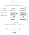
- the last step of the real-time methods of the invention involves quantifying the results of the real-time detection against a standard curve for the methylated or unmethylated gene of interest, and optionally the reference gene (where included).
- Standard curves may be generated using a set of standards. Each standard contains a known copy number, or concentration, of the gene of interest and/or reference gene as appropriate.
- a baseline value of fluorescence will be set to account for background fluorescence.
- the Sequence Detection System (SDS) software is utilised. This software sets a default baseline range of cycles 3 to 15 of the amplification reaction before amplification products are detected. A threshold value of fluorescence is then defined at a statistically significant value above this baseline.
- SDS Sequence Detection System
- the threshold is set to 10 standard deviations above the baseline fluorescence.
- Appropriate software is provided with apparatus for carrying out real-time amplification reactions. The software automatically calculates the baseline and threshold values for the reaction. The threshold cycle value (Ct) can then be determined for each standard. This is the number of cycles required to achieve the threshold amplification level. Thus, the greater the initial concentration of the gene standard in the reaction mixture, the fewer the number of cycles required to achieve a particular yield of amplified product. A plot of Ct against the log 10 of the known initial copy number of the set of standard DNAs produces a straight line. This is the standard curve.
- the Ct value for the amplification of the gene of interest and reference gene can each be interpolated against the respective standard curve in order to determine the copy number in the DNA-containing sample.
- the output of the method is the gene copy number for each of the gene of interest and reference gene.
- the results may be normalised by dividing the gene copy number of the methylated or unmethylated gene of interest by the gene copy number of the reference gene.
- the Applied Biosystems 7900 HT fast real-time PCR system is used to carry out the methods of the invention.
- SDS software is utilised, preferably including a suitable algorithm such as the Auto CT algorithm for automatically generating baseline and threshold values for individual detectors.
- the cycle threshold value is less than (or equal to) around 40 (such as 35 to 45, or 37 to 43, or 38 to 42 or 39 to 41), sensitive and specific results can be obtained. If the threshold is only crossed after more than 40 cycles, the amplification is considered invalid and the results are not utilised.
- the amplification is considered valid where the slope of the standard curve for the methylated or unmethylated gene of interest and reference gene is at least -4, indicating an amplification efficiency of at least 77%.
- the amplification is considered valid where the coefficient of determination (R 2 ) for at least four data points on each curve is above 0.990.
- the amplification is considered valid where, in a parallel reaction using the same reagents, there is no amplification of a sample containing no DNA at the cycle threshold value of less than 40. Any suitable sample lacking a DNA component, such as a water sample for example, may be utilised.
- the amplification is considered valid where, in a parallel reaction using the same reagents, there is detectable amplification of a positive control sample known to contain the gene of interest in methylated form at the cycle threshold value of less than 40.
- a positive control sample known to contain the gene of interest in methylated form at the cycle threshold value of less than 40.
- Any suitable positive control sample may be utilised.
- the positive control sample known to contain methylated MGMT is preferably derived from SW48 cells.
- a further validation criterion comprises the amplification being considered valid where, in a parallel reaction using the same reagents, there is no detectable amplification of a negative control sample known to contain the gene of interest in unmethylated form at the cycle threshold value of less than 40. Any suitable negative control sample may be utilised.
- the negative control sample known to contain unmethylated MGMT is preferably derived from HT29 cells.
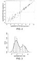
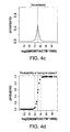
- the method may further comprise carrying out a statistical analysis of the results obtained. For example, density plots of the results obtained may be generated to provide an indication of the distribution of results. Log 2 transformation may be utilised to produce density plots and also assess the reproducibility of the methods by a direct comparison of duplicate experiments. Cohen's Kappa values may be calculated to provide an indication of the reproducibility of the methods (through comparison of results obtained through duplicate experiments or by comparing results obtained with the claimed methods with other known methods). Receiver Operating Characteristics (ROC) curves may be plotted to determine the specificity (proportion of true negative results) and sensitivity (proportion of true positive results) of the methods. The area under the ROC curve (AUC) provides an indication of the overall performance of the method.
- ROC Receiver Operating Characteristics
- the AUC is at least 0.90, 0.91, 0.92, 0.93, 0.94, 0.95, 0.96, 0.97, 0.98 or 0.99 for the methods of the invention.
- the software environment R (available at http://www.r-project.org/) is utilised to carry out the analysis.
- the at least one primer pair for amplification of the methylated or unmethylated gene of interest/reference gene is used in the amplification at a concentration of approximately 50 to 150nM. More preferably, the at least one primer pair for amplification of the methylated or unmethylated gene of interest/reference gene is used in the amplification at a concentration of approximately 100nM.
- amplification is carried out using the polymerase chain reaction (PCR).
- PCR polymerase chain reaction
- isothermal amplification techniques such as NASBA, 3SR, TMA and triamplification, all of which are well known in the art and suitable reagents are commercially available.
- suitable amplification methods include, without limitation, the ligase chain reaction (LCR) (Barringer et al, 1990), MLPA, selective amplification of target polynucleotide sequences ( US Patent No.
- the real-time PCR methods of the invention generally involve steps of lowering the temperature to allow primer annealing, raising the temperature for primer extension, raising the temperature for denaturation and lowering the temperature for data-collection.
- the data-collection step is carried out at a temperature of between approximately 60°C and 64°C, most preferably at approximately 62°C since this has been shown to give maximally sensitive and specific results (as discussed in Example 2 below).
- Data collection may be carried out for a suitable period of time, such as 30 seconds or 1 minute per cycle.
- Annealing temperatures may be varied as would be appreciated by one skilled in the art, depending upon the primers utilised in the reaction. Suitable annealing temperatures herein may be between 50°C and 90°C. Particularly useful annealing temperatures have been shown to be between 55°C and 65°C, especially 57°C and/or 62°C. These annealing temperatures may be used for any gene of interest, as indicated herein.
- the thermal profiling of the polymerase chain reaction comprises between 40 and 50 repeats, preferably approximately 45 repeats of the cycle:
- the methods of the invention can be used in order to detect more than one gene of interest in the same reaction.
- amplification of several nucleic acid targets can be performed in the same reaction mixture. This may be termed "multiplexing".
- one or both primers for each target may be hairpin primers labelled with a fluorescent moiety and a quenching moiety that form a FRET pair.
- Amplification of several nucleic acid targets requires that a different fluorescent donor and/or acceptor moiety, with a different emission wavelength, be used to label each set of primers.
- the reaction mixture is illuminated and read at each of the specific wavelengths characteristic for each of the sets of primers used in the reaction.
- Multiplexing can also be utilised in the context of detecting both the gene of interest and a reference gene in the same reaction.
- primers labelled with appropriate distinguishable donor and/or acceptor moieties allow the signal generated by amplification of the gene of interest and reference gene respectively to be distinguished.
- a universal quencher is utilised together with suitable fluorophore donors each having a distinguishable emission wavelength maximum.
- a particularly preferred quencher is DABCYL. Together with DABCYL as quencher, the following fluorophores may each be utilised to allow multiplexing: Coumarin (emission maximum of 475nm), EDANS (491nm), fluorescein (515nm), Lucifer yellow (523nm), BODIPY (525nm), Eosine (543nm), tetramethylrhodamine (575nm) and texas red (615nm) ( Tyagi et al., Nature Biotechnology, Vol. 16, Jan 1998; 49-53 ). Other preferred combinations are discussed herein.
- the DNA-containing sample can be split and the methods of the invention carried out on suitable portions of the sample in order to obtain directly comparable results.
- the sample may be split two ways to allow detection of amplification of the gene of interest in real time in one sample portion and detection of amplification of the reference gene in real time in the other sample portion.
- the sample may be split further to allow suitable control reactions to be carried out, as required.
- the benefit of this scheme is that a universal FRET pair can be used to label each primer pair and removes the requirement to detect emission at a range of wavelengths.
- this method does rely upon obtaining a suitable sample initially to permit dividing the sample.
- the total reaction volume for the amplification step is between approximately 10 and 40 ⁇ l, more preferably between approximately 20 and 30 ⁇ l and most preferably around 25 ⁇ l.
- epigenetic silencing of the MGMT gene has been shown to correlate with improved survival in several studies with glioma patients treated with alkylating agent therapy (7) and has been substantiated in two clinical trials (8, 9).
- the methylation status of MGMT is believed to have a predictive value for benefit from the addition of the alkylating agent TMZ (9, 10).
- this epigenetic alteration in tumors can be exploited in a diagnostic test to predict benefit from alkylating agent therapy for individualized management of patients.
- MGMT methylation status may also predict benefit from alkylating agent containing therapy in patients with low grade glioma, oligodendroglioma, and diffuse large B-cell lymphoma.
- the invention provides a method of predicting the likelihood of successful treatment of a cell proliferative disorder in a subject using an alkylating chemotherapeutic agent comprising, in a DNA-containing sample isolated from the subject, detecting the presence and/or amount of methylated MGMT gene in the sample by carrying out a method of the invention, wherein the presence of methylated MGMT in the sample indicates that the likelihood of successful treatment using the alkylating chemotherapeutic agent is higher than if no or lower levels of methylated MGMT is detected.
- the reverse situation is also applicable and so the methods of the invention may likewise be utilised in order to determine whether there is likely to be resistance to, or unsuccessful treatment using, an alkylating chemotherapeutic agent - the absence of methylated MGMT in the sample indicates there is likely to be resistance to treatment and/or that treatment is likely to be unsuccessful.
- the methods of the invention may also be utilised to select a suitable course of treatment for a patient - the presence of methylated MGMT indicates alkylating agents may be beneficially administered, whereas the absence or low level of methylated MGMT indicates alkylating agents are contra-indicated.
- the discussion provided in respect of the methods, primers and kits of the invention applies to the present aspect mutatis mutandis and all embodiments are therefore envisaged, as appropriate, for this aspect of the invention.
- the cell proliferative disorder is cancer.
- the cancer is cancer of the central nervous system and especially the brain.
- the cancer of the brain comprises, consists essentially of or consists of a glioma.
- the glioma comprises, consists essentially of or consists of a glioblastoma or oligodendroglioma.
- the cell proliferative disorder is diffuse large B-cell lymphoma.
- the alkylating agent may be any suitable agent useful for treating a cellular proliferative disorder.
- the alkylating chemotherapeutic agent is selected from carmustine, lomustine, cisplatin, carboplatin, mechlorethamine, cyclophosphamide, ifosfamide, melphalan, chlorambucil, busulfan, thiotepa, dacarbazine, temozolamide or procarbazine. All of these agents are known in the art.
- the invention provides a method of detecting a predisposition to, or the incidence of, cancer of the central nervous system and especially the brain or diffuse large B cell lymphoma in a sample comprising detecting methylation of the MGMT gene using the methods of the invention, wherein detection of methylation is indicative of a predisposition to, or the incidence of, cancer and in particular cancer of the central nervous system and especially the brain or diffuse large B cell lymphoma.
- WRN and colorectal cancer BRCA1 and breast cancer
- NDRG4 and colorectal cancer and PTEN and cancers such as thyroid carcinomas, melanoma, Leukaemia and gynaecological cancers such as cervical and ovarian cancers.
- the invention provides a method for determining the histopathological stage of cancer of the central nervous system and especially the brain or diffuse large B cell lymphoma in a sample comprising detecting methylation of the MGMT gene using the methods of the invention, wherein detection of methylation is indicative of the histopathological stage of the cancer of the central nervous system and especially the brain or diffuse large B cell carcinoma.
- All embodiments of the methods of the invention are hereby incorporated as appropriate and are not repeated for reasons of conciseness.
- the "stage" of a cancer is a descriptor (usually numbers I to IV) of how much the cancer has spread.
- the stage often takes into account the size of a tumour, how deep it has penetrated, whether it has invaded adjacent organs, if and how many lymph nodes it has metastasized to, and whether it has spread to distant organs. Staging of cancer is important because the stage at diagnosis is the biggest predictor of survival, and treatments are often changed based on the stage.
- corresponding methods are envisaged for WRN and colorectal cancer, BRCA1 and breast cancer, NDRG4 and colorectal cancer and PTEN and cancers such as thyroid carcinomas, melanoma, Leukaemia and gynaecological cancers such as cervical and ovarian cancers.
- Testing can be performed diagnostically or in conjunction with a therapeutic regimen. Epigenetic loss of function of the MGMT gene can be rescued by the use of DNA demethylating agents and/or DNA methyltransferase inhibitors. Testing can be used to determine what therapeutic or preventive regimen to employ on a patient and be used to monitor efficacy of a therapeutic regimen.
- the methods of the invention are applied in a method for predicting the likelihood of successful treatment of cancer of the central nervous system and especially the brain or diffuse large B cell lymphoma with a DNA demethylating agent and/or a DNA methyltransferase inhibitor and/or HDAC inhibitor comprising detecting methylation of the MGMT gene using the methods of the invention, wherein detection of methylation is indicative that the likelihood of successful treatment is higher than if the epigenetic modification is not detected.
- the invention provides for a method for predicting the likelihood of resistance to treatment of cancer of the central nervous system and especially the brain or diffuse large B cell lymphoma with a DNA demethylating agent and/or a DNA methyltransferase inhibitor and/or HDAC inhibitor comprising detecting methylation of the MGMT gene using the methods of the invention, wherein detection of methylation is indicative that the likelihood of resistance to treatment is lower than if the epigenetic modification is not detected.
- the patient population may be selected for treatment on the basis of their methylation status with respect to the MGMT gene. This leads to a much more focussed and personalised form of medicine and thus leads to improved success rates since patients will be treated with drugs which are most likely to be effective.
- WRN and colorectal cancer BRCA1 and breast cancer
- NDRG4 and colorectal cancer and PTEN and cancers such as thyroid carcinomas, melanoma, Leukaemia and gynaecological cancers such as cervical and ovarian cancers.
- the disclosure further provides for a method of selecting a suitable treatment regimen for cancer of the central nervous system and especially the brain or diffuse large B cell lymphoma comprising detecting methylation of the MGMT gene using the methods of the invention, wherein detection of methylation results in selection of a DNA demethylating agent and/or a DNA methyltransferase inhibitor and/or a HDAC inhibitor for treatment and wherein if methylation is not detected, a DNA demethylating agent and/or a DNA methyltransferase inhibitor and/or a HDAC inhibitor is not selected for treatment.
- alternative treatments should be explored.
- the disclosure provides for a method of treating cancer and in particular cancer of the central nervous system and especially the brain or diffuse large B cell lymphoma comprising administration of a DNA demethylating agent and/or a HDAC inhibitor and/or a DNA methyltransferase inhibitor wherein the subject has been selected for treatment using the methods of the invention.
- a DNA demethylating agent and/or a HDAC inhibitor and/or a DNA methyltransferase inhibitor wherein the subject has been selected for treatment using the methods of the invention.
- the disclosure provides for the use of a DNA demethylating agent and/or a DNA methyltransferase inhibitor and/or HDAC inhibitor (in the manufacture of a medicament for use) in treating cancer of the central nervous system and especially the brain or diffuse large B cell lymphoma in a subject, wherein the subject has been selected for treatment on the basis of the methods of the invention.
- WRN and colorectal cancer BRCA1 and breast cancer
- NDRG4 and colorectal cancer and PTEN and cancers such as thyroid carcinomas, melanoma, Leukaemia and gynaecological cancers such as cervical and ovarian cancers.
- the DNA demethylating agent may be any agent capable of up regulating transcription of the appropriate gene.
- a preferred DNA demethylating agent comprises, consists essentially of or consists of a DNA methyltransferase inhibitor.
- the DNA methyltransferase inhibitor may be any suitable inhibitor of DNA methyltransferase which is suitable for treating cancer in the presence of methylation of the relevant gene. The link between gene methylation and a respective cancer type is known for these genes and so preventing this methylation is predicted to help to treat cancer.
- the DNA methyltransferase inhibitor may, in one embodiment, be one which reduces expression of DNMT genes, such as suitable antisense molecules, or siRNA molecules which mediate RNAi for example.
- suitable antisense molecules or siRNA molecules which mediate RNAi for example.
- the design of a suitable siRNA molecule is within the capability of the skilled person and suitable molecules can be made to order by commercial entities (such as Ambion (www.ambion.com)).
- the DNA methyltransferase gene is (human) DNMT1.
- the agent may be a direct inhibitor of DNMTs.
- modified nucleotides such as phosphorothioate modified oligonucleotides (see Fig. 6 of Villar-Garea, A. And Esteller, M. DNA demethylating agents and chromatin-remodelling drugs: which, how and why? Current Drug Metabolism, 2003, 4, 11-31 ) and nucleosides and nucleotides such as cytidine analogues.
- cytidine analogues include 5-azacytidine, 5-aza-2'-deoxycytidine, 5-fluouro-2'-deoxycytidine, pseudoisocytidine, 5,6-dihydro-5-azacytidine, 1- ⁇ -D-arabinofuranosyl-5-azacytosine (known as cambarine) (see Fig. 4 of Villar-Garea, A. And Esteller, M. DNA demethylating agents and chromatin-remodelling drugs: which, how and why? Current Drug Metabolism, 2003, 4, 11-31 ).
- the DNA methyltransferase inhibitor comprises Decitabine. Full details of this drug can be found at www.supergen.com for example.
- Additional DNMT inhibitors include S-Adenosyl-Methionine (SAM) related compounds like ethyl group donors such as L-ethionine and non-alkylating agents such as S-adenosyl-homocysteine (SAH), sinefungin, (S)-6-methyl-6-deaminosine fungin, 6-deaminosinefungin, N4-adenosyl-N4-methyl-2,4-diaminobutanoic acid, 5'-methylthio-5'-deoxyadenosine (MTA)and 5'-amino-5'-deoxyadenosine ( Villar-Garea, A. And Esteller, M. DNA demethylating agents and chromatin-remodelling drugs: which, how and why? Current Drug Metabolism, 2003, 4, 11-31 ).
- SAM S-Adenosyl-Methionine
- SAH S-adenosyl-homocysteine
- agents which may alter DNA methylation and which may, therefore, be useful in the present compositions include organohalogenated compounds such as chloroform etc, procianamide, intercalating agents such as mitomycin C, 4-aminobiphenyl etc, inorganic salts of arsenic and selenium and antibiotics such as kanamycin, hygromycin and cefotaxim ( Villar-Garea, A. And Esteller, M. DNA demethylating agents and chromatin-remodelling drugs: which, how and why? Current Drug Metabolism, 2003, 4, 11-31 ).
- organohalogenated compounds such as chloroform etc, procianamide
- intercalating agents such as mitomycin C, 4-aminobiphenyl etc
- inorganic salts of arsenic and selenium such as kanamycin, hygromycin and cefotaxim ( Villar-Garea, A. And Esteller, M. DNA demethylating agents and chromatin-remodelling drugs: which, how and why
- Particulary preferred DNMT inhibitors in the present invention comprise, consists essentially of or consists of 5-azacytidine and/or zebulaine.
- the histone deacetylase (HDAC) inhibitor may comprise at least one of trichostatin A (TSA), suberoyl hydroxamic acid (SBHA), 6-(3-chlorophenylureido)caproic hydroxamic acid (3-Cl-UCHA), m-carboxycinnamic acid bishydroxylamide (CBHA), suberoylanilide hydroxamic acid (SAHA), azelaic bishydroxamic acid (ABHA), pyroxamide, scriptaid, aromatic sulfonamides bearing a hydroxamic acid group, oxamflatin, trapoxin, cyclic-hydroxamic-acid containing peptides, FR901228, MS-275, MGCD0103, short-chain fatty acids and N-acetyldinaline.
- TSA trichostatin A
- SBHA suberoyl hydroxamic acid
- Example 1 Correlation between testing MGMT gene promoter methylation status of glioblastoma tissue using nested gel-based and direct real-time fluorescence -based Methylation Specific PCR
- FFPE glioma tissues predominantly glioblastoma, were obtained from patients who had given their informed consent. Patients were enrolled in clinical trials coordinated at the University Hospitals in Lausanne (Switzerland), Rotterdam (The Netherlands), and Regensburg (Germany). The correlations of the MGMT status with survival will be part of the publications reporting the respective trials (Stupp et al .; van den Bent et al.; Hau e t al .; manuscripts in preparation). Of these, 125 were tested once by the CHUV (gel-based MSP), 29 were tested once by ONCO (real-time MSP), and 105 were tested in duplicate by ONCO (real-time MSP). See Table 1 for further details.
- Table 1 Summary of the samples evaluated by both assays Site Samples Duplicate test Single test Total tests Valid tests [%] ONCO 134 105 (210 tests) 29 239 225/239 [94.2%] CHUV 125 0 125 125 94/125 [75.2%]
- HE Hematoxylin-Eosin
- the resulting PCR product of 289bp served as a template for the second, methylation-specific PCRs, amplifying either m_MGMT or u_MGMT, yielding PCR products of 81 and 93bp, respectively ( 7 ).
- the specific primers for m_MGMT recognize the fully methylated sequence.
- the u_MGMT assay served as a control for the PCR.
- the first PCR consisted of 35 cycles. Two ⁇ l of the first PCR was diluted 1/20 and input to the second PCR of 30 cycles. The products from the second PCR were visualized on 4% agarose gels (NuSieve 3:1) to determine the MGMT methylation status.
- the outcome of the nested gel-based MSP was considered valid when the following four criteria were fulfilled: a) PCR products of the expected sizes were detected on agarose gel (single bands); b) either a band for u_MGMT, m_MGMT, or both was present; c) routinely included positive and negative controls, including a no-template control (NTC) gave the expected result, and d) the result was confirmed in an independent experiment starting with the bisulphite treatment of DNA. The valid results of all clinical samples were classified as methylated or non-methylated depending on the presence or absence of a band for m_MGMT. This procedure was used for the analysis of all samples in the laboratory in the CHUV.
- DNA was extracted using the classical phenol/chloroform extraction method and resuspended in 50 ⁇ l LoTe (3mM TRIS, 0.2mM EDTA, pH 8.0). The DNA was quantified using the Picogreen® dsDNA quantitation kit (Molecular Probes, Invitrogen) following manufacturer's recommendations. ⁇ DNA provided with the kit was used to prepare a standard curve (from 1 to 800ng/ml). The data were collected using a FluoStar Galaxy plate reader (BMG Lab technologies, Germany).
- Plasmid DNA was prepared by cloning of the relevant sequences into TOPO® TA vectors (TOPO® cloning kit, Invitrogen®).
- m_MGMT was obtained from the methylated sequence present in the SW48 cell line.
- QIAprep® spin midiprep kit Qiagen GmbH; according to the manufacturer's protocol
- sequences were confirmed by MWG Biotech, Germany (data not shown).
- the plasmids were then linearized by digestion with the restriction enzyme BamHI (Roche).
- the linearized plasmid was purified using the QIAquick® PCR purification kit (Qiagen GmbH; according to the manufacturer's protocol).
- the plasmid concentration was determined by OD 260 measurement.
- a stock solution of 2x10 7 copies/5 ⁇ l (4x10 6 copies/ ⁇ l) was prepared and stored at -80°C until use. Dilutions of standard curves (2x10 6 -2x10 1 copies/5 ⁇ l) for m_MGMT and ACTB were freshly prepared for each experiment.
- the analyte (m_MGMT and ACTB) quantifications were performed by real-time PCR assays. These consisted of parallel amplification / quantification processes using specific primer and primer/detector pairs for each analyte using the Amplifluor® assay format on an ABI Prism® 7900HT instrument (Applied Biosystems).
- the analyte defined in the real-time PCR was the MGMT promoter sequence and detects the fully methylated version.
- ACTB was used as a reference gene in the assay.
- the Amplifluor® forward primers are preceded by the detection elements (underlined).
- the amplicon size is 136bp for the m_MGMT analyte, and 125bp for the ACTB analyte. These amplicons include the Amplifluor detection sequence. Sequence details for both forward and reverse primers are as follows:
- the MGMT target sequence is located on chromosome 10 between positions 131155505 and 131155619, while the ACTB target sequence is located on chromosome 7 between positions 5538428 and 5538325, based on version 36.1 of the NCBI human genome.
- the final primer concentrations in the reaction mix were 100nM for both forward primer/detector and reverse primer.
- 12.5 ⁇ l of iTaqTM Supermix with Rox (BioRad, 2xbuffer) were used per PCR reaction.
- results of a run were considered valid when the following five criteria were met: a) slopes of both standard curves above -4 (PCR efficiency > 77.8%); b) R 2 of at least 4 relevant data points above 0.990; c) routinely included NTC not amplified; d) 10% of a 1 ⁇ g conversion reaction of the positive cell line assay control SW48 was detectable; and e) 10% of a 1 ⁇ g conversion reaction of the negative cell line assay control HT29 was not detected within the standard curve.
- the m_MGMT copy numbers derived were divided by the ACTB copy numbers for that sample. This figure was multiplied by 1000 for convenient handling, and the result referred to as the ratio value.
- the ratio values were log 2 transformed.
- the evaluation of the distribution of the m_MGMT measurements in the density plot was performed using 99 duplicate samples (see Table 2 for more details). Gaussian mixture models were fitted to the average data log 2 (1000*m_MGMT/ACTB) of the real-time MSP duplicates ( 20-22 ).
- FFPE glioma tissues were analyzed in parallel to determine the methylation status of the MGMT promoter in independent laboratories using two distinct technical approaches. This blinded study compared the results obtained by a real-time MSP on a high throughput platform to the gel-based MSP assay previously shown to predict benefit from the addition of the alkylating agent temozolomide to the treatment of newly diagnosed glioblastoma in two clinical trials ( 8, 9 ).
- the assays were compared by evaluating 134 FFPE glioma samples by real-time MSP (29 single and 105 duplicate tests) and 125 FFPE glioma samples by gel-based MSP (all single tests as defined in references 2 and 3).
- the real-time MSP assay produced valid results in 94.2% versus 75.2% of valid tests for the gel-based assay (Table 1).
- the duplicate real-time MSP tests 5 of 6 invalid results were duplicated.
- the validity rates are based on the respective validation criteria for each assay (for gel-based MSP see references 2 and 3, for real-time MSP see Figure 1 ).
- a sensitive and specific real-time MSP assay has been developed to reliably detect the methylation status of the MGMT gene promoter in clinical samples of FFPE tissues for diagnostic purposes.
- the methylation status result of this test is in good concordance with results obtained with the gel-based MSP assay that established the predictive value of the MGMT methylation status in glioblastoma for benefit from temozolomide therapy ( 9 ).
- normalization provides a more reproducible assay, it may contribute to the few discrepants seen in this study, as they are mostly samples with detectable levels of m_MGMT but also high levels of ACTB.
- Some discordance is not surprising, since the cut-off of the gel-based MSP assay is defined by visual presence or absence of the PCR product without normalization. Normalization to DNA content, measured here as ratio to ACTB, is not an absolute measurement, since "normal tissue" contaminations are present in all clinical tumor samples, and genomic copy number aberrations are common in glioblastoma, including on chromosomes 10 and 7, where the MGMT and ACTB gene reside, respectively.
- ACTB is also used as a control gene by other groups using real-time PCR for quantitative DNA methylation analysis ( 17 ).
- the real-time MSP assay more often yielded a valid result with FFPE tissue specimens than the gel-based MSP assay. This improved performance is likely due to the smaller amplicon size of the real-time assay that is more appropriate for FFPE samples known to yield limited DNA quality.
- the test described here is being prospectively used for randomizing patients in an ongoing clinical phase III trial (RTOG 0525/ EORTC 26052-22053) testing standard versus dose-intense adjuvant TMZ in patients with newly diagnosed glioblastoma (trial is reviewed in Stupp et al. ( 11 )).
- RTOG 0525/ EORTC 26052-22053 testing standard versus dose-intense adjuvant TMZ in patients with newly diagnosed glioblastoma (trial is reviewed in Stupp et al. ( 11 )).
- MGMT methylation status for predicting benefit from alkylating agent therapy and it will provide further information on a clinically relevant cut-off for m_MGMT.
- This sensitive and robust high through-put test for evaluating the methylation status of the MGMT gene may provide an important pharmacogenomic tool for individualized management of patients considered for treatment with temozolomide or other alkylating agent chemotherapy.
- Amplifluor is a primer-based methodology. It lacks the third oligo nucleotide "probe" that typifies TaqMan and Molecular Beacon technologies. Without this third layer of specificity, amplifluor primers can fluoresce in response to non-specific amplification such as primer-dimers. Therefore it is important to carefully select primer sequences to overcome this behavior.
- Table 4 gives an overview of the positive Ct values observed for 12 NTC samples tested through the initial MGMT amplifluor assay.
- Table 4 NTC performance initial MGMT amplifluor design Sample Task Ct PMR91 1 NTC 37.38 2 NTC 36.78 3 NTC 36.67 4 NTC 37.15 5 NTC 36.69 6 NTC 36.59 7 NTC 37.17 8 NTC 36.32 9 NTC 37.71 10 NTC 36.71 11 NTC 37.86 12 NTC 36.65
- Table 6 summarizes the Ct values obtained for the 6 new primer designs. A high variation in NTC performance was observed depending on the chosen primer set. The best NTC performance was obtained for PMR105: 13 out of 14 tested water controls resulted in Ct values above 40.
- the applied test run validation criteria for the amplifluor assay are defined in such a way that a "valid" Ct value has to be ⁇ 40 (based on a theoretical definition: if there was one copy in the PCR reaction to be amplified it would take 40 cycles at a PCR efficiency of 100%). Ct values above 40 are considered as 'invalid'. Accordingly, a signal is considered to be negative in real-time MSP when it is classified as "undetermined" or when its Ct value is above 40.
- Reading at 68.6°C results in a considerable loss of sensitivity compared to a reading temperature of 62°C. It can be concluded that best workable results for the MGMT Amplifluor assay are obtained using the reverse primer design PMR105 in combination with a reading temperature of 62°C.
- the quantitation process depends on fluorescent light which is emitted only when the detector is bound to its complementary sequence.
- the final primer concentrations in the reaction mix were 100nM for both forward primer/detector and reverse primer.
- 12.5 ⁇ l of iTaqTM Supermix with Rox (BioRad, 2xbuffer) were used per PCR reaction.
- Plasmid material used as standard curve was generated as follows: the promoter sequence as defined by the primers is PCR amplified and cloned (using suitable isolated and bisulphite modified cell line DNA). The sequence is verified by sequencing and compared to the published promoter sequence.
- a standard curve (2x10 6 - 20 copies) was included to determine copy numbers of unknown samples by interpolation of their Ct values to the standard curve.
- ⁇ -Actin was used as a reference gene in the assay.
- a BRCA1 amplifluor assay was designed to evaluate the methylation status of the BRCA1 gene in breast cancer patients.
- Primer and detector sequences are detailed in Table 9.
- Table 9 Primer and amplifluor detector sequences
- BRCA1 Name Sequence BRCA1 forward detector 5' - AGCGATGCGTTCGAGCATCGCUTCGTGGTAACGGAAAAGCGC - 3' (SEQ ID NO: 1 plus SEQ ID NO: 6)
- Table 10 performance BRCA1 standard curve Quantity Standards Ct 1 Ct 2 Average Ct ⁇ Ct 2000000 18.39 18.33 18.36 0.06 200000 22.30 22.15 22.22 0.15 20000 26.19 26.17 26.18 0.03 2000 30.12 30.24 30.18 0.11 200 33.97 33.92 33.95 0.04 20 37.92 37.90 37.91 0.02 NTC 42.30 41.55 41.93
- the BRCA1 methylation status was investigated for 40 breast tumor samples and 10 normal breast samples.
- the Ct values were used to calculate the copy numbers for each sample based on a linear regression of the standard curve values.
- the BRCA1 copy numbers were divided by the ⁇ -Actin copy numbers and multiplied by 1000 for convenient handling; results were referred to as the ratio value (see Table 11). Invalid results are due to ⁇ -Actin copy numbers below 200 in the respective samples.
- a serial dilution of WRN plasmid material (2x10 6 to 2x10 1 copies/5 ⁇ l) was loaded in duplicate using the above specified primer and Amplifluor detector sequences using an optimized thermal profile: Stage1: 50°C for 2 min, Stage2: 95°C for 10min, Stage3: 95°C for 15sec, 61°C for 30sec, 61°C for 30sec ( plateau-data collection) for 45 repeats.
- Different primer combinations were assessed for WRN. Results were generated (see Table 13) using the SDS 2.2 software (Applied Biosystems), exported as Ct values (cycle number at which the amplification curves cross the threshold value, set automatically by the software). Good performance of the standard curve was obtained for all assays.
- the WRN methylation status was investigated for 56 colorectal cancer samples and 39 normal samples.
- the Ct values were used to calculate the copy numbers for each sample based on a linear regression of the standard curve values.
- the WRN copy numbers were divided by the ⁇ -Actin copy numbers and multiplied by 1000 for convenient handling; results were referred to as the ratio value (see Table 14).
- Table 14 Preliminary results WRN amplifluor assay on clinical material Samples Group ratio WRN_NOR1F/b-acting X1000 ratio WRN_NOR1R/b-actin X1000 ratio WRN_NOR2R/b-actin X1000 ratio WRN_NOR4R/b -actin X1000 Cut_off 39.07 13.44 3.23 6.70 Specificity % 100 100 100 100 Sensitivity % 41 49 30 50
- PTEN amplifluor assays were designed to evaluate the methylation status of the PTEN gene in cancerous patients. Sequence details can be found in Table 16. Table 16. Table 16. : Primer and amplifluor detector sequences PTEN Name Sequence PTEN 11 forward detector 5' - AGCGATGCGTTCGAGCATCGCUGTGTTTACGTTTAGTACGTTCGGT - 3' (SEQ ID NO: 1 plus SEQ ID NO: 20) PTEN 11 reverse primer 5' - TCATCCGACTCCCTTACAACG - 3' (SEQ ID NO: 21) PTEN 12 forward detector 5' - AGCGATGCGTTCGAGCATCGCUTAGTTTTGGGTGCGAGCGTAG - 3' (SEQ ID NO: 1 plus SEQ ID NO:22) PTEN 12 reverse primer 5' - GCGTTACTACAAAAACCGCAA - 3' (SEQ ID NO: 23) PTEN 6 forward detector 5' - AGCGATGCGTTCGAGCATCGCUTGGTATATTTAGGGATTCGGGTC
- NDRG4 amplifluor assays were tested to evaluate the methylation status of the NDRG4 gene in colorectal cancer patients. Especially short amplifluor assays were designed to favour the detection of low-molecular-weight DNA, often present in plasma/serum samples in the form of fragmented DNA. Primer and detector sequences are detailed in Table 18.
- NDRG4_66304_S_AMP forward detector AGCGATGCGTTCGAGCATCGCUTTCGGTGAATTTTAGGAGGC (SEQ ID NO: 1 plus SEQ ID NO: 26)
- NDRG4_66304_AS reverse primer TCGAACGACGAACACGAAA (SEQ ID NO: 27)
- NDRG4_72006_S_AMP forward detector AGCGATGCGTTCGAGCATCGCUCGTTCGGGATTAGTTTTAGGTTC (SEQ ID NO:1 plus SEQ ID NO:28)
- NDRG4_72006_AS reverse primer AATTTAACGAATATAAACGCTCG (SEQ ID NO: 29)
- NDRG4_72007_S_AMP forward detector AGCGATGCGTTCGAGCATCGCUGGTATTTTAGTCGCGTAGAAGGC (SEQ ID NO: 1 plus SEQ ID NO:
- Genomic DNA was extracted using a standard method (phenol/chloroform), followed by sodium bisulfite treatment (BT) using the EZ DNA Methylation kit from Zymo Research.
- the chemically treated DNA was used as template for real-time MSP.
- Methylation levels of the NDRG4 gene promoter were determined by real-time MSP using specific primers and probes for the molecular beacon format NDRG4_1a (details of this assay have previously been provided in International Publication WO08/084219 ) and amplifluor format: NDRG4_72007 and NDRG4_72008.
- Results are presented in Table 20. Higher copy numbers are obtained using the amplifluor format. Performance characteristics of the different NDRG4 assays: NDRG4_1a (beacon), NDRG4_72007 (amplifluor), and NDRG4_72008 (amplifluor) demonstrated a sensitivity of respectively 20%, 50%, and 50% with a specificity of 100% for the tested CRC diluted plasma samples. These preliminary data indicate that a short amplifluor assay could provide a better alternative than beacon assay (technically difficult to shorten the assay) for CRC methylation detection in plasma DNA samples (high DNA fragmentation nature).
- the NDRG4 methylation status was further investigated in 88 CRC FFPE (formalin fixed paraffin embedded) samples and 10 normal FFPE samples.
- Formalin Fixed paraffin embedded samples were first deparaffinized in 750 ⁇ l xylene for 2h. Then, 250 ⁇ l of 70% ethanol was added before centrifugation at 13000rpm for 15min. The supernatant was removed and the samples were air dried at room temperature.
- Genomic DNA was extracted using a standard method (phenol/chloroform), followed by sodium bisulfite treatment (BT) using the EZ DNA Methylation kit from Zymo Research. 1.5 ⁇ l of chemically treated DNA was used as template for real-time MSP. Methylation levels of the NDRG4 gene promoter were determined by real-time MSP using specific primers and probes for the molecular beacon format NDRG4_1a and amplifluor format: NDRG4_72007. Several annealing temperatures were investigated (57°C, 62°C and 80.5°C) to further optimize the NDRG4_72007 amplifluor assay.
- Table 21 Performance characteristics for NDRG4_1a and NDRG4_72007 NDRG4_1a (beacon) NDRG4_72007 57°C (amplifluor) NDRG4_72007 62°C (amplifluor) NDRG4_72007 80.5°C (amplifluor) No cut-off applied Sensitivity (%) 57 83 71 56 Specificity (%) 93 64 95 100 Cut-off applied to obtain 100% specificity Cut-off ratio 2.26 80 390 / Sensitivity (%) 55 78 63 /
- FFPE paraffin-embedded
- DNA isolation, modification and analyte quantitations were conducted as previously described in example 1; amplification conditions were subject to the following modifications:
- Genomic DNA extraction, purification and modification was according to the in-house procedure used at LabCorp® Real-time MSP procedure was conducted similarly to the protocol followed by ONCO. Technical test run validation criteria are described in Table 22.
Landscapes
- Health & Medical Sciences (AREA)
- Chemical & Material Sciences (AREA)
- Life Sciences & Earth Sciences (AREA)
- Organic Chemistry (AREA)
- Proteomics, Peptides & Aminoacids (AREA)
- Engineering & Computer Science (AREA)
- Immunology (AREA)
- General Health & Medical Sciences (AREA)
- Wood Science & Technology (AREA)
- Zoology (AREA)
- Bioinformatics & Cheminformatics (AREA)
- Microbiology (AREA)
- Medicinal Chemistry (AREA)
- Veterinary Medicine (AREA)
- Public Health (AREA)
- Animal Behavior & Ethology (AREA)
- Pharmacology & Pharmacy (AREA)
- Genetics & Genomics (AREA)
- Analytical Chemistry (AREA)
- Epidemiology (AREA)
- General Engineering & Computer Science (AREA)
- Physics & Mathematics (AREA)
- Biophysics (AREA)
- Biochemistry (AREA)
- Molecular Biology (AREA)
- Biotechnology (AREA)
- Oncology (AREA)
- Pathology (AREA)
- Gastroenterology & Hepatology (AREA)
- Mycology (AREA)
- Chemical Kinetics & Catalysis (AREA)
- Hospice & Palliative Care (AREA)
- General Chemical & Material Sciences (AREA)
- Nuclear Medicine, Radiotherapy & Molecular Imaging (AREA)
- Measuring Or Testing Involving Enzymes Or Micro-Organisms (AREA)
- Medicines That Contain Protein Lipid Enzymes And Other Medicines (AREA)
Claims (14)
- Echtzeitverfahren zur Erfassung des Vorhandenseins und/oder der Menge eines betreffenden methylierten oder unmethylierten Gens in einer DNA-enthaltenden Probe, wobei das Verfahren Folgendes aufweist:(a) Kontaktieren der DNA-enthaltenden Probe mit einem Reagens, das selektiv unmethylierte Cytosinrückstände in der DNA zur Herstellung von erfassbaren modifizierten Rückständen modifiiziert, das jedoch nicht methylierte Cytosinrückstände modifiiziert(b) Verstärken von wenigstens einem Teil des betreffenden methylierten oder unmethylierten Gens unter Verwendung von wenigstens einem Primerpaar, von welchem wenigstens ein Primer zur Bindung lediglich an die Sequenz von methylierter oder unmethylierter DNA nach der Behandlung mit dem Reagens ausgestaltet ist, wobei wenigstens ein Primer in dem Primarpaar ein Primer ist, der eine Haarnadelstruktur aufweist, die eine Donator- und eine Akzeptorkomponente eines molekularen Energieübertragungspaars führt, das so angeordnet ist, dass in Abwesenheit einer Vervielfachung die Akzeptorkomponente von der Donatorkomponente bei Erregung und während der Vervielfachung abgegebene Fluoreszenz löscht, wobei die Haarnadelstruktur so unterbrochen ist, dass die Donator- und Akzeptorkomponenten ausreichend getrennt sind, um ein erfassbares Fluoreszenzsignal zu erzeugen, das in Echtzeit erfasst wird, um eine Angabe zur Genkopienzahl des betreffenden methylierten oder unmethylierten Gens bereitzustellen(c) Quantifizieren der Ergebnisse der Echtzeiterfassung im Vergleich mit einer standardmäßigen Kurve für das betreffende methylierte oder unmethylierte Gen zur Erzeugung einer Ausgabe einer Genkopienzahl;
dadurch gekennzeichnet, dass die Vervielfachung als gültig angesehen wird, wo der Zyklus-Schwellwert geringer als 40 ist, ferner dadurch gekennzeichnet, dass die Vervielfachung als gültig angesehen wird, wo es bei einer parallelen Reaktion unter Verwendung derselben Reagenzien kleine Vervielfachung einer Probe gibt, die kein DNA beim Zyklus-Schwellwert von weniger als 40 aufweist, ferner dadurch gekennzeichnet, dass die Vervielfachung unter Verwendung der Polymerase-Kettenreaktion ausgeführt wird und dass der Datenerfassungsschritt bei zwischen 60°C und 64°C ausgeführt wird und ferner dadurch gekennzeichnet, dass das wenigstens eine Primerpaar zur Vervielfiachung des betreffenden methylierten oder unmethylierten Gens ein Vervielfachungsprodukt von zwischen 50 und 250bp erzeugt. - Verfahren nach Anspruch 1, das ferner durch Verstärken von wenigstens einem Teil eines Bezugsgens unter Verwendung von wenigstens einem Primerpaar gekennzeichnet ist, wobei wenigstens ein Primer in dem Primerpaar ein Primer ist, der eine Haarnadelstruktur aufweist, die eine Donator- und eine Akzeptorkomponente eines molekularen Energieübertragungspaars führt, das so angeordnet ist, dass in Abwesenheit einer Vervielfachung die Akzeptorkomponente von der Donatorkomponente bei Erregung und während der Vervielfachung abgegebene Fluoreszenz löscht, wobei die hlaarnadolstruktur so unterbrochen ist, dass die Donator- und Akzeptorkomponenten ausreichend getrennt sind, um ein erfassbares Fluoreszenzsignal zu erzeugen, das in Echtzeit erfasst wird, um eine Angabe zur Genkopienzahl des Bezugsgens bereitzustellen.
- Verfahren nach Anspruch 2, das ferner dadurch gekennzeichnet ist, dass die Ergebnisse der Echtzeiterfassung im Vergleich mit einer standardmäßigen Kurve für sowohl jeweils das betreffende methylierte oder unmethylierte Gen und das Bezugsgen zur Erzeugung einer Ausgabe einer Genkopienzahl quantifiziert werden und dass die Ergebnisse durch Teilen der Genkopienzahl des betreffenden methylierten oder unmethylierten Gens durch die Genkopienzahl des Bezugsgens normalisiert werden.
- Verfahren nach einem der vorhergehenden Ansprüche, das ferner dadurch gekennzeichnet ist, dass die Vervielfachung als gültig angesehen wird, wo:(a) die Neigung der standardmäßige Kurve für das betreffende methylierte oder unmethylierte Gen und optional das Bezugsgen wenigstens -4 beträgt, wodurch eine Vervielfachungseffizionz von wenigstens 77% angegeben wird,(b) das Bestimmtheitsmaß (R2) für wenigstens vier Datenpunkte auf der standardmäßigen Kurve oder den standardmäßigen Kurven mehr als 0,99 beträgt(c) es in einer parallelen Reaktion unter Verwendung derselben Reagenzien eine erfassbare Vervielfachung einer positiven Kontrollprobe, die bekanntermaßen das betreffende Gen in methylierter oder unmethylierter Form aufweist, am Zyklus-Schwellwert von weniger als 40 gibt; oder(d) es in einer parallelen Reaktion unter Verwendung derselben Reagenzien keine erfassbare Vervielfachung einer negativen Kontrollprobe, die bekanntermaßen das betreffende Gen in unmethylierter oder mothylierter Form aufweist, am Zyklus-Schwellwert von weniger als 40 gibt.
- Verfahren nach einem der vorhergehenden Anspruche, wobei das betreffende methylierte Gen aus MGMT, WRN, BRCA1, PTEN und NDRG4 ausgewählt ist.
- Verfahren nach Anspruch 5, wobei das betreffende Gen MGMT ist und die positive Kontrollprobe, die bekanntermaßen methyliertes MGMT aufweist, von SW48-Zellen abgeleitet ist und/oder wobei das betreffende Gen MGMT ist und die negative Kontrollprobe, die bekanntermaßen unmethyliertes MGMT aufweiset, von HT29-Zellen abgeleitet ist.
- Verfahren nach einem der vorhergehenden Ansprüche, wobei das wenigstens eine Primerpaar zur Vervielfachung des betreffenden methylierten oder unmethylierten Gens/ Bezugsgens bei der Verviclfaohung mit einer Konzentration von ungefähr 50 bis 150nM oder mit einer Konzentration von ungefähr 100nM verwendet wird.
- Verfahren nach einem der vorhergehenden Ansprüche, wobei der Datenerfassungsschritt bei 62°C ausgeführt wird.
- Verfahren nach Anspruch 8, wobei die thermische Profilbildung der PolymeraseKettenreaktion ungefähr 45 Wiederholungen des Zyklus aufweist:(a) 50°C über einen Zeitraum von 2 Minuten(b) 95°C über einen Zeitraum von 10 Minuten(c) 95°C über einen Zeitraum von 15 Sekunden(d) 62°C über einen Zeitraum von 1 Minute
- Verfahren nach einem der vorhergehenden Ansprüche, wobei das wenigstens eine Primerpaar für die Vervielfachung des betreffenden methylierten oder unmethylierten Genes/ Bezugsgens ein Vervielfiachungsprodukt von ungefähr 100 und 200bp erzeugt.
- Verfahren nach einem der Ansprüche 2 bis 10, wobei die Vervielfachung des betreffenden Gens und Bezugsgens gleichzeitig in derselben Reaktion ausgeführt wird.
- Verfahren nach einem der vorhergehenden Ansprüche, wobei das gesamte Reaktionsvolumen für den Vervielfachungsschritt ungefähr 25µl beträgt.
- Verfahren zur Vorhersage der Wahrscheinlichkeit einer erfolgreichen Behandlung einer Zellvermehrungsstörung bei einer Versuchsperson unter Verwendung eines alkylierenden chemotherapeutischen Mittels, das, in einer aus der Versuchsperson isolierten DNA-enthaltenden Probe, ein Erfassen des Vorhandenseins und/oder der Menge von methyliertem MGMT in der Probe durch Ausführen des Verfahrens nach einem der Ansprüche 1 bis 12 aufweiset, wobei das Vorhandensein von methyliertem MGMT in der Probe darauf hinweist, dass die Wahrscheinlichkeit einer erfolgreichen Behandlung unter Verwendung des alkylierenden chemotherapeutischen Mittels höher ist, als wenn kein oder geringere Mengen vorn methyliertem MGMT erfasst werden.
- Verfahren nach einem der vorhergehenden Ansprüche, wobei die DNA-enthaltende Probe eine Gewebeprobe aufweist.
Applications Claiming Priority (2)
| Application Number | Priority Date | Filing Date | Title |
|---|---|---|---|
| US96012807P | 2007-09-17 | 2007-09-17 | |
| PCT/GB2008/003145 WO2009037441A1 (en) | 2007-09-17 | 2008-09-17 | Improved methylation detection |
Publications (3)
| Publication Number | Publication Date |
|---|---|
| EP2201131A1 EP2201131A1 (de) | 2010-06-30 |
| EP2201131B1 true EP2201131B1 (de) | 2014-11-05 |
| EP2201131B8 EP2201131B8 (de) | 2014-12-10 |
Family
ID=40239643
Family Applications (2)
| Application Number | Title | Priority Date | Filing Date |
|---|---|---|---|
| EP08806301.1A Active EP2201131B8 (de) | 2007-09-17 | 2008-09-17 | Verbesserter methylierungsnachweis |
| EP08806298A Withdrawn EP2198045A1 (de) | 2007-09-17 | 2008-09-17 | Verbesserter nachweis der mage-a-expression |
Family Applications After (1)
| Application Number | Title | Priority Date | Filing Date |
|---|---|---|---|
| EP08806298A Withdrawn EP2198045A1 (de) | 2007-09-17 | 2008-09-17 | Verbesserter nachweis der mage-a-expression |
Country Status (18)
| Country | Link |
|---|---|
| US (4) | US9050280B2 (de) |
| EP (2) | EP2201131B8 (de) |
| JP (1) | JP5406839B2 (de) |
| KR (1) | KR20100085917A (de) |
| CN (1) | CN101932723B (de) |
| AU (1) | AU2008300397A1 (de) |
| BR (1) | BRPI0817008A2 (de) |
| CA (2) | CA2699856C (de) |
| CO (1) | CO6260156A2 (de) |
| CR (1) | CR11504A (de) |
| DO (1) | DOP2010000076A (de) |
| EA (1) | EA017879B1 (de) |
| IL (1) | IL204525A0 (de) |
| MA (1) | MA31753B1 (de) |
| MX (1) | MX2010002965A (de) |
| NZ (2) | NZ596545A (de) |
| WO (2) | WO2009037438A1 (de) |
| ZA (1) | ZA201001878B (de) |
Cited By (2)
| Publication number | Priority date | Publication date | Assignee | Title |
|---|---|---|---|---|
| US12319969B2 (en) | 2015-03-27 | 2025-06-03 | Exact Sciences Corporation | Detecting esophageal disorders |
| US12606873B2 (en) | 2013-03-14 | 2026-04-21 | Mayo Foundation For Medical Education And Research | Detecting neoplasm |
Families Citing this family (20)
| Publication number | Priority date | Publication date | Assignee | Title |
|---|---|---|---|---|
| US20130237445A1 (en) * | 2010-09-14 | 2013-09-12 | The University Of North Carolina At Chapel Hill | Methods and kits for detecting melanoma |
| WO2012129488A2 (en) * | 2011-03-23 | 2012-09-27 | Virginia Commonwealth University | Gene signatures associated with rejection or recurrence of cancer |
| CN102251033B (zh) * | 2011-07-05 | 2013-03-20 | 北京大学人民医院 | 基于mage-a3基因辅助诊断多发性骨髓瘤患者的定量检测试剂盒 |
| JP2016509480A (ja) | 2013-01-24 | 2016-03-31 | カリフォルニア インスティチュート オブ テクノロジー | 発色団に基づく特徴づけおよび検出方法 |
| US10184154B2 (en) | 2014-09-26 | 2019-01-22 | Mayo Foundation For Medical Education And Research | Detecting cholangiocarcinoma |
| JP6560336B2 (ja) * | 2015-02-26 | 2019-08-14 | 株式会社日立ハイテクノロジーズ | 核酸分子の構築方法 |
| CN105100961B (zh) * | 2015-07-23 | 2018-03-13 | 华为技术有限公司 | 视频缩略图生成方法及生成装置 |
| KR20250172724A (ko) | 2015-08-31 | 2025-12-09 | 메이오 파운데이션 포 메디칼 에쥬케이션 앤드 리써치 | 위 신생물 검출 방법 |
| US11685955B2 (en) | 2016-05-16 | 2023-06-27 | Dimo Dietrich | Method for predicting response of patients with malignant diseases to immunotherapy |
| DE102016005947B3 (de) * | 2016-05-16 | 2017-06-08 | Dimo Dietrich | Verfahren zur Abschätzung der Prognose und zur Prädiktion des Ansprechens auf eine Immuntherapie von Patienten mit malignen Erkrankungen |
| US10294518B2 (en) | 2016-09-16 | 2019-05-21 | Fluxion Biosciences, Inc. | Methods and systems for ultra-sensitive detection of genomic alterations |
| WO2018167572A1 (en) | 2017-03-17 | 2018-09-20 | Mdxhealth Sa | Mgmt epigenetic deep-sequencing assay |
| US20180271710A1 (en) * | 2017-03-22 | 2018-09-27 | Bragi GmbH | Wireless earpiece for tinnitus therapy |
| WO2018237327A1 (en) * | 2017-06-22 | 2018-12-27 | Triact Therapeutics, Inc. | Methods of treating glioblastoma |
| WO2019067991A1 (en) | 2017-09-29 | 2019-04-04 | Triact Therapeutics, Inc. | INIPARIB FORMULATIONS AND USES THEREOF |
| US11441182B2 (en) * | 2018-08-08 | 2022-09-13 | Virginia Commonwealth University | Multiplexed and recyclable single-molecule sensors for quantitative analysis of nucleic-acid biomarkers |
| CN109540856B (zh) * | 2018-11-08 | 2022-01-28 | 南京师范大学 | 一种基于荧光共振能量转移检测不同亚型乳腺癌细胞的试剂 |
| CN111551978B (zh) * | 2020-05-08 | 2022-09-27 | 中国辐射防护研究院 | 一种放射性土壤样品采样过程不确定度的评定方法 |
| WO2023108145A1 (en) * | 2021-12-10 | 2023-06-15 | Nuprobe Usa, Inc. | Universal probes for amplification and detection of nucleic acids |
| KR20250089492A (ko) | 2022-11-07 | 2025-06-18 | 네오이뮨텍, 인코퍼레이티드 | 비메틸화된 mgmt 프로모터를 갖는 종양의 치료 방법 |
Family Cites Families (35)
| Publication number | Priority date | Publication date | Assignee | Title |
|---|---|---|---|---|
| US4437975A (en) | 1977-07-20 | 1984-03-20 | Mobil Oil Corporation | Manufacture of lube base stock oil |
| CA1340843C (en) | 1987-07-31 | 1999-12-07 | J. Lawrence Burg | Selective amplification of target polynucleotide sequences |
| WO1990006995A1 (en) | 1988-12-16 | 1990-06-28 | Siska Diagnostics, Inc. | Self-sustained sequence replication system |
| SE466259B (sv) | 1990-05-31 | 1992-01-20 | Arne Forsgren | Protein d - ett igd-bindande protein fraan haemophilus influenzae, samt anvaendning av detta foer analys, vacciner och uppreningsaendamaal |
| CA2138997C (en) | 1992-06-25 | 2003-06-03 | Jean-Paul Prieels | Vaccine composition containing adjuvants |
| GB9326253D0 (en) | 1993-12-23 | 1994-02-23 | Smithkline Beecham Biolog | Vaccines |
| WO1996002555A1 (en) | 1994-07-15 | 1996-02-01 | The University Of Iowa Research Foundation | Immunomodulatory oligonucleotides |
| UA56132C2 (uk) | 1995-04-25 | 2003-05-15 | Смітклайн Бічем Байолоджікалс С.А. | Композиція вакцини (варіанти), спосіб стабілізації qs21 відносно гідролізу (варіанти), спосіб приготування композиції вакцини |
| US6117635A (en) | 1996-07-16 | 2000-09-12 | Intergen Company | Nucleic acid amplification oligonucleotides with molecular energy transfer labels and methods based thereon |
| US6303347B1 (en) | 1997-05-08 | 2001-10-16 | Corixa Corporation | Aminoalkyl glucosaminide phosphate compounds and their use as adjuvants and immunoeffectors |
| US6113918A (en) | 1997-05-08 | 2000-09-05 | Ribi Immunochem Research, Inc. | Aminoalkyl glucosamine phosphate compounds and their use as adjuvants and immunoeffectors |
| US6291430B1 (en) * | 1997-09-12 | 2001-09-18 | Ludwig Institute For Cancer Research | Mage-3 peptides presented by HLA class II molecules |
| ATE513042T1 (de) * | 1998-02-05 | 2011-07-15 | Glaxosmithkline Biolog Sa | Tumorassoziierte antigenderivate der mage-familie,nukleinsäuresequenzen, die dafür kodieren, zur herstellung von fusionsproteinen und zusammensetzungen zur impfung |
| US6768000B1 (en) | 1998-06-12 | 2004-07-27 | Intergen Company | Multi-fluorescent hairpin energy transfer oligonucleotides |
| EP1104306B1 (de) | 1998-08-10 | 2006-01-11 | Antigenics Inc. | Cpg-zusammensetzungen, saponin-adjuvantien und verfahren zu deren verwendung |
| US6667037B1 (en) * | 1998-10-09 | 2003-12-23 | Ludwig Institute For Cancer Research | Isolated peptides which bind to HLA-B35 molecules, larger peptides which contain these, nucleic acid molecules encoding peptides, and uses thereof |
| CN1227030C (zh) | 1999-04-19 | 2005-11-16 | 史密丝克莱恩比彻姆生物有限公司 | 包含皂甙和免疫刺激寡核苷酸的佐剂组合物 |
| US6331393B1 (en) * | 1999-05-14 | 2001-12-18 | University Of Southern California | Process for high-throughput DNA methylation analysis |
| EP1283906A4 (de) * | 2000-03-31 | 2005-04-27 | Univ Southern California | Epigenetische sequenzen für speiseröhrenadenokarzinome |
| US7955796B2 (en) | 2001-03-15 | 2011-06-07 | Jacques Schrenzel | Method for the direct detection of methicillin-resistant staphylococcus aureus |
| US20030134404A1 (en) | 2001-11-26 | 2003-07-17 | Lochrie Michael A. | Methods for producing stocks of recombinant AAV virions |
| EP1482795B1 (de) | 2002-02-04 | 2009-11-11 | Corixa Corporation | Neue immuneffektor-verbindungen |
| DE10215770A1 (de) * | 2002-04-10 | 2003-10-30 | Michael Giesing | Verfahren zum Nachweis methylierter und/oder nichtmethylierter Cytosine in Nukleinsäuren, dessen Verwendung sowie entsprechende Analysekits |
| WO2004067726A2 (en) | 2003-01-29 | 2004-08-12 | Keck Graduate Institute | Isothermal reactions for the amplification of oligonucleotides |
| WO2004087957A2 (en) | 2003-04-03 | 2004-10-14 | Oncomethylome Sciences S.A. | Hypermethylated genes and cervical cancer |
| US7881873B2 (en) * | 2003-04-29 | 2011-02-01 | The Jackson Laboratory | Systems and methods for statistical genomic DNA based analysis and evaluation |
| DE10329240A1 (de) * | 2003-06-24 | 2005-01-20 | Epigenomics Ag | Verfahren zur Analyse des Cytosin-Methylierungsstatus von Cancer-Testis-Antigenen für eine individualisierte Immuntherapie |
| DE10338308B4 (de) * | 2003-08-15 | 2006-10-19 | Epigenomics Ag | Verfahren zum Nachweis von Cytosin-Methylierungen in DNA |
| US8062849B2 (en) * | 2003-10-28 | 2011-11-22 | The Johns Hopkins University | Quantitative multiplex methylation-specific PCR |
| US20050130170A1 (en) * | 2003-12-16 | 2005-06-16 | Jeanne Harvey | Identification and verification of methylation marker sequences |
| EP1659186B1 (de) * | 2004-10-11 | 2008-05-21 | Epigenomics AG | Verfahren zum Kontaminationsschutz von DNA-Amplifikationsreaktionen zur Methylierungsanalyse durch modifizierte Vorbehandlung der Nukleinsäuren |
| US20070059753A1 (en) * | 2005-09-15 | 2007-03-15 | Tatiana Vener | Detecting gene methylation |
| GB0612342D0 (en) * | 2006-06-21 | 2006-08-02 | Glaxosmithkline Biolog Sa | Method |
| GB0700374D0 (en) | 2007-01-09 | 2007-02-14 | Oncomethylome Sciences S A | NDRG family methylation markers |
| US8911937B2 (en) * | 2007-07-19 | 2014-12-16 | Brainreader Aps | Method for detecting methylation status by using methylation-independent primers |
-
2008
- 2008-09-17 CA CA2699856A patent/CA2699856C/en active Active
- 2008-09-17 EP EP08806301.1A patent/EP2201131B8/de active Active
- 2008-09-17 EA EA201000334A patent/EA017879B1/ru not_active IP Right Cessation
- 2008-09-17 US US12/678,706 patent/US9050280B2/en active Active
- 2008-09-17 WO PCT/GB2008/003142 patent/WO2009037438A1/en not_active Ceased
- 2008-09-17 NZ NZ596545A patent/NZ596545A/xx not_active IP Right Cessation
- 2008-09-17 JP JP2010524578A patent/JP5406839B2/ja not_active Expired - Fee Related
- 2008-09-17 US US12/678,702 patent/US8481700B2/en not_active Expired - Fee Related
- 2008-09-17 EP EP08806298A patent/EP2198045A1/de not_active Withdrawn
- 2008-09-17 BR BRPI0817008A patent/BRPI0817008A2/pt not_active IP Right Cessation
- 2008-09-17 WO PCT/GB2008/003145 patent/WO2009037441A1/en not_active Ceased
- 2008-09-17 CA CA2699853A patent/CA2699853A1/en not_active Abandoned
- 2008-09-17 KR KR1020107008201A patent/KR20100085917A/ko not_active Abandoned
- 2008-09-17 NZ NZ583796A patent/NZ583796A/en not_active IP Right Cessation
- 2008-09-17 AU AU2008300397A patent/AU2008300397A1/en not_active Abandoned
- 2008-09-17 CN CN200880117371.0A patent/CN101932723B/zh not_active Expired - Fee Related
- 2008-09-17 MX MX2010002965A patent/MX2010002965A/es not_active Application Discontinuation
-
2010
- 2010-03-11 DO DO2010000076A patent/DOP2010000076A/es unknown
- 2010-03-16 IL IL204525A patent/IL204525A0/en unknown
- 2010-03-16 ZA ZA2010/01878A patent/ZA201001878B/en unknown
- 2010-03-19 CO CO10033047A patent/CO6260156A2/es not_active Application Discontinuation
- 2010-04-01 MA MA32736A patent/MA31753B1/fr unknown
- 2010-06-16 CR CR11504A patent/CR11504A/es not_active Application Discontinuation
-
2013
- 2013-06-13 US US13/917,108 patent/US20130302363A1/en not_active Abandoned
-
2015
- 2015-06-08 US US14/733,152 patent/US10053724B2/en active Active
Non-Patent Citations (1)
| Title |
|---|
| DEMING KONG ET AL: "Real-time PCR detection of telomerase activity using specific molecular beacon probes", ANALYTICAL AND BIOANALYTICAL CHEMISTRY, SPRINGER, BERLIN, DE, vol. 388, no. 3, 17 April 2007 (2007-04-17), pages 699 - 709, XP019512336, ISSN: 1618-2650, DOI: 10.1007/S00216-007-1247-6 * |
Cited By (2)
| Publication number | Priority date | Publication date | Assignee | Title |
|---|---|---|---|---|
| US12606873B2 (en) | 2013-03-14 | 2026-04-21 | Mayo Foundation For Medical Education And Research | Detecting neoplasm |
| US12319969B2 (en) | 2015-03-27 | 2025-06-03 | Exact Sciences Corporation | Detecting esophageal disorders |
Also Published As
Similar Documents
| Publication | Publication Date | Title |
|---|---|---|
| US10053724B2 (en) | Methylation detection of the MGMT promoter | |
| CA2437044C (en) | Method of determining a chemotherapeutic regimen based on ercc1 expression | |
| US20230313317A1 (en) | Fecal sample processing and analysis comprising detection of blood | |
| EP3147375A1 (de) | Neuartige marker zur blasenkrebsdetektion | |
| US20190048422A1 (en) | Compositions and methods for detecting and diagnosing neoplasia | |
| US10697021B2 (en) | Methods of detecting breast cancer brain metastasis with genomic and epigenomic biomarkers | |
| US9328379B2 (en) | Hypermethylation biomarkers for detection of head and neck squamous cell cancer | |
| CN115997035A (zh) | 用于检测多种癌症类型的系统和方法 | |
| EP2102361B1 (de) | Dna-methylierungsmarker und verwendungsverfahren | |
| WO2015181213A1 (en) | Methods for predicting the survival time of patients suffering from cancer | |
| Parrella et al. | High specificity of quantitative methylation‐specific PCR analysis for MGMT promoter hypermethylation detection in gliomas | |
| US11840738B2 (en) | Method for determining risk of urothelial carcinoma | |
| US20090275632A1 (en) | Methods of diagnosis and treatment | |
| AU2007231289A1 (en) | Method of predicting survival of a non-small-cell lung cancer patient to a chemotherapeutic treatment | |
| EP2406393B1 (de) | Neue marker zum nachweis von blasenkrebs | |
| US20110159504A1 (en) | Methylation markers for sensitivity to microtube based therapies and methods of use | |
| CN120272592A (zh) | 一种用于检测乳腺癌甲基化标志物的试剂盒及其应用 | |
| Tawbi | The epigenetic regulation of chemotherapy resistance in melanoma |
Legal Events
| Date | Code | Title | Description |
|---|---|---|---|
| PUAI | Public reference made under article 153(3) epc to a published international application that has entered the european phase |
Free format text: ORIGINAL CODE: 0009012 |
|
| 17P | Request for examination filed |
Effective date: 20100415 |
|
| AK | Designated contracting states |
Kind code of ref document: A1 Designated state(s): AT BE BG CH CY CZ DE DK EE ES FI FR GB GR HR HU IE IS IT LI LT LU LV MC MT NL NO PL PT RO SE SI SK TR |
|
| AX | Request for extension of the european patent |
Extension state: AL BA MK RS |
|
| 17Q | First examination report despatched |
Effective date: 20100913 |
|
| DAX | Request for extension of the european patent (deleted) | ||
| RAP1 | Party data changed (applicant data changed or rights of an application transferred) |
Owner name: MDXHEALTH SA |
|
| GRAP | Despatch of communication of intention to grant a patent |
Free format text: ORIGINAL CODE: EPIDOSNIGR1 |
|
| INTG | Intention to grant announced |
Effective date: 20140204 |
|
| GRAS | Grant fee paid |
Free format text: ORIGINAL CODE: EPIDOSNIGR3 |
|
| GRAA | (expected) grant |
Free format text: ORIGINAL CODE: 0009210 |
|
| AK | Designated contracting states |
Kind code of ref document: B1 Designated state(s): AT BE BG CH CY CZ DE DK EE ES FI FR GB GR HR HU IE IS IT LI LT LU LV MC MT NL NO PL PT RO SE SI SK TR |
|
| REG | Reference to a national code |
Ref country code: GB Ref legal event code: FG4D |
|
| REG | Reference to a national code |
Ref country code: CH Ref legal event code: EP |
|
| REG | Reference to a national code |
Ref country code: AT Ref legal event code: REF Ref document number: 694694 Country of ref document: AT Kind code of ref document: T Effective date: 20141115 |
|
| RAP2 | Party data changed (patent owner data changed or rights of a patent transferred) |
Owner name: MDXHEALTH SA |
|
| REG | Reference to a national code |
Ref country code: IE Ref legal event code: FG4D |
|
| REG | Reference to a national code |
Ref country code: DE Ref legal event code: R096 Ref document number: 602008035227 Country of ref document: DE Effective date: 20141218 |
|
| REG | Reference to a national code |
Ref country code: AT Ref legal event code: MK05 Ref document number: 694694 Country of ref document: AT Kind code of ref document: T Effective date: 20141105 |
|
| REG | Reference to a national code |
Ref country code: NL Ref legal event code: VDEP Effective date: 20141105 |
|
| REG | Reference to a national code |
Ref country code: LT Ref legal event code: MG4D |
|
| PG25 | Lapsed in a contracting state [announced via postgrant information from national office to epo] |
Ref country code: ES Free format text: LAPSE BECAUSE OF FAILURE TO SUBMIT A TRANSLATION OF THE DESCRIPTION OR TO PAY THE FEE WITHIN THE PRESCRIBED TIME-LIMIT Effective date: 20141105 Ref country code: IS Free format text: LAPSE BECAUSE OF FAILURE TO SUBMIT A TRANSLATION OF THE DESCRIPTION OR TO PAY THE FEE WITHIN THE PRESCRIBED TIME-LIMIT Effective date: 20150305 Ref country code: NO Free format text: LAPSE BECAUSE OF FAILURE TO SUBMIT A TRANSLATION OF THE DESCRIPTION OR TO PAY THE FEE WITHIN THE PRESCRIBED TIME-LIMIT Effective date: 20150205 Ref country code: NL Free format text: LAPSE BECAUSE OF FAILURE TO SUBMIT A TRANSLATION OF THE DESCRIPTION OR TO PAY THE FEE WITHIN THE PRESCRIBED TIME-LIMIT Effective date: 20141105 Ref country code: FI Free format text: LAPSE BECAUSE OF FAILURE TO SUBMIT A TRANSLATION OF THE DESCRIPTION OR TO PAY THE FEE WITHIN THE PRESCRIBED TIME-LIMIT Effective date: 20141105 Ref country code: LT Free format text: LAPSE BECAUSE OF FAILURE TO SUBMIT A TRANSLATION OF THE DESCRIPTION OR TO PAY THE FEE WITHIN THE PRESCRIBED TIME-LIMIT Effective date: 20141105 Ref country code: PT Free format text: LAPSE BECAUSE OF FAILURE TO SUBMIT A TRANSLATION OF THE DESCRIPTION OR TO PAY THE FEE WITHIN THE PRESCRIBED TIME-LIMIT Effective date: 20150305 |
|
| PG25 | Lapsed in a contracting state [announced via postgrant information from national office to epo] |
Ref country code: SE Free format text: LAPSE BECAUSE OF FAILURE TO SUBMIT A TRANSLATION OF THE DESCRIPTION OR TO PAY THE FEE WITHIN THE PRESCRIBED TIME-LIMIT Effective date: 20141105 Ref country code: AT Free format text: LAPSE BECAUSE OF FAILURE TO SUBMIT A TRANSLATION OF THE DESCRIPTION OR TO PAY THE FEE WITHIN THE PRESCRIBED TIME-LIMIT Effective date: 20141105 Ref country code: GR Free format text: LAPSE BECAUSE OF FAILURE TO SUBMIT A TRANSLATION OF THE DESCRIPTION OR TO PAY THE FEE WITHIN THE PRESCRIBED TIME-LIMIT Effective date: 20150206 Ref country code: HR Free format text: LAPSE BECAUSE OF FAILURE TO SUBMIT A TRANSLATION OF THE DESCRIPTION OR TO PAY THE FEE WITHIN THE PRESCRIBED TIME-LIMIT Effective date: 20141105 Ref country code: PL Free format text: LAPSE BECAUSE OF FAILURE TO SUBMIT A TRANSLATION OF THE DESCRIPTION OR TO PAY THE FEE WITHIN THE PRESCRIBED TIME-LIMIT Effective date: 20141105 Ref country code: CY Free format text: LAPSE BECAUSE OF FAILURE TO SUBMIT A TRANSLATION OF THE DESCRIPTION OR TO PAY THE FEE WITHIN THE PRESCRIBED TIME-LIMIT Effective date: 20141105 Ref country code: LV Free format text: LAPSE BECAUSE OF FAILURE TO SUBMIT A TRANSLATION OF THE DESCRIPTION OR TO PAY THE FEE WITHIN THE PRESCRIBED TIME-LIMIT Effective date: 20141105 |
|
| PG25 | Lapsed in a contracting state [announced via postgrant information from national office to epo] |
Ref country code: DK Free format text: LAPSE BECAUSE OF FAILURE TO SUBMIT A TRANSLATION OF THE DESCRIPTION OR TO PAY THE FEE WITHIN THE PRESCRIBED TIME-LIMIT Effective date: 20141105 Ref country code: RO Free format text: LAPSE BECAUSE OF FAILURE TO SUBMIT A TRANSLATION OF THE DESCRIPTION OR TO PAY THE FEE WITHIN THE PRESCRIBED TIME-LIMIT Effective date: 20141105 Ref country code: EE Free format text: LAPSE BECAUSE OF FAILURE TO SUBMIT A TRANSLATION OF THE DESCRIPTION OR TO PAY THE FEE WITHIN THE PRESCRIBED TIME-LIMIT Effective date: 20141105 Ref country code: CZ Free format text: LAPSE BECAUSE OF FAILURE TO SUBMIT A TRANSLATION OF THE DESCRIPTION OR TO PAY THE FEE WITHIN THE PRESCRIBED TIME-LIMIT Effective date: 20141105 Ref country code: SK Free format text: LAPSE BECAUSE OF FAILURE TO SUBMIT A TRANSLATION OF THE DESCRIPTION OR TO PAY THE FEE WITHIN THE PRESCRIBED TIME-LIMIT Effective date: 20141105 |
|
| REG | Reference to a national code |
Ref country code: DE Ref legal event code: R097 Ref document number: 602008035227 Country of ref document: DE |
|
| PLBE | No opposition filed within time limit |
Free format text: ORIGINAL CODE: 0009261 |
|
| STAA | Information on the status of an ep patent application or granted ep patent |
Free format text: STATUS: NO OPPOSITION FILED WITHIN TIME LIMIT |
|
| 26N | No opposition filed |
Effective date: 20150806 |
|
| PG25 | Lapsed in a contracting state [announced via postgrant information from national office to epo] |
Ref country code: IT Free format text: LAPSE BECAUSE OF FAILURE TO SUBMIT A TRANSLATION OF THE DESCRIPTION OR TO PAY THE FEE WITHIN THE PRESCRIBED TIME-LIMIT Effective date: 20141105 |
|
| PG25 | Lapsed in a contracting state [announced via postgrant information from national office to epo] |
Ref country code: SI Free format text: LAPSE BECAUSE OF FAILURE TO SUBMIT A TRANSLATION OF THE DESCRIPTION OR TO PAY THE FEE WITHIN THE PRESCRIBED TIME-LIMIT Effective date: 20141105 |
|
| PG25 | Lapsed in a contracting state [announced via postgrant information from national office to epo] |
Ref country code: MC Free format text: LAPSE BECAUSE OF FAILURE TO SUBMIT A TRANSLATION OF THE DESCRIPTION OR TO PAY THE FEE WITHIN THE PRESCRIBED TIME-LIMIT Effective date: 20141105 Ref country code: LU Free format text: LAPSE BECAUSE OF FAILURE TO SUBMIT A TRANSLATION OF THE DESCRIPTION OR TO PAY THE FEE WITHIN THE PRESCRIBED TIME-LIMIT Effective date: 20150917 |
|
| REG | Reference to a national code |
Ref country code: CH Ref legal event code: PL |
|
| REG | Reference to a national code |
Ref country code: IE Ref legal event code: MM4A |
|
| PG25 | Lapsed in a contracting state [announced via postgrant information from national office to epo] |
Ref country code: IE Free format text: LAPSE BECAUSE OF NON-PAYMENT OF DUE FEES Effective date: 20150917 Ref country code: LI Free format text: LAPSE BECAUSE OF NON-PAYMENT OF DUE FEES Effective date: 20150930 Ref country code: CH Free format text: LAPSE BECAUSE OF NON-PAYMENT OF DUE FEES Effective date: 20150930 |
|
| REG | Reference to a national code |
Ref country code: FR Ref legal event code: PLFP Year of fee payment: 9 |
|
| PG25 | Lapsed in a contracting state [announced via postgrant information from national office to epo] |
Ref country code: MT Free format text: LAPSE BECAUSE OF FAILURE TO SUBMIT A TRANSLATION OF THE DESCRIPTION OR TO PAY THE FEE WITHIN THE PRESCRIBED TIME-LIMIT Effective date: 20141105 |
|
| PG25 | Lapsed in a contracting state [announced via postgrant information from national office to epo] |
Ref country code: HU Free format text: LAPSE BECAUSE OF FAILURE TO SUBMIT A TRANSLATION OF THE DESCRIPTION OR TO PAY THE FEE WITHIN THE PRESCRIBED TIME-LIMIT; INVALID AB INITIO Effective date: 20080917 Ref country code: BG Free format text: LAPSE BECAUSE OF FAILURE TO SUBMIT A TRANSLATION OF THE DESCRIPTION OR TO PAY THE FEE WITHIN THE PRESCRIBED TIME-LIMIT Effective date: 20141105 |
|
| PG25 | Lapsed in a contracting state [announced via postgrant information from national office to epo] |
Ref country code: TR Free format text: LAPSE BECAUSE OF FAILURE TO SUBMIT A TRANSLATION OF THE DESCRIPTION OR TO PAY THE FEE WITHIN THE PRESCRIBED TIME-LIMIT Effective date: 20141105 |
|
| REG | Reference to a national code |
Ref country code: FR Ref legal event code: PLFP Year of fee payment: 10 |
|
| REG | Reference to a national code |
Ref country code: FR Ref legal event code: PLFP Year of fee payment: 11 |
|
| P01 | Opt-out of the competence of the unified patent court (upc) registered |
Effective date: 20230516 |
|
| PGFP | Annual fee paid to national office [announced via postgrant information from national office to epo] |
Ref country code: DE Payment date: 20250929 Year of fee payment: 18 |
|
| PGFP | Annual fee paid to national office [announced via postgrant information from national office to epo] |
Ref country code: BE Payment date: 20250929 Year of fee payment: 18 Ref country code: GB Payment date: 20250929 Year of fee payment: 18 |
|
| PGFP | Annual fee paid to national office [announced via postgrant information from national office to epo] |
Ref country code: FR Payment date: 20250925 Year of fee payment: 18 |