EP2187352A2 - Einrichtung und Verfahren zum überlagern von Mustern auf Bildern in echtzeit insbesondere zur Lenkung durch Lokalisierung - Google Patents
Einrichtung und Verfahren zum überlagern von Mustern auf Bildern in echtzeit insbesondere zur Lenkung durch Lokalisierung Download PDFInfo
- Publication number
- EP2187352A2 EP2187352A2 EP10150348A EP10150348A EP2187352A2 EP 2187352 A2 EP2187352 A2 EP 2187352A2 EP 10150348 A EP10150348 A EP 10150348A EP 10150348 A EP10150348 A EP 10150348A EP 2187352 A2 EP2187352 A2 EP 2187352A2
- Authority
- EP
- European Patent Office
- Prior art keywords
- pattern
- representative
- images
- region
- registration
- Prior art date
- Legal status (The legal status is an assumption and is not a legal conclusion. Google has not performed a legal analysis and makes no representation as to the accuracy of the status listed.)
- Withdrawn
Links
Images
Classifications
-
- G—PHYSICS
- G06—COMPUTING OR CALCULATING; COUNTING
- G06T—IMAGE DATA PROCESSING OR GENERATION, IN GENERAL
- G06T7/00—Image analysis
- G06T7/30—Determination of transform parameters for the alignment of images, i.e. image registration
-
- A—HUMAN NECESSITIES
- A61—MEDICAL OR VETERINARY SCIENCE; HYGIENE
- A61B—DIAGNOSIS; SURGERY; IDENTIFICATION
- A61B1/00—Instruments for performing medical examinations of the interior of cavities or tubes of the body by visual or photographical inspection, e.g. endoscopes; Illuminating arrangements therefor
- A61B1/00002—Operational features of endoscopes
- A61B1/00004—Operational features of endoscopes characterised by electronic signal processing
- A61B1/00009—Operational features of endoscopes characterised by electronic signal processing of image signals during a use of endoscope
- A61B1/000094—Operational features of endoscopes characterised by electronic signal processing of image signals during a use of endoscope extracting biological structures
-
- A—HUMAN NECESSITIES
- A61—MEDICAL OR VETERINARY SCIENCE; HYGIENE
- A61B—DIAGNOSIS; SURGERY; IDENTIFICATION
- A61B34/00—Computer-aided surgery; Manipulators or robots specially adapted for use in surgery
- A61B34/10—Computer-aided planning, simulation or modelling of surgical operations
-
- A—HUMAN NECESSITIES
- A61—MEDICAL OR VETERINARY SCIENCE; HYGIENE
- A61B—DIAGNOSIS; SURGERY; IDENTIFICATION
- A61B34/00—Computer-aided surgery; Manipulators or robots specially adapted for use in surgery
- A61B34/30—Surgical robots
-
- A—HUMAN NECESSITIES
- A61—MEDICAL OR VETERINARY SCIENCE; HYGIENE
- A61B—DIAGNOSIS; SURGERY; IDENTIFICATION
- A61B5/00—Measuring for diagnostic purposes; Identification of persons
- A61B5/06—Devices, other than using radiation, for detecting or locating foreign bodies ; Determining position of diagnostic devices within or on the body of the patient
- A61B5/065—Determining position of the probe employing exclusively positioning means located on or in the probe, e.g. using position sensors arranged on the probe
- A61B5/066—Superposing sensor position on an image of the patient, e.g. obtained by ultrasound or x-ray imaging
-
- A—HUMAN NECESSITIES
- A61—MEDICAL OR VETERINARY SCIENCE; HYGIENE
- A61B—DIAGNOSIS; SURGERY; IDENTIFICATION
- A61B5/00—Measuring for diagnostic purposes; Identification of persons
- A61B5/74—Details of notification to user or communication with user or patient; User input means
- A61B5/7475—User input or interface means, e.g. keyboard, pointing device, joystick
- A61B5/749—Voice-controlled interfaces
-
- G—PHYSICS
- G06—COMPUTING OR CALCULATING; COUNTING
- G06T—IMAGE DATA PROCESSING OR GENERATION, IN GENERAL
- G06T7/00—Image analysis
- G06T7/60—Analysis of geometric attributes
-
- A—HUMAN NECESSITIES
- A61—MEDICAL OR VETERINARY SCIENCE; HYGIENE
- A61B—DIAGNOSIS; SURGERY; IDENTIFICATION
- A61B1/00—Instruments for performing medical examinations of the interior of cavities or tubes of the body by visual or photographical inspection, e.g. endoscopes; Illuminating arrangements therefor
- A61B1/04—Instruments for performing medical examinations of the interior of cavities or tubes of the body by visual or photographical inspection, e.g. endoscopes; Illuminating arrangements therefor combined with photographic or television appliances
-
- A—HUMAN NECESSITIES
- A61—MEDICAL OR VETERINARY SCIENCE; HYGIENE
- A61B—DIAGNOSIS; SURGERY; IDENTIFICATION
- A61B34/00—Computer-aided surgery; Manipulators or robots specially adapted for use in surgery
- A61B34/10—Computer-aided planning, simulation or modelling of surgical operations
- A61B2034/107—Visualisation of planned trajectories or target regions
-
- A—HUMAN NECESSITIES
- A61—MEDICAL OR VETERINARY SCIENCE; HYGIENE
- A61B—DIAGNOSIS; SURGERY; IDENTIFICATION
- A61B34/00—Computer-aided surgery; Manipulators or robots specially adapted for use in surgery
- A61B34/30—Surgical robots
- A61B2034/301—Surgical robots for introducing or steering flexible instruments inserted into the body, e.g. catheters or endoscopes
-
- A—HUMAN NECESSITIES
- A61—MEDICAL OR VETERINARY SCIENCE; HYGIENE
- A61B—DIAGNOSIS; SURGERY; IDENTIFICATION
- A61B90/00—Instruments, implements or accessories specially adapted for surgery or diagnosis and not covered by any of the groups A61B1/00 - A61B50/00, e.g. for luxation treatment or for protecting wound edges
- A61B90/36—Image-producing devices or illumination devices not otherwise provided for
- A61B2090/364—Correlation of different images or relation of image positions in respect to the body
- A61B2090/365—Correlation of different images or relation of image positions in respect to the body augmented reality, i.e. correlating a live optical image with another image
-
- A—HUMAN NECESSITIES
- A61—MEDICAL OR VETERINARY SCIENCE; HYGIENE
- A61B—DIAGNOSIS; SURGERY; IDENTIFICATION
- A61B90/00—Instruments, implements or accessories specially adapted for surgery or diagnosis and not covered by any of the groups A61B1/00 - A61B50/00, e.g. for luxation treatment or for protecting wound edges
- A61B90/36—Image-producing devices or illumination devices not otherwise provided for
- A61B90/37—Surgical systems with images on a monitor during operation
- A61B2090/373—Surgical systems with images on a monitor during operation using light, e.g. by using optical scanners
-
- A—HUMAN NECESSITIES
- A61—MEDICAL OR VETERINARY SCIENCE; HYGIENE
- A61B—DIAGNOSIS; SURGERY; IDENTIFICATION
- A61B90/00—Instruments, implements or accessories specially adapted for surgery or diagnosis and not covered by any of the groups A61B1/00 - A61B50/00, e.g. for luxation treatment or for protecting wound edges
- A61B90/36—Image-producing devices or illumination devices not otherwise provided for
- A61B90/37—Surgical systems with images on a monitor during operation
- A61B2090/374—NMR or MRI
-
- G—PHYSICS
- G06—COMPUTING OR CALCULATING; COUNTING
- G06F—ELECTRIC DIGITAL DATA PROCESSING
- G06F3/00—Input arrangements for transferring data to be processed into a form capable of being handled by the computer; Output arrangements for transferring data from processing unit to output unit, e.g. interface arrangements
- G06F3/01—Input arrangements or combined input and output arrangements for interaction between user and computer
- G06F3/048—Interaction techniques based on graphical user interfaces [GUI]
- G06F3/0484—Interaction techniques based on graphical user interfaces [GUI] for the control of specific functions or operations, e.g. selecting or manipulating an object, an image or a displayed text element, setting a parameter value or selecting a range
-
- G—PHYSICS
- G06—COMPUTING OR CALCULATING; COUNTING
- G06T—IMAGE DATA PROCESSING OR GENERATION, IN GENERAL
- G06T2200/00—Indexing scheme for image data processing or generation, in general
- G06T2200/24—Indexing scheme for image data processing or generation, in general involving graphical user interfaces [GUIs]
-
- G—PHYSICS
- G06—COMPUTING OR CALCULATING; COUNTING
- G06T—IMAGE DATA PROCESSING OR GENERATION, IN GENERAL
- G06T2207/00—Indexing scheme for image analysis or image enhancement
- G06T2207/10—Image acquisition modality
- G06T2207/10068—Endoscopic image
-
- G—PHYSICS
- G06—COMPUTING OR CALCULATING; COUNTING
- G06T—IMAGE DATA PROCESSING OR GENERATION, IN GENERAL
- G06T2207/00—Indexing scheme for image analysis or image enhancement
- G06T2207/10—Image acquisition modality
- G06T2207/10072—Tomographic images
-
- G—PHYSICS
- G06—COMPUTING OR CALCULATING; COUNTING
- G06T—IMAGE DATA PROCESSING OR GENERATION, IN GENERAL
- G06T2207/00—Indexing scheme for image analysis or image enhancement
- G06T2207/20—Special algorithmic details
- G06T2207/20092—Interactive image processing based on input by user
-
- G—PHYSICS
- G06—COMPUTING OR CALCULATING; COUNTING
- G06T—IMAGE DATA PROCESSING OR GENERATION, IN GENERAL
- G06T2207/00—Indexing scheme for image analysis or image enhancement
- G06T2207/30—Subject of image; Context of image processing
- G06T2207/30004—Biomedical image processing
- G06T2207/30048—Heart; Cardiac
-
- G—PHYSICS
- G06—COMPUTING OR CALCULATING; COUNTING
- G06T—IMAGE DATA PROCESSING OR GENERATION, IN GENERAL
- G06T2207/00—Indexing scheme for image analysis or image enhancement
- G06T2207/30—Subject of image; Context of image processing
- G06T2207/30004—Biomedical image processing
- G06T2207/30101—Blood vessel; Artery; Vein; Vascular
-
- Y—GENERAL TAGGING OF NEW TECHNOLOGICAL DEVELOPMENTS; GENERAL TAGGING OF CROSS-SECTIONAL TECHNOLOGIES SPANNING OVER SEVERAL SECTIONS OF THE IPC; TECHNICAL SUBJECTS COVERED BY FORMER USPC CROSS-REFERENCE ART COLLECTIONS [XRACs] AND DIGESTS
- Y10—TECHNICAL SUBJECTS COVERED BY FORMER USPC
- Y10S—TECHNICAL SUBJECTS COVERED BY FORMER USPC CROSS-REFERENCE ART COLLECTIONS [XRACs] AND DIGESTS
- Y10S128/00—Surgery
- Y10S128/92—Computer assisted medical diagnostics
- Y10S128/922—Computer assisted medical diagnostics including image analysis
Definitions
- the invention relates to the field of image data processing, and more specifically the real-time registration of image data representing known patterns on observation images.
- the operating field is observed by an endoscopic camera introduced into the body of the patient and delivering images on a or several monitors (or in glasses of observation) in front of which (of) is installed a surgeon.
- the surgeon remotely controls the manipulator arms of the robot whose ends are also introduced into the body of the patient.
- This surgical technique is particularly interesting for the patient as far as where it is very minimally invasive. But, it is particularly difficult to implement because it offers the surgeon, on the one hand, a partial and partially distorted view of the region in which he must intervene, because of the use of an endoscopic camera, and secondly, that a space of intervention cramped and congested by the manipulator arms of the robot and by the endoscopic camera. In addition, some regions, such as the heart, being animated, this accentuates the difficulty of the intervention.
- a preoperative modeling phase In order to improve the situation, it has been proposed to implement a preoperative modeling phase.
- a pre-operative phase firstly a three-dimensional and possibly temporal modeling of the intervention region of the image from images obtained by medical imaging.
- the coronary network is also determined using angiographic sequences, and then this coronal network is rescaled to the surface of the heart obtained by MRI.
- An anatomical model of the part of the patient's body that contains the area of the procedure is then determined, always from images obtained by medical imaging.
- the optimal incision points taking into account the anatomical model and parameters, such as the dexterity and the accessibility of the targeted region, and on the other hand, the optimal configuration of the arms of the robot, so as to avoid collisions and to obtain maximum separation, in particular.
- the surgeon can proceed with the procedure.
- the patient is then placed on the operating table, and the endoscope is calibrated using a grid placed on the operating table and observed from different points of view.
- the body of the patient is then incised at the points of optimal incisions previously determined.
- the arms of the robot are placed in the optimal configuration previously determined, and their ends, like that of the endoscopic camera, are introduced into the body of the patient by the incisions. The intervention can then begin.
- the surgeon may still have difficulty in accurately locating the intervention area. This can especially occur during an intervention on an organ such as the heart. It can indeed be difficult to locate the interventricular artery due to too much fat on the surface of the epicardium. It is also possible to confuse the marginal branch of the circumflex artery or the diagonal branch (particularly developed) with the interventricular artery because of the high magnification of the endoscopic camera and / or the weak field of vision and / or the slight recoil possible and / or poor positioning of the opening made in the pericardium.
- the surgeon has real difficulties in determining the precise position of the area (or portion) of the observed region, in which it must intervene, relative to the known positions of the ends of the arms of the intervention robot.
- the invention therefore aims to remedy all or part of the aforementioned drawbacks.
- This device is characterized in that it comprises, on the one hand, a memory in which are stored patterns representative of portions of a selected region and of known position and orientation with respect to a common reference frame, and on the other hand, loaded processing means, when they receive the designation of at least a portion of an observation image of the selected region, taken at a selected angle, and at least one attribute representative of this portion , determining in the memory a pattern representative of the designated portion, taking into account the designated attribute, and then resetting the determined pattern on the designated image portion taking into account the chosen angle.
- the invention furthermore relates to a location guidance system comprising, firstly, observation means capable of delivering images of a selected region at a selected angle, and secondly a display (for example one or more computer monitors or observation glasses) for displaying these images, and thirdly, a human / machine interface allowing a user to designate at least a portion of the region represented by the displayed images. and at least one attribute representative of this portion, and fourthly, a resetting device of the type of the one presented above, fed in portions and attribute (s) designated by the man / machine interface and delivering image data representative of a patterned pattern so that the latter is superimposed by the display on the designated portion displayed, once the registration is made.
- the observation means may comprise acquisition means, for example of the endoscopic type, the position of which is known at each instant with respect to a calibration reference frame, from which the position of the region observed is defined, and capable of deliver observation images to the display.
- the installation When the installation only serves for guidance, for example urban, it preferably comprises loaded control means, when they receive a request designating a pattern of the region observed, to order the registration device to determine data of position representative of the position of this pattern relative to the calibration reference, given the registration, and then to determine piloting instructions for guiding the user to the portion that corresponds to this pattern.
- the installation When the installation is used for surgical procedures, it may comprise an intervention robot comprising arms whose respective positions relative to the calibration reference system are known at any time and which can be controlled remotely by instructions provided by a user via the man / machine interface. But, it also comprises control means coupled to the registration device and the man / machine interface, and loaded, when they receive a request designating a pattern of the region. observed, on the one hand, to order the resetting device to determine position data representative of the position of this pattern relative to the calibration reference system in view of the registration, and secondly, to determine piloting instructions intended to move the arms of the robot in the vicinity of the region portion corresponding to the designated pattern.
- the invention relates generally to the real-time registration of image data representing known patterns, for example three-dimensional (3D) characteristics of a region, on observation images of this region. But it also relates to location guidance facilities using such a registration, such as urban guidance and telesurgery, in particular of the "mini-invasive" type.
- 3D three-dimensional
- FIG. 1 We first refer to the figure 1 to describe an exemplary embodiment, not limiting, of an installation according to the invention adapted to minimally invasive telesurgery.
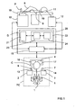
- the illustrated telesurgery installation is for example formed from the Da Vinci ® installation of the company Intuitive Surgical Inc. It schematically comprises a PC control station comprising a chair 1 allowing a surgeon C to install in front of a console equipped with a control keyboard (not shown), a first manual control 2 for the left hand, a second manual control 3 for the right hand, a display device 4, here stereoscopic type, and a set of control pedals 5.
- Each manual control 2, 3 comprises for example a control lever 6, 7 (of the "joystick” type), intended to control one of the manipulator arms 8, 9 of a robot 10, on which one will return further, as well as the manipulator arm 11 of a stereoscopic camera 12, which will also be discussed later, and one or more buttons 13,14 control (sensory type, or push button, or “mouse” ).
- the set of pedals 5 includes for example a pedal for associating a manual control 2, 3 to the control of the intervention robot 10, a pedal for associating a manual control 2, 3 to the control of the camera 12, and a pedal for associating a manual control 2, 3 to the control of a control module 15 of the installation, which will be discussed later.
- the control keyboard, the manual controls 2 and 3, and the set of control pedals 5 constitute a man / machine interface.
- the display device 4 comprises, here, a first screen 16 for displaying real two-dimensional images (2D) delivered by a first path of the camera 12 and intended for the left eye of the surgeon C, and a second screen 17, for displaying real two-dimensional (2D) images delivered by a second path of the camera 12 and intended for the right eye of the surgeon C.
- the interventional robot 10 is intended to be placed close to the operating table 18, on which the patient P is installed to be operated minimally. It generally comprises two manipulator arms 8 and 9 provided with ends adapted to the operation and intended to be introduced into the body of the patient P via incisions.
- the stereoscopic camera 12 comprises a manipulator arm 11 whose end supports two endoscopic optical fibers 19 defining two image acquisition channels.
- the interventional robot 10 and the endoscopic camera 12 can be combined to constitute a "robot master”.
- the installation further comprises a control unit 20, for example arranged in the form of a work station, comprising the control module 15 and a resetting device according to the invention D, which will be discussed later.
- the control module 15 is coupled to the console of the PC control station, the interventional robot 10, the stereoscopic camera 12 and the resetting device D.
- the registration device D is intended, in a general manner, to register in real time known patterns, which are characteristic of a region (here in which an operation must be carried out), on (real) images of this region.
- the patterns are three-dimensional (3D), but they could be two-dimensional (2D), at least for some of them.
- This device D firstly comprises a memory 21, storing three-dimensional patterns (3D) representative of characteristic portions of the region in which the intervention is to take place, and of known position and orientation with respect to a common reference frame ( or preoperative reference).
- the device D also comprises a processing module 22 coupled to the memory 21 and loaded, when it receives the designations, on the one hand, at least a portion of the observation image of the intervention region, taken under an angle chosen by the endoscopic camera 12 and delivered by the control module 15, and on the other hand, of at least one attribute representative of the designated portion, to determine in the memory 21 a 3D pattern which is representative of this portion designated, taking into account the designated attribute and the chosen viewing angle, and then resetting the determined 3D pattern to the designated image portion.
- 3D three-dimensional patterns
- a set of 3D patterns can be a 3D model. Therefore, the registration may relate not only to a 3D pattern, but also to a 3D model.
- a 3D model can be representative of the coronary tree, this 3D model then consisting of a multiplicity of 3D patterns representative of characteristic structures of the coronary tree, such as for example arteries, junctions and bifurcations.
- characteristic structures of the coronary tree such as for example arteries, junctions and bifurcations.
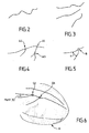
- two types of structures are defined. A first type groups curves representing the arteries, while a second type groups characteristic elements, such as junctions or bifurcations. These different types are illustrated on the Figures 2 to 5 .
- the figure 2 illustrates a representative pattern of a coronary artery
- the figure 3 illustrates a representative pattern of a configuration of three coronary arteries
- the figure 4 illustrates a representative pattern of a configuration of three bifurcations on a coronary artery
- the figure 5 illustrates a representative pattern of an angle ⁇ characteristic of a bifurcation between two coronary arteries.
- a bifurcation is defined, first, by an index (integer identifying the bifurcation), a second part, by two indexes (Art1 and Art2 which identify the two arteries concerned), and a third on the other hand, by a point (triplet of coordinates of the bifurcation in the pre-operative repository).
- an artery is defined on the one hand by an index (integer identifying the artery), and on the other hand by a set of parameters of a B-spline composing the central line of the artery ( quadruplet (xi, yi, zi, ui) in which (xi, yi, zi) is a triplet defining each control point of the artery in the pre-operative repository, and ui denotes a node of the B-spline whose value is between 0 and 1).
- obtaining 3D patterns of a region of intervention such as an organ such as the heart, firstly requires a three-dimensional modeling, and possibly temporal (3 D + t), the region of intervention from images obtained by medical imaging (MRI, scanner, etc.).
- MRI medical imaging
- 3D volume model
- the volume model (3D) is animated from a 3D + t model of the coronary network of the modeled heart, obtained from sequences of angiographic images (X-rays), also called coronarographies , taken at different angles and for several cycles, and synchronized with respect to an electrocardiogram (ECG).
- ECG electrocardiogram
- the 3D patterns are therefore fractions (or portions) characteristic of the 3D coronary tree (or network) whose positions are known with respect to the volume model (3D). from the heart of the patient P.
- the preoperative reference system relative to which the positions of the 3D units and the heart are defined is usually that of the external envelope of the patient P. It is in fact with respect to this external envelope that the intervention robot can be calibrated. 10 and the endoscopic camera 12. In addition, the fact of reporting the position of the heart, and therefore of its coronary network, with respect to the outer casing makes it possible to recalibrate the patient with respect to the robot in the operating room.
- a 3D anatomical model of the patient's body is also determined, at least in a large portion containing the subject area of the procedure, always from images obtained by medical imaging.
- the positions, orientations and configurations of the patterns (and models) 3D are stored in the memory 21 with respect to a common reference defined from marks placed on the outer envelope of the patient P.
- the 3D model of the heart of the patient P is also stored in the memory 21.
- the subject of the invention is not the preoperative planning phase of the surgical operation, it will not be described here. Therefore, the following describes the implementation of the device D according to the invention, within an installation according to the invention, in the operating phase.
- the preoperative planning phase which is only optional, is in particular to determine the three optimal incision points, which will allow to introduce the ends of the manipulator arms 8, 9 and 11 of the interventional robot 10 and the endoscopic camera 12, taking into account the 3D anatomical model of the body of the patient P and parameters, such as the dexterity and accessibility of the target region. It also consists in determining the optimal configuration of the manipulator arms 8 and 9 of the interventional robot 10 in order to avoid collisions and to obtain maximum separation.
- the device D intervenes within the installation once the arm 11 and the endoscopic camera 12 (possibly endoscopic) have been calibrated.
- An example of an endoscopic calibration method is described in the document F. Mourgues et al "Flexible calibration of actuated stereoscopic endoscope for overlay in robot assisted surgery", Proc. of MICCAI, Volume 2488 of LNCS, Spinger (2002), 25-34 .
- the calibration data are preferably stored in a memory of the control module 15.
- Incisions are then made in the patient's body P at the optimal incision points determined during the pre-operative planning phase.
- the manipulator arms 8 and 9 of the interventional robot 10 and 11 of the endoscopic camera are then placed in the optimal configuration, also determined during the preoperative planning phase, and their respective ends are introduced into the patient's body. by the incisions. It is recalled that the determination of the optimal incision points is a preferred but not mandatory option, as the positions of the robot arms and the endoscope can be determined empirically by the surgeon.
- the two channels of the endoscopic camera 12 deliver their respective 2D image sequences to the control module 15, which transmits them to the display device 4 so that they are displayed on the screens 16 and 17.
- the surgeon C can then observe the region in which is implanted the heart H object of the intervention, under the observation angle of the endoscopic camera 12.
- the figure 6 is an image of heart H of the type observed by surgeon C on screens 16 and 17.
- model3D here of the coronary tree
- This can be done manually or by an external registration of the patient, installed on the operating table, with its pre-operative model.
- the external registration is first to point with the end of the robot several radiopaque markers previously glued on the patient's chest, and previously segmented in the scanner images. Then, we calculate the rigid transformation between the dotted markers and the segmented markers.
- This initial rigid transformation makes it possible to pass from the pre-operative repository (referential in which the patterns of the 3D model to be recalibrated) to the reference frame of the base of the robot, and to the repository of the endoscopic camera (then using the calibration of the endoscope).
- This external registration technique is described in particular in the document E. Coste-Maniere et al "Optimal planning ofrobotically assisted heart surgery: Precision transfer in the operating room, B. Siciliano and P. Dario, eds, Springer Tracts In Advanced Robotics, Experimental Robotics VIII, Volume 5, Springer (2002), 424-434 .
- the surgeon C designates at least a portion of the images displayed on his screens 16 and 17 and at least one attribute representative of each image portion using his man / machine interface.
- the designation of a portion is for example by selecting the portion on the image with a mouse.
- the designation of an attribute is made either by a voice command or by selection from a list displayed on the screens 16 and 17 or on an auxiliary screen.
- the squares A1 to A3 materialize the places on which the surgeon C "clicks" with his mouse to designate three portions of the region which seem significant to him.
- the square B materializes the place on which the surgeon C "clicks" with his mouse to designate the portion of the region that seems significant to him.
- the attributes are standard information (or classes) which each describe a known local characteristic or a known local configuration, or generally any information enabling the processing module 22 of the device D to determine in the memory 21 the corresponding 3D pattern. .
- the device D transmits them to its processing module 22, so that it determines in the memory 21 a 3D pattern that seems representative of the designated portion or portions, taking into account the (or) the designated attribute (s).
- This determination is provided by an extraction module 23 coupled to the memory 21. Then, the processing module 22 must perform the registration of this pattern (and possibly the entire 3D model of which it is part). This is to determine how to orient and position the pattern so that it can be superimposed on the portion designated by the surgeon, given the angle at which the region of intervention is observed by the endoscopic camera.
- the registration is preferably performed by a registration module 24 of the processing module 22. Furthermore, this registration is preferably rigid, by minimizing a criterion constructed from the measurement equations.
- the measurement equations are deduced here from the hypotheses provided by the surgeon, when he thinks to recognize a portion, such as an artery or a bifurcation between known arteries, or directly determined by the registration module 24.
- another type of registration could be considered, including an affine registration.
- the rigid registration consists in determining at least one measurement equation from the designation of a portion of an image, an attribute and at least one hypothesis on the identity of the pattern determined by the module. 23. This registration may also take into account the 3D model of the heart when it is stored in the memory 21.
- the registration module 24 generates several sets of equations corresponding to the different possible hypotheses and then optimizes the registration parameters by minimizing a criterion. It then classifies the readjustments obtained according to their relevance and selects the best registration.
- the parameter estimation method is described in particular in the T. Vieville et al "Implementing a multi-model estimation method", The International Journal of Computer Vision, 44 (2001), 41-64 .
- the entire 3D model which includes said pattern.
- it is the entire 3D model, seen from the angle of observation of the endoscopic camera, and not only the determined 3D pattern and recaled, which can be superimposed on the image displayed or observed.
- some portions of the 3D model may not be visible due to the viewing angle.
- the processing module 22 can then transmit to the control module 15 the position data of the recalibrated 3D pattern (or of the entire 3D model of which it forms part), in the (calibration) frame of the displayed image (or observed, when the surgeon is equipped with observation glasses), and the image data which define this pattern (or model) 3D, so that it orders the display device 4 its display superimposed on the images of observed by the endoscopic camera 12 (or observed in the glasses).
- a superposition of a large 3D pattern on a heart image is illustrated on the figure 6 .
- the squares D1 and D2 represent the places on which the surgeon C clicked with his mouse to designate two portions of the intervention area that seemed significant to him, and the fraction of the coronary network superimposed on the image of the heart H materializes. the 3D pattern recaled by the device D.
- the designations made by the surgeon C may not allow the extraction module 23 to determine the 3D pattern that corresponds to the selected portion, or the registration module 24 to perform an appropriate registration, thereby causing a bad superposition of the pattern (or model) 3D on the displayed image. Consequently, the processing module 22 can be arranged to precisely determine the position of the region observed in the reference frame by successive readjustments, each relying on the extraction of a new 3D pattern following the designation of at least one another portion of observation images and at least one attribute representative of this other portion.
- the 3D pattern determined by the extraction module 23 is not superimposed on the image of the region observed at the portion selected by the surgeon C or the recaled 3D pattern is superimposable, but it does not correspond not to the structure observed by the surgeon on the selected portion. The surgeon must then make a new designation to converge the resetting operation.
- the control module 15 which automatically notices the error and sends a message to the surgeon, requesting new designations from him.
- the registration device D On receipt of these new designations, the registration device D reiterates the treatments presented above by taking into account the new and old designations. He may in particular, when arranged for this purpose, calculate new hypotheses and measurement equations.
- a correspondence table between the 3D patterns and information data which represents them may be for example the name of the pattern, such as for example the name of an artery and / or its possible ramifications, or coordinates of a target point, or operating data recorded during the phase of planning, such as the identification of a stenosis or a calcification zone.
- the processing module 22 can be configured to deliver the information data associated with a 3D pattern (recalibrated or not) when it receives the order from the control module 15 (for example in the case of request of the surgeon C).
- a 3D pattern for example in the case of request of the surgeon C.
- the control module 15 can also be designed, each time it receives a request designating a 3D pattern of the region observed, so as, firstly, order the resetting device D to determine position data representative of the position of this 3D pattern relative to the calibration reference, taking into account the registration, and secondly, to determine piloting instructions intended to move the manipulator arms 8 and 9 of the interventional robot 10 in the vicinity of the portion of region that corresponds to the designated 3D pattern.
- the installation presented above can be used for other types of operation, such as open liver or breast surgery.
- the installation according to the invention makes it possible to guide a user in the performance of his task in an environment where he has only partial and / or distorted vision, and / or in an environment difficult.
- the resetting device D according to the invention can be used in other applications than those presented above. It can in particular be used in installations dedicated solely to localization guidance (without the intervention of a teleoperated robot), and in particular in urban areas.
- an installation embedded in a motor vehicle comprising one or more cameras, delivering to a control module real images of the environment so that they are displayed on screens installed in the passenger compartment, and a resetting device D coupled to said control module.
- the installation can also be coupled to an onboard GPS-based satellite guidance device.
- the registration device D stores in its memory representative patterns of the environment (or region) in which the vehicle can evolve and determined from digital recordings previously made. These reasons, whose positions are defined in a chosen landmark, are for example facades of buildings, or buildings or remarkable sites, or statues or works of art.
- the memory may also include a volume model of the environment, as well as a correspondence table between the patterns and information data on these patterns.
- the installation can be used.
- the control module is loaded here, when it receives from a passenger of the vehicle a request designating a pattern of the observed region (which he has selected in the list of patterns stored using a mouse or by exercising a pressure on a touch display screen), order the resetting device D to determine position data representative of the position of this pattern relative to the calibration reference, given the registration induced by the offsets between the reference mark selected and the calibration mark, and then determine driving instructions to guide the driver of the vehicle to the portion of the region that corresponds to this pattern.
- the control module may possibly rely on the vehicle position data delivered by the GPS device to determine the piloting instructions.
- the pattern Once the vehicle arrived on site, the pattern may be optionally superimposed on the actual image of the region portion, and information data associated with said pattern may be issued to the passenger.
- the registration device D receives from a passenger of the vehicle the designations of at least one portion of the region observed and displayed (which he has selected with the aid of a mouse or by exerting pressure on the tactile display screen) and at least one attribute representative of this portion, such as for example the nature of a building (house, school, town hall, museum, church) or a place (garden, park, square) or object (statue, sculpture, work of art), and determines in its memory the pattern that best corresponds to that designated portion, taking into account the attribute designated and the chosen viewing angle. Then, he recalibrates the determined pattern on the designated image portion.
- the device according to the invention is advantageously implanted in a communication equipment, such as for example a mobile telephone, equipped with a location function, for example by triangulation or by GPS, and a camera, as well as possibly an inertial unit.
- a communication equipment such as for example a mobile telephone, equipped with a location function, for example by triangulation or by GPS, and a camera, as well as possibly an inertial unit.
- the processing module 22 of the resetting device D and the control module 15 of the installations can be made in the form of electronic circuits, software (or computer) modules, or a combination of software modules and electronic circuits.
- the invention also relates to a method dedicated to the real-time registration of known patterns, characteristic of a region, on images of this region.
Landscapes
- Health & Medical Sciences (AREA)
- Engineering & Computer Science (AREA)
- Life Sciences & Earth Sciences (AREA)
- Surgery (AREA)
- Physics & Mathematics (AREA)
- Heart & Thoracic Surgery (AREA)
- Veterinary Medicine (AREA)
- Biomedical Technology (AREA)
- Public Health (AREA)
- Medical Informatics (AREA)
- Molecular Biology (AREA)
- Animal Behavior & Ethology (AREA)
- General Health & Medical Sciences (AREA)
- Nuclear Medicine, Radiotherapy & Molecular Imaging (AREA)
- Biophysics (AREA)
- Pathology (AREA)
- Robotics (AREA)
- Theoretical Computer Science (AREA)
- Computer Vision & Pattern Recognition (AREA)
- General Physics & Mathematics (AREA)
- Radiology & Medical Imaging (AREA)
- Optics & Photonics (AREA)
- Signal Processing (AREA)
- Human Computer Interaction (AREA)
- Gynecology & Obstetrics (AREA)
- Geometry (AREA)
- Image Processing (AREA)
- Endoscopes (AREA)
- High Energy & Nuclear Physics (AREA)
- Pharmaceuticals Containing Other Organic And Inorganic Compounds (AREA)
- Processing Or Creating Images (AREA)
Applications Claiming Priority (2)
| Application Number | Priority Date | Filing Date | Title |
|---|---|---|---|
| FR0306176A FR2855292B1 (fr) | 2003-05-22 | 2003-05-22 | Dispositif et procede de recalage en temps reel de motifs sur des images, notamment pour le guidage par localisation |
| EP04742719.0A EP1625546B1 (de) | 2003-05-22 | 2004-05-13 | Einrichtung und verfahren zum üebrlagern von mustern auf bildern in echtzeit insbesondere zur lenkung durch lokalisierung |
Related Parent Applications (3)
| Application Number | Title | Priority Date | Filing Date |
|---|---|---|---|
| EP04742719.0 Division | 2004-05-13 | ||
| WOPCT/FR2004/001166 Previously-Filed-Application | 2004-05-13 | ||
| EP04742719.0A Division-Into EP1625546B1 (de) | 2003-05-22 | 2004-05-13 | Einrichtung und verfahren zum üebrlagern von mustern auf bildern in echtzeit insbesondere zur lenkung durch lokalisierung |
Publications (2)
| Publication Number | Publication Date |
|---|---|
| EP2187352A2 true EP2187352A2 (de) | 2010-05-19 |
| EP2187352A3 EP2187352A3 (de) | 2011-01-12 |
Family
ID=33396676
Family Applications (4)
| Application Number | Title | Priority Date | Filing Date |
|---|---|---|---|
| EP10150348A Withdrawn EP2187352A3 (de) | 2003-05-22 | 2004-05-13 | Einrichtung und Verfahren zum üebrlagern von Mustern auf Bildern in echtzeit insbesondere zur Lenkung durch Lokalisierung |
| EP21175754.7A Withdrawn EP3889901A1 (de) | 2003-05-22 | 2004-05-13 | Vorrichtung und verfahren zur echtzeitkorrektur von motiven auf bildern, insbesondere für die lenkung durch lokalisierung |
| EP18165967.3A Expired - Lifetime EP3366193B1 (de) | 2003-05-22 | 2004-05-13 | Vorrichtung und verfahren zur echtzeitkorrektur von motiven auf bildern, insbesondere für die lenkung durch lokalisierung |
| EP04742719.0A Expired - Lifetime EP1625546B1 (de) | 2003-05-22 | 2004-05-13 | Einrichtung und verfahren zum üebrlagern von mustern auf bildern in echtzeit insbesondere zur lenkung durch lokalisierung |
Family Applications After (3)
| Application Number | Title | Priority Date | Filing Date |
|---|---|---|---|
| EP21175754.7A Withdrawn EP3889901A1 (de) | 2003-05-22 | 2004-05-13 | Vorrichtung und verfahren zur echtzeitkorrektur von motiven auf bildern, insbesondere für die lenkung durch lokalisierung |
| EP18165967.3A Expired - Lifetime EP3366193B1 (de) | 2003-05-22 | 2004-05-13 | Vorrichtung und verfahren zur echtzeitkorrektur von motiven auf bildern, insbesondere für die lenkung durch lokalisierung |
| EP04742719.0A Expired - Lifetime EP1625546B1 (de) | 2003-05-22 | 2004-05-13 | Einrichtung und verfahren zum üebrlagern von mustern auf bildern in echtzeit insbesondere zur lenkung durch lokalisierung |
Country Status (5)
| Country | Link |
|---|---|
| US (8) | US8126223B2 (de) |
| EP (4) | EP2187352A3 (de) |
| CA (3) | CA2526590C (de) |
| FR (1) | FR2855292B1 (de) |
| WO (1) | WO2004107267A2 (de) |
Families Citing this family (69)
| Publication number | Priority date | Publication date | Assignee | Title |
|---|---|---|---|---|
| FR2855292B1 (fr) | 2003-05-22 | 2005-12-09 | Inst Nat Rech Inf Automat | Dispositif et procede de recalage en temps reel de motifs sur des images, notamment pour le guidage par localisation |
| EP1607716A3 (de) | 2004-06-18 | 2012-06-20 | Topcon Corporation | Gerät und Verfahren zum Bilden eines Modells, und Bildaufnahmegerät und -Verfahren |
| US9492240B2 (en) * | 2009-06-16 | 2016-11-15 | Intuitive Surgical Operations, Inc. | Virtual measurement tool for minimally invasive surgery |
| US8971597B2 (en) * | 2005-05-16 | 2015-03-03 | Intuitive Surgical Operations, Inc. | Efficient vision and kinematic data fusion for robotic surgical instruments and other applications |
| US10555775B2 (en) * | 2005-05-16 | 2020-02-11 | Intuitive Surgical Operations, Inc. | Methods and system for performing 3-D tool tracking by fusion of sensor and/or camera derived data during minimally invasive robotic surgery |
| JP5229873B2 (ja) * | 2008-01-31 | 2013-07-03 | 東芝メディカルシステムズ株式会社 | 画像表示装置 |
| JP2010137009A (ja) * | 2008-12-15 | 2010-06-24 | Nintendo Co Ltd | キャリブレーションプログラム及び座標検出装置 |
| US8830224B2 (en) * | 2008-12-31 | 2014-09-09 | Intuitive Surgical Operations, Inc. | Efficient 3-D telestration for local robotic proctoring |
| KR100961661B1 (ko) * | 2009-02-12 | 2010-06-09 | 주식회사 래보 | 수술용 항법 장치 및 그 방법 |
| US8918207B2 (en) * | 2009-03-09 | 2014-12-23 | Intuitive Surgical Operations, Inc. | Operator input device for a robotic surgical system |
| US9155592B2 (en) * | 2009-06-16 | 2015-10-13 | Intuitive Surgical Operations, Inc. | Virtual measurement tool for minimally invasive surgery |
| KR101180665B1 (ko) * | 2009-07-03 | 2012-09-07 | 주식회사 이턴 | 하이브리드 수술용 로봇 시스템 및 수술용 로봇 제어방법 |
| DE102009032257B4 (de) * | 2009-07-08 | 2017-05-04 | Siemens Healthcare Gmbh | Verfahren und Vorrichtung zur automatisierten Ermittlung der Mittellinie zumindest eines Teilstücks einer tubulären Gewebestruktur |
| DE102009040430B4 (de) * | 2009-09-07 | 2013-03-07 | Fraunhofer-Gesellschaft zur Förderung der angewandten Forschung e.V. | Vorrichtung, Verfahren und Computerprogramm zur Überlagerung eines intraoperativen Livebildes eines Operationsgebiets oder des Operationsgebiets mit einem präoperativen Bild des Operationsgebiets |
| CN103108602B (zh) * | 2010-09-15 | 2015-09-30 | 皇家飞利浦电子股份有限公司 | 从血管树图像对内窥镜的机器人控制 |
| JP2012096024A (ja) * | 2010-10-07 | 2012-05-24 | Toshiba Corp | 医用画像処理装置 |
| JP5765913B2 (ja) * | 2010-10-14 | 2015-08-19 | 株式会社東芝 | 医用画像診断装置及び医用画像処理方法 |
| US10631712B2 (en) | 2011-02-10 | 2020-04-28 | Karl Storz Imaging, Inc. | Surgeon's aid for medical display |
| US11412998B2 (en) | 2011-02-10 | 2022-08-16 | Karl Storz Imaging, Inc. | Multi-source medical display |
| US10674968B2 (en) | 2011-02-10 | 2020-06-09 | Karl Storz Imaging, Inc. | Adjustable overlay patterns for medical display |
| DE102011078212B4 (de) | 2011-06-28 | 2017-06-29 | Scopis Gmbh | Verfahren und Vorrichtung zum Darstellen eines Objektes |
| KR20130015146A (ko) * | 2011-08-02 | 2013-02-13 | 삼성전자주식회사 | 의료 영상 처리 방법 및 장치, 영상 유도를 이용한 로봇 수술 시스템 |
| US8908918B2 (en) | 2012-11-08 | 2014-12-09 | Navigate Surgical Technologies, Inc. | System and method for determining the three-dimensional location and orientation of identification markers |
| EP2797657B1 (de) | 2011-12-30 | 2019-03-13 | St. Jude Medical, Atrial Fibrillation Division, Inc. | System und verfahren zur erkennung und vermeidung von zusammenstössen zwischen robotisch gesteuerten medizinischen vorrichtungen |
| DE102012220116A1 (de) * | 2012-06-29 | 2014-01-02 | Fraunhofer-Gesellschaft zur Förderung der angewandten Forschung e.V. | Mobil handhabbare Vorrichtung, insbesondere zur Bearbeitung oder Beobachtung eines Körpers, und Verfahren zur Handhabung, insbesondere Kalibrierung, einer Vorrichtung |
| DE102012220115A1 (de) * | 2012-11-05 | 2014-05-22 | Fraunhofer-Gesellschaft zur Förderung der angewandten Forschung e.V. | Bildgebendes System, Operationsvorrichtung mit dem bildgebenden System und Verfahren zur Bildgebung |
| US9839481B2 (en) * | 2013-03-07 | 2017-12-12 | Intuitive Surgical Operations, Inc. | Hybrid manual and robotic interventional instruments and methods of use |
| EP2967623B1 (de) | 2013-03-14 | 2021-07-21 | SRI International | Kompaktes roboterhandgelenk |
| CA2905968A1 (en) | 2013-03-15 | 2014-09-25 | Sri International | Hyperdexterous surgical system |
| US10274553B2 (en) | 2013-03-15 | 2019-04-30 | Canon U.S.A., Inc. | Needle placement manipulator with attachment for RF-coil |
| DE102013211055B3 (de) | 2013-06-13 | 2014-09-18 | Scopis Gmbh | Adapter zur Aufnahme eines medizintechnischen Geräts sowie eines Lageerfassungssystems |
| AU2015221258B2 (en) | 2014-02-21 | 2019-11-21 | Cilag Gmbh International | A set comprising a surgical instrument |
| CN106456252B (zh) | 2014-03-28 | 2020-05-08 | 直观外科手术操作公司 | 手术场景的定量三维成像 |
| JP6854237B2 (ja) | 2014-03-28 | 2021-04-07 | インテュイティブ サージカル オペレーションズ, インコーポレイテッド | 視野内の器具の定量的三次元視覚化 |
| EP3122232B1 (de) * | 2014-03-28 | 2020-10-21 | Intuitive Surgical Operations Inc. | Ausrichtung von q3d-modellen mit 3d-bildern |
| KR102397670B1 (ko) | 2014-03-28 | 2022-05-16 | 인튜어티브 서지컬 오퍼레이션즈 인코포레이티드 | 정량적 3차원 영상화에 기초한 햅틱 피드백을 갖는 수술 시스템 |
| CN106535806B (zh) | 2014-03-28 | 2019-06-18 | 直观外科手术操作公司 | 来自多端口视角的手术场景的定量三维成像 |
| EP3122281B1 (de) | 2014-03-28 | 2022-07-20 | Intuitive Surgical Operations, Inc. | Quantitative dreidimensionale bildgebung und 3d modellierung von chirurgischen implantaten |
| GB2524955A (en) | 2014-04-01 | 2015-10-14 | Scopis Gmbh | Method for cell envelope segmentation and visualisation |
| WO2016044939A1 (en) * | 2014-09-24 | 2016-03-31 | Polymer Robotics Inc. | Tool manipulator and system for positioning a tool for surgical and like uses |
| GB201501157D0 (en) | 2015-01-23 | 2015-03-11 | Scopis Gmbh | Instrument guidance system for sinus surgery |
| WO2017012624A1 (en) | 2015-07-21 | 2017-01-26 | 3Dintegrated Aps | Cannula assembly kit, trocar assembly kit, sleeve assembly, minimally invasive surgery system and method therefor |
| US11020144B2 (en) | 2015-07-21 | 2021-06-01 | 3Dintegrated Aps | Minimally invasive surgery system |
| CN108025445A (zh) | 2015-07-23 | 2018-05-11 | 斯里国际 | 机器人臂及机器人手术系统 |
| DK178899B1 (en) | 2015-10-09 | 2017-05-08 | 3Dintegrated Aps | A depiction system |
| CN108135666B (zh) * | 2015-10-22 | 2021-06-18 | 柯惠Lp公司 | 多输入机器人手术系统控制方案 |
| JP6833455B2 (ja) * | 2016-10-31 | 2021-02-24 | キヤノン株式会社 | 眼科撮影装置及びその制御方法、眼科撮影システム、並びに、プログラム |
| US11602474B2 (en) | 2016-11-28 | 2023-03-14 | Verb Surgical Inc. | Surgical table base with high stiffness and adjustable support members with force feedback |
| CN106780527B (zh) * | 2016-11-29 | 2020-09-15 | 上海联影医疗科技有限公司 | 医学图像中血管进出口、边界条件获取方法及处理装置 |
| DE102017203025A1 (de) * | 2017-02-24 | 2018-08-30 | Siemens Healthcare Gmbh | Verfahren zu einem Unterstützen einer Planung einer Magnetresonanzuntersuchung an einem Patienten mit einer Magnetresonanzvorrichtung sowie eine Magnetresonanzvorrichtung zu einem Ausführen des Verfahrens |
| US10792119B2 (en) | 2017-05-22 | 2020-10-06 | Ethicon Llc | Robotic arm cart and uses therefor |
| US10856948B2 (en) | 2017-05-31 | 2020-12-08 | Verb Surgical Inc. | Cart for robotic arms and method and apparatus for registering cart to surgical table |
| US10485623B2 (en) | 2017-06-01 | 2019-11-26 | Verb Surgical Inc. | Robotic arm cart with fine position adjustment features and uses therefor |
| US10913145B2 (en) | 2017-06-20 | 2021-02-09 | Verb Surgical Inc. | Cart for robotic arms and method and apparatus for cartridge or magazine loading of arms |
| US10058396B1 (en) | 2018-04-24 | 2018-08-28 | Titan Medical Inc. | System and apparatus for insertion of an instrument into a body cavity for performing a surgical procedure |
| US11205508B2 (en) * | 2018-05-23 | 2021-12-21 | Verb Surgical Inc. | Machine-learning-oriented surgical video analysis system |
| CN108961375A (zh) * | 2018-06-20 | 2018-12-07 | 腾讯科技(深圳)有限公司 | 一种根据二维图像生成三维图像的方法及装置 |
| US10679743B2 (en) | 2018-09-12 | 2020-06-09 | Verb Surgical Inc. | Method and system for automatically tracking and managing inventory of surgical tools in operating rooms |
| JP7152240B2 (ja) * | 2018-10-11 | 2022-10-12 | 株式会社メディカロイド | ロボット手術支援装置、ロボット手術支援方法、及びプログラム |
| JP7182126B2 (ja) | 2018-10-11 | 2022-12-02 | 株式会社メディカロイド | ロボット手術支援装置、ロボット手術支援方法、及びプログラム |
| TWI696529B (zh) * | 2018-11-30 | 2020-06-21 | 財團法人金屬工業研究發展中心 | 自動定位方法以及自動控制裝置 |
| WO2021126788A1 (en) * | 2019-12-16 | 2021-06-24 | Intuitive Surgical Operations, Inc. | Systems for facilitating guided teleoperation of a non-robotic device in a surgical space |
| JP7680728B2 (ja) * | 2020-02-28 | 2025-05-21 | 株式会社根本杏林堂 | 医用画像処理装置、医用画像処理方法および医用画像処理プログラム |
| EP4157128A1 (de) * | 2020-05-29 | 2023-04-05 | Covidien LP | System und verfahren zur integrierten steuerung einer 3d-visualisierung durch ein chirurgisches robotersystem |
| US20210401527A1 (en) * | 2020-06-30 | 2021-12-30 | Auris Health, Inc. | Robotic medical systems including user interfaces with graphical representations of user input devices |
| US11980426B2 (en) | 2020-08-03 | 2024-05-14 | Warsaw Orthopedic, Inc. | System and method for preliminary registration |
| CN112815951B (zh) * | 2020-12-22 | 2023-02-28 | 北京旋极伏羲科技有限公司 | 一种基于建筑物3d模型的网格路径规划方法 |
| US12462439B2 (en) * | 2021-04-14 | 2025-11-04 | Cilag Gmbh International | Risk based prioritization of display aspects in surgical field view |
| US20230121709A1 (en) | 2021-10-15 | 2023-04-20 | Verb Surgical Inc. | Method and system for controlling and displaying video streams |
Family Cites Families (28)
| Publication number | Priority date | Publication date | Assignee | Title |
|---|---|---|---|---|
| FR2652928B1 (fr) | 1989-10-05 | 1994-07-29 | Diadix Sa | Systeme interactif d'intervention locale a l'interieur d'une zone d'une structure non homogene. |
| US5417210A (en) * | 1992-05-27 | 1995-05-23 | International Business Machines Corporation | System and method for augmentation of endoscopic surgery |
| US5782762A (en) * | 1994-10-27 | 1998-07-21 | Wake Forest University | Method and system for producing interactive, three-dimensional renderings of selected body organs having hollow lumens to enable simulated movement through the lumen |
| US6167145A (en) * | 1996-03-29 | 2000-12-26 | Surgical Navigation Technologies, Inc. | Bone navigation system |
| US6659939B2 (en) * | 1998-11-20 | 2003-12-09 | Intuitive Surgical, Inc. | Cooperative minimally invasive telesurgical system |
| US6424996B1 (en) * | 1998-11-25 | 2002-07-23 | Nexsys Electronics, Inc. | Medical network system and method for transfer of information |
| US6285902B1 (en) * | 1999-02-10 | 2001-09-04 | Surgical Insights, Inc. | Computer assisted targeting device for use in orthopaedic surgery |
| US6301495B1 (en) * | 1999-04-27 | 2001-10-09 | International Business Machines Corporation | System and method for intra-operative, image-based, interactive verification of a pre-operative surgical plan |
| JP4421016B2 (ja) * | 1999-07-01 | 2010-02-24 | 東芝医用システムエンジニアリング株式会社 | 医用画像処理装置 |
| US6330356B1 (en) | 1999-09-29 | 2001-12-11 | Rockwell Science Center Llc | Dynamic visual registration of a 3-D object with a graphical model |
| US7366562B2 (en) | 2003-10-17 | 2008-04-29 | Medtronic Navigation, Inc. | Method and apparatus for surgical navigation |
| US6563941B1 (en) * | 1999-12-14 | 2003-05-13 | Siemens Corporate Research, Inc. | Model-based registration of cardiac CTA and MR acquisitions |
| JP4674948B2 (ja) * | 2000-09-29 | 2011-04-20 | オリンパス株式会社 | 手術ナビゲーション装置および手術ナビゲーション装置の作動方法 |
| US6584339B2 (en) | 2001-06-27 | 2003-06-24 | Vanderbilt University | Method and apparatus for collecting and processing physical space data for use while performing image-guided surgery |
| US8175680B2 (en) * | 2001-11-09 | 2012-05-08 | Boston Scientific Scimed, Inc. | Systems and methods for guiding catheters using registered images |
| JP2003164413A (ja) * | 2001-12-03 | 2003-06-10 | Olympus Optical Co Ltd | 内視鏡画像ファイリングシステム |
| US7466848B2 (en) * | 2002-12-13 | 2008-12-16 | Rutgers, The State University Of New Jersey | Method and apparatus for automatically detecting breast lesions and tumors in images |
| US7356178B2 (en) * | 2002-12-31 | 2008-04-08 | Koninklijke Philips Electronics N.V. | System and method for improved multiple-dimension image displays |
| US7505809B2 (en) | 2003-01-13 | 2009-03-17 | Mediguide Ltd. | Method and system for registering a first image with a second image relative to the body of a patient |
| US20070055142A1 (en) * | 2003-03-14 | 2007-03-08 | Webler William E | Method and apparatus for image guided position tracking during percutaneous procedures |
| DE10318205A1 (de) * | 2003-04-22 | 2004-11-25 | Siemens Ag | Bildgebungsverfahren für ein kapselförmiges Endoskopiegerät |
| US7570791B2 (en) * | 2003-04-25 | 2009-08-04 | Medtronic Navigation, Inc. | Method and apparatus for performing 2D to 3D registration |
| FR2855292B1 (fr) | 2003-05-22 | 2005-12-09 | Inst Nat Rech Inf Automat | Dispositif et procede de recalage en temps reel de motifs sur des images, notamment pour le guidage par localisation |
| US20070276234A1 (en) * | 2003-10-21 | 2007-11-29 | The Board Of Trustees Of The Leland Stanford Junior University | Systems and Methods for Intraoperative Targeting |
| US20050089205A1 (en) * | 2003-10-23 | 2005-04-28 | Ajay Kapur | Systems and methods for viewing an abnormality in different kinds of images |
| US7702137B2 (en) * | 2004-11-10 | 2010-04-20 | M2S, Inc. | Anatomical visualization and measurement system |
| US7720520B2 (en) * | 2004-12-01 | 2010-05-18 | Boston Scientific Scimed, Inc. | Method and system for registering an image with a navigation reference catheter |
| US10489344B1 (en) * | 2018-12-28 | 2019-11-26 | Nasuni Corporation | Cloud-native global file system with direct-to-cloud migration |
-
2003
- 2003-05-22 FR FR0306176A patent/FR2855292B1/fr not_active Expired - Lifetime
-
2004
- 2004-05-13 CA CA2526590A patent/CA2526590C/en not_active Expired - Lifetime
- 2004-05-13 EP EP10150348A patent/EP2187352A3/de not_active Withdrawn
- 2004-05-13 US US10/557,790 patent/US8126223B2/en active Active
- 2004-05-13 WO PCT/FR2004/001166 patent/WO2004107267A2/fr not_active Ceased
- 2004-05-13 EP EP21175754.7A patent/EP3889901A1/de not_active Withdrawn
- 2004-05-13 EP EP18165967.3A patent/EP3366193B1/de not_active Expired - Lifetime
- 2004-05-13 CA CA2948415A patent/CA2948415C/en not_active Expired - Lifetime
- 2004-05-13 CA CA2891012A patent/CA2891012C/en not_active Expired - Lifetime
- 2004-05-13 EP EP04742719.0A patent/EP1625546B1/de not_active Expired - Lifetime
-
2010
- 2010-04-12 US US12/758,339 patent/US20100195919A1/en not_active Abandoned
- 2010-10-20 US US12/908,298 patent/US8086008B2/en not_active Expired - Lifetime
-
2011
- 2011-11-18 US US13/299,676 patent/US8712125B2/en not_active Expired - Lifetime
-
2014
- 2014-03-17 US US14/215,915 patent/US9486159B2/en not_active Expired - Fee Related
-
2016
- 2016-11-07 US US15/345,488 patent/US10231642B2/en not_active Expired - Lifetime
-
2019
- 2019-01-30 US US16/262,027 patent/US10987022B2/en not_active Expired - Fee Related
-
2021
- 2021-04-09 US US17/226,966 patent/US11842503B2/en not_active Expired - Lifetime
Non-Patent Citations (1)
| Title |
|---|
| HEDLEY N R ET AL: "Explorations in the use of augmented reality for geographic visualization" PRESENCE MIT PRESS USA, vol. 11, no. 2, avril 2002 (2002-04), page 12PP, XP002609279 ISSN: 1054-7460 DOI: 10.1162/1054746021470577 * |
Also Published As
| Publication number | Publication date |
|---|---|
| CA2526590C (en) | 2016-09-06 |
| US20120130167A1 (en) | 2012-05-24 |
| US11842503B2 (en) | 2023-12-12 |
| EP1625546A2 (de) | 2006-02-15 |
| CA2891012A1 (en) | 2004-12-09 |
| EP3366193A1 (de) | 2018-08-29 |
| EP1625546B1 (de) | 2018-07-04 |
| EP3366193B1 (de) | 2021-07-07 |
| CA2891012C (en) | 2017-11-28 |
| US20140275975A1 (en) | 2014-09-18 |
| US20210219866A1 (en) | 2021-07-22 |
| US20170049358A1 (en) | 2017-02-23 |
| WO2004107267A3 (fr) | 2005-12-29 |
| FR2855292A1 (fr) | 2004-11-26 |
| US10231642B2 (en) | 2019-03-19 |
| FR2855292B1 (fr) | 2005-12-09 |
| CA2526590A1 (en) | 2004-12-09 |
| US8086008B2 (en) | 2011-12-27 |
| US10987022B2 (en) | 2021-04-27 |
| US20100195919A1 (en) | 2010-08-05 |
| US9486159B2 (en) | 2016-11-08 |
| EP2187352A3 (de) | 2011-01-12 |
| EP3889901A1 (de) | 2021-10-06 |
| WO2004107267A2 (fr) | 2004-12-09 |
| US20160296137A9 (en) | 2016-10-13 |
| CA2948415A1 (en) | 2004-12-09 |
| US20070147707A1 (en) | 2007-06-28 |
| US20110060347A1 (en) | 2011-03-10 |
| US20190159699A1 (en) | 2019-05-30 |
| CA2948415C (en) | 2021-03-02 |
| US8126223B2 (en) | 2012-02-28 |
| US8712125B2 (en) | 2014-04-29 |
Similar Documents
| Publication | Publication Date | Title |
|---|---|---|
| EP1625546B1 (de) | Einrichtung und verfahren zum üebrlagern von mustern auf bildern in echtzeit insbesondere zur lenkung durch lokalisierung | |
| WO2018224485A1 (fr) | Procédé et système d'aide au guidage d'un outil endovasculaire | |
| CN103108602B (zh) | 从血管树图像对内窥镜的机器人控制 | |
| EP2715662B1 (de) | Verfahren zum lokalisieren einer kamera und 3d-rekonstruktion in einer teilweise bekannten umgebung | |
| US20110128352A1 (en) | Fast 3d-2d image registration method with application to continuously guided endoscopy | |
| FR3095331A1 (fr) | Procédé de chirurgie orthopédique assistée par ordinateur | |
| Mourgues et al. | 3D reconstruction of the operating field for image overlay in 3D-endoscopic surgery | |
| CN102711650A (zh) | 用于内窥镜手术的基于图像整合的配准和导航 | |
| EP3911238B1 (de) | Medizinische vorrichtung zum einsatz in der interventionellen radiologie zur echtzeit-führung einer medizinischen operationsnadel in einem volumen | |
| EP4230168A1 (de) | Robotisches wirbelsäulenchirurgiesystem | |
| FR2914466A1 (fr) | Dispositif de traitement d'images pour la mise en correspondance d'images d'une meme portion d'un corps obtenues par resonance magnetique et par ultrasons. | |
| US20050010105A1 (en) | Method and system for Coronary arterial intervention | |
| WO2025007493A1 (zh) | 一种基于学习的ar辅助牙科治疗自动校准和导航方法 | |
| JP2008541859A5 (de) | ||
| Cortadellas et al. | New concepts for intraoperative navigation: calibration of a 3-D laparoscope |
Legal Events
| Date | Code | Title | Description |
|---|---|---|---|
| PUAI | Public reference made under article 153(3) epc to a published international application that has entered the european phase |
Free format text: ORIGINAL CODE: 0009012 |
|
| 17P | Request for examination filed |
Effective date: 20100108 |
|
| AC | Divisional application: reference to earlier application |
Ref document number: 1625546 Country of ref document: EP Kind code of ref document: P |
|
| AK | Designated contracting states |
Kind code of ref document: A2 Designated state(s): AT BE BG CH CY CZ DE DK EE ES FI FR GB GR HU IE IT LI LU MC NL PL PT RO SE SI SK TR |
|
| RIN1 | Information on inventor provided before grant (corrected) |
Inventor name: COSTE-MANIERE, EVE Inventor name: MOURGUES, FABIEN Inventor name: VIEVILLE, THIERRY |
|
| RAP1 | Party data changed (applicant data changed or rights of an application transferred) |
Owner name: MYRIAD GROUP AG |
|
| PUAL | Search report despatched |
Free format text: ORIGINAL CODE: 0009013 |
|
| AK | Designated contracting states |
Kind code of ref document: A3 Designated state(s): AT BE BG CH CY CZ DE DK EE ES FI FR GB GR HU IE IT LI LU MC NL PL PT RO SE SI SK TR |
|
| STAA | Information on the status of an ep patent application or granted ep patent |
Free format text: STATUS: THE APPLICATION IS DEEMED TO BE WITHDRAWN |
|
| 18D | Application deemed to be withdrawn |
Effective date: 20110713 |