EP2171094B1 - Réarrangements de gènes mipol1 -etv1 - Google Patents
Réarrangements de gènes mipol1 -etv1 Download PDFInfo
- Publication number
- EP2171094B1 EP2171094B1 EP08826146A EP08826146A EP2171094B1 EP 2171094 B1 EP2171094 B1 EP 2171094B1 EP 08826146 A EP08826146 A EP 08826146A EP 08826146 A EP08826146 A EP 08826146A EP 2171094 B1 EP2171094 B1 EP 2171094B1
- Authority
- EP
- European Patent Office
- Prior art keywords
- etv1
- gene
- mipol1
- sequence
- detecting
- Prior art date
- Legal status (The legal status is an assumption and is not a legal conclusion. Google has not performed a legal analysis and makes no representation as to the accuracy of the status listed.)
- Active
Links
- 230000008707 rearrangement Effects 0.000 title claims abstract description 116
- 108090000623 proteins and genes Proteins 0.000 title claims description 160
- 238000000034 method Methods 0.000 claims abstract description 107
- 230000002068 genetic effect Effects 0.000 claims abstract description 67
- 239000000203 mixture Substances 0.000 claims abstract description 38
- 239000000523 sample Substances 0.000 claims description 124
- 108091034117 Oligonucleotide Proteins 0.000 claims description 88
- 150000007523 nucleic acids Chemical class 0.000 claims description 88
- 102100039563 ETS translocation variant 1 Human genes 0.000 claims description 80
- 101000813729 Homo sapiens ETS translocation variant 1 Proteins 0.000 claims description 80
- 108020004414 DNA Proteins 0.000 claims description 76
- 102000039446 nucleic acids Human genes 0.000 claims description 69
- 108020004707 nucleic acids Proteins 0.000 claims description 69
- 238000003199 nucleic acid amplification method Methods 0.000 claims description 63
- 230000003321 amplification Effects 0.000 claims description 61
- 238000009396 hybridization Methods 0.000 claims description 40
- JLCPHMBAVCMARE-UHFFFAOYSA-N [3-[[3-[[3-[[3-[[3-[[3-[[3-[[3-[[3-[[3-[[3-[[5-(2-amino-6-oxo-1H-purin-9-yl)-3-[[3-[[3-[[3-[[3-[[3-[[5-(2-amino-6-oxo-1H-purin-9-yl)-3-[[5-(2-amino-6-oxo-1H-purin-9-yl)-3-hydroxyoxolan-2-yl]methoxy-hydroxyphosphoryl]oxyoxolan-2-yl]methoxy-hydroxyphosphoryl]oxy-5-(5-methyl-2,4-dioxopyrimidin-1-yl)oxolan-2-yl]methoxy-hydroxyphosphoryl]oxy-5-(6-aminopurin-9-yl)oxolan-2-yl]methoxy-hydroxyphosphoryl]oxy-5-(6-aminopurin-9-yl)oxolan-2-yl]methoxy-hydroxyphosphoryl]oxy-5-(6-aminopurin-9-yl)oxolan-2-yl]methoxy-hydroxyphosphoryl]oxy-5-(6-aminopurin-9-yl)oxolan-2-yl]methoxy-hydroxyphosphoryl]oxyoxolan-2-yl]methoxy-hydroxyphosphoryl]oxy-5-(5-methyl-2,4-dioxopyrimidin-1-yl)oxolan-2-yl]methoxy-hydroxyphosphoryl]oxy-5-(4-amino-2-oxopyrimidin-1-yl)oxolan-2-yl]methoxy-hydroxyphosphoryl]oxy-5-(5-methyl-2,4-dioxopyrimidin-1-yl)oxolan-2-yl]methoxy-hydroxyphosphoryl]oxy-5-(5-methyl-2,4-dioxopyrimidin-1-yl)oxolan-2-yl]methoxy-hydroxyphosphoryl]oxy-5-(6-aminopurin-9-yl)oxolan-2-yl]methoxy-hydroxyphosphoryl]oxy-5-(6-aminopurin-9-yl)oxolan-2-yl]methoxy-hydroxyphosphoryl]oxy-5-(4-amino-2-oxopyrimidin-1-yl)oxolan-2-yl]methoxy-hydroxyphosphoryl]oxy-5-(4-amino-2-oxopyrimidin-1-yl)oxolan-2-yl]methoxy-hydroxyphosphoryl]oxy-5-(4-amino-2-oxopyrimidin-1-yl)oxolan-2-yl]methoxy-hydroxyphosphoryl]oxy-5-(6-aminopurin-9-yl)oxolan-2-yl]methoxy-hydroxyphosphoryl]oxy-5-(4-amino-2-oxopyrimidin-1-yl)oxolan-2-yl]methyl [5-(6-aminopurin-9-yl)-2-(hydroxymethyl)oxolan-3-yl] hydrogen phosphate Polymers Cc1cn(C2CC(OP(O)(=O)OCC3OC(CC3OP(O)(=O)OCC3OC(CC3O)n3cnc4c3nc(N)[nH]c4=O)n3cnc4c3nc(N)[nH]c4=O)C(COP(O)(=O)OC3CC(OC3COP(O)(=O)OC3CC(OC3COP(O)(=O)OC3CC(OC3COP(O)(=O)OC3CC(OC3COP(O)(=O)OC3CC(OC3COP(O)(=O)OC3CC(OC3COP(O)(=O)OC3CC(OC3COP(O)(=O)OC3CC(OC3COP(O)(=O)OC3CC(OC3COP(O)(=O)OC3CC(OC3COP(O)(=O)OC3CC(OC3COP(O)(=O)OC3CC(OC3COP(O)(=O)OC3CC(OC3COP(O)(=O)OC3CC(OC3COP(O)(=O)OC3CC(OC3COP(O)(=O)OC3CC(OC3COP(O)(=O)OC3CC(OC3CO)n3cnc4c(N)ncnc34)n3ccc(N)nc3=O)n3cnc4c(N)ncnc34)n3ccc(N)nc3=O)n3ccc(N)nc3=O)n3ccc(N)nc3=O)n3cnc4c(N)ncnc34)n3cnc4c(N)ncnc34)n3cc(C)c(=O)[nH]c3=O)n3cc(C)c(=O)[nH]c3=O)n3ccc(N)nc3=O)n3cc(C)c(=O)[nH]c3=O)n3cnc4c3nc(N)[nH]c4=O)n3cnc4c(N)ncnc34)n3cnc4c(N)ncnc34)n3cnc4c(N)ncnc34)n3cnc4c(N)ncnc34)O2)c(=O)[nH]c1=O JLCPHMBAVCMARE-UHFFFAOYSA-N 0.000 claims description 38
- 206010060862 Prostate cancer Diseases 0.000 claims description 32
- 208000000236 Prostatic Neoplasms Diseases 0.000 claims description 32
- 239000000047 product Substances 0.000 claims description 26
- 108091028043 Nucleic acid sequence Proteins 0.000 claims description 25
- 101000628946 Homo sapiens Mirror-image polydactyly gene 1 protein Proteins 0.000 claims description 21
- 102100026928 Mirror-image polydactyly gene 1 protein Human genes 0.000 claims description 21
- 238000003752 polymerase chain reaction Methods 0.000 claims description 18
- 238000007901 in situ hybridization Methods 0.000 claims description 17
- 230000008711 chromosomal rearrangement Effects 0.000 claims description 16
- 238000002493 microarray Methods 0.000 claims description 16
- 238000012163 sequencing technique Methods 0.000 claims description 13
- 101150015184 Etv1 gene Proteins 0.000 claims description 10
- 238000002105 Southern blotting Methods 0.000 claims description 10
- 238000004458 analytical method Methods 0.000 claims description 10
- 230000001404 mediated effect Effects 0.000 claims description 10
- 210000002700 urine Anatomy 0.000 claims description 10
- 238000007899 nucleic acid hybridization Methods 0.000 claims description 9
- 238000013518 transcription Methods 0.000 claims description 9
- 230000035897 transcription Effects 0.000 claims description 9
- 101100457323 Homo sapiens MIPOL1 gene Proteins 0.000 claims description 8
- 101150020920 MIPOL1 gene Proteins 0.000 claims description 8
- 238000007834 ligase chain reaction Methods 0.000 claims description 8
- 238000003757 reverse transcription PCR Methods 0.000 claims description 8
- 238000006073 displacement reaction Methods 0.000 claims description 7
- 210000004369 blood Anatomy 0.000 claims description 5
- 239000008280 blood Substances 0.000 claims description 5
- 239000012472 biological sample Substances 0.000 claims description 4
- 239000000975 dye Substances 0.000 claims description 4
- 210000005267 prostate cell Anatomy 0.000 claims description 4
- 210000002966 serum Anatomy 0.000 claims description 4
- 210000002381 plasma Anatomy 0.000 claims description 3
- 230000028327 secretion Effects 0.000 claims description 3
- 210000000582 semen Anatomy 0.000 claims description 3
- 238000009830 intercalation Methods 0.000 claims description 2
- 239000008188 pellet Substances 0.000 claims description 2
- 239000006228 supernatant Substances 0.000 claims description 2
- 206010028980 Neoplasm Diseases 0.000 abstract description 64
- 201000011510 cancer Diseases 0.000 abstract description 34
- 238000002560 therapeutic procedure Methods 0.000 abstract description 9
- 238000003745 diagnosis Methods 0.000 abstract description 7
- 230000000306 recurrent effect Effects 0.000 abstract description 7
- 210000004027 cell Anatomy 0.000 description 92
- 230000014509 gene expression Effects 0.000 description 59
- 108091032973 (ribonucleotides)n+m Proteins 0.000 description 55
- 150000001875 compounds Chemical class 0.000 description 46
- 108020004999 messenger RNA Proteins 0.000 description 42
- 210000000349 chromosome Anatomy 0.000 description 36
- 239000013615 primer Substances 0.000 description 36
- 230000000692 anti-sense effect Effects 0.000 description 35
- 230000000295 complement effect Effects 0.000 description 33
- 125000003729 nucleotide group Chemical group 0.000 description 31
- 239000002773 nucleotide Substances 0.000 description 30
- 230000004927 fusion Effects 0.000 description 28
- 108020004459 Small interfering RNA Proteins 0.000 description 27
- 238000001514 detection method Methods 0.000 description 25
- 102000004169 proteins and genes Human genes 0.000 description 24
- 239000003098 androgen Substances 0.000 description 23
- 230000006870 function Effects 0.000 description 22
- 241000282414 Homo sapiens Species 0.000 description 19
- 210000001519 tissue Anatomy 0.000 description 19
- 241001465754 Metazoa Species 0.000 description 18
- 230000027455 binding Effects 0.000 description 18
- -1 pseudoisocytosine Chemical compound 0.000 description 18
- 239000000439 tumor marker Substances 0.000 description 18
- 108091081024 Start codon Proteins 0.000 description 17
- 108700019146 Transgenes Proteins 0.000 description 16
- 238000003556 assay Methods 0.000 description 16
- 238000012228 RNA interference-mediated gene silencing Methods 0.000 description 15
- 230000000694 effects Effects 0.000 description 15
- 230000009368 gene silencing by RNA Effects 0.000 description 15
- 108020004635 Complementary DNA Proteins 0.000 description 14
- 210000004436 artificial bacterial chromosome Anatomy 0.000 description 14
- 108090000765 processed proteins & peptides Proteins 0.000 description 14
- 239000003153 chemical reaction reagent Substances 0.000 description 13
- 238000006243 chemical reaction Methods 0.000 description 12
- 239000003795 chemical substances by application Substances 0.000 description 12
- 208000037265 diseases, disorders, signs and symptoms Diseases 0.000 description 12
- 238000003753 real-time PCR Methods 0.000 description 12
- 239000000243 solution Substances 0.000 description 12
- 230000001225 therapeutic effect Effects 0.000 description 12
- 230000009261 transgenic effect Effects 0.000 description 12
- 230000014616 translation Effects 0.000 description 12
- 230000014621 translational initiation Effects 0.000 description 12
- 239000013598 vector Substances 0.000 description 12
- 201000010099 disease Diseases 0.000 description 11
- 230000001965 increasing effect Effects 0.000 description 11
- 238000012986 modification Methods 0.000 description 11
- 230000004048 modification Effects 0.000 description 11
- 102000004196 processed proteins & peptides Human genes 0.000 description 11
- 230000001105 regulatory effect Effects 0.000 description 11
- 238000012546 transfer Methods 0.000 description 11
- 238000013519 translation Methods 0.000 description 11
- 238000010804 cDNA synthesis Methods 0.000 description 10
- 239000002299 complementary DNA Substances 0.000 description 10
- 238000003780 insertion Methods 0.000 description 10
- 230000037431 insertion Effects 0.000 description 10
- 102000040430 polynucleotide Human genes 0.000 description 10
- 108091033319 polynucleotide Proteins 0.000 description 10
- 239000002157 polynucleotide Substances 0.000 description 10
- 229920001184 polypeptide Polymers 0.000 description 10
- 238000011282 treatment Methods 0.000 description 10
- HEMHJVSKTPXQMS-UHFFFAOYSA-M Sodium hydroxide Chemical compound [OH-].[Na+] HEMHJVSKTPXQMS-UHFFFAOYSA-M 0.000 description 9
- 238000001727 in vivo Methods 0.000 description 9
- 238000011503 in vivo imaging Methods 0.000 description 9
- 238000002372 labelling Methods 0.000 description 9
- 230000000670 limiting effect Effects 0.000 description 9
- 108020000948 Antisense Oligonucleotides Proteins 0.000 description 8
- 108060003951 Immunoglobulin Proteins 0.000 description 8
- 108091092195 Intron Proteins 0.000 description 8
- 239000000074 antisense oligonucleotide Substances 0.000 description 8
- 238000012230 antisense oligonucleotides Methods 0.000 description 8
- 239000003814 drug Substances 0.000 description 8
- 239000012634 fragment Substances 0.000 description 8
- 102000018358 immunoglobulin Human genes 0.000 description 8
- 210000002307 prostate Anatomy 0.000 description 8
- 108091026890 Coding region Proteins 0.000 description 7
- 108020004705 Codon Proteins 0.000 description 7
- 108020005038 Terminator Codon Proteins 0.000 description 7
- 241000700605 Viruses Species 0.000 description 7
- 230000002018 overexpression Effects 0.000 description 7
- 230000008569 process Effects 0.000 description 7
- 230000000638 stimulation Effects 0.000 description 7
- 102000040650 (ribonucleotides)n+m Human genes 0.000 description 6
- 102000004190 Enzymes Human genes 0.000 description 6
- 108090000790 Enzymes Proteins 0.000 description 6
- LFQSCWFLJHTTHZ-UHFFFAOYSA-N Ethanol Chemical compound CCO LFQSCWFLJHTTHZ-UHFFFAOYSA-N 0.000 description 6
- 238000012408 PCR amplification Methods 0.000 description 6
- FAPWRFPIFSIZLT-UHFFFAOYSA-M Sodium chloride Chemical compound [Na+].[Cl-] FAPWRFPIFSIZLT-UHFFFAOYSA-M 0.000 description 6
- 208000037065 Subacute sclerosing leukoencephalitis Diseases 0.000 description 6
- 206010042297 Subacute sclerosing panencephalitis Diseases 0.000 description 6
- 230000001594 aberrant effect Effects 0.000 description 6
- 125000000217 alkyl group Chemical group 0.000 description 6
- 230000015572 biosynthetic process Effects 0.000 description 6
- 239000000306 component Substances 0.000 description 6
- 230000003247 decreasing effect Effects 0.000 description 6
- 238000012217 deletion Methods 0.000 description 6
- 230000037430 deletion Effects 0.000 description 6
- 238000002405 diagnostic procedure Methods 0.000 description 6
- 108020001507 fusion proteins Proteins 0.000 description 6
- 230000002285 radioactive effect Effects 0.000 description 6
- 230000001177 retroviral effect Effects 0.000 description 6
- 238000003786 synthesis reaction Methods 0.000 description 6
- RWQNBRDOKXIBIV-UHFFFAOYSA-N thymine Chemical compound CC1=CNC(=O)NC1=O RWQNBRDOKXIBIV-UHFFFAOYSA-N 0.000 description 6
- 238000001890 transfection Methods 0.000 description 6
- 230000005945 translocation Effects 0.000 description 6
- 241001430294 unidentified retrovirus Species 0.000 description 6
- 238000000018 DNA microarray Methods 0.000 description 5
- 108700026244 Open Reading Frames Proteins 0.000 description 5
- 102100029983 Transcriptional regulator ERG Human genes 0.000 description 5
- 229910052770 Uranium Inorganic materials 0.000 description 5
- 239000000427 antigen Substances 0.000 description 5
- 108091007433 antigens Proteins 0.000 description 5
- 102000036639 antigens Human genes 0.000 description 5
- 230000033228 biological regulation Effects 0.000 description 5
- 229910052799 carbon Inorganic materials 0.000 description 5
- 230000007423 decrease Effects 0.000 description 5
- 238000005516 engineering process Methods 0.000 description 5
- 239000007850 fluorescent dye Substances 0.000 description 5
- 238000003384 imaging method Methods 0.000 description 5
- 230000005764 inhibitory process Effects 0.000 description 5
- 210000001161 mammalian embryo Anatomy 0.000 description 5
- 230000031864 metaphase Effects 0.000 description 5
- 230000003169 placental effect Effects 0.000 description 5
- 238000011160 research Methods 0.000 description 5
- 230000008685 targeting Effects 0.000 description 5
- KCXVZYZYPLLWCC-UHFFFAOYSA-N EDTA Chemical compound OC(=O)CN(CC(O)=O)CCN(CC(O)=O)CC(O)=O KCXVZYZYPLLWCC-UHFFFAOYSA-N 0.000 description 4
- 108010042407 Endonucleases Proteins 0.000 description 4
- ZHNUHDYFZUAESO-UHFFFAOYSA-N Formamide Chemical compound NC=O ZHNUHDYFZUAESO-UHFFFAOYSA-N 0.000 description 4
- 102100029283 Hepatocyte nuclear factor 3-alpha Human genes 0.000 description 4
- 101001062353 Homo sapiens Hepatocyte nuclear factor 3-alpha Proteins 0.000 description 4
- 101000638154 Homo sapiens Transmembrane protease serine 2 Proteins 0.000 description 4
- NWIBSHFKIJFRCO-WUDYKRTCSA-N Mytomycin Chemical compound C1N2C(C(C(C)=C(N)C3=O)=O)=C3[C@@H](COC(N)=O)[C@@]2(OC)[C@@H]2[C@H]1N2 NWIBSHFKIJFRCO-WUDYKRTCSA-N 0.000 description 4
- QPCDCPDFJACHGM-UHFFFAOYSA-N N,N-bis{2-[bis(carboxymethyl)amino]ethyl}glycine Chemical compound OC(=O)CN(CC(O)=O)CCN(CC(=O)O)CCN(CC(O)=O)CC(O)=O QPCDCPDFJACHGM-UHFFFAOYSA-N 0.000 description 4
- 238000000636 Northern blotting Methods 0.000 description 4
- 108091093037 Peptide nucleic acid Proteins 0.000 description 4
- 208000035199 Tetraploidy Diseases 0.000 description 4
- 238000013459 approach Methods 0.000 description 4
- 238000003491 array Methods 0.000 description 4
- 230000015556 catabolic process Effects 0.000 description 4
- 238000003776 cleavage reaction Methods 0.000 description 4
- 238000006731 degradation reaction Methods 0.000 description 4
- 238000000338 in vitro Methods 0.000 description 4
- 229940055742 indium-111 Drugs 0.000 description 4
- APFVFJFRJDLVQX-AHCXROLUSA-N indium-111 Chemical compound [111In] APFVFJFRJDLVQX-AHCXROLUSA-N 0.000 description 4
- 230000001939 inductive effect Effects 0.000 description 4
- 238000002347 injection Methods 0.000 description 4
- 239000007924 injection Substances 0.000 description 4
- 230000003993 interaction Effects 0.000 description 4
- 238000007852 inverse PCR Methods 0.000 description 4
- 125000001570 methylene group Chemical group [H]C([H])([*:1])[*:2] 0.000 description 4
- 238000000520 microinjection Methods 0.000 description 4
- 238000007857 nested PCR Methods 0.000 description 4
- 230000009871 nonspecific binding Effects 0.000 description 4
- 230000036961 partial effect Effects 0.000 description 4
- 229960003330 pentetic acid Drugs 0.000 description 4
- 125000004437 phosphorous atom Chemical group 0.000 description 4
- 229910052698 phosphorus Inorganic materials 0.000 description 4
- BASFCYQUMIYNBI-UHFFFAOYSA-N platinum Chemical compound [Pt] BASFCYQUMIYNBI-UHFFFAOYSA-N 0.000 description 4
- 238000002600 positron emission tomography Methods 0.000 description 4
- 238000012545 processing Methods 0.000 description 4
- 230000006798 recombination Effects 0.000 description 4
- 238000005215 recombination Methods 0.000 description 4
- 230000000717 retained effect Effects 0.000 description 4
- 230000007017 scission Effects 0.000 description 4
- 150000003384 small molecules Chemical class 0.000 description 4
- 241000894007 species Species 0.000 description 4
- 238000012360 testing method Methods 0.000 description 4
- 229940124597 therapeutic agent Drugs 0.000 description 4
- 239000003053 toxin Substances 0.000 description 4
- 231100000765 toxin Toxicity 0.000 description 4
- 108700012359 toxins Proteins 0.000 description 4
- 230000002103 transcriptional effect Effects 0.000 description 4
- 238000005406 washing Methods 0.000 description 4
- 241000972773 Aulopiformes Species 0.000 description 3
- 241000894006 Bacteria Species 0.000 description 3
- 108010014303 DNA-directed DNA polymerase Proteins 0.000 description 3
- 102000016928 DNA-directed DNA polymerase Human genes 0.000 description 3
- 102100031780 Endonuclease Human genes 0.000 description 3
- GYHNNYVSQQEPJS-OIOBTWANSA-N Gallium-67 Chemical compound [67Ga] GYHNNYVSQQEPJS-OIOBTWANSA-N 0.000 description 3
- 241000282412 Homo Species 0.000 description 3
- 101000845182 Homo sapiens Tetratricopeptide repeat protein 6 Proteins 0.000 description 3
- 102100034343 Integrase Human genes 0.000 description 3
- 108010016790 RNA-Induced Silencing Complex Proteins 0.000 description 3
- 102000000574 RNA-Induced Silencing Complex Human genes 0.000 description 3
- 206010038997 Retroviral infections Diseases 0.000 description 3
- 102100037253 Solute carrier family 45 member 3 Human genes 0.000 description 3
- 102100031281 Tetratricopeptide repeat protein 6 Human genes 0.000 description 3
- RYYWUUFWQRZTIU-UHFFFAOYSA-N Thiophosphoric acid Chemical class OP(O)(S)=O RYYWUUFWQRZTIU-UHFFFAOYSA-N 0.000 description 3
- 102100031989 Transmembrane protease serine 2 Human genes 0.000 description 3
- 150000001408 amides Chemical group 0.000 description 3
- 238000000137 annealing Methods 0.000 description 3
- 230000002022 anti-cellular effect Effects 0.000 description 3
- 239000002246 antineoplastic agent Substances 0.000 description 3
- 230000008901 benefit Effects 0.000 description 3
- 230000004071 biological effect Effects 0.000 description 3
- 238000001574 biopsy Methods 0.000 description 3
- 210000004952 blastocoel Anatomy 0.000 description 3
- 210000001109 blastomere Anatomy 0.000 description 3
- 230000001413 cellular effect Effects 0.000 description 3
- 238000004891 communication Methods 0.000 description 3
- 230000021615 conjugation Effects 0.000 description 3
- 230000002559 cytogenic effect Effects 0.000 description 3
- 231100000433 cytotoxic Toxicity 0.000 description 3
- 229940127089 cytotoxic agent Drugs 0.000 description 3
- 231100000599 cytotoxic agent Toxicity 0.000 description 3
- 230000001472 cytotoxic effect Effects 0.000 description 3
- 238000013461 design Methods 0.000 description 3
- 238000011161 development Methods 0.000 description 3
- 230000003828 downregulation Effects 0.000 description 3
- 229940079593 drug Drugs 0.000 description 3
- 230000002255 enzymatic effect Effects 0.000 description 3
- 102000037865 fusion proteins Human genes 0.000 description 3
- 229940006110 gallium-67 Drugs 0.000 description 3
- 239000000499 gel Substances 0.000 description 3
- 230000030279 gene silencing Effects 0.000 description 3
- 210000004602 germ cell Anatomy 0.000 description 3
- 230000012010 growth Effects 0.000 description 3
- 229910052739 hydrogen Inorganic materials 0.000 description 3
- 239000001257 hydrogen Substances 0.000 description 3
- 229940072221 immunoglobulins Drugs 0.000 description 3
- 208000015181 infectious disease Diseases 0.000 description 3
- 230000002452 interceptive effect Effects 0.000 description 3
- 230000004807 localization Effects 0.000 description 3
- 238000002595 magnetic resonance imaging Methods 0.000 description 3
- 239000003550 marker Substances 0.000 description 3
- 230000007246 mechanism Effects 0.000 description 3
- 239000012528 membrane Substances 0.000 description 3
- 239000002777 nucleoside Substances 0.000 description 3
- 150000003833 nucleoside derivatives Chemical class 0.000 description 3
- 239000002245 particle Substances 0.000 description 3
- 239000008194 pharmaceutical composition Substances 0.000 description 3
- 230000002441 reversible effect Effects 0.000 description 3
- 235000019515 salmon Nutrition 0.000 description 3
- 150000003839 salts Chemical class 0.000 description 3
- 239000011780 sodium chloride Substances 0.000 description 3
- AJPJDKMHJJGVTQ-UHFFFAOYSA-M sodium dihydrogen phosphate Chemical compound [Na+].OP(O)([O-])=O AJPJDKMHJJGVTQ-UHFFFAOYSA-M 0.000 description 3
- 229910000162 sodium phosphate Inorganic materials 0.000 description 3
- 238000006467 substitution reaction Methods 0.000 description 3
- 239000000758 substrate Substances 0.000 description 3
- 210000003411 telomere Anatomy 0.000 description 3
- 102000055501 telomere Human genes 0.000 description 3
- 108091035539 telomere Proteins 0.000 description 3
- 210000004881 tumor cell Anatomy 0.000 description 3
- 241000701161 unidentified adenovirus Species 0.000 description 3
- 238000011144 upstream manufacturing Methods 0.000 description 3
- HPZMWTNATZPBIH-UHFFFAOYSA-N 1-methyladenine Chemical compound CN1C=NC2=NC=NC2=C1N HPZMWTNATZPBIH-UHFFFAOYSA-N 0.000 description 2
- RFLVMTUMFYRZCB-UHFFFAOYSA-N 1-methylguanine Chemical compound O=C1N(C)C(N)=NC2=C1N=CN2 RFLVMTUMFYRZCB-UHFFFAOYSA-N 0.000 description 2
- PNDPGZBMCMUPRI-HVTJNCQCSA-N 10043-66-0 Chemical compound [131I][131I] PNDPGZBMCMUPRI-HVTJNCQCSA-N 0.000 description 2
- YSAJFXWTVFGPAX-UHFFFAOYSA-N 2-[(2,4-dioxo-1h-pyrimidin-5-yl)oxy]acetic acid Chemical compound OC(=O)COC1=CNC(=O)NC1=O YSAJFXWTVFGPAX-UHFFFAOYSA-N 0.000 description 2
- FZWGECJQACGGTI-UHFFFAOYSA-N 2-amino-7-methyl-1,7-dihydro-6H-purin-6-one Chemical compound NC1=NC(O)=C2N(C)C=NC2=N1 FZWGECJQACGGTI-UHFFFAOYSA-N 0.000 description 2
- WEVYNIUIFUYDGI-UHFFFAOYSA-N 3-[6-[4-(trifluoromethoxy)anilino]-4-pyrimidinyl]benzamide Chemical compound NC(=O)C1=CC=CC(C=2N=CN=C(NC=3C=CC(OC(F)(F)F)=CC=3)C=2)=C1 WEVYNIUIFUYDGI-UHFFFAOYSA-N 0.000 description 2
- FWBHETKCLVMNFS-UHFFFAOYSA-N 4',6-Diamino-2-phenylindol Chemical compound C1=CC(C(=N)N)=CC=C1C1=CC2=CC=C(C(N)=N)C=C2N1 FWBHETKCLVMNFS-UHFFFAOYSA-N 0.000 description 2
- OVONXEQGWXGFJD-UHFFFAOYSA-N 4-sulfanylidene-1h-pyrimidin-2-one Chemical compound SC=1C=CNC(=O)N=1 OVONXEQGWXGFJD-UHFFFAOYSA-N 0.000 description 2
- 108020003589 5' Untranslated Regions Proteins 0.000 description 2
- OIVLITBTBDPEFK-UHFFFAOYSA-N 5,6-dihydrouracil Chemical compound O=C1CCNC(=O)N1 OIVLITBTBDPEFK-UHFFFAOYSA-N 0.000 description 2
- LRSASMSXMSNRBT-UHFFFAOYSA-N 5-methylcytosine Chemical compound CC1=CNC(=O)N=C1N LRSASMSXMSNRBT-UHFFFAOYSA-N 0.000 description 2
- DCPSTSVLRXOYGS-UHFFFAOYSA-N 6-amino-1h-pyrimidine-2-thione Chemical compound NC1=CC=NC(S)=N1 DCPSTSVLRXOYGS-UHFFFAOYSA-N 0.000 description 2
- KDCGOANMDULRCW-UHFFFAOYSA-N 7H-purine Chemical compound N1=CNC2=NC=NC2=C1 KDCGOANMDULRCW-UHFFFAOYSA-N 0.000 description 2
- LRFVTYWOQMYALW-UHFFFAOYSA-N 9H-xanthine Chemical compound O=C1NC(=O)NC2=C1NC=N2 LRFVTYWOQMYALW-UHFFFAOYSA-N 0.000 description 2
- GFFGJBXGBJISGV-UHFFFAOYSA-N Adenine Chemical compound NC1=NC=NC2=C1N=CN2 GFFGJBXGBJISGV-UHFFFAOYSA-N 0.000 description 2
- 229930024421 Adenine Natural products 0.000 description 2
- 208000031404 Chromosome Aberrations Diseases 0.000 description 2
- 239000003155 DNA primer Substances 0.000 description 2
- 102000040848 ETS family Human genes 0.000 description 2
- 108091071901 ETS family Proteins 0.000 description 2
- 241000196324 Embryophyta Species 0.000 description 2
- 241000206602 Eukaryota Species 0.000 description 2
- 108700024394 Exon Proteins 0.000 description 2
- GHASVSINZRGABV-UHFFFAOYSA-N Fluorouracil Chemical compound FC1=CNC(=O)NC1=O GHASVSINZRGABV-UHFFFAOYSA-N 0.000 description 2
- GYHNNYVSQQEPJS-YPZZEJLDSA-N Gallium-68 Chemical compound [68Ga] GYHNNYVSQQEPJS-YPZZEJLDSA-N 0.000 description 2
- 108700028146 Genetic Enhancer Elements Proteins 0.000 description 2
- 102100031181 Glyceraldehyde-3-phosphate dehydrogenase Human genes 0.000 description 2
- 108091026898 Leader sequence (mRNA) Proteins 0.000 description 2
- 108060001084 Luciferase Proteins 0.000 description 2
- 206010027476 Metastases Diseases 0.000 description 2
- 102100030129 Mitochondrial 2-oxodicarboxylate carrier Human genes 0.000 description 2
- 241000699666 Mus <mouse, genus> Species 0.000 description 2
- 101100208721 Mus musculus Usp5 gene Proteins 0.000 description 2
- HYVABZIGRDEKCD-UHFFFAOYSA-N N(6)-dimethylallyladenine Chemical compound CC(C)=CCNC1=NC=NC2=C1N=CN2 HYVABZIGRDEKCD-UHFFFAOYSA-N 0.000 description 2
- 101710163270 Nuclease Proteins 0.000 description 2
- 108020005187 Oligonucleotide Probes Proteins 0.000 description 2
- 108010043958 Peptoids Proteins 0.000 description 2
- 239000002202 Polyethylene glycol Substances 0.000 description 2
- 102100034391 Porphobilinogen deaminase Human genes 0.000 description 2
- 101710189720 Porphobilinogen deaminase Proteins 0.000 description 2
- 101710170827 Porphobilinogen deaminase, chloroplastic Proteins 0.000 description 2
- 101710100896 Probable porphobilinogen deaminase Proteins 0.000 description 2
- 108010092799 RNA-directed DNA polymerase Proteins 0.000 description 2
- 108091006427 SLC25A21 Proteins 0.000 description 2
- 108091007568 SLC45A3 Proteins 0.000 description 2
- 108091081021 Sense strand Proteins 0.000 description 2
- 108091008874 T cell receptors Proteins 0.000 description 2
- 102000016266 T-Cell Antigen Receptors Human genes 0.000 description 2
- GKLVYJBZJHMRIY-OUBTZVSYSA-N Technetium-99 Chemical compound [99Tc] GKLVYJBZJHMRIY-OUBTZVSYSA-N 0.000 description 2
- ISAKRJDGNUQOIC-UHFFFAOYSA-N Uracil Chemical compound O=C1C=CNC(=O)N1 ISAKRJDGNUQOIC-UHFFFAOYSA-N 0.000 description 2
- 239000002253 acid Substances 0.000 description 2
- 230000004913 activation Effects 0.000 description 2
- 229960000643 adenine Drugs 0.000 description 2
- 125000003342 alkenyl group Chemical group 0.000 description 2
- 125000000304 alkynyl group Chemical group 0.000 description 2
- 150000001413 amino acids Chemical class 0.000 description 2
- 229940041181 antineoplastic drug Drugs 0.000 description 2
- 230000037429 base substitution Effects 0.000 description 2
- 239000011324 bead Substances 0.000 description 2
- 210000002459 blastocyst Anatomy 0.000 description 2
- 230000032823 cell division Effects 0.000 description 2
- 230000004700 cellular uptake Effects 0.000 description 2
- 210000002230 centromere Anatomy 0.000 description 2
- 230000008859 change Effects 0.000 description 2
- 239000002738 chelating agent Substances 0.000 description 2
- HVYWMOMLDIMFJA-DPAQBDIFSA-N cholesterol Chemical group C1C=C2C[C@@H](O)CC[C@]2(C)[C@@H]2[C@@H]1[C@@H]1CC[C@H]([C@H](C)CCCC(C)C)[C@@]1(C)CC2 HVYWMOMLDIMFJA-DPAQBDIFSA-N 0.000 description 2
- 230000002759 chromosomal effect Effects 0.000 description 2
- 231100000005 chromosome aberration Toxicity 0.000 description 2
- 239000000356 contaminant Substances 0.000 description 2
- 238000012258 culturing Methods 0.000 description 2
- 125000000753 cycloalkyl group Chemical group 0.000 description 2
- OPTASPLRGRRNAP-UHFFFAOYSA-N cytosine Chemical compound NC=1C=CNC(=O)N=1 OPTASPLRGRRNAP-UHFFFAOYSA-N 0.000 description 2
- 239000002254 cytotoxic agent Substances 0.000 description 2
- 238000007405 data analysis Methods 0.000 description 2
- 238000004925 denaturation Methods 0.000 description 2
- 230000036425 denaturation Effects 0.000 description 2
- 238000007877 drug screening Methods 0.000 description 2
- 230000008030 elimination Effects 0.000 description 2
- 238000003379 elimination reaction Methods 0.000 description 2
- 239000002158 endotoxin Substances 0.000 description 2
- 238000011156 evaluation Methods 0.000 description 2
- 238000002474 experimental method Methods 0.000 description 2
- 238000010195 expression analysis Methods 0.000 description 2
- 238000001917 fluorescence detection Methods 0.000 description 2
- 229960002949 fluorouracil Drugs 0.000 description 2
- 238000001476 gene delivery Methods 0.000 description 2
- 238000010353 genetic engineering Methods 0.000 description 2
- 239000011521 glass Substances 0.000 description 2
- 108020004445 glyceraldehyde-3-phosphate dehydrogenase Proteins 0.000 description 2
- PCHJSUWPFVWCPO-UHFFFAOYSA-N gold Chemical compound [Au] PCHJSUWPFVWCPO-UHFFFAOYSA-N 0.000 description 2
- 239000010931 gold Substances 0.000 description 2
- 229910052737 gold Inorganic materials 0.000 description 2
- UYTPUPDQBNUYGX-UHFFFAOYSA-N guanine Chemical compound O=C1NC(N)=NC2=C1N=CN2 UYTPUPDQBNUYGX-UHFFFAOYSA-N 0.000 description 2
- 125000001475 halogen functional group Chemical group 0.000 description 2
- 125000005842 heteroatom Chemical group 0.000 description 2
- 239000005556 hormone Substances 0.000 description 2
- 229940088597 hormone Drugs 0.000 description 2
- FDGQSTZJBFJUBT-UHFFFAOYSA-N hypoxanthine Chemical compound O=C1NC=NC2=C1NC=N2 FDGQSTZJBFJUBT-UHFFFAOYSA-N 0.000 description 2
- 239000002596 immunotoxin Substances 0.000 description 2
- 229940051026 immunotoxin Drugs 0.000 description 2
- 230000002637 immunotoxin Effects 0.000 description 2
- 231100000608 immunotoxin Toxicity 0.000 description 2
- 238000011065 in-situ storage Methods 0.000 description 2
- 238000010348 incorporation Methods 0.000 description 2
- 239000002502 liposome Substances 0.000 description 2
- 238000004519 manufacturing process Methods 0.000 description 2
- 229960004857 mitomycin Drugs 0.000 description 2
- 239000000178 monomer Substances 0.000 description 2
- 125000004573 morpholin-4-yl group Chemical group N1(CCOCC1)* 0.000 description 2
- 230000009826 neoplastic cell growth Effects 0.000 description 2
- 239000002751 oligonucleotide probe Substances 0.000 description 2
- 238000011275 oncology therapy Methods 0.000 description 2
- 210000000287 oocyte Anatomy 0.000 description 2
- 229910052760 oxygen Inorganic materials 0.000 description 2
- 239000002831 pharmacologic agent Substances 0.000 description 2
- 238000000206 photolithography Methods 0.000 description 2
- 229910052697 platinum Inorganic materials 0.000 description 2
- 229920001223 polyethylene glycol Polymers 0.000 description 2
- 229920000642 polymer Polymers 0.000 description 2
- 102000054765 polymorphisms of proteins Human genes 0.000 description 2
- 230000032361 posttranscriptional gene silencing Effects 0.000 description 2
- 210000000064 prostate epithelial cell Anatomy 0.000 description 2
- 230000002829 reductive effect Effects 0.000 description 2
- 230000010076 replication Effects 0.000 description 2
- 230000035945 sensitivity Effects 0.000 description 2
- CXVGEDCSTKKODG-UHFFFAOYSA-N sulisobenzone Chemical compound C1=C(S(O)(=O)=O)C(OC)=CC(O)=C1C(=O)C1=CC=CC=C1 CXVGEDCSTKKODG-UHFFFAOYSA-N 0.000 description 2
- 229940056501 technetium 99m Drugs 0.000 description 2
- 229940113082 thymine Drugs 0.000 description 2
- 238000003325 tomography Methods 0.000 description 2
- 229940035893 uracil Drugs 0.000 description 2
- 210000005166 vasculature Anatomy 0.000 description 2
- 239000003981 vehicle Substances 0.000 description 2
- 230000003612 virological effect Effects 0.000 description 2
- NNJPGOLRFBJNIW-HNNXBMFYSA-N (-)-demecolcine Chemical compound C1=C(OC)C(=O)C=C2[C@@H](NC)CCC3=CC(OC)=C(OC)C(OC)=C3C2=C1 NNJPGOLRFBJNIW-HNNXBMFYSA-N 0.000 description 1
- YIMATHOGWXZHFX-WCTZXXKLSA-N (2r,3r,4r,5r)-5-(hydroxymethyl)-3-(2-methoxyethoxy)oxolane-2,4-diol Chemical compound COCCO[C@H]1[C@H](O)O[C@H](CO)[C@H]1O YIMATHOGWXZHFX-WCTZXXKLSA-N 0.000 description 1
- BHQCQFFYRZLCQQ-UHFFFAOYSA-N (3alpha,5alpha,7alpha,12alpha)-3,7,12-trihydroxy-cholan-24-oic acid Natural products OC1CC2CC(O)CCC2(C)C2C1C1CCC(C(CCC(O)=O)C)C1(C)C(O)C2 BHQCQFFYRZLCQQ-UHFFFAOYSA-N 0.000 description 1
- QGVQZRDQPDLHHV-DPAQBDIFSA-N (3s,8s,9s,10r,13r,14s,17r)-10,13-dimethyl-17-[(2r)-6-methylheptan-2-yl]-2,3,4,7,8,9,11,12,14,15,16,17-dodecahydro-1h-cyclopenta[a]phenanthrene-3-thiol Chemical compound C1C=C2C[C@@H](S)CC[C@]2(C)[C@@H]2[C@@H]1[C@@H]1CC[C@H]([C@H](C)CCCC(C)C)[C@@]1(C)CC2 QGVQZRDQPDLHHV-DPAQBDIFSA-N 0.000 description 1
- 125000000008 (C1-C10) alkyl group Chemical group 0.000 description 1
- FYADHXFMURLYQI-UHFFFAOYSA-N 1,2,4-triazine Chemical class C1=CN=NC=N1 FYADHXFMURLYQI-UHFFFAOYSA-N 0.000 description 1
- SATCOUWSAZBIJO-UHFFFAOYSA-N 1-methyladenine Natural products N=C1N(C)C=NC2=C1NC=N2 SATCOUWSAZBIJO-UHFFFAOYSA-N 0.000 description 1
- WJNGQIYEQLPJMN-IOSLPCCCSA-N 1-methylinosine Chemical compound C1=NC=2C(=O)N(C)C=NC=2N1[C@@H]1O[C@H](CO)[C@@H](O)[C@H]1O WJNGQIYEQLPJMN-IOSLPCCCSA-N 0.000 description 1
- WUAPFZMCVAUBPE-NJFSPNSNSA-N 188Re Chemical compound [188Re] WUAPFZMCVAUBPE-NJFSPNSNSA-N 0.000 description 1
- HLYBTPMYFWWNJN-UHFFFAOYSA-N 2-(2,4-dioxo-1h-pyrimidin-5-yl)-2-hydroxyacetic acid Chemical compound OC(=O)C(O)C1=CNC(=O)NC1=O HLYBTPMYFWWNJN-UHFFFAOYSA-N 0.000 description 1
- QSHACTSJHMKXTE-UHFFFAOYSA-N 2-(2-aminopropyl)-7h-purin-6-amine Chemical compound CC(N)CC1=NC(N)=C2NC=NC2=N1 QSHACTSJHMKXTE-UHFFFAOYSA-N 0.000 description 1
- PIINGYXNCHTJTF-UHFFFAOYSA-N 2-(2-azaniumylethylamino)acetate Chemical group NCCNCC(O)=O PIINGYXNCHTJTF-UHFFFAOYSA-N 0.000 description 1
- SGAKLDIYNFXTCK-UHFFFAOYSA-N 2-[(2,4-dioxo-1h-pyrimidin-5-yl)methylamino]acetic acid Chemical compound OC(=O)CNCC1=CNC(=O)NC1=O SGAKLDIYNFXTCK-UHFFFAOYSA-N 0.000 description 1
- SVBOROZXXYRWJL-UHFFFAOYSA-N 2-[(4-oxo-2-sulfanylidene-1h-pyrimidin-5-yl)methylamino]acetic acid Chemical compound OC(=O)CNCC1=CNC(=S)NC1=O SVBOROZXXYRWJL-UHFFFAOYSA-N 0.000 description 1
- WYMDDFRYORANCC-UHFFFAOYSA-N 2-[[3-[bis(carboxymethyl)amino]-2-hydroxypropyl]-(carboxymethyl)amino]acetic acid Chemical compound OC(=O)CN(CC(O)=O)CC(O)CN(CC(O)=O)CC(O)=O WYMDDFRYORANCC-UHFFFAOYSA-N 0.000 description 1
- FBUTXZSKZCQABC-UHFFFAOYSA-N 2-amino-1-methyl-7h-purine-6-thione Chemical compound S=C1N(C)C(N)=NC2=C1NC=N2 FBUTXZSKZCQABC-UHFFFAOYSA-N 0.000 description 1
- XMSMHKMPBNTBOD-UHFFFAOYSA-N 2-dimethylamino-6-hydroxypurine Chemical compound N1C(N(C)C)=NC(=O)C2=C1N=CN2 XMSMHKMPBNTBOD-UHFFFAOYSA-N 0.000 description 1
- SMADWRYCYBUIKH-UHFFFAOYSA-N 2-methyl-7h-purin-6-amine Chemical compound CC1=NC(N)=C2NC=NC2=N1 SMADWRYCYBUIKH-UHFFFAOYSA-N 0.000 description 1
- 108020005345 3' Untranslated Regions Proteins 0.000 description 1
- KOLPWZCZXAMXKS-UHFFFAOYSA-N 3-methylcytosine Chemical compound CN1C(N)=CC=NC1=O KOLPWZCZXAMXKS-UHFFFAOYSA-N 0.000 description 1
- WCKQPPQRFNHPRJ-UHFFFAOYSA-N 4-[[4-(dimethylamino)phenyl]diazenyl]benzoic acid Chemical compound C1=CC(N(C)C)=CC=C1N=NC1=CC=C(C(O)=O)C=C1 WCKQPPQRFNHPRJ-UHFFFAOYSA-N 0.000 description 1
- GJAKJCICANKRFD-UHFFFAOYSA-N 4-acetyl-4-amino-1,3-dihydropyrimidin-2-one Chemical compound CC(=O)C1(N)NC(=O)NC=C1 GJAKJCICANKRFD-UHFFFAOYSA-N 0.000 description 1
- TVZGACDUOSZQKY-LBPRGKRZSA-N 4-aminofolic acid Chemical compound C1=NC2=NC(N)=NC(N)=C2N=C1CNC1=CC=C(C(=O)N[C@@H](CCC(O)=O)C(O)=O)C=C1 TVZGACDUOSZQKY-LBPRGKRZSA-N 0.000 description 1
- SJQRQOKXQKVJGJ-UHFFFAOYSA-N 5-(2-aminoethylamino)naphthalene-1-sulfonic acid Chemical compound C1=CC=C2C(NCCN)=CC=CC2=C1S(O)(=O)=O SJQRQOKXQKVJGJ-UHFFFAOYSA-N 0.000 description 1
- MQJSSLBGAQJNER-UHFFFAOYSA-N 5-(methylaminomethyl)-1h-pyrimidine-2,4-dione Chemical compound CNCC1=CNC(=O)NC1=O MQJSSLBGAQJNER-UHFFFAOYSA-N 0.000 description 1
- WPYRHVXCOQLYLY-UHFFFAOYSA-N 5-[(methoxyamino)methyl]-2-sulfanylidene-1h-pyrimidin-4-one Chemical compound CONCC1=CNC(=S)NC1=O WPYRHVXCOQLYLY-UHFFFAOYSA-N 0.000 description 1
- LQLQRFGHAALLLE-UHFFFAOYSA-N 5-bromouracil Chemical compound BrC1=CNC(=O)NC1=O LQLQRFGHAALLLE-UHFFFAOYSA-N 0.000 description 1
- KELXHQACBIUYSE-UHFFFAOYSA-N 5-methoxy-1h-pyrimidine-2,4-dione Chemical compound COC1=CNC(=O)NC1=O KELXHQACBIUYSE-UHFFFAOYSA-N 0.000 description 1
- ZLAQATDNGLKIEV-UHFFFAOYSA-N 5-methyl-2-sulfanylidene-1h-pyrimidin-4-one Chemical compound CC1=CNC(=S)NC1=O ZLAQATDNGLKIEV-UHFFFAOYSA-N 0.000 description 1
- HSPHKCOAUOJLIO-UHFFFAOYSA-N 6-(aziridin-1-ylamino)-1h-pyrimidin-2-one Chemical compound N1C(=O)N=CC=C1NN1CC1 HSPHKCOAUOJLIO-UHFFFAOYSA-N 0.000 description 1
- CKOMXBHMKXXTNW-UHFFFAOYSA-N 6-methyladenine Chemical compound CNC1=NC=NC2=C1N=CN2 CKOMXBHMKXXTNW-UHFFFAOYSA-N 0.000 description 1
- SWJYOKZMYFJUOY-KQYNXXCUSA-N 9-[(2r,3r,4s,5r)-3,4-dihydroxy-5-(hydroxymethyl)oxolan-2-yl]-6-(methylamino)-7h-purin-8-one Chemical compound OC1=NC=2C(NC)=NC=NC=2N1[C@@H]1O[C@H](CO)[C@@H](O)[C@H]1O SWJYOKZMYFJUOY-KQYNXXCUSA-N 0.000 description 1
- MSSXOMSJDRHRMC-UHFFFAOYSA-N 9H-purine-2,6-diamine Chemical compound NC1=NC(N)=C2NC=NC2=N1 MSSXOMSJDRHRMC-UHFFFAOYSA-N 0.000 description 1
- 206010069754 Acquired gene mutation Diseases 0.000 description 1
- 241000701242 Adenoviridae Species 0.000 description 1
- 102100027211 Albumin Human genes 0.000 description 1
- 108010088751 Albumins Proteins 0.000 description 1
- 239000012110 Alexa Fluor 594 Substances 0.000 description 1
- 108091093088 Amplicon Proteins 0.000 description 1
- 101000669426 Aspergillus restrictus Ribonuclease mitogillin Proteins 0.000 description 1
- 208000032791 BCR-ABL1 positive chronic myelogenous leukemia Diseases 0.000 description 1
- 108091032955 Bacterial small RNA Proteins 0.000 description 1
- 102000004506 Blood Proteins Human genes 0.000 description 1
- 108010017384 Blood Proteins Proteins 0.000 description 1
- 101100456566 Caenorhabditis elegans dpy-22 gene Proteins 0.000 description 1
- 208000005623 Carcinogenesis Diseases 0.000 description 1
- 201000009030 Carcinoma Diseases 0.000 description 1
- 108091092236 Chimeric RNA Proteins 0.000 description 1
- 239000004380 Cholic acid Substances 0.000 description 1
- 206010009192 Circulatory collapse Diseases 0.000 description 1
- 108091033380 Coding strand Proteins 0.000 description 1
- 108020004394 Complementary RNA Proteins 0.000 description 1
- 239000004971 Cross linker Substances 0.000 description 1
- XXXSILNSXNPGKG-ZHACJKMWSA-N Crotoxyphos Chemical compound COP(=O)(OC)O\C(C)=C\C(=O)OC(C)C1=CC=CC=C1 XXXSILNSXNPGKG-ZHACJKMWSA-N 0.000 description 1
- 102000004127 Cytokines Human genes 0.000 description 1
- 108090000695 Cytokines Proteins 0.000 description 1
- 102000053602 DNA Human genes 0.000 description 1
- 102000012410 DNA Ligases Human genes 0.000 description 1
- 108010061982 DNA Ligases Proteins 0.000 description 1
- 238000001712 DNA sequencing Methods 0.000 description 1
- 230000006820 DNA synthesis Effects 0.000 description 1
- NNJPGOLRFBJNIW-UHFFFAOYSA-N Demecolcine Natural products C1=C(OC)C(=O)C=C2C(NC)CCC3=CC(OC)=C(OC)C(OC)=C3C2=C1 NNJPGOLRFBJNIW-UHFFFAOYSA-N 0.000 description 1
- 241000702421 Dependoparvovirus Species 0.000 description 1
- 229920002307 Dextran Polymers 0.000 description 1
- 102000016607 Diphtheria Toxin Human genes 0.000 description 1
- 108010053187 Diphtheria Toxin Proteins 0.000 description 1
- 238000002965 ELISA Methods 0.000 description 1
- 102000004533 Endonucleases Human genes 0.000 description 1
- 229920001917 Ficoll Polymers 0.000 description 1
- PXGOKWXKJXAPGV-UHFFFAOYSA-N Fluorine Chemical compound FF PXGOKWXKJXAPGV-UHFFFAOYSA-N 0.000 description 1
- 108700039691 Genetic Promoter Regions Proteins 0.000 description 1
- NYHBQMYGNKIUIF-UUOKFMHZSA-N Guanosine Chemical group C1=NC=2C(=O)NC(N)=NC=2N1[C@@H]1O[C@H](CO)[C@@H](O)[C@H]1O NYHBQMYGNKIUIF-UUOKFMHZSA-N 0.000 description 1
- 208000002250 Hematologic Neoplasms Diseases 0.000 description 1
- 102100035616 Heterogeneous nuclear ribonucleoproteins A2/B1 Human genes 0.000 description 1
- 101000854026 Homo sapiens Heterogeneous nuclear ribonucleoproteins A2/B1 Proteins 0.000 description 1
- UGQMRVRMYYASKQ-UHFFFAOYSA-N Hypoxanthine nucleoside Natural products OC1C(O)C(CO)OC1N1C(NC=NC2=O)=C2N=C1 UGQMRVRMYYASKQ-UHFFFAOYSA-N 0.000 description 1
- 101150017040 I gene Proteins 0.000 description 1
- 102000001706 Immunoglobulin Fab Fragments Human genes 0.000 description 1
- 108010054477 Immunoglobulin Fab Fragments Proteins 0.000 description 1
- UGQMRVRMYYASKQ-KQYNXXCUSA-N Inosine Chemical compound O[C@@H]1[C@H](O)[C@@H](CO)O[C@H]1N1C2=NC=NC(O)=C2N=C1 UGQMRVRMYYASKQ-KQYNXXCUSA-N 0.000 description 1
- 229930010555 Inosine Natural products 0.000 description 1
- 101710203526 Integrase Proteins 0.000 description 1
- ZCYVEMRRCGMTRW-AHCXROLUSA-N Iodine-123 Chemical compound [123I] ZCYVEMRRCGMTRW-AHCXROLUSA-N 0.000 description 1
- 102100034872 Kallikrein-4 Human genes 0.000 description 1
- FFEARJCKVFRZRR-BYPYZUCNSA-N L-methionine Chemical compound CSCC[C@H](N)C(O)=O FFEARJCKVFRZRR-BYPYZUCNSA-N 0.000 description 1
- FBOZXECLQNJBKD-ZDUSSCGKSA-N L-methotrexate Chemical compound C=1N=C2N=C(N)N=C(N)C2=NC=1CN(C)C1=CC=C(C(=O)N[C@@H](CCC(O)=O)C(O)=O)C=C1 FBOZXECLQNJBKD-ZDUSSCGKSA-N 0.000 description 1
- 239000005089 Luciferase Substances 0.000 description 1
- 206010025323 Lymphomas Diseases 0.000 description 1
- 108060004795 Methyltransferase Proteins 0.000 description 1
- 241001529936 Murinae Species 0.000 description 1
- 241000699670 Mus sp. Species 0.000 description 1
- SGSSKEDGVONRGC-UHFFFAOYSA-N N(2)-methylguanine Chemical compound O=C1NC(NC)=NC2=C1N=CN2 SGSSKEDGVONRGC-UHFFFAOYSA-N 0.000 description 1
- PYUSHNKNPOHWEZ-YFKPBYRVSA-N N-formyl-L-methionine Chemical compound CSCC[C@@H](C(O)=O)NC=O PYUSHNKNPOHWEZ-YFKPBYRVSA-N 0.000 description 1
- 108091061960 Naked DNA Proteins 0.000 description 1
- 108020003217 Nuclear RNA Proteins 0.000 description 1
- 102000043141 Nuclear RNA Human genes 0.000 description 1
- 108091005461 Nucleic proteins Proteins 0.000 description 1
- 239000004677 Nylon Substances 0.000 description 1
- 229910004679 ONO2 Inorganic materials 0.000 description 1
- REYJJPSVUYRZGE-UHFFFAOYSA-N Octadecylamine Chemical compound CCCCCCCCCCCCCCCCCCN REYJJPSVUYRZGE-UHFFFAOYSA-N 0.000 description 1
- 108700020796 Oncogene Proteins 0.000 description 1
- 108091033411 PCA3 Proteins 0.000 description 1
- 108091036255 PCGEM1 Proteins 0.000 description 1
- 238000010222 PCR analysis Methods 0.000 description 1
- 108010067902 Peptide Library Proteins 0.000 description 1
- ABLZXFCXXLZCGV-UHFFFAOYSA-N Phosphorous acid Chemical class OP(O)=O ABLZXFCXXLZCGV-UHFFFAOYSA-N 0.000 description 1
- 108091000080 Phosphotransferase Proteins 0.000 description 1
- 108700020978 Proto-Oncogene Proteins 0.000 description 1
- 102000052575 Proto-Oncogene Human genes 0.000 description 1
- 101000762949 Pseudomonas aeruginosa (strain ATCC 15692 / DSM 22644 / CIP 104116 / JCM 14847 / LMG 12228 / 1C / PRS 101 / PAO1) Exotoxin A Proteins 0.000 description 1
- CZPWVGJYEJSRLH-UHFFFAOYSA-N Pyrimidine Chemical compound C1=CN=CN=C1 CZPWVGJYEJSRLH-UHFFFAOYSA-N 0.000 description 1
- 108010066717 Q beta Replicase Proteins 0.000 description 1
- 108010057163 Ribonuclease III Proteins 0.000 description 1
- 102000003661 Ribonuclease III Human genes 0.000 description 1
- 108010083644 Ribonucleases Proteins 0.000 description 1
- 102000006382 Ribonucleases Human genes 0.000 description 1
- 108090000829 Ribosome Inactivating Proteins Proteins 0.000 description 1
- 108010039491 Ricin Proteins 0.000 description 1
- 206010039491 Sarcoma Diseases 0.000 description 1
- XUIMIQQOPSSXEZ-UHFFFAOYSA-N Silicon Chemical compound [Si] XUIMIQQOPSSXEZ-UHFFFAOYSA-N 0.000 description 1
- 108010090804 Streptavidin Proteins 0.000 description 1
- 101100054666 Streptomyces halstedii sch3 gene Proteins 0.000 description 1
- UCKMPCXJQFINFW-UHFFFAOYSA-N Sulphide Chemical compound [S-2] UCKMPCXJQFINFW-UHFFFAOYSA-N 0.000 description 1
- 210000001744 T-lymphocyte Anatomy 0.000 description 1
- 108091036066 Three prime untranslated region Proteins 0.000 description 1
- 108091023040 Transcription factor Proteins 0.000 description 1
- 102000040945 Transcription factor Human genes 0.000 description 1
- 108700025716 Tumor Suppressor Genes Proteins 0.000 description 1
- 102000044209 Tumor Suppressor Genes Human genes 0.000 description 1
- 206010054094 Tumour necrosis Diseases 0.000 description 1
- 241000700618 Vaccinia virus Species 0.000 description 1
- 241000251539 Vertebrata <Metazoa> Species 0.000 description 1
- 229940122803 Vinca alkaloid Drugs 0.000 description 1
- 108700005077 Viral Genes Proteins 0.000 description 1
- 241000021375 Xenogenes Species 0.000 description 1
- VWQVUPCCIRVNHF-OUBTZVSYSA-N Yttrium-90 Chemical compound [90Y] VWQVUPCCIRVNHF-OUBTZVSYSA-N 0.000 description 1
- RLXCFCYWFYXTON-JTTSDREOSA-N [(3S,8S,9S,10R,13S,14S,17R)-3-hydroxy-10,13-dimethyl-17-[(2R)-6-methylheptan-2-yl]-2,3,4,7,8,9,11,12,14,15,16,17-dodecahydro-1H-cyclopenta[a]phenanthren-16-yl] N-hexylcarbamate Chemical group C1C=C2C[C@@H](O)CC[C@]2(C)[C@@H]2[C@@H]1[C@@H]1CC(OC(=O)NCCCCCC)[C@H]([C@H](C)CCCC(C)C)[C@@]1(C)CC2 RLXCFCYWFYXTON-JTTSDREOSA-N 0.000 description 1
- XVIYCJDWYLJQBG-UHFFFAOYSA-N acetic acid;adamantane Chemical compound CC(O)=O.C1C(C2)CC3CC1CC2C3 XVIYCJDWYLJQBG-UHFFFAOYSA-N 0.000 description 1
- 108700010877 adenoviridae proteins Proteins 0.000 description 1
- 238000001042 affinity chromatography Methods 0.000 description 1
- 239000011543 agarose gel Substances 0.000 description 1
- 125000001931 aliphatic group Chemical group 0.000 description 1
- 150000001336 alkenes Chemical class 0.000 description 1
- 125000005083 alkoxyalkoxy group Chemical group 0.000 description 1
- 125000002877 alkyl aryl group Chemical group 0.000 description 1
- 125000005600 alkyl phosphonate group Chemical group 0.000 description 1
- 229940100198 alkylating agent Drugs 0.000 description 1
- 239000002168 alkylating agent Substances 0.000 description 1
- 108010001818 alpha-sarcin Proteins 0.000 description 1
- 230000004075 alteration Effects 0.000 description 1
- 125000005122 aminoalkylamino group Chemical group 0.000 description 1
- 229960003896 aminopterin Drugs 0.000 description 1
- 150000008064 anhydrides Chemical class 0.000 description 1
- 210000004102 animal cell Anatomy 0.000 description 1
- 238000010171 animal model Methods 0.000 description 1
- 229940045799 anthracyclines and related substance Drugs 0.000 description 1
- 230000000340 anti-metabolite Effects 0.000 description 1
- 230000000259 anti-tumor effect Effects 0.000 description 1
- 229940100197 antimetabolite Drugs 0.000 description 1
- 239000002256 antimetabolite Substances 0.000 description 1
- 150000008209 arabinosides Chemical class 0.000 description 1
- 125000003710 aryl alkyl group Chemical group 0.000 description 1
- FIVPIPIDMRVLAY-UHFFFAOYSA-N aspergillin Natural products C1C2=CC=CC(O)C2N2C1(SS1)C(=O)N(C)C1(CO)C2=O FIVPIPIDMRVLAY-UHFFFAOYSA-N 0.000 description 1
- RYXHOMYVWAEKHL-OUBTZVSYSA-N astatine-211 Chemical compound [211At] RYXHOMYVWAEKHL-OUBTZVSYSA-N 0.000 description 1
- QVGXLLKOCUKJST-UHFFFAOYSA-N atomic oxygen Chemical compound [O] QVGXLLKOCUKJST-UHFFFAOYSA-N 0.000 description 1
- 238000011888 autopsy Methods 0.000 description 1
- 238000000376 autoradiography Methods 0.000 description 1
- 230000001580 bacterial effect Effects 0.000 description 1
- 230000004888 barrier function Effects 0.000 description 1
- 230000001588 bifunctional effect Effects 0.000 description 1
- 230000000975 bioactive effect Effects 0.000 description 1
- 238000004166 bioassay Methods 0.000 description 1
- 230000008827 biological function Effects 0.000 description 1
- 239000012620 biological material Substances 0.000 description 1
- 230000008236 biological pathway Effects 0.000 description 1
- 230000005540 biological transmission Effects 0.000 description 1
- 229960000074 biopharmaceutical Drugs 0.000 description 1
- 229920001222 biopolymer Polymers 0.000 description 1
- 230000000903 blocking effect Effects 0.000 description 1
- 239000012503 blood component Substances 0.000 description 1
- 239000001506 calcium phosphate Substances 0.000 description 1
- 229910000389 calcium phosphate Inorganic materials 0.000 description 1
- 235000011010 calcium phosphates Nutrition 0.000 description 1
- 230000036952 cancer formation Effects 0.000 description 1
- 231100000504 carcinogenesis Toxicity 0.000 description 1
- 230000003197 catalytic effect Effects 0.000 description 1
- 230000001364 causal effect Effects 0.000 description 1
- 230000030833 cell death Effects 0.000 description 1
- 230000006037 cell lysis Effects 0.000 description 1
- 230000002032 cellular defenses Effects 0.000 description 1
- 238000005119 centrifugation Methods 0.000 description 1
- 238000002512 chemotherapy Methods 0.000 description 1
- 229960004630 chlorambucil Drugs 0.000 description 1
- JCKYGMPEJWAADB-UHFFFAOYSA-N chlorambucil Chemical compound OC(=O)CCCC1=CC=C(N(CCCl)CCCl)C=C1 JCKYGMPEJWAADB-UHFFFAOYSA-N 0.000 description 1
- 235000012000 cholesterol Nutrition 0.000 description 1
- BHQCQFFYRZLCQQ-OELDTZBJSA-N cholic acid Chemical compound C([C@H]1C[C@H]2O)[C@H](O)CC[C@]1(C)[C@@H]1[C@@H]2[C@@H]2CC[C@H]([C@@H](CCC(O)=O)C)[C@@]2(C)[C@@H](O)C1 BHQCQFFYRZLCQQ-OELDTZBJSA-N 0.000 description 1
- 235000019416 cholic acid Nutrition 0.000 description 1
- 229960002471 cholic acid Drugs 0.000 description 1
- 238000000975 co-precipitation Methods 0.000 description 1
- 239000000701 coagulant Substances 0.000 description 1
- 239000003184 complementary RNA Substances 0.000 description 1
- 239000002131 composite material Substances 0.000 description 1
- 230000001276 controlling effect Effects 0.000 description 1
- RYGMFSIKBFXOCR-AKLPVKDBSA-N copper-67 Chemical compound [67Cu] RYGMFSIKBFXOCR-AKLPVKDBSA-N 0.000 description 1
- 238000009223 counseling Methods 0.000 description 1
- 230000008878 coupling Effects 0.000 description 1
- 238000010168 coupling process Methods 0.000 description 1
- 238000005859 coupling reaction Methods 0.000 description 1
- 125000001995 cyclobutyl group Chemical group [H]C1([H])C([H])([H])C([H])(*)C1([H])[H] 0.000 description 1
- 229940104302 cytosine Drugs 0.000 description 1
- 239000002619 cytotoxin Substances 0.000 description 1
- 230000002950 deficient Effects 0.000 description 1
- 229960005052 demecolcine Drugs 0.000 description 1
- CFCUWKMKBJTWLW-UHFFFAOYSA-N deoliosyl-3C-alpha-L-digitoxosyl-MTM Natural products CC=1C(O)=C2C(O)=C3C(=O)C(OC4OC(C)C(O)C(OC5OC(C)C(O)C(OC6OC(C)C(O)C(C)(O)C6)C5)C4)C(C(OC)C(=O)C(O)C(C)O)CC3=CC2=CC=1OC(OC(C)C1O)CC1OC1CC(O)C(O)C(C)O1 CFCUWKMKBJTWLW-UHFFFAOYSA-N 0.000 description 1
- KXGVEGMKQFWNSR-UHFFFAOYSA-N deoxycholic acid Natural products C1CC2CC(O)CCC2(C)C2C1C1CCC(C(CCC(O)=O)C)C1(C)C(O)C2 KXGVEGMKQFWNSR-UHFFFAOYSA-N 0.000 description 1
- 230000001627 detrimental effect Effects 0.000 description 1
- 229960002086 dextran Drugs 0.000 description 1
- 229960000633 dextran sulfate Drugs 0.000 description 1
- UQLDLKMNUJERMK-UHFFFAOYSA-L di(octadecanoyloxy)lead Chemical compound [Pb+2].CCCCCCCCCCCCCCCCCC([O-])=O.CCCCCCCCCCCCCCCCCC([O-])=O UQLDLKMNUJERMK-UHFFFAOYSA-L 0.000 description 1
- 208000035475 disorder Diseases 0.000 description 1
- 238000009826 distribution Methods 0.000 description 1
- NAGJZTKCGNOGPW-UHFFFAOYSA-N dithiophosphoric acid Chemical class OP(O)(S)=S NAGJZTKCGNOGPW-UHFFFAOYSA-N 0.000 description 1
- 238000007878 drug screening assay Methods 0.000 description 1
- 235000013601 eggs Nutrition 0.000 description 1
- 230000005518 electrochemistry Effects 0.000 description 1
- 238000001962 electrophoresis Methods 0.000 description 1
- 210000001671 embryonic stem cell Anatomy 0.000 description 1
- 210000002257 embryonic structure Anatomy 0.000 description 1
- 210000002889 endothelial cell Anatomy 0.000 description 1
- 239000003623 enhancer Substances 0.000 description 1
- 230000002708 enhancing effect Effects 0.000 description 1
- 230000007515 enzymatic degradation Effects 0.000 description 1
- VJJPUSNTGOMMGY-MRVIYFEKSA-N etoposide Chemical compound COC1=C(O)C(OC)=CC([C@@H]2C3=CC=4OCOC=4C=C3[C@@H](O[C@H]3[C@@H]([C@@H](O)[C@@H]4O[C@H](C)OC[C@H]4O3)O)[C@@H]3[C@@H]2C(OC3)=O)=C1 VJJPUSNTGOMMGY-MRVIYFEKSA-N 0.000 description 1
- 229960005420 etoposide Drugs 0.000 description 1
- GNBHRKFJIUUOQI-UHFFFAOYSA-N fluorescein Chemical compound O1C(=O)C2=CC=CC=C2C21C1=CC=C(O)C=C1OC1=CC(O)=CC=C21 GNBHRKFJIUUOQI-UHFFFAOYSA-N 0.000 description 1
- 238000000799 fluorescence microscopy Methods 0.000 description 1
- 238000009472 formulation Methods 0.000 description 1
- RJOJUSXNYCILHH-UHFFFAOYSA-N gadolinium(3+) Chemical compound [Gd+3] RJOJUSXNYCILHH-UHFFFAOYSA-N 0.000 description 1
- 238000001502 gel electrophoresis Methods 0.000 description 1
- 238000003209 gene knockout Methods 0.000 description 1
- 238000001415 gene therapy Methods 0.000 description 1
- 238000012268 genome sequencing Methods 0.000 description 1
- FIVPIPIDMRVLAY-RBJBARPLSA-N gliotoxin Chemical compound C1C2=CC=C[C@H](O)[C@H]2N2[C@]1(SS1)C(=O)N(C)[C@@]1(CO)C2=O FIVPIPIDMRVLAY-RBJBARPLSA-N 0.000 description 1
- 125000003827 glycol group Chemical group 0.000 description 1
- 239000003102 growth factor Substances 0.000 description 1
- 125000000623 heterocyclic group Chemical group 0.000 description 1
- 125000000592 heterocycloalkyl group Chemical group 0.000 description 1
- 230000001744 histochemical effect Effects 0.000 description 1
- 230000006801 homologous recombination Effects 0.000 description 1
- 238000002744 homologous recombination Methods 0.000 description 1
- 230000003301 hydrolyzing effect Effects 0.000 description 1
- 229960003685 imatinib mesylate Drugs 0.000 description 1
- YLMAHDNUQAMNNX-UHFFFAOYSA-N imatinib methanesulfonate Chemical compound CS(O)(=O)=O.C1CN(C)CCN1CC1=CC=C(C(=O)NC=2C=C(NC=3N=C(C=CN=3)C=3C=NC=CC=3)C(C)=CC=2)C=C1 YLMAHDNUQAMNNX-UHFFFAOYSA-N 0.000 description 1
- 238000003312 immunocapture Methods 0.000 description 1
- 230000000984 immunochemical effect Effects 0.000 description 1
- 230000005847 immunogenicity Effects 0.000 description 1
- 238000003364 immunohistochemistry Methods 0.000 description 1
- 230000006872 improvement Effects 0.000 description 1
- 238000000099 in vitro assay Methods 0.000 description 1
- 238000005462 in vivo assay Methods 0.000 description 1
- 239000012678 infectious agent Substances 0.000 description 1
- 239000003112 inhibitor Substances 0.000 description 1
- 239000003999 initiator Substances 0.000 description 1
- 230000000977 initiatory effect Effects 0.000 description 1
- 238000007641 inkjet printing Methods 0.000 description 1
- 229960003786 inosine Drugs 0.000 description 1
- 239000000138 intercalating agent Substances 0.000 description 1
- 230000016507 interphase Effects 0.000 description 1
- 238000011835 investigation Methods 0.000 description 1
- XMBWDFGMSWQBCA-YPZZEJLDSA-N iodane Chemical compound [125IH] XMBWDFGMSWQBCA-YPZZEJLDSA-N 0.000 description 1
- 229940044173 iodine-125 Drugs 0.000 description 1
- 150000002500 ions Chemical class 0.000 description 1
- 238000002955 isolation Methods 0.000 description 1
- 125000001449 isopropyl group Chemical group [H]C([H])([H])C([H])(*)C([H])([H])[H] 0.000 description 1
- 238000005304 joining Methods 0.000 description 1
- 108010024383 kallikrein 4 Proteins 0.000 description 1
- 238000011005 laboratory method Methods 0.000 description 1
- 208000032839 leukemia Diseases 0.000 description 1
- 239000003446 ligand Substances 0.000 description 1
- GZQKNULLWNGMCW-PWQABINMSA-N lipid A (E. coli) Chemical group O1[C@H](CO)[C@@H](OP(O)(O)=O)[C@H](OC(=O)C[C@@H](CCCCCCCCCCC)OC(=O)CCCCCCCCCCCCC)[C@@H](NC(=O)C[C@@H](CCCCCCCCCCC)OC(=O)CCCCCCCCCCC)[C@@H]1OC[C@@H]1[C@@H](O)[C@H](OC(=O)C[C@H](O)CCCCCCCCCCC)[C@@H](NC(=O)C[C@H](O)CCCCCCCCCCC)[C@@H](OP(O)(O)=O)O1 GZQKNULLWNGMCW-PWQABINMSA-N 0.000 description 1
- 150000002632 lipids Chemical group 0.000 description 1
- 238000001638 lipofection Methods 0.000 description 1
- 230000005923 long-lasting effect Effects 0.000 description 1
- 230000001926 lymphatic effect Effects 0.000 description 1
- 210000004698 lymphocyte Anatomy 0.000 description 1
- 229920002521 macromolecule Polymers 0.000 description 1
- 230000005291 magnetic effect Effects 0.000 description 1
- 230000036210 malignancy Effects 0.000 description 1
- MMIPFLVOWGHZQD-UHFFFAOYSA-N manganese(3+) Chemical compound [Mn+3] MMIPFLVOWGHZQD-UHFFFAOYSA-N 0.000 description 1
- 239000000463 material Substances 0.000 description 1
- 239000011159 matrix material Substances 0.000 description 1
- 238000005259 measurement Methods 0.000 description 1
- 229960001924 melphalan Drugs 0.000 description 1
- SGDBTWWWUNNDEQ-LBPRGKRZSA-N melphalan Chemical compound OC(=O)[C@@H](N)CC1=CC=C(N(CCCl)CCCl)C=C1 SGDBTWWWUNNDEQ-LBPRGKRZSA-N 0.000 description 1
- 230000002503 metabolic effect Effects 0.000 description 1
- 229910052751 metal Inorganic materials 0.000 description 1
- 239000002184 metal Substances 0.000 description 1
- 239000013528 metallic particle Substances 0.000 description 1
- 150000002739 metals Chemical class 0.000 description 1
- 230000009401 metastasis Effects 0.000 description 1
- 208000037819 metastatic cancer Diseases 0.000 description 1
- 208000011575 metastatic malignant neoplasm Diseases 0.000 description 1
- 206010061289 metastatic neoplasm Diseases 0.000 description 1
- MYWUZJCMWCOHBA-VIFPVBQESA-N methamphetamine Chemical compound CN[C@@H](C)CC1=CC=CC=C1 MYWUZJCMWCOHBA-VIFPVBQESA-N 0.000 description 1
- 229930182817 methionine Natural products 0.000 description 1
- 229960000485 methotrexate Drugs 0.000 description 1
- IZAGSTRIDUNNOY-UHFFFAOYSA-N methyl 2-[(2,4-dioxo-1h-pyrimidin-5-yl)oxy]acetate Chemical compound COC(=O)COC1=CNC(=O)NC1=O IZAGSTRIDUNNOY-UHFFFAOYSA-N 0.000 description 1
- 150000004702 methyl esters Chemical class 0.000 description 1
- 125000002496 methyl group Chemical group [H]C([H])([H])* 0.000 description 1
- 238000010208 microarray analysis Methods 0.000 description 1
- 244000005700 microbiome Species 0.000 description 1
- 210000004939 midgestation embryo Anatomy 0.000 description 1
- CFCUWKMKBJTWLW-BKHRDMLASA-N mithramycin Chemical compound O([C@@H]1C[C@@H](O[C@H](C)[C@H]1O)OC=1C=C2C=C3C[C@H]([C@@H](C(=O)C3=C(O)C2=C(O)C=1C)O[C@@H]1O[C@H](C)[C@@H](O)[C@H](O[C@@H]2O[C@H](C)[C@H](O)[C@H](O[C@@H]3O[C@H](C)[C@@H](O)[C@@](C)(O)C3)C2)C1)[C@H](OC)C(=O)[C@@H](O)[C@@H](C)O)[C@H]1C[C@@H](O)[C@H](O)[C@@H](C)O1 CFCUWKMKBJTWLW-BKHRDMLASA-N 0.000 description 1
- 238000012544 monitoring process Methods 0.000 description 1
- 230000035772 mutation Effects 0.000 description 1
- 108700024542 myc Genes Proteins 0.000 description 1
- XJVXMWNLQRTRGH-UHFFFAOYSA-N n-(3-methylbut-3-enyl)-2-methylsulfanyl-7h-purin-6-amine Chemical compound CSC1=NC(NCCC(C)=C)=C2NC=NC2=N1 XJVXMWNLQRTRGH-UHFFFAOYSA-N 0.000 description 1
- 125000001893 nitrooxy group Chemical group [O-][N+](=O)O* 0.000 description 1
- 125000003835 nucleoside group Chemical group 0.000 description 1
- 210000004940 nucleus Anatomy 0.000 description 1
- 235000015097 nutrients Nutrition 0.000 description 1
- 229920001778 nylon Polymers 0.000 description 1
- 229940124276 oligodeoxyribonucleotide Drugs 0.000 description 1
- 230000006508 oncogene activation Effects 0.000 description 1
- 108091008819 oncoproteins Proteins 0.000 description 1
- 102000027450 oncoproteins Human genes 0.000 description 1
- 239000003960 organic solvent Substances 0.000 description 1
- 125000001181 organosilyl group Chemical group [SiH3]* 0.000 description 1
- 238000012261 overproduction Methods 0.000 description 1
- 239000001301 oxygen Substances 0.000 description 1
- 125000000913 palmityl group Chemical group [H]C([*])([H])C([H])([H])C([H])([H])C([H])([H])C([H])([H])C([H])([H])C([H])([H])C([H])([H])C([H])([H])C([H])([H])C([H])([H])C([H])([H])C([H])([H])C([H])([H])C([H])([H])C([H])([H])[H] 0.000 description 1
- 230000005298 paramagnetic effect Effects 0.000 description 1
- 230000037361 pathway Effects 0.000 description 1
- ONTNXMBMXUNDBF-UHFFFAOYSA-N pentatriacontane-17,18,19-triol Chemical compound CCCCCCCCCCCCCCCCC(O)C(O)C(O)CCCCCCCCCCCCCCCC ONTNXMBMXUNDBF-UHFFFAOYSA-N 0.000 description 1
- 230000002093 peripheral effect Effects 0.000 description 1
- 230000003285 pharmacodynamic effect Effects 0.000 description 1
- 239000012071 phase Substances 0.000 description 1
- RXNXLAHQOVLMIE-UHFFFAOYSA-N phenyl 10-methylacridin-10-ium-9-carboxylate Chemical compound C12=CC=CC=C2[N+](C)=C2C=CC=CC2=C1C(=O)OC1=CC=CC=C1 RXNXLAHQOVLMIE-UHFFFAOYSA-N 0.000 description 1
- 150000004713 phosphodiesters Chemical group 0.000 description 1
- 150000003904 phospholipids Chemical class 0.000 description 1
- 150000008298 phosphoramidates Chemical class 0.000 description 1
- 102000020233 phosphotransferase Human genes 0.000 description 1
- 230000004962 physiological condition Effects 0.000 description 1
- 239000000902 placebo Substances 0.000 description 1
- 229940068196 placebo Drugs 0.000 description 1
- 239000013612 plasmid Substances 0.000 description 1
- 229920003023 plastic Polymers 0.000 description 1
- 239000004033 plastic Substances 0.000 description 1
- 229960003171 plicamycin Drugs 0.000 description 1
- 229920002401 polyacrylamide Polymers 0.000 description 1
- 229920000768 polyamine Polymers 0.000 description 1
- 238000006116 polymerization reaction Methods 0.000 description 1
- 230000001124 posttranscriptional effect Effects 0.000 description 1
- 230000003389 potentiating effect Effects 0.000 description 1
- 239000002243 precursor Substances 0.000 description 1
- 238000002360 preparation method Methods 0.000 description 1
- 238000007639 printing Methods 0.000 description 1
- 238000004393 prognosis Methods 0.000 description 1
- 230000035755 proliferation Effects 0.000 description 1
- 208000023958 prostate neoplasm Diseases 0.000 description 1
- 238000011471 prostatectomy Methods 0.000 description 1
- 108010079891 prostein Proteins 0.000 description 1
- 210000001938 protoplast Anatomy 0.000 description 1
- 150000003212 purines Chemical class 0.000 description 1
- 238000004445 quantitative analysis Methods 0.000 description 1
- 238000011158 quantitative evaluation Methods 0.000 description 1
- 238000000163 radioactive labelling Methods 0.000 description 1
- 239000011541 reaction mixture Substances 0.000 description 1
- 125000006853 reporter group Chemical group 0.000 description 1
- 230000004044 response Effects 0.000 description 1
- 230000004043 responsiveness Effects 0.000 description 1
- 108091008146 restriction endonucleases Proteins 0.000 description 1
- 238000012552 review Methods 0.000 description 1
- WUAPFZMCVAUBPE-IGMARMGPSA-N rhenium-186 Chemical compound [186Re] WUAPFZMCVAUBPE-IGMARMGPSA-N 0.000 description 1
- 238000012502 risk assessment Methods 0.000 description 1
- SIXSYDAISGFNSX-NJFSPNSNSA-N scandium-47 Chemical compound [47Sc] SIXSYDAISGFNSX-NJFSPNSNSA-N 0.000 description 1
- 238000012216 screening Methods 0.000 description 1
- 238000002805 secondary assay Methods 0.000 description 1
- 230000019491 signal transduction Effects 0.000 description 1
- 230000011664 signaling Effects 0.000 description 1
- 230000001743 silencing effect Effects 0.000 description 1
- 239000010703 silicon Substances 0.000 description 1
- 229910052710 silicon Inorganic materials 0.000 description 1
- 239000005364 simax Substances 0.000 description 1
- 239000002356 single layer Substances 0.000 description 1
- 238000002603 single-photon emission computed tomography Methods 0.000 description 1
- 239000004055 small Interfering RNA Substances 0.000 description 1
- 239000007787 solid Substances 0.000 description 1
- 239000007790 solid phase Substances 0.000 description 1
- 239000006104 solid solution Substances 0.000 description 1
- 210000001082 somatic cell Anatomy 0.000 description 1
- 230000037439 somatic mutation Effects 0.000 description 1
- 230000009870 specific binding Effects 0.000 description 1
- 230000003595 spectral effect Effects 0.000 description 1
- 238000010561 standard procedure Methods 0.000 description 1
- 210000000130 stem cell Anatomy 0.000 description 1
- 150000003431 steroids Chemical class 0.000 description 1
- 125000001424 substituent group Chemical group 0.000 description 1
- IIACRCGMVDHOTQ-UHFFFAOYSA-N sulfamic acid Chemical group NS(O)(=O)=O IIACRCGMVDHOTQ-UHFFFAOYSA-N 0.000 description 1
- 150000003456 sulfonamides Chemical group 0.000 description 1
- BDHFUVZGWQCTTF-UHFFFAOYSA-M sulfonate Chemical compound [O-]S(=O)=O BDHFUVZGWQCTTF-UHFFFAOYSA-M 0.000 description 1
- 150000003457 sulfones Chemical group 0.000 description 1
- 150000003462 sulfoxides Chemical class 0.000 description 1
- 229910052717 sulfur Inorganic materials 0.000 description 1
- 238000011285 therapeutic regimen Methods 0.000 description 1
- 150000003568 thioethers Chemical class 0.000 description 1
- RYYWUUFWQRZTIU-UHFFFAOYSA-K thiophosphate Chemical compound [O-]P([O-])([O-])=S RYYWUUFWQRZTIU-UHFFFAOYSA-K 0.000 description 1
- 238000010361 transduction Methods 0.000 description 1
- 230000026683 transduction Effects 0.000 description 1
- QORWJWZARLRLPR-UHFFFAOYSA-H tricalcium bis(phosphate) Chemical compound [Ca+2].[Ca+2].[Ca+2].[O-]P([O-])([O-])=O.[O-]P([O-])([O-])=O QORWJWZARLRLPR-UHFFFAOYSA-H 0.000 description 1
- ZMANZCXQSJIPKH-UHFFFAOYSA-O triethylammonium ion Chemical compound CC[NH+](CC)CC ZMANZCXQSJIPKH-UHFFFAOYSA-O 0.000 description 1
- 125000000876 trifluoromethoxy group Chemical group FC(F)(F)O* 0.000 description 1
- 125000002023 trifluoromethyl group Chemical group FC(F)(F)* 0.000 description 1
- 230000001960 triggered effect Effects 0.000 description 1
- 239000001226 triphosphate Substances 0.000 description 1
- 235000011178 triphosphate Nutrition 0.000 description 1
- UNXRWKVEANCORM-UHFFFAOYSA-N triphosphoric acid Chemical compound OP(O)(=O)OP(O)(=O)OP(O)(O)=O UNXRWKVEANCORM-UHFFFAOYSA-N 0.000 description 1
- 230000004614 tumor growth Effects 0.000 description 1
- 125000002948 undecyl group Chemical group [H]C([*])([H])C([H])([H])C([H])([H])C([H])([H])C([H])([H])C([H])([H])C([H])([H])C([H])([H])C([H])([H])C([H])([H])C([H])([H])[H] 0.000 description 1
- 230000009750 upstream signaling Effects 0.000 description 1
- 210000001635 urinary tract Anatomy 0.000 description 1
- 238000010200 validation analysis Methods 0.000 description 1
- 210000003556 vascular endothelial cell Anatomy 0.000 description 1
- 210000000605 viral structure Anatomy 0.000 description 1
- 239000013603 viral vector Substances 0.000 description 1
- 229940075420 xanthine Drugs 0.000 description 1
- 210000004340 zona pellucida Anatomy 0.000 description 1
Images


Classifications
-
- C—CHEMISTRY; METALLURGY
- C12—BIOCHEMISTRY; BEER; SPIRITS; WINE; VINEGAR; MICROBIOLOGY; ENZYMOLOGY; MUTATION OR GENETIC ENGINEERING
- C12Q—MEASURING OR TESTING PROCESSES INVOLVING ENZYMES, NUCLEIC ACIDS OR MICROORGANISMS; COMPOSITIONS OR TEST PAPERS THEREFOR; PROCESSES OF PREPARING SUCH COMPOSITIONS; CONDITION-RESPONSIVE CONTROL IN MICROBIOLOGICAL OR ENZYMOLOGICAL PROCESSES
- C12Q1/00—Measuring or testing processes involving enzymes, nucleic acids or microorganisms; Compositions therefor; Processes of preparing such compositions
- C12Q1/68—Measuring or testing processes involving enzymes, nucleic acids or microorganisms; Compositions therefor; Processes of preparing such compositions involving nucleic acids
- C12Q1/6876—Nucleic acid products used in the analysis of nucleic acids, e.g. primers or probes
- C12Q1/6883—Nucleic acid products used in the analysis of nucleic acids, e.g. primers or probes for diseases caused by alterations of genetic material
- C12Q1/6886—Nucleic acid products used in the analysis of nucleic acids, e.g. primers or probes for diseases caused by alterations of genetic material for cancer
-
- C—CHEMISTRY; METALLURGY
- C12—BIOCHEMISTRY; BEER; SPIRITS; WINE; VINEGAR; MICROBIOLOGY; ENZYMOLOGY; MUTATION OR GENETIC ENGINEERING
- C12Q—MEASURING OR TESTING PROCESSES INVOLVING ENZYMES, NUCLEIC ACIDS OR MICROORGANISMS; COMPOSITIONS OR TEST PAPERS THEREFOR; PROCESSES OF PREPARING SUCH COMPOSITIONS; CONDITION-RESPONSIVE CONTROL IN MICROBIOLOGICAL OR ENZYMOLOGICAL PROCESSES
- C12Q2600/00—Oligonucleotides characterized by their use
- C12Q2600/118—Prognosis of disease development
-
- C—CHEMISTRY; METALLURGY
- C12—BIOCHEMISTRY; BEER; SPIRITS; WINE; VINEGAR; MICROBIOLOGY; ENZYMOLOGY; MUTATION OR GENETIC ENGINEERING
- C12Q—MEASURING OR TESTING PROCESSES INVOLVING ENZYMES, NUCLEIC ACIDS OR MICROORGANISMS; COMPOSITIONS OR TEST PAPERS THEREFOR; PROCESSES OF PREPARING SUCH COMPOSITIONS; CONDITION-RESPONSIVE CONTROL IN MICROBIOLOGICAL OR ENZYMOLOGICAL PROCESSES
- C12Q2600/00—Oligonucleotides characterized by their use
- C12Q2600/136—Screening for pharmacological compounds
-
- C—CHEMISTRY; METALLURGY
- C12—BIOCHEMISTRY; BEER; SPIRITS; WINE; VINEGAR; MICROBIOLOGY; ENZYMOLOGY; MUTATION OR GENETIC ENGINEERING
- C12Q—MEASURING OR TESTING PROCESSES INVOLVING ENZYMES, NUCLEIC ACIDS OR MICROORGANISMS; COMPOSITIONS OR TEST PAPERS THEREFOR; PROCESSES OF PREPARING SUCH COMPOSITIONS; CONDITION-RESPONSIVE CONTROL IN MICROBIOLOGICAL OR ENZYMOLOGICAL PROCESSES
- C12Q2600/00—Oligonucleotides characterized by their use
- C12Q2600/156—Polymorphic or mutational markers
Definitions
- This invention relates to compositions and methods for cancer diagnosis, research and therapy, including but not limited to, cancer markers.
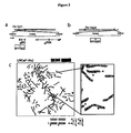
- this invention relates to recurrent MIPOL1-ETV1 genetic rearrangements that are useful as diagnostic markers and clinical targets for prostate cancer.
- Cancer research may identify altered genes that are causally implicated in oncogenesis.
- Several types of somatic mutations that result in altered activity of an oncogene or tumor suppressor gene have been identified, including base substitutions, insertions, deletions, translocations, and chromosomal gains and losses.
- Compelling evidence exists for a causal role for some chromosomal rearrangements in cancer ( Rowley, Nat Rev Cancer 1: 245 (2001 )).
- Recurrent chromosomal aberrations have been primarily characteristic of leukemias, lymphomas, and sarcomas.
- Cancer-related chromosomal rearrangements may result from two primary mechanisms.
- promoter/enhancer elements of one gene are rearranged adjacent to a proto-oncogene, thus causing altered expression of an oncogenic protein.
- This type of translocation is exemplified by the apposition of immunoglobulin (IG) and T-cell receptor (TCR) genes to the MYC oncogene, leading to oncogene activation in Band T-cell malignancies, respectively ( Rabbitts, Nature 372: 143 (1994 )).
- IG immunoglobulin
- TCR T-cell receptor
- TMPRSS2 Gene fusions between androgen-regulated gene TMPRSS2 and ETS family transcription factor members (e.g. ETV1) are known from the prior art WO-A-2007/033187 . Said gene fusion is indicative of prostate cancer.
- WO-A-2007/033187 suggests that TMPRSS2 may be partnering with novel ETS family members, which is the exact opposite from the subject-matter of the present invention as claimed in the claims, namely the partnering of a known ETS (ETV1) with the novel 5' fusion partner MIPOL1.
- a method for diagnosing prostate cancer comprising detecting the presence or absence in a biological sample of a MIPOL1-ETV1 genetic rearrangement, wherein the presence in the sample of the genetic rearrangement is indicative of prostate cancer in the individual from whom the sample was obtained.
- the sample is tissue, blood, plasma, serum, urine, semen, prostatic secretions or prostate cells.
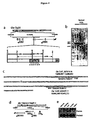
- the detecting step comprises detecting chromosomal rearrangements of genomic DNA encoding MIPOL1 and ETV1.
- the detecting step may use a nucleic acid sequencing technique or a nucleic acid hybridization technique, such as in situ hybridization (ISH), hybridization to one or more moieties in a microarray, or Southern blot analysis.
- ISH in situ hybridization
- the detecting step further includes nucleic acid amplification, which may use known methods that include, but are not limited to, polymerase chain reaction (PCR), reverse transcription polymerase chain reaction (RT-PCR), transcription-mediated amplification (TMA), ligase chain reaction (LCR), strand displacement amplification (SDA), and nucleic acid sequence based amplification (NASBA).
- PCR polymerase chain reaction
- RT-PCR reverse transcription polymerase chain reaction
- TMA transcription-mediated amplification
- LCR ligase chain reaction
- SDA strand displacement amplification
- NASBA nucleic acid sequence based amplification
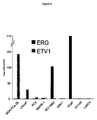
- the detecting step detects mRNA associated with MIPOL1-ETV1 genetic rearrangements or protein expression resulting from MIPOL1-ETV1 genetic rearrangements. Such detection may include analysis of RNA and/or protein expression levels, or determination of sequence characteristics.
- compositions for diagnosing prostate cancer comprising a reagent that directly or indirectly detects a junction between ETV1 genetic material and MIPOL1 genetic material associated with a MIPOL1-ETV1 genetic rearrangement.
- reagents include: a probe comprising a sequence that hybridizes to the junction between ETV1 genetic material and MIPOL1 genetic material associated with a MIPOL1-ETV1 genetic rearrangement, which may be the junction at which a ETV1 gene is inserted into a MIPOL1 gene; a combination of first and second probes, in which a first probe comprises a sequence that hybridizes to the ETV1 gene and a second probe comprises a sequence that hybridizes to the MIPOL1 gene; and at least one first amplification oligonucleotide that comprises a sequence that hybridizes specifically to a ETV1 gene and at least one second amplification oligonucleotide that comprises a sequence that hybridizes specifically to an MIPOL1 gene
- the composition of amplification oligonucleotides may also include a probe that hybridizes specifically to a sequence located between the sequences hybridized by the first amplificaiton oligonucleotide and the second oligonucleotide, which probe may hybridize specifically to a sequence in the ETV1 gene or in the MIPOL1 gene. All probes may be linked directly or indirectly to a label that provides a detectable signal.
- genetic rearrangement refers to any altered arrangement of genomic DNA resulting from the chromosomal rearrangement of two distinct genetic regions, two different genes, or portions of two different genes.
- the product of a genetic rearrangement includes a fusion of genetic material that does not exist in the absence of the genetic rearrangement. That is, the genetic rearrangement results in a junction that fuses one genetic sequence to another genetic sequence to produce a junction that does not exist in a wild type non-rearranged genome.
- Examples of genetic rearrangements include, but are not limited to, insertions of all or part of a first gene or genetic locus into a second gene or genetic locus.
- an insertion may result in a new configuration of the first and second genes or genetic loci that changes regulation of expression, e.g., resulting in increased or decreased expression of a wild type protein.
- an insertion may result in a gene fusion or chimeric DNA that is transcribed to make a chimeric RNA, which may be translated to make altered protein(s) compared to wild type protein(s), i.e., resulting in a new product that is different than made in a wild type configuration.
- gene fusion may refer to a chimeric genomic DNA, a chimeric mRNA that is transcribed from a chimeric DNA (which may be referred to as “gene fusion RNA”), or a protein translated from chimeric mRNA which is transcribed from a chimeric DNA.
- a “gene fusion protein” (or “gene fusion polypeptide”) may be a truncated protein compared to wild type protein, or a chimeric protein resulting from expression of at least a portion of a first gene fused to at least a portion of a second gene.
- a gene fusion (or RNA transcribed from the gene fusion DNA) need not include entire genes or exons of genes.
- transcriptional regulatory region refers to the non-coding upstream regulatory sequence of a gene, also called the 5' untranslated region (5'UTR).
- detect may describe either the general act of discovering or discerning or the specific observation of a detectably labeled composition.
- the term "inhibits at least one biological activity of a gene fusion” refers to any agent that decreases any activity of a gene fusion disclosed herein, its transcript(s) and/or translation products (e.g ., including, but not limited to, the activities described herein), via directly contacting a gene fusion protein, contacting gene fusion mRNA or genomic DNA, causing conformational changes of gene fusion polypeptides, decreasing gene fusion protein levels, or interfering with gene fusion interactions with signaling partners, and affecting the expression of target gene fusions (i.e., transcription or translation of the resulting transcripts).
- Inhibitors also include molecules that indirectly regulate gene fusion biological activity by intercepting upstream signaling molecules.
- siRNAs refers to small interfering RNAs.
- siRNAs comprise a duplex, or double-stranded region, of about 18-25 nucleotides long; often siRNAs contain from about two to four unpaired nucleotides at the 3' end of each strand.
- At least one strand of the duplex or double-stranded region of a siRNA is substantially homologous to, or substantially complementary to, a target RNA molecule.
- the strand complementary to a target RNA molecule is the "antisense strand;" the strand homologous to the target RNA molecule is the "sense strand,” and is also complementary to the siRNA antisense strand.
- siRNAs may also contain additional sequences; non-limiting examples of such sequences include linking sequences, or loops, as well as stem and other folded structures. siRNAs appear to function as key intermediaries in triggering RNA interference in invertebrates and in vertebrates, and in triggering sequence-specific RNA degradation during posttranscriptional gene silencing in plants.
- RNA interference refers to the silencing or decreasing of gene expression by siRNAs. It is the process of sequence-specific, post-transcriptional gene silencing in animals and plants, initiated by siRNA that is homologous in its duplex region to the sequence of the silenced gene.
- the gene may be endogenous or exogenous to the organism, present integrated into a chromosome or present in a transfection vector that is not integrated into the genome. The expression of the gene is either completely or partially inhibited.
- RNAi may also be considered to inhibit the function of a target RNA; the function of the target RNA may be complete or partial.
- stage of cancer refers to a qualitative or quantitative assessment of the level of advancement of a cancer. Criteria used to determine the stage of a cancer include, but are not limited to, the size of the tumor and the extent of metastases ( e.g ., localized or distant).
- gene transfer system refers to any means of delivering a composition comprising a nucleic acid sequence to a cell or tissue.
- gene transfer systems include, but are not limited to, vectors (e.g ., retroviral, adenoviral, adeno-associated viral, and other nucleic acid-based delivery systems), microinjection of naked nucleic acid, polymer-based delivery systems (e.g ., liposome-based and metallic particle-based systems), biolistic injection, and the like.
- viral gene transfer system refers to gene transfer systems comprising viral elements (e.g ., intact viruses, modified viruses and viral components such as nucleic acids or proteins) to facilitate delivery of the sample to a desired cell or tissue.
- viral elements e.g ., intact viruses, modified viruses and viral components such as nucleic acids or proteins
- adenovirus gene transfer system refers to gene transfer systems comprising intact or altered viruses belonging to the family Adenoviridae.
- site-specific recombination target sequences refers to nucleic acid sequences that provide recognition sequences for recombination factors and the location where recombination takes place.
- nucleic acid molecule refers to any nucleic acid containing molecule, including but not limited to, DNA or RNA.
- the term encompasses sequences that include any of the known base analogs of DNA and RNA including, but not limited to, 4-acetylcytosine, 8-hydroxy-N6-methyladenosine, aziridinylcytosine, pseudoisocytosine, 5-(carboxyhydroxylmethyl) uracil, 5-fluorouracil, 5-bromouracil, 5-carboxymethylaminomethyl-2-thiouracil, 5-carboxymethylaminomethyluracil, dihydrouracil, inosine, N6-isopentenyladenine, 1-methyladenine, 1-methylpseudouracil, 1-methylguanine, 1-methylinosine, 2,2-dimethylguanine, 2-methyladenine, 2-methylguanine, 3-methylcytosine, 5-methylcytosine, N6-methyladenine,
- gene refers to a nucleic acid (e.g ., DNA) sequence that comprises coding sequences necessary for the production of a polypeptide, precursor, or RNA (e.g ., rRNA, tRNA).
- the polypeptide can be encoded by a full length coding sequence or by any portion of the coding sequence so long as the desired activity or functional properties (e.g ., enzymatic activity, ligand binding, signal transduction, immunogenicity, etc.) of the full-length or fragment are retained.
- the term also encompasses the coding region of a structural gene and the sequences located adjacent to the coding region on both the 5' and 3' ends for a distance of about 1 kb or more on either end such that the gene corresponds to the length of the full-length mRNA. Sequences located 5' of the coding region and present on the mRNA are referred to as 5' non-translated sequences. Sequences located 3' or downstream of the coding region and present on the mRNA are referred to as 3' non-translated sequences.
- the term "gene” encompasses both cDNA and genomic forms of a gene.
- a genomic form or clone of a gene contains the coding region interrupted with non-coding sequences termed "introns” or “intervening regions” or “intervening sequences.”
- Introns are segments of a gene that are transcribed into nuclear RNA (hnRNA); introns may contain regulatory elements such as enhancers. Introns are removed or “spliced out” from the nuclear or primary transcript; introns therefore are absent in the messenger RNA (mRNA) transcript.
- mRNA messenger RNA
- heterologous gene refers to a gene that is not in its natural environment.
- a heterologous gene includes a gene from one species introduced into another species.
- a heterologous gene also includes a gene native to an organism that has been altered in some way (e.g ., mutated, added in multiple copies, linked to non-native regulatory sequences, etc).
- Heterologous genes are distinguished from endogenous genes in that the heterologous gene sequences are typically joined to DNA sequences that are not found naturally associated with the gene sequences in the chromosome or are associated with portions of the chromosome not found in nature ( e.g ., genes expressed in loci where the gene is not normally expressed).
- oligonucleotide refers to a short length of single-stranded polynucleotide chain. Oligonucleotides are typically less than 200 residues long ( e.g ., between 15 and 100), however, as used herein, the term is also intended to encompass longer polynucleotide chains. Oligonucleotides are often referred to by their length. For example a 24 residue oligonucleotide is referred to as a "24-mer”. Oligonucleotides can form secondary and tertiary structures by self-hybridizing or by hybridizing to other polynucleotides. Such structures can include, but are not limited to, duplexes, hairpins, cruciforms, bends, and triplexes.
- the terms “complementary” or “complementarity” are used in reference to polynucleotides (i.e ., a sequence of nucleotides) related by the base-pairing rules.
- sequence “5'-A-G-T-3'” is complementary to the sequence “3'-T-C-A-5'.”
- Complementarity may be “partial,” in which only some of the nucleic acids' bases are matched according to the base pairing rules. Or, there may be “complete” or “total” complementarity between the nucleic acids.
- the degree of complementarity between nucleic acid strands has significant effects on the efficiency and strength of hybridization between nucleic acid strands. This is of particular importance in amplification reactions, as well as detection methods that depend upon binding between nucleic acids.
- a partially complementary sequence is a nucleic acid molecule that at least partially inhibits a completely complementary nucleic acid molecule from hybridizing to a target nucleic acid is "substantially homologous.”
- the inhibition of hybridization of the completely complementary sequence to the target sequence may be examined using a hybridization assay (Southern or Northern blot, solution hybridization and the like) under conditions of low stringency.
- a substantially homologous sequence or probe will compete for and inhibit the binding ( i.e ., the hybridization) of a completely homologous nucleic acid molecule to a target under conditions of low stringency.
- low stringency conditions are such that non-specific binding is permitted; low stringency conditions require that the binding of two sequences to one another be a specific ( i.e ., selective) interaction.
- the absence of non-specific binding may be tested by the use of a second target that is substantially non-complementary (e.g ., less than about 30% identity); in the absence of non-specific binding the probe will not hybridize to the second non-complementary target.
- substantially homologous refers to any probe that can hybridize to either or both strands of the double-stranded nucleic acid sequence under conditions of low stringency as described above.
- a gene may produce multiple RNA species that are generated by differential splicing of the primary RNA transcript.
- cDNAs that are splice variants of the same gene will contain regions of sequence identity or complete homology (representing the presence of the same exon or portion of the same exon on both cDNAs) and regions of complete non-identity (for example, representing the presence of exon "A” on cDNA 1 wherein cDNA 2 contains exon "B" instead). Because the two cDNAs contain regions of sequence identity they will both hybridize to a probe derived from the entire gene or portions of the gene containing sequences found on both cDNAs; the two splice variants are therefore substantially homologous to such a probe and to each other.
- substantially homologous refers to any probe that can hybridize (i.e ., it is the complement of) the single-stranded nucleic acid sequence under conditions of low stringency as described above.
- hybridization is used in reference to the pairing of complementary nucleic acids. Hybridization and the strength of hybridization (i.e ., the strength of the association between the nucleic acids) is impacted by such factors as the degree of complementary between the nucleic acids, stringency of the conditions involved, the T m of the formed hybrid, and the G:C ratio within the nucleic acids. A single molecule that contains pairing of complementary nucleic acids within its structure is said to be “self-hybridized.”
- stringency is used in reference to the conditions of temperature, ionic strength, and the presence of other compounds such as organic solvents, under which nucleic acid hybridizations are conducted.
- low stringency conditions a nucleic acid sequence of interest will hybridize to its exact complement, sequences with single base mismatches, closely related sequences ( e.g ., sequences with 90% or greater homology), and sequences having only partial homology ( e.g ., sequences with 50-90% homology).
- 'medium stringency conditions a nucleic acid sequence of interest will hybridize only to its exact complement, sequences with single base mismatches, and closely relation sequences ( e.g ., 90% or greater homology).
- a nucleic acid sequence of interest will hybridize only to its exact complement, and (depending on conditions such a temperature) sequences with single base mismatches. In other words, under conditions of high stringency the temperature can be raised so as to exclude hybridization to sequences with single base mismatches.
- High stringency conditions when used in reference to nucleic acid hybridization comprise conditions equivalent to binding or hybridization at 42°C in a solution consisting of 5X SSPE (43.8 g/l NaCl, 6.9 g/l NaH 2 PO 4 H 2 O and 1.85 g/l EDTA, pH adjusted to 7.4 with NaOH), 0.5% SDS, 5X Denhardt's reagent and 100 ⁇ g/ml denatured salmon sperm DNA followed by washing in a solution comprising 0.1X SSPE, 1.0% SDS at 42°C when a probe of about 500 nucleotides in length is employed.
- 5X SSPE 43.8 g/l NaCl, 6.9 g/l NaH 2 PO 4 H 2 O and 1.85 g/l EDTA, pH adjusted to 7.4 with NaOH
- SDS 5X Denhardt's reagent
- 100 ⁇ g/ml denatured salmon sperm DNA followed by washing in a solution comprising 0.1X SSPE, 1.0%
- “Medium stringency conditions” when used in reference to nucleic acid hybridization comprise conditions equivalent to binding or hybridization at 42°C in a solution consisting of 5X SSPE (43.8 g/l NaCl, 6.9 g/l NaH 2 PO 4 H 2 O and 1.85 g/l EDTA, pH adjusted to 7.4 with NaOH), 0.5% SDS, 5X Denhardt's reagent and 100 ⁇ g/ml denatured salmon sperm DNA followed by washing in a solution comprising 1.0X SSPE, 1.0% SDS at 42°C when a probe of about 500 nucleotides in length is employed.
- Low stringency conditions comprise conditions equivalent to binding or hybridization at 42°C in a solution consisting of 5X SSPE (43.8 g/l NaCl, 6.9 g/l NaH 2 PO 4 H 2 O and 1.85 g/l EDTA, pH adjusted to 7.4 with NaOH), 0.1 % SDS, 5X Denhardt's reagent [50X Denhardt's contains per 500 ml: 5 g Ficoll (Type 400, Pharamcia), 5 g BSA (Fraction V; Sigma)] and 100 ⁇ g/ml denatured salmon sperm DNA followed by washing in a solution comprising 5X SSPE, 0.1% SDS at 42°C when a probe of about 500 nucleotides in length is employed.
- 5X SSPE 43.8 g/l NaCl, 6.9 g/l NaH 2 PO 4 H 2 O and 1.85 g/l EDTA, pH adjusted to 7.4 with NaOH
- 5X Denhardt's reagent 50X Den
- low stringency conditions factors such as the length and nature (DNA, RNA, base composition) of the probe and nature of the target (DNA, RNA, base composition, present in solution or immobilized, etc.) and the concentration of the salts and other components (e.g ., the presence or absence of formamide, dextran sulfate, polyethylene glycol) are considered and the hybridization solution may be varied to generate conditions of low stringency hybridization different from, but equivalent to, the above listed conditions.
- conditions that promote hybridization under conditions of high stringency e.g ., increasing the temperature of the hybridization and/or wash steps, the use of formamide in the hybridization solution, etc.) (see definition above for "stringency").
- amplification oligonucleotide refers to an oligonucleotide that hybridizes to a target nucleic acid, or its complement, and participates in a nucleic acid amplification reaction.
- An example of an amplification oligonucleotide is a "primer” that hybridizes to a template nucleic acid and contains a 3' OH end that is extended by a polymerase in an amplification process.
- Another example of an amplification oligonucleotide is an oligonucleotide that is not extended by a polymerase ( e.g ., because it has a 3' blocked end) but participates in or facilitates amplification.
- Amplification oligonucleotides may optionally include modified nucleotides or analogs, or additional nucleotides that participate in an amplification reaction but are not complementary to or contained in the target nucleic acid.
- Amplification oligonucleotides may contain a sequence that is not complementary to the target or template sequence.
- the 5' region of a primer may include a promoter sequence that is non-complementary to the target nucleic acid (referred to as a "promoter-primer").
- promoter-primer a promoter sequence that is non-complementary to the target nucleic acid
- a promoter-primer may be modified by removal of, or synthesis without, a promoter sequence and still function as a primer.
- a 3' blocked amplification oligonucleotide may provide a promoter sequence and serve as a template for polymerization (referred to as a "promoter-provider").
- the term "primer” refers to an oligonucleotide, whether occurring naturally as in a purified restriction digest or produced synthetically, that is capable of acting as a point of initiation of synthesis when placed under conditions in which synthesis of a primer extension product that is complementary to a nucleic acid strand is induced, ( i.e ., in the presence of nucleotides and an inducing agent such as DNA polymerase and at a suitable temperature and pH).
- the primer is preferably single stranded for maximum efficiency in amplification, but may alternatively be double stranded. If double stranded, the primer is first treated to separate its strands before being used to prepare extension products.
- the primer is an oligodeoxyribonucleotide.
- the primer must be sufficiently long to prime the synthesis of extension products in the presence of the inducing agent. The exact lengths of the primers will depend on many factors, including temperature, source of primer and the use of the method.
- probe refers to an oligonucleotide (i.e ., a sequence of nucleotides), whether occurring naturally as in a purified restriction digest or produced synthetically, recombinantly or by PCR amplification, that is capable of hybridizing to at least a portion of another oligonucleotide of interest.
- a probe may be single-stranded or double-stranded. Probes are useful in the detection, identification and isolation of particular gene sequences.
- any probe used in the disclosed methods may be labeled with any "reporter molecule,” so that is detectable in any detection system, including, but not limited to enzyme (e.g ., ELISA, as well as enzyme-based histochemical assays), fluorescent, radioactive, and luminescent systems.
- enzyme e.g ., ELISA, as well as enzyme-based histochemical assays
- fluorescent, radioactive, and luminescent systems e.g., ELISA, as well as enzyme-based histochemical assays
- the compositions and methods that use such compositions as disclosed herein are not limited to any particular detection system or label.
- isolated when used in relation to a nucleic acid, as in “an isolated oligonucleotide” or “isolated polynucleotide” refers to a nucleic acid sequence that is identified and separated from at least one component or contaminant with which it is ordinarily associated in its natural source. Isolated nucleic acid is such present in a form or setting that is different from that in which it is found in nature. In contrast, non-isolated nucleic acids as nucleic acids such as DNA and RNA found in the state they exist in nature.
- a given DNA sequence e.g ., a gene
- RNA sequences such as a specific mRNA sequence encoding a specific protein
- isolated nucleic acid encoding a given protein includes, by way of example, such nucleic acid in cells ordinarily expressing the given protein where the nucleic acid is in a chromosomal location different from that of natural cells, or is otherwise flanked by a different nucleic acid sequence than that found in nature.
- the isolated nucleic acid, oligonucleotide, or polynucleotide may be present in single-stranded or double-stranded form.
- the oligonucleotide or polynucleotide will contain at a minimum the sense or coding strand (i.e ., the oligonucleotide or polynucleotide may be single-stranded), but may contain both the sense and anti-sense strands ( i.e ., the oligonucleotide or polynucleotide may be double-stranded).
- the term "purified” or “to purify” refers to the removal of components (e.g ., contaminants) from a sample.
- antibodies are purified by removal of contaminating non-immunoglobulin proteins; they are also purified by the removal of immunoglobulin that does not bind to the target molecule.
- the removal of non-immunoglobulin proteins and/or the removal of immunoglobulins that do not bind to the target molecule results in an increase in the percent of target-reactive immunoglobulins in the sample.
- recombinant polypeptides are expressed in bacterial host cells and the polypeptides are purified by the removal of host cell proteins; the percent of recombinant polypeptides is thereby increased in the sample.
- compositions and methods are based on the discovery of recurrent gene rearrangements in prostate cancer. These compositions and methods are useful for diagnostic applications, therapeutic methods, and evaluation of therapeutic treatments that either directly or indirectly detect or target MIPOL1-ETV1 gene rearrangements.
- Recurrent gene rearrangements disclosed herein are indicative of prostate cancer. These genetic rearrangements result from chromosomal rearrangements that insert MIPOL1 genetic material into the ETV1 genetic locus. These recurrent gene rearrangements are useful diagnostic markers and clinical targets for prostate cancer.
- all or a portion of the ETV1 is inserted into the MIPOL1 locus (e.g., into an intron).
- the gene rearrangement is detectable, for example, as a chromosomal rearrangement of genomic DNA having at least a portion of an ETV1 gene inserted into a MIPOL1 locus.
- proteins which include fragments, derivatives and analogs thereof, which may be used as immunogens to produce antibodies useful for diagnostic and therapeutic applications.
- Such antibodies may be polyclonal or monoclonal, chimeric, humanized, single chain or Fab fragments, which may be labeled or unlabeled, all of which may be produced by using well known procedures and standard laboratory practices.
- the disclosed MIPOL1-ETV1 genetic rearrangements provide DNA, RNA and protein based diagnostic methods that detect, either directly or indirectly, the gene rearrangements or a product made specifically as a result of the genetic rearrangement.
- the disclosed MIPOL1-ETV1 genetic rearrangements also provide compositions useful for diagnostic purposes, such as oligonucleotide probes that specifically detect all or part of the genetic rearrangement. Such compositions may be in the form of a kit.
- the disclosed diagnostic methods may be qualitative or quantitative. Quantitative methods may, e.g., discriminate between indolent and aggressive cancers via a cutoff or threshold level where expression above that level provides information on the aggressiveness of the cancer which provides useful diagnostic and/or prognostic information to a treating physician or patient.
- Qualitative or quantitative diagnostic methods may include amplification of a target, signal or intermediary, such as by using a universal primer that amplifies a sequence that serves as an indicator for the presence or level of the specific target associated with the MIPOL1-ETV1 genetic rearrangement.
- An initial assay may confirm the presence of a gene rearrangement but not identify the specific rearrangement.
- a secondary assay is then performed to determine the identity of the particular rearrangement, if desired.
- the second assay may use a different detection technology than the initial assay.
- the disclosed MIPOL1-ETV1 genetic rearrangement may be detected along with other markers in a multiplex or panel format. Markers are selected for their predictive value alone or in combination with the gene rearrangements.
- Exemplary prostate cancer markers include, but are not limited to: AMACR/P504S ( U.S. Pat. No. 6,262,245 ); PCA3 ( U.S. Pat. No. 7,008,765 ); PCGEM1 ( U.S. Pat. No. 6,828,429 ); prostein/P501S, P503S, P504S, P509S, P510S, prostase/P703P, P710P ( U.S. Publication No. 20030185830 ); and, those disclosed in U.S. Pat. Nos. 5,854,206 and 6,034,218 , and U.S. Publication No. 20030175736 . Markers for other cancers, diseases, infections, and metabolic conditions are also contemplated for inclusion in a multiplex of panel format
- the diagnostic methods as disclosed herein may be modified with reference to data correlating particular gene rearrangements with the stage, aggressiveness or progression of the disease or the presence or risk of metastasis.
- the information provided by these diagnostic methods provide useful information to a physician who, based on that information, may choose an appropriate therapeutic treatment for a particular patient.
- any biological sample suspected of containing the MIPOL1-ETV1 gene rearrangements may be tested according to the disclosed methods.
- a sample may be tissue (e.g ., prostate biopsy sample or tissue obtained by prostatectomy), blood, urine, semen, prostatic secretions or a fraction thereof (e.g ., plasma, serum, urine supernatant, urine cell pellet or prostate cells), which may be obtained from a patient or other source of biological material, e.g., autopsy sample or forensic material.
- a urine sample is collected immediately following an attentive digital rectal examination (DRE), which causes prostate cells from the prostate gland to shed into the urinary tract.
- DRE attentive digital rectal examination
- the sample may be processed to isolate or enrich the sample for the gene rearrangements or cells that contain the gene rearrangements.
- a variety of well techniques that use standard laboratory practices may be used for this purpose, such as, e.g., centrifugation, immunocapture, cell lysis, and nucleic acid target capture ( See, e.g ., EP Pat. No. 1 409 727 ).
- the disclosed MIPOL1-ETV1 genetic rearrangements may be detected as chromosomal rearrangements of genomic DNA or as chimeric mRNA produced from a chromosomal rearrangement by using a variety of well known nucleic acid techniques that rely on standard laboratory methods, such as, e.g., nucleic acid sequencing, nucleic acid hybridization, and, nucleic acid amplification.
- nucleic acid sequencing techniques include, but are not limited to, chain terminator (Sanger) sequencing and dye terminator sequencing.
- chain terminator Sanger
- dye terminator sequencing Those of ordinary skill in the art will recognize that because RNA is less stable in the cell and more prone to nuclease attack experimentally RNA is usually reverse transcribed to DNA before sequencing.
- Chain terminator sequencing uses sequence-specific termination of a DNA synthesis reaction using modified nucleotide substrates. Extension is initiated at a specific site on the template DNA by using a short radioactive, or other labeled, oligonucleotide primer complementary to the template at that region.
- the oligonucleotide primer is extended using a DNA polymerase, standard four deoxynucleotide bases, and a low concentration of one chain terminating nucleotide, most commonly a di-deoxynucleotide. This reaction is repeated in four separate tubes with each of the bases taking turns as the di-deoxynucleotide.
- the DNA polymerase Limited incorporation of the chain terminating nucleotide by the DNA polymerase results in a series of related DNA fragments that are terminated only at positions where that particular di-deoxynucleotide is used.
- the fragments are size-separated by electrophoresis in a slab polyacrylamide gel or a capillary tube filled with a viscous polymer. The sequence is determined by reading which lane produces a visualized mark from the labeled primer as you scan from the top of the gel to the bottom.
- Dye terminator sequencing alternatively labels the terminators. Complete sequencing can be performed in a single reaction by labeling each of the di-deoxynucleotide chain-terminators with a separate fluorescent dye, which fluoresces at a different wavelength.
- nucleic acid hybridization techniques include, but are not limited to, in situ hybridization (ISH), microarray, and Southern or Northern blot.
- ISH In situ hybridization
- DNA ISH can be used to determine the structure of chromosomes.
- RNA ISH is used to measure and localize mRNAs and other transcripts within tissue sections or whole mounts. Sample cells and tissues are usually treated to fix the target transcripts in place and to increase access of the probe. The probe hybridizes to the target sequence at elevated temperature, and then the excess probe is washed away.
- ISH can also use two or more probes, labeled with radioactivity or the other non-radioactive labels, to simultaneously detect two or more transcripts.
- gene rearrangements are detected using fluorescence in situ hybridization (FISH).
- FISH fluorescence in situ hybridization
- the preferred FISH assays use bacterial artificial chromosomes (BACs), which have been used extensively in the human genome sequencing project (see Nature 409: 953-958 (2001 )) and clones containing specific BACs are widely available or can be made by using standard laboratory practices.
- BACs bacterial artificial chromosomes
- Each BAC clone from the human genome has been given a reference name that unambiguously identifies it. These names can be used to find a corresponding GenBank sequence and to order copies of the clone from a distributor.
- Specific BAC clones that can be used in FISH protocols to detect MIPOL1-ETV1 genetic rearrangements are probes specific for RP11-124L22 and RP11-703A4. These probes, or other suitable probes, usually are labeled with appropriate fluorescent or other markers and then used in hybridizations.
- the Examples section provided herein sets forth one particular protocol that is effective for measuring rearrangements but one of skill in the art will recognize that many variations of this assay can be used equally well.
- Specific protocols are well known in the art and can be readily adapted for detecting MIPOL1-ETV1 rearrangements. Guidance regarding such methodology is provided in many references including: In situ Hybridization: Medical Applications (eds. G. R. Coulton and J.
- Patents providing guidance on such methodology include U.S. 5,225,326 ; 5,545,524 ; 6,121,489 and 6,573,043 , and commercially available kits also provide protocols for performing FISH (e.g ., from Oncor, Inc., Gaithersburg, MD). All of these references may be used along with similar references in the art and with the information provided in the Examples section herein to establish procedural steps convenient for a particular laboratory.
- DNA microarrays e.g ., cDNA microarrays and oligonucleotide microarrays
- protein microarrays e.g ., protein microarrays
- tissue microarrays e.g ., transfection or cell microarrays
- chemical compound microarrays e.g ., chemical compound microarrays
- antibody microarrays e.g ., antibody microarrays.
- a DNA microarray commonly known as gene chip, DNA chip, or biochip, is a collection of microscopic DNA spots attached to a solid surface (e.g ., glass, plastic or silicon chip) forming an array for the purpose of expression profiling or monitoring expression levels for thousands of genes simultaneously.
- the affixed DNA segments are known as probes, thousands of which can be used in a single DNA microarray.
- Microarrays can be used to identify disease genes by comparing gene expression in disease and normal cells.
- Microarrays can be fabricated using a variety of technologies, including but not limiting: printing with fine-pointed pins onto glass slides; photolithography using pre-made masks; photolithography using dynamic micromirror devices; ink-jet printing; or, electrochemistry on microelectrode arrays.
- Southern and Northern blotting is used to detect specific DNA or RNA sequences, respectively.
- DNA or RNA extracted from a sample is fragmented, electrophoretically separated on a matrix gel, and transferred to a membrane filter.
- the filter bound DNA or RNA is subject to hybridization with a labeled probe complementary to the sequence of interest. Hybridized probe bound to the filter is detected.
- a variant of the procedure is the reverse Northern blot, in which the substrate nucleic acid that is affixed to the membrane is a collection of isolated DNA fragments and the probe is RNA extracted from a tissue and labeled.
- PCR polymerase chain reaction
- RT-PCR reverse transcription polymerase chain reaction
- TMA transcription-mediated amplification
- LCR ligase chain reaction
- SDA strand displacement amplification
- NASBA nucleic acid sequence based amplification
- RNA be reversed transcribed to DNA prior to amplification e.g ., RT-PCR
- other amplification techniques directly amplify RNA (e.g ., TMA and NASBA).
- the polymerase chain reaction ( U.S. Pat. Nos. 4,683,195 , 4,683,202 , 4,800,159 and 4,965,188 ), commonly referred to as PCR, uses multiple cycles of denaturation, annealing of primer pairs to opposite strands, and primer extension to exponentially increase copy numbers of a target nucleic acid sequence.
- RT-PCR reverse transcriptase (RT) is used to make a complementary DNA (cDNA) from RNA, and the cDNA is then amplified by PCR to produce multiple copies of DNA.
- RT-PCR reverse transcriptase
- cDNA complementary DNA
- cDNA complementary DNA
- TMA Transcription mediated amplification
- U.S. Pat. Nos. 5,480,784 and 5,399,491 , commonly referred to as TMA
- TMA optionally incorporates the use of blocking moieties, terminating moieties, and other modifying moieties to improve TMA process sensitivity and accuracy.
- the ligase chain reaction ( Weiss, R., Science 254: 1292 (1991 ) ), commonly referred to as LCR, uses two sets of complementary DNA oligonucleotides that hybridize to adjacent regions of the target nucleic acid.
- the DNA oligonucleotides are covalently linked by a DNA ligase in repeated cycles of thermal denaturation, hybridization and ligation to produce a detectable double-stranded ligated oligonucleotide product.
- Strand displacement amplification ( Walker, G. et al., Proc. Natl. Acad. Sci. USA 89: 392-396 (1992 ); U.S. Pat. Nos. 5,270,184 and 5,455,166 ), commonly referred to as SDA, uses cycles of annealing pairs of primer sequences to opposite strands of a target sequence, primer extension in the presence of a dNTP ⁇ S to produce a duplex hemiphosphorothioated primer extension product, endonuclease-mediated nicking of a hemimodified restriction endonuclease recognition site, and polymerase-mediated primer extension from the 3' end of the nick to displace an existing strand and produce a strand for the next round of primer annealing, nicking and strand displacement, resulting in geometric amplification of product.
- Thermophilic SDA (tSDA) uses thermophilic endonucleases and polymerases at higher temperatures in essentially the same method ( EP
- amplification methods include, for example: nucleic acid sequence based amplification ( U.S. Pat. No. 5,130,238 , herein incorporated by reference in its entirety), commonly referred to as NASBA; one that uses an RNA replicase to amplify the probe molecule itself ( Lizardi et al., BioTechnol. 6: 1197 (1988 ) ), commonly referred to as Q ⁇ replicase; a transcription based amplification method ( Kwoh et al., Proc. Natl. Acad. Sci. USA 86:1173 (1989 )); and, self-sustained sequence replication ( Guatelli et al., Proc. Natl. Acad. Sci.
- Non-amplified or amplified gene rearrangement nucleic acids can be detected by any conventional means.
- the gene rearrangements can be detected by hybridization with a detectably labeled probe and measurement of the resulting hybrids. Illustrative non-limiting examples of detection methods are described below.
- Hybridization Protection Assay involves hybridizing a chemiluminescent oligonucleotide probe (e.g ., an acridinium ester-labeled (AE) probe) to the target sequence, selectively hydrolyzing the chemiluminescent label present on unhybridized probe, and measuring the chemiluminescence produced from the remaining probe in a luminometer.
- a chemiluminescent oligonucleotide probe e.g ., an acridinium ester-labeled (AE) probe
- AE acridinium ester-labeled
- Another illustrative detection method provides for quantitative evaluation of the amplification process in real-time.
- Evaluation of an amplification process in "real-time” involves determining the amount of amplicon in the reaction mixture either continuously or periodically during the amplification reaction, and using the determined values to calculate the amount of target sequence initially present in the sample.
- a variety of methods for determining the amount of initial target sequence present in a sample based on real-time amplification are well known in the art. These include methods disclosed in U.S. Pat. Nos. 6,303,305 and 6,541,205 .
- Another method for determining the quantity of target sequence initially present in a sample, but which is not based on a real-time amplification is disclosed in U.S. Pat. No. 5,710,029 .
- Amplification products may be detected in real-time through the use of various self-hybridizing probes, most of which have a stem-loop structure.
- Such self-hybridizing probes are labeled so that they emit differently detectable signals, depending on whether the probes are in a self-hybridized state or an altered state through hybridization to a target sequence.
- “molecular torches” are a type of self-hybridizing probe that includes distinct regions of self-complementarity (referred to as “the target binding domain” and “the target closing domain") which are connected by a joining region (e.g ., non-nucleotide linker) and which hybridize to each other under predetermined hybridization assay conditions.
- molecular torches contain single-stranded base regions in the target binding domain that are from 1 to about 20 bases in length and are accessible for hybridization to a target sequence present in an amplification reaction under strand displacement conditions.
- hybridization of the two complementary regions, which may be fully or partially complementary, of the molecular torch is favored, except in the presence of the target sequence, which will bind to the single-stranded region present in the target binding domain and displace all or a portion of the target closing domain.
- the target binding domain and the target closing domain of a molecular torch include a detectable label or a pair of interacting labels (e.g ., luminescent/quencher) positioned so that a different signal is produced when the molecular torch is self-hybridized than when the molecular torch is hybridized to the target sequence, thereby permitting detection of probe:target duplexes in a test sample in the presence of unhybridized molecular torches.
- Molecular torches and many types of interacting label pairs are known (e.g., U.S. Pat. No. 6,534,274 ).
- Molecular beacons include nucleic acid molecules having a target complementary sequence, an affinity pair (or nucleic acid arms) holding the probe in a closed conformation in the absence of a target sequence present in an amplification reaction, and a label pair that interacts when the probe is in a closed conformation. Hybridization of the target sequence and the target complementary sequence separates the members of the affinity pair, thereby shifting the probe to an open conformation. The shift to the open conformation is detectable due to reduced interaction of the label pair, which may be, for example, a fluorophore and a quencher (e.g ., DABCYL and EDANS).
- a fluorophore and a quencher e.g ., DABCYL and EDANS
- probe binding pairs having interacting labels may be adapted for use in the compositions and methods disclosed herein.
- Probe systems used to detect single nucleotide polymorphisms (SNPs) might also be used.
- Additional detection systems include "molecular switches,” (e.g., see U.S. Publ. No. 20050042638 ).
- Other probes, such as those comprising intercalating dyes and/or fluorochromes, are also useful for detection of amplification products in the methods disclosed herein (e.g., see U.S. Pat. No. 5,814,447 ).
- a computer-based analysis program is used to translate the raw data generated by the detection assay (e.g ., the presence, absence, or amount of a given marker or markers) into data of predictive value for a clinician.
- the clinician can access the predictive data using any suitable means.
- the present invention provides the further benefit that the clinician, who is not likely to be trained in genetics or molecular biology, need not understand the raw data.
- the data is presented directly to the clinician in its most useful form. The clinician is then able to immediately utilize the information in order to optimize the care of the subject.
- a sample e.g ., a biopsy or a serum or urine sample
- a profiling service e.g ., clinical lab at a medical facility, genomic profiling business, etc.
- the subject may visit a medical center to have the sample obtained and sent to the profiling center, or subjects may collect the sample themselves (e.g ., a urine sample) and directly send it to a profiling center.
- the sample comprises previously determined biological information
- the information may be directly sent to the profiling service by the subject (e.g ., an information card containing the information may be scanned by a computer and the data transmitted to a computer of the profiling center using an electronic communication systems).
- the profiling service Once received by the profiling service, the sample is processed and a profile is produced (i.e ., expression data), specific for the diagnostic or prognostic information desired for the subject.
- the profile data is then prepared in a format suitable for interpretation by a treating clinician.
- the prepared format may represent a diagnosis or risk assessment (e.g ., likelihood of cancer being present) for the subject, along with recommendations for particular treatment options.
- the data may be displayed to the clinician by any suitable method.
- the profiling service generates a report that can be printed for the clinician ( e.g ., at the point of care) or displayed to the clinician on a computer monitor.
- the information is first analyzed at the point of care or at a regional facility.
- the raw data is then sent to a central processing facility for further analysis and/or to convert the raw data to information useful for a clinician or patient.
- the central processing facility provides the advantage of privacy (all data is stored in a central facility with uniform security protocols), speed, and uniformity of data analysis.
- the central processing facility can then control the fate of the data following treatment of the subject. For example, using an electronic communication system, the central facility can provide data to the clinician, the subject, or researchers.
- the subject is able to directly access the data using the electronic communication system.
- the subject may chose further intervention or counseling based on the results.
- the data is used for research use.
- the data may be used to further optimize the inclusion or elimination of markers as useful indicators of a particular condition or stage of disease.
- in vivo imaging techniques including but not limited to: radionuclide imaging; positron emission tomography (PET); computerized axial tomography, X-ray or magnetic resonance imaging method; fluorescence detection, and chemiluminescent detection.
- in vivo imaging techniques are used to visualize the presence of or expression of cancer markers in an animal (e.g ., a human or non-human mammal).
- cancer marker mRNA or protein is labeled using a labeled antibody specific for the cancer marker.
- a specifically bound and labeled antibody can be detected in an individual using an in vivo imaging method, including, but not limited to, radionuclide imaging, positron emission tomography, computerized axial tomography, X-ray or magnetic resonance imaging method, fluorescence detection, and chemiluminescent detection.
- an in vivo imaging method including, but not limited to, radionuclide imaging, positron emission tomography, computerized axial tomography, X-ray or magnetic resonance imaging method, fluorescence detection, and chemiluminescent detection.
- the in vivo imaging methods that use the compositions disclosed herein that detect MIPOL1-ETV1 rearrangements or products derived from them are useful in the diagnosis of cancers, particularly prostate cancer, that express the cancer markers disclosed herein.
- In vivo imaging visualizes the presence of a marker indicative of the cancer, allowing diagnosis and/or prognosis without the use of an unpleasant biopsy. For example, the presence of a marker indicative of cancers likely to metastasize can be detected.
- the in vivo imaging methods can further be used to detect metastatic cancers in other parts of the body.
- reagents e.g ., antibodies
- specific for the cancer markers associated with MIPOL1-ETV1 genetic rearrangements are fluorescently labeled.
- the labeled antibodies are introduced into a subject ( e.g ., orally or parenterally). Fluorescently labeled antibodies are detected using any suitable method or system ( e.g ., see U.S. Pat. No. 6,198,107 ).
- antibodies are radioactively labeled.
- the use of antibodies for in vivo diagnosis is well known in the art, e.g., by using an antibody-based labeling system to image tumors (see Sumerdon et al., Nucl. Med. Biol 17:247-254 [1990 ], Griffin et al., J. Clin. Onc. 9:631-640 [1991 ], and Lauffer, Magnetic Resonance in Medicine 22:339-342 [1991 ]).
- the label used with an antibody-based system will depend on the imaging modality chosen, for example, radioactive labels such as Indium-111, Technetium-99m, or Iodine-131 for use with planar scans or single photon emission computed tomography (SPECT), positron emitting labels such as Fluorine-19 for use with positron emission tomography (PET), and paramagnetic ions such as Gadolinium (III) or Manganese (III) for use with MRI.
- radioactive labels such as Indium-111, Technetium-99m, or Iodine-131 for use with planar scans or single photon emission computed tomography (SPECT)
- positron emitting labels such as Fluorine-19 for use with positron emission tomography (PET)
- paramagnetic ions such as Gadolinium (III) or Manganese (III) for use with MRI.
- Radioactive metals with half-lives ranging from 1 hour to 3.5 days are available for conjugation to antibodies, such as scandium-47 (3.5 days) gallium-67 (2.8 days), gallium-68 (68 minutes), technetiium-99m (6 hours), and indium-111 (3.2 days), of which gallium-67, technetium-99m, and indium-111 are preferable for gamma camera imaging, gallium-68 is preferable for positron emission tomography.
- a useful method of labeling antibodies with such radiometals is by means of a bifunctional chelating agent, such as diethylenetriaminepentaacetic acid (DTPA), as described, for example, by Khaw et al. (Science 209:295 [1980 ]) for In-111 and Tc-99m, and by Scheinberg et al. (Science 215:1511 [1982 ]).
- DTPA diethylenetriaminepentaacetic acid
- Other chelating agents may also be used, but the 1-(p-carboxymethoxybenzyl)EDTA and the carboxycarbonic anhydride of DTPA are advantageous because their use permits conjugation without affecting the antibody's immunoreactivity substantially.
- Another method for coupling DPTA to proteins is by use of the cyclic anhydride of DTPA, as described by Hnatowich et al. (Int. J. Appl. Radiat. Isot. 33:327 [1982 ]) for labeling of albumin with In-111, but which can be adapted for labeling of antibodies.
- a suitable method of labeling antibodies with Tc-99m is known (e.g., see Crockford et al., U.S. Pat. No. 4,323,546 ). ).
- a preferred method of labeling immunoglobulins with Tc-99m is that described by Wong et al. (Int. J. Appl. Radiat. Isot., 29:251 [1978 ]) for plasma protein, and recently applied successfully by Wong et al. (J. Nucl. Med., 23:229 [1981 ]) for labeling antibodies.
- radiometals conjugated to the specific antibody it is likewise desirable to introduce as high a proportion of the radiolabel as possible into the antibody molecule without destroying its immunospecificity.
- a further improvement may be achieved by effecting radiolabeling in the presence of the specific cancer marker associated with a MIPOL1-ETV1 genetic rearrangement, to insure that the antigen binding site on the antibody is protected. The antigen is separated after labeling.
- in vivo biophotonic imaging (Xenogen, Almeda, CA) is used for in vivo imaging.
- This real-time in vivo imaging utilizes luciferase, an enzyme that catalyzes a light-emitting reaction.
- the luciferase gene is incorporated into cells, microorganisms, and animals ( e.g ., to produce a fusion protein with a cancer marker associated with a MIPOL1-ETV1 genetic rearrangement), so that when the cancer marker is active a light emission occurs which is captured as an image and analyzed by using a CCD camera and appropriate software.
- compositions for use in the disclosed diagnostic methods include, but are not limited to, probes, amplification oligonucleotides, and antibodies.
- the compositions detect a product only when a MIPOL1-ETV1 rearrangement is present, preferably ETV1 genetic material inserted into MIPOL1 genetic material.
- compositions include, but are not limited to: a single labeled probe comprising a sequence that hybridizes to the junction at which ETV1 is inserted into the MIPOL1 locus ( i.e ., spans the gene rearrangement junction); a pair of amplification oligonucleotides wherein the first amplification oligonucleotide comprises a sequence that hybridizes to MIPOL1 and a second amplification oligonucleotide comprising a sequence that hybridizes to ETV1.
- Other useful compositions include: a pair of labeled probes wherein the first labeled probe comprises a sequence that hybridizes to MIPOL1 and the second labeled probe comprises a sequence that hybridizes to ETV1.
- kits may be provided in the form of a kit.
- the single labeled probe and pair of amplification oligonucleotides may be provided in a kit for the amplification and detection of MIPOL1-ETV1 genetic rearrangements.
- Kits may further comprise appropriate controls and/or detection reagents. Any one or more reagents that find use in any of the methods described herein may be provided in the kit.
- the probe and antibody compositions may also be provided in the form of an array.
- the disclosed compositions and methods are used in drug screening assays (e.g ., to screen for anticancer drugs).
- These screening methods use cancer markers that include those associated with MIPOL1-ETV1 gene rearrangements, but are not limited only to those genetic rearrangements.
- an embodiment may screen for compounds that alter ( e.g ., decrease) the expression of cancer marker genes, including those associated with MIPOL1-ETV1 I gene rearrangements.
- candidate compounds may interfere with transcription (e.g., by interacting with a promoter region), may interfere with mRNA produced from the rearrangement (e.g ., by RNA interference, antisense technologies, etc.), or may interfere with pathways that are upstream or downstream of the biological activity of the gene rearrangement.
- candidate compounds are antisense or interfering RNA agents (e.g ., oligonucleotides) directed against cancer markers.
- candidate compounds are antibodies or small molecules that specifically bind to a cancer marker regulator or expression product associated with MIPOL1-ETV1 gene rearrangements and inhibit its biological function.
- candidate compounds are evaluated for their ability to alter cancer marker expression by contacting a compound with a cell expressing a cancer marker and then assaying for the effect of the candidate compounds on expression.
- the effect of candidate compounds on expression of a cancer marker gene is assayed for by detecting the level of cancer marker mRNA expressed by the cell. mRNA expression can be detected by any suitable method.
- the effect of candidate compounds on expression of cancer marker genes is assayed by measuring the level of polypeptide encoded by the cancer markers. The level of polypeptide expressed can be measured using any suitable method, including but not limited to, those disclosed herein.
- test compounds can be obtained using any of the numerous approaches in combinatorial library methods known in the art, including biological libraries; peptoid libraries (libraries of molecules having the functionalities of peptides, but with a novel, non-peptide backbone, which are resistant to enzymatic degradation but which nevertheless remain bioactive; see, e.g., Zuckennann et al., J. Med. Chem. 37: 2678-85 [1994 ]); spatially addressable parallel solid phase or solution phase libraries; synthetic library methods requiring deconvolution; the 'one-bead one-compound' library method; and synthetic library methods using affinity chromatography selection.
- biological libraries peptoid libraries (libraries of molecules having the functionalities of peptides, but with a novel, non-peptide backbone, which are resistant to enzymatic degradation but which nevertheless remain bioactive; see, e.g., Zuckennann et al., J. Med. Chem. 37: 2678-85 [1994
- Some embodiments provide therapies for cancer (e.g ., prostate cancer).
- Preferred therapy embodiments target directly or indirectly cancer markers, including those associated with MIPOL1-ETV1 genetic rearrangements.
- Some embodiments target the expression of cancer markers associated with MIPOL1-ETV1 genetic rearrangements.
- Some embodiments employ compositions comprising oligomeric antisense or RNAi compounds, particularly oligonucleotides (e.g. , those identified in the drug screening methods described above), for use in modulating the function of nucleic acid molecules encoding cancer markers associated with MIPOL1-ETV1 genetic rearrangements, ultimately modulating the amount of cancer marker expressed.
- RNA Interference RNA Interference
- RNAi is used to inhibit expression of MIPOL1-ETV1 gene rearrangements.
- RNAi represents an evolutionary conserved cellular defense for controlling the expression of foreign genes in most eukaryotes, including humans.
- RNAi is typically triggered by double-stranded RNA (dsRNA) and causes sequence-specific mRNA degradation of single-stranded target RNAs homologous in response to dsRNA.
- the mediators of mRNA degradation are small interfering RNA duplexes (siRNAs), which are normally produced from long dsRNA by enzymatic cleavage in the cell.
- siRNAs are generally approximately twenty-one nucleotides in length ( e.g .
- RNAi oligonucleotides are designed to target the junction region of gene rearrangements.
- siRNAs Chemically synthesized siRNAs have become powerful reagents for genome-wide analysis of mammalian gene function in cultured somatic cells. Beyond their value for validation of gene function, siRNAs also hold great potential as gene-specific therapeutic agents ( Tuschl and Borkhardt, Molecular Intervent. 2002; 2(3):158-67 , ).
- siRNAs are extraordinarily effective at lowering the amounts of targeted RNA, and by extension proteins, frequently to undetectable levels.
- the silencing effect can last several months, and is extraordinarily specific, because one nucleotide mismatch between the target RNA and the central region of the siRNA is frequently sufficient to prevent silencing ( Brummelkamp et al, Science 2002; 296:550-3 ; and Holen et al, Nucleic Acids Res. 2002; 30:1757-66 ).
- An important factor in the design of siRNAs is the presence of accessible sites for siRNA binding. Bahoia et al., (J. Biol.
- a scanning array to find accessible sites in mRNAs for designing effective siRNAs.
- These arrays comprise oligonucleotides ranging in size from monomers to a certain maximum, usually Corners, synthesized using a physical barrier (mask) by stepwise addition of each base in the sequence.
- the arrays represent a full oligonucleotide complement of a region of the target gene. Hybridization of the target mRNA to these arrays provides an exhaustive accessibility profile of this region of the target mRNA.
- Such data are useful in the design of antisense oligonucleotides (ranging from 7mers to 25mers), where it is important to achieve a compromise between oligonucleotide length and binding affinity, to retain efficacy and target specificity ( Sohail et al, Nucleic Acids Res., 2001; 29(10): 2041- 2045 ). Additional methods and concerns for selecting siRNAs are described for example, in WO 05054270 , WO05038054A1 , WO03070966A2 , J Mol Biol. 2005 May 13;348(4):883-93 , J Mol Biol. 2005 May 13;348(4):871-81 , and Nucleic Acids Res. 2003 Aug 1;31(15):4417-24 .
- software e.g. , the MWG online siMAX siRNA design tool
- expression of MIPOL1-ETV1 gene rearrangements is modulated using antisense compounds that specifically hybridize with one or more nucleic acids encoding cancer markers associated with MIPOL1-ETV1 genetic rearrangements.
- the specific hybridization of an oligomeric compound with its target nucleic acid interferes with the normal function of the nucleic acid. This modulation of function of a target nucleic acid by compounds that specifically hybridize to it is generally referred to as "antisense.”
- the functions of DNA to be interfered with include replication and transcription.
- RNA to be interfered with include all vital functions such as, for example, translocation of the RNA to the site of protein translation, translation of protein from the RNA, splicing of the RNA to yield one or more mRNA species, and catalytic activity that may be engaged in or facilitated by the RNA.
- the overall effect of such interference with target nucleic acid function is modulation of the expression of cancer markers associated with MIPOL1-ETV1 genetic rearrangements.
- modulation means either an increase (stimulation) or a decrease (inhibition) in the expression of a gene. For example, expression may be inhibited to potentially prevent tumor proliferation.
- Antisense methods preferably target specific nucleic acids.
- “Targeting" an antisense compound to a particular nucleic acid usually refers to a multistep process that begins with identification of a nucleic acid sequence whose function is to be modulated. This may be, e.g., a cellular gene (or mRNA transcribed from the gene) whose expression is associated with a particular disorder or disease state, or a nucleic acid molecule from an infectious agent.
- the target is a nucleic acid molecule encoding a cancer marker associated with MIPOL1-ETV1 genetic rearrangements.
- the targeting process also includes determination of a site or sites within this gene for the antisense interaction to occur such that the desired effect, e.g.
- a preferred intragenic site is the region encompassing the translation initiation or termination codon of the open reading frame (ORF) of the gene. Since the translation initiation codon is typically 5'-AUG (in transcribed mRNA molecules; 5'-ATG in the corresponding DNA molecule), the translation initiation codon is also referred to as the "AUG codon," the “start codon” or the "AUG start codon”. A minority of genes have a translation initiation codon having the RNA sequence 5'-GUG, 5'-UUG or 5'-CUG, and 5'-AUA, 5'-ACG and 5'-CUG have been shown to function in vivo.
- translation initiation codon and “start codon” can encompass many codon sequences, even though the initiator amino acid in each instance is typically methionine (in eukaryotes) or formylmethionine (in prokaryotes).
- Eukaryotic and prokaryotic genes may have two or more alternative start codons, any one of which may be preferentially used for translation initiation in a particular cell type or tissue, or under a particular set of conditions.
- start codon and “translation initiation codon” refer to the codon or codons that are used in vivo to initiate translation of an mRNA molecule transcribed from a gene encoding a tumor antigen associated with MIPOL1-ETV1 genetic rearrangements, regardless of the sequence(s) of such codons.
- Translation termination codon (or "stop codon") of a gene may have one of three sequences (i.e ., 5'-UAA, 5'-UAG and 5'-UGA; the corresponding DNA sequences are 5'-TAA, 5'-TAG and 5'-TGA, respectively).
- start codon region and “translation initiation codon region” refer to a portion of such an mRNA or gene that encompasses from about 25 to about 50 contiguous nucleotides in either direction ( i.e ., 5' or 3') from a translation initiation codon.
- stop codon region and “translation termination codon region” refer to a portion of such an mRNA or gene that encompasses from about 25 to about 50 contiguous nucleotides in either direction ( i.e. , 5' or 3') from a translation termination codon.
- Other target regions include the 5' untranslated region (5' UTR), referring to the portion of an mRNA in the 5' direction from the translation initiation codon, and thus including nucleotides between the 5' cap site and the translation initiation codon of an mRNA or corresponding nucleotides on the gene, and the 3' untranslated region (3' UTR), referring to the portion of an mRNA in the 3' direction from the translation termination codon, and thus including nucleotides between the translation termination codon and 3' end of an mRNA or corresponding nucleotides on the gene.
- 5' UTR 5' untranslated region
- 3' UTR 3' untranslated region
- the 5' cap of an mRNA comprises an N7-methylated guanosine residue joined to the 5'-most residue of the mRNA via a 5'-5' triphosphate linkage.
- the 5' cap region of an mRNA is considered to include the 5' cap structure itself as well as the first 50 nucleotides adjacent to the cap.
- the cap region may also be a preferred target region.
- introns regions that are excised from a transcript before it is translated.
- exons regions that are excised from a transcript before it is translated.
- mRNA splice sites i.e ., intron-exon junctions
- intron-exon junctions may also be preferred target regions, and are particularly useful in situ ations where aberrant splicing is implicated in disease, or where an overproduction of a particular mRNA splice product is implicated in disease. Aberrant fusion junctions due to rearrangements or deletions are also preferred targets. It has also been found that introns can also be effective, and therefore preferred, target regions for antisense compounds targeted, for example, to DNA or pre-mRNA.
- target sites for antisense inhibition are identified using commercially available software programs (e.g ., Biognostik, Gottingen, Germany; SysArris Software, Bangalore, India; Antisense Research Group, University of Liverpool, Liverpool, England; GeneTrove, Carlsbad, CA). In other embodiments, target sites for antisense inhibition are identified using the accessible site method described in PCT Publ. No. WO0198537A2 .
- oligonucleotides are chosen that are sufficiently complementary to the target (i.e ., hybridize sufficiently well and with sufficient specificity) to give the desired effect.
- antisense oligonucleotides are targeted to or near the start codon associated with MIPOL1-ETV1 genetic rearrangements.
- hybridization with respect to antisense compositions and methods, means hydrogen bonding, which may be Watson-Crick, Hoogsteen or reversed Hoogsteen hydrogen bonding, between complementary nucleoside or nucleotide bases.
- adenine and thymine are complementary nucleobases that pair through the formation of hydrogen bonds. It is understood that the sequence of an antisense compound need not be 100% complementary to that of its target nucleic acid to be specifically hybridizable.
- An antisense compound is specifically hybridizable when binding of the compound to the target DNA or RNA molecule interferes with the normal function of the target DNA or RNA to cause a loss of utility, and there is a sufficient degree of complementarity to avoid non-specific binding of the antisense compound to non-target sequences under conditions in which specific binding is desired (i.e ., under physiological conditions in the case of in vivo assays or therapeutic treatment, and in the case of in vitro assays, under conditions in which the assays are performed).
- Antisense compounds are commonly used as research reagents and diagnostics. For example, antisense oligonucleotides, which are able to inhibit gene expression with specificity, can be used to elucidate the function of particular genes. Antisense compounds are also used, for example, to distinguish between functions of various members of a biological pathway.
- antisense oligonucleotides have been employed as therapeutic moieties in the treatment of disease states in animals and man. Antisense oligonucleotides have been safely and effectively administered to humans and numerous clinical trials are presently underway. It is thus established that oligonucleotides are useful therapeutic modalities that can be configured to be useful in treatment regimes for treatment of cells, tissues, and animals, especially humans.
- antisense oligonucleotides are preferred, other oligomeric antisense compounds, including but not limited to oligonucleotide mimetics may be used, such as are described below.
- Preferred antisense compounds comprise from about 8 to about 30 nucleobases ( i.e ., from about 8 to about 30 linked bases), although both longer and shorter sequences may be used.
- Particularly preferred antisense compounds are antisense oligonucleotides, even more preferably those comprising from about 12 to about 25 nucleobases.
- oligonucleotides containing modified backbones or non-natural internucleoside linkages include those that retain a phosphorus atom in the backbone and those that do not have a phosphorus atom in the backbone.
- modified oligonucleotides that do not have a phosphorus atom in their internucleoside backbone can also be considered to be oligonucleosides.
- Preferred modified oligonucleotide backbones include, for example, phosphorothioates, chiral phosphorothioates, phosphorodithioates, phosphotriesters, aminoalkylphosphotriesters, methyl and other alkyl phosphonates including 3'-alkylene phosphonates and chiral phosphonates, phosphinates, phosphoramidates including 3'-amino phosphoramidate and aminoalkylphosphoramidates, thionophosphoramidates, thionoalkylphosphonates, thionoalkylphosphotriesters, and boranophosphates having normal 3'-5' linkages, 2'-5' linked analogs of these, and those having inverted polarity wherein the adjacent pairs of nucleoside units are linked 3'-5' to 5'-3' or 2'-5' to 5'-2'.
- Various salts, mixed salts and free acid forms are also included.
- Preferred modified oligonucleotide backbones that do not include a phosphorus atom therein have backbones that are formed by short chain alkyl or cycloalkyl internucleoside linkages, mixed heteroatom and alkyl or cycloalkyl internucleoside linkages, or one or more short chain heteroatomic or heterocyclic internucleoside linkages.
- morpholino linkages formed in part from the sugar portion of a nucleoside
- siloxane backbones sulfide, sulfoxide and sulfone backbones
- formacetyl and thioformacetyl backbones methylene formacetyl and thioformacetyl backbones
- alkene containing backbones sulfamate backbones
- sulfonate and sulfonamide backbones amide backbones; and others having mixed N, O, S and CH2 component parts.
- both the sugar and the internucleoside linkage (i.e ., the backbone) of the nucleotide units are replaced with novel groups.
- the base units are maintained for hybridization with an appropriate nucleic acid target compound.
- an oligonucleotide mimetic that has been shown to have excellent hybridization properties, is referred to as a peptide nucleic acid (PNA).
- PNA peptide nucleic acid
- the sugar-backbone of an oligonucleotide is replaced with an amide containing backbone, in particular an aminoethylglycine backbone.
- nucleobases are retained and are bound directly or indirectly to aza nitrogen atoms of the amide portion of the backbone.
- Methods for preparation of PNA compounds are well known (e.g., see U.S. Pat. Nos.: 5,539,082 ; 5,714,331 ; and 5,719,262 , and Nielsen et al., Science 254:1497 (1991 ) ).
- Most preferred embodiments of the invention are oligonucleotides with phosphorothioate backbones and oligonucleosides with heteroatom backbones, and in particular --CH2, --NH--O--CH2--, --CH2--N(CH3)--O--CH2-- [known as a methylene (methylimino) or MMI backbone], --CH2--O--N(CH3)--CH2--, --CH2--N(CH3)---N(CH3)--CH2--, and --O--N(CH3)--CH2--CH2-- [wherein the native phosphodiester backbone is represented as --O--P--O--CH2--], amid backbone, and morpholino backbone structures, all of which are well known (e.g., see U.S. Pat. Nos. 5,489,677 , 5,602,240 , and 5,034,506 ).
- Modified oligonucleotides may also contain one or more substituted sugar moieties.
- Preferred oligonucleotides comprise one of the following at the 2' position: OH; F; O-, S-, or N-alkyl; O-, S-, or N-alkenyl; O-, S- or N-alkynyl; or O-alkyl-O-alkyl, wherein the alkyl, alkenyl and alkynyl may be substituted or unsubstituted C1 to C10 alkyl or C2 to C 10 alkenyl and alkynyl.
- oligonucleotides comprise one of the following at the 2' position: C1 to C10 lower alkyl, substituted lower alkyl, alkaryl, aralkyl, O-alkaryl or O-aralkyl, SH, SCH3, OCN, Cl, Br, CN, CF3, OCF3, SOCH3, SO2CH3, ONO2, NO2, N3, NH2, heterocycloalkyl, heterocycloalkaryl, aminoalkylamino, polyalkylamino, substituted silyl, an RNA cleaving group, a reporter group, an intercalator, a group for improving the pharmacokinetic properties of an oligonucleotide, or a group for improving the pharmacodynamic properties of an oligonucleotide, and other substituents having similar properties.
- a preferred modification includes 2'-methoxyethoxy (2'-O--CH2CH2OCH3, also known as 2'-O-(2-methoxyethyl) or 2'-MOE) ( Martin et al., Helv. Chim. Acta 78:486 [1995 ]) i.e ., an alkoxyalkoxy group.
- a further preferred modification includes 2'-dimethylaminooxyethoxy (i.e., a O(CH2)2ON(CH3)2 group), also known as 2'-DMAOE, and 2'-dimethylaminoethoxyethoxy (also known in the art as 2'-O-dimethylaminoethoxyethyl or 2'-DMAEOE), i.e ., 2'-O--CH2--O--CH2--N(CH2)2.
- 2'-dimethylaminooxyethoxy i.e., a O(CH2)2ON(CH3)2 group
- 2'-DMAOE 2'-dimethylaminoethoxyethoxy
- 2'-DMAEOE 2'-dimethylaminoethoxyethyl
- Oligonucleotides may also have sugar mimetics such as cyclobutyl moieties in place of the pentofuranosyl sugar.
- Oligonucleotides may also include nucleobase (often referred to in the art simply as “base”) modifications or substitutions.
- nucleobase often referred to in the art simply as “base”
- “unmodified” or “natural” nucleobases include the purine bases adenine (A) and guanine (G), and the pyrimidine bases thymine (T), cytosine (C) and uracil (U). Modified nucleobases are well known (e.g., see U.S. Pat. No.
- nucleobases for which the A, G, T, C and U abbreviations for the bases are used in the following examples, such as 5-methylcytosine (5-me-C), 5-hydroxymethyl C, xanthine, hypoxanthine, 2-amino-A, 6-methyl or 2-propyl and other alkyl derivatives of A and G, 2-thio-U, 2-thio-T and 2-thio-C, 5-halo-U and -C, 5-propynyl U and C, 6-azo U, C and T, 5-uracil (pseudouracil), 4-thio-U, 8-halo, 8-amino, 8-thiol, 8-thioalkyl, 8-hydroxyl and other 8-substituted A and G, 5-halo substituted U and C, particularly 5-bromo, 5-trifluoromethyl and other 5-substituted U and C, 7-methyl-G and 7-methyl-A,
- nucleobases are particularly useful for increasing the binding affinity of the oligomeric compounds of the invention.
- These include 5-substituted pyrimidines, 6-azapyrimidines and N-2, N-6 and O-6 substituted purines, including 2-aminopropyladenine, 5-propynyl-U and 5-propynyl-C.
- 5-methyl-C substitutions are known to increase nucleic acid duplex stability and are preferred base substitutions in some embodiments, even more particularly when combined with 2'-O-methoxyethyl sugar modifications.
- oligonucleotides involve chemically linking to the oligonucleotide one or more moieties or conjugates that enhance the activity, cellular distribution or cellular uptake of the oligonucleotide.
- moieties include but are not limited to lipid moieties such as a cholesterol moiety, cholic acid, a thioether, ( e.g ., hexyl-S-tritylthiol), a thiocholesterol, an aliphatic chain, ( e.g ., dodecandiol or undecyl residues), a phospholipid, ( e.g ., di-hexadecyl-rac-glycerol or triethylammonium 1,2-di-O-hexadecyl-rac-glycero-3-H-phosphonate), a polyamine or a polyethylene glycol chain or adamantane acetic acid, a palmityl mo
- oligonucleotides containing the above-described modifications are not limited to the antisense oligonucleotides described above. Any suitable modification or substitution may be used.
- Antisense compounds may be chimeric compounds.
- Chimeric antisense compounds or “chimeras,” as used herein, are antisense compounds, particularly oligonucleotides, which contain two or more chemically distinct regions, each made up of at least one monomer unit, i.e ., a nucleotide in the case of an oligonucleotide compound.
- oligonucleotides typically contain at least one region wherein the oligonucleotide is modified so as to confer upon the oligonucleotide increased resistance to nuclease degradation, increased cellular uptake, and/or increased binding affinity for the target nucleic acid.
- An additional region of the oligonucleotide may serve as a substrate for enzymes capable of cleaving RNA:DNA or RNA:RNA hybrids.
- RNaseH is a cellular endonuclease that cleaves the RNA strand of an RNA:DNA duplex.
- RNA target Activation of RNase H, therefore, results in cleavage of the RNA target, thereby greatly enhancing the efficiency of oligonucleotide inhibition of gene expression. Consequently, comparable results can often be obtained with shorter oligonucleotides when chimeric oligonucleotides are used, compared to phosphorothioate deoxyoligonucleotides hybridizing to the same target region.
- Cleavage of the RNA target can be routinely detected by gel electrophoresis and, if necessary, associated nucleic acid hybridization techniques known in the art.
- Chimeric antisense compounds may be formed as composite structures of two or more oligonucleotides, modified oligonucleotides, oligonucleosides and/or oligonucleotide mimetics as described above.
- compositions and formulations that include the antisense compounds as described herein.
- Embodiments may use any genetic manipulation to modulate the expression of cancer markers associated with MIPOL1-ETV1 genetic rearrangements described herein.
- genetic manipulation include, but are not limited to, gene knockout (such as by removing the genetic rearrangement from the chromosome using, e.g., by recombination), expression of antisense constructs with or without inducible promoters, and the like.
- Delivery of nucleic acid construct to cells in vitro or in vivo may be conducted using any suitable method.
- a suitable method is one that introduces the nucleic acid construct into the cell such that the desired event occurs (e.g ., expression of an antisense construct).
- Genetic therapy may also be used to deliver siRNA or other interfering molecules that are expressed in vivo ( e.g ., upon stimulation by an inducible promoter (e.g ., an androgen-responsive promoter)).
- Plasmids carrying genetic information into cells are achieved by any of various methods including, but not limited to, directed injection of naked DNA constructs, bombardment with gold particles loaded with said constructs, and macromolecule mediated gene transfer using, for example, liposomes, biopolymers, and the like.
- Preferred methods use gene delivery vehicles derived from viruses, including, but not limited to, adenoviruses, retroviruses, vaccinia viruses, and adeno-associated viruses. Because of the higher efficiency as compared to retroviruses, vectors derived from adenoviruses are the preferred gene delivery vehicles for transferring nucleic acid molecules into host cells in vivo.
- Adenoviral vectors and their use in gene transfer are well known (e.g., see PCT publications WO 00/12738 and WO 00/09675 and U.S. Pat. Nos. 6,033,908 , 6,019,978 , 6,001,557 , 5,994,132 , 5,994,128 , 5,994,106 , 5,981,225 , 5,885,808 , 5,872,154 , 5,830,730 , and 5,824,544 , ).
- Such vectors and methods have been shown to provide very efficient in vivo gene transfer into a variety of solid tumors in animal models and into human solid tumor xenografts in immune-deficient mice.
- Vectors may be administered to subject in a variety of well known ways, e.g., administered into tumors or tissue associated with tumors by using direct injection or administration via the blood or lymphatic circulation ( See e.g., PCT publication 99/02685 ).
- exemplary dose levels of adenoviral vector are preferably 10 8 to 10 11 vector particles added to the perfusate.
- Some embodiments are or use antibodies and/or small molecules that target prostate tumors that express a cancer marker associated with MIPOL1-ETV1 genetic rearrangements.
- the therapeutic regimen is selected based on a diagnostic result and uses a suitable antibody (e.g ., monoclonal, polyclonal, or synthetic) in the therapeutic methods.
- the antibodies used for cancer therapy are humanized antibodies. Methods for humanizing antibodies are well known ( See e.g., U.S. Pat. Nos. 6,180,370 , 5,585,089 , 6,054,297 , and 5,565,332 ;
- the therapeutic antibodies comprise an antibody generated against a cancer marker associated with MIPOL1-ETV1 genetic rearrangements, wherein the antibody is conjugated to a cytotoxic agent.
- a tumor specific therapeutic agent is generated that does not target normal cells, thus reducing many of the detrimental side effects of traditional chemotherapy.
- the therapeutic agents will be pharmacologic agents that will serve as useful agents for attachment to antibodies, particularly cytotoxic or otherwise anticellular agents having the ability to kill or suppress the growth or cell division of endothelial cells.
- exemplary anticellular agents include chemotherapeutic agents, radioisotopes, and cytotoxins.
- Such therapeutic antibodies may include a variety of cytotoxic moieties, including but not limited to, radioactive isotopes (e.g ., iodine-131, iodine-123, technicium-99m, indium-111, rhenium-188, rhenium-186, gallium-67, copper-67, yttrium-90, iodine-125 or astatine-211), hormones such as a steroid, antimetabolites such as cytosines (e.g ., arabinoside, fluorouracil, methotrexate or aminopterin; an anthracycline; mitomycin C), vinca alkaloids ( e.g ., demecolcine; etoposide; mithramycin), and antitumor alkylating agent such as chlorambucil or melphalan.
- radioactive isotopes e.g ., iodine-131, iodine-123,
- agents such as a coagulant, a cytokine, growth factor, bacterial endotoxin or the lipid A moiety of bacterial endotoxin.
- therapeutic agents will include plant-, fungus- or bacteria-derived toxin, such as an A chain toxins, a ribosome inactivating protein, ⁇ -sarcin, aspergillin, restrictocin, a ribonuclease, diphtheria toxin or pseudomonas exotoxin, to mention just a few examples.
- deglycosylated ricin A chain is used.
- agents such as these may, if desired, be successfully conjugated to an antibody, in a manner that will allow their targeting, internalization, release or presentation to blood components at the site of the targeted tumor cells as required using known conjugation technology ( See, e.g., Ghose et al., Methods Enzymol., 93:280 [1983 ]).
- some embodiments provide immunotoxins targeted against cancer marker associated with MIPOL1-ETV1 genetic rearrangements.
- Immunotoxins are conjugates of a specific targeting agent typically a tumor-directed antibody or fragment, with a cytotoxic agent, such as a toxin moiety.
- the targeting agent directs the toxin to, and thereby selectively kills, cells carrying the targeted antigen.
- therapeutic antibodies employ crosslinkers that provide high in vivo stability ( Thorpe et al., Cancer Res., 48:6396 [1988 ]).
- antibodies are designed to have a cytotoxic or otherwise anticellular effect against the tumor vasculature, by suppressing the growth or cell division of the vascular endothelial cells. This attack is intended to lead to a tumor-localized vascular collapse, depriving the tumor cells, particularly those tumor cells distal of the vasculature, of oxygen and nutrients, ultimately leading to cell death and tumor necrosis.
- antibody based therapeutics are formulated as pharmaceutical compositions as described herein.
- administration of an antibody composition that targets a moiety associated with MIPOL1-ETV1 genetic rearrangements results in a measurable decrease in cancer (e.g., decrease or elimination of tumor).
- Embodiments include generation of transgenic animals comprising an exogenous cancer marker gene that is identical to or representative of a MIPOL1-ETV1 genetic rearrangement described herein, which includes mutants and variants thereof (e.g. , truncations or single nucleotide polymorphisms).
- the transgenic animal displays an altered phenotype (e.g., increased or decreased presence of markers associated with MIPOL1-ETV1 genetic rearrangements) as compared to wild-type animals. Methods for analyzing the presence or absence of such phenotypes include but are not limited to, those disclosed herein.
- the transgenic animals further display an increased or decreased growth of tumors or evidence of cancer.
- transgenic animals are useful in drug (e.g ., cancer therapy) screens.
- test compounds e.g ., a drug that is suspected of being useful to treat cancer
- control compounds e.g. , a placebo
- the transgenic animals can be generated via a variety of methods.
- embryonal cells at various developmental stages are used to introduce transgenes for the production of transgenic animals. Different methods are used depending on the stage of development of the embryonal cell.
- the zygote is the best target for micro-injection. In the mouse, the male pronucleus reaches the size of approximately 20 micrometers in diameter that allows reproducible injection of 1-2 picoliters (pl) of DNA solution.
- pl picoliters
- the use of zygotes as a target for gene transfer has a major advantage in that in most cases the injected DNA will be incorporated into the host genome before the first cleavage ( Brinster et al., Proc. Natl. Acad. Sci.
- transgenic non-human animal carry the incorporated transgene. This is reflected in the efficient transmission of the transgene to offspring of the founder since 50% of the germ cells harbor the transgene based on standard Mendelian genetics. Methods for making transgenics are well known (e.g., see U.S. Pat. No. 4,873,191 ).
- retroviral infection is used to introduce transgenes into a non-human animal.
- the retroviral vector is used to transfect oocytes by injecting the retroviral vector into the perivitelline space of the oocyte ( U.S. Pat. No. 6,080,912 ).
- the developing non-human embryo can be cultured in vitro to the blastocyst stage. During this time, the blastomeres can be targets for retroviral infection ( Janenich, Proc. Natl. Acad. Sci. USA 73:1260 [1976 ]).
- Efficient infection of the blastomeres is obtained by enzymatic treatment to remove the zona pellucida ( Hogan et al., in Manipulating the Mouse Embryo, Cold Spring Harbor Laboratory Press, Cold Spring Harbor, N.Y. [1986 ]).
- the viral vector system used to introduce the transgene is typically a replication-defective retrovirus carrying the transgene ( Jahner et al., Proc. Natl. Acad Sci. USA 82:6927 [1985 ]).
- Transfection is easily and efficiently obtained by culturing the blastomeres on a monolayer of virus-producing cells ( Stewart, et al., EMBO J., 6:383 [1987 ]).
- infection can be performed at a later stage.
- Virus or virus-producing cells can be injected into the blastocoele ( Jahner et al., Nature 298:623 [1982 ]).
- Most of the founders will be mosaic for the transgene since incorporation occurs only in a subset of cells that form the transgenic animal. Further, the founder may contain various retroviral insertions of the transgene at different positions in the genome that generally will segregate in the offspring.
- retroviruses or retroviral vectors to create transgenic animals known to the art involve the micro-injection of retroviral particles or mitomycin C-treated cells producing retrovirus into the perivitelline space of fertilized eggs or early embryos ( PCT International Application WO 90/08832 [ 1990 ], and Haskell and Bowen, Mol. Reprod. Dev., 40:386 [1995 ]).
- the transgene is introduced into embryonic stem cells and the transfected stem cells are used to form an embryo.
- ES cells are obtained by culturing pre-implantation embryos in vitro under appropriate conditions ( Evans et al., Nature 292:154 [1981 ]; Bradley et al., Nature 309:255 [1984 ]; Gossler et al., Proc. Acad. Sci. USA 83:9065 [1986 ]; and Robertson et al., Nature 322:445 [1986 ]).
- Transgenes can be efficiently introduced into the ES cells by DNA transfection by a variety of methods known to the art including calcium phosphate co-precipitation, protoplast or spheroplast fusion, lipofection and DEAE-dextran-mediated transfection. Transgenes may also be introduced into ES cells by retrovirus-mediated transduction or by micro-injection. Such transfected ES cells can thereafter colonize an embryo following their introduction into the blastocoel of a blastocyst-stage embryo and contribute to the germ line of the resulting chimeric animal (for review, See, Jaenisch, Science 240:1468 [1988 ]).
- the transfected ES cells Prior to the introduction of transfected ES cells into the blastocoel, the transfected ES cells may be subjected to various selection protocols to enrich for ES cells which have integrated the transgene assuming that the transgene provides a means for such selection.
- the polymerase chain reaction may be used to screen for ES cells that have integrated the transgene. This technique obviates the need for growth of the transfected ES cells under appropriate selective conditions prior to transfer into the blastocoel.
- homologous recombination is used to knock-out gene function or create deletion mutants (e.g ., truncation mutants), using well known methods (see U.S. Pat. No. 5,614,396 ).
- Interphase FISH on formalin-fixed paraffin-embedded (FFPE) tissue sections was performed using known methods (substantially as previously described in Tomlins et al., Cancer Res 66, 3396-400 (2006 )). A minimum of 50 nuclei per assay were evaluated. Metaphase spreads of LNCaP and MDA-PCa 2B were prepared using standard cytogenetic techniques. Slides were pre-treated in 2x SSC for 2 min, 70% ethanol for 2 min and 100% ethanol for 2 min, and air dried. Slide samples and probes were co-denatured at 75°C for 2 min, and hybridized overnight at 37°C.
- Post-hybridization was in 0.5x SSC at 42°C for 5 min, followed by 3 washes in PBST. Fluorescent detection was performed using anti-digoxigenin conjugated to fluorescein (Roche Applied Science, Indianapolis, IN) and streptavidin conjugated to a fluorophore (Alexa Fluor 594, Invitrogen). Slides were counterstained and mounted using standard reagents and methods (in ProLong Gold Antifade Reagent with DAPI, Invitrogen).
- BACs listed in Table 2, obtained from the BACPAC Resource Center (Oakland, CA) were used to prepare probes using previously described methods (Tomline et al., 2006, supra).
- Pre-labeled chromosome 7 centromere and 7p telomeric probes were also used (from Vysis Corp., Des Plaines, IL). The integrity and correct localization of all probes were verified by hybridization to metaphase spreads of normal peripheral lymphocytes.
- this assay revealed two pairs of co-localizing signals at the ETV1 locus on 7p, and two split signals where the 5' signals remained on 7p, while two copies of the ETV1 locus were inserted into another chromosome ( FIG. 1c ).
- Genomic DNA (10 ⁇ g) was digested with EcoRI or PstI (New England Biologicals, Ipswich, MA) overnight. Fragments were resolved on a 0.8% agarose gel at 40 V overnight, transferred to a nylon membrane (Hybond NX), prehybridized, hybridized with probe and washed according to standard laboratory procedures.
- a series of 22 probes spanning the region of chr 7 implicated by FISH (between RP11-313C20 and RP11-703A4) were generated by PCR amplification using pooled normal human male genomic DNA as template, a high fidelity polymerase enzyme (Platinum Taq High Fidelity), and primers listed in Table 1. Twenty-five ng of each probe was labeled with dCTP-P32 and used for hybridization.
- PstI digested DNA was then used for inverse PCR to identify the genomic breakpoint sequence on chromosome 7 as well as the partner sequence ( FIG. 4 ).
- Primers A1, A2, A3, which are reverse complemented from the wildtype sequence and are divergent to primers B1, B2, B3, were used for inverse PCR on PstI digested DNA and religated (intramolecular ligation) using the LNCaP genomic DNA template. Nested PCRs were performed in the following order of primer combinations: A1 and B1, then A2 and B2, and finally A3 and B3.
- the fusion product was amplified using PCR amplification and standard laboratory procedures (Expand 20 kbplus PCR System, Roche Diagnostics GmbH, Mannheim, Germany, used according to the manufacturor's instructions).
- the enriched 3Kb band observed in nested PCRs was cloned into a vector (pCR8/GW/TOPO, Invitrogen), and DNA isolated from various clones were screened for inserts and positive clones were sequenced using standard laboratory procedures (by the University of Michigan DNA Sequencing Core Laboratory, Ann Arbor, MI). Clones of the fusions sequences were confirmed by PCR amplification (Platinum Taq High Fidelity system) using fusion specific primers (genomic fusion f and r; Table 1).
- Additional prostate cancer cell lines were tested to screen for ERG and ETV1 expression by quantitative PCR (qPCR), to identify additional genetic rearrangements.
- the following cells were tested: an immortalized benign prostate epithelial cell line (RWPE) and 8 prostate cancer cell lines (MDA-PCa 2B, LNCaP, VCaP, LAPC4, 22Rv1, NCI-H660, PC3 and DU145).
- Quantitative PCR was performed by using the oligonucleotide primers shown in Table 1 ("Fusion QPCR” and "Androgen QPCR” sections) with standard laboratory procedures and reagents (Power SYBR Green Mastermix and 7300 Real Time PCR system, Applied Biosystems, Foster City, CA).
- oligonucleotide primers were synthesized by standard methods (performed by Integrated DNA Technologies, Coralville, IA). HMBS and GAPDH5, and PSA6 primers were as described previously ( Vandesompele et al., Genome Biol. 3:RESEARCH0034 (2002 ); Specht et al., Am J. Pathol. 158: 419 (2001 )). Androgen stimulation reactions were performed in quadruplicate, and all other reactions were performed in duplicate.
- ETV1 Marked over-expression of ETV1 was detected in LNCaP3 cells, and over-expression of ERG was detected in VCaP and NCI- H660 cells, which are both TMPRSS2:ERG positive ( Tomlins et al., Science 310, 644-8 (2005 )). Results showed that MDA-PCa 2B cells expressed higher levels of ETV1 than LNCaP cells ( FIG. 7 ).
- MDA-PCa 2B cells were also analyzed for genetic rearrangement involving the ETV1 locus.
- Analysis by RLM-RACE revealed only full-length ETV1 made in MDA-PCa 2B cells, indicating that rearrangement involves the entire ETV1 locus.
- a previous SKY and G-banding analysis of DNA from MDA-PCa 2B cells demonstrated the presence of a balanced t(7;14)(p21;q21)32, the locations of the ETV1 and MIPOL1 loci.
- the same FISH analysis was performed for MDA-PCa 2B cells as described above for LNCaP cells, using split probes around the ETV1 locus and the 14q13.3-14q21.1 regions.
- region 14q13.3-14q21.1 is coordinately regulated in prostate cancer and LNCaP cells.
- FOXA1 immediately adjacent to MIPOL1
- FOXA1 is strongly expressed throughout human and murine prostate development and acts as a transcriptional potentiator for androgen-regulated gene expression ( Mirosevich et al., Prostate 66, 1013-28 (2006 ); Mirosevich et al., Prostate 62, 339-52 (2005 )).
- Over-expression of ETV1 in these lines may be driven by enhancer elements that confer coordinated prostate specificity or androgen regulation to this region.
- the expression of four contiguous transcripts in the 1.5 MB 14q13.3-14q21.1 region was assessed using data in the expO dataset (International Genomics Consortium (IGC) expression project for Oncology (expO)).
- the expO dataset is a multi-tumor gene expression dataset generated by a consortium of labs including TGEN and has been included in Oncomine ( Rhodes et al., Neoplasia 6, 1-6 (2004 )). All four transcripts showed significant over-expression in prostate cancer compared to all other cancers ( FIG. 6a ). Furthermore, when all measured genes on chromosome 14 were ranked by significant over-expression in prostate cancer compared to all other cancers in this data set, MIPOL1 ranked second, FOXA1 ranked fifth, and TTC6 ranked twenty-third.
- LNCaP cells have previously been cultured in the absence of androgen and clones were selected that are insensitive to androgen.
- These model systems thus are related to expression levels of genetic components and androgen regulated expression of genetic components in the cells, which would include ETV1 and genes at the 14q13.3-14q21.1 region contained in the LNCaP cell line and the derived clones made from this cell line.
- ETV1 shows marked down-regulation in androgen insensitive derivatives of LNCaP36-39 cells.
Landscapes
- Chemical & Material Sciences (AREA)
- Life Sciences & Earth Sciences (AREA)
- Health & Medical Sciences (AREA)
- Organic Chemistry (AREA)
- Proteomics, Peptides & Aminoacids (AREA)
- Engineering & Computer Science (AREA)
- Immunology (AREA)
- Pathology (AREA)
- Analytical Chemistry (AREA)
- Zoology (AREA)
- Genetics & Genomics (AREA)
- Wood Science & Technology (AREA)
- Physics & Mathematics (AREA)
- Biotechnology (AREA)
- Microbiology (AREA)
- Molecular Biology (AREA)
- Hospice & Palliative Care (AREA)
- Biophysics (AREA)
- Oncology (AREA)
- Biochemistry (AREA)
- Bioinformatics & Cheminformatics (AREA)
- General Engineering & Computer Science (AREA)
- General Health & Medical Sciences (AREA)
- Measuring Or Testing Involving Enzymes Or Micro-Organisms (AREA)
- Medicines Containing Antibodies Or Antigens For Use As Internal Diagnostic Agents (AREA)
Claims (13)
- Procédé de détection d'un cancer de la prostate comprenant la détection de la présence d'un réarrangement génétique de MIPOL1-ETV1 dans un échantillon biologique, dans lequel la présence dans l'échantillon du réarrangement génétique indique un cancer de la prostate chez un individu à partir duquel l'échantillon a été dérivé.
- Procédé selon la revendication 1, dans lequel la détection du réarrangement génétique de MIPOL1-ETV1 comprend la détection d'un réarrangement chromosomique de l'ADN génomique qui comprend le matériel génétique de MIPOL1 et le matériel génétique de ETV1 dans la meme région génomique.
- Procédé selon la revendication 2, dans lequel la détection du réarrangement chromosomique de l'ADN génomique utilise une technique de séquençage d'acide nucléique.
- Procédé selon la revendication 2, dans lequel la détection du réarrangement chromosomique de l'ADN génomique utilise une technique d'hybridation d'acide nucléique.
- Procédé selon la revendication 4, dans lequel la détection du réarrangement chromosomique de l'ADN génomique utilise une technique d'hybridation d'acide nucléique choisie dans le groupe comprenant: l'hybridation in situ (HIS), l'analyse de l'hybridation à une biopuce de sondes et l'analyse Southern blot.
- Procédé selon la revendication 2, dans lequel la détection du réarrangement chromosomique de l'ADN génomique utilise un procédé d'amplification d'acide nucléique.
- Procédé selon la revendication 6, dans lequel le procédé d'amplification d'acide nucléique utilise est choisi dans le groupe comprenant : la réactionde polymérisation en chaîne (PCR), la réaction de polymérisation en chaîne à la suite d'une transcription inverse (RT-PCR), l'amplification médiée par la transcription (TMA), la réaction de ligation en chaîne (LCR), l'amplification par déplacement de brin (SDA) et l'amplification basée sur la séquence d'acide nucléique (NASBA).
- Procédé selon la revendication 1, dans lequel l'échantillon est choisi dans le groupe comprenant du tissu, du sang, du plasma, du sérum, de l'urine, du surnageant d'urine, du culot cellulaire d'urine, du 25 sperme, des sécrétions prostatiques et des cellules de la prostate.
- Composition permettant de détecter un réarrangement génétique de MIPOL1-ETV1 associé à un cancer de la prostate comprenant au moins l'un des éléments suivants :a) une sonde comprenant une séquence qui s'hybridem spécifiquement à une jonction au niveau de laquelle un gène ETV1 est inséré dans un gène MIPOL1 ; oub) un premier oligonucléotide d'amplification comprenant une séquence qui s'hybride spécifiquement à un gène MIPOL1 et un second oligonucléotide d'amplification comprenant une séquence qui s'hybride spécifiquement à un gène ETV1, lesquels oligonucléotides amplifient le réarrangement génétique de MIPOL1-ETV1, et un moyen de détection d'un produit amplifié.
- Composition selon la revendication 9, dans laquelle le moyen de détection d'un produit amplifié en (b) est choisi dans le groupe comprenant des sondes, des colorants intercalaires et des oligonucléotides d'amplification marqués.
- Utilisation d'une composition comprenant une sonde, ladite sonde comprenant une séquence qui s'hybride spécifiquement à une jonction au niveau de laquelle un gène ETV1 est inséré dans un gène MIPOL1, dans l'objectif de détecter un réarrangement génétique de MIPOL1-ETV1 associé à un cancer de la prostate.
- Utilisation d'une composition comprenant :a) une première sonde comprenant une séquence qui s'hybride spécifiquement à un gène MIPOL1 ; etb) une seconde sonde comprenant une séquence qui s'hybride spécifiquement à un gène ETV1,dans l'objectif de détecter un rearrangement génétique de MIPOL1-ETV1 associé à un cancer de la prostate.
- Utilisation d'une composition comprenant :a) un premier oligonucléotide d'amplification comprenant une séquence qui s'hybride spécifiquement à un gène MIPOL1; etb) un second oligonucléotide d'amplification comprenant une séquence qui s'hybride spécifiquement à un gène ETV1,
lesquels oligonucléotides amplifient le réarrangement génétique de MIPOL1-ETV1 ; etc) un moyen de détection d'un produit amplifié, dans l'objectif de détecter un rearrangement génétique de MIPOL1-ETV1 associé à un cancer de la prostate.
Applications Claiming Priority (2)
| Application Number | Priority Date | Filing Date | Title |
|---|---|---|---|
| US95862907P | 2007-07-06 | 2007-07-06 | |
| PCT/US2008/069201 WO2009009431A2 (fr) | 2007-07-06 | 2008-07-03 | Réarrangements de gènes mipol1 -etv1 |
Publications (2)
| Publication Number | Publication Date |
|---|---|
| EP2171094A2 EP2171094A2 (fr) | 2010-04-07 |
| EP2171094B1 true EP2171094B1 (fr) | 2011-11-16 |
Family
ID=40225089
Family Applications (1)
| Application Number | Title | Priority Date | Filing Date |
|---|---|---|---|
| EP08826146A Active EP2171094B1 (fr) | 2007-07-06 | 2008-07-03 | Réarrangements de gènes mipol1 -etv1 |
Country Status (8)
| Country | Link |
|---|---|
| US (3) | US9303291B2 (fr) |
| EP (1) | EP2171094B1 (fr) |
| JP (1) | JP5433572B2 (fr) |
| AT (1) | ATE533861T1 (fr) |
| AU (1) | AU2008275303B2 (fr) |
| CA (1) | CA2692793C (fr) |
| ES (1) | ES2376509T3 (fr) |
| WO (1) | WO2009009431A2 (fr) |
Cited By (1)
| Publication number | Priority date | Publication date | Assignee | Title |
|---|---|---|---|---|
| WO2022178185A1 (fr) * | 2021-02-17 | 2022-08-25 | Act Genomics (Ip) Co., Ltd. | Méthode de détection d'assemblage de fragments d'adn et kit associé |
Families Citing this family (3)
| Publication number | Priority date | Publication date | Assignee | Title |
|---|---|---|---|---|
| WO2014151511A2 (fr) | 2013-03-15 | 2014-09-25 | Abbott Molecular Inc. | Systèmes et procédés pour la détection de changements de nombre de copie de génome |
| FR3010530B1 (fr) * | 2013-09-11 | 2015-10-09 | Univ Rouen | Methode de diagnostic des hemopathies malignes et kit associe |
| US9994912B2 (en) | 2014-07-03 | 2018-06-12 | Abbott Molecular Inc. | Materials and methods for assessing progression of prostate cancer |
Family Cites Families (102)
| Publication number | Priority date | Publication date | Assignee | Title |
|---|---|---|---|---|
| US4109496A (en) * | 1977-12-20 | 1978-08-29 | Norris Industries | Trapped key mechanism |
| US4323546A (en) * | 1978-05-22 | 1982-04-06 | Nuc Med Inc. | Method and composition for cancer detection in humans |
| US4873191A (en) * | 1981-06-12 | 1989-10-10 | Ohio University | Genetic transformation of zygotes |
| US4683203A (en) * | 1984-04-14 | 1987-07-28 | Redco N.V. | Immobilized enzymes, processes for preparing same, and use thereof |
| US4683202A (en) * | 1985-03-28 | 1987-07-28 | Cetus Corporation | Process for amplifying nucleic acid sequences |
| US4683195A (en) * | 1986-01-30 | 1987-07-28 | Cetus Corporation | Process for amplifying, detecting, and/or-cloning nucleic acid sequences |
| US4965188A (en) * | 1986-08-22 | 1990-10-23 | Cetus Corporation | Process for amplifying, detecting, and/or cloning nucleic acid sequences using a thermostable enzyme |
| EP0232967B1 (fr) * | 1986-01-10 | 1993-04-28 | Amoco Corporation | Dosage homogène concomitant |
| US4800159A (en) * | 1986-02-07 | 1989-01-24 | Cetus Corporation | Process for amplifying, detecting, and/or cloning nucleic acid sequences |
| US5080891A (en) * | 1987-08-03 | 1992-01-14 | Ddi Pharmaceuticals, Inc. | Conjugates of superoxide dismutase coupled to high molecular weight polyalkylene glycols |
| US5283174A (en) * | 1987-09-21 | 1994-02-01 | Gen-Probe, Incorporated | Homogenous protection assay |
| US5130238A (en) * | 1988-06-24 | 1992-07-14 | Cangene Corporation | Enhanced nucleic acid amplification process |
| US4968103A (en) * | 1988-07-22 | 1990-11-06 | Photofinish Cosmetics Inc. | Method of making a brush |
| US5225326A (en) * | 1988-08-31 | 1993-07-06 | Research Development Foundation | One step in situ hybridization assay |
| GB8901778D0 (en) | 1989-01-27 | 1989-03-15 | Univ Court Of The University O | Manipulatory technique |
| CA2020958C (fr) * | 1989-07-11 | 2005-01-11 | Daniel L. Kacian | Methodes d'amplification de sequences d'acide nucleique |
| DE69034177T2 (de) * | 1989-07-11 | 2005-10-27 | Gen-Probe Inc., San Diego | Verfahren zur Amplifikation von Nukleinsäuresequenzen |
| US5614396A (en) * | 1990-06-14 | 1997-03-25 | Baylor College Of Medicine | Methods for the genetic modification of endogenous genes in animal cells by homologous recombination |
| US5455166A (en) * | 1991-01-31 | 1995-10-03 | Becton, Dickinson And Company | Strand displacement amplification |
| US5270184A (en) * | 1991-11-19 | 1993-12-14 | Becton, Dickinson And Company | Nucleic acid target generation |
| US5545524A (en) * | 1991-12-04 | 1996-08-13 | The Regents Of The University Of Michigan | Compositions and methods for chromosome region-specific probes |
| CA2087413A1 (fr) * | 1992-01-17 | 1993-07-18 | Joseph R. Lakowicz | Dosage immunologique fluorimetrique |
| CA2148252C (fr) | 1992-10-30 | 2007-06-12 | Roger Brent | Systeme de piegeage pour l'isolement de nouvelles proteines |
| US5925517A (en) * | 1993-11-12 | 1999-07-20 | The Public Health Research Institute Of The City Of New York, Inc. | Detectably labeled dual conformation oligonucleotide probes, assays and kits |
| JP3448090B2 (ja) * | 1994-02-16 | 2003-09-16 | 浜松ホトニクス株式会社 | エネルギー移動検出法およびその装置 |
| US20050214309A1 (en) * | 1994-03-18 | 2005-09-29 | Hinrichs Steven H | Methods and compositions for modulating transcription factor activity |
| US5648211A (en) | 1994-04-18 | 1997-07-15 | Becton, Dickinson And Company | Strand displacement amplification using thermophilic enzymes |
| JP3189000B2 (ja) * | 1994-12-01 | 2001-07-16 | 東ソー株式会社 | 特定核酸配列の検出方法 |
| US5856125A (en) * | 1995-06-05 | 1999-01-05 | The United States Of America As Represented By The Department Of Health And Human Services | ETS2 repressor factor (ERF) genetic locus and its products |
| US5710029A (en) * | 1995-06-07 | 1998-01-20 | Gen-Probe Incorporated | Methods for determining pre-amplification levels of a nucleic acid target sequence from post-amplification levels of product |
| US5854206A (en) * | 1995-08-25 | 1998-12-29 | Corixa Corporation | Compounds and methods for treatment and diagnosis of prostate cancer |
| US6121489A (en) * | 1996-03-05 | 2000-09-19 | Trega Biosciences, Inc. | Selectively N-alkylated peptidomimetic combinatorial libraries and compounds therein |
| AR006250A1 (es) * | 1996-03-15 | 1999-08-11 | Corixa Corp | Compuestos y metodos para la inmunoterapia e inmunodiagnostico del cancer prostatico |
| JP3898228B2 (ja) * | 1996-04-12 | 2007-03-28 | ザ パブリック ヘルス リサーチ インスティチュート オブ ザ シティー オブ ニューヨーク インク | 検出プローブ、キット及びアッセイ |
| EP0929814A1 (fr) | 1996-10-07 | 1999-07-21 | Meat And Livestock Commission | Methode de selection de fibres musculaires de type duroc |
| US7125969B1 (en) * | 1996-11-27 | 2006-10-24 | The Regents Of The University Of California | ETS-related gene overexpressed in human breast and epithelial cancers |
| US6395278B1 (en) * | 1997-02-25 | 2002-05-28 | Corixa Corporation | Prostate specific fusion protein compositions |
| US20030185830A1 (en) * | 1997-02-25 | 2003-10-02 | Corixa Corporation | Compositions and methods for the therapy and diagnosis of prostate cancer |
| CA2281952C (fr) * | 1997-02-25 | 2011-07-19 | Corixa Corporation | Composes permettant l'immunotherapie du cancer de la prostate et procedes concernant leur utilisation |
| US20020119531A1 (en) * | 1997-02-27 | 2002-08-29 | Olga Bandman | Prostate-associated protease antibody |
| US6043033A (en) * | 1997-02-27 | 2000-03-28 | Incyte Pharmaceuticals, Inc. | Human prostate-associated protease |
| US20030103981A1 (en) * | 1997-02-27 | 2003-06-05 | Incyte Genomics, Inc. | Methods of use of a prostate-associated protease in the diagnosis and treatment of prostate cancer |
| JP2002500753A (ja) * | 1997-03-07 | 2002-01-08 | クレア ケミカル リサーチ エルエルシー | 可視光を用いた蛍光検出 |
| US6080912A (en) * | 1997-03-20 | 2000-06-27 | Wisconsin Alumni Research Foundation | Methods for creating transgenic animals |
| ES2324503T3 (es) | 1997-04-10 | 2009-08-07 | Stichting Katholieke Universiteit University Medical Centre Nijmegen | Pca3, genes pca3 y metodos de uso. |
| US6187799B1 (en) * | 1997-05-23 | 2001-02-13 | Onyx Pharmaceuticals | Inhibition of raf kinase activity using aryl ureas |
| PT1082341E (pt) | 1998-06-01 | 2011-02-15 | Agensys Inc | Antigénio de tumor útil em diagnóstico e terapêutica de cancro da próstata e do cólon |
| US7037667B1 (en) * | 1998-06-01 | 2006-05-02 | Agensys, Inc. | Tumor antigen useful in diagnosis and therapy of prostate and colon cancer |
| AU4563599A (en) | 1998-06-16 | 2000-01-05 | Regents Of The University Of California, The | Exons 4 and 7 encode separate transactivating and chromatin localizing domains in esx |
| US6166194A (en) * | 1998-06-29 | 2000-12-26 | Myriad Genetics, Inc. | TMPRSS2 is a tumor suppressor |
| DE69941333D1 (de) * | 1998-07-02 | 2009-10-08 | Gen Probe Inc | Molekulare fackeln |
| CA2334038A1 (fr) | 1998-07-14 | 2000-01-27 | Corixa Corporation | Compositions et methodes de therapie et de diagnostic du cancer de la prostate |
| ATE413466T1 (de) | 1998-09-02 | 2008-11-15 | Diadexus Inc | Verfahren zur diagnose, bewertung sowie bilddarstellung verschiedener krebserkrankungen |
| US6872811B1 (en) * | 1998-09-30 | 2005-03-29 | Millennium Pharmaceuticals, Inc. | HRPCa9 and HRPCa10 nucleic acids and polypeptides |
| AU6502199A (en) | 1998-09-30 | 2000-04-17 | Millennium Pharmaceuticals, Inc. | Expression analysis of specific nucleic acids and polypeptides useful in the diagnosis and treatment of prostate cancer |
| US6573043B1 (en) * | 1998-10-07 | 2003-06-03 | Genentech, Inc. | Tissue analysis and kits therefor |
| EP1131095A4 (fr) | 1998-10-19 | 2003-04-23 | Diadexus Inc | Methode permettant de diagnostiquer, de surveiller, de classer par stades, de visualiser et de traiter le cancer de la prostate |
| US6902892B1 (en) * | 1998-10-19 | 2005-06-07 | Diadexus, Inc. | Method of diagnosing, monitoring, staging, imaging and treating prostate cancer |
| US6828429B1 (en) * | 1999-03-26 | 2004-12-07 | Henry M. Jackson Foundation For The Advancement Of Military Medicine | Prostate-specific gene, PCGEM1, and the methods of using PCGEM1 to detect, treat, and prevent prostate cancer |
| US6303305B1 (en) * | 1999-03-30 | 2001-10-16 | Roche Diagnostics, Gmbh | Method for quantification of an analyte |
| AU4484400A (en) | 1999-04-23 | 2000-11-10 | University Of Washington | Prostate-specific polynucleotides, polypeptides and their methods of use |
| WO2000070092A1 (fr) | 1999-05-14 | 2000-11-23 | Chiron Corporation | Expression de proteines du domaine ets dans le cancer |
| EP1055734B1 (fr) * | 1999-05-24 | 2004-10-13 | Tosoh Corporation | Méthode pour l'analyse d'ARN |
| WO2001053836A2 (fr) | 2000-01-24 | 2001-07-26 | Millennium Pharmaceuticals, Inc. | Compositions, trousses et methodes pour l'identification, l'evaluation, la prevention et la therapie du cancer de la prostate humain |
| DE10004102A1 (de) * | 2000-01-31 | 2002-06-20 | Metagen Pharmaceuticals Gmbh | Nachweis von differenzieller Genexpression |
| WO2001060860A2 (fr) * | 2000-02-17 | 2001-08-23 | Millennium Predictive Medicine, Inc. | Genes, compositions, kits, et procedes d'identification, d'evaluation, de prevention, et de traitement du cancer de la prostate |
| AU2001261676A1 (en) | 2000-05-16 | 2001-11-26 | Glaxo Group Limited | Method and reagent for the inhibition of erg |
| EP1322780A4 (fr) | 2000-07-27 | 2005-08-03 | Univ Australian | Sondes combinatoires et utilisations associees |
| CN1471523A (zh) * | 2000-09-21 | 2004-01-28 | ʷ | 咪唑类棉子糖激酶抑制剂 |
| US7892730B2 (en) * | 2000-12-22 | 2011-02-22 | Sagres Discovery, Inc. | Compositions and methods for cancer |
| US6897024B2 (en) * | 2001-05-31 | 2005-05-24 | Stichting Katholieke Universiteit More Particularly The University Medical Centre Nijmegen | Nucleic acid molecules comprising the promoter for PCA3, and uses thereof |
| US20040009481A1 (en) * | 2001-06-11 | 2004-01-15 | Millennium Pharmaceuticals, Inc. | Compositions, kits, and methods for identification, assessment, prevention, and therapy of human prostate cancer |
| US20030108963A1 (en) * | 2001-07-25 | 2003-06-12 | Millennium Pharmaceuticals, Inc. | Novel genes, compositions, kit, and methods for identification, assessment, prevention and therapy of prostate cancer |
| US6537811B1 (en) | 2001-08-01 | 2003-03-25 | Isis Pharmaceuticals, Inc. | Antisense inhibition of SAP-1 expression |
| US7229774B2 (en) * | 2001-08-02 | 2007-06-12 | Regents Of The University Of Michigan | Expression profile of prostate cancer |
| WO2003023057A2 (fr) | 2001-09-06 | 2003-03-20 | Adnagen Ag | Procede et kit de diagnostic destines a la selection et/ou detection qualitative et/ou quantitative de cellules |
| US6949342B2 (en) | 2001-12-21 | 2005-09-27 | Whitehead Institute For Biomedical Research | Prostate cancer diagnosis and outcome prediction by expression analysis |
| WO2004023973A2 (fr) | 2002-09-12 | 2004-03-25 | Incyte Corporation | Molecules utilisees a des fins diagnostiques et therapeutiques |
| US7217807B2 (en) * | 2002-11-26 | 2007-05-15 | Rosetta Genomics Ltd | Bioinformatically detectable group of novel HIV regulatory genes and uses thereof |
| CA2513780C (fr) | 2003-02-07 | 2014-12-30 | Diagnocure Inc. | Procede de detection de cancer de la prostate dans un echantillon |
| JP2009520459A (ja) | 2003-02-14 | 2009-05-28 | サイグレス ディスカバリー, インコーポレイテッド | 癌における治療標的 |
| WO2004092397A2 (fr) | 2003-04-15 | 2004-10-28 | Novartis Ag | Sequences regulatrices tmprss2 et leurs utilisations |
| JP2006525027A (ja) * | 2003-05-01 | 2006-11-09 | ジェン−プロウブ インコーポレイテッド | 分子スイッチを含むオリゴヌクレオチド |
| WO2004097358A2 (fr) | 2003-05-02 | 2004-11-11 | Bayer Healthcare Ag | Agents diagnostiques et therapeutiques destines a des maladies associees a la serine protease 2 transmembranaire humaine (tmprss2) |
| CA2528669A1 (fr) * | 2003-06-09 | 2005-01-20 | The Regents Of The University Of Michigan | Compositions et methodes de traitement et de diagnostic du cancer |
| JP2007527699A (ja) * | 2003-06-26 | 2007-10-04 | エグゾニ・テラピューティック・ソシエテ・アノニム | 前立腺特異的遺伝子、ならびにその前立腺癌治療および診断のための標的としての使用 |
| CA2432365A1 (fr) * | 2003-06-30 | 2004-12-30 | Jack A. Schalken | Methode specifique de detection du cancer de la prostate fondee sur le gene pca3 et trousses connexes |
| WO2005007090A2 (fr) | 2003-07-03 | 2005-01-27 | President And Fellows Of Harvard College | Inhibiteurs de la voie de signalisation des map kinases |
| US20070048738A1 (en) | 2003-07-14 | 2007-03-01 | Mayo Foundation For Medical Education And Research | Methods and compositions for diagnosis, staging and prognosis of prostate cancer |
| ES2337791T3 (es) * | 2003-09-05 | 2010-04-29 | Thomas Jefferson University | Diagnostico y monitorizacion del carcinoma hepatocelular. |
| CA2565450C (fr) | 2004-05-07 | 2018-03-06 | Gyorgy Petrovics | Methodes de diagnostic ou de traitement du cancer de la prostate au moyen du gene erg, seul ou combine a d'autres genes surexprimes ou sous-exprimes dans le cancer de la prostate |
| CA2576624A1 (fr) * | 2004-08-13 | 2006-03-16 | Millennium Pharmaceuticals, Inc. | Genes, compositions, kits, et methodes pour identification, evaluation, prevention et therapie du cancer de la prostate |
| CA2577122C (fr) * | 2004-08-27 | 2017-06-13 | Gen-Probe Incorporated | Techniques d'amplification d'acide nucleique avec une seule amorce |
| US20060275794A1 (en) * | 2005-03-07 | 2006-12-07 | Invitrogen Corporation | Collections of matched biological reagents and methods for identifying matched reagents |
| US7638278B2 (en) * | 2005-05-13 | 2009-12-29 | The Board Of Trustees Of The Leland Stanford Junior University | Markers of DNA copy number alteration for improved prognostication in prostate cancer |
| US9957569B2 (en) * | 2005-09-12 | 2018-05-01 | The Regents Of The University Of Michigan | Recurrent gene fusions in prostate cancer |
| CA2814598A1 (fr) | 2005-09-12 | 2007-03-22 | The Regents Of The University Of Michigan | Fusion geniques recurrentes dans le cancer de la prostate |
| CA3018520C (fr) * | 2006-10-10 | 2022-05-17 | The Henry M. Jackson Foundation For The Advancement Of Military Medicine, Inc. | Alterations de l'expression du gene erg specifiques au cancer de la prostate, et procedes de detection et de traitement en fonction de ces alterations |
| CA2692441C (fr) | 2007-07-06 | 2020-01-21 | The Regents Of The University Of Michigan | Fusions de gene solute carrier family 45 member 3 (slc45a3) et d'un genede la famille ets dans le cancer de la prostate |
| WO2009044899A1 (fr) * | 2007-10-03 | 2009-04-09 | Kyowa Hakko Kirin Co., Ltd. | Acide nucléique capable de réguler la prolifération d'une cellule |
| US9458213B2 (en) | 2009-02-19 | 2016-10-04 | Cornell University | Compositions and methods for diagnosing prostate cancer based on detection of SLC45A3-ELK4 fusion transcript |
| EP2478120B1 (fr) | 2009-09-17 | 2015-09-02 | The Regents Of The University Of Michigan | Fusions de gène récurrentes dans le cancer de la prostate |
-
2008
- 2008-07-03 ES ES08826146T patent/ES2376509T3/es active Active
- 2008-07-03 JP JP2010516161A patent/JP5433572B2/ja active Active
- 2008-07-03 EP EP08826146A patent/EP2171094B1/fr active Active
- 2008-07-03 WO PCT/US2008/069201 patent/WO2009009431A2/fr active Application Filing
- 2008-07-03 US US12/667,819 patent/US9303291B2/en active Active
- 2008-07-03 AU AU2008275303A patent/AU2008275303B2/en active Active
- 2008-07-03 CA CA2692793A patent/CA2692793C/fr active Active
- 2008-07-03 AT AT08826146T patent/ATE533861T1/de active
-
2016
- 2016-03-30 US US15/084,721 patent/US9719143B2/en active Active
-
2017
- 2017-06-30 US US15/639,583 patent/US10167517B2/en active Active
Cited By (1)
| Publication number | Priority date | Publication date | Assignee | Title |
|---|---|---|---|---|
| WO2022178185A1 (fr) * | 2021-02-17 | 2022-08-25 | Act Genomics (Ip) Co., Ltd. | Méthode de détection d'assemblage de fragments d'adn et kit associé |
Also Published As
| Publication number | Publication date |
|---|---|
| US9719143B2 (en) | 2017-08-01 |
| ES2376509T3 (es) | 2012-03-14 |
| EP2171094A2 (fr) | 2010-04-07 |
| US20110028336A1 (en) | 2011-02-03 |
| US10167517B2 (en) | 2019-01-01 |
| US20160208343A1 (en) | 2016-07-21 |
| AU2008275303A1 (en) | 2009-01-15 |
| ATE533861T1 (de) | 2011-12-15 |
| CA2692793A1 (fr) | 2009-01-15 |
| CA2692793C (fr) | 2014-05-20 |
| JP5433572B2 (ja) | 2014-03-05 |
| US9303291B2 (en) | 2016-04-05 |
| JP2010532668A (ja) | 2010-10-14 |
| AU2008275303B2 (en) | 2012-05-03 |
| US20170327901A1 (en) | 2017-11-16 |
| WO2009009431A2 (fr) | 2009-01-15 |
| WO2009009431A3 (fr) | 2009-04-02 |
Similar Documents
| Publication | Publication Date | Title |
|---|---|---|
| US10190173B2 (en) | Recurrent gene fusions in prostate cancer | |
| AU2008275304B2 (en) | Recurrent gene fusions in prostate cancer | |
| EP2358899A2 (fr) | Fusions de gène récurrentes dans le cancer de la prostate | |
| EP2094866B1 (fr) | Diagnostic et traitement du cancer du sein | |
| US10167517B2 (en) | MIPOL1-ETV1 gene rearrangements |
Legal Events
| Date | Code | Title | Description |
|---|---|---|---|
| PUAI | Public reference made under article 153(3) epc to a published international application that has entered the european phase |
Free format text: ORIGINAL CODE: 0009012 |
|
| 17P | Request for examination filed |
Effective date: 20100115 |
|
| AK | Designated contracting states |
Kind code of ref document: A2 Designated state(s): AT BE BG CH CY CZ DE DK EE ES FI FR GB GR HR HU IE IS IT LI LT LU LV MC MT NL NO PL PT RO SE SI SK TR |
|
| AX | Request for extension of the european patent |
Extension state: AL BA MK RS |
|
| 17Q | First examination report despatched |
Effective date: 20100505 |
|
| RIN1 | Information on inventor provided before grant (corrected) |
Inventor name: TOMLINS, SCOTT Inventor name: DHANASEKARAN, SARAVANA, MOHAN Inventor name: CHINNAIYAN, ARUL, M. |
|
| GRAP | Despatch of communication of intention to grant a patent |
Free format text: ORIGINAL CODE: EPIDOSNIGR1 |
|
| GRAS | Grant fee paid |
Free format text: ORIGINAL CODE: EPIDOSNIGR3 |
|
| GRAA | (expected) grant |
Free format text: ORIGINAL CODE: 0009210 |
|
| AK | Designated contracting states |
Kind code of ref document: B1 Designated state(s): AT BE BG CH CY CZ DE DK EE ES FI FR GB GR HR HU IE IS IT LI LT LU LV MC MT NL NO PL PT RO SE SI SK TR |
|
| AX | Request for extension of the european patent |
Extension state: AL BA MK RS |
|
| REG | Reference to a national code |
Ref country code: GB Ref legal event code: FG4D |
|
| REG | Reference to a national code |
Ref country code: CH Ref legal event code: EP |
|
| REG | Reference to a national code |
Ref country code: IE Ref legal event code: FG4D |
|
| REG | Reference to a national code |
Ref country code: DE Ref legal event code: R096 Ref document number: 602008011463 Country of ref document: DE Effective date: 20120112 |
|
| REG | Reference to a national code |
Ref country code: CH Ref legal event code: NV Representative=s name: TROESCH SCHEIDEGGER WERNER AG |
|
| REG | Reference to a national code |
Ref country code: NL Ref legal event code: T3 |
|
| REG | Reference to a national code |
Ref country code: ES Ref legal event code: FG2A Ref document number: 2376509 Country of ref document: ES Kind code of ref document: T3 Effective date: 20120314 |
|
| LTIE | Lt: invalidation of european patent or patent extension |
Effective date: 20111116 |
|
| PG25 | Lapsed in a contracting state [announced via postgrant information from national office to epo] |
Ref country code: IS Free format text: LAPSE BECAUSE OF FAILURE TO SUBMIT A TRANSLATION OF THE DESCRIPTION OR TO PAY THE FEE WITHIN THE PRESCRIBED TIME-LIMIT Effective date: 20120316 Ref country code: LT Free format text: LAPSE BECAUSE OF FAILURE TO SUBMIT A TRANSLATION OF THE DESCRIPTION OR TO PAY THE FEE WITHIN THE PRESCRIBED TIME-LIMIT Effective date: 20111116 Ref country code: NO Free format text: LAPSE BECAUSE OF FAILURE TO SUBMIT A TRANSLATION OF THE DESCRIPTION OR TO PAY THE FEE WITHIN THE PRESCRIBED TIME-LIMIT Effective date: 20120216 |
|
| PG25 | Lapsed in a contracting state [announced via postgrant information from national office to epo] |
Ref country code: SI Free format text: LAPSE BECAUSE OF FAILURE TO SUBMIT A TRANSLATION OF THE DESCRIPTION OR TO PAY THE FEE WITHIN THE PRESCRIBED TIME-LIMIT Effective date: 20111116 Ref country code: BE Free format text: LAPSE BECAUSE OF FAILURE TO SUBMIT A TRANSLATION OF THE DESCRIPTION OR TO PAY THE FEE WITHIN THE PRESCRIBED TIME-LIMIT Effective date: 20111116 Ref country code: SE Free format text: LAPSE BECAUSE OF FAILURE TO SUBMIT A TRANSLATION OF THE DESCRIPTION OR TO PAY THE FEE WITHIN THE PRESCRIBED TIME-LIMIT Effective date: 20111116 Ref country code: HR Free format text: LAPSE BECAUSE OF FAILURE TO SUBMIT A TRANSLATION OF THE DESCRIPTION OR TO PAY THE FEE WITHIN THE PRESCRIBED TIME-LIMIT Effective date: 20111116 Ref country code: GR Free format text: LAPSE BECAUSE OF FAILURE TO SUBMIT A TRANSLATION OF THE DESCRIPTION OR TO PAY THE FEE WITHIN THE PRESCRIBED TIME-LIMIT Effective date: 20120217 Ref country code: PL Free format text: LAPSE BECAUSE OF FAILURE TO SUBMIT A TRANSLATION OF THE DESCRIPTION OR TO PAY THE FEE WITHIN THE PRESCRIBED TIME-LIMIT Effective date: 20111116 Ref country code: LV Free format text: LAPSE BECAUSE OF FAILURE TO SUBMIT A TRANSLATION OF THE DESCRIPTION OR TO PAY THE FEE WITHIN THE PRESCRIBED TIME-LIMIT Effective date: 20111116 Ref country code: PT Free format text: LAPSE BECAUSE OF FAILURE TO SUBMIT A TRANSLATION OF THE DESCRIPTION OR TO PAY THE FEE WITHIN THE PRESCRIBED TIME-LIMIT Effective date: 20120316 |
|
| PG25 | Lapsed in a contracting state [announced via postgrant information from national office to epo] |
Ref country code: CY Free format text: LAPSE BECAUSE OF FAILURE TO SUBMIT A TRANSLATION OF THE DESCRIPTION OR TO PAY THE FEE WITHIN THE PRESCRIBED TIME-LIMIT Effective date: 20111116 |
|
| PG25 | Lapsed in a contracting state [announced via postgrant information from national office to epo] |
Ref country code: CZ Free format text: LAPSE BECAUSE OF FAILURE TO SUBMIT A TRANSLATION OF THE DESCRIPTION OR TO PAY THE FEE WITHIN THE PRESCRIBED TIME-LIMIT Effective date: 20111116 Ref country code: DK Free format text: LAPSE BECAUSE OF FAILURE TO SUBMIT A TRANSLATION OF THE DESCRIPTION OR TO PAY THE FEE WITHIN THE PRESCRIBED TIME-LIMIT Effective date: 20111116 Ref country code: EE Free format text: LAPSE BECAUSE OF FAILURE TO SUBMIT A TRANSLATION OF THE DESCRIPTION OR TO PAY THE FEE WITHIN THE PRESCRIBED TIME-LIMIT Effective date: 20111116 Ref country code: SK Free format text: LAPSE BECAUSE OF FAILURE TO SUBMIT A TRANSLATION OF THE DESCRIPTION OR TO PAY THE FEE WITHIN THE PRESCRIBED TIME-LIMIT Effective date: 20111116 Ref country code: BG Free format text: LAPSE BECAUSE OF FAILURE TO SUBMIT A TRANSLATION OF THE DESCRIPTION OR TO PAY THE FEE WITHIN THE PRESCRIBED TIME-LIMIT Effective date: 20120216 |
|
| PG25 | Lapsed in a contracting state [announced via postgrant information from national office to epo] |
Ref country code: IT Free format text: LAPSE BECAUSE OF FAILURE TO SUBMIT A TRANSLATION OF THE DESCRIPTION OR TO PAY THE FEE WITHIN THE PRESCRIBED TIME-LIMIT Effective date: 20111116 Ref country code: RO Free format text: LAPSE BECAUSE OF FAILURE TO SUBMIT A TRANSLATION OF THE DESCRIPTION OR TO PAY THE FEE WITHIN THE PRESCRIBED TIME-LIMIT Effective date: 20111116 |
|
| REG | Reference to a national code |
Ref country code: AT Ref legal event code: MK05 Ref document number: 533861 Country of ref document: AT Kind code of ref document: T Effective date: 20111116 |
|
| PLBE | No opposition filed within time limit |
Free format text: ORIGINAL CODE: 0009261 |
|
| STAA | Information on the status of an ep patent application or granted ep patent |
Free format text: STATUS: NO OPPOSITION FILED WITHIN TIME LIMIT |
|
| 26N | No opposition filed |
Effective date: 20120817 |
|
| REG | Reference to a national code |
Ref country code: DE Ref legal event code: R097 Ref document number: 602008011463 Country of ref document: DE Effective date: 20120817 |
|
| PG25 | Lapsed in a contracting state [announced via postgrant information from national office to epo] |
Ref country code: AT Free format text: LAPSE BECAUSE OF FAILURE TO SUBMIT A TRANSLATION OF THE DESCRIPTION OR TO PAY THE FEE WITHIN THE PRESCRIBED TIME-LIMIT Effective date: 20111116 |
|
| PG25 | Lapsed in a contracting state [announced via postgrant information from national office to epo] |
Ref country code: MC Free format text: LAPSE BECAUSE OF NON-PAYMENT OF DUE FEES Effective date: 20120731 |
|
| REG | Reference to a national code |
Ref country code: IE Ref legal event code: MM4A |
|
| PG25 | Lapsed in a contracting state [announced via postgrant information from national office to epo] |
Ref country code: FI Free format text: LAPSE BECAUSE OF FAILURE TO SUBMIT A TRANSLATION OF THE DESCRIPTION OR TO PAY THE FEE WITHIN THE PRESCRIBED TIME-LIMIT Effective date: 20111116 |
|
| PG25 | Lapsed in a contracting state [announced via postgrant information from national office to epo] |
Ref country code: IE Free format text: LAPSE BECAUSE OF NON-PAYMENT OF DUE FEES Effective date: 20120703 Ref country code: MT Free format text: LAPSE BECAUSE OF FAILURE TO SUBMIT A TRANSLATION OF THE DESCRIPTION OR TO PAY THE FEE WITHIN THE PRESCRIBED TIME-LIMIT Effective date: 20111116 |
|
| PG25 | Lapsed in a contracting state [announced via postgrant information from national office to epo] |
Ref country code: TR Free format text: LAPSE BECAUSE OF FAILURE TO SUBMIT A TRANSLATION OF THE DESCRIPTION OR TO PAY THE FEE WITHIN THE PRESCRIBED TIME-LIMIT Effective date: 20111116 |
|
| PG25 | Lapsed in a contracting state [announced via postgrant information from national office to epo] |
Ref country code: LU Free format text: LAPSE BECAUSE OF NON-PAYMENT OF DUE FEES Effective date: 20120703 |
|
| PG25 | Lapsed in a contracting state [announced via postgrant information from national office to epo] |
Ref country code: HU Free format text: LAPSE BECAUSE OF FAILURE TO SUBMIT A TRANSLATION OF THE DESCRIPTION OR TO PAY THE FEE WITHIN THE PRESCRIBED TIME-LIMIT Effective date: 20080703 |
|
| REG | Reference to a national code |
Ref country code: FR Ref legal event code: PLFP Year of fee payment: 9 |
|
| REG | Reference to a national code |
Ref country code: FR Ref legal event code: PLFP Year of fee payment: 10 |
|
| REG | Reference to a national code |
Ref country code: FR Ref legal event code: PLFP Year of fee payment: 11 |
|
| P01 | Opt-out of the competence of the unified patent court (upc) registered |
Effective date: 20230526 |
|
| PGFP | Annual fee paid to national office [announced via postgrant information from national office to epo] |
Ref country code: GB Payment date: 20230713 Year of fee payment: 16 Ref country code: ES Payment date: 20230821 Year of fee payment: 16 Ref country code: CH Payment date: 20230801 Year of fee payment: 16 |
|
| PGFP | Annual fee paid to national office [announced via postgrant information from national office to epo] |
Ref country code: FR Payment date: 20230724 Year of fee payment: 16 Ref country code: DE Payment date: 20230720 Year of fee payment: 16 |
|
| PGFP | Annual fee paid to national office [announced via postgrant information from national office to epo] |
Ref country code: NL Payment date: 20240722 Year of fee payment: 17 |