EP2145953A1 - Cell-free protein synthesis system originating in cultured mammalian cells - Google Patents
Cell-free protein synthesis system originating in cultured mammalian cells Download PDFInfo
- Publication number
- EP2145953A1 EP2145953A1 EP05806798A EP05806798A EP2145953A1 EP 2145953 A1 EP2145953 A1 EP 2145953A1 EP 05806798 A EP05806798 A EP 05806798A EP 05806798 A EP05806798 A EP 05806798A EP 2145953 A1 EP2145953 A1 EP 2145953A1
- Authority
- EP
- European Patent Office
- Prior art keywords
- cell
- mrna
- translation
- cells
- translation initiation
- Prior art date
- Legal status (The legal status is an assumption and is not a legal conclusion. Google has not performed a legal analysis and makes no representation as to the accuracy of the status listed.)
- Withdrawn
Links
Images


Classifications
-
- C—CHEMISTRY; METALLURGY
- C12—BIOCHEMISTRY; BEER; SPIRITS; WINE; VINEGAR; MICROBIOLOGY; ENZYMOLOGY; MUTATION OR GENETIC ENGINEERING
- C12P—FERMENTATION OR ENZYME-USING PROCESSES TO SYNTHESISE A DESIRED CHEMICAL COMPOUND OR COMPOSITION OR TO SEPARATE OPTICAL ISOMERS FROM A RACEMIC MIXTURE
- C12P21/00—Preparation of peptides or proteins
- C12P21/02—Preparation of peptides or proteins having a known sequence of two or more amino acids, e.g. glutathione
-
- C—CHEMISTRY; METALLURGY
- C07—ORGANIC CHEMISTRY
- C07K—PEPTIDES
- C07K14/00—Peptides having more than 20 amino acids; Gastrins; Somatostatins; Melanotropins; Derivatives thereof
- C07K14/435—Peptides having more than 20 amino acids; Gastrins; Somatostatins; Melanotropins; Derivatives thereof from animals; from humans
- C07K14/46—Peptides having more than 20 amino acids; Gastrins; Somatostatins; Melanotropins; Derivatives thereof from animals; from humans from vertebrates
- C07K14/47—Peptides having more than 20 amino acids; Gastrins; Somatostatins; Melanotropins; Derivatives thereof from animals; from humans from vertebrates from mammals
- C07K14/4701—Peptides having more than 20 amino acids; Gastrins; Somatostatins; Melanotropins; Derivatives thereof from animals; from humans from vertebrates from mammals not used
- C07K14/4702—Regulators; Modulating activity
Definitions
- the present invention relates to a process for producing a protein in a cell-free system. More particularly, it relates to a cell-free protein synthesis system using an extract derived from cultured mammalian cells.
- the cell-free protein synthesis system derived from cultured mammalian cells is important for analysis of gene products in eukaryotes and research of translational regulation. Most generally, rabbit reticulocyte lysate is used, and a number of proteins expressed in the system exhibit normal activities in vivo , through a correct folding and processing of those proteins (for example, see Non-Patent Document 1). Post-translational modification such as acetylation, isoprenylation or phosphorylation is required in some cases for the function or activity of proteins.
- eukaryotic initiation factors eukaryotic initiation factors
- the GTP type of eIF2 transports an initiation tRNA (Met-tRNAi) to a small subunit of 40S ribosome.
- the pre-initiation complex attaches to the 5'-end of eukaryotic mRNA.
- eIF4F also termed as cap-binding complex consisting of eIF4E, eIF4A and eIF4G
- eIF3 is required.
- This complex searches for AUG nearest at the 5'-end.
- the initiation factor eIF5 releases eIF2 and eIF3.
- a 60S subunit binds to the complex comprising the initiation tRNA, mRNA and 40S subunit to form a 80S initiation complex. It is considered that in eukaryotic cells the translation reaction is mainly regulated by such initiation factors (for example, see Non-Patent Documents 3-5).
- mRNA as a template for protein synthesis has a cap structure ( m7 GpppN) in which 7-methylguanosine binds to the 5'-end phosphate via pyrophosphate.
- This structure plays an important role not only in a protective action against enzymatic degradation of mRNA but also in a mechanism of initiation of translation.
- the translation is promoted by binding of a cap binding factor (eIF4E) to the cap structure.
- eIF4E cap binding factor
- At the 3'-end of mRNA there is a poly-A structure linked by 80 - 100 polyadenylic acids to stabilize mRNA, which structure interacts with a translation-initiating complex through a poly A binding factor (PABP).
- PABP poly A binding factor
- cap analogue is used as a substrate for synthesizing a cap-carrying mRNA by enzymatic reaction with RNA polymerase.
- this capped mRNA is used to synthesize a protein, however, since the coexisting cap analogue has an inhibitory action to the protein synthesis, an additional manipulation for removing the cap analogue from mRNA preparation is required. In addition, there is a problem in using a cap analogue that the amount of synthesized mRNA is reduced.
- Rabbit reticulocyte lysate is available as a number of commercial products, but they are expensive and activities thereof have lot-to-lot variances because of using animals.
- a system derived from culture cells such as HeLa cell has been developed, but in general it has been believed that the rabbit reticulocyte lysate has much higher protein synthetic activity.
- the invention is directed to produce an extract composition that has an improved protein synthesis activity in a cell-free protein synthesis system using a mammalian cultured cell extract.
- the present invention was accomplished to solve the above-mentioned problems.
- the present inventors succeeded in obtaining a protein synthetic activity comparable to or higher than that of rabbit reticulocyte lysate by adding a certain translation initiation factor or translational regulator to an extract from cultured mammalian cells.
- the inventors further investigated on various members of translation initiation factors or translation regulators to be added and found that higher translation activity can also be obtained even by using mRNA having no cap structure.
- the present invention was completed based on these findings.
- a process for producing a protein in cell-free protein synthesis system comprises adding a template mRNA and an eukaryotic translation initiation factor to an extract prepared from cultured mammalian cells.
- the above-mentioned translation initiation factor is preferably one or more selected from the group consisting of a translation initiation factor 4E(eIF4E), translation initiation factor 2(eIF2), and translation initiation factor 2B(eIF2B).
- the above-mentioned template mRNA has a cap structure at 5'-end thereof.
- the process for producing a protein in the invention comprises adding one or both of translation initiation factor 2 (eIF2) and translation initiation factor 2B (eIF2B) into a cell-free protein synthetic system containing an extract prepared from cultured mammalian cells and a template mRNA having an internal ribosome entry site (IRES) of picornavirus at the 5'-end.
- eIF2 translation initiation factor 2
- eIF2B translation initiation factor 2B
- the process for producing a protein in the invention comprises adding an un-capped template mRNA and one or more factors selected from the group consisting of eukaryotic translation initiation factors 2(eIF2) and 2B(eIF2B) and an eukaryotic translation regulator p97, into an extract prepared from cultured mammalian cells.
- compositions for use in synthesis of a protein using a cell-free system comprising an extract of cultured mammalian cells and one or more eukaryotic translation initiation factors selected from the group consisting of translation initiation factor 4E(eIF4E), translation initiation factor 2(eIF2), and translation initiation factor 2B(eIF2B).
- a cell-free protein synthesis system having a high protein synthetic activity by adding the above-mentioned particular eukaryotic translation initiation factor and/or translation regulator to an extract prepared from cultured mammalian cells. Further, a template mRNA can be produced at low costs since a high translation activity can be obtained without capping at the 5'-end of mRNA.
- the phrase "cell-free protein synthesis system” is a system to synthesize a target protein by preparing an extract including protein factors necessary for translation of proteins, and reconstituting the reaction in vitro.
- the extracts derived from a variety of organisms can be used for constituting the cell-free system, for example, those of eukaryotic and prokaryotic cells with high protein synthetic activities, such as bacteria including E.coli, thermophilic bacteria and the like, wheat germ, rabbit reticulocyte, mouse L cell, Ehrlich's ascitic cancer cell, HeLa cell, CHO cell and budding yeast and the like (see, Clemens, M.J., Transcription and Translation - A Practical Approach, (1984), pp. 231-270; Henes, B.D. et al. eds., IRL Press, Oxford ).
- eukaryotic translation initiation factor refers to a protein factor that is necessary for initiation of protein synthesis in eukaryotes, and more than ten kinds thereof are known to exist.
- the initiation step of eukaryotic translation includes significantly complicated reactions in contrast to that of prokaryote, which has only three initiation factors.
- eukaryotic translation initiation factors include eIF3, eIF4 and eIF6 that bind to a subunit of ribosome, eIF4B, eIF4E, eIF4A and eIF4G that are concerned with binding to mRNA, eIF2 and eIF2B that are related to transporting an initiation tRNA, eIF5 that dissociates other factors, and the like.
- eukaryotic translation initiation factors include eIF3, eIF4 and eIF6 that bind to a subunit of ribosome, eIF4B, eIF4E, eIF4A and eIF4G that are concerned with binding to mRNA, eIF2 and eIF2B that are related to transporting an initiation tRNA, eIF5 that dissociates other factors, and the like.
- p97 appears to suppress or enhance the translation activity by binding to eIF4A and eIF3.
- these factors are referred to as "
- template mRNA means an mRNA encoding a desired protein to be expressed.
- picornavirus refers to a virus having a single and positive-stranded RNA ("picornavirus” literally means small RNA virus) with an icosahedral particle (capsid).
- the family Picornaviridae is separated into at least four distinct genera such as Genus Enterovirus, Genus Rhinovirus, Genus Aphthovirus that relates to animal diseases, and Genus Cardiovirus.
- the cell-free protein synthesis system of the invention comprises an extract of cultured mammalian cells which are eukaryotic cells, and a template mRNA, to which an eukaryotic translation initiation factor and/or a translation regulator (hereinafter referred to as translation initiation factors and the like (eIF etc.)) are added.
- the translation initiation factors and the like to be added to the above cell-free protein synthesis system include at least one selected from the group consisting of translation initiation factors eIF2, eIF2B and eIF4E, and translation regulator p97.
- eIF2 is a heterotrimer GTP-binding protein which transports an initiation tRNA to the 40S ribosomal subunit.
- nucleotide sequences of mRNA encoding the three human eIF2 subunits, ⁇ :36kDa, ⁇ :38kDa and ⁇ :52kDa have been registered as Accession Nos. NM_004094, NM_003908 and NM_001415, respectively, on the database of GenBank.
- eIF2B catalyzes the reaction to convert eIF2-GDP to eIF2-GTP, and it is composed of 5 different subunits. It has been reported that FLAG-tag is attached on the respective subunits and the resulting proteins can be expressed in Sf9 cells infected with baculovirus and purified to be reconstituted as eIF2B having a guanine nucleotide converting activity (see Fabian, J.R. et al., J. Biol. Chem. Vol.272, No.19, pp.12359-12365, 1997 ). The nucleotide sequences of mRNAs encoding each subunit of human eIF2B have been registered as Accession Nos.
- NM_001414 (eIF2B1), NM_014239 (eIF2B2), NM_020365 (eIF2B3), Q9UI10 (eIF2B4), and XM_291076 (eIF2B5), respectively, on the database of GenBank.
- eIF4E is a constituent of a cap-binding complex, eIF4F, and a factor directly binding to the cap structure. It is known that eIF4E is essential for cap-depending translation initiation and excessive expression thereof in NIH3T3 cells causes malignant transformation of the cells (see Lazaris-Karatzas, A et al., Nature (1990) 345:544-547 ).
- the nucleotide sequence of mRNA encoding murine eIF4E has been registered as Accession no. NM_007917 on the database of GenBank.
- eIF4G works as a scaffold of eIF4E and eIF4A to form eIF4F.
- eIF4A works as RNA-dependent ATPase and as ATP-dependent RNA helicase. It has been considered to unwind the secondary structure of the 5' untranslated region of mRNA so that ribosome can readily bind thereto.
- p97 is a translation regulator homologous to the translation initiation factor eIF4G.
- Human p97 protein is composed of 907 amino acid residues, which has approximately 28% homology with the 2/3 C-terminal portion of eIF4G (see above-mentioned non-patent document 3).
- the entire coding region of mRNA has been registered as Accession no. U73824 on the database of DDBJ/EMBL/GenBank. At first, it was considered to control the translation of a capped mRNA in the living cells, but it was first found in the invention to enhance the translation of non-capped mRNA in a cell-free system.
- translation initiation factors etc. can easily be obtained by recombinant DNA techniques. For example, genes encoding them are cloned and linked (inserted) to a suitable vector to yield an expression vector. This expression vector is then transformed to suitable host cells, and the resulting transformants are cultured to yield the respective translation initiation factors etc.
- the above-mentioned translation initiation factors etc. include those derived from human being but are not particularly limited as far as they are derived from mammals.
- a histidine tag or FLAG tag may be added for the purpose of easy purification.
- the host organisms are not particularly limited as far as they allow replication and maintenance of the above expression vectors; for example, bacteria such as Escherichia coli, Bacillus subtilis, etc., Yeasts such as Saccharomyces cerevisiae, Schizosaccharomyces pombe, etc., and additionally animal cells such as COS cells, CHO cells, etc., or insect cells such as Sf9, Sf21, etc., may be used.
- a recombinant vector into which genes such as translation initiation factors are introduced can be self-replicated in the bacterium and the vector is further composed of promoter, ribosome-binding sequence, a gene of translation initiation factors, and a replication-terminating sequence.
- a promoter-regulating gene may be contained.
- Escherichia coli is exemplified by Escherichia coli K12, DH1, etc. ; and Bacillus includes for example Bacillus subtilis.
- Any type of promoters may be used as far as they can be expressed in a host such as Escherichia coli.
- promoters derived from Escherichia coli or phage such as trp promoter, lac promoter, P L promoter, P R promoter, etc. may be used.
- An artificially designed and improved promoter such as tac promoter may be used.
- the method for introducing a recombinant vector into bacteria there is no particular limitation as far as it allows introduction of DNA into bacteria. For example, a method of using calcium ion ( Cohen, S.N, et al. (1972) Proc. Natl. Acad. Sci., USA 69, 2110-2114 ), electroporation and the like are included.
- Saccharomyces cerevisiae Saccharomyces cerevisiae, Schizosaccharomyces pombe, Pichia pastoris, and the like may be used.
- promoters to be used as far as they can be expressed in yeasts for example, gall promoter, ga110 promoter, heat-shock protein promoter, MF ⁇ 1 promoter, PHO5 promoter, PGK promoter, GAP promoter, ADH promoter, AOX1 promoter, and the like are exemplified.
- the method for introducing a recombinant vector into yeasts there is no particular limitation as far as it allows introduction of DNA into yeasts.
- simian cell COS-7 When an animal cell is used as a host, simian cell COS-7, Vero, Chinese hamster ovary cell (CHO cell), murine L cell, rat GH3, human FL cell, and the like may be used.
- promoters SR ⁇ promoter, SV40 promoter, LTR promoter, CMV promoter, and the like may be used.
- an early gene promoter of human cytomegalovirus may also be used.
- the method for introducing a recombinant vector into animal cells for example, electoroporation, calcium phosphate method, lipofection method, and the like may be used.
- Sf9 cell When an insect cell is used as a host, Sf9 cell, Sf21 cell, and the like are used.
- Introduction of a recombinant vector into insect cells may be effected for example by a calcium phosphate method, lipofection method, electroporation, and the like.
- the transformants obtained from animal cells as host may be incubated on a generally used culture medium such as RPMI1640 medium, DMEM medium, or a medium prepared by adding calf serum to these media.
- the culture is usually carried out at 37°C in the presence of 5% CO 2 for a period of 1 - 30 days.
- an antibiotic such as kanamycin or penicillin if required may be added to the culture medium.
- a serum-free medium may be used depending on the purpose.
- the desired translation initiation factors etc. are produced within the cells, after incubation, the cells are disrupted to extract the translation initiation factor etc.
- the culture broth is used as such or the cells are removed by centrifugation and the like.
- the translation initiation factors etc. may be isolated from the above culture product and purified by means of a conventional biochemical technique usually used in isolation and purification of the translation initiation factors etc., for example, precipitation with ammonium sulfate, gel chromatography, ion exchange chromatography, affinity chromatography, and the like, alone or in their combination.
- the template mRNA used in the invention there is no particular limitation as far as it codes for the protein desired to be expressed; it may be isolated directly from the cells or may be synthesized by cloning a DNA encoding the above protein to a vector containing an RNA polymerase promoter, followed by in vitro transcription reaction.
- a method of in vitro transcription of DNA cloned after the phage polymerase promoter is known ( Krieq, P. and Melton, D., Nucl. Acids Res., vol. 12, p.7057, 1984 ). In this method, a gene of which expression is desired is cloned to a vector containing any one of promoters among SP6, T7, and T3 RNA polymerase.
- the gene cloned in this vector is linearized at the 3'-end with a restriction enzyme and transcribed to mRNA by in vitro transcription reaction.
- a large number of vectors containing SP6, T7, and T3 RNA polymerase promoters have been commercialized and readily available.
- synthesized template mRNA may or may not contain a cap structure at the 5' -end.
- a cap-like material designated as cap analogue may be used as a substrate to synthesize a cap-carrying mRNA by an enzymatic reaction with RNA polymerase.
- the template mRNA has an internal ribosome entry site (IRES) of picornavirus added at the 5'-end. It is known that the translation reaction of mRNA of picornavirus is initiated by using the internal ribosome entry site (IRES).
- IRES structure of picornavirus can be classified roughly into two groups; the first one includes Enterovirus (e.g. Poliovirus) and Rhinovirus; and the other includes Cardiovirus (e.g., Encephalomyocarditis virus (EMCV)) and Aphthovirus (e.g., Foot-and-mouth disease virus).
- the internal ribosome entry site (IRES) of EMCV is added to the 5' -end of the template mRNA.
- EMCV genome is a positive RNA strand of about 7900 bases in length. The entire nucleotide sequence of EMCV genome has been registered as the GenBank Accession nos. NC_001497 and X87335.
- EMCV RNA works as a template for translation of its own protein in the cells, which is then used in synthesis of a negative RNA strand.
- EMCV RNA binds to a ribosome with the assistance of the internal ribosome entry site (IRES) which works as a binding site of a translation initiation factor eIF4G.
- IRES exists in the 5' non-translation region of the EMCV RNA genome, and can be cloned, for example, by amplification of the 281st to 848th nucleotide sequence of EMCV genome by PCR.
- the extract of mammalian cells used in the method of the invention may be prepared by culturing mammalian cells and disrupting the recovered cells.
- the cells highly active in the protein synthesis are preferably employed, including for example murine L-cell, Ehrlich's ascites carcinoma cell, HeLa cell, CHO cell, and the like.
- HeLa S3 cell is a suspension cell derived from HeLa cell, which is particularly preferred since it is readily cultured and easily proliferated.
- the cultured cells can be disrupted, for example, mechanically by a known method using a homogenizer, etc.
- the extract is treated with Micrococcus nuclease and CaCl 2 to decompose endogenous mRNA to reduce the background translation to a minimum level. Then, EGTA is added for chelating CaCl 2 , thereby inactivating the nuclease.
- the cultured mammalian cells can be disrupted by exposing to compressed inert gas (e.g. nitrogen gas) to give pressure, followed by reducing the pressure (depressurization).
- compressed inert gas e.g. nitrogen gas
- An apparatus for this operation for example, Mini-Bomb Cell Disruption Chamber, has been marketed from KONTES.
- the cells are cooled by expanding gas without elevation of the temperature which is observed in the methods of a mechanical disruption and sonication and the like.
- it is possible to adjust the degree of cellular destruction by altering the pressure or equilibrium time. For example, when the cells are treated under moderate pressure for a short period of time, it is possible to disrupt cell membrane only without causing disruption of intracellular organelles.
- the extracts comprise several components necessary for protein synthesis, for example, tRNAs, rRNAs, amino acids, as well as initiation factors, extension and termination factors.
- the translation reaction of mRNA can be optimized by adding an energy regeneration system comprising phosphocreatine kinase and phosphocreatine. Further, a mixture of amino acids may be added. Potassium acetate and magnesium acetate are also added to adjust their concentrations suitable for the translation reaction.
- the translation activity of mRNA can be further increased by adding the above-mentioned eukaryotic translation initiation factors to these extracts.
- the concentration of each translation initiation factor to be added can be optimized at, for example, 0.01-10 ⁇ M, preferably 0.1-1 ⁇ M.
- reaction mixture or amino acid mixture for cell-free protein synthesis can be dispensed easy for use in aliquots in separate or previously mixed form, and distributed as a product.
- These products may be preserved in a frozen or dried state, and put on the market as a kit in suitable containers for storage and for shipment.
- the kits may be accompanied by an instruction manual, a positive control DNA, a vector DNA, and the like.
- the culture of HeLa S3 cells using a petri dish was carried out in a carbon dioxide incubator (5% CO 2 concentration; 37°C) in Eagle MEM medium (SIGMA Co.) supplemented with 10% heat-inactivated fetal calf serum (ICN Biomedicals, Inc), L-glutamine (2mM), 1 unit/ml penicillin, and 0.1 mg/ml streptomycin.
- the HeLa S3 cells in the culture broth were collected by centrifugation at 360 ⁇ g and 4°C for 10 minutes.
- the culture medium was eliminated with an aspirator, and the cells recovered from 1L of the culture broth was moderately suspended into 80ml of buffer (35mM HEPES-KOH (pH 7.5 at 25°C), 140mM NaCl, 11mM glucose), then transferred into two 50ml tubes, and centrifuged at 360 ⁇ g and 4°C for 5 minutes.
- the resulting cell homogenate was centrifuged twice at 10, 000 ⁇ g and 4°C for 5 minutes; the resulting solution (2-2.5 ml) from which the precipitate was removed was applied on a PD-10 column (Amersham) which had been equilibrated with 25 ml of extraction buffer in advance. The sample was applied by free-fall onto the column, then the extraction buffer was added, and when the solution falling down from the column changed from clear to creamy-white, about 1.7-2.0 ml was collected to use as a cell extract. The resulting solution was equally divided into small portions and flash-lyophilized with liquid nitrogen to preserve at -80°C.
- cDNA of luciferase was first inserted into the downstream of T7 RNA polymerase promoter of plasmid pSP72 (Promega Co.) to yield a plasmid pSP72-Luc; and then a poly A stretch (85 deoxyadenines) was inserted into the downstream of luciferase gene to yield a plasmid pSP72-Luc-A (see the above-cited non-patent document no.4).
- a plasmid pSP72-EMCV-Luc-A (see the above-cited non-patent document no. 5) was used.
- This plasmid was prepared by cloning a DNA fragment (comprising the 281st to 848th nucleotides of EMCV genome amplified by PCR using pGemCAT-EMCV-LUC (see Pause et al., Nature 371, 762-767 ) as a template) between a plasmid pSP72-LUC-A T7 promoter and a luciferase gene.
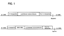
- Fig. 1 shows schematically the structure of each plasmid.
- mRNAs of Luc-poly-A and EMCV-Luc-poly-A were synthesized by using as templates products derived from the above respective plasmids (cleaved at the 3'-end side of the poly-A sequences by a restriction enzyme BamHI) with T7RNA polymerase according to the method using RiboMAX Large Scale RNA Production Systems (Promega).
- the concentration of GTP used in the above synthetic method was changed from 5.0mM to 0.8mM, and the reaction was conducted in a condition in which a cap structural analogue m7G(5')ppp(5')A (New England BioLabs) was added at 4mM.
- the resulting each mRNA was quantitatively analyzed as concentration from the absorbance at wavelength of 260 nm and preserved at -80°C.
- RNAs contained in the extract Prior to the translation reaction of each mRNA coding for luciferase, endogenous RNAs contained in the extract were decomposed and removed. The procedure is as follows. To 100 ⁇ l of the extract (protein concentration: 14-18mg/ml) was added 1 ⁇ l of 7500 unit/ml nuclease S7 and 1 ⁇ l of100mM CaCl 2 , and the mixture was treated at 20°C for 5 minutes, and quenched with 8 ⁇ l of 30mM EGTA.
- the mixture was filled in a dialysis chamber (MWCO 50kDa, produced from regenerated cellulose) and dialyzed in 4.9 ml of outer solution (containing 600 ⁇ l of energy-mixing solution, 60 ⁇ l of 3.3mM amino acid solution (20 kinds), 3.5 ml of extraction buffer, 21 ⁇ l of 100mM magnesium acetate, and 27 ⁇ l of 400mM EGTA) at 32°C for 1.5-6 hours.
- the concentration of potassium acetate in the outer solution was altered depending on the purpose.
- a necessary amount of the mixture in dialysis was first collected and allowed to stand in an ice-box. Then the ice-cooled mixture (4.5 ⁇ l) was moved into an Eppendorf tube, to which was added a potassium acetate solution and an mRNA solution in order (final volume: 6 ⁇ l) to initiate the reaction.
- a translation initiation factor eIF
- a necessary amount of eIF and mRNA solution in turn were added to the mixture and then the reaction was initiated.
- the final concentration of each component contained in the reaction mixture was as follows: 9-12 mg/ml of protein derived from the extract, 27mM HEPES-KOH (pH 7.5), 30 ⁇ M amino acids (20 kinds), 1.2mM ATP, 120 ⁇ M GTP, 18mM creatine phosphate, 0.3mM spermidine, 1.2mM magnesium acetate, 16mM KCl, 1.65mM EGTA, 90 ⁇ g/ml of t-RNA derived from bovine liver, and 60 ⁇ g/ml creatine phosphate kinase.
- the concentrations of potassium acetate and each mRNA were altered in the range of 44-224mM and 0-0.72 ⁇ g/6 ⁇ l, respectively, according to the condition.
- Figs. 2-7 show the results.
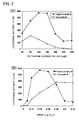
- Fig. 2 shows the characteristics of the cell-free translation reaction using cap-Luc-poly-A and Luc-poly-A.
- (A) HeLa S3 cell extract was treated with nuclease, and dialyzed in an outer solution of which the potassium concentration was adjusted at 80mM, and then the translation of each mRNA (0.24 ⁇ g/6 ⁇ l concentration) was conducted in the condition of the potassium concentration as indicated in the graph. After incubation at 32°C for 1 hour, a part of the reaction mixture was used in determination of the luciferase activity.
- cap-Luc-poly-A and Luc-poly-A were conducted in a condition of optimal potassium concentrations, 120mM and 90mM, respectively, at the mRNA concentrations (0-0.72 ⁇ g/6 ⁇ l) as indicated in the graph, and the luciferase activity was determined in the same way as indicated above.
- the symbol • indicates cap-Luc-poly A; and O indicates Luc-poly A.
- cap-Luc-poly A having a cap structure at the 5' -end showed the highest luceferase activity at 120mM potassium concentration and at 0.24 ⁇ g/6 ⁇ l mRNA concentration.
- Luc-poly-A which has no cap structure showed the highest activity at 90mM potassium concentration and at 0.48 ⁇ g/6 ⁇ l mRNA concentration.
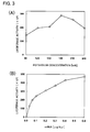
- Fig. 3 shows the characteristics of the cell-free translation reaction using EMCV-Luc-poly-A.
- A HeLa S3 cell extract treated with nuclease was dialyzed in an outer solution of which the potassium concentration was adjusted at 120mM, and then the translation of mRNA (0.2 ⁇ g/6 ⁇ l) was conducted in the condition of the potassium concentration as indicated in the graph. After incubation at 32°C for 1 hour, a part of the reaction mixture was used in determination of the luciferase activity.
- the translation reaction was conducted in the condition of the optimal potassium concentration (180mM) obtained from the result in the translation of EMCV-Luc-poly-A in (A) at the mRNA concentration (0-0.
- Luc-poly A having an IRES of EMCV showed the highest luceferase activity at 180mM potassium concentration and the activity value increased depending on the concentration of mRNA.
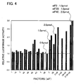
- Fig. 4 shows the effects of a variety of factors (eIF) in the translation reaction of a cell-free system using cap-Luc-poly-A.
- the HeLa S3 cell extract treated with nuclease was dialyzed in an outer solution of which the potassium concentration was adjusted at 160mM, to which was then added each factor as indicated in the graph, and then the translation was conducted in the optimum conditions (the concentrations of potassium and mRNA were 120mM and 0.24 ⁇ g/6 ⁇ l, respectively). After incubation at 32°C for 1 hour, a part of the reaction mixture was used in determination of the luciferase activity. Each reaction was repeated twice.
- the luciferase activity value was regarded as 1.0 when no eIF was added, and the activity value of each reaction was calculated as a ratio thereof, which was represented by the average value.
- the white, light gray and dark gray bars indicate that the concentrations of eIF contained in the reaction mixture are 0.5, 1.0 and 2.0 pmol, respectively.
- the black bar indicates that eIF2 and eIF2B each is contained at 1.0 pmol, and eIF4E at 2.0 pmol.
- cap-Luc-poly-A showed significantly high luciferase activity when any one of eIF2, eIF2B and eIF4E or they together were added. When these 3 translation initiation factors were added together, it was found that the activity of protein synthesis was increased about 3 times in comparison with no addition.
- Fig. 5 shows the effects of a variety of factors (eIF etc.) in the translation reaction of a cell-free system using Luc-poly-A.
- the HeLa S3 cell extract treated with nuclease was dialyzed in an outer solution of which the potassium concentration was adjusted at 120mM, to which was then added each factor as indicated in the graph, and then the translation was conducted in the optimum conditions (the concentrations of potassium and mRNA were 90mM and 0.48 ⁇ g/6 ⁇ l, respectively) . After incubation at 32°C for 1 hour, a part of the reaction mixture was used in determination of the luciferase activity. Each reaction was repeated twice. The luciferase activity value was regarded as 1.0 when no eIF etc.
- the white, light gray and dark gray bars indicate that the concentrations of eIF contained in the reaction mixture are 0.5, 1.0 and 2.0 pmol, respectively.
- the black bar indicates that eIF2 and eIF2B each is contained at 1.0 pmol, and p97 at 2.0 pmol.
- the translation of mRNA having no cap at the 5'-end, i.e. Luc-poly-A showed significantly high luciferase activity when any one of eIF2, eIF2B and p97 or they together were added. When these 3 translation initiation factors were added together, it was found that the activity of protein synthesis was increased 5 times or more in comparison with no addition.
- Fig. 6 shows the effects of a variety of factors (eIF) in the translation reaction of a cell-free system using EMCV-Luc-poly-A.
- the HeLa S3 cell extract treated with nuclease was dialyzed in an outer solution of which the potassium concentration was adjusted at 240mM, to which was then added each factor as indicated in the graph, and then the translation was conducted in the optimum conditions (the concentrations of potassium and mRNA were 180mM and 0.6 ⁇ g/6 ⁇ l, respectively) After incubation at 32°C for 1 hour, a part of the reaction mixture was used in determination of the luciferase activity. Each reaction was repeated twice.
- the luciferase activity value was regarded as 1.0 when no eIF was added, and the activity value of each reaction was calculated as a ratio thereof, which was represented by the average value.
- the white, dotted, light gray and dark gray bars indicate that the concentrations of eIF contained in the reaction mixture are 0.3, 0.6, 1.0 and 2.0 pmol, respectively.
- the black bar indicates that eIF2 and eIF2B each is contained at 1.5 pmol.
- the translation of mRNA to which was added an IRES of EMCV at the 5'-end, i.e. EMCV-Luc-poly-A showed significantly high luciferase activity when either of eIF2 or eIF2B or both were added. When these 2 translation initiation factors were added together, it was found that the activity of protein synthesis was increased 1.5 times or more in comparison with no addition.
- Fig. 7 shows the translation activity of each mRNA in the translation reaction of a cell-free system using HeLa S3 and rabbit reticulocyte lysate.
- the translation reaction was conducted in the optimum conditions for translation of each mRNA, obtained from the results of Figs. 4-6 using HeLa S3 (no eIF etc. added; light gray), and in the condition in which the best effect was observed by addition of eIF etc. (addition of eIF etc.; black), and then the luciferase activity value was compared.
- the eIF etc. showing a translation-enhancing effect in the above-mentioned Example are as follows.
- the protein concentration in each final sample was determined by a Bradford method using BSA as a standard. No contamination of other proteins was confirmed by SDS-PAGE, followed by Coomassie blue staining.
- Endogenous eIF2 isolated from HeLa cells (human) or Krebs cells (mouse) was purified to use (see Trachsel et al. (1979) Biochim. Biophys. Acta 561: 484-490 ).
- RNA for use in RT-PCR was obtained from HeLa cells.
- Primer DNAs corresponding to eIF2B1 GenBank accession number: NM_001414), eIF2B2 (no.NM_014239), eIF2B3 (no.NM_020365), eIF2B4 (no.Q9UI10), and eIF2B5 (no.XM_291076) were respectively referred to the sequences registered in GenBank.
- the resulting cDNAs all were sequenced and the base sequences were confirmed to be identical with those registered.
- a FLAG sequence was added at the C-terminal of eIF2B3, and 6xHis sequence at the N-terminal of eIF2B4.
- These cDNAs were inserted into a plasmid pAcDB3 (PharMingen) for use in production of baculovirus to produce pAcDB3-2B1-2B2-His2B4 and pAcDB3-2B3-FLAG-2B5.
- plasmid and baculovirus DNA BaculoGold were introduced into an insect cell Sf9 simultaneously.
- Virus in which correct recombination occurred was selected, and Baculo-2B1-2B2-his-2B4 virus and Baculo-2B3-FLAG-2B5 virus were amplified.
- the former virus can express eIF2Bl, eIF2B2 and His-eIF2B4 simultaneously, and the latter expresses eIF2B3-FLAG and eIF2B5 simultaneously.
- Insect cells High Five were cultured in 1L of culture medium Express FiveSFM (Invitrogen Co.) in a spinner flask equipped with a cell culture controller system (Cell master model 1700, Waken-yaku). The system was set up at a temperature of 27°C, dissolved oxygen concentration of 7.8 ppm, and stirring rate of 50 rpm. When the cell concentration reached 0.5-0.8 ⁇ 10 6 cells/ml, the cells were infected with Baculo-2B1-2B2-his-2B4 and Baculo-2B3-FLAG-2B5 simultaneously. After the lapse of 50 hours, the cells were recovered, frozen at -20°C and preserved. eIF2B was purified as follows.
- the frozen-preserved cells were suspended in 100 ml of buffer (0.5M KCl, 20mM HEPES, pH 7.5, 10% glycerol, 5mM 2-mercaptoethanol, 0.2% Triton X-100, and proteinase inhibitor "-EDTA Complete (Roche)"), allowed to stand in ice for 20 minutes, and centrifuged at 10,000 ⁇ g for 20 minutes to yield a clear supernatant. Imidazole was added thereto so that the final concentration was 20mM, and admixed with 2 ml of Ni-NTA-agarose resin (Qiagen Co.) at 4°C. After 1 hour, the mixture was passed through an empty column to remove unbound proteins.
- buffer 0.5M KCl, 20mM HEPES, pH 7.5, 10% glycerol, 5mM 2-mercaptoethanol, 0.2% Triton X-100, and proteinase inhibitor "-EDTA Complete (Roche)
- Imidazole was added thereto so
- the column was further washed with the above buffer (50 ml).
- the bound protein was eluted with a high imidazole-containing buffer (10 ml)(0.25M imidazole, 0.15M KCl, 20mM HEPES, pH 7. 5, 10% glycerol, 5mM 2-mercaptoethanol, 0.02% Triton X-100).
- EDTA was added up to 1mM, and the mixture was concentrated to 2 ml by Amicon Ultra 30,OOOMWCO (Millipore), and then added to a gel filtration column Superdex 200 HiLoad 16/60 (Amersham).
- Fractionation with a buffer (0.5M KCl, 20mM HEPES, pH 7.5, 10% glycerol, 5mM 2-mercaptoethanol, and 1mM EDTA) gave fractions (0.3 ml/min, 1 ml/fraction) and the fractions containing each subunit were confirmed by staining with Coomassie blue and Western blotting. The fractions confirmed that all of the subunits were contained were collected and diluted 3-fold with a buffer (20mM HEPES, pH 7. 5, 10% glycerol, 5mM 2-mercaptoethanol, and 1mM EDTA) and added to an anti-FLAG-Agarose (Sigma Co.) column (0.2 ml).
- a buffer 0.5M KCl, 20mM HEPES, pH 7.5, 10% glycerol, 5mM 2-mercaptoethanol, and 1mM EDTA
- the column was washed with a buffer (5 ml) (0.1M KCl, 20mM HEPES, pH 7.5, 10% glycerol, 5mM 2-mercaptoethanol, 1mM EDTA), and then eluted with the same buffer (0.7 ml) containing FLAG-peptide (Sigma Co.) (50 ⁇ g/ml) to give eIF2B.
- a buffer 5 ml
- KCl 0.1M KCl, 20mM HEPES, pH 7.5, 10% glycerol, 5mM 2-mercaptoethanol, 1mM EDTA
- the eluted eIF2B was dialyzed in a solution (0.1M KCl, 20mM HEPES, pH 7.5, 10% glycerol, 5mM 2-mercaptoethanol, 0.1mM EDTA) using a Slide-A-Lyzer Mini Dialysis unit (10,000 MWCO) (PIERCE Co.) and concentrated with a Slide-A-Lyzer Concentrating solution (PIERCE Co.).
- Murine eIF4E cDNA ( Jaramillo et al. (1991) J. Biol. Chem. 266: 10446-10451 ) was inserted into pGEX-6P (Amersham Co.) to yield pGEX-6P-eIF4E.
- pGEX-6P-eIF4E was introduced into Escherichia coli BL21 (DE-3)(pLys), which was then cultured in a Luria broth (LB) until the absorbance at 600 nm reached from 0.5 to 0.9. IPTG was added (final concentration 0.1 mM), and the mixture was cultured at 30°C for 12-16 hours to induce the expression of GST-eIF4E.
- the bacterium was recovered by centrifugation, washed once with a buffer (0.15M NaCl, 20mM Tris) (50 ml), and then suspended into a buffer (0.1M KCl, 20mM HEPES, pH 7.5, 10% glycerol, 1mM DTT, 0.1% Triton X-100, proteinase inhibitor "Complete, Roche”)(50 ml).
- a buffer 0.1M KCl, 20mM HEPES, pH 7.5, 10% glycerol, 1mM DTT, 0.1% Triton X-100, proteinase inhibitor "Complete, Roche
- the cells were destroyed by ultra-sonication and ultracentrifuged at 30,000 rpm (Beckman 70Ti roter) (1 hour) to remove insoluble fractions.
- the supernatant was admixed with Glutathione Sepharose 4B resin (Amersham Co.) (0.5ml) and agitated at 4°C for 1 hour.
- the mixture was poured into an empty column and further washed with a buffer (0.1M KCl, 20mM HEPES, pH 7.5, 10% glycerol, 1mM DTT, 0.1% Triton X-100, 1mM EDTA) (30 ml) to remove unbound proteins.
- GST-eIF4E is considered to still bind to the resin.
- PreScission Protease (12 units) (Amersham Co.) was added to the above buffer (0.5 ml), to which added the resin, and the mixture was agitated at 4°C overnight to cleavage between GST and eIF4E in GST-eIF4E.
- eIF4E released from the resin was recovered and again passed through Glutathione Sepharose 4B resin (0.3 ml) to remove GST-eIF4E and GST completely.
- the resulting eIF4E sample was bound to SP-Sepharose (0.3 ml) (Pharmacia) after adjustment of the buffer at pH 7.0.
- eIF4E was eluted with the same buffer containing 0.3M KCl.
- a FLAG sequence was added to the C-terminal of p97 cDNA ( Imataka et al. (1997) EMBO J 17: 6940-6947 ), and inserted into pGEX-6P to yield pGEX-6P-p97-FLAG.
- GST-p97-FLAG was expressed in the same manner as in GST-eIF4E. After ultracentrifugation, the supernatant was admixed with Heparin Sepharose CL-6B (0.5 ml)(Amersham Co.), and agitated at 4°C for 1 hour.
- Unbound proteins were washed out in the same manner as in GST-eIF4E/ Glutathione Sepharose 4B, and the bound protein was eluted with a buffer (0.5M KCl, 20mM HEPES, pH7.5, 10% glycerol, 1mM DTT, 0.1% Triton X-100, 1mM EDTA) (3 ml) .
- the eluate was admixed with Glutathione Sepharose 4B resin (0.5 ml) and treated in the same manner as in eIF4E to yield p97-FLAG. Further, the latter was purified completely with anti-FLAG-resin in the same manner as in eIF2B.
Landscapes
- Chemical & Material Sciences (AREA)
- Life Sciences & Earth Sciences (AREA)
- Organic Chemistry (AREA)
- Health & Medical Sciences (AREA)
- Zoology (AREA)
- Proteomics, Peptides & Aminoacids (AREA)
- Engineering & Computer Science (AREA)
- Wood Science & Technology (AREA)
- General Health & Medical Sciences (AREA)
- Genetics & Genomics (AREA)
- Biochemistry (AREA)
- Molecular Biology (AREA)
- Medicinal Chemistry (AREA)
- Toxicology (AREA)
- Gastroenterology & Hepatology (AREA)
- Biotechnology (AREA)
- Chemical Kinetics & Catalysis (AREA)
- General Chemical & Material Sciences (AREA)
- Microbiology (AREA)
- Biophysics (AREA)
- Bioinformatics & Cheminformatics (AREA)
- General Engineering & Computer Science (AREA)
- Preparation Of Compounds By Using Micro-Organisms (AREA)
- Enzymes And Modification Thereof (AREA)
Abstract
Prepared is an extract composition having an improved protein synthetic activity in a cell-free protein synthesis system using a mammalian cultured cell extract. An eukaryotic translation initiation factor and/or translational regulator are added to a cell-free protein synthesis system comprising an extract prepared from cultured mammalian cells and a template mRNA. These factors are preferably one or more selected from the group consisting of eukaryotic translation initiation factors 4E (eIF4E), 2 (eIF2) and 2B (eIF2B), and eukaryotic translational regulator p97.
Description
- The present invention relates to a process for producing a protein in a cell-free system. More particularly, it relates to a cell-free protein synthesis system using an extract derived from cultured mammalian cells.
- The cell-free protein synthesis system derived from cultured mammalian cells is important for analysis of gene products in eukaryotes and research of translational regulation. Most generally, rabbit reticulocyte lysate is used, and a number of proteins expressed in the system exhibit normal activities in vivo, through a correct folding and processing of those proteins (for example, see Non-Patent Document 1). Post-translational modification such as acetylation, isoprenylation or phosphorylation is required in some cases for the function or activity of proteins. Alternative post-translational modification (processing) such as cleavage of a signal peptide or addition of a carbohydrate chain has been achieved by adding a canine microsomal membrane to the above-mentioned translation reaction mixture (for example, see Non-Patent Document 2).
- On the other hand, it is known that in a synthetic reaction of proteins in eukaryotes, particularly translation reaction, a far larger number of translation initiation factors are involved than in prokaryotes and their interaction is complicated. In initiation of the protein synthetic system in eukaryotes, at least ten members of eukaryotic initiation factors (eIFs) are required. In the first step in which a pre-initiation complex is formed, the GTP type of eIF2 transports an initiation tRNA (Met-tRNAi) to a small subunit of 40S ribosome. Then, the pre-initiation complex attaches to the 5'-end of eukaryotic mRNA. In this step, eIF4F (also termed as cap-binding complex consisting of eIF4E, eIF4A and eIF4G) and eIF3 are required. This complex searches for AUG nearest at the 5'-end. After binding of Met-tRNAi with the initiation AUG, the initiation factor eIF5 releases eIF2 and eIF3. Finally, a 60S subunit binds to the complex comprising the initiation tRNA, mRNA and 40S subunit to form a 80S initiation complex. It is considered that in eukaryotic cells the translation reaction is mainly regulated by such initiation factors (for example, see Non-Patent Documents 3-5).
- In eukaryotic cells, it is known that mRNA as a template for protein synthesis has a cap structure (m7GpppN) in which 7-methylguanosine binds to the 5'-end phosphate via pyrophosphate. This structure plays an important role not only in a protective action against enzymatic degradation of mRNA but also in a mechanism of initiation of translation. The translation is promoted by binding of a cap binding factor (eIF4E) to the cap structure. At the 3'-end of mRNA, there is a poly-A structure linked by 80 - 100 polyadenylic acids to stabilize mRNA, which structure interacts with a translation-initiating complex through a poly A binding factor (PABP). It is known that this cap structure promotes translation in a cell-free system for protein synthesis, too. In order to provide a cap structure at the 5'-end of mRNA in vitro, a cap-like material designated as cap analogue is used as a substrate for synthesizing a cap-carrying mRNA by enzymatic reaction with RNA polymerase. When this capped mRNA is used to synthesize a protein, however, since the coexisting cap analogue has an inhibitory action to the protein synthesis, an additional manipulation for removing the cap analogue from mRNA preparation is required. In addition, there is a problem in using a cap analogue that the amount of synthesized mRNA is reduced.
-
- Non-Patent Document 1: Pelham, HR. and Jackson, RJ., "An efficient mRNA-dependent translation system from reticulocyte lysates", (1976) Eur J Biochem. Vol.67, pp.247-256.
- Non-Patent Document 2: Walter, P. and Blobel, G. (1983) Method, Enzymol., Vol.96, pp.84
- Non-Patent Document 3: Imataka, H. et al., (1997) EMBO J. Vol.16, pp.817-825
- Non-Patent Document 4: Imataka, H. et al., (1998) EMBO J. Vol.17, pp.7480-7489
- Non-Patent Document 5: Svitkin, YV, et al., (2001) RNA Vol.7, pp.1743-1752
- Rabbit reticulocyte lysate is available as a number of commercial products, but they are expensive and activities thereof have lot-to-lot variances because of using animals. In order to maintain a stable synthetic activity of protein without sacrifice of animals, a system derived from culture cells such as HeLa cell has been developed, but in general it has been believed that the rabbit reticulocyte lysate has much higher protein synthetic activity. Thus, the invention is directed to produce an extract composition that has an improved protein synthesis activity in a cell-free protein synthesis system using a mammalian cultured cell extract.
- The present invention was accomplished to solve the above-mentioned problems. The present inventors succeeded in obtaining a protein synthetic activity comparable to or higher than that of rabbit reticulocyte lysate by adding a certain translation initiation factor or translational regulator to an extract from cultured mammalian cells. The inventors further investigated on various members of translation initiation factors or translation regulators to be added and found that higher translation activity can also be obtained even by using mRNA having no cap structure. The present invention was completed based on these findings.
- In a first aspect of the present invention, a process for producing a protein in cell-free protein synthesis system is provided, which comprises adding a template mRNA and an eukaryotic translation initiation factor to an extract prepared from cultured mammalian cells. The above-mentioned translation initiation factor is preferably one or more selected from the group consisting of a
translation initiation factor 4E(eIF4E), translation initiation factor 2(eIF2), andtranslation initiation factor 2B(eIF2B). - In an example, the above-mentioned template mRNA has a cap structure at 5'-end thereof.
- In another example, the process for producing a protein in the invention comprises adding one or both of translation initiation factor 2 (eIF2) and
translation initiation factor 2B (eIF2B) into a cell-free protein synthetic system containing an extract prepared from cultured mammalian cells and a template mRNA having an internal ribosome entry site (IRES) of picornavirus at the 5'-end. - In yet another example, the process for producing a protein in the invention comprises adding an un-capped template mRNA and one or more factors selected from the group consisting of eukaryotic translation initiation factors 2(eIF2) and 2B(eIF2B) and an eukaryotic translation regulator p97, into an extract prepared from cultured mammalian cells.
- In an alternative aspect of the present invention, there is provided a composition for use in synthesis of a protein using a cell-free system, comprising an extract of cultured mammalian cells and one or more eukaryotic translation initiation factors selected from the group consisting of
translation initiation factor 4E(eIF4E), translation initiation factor 2(eIF2), andtranslation initiation factor 2B(eIF2B). - According to the invention, it is possible to construct a cell-free protein synthesis system having a high protein synthetic activity by adding the above-mentioned particular eukaryotic translation initiation factor and/or translation regulator to an extract prepared from cultured mammalian cells. Further, a template mRNA can be produced at low costs since a high translation activity can be obtained without capping at the 5'-end of mRNA.
-
-
Fig. 1 schematically shows the structure of plasmid used in preparation of a variety of luciferase mRNAs used in Examples. -
Fig. 2 shows characteristic features (potassium concentration and mRNA concentration dependency) of the translation reaction in the cell-free system using cap-Luc-poly-A and Luc-poly-A. -
Fig. 3 shows characteristic features (potassium concentration and mRNA concentration dependency) of the translation reaction in the cell-free system using EMCV-Luc-poly-A. -
Fig. 4 shows the effect of the respective factors (eIFs) in the translation reaction in the cell-free system using cap-Luc-poly-A. -
Fig. 5 shows the effect of the respective factors (eIFs etc.) in the translation reaction in the cell-free system using Luc-poly-A. -
Fig. 6 shows the effect of the respective factors (eIFs) in the translation reaction in the cell-free system using EMCV-Luc-poly-A. -
Fig. 7 shows the translation activity of the respective mRNAs in the translation reaction in the cell-free system using HeLaS3, rabbit reticulocyte lysate. - In a method of the present invention, the phrase "cell-free protein synthesis system" is a system to synthesize a target protein by preparing an extract including protein factors necessary for translation of proteins, and reconstituting the reaction in vitro. The extracts derived from a variety of organisms can be used for constituting the cell-free system, for example, those of eukaryotic and prokaryotic cells with high protein synthetic activities, such as bacteria including E.coli, thermophilic bacteria and the like, wheat germ, rabbit reticulocyte, mouse L cell, Ehrlich's ascitic cancer cell, HeLa cell, CHO cell and budding yeast and the like (see, Clemens, M.J., Transcription and Translation - A Practical Approach, (1984), pp. 231-270; Henes, B.D. et al. eds., IRL Press, Oxford).
- The term "eukaryotic translation initiation factor (eIF)" refers to a protein factor that is necessary for initiation of protein synthesis in eukaryotes, and more than ten kinds thereof are known to exist. The initiation step of eukaryotic translation includes significantly complicated reactions in contrast to that of prokaryote, which has only three initiation factors. These eukaryotic translation initiation factors include eIF3, eIF4 and eIF6 that bind to a subunit of ribosome, eIF4B, eIF4E, eIF4A and eIF4G that are concerned with binding to mRNA, eIF2 and eIF2B that are related to transporting an initiation tRNA, eIF5 that dissociates other factors, and the like. On the other hand, it is known that there is a factor that regulates the translation by some mechanism such as an interaction with the translation initiation factors, although such a factor is not indispensable in the eukaryotic translation initiation. For example, p97 appears to suppress or enhance the translation activity by binding to eIF4A and eIF3. In the present specification, these factors are referred to as "eukaryotic translational regulator".
- The term "template mRNA" means an mRNA encoding a desired protein to be expressed. The term "picornavirus" refers to a virus having a single and positive-stranded RNA ("picornavirus" literally means small RNA virus) with an icosahedral particle (capsid). The family Picornaviridae is separated into at least four distinct genera such as Genus Enterovirus, Genus Rhinovirus, Genus Aphthovirus that relates to animal diseases, and Genus Cardiovirus.
- The cell-free protein synthesis system of the invention comprises an extract of cultured mammalian cells which are eukaryotic cells, and a template mRNA, to which an eukaryotic translation initiation factor and/or a translation regulator (hereinafter referred to as translation initiation factors and the like (eIF etc.)) are added. In an example, the translation initiation factors and the like to be added to the above cell-free protein synthesis system include at least one selected from the group consisting of translation initiation factors eIF2, eIF2B and eIF4E, and translation regulator p97. eIF2 is a heterotrimer GTP-binding protein which transports an initiation tRNA to the 40S ribosomal subunit. The nucleotide sequences of mRNA encoding the three human eIF2 subunits, α:36kDa, β:38kDa and γ:52kDa have been registered as Accession Nos. NM_004094, NM_003908 and NM_001415, respectively, on the database of GenBank.
- eIF2B catalyzes the reaction to convert eIF2-GDP to eIF2-GTP, and it is composed of 5 different subunits. It has been reported that FLAG-tag is attached on the respective subunits and the resulting proteins can be expressed in Sf9 cells infected with baculovirus and purified to be reconstituted as eIF2B having a guanine nucleotide converting activity (see Fabian, J.R. et al., J. Biol. Chem. Vol.272, No.19, pp.12359-12365, 1997). The nucleotide sequences of mRNAs encoding each subunit of human eIF2B have been registered as Accession Nos. NM_001414 (eIF2B1), NM_014239 (eIF2B2), NM_020365 (eIF2B3), Q9UI10 (eIF2B4), and XM_291076 (eIF2B5), respectively, on the database of GenBank.
- eIF4E is a constituent of a cap-binding complex, eIF4F, and a factor directly binding to the cap structure. It is known that eIF4E is essential for cap-depending translation initiation and excessive expression thereof in NIH3T3 cells causes malignant transformation of the cells (see Lazaris-Karatzas, A et al., Nature (1990) 345:544-547). The nucleotide sequence of mRNA encoding murine eIF4E has been registered as Accession no. NM_007917 on the database of GenBank. eIF4G works as a scaffold of eIF4E and eIF4A to form eIF4F. eIF4A works as RNA-dependent ATPase and as ATP-dependent RNA helicase. It has been considered to unwind the secondary structure of the 5' untranslated region of mRNA so that ribosome can readily bind thereto.
- p97 is a translation regulator homologous to the translation initiation factor eIF4G. Human p97 protein is composed of 907 amino acid residues, which has approximately 28% homology with the 2/3 C-terminal portion of eIF4G (see above-mentioned non-patent document 3). The entire coding region of mRNA has been registered as Accession no. U73824 on the database of DDBJ/EMBL/GenBank. At first, it was considered to control the translation of a capped mRNA in the living cells, but it was first found in the invention to enhance the translation of non-capped mRNA in a cell-free system.
- These translation initiation factors etc., except eIF2, can easily be obtained by recombinant DNA techniques. For example, genes encoding them are cloned and linked (inserted) to a suitable vector to yield an expression vector. This expression vector is then transformed to suitable host cells, and the resulting transformants are cultured to yield the respective translation initiation factors etc. In this situation, the above-mentioned translation initiation factors etc. include those derived from human being but are not particularly limited as far as they are derived from mammals. In addition, it is natural that, though the amino acid sequences of the translation initiation factors etc. prepared by the above method contain deletion, substitution or addition of one or more amino acids, such proteins may be employed in the method of the invention as far as they have the respective activities as translation initiation factors etc. In addition, in these translation initiation factors etc., a histidine tag or FLAG tag may be added for the purpose of easy purification.
- Though the host organisms are not particularly limited as far as they allow replication and maintenance of the above expression vectors; for example, bacteria such as Escherichia coli, Bacillus subtilis, etc., Yeasts such as Saccharomyces cerevisiae, Schizosaccharomyces pombe, etc., and additionally animal cells such as COS cells, CHO cells, etc., or insect cells such as Sf9, Sf21, etc., may be used.
- When a bacterium such as Escherichia coli is used as a host, it is preferred that a recombinant vector into which genes such as translation initiation factors are introduced can be self-replicated in the bacterium and the vector is further composed of promoter, ribosome-binding sequence, a gene of translation initiation factors, and a replication-terminating sequence. In addition, a promoter-regulating gene may be contained.
- Escherichia coli is exemplified by Escherichia coli K12, DH1, etc. ; and Bacillus includes for example Bacillus subtilis. Any type of promoters may be used as far as they can be expressed in a host such as Escherichia coli. For example, promoters derived from Escherichia coli or phage, such as trp promoter, lac promoter, PL promoter, PR promoter, etc. may be used. An artificially designed and improved promoter such as tac promoter may be used. As for the method for introducing a recombinant vector into bacteria, there is no particular limitation as far as it allows introduction of DNA into bacteria. For example, a method of using calcium ion (Cohen, S.N, et al. (1972) Proc. Natl. Acad. Sci., USA 69, 2110-2114), electroporation and the like are included.
- When a yeast is used as a host, Saccharomyces cerevisiae, Schizosaccharomyces pombe, Pichia pastoris, and the like may be used. In this case, there is no particular limitation in promoters to be used as far as they can be expressed in yeasts; for example, gall promoter, ga110 promoter, heat-shock protein promoter, MFα1 promoter, PHO5 promoter, PGK promoter, GAP promoter, ADH promoter, AOX1 promoter, and the like are exemplified. As for the method for introducing a recombinant vector into yeasts, there is no particular limitation as far as it allows introduction of DNA into yeasts. For example, an electroporation method (Becker, D.M. et al., (1990) Methods Enzymol., 194, 182-187), a spheroplast method (Hinnen, A. et al., (1978) Proc. Natl. Acad. Sci., USA 75, 1929-1933), a lithium acetate method (Itoh, H. (1983) J. Bacteriol. 153, 163-168), and the like are exemplified.
- When an animal cell is used as a host, simian cell COS-7, Vero, Chinese hamster ovary cell (CHO cell), murine L cell, rat GH3, human FL cell, and the like may be used. As for promoters, SRα promoter, SV40 promoter, LTR promoter, CMV promoter, and the like may be used. Additionally, an early gene promoter of human cytomegalovirus may also be used. As for the method for introducing a recombinant vector into animal cells, for example, electoroporation, calcium phosphate method, lipofection method, and the like may be used.
- When an insect cell is used as a host, Sf9 cell, Sf21 cell, and the like are used. Introduction of a recombinant vector into insect cells may be effected for example by a calcium phosphate method, lipofection method, electroporation, and the like.
- The transformants obtained from animal cells as host may be incubated on a generally used culture medium such as RPMI1640 medium, DMEM medium, or a medium prepared by adding calf serum to these media. The culture is usually carried out at 37°C in the presence of 5% CO2 for a period of 1 - 30 days. During culture, an antibiotic such as kanamycin or penicillin if required may be added to the culture medium. When the coexistence of serum is inappropriate, a serum-free medium may be used depending on the purpose.
- When the desired translation initiation factors etc. are produced within the cells, after incubation, the cells are disrupted to extract the translation initiation factor etc. When the desired translation initiation factors etc. are produced outside of the cells, the culture broth is used as such or the cells are removed by centrifugation and the like. Thereafter, the translation initiation factors etc. may be isolated from the above culture product and purified by means of a conventional biochemical technique usually used in isolation and purification of the translation initiation factors etc., for example, precipitation with ammonium sulfate, gel chromatography, ion exchange chromatography, affinity chromatography, and the like, alone or in their combination.
- As for the template mRNA used in the invention, there is no particular limitation as far as it codes for the protein desired to be expressed; it may be isolated directly from the cells or may be synthesized by cloning a DNA encoding the above protein to a vector containing an RNA polymerase promoter, followed by in vitro transcription reaction. A method of in vitro transcription of DNA cloned after the phage polymerase promoter is known (Krieq, P. and Melton, D., Nucl. Acids Res., vol. 12, p.7057, 1984). In this method, a gene of which expression is desired is cloned to a vector containing any one of promoters among SP6, T7, and T3 RNA polymerase. Subsequently, the gene cloned in this vector is linearized at the 3'-end with a restriction enzyme and transcribed to mRNA by in vitro transcription reaction. A large number of vectors containing SP6, T7, and T3 RNA polymerase promoters have been commercialized and readily available. Thus synthesized template mRNA may or may not contain a cap structure at the 5' -end. When a cap is added to the 5'-end, a cap-like material designated as cap analogue may be used as a substrate to synthesize a cap-carrying mRNA by an enzymatic reaction with RNA polymerase.
- In an example of the invention, the template mRNA has an internal ribosome entry site (IRES) of picornavirus added at the 5'-end. It is known that the translation reaction of mRNA of picornavirus is initiated by using the internal ribosome entry site (IRES). The IRES structure of picornavirus can be classified roughly into two groups; the first one includes Enterovirus (e.g. Poliovirus) and Rhinovirus; and the other includes Cardiovirus (e.g., Encephalomyocarditis virus (EMCV)) and Aphthovirus (e.g., Foot-and-mouth disease virus).
- Preferably, the internal ribosome entry site (IRES) of EMCV is added to the 5' -end of the template mRNA. EMCV genome is a positive RNA strand of about 7900 bases in length. The entire nucleotide sequence of EMCV genome has been registered as the GenBank Accession nos. NC_001497 and X87335. EMCV RNA works as a template for translation of its own protein in the cells, which is then used in synthesis of a negative RNA strand. EMCV RNA binds to a ribosome with the assistance of the internal ribosome entry site (IRES) which works as a binding site of a translation initiation factor eIF4G. This IRES exists in the 5' non-translation region of the EMCV RNA genome, and can be cloned, for example, by amplification of the 281st to 848th nucleotide sequence of EMCV genome by PCR.
- The extract of mammalian cells used in the method of the invention may be prepared by culturing mammalian cells and disrupting the recovered cells. Though there is no particular limitation in the mammalian cells to be used, the cells highly active in the protein synthesis are preferably employed, including for example murine L-cell, Ehrlich's ascites carcinoma cell, HeLa cell, CHO cell, and the like. HeLa S3 cell is a suspension cell derived from HeLa cell, which is particularly preferred since it is readily cultured and easily proliferated. The cultured cells can be disrupted, for example, mechanically by a known method using a homogenizer, etc. The extract is treated with Micrococcus nuclease and CaCl2 to decompose endogenous mRNA to reduce the background translation to a minimum level. Then, EGTA is added for chelating CaCl2, thereby inactivating the nuclease.
- In an example of the present invention, the cultured mammalian cells can be disrupted by exposing to compressed inert gas (e.g. nitrogen gas) to give pressure, followed by reducing the pressure (depressurization). An apparatus for this operation, for example, Mini-Bomb Cell Disruption Chamber, has been marketed from KONTES. In this method, the cells are cooled by expanding gas without elevation of the temperature which is observed in the methods of a mechanical disruption and sonication and the like. Thus, it is possible to adjust the degree of cellular destruction by altering the pressure or equilibrium time. For example, when the cells are treated under moderate pressure for a short period of time, it is possible to disrupt cell membrane only without causing disruption of intracellular organelles. It has been reported that a high activity of glycoprotein synthesis can be obtained by preparing an extract of insect cells using this apparatus (see Tarui, H. et al., Appl. Microbiol. Biotechnol. Vol.55, pp.446-453, 2001).
- The extracts comprise several components necessary for protein synthesis, for example, tRNAs, rRNAs, amino acids, as well as initiation factors, extension and termination factors. In addition, the translation reaction of mRNA can be optimized by adding an energy regeneration system comprising phosphocreatine kinase and phosphocreatine. Further, a mixture of amino acids may be added. Potassium acetate and magnesium acetate are also added to adjust their concentrations suitable for the translation reaction. According to the method of the invention, the translation activity of mRNA can be further increased by adding the above-mentioned eukaryotic translation initiation factors to these extracts. The concentration of each translation initiation factor to be added can be optimized at, for example, 0.01-10 µM, preferably 0.1-1 µM.
- These reaction mixture or amino acid mixture for cell-free protein synthesis can be dispensed easy for use in aliquots in separate or previously mixed form, and distributed as a product. These products may be preserved in a frozen or dried state, and put on the market as a kit in suitable containers for storage and for shipment. The kits may be accompanied by an instruction manual, a positive control DNA, a vector DNA, and the like.
- The invention will be explained in more detail by the following examples, which are not intended to limit the scope of the invention.
- The culture of HeLa S3 cells using a petri dish was carried out in a carbon dioxide incubator (5% CO2 concentration; 37°C) in Eagle MEM medium (SIGMA Co.) supplemented with 10% heat-inactivated fetal calf serum (ICN Biomedicals, Inc), L-glutamine (2mM), 1 unit/ml penicillin, and 0.1 mg/ml streptomycin.
- Spinner culture in one-litter scale was carried out in a spinner flask equipped with a cell culture controller system (Cell Master Model 1700 Waken-yaku). The system was set up at a temperature of 37°C, pH 7.2, dissolved oxygen concentration of 7 ppm, and stirring rate of 50 rpm.
- When the cell density reached 1.0-1.5 × 10⁶ cells/ml, the HeLa S3 cells in the culture broth were collected by centrifugation at 360 × g and 4°C for 10 minutes. The culture medium was eliminated with an aspirator, and the cells recovered from 1L of the culture broth was moderately suspended into 80ml of buffer (35mM HEPES-KOH (pH 7.5 at 25°C), 140mM NaCl, 11mM glucose), then transferred into two 50ml tubes, and centrifuged at 360 × g and 4°C for 5 minutes. This operation was repeated 3 times, and then 1.5 parts (for 1 part by volume of cells) of an extraction buffer (20mM HEPES-KOH (pH 7.5 at 25°C), 135mM potassium acetate, 30mM KCl, and 1. 65mM magnesium acetate) was added to give a suspension containing about 2.5 × 10⁸ cells/ml, which was then sealed in a Mini-Bomb Cell Disruption Chamber (KONTES). The cells were disrupted under nitrogen gas pressure of 1.0 Mpa standing in ice. After the lapse of 30 minutes, the outlet port was slowly opened and the cell homogenate was recovered at a flow rate of about 3-10 drops/sec. The resulting cell homogenate was centrifuged twice at 10, 000 × g and 4°C for 5 minutes; the resulting solution (2-2.5 ml) from which the precipitate was removed was applied on a PD-10 column (Amersham) which had been equilibrated with 25 ml of extraction buffer in advance. The sample was applied by free-fall onto the column, then the extraction buffer was added, and when the solution falling down from the column changed from clear to creamy-white, about 1.7-2.0 ml was collected to use as a cell extract. The resulting solution was equally divided into small portions and flash-lyophilized with liquid nitrogen to preserve at -80°C.
- In order to synthesize an mRNA coding for luciferase, cap-Luc-poly-A and Luc-poly-A, cDNA of luciferase was first inserted into the downstream of T7 RNA polymerase promoter of plasmid pSP72 (Promega Co.) to yield a plasmid pSP72-Luc; and then a poly A stretch (85 deoxyadenines) was inserted into the downstream of luciferase gene to yield a plasmid pSP72-Luc-A (see the above-cited non-patent document no.4). In order to synthesize an mRNA coding for IRES of EMCV, EMCV-Luc-poly-A, a plasmid pSP72-EMCV-Luc-A (see the above-cited non-patent document no. 5) was used. This plasmid was prepared by cloning a DNA fragment (comprising the 281st to 848th nucleotides of EMCV genome amplified by PCR using pGemCAT-EMCV-LUC (see Pause et al., Nature 371, 762-767) as a template) between a plasmid pSP72-LUC-A T7 promoter and a luciferase gene.
Fig. 1 shows schematically the structure of each plasmid. - mRNAs of Luc-poly-A and EMCV-Luc-poly-A were synthesized by using as templates products derived from the above respective plasmids (cleaved at the 3'-end side of the poly-A sequences by a restriction enzyme BamHI) with T7RNA polymerase according to the method using RiboMAX Large Scale RNA Production Systems (Promega). In the synthesis of cap-Luc-poly-A mRNA, the concentration of GTP used in the above synthetic method was changed from 5.0mM to 0.8mM, and the reaction was conducted in a condition in which a cap structural analogue m7G(5')ppp(5')A (New England BioLabs) was added at 4mM. The resulting each mRNA was quantitatively analyzed as concentration from the absorbance at wavelength of 260 nm and preserved at -80°C.
- Prior to the translation reaction of each mRNA coding for luciferase, endogenous RNAs contained in the extract were decomposed and removed. The procedure is as follows. To 100 µl of the extract (protein concentration: 14-18mg/ml) was added 1 µl of 7500 unit/ml nuclease S7 and 1 µl of100mM CaCl2, and the mixture was treated at 20°C for 5 minutes, and quenched with 8 µl of 30mM EGTA. To this extract (110 µl) was then added 16.4µl of an energy-mixing solution (184mM HEPES-KOH (pH7.5), 13.2mM ATP, 1.32mM GTP, 198mM creatine phosphate, 3.3mM spermidine, 13.2mM magnesium acetate), 1.64 µl of 3.3mM amino acid solution (20 kinds), 4.92 µl of 3.3 mg/ml t-RNA derived from bovine liver, and 1.8 µl of 6.0 mg/ml creatine phosphate kinase, yielding a mixture. Further, the mixture was filled in a dialysis chamber (MWCO 50kDa, produced from regenerated cellulose) and dialyzed in 4.9 ml of outer solution (containing 600 µl of energy-mixing solution, 60 µl of 3.3mM amino acid solution (20 kinds), 3.5 ml of extraction buffer, 21 µl of 100mM magnesium acetate, and 27 µl of 400mM EGTA) at 32°C for 1.5-6 hours. In this procedure, the concentration of potassium acetate in the outer solution was altered depending on the purpose.
- In the translation reaction, a necessary amount of the mixture in dialysis was first collected and allowed to stand in an ice-box. Then the ice-cooled mixture (4.5 µl) was moved into an Eppendorf tube, to which was added a potassium acetate solution and an mRNA solution in order (final volume: 6 µl) to initiate the reaction. In a case of elucidating the effect of a translation initiation factor (eIF), a necessary amount of eIF and mRNA solution in turn were added to the mixture and then the reaction was initiated. The final concentration of each component contained in the reaction mixture was as follows: 9-12 mg/ml of protein derived from the extract, 27mM HEPES-KOH (pH 7.5), 30µM amino acids (20 kinds), 1.2mM ATP, 120µM GTP, 18mM creatine phosphate, 0.3mM spermidine, 1.2mM magnesium acetate, 16mM KCl, 1.65mM EGTA, 90 µg/ml of t-RNA derived from bovine liver, and 60 µg/ml creatine phosphate kinase. The concentrations of potassium acetate and each mRNA were altered in the range of 44-224mM and 0-0.72 µg/6 µl, respectively, according to the condition. After incubation at 32°C for 1 hour, the reaction mixture was immediately ice-cooled, and a part of the solution was used in determination of the luciferase activity. In determination of the activity a Luciferase Assay System (Promega Co.) was used.
-
Figs. 2-7 show the results.Fig. 2 shows the characteristics of the cell-free translation reaction using cap-Luc-poly-A and Luc-poly-A. In (A), HeLa S3 cell extract was treated with nuclease, and dialyzed in an outer solution of which the potassium concentration was adjusted at 80mM, and then the translation of each mRNA (0.24 µg/6 µl concentration) was conducted in the condition of the potassium concentration as indicated in the graph. After incubation at 32°C for 1 hour, a part of the reaction mixture was used in determination of the luciferase activity. In (B), the translation reactions for cap-Luc-poly-A and Luc-poly-A were conducted in a condition of optimal potassium concentrations, 120mM and 90mM, respectively, at the mRNA concentrations (0-0.72 µg/6 µl) as indicated in the graph, and the luciferase activity was determined in the same way as indicated above. The symbol • indicates cap-Luc-poly A; and O indicates Luc-poly A. As shown inFigs. 2 (A) and (B) , cap-Luc-poly A having a cap structure at the 5' -end showed the highest luceferase activity at 120mM potassium concentration and at 0.24 µg/6 µl mRNA concentration. On the other hand, Luc-poly-A which has no cap structure showed the highest activity at 90mM potassium concentration and at 0.48 µg/6 µl mRNA concentration. - Next,
Fig. 3 shows the characteristics of the cell-free translation reaction using EMCV-Luc-poly-A. In (A), HeLa S3 cell extract treated with nuclease was dialyzed in an outer solution of which the potassium concentration was adjusted at 120mM, and then the translation of mRNA (0.2 µg/6 µl) was conducted in the condition of the potassium concentration as indicated in the graph. After incubation at 32°C for 1 hour, a part of the reaction mixture was used in determination of the luciferase activity. In (B), the translation reaction was conducted in the condition of the optimal potassium concentration (180mM) obtained from the result in the translation of EMCV-Luc-poly-A in (A) at the mRNA concentration (0-0. 6 µg/6 µl) as indicated in the graph, and the luciferase activity was determined in the same way as indicated above. As shown inFigs. 3 (A) and (B) , Luc-poly A having an IRES of EMCV showed the highest luceferase activity at 180mM potassium concentration and the activity value increased depending on the concentration of mRNA. -
Fig. 4 shows the effects of a variety of factors (eIF) in the translation reaction of a cell-free system using cap-Luc-poly-A. The HeLa S3 cell extract treated with nuclease was dialyzed in an outer solution of which the potassium concentration was adjusted at 160mM, to which was then added each factor as indicated in the graph, and then the translation was conducted in the optimum conditions (the concentrations of potassium and mRNA were 120mM and 0.24 µg/6 µl, respectively). After incubation at 32°C for 1 hour, a part of the reaction mixture was used in determination of the luciferase activity. Each reaction was repeated twice. The luciferase activity value was regarded as 1.0 when no eIF was added, and the activity value of each reaction was calculated as a ratio thereof, which was represented by the average value. The white, light gray and dark gray bars indicate that the concentrations of eIF contained in the reaction mixture are 0.5, 1.0 and 2.0 pmol, respectively. The black bar indicates that eIF2 and eIF2B each is contained at 1.0 pmol, and eIF4E at 2.0 pmol. As clearly seen fromFig. 4 , the translation of mRNA having a cap at the 5'-end, i.e. cap-Luc-poly-A, showed significantly high luciferase activity when any one of eIF2, eIF2B and eIF4E or they together were added. When these 3 translation initiation factors were added together, it was found that the activity of protein synthesis was increased about 3 times in comparison with no addition. -
Fig. 5 shows the effects of a variety of factors (eIF etc.) in the translation reaction of a cell-free system using Luc-poly-A. The HeLa S3 cell extract treated with nuclease was dialyzed in an outer solution of which the potassium concentration was adjusted at 120mM, to which was then added each factor as indicated in the graph, and then the translation was conducted in the optimum conditions (the concentrations of potassium and mRNA were 90mM and 0.48 µg/6 µl, respectively) . After incubation at 32°C for 1 hour, a part of the reaction mixture was used in determination of the luciferase activity. Each reaction was repeated twice. The luciferase activity value was regarded as 1.0 when no eIF etc. was added, and the activity value of each reaction was calculated as a ratio thereof, which was represented by the average value. The white, light gray and dark gray bars indicate that the concentrations of eIF contained in the reaction mixture are 0.5, 1.0 and 2.0 pmol, respectively. The black bar indicates that eIF2 and eIF2B each is contained at 1.0 pmol, and p97 at 2.0 pmol. As seen fromFig. 5 , the translation of mRNA having no cap at the 5'-end, i.e. Luc-poly-A, showed significantly high luciferase activity when any one of eIF2, eIF2B and p97 or they together were added. When these 3 translation initiation factors were added together, it was found that the activity of protein synthesis was increased 5 times or more in comparison with no addition. -
Fig. 6 shows the effects of a variety of factors (eIF) in the translation reaction of a cell-free system using EMCV-Luc-poly-A. The HeLa S3 cell extract treated with nuclease was dialyzed in an outer solution of which the potassium concentration was adjusted at 240mM, to which was then added each factor as indicated in the graph, and then the translation was conducted in the optimum conditions (the concentrations of potassium and mRNA were 180mM and 0.6 µg/6 µl, respectively) After incubation at 32°C for 1 hour, a part of the reaction mixture was used in determination of the luciferase activity. Each reaction was repeated twice. The luciferase activity value was regarded as 1.0 when no eIF was added, and the activity value of each reaction was calculated as a ratio thereof, which was represented by the average value. The white, dotted, light gray and dark gray bars indicate that the concentrations of eIF contained in the reaction mixture are 0.3, 0.6, 1.0 and 2.0 pmol, respectively. The black bar indicates that eIF2 and eIF2B each is contained at 1.5 pmol. As seen fromFig. 6 , the translation of mRNA to which was added an IRES of EMCV at the 5'-end, i.e. EMCV-Luc-poly-A, showed significantly high luciferase activity when either of eIF2 or eIF2B or both were added. When these 2 translation initiation factors were added together, it was found that the activity of protein synthesis was increased 1.5 times or more in comparison with no addition. -
Fig. 7 shows the translation activity of each mRNA in the translation reaction of a cell-free system using HeLa S3 and rabbit reticulocyte lysate. The translation reaction was conducted in the optimum conditions for translation of each mRNA, obtained from the results ofFigs. 4-6 using HeLa S3 (no eIF etc. added; light gray), and in the condition in which the best effect was observed by addition of eIF etc. (addition of eIF etc.; black), and then the luciferase activity value was compared. That is, when EMCV-Luc,-poly-A was used as a template, each 1.5 pmol of eIF2 and eIF2B was added; when cap-Luc-poly-A was used, each 1.0 pmol of eIF2 and eIF2B and 2.0 pmol of eIF4E were added; and when Luc-poly-A was used, each 1.0 pmol of eIF2 and eIF2B and 2.0 pmol of p97 were added. In addition, the results of the translation reaction of each mRNA when an extract of rabbit reticulocyte lysate (Promega) was used, was also compared at the same time. In the reaction with the extract of rabbit reticulocyte lysate, potassium acetate was supplemented at a concentration of 100, 150, and 50 mM in translation of EMCV-Luc-poly-A, cap-Luc-poly-A, and Luc-poly-A, respectively, and the incubation was carried out at 30°C for 1 hour according to the instruction attached. All of the mRNA concentration was fixed at 0.1 µg/6 µl. As seen fromFig. 7 , a higher luciferase activity was recognized in the extract of rabbit reticulocyte lysate than in HeLa S3 cells in an eIF-free condition regardless of capped or un-capped, but when a given translation initiation factor etc. was added, respectively, the protein synthesis activity was greatly increased in the HeLa S3 cell extract, and it was found that the protein was synthesized in larger quantities than in the extract of rabbit reticulocyte lysate in each condition. - The eIF etc. showing a translation-enhancing effect in the above-mentioned Example are as follows. The protein concentration in each final sample was determined by a Bradford method using BSA as a standard. No contamination of other proteins was confirmed by SDS-PAGE, followed by Coomassie blue staining.
- Endogenous eIF2 isolated from HeLa cells (human) or Krebs cells (mouse) was purified to use (see Trachsel et al. (1979) Biochim. Biophys. Acta 561: 484-490).
- Each cDNA was obtained by reverse transcription PCR (RT-PCR). RNA for use in RT-PCR was obtained from HeLa cells. Primer DNAs corresponding to eIF2B1 (GenBank accession number: NM_001414), eIF2B2 (no.NM_014239), eIF2B3 (no.NM_020365), eIF2B4 (no.Q9UI10), and eIF2B5 (no.XM_291076) were respectively referred to the sequences registered in GenBank. The resulting cDNAs all were sequenced and the base sequences were confirmed to be identical with those registered. In order to make purification easy, a FLAG sequence was added at the C-terminal of eIF2B3, and 6xHis sequence at the N-terminal of eIF2B4. These cDNAs were inserted into a plasmid pAcDB3 (PharMingen) for use in production of baculovirus to produce pAcDB3-2B1-2B2-His2B4 and pAcDB3-2B3-FLAG-2B5. Thus produced plasmid and baculovirus DNA BaculoGold (PharMingen) were introduced into an insect cell Sf9 simultaneously. Virus in which correct recombination occurred was selected, and Baculo-2B1-2B2-his-2B4 virus and Baculo-2B3-FLAG-2B5 virus were amplified. The former virus can express eIF2Bl, eIF2B2 and His-eIF2B4 simultaneously, and the latter expresses eIF2B3-FLAG and eIF2B5 simultaneously.
- Insect cells High Five (Invitrogen Co.) were cultured in 1L of culture medium Express FiveSFM (Invitrogen Co.) in a spinner flask equipped with a cell culture controller system (Cell master model 1700, Waken-yaku). The system was set up at a temperature of 27°C, dissolved oxygen concentration of 7.8 ppm, and stirring rate of 50 rpm. When the cell concentration reached 0.5-0.8 × 10⁶ cells/ml, the cells were infected with Baculo-2B1-2B2-his-2B4 and Baculo-2B3-FLAG-2B5 simultaneously. After the lapse of 50 hours, the cells were recovered, frozen at -20°C and preserved. eIF2B was purified as follows. The frozen-preserved cells were suspended in 100 ml of buffer (0.5M KCl, 20mM HEPES, pH 7.5, 10% glycerol, 5mM 2-mercaptoethanol, 0.2% Triton X-100, and proteinase inhibitor "-EDTA Complete (Roche)"), allowed to stand in ice for 20 minutes, and centrifuged at 10,000 × g for 20 minutes to yield a clear supernatant. Imidazole was added thereto so that the final concentration was 20mM, and admixed with 2 ml of Ni-NTA-agarose resin (Qiagen Co.) at 4°C. After 1 hour, the mixture was passed through an empty column to remove unbound proteins. The column was further washed with the above buffer (50 ml). The bound protein was eluted with a high imidazole-containing buffer (10 ml)(0.25M imidazole, 0.15M KCl, 20mM HEPES, pH 7. 5, 10% glycerol, 5mM 2-mercaptoethanol, 0.02% Triton X-100). EDTA was added up to 1mM, and the mixture was concentrated to 2 ml by Amicon Ultra 30,OOOMWCO (Millipore), and then added to a gel
filtration column Superdex 200 HiLoad 16/60 (Amersham). Fractionation with a buffer (0.5M KCl, 20mM HEPES, pH 7.5, 10% glycerol, 5mM 2-mercaptoethanol, and 1mM EDTA) gave fractions (0.3 ml/min, 1 ml/fraction) and the fractions containing each subunit were confirmed by staining with Coomassie blue and Western blotting. The fractions confirmed that all of the subunits were contained were collected and diluted 3-fold with a buffer (20mM HEPES, pH 7. 5, 10% glycerol, 5mM 2-mercaptoethanol, and 1mM EDTA) and added to an anti-FLAG-Agarose (Sigma Co.) column (0.2 ml). The column was washed with a buffer (5 ml) (0.1M KCl, 20mM HEPES, pH 7.5, 10% glycerol, 5mM 2-mercaptoethanol, 1mM EDTA), and then eluted with the same buffer (0.7 ml) containing FLAG-peptide (Sigma Co.) (50 µg/ml) to give eIF2B. The eluted eIF2B was dialyzed in a solution (0.1M KCl, 20mM HEPES, pH 7.5, 10% glycerol, 5mM 2-mercaptoethanol, 0.1mM EDTA) using a Slide-A-Lyzer Mini Dialysis unit (10,000 MWCO) (PIERCE Co.) and concentrated with a Slide-A-Lyzer Concentrating solution (PIERCE Co.). - Murine eIF4E cDNA (Jaramillo et al. (1991) J. Biol. Chem. 266: 10446-10451) was inserted into pGEX-6P (Amersham Co.) to yield pGEX-6P-eIF4E. pGEX-6P-eIF4E was introduced into Escherichia coli BL21 (DE-3)(pLys), which was then cultured in a Luria broth (LB) until the absorbance at 600 nm reached from 0.5 to 0.9. IPTG was added (final concentration 0.1 mM), and the mixture was cultured at 30°C for 12-16 hours to induce the expression of GST-eIF4E. The bacterium was recovered by centrifugation, washed once with a buffer (0.15M NaCl, 20mM Tris) (50 ml), and then suspended into a buffer (0.1M KCl, 20mM HEPES, pH 7.5, 10% glycerol, 1mM DTT, 0.1% Triton X-100, proteinase inhibitor "Complete, Roche")(50 ml). The cells were destroyed by ultra-sonication and ultracentrifuged at 30,000 rpm (Beckman 70Ti roter) (1 hour) to remove insoluble fractions. The supernatant was admixed with
Glutathione Sepharose 4B resin (Amersham Co.) (0.5ml) and agitated at 4°C for 1 hour. The mixture was poured into an empty column and further washed with a buffer (0.1M KCl, 20mM HEPES, pH 7.5, 10% glycerol, 1mM DTT, 0.1% Triton X-100, 1mM EDTA) (30 ml) to remove unbound proteins. In this stage, GST-eIF4E is considered to still bind to the resin. PreScission Protease (12 units) (Amersham Co.) was added to the above buffer (0.5 ml), to which added the resin, and the mixture was agitated at 4°C overnight to cleavage between GST and eIF4E in GST-eIF4E. eIF4E released from the resin was recovered and again passed throughGlutathione Sepharose 4B resin (0.3 ml) to remove GST-eIF4E and GST completely. In order to further purify, the resulting eIF4E sample was bound to SP-Sepharose (0.3 ml) (Pharmacia) after adjustment of the buffer at pH 7.0. After washing with a buffer (0.1M KCl, 20mM HEPES, pH 7.0, 10% glycerol, 5mM 2-mercaptoethanol, 0.1mM EDTA) (10 ml), eIF4E was eluted with the same buffer containing 0.3M KCl. - A FLAG sequence was added to the C-terminal of p97 cDNA (Imataka et al. (1997) EMBO J 17: 6940-6947), and inserted into pGEX-6P to yield pGEX-6P-p97-FLAG. GST-p97-FLAG was expressed in the same manner as in GST-eIF4E. After ultracentrifugation, the supernatant was admixed with Heparin Sepharose CL-6B (0.5 ml)(Amersham Co.), and agitated at 4°C for 1 hour. Unbound proteins were washed out in the same manner as in GST-eIF4E
/ Glutathione Sepharose 4B, and the bound protein was eluted with a buffer (0.5M KCl, 20mM HEPES, pH7.5, 10% glycerol, 1mM DTT, 0.1% Triton X-100, 1mM EDTA) (3 ml) . The eluate was admixed withGlutathione Sepharose 4B resin (0.5 ml) and treated in the same manner as in eIF4E to yield p97-FLAG. Further, the latter was purified completely with anti-FLAG-resin in the same manner as in eIF2B.
Claims (11)
- A process for producing a protein in cell-free protein synthesis system comprisingadding a template mRNA and an eukaryotic translation initiation factor to an extract prepared from cultured mammalian cells.
- The process of claim 1, wherein said eukaryotic translation initiation factor is one or more selected from the group consisting of eukaryotic translation initiation factors 4E (eIF4E), 2 (eIF2), and 2B (eIF2B).
- The process of claim 1 or 2, wherein said template mRNA has a cap structure at 5' end thereof.
- The process of claim 1 or 2, wherein said template mRNA comprises an internal ribosome entry site (IRES) of Encephalomyocarditis virus (EMCV) at 5' end thereof and said eukaryotic translation initiation factor is one or both of eukaryotic translation initiation factors 2 (eIF2) and 2B (eIF2B).
- A process for producing a protein in cell-free protein synthesis system comprisingadding an uncapped mRNA, and one or more selected from the group consisting of eukaryotic translation initiation factors 2 (eIF2) and 2B (eIF2B), and an eukaryotic translational regulator p97 to an extract prepared from cultured mammalian cells.
- The process of any one of claims 1 to 5, wherein said cultured mammalian cells are cultured human cells.
- The method of claim 6, wherein said cultured human cells are cultured HeLa S3 cells.
- A composition for use in a production of a protein using a cell-free system, comprising an extract of cultured mammalian cells and one or more eukaryotic translation initiation factors selected from the group consisting of translation initiation factors 4E (eIF4E), 2 (eIF2), and 2B (eIF2B).
- The composition of claim 8 further comprising an eukaryotic translational regulator p97, which composition is used for cell-free protein synthesis using an uncapped mRNA.
- The composition of claim 8 or 9, wherein said cultured mammalian cells are cultured human cells.
- The composition of claim 10, wherein said cultured human cells are cultured HeLa S3 cells.
Applications Claiming Priority (2)
| Application Number | Priority Date | Filing Date | Title |
|---|---|---|---|
| JP2004333250A JP4868731B2 (en) | 2004-11-17 | 2004-11-17 | Cell-free protein synthesis system derived from cultured mammalian cells |
| PCT/JP2005/020959 WO2006054556A1 (en) | 2004-11-17 | 2005-11-15 | Cell-free protein synthesis system originating in cultured mammalian cells |
Publications (2)
| Publication Number | Publication Date |
|---|---|
| EP2145953A1 true EP2145953A1 (en) | 2010-01-20 |
| EP2145953A4 EP2145953A4 (en) | 2010-01-20 |
Family
ID=36407099
Family Applications (1)
| Application Number | Title | Priority Date | Filing Date |
|---|---|---|---|
| EP05806798A Withdrawn EP2145953A4 (en) | 2004-11-17 | 2005-11-15 | ACELLULAR SYSTEM FOR THE SYNTHESIS OF PROTEINS FROM CULTURED MAMMAL CELLS |
Country Status (4)
| Country | Link |
|---|---|
| US (2) | US8012712B2 (en) |
| EP (1) | EP2145953A4 (en) |
| JP (1) | JP4868731B2 (en) |
| WO (1) | WO2006054556A1 (en) |
Families Citing this family (13)
| Publication number | Priority date | Publication date | Assignee | Title |
|---|---|---|---|---|
| JP4441170B2 (en) * | 2002-11-28 | 2010-03-31 | 独立行政法人理化学研究所 | Escherichia coli cell extract having mutation in S12 ribosomal protein and cell-free protein production method using the same |
| JP4590249B2 (en) * | 2004-11-17 | 2010-12-01 | 独立行政法人理化学研究所 | Cell-free protein synthesis system for glycoprotein synthesis |
| JP4868731B2 (en) | 2004-11-17 | 2012-02-01 | 独立行政法人理化学研究所 | Cell-free protein synthesis system derived from cultured mammalian cells |
| JP4787488B2 (en) * | 2004-11-19 | 2011-10-05 | 独立行政法人理化学研究所 | Cell-free protein synthesis method using linear template DNA and cell extract therefor |
| JP5099881B2 (en) * | 2007-01-27 | 2012-12-19 | 独立行政法人科学技術振興機構 | Highly efficient cell-free protein synthesis system |
| EP2213747B1 (en) * | 2007-11-05 | 2021-06-30 | Riken | Method for producing membrane protein |
| CN114891855A (en) | 2013-12-06 | 2022-08-12 | 哈佛大学校长及研究员协会 | Synthetic gene networks based on paper |
| CA3032514C (en) | 2015-07-31 | 2021-05-04 | Takehiro SHINODA | Method of producing membrane protein and utilization thereof |
| AU2018383675A1 (en) | 2017-12-13 | 2020-07-09 | Altos Labs, Inc. | Inhibitors of integrated stress response pathway |
| MX2020013269A (en) | 2018-06-05 | 2021-02-18 | Praxis Biotech LLC | Inhibitors of integrated stress response pathway. |
| CN111118065A (en) * | 2018-10-31 | 2020-05-08 | 康码(上海)生物科技有限公司 | A kind of eukaryotic genetic modification method and corresponding genetic engineering cell and application |
| CN113840597A (en) * | 2019-02-25 | 2021-12-24 | 普拉西斯生物技术有限责任公司 | Integrated Stress Response Pathway Inhibitors |
| CN114466654A (en) | 2019-06-12 | 2022-05-10 | 普拉西斯生物技术有限责任公司 | Integrated stress response pathway modulators |
Family Cites Families (52)
| Publication number | Priority date | Publication date | Assignee | Title |
|---|---|---|---|---|
| US5804374A (en) | 1980-12-05 | 1998-09-08 | Massachusetts Insti. Technology | Nuclear factors associates with transcriptional regulation |
| SU1618761A1 (en) | 1987-04-29 | 1991-01-07 | Институт Белка Ан Ссср | Method of producing peptides and proteins in cell-less translation system |
| US5264341A (en) | 1989-08-30 | 1993-11-23 | Eli Lilly And Company | Selective cloning for high monoclonal antibody secreting hybridomas |
| JPH0491790A (en) | 1990-08-02 | 1992-03-25 | Shionogi & Co Ltd | Two step pcr method |
| AU1322692A (en) | 1990-11-05 | 1992-05-26 | United States Of America, As Represented By The Secretary Of The Army, The | Method for the in vitro production of protein from a dna sequence without cloning |
| JPH04200390A (en) | 1990-11-30 | 1992-07-21 | Shigeyuki Yokoyama | Production polypeptide by cell-free polypeptide synthesis system |
| WO1992011390A1 (en) | 1990-12-17 | 1992-07-09 | Idexx Laboratories, Inc. | Nucleic acid sequence detection by triple helix formation |
| US5270181A (en) | 1991-02-06 | 1993-12-14 | Genetics Institute, Inc. | Peptide and protein fusions to thioredoxin and thioredoxin-like molecules |
| WO1993000442A1 (en) * | 1991-06-24 | 1993-01-07 | The Research Foundation Of State University Of New York | De novo cell-free synthesis of picornavirus |
| US5532151A (en) | 1993-09-17 | 1996-07-02 | Icos Corporation | G protein-coupled receptor kinase GRK6 |
| JP2501081B2 (en) | 1993-10-13 | 1996-05-29 | 株式会社ソキア | Reference plane setting device |
| US5959085A (en) | 1993-11-23 | 1999-09-28 | Schering Corporation | Human monoclonal antibodies against human cytokines and methods of making and using such antibodies |
| MX9707021A (en) | 1995-03-23 | 1997-11-29 | Immunex Corp | Il-17 receptor. |
| JP3092904B2 (en) | 1995-10-13 | 2000-09-25 | 株式会社エイチ・エス・ピー研究所 | Methods for producing soluble proteins by bacteria |
| JPH09234074A (en) | 1996-03-04 | 1997-09-09 | Sumitomo Electric Ind Ltd | Adapter double-stranded DNA and DNA amplification method using the same |
| US5962246A (en) | 1996-03-29 | 1999-10-05 | The University Of Medicine And Dentistry Of New Jersey | dUTPase, its isoforms, and diagnostic and other uses |
| WO1997046696A1 (en) | 1996-06-07 | 1997-12-11 | Genentech, Inc. | Methods for in vitro protein synthesis |
| US6593103B1 (en) | 1997-02-07 | 2003-07-15 | The Regents Of The University Of California | HIV capsid assembly-associated compositions and method |
| ES2283936T3 (en) | 1997-07-07 | 2007-11-01 | Medical Research Council | IN VITRO CLASSIFICATION PROCEDURE. |
| US6136568A (en) | 1997-09-15 | 2000-10-24 | Hiatt; Andrew C. | De novo polynucleotide synthesis using rolling templates |
| US6607878B2 (en) | 1997-10-06 | 2003-08-19 | Stratagene | Collections of uniquely tagged molecules |
| WO1999020798A1 (en) | 1997-10-23 | 1999-04-29 | Exact Laboratories, Inc. | Methods for detecting contamination in molecular diagnostics using pcr |
| AU4187799A (en) | 1998-05-14 | 1999-11-29 | Clontech Laboratories, Inc. | Compositions and methods for protein purification based on metal ion affinity site |
| JP2000175695A (en) | 1998-12-14 | 2000-06-27 | Inst Of Physical & Chemical Res | Method for producing polypeptide by cell-free protein synthesis system |
| US6280977B1 (en) | 1999-03-24 | 2001-08-28 | Gene Therapy Systems | Method for generating transcriptionally active DNA fragments |
| EP1176210B1 (en) | 1999-05-11 | 2009-03-04 | CellFree Sciences Co., Ltd. | Preparation containing cell extract for synthesizing cell-free protein and means for synthesizing cell-free protein |
| JP3713402B2 (en) | 1999-05-21 | 2005-11-09 | 敏夫 原 | Cell-free extract and glycoprotein synthesis system |
| US6303337B1 (en) | 1999-08-25 | 2001-10-16 | Amber Gen. Inc. | N-terminal and C-terminal markers in nascent proteins |
| US6511832B1 (en) | 1999-10-06 | 2003-01-28 | Texas A&M University System | In vitro synthesis of capped and polyadenylated mRNAs using baculovirus RNA polymerase |
| ATE286140T1 (en) | 2000-05-03 | 2005-01-15 | Emd Biosciences Inc | E. COLI EXTRACT FOR SYNTHESIS OF PROTEINS |
| JP4762481B2 (en) | 2000-08-30 | 2011-08-31 | 株式会社セルフリーサイエンス | Design and construction of transcription template for cell-free protein synthesis and dilution batch method wheat germ cell-free protein synthesis method using the same |
| JP4038555B2 (en) | 2000-12-26 | 2008-01-30 | 積水化学工業株式会社 | Recombinant protein production method and fusion protein |
| ATE538380T1 (en) * | 2001-01-23 | 2012-01-15 | Harvard College | NUCLEIC ACID PROGRAMMABLE PROTEIN ARRAYS |
| KR100399337B1 (en) | 2001-02-07 | 2003-09-26 | 드림바이오젠 주식회사 | Method for Cell-free Protein Post-translational Modification |
| JP2002262873A (en) | 2001-03-06 | 2002-09-17 | Inst Of Physical & Chemical Res | Method for producing protein domain and method for analyzing three-dimensional structure of protein using domain obtained by the method |
| WO2003002724A2 (en) * | 2001-03-12 | 2003-01-09 | Affinium Pharmaceuticals, Inc. | Proteins, druggable regions of proteins and target analysis for chemistry of therapeutics |
| JP4838957B2 (en) | 2001-05-02 | 2011-12-14 | 独立行政法人理化学研究所 | Protein production method using cell-free protein synthesis system |
| JP4768155B2 (en) | 2001-07-02 | 2011-09-07 | 独立行政法人理化学研究所 | Method for producing template DNA and method for producing protein by cell-free protein synthesis system using the same |
| DE10158904A1 (en) | 2001-11-30 | 2003-06-12 | Roche Diagnostics Gmbh | Process for the preparation of linear DNA fragments for the in vitro expression of proteins |
| JP4244625B2 (en) | 2001-12-10 | 2009-03-25 | 株式会社島津製作所 | Extract for cell-free protein synthesis, cell-free protein synthesis method using the same, and method for preparing the extract |
| US20040137448A1 (en) * | 2002-02-07 | 2004-07-15 | Thornton Michael B. | Nucleic acid-associated proteins |
| WO2003097829A1 (en) | 2002-05-22 | 2003-11-27 | Riken | Process for producing protein in cell-free protein synthesis system using thioredoxin fused protein expression vector |
| EP1394227B1 (en) | 2002-08-30 | 2006-05-24 | Eastman Kodak Company | Ink jet ink composition and printing process |
| JP3668942B2 (en) * | 2002-10-18 | 2005-07-06 | 独立行政法人理化学研究所 | Protein synthesis system using Xenopus oocyte extract |
| JP4324730B2 (en) | 2002-12-27 | 2009-09-02 | 株式会社島津製作所 | Method for preparing insect cell extract for cell-free protein synthesis, insect cell extract, and cell-free protein synthesis method using insect cell extract |
| JP4243762B2 (en) | 2003-02-18 | 2009-03-25 | 国立大学法人京都工芸繊維大学 | Preparation method of cell-free protein synthesis extract and silkworm tissue extraction kit |
| JP2004290181A (en) | 2003-03-13 | 2004-10-21 | National Institute Of Advanced Industrial & Technology | Cell-free translation of polypeptides at low temperatures |
| US20050095705A1 (en) * | 2003-04-15 | 2005-05-05 | Michael Kadan | Method for production of oncolytic adenoviruses |
| JP4477923B2 (en) | 2003-05-22 | 2010-06-09 | 独立行政法人理化学研究所 | Method for producing extract for cell-free protein synthesis and cell extract produced by the method |
| JP2005225796A (en) | 2004-02-12 | 2005-08-25 | Wakenyaku Kk | Method for producing protein by using liposome |
| JP4590249B2 (en) | 2004-11-17 | 2010-12-01 | 独立行政法人理化学研究所 | Cell-free protein synthesis system for glycoprotein synthesis |
| JP4868731B2 (en) | 2004-11-17 | 2012-02-01 | 独立行政法人理化学研究所 | Cell-free protein synthesis system derived from cultured mammalian cells |
-
2004
- 2004-11-17 JP JP2004333250A patent/JP4868731B2/en not_active Expired - Fee Related
-
2005
- 2005-11-15 WO PCT/JP2005/020959 patent/WO2006054556A1/en not_active Ceased
- 2005-11-15 EP EP05806798A patent/EP2145953A4/en not_active Withdrawn
-
2007
- 2007-05-16 US US11/798,759 patent/US8012712B2/en not_active Expired - Fee Related
-
2011
- 2011-08-16 US US13/211,280 patent/US8445232B2/en not_active Expired - Fee Related
Non-Patent Citations (9)
| Title |
|---|
| CARROLL R ET AL: "Preparation of a cell-free translation system with minimal loss of initiation factor eIF-2/eIF-2B Activity" ANALYTICAL BIOCHEMISTRY, ACADEMIC PRESS INC. NEW YORK, vol. 212, no. 1, 1 January 1993 (1993-01-01), pages 17-23, XP003009881 ISSN: 0003-2697 * |
| HENIS-KORENBLIT SIVAN ET AL: "The caspase-cleaved DAP5 protein supports internal ribosome entry site-mediated translation of death proteins." PROCEEDINGS OF THE NATIONAL ACADEMY OF SCIENCES OF THE UNITED STATES OF AMERICA 16 APR 2002, vol. 99, no. 8, 16 April 2002 (2002-04-16), pages 5400-5405, XP002496748 ISSN: 0027-8424 * |
| PERSON A ET AL: "Translation in micrococcal nuclease-treated cell-free extracts from Ehrlich ascites tumor cells. Stimulation by initiation factor eIF-2B." BIOCHIMICA ET BIOPHYSICA ACTA 22 NOV 1984, vol. 783, no. 2, 22 November 1984 (1984-11-22), pages 152-157, XP002496676 ISSN: 0006-3002 * |
| PESTOVA T V ET AL: "The structure and function of initiation factors in eukaryotic protein synthesis." CELLULAR AND MOLECULAR LIFE SCIENCES : CMLS APR 2000, vol. 57, no. 4, April 2000 (2000-04), pages 651-674, XP002496679 ISSN: 1420-682X * |
| PREISS THOMAS ET AL: "Starting the protein synthesis machine: eukaryotic translation initiation." BIOESSAYS : NEWS AND REVIEWS IN MOLECULAR, CELLULAR AND DEVELOPMENTAL BIOLOGY DEC 2003, vol. 25, no. 12, December 2003 (2003-12), pages 1201-1211, XP002496678 ISSN: 0265-9247 * |
| S. Gunnery: "Translation of an Uncapped mRNA Involves Scanning", Journal of Biological Chemistry, vol. 272, no. 34, 22 August 1997 (1997-08-22), pages 21642-21646, XP55007106, ISSN: 0021-9258, DOI: 10.1074/jbc.272.34.21642 * |
| See also references of WO2006054556A1 * |
| Svitkin: "Poly (A)-binding protein interaction with elF4G stimulates picornavirus IRES-dependent translation.", RNA, vol. 7, no. 12, 1 January 2001 (2001-01-01), page 1743, XP55007072, ISSN: 1355-8382 * |
| THOMA CHRISTIAN ET AL: "A poly(A) tail-responsive in vitro system for cap- or IRES-driven translation from HeLa cells." METHODS IN MOLECULAR BIOLOGY (CLIFTON, N.J.) 2004, vol. 257, 2004, pages 171-180, XP002496677 ISSN: 1064-3745 * |
Also Published As
| Publication number | Publication date |
|---|---|
| US8012712B2 (en) | 2011-09-06 |
| JP2006141240A (en) | 2006-06-08 |
| US20070281337A1 (en) | 2007-12-06 |
| JP4868731B2 (en) | 2012-02-01 |
| WO2006054556A1 (en) | 2006-05-26 |
| US20110300575A1 (en) | 2011-12-08 |
| EP2145953A4 (en) | 2010-01-20 |
| US8445232B2 (en) | 2013-05-21 |
Similar Documents
| Publication | Publication Date | Title |
|---|---|---|
| US8012712B2 (en) | Cell-free system for synthesis of proteins derived from cultured mammalian cells | |
| Mikami et al. | An efficient mammalian cell-free translation system supplemented with translation factors | |
| Goerke et al. | High‐level cell‐free synthesis yields of proteins containing site‐specific non‐natural amino acids | |
| Kerekatte et al. | Cleavage of Poly (A)-binding protein by coxsackievirus 2A protease in vitro and in vivo: another mechanism for host protein synthesis shutoff? | |
| Davydov et al. | RGS4 is arginylated and degraded by the N-end rule pathway in vitro | |
| Baugh et al. | 20S proteasome differentially alters translation of different mRNAs via the cleavage of eIF4F and eIF3 | |
| EP2221370B1 (en) | Process for production of non-natural protein having ester bond therein | |
| Melki et al. | Cofactor A is a molecular chaperone required for β-tubulin folding: functional and structural characterization | |
| JP7744244B2 (en) | Composition for translation and method for producing peptide | |
| US20040214988A1 (en) | Method for stabilization of proteins using non-natural amino acids | |
| KR20150064209A (en) | Proteolytic inactivation of select proteins in bacterial extracts for improved expression | |
| EP2376647B1 (en) | Mono charging system for selectively introducing non-native amino acids into proteins using an in vitro protein synthesis system | |
| Kawarasaki et al. | A long-lived batch reaction system of cell-free protein synthesis | |
| Daskalova et al. | Elongation factor P modulates the incorporation of structurally diverse noncanonical amino acids into Escherichia coli dihydrofolate reductase | |
| JP4936290B2 (en) | Method for producing a protein incorporating an unnatural amino acid | |
| JP4590249B2 (en) | Cell-free protein synthesis system for glycoprotein synthesis | |
| US20120264926A1 (en) | Orthogonal Q-Ribosomes | |
| Andler et al. | Enhancing the synthesis of latex clearing protein by different cultivation strategies | |
| JP2007060993A (en) | Improvement of eukaryotic cell-free protein synthesis system | |
| Sinclair | Effect of the yeast ubiquitination system on proteins accumulated at the entry to stationary phase | |
| Rattan | Protein synthesis and regulation in eukaryotes | |
| Misquitta | The mechanism of pyroglutamate formation | |
| Sawasaki et al. | Liver perchloric acid-soluble ribonuclease | |
| Mayberry et al. | Evidence for Variation in the Optimal Translation Initiation Complex: Plant eIF4B, eIF4F, and eIF (iso) 4F | |
| Kim | Development of a Unidirectional Expression Vector: in a Search of Suppressor against a Cell Death-Inducing Protein, Jpk |
Legal Events
| Date | Code | Title | Description |
|---|---|---|---|
| PUAI | Public reference made under article 153(3) epc to a published international application that has entered the european phase |
Free format text: ORIGINAL CODE: 0009012 |
|
| 17P | Request for examination filed |
Effective date: 20070928 |
|
| A4 | Supplementary search report drawn up and despatched |
Effective date: 20081010 |
|
| AK | Designated contracting states |
Kind code of ref document: A1 Designated state(s): CH DE FR GB IT LI SE |
|
| 17Q | First examination report despatched |
Effective date: 20100120 |
|
| STAA | Information on the status of an ep patent application or granted ep patent |
Free format text: STATUS: THE APPLICATION IS DEEMED TO BE WITHDRAWN |
|
| 18D | Application deemed to be withdrawn |
Effective date: 20130601 |