EP2142661B1 - Methods of detecting cancer cells and use of same for diagnosing and monitoring treatment of the disease - Google Patents
Methods of detecting cancer cells and use of same for diagnosing and monitoring treatment of the disease Download PDFInfo
- Publication number
- EP2142661B1 EP2142661B1 EP08720067A EP08720067A EP2142661B1 EP 2142661 B1 EP2142661 B1 EP 2142661B1 EP 08720067 A EP08720067 A EP 08720067A EP 08720067 A EP08720067 A EP 08720067A EP 2142661 B1 EP2142661 B1 EP 2142661B1
- Authority
- EP
- European Patent Office
- Prior art keywords
- cell
- cells
- cancer
- electrical signal
- substrate
- Prior art date
- Legal status (The legal status is an assumption and is not a legal conclusion. Google has not performed a legal analysis and makes no representation as to the accuracy of the status listed.)
- Not-in-force
Links
- 206010028980 Neoplasm Diseases 0.000 title claims description 92
- 201000011510 cancer Diseases 0.000 title claims description 85
- 238000000034 method Methods 0.000 title claims description 78
- 238000011282 treatment Methods 0.000 title claims description 20
- 238000012544 monitoring process Methods 0.000 title description 12
- 201000010099 disease Diseases 0.000 title description 3
- 208000037265 diseases, disorders, signs and symptoms Diseases 0.000 title description 3
- 239000000758 substrate Substances 0.000 claims description 39
- 239000003795 chemical substances by application Substances 0.000 claims description 37
- 102000002260 Alkaline Phosphatase Human genes 0.000 claims description 34
- 108020004774 Alkaline Phosphatase Proteins 0.000 claims description 34
- 206010009944 Colon cancer Diseases 0.000 claims description 23
- 208000029742 colonic neoplasm Diseases 0.000 claims description 23
- 230000003211 malignant effect Effects 0.000 claims description 17
- 239000002246 antineoplastic agent Substances 0.000 claims description 14
- 238000006243 chemical reaction Methods 0.000 claims description 12
- 238000001574 biopsy Methods 0.000 claims description 5
- 238000000338 in vitro Methods 0.000 claims description 4
- 210000001072 colon Anatomy 0.000 claims 5
- 210000004027 cell Anatomy 0.000 description 376
- 102000004190 Enzymes Human genes 0.000 description 40
- 108090000790 Enzymes Proteins 0.000 description 40
- FERIUCNNQQJTOY-UHFFFAOYSA-N Butyric acid Chemical compound CCCC(O)=O FERIUCNNQQJTOY-UHFFFAOYSA-N 0.000 description 35
- 230000004069 differentiation Effects 0.000 description 23
- 230000000694 effects Effects 0.000 description 23
- 238000005259 measurement Methods 0.000 description 19
- 239000003814 drug Substances 0.000 description 17
- 238000002560 therapeutic procedure Methods 0.000 description 17
- 239000000047 product Substances 0.000 description 16
- 210000004881 tumor cell Anatomy 0.000 description 15
- 238000001514 detection method Methods 0.000 description 13
- 229940079593 drug Drugs 0.000 description 13
- 238000002848 electrochemical method Methods 0.000 description 11
- 230000002255 enzymatic effect Effects 0.000 description 11
- 239000000523 sample Substances 0.000 description 11
- 230000004044 response Effects 0.000 description 10
- OKTJSMMVPCPJKN-UHFFFAOYSA-N Carbon Chemical compound [C] OKTJSMMVPCPJKN-UHFFFAOYSA-N 0.000 description 9
- 229910052799 carbon Inorganic materials 0.000 description 8
- 239000003550 marker Substances 0.000 description 8
- 238000000018 DNA microarray Methods 0.000 description 7
- 238000004458 analytical method Methods 0.000 description 7
- GYKLFBYWXZYSOW-UHFFFAOYSA-N butanoyloxymethyl 2,2-dimethylpropanoate Chemical compound CCCC(=O)OCOC(=O)C(C)(C)C GYKLFBYWXZYSOW-UHFFFAOYSA-N 0.000 description 7
- 238000012360 testing method Methods 0.000 description 7
- IAZDPXIOMUYVGZ-UHFFFAOYSA-N Dimethylsulphoxide Chemical compound CS(C)=O IAZDPXIOMUYVGZ-UHFFFAOYSA-N 0.000 description 6
- 238000006911 enzymatic reaction Methods 0.000 description 6
- PCHJSUWPFVWCPO-UHFFFAOYSA-N gold Chemical compound [Au] PCHJSUWPFVWCPO-UHFFFAOYSA-N 0.000 description 5
- 229910052737 gold Inorganic materials 0.000 description 5
- 239000010931 gold Substances 0.000 description 5
- 210000001519 tissue Anatomy 0.000 description 5
- PLIKAWJENQZMHA-UHFFFAOYSA-N 4-aminophenol Chemical compound NC1=CC=C(O)C=C1 PLIKAWJENQZMHA-UHFFFAOYSA-N 0.000 description 4
- 201000009030 Carcinoma Diseases 0.000 description 4
- 239000007795 chemical reaction product Substances 0.000 description 4
- 239000004744 fabric Substances 0.000 description 4
- 230000001965 increasing effect Effects 0.000 description 4
- 206010061289 metastatic neoplasm Diseases 0.000 description 4
- 239000000203 mixture Substances 0.000 description 4
- FERIUCNNQQJTOY-UHFFFAOYSA-M Butyrate Chemical compound CCCC([O-])=O FERIUCNNQQJTOY-UHFFFAOYSA-M 0.000 description 3
- XUIMIQQOPSSXEZ-UHFFFAOYSA-N Silicon Chemical compound [Si] XUIMIQQOPSSXEZ-UHFFFAOYSA-N 0.000 description 3
- 230000008901 benefit Effects 0.000 description 3
- 239000012472 biological sample Substances 0.000 description 3
- 230000001413 cellular effect Effects 0.000 description 3
- 230000036755 cellular response Effects 0.000 description 3
- 230000008859 change Effects 0.000 description 3
- 238000001727 in vivo Methods 0.000 description 3
- 230000003834 intracellular effect Effects 0.000 description 3
- 210000004962 mammalian cell Anatomy 0.000 description 3
- 239000012528 membrane Substances 0.000 description 3
- 230000001394 metastastic effect Effects 0.000 description 3
- 238000010369 molecular cloning Methods 0.000 description 3
- 239000013642 negative control Substances 0.000 description 3
- 108091033319 polynucleotide Proteins 0.000 description 3
- 102000040430 polynucleotide Human genes 0.000 description 3
- 239000002157 polynucleotide Substances 0.000 description 3
- 239000013641 positive control Substances 0.000 description 3
- 102000004196 processed proteins & peptides Human genes 0.000 description 3
- 108090000765 processed proteins & peptides Proteins 0.000 description 3
- 230000005855 radiation Effects 0.000 description 3
- 238000006479 redox reaction Methods 0.000 description 3
- 230000035945 sensitivity Effects 0.000 description 3
- 229910052710 silicon Inorganic materials 0.000 description 3
- 239000010703 silicon Substances 0.000 description 3
- 150000003384 small molecules Chemical class 0.000 description 3
- 230000001225 therapeutic effect Effects 0.000 description 3
- 108091032973 (ribonucleotides)n+m Proteins 0.000 description 2
- XZKIHKMTEMTJQX-UHFFFAOYSA-N 4-Nitrophenyl Phosphate Chemical compound OP(O)(=O)OC1=CC=C([N+]([O-])=O)C=C1 XZKIHKMTEMTJQX-UHFFFAOYSA-N 0.000 description 2
- 241000894006 Bacteria Species 0.000 description 2
- 208000018084 Bone neoplasm Diseases 0.000 description 2
- 208000003174 Brain Neoplasms Diseases 0.000 description 2
- 208000006332 Choriocarcinoma Diseases 0.000 description 2
- 108020004414 DNA Proteins 0.000 description 2
- MIJPAVRNWPDMOR-ZAFYKAAXSA-N L-ascorbic acid 2-phosphate Chemical compound OC[C@H](O)[C@H]1OC(=O)C(OP(O)(O)=O)=C1O MIJPAVRNWPDMOR-ZAFYKAAXSA-N 0.000 description 2
- GAAKALASJNGQKD-UHFFFAOYSA-N LY-165163 Chemical compound C1=CC(N)=CC=C1CCN1CCN(C=2C=C(C=CC=2)C(F)(F)F)CC1 GAAKALASJNGQKD-UHFFFAOYSA-N 0.000 description 2
- 206010025323 Lymphomas Diseases 0.000 description 2
- 108700020796 Oncogene Proteins 0.000 description 2
- 206010035226 Plasma cell myeloma Diseases 0.000 description 2
- 108020004511 Recombinant DNA Proteins 0.000 description 2
- 208000006265 Renal cell carcinoma Diseases 0.000 description 2
- 206010039491 Sarcoma Diseases 0.000 description 2
- 229910021607 Silver chloride Inorganic materials 0.000 description 2
- 206010043276 Teratoma Diseases 0.000 description 2
- 208000008383 Wilms tumor Diseases 0.000 description 2
- 210000004102 animal cell Anatomy 0.000 description 2
- 238000011394 anticancer treatment Methods 0.000 description 2
- 238000003556 assay Methods 0.000 description 2
- 239000003124 biologic agent Substances 0.000 description 2
- 210000004369 blood Anatomy 0.000 description 2
- 239000008280 blood Substances 0.000 description 2
- 150000001720 carbohydrates Chemical class 0.000 description 2
- 235000014633 carbohydrates Nutrition 0.000 description 2
- 238000006555 catalytic reaction Methods 0.000 description 2
- 238000004113 cell culture Methods 0.000 description 2
- 238000002512 chemotherapy Methods 0.000 description 2
- 229940044683 chemotherapy drug Drugs 0.000 description 2
- 150000001875 compounds Chemical class 0.000 description 2
- 230000034994 death Effects 0.000 description 2
- 238000000835 electrochemical detection Methods 0.000 description 2
- 238000005516 engineering process Methods 0.000 description 2
- 230000007613 environmental effect Effects 0.000 description 2
- 238000002474 experimental method Methods 0.000 description 2
- -1 glassy carbon Chemical compound 0.000 description 2
- 229910021397 glassy carbon Inorganic materials 0.000 description 2
- 206010073071 hepatocellular carcinoma Diseases 0.000 description 2
- 238000003018 immunoassay Methods 0.000 description 2
- 238000011534 incubation Methods 0.000 description 2
- 238000002032 lab-on-a-chip Methods 0.000 description 2
- 150000002632 lipids Chemical class 0.000 description 2
- 239000007788 liquid Substances 0.000 description 2
- 238000004519 manufacturing process Methods 0.000 description 2
- 239000000463 material Substances 0.000 description 2
- 230000035800 maturation Effects 0.000 description 2
- 239000002609 medium Substances 0.000 description 2
- 208000009091 myxoma Diseases 0.000 description 2
- 210000005170 neoplastic cell Anatomy 0.000 description 2
- 238000011275 oncology therapy Methods 0.000 description 2
- 230000003287 optical effect Effects 0.000 description 2
- 238000011369 optimal treatment Methods 0.000 description 2
- 201000008968 osteosarcoma Diseases 0.000 description 2
- BASFCYQUMIYNBI-UHFFFAOYSA-N platinum Chemical compound [Pt] BASFCYQUMIYNBI-UHFFFAOYSA-N 0.000 description 2
- 229920001184 polypeptide Polymers 0.000 description 2
- 230000008569 process Effects 0.000 description 2
- 238000011160 research Methods 0.000 description 2
- 201000009410 rhabdomyosarcoma Diseases 0.000 description 2
- 238000005070 sampling Methods 0.000 description 2
- HKZLPVFGJNLROG-UHFFFAOYSA-M silver monochloride Chemical compound [Cl-].[Ag+] HKZLPVFGJNLROG-UHFFFAOYSA-M 0.000 description 2
- 239000000243 solution Substances 0.000 description 2
- 241000894007 species Species 0.000 description 2
- 239000000126 substance Substances 0.000 description 2
- 229940124597 therapeutic agent Drugs 0.000 description 2
- 230000035899 viability Effects 0.000 description 2
- CDAWCLOXVUBKRW-UHFFFAOYSA-N 2-aminophenol Chemical compound NC1=CC=CC=C1O CDAWCLOXVUBKRW-UHFFFAOYSA-N 0.000 description 1
- 208000010400 APUDoma Diseases 0.000 description 1
- 206010000830 Acute leukaemia Diseases 0.000 description 1
- 208000024893 Acute lymphoblastic leukemia Diseases 0.000 description 1
- 208000014697 Acute lymphocytic leukaemia Diseases 0.000 description 1
- 206010001233 Adenoma benign Diseases 0.000 description 1
- 208000003120 Angiofibroma Diseases 0.000 description 1
- 201000003076 Angiosarcoma Diseases 0.000 description 1
- 206010003445 Ascites Diseases 0.000 description 1
- 206010003571 Astrocytoma Diseases 0.000 description 1
- 206010060971 Astrocytoma malignant Diseases 0.000 description 1
- 206010003594 Ataxia telangiectasia Diseases 0.000 description 1
- 206010004146 Basal cell carcinoma Diseases 0.000 description 1
- 208000003609 Bile Duct Adenoma Diseases 0.000 description 1
- 206010005949 Bone cancer Diseases 0.000 description 1
- 108091003079 Bovine Serum Albumin Proteins 0.000 description 1
- 206010006143 Brain stem glioma Diseases 0.000 description 1
- 206010006187 Breast cancer Diseases 0.000 description 1
- 208000026310 Breast neoplasm Diseases 0.000 description 1
- 208000011691 Burkitt lymphomas Diseases 0.000 description 1
- 102000053642 Catalytic RNA Human genes 0.000 description 1
- 108090000994 Catalytic RNA Proteins 0.000 description 1
- 206010008342 Cervix carcinoma Diseases 0.000 description 1
- VEXZGXHMUGYJMC-UHFFFAOYSA-M Chloride anion Chemical compound [Cl-] VEXZGXHMUGYJMC-UHFFFAOYSA-M 0.000 description 1
- 201000005262 Chondroma Diseases 0.000 description 1
- 208000005243 Chondrosarcoma Diseases 0.000 description 1
- 206010010774 Constipation Diseases 0.000 description 1
- 208000009798 Craniopharyngioma Diseases 0.000 description 1
- 201000005171 Cystadenoma Diseases 0.000 description 1
- 206010012735 Diarrhoea Diseases 0.000 description 1
- 239000006144 Dulbecco’s modified Eagle's medium Substances 0.000 description 1
- 208000007033 Dysgerminoma Diseases 0.000 description 1
- 206010014733 Endometrial cancer Diseases 0.000 description 1
- 206010014759 Endometrial neoplasm Diseases 0.000 description 1
- 206010014967 Ependymoma Diseases 0.000 description 1
- 208000031637 Erythroblastic Acute Leukemia Diseases 0.000 description 1
- 208000036566 Erythroleukaemia Diseases 0.000 description 1
- 208000006168 Ewing Sarcoma Diseases 0.000 description 1
- 241000282324 Felis Species 0.000 description 1
- 208000007659 Fibroadenoma Diseases 0.000 description 1
- 201000008808 Fibrosarcoma Diseases 0.000 description 1
- 201000004066 Ganglioglioma Diseases 0.000 description 1
- 208000032612 Glial tumor Diseases 0.000 description 1
- 206010018338 Glioma Diseases 0.000 description 1
- 208000002927 Hamartoma Diseases 0.000 description 1
- 208000006050 Hemangiopericytoma Diseases 0.000 description 1
- 208000001258 Hemangiosarcoma Diseases 0.000 description 1
- 208000017604 Hodgkin disease Diseases 0.000 description 1
- 208000010747 Hodgkins lymphoma Diseases 0.000 description 1
- 208000037396 Intraductal Noninfiltrating Carcinoma Diseases 0.000 description 1
- 206010073094 Intraductal proliferative breast lesion Diseases 0.000 description 1
- 208000007766 Kaposi sarcoma Diseases 0.000 description 1
- 208000008839 Kidney Neoplasms Diseases 0.000 description 1
- 206010024612 Lipoma Diseases 0.000 description 1
- 206010058467 Lung neoplasm malignant Diseases 0.000 description 1
- 206010025219 Lymphangioma Diseases 0.000 description 1
- 208000031422 Lymphocytic Chronic B-Cell Leukemia Diseases 0.000 description 1
- 208000028018 Lymphocytic leukaemia Diseases 0.000 description 1
- 206010052178 Lymphocytic lymphoma Diseases 0.000 description 1
- 206010025282 Lymphoedema Diseases 0.000 description 1
- 208000035771 Malignant Sertoli-Leydig cell tumor of the ovary Diseases 0.000 description 1
- 206010027406 Mesothelioma Diseases 0.000 description 1
- 206010027476 Metastases Diseases 0.000 description 1
- 206010027457 Metastases to liver Diseases 0.000 description 1
- 241001465754 Metazoa Species 0.000 description 1
- 208000002472 Morton Neuroma Diseases 0.000 description 1
- 208000034578 Multiple myelomas Diseases 0.000 description 1
- 208000007727 Muscle Tissue Neoplasms Diseases 0.000 description 1
- 206010051141 Myeloblastoma Diseases 0.000 description 1
- 208000007182 Myelolipoma Diseases 0.000 description 1
- 208000002454 Nasopharyngeal Carcinoma Diseases 0.000 description 1
- 206010061306 Nasopharyngeal cancer Diseases 0.000 description 1
- 206010061309 Neoplasm progression Diseases 0.000 description 1
- 206010029260 Neuroblastoma Diseases 0.000 description 1
- 201000004404 Neurofibroma Diseases 0.000 description 1
- 208000009905 Neurofibromatoses Diseases 0.000 description 1
- 208000005890 Neuroma Diseases 0.000 description 1
- 208000015914 Non-Hodgkin lymphomas Diseases 0.000 description 1
- 201000010133 Oligodendroglioma Diseases 0.000 description 1
- 206010073338 Optic glioma Diseases 0.000 description 1
- 208000000035 Osteochondroma Diseases 0.000 description 1
- 206010033128 Ovarian cancer Diseases 0.000 description 1
- 206010061535 Ovarian neoplasm Diseases 0.000 description 1
- 208000025618 Paget disease of nipple Diseases 0.000 description 1
- 208000024024 Paget disease of the nipple Diseases 0.000 description 1
- 208000002193 Pain Diseases 0.000 description 1
- 201000010630 Pancoast tumor Diseases 0.000 description 1
- 208000015330 Pancoast tumour Diseases 0.000 description 1
- 206010061902 Pancreatic neoplasm Diseases 0.000 description 1
- 108091093037 Peptide nucleic acid Proteins 0.000 description 1
- 208000007452 Plasmacytoma Diseases 0.000 description 1
- 239000004642 Polyimide Substances 0.000 description 1
- 208000006664 Precursor Cell Lymphoblastic Leukemia-Lymphoma Diseases 0.000 description 1
- 206010036832 Prolactinoma Diseases 0.000 description 1
- 206010037660 Pyrexia Diseases 0.000 description 1
- 208000034541 Rare lymphatic malformation Diseases 0.000 description 1
- 208000015634 Rectal Neoplasms Diseases 0.000 description 1
- 206010057071 Rectal tenesmus Diseases 0.000 description 1
- 201000010208 Seminoma Diseases 0.000 description 1
- 208000000097 Sertoli-Leydig cell tumor Diseases 0.000 description 1
- 229910052581 Si3N4 Inorganic materials 0.000 description 1
- BQCADISMDOOEFD-UHFFFAOYSA-N Silver Chemical compound [Ag] BQCADISMDOOEFD-UHFFFAOYSA-N 0.000 description 1
- 208000000453 Skin Neoplasms Diseases 0.000 description 1
- 108020004459 Small interfering RNA Proteins 0.000 description 1
- 206010042971 T-cell lymphoma Diseases 0.000 description 1
- 208000027585 T-cell non-Hodgkin lymphoma Diseases 0.000 description 1
- 239000004809 Teflon Substances 0.000 description 1
- 229920006362 Teflon® Polymers 0.000 description 1
- 208000024313 Testicular Neoplasms Diseases 0.000 description 1
- 201000000331 Testicular germ cell cancer Diseases 0.000 description 1
- 206010057644 Testis cancer Diseases 0.000 description 1
- 208000024770 Thyroid neoplasm Diseases 0.000 description 1
- 208000006105 Uterine Cervical Neoplasms Diseases 0.000 description 1
- 208000014070 Vestibular schwannoma Diseases 0.000 description 1
- 241000700605 Viruses Species 0.000 description 1
- 230000002159 abnormal effect Effects 0.000 description 1
- 208000004064 acoustic neuroma Diseases 0.000 description 1
- 208000021841 acute erythroid leukemia Diseases 0.000 description 1
- 208000009956 adenocarcinoma Diseases 0.000 description 1
- 230000001464 adherent effect Effects 0.000 description 1
- 208000026565 adrenal gland myelolipoma Diseases 0.000 description 1
- 208000024447 adrenal gland neoplasm Diseases 0.000 description 1
- 230000002411 adverse Effects 0.000 description 1
- 208000010029 ameloblastoma Diseases 0.000 description 1
- 239000012491 analyte Substances 0.000 description 1
- 208000007502 anemia Diseases 0.000 description 1
- 238000007743 anodising Methods 0.000 description 1
- 208000022531 anorexia Diseases 0.000 description 1
- 230000001093 anti-cancer Effects 0.000 description 1
- 230000000692 anti-sense effect Effects 0.000 description 1
- 238000013459 approach Methods 0.000 description 1
- 238000003491 array Methods 0.000 description 1
- 201000005476 astroblastoma Diseases 0.000 description 1
- 230000001746 atrial effect Effects 0.000 description 1
- 208000021592 benign granular cell tumor Diseases 0.000 description 1
- 102000005936 beta-Galactosidase Human genes 0.000 description 1
- 108010005774 beta-Galactosidase Proteins 0.000 description 1
- 230000033228 biological regulation Effects 0.000 description 1
- 210000001124 body fluid Anatomy 0.000 description 1
- 239000010839 body fluid Substances 0.000 description 1
- 201000003149 breast fibroadenoma Diseases 0.000 description 1
- 235000010633 broth Nutrition 0.000 description 1
- 150000004652 butanoic acids Chemical class 0.000 description 1
- 230000005880 cancer cell killing Effects 0.000 description 1
- 239000003560 cancer drug Substances 0.000 description 1
- 239000012830 cancer therapeutic Substances 0.000 description 1
- 208000001969 capillary hemangioma Diseases 0.000 description 1
- 231100000357 carcinogen Toxicity 0.000 description 1
- 239000003183 carcinogenic agent Substances 0.000 description 1
- 208000002458 carcinoid tumor Diseases 0.000 description 1
- 230000011712 cell development Effects 0.000 description 1
- 230000032823 cell division Effects 0.000 description 1
- 210000000170 cell membrane Anatomy 0.000 description 1
- 201000007335 cerebellar astrocytoma Diseases 0.000 description 1
- 201000010881 cervical cancer Diseases 0.000 description 1
- 239000013043 chemical agent Substances 0.000 description 1
- 238000007385 chemical modification Methods 0.000 description 1
- 230000000973 chemotherapeutic effect Effects 0.000 description 1
- 208000010575 cherry hemangioma Diseases 0.000 description 1
- 208000006990 cholangiocarcinoma Diseases 0.000 description 1
- 201000005217 chondroblastoma Diseases 0.000 description 1
- 208000024207 chronic leukemia Diseases 0.000 description 1
- 238000000970 chrono-amperometry Methods 0.000 description 1
- 238000001567 chrono-coulometry Methods 0.000 description 1
- 238000004769 chrono-potentiometry Methods 0.000 description 1
- 238000011443 conventional therapy Methods 0.000 description 1
- 230000002596 correlated effect Effects 0.000 description 1
- 238000002484 cyclic voltammetry Methods 0.000 description 1
- 230000009089 cytolysis Effects 0.000 description 1
- 238000007405 data analysis Methods 0.000 description 1
- 206010061428 decreased appetite Diseases 0.000 description 1
- 230000003247 decreasing effect Effects 0.000 description 1
- 230000013872 defecation Effects 0.000 description 1
- 238000000151 deposition Methods 0.000 description 1
- 238000013461 design Methods 0.000 description 1
- 238000007865 diluting Methods 0.000 description 1
- 238000010790 dilution Methods 0.000 description 1
- 239000012895 dilution Substances 0.000 description 1
- ZOMNIUBKTOKEHS-UHFFFAOYSA-L dimercury dichloride Chemical class Cl[Hg][Hg]Cl ZOMNIUBKTOKEHS-UHFFFAOYSA-L 0.000 description 1
- LDVDEPDIOSXHLX-UHFFFAOYSA-L disilver;(4-aminophenyl) phosphate Chemical compound [Ag+].[Ag+].NC1=CC=C(OP([O-])([O-])=O)C=C1 LDVDEPDIOSXHLX-UHFFFAOYSA-L 0.000 description 1
- 229940000406 drug candidate Drugs 0.000 description 1
- 230000000857 drug effect Effects 0.000 description 1
- 238000007877 drug screening Methods 0.000 description 1
- 208000028715 ductal breast carcinoma in situ Diseases 0.000 description 1
- 201000007273 ductal carcinoma in situ Diseases 0.000 description 1
- 201000003914 endometrial carcinoma Diseases 0.000 description 1
- 208000010932 epithelial neoplasm Diseases 0.000 description 1
- 238000007387 excisional biopsy Methods 0.000 description 1
- 230000007717 exclusion Effects 0.000 description 1
- 239000000284 extract Substances 0.000 description 1
- 239000012091 fetal bovine serum Substances 0.000 description 1
- 239000012530 fluid Substances 0.000 description 1
- 230000003325 follicular Effects 0.000 description 1
- 208000015419 gastrin-producing neuroendocrine tumor Diseases 0.000 description 1
- 201000000052 gastrinoma Diseases 0.000 description 1
- 208000003064 gonadoblastoma Diseases 0.000 description 1
- 201000009277 hairy cell leukemia Diseases 0.000 description 1
- 231100000844 hepatocellular carcinoma Toxicity 0.000 description 1
- 238000013537 high throughput screening Methods 0.000 description 1
- 210000005260 human cell Anatomy 0.000 description 1
- 210000001822 immobilized cell Anatomy 0.000 description 1
- 238000007386 incisional biopsy Methods 0.000 description 1
- 230000006698 induction Effects 0.000 description 1
- 230000001939 inductive effect Effects 0.000 description 1
- 230000000977 initiatory effect Effects 0.000 description 1
- 238000003780 insertion Methods 0.000 description 1
- 230000037431 insertion Effects 0.000 description 1
- 206010022498 insulinoma Diseases 0.000 description 1
- 201000003159 intraductal papilloma Diseases 0.000 description 1
- 230000000366 juvenile effect Effects 0.000 description 1
- 208000022013 kidney Wilms tumor Diseases 0.000 description 1
- 208000032839 leukemia Diseases 0.000 description 1
- 210000000265 leukocyte Anatomy 0.000 description 1
- 201000007270 liver cancer Diseases 0.000 description 1
- 208000014018 liver neoplasm Diseases 0.000 description 1
- 208000020442 loss of weight Diseases 0.000 description 1
- 201000005202 lung cancer Diseases 0.000 description 1
- 208000020816 lung neoplasm Diseases 0.000 description 1
- 208000002502 lymphedema Diseases 0.000 description 1
- 208000003747 lymphoid leukemia Diseases 0.000 description 1
- 208000006178 malignant mesothelioma Diseases 0.000 description 1
- 208000015486 malignant pancreatic neoplasm Diseases 0.000 description 1
- 208000015179 malignant superior sulcus neoplasm Diseases 0.000 description 1
- 201000000289 malignant teratoma Diseases 0.000 description 1
- 208000027202 mammary Paget disease Diseases 0.000 description 1
- 201000006512 mast cell neoplasm Diseases 0.000 description 1
- 208000006971 mastocytoma Diseases 0.000 description 1
- 230000007246 mechanism Effects 0.000 description 1
- 201000001441 melanoma Diseases 0.000 description 1
- 210000001809 melena Anatomy 0.000 description 1
- 206010027191 meningioma Diseases 0.000 description 1
- 230000009401 metastasis Effects 0.000 description 1
- 238000012775 microarray technology Methods 0.000 description 1
- 230000000813 microbial effect Effects 0.000 description 1
- 230000002906 microbiologic effect Effects 0.000 description 1
- 238000001393 microlithography Methods 0.000 description 1
- 238000002156 mixing Methods 0.000 description 1
- 208000025113 myeloid leukemia Diseases 0.000 description 1
- 201000000050 myeloid neoplasm Diseases 0.000 description 1
- 201000004130 myoblastoma Diseases 0.000 description 1
- 201000011216 nasopharynx carcinoma Diseases 0.000 description 1
- 201000008026 nephroblastoma Diseases 0.000 description 1
- 208000027831 neuroepithelial neoplasm Diseases 0.000 description 1
- 201000004931 neurofibromatosis Diseases 0.000 description 1
- 238000007899 nucleic acid hybridization Methods 0.000 description 1
- 108020004707 nucleic acids Proteins 0.000 description 1
- 102000039446 nucleic acids Human genes 0.000 description 1
- 150000007523 nucleic acids Chemical class 0.000 description 1
- 238000002515 oligonucleotide synthesis Methods 0.000 description 1
- 231100000590 oncogenic Toxicity 0.000 description 1
- 230000002246 oncogenic effect Effects 0.000 description 1
- 208000008511 optic nerve glioma Diseases 0.000 description 1
- 238000005457 optimization Methods 0.000 description 1
- 150000002894 organic compounds Chemical class 0.000 description 1
- 150000002902 organometallic compounds Chemical class 0.000 description 1
- 208000012221 ovarian Sertoli-Leydig cell tumor Diseases 0.000 description 1
- 230000003647 oxidation Effects 0.000 description 1
- 238000007254 oxidation reaction Methods 0.000 description 1
- 201000002528 pancreatic cancer Diseases 0.000 description 1
- 208000008443 pancreatic carcinoma Diseases 0.000 description 1
- 208000021255 pancreatic insulinoma Diseases 0.000 description 1
- 230000037361 pathway Effects 0.000 description 1
- 208000028591 pheochromocytoma Diseases 0.000 description 1
- 239000000419 plant extract Substances 0.000 description 1
- 239000004033 plastic Substances 0.000 description 1
- 229910052697 platinum Inorganic materials 0.000 description 1
- 229920001721 polyimide Polymers 0.000 description 1
- 238000002203 pretreatment Methods 0.000 description 1
- 208000030266 primary brain neoplasm Diseases 0.000 description 1
- 229940002612 prodrug Drugs 0.000 description 1
- 239000000651 prodrug Substances 0.000 description 1
- 208000030153 prolactin-producing pituitary gland adenoma Diseases 0.000 description 1
- 230000002062 proliferating effect Effects 0.000 description 1
- 238000012514 protein characterization Methods 0.000 description 1
- 238000001742 protein purification Methods 0.000 description 1
- 102000004169 proteins and genes Human genes 0.000 description 1
- 108090000623 proteins and genes Proteins 0.000 description 1
- 238000011002 quantification Methods 0.000 description 1
- 238000011084 recovery Methods 0.000 description 1
- 206010038038 rectal cancer Diseases 0.000 description 1
- 201000001275 rectum cancer Diseases 0.000 description 1
- 230000009467 reduction Effects 0.000 description 1
- 230000001105 regulatory effect Effects 0.000 description 1
- 238000002271 resection Methods 0.000 description 1
- 230000027756 respiratory electron transport chain Effects 0.000 description 1
- 230000004043 responsiveness Effects 0.000 description 1
- 201000006845 reticulosarcoma Diseases 0.000 description 1
- 208000029922 reticulum cell sarcoma Diseases 0.000 description 1
- 108091092562 ribozyme Proteins 0.000 description 1
- 208000011581 secondary neoplasm Diseases 0.000 description 1
- 230000009758 senescence Effects 0.000 description 1
- HQVNEWCFYHHQES-UHFFFAOYSA-N silicon nitride Chemical compound N12[Si]34N5[Si]62N3[Si]51N64 HQVNEWCFYHHQES-UHFFFAOYSA-N 0.000 description 1
- 229910052709 silver Inorganic materials 0.000 description 1
- 239000004332 silver Substances 0.000 description 1
- 201000000849 skin cancer Diseases 0.000 description 1
- 208000000649 small cell carcinoma Diseases 0.000 description 1
- 238000004544 sputter deposition Methods 0.000 description 1
- 206010041823 squamous cell carcinoma Diseases 0.000 description 1
- 238000004365 square wave voltammetry Methods 0.000 description 1
- 239000013589 supplement Substances 0.000 description 1
- 239000000725 suspension Substances 0.000 description 1
- 230000035900 sweating Effects 0.000 description 1
- 208000012271 tenesmus Diseases 0.000 description 1
- 201000003120 testicular cancer Diseases 0.000 description 1
- 208000008732 thymoma Diseases 0.000 description 1
- 208000019179 thyroid gland undifferentiated (anaplastic) carcinoma Diseases 0.000 description 1
- 231100000331 toxic Toxicity 0.000 description 1
- 230000002588 toxic effect Effects 0.000 description 1
- 239000003053 toxin Substances 0.000 description 1
- 231100000765 toxin Toxicity 0.000 description 1
- 108700012359 toxins Proteins 0.000 description 1
- 201000007363 trachea carcinoma Diseases 0.000 description 1
- 238000013518 transcription Methods 0.000 description 1
- 230000035897 transcription Effects 0.000 description 1
- 238000013519 translation Methods 0.000 description 1
- 238000011277 treatment modality Methods 0.000 description 1
- 208000029387 trophoblastic neoplasm Diseases 0.000 description 1
- 230000005751 tumor progression Effects 0.000 description 1
- 231100000588 tumorigenic Toxicity 0.000 description 1
- 230000000381 tumorigenic effect Effects 0.000 description 1
- 230000036642 wellbeing Effects 0.000 description 1
Images


Classifications
-
- G—PHYSICS
- G01—MEASURING; TESTING
- G01N—INVESTIGATING OR ANALYSING MATERIALS BY DETERMINING THEIR CHEMICAL OR PHYSICAL PROPERTIES
- G01N33/00—Investigating or analysing materials by specific methods not covered by groups G01N1/00 - G01N31/00
- G01N33/48—Biological material, e.g. blood, urine; Haemocytometers
- G01N33/50—Chemical analysis of biological material, e.g. blood, urine; Testing involving biospecific ligand binding methods; Immunological testing
- G01N33/5005—Chemical analysis of biological material, e.g. blood, urine; Testing involving biospecific ligand binding methods; Immunological testing involving human or animal cells
- G01N33/5008—Chemical analysis of biological material, e.g. blood, urine; Testing involving biospecific ligand binding methods; Immunological testing involving human or animal cells for testing or evaluating the effect of chemical or biological compounds, e.g. drugs, cosmetics
- G01N33/5011—Chemical analysis of biological material, e.g. blood, urine; Testing involving biospecific ligand binding methods; Immunological testing involving human or animal cells for testing or evaluating the effect of chemical or biological compounds, e.g. drugs, cosmetics for testing antineoplastic activity
-
- A—HUMAN NECESSITIES
- A61—MEDICAL OR VETERINARY SCIENCE; HYGIENE
- A61P—SPECIFIC THERAPEUTIC ACTIVITY OF CHEMICAL COMPOUNDS OR MEDICINAL PREPARATIONS
- A61P35/00—Antineoplastic agents
-
- C—CHEMISTRY; METALLURGY
- C12—BIOCHEMISTRY; BEER; SPIRITS; WINE; VINEGAR; MICROBIOLOGY; ENZYMOLOGY; MUTATION OR GENETIC ENGINEERING
- C12Q—MEASURING OR TESTING PROCESSES INVOLVING ENZYMES, NUCLEIC ACIDS OR MICROORGANISMS; COMPOSITIONS OR TEST PAPERS THEREFOR; PROCESSES OF PREPARING SUCH COMPOSITIONS; CONDITION-RESPONSIVE CONTROL IN MICROBIOLOGICAL OR ENZYMOLOGICAL PROCESSES
- C12Q1/00—Measuring or testing processes involving enzymes, nucleic acids or microorganisms; Compositions therefor; Processes of preparing such compositions
- C12Q1/34—Measuring or testing processes involving enzymes, nucleic acids or microorganisms; Compositions therefor; Processes of preparing such compositions involving hydrolase
- C12Q1/42—Measuring or testing processes involving enzymes, nucleic acids or microorganisms; Compositions therefor; Processes of preparing such compositions involving hydrolase involving phosphatase
-
- G—PHYSICS
- G01—MEASURING; TESTING
- G01N—INVESTIGATING OR ANALYSING MATERIALS BY DETERMINING THEIR CHEMICAL OR PHYSICAL PROPERTIES
- G01N27/00—Investigating or analysing materials by the use of electric, electrochemical, or magnetic means
- G01N27/26—Investigating or analysing materials by the use of electric, electrochemical, or magnetic means by investigating electrochemical variables; by using electrolysis or electrophoresis
-
- G—PHYSICS
- G01—MEASURING; TESTING
- G01N—INVESTIGATING OR ANALYSING MATERIALS BY DETERMINING THEIR CHEMICAL OR PHYSICAL PROPERTIES
- G01N33/00—Investigating or analysing materials by specific methods not covered by groups G01N1/00 - G01N31/00
- G01N33/48—Biological material, e.g. blood, urine; Haemocytometers
-
- B—PERFORMING OPERATIONS; TRANSPORTING
- B82—NANOTECHNOLOGY
- B82Y—SPECIFIC USES OR APPLICATIONS OF NANOSTRUCTURES; MEASUREMENT OR ANALYSIS OF NANOSTRUCTURES; MANUFACTURE OR TREATMENT OF NANOSTRUCTURES
- B82Y5/00—Nanobiotechnology or nanomedicine, e.g. protein engineering or drug delivery
Definitions
- the present invention relates to a method of detecting cancer and, more particularly, to methods of optimizing drug treatment for cancer, as characterized in the appended claims.
- Cancer is responsible for the majority of morbidity and mortality worldwide, despite recent advances in medical technology.
- Current therapeutic strategies focus predominantly on achieving the removal or death of cancer cells within the patient, through a diverse array of surgical and nonsurgical techniques; the most widely used are chemotherapy and gamma irradiation.
- Those methods have a number of prominent disadvantages, in particular the culling of healthy cells/tissues within the patient, and the rather toxic side-effects of the current generation of chemotherapeutic drugs utilized in cancer treatment.
- Differentiation therapy is an alternative approach which promotes reversion of phenotype from malignant to normal.
- Differentiation therapy is based on the concept that cancer cells are normal cells that have been arrested at an immature or less differentiated state, lack the ability to control their own growth, and thus multiply at an abnormally fast rate.
- Differentiation therapy aims to force the cancer cell to resume the process of maturation. Although differentiation therapy does not kill the cancer cells, it restrains their growth and allows the application of more conventional therapies (such as chemotherapy) to eradicate the remaining malignant cells.
- Differentiation therapy has a number of advantages over conventional therapeutic strategies that target death of cancer/tumor cells.
- the culling of healthy cells/tissues within the patient with chemotherapeutic drugs or gamma irradiation would be eliminated, together with their associated adverse side-effects.
- the killing of cancer cells through gamma irradiation or chemotherapeutic eliminates most, but not completely all cancer cells within the patient, thereby leading to remission of the disease.
- differentiation therapy it is speculated that by inducing some of the cancer cells into the pathway of terminal differentiation and eventual senescence, this would somehow signal other cancer cells to follow suit through a variety of mechanisms.
- alkaline phosphatase a marker that has been used as a means of monitoring patients undergoing differentiation therapy.
- One such marker is alkaline phosphatase, wherein expression of same was shown to correlate with the differentiation status of a cell [ Patnaik A, et al., Clin. Can Research, 2002 Jul;8(7):2142-8 ; Rephaeli A, Zhuk R, Nudelman A, 2000, Drug Develop. Res. Vol 50:379-391 ].
- U.S. Pat. App. No. 20060100488 teaches detection of cancerous cells by directly monitoring the electrical response of the cells following application of an alternating current.
- WO 91/15595 teaches analysis of electrical conductivity of cancer cells for monitoring responsiveness to therapy and drug screening. Specifically, WO 91/15595 teaches monitoring the effectiveness of a particular agent to inhibit increases in the volume and number of cancer cells by analyzing electrical conductivity thereof. Accordingly, both these patent applications teach that the intrinsic electrical properties of a cancer cell may be used as markers for detection and monitoring of cancer cells.
- U.S. Pat. Appl. No. 20040053425 teaches amperometric analysis of an analyte in a fluid, wherein the electrode comprises the current producing enzyme.
- U.S. Pat. Appl No. 20040053425 does not teach amperometric detection of intracellular markers.
- U.S. Pat. No. 5,149,629 teaches amperometric analysis of markers, including cancer cell markers, wherein the electrode comprises antibodies capable of binding the markers thereto. The analysis is by substrate competition. U.S. Pat. No. 5,149,629 does not detect endogenous amperometric features of cancer cells.
- Popovtzer et al . 2006 describe a lab-on-a-chip system for the electrochemical detection of biological reactions, e.g. enzymatic measurements using alkaline phosohatase and measurements of toxins using bacteria, in particular the enzyme beta-galactosidase expressed in said bacteria.
- the experiments with alkaline phosphatase have been carried out by diluting the enzyme in the electrochemical cell.
- Kokado et al. 2000 describe an alternative substrate for an enzyme immunoassay withelectrochemical detection.
- Alkaline phosphatase activity is determined by using ascorbic acid 2-phosphate as substrate and alkaline phosphatase-generated ascorbic acid 2-phosphate isdetected amperometrically.
- Walach and Gur 1995 describe the measurement of leukocyte alkaline phosphatasescores in pericheral blood of cancer patients to potentially detect metastatic disease prior to its clinical recognition.
- amperometric detection of analytes has been shown to be an effective method for detection. However, until presently, amperometric detection has not been used for analyzing cellular enzymatic activities.
- a method of detecting a cancer cell comprising: (a) contacting the cell with a substrate for an enzyme under conditions wherein the enzyme -catalyzes a reaction of the cell with the substrate, so as to generate a product capable of producing an electrical signal; and (b) measuring a level of the electrical signal, wherein a difference in a level of the electrical signal compared to a predetermined threshold is indicative of a cancer cell.
- a method of individually optimizing a treatment for cancer comprising: (a) contacting at least one cancer cell in a sample of a subject with at least one anti cancer agent; (b) contacting the at least one cancer cell with a substrate for an enzyme, under conditions wherein the enzyme catalyzes a reaction of the cell with the substrate, so as to generate a product capable of producing an electrical signal; and (c) measuring a level of the electrical signal produced by the cell, wherein the level is indicative of an efficiency of the anti cancer agent to treat the cancer of the subject.
- Also described herein is a method of monitoring an anti cancer treatment in a subject, the method comprising:
- a method of identifying an agent capable of reversing a malignant phenotype of a cell comprising, (a) subjecting at least one cancer cell to an agent; (b) measuring a malignant phenotype of the cell following (a) and optionally prior to (a) according to the method of the present invention, wherein a reversion of phenotype is indicative of an agent capable of reversing a malignant phenotype of a cell.
- kits comprising:(i) at least one component of an electrochemical cell, the electrochemical cell being adapted for holding at least one biological cell and for performing electrochemical measurement; and (ii) at least one anti-cancer agent.
- the kit further comprises a substrate which is enzymatically reacted on by an enzyme of the biological cell to yield a reaction product giving rise to a redox reaction at an electrode of the electrochemical cell.
- Also described herein is a system configured for detecting a cancer cell according to the method of the present invention.
- the at least one cell comprises a plurality of cells.
- the measuring is performed using means for high output.
- the means is selected from the group consisting of an automated sampling device, a liquid handling equipment, a dispenser, an electrode array, a robot, or any combination thereof.
- the contacting is effected in vitro.
- the contacting is effected ex vivo.
- the sample comprises no more than 500 cells.
- the biopsy sample comprises no less than 10 cells.
- the enzyme is alkaline phosphatase.
- the substrate is p-APP.
- the cancer cells are colon cancer cells.
- the measuring is effected using an electrochemical cell.
- the measuring is effected in a multiwell array.
- each well of the multiwell array comprises an electrochemical cell.
- each well of the multiwell array is a nano-volume well.
- the agent comprises a test composition.
- the test composition is selected from the group consisting of a polynucleotide a polypeptide, a small molecule chemical, a carbohydrate, a lipid and a combination of same.
- the agent comprises a test condition.
- the test condition is a radiation condition.
- the cell is intact.
- the cancer cells are intact.
- the cell is a mammalian cell.
- the cancer cells are mammalian cells.
- the present invention successfully addresses the shortcomings of the presently known configurations by providing a method of electrically detecting cancer cells.
- the present invention is of a method of detecting cancer cells. Specifically, the present invention can be used to diagnose cancer, and develop novel treatment modalities for the disease.
- Differentiation therapy focuses on the induction of cancer cells to normal cells by forcing the cancer cells to resume a process of maturation.
- Numerous markers have been used as a means of monitoring patients undergoing differentiation therapy.
- One such marker is alkaline phosphatase, wherein expression of same was shown to correlate with the differentiation status of a cell.
- normal enzymatic activity denotes that the cells differentiate properly as a consequence of the particular drug treatment, whilst lack of enzymatic activity denotes ineffectual drug treatment for the particular cancer and/or for the particular patient.
- the present inventors have developed a novel electrochemical method for sensitive and high-throughput detection of a cancer cell's response to differentiation therapy.
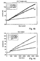
- the drug effect could be measured with a resolution current response of ten nano Amperes within 5 minutes. Moreover, theses results demonstrate quantitative correlation between the induced current signals and the number of cancer cell counted inside the nano-volume chamber.
- Utilizing nano-volume analytical device is of special interest in clinically relevant samples since it requires less tissue for diagnostics, and enables high-throughput analysis and comparison of the effect of various drugs on one tumor sample, while keeping uniform biological and environmental conditions.
- this new method can help tailor cancer drugs and treatments to individual patients towards 'personalized medicine'.
- a method of detecting a cancer cell as characterized by the appended claims.
- the method comprises contacting the cell with a substrate for an enzyme, under conditions wherein the enzyme catalyzes a reaction of the cell with the substrate, so as to generate a product capable of producing an electrical signal and subsequently, measuring a level of the electrical signal, wherein a difference in a level of the electrical signal compared to a predetermined threshold is indicative of a cancer cell.
- detecting refers to the act of detecting, diagnosing, perceiving, uncovering, exposing, visualizing or identifying a cell.
- the term "cell” refers to a mammalian cell, preferably a human cell.
- Single cells may be used in accordance with the teachings of the present invention as well as plurality of cells.
- the plurality of cells comprises no less than 10 cells and no more than 500 cells.
- the cells are in a single suspension such that the number of cells may be counted, although adherent cells and aggregates may still be detected.
- the plurality of cells may be from any biological sample such as cell-lines, primary cultures and cellular samples, e.g. biopsies (surgical biopsies including incisional or excisional biopsy, fine needle aspirates and the like), complete resections or body fluids. Methods of biopsy retrieval are well known in the art.
- the cells in the biological sample may be assayed for a functional enzyme without any pretreatment.
- the cells in the biological sample are preferably intact (i.e. whole), and preferably viable, although it will be appreciated that pre-treatment of cells, such as generation of cell extracts or non-intact cells are also contemplated by the present invention.
- a “cancer cell”, also referred to herein as a “malignant cell”, is a cell which has been released from normal cell division control, and is thus characterized by an abnormal growth and a tendency to proliferate in an uncontrolled way and, in some cases, to metastasize.
- the cancer cell may be a neoplastic cell, a pre-malignant cell, a metastatic cell, a tumor cell, an oncogenic cell, a cell with a cancer genotype, a cell of malignant phenotype, an oncogene transfected cell, a virus transformed cell, a cell which expresses an oncogene, a cell which expresses a marker for cancer, or a combination thereof.
- a cancer cell which may be detected by the method of the present invention: an adenocarcinoma cell, an adrenal gland tumor cell, an ameloblastoma cell, an anaplastic cell, anaplastic carcinoma of the thyroid cell, an angiofibroma cell, an angioma cell, an angiosarcoma cell, an apudoma cell, an argentaffmoma cell, an arrhenoblastoma cell, an ascites tumor cell, an ascitic tumor cell, an astroblastoma cell, an astrocytoma cell, an ataxia-telangiectasia cell, an atrial myxoma cell, a basal cell carcinoma cell, a benign tumor cell, a bone cancer cell, a bone tumor cell, a brainstem glioma cell, a brain tumor cell, a breast cancer cell, a Burkitt's lymphoma cell, a cancerous cell, a carcinoid cell, a cancerous cell, a
- a melanoma cell a meningioma cell, a mesothelioma cell, a metastatic cell, a metastasis cell, a metastatic spread cell, a Morton's neuroma cell, a multiple myeloma cell, a myeloblastoma cell, a myeloid leukemia cell, a myelolipoma cell, a myeloma cell, a myoblastoma cell, a myxoma cell, a nasopharyngeal carcinoma cell, a neoplastic cell, a nephroblastoma cell, a neuroblastoma cell, a neurofibroma cell, a neurofibromatosis cell, a neuroglioma cell, a neuroma cell, a non-Hodgkin's lymphoma cell, an oligodendroglioma cell, an optic glioma cell, an osteochond
- the cancer cell is a colon cancer cell.
- the method of the present invention is effected by contacting an enzyme substrate with a cell, to bring about a reaction of the cell, wherein the product of the enzymatic reaction is capable of generating an electrical signal.
- reaction of the cell refers to a reaction that occurs between the substrate and an endogenous enzyme expressed by the cell, and not to a reaction that occurs with an exogenous enzyme.
- the substrate is selected according to the enzyme, (also referred to herein as "marker enzyme") that is required to be detected.
- the enzyme may be situated inside the cell (i.e. intracellular) or on the cell membrane (i.e. membrane bound).
- the enzyme is a secreted enzyme.
- the substrate is preferably membrane permeable.
- the substrate is preferably selected such that following catalysis, the product formed is also membrane permeable such that it may diffuse away from the cell and on to the detector electrode
- the enzyme required to be detected is alkaline phosphatase (or enzyme secreted alkaline phosphatase, (SEAP)) and the substrate is 17, 4-aminophenyl phosphate (p-APP).
- Alkaline phosphatase converts p-APP to the electrochemical product, p-aminophenol (PAP).
- P-APP is widely commercially available from such Companies as Sigma-Aldrich (www.sigmaaldrich.com), Bio-world (www.Bio-world.com) and many others.
- Alkaline phosphatase is present in normal cells, but is reduced (or even absent) in cancerous cells. Therefore, analysis of the alkaline phosphatase activity level of cells, may be used as a marker for evaluating the efficiency of a particular drug for treating cancer and even for diagnostic purposes.
- the term "contacting" refers to bringing the substrate into the vicinity of a cell under conditions such that the substrate may be catalyzed by the enzyme.
- the contacting should by effected under buffer conditions, at a temperature and time sufficient to allow catalysis of the substrate and generation of sufficient product that it may be detected by an electrochemical cell.
- the contacting is effected for at least 5 minutes, at room temperature - see Examples 2 and 3 herein below.
- the contacting may be effected in vitro, ex vivo or in vivo.
- the contacting may be effected in a vessel which is also capable of detecting the product of the enzymatic reaction (i.e., in the electrochemical cell), such that the electrical signal is detected on-line.
- the contacting may be effected in a separate vessel from where the detection takes place such that it is possible to continuously withdraw samples at particular time points and place such samples within the electrochemical cells.
- the contacting may be effected in a test tube, flask, tissue culture, chip, array, plate, microplate, capillary, or the like.
- the cells may be placed on a vibrating plate following the addition of the substrate for continuous thorough mixing of the contents of the cells.
- electrochemical measurement of products capable of undergoing a redox reaction i.e. capable of electron transfer
- an electrical signal i.e. electrochemical products
- electrical signal refers to electrons or electrochemically active species.
- electrochemical measurement refers to a measurement performed by the use of electrodes in a solution, typically in an electrochemical cell.
- the measurement may be performed, for example, by chrono-amperometry, chrono-potentiometry, cyclic voltammetry, chrono-coulometry or square wave voltammetry.
- a signal detectable in such a measurement is one that differs in such electrochemical measurement from the control.
- the electrochemical cells of the present invention comprise a measurement electrode, a return electrode, a reference electrode and a chamber to hold the cells.
- the working electrode may be of a variety of different kinds, for example, it may be made of carbon, including glassy carbon, activated carbon cloth electrode, carbon felt, platinized carbon cloth, plain carbon cloth), may be made of gold, platinum or silver.
- the counter electrode may also be made of the same material as the working electrode.
- the reference electrode may for example be saturated calomel electrode, may be an Ag/AgCl electrode.
- the electrodes may be of a screen printed electrode which can be inserted into the vessel comprising the cells without the need to withdraw a sample and transport it into a separate electrochemical cell.
- the electrodes used to detect the product according to the method of the present invention may be reusable electrodes or disposable ones.
- Reusable electrodes may for example be electrodes made of glassy carbon in a disk or rod shape which are embedded in teflon.
- Disposable electrodes may for example be electrodes in the form of carbon paper, carbon cloth, carbon felts, or the screen printed electrode of the kind noted above.
- the electrochemical cell is a three-electrode cell. According to another embodiment, the electrochemical cell is a two-electrode cell. According to a preferred embodiment the electrochemical cells are provided as an array (i.e. chip) comprising a plurality of such cells i.e. a multiwell array where each well is of a nano-volume size.
- the system for measuring the electrical signal generated by the reaction product may further comprise a control module which may be a computer, a potentiostat and a multiplexer module which is needed in case of a typical embodiment for simultaneous measurement from a plurality of electrochemical cells.
- a control module which may be a computer, a potentiostat and a multiplexer module which is needed in case of a typical embodiment for simultaneous measurement from a plurality of electrochemical cells.
- the computer scans all the electrodes via the parallel port, and the background response to the potential application of each electrode is recorded by the computer.
- the entire electrochemical measurement sequence can be performed over a long period of time while measuring the currents resulting from the changes in the concentration of the products.
- the system can be calibrated by measuring the oxidation or reduction of an electroactive species, typically the same species which is the product of the enzymatic reaction in the electrochemical cell and comparison of the results of all the electrodes.
- the electrodes may be connected to the potentiostat and at the same time also collected via the multiplexer to a parallel port of the microcomputer.
- Each electrode is inserted in an electrochemical cell containing a reference electrode and a counter electrode which are also connected to the potentiostat.
- a specific potential is applied by the potentiostat on the electrodes (which can be the same for all the electrodes or can be a different potential to each electrode) and the current in each electrode is detected.
- the electrical signals are visualized in real-time on the computer screen.
- the signals generated by the electrochemical products of the enzymatic reactions reflect the level of enzyme in the cell, and the enzymes (presence, absence or level of same) are markers for cancer
- the signals may be used to determine whether a cell is cancerous (i.e. malignant) or not. Specifically, if the level of the generated electrical signal is different to a predetermined threshold, this would indicate that the cell is cancerous.
- the predetermined threshold is determined by the electrical signal generated by a control cell.
- the control cell can be a normally differentiated cell, non-cancerous cell, preferably of the same tissue and specimen as the tested cell suspicious of a cancerous or undifferentiated phenotype.
- the difference is at least 10 %, 20 %, 30 %, 40 %, 50 %, 80 %, 100 % (i.e., two-fold), 3 fold, 5 fold or 10 fold different as compared to a control cell.
- the amount of enzyme (and accordingly electrical signal) in a cancer cell is lower than the amount of enzyme (and accordingly electrical signal) in a non-cancer cell.
- the amount of enzyme (and accordingly electrical signal) in a cancer cell is higher than the amount of enzyme (and accordingly electrical signal) in a non-cancer cell.
- the method of the present invention may be used for diagnosing a subject with cancer.
- diagnosis refers to classifying a cancer, determining a severity of cancer (grade or stage), monitoring cancer progression, forecasting an outcome of the cancer and/or prospects of recovery.
- the subject may be a healthy animal or human subject undergoing a routine well-being check up.
- the subject may be at risk of having cancer (e.g., a genetically predisposed subject, a subject with medical and/or family history of cancer, a subject who has been exposed to carcinogens, occupational hazard, environmental hazard] and/or a subject who exhibits suspicious clinical signs of cancer [e.g., blood in the stool or melena, unexplained pain, sweating, unexplained fever, unexplained loss of weight up to anorexia, changes in bowel habits (constipation and/or diarrhea), tenesmus (sense of incomplete defecation, for rectal cancer specifically), anemia and/or general weakness).
- cancer e.g., a genetically predisposed subject, a subject with medical and/or family history of cancer, a subject who has been exposed to carcinogens, occupational hazard, environmental hazard
- a subject who exhibits suspicious clinical signs of cancer
- the present invention has a variety of applications pertaining to individually optimizing a treatment for cancer, and identifying an agent capable of reversing a malignant phenotype of a cell.
- a method of identifying an agent capable of reversing a malignant phenotype of a cell comprises subjecting at least one cancer cell to an agent and determining the efficiency of the anti cancer agent by monitoring the activity or expression of the marker enzyme (e.g. alkaline phosphatase) according to the method of the present invention.
- the marker enzyme e.g. alkaline phosphatase
- reversing a malignant phenotype refers to at least partially reversing the proliferative and/or invasive characteristics of the malignant cell.
- agent refers to a test composition comprising a biological agent or a chemical agent
- biological agents examples include, but are not limited to, nucleic acids, e.g., polynucleotides, ribozymes, siRNA and antisense molecules (including without limitation RNA, DNA, RNA/DNA hybrids, peptide nucleic acids, and polynucleotide analogs having altered backbone and/or bass structures or other chemical modifications); proteins, polypeptides (e.g. peptides), carbohydrates, lipids and "small molecule" drug candidates.
- nucleic acids e.g., polynucleotides, ribozymes, siRNA and antisense molecules (including without limitation RNA, DNA, RNA/DNA hybrids, peptide nucleic acids, and polynucleotide analogs having altered backbone and/or bass structures or other chemical modifications)
- proteins polypeptides (e.g. peptides)
- carbohydrates e.g. lipids and "small molecule" drug candidates.
- Small molecules can be, for example, naturally occurring compounds (e.g., compounds derived from plant extracts, microbial broths, and the like) or synthetic organic or organometallic compounds having molecular weights of less than about 10,000 daltons, preferably less than about 5,000 daltons, and most preferably less than about 1,500 daltons.
- the agents are differentiation agents including, but not limited to butyric acid and its derivatives.
- conditions that may be tested as potential anti cancer agents according to the method of the present invention include, but are not limited to, radiation exposure (such as, gamma radiation, UV radiation, X-radiation).
- the "marker enzyme" is also assayed prior to contact with the agent so that a comparison may be made prior to and following treatment.
- the agent is subjected to the cancer cells for a period long enough to have an anti cancer effect.
- these agents are subjected to the cancer cells for at least 1 day and more preferably 3 days.
- the agent may be contacted with cancer cells either in vitro, ex vivo or in vivo. If the contacting is effected in vivo, the cells are typically removed from the subject prior to contact with the substrate of the present invention.
- the present invention can, in theory, be practiced with a single electrochemical cell, such a method is not efficient nor is it desirable.
- the method of the present invention is used for high throughput screening of agents using a plurality of electrochemical cells to simultaneously screen a variety of agents.
- the cells may be part of a chip, for example a silicon chip as described in Example 1 herein below.
- the method of the present invention is performed using means for high output. Accordingly, the method may be performed, for example, using an automated sampling device, a liquid handling equipment, a dispenser, an electrode array, a robot, or any combination thereof.
- tumor treatment response cannot be predicted only from its type and anatomical location. It will be appreciated that the method of identifying an agent capable of reversing a malignant phenotype of a cell may be modified such that particular patent's cells may be used in the assay system, thereby tailoring therapeutic agents to specific patients. Furthermore, it will be appreciated that not only may the specific agent be selected using the method of the present invention, but the optimal dose and optimal treatment regimen may also be identified according to the method of the present invention. In this way a therapeutically effective amount of an agent may be determined.
- the patient may be treated according to the optimal treatment conditions selected with the aid of the method of the present invention and optionally retested after a suitable time period. In this way a patient's response may be continually monitored whilst undergoing therapy.
- the analyzing enzyme levels and administering steps may be repeated a number of times during the course of a treatment.
- the alkaline phosphatase levels may be analyzed one week following administration of the agent. If the alkaline phosphatase levels are higher than those compared with a control, the dose of the agent may be decreased. If the alkaline phosphatase levels remain lower than those compared with a control, the dose of the agent may be increased.
- the electrochemical cell may be provided in a kit together with at least one anti-cancer agent (e.g. a differentiation agent such as butyric acid) for determining an effect thereof on a cancer cell.
- the kit may, if desired, be presented in a pack which may contain one or more units of the kit .
- the pack may be accompanied by instructions for using the kit and the estimated dose of the anti-cancer agent for a particular number of cells.
- the pack may also be accommodated by a notice associated with the container in a form prescribed by a governmental agency regulating the manufacture, use or sale of laboratory supplements, which notice is reflective of approval by the agency of the form of the compositions.
- the kit may also comprise a substrate which is enzymatically reacted on by the marker enzyme of the biological cell (i.e. cancer cell) to yield a reaction product giving rise to a redox reaction at an electrode of the electrochemical cell.
- a substrate which is enzymatically reacted on by the marker enzyme of the biological cell (i.e. cancer cell) to yield a reaction product giving rise to a redox reaction at an electrode of the electrochemical cell.
- a nano-bio-chip was designed and fabricated using standard micro-system-technology (MST) methods. Its architecture included an array of miniaturized electrochemical cells. The cells were placed in the nano-volume chambers (i.e. the electrochemical-cells). The cylindrical chambers held 100 nL volume each. All arrays included positive and negative controls chambers.
- MST micro-system-technology
- the device was manufactured in two parts: a) a disposable chip - with the nano-chambers where the cells were placed, and b) a reusable chip, with an interface to electronic circuitry which included a multiplexer, potentiostat, temperature control and a pocket PC for sensing and data analysis. This setting allowed continuous reusing for multiple measurements.
- the chip was produced from silicon, and contained an array of eight miniaturized electrochemical cells. Each electrochemical-cell consisted of three circular-shaped electrodes, surrounded by an insulating silicon nitride layer. 1) Gold working electrode, 2) Gold counter electrode and 3) Ag/AgCl reference electrode. The electrodes were made by gold sputtering, microlithography and by selectively depositing Ag and anodizing it in a chloride containing solution for the reference electrode. The chambers walls were constructed from photopolymerized polyimide (SU-8) ( Figures 1A-B ). The silicon chip was wire bonded to a plastic chip, which was interfacing the electronic circuit.
- SU-8 photopolymerized polyimide
- the signal sensing was performed by the handheld palm-potentiostat with an interface to electronic circuitry for electrode signal regulation and detection (Palm Instruments BV-2004).
- the electronics consisted of eight independent duplicate circuits of electrochemical cells, which are temperature-controlled. A potential was applied between a working and a reference electrode in each electrochemical cell, and the output current was measured.
- HT-29 human colon cancer cells were grown in DMEM medium in the presence of fetal bovine serum at 37 °C in 95 % air, 5 % CO2 for 3 days prior to butyrate treatment. Butyrate at different concentrations was added to the HT-29 cell cultures. Concentrations applied were: 0, 0.078, 0.156, 0.3125, 0.625, 1.25, 2.5, 5 and 10 mM. Optimal butyrate concentration, LC50 and viability were calculated accordingly. The measurements were performed in PBS with the intact cells and without additional treatment of the cancer cells such as lysis.
- BA Butyric acid
- Its derivatives BA (Sigma, Israel) ranging between 0.08-10 mM were introduced to HT-29 colon cancer cells and incubated for 72 h for optimization. BA at a constant concentration of 2.5 mM was applied in all electrochemical experiments. BA derivatives, butyroylmethyl-diethyl phosphate, and pivaloyloxymethyl butyrate, were synthesized as described [ Rephaeli A, Zhuk R, Nudelman A, 2000, Drug Develop. Res.Vol 50:379-391 ].
- Butyroylmethyl-diethyl phosphate was solubilized in PBS and pivaloyloxymethyl butyrate in DMSO followed by dilution with medium to a final DMSO concentration of ⁇ 0.1%.
- the prodrugs were then introduced to the cells at a constant concentration of 50 ⁇ M and incubated for 96 h at 37 °C in 95 % air, 5 % CO2 conditions. Following incubation with the differentiation agents, viable cells were counted by tripa blue exclusion and tested for viability and LC 50 calculations. Prior to all electrochemical measurement, cells were centrifuged and diluted in PBS. All measurements were performed in PBS. The influence of BA, pivaloyloxymethyl butyrate and butyroylmethyl-diethyl phosphate were examined by measuring the enzymatic activity of alkaline phosphatase.
- Optical measurements Alkaline phosphatase activity was detected with the optical substrate para-Nitrophenyl Phosphate (PNPP kit ready to use, Sigma). The resultant enzymatic product was measured at wavelength of 420nm.
- Electrochemical measurements Amperometric enzyme measurements were performed with the electrochemical substrate, p-APP.
- the enzymatic reaction product, aminophenol (p-AP) is oxidized at the gold working electrode at 220mV and the generated current was measured.
- Eight-channeled 100 nL volume electrochemical chambers were loaded with HT-29 cancer cells, that were treated with BA or its derivatives. Cells were placed into the electrochemical chambers and the substrate was added to 1 mg/ml final concentration at a total volume of 100 nL. The generated current was measured and the number of cells was counted under the microscope. The correlation between cell number and current density was analyzed.
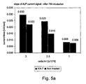
- HT-29 colon cancer cells were exposed for 96 hours to various differentiation therapy agents and the induced alkaline phosphatase activity was measured.
- Each electrochemical chamber on the array was loaded with cells exposed to a different agent - namely Butyric acid, butyroylmethyl-diethyl phosphate and pivaloyloxymethyl butyrate.
- the results are shown in Figure 1A-C .
- Normal enzymatic activity denotes that the cells differentiate properly as a consequence of the particular drug treatment.
- This 'Lab-on-a-chip' system provides the ability of sensitive measurements on extremely small samples (less than -15 cells) and does not require special cell treatment prior to the insertion into the chip.
Applications Claiming Priority (2)
| Application Number | Priority Date | Filing Date | Title |
|---|---|---|---|
| US90744107P | 2007-04-02 | 2007-04-02 | |
| PCT/IL2008/000457 WO2008120216A1 (en) | 2007-04-02 | 2008-04-02 | Methods of detecting cancer cells and use of same for diagnosing and monitoring treatment of the disease |
Publications (2)
| Publication Number | Publication Date |
|---|---|
| EP2142661A1 EP2142661A1 (en) | 2010-01-13 |
| EP2142661B1 true EP2142661B1 (en) | 2012-08-29 |
Family
ID=39530621
Family Applications (1)
| Application Number | Title | Priority Date | Filing Date |
|---|---|---|---|
| EP08720067A Not-in-force EP2142661B1 (en) | 2007-04-02 | 2008-04-02 | Methods of detecting cancer cells and use of same for diagnosing and monitoring treatment of the disease |
Country Status (6)
| Country | Link |
|---|---|
| US (2) | US8268577B2 (ko) |
| EP (1) | EP2142661B1 (ko) |
| JP (1) | JP2010523104A (ko) |
| KR (1) | KR20100015774A (ko) |
| CN (1) | CN101702920B (ko) |
| WO (1) | WO2008120216A1 (ko) |
Families Citing this family (11)
| Publication number | Priority date | Publication date | Assignee | Title |
|---|---|---|---|---|
| EP2142661B1 (en) | 2007-04-02 | 2012-08-29 | Ramot at Tel-Aviv University Ltd. | Methods of detecting cancer cells and use of same for diagnosing and monitoring treatment of the disease |
| US9707403B2 (en) | 2007-12-12 | 2017-07-18 | Neuro Code Tech Holdings, Llc | Rapid destruction of malignant tumors by excitotoxicity and osmotic-shock medical tactics |
| WO2009089517A1 (en) | 2008-01-10 | 2009-07-16 | The Neuro-Signaling Foundation | Method and system for processing cancer cell electrical signals for medical therapy |
| EP2265326A4 (en) | 2008-04-17 | 2011-06-22 | Neuro Signaling Foundation | SYSTEM AND METHOD FOR TRIGGERING APOPTOSIS IN MALIGNANT TUMOR CELLS FOR MEDICAL TREATMENT |
| WO2013011513A1 (en) * | 2011-07-21 | 2013-01-24 | Ramot at Tel- Aviv University Ltd. | Methods of determinig enzyme activity in preserved cell samples |
| RU2489710C1 (ru) * | 2012-02-20 | 2013-08-10 | Владимир Вадимович Мошкин | Способ коммутационной хроноамперометрии |
| WO2013179293A1 (en) * | 2012-05-31 | 2013-12-05 | Ramot At Tel-Aviv University Ltd. | Methods and system for detecting melanoma |
| KR20150074487A (ko) | 2013-12-24 | 2015-07-02 | 삼성전자주식회사 | 식각 부산물 검출 방법 및 이를 이용한 자기 저항 메모리 장치의 제조 방법 |
| JP2015144904A (ja) * | 2015-04-20 | 2015-08-13 | 佐藤 洋 | 位置制御システム |
| US11154238B2 (en) | 2015-08-07 | 2021-10-26 | Electroceuticals, Llc | Systems, methods and apparatuses for providing bioelectronic neurocode-based therapies to mammals |
| CN113720893A (zh) * | 2021-09-01 | 2021-11-30 | 致慧医疗科技(上海)有限公司 | 一种基于癌细胞表面电荷强度的评估肿瘤恶性程度的方法 |
Family Cites Families (22)
| Publication number | Priority date | Publication date | Assignee | Title |
|---|---|---|---|---|
| NL154600B (nl) | 1971-02-10 | 1977-09-15 | Organon Nv | Werkwijze voor het aantonen en bepalen van specifiek bindende eiwitten en hun corresponderende bindbare stoffen. |
| NL154598B (nl) | 1970-11-10 | 1977-09-15 | Organon Nv | Werkwijze voor het aantonen en bepalen van laagmoleculire verbindingen en van eiwitten die deze verbindingen specifiek kunnen binden, alsmede testverpakking. |
| NL154599B (nl) | 1970-12-28 | 1977-09-15 | Organon Nv | Werkwijze voor het aantonen en bepalen van specifiek bindende eiwitten en hun corresponderende bindbare stoffen, alsmede testverpakking. |
| US3901654A (en) | 1971-06-21 | 1975-08-26 | Biological Developments | Receptor assays of biologically active compounds employing biologically specific receptors |
| US3853987A (en) | 1971-09-01 | 1974-12-10 | W Dreyer | Immunological reagent and radioimmuno assay |
| US3867517A (en) | 1971-12-21 | 1975-02-18 | Abbott Lab | Direct radioimmunoassay for antigens and their antibodies |
| NL171930C (nl) | 1972-05-11 | 1983-06-01 | Akzo Nv | Werkwijze voor het aantonen en bepalen van haptenen, alsmede testverpakkingen. |
| US3850578A (en) | 1973-03-12 | 1974-11-26 | H Mcconnell | Process for assaying for biologically active molecules |
| US3935074A (en) | 1973-12-17 | 1976-01-27 | Syva Company | Antibody steric hindrance immunoassay with two antibodies |
| US3996345A (en) | 1974-08-12 | 1976-12-07 | Syva Company | Fluorescence quenching with immunological pairs in immunoassays |
| US4034074A (en) | 1974-09-19 | 1977-07-05 | The Board Of Trustees Of Leland Stanford Junior University | Universal reagent 2-site immunoradiometric assay using labelled anti (IgG) |
| US3984533A (en) | 1975-11-13 | 1976-10-05 | General Electric Company | Electrophoretic method of detecting antigen-antibody reaction |
| US4098876A (en) | 1976-10-26 | 1978-07-04 | Corning Glass Works | Reverse sandwich immunoassay |
| US4879219A (en) | 1980-09-19 | 1989-11-07 | General Hospital Corporation | Immunoassay utilizing monoclonal high affinity IgM antibodies |
| US5011771A (en) | 1984-04-12 | 1991-04-30 | The General Hospital Corporation | Multiepitopic immunometric assay |
| JPH05506098A (ja) * | 1990-04-03 | 1993-09-02 | オンコセラピューティクス | 癌患者を治療するための有効な薬品を確認する電子的方法 |
| US5281521A (en) | 1992-07-20 | 1994-01-25 | The Trustees Of The University Of Pennsylvania | Modified avidin-biotin technique |
| US6682648B1 (en) * | 1997-08-12 | 2004-01-27 | University Of Southern California | Electrochemical reporter system for detecting analytical immunoassay and molecular biology procedures |
| CN1312480C (zh) * | 2004-09-24 | 2007-04-25 | 南京大学 | 肿瘤细胞表面抗原的原位电化学免疫测定方法 |
| US20060205090A1 (en) * | 2005-03-14 | 2006-09-14 | Newton Michael W | Water-soluble conjugates for electrochemical detection |
| EP2142661B1 (en) | 2007-04-02 | 2012-08-29 | Ramot at Tel-Aviv University Ltd. | Methods of detecting cancer cells and use of same for diagnosing and monitoring treatment of the disease |
| KR101281761B1 (ko) * | 2009-03-27 | 2013-07-26 | 서강대학교산학협력단 | 전기검출기반 줄기세포 분화 측정용 센서 |
-
2008
- 2008-04-02 EP EP08720067A patent/EP2142661B1/en not_active Not-in-force
- 2008-04-02 CN CN2008800182996A patent/CN101702920B/zh not_active Expired - Fee Related
- 2008-04-02 KR KR1020097021991A patent/KR20100015774A/ko not_active Application Discontinuation
- 2008-04-02 JP JP2010501666A patent/JP2010523104A/ja active Pending
- 2008-04-02 WO PCT/IL2008/000457 patent/WO2008120216A1/en active Application Filing
-
2009
- 2009-03-05 US US12/379,974 patent/US8268577B2/en not_active Expired - Fee Related
-
2012
- 2012-07-18 US US13/551,642 patent/US8530179B2/en not_active Expired - Fee Related
Non-Patent Citations (1)
| Title |
|---|
| WALACH NATALIO ET AL: "Leukocyte alkaline phosphatase as a probable predictor of the metastatic state in breast and colon cancer patients", ONCOLOGY (BASEL), vol. 52, no. 1, 1995, pages 12 - 18, XP008127126, ISSN: 0030-2414 * |
Also Published As
| Publication number | Publication date |
|---|---|
| US20120288877A1 (en) | 2012-11-15 |
| CN101702920B (zh) | 2013-03-13 |
| WO2008120216A1 (en) | 2008-10-09 |
| KR20100015774A (ko) | 2010-02-12 |
| EP2142661A1 (en) | 2010-01-13 |
| CN101702920A (zh) | 2010-05-05 |
| US8268577B2 (en) | 2012-09-18 |
| US20090232740A1 (en) | 2009-09-17 |
| US8530179B2 (en) | 2013-09-10 |
| JP2010523104A (ja) | 2010-07-15 |
Similar Documents
| Publication | Publication Date | Title |
|---|---|---|
| EP2142661B1 (en) | Methods of detecting cancer cells and use of same for diagnosing and monitoring treatment of the disease | |
| US8206903B2 (en) | Device and method for electroporation-based delivery of molecules into cells and dynamic monitoring of cell responses | |
| US9399787B2 (en) | Real-time electronic cell sensing system and applications for cytotoxicity profiling and compound assays | |
| Ceriotti et al. | Assessment of cytotoxicity by impedance spectroscopy | |
| Gu et al. | Cellular electrical impedance spectroscopy: an emerging technology of microscale biosensors | |
| Krinke et al. | A microelectrode-based sensor for label-free in vitro detection of ischemic effects on cardiomyocytes | |
| Bettazzi et al. | Nanotoxicity assessment: A challenging application for cutting edge electroanalytical tools | |
| Kanyong et al. | Amperometric screen-printed galactose biosensor for cell toxicity applications | |
| JP5549908B2 (ja) | 損傷dna修復物質のスクリーニング方法 | |
| CN101017175A (zh) | 信息获取装置、应激度信息获取装置及应激度判定方法 | |
| CN102094068B (zh) | 一种蛋氨酸亚砜还原酶活性的检测方法及药物筛选试剂盒 | |
| EP1336101B1 (en) | A method to measure the activation state of signaling pathways in cells | |
| US20150185218A1 (en) | Methods and system for detecting melanoma | |
| Ponti et al. | Comparison of impedance-based sensors for cell adhesion monitoring and in vitro methods for detecting cytotoxicity induced by chemicals | |
| WO2013011513A1 (en) | Methods of determinig enzyme activity in preserved cell samples | |
| EP2639576B1 (en) | Real-time electronic cell sensing system and applications for cytotoxicity profiling and compound assays | |
| WO2010009387A1 (en) | Methods for controlling protein loading variability in reverse phase protein microarrays | |
| Mestres-Ventura | Chemosensitivity testing of human tumors using Si-sensor chips | |
| WO2003087293A1 (en) | Apparatus and method for biochemical analysis | |
| WO2006058185A2 (en) | Device and method for electroporation-based delivery of moecules into cells and dynamic monitoring of cell responses | |
| Kanyonga et al. | Development of an amperometric screen-printed galactose biosensor for cell toxicity applications | |
| Almog et al. | On-chip detection of cellular activity | |
| Boroditsky et al. | A Lens Into Mass Cytometry: A Multifaceted Technology Used to Delve Deep into Single-Cell Analysis | |
| Robitski et al. | An overview on bioelectronic and biosensoric microstructures supporting high-content screening in cell cultures | |
| GB2436145A (en) | A system for monitoring properties of a single cell |
Legal Events
| Date | Code | Title | Description |
|---|---|---|---|
| PUAI | Public reference made under article 153(3) epc to a published international application that has entered the european phase |
Free format text: ORIGINAL CODE: 0009012 |
|
| 17P | Request for examination filed |
Effective date: 20091030 |
|
| AK | Designated contracting states |
Kind code of ref document: A1 Designated state(s): AT BE BG CH CY CZ DE DK EE ES FI FR GB GR HR HU IE IS IT LI LT LU LV MC MT NL NO PL PT RO SE SI SK TR |
|
| 17Q | First examination report despatched |
Effective date: 20100219 |
|
| DAX | Request for extension of the european patent (deleted) | ||
| REG | Reference to a national code |
Ref country code: DE Ref legal event code: R079 Ref document number: 602008018371 Country of ref document: DE Free format text: PREVIOUS MAIN CLASS: C12Q0001000000 Ipc: G01N0033500000 |
|
| GRAP | Despatch of communication of intention to grant a patent |
Free format text: ORIGINAL CODE: EPIDOSNIGR1 |
|
| RIC1 | Information provided on ipc code assigned before grant |
Ipc: G01N 33/50 20060101AFI20120217BHEP |
|
| GRAS | Grant fee paid |
Free format text: ORIGINAL CODE: EPIDOSNIGR3 |
|
| GRAA | (expected) grant |
Free format text: ORIGINAL CODE: 0009210 |
|
| AK | Designated contracting states |
Kind code of ref document: B1 Designated state(s): AT BE BG CH CY CZ DE DK EE ES FI FR GB GR HR HU IE IS IT LI LT LU LV MC MT NL NO PL PT RO SE SI SK TR |
|
| REG | Reference to a national code |
Ref country code: GB Ref legal event code: FG4D |
|
| REG | Reference to a national code |
Ref country code: CH Ref legal event code: EP |
|
| REG | Reference to a national code |
Ref country code: AT Ref legal event code: REF Ref document number: 573321 Country of ref document: AT Kind code of ref document: T Effective date: 20120915 |
|
| REG | Reference to a national code |
Ref country code: IE Ref legal event code: FG4D |
|
| REG | Reference to a national code |
Ref country code: DE Ref legal event code: R096 Ref document number: 602008018371 Country of ref document: DE Effective date: 20121025 |
|
| REG | Reference to a national code |
Ref country code: AT Ref legal event code: MK05 Ref document number: 573321 Country of ref document: AT Kind code of ref document: T Effective date: 20120829 |
|
| REG | Reference to a national code |
Ref country code: NL Ref legal event code: VDEP Effective date: 20120829 |
|
| REG | Reference to a national code |
Ref country code: LT Ref legal event code: MG4D Effective date: 20120829 |
|
| PG25 | Lapsed in a contracting state [announced via postgrant information from national office to epo] |
Ref country code: NO Free format text: LAPSE BECAUSE OF FAILURE TO SUBMIT A TRANSLATION OF THE DESCRIPTION OR TO PAY THE FEE WITHIN THE PRESCRIBED TIME-LIMIT Effective date: 20121129 Ref country code: FI Free format text: LAPSE BECAUSE OF FAILURE TO SUBMIT A TRANSLATION OF THE DESCRIPTION OR TO PAY THE FEE WITHIN THE PRESCRIBED TIME-LIMIT Effective date: 20120829 Ref country code: HR Free format text: LAPSE BECAUSE OF FAILURE TO SUBMIT A TRANSLATION OF THE DESCRIPTION OR TO PAY THE FEE WITHIN THE PRESCRIBED TIME-LIMIT Effective date: 20120829 Ref country code: IS Free format text: LAPSE BECAUSE OF FAILURE TO SUBMIT A TRANSLATION OF THE DESCRIPTION OR TO PAY THE FEE WITHIN THE PRESCRIBED TIME-LIMIT Effective date: 20121229 Ref country code: LT Free format text: LAPSE BECAUSE OF FAILURE TO SUBMIT A TRANSLATION OF THE DESCRIPTION OR TO PAY THE FEE WITHIN THE PRESCRIBED TIME-LIMIT Effective date: 20120829 Ref country code: AT Free format text: LAPSE BECAUSE OF FAILURE TO SUBMIT A TRANSLATION OF THE DESCRIPTION OR TO PAY THE FEE WITHIN THE PRESCRIBED TIME-LIMIT Effective date: 20120829 Ref country code: CY Free format text: LAPSE BECAUSE OF FAILURE TO SUBMIT A TRANSLATION OF THE DESCRIPTION OR TO PAY THE FEE WITHIN THE PRESCRIBED TIME-LIMIT Effective date: 20120829 |
|
| PG25 | Lapsed in a contracting state [announced via postgrant information from national office to epo] |
Ref country code: GR Free format text: LAPSE BECAUSE OF FAILURE TO SUBMIT A TRANSLATION OF THE DESCRIPTION OR TO PAY THE FEE WITHIN THE PRESCRIBED TIME-LIMIT Effective date: 20121130 Ref country code: SI Free format text: LAPSE BECAUSE OF FAILURE TO SUBMIT A TRANSLATION OF THE DESCRIPTION OR TO PAY THE FEE WITHIN THE PRESCRIBED TIME-LIMIT Effective date: 20120829 Ref country code: BE Free format text: LAPSE BECAUSE OF FAILURE TO SUBMIT A TRANSLATION OF THE DESCRIPTION OR TO PAY THE FEE WITHIN THE PRESCRIBED TIME-LIMIT Effective date: 20120829 Ref country code: PT Free format text: LAPSE BECAUSE OF FAILURE TO SUBMIT A TRANSLATION OF THE DESCRIPTION OR TO PAY THE FEE WITHIN THE PRESCRIBED TIME-LIMIT Effective date: 20121231 Ref country code: SE Free format text: LAPSE BECAUSE OF FAILURE TO SUBMIT A TRANSLATION OF THE DESCRIPTION OR TO PAY THE FEE WITHIN THE PRESCRIBED TIME-LIMIT Effective date: 20120829 Ref country code: LV Free format text: LAPSE BECAUSE OF FAILURE TO SUBMIT A TRANSLATION OF THE DESCRIPTION OR TO PAY THE FEE WITHIN THE PRESCRIBED TIME-LIMIT Effective date: 20120829 |
|
| PG25 | Lapsed in a contracting state [announced via postgrant information from national office to epo] |
Ref country code: DK Free format text: LAPSE BECAUSE OF FAILURE TO SUBMIT A TRANSLATION OF THE DESCRIPTION OR TO PAY THE FEE WITHIN THE PRESCRIBED TIME-LIMIT Effective date: 20120829 Ref country code: EE Free format text: LAPSE BECAUSE OF FAILURE TO SUBMIT A TRANSLATION OF THE DESCRIPTION OR TO PAY THE FEE WITHIN THE PRESCRIBED TIME-LIMIT Effective date: 20120829 Ref country code: NL Free format text: LAPSE BECAUSE OF FAILURE TO SUBMIT A TRANSLATION OF THE DESCRIPTION OR TO PAY THE FEE WITHIN THE PRESCRIBED TIME-LIMIT Effective date: 20120829 Ref country code: ES Free format text: LAPSE BECAUSE OF FAILURE TO SUBMIT A TRANSLATION OF THE DESCRIPTION OR TO PAY THE FEE WITHIN THE PRESCRIBED TIME-LIMIT Effective date: 20121210 Ref country code: RO Free format text: LAPSE BECAUSE OF FAILURE TO SUBMIT A TRANSLATION OF THE DESCRIPTION OR TO PAY THE FEE WITHIN THE PRESCRIBED TIME-LIMIT Effective date: 20120829 Ref country code: CZ Free format text: LAPSE BECAUSE OF FAILURE TO SUBMIT A TRANSLATION OF THE DESCRIPTION OR TO PAY THE FEE WITHIN THE PRESCRIBED TIME-LIMIT Effective date: 20120829 |
|
| PG25 | Lapsed in a contracting state [announced via postgrant information from national office to epo] |
Ref country code: SK Free format text: LAPSE BECAUSE OF FAILURE TO SUBMIT A TRANSLATION OF THE DESCRIPTION OR TO PAY THE FEE WITHIN THE PRESCRIBED TIME-LIMIT Effective date: 20120829 Ref country code: PL Free format text: LAPSE BECAUSE OF FAILURE TO SUBMIT A TRANSLATION OF THE DESCRIPTION OR TO PAY THE FEE WITHIN THE PRESCRIBED TIME-LIMIT Effective date: 20120829 Ref country code: IT Free format text: LAPSE BECAUSE OF FAILURE TO SUBMIT A TRANSLATION OF THE DESCRIPTION OR TO PAY THE FEE WITHIN THE PRESCRIBED TIME-LIMIT Effective date: 20120829 |
|
| PLBE | No opposition filed within time limit |
Free format text: ORIGINAL CODE: 0009261 |
|
| STAA | Information on the status of an ep patent application or granted ep patent |
Free format text: STATUS: NO OPPOSITION FILED WITHIN TIME LIMIT |
|
| PG25 | Lapsed in a contracting state [announced via postgrant information from national office to epo] |
Ref country code: BG Free format text: LAPSE BECAUSE OF FAILURE TO SUBMIT A TRANSLATION OF THE DESCRIPTION OR TO PAY THE FEE WITHIN THE PRESCRIBED TIME-LIMIT Effective date: 20121129 |
|
| 26N | No opposition filed |
Effective date: 20130530 |
|
| REG | Reference to a national code |
Ref country code: DE Ref legal event code: R097 Ref document number: 602008018371 Country of ref document: DE Effective date: 20130530 |
|
| PG25 | Lapsed in a contracting state [announced via postgrant information from national office to epo] |
Ref country code: MC Free format text: LAPSE BECAUSE OF FAILURE TO SUBMIT A TRANSLATION OF THE DESCRIPTION OR TO PAY THE FEE WITHIN THE PRESCRIBED TIME-LIMIT Effective date: 20120829 |
|
| REG | Reference to a national code |
Ref country code: CH Ref legal event code: PL |
|
| REG | Reference to a national code |
Ref country code: IE Ref legal event code: MM4A |
|
| PG25 | Lapsed in a contracting state [announced via postgrant information from national office to epo] |
Ref country code: CH Free format text: LAPSE BECAUSE OF NON-PAYMENT OF DUE FEES Effective date: 20130430 Ref country code: LI Free format text: LAPSE BECAUSE OF NON-PAYMENT OF DUE FEES Effective date: 20130430 |
|
| PG25 | Lapsed in a contracting state [announced via postgrant information from national office to epo] |
Ref country code: IE Free format text: LAPSE BECAUSE OF NON-PAYMENT OF DUE FEES Effective date: 20130402 |
|
| PGFP | Annual fee paid to national office [announced via postgrant information from national office to epo] |
Ref country code: GB Payment date: 20140422 Year of fee payment: 7 |
|
| PGFP | Annual fee paid to national office [announced via postgrant information from national office to epo] |
Ref country code: DE Payment date: 20140418 Year of fee payment: 7 Ref country code: FR Payment date: 20140422 Year of fee payment: 7 |
|
| PG25 | Lapsed in a contracting state [announced via postgrant information from national office to epo] |
Ref country code: MT Free format text: LAPSE BECAUSE OF FAILURE TO SUBMIT A TRANSLATION OF THE DESCRIPTION OR TO PAY THE FEE WITHIN THE PRESCRIBED TIME-LIMIT Effective date: 20120829 |
|
| PG25 | Lapsed in a contracting state [announced via postgrant information from national office to epo] |
Ref country code: TR Free format text: LAPSE BECAUSE OF FAILURE TO SUBMIT A TRANSLATION OF THE DESCRIPTION OR TO PAY THE FEE WITHIN THE PRESCRIBED TIME-LIMIT Effective date: 20120829 |
|
| PG25 | Lapsed in a contracting state [announced via postgrant information from national office to epo] |
Ref country code: HU Free format text: LAPSE BECAUSE OF FAILURE TO SUBMIT A TRANSLATION OF THE DESCRIPTION OR TO PAY THE FEE WITHIN THE PRESCRIBED TIME-LIMIT; INVALID AB INITIO Effective date: 20080402 Ref country code: LU Free format text: LAPSE BECAUSE OF NON-PAYMENT OF DUE FEES Effective date: 20130402 |
|
| REG | Reference to a national code |
Ref country code: DE Ref legal event code: R119 Ref document number: 602008018371 Country of ref document: DE |
|
| GBPC | Gb: european patent ceased through non-payment of renewal fee |
Effective date: 20150402 |
|
| PG25 | Lapsed in a contracting state [announced via postgrant information from national office to epo] |
Ref country code: DE Free format text: LAPSE BECAUSE OF NON-PAYMENT OF DUE FEES Effective date: 20151103 Ref country code: GB Free format text: LAPSE BECAUSE OF NON-PAYMENT OF DUE FEES Effective date: 20150402 |
|
| REG | Reference to a national code |
Ref country code: FR Ref legal event code: ST Effective date: 20151231 |
|
| PG25 | Lapsed in a contracting state [announced via postgrant information from national office to epo] |
Ref country code: FR Free format text: LAPSE BECAUSE OF NON-PAYMENT OF DUE FEES Effective date: 20150430 |