EP2124193A2 - Automatisierte Plazenta-Messung - Google Patents
Automatisierte Plazenta-Messung Download PDFInfo
- Publication number
- EP2124193A2 EP2124193A2 EP09160891A EP09160891A EP2124193A2 EP 2124193 A2 EP2124193 A2 EP 2124193A2 EP 09160891 A EP09160891 A EP 09160891A EP 09160891 A EP09160891 A EP 09160891A EP 2124193 A2 EP2124193 A2 EP 2124193A2
- Authority
- EP
- European Patent Office
- Prior art keywords
- placental
- area
- villi
- algorithms
- mean
- Prior art date
- Legal status (The legal status is an assumption and is not a legal conclusion. Google has not performed a legal analysis and makes no representation as to the accuracy of the status listed.)
- Withdrawn
Links
Images





Classifications
-
- G—PHYSICS
- G06—COMPUTING OR CALCULATING; COUNTING
- G06T—IMAGE DATA PROCESSING OR GENERATION, IN GENERAL
- G06T7/00—Image analysis
- G06T7/0002—Inspection of images, e.g. flaw detection
- G06T7/0012—Biomedical image inspection
-
- G—PHYSICS
- G06—COMPUTING OR CALCULATING; COUNTING
- G06T—IMAGE DATA PROCESSING OR GENERATION, IN GENERAL
- G06T7/00—Image analysis
- G06T7/40—Analysis of texture
- G06T7/41—Analysis of texture based on statistical description of texture
-
- G—PHYSICS
- G06—COMPUTING OR CALCULATING; COUNTING
- G06T—IMAGE DATA PROCESSING OR GENERATION, IN GENERAL
- G06T7/00—Image analysis
- G06T7/60—Analysis of geometric attributes
- G06T7/62—Analysis of geometric attributes of area, perimeter, diameter or volume
-
- G—PHYSICS
- G06—COMPUTING OR CALCULATING; COUNTING
- G06V—IMAGE OR VIDEO RECOGNITION OR UNDERSTANDING
- G06V20/00—Scenes; Scene-specific elements
- G06V20/60—Type of objects
- G06V20/69—Microscopic objects, e.g. biological cells or cellular parts
- G06V20/695—Preprocessing, e.g. image segmentation
-
- G—PHYSICS
- G06—COMPUTING OR CALCULATING; COUNTING
- G06T—IMAGE DATA PROCESSING OR GENERATION, IN GENERAL
- G06T2207/00—Indexing scheme for image analysis or image enhancement
- G06T2207/10—Image acquisition modality
- G06T2207/10056—Microscopic image
-
- G—PHYSICS
- G06—COMPUTING OR CALCULATING; COUNTING
- G06T—IMAGE DATA PROCESSING OR GENERATION, IN GENERAL
- G06T2207/00—Indexing scheme for image analysis or image enhancement
- G06T2207/30—Subject of image; Context of image processing
- G06T2207/30004—Biomedical image processing
- G06T2207/30024—Cell structures in vitro; Tissue sections in vitro
-
- G—PHYSICS
- G06—COMPUTING OR CALCULATING; COUNTING
- G06T—IMAGE DATA PROCESSING OR GENERATION, IN GENERAL
- G06T2207/00—Indexing scheme for image analysis or image enhancement
- G06T2207/30—Subject of image; Context of image processing
- G06T2207/30004—Biomedical image processing
- G06T2207/30044—Fetus; Embryo
-
- G—PHYSICS
- G06—COMPUTING OR CALCULATING; COUNTING
- G06T—IMAGE DATA PROCESSING OR GENERATION, IN GENERAL
- G06T2207/00—Indexing scheme for image analysis or image enhancement
- G06T2207/30—Subject of image; Context of image processing
- G06T2207/30004—Biomedical image processing
- G06T2207/30101—Blood vessel; Artery; Vein; Vascular
Definitions
- the present invention generally relates automated imaging measures of the intrauterine environment through placental photography and histology.
- placenta the key organ upon which the fetus is entirely dependent for all oxygen and nutrition, grows in a branching fashion analogous to the growth of a tree and its branches.
- the major villous types, their principal time periods of development during gestation, and their specific physiology have been well delineated in the research setting. But routine pathology slide review has poor reliability in distinguishing the major patterns of placental branching morphogenesis. As the evidence that lifelong health risks appear to be correlated with birthweight, the importance of placental growth and development as the principal non-genetic contributor to fetal growth has grown.
- the placenta is the only fetal organ that can be dissected in a living child to yield information related to cell proliferation (a marker of tissue health), branching (reflecting gene transcription events) and cell death.
- a reliable and automated assessment tool performed on routine stained placental slides may help understand how intrauterine stressors modulate placental (and by extension fetal) well-being.
- Disclosed herein is a new approach towards automated measures of the intrauterine environment through placental imaging and histology.
- Figure 1 is a pair of before and after images of placental tissue processed by a spatial fuzzy c-means algorithm (SFCM algorithm).
- SFCM algorithm spatial fuzzy c-means algorithm
- Figure 2 is a pair of before and after images of placental tissue processed by a spatial fuzzy c-means algorithm (SFCM algorithm).
- SFCM algorithm spatial fuzzy c-means algorithm
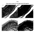
- Figure 3 is a pair of before and after images of placental tissue processed by a spatial fuzzy c-means algorithm (SFCM algorithm).
- SFCM algorithm spatial fuzzy c-means algorithm
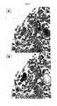
- Figure 4A is a photograph of a digitized histology slide.
- Figure 4B is the image processed by the ECognition software showing the extracted objects
- Figure 5 shows the image from Figure 4B correlated with the biological schema.
- Figure 6 is a series of photomicrographs depicting selected photomicrographs from cases with extreme low (negative) and high (positive) factor scores.
- Figure 7 is a photograph of a placenta sliced in 8ths, and the corresponding traced sliced perimeters
- Figure 8 is a digitized photograph of the placental chorionic surface marked for mbilical cord insertion, the disk perimeter (outer marking), and terminal points of chorionic vascular plate branches (inner marking). The ratio of these areas is associated with decreased observed/expected birth weight.
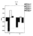
- Figure 9 is a series of histograms of mean REGR factor score 3, 4,6, and 7, for term and preterm birth.
- Figure 10 is a series of histograms of mean REGR factor score 3, 6, 7, and 12 for preterm cases categorized as having chorioamnionitis ('1') and not having chorioamnionitis ('0').
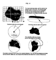
- Figure 11 is a series of schemas depicting determination of chorionic plate area and centroid, Chorionic vascular area and centroid, and discordance or concordance of centroids.
- Figure 12 is a series of histograms depicting the distribution of baby weight (12A), placental weight (12B) and inter-centroid distance normalized to chorionic plate area (12C).
- the methods described herein teach a process for extracting medically significant information from digital images of placental tissue by processing the image through a mathematical algorithm.
- the medically significant information extracted from the image may, for example, count neutrophils that are responses to bacterial infection. High neutrophil counts indicate a bacterial infection was present in the fetal environment before birth. Such bacterial infections are one of the most significant predictors of risk for Cerebral Palsy (CP) in term infants. Cerebral Palsy is not diagnosed until several years after birth; CP cannot be identified by examination of the mother or the newborn, but identification of the risk of CP by the methods taught herein can enable a phyisician to prescribe a plan of monitoring and early intervention if signs of the disease begin to manifest themselves.
- An example of the extraction of neutrophil information is discussed further in Example 1
- connective tissue Another example of medically significant information that can be extracted from digital images is a measure of the integrity of the connective tissue.
- these connective tissues may be damaged by digestive enzymes released by the neutrophils recruited to attack the invader.
- These same enzymes can damage the connective tissues of the fetus and lead to brain and lung damage in the child.
- this damage may not be observable in the newborn, but the information produced by these methods for analysis of the placenta may allow a physician to prescribe early monitoring, intervention, or treatment for the infant and child.
- medically significant information can extracted from digital images of placental histological features so as to provide analysis of congenital viral infection (well recognized as a precursor to fetal anomalies as well as poor long-term neurological development), and matemal/uteroplacental and fetal-placental vascular pathologies (both of which are associated with fetal hypoxia and risk for poor long-term neurological development).
- the first step of these methods is the selection of the placental sample to be analyzed. Every baby is born with a placenta and the sample may be taken from the placenta, the umbilical cord, or the membranes. The sample may be the entire placenta, the gross placental shape, portions of the placenta, umbilical cord, or membranes, or may be a slice of tissue from any of these fixed to a histology slide. The samples may be taken soon after birth, or the placental tissues may be preserved in formalin and the digital images may be taken at a later date, even years later. Measurements taken at birth can be used to predict risk to future pregnancies born to that mother, as well as risks to the particular child.
- a digital image of the placental sample may be obtained using a film or digital camera, using a microscope with a camera attachment, or using a slide digitizer. Film images may be digitized if they are of sufficient resolution.
- the preferred method is to use a digital camera.
- the preferred method is to use a slide digitizer such as an Aperio T3, manufactured by Aperio Technologies Corp. in Vista, CA. Other slide digitizers may be used such as those manufactured by Nikon, Zeiss, or Leica.
- the digital images of histology slides should have sufficient resolution to allow extraction of image features up to a magnification of 20-40X.
- the digital images are analysed by processing them by a mathematical algorithm.
- algorithms may be employed alone or in combination to extract the features of interest from the image.
- algorithms that can be used are spatial fuzzy c-means algorithms, segmentation algorithms, boundary finding algorithms, counting algorithms, length measuring algorithms, branching algorithms, angle measuring algorithms, and color discriminating algorithms.
- Other types of algorithms useful for image analysis or segmentation are clustering (K-means) algorithms, mean shift algorithms, histogram-based algorithms, edge detection algorithms, region growing algorithms, level setting algorithms, graph partitioning algorithms, watershed transformation algorithms, model based segmentation algorithms, multiscale segmentation algorithms, semi-automatics segmentation algorithms, and neural network algorithms.
- a branching algorithms may be used to extract the extent of branching of the major placental blood vessels from the digital image of the chorionic surface of the entire placenta.
- a color discriminating algorithm may be used to extract the neutrophils from a digital image of a histology slide and then a counting algorithm used to count the number of neutrophils present.
- SFCM spatial fuzzy c-means
- the unsupervised cluster algorithm called SFCM (Spatial Fuzzy c-Means)
- SFCM Session Cyclone
- the spatial function is the summation of the membership function in the neighborhood of each pixel under consideration.
- the advantages of the SFCM are the following: (1) it yields regions more homogeneous than those of other methods, (2) it reduces the spurious blobs, (3) it removes noisy spots, and (4) it is less sensitive to noise than other techniques. This technique is a powerful method for noisy image segmentation and works for both single and multiple-feature data with spatial information..
- the features of interest include neutrophils, connective tissues, portions of edema, cell nuclei, major blood vessels, branched villi, large villi, long villi, small villi, nutrition exchange vessels, and capillaries, markers of fetal hypoxia such as syncytial knots and syncytial basophilia, villous fibrosis/scarring, chronic villitis and chronic intervillositis, infarcts, abruption, perivillous fibrin deposition and cytotrophoblast proliferation, abnormalities of clotting and inflammation in the basal plate and maternal uteroplacental vessels, cell death of epithelia, stroma, endothelia, proliferation of macrophages and fibroblasts in connective tissue and stroma, abnormalities of clotting and inflammation in the fetal-placental blood vessels.
- markers of fetal hypoxia such as syncytial knots and syncytial basophilia, villous fibrosis/s
- the feature of interest After extracting the feature of interest from the digital image measurements of those features may be made and statistics of those parameters may be calculated.
- the neutrophils can be extracted and then counted.
- syncytial knots may also be extracted and counted.
- the major blood vessels may be extracted and their lengths and areas measured with statistics such as minimum, maximum, and mean computed.
- Obtaining the digital image, analyzing the image, extracting the features of interest, applying the algorithm or algorithms, and computing relevant statistics may be automated by computer scripts or macros.
- the physician or pathologist may be able to insert a slice in a slide digitizer and via an interface select features of interest or regions of interest on the image and the computer scripts will perform the requested analysis and report the relevant measurements or statistics in an automated operation. It is contemplated within the scope of this invention that these scripts may allow a slide to be inserted into the slide digitizer and the computer will look for any evidence of abnormality or disease in a completely automated operation without prior physician input.
- Reliable measures of placental tissues as taught by the methods described herein enable physicians to more accurately assess future health risks, risks to future pregnancies of that mother, and to prescribe monitoring, intervention, and treatment at an earlier time and to greater effect of her current child.
- an approach for an automated and method of placental diagnosis that includes a completely novel measurement of placental vascular branching structure and more comprehensive and reliable histopathology diagnoses that can be performed on a routine hematoxylin and eosin stained slide obtained from, for example, the placenta at birth.
- This measure can improve diagnosis of fetal growth restriction, identify critical periods of abnormal placental growth that might mark risks for later health risks, and reliably diagnose placental histopathology features that have been associated with increased long term neurodevelopment risks but which remain unreliably diagnosed by routine pathology.
- the measure is comprehensive, including both measures of the whole placenta and visible features of the chorionic surface vasculature with measures of the fine (microscopic) placental structure.
- the measurement is automated, incorporating into its algorithms the full field of knowledge of placental structure, pathology, and functional correlates.
- the reliability of the method and the ease of preparation of a routine stained slide makes its application practical on a wide population basis. As such, the diagnoses generated by these measurement tools would be accessible to all newborns. Such tools could impact public health burdens as obesity and diabetes, cardiovascular disease, certain cancers, and psychological disorders, disorders that have their genesis, at least in part, in fetal life.
- Image segmentation methods are described herein to be applied to the gross features of the placenta and to histology slides taken from placental tissues.
- the prior art method for measuring the whole placenta involves describing whether the placenta is round/oval or more irregular, noting whether more than one placental lobe is present, and taking a single measurement of larger and smaller diameters, and a single measure of the placental disc thickness.
- This method may be used in capturing the shape of regular, round/oval placentas, but is unreliable in regards to the irregular placental shapes that are commonly considered to reflect the effects of the most problematic maternal/uteroplacental environments and the formations of normal placental growth patterns.
- abnormal placental shape has a persistent negative effect on birthweight after adjustment for placental weight and other placental dimensions.
- placentas each weighing 500 g, the placenta with the irregular shape will yield a statistically significantly smaller baby than a round/oval placenta.
- abnormal placental shape is not compensated for by further placental growth.
- abnormal placental growth affects the three-quarter scaling of placental growth to fetal growth, indicating that these abnormal shapes reflect abnormal placental vascular fractal networks.
- placentas While most normal placentas (placentas delivered with infants who are well grown at term and not admitted to the neonatal intensive care unit) will have a uniform thickness, many placentas have variable thickness which is well recognized to reflect variable arborization of the placental villous trees. It is generally held that such variability in villous arborization reflects maternal uteroplacental pathology. However, current surgical diagnostic methods do not capture variability in disc thickness, and current research methods cannot allow such variability to be analyzed.
- a similar use-specific algorithm calculates a series of quantities, and the two sets of quantities are used to calculate the eccentricities, among other features, of the two shapes.
- the placenta disc is fixed in formalin and subsequently sliced in eighths, and the seven unique surfaces are digitally photographed. Again the drawing tool in Adobe Photoshop is sufficient to extract slice perimeters. These slices are used to add n "height function" to the surface information, in essence re-creating the three-dimensional placental shape.
- the combination of data from the chorionic surface and disc thickness reflecting the height of villous arborizations are used to calculate the "inverse solution", namely, a mathematical distribution of the maternal environment that created the particular combination of chorionic plate shape, chorionic vascular growth, and villous arborization patterns.
- Chorionic surface vascular branching is laid down by the middle of the second trimester, and the principal branches off the umbilical cord insertion reflect the state of the primordial placenta shortly after the onset of the beating fetal heart.
- the number of such vessels, the number of branch points, inter-branching intervals, and the total vascular length are measured to quantify aspects of endothelial proliferation and gene events relevant to placental branching early in gestation.
- fetal viscera such as lung, kidney, and pancreas are also using the same gene families, and the same molecular signals and cascades to induce growth and branching growth.
- the chorionic surface vessels can be reliably traced, the resultant network extracted, and analyzed by use-specific algorithms for initial number of branches and branching density at intervals of the radius out from the umbilical cord.
- Another embodiment takes the digital image of the chorionic surface vessels traced on the placental image.
- a distance measuring algorithm finds the distance from each pixel of the image to the nearest blood vessel. That metric is correlated with birth weight.
- Image segmentation Histologic placental features- Current diagnostic types (acute inflammation, chronic inflammation and vascular pathology)
- image segmentation is directed toward the identification of "objects” that can be organized into networked structures of linked objects, which may carry various kinds of data (including physiologic/functional).
- Images are segmented according to pixel characteristics. There can be many possible partitions of the domain of an image into subsets among them are coherence of brightness, color, texture, or motion. The objective is to use the low-level coherence of brightness, color, texture, or motion attributes to sequentially come up with hierarchical partitions. Such hierarchical partitioning returns a tree structure instead of a single "flat" partition.
- One of several computational approaches is based on concepts from spectral graph theory. The modeling of the extracted quantities can be informed by mid- or high level knowledge about symmetries of objects or object models, and physiologic attributes of the measured structures.
- Data are extracted from segmented images by straightforward algorithms but are modeled based on a priori understanding of the underlying structure or data organization. Modeling, rather than the image analysis itself, is used to construct "objects” such as villi or specific histology items from the concrete image. Once created, "semantic meanings” can be assigned and the objects (or “latent variables, in modeling parlance) ordered into a hierarchical input object structure, importantly again, incorporating a priori knowledge of the biological system. This approach has been suggested to have the relational flexibility of nonlinear methods such as functional data analysis plus the ability to organize relationships within a hierarchical structure that is biologically based.
- ROI regions of interest
- the interface runs a "Specify ROI" sub-routine which selects regions of interest (ROI) from the whole slide at random and presents them to the technician/pathologist for review. ROI's can be accepted all or in part, and rejected (all or in part) due to malposition (ROI located off the tissue image), technical artifact (knife blade artifact, folding, staining irregularities), or appropriateness (e.g., algorithms for diagnosis of acute intraamniotic infection are applied to the chorionic plate in a tissue slide, but not to the chorionic villi). ROI's are presented until a set area is reached; algorithms are then run and data exported.
- placental arborized structure reflects the effects of the underlying maternal uteroplacental environment. That environment is not directly observable (hence "latent") but it causes the observed placental arborized structure.
- measures of placental arborized structure and the maternal uteroplacental environment should be correlated, and the relationships among a set(s) of measured histological parameters related to placental arborized structure can be examined.
- histological parameters include, but are not limited to, villous numbers, villous areas, villous perimeters, trophoblast features including thickness, vascular features including medial characteristics, luminal perimeter and location within the villus (central versus subjacent to the trophoblast epithelium).
- histological parameters include, but are not limited to, syncytial knots (e.g., dark blue cluster of round objects); perivillious fibrin/fibrinoid (e.g., pink and devoid of nuclei the size of normal villous Syncytiotrophoblast, stroma and endothelial cells); cytotrophoblast proliferation (useful, for example, to distinguish "old" PVF from recent PVF; e.g., nuclei of the size of cytotrophoblast cells which should be distinct from villous stromal and other nuclei, found in PVF); and stromal cellularity (e.g., nuclear number within each distinct villus or maybe better nuclear area per villous area).
- syncytial knots e.g., dark blue cluster of round objects
- perivillious fibrin/fibrinoid e.g., pink and devoid of nuclei the size of normal villous Syncytiotrophoblast, stroma and
- histopathology parameters can be detected in, for example, H&E slides.
- histological paramters include, but are not limited to, syncytiotrophoblast; endothelium (useful, for example, to verify H&E stained algorithms); macrophages (e.g., 40-60% of villous stromal cells are immunocompetent macrophages); and anchoring and endovascular trophoblast (useful, for example, to shifts focus from villous arborization to the placental remodeling of the implantation site, which moves analysis into earlier times of gestation, and ultrasonographic correlation).
- Such histopathology parameters can be detected in, for example, immunohistochemical (IHC) stained slides. [What about cell proliferation and/or cell death markers/]
- analysis includes definition objects (e.g., discrete histological factors).
- the objects can be obtained from an image (e.g., an image of a histological slide from a placenta). Such image is segmented, and each segment is classifed according to size (see e.g., Examples 1-3).
- the classification parameters are combined in linear combinations so as to form factors (see e.g., Example 4).
- the parameters will relate to various placental structures and/or cell types.
- Analysis includes determination of the presence and number of the factors ("model object”). Analysis also includes determination of relationships and patterns among the factors ("neighborhood”).
- Useful to the methods described herein is any software package that incorporates these specific types of analytical tools. Examples of software approaches include factor analysis (FA) (e.g., exploratory and/or confirmatory factor analysis) and cognition networks (see infra, data reduction).
- FA factor analysis
- cognition networks see infra, data reduction.
- the methods described herein can be employed to reliably diagnose placental villous branching patterns that, to date, cannot be reliably diagnosed including, but not limited to, the 6 paradigm branching patterns as elaborated by Kaufmann (Normal preterm placenta (defined as prevalence of immature intermediate and mesenchymal villi, complete absence of mature intermediate and terminal villi, poorly matured stem villi), Immature placenta at term (defined as prevalence of mature intermediate and stem villi, paucity of immature intermediate and terminal villi), Normal term placenta (defined as a generally even distribution of all types of villi), Preterm preeclampsia (the pathology classic for maternal vascular pathology, defined as poorly branched, extremely tiny, filiform terminal villi and because of paucity of terminal branching, an unusually wide intervillous space), Term preeclampsia (defined as a generally even distribution of all types of villi, with terminal villi generally being enlarged and highly branched
- the methods described herein can also be employed, for example, to: diagnose time of onset of placental pathology (through "branching tree” analysis); quantify the effect of abnormal placental growth on the fetus (by, for example, assigning physiological quantities to a cognition network); reliably diagnose fetal growth restriction including abnormal growth within the "normal" birth weight range; diagnose which cases of maternal diseases (such as diabetes, preeclampsia) affect the growth of the placenta and/or growth of the baby and which do not; document treatment efficacy and treatment failure in patients treated in a subsequent pregnancy after a pregnancy loss or serious complication; and diagnose which pregnancies following IVF/ART have abnormal placental growth and which do not.
- Abnormal placental branching could be associated with childhood (and potentially lifelong) abnormal function of organs that are undergoing branching growth at the same time as the placenta.
- the methods described herein can also be employed to diagnose risk for abnormal neurodevelopmental outcome (analysis of neuron branching growth); risk of insulin resistance and abnormal glucose metabolism, obesity and diabetes (analysis of pancreatic branching growth); hypertension (analysis of branching growth of the cardiovascular system); reduced renal reserve/ risk of hypertension, renal dysfunction (analysis of kidney branching growth).
- Histology slides are two-dimensional slices from a three-dimensional placental volume.
- Image registration is the process of transforming the different sets of data into one coordinate system. Registration is necessary in order to be able to compare or integrate the data obtained from different measurements. Using image registration many slices can be combined to form a sub-volume of the three-dimensional structure.
- Two powerful registration methods can be combined in placental registration: Area based image registration algorithms and related methodology look at the structure of the image via correlation metrics, Fourier properties and other means of structural analysis; feature based methods, instead of looking at the overall structure of images, map to image features: lines, curves, points, line intersections, boundaries, etc.
- Combining segmentation techniques with registration parts of the villous tree internal to the placental volume may be extracted and studied. Using these methods to segment histology images as well as images of the chorionic surface produces the geometric structure of several parts of the placental villous tree.
- Very simple morphological methods can be applied to skeletonize the geometry and construct a representation of the tree as an embedded (i.e. geometric) graph.
- Graph theoretic techniques are applicable to analyzing the anatomical structure of the villous tree.
- Metrics can be designed from both the geometry and topology of the villous tree. For example, topological metrics count how many leaves are on each tree or how many levels of branching there are at each leaf, while geometric metrics examine how far traveling between two points in the tree compares to traveling in a straight line across the chorionic plate
- combined metrics i.e. metrics that consider both the geometry and topology of the tree, can be used. One example is to measure the length between branch points at each level of branching.
- Placental function depends on placental architecture.
- Another embodiment described herein are based at least in part upon application of the discovery that placental growth scales to fetal growth to the three-quarter power, essentially consistent with scaling typical of fractal transport networks, and that altered placental shapes have scaling factors that deviate from the three-quarter rule, consistent with altered placental shapes reflecting altered underlying placental vascular fractal networks.
- Modeling placental function and growth are accomplished separately. As stated above the primary function of the placenta is matemal-fetal transfer, therefore models of the vascular tree can be produced that optimize the transport function. Data analysis tools are then applied to compare specific villous trees with trees generated by this model to determine how far the given placental tree is from the "optimal" tree.
- Optimal transport can be used to validate our measurement methods; in other words, the villous (and by extension, the underlying vascular) architectures we reconstruct are directly related to the estimated transport function of the placenta.
- Diffusion limited aggregation (DLA) is a stochastic process that can be applied to dynamically model angiogenesis in the placenta, thereby modeling placental growth.
- DLA has been used to model retinal and tumor angiogenesis.
- Dynamic models of placental growth can be used to investigate the effect that environmental changes at different stages of the growth cycle have on the resulting vascular structure.
- DLA may be particular useful in maintaining a reasonably good agreement between the observed scaling exponent, approximately 0.75, which we have found to be appropriate for describing the relationship between placental structure and placental function.
- Dynamical modeling of vascular trees is a new technique, developed by M.Yampolsky and his team at the University of Toronto.
- This approach is static in its nature, and does not give clues to the temporal development of the vasculature, and thus is not suitable for determining how growth pathologies affect the development of a placental vascular tree and the geometric shape of the placenta.
- the dynamical growth process in this invention is based on sprouting angiogenesis which is the mechanism of growth at the tips of the vessels.
- the model vascular tree is randomly grown at one of its extremities, with a single parameter controlling the density of the branching. This random growth process is known as DLA. Applying a "hit" to the parameter of the model at a specific moment of time we influence the development of a particular level of the vasculature.
- the model successfully reproduces the variability of shapes of pathological placentas. Quantitatively, the deformations in the model trees will be described by the changes in the average number, length, and thickness of branches. This makes it possible to introduce measures of the deviation from the normal, and to search for markers corresponding to the specific changes in the vascular structure. Another approach to measuring the deviation from the normal relies on measuring the optimality of the branching architecture. Possible conditions of optimality reflect the efficiency with which the blood flow is delivered to the tissues. They translate into an optimal local geometry of the vascular tree. Under the assumption that a normal vasculature is close to optimal, the deviation from normal growth can be expected to induce a measurable decrease in optimality.
- Computer-assisted image analysis minimizes measurement error of individual histology items and while each item may be reliable, the complexity of placental arborized structure requires measuring so many histology items that data reduction is required before examination of the predictive effects of different patterns of placental arborized structure.
- Various strategies of data reduction can be employed.
- factor analysis is an applicable method of data reduction.
- FA determines the number of underlying latent variables that fit the mathematical variance of a large number of observed items. But empirically derived factors may not be biologically relevant Also, it can be difficult to choose between explaining greater data variance (with many factors) and accounting for less variance with a more readily interpretable model (with fewer factors). Trimming factors can be problematic as well.
- a factor marking a set of uncommon item values accounts for little variance, but may mark a physiologically significant process.
- FA is vulnerable to small fluctuations in multi-dimensional data "space"; factor extractions are not infrequently poorly generalizable to other data.
- a large data set allows split-half analysis; a set of empiric factors is extracted by exploratory factor analysis (EFA) in one half of the data. In the other half, that factor structure is tested with confirmatory factor analysis (CFA), with the EFA-derived factor structure being explicitly specified.
- EFA exploratory factor analysis
- CN cognition networks
- CN are network structures of linked objects, which may carry various kinds of data extracted from complex inputs such as images. Data are extracted by algorithms drawn from a priori understanding of the underlying structure or data organization (e.g., placental structure). Objects such as villi or specific histology items are constructed from the concrete data in the image.
- analytical software e.g., ECognition, Definiens, Kunststoff, Germany
- class can describe semantic meaning of these concrete objects.
- the classes can form a structured subnetwork of model objects called the model network.
- the model network can create a separate hierarchical input object structure from the input data (like pixels or words).
- the model network and the objects linked into it are generally termed a "network situation”. This can be modified by user-specified “procedures” (algorithms) that can incorporate a priori knowledge of the biological system (e.g., placental structure and development) until a final network configuration is reached that represents the knowledge extracted from the input data.
- the final output can include object features, measured properties of individual image objects, means and ranges of properties for specific image object types, and parameters of network structure and object classification that represent higher levels of data organization.
- CN has the relational flexibility of nonlinear methods such as functional data analysis plus the ability to organize relationships within a hierarchical structure that is biologically based, and thus has application to the development of placental structure measurement tools.
- CFA is generally exploratory and has few explicit assumptions but can be problematic to define and interpret.
- CFA allows explicit incorporation of knowledge but factors may be unstable between data sets.
- CN have features of CFA (e.g., relationships among objects are explicit) but allows for nonlinear relationships among objects, classes and groups.
- CN also add an explicit hierarchy or ordering of relationships that may be biologically more reasonable than the linear variable combinations that are EFA factors.
- several reliability tests can be used. For example, multiple tissue samples from one placenta are multiple "tests" of that placental structure.
- "test-retest" reliability can assess reliability of histology item quantification and also the extent to which placental structure (reflected in factors as combinations of related histology item scores) is stable across multiple tissue "tests”.
- the analysis methods may generate a large number of quantified variables relating to aspects of histology items, many of which are intercorrelated. When many potentially parallel histology items are present, histology items can be split in two to test "alternate forms" reliability. This approach can test whether items missing could be substituted with other items and the overall measures remain reliable. Such flexibility allows for tools to be robust to inevitable variability in tissue sampling techniques (that may result in missing histology items) when employed on large populations. Generalizability is the extent to which the measurement process is equivalent across dimensions. Potential sources of variation inlcude, for example, gestational age at delivery, maternal disease states (e.g., preeclampsia, diabetes), and exposures (e.g., maternal smoking). Procedures described herein are sufficient to test whether the measures are consistent across strata.
- SEM structural equation modeling
- a placental sample was taken from the placental membranes of a term fetus.
- a slice of the tissues was prepared using the standard procedure for preparing a histology slide.
- the tissue was fixed in formalin, de-hydrated, embedded in a paraffin block, a thin slice was microtomed from the block, and affixed to a glass slide.
- the slide was placed in an Aperio T3 slide digitizer and the image produced at a magnification of 20X.
- the digitized image was processed using the SFCM algorithm.
- the parameters of the algorithm were set to extract the color differences of the neutrophils. Both the original image and the extracted image are shown in Fig. 1 .
- the extracted image separates the neutrophils from the remainder of the image. The high incidence of neutrophils indicates a higher risk that this child will develop Cerebral Palsy.
- a placental sample was taken from the umbilical cord of a term fetus.
- a slice of the tissues was prepared using the standard procedure for preparing a histology slide.
- the tissue was fixed in formalin, de-hydrated, embedded in a paraffin block, a thin slice was microtomed from the block, and affixed to a glass slide.
- the slide was placed in an Aperio T3 slide digitizer and the image produced at a magnification of 20X.
- the digitized image was processed using the SFCM algorithm.
- the parameters of the algorithm were set to extract the clear areas that characterize edema. Both the original image and the extracted image are shown in Fig. 2 .
- the extracted image separates the areas of edema from the remainder of the image. The presence and extent of edema indicates an abnormal tissue function associated with poor neurodevelopmental outcome.
- a placental sample was taken from the placental membranes of a term fetus.
- a slice of the tissues was prepared using the standard procedure for preparing a histology slide.
- the tissue was fixed in formalin, de-hydrated, embedded in a paraffin block, a thin slice was microtomed from the block, and affixed to a glass slide.
- the slide was placed in an Aperio T3 slide digitizer and the image produced at a magnification of 20X.
- the digitized image was processed using the SFCM algorithm.
- the parameters of the algorithm were set to extract the grayscale intensity differences of the characterize connective tissues. Both the original image and the extracted image are shown in Fig. 3 .
- the extracted image separates the connective tissues from the remainder of the image.
- the damage seen in the connective tissues of the fetal placenta reflects breakdown of those tissues from digestive enzymes which are associated with an increased risk of damage to the child's heart, lungs, and brain.
- Example 4 Placental shape as reflective of placental function as a fractal network.
- NPP National Collaborative Perinatal Project
- Placental gross measures included placental disk shape, relative centrality of the umbilical cord insertion, estimated chorionic plate area, disk eccentricity, placental disk thickness, placental weight, and umbilical cord length, measured according to a standard protocol. Gestational age was calculated based on the last menstrual period in rounded weeks. Among 41,970 women who gave the first or only singleton live birth, 36,017 contributed placenta data.
- the original coding of placental measures and the recoding used for this analysis follow:
- the fetoplacental weight ratio was calculated as birth weight divided by the placental weight, and is a value generally considered to reflect a physiologic state of balance between fetal and placental growth.
- Umbilical cord length was analyzed as it was measured in the Labor and Delivery Room. Cord lengths ranged from seven to 98 cm. Maternal characteristics were recorded at enrollment. Maternal age was coded as age at (enrollment) in years, and maternal height was measured in inches. Maternal weight prior to pregnancy was self-reported in pounds. Body mass index (BMI) was calculated from maternal height and weight. Parity counted all delivered live born offspring and did not include miscarriages/early pregnancy losses.
- BMI Body mass index
- Deviations from the ideal fractal scale were uncorrelated with the biologically arbitrary centroid, but were highly correlated with both the radial deviation from the umbilical cord insertion, and the roughness, a general measure of perimeter irregularity.
- the blood vessels were traced on digital images of the placental chorionic surface.
- a distance measurement algorithm was applied to the image to determine the distance from each pixel to the nearest blood vessel.
- a metric was calculated using the mean distance divided by the placental diameter. Regression of that metric versus birth weight data showed that it accounted for 25% of birth weight variation.
- a minimal set of villous morphometric algorithms developed with ECognition software was applied to these 7 slides.
- Slide Digitization Slides were digitized using an Aperio T3 instrument that is a self-contained system for image capture, manipulation and management. This included tissue finding, auto-focusing, automated scanning, image compression and slide quality assessment. All relevant image capture parameters (e.g., file name, ScanScope ID, scan time, barcode, quality score, the directory path to the virtual slide image, etc.) are stored in a Virtual Slide Manager database (Aperio, Vista, CA). The slides were stored as JPEG compatible .svs files for optimal computational speed within the ECognition framework.
- Figure 4A shows an unprocessed image obtained by the slide digitizer.
- Figure 4B shows the same image after it was processed by the ECognition software.
- Figure 5 shows the correlation of the image objects to their biological counterparts.
- Results showed that villous histologic features were reduced to 13 variables related to villous size and/or villous capillary location. At least 2 and as many as 5 variables significantly distinguished the abnormal patterns from the paradigm normal pattern (p ⁇ 0.05).
- Table 1 Factor means comparing pathology types to "normal" Malformed at term Preterm/immature Immature/term Preterm preeclampsia Term preclampsia Factor 1 -0,08 v 0.15 -.021 v. 0.13 0,04 v. -0.90 0.15 v. -076 -0.13 v. 0.41 Factor 2 - 0.26 v. 0.45 0.04 v. -0.27 0.0 v. -0. 14 -0.14 v.
- H&E hematoxylin and eosin stained slides were digitized using an Aperio T3 instrument that is a self-contained system for image capture, manipulation and management. All relevant image capture parameters (e.g., file name, ScanScope ID, scan time, barcode, quality score, the directory path to the virtual slide image, etc.) was stored in a Virtual Slide Manager database (Aperio, Vista, CA). The slides were stored as JPEG compatible .svs files for optimal computational speed within the ECognition framework. 95 of the 100 were scanned at an image quality of at least 95% in a first run. In the other 5 slides, digitalization failed due to unusual placement of the tissue sample on the slide; adequate scanning was achieved in all of those cases after manual selection of the tissue frame.
- All relevant image capture parameters e.g., file name, ScanScope ID, scan time, barcode, quality score, the directory path to the virtual slide image, etc.
- the slides were stored as JPEG compatible .svs files for
- the modified algorithms were applied to a set of 24 slides (drawn from the population reported in Salafia et al, AJE 2005 ) representing a range of birth weights from 800-4300 g, and gestational ages 24-41 weeks. 131 variables were quantified and reduced into 16 principal component factors that accounted for 88% of data variance (see Table 3). Cases with extreme low ("negative") and high ("positive") factor scores were imaged (see e.g., Figure 3 ). Spearman correlations showed 5 factors correlated with birth weight, 3 of which were also correlated with gestational age. Four additional factors were correlated with placental weight but were not correlated with gestational age. One of these was correlated with birth weight.
- birth weight, gestational age, and placental weight are highly correlated. That the selected factors discriminate among these outcomes supports the efficicacy of the approaches described herein.
- Table 3 Table of correlations of factor scores with birthweight, gestational age and placental weight.
- a random sample of 50 Kodachrome slides was obtained from Avon Longitudinal Study of Parents and Children (ALSPAC) and digitized using a computer linked Canon Canoscan FS2710. Images suitable for the graphical analysis methods were selected by a placental pathologist and epidemiologist.
- a set of Excel-based macros were developed that capture and organize the mouse-clicks of a Kurta Graphics tablet. From digitized photographs of the placental chorionic surface, the umbilical cord insertion, the disk perimeter and terminal points of chorionic plate vasculature were marked (see e. g., Figure 7 ).
- a second macro captured placental chorionic vasculature stereo logically with a spiral grid of pitched at 1 cm intervals with the origin centered at the umbilical cord insertion. At each intersection of a placental chorionic vessel with the spiral, the sides of the vessel were marked, from which vessel numbers and calibers were calculated at distances from the umbilical cord insertion.
- a third macro traced the outlines of placental disk slices.
- the placenta was sliced in 8ths, creating 7 unique surfaces from which placental volume can be estimated without bias following Cavalieri's method.
- the macro also calculated mean and standard deviation of thickness, and minima and maxima relative to the cord insertion site and margins.
- Table 4 Spearman's correlations for associations among factors scores and birth weight, gestational age and placental weight. birth weight Gestational age Ptacenta I weight Factor score 1 Correlation Coefficient .014 .010 -.088 Sig. (2-tailed) .794 .855 .170 Factor score 3 Correlation Coefficient .465 .312 -.035 Sig. (2-tailed) .000 .000 .585 Factor score 4 Correlation Coefficient -.129 -.104 .054 Sig. (2-tailed) .019 0.52 .399 Factor score 5 Correlation Coefficient .294 .177 .015 Sig.
- Body mass index (BMI) and IQ at age 7 years were regressed against z-scored placental weight, birth weight and estimated placental chorionic surface area (calculated from the larger and smaller placental disk diameters) and disk thickness. Placental chorionic surface area and disk thickness were independently associated with BMI and IQ at age 7 years after adjustment for birth and placental weights. These standard placental measures are not only crude but they more poorly measure more unusually shaped (and more poorly grown) placentas than more normal round, oval and uniformly thick placentas. Despite limitations, the above analysis demonstrates effects on both bodily growth and IQ at age 7 and supports the approach of using comprehensive placental measures to yield useful predictions of childhood health risks.
- Predictors Chronological age at the time of IQ test, Zscore chorionic plate area, Zscore placental thickness(.1cms), Zscore cord length(cms), Zscore birthweight, gms Model Summary Model R R Square Adjusted R Square Std. Error of the Estimate 1 .250 .063 .062 .97170116 ANOVA Model Sum of Squares df Mean Square F Sig. 1 Regression 1952.285 5 390.457 413.531 .000(a) Residual 29283.517 31014 .944 Total 31235.802 31019 Coefficients Unstandardized Coefficients Std. Model B Error t Sig.
- BMI Body Mass Index
- Predictors (Constant), Zscore Placental thickness, Zscore Chorionic plate area, Zscore length of cord (cms), Zscore birthwelght(gms), Zscore placental weight (gms)
- ANOVA Model Sum of Squares df Mean Square F Sig. 1 Regression 2000.113 5 400.023 121.8 .000 Residual 50569.281 15393 3.285 Total 52569.394 15398 Coefficients Model Coefficients t Sig. B Std.
- Zscore Age 7 IQ.
- Predictors Chronological age at the time of 10 test, Zscore gestational age at delivery in weeks, Zscore birthweight, gms, log transformed score for fetal inflammatory response in umbilical cord, log transformed score for maternal inflammatory response in extraplacental membranes and chorionic plate
- Zscore Age 7 IQ
- Predictors Chronological age at the timeof IQ test, Zscore gestational age at delivery in weeks, Zscore birthweight, gms,log transformed score for infarct/abruption Model Summary Model R R Square Adjusted R Square Std. Error of the Estimate 1 .183 .033 .033 .98683026 ANOVA Model Sum of Squares df Mean Square F Sig. 1 Regression 1039.565 4 259.891 268.874 .000(a) Residual 30072.967 30881 .974 Total 31112.531 30885 Coefficients Model Unstandardized Coefficients t Sig. B Std.
- the data set is a 1,000 case subset of the Avon Longitudinal Study of Parents and Children (ALSPAC), an internationally recognized longitudinal study of children's health.
- the data set has been used to help understand the contribution of genetic factors, antenatal risk factors, peripartum conditions to perinatal and/or childhood outcomes.
- 14,000 placentas were collected and stored.
- the analytic sample used, and to be used, according to methods described herein include 1000 cases with placental photographs and a minimum of 7 tissue samples processed into wax blocks and H&E slides.
- ALSPAC placental photographs are scanned using the Canon Canoscan FS271 0 attached to a PC using Windows and stored as jpgs. Data are extracted from digitized images of placental chorionic surface and disk slices (see Example 3).
- chorionic vascular branching and vessel calibers Additional data and orientation points are incorporated into the placental chorionic surface analyses (e.g., chorionic vascular branching and vessel calibers).
- the macros for chorionic vascular branching measurement have ⁇ 5% inter-rater variability. Patterns of chorionic surface branching correlate with patterned villous branching in microscopic slides, and they combine to provide a measure that predicts the long term health of other organs that undergo contemporaneous (in utero) branching development.
- Figure 8 shows an example of the tracing of chorionic vessel branching.
- Disk thickness measures are incorporated into a single 3-dimensional macroscopic placental structural measurement model. Likewise, measurements of disk thickness (also with 5% inter-rater variability) that better capture its variability improve the ability to characterize placental structure. Moreover, differences in disk thickness may correlate with specific changes in microscopic patterned villous branching. Macroscopic and microscopic placental structural measures are likely to converge to identify abnormally stressful intrauterine environments. Data reductions by EFA/CFA and CN are compared, as for the microscopic tool.
- placental three-dimensional shapes are mathematically characterized, in terms of both chorionic plate area and disk thickness, as resulting from a single “disturbance” (or a “single hit") and those with shapes that would require multiple “disturbances” (“multiple hits”), a recognized antecedent to poor outcome. Shapes are also analyzed in terms of the relative severity of "disturbances” required to generate achieved placental shapes. Because early (peri-implantation) placentas are thought to have a basic shape (discoid, centered about the umbilical cord insertion), abnormal shapes that result from maternal uteroplacental stressors or "disturbances" that deform normal uniform centripetal expansion are better able to be characterized.
- H&E slides from ALSPAC are digitized using an Aperio T3 instrument. Described above is a set of "model objects", the basic elements of the network language.
- the set of model objects are added to by including other histology features (e.g., characteristics of villous stroma, characteristics of placental vessel media) that distinguish among villous types and refine objects.
- Histology features e.g., characteristics of villous stroma, characteristics of placental vessel media
- Data set and macroscopic and microscopic methods are as described earlier. Additional histology features that are defined as a result of that refinement yield a large number of variables that can be expressed as raw values (e.g., villous cross sectional area) or in relative terms (e.g., capillary number per villous cross sectional area, syncytial knots per villi, etc.)
- EFA/CFA data reduction
- Individual histology items and extracted factors are also tested for inclusion within a CN framework.
- Verification is made that automatically generated histology items quantities accurately measure the tissue features, and that CN parameters accurately reflect distinctive histopathology types.
- placental tissue samples are fixed in 10% buffered formalin solution for 24 hours, transferred to 70% ethanol and dehydrated.
- the tissues are embedded in paraffin, and the blocks cut into 5 ⁇ m thick serial sections that are placed on charged slides, deparaffinized and rinsed.
- Target Retrieval Solution (DAKO) is used to optimize antigen binding. Immunostaining methods otherwise involve standard double-labeling developed with AEC chromogen to reveal the reaction product (a reddish deposit). The stained slides are digitized as above.
- an "Intensity Score” algorithm (Aperio Technologies. Vista, CA), a general-purpose method for quantitating immunostained slides, were set to detect the appropriate chromogen and counterstains.
- the algorithm segments the stained image pixels into categories (negative, weak positive, positive, and strong positive) according to intensity thresholds that can be adjusted by the pathologist, and calculates the total stained area (mm 2 ) and the percentages of that area belonging to each category.
- three regions with different anatomy and histology, and gene expression are specified: the subchorionic, the basal, and the central regions of the chorionic disk.
- the percent area of immunostaining is measured for each type of immunostained preparation (CD-31, cytokeratin 8 and vimentin), and extracted. This method has been used successfully to quantitate villous endothelial % area in placentas containing different levels of arsenic. As a second quality assurance check, slides with values in the upper quartile have independent review of the markup image to visually confirm algorithms accuracy. These checks allow detection of outliers due to technical variability making these variables robust. H&E and immunostained quantified areas are compared and H&E algorithms refined until variance is ⁇ 5%.
- the algorithm assessments are compared against expert pathologist histology feature scoring. While villous branching patterns are not reliably diagnosed by pathologist' review, many histology items are quantified by Ecognition algorithms (e.g., numbers of syncytial knots, changes in villous calibers or "distal villous hypoplasia) and scorings are thus compared. Various approaches, such as a histology item dictionary of photo-micrographs, a learning set of slides, and periodic centering and standard conditions of slide review improve scoring reliability. Histology items are scored according to their template based on previous publications and villous branching patterns are categorized in preparation for developing a CN that organizes the histology items according to the standard understanding of villous arborized structure.
- Algorithm classification and automated histology item quantitative measures are compared against pathologists' classifications and semiquantitative histology item scores. Algorithm-pathologist discrepancies are resolved by standard morphometric analysis of 40 random slide fields, and 15 fields selected to justify pathologist classification. Standard quantitative morphometry and pathologists' assessment are reviewed and the algorithm revised. Iterative revision ceases when classification kappa is >0.80, and CN and standard morphometry quantities have ⁇ 10% variance.
- EFA/CFA data reduction the distributions of the macroscopic and microscopic item values or variables are reviewed with histograms and box plots to identify key distributional features (such as skewness or outliers) that might impact on the performance of inferential methods.
- Nonlinearities, multicolinearity and intercorrelations are evaluated with cross tabulation and plots of bivariate relationships. Data transformation develops linear functions of associations between placental measurement items. In one random split half, EFA extracts latent variables that capture important facets of placental arborized structure.
- Alternate factor structures are compared using robust fit criteria.
- the villous and histology item objects are organized into a "cognition network” that incorporates Kaufmann's model of the normal relationships among the levels of branches of the placental villous tree.
- This network describes the "semantic meaning" of objects in the network.
- Different villous types have different physiological roles (terminal villi as sites of nutrient and oxygen exchange, stem villi as sites of placental vascular resistance) and are laid down, like tree branches, at different times in pregnancy.
- villi generated at different times in pregnancy are analyzed not only as individual objects (present status of algorithms) but also within their "neighborhood" context, in relation to their neighboring villi, also assigned specific anatomic and physiologic attributes.
- the objects within their neighborhoods are analyzed in terms of the normal hierarchy of arborization of the placental villous tree, from the surface placental chorionic vascular branches to the distal terminal villi.
- these algorithms provide outputs that distinguish, for example, the branching structure of a maple tree from that of a willow tree. With over 7500 slides available in the ALSPAC sample, abundant resources are available to refine classification algorithms.
- extracted factors are also tested for inclusion in the CN, for the sake of model parsimony.
- the "manual" for the resulting tool is the algorithm sets that define the histology items and that place them within their "neighborhood” context.
- the tool can be applied to any digitalized slide set.
- An archive of slides from placentas of persons currently aged 45- 55 years, and other data sets, is used to confirm generalizability of the approach, and to refine algorithms where needed to achieve that generalizability.
- Example 11 Choronic vascular "fit", cord centrality, and fetal growth restriction
- Table 5 Distributions of Novel Chorionic Disk Measures and Ratios. This data set was a consecutively collected series of singleton placentas of liveborn, non-anomalous infants born at Erlanger Hospital, Chattanooga, TN, July- August 2005. The series was collected to explore environmental contributions to a LBW epidemic that has led to county-wide LBW rates of 14+%.
Landscapes
- Engineering & Computer Science (AREA)
- Physics & Mathematics (AREA)
- General Physics & Mathematics (AREA)
- Theoretical Computer Science (AREA)
- Computer Vision & Pattern Recognition (AREA)
- General Health & Medical Sciences (AREA)
- Health & Medical Sciences (AREA)
- Multimedia (AREA)
- Molecular Biology (AREA)
- Biomedical Technology (AREA)
- Medical Informatics (AREA)
- Nuclear Medicine, Radiotherapy & Molecular Imaging (AREA)
- Radiology & Medical Imaging (AREA)
- Quality & Reliability (AREA)
- Life Sciences & Earth Sciences (AREA)
- Probability & Statistics with Applications (AREA)
- Geometry (AREA)
- Investigating Or Analysing Biological Materials (AREA)
- Measuring Or Testing Involving Enzymes Or Micro-Organisms (AREA)
Applications Claiming Priority (1)
| Application Number | Priority Date | Filing Date | Title |
|---|---|---|---|
| US12/153,724 US8107710B2 (en) | 2008-05-23 | 2008-05-23 | Automated placental measurement |
Publications (1)
| Publication Number | Publication Date |
|---|---|
| EP2124193A2 true EP2124193A2 (de) | 2009-11-25 |
Family
ID=40929586
Family Applications (1)
| Application Number | Title | Priority Date | Filing Date |
|---|---|---|---|
| EP09160891A Withdrawn EP2124193A2 (de) | 2008-05-23 | 2009-05-22 | Automatisierte Plazenta-Messung |
Country Status (2)
| Country | Link |
|---|---|
| US (1) | US8107710B2 (de) |
| EP (1) | EP2124193A2 (de) |
Cited By (1)
| Publication number | Priority date | Publication date | Assignee | Title |
|---|---|---|---|---|
| CN103839261A (zh) * | 2014-02-18 | 2014-06-04 | 西安电子科技大学 | 一种基于分解进化多目标优化和fcm的sar图像分割方法 |
Families Citing this family (13)
| Publication number | Priority date | Publication date | Assignee | Title |
|---|---|---|---|---|
| US8565507B2 (en) * | 2008-05-23 | 2013-10-22 | University Of Rochester | Automated placental measurement |
| JP5289004B2 (ja) * | 2008-11-18 | 2013-09-11 | 株式会社東芝 | 超音波診断装置 |
| US20110081056A1 (en) * | 2009-10-05 | 2011-04-07 | Salafia Carolyn M | Automated placental measurement |
| US20110081059A1 (en) * | 2009-10-05 | 2011-04-07 | Placental Analytics, LLC | Automated placental measurement |
| US9595099B2 (en) * | 2010-02-26 | 2017-03-14 | Carolyn M Salafia | Automated placental measurement |
| US9727581B2 (en) * | 2011-09-26 | 2017-08-08 | Carnegie Mellon University | Quantitative comparison of image data using a linear optimal transportation |
| EP2967499A4 (de) * | 2013-03-14 | 2016-10-19 | Volcano Corp | Parallelisierte baumbasierte mustererkennung zur gewebecharakterisierung |
| US10176567B2 (en) * | 2015-12-21 | 2019-01-08 | Canon Kabushiki Kaisha | Physical registration of images acquired by Fourier Ptychography |
| US11205103B2 (en) | 2016-12-09 | 2021-12-21 | The Research Foundation for the State University | Semisupervised autoencoder for sentiment analysis |
| US11222194B2 (en) * | 2016-12-30 | 2022-01-11 | Ventana Medical Systems, Inc. | Automated system and method for creating and executing a scoring guide to assist in the analysis of tissue specimen |
| CN108389211B (zh) * | 2018-03-16 | 2020-08-11 | 西安电子科技大学 | 基于改进鲸鱼优化模糊聚类的图像分割方法 |
| CN110033846B (zh) * | 2019-04-17 | 2022-10-14 | 青岛市妇女儿童医院(青岛市妇幼保健院、青岛市残疾儿童医疗康复中心、青岛市新生儿疾病筛查中心) | 一种产科临床辅助顺产装置及控制方法、计算机程序 |
| US11775153B2 (en) * | 2022-02-17 | 2023-10-03 | Fujifilm Medical Systems U.S.A., Inc. | Assessing measurement errors in software calculations |
Citations (7)
| Publication number | Priority date | Publication date | Assignee | Title |
|---|---|---|---|---|
| EP0843864A1 (de) | 1996-04-25 | 1998-05-27 | Delphi System Simulation | Verfahren zum klassifizieren und wiedererkennen von mustern, wobei eine signatur durch das glätten eines polygonzugs erzeugt wird |
| EP0846306A1 (de) | 1996-06-27 | 1998-06-10 | Delphi 2 Creative Technologies GmbH | Verfahren zur berechnung und/oder analyse und/oder erkennung eines komplexen systems |
| EP0858051A2 (de) | 1997-02-10 | 1998-08-12 | Delphi 2 Creative Technologies GmbH | Verfahren zum Segmentieren eines digitalen Bildes |
| EP0863485A2 (de) | 1997-03-03 | 1998-09-09 | Delphi 2 Creative Technologies GmbH | Verfahren zum Rekonstruieren von Mustern |
| EP0870275A2 (de) | 1995-03-03 | 1998-10-14 | Definiens AG. | Verfahren zur mustererkennung und verfahren zum erstellen eines n-dimensionalen objektes |
| WO2000063788A2 (de) | 1999-04-19 | 2000-10-26 | Definiens Ag | Situationsabhängig operierendes semantisches netz n-ter ordnung |
| WO2001071577A2 (de) | 2000-03-22 | 2001-09-27 | Definiens Ag | Verfahren zum auffinden von objekten |
Family Cites Families (59)
| Publication number | Priority date | Publication date | Assignee | Title |
|---|---|---|---|---|
| DE2521659A1 (de) | 1975-05-13 | 1976-12-02 | Siemens Ag | Optisches uebertragungselement und verfahren zu seiner herstellung |
| GB2157842B (en) | 1984-01-11 | 1987-06-24 | Plessey Co Plc | Optical fibre sensing apparatus |
| US4913142A (en) * | 1985-03-22 | 1990-04-03 | Massachusetts Institute Of Technology | Catheter for laser angiosurgery |
| US20020045811A1 (en) * | 1985-03-22 | 2002-04-18 | Carter Kittrell | Laser ablation process and apparatus |
| CA1318533C (en) | 1988-01-07 | 1993-06-01 | Hisaharu Yanagawa | Optical switch system |
| FR2678744A1 (fr) | 1991-07-05 | 1993-01-08 | Alsthom Cge Alcatel | Commutateur optique a commande electrique. |
| FR2681440B1 (fr) | 1991-09-17 | 1994-11-04 | Commissariat Energie Atomique | Commutateur optique et procede de fabrication de ce commutateur. |
| US5533508A (en) * | 1991-10-31 | 1996-07-09 | Pdt Systems, Inc. | Vivo dosimeter for photodynamic therapy |
| DE4240769C2 (de) | 1992-12-03 | 1997-04-10 | Invent Gmbh | Verfahren und Vorrichtung zur Erfassung mehrerer Geschwindigkeitskomponenten, insbesondere von Teilchen in Fluiden |
| US6371763B1 (en) | 1997-11-28 | 2002-04-16 | Robert J. Sicurelli, Jr. | Flexible post in a dental post and core system |
| US5433204A (en) | 1993-11-16 | 1995-07-18 | Camilla Olson | Method of assessing placentation |
| US5483958A (en) * | 1994-01-25 | 1996-01-16 | United States Of America As Represented By The Secretary Of The Department Of Health And Human Services | Fluorescent-tipped dosimeter probe |
| US5413197A (en) * | 1994-03-14 | 1995-05-09 | Baer; Larry G. | Parking brake valve |
| DE9414467U1 (de) * | 1994-07-15 | 1994-11-10 | Bruker Analytische Meßtechnik GmbH, 76287 Rheinstetten | Raman-Spektrometer mit einer Meßsonde |
| US5537499A (en) * | 1994-08-18 | 1996-07-16 | Laser Peripherals, Inc. | Side-firing laser optical fiber probe and method of making same |
| US6572609B1 (en) * | 1999-07-14 | 2003-06-03 | Cardiofocus, Inc. | Phototherapeutic waveguide apparatus |
| US6130071A (en) | 1997-02-05 | 2000-10-10 | Helsinki University Licensing, Ltd. | Vascular endothelial growth factor C (VEGF-C) ΔCys156 protein and gene, and uses thereof |
| DE69515516T2 (de) | 1994-12-29 | 2000-07-20 | Mcneil-Ppc, Inc. | Optionale einführungsvorrichtung für digital zu gebrauchenden tampon |
| US5713356A (en) * | 1996-10-04 | 1998-02-03 | Optosonics, Inc. | Photoacoustic breast scanner |
| CA2291730A1 (en) * | 1997-06-05 | 1998-12-10 | Kairos Scientific Inc. | Calibration of fluorescence resonance energy transfer in microscopy |
| US5907395A (en) * | 1997-06-06 | 1999-05-25 | Image Guided Technologies, Inc. | Optical fiber probe for position measurement |
| US6238348B1 (en) * | 1997-07-22 | 2001-05-29 | Scimed Life Systems, Inc. | Miniature spectrometer system and method |
| CA2305721C (en) | 1997-10-08 | 2009-02-24 | The General Hospital Corporation | Phototherapy methods and systems |
| CA2310672A1 (en) * | 1997-11-19 | 1999-05-27 | University Of Washington | High throughput optical scanner |
| AU3700700A (en) * | 1999-02-19 | 2000-09-04 | Scimed Life Systems, Inc. | Laser lithotripsy device with suction |
| US6626899B2 (en) * | 1999-06-25 | 2003-09-30 | Nidus Medical, Llc | Apparatus and methods for treating tissue |
| US6678208B2 (en) * | 1999-08-18 | 2004-01-13 | Airmar Technology Corporation | Range computations for correlation speed sensor |
| WO2001053871A2 (en) * | 2000-01-21 | 2001-07-26 | Molecular Diagnostics, Inc. | In-vivo tissue inspection and sampling |
| US20020000141A1 (en) * | 2000-03-17 | 2002-01-03 | Mcmillian James Dale | Live center an HSK taper shank actuated by a manual clamping device |
| WO2001072215A1 (en) * | 2000-03-28 | 2001-10-04 | Board Of Regents, The University Of Texas System | Enhancing contrast in biological imaging |
| US20040044287A1 (en) * | 2000-03-31 | 2004-03-04 | Wei-Chiang Lin | Identification of human tissue using optical spectroscopy |
| DE10024394C5 (de) * | 2000-05-17 | 2008-11-06 | Siemens Ag | Ermittlungsverfahren für eine Istgeschwindigkeit eines bewegbaren Verfahrelements |
| JP4341871B2 (ja) * | 2000-05-31 | 2009-10-14 | 信越化学工業株式会社 | シリコーン重合物及びシリコーン組成物並びにそれを用いた化粧料 |
| FI121364B (fi) * | 2000-08-28 | 2010-10-29 | Addoz Oy | Järjestelmä pillerin tai kapselin muotoisten lääkkeiden jakelemiseksi haluttuina annoksina |
| US20040029825A1 (en) * | 2001-10-03 | 2004-02-12 | Davies Christopher J | Methods of minimizing immunological rejection of a nuclear transfer fetus |
| US6615063B1 (en) * | 2000-11-27 | 2003-09-02 | The General Hospital Corporation | Fluorescence-mediated molecular tomography |
| US6554824B2 (en) * | 2000-12-15 | 2003-04-29 | Laserscope | Methods for laser treatment of soft tissue |
| US6697652B2 (en) * | 2001-01-19 | 2004-02-24 | Massachusetts Institute Of Technology | Fluorescence, reflectance and light scattering spectroscopy for measuring tissue |
| DE10212366A1 (de) * | 2001-03-23 | 2002-12-05 | Surgical Laser Tech | Lichtdispersions-Sonde |
| US6522775B2 (en) | 2001-03-28 | 2003-02-18 | Alan C. Nelson | Apparatus and method for imaging small objects in a flow stream using optical tomography |
| SE522697C2 (sv) * | 2001-11-14 | 2004-03-02 | Spectracure Ab | Terapi- och diagnossystem med fördelare för distribution av strålning |
| TWI254927B (en) * | 2001-11-23 | 2006-05-11 | Via Tech Inc | Method and apparatus for long seeking control of pickup head |
| US6802838B2 (en) * | 2002-04-22 | 2004-10-12 | Trimedyne, Inc. | Devices and methods for directed, interstitial ablation of tissue |
| JP3969288B2 (ja) * | 2002-11-19 | 2007-09-05 | ティアック株式会社 | 記録媒体駆動装置 |
| US20040155049A1 (en) * | 2003-02-10 | 2004-08-12 | Artromick International, Inc. | Pill sorting device and method of use thereof |
| US7399278B1 (en) | 2003-05-05 | 2008-07-15 | Los Angeles Biomedical Research Institute At Harbor-Ucla Medical Center | Method and system for measuring amniotic fluid volume and/or assessing fetal weight |
| US20050000203A1 (en) * | 2003-07-03 | 2005-01-06 | Rogers Donald L. | Axially expandable roller for turf mowing equipment |
| CN1667418B (zh) | 2004-03-10 | 2010-10-06 | 马杰 | 多功能测量、分析和诊断便携式装置 |
| KR100691067B1 (ko) | 2005-06-08 | 2007-03-09 | 엘에스전선 주식회사 | 난연성 비할로겐계 전선 피복재료 조성물 및 이를 이용한철도차량용 전선 |
| US9439571B2 (en) * | 2006-01-20 | 2016-09-13 | Washington University | Photoacoustic and thermoacoustic tomography for breast imaging |
| US7613330B2 (en) | 2006-04-03 | 2009-11-03 | Jbs Swift & Company | Methods and systems for tracking and managing livestock through the production process |
| US7606394B2 (en) | 2006-04-03 | 2009-10-20 | Jbs Swift & Company | Methods and systems for administering a drug program related to livestock |
| US20080000192A1 (en) * | 2006-06-29 | 2008-01-03 | Mcglinch Bradley J | Concrete pole base cover |
| US20080123083A1 (en) * | 2006-11-29 | 2008-05-29 | The Regents Of The University Of Michigan | System and Method for Photoacoustic Guided Diffuse Optical Imaging |
| US7916834B2 (en) | 2007-02-12 | 2011-03-29 | Thermo Niton Analyzers Llc | Small spot X-ray fluorescence (XRF) analyzer |
| US7896196B2 (en) * | 2007-06-27 | 2011-03-01 | Joseph S. Kanfer | Fluid dispenser having infrared user sensor |
| KR20090007872A (ko) | 2007-07-16 | 2009-01-21 | 인제대학교 산학협력단 | 자간전증 시에 특이적으로 발현되는 태반 단백질의스크리닝 방법 및 자간전증의 조기 진단 및 예측을 위한마커 |
| US8257268B2 (en) | 2007-07-17 | 2012-09-04 | Macleod Ainslie | Devices and systems for the prevention of sudden infant death syndrome (SIDS) |
| US20090252392A1 (en) | 2008-04-08 | 2009-10-08 | Goyaike S.A.A.C.I.Y.F | System and method for analyzing medical images |
-
2008
- 2008-05-23 US US12/153,724 patent/US8107710B2/en not_active Expired - Fee Related
-
2009
- 2009-05-22 EP EP09160891A patent/EP2124193A2/de not_active Withdrawn
Patent Citations (9)
| Publication number | Priority date | Publication date | Assignee | Title |
|---|---|---|---|---|
| EP0870275A2 (de) | 1995-03-03 | 1998-10-14 | Definiens AG. | Verfahren zur mustererkennung und verfahren zum erstellen eines n-dimensionalen objektes |
| US6091852A (en) | 1995-03-03 | 2000-07-18 | Delphi 2 Creative Technologies Gmbh | Method of pattern recognition and method of creating an n-dimensional object |
| EP0843864A1 (de) | 1996-04-25 | 1998-05-27 | Delphi System Simulation | Verfahren zum klassifizieren und wiedererkennen von mustern, wobei eine signatur durch das glätten eines polygonzugs erzeugt wird |
| US6229920B1 (en) | 1996-04-25 | 2001-05-08 | Delphi Systemsimulation Gmbh | Method of classifying and recognizing patterns by additive contour extraction and averaging |
| EP0846306A1 (de) | 1996-06-27 | 1998-06-10 | Delphi 2 Creative Technologies GmbH | Verfahren zur berechnung und/oder analyse und/oder erkennung eines komplexen systems |
| EP0858051A2 (de) | 1997-02-10 | 1998-08-12 | Delphi 2 Creative Technologies GmbH | Verfahren zum Segmentieren eines digitalen Bildes |
| EP0863485A2 (de) | 1997-03-03 | 1998-09-09 | Delphi 2 Creative Technologies GmbH | Verfahren zum Rekonstruieren von Mustern |
| WO2000063788A2 (de) | 1999-04-19 | 2000-10-26 | Definiens Ag | Situationsabhängig operierendes semantisches netz n-ter ordnung |
| WO2001071577A2 (de) | 2000-03-22 | 2001-09-27 | Definiens Ag | Verfahren zum auffinden von objekten |
Non-Patent Citations (128)
Cited By (1)
| Publication number | Priority date | Publication date | Assignee | Title |
|---|---|---|---|---|
| CN103839261A (zh) * | 2014-02-18 | 2014-06-04 | 西安电子科技大学 | 一种基于分解进化多目标优化和fcm的sar图像分割方法 |
Also Published As
| Publication number | Publication date |
|---|---|
| US8107710B2 (en) | 2012-01-31 |
| US20090290766A1 (en) | 2009-11-26 |
Similar Documents
| Publication | Publication Date | Title |
|---|---|---|
| US8107710B2 (en) | Automated placental measurement | |
| US8565507B2 (en) | Automated placental measurement | |
| US20110081056A1 (en) | Automated placental measurement | |
| EP2962280B1 (de) | Automatische messung von läsionen auf medizinischen bildern | |
| CN101295309B (zh) | 一种医学图像识别的方法 | |
| US20230019599A1 (en) | Systems and methods for quantitative phenotyping of fibrosis | |
| Andreasen et al. | Multi-centre deep learning for placenta segmentation in obstetric ultrasound with multi-observer and cross-country generalization | |
| Singh et al. | Classification of first trimester ultrasound images using deep convolutional neural network | |
| US20180365825A1 (en) | Automated placental measurement | |
| CN113921142A (zh) | 早产风险预测方法、装置、计算机设备及存储介质 | |
| CN113361482A (zh) | 核性白内障识别方法、装置、电子设备以及存储介质 | |
| CN113096127A (zh) | 一种脑网络演化模型的生成系统及方法 | |
| US20110081059A1 (en) | Automated placental measurement | |
| US20230005133A1 (en) | Automated placental measurement | |
| US9595099B2 (en) | Automated placental measurement | |
| CN120147758B (zh) | 基于多模态征象识别的胎盘植入评估系统及其构建方法、构建装置 | |
| US10970838B2 (en) | Hough transform-based vascular network disorder features on baseline fluorescein angiography scans predict response to anti-VEGF therapy in diabetic macular edema | |
| CN116392166B (zh) | 一种孕囊及胚芽自动测量方法、系统、装置及介质 | |
| CN112992353A (zh) | 预产期精准预测方法、装置、计算机设备及存储介质 | |
| Dey et al. | Precise segmentation and classification of epithelial rete-pegs signature in assessing lower limb wound healing progression | |
| WO2022251962A1 (en) | System and method for predicting endometrium receptivity | |
| Zhang et al. | The Assessment of Downstream Clinical Tasks in Cardiac Ultrasound Is Crucial for Segmentation Evaluation | |
| Gao et al. | [Retracted] Cellular Imaging Analysis Algorithm‐Based Assessment and Prediction of Disease in Patients with Acute Lung Injury | |
| CN120452825B (zh) | 针对于母体微塑料暴露的胎儿发育情况评估方法及系统 | |
| Mathewlynn et al. | Reference charts for first‐trimester placental volume derived using OxNNet |
Legal Events
| Date | Code | Title | Description |
|---|---|---|---|
| PUAI | Public reference made under article 153(3) epc to a published international application that has entered the european phase |
Free format text: ORIGINAL CODE: 0009012 |
|
| AK | Designated contracting states |
Kind code of ref document: A2 Designated state(s): AT BE BG CH CY CZ DE DK EE ES FI FR GB GR HR HU IE IS IT LI LT LU LV MC MK MT NL NO PL PT RO SE SI SK TR |
|
| RIN1 | Information on inventor provided before grant (corrected) |
Inventor name: MILLER, RICHARD K. Inventor name: SALAFIA, CAROLYN M. |
|
| STAA | Information on the status of an ep patent application or granted ep patent |
Free format text: STATUS: THE APPLICATION IS DEEMED TO BE WITHDRAWN |
|
| 18D | Application deemed to be withdrawn |
Effective date: 20141202 |