EP2052273B1 - 13c-mr imaging or spectroscopy of cell death - Google Patents
13c-mr imaging or spectroscopy of cell death Download PDFInfo
- Publication number
- EP2052273B1 EP2052273B1 EP07808605A EP07808605A EP2052273B1 EP 2052273 B1 EP2052273 B1 EP 2052273B1 EP 07808605 A EP07808605 A EP 07808605A EP 07808605 A EP07808605 A EP 07808605A EP 2052273 B1 EP2052273 B1 EP 2052273B1
- Authority
- EP
- European Patent Office
- Prior art keywords
- pyruvate
- lactate
- hyperpolarised
- imaging
- cell death
- Prior art date
- Legal status (The legal status is an assumption and is not a legal conclusion. Google has not performed a legal analysis and makes no representation as to the accuracy of the status listed.)
- Not-in-force
Links
- 238000003384 imaging method Methods 0.000 title claims abstract description 86
- 230000030833 cell death Effects 0.000 title claims abstract description 42
- 238000004611 spectroscopical analysis Methods 0.000 title description 22
- LCTONWCANYUPML-VQEHIDDOSA-N pyruvic -2-13c acid Chemical compound C[13C](=O)C(O)=O LCTONWCANYUPML-VQEHIDDOSA-N 0.000 claims abstract description 101
- 238000000034 method Methods 0.000 claims abstract description 67
- 238000001644 13C nuclear magnetic resonance spectroscopy Methods 0.000 claims abstract description 14
- 238000000701 chemical imaging Methods 0.000 claims abstract description 9
- 229940076788 pyruvate Drugs 0.000 claims description 74
- JVTAAEKCZFNVCJ-UHFFFAOYSA-M Lactate Chemical compound CC(O)C([O-])=O JVTAAEKCZFNVCJ-UHFFFAOYSA-M 0.000 claims description 63
- 229940107700 pyruvic acid Drugs 0.000 claims description 22
- 239000003795 chemical substances by application Substances 0.000 claims description 20
- 238000004113 cell culture Methods 0.000 claims description 14
- 238000001228 spectrum Methods 0.000 claims description 10
- 230000015572 biosynthetic process Effects 0.000 claims description 7
- 239000002207 metabolite Substances 0.000 claims description 7
- 238000001646 magnetic resonance method Methods 0.000 claims 1
- 229940001447 lactate Drugs 0.000 description 101
- 239000002609 medium Substances 0.000 description 42
- 229960005420 etoposide Drugs 0.000 description 36
- VJJPUSNTGOMMGY-MRVIYFEKSA-N etoposide Chemical compound COC1=C(O)C(OC)=CC([C@@H]2C3=CC=4OCOC=4C=C3[C@@H](O[C@H]3[C@@H]([C@@H](O)[C@@H]4O[C@H](C)OC[C@H]4O3)O)[C@@H]3[C@@H]2C(OC3)=O)=C1 VJJPUSNTGOMMGY-MRVIYFEKSA-N 0.000 description 35
- 239000000523 sample Substances 0.000 description 34
- LCTONWCANYUPML-UHFFFAOYSA-M Pyruvate Chemical compound CC(=O)C([O-])=O LCTONWCANYUPML-UHFFFAOYSA-M 0.000 description 33
- 206010028980 Neoplasm Diseases 0.000 description 29
- 210000004027 cell Anatomy 0.000 description 29
- 239000000203 mixture Substances 0.000 description 25
- 210000001519 tissue Anatomy 0.000 description 25
- HEMHJVSKTPXQMS-UHFFFAOYSA-M Sodium hydroxide Chemical group [OH-].[Na+] HEMHJVSKTPXQMS-UHFFFAOYSA-M 0.000 description 24
- 150000001875 compounds Chemical class 0.000 description 17
- DFPAKSUCGFBDDF-UHFFFAOYSA-N Nicotinamide Chemical compound NC(=O)C1=CC=CN=C1 DFPAKSUCGFBDDF-UHFFFAOYSA-N 0.000 description 16
- 239000007788 liquid Substances 0.000 description 16
- 239000000243 solution Substances 0.000 description 15
- 230000008569 process Effects 0.000 description 13
- XLYOFNOQVPJJNP-UHFFFAOYSA-N water Substances O XLYOFNOQVPJJNP-UHFFFAOYSA-N 0.000 description 13
- 239000006285 cell suspension Substances 0.000 description 12
- 230000007423 decrease Effects 0.000 description 12
- 230000005291 magnetic effect Effects 0.000 description 12
- 238000001514 detection method Methods 0.000 description 11
- 238000001727 in vivo Methods 0.000 description 11
- 230000006907 apoptotic process Effects 0.000 description 10
- 230000017074 necrotic cell death Effects 0.000 description 10
- LCTONWCANYUPML-LBPDFUHNSA-N pyruvic acid-1-13c Chemical compound CC(=O)[13C](O)=O LCTONWCANYUPML-LBPDFUHNSA-N 0.000 description 10
- 241001465754 Metazoa Species 0.000 description 9
- 206010028851 Necrosis Diseases 0.000 description 9
- 239000008365 aqueous carrier Substances 0.000 description 9
- 238000005259 measurement Methods 0.000 description 9
- 230000002503 metabolic effect Effects 0.000 description 9
- 239000002904 solvent Substances 0.000 description 9
- BAWFJGJZGIEFAR-NNYOXOHSSA-O NAD(+) Chemical compound NC(=O)C1=CC=C[N+]([C@H]2[C@@H]([C@H](O)[C@@H](COP(O)(=O)OP(O)(=O)OC[C@@H]3[C@H]([C@@H](O)[C@@H](O3)N3C4=NC=NC(N)=C4N=C3)O)O2)O)=C1 BAWFJGJZGIEFAR-NNYOXOHSSA-O 0.000 description 8
- 238000006243 chemical reaction Methods 0.000 description 8
- 239000007789 gas Substances 0.000 description 8
- 229960003966 nicotinamide Drugs 0.000 description 8
- 235000005152 nicotinamide Nutrition 0.000 description 8
- 239000011570 nicotinamide Substances 0.000 description 8
- 239000007787 solid Substances 0.000 description 8
- 238000005481 NMR spectroscopy Methods 0.000 description 7
- 238000004090 dissolution Methods 0.000 description 7
- 230000000694 effects Effects 0.000 description 7
- LENZDBCJOHFCAS-UHFFFAOYSA-N tris Chemical compound OCC(N)(CO)CO LENZDBCJOHFCAS-UHFFFAOYSA-N 0.000 description 7
- WEVYAHXRMPXWCK-UHFFFAOYSA-N Acetonitrile Chemical compound CC#N WEVYAHXRMPXWCK-UHFFFAOYSA-N 0.000 description 6
- IAZDPXIOMUYVGZ-UHFFFAOYSA-N Dimethylsulphoxide Chemical compound CS(C)=O IAZDPXIOMUYVGZ-UHFFFAOYSA-N 0.000 description 6
- XEKOWRVHYACXOJ-UHFFFAOYSA-N Ethyl acetate Chemical compound CCOC(C)=O XEKOWRVHYACXOJ-UHFFFAOYSA-N 0.000 description 6
- OKKJLVBELUTLKV-UHFFFAOYSA-N Methanol Chemical compound OC OKKJLVBELUTLKV-UHFFFAOYSA-N 0.000 description 6
- LCTONWCANYUPML-UHFFFAOYSA-N Pyruvic acid Chemical compound CC(=O)C(O)=O LCTONWCANYUPML-UHFFFAOYSA-N 0.000 description 6
- 239000012216 imaging agent Substances 0.000 description 6
- 239000011734 sodium Substances 0.000 description 6
- DGAQECJNVWCQMB-PUAWFVPOSA-M Ilexoside XXIX Chemical compound C[C@@H]1CC[C@@]2(CC[C@@]3(C(=CC[C@H]4[C@]3(CC[C@@H]5[C@@]4(CC[C@@H](C5(C)C)OS(=O)(=O)[O-])C)C)[C@@H]2[C@]1(C)O)C)C(=O)O[C@H]6[C@@H]([C@H]([C@@H]([C@H](O6)CO)O)O)O.[Na+] DGAQECJNVWCQMB-PUAWFVPOSA-M 0.000 description 5
- 206010061216 Infarction Diseases 0.000 description 5
- 206010025323 Lymphomas Diseases 0.000 description 5
- FAPWRFPIFSIZLT-UHFFFAOYSA-M Sodium chloride Chemical compound [Na+].[Cl-] FAPWRFPIFSIZLT-UHFFFAOYSA-M 0.000 description 5
- 239000007864 aqueous solution Substances 0.000 description 5
- 239000002872 contrast media Substances 0.000 description 5
- 238000002474 experimental method Methods 0.000 description 5
- 239000000284 extract Substances 0.000 description 5
- 230000004907 flux Effects 0.000 description 5
- 238000000338 in vitro Methods 0.000 description 5
- 230000007574 infarction Effects 0.000 description 5
- 230000010412 perfusion Effects 0.000 description 5
- 150000003254 radicals Chemical class 0.000 description 5
- 150000003839 salts Chemical class 0.000 description 5
- 229910052708 sodium Inorganic materials 0.000 description 5
- 102000003855 L-lactate dehydrogenase Human genes 0.000 description 4
- 108700023483 L-lactate dehydrogenases Proteins 0.000 description 4
- 241000699670 Mus sp. Species 0.000 description 4
- 102000012338 Poly(ADP-ribose) Polymerases Human genes 0.000 description 4
- 108010061844 Poly(ADP-ribose) Polymerases Proteins 0.000 description 4
- 239000008280 blood Substances 0.000 description 4
- 210000004369 blood Anatomy 0.000 description 4
- 239000000872 buffer Substances 0.000 description 4
- 239000007853 buffer solution Substances 0.000 description 4
- 230000003247 decreasing effect Effects 0.000 description 4
- 239000003814 drug Substances 0.000 description 4
- 238000002347 injection Methods 0.000 description 4
- 239000007924 injection Substances 0.000 description 4
- 238000010253 intravenous injection Methods 0.000 description 4
- JVTAAEKCZFNVCJ-UHFFFAOYSA-N lactic acid Chemical compound CC(O)C(O)=O JVTAAEKCZFNVCJ-UHFFFAOYSA-N 0.000 description 4
- 229910052756 noble gas Inorganic materials 0.000 description 4
- 230000005298 paramagnetic effect Effects 0.000 description 4
- 239000012071 phase Substances 0.000 description 4
- 238000003786 synthesis reaction Methods 0.000 description 4
- 238000012546 transfer Methods 0.000 description 4
- OHSJPLSEQNCRLW-UHFFFAOYSA-N triphenylmethyl radical Chemical compound C1=CC=CC=C1[C](C=1C=CC=CC=1)C1=CC=CC=C1 OHSJPLSEQNCRLW-UHFFFAOYSA-N 0.000 description 4
- QTBSBXVTEAMEQO-UHFFFAOYSA-N Acetic acid Chemical compound CC(O)=O QTBSBXVTEAMEQO-UHFFFAOYSA-N 0.000 description 3
- BVKZGUZCCUSVTD-UHFFFAOYSA-M Bicarbonate Chemical compound OC([O-])=O BVKZGUZCCUSVTD-UHFFFAOYSA-M 0.000 description 3
- XPYBSIWDXQFNMH-UHFFFAOYSA-N D-fructose 1,6-bisphosphate Natural products OP(=O)(O)OCC(O)C(O)C(O)C(=O)COP(O)(O)=O XPYBSIWDXQFNMH-UHFFFAOYSA-N 0.000 description 3
- QNAYBMKLOCPYGJ-REOHCLBHSA-N L-alanine Chemical compound C[C@H](N)C(O)=O QNAYBMKLOCPYGJ-REOHCLBHSA-N 0.000 description 3
- 229920000776 Poly(Adenosine diphosphate-ribose) polymerase Polymers 0.000 description 3
- 239000007983 Tris buffer Substances 0.000 description 3
- -1 actinide ions Chemical class 0.000 description 3
- 230000004913 activation Effects 0.000 description 3
- 239000013543 active substance Substances 0.000 description 3
- 235000004279 alanine Nutrition 0.000 description 3
- RNBGYGVWRKECFJ-ZXXMMSQZSA-N alpha-D-fructofuranose 1,6-bisphosphate Chemical compound O[C@H]1[C@H](O)[C@](O)(COP(O)(O)=O)O[C@@H]1COP(O)(O)=O RNBGYGVWRKECFJ-ZXXMMSQZSA-N 0.000 description 3
- 230000006378 damage Effects 0.000 description 3
- 229940079593 drug Drugs 0.000 description 3
- 238000009472 formulation Methods 0.000 description 3
- RNBGYGVWRKECFJ-UHFFFAOYSA-N fructose-1,6-phosphate Natural products OC1C(O)C(O)(COP(O)(O)=O)OC1COP(O)(O)=O RNBGYGVWRKECFJ-UHFFFAOYSA-N 0.000 description 3
- 239000011521 glass Substances 0.000 description 3
- 230000000155 isotopic effect Effects 0.000 description 3
- 238000004519 manufacturing process Methods 0.000 description 3
- 230000007246 mechanism Effects 0.000 description 3
- 238000002844 melting Methods 0.000 description 3
- 230000008018 melting Effects 0.000 description 3
- 229910021645 metal ion Inorganic materials 0.000 description 3
- 239000000546 pharmaceutical excipient Substances 0.000 description 3
- 238000002360 preparation method Methods 0.000 description 3
- 238000005057 refrigeration Methods 0.000 description 3
- 239000011780 sodium chloride Substances 0.000 description 3
- CVKIQYITHFNPGP-UHFFFAOYSA-N tris(8-carboxy-2,2,6,6-tetra(hydroxyethoxy)methylbenzo[1,2-d:4,5-d']bis(1,3)dithiole-4-yl)methyl sodium Chemical class [Na+].S1C(COCCO)(COCCO)SC2=C1C(C(O)=O)=C1SC(COCCO)(COCCO)SC1=C2[C-](C=1C=2SC(COCCO)(COCCO)SC=2C(C(O)=O)=C2SC(COCCO)(COCCO)SC2=1)C1=C2SC(COCCO)(COCCO)SC2=C(C(O)=O)C2=C1SC(COCCO)(COCCO)S2 CVKIQYITHFNPGP-UHFFFAOYSA-N 0.000 description 3
- 238000005160 1H NMR spectroscopy Methods 0.000 description 2
- JKMHFZQWWAIEOD-UHFFFAOYSA-N 2-[4-(2-hydroxyethyl)piperazin-1-yl]ethanesulfonic acid Chemical compound OCC[NH+]1CCN(CCS([O-])(=O)=O)CC1 JKMHFZQWWAIEOD-UHFFFAOYSA-N 0.000 description 2
- GSCPDZHWVNUUFI-UHFFFAOYSA-N 3-aminobenzamide Chemical compound NC(=O)C1=CC=CC(N)=C1 GSCPDZHWVNUUFI-UHFFFAOYSA-N 0.000 description 2
- CYDQOEWLBCCFJZ-UHFFFAOYSA-N 4-(4-fluorophenyl)oxane-4-carboxylic acid Chemical compound C=1C=C(F)C=CC=1C1(C(=O)O)CCOCC1 CYDQOEWLBCCFJZ-UHFFFAOYSA-N 0.000 description 2
- CAQWNKXTMBFBGI-UHFFFAOYSA-N C.[Na] Chemical class C.[Na] CAQWNKXTMBFBGI-UHFFFAOYSA-N 0.000 description 2
- CURLTUGMZLYLDI-UHFFFAOYSA-N Carbon dioxide Chemical compound O=C=O CURLTUGMZLYLDI-UHFFFAOYSA-N 0.000 description 2
- 230000005778 DNA damage Effects 0.000 description 2
- 231100000277 DNA damage Toxicity 0.000 description 2
- KCXVZYZYPLLWCC-UHFFFAOYSA-N EDTA Chemical compound OC(=O)CN(CC(O)=O)CCN(CC(O)=O)CC(O)=O KCXVZYZYPLLWCC-UHFFFAOYSA-N 0.000 description 2
- 239000007995 HEPES buffer Substances 0.000 description 2
- 206010061218 Inflammation Diseases 0.000 description 2
- 241001529936 Murinae Species 0.000 description 2
- 239000007832 Na2SO4 Substances 0.000 description 2
- KEAYESYHFKHZAL-UHFFFAOYSA-N Sodium Chemical compound [Na] KEAYESYHFKHZAL-UHFFFAOYSA-N 0.000 description 2
- PMZURENOXWZQFD-UHFFFAOYSA-L Sodium Sulfate Chemical compound [Na+].[Na+].[O-]S([O-])(=O)=O PMZURENOXWZQFD-UHFFFAOYSA-L 0.000 description 2
- 208000027418 Wounds and injury Diseases 0.000 description 2
- ZSLZBFCDCINBPY-ZSJPKINUSA-N acetyl-CoA Chemical compound O[C@@H]1[C@H](OP(O)(O)=O)[C@@H](COP(O)(=O)OP(O)(=O)OCC(C)(C)[C@@H](O)C(=O)NCCC(=O)NCCSC(=O)C)O[C@H]1N1C2=NC=NC(N)=C2N=C1 ZSLZBFCDCINBPY-ZSJPKINUSA-N 0.000 description 2
- 239000002253 acid Substances 0.000 description 2
- 238000013459 approach Methods 0.000 description 2
- 201000011510 cancer Diseases 0.000 description 2
- 229910002092 carbon dioxide Inorganic materials 0.000 description 2
- 125000002915 carbonyl group Chemical group [*:2]C([*:1])=O 0.000 description 2
- 150000001767 cationic compounds Chemical class 0.000 description 2
- 230000001413 cellular effect Effects 0.000 description 2
- 230000003750 conditioning effect Effects 0.000 description 2
- 230000001419 dependent effect Effects 0.000 description 2
- 239000012738 dissolution medium Substances 0.000 description 2
- 239000003937 drug carrier Substances 0.000 description 2
- 230000002349 favourable effect Effects 0.000 description 2
- 108020004445 glyceraldehyde-3-phosphate dehydrogenase Proteins 0.000 description 2
- 102000006602 glyceraldehyde-3-phosphate dehydrogenase Human genes 0.000 description 2
- 230000002414 glycolytic effect Effects 0.000 description 2
- 229910052734 helium Inorganic materials 0.000 description 2
- 208000015181 infectious disease Diseases 0.000 description 2
- 230000004054 inflammatory process Effects 0.000 description 2
- 230000005764 inhibitory process Effects 0.000 description 2
- 208000014674 injury Diseases 0.000 description 2
- 229910001411 inorganic cation Inorganic materials 0.000 description 2
- INQOMBQAUSQDDS-UHFFFAOYSA-N iodomethane Chemical compound IC INQOMBQAUSQDDS-UHFFFAOYSA-N 0.000 description 2
- 239000004310 lactic acid Substances 0.000 description 2
- 235000014655 lactic acid Nutrition 0.000 description 2
- GKQWYZBANWAFMQ-UHFFFAOYSA-M lithium;2-hydroxypropanoate Chemical compound [Li+].CC(O)C([O-])=O GKQWYZBANWAFMQ-UHFFFAOYSA-M 0.000 description 2
- 230000004060 metabolic process Effects 0.000 description 2
- 238000002156 mixing Methods 0.000 description 2
- 230000001338 necrotic effect Effects 0.000 description 2
- 230000007935 neutral effect Effects 0.000 description 2
- 238000006386 neutralization reaction Methods 0.000 description 2
- 229930027945 nicotinamide-adenine dinucleotide Natural products 0.000 description 2
- BOPGDPNILDQYTO-NNYOXOHSSA-N nicotinamide-adenine dinucleotide Chemical compound C1=CCC(C(=O)N)=CN1[C@H]1[C@H](O)[C@H](O)[C@@H](COP(O)(=O)OP(O)(=O)OC[C@@H]2[C@H]([C@@H](O)[C@@H](O2)N2C3=NC=NC(N)=C3N=C2)O)O1 BOPGDPNILDQYTO-NNYOXOHSSA-N 0.000 description 2
- 238000000655 nuclear magnetic resonance spectrum Methods 0.000 description 2
- 239000012074 organic phase Substances 0.000 description 2
- 230000001575 pathological effect Effects 0.000 description 2
- VLTRZXGMWDSKGL-UHFFFAOYSA-N perchloric acid Chemical compound OCl(=O)(=O)=O VLTRZXGMWDSKGL-UHFFFAOYSA-N 0.000 description 2
- 235000018102 proteins Nutrition 0.000 description 2
- 102000004169 proteins and genes Human genes 0.000 description 2
- 108090000623 proteins and genes Proteins 0.000 description 2
- 230000005855 radiation Effects 0.000 description 2
- 239000012312 sodium hydride Substances 0.000 description 2
- 229910000104 sodium hydride Inorganic materials 0.000 description 2
- 239000001540 sodium lactate Substances 0.000 description 2
- 235000011088 sodium lactate Nutrition 0.000 description 2
- 229940005581 sodium lactate Drugs 0.000 description 2
- 229910052938 sodium sulfate Inorganic materials 0.000 description 2
- 239000008247 solid mixture Substances 0.000 description 2
- 238000000371 solid-state nuclear magnetic resonance spectroscopy Methods 0.000 description 2
- 238000000527 sonication Methods 0.000 description 2
- 239000007858 starting material Substances 0.000 description 2
- 238000003260 vortexing Methods 0.000 description 2
- CXWGKAYMVASWDQ-UHFFFAOYSA-N 1,2-dithiane Chemical compound C1CCSSC1 CXWGKAYMVASWDQ-UHFFFAOYSA-N 0.000 description 1
- WQADWIOXOXRPLN-UHFFFAOYSA-N 1,3-dithiane Chemical compound C1CSCSC1 WQADWIOXOXRPLN-UHFFFAOYSA-N 0.000 description 1
- KXROTPXCYDXGSC-UHFFFAOYSA-N 2-methyl-1,3-dithiane Chemical compound CC1SCCCS1 KXROTPXCYDXGSC-UHFFFAOYSA-N 0.000 description 1
- 238000004679 31P NMR spectroscopy Methods 0.000 description 1
- 208000023275 Autoimmune disease Diseases 0.000 description 1
- GAWIXWVDTYZWAW-UHFFFAOYSA-N C[CH]O Chemical group C[CH]O GAWIXWVDTYZWAW-UHFFFAOYSA-N 0.000 description 1
- 102000009058 Death Domain Receptors Human genes 0.000 description 1
- 108010049207 Death Domain Receptors Proteins 0.000 description 1
- 238000004435 EPR spectroscopy Methods 0.000 description 1
- 102000004190 Enzymes Human genes 0.000 description 1
- 108090000790 Enzymes Proteins 0.000 description 1
- UFHFLCQGNIYNRP-UHFFFAOYSA-N Hydrogen Chemical compound [H][H] UFHFLCQGNIYNRP-UHFFFAOYSA-N 0.000 description 1
- FXHOOIRPVKKKFG-UHFFFAOYSA-N N,N-Dimethylacetamide Chemical compound CN(C)C(C)=O FXHOOIRPVKKKFG-UHFFFAOYSA-N 0.000 description 1
- MBBZMMPHUWSWHV-BDVNFPICSA-N N-methylglucamine Chemical compound CNC[C@H](O)[C@@H](O)[C@H](O)[C@H](O)CO MBBZMMPHUWSWHV-BDVNFPICSA-N 0.000 description 1
- 229910001454 Ni2+ Inorganic materials 0.000 description 1
- VEQPNABPJHWNSG-UHFFFAOYSA-N Nickel(2+) Chemical compound [Ni+2] VEQPNABPJHWNSG-UHFFFAOYSA-N 0.000 description 1
- 239000012661 PARP inhibitor Substances 0.000 description 1
- 238000012879 PET imaging Methods 0.000 description 1
- 229940121906 Poly ADP ribose polymerase inhibitor Drugs 0.000 description 1
- 239000012980 RPMI-1640 medium Substances 0.000 description 1
- DFPAKSUCGFBDDF-ZQBYOMGUSA-N [14c]-nicotinamide Chemical compound N[14C](=O)C1=CC=CN=C1 DFPAKSUCGFBDDF-ZQBYOMGUSA-N 0.000 description 1
- FXXACINHVKSMDR-UHFFFAOYSA-N acetyl bromide Chemical compound CC(Br)=O FXXACINHVKSMDR-UHFFFAOYSA-N 0.000 description 1
- DPKHZNPWBDQZCN-UHFFFAOYSA-N acridine orange free base Chemical compound C1=CC(N(C)C)=CC2=NC3=CC(N(C)C)=CC=C3C=C21 DPKHZNPWBDQZCN-UHFFFAOYSA-N 0.000 description 1
- 229910052768 actinide Inorganic materials 0.000 description 1
- 150000001408 amides Chemical class 0.000 description 1
- 150000001412 amines Chemical class 0.000 description 1
- 230000001640 apoptogenic effect Effects 0.000 description 1
- 238000003782 apoptosis assay Methods 0.000 description 1
- 239000003125 aqueous solvent Substances 0.000 description 1
- 239000012300 argon atmosphere Substances 0.000 description 1
- 238000003556 assay Methods 0.000 description 1
- DZBUGLKDJFMEHC-UHFFFAOYSA-N benzoquinolinylidene Natural products C1=CC=CC2=CC3=CC=CC=C3N=C21 DZBUGLKDJFMEHC-UHFFFAOYSA-N 0.000 description 1
- 230000033228 biological regulation Effects 0.000 description 1
- 238000001574 biopsy Methods 0.000 description 1
- 230000037396 body weight Effects 0.000 description 1
- 210000004556 brain Anatomy 0.000 description 1
- 239000001569 carbon dioxide Substances 0.000 description 1
- 230000021523 carboxylation Effects 0.000 description 1
- 238000006473 carboxylation reaction Methods 0.000 description 1
- 230000000747 cardiac effect Effects 0.000 description 1
- 239000000969 carrier Substances 0.000 description 1
- 238000005341 cation exchange Methods 0.000 description 1
- 150000001768 cations Chemical class 0.000 description 1
- 239000005515 coenzyme Substances 0.000 description 1
- 230000002860 competitive effect Effects 0.000 description 1
- 238000007796 conventional method Methods 0.000 description 1
- 238000012937 correction Methods 0.000 description 1
- 239000012043 crude product Substances 0.000 description 1
- 238000002425 crystallisation Methods 0.000 description 1
- 230000008025 crystallization Effects 0.000 description 1
- 238000011161 development Methods 0.000 description 1
- 230000018109 developmental process Effects 0.000 description 1
- 238000009826 distribution Methods 0.000 description 1
- LOZWAPSEEHRYPG-UHFFFAOYSA-N dithiane Natural products C1CSCCS1 LOZWAPSEEHRYPG-UHFFFAOYSA-N 0.000 description 1
- 150000004252 dithioacetals Chemical class 0.000 description 1
- 238000002592 echocardiography Methods 0.000 description 1
- 239000003480 eluent Substances 0.000 description 1
- 230000013020 embryo development Effects 0.000 description 1
- 238000003028 enzyme activity measurement method Methods 0.000 description 1
- 238000011067 equilibration Methods 0.000 description 1
- 230000005284 excitation Effects 0.000 description 1
- 230000001747 exhibiting effect Effects 0.000 description 1
- 230000006624 extrinsic pathway Effects 0.000 description 1
- 238000000799 fluorescence microscopy Methods 0.000 description 1
- 239000012634 fragment Substances 0.000 description 1
- 230000005283 ground state Effects 0.000 description 1
- 244000144993 groups of animals Species 0.000 description 1
- 239000001963 growth medium Substances 0.000 description 1
- 239000001307 helium Substances 0.000 description 1
- SWQJXJOGLNCZEY-UHFFFAOYSA-N helium atom Chemical compound [He] SWQJXJOGLNCZEY-UHFFFAOYSA-N 0.000 description 1
- 230000002209 hydrophobic effect Effects 0.000 description 1
- 238000002513 implantation Methods 0.000 description 1
- 230000001939 inductive effect Effects 0.000 description 1
- 239000003112 inhibitor Substances 0.000 description 1
- 239000003999 initiator Substances 0.000 description 1
- 238000011835 investigation Methods 0.000 description 1
- 229910052747 lanthanoid Inorganic materials 0.000 description 1
- 150000002602 lanthanoids Chemical class 0.000 description 1
- 210000004185 liver Anatomy 0.000 description 1
- 231100000053 low toxicity Toxicity 0.000 description 1
- 238000012423 maintenance Methods 0.000 description 1
- 210000004962 mammalian cell Anatomy 0.000 description 1
- 239000000463 material Substances 0.000 description 1
- 239000011159 matrix material Substances 0.000 description 1
- 230000001404 mediated effect Effects 0.000 description 1
- 229960003194 meglumine Drugs 0.000 description 1
- 238000010309 melting process Methods 0.000 description 1
- 239000012528 membrane Substances 0.000 description 1
- 235000020938 metabolic status Nutrition 0.000 description 1
- 150000004702 methyl esters Chemical class 0.000 description 1
- 125000002496 methyl group Chemical group [H]C([H])([H])* 0.000 description 1
- 210000003470 mitochondria Anatomy 0.000 description 1
- 238000012544 monitoring process Methods 0.000 description 1
- 230000000877 morphologic effect Effects 0.000 description 1
- 210000003205 muscle Anatomy 0.000 description 1
- 210000004165 myocardium Anatomy 0.000 description 1
- 230000004770 neurodegeneration Effects 0.000 description 1
- 208000015122 neurodegenerative disease Diseases 0.000 description 1
- 150000002825 nitriles Chemical class 0.000 description 1
- 150000002835 noble gases Chemical class 0.000 description 1
- 230000004987 nonapoptotic effect Effects 0.000 description 1
- 210000000056 organ Anatomy 0.000 description 1
- KHPXUQMNIQBQEV-UHFFFAOYSA-N oxaloacetic acid Chemical compound OC(=O)CC(=O)C(O)=O KHPXUQMNIQBQEV-UHFFFAOYSA-N 0.000 description 1
- 238000005895 oxidative decarboxylation reaction Methods 0.000 description 1
- 238000012856 packing Methods 0.000 description 1
- 239000002907 paramagnetic material Substances 0.000 description 1
- 230000037361 pathway Effects 0.000 description 1
- 210000001539 phagocyte Anatomy 0.000 description 1
- 239000002574 poison Substances 0.000 description 1
- 231100000614 poison Toxicity 0.000 description 1
- 239000002243 precursor Substances 0.000 description 1
- 238000002953 preparative HPLC Methods 0.000 description 1
- 230000005522 programmed cell death Effects 0.000 description 1
- 230000002035 prolonged effect Effects 0.000 description 1
- 238000012342 propidium iodide staining Methods 0.000 description 1
- 150000004728 pyruvic acid derivatives Chemical class 0.000 description 1
- 239000012217 radiopharmaceutical Substances 0.000 description 1
- 229940121896 radiopharmaceutical Drugs 0.000 description 1
- 230000002799 radiopharmaceutical effect Effects 0.000 description 1
- 230000009467 reduction Effects 0.000 description 1
- 230000002829 reductive effect Effects 0.000 description 1
- 230000001105 regulatory effect Effects 0.000 description 1
- 230000002441 reversible effect Effects 0.000 description 1
- 238000012552 review Methods 0.000 description 1
- 210000003296 saliva Anatomy 0.000 description 1
- 238000000926 separation method Methods 0.000 description 1
- 230000009528 severe injury Effects 0.000 description 1
- 230000019491 signal transduction Effects 0.000 description 1
- 210000004872 soft tissue Anatomy 0.000 description 1
- 239000011877 solvent mixture Substances 0.000 description 1
- 241000894007 species Species 0.000 description 1
- 239000003381 stabilizer Substances 0.000 description 1
- 238000003756 stirring Methods 0.000 description 1
- 238000007920 subcutaneous administration Methods 0.000 description 1
- 239000000758 substrate Substances 0.000 description 1
- 238000001356 surgical procedure Methods 0.000 description 1
- 230000008961 swelling Effects 0.000 description 1
- 230000002123 temporal effect Effects 0.000 description 1
- 230000036962 time dependent Effects 0.000 description 1
- 231100000419 toxicity Toxicity 0.000 description 1
- 230000001988 toxicity Effects 0.000 description 1
- 238000005891 transamination reaction Methods 0.000 description 1
- 230000004102 tricarboxylic acid cycle Effects 0.000 description 1
- 210000004881 tumor cell Anatomy 0.000 description 1
- 230000007306 turnover Effects 0.000 description 1
- 210000002700 urine Anatomy 0.000 description 1
- 210000005166 vasculature Anatomy 0.000 description 1
- 229910052724 xenon Inorganic materials 0.000 description 1
Images

Classifications
-
- G—PHYSICS
- G01—MEASURING; TESTING
- G01R—MEASURING ELECTRIC VARIABLES; MEASURING MAGNETIC VARIABLES
- G01R33/00—Arrangements or instruments for measuring magnetic variables
- G01R33/20—Arrangements or instruments for measuring magnetic variables involving magnetic resonance
- G01R33/44—Arrangements or instruments for measuring magnetic variables involving magnetic resonance using nuclear magnetic resonance [NMR]
- G01R33/46—NMR spectroscopy
- G01R33/465—NMR spectroscopy applied to biological material, e.g. in vitro testing
-
- A—HUMAN NECESSITIES
- A61—MEDICAL OR VETERINARY SCIENCE; HYGIENE
- A61B—DIAGNOSIS; SURGERY; IDENTIFICATION
- A61B5/00—Measuring for diagnostic purposes; Identification of persons
- A61B5/05—Detecting, measuring or recording for diagnosis by means of electric currents or magnetic fields; Measuring using microwaves or radio waves
- A61B5/055—Detecting, measuring or recording for diagnosis by means of electric currents or magnetic fields; Measuring using microwaves or radio waves involving electronic [EMR] or nuclear [NMR] magnetic resonance, e.g. magnetic resonance imaging
-
- G—PHYSICS
- G01—MEASURING; TESTING
- G01R—MEASURING ELECTRIC VARIABLES; MEASURING MAGNETIC VARIABLES
- G01R33/00—Arrangements or instruments for measuring magnetic variables
- G01R33/20—Arrangements or instruments for measuring magnetic variables involving magnetic resonance
- G01R33/44—Arrangements or instruments for measuring magnetic variables involving magnetic resonance using nuclear magnetic resonance [NMR]
- G01R33/48—NMR imaging systems
- G01R33/483—NMR imaging systems with selection of signals or spectra from particular regions of the volume, e.g. in vivo spectroscopy
-
- G—PHYSICS
- G01—MEASURING; TESTING
- G01R—MEASURING ELECTRIC VARIABLES; MEASURING MAGNETIC VARIABLES
- G01R33/00—Arrangements or instruments for measuring magnetic variables
- G01R33/20—Arrangements or instruments for measuring magnetic variables involving magnetic resonance
- G01R33/44—Arrangements or instruments for measuring magnetic variables involving magnetic resonance using nuclear magnetic resonance [NMR]
- G01R33/48—NMR imaging systems
- G01R33/54—Signal processing systems, e.g. using pulse sequences ; Generation or control of pulse sequences; Operator console
- G01R33/56—Image enhancement or correction, e.g. subtraction or averaging techniques, e.g. improvement of signal-to-noise ratio and resolution
- G01R33/5601—Image enhancement or correction, e.g. subtraction or averaging techniques, e.g. improvement of signal-to-noise ratio and resolution involving use of a contrast agent for contrast manipulation, e.g. a paramagnetic, super-paramagnetic, ferromagnetic or hyperpolarised contrast agent
-
- G—PHYSICS
- G01—MEASURING; TESTING
- G01R—MEASURING ELECTRIC VARIABLES; MEASURING MAGNETIC VARIABLES
- G01R33/00—Arrangements or instruments for measuring magnetic variables
- G01R33/20—Arrangements or instruments for measuring magnetic variables involving magnetic resonance
- G01R33/44—Arrangements or instruments for measuring magnetic variables involving magnetic resonance using nuclear magnetic resonance [NMR]
- G01R33/48—NMR imaging systems
- G01R33/4828—Resolving the MR signals of different chemical species, e.g. water-fat imaging
Definitions
- the invention relates to a method of 13 C-MR imaging or spectroscopy of cell death using an imaging medium which comprises hyperpolarised 13 C-pyruvate.
- Cell death may arise through a variety of mechanisms. Several of these mechanisms are well characterized, including apoptosis and necrosis.
- Apoptosis or programmed cell death, plays an important role in the control of development and in the maintenance of tissue homeostatis in multi-cellular organisms. Apoptosis progresses through a series of energy-requiring and tightly-regulated steps that conclude with the engulfment of dying cells by neighbouring phagocytic cells, in a process that avoids the inflammatory reaction caused by cellular necrosis. In mammalian cells, apoptosis is mediated by two major signalling pathways: the first is though an extrinsic pathway initiated via cell surface death receptors and the second is through intrinsic initiators, such as DNA damage. Both of these pathways converge at the surface of the mitochondria.
- Apoptosis is a critical event in numerous processes within the mammalian body. For example embryonic development is highly reliant on apoptosis, and tissues that turnover rapidly require tight regulation to avoid serious pathological consequences. Failure to regulate apoptosis, i.e. insufficient or too much cell death, result in pathological conditions like cancer and autoimmune diseases (insufficient), or neurodegenerative diseases like Alzheimer (too much cell death). Hence there is an interest in identifying apoptosis non-invasively in vivo in the human or non-human animal body.
- Necrosis is a form of accidental cell death that results from prolonged exposure to injury, infection, cancer, infarction, poisons and inflammation. Severe damage to one essential system in the cell leads to secondary damage to other systems, a so-called cascade of effects. Necrosis is characterised by randomly sized DNA fragments, free radical formation, swelling of the cell and loss of membrane integrity resulting in release of cellular contents.
- necrosis can arise from lack of proper care to a site of injury, infection or infarction. Infarctions occur for instance in the myocardium but also in other tissues, especially in the brain. While infarction can be healed to a certain extent, in the case of necrosis, only the harmful sequels for the rest of the organism can be prevented or at least mitigated. As with infarction, knowing the extent and nature of a necrosis is important for further medical treatment. Hence there is an interest in identifying necrosis non-invasively in vivo in the human or non-human animal body
- Magnetic resonance (MR) detection like for instance MR imaging (MRI) and MR spectroscopy (MRS) could be valuable tools for detecting cell death and these tools have become particularly attractive to physicians as they allow for obtaining images of a patients body or parts thereof in a non-invasive way and without exposing the patient and the medical personnel to potentially harmful radiation such as X-ray. Because of its high quality images and good spatial and temporal resolution, MRI is the favourable imaging technique of soft tissue and organs.
- hyperpolarised 13 C-pyruvate can be used as an agent for detecting cell death in the human or non-human animal body using 13 C-MR imaging or 13 C-MR spectroscopy.
- Pyruvate is an endogenous compound which is very well tolerated by the human body, even in high concentrations.
- pyruvate plays an important metabolic role in the human body. Pyruvate is converted into different compounds: its transamination results in alanine, via oxidative decarboxylation, pyruvate is converted into acetyl-CoA and carbon dioxide (which is further converted to bicarbonate), the reduction of pyruvate results in lactate and its carboxylation in oxaloacetate.
- hyperpolarised 13 C-pyruvate into its metabolites hyperpolarised 13 C-lactate, hyperpolarised 13 C-bicarbonate (in the case of 13 C 1 pyruvate, l3 C 1,2 -pyruvate or 13 C 1,2,3 -pyruvate only) and hyperpolarised 13 C-alanine can be used to study metabolic processes in the human body using MR.
- 13 C 1 -pyruvate has a T 1 relaxation in human full blood at 37° C of about 42 s, however, the conversion of hyperpolarised 13 C-pyruvate to hyperpolarised 13 C-lactate, hyperpolarised 13 C-bicarbonate and hyperpolarised 13 C-alanine has been found to be fast enough to allow signal detection from the 13 C-pyruvate parent compound and its metabolites.
- the amount of alanine, bicarbonate and lactate is dependent on the metabolic status of the tissue under investigation.
- the MR signal intensity of hyperpolarised 13 C-lactate, hyperpolarised 13 C-bicarbonate and hyperpolarised 13 C-alanine is related to the amount of these compounds and the degree of polarisation left at the time of detection, hence by monitoring the conversion of hyperpolarised 13 C-pyruvate to hyperpolarised 13 C-lactate, hyperpolarised 13 C-bicarbonate and hyperpolarised 13 C-alanine it is possible to study metabolic processes in vivo in the human or non-human animal body by using non-invasive MR imaging or MR spectroscopy.
- MR signal amplitudes arising from the different pyruvate metabolites varies depending on the tissue type.
- the unique metabolic peak pattern formed by alanine, lactate, bicarbonate and pyruvate can be used as fingerprint for the metabolic state of the tissue under examination and thus allows for the discrimination between healthy tissue and tumour tissue.
- the use of hyperpolarised 13 C-pyruvate for tumour imaging - with tumour tissue showing high metabolic activity - has been described in detail in WO-A-2006/0118 10 .
- hyperpolarised 13 C-pyruvate for cardiac imaging has been described in WO-A-2006/054903 .
- the invention provides a method of 13 C-MR imaging and/or 13 C-MR spectroscopy for detecting cell death according to the method of claim 1.
- cell death denotes all forms of cell death arising from a variety of mechanisms. The term includes apoptosis and necrosis.
- 13 C-pyruvate denotes a salt of 13 C-pyruvic acid.
- hypopolarised and “polarised” are used interchangeably hereinafter and denote a nuclear polarisation level in excess of 0.1%, more preferred in excess of 1% and most preferred in excess of 10%.
- the level of polarisation may for instance be determined by solid state 13 C-NMR measurements in solid hyperpolarised 13 C-pyruvate, e.g. solid hyperpolarised 13 C-pyruvate obtained by dynamic nuclear polarisation (DNP) of 13 C-pyruvate.
- the solid state 13 C-NMR measurement preferably consists of a simple pulse-acquire NMR sequence using a low flip angle.
- the signal intensity of the hyperpolarised 13 C-pyruvate in the NMR spectrum is compared with signal intensity of 13 C-pyruvate in a NMR spectrum acquired before the polarisation process.
- the level of polarisation is then calculated from the ratio of the signal intensities of before and after polarisation.
- the level of polarisation for dissolved hyperpolarised 13 C-pyruvate may be determined by liquid state NMR measurements. Again the signal intensity of the dissolved hyperpolarised 13 C-pyruvate is compared with the signal intensity of the dissolved 13 C-pyruvate before polarisation. The level of polarisation is then calculated from the ratio of the signal intensities of 13 C-pyruvate before and after polarisation.
- imaging medium denotes a liquid composition comprising hyperpolarised 13 C-pyruvate as the MR active agent.
- the imaging medium according to the invention may be used as imaging medium in MR imaging or as MR spectroscopy agent in MR spectroscopy.
- the imaging medium according to the method of the invention may be used as imaging medium for in vivo MR imaging and/or spectroscopy, i.e. MR imaging and/or spectroscopy carried out on living human or non-human animal beings. Further, the imaging medium according to the method of the invention may be used as imaging medium for in vitro MR imaging and/or spectroscopy, e.g. for detecting cell death in cell cultures or ex vivo tissues.
- Cell cultures may be derived from cells obtained from samples derived from the human or non human animal body like for instance blood, urine or saliva while ex vivo tissue may be obtained from biopsies or surgical procedures.
- the isotopic enrichment of the hyperpolarised 13 C-pyruvate used in the method of the invention is preferably at least 75%, more preferably at least 80% and especially preferably at least 90%, an isotopic enrichment of over 90% being most preferred. Ideally, the enrichment is 100%.
- 13 C-pyruvate used in the method of the invention may be isotopically enriched at the C1-position (in the following denoted 13 C 1 -pyruvate), at the C2-position (in the following denoted 13 C 2 -pyruvate), at the C3-position (in the following denoted 13 C 3 -pyruvate), at the Cl- and the C2-position (in the following denoted 13 C 1,2 -pyruvate), at the Cl- and the C3-position (in the following denoted 13 C 1,3 -pyruvate), at the C2- and the C3-position (in the following denoted 13 C 2 , 3 -pyruvate) or at the Cl-, C2- and C3-position (in the following denoted 13 C 1,2,3 -pyruvate).
- Hyperpolarisation of NMR active 13 C-nuclei may be achieved by different methods which are for instance described in described in WO-A-98/30918 , WO-A-99/24080 and WO-A-99/35508 , which are incorporated herein by reference and hyperpolarisation methods are polarisation transfer from a noble gas, "brute force", spin refrigeration, the parahydrogen method and dynamic nuclear polarisation (DNP).
- hyperpolarised 13 C-pyurvate it is preferred to either polarise 13 C-pyruvate directly or to polarise 13 C-pyruvic acid and convert the polarised 13 C-pyruvic acid to polarised 13 C-pyruvate, e.g. by neutralisation with a base
- hyperpolarised 13 C-pyruvate is the polarisation transfer from a hyperpolarised noble gas which is described in WO-A-98/30918 .
- Noble gases having non-zero nuclear spin can be hyperpolarised by the use of circularly polarised light.
- a hyperpolarised noble gas preferably He or Xe, or a mixture of such gases, may be used to effect hyperpolarisation of 13 C-nuclei.
- the hyperpolarised gas may be in the gas phase, it may be dissolved in a liquid/solvent, or the hyperpolarised gas itself may serve as a solvent. Alternatively, the gas may be condensed onto a cooled solid surface and used in this form, or allowed to sublime.
- the hyperpolarised gas Intimate mixing of the hyperpolarised gas with 13 C-pyruvate or 13 C-pyruvic acid is preferred.
- the hyperpolarised gas is preferably dissolved in a liquid/solvent or serves as a solvent.
- the hyperpolarised gas is preferably dissolved in a liquid/solvent, which also dissolves pyruvate.
- hyperpolarisation is imparted to 13 C-nuclei by thermodynamic equilibration at a very low temperature and high field.
- Hyperpolarisation compared to the operating field and temperature of the NMR spectrometer is effected by use of a very high field and very low temperature (brute force).
- the magnetic field strength used should be as high as possible, suitably higher than 1 T, preferably higher than 5 T, more preferably 15 T or more and especially preferably 20 T or more.
- the temperature should be very low, e.g. 4.2 K or less, preferably 1.5 K or less, more preferably 1.0 K or less, especially preferably 100 mK or less.
- Another suitable way for obtaining hyperpolarised 13 C-pyruvate is the spin refrigeration method.
- This method covers spin polarisation of a solid compound or system by spin refrigeration polarisation.
- the system is doped with or intimately mixed with suitable crystalline paramagnetic materials such as Ni 2+ , lanthanide or actinide ions with a symmetry axis of order three or more.
- suitable crystalline paramagnetic materials such as Ni 2+ , lanthanide or actinide ions with a symmetry axis of order three or more.
- the instrumentation is simpler than required for DNP with no need for a uniform magnetic field since no resonance excitation field is applied.
- the process is carried out by physically rotating the sample around an axis perpendicular to the direction of the magnetic field.
- the pre-requisite for this method is that the paramagnetic species has a highly anisotropic g-factor.
- the electron paramagnetic resonance will be brought in contact with the nuclear spins, leading to a decrease in the
- DNP dynamic nuclear polarisation
- polarisation of MR active nuclei in a compound to be polarized is affected by a polarisation agent or so-called DNP agent, a compound comprising unpaired electrons.
- energy normally in the form of microwave radiation, is provided, which will initially excite the DNP agent.
- the unpaired electron of the DNP agent is provided, which will initially excite the DNP agent.
- the NMR active nuclei of the compound to be polarised e.g. to the 13 C nuclei in 13 C-pyruvate.
- a moderate or high magnetic field and a very low temperature are used in the DNP process, e.g. by carrying out the DNP process in liquid helium and a magnetic field of about 1 T or above.
- a moderate magnetic field and any temperature at which sufficient polarisation enhancement is achieved may be employed.
- the DNP technique is for example further described in WO-A-98/58272 and in WO-A-01/96895 .
- a mixture of the compound to be polarised and a DNP agent is prepared ("a sample") which is then frozen and inserted into a DNP polariser for polarisation.
- a sample a mixture of the compound to be polarised and a DNP agent
- the frozen solid hyperpolarized sample is rapidly transferred into the liquid state either by melting it or by dissolving it in a suitable dissolution medium. Dissolution is preferred and the dissolution process of a frozen hyperpolarized sample and suitable devices therefore are described in detail in WO-A-02/37132 .
- the melting process and suitable devices for the melting are for instance described in WO-A-02/36005 .
- 13 C-pyruvic acid or 13 C-pyruvate are suitable starting materials to obtain hyperpolarized 13 C-pyruvate.
- Isotopically enriched 13 C-pyruvate is commercially available, e.g. as sodium 13 C-pyruvate. Alternatively, it may be synthesized as described by S. Anker, J. Biol. Chem 176, 1948, 133-1335 .
- the carbonyl function is subsequently liberated by use of conventional methods described in the literature.
- a different synthetic route starts from acetic acid, which is first converted into acetyl bromide and then reacted with Cu 13 CN.
- the nitrile obtained is converted into pyruvic acid via the amide (see for instance S.H. Anker et al., J. Biol. Chem. 176 (1948), 1333 or J. E. Thirkettle, Chem Commun. (1997), 1025 ).
- 13 C-pyruvic acid may be obtained by protonating commercially available sodium 13 C-pyruvate, e.g. by the method described in US 6,232,497 or by the method described in WO-A-2006/038811 .
- the hyperpolarisation of 13 C-pyruvic acid by DNP is described in detail in WO-A1-2006/011809 .Briefly 13 C-pyruvic acid may be directly used for DNP since it forms a glass when frozen. After DNP, the frozen hyperpolarised 13 C-pyruvic acid needs to be dissolved and neutralised, i.e. converted to 13 C-pyruvate. For the conversion, a strong base is needed. Further, since 13 C-pyruvic acid is a strong acid, a DNP agent needs to be chosen which is stable in this strong acid.
- a preferred base is sodium hydroxide and conversion of hyperpolarised 13 C-pyruvic acid with sodium hydroxide results in hyperpolarised sodium 13 C-pyruvate, which is the preferred 13 C-pyruvate for an imaging medium which is used for in vivo MR imaging and/or spectroscopy, i.e. MR imaging and/or spectroscopy carried out on living human or non-human animal beings.
- 13 C-pyruvate i.e. a salt of 13 C-pyruvic acid
- Preferred salts are those 13 C-pyruvates which comprise an inorganic cation from the group consisting of NH 4 + , K + , Rb + , Cs + , Ca 2+ , Sr 2+ and Ba 2+ , preferably NH 4 + , K + , Rb + or Cs + , more preferably K + , Rb + , Cs + and most preferably Cs + .
- the synthesis of these preferred 13 C-pyruvates is disclosed in as well.
- the hyperpolarized 13 C-pyruvate is used in an imaging medium for in vivo MR imaging and/or spectroscopy it is preferred to exchange the inorganic cation from the group consisting of NH 4 + , K + , Rb + , Cs + , Ca 2+ , Sr 2+ and Ba 2+ by a physiologically very well tolerable cation like Na + or meglumine. This may be done by methods known in the art like the use of a cation exchange column.
- Further preferred salts are 13 C-pyruvates of an organic amine or amino compound, preferably TRIS- 13 C 1 -pyruvate or meglumine- 13 C 1 -pyruvate, as in detail described in WO-A2-2007/069909 .
- the synthesis of these preferred 13 C-pyruvates is disclosed in WO-A2-2007/069909 as well.
- the sample to be polarised comprising 13 C-pyruvic acid or 13 C-pyruvate and a DNP agent may further comprise a paramagnetic metal ion.
- the presence of paramagnetic metal ions in composition to be polarised by DNP has found to result in increased polarisation levels in the 13 C-pyruvic acid/ 13 C-pyruvate as described in detail in WO-A2-2007/064226 .
- the imaging medium according to the method of the invention may be used as imaging medium for in vivo MR imaging and/or spectroscopy, i.e. MR imaging and/or spectroscopy carried out on living human or non-human animal beings.
- Such an imaging medium preferably comprises in addition to the MR active agent 13 C-pyruvate an aqueous carrier, preferably a physiologically tolerable and pharmaceutically accepted aqueous carrier like water, a buffer solution or saline.
- Such an imaging medium may further comprise conventional pharmaceutical or veterinary carriers or excipients, e.g. formulation aids such as are conventional for diagnostic compositions in human or veterinary medicine.
- the imaging medium according to the method of the invention may be used as imaging medium for in vitro MR imaging and/or spectroscopy, e.g. for detecting cell death in cell cultures or ex vivo tissues.
- Such an imaging medium preferably comprises in addition to the MR active agent 13 C-pyruvate a solvent which is compatible with and used for in vitro cell or tissue assays, for instance DMSO or methanol or solvent mixtures comprising an aqueous carrier and a non aqueous solvent, for instance mixtures of DMSO and water or a buffer solution or methanol and water or a buffer solution.
- a solvent which is compatible with and used for in vitro cell or tissue assays for instance DMSO or methanol or solvent mixtures comprising an aqueous carrier and a non aqueous solvent, for instance mixtures of DMSO and water or a buffer solution or methanol and water or a buffer solution.
- pharmaceutically acceptable carriers, excipients and formulation aids may be present in such an imaging
- the method according to the invention additionally comprises the use of non-hyperpolarised lactate, hereinafter denoted lactate.
- lactate is added in the form of lactic acid or a salt of lactic acid, preferably lithium lactate or sodium lactate, most preferably sodium lactate.
- the concentration of hyperpolarised 13 C-pyruvate and lactate in the imaging agent used in the method of the invention is about equal or equal or lactate is present at a lower or higher concentration than 13 C-pyruvate. If for instance the imaging agent contains x M 13 C-pyruvate, it contains x M lactate or about x M lactate or less lactate but preferably not less lactate than a tenth of x M or more lactate but preferably not more lactate than three times x M. In a preferred embodiment, the concentration of lactate in the imaging agent used in the method of the invention is about equal or equal to the concentration of hyperpolarised 13 C-pyruvate.
- the term "about equal concentration” denotes a lactate concentration which is +/- 30% of the concentration of 13 C-pyruvate, preferably +/- 20%, more preferably +/- 10%.
- Lactate is suitably added to the hyperpolarised 13 C-pyruvate after the polarisation process.
- Several ways of adding the lactate are possible. Where the polarisation process results in a liquid composition comprising the hyperpolarised 13 C-pyruvate, lactate may be dissolved in said liquid composition or a solution of lactate in a suitable solvent, preferably an aqueous carrier may be added to the liquid composition. If the polarisation process results in a solid composition comprising the hyperpolarised 13 C-pyruvate, the lactate may be dissolved in the dissolution medium which is used to dissolve the solid composition. For instance 13 C-pyruvate polarised by the DNP method may be dissolved in an aqueous carrier like water or a buffer solution containing the lactate.
- lactate is added to the final liquid composition, i.e. to the liquid composition after dissolution/melting of the frozen polarised 13 C-pyruvate/ 13 C-pyruvic acid or to the liquid composition after removal of the DNP agent and/or an optional paramagnetic metal ion.
- the lactate may be added as a solid to the liquid composition or dissolved in a suitable solvent, e.g. an aqueous carrier.
- a suitable solvent e.g. an aqueous carrier.
- a suitable solvent e.g. an aqueous carrier.
- stirring, vortexing or sonication may be used. However, methods are preferred which are quick and do not require a mixing device or help coming into contact with the liquid composition. Methods like vortexing or sonication are thus preferred.
- the imaging medium according to the invention is administered together with lactate.
- lactate may be dissolved in a suitable solvent, e.g. an aqueous carrier and administered to a human or non-human animal body or added to a cell culture or ex vivo tissue prior to the administration/addition of the hyperpolarised 13 C-pyruvate ("pre-conditioning") or concomitantly with the administration/addition of the hyperpolarised 13 C-pyruvate.
- lactate is preferably administered to a human or non-human animal body 0.5 to 5 minutes prior to the administration of hyperpolarised 13 C-pyruvate and more preferably 1 to 2 minutes prior to the administration of hyperpolarised 13 C-pyruvate. Most preferably, lactate is administered to a human or non-human animal body concomitantly with the administration of hyperpolarised 13 C-pyruvate.
- lactate may be added to said cell culture or ex vivo tissue immediately before hyperpolarised 13 C-pyruvate is added, preferably 2 to 1 minute before hyperpolarised 13 C-pyruvate is added, more preferably 1 to 0.5 minutes and most preferably concomitantly with the addition of hyperpolarised 13 C-pyruvate.
- the imaging medium comprising the hyperpolarised 13 C-pyruvate and optionally lactate must be suitable for administration to a living human or non-human animal body.
- the imaging medium preferably comprises an aqueous carrier like water, a buffer or a mixture of buffers as described above.
- the imaging medium may further comprise conventional pharmaceutically acceptable carriers, excipients and formulation aids.
- the imaging medium may for example include stabilizers, osmolality adjusting agents, solubilising agents and the like.
- the imaging medium comprising the hyperpolarised 13 C-pyruvate and optionally lactate must be suitable to be added to, for instance, cell cultures or ex vivo tissues.
- the imaging medium preferably comprises an aqueous carrier like water, a buffer or a mixture of buffers as described above.
- the imaging medium comprising the hyperpolarised 13 C-pyruvate that is added to the cell culture or ex vivo tissue is 10 mM to 100 mM in 13 C-pyruvate, more preferably 20 mM to 90 mM and most preferably 40 to 80 mM in 13 C-pyruvate.
- Cell death can be detected by the method of the invention by following the 13 C-pyruvate signal and the signal of its metabolite 13 C-lactate over time.
- viable e.g. non-apoptotic/non-necrotic cells
- the 13 C-pyruvate signal decays over time.
- the 13 C-lactate signal increases first due to metabolic conversion of 13 C-pyruvate to 13 C-lactate and then slowly decreases mainly due to relaxation.
- dying e.g.
- FBP fructose-1,6-bisphosphate
- GPDH glycolytic enzyme
- NAD(H) glyceraldehyde 3-phosphate dehydrogenase
- Inhibition of PARP activity with a competitive inhibitor (20 mM nicotinamide or 10mM 3-aminobenzamide) inhibited the loss of NAD(H) and the increase in FBP concentration observed in dying cells.
- lactate - either being present in the imaging medium according to the invention or being added/administered separ ately - leads to an increased amount of observable 13 C-lactate and thus an increased MR signal from 13 C-lactate.
- the signal from 13 C-lactate is the signal which is monitored in MR imaging or spectroscopy measurements for detecting cell death and where dying or dead tissue is indicated by a low or absent 13 C-lactate signal, compared to a 13 C-lactate signal one could observe from a viable tissue.
- An increased signal from 13 C-lactate makes it easier to distinguish between a "low lactate signal” and "no lactate signal” situation, i.e. to distinguish between certain degrees of cell death.
- MR imaging or spectroscopy of healthy cells or tissue may carried out and the results - i.e. the amount or rate of 13 C-lactate formed over a given time period - may be compared.
- the imaging medium containing the hyperpolarised 13 C-pyruvate is preferably administered to said body parenterally, preferably intravenously.
- the body under examination is positioned in the MR magnet.
- Dedicated 13 C-MR RF-coils are positioned to cover the area of interest. Dosage and concentration of the imaging medium will depend upon a range of factors such as toxicity and the administration route.
- the imaging medium is administered in a concentration of up to 1 mmol 13 C-pyruvate per kg bodyweight, preferably 0.01 to 0.5 mmol/kg, more preferably 0.1 to 0.3 mmol/kg.
- the administration rate is preferably less than 10 ml/s, more preferably less than 6 ml/min and most preferable of from 5 ml/s to 0.1 ml/s.
- an MR imaging sequence is applied that encodes the volume of interest in a combined frequency and spatial selective way. This will result in metabolic images of 13 C-pyruvate and 13 C-lactate. The exact time of applying an MR sequence is highly dependent on the volume of interest for detecting cell death.
- Spectroscopic image data contain a number of volume elements in which each element contains a full 13 C-MR spectrum. 13 C-pyruvate and its metabolite 13 C-lactate have their unique position in a 13 C-MR spectrum and their resonance frequency can be used to identify them.
- the integral of the peak at its resonance frequency is directly related to the amount of 13 C-pyruvate and 13 C-lactate, respectively.
- the amount of 13 C-pyruvate and 13 C-lactate is estimated using time domain fitting routines as described for instance in L. Vanhamme et al., J Magn Reson 129, 35-43 (1997 )
- images can be generated for 13 C-pyruvate and 13 C-lactate in which a colour coding or grey coding is representative for the amount of 13 C-pyruvate and 13 C-lactate measured.
- Imaging methods based on the pioneering work of P. C. Lauterbur (Nature, 242, 190-191, (1973 ) and P. Mansfield (J. Phys. C. 6, L422-L426 (1973 )), which apply a readout gradient during the data acquisition, will allow for higher signal to noise images or the equivalent, higher spatial resolution images.
- these imaging methods in their basic form will not be able to produce separate images for 13 C-pyruvate and 13 C-lactate, i.e. the identification of specific metabolites is not possible.
- imaging sequences are used that will make use of multi-echoes to code for the frequency information. Sequences that can produce separate water and fat 1 H-images are for example described in G.
- the method according to the invention comprises acquiring direct 13 C-MR images or spectra of 13 C-pyruvate and 13 C-lactate from a human or non-human animal body pre-administered with an imaging medium comprising hyperpolarised 13 C-pyruvate or from a cell culture or ex vivo tissue the imaging medium has been added to.
- cell death is identified and detected by a low 13 C-signal intensity from 13 C-lactate or an absent 13 C-signal from 13 C-lactate or a decreased rate of formation of 13 C-lactate.
- both lactate and pyruvate images may be normalized to the maximum value in each individual image.
- the normalized lactate image is multiplied by the inverted pyruvate image, e.g. the maximum pyruvate signal in the image minus the pyruvate level for every pixel.
- the intermediate result gained in the operation above is multiplied by the original lactate image.
- the pyruvate and lactate peak intensities in each pixel of their respective images can be fit to a kinetic model of the flux of 13 C-label between pyruvate and lactate to obtain rate constants for label flux and the spin lattice relaxation times. Correction may need to be made for the effect of multiple RF pulses on the loss of polarisation.
- Anatomical and/or perfusion information may be included in the detection of cell death according to the method of the invention, if the method is used for detection of cell death in vivo.
- Anatomical information may for instance be obtained by acquiring a proton or 13 C-MR image with or without employing a suitable contrast agent.
- Relative perfusion can be determined by using an MR contrast agent like for instance Omniscan TM .
- MR imaging techniques for perfusion measurement without the administration of a contrast agent are known in the art.
- a non-metabolised hyperpolarised 13 C-contrast agent is used to determine quantitative perfusion. Suitable techniques and contrast agents are for instance described in WO-A-02/23209 .
- hyperpolarised 13 C-pyruvate is used to determine quantitative perfusion.
- the imaging medium comprising hyperpolarised 13 C-pyruvate is administered repeatedly, thus allowing dynamic studies. Due to the low toxicity of pyruvate and its favourable safety profile, repeated doses of this compound are well tolerated by the patient.
- results obtained in the method of the invention for instance allow the physician to choose the appropriate treatment for the patient under examination.
- the method of the invention is used to determine whether treatment is successful.
- the invention also concerns an imaging medium in accordance with claim 7.
- pyruvate, 13 C-pyruvate and 13 C 1 -pyruvate are used interchangeably and all denote 13 C 1 -pyruvate.
- pyruvic acid, 13 C-pyruvic acid and 13 C 1 -pyruvic acid are used interchangeably and all denote 13 C 1 -pyruvic acid.
- lactate, 13 C-lactate and 13 C 1 -lactate are used interchangeably and all denote 13 C 1 -lactate, unless further specified.
- Example 1 Synthesis of Tris(8-carboxy-2,2,6,6-(tetra(methoxyethyl)benzo-[1,2-4,5']bis-(1,3)dithiole -4-yl)methyl sodium salt, a DNP agent
- the crude product (24 g) was purified by preparative HPLC using acetonitrile/water as eluents. The collected fractions were evaporated to remove acetonitrile. The remaining water phase was extracted with ethyl acetate and the organic phase was dried over Na 2 SO 4 and then evaporated to dryness. Water (200 ml) was added to the residue and the pH was carefully adjusted with 0.1 M NaOH (aq) to 7, the residue slowly dissolving during this process. After neutralization, the aqueous solution was freeze dried.
- Example 2 Production of a composition comprising hyperpolarised 13 C 1 -pyruvate obtained by the DNP method using 13 C 1 -pyruvic acid and the DNP agent (trityl radical) of Example 1
- a 20 mM solution was prepared by dissolving 5.0 mg of the radical of Example 1 in 13 C 1 -pyruvic acid (164 ⁇ l). The sample was mixed to homogeneity and an aliquot of the solution (41 mg) was placed in a sample cup and inserted in the DNP polariser.
- the sample was polarised under DNP conditions at 1.2 K in a 3.35 T magnetic field under irradiation with microwave (93.950 GHz). After 2 hours the polarisation was stopped and the sample was dissolved using a dissolution device according to WO-A-02/37132 in an aqueous solution of sodium hydroxide and tris(hydroxymethyl)-aminomethane (TRIS) to provide a neutral solution of hyperpolarised sodium 13 C 1 -pyruvate. The dissolved sample was rapidly analysed with 13 C-NMR to assess the polarisation and a 19.0 % 13 C-polarisation was obtained.
- TMS tris(hydroxymethyl)-aminomethane
- Example 3 Production of a composition comprising hyperpolarised 13 C 1 - pyruvate obtained by the DNP method using 13 C-pyruvic acid and the DNP agent (trityl radical) of Example 1
- a 15 mM solution was prepared by dissolving the radical of Example 1 (209.1 mg) in a mixture of 13 C 1 -pyruvic acid (553 mg) and unlabelled pyruvic acid (10.505 g). The sample was mixed to homogeneity and an aliquot of the solution (2.015 g) was placed in a sample cup and inserted in the DNP polariser.
- the sample was polarised under DNP conditions at 1.2 K in a 3.35 T magnetic field under irradiation with microwave (93.950 GHz). After 4 hours the polarisation was stopped and the sample was dissolved using a dissolution device according to WO-A-02/37132 in an aqueous solution of sodium hydroxide and tris(hydroxymethyl)aminomethane (TRIS) to provide a neutral solution of hyperpolarised sodium 13 C 1 -pyruvate with a total pyruvate concentration of 0.5 M in 100 mM TRIS buffer. In series with the dissolution device a chromatographic column was connected.
- TRIS tris(hydroxymethyl)aminomethane
- the dissolved sample was forced through the column which selectively adsorbed the radical.
- the filtered solution was rapidly analysed with 13 C-NMR to assess the polarisation, 16.5 % 13 C polarisation was obtained.
- the residual radical concentration was subsequently analysed with a UV spectrophotometer meter at 469 nm and was determined to be below the detection limit of 0.1 ⁇ M.
- Example 4 Production of hyperpolarised 13 C 1 -pyruvate by the DNP method using 13 C 1 -pyruvic acid and Tris(8-carboxy-2,2,6,6-tetra(hydroxy-ethoxy)methyl-benzo [1,2-d:4,5-d']bis(1,3)dithiole-4-yl)methyl sodium salt as DNP agent
- Tris(8-carboxy-2,2,6,6-tetra(hydroxyethoxy)methyl-benzo[1,2-d:4,5-d']-bis-(1,3)-dithiole-4-yl)methyl sodium salt was synthesised as described in Example 29 in WO-A-97/0963 3 .
- a 20 mM solution was prepared by dissolving Tris(8-carboxy-2,2,6,6-tetra(hydroxyethoxy)methyl-benzo[1,2-d:4,5-d']-bis-(1,3)-dithiole-4-yl)methyl sodium salt in 13 C 1 -pyruvic acid (83.1 mg).
- the sample was mixed to homogeneity, placed in a sample cup and inserted in the DNP polariser.
- the sample was polarised under DNP conditions at 1.2 K in a 3.35 T magnetic field under irradiation with microwave (93.950 GHz).
- the 13 C-NMR signal from the sample was acquired using a Varian Inova-200 NMR spectrometer.
- the DNP enhancement was calculated from a measurement of the thermal equilibrium 13 C-NMR signal and the enhanced NMR signal. 16% 13 C- polarisation was obtained.
- a 15 mM solution was prepared by dissolving the DNP agent (trityl radical) of Example 1 in 13 C 1 -pyruvic acid (44 mg, 91%). The sample was mixed to homogeneity and the solution was placed in a sample cup and inserted in the DNP polariser.
- the sample was polarised under DNP conditions at 1.2 K in a 3.35 T magnetic field under irradiation with microwave (94 GHz and 100 mW, respectively). Polarisation was followed by solid state NMR. After 90 min hyperpolarisation, the sample was dissolved in 6 ml of an aqueous solution of 94 mM NaOH, 30 mM NaCl, 40 mM HEPES and 50 mg/litre EDTA. The pH of the dissolved sample was 7.4 with a final 13 C 1 -pyruvate concentration of 75 mM.
- EL4 murine lymphoma cells (10 8 cells) were treated with 15 ⁇ M etoposide (PCH Pharmachemie BV, Harleem), a compound which is known to induce cell death after 16 h exposure.
- a separate set of cells was treated for 16 hours with 15 ⁇ M etoposide plus 20 mM nicotinamide, a known PARP inhibitor.
- Cell death (apoptosis and necrosis) was confirmed by acridine orange and propidium iodide staining.
- the cells were washed 3 times with RPMI 1640 growth medium containing 10% FCS at 37 °C and to 2 ml of the etoposide- and etoposide/nicotinamide treated EL4 cell suspension 2 ml of the imaging medium according to Example 5 were added.
- the final cell suspension thus contained 30 mM hyperpolarised 13 C 1 -pyruvate and 37.5 mM lactate.
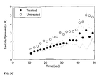
- 13 C-signal intensities from 13 C-pyruvate and 13 C-lactate in etoposide-treated EL4 cell suspensions as described in 6.1 were followed over a time period of 240 seconds from the time of addition of the imaging medium.
- One 13 C spectrum per second was acquired using low flip angle pulses at 9.4 T for a total of 240 spectra.
- a control of non-etoposide treated (untreated) EL4 lymphoma cells was also examined, as outlined above, and the peak intensities of 13 C-pyruvate and 13 C-lactate from the untreated and the etoposide were plotted on a graph ( FIG 1 ).
- 13 C-signal intensities from 13 C-pyruvate and 13 C-lactate in etoposide-treated and etoposide/nicotinamide treated EL4 cell suspensions as described in 6.1 were followed over a time period of 240 seconds from the time of addition of the imaging medium.
- One 13 C spectrum per second was acquired using low flip angle pulses at 9.4 T for a total of 240 spectra.
- a control of non-etoposide treated (untreated) EL4 lymphoma cells was also examined, as outlined above, and the peak intensities of 13 C-pyruvate and 13 C-lactate from the untreated, the etoposide treated and the etoposide/nicotinamide treated EL4 cells were compared.
- the data were fit to a two-site exchange model based on the modified Bloch equations, and the rate constants for the forward and reverse exchange 13 C-fluxes were determined. Bar graphs in FIG 2 represent 3 experiments +/- standard deviation.
- Example 7 Preparation of an imaging medium comprising hyperpolarised 13 C 1 -pyruvate
- a 15 mM solution was prepared by dissolving the DNP agent (trityl radical) of Example 1 in 13 C 1 -pyruvic acid (44 mg, 91%). The sample was mixed to homogeneity and the solution was placed in a sample cup and inserted in the DNP polariser.
- the sample was polarised under DNP conditions at 1.2 K in a 3.35 T magnetic field under irradiation with microwave (94 GHz and 100 mW, respectively). Polarisation was followed by solid state NMR. After 90 min hyperpolarisation, the sample was dissolved in 6ml of an aqueous solution of 94 mM NaOH, 30 mM NaCl, 40 mM HEPES and 50 mg/litre EDTA. The pH of the dissolved sample was 7.4 with a final 13 C 1 -pyruvate concentration of 75mM.
- Example 8 Detection of cell death in vivo
- Lymphoma tumours were produced in mice by subcutaneous implantation of EL4 cells. The mice were treated with an i.p. injection of 67mg/kg etoposide. Tumour cell death following etoposide treatment was assessed histologically. Non-etoposide treated mice were used as control.
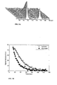
- Intravenous injection of hyperpolarized 13 C 1 -pyruvate of Example 7 resulted in readily measurable signals from 13 C-pyruvate and lactate in the tumour ( FIG. 3A )
- Spectra were collected every 2 s for two min starting from 12 s after the beginning of the injection.
- the time-dependent changes in the tumour signals from lactate and pyruvate were fit to the exchange model to obtain the apparent rate constants, and spin lattice relaxation times ( FIG. 3B ).
- the first 10-15 s of data acquisition were lost since this was the time taken to complete the injection and insert the 13 C surface coil probe assembly, containing the animal, into the imaging magnet.
Landscapes
- Physics & Mathematics (AREA)
- Health & Medical Sciences (AREA)
- Life Sciences & Earth Sciences (AREA)
- High Energy & Nuclear Physics (AREA)
- General Health & Medical Sciences (AREA)
- Condensed Matter Physics & Semiconductors (AREA)
- General Physics & Mathematics (AREA)
- Nuclear Medicine, Radiotherapy & Molecular Imaging (AREA)
- Molecular Biology (AREA)
- Spectroscopy & Molecular Physics (AREA)
- Radiology & Medical Imaging (AREA)
- Engineering & Computer Science (AREA)
- Signal Processing (AREA)
- Biophysics (AREA)
- Pathology (AREA)
- Biomedical Technology (AREA)
- Heart & Thoracic Surgery (AREA)
- Medical Informatics (AREA)
- Surgery (AREA)
- Animal Behavior & Ethology (AREA)
- Public Health (AREA)
- Veterinary Medicine (AREA)
- Optics & Photonics (AREA)
- Medicines Containing Antibodies Or Antigens For Use As Internal Diagnostic Agents (AREA)
- Magnetic Resonance Imaging Apparatus (AREA)
- Other Investigation Or Analysis Of Materials By Electrical Means (AREA)
Priority Applications (1)
| Application Number | Priority Date | Filing Date | Title |
|---|---|---|---|
| PL07808605T PL2052273T3 (pl) | 2006-08-18 | 2007-08-17 | Obrazowanie lub spektroskopia metodą rezonansu magnetycznego 13C śmierci komórkowej |
Applications Claiming Priority (2)
| Application Number | Priority Date | Filing Date | Title |
|---|---|---|---|
| NO20063702 | 2006-08-18 | ||
| PCT/NO2007/000286 WO2008020764A1 (en) | 2006-08-18 | 2007-08-17 | 13c-mr imaging or spectroscopy of cell death |
Publications (2)
| Publication Number | Publication Date |
|---|---|
| EP2052273A1 EP2052273A1 (en) | 2009-04-29 |
| EP2052273B1 true EP2052273B1 (en) | 2012-03-14 |
Family
ID=38690525
Family Applications (1)
| Application Number | Title | Priority Date | Filing Date |
|---|---|---|---|
| EP07808605A Not-in-force EP2052273B1 (en) | 2006-08-18 | 2007-08-17 | 13c-mr imaging or spectroscopy of cell death |
Country Status (8)
| Country | Link |
|---|---|
| US (1) | US8951500B2 (pl) |
| EP (1) | EP2052273B1 (pl) |
| JP (1) | JP5363320B2 (pl) |
| CN (1) | CN101506679B (pl) |
| AT (1) | ATE549638T1 (pl) |
| ES (1) | ES2381380T3 (pl) |
| PL (1) | PL2052273T3 (pl) |
| WO (1) | WO2008020764A1 (pl) |
Cited By (1)
| Publication number | Priority date | Publication date | Assignee | Title |
|---|---|---|---|---|
| US10265421B2 (en) | 2013-01-31 | 2019-04-23 | Bracco Imaging S.P.A. | Hyperpolarized esters as metabolic markers in MR |
Families Citing this family (8)
| Publication number | Priority date | Publication date | Assignee | Title |
|---|---|---|---|---|
| JP2010501483A (ja) * | 2006-08-18 | 2010-01-21 | ジーイー・ヘルスケア・アクスイェ・セルスカプ | 乳酸塩及び過分極13c−ピルビン酸塩を含むイメージング剤 |
| WO2008073842A1 (en) * | 2006-12-08 | 2008-06-19 | Molecular Image Inc. | Methods for diagnosis and monitoring of neurologic diseases using magnetic resonance methods |
| GB0713074D0 (en) * | 2007-07-05 | 2007-08-15 | Univ London | A method of hyperpolarising a magnetic resonance agent |
| CN102388317B (zh) * | 2009-04-02 | 2015-11-25 | 通用电气健康护理有限公司 | 包含超极化13c丙酮酸盐的磁共振成像介质用于检测炎症或感染的用途 |
| WO2012027274A2 (en) * | 2010-08-23 | 2012-03-01 | The Regents Of The University Of California | Compositions and methods for imaging |
| US9452409B2 (en) | 2011-04-22 | 2016-09-27 | Vanderbilt University | Para-hydrogen polarizer |
| WO2015000838A1 (en) * | 2013-07-01 | 2015-01-08 | Bracco Imaging Spa | Hyperpolarized 1-13c-1,1-bis(acetoxy(methyl))-2,2'-cyclopropane as metabolic marker for mr |
| EP2863229A1 (en) | 2013-10-15 | 2015-04-22 | Technische Universität München | pH-biosensors based on compounds with pH-sensitive enolic groups for magnetic resonance imaging and spectroscopy and their uses |
Family Cites Families (5)
| Publication number | Priority date | Publication date | Assignee | Title |
|---|---|---|---|---|
| US4822816A (en) * | 1987-04-10 | 1989-04-18 | Oxycal Laboratories, Inc. | Compositions and methods for administering vitamin C |
| US6278893B1 (en) * | 1998-01-05 | 2001-08-21 | Nycomed Imaging As | Method of magnetic resonance imaging of a sample with ex vivo polarization of an MR imaging agent |
| CN1165301C (zh) * | 1999-08-30 | 2004-09-08 | 奥克斯可尔实验室公司 | 组合物在制备治疗癌症的药物中的用途 |
| ES2372058T3 (es) * | 2004-07-30 | 2012-01-13 | Ge Healthcare As | Procedimiento de visualización de imágenes rm para la discriminación entre tejido sano y tumoral. |
| ES2422558T3 (es) * | 2004-11-19 | 2013-09-12 | Ge Healthcare As | Metodo de obtención de imágenes cardiacas con el uso de 13C-piruvato hiperpolarizado |
-
2007
- 2007-08-17 JP JP2009524573A patent/JP5363320B2/ja not_active Expired - Fee Related
- 2007-08-17 ES ES07808605T patent/ES2381380T3/es active Active
- 2007-08-17 PL PL07808605T patent/PL2052273T3/pl unknown
- 2007-08-17 US US12/376,470 patent/US8951500B2/en active Active
- 2007-08-17 AT AT07808605T patent/ATE549638T1/de active
- 2007-08-17 EP EP07808605A patent/EP2052273B1/en not_active Not-in-force
- 2007-08-17 CN CN2007800309297A patent/CN101506679B/zh not_active Expired - Fee Related
- 2007-08-17 WO PCT/NO2007/000286 patent/WO2008020764A1/en not_active Ceased
Cited By (1)
| Publication number | Priority date | Publication date | Assignee | Title |
|---|---|---|---|---|
| US10265421B2 (en) | 2013-01-31 | 2019-04-23 | Bracco Imaging S.P.A. | Hyperpolarized esters as metabolic markers in MR |
Also Published As
| Publication number | Publication date |
|---|---|
| JP5363320B2 (ja) | 2013-12-11 |
| CN101506679B (zh) | 2013-03-06 |
| US8951500B2 (en) | 2015-02-10 |
| CN101506679A (zh) | 2009-08-12 |
| WO2008020764A1 (en) | 2008-02-21 |
| US20100226859A1 (en) | 2010-09-09 |
| PL2052273T3 (pl) | 2012-08-31 |
| ATE549638T1 (de) | 2012-03-15 |
| JP2010501482A (ja) | 2010-01-21 |
| ES2381380T3 (es) | 2012-05-25 |
| EP2052273A1 (en) | 2009-04-29 |
Similar Documents
| Publication | Publication Date | Title |
|---|---|---|
| EP2052273B1 (en) | 13c-mr imaging or spectroscopy of cell death | |
| EP1824523B1 (en) | Method of cardiac imaging with the use of hyperpolarized 13c-pyruvate | |
| US20100178249A1 (en) | Imaging medium comprising lactate and hyperpolarised 13c-pyruvate | |
| AU2010230330B2 (en) | Use of a magnetic resonance imaging medium comprising hyperpolarized 13C pyruvate for the detection of inflammation or infection | |
| AU2011250012B2 (en) | Hyperpolarized lactate contrast agent for determination of LDH activity | |
| EP2476009A1 (en) | 13c-mr detection using hyperpolarised 13c-fructose |
Legal Events
| Date | Code | Title | Description |
|---|---|---|---|
| PUAI | Public reference made under article 153(3) epc to a published international application that has entered the european phase |
Free format text: ORIGINAL CODE: 0009012 |
|
| 17P | Request for examination filed |
Effective date: 20090211 |
|
| AK | Designated contracting states |
Kind code of ref document: A1 Designated state(s): AT BE BG CH CY CZ DE DK EE ES FI FR GB GR HU IE IS IT LI LT LU LV MC MT NL PL PT RO SE SI SK TR |
|
| AX | Request for extension of the european patent |
Extension state: AL BA HR MK RS |
|
| 17Q | First examination report despatched |
Effective date: 20110310 |
|
| REG | Reference to a national code |
Ref country code: DE Ref legal event code: R079 Ref document number: 602007021342 Country of ref document: DE Free format text: PREVIOUS MAIN CLASS: G01R0033280000 Ipc: G01R0033465000 |
|
| GRAP | Despatch of communication of intention to grant a patent |
Free format text: ORIGINAL CODE: EPIDOSNIGR1 |
|
| RIC1 | Information provided on ipc code assigned before grant |
Ipc: G01R 33/465 20060101AFI20110914BHEP Ipc: G01R 33/56 20060101ALI20110914BHEP Ipc: A61B 5/055 20060101ALI20110914BHEP Ipc: G01R 33/483 20060101ALI20110914BHEP |
|
| DAX | Request for extension of the european patent (deleted) | ||
| GRAS | Grant fee paid |
Free format text: ORIGINAL CODE: EPIDOSNIGR3 |
|
| GRAA | (expected) grant |
Free format text: ORIGINAL CODE: 0009210 |
|
| AK | Designated contracting states |
Kind code of ref document: B1 Designated state(s): AT BE BG CH CY CZ DE DK EE ES FI FR GB GR HU IE IS IT LI LT LU LV MC MT NL PL PT RO SE SI SK TR |
|
| REG | Reference to a national code |
Ref country code: GB Ref legal event code: FG4D |
|
| REG | Reference to a national code |
Ref country code: AT Ref legal event code: REF Ref document number: 549638 Country of ref document: AT Kind code of ref document: T Effective date: 20120315 Ref country code: CH Ref legal event code: EP |
|
| REG | Reference to a national code |
Ref country code: CH Ref legal event code: NV Representative=s name: ISLER & PEDRAZZINI AG |
|
| REG | Reference to a national code |
Ref country code: IE Ref legal event code: FG4D |
|
| REG | Reference to a national code |
Ref country code: DE Ref legal event code: R096 Ref document number: 602007021342 Country of ref document: DE Effective date: 20120510 |
|
| REG | Reference to a national code |
Ref country code: ES Ref legal event code: FG2A Ref document number: 2381380 Country of ref document: ES Kind code of ref document: T3 Effective date: 20120525 |
|
| REG | Reference to a national code |
Ref country code: NL Ref legal event code: T3 |
|
| PG25 | Lapsed in a contracting state [announced via postgrant information from national office to epo] |
Ref country code: LT Free format text: LAPSE BECAUSE OF FAILURE TO SUBMIT A TRANSLATION OF THE DESCRIPTION OR TO PAY THE FEE WITHIN THE PRESCRIBED TIME-LIMIT Effective date: 20120314 |
|
| LTIE | Lt: invalidation of european patent or patent extension |
Effective date: 20120314 |
|
| PG25 | Lapsed in a contracting state [announced via postgrant information from national office to epo] |
Ref country code: LV Free format text: LAPSE BECAUSE OF FAILURE TO SUBMIT A TRANSLATION OF THE DESCRIPTION OR TO PAY THE FEE WITHIN THE PRESCRIBED TIME-LIMIT Effective date: 20120314 Ref country code: FI Free format text: LAPSE BECAUSE OF FAILURE TO SUBMIT A TRANSLATION OF THE DESCRIPTION OR TO PAY THE FEE WITHIN THE PRESCRIBED TIME-LIMIT Effective date: 20120314 Ref country code: GR Free format text: LAPSE BECAUSE OF FAILURE TO SUBMIT A TRANSLATION OF THE DESCRIPTION OR TO PAY THE FEE WITHIN THE PRESCRIBED TIME-LIMIT Effective date: 20120615 |
|
| REG | Reference to a national code |
Ref country code: PL Ref legal event code: T3 |
|
| REG | Reference to a national code |
Ref country code: AT Ref legal event code: MK05 Ref document number: 549638 Country of ref document: AT Kind code of ref document: T Effective date: 20120314 |
|
| PG25 | Lapsed in a contracting state [announced via postgrant information from national office to epo] |
Ref country code: CY Free format text: LAPSE BECAUSE OF FAILURE TO SUBMIT A TRANSLATION OF THE DESCRIPTION OR TO PAY THE FEE WITHIN THE PRESCRIBED TIME-LIMIT Effective date: 20120314 |
|
| PG25 | Lapsed in a contracting state [announced via postgrant information from national office to epo] |
Ref country code: SI Free format text: LAPSE BECAUSE OF FAILURE TO SUBMIT A TRANSLATION OF THE DESCRIPTION OR TO PAY THE FEE WITHIN THE PRESCRIBED TIME-LIMIT Effective date: 20120314 Ref country code: SE Free format text: LAPSE BECAUSE OF FAILURE TO SUBMIT A TRANSLATION OF THE DESCRIPTION OR TO PAY THE FEE WITHIN THE PRESCRIBED TIME-LIMIT Effective date: 20120314 Ref country code: RO Free format text: LAPSE BECAUSE OF FAILURE TO SUBMIT A TRANSLATION OF THE DESCRIPTION OR TO PAY THE FEE WITHIN THE PRESCRIBED TIME-LIMIT Effective date: 20120314 Ref country code: IS Free format text: LAPSE BECAUSE OF FAILURE TO SUBMIT A TRANSLATION OF THE DESCRIPTION OR TO PAY THE FEE WITHIN THE PRESCRIBED TIME-LIMIT Effective date: 20120714 Ref country code: EE Free format text: LAPSE BECAUSE OF FAILURE TO SUBMIT A TRANSLATION OF THE DESCRIPTION OR TO PAY THE FEE WITHIN THE PRESCRIBED TIME-LIMIT Effective date: 20120314 Ref country code: CZ Free format text: LAPSE BECAUSE OF FAILURE TO SUBMIT A TRANSLATION OF THE DESCRIPTION OR TO PAY THE FEE WITHIN THE PRESCRIBED TIME-LIMIT Effective date: 20120314 Ref country code: BE Free format text: LAPSE BECAUSE OF FAILURE TO SUBMIT A TRANSLATION OF THE DESCRIPTION OR TO PAY THE FEE WITHIN THE PRESCRIBED TIME-LIMIT Effective date: 20120314 |
|
| PG25 | Lapsed in a contracting state [announced via postgrant information from national office to epo] |
Ref country code: SK Free format text: LAPSE BECAUSE OF FAILURE TO SUBMIT A TRANSLATION OF THE DESCRIPTION OR TO PAY THE FEE WITHIN THE PRESCRIBED TIME-LIMIT Effective date: 20120314 Ref country code: PT Free format text: LAPSE BECAUSE OF FAILURE TO SUBMIT A TRANSLATION OF THE DESCRIPTION OR TO PAY THE FEE WITHIN THE PRESCRIBED TIME-LIMIT Effective date: 20120716 |
|
| PLBE | No opposition filed within time limit |
Free format text: ORIGINAL CODE: 0009261 |
|
| STAA | Information on the status of an ep patent application or granted ep patent |
Free format text: STATUS: NO OPPOSITION FILED WITHIN TIME LIMIT |
|
| PG25 | Lapsed in a contracting state [announced via postgrant information from national office to epo] |
Ref country code: AT Free format text: LAPSE BECAUSE OF FAILURE TO SUBMIT A TRANSLATION OF THE DESCRIPTION OR TO PAY THE FEE WITHIN THE PRESCRIBED TIME-LIMIT Effective date: 20120314 Ref country code: DK Free format text: LAPSE BECAUSE OF FAILURE TO SUBMIT A TRANSLATION OF THE DESCRIPTION OR TO PAY THE FEE WITHIN THE PRESCRIBED TIME-LIMIT Effective date: 20120314 |
|
| 26N | No opposition filed |
Effective date: 20121217 |
|
| PG25 | Lapsed in a contracting state [announced via postgrant information from national office to epo] |
Ref country code: MC Free format text: LAPSE BECAUSE OF NON-PAYMENT OF DUE FEES Effective date: 20120831 |
|
| REG | Reference to a national code |
Ref country code: DE Ref legal event code: R097 Ref document number: 602007021342 Country of ref document: DE Effective date: 20121217 |
|
| PG25 | Lapsed in a contracting state [announced via postgrant information from national office to epo] |
Ref country code: IE Free format text: LAPSE BECAUSE OF NON-PAYMENT OF DUE FEES Effective date: 20120817 Ref country code: BG Free format text: LAPSE BECAUSE OF FAILURE TO SUBMIT A TRANSLATION OF THE DESCRIPTION OR TO PAY THE FEE WITHIN THE PRESCRIBED TIME-LIMIT Effective date: 20120614 |
|
| PG25 | Lapsed in a contracting state [announced via postgrant information from national office to epo] |
Ref country code: MT Free format text: LAPSE BECAUSE OF FAILURE TO SUBMIT A TRANSLATION OF THE DESCRIPTION OR TO PAY THE FEE WITHIN THE PRESCRIBED TIME-LIMIT Effective date: 20120314 |
|
| PG25 | Lapsed in a contracting state [announced via postgrant information from national office to epo] |
Ref country code: TR Free format text: LAPSE BECAUSE OF FAILURE TO SUBMIT A TRANSLATION OF THE DESCRIPTION OR TO PAY THE FEE WITHIN THE PRESCRIBED TIME-LIMIT Effective date: 20120314 |
|
| PG25 | Lapsed in a contracting state [announced via postgrant information from national office to epo] |
Ref country code: LU Free format text: LAPSE BECAUSE OF NON-PAYMENT OF DUE FEES Effective date: 20120817 |
|
| PG25 | Lapsed in a contracting state [announced via postgrant information from national office to epo] |
Ref country code: HU Free format text: LAPSE BECAUSE OF FAILURE TO SUBMIT A TRANSLATION OF THE DESCRIPTION OR TO PAY THE FEE WITHIN THE PRESCRIBED TIME-LIMIT Effective date: 20070817 |
|
| PGFP | Annual fee paid to national office [announced via postgrant information from national office to epo] |
Ref country code: IT Payment date: 20150825 Year of fee payment: 9 |
|
| REG | Reference to a national code |
Ref country code: FR Ref legal event code: PLFP Year of fee payment: 10 |
|
| PGFP | Annual fee paid to national office [announced via postgrant information from national office to epo] |
Ref country code: NL Payment date: 20160826 Year of fee payment: 10 |
|
| REG | Reference to a national code |
Ref country code: DE Ref legal event code: R082 Ref document number: 602007021342 Country of ref document: DE |
|
| REG | Reference to a national code |
Ref country code: FR Ref legal event code: PLFP Year of fee payment: 11 |
|
| PG25 | Lapsed in a contracting state [announced via postgrant information from national office to epo] |
Ref country code: IT Free format text: LAPSE BECAUSE OF NON-PAYMENT OF DUE FEES Effective date: 20160817 |
|
| REG | Reference to a national code |
Ref country code: NL Ref legal event code: MM Effective date: 20170901 |
|
| PG25 | Lapsed in a contracting state [announced via postgrant information from national office to epo] |
Ref country code: NL Free format text: LAPSE BECAUSE OF NON-PAYMENT OF DUE FEES Effective date: 20170901 |
|
| REG | Reference to a national code |
Ref country code: FR Ref legal event code: PLFP Year of fee payment: 12 |
|
| PGFP | Annual fee paid to national office [announced via postgrant information from national office to epo] |
Ref country code: FR Payment date: 20180720 Year of fee payment: 12 Ref country code: DE Payment date: 20180719 Year of fee payment: 12 Ref country code: ES Payment date: 20180903 Year of fee payment: 12 |
|
| PGFP | Annual fee paid to national office [announced via postgrant information from national office to epo] |
Ref country code: PL Payment date: 20180720 Year of fee payment: 12 Ref country code: GB Payment date: 20180720 Year of fee payment: 12 Ref country code: CH Payment date: 20180726 Year of fee payment: 12 |
|
| REG | Reference to a national code |
Ref country code: DE Ref legal event code: R119 Ref document number: 602007021342 Country of ref document: DE |
|
| GBPC | Gb: european patent ceased through non-payment of renewal fee |
Effective date: 20190817 |
|
| PG25 | Lapsed in a contracting state [announced via postgrant information from national office to epo] |
Ref country code: CH Free format text: LAPSE BECAUSE OF NON-PAYMENT OF DUE FEES Effective date: 20190831 Ref country code: LI Free format text: LAPSE BECAUSE OF NON-PAYMENT OF DUE FEES Effective date: 20190831 |
|
| PG25 | Lapsed in a contracting state [announced via postgrant information from national office to epo] |
Ref country code: DE Free format text: LAPSE BECAUSE OF NON-PAYMENT OF DUE FEES Effective date: 20200303 Ref country code: FR Free format text: LAPSE BECAUSE OF NON-PAYMENT OF DUE FEES Effective date: 20190831 |
|
| PG25 | Lapsed in a contracting state [announced via postgrant information from national office to epo] |
Ref country code: GB Free format text: LAPSE BECAUSE OF NON-PAYMENT OF DUE FEES Effective date: 20190817 |
|
| REG | Reference to a national code |
Ref country code: ES Ref legal event code: FD2A Effective date: 20210107 |
|
| PG25 | Lapsed in a contracting state [announced via postgrant information from national office to epo] |
Ref country code: ES Free format text: LAPSE BECAUSE OF NON-PAYMENT OF DUE FEES Effective date: 20190818 |
|
| PG25 | Lapsed in a contracting state [announced via postgrant information from national office to epo] |
Ref country code: PL Free format text: LAPSE BECAUSE OF NON-PAYMENT OF DUE FEES Effective date: 20190817 |