EP2014256A1 - Matériau de réparation d'os composite - Google Patents
Matériau de réparation d'os composite Download PDFInfo
- Publication number
- EP2014256A1 EP2014256A1 EP07013645A EP07013645A EP2014256A1 EP 2014256 A1 EP2014256 A1 EP 2014256A1 EP 07013645 A EP07013645 A EP 07013645A EP 07013645 A EP07013645 A EP 07013645A EP 2014256 A1 EP2014256 A1 EP 2014256A1
- Authority
- EP
- European Patent Office
- Prior art keywords
- bone repair
- repair material
- composite bone
- scaffold
- bone
- Prior art date
- Legal status (The legal status is an assumption and is not a legal conclusion. Google has not performed a legal analysis and makes no representation as to the accuracy of the status listed.)
- Withdrawn
Links
Images


Classifications
-
- A—HUMAN NECESSITIES
- A61—MEDICAL OR VETERINARY SCIENCE; HYGIENE
- A61L—METHODS OR APPARATUS FOR STERILISING MATERIALS OR OBJECTS IN GENERAL; DISINFECTION, STERILISATION OR DEODORISATION OF AIR; CHEMICAL ASPECTS OF BANDAGES, DRESSINGS, ABSORBENT PADS OR SURGICAL ARTICLES; MATERIALS FOR BANDAGES, DRESSINGS, ABSORBENT PADS OR SURGICAL ARTICLES
- A61L27/00—Materials for grafts or prostheses or for coating grafts or prostheses
- A61L27/50—Materials characterised by their function or physical properties, e.g. injectable or lubricating compositions, shape-memory materials, surface modified materials
- A61L27/56—Porous materials, e.g. foams or sponges
-
- A—HUMAN NECESSITIES
- A61—MEDICAL OR VETERINARY SCIENCE; HYGIENE
- A61F—FILTERS IMPLANTABLE INTO BLOOD VESSELS; PROSTHESES; DEVICES PROVIDING PATENCY TO, OR PREVENTING COLLAPSING OF, TUBULAR STRUCTURES OF THE BODY, e.g. STENTS; ORTHOPAEDIC, NURSING OR CONTRACEPTIVE DEVICES; FOMENTATION; TREATMENT OR PROTECTION OF EYES OR EARS; BANDAGES, DRESSINGS OR ABSORBENT PADS; FIRST-AID KITS
- A61F2/00—Filters implantable into blood vessels; Prostheses, i.e. artificial substitutes or replacements for parts of the body; Appliances for connecting them with the body; Devices providing patency to, or preventing collapsing of, tubular structures of the body, e.g. stents
- A61F2/02—Prostheses implantable into the body
- A61F2/28—Bones
-
- A—HUMAN NECESSITIES
- A61—MEDICAL OR VETERINARY SCIENCE; HYGIENE
- A61L—METHODS OR APPARATUS FOR STERILISING MATERIALS OR OBJECTS IN GENERAL; DISINFECTION, STERILISATION OR DEODORISATION OF AIR; CHEMICAL ASPECTS OF BANDAGES, DRESSINGS, ABSORBENT PADS OR SURGICAL ARTICLES; MATERIALS FOR BANDAGES, DRESSINGS, ABSORBENT PADS OR SURGICAL ARTICLES
- A61L27/00—Materials for grafts or prostheses or for coating grafts or prostheses
- A61L27/40—Composite materials, i.e. containing one material dispersed in a matrix of the same or different material
- A61L27/42—Composite materials, i.e. containing one material dispersed in a matrix of the same or different material having an inorganic matrix
- A61L27/425—Composite materials, i.e. containing one material dispersed in a matrix of the same or different material having an inorganic matrix of phosphorus containing material, e.g. apatite
-
- A—HUMAN NECESSITIES
- A61—MEDICAL OR VETERINARY SCIENCE; HYGIENE
- A61L—METHODS OR APPARATUS FOR STERILISING MATERIALS OR OBJECTS IN GENERAL; DISINFECTION, STERILISATION OR DEODORISATION OF AIR; CHEMICAL ASPECTS OF BANDAGES, DRESSINGS, ABSORBENT PADS OR SURGICAL ARTICLES; MATERIALS FOR BANDAGES, DRESSINGS, ABSORBENT PADS OR SURGICAL ARTICLES
- A61L27/00—Materials for grafts or prostheses or for coating grafts or prostheses
- A61L27/50—Materials characterised by their function or physical properties, e.g. injectable or lubricating compositions, shape-memory materials, surface modified materials
- A61L27/52—Hydrogels or hydrocolloids
-
- A—HUMAN NECESSITIES
- A61—MEDICAL OR VETERINARY SCIENCE; HYGIENE
- A61L—METHODS OR APPARATUS FOR STERILISING MATERIALS OR OBJECTS IN GENERAL; DISINFECTION, STERILISATION OR DEODORISATION OF AIR; CHEMICAL ASPECTS OF BANDAGES, DRESSINGS, ABSORBENT PADS OR SURGICAL ARTICLES; MATERIALS FOR BANDAGES, DRESSINGS, ABSORBENT PADS OR SURGICAL ARTICLES
- A61L27/00—Materials for grafts or prostheses or for coating grafts or prostheses
- A61L27/50—Materials characterised by their function or physical properties, e.g. injectable or lubricating compositions, shape-memory materials, surface modified materials
- A61L27/54—Biologically active materials, e.g. therapeutic substances
-
- A—HUMAN NECESSITIES
- A61—MEDICAL OR VETERINARY SCIENCE; HYGIENE
- A61P—SPECIFIC THERAPEUTIC ACTIVITY OF CHEMICAL COMPOUNDS OR MEDICINAL PREPARATIONS
- A61P19/00—Drugs for skeletal disorders
-
- C—CHEMISTRY; METALLURGY
- C08—ORGANIC MACROMOLECULAR COMPOUNDS; THEIR PREPARATION OR CHEMICAL WORKING-UP; COMPOSITIONS BASED THEREON
- C08L—COMPOSITIONS OF MACROMOLECULAR COMPOUNDS
- C08L71/00—Compositions of polyethers obtained by reactions forming an ether link in the main chain; Compositions of derivatives of such polymers
- C08L71/02—Polyalkylene oxides
-
- A—HUMAN NECESSITIES
- A61—MEDICAL OR VETERINARY SCIENCE; HYGIENE
- A61F—FILTERS IMPLANTABLE INTO BLOOD VESSELS; PROSTHESES; DEVICES PROVIDING PATENCY TO, OR PREVENTING COLLAPSING OF, TUBULAR STRUCTURES OF THE BODY, e.g. STENTS; ORTHOPAEDIC, NURSING OR CONTRACEPTIVE DEVICES; FOMENTATION; TREATMENT OR PROTECTION OF EYES OR EARS; BANDAGES, DRESSINGS OR ABSORBENT PADS; FIRST-AID KITS
- A61F2/00—Filters implantable into blood vessels; Prostheses, i.e. artificial substitutes or replacements for parts of the body; Appliances for connecting them with the body; Devices providing patency to, or preventing collapsing of, tubular structures of the body, e.g. stents
- A61F2/02—Prostheses implantable into the body
- A61F2/28—Bones
- A61F2/2803—Bones for mandibular reconstruction
-
- A—HUMAN NECESSITIES
- A61—MEDICAL OR VETERINARY SCIENCE; HYGIENE
- A61F—FILTERS IMPLANTABLE INTO BLOOD VESSELS; PROSTHESES; DEVICES PROVIDING PATENCY TO, OR PREVENTING COLLAPSING OF, TUBULAR STRUCTURES OF THE BODY, e.g. STENTS; ORTHOPAEDIC, NURSING OR CONTRACEPTIVE DEVICES; FOMENTATION; TREATMENT OR PROTECTION OF EYES OR EARS; BANDAGES, DRESSINGS OR ABSORBENT PADS; FIRST-AID KITS
- A61F2/00—Filters implantable into blood vessels; Prostheses, i.e. artificial substitutes or replacements for parts of the body; Appliances for connecting them with the body; Devices providing patency to, or preventing collapsing of, tubular structures of the body, e.g. stents
- A61F2/02—Prostheses implantable into the body
- A61F2/30—Joints
- A61F2/3094—Designing or manufacturing processes
- A61F2/30942—Designing or manufacturing processes for designing or making customized prostheses, e.g. using templates, CT or NMR scans, finite-element analysis or CAD-CAM techniques
-
- A—HUMAN NECESSITIES
- A61—MEDICAL OR VETERINARY SCIENCE; HYGIENE
- A61F—FILTERS IMPLANTABLE INTO BLOOD VESSELS; PROSTHESES; DEVICES PROVIDING PATENCY TO, OR PREVENTING COLLAPSING OF, TUBULAR STRUCTURES OF THE BODY, e.g. STENTS; ORTHOPAEDIC, NURSING OR CONTRACEPTIVE DEVICES; FOMENTATION; TREATMENT OR PROTECTION OF EYES OR EARS; BANDAGES, DRESSINGS OR ABSORBENT PADS; FIRST-AID KITS
- A61F2/00—Filters implantable into blood vessels; Prostheses, i.e. artificial substitutes or replacements for parts of the body; Appliances for connecting them with the body; Devices providing patency to, or preventing collapsing of, tubular structures of the body, e.g. stents
- A61F2/02—Prostheses implantable into the body
- A61F2/28—Bones
- A61F2002/2835—Bone graft implants for filling a bony defect or an endoprosthesis cavity, e.g. by synthetic material or biological material
-
- A—HUMAN NECESSITIES
- A61—MEDICAL OR VETERINARY SCIENCE; HYGIENE
- A61F—FILTERS IMPLANTABLE INTO BLOOD VESSELS; PROSTHESES; DEVICES PROVIDING PATENCY TO, OR PREVENTING COLLAPSING OF, TUBULAR STRUCTURES OF THE BODY, e.g. STENTS; ORTHOPAEDIC, NURSING OR CONTRACEPTIVE DEVICES; FOMENTATION; TREATMENT OR PROTECTION OF EYES OR EARS; BANDAGES, DRESSINGS OR ABSORBENT PADS; FIRST-AID KITS
- A61F2/00—Filters implantable into blood vessels; Prostheses, i.e. artificial substitutes or replacements for parts of the body; Appliances for connecting them with the body; Devices providing patency to, or preventing collapsing of, tubular structures of the body, e.g. stents
- A61F2/02—Prostheses implantable into the body
- A61F2/30—Joints
- A61F2002/30001—Additional features of subject-matter classified in A61F2/28, A61F2/30 and subgroups thereof
- A61F2002/30003—Material related properties of the prosthesis or of a coating on the prosthesis
- A61F2002/30004—Material related properties of the prosthesis or of a coating on the prosthesis the prosthesis being made from materials having different values of a given property at different locations within the same prosthesis
- A61F2002/30011—Material related properties of the prosthesis or of a coating on the prosthesis the prosthesis being made from materials having different values of a given property at different locations within the same prosthesis differing in porosity
-
- A—HUMAN NECESSITIES
- A61—MEDICAL OR VETERINARY SCIENCE; HYGIENE
- A61F—FILTERS IMPLANTABLE INTO BLOOD VESSELS; PROSTHESES; DEVICES PROVIDING PATENCY TO, OR PREVENTING COLLAPSING OF, TUBULAR STRUCTURES OF THE BODY, e.g. STENTS; ORTHOPAEDIC, NURSING OR CONTRACEPTIVE DEVICES; FOMENTATION; TREATMENT OR PROTECTION OF EYES OR EARS; BANDAGES, DRESSINGS OR ABSORBENT PADS; FIRST-AID KITS
- A61F2250/00—Special features of prostheses classified in groups A61F2/00 - A61F2/26 or A61F2/82 or A61F9/00 or A61F11/00 or subgroups thereof
- A61F2250/0014—Special features of prostheses classified in groups A61F2/00 - A61F2/26 or A61F2/82 or A61F9/00 or A61F11/00 or subgroups thereof having different values of a given property or geometrical feature, e.g. mechanical property or material property, at different locations within the same prosthesis
- A61F2250/0023—Special features of prostheses classified in groups A61F2/00 - A61F2/26 or A61F2/82 or A61F9/00 or A61F11/00 or subgroups thereof having different values of a given property or geometrical feature, e.g. mechanical property or material property, at different locations within the same prosthesis differing in porosity
-
- A—HUMAN NECESSITIES
- A61—MEDICAL OR VETERINARY SCIENCE; HYGIENE
- A61F—FILTERS IMPLANTABLE INTO BLOOD VESSELS; PROSTHESES; DEVICES PROVIDING PATENCY TO, OR PREVENTING COLLAPSING OF, TUBULAR STRUCTURES OF THE BODY, e.g. STENTS; ORTHOPAEDIC, NURSING OR CONTRACEPTIVE DEVICES; FOMENTATION; TREATMENT OR PROTECTION OF EYES OR EARS; BANDAGES, DRESSINGS OR ABSORBENT PADS; FIRST-AID KITS
- A61F2310/00—Prostheses classified in A61F2/28 or A61F2/30 - A61F2/44 being constructed from or coated with a particular material
- A61F2310/00005—The prosthesis being constructed from a particular material
- A61F2310/00179—Ceramics or ceramic-like structures
- A61F2310/00293—Ceramics or ceramic-like structures containing a phosphorus-containing compound, e.g. apatite
-
- A—HUMAN NECESSITIES
- A61—MEDICAL OR VETERINARY SCIENCE; HYGIENE
- A61L—METHODS OR APPARATUS FOR STERILISING MATERIALS OR OBJECTS IN GENERAL; DISINFECTION, STERILISATION OR DEODORISATION OF AIR; CHEMICAL ASPECTS OF BANDAGES, DRESSINGS, ABSORBENT PADS OR SURGICAL ARTICLES; MATERIALS FOR BANDAGES, DRESSINGS, ABSORBENT PADS OR SURGICAL ARTICLES
- A61L2300/00—Biologically active materials used in bandages, wound dressings, absorbent pads or medical devices
- A61L2300/20—Biologically active materials used in bandages, wound dressings, absorbent pads or medical devices containing or releasing organic materials
- A61L2300/252—Polypeptides, proteins, e.g. glycoproteins, lipoproteins, cytokines
-
- A—HUMAN NECESSITIES
- A61—MEDICAL OR VETERINARY SCIENCE; HYGIENE
- A61L—METHODS OR APPARATUS FOR STERILISING MATERIALS OR OBJECTS IN GENERAL; DISINFECTION, STERILISATION OR DEODORISATION OF AIR; CHEMICAL ASPECTS OF BANDAGES, DRESSINGS, ABSORBENT PADS OR SURGICAL ARTICLES; MATERIALS FOR BANDAGES, DRESSINGS, ABSORBENT PADS OR SURGICAL ARTICLES
- A61L2300/00—Biologically active materials used in bandages, wound dressings, absorbent pads or medical devices
- A61L2300/40—Biologically active materials used in bandages, wound dressings, absorbent pads or medical devices characterised by a specific therapeutic activity or mode of action
- A61L2300/412—Tissue-regenerating or healing or proliferative agents
-
- A—HUMAN NECESSITIES
- A61—MEDICAL OR VETERINARY SCIENCE; HYGIENE
- A61L—METHODS OR APPARATUS FOR STERILISING MATERIALS OR OBJECTS IN GENERAL; DISINFECTION, STERILISATION OR DEODORISATION OF AIR; CHEMICAL ASPECTS OF BANDAGES, DRESSINGS, ABSORBENT PADS OR SURGICAL ARTICLES; MATERIALS FOR BANDAGES, DRESSINGS, ABSORBENT PADS OR SURGICAL ARTICLES
- A61L2300/00—Biologically active materials used in bandages, wound dressings, absorbent pads or medical devices
- A61L2300/40—Biologically active materials used in bandages, wound dressings, absorbent pads or medical devices characterised by a specific therapeutic activity or mode of action
- A61L2300/43—Hormones, e.g. dexamethasone
-
- A—HUMAN NECESSITIES
- A61—MEDICAL OR VETERINARY SCIENCE; HYGIENE
- A61L—METHODS OR APPARATUS FOR STERILISING MATERIALS OR OBJECTS IN GENERAL; DISINFECTION, STERILISATION OR DEODORISATION OF AIR; CHEMICAL ASPECTS OF BANDAGES, DRESSINGS, ABSORBENT PADS OR SURGICAL ARTICLES; MATERIALS FOR BANDAGES, DRESSINGS, ABSORBENT PADS OR SURGICAL ARTICLES
- A61L2430/00—Materials or treatment for tissue regeneration
- A61L2430/02—Materials or treatment for tissue regeneration for reconstruction of bones; weight-bearing implants
Definitions
- the present relates to a composite bone repair material comprising a porous block-shaped synthetic ceramic scaffold and a stabilizing polymer disposed therein.
- the repair of bone defects can be facilitated by placing a bone repair material as a temporary substitute in the defect site, where a loss of natural bone has occurred.
- the bone repair material is meant to selectively promote and guide the regeneration of natural bone structures.
- Naturally-derived materials include grafts made from bones.
- the bone may be harvested directly from the patient, as in autograft-based procedures, or it may be harvested from a suitable donor, surrogate, or cadaver, as in allograft- or xenograft-based procedures.
- Naturally-derived bone repair materials are usually prepared by acid extraction of most of the mineralized component to result in so called demineralized bone matrix (DBM).
- DBM demineralized bone matrix
- Examples for naturally-derived materials are Bio-Oss ® of the mineral portion of bovine bone or Algipore ® a porous calcium phosphate material of algae.
- Autologous bone is an ideal source of graft material, not only due to its biocompatibility, but also because natural bone grafts facilitate reossification of the defect site by promoting or conducting ingrowth of the patient's own bone tissue to the defect site.
- Autologous bone material inherently is osteoconductive and osteoinductive, two properties facilitating regeneration of natural bone structure.
- autograft bone implant procedures are costly and cause additional discomfort for the patient, as they typically require an additional surgery for harvesting the graft material, which may cause significant morbidity at the donor site.
- Autografts may also show pronounced resorption making the outcome of the augmentation unpredictable. Allogenic bone repair materials also unify osteoconductive and osteoinductive properties, but their origin raises possible pathogenic transfers and ethical issues. Similar concerns are brought up against xenogenic graft materials.
- naturally-derived bone repair materials may be replaced by a completely synthetic bone repair material, which contains no organic residues.
- synthetic bone repair materials are often less osteoconductive and hardly osteoinductive. Nevertheless, much research has been and still is directed toward improved synthetic bone repair materials.
- HA hydroxyapatite
- TCP tricalcium phosphate
- synthetic bone repair materials on a hydroxyapatite (HA) and/or tricalcium phosphate (TCP) basis are widely used.
- they may be applied as granules or pre-fabricated blocks.
- US 6,511,510 relates to a porous ceramic material from calcium phosphates obtained by a sintering process.
- the use of granular material allows treatment of a wide range of indications.
- the ceramic block material is processed by steps such as rubbing, pounding and sieving afterwards ( WO 04/054633 ).
- a prosthetic bone implant is made of a hardened calcium phosphate cement having an apatitic phase as a major phase, which comprises a dense cortical portion bearing the majority of load, and a porous cancellous portion allowing a rapid blood/body fluid penetration and tissue ingrowth.
- EP 1457214 discloses a block shape organic-inorganic complex porous article with a superposed skin layer made of a degradable polymer with improved strength. The complex is mainly designed to be inserted between vertebral bodies.
- EP 1374922 discloses a bioresorbable structure for use in the repair of bone defects comprising a porous bioceramic matrix of hydroxyapatite or tricalcium phosphate and a polymer disposed by compression moulding therein.
- WO 97/34546 describes a ceramic block with a plurality of channels filled containing an enforcing bio-resorbant polymer material.
- bone repair materials have been supplemented with bone growth inducing agents.
- US 10/271,140 US2003/0143258A1
- suggests a composite comprising demineralized bone matrix mixed with a stabilizing biodegradable polymer and a bone growth factor.
- a bone repair material in periodontal surgery requires formulations that can be easily shaped to size and shape of the defect.
- WO 2004/011053 suggests a formulation with a putty consistency.
- EP1490123 describes a kneadable and pliable bone replacement material on a granular calcium phosphate and hydrogel basis. When applied to the defect site, the formulation remains adhered thereto without migration or excessive expansion.
- the problem of the present invention is therefore, to provide a bone repair material having osteoconductive and osteoinductive properties and which is easy to handle and suitable for treatment of large oral bone defects.
- the composite bone repair material according to the present invention emulates the osteoconductive and the osteoinductive properties of autografts. Further, due to the combination of the porous block-shaped synthetic ceramic scaffold and the stabilizing polymer disposed therein, the composite has sufficient stability to prevent movement of the graft material and that it is strong enough to withstand the forces within the implantation side, i.e. is resistant to mechanical stress. In addition, the material is not brittle and therefore sliceable. This means that the surgeon may bring the bone repair material into the desired shape by cutting it with the scalpel or process it with a bur.
- the bone repair material according to the present invention thus can be used in the treatment of large bone defects, such as critical size defects in oral indications that do not heal spontaneously. More particularly, the bone repair material of the invention is especially preferred upon enhancing treatment of oral bone defects such as bone loss from moderate or severe periodontitis, bony defects of the alveolar ridge, tooth extraction sites, or pneumatized (expanded) sinus.
- the composite bone repair material according to the present invention comprises a porous block-shaped scaffold and a stabilizing polymer disposed therein.
- the porous block-shaped scaffold can be a synthetic ceramic material or a naturally-derived material.
- said porous block-shaped scaffold the synthetic ceramic material comprises calcium phosphate.
- the synthetic ceramic material comprises a calcium phosphate selected from the group consisting of apatite and tricalcium phosphate or a mixture thereof. Further said ceramic scaffold comprises interconnected macropores.
- Ceramic scaffold material composed of calcium phosphates, namely apatite and tricalcium phosphate (TCP) or combinations thereof, are efficient bone substitutes that enhance bone ingrowth. Eventually, the material gets resorbed and substituted by bone. Hydroxyapatite and ⁇ -tricalcium phosphate, and combinations thereof are especially preferred. These materials can be manufactured with well defined reproducible morphologies with respect to size and porosity (see Figure 1 )
- the scaffold material according to the present invention has a porous morphology.
- Said ceramic scaffold material is a highly porous calcium phosphate with interconnected pores of a size range that allows fast ingrowth of natural bone. Methods to characterize calcium phosphate blocks with regard to the porosity have been described in Biomaterials, 2005 Nov;26(31):6099-105 .
- the total porosity lies in the range of 75 to 95%, preferably from 80 to 95%.
- Porosity is the percentage of void space per volume unit of scaffold material.
- High porosity usually results in a large specific surface density, which is one important property increasing primary liquid absorption and protein adsorption throughout the whole material.
- Specific surface density is defined as the scaffold surface per scaffold volume.
- the preferred scaffold material according to this invention has a specific surface density of at least 20/mm, more preferably above 30/mm.
- the preferred ceramic scaffold material facilitates optimum nutrient and oxygen supply, neo-vascularisation, cell immigration, colonization and bone deposition. Finally, the material will be integrated in newly formed bone and will eventually be degraded and replaced by natural bone.
- the porous structure may be obtained by various processes. Usually a ceramic powder is suspended in an aqueous solution to result in a slurry. To form a porous structure, a pore forming agent may be added. Alternatively a sponge-like polymeric matrix with a determined pore structure or spherical objects are coated with the slurry. After drying the slurry, the ceramic material undergoes a sintering process at high temperatures between 800°C and 1300°C, depending on the degree of cristallinity desired. During sintering the pore forming material is burned out and a porous ceramic scaffold remains ( Fig.1 ).
- the porosity of the ceramic block-shaped material can be adjusted to result in a desired distribution interconnectivity of pores of various sizes. They can be classified as nanopores (diameter below 1 ⁇ m), micropores (diameter between 1 and 100 ⁇ m) and macropores (diameter above 100 ⁇ m). For the purpose of tissue regeneration, a substantial amount of interconnected micropores and macropores is desired in order to allow cells to migrate into the scaffold material. Micropores are sufficient to allow nutrient and metabolic product transport. In a preferred embodiment of the invention, the diameter of the pores lies between 0.05 and 750 ⁇ m. More preferably, the diameter of the micropores is between 5 and 100 ⁇ m and the diameter of the macropores is between 100 and 1000 ⁇ m.
- the diameter of the micropores is between 10 and 70 ⁇ m and the diameter of the macropores is between 100 to 750 ⁇ m.
- the porosity of the preferred scaffold material according to this invention has mean pore diameter between 300 and 600 ⁇ m.
- the preferred embodiment further has highly interconnected pores.
- the interconnectivity can be defined as connective density (equivalent to the terms connectivity or interconnectedness) as described in Bone, 1993 Mar-Apr;14(2):173-82 .
- the scaffold material according to this invention has a connectivity, which is above 20 per mm 3 .
- connections per pore which is equal to the ratio of interconnectedness and the number of pores per volume
- the scaffold material according to this invention has a connectivity per pore, which at least 2, more preferably above 3.
- the porosity does not need to be of random distribution, but may be obtained by a highly repeated spacing structure such as tubuli.
- a tubular structure with a suitable stabilizing polymer may be preferred, if high mechanical strength is required.
- a suitable architecture of the block-shaped ceramic scaffold material may further enhance bone regeneration and improve the handling properties.
- a first portion of the block oriented to the remaining bone, which needs to be augmented, preferably has a cancellous structure with a high proportion of macropores, thereby facilitating the integration into bone tissue.
- a second portion of the block-shaped ceramic scaffold material oriented to the surrounding soft tissue preferably has dense structure in order to reduce the risk of soft tissue ingrowth into area of bone augmentation. Therefore, the ceramic scaffold material subject to this invention preferably is manufactured to contain a gradient in its porosity and/or crystallinity and/or ceramic composition
- the second portion of the block-shaped ceramic scaffold material preferably has also an enhanced mechanical strength similar to the cortical portion of a natural bone, resisting pressures of up to about 110-170 MPa, and is sufficiently rigid to be fixed by screws.
- the ceramic scaffold material has at least one rigid layer on the surface portion, which is obtained by dipping the portion into desired second slurry of ceramic bone repair material.
- the peripheral portion can comprise one or several preformed fixation holes.
- the ceramic scaffold material according to the present invention is block-shaped and can be applied to any large bone defect and which has superior handling properties.
- the composite bone repair material of this invention is based on a ceramic scaffold material in pre-manufactured block shape.
- Block-shaped shall mean, that the ceramic scaffold material is based on a solid body, which exceeds the dimensions of conventional granular bone repair material for oral applications and is designed to substantially fill a bony defect.
- Block-shaped shall encompass any dimensions and shapes desired by a practitioner to treat a bony defect. Due to the added or embedded stabilizing polymer, the composite bone repair material can be adjusted to the individual defect size and shape with a scalpel or with dental burs during surgery.
- the composite bone repair material according to the present invention is sliceable, and in contrast to the materials known in the art not brittle, which is an enormous advantage.
- dimensions up to a volume of 10 cm 3 preferably between 0.1 and 4 cm 3 , typically about 6x6x12 mm have proven to be suitable for most defects.
- several units of block-shaped composite bone repair material can be used in a kit in a building block system with differently sized blocks.
- the bone repair material according to the present invention comprises a stabilizing polymer.
- Said stabilizing polymer may be naturally-derived or synthetically produced.
- the polymer is formed of proteins, preferably proteins naturally present in the patient, into which the composite mesh is to be implanted.
- a particularly preferred natural polymeric protein is fibrin, although polymers made from other proteins, such as collagen and gelatin can also be used.
- Polysaccharides as hyaluronic acid or glycoproteins may also be used to form the polymeric matrix.
- Suitable synthetic polymers include polyoxyalkylenes, poly(vinyl alcohol), poly(ethylene-co-vinyl alcohol), poly(urethane)s, poly(hydroxyalkyl acrylate)s, poly(hydroxyalkyl methacrylate)s, poly(acrylic acid), poly(methacrylic acid), poly(ethylene-co-acrylic acid), poly(alkyloxazoline)s, poly(vinyl pyrrolidone), poly(ethylene-co-vinyl pyrrolidone), poly(maleic acid), poly(ethylene-co-maleic acid), poly(acrylamide), and poly(ethylene oxide)-co- (propylene oxide) block copolymers.
- a particularly preferred polymer or precursor substance is linear or branched polyethylene glycol. It could be shown that ceramic scaffold material according to the present invention which is soaked with a linear polyethylene glycol (PEG) polymers of a suitable molecular weight that it has a waxy consistiency, results in a composite with excellent handling properties.
- PEG polyethylene glycol
- the concentration of a linear PEG is adjusted to obtain the desired consistency. When using short linear PEG molecules of a molecular weight of about 1 kDa, a concentration of up to 100% may be required. For larger linear PEG molecules of a moleculare weight up to 1000 kDa PEG, a aqeous solution of 10% may be sufficient.
- the bone scaffold material is no longer brittle but has a malleable consistency and can be shaped with a scalpel, which is appreciated by the practitioner for fitting the block to the bone defect site. Further it could be shown, that a water-swollen, crosslinked PEG matrix (PEG hydrogel) further improves the mechanical properties of the bone scaffold material and is easier to apply.
- the bone scaffold material is soaked with precursor substances of the stabilizing polymer prior to the polymerization reaction. The polymerization within the porous structure of the bone scaffold material then forms a composite mesh of the two materials.
- the mechanism leading to a polymeric network can be ionical, covalent, or any combination thereof, or swelling of one or more polymeric material(s), or physical crosslinks, e.g. by crosslinking points formed through aggregation of endblocks through phase or solubility differences.
- the preferred stabilizing polymers according to this invention are crosslinked polyethylene glycols (PEG) hydrogels formed by a self selective addition reaction between two precursors as described in EP 1 609 491 .
- PEG polyethylene glycols
- PEG hydrogels in a composite bone repair material subject to this invention has many advantages.
- PEG hydrogels are well known for their excellent biocompatibility and their hydrophilicity. Such hydrogels are permeable for aqueous biologicals fluids and therefore allow diffusion of nutrients required in tissue regeneration.
- the hydrogels preferred as stabilizing polymer in this invention are based on the base catalyzed Michael type addition between the conjugated unsaturated group or the conjugated unsaturated bond of a first precursor A and the thiol group of a second precursor B.
- the resulting linkage is unstable and hydrolyzed in contact with water.
- the rate of the hydrolysis reaction depends on the temperature and the value of the pH, which is 7.4 in most tissues.
- the crosslinked network degrades or breaks down. Therefore, the time of degradation of the network can be influenced by the number of hydrolysable bonds present per unit of volume.
- the precursors forming the stabilizing polymer are dissolved or suspended in aqueous solutions. Since no organic solvents are necessary, only aqueous solutions and/or suspensions are present. These are easy to handle and do not require any laborious precautions as might be the case if organic solvents were present. Furthermore, organic solvents are an additional risk for the health of the staff and the patients exposed to these solvents. The present invention eliminates said risk.
- the gelation of the stabilizing polymer is completed within minutes, starting at the time of mixing.
- the first precursor A comprises a core which carries n chains with a conjugated unsaturated group or a conjugated unsaturated bond attached to any of the last 20 atoms of the chain.
- said conjugated unsaturated group or conjugated unsaturated bond is terminal.
- the core of the first precursor A can be a single atom such as a carbon or a nitrogen atom or a small molecule such as an ethylene oxide unit, an amino acid or a peptide, a sugar, a multifunctional alcohol, such as pentaerythritol, D-sorbitol, glycerol or oligoglycerol, such as hexaglycerol.
- the chains are linear polymers or linear or branched alkyl chains optionally comprising heteroatoms, amide groups or ester groups.
- the chain is a polyethylene glycol.
- the core of precursor A may be additionally substituted with linear or branched alkyl residues or polymers which have no conjugated unsaturated groups or bonds.
- the first precursor A has 2 to 10 chains, preferably 2 to 8, more preferably 2 to 6, most preferably 3 to 6 chains.
- the conjugated unsaturated bonds are preferably acrylates, acrylamides, quinines, 2- or 4-vinylpyridiniums, vinylsulfone, maleimide or itaconate esters of formula la or Ib
- R 1 and R 2 are independently hydrogen, methyl, ethyl, propyl or butyl
- R 3 is a linear or branched C 1 to C 10 hydrocarbon chain, preferably methyl, ethyl, propyl or butyl.
- the precursor A is a PEG-acrylate with 2 to 6 chains (2-arm to 6-arm PEG-acrylate).
- the second precursor B comprises a core carrying m chains each having a thiol or an amine group attached to any of the last 20 atoms at the end of the chain.
- a cysteine residue may be incorporated into the chain.
- the thiol group is terminal.
- the core of the second precursor B can be a single atom such as a carbon or a nitrogen atom or a small molecule such as an ethylene oxide unit, an amino acid or a peptide, a sugar, a multifunctional alcohol, such as pentaerythritol, D-sorbitol, glycerol or oligoglycerol, such as hexaglycerol.
- the chains are linear polymers or linear or branched alkyl chains optionally comprising heteroatoms, esters groups or amide groups.
- the chain is a polyethylene glycol.
- the second precursor B has 2 to 10 chains, preferably 2 to 8, more preferably 2 to 6, most preferably 2 to 4 chains.
- the precursor B is a PEG-thiol with 2 to 4 chains (2-arm to 4-arm PEG-thiol).
- the first precursor A compound has n chains, whereby n is greater than or equal to 2, and the second precursor B compound has m chains, whereby m is greater than or equal to 2.
- the first precursor A and/or the second precursor B may comprise further chains which are not functionalized.
- the sum of the functionalized chains of the first and the second precursor, that means m+n, is greater than or equal to 5.
- the sum of m+n is equal to or greater than 6 to obtain a well formed three-dimensional network.
- Such molecules having a core and two or more end groups are also referred to as multi-arm polymers.
- the number of atoms in the backbone connecting two adjacent crosslinking points is at least about 20 atoms, preferably between 50 and 5000 atoms and more preferably between about 50 and 2000 and ideally between 100 and 750 atoms.
- a crosslinking point is here defined as a point in which 3 or more backbone chains of the polymer network are connected.
- the mechanical strength of the bone composite material can be further enhanced by embedding one or more additional stabilizing polymers, fibrous or filamentous supplements such as carboxy methyl cellulose, alginates, xanthan gum etc..
- the stabilizing polymer is provided with a degradability by enzymatic degradation sites.
- An accordingly designed polymer will not degrade and lose its stabilizing function, unless ingrowing cells are present to replace the synthetic structure of the polymer.
- the core of precursor B comprises a peptide which comprises one or more enzymatic degradation sites.
- Preferred enzymatic degradable hydrogels contain metalloproteinase oligopeptides integrated in their backbone instead of a hydrolytically instable bond as described in detail in WO03040235A1 .
- Such a stabilizing polymer such as PEG hydrogel is preferably introduced into the pores of the bone block by soaking the block-shaped synthetic ceramic scaffold with a PEG hydrogel formulation at room temperature.
- a PEG hydrogel formulation at room temperature.
- One possibility to do this is to mix the hydrogel precursors and then, before the gel point is reached, apply the mixture onto the block and allow it to be absorbed by the block and gel inside the pores of the block. This procedure can be performed by the surgeon before adapting the block to the desired shape.
- the stabilizing polymer disposed in the synthetic ceramic scaffold is at the same time a matrix for sustained release of one or several bioactive agents, which promote the osteoconductive and/or osteoinductive properties of the composite bone repair material.
- a bioactive agent is not limited by its origin or the way it is produced and therefore can be extracted, synthetically or recombinantly produced and may have been subject to further processing or purification, such as but not limited to, splicing, fragmentation, enzymatic cleavage or chemical modification.
- suitable biologically active agents are BMPs, PTH, VEGF, Enamel Matrix Derivatives (EMD), TGF-beta, IGF, Dentonin, Adrenomedullin (ADM), FGF, PDGFBB, IGF, PGE2, EP2, L1 (and derivatives), HIF-1 ⁇ ODD (oxygen-independent domain), cell recognition sequences such as RGD, KRSR, H-Gly-Cys-Gly-Arg-Gly-Asp-Ser-Pro-Gly-NH 2 or derivatives thereof.
- extracellular matrix proteins such as fibronectin, collagen, laminin may be used as bioactive agents.
- These peptides and proteins may or may not comprise additional cystein.
- cystein facilitates the covalent attachment of the peptides and proteins to the preferred form of stabilizing polymer as described above.
- a peptide comprising the first 34 amino acids of PTH.
- This peptide may contain an additional cystein, which facilitates the covalent attachment of the peptide to the composite bone repair material.
- the bioactive agent is selected from the group of EMDs consisting of amelogenin, amelin, tuftelin, ameloblastin, enamelin and dentin.
- the preferred stabilizing polymers previously described are also suitable for delivery or of bioactive agents.
- the bioactive agent may be covalently bound to the stabilizing polymer, e.g., this can be achieved by a thiol moiety present in the bioactive agent which reacts with the conjugated unsaturated group or bond present in precursor A upon mixing.
- a thiol moiety is present, e.g. in the amino acid cystein. This amino acid can easily be introduced in peptides, oligopeptides or proteins.
- the bioactive agent is subsequently released from the stabilizing polymer as the unstable linkage hydrolyzes.
- the preferred embodiments of the stabilizing polymer described above allow the active agents to be simply entrapped or precipitated into the composite bone repair material.
- the bioactive agent can be added when mixing the other components of the composition.
- the bioactive agent is then released by diffusion after degradation of the hydrogel. It is also possible to adsorb the bioactive agent on the ceramic scaffold material prior to the soaking with the solutions comprising the first precursor A and the second precursor B.
- Kits also fall within the scope of the present invention.
- the kit comprises at least (i) a block-shaped ceramic scaffold and (ii) a stabilizing polymer.
- the kit comprises at least (i) a block-shaped ceramic scaffold, (II) a precursor A, such as a multi-arm PEG-acrylat, and (III) a precursor B, such as a multi-arm PEG-thiol, which are each individually stored.
- Another kit comprises (I) a block-shaped ceramic scaffold, (II) a stabilizing polymer, and (III) a bioactive agent.
- the kit further comprises one or several if required by the precursors and/or the bioactive agent.
- a suitable activator would be an aqueous solution of triethanolamine with HCl at pH of 7.4 - 9.0.
- the kit may also comprise more than one bioactive agent and more than two precursors. It is also possible that the kit comprises certain components in premixed form.
- the precursors can be stored in dry form or in a suitable solvent (e.g. 0.04% acetic acid).
- a suitable buffer solution is added immediately prior to application.
- the precursors are preferably stored in a dry form.
- the bioactive agent can be (pre-)adsorbed to the ceramic scaffold. Further, the bioactive agent can be stored in a dry (lyophilized) form or in an aqueous solution which is suitably buffered.
- the composition of the binder/plasticizer mix was as follows: 90g polyethylene glycol #6000; 150g poly-vinyl butyral; 240g ethanol absolute; 600g trichloroethylene.
- the slurry was prepared using the following batch composition: 70g hydroxyapatite; 50g ethanol absolute; 1 g emphos PS-21A deflocculant; 36g binder/plasticizer mix.
- a commercially available high porosity, low density polyurethane foam was used (from Recticel, Belgium).
- the foam was first immersed into the slurry and repeatedly compressed and expanded to ensure complete coverage of all pore walls. The excess slurry was then removed and the coated foam allowed to dry.
- the ceramic artefact was formed by heating the impregnated foam in stages to ensure the complete burn-out of all organic matter and finally sintering the hydroxyapatite using the following firing schedule: 90°C/h to 250°C, hold for 2 hours; 50°C/h to 650°C. hold for 5 hours; 200°C/h to 1200°C, holding for 2 hours; cooling at 200°C/h to ambient.
- the ceramic scaffold material was cut into blocks of 1 ⁇ 1 ⁇ 2 cm 3
- the aim of this example was to prepare a block-shaped ceramic scaffold material with a rigid portion.
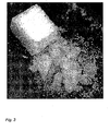
- Blocks were prepared according to Example 1 with the difference, that before the final sintering step, one side of the block was dipped about 1 mm deep into a slurry of pure hydroxyapatite. Thereby, the pores on the dipped side of the sponge were completely filled with slurry. ( Fig.4 )
- the liquid was almost completely taken up by the porous block and formed a gel in the pores of the block in ca. 3 minutes at 25°C.
- the block could then be easily cut using a scalpel and clean cutting surfaces were obtained. Cutting a block with empty pores caused it to crumble ( Figure 4 ).
- the samples were analysed (5 ⁇ l) on an TSK SSW2000 (18674, 4.6x300 mm, 4 ⁇ m, TosoHaas, Gmbh, Germany) in the mobile phase (30% acetonitrile [co3c11x, Labscan], 0.9% NaCl) at a flow of 0.3 ml/min, delivered from a HPLC system (pu880, Jasco Corporation).
- the peaks were detected as measured absorbance at 215 nm (online UV-detector, Jasco 1575) and the peak-areas were integrated.
- Table 2 Retention of PTH (raw data from HPLC) Assay time (hours) PTH concentration (area) PTH conc.
Priority Applications (12)
| Application Number | Priority Date | Filing Date | Title |
|---|---|---|---|
| EP07013645A EP2014256A1 (fr) | 2007-07-12 | 2007-07-12 | Matériau de réparation d'os composite |
| EP12006878.8A EP2564813B1 (fr) | 2007-07-12 | 2008-06-30 | Matériau de réparation d'os composite |
| BRPI0813696A BRPI0813696B8 (pt) | 2007-07-12 | 2008-06-30 | material de reparo ósseo composto cortável, seu método de preparação e kit |
| ES08773779.7T ES2525100T3 (es) | 2007-07-12 | 2008-06-30 | Material compuesto de reparación ósea |
| EP08773779.7A EP2173273B1 (fr) | 2007-07-12 | 2008-06-30 | Matériau composite de réparation des os |
| AU2008274585A AU2008274585B2 (en) | 2007-07-12 | 2008-06-30 | Composite bone repair material |
| PCT/EP2008/005340 WO2009007034A1 (fr) | 2007-07-12 | 2008-06-30 | Matériau composite de réparation des os |
| KR1020107000730A KR101508285B1 (ko) | 2007-07-12 | 2008-06-30 | 복합 골 복구물질 |
| US12/667,496 US8574611B2 (en) | 2007-07-12 | 2008-06-30 | Composite bone repair material |
| JP2010515382A JP5467443B2 (ja) | 2007-07-12 | 2008-06-30 | 複合材料骨修復材料 |
| CA2693599A CA2693599C (fr) | 2007-07-12 | 2008-06-30 | Materiau composite de reparation des os |
| ZA2010/00136A ZA201000136B (en) | 2007-07-12 | 2010-01-07 | Composite bone repair material |
Applications Claiming Priority (1)
| Application Number | Priority Date | Filing Date | Title |
|---|---|---|---|
| EP07013645A EP2014256A1 (fr) | 2007-07-12 | 2007-07-12 | Matériau de réparation d'os composite |
Publications (1)
| Publication Number | Publication Date |
|---|---|
| EP2014256A1 true EP2014256A1 (fr) | 2009-01-14 |
Family
ID=38626289
Family Applications (3)
| Application Number | Title | Priority Date | Filing Date |
|---|---|---|---|
| EP07013645A Withdrawn EP2014256A1 (fr) | 2007-07-12 | 2007-07-12 | Matériau de réparation d'os composite |
| EP08773779.7A Active EP2173273B1 (fr) | 2007-07-12 | 2008-06-30 | Matériau composite de réparation des os |
| EP12006878.8A Active EP2564813B1 (fr) | 2007-07-12 | 2008-06-30 | Matériau de réparation d'os composite |
Family Applications After (2)
| Application Number | Title | Priority Date | Filing Date |
|---|---|---|---|
| EP08773779.7A Active EP2173273B1 (fr) | 2007-07-12 | 2008-06-30 | Matériau composite de réparation des os |
| EP12006878.8A Active EP2564813B1 (fr) | 2007-07-12 | 2008-06-30 | Matériau de réparation d'os composite |
Country Status (10)
| Country | Link |
|---|---|
| US (1) | US8574611B2 (fr) |
| EP (3) | EP2014256A1 (fr) |
| JP (1) | JP5467443B2 (fr) |
| KR (1) | KR101508285B1 (fr) |
| AU (1) | AU2008274585B2 (fr) |
| BR (1) | BRPI0813696B8 (fr) |
| CA (1) | CA2693599C (fr) |
| ES (1) | ES2525100T3 (fr) |
| WO (1) | WO2009007034A1 (fr) |
| ZA (1) | ZA201000136B (fr) |
Cited By (5)
| Publication number | Priority date | Publication date | Assignee | Title |
|---|---|---|---|---|
| WO2010037881A1 (fr) * | 2008-10-03 | 2010-04-08 | Universidad Complutense De Madrid | Procédé pour la préparation à basse température de pièces de biocéramique à porosité tridimensionnelle modélisée et interconnectée |
| WO2011011785A3 (fr) * | 2009-07-24 | 2011-04-07 | Warsaw Orthopedic, Inc. | Dispositifs médicaux implantables |
| WO2013041482A1 (fr) * | 2011-09-23 | 2013-03-28 | Fraunhofer-Gesellschaft zur Förderung der angewandten Forschung e. V. | Peg fonctionnalisé par des chaînes latérales |
| US8524215B2 (en) | 2010-08-02 | 2013-09-03 | Janssen Biotech, Inc. | Absorbable PEG-based hydrogels |
| WO2020104591A1 (fr) | 2018-11-22 | 2020-05-28 | Cerhum | Implant crânien-maxillo-facial |
Families Citing this family (16)
| Publication number | Priority date | Publication date | Assignee | Title |
|---|---|---|---|---|
| US8293813B2 (en) * | 2008-03-05 | 2012-10-23 | Biomet Manufacturing Corporation | Cohesive and compression resistant demineralized bone carrier matrix |
| EP2814520B1 (fr) | 2012-02-14 | 2016-11-02 | Straumann Holding AG | Matériau de réparation d'os |
| EP2814519B1 (fr) | 2012-02-14 | 2016-11-16 | Straumann Holding AG | Matériau de réparation d'os |
| KR101304949B1 (ko) * | 2012-04-19 | 2013-09-06 | 순천향대학교 산학협력단 | 히알루론산-젤라틴 용액이 로딩된 이상인산칼슘 지지체의 제조방법 |
| US9907654B2 (en) * | 2012-12-11 | 2018-03-06 | Dr. H.C. Robert Mathys Stiftung | Bone substitute and method for producing the same |
| JP6533932B2 (ja) * | 2012-12-28 | 2019-06-26 | アボット カーディオバスキュラー システムズ インコーポレイテッド | 抗体を含む治療用組成物 |
| KR101443814B1 (ko) * | 2013-03-28 | 2014-09-30 | 주식회사 바이오알파 | 골 이식재 조성물 및 이의 제조방법 |
| WO2014187969A1 (fr) * | 2013-05-23 | 2014-11-27 | Ceramtec Gmbh | Élément de construction en céramique comprenant des canaux de pores |
| KR101569119B1 (ko) * | 2013-11-25 | 2015-11-13 | 한국세라믹기술원 | 가공성이 개선된 다공체 및 그 제조방법과 가공방법 |
| CN107847638A (zh) | 2014-12-29 | 2018-03-27 | 佰欧维恩图斯有限责任公司 | 用于骨修复中改善骨诱导性分子递送的系统和方法 |
| CN105641753B (zh) * | 2016-03-08 | 2019-07-05 | 吴志宏 | 一种复合rhBMP-2的可实现血管转移的3D打印可降解支架 |
| KR101890192B1 (ko) | 2016-12-20 | 2018-08-21 | (주)이노본 | 세라믹 입자를 포함하는 골이식재의 제조방법 |
| EP3684297B1 (fr) * | 2017-09-20 | 2021-07-28 | Universität Zürich | Microarchitecture de substitution osseuse ostéoconductive |
| KR102123540B1 (ko) | 2018-02-09 | 2020-06-16 | (주)이노본 | 세라믹 입자를 포함하는 골이식재의 제조방법 |
| EP3712243A1 (fr) * | 2019-03-20 | 2020-09-23 | Ricoh Company, Ltd. | Ensemble de liquide pour appareil de décharge de gouttelettes |
| US11638646B1 (en) * | 2019-08-16 | 2023-05-02 | 3D Biomaterials, Inc. | Bioceramic implants matched to patient specific and bone specific geometry |
Citations (7)
| Publication number | Priority date | Publication date | Assignee | Title |
|---|---|---|---|---|
| DE19610715C1 (de) * | 1996-03-19 | 1997-06-26 | Axel Kirsch | Verfahren zum Herstellen eines Knochenersatzmaterials |
| WO2000044808A1 (fr) * | 1999-02-01 | 2000-08-03 | Hubbell Jeffrey A | Biomateriaux formes par reaction d'addition nucleophile a des groupes non satures conjugues |
| WO2003040235A1 (fr) * | 2001-11-07 | 2003-05-15 | Universität Zürich | Matrice synthetique de croissance cellulaire interne et de regeneration tissulaire controlees |
| US20030143258A1 (en) * | 2001-10-12 | 2003-07-31 | David Knaack | Bone graft |
| US20040002770A1 (en) * | 2002-06-28 | 2004-01-01 | King Richard S. | Polymer-bioceramic composite for orthopaedic applications and method of manufacture thereof |
| EP1609491A1 (fr) * | 2004-06-16 | 2005-12-28 | Straumann Holding AG | Membrane barrière |
| EP1820522A1 (fr) * | 2006-02-20 | 2007-08-22 | Straumann Holding AG | Matrice à base de granulats |
Family Cites Families (37)
| Publication number | Priority date | Publication date | Assignee | Title |
|---|---|---|---|---|
| DK154260C (da) * | 1981-02-20 | 1989-05-22 | Mundipharma Gmbh | Fremgangsmaade til fremstilling af et knogleimplantat af braendt tricalciumphosphat, specielt til udfyldning af hulrum eller til sammensaetning af knogledele efter fraktur. |
| US5077049A (en) * | 1989-07-24 | 1991-12-31 | Vipont Pharmaceutical, Inc. | Biodegradable system for regenerating the periodontium |
| DE69121587T3 (de) | 1990-12-06 | 2000-05-31 | Gore & Ass | Implantierbare bioresorbierbare artikel |
| JP3315761B2 (ja) * | 1993-06-29 | 2002-08-19 | 旭光学工業株式会社 | 生体吸収性高分子含有焼結型骨補填材 |
| DE69426414T2 (de) | 1993-09-24 | 2001-05-03 | Takiron Co | Implantatmaterial |
| US5702449A (en) * | 1995-06-07 | 1997-12-30 | Danek Medical, Inc. | Reinforced porous spinal implants |
| US6039762A (en) * | 1995-06-07 | 2000-03-21 | Sdgi Holdings, Inc. | Reinforced bone graft substitutes |
| US5863984A (en) * | 1995-12-01 | 1999-01-26 | Universite Laval, Cite Universitaire | Biostable porous material comprising composite biopolymers |
| FR2758988B1 (fr) * | 1997-02-05 | 2000-01-21 | S H Ind | Procede d'elaboration de substituts osseux synthetiques d'architecture poreuse parfaitement maitrisee |
| SE514908C2 (sv) * | 1998-07-13 | 2001-05-14 | Gs Dev Ab | Medel för benrekonstruktion |
| AU766735B2 (en) * | 1998-09-15 | 2003-10-23 | Isotis N.V. | Osteoinduction |
| ES2224737T3 (es) * | 1998-12-14 | 2005-03-01 | Osteotech, Inc., | Injerto de hueso hecho de particulas oseas. |
| US6383519B1 (en) * | 1999-01-26 | 2002-05-07 | Vita Special Purpose Corporation | Inorganic shaped bodies and methods for their production and use |
| WO2000045871A1 (fr) | 1999-02-04 | 2000-08-10 | Sdgi Holdings, Inc. | Compositions spongieuses osteogeniques extremement mineralisees et leurs utilisations |
| US6656489B1 (en) * | 1999-02-10 | 2003-12-02 | Isotis N.V. | Scaffold for tissue engineering cartilage having outer surface layers of copolymer and ceramic material |
| US20030206928A1 (en) | 1999-04-07 | 2003-11-06 | Pertti Tormala | Bioactive, bioabsorbable surgical polyethylene glycol and polybutylene terephtalate copolymer composites and devices |
| DE19940717A1 (de) | 1999-08-26 | 2001-03-01 | Gerontocare Gmbh | Resorblerbares Knochenersatz- und Knochenaufbaumaterial |
| DE19940977A1 (de) | 1999-08-28 | 2001-03-01 | Lutz Claes | Folie aus resorbierbarem Polymermaterial und Verfahren zur Herstellung einer solchen Folie |
| DE10026306A1 (de) | 2000-05-26 | 2001-11-29 | Tutogen Medical Gmbh | Transplantat |
| JP5154729B2 (ja) | 2000-08-04 | 2013-02-27 | オルソゲム・リミテッド | 多孔質人工骨移植片およびその製造方法 |
| GB0020610D0 (en) * | 2000-08-21 | 2000-10-11 | Dytech Corp Ltd | Uses of porous carriers |
| US6793725B2 (en) * | 2001-01-24 | 2004-09-21 | Ada Foundation | Premixed calcium phosphate cement pastes |
| US6626950B2 (en) * | 2001-06-28 | 2003-09-30 | Ethicon, Inc. | Composite scaffold with post anchor for the repair and regeneration of tissue |
| CN1301757C (zh) | 2001-11-27 | 2007-02-28 | 多喜兰株式会社 | 植入物材料及其生产方法 |
| EP1494730B1 (fr) | 2002-03-22 | 2012-01-18 | Kuros Biosurgery AG | Composition regeneratrices de tissus durs |
| TW200400062A (en) | 2002-04-03 | 2004-01-01 | Mathys Medizinaltechnik Ag | Kneadable, pliable bone replacement material |
| AU2003254211A1 (en) | 2002-07-31 | 2004-02-16 | Dentsply International Inc. | Bone repair putty comprising porous particulate and carrier gel |
| DE10258773A1 (de) | 2002-12-16 | 2004-07-08 | SDGI Holding, Inc., Wilmington | Knochenersatzmaterial |
| US6997120B2 (en) * | 2003-05-15 | 2006-02-14 | Robert Gabriel | Planting apparatus and method |
| WO2004112856A1 (fr) | 2003-06-24 | 2004-12-29 | Kyushu Tlo Company Limited | Materiau prothetique osseux medical et processus de production de ce materiau |
| US7163651B2 (en) * | 2004-02-19 | 2007-01-16 | Calcitec, Inc. | Method for making a porous calcium phosphate article |
| US6994726B2 (en) * | 2004-05-25 | 2006-02-07 | Calcitec, Inc. | Dual function prosthetic bone implant and method for preparing the same |
| US8529625B2 (en) | 2003-08-22 | 2013-09-10 | Smith & Nephew, Inc. | Tissue repair and replacement |
| WO2005042046A1 (fr) * | 2003-11-03 | 2005-05-12 | Medtronic, Inc. | Hydrogel induisant une croissance interne specifique d'une cellule |
| MX2007007471A (es) | 2004-12-21 | 2007-07-20 | Nektar Therapeutics Al Corp | Reactivos de tiol polimericos estabilizados. |
| US20060199876A1 (en) * | 2005-03-04 | 2006-09-07 | The University Of British Columbia | Bioceramic composite coatings and process for making same |
| US20070098799A1 (en) * | 2005-10-28 | 2007-05-03 | Zimmer, Inc. | Mineralized Hydrogels and Methods of Making and Using Hydrogels |
-
2007
- 2007-07-12 EP EP07013645A patent/EP2014256A1/fr not_active Withdrawn
-
2008
- 2008-06-30 US US12/667,496 patent/US8574611B2/en active Active
- 2008-06-30 AU AU2008274585A patent/AU2008274585B2/en active Active
- 2008-06-30 JP JP2010515382A patent/JP5467443B2/ja active Active
- 2008-06-30 EP EP08773779.7A patent/EP2173273B1/fr active Active
- 2008-06-30 WO PCT/EP2008/005340 patent/WO2009007034A1/fr active Application Filing
- 2008-06-30 BR BRPI0813696A patent/BRPI0813696B8/pt active IP Right Grant
- 2008-06-30 CA CA2693599A patent/CA2693599C/fr active Active
- 2008-06-30 ES ES08773779.7T patent/ES2525100T3/es active Active
- 2008-06-30 KR KR1020107000730A patent/KR101508285B1/ko active IP Right Grant
- 2008-06-30 EP EP12006878.8A patent/EP2564813B1/fr active Active
-
2010
- 2010-01-07 ZA ZA2010/00136A patent/ZA201000136B/en unknown
Patent Citations (7)
| Publication number | Priority date | Publication date | Assignee | Title |
|---|---|---|---|---|
| DE19610715C1 (de) * | 1996-03-19 | 1997-06-26 | Axel Kirsch | Verfahren zum Herstellen eines Knochenersatzmaterials |
| WO2000044808A1 (fr) * | 1999-02-01 | 2000-08-03 | Hubbell Jeffrey A | Biomateriaux formes par reaction d'addition nucleophile a des groupes non satures conjugues |
| US20030143258A1 (en) * | 2001-10-12 | 2003-07-31 | David Knaack | Bone graft |
| WO2003040235A1 (fr) * | 2001-11-07 | 2003-05-15 | Universität Zürich | Matrice synthetique de croissance cellulaire interne et de regeneration tissulaire controlees |
| US20040002770A1 (en) * | 2002-06-28 | 2004-01-01 | King Richard S. | Polymer-bioceramic composite for orthopaedic applications and method of manufacture thereof |
| EP1609491A1 (fr) * | 2004-06-16 | 2005-12-28 | Straumann Holding AG | Membrane barrière |
| EP1820522A1 (fr) * | 2006-02-20 | 2007-08-22 | Straumann Holding AG | Matrice à base de granulats |
Cited By (9)
| Publication number | Priority date | Publication date | Assignee | Title |
|---|---|---|---|---|
| WO2010037881A1 (fr) * | 2008-10-03 | 2010-04-08 | Universidad Complutense De Madrid | Procédé pour la préparation à basse température de pièces de biocéramique à porosité tridimensionnelle modélisée et interconnectée |
| WO2011011785A3 (fr) * | 2009-07-24 | 2011-04-07 | Warsaw Orthopedic, Inc. | Dispositifs médicaux implantables |
| JP2013500085A (ja) * | 2009-07-24 | 2013-01-07 | ウォーソー・オーソペディック・インコーポレーテッド | セラミックおよびポリマー性充填剤に基づく多孔性複合性移植物 |
| AU2010275377B2 (en) * | 2009-07-24 | 2014-06-19 | Warsaw Orthopedic, Inc. | Porous composite implant based on ceramic and polymeric filler material |
| RU2545823C2 (ru) * | 2009-07-24 | 2015-04-10 | Ворсо Ортопедик, Инк. | Имплантируемое медицинское устройство |
| US8524215B2 (en) | 2010-08-02 | 2013-09-03 | Janssen Biotech, Inc. | Absorbable PEG-based hydrogels |
| WO2013041482A1 (fr) * | 2011-09-23 | 2013-03-28 | Fraunhofer-Gesellschaft zur Förderung der angewandten Forschung e. V. | Peg fonctionnalisé par des chaînes latérales |
| WO2020104591A1 (fr) | 2018-11-22 | 2020-05-28 | Cerhum | Implant crânien-maxillo-facial |
| BE1026794A1 (fr) | 2018-11-22 | 2020-06-18 | Cerhum | Implant cranio-maxillo-facial |
Also Published As
| Publication number | Publication date |
|---|---|
| ES2525100T3 (es) | 2014-12-17 |
| ZA201000136B (en) | 2011-08-31 |
| AU2008274585B2 (en) | 2014-05-15 |
| US8574611B2 (en) | 2013-11-05 |
| WO2009007034A1 (fr) | 2009-01-15 |
| US20100292146A1 (en) | 2010-11-18 |
| EP2173273B1 (fr) | 2014-09-24 |
| BRPI0813696B8 (pt) | 2021-06-22 |
| JP2010532692A (ja) | 2010-10-14 |
| KR101508285B1 (ko) | 2015-04-08 |
| AU2008274585A1 (en) | 2009-01-15 |
| EP2564813A3 (fr) | 2013-07-10 |
| CA2693599C (fr) | 2015-06-16 |
| BRPI0813696B1 (pt) | 2019-04-24 |
| JP5467443B2 (ja) | 2014-04-09 |
| EP2564813B1 (fr) | 2018-11-14 |
| BRPI0813696A2 (pt) | 2015-11-03 |
| EP2173273A1 (fr) | 2010-04-14 |
| EP2564813A2 (fr) | 2013-03-06 |
| CA2693599A1 (fr) | 2009-01-15 |
| KR20100061649A (ko) | 2010-06-08 |
Similar Documents
| Publication | Publication Date | Title |
|---|---|---|
| EP2564813B1 (fr) | Matériau de réparation d'os composite | |
| US8580865B2 (en) | Phase-and sedimentation-stable, plastically deformable preparation with intrinsic pore forming, intended for example for filling bone defects or for use as bone substitute material, and method of producing it | |
| EP1504776A1 (fr) | Element permettant de regenerer un cartilage articulaire et procede permettant de produire cet element, methodes de regeneration d'un cartilage articulaire et cartilage artificiel pour transplantation | |
| US20110003745A1 (en) | Granulate-matrix | |
| US10238775B2 (en) | Bone repair material | |
| US10098983B2 (en) | Bone repair material | |
| JP2004159971A (ja) | 骨形成用部材およびその製造方法 | |
| JP4388260B2 (ja) | 関節軟骨の再生用部材 | |
| JP2006020930A (ja) | リン酸カルシウム系骨補填材 | |
| JP2006174985A (ja) | リン酸カルシウム/ポリマーハイブリッド材 |
Legal Events
| Date | Code | Title | Description |
|---|---|---|---|
| PUAI | Public reference made under article 153(3) epc to a published international application that has entered the european phase |
Free format text: ORIGINAL CODE: 0009012 |
|
| AK | Designated contracting states |
Kind code of ref document: A1 Designated state(s): AT BE BG CH CY CZ DE DK EE ES FI FR GB GR HU IE IS IT LI LT LU LV MC MT NL PL PT RO SE SI SK TR |
|
| AX | Request for extension of the european patent |
Extension state: AL BA HR MK RS |
|
| 17P | Request for examination filed |
Effective date: 20090411 |
|
| 17Q | First examination report despatched |
Effective date: 20090714 |
|
| AKX | Designation fees paid |
Designated state(s): AT BE BG CH CY CZ DE DK EE ES FI FR GB GR HU IE IS IT LI LT LU LV MC MT NL PL PT RO SE SI SK TR |
|
| STAA | Information on the status of an ep patent application or granted ep patent |
Free format text: STATUS: THE APPLICATION IS DEEMED TO BE WITHDRAWN |
|
| 18D | Application deemed to be withdrawn |
Effective date: 20100126 |