EP2003451B1 - Heterooligomere t1r1-t1r3 Umamigeschmacksrezeptoren und diese exprimierende Zellinien sowie Verwendung davon zur Identifizierung von Umamigeschmacksverbindungen - Google Patents
Heterooligomere t1r1-t1r3 Umamigeschmacksrezeptoren und diese exprimierende Zellinien sowie Verwendung davon zur Identifizierung von Umamigeschmacksverbindungen Download PDFInfo
- Publication number
- EP2003451B1 EP2003451B1 EP08075748A EP08075748A EP2003451B1 EP 2003451 B1 EP2003451 B1 EP 2003451B1 EP 08075748 A EP08075748 A EP 08075748A EP 08075748 A EP08075748 A EP 08075748A EP 2003451 B1 EP2003451 B1 EP 2003451B1
- Authority
- EP
- European Patent Office
- Prior art keywords
- receptor
- taste
- polypeptide
- protein
- cells
- Prior art date
- Legal status (The legal status is an assumption and is not a legal conclusion. Google has not performed a legal analysis and makes no representation as to the accuracy of the status listed.)
- Expired - Lifetime
Links
Images


Classifications
-
- C—CHEMISTRY; METALLURGY
- C07—ORGANIC CHEMISTRY
- C07K—PEPTIDES
- C07K14/00—Peptides having more than 20 amino acids; Gastrins; Somatostatins; Melanotropins; Derivatives thereof
- C07K14/435—Peptides having more than 20 amino acids; Gastrins; Somatostatins; Melanotropins; Derivatives thereof from animals; from humans
- C07K14/705—Receptors; Cell surface antigens; Cell surface determinants
-
- A—HUMAN NECESSITIES
- A01—AGRICULTURE; FORESTRY; ANIMAL HUSBANDRY; HUNTING; TRAPPING; FISHING
- A01K—ANIMAL HUSBANDRY; AVICULTURE; APICULTURE; PISCICULTURE; FISHING; REARING OR BREEDING ANIMALS, NOT OTHERWISE PROVIDED FOR; NEW BREEDS OF ANIMALS
- A01K2217/00—Genetically modified animals
-
- A—HUMAN NECESSITIES
- A01—AGRICULTURE; FORESTRY; ANIMAL HUSBANDRY; HUNTING; TRAPPING; FISHING
- A01K—ANIMAL HUSBANDRY; AVICULTURE; APICULTURE; PISCICULTURE; FISHING; REARING OR BREEDING ANIMALS, NOT OTHERWISE PROVIDED FOR; NEW BREEDS OF ANIMALS
- A01K2217/00—Genetically modified animals
- A01K2217/05—Animals comprising random inserted nucleic acids (transgenic)
-
- G—PHYSICS
- G01—MEASURING; TESTING
- G01N—INVESTIGATING OR ANALYSING MATERIALS BY DETERMINING THEIR CHEMICAL OR PHYSICAL PROPERTIES
- G01N2333/00—Assays involving biological materials from specific organisms or of a specific nature
- G01N2333/435—Assays involving biological materials from specific organisms or of a specific nature from animals; from humans
- G01N2333/705—Assays involving receptors, cell surface antigens or cell surface determinants
- G01N2333/72—Assays involving receptors, cell surface antigens or cell surface determinants for hormones
- G01N2333/726—G protein coupled receptor, e.g. TSHR-thyrotropin-receptor, LH/hCG receptor, FSH
Definitions
- the present invention in part relates to the discovery that the T1R receptors assemble to form functional taste receptors.
- co-expression of T1R1 and T1R3 results in a taste receptor that responds to umami taste stimuli, including monosodium glutamate.
- co-expression of the T1 R2 and T1 R3 receptors results in a taste receptor that responds to sweet taste stimuli including naturally occurring and artificial sweeteners.
- the present description relates to the use of hetero-oligomeric taste receptors comprising T1R1/T1R3 and T1R2/T1R3 in assays to identify compounds that respectively respond to umami taste stimuli and sweet taste stimuli.
- the invention relates to the construction of cell lines that stably or transiently co-express a combination of T1R1 and T1R3; under constitutive or inducible conditions.
- WO 01/66563 suggests that T1R1 and T1R3 may coexpress within the same taste receptor cell type, and the two receptors may physically interact to form a heterodimeric taste receptor, or T1R1 and T1R3 may both independently bind to the same type of ligand, and their combined binding may result in a specific perceived taste sensation.
- the taste system provides sensory information about the chemical composition of the external world. Mammals are believed to have at least five basic taste modalities: sweet, bitter, sour, salty, and umami. See , e.g. , Kawamura et al., Introduction to Umami: A Basic Taste (1987 ); Kinnamon et al., Ann. Rev. Physiol., 54:715-31 (1992 ); Lindemann, Physiol. Rev., 76:718-66 (1996 ); Stewart et al., Am. J. Physiol., 272:1-26(1997 ).
- Each taste modality is thought to be mediated by a distinct protein receptor or receptors that are expressed in taste receptor cells found on the surface of the tongue ( Lindemann, Physol. Rev. 76:718-716 (1996 )).
- the taste receptors that recognize bitter, sweet, and umami taste stimuli belong to the G-protein-coupled receptor (GPCR) superfamily ( Hoon et al., Cell 96:451 (1999 ); Adler et al., Cell 100:693 (2000 )). (Other taste modalities are believed to be mediated by ion channels.)
- G protein-coupled receptors mediate many other physiological functions, such as endocrine function, exocrine function, heart rate, lipolysis, and carbohydrate metabolism.
- the biochemical analysis and molecular cloning of a number of such receptors has revealed many basic principles regarding the function of these receptors.
- United States Patent No. 5,691,188 describes how upon a ligand binding to a GPCR, the receptor undergoes a conformational change leading to activation of a heterotrimeric G protein by promoting the displacement of bound GDP by GTP on the surface of the G ⁇ subunit and subsequent dissociation of the G ⁇ subunit from the G ⁇ and G ⁇ subunits.
- the free G ⁇ subunits and G ⁇ complexes activate downstream elements of a variety of signal transduction pathways.
- This invention relates to the three-member T1R class of taste-specific GPCRs.
- the T1R receptors were hypothesized to function as sweet taste receptors ( Hoon et al., Cell 96:541-51 (1999 ); Kitagawa et al., Biochem Biophys Res. Commun. 283:236-42 (2001 ); Max et al., Nat. Genet. 28:58-63 (2001 ); Montmayeur et al., Nat. Neurosci. 4:492-8(2001 ); Sainz et al., J. Neurochem. 77:896-903(2001 )), and Nelson et a.
- rat T1 R2 and T1 R3 act in combination to recognize sweet taste stimuli.
- the present description relates to the discovery that, as is the case for rat T1R2/T1R3, human T1 R2 and T1 R3 act in combination to recognize sweet taste stimuli. Further, the present invention relates to the discovery that human T1R1 and T1 R3 act in combination to recognize umami taste stimuli. Therefore, T1R2/T1R3 is likely to function as a sweet taste receptor and T1R1/T1R3 is likely to function as an umami taste receptor in mammals.
- T1 R1 and T1R3 function co-dependence of T1 R2 and T1R3
- T1Rs function as heterodimeric complexes.
- characterization of taste receptors which function as sweet and umami receptors is significant as it will facilitate the use of these receptors in assays for identifying compounds that modulate (enhance or block) sweet and umami taste. These compounds would be useful for improving the taste and palatability of foods, beverages, medicinals for human or animal consumption. Particularly, an assay that utilizes a functional sweet receptor would allow the identification of novel sweeteners.
- the present invention relates to the discovery that different combinations of T1Rs, when co-expressed, produce functional taste receptors that respond to taste stimuli. Particularly, the present invention relates to the discovery that the co-expression of T1R1 and T1R3 results in a hetero-oligomeric taste receptor that responds to umami taste stimuli such as monosodium glutamate.
- the present invention also relates to cells that co-express T1R1 and T1R3, preferably human.
- these cell lines will express elevated amounts of the receptors, either constitutively or inducibly.
- These cell lines include cells that transiently or stably express T1R1 and T1R3.
- the present invention provides assays, preferably high throughput screening assays, that utilize the T1R1/T1R3 receptor, preferably high throughput cell-based assays, to identify compounds that modulate umami taste.
- assays preferably high throughput screening assays, that utilize the T1R1/T1R3 receptor, preferably high throughput cell-based assays, to identify compounds that modulate umami taste.
- the description also provides assays that include taste tests to confirm that these compounds modulate umami taste.
- T1 Rs mammalian G protein-coupled receptors
- human or non-human cells e.g., mammalian, yeast, worm, or insect cells
- T1R fusion proteins or polypeptides which include at least a fragment of at least one of such T1Rs.
- It is a further object to provide an isolated nucleic acid molecule comprising a nucleic acid sequence that encodes a polypeptide having an amino acid sequence at least 35 to 50%, and preferably 60%, 75%, 85%, 90%, 95%, 96%, 97%, 98%, or 99% identical to an amino acid sequence selected from the group of one of the TIR amino acid sequences identified infra and conservatively modified variants thereof, wherein the fragment is at least 20, preferably 40, 60, 80, 100, 150, 200, or 250 amino acids in length.
- the fragment can be an antigenic fragment that binds to an anti-T1R antibody.
- T1R1/T1R3 combinations wherein T1R1 and/or T1R3 is a variant or fragment
- T1R2/T1R3 combinations wherein T1R2 and/or T1R3 is a variant or fragment.
- PDZIP PDZ domain-interacting peptide
- Such assays will utilize a combination of T1Rs, or fragments or variants thereof, or genes encoding such T1Rs, or fragments or variants thereof, which are disclosed herein. Most preferably such combinations will comprise hT1R1/hT1 R3 and hT1R2/hT1 R3.
- T1R1/T1R3 or T1R2/T1R3 taste receptors e.g., which enhance the ability of these receptors to respond to taste stimuli.
- 5'-IMP or 5'-GMP enhances the responsiveness of the umami (T1R1/T1R3) to L-glutamate.
- These modulatory compounds may enhance the activity of different sweet or umami taste stimuli, and provide for enhanced tastes and/or for the same taste to be elicited at reduced concentration of the particular sweet or umami taste eliciting compound the activity of which is enhanced by a taste modulator identified using the subject assays.
- It is still a further object to provide preferred assays for evaluating one or more compounds for a taste comprising: a step of contacting said one or more compounds with at least one of the disclosed T1Rs, fragments or variants thereof, preferably combinations of human T1Rs.
- a compound that interacts with hT1R2 and/or hT1R3 as a sweet blocker, enhancer, modulator, or mimic and a compound that interacts with hT1R1 and/or hT1R3 as a umami blocker, enhancer, modulator, or mimic in food and beverage compositions.
- T1Rs in particular non-human T1Rs, to identify compounds that modulate the taste of animal feed formulations for use in, e.g., fish aquaculture.
- a G protein e.g., G ⁇ 15 or another G protein that when expressed in association with T1R2/T1R3 or T1R1/T1R3 produces a functional taste receptor.
- such cells will comprise HEK-293 cells that stably express G ⁇ 15 or another G protein that associates with T1R1/T1R3 or T1R2/T1R3 to produce a functional umami or sweet taste receptor.
- the present invention provides a taste receptor comprised of at least one T1R1 polypeptide and at least one T1 R3 polypeptide, wherein said taste receptor specifically binds to and/or is activated by a umami taste stimulus, wherein said T1R1 polypeptide is selected from the group consisting of:
- the present invention provides a composition comprising the above described receptor wherein at least one of the T1R polypeptides is coupled to amino acids representing all or part of another G-protein coupled receptor.
- the present invention also provides a host cell expressing the receptor.
- An expression vector comprising the nucleic acid sequence encoding the above described receptor is further provided.
- the invention provides a method for identifying compounds that modulate taste perception by identifying compounds that bind to, activate, inhibit, and/or modulate of the above described receptor.
- Figure 1 contains a sequence alignment of human and rat T1Rs, human calcium-sensing receptor and rat metabotropic glutamate receptor.
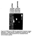
- Figure 2 contains RT-PCR amplification experimental results which show that hT1 R2 and hT1 R3 are expressed in taste tissue.
- Figure 3a - 3b contain functional data (intracellular calcium responses) elicited by different sweet taste stimuli in HEK cells stably expressing G ⁇ 15 that are transiently transfected with human T1R2, T1R3 and T1R2/T1R3 at various concentrations of sweet taste stimuli ( Figure 3a ); human T1R2/t1R3 dose responses for several sweet taste stimuli ( Figure 3b ); human T1R2/T1R3 responses to sucrose in the presence of gurmarin, and endogenous ⁇ 2-adrenergic receptor responses to isoproterenol in the presence of gurmarin.
- Figure 3c contains the normalized response to different sweeteners.
- Figure 4 contains intracellular calcium responses in HEK cells stably expressing G ⁇ 15, transiently transfected with hT1R2/hT1 R3, rT1R2/rT1R3, hT1R2/rT1R3 and rT1 R2/hT1R3 in response to 350 mM sucrose, 25 mM tryptophan, 15 mM aspartame, and 0.05 % monellin.
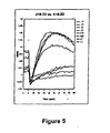
- Figure 5 contains the results of a fluorescence plate reactor based assay wherein HEK cells stably expressing G ⁇ 15 were transiently transfected with hT1R2 and hT1R3 or hT1R3 alone and contacted with the calcium dye Fluo-4 and a sweet taste stimulus (12.5 mM cyclamate).
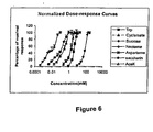
- Figure 6 contains normalized dose-response curves which show that hT1R2 and hT1R3 function in combination as the human sweet receptor based on their dose-specific interaction with various sweet stimuli (trp, cyclamate, sucrose, neotame, asparame, saccharin and Acek).
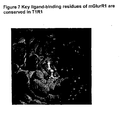
- Figure 7 contains structural information relating to mGluR1 and T1R1 showing the key ligand binding residues are observed in these molecules.
- Figure 8a-8c contains functional data showing HEK cells which stably express G ⁇ 15 that are transiently transfected with T1R1/T1R3 respond to glutamate in an intracellular calcium-based assay.
- Figure 8a shows that intracellular calcium increases in response to increasing glutamate concentration;
- Figure 8b shows intracellular calcium responds to IMP (2 mM), glutamate (0.5 mM) and 0.2 mM IMP;
- Figure 8c shows human T1R1/T1R3 responses for glutamate in the presence and absence of 0.2 mM IMP.
- Figures 9a-9b respectively contain the results of an immunofluorescence staining assay using Myc-tagged hT1R2 and a FACS experiment showing that the incorporation of the PDZIP peptide (SEQ ID No: 1) enhanced the expression of a T1 R (hT1 R2) on the plasma membrane.
- Figure 10a through 10b contain calcium imaging data demonstrating that h1TR2/hT1 R3 respond to different sweet stimuli.
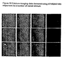
- Figure 11 shows the responses of cell lines which stably express hT1R1/hT1R3 by automated fluorescence imaging to umami taste stimuli.
- Figure 12 shows the responses of a cell line which stably expresses hT1 R2/hT1 R3 by automated fluorescence imaging to sweet taste stimuli.
- Figure 13 shows dose-response curves determined using automated fluorescence imaging for a cell line that inducibly expresses the human T1R1/T1R3 taste receptor for L-glutamate in the presence and absence of 0.2mM IMP.
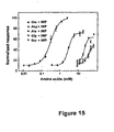
- Figures 14 and 15 show the response of a cell line that inducibly expresses the human T1R1/T1R3 taste receptor (I-17 clone) to a panel of L-amino acids.
- different C-amino acids at 10mM were tested in the presence and absence of 1 mM IMP.
- dose-responses for active amino acids were determined in the presence of 0.2mM IMP.
- Figure 16 shows that lactisole inhibits the receptor activities of human T1 R2/T1 R3 and human T1R1/T1R3.
- the invention thus provides functional taste receptors, preferably human taste receptors, that are produced by co-expression of a combination of T1R1/T1R3, and the corresponding isolated nucleic acid sequences thereof comprised in expression vectors that upon co-expression result in a functional taste receptor, i.e., umami taste receptor (T1R1/T1R3).
- a functional taste receptor i.e., umami taste receptor (T1R1/T1R3).
- the invention relates to the co-expression of T1R1/T1R3 taste-cell specific GPCRs.
- These nucleic acids and the receptors that they encode are referred to as members of the "T1R" family of taste-cell-specific GPCRs.
- the T1R family members that are co-expressed will include rT1R1, rT1 R3, mT1 R1, mT1 R3, hT1R1, and hT1R3. While not wishing to be bound by theory, it is believed that these taste-cell-specific GPCRs are components of the taste transduction pathway, and are involved in the taste detection of umami taste stimuli,
- T1R family members act in combination with other T1R family members to function as sweet and umami taste receptors.
- heterologous cells which co-express hT1R2 and hT1R3 are selectively activated by sweet taste stimuli in a manner that mirrors human sweet taste.
- HEK-293-G ⁇ 15 cells that co-express hT1R2 and hT1R3 specifically respond to cyclamate, sucrose, aspartame, and saccharin, and the dose responses for these compounds correlate with the psychophysical taste detection thresholds. Therefore, cells that co-express hT1R2 and hT1R3 can be used in screens, preferably high throughput screens, to identify compounds that mimic, modulate, block, and/or enhance sweet taste sensation.
- cells which co-express hT1R1 and hT1R3 are selectively activated by glutamate (monosodium glutamate) and 5'-ribonucleotides in a manner that mirrors human umami taste.
- glutamate monosodium glutamate
- 5'-ribonucleotides such as IMP enhance the glutamate response of the T1R1/T1R3 receptor, a synergism characteristic of umami taste. Therefore, cells that co-express hT1R1 and hT1R3 can be used in screens, preferably high throughput screens to identify compounds that mimic, modulate, block, and/or enhance umami taste sensation.
- T1R1/T1R3 selectively respond to the umami taste stimuli L-glutamate and L-aspartate and only weakly respond to other L-amino acids, and at much higher concentrations, providing further evidence that the T1R1/T1R3 receptor can be used in assays to identify compounds that modulate (enhance or block) umami taste stimuli.
- T1R1/T1R3 or T1R2/T1R3 respectively respond to umami or sweet taste stimuli and a quantitative dose-responsive manner which further supports a conclusion that the T1R1/T1R3 and T1R2/T1R3 receptor can be used to identify receptor agonists and antagonists, e.g., MSG substitutes, umami blockers, novel artificial and natural sweeteners, and sweet blockers.
- receptor agonists and antagonists e.g., MSG substitutes, umami blockers, novel artificial and natural sweeteners, and sweet blockers.
- the sweet taste blocker lactisole inhibits both the T1R2/T1R3 sweet receptor and the T1R1/T1R3 umami taste receptor.
- assays which screen for compounds which affect the binding of lactisole to T1R2/T1R3 or T1R1/T1R3 may be used to identify compounds that enhance, mimic, modulate or block sweet or umami taste.
- lactisole inhibits both the T1R1/T1R3 and T1R2/T1R3 receptors suggests that these receptors may share a common subunit which is bound by lactisole and potentially other taste modulators. Therefore, this suggests that some compounds which enhance, mimic, modulate or block sweet taste may have a similar effect on umami taste or vice versa.
- T1R1/T1R3 or T1R2/T1R3 when assayed by automated fluorescence imaging very effectively respond to various sweet and umami taste stimuli, i.e. at magnitudes substantially greater than transiently transfected cells.
- these cell lines are especially well suited for use in high throughput screening assays for identifying compounds that modulate, block, mimic or enhance sweet or umami taste.
- the invention also encompasses assays that utilize cells that transiently express a T1R1/T1R3 taste receptor.
- T1Rs act in combination, particularly T1R1/T1R3 and T1R2/T1R3, and that such receptor combinations may be used in assays, preferably high throughput assays
- the subject application also envisages assays that utilize T1R1, T1R2 and T1R3 alone or in combination with other proteins, e.g., other GPCRs.
- T1R assays can be used to modulate the taste of foods and beverages.
- Suitable assays described in further detail infra include by way of example whole-cell assays and biochemical assays, including direct-binding assays using one of a combination of different T1R receptors, chimeras or fragments thereof, especially fragments containing N-terminal ligand-binding domains. Examples of assays appropriate for use in the invention are described in greater detail infra and are known in the GPCR field.
- Assays can be designed that quantitate the binding of different compounds or mixtures of compounds to T1R taste receptors or T1R taste receptor combinations or T1R receptors expressed in combination with other heterologous (non-T1R) proteins, e.g. other GPCRs, or that quantitate the activation of cells that express T1R taste receptors. This can be effected by stably or transiently expressing taste receptors in heterologous cells such as HEK-293, CHO and COS cells.
- the assays will preferably use cells that also express (preferably stably) a G protein such as G ⁇ 15 or G ⁇ 16 or other promiscuous G proteins or G protein variants, or an endogenous G protein.
- G ⁇ and G ⁇ proteins may also be expressed therein.
- the effect of a compound on sweet or umami taste using cells or compositions that express or contain the above-identified receptors or receptor combinations may be determined by various means including the use of calcium-sensitive dyes, voltage-sensitive dyes, cAMP assays, direct binding assays using fluorescently labeled ligands or radioactive ligands such as 3 H-glutamate, or transcriptional assays (using a suitable reporter such as luciferase or beta-lactamase).
- Assays that may be utilized with one or more T1Rs include by way of example, assays that utilize a genetic selection for living cells; assays that utilize whole cells or membrane fragments or purified T1R proteins; assays that utilize second messengers such as cAMP and IP3, assays that detect the translocation of arrestin to the cell surface, assays that detect the loss of receptor expression on the cell surface (internalization) by tested ligands, direct ligand-binding assays, competitive-binding assays with inhibitors, assays using in vitro translated protein, assays that detect conformational changes upon the binding of a ligand (e.g., as evidenced by proteolysis, fluorescence, or NMR), behavioral assays that utilize transgenic non-human animals that express a T1 R or T1R combination, such as flies, worms, or mice, assays that utilize cells infected with recombinant viruses that contain T1R genes.
- T1R or T1R fragment or combination of T1Rs, or a combination of a T1R with another protein
- the description envisages the determination of the crystal structure of T1R1/T1R3 (preferably hT1R1/hT1R3) and/or T1R2/T1R3 (preferably hT1R2/hT1R3) and the use of such crystal structures in structure-based design methods to identify molecules that modulate T1R receptor activity.
- the invention especially includes biochemical assays conducted using cells, e.g., mammalian, yeast, insect or other heterologous cells that express the taste receptor of the present invention, preferably receptors that comprise the N-terminal domains of T1R1 and/or T1R3.
- the effect of a compound in such assays can be determined using competitive binding assays, e.g., using radioactive glutamate or IMP, fluorescence (e.g., fluorescence polarization, FRET), or GTP ⁇ 35 S binding assays.
- such assays will utilize cell lines that stably co-express T1R1/T1R3 and a suitable G protein, such as G ⁇ 15 .
- suitable G protein such as G ⁇ 15 .
- Other appropriate G proteins include the chimeric and variant G proteins disclosed in US2002/0143151 ( U.S. Application Serial No. 09/984,292 and 60/243,770 ).
- altered receptors can be constructed and expressed having improved properties, e.g., enhanced surface expression or G-protein coupling.
- T1R variants can be incorporated into cell-based and biochemical assays.
- the invention further includes that utilize different allelic variants of T1 R1 and T1 R3 and combinations thereof, thereby enabling the identification of compounds that elicit specific taste sensation in individuals that express those allelic variants or compounds that elicit specific taste sensations in all individuals.
- Such compounds can be used to make foods more generally palatable.
- T1R encoding nucleic acids also provide valuable probes for the identification of taste cells, as the nucleic acids are specifically expressed in taste cells.
- probes for T1R polypeptides and proteins can be used to identify taste cells present in foliate, circumvallate, and fungiform papillae, as well as taste cells present in the marmackstMail, oral cavity, gastrointestinal epithelium, and epiglottis.
- methods of detecting T1Rs can be used to identify taste cells sensitive to sweet and/or umami taste stimuli or other taste stimuli representing other taste modalities.
- cells stably or transiently expressing T1R2 and/or T1R3 would be predicted from the work herein to be responsive to sweet taste stimuli.
- T1 R1 and/or T1R3 would be predicted to be responsive to umami taste stimuli.
- the nucleic acids encoding the T1R proteins and polypeptides of the invention can be isolated from a variety of sources, genetically engineered, amplified, synthesized, and/or expressed recombinantly according to the methods disclosed in WO 00/035374 , A listing of T1R1s and T1R3s that may be expressed according to the invention are provided in the Examples.
- the invention embraces the expression and use of other specific T1R1s andT1R3s or fragments, variants, or chimeras constructed based on such T1 R sequences, and particularly T1Rs of other species having the required length and % sequence identity or hybridization ability.
- an important aspect of the invention is the plurality of methods of screening for modulators, e.g., activators, inhibitors, stimulators, enhancers, agonists, and antagonists, of these taste-cell-specific GPCRs.
- modulators of taste transduction are useful for the modulation of taste signaling pathways.
- These methods of screening can be used to identify high affinity agonists and antagonists of taste cell activity.
- modulatory compounds can then be used in the food industry to customize taste, e.g., to modulate the umami tastes of foods.
- This invention rectifies the previous lack of understanding relating to umami taste as it identifies specific T1Rs and T1R receptor combinations that mediate umami taste sensation. Therefore, in general, this application relates to the inventors' discoveries relating to the T1R class of taste-specific G-protein-coupled receptors and their specific function in taste perception and the relationship of these discoveries to a better understanding of the molecular basis of taste.
- T1 Rs three-member class of taste-specific G-protein-coupled receptors, termed T1 Rs. Overlapping T1R expression patterns and the demonstration that the structurally related GABA B receptor is heterodimeric suggest that the T1Rs function as heterodimeric taste receptors.
- the present inventors describe the functional co-expression of human T1R1, T1R2, and T1R3 in heterologous cells; cells co-expressing T1R1 and T1 R3 are activated by umami taste stimuli; cells co-expressing T1R2 and T1R3 are activated by sweet taste stimuli.
- T1R1/T1R3 and T1R2/T1R3 activity correlated with psychophysical detection thresholds.
- the 5'-ribonucleotide IMP was found to enhance the T1R1/T1R3 response to glutamate, a synergism characteristic of umami taste.
- T1Rs were first identified by large-scale sequencing of a subtracted cDNA library derived from rat taste tissue, which identified T1R1, and subsequently by T1R1-based degenerate PCR, which led to the identification of T1 R2 ( Hoon et al., Cell 96:541-551 (1999 )). Recently, the present inventors and others identified a third and possibly final member of the T1 R family, T1 R3, in the human genome databank ( Kitagawa et al., Biochem Biophys. Res Commun. 283(1): 236-42 (2001 ); Max et al., Nat. Genet. 28(1): 58-63 (2001 ); Sainz et al., J. Neurochem.
- mouse T1 R3 maps to a genomic interval containing Sac , a locus that influences sweet taste in the mouse ( Fuller et al., J. Hered. 65:33-6 (1974 ); Li et al., Mamm. Genome 12:13-16 (2001 )). Therefore, T1R3 was predicted to function as a sweet taste receptor. Recent high-resolution genetic mapping studies have strengthened the connection between mouse T1 R3 and Sac ( Fuller T.C., J. Hered. 65(1): 33-36 (1974 ); Li et al., Mammal. Genome 12(1): 13-16 (2001 )).
- mGluR4 metabotropic glutamate receptor a transcriptional variant of the mGluR4 metabotropic glutamate receptor has been proposed to be the umami taste receptor because of its selective expression in rat taste tissue, and the similarity of the receptor-activation threshold to the glutamate psychophysical detection threshold ( Chaudhari et al., Nat. Neurosci. 3:113-119 (2000 )). This hypothesis is difficult to reconcile with the exceedingly low expression level of the mGluR4 variant in taste tissue, and the more or less unaltered glutamate taste of mGluR4 knockout mice ( Chaudhari and Roper, Ann. N. Y. Acad. Sci. 855:398-406 (1998 )).
- the taste variant is structurally implausible, lacking not only the majority of the residues that form the glutamate-binding pocket of the wild-type receptor, but also approximately half of the globular N-terminal glutamate-binding domain ( Kunishima et al., Nature 407:971-7 (2000 )).
- T1R2 and possibly T1R1 are each coexpressed with T1R3 ( Hoon et al., Cell 96:541-51 (1999 ); Kitagawa et al., Biochem Biophy. Res. Commun. 283:236-242 (2001 ); Max et al., Nat. Genet. 28:58-63 (2001 ); Montmayeur et al., Nat. Neurosci 4:492-8 (2001 ); Sainz et al., J. Neurochem 77:896-903 (2001 )).
- dimerization is emerging as a common theme of C-family receptors: the metabotropic glutamate and calcium-sensing receptor are homodimers ( Romomano et al., J. Biol. Chem. 271:28612-6 (1996 ); Okamoto et al., J. Biol. Chem. 273: 13089-96 (1998 ); Han et al., J. Biol. Chem. 274:100008-13 (1999 ); Bai et al., J. Biol. Chem.
- the invention provides assays for detecting and characterizing taste-modulating compounds, wherein T1R family members act, as they do in the taste bud, as reporter molecules for the effect on sweet and umami taste of taste-modulating compounds.
- Particularly provided and within the scope of the invention are assays for identifying compounds that modulate, mimic, enhance and/or block umami tastes.
- Methods for assaying the activity of GPCRs, and especially compounds that affect GPCR activity are well known and are applicable to the T1R family member of the present invention and functional combinations thereof. Suitable assays have been identified supra.
- the subject GPCRs can be used in assays to, e.g., measure changes in ligand binding, ion concentration, membrane potential, current flow, ion flux, transcription, receptor-ligand interactions, second messenger concentrations, in vitro and in vivo.
- T1 R family members may be recombinantly expressed in cells, and the modulation of taste transduction via GPCR activity may be assayed by measuring changes in Ca 2+ levels and other intracellular messages such as cAMP, cGMP, or IP 3 .
- a domain of a T1R polypeptide e.g. , an extracellular, transmembrane, or intracellular domain
- a heterologous polypeptide e.g. , an extracellular, transmembrane, or intracellular domain
- a heterologous polypeptide e.g. , an extracellular, transmembrane, or intracellular domain
- a heterologous polypeptide e.g. , a chimeric protein with GPCR activity.
- fragments of T1R1, T1R2 or T1R3 containing the N-terminal ligand-binding domain.
- Such proteins are useful, e.g. , in assays to identify ligands, agonists, antagonists, or other modulators of T1R receptors.
- a T1 R polypeptide can be expressed in a eukaryotic cell as a chimeric receptor with a heterologous, chaperone sequence that facilitates plasma membrane trafficking, or maturation and targeting through the secretory pathway.
- the optional heterologous sequence may be a PDZ domain-interacting peptide, such as a C-terminal PDZIP fragment (SEQ ID NO 1).
- PDZIP is an ER export signal, which, according to the present description, has been shown to facilitate surface expression of heterologous proteins such as the T1R receptors described herein. More particularly, in one aspect of the description, PDZIP can be used to promote proper targeting of problematic membrane proteins such as olfactory receptors, T2R taste receptors, and the T1R taste receptors described herein.
- Such chimeric T1R receptors can be expressed in any eukaryotic cell, such as HEK-293 cells.
- the cells contain a G protein, preferably a promiscuous G protein such as G ⁇ 15 or G ⁇ 16 or another type of promiscuous G protein capable of linking a wide range of GPCRs to an intracellular signaling pathway or to a signaling protein such as phospholipase C.
- Activation of such chimeric receptors in such cells can be detected using any standard method, such as by detecting changes in intracellular calcium by detecting FURA-2 dependent fluorescence in the cell.
- preferred host cells may be transfected with a gene encoding a promiscuous G protein such as those described in US 2002028121 ( U.S. Application Serial No. 09/984,297, filed October 29, 2001 ) and US 20020143151 ( U.S. Application Serial No. 09/989,497 filed November 21, 2001 ).
- Additional methods of assaying for modulators of taste transduction include in vitro ligand-binding assays using: T1R polypeptides, portions thereof, i.e. , the extracellular domain, transmembrane region, or combinations thereof, or chimeric proteins comprising one or more domains of a T1R family member; oocyte or tissue culture cells expressing T1R polypeptides, fragments, or fusion proteins; phosphorylation and dephosphorylation of T1 R family members; G protein binding to GPCRs; ligand-binding assays; voltage, membrane potential and conductance changes; ion flux assays; changes in intracellular second messengers such as cGMP, cAMP and inositol triphosphate (IP3); and changes in intracellular calcium levels.
- T1R polypeptides portions thereof, i.e. , the extracellular domain, transmembrane region, or combinations thereof, or chimeric proteins comprising one or more domains of a T1R family member
- T1R family members also provide useful nucleic acid probes for paternity and forensic investigations.
- T1R genes are also useful as nucleic acid probes for identifying taste receptor cells, such as foliate, fungiform, circumvallate, marmackstMail, and epiglottis taste receptor cells.
- T1R receptors can also be used to generate monoclonal and polyclonal antibodies useful for identifying taste receptor cells.
- the T1R polypeptides comprise a family of related seven transmembrane G protein-coupled receptors, which are believed to be involved in taste transduction and may interact with a G protein to mediate taste signal transduction (see, e.g. , Fong, Cell Signal, 8:217 (1996 ); Baldwin, Curr. Opin. Cell Biol., 6:180 (1994 )).
- the nucleotide sequences of T1R family members encode related polypeptides comprising an extracellular domain, seven transmembrane domains, and a cytoplasmic domain.
- T1R family genes from other species share at least about 50%, and optionally 60%, 70%, 80%, or 90%, nucleotide sequence identity over a region of at least about 50 nucleotides in length, optionally 100, 200, 500, or more nucleotides in length to the T1R nucleic acid sequences disclosed herein in the Examples, or conservatively modified variants thereof, or encode polypeptides sharing at least about 35 to 50%, and optionally 60%, 70%, 80%, or 90%, amino acid sequence identity over an amino acid region at least about 25 amino acids in length, optionally 50 to 100 amino acids in length to a T1 R polypeptide sequence disclosed infra in the Examples conservatively modified variants thereof.
- T1R family members typically comprise a sequence having at least about 50%, optionally 55%, 60%, 65%, 70%, 75%, 80%, 85%, 90%, 95-99%, or higher, identity to T1 R consensus sequences 1 and 2 (SEQ ID NOs. 2 and 3, respectively). These conserved domains thus can be used to identify members of the T1R family, by identity, specific hybridization or amplification, or specific binding by antibodies raised against a domain.
- T1R consensus sequences include by way of example the following sequences:
- T1R family members from other organisms may be expected to comprise consensus sequences having about 75% identity or more to the inclusive consensus sequences described specifically herein.
- T1R nucleotide and amino acid sequences may be used to identify polymorphic variants, interspecies homologs, and alleles of T1 R family members. This identification can be made in vitro, e.g., under stringent hybridization conditions or PCR ( e.g. , using primers encoding the T1R consensus sequences identified above), or by using the sequence information in a computer system for comparison with other nucleotide sequences. Different alleles of T1R genes within a single species population will also be useful in determining whether differences in allelic sequences control differences in taste perception between members of the population. Classical PCR-type amplification and cloning techniques are useful for isolating new T1Rs, for example, where degenerate primers are sufficient for detecting related genes across species.
- identification of polymorphic variants and alleles of T1R family members can be made by comparing an amino acid sequence of about 25 amino acids or more, e.g. , 50-100 amino acids. Amino acid identity of approximately at least 35 to 50%, and optionally 60%, 70%, 75%, 80%, 85%, 90%, 95-99%, or above typically demonstrates that a protein is a polymorphic variant, interspecies homolog, or allele of a T1R family member. Sequence comparison can be performed using any of the sequence comparison algorithms discussed below. Antibodies that bind specifically to T1R polypeptides or a conserved region thereof can also be used to identify alleles, interspecies homologs, and polymorphic variants.
- T1R polypeptides having an amino acid sequence disclosed herein can be used as a positive control in comparison to the putative T1R polypeptide to demonstrate the identification of a polymorphic variant or allele of the T1R family member.
- the polymorphic variants, alleles, and interspecies homologs are expected to retain the seven transmembrane structure of a G protein-coupled receptor.
- GPCR-B3s discloses related T1R family members, GPCR-B3s, the contents of which are herein incorporated by reference in a manner consistent with this disclosure.
- GPCR-B3 receptors are referred to herein as rT1R1 and mT1R1.
- WO 00/06593 which also discloses related T1R family members, GPCR-B4s .
- GPCR-B4 receptors are referred to herein as rT1R2 and mT1R2.
- the description also includes structure-based assays that utilize the x-ray crystalline structure of a T1R or T1R combination, e.g., hT1R2/hT1R3 or hT1R1/hT1R3, to identify molecules that modulate T1R receptor activity, and thereby modulate sweet and/or umami taste.
- structure-based assays that utilize the x-ray crystalline structure of a T1R or T1R combination, e.g., hT1R2/hT1R3 or hT1R1/hT1R3, to identify molecules that modulate T1R receptor activity, and thereby modulate sweet and/or umami taste.
- the present invention also provides assays, preferably high throughput assays, to identify molecules that enhance, mimic, block and/or modulate T1R receptors.
- a particular domain of a T1R family member is used in combination with a particular domain of another T1R family member, e.g. , an extracellular, transmembrane, or intracellular domain or region.
- an extracellular domain, transmembrane region or combination thereof may be bound to a solid substrate, and used, e.g. , to isolate ligands, agonists, antagonists, or any other molecules that can bind to and/or modulate the activity of a T1R polypeptide.
- Taste cells include neuroepithelial cells that are organized into groups to form taste buds of the tongue, e.g. , foliate, fungiform, and circumvallate cells ( see , e.g ., Roper et al., Ann. Rev. Neurosci. 12:329-353 (1989 )). Taste cells are also found in the palate and other tissues, such as the esophagus and the stomach.
- T1R refers to one or more members of a family of G protein-coupled receptors that are expressed in taste cells such as foliate, fungiform, and circumvallate cells, as well as cells of the palate, and esophagus ( see , e.g. , Hoon et al., Cell, 96:541-551 (1999 ).
- Members of this family are also referred to as GPCR-B3 and TR1 in WO 00/06592 as well as GPCR-B4 and TR2 in WO 00/06593 .
- GPCR-B3 is also herein referred to as rT1R1
- GPCR-B4 is referred to as rT1R2.
- Taste receptor cells can also be identified on the basis of morphology ( see , e.g ., Roper, supra ), or by the expression of proteins specifically expressed in taste cells.
- T1R family members may have the ability to act as receptors for sweet taste transduction, or to distinguish between various other taste modalities.
- Representative T1 R sequences, including hT1R1, hT1R2 and hT1R3 are identified infra in the examples.
- T1R nucleic acids encode a family of GPCRs with seven transmembrane regions that have "G protein-coupled receptor activity,” e.g ., they may bind to G proteins in response to extracellular stimuli and promote production of second messengers such as IP3, cAMP, cGMP, and Ca 2+ via stimulation of enzymes such as phospholipase C and adenylate cyclase (for a description of the structure and function of GPCRs, see , e.g ., Fong, supra, and Baldwin, supra).
- a single taste cell may contain many distinct T1R polypeptides.
- T1R family therefore refers to polymorphic variants, alleles, mutants, and interspecies homologs that: (1) have at least about 35 to 50% amino acid sequence identity, optionally about 60, 75, 80, 85, 90, 95, 96, 97, 98, or 99% amino acid sequence identity to a T1R polypeptide, preferably those identified in Example 1, over a window of about 25 amino acids, optionally 50-100 amino acids; (2) specifically bind to antibodies raised against an immunogen comprising an amino acid sequence preferably selected from the group consisting of the T1R polypeptide sequence disclosed in Example 1 and conservatively modified variants thereof; (3) are encoded by a nucleic acid molecule which specifically hybridize (with a size of at least about 100, optionally at least about 500-1000 nucleotides) under stringent hybridization conditions to a sequence selected from the group consisting of the T1R nucleic acid sequences contained in Example 1, and conservatively modified variants thereof; or (4) comprise a sequence at least about 35 to 50% identical to
- certain chemosensory GPCRs have an "N-terminal domain;” “extracellular domains;” “transmembrane domains” comprising seven transmembrane regions, and corresponding cytoplasmic, and extracellular loops; “cytoplasmic domains,” and a "C-terminal domain” ( see , e.g ., Hoon et al., Cell, 96:541-551 (1999 ); Buck & Axel, Cell, 65:175-187 (1991 )).
- These domains can be structurally identified using methods known to those of skill in the art, such as sequence analysis programs that identify hydrophobic and hydrophilic domains ( see , e.g ., Stryer, Biochemistry, (3rd ed.
- Extracellular domains therefore refers to the domains of T1R polypeptides that protrude from the cellular membrane and are exposed to the extracellular face of the cell.
- Such domains generally include the "N terminal domain” that is exposed to the extracellular face of the cell, and optionally can include portions of the extracellular loops of the transmembrane domain that are exposed to the extracellular face of the cell, i.e ., the loops between transmembrane regions 2 and 3, between transmembrane regions 4 and 5, and between transmembrane regions 6 and 7.
- the "N-terminal domain” region starts at the N-terminus and extends to a region close to the start of the first transmembrane domain. More particularly, in one embodiment of the invention, this domain starts at the N-terminus and ends approximately at the conserved glutamic acid at amino acid position 563 plus or minus approximately 20 amino acids.
- These extracellular domains are useful for in vitro ligand-binding assays, both soluble and solid phase.
- transmembrane regions, described below can also bind ligand either in combination with the extracellular domain, and are therefore also useful for in vitro ligand-binding assays.
- Transmembrane domain which comprises the seven “transmembrane regions,” refers to the domain of T1R polypeptides that lies within the plasma membrane, and may also include the corresponding cytoplasmic (intracellular) and extracellular loops. In one embodiment, this region corresponds to the domain of T1R family members which starts approximately at the conserved glutamic acid residue at amino acid position 563 plus or minus 20 amino acids and ends approximately at the conserved tyrosine amino acid residue at position 812 plus or minus approximately 10 amino acids.
- the seven transmembrane regions and extracellular and cytoplasmic loops can be identified using standard methods, as described in Kyte & Doolittle, J. Mol. Biol., 157:105-32 (1982 )), or in Stryer, supra.
- Cytoplasmic domains refers to the domains of T1R polypeptides that face the inside of the cell, e.g ., the "C-terminal domain” and the intracellular loops of the transmembrane domain, e.g ., the intracellular loop between transmembrane regions 1 and 2, the intracellular loop between transmembrane regions 3 and 4, and the intracellular loop between transmembrane regions 5 and 6.
- C-terminal domain refers to the region that spans the end of the last transmembrane domain and the C-terminus of the protein, and which is normally located within the cytoplasm. In one embodiment, this region starts at the conserved tyrosine amino acid residue at position 812 plus or minus approximately 10 amino acids and continues to the C-terminus of the polypeptide.
- ligand-binding region refers to sequences derived from a taste receptor, particularly a taste receptor that substantially incorporates at least the extracellular domain of the receptor.
- the extracellular domain of the ligand-binding region may include the N-terminal domain and, optionally, portions of the transmembrane domain, such as the extracellular loops of the transmembrane domain.
- the ligand-binding region may be capable of binding a ligand, and more particularly, a compound that enhances, mimics, blocks, and/or modulates taste, e.g., sweet or umami taste.
- heteromultimer or “heteromultimeric complex” in the context of the T1R receptors or polypeptides of the invention refers to a functional association of at least one T1R receptor and another receptor, typically another T1R receptor polypeptide (or, alternatively another non-T1R receptor polypeptide).
- another receptor typically another T1R receptor polypeptide (or, alternatively another non-T1R receptor polypeptide).
- T1R receptor polypeptide or, alternatively another non-T1R receptor polypeptide
- the phrase "functional effects" in the context of assays for testing compounds that modulate T1 R family member mediated taste transduction includes the determination of any parameter that is indirectly or directly under the influence of the receptor, e.g ., functional, physical and chemical effects. It includes ligand binding, changes in ion flux, membrane potential, current flow, transcription, G protein binding, GPCR phosphorylation or dephosphorylation, conformation change-based assays, signal transduction, receptor-ligand interactions, second messenger concentrations (e.g ., cAMP, cGMP, IP3, or intracellular Ca 2+ ), in vitro, in vivo, and ex vivo and also includes other physiologic effects such increases or decreases of neurotransmitter or hormone release.
- functional effects in the context of assays for testing compounds that modulate T1 R family member mediated taste transduction includes the determination of any parameter that is indirectly or directly under the influence of the receptor, e.g ., functional, physical and chemical effects. It includes ligand binding, changes in i
- determining the functional effect in the context of assays is meant assays for a compound that increases or decreases a parameter that is indirectly or directly under the influence of a T1R family member, e.g ., functional, physical and chemical effects.
- Such functional effects can be measured by any means known to those skilled in the art, e.g ., changes in spectroscopic characteristics (e.g ., fluorescence, absorbency, refractive index), hydrodynamic ( e.g ., shape), chromatographic, or solubility properties, patch clamping, voltage-sensitive dyes, whole cell currents, radioisotope efflux, inducible markers, oocyte T1R gene expression; tissue culture cell T1R expression; transcriptional activation of T1 R genes; ligand-binding assays; voltage, membrane potential and conductance changes; ion flux assays; changes in intracellular second messengers such as cAMP, cGMP, and inositol triphosphate (IP3); changes in intracellular calcium levels
- Inhibitors “Inhibitors,” “activators,” and “modulators” of T1R genes or proteins are used to refer to inhibitory, activating, or modulating molecules identified using in vitro and in vivo assays for taste transduction, e.g ., ligands, agonists, antagonists, and their homologs and mimetics.
- Inhibitors are compounds that, e.g ., bind to, partially or totally block stimulation, decrease, prevent, delay activation, inactivate, desensitize, or down regulate taste transduction, e.g ., antagonists.
- Activators are compounds that, e.g ., bind to, stimulate, increase, open, activate, facilitate, enhance activation, sensitize, or up regulate taste transduction, e.g ., agonists.
- Modulators include compounds that, e.g ., alter the interaction of a receptor with: extracellular proteins that bind activators or inhibitor (e.g ., ebnerin and other members of the hydrophobic carrier family); G proteins; kinases ( e.g ., homologs of rhodopsin kinase and beta adrenergic receptor kinases that are involved in deactivation and desensitization of a receptor); and arrestins, which also deactivate and desensitize receptors.
- Modulators can include genetically modified versions of T1R family members, e.g ., with altered activity, as well as naturally occurring and synthetic ligands, antagonists, agonists, small chemical molecules and the like.
- Such assays for inhibitors and activators include, e.g ., expressing T1 R family members in cells or cell membranes, applying putative modulator compounds, in the presence or absence of tastants, e.g ., sweet tastants, and then determining the functional effects on taste transduction, as described above.
- Samples or assays comprising T1R family members that are treated with a potential activator, inhibitor, or modulator are compared to control samples without the inhibitor, activator, or modulator to examine the extent of modulation.
- Positive control samples e.g. a sweet tastant without added modulators
- Negative control samples are assigned a relative T1R activity value of 0%. Inhibition of a T1R is achieved when a mixture of the positive control sample and a modulator result in the T1R activity value relative to the positive control is about 80%, optionally 50% or 25-0%. Activation of a T1 R by a modulator alone is achieved when the T1 R activity value relative to the positive control sample is 10%, 25%, 50%, 75%, optionally 100%, optionally 150%, optionally 200-500%, or 1000-3000% higher.
- purified refers to the state of being free of other, dissimilar compounds with which the compound of the invention is normally associated in its natural state, so that the “purified,” “substantially purified,” and “isolated” subject comprises at least 0.5%, 1%, 5%, 10%, or 20%, and most preferably at least 50% or 75% of the mass, by weight, of a given sample. In one preferred embodiment, these terms refer to the compound of the invention comprising at least 95% of the mass, by weight, of a given sample.
- the terms “purified,” “substantially purified,” and “isolated,” when referring to a nucleic acid or protein, also refers to a state of purification or concentration different than that which occurs naturally in the mammalian, especially human body.
- nucleic acid or protein or classes of nucleic acids or proteins, described herein may be isolated, or otherwise associated with structures or compounds to which they are not normally associated in nature, according to a variety of methods and processes known to those of skill in the art.
- nucleic acid refers to a deoxyribonucleotide or ribonucleotide oligonucleotide in either single- or double-stranded form.
- the term encompasses nucleic acids, i.e., oligonucleotides, containing known analogs of natural nucleotides.
- the term also encompasses nucleic-acid-like structures with synthetic backbones (see e.g., Oligonucleotides and Analogues, a Practical Approach, ed. F. Eckstein, Oxford Univ. Press (1991 ); Antisense Strategies, Annals of the N.Y. Academy of Sciences, Vol. 600, Eds.
- nucleic acid sequence also implicitly encompasses conservatively modified variants thereof (e.g ., degenerate codon substitutions) and complementary sequences, as well as the sequence explicitly indicated.
- degenerate codon substitutions may be achieved by generating, e.g ., sequences in which the third position of one or more selected codons is substituted with mixed-base and/or deoxyinosine residues ( Batzer et al., Nucleic Acid Res., 19:5081 (1991 ); Ohtsuka et al., J. Biol. Chem., 260:2605-2608 (1985 ); Rossolini et al., Mol. Cell. Probes, 8:91-98 (1994 )).
- nucleic acid is used interchangeably with gene, cDNA, mRNA, oligonucleotide, and polynucleotide.
- polypeptide peptide
- protein protein
- amino acid polymers in which one or more amino acid residue is an artificial chemical mimetic of a corresponding naturally occurring amino acid, as well as to naturally occurring amino acid polymers and non-naturally occurring amino acid polymer.
- Plasmid membrane translocation domain or simply “translocation domain” means a polypeptide domain that, when incorporated into a polypeptide coding sequence, can with greater efficiency “chaperone” or “translocate” the hybrid ("fusion") protein to the cell plasma membrane than without the domain.
- a “translocation domain” may be derived from the amino terminus of the bovine rhodopsin receptor polypeptide, a 7-transmembrane receptor.
- rhodopsin from any mammal may be used, as can other translocation facilitating sequences.
- the translocation domain is particularly efficient in translocating 7-transmembrane fusion proteins to the plasma membrane, and a protein (e.g ., a taste receptor polypeptide) comprising an amino terminal translocating domain will be transported to the plasma membrane more efficiently than without the domain.
- a protein e.g ., a taste receptor polypeptide
- the use of other translocation domains may be preferred.
- a PDZ domain-interacting peptide as described herein, may be used.
- translocation domain "translocation domain," "ligand-binding domain”, and chimeric receptors compositions described herein also include “analogs,” or “conservative variants” and “mimetics” ("peptidomimetics") with structures and activity that substantially correspond to the exemplary sequences.
- the terms “conservative variant” or “analog” or “mimetic” refer to a polypeptide which has a modified amino acid sequence, such that the change(s) do not substantially alter the polypeptide's (the conservative variant's) structure and/or activity, as defined herein.
- amino acid sequence i.e ., amino acid substitutions, additions or deletions of those residues that are not critical for protein activity, or substitution of amino acids with residues having similar properties (e.g ., acidic, basic, positively or negatively charged, polar or non-polar, etc .) such that the substitutions of even critical amino acids does not substantially alter structure and/or activity.
- conservatively modified variants refers to those nucleic acids which encode identical or essentially identical amino acid sequences, or where the nucleic acid does not encode an amino acid sequence, to essentially identical sequences. Because of the degeneracy of the genetic code, a large number of functionally identical nucleic acids encode any given protein.
- the codons GCA, GCC, GCG and GCU all encode the amino acid alanine.
- the codon can be altered to any of the corresponding codons described without altering the encoded polypeptide.
- nucleic acid variations are "silent variations," which are one species of conservatively modified variations. Every nucleic acid sequence herein, which encodes a polypeptide, also describes every possible silent variation of the nucleic acid.
- each codon in a nucleic acid except AUG, which is ordinarily the only codon for methionine, and TGG, which is ordinarily the only codon for tryptophan
- TGG which is ordinarily the only codon for tryptophan
- one exemplary guideline to select conservative substitutions includes (original residue followed by exemplary substitution): ala/gly or ser; arg/lys; asn/gln or his; asp/glu; cys/ser; gln/asn; gly/asp; gly/ala or pro; his/asn or gln; ile/leu or val; leu/ile or val; lys/arg or gln or glu; met/leu or tyr or ile; phe/met or leu or tyr; ser/thr; thr/ser; trp/tyr; tyr/trp or phe; val/ile or leu.
- An alternative exemplary guideline uses the following six groups, each containing amino acids that are conservative substitutions for one another: 1) Alanine (A), Serine (S), Threonine (T); 2) Aspartic acid (D), Glutamic acid (E); 3) Asparagine (N), Glutamine (Q); 4) Arginine (R), Lysine (I); 5) Isoleucine (I), Leucine (L), Methionine (M), Valine (V); and 6) Phenylalanine (F), Tyrosine (Y), Tryptophan (W); ( see also, e.g., Creighton, Proteins, W.H.
- mimetic and “peptidomimetic” refer to a synthetic chemical compound that has substantially the same structural and/or functional characteristics of the polypeptides, e.g ., translocation domains, ligand-binding domains, or chimeric receptors of the invention.
- the mimetic can be either entirely composed of synthetic, non-natural analogs of amino acids, or may be a chimeric molecule of partly natural peptide amino acids and partly non-natural analogs of amino acids.
- the mimetic can also incorporate any amount of natural amino acid conservative substitutions as long as such substitutions also do not substantially alter the mimetic's structure and/or activity.
- Polypeptide mimetic compositions can contain any combination of non-natural structural components, which are typically from three structural groups: a) residue linkage groups other than the natural amide bond ("peptide bond") linkages; b) non-natural residues in place of naturally occurring amino acid residues; or c) residues which induce secondary structural mimicry, i.e., to induce or stabilize a secondary structure, e.g ., a beta turn, gamma turn, beta sheet, alpha helix conformation, and the like.
- a secondary structural mimicry i.e., to induce or stabilize a secondary structure, e.g ., a beta turn, gamma turn, beta sheet, alpha helix conformation, and the like.
- a polypeptide can be characterized as a mimetic when all or some of its residues are joined by chemical means other than natural peptide bonds.
- Individual peptidomimetic residues can be joined by peptide bonds, other chemical bonds or coupling means, such as, e.g ., glutaraldehyde, N-hydroxysuccinimide esters, bifunctional maleimides, N,N'-dicyclohexylcarbodiimide (DCC) or N,N'-diisopropylcarbodiimide (DIC).
- a polypeptide can also be characterized as a mimetic by containing all or some non-natural residues in place of naturally occurring amino acid residues; non-natural residues are well described in the scientific and patent literature.
- a “label” or a “detectable moiety” is a composition detectable by spectroscopic, photochemical, biochemical, immunochemical, or chemical means.
- useful labels include 32 P, fluorescent dyes, electron-dense reagents, enzymes ( e.g ., as commonly used in an ELISA), biotin, digoxigenin, or haptens and proteins which can be made detectable, e.g ., by incorporating a radiolabel into the peptide or used to detect antibodies specifically reactive with the peptide.
- a "labeled nucleic acid probe or oligonucleotide” is one that is bound, either covalently, through a linker or a chemical bond, or noncovalently, through ionic, van der Waals, electrostatic, or hydrogen bonds to a label such that the presence of the probe may be detected by detecting the presence of the label bound to the probe.
- nucleic acid probe or oligonucleotide is defined as a nucleic acid capable of binding to a target nucleic acid of complementary sequence through one or more types of chemical bonds, usually through complementary base pairing, usually through hydrogen bond formation.
- a probe may include natural (i.e., A, G, C, or T) or modified bases (7-deazaguanosine, inosine, etc .).
- the bases in a probe may be joined by a linkage other than a phosphodiester bond, so long as it does not interfere with hybridization.
- probes may be peptide nucleic acids in which the constituent bases are joined by peptide bonds rather than phosphodiester linkages.
- probes may bind target sequences lacking complete complementarity with the probe sequence depending upon the stringency of the hybridization conditions.
- the probes are optionally directly labeled as with isotopes, chromophores, lumiphores, chromogens, or indirectly labeled such as with biotin to which a streptavidin complex may later bind. By assaying for the presence or absence of the probe, one can detect the presence or absence of the select sequence or subsequence.
- heterologous when used with reference to portions of a nucleic acid indicates that the nucleic acid comprises two or more subsequences that are not found in the same relationship to each other in nature.
- the nucleic acid is typically recombinantly produced, having two or more sequences from unrelated genes arranged to make a new functional nucleic acid, e.g ., a promoter from one source and a coding region from another source.
- a heterologous protein indicates that the protein comprises two or more subsequences that are not found in the same relationship to each other in nature (e.g ., a fusion protein).
- a “promoter” is defined as an array of nucleic acid sequences that direct transcription of a nucleic acid.
- a promoter includes necessary nucleic acid sequences near the start site of transcription, such as, in the case of a polymerase II type promoter, a TATA element.
- a promoter also optionally includes distal enhancer or repressor elements, which can be located as much as several thousand base pairs from the start site of transcription.
- a “constitutive”promoter is a promoter that is active under most environmental and developmental conditions.
- an “inducible” promoter is a promoter that is active under environmental or developmental regulation.
- operably linked refers to a functional linkage between a nucleic acid expression control sequence (such as a promoter, or array of transcription factor binding sites) and a second nucleic acid sequence, wherein the expression control sequence directs transcription of the nucleic acid corresponding to the second sequence.
- recombinant refers to a polynucleotide synthesized or otherwise manipulated in vitro (e.g., “recombinant polynucleotide”), to methods of using recombinant polynucleotides to produce gene products in cells or other biological systems, or to a polypeptide ("recombinant protein") encoded by a recombinant polynucleotide.
- Recombinant means also encompass the ligation of nucleic acids having various coding regions or domains or promoter sequences from different sources into an expression cassette or vector for expression of, e.g ., inducible or constitutive expression of a fusion protein comprising a translocation domain of the description and a nucleic acid sequence amplified using a primer of the description.
- a “stable cell line” refers to a cell line, which stably, i.e. over a prolonged period, expresses a heterologous nucleic sequence, i.e. a T1R or G protein.
- a heterologous nucleic sequence i.e. a T1R or G protein.
- such stable cell lines will be produced by transfecting appropriate cells, typically mammalian cells, e.g. HEK-293 cells, with a linearized vector that contains a T1R expression construct, i.e. T1R1, T1R2 and/or T1R3.
- such stable cell lines will be produced by co-transfecting two linearized plasmids that express hT1R1 and hT1R3 or hT1R2 and hT1R3 and an appropriate selection procedure to generate cell lines having these genes stably integrated therein.
- the cell line will also stably express a G protein such as G ⁇ 15 .
- the phrase "selectively (or specifically) hybridizes to” refers to the binding, duplexing, or hybridizing of a molecule only to a particular nucleotide sequence under stringent hybridization conditions when that sequence is present in a complex mixture (e.g ., total cellular or library DNA or RNA).
- stringent hybridization conditions refers to conditions under which a probe will hybridize to its target subsequence, typically in a complex mixture of nucleic acid, but to no other sequences. Stringent conditions are sequence dependent and will be different in different circumstances. Longer sequences hybridize specifically at higher temperatures. An extensive guide to the hybridization of nucleic acids is found in Tijssen, Techniques in Biochemistry and Molecular Biology - Hybridization with Nucleic Probes, "Overview of principles of hybridization and the strategy of nucleic acid assays” (1993 ). Generally, stringent conditions are selected to be about 5-10° C lower than the thermal melting point (Tm) for the specific sequence at a defined ionic strength pH.
- Tm thermal melting point
- the Tm is the temperature (under defined ionic strength, pH, and nucleic concentration) at which 50% of the probes complementary to the target hybridize to the target sequence at equilibrium (as the target sequences are present in excess, at Tm, 50% of the probes are occupied at equilibrium).
- Stringent conditions will be those in which the salt concentration is less than about 1.0 M sodium ion, typically about 0.01 to 1.0 M sodium ion concentration (or other salts) at pH 7.0 to 8.3 and the temperature is at least about 30°C for short probes ( e.g ., 10 to 50 nucleotides) and at least about 60° C for long probes ( e.g ., greater than 50 nucleotides).
- Stringent conditions may also be achieved with the addition of destabilizing agents such as formamide.
- a positive signal is at least two times background, optionally 10 times background hybridization.
- Exemplary stringent hybridization conditions can be as following: 50% formamide, 5x SSC, and 1% SDS, incubating at 42°C, or, 5x SSC, 1% SDS, incubating at 65°C, with wash in 0.2x SSC, and 0.1 % SDS at 65°C.
- Such hybridizations and wash steps can be carried out for, e.g ., 1, 2, 5, 10, 15, 30, 60; or more minutes.
- Nucleic acids that do not hybridize to each other under stringent conditions are still substantially related if the polypeptides that they encode are substantially related. This occurs, for example, when a copy of a nucleic acid is created using the maximum codon degeneracy permitted by the genetic code. In such cases, the nucleic acids typically hybridize under moderately stringent hybridization conditions.
- Exemplary "moderately stringent hybridization conditions” include a hybridization in a buffer of 40% formamide, 1 M NaCl, 1% SDS at 37°C, and a wash in 1X SSC at 45°C. Such hybridizations and wash steps can be carried out for, e.g ., 1, 2, 5, 10, 15, 30, 60, or more minutes. A positive hybridization is at least twice background. Those of ordinary skill will readily recognize that alternative hybridization and wash conditions can be utilized to provide conditions of similar stringency.
- Antibody refers to a polypeptide comprising a framework region from an immunoglobulin gene or fragments thereof that specifically binds and recognizes an antigen.
- the recognized immunoglobulin genes include the kappa, lambda, alpha, gamma, delta, epsilon, and mu constant region genes, as well as the myriad immunoglobulin variable region genes.
- Light chains are classified as either kappa or lambda.
- Heavy chains are classified as gamma, mu, alpha, delta, or epsilon, which in turn define the immunoglobulin classes, IgG, IgM, IgA, IgD and IgE, respectively.
- An exemplary immunoglobulin (antibody) structural unit comprises a tetramer.
- Each tetramer is composed of two identical pairs of polypeptide chains, each pair having one "light” (about 25 kDa) and one "heavy” chain (about 50-70 kDa).
- the N-terminus of each chain defines a variable region of about 100 to 110 or more amino acids primarily responsible for antigen recognition.
- the terms "variable light chain” (VL) and “variable heavy chain” (VH) refer to these light and heavy chains respectively.
- a “chimeric antibody” is an antibody molecule in which (a) the constant region, or a portion thereof, is altered, replaced or exchanged so that the antigen binding site (variable region) is linked to a constant region of a different or altered class, effector function and/or species, or an entirely different molecule which confers new properties to the chimeric antibody, e.g ., an enzyme, toxin, hormone, growth factor, drug, etc .; or (b) the variable region, or a portion thereof, is altered, replaced or exchanged with a variable region having a different or altered antigen specificity.
- an "anti-T1R” antibody is an antibody or antibody fragment that specifically binds a polypeptide encoded by a T1R gene, cDNA, or a subsequence thereof.
- immunoassay is an assay that uses an antibody to specifically bind an antigen.
- the immunoassay is characterized by the use of specific binding properties of a particular antibody to isolate, target, and/or quantify the antigen.
- the specified antibodies bind to a particular protein at least two times the background and do not substantially bind in a significant amount to other proteins present in the sample.
- Specific binding to an antibody under such conditions may require an antibody that is selected for its specificity for a particular protein.
- polyclonal antibodies raised to a T1R family member from specific species such as rat, mouse, or human can be selected to obtain only those polyclonal antibodies that are specifically immunoreactive with the T1R polypeptide or an immunogenic portion thereof and not with other proteins, except for orthologs or polymorphic variants and alleles of the T1 R polypeptide.
- This selection may be achieved by subtracting out antibodies that cross-react with T1R molecules from other species or other T1R molecules.
- Antibodies can also be selected that recognize only T1R GPCR family members but not GPCRs from other families.
- immunoassay formats may be used to select antibodies specifically immunoreactive with a particular protein.
- solid-phase ELISA immunoassays are routinely used to select antibodies specifically immunoreactive with a protein (see , e.g ., Harlow & Lane, Antibodies, A Laboratory Manual, (1988), for a description of immunoassay formats and conditions that can be used to determine specific immunoreactivity).
- a specific or selective reaction will be at least twice background signal or noise and more typically more than 10 to 100 times background.
- expression vector refers to any recombinant expression system for the purpose of expressing a nucleic acid sequence of the invention in vitro or in vivo, constitutively or inducibly, in any cell, including prokaryotic, yeast, fungal, plant, insect or mammalian cell.
- the term includes linear or circular expression systems.
- the term includes expression systems that remain episomal or integrate into the host cell genome.
- the expression systems can have the ability to self-replicate or not, i.e., drive only transient expression in a cell.
- the term includes recombinant expression "cassettes which contain only the minimum elements needed for transcription of the recombinant nucleic acid.
- host cell is meant a cell that contains an expression vector and supports the replication or expression of the expression vector.
- Host cells may be prokaryotic cells such as E. coli, or eukaryotic cells such as yeast, insect, amphibian, worm or mammalian cells such as CHO, Hela, HEK-293, and the like, e.g ., cultured cells, explants, and cells in vivo.
- T1R polypeptides of the invention can be performed as described below.
- PCR primers can be used for the amplification of nucleic acids encoding taste receptor ligand-binding regions, and libraries of these nucleic acids can optionally be generated.
- Individual expression vectors or libraries of expression vectors can then be used to infect or transfect host cells for the functional expression of these nucleic acids or libraries.
- These genes and vectors can be made and expressed in vitro or in vivo.
- desired phenotypes for altering and controlling nucleic acid expression can be obtained by modulating the expression or activity of the genes and nucleic acids (e.g., promoters, enhancers and the like) within the vectors of the invention. Any of the known methods described for increasing or decreasing expression or activity can be used.
- the invention can be practiced in conjunction with any method or protocol known in the art, which are well described in the scientific and patent literature.
- nucleic acid sequences of the invention and other nucleic acids used to practice this invention may be isolated from a variety of sources, genetically engineered, amplified, and/or expressed recombinantly. Any recombinant expression system can be used, including, in addition to mammalian cells, e.g ., bacterial, yeast, insect, or plant systems.
- these nucleic acids can be synthesized in vitro by well-known chemical synthesis techniques, as described in, e.g., Carruthers, Cold Spring Harbor Symp. Quant. Biol. 47:411-418 (1982 ); Adams, Am. Chem. Soc. 105:661 (1983 ); Belousov, Nucleic Acids Res. 25:3440-3444 (1997 ); Frenkel, Free Radic. Biol. Med. 19:373-380 (1995 ); Blommers, Biochemistry 33:7886-7896 (1994 ); Narang, Meth. Enzymo/. 68:90 (1979 ); Brown, Meth. Enzymol. 68:109 (1979 ); Beaucage, Tetra. Lett.
- Double-stranded DNA fragments may then be obtained either by synthesizing the complementary strand and annealing the strands together under appropriate conditions, or by adding the complementary strand using DNA polymerase with an appropriate primer sequence.
- nucleic acids such as, for example, for generating mutations in sequences, subcloning, labeling probes, sequencing, hybridization and the like are well described in the scientific and patent literature. See , e.g ., Sambrook, ed., Molecular Cloning: a Laboratory manual (2nd ed.), Vols. 1-3, Cold Spring Harbor Laboratory (1989 ); Current Protocols in Molecular Biology, Ausubel, ed. John Wiley & Sons, Inc., New York (1997 ); Laboratory Techniques in Biochemistry and Molecular Biology: Hybridization With Nucleic Acid Probes, Part I, Theory and Nucleic Acid Preparation, Tijssen, ed. Elsevier, N.Y. (1993 ).
- Nucleic acids, vectors, capsids, polypeptides, and the like can be analyzed and quantified by any of a number of general means well known to those of skill in the art. These include, e.g ., analytical biochemical methods such as NMR, spectrophotometry, radiography, electrophoresis, capillary electrophoresis, high performance liquid chromatography (HPLC), thin layer chromatography (TLC), and hyperdiffusion chromatography, various immunological methods, e.g ., fluid or gel precipitin reactions, immunodiffusion, immunoelectrophoresis, radioimmunoassays (RIAs), enzyme-linked immunosorbent assays (ELISAs), immuno-fluorescent assays, Southern analysis, Northern analysis, dot-blot analysis, gel electrophoresis (e.g ., SDS-PAGE), RT-PCR, quantitative PCR, other nucleic acid or target or signal amplification methods, radiolabeling, scintillation counting, and
- Oligonucleotide primers may be used to amplify nucleic acid fragments encoding taste receptor ligand-binding regions.
- the nucleic acids described herein can also be cloned or measured quantitatively using amplification techniques. Amplification methods are also well known in the art, and include, e.g ., polymerase chain reaction, PCR (PCR Protocols, a Guide to Methods and Applications, ed. Innis. Academic Press, N.Y. (1990 ) and PCR Strategies, ed. Innis, Academic Press, Inc., N.Y.
- LCR ligase chain reaction
- transcription amplification see , e.g ., Kwoh, Proc. Natl. Acad. Sci. USA 86:1173 (1989 )
- self-sustained sequence replication see , e.g ., Guatelli, Proc. Natl. Acad. Sci. USA 87:1874 (1990 )
- Q Beta replicase amplification see , e.g., Smith, J. Clin. Microbiol.
- RNA polymerase mediated techniques e.g ., NASBA, Cangene, Mississauga, Ontario
- the primers can be designed to retain the original sequence of the "donor" 7-membrane receptor.
- the primers can encode amino acid residues that are conservative substitutions (e.g ., hydrophobic for hydrophobic residue, see above discussion) or functionally benign substitutions (e.g ., do not prevent plasma membrane insertion, cause cleavage by peptidase, cause abnormal folding of receptor, and the like).
- the nucleic acids either individually or as libraries, may be cloned according to methods known in the art, if desired, into any of a variety of vectors using routine molecular biological methods; methods for cloning in vitro amplified nucleic acids are described, e.g ., U.S. Pat. No. 5,426,039 .
- the primer pairs may be designed to selectively amplify ligand-binding regions of the T1R family members. These regions may vary for different ligands or tastants. Thus, what may be a minimal binding region for one tastant, may be too limiting for a second tastant. Accordingly, ligand-binding regions of different sizes comprising different extracellular domain structures may be amplified.
- CODEHOP COnsensus-DEgenerate Hybrid Oligonucleotide Primer
- oligonucleotide primer pairs are well known in the art. "Natural" base pairs or synthetic base pairs can be used. For example, use of artificial nucleobases offers a versatile approach to manipulate primer sequence and generate a more complex mixture of amplification products. Various families of artificial nucleobases are capable of assuming multiple hydrogen bonding orientations through internal bond rotations to provide a means for degenerate molecular recognition. Incorporation of these analogs into a single position of a PCR primer allows for generation of a complex library of amplification products. See, e.g., Hoops, Nucleic Acids Res. 25:4866-4871 (1997 ). Nonpolar molecules can also be used to mimic the shape of natural DNA bases.
- a non-hydrogen-bonding shape mimic for adenine can replicate efficiently and selectively against a nonpolar shape mimic for thymine (see, e.g., Morales, Nat. Struct. Biol. 5:950-954 (1998 )).
- two degenerate bases can be the pyrimidine base 6H, 8H-3,4-dihydropyrimido[4,5-c][1,2]oxazin-7-one or the purine base N6-methoxy-2,6-diaminopurine (see, e.g., Hill, Proc. Natl. Acad. Sci. USA 95:4258-4263 (1998 )).
- Exemplary degenerate primers of the invention incorporate the nucleobase analog 5'-Dimethoxytrityl-N-benzoyl-2'-deoxy-Cytidine,3'-[(2-cyanoethyl)-(N,N-diisopropyl)]-phosphoramidite (the term "P" in the sequences, see above).
- This pyrimidine analog hydrogen bonds with purines, including A and G residues.
- Polymorphic variants, alleles, and interspecies homologs that are substantially identical to a taste receptor disclosed herein can be isolated using the nucleic acid probes described above.
- expression libraries can be used to clone T1R polypeptides and polymorphic variants, alleles, and interspecies homologs thereof, by detecting expressed homologs immunologically with antisera or purified antibodies made against a T1R polypeptide, which also recognize and selectively bind to the T1R homolog.
- Nucleic acids that encode ligand-binding regions of taste receptors may be generated by amplification (e.g ., PCR) of appropriate nucleic acid sequences using degenerate primer pairs.
- the amplified nucleic acid can be genomic DNA from any cell or tissue or mRNA or cDNA derived from taste receptor-expressing cells.
- hybrid protein-coding sequences comprising nucleic acids encoding T1Rs fused to translocation sequences may be constructed. Also provided are hybrid T1Rs comprising the translocation motifs and tastant-binding domains of other families of chemosensory receptors, particularly taste receptors. These nucleic acid sequences can be operably linked to transcriptional or translational control elements, e.g ., transcription and translation initiation sequences, promoters and enhancers, transcription and translation terminators, polyadenylation sequences, and other sequences useful for transcribing DNA into RNA. In constitutive of recombinant expression cassettes, vectors, and transgenics, a promoter fragment can be employed to direct expression of the desired nucleic acid in all desired cells or tissues.
- fusion proteins may include C-terminal or N-terminal translocation sequences.
- fusion proteins can comprise additional elements, e.g ., for protein detection, purification, or other applications.
- Detection and purification facilitating domains include, e.g ., metal chelating peptides such as polyhistidine tracts, histidine-tryptophan modules, or other domains that allow purification on immobilized metals; maltose binding protein; protein A domains that allow purification on immobilized immunoglobulin; or the domain utilized in the FLAGS extension/affinity purification system (Immunex Corp., Seattle, WA).
- cleavable linker sequences such as Factor Xa (see , e.g., Ottavi, Biochimie 80:289-293 (1998 )), subtilisin protease recognition motif ( see , e.g ., Polyak, Protein Eng. 10:615-619 (1997 )); enterokinase (Invitrogen, San Diego, CA), and the like, between the translocation domain (for efficient plasma membrane expression) and the rest of the newly translated polypeptide may be useful to facilitate purification.
- Factor Xa see e.g., Ottavi, Biochimie 80:289-293 (1998 )
- subtilisin protease recognition motif see e.g ., Polyak, Protein Eng. 10:615-619 (1997 )
- enterokinase Invitrogen, San Diego, CA
- one construct can include a polypeptide encoding a nucleic acid sequence linked to six histidine residues followed by a thioredoxin, an enterokinase cleavage site (see, e.g., Williams, Biochemistry 34:1787-1797 (1995 )), and an C-terminal translocation domain.
- the histidine residues facilitate detection and purification while the enterokinase cleavage site provides a means for purifying the desired protein(s) from the remainder of the fusion protein.
- Technology pertaining to vectors encoding fusion proteins and application of fusion proteins are well described in the scientific and patent literature, see, e.g., Kroll, DNA Cell. Biol. 12:441-53 (1993 ).
- Expression vectors either as individual expression vectors or as libraries of expression vectors, comprising the ligand-binding domain encoding sequences may be introduced into a genome or into the cytoplasm or a nucleus of a cell and expressed by a variety of conventional techniques, well described in the scientific and patent literature. See, e.g., Roberts, Nature 328:731 (1987 ); Berger supra; Schneider, Protein Expr. Purif. 6435:10 (1995 ); Sambrook; Tijssen; Ausubel. Product information from manufacturers of biological reagents and experimental equipment also provide information regarding known biological methods.
- the vectors can be isolated from natural sources, obtained from such sources as ATCC or GenBank libraries, or prepared by synthetic or recombinant methods.
- the nucleic acids can be expressed using expression cassettes, vectors or viruses which are stably or transiently expressed in cells (e.g ., episomal expression systems).
- Selection markers can be incorporated into expression cassettes and vectors to confer a selectable phenotype on transformed cells and sequences. For example, selection markers can code for episomal maintenance and replication such that integration into the host genome is not required.
- the marker may encode antibiotic resistance (e.g ., chloramphenicol, kanamycin, G418, blasticidin, hygromycin) or herbicide resistance (e.g ., chlorosulfuron or Basta) to permit selection of those cells transformed with the desired DNA sequences (see, e.g., Blondelet-Rouault, Gene 190:315-317 (1997 ); Aubrecht, J. Pharmaco/. Exp. Ther. 281:992-997 (1997 )). Because selectable marker genes conferring resistance to substrates like neomycin or hygromycin can only be utilized in tissue culture, chemoresistance genes are also used as selectable markers in vitro and in vivo.
- antibiotic resistance e.g ., chloramphenicol, kanamycin, G418, blasticidin, hygromycin
- herbicide resistance e.g ., chlorosulfuron or Basta
- a chimeric nucleic acid sequence may encode a T1R ligand-binding domain within any 7-transmembrane polypeptide. Because 7-transmembrane receptor polypeptides have similar primary sequences and secondary and tertiary structures, structural domains (e.g ., extracellular domain, TM domains, cytoplasmic domain, etc .) can be readily identified by sequence analysis. For example, homology modeling, Fourier analysis and helical periodicity detection can identify and characterize the seven domains with a 7-transmembrane receptor sequence.
- FFT Fast Fourier Transform
- Periodicity detection enhancement and alpha helical periodicity index can be done as by, e.g ., Donnelly, Protein Sci. 2:55-70 (1993 ).
- Other alignment and modeling algorithms are well known in the art, see , e.g., Peitsch, Receptors Channels 4:161-164 (1996 ); Kyte & Doolittle, J. Med. Bio., 157:105-132 (1982 ); Cronet, Protein Eng. 6:59-64 (1993 ).
- the present invention also includes not only the DNA and proteins having the specified nucleic and amino acid sequences, but also DNA fragments, particularly fragments of, e.g ., 60, 80, 100, 150, 200, or 250 nucleotides, or more, as well as protein fragments of, e.g ., 20, 30, 50, 70, 100, or 150 amino acids, or more provided said fragments have the required % sequence identity.
- the nucleic acid fragments can encode an antigenic polypeptide, which is capable of binding to an antibody raised against a T1R family member.
- a protein fragment of the invention can optionally be an antigenic fragment, which is capable of binding to an antibody raised against a T1R family member.
- chimeric proteins comprising at least 10, 20, 30, 50, 70, 100, or 150 amino acids, or more, of one of at least one of the T1R polypeptides described herein, coupled to additional amino acids representing all or part of another GPCR, preferably a member of the 7 transmembrane superfamily.
- These chimeras can be made from the instant receptors and another GPCR, or they can be made by combining two or more of the present T1R receptors.
- one portion of the chimera corresponds to or is derived from the extracellular domain of a T1R polypeptide of the invention.
- one portion of the chimera corresponds to, or is derived from the extracellular domain and one or more of the transmembrane domains of a T1 R polypeptide described herein, and the remaining portion or portions can come from another GPCR.
- Chimeric receptors are well known in the art, and the techniques for creating them and the selection and boundaries of domains or fragments of G protein-coupled receptors for incorporation therein are also well known. Thus, this knowledge of those skilled in the art can readily be used to create such chimeric receptors. The use of such chimeric receptors can provide, for example, a taste selectivity characteristic of one of the receptors specifically disclosed herein, coupled with the signal transduction characteristics of another receptor, such as a well known receptor used in prior art assay systems.
- T1R receptors or receptor combinations are molecules which when expressed alone or in combination with other T1Rs or other GPCRs (which may themselves be chimeric) bind to or which are activated by taste stimuli, particularly sweet (T1R2/3) or umami taste stimuli (T1R1/3).
- Molecules that elicit sweet taste include natural and artificial sweeteners such as sucrose, aspartame, xylitol, cyclamate, et al.
- Molecules that elicit umami taste include glutamate and glutamate analogs and other compounds that bind to native T1R1 and/or T1R3, such as 5'-nucleotides.
- a domain such as a ligand-binding domain, an extracellular domain, a transmembrane domain, a transmembrane domain, a cytoplasmic domain, an N-terminal domain, a C-terminal domain, or any combination thereof, can be covalently linked to a heterologous protein.
- a heterologous protein for instance, an T1R extracellular domain can be linked to a heterologous GPCR transmembrane domain, or a heterologous GPCR extracellular domain can be linked to a T1R transmembrane domain.
- Other heterologous proteins of choice can be used; e.g ., green fluorescent protein.
- host cells for expressing the T1R polypeptides are also within the scope of the invention.
- a cloned gene or nucleic acid such as cDNAs encoding the T1Rs, fragments, or variants of the invention
- one of skill typically subclones the nucleic acid sequence of interest into an expression vector that contains a strong promoter to direct transcription, a transcription/translation terminator, and if for a nucleic acid encoding a protein, a ribosome binding site for translational initiation.
- Suitable bacterial promoters are well known in the art and described, e.g ., in Sambrook et al . However, bacterial or eukaryotic expression systems can be used.
- Any of the well-known procedures for introducing foreign nucleotide sequences into host cells may be used. These include the use of calcium phosphate transfection, polybrene, protoplast fusion, electroporation, liposomes, microinjection, plasma vectors, viral vectors and any of the other well known methods for introducing cloned genomic DNA, cDNA, synthetic DNA or other foreign genetic material into a host cell (see , e.g ., Sambrook et al.) It is only necessary that the particular genetic engineering procedure used be capable of successfully introducing at lest one nucleic acid molecule into the host cell capable of expressing the T1R, fragment, or variant of interest.
- the transfected cells are cultured under conditions favoring expression of the receptor, fragment, or variant of interest, which is then recovered from the culture using standard techniques. Examples of such techniques are well known in the art. See, e.g ., WO 00/06593 .
- T1Rs In addition to the detection of T1R genes and gene expression using nucleic acid hybridization technology, one can also use immunoassays to detect T1Rs, e.g ., to identify taste receptor cells, and variants of T1R family members. Immunoassays can be used to qualitatively or quantitatively analyze the T1Rs. A general overview of the applicable technology can be found in Harlow & Lane, Antibodies: A Laboratory Manual (1988 ).
- Such techniques include antibody preparation by selection of antibodies from libraries of recombinant antibodies in phage or similar vectors, as well as preparation of polyclonal and monoclonal antibodies by immunizing rabbits or mice (see , e.g ., Huse et al., Science, 246:1275-1281 (1989 ); Ward et al., Nature, 341:544-546 (1989 )).
- T1R-comprising immunogens may be used to produce antibodies specifically reactive with a T1R family member.
- a recombinant T1 R polypeptide, or an antigenic fragment thereof can be isolated as described herein. Suitable antigenic regions include, e.g ., the consensus sequences that are used to identify members of the T1 R family.
- Recombinant proteins can be expressed in eukaryotic or prokaryotic cells as described above, and purified as generally described above.
- Recombinant protein is the preferred immunogen for the production of monoclonal or polyclonal antibodies.
- a synthetic peptide derived from the sequences disclosed herein and conjugated to a carrier protein can be used an immunogen. Naturally occurring protein may also be used either in pure or impure form. The product is then injected into an animal capable of producing antibodies. Either monoclonal or polyclonal antibodies may be generated, for subsequent use in immunoassays to measure the protein.
- mice e.g ., BALB/C mice
- rabbits is immunized with the protein using a standard adjuvant, such as Freund's adjuvant, and a standard immunization protocol.
- the animal's immune response to the immunogen preparation is monitored by taking test bleeds and determining the titer of reactivity to the T1R.
- T1R T1R
- antisera blood is collected from the animal and antisera are prepared. Further fractionation of the antisera to enrich for antibodies reactive to the protein can be done if desired ( see Harlow & Lane, supra).
- Monoclonal antibodies may be obtained by various techniques familiar to those skilled in the art. Briefly, spleen cells from an animal immunized with a desired antigen may be immortalized, commonly by fusion with a myeloma cell ( see Kohler & Milstein, Eur. J. Immunol., 6:511-519 (1976 )). Alternative methods of immortalization include transformation with Epstein Barr Virus, oncogenes, or retroviruses, or other methods well known in the art. Colonies arising from single immortalized cells are screened for production of antibodies of the desired specificity and affinity for the antigen, and yield of the monoclonal antibodies produced by such cells may be enhanced by various techniques, including injection into the peritoneal cavity of a vertebrate host. Alternatively, one may isolate DNA sequences which encode a monoclonal antibody or a binding fragment thereof by screening a DNA library from human B cells according to the general protocol outlined by Huse et al., Science, 246:1275-1281 (1989 ).
- Monoclonal antibodies and polyclonal sera are collected and titered against the immunogen protein in an immunoassay, for example, a solid phase immunoassay with the immunogen immobilized on a solid support.
- an immunoassay for example, a solid phase immunoassay with the immunogen immobilized on a solid support.
- polyclonal antisera with a titer of 104 or greater are selected and tested for their cross reactivity against non-T1R polypeptides, or even other T1R family members or other related proteins from other organisms, using a competitive binding immunoassay.
- Specific polyclonal antisera and monoclonal antibodies will usually bind with a Kd of at least about 0.1 mM, more usually at least about 1 pM, optionally at least about 0.1 pM or better, and optionally 0.01 pM or better.
- T1R family member specific antibodies are available, individual T1R proteins and protein fragments can be detected by a variety of immunoassay methods.
- immunoassay methods see Basic and Clinical Immunology (Stites & Terr eds., 7th ed. 1991 ).
- the immunoassays of the present invention can be performed in any of several configurations, which are reviewed extensively in Enzyme Immunoassay (Maggio, ed., 1980 ); and Harlow & Lane, supra.
- T1R proteins, fragments, and variants can be detected and/or quantified using any of a number of well-recognized immunological binding assays (see , e.g ., U.S. Patents 4,366,241 ; 4,376,110 ; 4,517,288 ; and 4,837,168 ).
- U.S. Patents 4,366,241 ; 4,376,110 ; 4,517,288 ; and 4,837,168 see also Methods in Cell Biology: Antibodies in Cell Biology, volume 37 (Asai, ed. 1993 ); Basic and Clinical Immunology (Stites & Terr, eds., 7th ed. 1991 ).
- Immunological binding assays typically use an antibody that specifically binds to a protein or antigen of choice (in this case a T1 R family member or an antigenic subsequence thereof).
- the antibody e.g ., anti-T1R
- the antibody may be produced by any of a number of means well known to those of skill in the art and as described above.
- Immunoassays also often use a labeling agent to specifically bind to and label the complex formed by the antibody and antigen.
- the labeling agent may itself be one of the moieties comprising the antibody/antigen complex.
- the labeling agent may be a labeled T1 R polypeptide or a labeled anti-T1R antibody.
- the labeling agent may be a third moiety, such a secondary antibody, that specifically binds to the antibody/T1R complex (a secondary antibody is typically specific to antibodies of the species from which the first antibody is derived).
- Other proteins capable of specifically binding immunoglobulin constant regions, such as protein A or protein G may also be used as the label agent.
- the labeling agent can be modified with a detectable moiety, such as biotin, to which another molecule can specifically bind, such as streptavidin.
- detectable moieties are well known to those skilled in the art.
- incubation and/or washing steps may be required after each combination of reagents. Incubation steps can vary from about 5 seconds to several hours, optionally from about 5 minutes to about 24 hours. However, the incubation time will depend upon the assay format, antigen, volume of solution, concentrations, and the like. Usually, the assays will be carried out at ambient temperature, although they can be conducted over a range of temperatures, such as 10°C to 40°C.
- Immunoassays for detecting a T1R polypeptide in a sample may be either competitive or noncompetitive.
- Noncompetitive immunoassays are assays in which the amount of antigen is directly measured.
- the anti-T1R antibodies can be bound directly to a solid substrate on which they are immobilized. These immobilized antibodies then capture the T1 R polypeptide present in the test sample.
- the T1R polypeptide is thus immobilized is then bound by a labeling agent, such as a second T1R antibody bearing a label.
- the second antibody may lack a label, but it may, in turn, be bound by a labeled third antibody specific to antibodies of the species from which the second antibody is derived.
- the second or third antibody is typically modified with a detectable moiety, such as biotin, to which another molecule specifically binds, e.g ., streptavidin, to provide a detectable moiety.
- the amount of T1R polypeptide present in the sample is measured indirectly by measuring the amount of a known, added (exogenous) T1R polypeptide displaced (competed away) from an anti-T1R antibody by the unknown T1R polypeptide present in a sample.
- a known amount of T1R polypeptide is added to a sample and the sample is then contacted with an antibody that specifically binds to the T1R.
- the amount of exogenous T1R polypeptide bound to the antibody is inversely proportional to the concentration of T1 R polypeptide present in the sample.
- the antibody is immobilized on a solid substrate.
- the amount of T1R polypeptide bound to the antibody may be determined either by measuring the amount of T1 R polypeptide present in a T1R/antibody complex, or alternatively by measuring the amount of remaining uncomplexed protein.
- the amount of T1 R polypeptide may be detected by providing a labeled T1 R molecule.
- a hapten inhibition assay is another preferred competitive assay.
- the known T1 R polypeptide is immobilized on a solid substrate.
- a known amount of anti-T1 R antibody is added to the sample, and the sample is then contacted with the immobilized T1 R.
- the amount of anti-T1 R antibody bound to the known immobilized T1R polypeptide is inversely proportional to the amount of T1 R polypeptide present in the sample.
- the amount of immobilized antibody may be detected by detecting either the immobilized fraction of antibody or the fraction of the antibody that remains in solution. Detection may be direct where the antibody is labeled or indirect by the subsequent addition of a labeled moiety that specifically binds to the antibody as described above.
- Immunoassays in the competitive binding format can also be used for cross-reactivity determinations.
- a protein at least partially encoded by the nucleic acid sequences disclosed herein can be immobilized to a solid support.
- Proteins e.g ., T1 R polypeptides and homologs
- the ability of the added proteins to compete for binding of the antisera to the immobilized protein is compared to the ability of the T1R polypeptide encoded by the nucleic acid sequences disclosed herein to compete with itself.
- the percent cross-reactivity for the above proteins is calculated, using standard calculations.
- Those antisera with less than 10% cross-reactivity with each of the added proteins listed above are selected and pooled.
- the cross-reacting antibodies are optionally removed from the pooled antisera by immunoabsorption with the added considered proteins, e.g ., distantly related homologs.
- peptides comprising amino acid sequences representing conserved motifs that are used to identify members of the T1R family can be used in cross-reactivity determinations.
- the immunoabsorbed and pooled antisera are then used in a competitive binding immunoassay as described above to compare a second protein, thought to be perhaps an allele or polymorphic variant of a T1R family member, to the immunogen protein (i.e., T1 R polypeptide encoded by the nucleic acid sequences disclosed herein).
- the two proteins are each assayed at a wide range of concentrations and the amount of each protein required to inhibit 50% of the binding of the antisera to the immobilized protein is determined.
- the second protein required to inhibit 50% of binding is less than 10 times the amount of the protein encoded by nucleic acid sequences disclosed herein required to inhibit 50% of binding, then the second protein is said to specifically bind to the polyclonal antibodies generated to a T1 R immunogen.
- Antibodies raised against T1R conserved motifs can also be used to prepare antibodies that specifically bind only to GPCRs of the T1R family, but not to GPCRs from other families.
- Polyclonal antibodies that specifically bind to a particular member of the T1R family can be made by subtracting out cross-reactive antibodies using other T1R family members.
- Species-specific polyclonal antibodies can be made in a similar way.
- antibodies specific to human T1R1 can be made by, subtracting out antibodies that are cross-reactive with orthologous sequences, e.g ., rat T1R1 or mouse T1R1.
- Western blot (immunoblot) analysis is used to detect and quantify the presence of T1R polypeptide in the sample.
- the technique generally comprises separating sample proteins by gel electrophoresis on the basis of molecular weight, transferring the separated proteins to a suitable solid support, (such as a nitrocellulose filter, a nylon filter, or derivatized nylon filter), and incubating the sample with the antibodies that specifically bind the T1R polypeptide.
- the anti-T1 R polypeptide antibodies specifically bind to the T1 R polypeptide on the solid support.
- These antibodies may be directly labeled or alternatively may be subsequently detected using labeled antibodies (e.g ., labeled sheep anti-mouse antibodies) that specifically bind to the anti-T1R antibodies.
- assay formats include liposome immunoassays (LIA), which use liposomes designed to bind specific molecules (e.g ., antibodies) and release encapsulated reagents or markers. The released chemicals are then detected according to standard techniques (see Monroe et al., Amer. Clin. Prod. Rev., 5:34-41 (1986 )).
- LIA liposome immunoassays
- the particular label or detectable group used in the assay is not a critical aspect of the invention, as long as it does not significantly interfere with the specific binding of the antibody used in the assay.
- the detectable group can be any material having a detectable physical or chemical property.
- Such detectable labels have been well developed in the field of immunoassays and, in general, most any label useful in such methods can be applied to the present invention.
- a label is any composition detectable by spectroscopic, photochemical, biochemical, immunochemical, electrical, optical, or chemical means.
- Useful labels in the present invention include magnetic beads (e.g ., DYNABEADSTM), fluorescent dyes (e.g ., fluorescein isothiocyanate, Texas red, rhodamine, and the like), radiolabels (e.g ., 3 H, 125 I, 14 C, 35 S), enzymes (e.g ., horseradish peroxidase, alkaline phosphates and others commonly used in an ELISA), and colorimetric labels such as colloidal gold or colored glass or plastic beads ( e.g ., polystyrene, polypropylene, latex, etc .).
- magnetic beads e.g ., DYNABEADSTM
- fluorescent dyes e.g ., fluorescein isothiocyanate, Texas red, rhodamine, and the like
- radiolabels e.g ., 3 H, 125 I, 14 C, 35 S
- enzymes e.g ., horserad
- the label may be coupled directly or indirectly to the desired component of the assay according to methods well known in the art. As indicated above, a wide variety of labels may be used, with the choice of label depending on sensitivity required, ease of conjugation with the compound, stability requirements, available instrumentation, and disposal provisions.
- Non-radioactive labels are often attached by indirect means.
- a ligand molecule e.g ., biotin
- the ligand then binds to another molecules (e.g ., streptavidin) molecule, which is either inherently detectable or covalently bound to a signal system, such as a detectable enzyme, a fluorescent compound, or a chemiluminescent compound.
- a signal system such as a detectable enzyme, a fluorescent compound, or a chemiluminescent compound.
- the ligands and their targets can be used in any suitable combination with antibodies that recognize a T1R polypeptide, or secondary antibodies that recognize anti-T1R.
- the molecules can also be conjugated directly to signal generating compounds, e.g ., by conjugation with an enzyme or fluorophore.
- Enzymes of interest as labels will primarily be hydrolases, particularly phosphatases, esterases and glycosidases, or oxidotases, particularly peroxidases.
- Fluorescent compounds include fluorescein and its derivatives, rhodamine and its derivatives, dansyl, umbelliferone, etc .
- Chemiluminescent compounds include luciferin, and 2,3-dihydrophthalazinediones, e.g ., luminol.
- Means of detecting labels are well known to those of skill in the art.
- means for detection include a scintillation counter or photographic film as in autoradiography.
- the label is a fluorescent label, it may be detected by exciting the fluorochrome with the appropriate wavelength of light and detecting the resulting fluorescence. The fluorescence may be detected visually, by means of photographic film, by the use of electronic detectors such as charge-coupled devices (CCDs) or photomultipliers and the like.
- CCDs charge-coupled devices
- enzymatic labels may be detected by providing the appropriate substrates for the enzyme and detecting the resulting reaction product.
- simple colorimetric labels may be detected simply by observing the color associated with the label. Thus, in various dipstick assays, conjugated gold often appears pink, while various conjugated beads appear the color of the bead.
- agglutination assays can be used to detect the presence of the target antibodies.
- antigen-coated particles are agglutinated by samples comprising the target antibodies.
- none of the components need be labeled and the presence of the target antibody is detected by simple visual inspection.
- compositions and methods for determining whether a test compound specifically binds to a T1 R receptor of the invention are described below. Many aspects of cell physiology can be monitored to assess the effect of ligand binding to a T1R polypeptide of the invention. These assays may be performed on intact cells expressing a chemosensory receptor, on permeabilized cells, or on membrane fractions produced by standard methods or in vitro de novo synthesized proteins.
- taste receptors bind tastants and initiate the transduction of chemical stimuli into electrical signals.
- An activated or inhibited G protein will in turn alter the properties of target enzymes, channels, and other effector proteins.
- Some examples are the activation of cGMP phosphodiesterase by transducin in the visual system, adenylate cyclase by the stimulatory G protein, phospholipase C by Gq and other cognate G proteins, and modulation of diverse channels by Gi and other G proteins.
- Downstream consequences can also be examined such as generation of diacyl glycerol and IP3 by phospholipase C, and in turn, for calcium mobilization by IP3.
- the T1R proteins or polypeptides of the assay will preferably be selected from a polypeptide having the T1R polypeptide sequence selected from those disclosed in Example 1, or fragments or conservatively modified variants thereof.
- the fragments and variants can be antigenic fragments and variants which bind to an anti-T1R antibody.
- the fragments and variants can bind to or are activated by sweeteners or umami tastants.
- the T1R proteins or polypeptides of the assay can be derived from a eukaryotic host cell and can include an amino acid subsequence having amino acid sequence identity to the T1 R polypeptides disclosed in Example 1, or fragments or conservatively modified variants thereof. Generally, the amino acid sequence identity will be at least 35 to 50%, or optionally 75%, 85%, 90%, 95%, 96%, 97%, 98%, or 99%.
- the T1 R proteins or polypeptides of the assays can comprise a domain of a T1R protein, such as an extracellular domain, transmembrane region, transmembrane domain, cytoplasmic domain, ligand-binding domain, and the like. Further, as described above, the T1R protein or a domain thereof can be covalently linked to a heterologous protein to create a chimeric protein used in the assays described herein.
- T1R proteins or polypeptides as described above, either recombinant or naturally occurring.
- the T1 R proteins or polypeptides can be isolated, co-expressed in a cell, co-expressed in a membrane derived from a cell, co-expressed in tissue or in an animal, either recombinant or naturally occurring.
- tongue slices, dissociated cells from a tongue, transformed cells, or membranes can be used. Modulation can be tested using one of the in vitro or in vivo assays described herein.
- 5 1 nucleotides e.g ., 5 1 IMP or 5 1 GMP
- 5 1 IMP or 5 1 GMP enhance the activity of L-glutamate to activate the umami taste receptor, or block the activation of the umami taste receptor by umami taste stimuli such as L-glutamate and L-aspartate.
- T1R ligand-binding domains can be used in vitro in soluble or solid state reactions to assay for ligand binding.
- the T1R N-terminal domain is predicted to be involved in ligand binding. More particularly, the T1 Rs belong to a GPCR sub-family that is characterized by large, approximately 600 amino acid, extracellular N-terminal segments. These N-terminal segments are thought to form the ligand-binding domains, and are therefore useful in biochemical assays to identify T1 R agonists and antagonists. It is possible that the ligand-binding domain may be formed by additional portions of the extracellular domain, such as the extracellular loops of the transmembrane domain.
- In vitro binding assays have been used with other GPCRs that are related to the T1Rs, such as the metabotropic glutamate receptors (see, e.g., Han and Hampson, J. Biol. Chem. 274:10008-10013 (1999 )). These assays might involve displacing a radioactively or fluorescently labeled ligand, measuring changes in intrinsic fluorescence or changes in proteolytic susceptibility, etc .
- Ligand binding to a hetero-multimeric complex of T1R polypeptides of the invention can be tested in solution, in a bilayer membrane, optionally attached to a solid phase, in a lipid monolayer, or in vesicles. Binding of a modulator can be tested using, e.g ., changes in spectroscopic characteristics (e.g ., fluorescence, absorbence, refractive index) hydrodynamic (e.g ., shape), chromatographic, or solubility properties.
- spectroscopic characteristics e.g ., fluorescence, absorbence, refractive index
- hydrodynamic e.g ., shape
- chromatographic chromatographic, or solubility properties.
- a GTP ⁇ 35 S assay may be used. As described above, upon activation of a GPCR, the G ⁇ subunit of the G protein complex is stimulated to exchange bound GDP for GTP. Ligand-mediated stimulation of G protein exchange activity can be measured in a biochemical assay measuring the binding of added radioactively labeled GTP ⁇ 35 S to the G protein in the presence of a putative ligand. Typically, membranes containing the chemosensory receptor of interest are mixed with a complex of G proteins. Potential inhibitors and/or activators and GTP ⁇ 35 S are added to the assay, and binding of GTP ⁇ 35 S to the G protein is measured. Binding can be measured by liquid scintillation counting or by any other means known in the art, including scintillation proximity assays (SPA). In other assays formats, fluorescently labeled GTP ⁇ S can be utilized.
- SPA scintillation proximity assays
- Fluorescence Polarization (“FP") based assays may be used to detect and monitor ligand binding.
- Fluorescence polarization is a versatile laboratory technique for measuring equilibrium binding, nucleic acid hybridization, and enzymatic activity. Fluorescence polarization assays are homogeneous in that they do not require a separation step such as centrifugation, filtration, chromatography, precipitation, or electrophoresis. These assays are done in real time, directly in solution and do not require an immobilized phase. Polarization values can be measured repeatedly and after the addition of reagents since measuring the polarization is rapid and does not destroy the sample. Generally, this technique can be used to measure polarization values of fluorophores from low picomolar to micromolar levels. This section describes how fluorescence polarization can be used in a simple and quantitative way to measure the binding of ligands to the T1R polypeptides of the invention.
- a fluorescently labeled molecule When a fluorescently labeled molecule is excited with plane-polarized light, it emits light that has a degree of polarization that is inversely proportional to its molecular rotation. Large fluorescently labeled molecules remain relatively stationary during the excited state (4 nanoseconds in the case of fluorescein) and the polarization of the light remains relatively constant between excitation and emission. Small fluorescently labeled molecules rotate rapidly during the excited state and the polarization changes significantly between excitation and emission. Therefore, small molecules have low polarization values and large molecules have high polarization values.
- a single-stranded fluorescein-labeled oligonucleotide has a relatively low polarization value but when it is hybridized to a complementary strand, it has a higher polarization value.
- fluorescence-labeled tastants or auto-fluorescent tastants may be used.
- the rotational relaxation time is small ( ⁇ 1 nanosecond) for small molecules (e.g . fluorescein) and large ( ⁇ 100 nanoseconds) for large molecules ( e.g . immunoglobulins). If viscosity and temperature are held constant, rotational relaxation time, and therefore polarization, is directly related to the molecular volume. Changes in molecular volume may be due to interactions with other molecules, dissociation, polymerization, degradation, hybridization, or conformational changes of the fluorescently labeled molecule. For example, fluorescence polarization has been used to measure enzymatic cleavage of large fluorescein labeled polymers by proteases, DNases, and RNases. It also has been used to measure equilibrium binding for protein/protein interactions, antibody/antigen binding, and protein/DNA binding.
- the invention provides soluble assays using a hetero-oligomeric T1 R polypeptide complex; or a cell or tissue co-expressing T1R polypeptides.
- the cell will comprise a cell line that stably co-expresses a functional T1R1/T1 R3 (umami) taste receptor.
- the invention provides solid phase based in vitro assays in a high throughput format, where the T1R polypeptides, or cell or tissue expressing the T1R polypeptides is attached to a solid phase substrate or a taste stimulating compound and contacted with a T1 R receptor, and binding detected using an appropriate tag or antibody raised against the T1R receptor.
- each well of a microtiter plate can be used to run a separate assay against a selected potential modulator, or, if concentration or incubation time effects are to be observed, every 5-10 wells can test a single modulator.
- a single standard microtiter plate can assay about 100 ( e.g ., 96) modulators. If 1536 well plates are used, then a single plate can easily assay from about 1000 to about 1500 different compounds. It is also possible to assay multiple compounds in each plate well. It is possible to assay several different plates per day; assay screens for up to about 6,000-20,000 different compounds is possible using the integrated systems of the invention. More recently, microfluidic approaches to reagent manipulation have been developed.
- the molecule of interest can be bound to the solid state component, directly or indirectly, via covalent or non-covalent linkage, e.g ., via a tag.
- the tag can be any of a variety of components.
- a molecule which binds the tag (a tag binder) is fixed to a solid support, and the tagged molecule of interest (e.g ., the taste transduction molecule of interest) is attached to the solid support by interaction of the tag and the tag binder.
- tags and tag binders can be used, based upon known molecular interactions well described in the literature.
- a tag has a natural binder, for example, biotin, protein A, or protein G
- tag binders avidin, streptavidin, neutravidin, the Fc region of an immunoglobulin, etc .
- Antibodies to molecules with natural binders such as biotin are also widely available and appropriate tag binders ( see , SIGMA Immunochemicals 1998 catalogue SIGMA, St. Louis MO ).
- any haptenic or antigenic compound can be used in combination with an appropriate antibody to form a tag/tag binder pair.
- Thousands of specific antibodies are commercially available and many additional antibodies are described in the literature.
- the tag is a first antibody and the tag binder is a second antibody which recognizes the first antibody.
- receptor-ligand interactions are also appropriate as tag and tag-binder pairs.
- agonists and antagonists of cell membrane receptors e.g ., cell receptor-ligand interactions such as transferrin, c-kit, viral receptor ligands, cytokine receptors, chemokine receptors, interleukin receptors, immunoglobulin receptors and antibodies, the cadherein family, the integrin family, the selectin family, and the like; see, e.g., Pigott & Power, The Adhesion Molecule Facts Book I (1993 )).
- cell membrane receptors e.g ., cell receptor-ligand interactions such as transferrin, c-kit, viral receptor ligands, cytokine receptors, chemokine receptors, interleukin receptors, immunoglobulin receptors and antibodies, the cadherein family, the integrin family, the selectin family, and the like; see, e.g., Pigott & Power, The Adhesion Molecule Facts Book I (1993 )).
- toxins and venoms can all interact with various cell receptors.
- hormones e.g ., opiates, steroids, etc .
- intracellular receptors e.g ., which mediate the effects of various small ligands, including steroids, thyroid hormone, retinoids and vitamin D; peptides
- oligosaccharides proteins, phospholipids and antibodies
- Synthetic polymers such as polyurethanes, polyesters, polycarbonates, polyureas, polyamides, polyethyleneimines, polyarylene sulfides, polysiloxanes, polyimides, and polyacetates can also form an appropriate tag or tag binder. Many other tag/tag binder pairs are also useful in assay systems described herein, as would be apparent to one of skill upon review of this disclosure.
- linkers such as peptides, polyethers, and the like can also serve as tags, and include polypeptide sequences, such as poly gly sequences of between about 5 and 200 amino acids.
- polypeptide sequences such as poly gly sequences of between about 5 and 200 amino acids.
- Such flexible linkers are known to persons of skill in the art.
- poly(ethelyne glycol) linkers are available from Shearwater Polymers, Inc. Huntsville, Alabama. These linkers optionally have amide linkages, sulfhydryl linkages, or heterofunctional linkages.
- Tag binders are fixed to solid substrates using any of a variety of methods currently available.
- Solid substrates are commonly derivatized or functionalized by exposing all or a portion of the substrate to a chemical reagent which fixes a chemical group to the surface which is reactive with a portion of the tag binder.
- groups which are suitable for attachment to a longer chain portion would include amines, hydroxyl, thiol, and carboxyl groups.
- Aminoalkylsilanes and hydroxyalkylsilanes can be used to functionalize a variety of surfaces, such as glass surfaces. The constitutive of such solid phase biopolymer arrays is well described in the literature. See, e.g., Merrifield, J. Am. Chem.
- a combination of T1R proteins or polypeptides are transiently or stably co-expressed in a eukaryotic cell either in unmodified forms or as chimeric, variant or truncated receptors with or preferably without a heterologous, chaperone sequence that facilitates its maturation and targeting through the secretory pathway.
- T1R polypeptides can be expressed in any eukaryotic cell, such as HEK-293 cells.
- the cells comprise a functional G protein, e.g ., G ⁇ l5 or the chimeric G protein previously identified, or another G protein that is capable of coupling the chimeric receptor to an intracellular signaling pathway or to a signaling protein such as phospholipase C.
- a cell will be produced that stably co-expresses T1R1/T1 R3 or T1R2/T1 R3 as such cells have been found (as shown in the experimental examples) to exhibit enhanced responses to taste stimuli (relation to cells that transiently express the same T1 R combination).
- Activation of T1 R receptors in such cells can be detected using any standard method, such as by detecting changes in intracellular calcium by detecting Fluo-4 dependent fluorescence in the cell. Such an assay is the basis of the experimental findings presented in this application.
- Activated GPCR receptors often are substrates for kinases that phosphorylate the C-terminal tail of the receptor (and possibly other sites as well). Thus, activators will promote the transfer of 32 P from radiolabeled ATP to the receptor, which can be assayed with a scintillation counter. The phosphorylation of the C-terminal tail will promote the binding of arrestin-like proteins and will interfere with the binding of G proteins.
- T1R modulation may be assayed by comparing the response of T1R polypeptides treated with a putative T1R modulator to the response of an untreated control sample or a sample containing a known "positive" control.
- putative T1 R modulators can include molecules that either inhibit or activate T1 R polypeptide activity.
- control samples untreated with activators or inhibitors
- Inhibition of a T1R polypeptide is achieved when the T1R activity value relative to the control is about 90%, optionally 50%, optionally 25-0%.
- Activation of a T1R polypeptide is achieved when the T1R activity value relative to the control is 110%, optionally 150%, 200-500%, or 1000-2000%.
- Changes in ion flux may be assessed by determining changes in ionic polarization (i.e ., electrical potential) of the cell or membrane expressing a T1 R polypeptide.
- ionic polarization i.e ., electrical potential
- One means to determine changes in cellular polarization is by measuring changes in current (thereby measuring changes in polarization) with voltage-clamp and patch-clamp techniques (see, e.g., the "cell-attached" mode, the "inside-out” mode, and the "whole cell” mode, e.g ., Ackerman et al., New Engl. J Med., 336:1575-1595 (1997 )).
- Whole cell currents are conveniently determined using the standard.
- radiolabeled ion flux assays include: radiolabeled ion flux assays and fluorescence assays using voltage-sensitive dyes (see, e.g ., Vestergarrd-Bogind et al., J. Membrane Biol., 88:67-75 (1988 ); Gonzales & Tsien, Chem. Biol., 4:269-277 (1997 ); Daniel et al., J. Pharmacol. Meth., 25:185-193 (1991 ); Holevinsky et al., J. Membrane Biology, 137:59-70 (1994 )).
- voltage-sensitive dyes see, e.g ., Vestergarrd-Bogind et al., J. Membrane Biol., 88:67-75 (1988 ); Gonzales & Tsien, Chem. Biol., 4:269-277 (1997 ); Daniel et al., J. Pharmacol. Meth
- test compounds upon the function of the polypeptides can be measured by examining any of the parameters described above. Any suitable physiological change that affects GPCR activity can be used to assess the influence of a test compound on the polypeptides of this invention.
- functional consequences are determined using intact cells or animals, one can also measure a variety of effects such as transmitter release, hormone release, transcriptional changes to both known and uncharacterized genetic markers (e.g ., northern blots), changes in cell metabolism such as cell growth or pH changes, and changes in intracellular second messengers such as Ca 2+ , IP3, cGMP, or cAMP.
- Preferred assays for GPCRs include cells that are loaded with ion or voltage sensitive dyes to report receptor activity. Assays for determining activity of such receptors can also use known agonists and antagonists for other G protein-coupled receptors as controls to assess activity of tested compounds. In assays for identifying modulatory compounds (e.g ., agonists, antagonists), changes in the level of ions in the cytoplasm or membrane voltage will be monitored using an ion sensitive or membrane voltage fluorescent indicator, respectively. Among the ion-sensitive indicators and voltage probes that may be employed are those disclosed in the Molecular Probes 1997 Catalog.
- G protein-coupled receptors promiscuous G proteins such as G ⁇ 15 and G ⁇ 16 can be used in the assay of choice ( Wilkie et al., Proc. Nat'1 Acad. Sci., 88:10049-10053 (1991 )).
- Receptor activation initiates subsequent intracellular events, e.g ., increases in second messengers.
- Activation of some G protein-coupled receptors stimulates the formation of inositol triphosphate (IP3) through phospholipase C-mediated hydrolysis of phosphatidylinositol ( Berridge & Irvine, Nature, 312:315-21 (1984 )).
- IP3 in turn stimulates the release of intracellular calcium ion stores.
- IP3 inositol triphosphate
- a change in cytoplasmic calcium ion levels, or a change in second messenger levels such as IP3 can be used to assess G protein-coupled receptor function.
- Cells expressing such G protein-coupled receptors may exhibit increased cytoplasmic calcium levels as a result of contribution from both calcium release from intracellular stores and extracellular calcium entry via plasma membrane ion channels.
- T1R polypeptide activity is measured by stably or transiently co-expressing T1R genes, preferably stably, in a heterologous cell with a promiscuous G protein that links the receptor to a phospholipase C signal transduction pathway (see Offermanns & Simon, J. Biol. Chem., 270:15175-15180 (1995 )).
- the cell line is HEK-293 (which does not normally express T1R genes) and the promiscuous G protein is G ⁇ 15 (Offermanns & Simon, supra).
- Modulation of taste transduction is assayed by measuring changes in intracellular Ca 2+ levels, which change in response to modulation of the T1R signal transduction pathway via administration of a molecule that associates with T1R polypeptides. Changes in Ca 2+ levels are optionally measured using fluorescent Ca 2+ indicator dyes and fluorometric imaging.
- phosphatidyl inositol (PI) hydrolysis can be analyzed according to U.S. Patent 5,436,128 . Briefly, the assay involves labeling of cells with 3H-myoinositol for 48 or more hrs. The labeled cells are treated with a test compound for one hour. The treated cells are lysed and extracted in chloroform-methanol-water after which the inositol phosphates were separated by ion exchange chromatography and quantified by scintillation counting. Fold stimulation is determined by calculating the ratio of cpm in the presence of agonist, to cpm in the presence of buffer control. Likewise, fold inhibition is determined by calculating the ratio of cpm in the presence of antagonist, to cpm in the presence of buffer control (which may or may not contain an agonist).
- receptor assays can involve determining the level of intracellular cyclic nucleotides, e.g ., cAMP or cGMP.
- cAMP or cGMP intracellular cyclic nucleotides
- the changes in intracellular cAMP or cGMP can be measured using immunoassays. The method described in Offermanns & Simon, J. Bio. Chem., 270:15175-15180 (1995 ), may be used to determine the level of cAMP.
- transcription levels can be measured to assess the effects of a test compound on signal transduction.
- a host cell containing T1 R polypeptides of interest is contacted with a test compound for a sufficient time to effect any interactions, and then the level of gene expression is measured.
- the amount of time to effect such interactions may be empirically determined, such as by running a time course and measuring the level of transcription as a function of time.
- the amount of transcription may be measured by using any method known to those of skill in the art to be suitable. For example, mRNA expression of the protein of interest may be detected using northern blots or their polypeptide products may be identified using immunoassays. Alternatively, transcription based assays using reporter gene may be used as described in U.S.
- the reporter genes can be, e.g ., chloramphenicol acetyltransferase, luciferase, beta-galactosidase beta-lactamase and alkaline phosphatase.
- the protein of interest can be used as an indirect reporter via attachment to a second reporter such as green fluorescent protein (see , e.g ., Mistili & Spector, Nature Biotechnology, 15:961-964 (1997 )).
- the amount of transcription is then compared to the amount of transcription in either the same cell in the absence of the test compound, or it may be compared with the amount of transcription in a substantially identical cell that lacks the T1R polypeptide(s) of interest.
- a substantially identical cell may be derived from the same cells from which the recombinant cell was prepared but which had not been modified by introduction of heterologous DNA. Any difference in the amount of transcription indicates that the test compound has in some manner altered the activity of the T1R polypeptides of interest.
- Non-human animals expressing a combination of T1R taste receptor sequences of the invention can also be used for receptor assays. Such expression can be used to determine whether a test compound specifically binds to a mammalian taste transmembrane receptor complex in vivo by contacting a non-human animal stably or transiently transfected with nucleic acids encoding chemosensory receptors or ligand-binding regions thereof with a test compound and determining whether the animal reacts to the test compound by specifically binding to the receptor polypeptide complex.
- Animals transfected or infected with the vectors of the invention are particularly useful for assays to identify and characterize taste stimuli that can bind to a specific or sets of receptors.
- Such vector-infected animals expressing human taste receptor sequences can be used for in vivo screening of taste stimuli and their effect on, e.g ., cell physiology ( e.g ., on taste neurons), on the CNS, or behavior.
- stable cell lines that express a T1R or combination thereof can be used as nucleic transfer donors to produced cloned transgenic animals that stably express a particular T1R or combination.
- T1R sequences of the invention can be for example co-expressed in animal taste tissues by delivery with an infecting agent, e.g ., adenovirus expression vector.
- the endogenous taste receptor genes can remain functional and wild-type (native) activity can still be present. In other situations, where it is desirable that all taste receptor activity is by the introduced exogenous hybrid receptor, use of a knockout line is preferred.
- Methods for the constitutive of non-human transgenic animals, particularly transgenic mice, and the selection and preparation of recombinant constructs for generating transformed cells are well known in the art.
- Constitutive of a "knockout” cell and animal is based on the premise that the level of expression of a particular gene in a mammalian cell can be decreased or completely abrogated by introducing into the genome a new DNA sequence that serves to interrupt some portion of the DNA sequence of the gene to be suppressed.
- gene trap insertion can be used to disrupt a host gene
- mouse embryonic stem (ES) cells can be used to produce knockout transgenic animals (see , e.g., Holzschu, Transgenic Res 6:97-106 (1997 )).
- the insertion of the exogenous is typically by homologous recombination between complementary nucleic acid sequences.
- the exogenous sequence is some portion of the target gene to be modified, such as exonic, intronic or transcriptional regulatory sequences, or any genomic sequence which is able to affect the level of the target gene's expression; or a combination thereof.
- Gene targeting via homologous recombination in pluripotential embryonic stem cells allows one to modify precisely the genomic sequence of interest. Any technique can be used to create, screen for, propagate, a knockout animal, e.g., see Bijvoet, Hum. Mol. Genet. 7:53-62 (1998 ); Moreadith, J. Mol. Med. 75:208-216 (1997 ); Tojo, Cytotechnology 19:161-165 (1995 ); Mudgett, Methods Mol. Biol.
- the nucleic acids of the invention can also be used as reagents to produce "knockout” human cells and their progeny. Likewise, the nucleic acids of the invention can also be used as reagents to produce "knock-ins” in mice.
- the human or rat T1R gene sequences can replace the orthologous T1R in the mouse genome. In this way, a mouse expressing a human or rat T1R is produced. This mouse can then be used to analyze the function of human or rat T1Rs, and to identify ligands for such T1Rs.
- the compounds tested as modulators of a T1R family member can be any small chemical compound, or a biological entity, such as a protein, nucleic acid or lipid. Examples thereof include 5 1 IMP and 5 1 GMP. Essentially any chemical compound can be used as a potential modulator or ligand in the assays of the invention, although most often compounds that are soluble in aqueous solutions are tested. Assays can be designed to screen large chemical libraries by automating the assay steps and providing compounds from any convenient source; these assays are typically run in parallel ( e.g ., in microtiter formats on microtiter plates in robotic assays). It will be appreciated that chemical libraries can be synthesized by one of many chemical reactions (e.g. Senomyx proprietary chemistries).
- high throughput screening methods involve providing a combinatorial chemical or peptide library containing a large number of potential taste affecting compounds (potential modulator or ligand compounds).
- potential modulator or ligand compounds potential modulator compounds
- Such "combinatorial chemical libraries” or “ligand libraries” are then screened in one or more assays, as described herein, to identify those library members (particular chemical species or subclasses) that display a desired characteristic activity.
- the compounds thus identified can serve as conventional "lead compounds” or can themselves be used as potential or actual taste modulators.
- such libraries will be screened against cells or cell lines that stably express a T1R1/T1R3 and preferably a suitable G protein, e.g. G ⁇ 15 .
- a suitable G protein e.g. G ⁇ 15
- such stable cell lines exhibit very pronounced responses to taste stimuli, e.g. umami or sweet taste stimuli.
- taste stimuli e.g. umami or sweet taste stimuli.
- cells and cell lines that transiently express T1R1/T1R3 may also be used in such assays.
- a combinatorial chemical library is a collection of diverse chemical compounds generated by either chemical synthesis or biological synthesis, by combining a number of chemical "building blocks" such as reagents.
- a linear combinatorial chemical library such as a polypeptide library is formed by combining a set of chemical building blocks (amino acids) in every possible way for a given compound length (i.e ., the number of amino acids in a polypeptide compound).
- Thousands to millions of chemical compounds can be synthesized through such combinatorial mixing of chemical building blocks.
- combinatorial chemical libraries include, but are not limited to, peptide libraries (see , e.g ., U.S. Patent 5,010,175 , Furka, Int. J. Pept. Prot. Res., 37:487-493 (1991 ) and Houghton et al., Nature, 354:84-88 (1991 )).
- peptide libraries see , e.g ., U.S. Patent 5,010,175 , Furka, Int. J. Pept. Prot. Res., 37:487-493 (1991 ) and Houghton et al., Nature, 354:84-88 (1991 )).
- Other chemistries for generating chemical diversity libraries can also be used.
- Such chemistries include, but are not limited to: peptoids (e.g ., PCT Publication No: WO 91/19735 ), encoded peptides ( e.g ., PCT Publication WO 93/20242 ), random bio-oligomers ( e.g ., PCT Publication No. WO 92/00091 ), benzodiazepines (e.g ., U.S. Pat. No. 5,288,514 ), diversomers such as hydantoins, benzodiazepines and dipeptides ( Hobbs et al., Proc. Nat. Acad.
- peptoids e.g ., PCT Publication No: WO 91/19735
- encoded peptides e.g ., PCT Publication WO 93/20242
- random bio-oligomers e.g ., PCT Publication No. WO 92/00091
- benzodiazepines
- nucleic acid libraries (Ausubel, Berger and Sambrook, all supra), peptide nucleic acid libraries ( U.S. Patent 5,539,083 ), antibody libraries ( Vaughn et al., Nature Biotechnology, 14(3):309-314 (1996 ) and PCT/US96/10287 ), carbohydrate libraries ( Liang et al., Science, 274:1520-1522 (1996 ) and U.S. Patent 5,593,853 ), small organic molecule libraries (benzodiazepines, Baum, C&EN, Jan 18, page 33 (1993 ); thiazolidinones and metathiazanones, U.S.
- Patent 5,549,974 pynrolidines, U.S. Patents 5,525,735 and 5,519,134 ; morpholino compounds, U.S. Patent 5,506,337 ; benzodiazepines, 5,288,514 , and the like).
- the T1 R modulators can be used in any food product, confectionery, pharmaceutical composition, or ingredient thereof to thereby modulate the taste of the product, composition, or ingredient in a desired manner.
- T1 R modulators that enhance sweet taste sensation can be added to sweeten a product or composition
- T1 R modulators that enhance umami taste sensation can be added to foods to increase savory tastes.
- T1R antagonists can be used to block sweet and/or umami taste.
- T1R genes and their homologs are useful tools for identifying chemosensory receptor cells, for forensics and paternity determinations, and for examining taste transduction.
- T1R family member-specific reagents that specifically hybridize to T1 R nucleic acids, such as T1R probes and primers, and T1R specific reagents that specifically bind to a T1 R polypeptide, e.g ., T1R antibodies are used to examine taste cell expression and taste transduction regulation.
- Nucleic acid assays for the presence of DNA and RNA for a T1R family member in a sample include numerous techniques are known to those skilled in the art, such as southern analysis, northern analysis, dot blots, RNase protection, S1 analysis, amplification techniques such as PCR, and in situ hybridization.
- in situ hybridization for example, the target nucleic acid is liberated from its cellular surroundings in such as to be available for hybridization within the cell while preserving the cellular morphology for subsequent interpretation and analysis.
- T1 R polypeptide can be detected with the various immunoassay techniques described above.
- the test sample is typically compared to both a positive control (e.g ., a sample expressing a recombinant T1R polypeptide) and a negative control.
- kits for screening for modulators of T1R family members can be prepared from readily available materials and reagents.
- such kits can comprise any one or more of the following materials: T1R nucleic acids or proteins, reaction tubes, and instructions for testing T1R activity.
- the kit contains a biologically active T1 R receptor or cell line that stably or transiently expresses a biologically active T1R containing taste receptor.
- a wide variety of kits and components can be prepared according to the present description depending on the intended user of the kit and the particular needs of the user.
- the one-letter code X or Xaa refers to any of the twenty common amino acid residues.
- the one letter codes N or n refers to any of the of the four common nucleotide bases, A, T, C, or G.
- Intronless hT1R expression constructs were cloned by a combination of cDNA-based and genomic DNA-based methods. To generate the full-length hT1R1 expression construct, two 5' coding exons identified in a cloned hT1R1 interval (accession # AL159177) were combined by PCR-overlap, and then joined to a 5'-truncated testis cDNA clone. The hT1R2 expression construct was generated from a partially sequenced hT1R2 genomic interval. Two missing hT1 R2 5' exons were identified by screening shotgun libraries of the cloned genomic interval using probes derived from the corresponding rat coding sequence.
- the hT1 R3 expression construct was generated by PCR-overlap from a sequenced hT1 R3 genomic interval (accession # AL139287).
- Rat T1R3 was isolated from a rat taste tissue-derived cDNA library using an rT1R3 exon fragment generated by hT1 R3-based degenerate PCR.
- the partial hT1 R1 cDNA, rT1R2 cDNA, and partial hT1 R2 genomic sequences were obtained from Dr. Charles Zuker (University of California, San Diego).
- T1 R cloned sequences as well as other full-length and partial T1 R sequences are set forth below:
- accession number and reference citations relating to mouse and rat T1 Rs and allelic variants thereof in the public domain are set forth below:
- T1 R sequences selected from those identified above were aligned against the corresponding rat T1Rs.
- human T1R1, human T1 R2 and human T1 R3 and rat T1 R3 were aligned with previously described T1 Rs (rT1 R1 having Accession # AAD18069 and rT1 R2 having Accession # AAD18070), the rat mGluR1 metabotropic, glutamate receptor (Accession # P23385); and the human calcium-sensing receptor (Accession #P41180).
- T1 Rs rT1 R1 having Accession # AAD18069 and rT1 R2 having Accession # AAD18070
- the rat mGluR1 metabotropic glutamate receptor
- glutamate receptor accesion # P23385
- human calcium-sensing receptor accesion #P41180
- Residues that contact the glutamate side-chain carbutylate in the mGluR1 crystal structure are boxed in red, and residues that contact the glutamate ⁇ -amino acid moiety are boxed in green.
- the mGluR1 and calcium-sensing receptor cysteine residues implicated in intersubunit disulfide-based formation are circled in purple. These cysteines are not conserved in T1 R1 and T1 R2, but are located in a degraded region of the alignment that contains a potentially analogous T1R3 cysteine residue, also circled.
- hT1R2 and hT1R3 are expressed in taste tissue: expression of both genes can be detected by RT-PCR from resected human circumvallate papillae.
- HEK-293 derivative ( Chandrashekar et al., Cell 100(6): 703-11 (2000 )), which stably expresses G ⁇ 15, was grown and maintained at 37°C in Dulbecco's Modified Eagle Medium (DMEM, Gibco BRL) supplemented with 10% FBS, MEM non-essential amino acids (Gibco BRL), and 3 ⁇ g/ml blasticidin.
- DMEM Dulbecco's Modified Eagle Medium
- Gibco BRL Dulbecco's Modified Eagle Medium
- FBS FBS
- MEM non-essential amino acids Gibco BRL
- 3 ⁇ g/ml blasticidin 3 ⁇ g/ml blasticidin.
- DMEM low-glucose DMEM/GlutaMAX
- Gibco BRL low-glucose DMEM/GlutaMAX
- cells were loaded with the calcium dye Fluo-4 (Molecular Probes), 3 ⁇ M in Dulbecco's PBS buffer (DPBS, GibcoBRL), for 1.5 hours at room temperature.
- DPBS Dulbecco's PBS buffer
- stimulation was performed at room temperature by addition of 200 ⁇ l DPBS supplemented with taste stimuli.
- Calcium mobilization was monitored on a Axiovert S100 TV microscope (Zeiss) using Imaging Workbench 4.0 software (Axon).
- T1R1/T1 R3 and T1 R2/T1 R3 responses were strikingly transient - calcium increases rarely persisted longer than 15 seconds - and asynchronous.
- the number of responding cells was thus relatively constant over time; therefore, cell responses were quantitated by manually counting the number of responding cells at a fixed time point, typically 30 seconds after stimulus addition.
- Human T1 R2/T1 R3 functions as a sweet taste receptor
- HEK cells stably expressing G ⁇ 15 were transiently transfected with human T1R2, T1 R3 and T1 R2/T1 R3, and assayed for increases in intracellular calcium in response to increasing concentrations of sucrose ( Figure 3(a) ). Also, T1 R2/T1 R3 dose responses were determined for several sweet taste stimuli ( Figure 3(b) ). The maximal percentage of responding cells was different for different sweeteners, ranging from 10-30%. For clarity, dose responses were normalized to the maximal percentage of responding cells. The values in Figure 3 represent the mean ⁇ s.e. of four independent responses. X-axis circles mark psychophysical detection thresholds determined by taste testing.
- Rat T1 R2/T1 R3 also functions as a sweet taste receptor
- HEK cells stably expressing G ⁇ 15 were transiently transfected with hT1R2/hT1R3, rT1R2/rT1R3, hT1R2/rT1R3, and rT1R2/hT1R3. These transfected cells were then assayed for increased intracellular calcium in response to 350 mM sucrose, 25 mM tryptophan, 15 mM aspartame, and 0.05% of monellin. The results with sucrose and aspartame are contained in Figure 4 and indicate that rT1 R2/rT1 R3 also functions as a sweet taste receptor. Also, these results suggest that T1 R2 may control T1 R2/T1 R3 ligand specificity.
- HEK cells stably expressing G ⁇ 15 were transiently transfected with hT1R2 and hT1R3. These cells were loaded with the calcium dye Fluo-4, and their responses to a sweetener measured using a fluorescence plate reader.
- Figure 5 contains cyclamate (12.5 mM) responses for cells expressing hT1 R2/hT1 R3 and for cells expressing only hT1 R3 (J19-22). The fluorescence results obtained indicate that responses to these taste stimuli only occurred in the cells expressing hT1 R2/hT1 R3.
- Figure 6 contains normalized dose-response curves, the results of which show that hT1R2 and hT1R3 function together as a human taste receptor based on their dose-specific interaction with various sweet stimuli. Particularly, Figure 6 contains dose-responses for sucrose, tryptophan and various other commercially available sweeteners. These results indicate that T1R2/T1R3 is a human sweet taste receptor as the rank order and threshold values obtained in the assay closely mirror values for human sweet taste.
- HEK cells stably expressing G ⁇ 15 were transiently transfected with human T1R1, T1R3 and T1R1/T1R3 and assayed for increases in intracellular calcium in response to increasing concentrations of glutamate ( Figure 8(a) ), and 0.5 mM glutamate), 0.2 mM IMP, and 0.5 mM glutamate plus 0.2 mM IMP ( Figure 8(b) ).
- Human T1 R1/T1 R3 dose responses were determined for glutamate in the presence and absence of 0.2 mM lMP ( Figure 8(c) ).
- the maximal percentages of responding cells was approximately 5% for glutamate and approximately 10% for glutamate plus lMP. For clarity, does responses are normalized to the maximal percentage of responding cells.
- the values represent the mean ⁇ s.e. of four independent responses.
- X-axis circles mark taste detection thresholds determined by taste testing.
- PDZIP six residue PDZIP sequence
- SVSTW SEQ ID NO:1
- hT1R2-PDZIP chimeric receptor
- the surface expression of hT1R2 was then monitored using immunofluorescence and FACS scanning data.
- Figures 9A and 9B the inclusion of the PDZIP sequence increased the surface expression of hT1R2-PDZIP relative to hT1R2. More specifically, Figure 9A shows an immunofluorescence staining of myc-tagged hT1 R2 demonstrating that PDZIP significantly increases the amount of hT1R2 protein on the plasma membrane.
- Figure 9B shows FACS analysis data demonstrating the same result-Cells expressing myc-tagged hT1R2 are indicated by the dotted line and cells expressing myc-tagged hT1R2-PDZIP are indicated by the solid line.
- Figure 10A shows untransfected G ⁇ 15 stable host cells in HBS buffer
- Figure 10B shows hT1R2-PDZIP transfected G ⁇ 15 stable hose cells in sweetener pool no. 5 (saccharin, sodium cyclamate, Acesulfame K, and Aspartame-20 mM each in HBS buffer)
- Figure 10C shows T1R3-PDZlP transfected G ⁇ 15 stable host cells in sweetener pool no.
- Figure 10D shows hT1 R2-PDZIP/hT1 R3-PDZIP co-transfected G ⁇ 15 stable host cells in sweetener pool no. 5.
- Figures 10E-10H show dose-dependent response of hT1 R2/hT1 R3 co-transfected G ⁇ 15 stable host cells to sucrose-E: 0mM in HBS buffer; F: 30 mM; G: 60 mM; and H: 250 mM.
- Figures 10l-10L shown the responses of hT1R2/hT1R3 co-transfected G ⁇ 15 stable host cells to individual sweeteners - I: Aspartame (1.5 mM); J: Acesulfame K (1 mM); K: Neotame (20mM); L: Sodium cyclamate (20mM). As demonstrated by the calcium-images of Figure 10 , hT1R2 and hT1R3 are both required for the activities triggered by the sweet stimuli.
- Human cell lines that stably co-express human T1 R2/T1 R3 or human T1R1/T1R3 were generated by transfecting linearized PEAK10-derived (Edge Biosystems) vectors and pCDNA 3.1/ZE0-derived (Invitrogen) vectors respectively containing hT1R1 or hT1 R2 expression construct (plasmid SAV2485 for T1 R1, SAV2486 for T1 R2) and hT1 R3 (plasmid SXV550 for T1R3) into a G ⁇ 15 expressing cell line.
- PEAK10-derived Edge Biosystems
- pCDNA 3.1/ZE0-derived (Invitrogen) vectors respectively containing hT1R1 or hT1 R2 expression construct (plasmid SAV2485 for T1 R1, SAV2486 for T1 R2) and hT1 R3 (plasmid SXV550 for T1R3) into a G ⁇ 15 expressing cell line.
- T1 R2/T1 R3 stable cell lines were produced by co-transfecting linearized SAV2486 and SXV550 into Aurora Bioscience's HEK-293 cell line that stably expresses G ⁇ 15.
- T1R1/T1R3 stable cell lines were produced by co-transfecting linearized SAV2485 and SXV550 into the same HEK-293 cell line that stably expresses G ⁇ 15.
- puromycin-resistant and zeocin-resistant colonies were selected, expanded, and tested by calcium imaging for responses to sweet or umami taste stimuli.
- T1R stable cell lines by automated fluorescence imaging using Aurora Bioscience's VIPR instrumentation as described above.
- the responses of two T1R1/T1R3 and one T1R2/T1 R3 cell line are shown in Figure 11 and Figure 12 respectively.
- the combination of increased numbers of responding cells and increased response magnitudes resulted in a greater than 10-fold increase in activity relative to transiently transfected cells.
- the percent ionomycin response for cells transiently transfected with T1 R2/T1 R3 was approximately 5% under optimal conditions.
- dose responses obtained for stably expressed human T1R2/T1R3 and T1R1/T1R3 correlated with human taste detection thresholds.
- the robust T1R activity of these stable cell lines suggests that they are well suited for use in high-throughput screening of chemical libraries in order to identify compounds, e.g. small molecules, that modulate the sweet or umami taste receptor and which therefore modulate, enhance, block or mimic sweet or umami taste.
- T1R1/T1R3 HEK 293 cell lines that stably expressed the umami taste receptor display robust improved activity relative to transiently tranfected cites.
- a disadvantage is that they can rapidly lose activity during cell propagation.
- T1 R1/T1R3 is a umami taste receptor, i.e., and (ii) that cell lines which robustly express T1R1/T1R3, preferably stable and/or inducible T1R1/T1R3 cell lines can be used in assays, preferably for high throughput screening of chemical libraries to identify novel modulators of umami taste. Modulators that enhance umami taste may be used.
- the HEK-G ⁇ 15 cells have been engineered to inducibly express T1R1/T1R3 using the GeneSwitch system (Invitrogen).
- pGene-derived zeocin-resistant expression vectors for human T1 R1 and T1 R3 (plasmid SXV603 for T1 R1 and SXV611 for T1R3) and a puromycin-resistant pSwitch-derived vector that carries the GeneSwitch protein (plasmid SXV628) were linearized and cotransfected into the HEK-G ⁇ 15 cell line. Zeocin-resistant and puromycin-resistant colonies were selected, expanded, induced with variable amounts of mifepristone, and tested by calcium imaging for responses to umami taste stimuli.
- T1R1/T1R3 Inducible expression of T1R1/T1R3 resulted in robust activity. For example, approximately 80% of induced cells but only approximately 10% of transiently transfected cells responded to L-glutamate; More specifically, pGene derived Zeocin-resistant expression vectors that express human T1R1 and human T1 R3 and a puromycn-resistant pSwitch-derived vector that carries the GeneSwitch protein were linearized and co-tranfected into G ⁇ 15 cells.
- cells from one clone (designated clone I-17) were seeded into 96-well plates (approximately 80,000 cell per well) in the presence of 10 -10 M mifepristone and incubated for 48 hours. Cells were then loaded with the calcium dye fluo-4-AM (Molecular Probes), 3 ⁇ M in PBS, for 1.5 hours at room temperature.
- Figure 13 contains a dose-response curve determined for L-glutamate in the presence and absence of 0.2 mM IMP.
- each value represents average summated maximal fluorescence (corrected for background fluorescence) for four independent determinations.
- These dose-response curves correspond to those determined for cells transiently transfected with T1R1/T1R3.
- the selectivity of the umami T1R1/T1R3 taste receptor was also evaluated by screening with different L-amino acids. The results obtained indicated that T1 R1/T1 R3 is selectively activated by the umami-tasting L-amino acids (L-glutamate and L-aspartate).
- Figure 14 shows the results of an experiment wherein the I-17 cell line was contacted with different L-amino acids at a concentration of 10mM in the presence and absence of 1 mM IMP.
- Figure 15 contains a dose-response curve for active amino acids determined in the presence of 0.2mM IMP. Each value represents the average of four independent determinations.
- Lactisole Inhibits the Receptor Activities of Human T1R2/T1R3 and T1 R1/T1 R3 , and Sweet and Umami taste
- Lactisole an aralkyl carboxylic acid, was thought to be a selective sweet-taste inhibitor (See e.g., Lindley (1986) U.S. Patent 4,567,053 ; and Schiffman et al. Chem Senses 24:439-447 (1999 )).
- Responses of HEK-G ⁇ 15 cells transiently transfected with T1 R2/T1 R3 to 150 mM sucrose in the presence of variable concentrations of lactisole were measured. Lactisole inhibits the activity of human T1 R2/T1 R3 with an IC 50 of 24 ⁇ M.
- the T1R1/T1R3 umami and T1 R2/T1 R3 sweet taste receptor may share a common subunit. It has therefore been theorized that lactisole, which inhibit the T1 R2/T1 R3 sweet taste receptor, may have a similar effect on the T1 R1/T1 R3 umami taste receptor.
- lactisole which inhibit the T1 R2/T1 R3 sweet taste receptor, may have a similar effect on the T1 R1/T1 R3 umami taste receptor.
- the present inventors tested the effect of lactisole on the response of human T1 R1/T1 R3 to 10mM L-Glutamate. As with the T1R2/T1R3 sweet receptor, lactisole inhibited T1 R1/T1R3 with an IC 50 of 165 ⁇ M.
- Lactisole inhibition likely reflects antagonism at the T1 R receptors instead of, for example, non-specific inhibition of G ⁇ 15 -mediated signaling because the response of muscarinic acetylcholine receptors was not inhibited by lactisole.
- the present inventors then evaluated the effect of lactisole on human umami taste.
- Taste thresholds in the presence of 1 and 2 mM lactisole were determined for the umami taste stimuli L-Glutamate with or without 0.2 mM lMP, the sweet taste stimuli sucrose and D-tryptophan, and the salty taste stimulus sodium chloride following the methods of Schiffman et al. (Chem. Senses 24: 439-447 (1989 )).
- Millimolar concentrations of lactisole dramatically increased detection thresholds for sweet and umami but not salt taste stimuli.
- T1 R1/T1 R3 is the only umami taste receptor
- T1 R1/T1 R3 and T1 R2/T1 R3 receptors may share a structurally related lactisole-binding domain.
Landscapes
- Health & Medical Sciences (AREA)
- Life Sciences & Earth Sciences (AREA)
- Chemical & Material Sciences (AREA)
- Organic Chemistry (AREA)
- Zoology (AREA)
- Molecular Biology (AREA)
- Gastroenterology & Hepatology (AREA)
- Immunology (AREA)
- Biochemistry (AREA)
- Biophysics (AREA)
- General Health & Medical Sciences (AREA)
- Genetics & Genomics (AREA)
- Medicinal Chemistry (AREA)
- Toxicology (AREA)
- Proteomics, Peptides & Aminoacids (AREA)
- Cell Biology (AREA)
- Micro-Organisms Or Cultivation Processes Thereof (AREA)
- Measuring Or Testing Involving Enzymes Or Micro-Organisms (AREA)
- Investigating Or Analysing Biological Materials (AREA)
- Peptides Or Proteins (AREA)
- Heterocyclic Carbon Compounds Containing A Hetero Ring Having Oxygen Or Sulfur (AREA)
- Nitrogen Condensed Heterocyclic Rings (AREA)
Claims (18)
- Geschmacksrezeptor, der aus mindestens einem T1R1-Polypeptid und aus mindestens einem T1R3-Polypeptid besteht, wobei der Geschmacksrezeptor spezifisch an einen Umami-Geschmacksstimulus bindet und/oder durch diesen aktiviert wird, wobei das T1R1-Polypeptid ausgewählt wird aus der Gruppe bestehend aus:(a) einem Polypeptid mit einer Aminosäuresequenz, die zu mindestens 85 % identisch mit SEQ ID No: 5 ist, einem Fragment davon, das mindestens 40 Aminosäuren lang ist, oder einem Fragment einer Aminosäuresequenz, die mindestens 90 % identisch mit SEQ ID No: 5 ist und das mindestens 20 Aminosäuren lang ist;(b) einem Polypeptid, das durch ein Polynucleotid kodiert wird, das unter stringenten Hybridisierungsbedingungen mit dem reversen Komplement von SEQ ID NO: 8 hybridisiert; und(c) einem Polypeptid, das durch ein Polynucleotid kodiert wird, das unter stringenten Hybridisierungsbedingungen mit dem reversen Komplement eines Polynucleotids, das das Polypeptid von SEQ ID NO: 5 kodiert, hybridisiert;und wobei das T1R3-Polypeptid ausgewählt wird aus der Gruppe bestehend aus:(d) einem Polypeptid mit einer Aminosäuresequenz, die zu mindestens 85 % identisch mit SEQ ID No: 7 ist, einem Fragment davon, das mindestens 40 Aminosäuren lang ist, oder einem Fragment einer Aminosäuresequenz, die mindestens 90% identisch mit SEQ ID No: 7 ist und das mindestens 20 Aminosäuren lang ist;(e) einem Polypeptid, das durch ein Polynucleotid kodiert wird, das unter stringenten Hybridisierungsbedingungen mit dem reversen Komplement von SEQ ID NO: 9 hybridisiert; und(f) einem Polypeptid, das durch ein Polynucleotid kodiert wird, das unter stringenten Hybridisierungsbedingungen mit dem reversen Komplement eines Polynucleotids, das das Polypeptid von SEQ ID NO: 7 kodiert, hybridisiert;
- Rezeptor nach Anspruch 1, wobei das T1R1-Polypeptid und das T1R3-Polypeptid aus der gleichen Spezies oder von verschiedenen Spezies abgeleitet werden.
- Rezeptor nach Anspruch 1, wobei das T1R1-Polypeptid und/oder das T1R3-Polypeptid von einem Säuger, einem Fisch, einem Reptil, einem Amphibium oder einem Vogel abgeleitet wird, und vorzugsweise aus der Gruppe bestehend aus hT1R1, hT1R3, rT1R1, rT1R3, mT1R1, mT1R3 ausgewählt wird.
- Rezeptor nach Anspruch 1, wobei mindestens eines der T1R-Polypeptide mit einer Aminosäure verbunden ist, die einen ganzen oder teilweisen G-Protein-gekoppelten Rezeptor repräsentiert.
- Rezeptor oder Anspruch 4, wobei ein ganzer oder teilweiser anderer G-Protein-gekoppelter Rezeptor eine extrazelluläre, transmembrane oder intrazelluläre Domäne umfasst.
- Zusammensetzung, die einen Rezeptor nach Anspruch 4 oder Anspruch 5 enthält.
- Expressionsvektor, umfassend die Nukleinsäuresequenz, die den Geschmacksrezeptor nach einem der Ansprüche 1 bis 5 kodiert, der funktional mit mindestens einer regulatorischen Sequenz verknüpft ist.
- Wirtszelle, umfassend einen Expressionsvektor nach Anspruch 7, die einen Rezeptor nach einem der Ansprüche 1 bis 5 exprimiert.
- Wirtszelle nach Anspruch 8, die aus der Gruppe bestehend aus HEK-293-Zelle, COS-Zelle, CHO-Zelle und Xenopus-Oozyten ausgewählt wird.
- Wirtszelle nach Anspruch 8, die den Rezeptor stabil oder transient exprimiert und die ferner wahlweise ein G-Protein exprimiert, vorzugsweise ein promiskuitives G-Protein.
- Verfahren zur Identifizierung von Verbindungen, die die Geschmackswahrnehmung modulieren, indem Verbindungen identifiziert werden, die an einen Rezeptor nach einem der Ansprüche 1 bis 5 binden, diesen aktivieren, hemmen und/oder modulieren.
- Verfahren nach Anspruch 11, wobei der Rezeptor in vitro in einer Zelle nach einem der Ansprüche 8 bis 10 exprimiert wird.
- Verfahren nach Anspruch 12, wobei die Verbindung aufgrund ihrer Wirkung auf Rezeptor-Internalisierung, RezeptorPhosphorylierung oder Arrestin-Translokation identifiziert wird.
- Verfahren nach Anspruch 11, wobei die Verbindung durch ihre Wirkung auf die G-Protein-Aktivierung durch den Rezeptor, die Rezeptorkonformation, Veränderungen der intrazellulären Konzentration eines sekundären Botenstoffs, des Zellmembranpotenzials, Transkriptions-Assays mit Reportergenen oder der intrazellulären Calcium-Konzentrationidentifiziert wird.
- Verfahren nach Anspruch 12, wobei die Verbindung ein Agonist oder Antagonist des T1R1/T1R3-Rezeptors ist.
- Verfahren nach Anspruch 15, wobei die Zelle induzierbar den Rezeptor exprimiert und wobei das Verfahren dazu dient, für eine Verbindung zu screenen, die mit IMP, GMP oder deren Analoga um die Bindung an den Rezeptor T1R1/T1R3-Umami-Geschmacksrezeptor konkurriert.
- Verfahren nach Anspruch 15, das ein Hochdurchsatz-Screening-Assay ist, der automatisierte fluorometrische Bildgebungsinstrumente verwendet.
- Verfahren nach Anspruch 11, wobei die Verbindung aufgrund ihrer Wirkung auf ein nicht-menschliches Tier, das die transgenen T1R-Rezeptoren nach einem der Ansprüche 1 bis 5 exprimiert, identifiziert wird.
Applications Claiming Priority (11)
| Application Number | Priority Date | Filing Date | Title |
|---|---|---|---|
| US30043401P | 2001-06-26 | 2001-06-26 | |
| US09/897,427 US6955887B2 (en) | 2001-03-30 | 2001-07-03 | Use of T1R hetero-oligomeric taste receptor to screen for compounds that modulate taste signaling |
| US30474901P | 2001-07-13 | 2001-07-13 | |
| US31049301P | 2001-08-08 | 2001-08-08 | |
| US33177101P | 2001-11-21 | 2001-11-21 | |
| US33947201P | 2001-12-14 | 2001-12-14 | |
| US10/035,045 US7241880B2 (en) | 2001-01-03 | 2002-01-03 | T1R taste receptors and genes encoding same |
| US37209002P | 2002-04-15 | 2002-04-15 | |
| US37414302P | 2002-04-22 | 2002-04-22 | |
| US37452202P | 2002-04-23 | 2002-04-23 | |
| EP02761016A EP1412750B1 (de) | 2001-06-26 | 2002-06-26 | Heterooligomere t1r2-t1r3 Süssegeschmacksrezeptoren und diese exprimierende Zellinien sowie Verwendung davon zur Identifizierung von Süssegeschmacksverbindungen |
Related Parent Applications (2)
| Application Number | Title | Priority Date | Filing Date |
|---|---|---|---|
| EP02761016.1 Division | 2002-06-26 | ||
| EP02761016A Division EP1412750B1 (de) | 2001-06-26 | 2002-06-26 | Heterooligomere t1r2-t1r3 Süssegeschmacksrezeptoren und diese exprimierende Zellinien sowie Verwendung davon zur Identifizierung von Süssegeschmacksverbindungen |
Publications (3)
| Publication Number | Publication Date |
|---|---|
| EP2003451A2 EP2003451A2 (de) | 2008-12-17 |
| EP2003451A3 EP2003451A3 (de) | 2008-12-24 |
| EP2003451B1 true EP2003451B1 (de) | 2012-05-30 |
Family
ID=29588022
Family Applications (4)
| Application Number | Title | Priority Date | Filing Date |
|---|---|---|---|
| EP10180335.1A Expired - Lifetime EP2293067B1 (de) | 2001-06-26 | 2002-06-26 | Hetero-oligomere T1R-Geschmacksrezeptoren und diese Rezeptoren exprimierende Zelllinien und deren Verwendung zur Identifizierung von Geschmacksverbindungen |
| EP02761016A Expired - Lifetime EP1412750B1 (de) | 2001-06-26 | 2002-06-26 | Heterooligomere t1r2-t1r3 Süssegeschmacksrezeptoren und diese exprimierende Zellinien sowie Verwendung davon zur Identifizierung von Süssegeschmacksverbindungen |
| EP08075748A Expired - Lifetime EP2003451B1 (de) | 2001-06-26 | 2002-06-26 | Heterooligomere t1r1-t1r3 Umamigeschmacksrezeptoren und diese exprimierende Zellinien sowie Verwendung davon zur Identifizierung von Umamigeschmacksverbindungen |
| EP10180331.0A Expired - Lifetime EP2327985B2 (de) | 2001-06-26 | 2002-06-26 | Hetero-oligomere T1R-Geschmacksrezeptoren und diese Rezeptoren exprimierende Zelllinien und deren Verwendung zur Identifizierung von Geschmacksverbindungen |
Family Applications Before (2)
| Application Number | Title | Priority Date | Filing Date |
|---|---|---|---|
| EP10180335.1A Expired - Lifetime EP2293067B1 (de) | 2001-06-26 | 2002-06-26 | Hetero-oligomere T1R-Geschmacksrezeptoren und diese Rezeptoren exprimierende Zelllinien und deren Verwendung zur Identifizierung von Geschmacksverbindungen |
| EP02761016A Expired - Lifetime EP1412750B1 (de) | 2001-06-26 | 2002-06-26 | Heterooligomere t1r2-t1r3 Süssegeschmacksrezeptoren und diese exprimierende Zellinien sowie Verwendung davon zur Identifizierung von Süssegeschmacksverbindungen |
Family Applications After (1)
| Application Number | Title | Priority Date | Filing Date |
|---|---|---|---|
| EP10180331.0A Expired - Lifetime EP2327985B2 (de) | 2001-06-26 | 2002-06-26 | Hetero-oligomere T1R-Geschmacksrezeptoren und diese Rezeptoren exprimierende Zelllinien und deren Verwendung zur Identifizierung von Geschmacksverbindungen |
Country Status (6)
| Country | Link |
|---|---|
| EP (4) | EP2293067B1 (de) |
| CN (1) | CN100489102C (de) |
| AU (1) | AU2002326315B2 (de) |
| CA (1) | CA2451317C (de) |
| NO (1) | NO20035761L (de) |
| WO (1) | WO2003001876A2 (de) |
Families Citing this family (43)
| Publication number | Priority date | Publication date | Assignee | Title |
|---|---|---|---|---|
| US7402400B2 (en) | 2001-07-03 | 2008-07-22 | Regents Of The University Of California | Mammalian sweet taste receptors |
| TW201006846A (en) | 2000-03-07 | 2010-02-16 | Senomyx Inc | T1R taste receptor and genes encidung same |
| TW201022287A (en) | 2001-01-03 | 2010-06-16 | Senomyx Inc | T1R taste receptors and genes encoding same |
| US7309577B2 (en) | 2001-03-07 | 2007-12-18 | Senomyx, Inc. | Binding assays that use the T1R1/T1R3 (umami) taste receptor to identify compounds that elicit or modulate umami taste |
| US7763431B1 (en) | 2001-03-07 | 2010-07-27 | Senomyx, Inc. | Binding assays that use the T1R2/T1R3 (sweet) taste receptor to identify compounds that elicit or modulate sweet taste |
| US7368285B2 (en) | 2001-03-07 | 2008-05-06 | Senomyx, Inc. | Heteromeric umami T1R1/T1R3 taste receptors and isolated cells that express same |
| US7301009B2 (en) | 2001-06-26 | 2007-11-27 | Senomyx, Inc. | Isolated (T1R1/T1R3) umami taste receptors that respond to umami taste stimuli |
| US20080244761A1 (en) | 2001-03-07 | 2008-10-02 | Senomyx, Inc. | T1r hetero-oligomeric taste receptors and cell lines that express said receptors and use thereof for identification of taste compounds |
| US7803982B2 (en) | 2001-04-20 | 2010-09-28 | The Mount Sinai School Of Medicine Of New York University | T1R3 transgenic animals, cells and related methods |
| DK2327985T3 (en) | 2001-06-26 | 2016-09-05 | Senomyx Inc | H1 Oligomeric T1R Taste Receptors and Cell Lines Expressing the Receptors, and Their Use to Identify Taste Compounds |
| WO2003004992A2 (en) | 2001-07-03 | 2003-01-16 | The Regents Of The University Of California | Mammalian sweet and amino acid heterodimeric taste receptors |
| AU2002336571A1 (en) | 2001-09-18 | 2003-04-01 | Irm, Llc | Sweet taste receptors |
| AR046078A1 (es) | 2003-08-06 | 2005-11-23 | Senomyx Inc | Nuevos sabores, modificadores de sabores, sustancias gustativas, mejoradores de gusto, sustancias gustativas y/o mejoradoras del gusto umani o dulce y uso de los mismos. |
| US20050106571A1 (en) | 2003-10-02 | 2005-05-19 | The Regents Of The University Of California | Mammalian T1R3 sweet taste receptors |
| US8017334B2 (en) | 2004-06-28 | 2011-09-13 | Nihon University | Method for testing dysgeusia or stress by comparing expression patterns of taste receptor genes |
| CA2591959A1 (en) * | 2004-12-22 | 2006-06-29 | Cargill, Incorporated | Methods for determining cellular response to stimuli |
| JP4734346B2 (ja) | 2005-02-04 | 2011-07-27 | セノミックス インコーポレイテッド | 連結ヘテロアリール部分を含む化合物、ならびに食用組成物のための新規なうまみフレーバー改変剤、味物質および味覚増強剤としての使用 |
| US8796441B2 (en) | 2005-04-13 | 2014-08-05 | The United States Of America As Represented By The Secretary Of The Department Of Health And Human Services | Human sweet and umami taste receptor variants |
| CN101179943B (zh) | 2005-05-23 | 2013-06-26 | 卡夫食品环球品牌有限责任公司 | 填充液体的咀嚼型胶基糖组合物 |
| EP1901622B1 (de) | 2005-05-23 | 2014-07-16 | Intercontinental Great Brands LLC | Geschmacksverstärkerzusammensetzungen und diese enthaltende getränke |
| TW200715993A (en) | 2005-06-15 | 2007-05-01 | Senomyx Inc | Bis-aromatic amides and their uses as sweet flavor modifiers, tastants, and taste enhancers |
| EP3312194A1 (de) * | 2005-10-20 | 2018-04-25 | Senomyx, Inc. | Zelllinie die chimärische menschliche rezeptoren für süss-umami- und umami-süss-geschmack exprimieren |
| ES2640453T3 (es) | 2006-04-21 | 2017-11-03 | Senomyx, Inc. | Procesos para preparar composiciones saborizantes sólidas |
| CN101294970B (zh) * | 2007-04-25 | 2012-12-05 | 中国医学科学院基础医学研究所 | 蛋白质三维结构的预测方法 |
| US8633186B2 (en) | 2007-06-08 | 2014-01-21 | Senomyx Inc. | Modulation of chemosensory receptors and ligands associated therewith |
| US9603848B2 (en) | 2007-06-08 | 2017-03-28 | Senomyx, Inc. | Modulation of chemosensory receptors and ligands associated therewith |
| US7928111B2 (en) | 2007-06-08 | 2011-04-19 | Senomyx, Inc. | Compounds including substituted thienopyrimidinone derivatives as ligands for modulating chemosensory receptors |
| GB0713297D0 (en) | 2007-07-10 | 2007-08-15 | Cadbury Schweppes Plc | Chocolate compositions having improved flavour characteristics |
| WO2009045617A1 (en) * | 2007-10-03 | 2009-04-09 | The Regents Of The University Of California | Porcine umami taste receptors and uses therefor |
| EP2093234A1 (de) | 2008-02-08 | 2009-08-26 | Nutrinova Nutrition Specialties & Food Ingredients GmbH | Oligopeptide zur Verwendung als Geschmacksmodulatoren |
| TWI429404B (zh) | 2008-03-03 | 2014-03-11 | Senomyx Inc | 異山梨醇衍生物及彼等作為風味改良劑、促味劑及促味增強劑之用途 |
| WO2010014813A2 (en) | 2008-07-31 | 2010-02-04 | Senomyx, Inc. | Compositions comrpising sweetness enhancers and methods of making them |
| ES2915065T3 (es) | 2008-07-31 | 2022-06-20 | Firmenich Incorporated | Procedimientos para hacer potenciadores del sabor dulce |
| WO2012021837A2 (en) | 2010-08-12 | 2012-02-16 | Senomyx, Inc. | Method of improving stability of sweet enhancer and composition containing stabilized sweet enhancer |
| US8956859B1 (en) | 2010-08-13 | 2015-02-17 | Aviex Technologies Llc | Compositions and methods for determining successful immunization by one or more vaccines |
| ES2433004T3 (es) | 2011-05-31 | 2013-12-05 | Symrise Ag | Amidas de ácido cinámico como sustancias saporíferas especiadas |
| BR112015002380B1 (pt) | 2012-08-06 | 2021-09-28 | Firmenich Incorporated | Composto, composições ingeríveis, processos para aumentar o sabor doce de composição e formulação flavorizante concentrada |
| JO3155B1 (ar) | 2013-02-19 | 2017-09-20 | Senomyx Inc | معدِّل نكهة حلوة |
| US9804157B1 (en) | 2013-03-15 | 2017-10-31 | Senomyx, Inc. | Screening assays to identify compounds which modulate T1R associated taste modalities which eliminate false positives |
| EP2918270A1 (de) | 2014-03-12 | 2015-09-16 | Symrise AG | Aromatische Alkensäurederivate zur Appetithemmung und Stimmungsaufhellung |
| US20190290182A1 (en) * | 2016-07-11 | 2019-09-26 | Ranvier Health Limited | Method of diagnostic relevance based on taste recognition |
| SG11202008069TA (en) * | 2018-03-30 | 2020-10-29 | Suntory Holdings Ltd | Sweetening composition inducing sweetness response through molecule other than sweet taste receptor (t1r2/t1r3) |
| BR112021002261A2 (pt) | 2018-08-07 | 2021-05-04 | Firmenich Incorporated | 2,2-dióxidos de 4-amino-1h-benzo[c][1,2,6]tiadiazina 5-substituídos e formulações e usos dos mesmos |
Family Cites Families (46)
| Publication number | Priority date | Publication date | Assignee | Title |
|---|---|---|---|---|
| JPS5825980B2 (ja) | 1976-02-12 | 1983-05-31 | ヤマサ醤油株式会社 | サイクリックヌクレオチドの定量法 |
| US4391904A (en) | 1979-12-26 | 1983-07-05 | Syva Company | Test strip kits in immunoassays and compositions therein |
| US4458066A (en) | 1980-02-29 | 1984-07-03 | University Patents, Inc. | Process for preparing polynucleotides |
| US4376110A (en) | 1980-08-04 | 1983-03-08 | Hybritech, Incorporated | Immunometric assays using monoclonal antibodies |
| US4366241A (en) | 1980-08-07 | 1982-12-28 | Syva Company | Concentrating zone method in heterogeneous immunoassays |
| US4517288A (en) | 1981-01-23 | 1985-05-14 | American Hospital Supply Corp. | Solid phase system for ligand assay |
| GB8309855D0 (en) | 1983-04-12 | 1983-05-18 | Tate & Lyle Plc | Flavour modifiers |
| US5506337A (en) | 1985-03-15 | 1996-04-09 | Antivirals Inc. | Morpholino-subunit combinatorial library and method |
| US4683195A (en) | 1986-01-30 | 1987-07-28 | Cetus Corporation | Process for amplifying, detecting, and/or-cloning nucleic acid sequences |
| US4683202A (en) | 1985-03-28 | 1987-07-28 | Cetus Corporation | Process for amplifying nucleic acid sequences |
| CA1291031C (en) | 1985-12-23 | 1991-10-22 | Nikolaas C.J. De Jaeger | Method for the detection of specific binding agents and their correspondingbindable substances |
| US5010175A (en) | 1988-05-02 | 1991-04-23 | The Regents Of The University Of California | General method for producing and selecting peptides with specific properties |
| US5464764A (en) | 1989-08-22 | 1995-11-07 | University Of Utah Research Foundation | Positive-negative selection methods and vectors |
| ES2151463T5 (es) | 1989-12-22 | 2012-10-29 | Merck Serono Sa | Constructos de ADN para la activación y la modificación de la expresión de genes endógenos |
| US5272071A (en) | 1989-12-22 | 1993-12-21 | Applied Research Systems Ars Holding N.V. | Method for the modification of the expression characteristics of an endogenous gene of a given cell line |
| JPH04505546A (ja) | 1990-02-09 | 1992-09-24 | アスラブ ソシエテ アノニム | 写真平版マイクロ製造法を用いて製造した放射場静電マイクロモーター及び前記マイクロモーターの製造方法 |
| IE66205B1 (en) | 1990-06-14 | 1995-12-13 | Paul A Bartlett | Polypeptide analogs |
| US5650489A (en) | 1990-07-02 | 1997-07-22 | The Arizona Board Of Regents | Random bio-oligomer library, a method of synthesis thereof, and a method of use thereof |
| US5401629A (en) | 1990-08-07 | 1995-03-28 | The Salk Institute Biotechnology/Industrial Associates, Inc. | Assay methods and compositions useful for measuring the transduction of an intracellular signal |
| US6270989B1 (en) | 1991-11-05 | 2001-08-07 | Transkaryotic Therapies, Inc. | Protein production and delivery |
| US5641670A (en) | 1991-11-05 | 1997-06-24 | Transkaryotic Therapies, Inc. | Protein production and protein delivery |
| PT101031B (pt) | 1991-11-05 | 2002-07-31 | Transkaryotic Therapies Inc | Processo para o fornecimento de proteinas por terapia genetica |
| US5573905A (en) | 1992-03-30 | 1996-11-12 | The Scripps Research Institute | Encoded combinatorial chemical libraries |
| US5288514A (en) | 1992-09-14 | 1994-02-22 | The Regents Of The University Of California | Solid phase and combinatorial synthesis of benzodiazepine compounds on a solid support |
| US5321006A (en) * | 1992-12-09 | 1994-06-14 | International Flavors & Fragrances Inc. | Flavor and fragrance compositions produced using process for quantitatively and qualitatively substantially continuously analyzing the aroma emitted from a living fruit |
| US6664107B1 (en) | 1993-05-26 | 2003-12-16 | Ontario Cancer Institute, University Health Network | CD45 disrupted nucleic acid |
| US5426039A (en) | 1993-09-08 | 1995-06-20 | Bio-Rad Laboratories, Inc. | Direct molecular cloning of primer extended DNA containing an alkane diol |
| AU695641B2 (en) † | 1993-09-20 | 1998-08-20 | Novartis Ag | Human metabotropic glutamate receptor subtypes (HMR4, HMR6, HMR7) and related DNA compounds |
| US5519134A (en) | 1994-01-11 | 1996-05-21 | Isis Pharmaceuticals, Inc. | Pyrrolidine-containing monomers and oligomers |
| US5593853A (en) | 1994-02-09 | 1997-01-14 | Martek Corporation | Generation and screening of synthetic drug libraries |
| US5691188A (en) | 1994-02-14 | 1997-11-25 | American Cyanamid Company | Transformed yeast cells expressing heterologous G-protein coupled receptor |
| US5539083A (en) | 1994-02-23 | 1996-07-23 | Isis Pharmaceuticals, Inc. | Peptide nucleic acid combinatorial libraries and improved methods of synthesis |
| US5525735A (en) | 1994-06-22 | 1996-06-11 | Affymax Technologies Nv | Methods for synthesizing diverse collections of pyrrolidine compounds |
| US5549974A (en) | 1994-06-23 | 1996-08-27 | Affymax Technologies Nv | Methods for the solid phase synthesis of thiazolidinones, metathiazanones, and derivatives thereof |
| AU698739B2 (en) | 1995-06-06 | 1998-11-05 | Isis Pharmaceuticals, Inc. | Oligonucleotides having phosphorothioate linkages of high chiral purity |
| US5985662A (en) | 1995-07-13 | 1999-11-16 | Isis Pharmaceuticals Inc. | Antisense inhibition of hepatitis B virus replication |
| US6004808A (en) † | 1996-06-21 | 1999-12-21 | Aurora Biosciences Corporation | Promiscuous G-protein compositions and their use |
| BR9912545A (pt) | 1998-07-28 | 2001-10-30 | Univ California | ácidos nucleicos codificando um receptorcopulado a proteìna g envolvido em transduçãosensorial |
| DK1100811T3 (da) * | 1998-07-28 | 2007-11-05 | Univ California | Nucleinsyrer, som koder for en G-protein-koblet receptor involveret i sensorisk transduktion |
| US6106555A (en) | 1998-12-15 | 2000-08-22 | Av Healing Llc | Method for tissue fixation |
| EP1214343A2 (de) † | 1999-09-10 | 2002-06-19 | The Regents Of The University Of California | T2r geschmacksrezeptorfamilie |
| TW201006846A (en) † | 2000-03-07 | 2010-02-16 | Senomyx Inc | T1R taste receptor and genes encidung same |
| WO2002036622A2 (en) | 2000-10-30 | 2002-05-10 | Senomyx, Inc. | GαqPROETIN VARIANTS AND THEIR USE IN THE ANALYSIS AND DISCOVERY OF AGONISTS AND ANTAGONISTS OF CHEMOSENSORY RECEPTORS |
| US7041457B2 (en) | 2000-10-30 | 2006-05-09 | Senomyx, Inc. | Gαq protein variants and their use in the analysis and discovery of agonists and antagonists of chemosensory receptors |
| TW201022287A (en) * | 2001-01-03 | 2010-06-16 | Senomyx Inc | T1R taste receptors and genes encoding same |
| TWI552751B (zh) | 2011-06-20 | 2016-10-11 | H 朗德貝克公司 | 投予4-((1r,3s)-6-氯-3-苯基-二氫茚-1-基)-1,2,2-三甲基-哌及其鹽用於治療精神分裂症的方法 |
-
2002
- 2002-06-26 EP EP10180335.1A patent/EP2293067B1/de not_active Expired - Lifetime
- 2002-06-26 EP EP02761016A patent/EP1412750B1/de not_active Expired - Lifetime
- 2002-06-26 CN CNB028128303A patent/CN100489102C/zh not_active Expired - Lifetime
- 2002-06-26 CA CA2451317A patent/CA2451317C/en not_active Expired - Fee Related
- 2002-06-26 AU AU2002326315A patent/AU2002326315B2/en not_active Ceased
- 2002-06-26 WO PCT/US2002/019970 patent/WO2003001876A2/en not_active Ceased
- 2002-06-26 EP EP08075748A patent/EP2003451B1/de not_active Expired - Lifetime
- 2002-06-26 EP EP10180331.0A patent/EP2327985B2/de not_active Expired - Lifetime
-
2003
- 2003-12-22 NO NO20035761A patent/NO20035761L/no not_active Application Discontinuation
Also Published As
| Publication number | Publication date |
|---|---|
| CA2451317A1 (en) | 2003-01-09 |
| WO2003001876A3 (en) | 2003-12-04 |
| CN100489102C (zh) | 2009-05-20 |
| NO20035761L (no) | 2004-02-20 |
| EP2003451A3 (de) | 2008-12-24 |
| EP2327985B1 (de) | 2016-05-25 |
| WO2003001876A2 (en) | 2003-01-09 |
| EP2327985A3 (de) | 2012-01-18 |
| WO2003001876A8 (en) | 2004-08-19 |
| EP1412750A2 (de) | 2004-04-28 |
| EP2327985B2 (de) | 2019-08-21 |
| EP2003451A2 (de) | 2008-12-17 |
| EP1412750A4 (de) | 2005-02-09 |
| EP2293067A3 (de) | 2012-01-18 |
| CN1520516A (zh) | 2004-08-11 |
| EP2293067A2 (de) | 2011-03-09 |
| EP1412750B1 (de) | 2012-05-23 |
| AU2002326315B2 (en) | 2007-07-05 |
| EP2293067B1 (de) | 2016-04-20 |
| CA2451317C (en) | 2016-03-29 |
| EP2327985A2 (de) | 2011-06-01 |
Similar Documents
| Publication | Publication Date | Title |
|---|---|---|
| US10670584B2 (en) | In vitro methods of detecting heteroligomeric T1R2/T2R3-expressing cells that are potentially sensitive to sweet tastants | |
| EP2003451B1 (de) | Heterooligomere t1r1-t1r3 Umamigeschmacksrezeptoren und diese exprimierende Zellinien sowie Verwendung davon zur Identifizierung von Umamigeschmacksverbindungen | |
| AU2002326315A1 (en) | T1R hetero-oligomeric taste receptors and cell lines that express said receptors and use thereof for identification of taste compounds | |
| US20050084932A1 (en) | Binding assays that use the T1R1/T1R3 (Umami) taste receptor to identify compounds that elicit or modulate umami taste | |
| US8323912B2 (en) | Methods of screening heteromeric T1R2/T1R3 receptors which modulate sweet taste signaling | |
| US7763431B1 (en) | Binding assays that use the T1R2/T1R3 (sweet) taste receptor to identify compounds that elicit or modulate sweet taste |
Legal Events
| Date | Code | Title | Description |
|---|---|---|---|
| PUAI | Public reference made under article 153(3) epc to a published international application that has entered the european phase |
Free format text: ORIGINAL CODE: 0009012 |
|
| PUAL | Search report despatched |
Free format text: ORIGINAL CODE: 0009013 |
|
| 17P | Request for examination filed |
Effective date: 20080908 |
|
| AC | Divisional application: reference to earlier application |
Ref document number: 1412750 Country of ref document: EP Kind code of ref document: P |
|
| AK | Designated contracting states |
Kind code of ref document: A2 Designated state(s): AT BE CH CY DE DK ES FI FR GB GR IE IT LI LU MC NL PT SE TR |
|
| AK | Designated contracting states |
Kind code of ref document: A3 Designated state(s): AT BE CH CY DE DK ES FI FR GB GR IE IT LI LU MC NL PT SE TR |
|
| RIN1 | Information on inventor provided before grant (corrected) |
Inventor name: LI, XIAODONG Inventor name: STASZEWSKI, LENA Inventor name: ZOLLER, MARK T. Inventor name: ADLER, JON ELLIOT Inventor name: ECHEVERRI, FERNANDO Inventor name: ZOZULYA, SERGEY Inventor name: XU, HONG Inventor name: O'CONNELL, SHAWN |
|
| 17Q | First examination report despatched |
Effective date: 20090409 |
|
| RIN1 | Information on inventor provided before grant (corrected) |
Inventor name: ADLER, JON ELLIOT Inventor name: XU, HONG Inventor name: ECHEVERRI, FERNANDO Inventor name: ZOLLER, MARK T. Inventor name: STASZEWSKI, LENA Inventor name: ZOZULYA, SERGEY Inventor name: LI, XIAODONG Inventor name: O'CONNELL, SHAWN |
|
| AKX | Designation fees paid |
Designated state(s): AT BE CH CY DE DK ES FI FR GB GR IE IT LI LU MC NL PT SE TR |
|
| REG | Reference to a national code |
Ref country code: DE Ref legal event code: R079 Ref document number: 60243076 Country of ref document: DE Free format text: PREVIOUS MAIN CLASS: G01N0033530000 Ipc: C07K0014705000 |
|
| GRAP | Despatch of communication of intention to grant a patent |
Free format text: ORIGINAL CODE: EPIDOSNIGR1 |
|
| RIC1 | Information provided on ipc code assigned before grant |
Ipc: C07K 14/705 20060101AFI20111201BHEP |
|
| RTI1 | Title (correction) |
Free format text: T1R1-T1R3 HETERO-OLIGOMERIC UMAMI TASTE RECEPTORS AND CELL LINES THAT EXPRESS SAID RECEPTORS AND USE THEREOF FOR IDENTIFICATION OF UMAMI TASTE COMPOUNDS |
|
| GRAS | Grant fee paid |
Free format text: ORIGINAL CODE: EPIDOSNIGR3 |
|
| GRAA | (expected) grant |
Free format text: ORIGINAL CODE: 0009210 |
|
| AC | Divisional application: reference to earlier application |
Ref document number: 1412750 Country of ref document: EP Kind code of ref document: P |
|
| AK | Designated contracting states |
Kind code of ref document: B1 Designated state(s): AT BE CH CY DE DK ES FI FR GB GR IE IT LI LU MC NL PT SE TR |
|
| REG | Reference to a national code |
Ref country code: GB Ref legal event code: FG4D |
|
| REG | Reference to a national code |
Ref country code: CH Ref legal event code: EP |
|
| REG | Reference to a national code |
Ref country code: AT Ref legal event code: REF Ref document number: 560054 Country of ref document: AT Kind code of ref document: T Effective date: 20120615 |
|
| REG | Reference to a national code |
Ref country code: IE Ref legal event code: FG4D |
|
| REG | Reference to a national code |
Ref country code: CH Ref legal event code: NV Representative=s name: BOVARD AG |
|
| REG | Reference to a national code |
Ref country code: DE Ref legal event code: R096 Ref document number: 60243076 Country of ref document: DE Effective date: 20120726 |
|
| REG | Reference to a national code |
Ref country code: NL Ref legal event code: T3 |
|
| PG25 | Lapsed in a contracting state [announced via postgrant information from national office to epo] |
Ref country code: FI Free format text: LAPSE BECAUSE OF FAILURE TO SUBMIT A TRANSLATION OF THE DESCRIPTION OR TO PAY THE FEE WITHIN THE PRESCRIBED TIME-LIMIT Effective date: 20120530 Ref country code: SE Free format text: LAPSE BECAUSE OF FAILURE TO SUBMIT A TRANSLATION OF THE DESCRIPTION OR TO PAY THE FEE WITHIN THE PRESCRIBED TIME-LIMIT Effective date: 20120530 Ref country code: CY Free format text: LAPSE BECAUSE OF FAILURE TO SUBMIT A TRANSLATION OF THE DESCRIPTION OR TO PAY THE FEE WITHIN THE PRESCRIBED TIME-LIMIT Effective date: 20120530 |
|
| REG | Reference to a national code |
Ref country code: AT Ref legal event code: MK05 Ref document number: 560054 Country of ref document: AT Kind code of ref document: T Effective date: 20120530 |
|
| PG25 | Lapsed in a contracting state [announced via postgrant information from national office to epo] |
Ref country code: GR Free format text: LAPSE BECAUSE OF FAILURE TO SUBMIT A TRANSLATION OF THE DESCRIPTION OR TO PAY THE FEE WITHIN THE PRESCRIBED TIME-LIMIT Effective date: 20120831 |
|
| PG25 | Lapsed in a contracting state [announced via postgrant information from national office to epo] |
Ref country code: BE Free format text: LAPSE BECAUSE OF FAILURE TO SUBMIT A TRANSLATION OF THE DESCRIPTION OR TO PAY THE FEE WITHIN THE PRESCRIBED TIME-LIMIT Effective date: 20120530 |
|
| PG25 | Lapsed in a contracting state [announced via postgrant information from national office to epo] |
Ref country code: MC Free format text: LAPSE BECAUSE OF NON-PAYMENT OF DUE FEES Effective date: 20120630 Ref country code: AT Free format text: LAPSE BECAUSE OF FAILURE TO SUBMIT A TRANSLATION OF THE DESCRIPTION OR TO PAY THE FEE WITHIN THE PRESCRIBED TIME-LIMIT Effective date: 20120530 Ref country code: DK Free format text: LAPSE BECAUSE OF FAILURE TO SUBMIT A TRANSLATION OF THE DESCRIPTION OR TO PAY THE FEE WITHIN THE PRESCRIBED TIME-LIMIT Effective date: 20120530 |
|
| PG25 | Lapsed in a contracting state [announced via postgrant information from national office to epo] |
Ref country code: IT Free format text: LAPSE BECAUSE OF FAILURE TO SUBMIT A TRANSLATION OF THE DESCRIPTION OR TO PAY THE FEE WITHIN THE PRESCRIBED TIME-LIMIT Effective date: 20120530 Ref country code: PT Free format text: LAPSE BECAUSE OF FAILURE TO SUBMIT A TRANSLATION OF THE DESCRIPTION OR TO PAY THE FEE WITHIN THE PRESCRIBED TIME-LIMIT Effective date: 20121001 |
|
| PLBE | No opposition filed within time limit |
Free format text: ORIGINAL CODE: 0009261 |
|
| STAA | Information on the status of an ep patent application or granted ep patent |
Free format text: STATUS: NO OPPOSITION FILED WITHIN TIME LIMIT |
|
| PG25 | Lapsed in a contracting state [announced via postgrant information from national office to epo] |
Ref country code: ES Free format text: LAPSE BECAUSE OF FAILURE TO SUBMIT A TRANSLATION OF THE DESCRIPTION OR TO PAY THE FEE WITHIN THE PRESCRIBED TIME-LIMIT Effective date: 20120910 |
|
| 26N | No opposition filed |
Effective date: 20130301 |
|
| REG | Reference to a national code |
Ref country code: DE Ref legal event code: R097 Ref document number: 60243076 Country of ref document: DE Effective date: 20130301 |
|
| PG25 | Lapsed in a contracting state [announced via postgrant information from national office to epo] |
Ref country code: TR Free format text: LAPSE BECAUSE OF FAILURE TO SUBMIT A TRANSLATION OF THE DESCRIPTION OR TO PAY THE FEE WITHIN THE PRESCRIBED TIME-LIMIT Effective date: 20120530 |
|
| PG25 | Lapsed in a contracting state [announced via postgrant information from national office to epo] |
Ref country code: LU Free format text: LAPSE BECAUSE OF NON-PAYMENT OF DUE FEES Effective date: 20120626 |
|
| REG | Reference to a national code |
Ref country code: FR Ref legal event code: PLFP Year of fee payment: 15 |
|
| REG | Reference to a national code |
Ref country code: FR Ref legal event code: PLFP Year of fee payment: 16 |
|
| REG | Reference to a national code |
Ref country code: FR Ref legal event code: PLFP Year of fee payment: 17 |
|
| PGFP | Annual fee paid to national office [announced via postgrant information from national office to epo] |
Ref country code: NL Payment date: 20190626 Year of fee payment: 18 Ref country code: IE Payment date: 20190627 Year of fee payment: 18 |
|
| REG | Reference to a national code |
Ref country code: NL Ref legal event code: PD Owner name: FIRMENICH INCORPORATED; US Free format text: DETAILS ASSIGNMENT: CHANGE OF OWNER(S), MERGE; FORMER OWNER NAME: SENOMYX, INC. Effective date: 20191114 |
|
| REG | Reference to a national code |
Ref country code: GB Ref legal event code: 732E Free format text: REGISTERED BETWEEN 20191212 AND 20191218 |
|
| REG | Reference to a national code |
Ref country code: DE Ref legal event code: R082 Ref document number: 60243076 Country of ref document: DE Representative=s name: MARKS & CLERK (LUXEMBOURG) LLP, LU Ref country code: DE Ref legal event code: R082 Ref document number: 60243076 Country of ref document: DE Ref country code: DE Ref legal event code: R081 Ref document number: 60243076 Country of ref document: DE Owner name: FIRMENICH INCORPORATED (N. D. GESETZEN D. STAA, US Free format text: FORMER OWNER: SENOMYX, INC., SAN DIEGO, CALIF., US |
|
| REG | Reference to a national code |
Ref country code: DE Ref legal event code: R082 Ref document number: 60243076 Country of ref document: DE |
|
| REG | Reference to a national code |
Ref country code: NL Ref legal event code: MM Effective date: 20200701 |
|
| PG25 | Lapsed in a contracting state [announced via postgrant information from national office to epo] |
Ref country code: IE Free format text: LAPSE BECAUSE OF NON-PAYMENT OF DUE FEES Effective date: 20200626 Ref country code: NL Free format text: LAPSE BECAUSE OF NON-PAYMENT OF DUE FEES Effective date: 20200701 |
|
| PGFP | Annual fee paid to national office [announced via postgrant information from national office to epo] |
Ref country code: FR Payment date: 20210527 Year of fee payment: 20 Ref country code: DE Payment date: 20210602 Year of fee payment: 20 |
|
| PGFP | Annual fee paid to national office [announced via postgrant information from national office to epo] |
Ref country code: CH Payment date: 20210616 Year of fee payment: 20 Ref country code: GB Payment date: 20210603 Year of fee payment: 20 |
|
| REG | Reference to a national code |
Ref country code: DE Ref legal event code: R071 Ref document number: 60243076 Country of ref document: DE |
|
| REG | Reference to a national code |
Ref country code: CH Ref legal event code: PL |
|
| REG | Reference to a national code |
Ref country code: GB Ref legal event code: PE20 Expiry date: 20220625 |
|
| PG25 | Lapsed in a contracting state [announced via postgrant information from national office to epo] |
Ref country code: GB Free format text: LAPSE BECAUSE OF EXPIRATION OF PROTECTION Effective date: 20220625 |
|
| P01 | Opt-out of the competence of the unified patent court (upc) registered |
Effective date: 20230518 |