EP1984400B1 - Regulatoren von nfat - Google Patents
Regulatoren von nfat Download PDFInfo
- Publication number
- EP1984400B1 EP1984400B1 EP07717846A EP07717846A EP1984400B1 EP 1984400 B1 EP1984400 B1 EP 1984400B1 EP 07717846 A EP07717846 A EP 07717846A EP 07717846 A EP07717846 A EP 07717846A EP 1984400 B1 EP1984400 B1 EP 1984400B1
- Authority
- EP
- European Patent Office
- Prior art keywords
- nfat
- cell
- protein
- cells
- calcium
- Prior art date
- Legal status (The legal status is an assumption and is not a legal conclusion. Google has not performed a legal analysis and makes no representation as to the accuracy of the status listed.)
- Active
Links
- 210000004027 cell Anatomy 0.000 claims description 364
- 239000003795 chemical substances by application Substances 0.000 claims description 174
- BHPQYMZQTOCNFJ-UHFFFAOYSA-N Calcium cation Chemical compound [Ca+2] BHPQYMZQTOCNFJ-UHFFFAOYSA-N 0.000 claims description 164
- 238000012360 testing method Methods 0.000 claims description 143
- 230000000694 effects Effects 0.000 claims description 139
- 239000011575 calcium Substances 0.000 claims description 134
- OYPRJOBELJOOCE-UHFFFAOYSA-N Calcium Chemical compound [Ca] OYPRJOBELJOOCE-UHFFFAOYSA-N 0.000 claims description 123
- 229910052791 calcium Inorganic materials 0.000 claims description 123
- 102000034356 gene-regulatory proteins Human genes 0.000 claims description 115
- 108091006104 gene-regulatory proteins Proteins 0.000 claims description 115
- 238000000034 method Methods 0.000 claims description 115
- 230000003834 intracellular effect Effects 0.000 claims description 101
- 230000003185 calcium uptake Effects 0.000 claims description 74
- 239000012634 fragment Substances 0.000 claims description 74
- 150000007523 nucleic acids Chemical class 0.000 claims description 57
- 102000039446 nucleic acids Human genes 0.000 claims description 51
- 108020004707 nucleic acids Proteins 0.000 claims description 51
- 241000255581 Drosophila <fruit fly, genus> Species 0.000 claims description 46
- 241000282414 Homo sapiens Species 0.000 claims description 44
- 230000001404 mediated effect Effects 0.000 claims description 26
- 238000012544 monitoring process Methods 0.000 claims description 24
- 101100114828 Drosophila melanogaster Orai gene Proteins 0.000 claims description 23
- 150000001768 cations Chemical class 0.000 claims description 22
- 101001122471 Homo sapiens Protein orai-3 Proteins 0.000 claims description 16
- 102100027135 Protein orai-3 Human genes 0.000 claims description 16
- 108700027852 ORAI2 Proteins 0.000 claims description 14
- 101150002636 ORAI2 gene Proteins 0.000 claims description 14
- 150000002500 ions Chemical class 0.000 claims description 14
- 230000004941 influx Effects 0.000 claims description 9
- 238000006467 substitution reaction Methods 0.000 claims description 6
- 238000012217 deletion Methods 0.000 claims description 5
- 230000037430 deletion Effects 0.000 claims description 5
- 238000003780 insertion Methods 0.000 claims description 5
- 230000037431 insertion Effects 0.000 claims description 5
- 210000004962 mammalian cell Anatomy 0.000 claims description 5
- 241000238631 Hexapoda Species 0.000 claims description 3
- 210000004986 primary T-cell Anatomy 0.000 claims description 3
- 241000269370 Xenopus <genus> Species 0.000 claims description 2
- 210000000287 oocyte Anatomy 0.000 claims description 2
- 125000003275 alpha amino acid group Chemical group 0.000 claims 3
- 210000004102 animal cell Anatomy 0.000 claims 2
- 102100027134 Protein orai-2 Human genes 0.000 claims 1
- 210000001671 embryonic stem cell Anatomy 0.000 claims 1
- 108090000623 proteins and genes Proteins 0.000 description 222
- 102000004169 proteins and genes Human genes 0.000 description 120
- 235000018102 proteins Nutrition 0.000 description 113
- 230000014509 gene expression Effects 0.000 description 98
- 101150060735 orai1 gene Proteins 0.000 description 86
- 210000001744 T-lymphocyte Anatomy 0.000 description 84
- 208000002491 severe combined immunodeficiency Diseases 0.000 description 81
- 150000001875 compounds Chemical class 0.000 description 80
- 101000838016 Homo sapiens Dual specificity tyrosine-phosphorylation-regulated kinase 1A Proteins 0.000 description 71
- 102100028554 Dual specificity tyrosine-phosphorylation-regulated kinase 1A Human genes 0.000 description 68
- 208000037265 diseases, disorders, signs and symptoms Diseases 0.000 description 64
- 230000009460 calcium influx Effects 0.000 description 59
- 102000004631 Calcineurin Human genes 0.000 description 58
- 108010042955 Calcineurin Proteins 0.000 description 58
- 102000002673 NFATC Transcription Factors Human genes 0.000 description 57
- 108010018525 NFATC Transcription Factors Proteins 0.000 description 57
- 239000005090 green fluorescent protein Substances 0.000 description 57
- 102100023115 Dual specificity tyrosine-phosphorylation-regulated kinase 2 Human genes 0.000 description 56
- 101001049990 Homo sapiens Dual specificity tyrosine-phosphorylation-regulated kinase 2 Proteins 0.000 description 56
- HATRDXDCPOXQJX-UHFFFAOYSA-N Thapsigargin Natural products CCCCCCCC(=O)OC1C(OC(O)C(=C/C)C)C(=C2C3OC(=O)C(C)(O)C3(O)C(CC(C)(OC(=O)C)C12)OC(=O)CCC)C HATRDXDCPOXQJX-UHFFFAOYSA-N 0.000 description 55
- IXFPJGBNCFXKPI-FSIHEZPISA-N thapsigargin Chemical compound CCCC(=O)O[C@H]1C[C@](C)(OC(C)=O)[C@H]2[C@H](OC(=O)CCCCCCC)[C@@H](OC(=O)C(\C)=C/C)C(C)=C2[C@@H]2OC(=O)[C@@](C)(O)[C@]21O IXFPJGBNCFXKPI-FSIHEZPISA-N 0.000 description 55
- 108090000765 processed proteins & peptides Proteins 0.000 description 53
- 108091000080 Phosphotransferase Proteins 0.000 description 52
- 102000020233 phosphotransferase Human genes 0.000 description 52
- 102000020167 Calcium release-activated calcium channel Human genes 0.000 description 50
- 108091022898 Calcium release-activated calcium channel Proteins 0.000 description 50
- 238000003556 assay Methods 0.000 description 49
- 230000001105 regulatory effect Effects 0.000 description 44
- 238000004458 analytical method Methods 0.000 description 43
- 230000006870 function Effects 0.000 description 42
- 230000004913 activation Effects 0.000 description 40
- 241001465754 Metazoa Species 0.000 description 39
- 108091030071 RNAI Proteins 0.000 description 39
- 230000009368 gene silencing by RNA Effects 0.000 description 39
- 102000004196 processed proteins & peptides Human genes 0.000 description 38
- 238000006366 phosphorylation reaction Methods 0.000 description 37
- 201000010099 disease Diseases 0.000 description 36
- 230000035772 mutation Effects 0.000 description 36
- 230000026731 phosphorylation Effects 0.000 description 35
- 108091006146 Channels Proteins 0.000 description 30
- 230000027455 binding Effects 0.000 description 30
- 229920001184 polypeptide Polymers 0.000 description 28
- 108020004414 DNA Proteins 0.000 description 23
- 208000035475 disorder Diseases 0.000 description 23
- 238000002474 experimental method Methods 0.000 description 23
- 230000030609 dephosphorylation Effects 0.000 description 22
- 238000006209 dephosphorylation reaction Methods 0.000 description 22
- 239000000047 product Substances 0.000 description 22
- 239000000243 solution Substances 0.000 description 22
- 238000005259 measurement Methods 0.000 description 21
- 108020004999 messenger RNA Proteins 0.000 description 21
- 108010043121 Green Fluorescent Proteins Proteins 0.000 description 20
- 102000004144 Green Fluorescent Proteins Human genes 0.000 description 20
- 210000002950 fibroblast Anatomy 0.000 description 20
- 108020001507 fusion proteins Proteins 0.000 description 20
- 102000037865 fusion proteins Human genes 0.000 description 20
- 239000002773 nucleotide Substances 0.000 description 20
- 125000003729 nucleotide group Chemical group 0.000 description 20
- 238000011282 treatment Methods 0.000 description 20
- 108091032973 (ribonucleotides)n+m Proteins 0.000 description 19
- 102000040650 (ribonucleotides)n+m Human genes 0.000 description 19
- 102000001267 GSK3 Human genes 0.000 description 19
- 108060006662 GSK3 Proteins 0.000 description 19
- 102000004094 Stromal Interaction Molecule 1 Human genes 0.000 description 19
- 108090000532 Stromal Interaction Molecule 1 Proteins 0.000 description 19
- 239000000969 carrier Substances 0.000 description 19
- 230000003993 interaction Effects 0.000 description 19
- NFVJNJQRWPQVOA-UHFFFAOYSA-N n-[2-chloro-5-(trifluoromethyl)phenyl]-2-[3-(4-ethyl-5-ethylsulfanyl-1,2,4-triazol-3-yl)piperidin-1-yl]acetamide Chemical compound CCN1C(SCC)=NN=C1C1CN(CC(=O)NC=2C(=CC=C(C=2)C(F)(F)F)Cl)CCC1 NFVJNJQRWPQVOA-UHFFFAOYSA-N 0.000 description 19
- 239000013598 vector Substances 0.000 description 19
- 108010029485 Protein Isoforms Proteins 0.000 description 18
- 102000001708 Protein Isoforms Human genes 0.000 description 18
- 230000005764 inhibitory process Effects 0.000 description 18
- 239000000203 mixture Substances 0.000 description 18
- 239000000758 substrate Substances 0.000 description 18
- 102100039319 Calcium release-activated calcium channel protein 1 Human genes 0.000 description 17
- 108020004459 Small interfering RNA Proteins 0.000 description 17
- 238000012216 screening Methods 0.000 description 17
- 108700023573 Drosophila Stim Proteins 0.000 description 16
- 108010002350 Interleukin-2 Proteins 0.000 description 16
- 102000000588 Interleukin-2 Human genes 0.000 description 16
- 230000001086 cytosolic effect Effects 0.000 description 16
- 230000007547 defect Effects 0.000 description 16
- 108700027851 ORAI1 Proteins 0.000 description 15
- 210000000170 cell membrane Anatomy 0.000 description 15
- 230000001419 dependent effect Effects 0.000 description 15
- 239000002609 medium Substances 0.000 description 15
- 230000002018 overexpression Effects 0.000 description 15
- 230000009261 transgenic effect Effects 0.000 description 15
- VYTVPRWAGRDIIR-UHFFFAOYSA-N 2-aminoethyl diphenyl borate Chemical compound C=1C=CC=CC=1OB(OCCN)OC1=CC=CC=C1 VYTVPRWAGRDIIR-UHFFFAOYSA-N 0.000 description 14
- 125000000539 amino acid group Chemical group 0.000 description 14
- 150000001413 amino acids Chemical group 0.000 description 14
- 239000003112 inhibitor Substances 0.000 description 14
- 230000030648 nucleus localization Effects 0.000 description 14
- 230000000284 resting effect Effects 0.000 description 14
- 230000011664 signaling Effects 0.000 description 14
- 108091093088 Amplicon Proteins 0.000 description 13
- 102100023114 Dual specificity tyrosine-phosphorylation-regulated kinase 3 Human genes 0.000 description 13
- 102100032826 Homeodomain-interacting protein kinase 3 Human genes 0.000 description 13
- 101001049991 Homo sapiens Dual specificity tyrosine-phosphorylation-regulated kinase 3 Proteins 0.000 description 13
- 101001066389 Homo sapiens Homeodomain-interacting protein kinase 3 Proteins 0.000 description 13
- 108091028043 Nucleic acid sequence Proteins 0.000 description 13
- 102000049665 ORAI2 Human genes 0.000 description 13
- 210000002472 endoplasmic reticulum Anatomy 0.000 description 13
- 210000000987 immune system Anatomy 0.000 description 13
- 238000000338 in vitro Methods 0.000 description 13
- 210000001519 tissue Anatomy 0.000 description 13
- 102100033363 Dual specificity tyrosine-phosphorylation-regulated kinase 1B Human genes 0.000 description 12
- 102100023112 Dual specificity tyrosine-phosphorylation-regulated kinase 4 Human genes 0.000 description 12
- 101000926738 Homo sapiens Dual specificity tyrosine-phosphorylation-regulated kinase 1B Proteins 0.000 description 12
- 101001049983 Homo sapiens Dual specificity tyrosine-phosphorylation-regulated kinase 4 Proteins 0.000 description 12
- 108700019146 Transgenes Proteins 0.000 description 12
- 210000000349 chromosome Anatomy 0.000 description 12
- -1 e.g. Proteins 0.000 description 12
- 239000013613 expression plasmid Substances 0.000 description 12
- PGHMRUGBZOYCAA-UHFFFAOYSA-N ionomycin Natural products O1C(CC(O)C(C)C(O)C(C)C=CCC(C)CC(C)C(O)=CC(=O)C(C)CC(C)CC(CCC(O)=O)C)CCC1(C)C1OC(C)(C(C)O)CC1 PGHMRUGBZOYCAA-UHFFFAOYSA-N 0.000 description 12
- PGHMRUGBZOYCAA-ADZNBVRBSA-N ionomycin Chemical compound O1[C@H](C[C@H](O)[C@H](C)[C@H](O)[C@H](C)/C=C/C[C@@H](C)C[C@@H](C)C(/O)=C/C(=O)[C@@H](C)C[C@@H](C)C[C@@H](CCC(O)=O)C)CC[C@@]1(C)[C@@H]1O[C@](C)([C@@H](C)O)CC1 PGHMRUGBZOYCAA-ADZNBVRBSA-N 0.000 description 12
- 239000012528 membrane Substances 0.000 description 12
- 230000002829 reductive effect Effects 0.000 description 12
- 108700005075 Regulator Genes Proteins 0.000 description 11
- 235000001014 amino acid Nutrition 0.000 description 11
- 229940024606 amino acid Drugs 0.000 description 11
- 230000000692 anti-sense effect Effects 0.000 description 11
- 230000001965 increasing effect Effects 0.000 description 11
- 230000004807 localization Effects 0.000 description 11
- 230000037361 pathway Effects 0.000 description 11
- 235000004400 serine Nutrition 0.000 description 11
- 230000019491 signal transduction Effects 0.000 description 11
- 238000013518 transcription Methods 0.000 description 11
- 230000035897 transcription Effects 0.000 description 11
- 102000008130 Cyclic AMP-Dependent Protein Kinases Human genes 0.000 description 10
- 108010049894 Cyclic AMP-Dependent Protein Kinases Proteins 0.000 description 10
- TWRXJAOTZQYOKJ-UHFFFAOYSA-L Magnesium chloride Chemical compound [Mg+2].[Cl-].[Cl-] TWRXJAOTZQYOKJ-UHFFFAOYSA-L 0.000 description 10
- 230000007423 decrease Effects 0.000 description 10
- 125000003607 serino group Chemical group [H]N([H])[C@]([H])(C(=O)[*])C(O[H])([H])[H] 0.000 description 10
- 102100034357 Casein kinase I isoform alpha Human genes 0.000 description 9
- 208000029462 Immunodeficiency disease Diseases 0.000 description 9
- 102000001253 Protein Kinase Human genes 0.000 description 9
- FAPWRFPIFSIZLT-UHFFFAOYSA-M Sodium chloride Chemical compound [Na+].[Cl-] FAPWRFPIFSIZLT-UHFFFAOYSA-M 0.000 description 9
- 239000003153 chemical reaction reagent Substances 0.000 description 9
- 238000003119 immunoblot Methods 0.000 description 9
- 230000002401 inhibitory effect Effects 0.000 description 9
- 239000007788 liquid Substances 0.000 description 9
- 230000007246 mechanism Effects 0.000 description 9
- 230000000144 pharmacologic effect Effects 0.000 description 9
- 108060006633 protein kinase Proteins 0.000 description 9
- 230000004044 response Effects 0.000 description 9
- 230000004960 subcellular localization Effects 0.000 description 9
- 239000000126 substance Substances 0.000 description 9
- 208000011580 syndromic disease Diseases 0.000 description 9
- UXVMQQNJUSDDNG-UHFFFAOYSA-L Calcium chloride Chemical compound [Cl-].[Cl-].[Ca+2] UXVMQQNJUSDDNG-UHFFFAOYSA-L 0.000 description 8
- 206010061598 Immunodeficiency Diseases 0.000 description 8
- 239000012891 Ringer solution Substances 0.000 description 8
- 238000013459 approach Methods 0.000 description 8
- 239000011324 bead Substances 0.000 description 8
- 239000000872 buffer Substances 0.000 description 8
- 239000001110 calcium chloride Substances 0.000 description 8
- 229910001628 calcium chloride Inorganic materials 0.000 description 8
- 210000000805 cytoplasm Anatomy 0.000 description 8
- 238000001514 detection method Methods 0.000 description 8
- 238000011161 development Methods 0.000 description 8
- 230000018109 developmental process Effects 0.000 description 8
- 230000004907 flux Effects 0.000 description 8
- 238000003365 immunocytochemistry Methods 0.000 description 8
- 230000007813 immunodeficiency Effects 0.000 description 8
- 230000033001 locomotion Effects 0.000 description 8
- 238000013507 mapping Methods 0.000 description 8
- 238000012163 sequencing technique Methods 0.000 description 8
- 108700028369 Alleles Proteins 0.000 description 7
- FTEDXVNDVHYDQW-UHFFFAOYSA-N BAPTA Chemical compound OC(=O)CN(CC(O)=O)C1=CC=CC=C1OCCOC1=CC=CC=C1N(CC(O)=O)CC(O)=O FTEDXVNDVHYDQW-UHFFFAOYSA-N 0.000 description 7
- 108090000695 Cytokines Proteins 0.000 description 7
- 208000030836 Hashimoto thyroiditis Diseases 0.000 description 7
- 108060001084 Luciferase Proteins 0.000 description 7
- 239000005089 Luciferase Substances 0.000 description 7
- 102000003569 TRPV6 Human genes 0.000 description 7
- 101150096736 TRPV6 gene Proteins 0.000 description 7
- 102000040945 Transcription factor Human genes 0.000 description 7
- 108091023040 Transcription factor Proteins 0.000 description 7
- 238000003384 imaging method Methods 0.000 description 7
- 230000028993 immune response Effects 0.000 description 7
- 238000001727 in vivo Methods 0.000 description 7
- 230000002779 inactivation Effects 0.000 description 7
- 238000011534 incubation Methods 0.000 description 7
- 230000008569 process Effects 0.000 description 7
- 102000003922 Calcium Channels Human genes 0.000 description 6
- 108090000312 Calcium Channels Proteins 0.000 description 6
- 108091026890 Coding region Proteins 0.000 description 6
- IAZDPXIOMUYVGZ-UHFFFAOYSA-N Dimethylsulphoxide Chemical compound CS(C)=O IAZDPXIOMUYVGZ-UHFFFAOYSA-N 0.000 description 6
- 101100043696 Drosophila melanogaster Stim gene Proteins 0.000 description 6
- MTCFGRXMJLQNBG-UHFFFAOYSA-N Serine Natural products OCC(N)C(O)=O MTCFGRXMJLQNBG-UHFFFAOYSA-N 0.000 description 6
- 239000004480 active ingredient Substances 0.000 description 6
- 230000004075 alteration Effects 0.000 description 6
- 238000010171 animal model Methods 0.000 description 6
- 230000033228 biological regulation Effects 0.000 description 6
- 239000012472 biological sample Substances 0.000 description 6
- 230000001413 cellular effect Effects 0.000 description 6
- 238000006243 chemical reaction Methods 0.000 description 6
- 230000002950 deficient Effects 0.000 description 6
- 238000000684 flow cytometry Methods 0.000 description 6
- YFHXZQPUBCBNIP-UHFFFAOYSA-N fura-2 Chemical compound CC1=CC=C(N(CC(O)=O)CC(O)=O)C(OCCOC=2C(=CC=3OC(=CC=3C=2)C=2OC(=CN=2)C(O)=O)N(CC(O)=O)CC(O)=O)=C1 YFHXZQPUBCBNIP-UHFFFAOYSA-N 0.000 description 6
- 208000026278 immune system disease Diseases 0.000 description 6
- 239000004615 ingredient Substances 0.000 description 6
- 210000005061 intracellular organelle Anatomy 0.000 description 6
- 230000005937 nuclear translocation Effects 0.000 description 6
- 210000000056 organ Anatomy 0.000 description 6
- 239000013612 plasmid Substances 0.000 description 6
- 230000037452 priming Effects 0.000 description 6
- 102000005962 receptors Human genes 0.000 description 6
- 108020003175 receptors Proteins 0.000 description 6
- 239000000523 sample Substances 0.000 description 6
- 238000001890 transfection Methods 0.000 description 6
- FWMNVWWHGCHHJJ-SKKKGAJSSA-N 4-amino-1-[(2r)-6-amino-2-[[(2r)-2-[[(2r)-2-[[(2r)-2-amino-3-phenylpropanoyl]amino]-3-phenylpropanoyl]amino]-4-methylpentanoyl]amino]hexanoyl]piperidine-4-carboxylic acid Chemical compound C([C@H](C(=O)N[C@H](CC(C)C)C(=O)N[C@H](CCCCN)C(=O)N1CCC(N)(CC1)C(O)=O)NC(=O)[C@H](N)CC=1C=CC=CC=1)C1=CC=CC=C1 FWMNVWWHGCHHJJ-SKKKGAJSSA-N 0.000 description 5
- 102000014914 Carrier Proteins Human genes 0.000 description 5
- 102000008122 Casein Kinase I Human genes 0.000 description 5
- 108010049812 Casein Kinase I Proteins 0.000 description 5
- 101710118321 Casein kinase I isoform alpha Proteins 0.000 description 5
- 108010023798 Charybdotoxin Proteins 0.000 description 5
- 108010040648 Dyrk kinase Proteins 0.000 description 5
- 102000004190 Enzymes Human genes 0.000 description 5
- 108090000790 Enzymes Proteins 0.000 description 5
- 208000009329 Graft vs Host Disease Diseases 0.000 description 5
- 101000602922 Homo sapiens Mitochondrial sodium/calcium exchanger protein Proteins 0.000 description 5
- 102100037227 Mitochondrial sodium/calcium exchanger protein Human genes 0.000 description 5
- 241000699666 Mus <mouse, genus> Species 0.000 description 5
- 108091027967 Small hairpin RNA Proteins 0.000 description 5
- MMWCIQZXVOZEGG-HOZKJCLWSA-N [(1S,2R,3S,4S,5R,6S)-2,3,5-trihydroxy-4,6-diphosphonooxycyclohexyl] dihydrogen phosphate Chemical compound O[C@H]1[C@@H](O)[C@H](OP(O)(O)=O)[C@@H](OP(O)(O)=O)[C@H](O)[C@H]1OP(O)(O)=O MMWCIQZXVOZEGG-HOZKJCLWSA-N 0.000 description 5
- 230000003213 activating effect Effects 0.000 description 5
- 210000003719 b-lymphocyte Anatomy 0.000 description 5
- 230000004071 biological effect Effects 0.000 description 5
- 238000004422 calculation algorithm Methods 0.000 description 5
- 230000008859 change Effects 0.000 description 5
- 230000001684 chronic effect Effects 0.000 description 5
- 230000008878 coupling Effects 0.000 description 5
- 238000010168 coupling process Methods 0.000 description 5
- 238000005859 coupling reaction Methods 0.000 description 5
- CNVQLPPZGABUCM-LIGYZCPXSA-N ctx toxin Chemical compound C([C@@H](C(=O)N[C@H](C(=O)N[C@@H](CC(N)=O)C(=O)N[C@H](C(=O)N[C@@H](CO)C(=O)N[C@H]1CSSC[C@H]2C(=O)N[C@H](C(N[C@@H](CC(N)=O)C(=O)N[C@@H](CCCCN)C(=O)N[C@@H](CCCCN)C(=O)N[C@H]3CSSC[C@@H](C(N[C@@H](CC=4C5=CC=CC=C5NC=4)C(=O)N[C@@H](CO)C(=O)N[C@H](C(=O)N[C@@H](CSSC[C@H](NC(=O)[C@H](CCCNC(N)=N)NC3=O)C(=O)N[C@@H](CC=3C=CC(O)=CC=3)C(=O)N[C@@H](CO)C(O)=O)C(=O)N[C@@H](CCC(N)=O)C(=O)N[C@@H](CCCNC(N)=N)C(=O)N[C@@H](CC(C)C)C(=O)N[C@@H](CC=3NC=NC=3)C(=O)N[C@@H](CC(N)=O)C(=O)N[C@@H]([C@@H](C)O)C(=O)N[C@@H](CO)C(=O)N[C@@H](CCCNC(N)=N)C(=O)NCC(=O)N[C@@H](CCCCN)C(=O)N2)C(C)C)=O)NC(=O)[C@H](CCC(O)=O)NC(=O)[C@H](CCCCN)NC(=O)[C@H](CO)NC(=O)[C@H]([C@@H](C)O)NC(=O)[C@H]([C@@H](C)O)NC1=O)=O)CCSC)C(C)C)[C@@H](C)O)NC(=O)[C@H]1NC(=O)CC1)C1=CC=CC=C1 CNVQLPPZGABUCM-LIGYZCPXSA-N 0.000 description 5
- 239000003814 drug Substances 0.000 description 5
- 229940088598 enzyme Drugs 0.000 description 5
- 230000002068 genetic effect Effects 0.000 description 5
- 238000003205 genotyping method Methods 0.000 description 5
- 208000024908 graft versus host disease Diseases 0.000 description 5
- 208000013403 hyperactivity Diseases 0.000 description 5
- 238000011068 loading method Methods 0.000 description 5
- 210000004698 lymphocyte Anatomy 0.000 description 5
- 229910001629 magnesium chloride Inorganic materials 0.000 description 5
- 230000004048 modification Effects 0.000 description 5
- 238000012986 modification Methods 0.000 description 5
- 238000003032 molecular docking Methods 0.000 description 5
- 210000004940 nucleus Anatomy 0.000 description 5
- 239000013615 primer Substances 0.000 description 5
- 230000001177 retroviral effect Effects 0.000 description 5
- 239000011780 sodium chloride Substances 0.000 description 5
- 230000000638 stimulation Effects 0.000 description 5
- 230000002459 sustained effect Effects 0.000 description 5
- 239000003826 tablet Substances 0.000 description 5
- 230000008685 targeting Effects 0.000 description 5
- JKMHFZQWWAIEOD-UHFFFAOYSA-N 2-[4-(2-hydroxyethyl)piperazin-1-yl]ethanesulfonic acid Chemical compound OCC[NH+]1CCN(CCS([O-])(=O)=O)CC1 JKMHFZQWWAIEOD-UHFFFAOYSA-N 0.000 description 4
- FWBHETKCLVMNFS-UHFFFAOYSA-N 4',6-Diamino-2-phenylindol Chemical compound C1=CC(C(=N)N)=CC=C1C1=CC2=CC=C(C(N)=N)C=C2N1 FWBHETKCLVMNFS-UHFFFAOYSA-N 0.000 description 4
- 208000023275 Autoimmune disease Diseases 0.000 description 4
- 208000006386 Bone Resorption Diseases 0.000 description 4
- 101100180602 Caenorhabditis elegans csnk-1 gene Proteins 0.000 description 4
- 101100043687 Caenorhabditis elegans stim-1 gene Proteins 0.000 description 4
- 241000283707 Capra Species 0.000 description 4
- 206010007572 Cardiac hypertrophy Diseases 0.000 description 4
- 201000003874 Common Variable Immunodeficiency Diseases 0.000 description 4
- 206010056370 Congestive cardiomyopathy Diseases 0.000 description 4
- 206010010744 Conjunctivitis allergic Diseases 0.000 description 4
- 102000004127 Cytokines Human genes 0.000 description 4
- 206010011953 Decreased activity Diseases 0.000 description 4
- 206010012438 Dermatitis atopic Diseases 0.000 description 4
- 201000010046 Dilated cardiomyopathy Diseases 0.000 description 4
- 101100464784 Drosophila melanogaster CanA-14F gene Proteins 0.000 description 4
- YQYJSBFKSSDGFO-UHFFFAOYSA-N Epihygromycin Natural products OC1C(O)C(C(=O)C)OC1OC(C(=C1)O)=CC=C1C=C(C)C(=O)NC1C(O)C(O)C2OCOC2C1O YQYJSBFKSSDGFO-UHFFFAOYSA-N 0.000 description 4
- 208000035186 Hemolytic Autoimmune Anemia Diseases 0.000 description 4
- 101000994700 Homo sapiens Casein kinase I isoform alpha Proteins 0.000 description 4
- 101000977771 Homo sapiens Interleukin-1 receptor-associated kinase 4 Proteins 0.000 description 4
- 101001046426 Homo sapiens cGMP-dependent protein kinase 1 Proteins 0.000 description 4
- 208000022559 Inflammatory bowel disease Diseases 0.000 description 4
- 102100023533 Interleukin-1 receptor-associated kinase 4 Human genes 0.000 description 4
- 241000699670 Mus sp. Species 0.000 description 4
- 206010028980 Neoplasm Diseases 0.000 description 4
- 108010077850 Nuclear Localization Signals Proteins 0.000 description 4
- 208000008589 Obesity Diseases 0.000 description 4
- 108010043958 Peptoids Proteins 0.000 description 4
- 102100023218 Polypeptide N-acetylgalactosaminyltransferase 11 Human genes 0.000 description 4
- 241000700159 Rattus Species 0.000 description 4
- 206010039085 Rhinitis allergic Diseases 0.000 description 4
- 108010030731 Stromal Interaction Molecule 2 Proteins 0.000 description 4
- 102100035562 Stromal interaction molecule 2 Human genes 0.000 description 4
- 206010052779 Transplant rejections Diseases 0.000 description 4
- 206010067584 Type 1 diabetes mellitus Diseases 0.000 description 4
- 239000012190 activator Substances 0.000 description 4
- 210000001789 adipocyte Anatomy 0.000 description 4
- 208000002205 allergic conjunctivitis Diseases 0.000 description 4
- 201000010105 allergic rhinitis Diseases 0.000 description 4
- 208000006673 asthma Diseases 0.000 description 4
- 208000024998 atopic conjunctivitis Diseases 0.000 description 4
- 201000008937 atopic dermatitis Diseases 0.000 description 4
- 201000000448 autoimmune hemolytic anemia Diseases 0.000 description 4
- 108091008324 binding proteins Proteins 0.000 description 4
- 230000024279 bone resorption Effects 0.000 description 4
- 210000004899 c-terminal region Anatomy 0.000 description 4
- 102100022422 cGMP-dependent protein kinase 1 Human genes 0.000 description 4
- 230000028956 calcium-mediated signaling Effects 0.000 description 4
- 230000004663 cell proliferation Effects 0.000 description 4
- 230000030570 cellular localization Effects 0.000 description 4
- 230000000295 complement effect Effects 0.000 description 4
- 230000021615 conjugation Effects 0.000 description 4
- 230000003247 decreasing effect Effects 0.000 description 4
- 230000004069 differentiation Effects 0.000 description 4
- 230000003292 diminished effect Effects 0.000 description 4
- 229940079593 drug Drugs 0.000 description 4
- 230000004064 dysfunction Effects 0.000 description 4
- 239000000839 emulsion Substances 0.000 description 4
- 230000002255 enzymatic effect Effects 0.000 description 4
- 238000011156 evaluation Methods 0.000 description 4
- 230000005284 excitation Effects 0.000 description 4
- 238000009472 formulation Methods 0.000 description 4
- 102000054766 genetic haplotypes Human genes 0.000 description 4
- RWSXRVCMGQZWBV-WDSKDSINSA-N glutathione Chemical compound OC(=O)[C@@H](N)CCC(=O)N[C@@H](CS)C(=O)NCC(O)=O RWSXRVCMGQZWBV-WDSKDSINSA-N 0.000 description 4
- 238000009396 hybridization Methods 0.000 description 4
- 238000002169 hydrotherapy Methods 0.000 description 4
- 238000000021 kinase assay Methods 0.000 description 4
- 208000032839 leukemia Diseases 0.000 description 4
- 239000002502 liposome Substances 0.000 description 4
- 239000006166 lysate Substances 0.000 description 4
- 239000000463 material Substances 0.000 description 4
- 239000003068 molecular probe Substances 0.000 description 4
- 201000006417 multiple sclerosis Diseases 0.000 description 4
- 230000012223 nuclear import Effects 0.000 description 4
- 235000020824 obesity Nutrition 0.000 description 4
- 230000001575 pathological effect Effects 0.000 description 4
- YBYRMVIVWMBXKQ-UHFFFAOYSA-N phenylmethanesulfonyl fluoride Chemical compound FS(=O)(=O)CC1=CC=CC=C1 YBYRMVIVWMBXKQ-UHFFFAOYSA-N 0.000 description 4
- RXWNCPJZOCPEPQ-NVWDDTSBSA-N puromycin Chemical compound C1=CC(OC)=CC=C1C[C@H](N)C(=O)N[C@H]1[C@@H](O)[C@H](N2C3=NC=NC(=C3N=C2)N(C)C)O[C@@H]1CO RXWNCPJZOCPEPQ-NVWDDTSBSA-N 0.000 description 4
- 230000009467 reduction Effects 0.000 description 4
- 206010039073 rheumatoid arthritis Diseases 0.000 description 4
- 238000007423 screening assay Methods 0.000 description 4
- 239000004055 small Interfering RNA Substances 0.000 description 4
- 239000007787 solid Substances 0.000 description 4
- 238000010186 staining Methods 0.000 description 4
- 238000010561 standard procedure Methods 0.000 description 4
- 239000000725 suspension Substances 0.000 description 4
- 230000005945 translocation Effects 0.000 description 4
- 239000003981 vehicle Substances 0.000 description 4
- 238000011179 visual inspection Methods 0.000 description 4
- XLYOFNOQVPJJNP-UHFFFAOYSA-N water Substances O XLYOFNOQVPJJNP-UHFFFAOYSA-N 0.000 description 4
- 208000029483 Acquired immunodeficiency Diseases 0.000 description 3
- 208000032467 Aplastic anaemia Diseases 0.000 description 3
- 239000004475 Arginine Substances 0.000 description 3
- 208000023328 Basedow disease Diseases 0.000 description 3
- 208000008439 Biliary Liver Cirrhosis Diseases 0.000 description 3
- 208000033222 Biliary cirrhosis primary Diseases 0.000 description 3
- 108091005462 Cation channels Proteins 0.000 description 3
- 101000936911 Chionoecetes opilio Sarcoplasmic/endoplasmic reticulum calcium ATPase Proteins 0.000 description 3
- 206010057645 Chronic Inflammatory Demyelinating Polyradiculoneuropathy Diseases 0.000 description 3
- 208000030939 Chronic inflammatory demyelinating polyneuropathy Diseases 0.000 description 3
- 206010009900 Colitis ulcerative Diseases 0.000 description 3
- 208000011231 Crohn disease Diseases 0.000 description 3
- 229930105110 Cyclosporin A Natural products 0.000 description 3
- PMATZTZNYRCHOR-CGLBZJNRSA-N Cyclosporin A Chemical compound CC[C@@H]1NC(=O)[C@H]([C@H](O)[C@H](C)C\C=C\C)N(C)C(=O)[C@H](C(C)C)N(C)C(=O)[C@H](CC(C)C)N(C)C(=O)[C@H](CC(C)C)N(C)C(=O)[C@@H](C)NC(=O)[C@H](C)NC(=O)[C@H](CC(C)C)N(C)C(=O)[C@H](C(C)C)NC(=O)[C@H](CC(C)C)N(C)C(=O)CN(C)C1=O PMATZTZNYRCHOR-CGLBZJNRSA-N 0.000 description 3
- 108010036949 Cyclosporine Proteins 0.000 description 3
- 201000003883 Cystic fibrosis Diseases 0.000 description 3
- 201000004624 Dermatitis Diseases 0.000 description 3
- 201000010374 Down Syndrome Diseases 0.000 description 3
- 101100464778 Drosophila melanogaster CanA1 gene Proteins 0.000 description 3
- 101100011517 Drosophila melanogaster ELOVL gene Proteins 0.000 description 3
- 108090000331 Firefly luciferases Proteins 0.000 description 3
- OUVXYXNWSVIOSJ-UHFFFAOYSA-N Fluo-4 Chemical compound CC1=CC=C(N(CC(O)=O)CC(O)=O)C(OCCOC=2C(=CC=C(C=2)C2=C3C=C(F)C(=O)C=C3OC3=CC(O)=C(F)C=C32)N(CC(O)=O)CC(O)=O)=C1 OUVXYXNWSVIOSJ-UHFFFAOYSA-N 0.000 description 3
- 102000038624 GSKs Human genes 0.000 description 3
- 108091007911 GSKs Proteins 0.000 description 3
- 206010018367 Glomerulonephritis chronic Diseases 0.000 description 3
- WQZGKKKJIJFFOK-GASJEMHNSA-N Glucose Natural products OC[C@H]1OC(O)[C@H](O)[C@@H](O)[C@@H]1O WQZGKKKJIJFFOK-GASJEMHNSA-N 0.000 description 3
- 208000024869 Goodpasture syndrome Diseases 0.000 description 3
- 208000015023 Graves' disease Diseases 0.000 description 3
- 101100117946 Homo sapiens DYRK1A gene Proteins 0.000 description 3
- 101000583148 Homo sapiens Membrane-associated phosphatidylinositol transfer protein 2 Proteins 0.000 description 3
- 108060003951 Immunoglobulin Proteins 0.000 description 3
- 108090000862 Ion Channels Proteins 0.000 description 3
- 102000004310 Ion Channels Human genes 0.000 description 3
- 208000011200 Kawasaki disease Diseases 0.000 description 3
- ONIBWKKTOPOVIA-BYPYZUCNSA-N L-Proline Chemical compound OC(=O)[C@@H]1CCCN1 ONIBWKKTOPOVIA-BYPYZUCNSA-N 0.000 description 3
- 102000043136 MAP kinase family Human genes 0.000 description 3
- 108091054455 MAP kinase family Proteins 0.000 description 3
- 241000124008 Mammalia Species 0.000 description 3
- 102100030352 Membrane-associated phosphatidylinositol transfer protein 2 Human genes 0.000 description 3
- 241001529936 Murinae Species 0.000 description 3
- 101100426589 Neurospora crassa (strain ATCC 24698 / 74-OR23-1A / CBS 708.71 / DSM 1257 / FGSC 987) trp-3 gene Proteins 0.000 description 3
- 108091034117 Oligonucleotide Proteins 0.000 description 3
- 241000283973 Oryctolagus cuniculus Species 0.000 description 3
- 241001494479 Pecora Species 0.000 description 3
- 201000011152 Pemphigus Diseases 0.000 description 3
- 208000004347 Postpericardiotomy Syndrome Diseases 0.000 description 3
- ZLMJMSJWJFRBEC-UHFFFAOYSA-N Potassium Chemical compound [K] ZLMJMSJWJFRBEC-UHFFFAOYSA-N 0.000 description 3
- 208000012654 Primary biliary cholangitis Diseases 0.000 description 3
- ONIBWKKTOPOVIA-UHFFFAOYSA-N Proline Natural products OC(=O)C1CCCN1 ONIBWKKTOPOVIA-UHFFFAOYSA-N 0.000 description 3
- 201000004681 Psoriasis Diseases 0.000 description 3
- 108010052090 Renilla Luciferases Proteins 0.000 description 3
- 108700008625 Reporter Genes Proteins 0.000 description 3
- 239000006146 Roswell Park Memorial Institute medium Substances 0.000 description 3
- 206010039710 Scleroderma Diseases 0.000 description 3
- 208000021386 Sjogren Syndrome Diseases 0.000 description 3
- 102000003629 TRPC3 Human genes 0.000 description 3
- 101150037542 Trpc3 gene Proteins 0.000 description 3
- QIVBCDIJIAJPQS-UHFFFAOYSA-N Tryptophan Natural products C1=CC=C2C(CC(N)C(O)=O)=CNC2=C1 QIVBCDIJIAJPQS-UHFFFAOYSA-N 0.000 description 3
- 201000006704 Ulcerative Colitis Diseases 0.000 description 3
- 206010046851 Uveitis Diseases 0.000 description 3
- 241000251539 Vertebrata <Metazoa> Species 0.000 description 3
- 241000700605 Viruses Species 0.000 description 3
- JLCPHMBAVCMARE-UHFFFAOYSA-N [3-[[3-[[3-[[3-[[3-[[3-[[3-[[3-[[3-[[3-[[3-[[5-(2-amino-6-oxo-1H-purin-9-yl)-3-[[3-[[3-[[3-[[3-[[3-[[5-(2-amino-6-oxo-1H-purin-9-yl)-3-[[5-(2-amino-6-oxo-1H-purin-9-yl)-3-hydroxyoxolan-2-yl]methoxy-hydroxyphosphoryl]oxyoxolan-2-yl]methoxy-hydroxyphosphoryl]oxy-5-(5-methyl-2,4-dioxopyrimidin-1-yl)oxolan-2-yl]methoxy-hydroxyphosphoryl]oxy-5-(6-aminopurin-9-yl)oxolan-2-yl]methoxy-hydroxyphosphoryl]oxy-5-(6-aminopurin-9-yl)oxolan-2-yl]methoxy-hydroxyphosphoryl]oxy-5-(6-aminopurin-9-yl)oxolan-2-yl]methoxy-hydroxyphosphoryl]oxy-5-(6-aminopurin-9-yl)oxolan-2-yl]methoxy-hydroxyphosphoryl]oxyoxolan-2-yl]methoxy-hydroxyphosphoryl]oxy-5-(5-methyl-2,4-dioxopyrimidin-1-yl)oxolan-2-yl]methoxy-hydroxyphosphoryl]oxy-5-(4-amino-2-oxopyrimidin-1-yl)oxolan-2-yl]methoxy-hydroxyphosphoryl]oxy-5-(5-methyl-2,4-dioxopyrimidin-1-yl)oxolan-2-yl]methoxy-hydroxyphosphoryl]oxy-5-(5-methyl-2,4-dioxopyrimidin-1-yl)oxolan-2-yl]methoxy-hydroxyphosphoryl]oxy-5-(6-aminopurin-9-yl)oxolan-2-yl]methoxy-hydroxyphosphoryl]oxy-5-(6-aminopurin-9-yl)oxolan-2-yl]methoxy-hydroxyphosphoryl]oxy-5-(4-amino-2-oxopyrimidin-1-yl)oxolan-2-yl]methoxy-hydroxyphosphoryl]oxy-5-(4-amino-2-oxopyrimidin-1-yl)oxolan-2-yl]methoxy-hydroxyphosphoryl]oxy-5-(4-amino-2-oxopyrimidin-1-yl)oxolan-2-yl]methoxy-hydroxyphosphoryl]oxy-5-(6-aminopurin-9-yl)oxolan-2-yl]methoxy-hydroxyphosphoryl]oxy-5-(4-amino-2-oxopyrimidin-1-yl)oxolan-2-yl]methyl [5-(6-aminopurin-9-yl)-2-(hydroxymethyl)oxolan-3-yl] hydrogen phosphate Polymers Cc1cn(C2CC(OP(O)(=O)OCC3OC(CC3OP(O)(=O)OCC3OC(CC3O)n3cnc4c3nc(N)[nH]c4=O)n3cnc4c3nc(N)[nH]c4=O)C(COP(O)(=O)OC3CC(OC3COP(O)(=O)OC3CC(OC3COP(O)(=O)OC3CC(OC3COP(O)(=O)OC3CC(OC3COP(O)(=O)OC3CC(OC3COP(O)(=O)OC3CC(OC3COP(O)(=O)OC3CC(OC3COP(O)(=O)OC3CC(OC3COP(O)(=O)OC3CC(OC3COP(O)(=O)OC3CC(OC3COP(O)(=O)OC3CC(OC3COP(O)(=O)OC3CC(OC3COP(O)(=O)OC3CC(OC3COP(O)(=O)OC3CC(OC3COP(O)(=O)OC3CC(OC3COP(O)(=O)OC3CC(OC3COP(O)(=O)OC3CC(OC3CO)n3cnc4c(N)ncnc34)n3ccc(N)nc3=O)n3cnc4c(N)ncnc34)n3ccc(N)nc3=O)n3ccc(N)nc3=O)n3ccc(N)nc3=O)n3cnc4c(N)ncnc34)n3cnc4c(N)ncnc34)n3cc(C)c(=O)[nH]c3=O)n3cc(C)c(=O)[nH]c3=O)n3ccc(N)nc3=O)n3cc(C)c(=O)[nH]c3=O)n3cnc4c3nc(N)[nH]c4=O)n3cnc4c(N)ncnc34)n3cnc4c(N)ncnc34)n3cnc4c(N)ncnc34)n3cnc4c(N)ncnc34)O2)c(=O)[nH]c1=O JLCPHMBAVCMARE-UHFFFAOYSA-N 0.000 description 3
- ZKHQWZAMYRWXGA-KNYAHOBESA-N [[(2r,3s,4r,5r)-5-(6-aminopurin-9-yl)-3,4-dihydroxyoxolan-2-yl]methoxy-hydroxyphosphoryl] dihydroxyphosphoryl hydrogen phosphate Chemical compound C1=NC=2C(N)=NC=NC=2N1[C@@H]1O[C@H](COP(O)(=O)OP(O)(=O)O[32P](O)(O)=O)[C@@H](O)[C@H]1O ZKHQWZAMYRWXGA-KNYAHOBESA-N 0.000 description 3
- MCEXQZRGUKALLT-VVEOGCPPSA-N acetyloxymethyl 2-[n-[2-(acetyloxymethoxy)-2-oxoethyl]-2-[2-[[6-[bis[2-(acetyloxymethoxy)-2-oxoethyl]amino]-2-[(e)-(5-oxo-2-sulfanylideneimidazolidin-4-ylidene)methyl]-1-benzofuran-5-yl]oxy]ethoxy]-4-methylanilino]acetate Chemical compound CC(=O)OCOC(=O)CN(CC(=O)OCOC(C)=O)C1=CC=C(C)C=C1OCCOC(C(=C1)N(CC(=O)OCOC(C)=O)CC(=O)OCOC(C)=O)=CC2=C1OC(\C=C\1C(NC(=S)N/1)=O)=C2 MCEXQZRGUKALLT-VVEOGCPPSA-N 0.000 description 3
- 230000000735 allogeneic effect Effects 0.000 description 3
- 230000000890 antigenic effect Effects 0.000 description 3
- 108091007433 antigens Proteins 0.000 description 3
- ODKSFYDXXFIFQN-UHFFFAOYSA-N arginine Natural products OC(=O)C(N)CCCNC(N)=N ODKSFYDXXFIFQN-UHFFFAOYSA-N 0.000 description 3
- 210000004369 blood Anatomy 0.000 description 3
- 239000008280 blood Substances 0.000 description 3
- 230000003915 cell function Effects 0.000 description 3
- 238000004587 chromatography analysis Methods 0.000 description 3
- 229960001265 ciclosporin Drugs 0.000 description 3
- 206010009887 colitis Diseases 0.000 description 3
- 230000001186 cumulative effect Effects 0.000 description 3
- 201000001981 dermatomyositis Diseases 0.000 description 3
- 238000009826 distribution Methods 0.000 description 3
- VHJLVAABSRFDPM-QWWZWVQMSA-N dithiothreitol Chemical compound SC[C@@H](O)[C@H](O)CS VHJLVAABSRFDPM-QWWZWVQMSA-N 0.000 description 3
- 239000000975 dye Substances 0.000 description 3
- 238000002866 fluorescence resonance energy transfer Methods 0.000 description 3
- 239000008187 granular material Substances 0.000 description 3
- 208000006454 hepatitis Diseases 0.000 description 3
- 231100000283 hepatitis Toxicity 0.000 description 3
- 210000002865 immune cell Anatomy 0.000 description 3
- 230000003053 immunization Effects 0.000 description 3
- 102000018358 immunoglobulin Human genes 0.000 description 3
- 230000006698 induction Effects 0.000 description 3
- 230000001939 inductive effect Effects 0.000 description 3
- 208000027866 inflammatory disease Diseases 0.000 description 3
- 230000002757 inflammatory effect Effects 0.000 description 3
- 238000002347 injection Methods 0.000 description 3
- 239000007924 injection Substances 0.000 description 3
- 238000002372 labelling Methods 0.000 description 3
- 230000000670 limiting effect Effects 0.000 description 3
- 206010025135 lupus erythematosus Diseases 0.000 description 3
- 239000011159 matrix material Substances 0.000 description 3
- 238000000386 microscopy Methods 0.000 description 3
- 230000002438 mitochondrial effect Effects 0.000 description 3
- 208000001725 mucocutaneous lymph node syndrome Diseases 0.000 description 3
- 206010028417 myasthenia gravis Diseases 0.000 description 3
- 230000030147 nuclear export Effects 0.000 description 3
- 210000003463 organelle Anatomy 0.000 description 3
- 201000001976 pemphigus vulgaris Diseases 0.000 description 3
- 239000000816 peptidomimetic Substances 0.000 description 3
- 239000008194 pharmaceutical composition Substances 0.000 description 3
- 230000000865 phosphorylative effect Effects 0.000 description 3
- 229910052700 potassium Inorganic materials 0.000 description 3
- 239000011591 potassium Substances 0.000 description 3
- 239000000843 powder Substances 0.000 description 3
- 230000000750 progressive effect Effects 0.000 description 3
- 230000004952 protein activity Effects 0.000 description 3
- 208000005069 pulmonary fibrosis Diseases 0.000 description 3
- 238000000746 purification Methods 0.000 description 3
- 150000003384 small molecules Chemical class 0.000 description 3
- 239000011734 sodium Substances 0.000 description 3
- 239000007790 solid phase Substances 0.000 description 3
- 238000002198 surface plasmon resonance spectroscopy Methods 0.000 description 3
- 230000001225 therapeutic effect Effects 0.000 description 3
- 206010043554 thrombocytopenia Diseases 0.000 description 3
- 230000002103 transcriptional effect Effects 0.000 description 3
- 238000010361 transduction Methods 0.000 description 3
- 230000026683 transduction Effects 0.000 description 3
- 238000012546 transfer Methods 0.000 description 3
- 230000001052 transient effect Effects 0.000 description 3
- 230000007704 transition Effects 0.000 description 3
- 230000014621 translational initiation Effects 0.000 description 3
- 238000002054 transplantation Methods 0.000 description 3
- 125000000430 tryptophan group Chemical group [H]N([H])C(C(=O)O*)C([H])([H])C1=C([H])N([H])C2=C([H])C([H])=C([H])C([H])=C12 0.000 description 3
- YBJHBAHKTGYVGT-ZKWXMUAHSA-N (+)-Biotin Chemical compound N1C(=O)N[C@@H]2[C@H](CCCCC(=O)O)SC[C@@H]21 YBJHBAHKTGYVGT-ZKWXMUAHSA-N 0.000 description 2
- 102100030408 1-acyl-sn-glycerol-3-phosphate acyltransferase alpha Human genes 0.000 description 2
- PDURUKZNVHEHGO-UHFFFAOYSA-N 2-[6-[bis(carboxymethyl)amino]-5-(carboxymethoxy)-1-benzofuran-2-yl]-1,3-oxazole-5-carboxylic acid Chemical compound O1C=2C=C(N(CC(O)=O)CC(O)=O)C(OCC(=O)O)=CC=2C=C1C1=NC=C(C(O)=O)O1 PDURUKZNVHEHGO-UHFFFAOYSA-N 0.000 description 2
- 102100029160 ATP-dependent (S)-NAD(P)H-hydrate dehydratase Human genes 0.000 description 2
- 108091006112 ATPases Proteins 0.000 description 2
- 102000057290 Adenosine Triphosphatases Human genes 0.000 description 2
- 108010000239 Aequorin Proteins 0.000 description 2
- 108020005544 Antisense RNA Proteins 0.000 description 2
- 102100029516 Basic salivary proline-rich protein 1 Human genes 0.000 description 2
- 102100033943 Basic salivary proline-rich protein 2 Human genes 0.000 description 2
- 102100029963 Beta-galactoside alpha-2,6-sialyltransferase 2 Human genes 0.000 description 2
- 241000283690 Bos taurus Species 0.000 description 2
- 108091007914 CDKs Proteins 0.000 description 2
- 102100027557 Calcipressin-1 Human genes 0.000 description 2
- 102100021534 Calcium/calmodulin-dependent protein kinase kinase 2 Human genes 0.000 description 2
- 102000000584 Calmodulin Human genes 0.000 description 2
- 108010041952 Calmodulin Proteins 0.000 description 2
- 241000282472 Canis lupus familiaris Species 0.000 description 2
- 108010078791 Carrier Proteins Proteins 0.000 description 2
- 102100037402 Casein kinase I isoform delta Human genes 0.000 description 2
- 108020004635 Complementary DNA Proteins 0.000 description 2
- 206010010356 Congenital anomaly Diseases 0.000 description 2
- 102100033250 Cyclin-dependent kinase 15 Human genes 0.000 description 2
- 108090000266 Cyclin-dependent kinases Proteins 0.000 description 2
- 102000003903 Cyclin-dependent kinases Human genes 0.000 description 2
- 238000001712 DNA sequencing Methods 0.000 description 2
- 230000004568 DNA-binding Effects 0.000 description 2
- 108010008532 Deoxyribonuclease I Proteins 0.000 description 2
- 102100030012 Deoxyribonuclease-1 Human genes 0.000 description 2
- 101100444297 Drosophila melanogaster Dyrk2 gene Proteins 0.000 description 2
- 101100153807 Drosophila melanogaster Nipped-A gene Proteins 0.000 description 2
- 101100464781 Drosophila melanogaster Pp2B-14D gene Proteins 0.000 description 2
- 101100176785 Drosophila melanogaster gskt gene Proteins 0.000 description 2
- KCXVZYZYPLLWCC-UHFFFAOYSA-N EDTA Chemical compound OC(=O)CN(CC(O)=O)CCN(CC(O)=O)CC(O)=O KCXVZYZYPLLWCC-UHFFFAOYSA-N 0.000 description 2
- 102000016675 EF-hand domains Human genes 0.000 description 2
- 108050006297 EF-hand domains Proteins 0.000 description 2
- 238000002965 ELISA Methods 0.000 description 2
- 108091029865 Exogenous DNA Proteins 0.000 description 2
- 108700024394 Exon Proteins 0.000 description 2
- OZLGRUXZXMRXGP-UHFFFAOYSA-N Fluo-3 Chemical compound CC1=CC=C(N(CC(O)=O)CC(O)=O)C(OCCOC=2C(=CC=C(C=2)C2=C3C=C(Cl)C(=O)C=C3OC3=CC(O)=C(Cl)C=C32)N(CC(O)=O)CC(O)=O)=C1 OZLGRUXZXMRXGP-UHFFFAOYSA-N 0.000 description 2
- 241000287828 Gallus gallus Species 0.000 description 2
- 108010010803 Gelatin Proteins 0.000 description 2
- 102100030668 Glutamate receptor 4 Human genes 0.000 description 2
- 108010024636 Glutathione Proteins 0.000 description 2
- 108010070675 Glutathione transferase Proteins 0.000 description 2
- PEDCQBHIVMGVHV-UHFFFAOYSA-N Glycerine Chemical compound OCC(O)CO PEDCQBHIVMGVHV-UHFFFAOYSA-N 0.000 description 2
- 239000007995 HEPES buffer Substances 0.000 description 2
- 102100029100 Hematopoietic prostaglandin D synthase Human genes 0.000 description 2
- 108010077223 Homer Scaffolding Proteins Proteins 0.000 description 2
- 102000010029 Homer Scaffolding Proteins Human genes 0.000 description 2
- 241000282412 Homo Species 0.000 description 2
- 101000583049 Homo sapiens 1-acyl-sn-glycerol-3-phosphate acyltransferase alpha Proteins 0.000 description 2
- 101001124829 Homo sapiens ATP-dependent (S)-NAD(P)H-hydrate dehydratase Proteins 0.000 description 2
- 101001125486 Homo sapiens Basic salivary proline-rich protein 1 Proteins 0.000 description 2
- 101001068639 Homo sapiens Basic salivary proline-rich protein 2 Proteins 0.000 description 2
- 101000714321 Homo sapiens Calcineurin subunit B type 1 Proteins 0.000 description 2
- 101000745520 Homo sapiens Calcium release-activated calcium channel protein 1 Proteins 0.000 description 2
- 101000971617 Homo sapiens Calcium/calmodulin-dependent protein kinase kinase 2 Proteins 0.000 description 2
- 101001026336 Homo sapiens Casein kinase I isoform delta Proteins 0.000 description 2
- 101000944355 Homo sapiens Cyclin-dependent kinase 15 Proteins 0.000 description 2
- 101000951365 Homo sapiens Disks large-associated protein 5 Proteins 0.000 description 2
- 101001010438 Homo sapiens Glutamate receptor 4 Proteins 0.000 description 2
- 101000995096 Homo sapiens Nuclear factor of activated T-cells, cytoplasmic 1 Proteins 0.000 description 2
- 101000995104 Homo sapiens Nuclear factor of activated T-cells, cytoplasmic 2 Proteins 0.000 description 2
- 101000995102 Homo sapiens Nuclear factor of activated T-cells, cytoplasmic 3 Proteins 0.000 description 2
- 101000995100 Homo sapiens Nuclear factor of activated T-cells, cytoplasmic 4 Proteins 0.000 description 2
- 101000605625 Homo sapiens Polycystic kidney disease 2-like 1 protein Proteins 0.000 description 2
- 101000829574 Homo sapiens Polypeptide N-acetylgalactosaminyltransferase 11 Proteins 0.000 description 2
- 101000826081 Homo sapiens SRSF protein kinase 1 Proteins 0.000 description 2
- 101000701401 Homo sapiens Serine/threonine-protein kinase 38 Proteins 0.000 description 2
- 101000697608 Homo sapiens Serine/threonine-protein kinase 38-like Proteins 0.000 description 2
- 101000798710 Homo sapiens Transmembrane protease serine 9 Proteins 0.000 description 2
- 241001502974 Human gammaherpesvirus 8 Species 0.000 description 2
- 102000004388 Interleukin-4 Human genes 0.000 description 2
- 108090000978 Interleukin-4 Proteins 0.000 description 2
- XEEYBQQBJWHFJM-UHFFFAOYSA-N Iron Chemical compound [Fe] XEEYBQQBJWHFJM-UHFFFAOYSA-N 0.000 description 2
- 240000007472 Leucaena leucocephala Species 0.000 description 2
- 235000010643 Leucaena leucocephala Nutrition 0.000 description 2
- 108010052285 Membrane Proteins Proteins 0.000 description 2
- 208000021642 Muscular disease Diseases 0.000 description 2
- 201000009623 Myopathy Diseases 0.000 description 2
- 229910019142 PO4 Inorganic materials 0.000 description 2
- 229930040373 Paraformaldehyde Natural products 0.000 description 2
- 108010044843 Peptide Initiation Factors Proteins 0.000 description 2
- 102000005877 Peptide Initiation Factors Human genes 0.000 description 2
- 102000004160 Phosphoric Monoester Hydrolases Human genes 0.000 description 2
- 108090000608 Phosphoric Monoester Hydrolases Proteins 0.000 description 2
- 102100038330 Polycystic kidney disease 2-like 1 protein Human genes 0.000 description 2
- 101710189199 Polypeptide N-acetylgalactosaminyltransferase 11 Proteins 0.000 description 2
- WCUXLLCKKVVCTQ-UHFFFAOYSA-M Potassium chloride Chemical compound [Cl-].[K+] WCUXLLCKKVVCTQ-UHFFFAOYSA-M 0.000 description 2
- 108090000708 Proteasome Endopeptidase Complex Proteins 0.000 description 2
- 102000004245 Proteasome Endopeptidase Complex Human genes 0.000 description 2
- 102100037097 Protein disulfide-isomerase A3 Human genes 0.000 description 2
- 238000010240 RT-PCR analysis Methods 0.000 description 2
- 102000007056 Recombinant Fusion Proteins Human genes 0.000 description 2
- 108010008281 Recombinant Fusion Proteins Proteins 0.000 description 2
- 102100023010 SRSF protein kinase 1 Human genes 0.000 description 2
- 229920002684 Sepharose Polymers 0.000 description 2
- 102100030514 Serine/threonine-protein kinase 38 Human genes 0.000 description 2
- 102100027898 Serine/threonine-protein kinase 38-like Human genes 0.000 description 2
- 102100025352 Serine/threonine-protein kinase MRCK alpha Human genes 0.000 description 2
- 108010041216 Sirtuin 2 Proteins 0.000 description 2
- PXIPVTKHYLBLMZ-UHFFFAOYSA-N Sodium azide Chemical compound [Na+].[N-]=[N+]=[N-] PXIPVTKHYLBLMZ-UHFFFAOYSA-N 0.000 description 2
- 102100022719 Solute carrier family 2, facilitated glucose transporter member 5 Human genes 0.000 description 2
- 102100030056 Splicing factor 1 Human genes 0.000 description 2
- 108010090804 Streptavidin Proteins 0.000 description 2
- 229930006000 Sucrose Natural products 0.000 description 2
- CZMRCDWAGMRECN-UGDNZRGBSA-N Sucrose Chemical compound O[C@H]1[C@H](O)[C@@H](CO)O[C@@]1(CO)O[C@@H]1[C@H](O)[C@@H](O)[C@H](O)[C@@H](CO)O1 CZMRCDWAGMRECN-UGDNZRGBSA-N 0.000 description 2
- 108091008874 T cell receptors Proteins 0.000 description 2
- 102000016266 T-Cell Antigen Receptors Human genes 0.000 description 2
- 102100032468 Transmembrane protease serine 9 Human genes 0.000 description 2
- 102000004142 Trypsin Human genes 0.000 description 2
- 108090000631 Trypsin Proteins 0.000 description 2
- 108060008682 Tumor Necrosis Factor Proteins 0.000 description 2
- 102000000852 Tumor Necrosis Factor-alpha Human genes 0.000 description 2
- DRTQHJPVMGBUCF-XVFCMESISA-N Uridine Chemical compound O[C@@H]1[C@H](O)[C@@H](CO)O[C@H]1N1C(=O)NC(=O)C=C1 DRTQHJPVMGBUCF-XVFCMESISA-N 0.000 description 2
- 102100039411 WW domain-binding protein 4 Human genes 0.000 description 2
- 101710160510 WW domain-binding protein 4 Proteins 0.000 description 2
- 230000001594 aberrant effect Effects 0.000 description 2
- 230000002159 abnormal effect Effects 0.000 description 2
- QOMNQGZXFYNBNG-UHFFFAOYSA-N acetyloxymethyl 2-[2-[2-[5-[3-(acetyloxymethoxy)-2,7-difluoro-6-oxoxanthen-9-yl]-2-[bis[2-(acetyloxymethoxy)-2-oxoethyl]amino]phenoxy]ethoxy]-n-[2-(acetyloxymethoxy)-2-oxoethyl]-4-methylanilino]acetate Chemical compound CC(=O)OCOC(=O)CN(CC(=O)OCOC(C)=O)C1=CC=C(C)C=C1OCCOC1=CC(C2=C3C=C(F)C(=O)C=C3OC3=CC(OCOC(C)=O)=C(F)C=C32)=CC=C1N(CC(=O)OCOC(C)=O)CC(=O)OCOC(C)=O QOMNQGZXFYNBNG-UHFFFAOYSA-N 0.000 description 2
- 230000001154 acute effect Effects 0.000 description 2
- 102000035181 adaptor proteins Human genes 0.000 description 2
- 108091005764 adaptor proteins Proteins 0.000 description 2
- 239000002671 adjuvant Substances 0.000 description 2
- 239000000427 antigen Substances 0.000 description 2
- 102000036639 antigens Human genes 0.000 description 2
- 239000003963 antioxidant agent Substances 0.000 description 2
- 235000006708 antioxidants Nutrition 0.000 description 2
- 238000003491 array Methods 0.000 description 2
- 238000000376 autoradiography Methods 0.000 description 2
- 230000001580 bacterial effect Effects 0.000 description 2
- DHCLVCXQIBBOPH-UHFFFAOYSA-N beta-glycerol phosphate Natural products OCC(CO)OP(O)(O)=O DHCLVCXQIBBOPH-UHFFFAOYSA-N 0.000 description 2
- GHRQXJHBXKYCLZ-UHFFFAOYSA-L beta-glycerolphosphate Chemical compound [Na+].[Na+].CC(CO)OOP([O-])([O-])=O GHRQXJHBXKYCLZ-UHFFFAOYSA-L 0.000 description 2
- 230000008827 biological function Effects 0.000 description 2
- 230000001275 ca(2+)-mobilization Effects 0.000 description 2
- 229910001417 caesium ion Inorganic materials 0.000 description 2
- 230000004094 calcium homeostasis Effects 0.000 description 2
- 201000011510 cancer Diseases 0.000 description 2
- 239000002775 capsule Substances 0.000 description 2
- 230000001364 causal effect Effects 0.000 description 2
- 238000004113 cell culture Methods 0.000 description 2
- 239000002738 chelating agent Substances 0.000 description 2
- 235000013330 chicken meat Nutrition 0.000 description 2
- 238000010367 cloning Methods 0.000 description 2
- 239000002299 complementary DNA Substances 0.000 description 2
- 239000003184 complementary RNA Substances 0.000 description 2
- 230000009918 complex formation Effects 0.000 description 2
- 230000007711 cytoplasmic localization Effects 0.000 description 2
- 230000006735 deficit Effects 0.000 description 2
- 238000013461 design Methods 0.000 description 2
- 239000003937 drug carrier Substances 0.000 description 2
- 239000012636 effector Substances 0.000 description 2
- 235000013601 eggs Nutrition 0.000 description 2
- 210000002308 embryonic cell Anatomy 0.000 description 2
- 230000001747 exhibiting effect Effects 0.000 description 2
- 239000013604 expression vector Substances 0.000 description 2
- 238000011049 filling Methods 0.000 description 2
- 238000010230 functional analysis Methods 0.000 description 2
- 239000008273 gelatin Substances 0.000 description 2
- 229920000159 gelatin Polymers 0.000 description 2
- 235000019322 gelatine Nutrition 0.000 description 2
- 235000011852 gelatine desserts Nutrition 0.000 description 2
- 238000012252 genetic analysis Methods 0.000 description 2
- 229960003180 glutathione Drugs 0.000 description 2
- 102000051880 human NFATC1 Human genes 0.000 description 2
- 102000051881 human NFATC2 Human genes 0.000 description 2
- 102000051870 human NFATC3 Human genes 0.000 description 2
- 102000051863 human NFATC4 Human genes 0.000 description 2
- 238000002649 immunization Methods 0.000 description 2
- 238000003018 immunoassay Methods 0.000 description 2
- 230000002163 immunogen Effects 0.000 description 2
- 239000012133 immunoprecipitate Substances 0.000 description 2
- 239000003018 immunosuppressive agent Substances 0.000 description 2
- 239000000411 inducer Substances 0.000 description 2
- 230000007709 intracellular calcium signaling Effects 0.000 description 2
- 238000001990 intravenous administration Methods 0.000 description 2
- 238000002714 localization assay Methods 0.000 description 2
- 230000000527 lymphocytic effect Effects 0.000 description 2
- 238000004519 manufacturing process Methods 0.000 description 2
- 230000005012 migration Effects 0.000 description 2
- 238000013508 migration Methods 0.000 description 2
- 235000013336 milk Nutrition 0.000 description 2
- 239000008267 milk Substances 0.000 description 2
- 210000004080 milk Anatomy 0.000 description 2
- 238000000465 moulding Methods 0.000 description 2
- 210000000653 nervous system Anatomy 0.000 description 2
- 230000004942 nuclear accumulation Effects 0.000 description 2
- 229920002866 paraformaldehyde Polymers 0.000 description 2
- 230000036961 partial effect Effects 0.000 description 2
- 238000002823 phage display Methods 0.000 description 2
- 238000013081 phylogenetic analysis Methods 0.000 description 2
- 239000011148 porous material Substances 0.000 description 2
- 230000034190 positive regulation of NF-kappaB transcription factor activity Effects 0.000 description 2
- 230000023603 positive regulation of transcription initiation, DNA-dependent Effects 0.000 description 2
- 238000002360 preparation method Methods 0.000 description 2
- 239000003755 preservative agent Substances 0.000 description 2
- 230000035755 proliferation Effects 0.000 description 2
- 230000001737 promoting effect Effects 0.000 description 2
- 229950010131 puromycin Drugs 0.000 description 2
- 239000001397 quillaja saponaria molina bark Substances 0.000 description 2
- 230000007420 reactivation Effects 0.000 description 2
- 230000010322 reactivation of latent virus Effects 0.000 description 2
- 238000003753 real-time PCR Methods 0.000 description 2
- 230000008439 repair process Effects 0.000 description 2
- 238000003757 reverse transcription PCR Methods 0.000 description 2
- 150000003839 salts Chemical class 0.000 description 2
- 229930182490 saponin Natural products 0.000 description 2
- 150000007949 saponins Chemical class 0.000 description 2
- 230000035945 sensitivity Effects 0.000 description 2
- 210000002966 serum Anatomy 0.000 description 2
- 239000012679 serum free medium Substances 0.000 description 2
- 238000002415 sodium dodecyl sulfate polyacrylamide gel electrophoresis Methods 0.000 description 2
- 241000894007 species Species 0.000 description 2
- 230000004936 stimulating effect Effects 0.000 description 2
- 239000011550 stock solution Substances 0.000 description 2
- 239000005720 sucrose Substances 0.000 description 2
- 239000006228 supernatant Substances 0.000 description 2
- 230000001629 suppression Effects 0.000 description 2
- 231100000419 toxicity Toxicity 0.000 description 2
- 230000001988 toxicity Effects 0.000 description 2
- IHIXIJGXTJIKRB-UHFFFAOYSA-N trisodium vanadate Chemical compound [Na+].[Na+].[Na+].[O-][V]([O-])([O-])=O IHIXIJGXTJIKRB-UHFFFAOYSA-N 0.000 description 2
- 239000012588 trypsin Substances 0.000 description 2
- 230000009452 underexpressoin Effects 0.000 description 2
- 238000001262 western blot Methods 0.000 description 2
- YMXHPSHLTSZXKH-RVBZMBCESA-N (2,5-dioxopyrrolidin-1-yl) 5-[(3as,4s,6ar)-2-oxo-1,3,3a,4,6,6a-hexahydrothieno[3,4-d]imidazol-4-yl]pentanoate Chemical compound C([C@H]1[C@H]2NC(=O)N[C@H]2CS1)CCCC(=O)ON1C(=O)CCC1=O YMXHPSHLTSZXKH-RVBZMBCESA-N 0.000 description 1
- MZOFCQQQCNRIBI-VMXHOPILSA-N (3s)-4-[[(2s)-1-[[(2s)-1-[[(1s)-1-carboxy-2-hydroxyethyl]amino]-4-methyl-1-oxopentan-2-yl]amino]-5-(diaminomethylideneamino)-1-oxopentan-2-yl]amino]-3-[[2-[[(2s)-2,6-diaminohexanoyl]amino]acetyl]amino]-4-oxobutanoic acid Chemical compound OC[C@@H](C(O)=O)NC(=O)[C@H](CC(C)C)NC(=O)[C@H](CCCN=C(N)N)NC(=O)[C@H](CC(O)=O)NC(=O)CNC(=O)[C@@H](N)CCCCN MZOFCQQQCNRIBI-VMXHOPILSA-N 0.000 description 1
- 108091064702 1 family Proteins 0.000 description 1
- 102100030390 1-phosphatidylinositol 4,5-bisphosphate phosphodiesterase beta-1 Human genes 0.000 description 1
- 102100038366 1-phosphatidylinositol 4,5-bisphosphate phosphodiesterase beta-4 Human genes 0.000 description 1
- 102100026205 1-phosphatidylinositol 4,5-bisphosphate phosphodiesterase gamma-1 Human genes 0.000 description 1
- 108091074834 12 family Proteins 0.000 description 1
- 102100025007 14-3-3 protein epsilon Human genes 0.000 description 1
- JRQDGUSHUYLSHC-UHFFFAOYSA-N 148067-21-4 Chemical compound C1CCC(C(=O)N2C(CCC2)C(=O)NC(CCCNC(N)=N)C(=O)NC(CCCNC(N)=N)C(=O)NC(CC(O)=O)C(=O)NC(C)C(=O)NC(CCSC)C(=O)N2C(CCC2)C(O)=O)N1C(=O)C(CCSC)NC(=O)C(CCCNC(N)=N)NC(=O)C(CCC(O)=O)NC(=O)C(CC(N)=O)NC(=O)C(C(C)CC)NC(=O)C(CCCNC(N)=N)NC(=O)C(CC(O)=O)NC(=O)C(CC(C)C)NC(=O)CNC(=O)C(CCCCN)NC(=O)C(C)NC(=O)C(CCC(O)=O)NC(=O)C(CCC(O)=O)NC(=O)C(NC(=O)C(CO)NC(=O)C(C(C)O)NC(=O)C(N)C(C)CC)CC1=CC=CC=C1 JRQDGUSHUYLSHC-UHFFFAOYSA-N 0.000 description 1
- URDCARMUOSMFFI-UHFFFAOYSA-N 2-[2-[bis(carboxymethyl)amino]ethyl-(2-hydroxyethyl)amino]acetic acid Chemical compound OCCN(CC(O)=O)CCN(CC(O)=O)CC(O)=O URDCARMUOSMFFI-UHFFFAOYSA-N 0.000 description 1
- BLZVCIGGICSWIG-UHFFFAOYSA-N 2-aminoethoxydiphenylborane Chemical compound C=1C=CC=CC=1B(OCCN)C1=CC=CC=C1 BLZVCIGGICSWIG-UHFFFAOYSA-N 0.000 description 1
- KPGXRSRHYNQIFN-UHFFFAOYSA-N 2-oxoglutaric acid Chemical compound OC(=O)CCC(=O)C(O)=O KPGXRSRHYNQIFN-UHFFFAOYSA-N 0.000 description 1
- ZOOGRGPOEVQQDX-UUOKFMHZSA-N 3',5'-cyclic GMP Chemical compound C([C@H]1O2)OP(O)(=O)O[C@H]1[C@@H](O)[C@@H]2N1C(N=C(NC2=O)N)=C2N=C1 ZOOGRGPOEVQQDX-UUOKFMHZSA-N 0.000 description 1
- 102100037263 3-phosphoinositide-dependent protein kinase 1 Human genes 0.000 description 1
- 102100033747 39S ribosomal protein L15, mitochondrial Human genes 0.000 description 1
- 102100027561 39S ribosomal protein L37, mitochondrial Human genes 0.000 description 1
- 102100026744 40S ribosomal protein S10 Human genes 0.000 description 1
- 102100040385 5-hydroxytryptamine receptor 4 Human genes 0.000 description 1
- 102100022406 60S ribosomal protein L10a Human genes 0.000 description 1
- 102100028161 ATP-binding cassette sub-family C member 2 Human genes 0.000 description 1
- 102100023388 ATP-dependent RNA helicase DHX15 Human genes 0.000 description 1
- 101150071662 ATbp gene Proteins 0.000 description 1
- 102100022144 Achaete-scute homolog 2 Human genes 0.000 description 1
- 101710159080 Aconitate hydratase A Proteins 0.000 description 1
- 101710159078 Aconitate hydratase B Proteins 0.000 description 1
- 101150016805 Acp70A gene Proteins 0.000 description 1
- 102100022454 Actin, gamma-enteric smooth muscle Human genes 0.000 description 1
- 108090000104 Actin-related protein 3 Proteins 0.000 description 1
- 102100020980 Actin-related protein 3 Human genes 0.000 description 1
- 108010085238 Actins Proteins 0.000 description 1
- 102000007469 Actins Human genes 0.000 description 1
- 208000016557 Acute basophilic leukemia Diseases 0.000 description 1
- 102100020925 Adenosylhomocysteinase Human genes 0.000 description 1
- 102100039659 Adenylate cyclase type 3 Human genes 0.000 description 1
- 229920001817 Agar Polymers 0.000 description 1
- 102100027211 Albumin Human genes 0.000 description 1
- 108010088751 Albumins Proteins 0.000 description 1
- 102000007698 Alcohol dehydrogenase Human genes 0.000 description 1
- 108010021809 Alcohol dehydrogenase Proteins 0.000 description 1
- 108020004774 Alkaline Phosphatase Proteins 0.000 description 1
- 102000002260 Alkaline Phosphatase Human genes 0.000 description 1
- 102100034050 Alkaline ceramidase 2 Human genes 0.000 description 1
- 102100024296 Alpha-1,6-mannosyl-glycoprotein 2-beta-N-acetylglucosaminyltransferase Human genes 0.000 description 1
- 102100038910 Alpha-enolase Human genes 0.000 description 1
- 101100364799 Anopheles gambiae Ahcy13 gene Proteins 0.000 description 1
- 102000006306 Antigen Receptors Human genes 0.000 description 1
- 108010083359 Antigen Receptors Proteins 0.000 description 1
- 108010039627 Aprotinin Proteins 0.000 description 1
- 108091023037 Aptamer Proteins 0.000 description 1
- 101000910348 Arabidopsis thaliana CDPK-related kinase 2 Proteins 0.000 description 1
- 101100465053 Arabidopsis thaliana PRK1 gene Proteins 0.000 description 1
- 206010063847 Arachnodactyly Diseases 0.000 description 1
- VAWNQIGQPUOPQW-ACZMJKKPSA-N Asp-Glu-Ala Chemical compound [H]N[C@@H](CC(O)=O)C(=O)N[C@@H](CCC(O)=O)C(=O)N[C@@H](C)C(O)=O VAWNQIGQPUOPQW-ACZMJKKPSA-N 0.000 description 1
- 102100023927 Asparagine synthetase [glutamine-hydrolyzing] Human genes 0.000 description 1
- 108010070255 Aspartate-ammonia ligase Proteins 0.000 description 1
- 241000416162 Astragalus gummifer Species 0.000 description 1
- 208000012657 Atopic disease Diseases 0.000 description 1
- 102100032307 BTB/POZ domain-containing adapter for CUL3-mediated RhoA degradation protein 3 Human genes 0.000 description 1
- 102100022804 BTB/POZ domain-containing protein KCTD12 Human genes 0.000 description 1
- 102100028236 BTB/POZ domain-containing protein KCTD5 Human genes 0.000 description 1
- 241000894006 Bacteria Species 0.000 description 1
- 208000035143 Bacterial infection Diseases 0.000 description 1
- 102100032440 Beta-1,3-galactosyltransferase 2 Human genes 0.000 description 1
- 101710136188 Beta-galactoside alpha-2,6-sialyltransferase 2 Proteins 0.000 description 1
- 101150040844 Bin1 gene Proteins 0.000 description 1
- 108091003079 Bovine Serum Albumin Proteins 0.000 description 1
- 102100025994 Brefeldin A-inhibited guanine nucleotide-exchange protein 1 Human genes 0.000 description 1
- 102000039444 Bubblegum family Human genes 0.000 description 1
- 108091068610 Bubblegum family Proteins 0.000 description 1
- 101710085462 C-myc promoter-binding protein Proteins 0.000 description 1
- 102100021390 C-terminal-binding protein 1 Human genes 0.000 description 1
- 102000038625 CMGCs Human genes 0.000 description 1
- 108091007913 CMGCs Proteins 0.000 description 1
- 101100494773 Caenorhabditis elegans ctl-2 gene Proteins 0.000 description 1
- 101100229711 Caenorhabditis elegans eas-1 gene Proteins 0.000 description 1
- 101100512901 Caenorhabditis elegans mes-4 gene Proteins 0.000 description 1
- 101100297347 Caenorhabditis elegans pgl-3 gene Proteins 0.000 description 1
- 101100181137 Caenorhabditis elegans pkc-3 gene Proteins 0.000 description 1
- 101100417166 Caenorhabditis elegans rpi-1 gene Proteins 0.000 description 1
- 101100421188 Caenorhabditis elegans smp-1 gene Proteins 0.000 description 1
- 101100422649 Caenorhabditis elegans syx-6 gene Proteins 0.000 description 1
- 101100482465 Caenorhabditis elegans trpa-1 gene Proteins 0.000 description 1
- 102100031277 Calcineurin B homologous protein 1 Human genes 0.000 description 1
- 101710147327 Calcineurin B homologous protein 1 Proteins 0.000 description 1
- 229940122739 Calcineurin inhibitor Drugs 0.000 description 1
- 101710192106 Calcineurin-binding protein cabin-1 Proteins 0.000 description 1
- 102100024123 Calcineurin-binding protein cabin-1 Human genes 0.000 description 1
- 108050008834 Calcipressin-1 Proteins 0.000 description 1
- 102100023073 Calcium-activated potassium channel subunit alpha-1 Human genes 0.000 description 1
- 102100024052 Calcium-binding protein 1 Human genes 0.000 description 1
- 101710205660 Calcium-transporting ATPase Proteins 0.000 description 1
- 101710134161 Calcium-transporting ATPase sarcoplasmic/endoplasmic reticulum type Proteins 0.000 description 1
- 102100035768 Calcyphosin-like protein Human genes 0.000 description 1
- 102100025579 Calmodulin-2 Human genes 0.000 description 1
- 108090000549 Calreticulin Proteins 0.000 description 1
- 101710167800 Capsid assembly scaffolding protein Proteins 0.000 description 1
- 102100035024 Carboxypeptidase B Human genes 0.000 description 1
- 102100032407 Carboxypeptidase D Human genes 0.000 description 1
- 108010010919 Casein Kinase II Proteins 0.000 description 1
- 102000052052 Casein Kinase II Human genes 0.000 description 1
- 102000005403 Casein Kinases Human genes 0.000 description 1
- 108010031425 Casein Kinases Proteins 0.000 description 1
- 102100037398 Casein kinase I isoform epsilon Human genes 0.000 description 1
- 102100040751 Casein kinase II subunit alpha Human genes 0.000 description 1
- 229940122537 Casein kinase inhibitor Drugs 0.000 description 1
- 108010078140 Cation Transport Proteins Proteins 0.000 description 1
- 102100026772 Cell cycle control protein 50A Human genes 0.000 description 1
- 102100034744 Cell division cycle 7-related protein kinase Human genes 0.000 description 1
- 102000034573 Channels Human genes 0.000 description 1
- 102100023509 Chloride channel protein 2 Human genes 0.000 description 1
- 229920002567 Chondroitin Polymers 0.000 description 1
- 102100029318 Chondroitin sulfate synthase 1 Human genes 0.000 description 1
- 102100033380 Chordin Human genes 0.000 description 1
- 108010060434 Co-Repressor Proteins Proteins 0.000 description 1
- 102000008169 Co-Repressor Proteins Human genes 0.000 description 1
- 206010010099 Combined immunodeficiency Diseases 0.000 description 1
- 102100040998 Conserved oligomeric Golgi complex subunit 6 Human genes 0.000 description 1
- 238000011537 Coomassie blue staining Methods 0.000 description 1
- 108010003591 Cyclic GMP-Dependent Protein Kinases Proteins 0.000 description 1
- 102000004654 Cyclic GMP-Dependent Protein Kinases Human genes 0.000 description 1
- 102100029142 Cyclic nucleotide-gated cation channel alpha-3 Human genes 0.000 description 1
- 102100029141 Cyclic nucleotide-gated cation channel beta-1 Human genes 0.000 description 1
- 108010068192 Cyclin A Proteins 0.000 description 1
- 102100025191 Cyclin-A2 Human genes 0.000 description 1
- 108010024986 Cyclin-Dependent Kinase 2 Proteins 0.000 description 1
- 102100038113 Cyclin-dependent kinase 14 Human genes 0.000 description 1
- 102100036239 Cyclin-dependent kinase 2 Human genes 0.000 description 1
- 102100034501 Cyclin-dependent kinases regulatory subunit 1 Human genes 0.000 description 1
- 108010072220 Cyclophilin A Proteins 0.000 description 1
- 206010011777 Cystinosis Diseases 0.000 description 1
- 102100031655 Cytochrome b5 Human genes 0.000 description 1
- 102100030549 Cytochrome b5 type B Human genes 0.000 description 1
- 108010007167 Cytochromes b5 Proteins 0.000 description 1
- 102100034032 Cytohesin-3 Human genes 0.000 description 1
- 101710160297 Cytohesin-3 Proteins 0.000 description 1
- 102100020802 D(1A) dopamine receptor Human genes 0.000 description 1
- 101150074155 DHFR gene Proteins 0.000 description 1
- 102000053602 DNA Human genes 0.000 description 1
- 239000003155 DNA primer Substances 0.000 description 1
- 102100027830 DNA repair protein XRCC2 Human genes 0.000 description 1
- 102100037373 DNA-(apurinic or apyrimidinic site) endonuclease Human genes 0.000 description 1
- 102100031593 DNA-directed RNA polymerase I subunit RPA1 Human genes 0.000 description 1
- 102000004163 DNA-directed RNA polymerases Human genes 0.000 description 1
- 108090000626 DNA-directed RNA polymerases Proteins 0.000 description 1
- 102100024746 Dihydrofolate reductase Human genes 0.000 description 1
- 102100036238 Dihydropyrimidinase Human genes 0.000 description 1
- 102100024099 Disks large homolog 1 Human genes 0.000 description 1
- 102100022258 Disks large homolog 5 Human genes 0.000 description 1
- 102100037980 Disks large-associated protein 5 Human genes 0.000 description 1
- 102100029721 DnaJ homolog subfamily B member 1 Human genes 0.000 description 1
- 102100034115 DnaJ homolog subfamily C member 15 Human genes 0.000 description 1
- 102100033996 Double-strand break repair protein MRE11 Human genes 0.000 description 1
- 108010035533 Drosophila Proteins Proteins 0.000 description 1
- 241000255601 Drosophila melanogaster Species 0.000 description 1
- 101100256108 Drosophila melanogaster Ahcy gene Proteins 0.000 description 1
- 101100058872 Drosophila melanogaster Ca-alpha1D gene Proteins 0.000 description 1
- 101100111917 Drosophila melanogaster Cyp49a1 gene Proteins 0.000 description 1
- 101100097337 Drosophila melanogaster CysRS gene Proteins 0.000 description 1
- 101100387910 Drosophila melanogaster Dop1R1 gene Proteins 0.000 description 1
- 101100068868 Drosophila melanogaster GluClalpha gene Proteins 0.000 description 1
- 101100448883 Drosophila melanogaster GluRIA gene Proteins 0.000 description 1
- 101100512902 Drosophila melanogaster Mes-4 gene Proteins 0.000 description 1
- 101100423705 Drosophila melanogaster MetRS-m gene Proteins 0.000 description 1
- 101100402574 Drosophila melanogaster Mst84Db gene Proteins 0.000 description 1
- 101100081476 Drosophila melanogaster Obp19d gene Proteins 0.000 description 1
- 101100242227 Drosophila melanogaster Or85d gene Proteins 0.000 description 1
- 101100242255 Drosophila melanogaster Ork1 gene Proteins 0.000 description 1
- 101100519813 Drosophila melanogaster PGRP-LE gene Proteins 0.000 description 1
- 101100351315 Drosophila melanogaster Pdk1 gene Proteins 0.000 description 1
- 101100393025 Drosophila melanogaster Pgant35A gene Proteins 0.000 description 1
- 101100510329 Drosophila melanogaster Pkc53E gene Proteins 0.000 description 1
- 101100181138 Drosophila melanogaster Pkc98E gene Proteins 0.000 description 1
- 101100408352 Drosophila melanogaster Plc21C gene Proteins 0.000 description 1
- 101100409541 Drosophila melanogaster Prosalpha7 gene Proteins 0.000 description 1
- 101100300922 Drosophila melanogaster Rb97D gene Proteins 0.000 description 1
- 101100041534 Drosophila melanogaster RyR gene Proteins 0.000 description 1
- 101100000249 Drosophila melanogaster SP gene Proteins 0.000 description 1
- 101100234002 Drosophila melanogaster Shal gene Proteins 0.000 description 1
- 101100149679 Drosophila melanogaster SmD1 gene Proteins 0.000 description 1
- 101100097011 Drosophila melanogaster Syx1A gene Proteins 0.000 description 1
- 101100071302 Drosophila melanogaster T3dh gene Proteins 0.000 description 1
- 101100425737 Drosophila melanogaster TpnC41C gene Proteins 0.000 description 1
- 101100260892 Drosophila melanogaster TpnC47D gene Proteins 0.000 description 1
- 101100260898 Drosophila melanogaster TpnC73F gene Proteins 0.000 description 1
- 101100269980 Drosophila melanogaster aPKC gene Proteins 0.000 description 1
- 101100118093 Drosophila melanogaster eEF1alpha2 gene Proteins 0.000 description 1
- 101100536496 Drosophila melanogaster gammaTub23C gene Proteins 0.000 description 1
- 101100483248 Drosophila melanogaster gkt gene Proteins 0.000 description 1
- 101100181135 Drosophila melanogaster inaC gene Proteins 0.000 description 1
- 101100378121 Drosophila melanogaster nAChRalpha1 gene Proteins 0.000 description 1
- 101100322242 Drosophila melanogaster nAChRalpha2 gene Proteins 0.000 description 1
- 101100322244 Drosophila melanogaster nAChRbeta1 gene Proteins 0.000 description 1
- 101100297705 Drosophila melanogaster norpA gene Proteins 0.000 description 1
- 108700002083 Drosophila mol Proteins 0.000 description 1
- 101100097338 Drosophila pseudoobscura pseudoobscura Aats-cys gene Proteins 0.000 description 1
- 102100036367 Dual 3',5'-cyclic-AMP and -GMP phosphodiesterase 11A Human genes 0.000 description 1
- 102100028560 Dynein assembly factor with WDR repeat domains 1 Human genes 0.000 description 1
- 102100033596 Dynein axonemal intermediate chain 2 Human genes 0.000 description 1
- 102100037660 EF-hand calcium-binding domain-containing protein 1 Human genes 0.000 description 1
- 101710105623 EF-hand calcium-binding domain-containing protein 1 Proteins 0.000 description 1
- 241000613158 Eirene Species 0.000 description 1
- 102100029108 Elongation factor 1-alpha 2 Human genes 0.000 description 1
- 241000196324 Embryophyta Species 0.000 description 1
- 102100021598 Endoplasmic reticulum aminopeptidase 1 Human genes 0.000 description 1
- 101100440920 Escherichia phage 186 CP81 gene Proteins 0.000 description 1
- 108700039887 Essential Genes Proteins 0.000 description 1
- 241000206602 Eukaryota Species 0.000 description 1
- 108010082945 Eukaryotic Initiation Factor-2B Proteins 0.000 description 1
- 102000002639 Eukaryotic Initiation Factor-2B Human genes 0.000 description 1
- 102100027327 Eukaryotic translation initiation factor 2 subunit 2 Human genes 0.000 description 1
- 102100040015 Eukaryotic translation initiation factor 2 subunit 3 Human genes 0.000 description 1
- 102100029922 Eukaryotic translation initiation factor 4E type 2 Human genes 0.000 description 1
- 102100026765 Eukaryotic translation initiation factor 4H Human genes 0.000 description 1
- 241000019110 Eunomia <moth> Species 0.000 description 1
- 102100027300 Extracellular serine/threonine protein kinase FAM20C Human genes 0.000 description 1
- 102100037319 F-box/SPRY domain-containing protein 1 Human genes 0.000 description 1
- 241000272186 Falco columbarius Species 0.000 description 1
- 101100112369 Fasciola hepatica Cat-1 gene Proteins 0.000 description 1
- 241000282326 Felis catus Species 0.000 description 1
- 102100031510 Fibrillin-2 Human genes 0.000 description 1
- 101710181403 Frizzled Proteins 0.000 description 1
- 102100028466 Frizzled-8 Human genes 0.000 description 1
- 102100037181 Fructose-1,6-bisphosphatase 1 Human genes 0.000 description 1
- 102100021241 G protein-activated inward rectifier potassium channel 3 Human genes 0.000 description 1
- 102100021237 G protein-activated inward rectifier potassium channel 4 Human genes 0.000 description 1
- 102100025089 GPN-loop GTPase 1 Human genes 0.000 description 1
- 102000013446 GTP Phosphohydrolases Human genes 0.000 description 1
- 102100023448 GTP-binding protein 1 Human genes 0.000 description 1
- 108091006109 GTPases Proteins 0.000 description 1
- 241000755093 Gaidropsarus vulgaris Species 0.000 description 1
- 102100036769 Girdin Human genes 0.000 description 1
- 102100039708 Glucose-6-phosphate exchanger SLC37A2 Human genes 0.000 description 1
- 108010092364 Glucuronosyltransferase Proteins 0.000 description 1
- 102000016354 Glucuronosyltransferase Human genes 0.000 description 1
- 102100030652 Glutamate receptor 1 Human genes 0.000 description 1
- 102100030651 Glutamate receptor 2 Human genes 0.000 description 1
- 102100030669 Glutamate receptor 3 Human genes 0.000 description 1
- 102100022645 Glutamate receptor ionotropic, NMDA 1 Human genes 0.000 description 1
- 102100022197 Glutamate receptor ionotropic, kainate 1 Human genes 0.000 description 1
- 102100022758 Glutamate receptor ionotropic, kainate 2 Human genes 0.000 description 1
- 102100022767 Glutamate receptor ionotropic, kainate 3 Human genes 0.000 description 1
- 102100039770 Glutamate receptor-interacting protein 1 Human genes 0.000 description 1
- 108091009426 Glutamate receptor-interacting protein 1 Proteins 0.000 description 1
- 102100031181 Glyceraldehyde-3-phosphate dehydrogenase Human genes 0.000 description 1
- 101710086809 Glycerol-3-phosphate dehydrogenase 2 Proteins 0.000 description 1
- 102100030395 Glycerol-3-phosphate dehydrogenase, mitochondrial Human genes 0.000 description 1
- 108010001483 Glycogen Synthase Proteins 0.000 description 1
- 108010051975 Glycogen Synthase Kinase 3 beta Proteins 0.000 description 1
- 102100022975 Glycogen synthase kinase-3 alpha Human genes 0.000 description 1
- 102100038104 Glycogen synthase kinase-3 beta Human genes 0.000 description 1
- 102100021700 Glycoprotein-N-acetylgalactosamine 3-beta-galactosyltransferase 1 Human genes 0.000 description 1
- 102100034223 Golgi apparatus protein 1 Human genes 0.000 description 1
- 208000031886 HIV Infections Diseases 0.000 description 1
- 108010042283 HSP40 Heat-Shock Proteins Proteins 0.000 description 1
- 102100039392 Haloacid dehalogenase-like hydrolase domain-containing protein 2 Human genes 0.000 description 1
- 102100038654 Heart- and neural crest derivatives-expressed protein 2 Human genes 0.000 description 1
- 108010014594 Heterogeneous Nuclear Ribonucleoprotein A1 Proteins 0.000 description 1
- 102000017013 Heterogeneous Nuclear Ribonucleoprotein A1 Human genes 0.000 description 1
- 102100028155 Heterogeneous nuclear ribonucleoprotein A1-like 2 Human genes 0.000 description 1
- SQUHHTBVTRBESD-UHFFFAOYSA-N Hexa-Ac-myo-Inositol Natural products CC(=O)OC1C(OC(C)=O)C(OC(C)=O)C(OC(C)=O)C(OC(C)=O)C1OC(C)=O SQUHHTBVTRBESD-UHFFFAOYSA-N 0.000 description 1
- 102100021467 Histone acetyltransferase type B catalytic subunit Human genes 0.000 description 1
- 102100025539 Histone deacetylase complex subunit SAP18 Human genes 0.000 description 1
- 108010033040 Histones Proteins 0.000 description 1
- 102100028098 Homeobox protein Nkx-6.1 Human genes 0.000 description 1
- 101000583063 Homo sapiens 1-phosphatidylinositol 4,5-bisphosphate phosphodiesterase beta-1 Proteins 0.000 description 1
- 101000605565 Homo sapiens 1-phosphatidylinositol 4,5-bisphosphate phosphodiesterase beta-4 Proteins 0.000 description 1
- 101000691599 Homo sapiens 1-phosphatidylinositol 4,5-bisphosphate phosphodiesterase gamma-1 Proteins 0.000 description 1
- 101000760079 Homo sapiens 14-3-3 protein epsilon Proteins 0.000 description 1
- 101000600756 Homo sapiens 3-phosphoinositide-dependent protein kinase 1 Proteins 0.000 description 1
- 101000733945 Homo sapiens 39S ribosomal protein L15, mitochondrial Proteins 0.000 description 1
- 101000650303 Homo sapiens 39S ribosomal protein L37, mitochondrial Proteins 0.000 description 1
- 101000964065 Homo sapiens 5-hydroxytryptamine receptor 4 Proteins 0.000 description 1
- 101000755323 Homo sapiens 60S ribosomal protein L10a Proteins 0.000 description 1
- 101000907886 Homo sapiens ATP-dependent RNA helicase DHX15 Proteins 0.000 description 1
- 101000901109 Homo sapiens Achaete-scute homolog 2 Proteins 0.000 description 1
- 101000678433 Homo sapiens Actin, gamma-enteric smooth muscle Proteins 0.000 description 1
- 101000716952 Homo sapiens Adenosylhomocysteinase Proteins 0.000 description 1
- 101000798826 Homo sapiens Alkaline ceramidase 2 Proteins 0.000 description 1
- 101000798319 Homo sapiens BTB/POZ domain-containing adapter for CUL3-mediated RhoA degradation protein 3 Proteins 0.000 description 1
- 101000974804 Homo sapiens BTB/POZ domain-containing protein KCTD12 Proteins 0.000 description 1
- 101000613894 Homo sapiens BTB/POZ domain-containing protein KCTD3 Proteins 0.000 description 1
- 101001007242 Homo sapiens BTB/POZ domain-containing protein KCTD5 Proteins 0.000 description 1
- 101001007328 Homo sapiens BTB/POZ domain-containing protein KCTD9 Proteins 0.000 description 1
- 101000798387 Homo sapiens Beta-1,3-galactosyltransferase 2 Proteins 0.000 description 1
- 101000863891 Homo sapiens Beta-galactoside alpha-2,6-sialyltransferase 2 Proteins 0.000 description 1
- 101000933371 Homo sapiens Brefeldin A-inhibited guanine nucleotide-exchange protein 1 Proteins 0.000 description 1
- 101000580357 Homo sapiens Calcipressin-1 Proteins 0.000 description 1
- 101001049859 Homo sapiens Calcium-activated potassium channel subunit alpha-1 Proteins 0.000 description 1
- 101000715551 Homo sapiens Calcyphosin-like protein Proteins 0.000 description 1
- 101000984150 Homo sapiens Calmodulin-2 Proteins 0.000 description 1
- 101000946524 Homo sapiens Carboxypeptidase B Proteins 0.000 description 1
- 101001026376 Homo sapiens Casein kinase I isoform epsilon Proteins 0.000 description 1
- 101000892026 Homo sapiens Casein kinase II subunit alpha Proteins 0.000 description 1
- 101000892015 Homo sapiens Casein kinase II subunit alpha' Proteins 0.000 description 1
- 101000910814 Homo sapiens Cell cycle control protein 50A Proteins 0.000 description 1
- 101000945740 Homo sapiens Cell division cycle 7-related protein kinase Proteins 0.000 description 1
- 101000906633 Homo sapiens Chloride channel protein 2 Proteins 0.000 description 1
- 101000989500 Homo sapiens Chondroitin sulfate synthase 1 Proteins 0.000 description 1
- 101000943798 Homo sapiens Chordin Proteins 0.000 description 1
- 101000748957 Homo sapiens Conserved oligomeric Golgi complex subunit 6 Proteins 0.000 description 1
- 101000771071 Homo sapiens Cyclic nucleotide-gated cation channel alpha-3 Proteins 0.000 description 1
- 101000771075 Homo sapiens Cyclic nucleotide-gated cation channel beta-1 Proteins 0.000 description 1
- 101000884374 Homo sapiens Cyclin-dependent kinase 14 Proteins 0.000 description 1
- 101000710200 Homo sapiens Cyclin-dependent kinases regulatory subunit 1 Proteins 0.000 description 1
- 101000726631 Homo sapiens Cytochrome b5 type B Proteins 0.000 description 1
- 101000931925 Homo sapiens D(1A) dopamine receptor Proteins 0.000 description 1
- 101000649306 Homo sapiens DNA repair protein XRCC2 Proteins 0.000 description 1
- 101000806846 Homo sapiens DNA-(apurinic or apyrimidinic site) endonuclease Proteins 0.000 description 1
- 101000729474 Homo sapiens DNA-directed RNA polymerase I subunit RPA1 Proteins 0.000 description 1
- 101000930818 Homo sapiens Dihydropyrimidinase Proteins 0.000 description 1
- 101001053984 Homo sapiens Disks large homolog 1 Proteins 0.000 description 1
- 101000902114 Homo sapiens Disks large homolog 5 Proteins 0.000 description 1
- 101000870172 Homo sapiens DnaJ homolog subfamily C member 15 Proteins 0.000 description 1
- 101001072029 Homo sapiens Dual 3',5'-cyclic-AMP and -GMP phosphodiesterase 11A Proteins 0.000 description 1
- 101000915424 Homo sapiens Dynein assembly factor with WDR repeat domains 1 Proteins 0.000 description 1
- 101000872272 Homo sapiens Dynein axonemal intermediate chain 2 Proteins 0.000 description 1
- 101000841231 Homo sapiens Elongation factor 1-alpha 2 Proteins 0.000 description 1
- 101000898750 Homo sapiens Endoplasmic reticulum aminopeptidase 1 Proteins 0.000 description 1
- 101001081893 Homo sapiens Eukaryotic translation initiation factor 2 subunit 2 Proteins 0.000 description 1
- 101000959829 Homo sapiens Eukaryotic translation initiation factor 2 subunit 3 Proteins 0.000 description 1
- 101001011096 Homo sapiens Eukaryotic translation initiation factor 4E type 2 Proteins 0.000 description 1
- 101001054360 Homo sapiens Eukaryotic translation initiation factor 4H Proteins 0.000 description 1
- 101000937709 Homo sapiens Extracellular serine/threonine protein kinase FAM20C Proteins 0.000 description 1
- 101001026907 Homo sapiens F-box/SPRY domain-containing protein 1 Proteins 0.000 description 1
- 101000846890 Homo sapiens Fibrillin-2 Proteins 0.000 description 1
- 101001061408 Homo sapiens Frizzled-8 Proteins 0.000 description 1
- 101001028852 Homo sapiens Fructose-1,6-bisphosphatase 1 Proteins 0.000 description 1
- 101000614721 Homo sapiens G protein-activated inward rectifier potassium channel 3 Proteins 0.000 description 1
- 101000614712 Homo sapiens G protein-activated inward rectifier potassium channel 4 Proteins 0.000 description 1
- 101000857481 Homo sapiens GPN-loop GTPase 1 Proteins 0.000 description 1
- 101000828872 Homo sapiens GTP-binding protein 1 Proteins 0.000 description 1
- 101001010445 Homo sapiens Glutamate receptor 1 Proteins 0.000 description 1
- 101001010449 Homo sapiens Glutamate receptor 2 Proteins 0.000 description 1
- 101001010434 Homo sapiens Glutamate receptor 3 Proteins 0.000 description 1
- 101000900515 Homo sapiens Glutamate receptor ionotropic, kainate 1 Proteins 0.000 description 1
- 101000903346 Homo sapiens Glutamate receptor ionotropic, kainate 2 Proteins 0.000 description 1
- 101000903337 Homo sapiens Glutamate receptor ionotropic, kainate 3 Proteins 0.000 description 1
- 101000903717 Homo sapiens Glycogen synthase kinase-3 alpha Proteins 0.000 description 1
- 101001035680 Homo sapiens Haloacid dehalogenase-like hydrolase domain-containing protein 2 Proteins 0.000 description 1
- 101001031594 Homo sapiens Heart- and neural crest derivatives-expressed protein 2 Proteins 0.000 description 1
- 101001079106 Homo sapiens Heterogeneous nuclear ribonucleoprotein A1-like 2 Proteins 0.000 description 1
- 101000898976 Homo sapiens Histone acetyltransferase type B catalytic subunit Proteins 0.000 description 1
- 101000693664 Homo sapiens Histone deacetylase complex subunit SAP18 Proteins 0.000 description 1
- 101000578254 Homo sapiens Homeobox protein Nkx-6.1 Proteins 0.000 description 1
- 101001048464 Homo sapiens Homer protein homolog 2 Proteins 0.000 description 1
- 101000967820 Homo sapiens Inactive dipeptidyl peptidase 10 Proteins 0.000 description 1
- 101000981952 Homo sapiens Kanadaptin Proteins 0.000 description 1
- 101001049204 Homo sapiens Kelch-like protein 20 Proteins 0.000 description 1
- 101001005155 Homo sapiens Leishmanolysin-like peptidase Proteins 0.000 description 1
- 101001044011 Homo sapiens Lipase member M Proteins 0.000 description 1
- 101001115419 Homo sapiens MAGUK p55 subfamily member 7 Proteins 0.000 description 1
- 101001120854 Homo sapiens MKI67 FHA domain-interacting nucleolar phosphoprotein Proteins 0.000 description 1
- 101000979145 Homo sapiens Macoilin Proteins 0.000 description 1
- 101000950648 Homo sapiens Malectin Proteins 0.000 description 1
- 101001032851 Homo sapiens Metabotropic glutamate receptor 4 Proteins 0.000 description 1
- 101000787809 Homo sapiens Methionine-tRNA ligase, mitochondrial Proteins 0.000 description 1
- 101001018300 Homo sapiens Microtubule-associated serine/threonine-protein kinase 2 Proteins 0.000 description 1
- 101001059989 Homo sapiens Mitogen-activated protein kinase kinase kinase kinase 3 Proteins 0.000 description 1
- 101000969812 Homo sapiens Multidrug resistance-associated protein 1 Proteins 0.000 description 1
- 101000979288 Homo sapiens Negative elongation factor E Proteins 0.000 description 1
- 101000601394 Homo sapiens Neuroendocrine convertase 2 Proteins 0.000 description 1
- 101000782865 Homo sapiens Neuronal acetylcholine receptor subunit alpha-2 Proteins 0.000 description 1
- 101000745167 Homo sapiens Neuronal acetylcholine receptor subunit alpha-4 Proteins 0.000 description 1
- 101000822103 Homo sapiens Neuronal acetylcholine receptor subunit alpha-7 Proteins 0.000 description 1
- 101000678747 Homo sapiens Neuronal acetylcholine receptor subunit beta-4 Proteins 0.000 description 1
- 101000974009 Homo sapiens Nitric oxide synthase, brain Proteins 0.000 description 1
- 101001111328 Homo sapiens Nuclear factor 1 A-type Proteins 0.000 description 1
- 101000598495 Homo sapiens Nucleoporin Nup37 Proteins 0.000 description 1
- 101000720693 Homo sapiens Oxysterol-binding protein-related protein 1 Proteins 0.000 description 1
- 101001098179 Homo sapiens P2X purinoceptor 4 Proteins 0.000 description 1
- 101001098175 Homo sapiens P2X purinoceptor 7 Proteins 0.000 description 1
- 101100296692 Homo sapiens PCYT1B gene Proteins 0.000 description 1
- 101001098930 Homo sapiens Pachytene checkpoint protein 2 homolog Proteins 0.000 description 1
- 101001126876 Homo sapiens Peptidoglycan recognition protein 3 Proteins 0.000 description 1
- 101000738757 Homo sapiens Phosphatidylglycerophosphatase and protein-tyrosine phosphatase 1 Proteins 0.000 description 1
- 101001074954 Homo sapiens Phosphatidylinositol 4,5-bisphosphate 5-phosphatase A Proteins 0.000 description 1
- 101001093748 Homo sapiens Phosphatidylinositol N-acetylglucosaminyltransferase subunit P Proteins 0.000 description 1
- 101000983077 Homo sapiens Phospholipase A2 Proteins 0.000 description 1
- 101000701522 Homo sapiens Phospholipid-transporting ATPase ID Proteins 0.000 description 1
- 101000923320 Homo sapiens Phospholipid-transporting ATPase IF Proteins 0.000 description 1
- 101000905839 Homo sapiens Phospholipid-transporting ATPase VA Proteins 0.000 description 1
- 101001126806 Homo sapiens Phosphorylated adapter RNA export protein Proteins 0.000 description 1
- 101000728115 Homo sapiens Plasma membrane calcium-transporting ATPase 3 Proteins 0.000 description 1
- 101000730587 Homo sapiens Polycystic kidney disease protein 1-like 3 Proteins 0.000 description 1
- 101001117245 Homo sapiens Polymerase delta-interacting protein 2 Proteins 0.000 description 1
- 101001049831 Homo sapiens Potassium channel subfamily K member 4 Proteins 0.000 description 1
- 101001050878 Homo sapiens Potassium channel subfamily K member 9 Proteins 0.000 description 1
- 101000944000 Homo sapiens Potassium channel subfamily T member 2 Proteins 0.000 description 1
- 101000997292 Homo sapiens Potassium voltage-gated channel subfamily B member 1 Proteins 0.000 description 1
- 101001135471 Homo sapiens Potassium voltage-gated channel subfamily D member 3 Proteins 0.000 description 1
- 101001047093 Homo sapiens Potassium voltage-gated channel subfamily H member 1 Proteins 0.000 description 1
- 101001077418 Homo sapiens Potassium voltage-gated channel subfamily H member 6 Proteins 0.000 description 1
- 101001077423 Homo sapiens Potassium voltage-gated channel subfamily H member 8 Proteins 0.000 description 1
- 101000994656 Homo sapiens Potassium voltage-gated channel subfamily KQT member 5 Proteins 0.000 description 1
- 101001105692 Homo sapiens Pre-mRNA-processing factor 6 Proteins 0.000 description 1
- 101000711369 Homo sapiens Probable ribosome biogenesis protein RLP24 Proteins 0.000 description 1
- 101000808592 Homo sapiens Probable ubiquitin carboxyl-terminal hydrolase FAF-X Proteins 0.000 description 1
- 101000945496 Homo sapiens Proliferation marker protein Ki-67 Proteins 0.000 description 1
- 101000614345 Homo sapiens Prolyl 4-hydroxylase subunit alpha-1 Proteins 0.000 description 1
- 101001069749 Homo sapiens Prospero homeobox protein 1 Proteins 0.000 description 1
- 101001050220 Homo sapiens Proteasome adapter and scaffold protein ECM29 Proteins 0.000 description 1
- 101001136888 Homo sapiens Proteasome subunit alpha type-3 Proteins 0.000 description 1
- 101001133654 Homo sapiens Protein PALS1 Proteins 0.000 description 1
- 101001133650 Homo sapiens Protein PALS2 Proteins 0.000 description 1
- 101000821884 Homo sapiens Protein S100-G Proteins 0.000 description 1
- 101001098802 Homo sapiens Protein disulfide-isomerase A3 Proteins 0.000 description 1
- 101001051777 Homo sapiens Protein kinase C alpha type Proteins 0.000 description 1
- 101001051767 Homo sapiens Protein kinase C beta type Proteins 0.000 description 1
- 101001026852 Homo sapiens Protein kinase C epsilon type Proteins 0.000 description 1
- 101000971404 Homo sapiens Protein kinase C iota type Proteins 0.000 description 1
- 101000996935 Homo sapiens Putative oxidoreductase GLYR1 Proteins 0.000 description 1
- 101001131271 Homo sapiens Queuosine salvage protein Proteins 0.000 description 1
- 101001060859 Homo sapiens Ras-related protein Rab-32 Proteins 0.000 description 1
- 101001106082 Homo sapiens Receptor expression-enhancing protein 6 Proteins 0.000 description 1
- 101000738771 Homo sapiens Receptor-type tyrosine-protein phosphatase C Proteins 0.000 description 1
- 101000869654 Homo sapiens Relaxin receptor 2 Proteins 0.000 description 1
- 101001078087 Homo sapiens Reticulocalbin-2 Proteins 0.000 description 1
- 101000973629 Homo sapiens Ribosome quality control complex subunit NEMF Proteins 0.000 description 1
- 101000719024 Homo sapiens Ribosome-releasing factor 2, mitochondrial Proteins 0.000 description 1
- 101000637705 Homo sapiens Ryanodine receptor 2 Proteins 0.000 description 1
- 101000836005 Homo sapiens S-phase kinase-associated protein 1 Proteins 0.000 description 1
- 101000588007 Homo sapiens SPARC-like protein 1 Proteins 0.000 description 1
- 101000740180 Homo sapiens Sal-like protein 3 Proteins 0.000 description 1
- 101000739671 Homo sapiens Semaphorin-6D Proteins 0.000 description 1
- 101000771237 Homo sapiens Serine/threonine-protein kinase A-Raf Proteins 0.000 description 1
- 101000984753 Homo sapiens Serine/threonine-protein kinase B-raf Proteins 0.000 description 1
- 101000576901 Homo sapiens Serine/threonine-protein kinase MRCK alpha Proteins 0.000 description 1
- 101000783377 Homo sapiens Serine/threonine-protein phosphatase 2A 56 kDa regulatory subunit epsilon isoform Proteins 0.000 description 1
- 101000611254 Homo sapiens Serine/threonine-protein phosphatase 2B catalytic subunit beta isoform Proteins 0.000 description 1
- 101000611251 Homo sapiens Serine/threonine-protein phosphatase 2B catalytic subunit gamma isoform Proteins 0.000 description 1
- 101000739911 Homo sapiens Sestrin-3 Proteins 0.000 description 1
- 101000929936 Homo sapiens Short/branched chain specific acyl-CoA dehydrogenase, mitochondrial Proteins 0.000 description 1
- 101000688665 Homo sapiens Sideroflexin-2 Proteins 0.000 description 1
- 101001026232 Homo sapiens Small conductance calcium-activated potassium channel protein 3 Proteins 0.000 description 1
- 101000665150 Homo sapiens Small nuclear ribonucleoprotein Sm D1 Proteins 0.000 description 1
- 101000665250 Homo sapiens Small nuclear ribonucleoprotein Sm D2 Proteins 0.000 description 1
- 101000637904 Homo sapiens Sodium/potassium/calcium exchanger 2 Proteins 0.000 description 1
- 101000693219 Homo sapiens Solute carrier family 35 member B1 Proteins 0.000 description 1
- 101000713193 Homo sapiens Solute carrier family 52, riboflavin transporter, member 3 Proteins 0.000 description 1
- 101000707770 Homo sapiens Splicing factor 3B subunit 2 Proteins 0.000 description 1
- 101000875401 Homo sapiens Sterol 26-hydroxylase, mitochondrial Proteins 0.000 description 1
- 101000643017 Homo sapiens Stromal interaction molecule 1 Proteins 0.000 description 1
- 101000628885 Homo sapiens Suppressor of fused homolog Proteins 0.000 description 1
- 101000701411 Homo sapiens Suppressor of tumorigenicity 7 protein Proteins 0.000 description 1
- 101000584505 Homo sapiens Synaptic vesicle glycoprotein 2A Proteins 0.000 description 1
- 101000706160 Homo sapiens Syntaxin-10 Proteins 0.000 description 1
- 101000585070 Homo sapiens Syntaxin-1A Proteins 0.000 description 1
- 101000946860 Homo sapiens T-cell surface glycoprotein CD3 epsilon chain Proteins 0.000 description 1
- 101000835696 Homo sapiens T-complex protein 1 subunit theta Proteins 0.000 description 1
- 101000835731 Homo sapiens Transcription elongation factor A protein 2 Proteins 0.000 description 1
- 101000796673 Homo sapiens Transformation/transcription domain-associated protein Proteins 0.000 description 1
- 101000712663 Homo sapiens Transforming growth factor beta-3 proprotein Proteins 0.000 description 1
- 101000764872 Homo sapiens Transient receptor potential cation channel subfamily A member 1 Proteins 0.000 description 1
- 101000801038 Homo sapiens Translation machinery-associated protein 7 Proteins 0.000 description 1
- 101000800287 Homo sapiens Tubulointerstitial nephritis antigen-like Proteins 0.000 description 1
- 101000613251 Homo sapiens Tumor susceptibility gene 101 protein Proteins 0.000 description 1
- 101000934996 Homo sapiens Tyrosine-protein kinase JAK3 Proteins 0.000 description 1
- 101000760764 Homo sapiens Tyrosyl-DNA phosphodiesterase 1 Proteins 0.000 description 1
- 101000767135 Homo sapiens U3 small nucleolar RNA-associated protein 15 homolog Proteins 0.000 description 1
- 101000662009 Homo sapiens UDP-N-acetylglucosamine pyrophosphorylase Proteins 0.000 description 1
- 101000772964 Homo sapiens Ubiquitin-protein ligase E3C Proteins 0.000 description 1
- 101000583031 Homo sapiens Unconventional myosin-Va Proteins 0.000 description 1
- 101000644171 Homo sapiens Uridine phosphorylase 2 Proteins 0.000 description 1
- 101000841505 Homo sapiens Uridine-cytidine kinase 2 Proteins 0.000 description 1
- 101000591906 Homo sapiens Vacuolar fusion protein MON1 homolog A Proteins 0.000 description 1
- 101000852166 Homo sapiens Vesicle-associated membrane protein 7 Proteins 0.000 description 1
- 101000867817 Homo sapiens Voltage-dependent L-type calcium channel subunit alpha-1D Proteins 0.000 description 1
- 101000935117 Homo sapiens Voltage-dependent P/Q-type calcium channel subunit alpha-1A Proteins 0.000 description 1
- 101000740762 Homo sapiens Voltage-dependent calcium channel subunit alpha-2/delta-3 Proteins 0.000 description 1
- 101000965721 Homo sapiens Volume-regulated anion channel subunit LRRC8A Proteins 0.000 description 1
- 101000771662 Homo sapiens WD repeat and FYVE domain-containing protein 3 Proteins 0.000 description 1
- 101000782305 Homo sapiens Zinc finger protein 830 Proteins 0.000 description 1
- 101000734338 Homo sapiens [Pyruvate dehydrogenase (acetyl-transferring)] kinase isozyme 3, mitochondrial Proteins 0.000 description 1
- 101000883219 Homo sapiens cGMP-gated cation channel alpha-1 Proteins 0.000 description 1
- 101000730577 Homo sapiens p21-activated protein kinase-interacting protein 1 Proteins 0.000 description 1
- 101000814246 Homo sapiens tRNA (guanine-N(7)-)-methyltransferase non-catalytic subunit WDR4 Proteins 0.000 description 1
- 108010001336 Horseradish Peroxidase Proteins 0.000 description 1
- 102100034132 Hydroxyacid-oxoacid transhydrogenase, mitochondrial Human genes 0.000 description 1
- DGAQECJNVWCQMB-PUAWFVPOSA-M Ilexoside XXIX Chemical compound C[C@@H]1CC[C@@]2(CC[C@@]3(C(=CC[C@H]4[C@]3(CC[C@@H]5[C@@]4(CC[C@@H](C5(C)C)OS(=O)(=O)[O-])C)C)[C@@H]2[C@]1(C)O)C)C(=O)O[C@H]6[C@@H]([C@H]([C@@H]([C@H](O6)CO)O)O)O.[Na+] DGAQECJNVWCQMB-PUAWFVPOSA-M 0.000 description 1
- 102100040449 Inactive dipeptidyl peptidase 10 Human genes 0.000 description 1
- CIPFCGZLFXVXBG-FTSGZOCFSA-N Inositol 1,3,4,5-tetraphosphate Chemical compound O[C@H]1C(OP(O)(O)=O)[C@H](O)[C@@H](OP(O)(O)=O)C(OP(O)(O)=O)[C@H]1OP(O)(O)=O CIPFCGZLFXVXBG-FTSGZOCFSA-N 0.000 description 1
- 102100037850 Interferon gamma Human genes 0.000 description 1
- 108010074328 Interferon-gamma Proteins 0.000 description 1
- 108010066719 Interleukin Receptor Common gamma Subunit Proteins 0.000 description 1
- 102000018682 Interleukin Receptor Common gamma Subunit Human genes 0.000 description 1
- 108010002386 Interleukin-3 Proteins 0.000 description 1
- 102000000646 Interleukin-3 Human genes 0.000 description 1
- 102000006541 Ionotropic Glutamate Receptors Human genes 0.000 description 1
- 108010008812 Ionotropic Glutamate Receptors Proteins 0.000 description 1
- 101150037139 KCNJ12 gene Proteins 0.000 description 1
- 102100026797 Kanadaptin Human genes 0.000 description 1
- 102100023681 Kelch-like protein 20 Human genes 0.000 description 1
- 208000006264 Korsakoff syndrome Diseases 0.000 description 1
- QNAYBMKLOCPYGJ-REOHCLBHSA-N L-alanine Chemical compound C[C@H](N)C(O)=O QNAYBMKLOCPYGJ-REOHCLBHSA-N 0.000 description 1
- QIVBCDIJIAJPQS-VIFPVBQESA-N L-tryptophane Chemical compound C1=CC=C2C(C[C@H](N)C(O)=O)=CNC2=C1 QIVBCDIJIAJPQS-VIFPVBQESA-N 0.000 description 1
- OUYCCCASQSFEME-QMMMGPOBSA-N L-tyrosine Chemical compound OC(=O)[C@@H](N)CC1=CC=C(O)C=C1 OUYCCCASQSFEME-QMMMGPOBSA-N 0.000 description 1
- KZSNJWFQEVHDMF-BYPYZUCNSA-N L-valine Chemical compound CC(C)[C@H](N)C(O)=O KZSNJWFQEVHDMF-BYPYZUCNSA-N 0.000 description 1
- 102100026040 Leishmanolysin-like peptidase Human genes 0.000 description 1
- 102100024140 Leucine-rich repeat-containing G-protein coupled receptor 6 Human genes 0.000 description 1
- 101710174253 Leucine-rich repeat-containing G-protein coupled receptor 6 Proteins 0.000 description 1
- GDBQQVLCIARPGH-UHFFFAOYSA-N Leupeptin Natural products CC(C)CC(NC(C)=O)C(=O)NC(CC(C)C)C(=O)NC(C=O)CCCN=C(N)N GDBQQVLCIARPGH-UHFFFAOYSA-N 0.000 description 1
- 102000003960 Ligases Human genes 0.000 description 1
- 108090000364 Ligases Proteins 0.000 description 1
- 102100021611 Lipase member M Human genes 0.000 description 1
- 239000000232 Lipid Bilayer Substances 0.000 description 1
- 239000012097 Lipofectamine 2000 Substances 0.000 description 1
- 102100023259 MAGUK p55 subfamily member 7 Human genes 0.000 description 1
- 102000003625 MCOLN3 Human genes 0.000 description 1
- 101150082661 MFM1 gene Proteins 0.000 description 1
- 102100026050 MKI67 FHA domain-interacting nucleolar phosphoprotein Human genes 0.000 description 1
- 101150098959 MON1 gene Proteins 0.000 description 1
- 102100023235 Macoilin Human genes 0.000 description 1
- 102100037750 Malectin Human genes 0.000 description 1
- 101150115158 Mcoln3 gene Proteins 0.000 description 1
- 102000018697 Membrane Proteins Human genes 0.000 description 1
- 102100038354 Metabotropic glutamate receptor 4 Human genes 0.000 description 1
- 102100025860 Methionine-tRNA ligase, mitochondrial Human genes 0.000 description 1
- 102100031545 Microsomal triglyceride transfer protein large subunit Human genes 0.000 description 1
- 102100040243 Microtubule-associated protein tau Human genes 0.000 description 1
- 101710115937 Microtubule-associated protein tau Proteins 0.000 description 1
- 102100028193 Mitogen-activated protein kinase kinase kinase kinase 3 Human genes 0.000 description 1
- 101710154541 Modulator protein Proteins 0.000 description 1
- 108010066419 Multidrug Resistance-Associated Protein 2 Proteins 0.000 description 1
- 102100021339 Multidrug resistance-associated protein 1 Human genes 0.000 description 1
- 241000699660 Mus musculus Species 0.000 description 1
- 108010021466 Mutant Proteins Proteins 0.000 description 1
- 102000008300 Mutant Proteins Human genes 0.000 description 1
- 230000005725 N terminal phosphorylation Effects 0.000 description 1
- OGKYMFFYOWUTKV-UHFFFAOYSA-N N-(2-aminoethyl)-5-chloroisoquinoline-8-sulfonamide Chemical compound C1=NC=C2C(S(=O)(=O)NCCN)=CC=C(Cl)C2=C1 OGKYMFFYOWUTKV-UHFFFAOYSA-N 0.000 description 1
- NQTADLQHYWFPDB-UHFFFAOYSA-N N-Hydroxysuccinimide Chemical compound ON1C(=O)CCC1=O NQTADLQHYWFPDB-UHFFFAOYSA-N 0.000 description 1
- HOKKHZGPKSLGJE-GSVOUGTGSA-N N-Methyl-D-aspartic acid Chemical compound CN[C@@H](C(O)=O)CC(O)=O HOKKHZGPKSLGJE-GSVOUGTGSA-N 0.000 description 1
- 102100035286 N-acetyl-D-glucosamine kinase Human genes 0.000 description 1
- YDNKGFDKKRUKPY-TURZORIXSA-N N-hexadecanoylsphingosine Chemical compound CCCCCCCCCCCCCCCC(=O)N[C@@H](CO)[C@H](O)\C=C\CCCCCCCCCCCCC YDNKGFDKKRUKPY-TURZORIXSA-N 0.000 description 1
- 102100022913 NAD-dependent protein deacetylase sirtuin-2 Human genes 0.000 description 1
- 101150012532 NANOG gene Proteins 0.000 description 1
- 102100023070 Negative elongation factor E Human genes 0.000 description 1
- 206010029260 Neuroblastoma Diseases 0.000 description 1
- 102100037732 Neuroendocrine convertase 2 Human genes 0.000 description 1
- 102100035585 Neuronal acetylcholine receptor subunit alpha-2 Human genes 0.000 description 1
- 102100039908 Neuronal acetylcholine receptor subunit alpha-3 Human genes 0.000 description 1
- 102100039909 Neuronal acetylcholine receptor subunit alpha-4 Human genes 0.000 description 1
- 102100021511 Neuronal acetylcholine receptor subunit alpha-7 Human genes 0.000 description 1
- 102100022728 Neuronal acetylcholine receptor subunit beta-4 Human genes 0.000 description 1
- 101100108600 Neurospora crassa (strain ATCC 24698 / 74-OR23-1A / CBS 708.71 / DSM 1257 / FGSC 987) alg-10 gene Proteins 0.000 description 1
- 101100291875 Neurospora crassa (strain ATCC 24698 / 74-OR23-1A / CBS 708.71 / DSM 1257 / FGSC 987) apg-13 gene Proteins 0.000 description 1
- 101100005271 Neurospora crassa (strain ATCC 24698 / 74-OR23-1A / CBS 708.71 / DSM 1257 / FGSC 987) cat-1 gene Proteins 0.000 description 1
- 102100022397 Nitric oxide synthase, brain Human genes 0.000 description 1
- 102100024006 Nuclear factor 1 A-type Human genes 0.000 description 1
- 101710163270 Nuclease Proteins 0.000 description 1
- 102100037837 Nucleoporin Nup37 Human genes 0.000 description 1
- 101150010646 ODC1 gene Proteins 0.000 description 1
- 108700020796 Oncogene Proteins 0.000 description 1
- 101150066814 Orai3 gene Proteins 0.000 description 1
- 102100021079 Ornithine decarboxylase Human genes 0.000 description 1
- 108010058846 Ovalbumin Proteins 0.000 description 1
- 102000004316 Oxidoreductases Human genes 0.000 description 1
- 108090000854 Oxidoreductases Proteins 0.000 description 1
- 102100025924 Oxysterol-binding protein-related protein 1 Human genes 0.000 description 1
- 102100037601 P2X purinoceptor 4 Human genes 0.000 description 1
- 102100037602 P2X purinoceptor 7 Human genes 0.000 description 1
- 108091007960 PI3Ks Proteins 0.000 description 1
- 108700043304 PKC-3 Proteins 0.000 description 1
- 102100038993 Pachytene checkpoint protein 2 homolog Human genes 0.000 description 1
- 102000005327 Palmitoyl protein thioesterase Human genes 0.000 description 1
- 108020002591 Palmitoyl protein thioesterase Proteins 0.000 description 1
- 102000057297 Pepsin A Human genes 0.000 description 1
- 108090000284 Pepsin A Proteins 0.000 description 1
- 229940122344 Peptidase inhibitor Drugs 0.000 description 1
- 108010067902 Peptide Library Proteins 0.000 description 1
- 102100030405 Peptidoglycan recognition protein 3 Human genes 0.000 description 1
- 102100034539 Peptidyl-prolyl cis-trans isomerase A Human genes 0.000 description 1
- 102100027913 Peptidyl-prolyl cis-trans isomerase FKBP1A Human genes 0.000 description 1
- 101150051715 Phax gene Proteins 0.000 description 1
- 102100037408 Phosphatidylglycerophosphatase and protein-tyrosine phosphatase 1 Human genes 0.000 description 1
- 108090000430 Phosphatidylinositol 3-kinases Proteins 0.000 description 1
- 102000003993 Phosphatidylinositol 3-kinases Human genes 0.000 description 1
- 102100035985 Phosphatidylinositol 4,5-bisphosphate 5-phosphatase A Human genes 0.000 description 1
- 102100035188 Phosphatidylinositol N-acetylglucosaminyltransferase subunit P Human genes 0.000 description 1
- 102100026918 Phospholipase A2 Human genes 0.000 description 1
- 102100030474 Phospholipid-transporting ATPase ID Human genes 0.000 description 1
- 102100032687 Phospholipid-transporting ATPase IF Human genes 0.000 description 1
- 102100023496 Phospholipid-transporting ATPase VA Human genes 0.000 description 1
- 102000007982 Phosphoproteins Human genes 0.000 description 1
- 108010089430 Phosphoproteins Proteins 0.000 description 1
- 102100030276 Phosphorylated adapter RNA export protein Human genes 0.000 description 1
- 108010004729 Phycoerythrin Proteins 0.000 description 1
- 102100029744 Plasma membrane calcium-transporting ATPase 3 Human genes 0.000 description 1
- 241000276498 Pollachius virens Species 0.000 description 1
- 102100032598 Polycystic kidney disease protein 1-like 3 Human genes 0.000 description 1
- 102100024168 Polymerase delta-interacting protein 2 Human genes 0.000 description 1
- 102100023205 Potassium channel subfamily K member 4 Human genes 0.000 description 1
- 102100024986 Potassium channel subfamily K member 9 Human genes 0.000 description 1
- 102100033524 Potassium channel subfamily T member 2 Human genes 0.000 description 1
- 102100034310 Potassium voltage-gated channel subfamily B member 1 Human genes 0.000 description 1
- 102100033184 Potassium voltage-gated channel subfamily D member 3 Human genes 0.000 description 1
- 102100022810 Potassium voltage-gated channel subfamily H member 1 Human genes 0.000 description 1
- 102100025135 Potassium voltage-gated channel subfamily H member 6 Human genes 0.000 description 1
- 102100025073 Potassium voltage-gated channel subfamily H member 8 Human genes 0.000 description 1
- 102100034365 Potassium voltage-gated channel subfamily KQT member 5 Human genes 0.000 description 1
- 102100021232 Pre-mRNA-processing factor 6 Human genes 0.000 description 1
- 101710130420 Probable capsid assembly scaffolding protein Proteins 0.000 description 1
- 102100034040 Probable ribosome biogenesis protein RLP24 Human genes 0.000 description 1
- 102100038603 Probable ubiquitin carboxyl-terminal hydrolase FAF-X Human genes 0.000 description 1
- 102100034836 Proliferation marker protein Ki-67 Human genes 0.000 description 1
- 102100040477 Prolyl 4-hydroxylase subunit alpha-1 Human genes 0.000 description 1
- 102100033880 Prospero homeobox protein 1 Human genes 0.000 description 1
- 101710145576 Prostaglandin-H2 D-isomerase Proteins 0.000 description 1
- 102100023080 Proteasome adapter and scaffold protein ECM29 Human genes 0.000 description 1
- 102100035908 Proteasome subunit alpha type-3 Human genes 0.000 description 1
- 102100034054 Protein PALS1 Human genes 0.000 description 1
- 102100034053 Protein PALS2 Human genes 0.000 description 1
- 101710191290 Protein PALS2 Proteins 0.000 description 1
- 102000005569 Protein Phosphatase 1 Human genes 0.000 description 1
- 108010059000 Protein Phosphatase 1 Proteins 0.000 description 1
- 102100024924 Protein kinase C alpha type Human genes 0.000 description 1
- 102100024923 Protein kinase C beta type Human genes 0.000 description 1
- 102100037339 Protein kinase C epsilon type Human genes 0.000 description 1
- 102100021557 Protein kinase C iota type Human genes 0.000 description 1
- 102000017332 Protein kinase C, delta Human genes 0.000 description 1
- 108050005326 Protein kinase C, delta Proteins 0.000 description 1
- 101710137312 Protein orai Proteins 0.000 description 1
- 102000004022 Protein-Tyrosine Kinases Human genes 0.000 description 1
- 108090000412 Protein-Tyrosine Kinases Proteins 0.000 description 1
- 102100034301 Putative oxidoreductase GLYR1 Human genes 0.000 description 1
- 102000015799 Qa-SNARE Proteins Human genes 0.000 description 1
- 108010010469 Qa-SNARE Proteins Proteins 0.000 description 1
- 102100034358 Queuosine salvage protein Human genes 0.000 description 1
- 101150004497 RBP2 gene Proteins 0.000 description 1
- 102000044126 RNA-Binding Proteins Human genes 0.000 description 1
- 101710105008 RNA-binding protein Proteins 0.000 description 1
- 102000002024 Rabaptin Human genes 0.000 description 1
- 108050009305 Rabaptin Proteins 0.000 description 1
- 208000035977 Rare disease Diseases 0.000 description 1
- 102100027915 Ras-related protein Rab-32 Human genes 0.000 description 1
- 102100021075 Receptor expression-enhancing protein 6 Human genes 0.000 description 1
- 102100037422 Receptor-type tyrosine-protein phosphatase C Human genes 0.000 description 1
- 108020004511 Recombinant DNA Proteins 0.000 description 1
- 108010091086 Recombinases Proteins 0.000 description 1
- 102000018120 Recombinases Human genes 0.000 description 1
- 102100032445 Relaxin receptor 2 Human genes 0.000 description 1
- 102100025337 Reticulocalbin-2 Human genes 0.000 description 1
- 102000004389 Ribonucleoproteins Human genes 0.000 description 1
- 108010081734 Ribonucleoproteins Proteins 0.000 description 1
- 102000002278 Ribosomal Proteins Human genes 0.000 description 1
- 108010000605 Ribosomal Proteins Proteins 0.000 description 1
- 102100022213 Ribosome quality control complex subunit NEMF Human genes 0.000 description 1
- 102100025784 Ribosome-releasing factor 2, mitochondrial Human genes 0.000 description 1
- 101150022125 Rimbp2 gene Proteins 0.000 description 1
- 241000283984 Rodentia Species 0.000 description 1
- 101150042366 Rrp1 gene Proteins 0.000 description 1
- 102100032121 Ryanodine receptor 2 Human genes 0.000 description 1
- 102100034187 S-methyl-5'-thioadenosine phosphorylase Human genes 0.000 description 1
- 101710136206 S-methyl-5'-thioadenosine phosphorylase Proteins 0.000 description 1
- 102100025487 S-phase kinase-associated protein 1 Human genes 0.000 description 1
- 101150035353 SERCA gene Proteins 0.000 description 1
- 108091006207 SLC-Transporter Proteins 0.000 description 1
- 102000037054 SLC-Transporter Human genes 0.000 description 1
- 108091006301 SLC2A5 Proteins 0.000 description 1
- 108091006911 SLC37A1 Proteins 0.000 description 1
- 108091006910 SLC37A2 Proteins 0.000 description 1
- 108091006255 SLC8A3 Proteins 0.000 description 1
- 102100031581 SPARC-like protein 1 Human genes 0.000 description 1
- 101150004071 SRR1 gene Proteins 0.000 description 1
- 108010041897 SU(VAR)3-9 Proteins 0.000 description 1
- 101100396520 Saccharomyces cerevisiae (strain ATCC 204508 / S288c) TIF3 gene Proteins 0.000 description 1
- 102100037191 Sal-like protein 3 Human genes 0.000 description 1
- 101710204410 Scaffold protein Proteins 0.000 description 1
- 101100409048 Schizosaccharomyces pombe (strain 972 / ATCC 24843) ppk13 gene Proteins 0.000 description 1
- 101100030469 Schizosaccharomyces pombe (strain 972 / ATCC 24843) ppk21 gene Proteins 0.000 description 1
- 102100037548 Semaphorin-6D Human genes 0.000 description 1
- 102100029437 Serine/threonine-protein kinase A-Raf Human genes 0.000 description 1
- 102100027103 Serine/threonine-protein kinase B-raf Human genes 0.000 description 1
- 102100037310 Serine/threonine-protein kinase D1 Human genes 0.000 description 1
- 101710112530 Serine/threonine-protein kinase MRCK alpha Proteins 0.000 description 1
- 102100031463 Serine/threonine-protein kinase PLK1 Human genes 0.000 description 1
- 102100036141 Serine/threonine-protein phosphatase 2A 56 kDa regulatory subunit epsilon isoform Human genes 0.000 description 1
- 102100040321 Serine/threonine-protein phosphatase 2B catalytic subunit beta isoform Human genes 0.000 description 1
- 108050000761 Serpin Proteins 0.000 description 1
- 102000008847 Serpin Human genes 0.000 description 1
- 102100037575 Sestrin-3 Human genes 0.000 description 1
- 244000166071 Shorea robusta Species 0.000 description 1
- 235000015076 Shorea robusta Nutrition 0.000 description 1
- 102100035766 Short/branched chain specific acyl-CoA dehydrogenase, mitochondrial Human genes 0.000 description 1
- 102100024225 Sideroflexin-2 Human genes 0.000 description 1
- 102000000477 Sirtuin 2 Human genes 0.000 description 1
- 102000039471 Small Nuclear RNA Human genes 0.000 description 1
- 102100037442 Small conductance calcium-activated potassium channel protein 3 Human genes 0.000 description 1
- 102100038707 Small nuclear ribonucleoprotein Sm D1 Human genes 0.000 description 1
- 108010040240 Sodium-Calcium Exchanger Proteins 0.000 description 1
- 102000001794 Sodium-Calcium Exchanger Human genes 0.000 description 1
- 102100032576 Sodium/calcium exchanger 3 Human genes 0.000 description 1
- 102100031998 Sodium/potassium/calcium exchanger 2 Human genes 0.000 description 1
- 101710104292 Solute carrier family 2, facilitated glucose transporter member 5 Proteins 0.000 description 1
- 102100030119 Solute carrier family 25 member 34 Human genes 0.000 description 1
- 102100025664 Solute carrier family 35 member B1 Human genes 0.000 description 1
- 102100036865 Solute carrier family 52, riboflavin transporter, member 3 Human genes 0.000 description 1
- 102100031436 Splicing factor 3B subunit 2 Human genes 0.000 description 1
- 102100036325 Sterol 26-hydroxylase, mitochondrial Human genes 0.000 description 1
- 241000282887 Suidae Species 0.000 description 1
- 102100026939 Suppressor of fused homolog Human genes 0.000 description 1
- 102100030517 Suppressor of tumorigenicity 7 protein Human genes 0.000 description 1
- 102100030701 Synaptic vesicle glycoprotein 2A Human genes 0.000 description 1
- 102100031099 Syntaxin-10 Human genes 0.000 description 1
- 102100029932 Syntaxin-1A Human genes 0.000 description 1
- 102100035794 T-cell surface glycoprotein CD3 epsilon chain Human genes 0.000 description 1
- 102100028679 T-complex protein 1 subunit beta Human genes 0.000 description 1
- 101150041808 TDP1 gene Proteins 0.000 description 1
- 102000003627 TRPC1 Human genes 0.000 description 1
- 101150017559 TRPC1 gene Proteins 0.000 description 1
- 102000003570 TRPV5 Human genes 0.000 description 1
- 108010006877 Tacrolimus Binding Protein 1A Proteins 0.000 description 1
- 108010017842 Telomerase Proteins 0.000 description 1
- 210000000447 Th1 cell Anatomy 0.000 description 1
- 101710120037 Toxin CcdB Proteins 0.000 description 1
- 229920001615 Tragacanth Polymers 0.000 description 1
- 102100026428 Transcription elongation factor A protein 2 Human genes 0.000 description 1
- 102100032762 Transformation/transcription domain-associated protein Human genes 0.000 description 1
- 102100033460 Transforming growth factor beta-3 proprotein Human genes 0.000 description 1
- 102100026186 Transient receptor potential cation channel subfamily A member 1 Human genes 0.000 description 1
- 102100033055 Transketolase Human genes 0.000 description 1
- 102100033696 Translation machinery-associated protein 7 Human genes 0.000 description 1
- 102100036216 Tricarboxylate transport protein, mitochondrial Human genes 0.000 description 1
- 208000037280 Trisomy Diseases 0.000 description 1
- 101150092793 Trpa1 gene Proteins 0.000 description 1
- 101150034091 Trpv5 gene Proteins 0.000 description 1
- 102100033469 Tubulointerstitial nephritis antigen-like Human genes 0.000 description 1
- 108010040002 Tumor Suppressor Proteins Proteins 0.000 description 1
- 102000001742 Tumor Suppressor Proteins Human genes 0.000 description 1
- 102100024248 Tumor suppressor candidate 3 Human genes 0.000 description 1
- 102100040879 Tumor susceptibility gene 101 protein Human genes 0.000 description 1
- 102000014384 Type C Phospholipases Human genes 0.000 description 1
- 108010079194 Type C Phospholipases Proteins 0.000 description 1
- 108091005906 Type I transmembrane proteins Proteins 0.000 description 1
- 108091000117 Tyrosine 3-Monooxygenase Proteins 0.000 description 1
- 102000048218 Tyrosine 3-monooxygenases Human genes 0.000 description 1
- 102100025387 Tyrosine-protein kinase JAK3 Human genes 0.000 description 1
- 102100024579 Tyrosyl-DNA phosphodiesterase 1 Human genes 0.000 description 1
- 102100028732 U3 small nucleolar RNA-associated protein 15 homolog Human genes 0.000 description 1
- 102000056723 UBE3C Human genes 0.000 description 1
- 102100037921 UDP-N-acetylglucosamine pyrophosphorylase Human genes 0.000 description 1
- 102100040371 UDP-glucuronosyltransferase 2B28 Human genes 0.000 description 1
- 108010011861 UGT2B28 UDP-glucuronosyltransferase Proteins 0.000 description 1
- 102000018390 Ubiquitin-Specific Proteases Human genes 0.000 description 1
- 108010066496 Ubiquitin-Specific Proteases Proteins 0.000 description 1
- 102100030409 Unconventional myosin-Va Human genes 0.000 description 1
- 102100020891 Uridine phosphorylase 2 Human genes 0.000 description 1
- 102100029150 Uridine-cytidine kinase 2 Human genes 0.000 description 1
- 102100033389 Vacuolar fusion protein MON1 homolog A Human genes 0.000 description 1
- KZSNJWFQEVHDMF-UHFFFAOYSA-N Valine Natural products CC(C)C(N)C(O)=O KZSNJWFQEVHDMF-UHFFFAOYSA-N 0.000 description 1
- 102100036499 Vesicle-associated membrane protein 7 Human genes 0.000 description 1
- 208000036142 Viral infection Diseases 0.000 description 1
- 102100032575 Voltage-dependent L-type calcium channel subunit alpha-1D Human genes 0.000 description 1
- 102100040985 Volume-regulated anion channel subunit LRRC8A Human genes 0.000 description 1
- 102100029465 WD repeat and FYVE domain-containing protein 3 Human genes 0.000 description 1
- 201000008485 Wernicke-Korsakoff syndrome Diseases 0.000 description 1
- 208000008383 Wilms tumor Diseases 0.000 description 1
- 208000023940 X-Linked Combined Immunodeficiency disease Diseases 0.000 description 1
- 108010046882 ZAP-70 Protein-Tyrosine Kinase Proteins 0.000 description 1
- 102000007624 ZAP-70 Protein-Tyrosine Kinase Human genes 0.000 description 1
- 102100035805 Zinc finger protein 830 Human genes 0.000 description 1
- 102100034824 [Pyruvate dehydrogenase (acetyl-transferring)] kinase isozyme 3, mitochondrial Human genes 0.000 description 1
- 238000002679 ablation Methods 0.000 description 1
- 238000000862 absorption spectrum Methods 0.000 description 1
- 239000013543 active substance Substances 0.000 description 1
- 230000033289 adaptive immune response Effects 0.000 description 1
- 239000000654 additive Substances 0.000 description 1
- 101150031702 adhfe1 gene Proteins 0.000 description 1
- 238000001042 affinity chromatography Methods 0.000 description 1
- 239000008272 agar Substances 0.000 description 1
- 239000011543 agarose gel Substances 0.000 description 1
- 235000004279 alanine Nutrition 0.000 description 1
- 230000001476 alcoholic effect Effects 0.000 description 1
- 208000026935 allergic disease Diseases 0.000 description 1
- 230000000172 allergic effect Effects 0.000 description 1
- 108010082017 alpha chain interleukin-7 receptor Proteins 0.000 description 1
- 239000003708 ampul Substances 0.000 description 1
- 206010002512 anhidrosis Diseases 0.000 description 1
- 230000037001 anhydrosis Effects 0.000 description 1
- 230000002424 anti-apoptotic effect Effects 0.000 description 1
- 230000000845 anti-microbial effect Effects 0.000 description 1
- 230000002788 anti-peptide Effects 0.000 description 1
- 230000005875 antibody response Effects 0.000 description 1
- 239000004599 antimicrobial Substances 0.000 description 1
- 239000002246 antineoplastic agent Substances 0.000 description 1
- 229940041181 antineoplastic drug Drugs 0.000 description 1
- 230000003078 antioxidant effect Effects 0.000 description 1
- 108010002471 apomucin Proteins 0.000 description 1
- 230000034720 apoptotic signaling pathway Effects 0.000 description 1
- 229960004405 aprotinin Drugs 0.000 description 1
- 239000007864 aqueous solution Substances 0.000 description 1
- 125000000637 arginyl group Chemical group N[C@@H](CCCNC(N)=N)C(=O)* 0.000 description 1
- 229940009098 aspartate Drugs 0.000 description 1
- 208000025341 autosomal recessive disease Diseases 0.000 description 1
- 210000003050 axon Anatomy 0.000 description 1
- 208000022362 bacterial infectious disease Diseases 0.000 description 1
- 230000008901 benefit Effects 0.000 description 1
- DRTQHJPVMGBUCF-PSQAKQOGSA-N beta-L-uridine Natural products O[C@H]1[C@@H](O)[C@H](CO)O[C@@H]1N1C(=O)NC(=O)C=C1 DRTQHJPVMGBUCF-PSQAKQOGSA-N 0.000 description 1
- 239000011230 binding agent Substances 0.000 description 1
- 230000000975 bioactive effect Effects 0.000 description 1
- 230000015572 biosynthetic process Effects 0.000 description 1
- 229960002685 biotin Drugs 0.000 description 1
- 235000020958 biotin Nutrition 0.000 description 1
- 239000011616 biotin Substances 0.000 description 1
- 230000006287 biotinylation Effects 0.000 description 1
- 238000007413 biotinylation Methods 0.000 description 1
- 125000005340 bisphosphate group Chemical group 0.000 description 1
- 239000002981 blocking agent Substances 0.000 description 1
- 210000001185 bone marrow Anatomy 0.000 description 1
- 238000010322 bone marrow transplantation Methods 0.000 description 1
- KQNZDYYTLMIZCT-KQPMLPITSA-N brefeldin A Chemical compound O[C@@H]1\C=C\C(=O)O[C@@H](C)CCC\C=C\[C@@H]2C[C@H](O)C[C@H]21 KQNZDYYTLMIZCT-KQPMLPITSA-N 0.000 description 1
- JUMGSHROWPPKFX-UHFFFAOYSA-N brefeldin-A Natural products CC1CCCC=CC2(C)CC(O)CC2(C)C(O)C=CC(=O)O1 JUMGSHROWPPKFX-UHFFFAOYSA-N 0.000 description 1
- 239000006172 buffering agent Substances 0.000 description 1
- 101150000895 c1galt1 gene Proteins 0.000 description 1
- 102100038623 cGMP-gated cation channel alpha-1 Human genes 0.000 description 1
- 229940046731 calcineurin inhibitors Drugs 0.000 description 1
- 235000011148 calcium chloride Nutrition 0.000 description 1
- 238000004364 calculation method Methods 0.000 description 1
- 150000001720 carbohydrates Chemical group 0.000 description 1
- 230000003197 catalytic effect Effects 0.000 description 1
- 238000000423 cell based assay Methods 0.000 description 1
- 230000030833 cell death Effects 0.000 description 1
- 230000011712 cell development Effects 0.000 description 1
- 230000024245 cell differentiation Effects 0.000 description 1
- 230000010261 cell growth Effects 0.000 description 1
- 230000005754 cellular signaling Effects 0.000 description 1
- 238000005119 centrifugation Methods 0.000 description 1
- 238000012512 characterization method Methods 0.000 description 1
- 239000007795 chemical reaction product Substances 0.000 description 1
- DLGJWSVWTWEWBJ-HGGSSLSASA-N chondroitin Chemical compound CC(O)=N[C@@H]1[C@H](O)O[C@H](CO)[C@H](O)[C@@H]1OC1[C@H](O)[C@H](O)C=C(C(O)=O)O1 DLGJWSVWTWEWBJ-HGGSSLSASA-N 0.000 description 1
- 239000013611 chromosomal DNA Substances 0.000 description 1
- 230000002759 chromosomal effect Effects 0.000 description 1
- 238000003776 cleavage reaction Methods 0.000 description 1
- 239000011248 coating agent Substances 0.000 description 1
- 238000000576 coating method Methods 0.000 description 1
- 238000004891 communication Methods 0.000 description 1
- 239000007891 compressed tablet Substances 0.000 description 1
- 238000007906 compression Methods 0.000 description 1
- 230000006835 compression Effects 0.000 description 1
- 238000010226 confocal imaging Methods 0.000 description 1
- 238000004624 confocal microscopy Methods 0.000 description 1
- 238000011109 contamination Methods 0.000 description 1
- 238000013270 controlled release Methods 0.000 description 1
- ALEXXDVDDISNDU-JZYPGELDSA-N cortisol 21-acetate Chemical compound C1CC2=CC(=O)CC[C@]2(C)[C@@H]2[C@@H]1[C@@H]1CC[C@@](C(=O)COC(=O)C)(O)[C@@]1(C)C[C@@H]2O ALEXXDVDDISNDU-JZYPGELDSA-N 0.000 description 1
- 238000004132 cross linking Methods 0.000 description 1
- 238000012258 culturing Methods 0.000 description 1
- 230000034994 death Effects 0.000 description 1
- 230000007812 deficiency Effects 0.000 description 1
- 230000002939 deleterious effect Effects 0.000 description 1
- 210000003298 dental enamel Anatomy 0.000 description 1
- 239000003599 detergent Substances 0.000 description 1
- 238000010586 diagram Methods 0.000 description 1
- 238000000502 dialysis Methods 0.000 description 1
- 238000001085 differential centrifugation Methods 0.000 description 1
- 108020001096 dihydrofolate reductase Proteins 0.000 description 1
- 238000007865 diluting Methods 0.000 description 1
- 239000003085 diluting agent Substances 0.000 description 1
- 239000001177 diphosphate Substances 0.000 description 1
- XPPKVPWEQAFLFU-UHFFFAOYSA-J diphosphate(4-) Chemical compound [O-]P([O-])(=O)OP([O-])([O-])=O XPPKVPWEQAFLFU-UHFFFAOYSA-J 0.000 description 1
- 235000011180 diphosphates Nutrition 0.000 description 1
- 229940042399 direct acting antivirals protease inhibitors Drugs 0.000 description 1
- 239000002270 dispersing agent Substances 0.000 description 1
- 239000002612 dispersion medium Substances 0.000 description 1
- 239000002552 dosage form Substances 0.000 description 1
- 231100000673 dose–response relationship Toxicity 0.000 description 1
- 230000003828 downregulation Effects 0.000 description 1
- 230000007783 downstream signaling Effects 0.000 description 1
- 230000009977 dual effect Effects 0.000 description 1
- 238000010864 dual luciferase reporter gene assay Methods 0.000 description 1
- 230000008482 dysregulation Effects 0.000 description 1
- 208000002169 ectodermal dysplasia Diseases 0.000 description 1
- 208000031068 ectodermal dysplasia syndrome Diseases 0.000 description 1
- 230000000464 effect on transcription Effects 0.000 description 1
- 239000003792 electrolyte Substances 0.000 description 1
- 238000001962 electrophoresis Methods 0.000 description 1
- 238000004520 electroporation Methods 0.000 description 1
- 230000008030 elimination Effects 0.000 description 1
- 238000003379 elimination reaction Methods 0.000 description 1
- 238000000295 emission spectrum Methods 0.000 description 1
- 230000007515 enzymatic degradation Effects 0.000 description 1
- DEFVIWRASFVYLL-UHFFFAOYSA-N ethylene glycol bis(2-aminoethyl)tetraacetic acid Chemical compound OC(=O)CN(CC(O)=O)CCOCCOCCN(CC(O)=O)CC(O)=O DEFVIWRASFVYLL-UHFFFAOYSA-N 0.000 description 1
- 239000000284 extract Substances 0.000 description 1
- 239000012894 fetal calf serum Substances 0.000 description 1
- 238000001914 filtration Methods 0.000 description 1
- 239000000796 flavoring agent Substances 0.000 description 1
- 239000012530 fluid Substances 0.000 description 1
- 235000013305 food Nutrition 0.000 description 1
- 235000013355 food flavoring agent Nutrition 0.000 description 1
- 210000003953 foreskin Anatomy 0.000 description 1
- 125000000524 functional group Chemical group 0.000 description 1
- VPSRLGDRGCKUTK-UHFFFAOYSA-N fura-2-acetoxymethyl ester Chemical compound CC(=O)OCOC(=O)CN(CC(=O)OCOC(C)=O)C1=CC=C(C)C=C1OCCOC(C(=C1)N(CC(=O)OCOC(C)=O)CC(=O)OCOC(C)=O)=CC2=C1OC(C=1OC(=CN=1)C(=O)OCOC(C)=O)=C2 VPSRLGDRGCKUTK-UHFFFAOYSA-N 0.000 description 1
- 230000004927 fusion Effects 0.000 description 1
- 108010027225 gag-pol Fusion Proteins Proteins 0.000 description 1
- 239000000499 gel Substances 0.000 description 1
- 238000001502 gel electrophoresis Methods 0.000 description 1
- 238000001641 gel filtration chromatography Methods 0.000 description 1
- 101150023650 gemin2 gene Proteins 0.000 description 1
- 230000009395 genetic defect Effects 0.000 description 1
- 239000008103 glucose Substances 0.000 description 1
- 108020004445 glyceraldehyde-3-phosphate dehydrogenase Proteins 0.000 description 1
- 235000011187 glycerol Nutrition 0.000 description 1
- 108010043438 glycine receptor alpha3 subunit Proteins 0.000 description 1
- 230000013595 glycosylation Effects 0.000 description 1
- 238000006206 glycosylation reaction Methods 0.000 description 1
- PCHJSUWPFVWCPO-UHFFFAOYSA-N gold Chemical compound [Au] PCHJSUWPFVWCPO-UHFFFAOYSA-N 0.000 description 1
- 239000010931 gold Substances 0.000 description 1
- 229910052737 gold Inorganic materials 0.000 description 1
- 239000001963 growth medium Substances 0.000 description 1
- 230000003394 haemopoietic effect Effects 0.000 description 1
- 210000003780 hair follicle Anatomy 0.000 description 1
- 230000036541 health Effects 0.000 description 1
- 238000002744 homologous recombination Methods 0.000 description 1
- 230000006801 homologous recombination Effects 0.000 description 1
- 102000053977 human STIM1 Human genes 0.000 description 1
- 210000005260 human cell Anatomy 0.000 description 1
- 210000004408 hybridoma Anatomy 0.000 description 1
- 230000002209 hydrophobic effect Effects 0.000 description 1
- 238000007654 immersion Methods 0.000 description 1
- 230000003100 immobilizing effect Effects 0.000 description 1
- 230000007124 immune defense Effects 0.000 description 1
- 238000010166 immunofluorescence Methods 0.000 description 1
- 230000005847 immunogenicity Effects 0.000 description 1
- 238000001114 immunoprecipitation Methods 0.000 description 1
- 229940125721 immunosuppressive agent Drugs 0.000 description 1
- 229940124589 immunosuppressive drug Drugs 0.000 description 1
- 230000001771 impaired effect Effects 0.000 description 1
- 230000001976 improved effect Effects 0.000 description 1
- 238000011065 in-situ storage Methods 0.000 description 1
- PNDZEEPOYCVIIY-UHFFFAOYSA-N indo-1 Chemical compound CC1=CC=C(N(CC(O)=O)CC(O)=O)C(OCCOC=2C(=CC=C(C=2)C=2N=C3[CH]C(=CC=C3C=2)C(O)=O)N(CC(O)=O)CC(O)=O)=C1 PNDZEEPOYCVIIY-UHFFFAOYSA-N 0.000 description 1
- 239000003701 inert diluent Substances 0.000 description 1
- 239000011261 inert gas Substances 0.000 description 1
- ZPNFWUPYTFPOJU-LPYSRVMUSA-N iniprol Chemical compound C([C@H]1C(=O)NCC(=O)NCC(=O)N[C@H]2CSSC[C@H]3C(=O)N[C@@H](CCCCN)C(=O)N[C@@H](C)C(=O)N[C@@H](CCCNC(N)=N)C(=O)N[C@H](C(N[C@H](C(=O)N[C@@H](CCCNC(N)=N)C(=O)N[C@@H](CC=4C=CC(O)=CC=4)C(=O)N[C@@H](CC=4C=CC=CC=4)C(=O)N[C@@H](CC=4C=CC(O)=CC=4)C(=O)N[C@@H](CC(N)=O)C(=O)N[C@@H](C)C(=O)N[C@@H](CCCCN)C(=O)N[C@@H](C)C(=O)NCC(=O)N[C@@H](CC(C)C)C(=O)N[C@@H](CSSC[C@H](NC(=O)[C@H](CC(O)=O)NC(=O)[C@H](CCC(O)=O)NC(=O)[C@H](C)NC(=O)[C@H](CO)NC(=O)[C@H](CCCCN)NC(=O)[C@H](CC=4C=CC=CC=4)NC(=O)[C@H](CC(N)=O)NC(=O)[C@H](CC(N)=O)NC(=O)[C@H](CCCNC(N)=N)NC(=O)[C@H](CCCCN)NC(=O)[C@H](C)NC(=O)[C@H](CCCNC(N)=N)NC2=O)C(=O)N[C@@H](CCSC)C(=O)N[C@@H](CCCNC(N)=N)C(=O)N[C@@H]([C@@H](C)O)C(=O)N[C@@H](CSSC[C@H](NC(=O)[C@H](CC=2C=CC=CC=2)NC(=O)[C@H](CC(O)=O)NC(=O)[C@H]2N(CCC2)C(=O)[C@@H](N)CCCNC(N)=N)C(=O)N[C@@H](CC(C)C)C(=O)N[C@@H](CCC(O)=O)C(=O)N2[C@@H](CCC2)C(=O)N2[C@@H](CCC2)C(=O)N[C@@H](CC=2C=CC(O)=CC=2)C(=O)N[C@@H]([C@@H](C)O)C(=O)NCC(=O)N2[C@@H](CCC2)C(=O)N3)C(=O)NCC(=O)NCC(=O)N[C@@H](C)C(O)=O)C(=O)N[C@@H](CCC(N)=O)C(=O)N[C@H](C(=O)N[C@@H](CC=2C=CC=CC=2)C(=O)N[C@H](C(=O)N1)C(C)C)[C@@H](C)O)[C@@H](C)CC)=O)[C@@H](C)CC)C1=CC=C(O)C=C1 ZPNFWUPYTFPOJU-LPYSRVMUSA-N 0.000 description 1
- 229960000367 inositol Drugs 0.000 description 1
- CDAISMWEOUEBRE-GPIVLXJGSA-N inositol Chemical compound O[C@H]1[C@H](O)[C@@H](O)[C@H](O)[C@H](O)[C@@H]1O CDAISMWEOUEBRE-GPIVLXJGSA-N 0.000 description 1
- 230000010354 integration Effects 0.000 description 1
- 238000012482 interaction analysis Methods 0.000 description 1
- 230000002452 interceptive effect Effects 0.000 description 1
- 230000004073 interleukin-2 production Effects 0.000 description 1
- 238000007918 intramuscular administration Methods 0.000 description 1
- 238000004255 ion exchange chromatography Methods 0.000 description 1
- 230000037427 ion transport Effects 0.000 description 1
- 229910052742 iron Inorganic materials 0.000 description 1
- 238000005304 joining Methods 0.000 description 1
- 210000003734 kidney Anatomy 0.000 description 1
- GDBQQVLCIARPGH-ULQDDVLXSA-N leupeptin Chemical compound CC(C)C[C@H](NC(C)=O)C(=O)N[C@@H](CC(C)C)C(=O)N[C@H](C=O)CCCN=C(N)N GDBQQVLCIARPGH-ULQDDVLXSA-N 0.000 description 1
- 108010052968 leupeptin Proteins 0.000 description 1
- 239000003446 ligand Substances 0.000 description 1
- 238000001638 lipofection Methods 0.000 description 1
- 239000007791 liquid phase Substances 0.000 description 1
- 150000004668 long chain fatty acids Chemical class 0.000 description 1
- 230000007774 longterm Effects 0.000 description 1
- 239000007937 lozenge Substances 0.000 description 1
- 239000000314 lubricant Substances 0.000 description 1
- 239000012139 lysis buffer Substances 0.000 description 1
- 229920002521 macromolecule Polymers 0.000 description 1
- 238000012423 maintenance Methods 0.000 description 1
- 230000036210 malignancy Effects 0.000 description 1
- 239000003550 marker Substances 0.000 description 1
- 238000004949 mass spectrometry Methods 0.000 description 1
- 230000013011 mating Effects 0.000 description 1
- 238000001840 matrix-assisted laser desorption--ionisation time-of-flight mass spectrometry Methods 0.000 description 1
- 230000010534 mechanism of action Effects 0.000 description 1
- 210000001700 mitochondrial membrane Anatomy 0.000 description 1
- 238000002156 mixing Methods 0.000 description 1
- 239000007932 molded tablet Substances 0.000 description 1
- 101150071637 mre11 gene Proteins 0.000 description 1
- 210000004165 myocardium Anatomy 0.000 description 1
- 210000004897 n-terminal region Anatomy 0.000 description 1
- 230000003589 nefrotoxic effect Effects 0.000 description 1
- 201000008383 nephritis Diseases 0.000 description 1
- 201000008026 nephroblastoma Diseases 0.000 description 1
- 231100000381 nephrotoxic Toxicity 0.000 description 1
- 230000001537 neural effect Effects 0.000 description 1
- 230000000926 neurological effect Effects 0.000 description 1
- 230000002887 neurotoxic effect Effects 0.000 description 1
- 230000007935 neutral effect Effects 0.000 description 1
- 108091008799 nompC Proteins 0.000 description 1
- 230000025308 nuclear transport Effects 0.000 description 1
- 235000015097 nutrients Nutrition 0.000 description 1
- 230000009437 off-target effect Effects 0.000 description 1
- 238000011275 oncology therapy Methods 0.000 description 1
- 230000003287 optical effect Effects 0.000 description 1
- 230000008520 organization Effects 0.000 description 1
- 229940092253 ovalbumin Drugs 0.000 description 1
- 102100032579 p21-activated protein kinase-interacting protein 1 Human genes 0.000 description 1
- 230000020477 pH reduction Effects 0.000 description 1
- 238000004806 packaging method and process Methods 0.000 description 1
- 238000007911 parenteral administration Methods 0.000 description 1
- 235000010603 pastilles Nutrition 0.000 description 1
- 229940111202 pepsin Drugs 0.000 description 1
- 239000000137 peptide hydrolase inhibitor Substances 0.000 description 1
- 238000010647 peptide synthesis reaction Methods 0.000 description 1
- 230000010412 perfusion Effects 0.000 description 1
- 210000005105 peripheral blood lymphocyte Anatomy 0.000 description 1
- 210000003819 peripheral blood mononuclear cell Anatomy 0.000 description 1
- 230000035699 permeability Effects 0.000 description 1
- 239000012071 phase Substances 0.000 description 1
- 235000021317 phosphate Nutrition 0.000 description 1
- 239000010452 phosphate Substances 0.000 description 1
- 125000002467 phosphate group Chemical group [H]OP(=O)(O[H])O[*] 0.000 description 1
- 150000003013 phosphoric acid derivatives Chemical class 0.000 description 1
- BZQFBWGGLXLEPQ-REOHCLBHSA-N phosphoserine Chemical group OC(=O)[C@@H](N)COP(O)(O)=O BZQFBWGGLXLEPQ-REOHCLBHSA-N 0.000 description 1
- 238000002319 photoionisation mass spectrometry Methods 0.000 description 1
- 108091008695 photoreceptors Proteins 0.000 description 1
- 230000004962 physiological condition Effects 0.000 description 1
- 230000007180 physiological regulation Effects 0.000 description 1
- 108010056274 polo-like kinase 1 Proteins 0.000 description 1
- 229920001983 poloxamer Polymers 0.000 description 1
- 229920000729 poly(L-lysine) polymer Polymers 0.000 description 1
- 230000008488 polyadenylation Effects 0.000 description 1
- 201000008542 polycystic kidney disease 2 Diseases 0.000 description 1
- 229920000642 polymer Polymers 0.000 description 1
- 239000013641 positive control Substances 0.000 description 1
- 239000001103 potassium chloride Substances 0.000 description 1
- 235000011164 potassium chloride Nutrition 0.000 description 1
- 230000003389 potentiating effect Effects 0.000 description 1
- 230000002335 preservative effect Effects 0.000 description 1
- 210000001948 pro-b lymphocyte Anatomy 0.000 description 1
- 238000012545 processing Methods 0.000 description 1
- 108010061269 protein kinase D Proteins 0.000 description 1
- 230000004850 protein–protein interaction Effects 0.000 description 1
- 238000011002 quantification Methods 0.000 description 1
- 230000002285 radioactive effect Effects 0.000 description 1
- 101150030108 rdgB gene Proteins 0.000 description 1
- 239000000376 reactant Substances 0.000 description 1
- 239000011541 reaction mixture Substances 0.000 description 1
- 230000008707 rearrangement Effects 0.000 description 1
- 238000003259 recombinant expression Methods 0.000 description 1
- 230000006798 recombination Effects 0.000 description 1
- 238000005215 recombination Methods 0.000 description 1
- 238000011084 recovery Methods 0.000 description 1
- 230000007115 recruitment Effects 0.000 description 1
- 230000022532 regulation of transcription, DNA-dependent Effects 0.000 description 1
- 238000009877 rendering Methods 0.000 description 1
- 238000011160 research Methods 0.000 description 1
- 229920005989 resin Polymers 0.000 description 1
- 239000011347 resin Substances 0.000 description 1
- 230000004043 responsiveness Effects 0.000 description 1
- 238000003345 scintillation counting Methods 0.000 description 1
- 230000007017 scission Effects 0.000 description 1
- 101150062522 sconC gene Proteins 0.000 description 1
- CDAISMWEOUEBRE-UHFFFAOYSA-N scyllo-inosotol Natural products OC1C(O)C(O)C(O)C(O)C1O CDAISMWEOUEBRE-UHFFFAOYSA-N 0.000 description 1
- 230000003248 secreting effect Effects 0.000 description 1
- 230000028327 secretion Effects 0.000 description 1
- 238000000926 separation method Methods 0.000 description 1
- 238000002864 sequence alignment Methods 0.000 description 1
- 239000003001 serine protease inhibitor Substances 0.000 description 1
- 238000007493 shaping process Methods 0.000 description 1
- 102000034285 signal transducing proteins Human genes 0.000 description 1
- 108091006024 signal transducing proteins Proteins 0.000 description 1
- 238000002741 site-directed mutagenesis Methods 0.000 description 1
- 210000002027 skeletal muscle Anatomy 0.000 description 1
- 108091029842 small nuclear ribonucleic acid Proteins 0.000 description 1
- 229910052708 sodium Inorganic materials 0.000 description 1
- FQENQNTWSFEDLI-UHFFFAOYSA-J sodium diphosphate Chemical compound [Na+].[Na+].[Na+].[Na+].[O-]P([O-])(=O)OP([O-])([O-])=O FQENQNTWSFEDLI-UHFFFAOYSA-J 0.000 description 1
- 229940048086 sodium pyrophosphate Drugs 0.000 description 1
- 239000006104 solid solution Substances 0.000 description 1
- 239000002904 solvent Substances 0.000 description 1
- 238000012289 standard assay Methods 0.000 description 1
- 210000000130 stem cell Anatomy 0.000 description 1
- 238000011476 stem cell transplantation Methods 0.000 description 1
- 101150038107 stm1 gene Proteins 0.000 description 1
- 238000003860 storage Methods 0.000 description 1
- 238000007920 subcutaneous administration Methods 0.000 description 1
- 239000004094 surface-active agent Substances 0.000 description 1
- 239000000375 suspending agent Substances 0.000 description 1
- 238000013269 sustained drug release Methods 0.000 description 1
- 238000013268 sustained release Methods 0.000 description 1
- 239000012730 sustained-release form Substances 0.000 description 1
- 210000000106 sweat gland Anatomy 0.000 description 1
- 208000024891 symptom Diseases 0.000 description 1
- 239000011885 synergistic combination Substances 0.000 description 1
- 238000003786 synthesis reaction Methods 0.000 description 1
- 238000007910 systemic administration Methods 0.000 description 1
- 102100039415 tRNA (guanine-N(7)-)-methyltransferase non-catalytic subunit WDR4 Human genes 0.000 description 1
- 235000019818 tetrasodium diphosphate Nutrition 0.000 description 1
- 239000001577 tetrasodium phosphonato phosphate Substances 0.000 description 1
- 238000002560 therapeutic procedure Methods 0.000 description 1
- 239000002562 thickening agent Substances 0.000 description 1
- 238000003161 three-hybrid assay Methods 0.000 description 1
- 238000011200 topical administration Methods 0.000 description 1
- 230000000699 topical effect Effects 0.000 description 1
- 239000000196 tragacanth Substances 0.000 description 1
- 235000010487 tragacanth Nutrition 0.000 description 1
- 229940116362 tragacanth Drugs 0.000 description 1
- 230000002463 transducing effect Effects 0.000 description 1
- 239000012096 transfection reagent Substances 0.000 description 1
- 238000011830 transgenic mouse model Methods 0.000 description 1
- 108091005703 transmembrane proteins Proteins 0.000 description 1
- 102000035160 transmembrane proteins Human genes 0.000 description 1
- 230000001960 triggered effect Effects 0.000 description 1
- 238000003160 two-hybrid assay Methods 0.000 description 1
- OUYCCCASQSFEME-UHFFFAOYSA-N tyrosine Natural products OC(=O)C(N)CC1=CC=C(O)C=C1 OUYCCCASQSFEME-UHFFFAOYSA-N 0.000 description 1
- 241001430294 unidentified retrovirus Species 0.000 description 1
- 230000003827 upregulation Effects 0.000 description 1
- DRTQHJPVMGBUCF-UHFFFAOYSA-N uracil arabinoside Natural products OC1C(O)C(CO)OC1N1C(=O)NC(=O)C=C1 DRTQHJPVMGBUCF-UHFFFAOYSA-N 0.000 description 1
- 229940045145 uridine Drugs 0.000 description 1
- 238000010200 validation analysis Methods 0.000 description 1
- 239000004474 valine Substances 0.000 description 1
- 230000009385 viral infection Effects 0.000 description 1
- 230000003612 virological effect Effects 0.000 description 1
- 230000000007 visual effect Effects 0.000 description 1
- 239000011534 wash buffer Substances 0.000 description 1
- 238000005406 washing Methods 0.000 description 1
- 239000008215 water for injection Substances 0.000 description 1
- 239000012130 whole-cell lysate Substances 0.000 description 1
Images
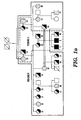

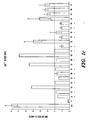


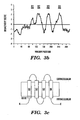
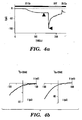
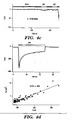
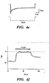
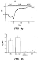


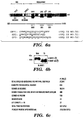
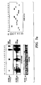
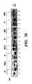
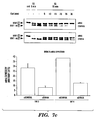


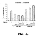

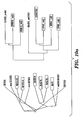


Classifications
-
- C—CHEMISTRY; METALLURGY
- C07—ORGANIC CHEMISTRY
- C07K—PEPTIDES
- C07K14/00—Peptides having more than 20 amino acids; Gastrins; Somatostatins; Melanotropins; Derivatives thereof
- C07K14/435—Peptides having more than 20 amino acids; Gastrins; Somatostatins; Melanotropins; Derivatives thereof from animals; from humans
- C07K14/46—Peptides having more than 20 amino acids; Gastrins; Somatostatins; Melanotropins; Derivatives thereof from animals; from humans from vertebrates
- C07K14/47—Peptides having more than 20 amino acids; Gastrins; Somatostatins; Melanotropins; Derivatives thereof from animals; from humans from vertebrates from mammals
- C07K14/4701—Peptides having more than 20 amino acids; Gastrins; Somatostatins; Melanotropins; Derivatives thereof from animals; from humans from vertebrates from mammals not used
- C07K14/4702—Regulators; Modulating activity
-
- G—PHYSICS
- G01—MEASURING; TESTING
- G01N—INVESTIGATING OR ANALYSING MATERIALS BY DETERMINING THEIR CHEMICAL OR PHYSICAL PROPERTIES
- G01N33/00—Investigating or analysing materials by specific methods not covered by groups G01N1/00 - G01N31/00
- G01N33/48—Biological material, e.g. blood, urine; Haemocytometers
- G01N33/50—Chemical analysis of biological material, e.g. blood, urine; Testing involving biospecific ligand binding methods; Immunological testing
- G01N33/5005—Chemical analysis of biological material, e.g. blood, urine; Testing involving biospecific ligand binding methods; Immunological testing involving human or animal cells
- G01N33/5008—Chemical analysis of biological material, e.g. blood, urine; Testing involving biospecific ligand binding methods; Immunological testing involving human or animal cells for testing or evaluating the effect of chemical or biological compounds, e.g. drugs, cosmetics
- G01N33/502—Chemical analysis of biological material, e.g. blood, urine; Testing involving biospecific ligand binding methods; Immunological testing involving human or animal cells for testing or evaluating the effect of chemical or biological compounds, e.g. drugs, cosmetics for testing non-proliferative effects
- G01N33/5041—Chemical analysis of biological material, e.g. blood, urine; Testing involving biospecific ligand binding methods; Immunological testing involving human or animal cells for testing or evaluating the effect of chemical or biological compounds, e.g. drugs, cosmetics for testing non-proliferative effects involving analysis of members of signalling pathways
-
- C—CHEMISTRY; METALLURGY
- C07—ORGANIC CHEMISTRY
- C07K—PEPTIDES
- C07K16/00—Immunoglobulins [IGs], e.g. monoclonal or polyclonal antibodies
- C07K16/18—Immunoglobulins [IGs], e.g. monoclonal or polyclonal antibodies against material from animals or humans
-
- A—HUMAN NECESSITIES
- A61—MEDICAL OR VETERINARY SCIENCE; HYGIENE
- A61K—PREPARATIONS FOR MEDICAL, DENTAL OR TOILETRY PURPOSES
- A61K35/00—Medicinal preparations containing materials or reaction products thereof with undetermined constitution
- A61K35/12—Materials from mammals; Compositions comprising non-specified tissues or cells; Compositions comprising non-embryonic stem cells; Genetically modified cells
-
- C—CHEMISTRY; METALLURGY
- C07—ORGANIC CHEMISTRY
- C07H—SUGARS; DERIVATIVES THEREOF; NUCLEOSIDES; NUCLEOTIDES; NUCLEIC ACIDS
- C07H21/00—Compounds containing two or more mononucleotide units having separate phosphate or polyphosphate groups linked by saccharide radicals of nucleoside groups, e.g. nucleic acids
- C07H21/04—Compounds containing two or more mononucleotide units having separate phosphate or polyphosphate groups linked by saccharide radicals of nucleoside groups, e.g. nucleic acids with deoxyribosyl as saccharide radical
-
- C—CHEMISTRY; METALLURGY
- C12—BIOCHEMISTRY; BEER; SPIRITS; WINE; VINEGAR; MICROBIOLOGY; ENZYMOLOGY; MUTATION OR GENETIC ENGINEERING
- C12N—MICROORGANISMS OR ENZYMES; COMPOSITIONS THEREOF; PROPAGATING, PRESERVING, OR MAINTAINING MICROORGANISMS; MUTATION OR GENETIC ENGINEERING; CULTURE MEDIA
- C12N15/00—Mutation or genetic engineering; DNA or RNA concerning genetic engineering, vectors, e.g. plasmids, or their isolation, preparation or purification; Use of hosts therefor
- C12N15/09—Recombinant DNA-technology
- C12N15/63—Introduction of foreign genetic material using vectors; Vectors; Use of hosts therefor; Regulation of expression
-
- C—CHEMISTRY; METALLURGY
- C12—BIOCHEMISTRY; BEER; SPIRITS; WINE; VINEGAR; MICROBIOLOGY; ENZYMOLOGY; MUTATION OR GENETIC ENGINEERING
- C12N—MICROORGANISMS OR ENZYMES; COMPOSITIONS THEREOF; PROPAGATING, PRESERVING, OR MAINTAINING MICROORGANISMS; MUTATION OR GENETIC ENGINEERING; CULTURE MEDIA
- C12N15/00—Mutation or genetic engineering; DNA or RNA concerning genetic engineering, vectors, e.g. plasmids, or their isolation, preparation or purification; Use of hosts therefor
- C12N15/09—Recombinant DNA-technology
- C12N15/87—Introduction of foreign genetic material using processes not otherwise provided for, e.g. co-transformation
- C12N15/90—Stable introduction of foreign DNA into chromosome
- C12N15/902—Stable introduction of foreign DNA into chromosome using homologous recombination
- C12N15/907—Stable introduction of foreign DNA into chromosome using homologous recombination in mammalian cells
-
- C—CHEMISTRY; METALLURGY
- C12—BIOCHEMISTRY; BEER; SPIRITS; WINE; VINEGAR; MICROBIOLOGY; ENZYMOLOGY; MUTATION OR GENETIC ENGINEERING
- C12N—MICROORGANISMS OR ENZYMES; COMPOSITIONS THEREOF; PROPAGATING, PRESERVING, OR MAINTAINING MICROORGANISMS; MUTATION OR GENETIC ENGINEERING; CULTURE MEDIA
- C12N2510/00—Genetically modified cells
-
- C—CHEMISTRY; METALLURGY
- C12—BIOCHEMISTRY; BEER; SPIRITS; WINE; VINEGAR; MICROBIOLOGY; ENZYMOLOGY; MUTATION OR GENETIC ENGINEERING
- C12N—MICROORGANISMS OR ENZYMES; COMPOSITIONS THEREOF; PROPAGATING, PRESERVING, OR MAINTAINING MICROORGANISMS; MUTATION OR GENETIC ENGINEERING; CULTURE MEDIA
- C12N5/00—Undifferentiated human, animal or plant cells, e.g. cell lines; Tissues; Cultivation or maintenance thereof; Culture media therefor
- C12N5/10—Cells modified by introduction of foreign genetic material
- C12N5/12—Fused cells, e.g. hybridomas
- C12N5/16—Animal cells
-
- C—CHEMISTRY; METALLURGY
- C12—BIOCHEMISTRY; BEER; SPIRITS; WINE; VINEGAR; MICROBIOLOGY; ENZYMOLOGY; MUTATION OR GENETIC ENGINEERING
- C12Q—MEASURING OR TESTING PROCESSES INVOLVING ENZYMES, NUCLEIC ACIDS OR MICROORGANISMS; COMPOSITIONS OR TEST PAPERS THEREFOR; PROCESSES OF PREPARING SUCH COMPOSITIONS; CONDITION-RESPONSIVE CONTROL IN MICROBIOLOGICAL OR ENZYMOLOGICAL PROCESSES
- C12Q1/00—Measuring or testing processes involving enzymes, nucleic acids or microorganisms; Compositions therefor; Processes of preparing such compositions
Definitions
- the invention relates to the field of regulation of a family of calcium regulated transcription factors known as NFAT proteins.
- Hyperactivity or inappropriate activity of the immune system is a serious and widespread medical problem. It contributes to acute and chronic immune diseases, e.g., allergic and atopic diseases, e.g., asthma, allergic rhinitis, allergic conjunctivitis and atopic dermatitis, and to autoimmune diseases, e.g., rheumatoid arthritis, insulin-dependent diabetes, inflammatory bowel disease, autoimmune thyroiditis, hemolytic anemia and multiple sclerosis. Hyperactivity or inappropriate activity of the immune system is also involved in transplant graft rejections and graft-versus-host disease.
- allergic and atopic diseases e.g., asthma, allergic rhinitis, allergic conjunctivitis and atopic dermatitis
- autoimmune diseases e.g., rheumatoid arthritis, insulin-dependent diabetes, inflammatory bowel disease, autoimmune thyroiditis, hemolytic anemia and multiple sclerosis.
- Hyperactivity or inappropriate activity of the immune system is also involved in transplant graf
- NFAT proteins nuclear factor of activated T cells
- the NFAT proteins are activated by an increase in intracellular calcium levels, e.g., by means of store-operated calcium entry.
- the activated NFAT proteins induce transcription of cytokine genes which are required for an immune response.
- the immunosuppressive drugs cyclosporin A and FK506 are potent inhibitors of cytokine gene transcription in activated immune cells, and have been reported to act by inhibiting calcineurin such that calcineurin is not able to activate NFAT. These drugs, however, can display nephrotoxic and neurotoxic effects after long term usage. Since calcineurin is ubiquitously expressed in many tissues, the drugs' inhibition of calcineurin activity toward substrates other than NFAT may contribute to the observed toxicity.
- EP-A-0976823 discloses certain methods for identifying inhibitors and activators of calcineurin.
- the present invention provides a recombinant cell as defined in and by any of the appended claims 1 to 7, a system comprising the recombinant cell of claim 1 and a monitoring agent used to monitor, detect or measure a calcium-entry mediated event, as defined in and by the appended claim 8, and a method of identifying an agent that modulates intracellular calcium comprising contacting a test agent with the recombinant cell, as defined in and by the appended claim 9.
- the recombinant cell according to the present invention comprises at least one heterologous nucleic acid encoding at least one NFAT regulator protein or a fragment thereof or a homologue thereof, wherein the NFAT regulator protein or fragment thereof or homologue thereof comprises an amino acid sequence at least 80% identical to that of and ORAI protein selected from the group consisting of ORAI1 (SEQ ID NO:1), ORAI2 (SEQ ID NO:2) or ORAI3 (SEQ ID NO:3); with the proviso that the recombinant cell is not a human embryonic cell.
- ORAI1 SEQ ID NO:1
- ORAI2 SEQ ID NO:2
- ORAI3 SEQ ID NO:3
- the present invention enables a method for identifying an agent that modulates NFAT activity or that modulates an NFAT regulator protein.
- Agents may modulate NFAT activity in a number of ways.
- an agent may modify NFAT activity by means of modulating intracellular calcium levels.
- the agent modulates at least one component of the CRAC channel, e.g., an ORAI protein, e.g., proteins encoded by ORAI1 (NM_032790; SEQ ID NO: 1), ORAI2 (BC069270; SEQ ID NO: 2), and/or ORAI3 (NM_152288; SEQ ID NO: 3).
- ORAI1 NM_032790; SEQ ID NO: 1
- ORAI2 BC069270; SEQ ID NO: 2
- ORAI3 NM_152288; SEQ ID NO: 3
- the agent modulates phosphorylation of NFAT, e.g., via modulation of a DYRK protein, e.g., proteins encoded by DYRK1A (NM_001396; SEQ ID NO:4), DYRK1B (NM_004714; SEQ ID NO:5), DYRK2 (NM_003583; SEQ ID NO:6), DYRK3 (NM_003582; SEQ ID NO:7), DYRK4 (NM_003845; SEQ ID NO:8) and/or DYRK6 (NM_005734; SEQ ID NO:9).
- a DYRK protein e.g., proteins encoded by DYRK1A (NM_001396; SEQ ID NO:4), DYRK1B (NM_004714; SEQ ID NO:5), DYRK2 (NM_003583; SEQ ID NO:6), DYRK3 (NM_003582; SEQ ID NO:
- the present invention enables a method of identifying an agent that modulates an NFAT regulator protein, comprising contacting at least one test agent with the recombinant cell; assessing the effect of the test agent on an activity, interaction, expression, or binding to the NFAT regulator protein or fragment or homologue thereof; and identifying the test agent that has an effect on an activity, interaction, expression, or binding to the NFAT regulator protein or fragment or homologue thereof, whereby the identified test agent is characterized as an agent that modulates an NFAT regulator protein.
- the NFAT regulator protein is encoded by at least one NFAT regulator selected from the group consisting of ORAI1 (SEQ ID NO: 1), ORAI2 (SEQ ID NO:2), ORAI3 (SEQ ID NO:3), DYRK1A (SEQ ID NO:4), DYRK1B (SEQ ID NO:5), DYRK2 (SEQ ID NO:6), DYRK3 (SEQ ID NO:7), DYRK4 (SEQ ID NO:8) and DYRK6 (SEQ ID NO:9).
- the NFAT regulator protein is encoded by at least one of the genes listed in Table I.
- assessing the effect of the test agent comprises using an antibody which specifically binds to a NFAT regulator protein encoded by ORAI1 (SEQ ID NO: 1), ORAI2 (SEQ ID NO: 2), ORAI3 (SEQ ID NO: 3), DYRK1A (SEQ ID NO:4), DYRK1B (SEQ ID NO:5), DYRK2 (SEQ ID NO:6), DYRK3 (SEQ ID NO:7), DYRK4 (SEQ ID NO:8), or DYRK6 (SEQ ID NO:9).
- the method further comprises assessing the effect of the test agent on electrical current across the plasma membrane of the cell. In one embodiment, the electrical current is due to flux of monovalent cations or divalent cations across the cell. In one embodiment, the method further comprises assessing the effect of the test agent on intracellular calcium within the cell. In one embodiment, the method further comprises identifying the test agent that has an effect on intracellular calcium within the cell, whereby the identified test agent is characterized as an agent that modulates intracellular calcium and an agent that modulates NFAT regulator protein.
- the cell comprises at least one heterologous NFAT regulator proteins or a fragment or homologue thereof.
- the cell according to the invention comprises heterologous nucleic acid encoding at least one NFAT regulator protein or a fragment or homologue thereof.
- the cell overexpresses, or underexpresses at least one NFAT regulator protein or fragment or derivative thereof.
- the present invention further provides a method of identifying an agent that modulates intracellular calcium, comprising contacting at least one test agent with the recombinant cell comprising at least one NFAT regulator protein or fragment or homologue thereof; assessing the effect(s) of the test agent on intracellular amounts, or concentrations, of cations or divalent cations within the cell, or on ion influx into the cell; and identifying the test agent that has an effect on intracellular amounts or concentrations of cations or divalent cations within the cell, or on ion influx into the cell, whereby the identified test agent is characterized as an agent that modulates intracellular calcium.
- the intracellular cation is calcium.
- assessing the effect of the test agent comprises monitoring calcium levels in the cytoplasm, monitoring calcium levels in an intracellular calcium store, monitoring calcium movement, or monitoring a calcium-entry mediated event. In one embodiment, the method further comprises assessing the effect of the test agent on an activity, interaction, expression, or binding to the NFAT regulator protein or fragment or derivative thereof.
- the NFAT regulator protein is encoded by at least one NFAT regulator selected from the group consisting of ORAI1 (SEQ ID NO: 1), ORAI2 (SEQ ID NO: 2), or ORAI3 (SEQ ID NO: 3), DYRK1A (SEQ ID NO:4), DYRK1B (SEQ ID NO:5), DYRK2 (SEQ ID NO:6), DYRK3 (SEQ ID NO:7), DYRK4 (SEQ ID NO:8) or DYRK6 (SEQ ID NO:9).
- the agent that modulates intracellular calcium is further characterized as an agent that modulates NFAT regulator protein.
- the recombinant cell comprises at least one heterologous NFAT regulator proteins or a fragment or derivative thereof. In one embodiment, the recombinant cell comprises a heterologous nucleic acid encoding at least one NFAT regulator proteins or fragment or derivative thereof. In one embodiment, the recombinant cell overexpresses at least one NFAT regulator protein or fragment or derivative thereof. In one embodiment, the recombinant cell exhibits dyshomeostasis. In one embodiment, the recombinant cell exhibits calcium dyshomeostasis
- the present invention further enables a method to screen for an agent that to modulates NFAT regulator function, comprising administering at least one test agent to the recombinant cell comprising at least one vector that comprises heterologous nucleic acid encoding at least one NFAT regulatory domain or a fragment or homologue thereof, operably linked to a sequence encoding a reporter protein; and monitoring intracellular localization of at least one expression product encoded by the vector, whereby a test agent that has an effect on intracellular localization of the expression product is characterized as an agent that modulates NFAT regulator function.
- the agent that modulates NFAT regulator function is associated with cytoplasmic or nuclear localization of the expression product.
- the cell is under resting conditions.
- the cell is stimulated with a calcium modulating agent. In one embodiment, the cell is stimulated with thapsigargin or ionomycin. In one embodiment, the cell is further administered a vector that comprises a heterologous nucleic acid encoding at least one NFAT regulator protein, or a fragment or derivative thereof. In one embodiment, the vector that comprises the heterologous nucleic acid encoding at least one NFAT regulator protein, or fragment or derivative thereof, is the same vector that comprises heterologous nucleic acid encoding at least one NFAT regulatory domain or a fragment or derivative thereof, operably linked to a sequence encoding a reporter protein.
- the present patent further describes, but does not claim, a method to diagnose unexplained immunodeficiency in a subject comprising sequencing at least 25 contiguous nucleotides in a gene from the subject corresponding to ORAI1 (SEQ ID NO: 1), ORAI2 (SEQ ID NO:2), ORAI3 (SEQ ID NO:3), DYRK1A (SEQ ID NO:4), DYRK1B (SEQ ID NO:5), DYRK2 (SEQ ID NO:6), DYRK3 (SEQ ID NO7), DYRK4 (SEQ ID NO:8), DYRK6 (SEQ ID NO:9), or any of the genes listed in Table I; and comparing the sequence of the subject's gene to the wild type sequence of the gene, wherein a variation between the gene from the wild type sequence indicates the subject's gene is responsible for the immunodeficiency.
- ORAI1 SEQ ID NO: 1
- ORAI2 SEQ ID NO:2
- ORAI3 SEQ
- the comparison comprises obtaining a biological sample from the subject, sequencing the DNA in the biological sample, and electronically aligning the DNA sequence obtained from the biological sample to a wild type sequence.
- the variation comprises a nucleotide mutation from C to T at position 271 of the coding sequence of ORAI1 (SEQ ID NO: 1).
- the unexplained immunodeficiency is associated with defects in regulation of NFAT activity.
- the variation comprises a mutation in a splice site.
- the variation comprises a nonsynonymous mutation.
- the present patent further describes, but does not claim, a method for identifying an agent for treating or preventing a disease or disorder associated with a NFAT regulator protein, comprising assessing the effects of a test agent on an organism exhibiting a disease or disorder associated with NFAT regulator protein; and identifying the test agent as an agent for treating or preventing a disease or disorder associated with NFAT regul ator protein if it has an effect on a phenotype of the organism associated with the disease or disorder, wherein the test agent modulates an activity, interaction, expression, or binding of, at least one NFAT regulator protein or fragment or derivative thereof.
- the organism comprises one or more cells that exhibit calcium dyshomeostasis. In one embodiment, the organism exhibits calcium dyshomeostasis.
- the phenotype on which the test agent has an effect is associated with the disease or disorder. This method is particularly useful, for diseases or conditions associated with altered regulation of intracellular calcium. In one embodiment, the disease or disorder is primarily attributable to deranged calcium signaling.
- the disease or disorder associated with NFAT regulator protein is rheumatoid arthritis, inflammatory bowel disease, allogeneic or xenogeneic transplantation rejection, graft-versus-host disease, aplastic anemia, psoriasis, lupus erytematosus, inflammatory disease, MS, type I diabetes, asthma, pulmonary fibrosis, scleroderma, dermatomyositis, Sjogren's syndrome, postpericardiotomy syndrome, Kawasaki disease, Hashimoto's thyroiditis, Graves' disease, myasthenia gravis, pemphigus vulgaris, autoimmune hemolytic anemia, idiopathic thrombopenia, chronic glomerulonephritis, Goodpasture's syndrome, Wegner's granulomatosis, multiple sclerosis, cystic fibrosis, chronic relapsing hepatitis, primary biliary cirrhosis, uveitis,
- Transplant graft rejections acquired immunodeficiencies, common variable immunodeficiency, myocardial hypertrophy, severe combined immunodeficiency, dilated cardiomyopathy, excessive or pathological bone resorption, excessive adipocyte differentiation, obesity, or reactivation of latent viruses.
- the present patent further describes, but does not claim an antibody which specifically binds to a NFAT regulator protein encoded by ORAI1 (SEQ ID NO: 1), ORAI2 (SEQ ID NO: 2), or ORAI3 (SEQ ID NO: 3), DYRK1A (SEQ ID NO:4), DYRK1B (SEQ ID NO:5), DYRK2 (SEQ ID NO:6), DYRK3 (SEQ ID NO:7), DYRK4 (SEQ ID NO:8) or DYRK6 (SEQ ID NO:9), or a homolog thereof.
- ORAI1 SEQ ID NO: 1
- ORAI2 SEQ ID NO: 2
- ORAI3 SEQ ID NO: 3
- DYRK1A SEQ ID NO:4
- DYRK1B SEQ ID NO:5
- DYRK2 SEQ ID NO:6
- SEQ ID NO:9 DYRK6
- the NFAT regulator protein used in the invention can be produced by a variety of means known in the art, e.g. automated peptide synthesis or culturing a host cell comprising a recombinant vector, the recombinant vector comprising a nucleic acid sequence, the nucleic acid sequence comprising/encoding the NFAT regulator or a fragment or derivative thereof, wherein the host cell is cultured under conditions suitable for expression of the NFAT regulator.
- the system according to the present invention may comprise an isolated cell comprising at least one heterologous NFAT regulator protein or fragment or derivative thereof, and/or at least one heterologous nucleic acid encoding a NFAT regulator protein or fragment or derivative thereof; provided that the recombinant cell of claim 1 is present; and a monitoring agent used to monitor, detect, or measure electrical current across the plasma membrane of the cell.
- the monitoring agent is an apparatus.
- the electrical current is due to flux of cations or divalent ions across the cell.
- the monitoring agent is used to monitor the effect of a test agent on intracellular calcium within the cell.
- the monitoring agent is used to monitor, detect, or measure a calcium-entry mediated event.
- the system according to the present invention may comprise a recombinant cell overexpressing at least one mammalian NFAT regulator protein or fragment or derivative thereof; provided that the recombinant cell of claim 1 is present; and a monitoring agent used to monitor, detect, or measure a calcium-entry mediated event.
- the NFAT regulator is encoded by ORAI1 (SEQ ID NO: 1), ORAI2 (SEQ ID NO: 2), or ORAI3 (SEQ ID NO: 3), DYRK1A (SEQ ID NO: 4), DYRK1B (SEQ ID NO: 5), DYRK2 (SEQ ID NO: 6), DYRK3 (SEQ ID NO: 7), DYRK4 (SEQ ID NO: 8) or DYRK6 (SEQ ID NO: 9).
- ORAI1 SEQ ID NO: 1
- ORAI2 SEQ ID NO: 2
- ORAI3 SEQ ID NO: 3
- DYRK1A SEQ ID NO: 4
- DYRK1B SEQ ID NO: 5
- DYRK2 SEQ ID NO: 6
- DYRK3 SEQ ID NO: 7
- DYRK4 SEQ ID NO: 8
- DYRK6 SEQ ID NO: 9
- the cells for use in the present invention may comprise a recombinant cell comprising at least one heterologous NFAT regulator protein or fragment or derivative thereof, and/or at least one heterologous nucleic acid encoding a NFAT regulator protein or fragment or derivative thereof; provided that the recombinant cell of claim 1 is present.
- the recombinant cell overexpresses at least one mammalian NFAT regulator protein or fragment or derivative thereof.
- the cell according to the present invention may be a recombinant cell overexpressing at least on mammalian NFAT regulator protein or fragment or derivative thereof.
- the present invention enables a method for identifying an agent for treating or preventing a disease or disorder associated with calcium signaling.
- the method comprises assessing the effects of a test agent on an organism exhibiting the disease or disorder, and identifying the test agent as an agent for treating or preventing the disease or disorder if it modulates an activity, interaction, expression, or binding of at least one NFAT regulator protein or fragment thereof.
- the disease or disorder is rheumatoid arthritis, inflammatory bowel disease, allogeneic or xenogeneic transplantation rejection, graft-versus-host disease, aplastic anemia, psoriasis, lupus erytematosus, inflammatory disease, MS, type I diabetes, asthma, pulmonary fibrosis, scleroderma, dermatomyositis, Sjogren's syndrome, postpericardiotomy syndrome, Kawasaki disease, Hashimoto's thyroiditis, Graves' disease, myasthenia gravis, pemphigus vulgaris, autoimmune hemolytic anemia, idiopathic thrombopenia, chronic glomerulonephritis, Goodpasture's syndrome, Wegner's granulomatosis, multiple sclerosis, cystic fibrosis, chronic relapsing hepatitis, primary biliary cirrhosis, uveitis, allergic rhinitis,
- Transplant graft rejections acquired immunodeficiencies, common variable immunodeficiency, myocardial hypertrophy, severe combined immunodeficiency, dilated cardiomyopathy, excessive or pathological bone resorption, excessive adipocyte differentiation, obesity, or reactivation of latent viruses.
- Figures 1A-1C show gene-dosage effect in store-operated Ca 2+ entry (SOCE).
- Figure 1A shows a pedigree of patients with a defect in SOCE and CRAC channel function.
- Two male SCID patients (subject ID numbers 8 and 11; filled black squares) were born to consanguineous parents (subject ID numbers 35 and 36).
- DNA and blood samples were obtained from all individuals shown in yellow or black.
- Half black squares or circles indicate heterozygous disease carriers as determined by phenotypic analysis.
- Double horizontal bars indicate consanguineous marriages, black boxes SCID disease, diagonal bars death of individuals.
- Figure 1B shows reduced SOCE in T cells of both parents of CRAC deficient SCID patients that defines them as heterozygous carriers of the disease trait.
- T cells were stimulated with thapsigargin (TG) in the absence of extracellular Ca 2+ .
- the peak (upper panel) and rate (bottom panel) of Ca 2+ influx were measured after readdition of 0.5 mM extracellular Ca 2+ .
- Figure 1C shows reduced SOCE that phenotypically identifies 12 / 21 family members of the SCID patients as heterozygous disease trait carriers.
- Ca 2+ influx was measured as described in B but using 0.2 mM extracellular Ca 2+ . Shown are the averages of Ca 2+ influx rates from 4-5 experiments. Individual ID numbers correspond to those shown in Fig. 1A . Stars indicate heterozygous carriers as defined by influx rates below 2 nM/s (dotted red line).
- Co healthy control
- P patient.
- Figures 2A-2B show that a genome-wide RNAi screen identifies Drosophila Orai as a protein regulating NFAT translocation and store-operated Ca 2+ entry.
- Figure 2A shows that RNAi of dSTIM or dOrai inhibits dephosphorylation of NFAT.
- S2R+ cells stably transfected with NFAT1(1-460)-GFP were incubated for 4 days with double-stranded (ds) RNAi against dSTIM, dOrai or an irrelevant DNA sequence (mock).
- RNAi of either dSTIM or dOrai inhibits Ca 2+ influx in S2R+ cells.
- Cells were left unstimulated (-TG) or stimulated with thapsigargin (+TG) for 10 min, then loaded with Fluo-4 and Fura-Red and analyzed for Ca 2+ influx by flow cytometry.
- RNAi for Gfp RNAi for Gfp
- RNAi for dSTIM or dOrai RNAi for dSTIM or dOrai.
- Decreased Ca 2+ influx is indicated by the much reduced change in emission ratio following addition of thapsigargin.
- Figures 3A-3C show that Orail is a transmembrane protein.
- Figure 3A shows that Orai1 is highly conserved in eukaryotes. Shown is the sequence conservation in the first of four putative transmembrane regions (M1, underlined) of Orai1, which contains the R>W mutation (bold) found in the SCID patients.
- Figure3B shows membrane topology of Orai1. Hydropathy plots were calculated from the full-length amino acid sequence of human Orai1 (301 a.a., NP_116179) using the Kyte-Doolittle algorithm with a window size of 19 amino acids.
- FIG. 3C shows schematic representation of the predicted membrane topology of Orai1, based on the hydropathy plot and immunocytochemistry data. The site of the R>W mutation in the SCID patients is indicated by a dark box.
- Figures 4A-4H show that expression of Orai 1 restores CRAC channel function in SCID T cells.
- Figure 4A shows activation of an inward current in an Orai WT -complemented SCID T cell by passive store depletion with a pipette solution containing 8 mM BAPTA. At the indicated times, the 20 mM Ca 2+ solution was replaced with a divalent free (DVF) solution.
- FIG. 4B shows the current-voltage (I-V) relation of currents in 20 mM Ca 2+ . (left) and in DVF solution (right) measured during voltage ramps from -100 to +100 mV. Data were collected at the times indicated by the arrows in 4A. Note that the Ca 2+ current I-V relation is inwardly rectifying with a reversal potential > +90 mV. In DVF solution, the current reversed at ⁇ +50 mV.
- Figure 4C shows that SCID T cells expressing mutant Orai1 R>W, inward Ca 2+ and Na + currents fail to develop during passive store depletion by 8 mM BAPTA.
- Figure 4D shows noise characteristics of the depotentiating Na + current.
- Top graph shows the mean current at a constant holding potential of ⁇ 100 mV. The dotted line indicates the zero current level (measured in 20 mM Ca 2+ + 2 ⁇ M La 3+ ). Variance was calculated from 100-ms segments of the Na + current and plotted against mean current in lower panel. The data are fit by a straight line with a slope of 26 fA, giving a lower limit to the unitary current.
- Figure 4E shows fast inactivation of the Ca 2+ current in a SCID T cell expressing Orai WT . Fast inactivation was measured during 300-ms steps to -100 mV from a holding potential of +30 mV with 20 mM Ca 2+ o .
- Figure 4F shows blockade of the Ca 2+ current by 2 ⁇ M La 3+ . After passive induction of the inward current in a SCID T cell expressing Orai WT , 2 ⁇ M La 3+ was applied. The dotted line indicates the zero current level, determined from traces collected at the beginning of the experiment immediately following whole-cell break-in.
- Figure 4G shows potentiation and blockade of I CRAC by application, respectively, of low (5 ⁇ M) and high (40 ⁇ M) doses of 2-APB.
- Figure 4H shows the summary of peak current densities in the indicated cell categories. Peak currents were measured during steps to -100 mV. Reconstitution with wild-type Orai1 thus reconstitutes a current with the expected characteristics of native CRAC channels. Cells transduced with Orai1 WT or Orai R>W were visually selected based on GFP-fluorescence; untransduced cells were GFP-negative.
- Figures 5A-5D show that expression of Orai1 in fibroblasts from SCID patients restores store-operated Ca 2+ influx.
- Figure 5A shows inhibition of Ca 2+ influx in Orai1 WT expressing SCID fibroblasts by 75 ⁇ M 2-APB.
- Figure 5B shows potentiation of Ca 2+ influx in Orai 1 WT -expressing SCID fibroblasts by 3 ⁇ M 2-APB.
- Figures 5C-5D shows inhibition of Ca 2+ influx in Orai 1 WT -expressing SCID fibroblasts by 2 ⁇ M La 3+ added before ( Fig. 5C ) or after ( Fig. 5D ) readdition of 20 mM Ca 2+ .
- ⁇ 15-20 GFP-positive fibroblasts were analyzed. Experiments were repeated at least three times for each protocol.
- Figures 6A-6C show the NFAT regulatory domain and results of the genome-wide RNAi screen in Drosophila.
- Figure 6A shows a schematic diagram of the N-terminal regulatory domain of NFAT1, showing the conserved phosphorylated serine motifs which are dephosphorylated upon stimulation (circles). Peptides corresponding to the SRR1, SP2 and SP3 motifs used for in vitro kinase assays are represented. Serine residues shown underlined have been identified to be phosphorylated in NFAT1 in vivo, and these are the residues mutated to alanine in the mutant SP2 and SP3 motifs.
- FIG. 6B shows that heterologously expressed NFAT is correctly regulated by Ca 2+ and calcineurin in Drosophila S2R+ cells.
- Drosophila S2R+ cells were transfected with NFAT1-GFP expression vector. 48 hrs later, the cells were left untreated (Untr) or treated with thapsigargin (TG, 1 ⁇ M) for 30 min and lysates from the cells were analysed by immunoblotting (IB) with anti-NFAT1.
- IB immunoblotting
- P and deP refer to the migration positions of phosphorylated and dephosphorylated NFAT-GFP, respectively.
- Figure 6C shows the tabulation of the results of the primary screen.
- Figures 7A-7C shows screening of candidate kinases identified in the Drosophila S2R+ cell RNAi screen, for NFAT phosphorylation and identification of DYRK as a negative regulator of NFAT.
- Figure 7A shows the ability of overexpressed mammalian homologs of the candidate kinases to directly phosphorylate the NFAT regulatory domain.
- FLAG-tagged mammalian homologues of selected Drosophila kinases were expressed in HEK293 cells, and immunopurified kinases were tested using an in vitro kinase assay for phosphorylation of GST-NFAT1(1-415). Phosphorylation levels were assessed by autoradiography with either short (top panel) or long (middle panel) exposures.
- kinases tested are as follows: lane 1, CK1 ⁇ ; lane 2,CK1 ⁇ ; lane 3, Bubl; lane 4, STK38; lane 5, STK38L; lane 6, CDC42BPA; lane 7, ARAF; lane 8. PRKG1; lane 9, SGK; lanes 10 and 11, CSNKA1 and CSNKA2 (CKII isoforms); lane 12, SRPK1; lane 13, DYRK2; lane 14, ALS2CR7; lane 15, IRAK4. Bubl was later dropped from our candidate list because of >10 predicted off-targets (Example 3).
- FIG. 7B shows overexpression of DYRK2 blocks calcineurin-mediated dephosphorylation of NFAT1.
- Each kinase was co-transfected with NFAT-GFP into HEK293 cells; after 18 hrs cells were stimulated with 1 ⁇ M ionomycin in the presence of 2 mM CaCl 2 . Lysates were immunoblotted using NFAT1 antibody. Relative expression levels of the kinases were determined by immunoblot using anti-FLAG antibody, and were identical to those represented in Fig 6A (bottom panel).
- Figure 7C shows depletion of endogenous DYRK1A potentiates NFAT activation.
- HeLa cells stably expressing Ha-tagged NFAT1-GFP were transfected with control siRNA or DYRK1A-specific siRNA. After 4 days cells were stimulated with 1 ⁇ M thapsigargin (TG) or 1 ⁇ M thapsigargin (TG) followed by 20 nM CsA for indicated times; lysates were immunoblotted for NFAT-GFP using anti-HA antibody (left). DYRK1A mRNA levels (right) were assessed after 3 and 4 days by real-time PCR. siControl, scrambled control siRNA; siDYRK1A, DYRK1A-specific siRNA. Results show the average and standard deviation of three independent experiments.
- Figures 8A-8C show that DYRK2 inhibits NFAT-dependent reporter activity and endogenous IL-2 expression.
- Figure 8A shows that overexpression of DYRK2 inhibits IL2 promoter-driven luciferase activity in stimulated Jurkat T cells.
- the IL2 promoter is an example of a cytokine promoter whose activation exhibits a strong requirement for NFAT.
- Exponentially growing Jurkat T cells were co-transfected with pRLTK (renilla luciferase, internal control), IL-2-pGL3 (IL-2-promoter driven firefly luciferase, experimental promoter) and empty vector or increasing amounts of wild type (WT) or kinase dead (KD) DYRK2 expression plasmids (5, 10, 15 and 20 ⁇ g). After 24 h cells untreated or stimulated with PMA and ionomycin for 6 h were analyzed for IL-2-promoter-driven luciferase activity.
- FIG. 8B shows that overexpression of DYRK2 inhibits endogenous IL-2 expression in stimulated Jurkat T cells. Exponentially-growing Jurkat T cells were co-transfected with GFP and empty vector or increasing amounts of wild type (WT) or kinase dead (KD) DYRK2 expression plasmids (10, 20 and 30 ⁇ g).
- Figures 9A-9C shows that STIM proteins affect NFAT localization by altering store-operated Ca 2+ influx.
- Figure 9A shows the percent of cells with nuclear NFAT was quantified in three independent experiments after mock treatment or treatment with dsRNAs against dSTIM. Mean and standard deviations are plotted. 50-100 cells were analyzed for each experiment.
- Figure 9B shows the effect of RNAi-mediated depletion of Drosophila STIM (dSTIM) on NFAT phosphorylation status. Lysates made from unstimulated or thapsigargin (TG)-stimulated S2R+ cells were examined by immunoblotting with antibody against NFAT1. The cells were mock-treated or treated for 4 days with dsRNAs targeting dSTIM.
- dSTIM Drosophila STIM
- Figure 9C shows intracellular Ca 2+ levels, analyzed by flow cytometry, in S2R+ cells depleted with dSTIM or novel gene candidates from the confirmatory screen.
- GFP dsRNA was used as a control for non-specific effects caused by dsRNA treatment.
- I ⁇ M thapsigargin was added (arrow) and [Ca 2+ ] I measurements were continued for a further 5 min. Depletion of dSTIM almost completely abolishes thapsigargin-triggered, that is store-operated, Ca 2+ influx.
- Figures 10A-10B shows the phylogenetic relation between different members of the DYRK family in Drosophila and in humans, and the expression pattern of human DYRKs in Jurkat T cells.
- Figure 10A shows the phylogenetic tree of DYRK family kinases using distance-based methods (neighbour joining).
- the left-hand side figures show the homology relationships between Drosophila CG40478 and human DYRK 2, 3; Drosophila CG4551 (smi35A) and human DYRK 4; Drosophila CG7826 (mnb) and human DYRK1 A, B (top); as computed by the program Tcoffee.
- the orthologue bootstrap value for CG40478-DYRK2 is higher than for CG40478-DYRK3 (top). Therefore, DYRK2 is an orthologue of CG40478 (the genes diverged by a speciation event), while DYRK3 may be a paralogue (the genes diverged by a duplication event).
- the calculations of the ortholog bootstrap values were performed with Orthostrapper.
- Figure 10B shows expression of DYRK family members in Jurkat T cells. Expression level of mammalian DYRK mRNAsin Jurkat T cells was estimated by RT-PCR analysis. Primers correspond to:
- Figures 11A-11I show the nucleotide sequences for NFAT regulator genes.
- Figure 11A shows the nucleotide sequence ORAI1 (NM_032790; SEQ ID NO:1).
- Figure 11B shows the nucleotide sequence for ORAI2 (BC069270; SEQ ID NO:2).
- Figure 11C shows the nucleotide sequence for ORAI3 (NM_152288; SEQ ID NO:3).
- Figure 11D shows the nucleotide sequence for DYRK1A (NM_001396; SEQ ID NO:4).
- Figure 11E shows the nucleotide sequence for DYRK1B (NM_004714; SEQ ID NO:5).
- Figure 11F shows the nucleotide sequence for DYRK2 (NM_003583; SEQ ID NO:6).
- Figure 11G shows the nucleotide sequence for DYRK3 (N_003582; SEQ ID NO:7).
- Figure 11H shows the nucleotide sequence for DYRK4 (NM_003845; SEQ ID NO:8).
- Figure 11I shows the nucleotide sequence for DYRK6 (NM_005734; SEQ ID NO:9).
- aspects of the present description relate to the characterization of genes regulating NFAT activity, for example, via Store-Operated Calcium Entry (SOCE) or via modulation of NFAT phosphorylation.
- SOCE Store-Operated Calcium Entry
- CRAC Ca 2+ release-activated Ca 2+
- aspects of the description relate to novel regulators of NFAT activity, particularly with regard to modulation of NFAT activity in T cells.
- aspects of the description also relate to methods to screen for novel agents that modulate NFAT activity.
- aspects of the description further relate to methods to screen for agents that modulate the activity of the NFAT regulators.
- the invention further provides methods to screen for agents that modulate the NFAT regulators of the present invention by means of modulating intracellular calcium.
- NFAT protein nuclear factor of activated T cells
- NFAT protein a member of a family of transcription factors comprising the members NFAT1, NFAT2, NFAT3 and NFAT4, with several isoforms. Any other NFAT protein whose activation is calcineurin dependent is also meant to be included.
- NFAT proteins can be, e.g., mammalian proteins, e.g., human or murine.
- NFAT1, NFAT2 and NFAT4 are expressed in immune cells, e.g., T lymphocytes, and play a role in eliciting immune responses.
- NFAT proteins are involved in the transcriptional regulation of cytokine genes, e.g., IL-2, IL-3, IL-4, TNF-alpha and IFN-gamma, during the immune response.
- the conserved regulatory domain of NFAT is an N-terminal region of NFAT which is about 300 amino acids in length.
- the conserved regulatory domain of murine NFAT1 is a region extending from amino acid residue 100 through amino acid residue 397
- human NFAT1 is a region extending from amino acid residue 100 through 395
- human NFAT2 is a region extending from amino acid residue 106 through 413
- human NFAT2b is a region extending from amino acid residue 93 through 400
- human NFAT3 is a region extending from amino acid residue 102 through 404
- human NFAT4 is a region extending from amino acid residue 97 through 418.
- the conserved regulatory domain is moderately conserved among the members of the NFAT family, NFAT1, NFAT2, NFAT3 and NFAT4.
- the conserved regulatory region binds directly to calcineurin.
- the conserved regulatory region is located immediately N-terminal to the DNA-binding domain (amino acid residues 398 through 680 in murine NFAT1, amino acid residues 396 through 678 in human NFAT1, amino acid residues 414 through 696 in human NFAT2, amino acid residues 401 through 683 in human NFAT2b, amino acid residues 405 through 686 in human NFAT3, and amino acid residues 419 through 700 in human NFAT4).
- SOCE is one of the main mechanisms to increase intracellular cytoplasmic free Ca 2+ concentrations ([Ca 2+ ]i) in electrically non-excitable cells.
- Ca 2+ elevations are a crucial signal transduction mechanism in virtually every cell.
- the tight control of intracellular Ca 2+ and its utility as a second messenger, is emphasized by the fact that [Ca 2+ ]i levels are typically 70-100 nM while extracellular Ca 2+ levels ([Ca 2+ ]ex) are 10 4 -fold higher, ⁇ 1-2 mM.
- the immediate source of Ca 2+ for cell signaling can be either intracellular or extracellular ( Figure 1 ).
- Intracellular Ca 2+ is released from ER stores by inositol 1,4,5-triphosphate (IP3), or other signals, while extracellular Ca 2+ enters the cell through voltage-gated, ligand-gated, store-operated or second messenger-gated Ca 2+ channels in the plasma membrane.
- IP3 inositol 1,4,5-triphosphate
- the major mechanism for Ca 2+ entry is store-operated Ca 2+ entry, a process controlled by the filling state of intracellular Ca 2+ stores.
- Depletion of intracellular Ca 2+ stores triggers activation of membrane Ca 2+ channels with specific electrophysiological characteristics, which are referred to as calcium release-activated Ca 2+ (CRAC) channels (Parekh and Putney, Jr. 2005, Physiol Rev 85:757).
- CRAC calcium release-activated Ca 2+
- CRAC channels Ca 2+ release activated Ca 2+ (CRAC) channels.
- the electrophysiological characteristics of CRAC channels have been studied intensively, but the molecular nature of the channel itself and the mechanisms of its activation remain unknown.
- One definition of CRAC channels holds that depletion of intracellular Ca 2+ stores is both necessary and sufficient for channel activation without direct need for increases in [Ca 2+ ]i, inositol phosphates IP3 or IP4, cGMP or cAMP (Parekh and Penner. 1997, Physiol Rev. 77:901).
- CRAC current is defined, amongst other criteria, by its activation as a result of ER Ca 2+ store depletion, its high selectivity for Ca 2+ over monovalent (Cs + , Na + ) cations, a very low single channel conductance, a characteristic I-V relationship with pronounced inward rectification and its susceptibility to pharmacological blockade for instance by La 3+ and 2-APB (100 ⁇ M), respectively (Parekh and Putney, Jr. 2005, Physiol Rev 85:757; Lewis, 2001, Annu Rev Immunol 19:497).
- Candidate genes for SOCE and CRAC The molecular nature of the CRAC channel remains completely unknown.
- the most widely investigated candidate genes for the CRAC channel have been the > 25 mammalian homologues of the Drosophila photoreceptor TRP (Transient Receptor Potential) gene.
- TRP Transient Receptor Potential
- TRPV6 was the most promising CRAC channel candidate gene because some of its biophysical features overlapped with that of CRAC.
- TRPV6 like CRAC, selectively conducts Ca 2+ , it is not activated by store depletion, a defining characteristic of the CRAC channel.
- the "conformational coupling model” postulates a conformational change of a molecule at the surface of the ER which then binds to the CRAC channel;
- the "secretion coupling model” suggests that (constitutively active) CRAC channels reside in intracytoplasmic vesicles that fuse to the plasma membrane upon store depletion;
- the "Calcium influx factor (CIF) model” predicts a soluble small molecule, which activates Ca 2+ influx through CRAC channels when CIF is released into the cytoplasm of stimulated cells.
- STIM1 Stromal interaction molecule 1 (STIM1). Recent evidence suggests that STIM1 plays an important role in store operated Ca 2+ entry and CRAC channel function.
- STIM1 is a type I transmembrane protein which was initially characterized as a stromal protein promoting the expansion of pre-B cells and as a putative tumor suppressor ( Oritani, et al.
- STIM1 contains a Ca 2+ binding EF hand motif and a sterile ⁇ -motif (SAM) domain in its ER / extracellular region, a single membrane-spanning domain, and two predicted cytoplasmic coiled-coil regions ( Manji et al. 2000, Biochim Biophys Acta 1481:147 ).
- STIM2 Domain structure and genomic organization are conserved in a related gene called STIM2, which differs from STIM1 mainly in its C-terminus ( Williams et al. 2002, Biochim Biophys Acta 1596:131 ). STIM1 is able to homodimerize or heterodimerize with STIM2 (Williams et al. 2002 supra). Expressed in the ER, its C-terminal region is located in the cytoplasm whereas the N-terminus resides in the lumen of the ER, as judged by glycosylation and phosphorylation studies (Maji et al. 2000 supra; Williams et al. 2002 supra). A minor fraction of STIM 1 is located in the plasma membrane.
- RNAi mediated suppression of STIM1 expression interferes with SOCE and CRAC channel function, STIM 1 is unlikely to be a Ca 2+ channel itself. Rather it is thought that STIM 1 may sense Ca 2+ levels in the ER via its EF hand ( Putney, Jr. 2005. J Cell Biol 169:381 ; Marchant, 2005, Curr Biol 15:R493 ). Consistent with the conformational coupling model of store-operated Ca 2+ influx, STIM1 could act as a key adapter protein, which physically bridges the space between ER and plasma membrane, and thus directly connects sensing of depleted Ca 2+ stores to store-operated Ca 2+ channels in the plasma membrane (Putney, Jr. 2005. supra; Putney, Jr. 1986, Cell Calcium 7:1 ).
- NFAT regulators is used to refer to the proteins (NFAT regulator proteins), and the encoding genes (NFAT regulator genes) which regulate NFAT activity.
- the methods described herein are intended to include use ofhomologues, analogues, isoforms (e.g. alternative splice variants), derivatives, and functional fragments of the NFAT regulators described herein.
- homologues of NFAT regulator proteins have at least 70%, more preferably, 80%, and more preferably 90% amino acid identity to those specifically identified herein.
- the NFAT regulator proteins are encoded by the ORAI genes.
- ORAI1 nucleic acid sequence corresponds to GenBank accession number NM_032790
- ORAI2 nucleic acid sequence corresponds to GenBank accession number BC069270
- ORAI3 nucleic acid sequence corresponds to GenBank accession number NM_152288.
- ORAI refers to any one of the ORAI genes, e.g., ORAI1, ORAI2, ORAI3.
- the NFAT regulator proteins are encoded by the DYRK genes.
- the DYRK genes were not known to regulate NFAT activity or function.
- DYRK1A is encoded by several nucleic acid isoforms including GenBank accession numbers NM_001396, NM_101395, NM_130436, NM_130437, and NM_130438.
- DYRK1B is encoded by multiple nucleic acid isoforms including GenBank accession numbers NM_004714, NM_006483, and NM_006484.
- DYRK2 is encoded by GenBank accession numbers including NM_003583 and NM_006482.
- DYRK3 is encoded by GenBank accession numbers including NM_001004023 and NM_003582.
- DYRK4 is encoded by GenBank accession number NM 003845.
- DYRK6, also known as HIPK3, is encoded by GenBank accession number NM_005734.
- the NFAT regulator proteins are encoded by the genes listed in Table I.
- fragment when referring to a NFAT regulator protein means proteins or polypeptides which retain essentially the same biological function or activity in at least one assay as the native NFAT regulator proteins.
- the NFAT regulator fragments or derivatives of the present invention maintain at least about 50% of the activity of the native proteins, preferably at least 75%, more preferably at least about 95% of the activity of the native proteins, as determined e.g. by a calcium influx assay described in Example 1.
- Fragments or derivatives as the term is used herein can include competitors of the native NFAT regulators with respect to a particular NFAT regulator domain activity. However, the fragment or derivative shows an overall similarity to NFAT regulators in other areas as explained herein.
- fragment refers to a fragment of the NFAT regulator protein, or nucleic acid sequence, wherein the (encoded) protein retains at least one biological activity of the full length NFAT regulator protein.
- fragment and functional fragment are used herein interchangeably.
- a fragment of a sequence contains less nucleotides or amino acids than the corresponding full length sequences, wherein the sequences present are in the same consecutive order as is present in the full length sequence.
- a fragment does not contain internal insertions or deletions of anything (e.g. nucleic acids or amino acids) in to the portion of the full length sequence represented by the fragment. This is in contrast to a derivative, which may contain internal insertions or deletions within the nucleic acids or amino acids that correspond to the full length sequence, or may have similarity to full length coding sequences.
- a derivative may comprise the same or different number of nucleic acids or amino acids as full length sequences.
- the term derivative, as used herein with respect to an NFAT regulator protein includes NFAT regulator proteins, or fragments thereof, which contain one or more modified amino acids. e.g. chemically modified, or modification to the amino acid sequence (substitution, deletion, or insertion). Such modifications should substantially preserve at least one biological activity of the NFAT regulator protein. Such biological activity is readily determined by a number of assays known in the art, for example, a calcium influx assay described below in Example 1.
- a derivative may be prepared by standard modifications of the side groups of one or more amino acid residues of the NFAT regulator protein, its analog, or a functional fragment thereof, or by conjugation of the NFAT regulator protein, its analogs or fragments, to another molecule e.g. an antibody, enzyme, receptor, etc., as are well known in the art.
- derivatives covers derivatives which may be prepared from the functional groups which occur as side chains on the residues or the N- or C-terminal groups, by means known in the art, and are included in the invention. Derivatives may have chemical moieties such as carbohydrate or phosphate residues.
- Such a derivativization process should preserve at least one biological activity of the NFAT regulator protein.
- Derivatives can be made for convenience in expression, for convenience in a specific assay, to enhance detection, or for other experimental purposes.
- Derivatives include dominant negatives, dominant positives and fusion proteins.
- Antibodies to the NFAT regulators can be prepared that will bind to one or more particular domains of a peptide and can be used to modulate NFAT regulator gene or protein activity.
- an antibody against an NFAT regulator protein or fragment or derivative thereof preferably monoclonal or monospecific, to mammalian cells (including human cells) can reduce or abrogate NFAT induced transcription of immune system associated genes, thus serving to treat hyperactivity or inappropriate activity of the immune system.
- Administration of an activating antibody against an NFAT regulator protein or fragment or derivative thereof, e.g. an Orai protein may serve to treat hypoactivity of the immune system by activating NFAT and thereby inducing transcription of immune response associated genes.
- Administration of an antibody against an NFAT regulator protein or fragment or derivative thereof, e.g., a DYRK protein may serve to treat hypoactivity of the immune system by activating NFAT and thereby inducing transcription of immune response associated genes.
- the present patent also describes antibodies that bind a protein or peptide encoded by all or a portion of the NFAT regulator nucleic acid molecule, as well as antibodies which bind the protein or peptide encoded by all or a portion of a variant nucleic acid molecule; for instance, polyclonal and monoclonal antibodies which bind to the described polypeptide or protein, or fragments or derivatives thereof,
- Antibodies can be produced using known methods.
- An animal such as a mouse, goat, chicken or rabbit, can be immunized with an immunogenic form of the protein or peptide (an antigenic fragment of the protein or peptide which is capable of eliciting an antibody response).
- Techniques for conferring immunogenicity on a protein or peptide include conjugation to carriers or other techniques well known in the art.
- the protein or peptide can be administered in the presence of an adjuvant.
- the progress of immunization can be monitored by detection of antibody titers in plasma or serum. Standard ELISA or other immunoassays can be used with immunogen as antigen to assess the levels of antibody.
- Anti-peptide antisera can be obtained, and if desired, polyclonal antibodies can be isolated from the serum.
- Monoclonal antibodies can also be produced by standard techniques which are well known in the art ( Kohler and Milstein, Nature 256:4595-497 (1975 ); Kozbar et al., Immunology Today 4:72 (1983 ); and Cole et al., Monoclonal Antibodies and Cancer Therapy, Alan R. Liss, Inc., pp. 77-96 (1985 )).
- Such antibodies are useful as diagnostics for the intact or disrupted gene, and also as research tools for identifying either the intact or disrupted gene.
- a monoclonal antibody to NFAT regulator proteins may be identified and isolated by screening a recombinant combinatorial immunoglobulin library (e.g., an antibody phage display library) to thereby isolate immunoglobulin library members that bind to NFAT regulator proteins.
- Kits for generating and screening phage display libraries are commercially available from, e.g., Dyax Corp. (Cambridge, Mass.) and Maxim Biotech (South San Francisco, Calif.). Additionally, examples of methods and reagents particularly amenable for use in generating and screening antibody display libraries can be found in the literature.
- Polyclonal sera and antibodies may be produced by immunizing a suitable subject, such as a rabbit, with NFAT regulator proteins (preferably mammalian; more preferably human) or an antigenic fragment thereof.
- NFAT regulator proteins preferably mammalian; more preferably human
- the antibody titer in the immunized subject may be monitored over time by standard techniques, such as with ELISA, using immobilized marker protein.
- the antibody molecules directed against NFAT regulator proteins may be isolated from the subject or culture media and further purified by well-known techniques, such as protein A chromatography, to obtain an IgG fraction.
- Fragments of antibodies to NFAT regulator proteins may be produced by cleavage of the antibodies in accordance with methods well known in the art. For example, immunologically active F(ab') and F(ab') 2 fragments may be generated by treating the antibodies with an enzyme such as pepsin. Additionally, chimeric, humanized, and single-chain antibodies to NFAT regulator proteins, comprising both human and nonhuman portions, may be produced using standard recombinant DNA techniques. Humanized antibodies to NFAT regulator proteins may also be produced using transgenic mice that are incapable of expressing endogenous immunoglobulin heavy and light chain genes, but which can express human heavy and light chain genes.
- the methods described herein can also be utilized to treat, or identify agents useful in treatment of, conditions and diseases associated with NFAT disregulation/disfunction and/or Calcium signaling.
- diseases include, without limitation, immune system diseases involving hyperactivity or inappropriate activity of the immune system, e.g., acute immune diseases, chronic immune diseases and autoimmune diseases
- diseases include rheumatoid arthritis, inflammatory bowel disease, allogeneic or xenogeneic transplantation rejection (organ, bone marrow, stem cells, other cells and tissues), graft-versus-host disease, aplastic anemia, psoriasis, lupus erytematosus, inflammatory disease, MS, type I diabetes, asthma, pulmonary fibrosis, scleroderma, dermatomyositis, Sjogren's syndrome, postpericardiotomy syndrome, Kawasaki disease, Hashimoto's thyroiditis, Graves' disease, myasthenia gravis, pemphigus vulgaris, autoimmune
- Transplant graft rejections can result from tissue or organ transplants.
- Graft-versus-host disease can result from bone marrow or stem cell transplantation.
- Immune system diseases involving hypoactivity of the immune system include, e.g., immunodeficiency diseases including acquired immunodeficiencies, such as HIV disease, and common variable immunodeficiency (CVID).
- the methods described herein can also be utilized to treat or identify agents useful in treatment of conditions and diseases that are not immune mediated, but which nevertheless involve the Ca 2+- calcineurin-mediated activation of NFAT ,e.g. a protein-protein interaction between calcineurin and NFAT.
- agents useful in treatment of conditions and diseases that are not immune mediated but which nevertheless involve the Ca 2+- calcineurin-mediated activation of NFAT , e.g. a protein-protein interaction between calcineurin and NFAT.
- NFAT e.g. a protein-protein interaction between calcineurin and NFAT.
- examples include myocardial hypertrophy, dilated cardiomyopathy, excessive or pathological bone resorption, excessive adipocyte differentiation, obesity, and reactivation of latent human herpesvirus-8 or other viruses.
- the methods described herein can be utilized to treat, or identify agents useful in the treatment of, conditions that involve a dysfunction of cellular Ca 2+ signaling, attributable to altered function of an NFAT regulator protein, wherein, the dysfunction of Ca 2+ signaling causes a disease or disorder at least in part through its effects on other Ca 2+ dependent pathways in addition to the Ca 2+ -calcineurin-NFAT pathway, or wherein the dysfunction of Ca 2+ signaling acts largely through such other pathways and the changes in NFAT function are ancillary.
- SCID Severe Combined Immunodeficiency
- IL-2 interleukin 2
- IL-4 interleukin 4
- -7 IL-4
- -9 -15 receptors leading to X-linked SCID account for 50% of all cases.
- SCID diseases Approximately 10% of all SCID cases are due to a variety of rare mutations in genes important for T and B cell development or function, especially signal transduction (CD3 ⁇ and ⁇ , ZAP-70, p56lck, CD45, JAK3, IL-7R ⁇ chain). Due to the low incidence of these mutations and small family sizes, classical positional cloning is usually not possible for most of these SCID diseases and mutations were often found in known signal transducing genes by functional analysis ofT cells followed by sequencing of candidate genes. Scientifically, SCID disease has been of extraordinary value for the elucidation of T cell and B cell function, highlighting the consequences of gene dysfunction in the immune system.
- a method to diagnose unexplained immunodeficiency in a subject may comprise comparison of a nucleotide sequence corresponding to a gene from the subject comprising the NFAT regulators of the present invention to wild type sequence of that gene, wherein alteration of the nucleotide sequence of the gene from the wild type sequence indicates that the alteration in the gene is responsible for the immunodeficiency.
- the alteration in the gene is a mutation in a splice site.
- the alteration in the gene is a nonsynonymous mutation.
- the unexplained immunodeficiency is associated with defects in regulation of NFAT activity.
- the comparison is accomplished by way of obtaining a biological sample from the subject, sequencing the DNA in the biological sample, and electronically aligning the DNA sequence obtained from the biological sample to a wild type sequence.
- a comparison is accomplished by way of obtaining a DNA sample, processing the DNA sample such that the DNA is available for hybridization, combining the DNA with nucleotide sequences complementary to the nucleotide sequence of a NFAT regulator under conditions appropriate for hybridization of the probes with complementary nucleotide sequences in the DNA sample, thereby producing a combination; and detecting hybridization in the combination, wherein absence of hybridization in the combination is indicative of alteration in the nucleotide sequence in the gene.
- NFAT regulator function may be altered as to the modulation of CRAC channel activation.
- NFAT regulator function may be altered as to the modulation of NFAT phosphorylation.
- NFAT regulator function may be altered as to modulation of NFAT subcellular localization.
- NFAT regulator function may be altered as to modulation of free intracellular calcium levels.
- NFAT regulator function may be altered as to modulation of calcineurin activity.
- alter or modulate refers to upregulation or enhancement of activity.
- alter or modulate refers to downregulation or inhibition.
- NFAT regulator genes are used to refer to the genes identified by the methods of the present invention that regulate NFAT activity, including by way of SOCE, by way of direct phosphorylation of NFAT or by other means as described in example 2.
- the NFAT regulator genes include: ORAI1, ORAI2, ORAI3, the DYRK genes including DYRK1A, DYRK1B, DYRK2, DYRK3 DYRK4 and DYRK6 and the genes disclosed in Table I in Example 3.
- the NFAT regulator genes are ORAIs, e.g., ORAI1, ORAI2, and ORAI3.
- the NFAT regulator genes and/or their encoded protein products modulate the activity of NFAT either directly or indirectly.
- modulates refers the effect an agent, including a gene product, has on another agent, including a second gene product.
- an agent that modulates another agent upregulates or increases the activity of the second agent.
- an agent that modulates another agent downregulates or decreases the activity of the second agent.
- NFAT regulator detected through the RNAi screening described herein is calcineurin.
- the role of calcineurin in NFAT signaling was previously known. Specifically, calcineurin dephosphorylates and activates NFAT, and therefore is a positive regulator.
- Calcineurin serves to illustrate the relationship between altered expression of a regulator and altered NFAT signaling: Overexpression of calcineurin leads to increased activation of NFAT in standard assays; conversely, diminished expression of calcineurin, as in the RNAi screen detailed below in Example 1, leads to a decrease in NFAT activation. Calcineurin also illustrates that altered activity of a regulator, by an agent, is reflected in altered NFAT signaling.
- cyclosporin A and FK506 are calcineurin inhibitors when complexed with their cytoplasmic binding proteins (cyclophilin A and FKBP12, respectively), and the inhibitory action of these compounds on calcineurin can be detected, for example, by examining the effect of cyclosporin A or FK506 on NFAT localization in cells stimulated with thapsigargin, or in T cells stimulated physiologically through the T cell receptor.
- an assay for an agent that affects an NFAT regulator need not directly involve NFAT.
- a number of agents that alter the activity of calcineurin for example, the PVIVIT peptide and its derivatives, the CsA-cyclophilin A complex, and the FK506-FKBP12 complex, can be assayed by examining their binding to calcineurin; and the calcineurin autoinhibitory peptide can be assayed by examining its effect on dephosphorylation of substrates other than NFAT.
- Positive regulators of NFAT are known to act at other stages of the Ca 2+ -calcineurin-NFAT signaling pathway.
- Orai1 and STIM1 contribute to the elevation of cytoplasmic [Ca 2+ ], and thereby elicit activation of calcineurin and subsequently of NFAT.
- agents that decrease expression of Orai1 or STIM1 can be recognized either by their effects on NFAT activation (e.g., NFAT dephosphorylation or intracellular localization) or on other parameters diagnostic of the function of the NFAT regulators in question (e.g., cytoplasmic Ca 2+ levels).
- Agents that inhibit function of the Ca 2+ -calcineurin-NFAT signaling pathway by affecting one or more NFAT regulator proteins for example agents that inhibit Ca 2+ influx through the CRAC channel (e.g., La 3+ , Gd 3+ , 2-APB) are likewise readily detected.
- the inhibitory agents that are known at present, however, are not entirely selective, which is the reason that the assays described herein constitute a valuable tool for the discovery of agents that target the NFAT modulator proteins of this pathway more selectively.
- the present patent also describes negative regulators of Ca 2+ -calcineurin-NFAT signaling.
- negative regulators include, for example, DYRK-family kinases, casein kinase-1 isoforms, and glycogen synthase kinase (GSK-3).
- Inhibition of the expression of these negative regulators for example by RNAi treatment, or inhibition of their activity, for example by treatment with an agent that inhibits enzyme activity (e.g., the casein kinase inhibitor CKI-7; Li + as a GSK-3 inhibitor), in each case can be detected using an assay that monitors an aspect of NFAT activation.
- an agent that inhibits enzyme activity e.g., the casein kinase inhibitor CKI-7; Li + as a GSK-3 inhibitor
- the present patent describes screening methods (also referred to herein as "assays") for identifying modulators, i.e., candidate compounds or agents (e.g., proteins, peptides, peptidomimetics, peptoids, oligonucleotides (such as siRNA or anti-sense RNA), small non-nucleic acid organic molecules, small inorganic molecules, or other drugs) that bind to NFAT regulator proteins, or to NFAT, have an inhibitory (or stimulatory) effect on, for example, NFAT regulator gene expression or protein activity, NFAT gene expression or protein activity, or have a stimulatory or inhibitory effect on, for example, the expression or activity of an NFAT regulator-interacting protein (e.g.
- candidate compounds or agents e.g., proteins, peptides, peptidomimetics, peptoids, oligonucleotides (such as siRNA or anti-sense RNA), small non-nucleic acid organic molecules, small inorganic molecules
- NFAT regulator substrate a NFAT regulator substrate
- NFAT-interacting protein e.g. a NFAT substrate
- Such interacting proteins can include Ca 2+ and other subunits of calcium channels, proteins that interact with one or more Orai proteins, e.g., additional CRAC channel subunits or CRAC channel modulatory proteins.
- Orai proteins e.g., additional CRAC channel subunits or CRAC channel modulatory proteins.
- Compounds thus identified can be used to modulate the activity of target gene products (e.g., NFAT regulator polypeptides, NFAT polypeptides) either directly or indirectly in a therapeutic protocol, to elaborate the biological function of the target gene product, or to identify compounds that disrupt the normal interactions of the target gene or gene product.
- Identification of a blocking agent or inhibitor of an NFAT regulator gene or an encoded product can be carried out using the screening methods and other methods known in the art.
- modulator compounds that affect NFAT regulator expression or activity can be identified as described herein or using other methods known in the art.
- the modulator compounds can be novel, compounds not previously identified as having any type of activity as a calcium channel modulator, or a compound previously known to modulate calcium channels, but that is used at a concentration not previously known to be effective for modulating calcium influx.
- the modulator can also be a modulator compound for NFAT regulators other than CRAC channel components.
- agent or “compound” as used herein and throughout the specification means any organic or inorganic molecule, including modified and unmodified nucleic acids such as antisense nucleic acids, RNAi, such as siRNA or shRNA, peptides, peptidomimetics, receptors, ligands, and antibodies.
- RNAi such as siRNA or shRNA
- peptides such as peptidomimetics
- receptors such as ligands, and antibodies.
- NFAT regulators that inhibit the activity or expression of an NFAT regulator are useful in the treatment of disorders involving cells that express an NFAT regulator.
- Particularly relevant disorders are those involving hyperactivity or inappropriate activity of the immune system or hypoactivity of the immune system, as further described herein.
- Cells or tissues affected by these disorders can be used in screening methods, e.g., to test whether an agent that modulates expression or activity of an NFAT regulator can reduce proliferation of affected cells, alleviate abnormal SOCE function, or alleviate abnormal NFAT activity.
- Other cells useful in the screening methods of the present invention are cells that exhibit store-operated calcium entry, which include insect cells, e.g., Drosophila cells (e.g., Schneider 2 or S2 cells), human embryonic kidney (HEK) cells, neuronal or nervous system cells, e.g., SHSY5Y neuroblastoma cells and PC12 cells, rat basophilic leukemia (RBL) cells, and immune system cells, e.g., primary T cells from mammals such as human or mouse, lymphocytes such as T lymphocytes, including Jurkat cells. Cells derived from the knock out or transgenic animals described below may be useful. Cells derived from immunodeficient patients, e.g., patients described in Example 1, including T cells and fibroblasts, may
- the term "recombinant cell” is used to refer to a cell with exogenous and/or heterologous nucleic acid incorporated within, either incorporated stably so as to remain incorporated in clonal expansion of the cells, or introduced transiently into a cell (or a population of cells).
- the nucleic acid may contain, for example, an NFAT regulator gene or it's mRNA, or its complementary (antisense) strand, or an shRNA or siRNA, or any fragment or derivative of the foregoing.
- the nucleic acid may comprise genomic DNA of NFAT regulator proteins, fragments, or derivative thereof.
- the nucleic acid can comprise corresponding coding and non-coding mRNA or its complementary (anticoding) strand, which can be employed to regulate expression of the corresponding mRNA, e.g. corresponding short nucleotides of shRNA or siRNA.
- the nucleic acid can result in altered expression (e.g. over expression or underexpression) of at least one NFAT regulator protein or its mRNA or antisense. It may also result in the expression of a NFAT regulator protein functional fragment or derivative otherwise not expressed in the recipient cell.
- test compounds can be obtained using any of the numerous approaches in combinatorial library methods known in the art, including: biological libraries; peptoid libraries (libraries of molecules having the functionalities of peptides, but with a novel, non-peptide backbone, which are resistant to enzymatic degradation but that nevertheless remain bioactive; see, e.g., Zuckermann, et al., 1994 J. Med. Chem. 37: 2678-85 ); spatially addressable parallel solid phase or solution phase libraries; synthetic library methods requiring deconvolution; the 'one-bead one-compound' library method; and synthetic library methods using affinity chromatography selection.
- the biological library and peptoid library approaches are limited to peptide libraries, while the other four approaches are applicable to peptide, non-peptide oligomer or small molecule libraries of compounds ( Lam (1997) Anticancer Drug Des. 12:145 ).
- Libraries of compounds may be presented in solution (e.g., Houghten, 1992, Biotechniques 13:412-421 ), or on beads ( Lam (1991) Nature 354:82-84 ), chips ( Fodor, 1993, Nature 364:555-556 ), bacteria (Ladner, U.S. Pat. No. 5,223,409 ), spores ( Ladner, U.S. Pat. No. 5,223,409 ), plasmids ( Cull et al., 1992, Proc. Natl. Acad. Sci.
- the compounds that can be screened by the methods described herein include, but are not limited to, any small molecule compound libraries derived from natural and/or synthetic sources, small non-nucleic acid organic molecules, small inorganic molecules, peptides, peptoids, peptidomimetics, oligonucleotides (e.g., siRNA, antisense RNA, aptamers such as those identified using SELEX), and oligonucleotides containing synthetic components.
- any small molecule compound libraries derived from natural and/or synthetic sources small non-nucleic acid organic molecules, small inorganic molecules, peptides, peptoids, peptidomimetics, oligonucleotides (e.g., siRNA, antisense RNA, aptamers such as those identified using SELEX), and oligonucleotides containing synthetic components.
- test compounds can be administered, for example, by diluting the compounds into the medium wherein the cell is maintained, mixing the test compounds with the food or liquid of a test animal (see below), topically administering the compound in a pharmaceutically acceptable carrier on the test animal, using three-dimensional substrates soaked with the test compound such as slow release beads and the like and embedding such substrates into the test animal, intracranially administering the compound, parenterally administering the compound.
- reagents such as salts, buffers, neutral proteins, e.g. albumin, detergents, etc. which may be used to facilitate optimal protein-protein and/or protein-nucleic acid binding and/or reduce non-specific or background interactions, etc.
- reagents that otherwise improve the efficiency of the assay such as protease inhibitors, nuclease inhibitors, antimicrobial agents, etc. may be used.
- pharmaceutically acceptable carriers is intended to include substances capable of being coadministered with the compound and which allow the active ingredient to perform its intended function of preventing, ameliorating, arresting, or eliminating a disease(s) of the nervous system.
- examples of such carriers include solvents, dispersion media, adjuvants, delay agents and the like.
- the use of such media and agents for pharmaceutically active substances is well known in the art. Any conventional media and agent compatible with the compound may be used within this invention.
- the compounds can be formulated according to the selected route of administration.
- the addition of gelatin, flavoring agents, or coating material can be used for oral applications.
- carriers may include aqueous or alcoholic/aqueous solutions, emulsions or suspensions, including saline and buffered media.
- Parenteral vehicles can include sodium chloride, potassium chloride among others.
- intravenous vehicles can include fluid and nutrient replenishers, electrolyte replenishers among others.
- Preservatives and other additives can also be present.
- antimicrobial, antioxidant, chelating agents, and inert gases can be added (see, generally, Remington's Pharmaceutical Sciences, 16h Edition, Mack, 1980 ).
- a recombinant vector encoding a fusion protein comprising the entire NFAT regulatory domain or a functional fragment or derivative thereof, and an operably linked reporter protein (for determining subcellular localization of the regulatory domain, e.g. GFP or an antigenic epitope) is transfected into cells, i.e. test cells.
- Test cells transfected with the vector are contacted with the test agent.
- test cells are scored for subcellular localization of the NFAT-reporter fusion protein. Scoring may be accomplished by way of automated microscopy, as in the examples, or by way of manual microscopy, e.g., fluorescent microscopy, confocal microscopy. Secondary test assays include calcium influx detection assays. If the test agent has an effect on intracellular localization of the expression product of the recombinant vector, this is indicative that it modulates NFAT regulator function.
- the cells also express an exogenous (e.g. heterologous or homologous) NFAT regulator protein, or fragment or derivative thereof, and/or exhibit altered expression of a NFAT regulatory protein or fragment or derivative thereof, achieved with the tools/methods described herein.
- exogenous e.g. heterologous or homologous
- the test cells are resting cells wherein NFAT is normally localized to the cytoplasm.
- Nuclear localization, or partial nuclear localization in excess of that observed in untreated control cells, of the NFAT-reporter fusion protein in the resting test cell indicates that the test agent successfully activated NFAT activity.
- the test cells are stimulated cells, wherein intracellular Ca 2+ stores are depleted and store-operated Ca 2+ entry is activated and NFAT is localized to the nucleus.
- Ca 2+ store depletion may be accomplished, for example, by means of contacting the test cells with thapsigargin or ionomycin.
- the test cells may be stimulated prior to, concurrently with or subsequent to contacting the test cells with the test agent. Cytoplasmic localization, or a reduction in nuclear localization compared to that observed in control cells, of the NFAT-reporter fusion protein in the stimulated test cell indicates that the test agent successfully inhibited NFAT activation.
- a reporter gene which encodes a reporter protein to be operably linked to nucleotide sequences encoding the NFAT regulatory domain any reporter gene for general use is satisfactory provided that its localization in the cell can be assessed either directly or indirectly in the context of the fusion protein.
- the reporter can be any protein whose localization can be detected by staining with a labeled antibody, or a protein epitope such as a haemagglutinin or myc epitope, or green fluorescent protein (GFP) or one of its variants.
- the reporter protein is GFP.
- the NFAT protein in the fusion protein may be full length or may comprise the regulatory domain, particularly the calcineurin and CK 1 docking sites and the conserved serine rich regions (SRR) and serine-proline (SP) repeat motifs.
- the method comprises assessing the effects of a test agent on an organism that exhibits the disease or disorder, or exhibits at least one phenotype associated with the disease or disorder.
- the test agent is identified as an agent for treating or preventing the disease or disorder if it modulates an activity, interaction, expression or binding of at least one NFAT regulator protein, fragment, or derivative thereof.
- the NFAT regulator protein, fragment, or derivative thereof is expressed either endogenously or exogenously in cells of the organism. Appropriate methods of administration of the test agent and assessment of effects can be determined by the skilled practitioner.
- a direct or indirect evaluation or measurement of cellular (including cytosolic and intracellular organelle or compartment) calcium and/or movement of ions into, within or out of a cell, organelle, or portions thereof (e.g., a membrane) can be conducted.
- cellular including cytosolic and intracellular organelle or compartment
- movement of ions into, within or out of a cell, organelle, or portions thereof e.g., a membrane
- a variety of methods are described herein and/or known in the art for evaluating calcium levels and ion movements or flux. The particular method used and the conditions employed can depend on whether a particular aspect of intracellular calcium is being monitored.
- reagents and conditions are known, and can be used, for specifically evaluating store-operated calcium entry, resting cytosolic calcium levels and calcium levels and uptake by or release from intracellular organelles.
- the effect of test agent on intracellular calcium can be monitored using, for example, a cell, an intracellular organelle or storage compartment, a membrane (including, e.g., a detached membrane patch or a lipid bilayer) or a cell-free assay system.
- monitoring the effect of a test agent on intracellular calcium involves contacting a test agent with or exposing a test agent to (1) a protein (and/or nucleic acid, or portion(s) thereof, encoding a protein) involved in modulating intracellular calcium (in particular, a protein provided herein) and/or (2) a cell, or portion(s) thereof (e.g., a membrane or intracellular structure or organelle) that may or may not contain a protein (and/or nucleic acid, or portion(s) thereof, encoding a protein) involved in modulating intracellular calcium.
- a cell can be one that exhibits one or more aspects of intracellular Ca 2+ modulation, such as, for example, store-operated calcium entry.
- a direct or indirect assessment of intracellular calcium can be made.
- An indirect assessment can be, for example, evaluation or measurement of current through an ion transport protein (e.g., a store-operated calcium channel or a Ca 2+ -regulated ion channel), or transcription of a reporter protein operably linked to a calcium-sensitive promoter.
- a direct assessment can be, for example, evaluation or measurement of intracellular (including cytosolic and intracellular organelle) calcium.
- intracellular calcium is made in such a way as to be able to determine an effect of an agent on intracellular calcium.
- this involves comparison of intracellular calcium in the presence of a test agent with a control for intracellular calcium.
- one control is a comparison of intracellular calcium in the presence and absence of the test agent or in the presence of varying amounts of a test agent.
- one method for monitoring an effect on intracellular calcium involves comparing intracellular calcium before and after contacting a test agent with a test cell containing a protein that modulates intracellular calcium, or comparing intracellular calcium in a test cell that has been contacted with test agent and in a test cell that has not been contacted with test agent (i.e., a control cell).
- control cell is substantially identical to, if not the same as, the control cell, except it is the cell in the absence of test agent.
- a difference in intracellular calcium of a test cell in the presence and absence of test agent indicates that the agent is one that modulates intracellular calcium.
- Another method for monitoring an effect on intracellular calcium involves comparing intracellular calcium of a test cell and a control cell that is substantially similar to the test cell (e.g., comparing a cell containing a protein (and/or nucleic acid encoding a protein) involved in intracellular calcium signaling, such as the proteins provided herein), and a cell that does not contain, or that contains lower levels of, the particular protein involved in modulating intracellular calcium signaling.
- test cell containing the protein involved in intracellular calcium modulation is a recombinant cell generated by transfer of nucleic acid encoding the protein into a host cell
- control cell is a host cell that has not been transfected with nucleic acid encoding the protein or that has been transfected with vector alone.
- a control cell may contain, e.g., endogenously, the particular protein involved in modulating intracellular calcium, in which case the test cell would contain higher levels of (or overexpress) the particular protein.
- NFAT regulator protein can be reduced in a cell by known experimental methods such as by targeting expression at the nucleic acid level, e.g. siRNA or shRNA treatment, to thereby reduce expression of functional protein.
- Systems which comprise such a cell which have reduced, or completely inhibited, expression of NFAT regulator are included. Such systems may further contain an exogenous (e.g. homologous or heterologous) nucleic acid molecule encoding one or more mammalian NFAT regulator proteins, or a portion thereof, in expressible form.
- control comparison where endogenous expression/functional levels of a particular protein are reduced in a cell, is particularly useful when trying to identify an agent that specifically modulates intracellular calcium via an effect on, or modulation of, a particular protein (and/or nucleic acid, or portion(s) thereof, encoding a particular protein).
- an agent that specifically modulates intracellular calcium via an effect on, or modulation of, a particular protein (and/or nucleic acid, or portion(s) thereof, encoding a particular protein).
- the agent likely does not mediate its effect on intracellular calcium via the particular protein (or nucleic acid encoding the protein).
- a detectable or substantial difference in intracellular calcium in the test versus control cells in the presence of the test agent indicates the test agent may be a candidate agent that specifically modulates intracellular calcium via an effect on or modulation of the particular protein.
- a candidate agent can be subjected to further control assays to compare intracellular calcium in test cells in the presence and absence of test agent or to compare intracellular calcium in control cells in the presence and absence of test agent, which can aid in determination of whether a candidate agent is an agent that modulates intracellular calcium.
- An assessment of intracellular calcium conducted to monitor the effect of test compound on intracellular calcium can be made under a variety of conditions. Conditions can be selected to evaluate the effect of test compound on a specific aspect of intracellular calcium.
- Conditions can be selected to evaluate the effect of test compound on a specific aspect of intracellular calcium.
- reagents and conditions are known, and can be used, for specifically evaluating store-operated calcium entry, resting cytosolic calcium levels and calcium levels of and calcium uptake by or release from intracellular organelles.
- calcium levels and/or calcium release from the endoplasmic reticulum can directly be assessed using mag-fura 2, endoplasmic reticulum-targeted aequorin or cameleons.
- One method for indirect assessment of calcium levels or release is monitoring intracellular cytoplasmic calcium levels (for example using fluorescence-based methods) after exposing a cell to an agent that effects calcium release (actively, e.g., IP 3 , or passively, e.g., thapsigargin) from the organelle in the absence of extracellular calcium.
- an agent that effects calcium release actively, e.g., IP 3 , or passively, e.g., thapsigargin
- Assessment of the effect of the test agent/compound on concentrations of cations or divalent cations within the cell, or of ion influx into the cell can also be used to identify a test agent as an agent that modulates intracellular calcium.
- Resting cytosolic calcium levels, intracellular organelle calcium levels and cation movement may be assessed using any of the methods described herein or known in the art (see, e.g., descriptions herein of calcium-sensitive indicator-based measurements, such as fluo-3, mag-fura 2 and ER-targeted aequorin, labeled calcium (such as 45 Ca 2+ )-based measurements, and electrophysiological measurements).
- Particular aspects of ion flux that may be assessed include, but are not limited to, a reduction (including elimination) or increase in the amount of ion flux, altered biophysical properties of the ion current, and altered sensitivities of the flux to activators or inhibitors of calcium flux processes, such as, for example, store-operated calcium entry. Reagents and conditions for use in specifically evaluating receptor-mediated calcium movement and second messenger-operated calcium movement are also available.
- the methods are conducted under conditions that permit store-operated calcium entry to occur.
- Such conditions are described herein and are known in the art.
- Test agents can be contacted with a protein and/or nucleic acid encoding a protein (such as the proteins and nucleic acids provided herein) involved in modulating intracellular calcium and/or a cell (or portion thereof) containing such a protein (or nucleic acid) under these appropriate conditions.
- a fluorescent calcium indicator e.g., FLUO-4.
- Store-operated calcium entry into the cells is detected depending on the specific indicator used as, e.g. an increase in fluorescence, a decrease in fluorescence, or a change in the ratio of fluorescence intensities elicited by excitation using light of two different wavelengths in response to conditions under which store-operated calcium entry occurs.
- the methods for eliciting the fluorescence signal for a specific calcium indicator and for interpreting its relation to a change in free calcium concentration are well known in the art.
- the conditions include addition of a store-depletion agent, e.g., thapsigargin (which inhibits the ER calcium pump and allows discharge of calcium stores through leakage) to the media of cell that has been incubated in Ca 2+ -free buffer, incubation with thapsigargin for about 5-15 minutes, addition of test compound (or vehicle control) to the media and incubation of the cell with test agent for about 5-15 minutes, followed by addition of external calcium to the media to a final concentration of about 1.8 mM.
- a store-depletion agent e.g., thapsigargin (which inhibits the ER calcium pump and allows discharge of calcium stores through leakage)
- thapsigargin which inhibits the ER calcium pump and allows discharge of calcium stores through leakage
- the kinetics of store-operated calcium entry can be assessed by evaluation of the time required to reach peak intracellular calcium levels, the up slope and rate constant associated with the increase in calcium levels, and the decay slope and rate constant associated with the decrease in calcium levels as store-operated calcium entry discontinues. Any of these parameters can be evaluated and compared in the presence and absence of test agent to determine whether the agent has an effect on store-operated calcium entry, and thus on intracellular calcium.
- store-operated calcium entry can be evaluated by, for example, assessing a current across a membrane or into a cell that is characteristic of a store-operated calcium entry current (e.g., responsiveness to reduction in calcium levels of intracellular stores) or assessing transcription of a reporter construct that includes a calcium-sensitive promoter element.
- a test agent is identified as one that produces a statistically significant difference. E.g., at least a 30% difference in any aspect or parameter of store-operated calcium entry relative to control (e.g., absence of compound, i.e., vehicle only).
- a test agent is identified as an agent, or candidate agent, that modulates intracellular calcium if there is a detectable effect of the agent on intracellular calcium levels and/or ion movement or flux, such as a detectable difference in levels or flux in the presence of the test agent.
- the effect or differences can be substantial or statistically significant.
- an assay is a cell-based assay in which a cell that expresses an NFAT regulator protein or biologically active portion thereof is contacted with a test compound, and the ability of the test compound to modulate NFAT regulator activity is determined. Determining the ability of the test compound to modulate NFAT regulator activity can be accomplished by monitoring, for example, changes in calcium flux in the cell or by testing downstream effects of modulating calcium flux such activation or IL-2 expression. Methods of testing such downstream effects are known in the art and include modulation of cell proliferation and cell growth. For example, a compound that decreases the number of NFAT regulator molecules in a cell or affects the function of an NFAT regulator channel may decrease cellular proliferation. Alternatively, transcription of genes requiring NFAT transactivation may be monitored.
- U.S. Pat. Application No. 20040002117 discloses known gene targets of NFAT and teaches methods to identify further genes transcribed due to activity of NFAT. Detection of transcription or protein expression of NFAT target genes may be useful in the methods described herein. Ablation of induced expression of NFAT target genes in the presence of a test agent indicates that the test agent is effective in inhibiting NFAT regulator activity, where the NFAT regulator is a positive regulator of NFAT. Conversely, expression of NFAT target genes above basal levels in the presence of a test agent, in otherwise unstimulated conditions, indicates that the test agents is effective in inhibiting a negative regulator of NFAT.
- the cell used in such assays does not normally express the NFAT regulator of interest (e.g. a channel protein).
- a cell such as a Xenopus oocyte or immune system cell or derivative thereof can be engineered to expresses a recombinant NFAT regulator protein, biologically active portion or derivative thereof.
- recombinant expression that results in increased expression of the NFAT regulator compared to a corresponding cell that does not express recombinant NFAT regulator, is referred to as "overexpression" of the NFAT regulator.
- the cell can be of mammalian origin.
- the cell can also be a cell that expresses the NFAT regulator of interest (e.g.
- test compound to modulate NFAT regulator binding to a compound, e.g., an NFAT regulator substrate, or to bind to NFAT regulator can also be evaluated. This can be accomplished, for example, by coupling the compound, e.g., the substrate, with a radioisotope or enzymatic label such that binding of the compound, e.g., the substrate, to NFAT regulator can be determined by detecting the labeled compound, e.g., substrate, in a complex.
- NFAT regulator could be coupled with a radioisotope or enzymatic label to monitor the ability of a test compound to modulate NFAT regulator binding to an NFAT regulator substrate in a complex.
- compounds e.g., NFAT regulator substrates
- compounds can be labeled with 125 I, 3S S, 14 C, or 3 H, either directly or indirectly, and the radioisotope detected by direct counting of radioemission or by scintillation counting.
- compounds can be enzymatically labeled with, for example, horseradish peroxidase, alkaline phosphatase, or luciferase, and the enzymatic label detected by determination of conversion of an appropriate substrate to product.
- An example of a screening assay for a compound that specifically modulates activity of an NFAT regulator polypeptide is as follows. Incubate a cell that expresses the NFAT regulator polypeptide of interest (e.g., a Jurkat cell or an HEK293 cell) with a test compound for a time sufficient for the compound to have an effect on transcription or activity (e.g., for at least 1 minute, 10 minutes, 1 hour, 3 hours, 5 hours, or 24 or more hours. Such times can be determined experimentally.
- the concentration of the test compound can also be varied (e.g., from 1 nM-100 ⁇ M, 10 nM to 10 ⁇ M or, 1 nM to 10 ⁇ M).
- test compound Inhibition of calcium influx in the presence and absence of the test compound is then assayed using methods known in the art. For example, fura-2, Indo-1, Fluo-3, or Rho-2 can be used to assay calcium flux. Other methods can be used as assays of inhibition. For example, a test compound is considered to have, or suspected of, having a significant impact on influx if any one or more of the following criteria are met:
- Direct testing of the effect of a test compound on an activity of a specific NFAT regulator polypeptide can be accomplished using, e.g., patch clamping to measure I CRAC .
- This method can be used in screening assays as a second step after testing for general effects on calcium influx or as a second step after identifying a test compound as affecting expression of an NFAT regulator mRNA or polypeptide.
- direct testing can be used as a first step in a multiple step assay or in single step assays.
- a compound e.g., an NFAT regulator substrate
- a microphysiometer can be used to detect the interaction of a compound with NFAT regulator without the labeling of either the compound or the NFAT regulator ( McConnell et al., 1992, Science 257:1906-1912 ).
- a "microphysiometer” e.g., Cytosensor
- LAPS light-addressable potentiometric sensor
- a cell-free assay in which a NFAT regulator protein or biologically active portion thereof is contacted with a test compound and the ability of the test compound to bind to the NFAT regulator protein or biologically active portion thereof is evaluated.
- Preferred biologically active portions of the NFAT regulator proteins to be used in assays of the present invention include fragments or derivatives that participate in interactions with other signaling molecules, or fragments or derivatives that interact directly with NFAT.
- Cell-free assays involve preparing a reaction mixture of the target gene protein and the test compound under conditions and for a time sufficient to allow the two components to interact and bind, thus forming a complex that can be removed and/or detected.
- FRET fluorescence resonance energy transfer
- a fluorophore label is selected such that a first 'donor' label's emission spectrum overlaps with the absorption spectrum of a second, 'acceptor' molecule, which then fluoresces on excitation of the donor, if the labels are in close proximity, due to transfer of energy.
- the 'donor' protein molecule may simply utilize the natural fluorescent energy of tryptophan residues.
- Labels are chosen that emit different wavelengths of light, such that the 'acceptor' molecule label may be differentiated from that of the 'donor'. Since the efficiency of energy transfer between the labels is related to the distance separating the molecules, the spatial relationship between the molecules can be assessed. In a situation in which binding occurs between the molecules, the fluorescent emission of the 'acceptor' molecule label in the assay is increased over the emission when binding does not occur, or when, e.g., binding is prevented by the excess of unlabelled competitor protein. A FRET binding event can be conveniently measured, in comparison to controls, through standard fluorometric detection means well known in the art (e.g., using a fluorimeter).
- determining the ability of the NFAT regulator protein to bind to a target molecule can be accomplished using real-time Biomolecular Interaction Analysis (BIA) (see, e.g., Sjolander and Urbaniczky, 1991, Anal. Chem. 63:2338-2345 and Szabo et al., 1995, Curr. Opin. Struct. Biol. 5:699-705 ).
- Biomolecular Interaction Analysis see, e.g., Sjolander and Urbaniczky, 1991, Anal. Chem. 63:2338-2345 and Szabo et al., 1995, Curr. Opin. Struct. Biol. 5:699-705 .
- "Surface plasmon resonance" or "BIA” detects biospecific interactions in real time, without labeling any of the interactants (e.g., BIAcore).
- the target gene product e.g., NFAT regulator polypeptide or the test substance
- the target gene product/test compound complexes anchored on the solid phase can be detected at the end of the reaction.
- the target gene product can be anchored onto a solid surface, and the test compound, (which is not anchored), can be labeled, either directly or indirectly, with detectable labels discussed herein.
- Binding of a test compound to an NFAT regulator protein, or interaction of an NFAT regulator protein with a target molecule in the presence and absence of a candidate compound can be accomplished in any vessel suitable for containing the reactants. Examples of such vessels include microtiter plates, test tubes, and micro-centrifuge tubes.
- a fusion protein can be provided which adds a domain that allows one or both of the proteins to be bound to a matrix.
- glutathione-S-transferase/ NFAT regulator fusion proteins or glutathione-S-transferase/target fusion proteins can be adsorbed onto glutathione SepharoseTM beads (Sigma Chemical, St. Louis, Mo.) or glutathione-derivatized microtiter plates, which are then combined with the test compound or the test compound and either the non-adsorbed target protein or NFAT regulator protein, and the mixture incubated under conditions conducive for complex formation (e.g., at physiological conditions for salt and pH).
- the beads or microtiter plate wells are washed to remove any unbound components, the matrix immobilized in the case of beads, complex determined either directly or indirectly, for example, as described above.
- the complexes can be dissociated from the matrix, and the level of NFAT regulator binding or activity determined using standard techniques.
- Biotinylated NFAT regulator protein or target molecules can be prepared from biotin-NHS(N-hydroxysuccinimide) using techniques known in the art (e.g., biotinylation kit, Pierce Chemicals, Rockford, Ill.), and immobilized in the wells of streptavidin-coated 96 well plates (Pierce Chemicals).
- the non-immobilized component is added to the coated surface containing the anchored component. After the reaction is complete, unreacted components are removed (e.g., by washing) under conditions such that any complexes formed will remain immobilized on the solid surface.
- the detection of complexes anchored on the solid surface can be accomplished in a number of ways. Where the previously non-immobilized component is pre-labeled, the detection of label immobilized on the surface indicates that complexes were formed.
- an indirect label can be used to detect complexes anchored on the surface; e.g., using a labeled antibody specific for the immobilized component (the antibody, in turn, can be directly labeled or indirectly labeled with, e.g., a labeled anti-Ig antibody).
- This assay is performed utilizing antibodies reactive with NFAT regulator protein or target molecules but which do not interfere with binding of the NFAT regulator protein to its target molecule.
- Such antibodies can be derivatized to the wells of the plate, and unbound target or NFAT regulator protein trapped in the wells by antibody conjugation.
- Methods for detecting such complexes include immunodetection of complexes using antibodies reactive with NFAT regulator protein or target molecule, as well as enzyme-linked assays which rely on detecting an enzymatic activity associated with the NFAT regulator protein or target molecule.
- cell free assays can be conducted in a liquid phase.
- the reaction products are separated from unreacted components, by any of a number of standard techniques, including, but not limited to: filtration; differential centrifugation (see, for example, Rivas and Minton, 1993, Trends Biochem. Sci. 18:284-7 ); chromatography (gel filtration chromatography, ion-exchange chromatography); electrophoresis (see, e.g., Ausubel et al., eds. Current Protocols in Molecular Biology 1999, J. Wiley: New York .); and immunoprecipitation (see, for example, Ausubel et al., eds.
- the assay can include contacting the NFAT regulator protein or biologically active portion thereof with a known compound that binds NFAT regulator to form an assay mixture, contacting the assay mixture with a test compound, and determining the ability of the test compound to interact with an NFAT regulator polypeptide, wherein determining the ability of the test compound to interact with an NFAT regulator protein includes determining the ability of the test compound to preferentially bind to NFAT regulator or biologically active portion thereof, or to modulate the activity of a target molecule, as compared to the known compound.
- NFAT regulator can, in vivo, interact with one or more cellular or extracellular macromolecules, such as proteins
- inhibitors of such an interaction are useful.
- Such interacting molecules include Ca 2+ and subunits of the calcium channel complex as well as signaling molecules that directly interact with the channel, such as kinases, phosphatases and adapter proteins, can be used to identify inhibitors.
- signaling molecules that directly interact with the channel, such as kinases, phosphatases and adapter proteins, can be used to identify inhibitors.
- a preformed complex of the target gene product and the interactive cellular or extracellular binding partner product is prepared such that either the target gene products or their binding partners are labeled, but the signal generated by the label is quenched due to complex formation (see, e.g., U.S. Pat. No. 4,109,496 that utilizes this approach for immunoassays).
- test substances that disrupt target gene product-binding partner interaction can be identified.
- an NFAT regulator polypeptide can be used as a "bait protein" in a two-hybrid assay or three-hybrid assay (see, e.g., U.S. Pat. No. 5,283,317 ; Zervos et al., 1993, Cell 72:223-232 ; Madura et al., 1993, J. Biol. Chem.
- NFAT regulator-binding proteins or " NFAT regulator-bp"
- NFAT regulator-bps can be activators or inhibitors of signals by the NFAT regulator proteins or NFAT regulator targets as, for example, downstream elements of an NFAT regulator-mediated signaling pathway, e.g., NFAT target gene expression or activity.
- Modulators of NFAT regulator expression can also be identified. For example, a cell or cell free mixture is contacted with a candidate compound and the expression of an NFAT regulator mRNA or protein evaluated relative to the level of expression of an NFAT regulator mRNA or protein in the absence of the candidate compound. Methods to dectect expression or evaluate expression level are well known to the skilled artisan. When expression of an NFAT regulator mRNA or protein is greater in the presence of the candidate compound than in its absence, the candidate compound is identified as a stimulator of NFAT regulator mRNA or protein expression.
- the candidate compound when expression of NFAT regulator mRNA or protein is less (i.e., statistically significantly less) in the presence of the candidate compound than in its absence, the candidate compound is identified as an inhibitor of NFAT regulator mRNA or protein expression.
- the level of NFAT regulator mRNA or protein expression can be determined by methods described herein for detecting an NFAT regulator mRNA or protein.
- a modulating agent can be identified using a cell-based or a cell-free assay, and the ability of the agent to modulate the activity of a NFAT regulator protein can be confirmed in vivo, e.g., in an animal such as an animal model for a disease (e.g., an animal with leukemia or autoimmune disease or an animal harboring a xenograft from an animal (e.g., human) or cells from a cancer resulting from a leukemia or other lymphocytic disorder, or cells from a leukemia or other lymphocytic disorder cell line.
- a disease e.g., an animal with leukemia or autoimmune disease or an animal harboring a xenograft from an animal (e.g., human) or cells from a cancer resulting from a leukemia or other lymphocytic disorder, or cells from a leukemia or other lymphocytic disorder cell line.
- Novel agents can be identified by the above-described screening assays. Accordingly, it is within the scope of this description to further use an agent identified as described herein (e.g., a NFAT regulator -modulating agent, an antisense NFAT regulator nucleic acid molecule, a NFAT regulator -specific antibody, or a NFAT regulator - binding partner) in an appropriate animal model (such as those described above) to determine the efficacy, toxicity, side effects; or mechanism of action, of treatment with such an agent. Furthermore, novel agents identified by the above-described screening assays can be used for treatments as described herein.
- an agent identified as described herein e.g., a NFAT regulator -modulating agent, an antisense NFAT regulator nucleic acid molecule, a NFAT regulator -specific antibody, or a NFAT regulator - binding partner
- an appropriate animal model such as those described above
- novel agents identified by the above-described screening assays can be used for treatments as described herein
- Animal models that are useful include animal models of leukemia and autoimmune disorders. Examples of such animal models are known in the art and can be obtained from commercial sources, e.g., the Jackson Laboratory (Bar Harbor, Me.) or generated as described in the relevant literature. Examples of animals useful for such studies include mice, rats, dogs, cats, sheep, rabbits, and goats. Other useful animal models include, without limitation, those for other disorders of Ca 2+ -NFAT signaling or of Ca 2+ signaling, e.g., for myocardial hypertrophy, dilated cardiomyopathy, excessive or pathological bone resorption, excessive adipocyte differentiation, obesity, and reactivation of latent human herpesvirus-8 or other viruses, as discussed elsewhere in this document.
- a system for use in identifying an agent that modulates one or more of the following: a NFAT protein, a NFAT regulator protein, and intracellular or cytoplasmic calcium.
- a system includes a cell, or portion(s) thereof, containing one or more proteins, e.g., NFAT regulator proteins of the present invention, or fragments or derivative thereof, e.g., ORAI proteins or fragments or derivatives thereof.
- the proteins are exogenous (heterologous or homologous) to the cell.
- the cell contains an exogenous (e.g. heterologous or homologous) nucleic acid encoding a NFAT regulator protein or fragment or derivative thereof.
- the system further contains a monitoring agent used to monitor, detect or measure electrical current across the plasma membrane of the cell. Many such monitoring agents are known in the art.
- the term "monitoring agent" is also meant to include any apparatus used for such monitoring.
- the protein(s) involved in modulating intracellular calcium are contained in cells.
- the cells can be isolated cells or cell cultures that endogenously express such protein(s) or recombinantly express such proteins as described above with respect to the methods for identifying agents, e.g. a recombinant cell overexpressing at least one NFAT regulator protein or fragment or derivative thereof.
- Systems in which the cells recombinantly express the proteins can be such that the cells are isolated cells or cell cultures or are contained within an animal,-in particular, a non-human animal, e.g., a non-human mammal.
- the proteins (and/or nucleic acids encoding proteins) or cells (or portions thereof) of the system can be contained in a medium that contains an agent that provides for passive or active intracellular calcium store reduction or depletion (e.g., thapsigargin and other agents described herein or known in the art) and/or that contains a molecule or molecules that facilitate monitoring or measurement of intracellular calcium and/or calcium movement.
- agents that provides for passive or active intracellular calcium store reduction or depletion e.g., thapsigargin and other agents described herein or known in the art
- molecules include fluorescent (or otherwise labeled) calcium indicators, trivalent cations, divalent cations other than calcium and calcium-buffering agents, e.g., calcium chelators.
- the recombinant cell comprises at least one exogenous (heterologous or homologous) NFAT regulator protein or fragment or derivative thereof.
- the recombinant cell may also further comprise at least one exogenous (heterologous or homologous) nucleic acid encoding a NFAT regulator protein or fragment or derivative thereof.
- the NFAT regulator protein may be of mammalian origin.
- the recombinant cell may over express the NFAT regulator protein or fragment or derivative thereof. This overexpression may result from expression of an exogenous (heterologous or homologous) NFAT regulator protein (e.g. from an exogenous nucleic acid) or may result from over expression of native/endogenous NFAT regulator protein.
- the invention enables non-human transgenic animals that are engineered to overexpress an NFAT regulator, ectopically express an NFAT regulator, express reduced levels of an NFAT regulator, express a mutant NFAT regulator, or be knocked out for expression of an NFAT regulator.
- Such animals and cell lines derived from such animals are useful for studying the function and/or activity of an NFAT regulator protein and for identifying and/or evaluating modulators of NFAT regulator activity.
- An animal that overexpresses an NFAT regulator polypeptide is useful, e.g., for testing the effects of candidate compounds for modulating the activity of the NFAT regulator polypeptide and assessing the effect of the compound in vivo.
- a "transgenic animal” is a non-human animal, in general, a mammal, for example, a rodent such as a rat or mouse, in which one or more of the cells of the animal include a transgene.
- Other examples of transgenic animals include non-human primates, sheep, dogs, cows, goats, chickens, amphibians, and the like.
- a transgene is exogenous DNA or a rearrangement, e.g., a deletion of endogenous chromosomal DNA, which is in most cases integrated into or occurs in the genome of the cells of a transgenic animal.
- a transgene can direct the expression of an encoded gene product in one or more cell types or tissues of the transgenic animal; other transgenes, e.g., a knockout, reduce expression.
- a transgenic animal can be one in which an endogenous NFAT regulator gene has been altered by, e.g., by homologous recombination between the endogenous gene and an exogenous DNA molecule introduced into a cell of the animal, e.g., an embryonic cell of the animal, prior to development of the animal.
- Intronic sequences and polyadenylation signals can also be included in the transgene to increase the efficiency of expression of the transgene.
- a tissue-specific regulatory sequence(s) can be operably linked to a transgene of the invention to direct expression of an NFAT regulator protein to particular cells.
- a transgenic founder animal can be identified based upon the presence of an NFAT regulator transgene in its genome and/or expression of NFAT regulator mRNA in tissues or cells of the animals. A transgenic founder animal can then be used to breed additional animals carrying the transgene.
- transgenic animals carrying a transgene encoding an NFAT regulator protein can further be bred to other transgenic animals carrying other transgenes.
- NFAT regulator proteins or polypeptides can be expressed in transgenic animals or plants, e.g., a nucleic acid encoding the protein or polypeptide can be introduced into the genome of an animal.
- the nucleic acid is placed under the control of a tissue specific promoter, e.g., a milk or egg specific promoter, and recovered from the milk or eggs produced by the animal.
- tissue specific promoter e.g., a milk or egg specific promoter
- Suitable animals are mice, pigs, cows, goats, and sheep.
- a mouse is engineered to express an NFAT regulator polypeptide using a T cell-specific promoter such as an LCK promoter using methods known in the art (e.g., Zhang et al., 2002, Nat. Immunol. 3:749-755 ).
- a mouse is engineered with a tissue-specific knockdown of an NFAT regulator mRNA and protein, e.g., by Cre-lox mediated recombination, where expression of the recombinase is under control of a tissue-specific promoter.
- Engineered animals can be identified using known methods of identifying the presence of a transgene in cells and by assaying a cell sample (e.g., T cells) for the overexpression or underexpression of the NFAT regulator (for example, using immunocytochemistry) or by assaying calcium flux in a cell from the sample.
- a cell sample e.g., T cells
- NFAT regulator for example, using immunocytochemistry
- Such transgenic animals are useful, e.g., for testing compounds for their ability to inhibit NFAT regulator -mediated cell proliferation.
- the present description includes a population of cells from a transgenic animal. Methods of developing primary, secondary, and immortal cell lines from such animals are known in the art.
- peptides and nucleic acids, the antibodies to the NFAT regulators or the agents identified by the screening methods may be suitably administered to a subject such as a mammal, particularly a human, alone or as part of a pharmaceutical composition, comprising the peptide, nucleic acid, antibody or agent together with one or more acceptable carriers thereof and optionally other therapeutic ingredients.
- a subject such as a mammal, particularly a human, alone or as part of a pharmaceutical composition, comprising the peptide, nucleic acid, antibody or agent together with one or more acceptable carriers thereof and optionally other therapeutic ingredients.
- the carrier(s) must be "acceptable" in the sense of being compatible with the other ingredients of the formulation and not deleterious to the recipient thereof.
- compositions include those suitable for oral, rectal, nasal, topical, e.g, including buccal and sublingual, mucosal or parenteral, e.g., including subcutaneous, intramuscular, intravenous and intradermal administration.
- the formulations may conveniently be presented in unit dosage form, e.g., tablets and sustained release capsules, and in liposomes, and may be prepared by any methods well know in the art of pharmacy. See, for example, Remington's Pharmaceutical Sciences, Mack Publishing Company, Philadelphia, Pa. (17th ed. 1985 ).
- Such preparative methods include the step of bringing into association with the molecule to be administered ingredients such as the carrier which constitutes one or more accessory ingredients.
- ingredients such as the carrier which constitutes one or more accessory ingredients.
- the compositions are prepared by uniformly and intimately bringing into association the active ingredients with liquid carriers, liposomes or finely divided solid carriers or both, and then if necessary shaping the product.
- compositions suitable for oral administration may be presented as discrete units such as capsules, cachets or tablets each containing a predetermined amount of the active ingredient; as a powder or granules; as a solution or a suspension in an aqueous liquid or a non-aqueous liquid; or as an oil-in-water liquid emulsion or a water-in-oil liquid emulsion, or packed in liposomes and as a bolus, etc.
- a tablet may be made by compression or molding, optionally with one or more accessory ingredients.
- Compressed tablets may be prepared by compressing in a suitable machine the active ingredient in a free-flowing form such as a powder or granules, optionally mixed with a binder, lubricant, inert diluent, preservative, surface-active or dispersing agent.
- Molded tablets may be made by molding in a suitable machine a mixture of the powdered compound moistened with an inert liquid diluent.
- the tablets optionally may be coated or scored and may be formulated so as to provide slow or controlled release of the active ingredient therein.
- compositions suitable for topical administration include lozenges comprising the ingredients in a flavored basis, usually sucrose and acacia or tragacanth; and pastilles comprising the active ingredient in an inert basis such as gelatin and glycerin, or sucrose and acacia.
- compositions suitable for parenteral administration include aqueous and nonaqueous sterile injection solutions which may contain anti-oxidants, buffers, bacteriostats and solutes which render the formulation isotonic with the blood of the intended recipient; and aqueous and non-aqueous sterile suspensions which may include suspending agents and thickening agents.
- the formulations may be presented in unit-dose or multi-dose containers, for example, sealed ampules and vials, and may be stored in a freeze dried (lyophilized) condition requiring only the addition of the sterile liquid carrier, for example water for injections, immediately prior to use.
- Extemporaneous injection solutions and suspensions may be prepared from sterile powders, granules and tablets.
- compositions at the site of interest can be used for providing the subject compositions at the site of interest, such as injection, use of catheters, trocars, projectiles, pluronic gel, stents, sustained drug release polymers or other device which provides for internal access.
- organ or tissue may be bathed in a medium containing the subject compositions, the subject compositions may be painted onto the organ, or may be applied in any convenient way.
- Systemic administration of a nucleic acid using lipofection, liposomes with tissue targeting may also be employed.
- EXAMPLE 1 Identification of Ca 2+ Release Activated Ca 2+ (CRAC) Channel Gene, ORAI1, in SCID Patients
- T cell lines were established from peripheral blood lymphocytes of two patients and 21 family members and grown as described 48 .
- Foreskin fibroblasts from the newborn SCID patient 2 and a healthy newborn (Hs27 cell line, ATCC, Manassas, VA) were immortalized by retroviral transduction with a telomerase expression plasmid (hTERT, generous gift of S. Lessnick, DFCI, Boston, MA).
- the macrophage-hemocyte-like Drosophila cell line S2R + was grown in Schneider's medium with 10% fetal calf serum (Invitrogen) according to standard protocols.
- Thapsigargin was purchased from LC Biochemicals (Woburn, MA), Charybdotoxin (CTX) and 2-aminoethoxydiphenylborate (2-APB) from Sigma (St. Louis, MO).
- SNP Single Nucleotide Polymorphism
- Genomic DNA of SCID patients and 21 relatives was prepared from peripheral blood mononuclear cells using genomic DNA Maxi prep kits (Qiagen). Genotyping was performed at the SNP Genotyping Center (Broad Institute, Cambridge, MA) and the Harvard Partners Center for Genetics and Genomics (Boston, MA), using "GeneChip” Human Mapping 10K Arrays (Xba 142 2.0, Affymetrix, Santa Clara, CA) with an average SNP heterozygosity of 0.38 and a mean intermarker density of 258kb. This platform allowed for simultaneous genotyping of more than 10,000 SNPs in the human genome. For parametric linkage analysis, data were converted into “Linkage” format using "Compare Linkage” 49 .
- Multipoint parametric linkage analysis was performed to compute LOD scores at each SNP position using Allegro 50 .
- SNP data was reanalyzed using Genehunter 2.1r6 51 and Merlin 52 obtaining very similar results.
- a disease allele frequency of 0.001 was used for all the pedigrees.
- Parametric linkage analyses were carried out using recessive and dominant models of inheritance, respectively.
- haplotypes from both patients, their parents, unaffected brother and grandparents were analyzed assuming an autosomal recessive mode of inheritance for the SCID disease with both SCID patients being homozygous for a common disease-causing mutation.
- the predicted maximum log 10 of the odds ratio (LOD) score from this analysis was ⁇ 1.9 (i.e. -log 10 [0.25 x 0.25 x 0.25 x 0.75]).
- LOD odds ratio
- Genomic DNA of two patients, 21 family members and three independent controls was sequenced for mutations in exons 1 and 2 of Orai1 using the following oligonucleotide primers: Orai1 ex1 for1 5' ACAACAACGCCCACTTCTTGGTGG (SEQ ID NO: 22) (exon 1); Orai1 ex1rev15' TGCTCACGTCCAGCACCTC (SEQ ID NO: 23) (exon 1); Orai1ex2for1 5' TCTTGCTTTCTGTAGGGCTTTCTG (SEQ ID NO: 24) (exon 2); Orai1ex2rev1 5' TCTCAAAGGAGCTGGAAGTGC (SEQ ID NO: 25) (exon 2).
- DNA was amplified using AmpliTaq Gold polymerase and separated on 1% agarose gels. PCR products were gel-purified and sequenced directly using the following primers: Orai 1ex1for2 5' AGCATGCAAAACAGCCCAGG (SEQ ID NO: 26) (exon 1); Orai1ex1rev2 5' ACGGTTTCTCCCAGCTCTTC (SEQ ID NO: 27) (exon 1); Orai1ex2for2 5' TGACAGGAGGAGAGCTAGG (SEQ ID NO: 28) (exon 2); Orai 1ex2rev2 5' AAGAGATCCTCCTGCCTTGG (SEQ ID NO: 29). Sequencing was done at the DF/HCC DNA Resource Core (DFCI) and DNA sequences analyzed using Xplorer Lite (dnaTools, Ft. Collins, CO).
- DFCI DF/HCC DNA Resource Core
- PCR fragments (size up to 600 bp) were used as templates for in vitro transcription reactions, followed by DNase I treatment to remove the template DNA.
- dsRNA 5 ⁇ g was co-transfected together with the NFAT-GFP expression plasmid into S2R+ cells in 8-chamber slides (10 ⁇ g for 12 well plate). After 72 hrs of incubation, cells were treated with the Ca 2+ influx inducers, 1 ⁇ M ionomycin or 1 ⁇ M thapsigargin for localization assays and were trypsinized for the measurement of [Ca 2+ ] i levels.
- RNAi screen was performed essentially as described ( Armknecht S. et al., 2005, Methods Enzymol 392, 55-73 ; Btros M. et al. 2004 Science 303, 832-835 ).
- the macrophage-hemocyte-like Drosophila cell line S2R+ was stably transfected with the coding sequence for the NFAT1 (1-460)-GFP fusion protein subcloned into the expression plasmid pAc5.1 (Invitrogen).
- Transfection was achieved using Effectene (Qiagen) with a 19:1 ratio of the expression plasmid to pCoHygro (Invitrogen), which encodes a hygromycin resistance gene under the control of a constitutively active promoter.
- the cells were selected for 3-4 weeks with 300 ⁇ g/ml hygromycin, and stable clones were selected by visual inspection.
- S2R + cells stably expressing NFAT1(1-460)-GFP were added onto each well of a 384 well plate containing 0.25 ⁇ g of dsRNAs (in 10 ⁇ l of serum-free medium) against Drosophila mRNAs and incubated for 1h at 26°C and incubated for 48-72 hrs at 26°C to achieve RNAi.
- S2R + cells were stimulated with 1 ⁇ M thapsigargin in Schneider medium containing 5 mM CaCl 2 at room temperature for 10 min, fixed and stained with DAPI.
- Coincident GFP and DAPI images were acquired by an automated camera from three different locations in each well, and scored by visual inspection.
- Orai1 Full-length cDNA for Orai1 (BCO15369) was purchased from OpenBiosystems (Huntsville, AL) and subcloned into pENTR11 ("Gateway" system, Invitrogen, Carlsbad, CA) in frame with an N- or C-terminal terminal sequence encoding the myc epitope. Orai 1 was then moved to the bicistronic retroviral expression vector pMSCV-CITE-eGFP-PGK-Puro (kind gift of Masatsugu Oh-hora), which allows for simultaneous expression of Orai1, GFP and a puromycin resistance gene.
- pMSCV-CITE-eGFP-PGK-Puro kind gift of Masatsugu Oh-hora
- gp293 packaging cell lines were co-transfected with plasmids encoding Orai1, gag-pol and env to produce amphotropic, replication-incompetent retrovirus.
- Virus containing supernatant was collected for 24h, filtered (0.45 microm, low protein binding) and concentrated by centrifugation at 6000xg for 16h.
- T cells and fibroblasts were transduced by addition of viral supernatant for 4d and 1d, respectively. Transduction efficiency was evaluated by GFP expression using flow cytometry and myc-Orai 1 expression using immunoblotting and immunocytochemistry. In some cases, transduced T cells were further selected with 1 ⁇ g/ml puromycin for 3 days.
- hydropathy plot of Orai1 was generated using the Kyte-Doolittle algorithm 29 .
- Membrane topology was further evaluated using the Phobius algorithm based on the hidden Markov model 26 .
- Sequence alignment was performed using MegAlign (DNAStar, Madison, WI).
- Immunocytochemistry for Orai1 was done as described 11 . Briefly, retrovirally transduced T cells and fibroblasts were fixed with 3% paraformaldehyde, left unpermebealized or permeabilized with wash buffer containing 0.5% NP-40, incubated with anti-myc antibodies (9E10) and Cy3-labeled secondary antibodies. Immunofluorescence was analyzed by confocal imaging using a Radiance 2000 Laser-scanning confocal system (Bio-Rad Laboratories) on a BX50BWI Olympus microscope using a 63x water immersion objective.
- T cells were loaded at 1x10 6 cells/ml with 1 ⁇ M fura-2/AM (Molecular Probes) in loading medium (RPMI + 10% FBS) for 30 min at 22-25°C, resuspended in loading medium and attached to poly-L-lysine-coated coverslips for 15 min. Fibroblasts were grown directly on UV-sterilized coverslips and loaded with 3 ⁇ M fura-2/AM for 45 min at 22-25°C. For [Ca 2+ ] ⁇ measurements, cells were mounted in a RC-20 closed-bath flow chamber (Warner Instrument Corp., Hamden, CT) and analyzed on an Axiovert S200 epifluorescence microscope (Zeiss) with OpenLab imaging software (Improvision).
- Ca 2+ influx in S2R+ cells was measured by flow cytometry after detaching cells from the dish with trypsin (CellGro, Herndon, VA). Cells were loaded with the Ca 2+ indicator dyes Fluo4-AM and Fura-Red (2 ⁇ M each, Molecular Probes, Eugene, OR) for 45 min at room temperature and then resuspended in loading medium (Schneider's medium + 10% FCS). Immediately before the flow cytometric Ca 2+ measurements, cells were resuspended in Ringer solution containing 2 mM Ca 2+ and analyzed on a FACSCalibur (BD Biosciences, San Jose, CA).
- the standard extracellular Ringer's solution contained (in mM): 155 NaCl, 4.5 KCI, 20 CaCl 2 , 1 MgCl 2 , 10 D-glucose, and 5 Na-Hepes (pH 7.4).
- the standard divalent-free (DVF) Ringer's solutions contained (in mM): 155 Na, 10 HEDTA, 1 EDTA and 10 Hepes (pH 7.4).
- Charybdotoxin (CTX) was included in all external solution to block Kv1.3 channels to prevent contamination of I CRAC recordings in DVF solutions.
- the standard internal solution contained (in mM): 150 Cs-aspartate, 8 MgCl 2 , 8 BAPTA, and 10 Cs-Hepes (pH 7.2).
- Thapsigargin (LC Biochemicals, Woburn, MA) was diluted from a 1 mM stock in DMSO, CTX (Sigma, St. Louis, MO) was diluted 1:1000 from 50 ⁇ M stock solution in water. 2-aminoethyoxydiphenylborate (2-APB, Sigma) was diluted from stock solutions in DMSO. The drugs were diluted to the concentrations indicated in the legends and applied to the cells using a multi-barrel local perfusion pipette with a common delivery port. The time for 90% solution exchange was measured to be ⁇ 1 s, based on the rate at which the K + current reversal potential changed when the external [K + ] was switched from 2 mM to 150 mM.
- Patch-clamp experiments were conducted in the standard whole-cell recording configuration at 22-25 °C using an Axopatch 200 amplifier (Axon Instruments, Foster City, CA) interfaced to an ITC-16 input/output board (Instrutech, Port Washington, NY) and a Macintosh G3 computer. Recording electrodes were pulled from 100- ⁇ l pipettes, coated with Sylgard, and fire-polished to a final resistance of 2-5 M ⁇ . Stimulation and data acquisition and analysis were performed using in-house routines developed on the Igor Pro platform (Wavemetrics, Lake Oswego, OR). The holding potential was +30 mV unless otherwise indicated.
- Voltage stimuli usually consisted of a 100-ms step to -100 mV followed by a 100-ms ramp from -100 to +100 mV, applied every 1.3 s. Currents were filtered at 2 kHz with a 4-pole Bessel filter and sampled at 5 kHz. Data are corrected for the liquid junction potential of the pipette solution relative to Ringer's in the bath (-10 mV) and for the bath DVF solution relative to Ringer's in the bath-ground agar bridge (+5 mV).
- Ca 2+ is an essential second messenger in almost all cell types.
- sustained Ca 2+ influx across the plasma membrane is crucial for lymphocyte activation and the adaptive immune response 1 .
- Antigen recognition by the surface antigen receptors of T and B lymphocytes triggers an elaborate signal transduction cascade, involving the activation of multiple tyrosine kinases and the assembly of large scaffolded complexes containing diverse adapters and signaling proteins.
- CRAC channel opening results in sustained influx of Ca 2+ ions across the plasma membrane, promoting a sustained elevation of intracellular free Ca 2+ ([Ca 2+ ] 1 ) levels and activating diverse Ca 2+ / calmodulin-dependent enzymes including the protein phosphatase calcineurin; an ultimate consequence is the activation of Ca 2+ -dependent transcriptional pathways required for proliferation and effector immune function 4,5 .
- One of the major Ca 2+ -regulated transcription factors is NFAT, a family of heavily-phosphorylated proteins that resides in the cytoplasm of resting-cells 5 .
- Sustained Ca 2+ influx results in the dephosphorylation of NFAT by calcineurin and promotes its translocation to the nucleus, where it turns on the expression of a large number of activation-associated genes 4,6 .
- CRAC channels are the principal pathway for Ca 2+ influx in both developing and mature T cells, thus orchestrating essentially all aspects of lymphocyte development and junction 1,7 .
- SCID hereditary severe combined immune deficiency
- T cell lines derived from one family of patients revealed severe impairment of NFAT dephosphorylation, nuclear translocation and activation of NFAT-dependent genes, secondary to a correspondingly severe impairment of store-operated Ca 2+ influx in cells activated through the T cell receptor or treated with thapsigargin, an inhibitor of the SERCA Ca 2+ pump 10 .
- Electrophysiological analysis of the patients' T cells confirmed an almost complete absence of CRAC channel function 11 . Together these data highlight the crucial importance of CRAC channels and store-operated Ca 2+ entry for lymphocyte activation and immune defense.
- the pharmacological and electrophysiological properties of the CRAC channel have been described in some detail 1,12,13 , its molecular identity has remained elusive to date.
- the key biophysical hallmarks of the channel include high selectivity for Ca 2+ over monovalent cations, low single-channel conductance ( ⁇ 1 pS), an inwardly rectifying I-V relationship, a lack of significant voltage-dependent gating, rapid inactivation by intracellular Ca 2+ , extracellular blockade by submicromolar La 3+ , and modulation of channel properties by 2-APB 1,13,14 .
- STIM 1 and STIM2 were shown to be essential for store-operated Ca 2+ entry and CRAC channel function 23,24 .
- STIM1 has been suggested to "sense" the filling state of the ER Ca 2+ stores via its EF hand domain, thus coupling store depletion to the opening of CRAC channels.
- STIM1 nor STIM2 were mutated in the SCID patients, and expression of STIM1 in SCID T cells did not result in complementation of the Ca 2+ entry defect 11 .
- Orai 1 The protein, here termed Orai 1, was identified using two unbiased genetic approaches: a modified linkage analysis to identify the gene mutated in the SCID patients, and a genome-wide RNAi screen in Drosophila to identify regulators of store-operated Ca 2+ entry and NFAT nuclear import. The combination of these two approaches pinpointed a single candidate gene.
- RNAi-mediated depletion of Drosophila Orai abrogates store-operated Ca 2+ entry as effectively as RNAi against Drosophila Stim.
- Orai 1 is responsible for the Ca 2+ influx defect in the SCID patients, and that complementation of SCID T cells and fibroblasts with wild type Orai 1 reconstitutes store-operated Ca 2+ influx and CRAC channel current (I CRAC ).
- I CRAC Ca 2+ influx and CRAC channel current
- the pharmacological and electrophysiological properties of the reconstituted currents are indistinguishable from those of endogenous I CRAC in control T cells.
- the primary sequence of Orai 1 predicts four transmembrane domains, and immunocytochemistry of epitope-tagged Orai 1 shows that the protein is localized at or near the plasma membrane.
- Genomic DNA from the 23 members of the SCID family was used for genotyping using genome-wide SNP arrays.
- SNP data were evaluated using two independent linkage analyses.
- the first analysis assumed an autosomal recessive mode of inheritance based on the clinical phenotype, and DNA from the two patients, their parents, their unaffected brother and their grandparents was analysed (Pedigree A, indicated by the grey shaded area in Figure 1A).
- the second analysis utilized the remainder of the pedigree in a completely independent analysis.
- an autosomal dominant mode of inheritance was assumed, based on our ability to identify heterozygous carriers of the disease mutation by phenotypic analysis in vitro (Pedigree B, indicated by the green box in Figure 1A ).
- the first analysis was performed without consideration of the heterozygous phenotype status of individuals
- the second (dominant inheritance) was performed on the large pedigree as two unrelated halves (the relatives of parent 35 and 36 being treated independently) such that the results of these two analyses are fully independent.
- Parametric linkage analysis for a recessive trait identified six regions on six chromosomes with LOD scores of 1.5 - 1.9 - while one of these is almost certain to harbor the gene, it is fully expected that this maximum LOD score would be achieved several times by chance and thus the homozygosity mapping is not sufficient alone to map this gene. Satisfyingly, the dominant analysis identified a unique region on chromosome 12q24, clearly overlapping with one of the 6 regions identified in the homozygosity mapping analysis, with a LOD score of ⁇ 3.8.
- Genomic sequencing of six known genes in this region with a potential role in Ca 2+ signaling or Ca 2+ binding did not reveal any mutations in exons or immediately adjacent genomic regions. It did however allow us to narrow down the candidate homozygous region from ⁇ 9.8 Mb to ⁇ 6.5 Mb, on the basis of several SNPs in PITPNM2 for which the patients were heterozygous.
- the ⁇ 6.5 Mb interval contains ⁇ 74 genes, of which 16 were annotated as "hypothetical proteins" or potential gene loci (Human genome assembly, NCBI build 35.1). Of these, 2 were predicted to contain transmembrane domains (KIAA0152 and FLJ14466) using TMHMM and Phobius algorithms 25.26 .
- RNAi screen for NFAT regulators in Drosophila was conducted as an independent method of identifying components of the CRAC channel and the signalling pathway leading to CRAC activation.
- Drosophila S2R+ cells stably-expressing an NFAT-GFP fusion protein, were incubated for 3 days with arrayed dsRNAs against each of ⁇ 21,000 Drosophila genes to achieve knockdown of gene expression. The cells were then stimulated for 10 min with thapsigargin to deplete Ca 2+ stores, thus activating store-operated Ca 2+ entry and nuclear translocation of NFAT-GFP.
- NFAT-GFP NFAT-GFP
- olf186F One positive candidate, olf186F, was notable because the gene encoding one of its three human homologues was located within the 6.5 Mb homozygous genomic region linked to the SCID mutation at 12q24 (hypothetical protein FLJ14466, NM_032790, NP_116179).
- olf186F and its human homologue at 12q24 have been designated Drosophila Orai (dOrai) and human Orai1 respectively; the other two human homologues, C70rf19 located on chromosome 7 and MGC13024 located on chromosome 16, have been designated Orai2 and Orai3 ( Figure 3A ).
- RNAi-mediated depletion of either dStim or dOrai blocked nuclear translocation and dephosphorylation of NFAT-GFP ( Figure 2B ).
- knockdown of either dSTIM or dOrai completely inhibited thapsigargin-induced Ca 2+ influx in S2R+ cells ( Figure 2B ).
- Orai1 is Mutated in the SCID Patients
- the mutation at position 271 of the coding sequence of Orai1 (position 444 of NM_032790), a C>T transition, leads to substitution of tryptophan for a highly-conserved arginine residue at position 91 (R91 W) of the protein (NP_116179, Figure 3B ).
- the mutated residue is located at the beginning of the first of four potential transmembrane segments in Orai 1, predicted by calculating the hydrophobicity of Orai 1 using the Kyte-Doolittle method 29 ( Figure 3B , 3C).
- Orai1 would complement the Ca 2+ influx defect in the SCID T cells (and fibroblasts) by expressing N- and C-terminally epitope-tagged versions of wild type and mutant Orai 1 in T cells and fibroblasts from the SCID patients.
- Retroviral expression of Myc-Orail WT in SCID T cells or fibroblasts using a bicistronic IRES-GFP vector restored Ca 2+ influx in response to thapsigargin treatment in GFP-positive but not GFP-negative cells, whereas retroviral expression of mutant R91>W Orai1 (Myc-Orai1 R>W ) did not restore Ca 2+ influx.
- Myc-Orai1 R>W The inability of Myc-Orai1 R>W to restore Ca 2+ influx in the SCID T cells and fibroblasts was not due to aberrant expression of Myc-Orai1 R>W compared to Myc-Orai1 WT , because mutant and wild-type proteins are present at equivalent levels and appear to be similarly localized at or near the plasma membrane as judged by immunoblotting (data not shown) and immunocytochemistry.
- SCID T cells expressing the R91 W mutant of Orai 1 failed to manifest any inward Ca 2+ currents following store depletion either with BAPTA ( Figure 4C ) or with thapsigargin (data not shown), as expected from the inability of this mutant protein to reconstitute store-operated Ca 2+ entry.
- the pharmacological hallmarks of the reconstituted current included complete block by 2 ⁇ M La 3+ ( Figure 4F ), inhibition by high doses of 2-APB ( Figure 4G ) and potentiation by low doses of 2-APB ( Figure 4G ); moreover the block observed with high doses of 2-APB was accompanied by the loss of fast inactivation 32 .
- Orai1 as an evolutionarily-conserved component of store-operated Ca 2+ entry and an essential contributor to I CRAC .
- SID severe combined immune deficiency
- This region overlapped with one of the regions identified by linkage analysis of Pedigree A. Because the individuals used for each analysis and the phenotypes used to classify them were distinct, allele sharing and thus linkage results were completely independent in these analyses; hence we could combine LOD scores from the two analyses to obtain an unbiased cumulative and highly significant LOD score of ⁇ 5.7 for an ⁇ 9.8 Md region at 12q24.
- this novel and powerful combination of linkage mapping approaches may be applied to elucidate the genetic causes of other rare autosomal-recessive diseases, even if only a very few diseased individuals are available and conventional homozygosity mapping fails to establish linkage. Prerequisites are that other family members are available and that mutation of one allele can be detected as a quantifiable trait in vitro.
- Orai Drosophila Orai
- FLJ 14466 human homologues
- C7Orf19 human homologues
- MGC13024 human homologues
- Orai1 hyperthetical protein FLJ 14466
- chromosome 12q24 the region identified by our SNP analysis as linked genetically to the SCID syndrome.
- DNA sequencing rapidly revealed the genetic basis for the SCID defect as a point mutation (C>T) in exon 1 of Orai1, which resulted in an arginine to tryptophan substitution at residue 91.
- This mutation is not a known polymorphism, as confirmed by sequencing DNA from 270 individuals of mixed ethnic backgrounds assembled for the international HapMap project 31 . This number of samples is sufficient to find almost all haplotypes with frequencies of 5% or higher.
- Orai 1 reconstituted I CRAC in the SCID patients' T cells, and thus that the C>T transition and resulting R91 W mutation in the Orai 1 coding region and protein are responsible for the SCID defect. While its specific role has not yet been determined, the available data are consistent with the possibility that Orai 1 encodes a channel subunit or a closely-associated channel regulator in the plasma membrane.
- the hydropathy profile of Orai1 predicts a membrane protein with three, or potentially four, hydrophobic membrane domains ( Figure 3B ).
- Orai1 is widely expressed at the mRNA level, potentially explaining our previous observations that not only T cells but also B cells and fibroblasts from the SCID patients show a substantial defect in store-operated Ca 2+ entry.
- the clinical phenotype of the SCID patients is predominantly one of immunodeficiency, associated in the single surviving patient with ectodermal dysplasia and anhydrosis (EDA) and a mild, congenital, non-progressive myopathy.
- EDA is characterized by defective tooth enamel and hair follicle function, and complete absence of sweat glands, and many previous studies have linked it to hypoactivation of NF- ⁇ B 41-45 .
- NF ⁇ B activation is thought to contribute to NF ⁇ B activation in T cells and other cell types under certain conditions of stimulation 46 , thus the EDA syndrome may well reflect defective NF ⁇ B activation, either during development or acutely in specific cell types.
- myopathy could potentially be a direct consequence of defective NFAT activation, given that NFAT has a major role in certain aspects of skeletal muscle development and function (reviewed in 7,47 ).
- Orai 1 in T cell function and the in vivo immune response.
- Orai1 a novel protein conserved from C. elegans to humans, disrupts store-operated Ca 2+ entry and CRAC channel function in patients with an inherited immune deficiency.
- Future studies will address the relation between Orai and Stim proteins and the mechanism by which store depletion couples to CRAC channel opening.
- EXAMPLE 2 A genome-wide Drosophila RNAi screen identifies DYRK as a novel regulator of NFAT (Not according to the invention).
- the confirmatory screening on the 699 potentially positive candidates from the primary screen was performed essentially as described for the primary screen, except that S2R + cells stably transfected with NFAT1(1-460)-GFP were used, and candidates were tested for whether their depletion altered NFAT subcellular localization in both resting and stimulated S2R + cells.
- the summed scores from all three experiments are presented in Table I. Note that the highest possible score is 9, but because we scored conservatively in the confirmatory screen, the highest actual score obtained by any candidate is 6. All candidates were also tested for whether they prevented NFAT nuclear localization in cells treated with thapsigargin (1 ⁇ M, 30 min); only Drosophila STIM (dSTIM) scored positive in this assay.
- the coding sequence for the NFAT1(1-460)-GFP fusion protein was subcloned into the expression plasmid pAc5.1 (Invitrogen), and the macrophage-hemocyte-like Drosophila cell line S2R + was transfected in a 6-well format using Effectene (Qiagen) with a 19:1 ratio of the expression plasmid to pCoHygro (Invitrogen), which encodes a hygromycin resistance gene under the control of a constitutively active promoter.
- the cells were selected for 3-4 weeks with 300 ⁇ g/ml hygromycin, and stable clones were selected by visual inspection.
- the number of off-targets was determined using the off-target sequence search tool on the DRSC website (http://www.flyrnai.org/RNAi_primer_design.html).
- This bioinformatic tool is based on an algorithm similar to that in ref 37 except that it does not have a built-in primer design component (Flockhart et al., submitted).
- Amplicon (dsRNA) sequences are searched for predicted off-targets by considering all possible fragments, of length 16-50 bp with a default value of 21 bp, that perfectly match sequences in fly transcripts in release 4.0.
- the human homologues of the fly kinases were obtained by reciprocal blast method using BLASTP; Altschul, et al. 1990, J. Mol. Biol. 215:403-410), as described 38,39 .
- Phylogenetic analysis was performed using TCoffee 40 , and the reliability of the ortholog assignments was assessed with the bootstrap method implemented in Orthostrapper 41 .
- PCR fragments (size up to 600 bp) were used as templates for in vitro transcription reactions, followed by DNase I treatment to remove the template DNA.
- dsRNA 5 ⁇ g was co-transfected together with the NFAT-GFP expression plasmid into S2R + cells in 8-chamber slides (10 ⁇ g for 12 well plate). After 72 hrs of incubation, cells were left untreated or were treated with the Ca 2+ influx inducers, 1 ⁇ M ionomycin or 1 ⁇ M thapsigargin for localization assays and were trypsinized for the measurement of [Ca2 + ]i levels.
- FLAG-tagged human kinases were immunoprecipitated from whole cell lysates of transiently-transfected HEK293 cells using anti-FLAG antibody-coupled protein G beads (Sigma), and immunoprecipitates were analysed for phosphorylation of either the entire NFAT1 regulatory domain (GST-NFAT1[1-415]) expressed in bacterial cells, or GST-fused peptides corresponding to the SRR-1 (amino acids 149-183), SP-2 (amino acids 206-237) and SP-3 (amino acids 264-295) motifs of NFAT1 (both wild-type and Ser ⁇ Ala mutants in serines phosphorylated in vivo) 10 .
- Immunocomplexes were washed twice with lysis buffer (1.0 % NP-40, 50 mM HEPES pH 7.4, 150 mM NaCl, 5 mM EDTA, 5 mM EGTA, 1 mM dithiothreitol [DTT], 20 mM ⁇ -glycerol-phosphate, 10 mM sodium pyrophosphate, 0.1 mM sodium orthovanadate, 10 mM NaF, 1 mM phenylmethylsulfonyl fluoride [PMSF], 10 ⁇ g/ml aprotinin, 10 ⁇ g/ml leupeptin) and twice with kinase buffer (20 mM HEPES, pH 7.4, 20 mM MgCl 2 , 1 mM DTT, 0.1 mM sodium orthovanadate, 20 mM ⁇ -glycerol-phosphate), and incubated at 30°C for 20 minutes in a 40 ⁇ l final volume of kinas
- DYRK1A and DYRK2 were examined using 20 ng of recombinant protein kinase (Upstate Biotechnology) in a 40 ⁇ l final volume of kinase buffer in the presence of 20 ⁇ M ATP, 2 ⁇ Ci [- 32 P]-ATP and 10 ⁇ g of GST-peptide substrate.
- GSK3 The ability of GSK3 to phosphorylate NFAT1 was examined by first pre-phosphorylating GST fusion proteins pre-bound to glutathione sepharose beads using 1 U of recombinant protein kinase A (PKA) (New England Biolabs [NEB]), 20 ng DYRK1A or DYRK2 in the presence of 1 mM cold ATP for 16 h at 30°C. After cold priming fusion proteins were washed repeatedly to remove recombinant kinase and ATP.
- PKA protein kinase A
- Phosphorylated fusion proteins were then incubated with 1 U of GSK3 (NEB) in a 40 ⁇ l final volume of kinase buffer in the presence of 20 ⁇ M ATP, 2 ⁇ Ci [ ⁇ 32 P]-ATP for 45 minutes.
- Jurkat T cells stably expressing HA-tagged full-length NFAT1 in the pOZ vector 42 were transfected by electroporation at 250 V and 960 ⁇ F.
- cells were transfected with 0.5 ⁇ g pRLTK reporter (Renilla luciferase for internal control), 5.0 ⁇ g pGL3 reporter (firefly luciferase, experimental promoter) and expression plasmids encoding empty vector, wild type or kinase dead DYRK2.
- 0.5x10 6 HeLa cells stably expressing NFAT1(1-460)-GFP were seeded in 6-well plates and transfected the next day with siRNAs (Dharmacon, Inc., Lafayette, CO) corresponding to control siRNA or human DYRK1A siRNA using lipofectamine 2000 transfection reagent (Invitrogen, Carlsbad, CA) according to the manufacturer's protocol. Cells were reseeded and the transfection procedure was repeated after 24 h to increase the efficiency of knockdown. Cells were harvested for immunoblot analysis or immunocytochemistry 4 days post transfection. DYRK transcript levels were measured by real-time RT-PCR.
- Threshold cycles (C T ) for DYRK1A were normalized to GAPDH housekeeping gene expression levels ( ⁇ CT) and plotted as 0.5 ⁇ Ct *10 4 (arbitrary units).
- the siRNA sequences correspond to DYRK1A: AGGUGGAGGUGCAAUAUUA (SEQ ID NO: 31); scrambled control: CUUUAAGCCUCGAGAUAUA (SEQ ID NO: 32).
- the RT-PCR primer sequences correspond to DYRK1A sense: AGTTCTGGGTATTCCACCTGCTCA (SEQ ID NO: 10), DYRK1A anti-sense: TGAAGTTTACGGGTTCCTGGTGGT (SEQ ID NO: 11).
- HEK 293T cells were grown directly on UV-sterilized coverslips, loaded with Ca 2+ indicator dye Fura-2 AM (3 ⁇ M, Molecular Probes, Eugene, OR) for 45 min at room temperature, washed and resuspended in loading medium (RPMI + 10% FCS).
- Ca 2+ indicator dye Fura-2 AM 3 ⁇ M, Molecular Probes, Eugene, OR
- RPMI + 10% FCS loading medium
- coverslips were mounted on a closed bath RC-20 flow chamber (Warner Instrument Corp., Hamden, CT) and perfused in 2 mM Calcium Ringer solution (155 mM NaCl, 4.5 mM KCI, 10 mM D-glucose, 5 mM Hepes (pH 7.4), 1 mM MgCl 2 , 2 mM CaCl 2 ).
- Calibration values were derived from cuvette measurements as previously described 43 . For each experiment, approximately 50-100 cells were analyzed. For simultaneous measurements of [Ca 2+ ]i and DYRK2 expression, Jurkat T cells were cotransfected with DYRK2 cDNA and eGFP at a ratio of 10:1. 48 hrs post transfection, cells were used for Ca 2+ imaging as described above. For single cell analysis of [Ca2+]i, GFP - (that is, DYRK2 - ) and GFP + (that is, DYRK2 + ) cells were gated and plotted separately.
- S2R+ cells were detached from the dish with trypsin (CellGro, Herndon, VA) and loaded with the Ca 2+ indicator dye Fluo4-AM (2 ⁇ M Molecular Probes, Eugene, OR) for 45 min at room temperature and then resuspended in loading medium (RPMI + 10% FCS). Immediately before the flow cytometric Ca 2+ measurements, cells were resuspended in Ca 2+ free Ringer solution and analyzed on a FACSCalibur (BD Biosciences, San Jose, CA).
- cDNAs encoding human orthologues of the kinase candidates were obtained from Flexgene Kinase Repository (Harvard Institute of Proteomics) 36 or the Mammalian Gene Collection (MGC, Open Biosystems), subcloned into pENTRY.11 (Invitrogen) vectors with insertion of Flag-tag at the N-terminus, and then recombined into pDEST12.2 (Invitrogen).
- Kinase-dead DYRK2 was constructed by introducing a K251R point mutation in the ATP binding pocket of the active site using the PCR-based method (QuikChange Site-Directed Mutagenesis, Stratagene) and sequenced to ensure polymerase fidelity.
- NFAT The subcellular localization of NFAT is determined by a complex process of signal integration that involves inputs from diverse signalling pathways 3-5 .
- NFAT proteins are heavily phosphorylated and reside in the cytoplasm; in cells exposed to stimuli that raise intracellular free Ca 2+ ([Ca 2+ ] I ) levels they are dephosphorylated by the calmodulin-dependent phosphatase calcineurin and translocate to the nucleus 3,6 .
- Dephosphorylation of NFAT by calcineurin is countered by distinct NFAT kinases, among them CK1, GSK3, and various members of the MAP kinase family 3,7-10 .
- the transcriptional activity of NFAT is regulated by additional inputs, including phosphorylation of the N-terminal transactivation domain, recruitment of co-activators and co-repressors, and choice of partner proteins in the nucleus 3,9,11 .
- NFAT-GFP GFP fusion protein NFAT1(1-460)-GFP
- NFAT-GFP contains the entire regulatory domain of NFAT, including the calcineurin and CK1 docking sites, the nuclear localization signal (NLS), and the conserved serine-rich regions (SRR) and serine-proline repeat (SP) motifs which control NFAT1 subcellular localization and DNA-binding affinity 3,9,10,17 ( Fig. 6A ).
- NFAT-GFP was correctly regulated in Drosophila S2R+ cells: it was phosphorylated and properly localized to the cytoplasm under resting conditions and became dephosphorylated and translocated to the nucleus in response to Ca 2+ store depletion with the SERCA inhibitor thapsigargin ( Fig.
- RNAi screen 12 On unstimulated S2R+ cells, and scored visually for aberrant nuclear localization of NFAT-GFP (see Methods and Example 3). Of 21,884 screened wells, 662 were scored as potentially positive using non-stringent criteria; in a confirmatory screen, 271/325 (83%) retested candidates were confirmed as positive, attesting to the reproducibility of our initial assessment of NFAT nuclear localization ( Fig. 6C ).
- the screen also identified Stim, a recently-identified regulator of store-operated Ca 2+ influx 20-22 as causing nuclear localization of NFAT-GFP in resting S2R+ cells, possibly because its depletion resulted in minor dysregulation of NFAT kinases or small increases in basal [Ca 2+ ] I levels ( Figs. 9A-9C ).
- the screen identified a large number of protein kinases which could potentially influence basal [Ca 2+ ] I levels or calcineurin activity, directly phosphorylate the NFAT regulatory domain, or indirectly influence the activity of direct NFAT kinases (Table I).
- NFAT NFAT regulatory domain
- the regulatory domain bears >14 phosphorylated serines, 13 of which are dephosphorylated by calcineurin 9 ( Fig. 6A ).
- Five of these serines are located in the SRR-1 motif, which controls exposure of the NLS and is a target for phosphorylation by-CK1- 3,10 ; three are located in the SP-2 motif, which can be phosphorylated by GSK3 after a priming phosphorylation by protein kinase A (PKA) 7,10 ; and four are located in the SP-3 motif, for which a relevant kinase had yet to be identified at the time this study was initiated.
- PKA protein kinase A
- SP-2 and SP-3 motifs do not directly regulate the subcellular localization of NFAT1, but their dephosphorylation increases both the probability of NLS exposure and the affinity of NFAT for DNA 3,10,23 . It was not known how distinct SRR-1, SP-2 and SP-3 kinases acted together to promote the full phosphorylation of NFAT; nevertheless, we expected that depletion of individual NFAT kinases in S2R+ cells would result in varying degrees of nuclear accumulation of NFAT, depending on kinase expression level, the particular motif phosphorylated, and whether or not other related kinases were redundantly expressed.
- FLAG-tagged mammalian homologues of selected Drosophila kinases were expressed in HEK293 cells, and anti-FLAG immunoprecipitates were tested in an in vitro kinase assay for their ability to phosphorylate the GST-NFAT1(1-415) fusion protein ( Fig. 7A ).
- Three novel candidates -- PRKG1, DYRK2 and IRAK4 - showed strong activity in this assay ( Fig 7A , lanes 8, 13 and 15; CK1 isoforms CK1 ⁇ and CK1 ⁇ were included as positive controls in lanes 1 and 2).
- PRKG1 was expressed at equivalent or higher levels than DYRK2 ( Fig 7A , bottom panel, lanes 8 and 13), but only DYRK2 could counter the dephosphorylation of NFAT-GFP by calcineurin ( Fig. 7B , lanes 3, 4; 7, 8; 11, 12 ).
- IRAK4 was poorly expressed ( Fig 7A , bottom panel, lane 15 ); however CD4+ Th1 cells isolated from IRAK4-/- mice showed normal NFAT1 dephosphorylation, rephosphorylation and nuclear transport compared to control cells. For these reasons, neither PRKG1 nor IRAK4 were further investigated.
- DYRK-family kinases were characterized by the role of DYRK-family kinases as direct regulators of NFAT.
- Overexpression of DYRK2 maintained NFAT-GFP in its phosphorylated form after ionomycin treatment ( Fig. 7B , lanes 5-8); similarly, overexpression of wild type (WT) DYRK2 but not a kinase-dead (KD) mutant of DYRK2, prevented NFAT nuclear localization in thapsigargin-treated cells.
- WT wild type
- KD kinase-dead
- DYRK overexpression yielded a slower-migrating form of NFAT ( Fig.
- DYRK a serine/proline-directed kinase 24
- SPRIEIT SEQ ID NO: 33
- DYRK2 inhibited the ionomycin-induced dephosphorylation of NFAT-GFP containing a SPRIEITPS (SEQ ID NO: 53) > HPVIVITGP (SEQ ID NO: 54) (VIVIT) (SEQ ID NO: 30) substitution 17 , which eliminates the SP and TP sequences that could be targeted by DYRK.
- DYRKs constitute an evolutionarily-conserved family of proline or arginine-directed protein kinases distantly related to c yclin-dependent kinases (CDK), m itogen-activated protein kinases (MAPK), glycogen synthetase kinases (GSK), and CDK-like ( C LK) kinases (CMGC kinases 24 .
- CDK c yclin-dependent kinases
- MAPK m itogen-activated protein kinases
- GSK glycogen synthetase kinases
- C CMGC kinases 24 CDK-like kinases 24 .
- the DYRK family has multiple members ( Fig. 11A ) which have been designated class I (nuclear, DYRK1A and DYRK1B) or class II (cytoplasmic, DYRK2-6), depending on their subcellular localisation 25,26 .
- DYRK1A and DYRK2 were major representatives of nuclear and cytoplasmic DYRKs in Jurkat T cells, respectively ( Fig. 11B ).
- Depletion of endogenous DYRK1A using DYRK1A-specific siRNA in HeLa cells stably expressing NFAT-GFP increased the rate and extent of NFAT1 dephosphorylation and nuclear import while slowing rephosphorylation and nuclear export, in response to treatment with thapsigargin for 10 min (to induce dephosphorylation and nuclear import) followed by CsA addition for 5 to 30 min (to inactivate calcineurin and permit rephosphorylation by NFAT kinases for nuclear export) (Fig.10C left panel ) .
- Results obtained using endogenous DYRK1A depletion, which reflect a knockdown efficiency of approximately 70% of mRNA levels (Fig. 10C right panel ), indicate that DYRK represent physiological negative regulators of NFAT activation in
- DYRK1A and DYRK2 both displayed strong and selective kinase activity towards the SP-3 motif of NFAT1, but neither kinase phosphorylated an SP-3 peptide with Ser>Ala substitutions in the specific serine residues known to be phosphorylated in cells 9 .
- At least 2 serine residues (bold and underlined) in the SP-3 motif ( S PQRSR S P S PQP S PHVAPQDD) (SEQ ID NO: 34) fit the known sequence preference of DYRK kinases for serine/threonine residues with arginine at the -2 or -3 position, and proline (or valine) at the +1 position 27-29 , and both are known to be phosphorylated in cells 9 (see Fig. 6A ). Additional studies will be needed to establish whether the two other phosphorylated serine residues (underlined) in the SP-3 motif are targets for DYRK or other NFAT kinases in vivo.
- Phosphorylation at the SP-2 and SP-3 motifs are the primary determinants for upward mobility shift of phosphorylated NFAT1, and we have shown here and previously that they are phosphorylated by GSK3 and DYRK, respectively 9 . Because DYRK kinases have been reported to prime for GSK3-mediated phosphorylation of protein-synthesis initiation factor eIF2B ⁇ and the microtubule-associated protein tau 29 , we asked whether DYRK kinases could similarly prime for GSK3-mediated phosphorylation of NFAT.
- the SP2 motif of NFAT1 can be phosphorylated by GSK3 10 , and GSK3 recognition of the target sequence requires a priming phosphorylation that can be mediated by PKA. In contrast to the strong priming by PKA, neither DYRK2 nor DYRK1A could efficiently prime for phosphorylation of the SP-2 motif by GSK3.
- the GST-NFAT1(1-415) fusion protein was prephosphorylated to completion by PKA or DYRK2 using the recombinant kinases, then washed and incubated briefly (45 min) in the absence or presence of recombinant GSK3 and radiolabelled [ ⁇ - 32 P] ATP.
- GSK3 does not phosphorylate GST-NFAT1 (1-415) without priming, but does phosphorylate after pre-phosphorylation with either PKA or DYRK2.
- Pre-phosphorylation with DYRK2 caused an upward mobility shift of the GST-NFAT1(1-415) substrate as judged by Coomassie blue staining, as expected from the fact that DYRK2 phosphorylates the SP-3 motif; moreover, pre-phosphorylation with DYRK2 yielded a radioactive GSK3-phosphorylated band of slower mobility compared to the band observed after pre-phosphorylation with PKA.
- DYRK expression regulated the transcriptional activity of NFAT utilizing the kinase-dead mutant of DYRK2 as an inhibitor of DYRK activity in cells 30,31 .
- Jurkat T cells were co-transfected with an IL-2 promoter-driven luciferase reporter plasmid and increasing amounts of expression plasmids for either wild type (WT) or kinase-dead (KD) DYRK2; one day later, the cells were stimulated for 6 h with PMA and ionomycin and reporter activity was measured.
- WT DYRK2 strongly diminished NFAT-dependent activity, while the KD mutant behaved as an inhibitor by increasing NFAT-dependent luciferase activity at higher concentrations ( Fig. 8A ). Similar results were obtained using luciferase reporters containing tandem copies of the ARRE2 NFAT:AP-1 site of the IL-2 promoter 32 as well as the ⁇ 3 site of the TNF ⁇ promoter 33 . In related experiments expression WT DYRK2 also diminished, the production of endogenous IL-2 by stimulated Jurkat T cells in a dose-dependent manner while KD DYRK2 again had an inhibitory effect, when expressed at high concentrations, by increasing IL-2 production under these conditions ( Figs. 8B , 8C).
- DYRK2 co-immunoprecipitating with HA-NFAT1 stably expressed at low endogenous levels in a Jurkat cell line; in this respect DYRK may resemble the SRR-1 kinase CK1, which forms a stable complex with NFAT under resting conditions but dissociates following activation 10 .
- a DYRK-NFAT interaction supports the hypothesis that DYRK is a physiological NFAT kinase: kinase-substrate interactions of this type are known to be critical in many other signal transduction pathways, although they are often transient and difficult to detect at endogenous levels of expression 34 .
- RNAi screening in Drosophila is a valid and powerful strategy for exploring novel aspects of signal transduction in mammalian cells, provided that key members of the signaling pathway are evolutionarily conserved and represented in the Drosophila genome.
- NFAT purely vertebrate transcription factor
- the strategy was successful because Drosophila developed an evolutionary niche that was later used by Ca 2+ -regulated NFAT proteins when they emerged in vertebrates.
- DYRK a novel physiological regulator of NFAT, and the first SP-3 motif-directed kinase. It is likely that conserved aspects of the regulation of other mammalian processes will also be successfully defined by developing assays in Drosophila cells.
- class II DYRKs (DYRK2, 3 and 4) which are localized to the cytoplasm 25 , function primarily as "maintenance" kinases that sustain the phosphorylation status of cytoplasmic NFAT in resting cells
- class I DYRKs (DYRK1A and 1 B) which are localized to the nucleus 25 , re-phosphorylate nuclear NFAT and promote its nuclear export.
- DYRK1A and the endogenous calcineurin regulator RCN/ DSCR1/ calcipressin-1 are both localized to the Down Syndrome Critical Region on chromosome 21.
- overexpression of these negative regulators of NFAT in Down Syndrome could contribute, by inhibiting NFAT activation, to the severe neurological and immune developmental defects associated with chromosome 21 trisomy 35 .
- RNAi phenotype in thapsigargin-treated cells Analysis of expression, RNAi phenotype in thapsigargin-treated cells, and amplicon off targets for calcineurin subunits and related proteins.
- Expression level of the subunits in S2R+ cells was estimated by RT-PCR analysis, and the effect of their depletion on NFAT nuclear localization in thapsigargin (TG)-treated cells was evaluated (+++, strong inhibition; -, no inhibition).
- the DRSC amplicons targeting each of the subunits were analyzed for predicted off-targets with exact matches of 21-, 20-, or 19- nt as described in Methods. Description of the off-targets is provided in Table III. Red indicates off-targets belonging to the same family as the primary targets.
- the amplicon for CanA1 and one amplicon each for Pp2b-14D and CanA-14F show no predicted off-targets.
- CanA1 is poorly expressed and its depletion does not inhibit NFAT nuclear translocation, while Pp2B-14D and CanA-14F are both expressed and depletion of either isoform results in strong inhibition of NFAT nuclear translocation.
- RNAi against either CanB or CanB2 gave equivalent inhibition ( ⁇ 70%) of NFAT nuclear localization, even though CanB is barely expressed while CanB2 is expressed at high levels. This is most likely due to the fact that CanB and CanB2 are reciprocal off-targets, with 20 nt overlap in their respective amplicons DRSC 18449 and DRSC07355.
- the amplicon corresponding to the GSK3 homologue sgg (DRSC 18832) gave the highest score but also has a high number of off-targets. None of these off-targets corresponds to gskt (DRSC14056), which gave a low score of 1 in the primary screen.
- the amplicon corresponding to the highest-scoring CK1 family member gish has no predicted off-targets, indicating that it represents a bonafide regulator of NFAT. Clear cross-inactivation exists for amplicons DRSC16929, DRSC20231 and DRSC19863, corresponding to the CK1 isoforms dco, CK1alpha/CG2028 and CG2577, each of which has a positive localization score of 1. Further work is necessary to determine whether the scores associated with the other isoforms reflect expression levels of the isoforms, off-target effects, or both.
- mitochondrial 1 3 CanA1 FBg0010015 0 PPP3CC protein phosphatase 3 (formerly 28), catalytic subunit, gamma isoform 3 flw FBgn0000711 1 PPP1C8 protein phosphatase 1, catalytic subunit beta isoform 3 PpD6 FBgn0006779 1 3 wm FBgn0027492 0 PPP2R5E protein phosphatase 2, regulatory subunit B (B56), epsilon isoform 1 CanB FBgn0010014 0 PPP3R1 protein phosphatase 3 (formerly 2B), regulatory subunit B, 19kDa, alpha isoform 1 CanB2 FBgn0015614 0 PPP3R1 protein phosphatase 3 (formerly 2B).
- member 5 1 CG14515 FBgn0039648 0 1 CG14629 FBgn0040398 1 1 CG14743 FBgn0033326 0 SLC24A8 solute carrier family 24 (sodium/potassium/calcium exchanger), member 6 1 CG18679 FBgn0040663 0 1 CG2879 FBgn0025834 0 LRRC88 leucine rich repeat containing 8 family.
- CG7838 (bab1) receptor signaling protein serine/threonine kinase activity; chromosome segregation; mitotic spindle checkpoint regulation of exit from mitoil CG7892 (nmo) receptor signaling protein serine/threonine kinase activity; anti-apoptosis; cell proliferation; establishment of planar polarity; eye morphogenesis; wing morphogenesis; negative regulation of Wat receptor signaling pathws negative regulation of frizzled signaling pathway; CG16973 (msn) JUN kinase kinase kinase kinase activity; small GTPase regulator activity; cogenesis; photoreceptor cell morphogenesis; regulation of cell shape; CG2577 CG2048 receptor signaling protein serine/threonine kinase activity, casein kinase I activity; cell communication; circadu rhythm; imaginal disc growth; regulation of ecdysteroid secretion regulation of protein-nu
- CG7094 CG9135 guanyl-nucleotide exchange factor activity; proteolysis and peptidolysis.
- CG9962 CG5621 glutamate-gated ion channel activity; kalnate selective glutamatereceptor activity; potassium channel activity; nerve-nerve synaptic transmission.
- CG31640 CG33531 (ddr) transmembrane receptor protein tyrosine kinase activity; cell-cell adhesion; ectoderm development; mesoderm development; nervous system development; CG2699 phosphoinositide 3-kinase regulator activity; insulin receptor signaling pathway; positive regulation of cell size (P13K21B) positive regulation of growth; regulation of cell proliferation; regulation of cell size; Pelle (pll. CG5974) CG5263 mRNA 3'-UTR binding; translation repressor activity: 148 (CG32742) CG9463 alpha-mannosidase activity; hydrolase activity, hydrolyzing N-glycosyl compounds. Pole h (phl.
- CG2845 CG8522 (HLH106) fatty acid biosynthesis; positive regulation of transcription; transcription from RNA polymerase II prometer; CG11073 NA CG3634 NA CG15105 transcription regulator activity; ubiquitin-protein lipase activity; CG3198 nuclear mRNA splicing, via spliceosome CG 17299 receptor signaling protein serine/threonine kinase activity; defense response; fatty acid metabolim; regulation phosphate metabolism; response to stress CG8465 NA Furuging (for.
- CG10033 CG7826 (mnb) receptor signaling protein serine/threonine kinase activity; nervous system development; ectoderm development olfactory learning; cell proliferation; circadian rhyhm; induction of apoptosis; learning and/or memory; CG32619 NA CG13472 NA CG18389 (EIp93F) transcription factor activity; autophatgy: ecdysone-mediated induction of salivery gland cell death; induction of apoptosis by hormones; larval midgut histolysis; CG9310 (hnf4) steroid hormone receptor activity; regulation of transcription from RNA polymerase II promoter; endoderm development; mesoderm development; CG16902 (Hr4) steroid hormone receptor activity; metamorphosis; regulation of transcription from RNA polymerase II promot CG4013 (smr) corepressor activity; regulation of transcription from RNA polymerase II promoter.
- CG31288 CG15415 NA CG32381 (unc-13-4A) neurotransmitter secretion; synaptic vesicle priming.
- Molecule in Suppl Table II Potential off-target Description of the potential off-target (NCBI Gene) CanA1 (CG1455) CG7952 (giant) negative regulation of transcription from RNA polymerase II promoter: posterior head segmentation; terminal region determination; zygotic determination of anterior/posterior axis; ring gland development; salivary gland development; torso signaling pathway.
- Pp2B-14D (CG98-12) CG12238 (1(1)G0084) chromatin binding: transcription regulator activity; gene silencing; cogenesis.
- CG32223 NA CG32025 NA CanA-14F (CG9819) not listed CanB (CG4209)
Landscapes
- Health & Medical Sciences (AREA)
- Chemical & Material Sciences (AREA)
- Life Sciences & Earth Sciences (AREA)
- Engineering & Computer Science (AREA)
- Immunology (AREA)
- Molecular Biology (AREA)
- Organic Chemistry (AREA)
- Biomedical Technology (AREA)
- Biochemistry (AREA)
- Medicinal Chemistry (AREA)
- General Health & Medical Sciences (AREA)
- Hematology (AREA)
- Urology & Nephrology (AREA)
- Toxicology (AREA)
- Biophysics (AREA)
- Genetics & Genomics (AREA)
- Proteomics, Peptides & Aminoacids (AREA)
- Analytical Chemistry (AREA)
- Bioinformatics & Cheminformatics (AREA)
- Food Science & Technology (AREA)
- Microbiology (AREA)
- General Physics & Mathematics (AREA)
- Pathology (AREA)
- Cell Biology (AREA)
- Biotechnology (AREA)
- Tropical Medicine & Parasitology (AREA)
- Physics & Mathematics (AREA)
- Zoology (AREA)
- Gastroenterology & Hepatology (AREA)
- Measuring Or Testing Involving Enzymes Or Micro-Organisms (AREA)
- Medicines That Contain Protein Lipid Enzymes And Other Medicines (AREA)
- Peptides Or Proteins (AREA)
- Micro-Organisms Or Cultivation Processes Thereof (AREA)
- Investigating Or Analysing Biological Materials (AREA)
- Apparatus Associated With Microorganisms And Enzymes (AREA)
- Preparation Of Compounds By Using Micro-Organisms (AREA)
- Pharmaceuticals Containing Other Organic And Inorganic Compounds (AREA)
Claims (9)
- Rekombinante Zelle, umfassend mindestens eine heterologe Nukleinsäure, die für ein heterologes NFAT-Regulatorprotein oder ein Fragment oder ein Homolog davon kodiert, wobei das NFAT-Regulatorprotein oder Fragment oder Homolog davon eine Aminosäuresequenz umfasst, die mindestens zu 80% identisch ist mit der eines ORAI-Proteins, ausgewählt aus der Gruppe ORAI1 (SEQ ID NO:1), ORAI2 (SEQ ID NO:2) oder ORAI3 (SEQ ID NO:3); mit der Voraussetzung, dass die rekombinante Zelle keine menschliche embryonische Stammzelle ist.
- Rekombinante Zelle gemäß Anspruch 1, wobei das NFAT-Regulatorprotein oder Fragment davon von einem Säugetier stammt.
- Rekombinante Zelle gemäß Anspruch 2, wobei das Säugetier-NFAT-Regulatorprotein oder Fragment davon überexprimiert ist.
- Rekombinante Zelle gemäß Anspruch 1, wobei die Zelle ausgewählt ist aus der Gruppe Zelle mit Säugetier-Ursprung; Insektenzelle, die vorratsausgeführten Calciumeintrag aufweist; nicht menschliche Tierzelle; und Xenopus-Oozytzelle.
- Rekombinante Zelle gemäß Anspruch 4, wobei die Säugetierzelle abgeleitet ist von einem immunodefizienten Menschen oder einer menschlichen Primär-T-Zelle, die vorratsausgeführten Calciumeintrag aufweist; oder wobei die Insektenzelle ein Drosophila-zelle ist; oder wobei die nicht menschliche Tierzelle eine nicht menschliche Säugetierzelle ist oder eine Mäuse-Primär-T-Zelle, die vorratsausgeführten Calciumeintrag aufweist.
- Rekombinante Zelle gemäß Anspruch 1, wobei die Aminosäuresequenz des ORAI-Proteins eine Substitution, Deletion oder Insertion in der Aminosäuresequenz aufweist.
- Rekombinante Zelle gemäß Anspruch 1, wobei die rekombinante Zelle eine stimulierte Zelle ist, wobei die intrazellulären Ca2+-Vorräte erschöpft sind und vorratsausgeführten Ca2+-Eintrag aktiviert ist und NFAT zum Zellkern lokalisiert ist.
- System, umfassend die rekombinante Zelle aus Anspruch 1; und ein Überwachungsmittel zum Überwachen, Erfassen oder Messen eines durch Calciumeintrag eingeleiteten Ereignisses.
- Verfahren zum Identifizieren eines Mittels, das intrazellulären Calcium moduliert, umfassend
Zusammenbringen mindestens eines Testmittels mit der rekombinanten Zelle aus Anspruch 1;
Messen der Auswirkung(en) des Testmittels auf intrazelluläre Mengen oder Konzentrationen an Kationen oder zweiwertigen Kationen innerhalb der Zelle, oder auf Ioneneinfluss in die Zelle; und
Identifizieren des Testmittels, das eine Auswirkung auf intrazelluläre Mengen oder Konzentrationen an Kationen oder zweiwertigen Kationen innerhalb der Zelle, oder auf Ioneneinfluss in die Zelle hat, wodurch das identifizierte Testmittel gekennzeichnet ist als Mittel, das intrazellulären Calcium moduliert.
Priority Applications (1)
| Application Number | Priority Date | Filing Date | Title |
|---|---|---|---|
| EP10191986.8A EP2368912B1 (de) | 2006-01-05 | 2007-01-05 | Regulatoren von NFAT |
Applications Claiming Priority (2)
| Application Number | Priority Date | Filing Date | Title |
|---|---|---|---|
| US75693406P | 2006-01-05 | 2006-01-05 | |
| PCT/US2007/000280 WO2007081804A2 (en) | 2006-01-05 | 2007-01-05 | Regulators of nfat |
Related Child Applications (2)
| Application Number | Title | Priority Date | Filing Date |
|---|---|---|---|
| EP10191986.8A Division EP2368912B1 (de) | 2006-01-05 | 2007-01-05 | Regulatoren von NFAT |
| EP10191986.8 Division-Into | 2010-11-19 |
Publications (2)
| Publication Number | Publication Date |
|---|---|
| EP1984400A2 EP1984400A2 (de) | 2008-10-29 |
| EP1984400B1 true EP1984400B1 (de) | 2012-11-28 |
Family
ID=38256924
Family Applications (2)
| Application Number | Title | Priority Date | Filing Date |
|---|---|---|---|
| EP10191986.8A Active EP2368912B1 (de) | 2006-01-05 | 2007-01-05 | Regulatoren von NFAT |
| EP07717846A Active EP1984400B1 (de) | 2006-01-05 | 2007-01-05 | Regulatoren von nfat |
Family Applications Before (1)
| Application Number | Title | Priority Date | Filing Date |
|---|---|---|---|
| EP10191986.8A Active EP2368912B1 (de) | 2006-01-05 | 2007-01-05 | Regulatoren von NFAT |
Country Status (5)
| Country | Link |
|---|---|
| US (4) | US8399185B2 (de) |
| EP (2) | EP2368912B1 (de) |
| JP (3) | JP5881270B2 (de) |
| CA (1) | CA2636417C (de) |
| WO (1) | WO2007081804A2 (de) |
Families Citing this family (31)
| Publication number | Priority date | Publication date | Assignee | Title |
|---|---|---|---|---|
| US8399185B2 (en) | 2006-01-05 | 2013-03-19 | Immune Disease Institute, Inc. | Regulators of NFAT |
| CA2648588A1 (en) * | 2006-04-10 | 2007-10-25 | The Queen's Medical Center | Crac modulators and use of same for drug discovery |
| WO2007139926A2 (en) * | 2006-05-26 | 2007-12-06 | The Regents Of The University Of California | Crac channel and modulator screening methods |
| WO2008148108A1 (en) | 2007-05-24 | 2008-12-04 | Calcimedica, Inc. | Calcium channel proteins and uses thereof |
| CN101854933A (zh) | 2007-09-10 | 2010-10-06 | 钙医学公司 | 调节细胞内钙的化合物 |
| GB0800938D0 (en) * | 2008-01-18 | 2008-02-27 | Ge Healthcare Uk Ltd | Multiplex cell signalling assay |
| JP5782377B2 (ja) | 2008-08-27 | 2015-09-24 | カルシメディカ,インク. | 細胞内カルシウムを調節する化合物 |
| US8394778B1 (en) | 2009-10-08 | 2013-03-12 | Immune Disease Institute, Inc. | Regulators of NFAT and/or store-operated calcium entry |
| JP2013511279A (ja) | 2009-11-20 | 2013-04-04 | アムジエン・インコーポレーテツド | 抗Orai1抗原結合蛋白及びその使用 |
| WO2012144972A2 (en) * | 2010-01-19 | 2012-10-26 | The General Hospital Corporation | Methods to inhibit neurodegeneration |
| US10703722B2 (en) | 2010-04-27 | 2020-07-07 | Calcimedica, Inc. | Compounds that modulate intracellular calcium |
| US8754219B2 (en) | 2010-04-27 | 2014-06-17 | Calcimedica, Inc. | Compounds that modulate intracellular calcium |
| WO2011139489A2 (en) | 2010-04-27 | 2011-11-10 | Calcimedica Inc. | Compounds that modulate intracellular calcium |
| KR101204620B1 (ko) | 2010-08-06 | 2012-11-27 | 가톨릭대학교 산학협력단 | 혈관 형성 관련 질환 진단용 마커 및 이의 용도 |
| WO2012027710A2 (en) | 2010-08-27 | 2012-03-01 | Calcimedica Inc. | Compounds that modulate intracellular calcium |
| WO2012048316A2 (en) | 2010-10-08 | 2012-04-12 | Immune Disease Institute, Inc. | Regulators of nfat and/or store-operated calcium entry |
| WO2013070943A2 (en) * | 2011-11-08 | 2013-05-16 | Children's Medical Center Corporation | Small molecule screen for inhibitors of nfat:ap-1:dna interactions |
| US9512116B2 (en) | 2012-10-12 | 2016-12-06 | Calcimedica, Inc. | Compounds that modulate intracellular calcium |
| US9611263B2 (en) | 2013-10-08 | 2017-04-04 | Calcimedica, Inc. | Compounds that modulate intracellular calcium |
| WO2015196068A2 (en) * | 2014-06-20 | 2015-12-23 | Geisinger Clinic | Loss of dyrk1b gene function to inhibit metabolic syndrome including diabetes and hypertension |
| WO2016021674A1 (ja) | 2014-08-07 | 2016-02-11 | 第一三共株式会社 | 抗Orai1抗体 |
| JP6366415B2 (ja) * | 2014-08-07 | 2018-08-01 | 株式会社 資生堂 | Orai3遺伝子発現抑制剤又はORAI3タンパク質の機能抑制剤を含む細胞賦活剤 |
| CN111773391A (zh) | 2015-02-27 | 2020-10-16 | 钙医学公司 | 胰腺炎治疗 |
| WO2017027400A1 (en) | 2015-08-07 | 2017-02-16 | Calcimedica, Inc. | Use of crac channel inhibitors for the treatment of stroke and traumatic brain injury |
| BR112020007586A2 (pt) | 2017-10-17 | 2020-09-24 | Rhizen Pharmaceuticals Sa | moduladores de canal de crac para tratar câncer esofágico |
| WO2019082124A1 (en) | 2017-10-26 | 2019-05-02 | Rhizen Pharmaceuticals Sa | COMPOSITION AND METHOD FOR THE TREATMENT OF LARGE B-CELL DIFFUSED LYMPHOMA |
| SG11202003437PA (en) | 2017-10-30 | 2020-05-28 | Rhizen Pharmaceuticals Sa | Calcium release-activated calcium channel modulators for treating hematological and solid cancers |
| SG11202102491RA (en) | 2018-09-14 | 2021-04-29 | Rhizen Pharmaceuticals A G | Compositions comprising a crac inhibitor and a corticosteroid and methods of use thereof |
| WO2020141106A1 (en) * | 2019-01-03 | 2020-07-09 | Kemijski Institut | Engineered externally regulated artificial transcription regulatory system based on engineered nfat |
| EP4153179A1 (de) | 2020-05-20 | 2023-03-29 | Calcimedica, Inc. | Verfahren und zusammensetzungen zur behandlung von akuter nierenläsion |
| CN112458149B (zh) * | 2020-12-03 | 2021-10-08 | 北京东方百泰生物科技股份有限公司 | 一种以cd3为靶点的抗体药物对t细胞激活程度的检测方法 |
Family Cites Families (33)
| Publication number | Priority date | Publication date | Assignee | Title |
|---|---|---|---|---|
| US4109496A (en) | 1977-12-20 | 1978-08-29 | Norris Industries | Trapped key mechanism |
| US4868103A (en) | 1986-02-19 | 1989-09-19 | Enzo Biochem, Inc. | Analyte detection by means of energy transfer |
| US5080891A (en) | 1987-08-03 | 1992-01-14 | Ddi Pharmaceuticals, Inc. | Conjugates of superoxide dismutase coupled to high molecular weight polyalkylene glycols |
| US5223409A (en) | 1988-09-02 | 1993-06-29 | Protein Engineering Corp. | Directed evolution of novel binding proteins |
| EP0552108B1 (de) | 1992-01-17 | 1999-11-10 | Lakowicz, Joseph R. | Phasenmodulationsenergieübertragungsfluoroimmunassay |
| ATE316573T1 (de) | 1992-10-30 | 2006-02-15 | Gen Hospital Corp | Wechselwirkendes fallensystem zur isolierung von proteinen |
| US20040002117A1 (en) | 1998-02-12 | 2004-01-01 | Hogan Patrick G. | Specific inhibitors of NFAT activation by calcineurin and their use in treating immune-related diseases |
| EP0976823A1 (de) * | 1998-07-22 | 2000-02-02 | Helge Dr. Völkel | Rekombinantes Expressions System und Hoch-Durchsatz BioAssay für die therapeutische Anwendung von Calcineurin-A-Alpha, Calcineurin-A-Beta, Calcineurin-A-Gamma, Calcineurin-B und Copper/Zinc-Superoxide Dismutase bzw. zur Identifizierung von Pharmazeutika |
| EP1074617A3 (de) * | 1999-07-29 | 2004-04-21 | Research Association for Biotechnology | Primers für Synthese von ganzen-Länge cDNS und deren Anwendung |
| US6485924B2 (en) | 1999-12-07 | 2002-11-26 | Boehringer Ingelheim International Gmbh | Method for identifying inhibitors of cytokinesis |
| US20040219521A1 (en) * | 2000-01-21 | 2004-11-04 | Tang Y. Tom | Novel nucleic acids and polypeptides |
| CN1177864C (zh) | 2000-06-20 | 2004-12-01 | 上海市肿瘤研究所 | 在肝癌组织中具有表达差异的新的人蛋白及其编码序列 |
| WO2002030976A1 (en) | 2000-10-11 | 2002-04-18 | The University Of Melbourne | Cell control nucleic acids and proteins |
| US6783969B1 (en) * | 2001-03-05 | 2004-08-31 | Nuvelo, Inc. | Cathepsin V-like polypeptides |
| US20040249128A1 (en) | 2001-07-09 | 2004-12-09 | Thornton Michael B | Molecules for disease detection and treatment |
| NZ513755A (en) | 2001-08-24 | 2001-09-28 | Ann Rachel Holmes | Protein expression system in yeast comprising a vector encoding a heterologous membrane protein and its application in screening for drugs |
| EP1293569A3 (de) | 2001-09-14 | 2004-03-31 | Research Association for Biotechnology | Vollständige cDNA-Sequenzen |
| AU2002364699A1 (en) * | 2001-11-13 | 2003-06-17 | Incyte Genomics, Inc. | Receptors and membrane-associated proteins |
| PT2284266E (pt) | 2002-11-14 | 2013-12-17 | Thermo Fisher Scient Biosciences Inc | Siarn contra tp53 |
| WO2004078995A2 (en) * | 2003-03-04 | 2004-09-16 | Neurogenetics, Inc. | Methods of modulating and of identifying agents that modulate intracellular calcium |
| WO2005019258A2 (en) * | 2003-08-11 | 2005-03-03 | Genentech, Inc. | Compositions and methods for the treatment of immune related diseases |
| CA2586201A1 (en) | 2004-11-03 | 2006-05-11 | Almac Diagnostics Limited | Transcriptome microarray technology and methods of using the same |
| CA2606341A1 (en) | 2005-04-29 | 2006-11-09 | Genizon Biosciences Inc. | Genemap of the human genes associated with crohn's disease |
| US7481994B2 (en) | 2005-06-07 | 2009-01-27 | The Board Of Trustees Of The Leland Stanford Junior University | Calcium biosensor polypeptides and methods of use |
| WO2007047796A2 (en) | 2005-10-17 | 2007-04-26 | Institute For Systems Biology | Tissue-and serum-derived glycoproteins and methods of their use |
| US8399185B2 (en) | 2006-01-05 | 2013-03-19 | Immune Disease Institute, Inc. | Regulators of NFAT |
| CA2648588A1 (en) * | 2006-04-10 | 2007-10-25 | The Queen's Medical Center | Crac modulators and use of same for drug discovery |
| WO2007139926A2 (en) * | 2006-05-26 | 2007-12-06 | The Regents Of The University Of California | Crac channel and modulator screening methods |
| WO2008148108A1 (en) | 2007-05-24 | 2008-12-04 | Calcimedica, Inc. | Calcium channel proteins and uses thereof |
| WO2009009655A1 (en) | 2007-07-10 | 2009-01-15 | Immune Disease Institute, Inc. | Stromal interacting molecule knockout mouse and uses thereof |
| US20090074750A1 (en) | 2007-09-18 | 2009-03-19 | Onn Brandman | Regulator of basal cellular calcium concentration and methods of use |
| EP2262532A1 (de) | 2008-03-20 | 2010-12-22 | CSL Behring GmbH | Calciumsensor STIM1 und Blutplättchen-SOC-kanal Orai1 (CRACM1) als wesentliche Faktoren für die pathologische Thrombusbildung |
| WO2010099401A1 (en) | 2009-02-26 | 2010-09-02 | The Board Of Trustees Of The Leland Stanford Junior University | Calcium signaling modulators involving stim and orai proteins |
-
2007
- 2007-01-05 US US12/160,030 patent/US8399185B2/en active Active
- 2007-01-05 CA CA2636417A patent/CA2636417C/en active Active
- 2007-01-05 EP EP10191986.8A patent/EP2368912B1/de active Active
- 2007-01-05 WO PCT/US2007/000280 patent/WO2007081804A2/en active Application Filing
- 2007-01-05 JP JP2008549576A patent/JP5881270B2/ja active Active
- 2007-01-05 EP EP07717846A patent/EP1984400B1/de active Active
-
2011
- 2011-06-15 US US13/161,307 patent/US9163078B2/en active Active
-
2013
- 2013-12-26 JP JP2013268702A patent/JP2014128267A/ja not_active Withdrawn
-
2014
- 2014-01-06 JP JP2014000164A patent/JP6025758B2/ja active Active
-
2015
- 2015-08-21 US US14/832,922 patent/US9932378B2/en active Active
-
2018
- 2018-02-20 US US15/900,698 patent/US20180194821A1/en not_active Abandoned
Also Published As
| Publication number | Publication date |
|---|---|
| US9163078B2 (en) | 2015-10-20 |
| US20180194821A1 (en) | 2018-07-12 |
| JP5881270B2 (ja) | 2016-03-09 |
| EP2368912A1 (de) | 2011-09-28 |
| US9932378B2 (en) | 2018-04-03 |
| CA2636417A1 (en) | 2007-07-19 |
| JP6025758B2 (ja) | 2016-11-16 |
| JP2014128267A (ja) | 2014-07-10 |
| US20090178146A1 (en) | 2009-07-09 |
| WO2007081804A9 (en) | 2007-09-07 |
| WO2007081804A2 (en) | 2007-07-19 |
| EP2368912B1 (de) | 2017-05-03 |
| US8399185B2 (en) | 2013-03-19 |
| US20110269174A1 (en) | 2011-11-03 |
| JP2009522579A (ja) | 2009-06-11 |
| EP1984400A2 (de) | 2008-10-29 |
| JP2014121327A (ja) | 2014-07-03 |
| WO2007081804A3 (en) | 2008-03-06 |
| CA2636417C (en) | 2020-08-25 |
| US20160052981A1 (en) | 2016-02-25 |
Similar Documents
| Publication | Publication Date | Title |
|---|---|---|
| US9932378B2 (en) | Regulators of NFAT | |
| Jaulin-Bastard et al. | Interaction between Erbin and a Catenin-related protein in epithelial cells | |
| Yokoyama et al. | BANK regulates BCR‐induced calcium mobilization by promoting tyrosine phosphorylation of IP3 receptor | |
| Tai et al. | Localization of Tctex-1, a cytoplasmic dynein light chain, to the Golgi apparatus and evidence for dynein complex heterogeneity | |
| US20080039392A1 (en) | CRAC channel and modulator screening methods | |
| JP2008517627A (ja) | Torcポリヌクレオチドおよびポリペプチドならびに使用法 | |
| Meng et al. | ArfGAP1 inhibits mTORC1 lysosomal localization and activation | |
| Hyndman et al. | Multiple functional effects of RET kinase domain sequence variants in Hirschsprung disease | |
| Zhang et al. | FER-mediated phosphorylation and PIK3R2 recruitment on IRS4 promotes AKT activation and tumorigenesis in ovarian cancer cells | |
| Müller et al. | Ligand sensitivity of the α2 subunit from the bovine cone cGMP-gated channel is modulated by protein kinase C but not by calmodulin | |
| US8809500B2 (en) | Complexes comprising mammalian raptor polypeptide and mammalian mTOR polypeptide | |
| Richman et al. | N-type Ca2+ channels as scaffold proteins in the assembly of signaling molecules for GABAB receptor effects | |
| Angelotti | Exploring the eukaryotic Yip and REEP/Yop superfamily of membrane-shaping adapter proteins (MSAPs): A cacophony or harmony of structure and function? | |
| Sirim et al. | Calcium signaling through the β2-cytoplasmic domain of LFA-1 requires intracellular elements of the T cell receptor complex | |
| JP4719831B2 (ja) | Tpl−2/cotキナーゼおよび使用方法 | |
| Thomas | Elongating the Mitotic Spindle: A Balance of Microtubules, Microtubule-Associated Proteins, and Motors | |
| Kline | Local Signaling Microdomains in Excitable Cells: Defining Novel Roles for Ankyrin-B in Ion Channel Targeting and Regulation | |
| Gan | A Genome-Wide siRna Screen Identifies a Novel, 7 Transmembrane Protein that Mediates Amino Acid Signaling to mTORC1 | |
| Timmerman | NF-ATc: Immunomodulatory molecules and calcium flux reporters | |
| Vasudevan | Roles of Phosphatidylinositol 4, 5-bisphosphate in Fc (epsilon) RI-mediated Signaling in Mast Cells. | |
| Mandell | Investigating the role of junctional adhesion molecule-1 in regulation of epithelial barrier function | |
| Rexer | Cadherin regulation by tryosine phosphorylation of the catenin p120 and by the p120-like protein ARVCF | |
| Kerr | Heterotrimeric G-protein interactions and activation in chemokine receptor 5 signalling |
Legal Events
| Date | Code | Title | Description |
|---|---|---|---|
| PUAI | Public reference made under article 153(3) epc to a published international application that has entered the european phase |
Free format text: ORIGINAL CODE: 0009012 |
|
| 17P | Request for examination filed |
Effective date: 20080804 |
|
| AK | Designated contracting states |
Kind code of ref document: A2 Designated state(s): AT BE BG CH CY CZ DE DK EE ES FI FR GB GR HU IE IS IT LI LT LU LV MC NL PL PT RO SE SI SK TR |
|
| RIN1 | Information on inventor provided before grant (corrected) |
Inventor name: FESKE, STEFAN Inventor name: GWACK, YOUSANG Inventor name: RAO, ANJANA Inventor name: HOGAN, PATRICK |
|
| 17Q | First examination report despatched |
Effective date: 20081119 |
|
| GRAP | Despatch of communication of intention to grant a patent |
Free format text: ORIGINAL CODE: EPIDOSNIGR1 |
|
| DAX | Request for extension of the european patent (deleted) | ||
| GRAS | Grant fee paid |
Free format text: ORIGINAL CODE: EPIDOSNIGR3 |
|
| GRAA | (expected) grant |
Free format text: ORIGINAL CODE: 0009210 |
|
| AK | Designated contracting states |
Kind code of ref document: B1 Designated state(s): AT BE BG CH CY CZ DE DK EE ES FI FR GB GR HU IE IS IT LI LT LU LV MC NL PL PT RO SE SI SK TR |
|
| REG | Reference to a national code |
Ref country code: GB Ref legal event code: FG4D |
|
| REG | Reference to a national code |
Ref country code: CH Ref legal event code: EP |
|
| REG | Reference to a national code |
Ref country code: AT Ref legal event code: REF Ref document number: 586126 Country of ref document: AT Kind code of ref document: T Effective date: 20121215 |
|
| REG | Reference to a national code |
Ref country code: IE Ref legal event code: FG4D |
|
| REG | Reference to a national code |
Ref country code: DE Ref legal event code: R096 Ref document number: 602007026973 Country of ref document: DE Effective date: 20130124 |
|
| REG | Reference to a national code |
Ref country code: SE Ref legal event code: TRGR |
|
| REG | Reference to a national code |
Ref country code: NL Ref legal event code: T3 |
|
| REG | Reference to a national code |
Ref country code: AT Ref legal event code: MK05 Ref document number: 586126 Country of ref document: AT Kind code of ref document: T Effective date: 20121128 |
|
| REG | Reference to a national code |
Ref country code: LT Ref legal event code: MG4D |
|
| PG25 | Lapsed in a contracting state [announced via postgrant information from national office to epo] |
Ref country code: LT Free format text: LAPSE BECAUSE OF FAILURE TO SUBMIT A TRANSLATION OF THE DESCRIPTION OR TO PAY THE FEE WITHIN THE PRESCRIBED TIME-LIMIT Effective date: 20121128 Ref country code: FI Free format text: LAPSE BECAUSE OF FAILURE TO SUBMIT A TRANSLATION OF THE DESCRIPTION OR TO PAY THE FEE WITHIN THE PRESCRIBED TIME-LIMIT Effective date: 20121128 Ref country code: ES Free format text: LAPSE BECAUSE OF FAILURE TO SUBMIT A TRANSLATION OF THE DESCRIPTION OR TO PAY THE FEE WITHIN THE PRESCRIBED TIME-LIMIT Effective date: 20130311 |
|
| PG25 | Lapsed in a contracting state [announced via postgrant information from national office to epo] |
Ref country code: PT Free format text: LAPSE BECAUSE OF FAILURE TO SUBMIT A TRANSLATION OF THE DESCRIPTION OR TO PAY THE FEE WITHIN THE PRESCRIBED TIME-LIMIT Effective date: 20130328 Ref country code: SI Free format text: LAPSE BECAUSE OF FAILURE TO SUBMIT A TRANSLATION OF THE DESCRIPTION OR TO PAY THE FEE WITHIN THE PRESCRIBED TIME-LIMIT Effective date: 20121128 Ref country code: PL Free format text: LAPSE BECAUSE OF FAILURE TO SUBMIT A TRANSLATION OF THE DESCRIPTION OR TO PAY THE FEE WITHIN THE PRESCRIBED TIME-LIMIT Effective date: 20121128 Ref country code: LV Free format text: LAPSE BECAUSE OF FAILURE TO SUBMIT A TRANSLATION OF THE DESCRIPTION OR TO PAY THE FEE WITHIN THE PRESCRIBED TIME-LIMIT Effective date: 20121128 Ref country code: CY Free format text: LAPSE BECAUSE OF FAILURE TO SUBMIT A TRANSLATION OF THE DESCRIPTION OR TO PAY THE FEE WITHIN THE PRESCRIBED TIME-LIMIT Effective date: 20121128 Ref country code: GR Free format text: LAPSE BECAUSE OF FAILURE TO SUBMIT A TRANSLATION OF THE DESCRIPTION OR TO PAY THE FEE WITHIN THE PRESCRIBED TIME-LIMIT Effective date: 20130301 |
|
| PG25 | Lapsed in a contracting state [announced via postgrant information from national office to epo] |
Ref country code: AT Free format text: LAPSE BECAUSE OF FAILURE TO SUBMIT A TRANSLATION OF THE DESCRIPTION OR TO PAY THE FEE WITHIN THE PRESCRIBED TIME-LIMIT Effective date: 20121128 |
|
| PG25 | Lapsed in a contracting state [announced via postgrant information from national office to epo] |
Ref country code: SK Free format text: LAPSE BECAUSE OF FAILURE TO SUBMIT A TRANSLATION OF THE DESCRIPTION OR TO PAY THE FEE WITHIN THE PRESCRIBED TIME-LIMIT Effective date: 20121128 Ref country code: BG Free format text: LAPSE BECAUSE OF FAILURE TO SUBMIT A TRANSLATION OF THE DESCRIPTION OR TO PAY THE FEE WITHIN THE PRESCRIBED TIME-LIMIT Effective date: 20130228 Ref country code: DK Free format text: LAPSE BECAUSE OF FAILURE TO SUBMIT A TRANSLATION OF THE DESCRIPTION OR TO PAY THE FEE WITHIN THE PRESCRIBED TIME-LIMIT Effective date: 20121128 Ref country code: CZ Free format text: LAPSE BECAUSE OF FAILURE TO SUBMIT A TRANSLATION OF THE DESCRIPTION OR TO PAY THE FEE WITHIN THE PRESCRIBED TIME-LIMIT Effective date: 20121128 Ref country code: EE Free format text: LAPSE BECAUSE OF FAILURE TO SUBMIT A TRANSLATION OF THE DESCRIPTION OR TO PAY THE FEE WITHIN THE PRESCRIBED TIME-LIMIT Effective date: 20121128 |
|
| PG25 | Lapsed in a contracting state [announced via postgrant information from national office to epo] |
Ref country code: RO Free format text: LAPSE BECAUSE OF FAILURE TO SUBMIT A TRANSLATION OF THE DESCRIPTION OR TO PAY THE FEE WITHIN THE PRESCRIBED TIME-LIMIT Effective date: 20121128 Ref country code: MC Free format text: LAPSE BECAUSE OF NON-PAYMENT OF DUE FEES Effective date: 20130131 |
|
| PLBE | No opposition filed within time limit |
Free format text: ORIGINAL CODE: 0009261 |
|
| STAA | Information on the status of an ep patent application or granted ep patent |
Free format text: STATUS: NO OPPOSITION FILED WITHIN TIME LIMIT |
|
| GBPC | Gb: european patent ceased through non-payment of renewal fee |
Effective date: 20130228 |
|
| 26N | No opposition filed |
Effective date: 20130829 |
|
| REG | Reference to a national code |
Ref country code: DE Ref legal event code: R097 Ref document number: 602007026973 Country of ref document: DE Effective date: 20130829 |
|
| PG25 | Lapsed in a contracting state [announced via postgrant information from national office to epo] |
Ref country code: GB Free format text: LAPSE BECAUSE OF NON-PAYMENT OF DUE FEES Effective date: 20130228 |
|
| PG25 | Lapsed in a contracting state [announced via postgrant information from national office to epo] |
Ref country code: LU Free format text: LAPSE BECAUSE OF NON-PAYMENT OF DUE FEES Effective date: 20130105 Ref country code: HU Free format text: LAPSE BECAUSE OF FAILURE TO SUBMIT A TRANSLATION OF THE DESCRIPTION OR TO PAY THE FEE WITHIN THE PRESCRIBED TIME-LIMIT; INVALID AB INITIO Effective date: 20070105 |
|
| REG | Reference to a national code |
Ref country code: FR Ref legal event code: PLFP Year of fee payment: 10 |
|
| REG | Reference to a national code |
Ref country code: FR Ref legal event code: PLFP Year of fee payment: 11 |
|
| REG | Reference to a national code |
Ref country code: FR Ref legal event code: PLFP Year of fee payment: 12 |
|
| REG | Reference to a national code |
Ref country code: DE Ref legal event code: R082 Ref document number: 602007026973 Country of ref document: DE Representative=s name: HL KEMPNER PATENTANWAELTE, SOLICITORS (ENGLAND, DE Ref country code: DE Ref legal event code: R082 Ref document number: 602007026973 Country of ref document: DE Representative=s name: HL KEMPNER PATENTANWALT, RECHTSANWALT, SOLICIT, DE |
|
| PGFP | Annual fee paid to national office [announced via postgrant information from national office to epo] |
Ref country code: IS Payment date: 20231220 Year of fee payment: 18 |
|
| PGFP | Annual fee paid to national office [announced via postgrant information from national office to epo] |
Ref country code: NL Payment date: 20240126 Year of fee payment: 18 |
|
| PGFP | Annual fee paid to national office [announced via postgrant information from national office to epo] |
Ref country code: IE Payment date: 20240129 Year of fee payment: 18 |
|
| PGFP | Annual fee paid to national office [announced via postgrant information from national office to epo] |
Ref country code: DE Payment date: 20240129 Year of fee payment: 18 Ref country code: CH Payment date: 20240201 Year of fee payment: 18 |
|
| PGFP | Annual fee paid to national office [announced via postgrant information from national office to epo] |
Ref country code: TR Payment date: 20240105 Year of fee payment: 18 Ref country code: SE Payment date: 20240127 Year of fee payment: 18 Ref country code: IT Payment date: 20240122 Year of fee payment: 18 Ref country code: FR Payment date: 20240125 Year of fee payment: 18 Ref country code: BE Payment date: 20240129 Year of fee payment: 18 |