EP1908482B1 - Stabilizer for protein preparation comprising meglumine and use thereof - Google Patents
Stabilizer for protein preparation comprising meglumine and use thereof Download PDFInfo
- Publication number
- EP1908482B1 EP1908482B1 EP06766537.2A EP06766537A EP1908482B1 EP 1908482 B1 EP1908482 B1 EP 1908482B1 EP 06766537 A EP06766537 A EP 06766537A EP 1908482 B1 EP1908482 B1 EP 1908482B1
- Authority
- EP
- European Patent Office
- Prior art keywords
- antibody
- meglumine
- antibodies
- cells
- vb22b
- Prior art date
- Legal status (The legal status is an assumption and is not a legal conclusion. Google has not performed a legal analysis and makes no representation as to the accuracy of the status listed.)
- Active
Links
Images

Classifications
-
- A—HUMAN NECESSITIES
- A61—MEDICAL OR VETERINARY SCIENCE; HYGIENE
- A61K—PREPARATIONS FOR MEDICAL, DENTAL OR TOILETRY PURPOSES
- A61K9/00—Medicinal preparations characterised by special physical form
- A61K9/14—Particulate form, e.g. powders, Processes for size reducing of pure drugs or the resulting products, Pure drug nanoparticles
- A61K9/19—Particulate form, e.g. powders, Processes for size reducing of pure drugs or the resulting products, Pure drug nanoparticles lyophilised, i.e. freeze-dried, solutions or dispersions
-
- A—HUMAN NECESSITIES
- A61—MEDICAL OR VETERINARY SCIENCE; HYGIENE
- A61K—PREPARATIONS FOR MEDICAL, DENTAL OR TOILETRY PURPOSES
- A61K39/00—Medicinal preparations containing antigens or antibodies
- A61K39/395—Antibodies; Immunoglobulins; Immune serum, e.g. antilymphocytic serum
-
- A—HUMAN NECESSITIES
- A61—MEDICAL OR VETERINARY SCIENCE; HYGIENE
- A61K—PREPARATIONS FOR MEDICAL, DENTAL OR TOILETRY PURPOSES
- A61K39/00—Medicinal preparations containing antigens or antibodies
- A61K39/395—Antibodies; Immunoglobulins; Immune serum, e.g. antilymphocytic serum
- A61K39/39591—Stabilisation, fragmentation
-
- A—HUMAN NECESSITIES
- A61—MEDICAL OR VETERINARY SCIENCE; HYGIENE
- A61K—PREPARATIONS FOR MEDICAL, DENTAL OR TOILETRY PURPOSES
- A61K47/00—Medicinal preparations characterised by the non-active ingredients used, e.g. carriers or inert additives; Targeting or modifying agents chemically bound to the active ingredient
- A61K47/06—Organic compounds, e.g. natural or synthetic hydrocarbons, polyolefins, mineral oil, petrolatum or ozokerite
- A61K47/26—Carbohydrates, e.g. sugar alcohols, amino sugars, nucleic acids, mono-, di- or oligo-saccharides; Derivatives thereof, e.g. polysorbates, sorbitan fatty acid esters or glycyrrhizin
-
- A—HUMAN NECESSITIES
- A61—MEDICAL OR VETERINARY SCIENCE; HYGIENE
- A61K—PREPARATIONS FOR MEDICAL, DENTAL OR TOILETRY PURPOSES
- A61K9/00—Medicinal preparations characterised by special physical form
- A61K9/0012—Galenical forms characterised by the site of application
- A61K9/0019—Injectable compositions; Intramuscular, intravenous, arterial, subcutaneous administration; Compositions to be administered through the skin in an invasive manner
-
- A—HUMAN NECESSITIES
- A61—MEDICAL OR VETERINARY SCIENCE; HYGIENE
- A61P—SPECIFIC THERAPEUTIC ACTIVITY OF CHEMICAL COMPOUNDS OR MEDICINAL PREPARATIONS
- A61P37/00—Drugs for immunological or allergic disorders
- A61P37/02—Immunomodulators
-
- C—CHEMISTRY; METALLURGY
- C07—ORGANIC CHEMISTRY
- C07K—PEPTIDES
- C07K16/00—Immunoglobulins [IG], e.g. monoclonal or polyclonal antibodies
-
- C—CHEMISTRY; METALLURGY
- C07—ORGANIC CHEMISTRY
- C07K—PEPTIDES
- C07K16/00—Immunoglobulins [IG], e.g. monoclonal or polyclonal antibodies
- C07K16/18—Immunoglobulins [IG], e.g. monoclonal or polyclonal antibodies against material from animals or humans
- C07K16/28—Immunoglobulins [IG], e.g. monoclonal or polyclonal antibodies against material from animals or humans against receptors, cell surface antigens or cell surface determinants
- C07K16/2803—Immunoglobulins [IG], e.g. monoclonal or polyclonal antibodies against material from animals or humans against receptors, cell surface antigens or cell surface determinants against the immunoglobulin superfamily
- C07K16/2833—Immunoglobulins [IG], e.g. monoclonal or polyclonal antibodies against material from animals or humans against receptors, cell surface antigens or cell surface determinants against the immunoglobulin superfamily against MHC-molecules, e.g. HLA-molecules
-
- C—CHEMISTRY; METALLURGY
- C07—ORGANIC CHEMISTRY
- C07K—PEPTIDES
- C07K16/00—Immunoglobulins [IG], e.g. monoclonal or polyclonal antibodies
- C07K16/18—Immunoglobulins [IG], e.g. monoclonal or polyclonal antibodies against material from animals or humans
- C07K16/28—Immunoglobulins [IG], e.g. monoclonal or polyclonal antibodies against material from animals or humans against receptors, cell surface antigens or cell surface determinants
- C07K16/2866—Immunoglobulins [IG], e.g. monoclonal or polyclonal antibodies against material from animals or humans against receptors, cell surface antigens or cell surface determinants against receptors for cytokines, lymphokines, interferons
-
- C—CHEMISTRY; METALLURGY
- C07—ORGANIC CHEMISTRY
- C07K—PEPTIDES
- C07K2317/00—Immunoglobulins specific features
- C07K2317/20—Immunoglobulins specific features characterized by taxonomic origin
- C07K2317/24—Immunoglobulins specific features characterized by taxonomic origin containing regions, domains or residues from different species, e.g. chimeric, humanized or veneered
-
- C—CHEMISTRY; METALLURGY
- C07—ORGANIC CHEMISTRY
- C07K—PEPTIDES
- C07K2317/00—Immunoglobulins specific features
- C07K2317/30—Immunoglobulins specific features characterized by aspects of specificity or valency
- C07K2317/34—Identification of a linear epitope shorter than 20 amino acid residues or of a conformational epitope defined by amino acid residues
-
- C—CHEMISTRY; METALLURGY
- C07—ORGANIC CHEMISTRY
- C07K—PEPTIDES
- C07K2317/00—Immunoglobulins specific features
- C07K2317/60—Immunoglobulins specific features characterized by non-natural combinations of immunoglobulin fragments
- C07K2317/62—Immunoglobulins specific features characterized by non-natural combinations of immunoglobulin fragments comprising only variable region components
- C07K2317/622—Single chain antibody (scFv)
-
- C—CHEMISTRY; METALLURGY
- C07—ORGANIC CHEMISTRY
- C07K—PEPTIDES
- C07K2317/00—Immunoglobulins specific features
- C07K2317/60—Immunoglobulins specific features characterized by non-natural combinations of immunoglobulin fragments
- C07K2317/62—Immunoglobulins specific features characterized by non-natural combinations of immunoglobulin fragments comprising only variable region components
- C07K2317/626—Diabody or triabody
-
- C—CHEMISTRY; METALLURGY
- C07—ORGANIC CHEMISTRY
- C07K—PEPTIDES
- C07K2317/00—Immunoglobulins specific features
- C07K2317/70—Immunoglobulins specific features characterized by effect upon binding to a cell or to an antigen
- C07K2317/73—Inducing cell death, e.g. apoptosis, necrosis or inhibition of cell proliferation
Definitions
- the present disclosure relates to the use of an agent for stabilizing proteins, which comprises meglumine, an amino sugar. More specifically, the present invention relates to the use of an agent for stabilizing antibody molecules, which comprises meglumine, and methods for stabilizing antibody molecules, which comprise the step of adding meglumine. The present invention also relates to pharmaceutical compositions comprising antibody molecules stabilized by meglumine and methods for producing the pharmaceutical compositions.
- Non-Patent Document 1 Preparations that enable stable preservation of proteins used as pharmaceuticals are required for the formulation of biopharmaceuticals.
- Non-Patent Document 2 the degradation pathway involving physical association of protein molecules, such as formation of soluble multimers or precipitate/insoluble material
- Non-Patent Document 3 the degradation pathway involving chemical modification, such as hydrolysis, deamidation, and methionine oxidation
- Methods for suppressing the degradation pathways as much as possible include optimization of pH of the solution, and optimization of types and concentrations of buffer, salt, and stabilizer.
- Antibodies that can be used as pharmaceuticals include whole antibodies, antibody fragments, minibodies, and modified antibodies (fusion proteins between antibodies and other proteins, and antibody conjugates).
- IgG antibody preparations are required to contain very high concentrations of IgG. Such preparations are known to be extremely difficult to prepare (Non-Patent Document 5).
- pH optimization and optimization of buffer type various attempts have been made to stabilize antibodies.
- stabilized high-concentration antibody preparations comprising acidic ingredients have been disclosed in WO 02/096457 (Patent Document 1).
- MgCl 2 or CaCl 2 are used as additives for antibody stabilization.
- Non-Patent Documents 8 and 9 scFv monomers are also known to aggregate very easily, and form dimers at high concentrations.
- Non-Patent Document 10 scFv monomers are also known to aggregate very easily, and form dimers at high concentrations.
- Non-Patent Documents 11, 12, and 13 Sucrose has been reported to be an effective excipient when IgG antibodies are freeze-dried.
- EP-A1 1 475 101 describes antibody-containing formulations in solution in which a sugar such as a sugar alcohol or a non-reducing oligosaccharide is applied as a stabilizer.
- Said solution formulations can further include a surfactant as a stabilizer.
- Meglumine has been used as an X-ray contrast medium (meglumine amidotrizoate), an MRI contrast medium (meglumine gadopentetate), or such. However, there is no report on the protein-stabilizing effect of meglumine.
- An objective of the present invention is to provide methods for stabilizing proteins, which comprise the step of adding meglumine, an amino sugar, to proteins.
- the objective is to provide methods for stabilizing antibody molecules or suppressing aggregation of antibody molecules, which comprise the step of adding meglumine to antibody molecules. Described are meglumine-containing agents for stabilizing antibody molecules or suppressing aggregation of antibody molecules.
- An objective is to provide pharmaceutical compositions comprising antibody molecules stabilized by meglumine, and methods for producing the pharmaceutical compositions.
- the present inventors tested the antibody-stabilizing effect of meglumine, an amino sugar.
- the present inventors assessed the stabilizing effect of meglumine on other sc(Fv)2 molecules and whole antibodies.
- the result showed that the addition of meglumine had the effect of generally suppressing the aggregation of antibody molecules, such as whole IgG antibodies, in addition to sc(Fv)2.
- the present invention also revealed that, as compared to sucrose, meglumine has a stronger effect in suppressing the formation of aggregates in solution and freeze-dried preparations, and against stress imposed during the freeze-drying process.
- meglumine produced a strong stabilizing effect even when the antibody concentration was as very high as 100 mg/ml in preparations, regardless of being in a solution or freeze-dried state.
- the present invention also discovered that the addition of meglumine suppressed aggregation of antibody molecules during long-term storage at low temperatures or room temperatures.
- the present inventors discovered for the first time that meglumine is useful as a stabilizer for antibody molecules, and thus completed the present invention.
- the present invention provides the following [1] to [14] :
- the present inventors investigated agents for stabilizing sc(Fv)2 and discovered the novel stabilizer meglumine, which is an amino sugar.
- the inventors also found that meglumine stabilized not only sc(Fv)2s, which are minibodies, but also whole antibodies.
- the inventors also discovered that meglumine had a superior effect of stabilizing proteins such as antibody molecules and peptides, as compared to sugars such as sucrose, and amino sugars that were generally used as stabilizers for protein pharmaceuticals.
- the present invention was achieved based on the findings described above.
- long-term stabilization is defined as follows.
- long-term stabilization means that the aggregate content is 35% or less after two weeks of storage at 55°C; alternatively, it is 10% or less, preferably 7% or less, after two weeks of storage at 40°C; alternatively, it is 1% or less after two months of storage at 25°C; alternatively, it is 2% or less, preferably 1% or less, after six months of storage at -20°C.
- long-term stabilization means that the aggregate content is 20% or less, preferably 10% or less, after three weeks of storage at 60°C; alternatively, it is 2% or less, preferably 1% or less, after six months of storage at -20°C.
- long-term stabilization means that the aggregate content is 5% or less, preferably 1 % or less, and more preferably 0.5% or less, after one month of storage at 40°C.
- meglumine also known as N-methylglucamine refers to the compound represented by the formula 1-Deoxy-1-methylamino-D-glucitol, and compounds represented by the following formula.
- meglumine includes salts of meglumine.
- the salts of meglumine include meglumine amidotrizoate, meglumine sodium amidotrizoate, meglumine cadopentetate, meglumine gadoterate, meglumine iotalamate, meglumine iotroxate, meglumine gadobenate, meglumine iodoxamate, meglumine flunixin, and gastrografin (meglumine sulfate).
- Target pharmaceutical compositions (proteins) to be stabilized according to the present disclosure may be proteins, including peptides, or other biopolymers, synthetic polymers, low molecular weight compounds, derivatives thereof, or complexes comprising a combination thereof.
- a preferred example of the present invention is antibodies.
- Target antibodies to be stabilized according to the present invention may be known antibodies, and may be any of whole antibodies, antibody fragments, modified antibodies, and minibodies.
- Known whole antibodies include IgGs (IgGls, IgG2s, IgG3s, and IgG4s), IgDs, IgEs, IgMs, IgYs, and such.
- the type of antibody is not particularly limited.
- Whole antibodies also include bispecific IgG antibodies ( J Immunol Methods. 2001 Feb 1;248(1-2):7-15 ).
- Antibodies prepared by methods known to those skilled in the art using novel antigens can also be targeted. Specifically, for example, novel antibodies can be prepared by the procedure described below.
- Immunization is carried out by conventional immunization methods using a novel antigen protein or a fragment thereof as a sensitizing antigen.
- the prepared immune cells are fused with known parent cells by conventional cell fusion methods.
- the fused cells are screened for monoclonal antibody-producing cells (hybridomas) by conventional screening methods.
- Antigens can be prepared by known methods, for example, methods using baculovirus ( WO 98/46777 ).
- Hybridomas can be prepared, for example, according to the method of Milstein et al. ( Kohler. G. and Milstein, C., Methods Enzymol. (1981) 73: 3-46 ).
- immunization can be performed using the antigen bound to immunogenic macromolecules, such as albumin.
- immunogenic macromolecules such as albumin.
- cDNAs encoding the variable regions (V regions) of the antibodies are synthesized from the mRNAs of hybridomas using a reverse transcriptase.
- the resulting cDNAs may be sequenced by known methods.
- Antibodies that recognize a novel antigen are not particularly limited, as long as they bind to the novel antigen, and mouse antibodies, rat antibodies, rabbit antibodies, sheep antibodies, human antibodies, and others can be appropriately used. Furthermore, to reduce heterologous antigenicity against humans, artificially modified recombinant antibodies, for example, chimeric antibodies and humanized antibodies, can be used. Such modified antibodies can be produced by known methods.
- a chimeric antibody comprises heavy chain and light chain variable regions of an antibody from a nonhuman mammal such as a mouse, and heavy chain and light chain constant regions of a human antibody.
- Such an antibody can be obtained by ligating a DNA encoding a variable region of a mouse antibody to a DNA encoding a constant region of a human antibody, inserting the resulting construct into an expression vector, and introducing the vector into a host for production of the antibody.
- a humanized antibody which is also called a reshaped human antibody, is obtained by transferring a complementarity determining region (CDR) of an antibody of a nonhuman mammal such as a mouse, to the CDR of a human antibody.
- CDR complementarity determining region
- Conventional genetic recombination techniques for the preparation of such antibodies are known. Specifically, a DNA sequence designed to ligate a CDR of a mouse antibody with the framework regions (FRs) of a human antibody is synthesized by PCR, using several oligonucleotides constructed to comprise overlapping portions at their ends.
- a humanized antibody can be obtained by ligating the obtained DNA to a DNA that encodes a human antibody constant region, inserting the resulting construct into an expression vector, and introducing the vector into a host to produce the antibody (see European Patent Application No. EP 239,400 , and International Patent Application No. WO 96/02576 ).
- Human antibody FRs ligated via the CDR of which the CDR forms a favorable antigen-binding site are selected.
- amino acids in the framework region of an antibody variable region may be substituted such that the CDR of a reshaped human antibody forms an appropriate antigen-binding site ( Sato, K. et al., Cancer Res. (1993) 53, 851-856 ).
- desired human antibodies with antigen-binding activity can be obtained by sensitizing human lymphocytes with desired antigens or cells expressing desired antigens in vitro and fusing the sensitized lymphocytes with human myeloma cells such as U266 (see Japanese Patent Application Kokoku Publication No. ( JP-B) H 1-59878 (examined, approved Japanese patent application published for opposition)).
- the desired human antibodies can be obtained by using desired antigens to immunize transgenic animals comprising the entire repertoire of human antibody genes (see International Patent Application WO 93/12227 , WO 92/03918 , WO 94/02602 , WO 94/25585 , WO 96/34096 , and WO 96/33735 ).
- desired antigens for immunize transgenic animals comprising the entire repertoire of human antibody genes.
- the DNA sequences encoding the variable regions of human antibodies that bind to the antigen can be determined. If the DNA sequences of scFvs that bind to the antigen are identified, appropriate expression vectors comprising these sequences can be constructed to obtain human antibodies. Such methods are already well known (see WO 92/01047 , WO 92/20791 , WO 93/06213 , WO 93/11236 , WO 93/19172 , WO 95/01438 , and WO 95/15388 ).
- Target antibodies to be stabilized according to the present invention as defined in the claims include antibody fragments and minibodies.
- the antibodies may be known antibodies or newly prepared antibodies.
- the antibody fragments and minibodies include antibody fragments which lack a portion of a whole antibody (for example, whole IgG).
- the antibody fragments and minibodies are not particularly limited, as long as they have the ability to bind to an antigen.
- the antibody fragments include Fab, Fab', F(ab')2, Fv, scFv (single chain Fv) ( Huston, J. S. et al., Proc. Natl. Acad. Sci. U.S.A.
- Plickthun The Pharmacology of Monoclonal Antibodies” Vol. 113, Resenburg and Moore eds., Springer Verlag, New York, pp. 269-315, (1994 )), VHH ( camelid VH, J Biotechnol. 2001 Jun; 74(4):277-302 .), and sc(Fv)2.
- Preferred antibody fragments are sc(Fv)2.
- Such an antibody fragment can be prepared by treating an antibody with an enzyme (for example, papain or pepsin) or by inserting a gene construct encoding the antibody fragment into an expression vector and expressing it in appropriate host cells (see for example, Co, M. S. et al., J.
- Preferred minibodies of the present invention are antibodies that comprise two or more antibody VHs and two or more antibody VLs, in which each of the variable regions are directly linked, or indirectly linked together via linkers or such.
- the linkages may be covalent or non-covalent bonds, or comprise both covalent and non-covalent bonds.
- More preferred minibodies are antibodies comprising two or more VH-VL pairs formed via non-covalent bonding between VH and VL.
- the distance between two VH-VL pairs in a minibody is preferably less than that in the whole antibody.
- minibodies include scFvs, diabodies, and sc(Fv)2s.
- scFvs are fragments in which two variable regions are linked together via a linker or such.
- Diabodies are dimers obtained by linking two units of scFvs or such (hereinafter, referred to as a fragment constituting a diabody), and typically comprise two VLs and two VHs.
- the linkages between fragments constituting a diabody may be non-covalent or covalent bonds, and are preferably non-covalent bonds.
- Fragments constituting diabodies include fragments consisting of VL and VH linked together, fragments consisting of VL and VL linked together, fragments consisting of VH and VH linked together, and the like. Fragments consisting of VH and VL linked together are preferred.
- the linker for linking two variable regions in a fragment constituting a diabody it is preferable to use a linker short enough to prevent formation of a non-covalent bond between variable regions in the same fragment.
- linkers for linking two variable regions in a fragment constituting a diabody; however, it is preferable to use a linker short enough to prevent formation of a non-covalent bond between variable regions in the same fragment.
- linkers can appropriately determine the length of such linkers; however, their length is typically 2 to 14 amino acids, preferably 3 to 9 amino acids, and more preferably 4 to 6 amino acids.
- the linker between the VL and VH encoded by the same fragment is short, and thus, no non-covalent bonds are formed between VL and VH on the same chain.
- a single chain V region fragment is not formed, and dimers are formed with other fragments via non-covalent bonds.
- a multimerized antibody such as a trimer or tetramer can be prepared by linking three or more fragments constituting a diabody.
- sc(Fv)2s are antibodies that are single-chain polypeptides obtained by linking two heavy chain variable regions ([VH]) and two light chain variable regions ([VL]) via linkers or such ( Hudson et al., J Immunol. Methods (1999) 231:177-189 ). The two VHs and VLs may also be derived from different monoclonal antibodies.
- sc(Fv)2s include, for example, bispecific sc(Fv)2s that recognize two types of antigens or two types of epitopes, as disclosed in Journal of Immunology, 1994, 152, 5368-5374 .
- sc(Fv)2s can be prepared, for example, by linking two single chain Fvs (scFvs) together via a linker or such ( Huston, J. S. et al., Proc. Natl. Acad. Sci. U.S.A. (1988) 85, 5879-5883 , Plickthun "The Pharmacology of Monoclonal Antibodies” Vol. 113, Resenburg and Moore eds., Springer Verlag, New York, pp. 269-315, (1994 )).
- linkers may be arbitrary peptide linkers that can be introduced by genetic engineering, or synthetic linker compounds, for example, the linkers disclosed in Protein Engineering, 9(3), 299-305, 1996 .
- peptide linkers are preferred in the present invention.
- the length of the peptide linkers is not particularly limited and can be appropriately selected by those skilled in the art, depending on the purpose, and is typically 1 to 100 amino acids, preferably 5 to 30 amino acids, and more preferably 12 to 18 amino acids (for example, 15 amino acids).
- the order of the two sets of VH and the two sets of VL to be linked is not particularly limited and may be any order, including for example, the following arrangements.
- a preferred sc(Fv)2 arrangement is [VH] linker [VL] linker [VH] linker [VL].
- the amino acid sequence of the heavy chain variable region or the light chain variable region may contain substitutions, deletions, additions, and/or insertions. Furthermore, it may also lack portions of heavy chain variable region and/or light chain variable region, or other polypeptides may be added, as long as the binding complex of heavy chain variable regions and light chain variable regions retains its antigen binding activity. Additionally, the variable region may be chimerized or humanized.
- the linkers to be used for linking the variable regions of an antibody comprise arbitrary peptide linkers that can be introduced by genetic engineering, synthetic linker compounds, for example, those disclosed in Protein Engineering, 9(3), 299-305, 1996 .
- preferred linkers are peptide linkers.
- the length of the polypeptide linkers is not particularly limited and can be suitably selected according to the purpose by those skilled in the art. Normally, the length is 1-100 amino acids, preferably 3-50 amino acids, more preferably 5-30 amino acids, and even more preferably 12-18 amino acids (for example, 15 amino acids).
- amino acid sequences for such peptide linkers include:
- Synthetic linkers include crosslinking agents routinely used to crosslink peptides, for example, N-hydroxy succinimide (NHS), disuccinimidyl suberate (DSS), bis(succinimidyl) suberate (BS3), dithiobis(succinimidyl propionate) (DSP), dithiobis(succinimidyl propionate) (DTSSP), ethylene glycol bis(succinimidyl succinate) (EGS), ethylene glycol bis(sulfosuccinimidyl succinate) (sulfo-EGS), disuccinimidyl tartrate (DST), disulfosuccinimidyl tartrate (sulfo-DST), bis[2-(succinimidoxycarbonyloxy)ethyl] sulfone (BSOCOES), and bis[2-(succinimidoxycarbonyloxy)ethyl] sulfone (BSOCOES), and
- linkers In general, three linkers are required to link four antibody variable regions together.
- the linkers to be used may all be the same or different.
- sc(Fv)2s can be prepared by methods known to those skilled in the art.
- sc(Fv)2s can be prepared by introducing into a host cell a vector comprising DNA encoding sc(Fv)2 as an insert, expressing sc(Fv)2, and collecting the expression products.
- the vectors are not particularly limited, and any vector can be used so long as it can stably carry the insert DNA.
- Escherichia coli E. coli
- various commercially available vectors may be used; however, preferred cloning vectors are pBluescript vector (Stratagene).
- preferred cloning vectors are pBluescript vector (Stratagene).
- expression vectors are particularly useful.
- the expression vectors are not particularly limited so long as the vectors express sc(Fv)2 in vitro, in E. coli, in culture cells, or in a body of an organism.
- pBEST vector Promega
- pET vector Invitrogen
- DNAs of the present invention can be inserted into the vectors by conventional methods, for example, by ligation using restriction sites ( Current protocols in Molecular Biology, eds. Ausubel et al. (1987) Publish. John Wiley & Sons, Section 11.4-11.11 ).
- Cells for expressing sc(Fv)2 include, for example, bacterial cells (for example, Streptococcus, Staphylococcus, E. coli, Streptomyces, and Bacillus subtilis ); fungal cells (for example, yeast and Aspergillus ); insect cells (for example, Drosophila S2 and Spodoptera SF9); animal cells (for example, CHO, COS, HeLa, C127, 3T3, BHK, HEK293, and Bowes melanoma cell); and plant cells.
- bacterial cells for example, Streptococcus, Staphylococcus, E. coli, Streptomyces, and Bacillus subtilis
- fungal cells for example, yeast and Aspergillus
- insect cells for example, Drosophila S2 and Spodoptera SF9
- animal cells for example, CHO, COS, HeLa, C127, 3T3, BHK, HEK293, and Bowes melanoma cell
- the vectors can be introduced into host cells by known methods, for example, calcium-phosphate precipitation method, electroporation ( Current protocols in Molecular Biology, eds. Ausubel et al. (1987) Publish. John Wiley & Sons, Section 9.1-9.9 ), lipofectamine method (GIBCO-BRL), and microinjection method.
- calcium-phosphate precipitation method electroporation ( Current protocols in Molecular Biology, eds. Ausubel et al. (1987) Publish. John Wiley & Sons, Section 9.1-9.9 ), lipofectamine method (GIBCO-BRL), and microinjection method.
- the sc(Fv)2 can be collected by collecting the culture media.
- the cells are first lysed and then the sc(Fv)2 compositions are collected.
- Methods for preparing polypeptide functionally equivalent to a certain polypeptide are well known to those skilled in the art, and include methods of introducing mutations into polypeptides.
- those skilled in the art can prepare an antibody functionally equivalent to the antibodies of the present invention by introducing appropriate mutations into the antibody using site-directed mutagenesis ( Hashimoto-Gotoh, T. et al. Gene 152, 271-275, (1995 ); Zoller, MJ, and Smith, M. Methods Enzymol. 100, 468-500, (1983 ); Kramer, W. et al., Nucleic Acids Res. 12, 9441-9456, (1984 ); Kramer, W. and Fritz HJ, Methods Enzymol.
- the present invention also comprises antibodies functionally equivalent to the antibodies of the present invention and comprising the amino acid sequences of these antibodies, in which one or more amino acids is mutated.
- the number of amino acids that may be mutated is not particularly restricted and is generally within 30 amino acids, preferably within 15 amino acids, and more preferably within 5 amino acids (for example, within three amino acids).
- amino acids residues are mutated into amino acids that conserve the characteristics of the amino acid side chain.
- amino acid side chain properties are: hydrophobic amino acids (A, I, L, M, F, P, W, Y, and V), hydrophilic amino acids (R, D, N, C, E, Q, G, H, K, S, and T), amino acids comprising the following side chains: aliphatic side chains (G, A, V, L, I, and P); hydroxyl-containing side chains (S, T, and Y); sulfur-containing side chains (C and M); carboxylic acid- and amide-containing side chains (D, N, E, and Q); basic side chains (R, K, and H); aromatic ring-containing side chains (H, F, Y, and W) (amino acids are represented by one-letter codes in parentheses).
- hydrophobic amino acids A, I, L, M, F, P, W, Y, and V
- hydrophilic amino acids R, D, N, C, E, Q, G, H, K, S, and T
- amino acids comprising the following side chains: alipha
- a polypeptide comprising a modified amino acid sequence, in which one or more amino acid residues is deleted, added, and/or replaced with other amino acids, is known to retain its original biological activity ( Mark, D. F. et al., Proc. Natl. Acad. Sci. USA 81, 5662-5666 (1984 ); Zoller, M. J. & Smith, M. Nucleic Acids Research 10, 6487-6500 (1982 ); Wang, A. et al, Science 224, 1431-1433 ; Dalbadie-McFarland, G. et al., Proc. Natl. Acad. Sci. USA 79, 6409-6413 (1982 )). Amino acid sequences of antibody constant regions and the like are known to those skilled in the art.
- the antibodies to be used in the present invention may be modified antibodies.
- Modified antibodies may be conjugated antibodies obtained by linking with various molecules, such as polyethylene glycol (PEG), radioactive substances, and toxins.
- PEG polyethylene glycol
- the modified antibodies include not only conjugated antibodies but also fusion proteins between an antibody molecule, antibody molecule fragment, or antibody-like molecule, and other proteins or peptides.
- fusion proteins include, but are not particularly limited to, fusion proteins between TNF ⁇ and Fc ( Int J Clin Pract. 2005 Jan;59(1):114-8 ) and fusion proteins between IL-2 and scFv ( J Immunol Methods. 2004 Dec;295(1-2):49-56 ).
- the antibodies described above can be produced by methods known to those skilled in the art. Specifically, DNA of an antibody of interest is inserted into an expression vector so that the DNA is expressed under the regulation of an expression regulatory region, for example, an enhancer and a promoter. Then, host cells are transformed with the expression vector to express the antibody. Hosts and expression vectors can be used in appropriate combination.
- Vectors include, for example, M13 vectors, pUC vectors, pBR322, pBluescript, and pCR-Script.
- pGEM-T, pDIRECT, and pT7 can also be used for the subcloning and excision of cDNAs.
- expression vectors are useful when using vectors for producing antibodies.
- an expression vector When an expression vector is expressed, for example, in E. coli, it should have the above characteristics in order to be amplified in E. coli.
- the vector when E . coli, such as JM109, DH5 ⁇ , HB101, or XL1-Blue are used as the host cell, the vector preferably has a promoter, for example, lacZ promoter ( Ward et al. (1989) Nature 341:544-546 ; ( 1992) FASEB J. 6:2422-2427 ), araB promoter ( Better et al. (1988) Science 240:1041-1043 ), or T7 promoter, that allows efficient expression of the desired gene in E. coli.
- lacZ promoter Ward et al. (1989) Nature 341:544-546 ; ( 1992) FASEB J. 6:2422-2427
- araB promoter Better et al. (1988) Science 240:1041-1043
- vectors include pGEX-5X-1 (Pharmacia), "QIAexpress system” (QIAGEN), pEGFP, and pET (where BL21, a strain expressing T7 RNA polymerase, is preferably used as the host).
- the vectors may comprise a signal sequence for polypeptide secretion.
- the pelB signal sequence Lei, S. P. et al. J. Bacteriol. 169:4379 (1987 )
- calcium chloride methods or electroporation methods may be used to introduce the vector into a host cell.
- expression vectors derived from mammals e.g., pCDNA3 (Invitrogen), pEGF-BOS ( Nucleic Acids Res. (1990) 18(17):5322 ), pEF, pCDM8), insect cells (e.g., "Bac-to-BAC baculovirus expression system” (GIBCO-BRL), pBacPAK8), plants (e.g., pMH1, pMH2), animal viruses (e.g., pHSV, pMV, pAdexLcw), retroviruses (e.g., pZIPneo), yeasts (e.g., "Pichia Expression Kit” (Invitrogen), pNV11, SP-Q01), and Bacillus subtilis (e.g., pPL608, pKTH50) may also be used as a vector for producing polypeptides of the present disclosure.
- mammals e.g., pCDNA3 (Invitrogen),
- the vector preferably has a promoter necessary for expression in such cells, for example, an SV40 promoter ( Mulligan et al. (1979) Nature 277:108 ), MMLV-LTR promoter, EF1 ⁇ promoter ( Mizushima et al. (1990) Nucleic Acids Res. 18:5322 ), CMV promoter, etc.). It is even more preferable that the vector also carries a marker gene for selecting transformants (for example, a drug-resistance gene selected by a drug such as neomycin and G418. Examples of vectors with such characteristics include pMAM, pDR2, pBK-RSV, pBK-CMV, pOPRSV, and pOP13, and such.
- CHO cells that are defective in the nucleic acid synthesis pathway are introduced with a vector containing a DHFR gene (for example, pCHOI) to compensate for the defect, and the copy number is amplified using methotrexate (MTX).
- a COS cell which carries an SV40 T antigen-expressing gene on its chromosome, can be transformed with a vector containing the SV40 replication origin (for example, pcD) for transient gene expression.
- the replication origin may be derived from polyoma virus, adenovirus, bovine papilloma virus (BPV), and such.
- the expression vector may contain, as a selection marker, aminoglycoside transferase (APH) gene, thymidine kinase (TK) gene, E. coli xanthine guanine phosphoribosyl transferase (Ecogpt) gene, dihydrofolate reductase (dhfr) gene, and such.
- APH aminoglycoside transferase
- TK thymidine kinase
- Ecogpt E. coli xanthine guanine phosphoribosyl transferase
- dhfr dihydrofolate reductase
- adding meglumine to proteins also means mixing meglumine with proteins.
- mixing meglumine with proteins may mean dissolving proteins in a meglumine-containing solution.
- stabilizing means maintaining proteins in the natural state or preserving their activity.
- the protein when protein activity is enhanced upon addition of a stabilizer comprising meglumine of the present disclosure as compared to the natural state or a control or when the degree of activity reduction due to aggregation during storage is decreased, the protein can also be assumed to be stabilized. Specifically, whether the activity of a protein, for example, an antibody molecule, is enhanced can be tested by assaying the activity of interest under the same conditions.
- Target antibody molecules to be stabilized include newly synthesized antibodies and antibodies isolated from organisms.
- the activity of the present disclosure may be any activity, such as binding activity, neutralizing activity, cytotoxic activity, agonistic activity, antagonistic activity, and enzymatic activity.
- the activity is not particularly limited; however, the activity is preferably an activity that quantitatively and/or qualitatively alters or influences living bodies, tissues, cells, proteins, DNAs, RNAs, and such. Agonistic activities are especially preferred.
- Antagonist activity refers to an activity that induces a change in some physiological activity by transducing a signal into cells and such, due to the binding of an antibody to an antigen such as a receptor.
- Physiological activities include, for example, proliferation activity, survival activity, differentiation activity, transcriptional activity, membrane transportation activity, binding activity, proteolytic activity, phosphorylation/dephosphorylation activity, oxidation/reduction activity, transfer activity, nucleolytic activity, dehydration activity, cell death-inducing activity, and apoptosis-inducing activity.
- antigens of the present disclosure are not particularly limited, and any antigen may be used.
- antigens include, receptors, tumor antigens, MHC antigens, and differentiation antigens.
- receptors include receptors belonging to receptor families such as the hematopoietic growth factor receptor family, the cytokine receptor family, the tyrosine kinase receptor family, the serine/threonine kinase receptor family, the TNF receptor family, the G protein-coupled receptor family, the GPI-anchored receptor family, the tyrosine phosphatase receptor family, the adhesion factor family, and the hormone receptor family.
- Specific receptors belonging to the receptor families listed above include: human or mouse erythropoietin (EPO) receptor, human or mouse granulocyte-colony stimulating factor (G-CSF) receptor, human or mouse thrombopoietin (TPO) receptor, human or mouse insulin receptor, human or mouse Flt-3 ligand receptor, human or mouse platelet-derived growth factor (PDGF) receptor, human or mouse interferon (IFN)- ⁇ and - ⁇ receptor, human or mouse leptin receptor, human or mouse growth hormone (GH) receptor, human or mouse interleukin (IL)-10 receptor, human or mouse insulin-like growth factor (IGF)-I receptor, human or mouse leukemia inhibitory factor (LIF) receptor, and human or mouse ciliary neurotrophic factor (CNTF) receptor ( hEPOR: Simon, S.
- EPO erythropoietin
- G-CSF granulocyte-colony stimulating factor
- TPO thrombopoietin
- hInsR Ullrich, A. et al. (1985) Nature 313, 756-761 ; hFlt-3: Small, D. et al. (1994) Proc. Natl. Acad. Sci. USA. 91, 459-463 ; hPDGFR: Gronwald, RGK. et al. (1988) Proc. Natl. Acad. Sci. USA. 85, 3435-3439 ; hIFN ⁇ / ⁇ R: Uze, G. et al. (1990) Cell 60, 225-234 , and Novick, D. et al. (1994) Cell 77, 391-400 ).
- Tumor antigens which are also called tumor-specific antigens, are expressed along with malignant transformation of cells. Furthermore, abnormal sugar chains displayed on cellular surface or protein molecules upon canceration of cells also serve as tumor antigens, and are called tumor-associated carbohydrate antigens in particular. Tumor antigens include, for example, CA19-9, CA15-3, sialyl SSEA-1 (SLX) and the like.
- MHC antigens are broadly grouped under MHC class I and II antigens.
- -MHC class I antigens include HLA-A, -B, -C, -E, -F, -G, and -H
- MHC class II antigens include HLA-DR, -DQ, and -DP.
- Differentiation antigens include CD1, CD2, CD3, CD4, CD5, CD6, CD7, CD8, CD10, CD 11a, CD11b, CD11c, CD13, CD14, CD15s, CD16, CD18, CD19, CD20, CD21, CD23, CD25, CD28, CD29, CD30, CD32, CD33, CD34, CD35, CD38, CD40, CD41a, CD41b, CD42a, CD42b, CD43, CD44, CD45, CD45RO, CD48, CD49a, CD49b, CD49c, CD49d, CD49e, CD49f, CD51, CD54, CD55, CD56, CD57, CD58, CD61, CD62E, CD62L, CD62P, CD64, CD69, CD71, CD73, CD95, CD102, CD106, CD122, CD126, CDw130 and such.
- detection indicators there is no limitation as to the type of detection indicators to be used for determining the change in activity, as long as the indicator can monitor quantitative and/or qualitative changes.
- the indicator can monitor quantitative and/or qualitative changes.
- Indicators that can be used in cell-free assays include enzymatic reactions, quantitative and/or qualitative changes in proteins, DNAs, or RNAs.
- Such enzymatic reactions include, for example, amino acid transfers, sugar transfers, dehydrations, dehydrogenations, and substrate cleavages.
- protein phosphorylations, dephosphorylations, dimerizations, multimerizations, hydrolyses, dissociations and such; DNA or RNA amplifications, cleavages, and extensions can be used as the indicator in cell-free assays.
- protein phosphorylations downstream of a signal transduction pathway may be used as a detection indicator.
- Alterations in cell phenotype for example, quantitative and/or qualitative alterations in products, alterations in growth activity, alterations in cell number, morphological alterations, or alterations in cellular properties, can be used as indicators in cell-based assays.
- the products include, for example, secretory proteins, surface antigens, intracellular proteins, and mRNAs.
- the morphological alterations include, for example, alterations in dendrite formation and/or dendrite number, alteration in cell flatness, alteration in cell elongation/axial ratio, alterations in cell size, alterations in intracellular structure, heterogeneity/homogeneity of cell populations, and alterations in cell density. Such morphological alterations can be observed under a microscope.
- Cellular properties to be used as the indicator include anchor dependency, cytokine-dependent response, hormone dependency, drug resistance, cell motility, cell migration activity, pulsatory activity, and alteration in intracellular substances.
- Cell motility includes cell infiltration activity and cell migration activity.
- Alterations in intracellular substances include, for example, alterations in enzyme activity, mRNA levels, levels of intracellular signaling molecules such as Ca2+ and cAMP, and intracellular protein levels.
- alterations in the cell proliferating activity induced by receptor stimulation can be used as the indicator.
- Indicators to be used in tissue-based assays include functional alterations adequate for the subject tissue. Alterations in tissue weight, alterations in the blood system (for example, alterations in blood cell counts, protein contents, or enzyme activities), alterations in electrolyte levels, and alterations in the circulating system (for example, alterations in blood pressure or heart rate) can be used as biological indicators.
- the methods for measuring such detection indices are not particularly limited. For example, absorbance, luminescence, color development, fluorescence, radioactivity, fluorescence polarization, surface plasmon resonance signal, time-resolved fluorescence, mass, absorption spectrum, light scattering, and fluorescence resonance energy transfer may be used. These measurement methods are known to those skilled in the art and may be selected appropriately depending on the purpose.
- absorption spectra can be obtained by using a conventional photometer, plate reader, or such; luminescence can be measured with a luminometer or such; and fluorescence can be measured with a fluorometer or such.
- Mass can be determined with a mass spectrometer. Radioactivity can be determined with a device such as a gamma counter depending on the type of radiation. Fluorescence polarization can be measured with BEACON (TaKaRa). Surface plasmon resonance signals can be obtained with BIACORE. Time-resolved fluorescence, fluorescence resonance energy transfer, or such can be measured with ARVO or such.
- a flow cytometer can also be used for measuring.
- detection indices it is possible to use one of the above methods to measure two or more different types of detection indices.
- a greater number of detection indices may also be examined by using two or more measurement methods simultaneously and/or consecutively. For example, fluorescence and fluorescence resonance energy transfer can be measured at the same time with a fluorometer.
- agonistic activities can be assayed by methods known to those skilled in the art.
- agonistic activities can be determined by methods using cell growth as an indicator, as described in the Examples. More specifically, an antibody whose agonistic activity is to be determined is added to cells which proliferate in an agonist-dependent manner, followed by incubation of the cells. Then, a reagent such as WST-8 which shows a coloring reaction at specific wavelengths depending on the viable cell count, is added to the culture and the absorbance is measured. Subsequently, the agonistic activity can be determined using the obtained absorbance as an indicator.
- WST-8 which shows a coloring reaction at specific wavelengths depending on the viable cell count
- Cells that proliferate in an agonist-dependent manner can also be prepared by methods known to those skilled in the art.
- the antigen when the antigen is a receptor capable of transducing cell growth signals, cells expressing the receptor may be used.
- a chimeric receptor consisting of the intracellular domain of a receptor that transduces cell growth signals and the extracellular domain of a receptor that does not transduce cell growth signals can be prepared for cellular expression.
- Receptors that transduce cell growth signals include, for example, G-CSF receptors, mpl, neu, GM-CSF receptors, EPO receptors, c-kit, and FLT-3.
- Cells that can be used to express a receptor include, for example, BaF3, NFS60, FDCP-1, FDCP-2, CTLL-2, DA-1, and KT-3.
- stabilizing proteins means suppressing the increase of protein aggregate amount during storage by suppressing protein aggregation, and/or suppressing the increase in the amount of insoluble aggregates (precipitates) formed during storage, and/or maintaining protein function.
- stabilizing proteins means suppressing the increase of the amount of protein aggregates formed during storage.
- the present disclosure relates to methods for suppressing protein aggregation, which comprise the step of adding meglumine, an amino sugar, to proteins. More specifically, the present invention relates to methods for suppressing aggregation of antibody molecules, which comprise the step of adding meglumine to antibody molecules.
- aggregation refers to formation of multimers consisting of two or more antibody molecules via reversible or irreversible aggregation of proteins (antibody molecules). Whether the aggregation is suppressed can be tested by measuring the content of antibody molecule aggregates by methods known to those skilled in the art, for example, sedimentation equilibrium method (ultracentrifugation method), osmometry, light scattering method, low-angle laser light scattering method, small angle X-ray scattering method, small-angle neutron scattering method, and gel filtration. When the content of antibody aggregates during storage is reduced upon addition of meglumine, the aggregation can be interpreted to be suppressed.
- Methods for determining the content of antibody aggregates include methods using size exclusion chromatography (SEC) described in the Examples, but are not limited thereto.
- SEC size exclusion chromatography
- stabilizing antibody molecules include stabilizing antibody molecules in antibody solution preparations, freeze-dried antibody preparations, and spray-dried preparations, regardless of antibody concentration and condition, and also include stabilizing antibody molecules that are stored for a long term at a low temperature or room temperature.
- low-temperature storage includes, for example, storage at -80°C to 10°C.
- cryopreservation is also included in the storage means.
- Preferred low temperatures include, for example, -20°C and 5°C, but are not limited thereto.
- room temperature storage includes, for example, storage at 15°C to 30°C.
- Preferred room temperatures include, for example, 25°C, but are not limited thereto.
- Solution preparations can be formulated by methods known to those skilled in the art.
- the membrane concentration method using a TFF membrane is routinely used, as described in a Non-Patent Document ( J. Pharm. Sc, 2004, 93(6), 1390-1402 ).
- Freeze-drying can be carried out by methods known to those skilled in the art ( Pharm. Biotechnol, 2002, 13, 109-33 ; Int. J. Pharm. 2000, 203(1-2), 1-60 ; Pharm. Res. 1997, 14(8), 969-75 ). For example, adequate amounts of solutions are aliquoted into vessels such as vials for freeze-drying. The vessels are placed in a freezing chamber or freeze drying chamber, or immersed in a refrigerant, such as acetone/dry ice or liquid nitrogen, to achieve freeze-drying.
- a refrigerant such as acetone/dry ice or liquid nitrogen
- spray-dried preparations can be formulated by methods known to those skilled in the art ( J. Pharm. Sci. 1998 Nov; 87(11):1406-11 ).
- Formulation of solution preparations and freeze drying can also be achieved using the methods described in the Examples; however, formulation and freeze drying methods are not limited thereto.
- agents for stabilizing proteins and agents for suppressing protein aggregation which comprise meglumine, an amino sugar.
- the present invention relates to the use of an agent for stabilizing antibody molecules or for suppressing aggregation of antibody molecules, which comprises meglumine.
- the agents of the present disclosure may comprise pharmaceutically acceptable carries, such as preservatives and stabilizers.
- pharmaceutically acceptable carriers means pharmaceutically acceptable materials that can be administered in combination with the above-described agents, and which themselves may or may not have the above-described protein-stabilizing action. Alternatively, the carriers may be materials without a stabilization effect or materials that produce a synergistic or additive stabilization effect when used in combination with meglumine.
- Such pharmaceutically acceptable materials include, for example, sterile water, physiological saline, stabilizers, excipients, buffers, preservatives, detergents, chelating agents (EDTA and such), and binders.
- detergents include nonionic detergents, typical examples of which being sorbitan fatty acid esters such as sorbitanmonocaprylate, sorbitan monolaurate, and sorbitan monopalmitate; glycerin fatty acid esters such as glycerin monocaprylate, glycerin monomyristate and glycerin monostearate; polyglycerin fatty acid esters such as decaglyceryl monostearate, decaglyceryl distearate, and decaglyceryl monolinoleate; polyoxyethylene sorbitan fatty acid esters such as polyoxyethylene sorbitan monolaurate, polyoxyethylene sorbitan monooleate, polyoxyethylene sorbitan monostearate, polyoxyethylene sorbitan monopalmitate, polyoxyethylene sorbitan trioleate, and polyoxyethylene sorbitan tristearate; polyoxyethylene sorbit fatty acid esters such as polyoxyethylene sorbit t
- Detergents also include anionic detergents, and typical examples of such include alkylsulfates having an alkyl group with 10 to 18 carbon atoms, such as sodium cetylsulfate, sodium laurylsulfate, and sodium oleylsulfate; polyoxyethylene alkyl ether sulfates in which the alkyl group has 10 to 18 carbon atoms and the average molar number of added ethylene oxide is 2 to 4, such as sodium polyoxyethylene lauryl sulfate; alkyl sulfosuccinate ester salts having an alkyl group with 8 to 18 carbon atoms, such as sodium lauryl sulfosuccinate ester; natural detergents, for example, lecithin, glycerophospholipids; sphingo-phospholipids such as sphingomyelin; and sucrose fatty acid esters in which the fatty acids have 12 to 18 carbon atoms.
- alkylsulfates having an alkyl group
- detergents that are preferably used in the preparations of the present disclosure include polyoxyethylene sorbitan fatty acid esters, such as polysorbates 20, 40, 60, and 80. Polysorbates 20 and 80 are particularly preferred. Polyoxyethylene polyoxypropylene glycols, represented by poloxamer (Pluronic F-68TM and such), are also preferred.
- the amount of detergent added varies depending on the type of detergent used.
- the amount is in general 0.001 to 100 mg/ml, preferably 0.003 to 50 mg/ml, and more preferably 0.005 to 2 mg/ml.
- buffers include phosphate, citrate buffer, acetic acid, malic acid, tartaric acid, succinic acid, lactic acid, potassium phosphate, gluconic acid, caprylic acid, deoxycholic acid, salicylic acid, triethanolamine, fumaric acid, and other organic acids; and carbonic acid buffer, Tris buffer, histidine buffer, and imidazole buffer.
- Solution preparations may be prepared by dissolving the agents in aqueous buffers known in the field of liquid preparations.
- the buffer concentration is in general 1 to 500 mM, preferably 5 to 100 mM, and more preferably 10 to 20 mM.
- the agents of the present disclosure may also comprise other low molecular weight polypeptides; proteins such as serum albumin, gelatin, and immunoglobulin; amino acids; sugars and carbohydrates such as polysaccharides and monosaccharides; sugar alcohols and such.
- amino acids include basic amino acids, for example, arginine, lysine, histidine, and ornithine, and inorganic salts of these amino acids (preferably in the form of hydrochlorides, and phosphates, namely phosphate amino acids).
- the pH is adjusted to a preferred value by adding appropriate physiologically acceptable buffering substances, for example, inorganic acids, in particular hydrochloric acid, phosphoric acid, sulfuric acid, acetic acid, and formic acid, and salts thereof.
- inorganic acids in particular hydrochloric acid, phosphoric acid, sulfuric acid, acetic acid, and formic acid, and salts thereof.
- phosphate is particularly beneficial because it gives especially stable freeze-dried products.
- Phosphate is particularly advantageous when preparations do not substantially contain organic acids, such as malic acid, tartaric acid, citric acid, succinic acid, and fumaric acid, or do not contain corresponding anions (malate ion, tartrate ion, citrate ion, succinate ion, fumarate ion, and such).
- Preferred amino acids are arginine, lysine, histidine, and ornithine.
- acidic amino acids for example, glutamic acid and aspartic acid, and salts thereof (preferably sodium salts); neutral amino acids, for example, isoleucine, leucine, glycine, serine, threonine, valine, methionine, cysteine, and alanine; and aromatic amino acids, for example, phenylalanine, tyrosine, tryptophan, and its derivative, N-acetyl tryptophan may also be used.
- neutral amino acids for example, isoleucine, leucine, glycine, serine, threonine, valine, methionine, cysteine, and alanine
- aromatic amino acids for example, phenylalanine, tyrosine, tryptophan, and its derivative, N-acetyl tryptophan may also be used.
- sugars and carbohydrates such as polysaccharides and monosaccharides include, for example, dextran, glucose, fructose, lactose, xylose, mannose, maltose, sucrose, trehalose, and raffinose.
- sugar alcohols include, for example, mannitol, sorbitol, and inositol.
- the agents of the present disclosure are prepared as aqueous solutions for injection
- the agents may be mixed with, for example, physiological saline, and/or isotonic solution containing glucose or other auxiliary agents (such as D-sorbitol, D-mannose, D-mannitol, and sodium chloride).
- glucose or other auxiliary agents such as D-sorbitol, D-mannose, D-mannitol, and sodium chloride.
- the aqueous solutions may be used in combination with appropriate solubilizing agents such as alcohols (ethanol and such), polyalcohols (propylene glycol, PEG, and such), or non-ionic detergents (polysorbate 80 and HCO-50).
- the agents may further comprise, if required, diluents, solubilizers, pH adjusters, soothing agents, sulfur-containing reducing agents, antioxidants, and such.
- sulfur-containing reducing agents include, for example, compounds comprising sulfhydryl groups, such as N-acetylcysteine, N-acetylhomocysteine, thioctic acid, thiodiglycol, thioethanolamine, thioglycerol, thiosorbitol, thioglycolic acid and salts thereof, sodium thiosulfate, glutathione, and thioalkanoic acids having 1 to 7 carbon atoms.
- sulfhydryl groups such as N-acetylcysteine, N-acetylhomocysteine, thioctic acid, thiodiglycol, thioethanolamine, thioglycerol, thiosorbitol, thioglycolic acid and salts thereof, sodium thiosulfate, glutathione, and thioalkanoic acids having 1 to 7 carbon atoms.
- the antioxidants in the present disclosure include, for example, erythorbic acid, dibutylhydroxy toluene, butylhydroxy anisole, ⁇ -tocopherol, tocopherol acetate, L-ascorbic acid and salts thereof, L-ascorbic acid palmitate, L-ascorbic acid stearate, sodium hydrogen sulfite, sodium sulfite, triamyl gallate, propyl gallate, and chelating agents such as disodium ethylenediamine tetraacetic acid (EDTA), sodium pyrophosphate, and sodium metaphosphate.
- EDTA disodium ethylenediamine tetraacetic acid
- the agents may be encapsulated in microcapsules (microcapsules of hydroxymethylcellulose, gelatin, poly[methylmethacrylic acid] or such) or prepared as colloidal drug delivery systems (liposome, albumin microspheres, microemulsion, nano-particles, nano-capsules, and such) (see “ Remington's Pharmaceutical Science 16th edition", Oslo Ed., 1980 , and the like).
- methods for making agents into sustained-release agents are also known, and are applicable to the present disclosure ( Langer et al., J. Biomed. Mater. Res. 1981, 15: 167-277 ; Langer, Chem. Tech. 1982, 12: 98-105 ; U.S. Patent No. 3,773,919 ; European Patent Application No. (EP) 58,481 ; Sidman et al., Biopolymers 1983, 22: 547-556 ; and EP 133,988 ).
- microcapsules microcapsules of hydroxymethylcellulose, gelatin, poly[methyl
- the present invention relates to pharmaceutical compositions comprising antibody molecules stabilized by meglumine, an amino sugar.
- the present disclosure also relates to pharmaceutical compositions comprising antibody molecules in which their aggregation is suppressed by meglumine, an amino sugar.
- the present disclosure also relates to kits comprising the pharmaceutical compositions and pharmaceutically acceptable carriers.
- compositions of the present invention may comprise pharmaceutically acceptable materials, in addition to the stabilized antibody molecules described above.
- pharmaceutically acceptable materials include the materials described above.
- the formula (dosage form) of the pharmaceutical compositions of the present invention includes injections, freeze-dried preparations, solutions, and spray-dried preparations.
- the preparations of the present invention can be provided in containers with a fixed volume, such as closed sterile plastic or glass vials, ampules, and injectors, or large volume containers, such as bottles. Prefilled syringes are preferred for the convenience of use.
- Administration to patients may be an oral or parenteral administration, and is preferably a parenteral administration, such as an injection.
- Administration by injection includes, for example, intravenous injection, intramuscular injection, intraperitoneal injection, and subcutaneous injection, for systemic or local administration.
- the administration methods can be suitably selected according to the patient's age and symptoms.
- the single-administration dose of a protein, peptide, or antibody can be selected, for example, from the range of 0.0001 mg to 1000 mg/kg body weight. Alternatively, the dose can be selected, for example, from the range of 0.001 to 100,000 mg/patient.
- the dose of a low molecular weight compound as an active ingredient is in the range of 0.1 to 2000 mg/adult/day.
- kits comprising freeze-dried or spray-dried preparations of the present disclosure and pharmaceutically acceptable carriers.
- pharmaceutically acceptable carrier There is no limitation on the type of pharmaceutically acceptable carrier, or on whether there is a combination of carriers or not, as long as the pharmaceutically acceptable carrier(s) allows formulation of freeze-dried or spray-dried preparations into solution preparations.
- the aggregation of antibody molecules in solution preparations can be suppressed by using a stabilizer of the present invention as a pharmaceutically acceptable carrier, or a part of pharmaceutically acceptable carrier as defined in the claims.
- the present invention relates to methods for producing pharmaceutical compositions comprising antibody molecules, which comprise the step of adding meglumine to antibodies to stabilize the antibody molecules.
- the present invention also relates to methods for producing pharmaceutical compositions comprising antibody molecules, which comprise the step of adding meglumine to antibodies to suppress the aggregation of the antibody molecules.
- the present invention also relates to methods for producing pharmaceutical compositions comprising antibody molecules, which comprise the steps of: (1) adding meglumine to antibodies and (2) formulating the mixture of (1) into solution preparations.
- the present invention also relates to methods for producing pharmaceutical compositions comprising antibody molecules, which comprise the steps of:
- the formulation of solution preparations and freeze drying can be carried out by the methods described above.
- hVB22B sc(Fv)2 u2-wz4 SEQ ID NO: 1; see Reference Examples described below
- sc(Fv)2 u2-wz4 SEQ ID NO: 1; see Reference Examples described below
- the solvent condition and hVB22B concentration used in the stabilization test are shown below.
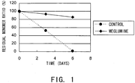
- the residual monomer ratio was determined by size exclusion chromatography (SEC) under the conditions described above (the residual monomer ratio is defined as the percentage of monomer peak area in the heat acceleration sample to that of the initial state).
- SEC size exclusion chromatography
- Fig. 1 shows the change in residual monomer ratio over time, from the initial ratio to the ratio after three and six days of incubation at 55°C under the solution conditions described above.
- the stabilization effect of meglumine on sc(Fv)2s other than hVB22B described in Example 1 was tested.
- the sc(Fv)2s used were: mVB22B (SEQ ID NO: 2; see Reference Examples described below), 12E10 (SEQ ID NO: 3; WO 02/33072 ), and 2D7 (SEQ ID NO: 4; see Reference Examples described below) listed below.
- the stabilization effect on a humanized anti-IL-6 receptor antibody H chain, SEQ ID NO: 5; L chain, SEQ ID NO: 6; WO 92/19759 ), which is not an sc(Fv)2 but a whole antibody IgG1, was also tested.
- the solvent conditions and concentration of each antibody used in the stabilization test are shown below.
- the heat acceleration test was carried out under the condition described above.
- IgG was analyzed under the conditions described below.
- Fig. 2 shows the change in aggregate content over time under each solution acceleration condition described above.
- the stabilization test result showed that the addition of meglumine suppressed the formation of aggregates of not only sc(Fv)2 and IgG but also of other types of molecules.
- the result described above demonstrates that the addition of meglumine has the effect of suppressing aggregation of not only sc(Fv)2 but also general antibody molecules, such as whole antibody IgG.
- meglumine serves as a stabilizer for antibody molecules. This investigation revealed for the first time that meglumine is useful as a stabilizer for antibody molecules.
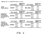
- Sucrose has been reporter to be very useful in preparing freeze-dried antibody agents. Samples tested and stabilization test conditions are described below:
- Freeze-dried preparations were prepared from the samples listed above by aliquoting solutions prior to freeze drying, in which IgG concentration was adjusted to 40 mg/ml, into vials (2 ml/vial) and freeze-drying them using a shelf-freeze dryer under the condition indicated below.
- Solution preparations were prepared from the samples listed above by adding 0.6 ml of water for injections to each vial of the freeze-dried preparations.
- the concentration of IgG in each vial was about 120 mg/ml.
- Fig. 3 shows a comparison of the effects of sucrose and meglumine on the aggregate content before and after freeze-drying (solutions prior to freeze-drying and freeze-dried preparations), comparison of the effects of sucrose and meglumine on the aggregate content in freeze-dried preparations after one month of acceleration test at 40°C, and comparison of the effects of sucrose and meglumine on the aggregate content in solution preparations after two weeks of acceleration test at 25°C.
- the aggregate content was lower with meglumine than with sucrose. This indicates that compared to sucrose, meglumine has a greater stabilization effect against aggregate formation .
- the stabilization effect was also found to depend on meglumine concentration, and the stabilization effect was stronger as meglumine concentration increased.
- the addition of meglumine could suppress the increase of aggregates caused by freeze-drying in a meglumine concentration-dependent manner.
- the aggregation in freeze-dried preparations during one month of storage at 40°C could also be suppressed in the same way.
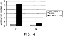
- hVB22B u2-wz4 was expressed by the method described in Reference Examples 1 to 3 herein, or in WO 2005/056603 or WO 2005/056604 , and then the single-chain diabody-type sc(Fv)2 was purified.
- Formulae (F1 and F2) were prepared from the purified single-chain diabody-type sc(Fv)2 according to the following solution conditions:
- Fig. 4 shows the aggregate content in the samples prior to storage and after six months of storage at -20°C, which were analyzed by SEC. While the increase of aggregate after six months of storage at -20°C was 4.8% in F1 without meglumine, the increase of aggregate could be suppressed to 0.4% in F2 containing meglumine as a stabilizer. This Example demonstrates that meglumine has the effect of stabilizing proteins even in a frozen state.
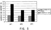
- Formulation (AF1, AF2, and AF3) were prepared from the purified humanized bispecific antibody hA69-KQ/hB26-PF/hAL-AQ according to the solution conditions described below.
- the humanized bispecific antibody comprising hA69-KQ/hB26-PF/hAL-AQ is an IgG4-type antibody comprising the first H chain variable region shown in SEQ ID NO: 49 (hA69-KQ), the second H chain variable region shown in SEQ ID NO: 50 (hB26-PF), and the L chain variable region shown in SEQ ID NO: 51 (hAL-AQ) that are shared by both H chains.
- Fig. 5 shows the aggregate content in samples prior to storage and samples after two months of storage at 25°C, which were analyzed by SEC. While the aggregate increase after six months of storage at 25°C was 0.51% in AF1 without any stabilizer, the aggregate increase could be suppressed to 0.26% in AF2 containing meglumine as a stabilizer. The aggregate increase was 0.27% in AF3 containing arginine as a stabilizer. Thus, as compared to arginine, meglumine was found to have equivalent or greater stabilizing effect.
- meglumine was also found to have the stabilizing effect in solutions containing high concentration (120 mg/ml) of humanized bispecific antibody hA69-KQ/hB26-PF/h.AL-AQ; as similarly described in Example 3.
- meglumine was demonstrated to have the effect of stabilizing not only IgG and sc(Fv)2 but also bispecific IgG, indicating that it can be commonly used as a protein stabilizer.
- BaF3 cell lines expressing the full-length Mpl gene were established to obtain cell lines that proliferate in a TPO-dependent manner.
- a full-length human Mpl cDNA ( Palacios, R. et al., Cell, 41, 727-734 (1985 )) (GenBank accession NO. NM_005373) was amplified by PCR.
- the cDNA was cloned into a pCOS2 expression vector to construct pCOS2-hMplfull.
- the expression vector pCOS2 was constructed by removing the DHFR gene expression region from pCHOI ( Hirata, Y. et al., FEBS Letter, 356, 244-248 (1994 )), where the expression region of the neomycin resistance gene HEF-VH-g ⁇ 1 ( Sato, K. et al., Mol Immunol., 31, 371-381 (1994 )) is inserted.
- the cynomolgus monkey Mpl cDNA (SEQ ID NO: 7) was cloned from total RNA extracted from the bone marrow cells of cynomolgus monkey, using a SMART RACE cDNA Amplification Kit (Clontech). The resulting cynomolgus monkey cDNA was inserted into pCOS2 to construct pCOS2-monkeyMplfull.
- mouse Mpl cDNA (GenBank accession NO. NM_010823) was amplified by PCR, and inserted into pCOS2 to construct pCOS2-mouseMplfull.
- Each vector (20 ⁇ g) prepared as described above was mixed with BaF3 cells (1x 10 7 cells/mL) suspended in PBS in Gene Pulser cuvettes. This mixture was then pulsed at 0.33 kV and 950 ⁇ FD using a Gene Pulser II (Bio-Rad).
- the BaF3 cells introduced with the above DNAs by electroporation were added to RPMI 1640 medium (Invitrogen) containing 1 ng/mL mouse interleukin 3 (hereinafter abbreviated as mIL-3; Peprotech), 500 ⁇ g/mL Geneticin (Invitrogen), and 10% FBS (Invitrogen), and selected to establish a human Mpl-expressing BaF3 cell line (hereinafter abbreviated as "BaF3-human Mpl”), monkey Mpl-expressing BaF3 cell line (hereinafter abbreviated as BaF3-monkey Mpl), and mouse Mpl-expressing BaF3 cell line (hereinafter abbreviated as "BaF3-mouse Mpl”). Following selection, these cells were cultured and maintained in RPMI 1640 containing 1 ng/mL rhTPO (R&D) and 10% FBS.
- RPMI 1640 medium Invitrogen
- mIL-3 mouse interleukin 3
- CHO cell lines expressing the full-length Mpl gene were established to obtain cell lines to be used for assessing binding activity by flow cytometry.
- the DHFR gene expression site from pCHOI was inserted into pCXN2 ( Niwa, H. et al., Gene, 108, 193-199 (1991 )) at the HindIII site to prepare a pCXND3 expression vector.
- the respective Mpl genes were amplified by PCR using pCOS2-hMplfull, pCOS2-monkeyMplfull, and pCOS2-mouseMplfull as templates, and primers with a His-tag sequence.
- PCR products were cloned into pCXND3 to construct pCXND3-hMpl-His, pCXND3-monkey Mpl-His, and pCXND3-mouse Mpl-His, respectively.
- Vectors thus prepared (25 ⁇ g each) were mixed with a PBS suspension of CHO-DG44 cells (1x 10 7 cells/mL) in Gene Pulser cuvettes. The mixture was then pulsed at 1.5 kV and 25 ⁇ FD using Gene Pulser II (Bio-Rad). The CHO cells introduced with these DNAs by electroporation were added to CHO-S-SFMII medium (Invitrogen) containing 500 ⁇ g/mL Geneticin and 1x HT (Invitrogen).
- CHO-human Mpl human Mpl-expressing CHO cell line
- CHO-monkey Mpl monkey Mpl-expressing CHO cell line
- CHO-mouse Mpl mouse Mpl-expressing CHO cell line
- a DNA construct encoding the extracellular region of human Mpl (Gln 26 to Trp 491) with a downstream FLAG tag was prepared.
- the construct was inserted into a pBACSurf-1 Transfer Plasmid (Novagen) between the PstI and SmaI sites to prepare pBACSurf1-hMpl-FLAG.
- Sf9 cells were transformed with 4 ⁇ g of pBACSurfl-hMpl-FLAG using the Bac-N-Blue Transfection Kit (Invitrogen).
- the culture supernatant was collected after a three-day incubation. Recombinant virus was isolated by plaque assays.
- the prepared virus stock was used to infect Sf9 cells, and the culture supernatant was collected.
- Soluble human Mpl protein was purified from the obtained culture supernatant as described below.
- the culture supernatant was loaded onto a Q Sepharose Fast Flow (Amersham Biosciences) for adsorption, and the adsorbed protein was then eluted with 50 mM Na-phosphate buffer (pH7.2) containing 0.01 % (v/v) Tween 20 and 500 mM NaCl.
- the eluates were loaded onto a FLAG M2-Agarose (Sigma-Aldrich) for adsorption, the protein adsorbed was eluted with 100 mM glycine-HCl buffer (pH3.5) containing 0.01% (v/v) Tween 20.
- Human fusion protein Mpl-IgG Fc gene was prepared according to the method by Bennett et al. ( Bennett, B. D. et al., J. Biol. Chem. 266, 23060-23067 (1991 )).
- a nucleotide sequence encoding the extracellular region of human Mpl (Gln 26 to Trp 491) was linked to a nucleotide sequence encoding the Fc region of human IgG- ⁇ 1 (a region downstream of Asp 216).
- a BstEII sequence (amino acids: Val-Thr) was attached to the junction as a fusion linker between these two regions.
- a 19-amino acid signal peptide derived form human IgG H chain variable region was used as the signal sequence.
- the resulting human fusion protein Mpl-IgG Fc gene was cloned into pCXND3 to construct pCXND3-hMpl-Fc.
- the vector thus prepared (25 ⁇ g) was mixed with a PBS suspension of CHO-DG44 cells (1x 10 7 cells/mL) in Gene Pulser cuvettes. The mixture was then pulsed at 1.5 kV and 25 ⁇ FD using Gene Pulser II (Bio-Rad). The CHO cells introduced with the DNA by electroporation were added to CHO-S-SFMII medium containing 500 ⁇ g/mL Geneticin and 1x HT (Invitrogen). shMPL-Fc-expressing CHO cell line (CHO-hMpl-Fc) was then established through selection.
- Human Mpl-IgG Fc fusion protein was purified from the culture supernatant as described below.
- the culture supernatant was loaded onto a Q Sepharose Fast Flow (Amersham Biosciences) for adsorption, and then the adsorbed protein were eluted with 50 mM Na-phosphate buffer (pH7.6) containing 0.01% (v/v) Tween 20 and 1 M NaCl. After the eluates were loaded onto a HiTrap protein G HP column (Amersham Biosciences) for adsorption, the adsorbed protein was eluted with 0.1 M glycine-HCl buffer (pH2.7) containing 150 mM NaCl and 0.01% (v/v) Tween 20.
- hMpl-Fc The purified soluble Mpl protein was referred to as "hMpl-Fc".
- MRL/MpJUmmCrj-lpr/lpr mice (hereinafter abbreviated as "MRL/lpr mice”; purchased from Charles River, Japan) were immunized; the primary immunization was carried out at eight weeks of age.
- MRL/MpJUmmCrj-lpr/lpr mice (hereinafter abbreviated as "MRL/lpr mice”; purchased from Charles River, Japan) were immunized; the primary immunization was carried out at eight weeks of age.
- an emulsion containing 100 ⁇ g of shMPL-FLAG combined with Freund's complete adjuvant H37 Ra; Beckton Dickinson
- an emulsion containing shMPL-FLAG 50 ⁇ g per mouse
- Freund's incomplete adjuvant (Beckton Dickinson) was administered subcutaneously.
- mice which have been immunized six times in total were subjected to a final injection of shMPL-FLAG (50 ⁇ g per mouse) through the caudal vein.
- Cell fusion was achieved by mixing the mouse myeloma P3-X63Ag8U1 cells (P3U1; purchased from ATCC) and mouse splenocytes using polyethylene glycol 1500 (Roche Diagnostics).
- Hybridoma selection in HAT medium began the following day and culture supernatants were obtained. Screening was carried out by ELISA, using immunoplates immobilized with shMpl-FLAG or hMpl-Fc and the assayed cell growth activity of BaF3-human Mpl as an index.
- Balb/C mice were immunized eleven times in total by administering BaF3-human Mpl (1.0x 10 7 cells per mouse) intraperitoneally over a period of one week to five months.
- Hybridomas were similarly prepared by cell fusion, and screened using the assayed cell growth activity of BaF3-human Mpl as an index. Positive clones were isolated as single clones by limiting dilution and then cultured in a large scale. The culture supernatants were collected.
- Antibody concentrations were determined by carrying out a mouse IgG sandwich ELISA using goat anti-mouse IgG (gamma) (ZYMED) and alkaline phosphatase-goat anti-mouse IgG (gamma) (ZYMED), generating a calibration curve by GraphPad Prism (GraphPad Software; USA), and calculating the antibody concentrations from the calibration curve.
- Commercially available antibodies of the same isotype were used as standards.
- Antibody isotypes were determined by antigen-dependent ELISA using isotype-specific secondary antibodies.
- hMp1-Fc was diluted to 1 ⁇ g/mL with a coating buffer (0.1 mM NaHCO 3 , pH9.6) containing 0.02% (w/v) NaN 3 , and then added to ELISA plates. The plates were incubated overnight at 4°C for coating. The plates were blocked with a diluent buffer (50 mM Tris-HCl (pH8.1) containing 1 mM MgCl 2 , 150 mM NaCl, 0.05% (v/v) Tween 20, 0.02% (w/v) NaN 3 , 1% (w/v) BSA).
- the binding activities of an antibody to shMpl-FLAG and hMPL-Fc were determined by ELISA.
- ELISA plates were coated with 1 ⁇ g/mL of purified shMpl-FLAG or hMPL-Fc, and blocked with a diluent buffer.
- Hybridoma culture supernatants were added to the plates, and the plates were allowed to stand at room temperature for 1 hr.
- alkaline phosphatase-labeled anti-mouse IgG antibodies (Zymed) were added to the plates. Color development was similarly carried out using the above method. Following a one-hour coloring reaction at room temperature, absorbance was measured at 405 nm and EC 50 values were computed using GraphPad Prism.
- CHO-human Mpl cells and CHO-monkey Mpl cells were harvested, and suspended in FACS Buffer (1% FBS/ PBS) to a final concentration of 1x 10 6 cells/mL.
- FACS Buffer 1% FBS/ PBS
- the suspensions were aliquoted into Multiscreen (Millipore) at 100 ⁇ l/well, and the culture supernatants were removed by centrifugation. Culture supernatants diluted to 5 ⁇ g/mL were added to the plates and incubated on ice for 30 min. The cells were washed once with FACS buffer, and incubated on ice for 30 min following the addition of an FITC-labeled anti-mouse IgG antibody (Beckman Coulter).
- Agonistic activities of an antibody were evaluated using BaF3-human Mpl and BaF3-monkey Mpl which proliferate in a TPO-dependent manner.
- Cells of each cell line were suspended at 4x 10 5 cells/ml in RPMI 1640/10% FBS (Invitrogen), and each suspension was aliquoted into a 96-well plate at 60 ⁇ l/well.
- a 40- ⁇ L aliquot of rhTPO (R&D) and hybridoma culture supernatants prepared at various concentrations was added into each well. The plates were then incubated at 37°C under 5% CO 2 for 24 hr.
- a 10- ⁇ L aliquot of the Cell Count Reagent SF (Nacalai Tesque) was added into each well. After incubation for 2 hr, absorbance was measured at 450 nm (and at 655 nm as a control) using a Benchmark Plus. EC 50 values were calculated using GraphPad Prism.
- TA136 was established from mice immunized with BaF3-human Mpl and the others were established from mice immunized with shMpl-Flag.
- Anti-human Mpl antibodies were purified from hybridoma culture supernatants as described below.
- the antibodies were eluted with 0.1 M glycine-HCl (pH2.7) Buffer. Immediately after elution, the fractions were neutralized with 1 M Tris-HCl Buffer (pH9.0), and dialyzed against PBS to replace the buffer for one day.
- a GST-fusion protein containing a partial sequence of human Mpl was constructed for VB22B epitope analysis.
- MG1 Gan26 to Trp491
- MG2 Gan26 to Leu274 regions were each amplified by PCR, and cloned into pGEX-4T-3 (Amersham Biosciences) to be expressed as GST fusion proteins.
- the resulting plasmid DNAs were transformed into DH5 ⁇ to give transformants.
- a final concentration of 1 mM IPTG was added to the transformants in their logarithmic growth phase to induce the expression of GST fusion proteins.
- the bacterial cells were harvested after two hours of incubation.
- the cells were lysed by sonication.
- the lysates were centrifuged in XL-80 Ultracentrifuge (Beckman, Rotor 70.1Ti) at 35,000 rpm for 30 min.
- the culture supernatants were removed, and then the fusion proteins were purified using GST Purification Modules (Amersham Biosciences).
- the samples were separated by 10%-SDS-PAGE, and then transferred onto a PVDF membrane.
- the membrane was analyzed by the murine antibody VB22B in Western blotting. VB22B was found to recognize both MG-1 and MG-2, indicating that the VB22B epitope is located in the (Gln26 to Leu274) region.
- GST fusion proteins containing the respective regions of human Mp1: MG3 (Gln26 to Alma189), MG4 (Gln26 to Pro 106), MG5 (Gln26 to Glu259), and MG6 (Gln26 to Gly245) were prepared and analyzed by Western blotting using the same procedure described above.
- VB22B was found to recognize MG5 and MG6, but not MG3 and MG4. This suggests that the VB22B epitope is located within the (Ala189 to Gly245) region.
- GST was fused with MG7 (Gln26 to Ala231) and MG8 (Gln26 to Pro217) to prepare GST fusion proteins.
- VB22B recognized MG7 but not MG8, suggesting that the VB22B epitope is located in the (Gln217 to Alma231) region. Furthermore, GST fusion protein containing MG10 (Gln213 to Ala231) was recognized by VB22B, suggesting that the VB22B epitope is located within the limited region of 19 amino acids between Gln213 and Ala231.
- Example 1.4 Since the anti-human Mpl antibody VB22B binds to soluble recombinant Mpl, kinetic analyses of the antigen-antibody reaction between VB22B IgG and human Mpl-IgG Fc fusion protein were carried out as described in Example 1.4.
- the Sensor Chip CM5 (Biacore) was placed in Biacore 2000 (Biacore), and human Mpl-IgG Fc fusion protein was immobilized onto the chip by amine-coupling methods. Then, 1.25 to 20 ⁇ g/mL of VB22B IgG solution was prepared using HBS-EP Buffer (Biacore), and injected over the chip surface for 2 min to reveal the binding region.
- HBS-EP Buffer was injected over the chip surface for 2 min to reveal the dissociation region.
- VB22B IgG bound to the human Mpl-IgG Fc fusion protein on the sensor chip was removed by injecting 10 mM NaOH over the sensor chip for 15 sec, and the chip was recovered.
- HBS-EP Buffer was used as the running buffer, and the flow rate was 20 ⁇ L/min.
- the reaction rate constant at each concentration was calculated from the sensorgrams.
- the dissociation constant (KD) for VB22B IgG was determined to be 1.67 ⁇ 0.713 x 10 -9 M.
- variable region was amplified by RT-PCR using total RNA extracted from hybridomas producing anti-human Mpl antibodies.
- Total RNA was extracted from 1x 10 7 hybridoma cells using the RNeasy Plant Mini Kit (QIAGEN).
- a 5'-terminal fragment of the gene was amplified from 1 ⁇ g of total RNA by the SMART RACE cDNA Amplification Kit (Clontech), using a synthetic oligonucleotide MHC-IgG2b (SEQ ID NO: 8) complementary to mouse IgG2b constant region or a synthetic oligonucleotide kappa (SEQ ID NO: 9) complementary to mouse ⁇ chain constant region. Reverse transcription was carried out at 42°C for 1.5 hr.
- the composition of the PCR reaction solution (50 ⁇ L in total) is shown below.
- the PCR reaction conditions were:
- PCR products were purified from agarose gel using the QIAquick Gel Extraction Kit (QIAGEN), and cloned into a pGEM-T Easy Vector (Promega). The nucleotide sequence was then determined using the ABI 3700 DNA Analyzer (Perkin Elmer).
- VB22B-VH The nucleotide sequence of cloned VB22B H chain variable region (hereinafter abbreviated as "VB22B-VH”) is shown in SEQ ID NO: 10, and its amino acid sequence is shown in SEQ ID NO: 11.
- the nucleotide sequence of the L chain variable region (hereinafter abbreviated as "VB22B-VL”) is shown in SEQ ID NO: 12, and its amino acid sequence is shown in SEQ ID NO: 13.
- VB22B diabody The gene encoding VB22B single-chain Fv (hereinafter abbreviated as "VB22B diabody”) containing a five-amino acid linker sequence was constructed, by linking a nucleotide sequence encoding a (Gly 4 Ser) 1 linker to the VB22B-VH-encoding gene at its 3' end and to the VB22B-VL-encoding gene at its 5' end; both of which have been amplified by PCR.
- the VB22B-VH forward primer, 70-115HF, (SEQ ID NO: 14) was designed to contain an EcoRI site.
- the VB22B-VL forward primer, 33 ⁇ 115LF, was designed to have a nucleotide sequence encoding the N terminus of VB22B-VL, a nucleotide sequence encoding the (Gly 4 Ser) 1 linker, and a nucleotide sequence encoding the C terminus of VB22B-VH.
- the VB22B-VL reverse primer, 33 ⁇ 115LR, was designed to hybridize to a DNA encoding the C terminus of VB22B-VL and to have a nucleotide sequence encoding a FLAG tag (Asp Tyr Lys Asp Asp Asp Asp Lys/SEQ ID NO: 18) and a NotI site.
- the composition of the PCR reaction solution (50 ⁇ L in total) is shown below.
- the PCR reaction conditions were:
- the second-round PCR was carried out using aliquots of the respective PCR products according to the protocol described below.
- the composition of the PCR reaction solution (50 ⁇ L in total) is shown below.
- reaction conditions were:
- PCR products of about 800 bp were purified from agarose gel using the QIAquick Gel Extraction Kit (QIAGEN), and then digested with EcoRI and NotI (both from TaKaRa). The resulting DNA fragments were purified using the QIAquick PCR Purification Kit (QIAGEN), and then cloned into pCXND3 to prepare pCXND3-VB22B db.
- PCR was carried out to amplify (a) the VB22B-VH-encoding gene in which a nucleotide sequence encoding a 15-amino acid linker (Gly 4 Ser) 3 was added to its 3' end; and (b) the VB22B-VL-encoding gene containing the identical linker nucleotide sequence added to its 5' end.
- the desired construct was prepared by linking these amplified genes. Three new primers were designed in this construction process.
- the VB22B-VH forward primer, VB22B-fpvu, (primer A; SEQ ID NO: 19) was designed to have an EcoRI site at its 5' end and to convert Gln22 and Leu23 of VB22B db into a PvuII site.
- the VB22B-VH reverse primer, sc-rL15, (primer B; SEQ ID NO: 20) was designed to hybridize to a DNA encoding the C terminus of VB22B-VH, and to have a nucleotide sequence encoding the (Gly 4 Ser) 3 linker, as well as a nucleotide sequence hybridizing to a DNA encoding the N terminus of VB22B-VL.
- the VB22B-VL forward primer, sc-fL15, (primer C; SEQ ID NO: 21) was designed to have a nucleotide sequence encoding the N terminus of VB22B-VL, a nucleotide sequence encoding the (Gly 4 Ser) 3 linker, and a nucleotide sequence encoding the C terminus of VB22B-VH.
- the composition of the PCR reaction solution (50 ⁇ L in total) is shown below.
- reaction conditions were:
- the second-round PCR was carried out using aliquots of the respective PCR products according to the protocol described below.
- the composition of the PCR reaction solution (50 ⁇ L in total) is shown below.
- First-round PCR product two types 1 ⁇ L Synthetic oligonucleotide, 70 ⁇ 115HF and 33 ⁇ 115LR 10 pmol
- reaction conditions were:
- PCR products of about 800 bp were purified from agarose gel using the QIAquick Gel Extraction Kit (QIAGEN), and then digested with EcoRI and NotI (both from TaKaRa). The resulting DNA fragments were purified using the QIAquick PCR Purification Kit (QIAGEN), and then cloned into pBacPAK9 (Clontech) to construct pBacPAK9-scVB22B.
- a fragment to be inserted into the PvuII site of pBacPAK9-scVB22B was prepared. Specifically, the fragment has a PvuII recognition site at both ends and a nucleotide sequence, in which a gene encoding the VB22B-VH N-terminus is linked, via a (Gly 4 Ser) 3 linker-encoding nucleotide sequence, to a gene encoding the amino acid sequence of an N terminus-deleted VB22B-VH linked to VB22B-VL via the (Gly 4 Ser) 3 linker.
- Two primers were newly designed to prepare the fragment by PCR.
- the forward primer for the fragment of interest, Fv2-f was designed to have a PvuII site at its 5' end and a VB22B-VH 5'-end sequence.
- the reverse primer for the fragment of interest, Fv2-r was designed to hybridize to a DNA encoding the C terminus of VB22B-VL, and to have a PvuII site, a nucleotide sequence encoding the (Gly 4 Ser) 3 linker, and a nucleotides sequence hybridizing to a DNA encoding the N terminus of VB22B-VH.
- PCR was carried out using pBacPAK9-scVB22B as a template as described below.
- the composition of the PCR reaction solution (50 ⁇ L in total) is shown below.
- reaction conditions were:
- PCR products of about 800 bp were purified from agarose gel using the QIAquick Gel Extraction Kit (QIAGEN), and then cloned into the pGEM-T Easy Vector (Promega). After sequencing, the plasmid was digested with PvuII (TaKaRa), and the fragment of interest was recovered. The recovered fragment was ligated to pBacPAK9-scVB22B pre-digested with PvuII (TaKaRa) to construct pBacPAK9-VB22B sc(Fv)2.
- the fragment of about 1,600 bp was purified from agarose gel using the QIAquick Gel Extraction Kit (QIAGEN). The fragment was then cloned into a pCXND3 expression vector to construct pCXND3-VB22B sc(Fv)2.
- a cell line stably expressing the single-chain antibody was prepared from CHO-DG44 cells as described below. Gene transfer was achieved by electroporation using a Gene Pulser II (Bio-Rad). An expression vector (25 ⁇ g) and 0.75 mL of CHO-DG44 cells suspended in PBS (1x 10 7 cells/mL) were mixed. The resulting mixture was cooled on ice for 10 min, transferred into a cuvette, and pulsed at 1.5-kV and 25 ⁇ FD. After a ten-minute restoration period at room temperature, the electroporated cells were plated in CHO-S-SFMII medium (Invitrogen) containing 500 ⁇ g/mL Geneticin (Invitrogen). CHO cell lines expressing the single-chain antibody were established through selection. A cell line stably expressing VB22B sc(Fv)2 and its culture supernatants were obtained by this method.
- the transient expression of the single-chain antibody was achieved using COS7 cells as described below.
- An expression vector (10 ⁇ g) and 0.75 mL of COS7 cells suspended in PBS (1x 10 7 cells/mL) were mixed. The resulting mixture was cooled on ice for 10 min, transferred into a cuvette, and then pulsed at 1.5-kV and 25 ⁇ FD. After a ten-minute restoration period at room temperature, the electroporated cells were plated in DMEM/10% FBS medium (Invitrogen). The cells were incubated overnight and then washed with PBS. CHO-S-SFMII medium was added and the cells were cultured for about three days. The culture supernatants for preparing the VB22B diabody were thus prepared.
- the culture supernatant concentration of the single-chain anti-human Mpl antibody transiently expressed in COS cells was determined using surface plasmon resonance.
- a sensor chip CM5 (Biacore) was placed in Biacore 2000 (Biacore).
- ANTI-FLAG(R) M2 Monoclonal Antibody (Sigma-Aldrich) was immobilized onto the chip.
- An appropriate concentration of sample was injected over the chip surface at a flow rate of 5 mL/sec, and 50 mM diethylamine was used to dissociate the bound antibody. Changes in the mass during sample injection were recorded, and the sample concentration was calculated from the calibration curve prepared using the mass changes of a standard sample.
- db12E10 (see WO 02/33073 and WO 02/33072 ) was used as the diabody standard, and 12E10 sc(Fv)2 which has the same gene structure as that of sc(Fv)2 was used as the sc(Fv)2 standard.
- the culture supernatants of VB22B diabody-expressing COS7 cells or CHO cells was loaded onto an Anti-Flag M2 Affinity Gel (Sigma-Aldrich) column equilibrated with a 50 mM Tris-HCl buffer (pH7.4) containing 150 mM NaCl and 0.05% Tween 20.
- the absorbed antibodies were eluted with 100 mM glycine-HCl (pH3.5).
- the fractions eluted were immediately neutralized with 1 M Tris-HCl (pH8.0), and loaded onto a HiLoad 26/60 Superdex 200 pg (Amersham Biosciences) column for gel filtration chromatography.
- PBS/0.01% Tween 20 was used in the gel filtration chromatography.
- VB22B sc(Fv)2 was purified from the culture supernatants of VB22B sc(Fv)2-expressing COS7 cells or CHO cells under the same conditions used for purifying the diabodies.
- a large-scale preparation of VB22B sc(Fv)2 was prepared by loading the CHO cell culture supernatants onto a Macro-Prep Ceramic Hydroxyapatite Type I (Bio-Rad) column equilibrated with a 20 mM phosphate buffer (pH6.8), and eluting the VB22B sc(Fv)2 in a stepwise manner with 250 mM phosphate buffer (pH6.8).
- the eluted fraction was concentrated on an ultrafilter, and then fractionated by gel filtration chromatography using a HiLoad 26/60 Superdex 200 pg (Amersham Biosciences) column, and a fraction corresponding to the molecular weight range of about 40 kD to 70 kD was obtained.
- the fraction was loaded onto an Anti-Flag M2 Affinity Gel column equilibrated with a 50 mM Tris-HCl buffer (pH7.4) containing 150 mM NaCl and 0.05% Tween 20.
- the absorbed antibody was eluted with 100 mM glycine-HCl (pH3.5).
- the eluted fraction was immediately neutralized with 1 M Tris-HCl (pH8.0), and loaded onto a HiLoad 26/60 Superdex 200 pg (Amersham Biosciences) column for gel filtration chromatography. 20 mM acetate buffer (pH6.0) containing 150 mM NaCl and 0.01% Tween 80 was used in the gel filtration chromatography. In each purification step, the presence of the diabody and sc(Fv)2 in the samples was confirmed by SDS-PAGE and Western blotting using an anti-Flag antibody (Sigma-Aldrich).
- CHO-human Mpl, CHO-monkey Mpl, and CHO-mouse Mpl cells were recovered and suspended in FACS buffer (1% FBS/PBS) to a final concentration of 1x 10 6 cells/mL.
- FACS buffer 1% FBS/PBS
- Cell suspensions were aliquoted at 100- ⁇ L/well into the Multiscreen-HV Filter Plates (Millipore). After centrifugation, the supernatant was removed. An appropriate concentration of diabody or sc(Fv)2 was added into each well and incubated on ice for 30 min.
- the cells were washed once with 200 ⁇ L of FACS buffer, and incubated on ice for 30 min following the addition of 10 ⁇ g/mL ANTI-FLAG(R) M2 Monoclonal Antibody (Sigma-Aldrich). The cells were then washed once with 200 ⁇ L of FACS buffer, and a 100x-diluted FITC-labeled anti-mouse IgG antibody (Beckman Coulter) was added to the plate. The plate was incubated on ice for 30 min. After centrifugation, the supernatant was removed. The cells were suspended in 400 ⁇ L of FACS Buffer, and then analyzed by flow cytometry using EPICS ELITE ESP (Beckman Coulter). An analysis gate was set on the forward and side scatters of a histogram to include viable cell populations.
- VB22B sc(Fv)2 The binding activity of the purified VB22B sc(Fv)2 to various Mpl molecules expressed in CHO cells was determined.
- VB22B sc(Fv)2 was found to specifically bind to CHO-human Mpl and CHO-monkey Mpl but not to the host cell CHO or CHO-mouse Mpl.
- This binding characteristic of VB22B sc(Fv)2 is comparable to those of VB22B IgG, indicating that the antibody binding site remains unaltered by converting whole IgG to minibody.
- Antibody sequence data for the humanization of VB22B sc(Fv)2 were obtained from the Kabat Database (ftp://ftp.ebi.ac.uk/pub/databases/kabat/), and homology searches were carried out independently for the H chain variable region and the L chain variable region.
- the H chain variable region was found to be highly homologous to DN13 ( Smithson S. L. et al., Mol Immunol. 36, 113-124 (1999 )).
- the L chain variable region was found to be highly homologous to ToP027 ( Hougs L. et al., J. Immunol. 162, 224-237 (1999 )).
- Humanized antibodies were prepared by inserting a complementarity-determining region (hereinafter abbreviated as "CDR") into the framework regions (hereinafter abbreviated as "FR") of the above antibodies.
- CDR complementarity-determining region
- FR framework regions
- the humanized antibody sc(Fv)2 was expressed in CHO-DG44 cells, and its agonistic activity was assessed using BaF3-human Mpl.
- the agonistic activity was used as an index to generate a humanized VB22B sc(Fv)2 which has agonistic activity equivalent to that of murine VB22B sc(Fv)2 by replacing one or more amino acids in its framework region.
- synthetic oligo-DNAs of approximately 50 nucleotides in length were designed as to make 20 of these nucleotides available for hybridization, and the synthetic oligo-DNAs were assembled by PCR to prepare genes that encode the respective variable regions. Using the resulting genes, sc(Fv)2 was similarly prepared by the method described in Example 2.3.
- the respective DNAs were cloned into a pCXND3expression vector to construct expression vectors pCXND3-hVB22B p-z sc(Fv)2, pCXND3-hVB22B g-e sc(Fv)2, pCXND3-hVB22B e sc(Fv)2, pCXND3-hVB22B u2-wz4 sc(Fv)2, and pCXND3-hVB22B q-wz5 sc(Fv)2, to which the humanized VB22B sc(Fv)2 is inserted.
- the plasmids were expressed in CHO-DG44 cells and the culture supernatants were recovered by the method described in Example 2.4. Since the humanized VB22B sc(Fv)2 does not contain a Flag tag, its purification from the culture supernatant was performed using a MG10-GST fusion protein.
- MG10 (Gln213 to Ala231) is one of the epitopes recognized by VB22B, as described in Example 1.8.
- the MG10-GST fusion protein was purified using Glutathione Sepharose 4B (Amersham Biosciences) according to the supplier's protocol.
- the purified MG10-GST fusion protein was immobilized onto a HiTrap NHS-activated HP Column (Amersham Biosciences) to prepare an affinity column, according to the supplier's protocol.
- the culture supernatant of CHO cells expressing the humanized VB22B sc(Fv)2 was loaded onto the MG10-GST fusion protein-immobilized column, which has been equilibrated with 50 mM Tris-HCl (pH7.4)/150 mM NaCl/0.01% Tween 80.
- the adsorbed humanized VB22B sc(Fv)2 was eluted with 100 mM glycine-HCl (pH3.5)/0.01% Tween 80.
- the eluted fraction was neutralized with 1 M Tris-HCl (pH7.4), and was further subjected to gel filtration chromatography using a HiLoad 16/60 Superdex 200 pg (Amersham Biosciences). 20 mM citrate buffer (pH7.5) containing 300 mM NaCl and 0.01% Tween 80 was used in the gel filtration chromatography.
- a DNA expression vector encoding 2D7sc(Fv)2 in which these sequences are linked by a 15-mer linker was produced by the following procedure.
- 2D7 diabody (HL5) expression vector as the template, a PCR reaction was performed using primer 2D7PA6 (SEQ ID NO: 38) and primer 2D7PA2 (SEQ ID NO: 35) to amplify fragment C.
- a PCR reaction was performed using primer 2D7PA3 (SEQ ID NO: 36) and primer 2D7DBL2 (SEQ ID NO: 39) to amplify fragment D.
- the obtained fragments C and D were mixed in the same tube, and the two fragments were linked by conducting a PCR-recombination reaction. This yielded the DNA fragment "2D7diabody HL15-2" comprising a Flag-tag region at the C terminal, in which VH-VL is linked by a 15-mer linker.
- the nucleotide sequence of the inserted DNA was analyzed to confirm that the cDNA encoding signal-VH(15)VL(15)VH(15)VL-Flag has been inserted between EcoRI-NotI of pCXND3 as intended, and the construction of the 2D7sc(Fv)2 expression vector (pCXND3-2D7sc(Fv)2) was completed.
- the nucleotide sequence and the amino acid sequence of 2D7sc(Fv)2 are shown in SEQ ID NO: 40 and SEQ ID NO: 4, respectively.
- DG44 cells cultured in CHO-S-SFM-II medium were washed twice with ice-cold PBS, and then suspended in PBS to a concentration of 1x 10 7 cells/mL. 20 ⁇ g of the above-mentioned plasmid was mixed with this suspension, and then treated with an electric pulse (1.5 KV, 25 ⁇ FD). The cells were diluted into appropriate ratios, plated onto a 96-well plate, and cultured in CHO-S-SFM-II medium in the presence of G418 (Invitrogen) at a final concentration of 500 ⁇ g/ml.
- G418 Invitrogen
- a sub-confluent, 2D7sc(Fv)2 highly producing CHO cell line in a T-125 flask was transferred to a roller bottle (250 ml of CHO-S-SFM II medium/bottle) to achieve a concentration of 1x 10 5 cells/mL.
- the cells were cultured at 37°C and the culture supernatant was collected after 6 days. Dead cells were removed by centrifugation, and the solution was passed through a 0.45 ⁇ m filter and then used for purification.
- the collected culture supernatant was applied to a hydroxyapatite column (microprep ceramic Hydroxyapatite type I, Bio-Rad) equilibrated with buffer A (20 mM Na-phosphate pH6.8). After washing the column with buffer A, 2D7sc(Fv)2 was eluted using buffer C (250 mM Na-phosphate pH6.8). The fraction containing 2D7sc(Fv)2 was diluted with an equal amount of buffer A, and then this was applied to Anti-Flag M2 agarose affinity column (Bio-Rad).
- buffer A 20 mM Na-phosphate pH6.8
- buffer C 250 mM Na-phosphate pH6.8
- the fraction containing 2D7sc(Fv)2 was diluted with an equal amount of buffer A, and then this was applied to Anti-Flag M2 agarose affinity column (Bio-Rad).
- Minibodies produced from the CHO cell line producing high levels of 2D7sc(Fv)2 mostly have an elution peak at around the molecular weight of 52 Kd, which completely matches the peak of the 2D7 diabody also eluted at 52Kd.
- Aggregation of antibody molecules can be suppressed to allow antibody molecules to exist as monomers, by using stabilizers comprising meglumine of the present invention.
- each antibody molecule has to be stabilized so that the aggregation is suppressed to minimum during storage of preparations.
- the stabilizers of the present invention can stabilize antibody molecules and suppress aggregation even when the concentration of antibodies to be stabilized is very high.
- the stabilizers disclosed are thought to be very useful in producing antibody preparations.
- agents comprising meglumine of the present invention also have the effect of stabilizing antibody molecules when the antibody molecules are formulated into solution preparations or freeze-dried preparations.
- the stabilizers also have the effect of stabilizing antibody molecules against the stress imposed during the freeze-drying process in the formulation of freeze-dried preparations.
- the stabilizers disclosed have the effect of stabilizing entire whole antibodies, antibody fragments, and minibodies, and thus are widely useful in producing antibody preparations.
- compositions of the present invention which comprise antibody molecules stabilized by meglumine, are well-preserved, as compared to conventional antibody preparations, because the denaturation and aggregation of antibody molecules are suppressed. Therefore, the degree of activity loss during preservation is expected to be less.
Landscapes
- Health & Medical Sciences (AREA)
- Chemical & Material Sciences (AREA)
- Life Sciences & Earth Sciences (AREA)
- Medicinal Chemistry (AREA)
- Immunology (AREA)
- General Health & Medical Sciences (AREA)
- Organic Chemistry (AREA)
- Public Health (AREA)
- Pharmacology & Pharmacy (AREA)
- Animal Behavior & Ethology (AREA)
- Veterinary Medicine (AREA)
- Epidemiology (AREA)
- Engineering & Computer Science (AREA)
- Biochemistry (AREA)
- Molecular Biology (AREA)
- Bioinformatics & Cheminformatics (AREA)
- Genetics & Genomics (AREA)
- Biophysics (AREA)
- Proteomics, Peptides & Aminoacids (AREA)
- General Chemical & Material Sciences (AREA)
- Chemical Kinetics & Catalysis (AREA)
- Microbiology (AREA)
- Oil, Petroleum & Natural Gas (AREA)
- Mycology (AREA)
- Dermatology (AREA)
- Nuclear Medicine, Radiotherapy & Molecular Imaging (AREA)
- Medicines Containing Antibodies Or Antigens For Use As Internal Diagnostic Agents (AREA)
- Peptides Or Proteins (AREA)
- Medicinal Preparation (AREA)
- Medicines That Contain Protein Lipid Enzymes And Other Medicines (AREA)
Description
- The present disclosure relates to the use of an agent for stabilizing proteins, which comprises meglumine, an amino sugar. More specifically, the present invention relates to the use of an agent for stabilizing antibody molecules, which comprises meglumine, and methods for stabilizing antibody molecules, which comprise the step of adding meglumine. The present invention also relates to pharmaceutical compositions comprising antibody molecules stabilized by meglumine and methods for producing the pharmaceutical compositions.
- Preparations that enable stable preservation of proteins used as pharmaceuticals are required for the formulation of biopharmaceuticals (Non-Patent Document 1).
- Generally, there are two known pathways of protein degradation: the degradation pathway involving physical association of protein molecules, such as formation of soluble multimers or precipitate/insoluble material (Non-Patent Document 2) and the degradation pathway involving chemical modification, such as hydrolysis, deamidation, and methionine oxidation (Non-Patent Document 3). When proteins are developed as pharmaceuticals, decrease in biological activities during storage of proteins in the preparations to be provided should be prevented by suppressing insofar as possible both deterioration pathways. Methods for suppressing the degradation pathways as much as possible include optimization of pH of the solution, and optimization of types and concentrations of buffer, salt, and stabilizer.
- Antibodies that can be used as pharmaceuticals include whole antibodies, antibody fragments, minibodies, and modified antibodies (fusion proteins between antibodies and other proteins, and antibody conjugates). In general, IgG antibody preparations are required to contain very high concentrations of IgG. Such preparations are known to be extremely difficult to prepare (Non-Patent Document 5). In addition to pH optimization and optimization of buffer type, various attempts have been made to stabilize antibodies. For example, stabilized high-concentration antibody preparations comprising acidic ingredients have been disclosed in
WO 02/096457 Non-Patent Documents 8 and 9). scFv monomers are also known to aggregate very easily, and form dimers at high concentrations (Non-Patent Document 10). Thus, in the development of pharmaceuticals that are solution preparations of such antibodies, a very important objective is to stabilize antibody molecules in the solutions (specifically to suppress their aggregation). - In general, freeze-dried proteins are more stable than proteins in solutions (the aggregation is suppressed). Thus, the antibody preparations described above may be formulated as freeze-dried preparations, if it is difficult to formulate the preparations as solutions (Non-Patent
Documents 11, 12, and 13). Sucrose has been reported to be an effective excipient when IgG antibodies are freeze-dried (Non-Patent Document 14). - Furthermore,
EP-A1 1 475 101 describes antibody-containing formulations in solution in which a sugar such as a sugar alcohol or a non-reducing oligosaccharide is applied as a stabilizer. Said solution formulations can further include a surfactant as a stabilizer. - Meglumine has been used as an X-ray contrast medium (meglumine amidotrizoate), an MRI contrast medium (meglumine gadopentetate), or such. However, there is no report on the protein-stabilizing effect of meglumine.
- [Non-Patent Document 1] Nat. Rev. Drug Discov. 2005, 4(4), 298-306
- [Non-Patent Document 2] Int. J. Pharm. 2005, 289, 1-30
- [Non-Patent Document 3] Int. J. Pharm. 1999, 185, 129-188
- [Non-Patent Document 4] J. Pharm. Sci. 2004, 93(6), 1390-1402
- [Non-Patent Document 5] Pharmaceutical Research 1994, 11(5), 624-632
- [Non-Patent Document 6] Clin. Chem. Lab. Med. 2000, 38(8):759-764
- [Non-Patent Document 7] Journal of Immunological Methods, 111 (1988), 17-23
- [Non-Patent Document 8] FEBS Letters Volume 360,
- [Non-Patent Document 9] FEBS Letters, 1995, 441, 458-462
- [Non-Patent Document 10] J. Mol. Biol. 2002, 320, 107-127
- [Non-Patent Document 11] Pharm Biotechnol, 2002, 13, 109-33
- [Non-Patent Document 12] Int. J. Pharm. 2000, 203(1-2), 1-60
- [Non-Patent Document 13] Pharm. Res. 1997, 14(8), 969-75
- [Non-Patent Document 14] J. Pharm. Sci. 2001, 90(3), 310-21
- [Patent Document 1]
WO 02/096457 - The present invention was achieved under such circumstances. An objective of the present invention is to provide methods for stabilizing proteins, which comprise the step of adding meglumine, an amino sugar, to proteins.
- More specifically, the objective is to provide methods for stabilizing antibody molecules or suppressing aggregation of antibody molecules, which comprise the step of adding meglumine to antibody molecules. Described are meglumine-containing agents for stabilizing antibody molecules or suppressing aggregation of antibody molecules. An objective is to provide pharmaceutical compositions comprising antibody molecules stabilized by meglumine, and methods for producing the pharmaceutical compositions.
- To achieve the above-described objectives, the present inventors tested the antibody-stabilizing effect of meglumine, an amino sugar.
- First, the inventors tested the aggregation-suppressing effect of meglumine using hVB22B sc(Fv)2, which is an sc(Fv)2. The result demonstrated that the addition of meglumine considerably increased the residual monomer ratio, and thus, the aggregation of hVB22B could be significantly suppressed. This study revealed that the addition of meglumine significantly improved stability of minibodies that have a low stability. In addition, this study demonstrated for the first time that meglumine is useful as a protein stabilizer.
- Next, the present inventors assessed the stabilizing effect of meglumine on other sc(Fv)2 molecules and whole antibodies. The result showed that the addition of meglumine had the effect of generally suppressing the aggregation of antibody molecules, such as whole IgG antibodies, in addition to sc(Fv)2.
- The present invention also revealed that, as compared to sucrose, meglumine has a stronger effect in suppressing the formation of aggregates in solution and freeze-dried preparations, and against stress imposed during the freeze-drying process. The present invention also demonstrated that meglumine produced a strong stabilizing effect even when the antibody concentration was as very high as 100 mg/ml in preparations, regardless of being in a solution or freeze-dried state. The present invention also discovered that the addition of meglumine suppressed aggregation of antibody molecules during long-term storage at low temperatures or room temperatures.
- Specifically, by the present invention the present inventors discovered for the first time that meglumine is useful as a stabilizer for antibody molecules, and thus completed the present invention.
- More specifically, the present invention provides the following [1] to [14] :
- [1] Use of an agent for long-term stabilization of a protein or for suppressing protein aggregation, which comprises meglumine, or a salt thereof, wherein the protein is an antibody molecule.
- [2] The use of [1], wherein the antibody molecule is a whole antibody, antibody fragment, minibody, or a modified antibody.
- [3] The use of any one of [1] or [2], whose dosage form is a freeze-dried preparation.
- [4] A method for long-term stabilization of an antibody molecule, which comprises the step of adding meglumine, or a salt thereof, to the antibody molecule.
- [5] A method for suppressing antibody molecule aggregation, which comprises the step of adding meglumine, or a salt thereof, to the antibody molecule.
- [6] The method of [4] or [5] wherein the antibody molecule is stabilized for a long period or antibody molecule aggregation is suppressed under long-term storage conditions at a low temperature.
- [7] The method of [4] or [5], wherein the antibody molecule is stabilized for a long period or antibody molecule aggregation is suppressed under long-term storage conditions at room temperature.
- [8] The method of [4] to [7], wherein the antibody molecule is a whole antibody, antibody fragment, minibody, or a modified antibody.
- [9] The method of any one of [4] to [8], which comprises the step of freeze-drying the antibody molecule after the step of adding meglumine, or a salt thereof.
- [10] A pharmaceutical composition comprising an antibody molecule and meglumine, or a salt of meglumine.
- [11] The pharmaceutical composition of [10], whose dosage form is a freeze-dried preparation.
- [12] A method for producing a pharmaceutical composition comprising an antibody molecule, which comprises the step of adding meglumine, or a salt thereof, to an antibody-containing composition.
- [13] The method of [12], further comprising the step of:
- freeze-drying the mixture.
- [14] The method for producing a pharmaceutical composition of [12] or [13], wherein the antibody molecule is a whole antibody, antibody fragment, minibody, or a modified antibody.
-
-
Fig. 1 is a graph showing the hVB22B-stabilizing effect of meglumine. -
Fig. 2 is a diagram showing the stabilizing effect of meglumine on sc(Fv)2 molecules and a whole antibody. The numerals in this diagram represent the aggregate content(%) under the solution acceleration conditions. -
Fig. 3 is a diagram showing the stabilizing effect of meglumine in the formulation of IgG into solution preparations or freeze-dried preparations. -
Fig. 4 is a graph showing the stabilizing effect of meglumine on long-term storage of single-chain diabody-type sc(Fv)2 at low-temperature (at -20°C for six months). -
Fig. 5 is a graph showing the stabilizing effect of meglumine on long-term storage of humanized bispecific antibody at a room temperature (at 25°C for two months). -
Fig. 6 is a diagram showing the production process for single-chain antibody sc(Fv)2. - The present inventors investigated agents for stabilizing sc(Fv)2 and discovered the novel stabilizer meglumine, which is an amino sugar. The inventors also found that meglumine stabilized not only sc(Fv)2s, which are minibodies, but also whole antibodies. The inventors also discovered that meglumine had a superior effect of stabilizing proteins such as antibody molecules and peptides, as compared to sugars such as sucrose, and amino sugars that were generally used as stabilizers for protein pharmaceuticals. The present invention was achieved based on the findings described above.
- Described are methods for stabilizing proteins, which comprise the step of adding meglumine to proteins. The stabilization according to the present invention may be long-term stabilization. Herein, "long-term stabilization" is defined as follows. When the preparation is a minibody solution, long-term stabilization means that the aggregate content is 35% or less after two weeks of storage at 55°C; alternatively, it is 10% or less, preferably 7% or less, after two weeks of storage at 40°C; alternatively, it is 1% or less after two months of storage at 25°C; alternatively, it is 2% or less, preferably 1% or less, after six months of storage at -20°C. When the preparation is a solution of any antibody other than a minibody, or a general protein solution, long-term stabilization means that the aggregate content is 20% or less, preferably 10% or less, after three weeks of storage at 60°C; alternatively, it is 2% or less, preferably 1% or less, after six months of storage at -20°C. Alternatively, when the preparation is a freeze-dried preparation of any antibody, including IgGs and minibodies, or a general freeze-dried protein preparation, long-term stabilization means that the aggregate content is 5% or less, preferably 1 % or less, and more preferably 0.5% or less, after one month of storage at 40°C.
- Herein, "meglumine" (also known as N-methylglucamine) refers to the compound represented by the formula 1-Deoxy-1-methylamino-D-glucitol, and compounds represented by the following formula.
-

- Herein, "meglumine" includes salts of meglumine. The salts of meglumine include meglumine amidotrizoate, meglumine sodium amidotrizoate, meglumine cadopentetate, meglumine gadoterate, meglumine iotalamate, meglumine iotroxate, meglumine gadobenate, meglumine iodoxamate, meglumine flunixin, and gastrografin (meglumine sulfate).
- Target pharmaceutical compositions (proteins) to be stabilized according to the present disclosure may be proteins, including peptides, or other biopolymers, synthetic polymers, low molecular weight compounds, derivatives thereof, or complexes comprising a combination thereof. A preferred example of the present invention is antibodies.
- Target antibodies to be stabilized according to the present invention may be known antibodies, and may be any of whole antibodies, antibody fragments, modified antibodies, and minibodies.
- Known whole antibodies include IgGs (IgGls, IgG2s, IgG3s, and IgG4s), IgDs, IgEs, IgMs, IgYs, and such. The type of antibody is not particularly limited. Whole antibodies also include bispecific IgG antibodies (J Immunol Methods. 2001 ).
- Antibodies prepared by methods known to those skilled in the art using novel antigens can also be targeted. Specifically, for example, novel antibodies can be prepared by the procedure described below.
- Immunization is carried out by conventional immunization methods using a novel antigen protein or a fragment thereof as a sensitizing antigen. The prepared immune cells are fused with known parent cells by conventional cell fusion methods. The fused cells are screened for monoclonal antibody-producing cells (hybridomas) by conventional screening methods. Antigens can be prepared by known methods, for example, methods using baculovirus (
WO 98/46777 - Antibodies that recognize a novel antigen are not particularly limited, as long as they bind to the novel antigen, and mouse antibodies, rat antibodies, rabbit antibodies, sheep antibodies, human antibodies, and others can be appropriately used. Furthermore, to reduce heterologous antigenicity against humans, artificially modified recombinant antibodies, for example, chimeric antibodies and humanized antibodies, can be used. Such modified antibodies can be produced by known methods. A chimeric antibody comprises heavy chain and light chain variable regions of an antibody from a nonhuman mammal such as a mouse, and heavy chain and light chain constant regions of a human antibody. Such an antibody can be obtained by ligating a DNA encoding a variable region of a mouse antibody to a DNA encoding a constant region of a human antibody, inserting the resulting construct into an expression vector, and introducing the vector into a host for production of the antibody.
- A humanized antibody, which is also called a reshaped human antibody, is obtained by transferring a complementarity determining region (CDR) of an antibody of a nonhuman mammal such as a mouse, to the CDR of a human antibody. Conventional genetic recombination techniques for the preparation of such antibodies are known. Specifically, a DNA sequence designed to ligate a CDR of a mouse antibody with the framework regions (FRs) of a human antibody is synthesized by PCR, using several oligonucleotides constructed to comprise overlapping portions at their ends. A humanized antibody can be obtained by ligating the obtained DNA to a DNA that encodes a human antibody constant region, inserting the resulting construct into an expression vector, and introducing the vector into a host to produce the antibody (see European Patent Application No.
EP 239,400 WO 96/02576 - Methods for obtaining human antibodies are also known. For example, desired human antibodies with antigen-binding activity can be obtained by sensitizing human lymphocytes with desired antigens or cells expressing desired antigens in vitro and fusing the sensitized lymphocytes with human myeloma cells such as U266 (see Japanese Patent Application Kokoku Publication No. (
JP-B) H 1-59878 WO 93/12227 WO 92/03918 WO 94/02602 WO 94/25585 WO 96/34096 WO 96/33735 WO 92/01047 WO 92/20791 WO 93/06213 WO 93/11236 WO 93/19172 WO 95/01438 WO 95/15388 - Target antibodies to be stabilized according to the present invention as defined in the claims include antibody fragments and minibodies. The antibodies may be known antibodies or newly prepared antibodies. The antibody fragments and minibodies include antibody fragments which lack a portion of a whole antibody (for example, whole IgG). The antibody fragments and minibodies are not particularly limited, as long as they have the ability to bind to an antigen. Specifically, the antibody fragments include Fab, Fab', F(ab')2, Fv, scFv (single chain Fv) (Huston, J. S. et al., Proc. Natl. Acad. Sci. U.S.A. (1988) 85, 5879-5883, Plickthun "The Pharmacology of Monoclonal Antibodies" Vol. 113, Resenburg and Moore eds., Springer Verlag, New York, pp. 269-315, (1994)), VHH (camelid VH, J Biotechnol. 2001 Jun; 74(4):277-302.), and sc(Fv)2. Preferred antibody fragments are sc(Fv)2. Such an antibody fragment can be prepared by treating an antibody with an enzyme (for example, papain or pepsin) or by inserting a gene construct encoding the antibody fragment into an expression vector and expressing it in appropriate host cells (see for example, Co, M. S. et al., J. Immunol. (1994) 152, 2968-2976 ; Better, M. and Horwitz, A. H., Methods Enzymol. (1989) 178, 476-496; Pluckthun, A. and Skerra, A., Methods Enzymol. (1989) 178, 497-515; Lamoyi, E., Methods Enzymol. (1986) 121, 652-663; Rousseaux, J. et al., Methods Enzymol. (1986) 121, 663-669; Bird, R. E. and Walker, B. W., Trends Biotechnol. (1991) 9,132-137).
- Preferred minibodies of the present invention are antibodies that comprise two or more antibody VHs and two or more antibody VLs, in which each of the variable regions are directly linked, or indirectly linked together via linkers or such. The linkages may be covalent or non-covalent bonds, or comprise both covalent and non-covalent bonds. More preferred minibodies are antibodies comprising two or more VH-VL pairs formed via non-covalent bonding between VH and VL. The distance between two VH-VL pairs in a minibody is preferably less than that in the whole antibody.
- In the present invention, minibodies include scFvs, diabodies, and sc(Fv)2s. scFvs are fragments in which two variable regions are linked together via a linker or such.
- Diabodies are dimers obtained by linking two units of scFvs or such (hereinafter, referred to as a fragment constituting a diabody), and typically comprise two VLs and two VHs. The linkages between fragments constituting a diabody may be non-covalent or covalent bonds, and are preferably non-covalent bonds.
- Fragments constituting diabodies include fragments consisting of VL and VH linked together, fragments consisting of VL and VL linked together, fragments consisting of VH and VH linked together, and the like. Fragments consisting of VH and VL linked together are preferred. There is no limitation on the linker for linking two variable regions in a fragment constituting a diabody; however, it is preferable to use a linker short enough to prevent formation of a non-covalent bond between variable regions in the same fragment. Those skilled in the art can appropriately determine the length of such linkers; however, their length is typically 2 to 14 amino acids, preferably 3 to 9 amino acids, and more preferably 4 to 6 amino acids. In these cases, the linker between the VL and VH encoded by the same fragment is short, and thus, no non-covalent bonds are formed between VL and VH on the same chain. Thus, a single chain V region fragment is not formed, and dimers are formed with other fragments via non-covalent bonds. Furthermore, based on the same principle for producing diabodies, a multimerized antibody such as a trimer or tetramer can be prepared by linking three or more fragments constituting a diabody.
- sc(Fv)2s are antibodies that are single-chain polypeptides obtained by linking two heavy chain variable regions ([VH]) and two light chain variable regions ([VL]) via linkers or such (Hudson et al., J Immunol. Methods (1999) 231:177-189). The two VHs and VLs may also be derived from different monoclonal antibodies. sc(Fv)2s include, for example, bispecific sc(Fv)2s that recognize two types of antigens or two types of epitopes, as disclosed in Journal of Immunology, 1994, 152, 5368-5374. sc(Fv)2s can be prepared, for example, by linking two single chain Fvs (scFvs) together via a linker or such (Huston, J. S. et al., Proc. Natl. Acad. Sci. U.S.A. (1988) 85, 5879-5883, Plickthun "The Pharmacology of Monoclonal Antibodies" Vol. 113, Resenburg and Moore eds., Springer Verlag, New York, pp. 269-315, (1994)). Such linkers may be arbitrary peptide linkers that can be introduced by genetic engineering, or synthetic linker compounds, for example, the linkers disclosed in Protein Engineering, 9(3), 299-305, 1996. However, peptide linkers are preferred in the present invention. The length of the peptide linkers is not particularly limited and can be appropriately selected by those skilled in the art, depending on the purpose, and is typically 1 to 100 amino acids, preferably 5 to 30 amino acids, and more preferably 12 to 18 amino acids (for example, 15 amino acids).
- The order of the two sets of VH and the two sets of VL to be linked is not particularly limited and may be any order, including for example, the following arrangements.
- [VH] linker [VL] linker [VH] linker [VL]
- [VL] linker [VH] linker [VH] linker [VL]
- [VH] linker [VL] linker [VL] linker [VH]
- [VH] linker [VH] linker [VL] linker [VL]
- [VL] linker [VL] linker [VH] linker [VH]
- [VL] linker [VH] linker [VL] linker [VH]
- In the context of the present invention, a preferred sc(Fv)2 arrangement is [VH] linker [VL] linker [VH] linker [VL].
- The amino acid sequence of the heavy chain variable region or the light chain variable region may contain substitutions, deletions, additions, and/or insertions. Furthermore, it may also lack portions of heavy chain variable region and/or light chain variable region, or other polypeptides may be added, as long as the binding complex of heavy chain variable regions and light chain variable regions retains its antigen binding activity. Additionally, the variable region may be chimerized or humanized.
- In the present invention, the linkers to be used for linking the variable regions of an antibody comprise arbitrary peptide linkers that can be introduced by genetic engineering, synthetic linker compounds, for example, those disclosed in Protein Engineering, 9(3), 299-305, 1996.
- In the present invention, preferred linkers are peptide linkers. The length of the polypeptide linkers is not particularly limited and can be suitably selected according to the purpose by those skilled in the art. Normally, the length is 1-100 amino acids, preferably 3-50 amino acids, more preferably 5-30 amino acids, and even more preferably 12-18 amino acids (for example, 15 amino acids).
- For example, amino acid sequences for such peptide linkers include:
- Ser
- Gly • Ser
- Gly • Gly • Ser
- Ser • Gly • Gly
- Gly • Gly • Gly • Ser (SEQ ID NO: 41)
- Ser • Gly • Gly • Gly (SEQ ID NO: 42)
- Gly • Gly • Gly • Gly • Ser (SEQ ID NO: 43)
- Ser • Gly • Gly • Gly • Gly (SEQ ID NO: 44)
- Gly • Gly • Gly • Gly • Gly • Ser (SEQ ID NO: 45)
- Ser • Gly • Gly • Gly • Gly • Gly (SEQ ID NO: 46)
- Gly • Gly • Gly • Gly • Gly • Gly • Ser (SEQ ID NO: 47)
- Ser • Gly • Gly • Gly • Gly • Gly • Gly (SEQ ID NO: 48)
- (Gly • Gly • Gly • Gly • Ser (SEQ ID NO: 43))n
- (Ser • Gly • Gly • Gly • Gly (SEQ ID NO: 44))n
- [where n is an integer of 1 or larger]
- Synthetic linkers (chemical crosslinking agents) include crosslinking agents routinely used to crosslink peptides, for example, N-hydroxy succinimide (NHS), disuccinimidyl suberate (DSS), bis(succinimidyl) suberate (BS3), dithiobis(succinimidyl propionate) (DSP), dithiobis(succinimidyl propionate) (DTSSP), ethylene glycol bis(succinimidyl succinate) (EGS), ethylene glycol bis(sulfosuccinimidyl succinate) (sulfo-EGS), disuccinimidyl tartrate (DST), disulfosuccinimidyl tartrate (sulfo-DST), bis[2-(succinimidoxycarbonyloxy)ethyl] sulfone (BSOCOES), and bis[2-(succinimidoxycarbonyloxy)ethyl] sulfone (sulfo-BSOCOES). These crosslinking agents are commercially available.
- In general, three linkers are required to link four antibody variable regions together. The linkers to be used may all be the same or different.
- sc(Fv)2s can be prepared by methods known to those skilled in the art. For example, sc(Fv)2s can be prepared by introducing into a host cell a vector comprising DNA encoding sc(Fv)2 as an insert, expressing sc(Fv)2, and collecting the expression products.
- The vectors are not particularly limited, and any vector can be used so long as it can stably carry the insert DNA. For example, when Escherichia coli (E. coli) is used as the host, various commercially available vectors may be used; however, preferred cloning vectors are pBluescript vector (Stratagene). When using vectors for the purpose of producing an sc(Fv)2 of the present invention, expression vectors are particularly useful. The expression vectors are not particularly limited so long as the vectors express sc(Fv)2 in vitro, in E. coli, in culture cells, or in a body of an organism. For example, pBEST vector (Promega) is preferred for in vitro expression; pET vector (Invitrogen), for E. coli; pME18S-FL3 vector (GenBank Accession No. AB009864), for culture cells; and pME18S vector (Mol Cell Biol. 8:466-472 (1988)), for organisms. DNAs of the present invention can be inserted into the vectors by conventional methods, for example, by ligation using restriction sites (Current protocols in Molecular Biology, eds. Ausubel et al. (1987) Publish. John Wiley & Sons, Section 11.4-11.11).
- The host cells described above are not particularly limited, and depending on the purpose, various host cells can be used. Cells for expressing sc(Fv)2 include, for example, bacterial cells (for example, Streptococcus, Staphylococcus, E. coli, Streptomyces, and Bacillus subtilis); fungal cells (for example, yeast and Aspergillus); insect cells (for example, Drosophila S2 and Spodoptera SF9); animal cells (for example, CHO, COS, HeLa, C127, 3T3, BHK, HEK293, and Bowes melanoma cell); and plant cells. The vectors can be introduced into host cells by known methods, for example, calcium-phosphate precipitation method, electroporation (Current protocols in Molecular Biology, eds. Ausubel et al. (1987) Publish. John Wiley & Sons, Section 9.1-9.9), lipofectamine method (GIBCO-BRL), and microinjection method.
- When an sc(Fv)2 of the present invention is secreted into culture media, the sc(Fv)2 can be collected by collecting the culture media. Alternatively, when the sc(Fv)2 is produced within cells, the cells are first lysed and then the sc(Fv)2 compositions are collected.
- Methods for preparing polypeptide functionally equivalent to a certain polypeptide are well known to those skilled in the art, and include methods of introducing mutations into polypeptides. For example, those skilled in the art can prepare an antibody functionally equivalent to the antibodies of the present invention by introducing appropriate mutations into the antibody using site-directed mutagenesis (Hashimoto-Gotoh, T. et al. Gene 152, 271-275, (1995); Zoller, MJ, and Smith, M. Methods Enzymol. 100, 468-500, (1983); Kramer, W. et al., Nucleic Acids Res. 12, 9441-9456, (1984); Kramer, W. and Fritz HJ, Methods Enzymol. 154, 350-367, (1987); Kunkel, TA, Proc. Natl. Acad. Sci. USA. 82, 488-492, (1985); Kunkel, Methods Enzymol. 85, 2763-2766, (1988)), or such. Amino acid mutations may occur naturally. Thus, the present invention also comprises antibodies functionally equivalent to the antibodies of the present invention and comprising the amino acid sequences of these antibodies, in which one or more amino acids is mutated.
- In such mutants, the number of amino acids that may be mutated is not particularly restricted and is generally within 30 amino acids, preferably within 15 amino acids, and more preferably within 5 amino acids (for example, within three amino acids). Preferably, amino acids residues are mutated into amino acids that conserve the characteristics of the amino acid side chain. Examples of amino acid side chain properties are: hydrophobic amino acids (A, I, L, M, F, P, W, Y, and V), hydrophilic amino acids (R, D, N, C, E, Q, G, H, K, S, and T), amino acids comprising the following side chains: aliphatic side chains (G, A, V, L, I, and P); hydroxyl-containing side chains (S, T, and Y); sulfur-containing side chains (C and M); carboxylic acid- and amide-containing side chains (D, N, E, and Q); basic side chains (R, K, and H); aromatic ring-containing side chains (H, F, Y, and W) (amino acids are represented by one-letter codes in parentheses). A polypeptide comprising a modified amino acid sequence, in which one or more amino acid residues is deleted, added, and/or replaced with other amino acids, is known to retain its original biological activity (Mark, D. F. et al., Proc. Natl. Acad. Sci. USA 81, 5662-5666 (1984); Zoller, M. J. & Smith, M. Nucleic Acids Research 10, 6487-6500 (1982); Wang, A. et al, Science 224, 1431-1433; Dalbadie-McFarland, G. et al., Proc. Natl. Acad. Sci. USA 79, 6409-6413 (1982)). Amino acid sequences of antibody constant regions and the like are known to those skilled in the art.
- The antibodies to be used in the present invention may be modified antibodies. Modified antibodies may be conjugated antibodies obtained by linking with various molecules, such as polyethylene glycol (PEG), radioactive substances, and toxins.
- Furthermore, the modified antibodies include not only conjugated antibodies but also fusion proteins between an antibody molecule, antibody molecule fragment, or antibody-like molecule, and other proteins or peptides. Such fusion proteins include, but are not particularly limited to, fusion proteins between TNFα and Fc (Int J Clin Pract. 2005 Jan;59(1):114-8) and fusion proteins between IL-2 and scFv (J Immunol Methods. 2004 Dec;295(1-2):49-56).
- The antibodies described above can be produced by methods known to those skilled in the art. Specifically, DNA of an antibody of interest is inserted into an expression vector so that the DNA is expressed under the regulation of an expression regulatory region, for example, an enhancer and a promoter. Then, host cells are transformed with the expression vector to express the antibody. Hosts and expression vectors can be used in appropriate combination.
- Vectors include, for example, M13 vectors, pUC vectors, pBR322, pBluescript, and pCR-Script. In addition to the above vectors, for example, pGEM-T, pDIRECT, and pT7 can also be used for the subcloning and excision of cDNAs.
- In particular, expression vectors are useful when using vectors for producing antibodies. When an expression vector is expressed, for example, in E. coli, it should have the above characteristics in order to be amplified in E. coli. Additionally, when E. coli, such as JM109, DH5α, HB101, or XL1-Blue are used as the host cell, the vector preferably has a promoter, for example, lacZ promoter (Ward et al. (1989) Nature 341:544-546; (1992) FASEB J. 6:2422-2427), araB promoter (Better et al. (1988) Science 240:1041-1043), or T7 promoter, that allows efficient expression of the desired gene in E. coli. Other examples of the vectors include pGEX-5X-1 (Pharmacia), "QIAexpress system" (QIAGEN), pEGFP, and pET (where BL21, a strain expressing T7 RNA polymerase, is preferably used as the host).
- Furthermore, the vectors may comprise a signal sequence for polypeptide secretion. When producing polypeptides into the periplasm of E. coli, the pelB signal sequence (Lei, S. P. et al. J. Bacteriol. 169:4379 (1987)) may be used as a signal sequence for polypeptide secretion. For example, calcium chloride methods or electroporation methods may be used to introduce the vector into a host cell.
- In addition to E. coli, expression vectors derived from mammals (e.g., pCDNA3 (Invitrogen), pEGF-BOS (Nucleic Acids Res. (1990) 18(17):5322), pEF, pCDM8), insect cells (e.g., "Bac-to-BAC baculovirus expression system" (GIBCO-BRL), pBacPAK8), plants (e.g., pMH1, pMH2), animal viruses (e.g., pHSV, pMV, pAdexLcw), retroviruses (e.g., pZIPneo), yeasts (e.g., "Pichia Expression Kit" (Invitrogen), pNV11, SP-Q01), and Bacillus subtilis (e.g., pPL608, pKTH50) may also be used as a vector for producing polypeptides of the present disclosure.
- In order to express proteins in animal cells such as CHO, COS, and NIH3T3 cells, the vector preferably has a promoter necessary for expression in such cells, for example, an SV40 promoter (Mulligan et al. (1979) Nature 277:108), MMLV-LTR promoter, EF1α promoter (Mizushima et al. (1990) Nucleic Acids Res. 18:5322), CMV promoter, etc.). It is even more preferable that the vector also carries a marker gene for selecting transformants (for example, a drug-resistance gene selected by a drug such as neomycin and G418. Examples of vectors with such characteristics include pMAM, pDR2, pBK-RSV, pBK-CMV, pOPRSV, and pOP13, and such.
- In addition, to stably express a gene and amplify the gene copy number in cells, CHO cells that are defective in the nucleic acid synthesis pathway are introduced with a vector containing a DHFR gene (for example, pCHOI) to compensate for the defect, and the copy number is amplified using methotrexate (MTX). Alternatively, a COS cell, which carries an SV40 T antigen-expressing gene on its chromosome, can be transformed with a vector containing the SV40 replication origin (for example, pcD) for transient gene expression. The replication origin may be derived from polyoma virus, adenovirus, bovine papilloma virus (BPV), and such. Furthermore, to increase the gene copy number in host cells, the expression vector may contain, as a selection marker, aminoglycoside transferase (APH) gene, thymidine kinase (TK) gene, E. coli xanthine guanine phosphoribosyl transferase (Ecogpt) gene, dihydrofolate reductase (dhfr) gene, and such.
- Herein, "adding" meglumine to proteins also means mixing meglumine with proteins. Herein, "mixing" meglumine with proteins may mean dissolving proteins in a meglumine-containing solution.
- Herein, "stabilizing" means maintaining proteins in the natural state or preserving their activity.
- Furthermore, when protein activity is enhanced upon addition of a stabilizer comprising meglumine of the present disclosure as compared to the natural state or a control or when the degree of activity reduction due to aggregation during storage is decreased, the protein can also be assumed to be stabilized. Specifically, whether the activity of a protein, for example, an antibody molecule, is enhanced can be tested by assaying the activity of interest under the same conditions. Target antibody molecules to be stabilized include newly synthesized antibodies and antibodies isolated from organisms.
- The activity of the present disclosure may be any activity, such as binding activity, neutralizing activity, cytotoxic activity, agonistic activity, antagonistic activity, and enzymatic activity. The activity is not particularly limited; however, the activity is preferably an activity that quantitatively and/or qualitatively alters or influences living bodies, tissues, cells, proteins, DNAs, RNAs, and such. Agonistic activities are especially preferred.
- "Agonistic activity" refers to an activity that induces a change in some physiological activity by transducing a signal into cells and such, due to the binding of an antibody to an antigen such as a receptor. Physiological activities include, for example, proliferation activity, survival activity, differentiation activity, transcriptional activity, membrane transportation activity, binding activity, proteolytic activity, phosphorylation/dephosphorylation activity, oxidation/reduction activity, transfer activity, nucleolytic activity, dehydration activity, cell death-inducing activity, and apoptosis-inducing activity.
- The antigens of the present disclosure are not particularly limited, and any antigen may be used. Examples of antigens include, receptors, tumor antigens, MHC antigens, and differentiation antigens. Examples of receptors include receptors belonging to receptor families such as the hematopoietic growth factor receptor family, the cytokine receptor family, the tyrosine kinase receptor family, the serine/threonine kinase receptor family, the TNF receptor family, the G protein-coupled receptor family, the GPI-anchored receptor family, the tyrosine phosphatase receptor family, the adhesion factor family, and the hormone receptor family. There are many documents that describe receptors belonging to these receptor families, and their characteristics, which include for example, Cooke BA, King RJB, van der Molen HJ Eds. New Comprehensive Biochemistry Vol. 18B "Hormones and their Actions Part II" pp. 1-46 (1988) Elsevier Science Publishers BV, New York, USA; Patthy L. (1990) Cell, 61: 13-14; Ullrich A. et al. (1990) Cell, 61: 203-212; Massagul J. (1992) Cell, 69: 1067-1070; Miyajima A. et al. (1992) Annu. Rev. Immunol., 10: 295-331; Taga T. and Kishimoto T. (1992) FASEB J., 7: 3387-3396; Fantl WI. et al. (1993) Annu. Rev. Biochem., 62: 453-481; Smith CA., et al. (1994) Cell, 76: 959-962; Flower DR. (1999) Biochim. Biophys. Acta, 1422: 207-234; SAIBO KOGAKU (Cell Technology) Supplementary vol. Handbook series "Handbook for Adhesion factors" M. Miyasaka Ed. (1994) Shujunnsha, Tokyo, Japan.
- Specific receptors belonging to the receptor families listed above include: human or mouse erythropoietin (EPO) receptor, human or mouse granulocyte-colony stimulating factor (G-CSF) receptor, human or mouse thrombopoietin (TPO) receptor, human or mouse insulin receptor, human or mouse Flt-3 ligand receptor, human or mouse platelet-derived growth factor (PDGF) receptor, human or mouse interferon (IFN)-α and -β receptor, human or mouse leptin receptor, human or mouse growth hormone (GH) receptor, human or mouse interleukin (IL)-10 receptor, human or mouse insulin-like growth factor (IGF)-I receptor, human or mouse leukemia inhibitory factor (LIF) receptor, and human or mouse ciliary neurotrophic factor (CNTF) receptor (hEPOR: Simon, S. et al. (1990) Blood 76, 31-35; mEPOR: D'Andrea, AD. et al. (1989) Cell 57, 277-285; hG-CSFR: Fukunaga, R. et al. (1990) Proc. Natl. Acad. Sci. USA. 87, 8702-8706; mG-CSFR: Fukunaga, R. et al. (1990) Cell 61, 341-350; hTPOR: Vigon, I. et al. (1992) 89, 5640-5644; mTPOR: Skoda, RC. et al. (1993) 12, 2645-2653; hInsR: Ullrich, A. et al. (1985) Nature 313, 756-761; hFlt-3: Small, D. et al. (1994) Proc. Natl. Acad. Sci. USA. 91, 459-463; hPDGFR: Gronwald, RGK. et al. (1988) Proc. Natl. Acad. Sci. USA. 85, 3435-3439; hIFN α/β R: Uze, G. et al. (1990) , and Novick, D. et al. (1994) Cell 77, 391-400).
- Tumor antigens, which are also called tumor-specific antigens, are expressed along with malignant transformation of cells. Furthermore, abnormal sugar chains displayed on cellular surface or protein molecules upon canceration of cells also serve as tumor antigens, and are called tumor-associated carbohydrate antigens in particular. Tumor antigens include, for example, CA19-9, CA15-3, sialyl SSEA-1 (SLX) and the like.
- MHC antigens are broadly grouped under MHC class I and II antigens. -MHC class I antigens include HLA-A, -B, -C, -E, -F, -G, and -H, while MHC class II antigens include HLA-DR, -DQ, and -DP.
- Differentiation antigens include CD1, CD2, CD3, CD4, CD5, CD6, CD7, CD8, CD10, CD 11a, CD11b, CD11c, CD13, CD14, CD15s, CD16, CD18, CD19, CD20, CD21, CD23, CD25, CD28, CD29, CD30, CD32, CD33, CD34, CD35, CD38, CD40, CD41a, CD41b, CD42a, CD42b, CD43, CD44, CD45, CD45RO, CD48, CD49a, CD49b, CD49c, CD49d, CD49e, CD49f, CD51, CD54, CD55, CD56, CD57, CD58, CD61, CD62E, CD62L, CD62P, CD64, CD69, CD71, CD73, CD95, CD102, CD106, CD122, CD126, CDw130 and such.
- There is no limitation as to the type of detection indicators to be used for determining the change in activity, as long as the indicator can monitor quantitative and/or qualitative changes. For example, it is possible to use cell-free assay indicators, cell-based assay indicators, tissue-based assay indicators, and biological indicators.
- Indicators that can be used in cell-free assays include enzymatic reactions, quantitative and/or qualitative changes in proteins, DNAs, or RNAs. Such enzymatic reactions include, for example, amino acid transfers, sugar transfers, dehydrations, dehydrogenations, and substrate cleavages. Alternatively, protein phosphorylations, dephosphorylations, dimerizations, multimerizations, hydrolyses, dissociations and such; DNA or RNA amplifications, cleavages, and extensions can be used as the indicator in cell-free assays. For example, protein phosphorylations downstream of a signal transduction pathway may be used as a detection indicator.
- Alterations in cell phenotype, for example, quantitative and/or qualitative alterations in products, alterations in growth activity, alterations in cell number, morphological alterations, or alterations in cellular properties, can be used as indicators in cell-based assays. The products include, for example, secretory proteins, surface antigens, intracellular proteins, and mRNAs. The morphological alterations include, for example, alterations in dendrite formation and/or dendrite number, alteration in cell flatness, alteration in cell elongation/axial ratio, alterations in cell size, alterations in intracellular structure, heterogeneity/homogeneity of cell populations, and alterations in cell density. Such morphological alterations can be observed under a microscope. Cellular properties to be used as the indicator include anchor dependency, cytokine-dependent response, hormone dependency, drug resistance, cell motility, cell migration activity, pulsatory activity, and alteration in intracellular substances. Cell motility includes cell infiltration activity and cell migration activity. Alterations in intracellular substances include, for example, alterations in enzyme activity, mRNA levels, levels of intracellular signaling molecules such as Ca2+ and cAMP, and intracellular protein levels. When a cell membrane receptor is used, alterations in the cell proliferating activity induced by receptor stimulation can be used as the indicator.
- Indicators to be used in tissue-based assays include functional alterations adequate for the subject tissue. Alterations in tissue weight, alterations in the blood system (for example, alterations in blood cell counts, protein contents, or enzyme activities), alterations in electrolyte levels, and alterations in the circulating system (for example, alterations in blood pressure or heart rate) can be used as biological indicators.
- The methods for measuring such detection indices are not particularly limited. For example, absorbance, luminescence, color development, fluorescence, radioactivity, fluorescence polarization, surface plasmon resonance signal, time-resolved fluorescence, mass, absorption spectrum, light scattering, and fluorescence resonance energy transfer may be used. These measurement methods are known to those skilled in the art and may be selected appropriately depending on the purpose.
- For example, absorption spectra can be obtained by using a conventional photometer, plate reader, or such; luminescence can be measured with a luminometer or such; and fluorescence can be measured with a fluorometer or such. Mass can be determined with a mass spectrometer. Radioactivity can be determined with a device such as a gamma counter depending on the type of radiation. Fluorescence polarization can be measured with BEACON (TaKaRa). Surface plasmon resonance signals can be obtained with BIACORE. Time-resolved fluorescence, fluorescence resonance energy transfer, or such can be measured with ARVO or such. Furthermore, a flow cytometer can also be used for measuring. It is possible to use one of the above methods to measure two or more different types of detection indices. A greater number of detection indices may also be examined by using two or more measurement methods simultaneously and/or consecutively. For example, fluorescence and fluorescence resonance energy transfer can be measured at the same time with a fluorometer.
- Described is that agonistic activities can be assayed by methods known to those skilled in the art. For example, agonistic activities can be determined by methods using cell growth as an indicator, as described in the Examples. More specifically, an antibody whose agonistic activity is to be determined is added to cells which proliferate in an agonist-dependent manner, followed by incubation of the cells. Then, a reagent such as WST-8 which shows a coloring reaction at specific wavelengths depending on the viable cell count, is added to the culture and the absorbance is measured. Subsequently, the agonistic activity can be determined using the obtained absorbance as an indicator.
- Cells that proliferate in an agonist-dependent manner can also be prepared by methods known to those skilled in the art. For example, when the antigen is a receptor capable of transducing cell growth signals, cells expressing the receptor may be used. Alternatively, when the antigen is a receptor that cannot transduce signals, a chimeric receptor consisting of the intracellular domain of a receptor that transduces cell growth signals and the extracellular domain of a receptor that does not transduce cell growth signals can be prepared for cellular expression. Receptors that transduce cell growth signals include, for example, G-CSF receptors, mpl, neu, GM-CSF receptors, EPO receptors, c-kit, and FLT-3. Cells that can be used to express a receptor include, for example, BaF3, NFS60, FDCP-1, FDCP-2, CTLL-2, DA-1, and KT-3.
- Herein, "stabilizing" proteins means suppressing the increase of protein aggregate amount during storage by suppressing protein aggregation, and/or suppressing the increase in the amount of insoluble aggregates (precipitates) formed during storage, and/or maintaining protein function. Preferably, "stabilizing" proteins means suppressing the increase of the amount of protein aggregates formed during storage.
- The present disclosure relates to methods for suppressing protein aggregation, which comprise the step of adding meglumine, an amino sugar, to proteins. More specifically, the present invention relates to methods for suppressing aggregation of antibody molecules, which comprise the step of adding meglumine to antibody molecules.
- Herein, aggregation refers to formation of multimers consisting of two or more antibody molecules via reversible or irreversible aggregation of proteins (antibody molecules). Whether the aggregation is suppressed can be tested by measuring the content of antibody molecule aggregates by methods known to those skilled in the art, for example, sedimentation equilibrium method (ultracentrifugation method), osmometry, light scattering method, low-angle laser light scattering method, small angle X-ray scattering method, small-angle neutron scattering method, and gel filtration. When the content of antibody aggregates during storage is reduced upon addition of meglumine, the aggregation can be interpreted to be suppressed.
- Methods for determining the content of antibody aggregates include methods using size exclusion chromatography (SEC) described in the Examples, but are not limited thereto.
- Herein, "stabilizing antibody molecules" include stabilizing antibody molecules in antibody solution preparations, freeze-dried antibody preparations, and spray-dried preparations, regardless of antibody concentration and condition, and also include stabilizing antibody molecules that are stored for a long term at a low temperature or room temperature. Herein, low-temperature storage includes, for example, storage at -80°C to 10°C. Thus, cryopreservation is also included in the storage means. Preferred low temperatures include, for example, -20°C and 5°C, but are not limited thereto. Herein, room temperature storage includes, for example, storage at 15°C to 30°C. Preferred room temperatures include, for example, 25°C, but are not limited thereto.
- Solution preparations can be formulated by methods known to those skilled in the art. For example, the membrane concentration method using a TFF membrane is routinely used, as described in a Non-Patent Document (J. Pharm. Sc, 2004, 93(6), 1390-1402).
- Freeze-drying can be carried out by methods known to those skilled in the art (Pharm. Biotechnol, 2002, 13, 109-33; Int. J. Pharm. 2000, 203(1-2), 1-60; Pharm. Res. 1997, 14(8), 969-75). For example, adequate amounts of solutions are aliquoted into vessels such as vials for freeze-drying. The vessels are placed in a freezing chamber or freeze drying chamber, or immersed in a refrigerant, such as acetone/dry ice or liquid nitrogen, to achieve freeze-drying.
- Furthermore, spray-dried preparations can be formulated by methods known to those skilled in the art (J. Pharm. Sci. 1998 Nov; 87(11):1406-11).
- Formulation of solution preparations and freeze drying can also be achieved using the methods described in the Examples; however, formulation and freeze drying methods are not limited thereto.
- Described are agents for stabilizing proteins and agents for suppressing protein aggregation, which comprise meglumine, an amino sugar. The present invention relates to the use of an agent for stabilizing antibody molecules or for suppressing aggregation of antibody molecules, which comprises meglumine.
- Described are also agents for stabilizing antibody molecules and agents for stabilizing antibody molecules in freeze-dried antibody preparations, which comprise meglumine.
- The agents of the present disclosure may comprise pharmaceutically acceptable carries, such as preservatives and stabilizers. "Pharmaceutically acceptable carriers" means pharmaceutically acceptable materials that can be administered in combination with the above-described agents, and which themselves may or may not have the above-described protein-stabilizing action. Alternatively, the carriers may be materials without a stabilization effect or materials that produce a synergistic or additive stabilization effect when used in combination with meglumine.
- Such pharmaceutically acceptable materials include, for example, sterile water, physiological saline, stabilizers, excipients, buffers, preservatives, detergents, chelating agents (EDTA and such), and binders.
- In the present disclosure, detergents include nonionic detergents, typical examples of which being sorbitan fatty acid esters such as sorbitanmonocaprylate, sorbitan monolaurate, and sorbitan monopalmitate; glycerin fatty acid esters such as glycerin monocaprylate, glycerin monomyristate and glycerin monostearate; polyglycerin fatty acid esters such as decaglyceryl monostearate, decaglyceryl distearate, and decaglyceryl monolinoleate; polyoxyethylene sorbitan fatty acid esters such as polyoxyethylene sorbitan monolaurate, polyoxyethylene sorbitan monooleate, polyoxyethylene sorbitan monostearate, polyoxyethylene sorbitan monopalmitate, polyoxyethylene sorbitan trioleate, and polyoxyethylene sorbitan tristearate; polyoxyethylene sorbit fatty acid esters such as polyoxyethylene sorbit tetrastearate and polyoxyethylene sorbit tetraoleate; polyoxyethylene glycerin fatty acid esters such as polyoxyethylene glyceryl monostearate; polyethylene glycol fatty acid esters such as polyethylene glycol distearate; polyoxyethylene alkyl ethers such as polyoxyethylene lauryl ether; polyoxyethylene polyoxypropylene alkyl ethers such as polyoxyethylene polyoxypropylene glycol, polyoxyethylene polyoxypropylene propyl ether, and polyoxyethylene polyoxypropylene cetyl ether; polyoxyethylene alkyl phenyl ethers such as polyoxyethylene nonylphenyl ether; polyoxyethylene hardened castor oils such as polyoxyethylene castor oil and polyoxyethylene hardened castor oil (polyoxyethylene hydrogenated castor oil); polyoxyethylene beeswax derivatives such as polyoxyethylene sorbit beeswax; polyoxyethylene lanolin derivatives such as polyoxyethylene lanolin; and polyoxyethylene fatty acid amides such as polyoxyethylene stearic acid amide and such, with an HLB of 6 to 18.
- Detergents also include anionic detergents, and typical examples of such include alkylsulfates having an alkyl group with 10 to 18 carbon atoms, such as sodium cetylsulfate, sodium laurylsulfate, and sodium oleylsulfate; polyoxyethylene alkyl ether sulfates in which the alkyl group has 10 to 18 carbon atoms and the average molar number of added ethylene oxide is 2 to 4, such as sodium polyoxyethylene lauryl sulfate; alkyl sulfosuccinate ester salts having an alkyl group with 8 to 18 carbon atoms, such as sodium lauryl sulfosuccinate ester; natural detergents, for example, lecithin, glycerophospholipids; sphingo-phospholipids such as sphingomyelin; and sucrose fatty acid esters in which the fatty acids have 12 to 18 carbon atoms.
- One type, or a combination of two or more types of the detergents described above can be added to the preparations of the present disclosure. Detergents that are preferably used in the preparations of the present disclosure include polyoxyethylene sorbitan fatty acid esters, such as
polysorbates - The amount of detergent added varies depending on the type of detergent used. When
polysorbate 20 orpolysorbate 80 is used, the amount is in general 0.001 to 100 mg/ml, preferably 0.003 to 50 mg/ml, and more preferably 0.005 to 2 mg/ml. - In the present disclosure, buffers include phosphate, citrate buffer, acetic acid, malic acid, tartaric acid, succinic acid, lactic acid, potassium phosphate, gluconic acid, caprylic acid, deoxycholic acid, salicylic acid, triethanolamine, fumaric acid, and other organic acids; and carbonic acid buffer, Tris buffer, histidine buffer, and imidazole buffer.
- Solution preparations may be prepared by dissolving the agents in aqueous buffers known in the field of liquid preparations. The buffer concentration is in general 1 to 500 mM, preferably 5 to 100 mM, and more preferably 10 to 20 mM.
- The agents of the present disclosure may also comprise other low molecular weight polypeptides; proteins such as serum albumin, gelatin, and immunoglobulin; amino acids; sugars and carbohydrates such as polysaccharides and monosaccharides; sugar alcohols and such.
- Herein, amino acids include basic amino acids, for example, arginine, lysine, histidine, and ornithine, and inorganic salts of these amino acids (preferably in the form of hydrochlorides, and phosphates, namely phosphate amino acids). When free amino acids are used, the pH is adjusted to a preferred value by adding appropriate physiologically acceptable buffering substances, for example, inorganic acids, in particular hydrochloric acid, phosphoric acid, sulfuric acid, acetic acid, and formic acid, and salts thereof. In this case, the use of phosphate is particularly beneficial because it gives especially stable freeze-dried products. Phosphate is particularly advantageous when preparations do not substantially contain organic acids, such as malic acid, tartaric acid, citric acid, succinic acid, and fumaric acid, or do not contain corresponding anions (malate ion, tartrate ion, citrate ion, succinate ion, fumarate ion, and such). Preferred amino acids are arginine, lysine, histidine, and ornithine. Furthermore, acidic amino acids, for example, glutamic acid and aspartic acid, and salts thereof (preferably sodium salts); neutral amino acids, for example, isoleucine, leucine, glycine, serine, threonine, valine, methionine, cysteine, and alanine; and aromatic amino acids, for example, phenylalanine, tyrosine, tryptophan, and its derivative, N-acetyl tryptophan may also be used.
- Herein, sugars and carbohydrates such as polysaccharides and monosaccharides include, for example, dextran, glucose, fructose, lactose, xylose, mannose, maltose, sucrose, trehalose, and raffinose.
- Herein, sugar alcohols include, for example, mannitol, sorbitol, and inositol.
- When the agents of the present disclosure are prepared as aqueous solutions for injection, the agents may be mixed with, for example, physiological saline, and/or isotonic solution containing glucose or other auxiliary agents (such as D-sorbitol, D-mannose, D-mannitol, and sodium chloride). The aqueous solutions may be used in combination with appropriate solubilizing agents such as alcohols (ethanol and such), polyalcohols (propylene glycol, PEG, and such), or non-ionic detergents (
polysorbate 80 and HCO-50). - The agents may further comprise, if required, diluents, solubilizers, pH adjusters, soothing agents, sulfur-containing reducing agents, antioxidants, and such.
- Herein, sulfur-containing reducing agents include, for example, compounds comprising sulfhydryl groups, such as N-acetylcysteine, N-acetylhomocysteine, thioctic acid, thiodiglycol, thioethanolamine, thioglycerol, thiosorbitol, thioglycolic acid and salts thereof, sodium thiosulfate, glutathione, and thioalkanoic acids having 1 to 7 carbon atoms.
- Moreover, the antioxidants in the present disclosure include, for example, erythorbic acid, dibutylhydroxy toluene, butylhydroxy anisole, α-tocopherol, tocopherol acetate, L-ascorbic acid and salts thereof, L-ascorbic acid palmitate, L-ascorbic acid stearate, sodium hydrogen sulfite, sodium sulfite, triamyl gallate, propyl gallate, and chelating agents such as disodium ethylenediamine tetraacetic acid (EDTA), sodium pyrophosphate, and sodium metaphosphate.
- If required, the agents may be encapsulated in microcapsules (microcapsules of hydroxymethylcellulose, gelatin, poly[methylmethacrylic acid] or such) or prepared as colloidal drug delivery systems (liposome, albumin microspheres, microemulsion, nano-particles, nano-capsules, and such) (see "Remington's Pharmaceutical Science 16th edition", Oslo Ed., 1980, and the like). Furthermore, methods for making agents into sustained-release agents are also known, and are applicable to the present disclosure (Langer et al., J. Biomed. Mater. Res. 1981, 15: 167-277; Langer, Chem. Tech. 1982, 12: 98-105;
U.S. Patent No. 3,773,919 ;European Patent Application No. (EP) 58,481 EP 133,988 - The present invention relates to pharmaceutical compositions comprising antibody molecules stabilized by meglumine, an amino sugar. The present disclosure also relates to pharmaceutical compositions comprising antibody molecules in which their aggregation is suppressed by meglumine, an amino sugar. The present disclosure also relates to kits comprising the pharmaceutical compositions and pharmaceutically acceptable carriers.
- The pharmaceutical compositions of the present invention may comprise pharmaceutically acceptable materials, in addition to the stabilized antibody molecules described above. Such pharmaceutically acceptable materials include the materials described above.
- The formula (dosage form) of the pharmaceutical compositions of the present invention includes injections, freeze-dried preparations, solutions, and spray-dried preparations.
- In general, the preparations of the present invention can be provided in containers with a fixed volume, such as closed sterile plastic or glass vials, ampules, and injectors, or large volume containers, such as bottles. Prefilled syringes are preferred for the convenience of use.
- Administration to patients may be an oral or parenteral administration, and is preferably a parenteral administration, such as an injection. Administration by injection includes, for example, intravenous injection, intramuscular injection, intraperitoneal injection, and subcutaneous injection, for systemic or local administration. The administration methods can be suitably selected according to the patient's age and symptoms. The single-administration dose of a protein, peptide, or antibody can be selected, for example, from the range of 0.0001 mg to 1000 mg/kg body weight. Alternatively, the dose can be selected, for example, from the range of 0.001 to 100,000 mg/patient. The dose of a low molecular weight compound as an active ingredient is in the range of 0.1 to 2000 mg/adult/day.
- The freeze-dried or spray-dried preparations of the present invention can be made into solution preparations prior to use. Disclosed are kits comprising freeze-dried or spray-dried preparations of the present disclosure and pharmaceutically acceptable carriers. There is no limitation on the type of pharmaceutically acceptable carrier, or on whether there is a combination of carriers or not, as long as the pharmaceutically acceptable carrier(s) allows formulation of freeze-dried or spray-dried preparations into solution preparations. The aggregation of antibody molecules in solution preparations can be suppressed by using a stabilizer of the present invention as a pharmaceutically acceptable carrier, or a part of pharmaceutically acceptable carrier as defined in the claims.
- The present invention relates to methods for producing pharmaceutical compositions comprising antibody molecules, which comprise the step of adding meglumine to antibodies to stabilize the antibody molecules. The present invention also relates to methods for producing pharmaceutical compositions comprising antibody molecules, which comprise the step of adding meglumine to antibodies to suppress the aggregation of the antibody molecules. The present invention also relates to methods for producing pharmaceutical compositions comprising antibody molecules, which comprise the steps of: (1) adding meglumine to antibodies and (2) formulating the mixture of (1) into solution preparations. The present invention also relates to methods for producing pharmaceutical compositions comprising antibody molecules, which comprise the steps of:
- (1) adding meglumine to antibodies and (2) freeze-drying the mixture of (1).
- The formulation of solution preparations and freeze drying can be carried out by the methods described above.
- Hereinbelow, the present invention will be more specifically described with reference to the Examples, but it is not to be construed as being limited thereto.
- The aggregation-suppressing effect of meglumine was tested using hVB22B sc(Fv)2 u2-wz4 (SEQ ID NO: 1; see Reference Examples described below), a highly purified sc(Fv)2. The solvent condition and hVB22B concentration used in the stabilization test are shown below.
-
- Control: 20 mM sodium citrate, pH6.5
- Meglumine: 20 mM sodium citrate, 10% meglumine, pH6.5
- hVB22B u2-wz4: 1.0 mg/ml
- After three and six days of incubation at 55°C, the residual monomer ratio was determined by size exclusion chromatography (SEC) under the conditions described above (the residual monomer ratio is defined as the percentage of monomer peak area in the heat acceleration sample to that of the initial state). The conditions for SEC analysis is described below.
-
- Column: TSKgel Super2000sw (TOSOH)
- Eluant: 50 mM sodium phosphate, 300 mM KCl, pH7.0
- Flow rate: 0.2 ml/min
- Detection wavelength: 220 nm
- The result of SEC analysis is shown in
Fig. 1. Fig. 1 shows the change in residual monomer ratio over time, from the initial ratio to the ratio after three and six days of incubation at 55°C under the solution conditions described above. - The result showed that the addition of meglumine considerably increased the residual monomer ratio as compared to the control, demonstrating that the aggregation of hVB22B can be markedly suppressed. This study elucidated that the addition of meglumine significantly improved the stability of minibodies, which have very low stability. In addition, this study revealed for the first time that meglumine is useful as a protein stabilizer.
- The stabilization effect of meglumine on sc(Fv)2s other than hVB22B described in Example 1 was tested. The sc(Fv)2s used were: mVB22B (SEQ ID NO: 2; see Reference Examples described below), 12E10 (SEQ ID NO: 3;
WO 02/33072 WO 92/19759 -
- Control: 20 mM citrate buffer, pH6.5
- Meglumine: 20 mM citrate buffer, 10% meglumine, pH6.5
- mVB22B: 28 ug/ml; measurement was carried out after one week and two weeks of incubation at 40°C.
- 2D7: 59 ug/ml; measurement was carried out after one week and two weeks of incubation at 40°C.
- 12E10: 10 ug/ml; measurement was carried out after one week and two weeks of incubation at 55°C.
- IgG: 6.84 mg/ml; measurement was carried out after one week and three weeks of incubation at 60°C.
- The heat acceleration test was carried out under the condition described above.
-
- mVB22B, 2D7, and 12E10 were analyzed under the conditions described below. Column : TSKgel Super2000SW (TOSOH)
- Eluant: 50 mM sodium phosphate, 300 mM KCl, pH7.0
- Flow rate: 0.2 ml/min
- Detection wavelength: 220 nm
- IgG was analyzed under the conditions described below.
- Column: TSKgel G3000SWxl (TOSOH)
- Eluant: DPBS(-)
- Flow rate: 0.5ml/min
- Detection wavelength: 220 nm
-
Fig. 2 shows the change in aggregate content over time under each solution acceleration condition described above. The stabilization test result showed that the addition of meglumine suppressed the formation of aggregates of not only sc(Fv)2 and IgG but also of other types of molecules. The result described above demonstrates that the addition of meglumine has the effect of suppressing aggregation of not only sc(Fv)2 but also general antibody molecules, such as whole antibody IgG. To date, there is no report describing that meglumine serves as a stabilizer for antibody molecules. This investigation revealed for the first time that meglumine is useful as a stabilizer for antibody molecules. - The stabilization effect of meglumine as a stabilizer for IgG solution preparations or as an excipient for freeze-dried IgG preparations was tested. Sucrose, a popular stabilizer, was used as a control stabilizer (excipient) for IgG.
- Sucrose has been reporter to be very useful in preparing freeze-dried antibody agents. Samples tested and stabilization test conditions are described below:
-
- Sample 1:
IgG 80 mg/vial,sucrose 100 mg/vial,polysorbate 80 1 mg/vial, sodium phosphate 30 µmol/vial, pH6.0 - Sample 2:
IgG 80 mg/vial, sucrose 70 mg/vial,polysorbate 80 1 mg/vial, sodium phosphate 30 µmol/vial, pH6.0 - Sample 3:
IgG 80 mg/vial, sucrose 50 mg/vial,polysorbate 80 1 mg/vial, sodium phosphate 30 µmol/vial, pH6.0 - Sample 4:
IgG 80 mg/vial, meglumine 55 mg/vial,polysorbate 80 1 mg/vial, sodium phosphate 30 µmol/vial, pH6.0 - Sample 5:
IgG 80 mg/vial, meglumine 40 mg/vial,polysorbate 80 1 mg/vial, sodium phosphate 30 µmol/vial, pH6.0 - Sample 6:
IgG 80 mg/vial, meglumine 30 mg/vial,polysorbate 80 1 mg/vial, sodium phosphate 30 µmol/vial, pH6.0 - Freeze-dried preparations were prepared from the samples listed above by aliquoting solutions prior to freeze drying, in which IgG concentration was adjusted to 40 mg/ml, into vials (2 ml/vial) and freeze-drying them using a shelf-freeze dryer under the condition indicated below.
-
Name of process Temperature Prefreezing -50°C Primary drying -20°C Secondary drying 25 → 30°C - Solution preparations were prepared from the samples listed above by adding 0.6 ml of water for injections to each vial of the freeze-dried preparations. The concentration of IgG in each vial was about 120 mg/ml.
-
- Before and after freeze-drying: the solutions prior to freeze-drying and freeze-dried preparations were tested.
- Freeze-dried preparation: samples after one-month storage at 40°C were tested.
- Solution preparation: samples after two-week storage at 25°C were tested.
- The analysis was carried out under the conditions described below.
- Column: TSKgel G3000SWxl (TOSOH)
- Eluant: phosphate buffer, pH7.0 (50 mmol/l phosphate buffer (pH7.0) containing 300 mmol/l sodium chloride and 0.05% sodium azide)
- Flow rate: 1 ml/min
- Detection wavelength: 280 nm
-
Fig. 3 shows a comparison of the effects of sucrose and meglumine on the aggregate content before and after freeze-drying (solutions prior to freeze-drying and freeze-dried preparations), comparison of the effects of sucrose and meglumine on the aggregate content in freeze-dried preparations after one month of acceleration test at 40°C, and comparison of the effects of sucrose and meglumine on the aggregate content in solution preparations after two weeks of acceleration test at 25°C. - In each comparison, the aggregate content was lower with meglumine than with sucrose. This indicates that compared to sucrose, meglumine has a greater stabilization effect against aggregate formation . The stabilization effect was also found to depend on meglumine concentration, and the stabilization effect was stronger as meglumine concentration increased. Specifically, with regard to the stress imposed on antibodies during the drying process of the freeze-drying step, the addition of meglumine could suppress the increase of aggregates caused by freeze-drying in a meglumine concentration-dependent manner. The aggregation in freeze-dried preparations during one month of storage at 40°C could also be suppressed in the same way. Furthermore, the aggregation in solution preparations with a concentration of about 120 mg/ml during two weeks of storage at 25°C could also be suppressed in the same way. Thus, as compared to sucrose, meglumine was found to have a stronger stabilization effect, even for solution preparations with high concentrations, such as those above 100 mg/ml, and regardless of the status of the preparation, whether in solutions or freeze-dried preparations.
- hVB22B u2-wz4 was expressed by the method described in Reference Examples 1 to 3 herein, or in
WO 2005/056603 orWO 2005/056604 , and then the single-chain diabody-type sc(Fv)2 was purified. Formulae (F1 and F2) were prepared from the purified single-chain diabody-type sc(Fv)2 according to the following solution conditions: - F1: 10 mg/ml single-chain diabody-type sc(Fv)2
20 mM histidine (pH6.8) + 150 mM NaCl + 0.5 mg/ml Polysorbate 80 - F2: 10 mg/ml single-chain diabody-type sc(Fv)2 + 150 mM meglumine
20 mM histidine (pH6.8) + 150 mM NaCl + 0.5 mg/ml Polysorbate 80 - These samples were frozen and stored at -20°C for six months. The aggregate contents in the sample prior to storage (initial) and the samples after six months of storage at -20°C (-20°C-6M) were analyzed by gel filtration chromatography (SEC) under the following conditions.
-
- Column: TSKgel G3000SWXL (TOSOH)
- Mobile phase: 50 mM phosphate buffer (pH7.0) + 300 mM potassium chloride
- Flow rate: 0.5 ml/min
- Detection: 220 nm
- Sample injection volume: 10 µl
- Column temperature: room temperature
- Sample temperature: 5°C
-
Fig. 4 shows the aggregate content in the samples prior to storage and after six months of storage at -20°C, which were analyzed by SEC. While the increase of aggregate after six months of storage at -20°C was 4.8% in F1 without meglumine, the increase of aggregate could be suppressed to 0.4% in F2 containing meglumine as a stabilizer. This Example demonstrates that meglumine has the effect of stabilizing proteins even in a frozen state. - Formulation (AF1, AF2, and AF3) were prepared from the purified humanized bispecific antibody hA69-KQ/hB26-PF/hAL-AQ according to the solution conditions described below. The humanized bispecific antibody comprising hA69-KQ/hB26-PF/hAL-AQ is an IgG4-type antibody comprising the first H chain variable region shown in SEQ ID NO: 49 (hA69-KQ), the second H chain variable region shown in SEQ ID NO: 50 (hB26-PF), and the L chain variable region shown in SEQ ID NO: 51 (hAL-AQ) that are shared by both H chains.
- AF1: 20 mM histidine-HCl, 50 mM NaCl, pH5.8, 120 mg/ml hA69-KQ/hB26-PF/hAL-AQ
- AF2: 20 mM histidine-HCl, 50 mM NaCl, 200 mM meglumine, pH5.8, 120 mg/ml hA69-KQ/hB26-PF/hAL-AQ
- AF3: 20 mM histidine-HCl, 50 mM NaCl, 200 mM arginine-HCl, pH5.8, 120 mg/ml hA69-KQ/hB26-PF/hAL-AQ
- These samples were stored at room temperature (25°C) for two months. The aggregate content in the samples prior to storage (initial) and in the samples after two months of storage at 25°C (25°C-2M) were analyzed by gel filtration chromatography (SEC) under the following conditions. In the measurement, AF1, AF2, and AF3 were diluted to 1 mg/ml with the mobile phase of SEC. The diluted samples were analyzed.
-
- Column: TSKgel G3000SWXL (TOSOH)
- Mobile phase: 50m M phosphate buffer (pH7.0) + 300 mM potassium chloride
- Flow rate: 0.5 ml/min
- Detection: 220 nm
- Sample injection volume: 10 µl (120 mg/ml solutions diluted to 1 mg/ml with the mobile phase were injected)
- Column temperature: room temperature
- Sample temperature: 5°C
-
Fig. 5 shows the aggregate content in samples prior to storage and samples after two months of storage at 25°C, which were analyzed by SEC. While the aggregate increase after six months of storage at 25°C was 0.51% in AF1 without any stabilizer, the aggregate increase could be suppressed to 0.26% in AF2 containing meglumine as a stabilizer. The aggregate increase was 0.27% in AF3 containing arginine as a stabilizer. Thus, as compared to arginine, meglumine was found to have equivalent or greater stabilizing effect. Furthermore, meglumine was also found to have the stabilizing effect in solutions containing high concentration (120 mg/ml) of humanized bispecific antibody hA69-KQ/hB26-PF/h.AL-AQ; as similarly described in Example 3. Thus, meglumine was demonstrated to have the effect of stabilizing not only IgG and sc(Fv)2 but also bispecific IgG, indicating that it can be commonly used as a protein stabilizer. - Reference Examples of the method for preparing sc(Fv)2 antibodies of the present invention (hVB22B, mVB22B, and 2D7) are described below. However, the present invention is not limited to the Reference Examples.
- BaF3 cell lines expressing the full-length Mpl gene were established to obtain cell lines that proliferate in a TPO-dependent manner.
- A full-length human Mpl cDNA (Palacios, R. et al., Cell, 41, 727-734 (1985)) (GenBank accession NO. NM_005373) was amplified by PCR. The cDNA was cloned into a pCOS2 expression vector to construct pCOS2-hMplfull. The expression vector pCOS2 was constructed by removing the DHFR gene expression region from pCHOI (Hirata, Y. et al., FEBS Letter, 356, 244-248 (1994)), where the expression region of the neomycin resistance gene HEF-VH-gγ1 (Sato, K. et al., Mol Immunol., 31, 371-381 (1994)) is inserted.
- The cynomolgus monkey Mpl cDNA (SEQ ID NO: 7) was cloned from total RNA extracted from the bone marrow cells of cynomolgus monkey, using a SMART RACE cDNA Amplification Kit (Clontech). The resulting cynomolgus monkey cDNA was inserted into pCOS2 to construct pCOS2-monkeyMplfull.
- Then, the full-length mouse Mpl cDNA (GenBank accession NO. NM_010823) was amplified by PCR, and inserted into pCOS2 to construct pCOS2-mouseMplfull.
- Each vector (20 µg) prepared as described above was mixed with BaF3 cells (1x 10⁷ cells/mL) suspended in PBS in Gene Pulser cuvettes. This mixture was then pulsed at 0.33 kV and 950 µFD using a Gene Pulser II (Bio-Rad). The BaF3 cells introduced with the above DNAs by electroporation were added to RPMI 1640 medium (Invitrogen) containing 1 ng/mL mouse interleukin 3 (hereinafter abbreviated as mIL-3; Peprotech), 500 µg/mL Geneticin (Invitrogen), and 10% FBS (Invitrogen), and selected to establish a human Mpl-expressing BaF3 cell line (hereinafter abbreviated as "BaF3-human Mpl"), monkey Mpl-expressing BaF3 cell line (hereinafter abbreviated as BaF3-monkey Mpl), and mouse Mpl-expressing BaF3 cell line (hereinafter abbreviated as "BaF3-mouse Mpl"). Following selection, these cells were cultured and maintained in RPMI 1640 containing 1 ng/mL rhTPO (R&D) and 10% FBS.
- CHO cell lines expressing the full-length Mpl gene were established to obtain cell lines to be used for assessing binding activity by flow cytometry.
- First, the DHFR gene expression site from pCHOI was inserted into pCXN2 (Niwa, H. et al., Gene, 108, 193-199 (1991)) at the HindIII site to prepare a pCXND3 expression vector. The respective Mpl genes were amplified by PCR using pCOS2-hMplfull, pCOS2-monkeyMplfull, and pCOS2-mouseMplfull as templates, and primers with a His-tag sequence. The PCR products were cloned into pCXND3 to construct pCXND3-hMpl-His, pCXND3-monkey Mpl-His, and pCXND3-mouse Mpl-His, respectively.
- Vectors thus prepared (25 µg each) were mixed with a PBS suspension of CHO-DG44 cells (1x 10⁷ cells/mL) in Gene Pulser cuvettes. The mixture was then pulsed at 1.5 kV and 25 µFD using Gene Pulser II (Bio-Rad). The CHO cells introduced with these DNAs by electroporation were added to CHO-S-SFMII medium (Invitrogen) containing 500 µg/mL Geneticin and 1x HT (Invitrogen). A human Mpl-expressing CHO cell line (hereinafter abbreviated as "CHO-human Mpl"), monkey Mpl-expressing CHO cell line (hereinafter abbreviated as "CHO-monkey Mpl"), and mouse Mpl-expressing CHO cell line (hereinafter abbreviated as "CHO-mouse Mpl") were established through selection.
- To prepare soluble human Mpl protein, an expression system using insect Sf9 cells for production and secretion of the protein was constructed as described below.
- A DNA construct encoding the extracellular region of human Mpl (Gln 26 to Trp 491) with a downstream FLAG tag was prepared. The construct was inserted into a pBACSurf-1 Transfer Plasmid (Novagen) between the PstI and SmaI sites to prepare pBACSurf1-hMpl-FLAG Then, Sf9 cells were transformed with 4 µg of pBACSurfl-hMpl-FLAG using the Bac-N-Blue Transfection Kit (Invitrogen). The culture supernatant was collected after a three-day incubation. Recombinant virus was isolated by plaque assays. The prepared virus stock was used to infect Sf9 cells, and the culture supernatant was collected.
- Soluble human Mpl protein was purified from the obtained culture supernatant as described below. The culture supernatant was loaded onto a Q Sepharose Fast Flow (Amersham Biosciences) for adsorption, and the adsorbed protein was then eluted with 50 mM Na-phosphate buffer (pH7.2) containing 0.01 % (v/v)
Tween 20 and 500 mM NaCl. After the eluates were loaded onto a FLAG M2-Agarose (Sigma-Aldrich) for adsorption, the protein adsorbed was eluted with 100 mM glycine-HCl buffer (pH3.5) containing 0.01% (v/v)Tween 20. Immediately after elution, the fraction obtained was neutralized with 1 M Tris-HCl Buffer (pH8.0) and the buffer was exchanged with PBS(-) and 0.01% (v/v)Tween 20 using PD-10 columns (Amersham Biosciences). The purified soluble Mpl protein was referred to as "shMpl-FLAG". - Human fusion protein Mpl-IgG Fc gene was prepared according to the method by Bennett et al. (Bennett, B. D. et al., J. Biol. Chem. 266, 23060-23067 (1991)). A nucleotide sequence encoding the extracellular region of human Mpl (Gln 26 to Trp 491) was linked to a nucleotide sequence encoding the Fc region of human IgG-γ1 (a region downstream of Asp 216). A BstEII sequence (amino acids: Val-Thr) was attached to the junction as a fusion linker between these two regions. A 19-amino acid signal peptide derived form human IgG H chain variable region was used as the signal sequence. The resulting human fusion protein Mpl-IgG Fc gene was cloned into pCXND3 to construct pCXND3-hMpl-Fc.
- The vector thus prepared (25 µg) was mixed with a PBS suspension of CHO-DG44 cells (1x 10⁷ cells/mL) in Gene Pulser cuvettes. The mixture was then pulsed at 1.5 kV and 25 µFD using Gene Pulser II (Bio-Rad). The CHO cells introduced with the DNA by electroporation were added to CHO-S-SFMII medium containing 500 µg/mL Geneticin and 1x HT (Invitrogen). shMPL-Fc-expressing CHO cell line (CHO-hMpl-Fc) was then established through selection.
- Human Mpl-IgG Fc fusion protein was purified from the culture supernatant as described below.
- The culture supernatant was loaded onto a Q Sepharose Fast Flow (Amersham Biosciences) for adsorption, and then the adsorbed protein were eluted with 50 mM Na-phosphate buffer (pH7.6) containing 0.01% (v/v)
Tween 20 and 1 M NaCl. After the eluates were loaded onto a HiTrap protein G HP column (Amersham Biosciences) for adsorption, the adsorbed protein was eluted with 0.1 M glycine-HCl buffer (pH2.7) containing 150 mM NaCl and 0.01% (v/v)Tween 20. Immediately after elution, the obtained fraction was neutralized with 1 M Tris-HCl Buffer (pH8.0) and the buffer was exchanged with PBS(-) and 0.01% (v/v)Tween 20 using PD-10 columns (Amersham Biosciences). The purified soluble Mpl protein was referred to as "hMpl-Fc". - MRL/MpJUmmCrj-lpr/lpr mice (hereinafter abbreviated as "MRL/lpr mice"; purchased from Charles River, Japan) were immunized; the primary immunization was carried out at eight weeks of age. For every single mouse, an emulsion containing 100 µg of shMPL-FLAG combined with Freund's complete adjuvant (H37 Ra; Beckton Dickinson), was administered subcutaneously as the primary injection. As a booster injection, an emulsion containing shMPL-FLAG (50 µg per mouse) combined with Freund's incomplete adjuvant (Beckton Dickinson) was administered subcutaneously. Three mice which have been immunized six times in total were subjected to a final injection of shMPL-FLAG (50 µg per mouse) through the caudal vein. Cell fusion was achieved by mixing the mouse myeloma P3-X63Ag8U1 cells (P3U1; purchased from ATCC) and mouse splenocytes using polyethylene glycol 1500 (Roche Diagnostics). Hybridoma selection in HAT medium began the following day and culture supernatants were obtained. Screening was carried out by ELISA, using immunoplates immobilized with shMpl-FLAG or hMpl-Fc and the assayed cell growth activity of BaF3-human Mpl as an index. In addition, Balb/C mice were immunized eleven times in total by administering BaF3-human Mpl (1.0x 10⁷ cells per mouse) intraperitoneally over a period of one week to five months. Hybridomas were similarly prepared by cell fusion, and screened using the assayed cell growth activity of BaF3-human Mpl as an index. Positive clones were isolated as single clones by limiting dilution and then cultured in a large scale. The culture supernatants were collected.
- Antibody concentrations were determined by carrying out a mouse IgG sandwich ELISA using goat anti-mouse IgG (gamma) (ZYMED) and alkaline phosphatase-goat anti-mouse IgG (gamma) (ZYMED), generating a calibration curve by GraphPad Prism (GraphPad Software; USA), and calculating the antibody concentrations from the calibration curve. Commercially available antibodies of the same isotype were used as standards.
- Antibody isotypes were determined by antigen-dependent ELISA using isotype-specific secondary antibodies. hMp1-Fc was diluted to 1 µg/mL with a coating buffer (0.1 mM NaHCO3, pH9.6) containing 0.02% (w/v) NaN3, and then added to ELISA plates. The plates were incubated overnight at 4°C for coating. The plates were blocked with a diluent buffer (50 mM Tris-HCl (pH8.1) containing 1 mM MgCl2, 150 mM NaCl, 0.05% (v/v)
Tween 20, 0.02% (w/v) NaN3, 1% (w/v) BSA). After the addition of hybridoma culture supernatants, the plates were allowed to stand at room temperature for 1 hr. After washing with a rinse buffer (0.05% (v/v)Tween 20 in PBS), alkaline phosphatase-labeled isotype-specific secondary antibodies were added to the plates. Then, the plates were allowed to stand at room temperature for 1 hr. Color development was carried out using SIGMA104 (Sigma-Aldrich) diluted to 1 mg/mL with a substrate buffer (50 mM NaHCO3, pH9.8) containing 10 mM MgCl2, and absorbance was measured at 405 nm using Benchmark Plus (Bio-Rad). - The binding activities of an antibody to shMpl-FLAG and hMPL-Fc were determined by ELISA. ELISA plates were coated with 1 µg/mL of purified shMpl-FLAG or hMPL-Fc, and blocked with a diluent buffer. Hybridoma culture supernatants were added to the plates, and the plates were allowed to stand at room temperature for 1 hr. Then, alkaline phosphatase-labeled anti-mouse IgG antibodies (Zymed) were added to the plates. Color development was similarly carried out using the above method. Following a one-hour coloring reaction at room temperature, absorbance was measured at 405 nm and EC50 values were computed using GraphPad Prism.
- CHO-human Mpl cells and CHO-monkey Mpl cells were harvested, and suspended in FACS Buffer (1% FBS/ PBS) to a final concentration of 1x 10⁶ cells/mL. The suspensions were aliquoted into Multiscreen (Millipore) at 100 µl/well, and the culture supernatants were removed by centrifugation. Culture supernatants diluted to 5 µg/mL were added to the plates and incubated on ice for 30 min. The cells were washed once with FACS buffer, and incubated on ice for 30 min following the addition of an FITC-labeled anti-mouse IgG antibody (Beckman Coulter). After incubation, the mixture was centrifuged at 500 rpm for 1 min. The supernatants were removed, and then the cells were suspended in 400 µL of FACS buffer. The samples were analyzed by flow cytometry using EPICS ELITE ESP (Beckman Coulter). An analysis gate was set on the forward and side scatters of a histogram to include viable cell populations.
- Agonistic activities of an antibody were evaluated using BaF3-human Mpl and BaF3-monkey Mpl which proliferate in a TPO-dependent manner. Cells of each cell line were suspended at 4x 10⁵ cells/ml in RPMI 1640/10% FBS (Invitrogen), and each suspension was aliquoted into a 96-well plate at 60µl/well. A 40-µL aliquot of rhTPO (R&D) and hybridoma culture supernatants prepared at various concentrations was added into each well. The plates were then incubated at 37°C under 5% CO2 for 24 hr. A 10-µL aliquot of the Cell Count Reagent SF (Nacalai Tesque) was added into each well. After incubation for 2 hr, absorbance was measured at 450 nm (and at 655 nm as a control) using a Benchmark Plus. EC50 values were calculated using GraphPad Prism.
- The above analysis yielded a total of 163 clones of mouse monoclonal antibodies that bind to human Mpl.
- Among the anti-human Mpl antibodies to be described, TA136 was established from mice immunized with BaF3-human Mpl and the others were established from mice immunized with shMpl-Flag.
- Anti-human Mpl antibodies were purified from hybridoma culture supernatants as described below.
- After the culture supernatants were loaded onto HiTrap protein G HP columns (Amersham Biosciences) for adsorption, the antibodies were eluted with 0.1 M glycine-HCl (pH2.7) Buffer. Immediately after elution, the fractions were neutralized with 1 M Tris-HCl Buffer (pH9.0), and dialyzed against PBS to replace the buffer for one day.
- Since the anti-human Mpl antibody VB22B can be used for Western blotting, a GST-fusion protein containing a partial sequence of human Mpl was constructed for VB22B epitope analysis. MG1 (Gln26 to Trp491) and MG2 (Gln26 to Leu274) regions were each amplified by PCR, and cloned into pGEX-4T-3 (Amersham Biosciences) to be expressed as GST fusion proteins. The resulting plasmid DNAs were transformed into DH5α to give transformants. A final concentration of 1 mM IPTG was added to the transformants in their logarithmic growth phase to induce the expression of GST fusion proteins. The bacterial cells were harvested after two hours of incubation. The cells were lysed by sonication. The lysates were centrifuged in XL-80 Ultracentrifuge (Beckman, Rotor 70.1Ti) at 35,000 rpm for 30 min. The culture supernatants were removed, and then the fusion proteins were purified using GST Purification Modules (Amersham Biosciences). The samples were separated by 10%-SDS-PAGE, and then transferred onto a PVDF membrane. The membrane was analyzed by the murine antibody VB22B in Western blotting. VB22B was found to recognize both MG-1 and MG-2, indicating that the VB22B epitope is located in the (Gln26 to Leu274) region.
- Then, GST fusion proteins containing the respective regions of human Mp1: MG3 (Gln26 to Alma189), MG4 (Gln26 to Pro 106), MG5 (Gln26 to Glu259), and MG6 (Gln26 to Gly245) were prepared and analyzed by Western blotting using the same procedure described above. VB22B was found to recognize MG5 and MG6, but not MG3 and MG4. This suggests that the VB22B epitope is located within the (Ala189 to Gly245) region. In addition, GST was fused with MG7 (Gln26 to Ala231) and MG8 (Gln26 to Pro217) to prepare GST fusion proteins. VB22B recognized MG7 but not MG8, suggesting that the VB22B epitope is located in the (Gln217 to Alma231) region. Furthermore, GST fusion protein containing MG10 (Gln213 to Ala231) was recognized by VB22B, suggesting that the VB22B epitope is located within the limited region of 19 amino acids between Gln213 and Ala231.
- Since the anti-human Mpl antibody VB22B binds to soluble recombinant Mpl, kinetic analyses of the antigen-antibody reaction between VB22B IgG and human Mpl-IgG Fc fusion protein were carried out as described in Example 1.4. The Sensor Chip CM5 (Biacore) was placed in Biacore 2000 (Biacore), and human Mpl-IgG Fc fusion protein was immobilized onto the chip by amine-coupling methods. Then, 1.25 to 20 µg/mL of VB22B IgG solution was prepared using HBS-EP Buffer (Biacore), and injected over the chip surface for 2 min to reveal the binding region. Then, HBS-EP Buffer was injected over the chip surface for 2 min to reveal the dissociation region. VB22B IgG bound to the human Mpl-IgG Fc fusion protein on the sensor chip was removed by injecting 10 mM NaOH over the sensor chip for 15 sec, and the chip was recovered. HBS-EP Buffer was used as the running buffer, and the flow rate was 20 µL/min. Using the BIAevaluation Version 3.1 (Biacore) software, the reaction rate constant at each concentration was calculated from the sensorgrams. The dissociation constant (KD) for VB22B IgG was determined to be 1.67 ± 0.713 x 10⁻⁹ M.
- Among the prepared anti-human Mpl antibodies, 23 types of antibodies, which exhibit higher binding activities and agonistic activities, were selected to construct expression systems for single-chain antibodies using genetic engineering techniques. An exemplary method for constructing a single-chain antibody derived from the anti-human Mpl antibody VB22B is described below.
- The variable region was amplified by RT-PCR using total RNA extracted from hybridomas producing anti-human Mpl antibodies. Total RNA was extracted from 1x 10⁷ hybridoma cells using the RNeasy Plant Mini Kit (QIAGEN).
- A 5'-terminal fragment of the gene was amplified from 1 µg of total RNA by the SMART RACE cDNA Amplification Kit (Clontech), using a synthetic oligonucleotide MHC-IgG2b (SEQ ID NO: 8) complementary to mouse IgG2b constant region or a synthetic oligonucleotide kappa (SEQ ID NO: 9) complementary to mouse κ chain constant region. Reverse transcription was carried out at 42°C for 1.5 hr.
- The composition of the PCR reaction solution (50 µL in total) is shown below.
10x Advantage 2 PCR Buffer (Clontech)5 µL 10x Universal Primer A Mix (Clontech) 5 µL dNTPs (dATP, dGTP, dCTP, and dTTP) (Clontech) 0.2 mM Advantage 2 Polymerase Mix (Clontech) 1 µL Reverse transcription product 2.5 µL Synthetic oligonucleotide, MHC-IgG2b or kappa 10 pmol - The PCR reaction conditions were:
- 94°C (initial temperature) for 30 sec;
- five cycles of 94°C for 5 sec and 72°C for 3 min;
- five cycles of 94°C for 5 sec, 70°C for 10 sec, and 72°C for 3 min;
- 25 cycles of 94°C for 5 sec, 68°C for 10 sec, and 72°C for 3 min; and final extension was at 72°C for 7 min.
- The PCR products were purified from agarose gel using the QIAquick Gel Extraction Kit (QIAGEN), and cloned into a pGEM-T Easy Vector (Promega). The nucleotide sequence was then determined using the ABI 3700 DNA Analyzer (Perkin Elmer).
- The nucleotide sequence of cloned VB22B H chain variable region (hereinafter abbreviated as "VB22B-VH") is shown in SEQ ID NO: 10, and its amino acid sequence is shown in SEQ ID NO: 11. The nucleotide sequence of the L chain variable region (hereinafter abbreviated as "VB22B-VL") is shown in SEQ ID NO: 12, and its amino acid sequence is shown in SEQ ID NO: 13.
- The gene encoding VB22B single-chain Fv (hereinafter abbreviated as "VB22B diabody") containing a five-amino acid linker sequence was constructed, by linking a nucleotide sequence encoding a (Gly4Ser)1 linker to the VB22B-VH-encoding gene at its 3' end and to the VB22B-VL-encoding gene at its 5' end; both of which have been amplified by PCR.
- The VB22B-VH forward primer, 70-115HF, (SEQ ID NO: 14) was designed to contain an EcoRI site. The VB22B-VH reverse primer, 33-115HR, (SEQ ID NO: 15) was designed to hybridize to a DNA encoding the C terminus of VB22B-VH, and to have a nucleotide sequence encoding the (Gly4Ser)1 linker and a nucleotide sequence hybridizing to the DNA encoding the N terminus of VB22B-VL. The VB22B-VL forward primer, 33·115LF, (SEQ ID NO: 16) was designed to have a nucleotide sequence encoding the N terminus of VB22B-VL, a nucleotide sequence encoding the (Gly4Ser)1 linker, and a nucleotide sequence encoding the C terminus of VB22B-VH. The VB22B-VL reverse primer, 33·115LR, (SEQ ID NO: 17) was designed to hybridize to a DNA encoding the C terminus of VB22B-VL and to have a nucleotide sequence encoding a FLAG tag (Asp Tyr Lys Asp Asp Asp Asp Lys/SEQ ID NO: 18) and a NotI site.
- In the first round of PCR, two PCR products: one containing VB22B-VH and a linker sequence, and the other containing VB22B-VL and the identical linker sequence, were synthesized by the procedure described below.
- The composition of the PCR reaction solution (50 µL in total) is shown below.
10x PCR Buffer (TaKaRa) 5 µL dNTPs (dATP, dGTP, dCTP, and dTTP) (TaKaRa) 0.4 mM DNA polymerase TaKaRa Ex Taq (TaKaRa) 2.5 units pGEM-T Easy vector comprising VB22B-VH or VB22B-VL gene 10 ng Synthetic oligonucleotides, 70·115HF and 33·115HR, or 33·115LF and 33·115LR 10 pmol - The PCR reaction conditions were:
- 94°C (initial temperature) for 30 sec;
- five cycles of: 94°C for 15 sec and 72°C for 2 min;
- five cycles of 94°C for 15 sec and 70°C for 2 min;
- 28 cycles of 94°C for 15 sec and 68°C for 2 min;
- and final extension was at 72°C for 5 min.
- After the PCR products of about 400 bp were purified from agarose gel using the QIAquick Gel Extraction Kit (QIAGEN), the second-round PCR was carried out using aliquots of the respective PCR products according to the protocol described below.
- The composition of the PCR reaction solution (50 µL in total) is shown below.
10x PCR Buffer (TaKaRa) 5 µL dNTPs (dATP, dGTP, dCTP, and dTTP) (TaKaRa) 0.4 mM DNA polymerase TaKaRa Ex Taq (TaKaRa) 2.5 unit First-round PCR products (two types) 1 µL Synthetic oligonucleotides, 70-115H and 33·115LR 10 pmol - The reaction conditions were:
- 94°C (initial temperature) for 30 sec;
- five cycles of 94°C for 15 sec and 72°C for 2 min;
- five cycles of 94°C for 15 sec and 70°C for 2 min;
- 28 cycles of 94°C for 15 sec and 68°C for 2 min;
- and final extension was at 72°C for 5 min.
- The PCR products of about 800 bp were purified from agarose gel using the QIAquick Gel Extraction Kit (QIAGEN), and then digested with EcoRI and NotI (both from TaKaRa). The resulting DNA fragments were purified using the QIAquick PCR Purification Kit (QIAGEN), and then cloned into pCXND3 to prepare pCXND3-VB22B db.
- To prepare expression plasmids for the modified antibody [sc(Fv)2] comprising two units of H chain variable region and two units of L chain variable region derived from VB22B, the above-described pCXND3-VB22B db was modified by PCR using the procedure shown below.
- First, PCR was carried out to amplify (a) the VB22B-VH-encoding gene in which a nucleotide sequence encoding a 15-amino acid linker (Gly4Ser)3 was added to its 3' end; and (b) the VB22B-VL-encoding gene containing the identical linker nucleotide sequence added to its 5' end. The desired construct was prepared by linking these amplified genes. Three new primers were designed in this construction process. The VB22B-VH forward primer, VB22B-fpvu, (primer A; SEQ ID NO: 19) was designed to have an EcoRI site at its 5' end and to convert Gln22 and Leu23 of VB22B db into a PvuII site. The VB22B-VH reverse primer, sc-rL15, (primer B; SEQ ID NO: 20) was designed to hybridize to a DNA encoding the C terminus of VB22B-VH, and to have a nucleotide sequence encoding the (Gly4Ser)3 linker, as well as a nucleotide sequence hybridizing to a DNA encoding the N terminus of VB22B-VL. The VB22B-VL forward primer, sc-fL15, (primer C; SEQ ID NO: 21) was designed to have a nucleotide sequence encoding the N terminus of VB22B-VL, a nucleotide sequence encoding the (Gly4Ser)3 linker, and a nucleotide sequence encoding the C terminus of VB22B-VH.
- In the first-round PCR, two PCR products: one comprising VB22B-VH and a linker sequence, and the other comprising VB22B-VL and the identical linker sequence, were synthesized by the procedure described below.
- The composition of the PCR reaction solution (50 µL in total) is shown below.
10x PCR Buffer (TaKaRa) 5 µL dNTPs (dATP, dGTP, dCTP, and dTTP) (TaKaRa) 0.4 mM DNA polymerase TaKaRa Ex Taq (TaKaRa) 2.5 units pCXND3-VB22B db 10 ng Synthetic oligonucleotides, VB22B-fpvu, sc-rL15 or sc-fL15, and 33·115LR (primer D) 10 pmol - The reaction conditions were:
- 94°C (initial temperature) for 30 sec;
- five cycles of 94°C for 15 sec and 72°C for 2 min;
- five cycles of 94°C for 15 sec and 70°C for 2 min;
- 28 cycles of 94°C for 15 sec and 68°C for 2 min;
- and final extension was at 72°C for 5 min.
- After the PCR products of about 400 bp were purified from agarose gel using the QIAquick Gel Extraction Kit (QIAGEN), the second-round PCR was carried out using aliquots of the respective PCR products according to the protocol described below.
- The composition of the PCR reaction solution (50 µL in total) is shown below.
10x PCR Buffer (TaKaRa) 5 µL dNTPs (dATP, dGTP, dCTP, and dTTP) (TaKaRa) 0.4 mM DNA polymerase TaKaRa Ex Taq (TaKaRa) 2.5 units First-round PCR product (two types) 1 µL Synthetic oligonucleotide, 70·115HF and 33·115LR 10 pmol - The reaction conditions were:
- 94°C (initial temperature) for 30 sec;
- five cycles of 94°C for 15 sec and 72°C for 2 min;
- five cycles of 94°C for 15 sec and 70°C for 2 min;
- 28 cycles of 94°C for 15 sec and 68°C for 2 min;
- and final extension was at 72°C for 5 min.
- The PCR products of about 800 bp were purified from agarose gel using the QIAquick Gel Extraction Kit (QIAGEN), and then digested with EcoRI and NotI (both from TaKaRa). The resulting DNA fragments were purified using the QIAquick PCR Purification Kit (QIAGEN), and then cloned into pBacPAK9 (Clontech) to construct pBacPAK9-scVB22B.
- A fragment to be inserted into the PvuII site of pBacPAK9-scVB22B was prepared. Specifically, the fragment has a PvuII recognition site at both ends and a nucleotide sequence, in which a gene encoding the VB22B-VH N-terminus is linked, via a (Gly4Ser)3 linker-encoding nucleotide sequence, to a gene encoding the amino acid sequence of an N terminus-deleted VB22B-VH linked to VB22B-VL via the (Gly4Ser)3 linker. Two primers were newly designed to prepare the fragment by PCR. The forward primer for the fragment of interest, Fv2-f (primer E; SEQ ID NO: 22), was designed to have a PvuII site at its 5' end and a VB22B-VH 5'-end sequence. The reverse primer for the fragment of interest, Fv2-r (primer F; SEQ ID NO: 23), was designed to hybridize to a DNA encoding the C terminus of VB22B-VL, and to have a PvuII site, a nucleotide sequence encoding the (Gly4Ser)3 linker, and a nucleotides sequence hybridizing to a DNA encoding the N terminus of VB22B-VH. PCR was carried out using pBacPAK9-scVB22B as a template as described below.
- The composition of the PCR reaction solution (50 µL in total) is shown below.
10x PCR Buffer (TaKaRa) 5 µL dNTPs (dATP, dGTP, dCTP, and dTTP) (TaKaRa) 0.4 mM DNA polymerase TaKaRa Ex Taq (TaKaRa) 2.5 units pBacPAK9-scVB22B 10 µg Synthetic oligonucleotide, Fv2-f and Fv2-r 10 pmol - The reaction conditions were:
- 94°C (initial temperature) for 30 sec;
- five cycles of 94°C for 15 sec and 72°C for 2 min;
- five cycles of 94°C for 15 sec and 70°C for 2 min;
- 28 cycles of 94°C for 15 sec and 68°C for 2 min;
- and final extension was at 72°C for 5 min.
- The PCR products of about 800 bp were purified from agarose gel using the QIAquick Gel Extraction Kit (QIAGEN), and then cloned into the pGEM-T Easy Vector (Promega). After sequencing, the plasmid was digested with PvuII (TaKaRa), and the fragment of interest was recovered. The recovered fragment was ligated to pBacPAK9-scVB22B pre-digested with PvuII (TaKaRa) to construct pBacPAK9-VB22B sc(Fv)2. After the resulting vector was digested with EcoRI and NotI (both from TaKaRa), the fragment of about 1,600 bp was purified from agarose gel using the QIAquick Gel Extraction Kit (QIAGEN). The fragment was then cloned into a pCXND3 expression vector to construct pCXND3-VB22B sc(Fv)2.
- A cell line stably expressing the single-chain antibody was prepared from CHO-DG44 cells as described below. Gene transfer was achieved by electroporation using a Gene Pulser II (Bio-Rad). An expression vector (25 µg) and 0.75 mL of CHO-DG44 cells suspended in PBS (1x 10⁷ cells/mL) were mixed. The resulting mixture was cooled on ice for 10 min, transferred into a cuvette, and pulsed at 1.5-kV and 25 µFD. After a ten-minute restoration period at room temperature, the electroporated cells were plated in CHO-S-SFMII medium (Invitrogen) containing 500 µg/mL Geneticin (Invitrogen). CHO cell lines expressing the single-chain antibody were established through selection. A cell line stably expressing VB22B sc(Fv)2 and its culture supernatants were obtained by this method.
- The transient expression of the single-chain antibody was achieved using COS7 cells as described below. An expression vector (10 µg) and 0.75 mL of COS7 cells suspended in PBS (1x 10⁷ cells/mL) were mixed. The resulting mixture was cooled on ice for 10 min, transferred into a cuvette, and then pulsed at 1.5-kV and 25 µFD. After a ten-minute restoration period at room temperature, the electroporated cells were plated in DMEM/10% FBS medium (Invitrogen). The cells were incubated overnight and then washed with PBS. CHO-S-SFMII medium was added and the cells were cultured for about three days. The culture supernatants for preparing the VB22B diabody were thus prepared.
- The culture supernatant concentration of the single-chain anti-human Mpl antibody transiently expressed in COS cells was determined using surface plasmon resonance. A sensor chip CM5 (Biacore) was placed in Biacore 2000 (Biacore). ANTI-FLAG(R) M2 Monoclonal Antibody (Sigma-Aldrich) was immobilized onto the chip. An appropriate concentration of sample was injected over the chip surface at a flow rate of 5 mL/sec, and 50 mM diethylamine was used to dissociate the bound antibody. Changes in the mass during sample injection were recorded, and the sample concentration was calculated from the calibration curve prepared using the mass changes of a standard sample. db12E10 (see
WO 02/33073 WO 02/33072 - The culture supernatants of VB22B diabody-expressing COS7 cells or CHO cells was loaded onto an Anti-Flag M2 Affinity Gel (Sigma-Aldrich) column equilibrated with a 50 mM Tris-HCl buffer (pH7.4) containing 150 mM NaCl and 0.05
% Tween 20. The absorbed antibodies were eluted with 100 mM glycine-HCl (pH3.5). The fractions eluted were immediately neutralized with 1 M Tris-HCl (pH8.0), and loaded onto a HiLoad 26/60 Superdex 200 pg (Amersham Biosciences) column for gel filtration chromatography. PBS/0.01% Tween 20 was used in the gel filtration chromatography. - VB22B sc(Fv)2 was purified from the culture supernatants of VB22B sc(Fv)2-expressing COS7 cells or CHO cells under the same conditions used for purifying the diabodies. A large-scale preparation of VB22B sc(Fv)2 was prepared by loading the CHO cell culture supernatants onto a Macro-Prep Ceramic Hydroxyapatite Type I (Bio-Rad) column equilibrated with a 20 mM phosphate buffer (pH6.8), and eluting the VB22B sc(Fv)2 in a stepwise manner with 250 mM phosphate buffer (pH6.8). The eluted fraction was concentrated on an ultrafilter, and then fractionated by gel filtration chromatography using a HiLoad 26/60 Superdex 200 pg (Amersham Biosciences) column, and a fraction corresponding to the molecular weight range of about 40 kD to 70 kD was obtained. The fraction was loaded onto an Anti-Flag M2 Affinity Gel column equilibrated with a 50 mM Tris-HCl buffer (pH7.4) containing 150 mM NaCl and 0.05
% Tween 20. The absorbed antibody was eluted with 100 mM glycine-HCl (pH3.5). The eluted fraction was immediately neutralized with 1 M Tris-HCl (pH8.0), and loaded onto a HiLoad 26/60 Superdex 200 pg (Amersham Biosciences) column for gel filtration chromatography. 20 mM acetate buffer (pH6.0) containing 150 mM NaCl and 0.01% Tween 80 was used in the gel filtration chromatography. In each purification step, the presence of the diabody and sc(Fv)2 in the samples was confirmed by SDS-PAGE and Western blotting using an anti-Flag antibody (Sigma-Aldrich). Specifically, obtained fractions corresponding to each peak were subjected to the electrophoresis according to the method described by Laemli, and then stained using Coomassie Brilliant Blue. As a result, single band was detected apparently at about 29 kDa for the diabody; while single band was detected apparently at about 55 kDa for sc(Fv)2. - CHO-human Mpl, CHO-monkey Mpl, and CHO-mouse Mpl cells were recovered and suspended in FACS buffer (1% FBS/PBS) to a final concentration of 1x 10⁶ cells/mL. Cell suspensions were aliquoted at 100-µL/well into the Multiscreen-HV Filter Plates (Millipore). After centrifugation, the supernatant was removed. An appropriate concentration of diabody or sc(Fv)2 was added into each well and incubated on ice for 30 min. The cells were washed once with 200 µL of FACS buffer, and incubated on ice for 30 min following the addition of 10 µg/mL ANTI-FLAG(R) M2 Monoclonal Antibody (Sigma-Aldrich). The cells were then washed once with 200 µL of FACS buffer, and a 100x-diluted FITC-labeled anti-mouse IgG antibody (Beckman Coulter) was added to the plate. The plate was incubated on ice for 30 min. After centrifugation, the supernatant was removed. The cells were suspended in 400 µL of FACS Buffer, and then analyzed by flow cytometry using EPICS ELITE ESP (Beckman Coulter). An analysis gate was set on the forward and side scatters of a histogram to include viable cell populations.
- The binding activity of the purified VB22B sc(Fv)2 to various Mpl molecules expressed in CHO cells was determined. VB22B sc(Fv)2 was found to specifically bind to CHO-human Mpl and CHO-monkey Mpl but not to the host cell CHO or CHO-mouse Mpl. This binding characteristic of VB22B sc(Fv)2 is comparable to those of VB22B IgG, indicating that the antibody binding site remains unaltered by converting whole IgG to minibody.
- Antibody sequence data for the humanization of VB22B sc(Fv)2 were obtained from the Kabat Database (ftp://ftp.ebi.ac.uk/pub/databases/kabat/), and homology searches were carried out independently for the H chain variable region and the L chain variable region. As a result, the H chain variable region was found to be highly homologous to DN13 (Smithson S. L. et al., Mol Immunol. 36, 113-124 (1999)). The L chain variable region was found to be highly homologous to ToP027 (Hougs L. et al., J. Immunol. 162, 224-237 (1999)). Humanized antibodies were prepared by inserting a complementarity-determining region (hereinafter abbreviated as "CDR") into the framework regions (hereinafter abbreviated as "FR") of the above antibodies. The humanized antibody sc(Fv)2 was expressed in CHO-DG44 cells, and its agonistic activity was assessed using BaF3-human Mpl. The agonistic activity was used as an index to generate a humanized VB22B sc(Fv)2 which has agonistic activity equivalent to that of murine VB22B sc(Fv)2 by replacing one or more amino acids in its framework region.
- Specifically, synthetic oligo-DNAs of approximately 50 nucleotides in length were designed as to make 20 of these nucleotides available for hybridization, and the synthetic oligo-DNAs were assembled by PCR to prepare genes that encode the respective variable regions. Using the resulting genes, sc(Fv)2 was similarly prepared by the method described in Example 2.3. The respective DNAs were cloned into a pCXND3expression vector to construct expression vectors pCXND3-hVB22B p-z sc(Fv)2, pCXND3-hVB22B g-e sc(Fv)2, pCXND3-hVB22B e sc(Fv)2, pCXND3-hVB22B u2-wz4 sc(Fv)2, and pCXND3-hVB22B q-wz5 sc(Fv)2, to which the humanized VB22B sc(Fv)2 is inserted. The nucleotide sequences and the amino acid sequences of the fragments in each plasmid are shown below.
Plasmid name Nucleotide sequence Amino acid sequence hVB22B p-z sc(Fv)2 SEQ ID NO: 24 SEQ ID NO: 25 hVB22B g-e sc(Fv)2 SEQ ID NO: 26 SEQ ID NO: 27 hVB22B e sc(Fv)2 SEQ ID NO: 28 SEQ ID NO: 29 hVB22B u2-wz4 sc(Fv)2 SEQ ID NO: 30 SEQ ID NO: 1 hVB22B q-wz5 sc(Fv)2 SEQ ID NO: 31 SEQ ID NO: 32 Murine VB22B sc(Fv)2 SEQ ID NO: 33 SEQ ID NO: 2 - The plasmids were expressed in CHO-DG44 cells and the culture supernatants were recovered by the method described in Example 2.4. Since the humanized VB22B sc(Fv)2 does not contain a Flag tag, its purification from the culture supernatant was performed using a MG10-GST fusion protein. MG10 (Gln213 to Ala231) is one of the epitopes recognized by VB22B, as described in Example 1.8. The MG10-GST fusion protein was purified using Glutathione Sepharose 4B (Amersham Biosciences) according to the supplier's protocol. Then, the purified MG10-GST fusion protein was immobilized onto a HiTrap NHS-activated HP Column (Amersham Biosciences) to prepare an affinity column, according to the supplier's protocol. The culture supernatant of CHO cells expressing the humanized VB22B sc(Fv)2 was loaded onto the MG10-GST fusion protein-immobilized column, which has been equilibrated with 50 mM Tris-HCl (pH7.4)/150 mM NaCl/0.01
% Tween 80. The adsorbed humanized VB22B sc(Fv)2 was eluted with 100 mM glycine-HCl (pH3.5)/0.01% Tween 80. Immediately after elution, the eluted fraction was neutralized with 1 M Tris-HCl (pH7.4), and was further subjected to gel filtration chromatography using a HiLoad 16/60 Superdex 200 pg (Amersham Biosciences). 20 mM citrate buffer (pH7.5) containing 300 mM NaCl and 0.01% Tween 80 was used in the gel filtration chromatography. - As already described in
WO 2004/033499 , it has been demonstrated that modification of a 2D7 monoclonal antibody against HLA class I to a minibody (2D7 diabody) in which the heavy chain and light chain variable regions are linked with a 5-mer linker, dramatically increases cell death-inducing activity against myeloma cells. This 2D7 diabody was further modified to a sc(Fv)2 form, which is thought to be structurally more stable, and then the cell death inducing activity was compared with that of the conventional diabody (HL5). - To arrange the heavy chain variable region sequence (VH) and the light chain variable region sequence (VL) of the 2D7 antibody in the order of VH-VL-VH-VL, a DNA expression vector encoding 2D7sc(Fv)2 in which these sequences are linked by a 15-mer linker (GlyGlyGlyGlySerGlyGlyGlyGlySerGlyGlyGlyGlySer) was produced by the following procedure.
- 2D7 diabody (HL5) expression vector produced according to the method of
WO2004/033499 by linking VH-VL with a 5-mer linker (GlyGlyGlyGlySer), was used as a template for a PCR reaction using primer 2D7DBH1 (SEQ ID NO: 34) and primer 2D7PA2 (SEQ ID NO: 35) to amplify fragment A. Similarly, a PCR reaction was performed using primer 2D7PA3 (SEQ ID NO: 36) and primer 2D7PA5 (SEQ ID NO: 37) to amplify fragment B. The obtained fragments A and B were mixed in the same tube, and the two fragments were linked by conducting a PCR-recombination reaction. This yielded the DNA fragment "2D7 diabody HL15-1" comprising a VH signal sequence at the N terminal, in which VH-VL is linked by a 15-mer linker. - Subsequently, using 2D7 diabody (HL5) expression vector as the template, a PCR reaction was performed using primer 2D7PA6 (SEQ ID NO: 38) and primer 2D7PA2 (SEQ ID NO: 35) to amplify fragment C. Similarly, a PCR reaction was performed using primer 2D7PA3 (SEQ ID NO: 36) and primer 2D7DBL2 (SEQ ID NO: 39) to amplify fragment D. The obtained fragments C and D were mixed in the same tube, and the two fragments were linked by conducting a PCR-recombination reaction. This yielded the DNA fragment "2D7diabody HL15-2" comprising a Flag-tag region at the C terminal, in which VH-VL is linked by a 15-mer linker.
- The two DNA fragments obtained by the above-mentioned reactions, that is, "2D7 diabody HL15-1" DNA fragment and "2D7 diabody HL15-2" DNA fragment were digested using EcoRI-BamHI and BamHI-NotI, respectively, and both DNA fragments were inserted into expression vector pCXND3 that had been digested and cleaved using EcoRI-NotI. The nucleotide sequence of the inserted DNA was analyzed to confirm that the cDNA encoding signal-VH(15)VL(15)VH(15)VL-Flag has been inserted between EcoRI-NotI of pCXND3 as intended, and the construction of the 2D7sc(Fv)2 expression vector (pCXND3-2D7sc(Fv)2) was completed. The nucleotide sequence and the amino acid sequence of 2D7sc(Fv)2 are shown in SEQ ID NO: 40 and SEQ ID NO: 4, respectively.
- 20 µg of linearized pCXND3-2D7sc(Fv)2 obtained by digesting the plasmid with PvuI was introduced into CHO cells (DG44 cell line) by electroporation as described below.
- DG44 cells cultured in CHO-S-SFM-II medium (Invitrogen) were washed twice with ice-cold PBS, and then suspended in PBS to a concentration of 1x 10⁷ cells/mL. 20 µg of the above-mentioned plasmid was mixed with this suspension, and then treated with an electric pulse (1.5 KV, 25 µFD). The cells were diluted into appropriate ratios, plated onto a 96-well plate, and cultured in CHO-S-SFM-II medium in the presence of G418 (Invitrogen) at a final concentration of 500 µg/ml. Approximately 30 clones were selected from the grown single colonies, and the expression levels of 2D7sc(Fv)2 in these culture supernatants were investigated by Western blotting, using anti-FLAG antibody (Sigma). The clone with the highest expression level was cultured in nucleic acid-free CHO-S-SFM II medium (Invitrogen) containing 5 nM MTX to expand culture scale . The resulting cell line was regarded as a highly productive cell line.
- A sub-confluent, 2D7sc(Fv)2 highly producing CHO cell line in a T-125 flask was transferred to a roller bottle (250 ml of CHO-S-SFM II medium/bottle) to achieve a concentration of 1x 10⁵ cells/mL. The cells were cultured at 37°C and the culture supernatant was collected after 6 days. Dead cells were removed by centrifugation, and the solution was passed through a 0.45 µm filter and then used for purification.
- Purification of 2D7sc(Fv)2 was carried out as follows.
- First, the collected culture supernatant was applied to a hydroxyapatite column (microprep ceramic Hydroxyapatite type I, Bio-Rad) equilibrated with buffer A (20 mM Na-phosphate pH6.8). After washing the column with buffer A, 2D7sc(Fv)2 was eluted using buffer C (250 mM Na-phosphate pH6.8). The fraction containing 2D7sc(Fv)2 was diluted with an equal amount of buffer A, and then this was applied to Anti-Flag M2 agarose affinity column (Bio-Rad). This column was washed with buffer C (50 mM tris-HCl pH7.4, 150 mM NaCl, 0.01% Tween 20), and then 2D7sc(Fv)2 was eluted with buffer D (100 mM Glycine H3.5, 0.01% Tween 20). The collected sample was immediately neutralized with Tris-HCl pH8.0 to obtain a final concentration of 25 mM. Thereafter, this fraction was concentrated using centriprep YM-10 (AMICON®), and then purified by gel filtration chromatography using Superdex200HR (26/60) column (Amersham Pharmacia).
- Purification by gel filtration chromatography was performed using PBS containing 0.01
% Tween 20. Minibodies produced from the CHO cell line producing high levels of 2D7sc(Fv)2 mostly have an elution peak at around the molecular weight of 52 Kd, which completely matches the peak of the 2D7 diabody also eluted at 52Kd. - Only the 52 Kd peak fraction separated by gel filtration chromatographic purification was collected, and this was used as the 2D7sc(Fv)2 protein sample. SDS electrophoresis and silver staining using a portion of the collected sample was performed to confirm that the desired protein was purified to 100% purity. The collected purified sample was concentrated using Centriprep YM-10 (AMICON).
- Aggregation of antibody molecules can be suppressed to allow antibody molecules to exist as monomers, by using stabilizers comprising meglumine of the present invention. In the development of antibodies as pharmaceuticals, each antibody molecule has to be stabilized so that the aggregation is suppressed to minimum during storage of preparations. The stabilizers of the present invention can stabilize antibody molecules and suppress aggregation even when the concentration of antibodies to be stabilized is very high. Thus, the stabilizers disclosed are thought to be very useful in producing antibody preparations. Furthermore, agents comprising meglumine of the present invention also have the effect of stabilizing antibody molecules when the antibody molecules are formulated into solution preparations or freeze-dried preparations. The stabilizers also have the effect of stabilizing antibody molecules against the stress imposed during the freeze-drying process in the formulation of freeze-dried preparations. The stabilizers disclosed have the effect of stabilizing entire whole antibodies, antibody fragments, and minibodies, and thus are widely useful in producing antibody preparations.
- The pharmaceutical compositions of the present invention, which comprise antibody molecules stabilized by meglumine, are well-preserved, as compared to conventional antibody preparations, because the denaturation and aggregation of antibody molecules are suppressed. Therefore, the degree of activity loss during preservation is expected to be less.
-
- <110> CHUGAI SEIYAKU KABUSHIKI KAISHA
- <120> STABILIZER FOR PROTEIN PREPARATION COMPRISING MEGLUMINE AND USE THEREOF
- <130> C1-A0509P
- <150>
JP 2005-170794
<151> 2005-06-10 - <160> 51
- <170> Patent In version 3.3
- <210> 1
<211> 505
<212> PRT
<213> Homo sapiens - <220>
<223> An artificially synthesized scFv sequence - <400> 1


- <210> 2
<211> 505
<212> PRT
<213> Mus musculus - <220>
<223> An artificially synthesized scFv sequence - <400> 2


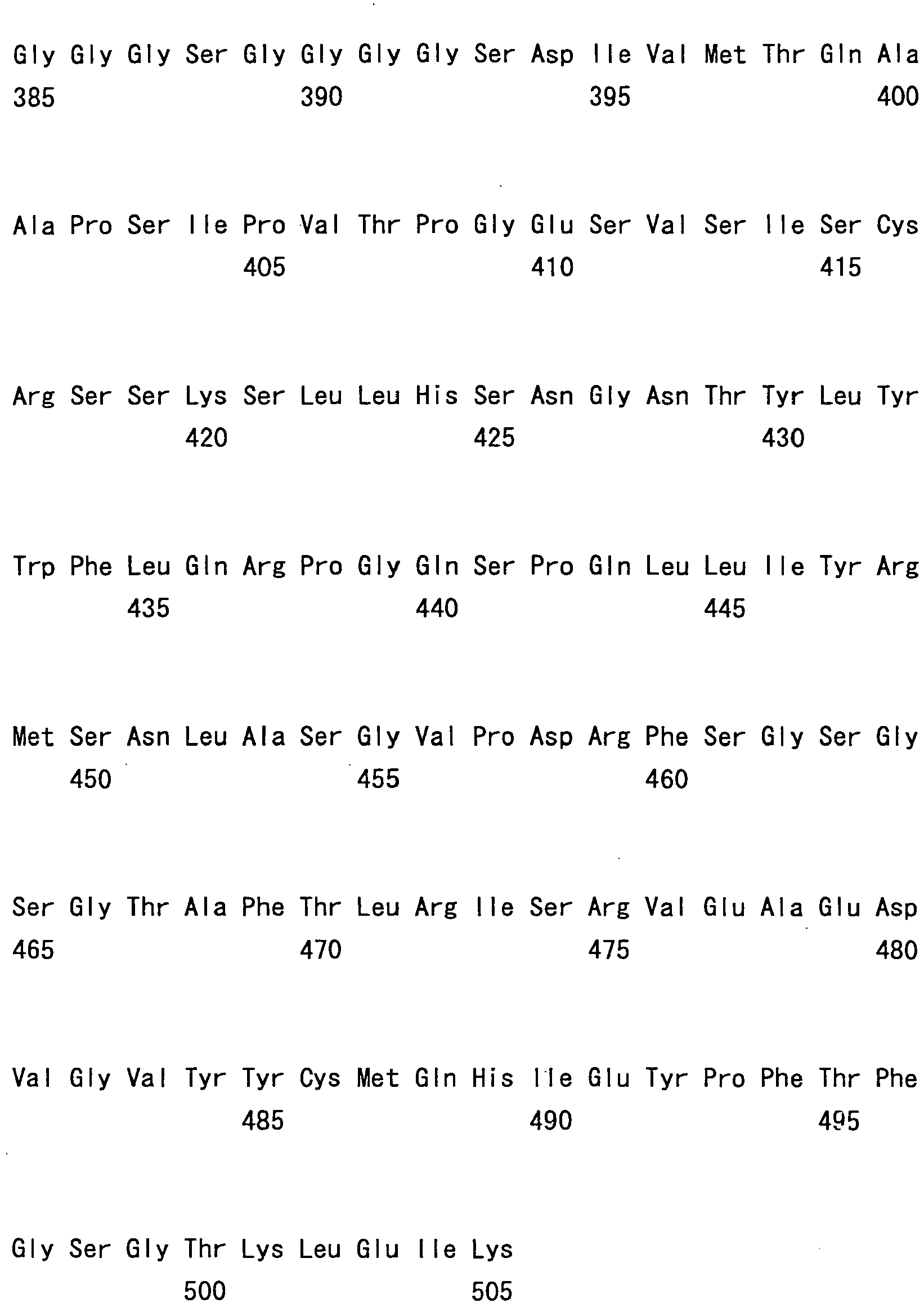
- <210> 3
<211> 501
<212> PRT
<213> Homo sapiens - <220>
<223> An artificially synthesized scFv sequence - <400> 3

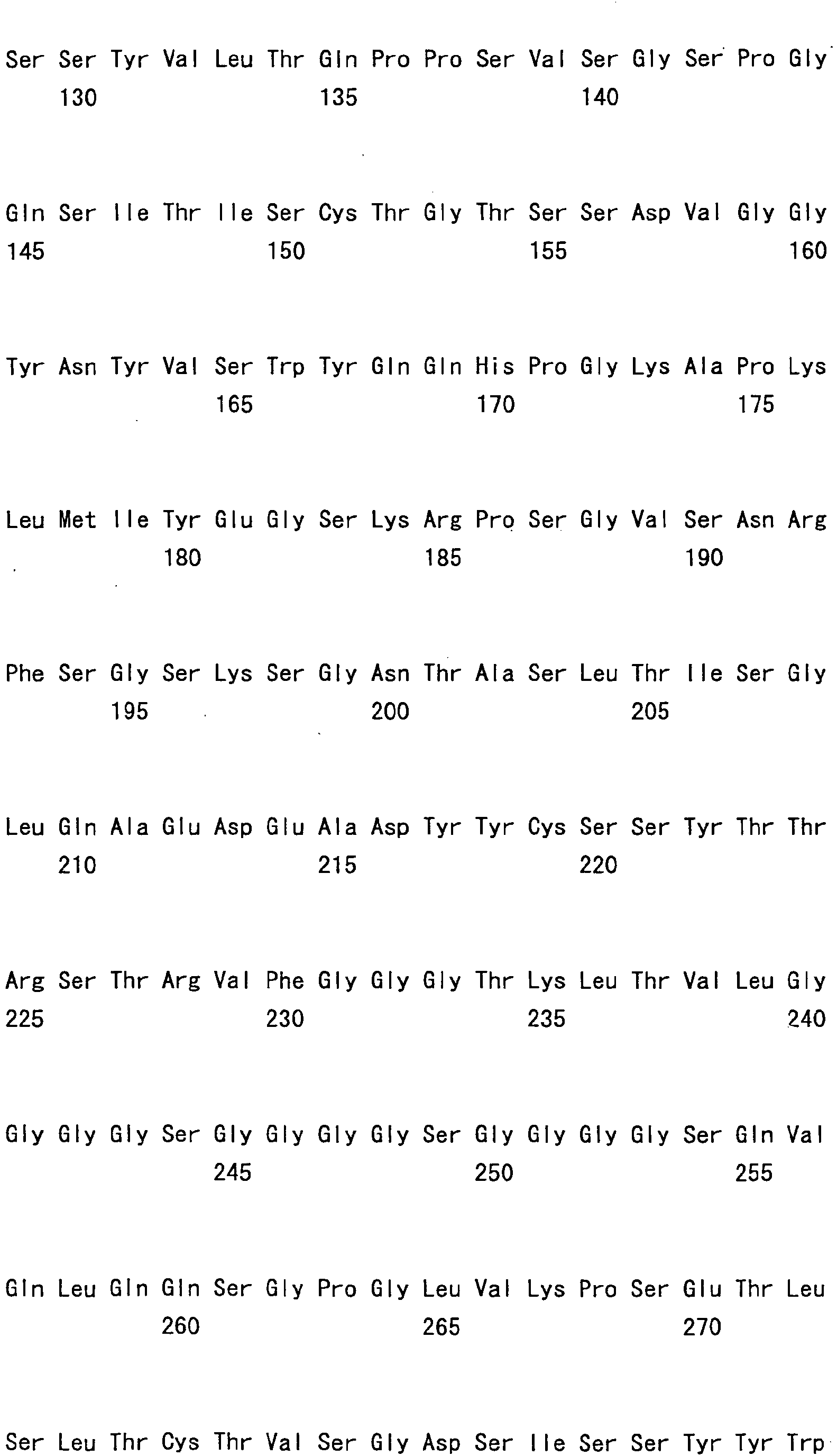

- <210> 4
<211> 495
<212> PRT
<213> Mus museums - <220>
<223> An artificially synthesized scFv sequence - <400> 4


- <210> 5
<211> 449
<212> PRT
<213> Homo sapiens - <220>
- <400> 5


- <210> 6
<211> 94
<212> PRT
<213> Homo sapiens - <220>
- <400> 6


- <210> 7
<211> 1924
<212> DNA
<213> Macaca fascicularis - <220>
<221> CDS
<222> (11).. (1918) - <400> 7


- <210> 8
<211> 24
<212> DNA
<213> Artificial - <220>
<223> an artificially synthesized sequence - <400> 8
caggggccag tggatagact gatg 24 - <210> 9
<211> 23
<212> DNA
<213> Artificial - <220>
<223> an artificially synthesized sequence - <400> 9
gctcactgga tggtgggaag atg 23 - <210> 10
<211> 354
<212> DNA
<213> Mus musculus - <400> 10

- <210> 11
<211> 118
<212> PRT
<213> Mus musculus - <400> 11


- <210> 12
<211> 336
<212> DNA
<213> Mus musculus - <400> 12

- <210> 13
<211> 112
<212> PRT
<213> Mus musculus - <400> 13

- <210> 14
<211> 30
<212> DNA
<213> Artificial - <220>
<223> an artificially synthesized primer sequence - <400> 14
tagaattcca ccatggaatg gcctttgatc 30 - <210> 15
<211> 56
<212> DNA
<213> Artificial - <220>
<223> an artificially synthesized primer sequence - <400> 15
agcctgagtc atcacaatat ccgatccgcc tccacctgca gagacagtga ccagag 56 - <210> 16
<211> 56
<212> DNA
<213> Artificial - <220>
<223> an artificially synthesized primer sequence - <400> 16
actctggtca ctgtctctgc aggtggaggc ggatcggata ttgtgatgac tcaggc 56 - <210> 17
<211> 60
<212> DNA
<213> Artificial - <220>
<223> an artificially synthesized primer sequence - <400> 17
attgcggccg cttatcactt atcgtcgtca tccttgtagt cttttatttc cagcttggtc 60 - <210> 18
<211> 8
<212> PRT
<213> Artificial - <220>
<223> an artificially synthesized FLAG tag sequence - <400> 18

- <210> 19
<211> 85
<212> DNA
<213> Artificial - <220>
<223> an artificially synthesized primer sequence - <400> 19

- <210> 20
<211> 82
<212> DNA
<213> Artificial - <220>
<223> an artificially synthesized primer sequence - <400> 20

- <210> 21
<211> 82
<212> DNA
<213> Artificial - <220>
<223> an artificially synthesized primer sequence - <400> 21

- <210> 22
<211> 25
<212> DNA
<213> Artificial - <220>
<223> an artificially synthesized primer sequence - <400> 22
caggttcagc tgcagcagtc tggac 25 - <210> 23
<211> 81
<212> DNA
<213> Artificial - <220>
<223> an artificially synthesized primer sequence - <400> 23

- <210> 24
<211> 1572
<212> DNA
<213> Homo sapiens - <400> 24


- <210> 25
<211> 524
<212> PRT
<213> Homo sapiens - <400> 25


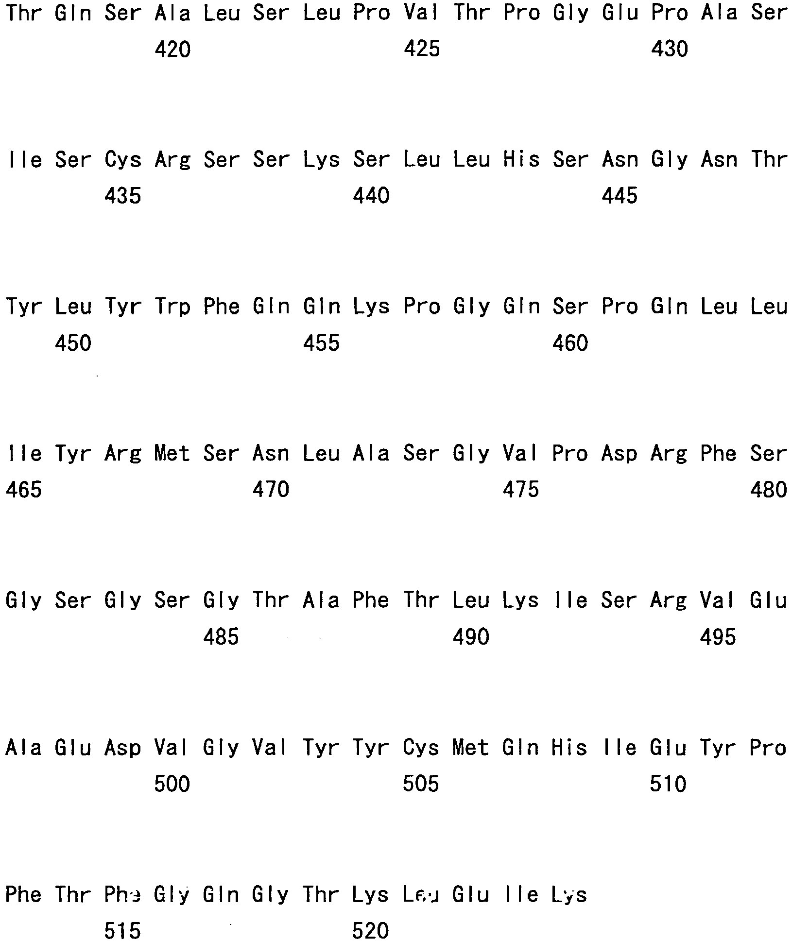
- <210> 26
<211> 1572
<212> DNA
<213> Homo sapiens - <400> 26


- <210> 27
<211> 524
<212> PRT
<213> Homo sapiens - <400> 27


- <210> 28
<211> 1572
<212> DNA
<213> Homo sapiens - <400> 28


- <210> 29
<211> 524
<212> PRT
<213> Homo sapiens - <400> 29


- <210> 30
<211> 1572
<212> DNA
<213> Homo sapiens - <400> 30


- <210> 31
<211> 157.2.
<212> DNA
<213> Homo sapiens - <400> 31


- <210> 32
<211> 524
<212> PRT
<213> Homo sapiens - <400> 32


- <210> 33
<211> 1572
<212> DNA
<213> Mus musculus - <400> 33


- <210> 34
<211> 35
<212> DNA
<213> Artificial - <220>
<223> an artificially synthesized primer sequence - <400> 34
cctgaattcc accatgcgat ggagctggat ctttc 35 - <210> 35
<211> 48
<212> DNA
<213> Artificial - <220>
<223> an artificially synthesized primer sequence - <400> 35
accgccagag ccacctccgc ctgaaccgcc tccacctgag gagactgt 48 - <210> 36
<211> 57
<212> DNA
<213> Artificial - <220>
<223> an artificially synthesized primer sequence - <400> 36
ttcaggcgga ggtggctctg gcggtggcgg aagccaaatt gttctcaccc agtcgcc 57 - <210> 37
<211> 63
<212> DNA
<213> Artificial - <220>
<223> an artificially synthesized primer sequence - <400> 37
- <210> 38
<211> 50
<212> DNA
<213> Artificial - <220>
<223> an artificially synthesized primer sequence - <400> 38
ggcggatccg gtggcggtgg ctcacaggtc cagttgcagc agtctggacc 50 - <210> 39
<211> 68
<212> DNA
<213> Artificial - <220>
<223> an artificially synthesized primer sequence - <400> 39

- <210> 40
<211> 1572
<212> DNA
<213> Mus musculus - <220>
<221> CDS
<222> (14).. (1561) - <400> 40

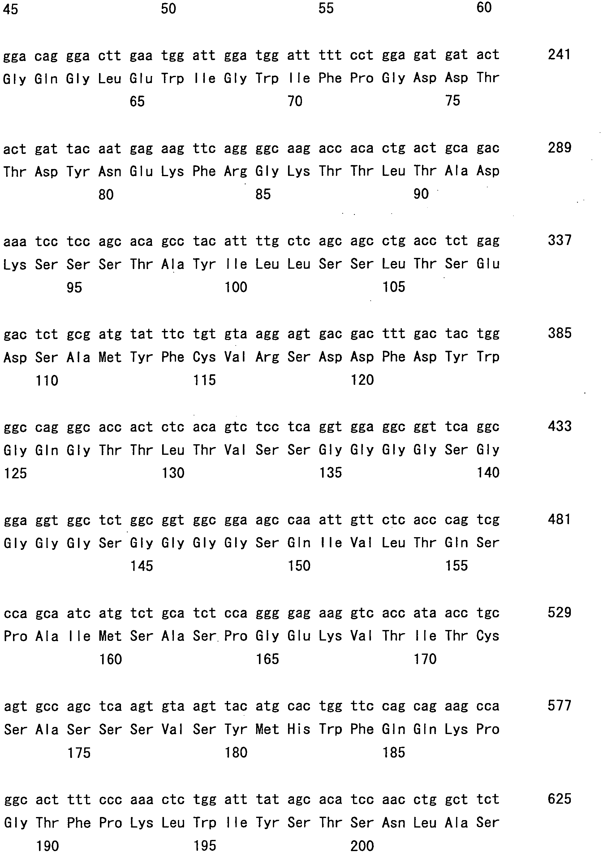


- <210> 41
<211> 4
<212> PRT
<213> Artificial - <220>
<223> An artificially synthesized peptide sequence - <400> 41

- <210> 42
<211> 4
<212> PRT
<213> Artificial - <220>
<223> An artificially synthesized peptide sequence - <400> 42

- <210> 43
<211> 5
<212> PRT
<213> Artificial - <220>
<223> An artificially synthesized peptide sequence - <400> 43

- <210> 44
<211> 5
<212> PRT
<213> Artificial - <220>
<223> An artificially synthesized peptide sequence - <400> 44

- <210> 45
<211> 6
<212> PRT
<213> Artificial - <220>
<223> An artificially synthesized peptide sequence - <400> 45

- <210> 46
<211> 6
<212> PRT
<213> Artificial - <220>
<223> An artificially synthesized peptide sequence - <400> 46

- <210> 47
<211> 7
<212> PRT
<213> Artificial - <220>
<223> An artificially synthesized peptide sequence - <400> 47

- <210> 48
<211> 7
<212> PRT
<213> Artificial - <220>
<223> An artificially synthesized peptide sequence - <400> 48

- <210> 49
<211> 118
<212> PRT
<213> Artificial - <220>
<223> An artificially synthesized sequence - <400> 49


- <210> 50
<211> 119
<212> PRT
<213> Artificial - <220>
<223> An artificially synthesized sequence - <400> 50
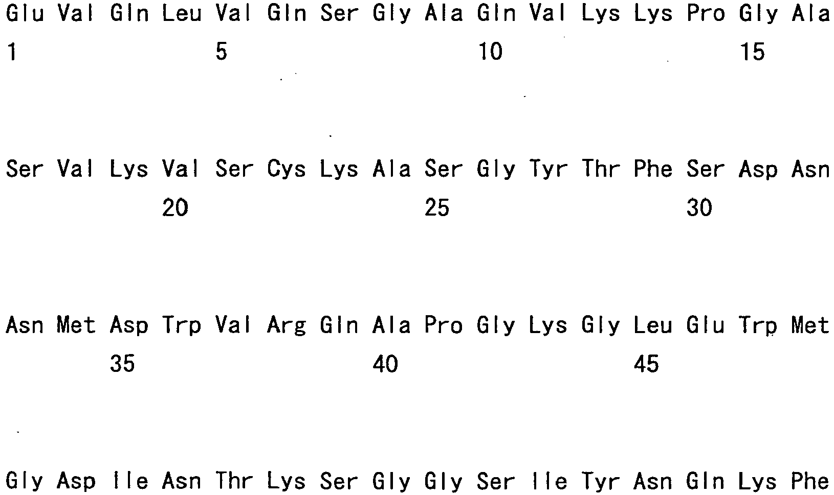

- <210> 51
<211> 106
<212> PRT
<213> Artificial - <220>
<223> An artificially synthesized sequence - <400> 51


Claims (14)
- Use of an agent for long-term stabilization of a protein or for suppressing protein aggregation, which comprises meglumine, or a salt thereof, wherein the protein is an antibody molecule.
- The use of claim 1, wherein the antibody molecule is a whole antibody, antibody fragment, minibody, or a modified antibody.
- The use of any one of claims 1 or 2, whose dosage form is a freeze-dried preparation.
- A method for long-term stabilization of an antibody molecule, which comprises the step of adding meglumine, or a salt thereof, to the antibody molecule.
- A method for suppressing antibody molecule aggregation, which comprises the step of adding meglumine, or a salt thereof, to the antibody molecule.
- The method of claim 4 or 5, wherein the antibody molecule is stabilized for a long period or antibody molecule aggregation is suppressed under long-term storage conditions at a low temperature.
- The method of claim 4 or 5, wherein the antibody molecule is stabilized for a long period or antibody molecule aggregation is suppressed under long-term storage conditions at room temperature.
- The method of claim 4 to 7, wherein the antibody molecule is a whole antibody, antibody fragment, minibody, or a modified antibody.
- The method of any one of claims 4 to 8, which comprises the step of freeze-drying the antibody molecule after the step of adding meglumine, or a salt thereof.
- A pharmaceutical composition comprising an antibody molecule and meglumine, or a salt of meglumine.
- The pharmaceutical composition of claim 10, whose dosage form is a freeze-dried preparation.
- A method for producing a pharmaceutical composition comprising an antibody molecule, which comprises the step of adding meglumine, or a salt thereof, to an antibody-containing composition.
- The method of claim 12, further comprising the step of:freeze-drying the mixture.
- The method for producing a pharmaceutical composition of claim 12 or 13, wherein the antibody molecule is a whole antibody, antibody fragment, minibody, or a modified antibody.
Applications Claiming Priority (2)
| Application Number | Priority Date | Filing Date | Title |
|---|---|---|---|
| JP2005170794 | 2005-06-10 | ||
| PCT/JP2006/311625 WO2006132363A1 (en) | 2005-06-10 | 2006-06-09 | Stabilizer for protein preparation comprising meglumine and use thereof |
Publications (3)
| Publication Number | Publication Date |
|---|---|
| EP1908482A1 EP1908482A1 (en) | 2008-04-09 |
| EP1908482A4 EP1908482A4 (en) | 2012-12-19 |
| EP1908482B1 true EP1908482B1 (en) | 2017-09-06 |
Family
ID=37498557
Family Applications (1)
| Application Number | Title | Priority Date | Filing Date |
|---|---|---|---|
| EP06766537.2A Active EP1908482B1 (en) | 2005-06-10 | 2006-06-09 | Stabilizer for protein preparation comprising meglumine and use thereof |
Country Status (8)
| Country | Link |
|---|---|
| US (1) | US8945543B2 (en) |
| EP (1) | EP1908482B1 (en) |
| JP (1) | JP5068167B2 (en) |
| KR (1) | KR101367544B1 (en) |
| CN (1) | CN101237890A (en) |
| AU (1) | AU2006256041B2 (en) |
| CA (1) | CA2610987C (en) |
| WO (1) | WO2006132363A1 (en) |
Families Citing this family (60)
| Publication number | Priority date | Publication date | Assignee | Title |
|---|---|---|---|---|
| US7696325B2 (en) * | 1999-03-10 | 2010-04-13 | Chugai Seiyaku Kabushiki Kaisha | Polypeptide inducing apoptosis |
| US8034903B2 (en) * | 2000-10-20 | 2011-10-11 | Chugai Seiyaku Kabushiki Kaisha | Degraded TPO agonist antibody |
| US20040242847A1 (en) * | 2000-10-20 | 2004-12-02 | Naoshi Fukushima | Degraded agonist antibody |
| AU2003271175A1 (en) * | 2002-10-11 | 2004-05-04 | Masahiro Abe | Cell death-inducing agent |
| JP2004279086A (en) * | 2003-03-13 | 2004-10-07 | Konica Minolta Holdings Inc | Radiation image conversion panel and method for manufacturing it |
| EP1609803A4 (en) * | 2003-03-31 | 2006-05-24 | Chugai Pharmaceutical Co Ltd | MODIFIED ANTIBODY AGAINST CD22 AND ITS USE |
| JP4794301B2 (en) * | 2003-06-11 | 2011-10-19 | 中外製薬株式会社 | Antibody production method |
| WO2005035753A1 (en) * | 2003-10-10 | 2005-04-21 | Chugai Seiyaku Kabushiki Kaisha | Double specific antibodies substituting for functional protein |
| AU2003271186A1 (en) * | 2003-10-14 | 2005-04-27 | Chugai Seiyaku Kabushiki Kaisha | Double specific antibodies substituting for functional protein |
| US20070280951A1 (en) * | 2003-12-12 | 2007-12-06 | Naoki Kimura | Cell Death Inducing Agents |
| US20070281327A1 (en) * | 2003-12-12 | 2007-12-06 | Kiyotaka Nakano | Methods of Screening for Modified Antibodies With Agonistic Activities |
| TW200530269A (en) | 2003-12-12 | 2005-09-16 | Chugai Pharmaceutical Co Ltd | Anti-Mpl antibodies |
| TW200530266A (en) * | 2003-12-12 | 2005-09-16 | Chugai Pharmaceutical Co Ltd | Method of reinforcing antibody activity |
| EP1757686A4 (en) * | 2004-04-09 | 2008-03-12 | Chugai Pharmaceutical Co Ltd | INDUCER OF CELL DEATH |
| AU2006232287B2 (en) * | 2005-03-31 | 2011-10-06 | Chugai Seiyaku Kabushiki Kaisha | Methods for producing polypeptides by regulating polypeptide association |
| JP5057967B2 (en) * | 2005-03-31 | 2012-10-24 | 中外製薬株式会社 | sc (Fv) 2 structural isomer |
| PT1876236E (en) * | 2005-04-08 | 2014-10-22 | Chugai Pharmaceutical Co Ltd | ANTIBODIES FOR REPLACING THE FUNCTION OF THE BLOOD CELL FACTOR VIII |
| US20090022687A1 (en) * | 2005-05-18 | 2009-01-22 | Chugai Seiyaku Kabushiki Kaisha | Novel Pharmaceuticals That Use Anti-HLA Antibodies |
| CA2611726C (en) | 2005-06-10 | 2017-07-11 | Chugai Seiyaku Kabushiki Kaisha | Pharmaceutical compositions containing sc(fv)2 |
| CN104761637B (en) | 2006-03-31 | 2021-10-15 | 中外制药株式会社 | Methods for modulating antibody hemodynamics |
| EP2009101B1 (en) | 2006-03-31 | 2017-10-25 | Chugai Seiyaku Kabushiki Kaisha | Antibody modification method for purifying bispecific antibody |
| US20100150927A1 (en) * | 2006-07-13 | 2010-06-17 | Chugai Seiyaku Kabushiki Kaisha | Cell death inducer |
| CL2008000719A1 (en) * | 2007-03-12 | 2008-09-05 | Univ Tokushima Chugai Seiyaku | THERAPEUTIC AGENT FOR CANCER RESISTANT TO CHEMOTHERAPEUTIC AGENTS THAT UNDERSTAND AN ANTIBODY THAT RECOGNIZES IT CLASS I AS ACTIVE INGREDIENT; PHARMACEUTICAL COMPOSITION THAT INCLUDES SUCH ANTIBODY; AND METHOD TO TREAT CANCER RESISTANT TO |
| KR101680906B1 (en) | 2007-09-26 | 2016-11-30 | 추가이 세이야쿠 가부시키가이샤 | Modified antibody constant region |
| ES2595638T3 (en) * | 2007-09-26 | 2017-01-02 | Chugai Seiyaku Kabushiki Kaisha | Method to modify the isoelectric point of an antibody by replacing amino acids in a CDR |
| KR102057826B1 (en) | 2008-04-11 | 2019-12-20 | 추가이 세이야쿠 가부시키가이샤 | Antigen-binding molecule capable of binding to two or more antigen molecules repeatedly |
| JP5717624B2 (en) | 2009-03-19 | 2015-05-13 | 中外製薬株式会社 | Antibody constant region variants |
| EP2826789A1 (en) | 2009-03-19 | 2015-01-21 | Chugai Seiyaku Kabushiki Kaisha | Antibody constant region variant |
| US10022109B2 (en) * | 2009-06-17 | 2018-07-17 | Gilupi Gmbh | Detection device for the in vivo and/or in vitro enrichment of sample material |
| EP2603232B1 (en) * | 2010-08-11 | 2019-09-25 | Ironwood Pharmaceuticals, Inc. | Stable formulations of linaclotide |
| TWI452136B (en) | 2010-11-17 | 2014-09-11 | 中外製藥股份有限公司 | A multiple specific antigen-binding molecule that replaces the function of Factor VIII in blood coagulation |
| EP2647706B1 (en) | 2010-11-30 | 2023-05-17 | Chugai Seiyaku Kabushiki Kaisha | Antigen-binding molecule capable of binding to plurality of antigen molecules repeatedly |
| EP3604339B1 (en) | 2011-01-14 | 2021-03-10 | The Regents Of The University Of California | Therapeutic antibodies against ror-1 protein and methods for use of same |
| EP2500035A1 (en) * | 2011-03-15 | 2012-09-19 | Icon Genetics GmbH | Pharmaceutical formulation containing immunglobulin |
| WO2013012022A1 (en) | 2011-07-19 | 2013-01-24 | 中外製薬株式会社 | Stable protein-containing preparation containing argininamide or analogous compound thereof |
| BR112014009146A8 (en) * | 2011-10-18 | 2017-06-20 | Coherus Biosciences Inc | amino acid stabilized etanercept formulations |
| US10485869B2 (en) | 2011-10-18 | 2019-11-26 | Coherus Biosciences, Inc. | Etanercept formulations stabilized with meglumine |
| HK1209343A1 (en) | 2012-07-09 | 2016-04-01 | Coherus Biosciences, Inc. | Etanercept formulations exhibiting marked reduction in sub-visible particles |
| SI2895188T1 (en) | 2012-09-11 | 2018-05-31 | Coherus Biosciences, Inc. | Correctly folded etanercept in high purity and excellent yield |
| ES2881306T3 (en) | 2013-09-27 | 2021-11-29 | Chugai Pharmaceutical Co Ltd | Method for the production of heteromultimers of polypeptides |
| RU2715236C2 (en) | 2014-03-26 | 2020-02-26 | Астекс Терапьютикс Лтд | Combinations |
| US10494422B2 (en) | 2014-04-29 | 2019-12-03 | Seattle Children's Hospital | CCR5 disruption of cells expressing anti-HIV chimeric antigen receptor (CAR) derived from broadly neutralizing antibodies |
| IS3008B (en) * | 2014-05-14 | 2018-12-15 | Calor ehf | A peptide or protein stabilizing formulation |
| MA40764A (en) | 2014-09-26 | 2017-08-01 | Chugai Pharmaceutical Co Ltd | THERAPEUTIC AGENT INDUCING CYTOTOXICITY |
| US20180256717A1 (en) * | 2014-12-23 | 2018-09-13 | Drug Discovery Laboratory As | Protein compositions and use thereof |
| TWI719960B (en) | 2015-02-10 | 2021-03-01 | 英商阿斯迪克治療公司 | New compositions |
| AU2016381992B2 (en) | 2015-12-28 | 2024-01-04 | Chugai Seiyaku Kabushiki Kaisha | Method for promoting efficiency of purification of Fc region-containing polypeptide |
| US11324829B2 (en) | 2016-03-14 | 2022-05-10 | Santen Pharmaceutical Co., Ltd. | Antiseptic agent comprising meglumine or salt thereof |
| CR20180554A (en) | 2016-04-28 | 2019-01-10 | Chugai Pharmaceutical Co Ltd | PREPARATIONS CONTAINING ANTIBODIES |
| MX383392B (en) | 2016-09-27 | 2025-03-13 | Camurus Ab | MIXTURES AND FORMULATIONS COMPRISING AN ALKYL AMMONIUM SALT OF ETHYLENEDIAMINETETRAACETIC ACID (EDTA). |
| AU2017386888B2 (en) | 2016-12-28 | 2024-08-08 | Jcr Pharmaceuticals Co., Ltd. | Lyophilized preparation |
| EP3680330A4 (en) * | 2017-09-07 | 2021-09-01 | Amano Enzyme Inc. | Stabilised dry protein deamidase composition |
| CN119161488A (en) | 2017-11-01 | 2024-12-20 | 中外制药株式会社 | Antibody variants and isotypes with reduced biological activity |
| WO2019130049A1 (en) | 2017-12-29 | 2019-07-04 | Grünenthal GmbH | Pharmaceutical combination comprising extended-release tramadol hydrochloride and immediate-release etoricoxib, and its use for the treatment of pain |
| US20210379186A1 (en) * | 2018-04-16 | 2021-12-09 | Merck Patent Gmbh | Additives for protein formulations to improve thermal stability |
| AU2019254478A1 (en) * | 2018-04-16 | 2020-12-03 | Merck Patent Gmbh | Method for stabilizing protein comprising formulations by using a meglumine salt. |
| WO2019201904A1 (en) * | 2018-04-16 | 2019-10-24 | Merck Patent Gmbh | Viscosity reduction of highly concentrated protein formulations |
| US12331320B2 (en) | 2018-10-10 | 2025-06-17 | The Research Foundation For The State University Of New York | Genome edited cancer cell vaccines |
| WO2021053001A1 (en) * | 2019-09-17 | 2021-03-25 | Merck Patent Gmbh | Camphorsulfonic acid and combinations thereof with cationic excipients as viscosity reducing agents in high concentrated protein formulations |
| US20220387433A1 (en) * | 2019-11-12 | 2022-12-08 | American Regent, Inc. | Type v phosphodiesterase inhibitor compositions, methods of making them and methods of using them in preventing or treating elevated pulmonary vascular pressure or pulmonary hemorrhages |
Family Cites Families (164)
| Publication number | Priority date | Publication date | Assignee | Title |
|---|---|---|---|---|
| US3773919A (en) | 1969-10-23 | 1973-11-20 | Du Pont | Polylactide-drug mixtures |
| IE52535B1 (en) | 1981-02-16 | 1987-12-09 | Ici Plc | Continuous release pharmaceutical compositions |
| JPS58201994A (en) | 1982-05-21 | 1983-11-25 | Hideaki Hagiwara | Method for producing antigen-specific human immunoglobulin |
| HUT35524A (en) | 1983-08-02 | 1985-07-29 | Hoechst Ag | Process for preparing pharmaceutical compositions containing regulatory /regulative/ peptides providing for the retarded release of the active substance |
| JPS6094580U (en) | 1983-12-05 | 1985-06-27 | 株式会社 銭屋アルミニウム製作所 | door decoration frame |
| US5618920A (en) * | 1985-11-01 | 1997-04-08 | Xoma Corporation | Modular assembly of antibody genes, antibodies prepared thereby and use |
| GB8607679D0 (en) | 1986-03-27 | 1986-04-30 | Winter G P | Recombinant dna product |
| US4946778A (en) * | 1987-09-21 | 1990-08-07 | Genex Corporation | Single polypeptide chain binding molecules |
| JP2727112B2 (en) * | 1988-04-26 | 1998-03-11 | コニカ株式会社 | Stable peroxidase composition and stable antibody composition |
| US5077216A (en) * | 1988-07-06 | 1991-12-31 | The Trustees Of Dartmouth College | Monoclonal antibodies specific for a human mononculear phagocyte-specific antigen |
| JPH0341033A (en) | 1989-07-07 | 1991-02-21 | Kyowa Hakko Kogyo Co Ltd | Stable preparation containing motilins |
| WO1996033735A1 (en) | 1995-04-27 | 1996-10-31 | Abgenix, Inc. | Human antibodies derived from immunized xenomice |
| GB9009548D0 (en) | 1990-04-27 | 1990-06-20 | Celltech Ltd | Chimeric antibody and method |
| GB9015198D0 (en) | 1990-07-10 | 1990-08-29 | Brien Caroline J O | Binding substance |
| DK0585287T3 (en) | 1990-07-10 | 2000-04-17 | Cambridge Antibody Tech | Process for producing specific binding pair elements |
| ATE158021T1 (en) | 1990-08-29 | 1997-09-15 | Genpharm Int | PRODUCTION AND USE OF NON-HUMAN TRANSGENT ANIMALS FOR THE PRODUCTION OF HETEROLOGUE ANTIBODIES |
| WO1993012227A1 (en) | 1991-12-17 | 1993-06-24 | Genpharm International, Inc. | Transgenic non-human animals capable of producing heterologous antibodies |
| US5223241A (en) * | 1990-10-01 | 1993-06-29 | The General Hospital Corporation | Method for early detection of allograft rejection |
| US5795965A (en) | 1991-04-25 | 1998-08-18 | Chugai Seiyaku Kabushiki Kaisha | Reshaped human to human interleukin-6 receptor |
| US5637481A (en) * | 1993-02-01 | 1997-06-10 | Bristol-Myers Squibb Company | Expression vectors encoding bispecific fusion proteins and methods of producing biologically active bispecific fusion proteins in a mammalian cell |
| EP0605522B1 (en) | 1991-09-23 | 1999-06-23 | Medical Research Council | Methods for the production of humanized antibodies |
| GB9120304D0 (en) | 1991-09-24 | 1991-11-06 | Erba Carlo Spa | Stable pharmaceutical compositions containing a granulocyte macrophage colony stimulating factor |
| JPH0597703A (en) | 1991-10-04 | 1993-04-20 | Yamanouchi Pharmaceut Co Ltd | Modified tissue plasminogen-activating factor-containing composition |
| WO1993006862A1 (en) * | 1991-10-11 | 1993-04-15 | Toray Industries, Inc. | Antibody composition |
| PT1024191E (en) | 1991-12-02 | 2008-12-22 | Medical Res Council | Production of anti-self antibodies from antibody segment repertoires and displayed on phage |
| CA2131151A1 (en) | 1992-03-24 | 1994-09-30 | Kevin S. Johnson | Methods for producing members of specific binding pairs |
| ES2301158T3 (en) | 1992-07-24 | 2008-06-16 | Amgen Fremont Inc. | XENOGENIC ANTIBODY PRODUCTION. |
| ZA936260B (en) | 1992-09-09 | 1994-03-18 | Smithkline Beecham Corp | Novel antibodies for conferring passive immunity against infection by a pathogen in man |
| EP0627932B1 (en) * | 1992-11-04 | 2002-05-08 | City Of Hope | Antibody construct |
| ES2156149T3 (en) * | 1992-12-04 | 2001-06-16 | Medical Res Council | MULTIVALENT AND MULTI-SPECIFIC UNION PROTEINS, ITS MANUFACTURE AND USE. |
| ATE187494T1 (en) | 1992-12-11 | 1999-12-15 | Dow Chemical Co | MULTIVALENT SINGLE CHAIN ANTIBODIES |
| US5780021A (en) * | 1993-03-05 | 1998-07-14 | Georgetown University | Method for treating type 1 diabetes using α-interferon and/or β-i |
| JPH08509612A (en) | 1993-04-26 | 1996-10-15 | ジェンファーム インターナショナル インコーポレイテッド | Transgenic non-human animal capable of producing heterologous antibody |
| US5747654A (en) * | 1993-06-14 | 1998-05-05 | The United States Of America As Represented By The Department Of Health And Human Services | Recombinant disulfide-stabilized polypeptide fragments having binding specificity |
| GB9313509D0 (en) | 1993-06-30 | 1993-08-11 | Medical Res Council | Chemisynthetic libraries |
| CZ294359B6 (en) * | 1993-09-03 | 2004-12-15 | Chugai Seiyaku Kabushiki Kaisha | Monoclonal antibody that induces apoptosis in the myelocytic cells, fragment thereof, hybridoma producing thereof and pharmaceutical composition in which the antibody is comprised |
| WO1995015388A1 (en) | 1993-12-03 | 1995-06-08 | Medical Research Council | Recombinant binding proteins and peptides |
| JP3554355B2 (en) | 1994-03-03 | 2004-08-18 | 中外製薬株式会社 | IL-6 autocrine proliferating human myeloma cell line |
| US6719972B1 (en) * | 1994-06-03 | 2004-04-13 | Repligen Corporation | Methods of inhibiting T cell proliferation or IL-2 accumulation with CTLA4- specific antibodies |
| DE69535243T2 (en) | 1994-07-13 | 2007-05-10 | Chugai Seiyaku K.K. | AGAINST HUMAN INTERLEUKIN-8 DIRECTED, RECONSTITUTED HUMAN ANTIBODY |
| US5885574A (en) * | 1994-07-26 | 1999-03-23 | Amgen Inc. | Antibodies which activate an erythropoietin receptor |
| DE69534530T2 (en) * | 1994-08-12 | 2006-07-06 | Immunomedics, Inc. | FOR B-CELL LYMPHOMA AND LEUKEMIA SPECIMEN IMMUNOCONJUGATES AND HUMAN ANTIBODIES |
| US8771694B2 (en) * | 1994-08-12 | 2014-07-08 | Immunomedics, Inc. | Immunoconjugates and humanized antibodies specific for B-cell lymphoma and leukemia cells |
| US6451523B1 (en) * | 1994-09-14 | 2002-09-17 | Interneuron Pharmaceuticals, Inc. | Detection of a leptin receptor variant and methods for regulating obesity |
| US5690935A (en) | 1995-01-13 | 1997-11-25 | Regents Of The University Of Minnesota | Biotherapy of cancer by targeting TP-3/P80 |
| EP0726075A1 (en) | 1995-02-08 | 1996-08-14 | Therapicon Srl | Pharmaceutical non-inorganic saline solutions for endonasal administration |
| PT812136E (en) | 1995-02-28 | 2001-04-30 | Procter & Gamble | PREPARATION OF NON-GASTIC BEVERAGES WITH SUPERIOR MICROBIAL STABILITY |
| US5731168A (en) | 1995-03-01 | 1998-03-24 | Genentech, Inc. | Method for making heteromultimeric polypeptides |
| WO1996034096A1 (en) | 1995-04-28 | 1996-10-31 | Abgenix, Inc. | Human antibodies derived from immunized xenomice |
| EP0871673B1 (en) | 1995-05-03 | 2006-04-05 | Bioenhancementsments Ltd. | Bispecific antibodies in which the binding capability is reversibly inhibited by a photocleavable moiety |
| WO1996036360A1 (en) | 1995-05-17 | 1996-11-21 | Regents Of The University Of Minnesota | Immunoconjugates comprising single-chain variable region fragments of anti-cd-19 antibodies |
| AUPO591797A0 (en) | 1997-03-27 | 1997-04-24 | Commonwealth Scientific And Industrial Research Organisation | High avidity polyvalent and polyspecific reagents |
| US6013067A (en) * | 1995-06-07 | 2000-01-11 | Zymogenetics, Inc. | Methods for increasing hematopoietic cells |
| US5977322A (en) * | 1995-06-14 | 1999-11-02 | The Regents Of The University Of California | High affinity human antibodies to tumor antigens |
| DK1666591T3 (en) | 1995-06-29 | 2011-05-23 | Immunex Corp | Cytokine that induces apoptosis |
| ES2233974T3 (en) | 1995-09-11 | 2005-06-16 | Kyowa Hakko Kogyo Co., Ltd. | ANTIBODY AGAINST THE ALFA CHAIN OF THE 5 HUMAN INTERLEUCINE RECEIVER. |
| AU721129B2 (en) * | 1996-01-08 | 2000-06-22 | Genentech Inc. | WSX receptor and ligands |
| FR2745008A1 (en) * | 1996-02-20 | 1997-08-22 | Ass Pour Le Dev De La Rech En | MODIFIED NUCLEAR GLUCOCORTICOID RECEPTOR, DNA FRAGMENTS ENCODING SAID RECEPTOR, AND METHODS IN WHICH THEY ARE USED |
| AU2232597A (en) * | 1996-03-06 | 1997-09-22 | Chugai Seiyaku Kabushiki Kaisha | Method of screening apoptosis inducing substances |
| ATE267610T1 (en) | 1996-03-20 | 2004-06-15 | Immunomedics Inc | GLYCOSYLATED IGG ANTIBODIES |
| JPH09289892A (en) | 1996-04-25 | 1997-11-11 | Yamanouchi Pharmaceut Co Ltd | c-MPL ligand agonist antibody |
| IL126971A0 (en) * | 1996-05-09 | 1999-09-22 | Pharma Pacific Pty Ltd | Interferon compositions |
| JP3416035B2 (en) | 1996-09-26 | 2003-06-16 | 中外製薬株式会社 | Antibodies to human parathyroid hormone-related peptides |
| EP0962467A4 (en) * | 1996-09-26 | 2002-10-30 | Chugai Pharmaceutical Co Ltd | ANTIBODIES AGAINST HUMAN PARATHORMON RELATED PEPTIDES |
| EP0852951A1 (en) * | 1996-11-19 | 1998-07-15 | Roche Diagnostics GmbH | Stable lyophilized monoclonal or polyclonal antibodies containing pharmaceuticals |
| US6323000B2 (en) * | 1996-12-20 | 2001-11-27 | Clark A. Briggs | Variant human α7 acetylcholine receptor subunit, and methods of production and uses thereof |
| US7541034B1 (en) | 1997-03-20 | 2009-06-02 | The United States Of America As Represented By The Department Of Health And Human Services | Recombinant antibodies and immunoconjugates targeted to CD-22 bearing cells and tumors |
| US6306393B1 (en) | 1997-03-24 | 2001-10-23 | Immunomedics, Inc. | Immunotherapy of B-cell malignancies using anti-CD22 antibodies |
| US6183744B1 (en) | 1997-03-24 | 2001-02-06 | Immunomedics, Inc. | Immunotherapy of B-cell malignancies using anti-CD22 antibodies |
| FR2761994B1 (en) | 1997-04-11 | 1999-06-18 | Centre Nat Rech Scient | PREPARATION OF MEMBRANE RECEPTORS FROM EXTRACELLULAR BACULOVIRUSES |
| ATE299938T1 (en) | 1997-05-02 | 2005-08-15 | Genentech Inc | A METHOD FOR PRODUCING MULTI-SPECIFIC ANTIBODIES THAT POSSESS HETEROMULTIMER AND COMMON COMPONENTS |
| US6368596B1 (en) * | 1997-07-08 | 2002-04-09 | Board Of Regents, The University Of Texas System | Compositions and methods for homoconjugates of antibodies which induce growth arrest or apoptosis of tumor cells |
| US5980893A (en) | 1997-07-17 | 1999-11-09 | Beth Israel Deaconess Medical Center, Inc. | Agonist murine monoclonal antibody as a stimulant for megakaryocytopoiesis |
| US6342220B1 (en) | 1997-08-25 | 2002-01-29 | Genentech, Inc. | Agonist antibodies |
| HUP0003513A3 (en) * | 1997-09-11 | 2004-05-28 | Chugai Pharmaceutical Co Ltd | Monoclonal antibody inducing apoptosis |
| US7531643B2 (en) * | 1997-09-11 | 2009-05-12 | Chugai Seiyaku Kabushiki Kaisha | Monoclonal antibody inducing apoptosis |
| CA2273223C (en) | 1997-09-29 | 2003-11-11 | Raytheon Company | Chip-size package using a polyimide pcb interposer |
| US5998593A (en) * | 1998-03-10 | 1999-12-07 | Abbott Laboratories | Fluorescent enzyme substrates |
| DE19819846B4 (en) | 1998-05-05 | 2016-11-24 | Deutsches Krebsforschungszentrum Stiftung des öffentlichen Rechts | Multivalent antibody constructs |
| US7138103B2 (en) | 1998-06-22 | 2006-11-21 | Immunomedics, Inc. | Use of bi-specific antibodies for pre-targeting diagnosis and therapy |
| US7081360B2 (en) * | 1998-07-28 | 2006-07-25 | Cadus Technologies, Inc. | Expression of G protein-coupled receptors with altered ligand binding and/or coupling properties |
| ES2246093T3 (en) | 1998-10-16 | 2006-02-01 | Fraunhofer-Gesellschaft Zur Forderung Der Angewandten Forschung E.V. | MOLECULAR PATHOGENICIDE THAT INDUCES A RESISTANCE TO DISEASE IN PLANTS. |
| US20050220787A1 (en) * | 2002-11-07 | 2005-10-06 | Lobo Peter I | Naturally occuring IgM antibodies that bind to lymphocytes |
| US6897044B1 (en) | 1999-01-28 | 2005-05-24 | Biogen Idec, Inc. | Production of tetravalent antibodies |
| US7696325B2 (en) * | 1999-03-10 | 2010-04-13 | Chugai Seiyaku Kabushiki Kaisha | Polypeptide inducing apoptosis |
| AU2940900A (en) | 1999-03-10 | 2000-09-28 | Chugai Seiyaku Kabushiki Kaisha | Single-stranded fv inducing apoptosis |
| AR030019A1 (en) | 1999-05-18 | 2003-08-13 | Smithkline Beecham Corp | HUMAN MONOCLONAL ANTIBODIES AND FUNCTIONAL FRAGMENTS OF THE SAME, A PROCEDURE FOR THEIR PRODUCTION, PHARMACEUTICAL COMPOSITIONS THAT INCLUDE THEM, A NUCLEIC ACID ISOLATED MOLECULA, A RECOMBINANT PLASMIDE, A HOSPED DIFFERENT USE OF A MUSCLE |
| ATE330972T1 (en) | 1999-06-09 | 2006-07-15 | Genentech Inc | APO-2L RECEPTOR AGONIST AND CPT-11 SYNERGY EFFECT |
| US20020028178A1 (en) * | 2000-07-12 | 2002-03-07 | Nabil Hanna | Treatment of B cell malignancies using combination of B cell depleting antibody and immune modulating antibody related applications |
| JP2003515323A (en) | 1999-11-18 | 2003-05-07 | オックスフォード バイオメディカ(ユーケイ)リミテッド | Body |
| JP2003516744A (en) | 1999-12-14 | 2003-05-20 | ザ バーナム インスティチュート | BCL-G polypeptides, nucleic acids encoding them and methods of use |
| AU2001256174A1 (en) | 2000-03-01 | 2001-09-12 | Christoph Gasche | Mammalian interleukin-10 (il-10) receptor variants |
| TWI241345B (en) | 2000-03-10 | 2005-10-11 | Chugai Pharmaceutical Co Ltd | Apoptosis inducing polypeptide |
| CA2404216A1 (en) | 2000-03-22 | 2001-09-27 | Curagen Corporation | Novel polypeptides, and nucleic acids encoding the same |
| WO2001074388A1 (en) | 2000-03-31 | 2001-10-11 | Idec Pharmaceuticals Corporation | Combined use of anti-cytokine antibodies or antagonists and anti-cd20 for the treatment of b cell lymphoma |
| JP2003531588A (en) | 2000-04-11 | 2003-10-28 | ジェネンテック・インコーポレーテッド | Multivalent antibodies and their uses |
| US20040058393A1 (en) * | 2000-04-17 | 2004-03-25 | Naoshi Fukishima | Agonist antibodies |
| WO2001079494A1 (en) | 2000-04-17 | 2001-10-25 | Chugai Seiyaku Kabushiki Kaisha | Agonist antibodies |
| EP1156060B1 (en) | 2000-05-12 | 2007-06-27 | GPC Biotech AG | Human peptides/proteins causing or leading to the killing of cells including lymphoid tumor cells |
| JP2004512262A (en) | 2000-06-20 | 2004-04-22 | アイデック ファーマスーティカルズ コーポレイション | Non-radioactive anti-CD20 antibody / radiolabeled anti-CD22 antibody combination |
| AU6461201A (en) | 2000-07-12 | 2002-01-21 | Idec Pharma Corp | Treatment of b cell malignancies using combination of b cell depleting antibody and immune modulating antibody related applications |
| JP5485489B2 (en) * | 2000-08-11 | 2014-05-07 | 中外製薬株式会社 | Antibody-containing stabilized preparation |
| EP1310252A4 (en) | 2000-08-18 | 2009-06-10 | Takeda Pharmaceutical | INJECTIONS |
| JP2004508420A (en) | 2000-09-18 | 2004-03-18 | アイデック ファーマスーティカルズ コーポレイション | Combination therapy for treating autoimmune diseases using B cell depleting antibody / immunomodulatory antibody combination |
| EP1323735A4 (en) | 2000-10-03 | 2005-02-09 | Banyu Pharma Co Ltd | GENE RELATING TO DRUG TOLERANCE AND USE THEREOF |
| US8034903B2 (en) * | 2000-10-20 | 2011-10-11 | Chugai Seiyaku Kabushiki Kaisha | Degraded TPO agonist antibody |
| AU1091802A (en) | 2000-10-20 | 2002-04-29 | Chugai Pharmaceutical Co Ltd | Degraded agonist antibody |
| WO2002033072A1 (en) | 2000-10-20 | 2002-04-25 | Chugai Seiyaku Kabushiki Kaisha | Degraded tpo agonist antibody |
| US20040242847A1 (en) * | 2000-10-20 | 2004-12-02 | Naoshi Fukushima | Degraded agonist antibody |
| WO2002078612A2 (en) | 2001-04-02 | 2002-10-10 | Euro-Celtique S.A. | Thrombopoietin (tpo) synthebody for stimulation of platelet production |
| WO2002085923A2 (en) * | 2001-04-19 | 2002-10-31 | The Scripps Research Institute | In vivo incorporation of unnatural amino acids |
| US7115717B2 (en) | 2001-05-18 | 2006-10-03 | Kirin Beer Kabushiki Kaisha | Anti-TRAIL-R antibodies |
| US7064189B2 (en) | 2001-05-25 | 2006-06-20 | Human Genome Sciences, Inc. | Antibodies that immunospecifically bind to trail receptors |
| GB0113179D0 (en) | 2001-05-31 | 2001-07-25 | Novartis Ag | Organic compounds |
| US7321026B2 (en) | 2001-06-27 | 2008-01-22 | Skytech Technology Limited | Framework-patched immunoglobulins |
| DK1399484T3 (en) * | 2001-06-28 | 2010-11-08 | Domantis Ltd | Double-specific ligand and its use |
| US7262278B2 (en) * | 2001-10-15 | 2007-08-28 | Kirin Beer Kabushiki Kaisha | Anti-HLA-DR antibody |
| JP2005507659A (en) | 2001-10-15 | 2005-03-24 | イミューノメディクス、インコーポレイテッド | Direct targeting binding protein |
| AU2003207459A1 (en) | 2002-01-03 | 2003-07-24 | The Scripps Research Institute | Cancer-associated epitope |
| CN101721362B (en) | 2002-02-14 | 2018-07-03 | 中外制药株式会社 | Pharmaceutical solutions comprising antibody |
| WO2003072736A2 (en) * | 2002-02-21 | 2003-09-04 | Duke University | Reagents and treatment methods for autoimmune diseases |
| US20030232887A1 (en) * | 2002-04-10 | 2003-12-18 | Johnson Douglas Giles | Preparation and use of a stable formulation of allosteric effector compounds |
| WO2003087163A1 (en) | 2002-04-15 | 2003-10-23 | Chugai Seiyaku Kabushiki Kaisha | METHOD OF CONSTRUCTING scDb LIBRARY |
| JP2004086862A (en) | 2002-05-31 | 2004-03-18 | Celestar Lexico-Sciences Inc | Apparatus, method and program for processing protein interaction information, and recording medium |
| WO2003107218A1 (en) | 2002-05-31 | 2003-12-24 | セレスター・レキシコ・サイエンシズ株式会社 | Interaction predicting device |
| EP1511518A2 (en) | 2002-06-07 | 2005-03-09 | Immunomedics, Inc. | Neutrophil imaging methods in cystic fibrosis |
| AU2003239197A1 (en) | 2002-06-07 | 2003-12-22 | The Government Of The United States Of America, As Represented By The Secretary Of The Department Of | Novel stable anti-cd22 antibodies |
| US7456260B2 (en) * | 2002-06-17 | 2008-11-25 | The United States Of America As Represented By The Secretary Of The Department Of Health And Human Services | Humanized antibody |
| US20040101915A1 (en) | 2002-06-18 | 2004-05-27 | Irm Llc, A Delaware Limited Liability Company | Diagnosis and treatment of chemoresistant tumors |
| AU2003246102A1 (en) * | 2002-06-27 | 2004-01-19 | Genox Research, Inc. | Method of examining allergic disease and drug for treating the same |
| AU2003244817B2 (en) | 2002-06-28 | 2010-08-26 | Domantis Limited | Antigen-binding immunoglobulin single variable domains and dual-specific constructs |
| EP1541165A4 (en) | 2002-08-27 | 2009-06-24 | Chugai Pharmaceutical Co Ltd | METHOD OF STABILIZING PROTEIN SOLUTION PREPARATIONS |
| ES2781475T3 (en) | 2002-09-18 | 2020-09-02 | Janssen Pharmaceuticals Inc | Methods to increase the production of hematopoietic stem cells and platelets |
| AU2003271175A1 (en) * | 2002-10-11 | 2004-05-04 | Masahiro Abe | Cell death-inducing agent |
| AU2003301654A1 (en) * | 2002-10-22 | 2004-05-13 | Dainippon Pharmaceutical Co., Ltd. | Stabilized composition |
| US8337841B2 (en) * | 2003-01-21 | 2012-12-25 | Chugai Seiyaku Kabushiki Kaisha | Methods of screening for antibody light chains |
| WO2004081048A1 (en) | 2003-03-13 | 2004-09-23 | Chugai Seiyaku Kabushiki Kaisha | Ligand having agonistic activity to mutated receptor |
| JP2004279086A (en) * | 2003-03-13 | 2004-10-07 | Konica Minolta Holdings Inc | Radiation image conversion panel and method for manufacturing it |
| EP1609803A4 (en) | 2003-03-31 | 2006-05-24 | Chugai Pharmaceutical Co Ltd | MODIFIED ANTIBODY AGAINST CD22 AND ITS USE |
| JP4794301B2 (en) * | 2003-06-11 | 2011-10-19 | 中外製薬株式会社 | Antibody production method |
| JP4691626B2 (en) | 2003-07-11 | 2011-06-01 | オンコセラピー・サイエンス株式会社 | Method for treating synovial sarcoma |
| CA2545166A1 (en) * | 2003-11-11 | 2005-05-19 | Chugai Seiyaku Kabushiki Kaisha | Humanized anti-cd47 antibody |
| WO2005056605A1 (en) | 2003-12-12 | 2005-06-23 | Chugai Seiyaku Kabushiki Kaisha | Modified antibodies recognizing trimer receptor or higher |
| TW200530269A (en) | 2003-12-12 | 2005-09-16 | Chugai Pharmaceutical Co Ltd | Anti-Mpl antibodies |
| US20070280951A1 (en) * | 2003-12-12 | 2007-12-06 | Naoki Kimura | Cell Death Inducing Agents |
| US20070281327A1 (en) * | 2003-12-12 | 2007-12-06 | Kiyotaka Nakano | Methods of Screening for Modified Antibodies With Agonistic Activities |
| TW200530266A (en) * | 2003-12-12 | 2005-09-16 | Chugai Pharmaceutical Co Ltd | Method of reinforcing antibody activity |
| KR20070010046A (en) * | 2004-04-06 | 2007-01-19 | 제넨테크, 인크. | Dr5 antibody and uses thereof |
| EP1757686A4 (en) | 2004-04-09 | 2008-03-12 | Chugai Pharmaceutical Co Ltd | INDUCER OF CELL DEATH |
| TW200605906A (en) | 2004-05-11 | 2006-02-16 | Chugai Pharmaceutical Co Ltd | Remedy for thrombopenia |
| US20090062184A1 (en) | 2005-03-24 | 2009-03-05 | Dainippon Sumitomo Pharma Co., Ltd. | Fine particulate preparation comprising complex of nucleic acid molecule and collagen |
| AU2006232287B2 (en) * | 2005-03-31 | 2011-10-06 | Chugai Seiyaku Kabushiki Kaisha | Methods for producing polypeptides by regulating polypeptide association |
| JP5057967B2 (en) * | 2005-03-31 | 2012-10-24 | 中外製薬株式会社 | sc (Fv) 2 structural isomer |
| US20090022687A1 (en) * | 2005-05-18 | 2009-01-22 | Chugai Seiyaku Kabushiki Kaisha | Novel Pharmaceuticals That Use Anti-HLA Antibodies |
| JP5224580B2 (en) * | 2005-06-10 | 2013-07-03 | 中外製薬株式会社 | sc (Fv) 2 site-specific mutant |
| CA2611726C (en) * | 2005-06-10 | 2017-07-11 | Chugai Seiyaku Kabushiki Kaisha | Pharmaceutical compositions containing sc(fv)2 |
| AU2007254004A1 (en) * | 2006-02-02 | 2007-11-29 | The General Hospital Corporation | Engineered antibody-stress protein fusions |
| WO2007145227A1 (en) * | 2006-06-14 | 2007-12-21 | Chugai Seiyaku Kabushiki Kaisha | Hematopoietic stem cell proliferation promoter |
| US20100150927A1 (en) * | 2006-07-13 | 2010-06-17 | Chugai Seiyaku Kabushiki Kaisha | Cell death inducer |
| DE102006037703B4 (en) * | 2006-08-11 | 2013-04-18 | Eisenmann Ag | Convection Oven |
| EP2050466B1 (en) * | 2006-08-14 | 2015-10-28 | Chugai Seiyaku Kabushiki Kaisha | Diagnosis and treatment of cancer using anti-desmoglein-3 antibodies |
| CL2008000719A1 (en) * | 2007-03-12 | 2008-09-05 | Univ Tokushima Chugai Seiyaku | THERAPEUTIC AGENT FOR CANCER RESISTANT TO CHEMOTHERAPEUTIC AGENTS THAT UNDERSTAND AN ANTIBODY THAT RECOGNIZES IT CLASS I AS ACTIVE INGREDIENT; PHARMACEUTICAL COMPOSITION THAT INCLUDES SUCH ANTIBODY; AND METHOD TO TREAT CANCER RESISTANT TO |
| BRPI0820819B1 (en) * | 2007-12-21 | 2021-06-08 | F. Hoffmann-La Roche Ag | b-ly 1 humanized antibody formulation and its use |
| TW201118166A (en) * | 2009-09-24 | 2011-06-01 | Chugai Pharmaceutical Co Ltd | HLA class I-recognizing antibodies |
-
2006
- 2006-06-09 KR KR1020087000693A patent/KR101367544B1/en not_active Expired - Fee Related
- 2006-06-09 JP JP2007520185A patent/JP5068167B2/en active Active
- 2006-06-09 US US11/916,981 patent/US8945543B2/en active Active
- 2006-06-09 AU AU2006256041A patent/AU2006256041B2/en not_active Ceased
- 2006-06-09 WO PCT/JP2006/311625 patent/WO2006132363A1/en not_active Ceased
- 2006-06-09 CN CNA2006800291706A patent/CN101237890A/en active Pending
- 2006-06-09 EP EP06766537.2A patent/EP1908482B1/en active Active
- 2006-06-09 CA CA2610987A patent/CA2610987C/en not_active Expired - Fee Related
Non-Patent Citations (1)
| Title |
|---|
| ROWE RC ET AL. (EDS): "Handbook of Pharmaceutical Excipients, 4th ed.", 2003, PHARMACEUTICAL PRESS, London, ISBN: 0853694729, article WELLER PJ: "Meglumine", pages: 381 - 382 * |
Also Published As
| Publication number | Publication date |
|---|---|
| EP1908482A1 (en) | 2008-04-09 |
| CA2610987A1 (en) | 2006-12-14 |
| WO2006132363A1 (en) | 2006-12-14 |
| AU2006256041B2 (en) | 2012-03-29 |
| JPWO2006132363A1 (en) | 2009-01-08 |
| JP5068167B2 (en) | 2012-11-07 |
| US8945543B2 (en) | 2015-02-03 |
| CN101237890A (en) | 2008-08-06 |
| CA2610987C (en) | 2013-09-10 |
| KR20080033253A (en) | 2008-04-16 |
| US20090117097A1 (en) | 2009-05-07 |
| KR101367544B1 (en) | 2014-02-26 |
| AU2006256041A1 (en) | 2006-12-14 |
| EP1908482A4 (en) | 2012-12-19 |
Similar Documents
| Publication | Publication Date | Title |
|---|---|---|
| EP1908482B1 (en) | Stabilizer for protein preparation comprising meglumine and use thereof | |
| US9777066B2 (en) | Pharmaceutical compositions containing sc(Fv)2 | |
| JP5224580B2 (en) | sc (Fv) 2 site-specific mutant | |
| AU2004296336B2 (en) | Methods for enhancing antibody activity | |
| WO2011037158A1 (en) | Modified antibody constant regions | |
| US20070281327A1 (en) | Methods of Screening for Modified Antibodies With Agonistic Activities | |
| WO2005107784A1 (en) | Remedy for thrombopenia | |
| HK1120422A (en) | Stabilizer for protein preparation comprising meglumine and use thereof | |
| HK1122494B (en) | Pharmaceutical compositions containing sc(fv)2 |
Legal Events
| Date | Code | Title | Description |
|---|---|---|---|
| PUAI | Public reference made under article 153(3) epc to a published international application that has entered the european phase |
Free format text: ORIGINAL CODE: 0009012 |
|
| 17P | Request for examination filed |
Effective date: 20080108 |
|
| AK | Designated contracting states |
Kind code of ref document: A1 Designated state(s): AT BE BG CH CY CZ DE DK EE ES FI FR GB GR HU IE IS IT LI LT LU LV MC NL PL PT RO SE SI SK TR |
|
| DAX | Request for extension of the european patent (deleted) | ||
| A4 | Supplementary search report drawn up and despatched |
Effective date: 20121115 |
|
| RIC1 | Information provided on ipc code assigned before grant |
Ipc: A61K 9/19 20060101ALI20121109BHEP Ipc: A61K 47/26 20060101AFI20121109BHEP Ipc: A61K 39/395 20060101ALI20121109BHEP |
|
| 17Q | First examination report despatched |
Effective date: 20130924 |
|
| GRAP | Despatch of communication of intention to grant a patent |
Free format text: ORIGINAL CODE: EPIDOSNIGR1 |
|
| INTG | Intention to grant announced |
Effective date: 20170314 |
|
| GRAS | Grant fee paid |
Free format text: ORIGINAL CODE: EPIDOSNIGR3 |
|
| GRAA | (expected) grant |
Free format text: ORIGINAL CODE: 0009210 |
|
| AK | Designated contracting states |
Kind code of ref document: B1 Designated state(s): AT BE BG CH CY CZ DE DK EE ES FI FR GB GR HU IE IS IT LI LT LU LV MC NL PL PT RO SE SI SK TR |
|
| REG | Reference to a national code |
Ref country code: GB Ref legal event code: FG4D |
|
| REG | Reference to a national code |
Ref country code: CH Ref legal event code: EP Ref country code: AT Ref legal event code: REF Ref document number: 925164 Country of ref document: AT Kind code of ref document: T Effective date: 20170915 |
|
| REG | Reference to a national code |
Ref country code: IE Ref legal event code: FG4D |
|
| REG | Reference to a national code |
Ref country code: DE Ref legal event code: R096 Ref document number: 602006053545 Country of ref document: DE |
|
| REG | Reference to a national code |
Ref country code: NL Ref legal event code: MP Effective date: 20170906 |
|
| REG | Reference to a national code |
Ref country code: LT Ref legal event code: MG4D |
|
| PG25 | Lapsed in a contracting state [announced via postgrant information from national office to epo] |
Ref country code: FI Free format text: LAPSE BECAUSE OF FAILURE TO SUBMIT A TRANSLATION OF THE DESCRIPTION OR TO PAY THE FEE WITHIN THE PRESCRIBED TIME-LIMIT Effective date: 20170906 Ref country code: SE Free format text: LAPSE BECAUSE OF FAILURE TO SUBMIT A TRANSLATION OF THE DESCRIPTION OR TO PAY THE FEE WITHIN THE PRESCRIBED TIME-LIMIT Effective date: 20170906 Ref country code: LT Free format text: LAPSE BECAUSE OF FAILURE TO SUBMIT A TRANSLATION OF THE DESCRIPTION OR TO PAY THE FEE WITHIN THE PRESCRIBED TIME-LIMIT Effective date: 20170906 |
|
| REG | Reference to a national code |
Ref country code: AT Ref legal event code: MK05 Ref document number: 925164 Country of ref document: AT Kind code of ref document: T Effective date: 20170906 |
|
| PG25 | Lapsed in a contracting state [announced via postgrant information from national office to epo] |
Ref country code: LV Free format text: LAPSE BECAUSE OF FAILURE TO SUBMIT A TRANSLATION OF THE DESCRIPTION OR TO PAY THE FEE WITHIN THE PRESCRIBED TIME-LIMIT Effective date: 20170906 Ref country code: ES Free format text: LAPSE BECAUSE OF FAILURE TO SUBMIT A TRANSLATION OF THE DESCRIPTION OR TO PAY THE FEE WITHIN THE PRESCRIBED TIME-LIMIT Effective date: 20170906 Ref country code: GR Free format text: LAPSE BECAUSE OF FAILURE TO SUBMIT A TRANSLATION OF THE DESCRIPTION OR TO PAY THE FEE WITHIN THE PRESCRIBED TIME-LIMIT Effective date: 20171207 Ref country code: BG Free format text: LAPSE BECAUSE OF FAILURE TO SUBMIT A TRANSLATION OF THE DESCRIPTION OR TO PAY THE FEE WITHIN THE PRESCRIBED TIME-LIMIT Effective date: 20171206 |
|
| PG25 | Lapsed in a contracting state [announced via postgrant information from national office to epo] |
Ref country code: NL Free format text: LAPSE BECAUSE OF FAILURE TO SUBMIT A TRANSLATION OF THE DESCRIPTION OR TO PAY THE FEE WITHIN THE PRESCRIBED TIME-LIMIT Effective date: 20170906 |
|
| PG25 | Lapsed in a contracting state [announced via postgrant information from national office to epo] |
Ref country code: PL Free format text: LAPSE BECAUSE OF FAILURE TO SUBMIT A TRANSLATION OF THE DESCRIPTION OR TO PAY THE FEE WITHIN THE PRESCRIBED TIME-LIMIT Effective date: 20170906 Ref country code: CZ Free format text: LAPSE BECAUSE OF FAILURE TO SUBMIT A TRANSLATION OF THE DESCRIPTION OR TO PAY THE FEE WITHIN THE PRESCRIBED TIME-LIMIT Effective date: 20170906 Ref country code: RO Free format text: LAPSE BECAUSE OF FAILURE TO SUBMIT A TRANSLATION OF THE DESCRIPTION OR TO PAY THE FEE WITHIN THE PRESCRIBED TIME-LIMIT Effective date: 20170906 |
|
| PG25 | Lapsed in a contracting state [announced via postgrant information from national office to epo] |
Ref country code: IS Free format text: LAPSE BECAUSE OF FAILURE TO SUBMIT A TRANSLATION OF THE DESCRIPTION OR TO PAY THE FEE WITHIN THE PRESCRIBED TIME-LIMIT Effective date: 20180106 Ref country code: SK Free format text: LAPSE BECAUSE OF FAILURE TO SUBMIT A TRANSLATION OF THE DESCRIPTION OR TO PAY THE FEE WITHIN THE PRESCRIBED TIME-LIMIT Effective date: 20170906 Ref country code: EE Free format text: LAPSE BECAUSE OF FAILURE TO SUBMIT A TRANSLATION OF THE DESCRIPTION OR TO PAY THE FEE WITHIN THE PRESCRIBED TIME-LIMIT Effective date: 20170906 Ref country code: IT Free format text: LAPSE BECAUSE OF FAILURE TO SUBMIT A TRANSLATION OF THE DESCRIPTION OR TO PAY THE FEE WITHIN THE PRESCRIBED TIME-LIMIT Effective date: 20170906 Ref country code: AT Free format text: LAPSE BECAUSE OF FAILURE TO SUBMIT A TRANSLATION OF THE DESCRIPTION OR TO PAY THE FEE WITHIN THE PRESCRIBED TIME-LIMIT Effective date: 20170906 |
|
| REG | Reference to a national code |
Ref country code: DE Ref legal event code: R097 Ref document number: 602006053545 Country of ref document: DE |
|
| REG | Reference to a national code |
Ref country code: FR Ref legal event code: PLFP Year of fee payment: 13 |
|
| PLBE | No opposition filed within time limit |
Free format text: ORIGINAL CODE: 0009261 |
|
| STAA | Information on the status of an ep patent application or granted ep patent |
Free format text: STATUS: NO OPPOSITION FILED WITHIN TIME LIMIT |
|
| PG25 | Lapsed in a contracting state [announced via postgrant information from national office to epo] |
Ref country code: DK Free format text: LAPSE BECAUSE OF FAILURE TO SUBMIT A TRANSLATION OF THE DESCRIPTION OR TO PAY THE FEE WITHIN THE PRESCRIBED TIME-LIMIT Effective date: 20170906 |
|
| 26N | No opposition filed |
Effective date: 20180607 |
|
| PG25 | Lapsed in a contracting state [announced via postgrant information from national office to epo] |
Ref country code: SI Free format text: LAPSE BECAUSE OF FAILURE TO SUBMIT A TRANSLATION OF THE DESCRIPTION OR TO PAY THE FEE WITHIN THE PRESCRIBED TIME-LIMIT Effective date: 20170906 |
|
| REG | Reference to a national code |
Ref country code: CH Ref legal event code: PL |
|
| REG | Reference to a national code |
Ref country code: BE Ref legal event code: MM Effective date: 20180630 |
|
| REG | Reference to a national code |
Ref country code: IE Ref legal event code: MM4A |
|
| PG25 | Lapsed in a contracting state [announced via postgrant information from national office to epo] |
Ref country code: MC Free format text: LAPSE BECAUSE OF FAILURE TO SUBMIT A TRANSLATION OF THE DESCRIPTION OR TO PAY THE FEE WITHIN THE PRESCRIBED TIME-LIMIT Effective date: 20170906 Ref country code: LU Free format text: LAPSE BECAUSE OF NON-PAYMENT OF DUE FEES Effective date: 20180609 |
|
| PG25 | Lapsed in a contracting state [announced via postgrant information from national office to epo] |
Ref country code: CH Free format text: LAPSE BECAUSE OF NON-PAYMENT OF DUE FEES Effective date: 20180630 Ref country code: LI Free format text: LAPSE BECAUSE OF NON-PAYMENT OF DUE FEES Effective date: 20180630 Ref country code: IE Free format text: LAPSE BECAUSE OF NON-PAYMENT OF DUE FEES Effective date: 20180609 |
|
| PG25 | Lapsed in a contracting state [announced via postgrant information from national office to epo] |
Ref country code: BE Free format text: LAPSE BECAUSE OF NON-PAYMENT OF DUE FEES Effective date: 20180630 |
|
| PG25 | Lapsed in a contracting state [announced via postgrant information from national office to epo] |
Ref country code: TR Free format text: LAPSE BECAUSE OF FAILURE TO SUBMIT A TRANSLATION OF THE DESCRIPTION OR TO PAY THE FEE WITHIN THE PRESCRIBED TIME-LIMIT Effective date: 20170906 |
|
| PG25 | Lapsed in a contracting state [announced via postgrant information from national office to epo] |
Ref country code: HU Free format text: LAPSE BECAUSE OF FAILURE TO SUBMIT A TRANSLATION OF THE DESCRIPTION OR TO PAY THE FEE WITHIN THE PRESCRIBED TIME-LIMIT; INVALID AB INITIO Effective date: 20060609 Ref country code: PT Free format text: LAPSE BECAUSE OF FAILURE TO SUBMIT A TRANSLATION OF THE DESCRIPTION OR TO PAY THE FEE WITHIN THE PRESCRIBED TIME-LIMIT Effective date: 20170906 |
|
| PG25 | Lapsed in a contracting state [announced via postgrant information from national office to epo] |
Ref country code: CY Free format text: LAPSE BECAUSE OF FAILURE TO SUBMIT A TRANSLATION OF THE DESCRIPTION OR TO PAY THE FEE WITHIN THE PRESCRIBED TIME-LIMIT Effective date: 20170906 |
|
| PGFP | Annual fee paid to national office [announced via postgrant information from national office to epo] |
Ref country code: DE Payment date: 20250520 Year of fee payment: 20 |
|
| PGFP | Annual fee paid to national office [announced via postgrant information from national office to epo] |
Ref country code: GB Payment date: 20250520 Year of fee payment: 20 |
|
| PGFP | Annual fee paid to national office [announced via postgrant information from national office to epo] |
Ref country code: FR Payment date: 20250521 Year of fee payment: 20 |