JP3554355B2 - IL-6 autocrine proliferating human myeloma cell line - Google Patents
IL-6 autocrine proliferating human myeloma cell line Download PDFInfo
- Publication number
- JP3554355B2 JP3554355B2 JP05808294A JP5808294A JP3554355B2 JP 3554355 B2 JP3554355 B2 JP 3554355B2 JP 05808294 A JP05808294 A JP 05808294A JP 5808294 A JP5808294 A JP 5808294A JP 3554355 B2 JP3554355 B2 JP 3554355B2
- Authority
- JP
- Japan
- Prior art keywords
- myeloma
- human
- kpmm2
- cells
- cell line
- Prior art date
- Legal status (The legal status is an assumption and is not a legal conclusion. Google has not performed a legal analysis and makes no representation as to the accuracy of the status listed.)
- Expired - Fee Related
Links
Images


Landscapes
- Micro-Organisms Or Cultivation Processes Thereof (AREA)
- Medicines Containing Material From Animals Or Micro-Organisms (AREA)
Description
【0001】
【産業上の利用分野】
本発明はヒト骨髄腫細胞株に関し、さらに詳しくはオートクライン機構によってIL−6依存性で増殖するヒト骨髄腫細胞株、該細胞株を移植した実験動物、ならびに該細胞株または該実験動物を用いる骨髄腫治療剤のスクリーニング法に関する。
【0002】
【従来の技術】
B細胞刺激因子2(BSF−2)およびマウスハイブリドーマ/形質細胞腫増殖因子と同一因子であるインターロイキン6(IL−6)は多発性骨髄腫(multiple myeloma:以下MMと記載する場合がある)細胞の主要な増殖因子であると考えられている(Kawano et al., Nature 332:83,1988; Kleinet al., Blood 73:517, 1989)。多発性骨髄腫は、形質細胞が悪性化した腫瘍で、骨髄を増殖の場とし、複数の部位に同時に発生する。IL−6はこのような細胞上で2種の膜タンパク質を介してその活性を伝達する。その1つは、IL−6が結合する分子量80kDのリガンド結合性膜タンパク質(IL−6受容体)であり、他の1つは非リガンド結合性のシグナル伝達にかかわる膜タンパク質gp130である(Taga et al., J. Exp. Med. 196:967, 1987)。
【0003】
1988年Kawanoらは新鮮ヒト骨髄腫細胞が構成的にIL−6を産生し、かつIL−6受容体を発現していること、ならびに抗IL−6受容体(R)抗体によりインビトロで増殖が抑制されることから、骨髄腫細胞は成長因子を自ら産生し、自身で受容するというオートクライン機構により増殖する可能性があることを報告し(Kawano et al., Nature 332:83, 1988)、一方Kleinらは、自ら成長因子を産生しないが、周囲からの成長因子を受容するというパラクライン機構により増殖することを提唱した(Klein et al., Blood 73:517, 1989)。また、血清中のIL−6濃度は骨髄腫の病勢と相関していることが知られており(Bataille et al., J. Clin. Invest. 84:2008, 1989)、IL−6が骨髄腫の主要な増殖因子の1つであると考えられている。
【0004】
また、新鮮分離した骨髄腫細胞の場合、骨髄腫細胞以外の細胞の混入が避けられず、正確なアッセイが困難であることから、現在にいたるまで、骨髄腫細胞がオートクライン機構またはパラクライン機構のいずれによって増殖するのかは定かではない。
【0005】
IL−6依存性増殖をするヒト骨髄腫細胞株中にトランスフェクションによってヒトIL−6 cDNAを導入すると、自律的に増殖して腫瘍化することが観察され、これはパラクラインIL−6増殖機構を示唆するものである(Okuno etal., Exp. Hematol. 20:395, 1992)。また、ヒト骨髄腫細胞株U266はIL−6オートクライン機構によって増殖することが報告されている(Jernberg et al., Leukemia 5:255, 1991; Levy et al., J. Clin. Invest. 88:696, 1991)。しかしながら、U266の増殖は外因性IL−6によっても(Jernberg et al., Leukemia 5:255, 1991)、また抗IL−6モノクローナル抗体によっても(Levy et al., J. Clin. Invest. 88:696, 1991)影響を受けなかった、という報告もあり、U266の増殖機構に対するIL−6の関与は不明である。
【0006】
本発明者らは、多発性骨髄腫患者の腹水から得た1例の新鮮骨髄腫細胞において、腫瘍細胞は明らかなIL−6依存性増殖を示し、かつ、自律的増殖とともに抗IL−6受容体抗体で強く抑制されることを報告した(Goto et al., Biotherapy 7:655, 1993)。
【0007】
本発明者らはさらに、新鮮ヒト骨髄腫細胞をヒトIL−6遺伝子導入重症複合免疫不全マウス(IL−6トランスジェニックSCIDマウス)に移植した腫瘍細胞の性質について検討した(Goto et al., 第52回日本癌学会総会講演要旨集、498頁、1993年10月)。その結果、皮下に移植した3匹にはその移植部位に形質細胞腫を認め、腋窩リンパ節転移も認めた。腹腔内移植では腫瘤形成は認めなかった。移植後の腫瘍細胞は表面抗原やインビトロでのIL−6依存性増殖および抗ヒトIL−6受容体抗体による増殖抑制効果などに移植前と比べて変化を認めなかった。
【0008】
【発明が解決すべき課題】
上記したように、骨髄腫細胞の増殖機構には未だ不明な点が多く、これを解明することが求められている。
【0009】
本発明の目的は骨髄腫のIL−6依存性増殖機構のモデルとなりえる骨髄腫細胞株を樹立することにある。該細胞株は抗IL−6抗体、抗IL−6受容体抗体などのIL−6活性阻害剤をはじめとする骨髄腫治療剤による骨髄腫の治療のインビトロモデルとして有用である。
【0010】
本発明は該細胞株を移植した実験動物を提供することも目的とする。上記実験動物は抗IL−6抗体、抗IL−6受容体抗体などのIL−6活性阻害剤をはじめとする骨髄腫治療剤による骨髄腫の治療のインビボモデルとして有用である。
【0011】
本発明は骨髄腫治療剤のスクリーニング法を提供することも目的とする。上記インビトロモデルにおいては、細胞増殖抑制あるいはMタンパク(ミエローマタンパク)分泌抑制を指標とする骨髄腫治療剤のスクリーニング法を使用することができる。また、上記インビボモデルにおいては、細胞増殖抑制、Mタンパク分泌抑制あるいは骨病変の抑制を指標とする骨髄腫治療剤のスクリーニング法を使用することができる。なお、Mタンパクとは、骨髄腫が特異的に産生する免疫グロブリンタンパク質であり、それを産生する骨髄腫によりIgA、IgM、IgG、IgEおよびBence−Jonesタンパクの5種類がある。
【0012】
【課題を解決するための手段】
本発明者らは、上記目的を達成すべく鋭意研究した結果、IL−6依存性増殖機構を有する骨髄腫のモデルとなりうる細胞株を樹立することに成功し、本発明を完成した。
【0013】
すなわち、本発明はオートクライン機構によりIL−6依存性で増殖するヒト骨髄腫細胞株を提供する。
【0014】
また、本発明は該細胞株を移植した実験動物を提供する。
【0015】
さらに、本発明は骨髄腫治療剤を上記細胞株に添加して骨髄腫細胞増殖抑制あるいはMタンパク分泌抑制を試験することからなる骨髄腫治療剤のインビトロスクリーニング法を提供する。
【0016】
さらに、本発明は骨髄腫治療剤を上記実験動物に投与して骨髄腫細胞増殖抑制、Mタンパク分泌抑制あるいは骨病変の抑制を試験することからなる骨髄腫治療剤のインビボスクリーニング法を提供する。
【0017】
本発明の細胞株は、例えば多発性骨髄腫患者の腹水などから採取した骨髄腫細胞を用いて樹立することができる。本発明では特に、IgG、λ型多発性骨髄腫患者の腹水から骨髄腫細胞を採取した。培養開始後1カ月で細胞は安定して増殖し始め、1年以上維持された。このようにして樹立された細胞株はKPMM2と命名され、生命工学工業技術研究所特許微生物寄託センターに受託番号FIRM:P−14170で寄託されている(1994年2月22日寄託)。細胞株KPMM2はインビトロでの継代、ならびに実験動物、例えば重症複合免疫不全(SCID)マウス、IL−6トランスジェニックSCIDマウスおよびヌードマウスなどの免疫不全状態にあるマウス中での安定した継代が可能である。
【0018】
細胞株KPMM2はインビトロでIL−6を産生し、またIL−6受容体(IL−6R)を発現していることが確認された。また、KPMM2の各種サイトカインに対する反応性を3H−チミジンの取り込み実験、MTT(3−[4,5−dimethylthiazol−2−yl]−2,5−diphenyltetrazolium bromide)法(J. Immun. Methods 65:55−63, 1983 参照)および生細胞の測定で試験した結果、IL−6とともにインキュベートしたときのみ顕著に刺激され、これはKPMM2がIL−6に特異的に反応して増殖することを示している。さらに、KPMM2の増殖は、抗IL−6 mAb(モノクローナル抗体)および抗IL−6R mAbによって用量依存的に顕著に抑制された。また、RT−PCR(逆転写ポリメレースチェインリアクション)により、KPMM2がIL−6およびIL−6R mRNAを発現していることが確認された。なお、KPMM2は各種の接着分子、すなわちCD44、VLA−β、ICAM−1、NCAM、LFA−3およびVLA−4を発現しており、またインビトロで自律的細胞凝集を示す。
【0019】
このようなKPMM2の各種特徴から、KPMM2がIL−6オートクライン機構により増殖する骨髄腫細胞株であることが示された。本発明の細胞株はその増殖機構がIL−6依存性のオートクライン機構であることが証明された最初の細胞株である。
【0020】
したがって、本発明の細胞株は抗IL−6 mAbあるいは抗IL−6R mAbなどのIL−6活性阻害剤をはじめとする骨髄腫治療剤のスクリーニングに有用である。例えば、抗IL−6抗体または抗IL−6受容体抗体を本発明の細胞株に添加して骨髄腫細胞増殖抑制効果を試験することからなる骨髄腫治療剤のインビトロスクリーニング法が可能である。また、本発明の骨髄腫細胞株は、細胞数の増加に比例してMタンパクの分泌量が増大することにより、Mタンパク分泌抑制を指標として骨髄腫治療剤のインビトロスクリーニングを行うことができる。さらに、本発明の細胞株は、細胞間相互作用やIL−6シグナル伝達を介する骨髄腫細胞の増殖において接着分子の果たす役割を研究するモデルとしても有用である。
【0021】
本発明は、本発明の細胞株を移植した実験動物も提供する。本発明の細胞株を移植する実験動物としては、マウスの他、ラット、ウサギ、モルモット、ハムスター、サルなどが挙げられ、さらには、T細胞あるいはB細胞といった免疫担当細胞の機能に障害が生じ、免疫不全状態にある実験動物に本発明の細胞株を移植するのがよい。既に述べたように、本発明の細胞株はSCIDマウス、IL−6トランスジェニックSCIDマウスおよびヌードマウスなどの免疫不全状態にあるマウス中で安定した継代が可能である。
【0022】
興味深いことに、本発明の上記細胞株をマウスに移植するには、皮下移植、腹腔内移植の外に、静脈内移植によっても行うことができる。皮下移植および腹腔内移植した場合には、それぞれ移植部位皮下および腹腔内に固形腫瘍が観察されるが、静脈内移植した場合には、骨髄への腫瘍細胞の生着が見られ、これを実際の骨髄腫の病態に近いモデルとして使用することができる。例えば、抗IL−6抗体または抗IL−6受容体抗体といったIL−6活性阻害剤をはじめとする骨髄腫治療剤を本発明の実験動物に投与して骨髄腫細胞増殖抑制効果を試験することからなる骨髄腫治療剤のインビボスクリーニング法が可能である。また、本発明の細胞株が生着した実験動物では、腫瘍の増殖に伴って血清中のMタンパク濃度の上昇が観察されるため、Mタンパク濃度の抑制を指標とした骨髄腫治療剤のインビボスクリーニング法も可能である。さらに、本発明の細胞株が実験動物の骨髄へ生着すると骨髄腫細胞の増殖に伴い、血中イオン化カルシウム濃度の上昇、骨破壊、骨融解および骨吸収といった骨病変が観察されることから、これら骨病変の抑制を指標とした骨髄腫治療剤のインビボスクリーニング法が可能である。
【0023】
以下に本発明を実施例によりさらに詳しく説明するが、本発明の範囲はこれに限定されるものではない。
【0024】
【実施例】
実施例1:骨髄腫細胞株の樹立および維持
IgG、λ型多発性骨髄腫患者(76才、女性、ステージIIA)の腹水から骨髄腫細胞を採取した。腹水は多数の骨髄腫細胞を含んでおり、腹水中のIL−6レベルは91.0pg/mlに達していた。採取した腹水をFicoll−Hypaque(ファルマシア社製)を用いた密度勾配遠心法にかけて単核球を分離、プラスチックシャーレにて付着細胞を除去し、さらにヒツジ赤血球にてT細胞を除去し、腫瘍細胞を95%以上に純化した。細胞を20%ウシ胎児血清(FCS:Xavier Investments製、オーストラリア)、組換えヒトIL−6(中外製薬製)4ng/mlおよびカナマイシン(明治製菓社製)100μg/mlを含むRPMI1640(ギブコ社製)培養液中に1×106細胞/mlの濃度で浮遊させた。次いで25mlフラスコ中で10mlの培養液中で培養し、湿潤5%CO2中、37℃でインキュベートした。安定した細胞増殖が観察されるまで3日ごとに培地を部分的に新しいものに取り替えた。
【0025】
培養開始1カ月後で細胞は安定して増殖し始め1年以上維持され、細胞株として樹立された。この細胞株をKPMM2と命名した。IL−6の存在下または非存在下における倍加時間はそれぞれ48時間および72時間であった。
【0026】
KPMM2の形態およびIg分泌は以下の通りである。
KPMM2の形態およびIg分泌
KPMM2細胞は光学顕微鏡下で自律的な細胞凝集を示して増殖することが観察され(図1)、ライト−ギムザ染色では形質細胞様の形態を示した(図2)。KPMM2は酸ホスファターゼが陽性であり、α−ナフチルブチレートエステラーゼがやや陽性であるが、ペルオキシダーゼ、AS−Dクロロアセテートエステラーゼ、パス(過ヨウ素酸シッフ試薬)およびアルカリホスファターゼは陰性であった。細胞質IgGおよびλL鎖が検出されたが、IgA、IgM、およびκL鎖は直接免疫蛍光法で陰性であった。また、細胞(106個/ml)を3日間培養すると、培養上清にはIgGおよびλL鎖の分泌が見られた。
【0027】
実施例2:表面抗原の解析
上記実施例1で樹立した細胞株KPMM2の表面抗原の発現を各種ヒト抗原に対するモノクローナル抗体のパネルを用いて、直接および間接蛍光抗体法(Fried et al., Flow Cytometry, Boca Raton, CRC Press:59−78, 1989)により検討した。
【0028】
実施例1に記載の方法で得られたKPMM2を、106個/チューブとなるように、100μlの蛍光活性化細胞選択装置(FACS)用緩衝液(2% FCSおよび0.1% NaN3を含むリン酸緩衝化生理食塩溶液(PBS)、以下FACS緩衝液という)に浮遊させた。次いで、直接蛍光抗体法においては、飽和量の下記表1に記載の各種ヒト抗原に対するフルオレシンイソチオシアネート(FITC)あるいはフィコエリスリン(PE)標識抗体を添加し、4℃にて30分間インキュベートした。細胞を上記FACS緩衝液で2回洗浄した後、フローサイトメーター(EPICS PROFILE,コールター社製)で分析した。
【0029】
一方、間接蛍光抗体法においては、非標識の下記表1に記載の各種ヒト抗原に対する抗体を添加し、4℃にて30分間インキュベートして、細胞をFACS緩衝液で2回洗浄した後、5μg/mlのFITCあるいはPE標識ヤギ抗マウスIgG抗体F(ab’)2断片(TAGO社製)を加え、4℃にて30分間反応させた。FACS緩衝液で2回洗浄した後、FACS緩衝液に浮遊させ、フローサイトメーター(EPICS PROFILE,コールター社製)で分析した。
【0030】
KPMM2の表面抗原を以下の表1にまとめて示す。
【0031】
【表1】
【0032】
実施例3:免疫グロブリン遺伝子再構成
上記実施例1で樹立した細胞株KPMM2の免疫グロブリン(Ig)遺伝子再構成をサザンブロット法により分析した。
【0033】
実施例1に記載の方法により得られたKPMM2細胞(107個)から、Manual of Clinical Immunology, 3rd edition, American Society for Microbiology, 1986の方法に準じてDNAを調製し、3種類の制限酵素BamHI、EcoRIあるいはHindIII(ベーリンガー・マンハイム社製)で別々に処理し、エタノール沈殿としてDNAを回収して、0.8%アガロースゲル(SEAKEM GTG,FMC社製)により24時間電気泳動を行った。電気泳動したDNAをナイロン膜(ハイボンドN+,アマシャム社製)に移し、これを乾燥させた。次に、タカラランダムプライマーDNAラベリングキット(宝酒造社製)を用いて、32P標識したヒトIg JH、CκおよびCλプローブ(オンコア社製)を用い、添付の処方に従ってサザンブロット分析を行った。なお、対照として健常人の末梢血単核球から得た再構成を生じていない染色体DNAを用いた。その結果、IgHおよびκ鎖遺伝子が再構成していたが、λ鎖遺伝子は再構成していなかった(図3)。以上のことより、KPMM2がモノクローナルな抗体を産生すること、および細胞の単一性が確認された。
【0034】
実施例4:細胞遺伝学的分析
上記実施例1で樹立した細胞株KPMM2の染色体の構造異常を分析した。
【0035】
実施例1に記載の方法で得られたKPMM2を20%FCSおよび100μg/mlカナマイシンを含むRPMI1640培養液にて5×105個/mlとなるように培養した。培養48時間後に、KPMM2に0.05μgのコルセミド(ギブコ社製)を15分間処理し、分裂中期で細胞周期が停止したKPMM2細胞を回収した。回収したKPMM2細胞を0.075MのKClで20分間処理し、メタノール−酢酸で固定した。次いで、KPMM2細胞の染色体をトリプシン−ギムザバンド染色法によって分析した。
【0036】
その結果、KPMM2は多くの構造異常をもつ二倍体細胞であることが判明した(図4)。分析した15細胞すべてが46、XX、der(1;19)(q10;q10)、t(3;14)(q21;q32)、−4、t(6;11)(p12;p15)、der(10)add(10)(p13)dic(9;10)(q10;q26)、+16を示した。
【0037】
実施例5:EBVおよびマイコプラズマの検出
実施例1で樹立した細胞株KPMM2のエプスタイン・バールウイルス(EBV)およびマイコプラズマ汚染を試験した。
【0038】
実施例1に記載の方法で得られたKPMM2細胞の染色体からエプスタイン・バールウイルス(EBV)を検出するために、Systemic Genetic Institute社から購入したEBV BamW領域増幅プライマーを用いて、添付の処方に従いPCR(ポリメレース チェイン リアクション)を行った。マイコプラズマ感染の検出はマイコプラズマDNA検出用M.T.C.キット(Gen−Probe Inc.社製)により、添付の処方に従って実施した。
【0039】
その結果、KPMM2はEBVゲノムおよびマイコプラズマゲノムに対して陰性であった。
【0040】
実施例6:サイトカインに対する反応性
実施例1で樹立した細胞株KPMM2の各種サイトカインへの反応性を試験した。
【0041】
実施例1に記載する方法で得られたKPMM2を20%FCSおよび100μg/mlカナマイシンを含むRPMI1640培養液に浮遊させ、容量が200μlで96穴プレート(ファルコン社製)へ1×104個/穴となるように分注した。
【0042】
96穴プレートの各穴には、下記の濃度となるように各種サイトカインを別々に加えた。
【0043】
組換えIL−2、同IL−3、同腫瘍壊死因子(TNF)−α、同顆粒球マクロファージコロニー刺激因子(GM−CSF)、同幹細胞成長因子(SCF)(以上、Genzyme社製)、同IL−4、同IL−7、同IL−10、同IL−11、同白血病阻害因子(LIF)、同オンコスタチンM(OSM)(以上、Pepro Tec Inc.社製)、同IL−9、同トランスフォーミング成長因子(TGF)−β(以上、R&D System Inc.社製)、同IL−1α(Boehringer Manheim社製)、同IL−5(Upstate Biotechnology Inc.社製)、同IL−8(Amersham社製)、同エリスロポエチン(EPO)および同顆粒球コロニー刺激因子(G−CSF)(以上、中外製薬株式会社より提供)は100ng/ml。
【0044】
組換えインターフェロン(IFN)−γ(塩野義製薬株式会社より提供)および天然型ヒトIFN−α(住友製薬株式会社より提供)は1000U/ml。
【0045】
組換えIL−6(中外製薬株式会社より提供)は1ng/ml。
【0046】
なお、対照群は、サイトカインを加えない20%FCSおよび100μg/mlカナマイシンを添加したRPMI1640培養液で培養した。
【0047】
96穴プレートの各穴に分注したKPMM2を、上記サイトカイン存在下あるいは非存在下で湿潤5%CO2中、37℃で96時間培養した。その培養終了4時間前に、各穴に3H−チミジン(Amersham社製)を1μCi/穴となるように添加した。KPMM2が取り込んだ3H−チミジンの量の測定は、液体シンチレーションカウンター(1205 ベータプレート、ファルマシア社製)を用いた。
【0048】
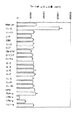
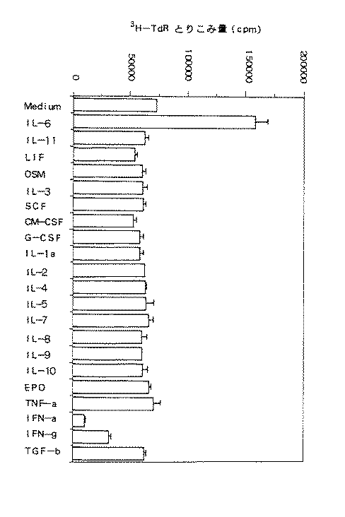
KPMM2の増殖に対する各種サイトカインの効果を図5に示す。図5から明らかなように、KPMM2細胞はIL−6とともにインキュベートしたときのみに3H−チミジンの取り込みが顕著に刺激された。濃度1ng/mlにおけるIL−6により、3H−チミジンの取り込みが2.2倍に上昇した。一方、IFN−αおよびIFN−γはKPMM2細胞の増殖を顕著に阻害した。
【0049】
また、KPMM2のサイトカインに対する反応性を調べるために、MTT(3−[4,5−dimethylthiazol−2−yl]−2,5−diphenyltetrazolium bromide)法(J. Immun. Methods 65:55, 1983 を参照)および生細胞を直接測定する方法を用いたが、同様の結果が得られた。
【0050】
実施例7:抗IL−6 mAbおよび抗IL−6R mAbによる増殖阻害
実施例1で樹立した細胞株KPMM2に対するマウス抗ヒトIL−6R mAb(モノクローナル抗体)(IgG1クラス:PM1)およびマウス抗IL−6mAb(IgG1クラス:SK2)の効果を調べた。SK2は文献(Y. Ohe etal., Br. J. Cancer 67:939, 1993)に記載されており、またPM1は文献(Hirata et al., J. Immunol. 143:2900, 1989)に記載されている。
【0051】
実施例1に記載する方法で得たKPMM2を20%FCSおよび100μg/mlカナマイシンを含むRPMI1640培養液に浮遊させ、容量が200μlで96穴プレート(ファルコン社製)へ1×104個/穴となるように分注した。
【0052】
96穴プレートの各穴には、各種濃度のIL−6 mAb(モノクローナル抗体)(SK2)および抗IL−6R(受容体)mAb(PM1)を別々に加えた。なお、対照群は、抗体を加えない20%FCSおよび100μg/mlカナマイシンを含むRPMI1640培養液とした。
【0053】
96穴プレートの各穴に分注したKPMM2を、抗IL−6 mAbおよび抗IL−6R mAb存在下あるいは非存在下で湿潤5%CO2中、37℃で96時間培養した。その培養終了4時間前に、各穴に3H−チミジン(Amersham社製)を1μCi/穴となるように添加した。KPMM2が取り込んだ3H−チミジンの量の測定は、液体シンチレーションカウンター(1205 ベータプレート、ファルマシア社製)を用いた。
【0054】
KPMM2の増殖に対する抗IL−6 mAbおよび抗IL−6R mAbの効果を図6に示す。SK2およびPM1の添加はいずれも用量依存的に細胞増殖を有意に阻害した。特に、PM1は1μg/mlの濃度でKPMM2の増殖を完全に阻害した。
【0055】
また、KPMM2増殖の抗IL−6 mAbおよび抗IL−6R mAbに対する効果を調べるために、MTT(3−[4,5−dimethylthiazol−2−yl]−2,5−diphenyltetrazolium bromide)法(J. Immun. Methods 65:55, 1983 を参照)および生細胞を直接測定する方法を用いたが、同様の結果が得られた。
【0056】
実施例8:ELISAによるIL−6産生の測定
上記実施例1で樹立した細胞株KPMM2によるIL−6産生能を試験した。
【0057】
実施例1に記載する方法で得たKPMM2を、20%FCSおよび100μg/mlカナマイシンを含むRPMI1640培養液に106個/mlとなるように浮遊させ、ヒトIL−6非存在下で湿潤5%CO2中、37℃で培養した。
【0058】
培養72時間後、培養上清中に含まれるKPMM2が産生したIL−6の濃度を、ヒトIL−6用ELISAキット(帝人バイオラボラトリー社製)を用いて添付の処方に従い測定した。なお、陰性対照として、20%FCSおよび100μg/mlカナマイシンを添加したRPMI1640培養液を用いた。培養上清には79.7±19.6(平均値±S.D.)pg/mlのIL−6の産生が検出され、対照培養液において検出限界(4.0pg/ml)以下であったのに比較すると、培養上清中のIL−6濃度が顕著に増加していることが確認された。
【0059】
実施例9:フローサイトメトリーによるIL−6Rの発現の確認
KPMM2細胞上でのIL−6R発現を確認するために、IL−6Rに結合し、IL−6のIL−6Rへの結合を阻害しないマウス抗ヒトIL−6R mAbであるMT18抗体(Hirata et al., J. Immunol. 143:2900, 1989)を用いて、間接蛍光抗体法を実施した。
【0060】
実施例1に記載の方法で得られたKPMM2細胞を106個/チューブとなるように、100μlの実施例2記載のFACS緩衝液に浮遊させ、10μg/mlのMT18抗体を加え、4℃にて3時間反応させた後、FACS緩衝液で2回洗浄し、100μlのFACS緩衝液に浮遊させ、さらに、FITC標識ヤギ抗マウスIgG抗体(TAGO社製)を5μg/ml添加し、4℃にて30分間反応させた。FACS緩衝液で2回洗浄した後、同FACS緩衝液に浮遊させ、フローサイトメーター(EPICS PROFILE、コールター社製)で蛍光を測定した。
【0061】
その結果、図7に示すように、KPMM2細胞上にIL−6Rの発現が示された。
【0062】
実施例10:ELISAによるヒトIgG(Mタンパク)産生の測定
KPMM2のヒトIgG(Mタンパク)産生能を試験した。
【0063】
実施例1に記載の方法で得られたKPMM2を106個/mlとなるように、20%FCSおよび100μg/mlカナマイシンを含むRPMI1640培養液に浮遊させ、容量2mlで12穴プレート(ファルコン社製)の各穴に分注し、ヒトIL−6非存在下で湿潤5%CO2中、37℃で72時間培養した。なお、実験は3回行った。その後、培養上清中のヒトIgG濃度を、TAGO社製ヤギ抗IgG抗体(NO.4100)およびアルカリフォスファターゼ標識ヤギ抗IgGガンマ鎖特異的抗体(NO.2490)を用いるELISAにて測定した。なお、スタンダードとしてカッペル社製ヒトIgG(NO.0001−860)を用いた。その結果、培養上清中には10.1μg/mlのヒトIgGが検出され、対照培養液では検出限界以下(5ng/ml)であったのに比較して、培養上清中のヒトIgG濃度が顕著に増加していることが確認された。
【0064】
実施例11:RT−PCR(逆転写ポリメレースチェインリアクション)によるIL−6およびIL−6R mRNAの検出
実施例1に記載の方法で得られたKPMM2におけるヒトIL−6およびヒトIL−6Rの発現を確認するために、RT−PCR(逆転写ポリメレースチェインリアクション)法によりヒトIL−6およびヒトIL−6RのメッセンジャーRNA(mRNA)を検出した。
【0065】
グアニジウムセシウムクロライド法(Molecular Cloning, Cold Spring Harbor Laboratory Press)によってKPMM2細胞(108個)から全RNAを調製した。陰性対照としてヒトB細胞リンパ腫細胞株SKW6.4からも同様に全RNAを調製した。1本鎖cDNA合成はcDNA合成キット(Invitrogen社製)を用いて、添付の処方に従い全RNA5μgから直接実施した。
【0066】
陽性対照としてヒトIL−6およびヒトIL−6R検出用PCRプライマーを用いた(Clontech Inc.社製)。PCR溶液各100μlは、10mM Tris−HCl(pH8.3)、50mM 塩化カリウム、Amplitaq(Perkin Elmer Cetus社製)2.5ユニット、1本鎖cDNA合成反応物1μl、各プライマー100pmolを含む。各PCR用チューブに鉱物オイル50μlを上層してPCRに付した。最初に94℃で1分メルトし、60℃で1分と72℃で10分のサイクルを30サイクル繰り返した。最終サイクルの後、最終的に72℃で10分伸長を行った。ヒトIL−6およびヒトIL−6R検出用プライマーを用いた陽性対照群も同様に増幅し、約50pgの陽性対照PCR産物を得た。
【0067】
各反応チューブから10μlを取って1.5%アガロースゲル上で電気泳動を行った。図8に示すように、IL−6(628bp)およびIL−6R(251bp)のPCR産物はいずれもKPMM2のRNAから増幅された。これらの結果は、KPMM2がIL−6のオートクライン機構によって増殖することを遺伝子発現の面から支持する。一方、SKW6.4細胞はIL−6に応答してIgMを分泌する。IL−6RのPCR産物はSKW6.4 mRNAから増幅された。しかし、IL−6のPCR産物はこの実験で検出されず、このことはSKW6.4細胞がIL−6を産生していないことを示唆する。
【0068】
実施例12:細胞株KPMM2のSCIDマウスおよびヌードマウスへの移植(1)KPMM2の可移植性およびインビボでの継代
上記実施例1で樹立した細胞株KPMM2の可移植性およびインビボでの継代を以下のように検討した。
【0069】
KPMM2を20%FCSおよび100μg/mlカナマイシンを含むRPMI1640培養液に108個/mlで懸濁し、0.2mgのウサギ抗アシアロGM1抗体(Code No.014−09801、和光純薬工業社製)で処理したIL−6トランスジェニック重症免疫不全マウス(以下IL−6−SCID Tmという)(中外製薬製)の腹部皮下に注射針で0.1ml移植した。その結果、1カ月後には3例全例で移植部位に結節型の腫瘍を形成した。
【0070】
この腫瘍を無菌的に摘出し、摘出した腫瘍塊を使い捨て注射器のピストンなどでつぶし、ナイロンメッシュ(70μm、ファルコン社製)を通して細胞を回収した。この細胞を上記と同様にRPMI1640培養液に懸濁し、IL−6−SCID Tm、重症免疫不全マウス(SCIDマウスという)(日本クレア社製)、BALB/c−nu/nuマウス(以下ヌードマウスという)(日本クレア社製)に皮下移植したところ、同じように移植部位に結節型の腫瘍を形成し、インビボでの継代が可能であった。また腫瘍塊を3mm角のブロックにし、移植針を用いてIL−6−SCID Tm、SCIDマウス、ヌードマウスの皮下に移植しても継代可能であった。
(2)KPMM2の異なる移植経路での可移植性
KPMM2の移植経路を皮下(s.c.)、静脈内(i.v.)、腹腔内(i.p.)とした場合の可移植性を以下のように検討した。
【0071】
(1)においてSCIDマウスにて継代したKPMM2腫瘍を無菌的に摘出し、摘出した腫瘍塊を使い捨て注射器のピストンでつぶし、ナイロンメッシュを通して細胞を回収し、108個/mlの細胞懸濁液を作成した。この懸濁液をSCIDマウス、あるいは0.2mgのウサギ抗アシアロGM1抗体および500RのX線で処理したヌードマウスに0.1mlずつ皮下(s.c.)、静脈内(i.v.)、腹腔内(i.p.)の3経路で移植し、40日後に生着の判定を行った(表2)。その結果、約40日後には全例で生着が確認され、s.c.移植では移植部位に、i.p.移植では腹腔内に固形腫瘍を形成した。また、i.v.移植ではKPMM2は骨髄での生着が認められた。
【0072】
【表2】
細胞株KPMM2をSCIDマウスへ皮下移植し、形成された腫瘍の体積と血清中ヒトIgG濃度の相関を調べた。上記(2)に記載の方法で細胞株KPMM2をSCIDマウスへ皮下移植し、KPMM2移植前、移植後21日目、42日目の3回、血清サンプルを採取してELISA法にてヒトIgG濃度を測定した。
【0073】
その結果、マウス血清中のヒトIgG濃度はKPMM2を移植した動物ではいずれも経時的に上昇した。また図9に示すように、皮下移植した動物では皮下に形成された腫瘍の体積と血清ヒトIgG濃度に相関が見られ、腫瘍の大きな動物ほど血清ヒトIgG濃度は高値を示した。このことから、血清ヒトIgG濃度を指標としても抗腫瘍効果の判定ができると考えられた。
(4)KPMM2の可移植性および移植細胞数の検討
SCIDマウスとヌードマウスで移植細胞数を変えたときのKPMM2の可移植性を検討した。
【0074】
特別な前処理を行わない雄のSCIDマウスおよびヌードマウスに、上記(2)記載の方法で各々KPMM2細胞を107、3×10⁶、106個腹部皮下(s.c.)に移植し、またSCIDマウスには上記(2)の方法で得たKPMM2細胞を3×10⁶、106個静脈内(i.v.)でも移植した。
【0075】
その結果、SCIDマウスに皮下移植した場合、3×10⁶個以上移植すると、21日までに全例で腫瘍結節を形成し、106個移植した場合でも21日までに2/3、32日までに全例で腫瘍結節を形成した。ヌードマウスに皮下移植すると、107個移植した場合では21日までに2/3、32日までには全例で、また3×106個移植した場合では42日までには全例で腫瘍結節が形成された。しかし、106個の移植では42日までに腫瘍の生着は見られなかった。また、SCIDマウスに静脈内移植した場合、42日までに3×106個移植で全例、106個移植で1/3にKPMM2が生着した。これらの結果を以下の表3にまとめて示す。
【0076】
【表3】
KPMM2を移植したSCIDマウスにおいて、その生着を確認するために、KPMM2の細胞表面上で特徴的に発現している表面抗原であるヒトCD38抗原の発現を試験した。
【0077】
KPMM2を上記(1)に記載する方法でSCIDマウスへ静脈内移植し、移植後37日目にマウスを屠殺した。屠殺したマウスの大腿骨より骨髄を回収して懸濁し、ステンレスメッシュ(100μm)を通し、蛍光活性化細胞選択装置(FACS)用緩衝液(2%FCSおよび0.1%NaN3を含むPBS(−)溶液)にて骨髄細胞の浮遊液を調製した。なお、陰性対照としてKPMM2を移植していないSCIDマウスの骨髄からも同様の方法にて骨髄細胞浮遊液を調製した。
【0078】
また、本実施例に記載する方法によりKPMM2を皮下移植したSCIDマウスからは、移植後37日目に皮下腫瘍塊を外科的に摘出し、摘出した腫瘍塊を2枚のスライドグラスの間にはさんですりつぶし、これをステンレスメッシュ(100μm)を通し、FACS緩衝液にて骨髄細胞の浮遊液を調製した。
【0079】
陽性対照として、実施例1に記載した方法でインビトロにて培養したKPMM2を用いた。インビトロで4日間培養したKPMM2をFACS緩衝液で洗浄した後、同緩衝液中に浮遊させた。
【0080】
次に、このように調製した細胞浮遊液を用いて、FACS解析によるヒトCD38抗原の発現を調べた。
【0081】
すなわち、100μlのFACS緩衝液中にて、各群の浮遊細胞106個に対し、2.5μg/mlのフィコエリスリン(PE)標識抗ヒトCD38抗体(Leu−17、ベクトン・ディッキンソン社製)を添加し、氷上で30分間反応させた。次いで、1mlのFACS緩衝液で2回洗浄後、500μlのFACS緩衝液で懸濁し、FACScan(ベクトン・ディッキンソン社製)によりFACS解析を行った。
【0082】
陽性対照群であるインビトロ培養KPMM2のFACS解析の結果から、蛍光強度40から2000までの範囲にある細胞をヒトCD38抗原発現細胞とした(図10(a)参照)。KPMM2を静脈内移植したSCIDマウスでは、その骨髄細胞のおよそ71%がヒトCD38陽性細胞で占められていた(図10(b)参照)。このことは、SCIDマウスに静脈内移植したKPMM2がSCIDマウスの骨髄に生着したことを示している。また、KPMM2をSCIDマウスに皮下移植して生じた腫瘍塊から得られた細胞は全てヒトCD38が陽性であり(図10(c)参照)、KPMM2を皮下移植して生ずる腫瘍塊は、全てKPMM2から構成されていることが示された。なお、KPMM2を移植していない陰性対照群のSCIDマウスの骨髄細胞にはヒトCD38陽性細胞は全く検出されなかった(図10(d)参照)。
【0083】
実施例13:抗ヒトIL−6抗体SK2および抗ヒトIL−6R再構成ヒト型化抗体PM1の抗腫瘍効果
KPMM2移植動物における抗ヒトIL−6抗体SK2および抗ヒトIL−6R再構成ヒト型化抗体PM1のインビボ抗腫瘍効果を以下のようにして検討した。
【0084】
上記実施例12(1)に記載の方法でSCIDマウスで継代して得たKPMM2骨髄腫腫瘍塊をウサギ抗アシアロGM1抗体/X線処理ヌードマウス(5週齢、雄)に4mm角ブロックで皮下移植し、翌日に1回だけSK2抗体を1mg/マウスの1用量、再構成ヒト型化PM1抗体(国際公開出願WO92−19759参照)を0.125、0.5および1mg/マウスの3用量で静脈内投与した。各抗体は0.2ml/マウスとなるようにPBS(−)(ニッスイ製)で調製し、陰性対照群には、PBS(−)を0.2ml/マウス投与した。その後腫瘍の大きさを経時的に観察し、対照群の腫瘍が十分大きくなった35日目に全採血を行ってから腫瘍を摘出して重量を測定した。
【0085】
その結果を図11に示す。陰性対照群の平均腫瘍重量が約1gであったのに対し、再構成ヒト型化PM1抗体投与群においては、1mg/マウス投与した場合で腫瘍増殖抑制率(Growth Inhibitory Ratio:GIR)は78%、0.5mg/マウス投与した場合でのGIRは53%、0.125mg/マウス投与した場合でのGIRは66%を示し、強い腫瘍増殖抑制効果が見られた。またSK2においても、1mg/マウス投与群でGIR61%と腫瘍増殖を抑制した。
【0086】
さらに、腫瘍摘出時に採取した血清サンプルのヒトIgG濃度をELISA法にて測定した。その結果を図12に示す。抗体非投与陰性対照群では血清ヒトIgG濃度が平均27.6mg/mlであったものが、再構成ヒト型化PM1抗体を1、0.5、0.125mg/マウス投与することでそれぞれ71%、55%、75%抑制された、SK2 1mg/マウス投与でも43%抑制された。腫瘍重量と血清ヒトIgG濃度は各処理群間でも、個体レベルでもよく相関していた。
【0087】
実施例14:KPMM2静脈内移植SCIDマウスにおけるイオン化カルシウム濃度の上昇および骨吸収の亢進
KPMM2(107個)を上記実施例12(2)に記載する方法でSCIDマウス(日本クレア社製)へ静脈内移植し、移植後経時的に血中イオン化カルシウム濃度および骨吸収の有無を試験した。
【0088】
KPMM2移植後9日目、20日目、30日目および37日目にKPMM2静脈内移植SCIDマウスをエーテル麻酔し、マウス眼窩より60μl容量のキャピラリーカラム(チバ・コーニング社製)で採血し、直ちに血中イオン化カルシウム濃度を634自動Ca++/pHアナライザー(チバ・コーニング社製)にて測定した。なお、対照としてKPMM2を移植していないSCIDマウスからも同様の方法で採血し、血中イオン化カルシウム濃度を測定した。
【0089】
その結果、KPMM2静脈内移植SCIDマウスの血中イオン化カルシウム濃度は、移植後30日目より上昇がみられ、37日目には対照群のマウスに比べ、約20%の血中イオン化カルシウム濃度上昇が観察された(図13)。なお、血中イオン化カルシウム濃度上昇は、KPMM2をSCIDマウスへ静脈内移植したときの、マウス骨髄におけるKPMM2(ヒトCD38抗原陽性細胞)が占める増加の割合の経時的増加とよく相関していた(図14参照)。
【0090】
また、KPMM2静脈内移植37日目のSCIDマウス下肢の骨をX線撮影して形態的に観察したところ、対照群のマウスと比較し、顕著な骨吸収像が確認された(図15参照)。以上の結果は、KPMM2静脈内移植SCIDマウスの骨病変が、実際の骨髄腫の病変とよく一致していることを示している。
【0091】
【発明の効果】
本発明によってIL−6オートクライン依存性で増殖する骨髄腫が存在することが明らかとなった。本発明のオートクライン機構によりIL−6依存性で増殖するヒト骨髄腫細胞株は、骨髄腫のIL−6依存性増殖機構モデルとして有用である。また抗IL−6抗体、抗IL−6受容体抗体などのIL−6活性阻害剤をはじめとする骨髄腫治療剤の治療モデルとしても使用し得るものである。本発明のオートクライン機構によりIL−6依存性で増殖するヒト骨髄腫細胞株はインビトロおよびインビボで細胞増殖抑制を指標とした骨髄腫治療剤の評価系を作成するのに有用であるのはもちろん、Mタンパクを産生し、この産生量が骨髄腫の増殖に極めてよく相関することから、Mタンパク産生量の抑制を指標とした骨髄腫治療剤の評価系を作成するのに有用である。
【0092】
さらに、本発明のヒト骨髄腫細胞株を実験動物に静脈内移植して得られる骨髄腫の骨髄生着モデルでは、多発性骨髄腫の増殖に伴い骨髄腫に特徴的な骨病変が観察され、したがってこの骨病変の抑制を指標とした骨髄腫治療剤の評価系を作成することができる。
【0093】
これらの点から本発明のヒト骨髄腫細胞株の利用価値は極めて大きい。
【図面の簡単な説明】
【図1】液体培養におけるKPMM2の自律的凝集を示す図(生物の形態を表す写真)である。
【図2】KPMM2の形態を示す図(生物の形態を表す写真)である。ライト−ギムザ染色では形質細胞の特徴を有する。
【図3】サザンブロット分析によるKPMM2のJHおよびCλ遺伝子の再構成を示す図(電気泳動の写真)である。KPMM2から得たDNAをBamHI、EcoRIおよびHindIIIで消化し、JHおよびCλ遺伝子プローブを用いてサザンブロット分析を行った。再構成したバンドを(▲)で示す。
【図4】KPMM2の核型を示す図(生物の形質を表す写真)である。検索した15細胞はすべて46、XX、der(1;19)(q10;q10)、t(3;14)(q21;q32)、−4、t(6;11)(p12;p15)、der(10)add(10)(p13)dic(9;10)(q10;q26)、+16を示した。
【図5】KPMM2の細胞増殖に対する各種サイトカインの効果を示す図である。使用したサイトカインの濃度は以下の通りである:IL−6、1ng/ml;IFN−αおよびIFN−γ、1000U/ml;その他のサイトカイン、100ng/ml。各数値は3回の試験の平均+標準偏差(SD)を表す。
【図6】KPMM2の細胞増殖に対する抗IL−6 mAbおよび抗IL−6R mAbの効果を示す図である。SK2はマウス抗IL−6 mAb(▲);PM1はマウス抗IL−6R mAb(●)。破線は対照を示す。各数値は3回の試験の平均を表す。
【図7】KPMM2細胞におけるIL−6Rの発現を示す図である。細胞は抗IL−6R mAb(MT18)で染色した。マウスIgG2b抗体を対照として用いた。破線はmIgG2bを、実線はMT18を表す。
【図8】1.5%アガロースゲル上でのRT−PCR(逆転写PCR)分析により、KPMM2のIL−6およびIL−6R mRNAの発現を示す図(電気泳動の写真)である。レーン1および4、SKW6.4;レーン2および5、KPMM2;レーン3および6、陽性対照。
【図9】KPMM2を皮下移植したマウスにおける腫瘍体積と血清ヒトIgG濃度の相関を示す図である。
【図10】(a)はインビトロ培養したKPMM2におけるヒトCD38抗原のFACS解析を示す図である。KPMM2は40から2000までの範囲の蛍光強度を有する。
(b)はKPMM2を静脈内移植したSCIDマウスの骨髄細胞におけるヒトCD38抗原のFACS解析を示す図である。71%の細胞が40から2000の範囲の蛍光強度を有する。
(c)はKPMM2を皮下移植したSCIDマウスの腫瘍塊から得た細胞におけるヒトCD38抗原のFACS解析を示す図である。全ての細胞が40から2000の範囲の蛍光強度を有する。
(d)はKPMM2を移植していないSCIDマウスの骨髄細胞におけるヒトCD38抗原のFACS解析を示す図である。40から2000の範囲の蛍光強度を有する細胞は全くみられない。
【図11】KPMM2に対する抗IL−6 mAbおよび抗IL−6R mAbのインビボにおける腫瘍増殖抑制効果を示す図である。SK2は抗ヒトIL−6 mAb;再構成ヒト型化PM1は抗ヒトIL−6R mAb。
【図12】KPMM2移植ヌードマウス中の血清ヒトIgG濃度に対する抗IL−6 mAbまたは抗IL−6R mAbの効果を示す図である。
【図13】KPMM2を静脈内移植したSCIDマウスの血中イオン化カルシウム濃度(□)の経時的変化を示す図である。(◇)は対照を示す。各数値はSCIDマウス4匹(37日目のみ5匹)の平均±S.D.を示す。
【図14】KPMM2を静脈内移植したSCIDマウスの骨髄中のヒトCD38抗原陽性細胞の割合(□)の経時的変化を示す図である。(◇)は対照を示す。各数値はSCIDマウス4匹(37日目のみ5匹)の平均±S.D.を示す。
【図15】KPMM2を静脈内移植したSCIDマウスの骨のX線撮影像を示す図(生物の形態を示す写真)である。
(a)はKPMM2を移植していない対照群のSCIDマウス
(b)はKPMM2移植後37日目のSCIDマウス[0001]
[Industrial applications]
The present invention relates to a human myeloma cell line, and more particularly to a human myeloma cell line that grows in an IL-6-dependent manner by an autocrine mechanism, an experimental animal transplanted with the cell line, and using the cell line or the experimental animal. The present invention relates to a method for screening a therapeutic agent for myeloma.
[0002]
[Prior art]
Interleukin 6 (IL-6), which is the same factor as B cell stimulating factor 2 (BSF-2) and mouse hybridoma / plasmacytoma growth factor, is multiple myeloma (hereinafter sometimes referred to as MM). It is considered to be a major growth factor for cells (Kawano et al., Nature 332: 83, 1988; Kleine et al., Blood 73: 517, 1989). Multiple myeloma is a tumor in which plasma cells have become malignant, and the bone marrow is used as a growth site, and occurs simultaneously in multiple sites. IL-6 transmits its activity on such cells via two membrane proteins. One is a ligand-binding membrane protein with a molecular weight of 80 kD (IL-6 receptor) to which IL-6 binds, and the other is a membrane protein gp130 involved in non-ligand binding signal transduction (Taga). et al., J. Exp. Med. 196: 967, 1987).
[0003]
In 1988, Kawano et al. Found that fresh human myeloma cells constitutively produced and expressed IL-6 receptor, and that proliferation in vitro was induced by anti-IL-6 receptor (R) antibodies. It was reported that myeloma cells could proliferate by the autocrine mechanism of producing and receiving growth factors by themselves (Kawano et al., Nature 332: 83, 1988), because they are suppressed. On the other hand, Klein et al. Proposed that they do not produce growth factors themselves, but proliferate by a paracrine mechanism that accepts growth factors from the surroundings (Klein et al., Blood 73: 517, 1989). It is also known that serum IL-6 concentration is correlated with myeloma disease (Bataille et al., J. Clin. Invest. 84: 2008, 1989), and IL-6 is associated with myeloma. Is believed to be one of the major growth factors for
[0004]
In the case of freshly isolated myeloma cells, contamination of cells other than myeloma cells is unavoidable, and accurate assays are difficult. It is not clear which of them will proliferate.
[0005]
When human IL-6 cDNA was introduced by transfection into a human myeloma cell line that had an IL-6-dependent growth, it was observed that the human IL-6 cDNA grew autonomously and turned into a tumor. (Okuno et al., Exp. Hematol. 20: 395, 1992). It has also been reported that the human myeloma cell line U266 grows by the IL-6 autocrine mechanism (Jernberg et al., Leukemia 5: 255, 1991; Levy et al., J. Clin. Invest. 88: 696, 1991). However, proliferation of U266 was also induced by exogenous IL-6 (Jernberg et al., Leukemia 5: 255, 1991) and by anti-IL-6 monoclonal antibody (Levy et al., J. Clin. Invest. 88: 696, 1991) There was a report that it was not affected, and the involvement of IL-6 in the proliferation mechanism of U266 is unknown.
[0006]
We found that in one case of fresh myeloma cells obtained from ascites of a patient with multiple myeloma, the tumor cells showed a clear IL-6-dependent growth, and the anti-IL-6 receptor with autonomous growth. It was reported that the antibody was strongly suppressed by a body antibody (Goto et al., Biotherapy 7: 655, 1993).
[0007]
The present inventors further examined the properties of tumor cells obtained by transplanting fresh human myeloma cells into a human IL-6 gene-transfected severe combined immunodeficient mouse (IL-6 transgenic SCID mouse) (Goto et al., No. Proceedings of the 52nd Annual Meeting of the Japanese Cancer Society, 498 pages, October 1993). As a result, three subcutaneously transplanted mice showed plasmacytoma at the transplanted site and metastasis to axillary lymph nodes. No tumor formation was observed by intraperitoneal transplantation. The tumor cells after transplantation showed no change in surface antigens, in vitro IL-6 dependent proliferation in vitro, and the growth inhibitory effect of anti-human IL-6 receptor antibody compared to before transplantation.
[0008]
[Problems to be solved by the invention]
As described above, there are still many unknowns about the proliferation mechanism of myeloma cells, and it is required to elucidate this.
[0009]
An object of the present invention is to establish a myeloma cell line that can serve as a model for the IL-6-dependent growth mechanism of myeloma. The cell lines are useful as in vitro models for the treatment of myeloma with myeloma therapeutics, including IL-6 activity inhibitors such as anti-IL-6 antibodies, anti-IL-6 receptor antibodies.
[0010]
Another object of the present invention is to provide an experimental animal into which the cell line has been transplanted. The above experimental animals are useful as an in vivo model for the treatment of myeloma with myeloma therapeutic agents including IL-6 activity inhibitors such as anti-IL-6 antibodies and anti-IL-6 receptor antibodies.
[0011]
Another object of the present invention is to provide a method for screening a therapeutic agent for myeloma. In the above-mentioned in vitro model, a screening method for a therapeutic agent for myeloma using inhibition of cell growth or inhibition of M protein (myeloma protein) secretion as an index can be used. Further, in the above-mentioned in vivo model, a screening method for a therapeutic agent for myeloma, which uses cell growth suppression, M protein secretion suppression, or bone lesion suppression as an index, can be used. The M protein is an immunoglobulin protein specifically produced by myeloma, and there are five types of myeloma producing IgA, IgM, IgG, IgE and Bence-Jones protein.
[0012]
[Means for Solving the Problems]
The present inventors have conducted intensive studies to achieve the above object, and as a result, succeeded in establishing a cell line that can be a model of myeloma having an IL-6-dependent growth mechanism, and completed the present invention.
[0013]
That is, the present invention provides a human myeloma cell line that grows in an IL-6-dependent manner by an autocrine mechanism.
[0014]
The present invention also provides an experimental animal into which the cell line has been transplanted.
[0015]
Furthermore, the present invention provides an in vitro screening method for a therapeutic agent for myeloma, which comprises adding a therapeutic agent for myeloma to the above cell line and testing for inhibition of myeloma cell growth or inhibition of M protein secretion.
[0016]
Further, the present invention provides an in vivo screening method for a therapeutic agent for myeloma, which comprises administering the therapeutic agent for myeloma to the above-mentioned experimental animal and testing the inhibition of myeloma cell growth, inhibition of M protein secretion, or inhibition of bone lesions.
[0017]
The cell line of the present invention can be established using myeloma cells collected from, for example, ascites of a multiple myeloma patient. In the present invention, in particular, myeloma cells were collected from ascites of IgG and λ-type multiple myeloma patients. One month after the start of the culture, the cells began to proliferate stably and were maintained for one year or more. The cell line thus established is named KPMM2 and has been deposited at the Biotechnology, Industrial Technology Research Institute, Patent Microorganisms Depositary under the accession number FIRM: P-14170 (deposited on February 22, 1994). The cell line KPMM2 has been successfully passaged in vitro and in stable passage in immunodeficient mice such as experimental animals, for example, severe combined immunodeficiency (SCID) mice, IL-6 transgenic SCID mice and nude mice. It is possible.
[0018]
The cell line KPMM2 was confirmed to produce IL-6 in vitro and to express the IL-6 receptor (IL-6R). In addition, the reactivity of KPMM2 to various cytokines3H-thymidine incorporation experiment, MTT (3- [4,5-dimethylthiazol-2-yl] -2,5-diphenyltetrazolium bromide) method (see J. Immun. Methods 65: 55-63, 1983) and live cells. Tests of the measurements showed that only significant stimulation was observed when incubated with IL-6, indicating that KMMM2 proliferates specifically in response to IL-6. Furthermore, the proliferation of KPMM2 was significantly suppressed in a dose-dependent manner by anti-IL-6 mAb (monoclonal antibody) and anti-IL-6R mAb. In addition, RT-PCR (reverse transcription polymerase chain reaction) confirmed that KPMM2 expressed IL-6 and IL-6R mRNA. KPMM2 expresses various adhesion molecules, namely, CD44, VLA-β, ICAM-1, NCAM, LFA-3 and VLA-4, and shows autonomous cell aggregation in vitro.
[0019]
From these various characteristics of KPMM2, it was shown that KPMM2 is a myeloma cell line proliferating by the IL-6 autocrine mechanism. The cell line of the invention is the first cell line whose growth mechanism has been demonstrated to be an IL-6 dependent autocrine mechanism.
[0020]
Therefore, the cell line of the present invention is useful for screening a therapeutic agent for myeloma including an inhibitor of IL-6 activity such as an anti-IL-6 mAb or an anti-IL-6R mAb. For example, an in vitro screening method for a therapeutic agent for myeloma, which comprises adding an anti-IL-6 antibody or an anti-IL-6 receptor antibody to the cell line of the present invention and testing the inhibitory effect on myeloma cell growth, is possible. In addition, the myeloma cell line of the present invention can perform in vitro screening for a therapeutic agent for myeloma using the suppression of M protein secretion as an index because the amount of secreted M protein increases in proportion to the increase in cell number. Furthermore, the cell lines of the present invention are also useful as models for studying the role played by adhesion molecules in myeloma cell proliferation via cell-cell interactions and IL-6 signaling.
[0021]
The present invention also provides a laboratory animal into which the cell line of the present invention has been transplanted. Experimental animals to which the cell line of the present invention is transplanted include, in addition to mice, rats, rabbits, guinea pigs, hamsters, monkeys, and the like. Further, the function of immunocompetent cells such as T cells or B cells is impaired, It is preferable to transplant the cell line of the present invention into an experimental animal in an immunodeficient state. As already mentioned, the cell lines of the present invention are capable of stable passage in immunodeficient mice such as SCID mice, IL-6 transgenic SCID mice and nude mice.
[0022]
Interestingly, the above cell line of the present invention can be transplanted into mice by subcutaneous transplantation or intraperitoneal transplantation, as well as by intravenous transplantation. In the case of subcutaneous and intraperitoneal transplants, solid tumors were observed at the subcutaneous and intraperitoneal sites, respectively.However, in the case of intravenous transplantation, engraftment of tumor cells into the bone marrow was observed. Can be used as a model close to the pathology of myeloma. For example, administering a therapeutic agent for myeloma including an IL-6 activity inhibitor such as an anti-IL-6 antibody or an anti-IL-6 receptor antibody to an experimental animal of the present invention to test the inhibitory effect on myeloma cell proliferation. In vivo screening method for a therapeutic agent for myeloma comprising In experimental animals in which the cell line of the present invention has survived, an increase in serum M protein concentration is observed with the growth of tumors. Screening methods are also possible. Further, when the cell line of the present invention engrafted to the bone marrow of an experimental animal, as the myeloma cells proliferate, an increase in blood ionized calcium concentration, bone destruction, bone lesions such as osteolysis and bone resorption are observed, An in vivo screening method for a therapeutic agent for myeloma using the suppression of these bone lesions as an index is possible.
[0023]
Hereinafter, the present invention will be described in more detail with reference to Examples, but the scope of the present invention is not limited thereto.
[0024]
【Example】
Example 1 Establishment and Maintenance of a Myeloma Cell Line
Myeloma cells were collected from ascites of an IgG, λ multiple myeloma patient (76 years old, female, stage IIA). The ascites fluid contained a large number of myeloma cells, with IL-6 levels in the ascites fluid reaching 91.0 pg / ml. The collected ascites was subjected to density gradient centrifugation using Ficoll-Hypaque (manufactured by Pharmacia) to separate mononuclear cells, to remove adherent cells with a plastic Petri dish, and to remove T cells with sheep erythrocytes. Purified to 95% or more. RPMI1640 (manufactured by Gibco) containing 20% fetal calf serum (FCS: manufactured by Xavier Investments, Australia), 4 ng / ml of recombinant human IL-6 (manufactured by Chugai Pharmaceutical) and 100 μg / ml of kanamycin (manufactured by Meiji Seika) 1 × 10 in culture6The cells were suspended at a concentration of cells / ml. The cells are then cultured in a 10 ml culture in a 25 ml flask and wet 5
[0025]
One month after the start of the culture, the cells began to proliferate stably and were maintained for one year or more, and were established as cell lines. This cell line was named KPMM2. The doubling times in the presence or absence of IL-6 were 48 hours and 72 hours, respectively.
[0026]
The morphology and Ig secretion of KPMM2 are as follows.
KPMM2 morphology and Ig secretion
KPMM2 cells were observed to proliferate under light microscopy with autonomous cell aggregation (FIG. 1), and Wright-Giemsa staining showed a plasma cell-like morphology (FIG. 2). KPMM2 was positive for acid phosphatase and slightly positive for α-naphthyl butyrate esterase, but negative for peroxidase, AS-D chloroacetate esterase, pass (periodite Schiff reagent) and alkaline phosphatase. Cytoplasmic IgG and λ light chains were detected, but IgA, IgM, and κ light chains were negative by direct immunofluorescence. In addition, cells (106Per ml) for 3 days, IgG and λ light chain secretion was observed in the culture supernatant.
[0027]
Example 2: Analysis of surface antigen
The expression of the surface antigen of the cell line KPMM2 established in Example 1 was determined by direct and indirect immunofluorescence (Fried et al., Flow Cytometry, Boca Raton, CRC Press: 59) using a panel of monoclonal antibodies against various human antigens. −78, 1989).
[0028]
KPMM2 obtained by the method described in Example 16100 μl of a buffer for fluorescence activated cell selection device (FACS) (2% FCS and 0.1% NaN)3In a phosphate-buffered saline solution (PBS) containing the following (hereinafter referred to as FACS buffer). Then, in the direct fluorescent antibody method, a fluorescin isothiocyanate (FITC) or phycoerythrin (PE) -labeled antibody against various human antigens shown in Table 1 below in a saturating amount is added and incubated at 4 ° C. for 30 minutes. did. After the cells were washed twice with the above FACS buffer, they were analyzed with a flow cytometer (EPICS PROFILE, manufactured by Coulter).
[0029]
On the other hand, in the indirect fluorescent antibody method, unlabeled antibodies against various human antigens shown in Table 1 below were added, incubated at 4 ° C. for 30 minutes, and the cells were washed twice with a FACS buffer. / Ml of FITC or PE-labeled goat anti-mouse IgG antibody F (ab ') 2 fragment (manufactured by TAGO) was added and reacted at 4 ° C for 30 minutes. After washing twice with a FACS buffer, the cells were suspended in the FACS buffer and analyzed with a flow cytometer (EPICS PROFILE, manufactured by Coulter).
[0030]
The surface antigens of KPMM2 are summarized in Table 1 below.
[0031]
[Table 1]
[0032]
Example 3: Immunoglobulin gene rearrangement
The immunoglobulin (Ig) gene rearrangement of the cell line KPMM2 established in Example 1 was analyzed by Southern blotting.
[0033]
KPMM2 cells (10%) obtained by the method described in Example 17), DNA was prepared in accordance with the method of Manual of Clinical Immunology, 3rd edition, American Society for Microbiology, 1986, and treated with three kinds of restriction enzymes BamHI, EcoRI or Hindman separately from BamHI, EcoRI or Hindman Hygerin, respectively. The DNA was recovered as an ethanol precipitate, and electrophoresed on a 0.8% agarose gel (SEAKEM GTG, FMC) for 24 hours. The electrophoresed DNA is transferred to a nylon membrane (Hybond N+, Amersham) and dried. Next, using Takara random primer DNA labeling kit (Takara Shuzo),32P-labeled human Ig JH, Cκ and Cλ probes (manufactured by Oncore) were used for Southern blot analysis according to the attached instructions. As a control, non-reconstituted chromosomal DNA obtained from peripheral blood mononuclear cells of a healthy person was used. As a result, IgH and κ chain genes were rearranged, but λ chain genes were not rearranged (FIG. 3). From the above, it was confirmed that KPMM2 produced a monoclonal antibody and that the cells were unique.
[0034]
Example 4: Cytogenetic analysis
The chromosomal structural abnormality of the cell line KPMM2 established in Example 1 was analyzed.
[0035]
The KPMM2 obtained by the method described in Example 1 was added to an RPMI1640 culture medium containing 20% FCS and 100 μg / ml kanamycin at 5 × 10 55The cells were cultured so as to obtain the number of cells / ml. Forty-eight hours after culturing, KMPM2 was treated with 0.05 μg of corsemide (manufactured by Gibco) for 15 minutes to collect KMMM2 cells whose cell cycle was stopped in the metaphase. The recovered KMPM2 cells were treated with 0.075 M KCl for 20 minutes and fixed with methanol-acetic acid. The chromosomes of the KPMM2 cells were then analyzed by trypsin-Giemsaband staining.
[0036]
As a result, KPMM2 was found to be a diploid cell having many structural abnormalities (FIG. 4). All 15 cells analyzed were 46, XX, der (1; 19) (q10; q10), t (3; 14) (q21; q32), -4, t (6; 11) (p12; p15), der (10) add (10) (p13) dic (9; 10) (q10; q26), +16.
[0037]
Example 5: Detection of EBV and mycoplasma
The cell line KPMM2 established in Example 1 was tested for Epstein-Barr virus (EBV) and mycoplasma contamination.
[0038]
In order to detect Epstein-Barr virus (EBV) from the chromosome of KPMM2 cells obtained by the method described in Example 1, PCR was carried out using an EBV BamW region amplification primer purchased from Systemic Genetic Institute according to the attached protocol. (Polymer race chain reaction). The detection of mycoplasma infection was carried out by M. for mycoplasma DNA detection. T. C. The test was carried out using a kit (Gen-Probe Inc.) according to the attached prescription.
[0039]
As a result, KPMM2 was negative for the EBV genome and the mycoplasma genome.
[0040]
Example 6: Reactivity to cytokine
The reactivity of the cell line KPMM2 established in Example 1 to various cytokines was tested.
[0041]
KPMM2 obtained by the method described in Example 1 was suspended in an RPMI1640 culture solution containing 20% FCS and 100 μg / ml kanamycin, and 1 × 10 6 was added to a 96-well plate (Falcon) at a volume of 200 μl.4The mixture was dispensed so as to obtain individual pieces / holes.
[0042]
Various cytokines were separately added to each well of the 96-well plate so as to have the following concentrations.
[0043]
Recombinant IL-2, IL-3, tumor necrosis factor (TNF) -α, granulocyte macrophage colony stimulating factor (GM-CSF), stem cell growth factor (SCF) (all from Genzyme), IL-4, IL-7, IL-10, IL-11, leukemia inhibitory factor (LIF), oncostatin M (OSM) (all manufactured by Pepro Tec Inc.), IL-9, The transforming growth factor (TGF) -β (above, manufactured by R & D System Inc.), IL-1α (Boehringer Manheim), IL-5 (Upstate Biotechnology Inc.), IL-8 ( Amersham), erythropoietin (EPO) and granulocyte colony stimulating factor (G-CSF) (above, medium Pharmaceutical provided by Co., Ltd.) is 100ng / ml.
[0044]
Recombinant interferon (IFN) -γ (provided by Shionogi & Co., Ltd.) and natural human IFN-α (supplied by Sumitomo Pharma Co., Ltd.) are 1000 U / ml.
[0045]
Recombinant IL-6 (provided by Chugai Pharmaceutical Co., Ltd.) is 1 ng / ml.
[0046]
The control group was cultured in an RPMI 1640 culture medium supplemented with 20% FCS without added cytokine and 100 μg / ml kanamycin.
[0047]
KPMM2 dispensed into each well of a 96-well plate was added with 5
[0048]
FIG. 5 shows the effects of various cytokines on the growth of KMMM2. As is evident from FIG. 5, KPMM2 cells were only incubated when incubated with IL-6.3H-thymidine incorporation was significantly stimulated. By IL-6 at a concentration of 1 ng / ml,3H-thymidine incorporation increased 2.2-fold. On the other hand, IFN-α and IFN-γ markedly inhibited the growth of KPMM2 cells.
[0049]
In addition, in order to examine the reactivity of KPMM2 to cytokines, see MTT (3- [4,5-dimethylthiazol-2-yl] -2,5-diphenyltetrazolium bromide) method (J. Immun. Methods 65:55, 1983). ) And a method of directly measuring living cells, but similar results were obtained.
[0050]
Example 7: Growth inhibition by anti-IL-6 and anti-IL-6R mAbs
Mouse anti-human IL-6R mAb (monoclonal antibody) against the cell line KPMM2 established in Example 1 (IgG1Class: PM1) and mouse anti-IL-6 mAb (IgG1The effect of class: SK2) was investigated. SK2 is described in the literature (Y. Ohe et al., Br. J. Cancer 67: 939, 1993), and PM1 is described in the literature (Hirata et al., J. Immunol. 143: 2900, 1989). ing.
[0051]
KPMM2 obtained by the method described in Example 1 was suspended in an RPMI1640 culture solution containing 20% FCS and 100 μg / ml kanamycin, and 1 × 10 6 was added to a 96-well plate (manufactured by Falcon) in a volume of 200 μl.4The mixture was dispensed so as to obtain individual pieces / holes.
[0052]
Various concentrations of IL-6 mAb (monoclonal antibody) (SK2) and anti-IL-6R (receptor) mAb (PM1) were separately added to each well of the 96-well plate. The control group was an RPMI1640 culture solution containing 20% FCS and 100 μg / ml kanamycin to which no antibody was added.
[0053]
KPMM2 dispensed into each well of a 96-well plate was treated with 5
[0054]
The effect of anti-IL-6 mAb and anti-IL-6R mAb on KPMM2 proliferation is shown in FIG. The addition of both SK2 and PM1 significantly inhibited cell proliferation in a dose-dependent manner. In particular, PM1 completely inhibited the growth of KPMM2 at a concentration of 1 μg / ml.
[0055]
Further, in order to examine the effect of KPMM2 proliferation on anti-IL-6 mAb and anti-IL-6R mAb, the MTT (3- [4,5-dimethylthiazol-2-yl] -2,5-diphenyltetrazolium bromide) method (J. Immun. Methods 65:55, 1983) and a method of directly measuring live cells, with similar results.
[0056]
Example 8: Measurement of IL-6 production by ELISA
The ability to produce IL-6 by the cell line KPMM2 established in Example 1 was tested.
[0057]
KPMM2 obtained by the method described in Example 1 was added to an RPMI1640 culture medium containing 20% FCS and 100 μg / ml kanamycin in a concentration of 10%.6Cells / ml and 5
[0058]
After 72 hours of culture, the concentration of IL-6 produced by KPMM2 contained in the culture supernatant was measured using an ELISA kit for human IL-6 (manufactured by Teijin Bio-Laboratory) according to the attached instructions. As a negative control, an RPMI1640 culture solution supplemented with 20% FCS and 100 μg / ml kanamycin was used. 79.7 ± 19.6 (mean ± SD) pg / ml of IL-6 was detected in the culture supernatant, which was below the detection limit (4.0 pg / ml) in the control culture. In comparison, it was confirmed that the concentration of IL-6 in the culture supernatant was significantly increased.
[0059]
Example 9: Confirmation of IL-6R expression by flow cytometry
To confirm IL-6R expression on KPMM2 cells, the MT18 antibody (Hirata et al.), A mouse anti-human IL-6R mAb that binds to IL-6R and does not inhibit IL-6 binding to IL-6R. , J. Immunol. 143: 2900, 1989).
[0060]
KPMM2 cells obtained by the method described in6The cells were suspended in 100 μl of the FACS buffer described in Example 2 so as to give individual cells / tube, 10 μg / ml of the MT18 antibody was added, the mixture was reacted at 4 ° C. for 3 hours, and washed twice with the FACS buffer. And 100 μl of FACS buffer, and 5 μg / ml of a FITC-labeled goat anti-mouse IgG antibody (manufactured by TAGO) was added, followed by reaction at 4 ° C. for 30 minutes. After washing twice with the FACS buffer, the cells were suspended in the same FACS buffer, and the fluorescence was measured with a flow cytometer (EPICS PROFILE, manufactured by Coulter).
[0061]
As a result, as shown in FIG. 7, the expression of IL-6R was shown on the KPMM2 cells.
[0062]
Example 10: Measurement of human IgG (M protein) production by ELISA
The ability of KMMM2 to produce human IgG (M protein) was tested.
[0063]
KPMM2 obtained by the method described in Example 16Cells / ml in an RPMI1640 culture solution containing 20% FCS and 100 μg / ml kanamycin, dispensed in a volume of 2 ml into each well of a 12-well plate (manufactured by Falcon), and the absence of human IL-6 5% CO wet under2The medium was cultured at 37 ° C. for 72 hours. The experiment was performed three times. Thereafter, the concentration of human IgG in the culture supernatant was measured by ELISA using a goat anti-IgG antibody (NO. 4100) manufactured by TAGO and an alkaline phosphatase-labeled goat anti-IgG gamma chain specific antibody (NO. 2490). In addition, human IgG (NO.0001-860) manufactured by Kappel was used as a standard. As a result, human IgG at a concentration of 10.1 μg / ml was detected in the culture supernatant, which was below the detection limit (5 ng / ml) in the control culture. Was remarkably increased.
[0064]
Example 11: Detection of IL-6 and IL-6R mRNA by RT-PCR (reverse transcription polymerase chain reaction)
In order to confirm the expression of human IL-6 and human IL-6R in KPMM2 obtained by the method described in Example 1, human IL-6 and human IL-6 were analyzed by RT-PCR (reverse transcription polymerase chain reaction). -6R messenger RNA (mRNA) was detected.
[0065]
KPMM2 cells (10%) were prepared by the guanidium cesium chloride method (Molecular Cloning, Cold Spring Harbor Laboratory Press).8) To prepare total RNA. As a negative control, total RNA was similarly prepared from a human B-cell lymphoma cell line SKW6.4. Single-stranded cDNA was synthesized directly from 5 μg of total RNA using a cDNA synthesis kit (manufactured by Invitrogen) according to the attached instructions.
[0066]
As a positive control, PCR primers for detecting human IL-6 and human IL-6R were used (Clontech Inc.). Each 100 μl of the PCR solution contains 10 mM Tris-HCl (pH 8.3), 50 mM potassium chloride, 2.5 units of Amplitaq (manufactured by Perkin Elmer Cetus), 1 μl of a single-stranded cDNA synthesis reaction, and 100 pmol of each primer. Each PCR tube was overlaid with 50 μl of mineral oil and subjected to PCR. First, a melt was performed at 94 ° C. for 1 minute, and a cycle of 1 minute at 60 ° C. and 10 minutes at 72 ° C. was repeated 30 times. After the final cycle, a final extension was performed at 72 ° C. for 10 minutes. A positive control group using primers for detection of human IL-6 and human IL-6R was similarly amplified to obtain a positive control PCR product of about 50 pg.
[0067]
10 μl was taken from each reaction tube and electrophoresed on a 1.5% agarose gel. As shown in FIG. 8, the PCR products of IL-6 (628 bp) and IL-6R (251 bp) were both amplified from KPMM2 RNA. These results support that KMMM2 proliferates by the autocrine mechanism of IL-6 in terms of gene expression. On the other hand, SKW6.4 cells secrete IgM in response to IL-6. The IL-6R PCR product was amplified from SKW6.4 mRNA. However, no PCR product of IL-6 was detected in this experiment, suggesting that SKW6.4 cells are not producing IL-6.
[0068]
Example 12: Transplantation of cell line KPMM2 into SCID and nude mice (1) KPMM2 portability and passage in vivo
The transplantability and in vivo passage of the cell line KPMM2 established in Example 1 were examined as follows.
[0069]
KPMM2 was added to RPMI 1640 culture medium containing 20% FCS and 100 μg / ml kanamycin in a concentration of 10%.8Cells / ml, and treated with 0.2 mg of rabbit anti-asialo GM1 antibody (Code No. 014-09801, manufactured by Wako Pure Chemical Industries, Ltd.). Tm) (manufactured by Chugai Pharmaceutical Co., Ltd.) was subcutaneously implanted into the abdomen with an injection needle at 0.1 ml. As a result, one month later, all three cases formed a nodular tumor at the transplant site.
[0070]
The tumor was aseptically excised, the excised tumor mass was crushed with a piston of a disposable syringe, and the cells were collected through a nylon mesh (70 μm, Falcon). These cells were suspended in a culture medium of RPMI1640 in the same manner as described above, and IL-6-SCID Tm, severe immunodeficiency mouse (referred to as SCID mouse) (manufactured by CLEA Japan), BALB / c-nu / nu mouse (hereinafter referred to as nude mouse) ) (Manufactured by CLEA Japan), a nodular tumor was similarly formed at the site of implantation, and subculture was possible in vivo. It was also possible to subculture the tumor mass into a block of 3 mm square and transplanted subcutaneously into IL-6-SCID Tm, SCID mouse and nude mouse using a transplant needle.
(2) Portability of KPMM2 in different transplant routes
The transplantability of the KMPM2 when the transplant route was subcutaneous (sc), intravenous (iv), or intraperitoneal (ip) was examined as follows.
[0071]
The KPMM2 tumor subcultured in the SCID mouse in (1) was aseptically excised, the excised tumor mass was crushed with a piston of a disposable syringe, and the cells were collected through a nylon mesh.8Cells / ml of cell suspension were prepared. This suspension was subcutaneously (sc), intravenously (iv), 0.1 ml each in SCID mice or nude mice treated with 0.2 mg rabbit anti-asialo GM1 antibody and 500R X-ray. Transplantation was performed by three routes intraperitoneally (ip), and engraftment was determined 40 days later (Table 2). As a result, engraftment was confirmed in all cases after about 40 days, and s. c. In transplantation, i. p. The transplant formed a solid tumor in the abdominal cavity. I. v. In transplantation, KPMM2 was found to engraft in the bone marrow.
[0072]
[Table 2]
The cell line KPMM2 was implanted subcutaneously into SCID mice, and the correlation between the volume of the formed tumor and the concentration of human IgG in serum was examined. The cell line KMMM2 was subcutaneously transplanted into SCID mice by the method described in the above (2), serum samples were collected three times before transplantation, 21 days and 42 days after transplantation, and human IgG concentration was determined by ELISA. Was measured.
[0073]
As a result, the concentration of human IgG in mouse serum increased with time in all animals transplanted with KPMM2. In addition, as shown in FIG. 9, in the animals implanted subcutaneously, there was a correlation between the volume of the tumor formed subcutaneously and the serum human IgG concentration, and the larger the tumor, the higher the serum human IgG concentration. From this, it was considered that the antitumor effect could be determined using the serum human IgG concentration as an index.
(4) Examination of KPMM2 transplantability and number of transplanted cells
The portability of KPMM2 when the number of transplanted cells was changed between SCID mice and nude mice was examined.
[0074]
10 KPMM2 cells were each added to male SCID mice and nude mice without special pretreatment by the method described in (2) above.7, 3 × 10⁶, 106The cells were implanted subcutaneously (sc) into the abdomen, and 3 × 10 5 KPMM2 cells obtained by the method (2) were transplanted into SCID mice.6, 106Individuals were also implanted intravenously (iv).
[0075]
As a result, when subcutaneously transplanted into SCID mice, 3 × 10⁶When transplanted, tumor nodules were formed in all cases by
[0076]
[Table 3]
In SCID mice transplanted with KMPM2, expression of human CD38 antigen, a surface antigen characteristically expressed on the cell surface of KMMM2, was examined in order to confirm the engraftment.
[0077]
KPMM2 was intravenously transplanted into SCID mice by the method described in (1) above, and the mice were sacrificed 37 days after the transplantation. The bone marrow was recovered from the femur of the sacrificed mouse, suspended, passed through a stainless mesh (100 μm), and subjected to a buffer for fluorescence activated cell selection (FACS) (2% FCS and 0.1% NaN).3A suspension of bone marrow cells was prepared in a PBS (-) solution containing As a negative control, a bone marrow cell suspension was prepared in the same manner from bone marrow of SCID mice not transplanted with KPMM2.
[0078]
Further, from the SCID mouse to which KMPM2 was subcutaneously transplanted by the method described in this example, a subcutaneous tumor mass was surgically excised on day 37 after implantation, and the excised tumor mass was inserted between two slide glasses. The mixture was ground through a stainless steel mesh (100 μm), and a suspension of bone marrow cells was prepared using a FACS buffer.
[0079]
KPMM2 cultured in vitro by the method described in Example 1 was used as a positive control. KPMM2 cultured in vitro for 4 days was washed with FACS buffer, and then suspended in the same buffer.
[0080]
Next, using the cell suspension thus prepared, the expression of human CD38 antigen was examined by FACS analysis.
[0081]
That is, in 100 μl of FACS buffer, 1062.5 μg / ml of phycoerythrin (PE) -labeled anti-human CD38 antibody (Leu-17, manufactured by Becton Dickinson) was added to each of the cells, and reacted on ice for 30 minutes. Next, the cells were washed twice with 1 ml of FACS buffer, suspended in 500 μl of FACS buffer, and analyzed by FACSScan (manufactured by Becton Dickinson).
[0082]
From the results of FACS analysis of the in vitro cultured KPMM2 as a positive control group, cells having a fluorescence intensity in the range of 40 to 2,000 were determined to be human CD38 antigen-expressing cells (see FIG. 10A). In SCID mice into which KMPM2 had been intravenously transplanted, approximately 71% of the bone marrow cells were occupied by human CD38-positive cells (see FIG. 10 (b)). This indicates that KPMM2 implanted intravenously in SCID mice has engrafted in the bone marrow of SCID mice. In addition, all the cells obtained from the tumor mass generated by subcutaneously transplanting KPMM2 into SCID mice were positive for human CD38 (see FIG. 10 (c)), and all the tumor masses generated by subcutaneously transplanting KPMM2 showed KPMM2. It was shown to be composed of No human CD38-positive cells were detected in the bone marrow cells of the SCID mice in the negative control group not transplanted with KPMM2 (see FIG. 10 (d)).
[0083]
Example 13: Antitumor effect of anti-human IL-6 antibody SK2 and anti-human IL-6R reshaped humanized antibody PM1
The in vivo antitumor effect of the anti-human IL-6 antibody SK2 and the anti-human IL-6R reshaped humanized antibody PM1 in the KMMM2-transplanted animal was examined as follows.
[0084]
A KPMM2 myeloma tumor mass obtained by subculturing SCID mice by the method described in Example 12 (1) above was applied to rabbit anti-asialo GM1 antibody / X-ray-treated nude mice (5 weeks old, male) using a 4 mm square block. Subcutaneous transplantation, one dose of 1 mg / mouse of SK2 antibody and 3 doses of 0.125, 0.5 and 1 mg / mouse of reconstituted humanized PM1 antibody (see International Patent Application WO 92-19759) only once a day Was administered intravenously. Each antibody was prepared with PBS (-) (manufactured by Nissui) at a concentration of 0.2 ml / mouse, and PBS (-) was administered to the negative control group at 0.2 ml / mouse. Thereafter, the size of the tumor was observed over time, and on day 35 when the tumor in the control group became sufficiently large, the whole blood was collected, and the tumor was excised and weighed.
[0085]
The result is shown in FIG. In the negative control group, the average tumor weight was about 1 g, whereas in the group to which the reconstituted humanized PM1 antibody was administered, the tumor growth inhibition rate (Growth Inhibitory Ratio: GIR) was 78% at 1 mg / mouse. The GIR when administering 0.5 mg / mouse was 53%, and the GIR when administering 0.125 mg / mouse was 66%, showing a strong tumor growth inhibitory effect. In addition, SK2 also suppressed tumor growth by GIR 61% in the 1 mg / mouse administration group.
[0086]
Furthermore, the concentration of human IgG in the serum sample collected at the time of tumor removal was measured by ELISA. FIG. 12 shows the result. In the negative control group without antibody administration, the serum human IgG concentration was 27.6 mg / ml on average, but 71% when the reconstituted humanized PM1 antibody was administered at 1, 0.5 and 0.125 mg / mouse, respectively.
[0087]
Example 14: Increased ionized calcium concentration and enhanced bone resorption in KPMM2 intravenously transplanted SCID mice
KPMM2 (107Were implanted intravenously into SCID mice (manufactured by CLEA Japan) according to the method described in Example 12 (2) above, and the blood ionized calcium concentration and the presence or absence of bone resorption were examined with time after transplantation.
[0088]
On
[0089]
As a result, the blood ionized calcium concentration of KPMM2 intravenously transplanted SCID mice was increased from
[0090]
Further, when bones of the lower limb of the SCID mouse on the 37th day of KPMM2 intravenous transplantation were X-rayed and observed morphologically, a remarkable bone resorption image was confirmed as compared with the control group of mice (see FIG. 15). . The above results indicate that bone lesions of SCID mice transplanted intravenously with KPMM2 are in good agreement with actual myeloma lesions.
[0091]
【The invention's effect】
The present invention has revealed that there is a myeloma that grows in an IL-6 autocrine-dependent manner. The human myeloma cell line that proliferates in an IL-6-dependent manner by the autocrine mechanism of the present invention is useful as a model of the IL-6-dependent proliferation mechanism of myeloma. It can also be used as a therapeutic model for myeloma therapeutics including IL-6 activity inhibitors such as anti-IL-6 antibodies and anti-IL-6 receptor antibodies. The human myeloma cell line that proliferates in an IL-6-dependent manner by the autocrine mechanism of the present invention is, of course, useful for preparing an evaluation system for a therapeutic agent for myeloma using cell growth inhibition as an index in vitro and in vivo. , M protein is produced, and the production amount correlates very well with the growth of myeloma. Therefore, it is useful for preparing an evaluation system for a therapeutic agent for myeloma using suppression of the production amount of M protein as an index.
[0092]
Further, in a myeloma bone marrow engraftment model obtained by intravenously transplanting the human myeloma cell line of the present invention into an experimental animal, a bone lesion characteristic of myeloma is observed with the proliferation of multiple myeloma, Therefore, an evaluation system for a therapeutic agent for myeloma using the suppression of this bone lesion as an index can be created.
[0093]
From these points, the utility value of the human myeloma cell line of the present invention is extremely large.
[Brief description of the drawings]
FIG. 1 is a view showing autonomous aggregation of KPMM2 in a liquid culture (a photograph showing the form of an organism).
FIG. 2 is a view showing a form of a KPMM2 (a photograph showing a form of an organism). Light-Giemsa staining has the characteristics of plasma cells.
FIG. 3. J of KPMM2 by Southern blot analysis.HFIG. 4 is a diagram (photograph of electrophoresis) showing reconstitution of Cλ gene. DNA obtained from KPMM2 was digested with BamHI, EcoRI and HindIII, andHAnd Southern blot analysis using Cλ gene probe. The reconstructed band is indicated by (▲).
FIG. 4 is a view showing a karyotype of KPMM2 (a photograph showing a trait of an organism). The 15 cells searched were all 46, XX, der (1; 19) (q10; q10), t (3; 14) (q21; q32), -4, t (6; 11) (p12; p15), der (10) add (10) (p13) dic (9; 10) (q10; q26), +16.
FIG. 5 is a graph showing the effects of various cytokines on the cell proliferation of KPMM2. The concentrations of cytokines used are as follows: IL-6, 1 ng / ml; IFN-α and IFN-γ, 1000 U / ml; other cytokines, 100 ng / ml. Each number represents the mean of three tests + standard deviation (SD).
FIG. 6 shows the effect of anti-IL-6 mAb and anti-IL-6R mAb on cell proliferation of KMMM2. SK2 is a mouse anti-IL-6 mAb (▲); PM1 is a mouse anti-IL-6R mAb (●). Dashed lines indicate controls. Each number represents the average of three tests.
FIG. 7 is a view showing expression of IL-6R in KPMM2 cells. Cells were stained with anti-IL-6R mAb (MT18). Mouse IgG2b antibody was used as a control. The dashed line represents mIgG2b and the solid line represents MT18.
FIG. 8 is a diagram (photograph of electrophoresis) showing the expression of IL-6 and IL-6R mRNA of KPMM2 by RT-PCR (reverse transcription PCR) analysis on a 1.5% agarose gel.
FIG. 9 is a graph showing the correlation between tumor volume and serum human IgG concentration in mice into which KMMM2 has been subcutaneously implanted.
FIG. 10 (a) is a diagram showing FACS analysis of human CD38 antigen on KPMM2 cultured in vitro. KPMM2 has a fluorescence intensity ranging from 40 to 2000.
(B) is a diagram showing FACS analysis of human CD38 antigen in bone marrow cells of SCID mice into which KPMM2 has been implanted intravenously. 71% of the cells have a fluorescence intensity ranging from 40 to 2000.
(C) is a diagram showing FACS analysis of human CD38 antigen in cells obtained from a tumor mass of a SCID mouse into which KMMM2 has been subcutaneously implanted. All cells have a fluorescence intensity ranging from 40 to 2000.
(D) shows FACS analysis of human CD38 antigen in bone marrow cells of SCID mice not transplanted with KMPM2. No cells have a fluorescence intensity in the range of 40 to 2000.
FIG. 11 shows the in vivo tumor growth inhibitory effects of anti-IL-6 mAb and anti-IL-6R mAb on KPMM2. SK2 is an anti-human IL-6 mAb; reshaped humanized PM1 is an anti-human IL-6R mAb.
FIG. 12 shows the effect of anti-IL-6 mAb or anti-IL-6R mAb on serum human IgG concentration in nude mice transplanted with KPMM2.
FIG. 13 is a graph showing the time-dependent change in blood ionized calcium concentration (□) of SCID mice in which KMMM2 was intravenously transplanted. (◇) indicates a control. Each numerical value is the mean ± SEM of four SCID mice (five on day 37 only). D. Is shown.
FIG. 14 is a graph showing the time course of the ratio (□) of human CD38 antigen-positive cells in the bone marrow of SCID mice into which KMMM2 has been intravenously transplanted. (◇) indicates a control. Each numerical value is the mean ± SEM of four SCID mice (five on day 37 only). D. Is shown.
FIG. 15 is a view showing an X-ray image of a bone of a SCID mouse in which KPMM2 has been implanted intravenously (a photograph showing the form of an organism).
(A) SCID mouse of control group not transplanted with KPMM2
(B) SCID mouse on day 37 after KPMM2 transplantation
Claims (14)
Priority Applications (1)
| Application Number | Priority Date | Filing Date | Title |
|---|---|---|---|
| JP05808294A JP3554355B2 (en) | 1994-03-03 | 1994-03-03 | IL-6 autocrine proliferating human myeloma cell line |
Applications Claiming Priority (1)
| Application Number | Priority Date | Filing Date | Title |
|---|---|---|---|
| JP05808294A JP3554355B2 (en) | 1994-03-03 | 1994-03-03 | IL-6 autocrine proliferating human myeloma cell line |
Related Child Applications (1)
| Application Number | Title | Priority Date | Filing Date |
|---|---|---|---|
| JP2004001967A Division JP2004159662A (en) | 2004-01-07 | 2004-01-07 | Il-6 autocrine-proliferating human myeloma cell line |
Publications (2)
| Publication Number | Publication Date |
|---|---|
| JPH07236475A JPH07236475A (en) | 1995-09-12 |
| JP3554355B2 true JP3554355B2 (en) | 2004-08-18 |
Family
ID=13074005
Family Applications (1)
| Application Number | Title | Priority Date | Filing Date |
|---|---|---|---|
| JP05808294A Expired - Fee Related JP3554355B2 (en) | 1994-03-03 | 1994-03-03 | IL-6 autocrine proliferating human myeloma cell line |
Country Status (1)
| Country | Link |
|---|---|
| JP (1) | JP3554355B2 (en) |
Families Citing this family (11)
| Publication number | Priority date | Publication date | Assignee | Title |
|---|---|---|---|---|
| US7696325B2 (en) | 1999-03-10 | 2010-04-13 | Chugai Seiyaku Kabushiki Kaisha | Polypeptide inducing apoptosis |
| TWI248365B (en) | 1999-08-23 | 2006-02-01 | Chugai Pharmaceutical Co Ltd | HM1.24 antigen expression potentiators |
| US8034903B2 (en) | 2000-10-20 | 2011-10-11 | Chugai Seiyaku Kabushiki Kaisha | Degraded TPO agonist antibody |
| US20040242847A1 (en) | 2000-10-20 | 2004-12-02 | Naoshi Fukushima | Degraded agonist antibody |
| AU1091802A (en) | 2000-10-20 | 2002-04-29 | Chugai Pharmaceutical Co Ltd | Degraded agonist antibody |
| JP3986439B2 (en) | 2001-02-07 | 2007-10-03 | 中外製薬株式会社 | Hematopoietic tumor therapeutic agent |
| DE60324700D1 (en) | 2002-10-11 | 2008-12-24 | Chugai Pharmaceutical Co Ltd | CELL TOD INDUCTIVE ACTIVE SUBSTANCE |
| JP2004279086A (en) | 2003-03-13 | 2004-10-07 | Konica Minolta Holdings Inc | Radiation image conversion panel and method for manufacturing it |
| TW200530269A (en) | 2003-12-12 | 2005-09-16 | Chugai Pharmaceutical Co Ltd | Anti-Mpl antibodies |
| JP5068167B2 (en) | 2005-06-10 | 2012-11-07 | 中外製薬株式会社 | Stabilizer for protein preparation containing meglumine and use thereof |
| JP5085322B2 (en) | 2005-06-10 | 2012-11-28 | 中外製薬株式会社 | Pharmaceutical composition containing sc (Fv) 2 |
-
1994
- 1994-03-03 JP JP05808294A patent/JP3554355B2/en not_active Expired - Fee Related
Also Published As
| Publication number | Publication date |
|---|---|
| JPH07236475A (en) | 1995-09-12 |
Similar Documents
| Publication | Publication Date | Title |
|---|---|---|
| Esplugues et al. | Enhanced antitumor immunity in mice deficient in CD69 | |
| US5733541A (en) | Hematopoietic cells: compositions and methods | |
| Valent et al. | Interleukin 4 promotes expression of mast cell ICAM-1 antigen. | |
| Miranda-Carús et al. | IL-15 and the initiation of cell contact-dependent synovial fibroblast-T lymphocyte cross-talk in rheumatoid arthritis: effect of methotrexate | |
| Arock et al. | Differentiation of human basophils: an overview of recent advances and pending questions | |
| Lambrecht et al. | Endogenously produced substance P contributes to lymphocyte proliferation induced by dendritic cells and direct TCR ligation | |
| Korngold et al. | Role of tumor necrosis factor-α in graft-versus-host disease and graft-versus-leukemia responses | |
| JP5519946B2 (en) | Mammalian cytokines; use of related reagents | |
| KR100241863B1 (en) | Monoclonal Antibodies Cause Apoptosis | |
| US6579692B1 (en) | Method of screening apoptosis inducing substances | |
| JP3554355B2 (en) | IL-6 autocrine proliferating human myeloma cell line | |
| Benito-Miguel et al. | A dual action of rheumatoid arthritis synovial fibroblast IL-15 expression on the equilibrium between CD4+ CD25+ regulatory T cells and CD4+ CD25− responder T cells | |
| JPH0899902A (en) | Immature type myeloma cell treating agent containing il--6 receptor as active ingredient | |
| Airoldi et al. | Heterogeneous expression of interleukin-18 and its receptor in B-cell lymphoproliferative disorders deriving from naive, germinal center, and memory B lymphocytes | |
| Bloem et al. | Long‐term bone marrow cultured stromal cells regulate myeloma tumour growth in vitro: studies with primary tumour cells and LTBMC‐dependent cell lines | |
| Plumas et al. | Human eosinophils from hypereosinophilic patients spontaneously express the p55 but not the p75 interleukin 2 receptor subunit | |
| Ishida et al. | Effects of the deregulated expression of human interleukin-2 in transgenic mice | |
| Finnin et al. | Characterization of a CSF-induced proliferating subpopulation of human peripheral blood monocytes by surface marker expression and cytokine production | |
| Peschel et al. | Preferential proliferation of immature B lineage cells in long-term stromal cell-dependent cultures with IL-4. | |
| Sehgal et al. | Interleukin-6 enhances motility of breast carcinoma cells | |
| Hirata et al. | Humanized anti-interleukin-6 receptor monoclonal antibody induced apoptosis of fresh and cloned human myeloma cells in vitro | |
| Renard et al. | Demonstration of functional CD40 in B-lineage acute lymphoblastic leukemia cells in response to T-cell CD40 ligand | |
| JP2004159662A (en) | Il-6 autocrine-proliferating human myeloma cell line | |
| Hirano et al. | The role of interleukin 6 in plasmacytomagenesis | |
| Zola et al. | Expression of membrane receptor for tumour necrosis factor on human blood lymphocytes |
Legal Events
| Date | Code | Title | Description |
|---|---|---|---|
| A521 | Written amendment |
Free format text: JAPANESE INTERMEDIATE CODE: A523 Effective date: 20040107 |
|
| A521 | Written amendment |
Free format text: JAPANESE INTERMEDIATE CODE: A821 Effective date: 20040212 |
|
| A911 | Transfer of reconsideration by examiner before appeal (zenchi) |
Free format text: JAPANESE INTERMEDIATE CODE: A911 Effective date: 20040317 |
|
| TRDD | Decision of grant or rejection written | ||
| A01 | Written decision to grant a patent or to grant a registration (utility model) |
Free format text: JAPANESE INTERMEDIATE CODE: A01 Effective date: 20040409 |
|
| A61 | First payment of annual fees (during grant procedure) |
Free format text: JAPANESE INTERMEDIATE CODE: A61 Effective date: 20040507 |
|
| R150 | Certificate of patent (=grant) or registration of utility model |
Free format text: JAPANESE INTERMEDIATE CODE: R150 |
|
| R250 | Receipt of annual fees |
Free format text: JAPANESE INTERMEDIATE CODE: R250 |
|
| FPAY | Renewal fee payment (prs date is renewal date of database) |
Free format text: PAYMENT UNTIL: 20080514 Year of fee payment: 4 |
|
| FPAY | Renewal fee payment (prs date is renewal date of database) |
Free format text: PAYMENT UNTIL: 20090514 Year of fee payment: 5 |
|
| FPAY | Renewal fee payment (prs date is renewal date of database) |
Free format text: PAYMENT UNTIL: 20090514 Year of fee payment: 5 |
|
| FPAY | Renewal fee payment (prs date is renewal date of database) |
Free format text: PAYMENT UNTIL: 20100514 Year of fee payment: 6 |
|
| FPAY | Renewal fee payment (prs date is renewal date of database) |
Free format text: PAYMENT UNTIL: 20100514 Year of fee payment: 6 |
|
| FPAY | Renewal fee payment (prs date is renewal date of database) |
Free format text: PAYMENT UNTIL: 20100514 Year of fee payment: 6 |
|
| FPAY | Renewal fee payment (prs date is renewal date of database) |
Free format text: PAYMENT UNTIL: 20110514 Year of fee payment: 7 |
|
| FPAY | Renewal fee payment (prs date is renewal date of database) |
Free format text: PAYMENT UNTIL: 20120514 Year of fee payment: 8 |
|
| FPAY | Renewal fee payment (prs date is renewal date of database) |
Free format text: PAYMENT UNTIL: 20120514 Year of fee payment: 8 |
|
| FPAY | Renewal fee payment (prs date is renewal date of database) |
Free format text: PAYMENT UNTIL: 20130514 Year of fee payment: 9 |
|
| FPAY | Renewal fee payment (prs date is renewal date of database) |
Free format text: PAYMENT UNTIL: 20130514 Year of fee payment: 9 |
|
| LAPS | Cancellation because of no payment of annual fees |