EP1905464A2 - Implantat und Verfahren zu seiner Herstellung - Google Patents
Implantat und Verfahren zu seiner Herstellung Download PDFInfo
- Publication number
- EP1905464A2 EP1905464A2 EP07017118A EP07017118A EP1905464A2 EP 1905464 A2 EP1905464 A2 EP 1905464A2 EP 07017118 A EP07017118 A EP 07017118A EP 07017118 A EP07017118 A EP 07017118A EP 1905464 A2 EP1905464 A2 EP 1905464A2
- Authority
- EP
- European Patent Office
- Prior art keywords
- implant
- protein
- protein matrix
- material component
- implant according
- Prior art date
- Legal status (The legal status is an assumption and is not a legal conclusion. Google has not performed a legal analysis and makes no representation as to the accuracy of the status listed.)
- Granted
Links
- 239000007943 implant Substances 0.000 title claims abstract description 100
- 238000000034 method Methods 0.000 title description 29
- 238000004519 manufacturing process Methods 0.000 title description 14
- 102000004169 proteins and genes Human genes 0.000 claims abstract description 99
- 108090000623 proteins and genes Proteins 0.000 claims abstract description 99
- 239000011159 matrix material Substances 0.000 claims abstract description 59
- 239000000463 material Substances 0.000 claims abstract description 54
- 239000012528 membrane Substances 0.000 claims abstract description 49
- 239000011148 porous material Substances 0.000 claims abstract description 40
- 230000002792 vascular Effects 0.000 claims description 32
- 239000000203 mixture Substances 0.000 claims description 8
- -1 polytetrafluoroethylene Polymers 0.000 claims description 8
- 229920000728 polyester Polymers 0.000 claims description 7
- 239000004480 active ingredient Substances 0.000 claims description 6
- 239000003431 cross linking reagent Substances 0.000 claims description 6
- 239000002184 metal Substances 0.000 claims description 6
- 229910052751 metal Inorganic materials 0.000 claims description 6
- 239000003102 growth factor Substances 0.000 claims description 3
- 102000009027 Albumins Human genes 0.000 claims description 2
- 108010088751 Albumins Proteins 0.000 claims description 2
- BSYNRYMUTXBXSQ-UHFFFAOYSA-N Aspirin Chemical compound CC(=O)OC1=CC=CC=C1C(O)=O BSYNRYMUTXBXSQ-UHFFFAOYSA-N 0.000 claims description 2
- SXRSQZLOMIGNAQ-UHFFFAOYSA-N Glutaraldehyde Chemical compound O=CCCCC=O SXRSQZLOMIGNAQ-UHFFFAOYSA-N 0.000 claims description 2
- 229920002971 Heparan sulfate Polymers 0.000 claims description 2
- HTTJABKRGRZYRN-UHFFFAOYSA-N Heparin Chemical compound OC1C(NC(=O)C)C(O)OC(COS(O)(=O)=O)C1OC1C(OS(O)(=O)=O)C(O)C(OC2C(C(OS(O)(=O)=O)C(OC3C(C(O)C(O)C(O3)C(O)=O)OS(O)(=O)=O)C(CO)O2)NS(O)(=O)=O)C(C(O)=O)O1 HTTJABKRGRZYRN-UHFFFAOYSA-N 0.000 claims description 2
- 102000007625 Hirudins Human genes 0.000 claims description 2
- 108010007267 Hirudins Proteins 0.000 claims description 2
- 239000004793 Polystyrene Substances 0.000 claims description 2
- 229960001138 acetylsalicylic acid Drugs 0.000 claims description 2
- 239000003242 anti bacterial agent Substances 0.000 claims description 2
- 229940088710 antibiotic agent Drugs 0.000 claims description 2
- 239000000919 ceramic Substances 0.000 claims description 2
- 229920001577 copolymer Polymers 0.000 claims description 2
- 150000004676 glycans Chemical class 0.000 claims description 2
- 229920000669 heparin Polymers 0.000 claims description 2
- 229960002897 heparin Drugs 0.000 claims description 2
- WQPDUTSPKFMPDP-OUMQNGNKSA-N hirudin Chemical compound C([C@@H](C(=O)N[C@@H](CCC(O)=O)C(=O)N[C@@H](CCC(O)=O)C(=O)N[C@@H]([C@@H](C)CC)C(=O)N1[C@@H](CCC1)C(=O)N[C@@H](CCC(O)=O)C(=O)N[C@@H](CCC(O)=O)C(=O)N[C@@H](CC=1C=CC(OS(O)(=O)=O)=CC=1)C(=O)N[C@@H](CC(C)C)C(=O)N[C@@H](CCC(N)=O)C(O)=O)NC(=O)[C@H](CC(O)=O)NC(=O)CNC(=O)[C@H](CC(O)=O)NC(=O)[C@H](CC(N)=O)NC(=O)[C@H](CC=1NC=NC=1)NC(=O)[C@H](CO)NC(=O)[C@H](CCC(N)=O)NC(=O)[C@H]1N(CCC1)C(=O)[C@H](CCCCN)NC(=O)[C@H]1N(CCC1)C(=O)[C@@H](NC(=O)CNC(=O)[C@H](CCC(O)=O)NC(=O)CNC(=O)[C@@H](NC(=O)[C@@H](NC(=O)[C@H]1NC(=O)[C@H](CCC(N)=O)NC(=O)[C@H](CC(N)=O)NC(=O)[C@H](CCCCN)NC(=O)[C@H](CCC(O)=O)NC(=O)CNC(=O)[C@H](CC(O)=O)NC(=O)[C@H](CO)NC(=O)CNC(=O)[C@H](CC(C)C)NC(=O)[C@H]([C@@H](C)CC)NC(=O)[C@@H]2CSSC[C@@H](C(=O)N[C@@H](CCC(O)=O)C(=O)NCC(=O)N[C@@H](CO)C(=O)N[C@@H](CC(N)=O)C(=O)N[C@H](C(=O)N[C@H](C(NCC(=O)N[C@@H](CCC(N)=O)C(=O)NCC(=O)N[C@@H](CC(N)=O)C(=O)N[C@@H](CCCCN)C(=O)N2)=O)CSSC1)C(C)C)NC(=O)[C@H](CC(C)C)NC(=O)[C@H]1NC(=O)[C@H](CC(C)C)NC(=O)[C@H](CC(N)=O)NC(=O)[C@H](CCC(N)=O)NC(=O)CNC(=O)[C@H](CO)NC(=O)[C@H](CCC(O)=O)NC(=O)[C@H]([C@@H](C)O)NC(=O)[C@@H](NC(=O)[C@H](CC(O)=O)NC(=O)[C@@H](NC(=O)[C@H](CC=2C=CC(O)=CC=2)NC(=O)[C@@H](NC(=O)[C@@H](N)C(C)C)C(C)C)[C@@H](C)O)CSSC1)C(C)C)[C@@H](C)O)[C@@H](C)O)C1=CC=CC=C1 WQPDUTSPKFMPDP-OUMQNGNKSA-N 0.000 claims description 2
- 229940006607 hirudin Drugs 0.000 claims description 2
- 239000005014 poly(hydroxyalkanoate) Substances 0.000 claims description 2
- 239000004633 polyglycolic acid Substances 0.000 claims description 2
- 229950008885 polyglycolic acid Drugs 0.000 claims description 2
- 229920000903 polyhydroxyalkanoate Polymers 0.000 claims description 2
- 229920001282 polysaccharide Polymers 0.000 claims description 2
- 239000005017 polysaccharide Substances 0.000 claims description 2
- 229920002223 polystyrene Polymers 0.000 claims description 2
- 229920001343 polytetrafluoroethylene Polymers 0.000 claims description 2
- 239000004810 polytetrafluoroethylene Substances 0.000 claims description 2
- 229920002635 polyurethane Polymers 0.000 claims description 2
- 239000004814 polyurethane Substances 0.000 claims description 2
- WSLDOOZREJYCGB-UHFFFAOYSA-N 1,2-Dichloroethane Chemical compound ClCCCl WSLDOOZREJYCGB-UHFFFAOYSA-N 0.000 claims 1
- 239000012948 isocyanate Substances 0.000 claims 1
- JJTUDXZGHPGLLC-UHFFFAOYSA-N lactide Chemical compound CC1OC(=O)C(C)OC1=O JJTUDXZGHPGLLC-UHFFFAOYSA-N 0.000 claims 1
- QTBSBXVTEAMEQO-UHFFFAOYSA-N Acetic acid Natural products CC(O)=O QTBSBXVTEAMEQO-UHFFFAOYSA-N 0.000 description 24
- 238000001816 cooling Methods 0.000 description 24
- 102000008186 Collagen Human genes 0.000 description 23
- 108010035532 Collagen Proteins 0.000 description 23
- 229920001436 collagen Polymers 0.000 description 23
- 210000004027 cell Anatomy 0.000 description 22
- 239000000725 suspension Substances 0.000 description 20
- 239000010410 layer Substances 0.000 description 14
- 210000001519 tissue Anatomy 0.000 description 14
- 229920002549 elastin Polymers 0.000 description 11
- 230000008569 process Effects 0.000 description 11
- 230000015572 biosynthetic process Effects 0.000 description 10
- 238000007710 freezing Methods 0.000 description 8
- 230000008014 freezing Effects 0.000 description 8
- 210000002889 endothelial cell Anatomy 0.000 description 6
- 238000004108 freeze drying Methods 0.000 description 6
- 230000003511 endothelial effect Effects 0.000 description 5
- 239000002356 single layer Substances 0.000 description 5
- 239000000126 substance Substances 0.000 description 5
- CIWBSHSKHKDKBQ-JLAZNSOCSA-N Ascorbic acid Chemical compound OC[C@H](O)[C@H]1OC(=O)C(O)=C1O CIWBSHSKHKDKBQ-JLAZNSOCSA-N 0.000 description 4
- 102000016942 Elastin Human genes 0.000 description 4
- 108010014258 Elastin Proteins 0.000 description 4
- LFQSCWFLJHTTHZ-UHFFFAOYSA-N Ethanol Chemical compound CCO LFQSCWFLJHTTHZ-UHFFFAOYSA-N 0.000 description 4
- 239000013553 cell monolayer Substances 0.000 description 4
- 238000004132 cross linking Methods 0.000 description 4
- 239000013078 crystal Substances 0.000 description 4
- 229920000295 expanded polytetrafluoroethylene Polymers 0.000 description 4
- 238000002513 implantation Methods 0.000 description 4
- 210000000329 smooth muscle myocyte Anatomy 0.000 description 4
- PEDCQBHIVMGVHV-UHFFFAOYSA-N Glycerine Chemical compound OCC(O)CO PEDCQBHIVMGVHV-UHFFFAOYSA-N 0.000 description 3
- 239000012237 artificial material Substances 0.000 description 3
- 230000004888 barrier function Effects 0.000 description 3
- 230000008901 benefit Effects 0.000 description 3
- 210000004369 blood Anatomy 0.000 description 3
- 239000008280 blood Substances 0.000 description 3
- 239000011248 coating agent Substances 0.000 description 3
- 238000000576 coating method Methods 0.000 description 3
- 238000010586 diagram Methods 0.000 description 3
- 238000002955 isolation Methods 0.000 description 3
- 239000004033 plastic Substances 0.000 description 3
- 229920003023 plastic Polymers 0.000 description 3
- 229920000139 polyethylene terephthalate Polymers 0.000 description 3
- 239000005020 polyethylene terephthalate Substances 0.000 description 3
- 102000010834 Extracellular Matrix Proteins Human genes 0.000 description 2
- 108010037362 Extracellular Matrix Proteins Proteins 0.000 description 2
- 239000002253 acid Substances 0.000 description 2
- 150000007513 acids Chemical class 0.000 description 2
- 238000004873 anchoring Methods 0.000 description 2
- 229960005070 ascorbic acid Drugs 0.000 description 2
- 235000010323 ascorbic acid Nutrition 0.000 description 2
- 239000011668 ascorbic acid Substances 0.000 description 2
- 210000000988 bone and bone Anatomy 0.000 description 2
- 210000000845 cartilage Anatomy 0.000 description 2
- 230000015271 coagulation Effects 0.000 description 2
- 238000005345 coagulation Methods 0.000 description 2
- 210000002744 extracellular matrix Anatomy 0.000 description 2
- 239000000835 fiber Substances 0.000 description 2
- 238000002360 preparation method Methods 0.000 description 2
- KIUKXJAPPMFGSW-DNGZLQJQSA-N (2S,3S,4S,5R,6R)-6-[(2S,3R,4R,5S,6R)-3-Acetamido-2-[(2S,3S,4R,5R,6R)-6-[(2R,3R,4R,5S,6R)-3-acetamido-2,5-dihydroxy-6-(hydroxymethyl)oxan-4-yl]oxy-2-carboxy-4,5-dihydroxyoxan-3-yl]oxy-5-hydroxy-6-(hydroxymethyl)oxan-4-yl]oxy-3,4,5-trihydroxyoxane-2-carboxylic acid Chemical compound CC(=O)N[C@H]1[C@H](O)O[C@H](CO)[C@@H](O)[C@@H]1O[C@H]1[C@H](O)[C@@H](O)[C@H](O[C@H]2[C@@H]([C@@H](O[C@H]3[C@@H]([C@@H](O)[C@H](O)[C@H](O3)C(O)=O)O)[C@H](O)[C@@H](CO)O2)NC(C)=O)[C@@H](C(O)=O)O1 KIUKXJAPPMFGSW-DNGZLQJQSA-N 0.000 description 1
- FGUUSXIOTUKUDN-IBGZPJMESA-N C1(=CC=CC=C1)N1C2=C(NC([C@H](C1)NC=1OC(=NN=1)C1=CC=CC=C1)=O)C=CC=C2 Chemical compound C1(=CC=CC=C1)N1C2=C(NC([C@H](C1)NC=1OC(=NN=1)C1=CC=CC=C1)=O)C=CC=C2 FGUUSXIOTUKUDN-IBGZPJMESA-N 0.000 description 1
- 229920002101 Chitin Polymers 0.000 description 1
- 229920001661 Chitosan Polymers 0.000 description 1
- 102000016359 Fibronectins Human genes 0.000 description 1
- 108010067306 Fibronectins Proteins 0.000 description 1
- 108010010803 Gelatin Proteins 0.000 description 1
- 229920002683 Glycosaminoglycan Polymers 0.000 description 1
- 102000007547 Laminin Human genes 0.000 description 1
- 108010085895 Laminin Proteins 0.000 description 1
- 206010029113 Neovascularisation Diseases 0.000 description 1
- 230000004913 activation Effects 0.000 description 1
- 239000013543 active substance Substances 0.000 description 1
- 239000000654 additive Substances 0.000 description 1
- 238000013459 approach Methods 0.000 description 1
- 229920001222 biopolymer Polymers 0.000 description 1
- 239000001506 calcium phosphate Substances 0.000 description 1
- 229910000389 calcium phosphate Inorganic materials 0.000 description 1
- 235000011010 calcium phosphates Nutrition 0.000 description 1
- 230000008619 cell matrix interaction Effects 0.000 description 1
- 230000001413 cellular effect Effects 0.000 description 1
- 229920002678 cellulose Polymers 0.000 description 1
- 239000001913 cellulose Substances 0.000 description 1
- 238000010382 chemical cross-linking Methods 0.000 description 1
- 230000001427 coherent effect Effects 0.000 description 1
- 239000000515 collagen sponge Substances 0.000 description 1
- 230000004154 complement system Effects 0.000 description 1
- 239000002131 composite material Substances 0.000 description 1
- 150000001875 compounds Chemical class 0.000 description 1
- 239000002826 coolant Substances 0.000 description 1
- 210000004351 coronary vessel Anatomy 0.000 description 1
- 210000000695 crystalline len Anatomy 0.000 description 1
- 230000018044 dehydration Effects 0.000 description 1
- 238000006297 dehydration reaction Methods 0.000 description 1
- 238000013461 design Methods 0.000 description 1
- 238000011161 development Methods 0.000 description 1
- 230000018109 developmental process Effects 0.000 description 1
- 238000000502 dialysis Methods 0.000 description 1
- 238000009792 diffusion process Methods 0.000 description 1
- 238000009826 distribution Methods 0.000 description 1
- 230000002500 effect on skin Effects 0.000 description 1
- 238000001125 extrusion Methods 0.000 description 1
- 239000012520 frozen sample Substances 0.000 description 1
- 230000006870 function Effects 0.000 description 1
- 229920000159 gelatin Polymers 0.000 description 1
- 239000008273 gelatin Substances 0.000 description 1
- 235000019322 gelatine Nutrition 0.000 description 1
- 235000011852 gelatine desserts Nutrition 0.000 description 1
- 210000003709 heart valve Anatomy 0.000 description 1
- 229920002674 hyaluronan Polymers 0.000 description 1
- 229960003160 hyaluronic acid Drugs 0.000 description 1
- 239000000017 hydrogel Substances 0.000 description 1
- 210000000987 immune system Anatomy 0.000 description 1
- 239000012535 impurity Substances 0.000 description 1
- 238000001727 in vivo Methods 0.000 description 1
- 239000004615 ingredient Substances 0.000 description 1
- 238000009413 insulation Methods 0.000 description 1
- 230000003993 interaction Effects 0.000 description 1
- 230000008407 joint function Effects 0.000 description 1
- 239000007788 liquid Substances 0.000 description 1
- 210000000651 myofibroblast Anatomy 0.000 description 1
- 235000015097 nutrients Nutrition 0.000 description 1
- 210000004789 organ system Anatomy 0.000 description 1
- 235000011837 pasties Nutrition 0.000 description 1
- 230000001991 pathophysiological effect Effects 0.000 description 1
- 210000002381 plasma Anatomy 0.000 description 1
- 230000008929 regeneration Effects 0.000 description 1
- 238000011069 regeneration method Methods 0.000 description 1
- 230000002787 reinforcement Effects 0.000 description 1
- 238000011160 research Methods 0.000 description 1
- 230000000284 resting effect Effects 0.000 description 1
- JQXXHWHPUNPDRT-WLSIYKJHSA-N rifampicin Chemical compound O([C@](C1=O)(C)O/C=C/[C@@H]([C@H]([C@@H](OC(C)=O)[C@H](C)[C@H](O)[C@H](C)[C@@H](O)[C@@H](C)\C=C\C=C(C)/C(=O)NC=2C(O)=C3C([O-])=C4C)C)OC)C4=C1C3=C(O)C=2\C=N\N1CC[NH+](C)CC1 JQXXHWHPUNPDRT-WLSIYKJHSA-N 0.000 description 1
- 229960001225 rifampicin Drugs 0.000 description 1
- 238000009958 sewing Methods 0.000 description 1
- 210000003491 skin Anatomy 0.000 description 1
- 238000007711 solidification Methods 0.000 description 1
- 230000008023 solidification Effects 0.000 description 1
- 230000001954 sterilising effect Effects 0.000 description 1
- 238000004659 sterilization and disinfection Methods 0.000 description 1
- 238000000859 sublimation Methods 0.000 description 1
- 230000008022 sublimation Effects 0.000 description 1
- 230000008093 supporting effect Effects 0.000 description 1
- 230000001225 therapeutic effect Effects 0.000 description 1
- 230000017423 tissue regeneration Effects 0.000 description 1
- 238000012549 training Methods 0.000 description 1
- QORWJWZARLRLPR-UHFFFAOYSA-H tricalcium bis(phosphate) Chemical compound [Ca+2].[Ca+2].[Ca+2].[O-]P([O-])([O-])=O.[O-]P([O-])([O-])=O QORWJWZARLRLPR-UHFFFAOYSA-H 0.000 description 1
- 238000007631 vascular surgery Methods 0.000 description 1
Images
Classifications
-
- A—HUMAN NECESSITIES
- A61—MEDICAL OR VETERINARY SCIENCE; HYGIENE
- A61L—METHODS OR APPARATUS FOR STERILISING MATERIALS OR OBJECTS IN GENERAL; DISINFECTION, STERILISATION OR DEODORISATION OF AIR; CHEMICAL ASPECTS OF BANDAGES, DRESSINGS, ABSORBENT PADS OR SURGICAL ARTICLES; MATERIALS FOR BANDAGES, DRESSINGS, ABSORBENT PADS OR SURGICAL ARTICLES
- A61L27/00—Materials for grafts or prostheses or for coating grafts or prostheses
- A61L27/50—Materials characterised by their function or physical properties, e.g. injectable or lubricating compositions, shape-memory materials, surface modified materials
- A61L27/507—Materials characterised by their function or physical properties, e.g. injectable or lubricating compositions, shape-memory materials, surface modified materials for artificial blood vessels
-
- A—HUMAN NECESSITIES
- A61—MEDICAL OR VETERINARY SCIENCE; HYGIENE
- A61L—METHODS OR APPARATUS FOR STERILISING MATERIALS OR OBJECTS IN GENERAL; DISINFECTION, STERILISATION OR DEODORISATION OF AIR; CHEMICAL ASPECTS OF BANDAGES, DRESSINGS, ABSORBENT PADS OR SURGICAL ARTICLES; MATERIALS FOR BANDAGES, DRESSINGS, ABSORBENT PADS OR SURGICAL ARTICLES
- A61L27/00—Materials for grafts or prostheses or for coating grafts or prostheses
- A61L27/40—Composite materials, i.e. containing one material dispersed in a matrix of the same or different material
- A61L27/44—Composite materials, i.e. containing one material dispersed in a matrix of the same or different material having a macromolecular matrix
- A61L27/48—Composite materials, i.e. containing one material dispersed in a matrix of the same or different material having a macromolecular matrix with macromolecular fillers
-
- A—HUMAN NECESSITIES
- A61—MEDICAL OR VETERINARY SCIENCE; HYGIENE
- A61P—SPECIFIC THERAPEUTIC ACTIVITY OF CHEMICAL COMPOUNDS OR MEDICINAL PREPARATIONS
- A61P43/00—Drugs for specific purposes, not provided for in groups A61P1/00-A61P41/00
Definitions
- the invention relates to an implant, in particular a vascular prosthesis, which has a structured material component and a protein matrix with pore structure, as well as processes for its preparation and its use.
- Implants and methods for their manufacture are well known in the art. "Implants” are generally understood to mean devices which are at least temporarily inserted into the body of a patient and which are intended, for example, to exercise therapeutic, supporting or joint functions.
- tissue engineering implantable structures find their application, an interdisciplinary research field, which deals with methods and materials for the production of artificial tissue and organ systems.
- artificially produced implants can be used as skin, bone, cartilage, lens or vascular replacement.
- Implants in the form of vascular prostheses are used as a replacement for a natural diseased vessel.
- the diseased vessel section is removed and replaced with an implant.
- small-lumen implants are used in vascular surgery especially when the body's own vessels of a patient are unusable. This is e.g. the case when a specific vessel length is needed, or when the autologous vessels are not applicable because of pathophysiological properties.
- plastic vascular prostheses are used, with above all plastics such. knitted or woven polyethylene terephthalate (PET) or expanded polytetrafluoroethylene (ePET) stents are used.
- Vascular implants made from these plastics are preferably used because they have advantageous structural and biocompatible properties.
- surrounding tissue can grow in, on the other hand, no blood plasma is allowed to escape through the pores.
- the coated implants with cells such. Endothelial cells and smooth muscle cells to colonize.
- the interaction of the different cell types present in, for example, natural vessels, as well as the cell-matrix interaction, are important to the functionality of the implants.
- Implants are subject to special mechanical and structural requirements. So they should have in addition to a sufficient structural stability and a matched to the tissue to be replaced force and elongation behavior. Implants must also have different fits, lengths and diameters. In addition, the microstructuring, such as in bioartificial vascular grafting, the radial pore structure with cell-specific size, an important role for the colonization of cells and for the growing tissue.
- sponges or spongy implants are provided in the state of the art.
- a method for producing porous structures in which a liquid or pasty mixture is brought to solidification by two-sided cooling. In this way, a homogeneous porous structure can be produced.
- heteromorphic sponges which have active ingredients.
- the heteromorphic sponges also have at least one substructure, which like the matrix structure of the sponge, is made from absorbable biopolymer materials.
- the description of this patent application shows that in the production of heteromorphic sponges first a corresponding sponge is prepared by freezing, and then a collagen film is placed on the frozen sponge, another layer of collagen suspension applied over the collagen film and This composite is then freeze-dried.
- a disadvantage of this implant is that no aligned pore structure of the sponge can be achieved, which is due to the fact that it is frozen by means of a blower. Furthermore, no anchoring of the collagen film in the sponge-like structure is achieved by the resting of the collagen film and further application of a collagen suspension and its freezing.
- Anchoring of the various layers is, however, desirable in order to give the implant sufficient stability and elasticity in order to handle the implant accordingly flexibly.
- an implant and a method for its production which has a protein matrix with a directed pore structure, wherein the protein matrix is anchored in a further layer (foreign structure).
- the in the DE 101 35 275 Implants disclosed have the advantage over the implants described above that they have a directed pore structure due to the manufacturing process, due to which, for example, cells can grow optimally.
- a disadvantage of the from the DE 101 35 275 known implant is that the uninhabited implant is not tight immediately after cell task, which may occur during the colonization under flow to a media outlet at the vessel entrance and a media inlet at the end of the flow-through vessel. The formation of an endothelial monolayer is therefore difficult.
- the invention is therefore based on the object to provide an implant and a method for its production, with which on the one hand, the disadvantages of the disclosed in the prior art implants can be overcome and which can meet the advantages just described.
- the object is achieved by an implant in which the structured material component is anchored at least partially in the protein matrix and in which the implant has a protein membrane on at least one surface.
- the object is achieved by a method for producing an implant, which has the structure according to the invention.
- the protein membrane provided on the implant provides a surface which is suitable for colonization with cells, for example endothelial cells, and for the rapid formation of a cell monolayer.
- the protein membrane is designed to be liquid-tight, without presenting a diffusion barrier, and thus is impermeable to flow out and in.
- the protein membrane forms the inner side of the vessel, whereby this inner side of the vessel is populated with endothelial cells to form an endothelial monolayer.
- the protein membrane is arranged on the surface as a delimiting layer.
- structured material component is meant any continuous or coherent structure that is layered and forms a kind of framework for the implant, which either completely in the protein matrix is contained, or serves as a boundary layer, in which the protein matrix may be partially incorporated.
- Structured here means that the material component has pores or undercuts, via which the material component can be anchored in the protein matrix.
- the implant according to the invention makes it possible for the first time to provide vascular and / or tissue implants which have sufficient stability, which completely or almost completely resorb, depending on the choice of the structured material component, and which by their microstructuring to the specific cell requirements during cell application, cultivation and the tissue regeneration are adjusted.
- the protein membrane forms a suitable base for the attachment of cells and the formation of a cell monolayer, such as the formation of an endothelial monolayer in the bioartificial vascular graft.
- the protein matrix with directional (i.e., in a single direction), open pore structure of matched pore size allows for uniform cell distribution throughout the matrix during colonization.
- the cells are embedded in a protein matrix similar to the native situation. In the case of bioartificial vascular replacement, for example, we adapt the pore structure to the requirements of smooth muscle cells.
- the structured material component is completely anchored in the protein matrix.
- the pores present in the protein matrix through the structured material component so that the structured material component is completely surrounded by the protein matrix, or is incorporated in the protein matrix.
- the pores or meshes of the structured material component are many times greater than those of the protein matrix and thus do not form a cell barrier.
- the spongy structure of the protein matrix is given sufficient stability.
- the surface-mounted protein membrane simultaneously becomes a liquid-tight structure as the boundary to the environment provided, so created a kind of seal that serves as a blood outlet barrier, for example, when working out the implant as a vascular implant.
- the implant is already liquid-tight during cell application.
- the colonization can advantageously be carried out under defined shear rates.
- the colonization time to the formation of a cell monolayer can be advantageously shortened.
- the structured material component is only partially introduced into the protein matrix and the structured material component forms an outer boundary layer of the implant.
- This embodiment of the implant thus has a layered structure, wherein the layer of the protein matrix between the one part of the structured material component, in which it is partially anchored, and the protein membrane on the other hand is introduced.
- the structured material component can have any material that forms or predetermines a structured shape. Structured material components are therefore suitable, for example, structured, resistant artificial materials, structured resorbable artificial materials, structured natural materials, and mixtures of the materials mentioned.
- the structured material component comprises a material which is selected from the group consisting of polytetrafluoroethylene, polyurethane, polystyrene, polyester, ceramics, metal, polylactite, poly-glycolic acid, polyhydroxyalkanoates, copolymers thereof, polysaccharides or mixtures of one or more of mentioned materials.
- the structured material component polyester which in the case of a vascular prosthesis as a large-mesh reticulated Hose can be formed.
- other materials than hose-like skeletons of foreign structure are conceivable, for example.
- Expanded polytetrafluoroethylene which has prevailed in recent years as a preferred artificial material for implants.
- the protein membrane comprises a material selected from the group comprising collagen, elastin, cellulose, chitosan, chitin and components of the extracellular matrix or a mixture of two or more of these substances.
- the protein membrane may have a layer thickness of between 0.02 mm and 5 mm.
- the protein matrix has a pore size of about 5 ⁇ m to 500 ⁇ m.
- the protein matrix into which the structured material component is introduced has a layer thickness of about 0.05 mm to about 50 mm.
- the implant comprises the formation of a tubular vascular prosthesis / vascular implant, wherein it is preferred if the protein membrane is formed in the inside of the tubular vascular prosthesis.
- a vascular prosthesis which ensures on the one hand the regeneration of the vessel wall, a neovascularization from the surrounding tissue and a good nutrient supply in the protein matrix due to their training, and on the other hand due to the inner lining with the protein membrane leakage of blood in prevents the surrounding tissue.
- a layer for colonization with the protein membrane Endothelial cells provided, via which an endothelial monolayer can be easily formed.
- the implant further comprises at least one active agent selected from the group comprising heparin, hirudin, aspirin, heparan sulfate, albumin, antibiotics, such as rifampicin, and / or growth factors.
- the implants may be loaded or coated with active ingredients prior to implantation.
- the release of active ingredient can be additionally controlled, for example, by an additionally applied hydrogel coating or by the type of binding of the active ingredients.
- the protein membrane is cross-linked with the protein matrix without crosslinking agent.
- crosslinking of the protein membrane with the protein matrix is achieved by chemical crosslinking agents (such as glutaraldehyde).
- a prefabricated protein membrane is first applied to a first form corresponding to the shape of the prefabricated protein membrane, this form forming a first boundary for the implant construct during the production of the implant.
- the mold can serve as a cooling plate or insulation during a one-sided freezing process.
- a structured material component is positioned over the protein membrane, wherein a second delimiting shape is applied to the structured material component, which may optionally serve as a cooling plate or for isolation depending on the arrangement.
- a suspension is introduced between the protein membrane and the structured material component and the entire assembly is cooled and freeze-dried. Prior to freeze-drying, one of the delimiting molds is removed to accelerate the freeze-drying process.
- a protein matrix is formed from the suspension, whereby at the same time during this process, this protein matrix is partially anchored in the structured material component and, on the other hand, crosslinked with the protein membrane.
- the pores formed in the protein matrix can grow through the structured material component, for example.
- the protein matrix grown through the structured material component can be cut off or shortened according to the purpose of use after completion of the implant.
- it can also be achieved by direct application of the delimiting second shape that the pores do not grow completely through the structured material component but are merely anchored in the structured material component.
- step e) it is preferred if the cooling in step e) takes place on one side.
- a directional pore structure can be formed by which a uniform embedding of cells during colonization is made possible.
- the assembly on one side or surface is cooled on one side while the other surface is isolated. During the freezing process, the suspension may partially or completely crystallize out.
- Implants produced according to the preferred method can, for example, represent vascular implants when a metal tube is used as a basic form, but flat tissue implants can also be produced using corresponding flat basic shapes.
- Embodiment of the method according to the invention is preferred when the cooling in step e) proceeds at a constant cooling rate and in particular at a cooling rate of about 0.1 K / min to 200 K / min.
- different pore sizes of the protein matrix can be adjusted.
- the rule here is that the faster the pore size is cooled, the faster it is cooled.
- step e) it is preferred if the cooling in step e) takes place at a constant temperature.
- the arrangement can be cooled, for example, in a freezer or other suitable cooling or freezing devices.
- these implants then have no directed pore structure, but are - depending on the application - also suitable for use as implants.
- the suspension from which the protein matrix is formed is a suspension containing collagen, elastin and soluble, non-collagenous components.
- Additional impurities may be growth factors or extracellular matrix components, such as laminin, elastin, fibronectin, hyaluronic acid, glycosaminoglycans, among others, as well as derivatives thereof.
- the soluble ingredients include on the one hand acids such as HCL, acetic acid or ascorbic acid, since it is known that the optimum pH range for producing freeze-dried collagen sponges is between 2.5 and 3.5.
- soluble additives such as glycerol or ethanol or finely dispersed substances such as, for example, calcium phosphate can be used, since the ice crystal morphology and thus the pore structure can be adjusted by their concentration.
- the protein membrane used in step a) is prepared from the same suspension as the suspension for producing the protein matrix, and-depending on the use of the implant-is formed in planar or tubular form.
- the collagens, elastin and solutes are thereby displaced by the growing cellular one-phase front, whereby the suspended proteins and the dissolved or dispersed substances are highly concentrated between the ice crystals. Due to the cooling rate and the chemical composition of the protein suspension, the Eiskristall Design- and size can be advantageously adjusted at any layer thickness.
- a further step f) is provided with which the implant produced in steps a) to e) is subsequently freeze-dried.
- the ice crystals sublime, creating pores that correspond to the single crystal morphology of the frozen sample.
- cells can be distributed evenly throughout the pores.
- the cells of the surrounding tissue can grow into the implant along the protein fibers. Dehydration during sublimation causes covalent bonds to form between the collagen molecules, giving the matrix, membrane and compound matrix membrane desirable stability. It is advantageous that the degree of crosslinking can be adjusted in a targeted manner by the freeze-drying process or by a chemical treatment of the freeze-dried product.
- the protein matrix formed by the freeze-drying process is thus anchored directly to the membrane and the foreign structure, and no further steps are required to connect the foreign structure and the membrane with the protein matrix.
- the implant can be easily and efficiently adapted to the respective requirements of the surrounding tissue by forming a pore structure and the connection of a membrane.
- the implants produced in this way have mechanical stability and biological compatibility.
- the invention further relates to implants which are produced by the method according to the invention, and in particular vascular prostheses / vascular implants.
- vascular prostheses may, for example, as a small-lumen vascular substitute (diameter ⁇ 6 mm), e.g. used in coronary vessels or vessels of the periphery, as a dialysis shunt or in paediatrics as growing vascular implants.
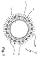
- 10 indicates a total of a vascular graft 10 prepared by the method of the present invention.
- a protein matrix is 12 in a structured material component 14 deeply anchored.
- the inner wall of the vascular graft 10 is formed by a protein membrane 16, which is the boundary to the lumen 18.
- the protein matrix 12 consists of protein fibers 20 with pores 22.
- the vessel implant 10 is given sufficient stability and sewing strength.
- the directed pores 22 of the protein matrix 12 can be uniformly populated with cells, such as smooth muscle cells, or in vivo , cells can grow from the surrounding tissue.
- the inner wall, or protein membrane 16, of the vascular implant 10 can be colonized with endothelial cells, since a suitable surface for the formation of an endothelial monolayer is provided by the protein membrane 16.
- the vascular graft 10 shown in FIG. 1 embodies only one embodiment.
- the invention can also be used in other forms and functions, for example as patches in dermal implants, as a cylinder or rectangle in cartilage and bone implants or heart valves.
- the method makes it possible to produce implants with different layer thicknesses of the protein matrix.
- a specific temperature profile is applied.
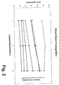
- a diagram is shown in Fig. 2, in which the dependence of the pore size taking into account the acetic acid concentration and the cooling rate is shown.
- the diagram shows that at a low cooling rate, larger pores can be produced (see, for example, 0.5 K / min compared to 12 K / min) and that the pore size was also greater when the acetic acid concentration was increased at the same time than at a lower one Acetic acid concentration (see, for example, at the cooling rate of 1 K / min: 1.5% acetic acid: 85 microns pore size compared to 3.8% acetic acid: about 110 microns pore size).
- a collagen / collagen-elastin tubular collagen having the desired inner diameter of the resulting vascular graft was prepared by extrusion or collagen membrane preparation followed by collagen suture tube formation.
- This collagen / collagen-elastin membrane (protein membrane) has been attached to a desired length on, for example, a metal tube;
- the polyester mesh (ie the structured material component) was pushed over the protein membrane and a second tubular form was applied to the structured material component, which, depending on the design, can serve as a cooling plate or for isolation.
- a (collagen / collagen-elastin) suspension between protein membrane and structured material component was filled.
- the arrangement was cooled, ie the collagen / collagen-elastin suspension is directionally solidified, for example by a one-sided controlled freezing process.
- the temperature of the metal tube which now acts as a cooling tube, lowered at a constant cooling rate of, for example, 6 K / min.
- the cooling medium is ethanol, which is pumped continuously through the metal tube.
- the second mold After freezing, the second mold is removed.
- the samples are stored at ⁇ -45 ° C for at least 12 hours and then freeze-dried.
- the vascular prosthesis produced by the method according to the invention from a collagen / collagen-elastin matrix, collagen / collagen-elastin membrane and a polyester reinforcement for colonization with, for example, myofibroblasts, endothelial cells and smooth muscle cells in a cell reactor ready.
- the implant is liquid-tight by the introduction of the protein membrane even at the time of cell application.
- the colonization time until the formation of a cell monolayer could be reduced to a maximum of four days.
Landscapes
- Health & Medical Sciences (AREA)
- Chemical & Material Sciences (AREA)
- General Health & Medical Sciences (AREA)
- Life Sciences & Earth Sciences (AREA)
- Animal Behavior & Ethology (AREA)
- Public Health (AREA)
- Medicinal Chemistry (AREA)
- Veterinary Medicine (AREA)
- Epidemiology (AREA)
- Transplantation (AREA)
- Oral & Maxillofacial Surgery (AREA)
- Dermatology (AREA)
- Engineering & Computer Science (AREA)
- Materials Engineering (AREA)
- Composite Materials (AREA)
- Vascular Medicine (AREA)
- Nuclear Medicine, Radiotherapy & Molecular Imaging (AREA)
- General Chemical & Material Sciences (AREA)
- Chemical Kinetics & Catalysis (AREA)
- Organic Chemistry (AREA)
- Pharmacology & Pharmacy (AREA)
- Bioinformatics & Cheminformatics (AREA)
- Materials For Medical Uses (AREA)
- Prostheses (AREA)
- Immobilizing And Processing Of Enzymes And Microorganisms (AREA)
Abstract
Description
- Die Erfindung betrifft ein Implantat, insbesondere eine Gefäßprothese, welche eine strukturierte Materialkomponente und eine Proteinmatrix mit Porenstruktur aufweist, sowie Verfahren zu seiner Herstellung und seine Verwendung.
- Implantate und Verfahren zu ihrer Herstellung sind aus dem Stand der Technik vielfach bekannt. Unter "Implantaten" werden im Allgemeinen Vorrichtungen verstanden, die in den Körper eines Patienten zumindest vorübergehend eingesetzt werden, und die beispielsweise therapeutische, Stütz- oder Gelenkfunktionen ausüben sollen.
- Vor allem im Gebiet des Tissue Engineerings finden implantierbare Strukturen ihre Anwendung, einem interdisziplinären Forschungsgebiet, das sich mit Verfahren und Materialien zur Herstellung von künstlichen Gewebe- und Organsystemen befasst. So können beispielsweise künstlich hergestellte Implantate als Haut-, Knochen-, Knorpel-, Linsen- oder Gefäßersatz Anwendung finden.
- Implantate in Form von Gefäßprothesen werden als Ersatz eines natürlichen erkrankten Gefäßes eingesetzt. Der erkrankte Gefäßabschnitt wird entfernt und durch ein Implantat ersetzt. So werden in der Gefäßchirurgie beispielsweise kleinlumige Implantate vor allem dann eingesetzt, wenn die körpereigenen Gefäße eines Patienten nicht verwendbar sind. Dies ist z.B. der Fall, wenn eine spezifische Gefäßlänge benötigt wird, oder wenn die autologen Gefäße auf Grund pathophysiologischer Eigenschaften nicht einsetzbar sind. Hier kommen Gefäßprothesen aus Kunststoff zum Einsatz, wobei vor allem Kunststoffe wie z.B. gewirkte oder gewebte Fäden aus Polyethylenterephthalat (PET) oder Gefäßprothesen aus expandiertem Polytetrafluorethylen (ePET) verwendet werden.
- Gefäßimplantate aus diesen Kunststoffen werden bevorzugt verwendet, da sie vorteilhafte strukturelle und biokompatible Eigenschaften besitzen. So kann einerseits umliegendes Gewebe einwachsen, andererseits darf durch die Poren kein Blutplasma austreten. Dies wird bei den ePET-Implantaten durch die eingestellte Größe der Poren erzielt, während gewirkte und gewebte PET-Implantate durch eine Beschichtung mit resorbierbaren Materialien wie beispielsweise Kollagen oder Gelatine imprägniert werden. Nach Implantation wird die Beschichtung in dem Maße resorbiert, in dem das umliegende neu gebildete Gewebe in die poröse Kollagenschicht einwächst.
- Es ist bekannt, dass der Einsatz der oben beschriebenen Implantate im kleinlumigen Gefäßbereich zu hohen Verschlussraten führt. Denn insbesondere der Kontakt von langsam strömendem Blut mit künstlichen Oberflächen kann zur Aktivierung des Gerinnungssystems, des Komplementsystems und des Immunsystems führen.
- Weitergehende Ansätze zur Vermeidung von Blutgerinnung gehen dahin, die beschichteten Implantate mit Zellen, wie z.B. Endothelzellen und Zellen der glatten Muskulatur, zu besiedeln. Die Interaktion der verschiedenen Zellarten, die beispielsweise in natürlichen Gefäßen vorhanden sind, sowie die Zell-Matrix-Interaktion sind für die Funktionalität der Implantate bedeutend. So muss neben einer hohen Biokompatibilität auch gewährleistet sein, dass die Struktur des Implantats an die Anforderungen von verschiedenen Zellen angepasst ist.
- An Implantate werden besondere mechanische und strukturelle Anforderungen gestellt. So sollen sie neben einer ausreichenden Strukturstabilität auch ein auf das zu ersetzende Gewebe abgestimmtes Kraft- und Dehnungsverhalten besitzen. Implantate müssen außerdem verschiedene Passformen, Längen und Durchmesser besitzen. Zudem spielt die Mikrostrukturierung, wie z.B. beim bioartifiziellen Gefäßersatz die radial verlaufende Porenstruktur mit zellspezifischer Größe, eine wichtige Rolle für die Besiedelung mit Zellen und für das wachsende Gewebe.
- Für die Bereitstellung einer geeigneten Porenstruktur werden im Stand der Technik u.a. Schwämme, bzw. einen Schwamm aufweisende Implantate bereitgestellt. So ist beispielsweise aus der
WO 99/27315 - Aus der
EP 0 562 864 sind ferner heteromorphe Schwämme bekannt, die aktive Inhaltsstoffe aufweisen. Die heteromorphen Schwämme weisen darüber hinaus zumindest eine Substruktur auf, die wie die Matrixstruktur des Schwammes aus absorbierbaren Biopolymer-Materialien hergestellt sind. Der Beschreibung dieser Patentanmeldung ist zu entnehmen, dass bei der Herstellung der heteromorphen Schwämme zunächst ein entsprechender Schwamm durch Einfrieren hergestellt wird, und anschließend wird ein Kollagen-Film auf den eingefrorenen Schwamm gelegt, eine weitere Schicht Kollagen-Suspension über den Kollagen-Film aufgebracht und dieses Komposit anschließend gefriergetrocknet. - Nachteilig an diesem Implantat ist jedoch, dass keine ausgerichtete Porenstruktur des Schwammes erzielt werden kann, was darin begründet ist, dass mittels eines Gebläses eingefroren wird. Ferner wird durch das Aufliegen der Kollagen-Folie und weiteres Auftragen einer Kollagen-Suspension und deren Einfrieren keine Verankerung des Kollagen-Films in der schwammartigen Struktur erreicht.
- Eine Verankerung der verschiedenen Schichten ist jedoch erwünscht, um dem Implantat eine hinreichende Stabilität und Elastizität zu verleihen, um das Implantat dementsprechend flexibel handhaben zu können.
- Aus der
DE 101 35 275 ist ein Implantat sowie ein Verfahren zu seiner Herstellung bekannt, das eine Proteinmatrix mit einer gerichteten Porenstruktur aufweist, wobei die Proteinmatrix in einer weiteren Schicht (Fremdstruktur) verankert ist. Die in derDE 101 35 275 offenbarten Implantate haben gegenüber den zuvor beschriebenen Implantaten den Vorteil, dass sie aufgrund des Herstellungsverfahrens eine gerichtete Porenstruktur aufweisen, aufgrund dessen beispielsweise Zellen optimal einwachsen können. - Nachteilig an dem aus der
DE 101 35 275 bekannten Implantats ist, dass das unbesiedelte Implantat direkt nach Zellaufgabe nicht dicht ist, wodurch es während der Besiedelung unter Strömung zu einem Medienaustritt am Gefäßeingang und einem Medieneintritt am Ende des durchströmten Gefäßes kommen kann. Die Ausbildung eines Endothelmonolayers ist daher nur schwer möglich. - Obwohl mit den gegenwärtig auf dem Markt vertriebenen Implantaten ein breites Spektrum an Implantaten zur Verfügung steht, besteht dennoch das große Bedürfnis, weitere Ausführungsformen bereitzustellen, bei welchen die für Implantate essentiellen Eigenschaften wie hinreichende Stabilität bei gleichzeitiger Flexibilität verbessert sind, welche eine gute Handhabung ermöglichen und ferner biokompatibel sowie gut resorbierbar sind.
- Der Erfindung liegt daher die Aufgabe zugrunde, ein Implantat sowie ein Verfahren zu seiner Herstellung bereitzustellen, mit welchem zum einen die Nachteile der im Stand der Technik offenbarten Implantate überwunden werden können und welches die eben beschriebenen Vorteile erfüllen kann.
- Bei dem eingangs genannten Implantat wird die Aufgabe erfindungsgemäß durch ein Implantat gelöst, bei welchem die strukturierte Materialkomponente zumindest teilweise in der Proteinmatrix verankert ist und bei welchem das Implantat auf zumindest einer Oberfläche eine Proteinmembran aufweist.
- Ferner wird die Aufgabe gelöst durch ein Verfahren zur Herstellung eines Implantats, das die erfindungsgemäße Struktur aufweist.
- Die der Erfindung zugrunde liegende Aufgabe wird hiermit vollständig gelöst.
- Mit der auf dem Implantat vorgesehenen Proteinmembran wird eine Oberfläche bereitgestellt, die für die Besiedelung mit Zellen, beispielsweise Endothelzellen, und für die zügige Ausbildung eines Zellmonolayers geeignet ist. Die Proteinmembran ist derart ausgebildet, dass sie flüssigkeitsdicht ist, ohne eine Diffusionsbarriere darzustellen, und damit undurchlässig für einen Strömungsaus- und -eintritt ist.
- Im Falle der Ausbildung des Implantats als Gefäßimplantat bildet die Proteinmembran die Gefäßinnenseite, wodurch diese Gefäßinnenseite zur Ausbildung eines Endothelmonolayers mit Endothelzellen besiedelt wird.
- Im Falle einer Ausbildung des Implantats als Gewebeimplantat ist die Proteinmembran auf der Oberfläche als abgrenzende Schicht angeordnet.
- Unter "strukturierte Materialkomponente" wird dabei jede fortlaufende, bzw. zusammenhängende Struktur verstanden, die schichtartig ausgebildet ist und eine Art Gerüst für das Implantat bildet, welches entweder in der Proteinmatrix gänzlich enthalten ist, oder aber als Begrenzungsschicht dient, in welche die Proteinmatrix teilweise eingebracht sein kann. "Strukturiert" bedeutet hierbei, dass die Materialkomponente Poren oder Hinterschneidungen aufweist, über welche die Materialkomponente in der Proteinmatrix verankert werden kann.
- Durch das erfindungsgemäße Implantat können erstmals Gefäß- und/oder Gewebeimplantate bereitgestellt werden, die eine hinreichende Stabilität aufweisen, die - je nach Wahl der strukturierten Materialkomponente - vollständig oder nahezu vollständig resorbieren und die durch ihre Mikrostrukturierung an die spezifischen Zellanforderungen während der Zellaufgabe, der Kultivierung und der Gewebeneubildung angepasst sind. So bildet die Proteinmembran eine geeignete Grundlage für das Anheften von Zellen und die Ausbildung eines Zellmonolayers, wie beispielsweise die Ausbildung eines Endothelmonolayers beim bioartifiziellen Gefäßimplantat. Die Proteinmatrix mit gerichtet (d.h. in eine einzige Richtung) verlaufender, offener Porenstruktur mit angepasster Porengröße ermöglicht während der Besiedelung eine gleichmäßige Zellverteilung innerhalb der Matrix. Die Zellen sind ähnlich der nativen Situation in eine Proteinmatrix eingebettet. Beim bioartifiziellen Gefäßersatz wir die Porenstruktur beispielsweise an die Anforderungen der glatten Muskelzellen angepasst.
- In einer Ausführungsform ist dabei bevorzugt, wenn die strukturierte Materialkomponente vollständig in der Proteinmatrix verankert ist.
- Bei dieser Ausführungsform durchwachsen die in der Proteinmatrix vorliegenden Poren die strukturierte Materialkomponente, so dass die strukturierte Materialkomponente vollständig von der Proteinmatrix umgeben ist, bzw. in der Proteinmatrix aufgenommen ist. Die Poren oder Maschen der strukturierten Materialkomponente sind um ein Vielfaches größer als die der Proteinmatrix und bilden somit keine Zellbarriere. Gleichzeitig wird durch die in der Proteinmatrix eingelagerte strukturierte Materialkomponente der schwammartigen Struktur der Proteinmatrix eine hinreichende Stabilität verliehen. Ferner wird durch die auf der Oberfläche angebrachte Proteinmembran gleichzeitig eine flüssigkeitsdichte Struktur als Grenze zur Umgebung bereitgestellt, also eine Art Abdichtung geschaffen, die beispielsweise bei Ausarbeitung des Implantats als Gefäßimplantat als Blutaustrittsbarriere dient.
- Durch die Einbringung der Proteinmembran ist das Implantat bereits bei Zellaufgabe flüssigkeitsdicht. Dadurch kann vorteilhafterweise die Besiedelung unter definierten Scherraten durchgeführt werden. Die Besiedelungszeit bis zur Ausbildung eines Zellmonolayers kann dadurch vorteilhaft verkürzt werden.
- In einer weiteren Ausführungsform ist bevorzugt, wenn die strukturierte Materialkomponente nur teilweise in die Proteinmatrix eingebracht ist und die strukturierte Materialkomponente eine äußere Begrenzungsschicht des Implantats bildet.
- Diese Ausführungsform des Implantats weist somit einen schichtartigen Aufbau auf, wobei die Schicht der Proteinmatrix zwischen einerseits der strukturierten Materialkomponente, in welche sie teilweise verankert ist, und der Proteinmembran andererseits eingebracht ist.
- Die strukturierte Materialkomponente kann dabei jedes Material aufweisen, das eine strukturierte Form bildet bzw. vorgibt. Als strukturierte Materialkomponente geeignet sind daher bspw. strukturierte beständige künstliche Materialien, strukturierte resorbierbare künstliche Materialien, strukturierte natürliche Materialien, sowie Mischungen aus den genannten Materialien.
- Ferner ist bevorzugt, wenn die strukturierte Materialkomponente ein Material aufweist, das ausgewählt ist aus der Gruppe umfassend Polytetrafluorethylen, Polyurethan, Polystyrol, Polyester, Keramik, Metall, Polylactit, Poly-Glykolsäure, Polyhydroxyalkanoate, Copolymere davon, Polysaccharide oder Mischungen aus einem oder mehrerer der genannten Materialien.
- In einer bevorzugten Ausführungsform weist die strukturierte Materialkomponente Polyester auf, welches im Falle einer Gefäßprothese als großmaschiger netzartiger Schlauch ausgebildet sein kann. Gleichwohl sind auch andere Materialien als schlauchartige Grundgerüste der Fremdstruktur denkbar, bspw. expandiertes Polytetrafluorethylen, das sich in den letzten Jahren als bevorzugtes künstliches Material für Implantate durchgesetzt hat.
- Es ist ferner bevorzugt, wenn die Proteinmembran ein Material aufweist, das ausgewählt ist aus der Gruppe umfassend Kollagen, Elastin, Cellulose, Chitosan, Chitin und Komponenten der Extrazellulären Matrix oder eine Mischung zweier oder mehrerer dieser Stoffe.
- Die Proteinmembran kann dabei in einer bevorzugten Ausführungsform eine Schichtdicke von zwischen 0,02 mm und 5 mm aufweisen.
- Bei dem erfindungsgemäßen Implantat ist ferner bevorzugt, wenn die Proteinmatrix eine Porengröße von ca. 5 µm bis 500 µm aufweist.
- Ferner ist bevorzugt, wenn die Proteinmatrix, in welche die strukturierte Materialkomponente eingebracht ist, eine Schichtdicke von ca. 0,05 mm bis ca. 50 mm aufweist.
- In einer bevorzugten Ausführungsform weist das Implantat die Ausbildung einer röhrenförmigen Gefäßprothese/Gefäßimplantats auf, wobei bevorzugt ist, wenn die Proteinmembran in der Innenseite der röhrenförmigen Gefäßprothese ausgebildet ist.
- Mit dieser Ausführungsform wird demnach eine Gefäßprothese bereitgestellt, die auf Grund ihrer Ausbildung einerseits die Regeneration der Gefäßwand, eine Neovaskularisierung aus dem umliegenden Gewebe und eine gute Nährstoffversorgung in der Proteinmatrix gewährleistet, und die andererseits auf Grund der Innenauskleidung mit der Proteinmembran ein Austreten von Blut in das umgebende Gewebe verhindert. Gleichzeitig wird mit der Proteinmembran eine Schicht für die Besiedelung mit Endothelzellen bereitgestellt, über welche ein Endothelmonolayer einfach ausgebildet werden kann.
- In weiteren Ausführungsformen ist bevorzugt, wenn das Implantat ferner zumindest einen Wirkstoff aufweist, der ausgewählt ist aus der Gruppe umfassend Heparin, Hirudin, Aspirin, Heparansulfat, Albumin, Antibiotika, wie beispielsweise Rifampicin, und/oder Wachstumsfaktoren.
- Je nach Anwendung können die Implantate vor der Implantation mit Wirkstoffen beladen oder beschichtet werden. Dabei kann die Wirkstoffabgabe anschließend beispielsweise durch eine zusätzlich aufgebrachte Hydrogelbeschichtung oder durch die Art der Anbindung der Wirkstoffe noch zusätzlich kontrolliert werden.
- Dabei ist in einer Ausführungsform bevorzugt, wenn die Proteinmembran ohne Vernetzungsmittel mit der Proteinmatrix vernetzt ist.
- Dies wird bspw. dadurch erreicht, dass die Proteinmembran durch einen Abkühlvorgang bei der Herstellung des Implantats mit der Proteinmatrix vernetzt wird.
- In einer anderen Ausführungsform ist bevorzugt, wenn die Vernetzung der Proteinmembran mit der Proteinmatrix (oder eine weitere/zusätzliche Vernetzung) durch chemische Vernetzungsmittel (wie bspw. Glutaraldehyd) erreicht wird.
- Die Erfindung betrifft ferner ein Verfahren zur Herstellung der erfindungsgemäßen Implantate, das die folgenden Schritte aufweist:
- a) Bereitstellen einer Proteinmembran auf einer der Grundform der Proteinmembran entsprechenden ersten abgrenzenden Form in einer Anordnung;
- b) Positionieren der strukturierten Materialkomponente über der Proteinmembran in der Anordnung, wobei die Form der Fremdstruktur der Grundform der Proteinmembran entspricht;
- c) Aufbringen einer zweiten abgrenzenden Form auf die strukturierte Materialkomponente;
- d) Einbringen einer Suspension zwischen Proteinmembran und strukturierter Materialkomponente, und
- e) Abkühlen der Anordnung und anschließende Gefriertrocknung zur Ausbildung einer Proteinmatrix aus der Suspension und zur Anbindung der Proteinmembran.
- Bei diesem Verfahren wird also zunächst eine vorgefertigte Proteinmembran auf eine - der Form der vorgefertigten Proteinmembran entsprechende - erste Form aufgebracht, wobei diese Form bei der Herstellung des Implantats eine erste Abgrenzung für das Implantat-Konstrukt bildet. Die Form kann ggf. während eines einseitigen Einfriervorgangs als Kühlplatte oder Isolation dienen. Anschließend wird eine strukturierte Materialkomponente über die Proteinmembran positioniert, wobei auf die strukturierte Materialkomponente eine zweite abgrenzende Form aufgebracht wird, die ggf. je nach Anordnung als Kühlplatte oder zur Isolation dienen kann. In einem nächsten Schritt wird eine Suspension zwischen die Proteinmembran und die strukturierte Materialkomponente eingebracht und die gesamte Anordnung abgekühlt und gefriergetrocknet. Vor der Gefriertrocknung wird eine der begrenzenden Formen entfernt, um den Gefriertrocknungsvorgang zu beschleunigen. Durch das Abkühlen bildet sich aus der Suspension eine Proteinmatrix aus, wobei gleichzeitig bei diesem Vorgang diese Proteinmatrix einerseits teilweise in der strukturierten Materialkomponente verankert und andererseits mit der Proteinmembran vernetzt wird.
- Je nach Abstand der zweiten abgrenzenden Form von der Fremdstruktur können bspw. die in der Proteinmatrix entstehenden Poren bspw. durch die strukturierte Materialkomponente hindurch wachsen. In diesem Fall kann bspw. die durch die strukturierte Materialkomponente hindurch gewachsene Proteinmatrix nach Fertigstellung des Implantats dem Verwendungszweck entsprechend abgeschnitten bzw. gekürzt werden. Andererseits kann auch durch direktes Anlegen der abgrenzenden zweiten Form erreicht werden, dass die Poren nicht komplett durch die strukturierte Materialkomponente wachsen, sondern lediglich in der strukturierten Materialkomponente verankert werden.
- In einer Ausführungsform des erfindungsgemäßen Verfahrens ist bevorzugt, wenn das Abkühlen in Schritt e) einseitig erfolgt. Durch das einseitige Abkühlen kann eine gerichtete Porenstruktur ausgebildet werden, durch welche ein gleichmäßiges Einbetten von Zellen während der Besiedelung ermöglicht wird. Bei diesem Verfahren wird die Anordnung auf einer Seite bzw. Oberfläche einseitig abgekühlt, während die andere Oberfläche isoliert wird. Während des Einfrierprozess kann die Suspension teilweise oder vollständig auskristallisieren.
- Implantate, die nach dem bevorzugten Verfahren hergestellt werden, können beispielsweise - bei Verwendung eines Metallrohrs als Grundform - Gefäßimplantate darstellen, wobei jedoch auch flächige Gewebeimplantate unter Verwendung entsprechend flächiger Grundformen hergestellt werden können.
- Bei der o.g. Ausführungsform des erfindungsgemäßen Verfahrens ist bevorzugt, wenn das Abkühlen in Schritt e) mit einer konstanten Kühlrate verläuft und insbesondere mit einer Kühlrate von ca. 0,1 K/min bis 200 K/min.
- Durch Anwendung unterschiedlicher Kühlraten können verschiedene Porengrößen der Proteinmatrix eingestellt werden. Dabei gilt, dass die Porengröße umso kleiner ist, je schneller abgekühlt wird. Bspw. kann mit einer Kühlrate von 12 K/min eine Porengröße von ca. 45 µm bis 50 µm eingestellt werden und eine Porengröße von ca. 85 µm bis 105 µm bei einer Abkühlrate von 1 K/min, wobei die jeweiligen Bereiche bspw. durch die Zugabe von Säuren wie Essig- oder Ascorbinsäure beeinflusst werden können.
- Die Anwendung des einseitigen Abkühlens hat sich bei der Herstellung des erfindungsgemäßen Implantats als besonders geeignet erwiesen, da auf diese Weise gerichtete Poren hergestellt werden können.
- In einer anderen Ausführungsform des erfindungsgemäßen Verfahrens ist bevorzugt, wenn das Abkühlen in Schritt e) bei einer konstanten Temperatur erfolgt.
- In dieser Variante kann die Anordnung bspw. in einer Gefriertruhe oder anderen geeigneten Kühl- bzw. Gefriervorrichtungen abgekühlt werden. Diese Implantate weisen jedoch dann keine gerichtete Porenstruktur auf, sind aber - je nach Anwendung - für einen Einsatz als Implantate ebenfalls geeignet.
- In dem erfindungsgemäßen Verfahren ist ferner bevorzugt, wenn die Suspension, aus welcher die Proteinmatrix gebildet wird, eine Suspension ist, die Kollagen, Elastin und lösliche, nicht-kollagene Bestandteile enthält.
- Zusätzliche Begleitstoffe können Wachstumsfaktoren oder Bestandteile der extrazellulären Matrix sein, wie z.B. Laminin, Elastin, Fibronektin, Hyaluronsäure, Glykosaminoglykane u.a., sowie Derivate davon. Die löslichen Bestandteile umfassen einerseits Säuren wie HCL, Essigsäure oder Ascorbinsäure, da bekannt ist, dass der optimale pH-Bereich zur Herstellung gefriergetrockneter Kollagenschwämme zwischen 2,5 und 3,5 liegt. Andererseits können lösliche Zusatzstoffe wie Glycerin oder Ethanol oder fein-dispergierte Stoffe wie bspw. Calciumphosphat eingesetzt werden, da durch ihre Konzentration die Eiskristallmorphologie und somit die Porenstruktur eingestellt werden kann.
- Bei einer weiteren Ausführungsform ist vorgesehen, dass die Proteinmembran, die in Schritt a) eingesetzt wird, aus der gleichen Suspension hergestellt wird wie die Suspension zur Herstellung der Proteinmatrix, und -je nach Einsatz des Implantats - in planarer oder tubulärer Form ausgebildet wird.
- Im Falle des einseitigen Einfrierprozess, während dessen die Suspension auskristallisieren kann, werden die Kollagene, Elastin und die gelösten Stoffe dabei von der wachsenden zellularen Einsphasenfront verschoben, wobei die suspensierten Proteine und die gelösten oder dispergierten Stoffe zwischen den Eiskristallen hoch aufkonzentriert werden. Durch die Kühlrate und die chemische Zusammensetzung der Proteinsuspension können die Eiskristallstruktur- und größe bei einer beliebigen Schichtdicke vorteilhaft eingestellt werden.
- In einer Weiterbildung des erfindungsgemäßen Verfahren ist ein weiterer Schritt f) vorgesehen, mit welchem das in den Schritten a) bis e) hergestellte Implantat anschließend gefriergetrocknet wird.
- Bei dem Gefriertrocknungsprozess sublimieren die Eiskristalle, wodurch Poren entstehen, die der Einkristallmorphologie der gefrorenen Probe entsprechen. Bei der Besiedelung der Proteinmatrix können Zellen gleichmäßig in den Poren verteilt werden. Des weiteren können bei einer Implantation die Zellen des umliegenden Gewebes entlang der Proteinfasern in das Implantat einwachsen. Durch den Wasserentzug während der Sublimation bilden sich kovalente Bindungen zwischen den Kollagenmolekülen aus, wodurch die Matrix, die Membran und die Verbindung Matrix-Membran eine erwünschte Stabilität erhält. Dabei ist es vorteilhaft, dass der Vernetzungsgrad durch den Gefriertrocknungsprozess oder durch eine chemische Behandlung des gefriergetrockneten Produkts gezielt eingestellt werden kann. Die durch den Gefriertrocknungsprozess entstandene Proteinmatrix ist somit direkt mit der Membran und der Fremdstruktur verankert und es sind keine weiteren Schritte zur Verbindung der Fremdstruktur und der Membran mit der Proteinmatrix notwendig.
- Durch die Erfindung kann mit einfachen Mitteln erreicht werden, dass das Implantat den jeweiligen Anforderungen des umliegenden Gewebes durch die Ausbildung einer Porenstruktur und die Anbindung einer Membran einfach und effizient angepasst werden kann. Die so hergestellten Implantate weisen eine mechanische Stabilität und biologische Kompatibilität auf.
- Die Erfindung betrifft ferner Implantate, die nach dem erfindungsgemäßen Verfahren hergestellt werden, sowie insbesondere Gefäßprothesen/Gefäßimplantate. Derartige Gefäßprothesen können bspw. als kleinlumiger Gefäßersatz (Durchmesser < 6 mm), z.B. bei Herzkranzgefäßen oder Gefäßen der Peripherie eingesetzt werden, ferner als Dialyseshunt oder in der Pädiatrie als mitwachsende Gefäßimplantate.
- Weitere Vorteile ergeben sich aus der Beschreibung und der beigefügten Zeichnungen.
- Es versteht sich, dass die vorstehend genannten und die nachstehend noch zu erläuternden Merkmale nicht nur in der jeweils angegebenen Kombination, sondern auch in anderen Kombinationen oder in Alleinstellung verwendbar sind, ohne den Rahmen der vorliegenden Erfindung zu verlassen.
- Ausführungsbeispiele der Erfindung sind in den nachstehenden Zeichnungen dargestellt und werden in der nachfolgenden Beschreibung näher erläutert. Es zeigen:
- Fig. 1
- einen Querschnitt eines Ausführungsbeispiels eines erfindungsgemäßen Implantats, nämlich einer Gefäßprothese;
- Fig. 2
- ein Diagramm, das die Abhängigkeit der Porengröße von der Essigsäurekonzentration darstellt;
- In Fig. 1 bezeichnet 10 insgesamt ein Gefäßimplantat 10, das nach dem erfindungsgemäßen Verfahren hergestellt wurde. Bei dem Implantat ist eine Proteinmatrix 12 in einer strukturierten Materialkomponente 14 tief verankert. Die Innenwand des Gefäßimplantats 10 wird durch eine Proteinmembran 16 gebildet, welche die Grenze zum Lumen 18 darstellt. Die Proteinmatrix 12 besteht aus Proteinfasern 20 mit Poren 22.
- Durch die strukturierte Materialkomponente 14, die gerüstartig in der Proteinmatrix vorliegt, wird dem Gefäßimplantat 10 eine ausreichende Stabilität und Nähfestigkeit verliehen. Die gerichtet verlaufenden Poren 22 der Proteinmatrix 12 können gleichmäßig mit Zellen, wie beispielsweise glatten Muskelzellen, besiedelt werden, bzw. in vivo können Zellen aus dem umgebenden Gewebe einwachsen. Andererseits kann die Innenwand, bzw. Proteinmembran 16, des Gefäßimplantats 10 mit Endothelzellen besiedelt werden, da durch die Proteinmembran 16 eine geeignete Oberfläche zur Ausbildung eines Endothelmonolayer bereitgestellt wird.
- An dieser Stelle wird angemerkt, dass das in Fig. 1 gezeigte Gefäßimplantat 10 nur ein Ausführungsbeispiel verkörpert. Die Erfindung ist daneben auch in anderen Formen und Funktionen einsetzbar wie beispielsweise als Patches bei Hautimplantaten, als Zylinder oder Rechteck bei Knorpel- und Knochenimplantaten oder Herzklappen.
- Durch das Verfahren können Implantate mit unterschiedlichen Schichtdicken der Proteinmatrix hergestellt werden. Um eine bestimmte Proteinmatrixschichtdicke zu erreichen, wird ein bestimmter Temperaturverlauf angelegt.
- In diesem Zusammenhang ist in Fig. 2 ein Diagramm gezeigt, in welchem die Abhängigkeit der Porengröße unter Berücksichtigung der Essigsäurekonzentration und der Kühlrate dargestellt ist. Dem Diagramm ist zu entnehmen, dass bei einer geringen Abkühlrate größere Poren hergestellt werden können (s. beispielsweise 0,5 K/min im Vergleich zu 12 K/min) und dass bei gleichzeitiger Erhöhung der Essigsäurekonzentration die Porengröße ebenfalls größer war als bei einer geringeren Essigsäurekonzentration (s. beispielsweise bei der Abkühlrate von 1 K/min: 1,5 % Essigsäure: 85 µm Porengröße im Vergleich zu 3,8 % Essigsäure: ca. 110 µm Porengröße).
- An Hand des unten beschriebenen Beispiels wird nun die Anwendung der Vorrichtung gezeigt.
- Nach bekannten Verfahren wurde ein großmaschiges texturiertes Polyesternetz gewirkt, gereinigt und geschrumpft. In einem Exsikkator wurde das Polyesternetz in Kollagen-Suspension vollständig entlüftet.
- Aus der Kollagen-Suspension oder der Kollagen-Elastin-Suspension wurde durch Extrusion oder durch Herstellen einer flächigen Kollagenmembran und anschließende Röhrenbildung mit Kollagennaht eine röhrenförmige Kollagen-/Kollagen-Elastin-Membran mit dem gewünschten Innendurchmesser des entstehenden Gefäßimplantates hergestellt. Diese Kollagen-/Kollagen-Elastin-Membran (Proteinmembran) wurde in gewünschter Länge auf bspw. einem Metallrohr angebracht; über die Proteinmembran wurde das Polyesternetz (also die strukturierte Materialkomponente) geschoben und eine zweite röhrenförmige Form auf die strukturierte Materialkomponente aufgebracht, die je nach Ausführung als Kühlplatte oder zur Isolation dienen kann. In einem nächsten Schritt wurde eine (Kollagen-/Kollagen-Elastin)Suspension zwischen Proteinmembran und strukturierte Materialkomponente eingefüllt.
- Anschließend wurde die Anordnung abgekühlt, d.h. die Kollagen-/Kollagen-Elastin-Suspension wird - bspw. durch einen einseitig kontrollierten Einfriervorgang - gerichtet erstarrt. Dazu wird die Temperatur des Metallrohres, welches nun als Kühlrohr fungiert, mit einer konstanten Kühlrate von beispielsweise 6 K/min abgesenkt. Als Kühlmedium dient Ethanol, welches kontinuierlich durch das Metallrohr gepumpt wird.
- Nach dem Einfrieren wird die zweite Form entfernt. Die Proben werden bei < -45°C mindestens 12 Stunden gelagert und anschließend gefriergetrocknet.
- Nach einer darauffolgenden Sterilisation ist die nach dem erfindungsgemäßen Verfahren hergestellte Gefäßprothese aus einer Kollagen-/Kollagen-Elastin-Matrix, Kollagen-/Kollagen-Elastin-Membran und einer Polyesterverstärkung zur Besiedelung mit bspw. Myofibroblasten, Endothelzellen und Zellen der glatten Muskulatur in einem Zellreaktor bereit.
- Wie bereits erwähnt, ist das Implantat durch die Einbringung der Proteinmembran bereits bei Zellaufgabe flüssigkeitsdicht. Mit dem erfindungsgemäßen Implantat konnte die Besiedelungszeit bis zur Ausbildung eines Zellmonolayers auf höchstens vier Tage reduziert werden.
Claims (13)
- Implantat mit einer strukturierten Materialkomponente und mit einer Proteinmatrix, die eine Porenstruktur aufweist, dadurch gekennzeichnet, dass die strukturierte Materialkomponente zumindest teilweise in der Proteinmatrix verankert ist und dass das Implantat auf zumindest einer Oberfläche eine mit der Proteinmatrix vernetzte Proteinmembran aufweist.
- Implantat nach Anspruch 1, dadurch gekennzeichnet, dass die strukturierte Materialkomponente vollständig in der Proteinmatrix verankert ist.
- Implantat nach Anspruch 1, dadurch gekennzeichnet, dass die strukturierte Materialkomponente nur teilweise in die Proteinmatrix eingebracht ist und die strukturierte Materialkomponente eine äußere Begrenzungsschicht des Implantats bildet.
- Implantat nach einem der Ansprüche 1 bis 3, dadurch gekennzeichnet, dass die strukturierte Materialkomponente ein Material aufweist, das ausgewählt ist aus der Gruppe umfassend Polytetrafluorethylen, Polyurethan, Polystyrol, Polyester, Keramik, Metall, Poly-Lactid, Poly-Glykolsäure, Polyhydroxyalkanoate, Copolymere davon, Polysaccharide oder Mischungen aus einem oder mehrerer davon.
- Implantat nach einem der Ansprüche 1 bis 4, dadurch gekennzeichnet, dass die Proteinmatrix eine Porengröße von ca. 5 µm bis 500 µm aufweist.
- Implantat nach einem der Ansprüche 1 bis 5, dadurch gekennzeichnet, dass die Proteinmatrix, in welche die Fremdstruktur eingebracht ist, eine Schichtdicke von ca. 0,05 mm bis ca. 50 mm aufweist.
- Implantat nach einem der Ansprüche 1 bis 6, dadurch gekennzeichnet, dass das Implantat als röhrenförmige Gefäßprothese ausgebildet ist.
- Implantat nach Anspruch 7, dadurch gekennzeichnet, dass die Proteinmembran in der Innenseite der röhrenförmigen Gefäßprothese ausgebildet ist.
- Implantat nach einem der Ansprüche 1 bis 8, dadurch gekennzeichnet, dass das Implantat ferner einen Wirkstoff aufweist, der ausgewählt ist aus der Gruppe umfassend Heparin, Hirudin, Aspirin, Heparansulfat, Albumin, Antibiotika, Wachstumsfaktoren, oder Mischungen aus einem oder mehrerer dieser Wirkstoffe.
- Implantat nach einem der Ansprüche 1 bis 9, dadurch gekennzeichnet, dass die Proteinmembran ohne Vernetzungsmittel mit der Proteinmatrix vernetzt ist.
- Implantat nach einem der Ansprüche 1 bis 9, dadurch gekennzeichnet, dass die Proteinmembran über Vernetzungsmittel mit der Proteinmatrix verbunden ist.
- Implantat nach Anspruch 11, dadurch gekennzeichnet, dass das Vernetzungsmittel ausgewählt ist aus der Gruppe umfassend Glutaraldehyd, Formaldehyd, Isocyanate, Ethylendi-chlorid, oder Mischungen aus einem oder mehrerer dieser Vernetzungsmittel.
- Implantat nach einem der Ansprüche 1 bis 13, dadurch gekennzeichnet, dass die Proteinmatrix eine gerichtete Porenstruktur aufweist.
Applications Claiming Priority (1)
| Application Number | Priority Date | Filing Date | Title |
|---|---|---|---|
| DE102006042631A DE102006042631A1 (de) | 2006-09-05 | 2006-09-05 | Implantat und Verfahren zu seiner Herstellung |
Publications (3)
| Publication Number | Publication Date |
|---|---|
| EP1905464A2 true EP1905464A2 (de) | 2008-04-02 |
| EP1905464A3 EP1905464A3 (de) | 2010-10-06 |
| EP1905464B1 EP1905464B1 (de) | 2012-05-16 |
Family
ID=39062284
Family Applications (1)
| Application Number | Title | Priority Date | Filing Date |
|---|---|---|---|
| EP07017118A Active EP1905464B1 (de) | 2006-09-05 | 2007-08-31 | Implantat und Verfahren zu seiner Herstellung |
Country Status (4)
| Country | Link |
|---|---|
| US (1) | US7976860B2 (de) |
| EP (1) | EP1905464B1 (de) |
| DE (1) | DE102006042631A1 (de) |
| ES (1) | ES2384527T3 (de) |
Cited By (1)
| Publication number | Priority date | Publication date | Assignee | Title |
|---|---|---|---|---|
| US10576694B2 (en) | 2011-12-08 | 2020-03-03 | Julius-Maximilians-Universitaet Wuerzburg | Production of materials having an anisotropic structure |
Families Citing this family (1)
| Publication number | Priority date | Publication date | Assignee | Title |
|---|---|---|---|---|
| EP3034103A1 (de) * | 2014-12-15 | 2016-06-22 | Geistlich Pharma AG | Kollagenschwamm |
Citations (4)
| Publication number | Priority date | Publication date | Assignee | Title |
|---|---|---|---|---|
| DE3203957A1 (de) * | 1982-02-05 | 1983-08-18 | Chemokol Gesellschaft zur Entwicklung von Kollagenprodukten, 5190 Stolberg | Verfahren zur herstellung von feinporigen kollagenschwaemmen |
| EP0562864A1 (de) * | 1992-03-25 | 1993-09-29 | JOHNSON & JOHNSON MEDICAL, INC. | Heteromorphe Schwämme mit Wirkstoffen |
| EP1275405A1 (de) * | 2001-07-13 | 2003-01-15 | JOTEC GmbH | Implantat mit poröser Proteinmatrix und Verfahren zu seiner Herstellung |
| US20040110439A1 (en) * | 2001-04-20 | 2004-06-10 | Chaikof Elliot L | Native protein mimetic fibers, fiber networks and fabrics for medical use |
Family Cites Families (5)
| Publication number | Priority date | Publication date | Assignee | Title |
|---|---|---|---|---|
| US4787900A (en) * | 1982-04-19 | 1988-11-29 | Massachusetts Institute Of Technology | Process for forming multilayer bioreplaceable blood vessel prosthesis |
| JPH0824326A (ja) * | 1994-07-13 | 1996-01-30 | Terumo Corp | 人工血管およびその製造方法 |
| GB9721585D0 (en) * | 1997-10-10 | 1997-12-10 | Geistlich Soehne Ag | Chemical product |
| DE19751031A1 (de) | 1997-11-19 | 1999-06-24 | Ingo Dipl Ing Heschel | Verfahren zur Herstellung poröser Strukturen |
| US6743253B2 (en) * | 2000-02-29 | 2004-06-01 | Biomod Surfaces | Polyurethane-sealed biocompatible device and method for its preparation |
-
2006
- 2006-09-05 DE DE102006042631A patent/DE102006042631A1/de not_active Withdrawn
-
2007
- 2007-08-31 ES ES07017118T patent/ES2384527T3/es active Active
- 2007-08-31 EP EP07017118A patent/EP1905464B1/de active Active
- 2007-09-04 US US11/849,631 patent/US7976860B2/en not_active Expired - Fee Related
Patent Citations (4)
| Publication number | Priority date | Publication date | Assignee | Title |
|---|---|---|---|---|
| DE3203957A1 (de) * | 1982-02-05 | 1983-08-18 | Chemokol Gesellschaft zur Entwicklung von Kollagenprodukten, 5190 Stolberg | Verfahren zur herstellung von feinporigen kollagenschwaemmen |
| EP0562864A1 (de) * | 1992-03-25 | 1993-09-29 | JOHNSON & JOHNSON MEDICAL, INC. | Heteromorphe Schwämme mit Wirkstoffen |
| US20040110439A1 (en) * | 2001-04-20 | 2004-06-10 | Chaikof Elliot L | Native protein mimetic fibers, fiber networks and fabrics for medical use |
| EP1275405A1 (de) * | 2001-07-13 | 2003-01-15 | JOTEC GmbH | Implantat mit poröser Proteinmatrix und Verfahren zu seiner Herstellung |
Non-Patent Citations (1)
| Title |
|---|
| THOMAS SCHEIBEL: "Protein fibers as performance proteins: new technologies and applications" CURRENT OPINION IN BIOTECHNOLOGY, Bd. 16, 2005, Seiten 427-433, XP002597847 * |
Cited By (1)
| Publication number | Priority date | Publication date | Assignee | Title |
|---|---|---|---|---|
| US10576694B2 (en) | 2011-12-08 | 2020-03-03 | Julius-Maximilians-Universitaet Wuerzburg | Production of materials having an anisotropic structure |
Also Published As
| Publication number | Publication date |
|---|---|
| EP1905464B1 (de) | 2012-05-16 |
| ES2384527T3 (es) | 2012-07-06 |
| EP1905464A3 (de) | 2010-10-06 |
| DE102006042631A1 (de) | 2008-03-20 |
| US7976860B2 (en) | 2011-07-12 |
| US20080057126A1 (en) | 2008-03-06 |
Similar Documents
| Publication | Publication Date | Title |
|---|---|---|
| EP1275405B1 (de) | Implantat mit poröser Proteinmatrix und Verfahren zu seiner Herstellung | |
| DE69525692T2 (de) | Implantierbare Rohrprothese aus Polytetrafluorethylen | |
| DE69625822T2 (de) | Implantierbare, bioresorbierbare membran und verfahren zu ihrer herstellung | |
| DE69232899T2 (de) | Implantierbare mehrlumige, eine kontollierte Porosität aufweisende, extrudierte Vorrichtung und Verfahren zur Herstellung | |
| DE69732721T2 (de) | Biologisch abbaubare kunstoff-folien | |
| DE3751843T2 (de) | Herstellung von körperorganen durch kontroliertes zellwachstum auf künstlicher matrix | |
| DE69307299T2 (de) | Mehrphasiges, biologisch abbaubares implantat/träger und verfahren zu seiner herstellung | |
| DE69432865T2 (de) | Implantierbare prothese, kit und vorrichtung zu deren herstellung | |
| DE69419859T2 (de) | Synthetisches gewebe | |
| EP0770401B1 (de) | Verfahren zur Herstellung intraluminaler Stents aus bioresorbierbarem Polymermaterial | |
| DE69219613T2 (de) | Vorvaskularisierte polymerimplantate für organtransplantation | |
| DE69728054T2 (de) | Bioresorbierbare dichtungsmassen für poröse künstliche gefässen | |
| DE68915540T2 (de) | Für zellen eindringbares medizinisches material und kunsthaut. | |
| DE19654884C2 (de) | Formkörper | |
| DE10130968A1 (de) | Beschichtetes Polymermaterial, dessen Verwendung sowie Verfahren zu dessen Herstellung | |
| EP1948263A2 (de) | Nervenleitschiene | |
| EP0248246A1 (de) | Künstliche Gefässwand | |
| WO2004108810A1 (de) | Matrix, zellimplantat, verfahren zu deren herstellung und deren verwendung | |
| EP3319652B1 (de) | Verfahren zur herstellung eines bioartifiziellen, primär azellulären konstrukts auf fibrinbasis und dieses konstrukt selbst | |
| DE69815096T2 (de) | Selbst aggregierende protein zusammensetzungen und ihre verwendung als dichtungsmassen | |
| EP2794842B1 (de) | Verfahren und vorrichtung zur herstellung eines bioartifiziellen gewebekonstrukts | |
| DE10050870A1 (de) | Biokompatibles Verbundmaterial für medizinische Anwendungen | |
| EP1905464B1 (de) | Implantat und Verfahren zu seiner Herstellung | |
| EP2701757B1 (de) | Implantierbare gefässstütze | |
| DE69007999T2 (de) | Prothese für ein lebendes Organ. |
Legal Events
| Date | Code | Title | Description |
|---|---|---|---|
| PUAI | Public reference made under article 153(3) epc to a published international application that has entered the european phase |
Free format text: ORIGINAL CODE: 0009012 |
|
| AK | Designated contracting states |
Kind code of ref document: A2 Designated state(s): AT BE BG CH CY CZ DE DK EE ES FI FR GB GR HU IE IS IT LI LT LU LV MC MT NL PL PT RO SE SI SK TR |
|
| AX | Request for extension of the european patent |
Extension state: AL BA HR MK YU |
|
| PUAL | Search report despatched |
Free format text: ORIGINAL CODE: 0009013 |
|
| AK | Designated contracting states |
Kind code of ref document: A3 Designated state(s): AT BE BG CH CY CZ DE DK EE ES FI FR GB GR HU IE IS IT LI LT LU LV MC MT NL PL PT RO SE SI SK TR |
|
| AX | Request for extension of the european patent |
Extension state: AL BA HR MK RS |
|
| 17P | Request for examination filed |
Effective date: 20110315 |
|
| AKX | Designation fees paid |
Designated state(s): AT BE BG CH CY CZ DE DK EE ES FI FR GB GR HU IE IS IT LI LT LU LV MC MT NL PL PT RO SE SI SK TR |
|
| GRAP | Despatch of communication of intention to grant a patent |
Free format text: ORIGINAL CODE: EPIDOSNIGR1 |
|
| GRAS | Grant fee paid |
Free format text: ORIGINAL CODE: EPIDOSNIGR3 |
|
| GRAA | (expected) grant |
Free format text: ORIGINAL CODE: 0009210 |
|
| AK | Designated contracting states |
Kind code of ref document: B1 Designated state(s): AT BE BG CH CY CZ DE DK EE ES FI FR GB GR HU IE IS IT LI LT LU LV MC MT NL PL PT RO SE SI SK TR |
|
| REG | Reference to a national code |
Ref country code: GB Ref legal event code: FG4D Free format text: NOT ENGLISH |
|
| REG | Reference to a national code |
Ref country code: CH Ref legal event code: EP |
|
| REG | Reference to a national code |
Ref country code: AT Ref legal event code: REF Ref document number: 557734 Country of ref document: AT Kind code of ref document: T Effective date: 20120615 |
|
| REG | Reference to a national code |
Ref country code: IE Ref legal event code: FG4D Free format text: LANGUAGE OF EP DOCUMENT: GERMAN |
|
| REG | Reference to a national code |
Ref country code: ES Ref legal event code: FG2A Ref document number: 2384527 Country of ref document: ES Kind code of ref document: T3 Effective date: 20120706 |
|
| REG | Reference to a national code |
Ref country code: DE Ref legal event code: R096 Ref document number: 502007009852 Country of ref document: DE Effective date: 20120712 |
|
| REG | Reference to a national code |
Ref country code: NL Ref legal event code: VDEP Effective date: 20120516 |
|
| REG | Reference to a national code |
Ref country code: LT Ref legal event code: MG4D Effective date: 20120516 |
|
| PG25 | Lapsed in a contracting state [announced via postgrant information from national office to epo] |
Ref country code: LT Free format text: LAPSE BECAUSE OF FAILURE TO SUBMIT A TRANSLATION OF THE DESCRIPTION OR TO PAY THE FEE WITHIN THE PRESCRIBED TIME-LIMIT Effective date: 20120516 Ref country code: FI Free format text: LAPSE BECAUSE OF FAILURE TO SUBMIT A TRANSLATION OF THE DESCRIPTION OR TO PAY THE FEE WITHIN THE PRESCRIBED TIME-LIMIT Effective date: 20120516 Ref country code: PL Free format text: LAPSE BECAUSE OF FAILURE TO SUBMIT A TRANSLATION OF THE DESCRIPTION OR TO PAY THE FEE WITHIN THE PRESCRIBED TIME-LIMIT Effective date: 20120516 Ref country code: IS Free format text: LAPSE BECAUSE OF FAILURE TO SUBMIT A TRANSLATION OF THE DESCRIPTION OR TO PAY THE FEE WITHIN THE PRESCRIBED TIME-LIMIT Effective date: 20120916 Ref country code: SE Free format text: LAPSE BECAUSE OF FAILURE TO SUBMIT A TRANSLATION OF THE DESCRIPTION OR TO PAY THE FEE WITHIN THE PRESCRIBED TIME-LIMIT Effective date: 20120516 Ref country code: CY Free format text: LAPSE BECAUSE OF FAILURE TO SUBMIT A TRANSLATION OF THE DESCRIPTION OR TO PAY THE FEE WITHIN THE PRESCRIBED TIME-LIMIT Effective date: 20120516 |
|
| PG25 | Lapsed in a contracting state [announced via postgrant information from national office to epo] |
Ref country code: PT Free format text: LAPSE BECAUSE OF FAILURE TO SUBMIT A TRANSLATION OF THE DESCRIPTION OR TO PAY THE FEE WITHIN THE PRESCRIBED TIME-LIMIT Effective date: 20120917 Ref country code: LV Free format text: LAPSE BECAUSE OF FAILURE TO SUBMIT A TRANSLATION OF THE DESCRIPTION OR TO PAY THE FEE WITHIN THE PRESCRIBED TIME-LIMIT Effective date: 20120516 Ref country code: GR Free format text: LAPSE BECAUSE OF FAILURE TO SUBMIT A TRANSLATION OF THE DESCRIPTION OR TO PAY THE FEE WITHIN THE PRESCRIBED TIME-LIMIT Effective date: 20120817 Ref country code: SI Free format text: LAPSE BECAUSE OF FAILURE TO SUBMIT A TRANSLATION OF THE DESCRIPTION OR TO PAY THE FEE WITHIN THE PRESCRIBED TIME-LIMIT Effective date: 20120516 |
|
| PG25 | Lapsed in a contracting state [announced via postgrant information from national office to epo] |
Ref country code: NL Free format text: LAPSE BECAUSE OF FAILURE TO SUBMIT A TRANSLATION OF THE DESCRIPTION OR TO PAY THE FEE WITHIN THE PRESCRIBED TIME-LIMIT Effective date: 20120516 Ref country code: SK Free format text: LAPSE BECAUSE OF FAILURE TO SUBMIT A TRANSLATION OF THE DESCRIPTION OR TO PAY THE FEE WITHIN THE PRESCRIBED TIME-LIMIT Effective date: 20120516 Ref country code: CZ Free format text: LAPSE BECAUSE OF FAILURE TO SUBMIT A TRANSLATION OF THE DESCRIPTION OR TO PAY THE FEE WITHIN THE PRESCRIBED TIME-LIMIT Effective date: 20120516 Ref country code: RO Free format text: LAPSE BECAUSE OF FAILURE TO SUBMIT A TRANSLATION OF THE DESCRIPTION OR TO PAY THE FEE WITHIN THE PRESCRIBED TIME-LIMIT Effective date: 20120516 Ref country code: DK Free format text: LAPSE BECAUSE OF FAILURE TO SUBMIT A TRANSLATION OF THE DESCRIPTION OR TO PAY THE FEE WITHIN THE PRESCRIBED TIME-LIMIT Effective date: 20120516 Ref country code: EE Free format text: LAPSE BECAUSE OF FAILURE TO SUBMIT A TRANSLATION OF THE DESCRIPTION OR TO PAY THE FEE WITHIN THE PRESCRIBED TIME-LIMIT Effective date: 20120516 |
|
| BERE | Be: lapsed |
Owner name: JOTEC G.M.B.H. Effective date: 20120831 |
|
| PLBE | No opposition filed within time limit |
Free format text: ORIGINAL CODE: 0009261 |
|
| STAA | Information on the status of an ep patent application or granted ep patent |
Free format text: STATUS: NO OPPOSITION FILED WITHIN TIME LIMIT |
|
| REG | Reference to a national code |
Ref country code: CH Ref legal event code: PL |
|
| PG25 | Lapsed in a contracting state [announced via postgrant information from national office to epo] |
Ref country code: MC Free format text: LAPSE BECAUSE OF NON-PAYMENT OF DUE FEES Effective date: 20120831 |
|
| 26N | No opposition filed |
Effective date: 20130219 |
|
| PG25 | Lapsed in a contracting state [announced via postgrant information from national office to epo] |
Ref country code: LI Free format text: LAPSE BECAUSE OF NON-PAYMENT OF DUE FEES Effective date: 20120831 Ref country code: CH Free format text: LAPSE BECAUSE OF NON-PAYMENT OF DUE FEES Effective date: 20120831 |
|
| REG | Reference to a national code |
Ref country code: IE Ref legal event code: MM4A |
|
| PG25 | Lapsed in a contracting state [announced via postgrant information from national office to epo] |
Ref country code: BE Free format text: LAPSE BECAUSE OF NON-PAYMENT OF DUE FEES Effective date: 20120831 |
|
| REG | Reference to a national code |
Ref country code: DE Ref legal event code: R097 Ref document number: 502007009852 Country of ref document: DE Effective date: 20130219 |
|
| PG25 | Lapsed in a contracting state [announced via postgrant information from national office to epo] |
Ref country code: BG Free format text: LAPSE BECAUSE OF FAILURE TO SUBMIT A TRANSLATION OF THE DESCRIPTION OR TO PAY THE FEE WITHIN THE PRESCRIBED TIME-LIMIT Effective date: 20120816 Ref country code: IE Free format text: LAPSE BECAUSE OF NON-PAYMENT OF DUE FEES Effective date: 20120831 |
|
| REG | Reference to a national code |
Ref country code: AT Ref legal event code: MM01 Ref document number: 557734 Country of ref document: AT Kind code of ref document: T Effective date: 20120831 |
|
| PG25 | Lapsed in a contracting state [announced via postgrant information from national office to epo] |
Ref country code: AT Free format text: LAPSE BECAUSE OF NON-PAYMENT OF DUE FEES Effective date: 20120831 |
|
| PG25 | Lapsed in a contracting state [announced via postgrant information from national office to epo] |
Ref country code: MT Free format text: LAPSE BECAUSE OF FAILURE TO SUBMIT A TRANSLATION OF THE DESCRIPTION OR TO PAY THE FEE WITHIN THE PRESCRIBED TIME-LIMIT Effective date: 20120516 |
|
| PG25 | Lapsed in a contracting state [announced via postgrant information from national office to epo] |
Ref country code: TR Free format text: LAPSE BECAUSE OF FAILURE TO SUBMIT A TRANSLATION OF THE DESCRIPTION OR TO PAY THE FEE WITHIN THE PRESCRIBED TIME-LIMIT Effective date: 20120516 |
|
| PG25 | Lapsed in a contracting state [announced via postgrant information from national office to epo] |
Ref country code: LU Free format text: LAPSE BECAUSE OF NON-PAYMENT OF DUE FEES Effective date: 20120831 |
|
| PG25 | Lapsed in a contracting state [announced via postgrant information from national office to epo] |
Ref country code: HU Free format text: LAPSE BECAUSE OF FAILURE TO SUBMIT A TRANSLATION OF THE DESCRIPTION OR TO PAY THE FEE WITHIN THE PRESCRIBED TIME-LIMIT Effective date: 20070831 |
|
| REG | Reference to a national code |
Ref country code: FR Ref legal event code: PLFP Year of fee payment: 10 |
|
| REG | Reference to a national code |
Ref country code: FR Ref legal event code: PLFP Year of fee payment: 11 |
|
| P01 | Opt-out of the competence of the unified patent court (upc) registered |
Effective date: 20230526 |
|
| PGFP | Annual fee paid to national office [announced via postgrant information from national office to epo] |
Ref country code: IT Payment date: 20230825 Year of fee payment: 17 Ref country code: GB Payment date: 20230822 Year of fee payment: 17 |
|
| PGFP | Annual fee paid to national office [announced via postgrant information from national office to epo] |
Ref country code: FR Payment date: 20230823 Year of fee payment: 17 Ref country code: DE Payment date: 20230926 Year of fee payment: 17 |
|
| PGFP | Annual fee paid to national office [announced via postgrant information from national office to epo] |
Ref country code: ES Payment date: 20231027 Year of fee payment: 17 |